Alasdair Scott - Scott's Notes
277
-
Upload
khangminh22 -
Category
Documents
-
view
2 -
download
0
Transcript of Alasdair Scott - Scott's Notes
Table of Contents 1. Cardiology ........................................................................................................................................... 1
2. Pulmonology ..................................................................................................................................... 35
3. Endocrinology ................................................................................................................................... 59
4. Gastroenterology ............................................................................................................................... 78
5. Nephrology ...................................................................................................................................... 103
6. Haematology ................................................................................................................................... 118
7. Infectious Disease ........................................................................................................................... 137
8. Neurology ........................................................................................................................................ 153
9. Rheumatology ................................................................................................................................. 198
10. Clinical Chemistry ......................................................................................................................... 211
11. Oncology ....................................................................................................................................... 222
12. Immunology ................................................................................................................................... 227
13. Dermatology .................................................................................................................................. 232
14. Epidemiology ................................................................................................................................. 240
15. Emergencies ................................................................................................................................. 246

Cardiology
Contents Cardiac Electrophysiology ............................................................................................................................................................. 2
ECG Analysis ................................................................................................................................................................................ 3
ECG Abnormalities ........................................................................................................................................................................ 4
Bradycardias ................................................................................................................................................................................ 10
Narrow Complex Tachycardias = SVT ........................................................................................................................................ 11
Atrial Fibrillation ........................................................................................................................................................................... 13
Acute Coronary Syndromes ........................................................................................................................................................ 14
MI Complications ......................................................................................................................................................................... 15
STEMI Management.................................................................................................................................................................... 16
Angina Pectoris ........................................................................................................................................................................... 18
Heart Failure: Concepts and Causes .......................................................................................................................................... 19
Chronic Heart Failure .................................................................................................................................................................. 20
Severe Pulmonary Oedema ........................................................................................................................................................ 21
Cardiogenic Shock ...................................................................................................................................................................... 22
Hypertension ............................................................................................................................................................................... 23
Aortic Stenosis ............................................................................................................................................................................. 24
Aortic Regurgitation ..................................................................................................................................................................... 25
Mitral Stenosis ............................................................................................................................................................................. 26
Mitral Regurgitation ..................................................................................................................................................................... 27
Mitral Valve Prolapse (Barlow Syndrome) .................................................................................................................................. 27
Right Heart Valve Disease .......................................................................................................................................................... 28
Infective Endocarditis .................................................................................................................................................................. 29
Pericardial Disease...................................................................................................................................................................... 31
Myocardial Disease ..................................................................................................................................................................... 32
Congenital Heart Disease ........................................................................................................................................................... 33
Inherited Connective Tissue Disorders ....................................................................................................................................... 34
© Alasdair Scott, 2012 1

Cardiac Electrophysiology
View Leads Vessel Inferior II, III, aVF RCA Anterolateral I, aVL, V5 + V6 L circumflex Anteroseptal V2-V4 LAD Anterior V2-V6 Left main stem Posterior V1, V2, V3 (recip) RCA
© Alasdair Scott, 2012 2
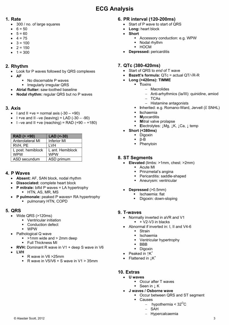
ECG Analysis 1. Rate
300 / no. of large squares 6 = 50 5 = 60 4 = 75 3 = 100 2 = 150 1 = 300
2. Rhythm
Look for P waves followed by QRS complexes AF
No discernable P waves Irregularly irregular QRS
Atrial flutter: saw-toothed baseline Nodal rhythm: regular QRS but no P waves
3. Axis
I and II +ve = normal axis (-30 – +90) I +ve and II –ve (leaving) = LAD (-30 – -90) I –ve and II +ve (reaching) = RAD (+90 – +180)
RAD (> +90) LAD (<-30) Anterolateral MI Inferior MI RVH, PE LVH L post. hemiblock WPW
L ant. Hemiblock WPW
ASD secundum ASD primum 4. P Waves
Absent: AF, SAN block, nodal rhythm Dissociated: complete heart block P mitrale: bifid P waves = LA hypertrophy
HTN, AS, MR, MS P pulmonale: peaked P waves= RA hypertrophy
pulmonary HTN, COPD 5. QRS
Wide QRS (>120ms) Ventricular initiation Conduction defect WPW
Pathological Q wave >1mm wide and > 2mm deep Full Thickness MI
RVH: Dominant R wave in V1 + deep S wave in V6 LVH
R wave in V6 >25mm R wave in V5/V6 + S wave in V1 > 35mm
6. PR interval (120-200ms)
Start of P wave to start of QRS Long: heart block Short
Accessory conduction: e.g. WPW Nodal rhythm HOCM
Depressed: pericarditis
7. QTc (380-420ms) Start of QRS to end of T wave Bazett’s formula: QTc = actual QT/√R-R Long (>420ms): TIMME
Toxins Macrolides Anti-arhythmics (Ia/III): quinidine, amiod TCAs Histamine antagonists
Inherited: e.g. Romano-Ward, Jervell (c̄ SNHL) Ischaemia Myocarditis Mitral valve prolapse Electrolytes: ↓Mg, ↓K, ↓Ca, ↓ temp
Short (<380ms) Digoxin β-B Phenytoin
8. ST Segments
Elevated (limbs: >1mm, chest: >2mm) Acute MI Prinzmetal’s angina Pericarditis: saddle-shaped Aneurysm: ventricular
Depressed (>0.5mm)
Ischaemia: flat Digoxin: down-sloping
9. T-waves Normally inverted in aVR and V1
+ V2-V3 in blacks Abnormal if inverted in: I, II and V4-6
Strain Ischaemia Ventricular hypertrophy BBB Digoxin
Peaked in ↑K+ Flattened in ↓K+
10. Extras
U waves Occur after T waves Seen in ↓ K
J waves / Osborne wave Occur between QRS and ST segment Causes
hypothermia < 32OC SAH Hypercalcaemia
© Alasdair Scott, 2012 3

ECG Abnormalities Conduction Defects Abnormality Features ECG 1st Degree Heart Block PR > 200ms
2nd Degree Heart Block - Wenckebach /Mobitz I
Progressive lengthening of PR interval One non-conducted P wave Next conducted beat has shorter PR interval
2nd Degree Heart Block - Mobitz II
Constant PR Occasional non-conducted P waves Often wide QRS - block is usually in bundle branches of Purkinje fibres
2nd Degree Heart Block - 2:1 Block
Two P waves per QRS Normal consistent PR intervals
3rd Degree Heart Block P waves and QRS @ different rates - dissociation Abnormally shaped QRS - ventricular origin (40bpm)
Abnormality Features ECG Aetiology Right BBB MaRRoW
Wide QRS RSR pattern in V1
Infarct – Inferior MI Normal variant Congenital – ASD, VSD, Fallot’s Hypertrophy – RVH (PE, Cor Pulmonale)
Left BBB WiLLiaM Wide QRS c̄ notched top T wave inversion in lat leads
Fibrosis LVH – AS, HTN Infarct – Inf. MI Coronary HD
Bifascicular Block RBBB + LAD RBBB + Left ant. hemiblock Trifascicular Block RBBB + LAFB + 1st
degree AV block
© Alasdair Scott, 2012 4

Escape Rhythms: appear late (after anticipated beat) Abnormality Features ECG Atrial Escape SAN fails to depolarise
Abnormal P wave Normal QRS 60-80bpm
Junctional Escape Usually no P waves (occasionally after QRS) Normal QRS 40-60bpm
Ventricular Escape Usually result of complete AV block regular P waves seen (top). May be SAN failure → no P waves (below). Wide QRS, 20bpm
Extrasystoles: appear early (before anticipated beat) Abnormality Features ECG Atrial Extrasystole Abnormal P wave
Normal QRS
Nodal Extrasystole P wave buried in QRS or
sometimes immediately before/after QRS. - may be negative Normal QRS
Ventricular Extrasystole
No P wave. Wide QRS and abnormal T wave.
© Alasdair Scott, 2012 5
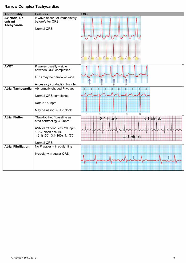
Narrow Complex Tachycardias Abnormality Features ECG AV Nodal Re-entrant Tachycardia
P wave absent or immediately before/after QRS Normal QRS
AVRT P waves usually visible
between QRS complexes QRS may be narrow or wide Accessory conduction bundle
Atrial Tachycardia Abnormally shaped P waves Normal QRS complexes. Rate > 150bpm May be assoc. c̄ AV block.
Atrial Flutter “Saw-toothed” baseline as
atria contract @ 300bpm. AVN can’t conduct > 200bpm AV block occurs. - 2:1(150), 3:1(100), 4:1(75) Normal QRS
Atrial Fibrillation No P waves – irregular line Irregularly irregular QRS
© Alasdair Scott, 2012 6
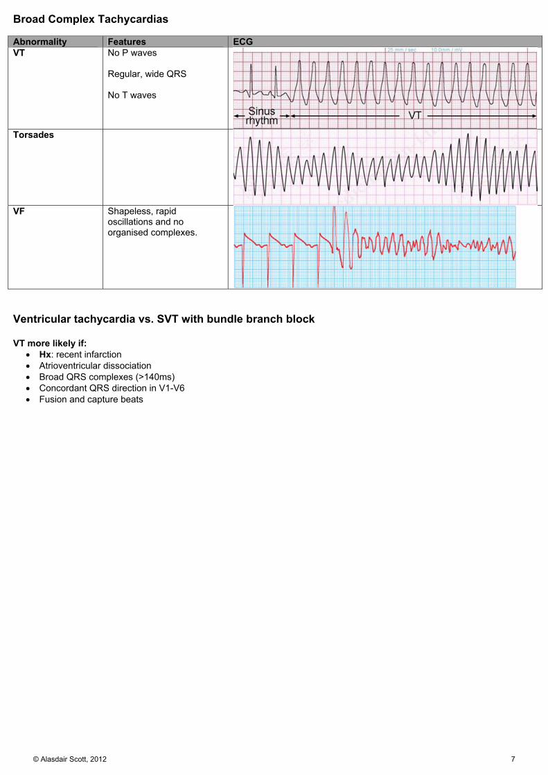
Broad Complex Tachycardias Abnormality Features ECG VT No P waves
Regular, wide QRS No T waves
Torsades
VF Shapeless, rapid oscillations and no organised complexes.
Ventricular tachycardia vs. SVT with bundle branch block VT more likely if:
Hx: recent infarction Atrioventricular dissociation Broad QRS complexes (>140ms) Concordant QRS direction in V1-V6 Fusion and capture beats
© Alasdair Scott, 2012 7

P Wave Abnormalities Abnormality Features ECG Aetiology P pulmonale
Peaked P wave RAH - pulmonary HTN - tricuspid stenois
P mitrale Broad, bifid P wave LAH - mitral stenois
QRS Abnormalities Abnormality Features ECG Aetiology RVH Tall R wave in V1
Deep S wave in V6 RAD Normal QRS width May be T wave inversion in V1-V3
Cor pulmonale
LVH S in V1 + R in V6 >35mm and/or R wave in V6 >25mm May be LAD May be T wave inversion in II, aVL, V5, V6
HTN AS COA H(O)CM
© Alasdair Scott, 2012 8

Miscellaneous Abnormality Features ECG WPW Accessory conducting bundle.
Short PR interval Slurred upstroke of QRS called a delta wave (V3/4). Can establish re-entrant circuit → SVT (antidromic AVRT) AF + WPW → irregularly irregular broad QRS complexes (below).
Brugada Syndrome
RBBB Coved ST elevation in V1-V3
Digoxin Reverse tick
- Down-sloping ST depression - T wave inversion
PE SI QIII TIII (rare) - deep S wave in I (RAD) - pathological Q in III - T inversion in III Right vent strain - RAD (S wave in I) - Dominant R wave and T wave inversion in V1-V3
↑ K+ Tall tented T waves Widened QRS Absent P waves Sine wave appearance Fig shows serial ↓ in K+
↓ K+ Small T waves ST depression Prolonged QT interval Prominent U waves
© Alasdair Scott, 2012 9
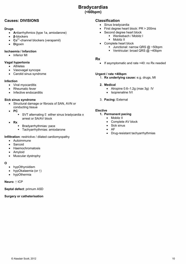
Bradycardias (<60bpm)
Causes: DIVISIONS Drugs
Antiarrhythmics (type 1a, amiodarone) β-blockers Ca2+-channel blockers (verapamil) Digoxin
Ischaemia / Infarction
Inferior MI Vagal hypertonia
Athletes Vasovagal syncope Carotid sinus syndrome
Infection
Viral myocarditis Rheumatic fever Infective endocarditis
Sick sinus syndrome
Structural damage or fibrosis of SAN, AVN or conducting tissue
PC SVT alternating c̄ either sinus bradycardia ±
arrest or SA/AV block Rx
Bradyarrhythmias: pace Tachyarrhythmias: amiodarone
Infiltration: restrictive / dilated cardiomyopathy
Autoimmune Sarcoid Haemochromatosis Amyloid Muscular dystrophy
O
hypOthyroidism hypOkalaemia (or ↑) hypOthermia
Neuro: ↑ ICP Septal defect: primum ASD Surgery or catheterisation
Classification
Sinus bradycardia First degree heart block: PR > 200ms Second degree heart block
Wenkebach / Mobitz I Mobitz II
Complete heart block Junctional: narrow QRS @ ~50bpm Ventricular: broad QRS @ ~40bpm
Rx
If asymptomatic and rate >40: no Rx needed Urgent / rate <40bpm
1. Rx underlying cause: e.g. drugs, MI
2. Medical Atropine 0.6–1.2g (max 3g) IV Isoprenaline IVI
3. Pacing: External
Elective
1. Permanent pacing Mobitz II Complete AV block Sick sinus AF Drug-resistant tachyarrhythmias
© Alasdair Scott, 2012 10
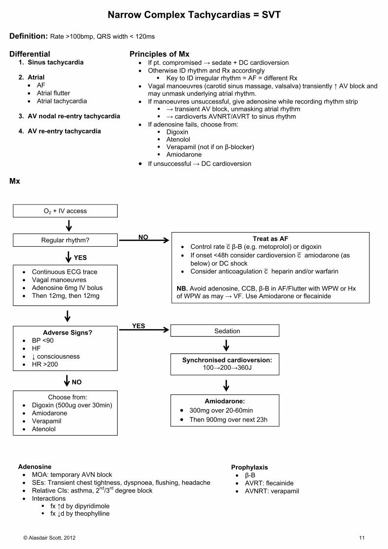
Narrow Complex Tachycardias = SVT
Definition: Rate >100bmp, QRS width < 120ms Differential
1. Sinus tachycardia
2. Atrial AF Atrial flutter Atrial tachycardia
3. AV nodal re-entry tachycardia
4. AV re-entry tachycardia
Principles of Mx If pt. compromised → sedate + DC cardioversion Otherwise ID rhythm and Rx accordingly
Key to ID irregular rhythm = AF = different Rx Vagal manoeuvres (carotid sinus massage, valsalva) transiently ↑ AV block and
may unmask underlying atrial rhythm. If manoeuvres unsuccessful, give adenosine while recording rhythm strip
→ transient AV block, unmasking atrial rhythm → cardioverts AVNRT/AVRT to sinus rhythm
If adenosine fails, choose from: Digoxin Atenolol Verapamil (not if on β-blocker) Amiodarone
If unsuccessful → DC cardioversion Mx
O2 + IV access
Regular rhythm?
Continuous ECG trace Vagal manoeuvres Adenosine 6mg IV bolus Then 12mg, then 12mg
Adverse Signs? BP <90 HF ↓ consciousness HR >200
Choose from: Digoxin (500ug over 30min) Amiodarone Verapamil Atenolol
Treat as AF Control rate c̄ β-B (e.g. metoprolol) or digoxin If onset <48h consider cardioversion c̄ amiodarone (as
below) or DC shock Consider anticoagulation c̄ heparin and/or warfarin
NB. Avoid adenosine, CCB, β-B in AF/Flutter with WPW or Hx of WPW as may → VF. Use Amiodarone or flecainide
Sedation
Amiodarone:
300mg over 20-60min Then 900mg over next 23h
Adenosine MOA: temporary AVN block SEs: Transient chest tightness, dyspnoea, flushing, headache Relative CIs: asthma, 2nd/3rd degree block Interactions
fx ↑d by dipyridimole fx ↓d by theophylline
NO
NO
YES
YES
Synchronised cardioversion:100→200→360J
Prophylaxis β-B AVRT: flecainide AVNRT: verapamil
© Alasdair Scott, 2012 11
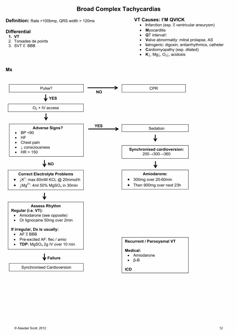
Broad Complex Tachycardias Definition: Rate >100bmp, QRS width > 120ms Differential
1. VT 2. Torsades de points 3. SVT c̄ BBB
VT Causes: I’M QVICK
Infarction (esp. c̄ ventricular aneurysm) Myocarditis QT interval↑ Valve abnormality: mitral prolapse, AS Iatrogenic: digoxin, antiarrhythmics, catheter Cardiomyopathy (esp. dilated) K↓, Mg↓, O2↓, acidosis
Mx
O2 + IV access
Synchronised Cardioversion
Correct Electrolyte Problems
↓K+: max 60mM KCL @ 20mmol/h ↓Mg2+: 4ml 50% MgSO4 in 30min
Adverse Signs? BP <90 HF Chest pain ↓ consciousness HR > 150
Sedation
Synchronised cardioversion: 200→300→360
Amiodarone:
300mg over 20-60min Then 900mg over next 23h
Failure
YES
NO
Pulse?
YES NO
CPR
Assess Rhythm Regular (i.e. VT): Amiodarone (see opposite) Or lignocaine 50mg over 2min
If irregular, Dx is usually: AF c̄ BBB Pre-excited AF: flec / amio TDP: MgSO4 2g IV over 10 min
Recurrent / Paroxysmal VT Medical: Amiodarone β-B
ICD
© Alasdair Scott, 2012 12

Atrial Fibrillation Pathology
LA loses refractoriness before the end of atrial systole. → recurrent, uncoordinated contraction @ 300-600bpm Atrial contraction responsible for ~25% of CO
often triggers heart failure Causes Common
IHD Rheumatic heart disease Thyrotoxicosis Hypertension
Other Alcohol Pneumonia PE Post-op Hypokalaemia RA
Symptoms
Asympto Chest pain Palpitations Dyspnoea Faintness
Signs
Irregularly irregular pulse Pulse deficit: difference between pulse and HS
Fast AF → loss of diastolic filling → no palpable pulse
Signs of LVF Ix
ECG FBC, U+E, TFTs, Trop Consider TTE: structural abnormalities
Acute AF (≤48h)
Haemo unstable → emergency cardioversion (IV amiodarone 2nd line)
Control ventricular rate
1st line: diltiazem or verapamil or metoprolol 2nd line: digoxin or amiodarone
Start LMWH Cardioversion: only if acute AF <48hrs
Electrical cardioversion or pharmacological 1st: Flecainide (if no structural heart disease) 2nd: Amiodarone
Long-term anticoagulation not needed if sinus restored no RFs (0 CHADSVAS) + low recurrence risk.
Paroxysmal AF
Self-limiting, <7d, recurs Anticoagulate: use CHADSVAS Rx “pill-in-pocket” : flecainide, propafenone Prevention: β-B, sotalol or amiodarone
Persistent AF >7d, may recur even after cardioversion
Try rhythm control first-line if:
Symptomatic or CCF Younger (<65) Presenting first time c̄ lone AF Secondary to treated precipitant
Rhythm Control
TTE first: structural abnormalities Anticoagulate c̄ warfarin for ≥3wks
or use TOE to exclude intracardiac thrombus. Pre-Rx ≥4wks c̄ sotalol or amiodarone if ↑ risk of failure Electrical or pharmacological cardioversion ≥ 4 wks anticoagulation afterwards (target INR 2.5) Maintenance antiarrhythmic
Not needed if successfully treated precipitant 1st: β-B (e.g. bisoprolol, metoprolol). 2nd: amiodarone
Other options
Radiofrequency ablation of AV node Maze procedure Pacing
Rate control (target <90bpm at rest):
1st line: β-B or rate-limiting CCB (NOT both!) 2nd line: add digoxin (don’t use as monotherapy) 3rd line: consider amiodarone
Mx of Permanent AF
Failed cardioversion / unlikely to succeed AF >1yr, valve disease, poor LV function
Pt. doesn’t want cardioversion → Rate control
Mx of Atrial Flutter
Manage as for AF Anti-AF drugs may not work, but try
Amiodarone to restore sinus Amiodarone or sotalol to maintain it
Cavotricuspid isthmus ablation (RA) is Rx of choice. CHA2-DS2-VAS Score
Determines necessity of anticoagulation in AF Warfarin CI in AF
Bleeding diathesis, ↓plats, BP > 160/90, poor compliance
Dabigatran may be cost-effective alternative. CHA2-DS2
CCF HTN Age≥75 (2 points) DM Stroke or TIA (2 points)
VAS Vascular disease Age: 65-74yrs Sex: female
Score
0: aspirin 300mg ≥1: Warfarin
© Alasdair Scott, 2012 13
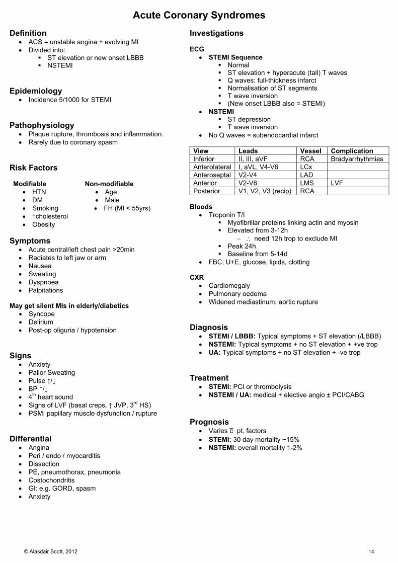
Acute Coronary Syndromes Definition
ACS = unstable angina + evolving MI Divided into:
ST elevation or new onset LBBB NSTEMI
Epidemiology
Incidence 5/1000 for STEMI Pathophysiology
Plaque rupture, thrombosis and inflammation. Rarely due to coronary spasm
Risk Factors Modifiable
HTN DM Smoking ↑cholesterol Obesity
Non-modifiable Age Male FH (MI < 55yrs)
Symptoms
Acute central/left chest pain >20min Radiates to left jaw or arm Nausea Sweating Dyspnoea Palpitations
May get silent MIs in elderly/diabetics
Syncope Delirium Post-op oliguria / hypotension
Signs
Anxiety Pallor Sweating Pulse ↑/↓ BP ↑/↓ 4th heart sound Signs of LVF (basal creps, ↑ JVP, 3rd HS) PSM: papillary muscle dysfunction / rupture
Differential
Angina Peri / endo / myocarditis Dissection PE, pneumothorax, pneumonia Costochondritis GI: e.g. GORD, spasm Anxiety
Investigations ECG
STEMI Sequence Normal ST elevation + hyperacute (tall) T waves Q waves: full-thickness infarct Normalisation of ST segments T wave inversion (New onset LBBB also = STEMI)
NSTEMI ST depression T wave inversion
No Q waves = subendocardial infarct View Leads Vessel Complication Inferior II, III, aVF RCA Bradyarrhythmias Anterolateral I, aVL, V4-V6 LCx Anteroseptal V2-V4 LAD Anterior V2-V6 LMS LVF Posterior V1, V2, V3 (recip) RCA
Bloods
Troponin T/I Myofibrillar proteins linking actin and myosin Elevated from 3-12h
need 12h trop to exclude MI Peak 24h Baseline from 5-14d
FBC, U+E, glucose, lipids, clotting CXR
Cardiomegaly Pulmonary oedema Widened mediastinum: aortic rupture
Diagnosis
STEMI / LBBB: Typical symptoms + ST elevation (/LBBB) NSTEMI: Typical symptoms + no ST elevation + +ve trop UA: Typical symptoms + no ST elevation + -ve trop
Treatment
STEMI: PCI or thrombolysis NSTEMI / UA: medical + elective angio ± PCI/CABG
Prognosis
Varies c̄ pt. factors STEMI: 30 day mortality ~15% NSTEMI: overall mortality 1-2%
© Alasdair Scott, 2012 14
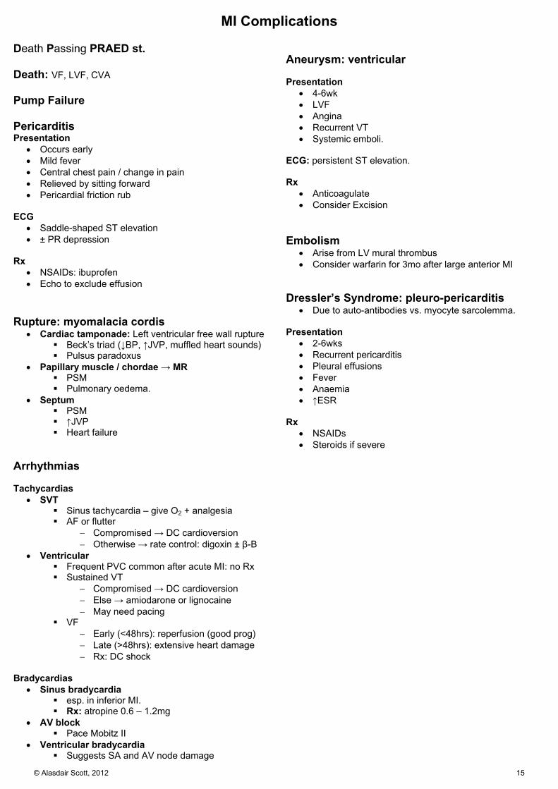
MI Complications Death Passing PRAED st. Death: VF, LVF, CVA Pump Failure Pericarditis Presentation
Occurs early Mild fever Central chest pain / change in pain Relieved by sitting forward Pericardial friction rub
ECG
Saddle-shaped ST elevation ± PR depression
Rx
NSAIDs: ibuprofen Echo to exclude effusion
Rupture: myomalacia cordis
Cardiac tamponade: Left ventricular free wall rupture Beck’s triad (↓BP, ↑JVP, muffled heart sounds) Pulsus paradoxus
Papillary muscle / chordae → MR PSM Pulmonary oedema.
Septum PSM ↑JVP Heart failure
Arrhythmias Tachycardias
SVT Sinus tachycardia – give O2 + analgesia AF or flutter
Compromised → DC cardioversion Otherwise → rate control: digoxin ± β-B
Ventricular Frequent PVC common after acute MI: no Rx Sustained VT
Compromised → DC cardioversion Else → amiodarone or lignocaine May need pacing
VF Early (<48hrs): reperfusion (good prog) Late (>48hrs): extensive heart damage Rx: DC shock
Bradycardias
Sinus bradycardia esp. in inferior MI. Rx: atropine 0.6 – 1.2mg
AV block Pace Mobitz II
Ventricular bradycardia Suggests SA and AV node damage
Aneurysm: ventricular Presentation
4-6wk LVF Angina Recurrent VT Systemic emboli.
ECG: persistent ST elevation. Rx
Anticoagulate Consider Excision
Embolism
Arise from LV mural thrombus Consider warfarin for 3mo after large anterior MI
Dressler’s Syndrome: pleuro-pericarditis
Due to auto-antibodies vs. myocyte sarcolemma. Presentation
2-6wks Recurrent pericarditis Pleural effusions Fever Anaemia ↑ESR
Rx
NSAIDs Steroids if severe
© Alasdair Scott, 2012 15

STEMI Management
O2 2-4L aim for SpO2 94-98%
Analgesia Morphine 5-10mg IV Metoclopramide 10mg IV
LMWH: e.g. enoxaparin IV then SC
IV access Bloods for FBC, U+E, glucose, lipids
12 lead ECG Primary Percutaneous Coronary Intervention Rx of choice if <12h Angioplasty and stenting + GP IIb/IIIa antagonist (tirofiban) if high risk
Delayed PCI, DM, complex procedure Complications: Bleeding Emboli Arrhythmia
Brief assessment Hx of CVD and risk factors Thrombolysis CIs CV exam
Antiplatelet Aspirin 300mg PO (then 75mg/d) Clopidogrel 300mg PO (then 75mg/d)
Anti-ischaemia GTN 2 puffs or 1 tablet SL β-B atenolol 5mg IV (CI: asthma, LVF)
Admit to CCU for monitoring Arrhythmias Continue meds except CCBs
Thrombolysis CI beyond 24hrs from pain onset
ECG Criteria: ST elevation > 1mm in 2+ limbs or > 2mm in 2+ chest leads. New LBBB Posterior: Deep ST “depression” and tall “R” waves in V1-V3
Contraindications: AGAINST Aortic dissection GI bleeding Allergic reaction previously Iatrogenic: recent surgery Neuro: cerebral neoplasm or CVA Hx Severe HTN (200/120) Trauma, inc. CPR
Agents: 1st: streptokinase, alteplase (rt-PA), tenecteplase Complications: Bleeding Stroke Arrhythmia Allergic reaction
Continuing Therapy: address risk factors ACEi: start w/i 24hrs of MI (e.g. lisinopril 2.5mg) β-blocker: e.g. bisoprolol 10mg OD (or, CCB) Cardiac rehabilitation (group exercise and info) / Heart Manual DVT prophylaxis until fully mobile
Continue for 3mo if large anterior MI Statin: regardless of basal lipids (e.g. atorvastatin 80mg)
Advice Stop smoking Diet: oily fish, fruit, veg, ↓ sat fats Exercise: 30min OD Work: return in 2mo Sex: avoid for 1mo Driving :avoid for 1mo
NB. Continue clopidogrel for 1mo following STEMI Continue aspirin indefinitely.
Primary PCI or Thrombolysis
Pts. not receiving any form of reperfusion therapy should be given fondaparinux.
© Alasdair Scott, 2012 16

NSTEMI and UA Management
O2 2-4L aim for SpO2 94-98%
Analgesia Morphine 5-10mg IV Metoclopramide 10mg IV
Assess Cardiovascular Risk: GRACE/TIMI
IV access Bloods for FBC, U+E, glucose, lipids,
Troponin
12 lead ECG + Admit to CCU
Brief Assessment Hx of CVD and risk factors CV exam
Anti-ischaemia GTN: 2 puffs or 1 tablet SL β-B: atenolol 50mg/24h PO (CI: asthma, LVF) IV GTN if pain continues
Anticoagulate Fondaparinux 2.5mg SC
Intermediate- to High-Risk Persistent/recurrent ischaemia, ST depression, DM,
positive trop. GPIIb/IIIa antagonist (tirofiban) Angiography (±PCI) w/i 96hrs Clopidogrel 75mg/d for one year
Low-Risk No further pain, flat or inverted T waves or normal
ECG, negative trop. May discharge if 12h trop is negative. Outpatient tests: angio, perfusion scan, stress echo
Continuing Therapy: address risk factors
ACEi (e.g. lisinopril 2.5mg) β-blocker (e.g. bisoprolol 10mg OD) or (CCB) Cardiac rehabilitation (group exercise and info) / Heart Manual Stop antithrombotic therapy when pain free (but give 3-5d) Statin (e.g. atorvastatin 80mg)
General advice as above NB. Continue clopidogrel for 1yr following NSTEMI Continue aspirin indefinitely.
Antiplatelet Aspirin 300mg PO (then 75mg/d) Clopidogrel 300mg PO
© Alasdair Scott, 2012 17

Angina Pectoris Pathophysiology
Atherosclerosis → myocardial ischaemia Aetiology
Mostly atheroma. Anaemia AS Tachyarrhythmias Arteritis
Risk Factors Modifiable:
HTN DM Smoking ↑cholesterol Obesity
Non-modifiable: Age Male FH (MI < 55yrs) Genetic: e.g. hyperlipidaemia
Symptoms
Central chest tightness or heaviness Brought on by exertion, relieved by rest May radiate to one/both arms, neck, jaw or teeth Other ppts: emotion, cold weather, heavy meals
Classification
Stable: induced by effort Unstable: occurs at rest / minimal exertion Decubitus: induced by lying down Prinzmetal’s / variant: occurs during rest
Due to coronary spasm ST elevation during attack: resolves as pain
subsides. Rx: CCB + long-acting nitrate
Syndrome X: angina pain + ST elevation on exercise test but no evidence of coronary atherosclerosis. Probably represents small vessel disease
Differential
AS Aortic aneurysm GI: GORD, spasm Musculoskeletal
Ix
Bloods: FBC, U+E, lipids, glucose, ESR, TFTs ECG: usually normal
May show ST ↓, flat/inverted T waves, past MI Consider exercise ECG
Stress echo Perfusion scan CT coronary Ca2+ score Angiography (gold standard)
Mx Lifestyle Stop smoking Wt. loss and ↑ exercise Healthy diet: oily fish, fruit, veg, ↓ sat fats
Medical
2O Prevention: prevent cardiovascular events Aspirin 75mg OD ACEi (esp. if angina + DM) Statins: simvastatin 40mg Antihypertensives
Anti-anginals: prevents angina episodes
1. GTN (spray or SL) + either 1st: β-B (e.g. Atenolol 50-100mg OD) 2nd: CCB (e.g. Verapamil 80mg TDS)
2. If either β-B or CCB doesn’t control symptoms, try the other option.
3. Can try β-B + dihydropyridine CCB e.g. amlodipine MR 10mg/24h
4. If symptoms still not controlled ISMN 20-40mg BD (8h washout @ PM) or
slow-release nitrate (Imdur 60mg OD) Ivabradine (esp. if can’t take β-B) Nicorandil 10-30mg BD Ranolazine
Interventional: PCI Indications
Poor response to medical Rx Refractory angina but not suitable for CABG
Complications Re-stenosis (20-30% @ 6mo) Emergency CABG (<2%) MI (<2%) Death (<0.5%)
Clopidogrel ↓s risk of re-stenosis Bare metal stent: 1mo Drug-eluting (e.g. sirolimus) stent: 1yr
Surgical: CAGB
Indications L main stem disease Triple vessel disease Refractory angina Unsuccessful angioplasty
Complications MI Stroke Pericardial tamponade or haemothorax Postperfusion syn. Post-op AF Nonunion of sternum Graft stenosis
© Alasdair Scott, 2012 18
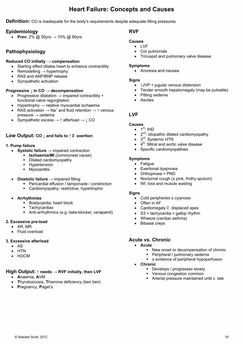
Heart Failure: Concepts and Causes Definition: CO is inadequate for the body’s requirements despite adequate filling pressures. Epidemiology
Prev: 2% @ 50yrs → 10% @ 80yrs Pathophysiology Reduced CO initially → compensation
Starling effect dilates heart to enhance contractility Remodelling → hypertrophy RAS and ANP/BNP release Sympathetic activation
Progressive ↓ in CO → decompensation
Progressive dilatation → impaired contractility + functional valve regurgitation
Hypertrophy → relative myocardial ischaemia RAS activation → Na+ and fluid retention → ↑ venous
pressure → oedema Sympathetic excess → ↑ afterload → ↓ CO
Low Output: CO ↓ and fails to ↑ c̄ exertion 1. Pump failure
Systolic failure → impaired contraction Ischaemia/MI (commonest cause) Dilated cardiomyopathy Hypertension Myocarditis
Diastolic failure → impaired filling
Pericardial effusion / tamponade / constriction Cardiomyopathy: restrictive, hypertrophic
Arrhythmias
Bradycardia, heart block Tachycardias Anti-arrhythmics (e.g. beta-blocker, verapamil)
2. Excessive pre-load
AR, MR Fluid overload
3. Excessive afterload
AS HTN HOCM
High Output: ↑ needs → RVF initially, then LVF
Anaemia, AVM Thyrotoxicosis, Thiamine deficiency (beri beri) Pregnancy, Paget’s
RVF Causes
LVF Cor pulmonale Tricuspid and pulmonary valve disease
Symptoms
Anorexia and nausea Signs
↑JVP + jugular venous distension Tender smooth hepatomegaly (may be pulsatile) Pitting oedema Ascites
LVF Causes
1st: IHD 2nd: idiopathic dilated cardiomyopathy 3rd: Systemic HTN 4th: Mitral and aortic valve disease Specific cardiomyopathies
Symptoms
Fatigue Exertional dyspnoea Orthopnoea + PND Nocturnal cough (± pink, frothy sputum) Wt. loss and muscle wasting
Signs
Cold peripheries ± cyanosis Often in AF Cardiomegaly c̄ displaced apex S3 + tachycardia = gallop rhythm Wheeze (cardiac asthma) Bibasal creps
Acute vs. Chronic
Acute New onset or decompensation of chronic Peripheral / pulmonary oedema ± evidence of peripheral hypoperfusion
Chronic Develops / progresses slowly Venous congestion common Arterial pressure maintained until v. late
© Alasdair Scott, 2012 19
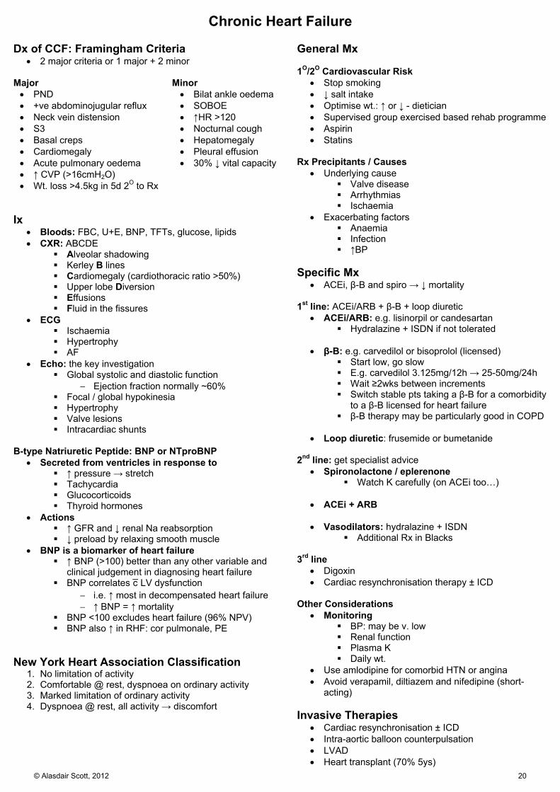
Chronic Heart Failure Dx of CCF: Framingham Criteria
2 major criteria or 1 major + 2 minor Major PND +ve abdominojugular reflux Neck vein distension S3 Basal creps Cardiomegaly Acute pulmonary oedema ↑ CVP (>16cmH2O) Wt. loss >4.5kg in 5d 2O to Rx
Minor Bilat ankle oedema SOBOE ↑HR >120 Nocturnal cough Hepatomegaly Pleural effusion 30% ↓ vital capacity
Ix
Bloods: FBC, U+E, BNP, TFTs, glucose, lipids CXR: ABCDE
Alveolar shadowing Kerley B lines Cardiomegaly (cardiothoracic ratio >50%) Upper lobe Diversion Effusions Fluid in the fissures
ECG Ischaemia Hypertrophy AF
Echo: the key investigation Global systolic and diastolic function
Ejection fraction normally ~60% Focal / global hypokinesia Hypertrophy Valve lesions Intracardiac shunts
B-type Natriuretic Peptide: BNP or NTproBNP
Secreted from ventricles in response to ↑ pressure → stretch Tachycardia Glucocorticoids Thyroid hormones
Actions ↑ GFR and ↓ renal Na reabsorption ↓ preload by relaxing smooth muscle
BNP is a biomarker of heart failure ↑ BNP (>100) better than any other variable and
clinical judgement in diagnosing heart failure BNP correlates c̄ LV dysfunction
i.e. ↑ most in decompensated heart failure ↑ BNP = ↑ mortality
BNP <100 excludes heart failure (96% NPV) BNP also ↑ in RHF: cor pulmonale, PE
New York Heart Association Classification
1. No limitation of activity 2. Comfortable @ rest, dyspnoea on ordinary activity 3. Marked limitation of ordinary activity 4. Dyspnoea @ rest, all activity → discomfort
General Mx 1O/2O Cardiovascular Risk
Stop smoking ↓ salt intake Optimise wt.: ↑ or ↓ - dietician Supervised group exercised based rehab programme Aspirin Statins
Rx Precipitants / Causes
Underlying cause Valve disease Arrhythmias Ischaemia
Exacerbating factors Anaemia Infection ↑BP
Specific Mx
ACEi, β-B and spiro → ↓ mortality 1st line: ACEi/ARB + β-B + loop diuretic
ACEi/ARB: e.g. lisinorpil or candesartan Hydralazine + ISDN if not tolerated
β-B: e.g. carvedilol or bisoprolol (licensed)
Start low, go slow E.g. carvedilol 3.125mg/12h → 25-50mg/24h Wait ≥2wks between increments Switch stable pts taking a β-B for a comorbidity
to a β-B licensed for heart failure β-B therapy may be particularly good in COPD
Loop diuretic: frusemide or bumetanide
2nd line: get specialist advice
Spironolactone / eplerenone Watch K carefully (on ACEi too…)
ACEi + ARB
Vasodilators: hydralazine + ISDN
Additional Rx in Blacks 3rd line
Digoxin Cardiac resynchronisation therapy ± ICD
Other Considerations
Monitoring BP: may be v. low Renal function Plasma K Daily wt.
Use amlodipine for comorbid HTN or angina Avoid verapamil, diltiazem and nifedipine (short-
acting) Invasive Therapies
Cardiac resynchronisation ± ICD Intra-aortic balloon counterpulsation LVAD Heart transplant (70% 5ys)
© Alasdair Scott, 2012 20

Severe Pulmonary Oedema
Morphine in Pulmonary Oedema
Make pt. more comfortable Pulm venodilators → ↓ pre-load → optimise position on Starling Curve
O2 15L/min via reservoir mask Target SpO2: 94-98%
Hx, Ex, Ix CXR: ABCDE ECG: MI, arrhythmias, pulsus alternans Consider echo
IV access + monitor ECG Bloods for FBC, U+E, troponin, BNP, ABG Rx any arrhythmias (e.g. AF)
Sit pt. up
Diamorphine 2.5-5mg IV + Metoclopramide 10mg IV
If SBP >100mmHg, start nitrate IVI ISMN 2-10mg/h IVI Keep SBP >90
GTN 2 puffs or 2 x 300ug tabs SL Unless SBP <90mmHg
Continuing Therapy Daily weights DVT prophylaxis Repeat CXR Change to oral frusemide or bumetanide ACEi + β-B if heart failure Consider spironolactone Consider digoxin ± warfarin (esp. if in AF)
Frusemide 40-80mg IV
If worsening, consider: CPAP More frusemide or ↑ nitrate infusion Haemofiltration / dialysis
Causes Cardiogenic MI Arrhythmia Fluid overload: renal, iatrogenic
Non-cardiogenic ARDS: sepsis, post-op, trauma Upper airway obstruction Neurogenic: head injury
Differential Asthma/COPD Pneumonia PE
Symptoms Dyspnoea Orthopnoea Pink frothy sputum
Signs Distressed, sweaty, cyanosed ↑HR, ↑RR ↑JVP S3 / gallop rhythm Bibasal creps Pleural effusions Wheeze (cardiac asthma)
Monitoring Progress BP HR and RR JVP Urine Output ABG
If SBP <100mmHg: Rx as cardiogenic shock i.e. consider inotropes
© Alasdair Scott, 2012 21

Cardiogenic Shock
O2 15L/min via reservoir mask Target SpO2: 94-98%
IV access + monitor ECG Bloods for FBC, U+E, troponin, ABG
Diamorphine 2.5-5mg IV (pain/anxiety) + metoclopramide 10mg IV
Consider need for dobutamine
Monitoring CVP, BP, ABG, ECG, urine output.
Correct any: Arrhythmias Electrolyte disturbance Acid-base abnormalities
Causes: MI HEART MI Hyperkalaemia (inc. electrolytes) Endocarditis (valve destruction) Aortic Dissection Rhythm disturbance Tamponade
Obstructive
Tension pneumo Massive PE
Hx, Ex, Ix CXR Echo Consider CT thorax (dissection/PE)
Rx underlying cause
Tamponade Causes: Trauma Lung/breast Ca Pericarditis MI Bacteria (e.g. TB)
Mx: ABCs Pericardiocentesis (preferably under echo guidance)
Signs: Beck’s triad: ↓BP, ↑JVP, muffled heart sounds Kussmaul’s sign: ↑JVP on inspiration Pulsus paradoxus (pulse fades on inspiration)
Ix: Echo: diagnostic CXR: globular heart
Definition Inadequate tissue perfusion primarily due to
cardiac dysfunction.
Presentation Unwell: pale, sweaty, cyanosed, distressed Cold clammy peripheries ↑RR ± ↑HR Pulmonary oedema
© Alasdair Scott, 2012 22

Hypertension Definitions
Stage 1: Clinic BP > 140/90 Stage 2: Clinic BP > 160/100 Severe: Clinic BP > 180/110 Malignant: BP > 180/110 + papilloedema and/or retinal
haemorrhage Isolated SHT: SBP ≥140, DBP <90
Aetiology: PREDICTION
Primary: 95% Renal: RAS, GN, APKD, PAN Endo: ↑T4, Cushing’s, phaeo, acromegaly, Conn’s Drugs: cocaine, NSAIDs, OCP ICP ↑ CoA Toxaemia of Pregnancy (PET) Increased viscosity Overload with fluid Neurogenic: diffuse axonal injury, spinal section
Aetiological clues
↑HR: Thyrotoxicosis RF-delay: CoA Renal bruits: RAS Palpable kidneys: APKD Paroxysmal headache, tachycardia, sweating,
palpitations, labile or postural hypotension: phaeo End-organ damage: CANER Cardiac
IHD LVH → CCF AR, MR
Aortic
Aneurysm Dissection
Neuro
CVA: ischaemic, haemorrhagic Encephalopathy (malignant HTN)
Eyes: hypertensive retinopathy
Keith-Wagener Classification: 1. Tortuosity and silver wiring 2. AV nipping 3. Flame haemorrhages and cotton wool spots 4. Papilloedema
Grades 3 and 4 = malignant hypertension Renal
Proteinuria CRF
Ix
24h ABPM Urine: haematuria, Alb:Cr ratio Bloods: FBC, U+Es, eGFR, glucose, fasting lipids 12 lead ECG: LVH, old infarct Calculate 10yr CV risk
Management Do ABPM to confirm Dx before Rx (unless severe HTN)
Lifestyle interventions
↑ exercise ↓ smoking, ↓ EtOH, ↓ salt, ↓ caffeine
Indications for Pharmacological Rx
<80yrs, stage 1 HTN (>140/90) and one of: Target organ damage (e.g. LVH, retinopathy) 10yr CV risk ≥20% Established CVD DM Renal disease
Anyone with stage 2 HTN (>160/100) Severe / malignant HTN (specialist referral) Consider specialist opinion if <40yrs with stage 1 HTN
and no end organ damage. BP Targets
Under 80yrs: <140/90 (<130/80 in DM) Over 80yrs: <150/90
CV Risk Mx
Statins indicated for 1O prevention if 10yr CVD risk ≥20% Aspirin may be indicated: evaluate risk of bleeding
Malignant HTN Controlled ↓ in BP over days to avoid stroke Atenolol or long-acting CCB PO Encephalopathy/CCF: fruse + nitroprusside / labetalol IV
Aim to ↓ BP to 110 diastolic over ~4h Nitroprusside requires intra-arterial BP monitoring
< 55 > 55 / Black
1:
2:
3:
4:
A C (or D)
A + C (/D)
A + C + D
Resistant HTN A+C+D+ consider further diuretic (e.g.
spiro) or α-blocker or β-B. Seek expert opinion
A: ACEi or ARB e.g. lisinopril 10mg OD (↑ to 30-40mg) e.g. candesartan 8mg OD (max 32mg OD)
C: CCB: e.g. nifedipine MR30-60mg OD D: Thiazide-like diuretic: e.g. chlortalidone 25-50mg OD In step 2, use ARB over ACEi in blacks. Avoid thiazides + β-B if possible (↑ risk of DM). Only consider β-B if young and ACEi/ARB not tolerated.
Antihypertensive Rx
© Alasdair Scott, 2012 23
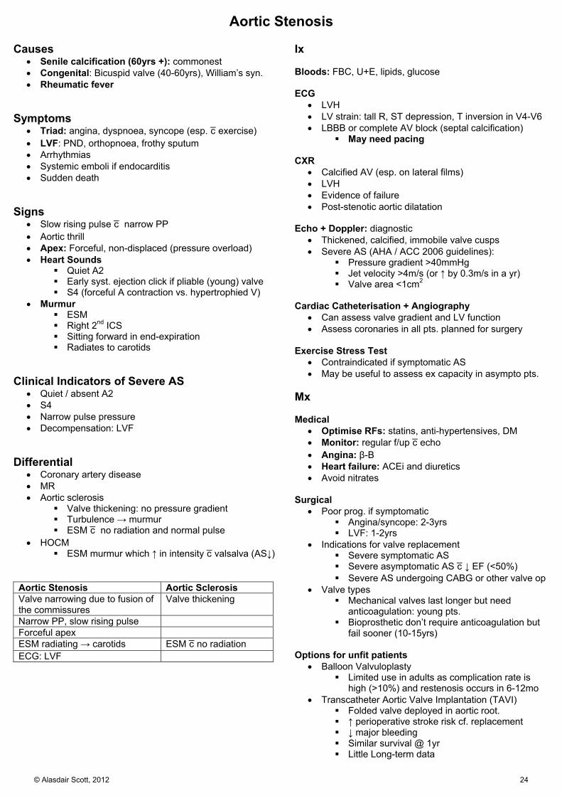
Aortic Stenosis Causes
Senile calcification (60yrs +): commonest Congenital: Bicuspid valve (40-60yrs), William’s syn. Rheumatic fever
Symptoms
Triad: angina, dyspnoea, syncope (esp. c̄ exercise) LVF: PND, orthopnoea, frothy sputum Arrhythmias Systemic emboli if endocarditis Sudden death
Signs
Slow rising pulse c̄ narrow PP Aortic thrill Apex: Forceful, non-displaced (pressure overload) Heart Sounds
Quiet A2 Early syst. ejection click if pliable (young) valve S4 (forceful A contraction vs. hypertrophied V)
Murmur ESM Right 2nd ICS Sitting forward in end-expiration Radiates to carotids
Clinical Indicators of Severe AS
Quiet / absent A2 S4 Narrow pulse pressure Decompensation: LVF
Differential
Coronary artery disease MR Aortic sclerosis
Valve thickening: no pressure gradient Turbulence → murmur ESM c̄ no radiation and normal pulse
HOCM ESM murmur which ↑ in intensity c̄ valsalva (AS↓)
Aortic Stenosis Aortic Sclerosis Valve narrowing due to fusion of the commissures
Valve thickening
Narrow PP, slow rising pulse Forceful apex ESM radiating → carotids ESM c̄ no radiation ECG: LVF
Ix Bloods: FBC, U+E, lipids, glucose ECG
LVH LV strain: tall R, ST depression, T inversion in V4-V6 LBBB or complete AV block (septal calcification)
May need pacing CXR
Calcified AV (esp. on lateral films) LVH Evidence of failure Post-stenotic aortic dilatation
Echo + Doppler: diagnostic
Thickened, calcified, immobile valve cusps Severe AS (AHA / ACC 2006 guidelines):
Pressure gradient >40mmHg Jet velocity >4m/s (or ↑ by 0.3m/s in a yr) Valve area <1cm2
Cardiac Catheterisation + Angiography
Can assess valve gradient and LV function Assess coronaries in all pts. planned for surgery
Exercise Stress Test
Contraindicated if symptomatic AS May be useful to assess ex capacity in asympto pts.
Mx Medical
Optimise RFs: statins, anti-hypertensives, DM Monitor: regular f/up c̄ echo Angina: β-B Heart failure: ACEi and diuretics Avoid nitrates
Surgical
Poor prog. if symptomatic Angina/syncope: 2-3yrs LVF: 1-2yrs
Indications for valve replacement Severe symptomatic AS Severe asymptomatic AS c̄ ↓ EF (<50%) Severe AS undergoing CABG or other valve op
Valve types Mechanical valves last longer but need
anticoagulation: young pts. Bioprosthetic don’t require anticoagulation but
fail sooner (10-15yrs) Options for unfit patients
Balloon Valvuloplasty Limited use in adults as complication rate is
high (>10%) and restenosis occurs in 6-12mo Transcatheter Aortic Valve Implantation (TAVI)
Folded valve deployed in aortic root. ↑ perioperative stroke risk cf. replacement ↓ major bleeding Similar survival @ 1yr Little Long-term data
© Alasdair Scott, 2012 24
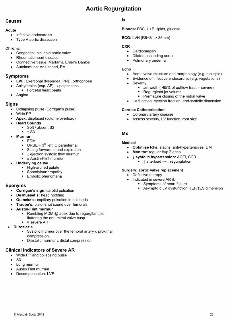
Aortic Regurgitation Causes Acute
Infective endocarditis Type A aortic dissection
Chronic
Congenital: bicuspid aortic valve Rheumatic heart disease Connective tissue: Marfan’s, Ehler’s Danlos Autoimmune: Ank spond, RA
Symptoms
LVF: Exertional dyspnoea, PND, orthopnoea Arrhythmias (esp. AF) → palpitations
Forceful heart beats Angina
Signs
Collapsing pulse (Corrigan’s pulse) Wide PP Apex: displaced (volume overload) Heart Sounds
Soft / absent S2 ± S3
Murmur EDM URSE + 3rd left IC parasternal Sitting forward in end-expiration ± ejection systolic flow murmur ± Austin-Flint murmur
Underlying cause High-arched palate Spondyloarthropathy Embolic phenomena
Eponyms
Corrigan’s sign: carotid pulsation De Musset’s: head nodding Quincke’s: capillary pulsation in nail beds Traube’s: pistol-shot sound over femorals Austin-Flint murmur
Rumbling MDM @ apex due to regurgitant jet fluttering the ant. mitral valve cusp.
= severe AR Duroziez’s
Systolic murmur over the femoral artery c̄ proximal compression.
Diastolic murmur c̄ distal compression Clinical Indicators of Severe AR
Wide PP and collapsing pulse S3 Long murmur Austin Flint murmur Decompensation: LVF
Ix Bloods: FBC, U+E, lipids, glucose ECG: LVH (R6+S1 > 35mm) CXR
Cardiomegaly Dilated ascending aorta Pulmonary oedema
Echo
Aortic valve structure and morphology (e.g. bicuspid) Evidence of infective endocarditis (e.g. vegetations) Severity
Jet width (>65% of outflow tract = severe) Regurgitant jet volume Premature closing of the mitral valve
LV function: ejection fraction, end-systolic dimension Cardiac Catheterisation
Coronary artery disease Assess severity, LV function, root size
Mx Medical
Optimise RFs: statins, anti-hypertensives, DM Monitor: regular f/up c̄ echo ↓ systolic hypertension: ACEi, CCB
↓ afterload → ↓ regurgitation Surgery: aortic valve replacement
Definitive therapy Indicated in severe AR if:
Symptoms of heart failure Asympto c̄ LV dysfunction: ↓EF/↑ES dimension
© Alasdair Scott, 2012 25

Mitral Stenosis Causes
Rheumatic fever Prosthetic valve Congenital (rare)
Pathophysiology
Valve narrowing → ↑ left atrial pressure → loud S1 and atrial hypertrophy → AF
→ pulmonary oedema and PHT → loud P2, PR → RVH → left parasternal heave → TR → large v waves → RHF → ↑JVP, oedema, ascites
Symptoms
Dyspnoea Fatigue Chest pain AF → palpitations + emboli Haemoptysis: rupture of bronchial veins
Signs
Symptoms manifest when orifice <2cm2 (norm 4-6) AF, low volume pulse Malar flush (↓CO → backpressure + vasoconstriction) JVP may be raised late on
Prominent a waves: PTH Large v waves: TR Absent a waves: AF
Left parasternal heave (RVH 2O to PHT) Apex: Tapping (palpable S1), non-displaced Heart sounds
Loud S1 Loud P2 (if PHT) Early diastolic opening snap
Murmur Rumbling MDM Apex Left lateral position in end expiration Radiates to the axilla ± Graham Steell murmur (EDM 2O to PR)
Clinical Indicators of Severe MS
Mitral facies Longer murmur Opening snap closer to 2nd heart sound
High LA pressure forcing valve open early Decompensation: RVF
Complications
Pulmonary HTN Emboli: TIA, CVA, PVD, ischaemic colitis Hoarseness: rec laryngeal N. palsy = Ortner’s Syn Dysphagia (oesophageal compression) Bronchial obstruction
Ix Bloods: FBC, U+E, LFTs, glucose, lipids ECG
AF P mitrale (if in sinus) RVH c̄ strain: ST depression and T wave inversion in V1-V2
CXR
LA enlargement Pulmonary oedema: ABCDE Mitral valve calcification
Echo + Doppler
Severe MS (AHA 2006 Criteria) Valve orifice <1cm2 Pressure gradient >10mmHg Pulmonary artery systolic pressure >50mmHg
Use TOE to look for left atrial thrombus if intervention considered.
Cardiac Catheterisation
Assess coronary arteries Mx Medical
Optimise RFs: statins, anti-hypertensives, DM Monitor: regular f/up c̄ echo Consider prophylaxis vs. rheumatic fever: e.g. Pen V AF: rate control and anticoagulate Diuretics provide symptom relief
Surgical
Indicated in mod–severe MS (asympto and symptomatic) Percutaneous balloon valvuloplasty
Rx of choice Suitability depends on valve characteristics
Pliable, minimally calcified CI if left atrial mural thrombus
Surgical valvotomy / commissurotomy: valve repair Valve replacement if repair not possible
© Alasdair Scott, 2012 26

Mitral Regurgitation Causes
Mitral valve prolapse LV dilatation: AR, AS, HTN Annular calcification → contraction (elderly) Post-MI: papillary muscle dysfunction/rupture Rheumatic fever Connective tissue: Marfan’s, Ehlers-Danlos
Symptoms
Dyspnoea, fatigue AF → palpitations + emboli Pulmonary congestion → HTN + oedema
Signs
AF Left parasternal heave (RVH) Apex: displaced
Volume overload as ventricle has to pump forward SV and regurgitant volume
→ eccentric hypertrophy Heart Sounds
Soft S1 S2 not heard separately from murmur Loud P2 (if PTH)
Murmur Blowing PSM Apex Left lateral position in end expiration Radiates to the axilla
Clinical Indicators of Severe MR
Larger LV Decompensation: LVF AF
Differential
AS TR VSD
Ix Bloods: FBC, U+E, glucose, lipids ECG
AF P mitrale (unless in AF) LVH
CXR LA and LV hypertrophy Mitral valve calcification Pulmonary oedema
Echo
Doppler echo to assess MR severity: multiple criteria Jet width (vena contracta) >0.6cm Systolic pulmonary flow reversal Regurgitant volume >60ml
TOE to assess severity and suitability of repair cf. replacement.
Cardiac Catheterisation
Confirm Dx Assess CAD
Mx Medical
Optimise RFs: statins, anti-hypertensives, DM Monitor: regular f/up c̄ echo AF: rate control and anticoagulate
Also anticoagulate if: Hx of embolism, prosthetic valve, additional MS
Drugs to ↓ afterload can help ↓ symptoms ACEi or β-B (esp. carvedilol) Diuretics
Surgical
Valve replacement or repair Indications
Severe symptomatic MR Severe asympto MR c̄ diastolic dysfunction: ↓EF
Mitral Valve Prolapse (Barlow Syndrome) Commonest valve prob. (~5%)
Causes
Primary: myxomatous degeneration Often young women
MI Marfan’s, ED Turner’s
Symptoms
Usually asymptomatic Autonomic dysfunction: Atypical chest pain,
palpitations, anxiety, panic attack MR: SOB, fatigue
Signs
Mid-systolic click ± late-systolic murmur
Complications
MR Cerebral emboli Arrhythmias → sudden death
Mx
β-B may relieve palpitations and chest pain Surgery if severe (commonest reason for MV surgery)
© Alasdair Scott, 2012 27

Right Heart Valve Disease Tricuspid Regurgitation Causes
Functional: RV dilatation Rheumatic fever Infective endocarditis Carcinoid syndrome
Symptoms
Fatigue Hepatic pain on exertion Ascites, oedema
Signs
↑JVP c̄ giant V waves RV heave Murmur:
PSM LLSE in inspiration (Carvallo’s sign)
Pulsatile hepatomegaly Jaundice
Ix
LFTs Echo
Mx
Rx cause Medical: diuretics, ACEi, digoxin Surgical: valve replacement
Tricuspid Stenosis Causes
Rheumatic fever (with MV and AV disease)
Symptoms
Fatigue Ascites Oedema
Signs
Large A waves Opening snap Murmur:
EDM LLSE in inspiration
Mx
Medical: diuretics Surgical: repair, replacement
Pulmonary Regurgitation Causes
Any cause of pulmonary HTN PR 2O to MS = Graham-Steell murmur
Signs
Murmur: Decrescendo EDM @ ULSE Pulmonary Stenosis Causes
Usually congenital: e.g. Turner’s, Fallot’s Rheumatic fever Carcinoid syndrome
Symptoms
Dyspnoea, fatigue Ascites Oedema
Signs
Dysmorphia Large A wave RV heave Ejection click, soft P2 Murmur
ESM ULSE → L shoulder
Ix
ECG P pulmonale RAD RBBB
CXR: Prominent pulmonary arteries: post-stenotic
dilatation Catheterisation: diagnostic Mx: valvuloplasty or valvotomy
© Alasdair Scott, 2012 28

Infective Endocarditis Definition Cardiac valves develop vegetations composed of bacteria
and platelet-fibrin thrombus.
Risk Factors Cardiac disease → Subacute Prosthetic valves Degen. valvulopathy VSD, PDA, CoA Rheumatic fever
Normal valves → Acute Dental caries Post-op wounds IVDU (tricuspid valve) Immunocomp. (inc. DM)
Aetiology Culture +ve S. viridans (>35%) S. bovis S. aureus S. epidermidis Enterococci Pseudomonas
Culture –ve Haemophilus Actinobacillus Cardiobacterium Eikenella Kingella Coxiella Chlamydia
Non-infective SLE Marantic
Clinical Features Sepsis Fever, rigors Night sweats Wt. loss Anaemia Splenomegaly Clubbing
Cardiac New/changing murmur
(MR: 85%, AR: 55%) AV block LVF
Embolic phenomena Abscesses in brain, heart,
kidney, spleen, gut and lung (if right-sided)
Janeway lesions Immune complex deposition Micro haematuria due to
GN Vasculitis Roth spots Splinter haemorrhages Osler’s nodes
Roth spots: boat-shaped retinal haemorrhages c̄ pale centre Janeway lesions: painless palmer macules Osler’s nodes: painful, purple papules on finger pulps
Dx: Duke Criteria Major
1. +ve blood culture Typical organism in 2 separate cultures, or Persistently +ve cultures, e.g. 3, >12h apart
2. Endocardium involved +ve echo (vegetation, abscess, valve dehiscence) or New valvular regurgitation
Minor
1. Predisposition: cardiac lesion, IVDU 2. Fever >38 3. Emboli: septic infarcts, splinters, Janeway lesions 4. Immune phenomenon: GN, Osler nodes, Roth spots, RF5. +ve blood culture not meeting major criteria
Dx if: 2 major 1 major + 3 minor All 5 minor
Ix Bloods
N.chromic, N.cytic anaemia ↑ESR, ↑CRP +ve IgG RF (immune phenomenon) Cultures x 3, >12h apart Serology for unusual organisms
Urine: Micro haematuria ECG: AV block Echo
TTE detects vegetations > 2mm TOE is more sensitive (90-100% vs. 50-60%)
Rx
Empiric Acute severe: Fuclox + gent IV Subacute: Benpen + gent IV
Streps: benpen + gent IV Enterococci: amoxicillin + gent IV Staphs: fluclox ± rifampicin IV Fungi: flucytosine IV + fluconazole PO.
Amphotericin if flucytosine resistance or Aspergillus.
Consider surgery if
Heart failure Emboli Valve obstruction Prosthetic valve
Prophylaxis
Abx prophylaxis solely to prevent IE not recommended
Mortality
30% c̄ staphs 14% c̄ bowel flora 6% c̄ sensitive streps
© Alasdair Scott, 2012 29

Rheumatic Fever Aetiology
Group A β-haemolytic strep. (pyogenes)
Epidemiology 5-15yrs Rare in West. Common in developing world. Only 2% of population susceptible
Pathophysiology Ab cross-reactivity following S. pyogenes infection →
T2 hypersensitivity reaction (molecular mimicry). Abs. vs. M protein in cell wall. Cross react c̄ myosin, muscle glycogen and SM cells. Path: Aschoff bodies and Anitschkow myocytes.
Dx: revised Jones Criteria Evidence of GAS infection plus:
2 major criteria, or 1 major + 2 minor
Evidence of GAS infection
+ve throat culture Rapid strep Ag test ↑ ASOT or DNase B titre Recent scarlet fever
Major Criteria
Pancarditis Arthritis Subcutaneous nodules Erythema marginatum Sydenham’s chorea
Minor criteria
Fever ↑ESR or ↑CRP Arthralgia (not if arthritis is major) Prolonged PR interval (not if carditis is a major) Prev rheumatic fever
Symptomatology Pancarditis (60%)
Pericarditis: chest pain, friction rub
Myocarditis: sinus tachy, AV block, HF, ↑CK, T inversion
Endocarditis: murmurs MR, AR, Carey Coombs’ (MDM)
Arthritis (75%)
Migratory polyarthritis of large joints (esp. knees) Subcutaneous nodules (2-20%)
Small mobile, painless nodules on extensor surfaces (esp. elbows)
Erythema marginatum (2-10%)
Red, raised edges c̄ central clearing. Trunk, thighs and arms.
Sydenham’s Chorea (10%)
Occurs late Grimacing, clumsy, hypotonia (stops in sleep)
Ix Bloods
Strep Ag test or ASOT FBC, ESR/CRP
ECG Echo
Rx Bed rest until CRP normal for 2wks Benpen 0.6-1.2mg IM for 10 days Analgesia for carditis/arthritis: aspirin / NSAIDs Add oral pred if: CCF, cardiomegaly, 3rd degree block Chorea: Haldol or diazepam
Prognosis Attacks last ~ 3mo. 60% c̄ carditis develop chronic rheumatic heart
disease. Recurrence ppted by
Further strep infection Pregnancy OCP
Valve disease: regurgitation → stenosis Mitral (70%) Aortic (40%) Tricuspid (10%) Pulmonary (2%)
Secondary Prophylaxis Prevent recurrence Pen V 250mg/12h PO
Carditis + valve disease: until 40yrs old Carditis w/o valve disease:10yrs No carditis: 5yrs
© Alasdair Scott, 2012 30

Pericardial Disease Acute Pericarditis Causes
Viral: coxsackie, flu, EBV, HIV Bacterial: pneumonia, rheumatic fever, TB, staphs Fungi MI, Dressler’s Drugs: penicillin, isoniazid, procainamide, hydralazine Other: uraemia, RA, SLE, sarcoid, radiotherapy
Clinical Features
Central / retrosternal chest pain Sharp Pleuritic Worse lying down Relieved by sitting forward Radiates to left shoulder
Pericardial friction rub. Fever Signs of effusion / tamponade
Ix
ECG: saddle-shaped ST-elevation ± PR depression Bloods: FBC, ESR, trop (may be ↑), cultures, virology
Mx
Analgesia: ibuprofen 400mg/8h PO Rx cause Consider steroids / immunosuppression
Constrictive Pericarditis
Heart encased in a rigid pericardium. Causes
Often unknown May occur after any pericarditis
Clinical features
RHF c̄ ↑JVP (prominent x and y descents) Kussmaul’s sign: ↑JVP c̄ inspiration Quiet heart sounds S3 Hepatosplenomegaly Ascites, oedema
Ix
CXR: small heart + pericardial calcification Echo Cardiac Catheterisation
Mx
Surgical excision
Pericardial Effusion Causes
Any cause of pericarditis Clinical Features
Dyspnoea ↑JVP (prominent x descent) Bronchial breathing @ left base
Ewart’s sign: large effusion compressing left lower lobe
Signs of cardiac tamponade may be present. Ix
CXR: enlarged, globular heart ECG
Low-voltage QRS complexes Alternating QRS amplitude (electrical alterans)
Echo: echo-free zone around heart Mx
Treat cause Pericardiocentesis may be diagnostic or therapeutic
Culture, ZN stain, cytology
Tamponade
Accumulation of pericardial fluid → ↑ intra-pericardial pressure → poor ventricular filling → ↓ CO
Causes
Any cause of pericarditis Aortic dissection Warfarin Trauma
Signs
Beck’s Triad: ↓ BP, ↑ JVP, quiet heart sounds Pulsus paradoxus: pulse fades on inspiration Kussmaul’s sign
Ix
ECG: low-voltage QRS ± electrical alternans CXR: large, globular heart Echo: diagnostic, echo-free zone around heart
Mx
Urgent pericardiocentesis 20ml syringe + long 18G cannula 45O, just left of xiphisternum, aiming for tip of left
scapula. Aspirate continuously and watch ECG.
Treat cause Send fluid for cytology, ZN stain and culture
© Alasdair Scott, 2012 31

Myocardial Disease Acute Myocarditis Causes
Idiopathic (~50%) Viral: coxsackie B, flu, HIV Bacterial: S. aureus, syphilis Drugs: cyclophosphamide, Herceptin, CBZ, phenytoin Autoimmune: Giant cell myocarditis assoc. c̄ SLE
Symptoms
Flu-like prodrome: fever, sore throat, myalgia Dyspnoea, fatigue Chest pain (may coexist c̄ Bornholm disease) Arrhythmia → palpitations
Signs
Soft S1 S4 gallop
Ix
ECG ST-elevation or depression T wave inversion Transient AV block
Bloods: +ve trop, ↑CK Mx
Supportive Rx cause
Hypertrophic Obstructive Cardiomyopathy
LVOT obstruction from asymmetric septal hypertrophy AD inheritance (but 50% sporadic) β-myosin heavy chain mutation commonest Ask re family Hx of sudden death
Symptoms
Angina Dyspnoea Palpitations: AF, WPW, VT Exertional syncope or sudden death
Signs
Jerky pulse Double apex beat Harsh ESM @ LLSE c̄ systolic thrill S4
Ix
ECG LVH/LAD/L strain, Ventricular ectopics, VT, VF
Echo: ASH Exercise test ± holter monitor to quantify risk
Mx
Medical -ve inotropes: 1st – β-B, 2nd –verapamil Amiodarone: arrhythmias Anticoagulate if AF or emboli
Non-medical Septal myomectomy (surgical or chemical) if
severe symptoms Consider ICD
Cardiac Myxoma
Rare, benign cardiac tumour F>M=2:1 May be familial: e.g. Carney Complex
Cardiac and cutaneous myxoma, skin pigmentation, endocrinopathy (e.g. Cushing’s)
90% in left atrium (fossa ovalis) Features:
Clubbing, fever, ↓wt., ↑ESR Signs similar to MS (MDM, systemic emboli, AF)
but vary c̄ posture. Dx: Echo Rx: Excision
Restrictive Cardiomyopathy Causes: miSSHAPEN
Sarcoid Systemic sclerosis Haemochromatosis Amyloidosis Primary: endomyocardial fibrosis Eosinophilia (Loffler’s eosinophilic endocarditis) Neoplasia: carcinoid (→ TR and PS)
Clinical Features: as constrictive pericarditis Dx: Catheterisation Rx: Rx cause Dilated Cardiomyopathy Causes: DILATE
Dystophy: muscular, myotonic, glycogen storage disease Infection: complication of myocarditis Late pregnancy: peri-, post-partum Autoimmune: SLE Toxins: EtOH, doxorubicin, cyclophosphamide, DXT Endocrine: thyrotoxicosis
Presentation
LVF and RVF Arrhythmias
Signs
JVP ↑↑ Displaced apex S3 gallop ↓BP MR/TR
Ix
CXR: cardiomegaly, pulmonary oedema ECG: T inversion, poor progression Echo: globally dilated, hypokinetic heart + ↓EF Catheter + biopsy: myocardial fibre disarray
Mx
Bed rest Medical: diuretics, ACEi, digoxin, anticoagulation Non-medical: biventricular pacing, ICD Surgical: heart Tx
© Alasdair Scott, 2012 32
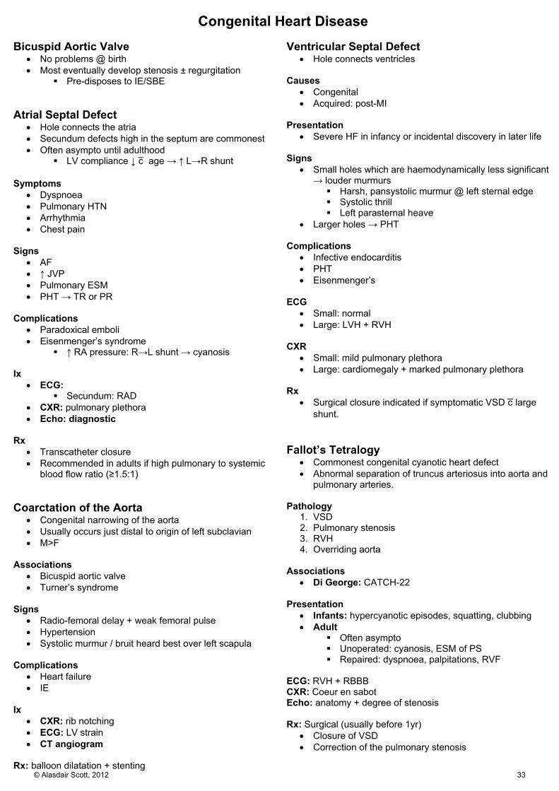
Congenital Heart Disease Bicuspid Aortic Valve
No problems @ birth Most eventually develop stenosis ± regurgitation
Pre-disposes to IE/SBE Atrial Septal Defect
Hole connects the atria Secundum defects high in the septum are commonest Often asympto until adulthood
LV compliance ↓ c̄ age → ↑ L→R shunt Symptoms
Dyspnoea Pulmonary HTN Arrhythmia Chest pain
Signs
AF ↑ JVP Pulmonary ESM PHT → TR or PR
Complications
Paradoxical emboli Eisenmenger’s syndrome
↑ RA pressure: R→L shunt → cyanosis Ix
ECG: Secundum: RAD
CXR: pulmonary plethora Echo: diagnostic
Rx
Transcatheter closure Recommended in adults if high pulmonary to systemic
blood flow ratio (≥1.5:1) Coarctation of the Aorta
Congenital narrowing of the aorta Usually occurs just distal to origin of left subclavian M>F
Associations
Bicuspid aortic valve Turner’s syndrome
Signs
Radio-femoral delay + weak femoral pulse Hypertension Systolic murmur / bruit heard best over left scapula
Complications
Heart failure IE
Ix
CXR: rib notching ECG: LV strain CT angiogram
Rx: balloon dilatation + stenting
Ventricular Septal Defect
Hole connects ventricles Causes
Congenital Acquired: post-MI
Presentation
Severe HF in infancy or incidental discovery in later life Signs
Small holes which are haemodynamically less significant → louder murmurs Harsh, pansystolic murmur @ left sternal edge Systolic thrill Left parasternal heave
Larger holes → PHT Complications
Infective endocarditis PHT Eisenmenger’s
ECG
Small: normal Large: LVH + RVH
CXR
Small: mild pulmonary plethora Large: cardiomegaly + marked pulmonary plethora
Rx
Surgical closure indicated if symptomatic VSD c̄ large shunt.
Fallot’s Tetralogy
Commonest congenital cyanotic heart defect Abnormal separation of truncus arteriosus into aorta and
pulmonary arteries. Pathology
1. VSD 2. Pulmonary stenosis 3. RVH 4. Overriding aorta
Associations
Di George: CATCH-22 Presentation
Infants: hypercyanotic episodes, squatting, clubbing Adult
Often asympto Unoperated: cyanosis, ESM of PS Repaired: dyspnoea, palpitations, RVF
ECG: RVH + RBBB CXR: Coeur en sabot Echo: anatomy + degree of stenosis Rx: Surgical (usually before 1yr)
Closure of VSD Correction of the pulmonary stenosis
© Alasdair Scott, 2012 33

Inherited Connective Tissue Disorders Marfan’s Syndrome Epidemiology
Autosomal dominant Spontaneous mutation in 25% M=F Prevalence = 1/5000
Pathophysiology
Mutation in FBN1 gene on Chr 5 Encodes fibrillin-1 glycoprotein
Fibrillin-1 is an essential component of elastin Histology: “cystic medial necrosis”
Presentation
Cardiac Aortic aneurysm and dissection Aortic root dilatation → regurgitation MV prolapse ± regurgitation
Ocular Lens dislocation: superotemporal
MSK High-arched palate Arachnodactyly Arm-span > height Pectus excavatum Scoliosis Pes planus Joint hypermobility
Complications
Ruptured aortic aneurysm Spontaneous pneumothorax Diaphragmatic hernia Hernias
Dx
Two 2/3 organ systems must be involved Differential Diagnosis
MEN-2b Homocystinuria Ehlers-Danlos
Ix
Slit-lamp examination: ectopia lentis CXR
Widened mediastinum Scoliosis Pneumothorax
ECG Arrhythmias: premature atrial and ventricular
ectopics Echo
Aortic root dilatation → AR MVP and MR
MRI: dural ectasia (dilation of neural canal) Genetic testing: FBN-1 mutation
Mx
Refer to ortho, cardio and ophtho Life-style alteration: ↓ cardiointensive sports Beta-blockers slow dilatation of the aortic root Regular cardiac echo
Surgery when aortic root ≥5cm wide
Ehlers-Danlos Syndrome Pathogenesis
Rare heterogeneous group of collagen disorders. 6 subtypes c̄ varying severity Commonest types (1 and 2) are autosomal dominant
Presentation
Hyperelastic skin Hypermobile joints Cardiac: MVP, AR, MR and aneurysms Fragile blood vessels → easy bruising, GI bleeds Poor healing
Differential Diagnosis
Cutis Laxa: loose skin + hypermobile joints Pseudoxanthoma elasticum: skin laxity Marfan’s
© Alasdair Scott, 2012 34

Pulmonology
Contents Clubbing ...................................................................................................................................................................................... 36
Cyanosis ...................................................................................................................................................................................... 36
Pneumonia .................................................................................................................................................................................. 37
Complications of Pneumonia ....................................................................................................................................................... 38
Systemic Inflammatory Response Syndrome ............................................................................................................................. 38
Specific Pneumonias ................................................................................................................................................................... 39
Bronchiectasis ............................................................................................................................................................................. 40
Cystic Fibrosis ............................................................................................................................................................................. 41
Pulmonary Aspergillus Infections ................................................................................................................................................ 42
Lung Cancer: Presentation .......................................................................................................................................................... 43
Lung Cancer: Investigation and Management ............................................................................................................................ 44
ARDS ........................................................................................................................................................................................... 45
Respiratory Failure ...................................................................................................................................................................... 46
Oxygen Therapy .......................................................................................................................................................................... 46
Chronic Asthma ........................................................................................................................................................................... 47
Acute Severe Asthma .................................................................................................................................................................. 48
COPD .......................................................................................................................................................................................... 49
Acute Exacerbation of COPD ...................................................................................................................................................... 50
Pulmonary Embolism ................................................................................................................................................................... 51
Pneumothorax ............................................................................................................................................................................. 52
Pleural Effusion ........................................................................................................................................................................... 53
Sarcoidosis .................................................................................................................................................................................. 54
Interstitial Lung Disease .............................................................................................................................................................. 55
Extrinsic Allergic Alveolitis ........................................................................................................................................................... 56
Industrial Lung Disease ............................................................................................................................................................... 56
Idiopathic Pulmonary Fibrosis (CFA) ........................................................................................................................................... 56
Pulmonary Hypertension ............................................................................................................................................................. 57
Cor Pulmonale ............................................................................................................................................................................. 57
Obstructive Sleep Apnoea ........................................................................................................................................................... 58
Smoking Cessation ...................................................................................................................................................................... 58
© Alasdair Scott, 2012 35

Clubbing Features and Stages
1. Bogginess / ↑ fluctuance of nail bed 2. Loss of concave nail fold angle 3. ↑ longitudinal and transverse curvature 4. Soft tissue expansion at distal phalanx (drumstick)
Causes Respiratory
Carcinoma Bronchial Mesothelioma
Chronic lung suppuration Empyema, abscess Bronchiectasis, CF
Fibrosis Idiopathic pulmonary fibrosis / CFA TB
Cardiac
Infective Endocarditis Congenital cyanotic heart disease Atrial myxoma
GIT
Cirrhosis Crohn’s, uC Coeliac Cancer: GI lymphoma
Other
Familial Thyroid Acropachy Upper limb AVMs or aneurysms
Unilateral clubbing
Cyanosis Definition
Blue discoloration of mucosal membranes or skin Deoxygenated Hb >5g/dl
Classification
Peripheral: cold, blue nails Central: blue tongue, lips
Causes
Think of O2 cascade Respiratory
Hypoventilation: COPD, MSK ↓ diffusion: pulm oedema, fibrosing alveolitis V/Q mismatch: PE, AVM (e.g. HHT)
Cardiac
Congenital: Fallot’s, TGA ↓ CO: MS, systolic LVF Vascular: Raynaud’s, DVT
RBCs
Low affinity Hb, may be hereditary or acquired
© Alasdair Scott, 2012 36

Pneumonia Epidemiology
Incidence: 1/100 Mortality: 10% in hospital, 30% in ITU
Anatomic Classification Bronchopneumonia
Patchy consolidation of different lobes Lobar Pneumonia
Fibrosuppurative consolidation of a single lobe Congestion → red → grey → resolution
Aetiological Classification Community Acquired Pneumonia
Pneumococcus, mycoplasma, haemophilus S. aureus, Moraxella, Chlamydia, Legionella Viruses: 15%
Hospital Acquired Pneumonia
>48hrs after hospital admission Gm-ve enterobacteria, S. aureus
Aspiration
↑ Risk: stroke, bulbar palsy, ↓GCS, GORD, achalasia Anaerobes
Immunocompromised
The usual suspects, plus PCP, TB, fungi, CMV/HSV
Symptoms
Fever, rigors Malaise, anorexia Dyspnoea Cough, purulent sputum, haemoptysis Pleuritic pain
Signs
↑RR, ↑ HR Cyanosis Confusion Consolidation
↓ expansion Dull percussion Bronchial breathing ↓ air entry Crackles Pleural rub ↑VR
Ix
Bloods: FBC, U+E, LFT, CRP, culture, ABG (if ↓SpO2) Urine: Ag tests (Pneumococcal, Legionella) Sputum: MC&S Imaging: CXR
infiltrates, cavities, effusion Special
Paired sera Abs for atypicals Mycoplasma, Chlamydia, Legionella
Immunofluorescence (PCP) BAL Pleural tap
Severity: CURB-65 (only if x-ray changes)
Confusion (AMT ≤ 8) Urea >7mM Resp. rate >30/min BP <90/60 ≥65
Score 0-1 → home Rx 2 → hospital Rx ≥3 → consider ITU
Mx
Abx O2: PaO2≥8, SpO2 94-98% Fluids Analgesia Chest physio Consider ITU if shock, hypercapnoea, hypoxia F/up @ 6wks c̄ CXR
Check for underlying Ca Empirical Abx CAP Mild amoxicillin 500mg TDS PO for 5d or
clarithro 500mg BD PO for 7d
Mod amoxicillin 500mg TDS and clarithro 500mg BD PO/IV (clarithro alone if pen allergy) for 7d
Sev Co-amoxiclav 1.2g TDS IV / cefuroxime 1.5g TDS IV and clarithro 500mg BD IV for 7-10d Add fluclox if staph suspected.
Atyp Chlamydia: tetracycline PCP: Co-trimoxazole Legionella: Clarithro + rifampicin
HAP
Mild / <5d: Co-amoxiclav 625mg PO TDS for 7d Severe / >5d: Tazocin ± vanc ± gent for 7d
Aspiration
Co-amoxiclav 625mg PO TDS for 7d Pneumovax (23 valent)
≥65yrs Chronic HLKP failure or conditions DM Immunosuppression: hyposplenism, chemo, HIV CI: P, B, fever
NB. revaccinate every 6yrs
© Alasdair Scott, 2012 37

Complications of Pneumonia Respiratory failure
Type 1: PaO2 <8kPa + PaCO2 <6kPa Type 2: PaO2 <8kPa + PaCO2 >6kPa Mx: O2 therapy, ventilation
Hypotension
Cause: dehydration + septic vasodilatation Mx
If SBP<90 → 250ml fluid challenge over 15min If no improvement: central line + IV fluids If refractory: ITU for inotropes
AF
Usually resolves c̄ Rx Mx: Digoxin or β-B for rate control
Pleural effusion
Exudate Mx: tap and send for MC+S, cytology and chemistry
Empyema
Pus in the pleural cavity Anaerobes, Staph, Gm-ve Assoc. c̄ recurrent aspiration Pt. c̄ resolving pneumonia develops recurrent fever Tap: turbid, pH<7.2, ↓glucose, ↑LDH Mx: US guided chest drain + Abx
Lung Abscess Causes
Aspiration Bronchial obstruction: tumour, foreign body Septic emboli: sepsis, IVDU, RH endocarditis Pulmonary infarction Subphrenic / hepatic abscess
Features
Swinging fever Cough, foul purulent sputum, haemoptysis Malaise, wt. loss Pleuritic pain Clubbing Empyema
Tests
Blood: FBC, ESR, CRP, cultures Sputum: micro, culture, cytology CXR: cavity c̄ fluid level Consider CT and bronchoscopy
Mx
Abx according to sensitivities Aspiration Surgical excision
Other Complications
Sepsis Pericarditis / myocarditis Jaundice
Usually cholestatic Causes: sepsis, drugs (fluclox, Augmentin),
Mycoplasma, Legionella
Systemic Inflammatory Response Syndrome Inflammatory response to a variety of insults manifest by ≥2 of:
Temperature: >38°C or <36°C Heart rate: >90 Respiratory rate: >20 or PaCO2 <4.6 KPa WCC: >12x109/L or <4 x10⁹/L or >10% bands
Sepsis
SIRS caused by infection Severe Sepsis
Sepsis c̄ at least 1 organ dysfunction or hypoperfusion Septic Shock
Severe sepsis with refractory hypotension MODS
Impairment of ≥2 organ systems Homeostasis cannot be maintained without therapeutic
intervention.
© Alasdair Scott, 2012 38
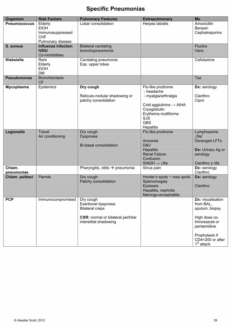
Specific Pneumonias Organism Risk Factors Pulmonary Features Extrapulmonary Mx Pneumococcus Elderly
EtOH Immunosuppressed CHF Pulmonary disease
Lobar consolidation Herpes labialis Amoxicillin Benpen Cephalosporins
S. aureus Influenza infection IVDU Co-morbidities
Bilateral cavitating bronchopneumonia
Fluclox Vanc
Klebsiella Rare Elderly EtOH DM
Cavitating pneumonia Esp. upper lobes
Cefotaxime
Pseudomonas Bronchiectasis CF
Taz
Mycoplasma Epidemics Dry cough Reticulo-nodular shadowing or patchy consolidation
Flu-like prodrome - headache - myalgia/arthralgia Cold agglutinins → AIHA Cryoglobulin Erythema multiforme SJS GBS Hepatitis
Dx: serology Clarithro Cipro
Legionella Travel Air conditioning
Dry cough Dyspnoea Bi-basal consolidation
Flu-like prodrome Anorexia D&V Hepatitis Renal Failure Confusion SIADH → ↓Na
Lymphopenia ↓Na+ Deranged LFTs Dx: Urinary Ag or serology Clarithro ± rifa
Chlam. pneumoniae
Pharyngitis, otitis pneumonia
Sinus pain Dx: serology Clarithro
Chlam. psittaci Parrots Dry cough Patchy consolidation
Horder’s spots ~ rose spots Splenomegaly Epistaxis Hepatitis, nephritis Meningo-encephalitis
Dx: serology Clarithro
PCP Immunocompromised Dry cough Exertional dyspnoea Bilateral creps CXR: normal or bilateral perihilar interstitial shadowing
Dx: visualisation from BAL, sputum, biopsy High dose co-trimoxazole or pentamidine Prophylaxis if CD4<200 or after 1st attack
© Alasdair Scott, 2012 39

Bronchiectasis Pathophysiology
Chronic infection of bronchi/bronchioles → permanent dilatation.
Retained inflammatory secretions and microbes → airway damage and recurrent infection
Organisms: H. influenza Pneumococcus S. aureus Pseudomonas
Causes
Idiopathic in 50% Congenital
CF (mainly upper lobe infiltration) Kartagener’s / PCD Young’s syn. (azoopermia + bronchiectasis)
Post-infectious
Measles Pertussis Pneumonia TB Bronchiolitis
Immunodeficiency
Hypogammaglobulinaemia X-linked agammaglobulinaemia: Bruton’s CVID IgG subclass deficiency IgA deficiency
Other
Bronchial obstruction: LNs, tumour, foreign body ABPA RA UC Yellow nail syndrome
Yellow nail discoloration and dystrophy Lymphoedema Pleural effusions Bronchiectasis
Symptoms
Persistent cough c̄ purulent sputum Haemoptysis (may be massive) Fever, wt. loss
Signs
Clubbing Coarse inspiratory creps Wheeze Purulent sputum Cause
Situs inversus (+ PCD = Kartagener’s syn.) Splenomegaly: immune deficiency
Complications
Pneumonia Pleural effusion Pneumothorax Pulmonary HTN Massive haemoptysis Cerebral abscess Amyloidosis
Ix
Sputum MCS Blood: Se Ig, Aspergillus precipitins, RF, α1-AT level
Test Ig response to pneumococcal vaccine CXR: thickened bronchial walls (tramlines and rings) Spirometry: obstructive pattern HRCT chest
Dilated and thickened airways Saccular dilatations in clusters c̄ pools of mucus
Bronchoscopy + mucosal biopsy Focal obstruction PCD
CF sweat test (pilocarpine iontopheresis) Rx
Chest physio: expectoration, drainage, pulm. rehab Abx for exacerbations: e.g. cipro for 7-10d Bronchodilators: nebulised β agonists Treat underlying cause
CF: DNAase ABPA: Steroids Immune deficiency: IVIg
Surgery may be indicated in severe localised disease
© Alasdair Scott, 2012 40

Cystic Fibrosis Pathogenesis
Auto recessive, 1:2000 live Caucasian births Mutation in CFTR gene on Chr 7 (commonly ∆F508) → ↓ luminal Cl secretion and ↑ Na reabsorption →
viscous secretions. In sweat glands, ↓ Cl and Na reabsorption → salty sweat
Clinical Features Neonate
FTT Meconium ileus Rectal prolapse
Children / Young Adults
Nose: nasal polyps, sinusitis Resp: cough, wheeze, infections, bronchiectasis,
haemoptysis, pneumothorax, cor pulmonale GI:
Pancreatic insufficiency: DM, steatorrhoea Distal Intestinal Obstruction Syndrome Gallstones Cirrhosis (2O biliary)
Other: male infertility, osteoporosis, vasculitis Signs
Clubbing ± HPOA Cyanosis Bilateral coarse creps
Common Respiratory Organisms
Early S. aureus H. influenza
Late P. aeruginosa: 85% B. cepacia: 4%
Dx
Sweat test: Na and Cl > 60mM Genetic screening for common mutations Faecal elastase (tests pancreatic exocrine function) Immunoreactive trypsinogen (neonatal screening)
Ix
Bloods: FBC, LFTs, clotting, ADEK levels, glucose TT Sputum MCS CXR: bronchiectasis Abdo US: fatty liver, cirrhosis, pancreatitis Spirometry: obstructive defect Aspergillus serology / skin test (20% develop ABPA)
Mx General
MDT: physician, GP, physio, dietician, specialist nurse Chest
Physio: postural drainage, forced expiratory techniques Abx: acute infections and prophylaxis Mucloytics: DNAse Bronchodilators Vaccinate
GI
Pancreatic enzyme replacement: pancreatin (Creon) ADEK supplements Insulin Ursodeoxycholic acid for impaired hepatic function
Stimulates bile secretion Advanced Lung Disease
O2 Diuretics (Cor pulmonale) NIV Heart/lung transplantation
Other
Rx of complications: e.g. DM Fertility and genetic counselling DEXA osteoporosis screen
© Alasdair Scott, 2012 41
Pulmonary Aspergillus Infections Diseases
1. Asthma: T1HS reaction to spores 2. ABPA 3. Aspergilloma (mycetoma) 4. Invasive aspergillosis 5. Extrinsic allergic alveolitis
Allergic Bronchopulmonary Aspergillosis
T1 and T3 HS reaction to Aspergillus fumigatus Bronchoconstriction → bronchiectasis
Symptoms
Wheeze Productive cough Dyspnoea
Ix
CXR: bronchiectasis Aspergillus in sputum (black on silver stain) Aspergillus skin test or IgE RAST +ve se precipitins ↑ IgE and ↑ eosinophils
Rx
Pred 40mg/d + itraconazole for acute attacks Pred maintenance 5-10mg/d Bronchodilators for asthma
Aspergilloma (mycetoma)
Fungus ball within a pre-existing cavity e.g. TB or sarcoid
Features
Usually asympto Can → haemoptysis (may be severe) Lethargy, ↓wt.
Ix
CXR: round opacity w/i a cavity, usually apical Sputum culture +ve se precipitins Aspergillus skin test / RAST
Rx
Consider excision for solitary lesions / severe haemoptysis
Invasive Aspergillosis
Aflatoxins → liver cirrhosis and HCC (especially A. flavus)
Risk factors
Immunocompromise: HIV, leukaemia, Wegener’s Post-broad spectrum Abx
Ix
CXR: consolidation, abscess Sputum MCS BAL +ve se precipitins Serial galactomannan
Rx
Voriconazole (better cf amphotericin) Prog
30% mortality Extrinsic Allergic Alveolitis
Sensitivity to Aspergillus clavatus → Malt worker’s lung
© Alasdair Scott, 2012 42

Lung Cancer: Presentation Classification
Clinically, the most important distinction is between small-cell and non-small-cell (SCC, adeno and large-cell) Type Epidemiology Pathology Behaviour SCC 35%
M>F Smoking Radon gas
Centrally located Histo: Evidence of squamous differentiation - Keratinisation
Locally invasive Metastasise late (via LN) PTHrP → ↑ Ca2+
Adeno 25% Females, Non-smokers Far East
Peripherally located Histo: Glandular differentiation - Gland formation - Mucin production
Extrathoracic mets common and early. - 80% present with mets
Large-cell 10% Peripheral or central Histo: Large, poorly differentiated cells
Poor prognosis
Small-cell 20% Smoking
Central location, near bronchi Histo: Small, poorly differentiated cells
80% present c̄ advanced disease V. chemosensitive but v. poor prog Ectopic hormone secretion
Other Lung Tumours: all rare
Adenoma: 90% are carcinoid tumours Hamartoma Mesothelioma
Epidemiology
19% of all cancers 27% of cancer deaths (commonest)
Symptoms
Cough and haemoptysis Dyspnoea Chest pain Recurrent or slow resolving pneumonia Anorexia and ↓wt. Hoarseness
Signs Chest
Consolidation Collapse Pleural effusion
General
Cachexia Anaemia Clubbing and HPOA (painful wrist swelling) Supraclavicular and/or axillary LNs
Metastasis
Bone tenderness Hepatomegaly Confusion, fits, focal neuro Addison’s
Complications Local
Recurrent laryngeal N. palsy Phrenic N. palsy SVC obstruction Horner’s (Pancoast’s tumour) AF
Paraneoplastic
Endo ADH → SIADH ( euvolaemic ↓Na+) ACTH → Cushing’s syndrome Serotonin → carcinoid (flushing, diarrhoea) PTHrP → 1O HPT (↑Ca2+, bone pain) – SCC
Rheum Dermatomyositis / polymyositis
Neuro Purkinje Cells (CDR2) → cerebellar degeneration Peripheral neuropathy
Derm Acanthosis nigricans (hyperpigmented body folds) Trousseau syndrome: thrombophlebitis migrans
Metastatic
Pathological # Hepatic failure Confusion, fits, focal neuro Addison’s
© Alasdair Scott, 2012 43

Lung Cancer: Investigation and Management Ix Bloods: FBC, U+E, Ca2+, LFTs Cytology: sputum, pleural fluid Imaging
CXR Coin lesion Hilar enlargement Consolidation, collapse Effusion Bony secondaries
Contrast-enhanced Volumetric CT Staging: lower neck, chest, upper abdomen Consider CT brain
PET-CT: exclude distant mets Radionucleotide bone scan
Biopsy
Percutaneous FNA: peripheral lesions and LNs Bronchoscopy: biopsy and assess operability Endoscopic bronchial US biopsy: mediastinal LNS Mediastinoscopy
Lung Function Tests
Assess treatment fitness CXR Coin Lesion Differential
Foreign body Abscess: Staph, TB, Klebsiella, Mycetoma Neoplasia (1O or 2O) Granuloma: RA, Wegener’s, TB, Sarcoid Structural: AVM
TNM Staging for NSCLC Tx Tis T0 T1 T2 T3 T4
Malignant cells in bronchial secretions CIS None evident ≤3cm, in lobar or more distal airway >3cm and >2cm from carina or pleural involvement <2cm from carina or involves chest wall, diaphragm Involves mediastinum or malignant effusion is present
N0 N1 N2 N3
None involved Peribronchial or ipsilateral hilum Ipsilateral mediastinum Contralateral hilum or mediastinum or supraclavicular
Mx MDT: pulmonologist, oncologist, radiologist,
histopathologist, cardiothoracic surgeon, specialist nurses, palliative care, GP
Assess risk of operative mortality: e.g. Thoracoscore Cardiorespiratory function Co-morbidities
Advise smoking cessation NSCLC
Surgical Resection Rx of choice for peripheral lesions c̄ no metastatic
spread = stage I/II (~25%) Need good cardiorespiratory function Wedge resection, lobectomy or pneumonectomy ± adjuvant chemo
Curative radiotherapy If cardiorespiratory reserve is poor
Chemo ± radio for more advanced disease Platinum-based regimens MAbs targeting EGFR (e.g. cetuximab) or TKI
(e.g. erlotinib) SCLC
Typically disseminated @ presentation May respond to chemo but invariably relapse
Palliation
Radio: bronchial obstruction, haemoptysis, bone or CNS mets
SVC obstruction: stenting + radio + dexamethasone Endobronchial therapy: stenting, brachytherapy Pleural drainage / pleurodesis Analgesia
Prognosis
NSCLC: 50% 5ys w/o spread; 10% c̄ spread SCLC: 1-1.5yrs median survival treated; 3mo untreated
© Alasdair Scott, 2012 44
ARDS Pathogenesis
May result from direct pulmonary insult or be 2O to severe systemic illness.
Inflam mediators → ↑ capillary permeability and non-cardiogenic pulmonary oedema.
Clinical Features
Tachypnoea Cyanosis Bilateral fine creps SIRS
Ix
Bloods: FBC, U+E, LFTs, clotting, amylase, CRP, cultures, ABG.
CXR: bilateral perihilar infiltrates Dx
Acute onset CXR shows bilateral infiltrates No evidence of congestive cardiac failure PaO2:FiO2 <200
Mx
Admit to ITU for organ support and Rx underlying cause Ventilation
Indications PaO2<8KPa despite 60% FiO2 PaCO2>6KPa
Method 6ml/kg + PEEP (e.g. 10cm H2O)
SEs VILI VAP Weaning difficulty
Circulation
Invasive BP monitoring Maintain CO and DO2 c̄ inotropes
E.g. norad or dobutamine RF may require haemofiltration
Sepsis
Abx Other
Nutritional support: enteral (best), TPN Prognosis
50-75% mortality
Causes Pulmonary
Pneumonia Aspiration Inhalation injury Contusion
Systemic
Shock Sepsis Trauma Haemorrhage and multiple transfusions Pancrea0titis Acute liver failure DIC Obs: eclampsia, amniotic embolism Drugs: aspirin, heroin
Differential Dx of Pulmonary Oedema Tranusdates
↑ capillary hydrostatic pressure CCF Iatrogenic fluid overload Renal failure Relative ↑ in negative pressure pulmonary
oedema ↓ capillary oncotic pressure
Liver failure Nephrotic syndrome Malnutrition, malabsorption, protein-losing
enteropathy ↑ interstitial pressure
↓lymphatic drainage: e.g. Ca Exudates
ARDS
© Alasdair Scott, 2012 45

Respiratory Failure Type 1
PaO2 <8KPa and PaCO2 <6KPa V/Q mismatch and diffusion failure
Type 2
PaO2 <8KPa and PaCO2 >6KPa Alveolar hypoventilation ± V/Q mismatch
Causes V/Q Mismatch (↑A-a gradient)
Vascular PE PHT Pulmonary Shunt (R → L)
Asthma (early) Pneumothorax Atelectasis
Alveolar Hypoventilation
Obstructive COPD Asthma Bronchiectasis Bronchiolitis Intra- and Extra-thoracic (Ca, LN, epiglottitis…)
Restrictive ↓ drive: CNS sedation, trauma, tumour NM disease: cervical cord lesion, polio, GBS,
MG Chest: flail, kyphoscoliosis, obesity Fluid and fibrosis
Diffusion Failure (↑A-a gradient)
Fluid Pulmonary oedema Pneumonia Infarction Blood
Fibrosis NB. Both fluid and fibrosis also → V/Q mismatch and alveolar hypoventilation due to ↓ compliance A-a Gradient
PAO2= (95 x FiO2) – (PaCO2/0.8) ↑ (PAO2-PaO2) suggests lung pathology
Clinical Features Hypoxia
Acute Dyspnoea Agitation Confusion Cyanosis
Chronic Polycythaemia PHT cor pulmonale
Hypercapnoea
Headache Flushing and peripheral vasodilatation Bounding pulse Flap Confusion → coma
Mx: Rx underlying cause Type 1
Give O2 to maintain SpO2 94-98% Assisted ventilation if PaO2<8KPa despite 60% O2
Type 2
Controlled O2 therapy @ 24% O2 aiming for SpO2 88-92% and a PaO2 >8kPa
Check ABG after 20min If PaCO2 steady or lower can ↑ FiO2 if necessary If PaCO2 ↑>1.5KPa and pt. still hypoxic, consider
NIV or respiratory stimulant (e.g. doxapram)
Oxygen Therapy Principles
Critically ill pts. should receive high conc O2 immediately. O2 should be prescribed to achieve target SpO2
94-98% for most patients 88-92% for those at risk or hypercapnic resp failure
In pts. at risk of hypercapnic resp failure: Start O2 therapy at 24% and do an ABG
– Blue Venturi @ 2-4L/min Clinically: ↓ RR c̄ O2 may be useful sign If PCO2 ≤6kPa: ↑ target SpO2 to 94-98% If PCO2 >6kPa: maintain target SpO2
Mechanisms Nasal Prongs: 1-4L/min = 24-40% O2 Simple Face Mask Non-rebreathing Mask
Reservoir bag allows delivery of high concentrations of O2. 60-90% at 10-15L
Venturi Mask
Provide precise O2 concentration at high flow rates Yellow: 5% White: 8% Blue: 24% Red: 40% Green: 60%
© Alasdair Scott, 2012 46
Chronic Asthma Definition
Episodic, reversible airway obstruction due to bronchial hyper-reactivity to a variety of stimuli.
Epidemiology
Incidence 5-8% (↑ in children vs. adults) Peaks at 5yrs, most outgrow by adolescence
Pathophysiology Acute (30min)
Mast cell-Ag interaction → histamine release Bronchoconstriction, mucus plugs, mucosal swelling
Chronic (12h)
TH2 cells release IL-3,4,5 → mast cell, eosinophil and B cell recruitment
Airway remodelling Causes Atopy
T1 hypersensitivity to variety of antigens Dust mites, pollen, food, animals, fungus
Stress
Cold air Viral URTI Exercise Emotion
Toxins
Smoking, pollution, factory Drugs: NSAIDS, β-B
Symptoms
Cough ± sputum (often at night) Wheeze Dyspnoea Diurnal variation c̄ morning dipping
Hx
Precipitants Diurnal variation Exercise tolerance Life effects: sleep, work Other atopy: hay fever, eczema Home and job environment
Signs
Tachypnoea, tachycardia Widespread polyphonic wheeze Hyperinflated chest ↓ air entry Signs of steroid use
Associated Disease
GORD Churg-Strauss ABPA
Differential
Pulmonary oedema (cardiac asthma) COPD
Ix Bloods
FBC (eosinophila) ↑IgE Aspergillus serology
CXR: hyperinflation Spirometry
Obstructive pattern c̄ FEV1:FVC < 0.75 ≥15% improvement in FEV1 c̄ β-agonist
PEFR monitoring / diary
Diurnal variation >20% Morning dipping
Atopy: skin-prick, RAST Mx General Measures: TAME
Technique for inhaler use Avoidance: allergens, smoke (ing), dust Monitor: Peak flow diary (2-4x/d) Educate
Liaise c̄ specialist nurse Need for Rx compliance Emergency action plan
Drug Ladder 1 SABA PRN
If use >1/d or nocte symptoms → step 2
2 Low-dose inhaled steroid: beclometasone 100-400ug bd
3 LABA: salmeterol 50ug bd Good response: continue Benefit but control still poor: ↑ steroid to 400ug bd No benefit: discontinue + ↑ steroid to 400ug bd
If control is still poor consider trial of: Leukotriene receptor antagonist (e.g. monetelukast)
Esp. if exercise- / NSAID-induced asthma SR Theophylline
4 Trials of
↑ inhaled steroid to up to 1000ug bd Leukotriene receptor antagonist SR Theophylline MR β agonist PO
5 Oral steroids: e.g. prednisolone 5-10mg od
Use lowest dose necessary for symptom control Maintain high-dose inhaled steroid Refer to asthma clinic
© Alasdair Scott, 2012 47

Acute Severe Asthma Presentation
Acute breathlessness and wheeze Hx
Precipitant: infection, travel, exercise? Usual and recent Rx? Previous attacks and severity: ICU? Best PEFR?
Ix
PEFR ABG
PaO2 usually normal or slightly ↓ PaCO2 ↓ If PaCO2 ↑: send to ITU for ventilation
FBC, U+E, CRP, blood cultures Assessment Severe: any one of
PEFR <50% RR >25 HR >110 Can’t complete sentence in one breath
Life Threatening: any one of
PEFR <33% SpO2 <92%, PCO2 >4.6kPa, PaO2 <8kPa Cyanosis Hypotension Exhaustion, confusion Silent chest, poor respiratory effort Tachy-/brady-/arrhythmias
Differential
Pneumothorax Acute exacerbation of COPD Pulmonary oedema
Admission Criteria
Life-threatening attack Feature of severe attack persisting despite initial Rx May discharge if PEFR > 75% 1h after initial Rx
Discharge When
Been stable on discharge meds for 24h PEFR > 75% c̄ diurnal variability < 20%
Discharge Plan
TAME pt. PO steroids for 5d GP appointment w/i 1 wk. Resp clinic appointment w/i 1mo
Mx
O2, Nebs and Steroids 1. Sit-up 2. 100% O2 via non-rebreathe mask (aim for 94-98%) 3. Nebulised salbutamol (5mg) and ipratropium (0.5mg) 4. Hydrocortisone 100mg IV or pred 50mg PO (or both) 5. Write “no sedation” on drug chart
If Life Threatening Inform ITU MgSO4 2g IVI over 20min Nebulised salbutamol every 15min (monitor ECG)
If Improving Monitor: SpO2 @ 92-94%, PEFR Continue pred 50mg OD for 5 days Nebulised salbutamol every 4hrs
IV Rx if No Improvement in 15-30min: Nebulised salbutamol every 15min (monitor ECG) Continue ipratropium 0.5mg 4-6hrly MgSO4 2g IVI over 20min Salbutamol IVI 3-20ug/min Consider aminophylline
Load: 5mg/kg IVI over 20min Unless already on theophylline
Continue: 0.5mg/kg/hr Monitor levels
ITU transfer for invasive ventilation
Monitoring PEFR every 15-30min
Pre- and post-β agonist SpO2: keep >92% ABG if initial PaCO2 normal or ↑
© Alasdair Scott, 2012 48
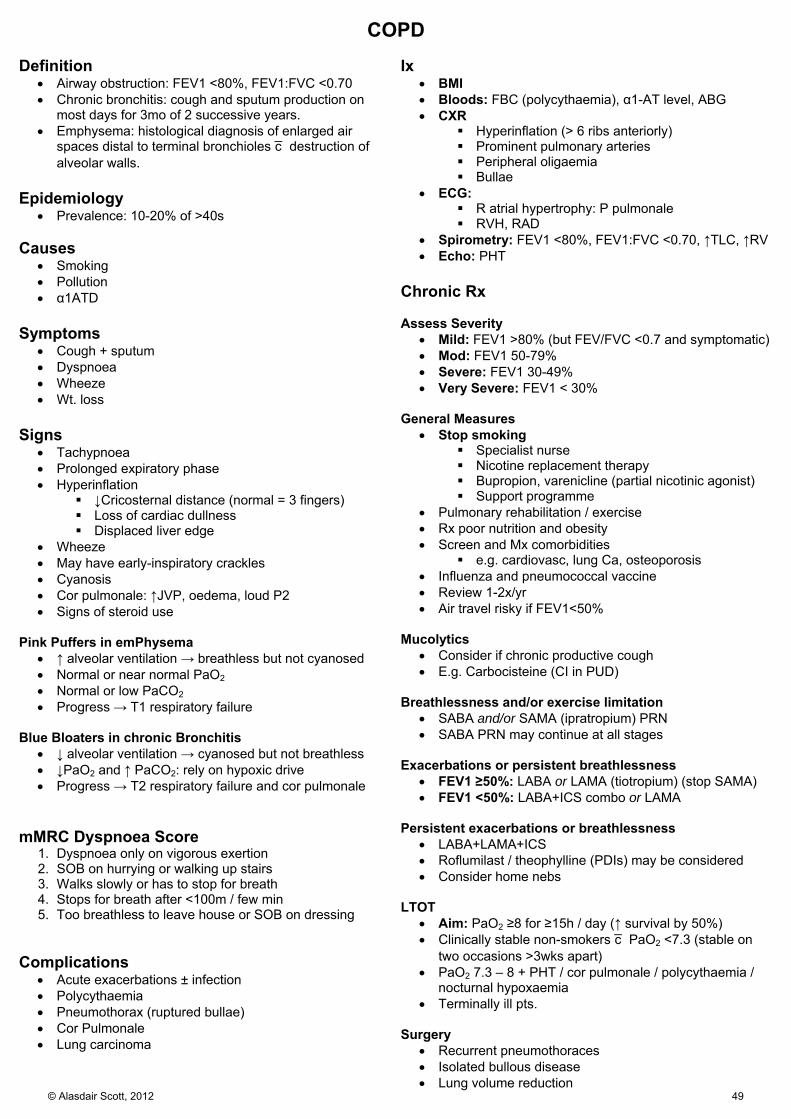
COPD Definition
Airway obstruction: FEV1 <80%, FEV1:FVC <0.70 Chronic bronchitis: cough and sputum production on
most days for 3mo of 2 successive years. Emphysema: histological diagnosis of enlarged air
spaces distal to terminal bronchioles c̄ destruction of alveolar walls.
Epidemiology
Prevalence: 10-20% of >40s Causes
Smoking Pollution α1ATD
Symptoms
Cough + sputum Dyspnoea Wheeze Wt. loss
Signs
Tachypnoea Prolonged expiratory phase Hyperinflation
↓Cricosternal distance (normal = 3 fingers) Loss of cardiac dullness Displaced liver edge
Wheeze May have early-inspiratory crackles Cyanosis Cor pulmonale: ↑JVP, oedema, loud P2 Signs of steroid use
Pink Puffers in emPhysema
↑ alveolar ventilation → breathless but not cyanosed Normal or near normal PaO2 Normal or low PaCO2 Progress → T1 respiratory failure
Blue Bloaters in chronic Bronchitis
↓ alveolar ventilation → cyanosed but not breathless ↓PaO2 and ↑ PaCO2: rely on hypoxic drive Progress → T2 respiratory failure and cor pulmonale
mMRC Dyspnoea Score
1. Dyspnoea only on vigorous exertion 2. SOB on hurrying or walking up stairs 3. Walks slowly or has to stop for breath 4. Stops for breath after <100m / few min 5. Too breathless to leave house or SOB on dressing
Complications
Acute exacerbations ± infection Polycythaemia Pneumothorax (ruptured bullae) Cor Pulmonale Lung carcinoma
Ix
BMI Bloods: FBC (polycythaemia), α1-AT level, ABG CXR
Hyperinflation (> 6 ribs anteriorly) Prominent pulmonary arteries Peripheral oligaemia Bullae
ECG: R atrial hypertrophy: P pulmonale RVH, RAD
Spirometry: FEV1 <80%, FEV1:FVC <0.70, ↑TLC, ↑RV Echo: PHT
Chronic Rx Assess Severity
Mild: FEV1 >80% (but FEV/FVC <0.7 and symptomatic) Mod: FEV1 50-79% Severe: FEV1 30-49% Very Severe: FEV1 < 30%
General Measures
Stop smoking Specialist nurse Nicotine replacement therapy Bupropion, varenicline (partial nicotinic agonist) Support programme
Pulmonary rehabilitation / exercise Rx poor nutrition and obesity Screen and Mx comorbidities
e.g. cardiovasc, lung Ca, osteoporosis Influenza and pneumococcal vaccine Review 1-2x/yr Air travel risky if FEV1<50%
Mucolytics
Consider if chronic productive cough E.g. Carbocisteine (CI in PUD)
Breathlessness and/or exercise limitation SABA and/or SAMA (ipratropium) PRN SABA PRN may continue at all stages
Exacerbations or persistent breathlessness
FEV1 ≥50%: LABA or LAMA (tiotropium) (stop SAMA) FEV1 <50%: LABA+ICS combo or LAMA
Persistent exacerbations or breathlessness
LABA+LAMA+ICS Roflumilast / theophylline (PDIs) may be considered Consider home nebs
LTOT
Aim: PaO2 ≥8 for ≥15h / day (↑ survival by 50%) Clinically stable non-smokers c̄ PaO2 <7.3 (stable on
two occasions >3wks apart) PaO2 7.3 – 8 + PHT / cor pulmonale / polycythaemia /
nocturnal hypoxaemia Terminally ill pts.
Surgery
Recurrent pneumothoraces Isolated bullous disease Lung volume reduction
© Alasdair Scott, 2012 49
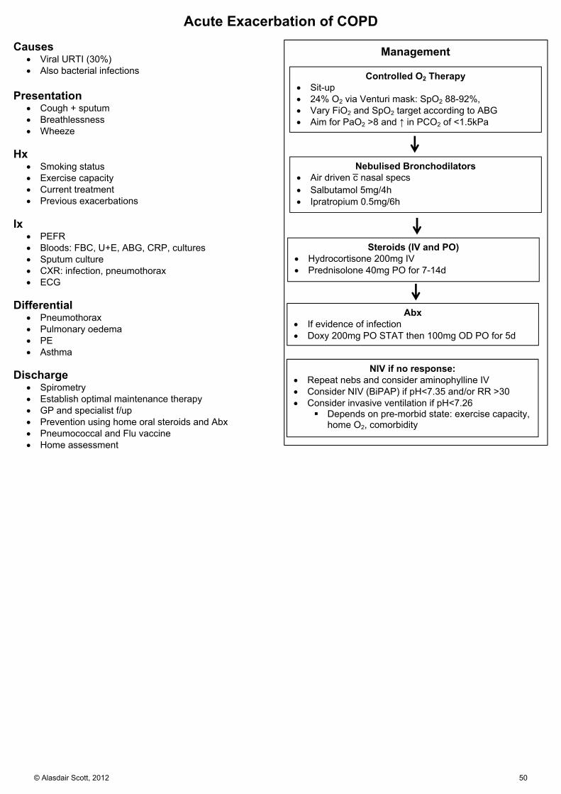
Acute Exacerbation of COPD Causes
Viral URTI (30%) Also bacterial infections
Presentation
Cough + sputum Breathlessness Wheeze
Hx
Smoking status Exercise capacity Current treatment Previous exacerbations
Ix
PEFR Bloods: FBC, U+E, ABG, CRP, cultures Sputum culture CXR: infection, pneumothorax ECG
Differential
Pneumothorax Pulmonary oedema PE Asthma
Discharge
Spirometry Establish optimal maintenance therapy GP and specialist f/up Prevention using home oral steroids and Abx Pneumococcal and Flu vaccine Home assessment
Controlled O2 Therapy Sit-up 24% O2 via Venturi mask: SpO2 88-92%, Vary FiO2 and SpO2 target according to ABG Aim for PaO2 >8 and ↑ in PCO2 of <1.5kPa
Management
Nebulised Bronchodilators Air driven c̄ nasal specs Salbutamol 5mg/4h Ipratropium 0.5mg/6h
Steroids (IV and PO) Hydrocortisone 200mg IV Prednisolone 40mg PO for 7-14d
Abx If evidence of infection Doxy 200mg PO STAT then 100mg OD PO for 5d
NIV if no response: Repeat nebs and consider aminophylline IV Consider NIV (BiPAP) if pH<7.35 and/or RR >30 Consider invasive ventilation if pH<7.26
Depends on pre-morbid state: exercise capacity, home O2, comorbidity
© Alasdair Scott, 2012 50

Pulmonary Embolism Causes
Usually arise from DVTs in proximal leg or iliac veins Rarely:
Right ventricle post MI Septic emboli in right sided endocarditis
Risk Factors: SPASMODICAL
Sex: F Pregnancy Age: ↑ Surgery (classically 10d post-op straining at stool) Malignancy Oestrogen: OCP/HRT DVT/PE previous Hx Immobility Colossal size Antiphospholipid Abs Lupus Anti-coagulant
Presentation
Symptoms and signs depend on size, number and distribution of emboli
Symptoms
Dyspnoea Pleuritic pain Haemoptysis Syncope
Signs
Fever Cyanosis Tachycardia, tachypnoea RHF: hypotension, ↑JVP, loud P2 Evidence of cause: DVT
Ix
Bloods: FBC, U+E, clotting, D-dimers ABG: normal or ↓PaO2 and ↓PaCO2, ↑pH CXR: normal or oligaemia, linear atelectasis ECG: sinus tachycardia, RBBB, right ventricular strain
(inverted T in V1-V4) S1, Q3, T3 is rare
Doppler US: thigh and pelvis (+ve in 60%) CTPA + venous phase of legs and pelvis
85-95% sensitivity V/Q scan no longer used
Dx
1. Assess probability using Wells’ Score 2. Low-probability → perform D-dimers
Negative → excludes PE Positive → CTPA
3. High probability → CTPA NB. –ve D-dimer has 95% NPV for PE Prevention
Risk assessment for all pts TEDS Prophylactic LMWH Avoid OCP/HRT if @ risk
O2 Sit-up 100% O2 via non-rebreather mask
Management
Analgesia Morphine ± metoclopramide if distressed
SBP?
>90 Start Warfarin Confirm Dx
LMWH Heparin e.g. enoxaparin 1.5mg/kg/24h SC
<90 500ml colloid
Inotropes if BP still ↓ Dobutamine: aim for SBP >90 Consider addition of NORAD Consider thrombolysis (medical or surgical)
On-going Mx
TEDS stockings in hospital Graduated compression stockings for 2yrs if DVT:
prevent post-phlebitic syndrome (10-30%) Continue LMWH until INR >2 (at least 5d) Target INR = 2-3 Duration
Remedial cause: 3mo No identifiable cause: 6mo On-going cause: indefinite
VC filter if repeat DVT/PE despite anticoagulation
If critically ill c̄ massive PE consider thrombolysis
Alteplase 50mg bolus stat (Surgical or interventional embolectomy)
© Alasdair Scott, 2012 51

Pneumothorax Definition
Accumulation of air in the pleural space c̄ 2O lung collapse.
Classification
Closed: intact chest wall and air leaks from lung into pleural cavity.
Open: defect in the chest wall allows communication between PTX and exterior: may be sucking.
Tension: air enters pleural cavity through one-way valve and cannot escape → mediastinal compression.
Causes Spontaneous
1O: no underlying lung disease Young, thin men (ruptured subpleural bulla) Smokers
2O: underlying lung disease COPD Marfan’s, Ehler’s Danlos Pulmonary fibrosis, sarcoidosis
Trauma
Penetrating Blunt ± rib #s
Iatrogenic
Subclavian CVP line insertion Positive pressure ventilation Transbronchial biospy Liver biopsy
Presentation Symptoms
Sudden onset Dyspnoea Pleuritic chest pain Tension: respiratory distress, cardiac arrest
Signs
Chest ↓ expansion Resonant percussion ↓ breath sounds ↓VR
Tension: ↑JVP, mediastinal shift, ↑HR, ↓BP Crepitus: surgical emphysema
Ix
ABG US CXR (expiratory film may be helpful)
Translucency + collapse (2cm rim = 50% vol loss) Mediastinal shift (away from PTX) Surgical emphysema Cause: rib #s, pulmonary disease (e.g. bullae)
Tension PTX Resuscitate pt. No CXR Large bore Venflon into 2nd ICS, mid-clavicular line Insert ICD
Mx
Traumatic Resuscitate pt. Analgesia: e.g. morphine 3-sided wet dressing if sucking Insert ICD
1O PTX
2O PTX
SOB and/or rim ≥2cm? Consider d/c
Aspiration successful?
Insert ICD
No
Yes
No
Yes
SOB and > 50yrs and rim ≥2cm?
Admit for 24h
Aspiration successful?
Insert ICDYes
No No
Yes
© Alasdair Scott, 2012 52
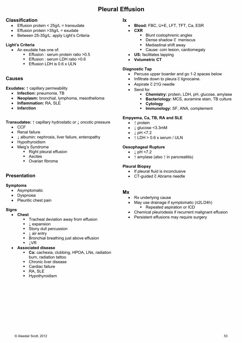
Pleural Effusion Classification
Effusion protein < 25g/L = transudate Effusion protein >35g/L = exudate Between 25-35g/L: apply Light’s Criteria
Light’s Criteria
An exudate has one of: Effusion : serum protein ratio >0.5 Effusion : serum LDH ratio >0.6 Effusion LDH is 0.6 x ULN
Causes Exudates: ↑ capillary permeability
Infection: pneumonia, TB Neoplasm: bronchial, lymphoma, mesothelioma Inflammation: RA, SLE Infarction
Transudates: ↑ capillary hydrostatic or ↓ oncotic pressure
CCF Renal failure ↓ albumin: nephrosis, liver failure, enteropathy Hypothyroidism Meig’s Syndrome
Right pleural effusion Ascites Ovarian fibroma
Presentation Symptoms
Asymptomatic Dyspnoea Pleuritic chest pain
Signs
Chest Tracheal deviation away from effusion ↓ expansion Stony dull percussion ↓ air entry Bronchial breathing just above effusion ↓VR
Associated disease Ca: cachexia, clubbing, HPOA, LNs, radiation
burn, radiation tattoo Chronic liver disease Cardiac failure RA, SLE Hypothyroidism
Ix
Blood: FBC, U+E, LFT, TFT, Ca, ESR CXR
Blunt costophrenic angles Dense shadow c̄ meniscus Mediastinal shift away Cause: coin lesion, cardiomegaly
US: facilitates tapping Volumetric CT
Diagnostic Tap
Percuss upper boarder and go 1-2 spaces below Infiltrate down to pleura c̄ lignocaine. Aspirate c̄ 21G needle Send for
Chemistry: protein, LDH, pH, glucose, amylase Bacteriology: MCS, auramine stain, TB culture Cytology Immunology: SF, ANA, complement
Empyema, Ca, TB, RA and SLE
↑ protein ↓ glucose <3.3mM ↓ pH <7.2 ↑ LDH > 0.6 x serum / ULN
Oesophageal Rupture
↓ pH <7.2 ↑ amylase (also ↑ in pancreatitis)
Pleural Biopsy
If pleural fluid is inconclusive CT-guided c̄ Abrams needle
Mx
Rx underlying cause May use drainage if symptomatic (≤2L/24h)
Repeated aspiration or ICD Chemical pleurodesis if recurrent malignant effusion Persistent effusions may require surgery
© Alasdair Scott, 2012 53
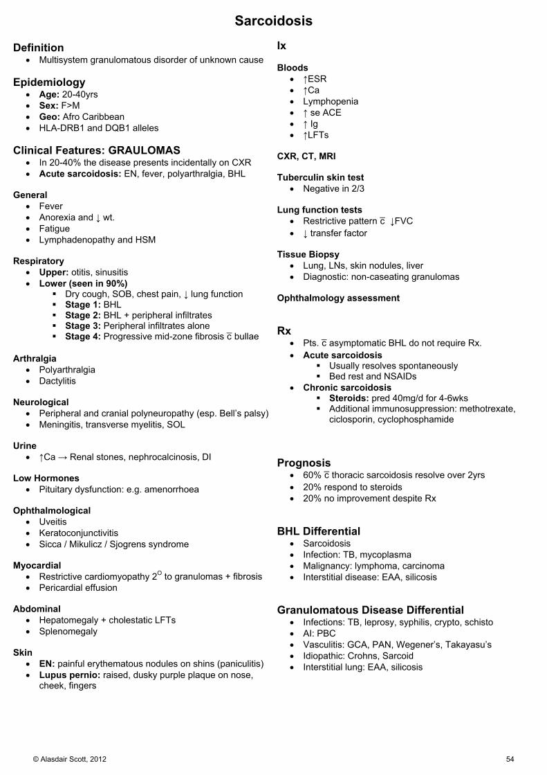
Sarcoidosis Definition
Multisystem granulomatous disorder of unknown cause Epidemiology
Age: 20-40yrs Sex: F>M Geo: Afro Caribbean HLA-DRB1 and DQB1 alleles
Clinical Features: GRAULOMAS
In 20-40% the disease presents incidentally on CXR Acute sarcoidosis: EN, fever, polyarthralgia, BHL
General
Fever Anorexia and ↓ wt. Fatigue Lymphadenopathy and HSM
Respiratory
Upper: otitis, sinusitis Lower (seen in 90%)
Dry cough, SOB, chest pain, ↓ lung function Stage 1: BHL Stage 2: BHL + peripheral infiltrates Stage 3: Peripheral infiltrates alone Stage 4: Progressive mid-zone fibrosis c̄ bullae
Arthralgia
Polyarthralgia Dactylitis
Neurological
Peripheral and cranial polyneuropathy (esp. Bell’s palsy) Meningitis, transverse myelitis, SOL
Urine
↑Ca → Renal stones, nephrocalcinosis, DI Low Hormones
Pituitary dysfunction: e.g. amenorrhoea Ophthalmological
Uveitis Keratoconjunctivitis Sicca / Mikulicz / Sjogrens syndrome
Myocardial
Restrictive cardiomyopathy 2O to granulomas + fibrosis Pericardial effusion
Abdominal
Hepatomegaly + cholestatic LFTs Splenomegaly
Skin
EN: painful erythematous nodules on shins (paniculitis) Lupus pernio: raised, dusky purple plaque on nose,
cheek, fingers
Ix Bloods
↑ESR ↑Ca Lymphopenia ↑ se ACE ↑ Ig ↑LFTs
CXR, CT, MRI Tuberculin skin test
Negative in 2/3 Lung function tests
Restrictive pattern c̄ ↓FVC ↓ transfer factor
Tissue Biopsy
Lung, LNs, skin nodules, liver Diagnostic: non-caseating granulomas
Ophthalmology assessment Rx
Pts. c̄ asymptomatic BHL do not require Rx. Acute sarcoidosis
Usually resolves spontaneously Bed rest and NSAIDs
Chronic sarcoidosis Steroids: pred 40mg/d for 4-6wks Additional immunosuppression: methotrexate,
ciclosporin, cyclophosphamide
Prognosis
60% c̄ thoracic sarcoidosis resolve over 2yrs 20% respond to steroids 20% no improvement despite Rx
BHL Differential
Sarcoidosis Infection: TB, mycoplasma Malignancy: lymphoma, carcinoma Interstitial disease: EAA, silicosis
Granulomatous Disease Differential
Infections: TB, leprosy, syphilis, crypto, schisto AI: PBC Vasculitis: GCA, PAN, Wegener’s, Takayasu’s Idiopathic: Crohns, Sarcoid Interstitial lung: EAA, silicosis
© Alasdair Scott, 2012 54

Interstitial Lung Disease Principal Features
Dyspnoea Dry cough Abnormal CXR / CT Restrictive Spirometry
Causes Known Cause
Environmental: asbestosis, silicosis Drugs: BANS ME
Bleomycin, Busulfan Amiodarone Nitrofurantoin Sulfasalazine MEthotrexate, MEthysergide
Hypersensitivity: EAA Infection: TB, viral, fungi
Assoc. c̄ systemic disease
Sarcoidosis RA SLE, systemic sclerosis, Sjogren’s, MCTD UC, ankylosing spondylitis
Idiopathic
Idiopathic pulmonary fibrosis (CFA)
Cause by Location Upper Zone: A PENT
Aspergillosis : ABPA Pneumoconiosis: Coal, Silica Extrinsic allergic alveolitis Negative, sero-arthropathy TB
Lower Zone: STAIR
Sarcoidosis (mid zone) Toxins: BANS ME Asbestosis Idiopathic pulmonary fibrosis Rheum: RA, SLE, Scleroderma, Sjogren’s, PM/DM
© Alasdair Scott, 2012 55

Extrinsic Allergic Alveolitis Acute allergen exposure in sensitised pts. → T3HS Chronic exposure → granuloma formation and
obliterative bronchiolitis (T4HS) Causes
Bird fancier’s lung: proteins in bird droppings Farmer’s / mushroom worker’s Malt worker’s lung: Aspergillus clavatus
Clinical Features 4-6h post-exposure
Fever, rigors, malaise Dry cough, dyspnoea Crackles (no wheeze)
Chronic
Increasing dyspnoea Wt. loss T1 respiratory failure Cor pulmonale
Ix
Bloods Acute: neutrophilia, ↑ESR +ve se precipitins
CXR Upper zone reticulonodular opacification or
fibrosis → honeycomb lung BHL (rare)
Spirometry Restrictive defect ↓ transfer factor during acute attacks
BAL ↑ lymphocytes and mast cells
Mx
Avoid exposure Steroids: acute / long-term Compensation may be payable
Industrial Lung Disease Coal-workers Pneumoconiosis
CWP → Progressive Massive Fibrosis Presents as progressive dyspnoea and chronic
bronchitis CXR: upper zone fibrotic masses
Silicosis
Quarrying, sand-blasting Upper zone reticular shadowing and egg shell
calcification of hilar nodes → PMF Asbestosis
Demolition and ship building Basal fibrosis, pleural plaques ↑ risk of mesothelioma
Chest pain, wt. loss, clubbing, recurrent effusions, dyspnoea.
CXR: pleural effusions, thickening Dx by histology of pleural biopsy <2yr survival
Idiopathic Pulmonary Fibrosis (CFA) Epidemiology
Commonest cause of interstitial lung disease Middle age M>F = 2:1 Assoc. c̄ other AI disease in 30%
Presentation Symptoms
Dry cough Dyspnoea Malaise, wt. loss Arthralgia OSA
Signs
Cyanosis Clubbing Crackles: fine, end-inspiratory
Complications
↑ risk Ca lung Type 2 respiratory failure and cor pulmonale
Ix Bloods
↑CRP ↑Ig ANA+ (30%) RF+ (10%) ABG: ↓PaO2, ↑PaCO2
CXR
↓ lung volume Bilat lower zone retic-nod shadowing Honeycomb lung
HRCT
Shows similar changes to CXR More sensitive
Spiro Restrictive defect ↓ transfer factor
Other
Lung biopsy: Usual interstitial pneumonia BAL: may indicate disease activity
↑ lymphocytes: good prognosis ↑ PMN or eosinophils: bad prognosis
DTPA scan: may reflect disease activity Mx
Supportive care Stop smoking Pulmonary rehabilitation O2 therapy Palliation Rx symptoms of heart failure
Lung Tx offers only cure Prognosis
50% 5yr survival
© Alasdair Scott, 2012 56

Pulmonary Hypertension Definition
PA pressure >25mmHg Causes Left Heart Disease
Mitral stenosis Mitral regurgitation Left ventricular failure L → R shunt
Lung Parenchymal Disease
Mechanism Chronic hypoxia → hypoxic vasoconstriction Perivascular parenchymal changes
COPD Asthma: severe, chronic Interstitial lung disease CF, bronchiectasis
Pulmonary Vascular Disease
Idiopathic pulmonary hypertension Pulmonary vasculitis: scleroderma, SLE, Wegener’s Sickle cell Pulmonary embolism: acute or chronic Portal HTN: portopulmonary HTN
Hypoventilation
OSA Morbid obesity (Pickwickian syn.) Thoracic cage abnormality: kyphosis, scoliosis Neuromuscular: MND, MG, polio
Ix
ECG P pulmonale RVH RAD
Echo Velocity of tricuspid regurgitation jet Right atrial or ventricular enlargement Ventricular dysfunction Valve disease
Right heart catheterisation: gold standard Mean pulmonary artery pressure Pulmonary vascular resistance CO Vasoreactivity testing to guide Rx
Cor Pulmonale Definition
RHF due to chronic PHT Symptoms
Dyspnoea Fatigue Syncope
Signs
1. ↑ JVP c̄ prominent a wave 2. Left parasternal heave 3. Loud P2 ± S3 4. Murmurs
PR: Graham Steell EDM TR: PSM
5. Pulsatile hepatomegaly 6. Fluid: Ascites + Peripheral oedema
Ix
Bloods: FBC, U+E, LFTs, ESR, ANA, RF ABG: hypoxia ± hypercapnoea CXR
Enlarged R atrium and ventricle Prominent pulmonary arteries Peripheral oligaemia
ECG: P pulmonale + RVH Echo: RVH, TR, ↑ PA pressure Spirometry Right heart catheterisation
Mx
Rx underlying condition ↓ pulmonary vascular resistance
LTOT CCB: e.g. nifedipine Sildenafil (PDE-5 inhibitor) Prostacycline analogues Bosentan (endothelin receptor antagonist)
Cardiac failure ACEi + β-B (caution if asthma) Diuretics
Heart-Lung Tx Prognosis
50% 5ys
© Alasdair Scott, 2012 57
Obstructive Sleep Apnoea Definition
Intermittent closure/collapse of pharyngeal airway → apnoeic episodes during sleep.
Risk Factors
Obesity Male Smoker EtOH Idiopathic pulmonary fibrosis Structural airway pathology: e.g. micrognathia NM disease: e.g. MND
Ix
SpO2 Polysomnography is diagnostic
Rx
Wt. loss Avoid smoking and EtOH CPAP during sleep
Clinical Features Nocturnal
Snoring Choking, gasping, apnoeic episodes
Daytime
Morning headache Somnolence ↓ memory and attention Irritability, depression
Complications
Pulmonary hypertension Type 2 respiratory failure Cor pulmonale
Mx
↓ wt. Stop smoking CPAP @ night via a nasal mask Surgery to relieve pharyngeal obstruction
Tonsillectomy Uvulopalatpharyngoplasty
Smoking Cessation Very Brief Advice
Ask: enquire as to smoking status Advise: best way to stop is with support and medication Act: provide details of where to get help
E.g. NHS stop smoking helpline Facilitating Quitting
Refer to specialist stop smoking service Nicotine replacement
Gum Patches
Varenicline: selective partial nicotine receptor agonist Recommended by NICE 23% abstinence @ 1yr vs. 10% for placebo Start while still smoking
Bupropion: also an option
© Alasdair Scott, 2012 58
Endocrinology Contents
DM: Classification, Dx and Causes ............................................................................................................................................. 60
DM: Conservative Mx .................................................................................................................................................................. 60
DM: Oral Hypoglycaemics ........................................................................................................................................................... 61
Insulin .......................................................................................................................................................................................... 61
Diabetic Complications ................................................................................................................................................................ 62
Macrovascular ............................................................................................................................................................................. 62
Diabetic Feet ............................................................................................................................................................................... 62
Diabetic Ketoacidosis .................................................................................................................................................................. 63
Hyperosmolar Non-Ketotic Coma ................................................................................................................................................ 64
Hypoglycaemia ............................................................................................................................................................................ 65
Thyrotoxicosis .............................................................................................................................................................................. 66
Hypothyroidism ............................................................................................................................................................................ 67
Hyperparathyroidism ................................................................................................................................................................... 70
Hypoparathyroidism..................................................................................................................................................................... 70
Cushing’s Syndrome ................................................................................................................................................................... 71
Hyperaldosteronism..................................................................................................................................................................... 72
Adrenal Insufficiency ................................................................................................................................................................... 73
Phaeochromocytoma .................................................................................................................................................................. 74
Multiple Endocrine Neoplasia ...................................................................................................................................................... 74
Autoimmune Polyendocrine Syndromes ..................................................................................................................................... 74
Hypopituitarism ............................................................................................................................................................................ 75
Pituitary Tumours ........................................................................................................................................................................ 75
Hyperprolactinaemia ................................................................................................................................................................... 76
Acromegaly .................................................................................................................................................................................. 76
Diabetes Insipidus ....................................................................................................................................................................... 77
Sexual Characteristics ................................................................................................................................................................. 77
© Alasdair Scott, 2012 59
DM: Classification, Dx and Causes Definition
Multisystem disorder due to an absolute or relative lack of endogenous insulin → metabolic and vascular complications.
Classification T1DM
Path: autoimmune destruction of β-cells → absolute insulin deficiency.
Age: usually starts before puberty Presentation: polyuria, polydipsia, ↓wt., DKA Genetics: concordance only 30% in MZs Assoc.: HLA-D3 and –D4, other AI disease Abs: anti-islet, anti-GAD
T2DM
Path: insulin resistance and β-cell dysfunction → relative insulin deficiency
Age: usually older patients Presentation: polyuria, polydipsia, complications Genetics: concordance 80% in MZs Assoc.: obesity, ↓exercise, calorie and EtOH excess
Dx
Symptomatic: Polyuria, polydipsia, ↓wt., lethargy ↑ plasma venous glucose detected once
Fasting ≥7mM Random ≥11.1mM
Asymptomatic ↑ venous glucose on 2 separate occasions Or, 2h OGTT ≥ 11.1mM
Glucose Testing
OGTT only needed if borderline fasting or random glucose measurements.
Normal IFG IGT DiabetesFasting <6.1 6.1 – 6.9 ≥7.0 75g OGTT <7.8 7.8 – 11 ≥11.1
Secondary Causes of DM
Drugs: steroids, anti-HIV, atypical neuroletics, thiazides Pancreatic: CF, chronic pancreatitis, HH, pancreatic Ca Endo: Phaeo, Cushings, Acromegaly, T4 Other: glycogen storage diseases
Metabolic Syndrome
Central obesity (↑ waist circumference) and two of: ↑ Triglycerides ↓ HDL HTN Hyperglycaemia: DM, IGT, IFG
DM: Conservative Mx MDT
GP, endocrinologist, surgeons, specialist nurses, dieticians, chiropodists, fellow patients (education groups).
Monitoring: 4Cs Control, glycaemic
Record of complications: DKA, HONK, hypos Capillary blood glucose
Fasting: 4.5-6.5mM 2h post-prandial: 4.5-9mM
HbA1c Reflects exposure over last 6-8wks Aim <45 - 50mM (7.5 - 8%)
BP, lipids Complications
Macro Pulses BP Cardiac auscultation
Micro Fundoscopy ACR + U+Es Sensory testing plus foot inspection
Competency
With insulin injections Checking injection sites BM monitoring
Coping
Psychosocial: e.g. ED, depression Occupation Domestic
Lifestyle Modification: DELAYS Diet
Same as that considered healthy for everyone ↓ total calorie intake ↓ refined CHO, ↑ complex CHO ↑ soluble fibre ↓ fat (especially saturated) ↓ Na Avoid binge drinking
Exercise Lipids
Rx of hyperlipidaemia 1O prevention c̄ statins if >40yrs (regardless of lipids)
ABP
↓ Na intake and EtOH Keep BP <130/80 ACEis best (β-B: mask hypos, thiazides: ↑ glucose)
Aspirin
1O prevention if >50yrs or <50 c̄ other CVD RFs Yearly / 6 monthly check-up: 4Cs Smoking cessation
© Alasdair Scott, 2012 60

DM: Oral Hypoglycaemics
1. Lifestyle Modification: DELAYS
2. Start Metformin (if HBA1c >target after lifestyle changes)
SE: nausea, diarrhoea, abdo pain, lactic acidosis CI: GFR<30, tissue hypoxia (sepsis, MI), morning
before GA and iodinated contrast media 500mg after evening meal, ↑ing to 2g max.
3. Metformin + Sulfonylurea (if HBA1c >target)
E.g. gliclazide MR 30mg c̄ breakfast SE: hypoglycaemia, wt. gain CI: omit on morning of surgery
Other Options
Consider adding a rapid-acting insulin secretagogue (e.g. nateglinide) to metformin instead of a sulfonylurea. May be preferable if erratic lifestyle.
Consider adding pioglitazone to metformin instead of a sulfonylurea
4. Additional Therapy
1st line Add insulin → insulin + metformin + sulfonylurea
2nd line
Add sitagliptin or pioglitazone if insulin unacceptable Employment, social or recreational issues Obesity
metformin + sulfonylurea + sitagliptin / pio
3rd line Add exenatide (SC) if insulin unacceptable or BMI>35 metformin + sulfonylurea + exenatide
4th line
Consider acarbose if unable to use other glucose-lowering drugs
Insulin Principles
Ensure pt. education about Self-adjustment c̄ exercise and calories Titrate dose Family member can abort hypo c̄ sugary drinks
or GlucoGel Pre-prandial BM don’t tell you who much glucose is
needed Fasting BM before meal informs re long-acting insulin
dose. Finger-prick BM after meal informs re short-acting
insulin dose (for that last meal) Common Regimes BD Biphasic Regime
BD insulin mixture 30min before breakfast and dinner Rapid-acting: e.g. actrapid Intermediate- / long-acting: e.g. insulatard
T2 or T1 DM c̄ regular lifestyle: children, older pts. Assoc, c̄ fasting hyperglycaemia
Basal-Bolus Regime
Bedtime long-acting (e.g. glargine) + short acting before each meal (e.g. lispro) Adjust dose according to meal size ~50% of insulin given as long-acting
T1DM allowing flexible lifestyle Best outcome
OD Long-Acting Before Bed
Initial regime when switching from tablets in T2DM Illness
Insulin requirements usually ↑ (even if food intake ↓) Maintain calories (e.g. milk) Check BMs ≥4hrly and test for ketonuria ↑ insulin dose if glucose rising
Side-Effects
Hypoglycaemia At risk: EtOH binge, β-B (mask symptoms),
elderly Need to admit sulfonylurea-induced hypo
Lipohypertrophy Rotate injection site: abdomen, thighs
Wt. gain in T2DM ↓ wt. gain if insulin given c̄ metformin
© Alasdair Scott, 2012 61

Diabetic Complications Summary
Hyperglycaemia: DKA, HONK Hypoglycaemia Infection Macrovascular: MI, CVA Microvascular
Macrovascular MI: May be “silent” due to autonomic neuropathy PVD: claudication, foot ulcers CVA
Rx: Manage CV risk factors
BP (aim <130/80) Smoking Lipids HBA1c
Prevention
Good glycaemic control (e.g. HbA1c <6%) prevents both macro- and micro-vascular complications.
Proved by DCCT, EDIC and UKPDS trials Regular screening: fundoscopy, ACR, foot check
Diabetic Feet Ischaemia
Critical toes Absent pulses (do ABPI) Ulcers: painful, punched-out, foot margins, pressure
points Neuropathy
Loss of protective sensation Deformity: Charcot’s joints, pes cavus, claw toes Injury or infection over pressure points Ulcers: painless, punched-out, metatarsal heads,
calcaneum Mx
Conservative Daily foot inspection (e.g. c̄ mirror) Comfortable / therapeutic shoes Regular chiropody (remove callus)
Medical Rx infection: benpen + fluclox ± metronidazole
Surgical Abscess or deep infection Spreading cellulitis Gangrene Suppurative arthritis
Nephropathy Pathophysiology
Hyperglycaemia → nephron loss and glomerulosclerosis
Features
Microalbuminuria: urine albumin:Cr (ACR) ≥30mg/mM If present → ACEi / ARA Refer if UCR >70
Retinopathy, Maculopathy and Cataracts Pathogenesis
Microvascular disease → retinal ischaemia → ↑VEGF ↑ VEGF → new vessel formation
Presentation
Retinopathy and maculopathy Cataracts Rubeosis iris: new vessels on iris → glaucoma CN palsies
Diabetic Retinopathy and Maculopathy
Commonest cause of blindness up to 60yrs Refer if pre-proliferative retinopathy / maculopathy Ix: fluorescein angiography Rx: laser photocoagulation
Background Retinopathy Dots: microaneurysms Blot haemorrhages Hard exudates: yellow lipid patches
Pre-proliferative Retinopathy Cotton-wool spots (retinal infarcts) Venous beading Haemorrhages
Proliferative Retinopathy New vessels Pre-retinal or vitreous haemorrhage
Maculopathy ↓ acuity may be only sign Hard exudates w/i one disc width of macula
Neuropathy Pathophysiology
Metabolic: glycosylation, ROS, sorbitol accumulation Ischaemia: loss of vasa nervorum
Symmetric sensory polyneuropathy
Glove and stocking: length-dependent ( feet 1st) Loss of all modalities
Absent ankle jerks Numbness, tingling, pain (worse @ night) Rx
Paracetamol Amitriptyline, Gabapentin, SSRI Capsaicin cream Baclofen
Mononeuropathy / Mononeuritis Multiplex
E.g. CN3/6 palsies Femoral Neuropathy / Amyotrophy
Painful asymmetric weakness and wasting of quads c̄ loss of knee jerks
Dx: nerve conduction and electromyography Autonomic Neuropathy
Postural hypotension – Rx: fludrocortisone Gastroparesis → early satiety, GORD, bloating Diarrhoea: Rx c̄ codeine phosphate Urinary retention ED
© Alasdair Scott, 2012 62

Diabetic Ketoacidosis Pathogenesis Ketogenesis
↓ insulin → ↑ stress hormones and ↑ glucagon → ↓ glucose utilisation + ↑ fat β-oxidation ↑ fatty acids → ↑ ATP + generation of ketone bodies.
Dehydration
↓ insulin → ↓ glucose utilisation + ↑ gluconeogenesis → severe hyperglycaemia
→ osmotic diuresis → dehydration Also, ↑ ketones → vomiting
Acidosis
Dehydration → renal perfusion Hyperkalaemia
Precipitants
Infection / stress ± stopping insulin New T1DM
Presentation
Abdo pain + vomiting Gradual drowsiness Sighing “Kussmaul” hyperventilation Dehydration Ketotic breath
Dx
Acidosis (↑AG): pH <7.3 (± HCO3 <15mM) Hyperglycaemia: ≥11.1mM (or known DM) Ketonaemia: ≥3mM (≥2+ on dipstix)
Ix
Urine: ketones and glucose, MCS Cap glucose and ketones VBG: acidosis + ↑K Bloods: U+E, FBC, glucose, cultures CXR: evidence of infection
Subtleties
Hyponatraemia is the norm Osmolar compensation for hyperglycaemia ↑/↔ Na indicates severe dehydration
Avoid rapid ↓ in insulin once glucose normalised Glucose decreases faster than ketones and
insulin is necessary to get rid of them. Amylase is often ↑ (up to 10x) Excretion of ketones → loss of potential bicarbonate →
hyperchloraemic metabolic acidosis after Rx Complications
Cerebral oedema: excess fluid administration Commonest cause of mortality
Aspiration pneumonia Hypokalaemia Hypophosphataemia → resp and skeletal muscle
weakness Thromboembolism
Mx: in HDU
Gastric aspiration Rehydrate Insulin infusion Potassium replacement
Fluids 0.9% NS infusion via large bore cannula
SBP<90 → 1L stat + more until SBP >90 SBP>90 → 1L over 1h
Then: 1L over next 2h, 1L/2h, 1L/4h, 1L/4h, 1L/6h Switch to 10% dex 1L/8h when glucose <14mM Start Potassium Replacement in 2nd Bag of Fluids >5.5mM → Nil 3.5-5.5mM → 40mmol/L <3.5mM → consult senior for review
Management
Insulin Infusion Actrapid 0.1u/kg/h IVI (6u if no wt., max 15u)
Assessment Hx + full examination Investigations: capillary, urine, blood, imaging
Resolution Ketones <0.3mM + venous pH>7.3 (HCO3 >18mM) Transfer to sliding scale if not eating Transfer to SC insulin when eating and drinking
Additional Measures Urinary catheter (aim: 0.5ml/kg/hr) NGT if vomiting or ↓GCS Thromboprophylaxis c̄ LMWH Refer to Specialist Diabetes Team Find and treat precipitating factors
Monitoring Hrly capillary glucose and ketones VBG @ 60min, 2h and then 2hrly Plasma electrolytes 4hrly Aims ↓ ketones by ≥0.5mM/h or ↑HCO3 by ≥3mM/h ↓ plasma glucose by ≥3mM/h Maintain K in normal range Avoid hypoglycaemia
Transfer to SC Insulin When biochemically resolved and eating Start long-acting insulin the night before Give short-acting insulin before breakfast Stop IVI 30min after short acting
Pt. Education ID precipitating factors and provide action plan Provision of ketone meter c̄ education on use.
© Alasdair Scott, 2012 63

Hyperosmolar Non-Ketotic Coma The Patient
Usually T2DM, often new presentation Usually older Long hx (e.g. 1wk)
Metabolic Derangement
Marked dehydration and glucose >35mM No acidosis (no ketogenesis) Osmolality >340mosmol/kg
Complications
Occlusive events are common: DVT, stroke Give LMWH
Mx
Rehydrate c̄ 0.9% NS over 48h May need ~9L
Wait 1h before starting insulin It may not be needed Start low to avoid rapid changes in osmolality
E.g. 1-3u/hr Look for precipitant
MI Infection Bowel infarct
© Alasdair Scott, 2012 64

Hypoglycaemia Hypoglycaemia: Whipple’s Triad
Low plasma glucose ≤3mM Symptoms consistent c̄ hypoglycaemia Relief of symptoms by glucose administration
Symptoms Autonomic: 2.5-3 Sweating Anxiety Hunger Tremor Palpitations
Neuroglycopenic: <2.5 Confusion Drowsiness Seizures Personality change Focal neurology (e.g. CN3) Coma (<2.2)
Fasting Hypoglycaemia Causes: EXPLAIN
Usually insulin or sulfonylurea Rx in a known diabetic Exercise, missed meal, OD
Exogenous drugs Pituitary insufficiency Liver failure Addison’s Islet cell tumours (insulinomas) Immune (insulin receptor Abs: Hodgkin’s) Non-pancreatic neoplasms: e.g. fibrosarcomas
Ix
72h fast c̄ monitoring Sympto: Glucose, insulin, C-peptide, ketones
Dx
Hyperinsulinaemic hypoglycaemia Drugs
↑ C-pep: sulfonylurea Normal C-pep: insulin
Insulinoma ↓ insulin, no ketones
Non-pancreatic neoplasms Insulin receptor Abs
↓ insulin, ↑ ketones Alcohol binge c̄ no food Pituitary insufficiency Addison’s
Insulinoma
Path: 95% benign β-cell tumour usually seen c̄ MEN1 Pres: fasting- / exercise-induced hypoglycaemia Ix:
Hypoglycaemia + ↑ insulin Exogenous insulin doesn’t suppress C-pep MRI, EUS pancreas
Rx: excision Post-Prandial Hypoglycaemia
Dumping post-gastric bypass
Mx Alert and Orientated: Oral Carb
Rapid acting: lucozade Long acting: toast, sandwich
Drowsy / confused but swallow intact: Buccal Carb
Hypostop / Glucogel Consider IV access
Unconscious or Concerned re Swallow: IV dextrose
100ml 20% glucose (50ml 50% dextrose: not used) Deteriorating / refractory / insulin-induced / no access
1mg glucagon IM/SC Won’t work in drunks + short duration of effect (20min) Insulin release may → rebound hypoglycaemia
© Alasdair Scott, 2012 65

Thyrotoxicosis Definition
The clinical effect of ↑T4, usually from gland hyperfunction.
Symptoms
Diarrhoea ↑ appetite but ↓ wt. Sweats, heat intolerance Palpitations Tremor Irritability Oligomenorrhoea ± infertility
Signs Hands
Fast / irregular pulse Warm, moist skin Fine tremor Palmer erythema
Face
Thin hair Lid lag Lid retraction
Neck
Goitre or nodules Graves’ Specific
Ophthalmopathy Exophthalmos Ophthalmoplegia: esp. up-gaze palsy Eye discomfort and grittiness Photophobia and ↓ acuity Chemosis
Dermopathy: pre-tibial myxoedema Thyroid acropachy
Ix
↓TSH, ↑T4/↑T3 Abs: TSH receptor, TPO ↑Ca, ↑LFTs Isotope scan
↑ in Graves’ ↓ in thyroiditis
Ophthalmopathy: acuity, fields, movements Graves’ Disease Epidemiology
60% of cases of thyrotoxicosis Prev: 0.5% Sex: F>>M=9:1 Age: 40-60yrs
Features
Diffuse goitre c̄ ↑iodine uptake Ophthalmopathy and dermopathy Triggers: stress, infection, child-birth
Associations
T1DM Vitiligo Addison’s
Other Causes Toxic Multinodular Goitre = Plummer’s Disease
Autonomous nodule develops on background multinodular goitre.
Elderly and iodine-deficient areas Iodine scan shows hot nodules
Toxic Adenoma
Solitary hot nodule ± producing T3/T4 Most nodules are non-functional
Thyrotoxic Phase of Thyroiditis
Hashimoto’s de Quervain’s Subacute lymphocytic
Drugs
Thyroxine Amiodarone
Rx Medical
Symptomatic: β-B (e.g. propranolol 40mg/6h) Anti-thyroid: carbimazole (inhibits TPO)
Titrate according to TFTs or block and replace In Graves’ Rx for 12-18mo then withdraw
~50% relapse → surgery or radioiodine SE: agranulocytosis
Radiological: Radio-iodine
Most become hypothyroid CI: pregnancy, lactation
Surgical: Thyroidectomy
Recurrent laryngeal N. damage → hoarseness Hypoparathyroidism Hypothyroidism
Thyroid Storm Features
↑ temp Agitation, confusion, coma Tachycardia, AF Acute abdomen Heart failure
Precipitants
Recent thyroid surgery or radio-iodine Infection MI Trauma
Rx
1. Fluid resuscitation + NGT 2. Bloods: TFTs + cultures if infection suspected 3. Propranolol PO/IV 4. Digoxin may be needed 5. Carbimazole then Lugol’s Iodine 4h later to inhibit thyroid 6. Hydrocortisone 7. Rx cause
© Alasdair Scott, 2012 66

Hypothyroidism Epidemiology
Incidence: 4/1000/yr Sex: F>>M=6:1 Age: >40yrs
Symptoms
Lethargy Cold intolerance ↓ appetite but ↑wt. Constipation Menorrhagia ↓ mood
Signs
Cold hands Bradycardic Slow-relaxing reflexes Dry hair and skin Puffy face Goitre Myopathy, neuropathy Ascites Myxoedema
SC tissue swelling in severe hypothyroidism Typically around eyes and dorsum of hand
Causes Primary
Atrophic thyroiditis (commonest UK) Hashimoto’s thyroiditis Subacute thyroiditis (e.g. post-partum) Post De Quervain’s thyroiditis Iodine deficiency (commonest worldwide) Drugs: carbimazole, amiodarone, lithium Congenital: thyroid agenesis
Post-surgical
Thyroidectomy Radioiodine
Secondary
Hypopituitarism (v. rare cause) Atrophic Thyroiditis
Thyroid antibodies +ve: anti-TPO, anti-TSH Lymphocytic infiltrate → atrophy (no goitre) Associations
Pernicious anaemia Vitiligo Endocrinopathies
Hashimoto’s Thyoiditis
TPO +ve Atrophy + regeneration → goitre May go through initial thyrotoxicosis phase May be euthyroid or hypothyoid
Ix
↑TSH, ↓T3/T4 ↑MCV ± normochromic anaemia ↑ triglyceride + ↑ cholesterol Hyponatraemia (SIADH) ↑ CK if assoc. myopathy Abs: TPO, TSH
Rx
Levothyroxine Titrate to normalise TSH Enzyme inducers ↑ thyroxine metabolism Clinical improvement takes ~2wks
Check for other AI disease: e.g. Addison’s, PA Myxoedema Coma Features
Looks hypothyroid Hypothermia Hypoglycaemia Heart failure: bradycardia and ↓BP Coma and seizures
Precipitants
Radioiodine Thyroidectomy Pituitary surgery Infection, trauma, MI, stroke
Mx
Bloods: TFTs, FBC, U+E, glucose, cortisol Correct any hypoglycaemia T3/T4 IV slowly (may ppt. myocardial ischaemia) Hydrocortisone 100mg IV Rx hypothermia and heart failure
© Alasdair Scott, 2012 67

Benign Thyroid Disease Disease Clinical Features Cause Management Simple goitre Diffuse painless goitre
Mass effects: - dysphagia - stridor - SVC obstruction Usually euthyroid, may → hypothyroid
Endemic - iodine deficiency Sporadic - autoimmune - goitrogens (e.g. sulphonylureas) - hereditary (dyshormogenic)
Thyroxine Total or subtotal thyroidectomy if pressure symptoms.
Multinodular goitre
Evolves from long-standing simple goitre. Mass effects. Euthyroid (or subclinical hyperthyroid)
As for simple goitre Thyroxine Total or subtotal thyroidectomy if pressure symptoms.
Plummer’s Toxic multinodular goitre
Multinodular goitre Thyrotoxicosis Uneven iodine uptake with hot nodule
Autonomous nodule develops on background multinodular goitre
Anti-thyroid drugs Total or subtotal thyroidectomy.
Graves’ Diffuse goitre c̄ bruit Ophthalmopathy Dermopathy Thyrotoxicosis Assoc. c̄ other AI disease (T1DM, PA) ↑ uptake on radionucleotide scan
Autoimmune (T2 hypersensitivity) - anti-TSH
Propranolol Carbimazole Radioiodine Thyroidectomy
Hashimoto’s thyroiditis
Diffuse painless goitre May have transient thyrotoxicosis, then hypothyroidism Assoc. c̄ other AI disease (T1DM, PA)
Autoimmune (T2 and T4) - anti-TPO, -Tg
Levothyroxine
de Quervain’s
Diffuse painful goitre Preceding viral URTI common Thyrotoxicosis → hypo→eu ↓ iodine uptake
Viral: Coxsackie common Autoimmune
Self-limiting
Subacute lymphocytic
Diffuse painless goitre May occur post-partum Thyrotoxicosis → hypo→eu
Autoimmune Self-limiting
Riedel’s thyoiditis Hard fixed thyroid mass Mass effects Assoc. c̄ retroperitoneal fibrosis
Autoimmune fibrosis Conservative
Follicular adenoma
Single thyroid nodule ± thyrotoxicosis (majority are cold) May get pressure symptoms
Hemithyroidectomy
Thyroid cysts Solitary thyroid nodule Asympto or pressure symptoms Can → localised pain due to cyst bleed
Aspiration or excision
© Alasdair Scott, 2012 68

Malignant Thyroid Disease
Presentation:
Non-functional (cold) Painless neck mass Cervical mets Compression symptoms
Dysphagia Stridor SVC obstruction
Risk factors for malignancy in thyroid nodules
Solitary Solid Younger Male Cold Risk factor: e.g. radiation exposure
Thyroid Surgery Indications
Pressure symptoms Relapse hyperthyroidism (>1 failed drug Rx) Cosmesis Carcinoma
Practicalities
Render euthyroid pre-op c̄ antithyroid drugs Stop 10 days prior to surgery (they ↑ vascularity) Alternatively just give propronalol
Check for phaeo pre-op in medullary carcinoma Laryngoscopy: check vocal cords pre- and post-op
Procedure
Collar incision
Complications of Thyroid Surgery Early
Reactionary haemorrhage → haematoma (<1%) Can → airway obstruction. Call anaesthetist and remove wound clips Evacuate haematoma and re-explore wound
Laryngeal oedema Damage during intubation or surgical
manipulation Can → airway obstruction
Recurrent laryngeal nerve palsy (0.5%) Right RLN more common (oblique ascent) Damage to one → hoarse voice Damage to both → obstruction needing
trachyostomy Hypoparathyroidism (2.5%)
→ ↓ Ca2+ → Chvostek’s and Trousseau’s Thyroid storm
Severe hyperthyroidism Rx: propranolol, antithyroid drugs, Lugol’s iodine
and hydrocortisone sodium succinate Late
Hypothyroidism Recurrent hyperthyroidism Keloid scar
Disease Frequency Age Cell Origin Spread Mx Papillary 80%
Assoc. c̄ irradiation
20-40 Follicular cells Tg tumour marker
Nodes and lung - JDG node = lateral aberrant thyroid
Total thyroidectomy ± node excision ± radioiodine T4 to suppress TSH >95% 10ys
Follicular 10% F>M = 3:1
40-60 Follicular cells Tg tumour marker
Blood → bone and lungs
Total thyroidectomy + T4 suppression + Radioiodine >95% 10ys
Medullary 5% 30% are familial - e.g. MEN2
Men: young Sporadic: 40-50
Parafollicular C-cells CEA and calcitonin markers
Do phaeo screen pre-op Thyroidectomy + Node clearance Consider radiotherapy
Anaplastic Rare F>M = 3:1
>60 Undifferentiated follicular cells
Rapid growth Aggressive: local, LN and blood.
Usually palliative May try thyroidectomy + radiotherapy <1% 10ys
Lymphoma 5% Lymphocytes - MALToma in Hashi’s
Chemo-radiotherapy
© Alasdair Scott, 2012 69

Hyperparathyroidism Hypoparathyroidism ↓PTH due to gland failure
Physiology PTH secreted in response to ↓Ca2+
↑osteoclast activity ↑Ca2+ and ↓PO4 reabsorption in kidney ↑ 1α-hydroxylation of 25OH-Vit D3
Presentation: ↑ Ca2+
Stones Renal stones Polyuria and polydipsia (nephrogenic DI) Nephrocalcinosis
Bones Bone pain Pathological #s
Moans: depression Groans
Abdo pain n/v and constipation Pancreatitis PUD (↑gastrin secretion)
Other: ↑ BP (check Ca2+ in all with HTN)
Primary Hyperparathyroidism Causes
Solitary adenoma: 80% Hyperplasia: 20% Pathyroid Ca: <0.5%
Ix
↑Ca2+ + ↑ or inappropriately normal PTH, ↑ALP, ↓PO4 ECG: ↓QTc → bradycardia → 1st degree block X-ray: osteitis fibrosa cystica → phalangeal erosions DEXA: osteoporosis
Rx
General ↑ fluid intake Avoid dietary Ca2+ and thiazides (↑ serum Ca)
Surgical: excision of adenoma Hypoparathyroidism Recurrent laryngeal N. palsy
Secondary Hyperparathyroidism Causes
Vitamin D deficiency Chronic renal failure
Ix
↑PTH, ↓Ca, ↑PO4, ↑ALP, ↓vit D Rx
Correct causes Phosphate binders
With Ca: calcichew W/o Ca: sevelamer, lanthanum
Vit D: calcitriol (active), cholecalciferol (innactive) Cinacalcet: ↑ parathyroid Ca-sensitivity
Tertiary Hyperparathyroidism
Prolonged 2O HPT → autonomous PTH secretion ↑Ca2+, ↑PTH, ↓PO4, ↑ALP
Presentation: ↓Ca → SPASMODIC
Spasms (carpopedal = Trousseau’s sign) Perioral paraesthesia Anxious, irritable Seizures Muscle tone ↑→ colic, wheeze, dysphagia Orientation impaired (confusion) Dermatitis Impetigo herpetiformis (↓Ca + pustules in pregnancy) Chovsteks, cardiomyopathy (↑ QTc → TdP)
Causes
Autoimmune Congenital: DiGeorge
Cardiac abnormality: Fallot’s Abnormal facies Thymic aplasia Cleft palate Hypocalcaemia Chr 22
Iatrogenic Surgery Radiation
Ix
↓Ca, ↑PO4, ↓PTH, normal ALP Rx
Ca supplements Calcitriol
Pseudohypoparathyroidism
Failure of target organ response to PTH Symptoms of hypocalcaemia Short 4th and 5th metacarpals, short stature Ix: ↓Ca, ↑PTH Rx: Ca + calcitriol
Pseudopseudohypoparathyroidism
Normal (maternal) receptor in kidney → normal biochem
Abnormal (paternal) receptors in body → pseudohypoparathyroidsm phenotype
© Alasdair Scott, 2012 70

Cushing’s Syndrome Definition
Clinical state produced by chronic glucocorticoid excess Features Catabolic Effects
Proximal myopathy Striae Bruising Osteoporosis
Glucocorticoid Effects
DM Obesity
Mineralocorticoid Effects
HTN Hypokalaemia
Appearance
Moon face Acne and hirsutism Interscapular and supraclavicular fat pads Centripetal obesity Striae Thin limbs Bruising Thin skin
ACTH-Independent
↓ACTH due to –ve FB No suppression c̄ any dose of dex
Causes
Iatrogenic steroids: commonest cause Adrenal adenoma / Ca: carcinoma often → virilisation Adrenal nodular hyperplasia Carney complex: LAME Syndrome McCune-Albright
ACTH-Dependent
↑ ACTH Causes
Cushing’s disease Bilat adrenal hyperplasia from ACTH-secreting
pituitary tumour (basophilic microadenoma) Cortisol suppression c̄ high-dose dex
Ectopic-ACTH SCLC Carcinoid tumour Skin pigmentation, metabolic alkalosis, wt. loss,
hyperglycaemia No suppression c̄ any dose of dex
Ix
1st: 24h urinary free cortisol Late night serum or salivary cortisol
Highest @ AM, lowest @ midnight Dexamethasone suppression tests ACTH (but ACTH degrades v. quick after venepuncture) Localisation: CT, MRI DEXA scan
Rx: Treat Causes
Cushing’s disease: trans-sphenoidal excision Adrenal adenoma / Ca: adrenelectomy Ectopic ACTH: tumour excision, metyrapone (inhibits
cortisol synthesis) Nelson’s Syndrome
Rapid enlargement of a pituitary adenoma following bilateral adrenelectomy for Cushing’s syndrome Not typically performed nowadays
Presentation Mass effects: bitemporal hemianopia Hyperpigmentation
© Alasdair Scott, 2012 71

Hyperaldosteronism Primary Hyperaldosteronism
Excess aldosterone, independent of RAS Features
Hypokalaemia: weakness, hypotonia, hyporeflexia, cramps
Paraesthesia ↑BP
Causes
Bilateral adrenal hyperplasia (70%) Adrenocortical adenoma (30%): Conn’s syndrome
Ix
U+E: ↑/↔ Na, ↓K, alkalosis Care c̄ diuretics, hypotensives, laxatives, steroids
Aldosterone:renin ratio: ↑ c̄ primary ECG: flat / inverted T waves, U waves, depressed ST
segments, prolonged PR and QT intervals Adrenal CT/MRI
Rx
Conn’s: laparoscopic adrenelectomy Hyperplasia: spironolactone, eplerenone or amiloride
Secondary Hyperaldosteronism Due to ↑ renin from ↓ renal perfusion
Causes
RAS Diuretics CCF Hepatic failure Nephrotic syndrome
Ix
Aldosterone:renin ratio: normal Bartter’s Syndrome
Autosommal recesive Blockage of NaCl reabsorption in loop of Henle (as if
taking frusemide) Congenital salt wasting → RAS activation →
hypokalaemia and metabolic alkalosis Normal BP
© Alasdair Scott, 2012 72
Adrenal Insufficiency Primary Adrenal Insufficiency: Addison’s
Destruction of adrenal cortex → glucocorticoid and mineralocorticoid deficiency
Causes
Autoimmune destruction: 80% in the UK TB: commonest worldwide Metastasis: lung, breast, kidneys Haemorrhage: Waterhouse-Friedrichson Congenital: CAH
Symptoms
Wt. loss + anorexia n/v, abdo pain, diarrhoea/constipation Lethargy, depression Hyperpigmentation: buccal mucosa, palmer creases Postural hypotension → dizziness, faints Hypoglycaemia Vitiligo Addisonian crisis
Ix
Bloods ↓Na/↑K ↓glucose ↓Ca Anaemia
Differential Short synACTHen test
Cortisol before and after tetracosactide Exclude Addison’s if ↑ cortisol
↑ 9am ACTH (usually low) Other
21-hydroxylase Abs: +ve in 80% of AI disease Plasma renin and aldosterone CXR: evidence of TB AXR: adrenal calcification
Rx
Replace Hydrocortisone Fludrocortisone
Advice Don’t stop steroids suddenly ↑ steroids during intercurrent illness, injury Wear a medic-alert bracelet
F/up Watch for autoimmune disease
Secondary Adrenal Insufficiency
i.e. hypothalamo or pituitary failure Causes
Chronic steroid use → suppression of HPA axis Pituitary apoplexy / Sheehan’s Pituitary microadenoma
Features
Normal mineralocorticoid production No pigmentation (ACTH ↓)
Addisonian Crisis Presentation
Shocked: ↑HR, postural drop, oliguria, confused Hypoglycaemia Usually known Addisonian or chronic steroid user
Precipitants
Infection Trauma Surgery Stopping long-term steroids
Mx
Bloods: cortisol, ACTH, U+E, cultures Check CBG: glucose may be needed Hydrocortisone 100mg IV 6hrly IV crystalloid Septic screen Treat underlying cause
© Alasdair Scott, 2012 73
Phaeochromocytoma Pathology
Catecholamine-producing tumours arising from sympathetic paraganglia
Usually found in adrenal medulla Extra-adrenal phaeo’s found by aortic bifurcation
Rule of 10s
10% malignant 10% extra-adrenal 10% bilateral 10% part of hereditary syndromes
Associations
10% part of herediatary syndromes MEN2a and 2b Neurofibrimatosis Von Hippel-Lindau: RCC + cerebellar signs
Presentation
Triad: episodic headache, sweating and tachycardia Other adrenergic features
↑BP, palpitations Headache, tremor, dizziness Anxiety d/v, abdo pain Heat intolerance, flushes
May have precipitant Straining, abdo palpation Exercise, stress β-B, IV contrast, TCAs, GA
Ix
Plasma + urine metadrenaline Also vanillylmandelic acid
Abdo CT/MRI MIBG (mete-iodobenzylguanidine) scan
Rx Medical
If malignant Chemo or radiolabelled MIGB
Surgery: adrenelectomy
α-blocker first, then β-blockade pre-op Avoids unopposed α-adrenergic stimulation Phenoxybenzamine = α-blocker
Monitor BP post-op for ↓↓BP Hypertensive Crisis Features
Pallor Pulsating headache Feeling of impending doom ↑↑BP ↑ ST and cardiogenic shock
Rx
Phentolamine 2-5mg IV (α-blocker) or labetalol 50mg IV Repeat to safe BP (e.g. 110 diastolic)
Phenoxybenzaime 10mg/d PO when BP controlled Elective surgery after 4-6wks to allow full α-blockade
and volume expansion
Multiple Endocrine Neoplasia Definition
Functioning hormone tumours in multiple organs All autosomal dominant
MEN1
Pituitary adenoma: prolactin or GH Parathyroid adenoma / hyperplasia Pancreatic tumours: gastrinoma or insulinoma
MEN2
Thyroid medullary carcinoma Adrenal phaeochromocytoma
A) Hyperthyroidism B) Marfanoid habitus
Carney Complex / LAME Syndrome
Lentigenes: spotty skin pigmentation Differential: Peutz-Jehgers
Atrial Myxoma Endocrine tumours: pituitary, adrenal hyperplasia Schwannomas
Peutz-Jeghers
Mucocutaneous freckles on lips, oral mucosa and palms / soles
GI hamartomas: obstruction, bleeds Pancreatic endocrine tumours ↑ risk of cancer: CRC, pancreas, liver, lungs, breast
Von Hippel-Lindau
Renal cysts Bilateral renal cell carcinoma Haemangioblastomas
Often in cerebellum → cerebellar signs Phaeochromocytoma Pancreatic endocrine tumours
Neurofibrimatosis
Dermal neurofibromas Café-au-lait spots Lisch nodules Axillary freckling Phaeochromocytoma
Autoimmune Polyendocrine Syndromes Type 1
Autosomal recessive Addison’s Candidiasis Hypoparathyroidism
Type 2: Schmidt’s Syndrome
Polygenic Addison’s Thyroid disease: hypothyroidism or Graves’ T1DM
© Alasdair Scott, 2012 74

Hypopituitarism Causes
Hypothalamic Kallmann’s (anosmia + GnRH deficiency) Tumour Inflam, infection, ischaemia
Pituitary Stalk Trauma Surgery Tumour (e.g. craniopharyngioma)
Pituitary Irradiation Tumour Ischaemia: apoplexy, Sheehan’s Infiltration: HH, amyloid
Commonest causes of panhypopituitarism
Surgery Tumour Irradiation
Features Hormone Deficiency
GH: central obesity, atherosclerosis, ↓CO, ↓stength LH/FSH
M: ↓libido, ED, ↓hair F: ↓libido, amenorrhoea, breast atrophy
TSH: hypothyroidism ACTH: 2O adrenal failure
Cause
Mass effects Other hormones: e.g. prolactin excess
Ix
Basal hormone tests Dynamic pituitary function test
Insulin → ↑ cortisol + ↑ GH GnRH → ↑ LH/FSH TRH → ↑T4 + ↑ PRL
MRI brain Rx
Hormone replacement Treat underlying cause
Pituitary Tumours 10% of intracranial tumours
Classification Size
Microadenoma: <1cm Macroadenoma: >1cm
Pathology
Many are non-secretory ~50% produce PRL Others produce GH or ACTH
Features Mass Effects
Headache Visual field defect: bitemporal hemianopia CN palsies: 3, 4, 5, 6 (pressure on cavernous sinus) Diabetes insipidus CSF rhinorrhoea
Hormone Effects
PRL → galactorrhoea, ↓libido, amenorrhoea, ED ↑PRL → ↓GnRH → ↓LH/FSH
GH → acromegaly ACTH → Cushing’s Disease
Ix
MRI Visual field tests Hormones: PRL, IGF, ACTH, cortisol, TFTs, LF/FSH Suppression tests
Rx Medical
Replace hormones Treat hormone excess
Surgical: Trans-sphenoidal excision
Pre-op hydrocortisone Post-op dynamic pituitary tests
Radiotherapy: sterotactic Pituitary Apoplexy
Rapid pituitary enlargement due to bleed into a tumour Mass effects
Headache, meningism, ↓GCS Bitemporal hemianopia
Cardiovascular collapse due to acute hypopituitarism Rx: urgent hydrocortisone 100mg IV
Craniopharyngeoma
Originates from Rathke’s pouch Commonest childhood intracranial tumour
→ growth failure Calcification seen on CT/MRI
© Alasdair Scott, 2012 75

Hyperprolactinaemia Causes
Excess pituitary production Pregnancy, breastfeeding Prolactinoma (PRL >5000) Hypothyroidism (↑TRH)
Disinhibition by compression of pituitary stalk Pituitary adenoma Craniopharyngioma
Dopamine antagonsists (commonest cause) Antiemetics: metoclopramide Antipsychotics: risperidone, haldol
Symptoms
Amenorrhoea Infertility Galactorrhoea ↓ libido ED Mass effects from prolactinoma
Ix
Basal PRL: >5000 = prolactinoma Pregnancy test, TFTs MRI
Rx
1st line: Cabergoline or bromocroptine Da agonist ↓ PRL secretion and ↓ tumour size SE: nausea, postural hypotension, fibrosis (lung
and heart) 2nd line: Trans-sphenoidal excision
If visual or pressure symptoms don’t response to medical Rx
Acromegaly Causes
Pituitary acidophil adenoma in 99% Hyperplasia from GHRH secreting carcinoid tumour GH stimulates bone and soft tissue growth through ↑IGF1
Symptoms
Acroparaesthesia Amenorrhoea, ↓libido Headache Snoring Sweating Arthralgia, back ache Carpal tunnel (50%)
Signs Hands
Spade-like Thenar wasting Boggy sweaty palms (if active) ↑ skin fold thickness Carpal tunnel: ↓ sensation + thenar wasting
Face
Prominent supraorbital ridges Scalp folds: cutis verticis gyrata Coarse face, wide nose and big ears Prognathism: look from side Macroglossia Widely-spaced teeth Goitre
Other
Puffy, oily, darkened skin skin Proximal weakness + arthropathy Pituitary mass effects: bitemporal hemianopia
Complications
Endocrine Impaired glucose tolerance (40%) DM (15%)
Cardiovascular ↑BP LVH Cardiomyopathy ↑ IHD and ↑ stroke
Neoplasia ↑ risk of CRC
Ix
↑IGF1 ↑ glucose, ↑Ca, ↑PO4 Glucose tolerance test
GH fails to suppress c̄ glucose in acromegaly Visual fields and acuity MRI brain
Rx
1st line: trans-sphenoidal excision 2nd line: somatostatin analogues – octreotide 3rd line: GH antagonist – pegvisomant 4th line: radiotherapy
© Alasdair Scott, 2012 76

Diabetes Insipidus Symptoms
Polyuria Polydipsia Dehydration Hypernatraemia: lethargy, thirst, confusion, coma
Causes Cranial
Idiopathic: 50% Congenital: DIDMOAD / Wolfram Syndrome Tumours Trauma Vascular: haemorrhage (Sheehan’s syn.) Infection: meningoencephalitis Infiltration: sarcoidosis
Nephrogenic
Congenital Metabolic: ↓K, ↑Ca Drugs: Li, demecleocycline, vaptans Post-obstructive uropathy
Ix
Bloods: U+E, Ca, glucose Urine and plasma osmolality
Exclude DI if U:P osmolality >2 Dx
Water deprivation test c̄ desmopressin trial Differential
DM Diuretics or Lithium Primary polydipsia
Rx Cranial
Find cause: MRI brain Desmopressin PO
Nephrogenic
Treat cause
Sexual Characteristics Hirsutism Causes
Familial Idiopathic ↑ androgens:
Ovary: PCOS Adrenals: Cushing’s, adrenal Ca Drugs: steroids
PCOS
Features 2O oligo-/amenorrhoea → infertility Obesity Acene, hirsutism
US: bilateral polycystic ovaries Hormones: ↑ testosterone, ↓ SHBG, ↑LH:FSH ratio Mx
Metformin COCP Clomifene for infertility
Gynaecomastia
Abnormal amount of breast tissue in men May occur in normal puberty
Causes
Cirrhosis Hypogonadism Hyperthyroidism Oestrogen- or HCG-producing tumours: e.g. testicular Drugs: spiro, digoxin, oestrogen
ED Organic Causes
Smoking EtOH DM Endo: hypogonadism, hyperthyroidism, ↑Prl Neuro: MS, autonomic neuropathy, cord lesion Pelvic surgery: bladder, prostate Penile abnormalities: Peyronie’s disease
© Alasdair Scott, 2012 77

Gastroenterology Contents
Diarrhoea ..................................................................................................................................................................................... 79
C. diff Diarrhoea .......................................................................................................................................................................... 79
Constipation ................................................................................................................................................................................. 80
IBS ............................................................................................................................................................................................... 80
Dysphagia .................................................................................................................................................................................... 81
Dyspepsia .................................................................................................................................................................................... 82
Peptic Ulcer Disease ................................................................................................................................................................... 83
GORD .......................................................................................................................................................................................... 84
Hiatus Hernia ............................................................................................................................................................................... 84
Haematemesis Differential .......................................................................................................................................................... 85
Rectal Bleeding Differential ......................................................................................................................................................... 85
Upper GI Bleeding ....................................................................................................................................................................... 86
Jaundice ...................................................................................................................................................................................... 87
Liver Failure ................................................................................................................................................................................. 88
Cirrhosis ...................................................................................................................................................................................... 89
Portal Hypertension ..................................................................................................................................................................... 90
Alcoholism ................................................................................................................................................................................... 91
Alcoholic Hepatitis ....................................................................................................................................................................... 91
Viral Hepatitis .............................................................................................................................................................................. 92
Non-alcoholic Fatty Liver Disease (NAFLD) ............................................................................................................................... 92
Budd-Chiari Syndrome ................................................................................................................................................................ 92
Hereditary Haemochromatosis .................................................................................................................................................... 93
α1-Antitrypsin Deficiency ............................................................................................................................................................. 93
Wilson’s Disease ......................................................................................................................................................................... 94
Autoimmune Hepatitis ................................................................................................................................................................. 94
Primary Biliary Cirrhosis .............................................................................................................................................................. 95
Primary Sclerosing Cholangitis ................................................................................................................................................... 95
Liver Tumours .............................................................................................................................................................................. 96
Liver Transplant in CLD ............................................................................................................................................................... 96
Inflammatory Bowel Disease: Pathology and Presentation ........................................................................................................ 97
Ulcerative Colitis: Management ................................................................................................................................................... 98
Crohn’s Disease: Management ................................................................................................................................................... 99
Coeliac Disease ......................................................................................................................................................................... 100
Malabsorption ............................................................................................................................................................................ 100
Pancreatic Cancer ..................................................................................................................................................................... 101
Chronic Pancreatitis .................................................................................................................................................................. 101
Carcinoid Tumours .................................................................................................................................................................... 102
Nutritional Deficiencies .............................................................................................................................................................. 102
© Alasdair Scott, 2012 78
Diarrhoea Definitions
Diarrhoea = ↑ stool water → ↑ stool frequency Steatorrhoea = ↑ stool fat → pale, float, smelly Faecal urgency = suggests rectal pathology
Clinical Features and Causes Acute
Suspect gastroenteritis Travel, diet, contacts?
Chronic
Diarrhoea alternating c̄ constipation: IBS Anorexia, ↓wt., nocturnal diarrhoea: organic cause
Bloody
Vascular: ischaemic colitis Infective: campylobacter, shigella, salmonella, E. coli,
amoeba, pseudomembranous colitis Inflammatory: UC, Crohn’s Neoplastic: CRC, polyps
Mucus
IBS, CRC, polyps Pus
IBD, diverticulitis, abscess Assoc. c̄ medical disease
↑ T4 Autonomic neuropathy (e.g. DM) Carcinoid
Assoc. c̄ drugs
Abx PPI, cimetidine NSAIDs Digoxin
Ix Bloods
FBC: ↑ WCC, anaemia U+E: ↓K+, dehydration ↑ESR: IBD, Ca ↑CRP: IBD, infection Coeliac serology: anti-TTG or anti-endomysial Abs
Stool
MCS and C. diff toxin Rx
Treat cause Oral or IV rehydration Codeine phosphate or loperamide after each loose
stool Anti-emetic if assoc. c̄ n/v: e.g. prochlorperazine Abx (e.g. cipro) in infective diarrhoea → systemic illness
C. diff Diarrhoea Pathogen
Gm+ve spore-forming anaerobe Release enterotoxins A and B Spores are v. robust and can survive for >40d
Epidemiology
Commonest cause (25%) of Abx assoc. diarrhoea 100% of Abx assoc. pseudomembranous colitis
Stool carriage in 3% of healthy adults and 15-30% of hospital pts.
Risk Factors
Abx: e.g. clindamycin, cefs, augmentin, quinolones ↑ age In hospital: ↑ c̄ length of stay, ↑ c̄ C. diff +ve contact PPIs
Clinical Presentations
Asymptomatic Mild diarrhoea Colitis w/o pseudomembranes Pseudomembranous colitis Fulminant colitis May occur up to 2mo after discontinuation of Abx
Pseudomembranous Colitis
Severe systemic symptoms: fever, dehydration Abdominal pain, bloody diarrhoea, mucus PR Pseudomembranes (yellow plaques) on flexi sig Complications
Paralytic ileus Toxic dilatation → perforation Multi-organ failure
Ix
Bloods: ↑↑CRP, ↑↑WCC, ↓albumin, dehydration CDT ELISA Stool culture
Severe Disease: ≥1 of
WCC >15 Cr >50% above baseline Temp >38.5 Clinical / radiological evidence of severe colitis
Rx General
Stop causative Abx Avoid antidiarrhoeals and opiates Enteric precautions
Specific 1st line: Metronidazole 400mg TDS PO x 10-14d 2nd line: Vanc 125mg QDS PO x 10-14d
Failed metro Severe: Vanc 1st (may add metro IV)
↑ to 250mg QDS if no response (max 500mg) Urgent colectomy may be needed if
Toxic megacolon ↑ LDH Deteriorating condition
Recurrence (15-30%) Reinfection or residual spores Repeat course of metro x 10-14d Vanc if further relapse (25%)© Alasdair Scott, 2012 79

Constipation Definition
Infrequent BMs (≤3/wk) or passing BMs less often than normal or c̄ difficulty, straining or pain.
Causes: OPENED IT Obstruction
Mechanical: adhesions, hernia, Ca, inflamatory strictures, pelvic mass
Pseudo-obstruction: post-op ileus Pain
Anal fissure Proctalgia fugax
Endocrine / Electrolytes
Endo: ↓T4 Electrolytes: ↓Ca, ↓K, uraemia
Neuro
MS Myelopathy Cauda equina syndrome
Elderly Diet / Dehydration IBS Toxins
Opioids Anti-mACh
Mx General
Drink more ↑ dietary fibre
Bulking: ↑ faecal mass → ↑ peristalsis
CI: obstruction and faecal impaction Bran Ispaghula husk (Fybogel) Methylcellulose
Osmotic: retain fluid in the bowel
Lactulose MgSO4 (rapid)
Stimulant: ↑ intestinal motility and secretion
CI: obstruction, acute colitis SE: abdo cramps Bisacodyl PO or PR Senna Docusate sodium Sodium picosulphate (rapid)
Softeners
Useful when managing painful anal conditions Liquid paraffin
Enemas
Phosphate enema (osmotic) Suppositories
Glycerol (stimulant)
IBS Definition
Disorders of enhanced visceral perception → bowel symptoms for which no organic cause can be found.
Dx: ROME Criteria
Abdo discomfort / pain for ≥ 12wks which has 2 of: Relieved by defecation Change in stool frequency (D or C) Change in stool form: pellets, mucus
+ 2 of: Urgency Incomplete evacuation Abdo bloating / distension Mucous PR Worsening symptoms after food
Exclusion criteria >40yrs Bloody stool Anorexia Wt. loss Diarrhoea at night
Ix
Bloods: FBC, ESR, LFT, coeliac serology, TSH Colonoscopy: if >60yrs or any features of organic
disease Rx
Exclusion diets can be tried Bulking agents for constipation and diarrhoea (e.g.
fybogel). Antispasmodics for colic/bloating (e.g. mebeverine) Amitriptyline may be helpful CBT
© Alasdair Scott, 2012 80
Dysphagia Definition
Difficulty swallowing Causes Inflammatory
Tonsillitis, pharyngitis Oesophagitis: GORD, candida Oral candidiasis Aphthous ulcers
Mechanical Block
Luminal FB Large food bolus
Mural Benign stricture
Web (e.g. Plummer-Vinson) Oesophagitis Trauma (e.g. OGD)
Malignant stricture Pharynx, oesophagus, gastric
Pharyngeal pouch Extra-mural
Lung Ca Rolling hiatus hernia Mediastinal LNs (e.g. lymphoma) Retrosternal goitre Thoracic aortic aneurysm
Motility Disorders
Local Achalasia Diffuse oesophageal spasm Nutcracker oesophagus Bulbar / pseudobulbar palsy (CVA, MND)
Systemic Systemic sclerosis / CREST MG
Presentation
Dysphagia for liquids and solids at start Yes: motility disorder No, solids > liquids: stricture
Difficulty making swallowing movement: bulbar palsy Odonophagia: Ca, oesophageal ulcer, spasm Intermittent: oesophageal spasm Constant and worsening: malignant stricture Neck bulges or gurgles on drinking: pharyngeal pouch
Signs
Cachexia Anaemia Virchow’s node (+ve = Troisier’s sign) Neurology Signs of systemic disease (e.g. scleroderma)
Ix
Bloods: FBC, U+E CXR OGD Barium swallow ± video fluoroscopy Oesophageal manometrry
Achalasia
Pathophysiology Degeneration of myenteric plexus (Auerbach’s) ↓ peristalsis LOS fails to relax
Cause 1O / idiopathic: commonest 2O: oesophageal Ca, Chagas’ disease (T. cruzii)
Presentation Dysphagia: liquids and solids at same time Regurgitation Substernal cramps Wt. loss
Comps: Chronic achalasia → oesophageal SCC Ix
Ba swallow: dilated tapering oesophagus (Bird’s beak)
Manometry: failure of relaxation + ↓ peristalsis CXR: may show widended mediastinum OGD: exclude malignancy
Rx: Med: CCBs, nitrates Int: endoscopic balloon dilatation, botulinum toxin
injection Surg: Heller’s cardiomyotomy (open or endo)
Pharyngeal Pouch: Zenker’s Diverticulum
Outpouching of oesophagus between upper boarder of cricopharyngeus muscle and lower boarder of inferior constrictor of pharynx Weak area called Killian’s dehiscence.
Defect usually occurs posteriorly but swelling usually bulges to left side of neck.
Food debris → pouch expansion → oesophageal compression → dysphagia.
Pres: regurgitation, halitosis, gurgling sounds Rx: excision, endoscopic stapling
Diffuse Oesophageal Spasm
Intermittent dysphagia ± chest pain Ba swallow shows corkscrew oesophagus
Nutcracker Oesophagus
↑ contraction pressure c̄ normal peristalsis
© Alasdair Scott, 2012 81

Dyspepsia Definition
Non-specific group of symptoms Epigastric pain Bloating Heartburn
ALARM Symptoms
Anaemia Loss of wt. Anorexia Recent onset progressive symptoms Melaena or haematemesis Swallowing difficulty
Causes
Inflammation: GORD, gastritis, PUD Ca: oesophageal, gastric Functional: non-ulcer dyspepsia
Mx of New Onset Dyspepsia
OGD if >55 or ALARMS
Try conservative measures for 4 wks Stop drugs: NSAIDs, CCBs (relax LOS) Lose wt., stop smoking, ↓ EtOH Avoid hot drinks and spicy food OTC
Antacids: magnesium trisilicate Alginates: gaviscon advance
Test for H. pylori if no improvement: breath or serology
+ve → eradication therapy Consider OGD if no improvement
-ve → PPI trial for 4wks Consider OGD if no improvement PPIs can be used intermittently to control
symptoms.
Proven GORD Full dose PPI for 1-2mo Then, low-dose PPI PRN
Proven PUD
Full dose PPI for 1-2mo H. pylori eradication if positive Endoscopy to check for resolution if GU Then, low-dose PPI PRN
Eradication Therapy
7 days Rx NB. PPIs and cimetidine → false –ve C13 breath tests
and antigen tests stop >2wks before. PAC 500
PPI: lansoprazole 30mg BD Amoxicillin 1g BD Clarithromycin 500mg BD
PMC 250
PPI: lansoprazole 30mg BD Metronidazole 400mg BD Clarithromycin 250mg BD
Failure
95% success Mostly due to poor compliance Add bismuth
Stools become tarry black
© Alasdair Scott, 2012 82

Peptic Ulcer Disease Classification
Acute: usually due to drugs (NSAIDs, steroids) or “stress” Chronic: drugs, H. pylori, ↑Ca, Zollinger-Ellison
Features Duodenal Ulcers Gastric Ulcers Pathology 4x commoner cf. GU
1st part of duodenum (cap) M>F
Lesser curve of gastric antrum Beware ulcers elsewhere (often malignant)
Risk Factors H. pylori (90%) Drugs: NSAIDs, steroids Smoking EtOH ↑ gastric emptying Blood group O
H. pylori (80%) Smoking Drugs Delayed gastric emptying Stress
Cushing’s: intracranial disease Curling’s: burns, sepsis, trauma
Presentation Epigastric pain: Before meals and at night Relieved by eating or milk
Epigastric pain: Worse on eating Relieved by antacids
Wt. loss Complications Haemorrhage
Haematemeis or melaena Fe deficiency anaemia
Perforation Peritonitis
Gastric Outflow Obstruction Vomiting, colic, distension
Malignancy ↑ risk c̄ H. pylori
Ix
Bloods: FBC, urea (↑ in haemorrhage) C13 breath test OGD (stop PPIs >2wks before)
CLO / urease test for H. pylori Always take biopsies of ulcers to check for Ca
Gastrin levels if Zollinger-Ellison suspected Mx Conservative
Lose wt. Stop smoking and ↓ EtOH Avoid hot drinks and spicy food Stop drugs: NSAIDs, steroids OTC antacids
Medical
OTC antacids: Gaviscon, Mg trisilicate H. pylori eradication: PAC500 or PMC250 Full-dose acid suppression for 1-2mo
PPIs: lansoprazole 30mg OD H2RAs: ranitidine 300mg nocte
Low-dose acid suppression PRN
Surgery for PUD Concepts
No acid → no ulcer Secretion stimulated by gastrin and vagus N.
Vagotomy
Truncal: ↓ acid secretion but prevents pyloric sphincter relaxation must be combined c̄ pyloroplasty or gastroenterostomy.
Selective: vagus nerve only denervated where it supplies lower oesophagus and stomach Nerves of Laterjet (supply pylorus) left intact
Antrectomy c̄ vagotomy
Distal half of stomach removed + anastomosis: Directly to duodenum: Billroth 1 To small bowel loop c̄ duodenal stump oversewn:
Billroth 2 or Polya Subtotal gastrectomy c̄ Roux-en-Y
Occasionally performed for Zollinger-Ellison Complications
Physical Stump leakage Abdominal fullness Reflux or bilious vomiting (improves c̄ time) Stricture
Metabolic Dumping syndrome
Abdo distension, flushing, n/v Early: osmotic hypovolaemia Late: reactive hypoglycaemia
Blind loop syndrome → malabsorption, diarrhoea Overgrowth of bacteria in duodenal stump Anaemia: Fe + B12 Osteoporosis
Wt. loss: malabsorption of ↓ calories intake© Alasdair Scott, 2012 83
GORD Pathophysiology
LOS dysfunction → reflux of gastric contents → oesophagitis.
Risk Factors
Hiatus hernia Smoking EtOH Obesity Pregnancy Drugs: anti-AChM, nitrates, CCB, TCAs Iatrogenic: Heller’s myotomy
Symptoms Oesophageal
Heartburn Related to meals Worse lying down / stooping Relieved by antacids
Belching Acid brash, water brash Odonophagia
Extra-oesophageal
Nocturnal asthma Chronic cough Laryngitis, sinusitis
Complications
Oesophagitis: heartburn Ulceration: rarely → haematemesis, melaena, ↓Fe Benign stricture: dysphagia Barrett’s oesophagus
Intestinal metaplasia of squamous epithelium Metaplasia → dysplasia → adenocarcinoma
Oesophageal adenocarcinoma Differential Dx
Oesophagitis Infection: CMV, candida IBD Caustic substances / burns
PUD Oesophageal Ca
Ix
Isolated symptoms don’t need Ix Bloods: FBC CXR: hiatus hernia may be seen OGD if:
>55yrs Symptoms >4wks Dysphagia Persistent symptoms despite Rx Wt. loss OGD allows grading by Los Angeles
Classification Ba swallow: hiatus hernia, dysmotility 24h pH testing ± manometry
pH <4 for >4hrs
Rx Conservative
Lose wt. Raise head of bed Small regular meals ≥ 3h before bed Stop smoking and ↓ EtOH Avoid hot drinks and spicy food Stop drugs: NSAIDs, steroids, CCBs, nitrates
Medical
OTC antacids: Gaviscon, Mg trisilicate 1: Full-dose PPI for 1-2mo
Lansoprazole 30mg OD 2: No response → double dose PPI BD 3: No response: add an H2RA
Ranitidine 300mg nocte Control: low-dose acid suppression PRN
Surgical: Nissen Fundoplication
Indications: all 3 of: Severe symptoms Refractory to medical therapy Confirmed reflux (pH monitoring)
Nissen Fundoplication
Aim: prevent reflux, repair diaphragm Usually laparoscopic approach Mobilise gastric fundus and wrap around lower
oesophagus Close any diaphragmatic hiatus Complications:
Gas-bloat syn.: inability to belch / vomit Dysphagia if wrap too tight
Hiatus Hernia Classification Sliding (80%)
Gastro-oesophageal junction slides up into chest Often assoc. c̄ GORD
Rolling (15%)
Gastro-oesophageal junction remains in abdomen but a bulge of stomach rolls into chest alongside the oesophagus
LOS remains intact so GORD uncommon Can → strangulation
Mixed (5%) Ix
CXR: gas bubble and fluid level in chest Ba swallow: diagnostic OGD: visualises the mucosa but can’t exclude hernia 24h pH + manometry: exclude dysmotility or achalsia
Rx
Lose wt. Rx reflux Surgery if intractable symptoms despite medical Rx.
Should repair rolling hernia (even if asympto) as it may strangulate.
© Alasdair Scott, 2012 84

Haematemesis Differential VINTAGE Varices Inflammation
Oesophago-gastro-duodenitis PUD: DU is commonest cause
Neoplasia
Oesophageal or gastric Ca Trauma
Mallory-Weiss Tear Mucosal tear due to vomiting
Boerhaave’s Syndrome Full-thickness tear 2cm proximal to LOS
Angiodysplasia + other vascular anomalies
Angiodysplasia HHT Dieulafoy lesion: rupture of large arteriole in stomach or
other bowel Generalised bleeding diathesis
Warfarin, thrombolytics CRF
Epistaxis
Rectal Bleeding Differential DRIPING Arse Diverticulae Rectal
Haemorrhoids Infection
Campylobacter, shigella, E. coli, C. diff, amoebic dysentery
Polyps Inflammation
UC, Crohn’s Neoplasia Gastic-upper bowel bleeding Angio
Ischaemic colitis HHT Angiodysplasia
© Alasdair Scott, 2012 85
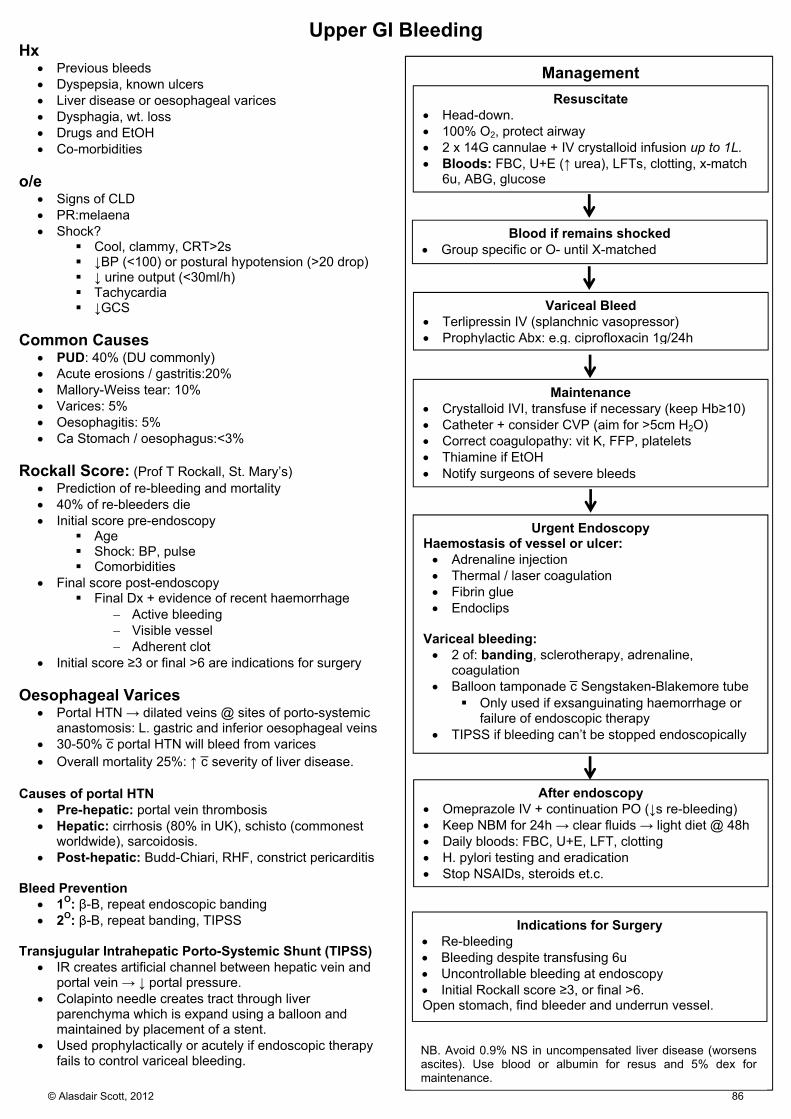
Upper GI Bleeding Hx
Previous bleeds Dyspepsia, known ulcers Liver disease or oesophageal varices Dysphagia, wt. loss Drugs and EtOH Co-morbidities
o/e
Signs of CLD PR:melaena Shock?
Cool, clammy, CRT>2s ↓BP (<100) or postural hypotension (>20 drop) ↓ urine output (<30ml/h) Tachycardia ↓GCS
Common Causes
PUD: 40% (DU commonly) Acute erosions / gastritis:20% Mallory-Weiss tear: 10% Varices: 5% Oesophagitis: 5% Ca Stomach / oesophagus:<3%
Rockall Score: (Prof T Rockall, St. Mary’s)
Prediction of re-bleeding and mortality 40% of re-bleeders die Initial score pre-endoscopy
Age Shock: BP, pulse Comorbidities
Final score post-endoscopy Final Dx + evidence of recent haemorrhage
Active bleeding Visible vessel Adherent clot
Initial score ≥3 or final >6 are indications for surgery Oesophageal Varices
Portal HTN → dilated veins @ sites of porto-systemic anastomosis: L. gastric and inferior oesophageal veins
30-50% c̄ portal HTN will bleed from varices Overall mortality 25%: ↑ c̄ severity of liver disease.
Causes of portal HTN
Pre-hepatic: portal vein thrombosis Hepatic: cirrhosis (80% in UK), schisto (commonest
worldwide), sarcoidosis. Post-hepatic: Budd-Chiari, RHF, constrict pericarditis
Bleed Prevention
1O: β-B, repeat endoscopic banding 2O: β-B, repeat banding, TIPSS
Transjugular Intrahepatic Porto-Systemic Shunt (TIPSS)
IR creates artificial channel between hepatic vein and portal vein → ↓ portal pressure.
Colapinto needle creates tract through liver parenchyma which is expand using a balloon and maintained by placement of a stent.
Used prophylactically or acutely if endoscopic therapy fails to control variceal bleeding.
Resuscitate Head-down. 100% O2, protect airway 2 x 14G cannulae + IV crystalloid infusion up to 1L. Bloods: FBC, U+E (↑ urea), LFTs, clotting, x-match
6u, ABG, glucose
Management
Blood if remains shocked Group specific or O- until X-matched
Maintenance Crystalloid IVI, transfuse if necessary (keep Hb≥10) Catheter + consider CVP (aim for >5cm H2O) Correct coagulopathy: vit K, FFP, platelets Thiamine if EtOH Notify surgeons of severe bleeds
Urgent Endoscopy Haemostasis of vessel or ulcer: Adrenaline injection Thermal / laser coagulation Fibrin glue Endoclips
Variceal bleeding: 2 of: banding, sclerotherapy, adrenaline,
coagulation Balloon tamponade c̄ Sengstaken-Blakemore tube
Only used if exsanguinating haemorrhage or failure of endoscopic therapy
TIPSS if bleeding can’t be stopped endoscopically
Indications for Surgery Re-bleeding Bleeding despite transfusing 6u Uncontrollable bleeding at endoscopy Initial Rockall score ≥3, or final >6. Open stomach, find bleeder and underrun vessel.
Variceal Bleed Terlipressin IV (splanchnic vasopressor) Prophylactic Abx: e.g. ciprofloxacin 1g/24h
After endoscopy Omeprazole IV + continuation PO (↓s re-bleeding) Keep NBM for 24h → clear fluids → light diet @ 48h Daily bloods: FBC, U+E, LFT, clotting H. pylori testing and eradication Stop NSAIDs, steroids et.c.
NB. Avoid 0.9% NS in uncompensated liver disease (worsens ascites). Use blood or albumin for resus and 5% dex for maintenance.
© Alasdair Scott, 2012 86
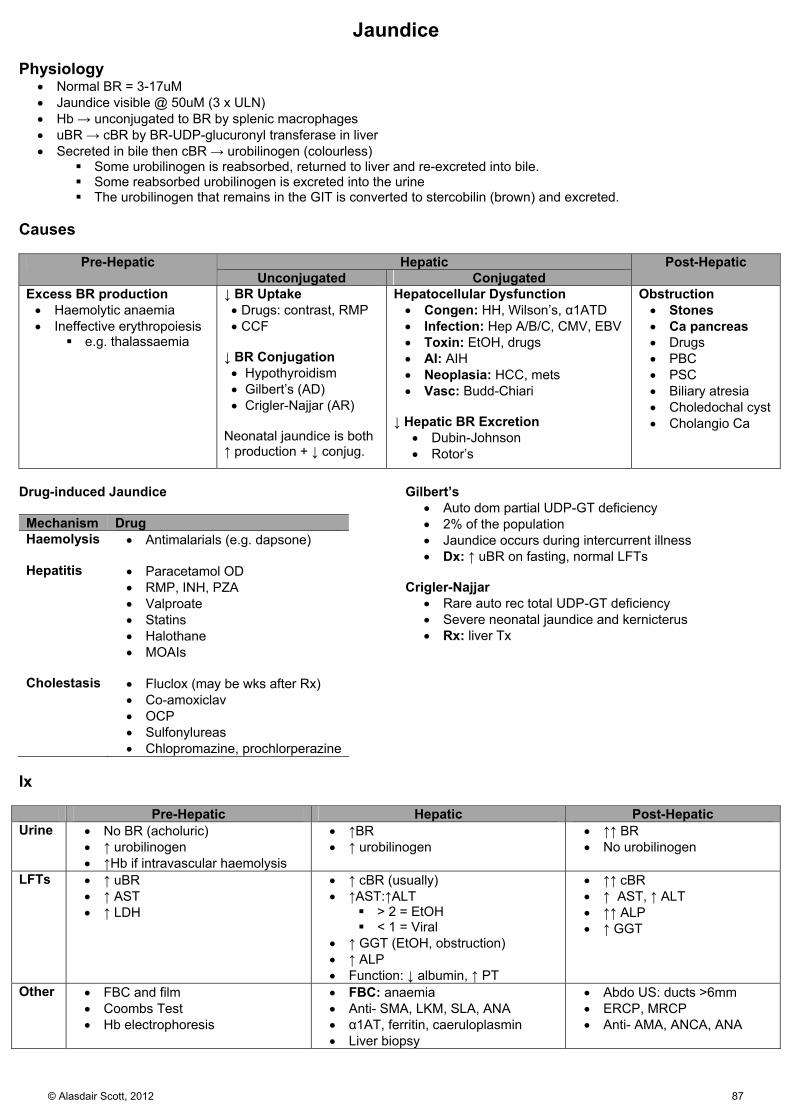
Jaundice Physiology
Normal BR = 3-17uM Jaundice visible @ 50uM (3 x ULN) Hb → unconjugated to BR by splenic macrophages uBR → cBR by BR-UDP-glucuronyl transferase in liver Secreted in bile then cBR → urobilinogen (colourless)
Some urobilinogen is reabsorbed, returned to liver and re-excreted into bile. Some reabsorbed urobilinogen is excreted into the urine The urobilinogen that remains in the GIT is converted to stercobilin (brown) and excreted.
Causes
Pre-Hepatic Hepatic Post-Hepatic Unconjugated Conjugated
Excess BR production Haemolytic anaemia Ineffective erythropoiesis
e.g. thalassaemia
↓ BR Uptake Drugs: contrast, RMP CCF
↓ BR Conjugation Hypothyroidism Gilbert’s (AD) Crigler-Najjar (AR)
Neonatal jaundice is both ↑ production + ↓ conjug.
Hepatocellular Dysfunction Congen: HH, Wilson’s, α1ATD Infection: Hep A/B/C, CMV, EBV Toxin: EtOH, drugs AI: AIH Neoplasia: HCC, mets Vasc: Budd-Chiari
↓ Hepatic BR Excretion
Dubin-Johnson Rotor’s
Obstruction Stones Ca pancreas Drugs PBC PSC Biliary atresia Choledochal cyst Cholangio Ca
Drug-induced Jaundice Mechanism Drug Haemolysis Antimalarials (e.g. dapsone)
Hepatitis Paracetamol OD
RMP, INH, PZA Valproate Statins Halothane MOAIs
Cholestasis Fluclox (may be wks after Rx)
Co-amoxiclav OCP Sulfonylureas Chlopromazine, prochlorperazine
Gilbert’s
Auto dom partial UDP-GT deficiency 2% of the population Jaundice occurs during intercurrent illness Dx: ↑ uBR on fasting, normal LFTs
Crigler-Najjar
Rare auto rec total UDP-GT deficiency Severe neonatal jaundice and kernicterus Rx: liver Tx
Ix
Pre-Hepatic Hepatic Post-Hepatic Urine No BR (acholuric)
↑ urobilinogen ↑Hb if intravascular haemolysis
↑BR ↑ urobilinogen
↑↑ BR No urobilinogen
LFTs ↑ uBR ↑ AST ↑ LDH
↑ cBR (usually) ↑AST:↑ALT
> 2 = EtOH < 1 = Viral
↑ GGT (EtOH, obstruction) ↑ ALP Function: ↓ albumin, ↑ PT
↑↑ cBR ↑ AST, ↑ ALT ↑↑ ALP ↑ GGT
Other FBC and film Coombs Test Hb electrophoresis
FBC: anaemia Anti- SMA, LKM, SLA, ANA α1AT, ferritin, caeruloplasmin Liver biopsy
Abdo US: ducts >6mm ERCP, MRCP Anti- AMA, ANCA, ANA
© Alasdair Scott, 2012 87
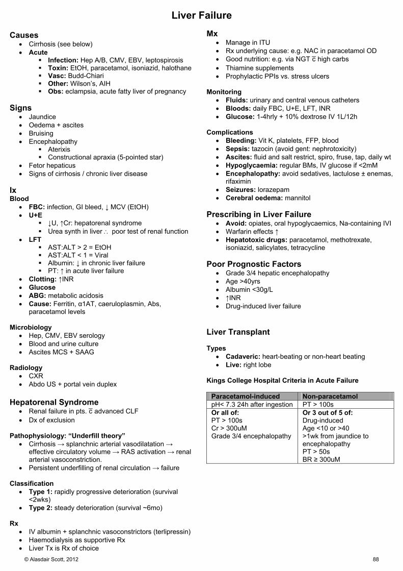
Liver Failure Causes
Cirrhosis (see below) Acute
Infection: Hep A/B, CMV, EBV, leptospirosis Toxin: EtOH, paracetamol, isoniazid, halothane Vasc: Budd-Chiari Other: Wilson’s, AIH Obs: eclampsia, acute fatty liver of pregnancy
Signs
Jaundice Oedema + ascites Bruising Encephalopathy
Aterixis Constructional apraxia (5-pointed star)
Fetor hepaticus Signs of cirrhosis / chronic liver disease
Ix Blood
FBC: infection, GI bleed, ↓ MCV (EtOH) U+E
↓U, ↑Cr: hepatorenal syndrome Urea synth in liver poor test of renal function
LFT AST:ALT > 2 = EtOH AST:ALT < 1 = Viral Albumin: ↓ in chronic liver failure PT: ↑ in acute liver failure
Clotting: ↑INR Glucose ABG: metabolic acidosis Cause: Ferritin, α1AT, caeruloplasmin, Abs,
paracetamol levels Microbiology
Hep, CMV, EBV serology Blood and urine culture Ascites MCS + SAAG
Radiology
CXR Abdo US + portal vein duplex
Hepatorenal Syndrome
Renal failure in pts. c̄ advanced CLF Dx of exclusion
Pathophysiology: “Underfill theory”
Cirrhosis → splanchnic arterial vasodilatation → effective circulatory volume → RAS activation → renal arterial vasoconstriction.
Persistent underfilling of renal circulation → failure Classification
Type 1: rapidly progressive deterioration (survival <2wks)
Type 2: steady deterioration (survival ~6mo) Rx
IV albumin + splanchnic vasoconstrictors (terlipressin) Haemodialysis as supportive Rx Liver Tx is Rx of choice
Mx
Manage in ITU Rx underlying cause: e.g. NAC in paracetamol OD Good nutrition: e.g. via NGT c̄ high carbs Thiamine supplements Prophylactic PPIs vs. stress ulcers
Monitoring
Fluids: urinary and central venous catheters Bloods: daily FBC, U+E, LFT, INR Glucose: 1-4hrly + 10% dextrose IV 1L/12h
Complications
Bleeding: Vit K, platelets, FFP, blood Sepsis: tazocin (avoid gent: nephrotoxicity) Ascites: fluid and salt restrict, spiro, fruse, tap, daily wt Hypoglycaemia: regular BMs, IV glucose if <2mM Encephalopathy: avoid sedatives, lactulose ± enemas,
rifaximin Seizures: lorazepam Cerebral oedema: mannitol
Prescribing in Liver Failure
Avoid: opiates, oral hypoglycaemics, Na-containing IVI Warfarin effects ↑ Hepatotoxic drugs: paracetamol, methotrexate,
isoniazid, salicylates, tetracycline Poor Prognostic Factors
Grade 3/4 hepatic encephalopathy Age >40yrs Albumin <30g/L ↑INR Drug-induced liver failure
Liver Transplant Types
Cadaveric: heart-beating or non-heart beating Live: right lobe
Kings College Hospital Criteria in Acute Failure
Paracetamol-induced Non-paracetamol pH< 7.3 24h after ingestion PT > 100s Or all of: PT > 100s Cr > 300uM Grade 3/4 encephalopathy
Or 3 out of 5 of: Drug-induced Age <10 or >40 >1wk from jaundice to encephalopathy PT > 50s BR ≥ 300uM
© Alasdair Scott, 2012 88
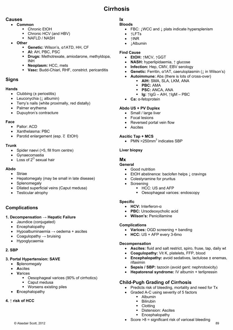
Cirrhosis Causes
Common Chronic EtOH Chronic HCV (and HBV) NAFLD / NASH
Other Genetic: Wilson’s, α1ATD, HH, CF AI: AH, PBC, PSC Drugs: Methotrexate, amiodarone, methyldopa,
INH Neoplasm: HCC, mets Vasc: Budd-Chiari, RHF, constrict. pericarditis
Signs Hands
Clubbing (± periostitis) Leuconychia (↓ albumin) Terry’s nails (white proximally, red distally) Palmer erythema Dupuytron’s contracture
Face
Pallor: ACD Xanthelasma: PBC Parotid enlargement (esp. c̄ EtOH)
Trunk
Spider naevi (>5, fill from centre) Gynaecomastia Loss of 2O sexual hair
Abdo
Striae Hepatomegaly (may be small in late disease) Splenomegaly Dilated superficial veins (Caput medusa) Testicular atrophy
Complications 1. Decompensation → Hepatic Failure
Jaundice (conjugated) Encephalopathy Hypoalbuminaemia → oedema + ascites Coagulopathy → bruising Hypoglycaemia
2. SBP 3. Portal Hypertension: SAVE
Splenomegaly Ascites Varices
Oesophageal varices (90% of cirrhotics) Caput medusa Worsens existing piles
Encephalopathy 4. ↑ risk of HCC
Ix Bloods
FBC: ↓WCC and ↓ plats indicate hypersplenism ↑LFTs ↑INR ↓Albumin
Find Cause
EtOH: ↑MCV, ↑GGT NASH: hyperlipidaemia, ↑ glucose Infection: Hep, CMV, EBV serology Genetic: Ferritin, α1AT, caeruloplasmin (↓ in Wilson’s) Autoimmune: Abs (there is lots of cross-over)
AIH: SMA, SLA, LKM, ANA PBC: AMA PSC: ANCA, ANA Ig: ↑IgG – AIH, ↑IgM – PBC
Ca: α-fetoprotein Abdo US + PV Duplex
Small / large liver Focal lesions Reversed portal vein flow Ascites
Ascitic Tap + MCS
PMN >250mm3 indicates SBP Liver biopsy Mx General
Good nutrition EtOH abstinence: baclofen helps ↓ cravings Colestyramine for pruritus Screening
HCC: US and AFP Oesophageal varices: endoscopy
Specific
HCV: Interferon-α PBC: Ursodeoxycholic acid Wilson’s: Penicillamine
Complications
Varices: OGD screening + banding HCC: US + AFP every 3-6mo
Decompensation
Ascites: fluid and salt restrict, spiro, fruse, tap, daily wt Coagulopathy: Vit K, platelets, FFP, blood Encephalopathy: avoid sedatives, lactulose ± enemas,
rifaximin Sepsis / SBP: tazocin (avoid gent: nephrotoxicity) Hepatorenal syndrome: IV albumin + terlipressin
Child-Pugh Grading of Cirrhosis
Predicts risk of bleeding, mortality and need for Tx Graded A-C using severity of 5 factors
Albumin Bilirubin Clotting Distension: Ascites Encephalopathy
Score >8 = significant risk of variceal bleeding © Alasdair Scott, 2012 89
Portal Hypertension Causes
Pre-hepatic: portal vein thrombosis (e.g. pancreatitis) Hepatic: cirrhosis (80% in UK), schisto (commonest
worldwide), sarcoidosis. Post-hepatic: Budd-Chiari, RHF, constrictive
pericarditis, TR Portosystemic Anastomoses Effect Portal Systemic Oesophageal varices
Left and short gastric veins
Inf. oesophageal veins
Caput medusae Peri-umbilical veins
Superficial abdo wall veins
Haemorrhoids (worsened
Sup. rectal veins Inf. and mid. Rectal veins
Prominent Abdominal Veins
A lot more common than caput medusa Blood flow down below the umbilicus: portal HTN Blood flow up below the umbilicus: IVC obstruction
Encephalopathy Pathophysiology
↓ hepatic metabolic function Diversion of toxins from liver directly into systemic
system. Ammonia accumulates and pass to brain where
astrocytes clear it causing glutamate → glutamine ↑ glutamine → osmotic imbalance → cerebral oedema.
Classification
1: Confused – irritable, mild confusion, sleep inversion 2: Drowsy – ↑ disorientated, slurred speech, asterixis 3: Stupor – rousable, incoherence 4: Coma – unrousable, ± extensor plantars
Presentation
Asterixis, ataxia Confusion Dysarthria Constructional apraxia Seizures
Precipitants → HEPATICS
Haemorrhage: e.g. varices Electrolytes: ↓K, ↓Na Poisons: diuretics, sedatives, anaesthetics Alcohol Tumour: HCC Infection: SBP, pneumonia, UTI, HDV Constipation (commonest cause) Sugar (glucose) ↓: e.g. low calorie diet
Ix
↑ plasma NH4 Rx
Nurse 20O head up Correct any precipitants Avoid sedatives Lactulose ± PO4 enemas to ↓ nitrogen-forming bowel
bacteria → 2-4 soft stools/d Consider rifaximin PO to kill intestinal microflora
Sequelae: SAVE
Splenomegaly Ascites Varices Encephalopathy
Ascites Pathophysiology
Back-pressure → fluid exudation ↓ effective circulating volume → RAS activation (In cirrhosis: ↓ albumin → ↓ plasma oncotic pressure
and aldosterone metabolism impaired) Symptoms
Distension → abdominal discomfort and anorexia Dyspnoea ↓ venous return
Differential: Serum Ascites Albumin Gradient (SAGG)
SAAG ≥1.1g/dL = Portal HTN (97% accuracy) Pre-, hepatic and post Cirrhosis in 80%
SAAG <1.1g/dL = Other Causes Neoplasia: peritoneal or visceral (e.g. ovarian) Inflammation: e.g. pancreatitis Nephrotic Syndrome Infection: TB peritonitis
Ix
Bloods: FBC, U+E, LFTs, INR, chronic hepatitis screen US: confirm ascites, liver echogenicity, PV duplex Ascitic tap
MCS and AFB Cytology Chemistry: albumin, LDH, glucose, protein SAAG = serum albumin – ascites albumin
Liver biopsy Rx
Daily wt. aiming for ≤0.5kg/d reduction Fluid restrict <1.5L/d and low Na diet Spironolactone + frusemide (if response poor) Therapeutic paracentesis c̄ albumin infusion (100ml
20% albumin /L drained) Respiratory compromise Pain / discomfort Renal impairment
Refractory: TIPSS SBP
Pt. c̄ ascites and peritonitic abdomen E. coli, Klebsiella, Streps Complicated by hepatorenal syn. in 30% Ix: ascitic PMN > 250mm3 + MC+S Rx: Tazocin or cefotaxime until sensitivities known Prophylaxis: high recurrence cipro long-term
Splenomegaly
Splenic congestion Hypersplenism: ↓ WCC, ↓ plats
© Alasdair Scott, 2012 90

Alcoholism Effects Hepatic
Fatty liver → hepatitits → cirrhosis AST:ALT >2, ↑ GGT
GIT
Gastritis, erosions PUD Varices Pancreatitis Carcinoma
CNS
Poor memory / cognition Peripheral polyneuropathy (mainly sensory) Wernicke’s encephalopathy
Confusion Ophthalmoplegia (nystagmus, LR palsy) Ataxia
Korsakoff’s: amnesia → confabulation Fits, falls
Heart
Arrhythmias: e.g. AF Dilated cardiomyopathy ↑BP
Blood
↑ MCV Folate deficiency → anaemia
Dx: CAGE
Cut down? Annoyed by people’s criticisms Guilty about drinking Eye opener?
Withdrawal
10-72h after last drink Consider in new ward pt (≤3d) c̄ acute confusion Signs
↑HR, ↓BP, tremor Confusion, fits, hallucinations: esp formication (DTs)
Rx Tapering regimen of chlordiazepoxide PO /
lorazepam IM Thiamine
Mx
Group therapy or self-help (e.g. AA) Baclofen: ↓ cravings Acamprosate: ↓ cravings Disulfiram: aversion therapy
Alcoholic Hepatitis Presentation
Anorexia D/V Tender hepatomegaly Ascites Severe: Jaundice, bleeding, encephalopathy
Ix
Bloods: ↑MCV, ↑GGT, AST:ALT>2 Ascitic tap Abdo US + PV duplex
Rx
Stop EtOH Rx withdrawal High dose B vitamins: Pabrinex Optimise nutrition Daily wt., LFT, U+E, INR Mx complications of failure
Prognosis
Maddrey score predicts mortality Mild: 0-5% 30d mortality Severe: 50% 30d mortality 1yr after admission: 40% mortality
© Alasdair Scott, 2012 91

Viral Hepatitis Types Type Spread Cause A FO Seafood, especially abroad B IV Blood, body fluids, babies (vertical) C IV Mainly blood. Less vertical cf. HCV D IV Dependent on prior HBV infection E FO Developing world
Presentation Prodromal Phase
Seen particularly in HAV and BV Flu-like, malaise, arthralgia, nausea Distaste for cigarettes in Hep A
Icteric Phase
Acute jaundice in A>B>C (99, 75 and 25%) Hepatitis
Abdo pain Hepatomegaly Cholestasis: dark urine, pale stools
Extrahepatic features due to complexes (esp. Hep B) Urticaria or vasculitic rash Cryoglobulinaemia PAN GN Arthritis
Chronic Phase
Mainly HCV and childhood HBV Cirrhosis → ↑ risk of HCC
Hep B
Carrier: 10% HBsAg +ve > 6mo
Chronic hepatitis: 10% Cirrhosis: 5%
Hep C
Carrier: 80% HCV RNA+ve >6mo
Chronic hepatitis: 80% Cirrhosis: 20%
Ix
FBC, LFTs, clotting Hep A/B/C serology
Rx
Supportive No EtOH Avoid hepatotoxic drugs (e.g. aspirin)
Anti-viral Indicated in chronic disease HBV: PEGinterferon HCV: PEGinterferon + ribavarin Seroconversion: HBV – 40%, HCV – 10%
Non-alcoholic Fatty Liver Disease (NAFLD)
Cryptogenic cause of hepatitis and cirrhosis assoc. c̄ insulin resistance and the metabolic syndrome.
Non-alcoholic steatohepatitis (NASH) is most extreme form and → cirrhosis in 10%
Risk Factors
Obesity HTN T2DM Hyperlipidaemia
Presentation
Mostly asymptomatic Hepatomegaly and RUQ discomfort may be present.
Metabolic Syndrome
Central obesity (↑ waist circumference) and two of: ↑ Triglycerides ↓ HDL HTN Hyperglycaemia: DM, IGT, IFG
Ix
BMI Glucose, fasting lipids ↑ transaminases: AST:ALT <1 Liver biopsy
Mx
Lose wt. Control HTN, DM and lipids
Budd-Chiari Syndrome
Hepatic vein obstruction → ischaemia and hepatocyte damage → liver failure or insidious cirrhosis.
Causes
Hypercoagulable states: myeloproliferative disorders (PV = commonest cause), PNH, anti-phospholipid, OCP
Local Tumour: HCC Congenital: membranous obstruction of IVC
Presentation
RUQ pain: stretching of Glisson’s capsule Hepatomegaly Ascites: SAAG ≥1.1g/dL Jaundice (and other features of liver failure)
Ix
Bloods: FBC, clotting, LFTs US + hepatic vein Doppler Ascitic tap: ↑↑ protein (>2.5g/dL) c̄ ↑SAAG (≥1.1g/dL) Other: JAK2 mutation analysis, RBC CD55 and CD59
Rx
Anticoagulate unless there are varices Ascites: fluid and salt restrict, spiro, fruse, tap, daily wt Other options: thrombolysis, angioplasty, TIPSS Transplant if fulminant hepatic failure or cirrhosis Rx underlying cause
© Alasdair Scott, 2012 92

Hereditary Haemochromatosis Epidemiology
Prevalence: 1/3000, 10% are carriers. Age of onset: 40-60yrs (women later due to menses) Genetics: AR, HFE gene (High FE) on Chr6 (C282Y)
Pathophysiology
Inherited, multisystem disorder resulting from abnormal iron metabolism.
↑ intestinal Fe absorption (↑ enterocyte DMT + ↓ hepatocyte hepcidin) → deposition in multiple organs.
Clinical Features: iron MEALS Myocardial
Dilated cardiomyopathy Arrhythmias
Endocrine
Pancreas: DM Pituitary: hypogonadism → amenorrhoea, infertility Parathyroid: hypocalcaemia, osteoporosis
Arthritis
2nd and 3rd MCP joints, knees and shoulders Liver
Chronic liver disease → cirrhosis → HCC Hepatomegaly
Skin
Slate grey discolouration Ix
Bloods: ↑LFT, ↑ ferritin, ↑Fe, ↓TIBC, glucose, genotype X-ray: chondrocalcinosis ECG, ECHO Liver biopsy: Pearl’s stain to quantify Fe and severity MRI: can estimate iron loading
Rx
Iron removal Venesection: aim for Hct <0.5 Desferrioxamine is 2nd line
General Monitor DM Low Fe diet
Screening Se ferritin and genotype Screen 1st degree relatives
Transplant in cirrhosis Prognosis
Venesection returns life expectancy to normal if non-cirrhotic and non-diabetic.
Cirrhotic patients have >10% chance of HCC
α1-Antitrypsin Deficiency Epidemiology
Prevalence: 1/4000, 10% are carriers Genetics: AR, Chr 14
Homozygotes have PiZZ phenootype Pathophysiology
α1AT is a serpin involved in control of the inflammatory cascade by inhibiting neutrophil elastase.
α1AT is synthesised in the liver and comprises 90% of se α1-globulin on electrophoresis.
Presentation
Variable presentation Neonatal and childhood hepatitis 15% of adults develop cirrhosis by 50yrs 75% of adults have emphysema (esp. smokers)
Ix
Blood: ↓ se α1AT levels Liver biopsy: PAS+ve, diastase-resistant globules CXR: emphysematous changes Spirometry: obstructive defect Prenatal Dx: possible by CVS
Mx
Mostly supportive for pulmonary and hepatic complications.
Quit smoking Can consider α1AT therapy from pooled donors.
© Alasdair Scott, 2012 93
Wilson’s Disease Epidemiology
Prevalence: 3/100,000 Age: presents between childhood and 30 (never >56) Genetics: AR, ATP7B gene on Chr 13
Pathophysiology
Mutation of Cu transporting ATPase Impaired hepatocyte incorporation of Cu into
caeruloplasmin and excretion into bile. Cu accumulation in liver and, later, other organs
Clinical Features: CLANKAH Cornea
Kayser-Fleischer rings (70%, may need slit-lamp) Liver Disease
Children usually present c̄ acute hepatitis. Fulminant necrosis may occur → cirrhosis
Arthritis
Chondrocalcinosis Osteoporosis
Neurology
Parkinsonism: bradykinesia, tremor, chorea, tics Spasticity, dysarthria, dysphagia Ataxia Depression, dementia, psychosis
Kidney
Fanconi’s syn. (T2 RTA) → osteomalacia Abortions Haemolytic anaemia
Coombs’ negative Ix
Bloods: ↓Cu, ↓ caeruloplasmin NB. Caeruloplasmin is an acute-phase protein and may be high during infection. It may also be low protein-deficient states: nephrotic syndrome, malabsorption
↑ 24h urinary Cu Liver biopsy: ↑ hepatic Cu MRI: basal-ganglia degeneration
Rx
Diet: avoid high Cu foods: liver, chocolate, nuts Penicillamine lifelong (Cu chelator)
SE: nausea, rash, ↓WCC, ↓Hb, ↓plats, lupus, haematuria
Monitor FBC and urinary Cu excretion Liver Tx if severe liver disease Screen siblings
Autoimmune Hepatitis Pathophysiology
Inflammatory disease of unknown cause characterised by Abs directed vs. hepatocyte surface antigens
Predominantly young and middle-aged women Classified according to Abs
T1: Adult, SMA+ (80%), ANA+ (10%), ↑IgG T2: Young, LKM+ T3: Adult, SLA+
Presentation
Teens and early 20s (25%) Constitutional: fatigue, fever, malaise Cushingoid: hirsute, acne, striae Hepatitis HSM Fever Amenorrhoea Polyarthritis Pulmonary infiltration Pleurisy
Post-/peri-menopausal Present insidiously c̄ chronic liver disease
Associated Diseases
Autoimmune thyroiditis DM Pernicious anaemia PSC UC GN AIHA (Coombs +ve)
Ix
↑LFTs ↑IgG Auto Abs: SMA, LKM, SLA, ANA ↓WCC and ↓plats = hypersplenism Liver biopsy
Mx
Immunosuppression Prednisolone Azathioprine as steroid-sparer
Liver transplant (disease may recur) Prognosis
Remission in 80% of patients 10yr survival 80%
© Alasdair Scott, 2012 94

Primary Biliary Cirrhosis Epidemiology
Prev: ≤4/100,000 Sex: F>>M = 9:1 Age: 50s
Pathology
Intrahepatic bile duct destruction by chronic granulomatous inflammation → cirrhosis
Presentation: PPBBCCS
Often asympto and Dx incidentally (↑ALP) Jaundice occurs late Pruritus and fatigue Pigmentation of face Bones: osteoporosis, osteomalacia (↓ vit D) Big organs: HSM Cirrhosis and coagulopathy (↓ vit K) Cholesterol ↑: xanthelasma, xanthomata Steatorrhoea
Associated Diseases
Thyroid disease RA, Sjogrens, scleroderma Coeliac disease RTA Membranous GN
Ix
LFTs: ↑↑ALP, ↑↑GGT, ↑AST/ALT Late: ↑BR, ↑PT, ↓albumin
Abs: AMA+ (98%) ↑IgM ↑ cholesterol ± ↑ TSH US to exclude extra-hepatic cholestasis Liver biopsy: non-caseating granulomatous inflam
Rx
Symptomatic Pruritus: colestyramine, naltrexone Diarrhoea: codeine phosphate Osteoporosis: bisphosphonates
Specific ADEK vitamins Ursodeoxcholic acid: ↓LFTs but no effect on
mortality or need for transplant Liver transplant
End-stage disease or intractable pruritus Recurrence occurs in ~20% but doesn’t usually
→ graft failure. Prognosis
Once jaundice develops survival is <2yrs
Primary Sclerosing Cholangitis Pathophysiology
Inflammation, fibrosis and strictures and intra- and extra-hepatic ducts.
Chronic biliary obstruction → 2O biliary cirrhosis → liver failure
Epidemiology
Age: 30-50yrs Sex: M>F = 2:1
Presentation
May be asypmto and Dx incidentally (↑ALP) Symptoms
Jaundice Pruritus and fatigue Abdo pain
Signs
Jaundice: dark urine, pale stools HSM
Complications
Bacterial cholangitis ↑ Cholangiocarcinoma ↑ CRC
Associated Diseases
UC 3% of those c̄ UC have PSC 80-100% of those c̄ PSC have UC/Crohn’s
Crohn’s (much rarer) AIH HIV
Ix
LFTs: ↑ALP initially, then ↑BR Abs: pANCA (80%), ANA and SMA may be +ve ERCP/MRCP: “beaded” appearance of ducts Biopsy: fibrous, obliterative cholangitis
Rx
No curative medical therapy: transplant needed Symptomatic
Pruritus: colestyramine, naltrexone Diarrhoea: codeine phosphate
Specific ADEK vitamins Ursodeoxycholic acid improves cholestasis only Abx for cholangitis Endoscopic stenting for dominant strictures
Screening Cholangiocarcinoma: US + Ca19-9 CRC: colonoscopy
Transplant Recurrence occurs in 30%
© Alasdair Scott, 2012 95

Liver Tumours Pathology
90% of liver tumours are 2O metastases 1O in men: stomach, lung, colon 1O in women: breast, colon, stomach, uterus Less common: pancreas, leukaemia, lymphoma
90% of primary tumours are HCC. Benign tumours: haemangiomas, adenomas, cysts
Symptoms
Benign tumours are usually asymptomatic Systemic: fever, malaise, wt. loss, anorexia RUQ pain: stretching of Glisson’s capsule Jaundice is often late, except in cholangiocarcinoma May rupture → intraperitoneal haemorrhage
Signs
Hepatomegaly: smooth or hard and irregular Signs of chronic liver disease Abdominal mass Hepatic bruit (HCC)
Ix
Bloods: LFTs, hepatitis serology, AFP Imaging:
US or CT / MRI ± guided diagnostic biopsy ERCP + biopsy in suspected cholangiocarcinoma
Biopsy (seeding may occur along tract) Find primary: e.g. colonoscopy, mammography
Liver Mets
Rx and prognosis vary c̄ type and extent of 1O Small, solitary CRC mets may be resectable Advanced disease prognosis: < 6mo
HCC
Rare in West, common in China and sub-Saharan Africa Causes
Viral hepatitis Cirrhosis: EtOH, HH, PBC Aflatoxins (produced by Aspergillus)
Mx
Resection of solitary tumours improves prognosis (13 → 59%), but 50% have recurrence.
Also: chemo, percutaneous ablation and embolization Cholangiocarcinoma
Biliary tree malignancy (10% of liver 1O tumours) Causes
Flukes (Clonorchis) PSC Congenital biliary cysts UC
Presentation
Fever, malaise Abdominal pain, ascites, jaundice ↑BR, ↑↑ALP
Mx
30% resectable Palliative stenting: percutaneous or ERCP
Liver Transplant in CLD Types
Cadaveric: heart-beating or non-heart beating Live: right lobe
Indications
Advanced cirrhosis HCC
Contraindications
Extra-hepatic malignancy Severe cardiorespiratory disease Systemic sepsis HIV infection Non-compliance c̄ drug therapy
Post-op
12-24h on ITU Immunosuppression
Ciclosporin / Tacrolimus + Azathioprine / Mycophenolate Mofetil + Prednisolone
Complications
Acute rejection (T-cell mediated) 50% @ 5-10 days Pyrexia, tender hepatomegaly ↑ or change immunosuppressants
Sepsis Hepatic artery thrombosis CMV infection Chronic rejection (6-9mo): shrinking bile ducts Disease recurrence (e.g. HBV)
Prognosis
Depends on disease aetiology 60-90% 5ys
© Alasdair Scott, 2012 96

Inflammatory Bowel Disease: Pathology and Presentation Epidemiology
UC
Crohn’s
Prev 100-200 /100,000 50-100 /100,000 Age 30s 20s Sex F>M (just) Aet Concordance = 10%
Smoking protective TH2-mediated
Concordance = 70% Smoking ↑ risk TH1/TH17-mediated
Pathology
UC
Crohn’s
Macroscopic Location Rectum + colon
± backwash ileitis Mouth to anus esp. terminal ileum
Distribution Contiguous Skip lesions Strictures
No Yes
Microscopic Inflammation Mucosal
Crypt Abscesses Transmural
Ulceration Shallow, broad Deep, thin, serpiginous → cobblestone mucosa
Fibrosis None Marked Granulomas None Present Pseudoplyps Marked Minimal Fistulae No Yes
Presentation UC Crohn’s
Symptoms Systemic Fever, malaise, anorexia, wt. loss in active disease Abdominal Diarrhoea
Blood ± mucus PR Abdominal discomfort Tenesmus, faecal urgency
Diarrhoea (not usually bloody) Abdominal pain Wt. loss
Signs Abdominal Fever
Tender, distended abdomen Aphthous ulcers, glossitis Abdominal tenderness RIF mass Perianal abscesses, fistulae, tags Anal / rectal strictures
Extra-abdominal Skin Clubbing Erythema nodosum Pyoderma gang (esp. UC)
Eyes Iritis Episcleritis Conjunctivitis
Joints Arthritis (non-deforming, asymm) Sacroiliitis Ank spond
HPB PSC + cholangiocarcinoma (esp. UC) Gallstones (esp. Crohn’s) Fatty liver
Other Amyloidosis Oxalate renal stones (esp. Crohns)
Complications Toxic megacolon
Diameter >6cm Risk of perforation
Bleeding Malignancy
CRC in 15% c̄ pancolitis for 20yrs Cholangiocarcinoma
Strictures → obstruction Venous thrombosis
Fistulae Entero-enteric/colonic → diarrhoea Enterovesical → frequency, UTI Enterovaginal Perianal → “pepperpot” anus
Strictures → obstruction Abscesses
Abdominal Anorectal
Malabsorption Fat → Steatorrhoea, gallstones B12 → megaloblastic anaemia Vit D → osteomalacia Protein → oedema
Toxic megacolon and Ca may occur (< cf. UC)
© Alasdair Scott, 2012 97

Ulcerative Colitis: Management Ix
Bloods: FBC: ↓Hb, ↑WCC LFT: ↓albumin ↑CRP/ESR Blood cultures
Stool MCS: exclude Campy, Shigella, Salmonella… CDT: C. diff may complicate or mimic
Imaging AXR: megacolon (>6cm), wall thickening CXR: perforation CT Ba / gastrograffin enema
Lead-pipe: no haustra Thumbprinting: mucosal thickening Pseudopolyps: regenerating mucosal island
Ileocolonoscopy + regional biopsy: Baron Score Severity Truelove and Witts Criteria
Mild Moderate Severe Motions <4 4-6 >6 PR bleed small moderate large Temp Apyrexic 37.1-37.8 >37.8 HR <70 70-90 >90 Hb >11 10.5-11 <10.5 ESR <30 >30
Acute Severe UC
Resus: Admit, IV hydration, NBM Hydrocortisone: IV 100mg QDS + PR Transfuse if required Thromboprophylaxis: LMWH Monitoring
Bloods: FBC, ESR, CRP, U+E Vitals + stool chart Twice daily examination ± AXR
NB. RCTs show no benefit of Abx: not routinely recommended
May use: megacolon, perforation, uncertain Dx Acute Complications
Perforation Bleeding Toxic megacolon (>6cm) VTE
Improvement → oral therapy
Switch to oral pred + a 5-ASA Taper pred after full remission
No Improvement → rescue therapy On day 3: stool freq >8 or CRP >45
Predicts 85% chance of needing a colectomy during the admission
Discussion between pt, physician and surgeon Medical: ciclosporin, infliximab or visilizumab (anti-T cell) Surgical
Inducing Remission in Mild / Mod Disease
OPD-based Oral Therapy
1st line: 5-ASAs 2nd line: prednisolone
Topical Therapy: mainly left-sided disease
Proctitis: suppositories More proximal disease: enemas or foams 5-ASAs ± steroids (prednisolone or budesonide)
Additional Therapy: steroid sparing
Azathioprine or mercaptopurine Infliximab: steroid-dependent pts
Maintaining Remission
1st line: 5-ASAs PO – sulfasalazine or mesalazine Topical Rx may be used in proctitis
2nd line: Azathioprine or 6-mercaptopurine Relapsed on ASA or are steroid-dependent Use 6-mercaptopurine if azathioprine intolerant
3rd line: Infliximab / adalimumab Emergency Surgery
20% require surgery at some stage 30% c̄ colitis require surgery w/i 5yrs
Indications
Toxic megacolon Perforation Massive haemorrhage Failure to respond to medical Rx
Procedures
Total / subtotal colectomy c̄ end ileostomy ± mucus fistula
Followed after ~3mo by either Completion proctectomy + Ileal-pouch anal
anastomosis (IPAA) or end ileostomy Ileorectal anastomosis (IRA)
Panproctocolectomy + permanent end ileostomy Acute colitis op mortality: 7% (30% if perforated)
Elective Surgery Indications
Chronic symptoms despite medical therapy Carcinoma or high-grade dysplasia
Procedures
Panproctocolectomy c̄ end ileostomy or IPAA Total colectomy c̄ IRA
Surgical Complications
Abdominal SBO Anastomotic stricture Pelvic abscess
Stoma: retraction, stenosis, prolapse, dermatitis Pouch
Pouchitis (50%): metronidazole + cipro ↓ female fertility Faecal leakage
© Alasdair Scott, 2012 98

Crohn’s Disease: Management Ix
Bloods: (top 3 are severity markers) FBC: ↓Hb, ↑WCC LFT: ↓albumin ↑CRP/ESR Haematinics: Fe, B12, Folate Blood cultures
Stool MCS: exclude Campy, Shigella, Salmonella… CDT: C. diff may complicate or mimic
Imaging AXR: obstruction, sacroileitis CXR: perforation MRI
Assess pelvic disease and fistula Assess disease severity
Small bowel follow-through or enteroclysis Skip lesions Rose-thorn ulcers Cobblestoning: ulceration + mural oedema String sign of Kantor: narrow terminal ileum
Endoscopy Ileocolonoscopy + regional biopsy: Ix of choice Wireless capsule endoscopy Small bowel enteroscopy
Severe Attack Assessment
↑temp, ↑HR, ↑ESR, ↑CRP, ↑WCC, ↓albumin Management
Resus: Admit, NBM, IV hydration Hydrocortisone: IV + PR if rectal disease Abx: metronidazole PO or IV Thromboprophylaxis: LMWH Dietician Review
Elemental diet Liquid prep of amino acids, glucose and fatty
acids Consider parenteral nutrition
Monitoring Vitals + stool chart Daily examination
Improvement → oral therapy
Switch to oral pred (40mg/d)
No Improvement → rescue therapy Discussion between pt, physician and surgeon Medical: methotrexate ± infliximab Surgical
Inducing Remission in Mild / Mod Disease
OPD treatment Supportive
High fibre diet Vitamin supplements
Oral Therapy
1st line Ileocaecal: budesonide Colitis: sulfasalazine
2nd line: prednisolone (tapering) 3rd line: methotrexate 4th line: infliximab or adalimumab
Perianal Disease
Occurs in ~50% Ix: MRI + EUA Rx
Oral Abx: metronidazole Immunosuppression ± infliximab Local surgery ± seton insertion
Maintaining Remission
1st line: azathioprine or mercaptopurine 2nd line: methotrexate 3rd line: Infliximab / adalimumab
Surgery
50-80% need ≥1 operation in their life Never curative Should be as conservative as possible
Indications
Emergency Failure to respond to medical Rx Intestinal obstruction or perforation Massive haemorrhage
Elective Abscess or fistula Perianal disease Chronic ill health Carcinoma
Procedures
Limited resection: e.g. ileocaecal Stricturoplasty Defunction distal disease c̄ temporary loop ileostomy
Complications
Stoma complications Enterocutaneous fistulae Anastomotic leak or stricture
Short gut
<1-2m small bowel Features
Steatorrhoea ADEK and B12 malabsorption Bile acid depletion → gallstones Hyperoxaluria → renal stones
Rx Dietician Supplements or TPN Loperamide
© Alasdair Scott, 2012 99

Coeliac Disease Epidemiology
Prev: 0.5 – 1% Age: Any, bimodal: infancy and 50-60yrs Sex: F>M Geo: ↑ in Ireland and N. Africa
Pathophysiology
HLA-DQ2 (95%) and DQ8 CD8+ mediated response to gliadin in gluten
Presentation: GLIAD GI Malabsorption: fatigue, weakness
Carb N/V/D Abdo distension + colic Flatus Wt. ↓
Fat Steatorrhoea Hyperoxaluria → renal stones
Protein Protein-losing enteropathy if severe
Haematinics ↓ Folate and Fe → anaemia
Vitamins Vit D and Ca → bone pain, osteoporosis Vit K → petechiae and ↑INR B2 (riboflavin) → angular stomatitis B1 and B6 → polyneuropathy
Lymphoma and Carcinoma
Enteropathy-associated T-cell lymphoma Adenocarcinoma of small bowel Other Ca: breast, bladder, breast
Immune Associations
IgA deficiency T1DM PBC
Anaemia
↑ or ↓ MCV Hyposplenism: Howell-Jolly bodies, target cells
Dermatological
Dermatitis herpetiformis: 15-20% Symmetrical vesicles, extensor surfaces
Esp. elbows Very itchy Responds to gluten-free diet or dapsone Biopsy: granular deposition of IgA
Aphthous ulcers
Ix
Bloods: FBC, LFTs (↓alb), INR, Vit D and bone, red cell folate, serum B12
Abs Anti-endomysial IgA (95% specificity) Anti-TTG IgA
Both above ↓ c̄ exclusion diet Anti-gliadin IgG persist c̄ exclusion diet) IgA ↑ in most but may have IgA deficiency
Stools Stool cysts and antibody: exclude Giardia
OGD and duodenal biopsy Subtotal villous atrophy Crypt hyperplasia Intra-epithelial lymphocytes
Rx
Lifelong gluten-free diet Avoid: barley, rye, oats, wheat OK: Maize, soya, rice Verify diet by endomysial Ab tests
Pneumovax as hyposplenic Dermatitis herpetiformis: dapsone
Malabsorption Presentation
Diarrhoea / Steatorrhoea Wt. loss Lethargy
Causes
Common in UK: Coeliac, Chronic pancreatitis, Crohn’s Rarer
↓Bile: PBC, ileal resection, colestyramine Pancreatic insufficiency: Ca, CF, chronic panc Small bowel: resection, tropical sprue, metformin Bacterial overgrowth: spontaneous, post-op
blind loops, DM, PPIs Infection: Giardia, Strongyloides, Crypto parvum Hurry: post-gastrectomy dumping
Ix
Coeliac tests Stool microscopy Faecal elastase Hydrogen breath test MRI / CT ERCP (chronic pancreatitis) Small bowel endoscopy
© Alasdair Scott, 2012 100
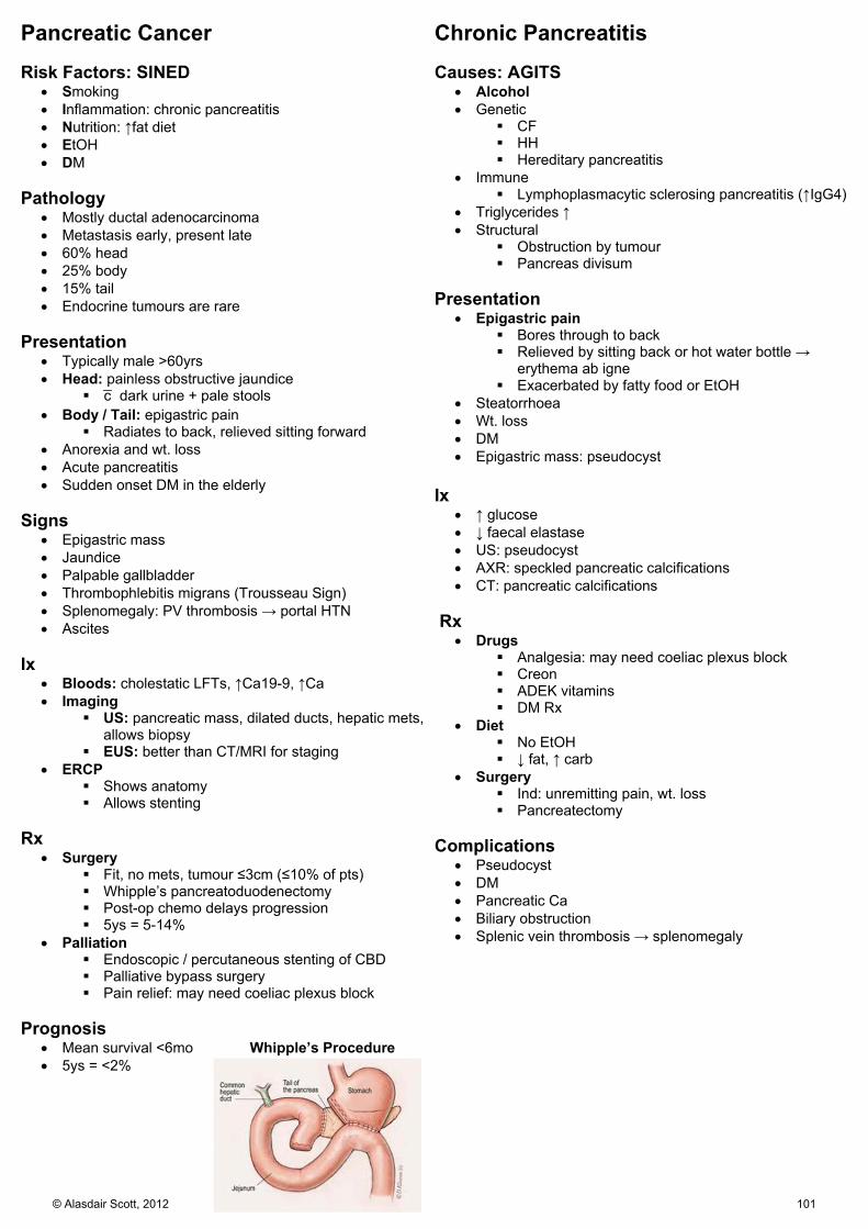
Pancreatic Cancer Risk Factors: SINED
Smoking Inflammation: chronic pancreatitis Nutrition: ↑fat diet EtOH DM
Pathology
Mostly ductal adenocarcinoma Metastasis early, present late 60% head 25% body 15% tail Endocrine tumours are rare
Presentation
Typically male >60yrs Head: painless obstructive jaundice
c̄ dark urine + pale stools Body / Tail: epigastric pain
Radiates to back, relieved sitting forward Anorexia and wt. loss Acute pancreatitis Sudden onset DM in the elderly
Signs
Epigastric mass Jaundice Palpable gallbladder Thrombophlebitis migrans (Trousseau Sign) Splenomegaly: PV thrombosis → portal HTN Ascites
Ix
Bloods: cholestatic LFTs, ↑Ca19-9, ↑Ca Imaging
US: pancreatic mass, dilated ducts, hepatic mets, allows biopsy
EUS: better than CT/MRI for staging ERCP
Shows anatomy Allows stenting
Rx
Surgery Fit, no mets, tumour ≤3cm (≤10% of pts) Whipple’s pancreatoduodenectomy Post-op chemo delays progression 5ys = 5-14%
Palliation Endoscopic / percutaneous stenting of CBD Palliative bypass surgery Pain relief: may need coeliac plexus block
Prognosis
Mean survival <6mo Whipple’s Procedure 5ys = <2%
Chronic Pancreatitis Causes: AGITS
Alcohol Genetic
CF HH Hereditary pancreatitis
Immune Lymphoplasmacytic sclerosing pancreatitis (↑IgG4)
Triglycerides ↑ Structural
Obstruction by tumour Pancreas divisum
Presentation
Epigastric pain Bores through to back Relieved by sitting back or hot water bottle →
erythema ab igne Exacerbated by fatty food or EtOH
Steatorrhoea Wt. loss DM Epigastric mass: pseudocyst
Ix
↑ glucose ↓ faecal elastase US: pseudocyst AXR: speckled pancreatic calcifications CT: pancreatic calcifications
Rx
Drugs Analgesia: may need coeliac plexus block Creon ADEK vitamins DM Rx
Diet No EtOH ↓ fat, ↑ carb
Surgery Ind: unremitting pain, wt. loss Pancreatectomy
Complications
Pseudocyst DM Pancreatic Ca Biliary obstruction Splenic vein thrombosis → splenomegaly
© Alasdair Scott, 2012 101
Carcinoid Tumours Pathology
Diverse group of neuroendocrine tumours of enterochromaffin cell origin capable of producing 5HT
May secrete: 5-HT, VIP, gastrin, glucagon, insulin, ACTH Carcinoid syndrome suggest bypass of first-pass
metabolism and is strongly assoc. c̄ metastatic disease. 10% part of MEN1 Sites
Appendix: 45% Ileum: 30% Colorectum: 20% Stomach: 10% Elsewhere in GIT and bronchus
Consider all as malignant Presentation Local
Appendicitis Intussusception or obstruction Abdominal pain
Carcinoid Syndrome: FIVE HT
Flushing: paroxysmal, upper body ± wheals Intestinal: diarrhoea Valve fibrosis: tricuspid regurg and pulmonary stenosis whEEze: bronchoconstriction Hepatic involvement: bypassed 1st pass metabolism Tryptophan deficiency → pellagra (3Ds)
Ix
↑ urine 5-hydroxyindoleacetic acid ↑ plasma chromogranin A CT/MRI: find primary
Rx
Symptoms: octreotide or loperamide Curative
Resection: tumours are v. yellow Give octreotide to avoid carcinoid crisis
Carcinoid Crisis
Tumour outgrows blood supply or is handled too much → massive mediator release
Vasodilatation, hypotension, bronchoconstriction, hyperglycaemia
Rx: high-dose octreotide Prognosis
Median survival is 5-8yrs (~3yrs if mets present)
Nutritional Deficiencies Vitamin A → Xerophthalmia
Dry conjunctivae, develop spots (Bitots spots) Corneas become cloudy then ulcerate Night blindness → total blindness
Thiamine (B1) → Beri Beri
Wet: heart failure + oedema Dry: polyneuropathy Wernicke’s: ophthalmoplegia, ataxia, confusion
Niacin / Nicotinic acid (B3) → Pellagra
Diarrhoea, Dermatitis, Dementia Also: neuropathy, depression, ataxia Causes: dietary, isoniazid, carcinoid syndrome
Pyridoxine (B6)
Peripheral sensory neuropathy Cause: PZA
Cyanocobalamin (B12)
Glossitis → sore tongue Peripheral neuropathy
Paraesthesia Early loss of vibration and proprioception → ataxia
SCDC Dorsal and corticospinal tracts Sensory loss and UMN weakness
Overall mixed UMN and LMN signs c̄ sensory disturbance Extensor plantars + absent knee and ankle
jerks Vitamin C → Scurvy
Gingivitis Bleeding: gums, nose, hair follicles (petechial) Muscle pain / weakness Oedema Corkscrew hairs
Vitamin D → Osteomalacia
Bone pain #s
Vitamin K
↓ factors: 2, 7, 9, 10, C and S Bruising, petechiae Bleeding: e.g. epistaxis, menorrhagia
© Alasdair Scott, 2012 102

Nephrology
Contents Renal Physiology ....................................................................................................................................................................... 104
Urine .......................................................................................................................................................................................... 105
Urea and Creatinine .................................................................................................................................................................. 105
Causes of Renal Disease .......................................................................................................................................................... 106
Presentation of Renal Failure .................................................................................................................................................... 106
Urinary Tract Infection ............................................................................................................................................................... 107
Glomerulonephritis .................................................................................................................................................................... 108
Asymptomatic Haematuria ........................................................................................................................................................ 108
Nephritic Syndrome / Acute GN ................................................................................................................................................ 109
Nephrotic Syndrome .................................................................................................................................................................. 109
Acute Kidney Injury.................................................................................................................................................................... 110
Management of Acute Renal Failure ......................................................................................................................................... 111
Interstitial Nephritidies ............................................................................................................................................................... 112
Nephrotoxins ............................................................................................................................................................................. 112
Chronic Renal Failure ................................................................................................................................................................ 113
Renal Transplant ....................................................................................................................................................................... 114
Renal Complications of Systemic Disease ................................................................................................................................ 115
Renal Vascular Disease ............................................................................................................................................................ 116
Renal Tubular Disease .............................................................................................................................................................. 116
Renal Cystic Diseases .............................................................................................................................................................. 117
© Alasdair Scott, 2012 103
Renal Physiology The Glomerulus
Epithelial pouch invaginated by capillary tuft Semi-permeable filter
Endothelium Basement membrane Epithelium
Mesangial cells are specialised smooth muscle cells that support the glomerulus and regulate blood flow and GFR
Filtration
Receive 25% of CO (1200ml/min) 20% of blood volume is filtered (~250ml/min) GBM is –vely charged → retention of anionic proteins
such as albumin which are small enough to pass. Filtration is key to excretion of waste and remains
constant over a range of pressures (80-200mmHg). Flow will depend on Na and water reabsorption.
Na Reabsorption
Main factor determining extracellular volume ↓BP and ↓NaCl @ macula densa (DCT) → renin release
→ aldosterone release → more Na/K pumps. Water Reabsorption
Determines ECF osmolality ↑osmolality or ↓BP → ADH release
The Nephron PCT: Reabsorption of filtrate
70% of total Na+ reabsorption Reabsorption of amino acids, glucose, cations Bicarbonate reabsorbed using carbonic anhydrase
Thick Ascending Limb: Creation of osmolality gradient
20% of Na reabsorption Na/K/2Cl triple symporter
DCT: pH and Ca reabsorption
5% of Na reabsorption Apical NaCl co-transporter Ca2+ reabsorption under control of PTH
Medullary CD: pH and K regulation
Na reabsorption coupled to K or H excretion Basolateral aldosterone-sensitive Na/K pump
Cortical CD: Regulation of water reabsorption
Water reabsorption controlled by aquaporin-2 channels Endocrine Function
Secretion of renin by juxtaglomerular apparatus EPO synthesis 1α-hydroxylation of vitamin D (controlled by PTH)
Diuretics Carbonic Anhydrase Inhibitors (acetazolamide)
MOA: inhibit carbonic anhydrase in PCT Effect: ↓ HCO3 reabsorption → small ↑ Na loss Use: glaucoma SE: drowsiness, renal stones, metabolic acidosis
Loop Diuretics (fursemide, bumetanide)
MOA: inhibit Na/K/2Cl symporter in thick ascending limb Effect: massive NaCl excretion, Ca and K excretion Use: Rx of oedema – CCF, nephrotic syndrome,
hypercalcaemia SE: hypokalaemic met alkalosis, ototoxic, Hypovolaemia
Thiazide Diuretics (bendroflumethazide)
MOA: inhibit NaCl co-transporter in DCT Effect: moderate NaCl excretion, ↑ Ca reabsorption Use: HTN, ↓ renal stones, mild oedema SE: ↓K, hyperglycaemia, ↑ urate (CI in gout)
K-Sparing Diuretics (spironolactone, amiloride)
MOA Spiro: aldosterone antagonist Amiloride: blocks DCT/CD luminal Na channel
Effect: ↑ Na excretion, ↓K and H excretion Use: used c̄ loop or thiazide diuretics to control K loss,
spiro has long-term benefits in aldosteronsim (LF, HF) SE: ↑K, anti-androgenic (e.g. gynaecomastia)
Osmotic Diuretics (mannitol)
MOA: freely filtered and poorly reabsorbed Effect: ↓ brain volume and ↓ ICP Use: glaucoma, ↑ICP, rhabdomyolysis SE: ↓Na, pulmonary oedema, n/v
© Alasdair Scott, 2012 104
Urine Haematuria Renal
Congenital: PCK Trauma Infection: pyelonephritis Neoplasm Immune: GN, TIN
Extra-renal
Trauma: stones, catheter Infection: cystitis, prostatitis, urethritis Neoplasm: bladder, prostate Bleeding diathesis Drugs: NSAIDs, frusemide, cipro, cephalosporins
NB. False +ve: myoglobin, porphyria Proteinuria
30mg/dL = 1+ 300mg/dL = 3+ PCR < 20mg/mM is normal, >300 = nephrotic
Causes
Commonest DM Minimal change Membranous Amyloidosis SLE
Other HTN (inc. PET) ATN TIN UTI Fever, orthostatic
NB. False –ve: Bence-Jones protein Microalbuminuria
Albumin 30-300mg/24h Causes: DM, ↑BP, minimal change GN
Casts
RBC: glomerular haematuria WBC: interstitial nephritis or pyelonephritis Tubular: ATN
Differential Pathology Urine Glomerular Haematuria, proteinuria, red cells / casts Tubular White cell casts, small protein, leukocyturia Pre-renal Nothing CRF Depends on cause
Urea and Creatinine Creatinine
Creatinine is synthesised during muscle turnover. Freely filtered and small proportion secreted by PCT ↑ muscle → ↑ creatinine: age, sex, race Plasma Cr doesn’t ↑ above normal until 50% ↓ in GFR
Urea
Produced from ammonia by liver in ornithine cycle ↑ c̄ protein meal (e.g. upper GI bleed, supplements) ↓ c̄ hepatic impairment 10-70% is reabsorbed: depends on urine flow. ↓ flow → ↑ urea reabsorption (e.g. in dehydration)
Interpretation
Isolated ↑ urea = ↓ flow (i.e. hypoperfusion / dehydration) ↑ U and ↑ Cr = ↓ filtration (i.e. renal failure)
Creatinine Clearance
Vol of blood that can be cleared of a substance in 1min CrC roughly approximates GFR as it is freely filtered and
only a small proportion secreted (~10%) Slightly overestimates GFR
Requires urine concentration from 24h collection Can use radiolabelled EDTA: very rarely done
eGFR
Modifiation of Diet in Renal Disease (MDRD) equation Serum Cr, sex, age, race Obviates need for urine collection
Problems Validated for patients c̄ established renal failure:
?applicable to general population. Most elderly people are in ≥ stage 3 CRF by eGFR:
may not progress or impinge on their health. eGFR is too pessimistic in mild renal impairment
© Alasdair Scott, 2012 105

Causes of Renal Disease Pre-renal
Shock Renal Vascular
RAS Toxins: NSAIDs, ACEi Thrombosis Hepatorenal syn.
Renal
Glomerulonephritis Acute Tubular Necrosis Interstitial disease
Post-renal
Diseases of renal papillae, pelvis, ureters, bladder or urethra.
SNIPPIN Stone Neoplasm Inflammation: stricture Prostatic hypertrophy Posterior urethral valves Infection: TB, schisto Neuro: post-op, neuropathy
Presentation of Renal Failure Uraemia (need GFR <15ml/min)
Symptoms
Signs
Pruritus n/v, anorexia, wt. loss Lethargy Confusion Restless legs Metallic taste Paraesthesia: neuropathy Bleeding Chest pain: serositis Hiccoughs
Pale, sallow skin Striae Pericardial or plueral rub Fits Coma
Protein loss and Na+ retention
Symptoms
Signs
Polyuria, polydipsia Oliguria, anuria Breathlessness
Oedema ↑ JVP HTN (or ↓BP)
Acidosis Symptoms
Signs
Breathlessness Confusion
Kussmaul respiration
Hyperkalaemia Symptoms
Signs
Palpitations Chest pain Weakness
Peaked T waves Flattened P waves ↑ PR interval Widened QRS Sine-wave pattern → VF
Anaemia
Symptoms
Signs
Breathlessness Lethargy Faintness Tinnitus
Pallor Tachycardia Flow mumurs (ESM @ apex)
Vitamin D Deficiency
Symptoms
Signs
Bone pain #s
Osteomalacia - Looser’s zones (pseudo#s) - Cupped mataphyses
© Alasdair Scott, 2012 106

Urinary Tract Infection Definitions
Bacteriuria: bacteria in urine, symptomatic or asympto UTI: symptomatic c̄ +ve culture or dipstick Urethral Syndrome: symptomatic but no bacteriuria
Classification
Uncomplicated: normal GU tract and function Complicated: abnormal GU tract, outflow obstruction, ↓
renal function, impaired host defence, virulent organism Recurrent: further infection c̄ new organism Relapse: further infection c̄ same organism
Presentation Pyelonephritis
Fever, rigors Loin pain and tenderness Vomiting Oliguria if ARF
Cystitis
Frequency and urgency Polyuria Haematuria Dysuria Suprapubic tenderness Foul smelling urine
Prostatitis
Flu-like symptoms Low backache Dysuria Tender swollen prostate on PR
Sterile Pyuria
TB Treated UTI Appendicitis Calculi TIN Papillary necrosis Polycystic Kidney Chemical cystitis (e.g. cyclophosphamide)
Risk Factors
Female Sex Pregnancy Menopause DM Abnormal tract: stone, obstruction, catheter,
malformation Organisms
E. coli Staphylococcus saprophyticus Proteus (alkaline urine → struvite renal stones) Klebsiella
Ix
Dipstick MSU for MCS Bloods: FBC, U+E, blood cultures (if systemic signs) US: children, men, recurrence, pyelonephritis
Positive Culture
>104 CFU/ml pure growth >103 CFU/ml pure growth of E. coli or S. saprophyticus >105 CFU/ml mixed growth c̄ one predominant organism
Rx General
Drink plenty, urinate often Cystitis
Rx for 3-6d Trimethoprim 200mg BD Nitrofurantoin 50mg QDS (not in RF) Cefalexin 500mg BD (good in RF) Co-amoxiclav 625mg TDS
Pyelonephritis
Cefotaxime 1g IV BD for 10d No response: Augmentin 1.2g IV TDS + gentamicin
Prevention
Drink more Abx prophylaxis ? cranberry juice
© Alasdair Scott, 2012 107
Glomerulonephritis Features
Group of disorders resulting from glomerular damage Can → proteinuria ± haematuria Can → AKI or ESRF
Presentations
Asymptomatic haematuria Nephrotic syndrome Nephritic syndrome
Causes
Idiopathic Immune: SLE, Goodpastures, vasculitis Infection: HBV, HCV, Strep, HIV Drugs: penicillamine, gold Amyloid
Ix Blood
Basic: FBC, U+E, ESR Complement (C3 and C4) Abs: ANA, dsDNA, ANCA, GBM Serum protein electrophoresis and Ig Infection: ASOT, HBC and HCV serology
Urine
Dipstick: proteinuria ± haematuria Spot PCR MCS Bence-Jones protein
Imaging
CXR: infiltrates (Goodpasture’s, Wegener’s) Renal US ± biopsy
General Mx
Refer to nephrologist Rx HTN aggressively (≤130/80) Include and ACEi / ARA
Asymptomatic Haematuria Causes
1. IgA Nephropathy 2. Thin BM 3. Alport’s
1. IgA Nephropathy / Berger’s Disease
Commonest GN in developed world Features
Young male c̄ episodic macroscopic haematuria occurring a few days after URTI.
Rapid recovery between attacks ↑IgA Can occasionally → nephritic syndrome
Biopsy: IgA deposition in mesangium Rx: Steroids or cyclophosphamide if ↓renal function Prognosis: 20% ESRF after 20yrs 2. Thin BM Disease
Autosomal dominant Commonest cause of asymptomatic haematuria
Features
Persistent, asymptomatic microscopic haematuria V. small risk of ESRF
3. Alport’s Syndrome
85% X-linked inheritance Features
Haematuria, proteinuria → progressive renal failure Sensorineural deafness Lens dislocation and cataracts Retinal “flecks” Females: haematuria only
© Alasdair Scott, 2012 108

Nephritic Syndrome / Acute GN Haematuria (macro / micro) + red cell casts Proteinuria → oedema (esp. periorbital) Hypertension Oliguria and progressive renal impairment
Causes
1. Proliferative / post-streptococcal 2. Crescentic / RPGN
1. Proliferative / Post-streptococcal Features
Young child develops malaise and nephritic syndrome c̄ smoky urine 1-2wks after sore throat or skin infection.
↑ ASOT ↓C3
Biopsy: IgG and C3 deposition Rx: Supportive Prognosis
95% of children recover fully Minority develop RPGN
2. Crescentic / RPGN
Most aggressive GN which can → ESRF in days Type 1: Anti-GBM (Goodpasture’s) – 5%
Ab to NC domain of collagen 4 Haematuria and haemoptysis CXR shows infiltrates Rx: Plasmapheresis and immunosuppression
Type 2: Immune Complex Deposition – 45%
Complication of any immune complex deposition Berger’s, post-strep, endocarditis, SLE
Type 3: Pauci Immune – 50%
cANCA: Wegener’s pANCA: microscopic polyangiitis, Churg-Strauss Even if ANCA+ve, may still be idiopathic
i.e. no features of systemic vasculitis
Nephrotic Syndrome Proteinuria: PCR >300mg/mM or >3g/24h Hypoalbuminaemia: <35g/L Oedema: periorbital, genital, ascites, peripheral
Often intravascularly depleted c̄ ↓ JVP (cf. CCF) Complications
Infection: ↓ Ig, ↓ complement activity VTE: up to 40% Hyperlipidaemia: ↑ cholesterol and triglycerides
Ix
As for GN, check lipids Biopsy
All adults Steroids 1st c̄ children: mostly minimal change
Secondary to Systemic Disease
DM: glomerulosclerosis SLE: membranous Amyloidosis
1. Minimal Change Glomerulonephritis
Commonest cause of nephrotic syndrome in children Assoc.: URTI Biopsy: normal light micro, fusion of podocytes on EM Rx: steroids Prog: 1% → ESRF
2. Membranous Nephropathy
20-30% of adult nephrotic syndrome Associations
Ca: lung, colon, breast AI: SLE, thyroid disease Infections: HBV Drugs: Penicillamine, gold
Biopsy: subepithelial immune complex deposits Rx: immunosuppression if renal function declines Prog: 40% spontaneous remission
3. FSGS
Commoner in Afro-Caribs Idiopathic or Secondary: VUR, Berger’s, SCD, HIV Biopsy: focal scarring, IgM deposition Rx: steroids or cyclophosphamide/ciclosporin Prog: 30-50% → ESRF (may recur in transplants)
5. Membranoproliferative / Mesangiocapillary GN
Rare May → nephrotic (60%) or nephritic (30%) syndrome Asooc. c̄ HBV, HCV, endocarditis Prog: 50% → ESRF
Mx
Monitor U+E, BP, fluid balance, wt. Treat underlying cause Symptomatic / Complication Rx:
Oedema: salt and fluid restrict + frusemide Proteinuria: ACEi / ARA ↓ proteinuria ↑ Lipids: Statin VTE: Tinzaparin Rx HTN
© Alasdair Scott, 2012 109

Acute Kidney Injury Definition
Significant decline in renal function over hrs or days manifesting as an abrupt and sustained ↑ in Se U and Cr
Causes (Pre-renal and ATN account for ~80%) Pre-renal: commonest cause
Shock or renovascular compromise (e.g. NSAIDs, ACEi) Renal
ATN: Ischaemia: shock, HTN, HUS, TTP Direct nephrotoxins: drugs, contrast, Hb
Acute TIN: drug hypersensitivity Nephritic syndrome
Post-renal: SNIPPIN Presentation
Uraemia / Azotaemia Acidosis Hyperkalaemia Fluid overload
Oedema, inc. pulmonary ↑ BP (or ↓) S3 gallop ↑ JVP
Clinical Assessment 1. Acute or chronic?
Can’t tell for sure: Rx as acute Chronic features
Hx of comorbidity: DM, HTN Long duration of symptoms Previously abnormal bloods (GP records)
2. Volume depleted?
Postural hypotension ↓ JVP ↑ pulse Poor skin turgor, dry mucus membranes
3. GU tract obstruction?
Suprapubic discomfort Palpable bladder Enlarged prostate Catheter Complete anuria (rare in ARF)
4. Rare cause?
Assoc. c̄ proteinuria ± haematuria Vasculitis: rash, arthralgia, nosebleed
Ix
Bloods: FBC, U+E, LFT, glucose, clotting, Ca, ESR ABG: hypoxia (oedema), acidosis, ↑K+ GN screen: if cause unclear Urine: dip, MCS, chemistry (U+E, PCR, osmolality, BJP) ECG: hyperkalaemia CXR: pulmonary oedema Renal US: Renal size, hydronephrosis
NB. in pre-renal failure, urine is concentrated and Na is reabsorbed → ↑osmolality, Na <20mM
RIFLE Classification
3 grades of AKI and 2 outcomes Classification determined by worst criteria
Classification GFR UO Risk ↑Cr x1.5
↓GFR >25% <0.5ml/kg/h x 6h
Injury ↑Cr x 2 ↓GFR >50%
<0.5ml/kg/h x 12h
Failure ↑Cr x 3 ↓GFR >75%
<0.3ml/kg/h x 24h, or anuria x12h
Loss Persistent ARF = complete LOF >1mo ESKD complete LOF >3mo
Rx General
Identify and Rx pre-renal or post-renal causes Urgent US Rx exacerbating factors: e.g. sepsis Give PPIs Stop nephrotoxins: NSAIDs, ACEi, gent, vanc Stop metformin if Cr > 150mM
Monitor
Catheterise and monitor UO Consider CVP Fluid balance Wt.
Hyperkalaemia
ECG Features (in order) Peaked T waves Flattened P waves ↑ PR interval Widened QRS Sine-wave pattern → VF
Mx 10ml 10% calcium gluconate 100ml 20% glucose + 10u insulin (Actrapid) Salbutamol 5mg nebulizer Calcium resonium 15g PO or 30g PR Haemofiltration (usually needed if anuric)
Pulmonary Oedema
Sit up and give high-flow O2 Morphine 2.5mg IV (± metoclopramide 10mg IV) Frusemide 120-250mg IV over 1h GTN spray ± ISMN IVI (unless SBP <100) If no response consider:
CPAP Haemofiltration / haemodialysis ± venesection
Bleeding
↑ urea impairs haemostasis FFP + plats as needed Transfuse to maintain Hb >10
Indications for Acute Dialysis (AEIOU)
1. Persistent hyperkalaemia (>7mM) 2. Refractory pulmonary oedema 3. Symptomatic uraemia: encephalopathy, pericarditis 4. Severe metabolic acidosis (pH <7.2) 5. Poisoning (e.g. aspirin)
© Alasdair Scott, 2012 110

Management of Acute Renal Failure Common Causes
Pre-renal: shock (e.g. sepsis, hypovolaemia), HRS Renal: ATN, TIN, GN Post-renal: Stone, neoplasm, catheter
Presentation
Usually presents in the context of critical illness Uraemia Hyperkalaemia Acidosis Oedema and ↑BP
Ix
Bloods: FBC, U+E, LFT, glucose, clotting, Ca, ESR ABG: hypoxia (oedema), acidosis, ↑K+ GN screen: if cause unclear Urine: dip, MCS, chemistry (U+E, CRP, osmolality, BJP) ECG: hyperkalaemia CXR: pulmonary oedema Renal US: Renal size, hydronephrosis
Hyperkalaemia
ECG Features (in order) Peaked T waves Flattened P waves ↑ PR interval Widened QRS Sine-wave pattern → VF
Mx 10ml 10% calcium gluconate 50ml 50% glucose + 10u insulin (Actrapid) Salbutamol 5mg nebulizer Calcium resonium 15g PO or 30g PR Haemofiltration (usually needed if anuric)
Pulmonary Oedema
Sit up and give high-flow O2 Morphine 2.5mg IV (± metoclopramide 10mg IV) Frusemide 120-250mg IV over 1h GTN spray ± ISMN IVI (unless SBP <90) If no response consider:
CPAP Haemofiltration / haemodialysis ± venesection
Indications for Acute Dialysis (AEIOU)
1. Persistent hyperkalaemia (>7mM) 2. Refractory pulmonary oedema 3. Symptomatic uraemia: encephalopathy, pericarditis 4. Severe metabolic acidosis (pH <7.2) 5. Poisoning (e.g. aspirin)
Resuscitate and Assess Fluid Status A: ↓GCS may need airway Mx B: pulmonary oedema – sit up, high flow O2 C: Assess fluid status:
Rx Life-Threatening Complications Hyperkalaemia Pulmonary oedema Consider need for rapid dialysis
CV Tissues End-organ Postural BP CRT Mental state JVP Cold / warm hands Urine output HR Skin turgor Mucus membranes
Rx Sepsis Blood cultures and empirical Abx
Look for Evidence of Post-Renal Causes Palpable ± tender bladder Enlarged prostate Catheter in situ Complete anuria
Hx and Ix Hx: Evidence of Acute vs. Chronic RF Duration of symptoms Co-morbidities Previous blood results
Ix Bloods, ABG Urine dip + MCS + chem ECG CXR and Renal US
Rx Shock or Dehydration Fluid challenge 250-500ml over 30min Repeat as necessary: aim for CVP of 5-10cm Once replete, continue @ 20ml+UO/h
Monitor Cardiac monitor Urinary catheter Consider CVP Start fluid balance chart
Further Mx Call urologists if obstructed despite catheter Care with nephrotoxic drugs: e.g. gentamicin
© Alasdair Scott, 2012 111
Interstitial Nephritidies Acute Interstitial / Tubulointerstitial Nephritis
Immune-mediated hypersensitivity c̄ either drugs or other Ag acting as haptans
Causes
Drug hypersensitivity in 70% NSAIDs Abx: Cephs, penicillins, rifampicin, sulphonamide Diuretics: frusemide, thiazides Allopurinol Cimetidine
Infections in 15% Staphs, streps
Immune disorders SLE, Sjogren’s
Presentation
Fever, arthralgia, rashes AKI → olig/anuria Uveitis
Ix
↑IgE, eosinophilia Dip: haematuria, proteinuria, sterile pyuria
Rx
Stop offending drug Prednisolone
Prognosis: Most recover renal function Chronic TIN
Fibrosis and tubular loss Commonly caused by:
Reflux and chronic pyelonephritis DM SCD or trait
Analgesic Nephropathy
Prolonged heavy ingestion of compound analgesics Often a Hx of chronic pain: headaches, muscle pain
Features
Sterile pyuria ± mild proteinuria Slowly progressive CRF Sloughed papilla can → obstruction and renal colic
Ix: CT w/o contrast (papillary calcifications) Rx: stop analgesics Acute Urate Crystal Nephropathy
AKI due to urate precipitation Usually after chemo-induced cell lysis Rx: hydration, urinary alkalinisation
Nephrocalcinosis
Diffuse renal parenchymal calcification Progressive renal impairment Causes
Malignancy ↑PTH Myeloma Sarcoidosis Vit D intoxification RTA
Nephrotoxins Either directly toxic → ATN Or cause hypersensitivity → TIN
Exogenous
NSAIDs Antimicrobials: AVASTA
Aminoglycosides Vancomycin Aciclovir Sulphonamides Tetracycline Amphotericin
ACEi Immunosuppressants
Ciclosporin Tacrolimus
Contrast media Anaesthetics: enflurane
Endogenous
Haemoglobin, myoglobin Urate Ig: e.g. light chains in myeloma
Rhabdomyolysis Pathogenesis
Skeletal muscle breakdown → release of: K+, PO4, urate Myoglobin, CK
↑K and AKI Causes
Ischaemia: embolism, surgery Trauma: immobilisation, crush, burns, seizures,
compartment syndrome Toxins: statins, fibrates, ecstasy, neuroleptics
Clinical
Muscle pain, swelling Red/brown urine AKI occurs 10-12h later
Ix
Dipstick: +ve Hb, -ve RBCs Blood: ↑CK, ↑K, ↑PO4, ↑urate
Rx
Rx hyperkalaemia IV rehydration: 300ml/h CVP monitoring if oliguric IV NaHCO3 may be used to alkalinize urine and stabilise
a less toxic form of myoglobin.
© Alasdair Scott, 2012 112

Chronic Renal Failure Features
Kidney damage ≥3mo indicated by ↓ function Symptoms usually only occur by stage 4 (GFR<30) ESRF is stage 5 or need for RRT
Classification
Stage GFR 1 >90 2 60-89 3a 45-59 3b 30-44 4 16-29 5 <15
Causes
Common DM HTN
Other RAS GN Polycystic disease Drugs: e.g. analgesic nephropathy Pyelonephritis: usually 2O to VUR SLE Myeloma and amyloidosis
Hx
Past UTI HTN, DM FH DH Symptoms
Ix Blood
↓Hb, U+E, ESR, glucose, ↓Ca/↑PO4, ↑ALP, ↑PTH Immune: ANA, dsDNA, ANCA, GBM, C3, C4, Ig, Hep Film: burr cells
Urine: dip, MCS, PCR, BJP Imaging
CXR: cardiomegaly, pleural/pericardial effusion, oedema AXR: calcification from stones Renal US
Usually small (<9cm) May be large: polycystic, amyloid
Bone X-rays: renal osteodystrophy (pseudofractures) CT KUB: e.g. cortical scarring from pyelonephritis
Renal biopsy: if cause unclear and size normal Complications: CRF HEALS
Cardiovascular disease Renal osteodystrophy Fluid (oedema) HTN Electrolyte disturbances: K, H Anaemia Leg restlessness Sensory neuropathy
Renal Osteodystrophy Features
Osteoporosis: ↓ bone density Osteomalacia: ↓ mineralisation of osteoid (matrix) 2O/3O HPT → osteitis fibrosa cystica
Subperiosteal bone resorption Acral osteolysis: short stubby fingers Pepperpot skull
May get spinal osteosclerosis → Rugger Jersey spine Sclerotic vertebral end-plate c̄ lucent centre
Mechanism
↓ 1α-hydroxylase → ↓ vit D activation → ↓ Ca → ↑ PTH Phosphate retention → ↓ Ca and ↑ PTH (directly) ↑ PTH → activation of osteoclasts ± osteoblasts Also acidosis → bone resorption
Mx General
Rx reversible causes Stop nephrotoxic drugs
Lifestyle
Exercise Healthy wt. Stop smoking Na, fluid and PO4 restriction
CV Risk Statins (irrespective of lipids) Low-dose aspirin Rx DM
Hypertension
Target <140/90 (<130/80 if DM) In DM kidney disease give ACEi/ARB (inc. if normal BP)
Oedema
Frusemide Bone Disease
Phosphate binders: sevelamer, calcichew Vit D analogues: alfacalcidol (1 OH-Vit D3) Ca supplements Cinacalcet: Ca mimetic
Anaemia
Exclude IDA and ACD EPO to raise Hb to 11g/dL (higher = thrombosis risk)
Restless Legs
Clonazepam
© Alasdair Scott, 2012 113

Renal Transplant Treatment of choice for ESRF Assessment
Virology status: CMV, HCV, HBV, HIV, VZV, EBV CVD TB ABO and HLA haplotype
Contraindications
Active infection Cancer Severe HD or other co-morbidity
Types of Graft
Cadaveric: brainstem death c̄ CV support Non-heart beating donor: no active circulation Live-related
Optimal surgical timing HLA-matched Improved graft survival
Live unrelated Immunosuppression
Pre-op: campath / alemtuzumab (anti-CD52) Post-op: prednisolone short-term and tacro/ciclo long-
term
Prognosis
t½ for cadaveric grafts: 15yrs
Complications Post-op
Bleeding Graft thrombosis Infection Urinary leaks
Hyperacute rejection (minutes)
ABO incompatibility Thrombosis and SIRS
Acute Rejection (<6mo)
↑ing Cr (± fever and graft pain) Cell-mediated response Responsive to immunosuppression
Chronic Rejection (>6mo)
Interstitial fibrosis + tubular atrophy Gradual ↑ in Cr and proteinuria Not responsive to immunosuppression
Ciclosporin / tacrolimus nephrotoxicity
Acute: reversible afferent arteriole constriction → ↓GFR Chronic: tubular atrophy and fibrosis
↓ Immune Function
↑ risk of infection: opportunists, fungi, warts ↑ risk of malignancy: BCC, SCC, lymphoma (EBV)
Cardiovascular Disease
Hypertension and atherosclerosis Differential of Rising Cr in Tx pt.
Rejection Obstruction ATN Drug toxicity
© Alasdair Scott, 2012 114

Renal Complications of Systemic Disease Diabetic Nephropathy
Causes ~20% of ESRF Advanced / ESRF occurs in 40% of T1 and T2 DM
Pathology
Diabetic nephropathy describes conglomerate of lesions occurring concurrently.
Hyperglycaemia → renal hyperperfusion → hypertrophy and ↑ renal size
Hypertrophy and metabolic defects inc. ROS production → glomerulosclerosis and nephron loss
Nephron loss → RAS activation → HTN Clinically
Microalbuminuria (30-300mg/d or albumin:creatinine >3) Strong independent RF for CV disease
Progresses to proteinuria (albuminuria >300mg/d) Diabetic retinopathy usually co-exists and HTN is
common Screening
T2DMs should be screened for microalbuminuria 6moly Rx
Good glycaemic control delays onset and progression BP target 130/80 Start ACEi/ARB even if normotensive Stop smoking Combined kidney pancreas Tx possible in selected pts
Amyloidosis
Renal involvement usually caused by AL/AA amyloid Features:
Proteinuria Nephrotic syndrome Progressive renal failure
Dx Large kidneys on US Biopsy
Infection
GN: post-strep, HCV, HBV, HIV, SBE/IE, visceral abscess
Vasculitis: HBV, HCV, post-strep TIN: bacterial pyelonephritis, CMV, HBV, toxo
Malignancy
Direct Renal infiltration: leukaemia, lymphoma Obstruction: pelvic tumour Mets
Nephrotoxicity Toxic chemo Analgesics Tumour lysis syndrome
Hyperparathyroidism
→ hypercalcaemia Sarcoidosis
↑ Ca and TIN
Myeloma Pathology
Excess production of monoclonal Ab ± light chains (excreted and detected in 60% as urinary BJP).
Light chains block tubules and have direct toxic effects → ATN.
Myeloma also assoc. c̄ ↑↑Ca2+ Presentation
ARF / CRF Amyloidosis
Rx
Ensure fluid intake of 3L/d to prevent further impairment Dialysis may be required in ARF
Rheumatological Disease RA
NSAIDs → ATN Penicillamine and gold → membranous GN AA amyloidosis occurs in 15%
SLE
Involves glomerulus in 40-60% → ARF/CRF Proteinuria and ↑BP common Rx
Proteinuria: ACEi Aggressive GN: immunosuppression
Diffuse Systemic Sclerosis
Renal crisis: malignant HTN + ARF Commonest cause of death
Rx: ACEi if ↑BP or renal crisis
© Alasdair Scott, 2012 115

Renal Vascular Disease Hypertension
HTN can be both the cause and effect of renal damage. Renal diseases are commonest causes of 2O HTN
Activation of RAS Retention of Na and water due to ↓ excretion
Renovascular Disease: RAS Cause
Atherosclerosis in 80% Fibromuscular dysplasia Thromboembolism External mass compression
Features
Refractory hypertension Worsening renal function after ACEi/ARB Flash pulmonary oedema (no LV impairment on echo)
Ix
US + doppler: small kidney + ↓ flow CT/MR angio Renal angiography: gold standard
Rx
Rx medical CV risk factors Angioplasty and stenting
Haemolytic Uraemic Syndrome (HUS)
E. coli O157:H7: verotoxin → endothelial dysfunction Features
Young children eating undercooked meat (burgers) Bloody diarrhoea and abdominal pain precedes:
MAHA Thrombocytopenia Renal failure
Ix
Schistocytes, ↓ plats ↓Hb Normal clotting
Rx
Usually resolves spontaneously Dialysis or plasma exchange may be needed
Thrombotic Thrombocytopenia Purpura (TTP)
Genetic or acquired deficiency of ADAMTS13 → giant vWF multimers
Features
Adult females Pentad
Fever CNS signs: confusion, seizures MAHA Thrombocytopenia Renal failure
Ix: As HUS Rx: Plasmapheresis, immunosuppression, splenectomy
Renal Tubular Disease Renal Tubular Acidosis
Impaired acid excretion → hyperchloraemic met acidosis Both → RAS activation → K+ wasting and hypokalaemia
Type 1 (Distal)
Inability to excrete H+, even when acidotic May complicate other renal disorders Causes
Hereditary: Marfan’s, Ehler’s Danlos AI: Sjogren’s, SLE, thyroiditis Drugs
Features Rickets / osteomalacia (bone buffering) Renal stones and UTIs Nephrocalcinosis → ESRF
Dx Failure to acidify urine (pH >5.5) despite acid load
Type 2 (Proximal)
Defect in HCO3 reabsorption in PCT Tubules can reabsorb some HCO3 so can acidify urine in
systemic acidosis when HCO3 ↓ Usually assoc. c̄ Fanconi syndrome Dx
Urine will acidify c̄ acid load (pH <5.5) Fanconi Syndrome
Disturbance of PCT function → generalised impaired reabsorption amino acids, K+, HCO3, phosphate, glucose
Causes Idiopathic Inherited: inborn errors, Wilson’s Acquired: tubule damage (drugs, myeloma…)
Features Polyuria (osmotic diuresis) Hypophosphataemic rickets (Vit D resistant) Acidosis, ↓K
Hereditary Hypokalaemic Tubulopathies
Bartter’s Syndrome Blockage of NaCl reabsorption in loop of Henle
(as if taking frusemide) Congenital salt wasting → RAS activation →
hypokalaemia and metabolic alkalosis Normal BP
Gitelman Syndrome Blockage of NaCl reabsorption in DCT (as if
taking thiazides) Congenital salt wasting → RAS activation →
hypokalaemia and metabolic alkalosis + hypocalciuria
Normal BP
© Alasdair Scott, 2012 116
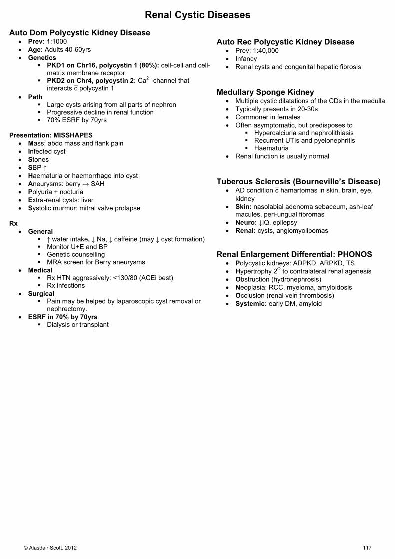
Renal Cystic Diseases Auto Dom Polycystic Kidney Disease
Prev: 1:1000 Age: Adults 40-60yrs Genetics
PKD1 on Chr16, polycystin 1 (80%): cell-cell and cell-matrix membrane receptor
PKD2 on Chr4, polycystin 2: Ca2+ channel that interacts c̄ polycystin 1
Path Large cysts arising from all parts of nephron Progressive decline in renal function 70% ESRF by 70yrs
Presentation: MISSHAPES
Mass: abdo mass and flank pain Infected cyst Stones SBP ↑ Haematuria or haemorrhage into cyst Aneurysms: berry → SAH Polyuria + nocturia Extra-renal cysts: liver Systolic murmur: mitral valve prolapse
Rx
General ↑ water intake, ↓ Na, ↓ caffeine (may ↓ cyst formation) Monitor U+E and BP Genetic counselling MRA screen for Berry aneurysms
Medical Rx HTN aggressively: <130/80 (ACEi best) Rx infections
Surgical Pain may be helped by laparoscopic cyst removal or
nephrectomy. ESRF in 70% by 70yrs
Dialysis or transplant
Auto Rec Polycystic Kidney Disease
Prev: 1:40,000 Infancy Renal cysts and congenital hepatic fibrosis
Medullary Sponge Kidney
Multiple cystic dilatations of the CDs in the medulla Typically presents in 20-30s Commoner in females Often asymptomatic, but predisposes to
Hypercalciuria and nephrolithiasis Recurrent UTIs and pyelonephritis Haematuria
Renal function is usually normal Tuberous Sclerosis (Bourneville’s Disease)
AD condition c̄ hamartomas in skin, brain, eye, kidney
Skin: nasolabial adenoma sebaceum, ash-leaf macules, peri-ungual fibromas
Neuro: ↓IQ, epilepsy Renal: cysts, angiomyolipomas
Renal Enlargement Differential: PHONOS
Polycystic kidneys: ADPKD, ARPKD, TS Hypertrophy 2O to contralateral renal agenesis Obstruction (hydronephrosis) Neoplasia: RCC, myeloma, amyloidosis Occlusion (renal vein thrombosis) Systemic: early DM, amyloid
© Alasdair Scott, 2012 117

Haematology Contents
Anaemia Classification and Causes .......................................................................................................................................... 119
Microcytic Anaemia ................................................................................................................................................................... 120
Macrocytic Anaemia .................................................................................................................................................................. 121
Haemolytic Anaemias ................................................................................................................................................................ 122
Sickle Cell Disease .................................................................................................................................................................... 123
Bleeding Diatheses.................................................................................................................................................................... 124
Thrombophilia ............................................................................................................................................................................ 125
Blood Transfusion ...................................................................................................................................................................... 126
Bone Marrow Failure ................................................................................................................................................................. 127
Myelodysplastic Syndromes ...................................................................................................................................................... 127
Chronic Myeloproliferative Disorders ........................................................................................................................................ 128
Acute Lymphoblastic Leukaemia ............................................................................................................................................... 129
Acute Myeloid Leukaemia ......................................................................................................................................................... 129
Chronic Lymphocytic Leukaemia .............................................................................................................................................. 130
Chronic Myeloid Leukaemia ...................................................................................................................................................... 130
Non-Hodgkin’s Lymphoma ........................................................................................................................................................ 131
Hodgkin’s Lymphoma ................................................................................................................................................................ 131
Multiple Myeloma ....................................................................................................................................................................... 132
Other Paraproteinaemias .......................................................................................................................................................... 133
Amyloidosis ............................................................................................................................................................................... 133
Complications of Haematological Malignancies ........................................................................................................................ 134
Spleen and Splenectomy .......................................................................................................................................................... 135
The Lab ..................................................................................................................................................................................... 136
© Alasdair Scott, 2012 118
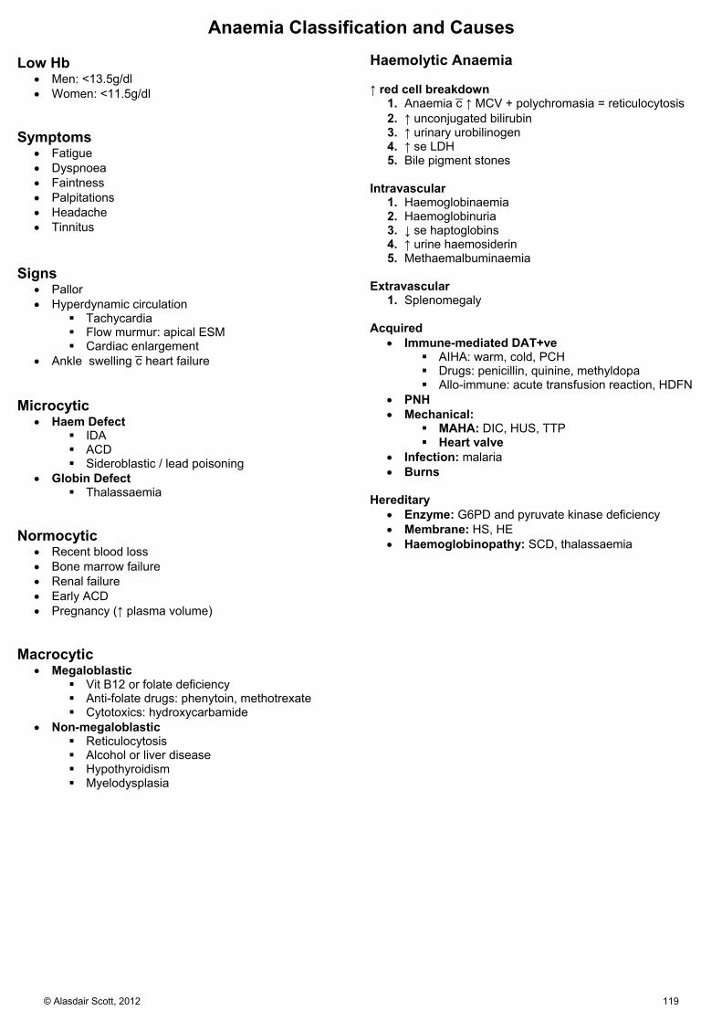
Anaemia Classification and Causes Low Hb
Men: <13.5g/dl Women: <11.5g/dl
Symptoms
Fatigue Dyspnoea Faintness Palpitations Headache Tinnitus
Signs
Pallor Hyperdynamic circulation
Tachycardia Flow murmur: apical ESM Cardiac enlargement
Ankle swelling c̄ heart failure Microcytic
Haem Defect IDA ACD Sideroblastic / lead poisoning
Globin Defect Thalassaemia
Normocytic
Recent blood loss Bone marrow failure Renal failure Early ACD Pregnancy (↑ plasma volume)
Macrocytic
Megaloblastic Vit B12 or folate deficiency Anti-folate drugs: phenytoin, methotrexate Cytotoxics: hydroxycarbamide
Non-megaloblastic Reticulocytosis Alcohol or liver disease Hypothyroidism Myelodysplasia
Haemolytic Anaemia ↑ red cell breakdown
1. Anaemia c̄ ↑ MCV + polychromasia = reticulocytosis 2. ↑ unconjugated bilirubin 3. ↑ urinary urobilinogen 4. ↑ se LDH 5. Bile pigment stones
Intravascular
1. Haemoglobinaemia 2. Haemoglobinuria 3. ↓ se haptoglobins 4. ↑ urine haemosiderin 5. Methaemalbuminaemia
Extravascular
1. Splenomegaly Acquired
Immune-mediated DAT+ve AIHA: warm, cold, PCH Drugs: penicillin, quinine, methyldopa Allo-immune: acute transfusion reaction, HDFN
PNH Mechanical:
MAHA: DIC, HUS, TTP Heart valve
Infection: malaria Burns
Hereditary
Enzyme: G6PD and pyruvate kinase deficiency Membrane: HS, HE Haemoglobinopathy: SCD, thalassaemia
© Alasdair Scott, 2012 119

Microcytic Anaemia IDA Signs
Koilonychia Angular stomatitis / cheilosis Post-cricoid Web: Plummer-Vinson
Causes Mechanism Examples ↑ Loss Menorrhagia
GI bleeding Hookworms
↓ Intake Poor diet
Malabsorption Coeliac Crohn’s
Ix
Haematinics: ↓ferritin, ↑TIBC, ↓ transferrin saturation Film: Anisocytosis, poikilocytosis, pencil cells Upper and lower GI endoscopy
Rx
Ferrous sulphate 200mg PO TDS SE: GI upset
Sideroblastic Anaemia
Ineffective erythropoiesis ↑ iron absorption Iron loading in BM → ringed sideroblasts Haemosiderosis: endo, liver and cardiac damage
Causes
Congenital Acquired
Myelodysplastic / myeloproliferative disease Drugs: chemo, anti-TB, lead
Ix
Microcytic anaemia not responsive to oral iron ↑Ferritin, ↑ se Fe, ↔TIBC
Rx
Remove cause Pyridoxine may help
Thalassaemia Pathophysiology
Point mutations (β) / deletions (α) → unbalanced production of globin chains
→ precipitation of unmatched globin → membrane damage → haemolysis while still in BM
and removal by the spleen Epidemiology
Common in Mediterranean and Far East β Thalassaemia Trait / Heterozygosity
β / β+ (↓ production) or β / βO (no production)
Mild anaemia which is usually harmless ↓ MCV (“too low for the anaemia”): e.g. <75 ↑ HbA2 (α2δ 2) and ↑HbF (α2γ2)
β Thalassaemia Major
βO / βO or βO / β+ or β+ / β+
Features develop from 3-6mo Severe anaemia Jaundice FTT Extramedullary erythropoiesis
Frontal bossing Maxillary overgrowth HSM
Haemochromatosis after 10yrs (transfusions) Ix
↓Hb, ↓MCV, ↑↑HbF, ↑HbA2 variable Film: Target cells and nucleated RBCs
Rx Life-long transfusions SC desferrioxamine Fe chelation BM transplant may be curative
α Thalassaemia
Trait --/αα or α-/α- Asymptomatic Hypochromic microcytes
HbH Disease --/-α Moderate anaemia: may need transfusions Haemolysis: HSM, jaundice
Hb Barts --/-- Hydrops fetalis → death in utero
© Alasdair Scott, 2012 120
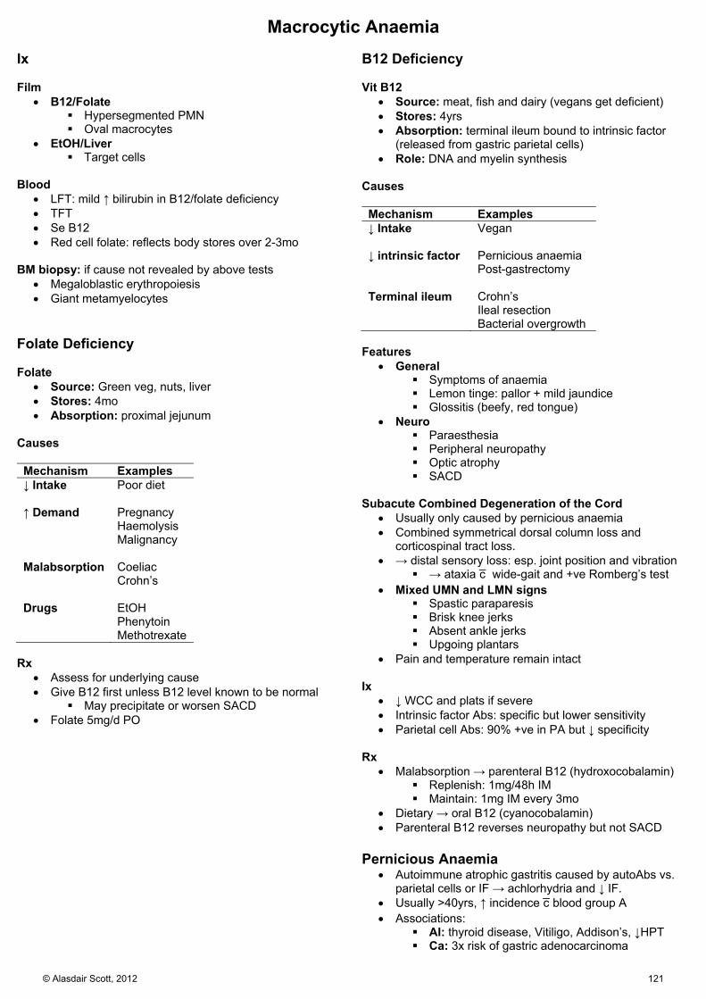
Macrocytic Anaemia Ix Film
B12/Folate Hypersegmented PMN Oval macrocytes
EtOH/Liver Target cells
Blood
LFT: mild ↑ bilirubin in B12/folate deficiency TFT Se B12 Red cell folate: reflects body stores over 2-3mo
BM biopsy: if cause not revealed by above tests
Megaloblastic erythropoiesis Giant metamyelocytes
Folate Deficiency Folate
Source: Green veg, nuts, liver Stores: 4mo Absorption: proximal jejunum
Causes Mechanism Examples ↓ Intake Poor diet
↑ Demand Pregnancy
Haemolysis Malignancy
Malabsorption Coeliac Crohn’s
Drugs
EtOH Phenytoin Methotrexate
Rx
Assess for underlying cause Give B12 first unless B12 level known to be normal
May precipitate or worsen SACD Folate 5mg/d PO
B12 Deficiency Vit B12
Source: meat, fish and dairy (vegans get deficient) Stores: 4yrs Absorption: terminal ileum bound to intrinsic factor
(released from gastric parietal cells) Role: DNA and myelin synthesis
Causes Mechanism Examples ↓ Intake Vegan
↓ intrinsic factor Pernicious anaemia
Post-gastrectomy
Terminal ileum Crohn’s Ileal resection Bacterial overgrowth
Features
General Symptoms of anaemia Lemon tinge: pallor + mild jaundice Glossitis (beefy, red tongue)
Neuro Paraesthesia Peripheral neuropathy Optic atrophy SACD
Subacute Combined Degeneration of the Cord
Usually only caused by pernicious anaemia Combined symmetrical dorsal column loss and
corticospinal tract loss. → distal sensory loss: esp. joint position and vibration
→ ataxia c̄ wide-gait and +ve Romberg’s test Mixed UMN and LMN signs
Spastic paraparesis Brisk knee jerks Absent ankle jerks Upgoing plantars
Pain and temperature remain intact Ix
↓ WCC and plats if severe Intrinsic factor Abs: specific but lower sensitivity Parietal cell Abs: 90% +ve in PA but ↓ specificity
Rx
Malabsorption → parenteral B12 (hydroxocobalamin) Replenish: 1mg/48h IM Maintain: 1mg IM every 3mo
Dietary → oral B12 (cyanocobalamin) Parenteral B12 reverses neuropathy but not SACD
Pernicious Anaemia
Autoimmune atrophic gastritis caused by autoAbs vs. parietal cells or IF → achlorhydria and ↓ IF.
Usually >40yrs, ↑ incidence c̄ blood group A Associations:
AI: thyroid disease, Vitiligo, Addison’s, ↓HPT Ca: 3x risk of gastric adenocarcinoma
© Alasdair Scott, 2012 121

Haemolytic AnaemiasAIHA Warm
IgG-mediated, bind @ 37OC Extravascular haemolysis and spherocytes Ix: DAT+ve Causes: idiopathic, SLE, RA, Evan’s Rx: immunosuppression, splenectomy
Cold
IgM-mediated, bind @ <4OC Often fix complement → intravascular haemolysis May cause agglutination → acrocyanosis or Raynaud’s Ix: DAT+ve for complement alone Causes: idiopathic, mycoplasma Rx: avoid cold, rituximab
Paroxysmal Cold Haemoglobinuria
Rare: assoc. c̄ measles, mumps, chickenpox IgG “Donath-Landsteiner” Abs bind RBCs in the cold and
→ complement-mediated lysis on rewarming PNH
Absence of RBC anchor molecule (GPI) → ↓ cell-surface complement degradation proteins → IV lysis
Affects stem cells and may also → ↓plats + ↓PMN Features
Visceral venous thrombosis (hepatic, mesenteric, CNS) IV haemolysis and haemoglobinuria
Ix
Anaemia ± thrombocytopenia ± neutropenia FACS: ↓CD55 and ↓CD59
Rx
Chronic disorder long-term anticoagulation Eculizumab (prevents complement MAC formation)
Haemolytic Uraemic Syndrome (HUS) Features
E. coli O157:H7 from undercooked meat Bloody diarrhoea and abdominal pain precedes:
MAHA Thrombocytopenia Renal failure
Ix: schistocytes, ↓ plats, normal clotting Rx
Usually resolves spontaneously Exchange transfusion or dialysis may be needed
Thrombotic Thrombocytopenia Purpura (TTP)
Genetic or acquired deficiency of ADAMTS13 Features
Adult females Pentad
Fever CNS signs: confusion, seizures MAHA Thrombocytopenia Renal failure
Ix: schistocytes, ↓ plats, normal clotting Rx: Plasmapheresis, immunosuppression, splenectomy
Hereditary Spherocytosis
Commonest inherited haemolytic anaemia in N. Europe Pathophysiology
Autosomal dominant defect in RBC membrane Spherocytes get trapped in spleen → extravascular
haemolysis Features
Splenomegaly Pigment gallstones Jaundice
Complications
Aplastic crisis Megaloblastic crisis
Ix
↑ osmotic fragility Spherocytes DAT-ve
Rx
Folate and splenectomy (after childhood) Hereditary Eliptocytosis
Autosomal dominant defect → elliptical RBCs Most pts. are asymptomatic Rx: folate, rarely splenectomy
G6PD deficiency Pathophysiology
X-linked disorder of pentose phosphate shunt ↓ NADPH production → RBC oxidative damage Affects mainly Mediterranean and Mid / Far East
Haemolysis Triggers
Broad (Fava) beans Mothballs (naphthalene) Infection Drugs: antimalarials, henna, dapsone, sulphonamides
Ix
Film Irregularly contracted cells Bite cells, ghost cells and blister cells Heinz bodies
G6PD assay after 8wks (reticulocytes have high G6PD) Rx
Treat underlying infection Stop and avoid precipitants Transfusion may be needed
Pyruvate Kinase Deficiency
Autosomal recessive defect in ATP synthesis → rigid red cells phagocytosed in the spleen Features: splenomegaly, anaemia ± jaundice Ix: PK enzyme assay Rx: often not needed or transfusion ± splenectomy
© Alasdair Scott, 2012 122

Sickle Cell Disease Epidemiology
Africa, Caribbean’s, Middle-East UK prevalence = 1/2000
Pathogenesis
Point mutation in β globin gene: glu→val SCA: HbSS Trait: HbAS
HbS insoluble when deoxygenated → sickling Sickle cells have ↓ life-span → haemolysis Sickle cells get trapped in microvasc → thrombosis
Ix
Hb 6-9, ↑ retics, ↑ bilirubin Film: sickle cells and target cells Hb electrophoresis
Dx at birth c̄ neonatal screening Presentation
Clinical features manifest from 3-6mo due to ↓HbF Triggers
Infection Cold Hypoxia Dehydration
Splenomegaly: may → sequestration crisis Infarction: stroke, spleen, AVN, leg ulcers, BM Crises: pulmonary, mesenteric, pain Kidney disease Liver, Lung disease Erection Dactylitis
Complications
Sequestration crisis Splenic pooling → shock + severe anaemia
Splenic infarction: atrophy and hyposplenism ↑ infection: osteomyelitis Aplastic crisis: parvovirus B19 infection Gallstones
Mx Chronic Disease
Pen V BD + immunisations Folate Hydroxycarbamide if frequent crises
Mx Acute Crises General
Analgesia: opioids IV Good hydration O2 Keep warm
Ix
FBC, U+E, reticulocytes, cultures Urine dip CXR
Rx
Blind Abx: e.g. ceftriaxone Transfusion: exchange if severe
© Alasdair Scott, 2012 123

Bleeding Diatheses Coagulation Tests APTT
Intrinsic: 12, 11, 9, 8 Common: 10, 5, 2, 1 Increased
Lupus anti-coagulant Haemophilia A or B vWD (carries factor 8) Unfractionated heparin DIC Hepatic failure
PT
Extrinsic: 7 Common: 10, 5, 2, 1 Increased
Warfarin / Vit K deficiency Hepatic failure DIC
Bleeding Time / PFA-100
Platelet function Increased
↓ plats number or function vWD Aspirin DIC
Thrombin Time
Fibrinogen function Increased
Quantitative/ qualitative fibrinogen defect DIC, dysfibrinogenaemia
Heparin Clinical Features Vascular or Platelet Disorder
Bleeding into skin: petechiae, purpura, echymoses Bleeding mucus mems: epistaxis, menorrhagia, gums Immediate, prolonged bleeding from cuts
Coagulation Disorder
Deep bleeding: muscles, joints, tissues Delayed but severe bleeding after injury
Vascular Disorders Congenital
HHT Ehler’s Danlos (easy bruising) Pseudoxanthoma elasticum
Acquired
Senile purpura Vitamin C deficiency Infection: e.g. meningococcus Steroids Vasculitis: e.g. HSP
Platelet Disorders Thrombocytopenia
↓ production BM failure: aplastic, infiltration, drugs (EtOH, cyto) Megaloblastic anaemia
↑ destruction Immune: ITP, SLE, CLL, heparin, viruses Non-immune: DIC, TTP, HUS, PNH, anti-
phospholipid Splenic pooling
Portal hypertension SCD
ITP Children: commonly post-URTI, self-limiting Adults: F>M, long-term Ix: Anti-platelet Abs present Rx: conservative, steroids, IVIg, splenectomy
Functional Defects
Drugs: aspirin, clopidogrel 2O: paraproteinaemias, uraemia Hereditary
Bernard-Soulier: GpIb deficiency Glanzmann’s: GpIIb/IIIa deficiency
Coagulation Disorders Acquired
Severe liver disease Anticoagulants Vitamin K deficiency
Haemophilia A: F8 Deficiency
X-linked, affects 1/5000 males Pres: haemoarthroses, bleeding after surgery/extraction Ix: ↑APTT, normal PT, ↓F8 assay Mx
Avoid NSAIDs and IM injections Minor bleeds: desmopressin + tranexamic acid Major bleeds: rhF8
Haemophilia B: F9 deficiency
X-linked, 1/20,000 males VWD
Commonest inherited clotting disorder (mostly AD) vWF
Stabilises F8 Binds plats via GpIb to damaged endothelium
Ix If mild, APTT and bleeding time may be normal ↑ APTT, ↑ bleeding time, normal plat, ↓ vWF AG
Rx: desmopressin + tranexamic acid DIC
↑PT, ↑APTT, ↑TT, ↓plats, ↓fibrinogen, ↑FDPs Schistocytes Thrombosis and bleeding Causes: sepsis, malignancy (esp. APML), trauma, obs Rx: FFP, plats, heparin
© Alasdair Scott, 2012 124

Thrombophilia Definition
Coagulopathy predisposing to thrombosis, usually venous.
Inherited Factor V Leiden / APC resistance
Protein C deactivates F5 and F8 c̄ protein S and thrombomodulin cofactors
Degradation resistance present in 5% of population Most don’t develop thrombosis
Heterozygotes: 5x ↑VTE Homozygotes: 50x ↑ VTE
Prothrombin Gene Mutation
↑ prothrombin levels ↑ thrombosis due to ↓ fibrinolysis by thrombin-activated
fibrinolysis inhibitor Protein C and S Deficiency
Heterozygotes for either have ↑ risk of thrombosis. Skin necrosis occurs – esp. c̄ warfarin
Heterozygotes → neonatal purpura fulminans Antithrombin III Deficiency
AT is heparin co-factor → thrombin inhibition Deficiency affects 1/500 Heterozygotes have ↑↑ thrombosis risk Homozygosity is incompatible c̄ life
Acquired
Progesterones in OCPs Anti-phospholipid syndrome
CLOTs: venous and arterial Coagulation defect: ↑APTT Livido reticularis Obstetric complications: recurrent 1st
trimester abortion Thrombocyotpenia
Thrombophilia Screen Indications
Arterial thrombosis <50yrs (?APL) Venous thrombosis <40yrs c̄ no RFs Familial VTE Unexplained recurrent VTE Unusual site: portal, mesenteric Recurrent foetal loss Neonatal
Investigations
FBC, clotting, fibrinogen concentration Factor V Leiden / APC resistance Lupus anticoagulant and anti-cardiolipin Abs Assays for AT, protein C and S deficiencies PCR for prothrombin gene mutation
Rx
Rx acute thrombosis as per normal Anticoagulate to INR 2-3 Consider lifelong warfarin If recurrence occurs on warfarin ↑INR to 3-4
Prevention
Lifelong anticoagulation not needed if asymptomatic ↑ VTE risk c̄ OCP or HRT Prophylaxis in high risk situations
Surgery Pregnancy
© Alasdair Scott, 2012 125

Blood Transfusion Products Packed red cells
Stored @ 4OC in SAGM Hct ~70% Use to correct anaemia or blood loss 1u → ↑Hb by 1-1.5g/dL
Platelets
Stored @ RT under agitation Not needed if count >20 or not actively bleeding Should be x-matched
Immediate Transfusion Reactions
FFP
Stored @ -18OC Should be X-matched Uses: DIC, liver disease, TTP, massive haemorrhage
Human Albumin Solution
Protein replacement After abdominal paracentesis Liver failure, nephrotic syndrome
Reaction Time Clinical Features Mechanism Rx Haemolytic Minutes Agitation
Fever Abdo/chest pain ↓ BP → shock DIC → haemorrhage Renal failure
ABO incompatibility → IV haemolysis
Stop transfusion Tell lab Keep IV line open c̄ NS Treat DIC
Bacterial Contamination
<24h ↑↑ temp + rigors ↓BP → Shock
Bacterial proliferation Esp. plats (Transfuse blood w/i 5h)
Stop transfusion Send unit to lab Abx: Taz+gent
Febrile non-Haemolytic
<24h Fever, rigors, chills
Recipient anti-HLA Abs Slow transfusion Paracetamol 1g
Allergic Immediate Urticaria, itch Angioedoema Anaphylaxis
Recipient IgA deficiency Anti-IgA IgE
Slow Chlorphenamine 10mg IV/IM
TRALI <6h ARDS: SOB, cough Bilat infiltrates on CXR
Anti-WBC Abs in donor plasma
Stop transfusion Rx ARDS
Fluid Overload
<6h CCF
Slow transfusion O2 + frusemide 40mg IV
Massive Transfusion
24h ↑K ↓ Ca (citrate chelation) ↓ F5 and F8 ↓ plats Hypothermia
Whole blood vol in 24h (5L = 10units)
Massive Transfusion Protocol - 1:1:1 ratio of PRBC:FFP:PLT - → ↑ survival Warm the blood
Delayed Transfusion Reactions
Reaction Time Clinical Features Mechanism Rx Delayed Haemolytic
1-7d Jaundice Anaemia / ↓ing Hb Fever ± Haemoglobinuria
Recipient anti-Rh Abs Extravasc haemolysis
Fe Overload Chronic SCA or Thal Major Haemochromatosis
Chronic transfusions
Desferrioxamine SC
Post-transfusion Purpura
7-10d Thrombocytopenia Purpura
AlloAbs attack recipient + donor plats
IvIg Plat transfusions
GvHD 4-30d Diarrhoea Skin rash Liver Failure → ↑ LFTs Pancytopaenia
Viable lymphocytes transfused into immunocompromised host Irradiate blood for vulnerable hosts
© Alasdair Scott, 2012 126
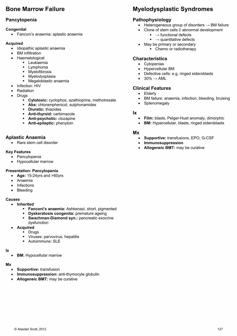
Bone Marrow Failure Pancytopenia Congenital
Fanconi’s anaemia: aplastic anaemia Acquired
Idiopathic aplastic anaemia BM infiltration Haematological
Leukaemia Lymphoma Myelofibrosis Myelodysplasia Megaloblastic anaemia
Infection: HIV Radiation Drugs
Cytotoxic: cyclophos, azathioprine, methotrexate Abx: chloramphenicol, sulphonamides Diuretic: thiazides Anti-thyroid: carbimazole Anti-psychotic: clozapine Anti-epileptic: phenytoin
Aplastic Anaemia
Rare stem cell disorder Key Features
Pancytopenia Hypocellular marrow
Presentation: Pancytopenia
Age: 15-24yrs and >60yrs Anaemia Infections Bleeding
Causes
Inherited Fanconi’s anaemia: Ashkenazi, short, pigmented Dyskeratosis congenita: premature ageing Swachman-Diamond syn.: pancreatic exocrine
dysfunction Acquired
Drugs Viruses: parvovirus, hepatitis Autoimmune: SLE
Ix
BM: Hypocellular marrow Mx
Supportive: transfusion Immunosuppression: anti-thymocyte globulin Allogeneic BMT: may be curative
Myelodysplastic Syndromes Pathophysiology
Heterogeneous group of disorders → BM failure Clone of stem cells c̄ abnormal development
→ functional defects → quantitative defects
May be primary or secondary Chemo or radiotherapy
Characteristics
Cytopenias Hypercellular BM Defective cells: e.g. ringed sideroblasts 30% → AML
Clinical Features
Elderly BM failure: anaemia, infection, bleeding, bruising Splenomegaly
Ix
Film: blasts, Pelger-Huet anomaly, dimorphic BM: Hypercellular, blasts, ringed sideroblasts
Mx
Supportive: transfusions, EPO, G-CSF Immunosuppression Allogeneic BMT: may be curative
© Alasdair Scott, 2012 127

Chronic Myeloproliferative Disorders Classification
RBC → Polycythaemia Vera WBC → CML Platelets → Essential thrombocythaemia Megakaryocytes → Myelofibrosis
Polycythaemia Vera Features
Hyperviscosity Headaches Visual disturbances Tinnitus Thrombosis
Arterial: strokes, TIA, peripheral emboli Venous: DVT, PE, Budd-Chiari
Histamine Release Aquagenic pruritus
Erythromelalgia Sudden, severe burning in hands and feet c̄
redness of the skin Splenomegaly: 75% Hepatomegaly: 30% Gout
Ix
99% JAK2+ve ↑RBC, Hb and Hct ↑WCC and ↑plats BM: hypercellular c̄ erythroid marrow ↓EPO ↑ red cell mass c̄ isotope studies
Rx
Aim to keep Hct <0.45 to ↓thrombosis Aspirin 75mg OD Venesection if young Hydroxycarbamide if older / higher risk
Prognosis
Thrombosis and haemorrhage are main complications 30% → MF 5% → AML
Polycythaemia Differential True Polycythaemia: ↑ total volume of red cells
Primary: PV Secondary
Hypoxia: altitude, COPD, smoking EPO: renal cysts/tumours
Pseudopolycythaemia: ↓ plasma volume
Acute Dehydration Shock Burns
Chronic Diuretics Smoking
Essential Thrombocythaemia Features
Thrombosis Arterial: strokes, TIA, peripheral emboli Venous: DVT, PE, Budd-Chiari
Bleeding (abnormal plat function) E.g. mucus membranes
Erythromelagia Ix
Plats >600 (often >1000) BM: ↑ megakaryocytes 50% JAK2+ve
Rx
Plats 400-1000: aspirin alone Thrombosis or plats >1000: hydroxycarbamide Anagralide may be used
Inhibits platelet maturation ↓ plat count and function
Prognosis
5% → AML/MF over 10yrs Thrombocythaemia Differential
Primary: ET Secondary:
Bleeding Infection Chronic inflammation: RA, IBD Trauma / surgery Hyposplenism / splenectomy
Primary Myelofibrosis
Clonal proliferation of megakaryocytes → ↑ PDGF → Myelofibrosis
Extramedullary haematopoiesis: liver and spleen Features
Elderly Massive HSM Hypermetabolism: wt. loss, fever, night sweats BM failure: anaemia, infections, bleeding
Ix
Film: leukoerythroblastic c̄ teardrop poikilocytes Cytopenias BM: dry tap (need trephine biopsy) 50% JAK2+ve
Rx
Supportive: blood products Splenectomy Allogeneic BMT may be curative in younger pts.
Prognosis
5yr median survival
© Alasdair Scott, 2012 128

Acute Lymphoblastic Leukaemia Epidemiology
Children 2-5yrs (commonest childhood Ca) Rare in adults
Aetiology
Arrest of maturation and proliferation of lymphoblasts 80% B lineage, 20% T lineage
Risk Factors
Genetic susceptibility (often Chr translocations) Environmental trigger
Radiation (e.g. during pregnancy) Down’s
Features BM Failure
Anaemia Thrombocytopenia → bleeding Leukopenia → infection
Infiltration
Lymphadenopathy Orchidomegaly Thymic enlargement HSM CNS: CN palsies, meningism Bone pain
Ix
↑WCC: lymphoblasts ↓RBC, ↓PMN, ↓plats BM aspirate
≥20% blasts Cytogenetic and molecular analysis
CXR+CT: mediastinal + abdominal LNs LP: CNS involvement
Mx Supportive
Blood products Allopurinol Hickman line or Portacath
Infections
Gentamicin + tazocin Prophylaxis: e.g. co-timoxazole, ciprofloxacin
Chemotherapy (recruited into national trials)
1. Remission induction 2. Consolidation + CNS Rx 3. Maintenance for 2-3yrs
BMT
Best option for younger adults Prognosis
85% survival in children Worse in adults
Acute Myeloid Leukaemia Epidemiology
↑ risk c̄ age: mean 65-70 Commonest acute leukaemia of adults
Aetiology
Neoplastic proliferation of myeloblasts Risk Factors
Chromosomal abnormalities Radiation Down’s Chemotherapy: e.g. for lymphoma Myelodysplastic and myeloproliferative syndromes
FAB Classification (based on cell type)
M2: granulocyte maturation M3: acute promyelocytic leukaemia – t(15;17) M4: acute myelomonocytic leukaemia M7: megakaryoblastic leukaemia – trisomy 21
Features BM Failure
Cytopenias Infiltration
Gum infiltration → hypertrophy and bleeding (M4) HSM Skin involvement Bone pain
Blood
DIC: APML (M3) Hyperviscosity: ↑↑WCC may → thrombi
Ix
↑ WCC blasts (occasionally normal) Anaemia and ↓ plats BM aspirate
≥20% blasts Auer rods are diagnostic
Dx
Made by immunological and molecular phenotyping Flow cytometry
Cytogenetic analysis affects Rx and guides prognosis Mx Supportive: as for ALL Infections: as for ALL Chemotherapy
V. intensive → long periods of neutropenia and ↓ plats ATRA for APML
BMT
Allogeneic if poor prognosis Destroy BM and leukemic cells c̄ chemotherapy
and total body irradiation. Repopulate marrow c̄ HLA-matched donor HSCs
Autologous if intermediate prognosis HSCs taken from pt.
© Alasdair Scott, 2012 129

Chronic Lymphocytic Leukaemia Epidemiology
Commonest leukaemia in Western World M>F=2:1 Elderly: 70s
Aetiology
Clone of mature B cells (memory cells) Features
Often asymptomatic incidental finding Symmetrical painless lymphadenopathy HSM Anaemia B symptoms: wt. loss, fever, night sweats
Complications
Autoimmune haemolysis Evan’s = AIHA and ITP
Infection (↓Ig): bacterial, zoster Marrow failure / infiltration
Ix
↑ WCC, lymphocytosis Smear cells ↓ se Ig +ve DAT Rai or Binet staging
Dx
Immunophenotyping to distinguish from NHL Natural Hx
Some remain stable for years Nodes usually enlarge slowly (± lymphatic obstruction) Death often due to infection: e.g. encapsulates, fungi Richter Transformation: CLL → large B cell lymphoma
Rx
Indications Symptomatic Ig genes un-mutated (bad prognostic indicator) 17p deletions (bad prognostic indicator)
Supportive care Chemotherapy
Cylophosphamide Fludarabine Rituximab
Radiotherapy Relieve LN or splenomegaly
Prognosis
1/3 never progress 1/3 progress c̄ time 1/3 are actively progressing
Chronic Myeloid Leukaemia Epidemiology
15% of leukaemia Middle-aged: 60-60yrs
Aetiology
Myeloproliferative disorder: clonal proliferation of myeloid cells.
Features
Systemic: wt. loss, fever, night sweats, lethargy Massive HSM → abdo discomfort Bruising / bleeding (platelet dysfunction) Gout Hyperviscosity
Philadelphia Chromosome
Reciprocal translocation: t(9;22) Formation of BCR-ABL fusion gene
Constitutive tyrosine kinase activity Present in >80% of CML Discovered by Nowell and Hungerford in 1960
Ix
↑↑WBC PMN and basophils Myelocytes
± ↓Hb and ↓plat (accelerated or blast phase) ↑urate BM cytogenetic analysis: Ph+ve
Natural Hx
Chronic phase: <5% blasts in blood or DM Accelerated phase: 10-19% blasts Blast crisis: usually AML, ≥20% blasts
Rx
Imatinib: tyrosine kinase inhibitor → >90% haematological response 80% 5ys
Allogeneic SCT Indicated if blast crisis or TK-refractory
© Alasdair Scott, 2012 130

Non-Hodgkin’s Lymphoma (80%) Features Lymphadenopathy: 75% @ presentation
Painless Symmetric Multiple sites Spreads discontinuously
Extranodal
Skin: esp. T cell lymphomas CNS Oropharynx and GIT Splenomegaly
B Symptoms
Fever Night sweats Wt. loss (>10% over 6mo)
Blood
Pancytopenia Hyperviscosity
Ix
FBC, U+E, LFT, LDH ↑ LDH = worse prognosis
Film Normal or circulating lymphoma cells ± pancytopenias
Classification: LN and BM biopsy Staging: CT/MRI chest, abdomen, pelvis
Stage c̄ Ann Arbor System Classification B Cell (commonest)
Low Grade: usually indolent but often incurable Follicular Small cell lymphocytic (=CLL) Marginal Zone (inc. MALTomas) Lymphoplamsacytoid (e.g. Waldenstrom’s)
High Grade: aggressive but may be curable Diffuse large B cell (commonest NHL) Burkitt’s
T Cell
Adult T cell lymphoma: Caribs and Japs – HTLV-1 Enteropathy-assoc. T cell lymphoma: chronic coeliac Cutaneous T cell lymphoma: e.g. Sezary syn. Anaplastic large cell
Mx
Diagnosis and management in an MDT High Grade (e.g. DLBCL)
R-CHOP regimen BMT for relapse ~30% 5ys
Low Grade (e.g. follicular)
Rx when clinically indicated (e.g. chloambucil) >50% 5ys
Hodgkin’s Lymphoma (20%) Epidemiology
M>F=2:1 (esp. in paeds) Bimodal age incidence: 20-29yrs and >60yrs May be assoc. c̄ EBV
Features Lymphadenopathy
Painless Asymmetric Spreads contiguously to adjacent LNs Cervical nodes in 70% (also axillary and inguinal) May be alcohol-induced LN pain Mediastinal LN may → mass effects
SVC obstruction Bronchial obstruction
B Symptoms
Fever Night sweats Wt. loss (>10% over 6mo)
Other
Itch Pel Ebstein Fever: cyclical fever Hepato- and/or spleno-megaly
Ix
FBC, film, ESR, LFT, LDH, Ca ↑ESR or ↓Hb = worse prognosis
LN excision biopsy or FNA Reed-Sternberg Cells (owl’s eye nucleus)
Staging: CT/MRI chest, abdomen, pelvis BM biopsy if B symptoms or Stage 3/4 disease
Staging: Ann Arbor System
1. Single LN region 2. ≥2 nodal area on same side of diaphragm 3. Nodes on both sides of diaphragm 4. Spread byond nodes: e.g. liver, BM + B if constitutional symptoms
Mx
Chemo, radio or both ABVD regimen BMT for relapse
Prognosis
Depends on stage and grade 1A: >95% 5ys 4B: <40% 5ys
© Alasdair Scott, 2012 131

Multiple Myeloma Epidemiology
M=F Blacks>White=2:1 ~70yrs
Pathogenesis
Clonal proliferation of plasma cells → monoclonal ↑↑Ig Usually IgG or IgA
Clones may also produce free light chain (λ or κ): ~2/3 Excreted by kidney → urinary BJP Light chains only seen in plasma in renal failure
Clones produce IL-6 which inhibits osteoblasts (↔ ALP) and activates osteoclasts.
Symptoms Osteolytic Bone Lesions
Backache and bone pain Pathological #s Vertebral collapse
BM Infiltration
Anaemia, neutropenia or thrombocytopenia Recurrent Bacterial Infections
Neutropenia Immunoparesis (= ↓Ig) Chemotherapy
Renal Impairment
Light chains ↑Ca AL-amyloid
Complications
Hypercalcaemia Neurological: ↑Ca, compression, amyloid AKI Hyperviscosity AL-amyloid (15%)
Ix NB. Do ESR and Se electrophoresis if >50 c̄ back pain Bloods
FBC: normocytic normochromic anaemia Film: rouleaux ± plasma cells ± cytopenias ↑↑ESR/PV, ↑U+Cr, ↑Ca, normal ALP Se electrophoresis and β2-microglobulin
Urine
Stix: ↑ specific gravity (BJP doesn’t show) Electrophoresis: BJP
BM trephine biopsy X-ray: Skeletal Survey
Punched-out lytic lesions Pepper-pot skull Vertebral collapse Fractures
Dx
Clonal BM plasma cells ≥10% Presence of se and/or urinary monoclonal protein End-organ Damage: CRAB (1 or more)
Ca ↑ (>2.6mM) Renal insufficiency Anaemia (<10g/dL) Bone lesions
Mx Supportive
Bone pain: Analgesia (avoid NSAIDs) + bisphosphonates
Anaemia: Transfusions and EPO Renal impairment: ensure good hydration ± dialysis Infections: broad spectrum Abx ± IVIg if recurrent
Complications
↑Ca: aggressive hydration, frusemide, bisphosphonates Cord compression: MRI, dexamethasone + local radio Hyperviscosity: Plasmapheresis (remove light chains) AKI: rehydration ± dialysis
Specific
Fit pts. Induction chemo: lenalidomide + low-dose dex Then allogeneic BMT
Unfit pts. Chemo only: melphalan + pred + lenalidomide Bortezomib for relapse
Prognosis
Mean survival: 3-5yrs Poor prognostic indicators
↑ β2-microglobulin ↓ albumin
© Alasdair Scott, 2012 132

Other Paraproteinaemias Smouldering / Asymptomatic Myeloma
Se monoclonal protein and/or BM plasma cells ≥10% No CRAB
MGUS
Se monoclonal protein <30g/L Clonal BM plasma cells <10% No CRAB
Solitary Plasmacytoma
Solitary bone/soft tissue plasma cell lesion Se or urinary monoclonal protein Normal BM and normal skeletal survey (except lesion) No CRAB
Waldenstrom’s Macroglobulinaemia
Lymphoplasmacytoid lymphoma→ monoclonal IgM band
Features: Hyperviscosity: CNS and ocular symptoms Lymphadenopathy + splenomegaly
Ix: ↑ESR, IgM paraprotein Primary AL Amyloid
Occult proliferation of plasma cells → production of amyloidogenic monoclonal protein.
Se or urinary light chains Paraproteinaemia in lymphoma or leukaemia
E.g. seen in 5% of CLL
Amyloidosis Definition
Group of disorders characterised by extracellular deposits of a protein in an abnormal fibrillar form that is resistant to degradation.
AL Amyloidosis
Clonal proliferation of plasma cells c̄ production of amyloidogenic light chains.
1O: occult plasma cell proliferation 2O: myeloma, Waldenstrom’s, MGUS, lymphoma
Features
Renal: proteinuria and nephrotic syndrome Heart: restrictive cardiomyopathy, arrhythmias, echo
“Sparkling” appearance on echo Nerves: peripheral and autonomic neuropathy, carpal
tunnel. GIT: macroglossia, malabsorption, perforation,
haemorrhage, hepatomegaly, obstruction. Vascular: periorbital purpura (characteristic)
AA Amyloidosis
Amyloid derived from serum amyloid A SAA is an acute phase protein
Chronic inflammation RA IBD Chronic infection: TB, bronchiectasis
Features
Renal: proteinuria and nephrotic syndrome Hepatosplenomegaly
Familial Amyloidosis
Group of AD disorders caused by mutations in transthyretin (produced by liver)
Features: sensory or autonomic neuropathy Others: non-Systemic Amyloidosis
β-amyloid: Alzheimer’s β2 microglobulin: chronic dialysis Amylin: T2DM
Dx
Biopsy of affected tissue Rectum or subcut fat is relatively non-invasive
Apple-green birefringence c̄ Congo Red stain under polarized light.
Rx
AA amyloid may improve c̄ underlying condition AL amyloid may respond to therapy for myeloma Liver Tx may be curative for familial amyloidosis
Prognosis
Median survival: 1-2yrs
© Alasdair Scott, 2012 133
Complications of Haematological Malignancies Neutropenic Sepsis General Precautions
Barrier nursing in a side room Avoid IM injections (may → infected haematoma) Swabs + septic screen TPR 4hrly
Antimicrobials
Start broad spectrum Abx: check local guidelines Consider G-CSF
Hyperviscosity Syndrome Causes
↑↑RBC / Hct >0.5: e.g. PV ↑↑WCC > 100: e.g. leukaemia ↑↑ plasma proteins: Myeloma, Waldenstrom’s
Features
CNS: headache, confusion, seizures, faints Visual: retinopathy → visual disturbance Bleeding: mucus membranes, GI, GU Thrombosis
Ix
↑ plasma viscosity (PV) FBC, film, clotting Se + urinary protein electrophoresis
Rx
Polycythaemia: venesection Leukopheresis: leukaemia
Avoid transfusing before lowering WCC Plasmapheresis: myeloma and Waldenstrom’s
DIC
Widespread activation of coagulation from release of pro-coagulants into the circulation.
Clotting factors and plats are consumed → bleeding Fibrin strands → haemolysis
Causes
Malignancy: e.g. APML Sepsis Trauma Obstetric events: e.g. PET
Signs
Bruising Bleeding Renal failure
Ix
↓plats, ↓Hb, ↑APTT, ↑PT, ↑FDPs, ↓fibrinogen (↑TT) Rx
Rx cause Replace: cryoprecipitate, FFP Consider heparin and APC
Tumour Lysis Syndrome
Massive cell destruction High count leukaemia or bulky lymphoma
↑K, ↑urate → renal failure Prevention: ↑ fluid intake + allopurinol
© Alasdair Scott, 2012 134
Spleen and Splenectomy Anatomy
Intraperitoneal structure lying in the LUQ Measures 1x3x5 inches Weighs ~7oz Lies anterior to ribs 9-11
Features
1. Dull to percussion 2. Enlarges to RIF 3. Moves inferiorly on respiration 4. Can’t get above it 5. Medial notch
Function: part of the mononuclear phagocytic system
Phagocytosis of old RBCs, WBCs and opsonised bugs Antibody production Haematopoiesis Sequestration of formed blood elements
Causes of Massive Splenomegaly: >20cm
CML Myelofibrosis Malaria LeishManiasis Gaucher’s (AR, glucocerebrosidase deficiency)
All Causes of Splenomegaly
Haematological Haemolysis: HS Myelproliferative disease: CML, MF, PV Leukaemia, lymphoma
Infective EBV, CMV, hepatitis, HIV, TB, infective endocarditis Malaria, leishmanias, hydatid disease
Portal HTN: cirrhosis, Budd-Chiari Connective tissue: RA, SLE, Sjogrens Other
Sarcoid Amyloidosis Gaucher’s 1O Ab deficiency (e.g. CVID)
Splenomegaly may → Hypersplenism
Pancytopenia as cells are sequestrated
Splenectomy → Hyposplenism Indications
Trauma Rupture (e.g. EBV infection) AIHA ITP HS Hypersplenism
Complications
Redistributive thrombocytosis → early VTE Gastric dilatation (ileus) Left lower lobe atelectasis: v. common ↑ susceptibility to infections
Encapsulates: haemophilus, pneumo, meningo Film
Howell-Jolly bodies Pappenheimer bodies Target cells
Mx
Immunisation: pneumovax, HiB, Men C, yrly flu Daily Abx: Pen V or erythromycin Warning: Alert Card and/or Bracelet
Other Causes of Hyposplenism
SCD Coeliac disease IBD
© Alasdair Scott, 2012 135
The Lab ESR
How far RBCs fall through anti-coagulated blood in 1h Normal: ~20mm/h (M: age/2, F: (age+10)/2 ↑ se proteins cover RBCs → clumping → rouleaux →
faster settling → ↑ESR. ↑ ESR
Plasma factors ↑ fibrinogen: inflammation ↑ globulins: e.g. myeloma
Red cell factors Anaemia → ↑ESR
∆∆ of ESR >100
Myeloma SLE GCA AAA Ca prostate
Differential White Count Neutrophilia
Bacterial infection Left shift Toxic granulation Vacuolation
Stress: trauma, surgery, burns, haemorrhage Steroids Inflammation: MI, PAN Myeloproliferative disorders: e.g. CML
Neutropenia
Viral infection Drugs: chemo, cytotoxics, carbimazole, sulphonamides Severe sepsis Hypersplenism: e.g. Felty’s
Lymphocytosis
Viral infections: EBV, CMV Chronic infections: TB, Brucella, Hepatitis, Toxo Leukaemia, lymphoma: esp. CLL
Lymphopenia
Drugs: steroids, chemo HIV
Monocytosis
Chronic infection: TB, Brucella, Typhoid AML
Eosinophilia
Parasitic infection Drug reactions: e.g. c̄ EM Allergies: asthma, atopy, Churg-Strauss Skin disease: eczema, psoriasis, pemphigus
Basophilia
Parasitic infection IgE-mediated hypersensitivity: urticarial, asthma CML
The Peripheral Film Feature Causes Acanthocytes Abetaliproteinaemia
Alcoholic lever disease Basophilic stippling Lead poisoning
Thalassaemia Blasts Leukaemia Burr cels Uraemia Dimorphic picture Myelodysplasia Howell-Jolly bodies Hyposplenism Heinz bodies G6PD deficiency Irregularly contracted cells G6PD deficiency Leukoerythroblastic film BM infiltration Pappenheimer bodies Hyposplenism Pencil cells IDA Reticulocytes Haemolysis
Haemorrhage Spherocytes AIHA
Hereditary spherocytosis Schistocytes MAHA
Prosthetic valve Target cells Hyposplenism
Thalassaemia Liver disease
Tear-drop cells / Dacrocytes BM infiltration Cytochemistry
Myeloblasts Sudan black B MPO
Hairy cell leukaemia: tartrate resistant acid phosphatase Leukocyte alkaline phosphatase
↑: PV, ET, MF ↓: CML, PNH
© Alasdair Scott, 2012 136
Infectious Disease Contents
Tuberculosis .............................................................................................................................................................................. 138
Other Mycobacterial Disease .................................................................................................................................................... 139
Influenza .................................................................................................................................................................................... 139
HIV ............................................................................................................................................................................................. 140
Complications of HIV Infection .................................................................................................................................................. 141
Herpes Infections ....................................................................................................................................................................... 142
Viral Hepatitis ............................................................................................................................................................................ 144
Gastrointestinal Infections 1 ...................................................................................................................................................... 145
Gastrointestinal Infections 2 ...................................................................................................................................................... 146
Sexually Transmitted Infections ................................................................................................................................................ 147
Syphilis ...................................................................................................................................................................................... 148
Zoonoses ................................................................................................................................................................................... 149
Malaria ....................................................................................................................................................................................... 150
Other Tropical Diseases ............................................................................................................................................................ 151
Miscellaneous Infections ........................................................................................................................................................... 152
Approach to Nosocomial Fever ................................................................................................................................................. 152
© Alasdair Scott, 2012 137
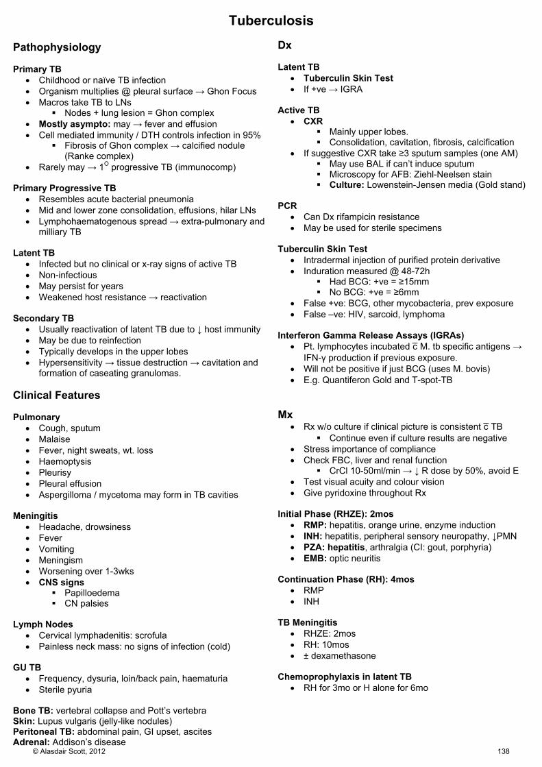
Tuberculosis Pathophysiology Primary TB
Childhood or naïve TB infection Organism multiplies @ pleural surface → Ghon Focus Macros take TB to LNs
Nodes + lung lesion = Ghon complex Mostly asympto: may → fever and effusion Cell mediated immunity / DTH controls infection in 95%
Fibrosis of Ghon complex → calcified nodule (Ranke complex)
Rarely may → 1O progressive TB (immunocomp) Primary Progressive TB
Resembles acute bacterial pneumonia Mid and lower zone consolidation, effusions, hilar LNs Lymphohaematogenous spread → extra-pulmonary and
milliary TB Latent TB
Infected but no clinical or x-ray signs of active TB Non-infectious May persist for years Weakened host resistance → reactivation
Secondary TB
Usually reactivation of latent TB due to ↓ host immunity May be due to reinfection Typically develops in the upper lobes Hypersensitivity → tissue destruction → cavitation and
formation of caseating granulomas. Clinical Features Pulmonary
Cough, sputum Malaise Fever, night sweats, wt. loss Haemoptysis Pleurisy Pleural effusion Aspergilloma / mycetoma may form in TB cavities
Meningitis
Headache, drowsiness Fever Vomiting Meningism Worsening over 1-3wks CNS signs
Papilloedema CN palsies
Lymph Nodes
Cervical lymphadenitis: scrofula Painless neck mass: no signs of infection (cold)
GU TB
Frequency, dysuria, loin/back pain, haematuria Sterile pyuria
Bone TB: vertebral collapse and Pott’s vertebra Skin: Lupus vulgaris (jelly-like nodules) Peritoneal TB: abdominal pain, GI upset, ascites Adrenal: Addison’s disease
Dx Latent TB
Tuberculin Skin Test If +ve → IGRA
Active TB
CXR Mainly upper lobes. Consolidation, cavitation, fibrosis, calcification
If suggestive CXR take ≥3 sputum samples (one AM) May use BAL if can’t induce sputum Microscopy for AFB: Ziehl-Neelsen stain Culture: Lowenstein-Jensen media (Gold stand)
PCR
Can Dx rifampicin resistance May be used for sterile specimens
Tuberculin Skin Test
Intradermal injection of purified protein derivative Induration measured @ 48-72h
Had BCG: +ve = ≥15mm No BCG: +ve = ≥6mm
False +ve: BCG, other mycobacteria, prev exposure False –ve: HIV, sarcoid, lymphoma
Interferon Gamma Release Assays (IGRAs)
Pt. lymphocytes incubated c̄ M. tb specific antigens → IFN-γ production if previous exposure.
Will not be positive if just BCG (uses M. bovis) E.g. Quantiferon Gold and T-spot-TB
Mx
Rx w/o culture if clinical picture is consistent c̄ TB Continue even if culture results are negative
Stress importance of compliance Check FBC, liver and renal function
CrCl 10-50ml/min → ↓ R dose by 50%, avoid E Test visual acuity and colour vision Give pyridoxine throughout Rx
Initial Phase (RHZE): 2mos
RMP: hepatitis, orange urine, enzyme induction INH: hepatitis, peripheral sensory neuropathy, ↓PMN PZA: hepatitis, arthralgia (CI: gout, porphyria) EMB: optic neuritis
Continuation Phase (RH): 4mos
RMP INH
TB Meningitis
RHZE: 2mos RH: 10mos ± dexamethasone
Chemoprophylaxis in latent TB
RH for 3mo or H alone for 6mo
© Alasdair Scott, 2012 138
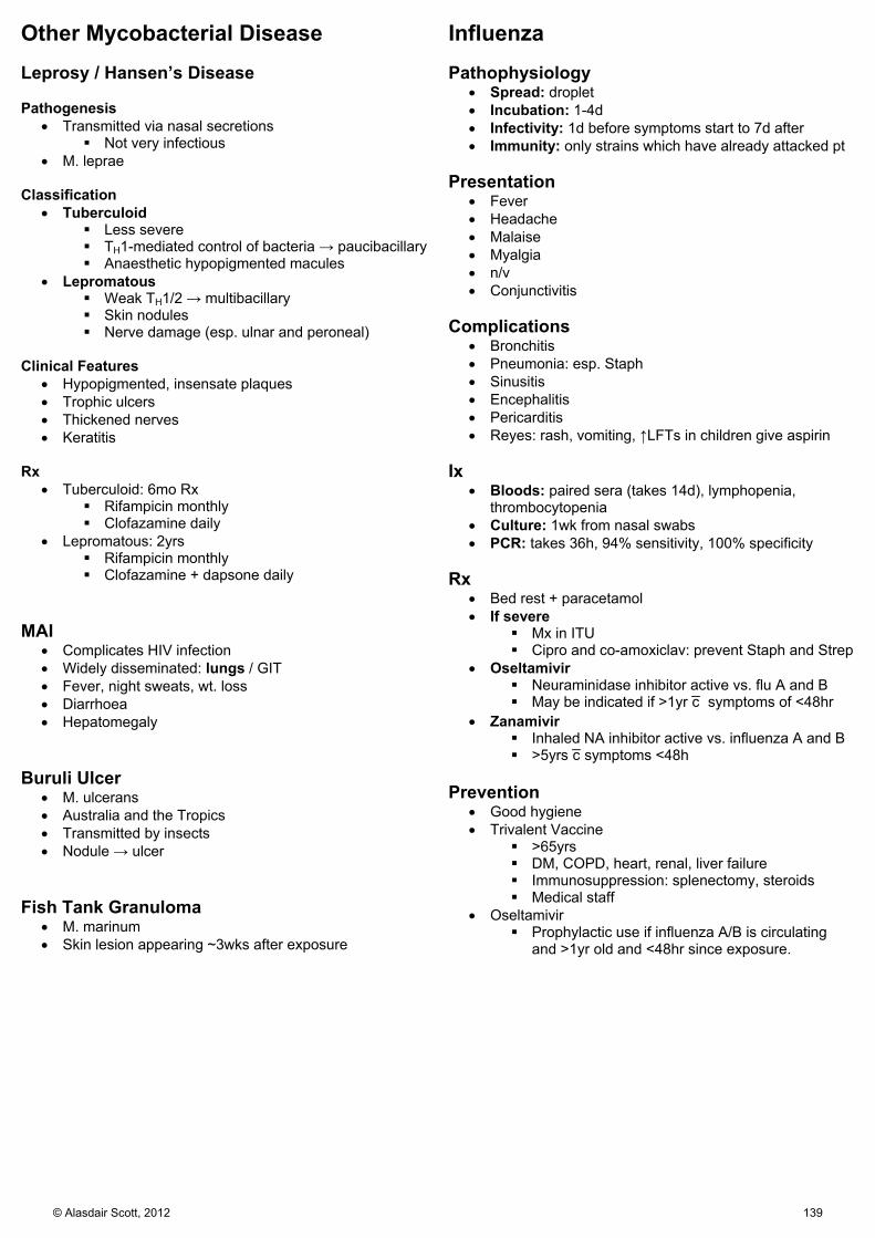
Other Mycobacterial Disease Leprosy / Hansen’s Disease Pathogenesis
Transmitted via nasal secretions Not very infectious
M. leprae Classification
Tuberculoid Less severe TH1-mediated control of bacteria → paucibacillary Anaesthetic hypopigmented macules
Lepromatous Weak TH1/2 → multibacillary Skin nodules Nerve damage (esp. ulnar and peroneal)
Clinical Features
Hypopigmented, insensate plaques Trophic ulcers Thickened nerves Keratitis
Rx
Tuberculoid: 6mo Rx Rifampicin monthly Clofazamine daily
Lepromatous: 2yrs Rifampicin monthly Clofazamine + dapsone daily
MAI
Complicates HIV infection Widely disseminated: lungs / GIT Fever, night sweats, wt. loss Diarrhoea Hepatomegaly
Buruli Ulcer
M. ulcerans Australia and the Tropics Transmitted by insects Nodule → ulcer
Fish Tank Granuloma
M. marinum Skin lesion appearing ~3wks after exposure
Influenza Pathophysiology
Spread: droplet Incubation: 1-4d Infectivity: 1d before symptoms start to 7d after Immunity: only strains which have already attacked pt
Presentation
Fever Headache Malaise Myalgia n/v Conjunctivitis
Complications
Bronchitis Pneumonia: esp. Staph Sinusitis Encephalitis Pericarditis Reyes: rash, vomiting, ↑LFTs in children give aspirin
Ix
Bloods: paired sera (takes 14d), lymphopenia, thrombocytopenia
Culture: 1wk from nasal swabs PCR: takes 36h, 94% sensitivity, 100% specificity
Rx
Bed rest + paracetamol If severe
Mx in ITU Cipro and co-amoxiclav: prevent Staph and Strep
Oseltamivir Neuraminidase inhibitor active vs. flu A and B May be indicated if >1yr c̄ symptoms of <48hr
Zanamivir Inhaled NA inhibitor active vs. influenza A and B >5yrs c̄ symptoms <48h
Prevention
Good hygiene Trivalent Vaccine
>65yrs DM, COPD, heart, renal, liver failure Immunosuppression: splenectomy, steroids Medical staff
Oseltamivir Prophylactic use if influenza A/B is circulating
and >1yr old and <48hr since exposure.
© Alasdair Scott, 2012 139

HIV Immunology
HIV binds via gp120 to CD4 TH cells, monocytes, macrophages, neural cells
CD4+ cells migrate to lymphoid tissue where virus replicates → infection of new CD4+ cells
Depletion and impaired function of CD4+ cells → immune dysfunction.
Virology
RNA retrovirus After entry, viral reverse transcriptase makes DNA
copy of viral RNA genome. Viral integrase enzyme integrates this c̄ host DNA Core viral proteins synthesised by host and then
cleaved by viral protease into mature subunits. Completed virions released by budding
Natural Hx Acute Infection: usually asympto Seroconversion
Transient illness 2-6wks after exposure Fever, malaise, myalgia, pharyngitis, macpap rash Rarely meningoencephalitis
Asymptomatic Infection
But 30% will have PGL Nodes >1cm in diameter ≥2 extra-inguinal sites ≥3mo
AIDS-related Complex (ARC)
AIDS prodrome Constitutional symptoms: fever, night sweats, wt. loss Minor opportunistic infections
Oral candida Oral hairy leukoplakia (EBV) Recurrent HSV Seborrhoeic dermatitis
AIDS
Defining illness CD4 usually <200
Other Effects of HIV
Osteoporosis Dementia Neuropathy Nephropathy
Dx
ELISA: detect serum (or salivary) anti-HIV Abs Western Blot: for confirmation If recent exposure, may be window period
Usually 1-3wks Can be 3-6mo
PCR: can detect HIV virions in the window period Rapid Antibody Tests: false positives are a problem
and results should be confirmed by Western Blot
Ix
HIV diagnostic tests Drug resistance studies
e.g. genotyping for reverse transcriptase or protease mutations
Mantoux test Serology: toxo, CMV, HBV, HCV, syphilis
Monitoring
CD4 count Viral load (HIV RNA) FBC, U+E, LFTs, lipids, glucose
HAART Indications
CD4 ≤350 AIDS-defining illness Pregnancy HIVAN Co-infected c̄ HBV when Rx is indicated for HBV
Regimens
1 NNRTI + 2 NRTIs NNRTI = Efavirenz NRTI = emtricitabine + tenofovir (Truvada) Atripla = efavirenz + emtricitabine + tenofovir
PI + 2 NRTIs PI = lopinavir (+ low dose ritonavir = Kaletra)
Aim
Undetectable VL after 4mo If VL remains high despite good compliance
Change to a new drug combination Request resistance studies
Prophylaxis
CD4 <200: PCP – co-trimoxazole CD4 <100: Toxo – co-trimoxazole CD4 <50: MAC – azithromycin
HIV Exposure
Seroconversion post-needle-stick = ~0.3% (1/300) Report to occupational health Immunise against hep B (active + passive) Test blood from both parties: HIV, HBC, HCV Repeat recipient testing @ 3 and 6mo
PEP
Start PEP in high-risk exposure from HIV+ or unknown source.
Start ASAP as possible. Continue for at least 28d E.g. Truvada + Kaletra
© Alasdair Scott, 2012 140
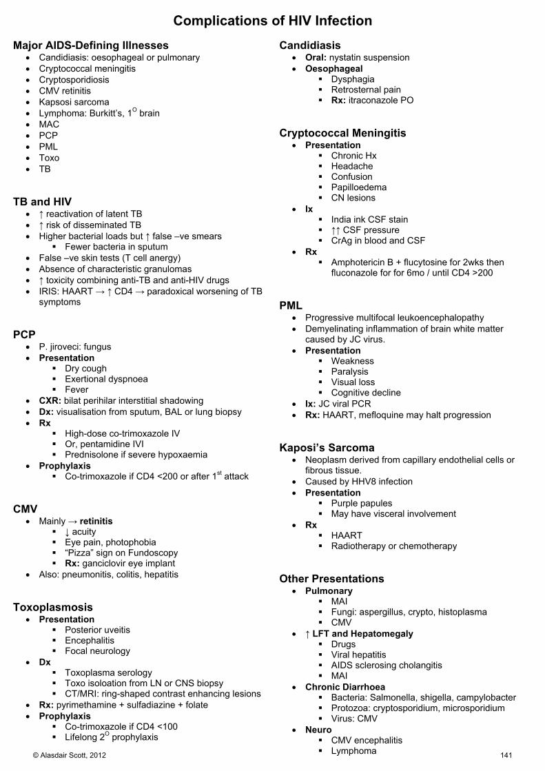
Complications of HIV Infection Major AIDS-Defining Illnesses
Candidiasis: oesophageal or pulmonary Cryptococcal meningitis Cryptosporidiosis CMV retinitis Kapsosi sarcoma Lymphoma: Burkitt’s, 1O brain MAC PCP PML Toxo TB
TB and HIV
↑ reactivation of latent TB ↑ risk of disseminated TB Higher bacterial loads but ↑ false –ve smears
Fewer bacteria in sputum False –ve skin tests (T cell anergy) Absence of characteristic granulomas ↑ toxicity combining anti-TB and anti-HIV drugs IRIS: HAART → ↑ CD4 → paradoxical worsening of TB
symptoms PCP
P. jiroveci: fungus Presentation
Dry cough Exertional dyspnoea Fever
CXR: bilat perihilar interstitial shadowing Dx: visualisation from sputum, BAL or lung biopsy Rx
High-dose co-trimoxazole IV Or, pentamidine IVI Prednisolone if severe hypoxaemia
Prophylaxis Co-trimoxazole if CD4 <200 or after 1st attack
CMV
Mainly → retinitis ↓ acuity Eye pain, photophobia “Pizza” sign on Fundoscopy Rx: ganciclovir eye implant
Also: pneumonitis, colitis, hepatitis Toxoplasmosis
Presentation Posterior uveitis Encephalitis Focal neurology
Dx Toxoplasma serology Toxo isoloation from LN or CNS biopsy CT/MRI: ring-shaped contrast enhancing lesions
Rx: pyrimethamine + sulfadiazine + folate Prophylaxis
Co-trimoxazole if CD4 <100 Lifelong 2O prophylaxis
Candidiasis
Oral: nystatin suspension Oesophageal
Dysphagia Retrosternal pain Rx: itraconazole PO
Cryptococcal Meningitis
Presentation Chronic Hx Headache Confusion Papilloedema CN lesions
Ix India ink CSF stain ↑↑ CSF pressure CrAg in blood and CSF
Rx Amphotericin B + flucytosine for 2wks then
fluconazole for for 6mo / until CD4 >200 PML
Progressive multifocal leukoencephalopathy Demyelinating inflammation of brain white matter
caused by JC virus. Presentation
Weakness Paralysis Visual loss Cognitive decline
Ix: JC viral PCR Rx: HAART, mefloquine may halt progression
Kaposi’s Sarcoma
Neoplasm derived from capillary endothelial cells or fibrous tissue.
Caused by HHV8 infection Presentation
Purple papules May have visceral involvement
Rx HAART Radiotherapy or chemotherapy
Other Presentations
Pulmonary MAI Fungi: aspergillus, crypto, histoplasma CMV
↑ LFT and Hepatomegaly Drugs Viral hepatitis AIDS sclerosing cholangitis MAI
Chronic Diarrhoea Bacteria: Salmonella, shigella, campylobacter Protozoa: cryptosporidium, microsporidium Virus: CMV
Neuro CMV encephalitis Lymphoma© Alasdair Scott, 2012 141

Herpes Infections Herpes Simplex
Virology Disease Features Diagnosis Treatment α-neurotrophic Persists in DRG Mucocutaneous spread
Oropharyngeal / primary stomatitis
HSV-1 Severe painful ulceration Fever + Submandibular lymphadenopathy
Topical Aciclovir
Encephalitis 90% HSV-1, 10% HSV-2 Flu-like prodrome Headache, focal neuro, fits, odd behaviour, ↓GCS Haemorrhagic necrosis of temporal lobes
CSF: ↑lympho, normal gluc CSF PCR MRI
Aciclovir IV
Mollaret’s Meningitis HSV-2 mostly Benign recurrent aseptic meningitis
Aciclovir IV
Herpes Gladiatorum Rugby players, wrestlers Painful vesicular rash, lymphadenopathy, fever
Aciclovir PO
Herpetic Whitlow Healthcare workers, children Painful red finger
Topical Aciclovir
Eczema Herpeticum Herpes infection at the site of skin damage Aciclovir Genital Herpes HSV-2
Flu-like prodrome Dysuria, inguinal LNs, painful ulcers Sacral radiculomyelitis → urinary retention + sacral sensory loss (Elsberg syndrome) Herpes meningitis in 4-8% primary genital herpes
Urinary PCR Aciclovir PO
Herpetic Keratitis Unilat/bilat conjunctivitis + pre-auricular LNs May cause a corneal ulcer = dendritic ulcer
Varicella Zoster
Virology Disease Features Diagnosis Treatment α-neurotrophic Droplet or contact spread Replication in LN, then liver and spleen
Chickenpox = varicella zoster
Flu-like prodrome followed by vesicular rash that crops Not contagious after lesions scab Droplet spread Complications: pneumonitis, haemorrhage, encephalitis - ↑ risk in immunocompromised and adults
DIF of scrapings Tzanck cells
Calamine lotion Aciclovir - adults - immunocomp - pregnant VZIg if contact
Shingles = herpes zoster
Zoster reactivation due to ↓ immunity/stress Lifetime prevalence = 20% Painful vesicular rash in dermatomal distribution - Thoracic and ophthalmic most commonly - Multidermatomal / disseminated in immunocomp Rx may ↓ progression to post-herpetic neuralgia Post-Herpetic neuralgia = severe dermatomal pain Ramsay Hunt = ear zoster, facial palsy, ↓ taste, ↓ hearing
Aciclovir PO/IV Famciclovir Valaciclovir
© Alasdair Scott, 2012 142
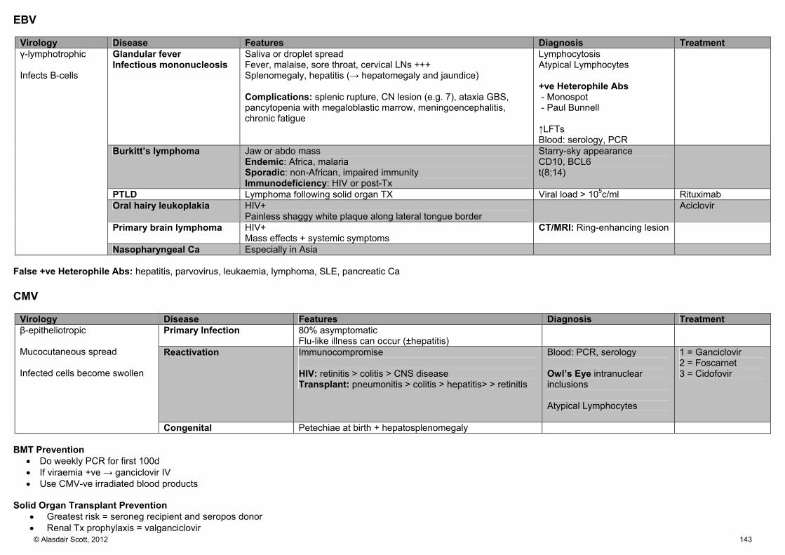
EBV
Virology Disease Features Diagnosis Treatment γ-lymphotrophic Infects B-cells
Glandular fever Infectious mononucleosis
Saliva or droplet spread Fever, malaise, sore throat, cervical LNs +++ Splenomegaly, hepatitis (→ hepatomegaly and jaundice) Complications: splenic rupture, CN lesion (e.g. 7), ataxia GBS, pancytopenia with megaloblastic marrow, meningoencephalitis, chronic fatigue
Lymphocytosis Atypical Lymphocytes +ve Heterophile Abs - Monospot - Paul Bunnell ↑LFTs Blood: serology, PCR
Burkitt’s lymphoma Jaw or abdo mass Endemic: Africa, malaria Sporadic: non-African, impaired immunity Immunodeficiency: HIV or post-Tx
Starry-sky appearance CD10, BCL6 t(8;14)
PTLD Lymphoma following solid organ TX Viral load > 105c/ml Rituximab Oral hairy leukoplakia HIV+
Painless shaggy white plaque along lateral tongue border Aciclovir
Primary brain lymphoma HIV+ Mass effects + systemic symptoms
CT/MRI: Ring-enhancing lesion
Nasopharyngeal Ca Especially in Asia False +ve Heterophile Abs: hepatitis, parvovirus, leukaemia, lymphoma, SLE, pancreatic Ca CMV
Virology Disease Features Diagnosis Treatment β-epitheliotropic Mucocutaneous spread Infected cells become swollen
Primary Infection 80% asymptomatic Flu-like illness can occur (±hepatitis)
Reactivation Immunocompromise HIV: retinitis > colitis > CNS disease Transplant: pneumonitis > colitis > hepatitis> > retinitis
Blood: PCR, serology Owl’s Eye intranuclear inclusions Atypical Lymphocytes
1 = Ganciclovir 2 = Foscarnet 3 = Cidofovir
Congenital Petechiae at birth + hepatosplenomegaly BMT Prevention
Do weekly PCR for first 100d If viraemia +ve → ganciclovir IV Use CMV-ve irradiated blood products
Solid Organ Transplant Prevention
Greatest risk = seroneg recipient and seropos donor Renal Tx prophylaxis = valganciclovir© Alasdair Scott, 2012 143

Viral Hepatitis Types Type Spread Cause A FO Seafood, especially abroad B IV Blood, body fluids, babies (vertical) C IV Mainly blood. Less vertical cf. HCV D IV Dependent on prior HBV infection E FO Developing world
Hepatitis A Presentation
Prodromal Phase Fever, malaise, arthralgia, nausea, anorexia Distaste for cigarettes
Icteric Phase Jaundice, HSM, lymphadenopathy, cholestasis
Ix
↑↑ALT, ↑AST (AST:ALT <2) IgM+ ~25d after exposure
= recent infection IgG+ for life
Rx
Supportive Avoid alcohol IFN-α for fulminant hepatitis (rare)
Prevention
Passive Ig → <3mo protection (e.g. travellers) Can give active vaccine
Hep B Incubation: 1-6mo Presentation
Prodromal phase and icteric phase as for Hep A Extra-hepatic features due to immune complexes
Urticaria or vasculitic rash Cryoglobulinaemia PAN GN Arthritis
Ix
HBsAg +ve = current infection +ve >6mo = chronic disease
HBeAg +ve = high infectivity Anti-HBc IgM = recent infection Anti-HBc IgG = past infection Anti-HBs = cleared infection or vaccinated HBV PCR: monitoring response to Rx
Rx
Supportive Avoid EtOH Chronic disease: PEGinterferon α2b
Complications
Fulminant hepatic failure (rare) Chronic hepatitis (5-10%) → cirrhosis in 5% HCC
Hep C
Mainly spread by blood. Thousands of UK cases due to transfusion (eligible for
compensation from Skipton fund) Presentation
Initial infection is usually asymptomatic 25% have acute icteric phase 85% → chronic infection 20-30% → cirrhosis w/i 20yrs ↑↑ risk of HCC
Ix
Anti-HCV Abs HCV-PCR Liver biopsy if PCR+ve to assess liver damage and
need for Rx HCV genotype
Rx
Indications Chronic haepatitis ↑ ALT Fibrosis
PEGinterferon α2b + ribavirin ↓ efficacy if
Genotype 1, 4, 5 or 6 ↑ VL Older Black Male
Hep D
Incomplete RNA virus that can only exist c̄ HBV ↑ risk of acute hepatic failure and cirrhosis Ix: anti-HDV Ab
Hep E
Similar to HAV Common in Indochina
Differential Acute
Infection: CMV, EBV, leptospirosis Toxin: EtOH, paracetamol, isoniazid, halothane Vasc: Budd-Chiari Obs: eclampsia, acute fatty liver of pregnancy Other: Wilson’s, AIH
Cirrhosis
Common: Chronic EtOH NAFLD / NASH
Other: Genetic: Wilson’s, α1ATD, HH, CF AI: AH, PBC, PSC Drugs: Methotrexate, amiodarone, isoniazid Neoplasm: HCC, mets Vasc: Budd-Chiari, RHF, constrict. pericarditis
© Alasdair Scott, 2012 144

Gastrointestinal Infections 1
Organism Aetiology Inc Clinical Features Dx Mx Staph aureus Meat
1-6h Diarrhoea, vomiting, abdo pain, ↓BP
Rapid resolution
Bacilus cereus Reheated rice 1-6h Watery diarrhoea and vomiting E. coli ETEC Travelling Watery diarrhoea Cipro E. coli EHEC (O157:H7)
Undercooked minced beef - e.g. burgers
12-72h Dysentery HUS
Stool MC+S
Dialysis if necessary
Campylobacter jejuni
Unpasteurised milk Animal faeces (cats, dogs)
2-5d Commonest bacterial diarrhoea Bloody diarrhoea, fever Guillain-Barre Reactive arthritis
Stool MC+S
Cipro - 250-750mg BD PO - 200-400mg BD IVI over 30min – 1h
Shigella 1-7d Bloody diarrhoea, abdo pain, fever Reactive arthritis
Stool MC+S
Cipro
Salmonella enteritidis
Poultry, eggs, meat 12-48h Diarrhoea, vomiting, fever, abdo pain Cipro or ceftriaxone
Listeria monocytogenes
Refrigerated food - cold enhancement Pates, soft cheeses
Watery diarrhoea, cramps, flu-like Pneumonia Meningoencephalitis Miscarriage
Blood culture
Ampicillin
Clostridium difficle
Antibiotic use - cephs, cipro, clinda FO spread Environment: spores
1-7d Bloody diarrhoea, abdo pain, fever - foul smelling Pseudomembranous colitis Toxic megacolon GI perforation
Stool MC+S CDT ELISA
Stop causative Abx 1st: Metronidazole 400mg TDS PO for 10d 2nd: Vanc 125mg QDS PO Colectomy may be needed
Clostridium botulinum
Canned / vac-packed food Kids=honey Students=beans
12-36h Afebrile Descending symmetric flaccid paralysis No sensory signs Autonomic: dry mouth, fixed dilated pupils
Toxin in blood samples Antitoxin Benpen + metro
Clostridium perfringes
Reheated meat
8-24h Watery diarrhoea + cramps Stool MC+S Benpen + metro
Vibrio cholera FO spread Dirty water
Hrs-5d Rice-water stools Shock, acidosis, renal failure
Stool MC+S
Rehydrate - Cooked rice powder solution - Hartmann’s c̄ K+ supplements Cipro Zn supplement
Vibrio parahaemolyticus
Raw/undercooked seafood - Japan
Profuse diarrhoea, abdo pain, vomiting Doxy 100-200mg/d PO
Norovirus 12-48h Commonest cause in adults 50% of all gastroenteritis worldwide Fever, diarrhoea, projectile vomiting
Rotavirus 1-7d Commonest cause in children Secretory diarrhoea and vomiting
© Alasdair Scott, 2012 145

Gastrointestinal Infections 2
Organism Aetiology Inc Features Dx Mx Salmonella typhi/paratyphi
FO spread Tropics
3-21d Malaise, headache, cough, constipation High fever c̄ relative bradycardia Rose spots: patchy red macules Epistaxis, splenomegaly Diarrhoea after 1st wk
Leukopenia Blood culture Urine or stool culture
Cefotaxime or cipro
Yersinia enterocolitica
Food contaminated by domestic animal faeces
24-36h Abdo pain, fever, diarrhoea Mesenteric adenitis Reactive arthritis, pharyngitis, pericarditis Erythema Nodosum
Serology Cipro
Entamaeoba histolytica
MSM Travelling
1-4wk Dysentery, wind, tenesmus Wt. loss if chronic Liver abscess - RUQ pain, swinging fever, sweats - Mass in R lobe Flask-shaped ulcer on histo
Stool micro - Motile trophozoite c̄ 4 nuclei Stool Ag
Metro Tinidazole if severe or abscess
Giardia lamblia MSM Hikers Travellers
1-4wk Bloating, explosive diarrhoea, offensive gas Malabsorption → steatorrhoea and wt. loss
Direct fluorescent Ab assay Stool micro - Pear-shaped trophozoites c̄ 2 nuceli Duodenal fluid analysis on swallowed string
Tinidazole
Crypto parvum Waterborne 4-12d Severe acute watery diarrhoea in HIV Stool micro - protozoan
Paromomycin
Strongyloides stercoralis
Endemic in sub-tropics Hyperinfestation in AIDS
Migrating urticarial rash on trunk and legs Pneumonitis, enteritis Malabsorption → chronic diarrhoea
Stool MC+S Serology
Ivermectin
Secretory Diarrhoea
Bacteria only found in lumen: don’t activate innate immunity No / low fever No faecal leukocytes Caused by bacterial toxins: Cholera, E. coli (except EIEC), S. aureus Toxin → ↑cAMP→ open CFTR channel → Cl loss followed by HCO3, Na and H2O loss → secretory diarrhoea
Inflammatory Diarrhoea
Bacteria invade lamina propria: activate innate immunity Fever PMN in stool Campylobacter, shigella, non-typhoidal salmonella, EIEC
Enteric Fever
Abdo pain, fever, mononuclear cells in stool Typhoidal salmonella, Yersinia enterocolitica, Brucella
© Alasdair Scott, 2012 146
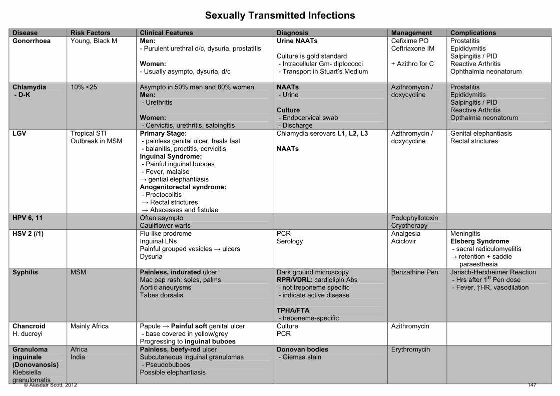
Sexually Transmitted Infections Disease Risk Factors Clinical Features Diagnosis Management Complications Gonorrhoea Young, Black M Men:
- Purulent urethral d/c, dysuria, prostatitis Women: - Usually asympto, dysuria, d/c
Urine NAATs Culture is gold standard - Intracellular Gm- diplococci - Transport in Stuart’s Medium
Cefixime PO Ceftriaxone IM + Azithro for C
Prostatitis Epididymitis Salpingitis / PID Reactive Arthritis Ophthalmia neonatorum
Chlamydia - D-K
10% <25 Asympto in 50% men and 80% women Men: - Urethritis Women: - Cervicitis, urethritis, salpingitis
NAATs - Urine Culture - Endocervical swab - Discharge
Azithromycin / doxycycline
Prostatitis Epididymitis Salpingitis / PID Reactive Arthritis Opthalmia neonatorum
LGV Tropical STI Outbreak in MSM
Primary Stage: - painless genital ulcer, heals fast - balanitis, proctitis, cervicitis Inguinal Syndrome: - Painful inguinal buboes - Fever, malaise → gential elephantiasis Anogenitorectal syndrome: - Proctocolitis → Rectal strictures → Abscesses and fistulae
Chlamydia serovars L1, L2, L3 NAATs
Azithromycin / doxycycline
Genital elephantiasis Rectal strictures
HPV 6, 11 Often asympto Cauliflower warts
Podophyllotoxin Cryotherapy
HSV 2 (/1) Flu-like prodrome Inguinal LNs Painful grouped vesicles → ulcers Dysuria
PCR Serology
Analgesia Aciclovir
Meningitis Elsberg Syndrome - sacral radiculomyelitis → retention + saddle paraesthesia
Syphilis MSM Painless, indurated ulcer Mac pap rash: soles, palms Aortic aneurysms Tabes dorsalis
Dark ground microscopy RPR/VDRL: cardiolipin Abs - not treponeme specific - indicate active disease TPHA/FTA - treponeme-specific
Benzathine Pen Jarisch-Herxheimer Reaction - Hrs after 1st Pen dose - Fever, ↑HR, vasodilation
Chancroid H. ducreyi
Mainly Africa Papule → Painful soft genital ulcer - base covered in yellow/grey Progressing to inguinal buboes
Culture PCR
Azithromycin
Granuloma inguinale (Donovanosis) Klebsiella granulomatis
Africa India
Painless, beefy-red ulcer Subcutaneous inguinal granulomas - Pseudobuboes Possible elephantiasis
Donovan bodies - Giemsa stain
Erythromycin
© Alasdair Scott, 2012 147

Syphilis Stages Stage Features: due to endarteritis obliterans Primary Mac→pap→indurated, painless ulcer = Chancre
Regional LNs Heals in 1-3wks
Secondary 6wks -6mo wks after chancre Systemic bacteraemia → fever, malaise Skin rash:
Symmetrical, non-itchy, mac pap / pustular Palms, soles, face, trunk
Buccal snail-track ulcers Warty lesions: condylomata lata
Tertiary 2-20yrs latency Gummas
Granulomas in skin, mucosa, bones, joints
Quaternary Syphilitic aortitis Aortic aneurysm Aortic regurg
Neurosyphilis Paralytic dementia Meningovascular: CN palsies, stroke Tabes dorsalis
Degeneration of sensory neurones, esp. legs
Ataxia and +ve Romberg’s Areflexia Plantars ↑↑ Charcot’s joints
Argyll-Robertson pupil accommodates, doesn’t react
Dx Cardiolipin antibody
E.g. VDRL, RPR Not treponeme specific
False +ve: pregnancy, pneumonia, SLE, malaria, TB
+ve in 1O and 2O syphilis (wanes in late disease) Reflects disease activity: -ve after Rx
Treponeme-specific Ab
+ve in 1O and 2O syphilis Remains +ve despite Rx THPA and FTA
Treponemes
Seen by dark ground microscopy of chancre fluid Seen in lesions of 2O syphilis May not be seen in late syphilis
Rx
2-3 doses (1wk apart of benzathine penicillin Or, doxycycline for 28d
Jarisch-Herxheimer Reaction
Fever, ↑HR, vasodilatation hrs after first Rx ? sudden release of endotoxin Rx: steroids
© Alasdair Scott, 2012 148

Zoonoses Disease Risk Factors Presentation Diagnosis Management Leptospirosis Weil’s Disease
Infected rat urine - swimming - canoeing
High fever, headache, myalgia / myositis Cough, chest pain ± haemoptysis ± hepatitis c̄ jaundice ± meningitis
Blood culture Serology
Doxycycline
Brucellosis Melitensis - goats, commonest Abortus: cattle Suis: pigs
Middle and Far East, Malta Unpasteurised milk / cheeses - esp. goat Vets, farmers, abattoir workers
Undulant fever (PUO): peak PM, normal AM Sweats, malaise, anorexia Arthritis, spinal tenderness LN, HSM Rash, jaundice
Pancytopenia Positive Rose Bengal Test Anti-O-polysaccharide Ag
Doxy + RMP + gent
Lyme disease Borrelia burgdorferi
Ixodes tick bite Walkers, hikers
Early localised: - Erythema migrans (target lesions) Early disseminated: - Malaise, LN, migratory arthritis, hepatitis Late persistent : - Arthritis, focal neuro (Bell’s palsy), heart block, myocarditis - Lymphocytoma: blue/red ear lobe
Biopsy ECM edge + Ab ELISA
Rash: Doxy Complications: IV benpen
Cat-Scratch Disease Bartonella henselae
Hx of cat scratch Tender regional LNs +ve cat scratch skin Ag test Azithro
Toxoplasmosis Cats are definitive hosts Sheep / pig intermediate hosts
Mostly asympto Reactivated in immunodeficiency - Encephalitis: confusion, seizures, focal - SOL/↑ICP - Posterior uveitis
CT/MRI: ring-shaped contrast enhancing CNS lesions Serology
Pyrimethamine + sulfadiazine Septrin prohylaxis in HIV
Rabies Animal bites - Bats, dogs, foxes
Prodrome - Headache, malaise, bite itch, odd behaviour Furious Rabies - Hydrophobia - Muscle spasms Dumb Rabies - flaccid limb paralysis
“Bullet-shaped” RNA virus Negri bodies
Immunised - diploid vaccine Unimmunised - vaccine + rabies Ig
Anthrax Spread by infected carcasses or hides - abattoir workers, farmers
Cutaneous - ulcer with BLACK centre - rim of oedema Inhalational - massive lymphadenopathy - mediastinal haemorrhage - resp failure GI - severe bloody diarrhoea
Microscopy - Gm+ spore forming rod
Cipro + clindamycin
© Alasdair Scott, 2012 149

Malaria Species Species Inc Liver stage? Rhythm Rx Falciparum 7-10d No Tertian: 48h Artemether-lumefantrine (Riamet)
(Quinine + Doxy) Vivax 10-17d Chronic liver stage Tertian: 48h Chloroquine then primaquine Ovale 10-17d Chronic liver stage Tertian: 48h Chloroquine then primaquine Malariae 18-40d No Quartan: 72h Chloroquine then primaquine
Biology
Plasmodium sporozoites injected by females Anopheles mosquito.
Sporozoites migrate to liver, infect hepatocytes and multiply asymptomatically (incubation period) → merozoites
Merozoites released from liver and infect RBCs Multiply in RBCs
Haemolysis RBC sequestration → splenomegaly Cytokine release
Falciparum Malaria
90% present w/i 1mo Flu-like Prodrome
Headache, malaise, myalgia, anorexia Fever Paroxysms
Shivering ≤1h Hot stage for 2-6h: ~41OC
Flushed, dry skin, headache, n/v Sweating for 2-4h as temp falls
Signs
Anaemia Jaundice HSM No rash, no LNs
Complications
Cerebral malaria: confusion, coma, fits Lactic acidosis → Kussmaul respiration Hypoglycaemia Acute renal failure: 2O to ATN ARDS
Dx
Serial thick and thin blood films Parasitaemia level FBC: anaemia, thrombocytopenia Clotting: DIC Glucose ABG: lactic acidosis U+E: renal failure Urinalysis: haemoglobionuria
Mx Uncomplicated Ovale, Vivax, Malariae
Chloroquine base Then primaquine
Uncomplicated Falciparum
Artemether-lumefantrine Severe Falciparum Malaria
Need ITU Mx IV antimalarials
Prophylaxis
No resistance: proguanil + chloroquine Resistance: mefloquine or malarone
Malarone: atovaquone + proguanil Antimalarial SEs
Chloroquine: retinopathy Fansidar: SJS, ↑LFTs, blood dyscrasias Primaquine: haemolysis if G6PD deficient Malarone: abdo pain, nausea, headache Mefloquine: dysphoria, neuropsychiatric signs
© Alasdair Scott, 2012 150

Other Tropical Diseases
Disease Aetiolgy Presentation Diagnosis African Trypanosomiasis “Sleeping sickness”
Tsetse fly
Tender subcut nodule @ site of infection Haemolymphatic Stage - rash, fever, rigors, headaches - LNs and HSM - Posterior cervical nodes (Winterbottom’s sign) Miningoencephalitic Stage - Wks – Mos after original infection - Convulsions, agitation confusion - Apathy, depression, hypersomnolence, coma T. gambiense: West Africa T. rhodesiense: East Africa - More severe than gambiense
Thick and thin films - flagellated protozoa Serology
American Trypanosomiasis “Chagas Disease”
Reduviids T. cruzi - Latin America - Erythematous nodule, fever, LN, HSM - Swelling of eyelid (Romana’s Sign) - Cardiac disease
Leishmaniasis Sandflies South + Central Am Asia Africa
Cutaneous (L. major, L. tropica) - Skin ulcer @ bite → depigmented scar Diffuse cutaneous - Widespread nodules (fail to ulcerate) Mucocutaneous (L. braziliensis) - South America - Ulcer in mucous mems of mouth and nose Visceral = Kala Azar (L. donovani) - Dry, warty hyperpigmented skin lesions (dark face and hands) - Prolonged fever - Massive splenomegaly, LNs, abdo pain
Visceral: - Leucopaenia - ↑Ig - Pancytopaenia - Serology Visualisation of Leishman-Donovan bodies
Filariasis Asia Africa South America
Lymphatic Filariasis - Wuchereria bancrofti - elephantiasis
Nemotodes Microfilariae in the blood Eosinophilia
Dengue Fever RNA virus
Aedes mosquito South + Central Am Asia Africa
Flushes: facem neck, chest Central macpap rash Headache, arthralgia HSM Jaundice Haemorrhage: petechial, GI, gums or nose, GU NB. can exclude if fever starts >2wks after leaving endemic area.
Serology Tourniquet test
Schistosomiasis Tremotode (Fluke) Snail vector
Itchy papular rash at site of penetration - Swimmer’s Itch Mansoni - abdo pain D&V - later hepatic fibrosis and portal HTN - HSM Haematobium - frequency, dysuria, haematuria - may → hydronephrosis and renal failure - ↑ risk of bladder SCC
© Alasdair Scott, 2012 151
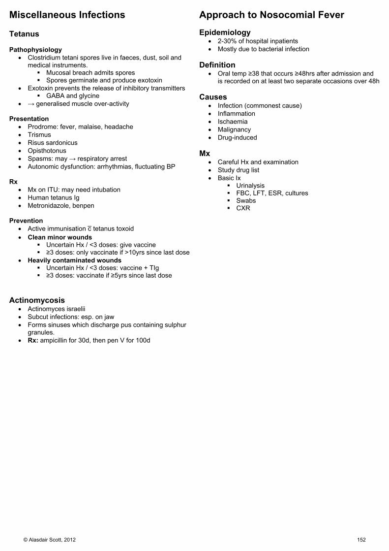
Miscellaneous Infections Tetanus Pathophysiology
Clostridium tetani spores live in faeces, dust, soil and medical instruments. Mucosal breach admits spores Spores germinate and produce exotoxin
Exotoxin prevents the release of inhibitory transmitters GABA and glycine
→ generalised muscle over-activity Presentation
Prodrome: fever, malaise, headache Trismus Risus sardonicus Opisthotonus Spasms: may → respiratory arrest Autonomic dysfunction: arrhythmias, fluctuating BP
Rx
Mx on ITU: may need intubation Human tetanus Ig Metronidazole, benpen
Prevention
Active immunisation c̄ tetanus toxoid Clean minor wounds
Uncertain Hx / <3 doses: give vaccine ≥3 doses: only vaccinate if >10yrs since last dose
Heavily contaminated wounds Uncertain Hx / <3 doses: vaccine + TIg ≥3 doses: vaccinate if ≥5yrs since last dose
Actinomycosis
Actinomyces israelii Subcut infections: esp. on jaw Forms sinuses which discharge pus containing sulphur
granules. Rx: ampicillin for 30d, then pen V for 100d
Approach to Nosocomial Fever Epidemiology
2-30% of hospital inpatients Mostly due to bacterial infection
Definition
Oral temp ≥38 that occurs ≥48hrs after admission and is recorded on at least two separate occasions over 48h
Causes
Infection (commonest cause) Inflammation Ischaemia Malignancy Drug-induced
Mx
Careful Hx and examination Study drug list Basic Ix
Urinalysis FBC, LFT, ESR, cultures Swabs CXR
© Alasdair Scott, 2012 152
Neurology Contents
Basic Neuroscience ................................................................................................................................................................... 155
Motor and Sensory Patterns ...................................................................................................................................................... 158
Specific Neurological Patterns and Causes .............................................................................................................................. 159
Muscle Weakness Differential ................................................................................................................................................... 160
Hand Wasting Differential .......................................................................................................................................................... 160
Gait Disturbance Differential ..................................................................................................................................................... 160
Blackouts ................................................................................................................................................................................... 161
Vertigo and Dizziness ................................................................................................................................................................ 162
Causes of Hearing Loss ............................................................................................................................................................ 162
Abnormal Involuntary Movements – Dyskinesias ..................................................................................................................... 163
Dementia ................................................................................................................................................................................... 164
Delirium / ACS ........................................................................................................................................................................... 164
Headache: Differential and Investigation .................................................................................................................................. 165
Headaches: Key Features ......................................................................................................................................................... 166
Migraine ..................................................................................................................................................................................... 167
Subarachnoid Haemorrhage ..................................................................................................................................................... 168
Stroke: Causes and Investigation .............................................................................................................................................. 169
Stroke: Presentation .................................................................................................................................................................. 170
Stroke: Management ................................................................................................................................................................. 171
Transient Ischaemic Attack ....................................................................................................................................................... 172
Subdural Haemorrhage ............................................................................................................................................................. 173
Extradural Haemorrhage ........................................................................................................................................................... 173
Intracranial Venous Thrombosis ................................................................................................................................................ 174
Meningitis .................................................................................................................................................................................. 175
Encephalitis ............................................................................................................................................................................... 176
Cerebral Abscess ...................................................................................................................................................................... 176
Epilepsy: Features ..................................................................................................................................................................... 177
Epilepsy: Investigation and Management ................................................................................................................................. 178
Status Epilepticus ...................................................................................................................................................................... 179
Head Injury ................................................................................................................................................................................ 180
Space Occupying Lesions ......................................................................................................................................................... 181
Idiopathic Intracranial Hypertension .......................................................................................................................................... 181
Raised ICP ................................................................................................................................................................................ 182
Signs and Symptoms ................................................................................................................................................................ 182
Parkinsonism ............................................................................................................................................................................. 183
Parkinson’s Disease .................................................................................................................................................................. 184
Multiple Sclerosis ....................................................................................................................................................................... 185
Cord Compression ..................................................................................................................................................................... 186
Cervical and Lumbar Degeneration ........................................................................................................................................... 187
Facial Nerve Palsy..................................................................................................................................................................... 188
Mononeuropathies ..................................................................................................................................................................... 189
Polyneuropathies ....................................................................................................................................................................... 190
Autonomic Neuropathy .............................................................................................................................................................. 190
Guillain-Barré Syndrome ........................................................................................................................................................... 191 © Alasdair Scott, 2012 153
Charcot-Marie-Tooth Syndrome ................................................................................................................................................ 191
Motor Neurone Disease ............................................................................................................................................................ 192
Polio ........................................................................................................................................................................................... 192
Myopathies ................................................................................................................................................................................ 193
Myasthenia Gravis ..................................................................................................................................................................... 194
Lambert-Eaton Myasthenic Syndrome (LEMS) ......................................................................................................................... 194
Botulism ..................................................................................................................................................................................... 194
Neurofibromatosis 1 – von Recklinghausen’s ........................................................................................................................... 195
Neurofibromatosis 2 .................................................................................................................................................................. 195
Intrinsic Cord Disease ............................................................................................................................................................... 196
Syringomyelia ............................................................................................................................................................................ 196
Brown-Sequard Syndrome ........................................................................................................................................................ 196
Miscellaneous Conditions .......................................................................................................................................................... 197
© Alasdair Scott, 2012 154

Basic Neuroscience The Spinal Cord Gross Anatomy
Foramen magnum to L1 Terminates as conus medullaris Spinal nerves continue inferiorly as corda equina Denticulate ligament and filum terminale are pial
extensions that suspend cord in subarachnoid space. Blood Supply
3 longitudinal vessels 2 posterior spinal arteries: dorsal 1/3 1 anterior spinal artery: ventral 2/3
Reinforced by segmental feeder arteries E.g. artery of Adamkiewicz
Longitudinal veins drain into extradural vertebral plexus Organisation
Dorsal Columns (FG + FC)
Mode: fine touch, vibration and proprioception Cell body: DRG Decussation: in medulla forming medial lemniscus
Lateral Spinothalamic Tract
Mode: pain and temperature Cell body: DRG Decussation: in cord, at entry level
Lateral Corticospinal Tract
Mode: motor (body) Cell body: 1O motor cortex Decussation: pyramidal decussation in ventral medulla
Thalamic Nucelei
VPL: somoatosensory body VPM: somatosensory head LGN: visual MGN: auditory
Cerebellum and Basal Ganglia
Output to cortex via thalamus Regulation of posture, locomotion, coordination and
movement. Internal Capsule
White matter carrying axonal fibres from motor cortex to pyramids of medulla.
Infarction → contralateral hemiparesis Brain Lobar Function Frontal
Cognition and memory Executive function Motor cortex Dominant hemisphere: motor speech (Broca’s area)
Parietal
Sensory cortex Body orientation
Temporal
Memory Dom hemisphere: receptive language (Wernicke’s)
Occipital
Visual cortex Homunculus
Brain Blood Supply
© Alasdair Scott, 2012 155
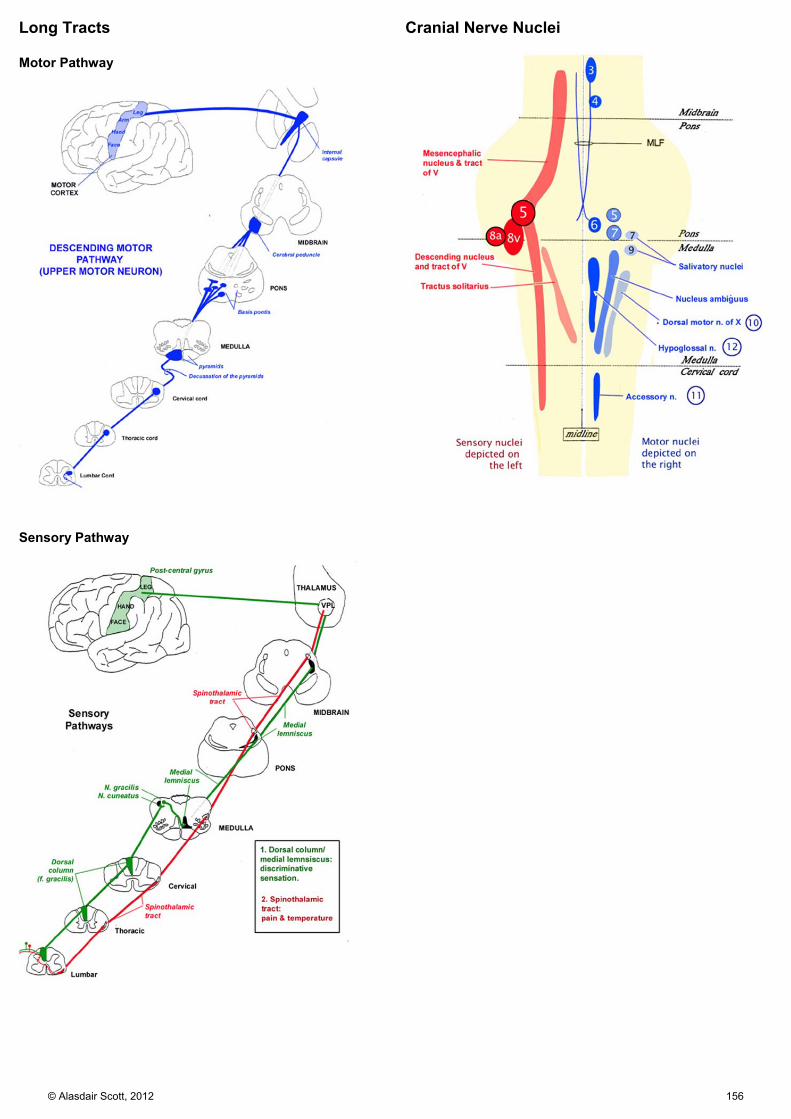
Long Tracts Motor Pathway
Sensory Pathway
Cranial Nerve Nuclei
© Alasdair Scott, 2012 156

NMJ Transmission
1. Presynaptic voltage-gated Ca2+ channels open → Ca2+ influx
2. ACh vesicles released from presynaptic terminal 3. ACh diffuses across cleft 4. ACh binds to nicotinic receptors on post-synaptic
terminal 5. → Na+ influx → depolarisation → Ca2+ release from SR
→ muscle contraction 6. ACh is degraded by acetylcholinesterase and choline is
taken up into presynaptic terminal. Blockade
1. Block presynaptic choline uptake: hemicholinium 2. Block ACh vesicle fusion: botulinum, LEMS 3. Block nicotinic ACh receptors
Non-depolarising: atracurium, vecuronium Depolarising: suxamethonium
Dopamine Pathways
Mesocorticolimbic: SCZ Nigrostriatal: Parkinsonism Tuberoinfundibular: Hyperprolactinaemia
Sympathetic NS
Cell bodies from T1-L2 GVE preganglionic fibres synapse @ either:
Paravertebral ganglia Prevertebral ganglia Chromaffin cells of adrenal medulla
Preganglionic fibres are myelinated and release ACh @ nicotinic receptors
Postganglionic fibres are unmyelinated and release NA @ adrenergic receptors Except @ sweat glands where they release ACh
for muscarinic receptors. Parasympathetic NS
Cranial: CN 3, 7, 9, 10 Ciliary: ciliary muscle and sphincter pupillae Pterygopalatine: mucus mems of nose and
palate, lacrimal gland Submandibular: submandibular and sublingual
glands Otic: parotid gland Vagus supplies thoracic and abdo viscera
Sacral: pelvic splanchnic nerves (S2-4) innervate pelvic viscera
Preganglionic fibres release ACh @ nicotinic receptors Postganglionic fibres release ACh @ muscarinic
receptors CN Nuclei
Midbrain: 3, 4, (5) Pons: 5, 6, 7, 8 Medulla: (5), 9, 10, 11, 12
NB. All nuclei except 4, innervate ipsilateral side. Fibres from trochlear nucleus decussate in medulla and supply contralateral SOB muscle.
Vestibulo-occular Pathways Vestibulo-occular reflex (VOR)
Axons from vestibular neurones project via MLF to abducens and occulomotor nuclei
Head turns L → eyes turn R Absent Doll’s Eye sign = brainstem death
Caloric Tests
Warm → ↑ firing of vestibular N. → eyes turn to contralateral side c̄ nystagmus to ipsilateral side
Remember fast direction of nystagmus – COWS Cold: Opposite Warm: Same
Absence of eye movements = brainstem damage on side being tested.
Visual Field Defects
Pupillary Light Reflex
© Alasdair Scott, 2012 157

Motor and Sensory Patterns Patterns of Motor Deficits Cortical Lesions
Hyperreflexia proximally in arm or leg Unexpected patterns: e.g. all movements in hand/foot
Internal Capsule and Corticospinal Lesions
Contralateral hemiparesis c̄ pyramidal distribution. Lesion c̄ epilepsy, ↓ cognition or homonymous hemianopia = in a cerebral hemisphere Lesion c̄ contralateral CN palsy = brainstem lesion on the side of the palsy (E.g. Miilard-Gubler syn.)
Cord Lesions
Quadriparesis / paraparesis Motor and reflex level: LMN signs at level of lesion and UMN signs below
Peripheral Neuropathies
Usually distal weakness In GBS weakness is proximal (root involvement) Single nerve = mononeuropathy: trauma or entrapment Several nerves = mononeuritis multiplex: vasculitis or DM
UMN Lesions
Motor cells in pre-central gyrus to anterior horn cells in the cord
Pyramidal weakness: extensors in UL, flexors in LL No wasting Spasticity: ↑ tone ± clonus Hyperreflexia Up-going plantars
LMN Lesions
Anterior horn cells to peripheral nerves Wasting Fasciculation Flaccidity: ↓ tone Hyporeflexia Down-going plantars
Primary Muscle Lesions
Symmetrical loss Reflexes lost later vs. neuropathies No sensory loss Fatiguability in myasthenia
Patterns of Sensory Deficits
Pain and temp travel in small fibres in peripheral nerves and in anterolateral spinothalamic tracts
Touch, joint position and vibrations travel in large fibres peripherally and in dorsal columns centrally
Distal Sensory Loss
Suggests a neuropathy Sensory Level
Hallmark of cord lesion Hemi-cord lesion → Brown-Sequard syndrome
Ipsilateral loss of proprioception / vibration and UMN weakness with contralateral loss of pain
Dissociated Sensory Loss
Selective loss of pain and temp c̄ conservation of proprioception and vibration
Occurs in cervical cord lesions: e.g. syringomyelia
© Alasdair Scott, 2012 158

Specific Neurological Patterns and Causes Cerebellar Syndrome Effects: DANISH
Dysdiadochokinesia Dysmetria: past-pointing Ataxia: limb / trunkal Nystagmus: horizontal = ipsilateral hemisphere Intention tremor Speech: slurred, staccato, scanning dysarthria Hypotonia
Common Causes: PASTRIES
Paraneoplastic: e.g. from bronchial Ca Alcohol: thiamine and B12 deficiency Sclerosis Tumor: e.g. CPA lesion Rare: MSA, Friedrich’s, Ataxia Telangiectasia Iatrogenic: phenytoin Endo: hypothyroidism Stroke: vertebrobasilar
Anterior Cerebral Artery
Supplies frontal and medial part of cerebrum Contralateral motor / sensory loss in the legs > arms Face is spared Abulia (pathological laziness)
Middle Cerebral Artery
Supplies lateral / external part of hemisphere Contralateral motor / sensory loss in face and arms >
legs. Contralateral homonymous hemianopia due to
involvement of optic radiation Cognitive changes
Dominant (L): aphasia Non-dominant (R): neglect, apraxia
Posterior Cerebral Artery
Supplies occipital lobe Contralateral homonymous hemianopia c̄ macula
sparing.
Vertebrobasilar Circulation Supplies cerebellum, brainstem and occipital lobes Combination of symptoms
Visual: hemianopia, cortical blindness Cerebellar: DANISH CN lesions Hemi- / quadriplegia Uni- / bi-lateral sensory symptoms
Lateral Medullary Syn. / Wallenberg’s Syn.
Occlusion of one vertebral A. or PICA Features: DANVAH
Dysphagia Ataxia (ipsilateral) Nystagmus (ipsilateral) Vertigo Anaesthesia
Ipsilat facial numbness + absent corneal reflex
Contralateral pain loss Horner’s syndrome (ipsilateral)
Millard-Gubler Syndrome: crossed hemiplegia
Pontine lesions (e.g. infarct) Effects: 6th and 7th CN palsy + contralateral hemiplegia
Locked-in Syndrome
Pt. is aware and cognitively intact but completely paralysed except for the eye muscles.
Causes Ventral pons infarction: basilar artery Central potine myelinolysis: rapid correction of
hyponatraemia Cerebellopontine Angle Syndrome
Causes: acoustic neuroma, meningioma, cerebellar astrocytoma, metastasis (e.g. breast)
Effects: ipsilat CN 5, 6, 7, 8 palsies + cerebellar signs Absent corneal reflex LMN facial palsy LR palsy Sensorineural deafness, vertigo, tinnitus DANISH
Subclavian Steal Syndrome
Subclavian A. stenosis proximal to origin of vertebral A. may → blood being stolen from this vertebral artery by retrograde flow.
Syncope / presyncope or focal neurology on using the arm.
BP difference of >20mmHg between arms Anterior Spinal Artery / Beck’s Syndrome
Infarction of spinal cord in distribution of anterior spinal artery: ventral 2/3 or cord.
Causes: Aortic aneurysm dissection or repair Effects
Para- / quadri-paresis Impaired pain and temperature sensation Preserved touch and proprioception
© Alasdair Scott, 2012 159

Muscle Weakness Differential 1. Cerebrum / Brainstem
Vascular: infarct, haemorrhage Inflammation: MS SOL Infection: encephalitis, abscess
2. Cord
Vascular: anterior spinal artery infarction Inflammation: MS Injury
3. Anterior Horn
MND, polio 4. Roots / Plexus
Spondylosis Cauda equina syndrome Carcinoma
5. Motor Nerves
Mononeuropathy: e.g. compression Polyneuropathy: e.g. GBS, CMT
6. NMJ
GB, LEMS, botulism 7. Muscle
Toxins: steroids Poly- / Dermato-myositis Inherited: DMD, BMD, FSH
Hand Wasting Differential 1. Cord
Anterior Horn: MND, polio Syringomyelia
2. Roots (C8 T1)
Compression: spondylosis, neurofibroma 3. Brachial Plexus
Compression Cervical rib Tumour: Pancoast’s, breast
Avulsion: Klumpke’s palsy 4. Neuropathy
Generalised: CMT Mononeuritis multiplex: DM Compressive mononeuropathy
Median: thenar wasting Ulnar: hypothenar and interossei wasting
5. Muscle
Disuse: RA Compartment syndrome: Volkman’s ischaemic
contracture Distal myopathy: myotonic dystophy Cachexia
Gait Disturbance Differential Motor Basal Ganglia: festinating / shuffling
PD Parkinsonism: MSA, PSP, Lewy body dementia, CBD
UMN Bilateral: spastic, scissoring
Cord: compression, trauma, hereditary spastic paraparesis, syringomyelia, transverse myelitis
Bihemispheric: CP, MS UMN Unilateral: spastic circumducting
Hemisphere lesion: CVS, MS, SOL Hemicord: MS, tumour
LMN Bilateral: bilat foot drop
Polyneuropathy: CMT, GBS Corda equina
LMN Unilateral: foot drop → high stepping gait
Ant horn: polio Radicular: L5 root lesion Sciatic / common peroneal nerve: trauma, DM
Mixed UMN and LMN: MAST
MND Ataxia: Friedrich’s SACD Taboparesis
Sensory Vestibular (Romberg’s +ve)
Meniere’s Viral labyrinthitis Brainstem lesion
Cerebellar: ataxic
EtOH Infarct
Proprioceptive Loss (Romberg’s +ve)
Dorsal columns: B12 deficiency Peripheral neuropathy: DM, EtOH, uraemia
Visual Loss Other
Myopathy, MG/LEMS Medical: postural hypotension, Stokes-Adams, arthritis
© Alasdair Scott, 2012 160

Blackouts Causes: CRASH Cardiac: Stokes-Adams Attacks
Brady: heart block, sick sinus, long-QT Tachy: SVT, VT Structural
Weak heart: LVF, tamponade Block: AS, HOCM, PE
Reflexes
1. Vagal overactivity Vasovagal syncope Situational: cough, effort, micturition Carotid sinus syncope
2. Sympathetic underactivity = Post. Hypotension
Salt deficiency: hypovolaemia, Addison’s Toxins
Cardiac: ACEi, diuretics, nitrates, α-B Neuro: TCAs, benzos, antipsychotics, L-
DOPA Autonomic Neuropathy: DM, Parkinson’s, GBS Dialysis Unwell: chronic bed-rest Pooling, venous: varicose veins, prolonged
standing Arterial
Vertebrobasilar insufficiency: migraine, TIA, CVA, subclavian steal
Shock Hypertension: phaeochromocytoma
Systemic
Metabolic: ↓glucose Resp: hypoxia, hypercapnoea (e.g. anxiety) Blood: anaemia, hyperviscosity
Head
Epilepsy Drop attacks
Examination
Postural hypotension: difference of >20/10 after standing for 3min vs. lying down
Cardiovascular Neurological
Ix
ECG ± 24hr ECG U+E, FBC, Glucose Tilt table EEG, sleep EEG Echo, CT, MRI brain
Presentation and Investigation Cardiogenic Syncope
Trigger: exertion, drug, unknown Before: palpitations, chest pain, dyspnoea During: pale, slow/absent pulse, clonic jerks may occur After: rapid recovery Ix: ECG, 24hr ECG, Echo
Reflex: Vasovagal
Trigger: prolonged standing, heat, fatigue, stress Before
Gradual onset: secs→mins Nausea, pallor, sweating, tunnel vision, tinnitus Cannot occur lying down
During Pale, grey, clammy, brady Clonic jerks and incontinence can occur, but no
tongue biting After: rapid recovery Ix: Tilt-table testing
Reflex: Postural Hypotension
Trigger: Standing up Before, During and After as for vagal above Ix: Tilt-table testing
Arterial
Trigger: Arm elevation (subclavian steal), migraine Before, During and After as for vasovagal ± brainstem
Sx (diplopia, nausea, dysarthria) Ix: MRA, duplex vertebrobasilar circulation
Systemic
Hypoglycaemia: tremor, hunger, sweating, light-headedness → LOC
Head: Epileptic
Trigger: flashing lights, fatigue, fasting Before: e.g. aura in complex partial seizures – feeling
strange, epigastric rising, deja/jamias vu, smells, lights, automatisms
During: Tongue biting, incontinence, stiffness→jerking, eyes open, cyanosis, ↓SpO2
After: headache, confusion, sleeps, Todd’s palsy Ix: EEG, ↑ se prolactin at 10-20min
Head: Drop Attacks
Trigger: nil Before: no warning During: sudden weakness of legs causes older woman
to fall to the ground. After: no post-ictal phase
© Alasdair Scott, 2012 161
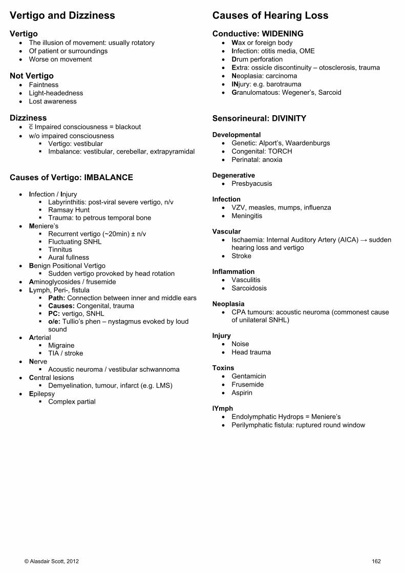
Vertigo and Dizziness Vertigo
The illusion of movement: usually rotatory Of patient or surroundings Worse on movement
Not Vertigo
Faintness Light-headedness Lost awareness
Dizziness
c̄ Impaired consciousness = blackout w/o impaired consciousness
Vertigo: vestibular Imbalance: vestibular, cerebellar, extrapyramidal
Causes of Vertigo: IMBALANCE
Infection / Injury Labyrinthitis: post-viral severe vertigo, n/v Ramsay Hunt Trauma: to petrous temporal bone
Meniere’s Recurrent vertigo (~20min) ± n/v Fluctuating SNHL Tinnitus Aural fullness
Benign Positional Vertigo Sudden vertigo provoked by head rotation
Aminoglycosides / frusemide Lymph, Peri-, fistula
Path: Connection between inner and middle ears Causes: Congenital, trauma PC: vertigo, SNHL o/e: Tullio’s phen – nystagmus evoked by loud
sound Arterial
Migraine TIA / stroke
Nerve Acoustic neuroma / vestibular schwannoma
Central lesions Demyelination, tumour, infarct (e.g. LMS)
Epilepsy Complex partial
Causes of Hearing Loss Conductive: WIDENING
Wax or foreign body Infection: otitis media, OME Drum perforation Extra: ossicle discontinuity – otosclerosis, trauma Neoplasia: carcinoma INjury: e.g. barotrauma Granulomatous: Wegener’s, Sarcoid
Sensorineural: DIVINITY Developmental
Genetic: Alport’s, Waardenburgs Congenital: TORCH Perinatal: anoxia
Degenerative
Presbyacusis Infection
VZV, measles, mumps, influenza Meningitis
Vascular
Ischaemia: Internal Auditory Artery (AICA) → sudden hearing loss and vertigo
Stroke Inflammation
Vasculitis Sarcoidosis
Neoplasia
CPA tumours: acoustic neuroma (commonest cause of unilateral SNHL)
Injury
Noise Head trauma
Toxins
Gentamicin Frusemide Aspirin
lYmph
Endolymphatic Hydrops = Meniere’s Perilymphatic fistula: ruptured round window
© Alasdair Scott, 2012 162

Abnormal Involuntary Movements – Dyskinesias Tremor
Regular, rhythmic oscillation NB. Asterixis = rhythmic myoclonus
Types and Causes: RAPID Resting
Features 4-6Hz, pill-rolling Abolished on voluntary movement ↑ c̄ distraction (e.g. counting backwards)
Causes: Parkinsonism Rx: Da agonists, antimuscarinic (e.g. procyclidine)
Action / Postural
Features 6-12Hz Absent at rest Worse c̄ outstretched hands or movement Equally bad at all stages of movement
Causes: BEATS Benign essential tremor Endocrine: thyroxicosis, ↓glucose, phaeo Alcohol withdrawal (or caffeine, opioids…) Toxins: β-agonists, theophylline, valproate, PHE Sympathetic: physiological tremor may be
enhanced: e.g. in anxiety Intention
Features >6Hz, irregular, large amplitude Worse at end of movement E.g. past-pointing
Causes: cerebellar damage Dystonic
Features: variable Causes: mostly idiopathic, as for dystonia
Benign Essential Tremor
Autosomal dominant Occur c̄ action and worse c̄ anxiety, emotion, caffeine Arms, neck, voice Doesn’t occur during sleep Better c̄ EtOH
Myoclonus
Sudden, involuntary jerks Causes
Metabolic = Asterixis (L, R, ↑CO2) Neurodegenerative diseases (LSDs) CJD Myoclonic epilepsies (e.g. infantile spasms)
Benign Essential Myoclonus
Auto dom Childhood onset frequent generalised myoclonus w/o
progression. May respond to valproate
Dystonia
Prolonged muscle contracture → unusual joint posture or repetitive movements
Idiopathic generalised dystonia
Often autosomal dominant Childhood onset: starts in one leg and spreads on that
side of the body over 5-10yrs Idiopathic focal dystonia
Commonest form of dystonia Confined to one part of the body Worsened by stress Types:
Spasmodic torticollis Blepharospasm Oromandibular Writer’s / Muscician’s cramp
Acute Dystonia
Torticollis, trismus and/or occulogyric crisis Typically a drug reaction:
Neuroleptics Metoclopramide L-DOPA
Rx: procyclidine (antimuscarinic) Chorea
Non-rhythmic, purposeless, jerky, flitting movements E.g. facial grimacing, flexing / extending the fingers Causes
Huntington’s Sydenham’s Wilson’s L-DOPA
Athetosis
Slow, sinuous, writhing movements Causes
Cerebral palsy Kernitcterus
Hemiballismus
Large amplitude, flinging hemichorea Contralateral to a vascular lesion in the subthalamic
nucleus: often elderly diabetics Recovers spontaneously over months
Tardive Syndromes
Delayed onset following chronic exposure to Da agonists (e.g. antipsychotics, antiemetics, L-DOPA)
Classification
Dyskinesia: orobuccolingual, truncal or choreifirm movements
Dystonia: sustained, stereotyped muscle spasms of twitching or turning
Akathisia: unpleasant sense of inner restlessness ± repetitive movements (e.g. pacing)
Rx
Change (e.g. to atypical) or slowly withdraw drug Dyskinesia: Da antagonist (tetrabenazine) Akathisia: β-B
© Alasdair Scott, 2012 163
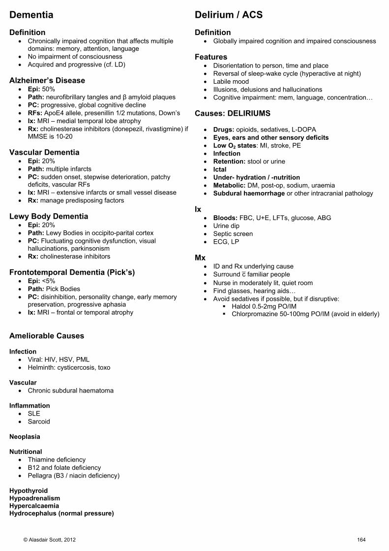
Dementia Definition
Chronically impaired cognition that affects multiple domains: memory, attention, language
No impairment of consciousness Acquired and progressive (cf. LD)
Alzheimer’s Disease
Epi: 50% Path: neurofibrillary tangles and β amyloid plaques PC: progressive, global cognitive decline RFs: ApoE4 allele, presenillin 1/2 mutations, Down’s Ix: MRI – medial temporal lobe atrophy Rx: cholinesterase inhibitors (donepezil, rivastigmine) if
MMSE is 10-20 Vascular Dementia
Epi: 20% Path: multiple infarcts PC: sudden onset, stepwise deterioration, patchy
deficits, vascular RFs Ix: MRI – extensive infarcts or small vessel disease Rx: manage predisposing factors
Lewy Body Dementia
Epi: 20% Path: Lewy Bodies in occipito-parital cortex PC: Fluctuating cognitive dysfunction, visual
hallucinations, parkinsonism Rx: cholinesterase inhibitors
Frontotemporal Dementia (Pick’s)
Epi: <5% Path: Pick Bodies PC: disinhibition, personality change, early memory
preservation, progressive aphasia Ix: MRI – frontal or temporal atrophy
Ameliorable Causes Infection
Viral: HIV, HSV, PML Helminth: cysticercosis, toxo
Vascular
Chronic subdural haematoma Inflammation
SLE Sarcoid
Neoplasia Nutritional
Thiamine deficiency B12 and folate deficiency Pellagra (B3 / niacin deficiency)
Hypothyroid Hypoadrenalism Hypercalcaemia Hydrocephalus (normal pressure)
Delirium / ACS Definition
Globally impaired cognition and impaired consciousness Features
Disorientation to person, time and place Reversal of sleep-wake cycle (hyperactive at night) Labile mood Illusions, delusions and hallucinations Cognitive impairment: mem, language, concentration…
Causes: DELIRIUMS
Drugs: opioids, sedatives, L-DOPA Eyes, ears and other sensory deficits Low O2 states: MI, stroke, PE Infection Retention: stool or urine Ictal Under- hydration / -nutrition Metabolic: DM, post-op, sodium, uraemia Subdural haemorrhage or other intracranial pathology
Ix
Bloods: FBC, U+E, LFTs, glucose, ABG Urine dip Septic screen ECG, LP
Mx
ID and Rx underlying cause Surround c̄ familiar people Nurse in moderately lit, quiet room Find glasses, hearing aids… Avoid sedatives if possible, but if disruptive:
Haldol 0.5-2mg PO/IM Chlorpromazine 50-100mg PO/IM (avoid in elderly)
© Alasdair Scott, 2012 164

Headache: Differential and Investigation Acute: VICIOUS Vascular
Haemorrhage: SAH, intracranial, intracerebral Infarction: esp. posterior circulation Venous: Sinus / cortical thrombosis
Infection/Inflammation
Meningitis Encephalitis Abscess
Compression
Obstructive hydrocephalus: tumour Pituitary enlargement: apoplexy
ICP
Spontaneous intracranial hypotension Acute dural CSF leak Worse on standing initially.
Ophthalmic
Acute glaucoma Unknown
Situational: cough, exertion, coitus Systemic
HTN: Phaeo, PET Infection: sinusitis, tonsillitis, atypical pneumonia Toxins: CO
Chronic: MCD TINGS Migraine Cluster headaches Drugs
Analgesics Caffeine Vasodilators: Ca2+
antagonists, nitrates Tension headaches ICP ↑/↓
↑: tumour, aneurysm, AVM, benign intracranial HTN ↓: spontaneous intracranial hypotension
Neuralgia (trigeminal) Giant cell arteritis Systemic
HTN Organ failure: e.g. uraemia
Investigations: Bloods Urine Micro
Blood cultures Serology: enterovirus (common cause of viral meningitis), HSV, HIV, syphilis, crypto CSF
Radiology
Non-contrast CT SAH: blood in sulci, cisterns (white). 90% sensitivity in first 24h
MRI MRA: aneurysm MRV: sinus thrombosis
Special: CSF
Opening pressure (norm = 5-20cm H2O): ↑: SAH, meningitis ↓: spontaneous intracranial hypotension
Xanthochromia: yellow appearance of CSF due to bilirubin. Detect by spectrophotometry.
Findings Bacterial TB Viral Appearance Turbid Fibrin web Clear Cells PMN Lympho / mononuc Lympho / mononuc Count 100-1000 10-1000 50-1000 Glucose ↓ (< ½ plasma) ↓ (< ½ plasma) > ½ plasma Protein (g/L) ↑↑ (>1.5) ↑↑↑ (1-5) mild ↑ (<1)
© Alasdair Scott, 2012 165

Headaches: Key Features SAH
Sudden onset, worst ever, occipital headache. Meningism, focal signs, ↓consciousness
Venous Sinus Thrombosis
Sagittal: headache, vomiting, seizures, ↓vision, papilloedema
Transverse: headache ± mastoid pain, focal CNS signs, seizures, papilloedema
Cortical Vein Thrombosis
Thunderclap headache Stroke-like focal symptoms over days Focal seizures are common
Meningitis
Fever, photophobia, neck stiffness, kernig’s +ve Purpuric rash ↓consciousness
Encephalitis
Fever, odd behaviour, fits, focal neuro, ↓consciousness Acute Glaucoma
Constant unilat eye pain, radiating to forehead ↓ acuity, haloes, n/v Red eye, cloudy cornea. Dilated, non-responsive pupil
Tension Headache
Bilateral / vertex-bitemporal, non-pulsatile, band-like Migraine
Prodrome → aura → headache Unilat, throbbing n/v, phono/photophobia
Cluster
Rapid onset very severe pain around/behind one eye. Red, watery eye, nasal congestion Miosis, ptosis Attacks last 15min–3hrs, 1-2x/day, mostly nocturnal Clusters last 4-12wks, remission lasts 3mo-3yrs. Can
be chronic vs. episodic. Rx: 100% O2 via non-rebreathe mask, Sumitriptan Prevention: verapamil, topiramate, Li
Hemicrania
Paroxysmal hemicranias: cluster-like headache lasting 5-45min, 5-30x/day
SUNCT: short-lasting unilateral neuralgia with conjunctival injection and tearing, attacks last 15-60s, recur 5-30x/hr
Hemicrania continua: continuous cluster-like headache Rx: all respond well to indomethacin
Trigeminal neuralgia
Paroxysms of unilateral intense stabbing pain in trigeminal distribution (usually V2/3)
Triggers: washing area, shaving, eating, talking Male > 50yrs 2O in 14%: compression of CNV, MS, Zoster, Chiari
malformation Ix: exclude 2O cause by MRI. Rx Med: CBZ, lamotrigine, gabapentin Rx Surg: microvascular decompression
Analgesia Overuse
Episodic headache becomes daily chronic headache Use OTC analgesia on 6 days/month max
ICP
↑: worse in AM, stooping, visual probs (papilloedema), obese women
↓: worse sitting or standing TMJ Dysfunction
Preauricular pain on chewing Assoc. c̄ crepitus Earache, headache
Giant Cell Arteritis (>60yrs, ESR>60, pred 60mg)
Unilateral temple/scalp pain and tenderness. Thickened, pulseless temporal artery Jaw claudication, amaurosis fugax, sudden blindness Assoc. c̄ PMR in 50% Ix: ESR↑↑↑, plats↑, ALP↑, Hb↓, temporal artery biopsy Rx
High dose pred (60mg/d PO) for 5-7d Guided by symptoms and ESR. Give PPI+bisphosphonate.
Prog: 2yr course then complete remission
© Alasdair Scott, 2012 166

Migraine
Epidemiology 8% prev F:M = 2:1
Risk Factors Obesity PFO
Pathophysiology Vascular: cerebrovascular constriction → aura,
dilatation → headache. Brain: spreading cortical depression Inflammation: activation of CN V nerve terminals in
meninges and cerebral vessels.
Triggers CHOCOLATE CHeese OCP Caffeine alcohOL Anxiety Travel Exercise
Symptoms Headache
Aura lasting 15-30min then unilat, throbbing headache
Phono/photophobia n/v Allodynia Often premenstrual
Prodrome (50%): precede migraine by hrs - days
Yawning Food cravings Changes in sleep, appetite or mood
Aura (20%): precedes migraine by mins and may persist
Visual: distortion, lines, dots, zig-zags, scotoma, hemianopia
Sensory: paraesthesia (fingers → face) Motor: dysarthria, ataxia, ophthalmoplegia,
hemiparesis (hemiplegic migraine) Speech: dysphasia, paraphasia
Classification Migraine c̄ aura (classical migraine) Migraine w/o aura (common migraine)
Diagnostic Criteria Typical aura + headache, or ≥ 5 headaches lasting 4-72h with either n/v or
photo/phonophobia + ≥2 of: Unilat Pulsating Interferes with normal life Worsened by routine activity
Differential
Cluster / tension headache Cervical spondylosis HTN Intracranial pathology Epilepsy
Treatment Acute episode
1st: Paracetamol + metoclopramide / domperidone 2nd: NSAID (e.g. ketoprofen) + M/D 3rd: Rizatriptan
CI: IHD, uncontrolled HTN, SSRIs 4th: ergotamine
Prophylaxis
Avoid triggers 1st: Propanolol, topiramate 2nd: Valproate, pizotifen (↑ wt.), gabapentin
© Alasdair Scott, 2012 167

Subarachnoid Haemorrhage Epidemiology
9/100,000 35-65yrs
Causes
Rupture of saccular aneurysms (80%) AVMs (15%)
Risk Factors
Smoking HTN EtOH Bleeding diathesis Mycotic aneurysms (SBE) FH (close relatives have 3-5x risk)
Berry Aneurysms Sites
Junction of post. communicating c̄ IC Junction of ant. communicating c̄ ACA Bifurcation of MCA
Associations
Adult polycystic kidneys CoA Ehlers Danlos
Symptoms
Sudden, severe occipital headache Collapse Meningism: neck stiffness, n/v, photophobia Seizures Drowsiness → coma
Signs
Kernig’s Retinal or subhyaloid haemorrhage Focal neuro
@ presentation suggests aneurysm location Later deficits suggests complications
Sentinel Headache
~6% of pts. experience sentinel headache from small warning bleed.
Differential
In 1O care, 25% of those c̄ thunderclap headache have SAH
50-60% no cause found Rest: meningitis, intracerebral bleeds, cortical vein
thrombosis
Investigations CT
Detects >90% of SAH w/i first 48hrs LP
If CT-ve and no CIs >12h after start of headache Xanthochromia due to breakdown of bilirubin
Mx
Frequent neuro obs: pupils, GCS, BP Maintain CPP: keep SBP >160 Nimodipine for 3wks → ↓ cerebral vasospasm Endovascular coiling (preferable to surgical clipping)
Complications Rebleeding: 20%
Commonest cause of mortality Cerebral Ischaemia
Due to vasospasm Commonest cause of morbidity
Hydrocephalus
Due to blockage of arachnoid granulations May require ventricular or lumbar drain
Hyponatraemia
Common Don’t Mx c̄ fluid restriction
Mx of Unruptured Aneurysms
Young pts. c̄ aneurysms >7mm in diameter may benefit from surgery.
© Alasdair Scott, 2012 168

Stroke: Causes and Investigation Definition
Rapid onset, focal neurological deficit due to a vascular lesion lasting >24h Pathogenesis
Infarction due ischaemia (80%) or intracerebral haemorrhage (20%).
Ischaemia (80%)
Atheroma Large (e.g. MCA) Small vessel perforators (lacunar)
Embolism Cardiac (30% of strokes):AF, endocarditis, MI Atherothromboembolism: e.g. from carotids
Haemorrhage (20%)
↑BP Trauma Aneurysm rupture Anticoagulation Thrombolysis
Rarer Causes
Watershed stroke: sudden ↓ in BP (e.g. in sepsis) Carotid artery dissection Vasculitis: PAN, HIV Cerebral vasospasm 2O to SAH Venous sinus thrombosis Anti-phospholipid syndrome, thrombophilia
Risk Factors
HYPERTENSION Smoking, DM, ↑ lipids, FH Cardiac: AF, valve disease Peripheral vascular disease Previous history Ethnicity: ↑ in Blacks and Asians ↑ PCV/Hct OCP
Cardiac Causes of Stroke
AF: 4.5% /yr External cardioversion: 1-3% Prosthetic valves Acute MI: esp. large anterior Paradoxical systemic emboli Cardiac surgery Valve vegetations
Ix ID risk factors for further strokes
HTN
Retinopathy Nephropathy Big heart on CXR (Don’t treat acutely)
Cardiac emboli
ECG ± 24hr tape: AF Echo: mural thrombus, hypokinesis, valve lesions, ASD,
VSD (paradoxical emboli) Carotid artery stenosis
Doppler US ± angio Endarterectomy beneficial if ≥ 70% symptomatic
stenosis Bleeding / thrombotic tendency
Thrombophilia screen Thrombocytopenia
Hyperviscosity
Polycythaemia SCD Myeloma
Metabolic
↑↓ glucose ↑ lipids Hyperhomocystinaemia
Vasculitis
↑ESR ANA
© Alasdair Scott, 2012 169
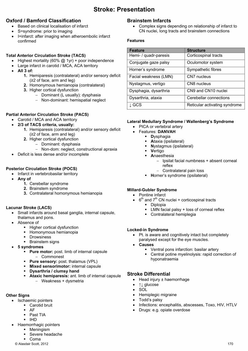
Stroke: Presentation Oxford / Bamford Classification
Based on clinical localisation of infarct S=syndrome: prior to imaging I=infarct: after imaging when atheroembolic infarct
confirmed Total Anterior Circulation Stroke (TACS)
Highest mortality (60% @ 1yr) + poor independence Large infarct in carotid / MCA, ACA territory All 3 of:
1. Hemiparesis (contralateral) and/or sensory deficit (≥2 of face, arm and leg)
2. Homonymous hemianopia (contralateral) 3. Higher cortical dysfunction
Dominant (L usually): dysphasia Non-dominant: hemispatial neglect
Partial Anterior Circulation Stroke (PACS)
Carotid / MCA and ACA territory 2/3 of TACS criteria, usually:
1. Hemiparesis (contralateral) and/or sensory deficit (≥2 of face, arm and leg)
2. Higher cortical dysfunction Dominant: dysphasia Non-dom: neglect, constructional apraxia
Deficit is less dense and/or incomplete Posterior Circulation Stroke (POCS)
Infarct in vertebrobasilar territory Any of
1. Cerebellar syndrome 2. Brainstem syndrome 3. Contralateral homonymous hemianopia
Lacunar Stroke (LACS)
Small infarcts around basal ganglia, internal capsule, thalamus and pons.
Absence of Higher cortical dysfunction Homonymous hemianopia Drowsiness Brainstem signs
5 syndromes Pure motor: post. limb of internal capsule
Commonest Pure sensory: post. thalamus (VPL) Mixed sensorimotor: internal capsule Dysarthria / clumsy hand Ataxic hemiparesis: ant. limb of internal capsule
Weakness + dysmetria Other Signs
Ischaemic pointers Carotid bruit AF Past TIA IHD
Haemorrhagic pointers Meningism Severe headache Coma
Brainstem Infarcts
Complex signs depending on relationship of infarct to CN nuclei, long tracts and brainstem connections
Features Feature Structure Hemi- / quadr-paresis Corticospinal tracts
Conjugate gaze palsy Oculomotor system
Horner’s syndrome Sympathetic fibres
Facial weakness (LMN) CN7 nucleus
Nystagmus, vertigo CN8 nucleus
Dysphagia, dysarthria CN9 and CN10 nuclei
Dysarthria, ataxia Cerebellar connections
↓ GCS Reticular activating syndrome
Lateral Medullary Syndrome / Wallenberg’s Syndrome
PICA or vertebral artery Features: DANVAH
Dysphagia Ataxia (ipsilateral) Nystagmus (ipsilateral) Vertigo Anaesthesia
Ipsilat facial numbness + absent corneal reflex
Contralateral pain loss Horner’s syndrome (ipsilateral)
Millard-Gubler Syndrome
Pontine infarct 6th and 7th CN nuclei + corticospinal tracts
Diplopia LMN facial palsy + loss of corneal reflex Contralateral hemiplegia
Locked-in Syndrome
Pt. is aware and cognitively intact but completely paralysed except for the eye muscles.
Causes Ventral pons infarction: basilar artery Central potine myelinolysis: rapid correction of
hyponatraemia Stroke Differential
Head injury ± haemorrhage ↑↓ glucose SOL Hemiplegic migraine Todd’s palsy Infections: encephalitis, abscesses, Toxo, HIV, HTLV Drugs: e.g. opiate overdose
© Alasdair Scott, 2012 170

Stroke: Management Acute Management Resuscitate
Ensure patent airway: consider NGT NBM until swallowing assessed by SALT Don’t overhydrate: risk of cerebral oedema
Monitor
Glucose: 4-11mM: sliding scale if DM BP: <185/110 (for thrombolysis)
Rx of HTN can → ↓ cerebral perfusion Neuro obs
Imaging
Urgent CT/MRI Diffusion-weighted MRI is most sensitive for acute infarct CT will exclude primary haemorrhage
Medical
Consider thrombolysis if 18-80yrs and <4.5hrs since onset of symptoms Alteplase (rh-tPA) → ↓ death and dependency (OR 0.64) CT 24h post-thrombolysis to look for haemorrhage
Aspirin 300mg PO/PR once haemorrhagic stroke excluded ± PPI Clopidogrel if aspirin sensitive
Surgery
Neurosurgical opinion if intracranial haemorrhage May coil bleeding aneurysms Decompressive hemicraniectomy for some forms of MCA
infarction. Stroke Unit
Specialist nursing and physio Early mobilisation DVT prophylaxis
Secondary Prevention Rehabilitation
Primary Prevention
Control RFs: HTN, ↑ lipids, DM, smoking, cardiac disease
Consider life-long anticoagulation in AF (use CHADS2) Carotid endarterectomy if symptomatic 70% stenosis Exercise
Secondary Prevention
Risk factor control as above Start a statin after 48h
Aspirin / clopi 300mg for 2wks after stroke then either Clopidogrel 75mg OD (preferred option) Aspirin 75mg OD + dipyridamole MR 200mg BD
Warfarin instead of aspirin/clopidogrel if Cardioembolic stroke or chronic AF Start from 2wks post-stroke (INR 2-3) Don’t use aspirin and warfarin together.
Carotid endarterectomy if good recovery + ipsilat stenosis ≥70%
Rehabilitation: MENDS
MDT: physio, SALT, dietician, OT, specialist nurses, neurologist, family
Eating Screen swallowing: refer to specialist
NG/PEG if unable to take oral nutrition Screen for malnutrition (MUST tool)
Supplements if necessary Neurorehab: physio and speech therapy
Botulinum can help spasticity DVT Prophylaxis Sores: must be avoided @ all costs
Occupational Therapy
Impairment: e.g. paralysed arm Disability: e.g. inability to write Handicap: e.g. can’t work as accountant OT aims to minimise disability and abolish handicap
Prognosis @ 1yr
10% recurrence PACS
20% mortality 1/3 of survivors independent 2/3 of survivors dependent
TACS is much worse 60% mortality 5% independence
© Alasdair Scott, 2012 171

Transient Ischaemic Attack Definition
Sudden onset focal neurology lasting <24h due to temporary occlusion of part of the cerebral circulation.
~15% of 1st strokes are preceded by TIAs. Signs
Symptoms usually brief Global events (e.g. syncope, dizziness) are not typical Signs mimic those of CVA in the same arterial territory
Signs of Causes
Carotid bruits ↑BP Heart murmur AF
Causes
Atherothromboembolism from carotids is main cause Cardioembolism: post-MI, AF, valve disease Hyperviscosity: polycythaemia, SCD, myeloma
Differential
Vascular: CVA, migraine, GCA Epilepsy Hyperventilation Hypoglycaemia
Ix
Aim to find cause and define vascular risk Bloods: FBC, U&E, ESR glucose, lipids CXR ECG Echo Carotid doppler ± angiography Consider brain imaging
Diffusion weighted MRI is best
Mx: ACAS Time to intervention is crucial.
Intervention w/i 72hrs → 2% strokes @ 90d Intervention w/i 3wks → 10% strokes @ 90d
Avoid driving for 1mo
1. Antiplatelet Therapy / Anticoagulate Aspirin/clopi 300mg/d for 2wks then 75mg/d
Add dipyridamole MR to aspirin Warfarin if cardiac emboli: AF, MI, MS
After 2wks
2. Cardiac Risk Factor Control BP, lipids, DM, smoking Exercise Diet: ↓ salt
3. Assess risk of subsequent stroke
ABCD2 score
4. Specialist referral to TIA clinic ABCD2 ≥4: w/i 24hrs ABCD2 <4: w/i 1wk
Carotid Endarterectomy
Beneficial if ≥70% symptomatic stenosis 50-70% stenosis may benefit if operative risk is <3% Surgery should be performed w/i 2wks. Endovascular stenting is an alternative, but safety and
long-term benefits (in-stent restenosis is common) are still under Ix.
Major complications are stroke and death. Prognosis
Combined risk of stroke and MI is ~9%/yr 3x ↑ in mortality cf TIA-free populations
ABCD2 Score
Predicts stroke risk following TIA Score ≥6 = 8% risk w/i 2d, 35% risk w/i 1wk Score ≥4 = pt. assessment by specialist w/i 24hrs All pts with suspected TIA should be seen by specialist
w/i 7d.
1. Age ≥ 60 2. BP ≥ 140/90 3. Clinical features
a. Unilateral weakness (2 points) b. Speech disturbance w/o weakness
4. Duration a. ≥ 1h (2 points) b. 10-59min
5. DM NB. 7 points max.
© Alasdair Scott, 2012 172

Subdural Haemorrhage Bleeding from bridging veins between cortex and
sinuses Haematoma between dura and arachnoid Often due to minor trauma that occurred a long time
previously – especially deceleration injuries Risk Factors
Elderly: brain atrophy Falls: epileptics, alcoholics Anticoagulation
Symptoms
Headache Fluctuating GCS, sleepiness Gradual physical or mental slowing Unsteadiness
Signs
↑ ICP (can → tentorial herniation) Localising signs occur late
Imaging: CT / MRI
Crescentic haematoma over one hemisphere Clot goes from white → grey c̄ time Mid-line shift
Mx
1st line: irrigation/evacuation via burr-hole craniostomy 2nd line: craniotomy Address causes of trauma
Differential
Stroke Dementia SOL
Extradural Haemorrhage Often due to # temporal or parietal bone → laceration of
middle meningeal artery and vein. Blood between bone and dura Suspect if after head injury GCS falls, is slow to
improve or there is a lucid interval. Presentation Lucid Interval
Deterioration of GCS after head injury that caused no LOC, or following initial improvement in GCS.
Lucid interval may be hrs or days ↑ ICP
Headache Vomiting Confusion → coma Fits Ipsilateral blown pupil (3rd nerve palsy) ± hemiparesis c̄ upgoing plantars and ↑ reflexes
Brainstem Compression
Deep irregular breathing (brainstem compression) Cushing response (↑BP, ↓HR) is late Death by cardiorespiratory arrest
Imaging: CT
Lens-shaped haematoma Skull # (↑↑ risk of extradural haemorrhage)
Mx
Neuroprotective ventilation (O2>100, CO2 35-40) Consider mannitol (1g/kg IV via central line) Craniectomy for clot evacuation and vessel ligation
© Alasdair Scott, 2012 173

Intracranial Venous Thrombosis Dural Venous sinus Thrombosis
Symptoms come one gradually over days-wks Sinus thrombosis may extend to cortical veins
Sagittal Sinus
45% of IVT Often co-exists if other sinuses are thrombosed Headache, vomiting, seizures, ↓ vision, papilloedema
Transverse Sinus
35% of IVT Headache ± mastoid pain, focal neuro, seizures,
papilloedema Sigmoid Sinus
Cerebellar signs, lower CN palsies Inferior Petrosal Sinus
5th and 6th CN palsies (Gradenigo’s syn.) Cavernous sinus
Spread from facial pustules or folliculitis Headache, chemosis, eyelid oedema, proptosis, painful
ophthalmoplegia, fever Cortical Vein Thrombosis
Often → venous infarcts c̄ stroke-like focal symptoms that evolve over days
Thunderclap headache Focal-seizures
Differential
SAH Meningitis Encephalitis Intracranial abscess Arterial stroke
Common Causes
Pregnancy / puerperium OCP Head injury Dehydration Intracranial / extracranial malignancy Thrombophilia
Ix
Exclude SAH and meningitis CT/MRI venography: absence of a sinus LP: ↑ pressure, may show RBCs and xanthochromia
Mx
LMWH → warfarin (INR 2-3) Fibrinolytics (e.g. streptokinase) can be used via
selective catheterisation. Thrombophilia screen
© Alasdair Scott, 2012 174
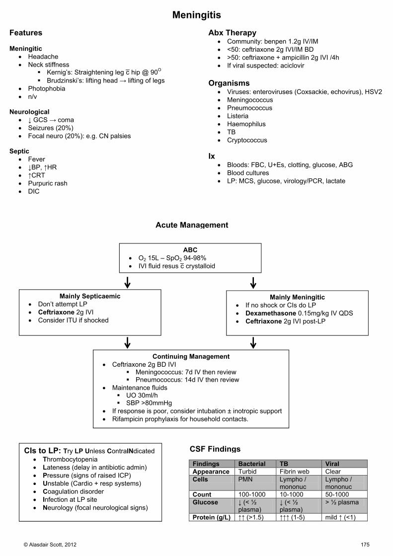
ABC O2 15L – SpO2 94-98% IVI fluid resus c̄ crystalloid
Acute Management
Mainly Septicaemic Don’t attempt LP Ceftriaxone 2g IVI Consider ITU if shocked
Mainly Meningitic If no shock or CIs do LP Dexamethasone 0.15mg/kg IV QDS Ceftriaxone 2g IVI post-LP
Continuing Management Ceftriaxone 2g BD IVI
Meningococcus: 7d IV then review Pneumococcus: 14d IV then review
Maintenance fluids UO 30ml/h SBP >80mmHg
If response is poor, consider intubation ± inotropic support Rifampicin prophylaxis for household contacts.
CIs to LP: Try LP Unless ContraINdicated Thrombocytopenia Lateness (delay in antibiotic admin) Pressure (signs of raised ICP) Unstable (Cardio + resp systems) Coagulation disorder Infection at LP site Neurology (focal neurological signs)
CSF Findings
Findings Bacterial TB Viral Appearance Turbid Fibrin web Clear Cells PMN Lympho /
mononuc Lympho / mononuc
Count 100-1000 10-1000 50-1000 Glucose ↓ (< ½
plasma) ↓ (< ½ plasma)
> ½ plasma
Protein (g/L) ↑↑ (>1.5) ↑↑↑ (1-5) mild ↑ (<1)
Meningitis Features Meningitic
Headache Neck stiffness
Kernig’s: Straightening leg c̄ hip @ 90O Brudzinski’s: lifting head → lifting of legs
Photophobia n/v
Neurological
↓ GCS → coma Seizures (20%) Focal neuro (20%): e.g. CN palsies
Septic
Fever ↓BP, ↑HR ↑CRT Purpuric rash DIC
Abx Therapy
Community: benpen 1.2g IV/IM <50: ceftriaxone 2g IVI/IM BD >50: ceftriaxone + ampicillin 2g IVI /4h If viral suspected: aciclovir
Organisms
Viruses: enteroviruses (Coxsackie, echovirus), HSV2 Meningococcus Pneumococcus Listeria Haemophilus TB Cryptococcus
Ix
Bloods: FBC, U+Es, clotting, glucose, ABG Blood cultures LP: MCS, glucose, virology/PCR, lactate
© Alasdair Scott, 2012 175

Encephalitis Presentation
Infectious prodrome: fever, rash, LNs, cold sores, conjunctivitis, meningeal signs.
Bizarre behaviour or personality change Confusion ↓ GCS → coma Fever Headache Focal neuro Seizures Hx of travel or animal bite
Causes Usually viral
HSV1/2 CMV, EBV, VZV Arboviruses HIV
Non-viral
Any bacterial meningitis TB Malaria Lyme disease
Ix
Bloods: cultures, viral PCR, malaria film Contrast CT: focal bilat temporal involvement
suggests HSV LP: ↑ CSF protein, lymphocytes, PCR EEG: shows diffuse abnormalities, may confirm Dx
Mx
Aciclovir STAT: 10mg/kg/8h IVI over 1h for 14/7 Supportive measures in HDU/ITU Phenytoin for seizures
Prognosis
70% mortality if untreated w/o fever, consider encephalopathy
↓ glucose Hepatic DKA Drugs SLE Uraemia Hypoxic brain injury Beri-Beri
Cerebral Abscess Pre-disposing Factors
Infection: ear, sinus, dental or periodontal Skull # Congenital heart disease Endocarditis Bronchiectasis Immunosuppression
Organisms
Frontal sinus/teeth: strep. Milleri, oropharyngeal anaerobes
Ear: Bacteroides, other anaerobes Signs
Seizures Fever Localizing signs Signs of ↑ ICP Signs of infection elsewhere
Ix
CT/MRI: ring-enhancing lesion ↑WCC, ↑ESR
Rx
Neurosurgical referral Abx: e.g. ceftriaxone Treat ↑ ICP
© Alasdair Scott, 2012 176

Epilepsy: Features Definition
Recurrent tendency to spontaneous, intermittent, abnormal electrical activity in part of the brain, manifest as seizures.
Causes
2/3 are idiopathic (often familial) Congenital
NF Tuberous Sclerosis TORCH Perinatal anoxia
Acquired Vascular: CVA Cortical scarring: trauma, infection SOL Other: SLE, PAN, MS, sarcoidosis
Non-epileptic / provoked seizures Withdrawal: EtOH, opiates, benzos Metabolic: glucose, Na, Ca, urea, NH3 ↑ICP: trauma, haemorrhage, cortical venous
thrombosis Infection: meningitis, encephalitis, cycticerosis,
HIV Eclampsia Pseudoseizures
NB. 3-10% of provoked seizures tend to recur, cf 30-50% of unprovoked seizures Seizure Terms
Prodrome pt. or others may notice change in mood or
behaviour lasting hrs – days. Not part of seizure.
Aura A simple partial seizure (usually temporal) which
may precede other manifestations. Experienced as a strange feeling:
Epigastric rising Deja/jamias vu, Automatisms, Smells, lights, sounds
Partial (focal) Features referable to part of one hemisphere
Primary Generalised No warning / aura Discharge throughout cortex w/o localising
features Simple: awareness unimpaired Complex: awareness impaired Secondary Generalised
Focal seizure → generalised E.g. aura → tonic-clonic
Diagnostic Pointers
Aura Specific trigger: e.g. flashing lights Lateral tongue biting (> incontinence) Typical movements: e.g. tonic-clonic Cyanosis Post-ictal phase
Presenting Features Simple Partial
Focal motor, sensory, autonomic or psychic symptoms Complex Partial: 5As
Aura Autonomic: change in skin colour, temperature, palps Awareness lost: motor arrest, motionless stare Automatisms: lip-smacking, fumbling, chewing,
swallowing Amnesia
NB. Usually arise from temporal lobe Absences (Petit mal)
ABrupt onset and offset Short: <10s Eyes: glazed, blank-stare Normal: intelligence, examination, brain-scan Clonus or automatisms may occur EEG: 3Hz spike and wave Stimulated by hyperventilation and photics
Tonic-Clonic (Grand mal)
LOC Tonic: limbs stiffen Clonic: rhythmic jerking of limbs ± cyanosis, incontinence, tongue biting (lateral) Post-ictal confusion and drowsiness
Myoclonic Seizure
Sudden jerk of limb, face or trunk. Atonic (akinetic seizures)
Sudden loss of muscle tone → fall No LOC
West Syndrome / Infantile Spasms
Clusters of head nodding and arm jerks EEG shows hypsarrhythmia
Localising Features Temporal
Automatisms: lip smacking, chewing, fumbling Deja/jamias vu Delusional behaviour Abdominal: rising, n/v Emotional disturbance: terror, panic, anger, elation Tastes, smells
Frontal
Motor features: arrest, Jacksonian march, Todd’s palsy Parietal
Sensory disturbance: tingling, numbness
Occipital Visual phenomena: spots, lines, flashes
© Alasdair Scott, 2012 177
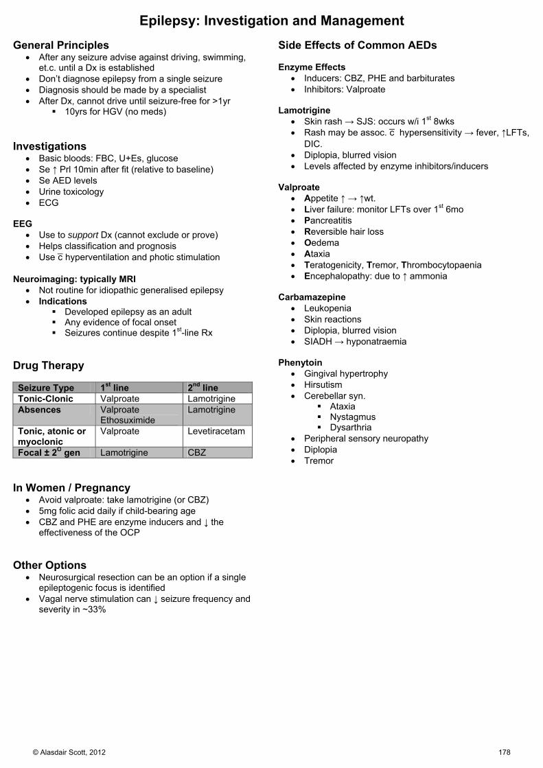
Epilepsy: Investigation and Management General Principles
After any seizure advise against driving, swimming, et.c. until a Dx is established
Don’t diagnose epilepsy from a single seizure Diagnosis should be made by a specialist After Dx, cannot drive until seizure-free for >1yr
10yrs for HGV (no meds) Investigations
Basic bloods: FBC, U+Es, glucose Se ↑ Prl 10min after fit (relative to baseline) Se AED levels Urine toxicology ECG
EEG
Use to support Dx (cannot exclude or prove) Helps classification and prognosis Use c̄ hyperventilation and photic stimulation
Neuroimaging: typically MRI
Not routine for idiopathic generalised epilepsy Indications
Developed epilepsy as an adult Any evidence of focal onset Seizures continue despite 1st-line Rx
Drug Therapy Seizure Type 1st line 2nd line Tonic-Clonic Valproate Lamotrigine Absences Valproate
Ethosuximide Lamotrigine
Tonic, atonic or myoclonic
Valproate Levetiracetam
Focal ± 2O gen Lamotrigine CBZ In Women / Pregnancy
Avoid valproate: take lamotrigine (or CBZ) 5mg folic acid daily if child-bearing age CBZ and PHE are enzyme inducers and ↓ the
effectiveness of the OCP Other Options
Neurosurgical resection can be an option if a single epileptogenic focus is identified
Vagal nerve stimulation can ↓ seizure frequency and severity in ~33%
Side Effects of Common AEDs Enzyme Effects
Inducers: CBZ, PHE and barbiturates Inhibitors: Valproate
Lamotrigine
Skin rash → SJS: occurs w/i 1st 8wks Rash may be assoc. c̄ hypersensitivity → fever, ↑LFTs,
DIC. Diplopia, blurred vision Levels affected by enzyme inhibitors/inducers
Valproate
Appetite ↑ → ↑wt. Liver failure: monitor LFTs over 1st 6mo Pancreatitis Reversible hair loss Oedema Ataxia Teratogenicity, Tremor, Thrombocytopaenia Encephalopathy: due to ↑ ammonia
Carbamazepine
Leukopenia Skin reactions Diplopia, blurred vision SIADH → hyponatraemia
Phenytoin
Gingival hypertrophy Hirsutism Cerebellar syn.
Ataxia Nystagmus Dysarthria
Peripheral sensory neuropathy Diplopia Tremor
© Alasdair Scott, 2012 178

ABC Oral / nasal airway, intubate Suction 100% O2 Capillary blood glucose
Acute Management
IV Access + Bloods U+E, LFT, FBC, Glucose, Ca2+ AED levels Tox screen
Reverse Potential Causes Thiamine 250mg IV if EtOH 100ml 20% glucose unless glucose
known to be normal
Slow IV Bolus Phase Lorazepam 2-4mg IV 2nd dose if no response w/i 2min
IV Infusion Phase Phenytoin 18mg/kg IVI (then 100mg/6-8h) Or, diazepam 100mg in 500ml 5% dex IVI
RSI PhaseNever spend >20min c̄ someone in status w/o
getting an anaesthetist
Status Epilepticus Definition
Seizure lasting >30min, or Repeated seizures w/o intervening consciousness
Ix
BM Bloods: glucose, ABG, U+E, FBC, Ca2+ ECG, EEG Consider: AED levels, tox screen, LP, βhCG, CT
Drugs Lorazepam
2-4mg IV bolus over 30s 2nd dose if no response w/i 2min Alternatives:
Diazepam 10mg IV/PR (20mg max) Midazolam 10mg buccal
Phenytoin
18mg/kg IVI @ 50mg/min 100mg/6-8h maintenance Monitor ECG and BP CI: bradycardia or heart block
Diazepam Infusion
100mg in 500ml 5% dex @ 40ml/hr (3mg/kg/24h) Dexamethasone
10mg IV if vasculitis / cerebral oedema (tumour) possible
© Alasdair Scott, 2012 179

Head Injury Initial Mx 1O Survey
A: ? intubation, immobilise C-spine B: 100% O2, RR C: IV access, BP, HR D: GCS, pupils Treat seizures
Lorazepam 2-4mg IV Phenytoin18mg/kg IVI then 100mg/6-8h
E: expose pt. and look for other obvious injuries 2O Survey
Look for: Lacerations Obvious facial/skull deformity CSF leak from nose or ears Battle’s sign, Racoon eyes Blood behind TM C-spine tenderness ± deformity
Head-to-toe examination for other injuries Log role
Hx if possible
How and when? GCS and other vitals immediately after injury Headache, fits, vomiting, amnesia, EtOH
Ix
Bloods: FBC, U+E, glucose, clotting, EtOH level, ABG ? CT head + c-spine
Rx
Neurosurgical opinion if signs of ↑ICP, CT evidence of intracranial bleed significant skull #
Admit if: Abnormalities on imaging Difficult to assess: EtOH, post-ictal Not returned to GCS 15 after imaging CNS signs: vomiting, severe headache
Neuro-obs half-hrly until GCS 15 GCS Pupils HR, BP RR, SpO2 Temperature
Discharge Advice
Stay with someone for first 48hrs Give advice card advising return on:
Confusion, drowsiness, unconsciousness Visual problems Weakness Deafness V. painful headache that won’t go away Vomiting Fits
Intubate GCS ≤ 8 PaO2 <9KPa on air / <13KPa on O2 or PCO2 >6KPa Spontaneous hyperventilation: PCO2 <4KPa Respiratory irregularity
CT head guidelines: BANGS LOC
Break: open, depressed or base of skull Amnesia >30min retrograde Neuro deficit or seizure GCS: <13 @ any time or <15 2h after injury Sickness: vomited > once LOC or any amnesia and any of:
Dangerous mechanism: RTA, great height Age ≥ 65 Coagulopathy (inc. warfarin)
Risk of Intracranial Haematoma in Adults:
Fully conscious, no skull # = <1:1000 Confused, no skull # = 1:100 Fully conscious, skull # = 1:30 Confused, skull # = 1:4
GCS Eyes: 4 4 – Spontaneous eye opening 3 – Open to voice 2 – Open to pain 1 – No opening Verbal: 5 5 – Orientated conversation 4 – Confused conversation 3 – Inappropriate speech 2 – Incomprehensible sounds 1 – No speech Motor: 6 6 – Obeys commands 5 – Localises pain 4 – Withdraws to pain 3 – Decorticate posturing to pain (flexor) 2 – Decerebrate posturing to pain (extensor) 1 – No movement
© Alasdair Scott, 2012 180
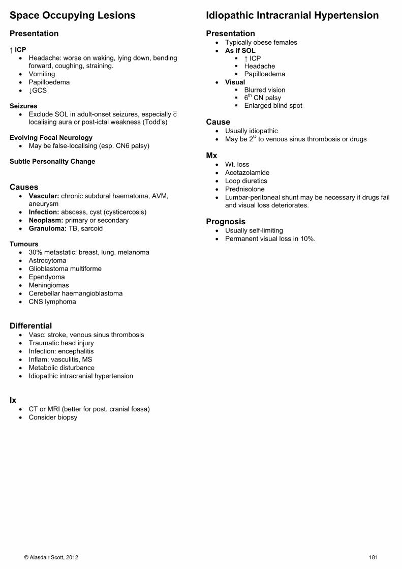
Space Occupying Lesions Idiopathic Intracranial Hypertension Presentation ↑ ICP
Headache: worse on waking, lying down, bending forward, coughing, straining.
Vomiting Papilloedema ↓GCS
Seizures
Exclude SOL in adult-onset seizures, especially c̄ localising aura or post-ictal weakness (Todd’s)
Evolving Focal Neurology
May be false-localising (esp. CN6 palsy) Subtle Personality Change Causes
Vascular: chronic subdural haematoma, AVM, aneurysm
Infection: abscess, cyst (cysticercosis) Neoplasm: primary or secondary Granuloma: TB, sarcoid
Tumours
30% metastatic: breast, lung, melanoma Astrocytoma Glioblastoma multiforme Ependyoma Meningiomas Cerebellar haemangioblastoma CNS lymphoma
Differential
Vasc: stroke, venous sinus thrombosis Traumatic head injury Infection: encephalitis Inflam: vasculitis, MS Metabolic disturbance Idiopathic intracranial hypertension
Ix
CT or MRI (better for post. cranial fossa) Consider biopsy
Presentation
Typically obese females As if SOL
↑ ICP Headache Papilloedema
Visual Blurred vision 6th CN palsy Enlarged blind spot
Cause
Usually idiopathic May be 2O to venous sinus thrombosis or drugs
Mx
Wt. loss Acetazolamide Loop diuretics Prednisolone Lumbar-peritoneal shunt may be necessary if drugs fail
and visual loss deteriorates. Prognosis
Usually self-limiting Permanent visual loss in 10%.
© Alasdair Scott, 2012 181
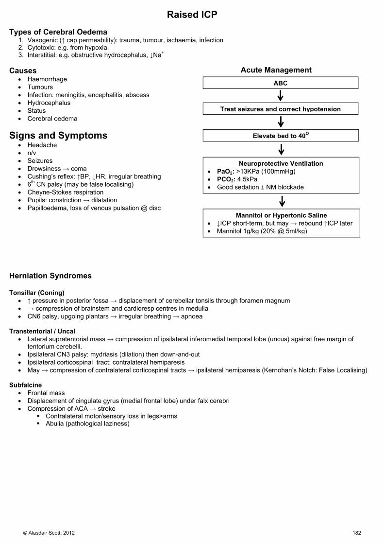
ABC
Acute Management
Treat seizures and correct hypotension
Elevate bed to 40O
Neuroprotective Ventilation PaO2: >13KPa (100mmHg) PCO2: 4.5kPa Good sedation ± NM blockade
Mannitol or Hypertonic Saline ↓ICP short-term, but may → rebound ↑ICP later Mannitol 1g/kg (20% @ 5ml/kg)
Raised ICP Types of Cerebral Oedema
1. Vasogenic (↑ cap permeability): trauma, tumour, ischaemia, infection 2. Cytotoxic: e.g. from hypoxia 3. Interstitial: e.g. obstructive hydrocephalus, ↓Na+
Causes
Haemorrhage Tumours Infection: meningitis, encephalitis, abscess Hydrocephalus Status Cerebral oedema
Signs and Symptoms Headache n/v Seizures Drowsiness → coma Cushing’s reflex: ↑BP, ↓HR, irregular breathing 6th CN palsy (may be false localising) Cheyne-Stokes respiration Pupils: constriction → dilatation Papilloedema, loss of venous pulsation @ disc
Herniation Syndromes Tonsillar (Coning)
↑ pressure in posterior fossa → displacement of cerebellar tonsils through foramen magnum → compression of brainstem and cardioresp centres in medulla CN6 palsy, upgoing plantars → irregular breathing → apnoea
Transtentorial / Uncal Lateral supratentorial mass → compression of ipsilateral inferomedial temporal lobe (uncus) against free margin of
tentorium cerebelli. Ipsilateral CN3 palsy: mydriasis (dilation) then down-and-out Ipsilateral corticospinal tract: contralateral hemiparesis May → compression of contralateral corticospinal tracts → ipsilateral hemiparesis (Kernohan’s Notch: False Localising)
Subfalcine
Frontal mass Displacement of cingulate gyrus (medial frontal lobe) under falx cerebri Compression of ACA → stroke
Contralateral motor/sensory loss in legs>arms Abulia (pathological laziness)
© Alasdair Scott, 2012 182

Parkinsonism Causes Degenerative
1. Parkinson’s disease
2. Parkinson’s-plus syndromes (basal ganglia degeneration + other system)
a. Multiple Systems Atrophy / Shy-Drager Autonomic dysfunction: post hypotension, bladder dysfunction Cerebellar + pyramidal signs Rigidity > Tremor
b. Progressive Supranuclear Palsy
Postural instability → falls Speech disturbance (+ dementia) Palsy: vertical gaze
c. Corticobasilar Degeneration:
Aphasia, dysarthria, apraxia Akinetic rigidity in one limb Astereognosis (cortical sensory loss) Alien limb phenomenon
d. Lewy Body Dementia:
Fluctuating cognition Visual hallucinations
Infection
Syphilis HIV CJD
Vascular: Multiple infarcts in SN Drugs: Antipsychotics, metoclopramide Trauma: dementia pugilistica Genetic: Wilson’s disease Symptoms Tremor
Worse at rest Exacerbated by distraction 4-6hz, pill-rolling
Rigidity
↑ tone in all muscle groups: lead-pipe rigidity Rigidity + tremor → cog-wheel rigidity
Bradykinesia
Slow initiation of movement c̄ reduction of amplitude on repetition Expressionless face Monotonous voice Micrografia
Gait
↓ arm swing Festinance Freezing (esp. in doorways)
© Alasdair Scott, 2012 183
Parkinson’s Disease Epidemiology
Mean onset 65yrs 2% prevalence
Pathophysiology
Destruction of dopaminergic neurones in pars compactaof substantia nigra.
β-amyloid plaques Neurofibrillary tangles: hyperphosphorlated tau
Features: TRAPPS PD
Asymmetric onset: side of onset remains worst
Tremor: ↑ by stress, ↓ by sleep Rigidity: lead-pipe, cog-wheel Akinesia: slow initiation, difficulty c̄ repetitive movement,
micrographia, monotonous voice, mask-like face Postural instability: stooped gait c̄ festination Postural hypotension: + other autonomic dysfunction Sleep disorders: insomnia, EDS, OSA, RBD Psychosis: esp. visual hallucinations Depression / Dementia / Drug SEs
Sleep Disorder Affects ~90% of PD pts. Insomnia + frequent waking → EDS
Inability to turn Restless legs Early morning dystonia (drugs wearing off) Nocturia OSA
REM Behavioural sleep Disorder Loss of muscle atonia during REM sleep Violent enactment of dreams
Da SEs: insomnia, drowsiness, EDS
Autonomic Dysfunction Combined effects of drugs and neurodegeneration Postural hypotension Constipation Hypersalivation → dribbling (↓ ability to swallow saliva) Urgency, frequency, Nocturia ED Hyperhidrosis
L-DOPA SEs: DOPAMINE Dyskinesia On-Off phenomena = Motor fluctuations Psychosis ABP↓ Mouth dryness Insomnia N/V EDS (excessive daytime sleepiness)
Motor Fluctuations
End-of-dose: deterioration as dose wears off c̄ progressively shorter benefit.
On-Off effect: unpredictable fluctuations in motor performance unrelated to timing of dose.
Ix
DaTSCAN
Mx
MDT: neurologist, PD nurse, physio, OT, social worker, GP and carers
Assess disability e.g. UPDRS: Unified Parkinson’s Disease Rating Scale
Physiotherapy: postural exercises Depression screening
Medical Young onset ± biologically fit
1. Da agonists: ropinirole, pramipexole 2. MOA-B inhibitors: rasagiline, selegiline 3. L-DOPA: co-careldopa or co-beneldopa
Biologically frail ± comorbidities
1. L-DOPA 2. MOA-B inhibitors
Other therapies
COMT inhibitor: tolcapone, entacapone Lessen end-of-dose effect
Apomorphine: potent Da agonist SC rescue pen for sudden “off” freezing
Amantidine: weak Da agonist Rx of drug-induced dyskinesias
Atypical antipsychotics: e.g. quetiapine Disease-induced psychosis
SSRIs: citalopram, sertraline Depression
Surgical
Interrupt basal ganglia Deep brain stimulation
Prognosis
↑ mortality Loss of response to L-DOPA w/i 2-5yrs
Differential
Parkinson plus syndromes Multiple infarcts Drugs: neuroleptics Inherited: Wilson’s Infection: HIV, syphilis, CJD Dementia pugilistica
© Alasdair Scott, 2012 184

Multiple Sclerosis
Definition A chronic inflammatory condition of the CNS characterised by multiple plaques of demyelination disseminated in time and
space. Epidemiology
Lifetime risk: 1/1000 Age: mean @ onset = 30yrs Sex: F>M = 3:1 Race: rarer in blacks
Aetiology Genetic (HLA-DRB1), environmental, viral
Pathophysiology CD4 cell-mediated destruction of oligodendrocytes →
demyelination and eventual neuronal death. Initial viral inflam primes humoral Ab responses vs. MBP Plaques of demyelination are hallmark
Classification: Relapsing-remitting: 80% Secondary progressive Primary progressive: 10% Progressive relapsing
Presentation: TEAM Tingling Eye: optic neuritis (↓ central vision + eye move pain) Ataxia + other cerebellar signs Motor: usually spastic paraparesis
Clinical features Sensory:
Dys/paraesthesia ↓ vibration sense Trigeminal neuralgia
Motor Spastic weakness Transverse myelitis
Eye: Diplopia Visual phenomena Bilateral INO Optic neuritis → atrophy
Cerebellum: Trunk and limb ataxia Scanning dysarthria Falls
GI: Swallowing disorders Constipation
Sexual/GU: ED + anorgasmia Retention Incontinence
Lhermitte’s Sign Neck flexion → electric shocks in trunk/limbs
Optic Neuritis
PC: pain on eye movement, rapid ↓ central vision Uhthoff’s: vision ↓ c̄ heat: hot bath, hot meal, exercise o/e: ↓ acuity, ↓ colour vision, white disc, central scotoma,
RAPD INO / ataxic nystagmus / conjugate gaze palsy
Disruption of MLF connecting CN6 to CN3 Weak adduction of ipsilateral eye Nystagmus of contralateral eye Convergence preserved
Ix MRI: Gd-enhancing or T2 hyper-intense plaques
Gd-enhancing = active inflammation Typically located in periventricular white matter
LP: IgG oligoclonal bands (not present in serum) Abs
Anti-MBP NMO-IgG: highly specific for Devic’s syn.
Evoked potentials: delayed auditory, visual and sensory
Diagnosis: clinical Demonstration of lesions disseminated in time and space May use McDonald Criteria
Differential Inflammatory conditions may mimic MS plaques:
CNS sarcoidosis SLE Devic’s: Neuromyelitis optica (NMO)
MS variant c̄ transverse myelitis and optic atrophy Distinguished by presence of NMO-IgG Abs
Mx MDT: neurologist, radiologist, physio, OT, specialist
nurses, GP, family Acute Attack
Methylpred 1g IV/PO /24h for 3d Doesn’t influence long-term outcome ↓ duration and severity of attacks
Preventing Relapse: DMARDs
IFN-β: ↓ relapses by 30% in relapsing remitting MS Glatiramer: similar efficacy to IFN-β
Preventing Relapse: Biologicals
Natalizumab: anti-VLA-4 Ab ↓ Relapses by 2/3 in RRMS
Alemtuzumab (Campath): anti-CD52 2nd line in RRMS
Symptomatic
Fatigue: modafinil Depression: SSRI (citalopram) Pain: amitryptylline, gabapentin Spasticity: physio, baclofen, dantrolene, botulinum Urgency / frequency: oxybutynin, tolterodine ED: sildenafil Tremor: clonazepam
Prognosis Poor Prognostic Signs
Older Male Motor signs @ onset Many relapses early on Many MRI lesions Axonal loss
Better Prognostic Signs Female <25 Sensory signs @ onset Long interval between
relapses Few MRI lesions
© Alasdair Scott, 2012 185
Cord Compression Symptoms
Deep, local spinal pain Stabbing, radicular pain in a dermatomal distribution
and LMN weakness @ lesion level Progressive UMN weakness and sensory loss below
lesion Bladder hesitancy, frequency → painless retention Faecal incontinence or constipation
Signs
Look for motor, reflex and sensory level Shooting, radicular pain @ level, anaesthesia below LMN signs @ level, UMN signs below level Tone and reflexes are usually reduced in acute cord
compression Causes
Trauma Infection: epidural abscess, TB 2O to malignancy: breast, thyroid, bronchus, kidney,
prostate Disc prolapse Haematoma: warfarin Intrinsic cord tumour Myeloma
Ix
MRI is definitive modality CXR for primaries
Rx
This is a neurosurgical emergency Malignancy
Dexamethasone IV Consider chemo, radio and decompressive
laminectomy Abscess: abx and surgical decompression
Differential
Transverse myelitis MS Cord vasculitis Spinal artery thrombosis Aortic dissection
Cauda Equina and Conus Medullaris Lesions
Spinal cord tapers to its end at L1 Compared c̄ lesions higher up the cord, these lesions
are flaccid and areflexic (cf, spastic and hyperreflexic) Conus Medullaris Lesions
Mixed UMN/LMN weakness Early constipation and retention Back pain Sacral sensory disturbance ED
Cauda Equina Lesions
Saddle anaesthesia Back pain Radicular pain down legs Bilateral flaccid, areflexic lower limb weakness Incontinence / retention of faeces / urine Poor anal tone
Mx
As for cord compression These are neurosurgical emergencies and require
urgent imaging and surgical decompression
© Alasdair Scott, 2012 186
Cervical and Lumbar Degeneration Spondylosis
Degeneration due to trauma or ageing IV disc / vertebral collapse Osteophytes May → central (myelopathy) and/or lateral
(radiculopathy) pathology Cervical Spondylosis
90% of men > 60 and women > 50 Presentation
Usually asympto Neck stiffness ± crepitus Stabbing / dull arm pain (brachialgia) Upper limb motor and sensory disturbances according
to compression level (often C7) Can → myelopathy c̄ quadraparesis and sphincter
dysfunction Specific Signs
Lhermitte’s sign: neck flexion → tingling down spine Hoffman reflex: flick to middle finger pulp → brief pincer
flexion of thumb and index finger Typical Deficits Root Disc Motor Weakness Sensory C5 C4/5 Deltoid
Supraspinatus ↓ supinator jerk
Numb elbow
C6 C5/6 Biceps Brachioradialis ↓ biceps jerk
Numb thumb and index finger
C7 C6/7 Triceps Finger extension ↓ triceps jerk
Numb middle finger
C8 C7/T1 Finger flexors Intrinsic hand
Number ring and little fingers
Ix
MRI Rx
Conservative: stiff collar, analgesia Medical: transforaminal steroid injection Surgical: decompression: laminectomy or laminoplasty
Differential
MS Nerve root neurofibroma SACD
Lumbosacral Spondylosis Presentation
L5 and S1 roots most commonly compressed by prolapse of L4/5, L5/S1 discs.
May present as severe pain on sneezing/coughing, a few days after low back strain
Lumbago – low back pain Sciatica – shooting radicular pain down buttock and
thigh Signs
Limited spinal flexion Pain on straight-leg raise
L4/5 → L5 Root Compression
Weak hallux extension ± foot drop In foot drop due to L5 radiculopathy, weak
inversion (tib. post.) helps distinguish from peroneal N. palsy.
↓ sensation on inner dorsum of foot L5/S1 → S1 Root Compression
Weak foot plantarflexion and eversion Loss of ankle-jerk Calf pain ↓ sensation over sole of foot and back of calf
Central Compression
Cauda equina syndrome Ix
MRI is definitive (emergency if cauda-equina syndrome) Rx
Conservative: rest, analgesia, mobilisation/physio Medical: transforaminal steroid injection Surgical: discectomy or laminectomy may be
considered in cauda-equina syndrome, continuing pain or muscle weakness.
Spinal Stenosis
Developmental predisposition ± facet joint osteoarthritis → generalized narrowing of lumbar spinal canal.
Presentation
Spinal claudication Aching or heavy buttock and lower limb pain on
walking Rapid onset May c/o paraesthesiae/numbness Pain eased by leaning forward (e.g. on bike)
Pain on spine extension Negative straight leg raise
Ix
MRI Rx
Corsets NSAIDs Epidural steroid injection Canal decompression surgery
© Alasdair Scott, 2012 187

Facial Nerve Palsy Bell’s Palsy
Inflammatory oedema from entrapment of CNVII in narrow facial canal
Probably of viral origin (HSV1) 75% of facial palsy
Features Sudden onset: e.g. overnight Complete, unilateral facial weakness in 24-72h
Failure of eye closure → dryness Bell’s sign: eyeball rolls up on attempted closure Drooling, speech difficulty
Numbness or pain around ear ↓ taste (ageusia) Hyperacusis: stapedius palsy
Ix
Serology: Borrelia or VZV Abs MRI: SOL, stroke, MS LP
Mx
Give prednisolone w/i 72hrs 60mg/d PO for 5/7 followed by tapering
Valaciclovir if zoster suspected Otherwise antivirals don’t help
Protect eye Dark glasses Artificial tears Tape closed @ night
Plastic surgery may help if no recovery Prognosis
Incomplete paralysis: recovers completely w/i wks Complete: 80% get full recovery
Remainder have delayed recovery or permanent neurological / cosmetic abnormalities.
Complications: Aberrant Neural Connections
Synkinesis: e.g. blinking causes up-turning of mouth Crocodile tears: eating stimulates unilateral
lacrimation, not salivation Ramsay Hunt Syndrome
American neurologist James Ramsay Hunt in 1907 Reactivation of VZV in geniculate ganglion of CNVII
Features
Preceding ear pain or stiff neck Vesicular rash in auditory canal ± TM, pinna, tongue,
hard palate (no rash = zoster sine herpete) Ipsilateral facial weakness, ageusia, hyperacusis, May affect CN8 → vertigo, tinnitus, deafness
Mx
If Dx suspected give valaciclovir and prednisolone w/i first 72h
Prognosis
Rxed w/i 72h: 75% recovery Otherwise: 1/3 full recovery, 1/3 partial, 1/3 poor
Other Causes of Facial Palsy May be suggested by
Bilat symptoms: Lyme, GBS, leukaemia, sarcoid, MG UMN signs: sparing of frontalis and orbicularis oculi
Due to bilateral cortical representation Other CN palsies (but seen in 8% of Bell’s) Limb weakness Rashes
Intracranial
Vascular, MS, SOL Motor cortex → UMN
Assoc. c̄ ipsilateral hemiplegia Brainstem nuclei → LMN
Usually assoc. c̄ CN6 palsy + contralateral hemiplegia
Cerebello-pontine angle: acoustic neuroma, meningioma Both may be accompanied by 5th, 6th and 8th CN
palsies and cerebellar signs Loss of corneal reflex Sensorineural deafness, tinnitus, vertigo DANISH
Intra-temporal
Cholesteatoma Ramsay Hunt Otitis media Trauma
Infra-temporal
Parotid tumours Trauma
Systemic
Peripheral neuropathy Demyelinating: GBS Axonal: DM, Lyme, HIV, Sarcoid
Pseudopalsy: MG, botulism
© Alasdair Scott, 2012 188

Mononeuropathies
Lesions of individual peripheral or cranial nerves Usually local cause: trauma, entrapment
Mononeuritis multiplex: 2 or more peripheral nerves affects Usually systemic cause: DM most commonly WAARDS PLC: Wegener’s, AIDS, Amyloid, RA, DM, Sarcoidosis, PAN, Leprosy, Carcinomatosis
Electromyography (EMG) helps define site of lesion Nerve Location / Cause Motor Features Sensory Loss/ Features Median: C6-T1 Wrist: carpal tunnel, trauma LLOAF muscles
Thenar wasting Radial 3½ fingers and palm Aching pain in hand ↓ 2-point discrimination Tinel’s and Phalen’s +ve
Ulnar: C7-T1 Elbow trauma - e.g. supracondylar #
Partial claw hand Hypothenar wasting Can’t do good luck sign Weakness and wasting of 1st dorsal interosseous Froment’s +ve
Ulnar 1½ fingers
Radial: C5-T1 Low: wrist High: humerus V. high: Axilla
Low: finger drop High: wrist drop V. high: triceps paralysis, wrist drop
Dorsal thumb root (snuff box)
Brachial plexus Trauma Radiotherapy (e.g. breast)
High (C5-6): Erb’s palsy → waiter’s tip Low (C8-T1): Klumpke’s → claw hand
High: C5-6 dermatome Low: C8-T1 dermatome
Phrenic: C3-5 Neoplastic: Lung Ca Myeloma Thymoma Mechanical: Cervical spondylosis Big left atrium Infective: C3-5 zoster HIV Lyme TB
Orthopnoea + raised hemidiaphragm
Lat. cut. N. thigh: L2-3
Entrapment under inguinal ligament
Meralgia paraesthetica – anterolat. burning thigh pain
Sciatic: L4-S3 Pelvic tumours Pelvic or femur #s
Hamstrings All muscles below knee
Below knee laterally and foot
Common peroneal: L4-S1
Fibular head: trauma, sitting cross legged
Foot drop: can’t walk on heals Weak ankle dorsiflexion, eversion NB. inversion intact!
Below knee laterally
Tibial: L4-S3 Can’t plantar flex → can’t stand on tiptoe Foot inversion Toe flexion
Sole of foot
© Alasdair Scott, 2012 189

Polyneuropathies Features
Generalised disorders of peripheral or cranial nerves Distribution is symmetrical and widespread Distal weakness and sensory loss (glove + stocking) Classification
Time-course: acute, chronic Function: motor, sensory, autonomic, mixed Path: demyelination, axonal degeneration, both
Causes Metabolic (mostly axonal)
DM Renal failure / uraemia Hypothyroid ↓B1 or ↓B12 (EtOH)
Vasculitis PAN RA Wegener’s
Inflammatory GBS Sarcoidosis
Infection HIV Syphilis Leprosy Lyme
Inherited
CMT Refsum’s syndrome
Toxins Lead
Drugs Isoniazid EtOH Phenytoin Vincristine
Other Amyloid Paraproteinaemias
History
Time-course Precise symptoms Assoc. events
D&V: GBS ↓wt: Ca Arthralgia: connective tissue
Travel, EtOH, drugs Ix
LFTs, U+E, glucose, ESR, B12 TFTs B1, ANA, ANCA Genetic tests: e.g. PMP22 in CMT Nerve conduction studies EMG
Sensory Neuropathy Main Causes
Alcohol B12 CRF and Ca (paraneoplastic) DM Every vasculitis
General Features
Glove and stocking distribution: length dependent Deep tendon reflexes may be ↓ or absent
E.g. loss of ankle jerks in diabetic neuropathy Signs of trauma or joint deformity (Charcot’s joints) Diabetic and alcoholic neuropathies are painful Some aetiologies favour loss of particular fibres
Large Myelinated Fibres (Aα): e.g. B12
Loss of proprioception → ataxia Pins and needles
Small Unmyelinated Fibres (C): e.g. EtOH
Loss of pain and temperature sensation Painful dysesthesia: e.g. burning, hyperalgesia
Motor Neuropathy Main Causes
GBS (+ botulism) HMSN / CMT Paraneoplastic Lead poisoning
Features
Weakness/clumsiness of hands, difficulty walking LMN signs CN: diplopia, dysarthria, dysphagia Involvement of respiratory muscles → ↓FVC
Autonomic Neuropathy Causes
DM HIV SLE GBS, LEMS
Features
Postural hypotension ED, ejaculatory failure ↓ sweating Constipation / Nocturnal diarrhoea Urinary retention Horner’s
Autonomic Function Tests
BP: Postural drop > 20/10mmHg ECG: variation >10bpm c̄ respiration
Primary Autonomic Failure
Idiopathic or part of MSA or PD
© Alasdair Scott, 2012 190
Guillain-Barré Syndrome Classification
AIDP: Acute autoimmune demyelinating polyneuropathy AMAN: Acute motor (± sensory) axonal neuropathy Miller-Fisher: Ophthalmoplegia + ataxia + areflexia
Causes
Abs cross-react to gangliosides No precipitant identified in 40% Bacteria: C. jejuni, mycoplasma Viruses: CMV, EBV, HSV, HIV, flu Vaccines: esp. rabies
Features and Ix: GBS=AIDP Growing Weakness
Symmetrical, ascending flaccid weakness / paralysis LMN signs: areflexia, fasciculations may occur Proximal > distal (trunk, respiratory, CNs [esp. 7]) Progressive phase lasts ≤ 4wks
Breathing and Bulbar Problems Back Pain
Back / limb pain is common Sensory disturbance
Paraesthesia in extremities Sensory ataxia in Miller-Fisher
Autonomic Neuropathy
Arrhythmias, ↑ HR Labile BP Sweating Urinary retention
Immune
Serology for anti-ganglioside Abs Evidence of infection: e.g. stool sample
Demyelinating Nerve Conduction Studies
Slow conduction velocities Protein in CSF
Protein often > 5.5g/L Normal white cell count
Mx Supportive
Airway / ventilation: ITU if FVC < 1.5L Analgesia: NSAIDs, gabapentin Autonomic: may need inotropes, catheter Antithrombotic: TEDS, LMWH
Immunosuppression
IVIg Plasma exchange
Physiotherapy
Prevent flexion contractures Prognosis
85% complete recovery 10% unable to walk alone at 1yr 5% mortality
Charcot-Marie-Tooth Syndrome = Peroneal Muscular Atrophy = Hereditary Motor and Sensory Neuropathy Pathophysiology
Group of inherited motor and sensory neuropathies HMSN1
Commonest form Demyelinating AD mutation in the peripheral myelin protein 22 gene
HMSN2
Second commonest form Axonal degeneration (near normal conduction velocity)
Clinical Features
Onset at puberty Nerves
Thickened, enlarged nerves: esp. common peroneal Motor
Foot drop → high stepping gait Weak ankle dorsiflexion and toe extension Absent ankle jerks Symmetrical muscle atrophy: mainly distal
Peroneal muscles → “Champagne Bottle” Hand and arm muscles → “Claw Hand”
Pes cavus (high-arched feet) Sensory
Variable loss of sensation in a stocking distribution Neuropathic pain in some
Ix
Genetic tests: PMP22 gene mutation Nerve conduction studies: ↓ conduction speed in CMT1
Mx
Supportive Physio Podiatry Orthoses: e.g. ankle braces
© Alasdair Scott, 2012 191

Motor Neurone Disease Characteristics
Cluster of degenerative disease characterised by axonal degeneration of neurones in the motor cortex, CN nuclei and anterior horn cells.
UM and LM neurones affected (cf. polyneuropathy) No sensory loss or sphincter disturbance (cf. MS) Never affects eye movements (cf. MG)
Epidemiology
Prevalence: 6/100,000 Sex: M>F=3:2 Median age @ onset = 60yrs Often fatal in 2-4yrs
Causes
Unknown ~10% familial: SOD1 mutation in 20% of those
Ix
Brain/cord MRI: exclude structural cause Cervical cord compression → myelopathy Brainstem lesions
LP: exclude inflammatory cause EMG: shows denervation
Features
UMN: spasticity, ↑ reflexes, ↑ plantars LMN: wasting, fasciculation of tongue / abdo / thigh Speech or swallowing impairment Fronto-temporal dementia
Diagnosis
MRI to exclude structural lesions LP to exclude inflammation EMG shows acute denervation Use Revised El Escorial Diagnostic Criteria
Mx
MDT: neurologist, physio, OT, dietician, specialist nurse, GP, family
Discussion of end-of-life decisions E.g. Advanced directive DNAR
Specific
Riluzole: antiglutamatergic that prolongs life by ~3mo Supportive
Drooling: propantheline or amitriptyline Dysphagia: NG or PEG feeding Respiratory failure: NIV Pain: analgesic ladder Spasticity: baclofen, botulinum
Prognosis
Most die w/i 3yrs Bronchopneumonia and respiratory failure
Worse prog: elderly, female, bulbar involvement
Classification Amyotrophic Lateral Sclerosis: 50%
Loss of motor neurones in cortex and anterior horn → UMN signs and LMN wasting + fasciculation
Progressive Bulbar Palsy: 10%
Only affects CN 9-12 → bulbar palsy Progressive Muscular Atrophy: 10%
Anterior horn cell lesion → LMN signs only Distal to proximal Better prognosis cf. ALS
Primary Lateral Sclerosis
Loss of Betz cells in motor cortex → mainly UMN signs Marked spastic leg weakness and pseudobulbar palsy No cognitive decline
Bulbar Palsy
Diseases of nuclei of CN 9-12 in the medulla LMN lesions of tongue, talking and swallowing Signs
Flaccid, fasciculating tongue Speech: quiet or nasal (“Donald Duck” speech) Normal / absent jaw jerk Loss of gag reflex
Causes MND GBS MG Central pontine myelinolysis (CPM)
Pseudobulbar / Corticobulbar Palsy
Commoner than bulbar palsy Bilateral lesions above mid-pons (e.g. corticobulbar
tracts) → UMN lesions of swallowing and talking CN motor nuclei have bilateral cortical
representation except lower half of CN7 Signs
Spastic tongue Slow tongue movements c̄ slow deliberate
speech: “hot-potato” speech Brisk jaw jerk Emotional incontinence
Causes MS MND Stroke CPM
Polio
RNA virus Affects anterior horn cells Fever, sore throat, myalgia 0.1% develop paralytic polio
Asymmetric LMN paralysis No sensory involvement May be confined to upper or lower limbs or both Respiratory muscle paralysis can → death
© Alasdair Scott, 2012 192

Myopathies Myopathy vs. Neuropathy Myopathy
Gradual onset Symmetrical, proximal weakness: difficulty combing
hair, climbing stairs, getting up from chairs Dystrophies usually affect specific muscle groups Preserved tendon reflexes
Neuropathy
Paraesthesia, bladder problems Distal weakness
Other Pointers
Rapid onset: neuropathy, or drug, toxic or metabolic myopathy
Fatiguability: MG, LEMS Spontaneous pain and tenderness @ rest: inflam
myopathy Pain on exercise: ischaemia or metabolic myopathy Oddly firm muscles: pseudohypertrophy in muscular
dystrophies Fasciculation: anterior horn cell or root disease
Ix
ESR, CK, AST, LDH EMG
Muscular Dystrophies
Group of genetic disease c̄ progressive degeneration and weakness of specific muscle groups.
DMD
Commonest: 3/1000 male births X-linked recessive, 30% spontaneous → non-functional
dystrophin Presentation
~4yrs old Difficulty standing Calf pseudohypertrophy Respiratory failure
Ix: ↑↑CK Prog: some survive > 20yrs
BMD
0.3/1000 male births X-linked recessive Partially functioning dystrophin Presents later, is less severe and has better prognosis
Facioscapulohumeral MD (Landouzy-Dejerine)
Almost as common as DMD AD inheritance Presentation
Onset @ 12-14yrs Difficulty puffing cheeks and raising arms above
head Signs
Weakness of face, shoulders and upper arms (often asymmetric c̄ deltoids spared)
Winging of scapula Foot drop.
Prog: <20% need wheelchair by 20yrs
Myotonic Dystrophy
AD Cl- channelopathy Onset in 20s Tonic muscle spasm (myotonia)
Presentation
Face Myopathic facies: long, thin, expressionless Wasting of facial muscles and SCM Bilateral ptosis Dysarthria: myotonia of tongue and pharynx
Hands Myotonia: slow relaxation
E.g. inability to release hand after shake Wasting and weakness of distal muscles +
areflexia → wrist drop Percussion myotonia: percuss thenar eminence
→ involuntary thumb flexion Other
Frontal balding Cataracts DM Cardiomyopathy, tachy- / brad-arrhythmias Dysphagia Testicular atrophy
Rx
No Rx for weakness Phenytoin may improve myotonia Caution c̄ GA: high risk of anaesthetic complications
Prognosis
Most die in middle-age of intercurrent illness Acquired myopathies of late onset
Usually part of systemic disease Hyperthyroidism, Cushing’s, ↑/↓Ca2+ Drugs: steroids, statins, EtOH
Inflammatory Myopathies
Inclusion body myositis Polymyositis Dermatomyositis
Inclusion Body Myositis
Asymmetric weakness affecting distal and prox muscles Early involvement of quads, ankle dorsiflexors
and wrist/finger flexors → loss of grip strength and ↓dexterity
Dysphagia is very common Myalgia is relatively uncommon
© Alasdair Scott, 2012 193

Myasthenia Gravis Pathophysiology
Autoimmune disease mediated by Abs vs. nicotinic Ach receptors.
Interferes c̄ NM transmission via depletion of working post-synaptic receptor sites
Presentation ↑ing muscular fatigue
Extra-ocular: bilateral ptosis, diplopia Bulbar: voice deteriorates on counting to 50 Face: myasthenic snarl on attempting to smile Neck: head droop Limb: asymmetric, prox. weakness
Normal tendon reflexes Weakness worsened by pregnancy, infection, emotion,
drugs (β-B, gent, opiates, tetracyclines)
Investigations Tensilon Test
Give edrophonium IV +ve if power improves w/i 1min
Anti-AChR Abs: ↑ in 90%, MuSK Abs EMG: ↓ response to a train of impulses Respiratory function: ↓FVC Thymus CT TFTs
Differential of Muscle Fatigability Polymyositis SLE Botulism
Associations
<50yrs MG is commoner in women and assoc. c̄ other AI disease (DM, RA, Graves) and thymic hyperplasia.
>50yrs MG is commoner in men and assoc. c̄ thymic atrophy or thymic tumour.
Treatment Symptom Control
Anticholinesterase: e.g. pyridostigmine. Cholinergic SEs = SLUDGEM
Immunosuppression
Rx relapses c̄ pred Steroids may be combined c̄ azathioprine or
methotrexate Thymectomy
Consider if young onset and disease not control by anticholinesterases
Remission in 25%, benefit in further 50%.
Complications Myasthenic Crisis
Weakness of respiratory muscles during relapse may be lethal.
Monitor FVC: vent support if <20ml/kg Plasmapheresis or IVIg Rx trigger for relapse (drugs, infection…)
Prognosis Relapsing or slow progression
Lambert-Eaton Myasthenic Syndrome (LEMS) Pathophysiology
Abs to VGCC ↓ influx of Ca2+ during presynaptic excitation → ↓ presynaptic ACh-vesicle fusion.
Causes
Paraneoplastic: e.g SCLC Autoimmune
Presentation
As for MG except: LEMS Leg weakness early (before eyes) Extra: Autonomic and areflexia Movement improves symptoms Small response to edrophonium
Anti-VGCC Abs Mx
3,4-diaminopyridine or IVIg Do regular CXRs / HRCTs as symptoms my precede Ca by 4yrs
Botulism
Botulinum toxin prevents ACh vesicle release Descending flaccid paralysis c̄ no sensory signs Anti-cholinergic effects: mydriasis, cycloplegia, n/v, dry mouth, constipation Rx: benpen + antiserum
© Alasdair Scott, 2012 194
Neurofibromatosis 1 – von Recklinghausen’s Epidemiology
AD Chr 17 Variable expression Prev: 1/2500
Features: CAFÉ NOIR Café-au-lait spots
1st yr of life ↑ in size and no. c̄ age Adults: >6, >15mm across
Axillary Freckling
in skin folds Fibromas, neuro-:
Subcutaneous Small, gelatinous, violaceous nodules Appear @ puberty May itch Nos. ↑ c̄ age
Plexiform Overgrowth of nerve trunk and overlying tissue Large cutaneous mass
Complications Sarcomatous change Compression:
Nerve roots: weakness, pain, paraesthesia GI: bleeds and obstruction
Eye
Lisch nodules Brown/translucent iris hamartomas Use a slit lamp
Optic N. glioma Neoplasia
CNS: meningioma, ependyoma, astrocytoma Phaeochromocytoma Chronic or acute myeloid leukaemia
Orthopaedic
Kyphoscoliosis Sphenoid dysplasia
IQ↓ and Epilepsy Renal
RAS → ↑ BP Mx
MDT orchestrated by GP Yearly BP and cutaneous review Excise some neurofibromas Genetic counselling
Café-au-Lait Spots Differential
NF McCune-Albright Multiple Lentigenes Urticaria Pigmentosa
Neurofibromatosis 2 Epidemiology
AD inheritance (Chr 22), but 50% are de novo Prev: 1/35,000
Signs Café-au-lait spots
Rare Fewer cf. NF1
Bilateral Vestibular Schwannomas
Characteristic Symptomatic by 20yrs SNHL is first sign, then tinnitus, vertigo
Juvenile Posterior Subcapsular Lenticular Opacity
Form of cataract Bilateral Occur before other manifestations and may be useful
for screening those @ risk Complications
Tender schwannomas of cranial and peripheral nerves and spinal nerve roots.
Meningiomas: often multiple Gliomas
Mx
Hearing tests from puberty in affected families MRI brain if abnormality detected
Prognosis
Mean survival from Dx is 15yrs
© Alasdair Scott, 2012 195

Intrinsic Cord Disease Syringomyelia Presentation
Painless Early sphincter / erectile dysfunction Bilateral motor and sensory disturbance below
lesion Causes: DIVINITY Degenerative
MND Developmental
Friedrich’s Ataxia Hereditary spastic paraparesis
Infection
Viral: HIV, HTLV-1 Syphilis: Tabes Dorsalis
Vascular Infarction
Aortic dissection/aneurysm Thromboembolism Atheroma Vasculitis: esp. PAN
NB. ant. spinal A. infarction → spinothalamic and corticospinal tract loss → bilat loss of pain/temp and spastic paresis
= Beck’s Syndrome Inflammation
Demyelination: MS Transverse myelitis
Neoplasia
Glioma Ependymoma
Injury Toxin / Nutrition
B12 deficiency sYringomyelia
Brown-Sequard Syndrome Hemi-cord lesion Ipsilateral loss of proprioception and vibration
sense Ipsilateral UMN weakness Contralateral loss of pain sensation
Characteristics
Syrinx: tubular cavity in central canal of the cervical cord. Onset @ 30yrs Symptoms may be static for yrs but then worsen fast
e.g. on coughing, sneezing as ↑ pressure → extension Commonly located in cervical cord Syrinx expands ventrally affecting:
Decussating spinothalamic neurones Anterior horn cells Corticospinal tracts
Causes
Blocked CSF circulation c̄ ↓ flow from posterior fossa Arnold-Chiari malformation (cerebellum herniates
through foramen magnum) Masses
Spina bifida 2O to cord trauma, myelitis, cord tumours and AVMs
Cardinal Signs
1. Dissociated Sensory Loss Absent pain and temperature → scars from burns Preserved touch, proprioception and vibration. Root distribution reflects syrinx location
Usually upper limbs and chest: “cape” 2. Wasting/weakness of hands ± Claw hand 3. Loss of reflexes in upper limb 4. Charcot Joints: shoulder and elbow
Other Signs
UMN weakness in lower limbs c̄ upgoing plantars Horner’s syndrome Syringobulbia: cerebellar and lower CN signs Kyphoscoliosis
Ix
MRI spine Surgery
Decompression at the foramen magnum for Chiari mal
© Alasdair Scott, 2012 196
Miscellaneous Conditions Friedrich’s Ataxia
Auto recessive progressive degeneration of DRGs, spinocerebellar and corticospinal tracts and cerebellar cells
Mitochondrial disorder Onset in teenage years Assoc. c̄ HOCM and mild dementia
Presentation
Pes cavus and scoliosis Bilateral cerebellar signs
Ataxia Dysarthria Nystagmus
Leg wasting + areflexia but extensor plantars Loss of lower limb proprioception and vibration
sense Optic atrophy Cardiac: HOCM → ESM + 4th heart sound DM → hyperglycaemia
Hereditary Spastic Paraparesis
Lower limb spasticity Ataxia Extrapyramidal signs
Human T-lymphotropic Virus-1
Retrovirus ↑ prevalence in Japan and Caribbean
Features
Adult T cell leukaemia / lymphoma Tropical spastic paraplegia / HTLV myelopathy
Slowly progressing spastic paraplegia Sensory loss and paraesthesia Bladder dysfunction
© Alasdair Scott, 2012 197

Rheumatology Contents
Patterns of Presentation ............................................................................................................................................................ 199
Rheumatological Investigations ................................................................................................................................................. 199
Back Pain .................................................................................................................................................................................. 200
Osteoarthritis ............................................................................................................................................................................. 201
Septic Arthritis ........................................................................................................................................................................... 201
Rheumatoid Arthritis .................................................................................................................................................................. 202
Gout ........................................................................................................................................................................................... 203
Ca Pyrophosphate Dehydrate Arthropathy ............................................................................................................................... 203
Seronegative Spondyloarthropathies ........................................................................................................................................ 204
Autoimmune Connective Tissue Disease ................................................................................................................................. 205
Systemic Sclerosis .................................................................................................................................................................... 206
Polymyositis and Dermatomyositis ............................................................................................................................................ 206
Systemic Lupus Erythematosus ................................................................................................................................................ 207
Vasculitis ................................................................................................................................................................................... 208
ANCA Positive Vasculitidies ...................................................................................................................................................... 209
ANCA Negative Vasculitidies .................................................................................................................................................... 209
Fibromyalgia .............................................................................................................................................................................. 210
© Alasdair Scott, 2012 198
Patterns of Presentation Monoarthritis
Septic arthritis Crystal arthritis: gout, CPPD Osteoarthritis Trauma: haemarthrosis
Oligoarthritis (≤5 joints)
Crystal arthritis Psoriatic arthritis Reactive arthritis Ankylosing spondylitis Osteoarthritis
Polyarthritis (>5 joints) Symmetrical
RA Osteoarthritis Viruses: Hep A,B,C, mumps
Asymmetrical
Reactive arthritis Psoriatic arthritis
Either
Systemic disease: SLE, sarcoid, endocarditis
Rheumatological Investigations Joint Aspiration
The key investigation in a monoarthritis Appearance Send for
WCC Gram stain and culture Polarized light microscopy
Bloods
Basic: FBC, U+E, ESR, CRP, urate Culture: septic arthritis Abs: RF, ANA, others HLA-B27 Viral serology, urine chlamydia PCR: reactive arthritis
Radiology
CXR: RA, SLE, Vasculitis, TB, Sarcoid US/MRI: more sensitive for synovitis, enthesitis, infection
Osteoarthritis
Loss of joint space Osteophytes Subchondral cysts Subchondral sclerosis
Rheumatoid Arthritis
Loss of joint space Soft tissue swelling Peri-articular osteopenia Deformity Subluxation
Gout
Normal joint space Soft tissue swelling Periarticular erosions
© Alasdair Scott, 2012 199

Back Pain Red Flags
Age <20 or >55yrs Neurological disturbance (inc. sciatica) Sphincter disturbance Bilateral or alternating leg pain Current or recent infection Fever, wt. loss, night sweats History of malignancy Thoracic back pain Morning stiffness Acute onset in elderly people Constant or progressive pain Nocturnal pain
Causes
Mechanical Strain/idiopathic Trauma Pregnancy Disc prolapse Spondylolisthesis (forward shift of one vertebra)
Degenerative: spondylosis, vertebral collapse, stenosis Inflammatory: Ank spond, Paget’s Neoplasm: Mets, myeloma Infection: TB, abscess
Nerve Root Lesions Root Weakness Reflex L2 Hip flexion + adduction L3 Knee extension
Hip adduction Knee Jerk
L4 Foot inversion + dorsiflexion Knee extension
Knee Jerk
L5 Great toe dorsiflexion Foot inversion + dorsiflexion Knee Flexion Hip extension + abduction
S1 Foot eversion Foot and toe plantarflexion Knee flexion
Ankle Jerk
Ix
Usually only necessary if red flags present FBC, ESR, CRP, ALP, se electrophoresis, PSA MRI
Mx
Neurosurgical referral if neurology Conservative
Max 2d bed rest Education: keep active, how to lift / stoop Physiotherapy Psychosocial issues re. chronic pain and disability Warmth
Medical Analgesia: paracetamol ± NSAIDs ± codeine Muscle relaxant: low-dose diazepam (short-term) Facet joint injections
Surgical Decompression Prolapse surgery: e.g. microdiscectomy
Neurosurgical Emergencies Acute Cord Compression
Bilateral pain: back and radicular LMN signs at compression level UMN signs and sensory level below compression Sphincter disturbance
Acute Cauda Equina Compression
Alternating or bilateral radicular pain in the legs Saddle anaesthesia Loss of anal tone Bladder ± bowel incontinence
Rx
Large prolapse: laminectomy / discectomy Tumours: radiotherapy and steroids Abscesses: decompression
© Alasdair Scott, 2012 200
Osteoarthritis Definition
Degenerative joint disorder in which there is progressive loss of hyaline cartilage.
Aetiology / Risk Factors Age (80% > 75yrs) Obesity Joint abnormality
Classification
Primary: no underlying cause Secondary: obesity, joint abnormality
Symptoms Affects: knees, hips, DIPs, PIPs, thumb CMC Pain: worse c̄ movement, background rest/night pain,
worse @ end of day. Stiffness: especially after rest, lasts ~30min (e.g. AM) Deformity ↓ ROM
Signs Bouchard’s (prox), Heberden’s (dist.) nodes Thumb CMC squaring Fixed flexion deformity
Hx and Ix Focus on ADLs and social circumstances X-ray
Differential
Septic Crystal Trauma
Mx Conservative
↓ wt. Alter activities: ↑ rest, ↓ sport Physio: muscle strengthening Walking aids, supportive footwear, home mods
Medical
Analgesia Paracetamol NSAIDs: e.g. arthrotec (diclofenac + misoprostol)
But misoprostol → diarrhoea Tramol
Joint injection: local anaesthetic and steroids Surgical
Arthroscopic washout: esp. knee. Trim cartilage, remove foreign bodies.
Arthroplasty: replacement (or excision) Osteotomy: small area of bone cut out. Arthrodesis: last resort for pain management Novel Techniques
Microfracture: stem cell release → fibro-cartilage formation
Autologous chondrocyte implantation
Septic Arthritis Pathophysiology
Source: local or haematogenous. Organisms
Staph: commonest overall (60%) Gonococcus: commonest in young sexually active Streps Gm-ve bacilli
RFs Joint disease (e.g. RA) CRF Immunosuppression (e.g. DM) Prosthetic joints
Symptoms
Acutely inflamed tender, swollen joint ↓ROM Systemically unwell
Investigations
Joint aspiration for MCS ↑↑ WCC (e.g. >50,000/mm3): mostly PMN
↑ESR/CRP, ↑WCC, Blood cultures X-ray
Management
IV Abx: vanc + cefotaxime Consider joint washout under GA Splint joint Physiotherapy after infection resolved
Complications
Osteomyelitis Arthritis Ankylosis: fusion
Differential
Crystal arthropathy Reactive arthritis
© Alasdair Scott, 2012 201
Rheumatoid Arthritis Definition
Chronic systemic inflammatory disease characterised by a symmetrical, deforming, peripheral polyarthritis.
Epidemiology
Prev: 1% (↑ in smokers) Sex: F>M=2:1 Age: 5th-6th decade Genetics: HLA-DR4/DR1 linked
Features: ANTI CCP Or RF Arthritis
Symmetrical, polyarthritis of MCPs, PIPs of hands and feet → pain, swelling, deformity
1. Swan neck 2. Boutonniere 3. Z-thumb 4. Ulnar deviation of the fingers 5. Dorsal subluxation of ulnar styloid
Morning stiffness >1h Improves c̄ exercise Larger joints may become involved
Nodules
Commonly elbows also fingers, feet, heal Firm, non-tender, mobile or fixed
Lungs Tenosynovitis
De Quervain’s Atlanto-axial subluxation
Immune
AIHA Vasculitis Amyloid Lymphadenopathy
Cardiac: pericarditis + pericardial effusion Carpal Tunnel Syndrome Pulmonary
Fibrosing alveolitis (lower zones) Pleural effusions (exudates)
Ophthalmic
Epi-/scleritis 2O Sjogren’s Syndrome
Raynaud’s Felty’s Syndrome
RA + splenomegaly + neutropenia Splenomegaly alone in 5%, Felty’s in 1%
Dx – 4/7 of:
1. Morning stiffness >1h (lasting >6wks) 2. Arthritis ≥3 joints 3. Arthritis of hand joints 4. Symmetrical 5. Rheumatoid nodules 6. +ve RF 7. Radiographic changes
Ix
Bloods: FBC (anaemia, ↓PMN, ↑plat), ↑ESR, ↑CRP RF +ve in 70%
High titre assoc. c̄ severe disease, erosions and extra-articular disease
-ve = “seronegative rheumatoid” Anti-CCP: 98% specific (Ag derived from collagen) ANA: +ve in 30% Radiography, US, MRI
Mx Conservative
Refer to rheumatologist Regular exercise PT OT: aids, splints
Medical DAS28: Monitor disease activity DMARDs and biologicals: use early Steroids: IM, PO or intra-articular for exacerbations
Avoid giving until seen by rheumatologist NSAIDs: good for symptom relief Mx CV risk: RA accelerates atherosclerosis Prevent osteoporosis and gastric ulcers
Surgical Ulna stylectomy Joint prosthesis
DMARDs
1st line for treating RA Early DMARD use assoc. c̄ better long-term outcome All DMARDs can → myelosuppression → pancytopenia
Main agents
Methorexate: hepatotoxic, pulmonary fibrosis Sulfasalazine: hepatotoxic, SJS, ↓ sperm count Hydroxychloroquine: retinopathy, seizures
Other Agents Leflunomide: ↑ risk of infection and malignancy Gold: nephrotic syndrome Penicillamine: drug-induced lupus, taste change
Biologicals Anti-TNF
Severe RA not responding to DMARDs Screen and Rx TB first Infliximab: chimeric anti-TNF Ab Etanercept: TNF-receptor Adalimumab: human anti-TNF Ab SEs: ↑ infection (sepsis, TB), ↑ AI disease, ↑ Ca
Rituximab (anti-CD20 mAb)
severe RA not responding to anti-TNF therapy Anatomy of Rheumatoid Hands
Boutonierre’s: rupture of central slip of extensor expansion → PIPJ prolapse through “button-hole” created by the two lateral slips.
Swan: rupture of lateral slips → PIPJ hyper-extension Differential of Rheumatoid Hands
Psoriatic arthritis: nail changes and plaques Jaccoud’s arthropathy: reducible in extension Chronic crystal arthritis
© Alasdair Scott, 2012 202

Gout Pathophysiology
Deposition of monosodium urate crystals in and around joints → erosive arthritis
May be ppted by surgery, infection, fasting or diuretics Presentation
M>F=5:1 Acute monoarthritis c̄ severe joint inflammation
~60% occur @ great toe MTP = Podagra Also: ankle, foot, hand joints, wrist, elbow, knee
Also: asymmetric oligoarthritis Urate deposits in pinna and tendons = Tophi Renal disease: radiolucent stones and interstitial
nephritis Differential
Septic arthritis Pseudogout Haemarthrosis
Causes
Hereditary Drugs: diuretics, NSAIDs, cytotoxics, pyrazinamide ↓ excretion: 1O gout, renal impairment ↑ cell turnover: lymphoma, leukaemia, psoriasis,
haemolysis, tumour lysis syndrome EtOH excess Purine rich foods: beef, pork, lamb, seafood
Associations
Check for: HTN IHD Metabolic syndrome
Ix
Polarised light microscopy Negatively birefringent needle-shaped crystals
↑ serum urate (may be normal) X-ray changes occur late
Punched-out erosions in juxta-articular bone ↓ joint space
Acute Rx
NSAID: diclofenac or indomethacin Colchicine
NSAIDs CI: warfarin, PUD, HF, CRF SE: diarrhoea
In renal impairment: NSAIDs and colchicine are CI → Use steroids
Prevention
Conservative Lose wt. Avoid prolonged fasts and EtOH excess
Xanthine Oxidase Inhibitors: Allopurinol Use if recurrent attacks, tophi or renal stones Introduce c̄ NSAID or colchicine cover for 3/12 SE: rash, fever, ↓WCC (c̄ azathioprine) Use febuxostat (XO inhibitor) if hypersensitivity
Uricosuric drugs: e.g. probenecid, losartan Rarely used
Recombinant urate oxidase: rasburicase May be used pre-cytotoxic therapy
Ca Pyrophosphate Dehydrate Arthropathy
Pseudogout / Chondrocalcinosis Acute CPPD
Presents as acute monoarthropathy Usually knee, wrist or hip
Usually spontaneous and self-limiting Chronic CPPD
Destructive changes like OA Can present as poly-arthritis (pseudo-rheumatoid)
Risk Factors
↑ age OA DM Hypothyroidism Hyperparathyroidism Hereditary haemochromatosis Wilson’s disease
Ix
Polarized light microscopy Positively birefringent rhomboid-shaped crystals
X-ray may show chondrocalcinosis Soft-tissue Ca deposition (e.g. knee cartilage)
Rx
Analgesia NSAIDs May try steroids: PO, IM or intra-articular
© Alasdair Scott, 2012 203

Seronegative Spondyloarthropathies Definition
Group of inflammatory arthritidies affecting the spine and peripheral joints w/o production of RFs and associated c̄ HLA-B27 allele.
Common Features
Axial arthritis and sacroiliitis Asymmetrical large-joint oligoarthritis or monoarthritis Enthesitis Dactylitis Extra-articular: iritis, psoriaform rashes, oral ulcers,
aortic regurg, IBD Ankylosing Spondylitis
Chronic disease of unknown aetiology characterised by stiffening and inflammation of the spine and sacroiliac joints.
Epidemiology
Sex: M>F=6:1 Age: men present earlier – late teens, early 20s Genetics: 95% are HLA-B27+ve
Presentation
Gradual onset back pain Radiates from SI joints to hips and buttocks Worse @ night c̄ morning stiffness Relieved by exercise.
Progressive loss of all spinal movements Schober’s test <5cm
Some develop thoracic kyphosis and neck hyperextension = question mark posture
Enthesitis: Achilles tendonitis, plantar fasciitis Costochondritis
Extra-articular manifestations
Osteoporosis: 60% Acute iritis / anterior uveitis: 30% Aortic valve incompetence: <3% Apical pulmonary fibrosis
Ix
Clinical Dx as radiological changes appear late Sacroliliitis: irregularities, sclerosis, erosions Vertebra: corner erosions, squaring
syndesmophytes (bony proliferations) Bamboo spine: calcification of ligaments,
periosteal bone formation FBC (anaemia), ↑ESR, ↑CRP, HLA-B27 DEXA scan and CXR
Mx
Conservative Exercise Physio
Medical NSAIDs: e.g. indomethacin Anti-TNF: if severe Local steroid injections Bisphosphonates
Surgical Hip replacement to ↓ pain and ↑ mobility
Psoriatic Arthritis
Develops in 10-40% and may predate skin disease Patterns of joint involvement
Asymmetrical oligoarthritis: 60% (commonest) Distal arthritis of the DIP joints: 15% (classical) Symmetrical polyarthritis: 15% (like RA but c̄ DIPJs) Arthritis mutilans (rare, ~3%) Spinal (like AS)
Other Features
Psoriatic plaques Nail changes
Pitting Subungual hyperkeratosis Onchyolysis
Enthesitis: Achilles tendonitis, plantar fasciitis Dactylitis
X-Ray
Erosion → “pencil-in-cup” deformity Rx
NSAIDs Sulfasalazine, methotrexate, ciclosporin Anti-TNF
Reactive Arthritis
Sterile arthritis 1-4wks after urethritis or dysentery Urethritis: chlamydia, ureaplasma Dysentery: campy, salmonella, shigella, yersinia
Presentation
Asymmetrical lower limb oligoarthritis: esp. knee Iritis, conjunctivitis Keratoderma blenorrhagica: plaques on soles/palms Circinate balanitis: painless serpiginous penile ulceration Enthesitis Mouth ulcers
Ix
↑ESR, ↑CRP Stool culture if diarrhoea Urine chlamydia PCR
Mx
NSAIDs and local steroids Relapse may need sulfasalazine or methotrexate
Enteropathic Arthritis
Occurs in 15% of pts. c̄ UC or Crohn’s Presentation
Asymmetrical large joint oligoarthritis mainly affecting the lower limbs.
Sacroiliitis may occur Rx
Treat the IBD NSAIDs or articular steroids for arthritis Colectomy → remission in UC
© Alasdair Scott, 2012 204

Autoimmune Connective Tissue Disease Types
SLE Systemic Sclerosis 1O Sjogren’s Myositis Mixed Connective Tissue Disease Relapsing Polychondritis Behcet’s Disease
Autoantibodies RF
Sjogren’s 100% Felty’s 100% RA 70%
ANA
SLE >95% AIH 75% Sjogren’s 70%
dsDNA SLE Histone Drug-induced lupus Centromere CREST Syndrome ENAs
- Ro - La - Sm - RNP - Jo-1 - Scl70 - RNA pol 1/2/3
SLE, Sjogren’s, heart block SLE, Sjogren’s SLE SLE, MCTD Polymyositis, Dermatomyositis Diffuse Systemic Sclerosis Diffuse Systemic Sclerosis
Relapsing Polychondritis
Rare inflammatory disease of cartilage Presentation
Tenderness, inflammation and destruction of cartilage Ear → floppy ears Nose → saddle-nose deformity Larynx → stridor
Associations
Aortic valve disease Polyarthritis Vasculitis
Rx
Immunosuppression Behcet’s Disease
Systemic vasculitis of unknown cause Presentation
Turks, Mediterraneans and Japanese Recurrent oral and genital ulceration Eyes: ant/post uveitis Skin lesions: EN Vasculitis Joints: non-erosive large joint oligoarthropathy Neuro: CN palsies GI: diarrhoea, colitis
Ix: skin pathergy test (needle prick → papule formation) Rx: immunosuppression
Sjogren’s Syndrome (= Keratoconjunctivitis Sicca) Classification
Primary F>>M=9:1 Onset 4th-5th decade
Secondary RA SLE Systemic Sclerosis
Features
↓ tear production → dry eyes ↓ salivation → xerostomia Bilateral parotid swelling Vaginal dryness → dyspareunia Systemic
Polyarthritis Raynaud’s Bibasal pulmonary fibrosis Vasculitis Myositis
Associations
AI: thyroid disease, AIH, PBC Marginal zone MALT lymphoma
Ix
Schirmer tear test Abs: ANA – Ro and La, RF Hypergammaglobulinaemia Parotid biopsy
Rx
Artificial tears Saliva replacement solutions NSAIDs and hydroxychloroquine for arthralgia Immunosuppression for severe systemic disease
Raynaud’s Phenomenon
Peripheral digital ischaemia precipitated by cold or emotion.
Classification
Idiopathic / 1O: Raynaud’s Disease Secondary: Raynaud’s Syndrome
Systemic sclerosis, SLE, RA, Sjogren’s Thrombocytosis, PV β-blockers
Presentation
Digit pain + triphasic colour change: WBC White, Blue, Crimson
Digital ulceration and gangrene Rx
Wear gloves CCBs: nifedipine ACEi IV prostacyclin
MCTD / UCTD
Combined features of SLE, PM, RA, SS Ab: RNP
© Alasdair Scott, 2012 205

Systemic Sclerosis Epidemiology
F>M=3:1 30-50yrs
Limited Systemic Sclerosis: 70% (includes CREST syndrome)
Calcinosis Raynaud’s Esophageal and gut dysmotility → GOR Sclerodactyly Telangiectasia Skin involvement limited to face, hands and feet
Beak nose Microstomia
Pulmonary HTN in 15% Diffuse Systemic Sclerosis: 30%
Diffuse skin involvement Organ fibrosis
GI: GOR, aspiration, dysphagia, anal incontinence Lung: fibrosis and PHT Cardiac: arrhythmias and conduction defects Renal: acute hypertensive crisis (commonest
cause of death) Ix
Bloods: FBC (anaemia), U+E (renal impairment) Abs
Centromere: limited Scl70 / topoisomerase: diffuse RNA pol 1,2,3: diffuse
Urine: stix, PCR Imaging
CXR: cardiomegaly, bibasal fibrosis Hands: calcinosis Ba swallow: impaired oesophageal motility HiRes CT Echo
ECG + Echo: evidence of pulmonary HTN Mx
Conservative Exercise and skin lubricants: ↓ contractures Hand warmers: Raynaud’s
Medical Immunosuppression Raynaud’s: CCBs, ACEi, IV prostacyclin Renal: intensive BP control – ACEi 1st line Oesophageal: PPIs, prokinetics (metoclopramide) PHT: sildenafil, bosentan
Polymyositis and Dermatomyositis Striated muscle inflammation
Principal Features
Progressive symmetrical proximal muscle weakness. Wasting of shoulder and pelvic girdle Dysphagia, dysphonia, respiratory weakness
Assoc. myalgia and arthralgia Commoner in females Often a paraneoplastic phenomenon
Lung, pancreas, ovarian, bowel Dermatomyositis = myositis + skin signs
Heliotrope rash on eyelids ± oedema Macular rash (shawl sign +ve: over back and shoulders) Nailfold erythema Gottron’s papules: knuckles, elbows, knees Mechanics hands: painful, rough skin cracking of finger
tips Retinopathy: haemorrhages and cotton wool spots Subcutaneous calcifications
Extra-Muscular Features
Fever Arthritis Bibasal pulmonary fibrosis Raynaud’s phenomenon Myocardial involvement: myocarditis, arrhythmias
Ix
Muscle enzymes: ↑CK, ↑AST, ↑ALT, ↑LDH Abs: Anti-Jo1 (assoc. c̄ extra-muscular features) EMG Muscle biopsy Screen for malignancy
Tumour markers CXR Mammogram Pelvic/abdo US CT
Differential
Inclusion body myositis Muscular dystrophy Polymyalgia rheumatica Endocrine / metabolic myopathy Drugs: steroids, statins, colchicine, fibrates
Mx
Screen for malignancy Immunosuppression
Steroids Cytotoxics: azathioprine, methotrexate
© Alasdair Scott, 2012 206
Systemic Lupus Erythematosus Definition
Multisystemic autoimmune inflammatory disease in which autoAbs to a variety of autoantigens result in the formation and deposition of immune complexes.
Epidemiology
Prev: 0.2% Sex: F>>M=9:1 Age: child-bearing age Genetic: ↑ in Afro-Caribs and Asians
Features
Relapsing, remitting history Constitutional symptoms: fatigue, wt. loss, fever, myalgia
A RASH POINts an MD
Arthritis Non-erosive, involving peripheral joints Jaccoud’s: reducible deforming arthropathy
Renal proteinuria and ↑BP
ANA (+ve in 95%) Serositis
Pleuritis: pleuritic pain, dyspnoea, effusion Pericarditis: chest pain relieved by sitting forwards
Haematological AIHA ↓WCC ↓Plats
Photosensitivity Oral ulcers Immune phenomenon
Anti-dsDNA Anti-Sm Anti-phospholipid
Neurological Seizures, psychosis
Malar Rash Facial erythema sparing the nasolabial folds
Discoid Rash Erythema → pigmented hyperkeratotic papules →
atrophic depressed lesions Mainly affects face and chest
Immunology
95% ANA+ve dsDNA is very specific (sensitivity 60%) 30% ENA+ve: Ro, La, Sm, RNP Anticardiolipin Abs → false +ve syphilis serology
Monitoring Disease Activity
Anti-dsDNA titres Complement: ↓C3, ↓C4 ↑ESR
Other Ix
Bloods: FBC, U+E, CRP, clotting (usually normal) Urine: stix, PCR
Drug-Induced Lupus
Causes: procainamide, phenytoin, hydralazine, isoniazid Anti-histone Abs in 100% Mostly skin and lung signs Disease remits if drug stopped
Anti-Phospholipid Syndrome Classification
Primary: 70% Secondary to SLE: 30%
Pathology
Anti-phospholipid Abs: anti-cardiolipin and lupus anticoagulant
Features
CLOTs: venous (e.g. DVT) and arterial (e.g. stroke) Coagulation defect: ↑APTT Livido reticularis Obstetric complications: recurrent 1st trimester abortion Thrombocyotpenia
Rx
Low-dose aspirin Warfarin if recurrent thromboses: INR 3.5
Mx
In specialist SLE and lupus nephritis clinics Severe Flares: Acute SLE
AIHA, nephritis, pericarditis or CNS disease High-dose prednisolone + IV cyclophosphamide
Cutaneous Symptoms
Treat c̄ topical steroids and prevent c̄ sun cream Maintenance
For joint and skin symptoms NSAIDs and hydroxychloroquine ± low-dose steroids
Lupus Nephritis
Proteinuria: ACEi Aggressive GN: immunosuppression
Rx Complications
↑ risk of osteoporosis and CV disease Prognosis
80% survival @ 15yrs
© Alasdair Scott, 2012 207

Vasculitis Classification Large Vessel
Giant Cell Arteritis Takayasu’s Arteritis
Medium Vessel
Polyarteritis Nodosa Kawasaki Disease
Small Vessel
pANCA Churg-Straus Microscopic Polyangiitis
cANCA: Wegener’s Granulomatosis ANCA –ve
Henoch-Schonlein Purpura Goodpasture’s Disease Cryoglobulinaemia Cutaneous Leukocytoclastic Vasculitis
Giant Cell Arteritis / Temporal Arteritis
Common in elderly (rare <55) Assoc. c̄ PMR in 50%
Features
Systemic signs: fever, malaise, fatigue Headache Temporal artery and scalp tenderness Jaw claudication Amaurosis fugax Prominent temporal arteries ± pulsation
Mx and Ix
If suspect GCA: Do ESR and start pred 40-60mg/d PO ↑↑ESR and CRP ↑ALP ↓Hb (normo normo), ↑Plats Temporal artery biopsy w/i 3d: but skip lesion occur
Continuing Rx
Taper steroids gradually, guided by symptoms and ESR PPI and alendronate cover (~2yr course usually)
PMR Presentation
>50yrs old Severe pain and stiffness in shoulders, neck and hips
Sudden / subacute onset Symmetrical Worse in the morning: stops pt. getting out of bed No weakness (cf. myopathy or myositis)
± mild polyarthritis, tenosynovitis and carpal tunnel syn. Systemic signs: fatigue, fever, wt. loss 15% develop GCA
Ix
↑↑ESR and CRP (+ ↑plasma viscosity) ↑ALP Normal CK
Rx
Pred 15mg/d PO: taper according to symptoms and ESR PPI and alendronate cover (~2yr course usually)
Takayasu’s Arteritis = Pulseless Disease Epidemiology
Geo: Rare outside Japan Sex: F>M Age: 20-40yrs
Features
Constitutional symptoms: fever, fatigue, wt. loss Weak pulses in upper extremities Visual disturbance HTN
Polyarteritis Nodosa Epidemiology
Prev: rare in UK Sex: M>F=2:1 Age: young adults
Features
Assoc. c̄ Hep B Constitutional symptoms Rash Renal → HTN GIT → melaena and abdo pain
Rx
Pred + cyclophosphamide Kawasaki’s Disease
Childhood PAN variant Features
5-day fever Bilat non-purulent conjunctivitis Oral mucositis Cervical lymphadenopathy Polymorphic rash (esp. trunk) Extremity changes (erythema + desquamation) Coronary artery aneurysms
Rx
IVIg + aspirin
© Alasdair Scott, 2012 208

ANCA Positive Vasculitidies Wegener’s Granulomatosis
Necrotizing granulomatous inflammation and small vessel vasculitis c̄ a predilection for URT, LRT and Kidneys
Features URT
Chronic sinusitis Epistaxis Saddle-nose deformity
LRT
Cough Haemoptysis Pleuritis
Renal
RPGN Haematuria and proteinuria
Other
Palpable purpura Ocular: conjunctivitis, keratitis, uveitis
Ix
cANCA Dipstick: haematuria and proteinuria CXR: bilat nodular and cavity infiltrates
Churg-Strauss
Late-onset asthma Eosinophilia Granulomatous small-vessel vasculitis
RPGN Palpable purpura GIT bleeding
pANCA May be ssoc. c̄ montelukast
Microscopic Polyangiitis
RPGN Haemoptysis Palpable purpura pANCA
ANCA Negative Vasculitidies HSP (Childhood IgA nephropathy variant)
Children 3-8yrs Post-URTI Palpable purpura on buttocks Colicky abdo pain Arthralgia Haematuria
Goodpastures
Anti-GBM Abs RPGN Haemoptysis CXR: Bilat lower zone infiltrates (haemorrhage)
Rx
Immunosuppresion + plasmapheresis Cryoglobulinaemia Simple
Monoclonal IgM 2O to myeloma / CLL / Waldenstroms → Hyperviscosity
Visual disturbance Bleeding from mucus mems Thrombosis Headache, seizures
Mixed (80%)
Polyclonal IgM 2O to SLE, Sjog, HCV, Mycoplasma → Immune complex disease
GN Palpable purpura Arthralgia Peripheral neuropathy
Cutaneous Leukocytoclastic Vasculitis
Palpable purpuric rash ± arthralgia ± GN Causes
HCV Drugs: sulphonamides, penicillin
© Alasdair Scott, 2012 209

Fibromyalgia Epidemiology
10% of new rheum referrals Prev: 0.5-4% Sex: F>>M=10:1
Risk Factors
Neurosis: depression, anxiety, stress Dissatisfaction at work Overprotective family or lack of support Middle age Low income Divorced Low educational status
Associations
Chronic fatigue syndrome Irritable bowel syndrome Chronic headache syndromes
Features
Chronic, widespread musculoskeletal pain and tenderness
Morning stiffness Fatigue Poor concentration Sleep disturbance Low mood
Ix
All normal Rule out organic cause: FBC, ESR, CRP, CK, TFTs, Ca
Mx
Educate pt. CBT Graded exercise programs Amitriptyline or pregabalin Venlafaxine
© Alasdair Scott, 2012 210

Clinical Chemistry Contents
Major Disease Patterns ............................................................................................................................................................. 212
Hyponatraemia .......................................................................................................................................................................... 213
Hypernatraemia ......................................................................................................................................................................... 213
Hypokalaemia ............................................................................................................................................................................ 214
Hyperkalaemia ........................................................................................................................................................................... 214
Ca and PO4 Physiology ............................................................................................................................................................. 215
Hypocalcaemia .......................................................................................................................................................................... 215
Hypercalcaemia ......................................................................................................................................................................... 216
Osteoporosis ............................................................................................................................................................................. 216
Osteomalacia ............................................................................................................................................................................. 217
Paget’s Disease / Osteitis Deformans ....................................................................................................................................... 217
Metabolic Bone Disease Summary ........................................................................................................................................... 218
Hyperlipidaemia ......................................................................................................................................................................... 219
Porphyrias ................................................................................................................................................................................. 220
Acute Intermittent Porphyria ...................................................................................................................................................... 220
Rare Inherited Metabolic Disorder ............................................................................................................................................. 221
© Alasdair Scott, 2012 211
Major Disease Patterns
Disease Biochemistry Dehydration ↑↑ U, ↑Cr
↑albumin ↑HCT
Low GFR ↑U, ↑Cr, ↑H, ↑K, ↑urate ↑PO4, ↓Ca
Tubular Dysfunction Normal U and Cr ↓K, ↓urate, ↓PO4, ↓HCO3
Thiazide and Loop Diuretics ↓Na, ↓K, ↑HCO3, ↑U Hepatocellular Disease EtOH: AST:ALT>2, ↑GGT
Viral: AST:ALT<2 ↑bilirubin, ↑ALP, ↓albumin, ↑PT (↑APTT if end-stage)
Cholestasis ↑↑ALP, ↑↑GGT, ↑Bilirubin, ↑AST Excess EtOH intake ↑GGT, ↑MCV, evidence of hepatocellular disease Addison’s ↑K, ↓Na Cushing’s May show: ↓K, ↑Na, ↑HCO3 Conn’s ↓K, ↑Na, ↑HCO3 DM ↑glucose DI ↑Na, ↑ serum osmolality, ↓ urine osmolality SIADH ↓Na, ↓ serum osmolality, ↑ urine osmolality (>500), ↑ urine Na
© Alasdair Scott, 2012 212

Hyponatraemia Presentation
<135: n/v, anorexia, malaise <130: headache, confusion, irritability <125: seizures, non-cardiogenic pulmonary oedema <115: coma and death
Causes Hypovolaemic
U Na >20mM (= renal loss) Diuretics Addison’s Osmolar diueresis (e.g. glucose) Renal failure (diuretic phase)
U Na >20mM (= extra-renal loss) Diarrhoea Vomiting Fistula SBO Burns
Hypervolaemic
Cadiac failure Nephrotic syndrome Cirrhosis Renal failure
Euvolaemic
U osmolality >500 SIADH
U osmolality <500 Water overload Severe hypothyroidism Glucocorticoid insufficiency
Mx
Correct the underlying cause Replace Na and water at the same rate they were lost
Too fast → central pontine myelinolysis Chronic: 10mM/d Acute: 1mM/hr
Asymptomatic chronic hyponatraemia Fluid restrict
Symptomatic / acute hyponatraemia / dehydrated Cautious rehydration with 0.9% NS
If hypervolaemic consider frusemide Emergency: seizures, coma
Consider hypertonic saline (e.g. 1.8%) SIADH
Concentrated urine: Na >20mM, osmolality >500 Hyponatraemia or plasma osmolality <275 Absence of hypovolaemia, oedema or diuretics
Causes
Resp: SCLC, pneumonia, TB CNS: meningoencephalitis, head injury, SAH Endo: hypothyroidism Drugs: cyclophosphamide, SSRIs, CBZ
Rx
Rx cause and fluid restrict Vasopressin receptor antagonists
Demeclocycline Vaptans
Hypernatraemia Presentation
Thirst Lethargy Weakness Irritability Confusion, fits, coma Signs of dehydration
Causes
Usually caused by dehydration (↓ intake or ↑ loss) Hypovolaemic
GI loss: diarrhoea, vomiting Renal loss: diuretics, osmotic diuresis (e.g. DM) Skin: sweating, burns
Euvolaemic
↓ fluid intake DI Fever
Hypervolaemic
Hyperaldosteronism (↑BP, ↓K, alkalosis) Hypertonic saline
Mx
Give water PO if possible Otherwise, 5% dextrose IV slowly Use 0.9% NS if hypovolaemic or Na >170mM
Causes less marked fluid shifts Aim for Na ↓ ≤12mM/d
Too fast → cerebral oedema Free Water Deficit
TBW (L) = coeff x wt. Coeff = men:0.6, women:0.5
Def (L) = TBW x (1-[140/Na]) Diabetes Insipidus
Polyuria Polydipsia Dehydration
Causes
Nephrogenic Inherited ↑Ca Drugs: Li, demeclocycline Post-obstructive uropathy
Cranial Idiopathic Congenital: DIDMOAD Tumor Trauma Vascular: haemorrhage Infection: meningoencephalitis
Ix
↑Na Dilute urine Dx: Water deprivation test
Rx
Treat cause Desmopressin if cranial
© Alasdair Scott, 2012 213
Hypokalaemia Symptoms
Muscle weakness Hypotonia Hyporeflexia Cramps Tetany Palpitations Arrhythmias
NB. ↓K exacerbates digoxin toxicity ECG
Result from delayed ventricular repolarisation Flattened / inverted T waves Prominent U waves (after T waves) ST depression Long PR interval Long QT interval
Causes Internal Distribution
Alkalosis ↑ insulin β-agonists
↑ Excretion
GI: vomiting, diarrhoea, rectal villous adenoma Renal: RTA (esp. type 2), Bartter syn. Drugs: diuretics, steroids Endo: Conn’s syn., Cushing’s syn.
↓ Input
Inappropriate IV fluid management Mx
1mM K ↓ = ~200-300mmol total deficit Don’t give K if oliguric Never give STAT fast bolus
Mild: K >2.5
Oral K supplements ≥80mmol/d
Severe: K <2.5 and/or dangerous symptoms
IV KCl cautiously 10mmol/h (20mmol/h max) Best to give centrally (burning sensation peripherally)
Max central conc: 60mM Max peripheral conc: 40mM
Mg Replacement
Pts. are often Mg deplete too Until Mg is replaced the K will not return to normal
levels despite K replacement Give empiric Mg replacement
Hyperkalaemia Symptoms
Fast, irregular pulse Palpitations Chest pain Weakness
ECG
Tall tented T waves Flattened P waves ↑ PR interval Widened QRS Sine-wave pattern → VF
Causes Artefact
Haemolysis K2EDTA contamination from FBC bottle Leucocytosis, thrombocytosis Drip arm
Internal Distribution
Acidosis ↓ insulin Cell death / tissue trauma / burns Digoxin poisoning Suxamethonium
↓ Excretion
Oliguric renal failure Addison’s Drugs: ACEi, NSAIDs, K+-sparing diuretics
↑ Input
Excessive K therapy Massive transfusion
Mx Non-urgent
Treat cause: review meds Polystyrene sulphonate resin (Calcium Resonium)
Binds K in the GIT and ↓K over days Emergency: evidence of myocardial instability or K >6.5
10ml 10% calcium gluconate 100ml 20% glucose + 10u insulin (Actrapid) Salbutamol 5mg nebulizer Haemofiltration (usually needed if anuric) Calcium resonium 15g PO or 30g PR
© Alasdair Scott, 2012 214

Ca and PO4 Physiology PTH
Overall: ↑Ca and ↓PO4 ↓ ionised Ca → PTH release
Renal
↑ Ca reabsorption ↓ PO4 reabsorption ↑ 1α-hydroxylation of 25-OH-Vit D3 in kidney ↑ HCO3 excretion (may → mild met acidosis)
Bone
↑ osteoclast activity → ↑Ca, ↑PO4 Vitamin D and Calcitriol
Hepatic 25 hydroxylation → 25-OH-Vit D3 (Calcidiol) Renal 1α-hydroxylation in kidney → 1,25(OH)2 Vit D3
(Calcitriol) ↑ by ↑PTH, ↓Ca or ↓PO4
GIT
↑Ca and ↑PO4 absorption Inhibition of PTH release
Renal
↑Ca and ↑PO4 reabsorption Magnesium
↓Mg prevents PTH release May → ↓Ca
Plasma Binding
2.2-2.6mM 50% albumin bound ( ↓Alb →↓Ca)
Labs measure total Ca cCa: ↑ Ca by 0.1mM for every 4g/L albumin↓
below 40g/L Alkalosis → ↓albumin protonation → ↑Ca binding →
↓[Ca] NB. Prolonged tourniquet application → ↑ albumin → ↑ Ca
Take uncuffed specimen if concerned
Hypocalcaemia Presentation: SPASMODIC
Spasms (carpopedal = Trousseau’s sign) Perioral paraesthesia Anxious, irritable Seizures Muscle tone ↑: colic, wheeze, dysphagia Orientation impaired (confusion) Dermatitis: atopic, exfoliative Impetigo herpetiformis (↓Ca + pustules in cyesis) Chovsteks, Cardiomyopathy (↑ QTc TdP)
↑ Ca → ↑threshold for action potential Causes
Commonest cause is CRF With ↑ PO4
CKD Hypoparathyroidism / pseudoyhpoparathyroidism ↓ Mg Acute rhabdomyolysis (muscle Ca deposition)
With normal or ↓ PO4
Osteomalacia Active pancreatitis Respiratory alkalosis
Rx Mild
Ca 5mmol QDS PO Daily Ca levels
CKD
Alfacalcidol (1-OH-Vit D3) Severe
10ml 10% Ca gluconate IV (2.25mmol) over 30min Repeat as necessary
© Alasdair Scott, 2012 215

Hypercalcaemia Presentation
Stones Renal stones Polyuria and polydipsia (nephrogenic DI) Nephrocalcinosis
Bones Bone pain Pathological #s
Moans: depression, confusion Groans
Abdo pain n/v and constipation Pancreatitis PUD (↑gastrin secretion)
Other: ↑ BP (check Ca2+ in all with HTN) ↓ QT interval
Causes
Most commonly malignancy or 1O HPT With ↑ PO4
↑ ALP (e.g. ↑ bone turnover) Bone mets
thyroid, breast, lung, kidney, prostate, colon Sarcoidosis Thyrotoxicosis Lithium
Normal ALP Myeloma Hypervitaminosis D Sarcoidosis Milk alkali syn.
With normal or ↓ PO4
1O or 3O HPT Familial benign hypercalciuria
AD ↑ Ca-sensing receptor set-point
Paraneoplastic: PTHrP (but ↓PTH) Ix
↑PTH = 1O or 3O HPT ↓PTH: most likely Ca FBC, protein electrophoresis, CXR, bone scan
Mx
Dx and Rx underlying cause Rehydrate
1L 0.9% NS / 4h Monitor pts. hydration state
Frusemide
Only start once pt. is volume replete Calciuric + makes room for more fluids
Bisphosphonates
Ca bisphosphonate can’t be resorbed by osteoclasts Only used in hypercalcaemia of malignancy
Can obscure Dx as → ↓Ca, ↓PO4 and ↑PTH E.g. Pamidronate, Zoledronate (IV)
Osteoporosis Definition
↓ bone mass 1O: age-related 2O: drugs or other condition
Risk Factors: SHATTERED
Steroids Hyperthyroidism, HPT, HIV Alcohol and Cigarettes Thin (BMI <22) Testosterone Low Early Menopause Renal / liver failure Erosive / inflam bone disease (e.g. RA, myeloma) Dietary Ca low / malabsorption
Presentation
Vertebral crush #s # NOF and other long bone #s
Ix: Bone profile, FBC, U+E DEXA Scan
Indications Low-trauma # Women ≥65yrs c̄ one or more risk factors Before giving long-term steroids (>3mo) Parathyroid disorders, myeloma, HIV
Interpretation T: no. of SDs away from youthful average Z: no. of SDs away from matched average T > -1 = normal T -1 - -2.5 = osteopenia T <-2.5 = osteoporosis
Rx
Decision to instigate pharmacological Rx is based upon age, RFs, and BMD. FRAX can estimate 10yr # risk
Conservative
Stop smoking, ↓ EtOH Wt. bearing or balancing exercise (e.g. Tai Chi) Ca and vit-D rich diet Home-based fall-prevention program c̄ visual
assessment. Primary and secondary prevention of #s
Bisphosphonates: alendronate is 1st line Ca and Vit D supplement’s: e.g Calcium D3 Forte Strontium ranelate: bisphosphonate alternative
Alternative for 2O
prevention of osteoporotic #s
Raloxifene: SERM, ↓ breast Ca risk cf. HRT Teriparetide: PTH analogue → new bone formation Denosumab: anti-RANKL → ↓ osteoclast activation
Bisphosphonate SEs
GI upset Oesophageal ulceration / erosion
Take c̄ plenty of water on an empty stomach and refrain from lying and don’t eat for 30min.
Diffuse musculoskeletal pain Osteonecrosis of the jaw
© Alasdair Scott, 2012 216

Osteomalacia Definition
↓ bone mineral content Excess uncalcified osteoid and cartilage
Presentation Rickets: children
Knock-kneed / bow-legged Bone pain Craniotabes Osteochondral swelling: rachitic rosary Harrison’s sulcus
Osteomalacia: adults (after epiphyseal fusion)
Bone pain and tenderness #s: esp. NOF Proximal myopathy due to ↓PO4
Causes
Vitamin D deficiency: malabsorption, poor diet, ↓sunlight Renal osteodystrophy: ↓ 1α hydroxylation Drugs: AEDs → ↑ hepatic vit D metabolism Vit D resistance: inherited conditions
Type 1: ↓ renal hydroxylation Type 2: end-organ resistance
Hepatic disease: malabsorption and ↓ 25 hydroxylation Malignancy: oncogenic hypophosphataemia
↑ fibroblast growth factor-23 → hyperphosphaturia Ix
↓Ca, ↓PO4, ↑ALP, ↑PTH, ↓25-OH Vit D3 (unless resistance) X-ray
Loss of cortical bone Looser’s zones: pseudofractures Cupped metaphyses in Rickets
Rx
Dietary: Calcium D3 Forte Malabsorption or hepatic disease
Vit D2 (ergocalciferol) PO Parenteral calcitriol
Renal disease or vit D resistance 1α-OH-Vit D3 (alfacalcidol) ↓1,25-(OH)2 Vit D3 (calcitriol)
Monitor plasma Ca
Paget’s Disease / Osteitis Deformans Pathophysiology
Affects 3% over 55yrs ↑ bone turnover → bone remodelling, enlargement,
weakness and deformity. ↑ bone mass but disordered and weak
Commoner in temperate climes and Anglo-Saxons 3 Phases
Osteolytic Mixed Osteolytic-osteoblastic Quiescent osteosclerotic stage
Presentation
Asymptomatic in 70% Predominantly affects the axial skeleton
Pelvis, lumbar spine, skull, femur and tibia Polyostotic in 85%
Bone pain Pathological # Deformity
Sabre tibia Head enlargement
Complications
Nerve compression: deafness, radiculopathy High output CCF Osteosarcoma (<1% after 10yrs)
Sudden onset or worsening of bone pain Ix
↑↑ALP, (Ca and PO4 normal) Bone scan: hot spots X-ray
Bone enlargement Sclerosis Patchy cortical thickening Deformity Wedge-shaped lytic lesions Osteoporosis circumscripta
Well-defined lytic skull lesions Mx
Analgesia Alendronate: ↓ pain and/or deformity
© Alasdair Scott, 2012 217

Metabolic Bone Disease Summary
Disease Aetiology Characteristics Symptoms Risk Factors Biochemistry Ca PO4 ALP PTH
Osteoporosis 1o: age, post-menopausal 2o: drugs, systemic disease
Normal bone quality, ↓ bone density: T -1 - -2.5 = osteopenia T <-2.5 = osteoporosis
Back pain Fractures
SHATTERED N N N N
Osteomalacia Vit D or PO4 deficiency
Defective bone mineralisation - ↑ osteoid - widened osteoid seams Looser’s zones on X-ray - pseudofractures
Bone pain #s Proximal weakness Bone deformity ↓ Ca SPASMODIC
Vegan Breast milk Malabsorption ↓ sun CLD CRD
↓ ↓ ↑ ↑
Rickets Craniotabes Frontal bossing Rachitic rosary Bowed legs Cupped metaphysis @ wrist
↓/N ↓ ↑ ↑
1o HPT 80% parathyroid adenomas 20% gland hyperplasia <0.5% parathyroid Ca
Osteitis fibrosa cystica - Subperiosteal erosions - Acral osteolysis - Cysts - Brown tumours (fibrous, vascular) - Pepperpot skull
Stones, bones, moans, groans ↑ ↓ ↑ ↑ /N
2o HPT Chronic renal failure Vit D deficiency Malabsorption
↓ ↑ ↑ ↑
3o HPT Parathyroid hyperplasia due to prolonged 2o HPT
Stones, bones, moans, groans Post-renal Tx ↑ ↑/N ↑ ↑
Hypo-PTH Surgical Autoimmune Congen – Di George
Spasmodic ↓ ↑ N ↓/N
Paget’s Unknown Disorder of bone turnover → ↑ bone mass but bone is disordered + weak - disorganised mosaic pattern of lamellar bone - sclerosis + cortical thickening Axial skeleton, esp. skull, pelvis ↑ risk of osteosarcoma Wedge-shaped lytic lesions Osteoporosis circumscripta
Bone pain Sabre tibia Chalkstick # Nerve compression - deafness Heart failure
N N ↑↑↑ N
Renal osteodystrophy
PO4 retention + ↓vit D →↓Ca → ↑PTH Met acidosis of RF → release of hydroxyapataite
↑ bone resorption → osteitis fibrosa Osteomalacia Osteosclerosis: rugger jersey spine Osteoporosis
Bone pain #s Uraemia
↓ ↑ ↑ /N ↑
© Alasdair Scott, 2012 218

Hyperlipidaemia Biology
Chylomicrons Mainly TG Carry dietary TG tissues
VLDL Mainly TG Take endogenous TG from liver → tissue
LDL Mainly cholesterol Carry cholesterol from liver to tissues
HDL Mainly phospholipid Take cholesterol from tissues → liver for excretion
Apolipoproteins Contained in lipoproteins and control transport and
uptake. ApoB contained in LDL and binds to cell receptors
→ uptake Classification
Common 1O hyperlipidaemia 70% Dietary and genetic factors ↑ LDL only
Familial primary hyperlipidaemia Multiple phenotypes ↑↑ risk of CVD
2O hyperlipidaemia ↑ LDL
Nephrotic syn. Cholestasis: lipoprotein x Hypothyroidism Cushing’s Drugs: thiazides, steroids
Mixed: ↑LDL and ↑TG T2DM EtOH CRF
Familial Primary Hyperlipidaemias 1O Hypercholesterolaemia
Commonest ApoB (LDL receptor) defect → ↑↑ LDL-C
Combined Hyperlipidaemia
2nd commonest ↑LDL-C + ↑TG
Lipoprotein Lipase Deficiency
↑↑ chylomicrons Hypertriglyceridaemia Remnant Particle Disease
Presentation
CVD Xanthomata
Eruptive: itchy nodules in crops (↑TG) Tuberous: plaques on elbows, knees Planar: ornage streaks in palmer creases
Remnant hyperlipidaemia Xanthelasma: eyelids Arcus: cornea
Pancreatitis (↑TG) Ix
Plasma cholesterol Plasma HDL Fasting TGs TC:HDL ratio is best predictor of CV risk
Mx Aims
TC <4 TC:HDL ratio <4.5
Lifestyle
Lose wt. ↓ saturated fat, ↑ fibre, ↑ fruit and veg ↑ exercise
Treatment Priorities
Known CVD Those c̄ DM Those c̄ 10yr CVD risk >20%
Irrespective of baseline lipids 1st-line: Statins
E.g. simvastatin 40mg PO nocte HMG-CoA reductase inhibitors → ↓ cholesterol synth.
2nd-line:
Fibrates: PPARα antagonists, ↓ TGs Ezetimibe: inhibits cholesterol absorption Niacin / nicotinic acid: ↑HDL, ↓LDL
© Alasdair Scott, 2012 219

Porphyrias Pathogenesis
Deficiencies of enzymes in haem biosynthesis → overproduction of toxic haem precursors
Porphyrins: induce photosensitivity Porphyrin precursors: neurotoxic
Genetic or acquired EtOH, lead and Fe → abnormal porphyrin
metabolism
Acute Intermittent Porphyria The Madness of King George 2nd commonest porphyria
Pathophysiology
AD c̄ partial penetrance Porphyrobilinogen deaminase deficiency F>M = 2:1 Presents @ 20-40yrs
Presentation
GI: Abdo pain, n/v, constipation CV: ↑HR, ↑BP (sympathetic overactivity) Neuropsych: peripheral motor neuropathy, seizures,
psychosis Other: Red urine, fever, ↑WCC
NB. Can mimic surgical abdomen (anaesthesia = disaster) Precipitants
P450 inducers: AEDs, EtOH, OCP/HRT Infection / stress Fasting Pre-menstrual
Ix
↑ urine PBG and ALA Rx
Supportive Analgesia: opiate (avoid oxycodone) IV fluids Carbohydrate Rx ↑HR and ↑BP c̄ β-B
Specific IV haematin
Porphyria Cutanea Tarda
Commonest porphyria Cutaneous manifestations only Uropophyrinogen decarboxylase deficiency
Presentation
Photosensitivity: blistering skin lesions Facial hyperpigmentation and hypertrichosis
Precipitants
Sunlight EtOH
Ix
↑ urine and se porphyrins ↑ se ferritin
Rx
Avoid sun Phlebotomy / iron chelators Chloroquine
© Alasdair Scott, 2012 220

Rare Inherited Metabolic Disorders Homocystinuria
Auto recessive Cystathione β-synthetase deficiency Accumulation of homocystine
Presentation
Marfanoid habitus Downward lens dislocation Mental retardation Heart rarely affected Recurrent thrombosis
Rx
Some response to high-dose pyridoxine Gaucher’s Disease
Commonest lysosomal storage disease Autosomal recessive glucocerebrosidase deficiency
→ accumulation of glucosylceramide in the lysosomes of the reticuloendothelial system.
Liver BM Spleen
High incidence in Ashkenazi Jews (1/450) Presentation
3 clinical types: type 1 is commonest Presents in adulthood Gross HSM Brown skin pigmentation: forehead, hands Pancytopenia Pathological fractures
Mx
Most have normal life expectancy in type 1 Can give recombinant enzyme replacement
© Alasdair Scott, 2012 221
Oncology Contents
Genetic Oncology ...................................................................................................................................................................... 223
Epidemiology ............................................................................................................................................................................. 223
Oncological Emergencies .......................................................................................................................................................... 224
Cancer Therapy ......................................................................................................................................................................... 225
Tumour Markers ........................................................................................................................................................................ 226
© Alasdair Scott, 2012 222

Genetic Oncology Familial Breast and Ovarian Ca
~10% of breast Ca is familial ~5% is caused by BRCA1 or BRCA2 mutations
Both TSGs BRCA1: Breast Ca – 80%, Ovarian Ca – 40% BRCA2: Breast Ca – 80%, male breast Ca
May opt for prophylactic mastectomy and oophrectomy. Familial Prostate Ca
~5% of those c̄ prostate Ca have +ve fam Hx Multifactorial inheritance BRCA1/2 → moderately ↑ risk
Familial CRC
~20% of those c̄ CRC have +ve fam Hx Relative risk of CRC for individual c̄ FH related to
Closeness of relative Age of relative when Dx.
Familial Adenomatous Polyposis
Mutation in APC gene on Chr 5 TSG Promotes β-catenin degredation β-catenin is an oncogene which → cell
proliferation Cells then acquire another mutation to become Ca
p53 kRAS
AD transmission ~100% risk of CRC by 50yrs
HNPCC Familial clustering of cancers
Lynch 1: CRC Lynch 2: CRC + other Ca
Ovarian Endometrial Pancreas Small Bowel Renal pelvis
Mutations in DNA mismatch repair genes AD transmission (variable penetrance) Often Right-sided CRC Present @ young age: <50yrs
Peutz-Jehgers
AD transmission Multiple GI hamartomatous polyps Mucocutaneous hyperpigmentation
Lips Palms
10-20% lifetime risk of CRC Also ↑ risk of other Ca
Pancreas Lung Breast Ovaries and uterus Testes
Epidemiology Cancer affects 30% of the population 20% of people die from cancer
Commonest Incidence
1. Skin cancers 2. Breast / Prostate 3. Lung 4. CRC
Commonest Mortality
Lung Breast / Prostate CRC
© Alasdair Scott, 2012 223
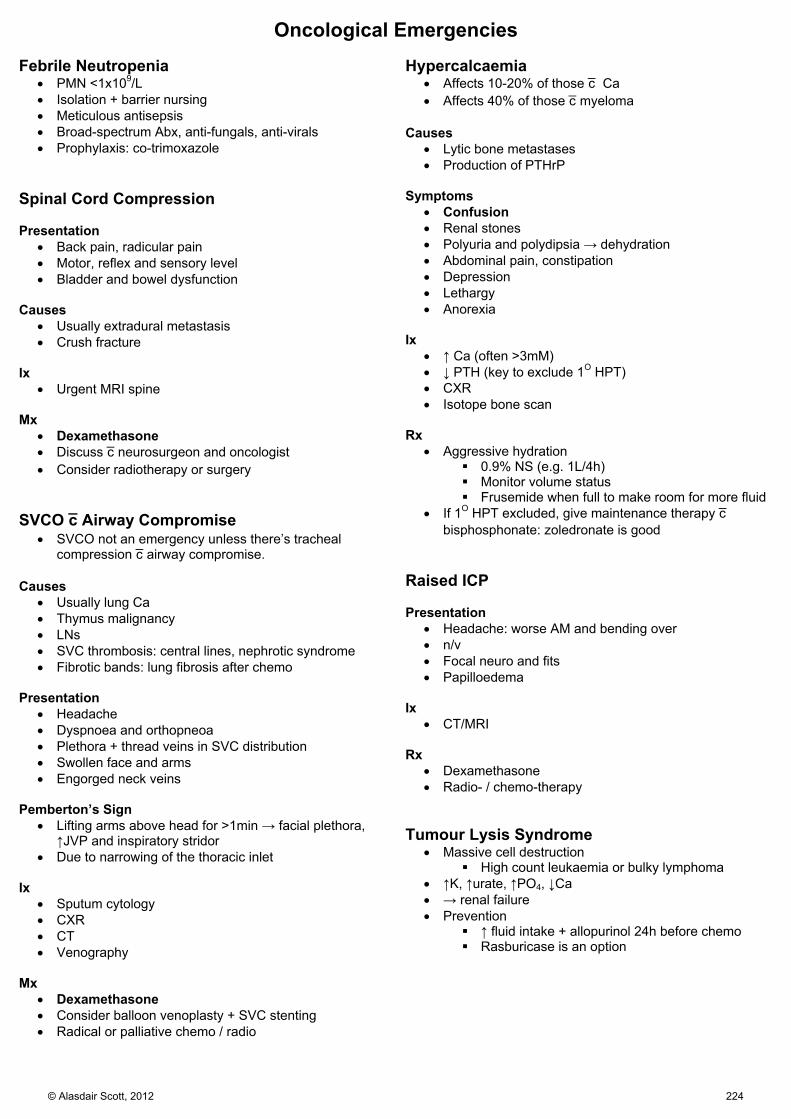
Oncological Emergencies Febrile Neutropenia
PMN <1x10⁹/L Isolation + barrier nursing Meticulous antisepsis Broad-spectrum Abx, anti-fungals, anti-virals Prophylaxis: co-trimoxazole
Spinal Cord Compression Presentation
Back pain, radicular pain Motor, reflex and sensory level Bladder and bowel dysfunction
Causes
Usually extradural metastasis Crush fracture
Ix
Urgent MRI spine Mx
Dexamethasone Discuss c̄ neurosurgeon and oncologist Consider radiotherapy or surgery
SVCO c̄ Airway Compromise
SVCO not an emergency unless there’s tracheal compression c̄ airway compromise.
Causes
Usually lung Ca Thymus malignancy LNs SVC thrombosis: central lines, nephrotic syndrome Fibrotic bands: lung fibrosis after chemo
Presentation
Headache Dyspnoea and orthopneoa Plethora + thread veins in SVC distribution Swollen face and arms Engorged neck veins
Pemberton’s Sign
Lifting arms above head for >1min → facial plethora, ↑JVP and inspiratory stridor
Due to narrowing of the thoracic inlet Ix
Sputum cytology CXR CT Venography
Mx
Dexamethasone Consider balloon venoplasty + SVC stenting Radical or palliative chemo / radio
Hypercalcaemia Affects 10-20% of those c̄ Ca Affects 40% of those c̄ myeloma
Causes
Lytic bone metastases Production of PTHrP
Symptoms
Confusion Renal stones Polyuria and polydipsia → dehydration Abdominal pain, constipation Depression Lethargy Anorexia
Ix
↑ Ca (often >3mM) ↓ PTH (key to exclude 1O HPT) CXR Isotope bone scan
Rx
Aggressive hydration 0.9% NS (e.g. 1L/4h) Monitor volume status Frusemide when full to make room for more fluid
If 1O HPT excluded, give maintenance therapy c̄ bisphosphonate: zoledronate is good
Raised ICP Presentation
Headache: worse AM and bending over n/v Focal neuro and fits Papilloedema
Ix
CT/MRI Rx
Dexamethasone Radio- / chemo-therapy
Tumour Lysis Syndrome
Massive cell destruction High count leukaemia or bulky lymphoma
↑K, ↑urate, ↑PO4, ↓Ca → renal failure Prevention
↑ fluid intake + allopurinol 24h before chemo Rasburicase is an option
© Alasdair Scott, 2012 224
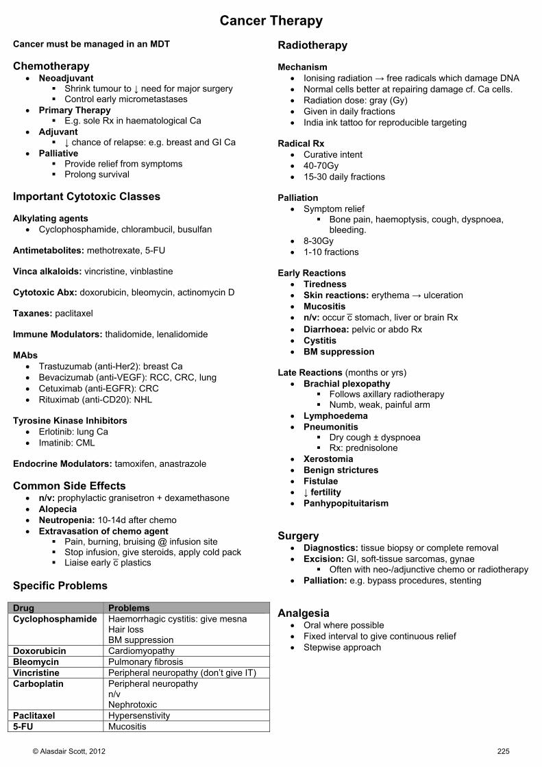
Cancer Therapy Cancer must be managed in an MDT Chemotherapy
Neoadjuvant Shrink tumour to ↓ need for major surgery Control early micrometastases
Primary Therapy E.g. sole Rx in haematological Ca
Adjuvant ↓ chance of relapse: e.g. breast and GI Ca
Palliative Provide relief from symptoms Prolong survival
Important Cytotoxic Classes Alkylating agents
Cyclophosphamide, chlorambucil, busulfan Antimetabolites: methotrexate, 5-FU Vinca alkaloids: vincristine, vinblastine Cytotoxic Abx: doxorubicin, bleomycin, actinomycin D Taxanes: paclitaxel Immune Modulators: thalidomide, lenalidomide MAbs
Trastuzumab (anti-Her2): breast Ca Bevacizumab (anti-VEGF): RCC, CRC, lung Cetuximab (anti-EGFR): CRC Rituximab (anti-CD20): NHL
Tyrosine Kinase Inhibitors
Erlotinib: lung Ca Imatinib: CML
Endocrine Modulators: tamoxifen, anastrazole Common Side Effects
n/v: prophylactic granisetron + dexamethasone Alopecia Neutropenia: 10-14d after chemo Extravasation of chemo agent
Pain, burning, bruising @ infusion site Stop infusion, give steroids, apply cold pack Liaise early c̄ plastics
Specific Problems Drug Problems Cyclophosphamide Haemorrhagic cystitis: give mesna
Hair loss BM suppression
Doxorubicin Cardiomyopathy Bleomycin Pulmonary fibrosis Vincristine Peripheral neuropathy (don’t give IT) Carboplatin Peripheral neuropathy
n/v Nephrotoxic
Paclitaxel Hypersenstivity 5-FU Mucositis
Radiotherapy Mechanism
Ionising radiation → free radicals which damage DNA Normal cells better at repairing damage cf. Ca cells. Radiation dose: gray (Gy) Given in daily fractions India ink tattoo for reproducible targeting
Radical Rx
Curative intent 40-70Gy 15-30 daily fractions
Palliation
Symptom relief Bone pain, haemoptysis, cough, dyspnoea,
bleeding. 8-30Gy 1-10 fractions
Early Reactions
Tiredness Skin reactions: erythema → ulceration Mucositis n/v: occur c̄ stomach, liver or brain Rx Diarrhoea: pelvic or abdo Rx Cystitis BM suppression
Late Reactions (months or yrs)
Brachial plexopathy Follows axillary radiotherapy Numb, weak, painful arm
Lymphoedema Pneumonitis
Dry cough ± dyspnoea Rx: prednisolone
Xerostomia Benign strictures Fistulae ↓ fertility Panhypopituitarism
Surgery
Diagnostics: tissue biopsy or complete removal Excision: GI, soft-tissue sarcomas, gynae
Often with neo-/adjunctive chemo or radiotherapy Palliation: e.g. bypass procedures, stenting
Analgesia
Oral where possible Fixed interval to give continuous relief Stepwise approach
© Alasdair Scott, 2012 225

Tumour Markers Marker Malignant Non-malignant AFP HCC
Teratoma Hepatitis Cirrhosis Pregnancy
CA 125 Ovary Uterus Breast
Cirrhosis Pregnancy
CA 15-3 Breast Benign breast disease CA 19-9 Pancreas
Cholangiocarcinoma CRC
Cholestasis Pancreatitis
CA 27-29 Breast Neuron-specific enolase
SCLC
CEA CRC Pancreatitis Cirrhosis
βHCG Germ cell tumour Pregnancy PSA Prostate BPH Mono Ig Multiple myeloma S-100 Melanoma Sarcoma PLAP Seminoma Acid phosphatases Prostate Ca Thyroglobulin Thyroid Ca
© Alasdair Scott, 2012 226

Immunology Contents
Phagocyte Deficiencies ............................................................................................................................................................. 228
T Cell Deficiency ........................................................................................................................................................................ 228
B Cell Deficiency ....................................................................................................................................................................... 229
Complement Dysfunction .......................................................................................................................................................... 230
Infections Associated With Immune Deficiencies ...................................................................................................................... 231
© Alasdair Scott, 2012 227
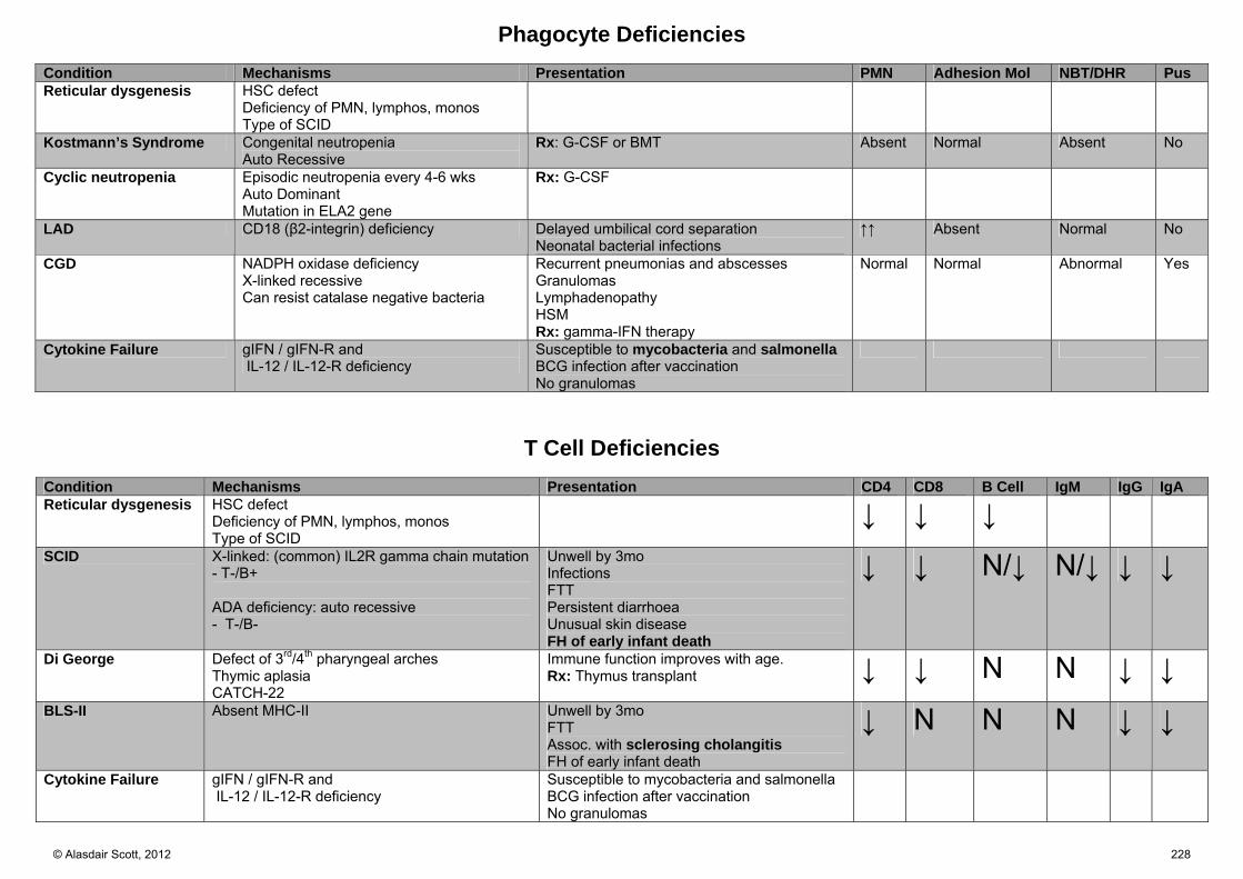
Phagocyte Deficiencies
Condition Mechanisms Presentation PMN Adhesion Mol NBT/DHR Pus Reticular dysgenesis HSC defect
Deficiency of PMN, lymphos, monos Type of SCID
Kostmann’s Syndrome Congenital neutropenia Auto Recessive
Rx: G-CSF or BMT Absent Normal Absent No
Cyclic neutropenia Episodic neutropenia every 4-6 wks Auto Dominant Mutation in ELA2 gene
Rx: G-CSF
LAD CD18 (β2-integrin) deficiency Delayed umbilical cord separation Neonatal bacterial infections
↑↑ Absent Normal No
CGD NADPH oxidase deficiency X-linked recessive Can resist catalase negative bacteria
Recurrent pneumonias and abscesses Granulomas Lymphadenopathy HSM Rx: gamma-IFN therapy
Normal Normal Abnormal Yes
Cytokine Failure gIFN / gIFN-R and IL-12 / IL-12-R deficiency
Susceptible to mycobacteria and salmonella BCG infection after vaccination No granulomas
T Cell Deficiencies
Condition Mechanisms Presentation CD4 CD8 B Cell IgM IgG IgA Reticular dysgenesis HSC defect
Deficiency of PMN, lymphos, monos Type of SCID
↓ ↓ ↓ SCID X-linked: (common) IL2R gamma chain mutation
- T-/B+ ADA deficiency: auto recessive - T-/B-
Unwell by 3mo Infections FTT Persistent diarrhoea Unusual skin disease FH of early infant death
↓ ↓ N/↓ N/↓ ↓ ↓
Di George Defect of 3rd/4th pharyngeal arches Thymic aplasia CATCH-22
Immune function improves with age. Rx: Thymus transplant ↓ ↓ N N ↓ ↓
BLS-II Absent MHC-II Unwell by 3mo FTT Assoc. with sclerosing cholangitis FH of early infant death
↓ N N N ↓ ↓
Cytokine Failure gIFN / gIFN-R and IL-12 / IL-12-R deficiency
Susceptible to mycobacteria and salmonella BCG infection after vaccination No granulomas
© Alasdair Scott, 2012 228

B Cell Deficiencies
Condition Mechanisms Clinical CD4 CD8 B Cell IgM IgG IgA SCID X-linked: (common) IL2R gamma chain mutation
- T-/B+ ADA deficiency: auto recessive - T-/B-
Unwell by 3mo Infections FTT Persistent diarrhoea Unusual skin disease FH of early infant death
↓ ↓ N/↓ N/↓ ↓ ↓
Bruton’s X-linked B-cell tyrosine kinase deficiency Pre-B cells cannot mature
No Ig after ~3mo Recurrent childhood infections - bacterial - enterovirus
N N ↓ ↓ ↓ ↓
Hyper IgM X-linked CD40L (CD154) mutation (T cells) No germinal centre development in LN/spleen Failure of isotype switching
Boys present in first few yrs Recurrent bacterial infections FTT PCP, autoimmune disease, Ca
N N N ↑↑ ↓ ↓
IgA deficiency Assoc. with coeliac disease Recurrent GIT and RT infections Not severe
N N N N N ↓ Common variable ID Recurrent bacterial infections
- bronchiectasis - sinusitis Autoimmune disease Granulomatous disease
N N N N ↓ ↓
Wiskott-Aldrich WASP gene mutation X-linked
Immunodeficiency Thrombocytopenia Eczema Malignant Lymphoma ↑ IgE
↓ ↕ ↑
© Alasdair Scott, 2012 229

Complement Dysfunction
Pathway Deficiency Outcome Classical C1, C2, C4
C2 deficiency = commonest SLE
MBL MBL deficiency ↑ infection is patient’s with other cause of immune impairment - prems, chemo, HIV, Ab deficiency
Alternative B, I, P deficiency Infection (esp. with encapsulates) - Meningococcus - Pneumococcus - H. influenzae - Hib FH of infection
Terminal pathway C5-9 deficiency Inability to make MAC
C3 deficiency C3 deficiency Acquired C3 deficiency due to C3 nephritic factor – assoc. with membranoproliferative (mesangiocapillary) GN
Infections as above + SLE
Ix
C3, C4 level CH50: classical pathway AP50: alternative pathway If CH50 and AP50 both abnormal with normal C3, C4 = final common pathway
deficiency (C5-9)
Hereditary Angioedema / C1 Esterase Deficiency
Auto dominant: commoner than complement deficiencies Acquired form may be caused by splenic marginal zone lymphoma
Pathophysiology
Serpin that inhibits C1 F12 of coag cascade Kallikrein (Cleaves HMWK → Bradykinin)
Deficiency → ↑ bradykinin → symptoms Presentation
Attacks may be ppted by EtOH, emotional stress, exercise. Angioedema but NO rash and NOT itchy
Skin Oropharynx – asphyxia GIT – nausea, vomiting diarrhoea
↓ C2 and ↓C4 (normal C1 and C3)
© Alasdair Scott, 2012 230
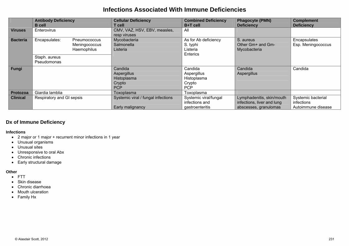
Infections Associated With Immune Deficiencies
Antibody Deficiency B cell
Cellular Deficiency T cell
Combined Deficiency B+T cell
Phagocyte (PMN) Deficiency
Complement Deficiency
Viruses Enterovirus CMV, VAZ, HSV, EBV, measles, resp viruses
All
Bacteria Encapsulates: Pneumococcus Meningococcus Haemophilus
Mycobacteria Salmonella Listeria
As for Ab deficiency S. typhi Listeria Enterics
S. aureus Other Gm+ and Gm- Mycobacteria
Encapsulates Esp. Meningococcus
Staph. aureus Pseudomonas
Fungi Candida Aspergillus Histoplasma Crypto PCP
Candida Aspergillus Histoplasma Crypto PCP
Candida Aspergillus
Candida
Protozoa Giardia lamblia Toxoplasma Toxoplasma Clinical Respiratory and GI sepsis Systemic viral / fungal infections
Early malignancy
Systemic viral/fungal infections and gastroenteritis
Lymphadenitis, skin/mouth infections, liver and lung abscesses, granulomas
Systemic bacterial infections Autoimmune disease
Dx of Immune Deficiency Infections
2 major or 1 major + recurrent minor infections in 1 year Unusual organisms Unusual sites Unresponsive to oral Abx Chronic infections Early structural damage
Other
FTT Skin disease Chronic diarrhoea Mouth ulceration Family Hx
© Alasdair Scott, 2012 231

Dermatology Contents
Malignant and Pre-malignant Skin Lesions ............................................................................................................................... 233
Psoriasis .................................................................................................................................................................................... 234
Eczema ...................................................................................................................................................................................... 235
Pruritus ...................................................................................................................................................................................... 235
Skin Infections ........................................................................................................................................................................... 236
Miscellaneous Skin Disorders ................................................................................................................................................... 237
Acne Vulgaris ............................................................................................................................................................................ 237
Acne Rosacea ........................................................................................................................................................................... 237
Drug Eruptions ........................................................................................................................................................................... 238
Infestations ................................................................................................................................................................................ 238
Skin Manifestations of Systemic Disease.................................................................................................................................. 239
© Alasdair Scott, 2012 232

Malignant and Pre-malignant Skin Lesions Malignant Melanoma Epidemiology
F>M = 1.5:1 UK incidence = 10 000/yr and 2000 deaths/yr
↑80% in 20yrs Features
Asymmetry Boarder: irregular Colour: non-uniform Diameter > 6mm Evolving / Elevation
Risk Factors
Sunlight: esp. intense exposure in early years. Fair skinned (Low Fitzpatrick Skin Type) ↑ no. of common moles +ve FH ↑ age Immunosuppression
Classification
Superficial Spreading: 80% Irregular boarders, colour variation Commonest in Caucasians Grow slowly, metastasise late = better prognosis
Lentigo Maligna Melanoma Often elderly pts. Face or scalp
Acral Lentiginous Asians/blacks Palms, soles, subungual (c̄ Hutchinson’s sign)
Nodular Melanoma All sites Younger age, new lesion Invade deeply and metastasis early = poor prog
Amelanotic Atypical appearance → delayed Dx
Staging and Prognosis
Breslow Depth Thickness of tumour to deepest point of dermal
invasion <1mm = 95-100% 5ys >4mm = 50% 5ys
Clark’s Staging Stratifies depth by 5 anatomical levels Stage 1: Epidermis Stage 5: sc fat
Metastasis
Liver Eye
Mx
Excision + 2O margin excision depending on Bres depth ± lymphadenectomy ± adjuvant chemo (may use isolated limb perfusion)
Poor prognostic indicators
Male sex (more tumours on trunk cf females) ↑ mitoses Satellite lesions (lymphatic spread) Ulceration
Squamous Cell Carcinoma
Ulcerated lesion c̄ hard, raised everted edges Sun exposed areas
Causes
Sun exposure: scalp, face, ears, lower leg May arise in chronic ulcers: Marjolin’s Ulcer Xeroderma pigmentosa
Evolution
Solar/actinic keratosis → Bowen’s → SCC Lymph node spread is rare
Rx
Excision + radiotherapy to affected nodes Actinic Keratoses
Irregular, crusty warty lesions. Pre-malignant (~1%/yr)
Rx
Cautery Cryo 5-FU Imiquimod Photodynamic phototherapy
Bowen’s Disease
Red/brown scaly plaques SCC in situ
Rx As for AKs
Keratoacanthoma
A well differentiated SCC that arises in a hair follicle. Fast-growing, dome-shaped c̄ a keratin plug. Usually regress but may be excised
Basal Cell Carcinoma
Commonest cancer Pearly nodule c̄ rolled telangiectactic edge May ulcerate Typically on face in sun-exposed area
Above line from tragus → angle of mouth Behaviour
Low-grade malignancy → very rarely metastasise Locally invasive
Rx
Excision Mohs: complete circumferential margin
assessment using frozen section histology Cryo/radio may be used.
© Alasdair Scott, 2012 233

Psoriasis Epidemiology
Prevalence: 2% of Caucasians Age: peaks in 20s and 50s Sex: F=M Genetics: 30% have FH
Genetic predisposition Pathology
TIV hypersensitivity reaction Epidermal proliferation T-cell driven inflammatory infiltration Histo
Acanthosis: thickening of the epidermis Parakeratosis: nuclei in stratum corneum Munro’s microabscesses: neutrophils
Triggers
Stress Infections: esp. streps Skin trauma: Kobner phenomenon Drugs: β-B, Li, anti-malarials, EtOH Smoking
Signs Plaques
Symmetrical well-defined red plaques c̄ silvery scale Extensors: elbows, knees Flexures (no scales): axillae, groins, submammary Scalp, behind ears, navel, sacrum
Nail Changes (in 50%)
Pitting Onycholysis Subungual hyperkeratosis
10-40% Develop Seronegative Arthritis
Mono-/oligo-arthritis: DIPs commonly involved Rheumatoid-like Asymmetrical polyarthritis Psoriatic spondylitis Arthritis mutilans May → dactylitis
Variants Guttate
Drop-like salmon-pink papules c̄ fine scale Mainly on trunk Occurs in children assoc. c̄ strep infection
Pustular
Sterile pustules May be localised to palms and soles
Erythroderma and Generalised Pustular
Generalised exfoliative dermatitis Severe systemic upset: fever, ↑WCC, dehydration May be triggered by rapid steroid withdrawal
Differential
Eczema Tinea: asymmetrical Seborrhoeic dermatitis
Mx Education
Avoid triggers Soap Substitute
Aqueous cream Dermol cream Epaderm ointment
Emollients
Epaderm Dermol Diprobase
Topical Therapy
Vit D3 analogue: e.g. calcipotriol Steroids: e.g. betamethasone
Dovobet = Calcipotriol + betamethasone Tar: mainly reserved for in-patient use Dithranol Retinoids: e.g. tazarotene
UV Phototherapy
Causes local immunosuppression Narrow band UVB Psoralen + UVA: PUVA
Psoralen is a photsensitising agent and can be topical or oral
PUVA is more effective but ↑ skin Ca risk Non-Biologicals
Methotrexate Ciclosporin Acetretin (oral retinoid / vit A analogue)
SE: ↑ lipids, ↑ glucose Biologicals
Infliximab Etanercept Adalimumab
© Alasdair Scott, 2012 234

Eczema Presentation
Extremely itchy Poorly demarcated rash
Acute: oozing papules and vesicles Subacute: red and scaly
Chronic eczema → lichenification Skin thickening c̄ exaggeration of skin markings
Pathology
Epidermal spongiosus Atopic Eczema
TH2 driven inflammation c̄ ↑IgE production Affects 2% of infants Most children grow-out of it by 13yrs
Cause
FH of atopy common Specific allergens
House dust mite Animal dander
Diet: e.g. dairy products Presentation
Face: esp. around eyes, cheeks Flexures: knees, elbows May become 2O infected
Staph → fluclox HSV → aciclovir
Associations
Asthma Hay fever
Ix
↑ IgE RAST testing: identify specific Ag
Irritant Contact Dermatitis
Everyone is susceptible to irritants Causes: detergents, soaps, oils, solvents, venous stasis
Allergic Contact Dermatitis
Type IV hypersensitivity reaction Common allergens
Nickel: jewellery, watches, coins Chromates: leather Lanolin: creams, cosmetics
Location: correlates sharply c̄ allergen exposure Ix: Patch testing
Adult Seborrhoeic Dermatitis
Red, scaly, rash Cause: overgrowth of skin yeasts (e.g. malassezia) Location: scalp (dandruff), eyebrows, cheeks,
nasolabial folds Rx: mild topical steroids / antifungal
Daktacort: miconazole + hydrocortisone
Mx of Atopic Eczema Education
Avoid triggers: e.g. soap Soap Substitute
Aqueous cream Dermol cream Epaderm ointment
Emollients
Epaderm Dermol Diprobase Oilatum (bath oil)
Topical Therapy
Steroids 1% Hydrocortisone: face, groins Eumovate: can use briefly (<1wk) on face Betnovate Dermovate: very strong, brief use on thick skin
Palms, soles 2nd line Therapies
Topical tacrolimus Phototherapy Ciclosporin or azathioprine
Pruritus Generalised
CRF Cholestasis Haematological
Polycythaemia Hodgkin’s Leukaemia Iron deficiency
Endocrine DM Hyper- / hypo-thyroidism Pregnancy
Very Itchy Dermatological Diseases
Eczema Urticaria Scabies Dermatitis herpetiformis
© Alasdair Scott, 2012 235

Skin Infections Tinea
A superficial mycosis caused by dermatophytes Microsporum Epidermophyton Trichophyton
Presentation
Round scaly lesion Itchy Central clearing Scalp, body, foot, groin, nails
Rx
Skin: terbinafine or topical ketoconazole / miconazole Scalp: griseofulvin or terbinafine Nails: terbinafine
Candida
Common RFs: immunosuppression, Abx, steroid inhalers
Presentation
Pink + white patches Moist Satellite lesions Mouth, vagina, skin folds, toe web
Rx
Mouth: nystatin Vagina: clotrimazole cream and pessary
Pityriasis versicolor
Caused by Malassezia furfur Presentation
Common in hot and humid environments Circular hypo-/hyper-pigmented patches Fine white scale Itchy Back of neck and trunk
Ix
“Spaghetti and meatballs” appearance c̄ KOH stain Rx
Selenium sulphate or ketoconazole shampoos Impetigo
Contagious superficial skin rash caused by S. aureus Presentation
Age: peak @ 2-5yrs Honey-coloured crusts on erythematous base Common on face
Rx
Mild: topical Abx (fusidic acid, mupirocin) More severe: fluclox PO
Erysipelas
Sharply defined superficial infection by S. pyogenes Often affects the face High fever + ↑ WCC Rx
Benpen IV Pen V and fluclox PO
Cellulitis
Acute infection of skin and soft tissues Cause: β-haemolytic Strep + staph. aureus Presentation
Deeper and less well defined than erysipelas Pain, swelling, erythema and warmth Systemic upset ± lymphadenopathy
Rx
Empiric: fluclox IV Confirmed Strep: Benpen or Pen V Pen allergic: clindamycin
Warts
Caused by HPV infection Rx
Expectant Destructive
Topical salicylic acid Cryotherapy Podophyllin Imiquimod
Molluscum
Pox virus Pink papules c̄ umbilicated central punctum Resolve spontaneaously
Herpes Zoster / Shingles
Recurrent VZV infection Dermatomal distribution of cropping vesicles and crust
Thoracic: 50% Ophthalmic: 20%
Cornea affected in 50% → keratitis, iritis May → post-herpetic neuralgia Rx: aciclovir or famciclovir PO if severe
Herpes Simplex
Gingivostomatitis or recurrent genital or oral infections Triggered by infection (e.g. CAP), sunlight and
immunosuppression May complicate eczema: eczema herpeticum Grouped painful vesicles on an erythematous base Rx: acyclovir or famciclovir indicated if
immunosuppressed or recurrent genital herpes. Pityriasis Rosea
HHV-6/-7 Herald patch precedes rash, mainly on the trunk
© Alasdair Scott, 2012 236

Miscellaneous Skin Disorders Lichen Planus Skin Presentation
Flexors: wrists, forearms, ankles, legs Display Kobner phenomenon Purple Pruritic Polygonal Planar Papules Lacy white marks: Wickham’s Striae
Lesions Elsewhere
Scalp: scarring alopecia Nails: longitudinal ridges Mouth: lacy white plaques on inner cheeks Genitals
Rx
Mild: topical steroids Severe: systemic steroids
Bullous Pemphigoid
Autoimmune blistering disease due to auto-abs against hemidesmosomes
Presentation
Mainly affects the elderly Tense bullae on erythematous base Can be itchy
Ix
Biopsy shows linear IgG along the BM and subepidermal bullae
Rx
Clobetasol (Dermavate) Pemphigus Vulgaris
Autoimmune blistering disease due to auto-abs against desmosomes.
May be ppted by drugs NSAIDs ACEi L-dopa
Presentation
Younger pts. Large flaccid bullae which rupture easily Nikolsky’s sign +ve Mucosa is often affected
Ix
Intraepidermal bullae Rx
Prednisolone Rituximab IVIg
Acne Vulgaris Pathophysiology
↑ sebum production: androgens and CRH P. acnes is a skin commensal that flourishes in the
anaerobic environment of the blocked follicle → inflammation
Presentation
Inflammation of pilosebaceus follicles Comedones (white- or black-heads), papules, pustules
nodules, cysts Face, neck, upper chest and back
Mx
Pt. education Remember that topical therapy is difficult to apply to the
back. Mild: topical therapy
Benzoyl peroxide Erythromycin, Clindamycin Tretinoin / Isotretinoin
Moderate
Topical benzoyl peroxide + oral Abx (doxy or erythro) Severe
Isotretinoin (vitamin A analogue) 60-70% have no further recurrence SE: teratogenic, hepatitis, ↑lipids, depression, dry
skin, myalgia Monitor: LFTs, lipids, FBC
May try Dianette in women
Acne Rosacea Chronic relapsing remitting disorder affecting the face
Presentation
Chronic flushing ppted. by alcohol or spicy foods. Fixed erythema: chin, nose, cheeks, forehead Telangiectasia, papules, pustules (no comedones)
Associations
Rhinophyma: swelling and soft tissue overgrowth of the nose in males
Blepharitis: scaling and irritation at the eyelashes Rx
Avoid sun exposure Topical azelaic acid Oral doxycycline or azithromycin
© Alasdair Scott, 2012 237

Drug Eruptions Maculopapular
Commonest type Generalized erythematous macules and papules ± fever and ↑ eosinophils Develops w/i two weeks of onset of drug Penicillins, cephalosporins, AEDs
Urticaria
Wheals ± angioedema and anaphylaxis Rapid onset after taking drug Immune (IgE)
Penicillins Cephalosporins
Non-immune (direct mast cell degranulation) Morphine Codeine NSAIDs Contrast
Erythema Multiforme
Symmetrical target lesions on palms, soles and limbs Occurs 1-2wks after insult Infections are commoner cause of EM
Causes
Idiopathic Infections: HSV, mycoplasma Drugs
Sulphonamides, NSAIDs, allopurinol, penicillin phenytoin
Stevens-Johnson Syndrome
More severe variant of EM, Blistering mucosa: conjunctiva, oral, genital
Toxic Epidermal Necrolysis
Extreme form of SJS Nearly always a drug reaction Features
Severe mucosal ulceration Widespread erythema followed by epidermal necrosis
c̄ loss of large sheets of epidermis → dehydration
↑↑↑ risk in HIV+ Rx: dexamethasone, IVIg Mortality: 30%
Infestations Scabies
Sarcoptes scabei Highly contagious: spread by direct contact Female mite digs burrows and lays eggs
Presentation
Burrows: short, serpiginous grey line, block dot Hypersensitivity rash: eczematous, vesicles Extremely itchy → escoriation Particularly affects the finger web spaces (esp. 1st) Also: axillae, groin, umbilicus
Mx
Permethrin cream: applied from neck down for 24hrs
2nd line: Malathion 3rd line: oral ivermectin Treat all members of the household
Headlice
Pediculus humanus capitis Spread by head to head contact Nits = empty eggs
Presentation
Itch Papular rash @ the nape of the neck
Rx
Malathion Combing
© Alasdair Scott, 2012 238

Skin Manifestations of Systemic Disease Skin Conditions Erythema Nodosum
Painful blue-red lesions on anterior shins 3 S’s: sarcoid, strep infections and sulfonamides Also: OCP, IBD and TB, Behcet’s
Erythema Multiforme
Symmetrical target lesions on palms, soles and limbs Possibly mediated by IgM deposition Usually 2O to infection Infections: HSV (70%), mycoplasma Drugs: SNAPP
Stevens-Johnson Syndrome and TEN
Severe variants of EM Nearly always drug-induced Fever, mucosal and skin ulceration and necrosis
Pyoderma Gangrenosum
Wide (10cm), deep ulceration c̄ violaceous border Purulent surface Undermined edge Commonly occurs on legs Heal c̄ cribriform (pitted) scars Assocs.: IBD, AIH, Wegener’s, RA, leukaemia Rx: High-dose systemic steroids
Vitiligo
Chalky white patches c̄ hyperpigmented borders Itch in sunlight Assocs.: autoimmune disorders
Livedo Reticularis
Persistent mottled red/blue lesions that don’t blanch Commonly found on the legs Triggered by cold Causes
Idiopathic Vasculitis: RA, SLE, PAN Obstruction: anti-phos, cryoglobulinaemia Sneddon’s syn.: LR + CVAs
Diseases Disease Skin Manifestation Rheumatic fever Erythema marginatum Lyme (Borrelia burgdorferi)
Erythema chronicum migrans
Crohn’s Perianal ulcers and fistulae EN Pyoderma gangrenosum
Dermatomyositis Heliotrope rash on eyelids Shawl sign (macular rash) Gottron’s Papules Mechanic’s hands Nailfold erythema, telangiectasia
DM Ulcers Candida Kyrle disease Acanthosis nigricans Necrobiosis lipoidica (shins) Granuloma annulare (hands, feet)
Sarcoidosis Erythema nodosum Erythema multiforme Lupus pernio Hypopigmented areas Red/violet plaques
Coeliac Dermatitis herpetiformis (elbows) Graves’ Pre-tibial myxoedema (lat mal) RA Rheumatoid nodules
Vasculitis (palpable purpura) SLE Facial butterfly rash Systemic sclerosis Calcinosis
Raynaud’s Sclerodactyly Telangiectasia Generalised skin thickening
Liver disease Palmar erythema Spider naevi Gynaecomastia ↓ 2O sexual hair Jaundice Bruising Excoriations
ESRD Assoc. c̄ Cause - DM, vasculitis, sclero, RA, SLE Assoc. c̄ ESRD - Pruritus, xerosis, pigment change Bullous disease Assoc. c̄ Transplant - Cushingoid, gingival hyperplasia, Infections, BCC, SCC, melanoma Kaposi’s
Neoplasia Acanthosis nigricans Dermatomyositis Thrombophlebitis migrans Acquired ichthyosis
© Alasdair Scott, 2012 239

Epidemiology Contents
Health Information ..................................................................................................................................................................... 241
Public Health Issues .................................................................................................................................................................. 242
Clinical Evidence ....................................................................................................................................................................... 243
Epidemiological Concepts ......................................................................................................................................................... 244
Disease Prevention ................................................................................................................................................................... 245
Likelihood Ratios ....................................................................................................................................................................... 245
© Alasdair Scott, 2012 240
Health Information Routine Data
Data that are routinely collected and recorded in an on-going systematic way, often for administrative purposes.
Advantages Relatively cheap Already collected and available Standardised collection procedures
Disadvantages
May not answer the question Variable quality Not every case captured
Sources
Demographics: census Health: births, deaths, admissions, prescriptions, hospital
episode statistics, cancer Exposures: smoking, air pollution, crime Disease prevention: screening, immunisation uptake
Hospital Episode Statistics
Electronic record of every inpatient or day-case episode of patient care in every NHS hospital.
300 fields of information, including: Pt demographics Diagnosis using ICD-10 Procedures using OPCS4
Advantages
Comprehensive Unbiased Based on case notes
Disadvantages
Data may not be complete In-hospital death only
Mortality UK
1. Heart and Circulatatory disorders: 33% a. IHD (23%) b. Stroke (10%)
2. Cancer: 30% a. Trachea, bronchus, lung b. Breast, prostate c. Colorectum
3. Respiratory disorders: 13% a. Pneumonia b. COPD
Worldwide
1. IHD 2. Stroke and other cerebrovascular disease 3. LRTI 4. COPD 5. Diarrhoeal diseases
Clinical Governance Definition
A transparent system for the continual maintenance and improvement of healthcare standards both on an individual and organisational level within the NHS.
Elements
Education and Training Clinical audit Clinical effectiveness Research and Development Openness Risk Management
Agencies
The Care Quality Commission Independently inspect healthcare services
against standards set by the Department of Health.
Investigate serious failures in healthcare services.
Publish regular ratings of NHS trusts. Clinical Audit
Are pts. given the best care? Definition
A quality improvement process that seeks to improve pt. care and outcomes through the systematic review of care against explicit criteria and the implementation of change.
Outcome Measures
Are pts. better and do they feel better? Patient Reported Outcomes (PROMs)
Calculate the health gain from the pts. perspective after surgical Rx using pre- and post-operative surveys. Arthroplasty, hernia repairs, varicose veins Hip replacement gives largest improvement in
specific condition and general health. Quality and Outcomes Framework (QOF)
Voluntary annual reward and incentive programme for all GP surgeries in England, detailing practice achievement results.
© Alasdair Scott, 2012 241

Public Health Issues Major Contributions to Health
Drugs and medical intervention Obstetric and perinatal care Immunisation Screening Chronic disease Mx
Major Mortality Risk Factors
Heart and Circulatory Disorders Smoking ↓ exercise Diet: ↑fat, ↑calorie, ↑salt, EtOH ?Stress
Neoplasms Smoking ↓ exercise Diet: ↓fruit and veg, ↓fibre, ↑fat
Pulmonary Disease Smoking ↓ exercise Occupation: asbestos, dust exposure
Major Public Health Issues
Smoking Obesity / unhealthy diet ↓ Exercise EtOH abuse Sexual health Mental health
Obesity
20% obese 50% of females overweight or obese
Nurses’ Health Study
Overwt. → ↓3yrs of life Obese → ↓7yrs of life Active lifestyle (30min walk/d) prevents 30% of obesity
and 45% of new DM. Evidence for Changing Nutrition
Mediterranean diet can ↓ cardiac death post-MI Low Na diet → ↓ BP
Sexual Health
10% of 16-24yr olds have ≥1 STI ↑ in urban areas and amongst Blacks and Minorities → infertility, ectopics, Cervical Ca, HIV
Some Strategies
Improving access to sexual health services Chlamydia screening
Smoking
Commonest cause of preventable death in the UK 2nd hand smoking
Respiratory infections Asthma SIDS ↑ lung Ca and CHD risk
Social gradient in mortality is partially explained by social gradient in smoking.
Some Strategies
Health education Restrict advertising Remove from display in shops ↑ price Smoking ban in public places Very brief advice
Alcohol
5% of men and 2% of women report problems Recommended
Men: 21u/wk or 3-4u/d Women: 14u/wk or 2-3u/d NB. 1u = 10ml EtOH
Some Strategies
National Alcohol Harm Reduction Strategy ↑ minimum price / unit
Mental Health
Common: ~15% lifetime risk Depression and anxiety disorders most commonly
Some Strategies
GPs get rewarded for depression screening Expansion of psychological therapies (e.g. CBT)
© Alasdair Scott, 2012 242

Clinical Evidence Hierarchy of Evidence
1. Systematic reviews and meta-analyses 2. RCTs 3. Cohort or case-control studies 4. Descriptive studies 5. Case reports
Study Types Observational
Analytical / Aetiological Studies determinants of disease by judging
whether exposure causes or prevents disease Descriptive
Studies distribution of disease Person, place and time
Experimental
RCTs Descriptive Studies
Can be used to generate hypotheses Provide frequency data
Incidence Point prevalence
Can’t determine causation Examples
Cross-sectional Surveys Household interview surveys asking questions
regarding illness, social circumstances and demographics.
Cross-sectional Census Ecological studies
Use populations rather than individual as unit of observation.
Subject to significant confounding: age, sex, SES Other routine data
Case-Control Studies
Retrospective study of exposure in a case grp c̄ the disease and a control grp w/o the disease.
Proportion of exposed in each group → odds ratio Advantages
Quick and cheep Well-suited for diseases c̄ long latent periods Good for evaluation of rare diseases Can examine multiple aetiological factors for a single
disease. Disadvantages
Inefficient for evaluating rare exposures Cannot calculate incidence rates Temporal relationship between exposure and disease
can be difficult to establish Recall bias
E.g. testicular cancer sufferers more likely to report preceding trauma
Selection bias E.g. non-random selection of study grps.
Cohort Study
Prospective study comparing development of disease in exposed and non-exposed grps.
Incidence of disease in each grp → relative risk Examples
Million Women study Framingham Heart Study Nurses’ Health Study
Advantages
Good for evaluation of rare exposures Can examine multiple effects of a single exposure Can elucidate temporal relationship Direct incidence calculation
Disadvantages
Inefficient for evaluation of rare diseases Expensive and time-consuming Loss to f/up affects results
RCTs
Planned experiment designed to assess the efficacy of an intervention.
Randomisation: ↓ selection bias. Blinding: ↓ measurement bias
Advantages
Most reliable demonstration of causality Disadvantages
Non-compliance Loss to f/up Validity depends on quality of study Ethical issues Selection criteria may limit generalisability
Meta-Analyses
Observational study of evidence Systematic identification of relevant trials and
assessment of their quality Allow accurate interpretation of multiple RCTs
Forest plots
Square = OR Size = size of study
Line = 95% CI of OR Diamond = combined odds ratio
Width = 95% CI
© Alasdair Scott, 2012 243

Epidemiological Concepts 5 Steps of EBM
1. Question 2. Search 3. Appraise 4. Apply 5. Evaluate
Association and Causation
Association: a relationship between two factors Causation: X → Y
Bradford Hill Causation Criteria
Strong association: i.e ↑↑ RR or OR Consistency c̄ other investigations Specificity Temporal relationship Dose response relationship Plausibility Coherence Experimental evidence Analogy
Chance
Studies are based on estimates from samples and are therefore subject to random variation.
Random variation can be minimised by ↑ sample size. The role of chance is assessed by statistical significance
tests and by calculating confidence intervals. Bias
Inaccurate data due to systematic error in selection, measurement or analysis.
Selection Bias
Systematic difference between the characteristics of those selected for a study and those who were not or differences between study grps.
E.g. self-selection, healthy worker effect Measurement Bias
Occurs when measurements or classifications of disease or exposure are inaccurate.
E.g. inaccuracies of the instruments, expectations of observers or participants.
Analysis Bias
Error caused by participants being lost to f/up or switching treatment groups.
Take into account using an intention to treat analysis. Confounding
Error in the interpretation of an accurate measurement. Any factor which is prognostically linked to the outcome
and is unevenly distributed between study groups. Not a confounder if it lies on the causal pathway
between the variables of interest. Common confounders: age, sex, SES
Reducing Confounding
Randomisation Stratification: e.g. stratify by age Standardisation: e.g. SMR
Ratio of observed:expected deaths Expected deaths derived from larger population
Regression
Odds Ratio OR = odds of exposure in cases / odds of exposure in controls = (A/C) / (B/D) = AD/BC Case Control Exposed A B Not Exposed C D
OR = ~RR if disease is rare (<10%) Relative Risk
↑/↓ in probability of disease given a particular RF RR = incidence in exposed / incidence in unexposed = (A/A+B) / (C/C+D) Risk Estimates
Absolute risk: probability of disease occurrence Relative risk: ↑/↓ in probability of disease given a RF Attributable risk: measure of excess risk due a factor
Number Needed To Treat
NNT = (1 / Absolute risk reduction) x 100 Absolute risk reduction = 1- hazard ratio
Screening Modified Wilson Criteria
Disease Important health problem Well recognised pre-clinical stage Natural Hx well understood
Test Valid and reliable Simple and cheap Safe and acceptable
Dx and Rx Adequate facilities Acceptable Rx Early intervention is of more benefit than later Rx
Cost effective Validity
Sensitivity: detect true positives Specificity: exclude true negatives PPV: how likely to have disease if positive NPV: how likely not to have disease if negative
PPV and NPV depend on disease frequency Disease No Disease Positive A B PPV
A/(A+B) Negative C D NPV
C/(C+D) Sensitivity
A/(A+C) Specificity B/(B+D)
Effectiveness
Selection bias: healthiest come for screening Lead-time bias: disease Dx earlier → ↑ survival time Length-time bias: outcome appears better because
more disease c̄ good outcome detected.
© Alasdair Scott, 2012 244

Disease Prevention Primordial Prevention
Prevention of factors promoting the emergence of risk factors: lifestyle, behaviours, exposure patterns
Examples
Healthy eating programmes in schools Social policies to ↓ poverty and inequality Programmes that promote walking, cycling and public
transport. Encouragement of positive health behaviour.
Primary Prevention
Prevention of disease onset Examples
Vaccination Smoking cessation Healthy diet Condom use Detection and Rx of hyperlipidaemia
Secondary Prevention
Halting progression of established disease Early detection followed by prompt, effective Rx
Examples
Screening for Ca Smoking cessation after MI
Tertiary Prevention
Rehabilitation of people c̄ established disease to minimise residual disability and complications.
Examples
Rapid Rx of MI or stroke to ↓ disability HAART
Disease Prevention in General Practice
General advice for a healthy lifestyle Identifying and facilitating reduction of risk factors Ensuring availability of preventative interventions Identifying early disease and offering treatment
Prevention Paradox
Many people exposed to a small risk may generate more disease than a few exposed to a large risk.
when many people receive a small benefit the total benefit may be large.
However, individual inconvenience may be high to the many while benefit may only be to a few.
Health Promotion
Process of enabling people to increase control over, and to improve, their health. Public policy, supportive environments…
Health promotion is specific requirement of the GMC Good Medical Practice guidelines.
Likelihood Ratios Pre- and Post-test Probability
Pre-: estimate of probability that pt. has a certain disease based on presentation or epidemiology.
Test: diagnostic test or sign or symptom that you elicit Post-: new probability taking into account the “test”
result Likelihood ratio: measure of how much the test alters
your probability of the disease. Calculation Odds = probability / (1- probability)
i.e. the ratio of the probability something will happen to the probability that it won’t.
Probability = odds / (odds +1) LR = p [test result if disease present] / p [test result if disease absent] LR for positive test result = sensitivity / (1- specificity) LR for negative test result = (1- sensitivity) / specificity Post-test odds = pre-test odds x likelihood ratio Example
Pre-test probability could be disease incidence in that population.
Convert pre-test probability to odds. Calculate likelihood ratio based on the result of your test. Apply likelihood ratio to get post-test odds. Convert post-test odds to post-test probability
© Alasdair Scott, 2012 245

Emergencies Contents
Shock ......................................................................................................................................................................................... 247
Anaphylactic Shock ................................................................................................................................................................... 248
Narrow Complex Tachycardias = SVT ...................................................................................................................................... 249
Broad Complex Tachycardias ................................................................................................................................................... 250
STEMI ........................................................................................................................................................................................ 251
NSTEMI + UA ............................................................................................................................................................................ 252
Severe Pulmonary Oedema ...................................................................................................................................................... 253
Cardiogenic Shock .................................................................................................................................................................... 254
Meningitis .................................................................................................................................................................................. 255
Encephalitis ............................................................................................................................................................................... 256
Cerebral Abscess ...................................................................................................................................................................... 256
Status Epilepticus ...................................................................................................................................................................... 257
Head Injury ................................................................................................................................................................................ 258
Raised ICP ................................................................................................................................................................................ 259
Signs and Symptoms ................................................................................................................................................................ 259
Acute Severe Asthma ................................................................................................................................................................ 260
Acute Exacerbation of COPD .................................................................................................................................................... 261
Pulmonary Embolism................................................................................................................................................................. 262
Pneumothorax ........................................................................................................................................................................... 263
Upper GI Bleeding ..................................................................................................................................................................... 264
Acute Renal Failure ................................................................................................................................................................... 265
Poisoning: Recognition and Management ................................................................................................................................ 266
Diabetic Ketoacidosis ................................................................................................................................................................ 267
Hyperosmolar Non-Ketotic Coma .............................................................................................................................................. 268
Hypoglycaemia .......................................................................................................................................................................... 268
Thyroid Storm ............................................................................................................................................................................ 269
Myxoedema Coma .................................................................................................................................................................... 269
Addisonian Crisis ....................................................................................................................................................................... 270
Hypertensive Crisis.................................................................................................................................................................... 270
Burns ......................................................................................................................................................................................... 271
Hypothermia .............................................................................................................................................................................. 272
ALS Algorithm ............................................................................................................................................................................ 272
Tachycardia Algorithm ............................................................................................................................................................... 272
Bradycardia Algorithm ............................................................................................................................................................... 272
© Alasdair Scott, 2012 246

Shock
ABCDE
If ECG unrecordable, Rx as cardiac arrest
Raise foot of bed (unless cardiogenic)
IV Access: 2 wide bore (14g) cannula in each ACF
Causes : CHOD Cardiogenic MI Arrhythmia
Hypovolaemic Haemorrhage: internal and external Endocrine: Addisonion crisis, DKA Excess loss: burns, diarrhoea Third-spacing: pancreatitis
Obstructive PE Tension pneumothorax
Distributive Sepsis Anaphylaxis Neurogenic
Fast infusion of crystalloid to raise BP (unless cardiogenic)
Monitoring Catheter (>30ml/hr) Art line CVP line
Assessment History Chest pain: MI, PE, dissection, anaphylaxis Abdo pain: AAA, DKA, peritonitis, ruptured ectopic Back pain: AAA
Hands ↑CRT: hypovolaemia, cardiogenic
Pulse Tachy (unless β-B or bradyarrhythmia) Small, thread: hypovolaemia Bounding: sepsis
ABP ↓PP: hypovolaemia R-L differential > 20mmHg: dissection
Appearance Cold, clammy: cardiogenic or hypovolaemic Warm, well perfused: septic Urticaria, angioedema, wheeze: anaphylaxis
Ix FBC, U+E, glucose, ABG, CRP, trop X-match, clotting Blood cultures, urine MCS ECG, CXR, USS, Echo, CT
JVP ↑: cardiogenic ↓: hypovolaemic, distributive
Abdomen Tender, guarding: trauma, aneurysm, peritonitis. Melaena: GI bleed
Specific Measures Anaphylaxis: Adrenaline (0.5mg), hydrocortisone (200mg), chlorphenamine
(10mg), salbutamol Cardiogenic: Rx arrhythmias/MI. Consider dobutamine.
Septic shock: IV Abx (e.g. meropenem 1g/8h IV + tazocin 4.5g/8h IV) Fluids, vasopressors (e.g. norad)
Hypovolaemic Fluid replacement: crystalloid, colloid, blood (grp specific/O neg) Titrate to: urine output, CVP, BP Haemodialysis if ATN
© Alasdair Scott, 2012 247
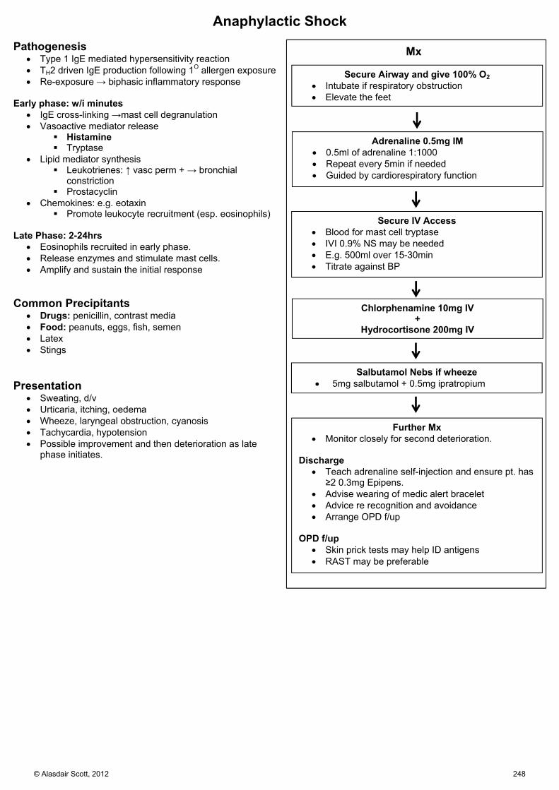
Anaphylactic Shock Pathogenesis
Type 1 IgE mediated hypersensitivity reaction TH2 driven IgE production following 1O allergen exposure Re-exposure → biphasic inflammatory response
Early phase: w/i minutes
IgE cross-linking →mast cell degranulation Vasoactive mediator release
Histamine Tryptase
Lipid mediator synthesis Leukotrienes: ↑ vasc perm + → bronchial
constriction Prostacyclin
Chemokines: e.g. eotaxin Promote leukocyte recruitment (esp. eosinophils)
Late Phase: 2-24hrs
Eosinophils recruited in early phase. Release enzymes and stimulate mast cells. Amplify and sustain the initial response
Common Precipitants
Drugs: penicillin, contrast media Food: peanuts, eggs, fish, semen Latex Stings
Presentation
Sweating, d/v Urticaria, itching, oedema Wheeze, laryngeal obstruction, cyanosis Tachycardia, hypotension Possible improvement and then deterioration as late
phase initiates.
Mx
Secure Airway and give 100% O2 Intubate if respiratory obstruction Elevate the feet
Adrenaline 0.5mg IM 0.5ml of adrenaline 1:1000 Repeat every 5min if needed Guided by cardiorespiratory function
Secure IV Access Blood for mast cell tryptase IVI 0.9% NS may be needed E.g. 500ml over 15-30min Titrate against BP
Chlorphenamine 10mg IV +
Hydrocortisone 200mg IV
Salbutamol Nebs if wheeze 5mg salbutamol + 0.5mg ipratropium
Further Mx Monitor closely for second deterioration.
Discharge
Teach adrenaline self-injection and ensure pt. has ≥2 0.3mg Epipens.
Advise wearing of medic alert bracelet Advice re recognition and avoidance Arrange OPD f/up
OPD f/up
Skin prick tests may help ID antigens RAST may be preferable
© Alasdair Scott, 2012 248

Narrow Complex Tachycardias = SVT Definition: Rate >100bmp, QRS width < 120ms Differential
1. Sinus tachycardia
2. Atrial AF Atrial flutter Atrial tachycardia
3. AV nodal re-entry tachycardia 4. AV re-entry tachycardia
Principles of Mx
If pt. compromised → sedate + DC cardioversion Otherwise ID rhythm and Rx accordingly
Key to ID irregular rhythm = AF = different Rx Vagal manoeuvres (carotid sinus massage, valsalva) transiently ↑
AV block and may unmask underlying atrial rhythm. If manoeuvres unsuccessful, give adenosine while recording
rhythm strip → transient AV block, unmasking atrial rhythm → cardioverts AVNRT/AVRT to sinus rhythm
If adenosine fails, choose from: Digoxin Atenolol Verapamil (not if on β-blocker) Amiodarone
If unsuccessful → DC cardioversion
Mx Flowchart
O2 + IV access
Regular rhythm?
Continuous ECG trace Vagal manoeuvres Adenosine 6mg IV bolus Then 12mg, then 12mg
Adverse Signs? BP <90 HF ↓ consciousness HR >200
Choose from: Digoxin (500ug over 30min) Amiodarone Verapamil Atenolol
Treat as AF Control rate c̄ β-B (e.g. metoprolol) or digoxin If onset <48h consider cardioversion c̄ amiodarone (as
below) or DC shock Consider anticoagulation c̄ heparin and/or warfarin
NB. Avoid adenosine, CCB, β-B in AF/Flutter with WPW or Hx of WPW as may → VF. Use Amiodarone or flecainide.
Sedation
Amiodarone:
300mg over 20-60min Then 900mg over next 23h
Adenosine MOA: temporary AVN block SEs: Transient chest tightness, dyspnoea, flushing, headache Relative CIs: asthma, 2nd/3rd degree block Interactions
fx ↑d by dipyridimole fx ↓d by theophylline
NO
NO
YES
YES
Synchronised cardioversion:100→200→360J
Prophylaxis β-B AVRT: flecainide AVNRT: verapamil
© Alasdair Scott, 2012 249

Broad Complex Tachycardias Definition: Rate >100bmp, QRS width > 120ms Differential
1. VT 2. Torsades de points 3. SVT c̄ BBB
VT Causes: I’M QVICK Infarction (esp. c̄ ventricular aneurysm) Myocarditis QT interval↑ Valve abnormality: mitral prolapse, AS Iatrogenic: digoxin, antiarrhythmics, catheter Cardiomyopathy (esp. dilated) K↓, Mg↓, O2↓, acidosis
Mx Flowchart
O2 + IV access
Synchronised Cardioversion
Correct Electrolyte Problems
↓K+: max 60mM KCL @ 20mmol/h ↓Mg2+: 4ml 50% MgSO4 in 30min
Adverse Signs? BP<90 HF Chest pain ↓ consciousness HR > 150
Sedation
Synchronised cardioversion: 200→300→360
Amiodarone:
300mg over 20-60min Then 900mg over next 23h
Failure
YES
NO
Pulse?
YES NO
CPR
Assess Rhythm Regular (i.e. VT): Amiodarone (see opposite) Or lignocaine 50mg over 2min
If irregular, Dx is usually: AF c̄ BBB Pre-excited AF: flec / amio TDP: MgSO4 2g IV over 10 min
Recurrent / Paroxysmal VT Medical: Amiodarone β-B
ICD
© Alasdair Scott, 2012 250

STEMI
O2 2-4L aim for SpO2 94-98%
Analgesia Morphine 5-10mg IV Metoclopramide 10mg IV
DVT Prophylaxis Enoxaparin 40mg SC OD
IV access Bloods for FBC, U+E, glucose, lipids
12 lead ECG Primary Percutaneous Coronary Intervention Rx of choice if <12h Angioplasty and stenting + GP IIb/IIIa antagonist (tirofiban) if high risk
Delayed PCI, DM, complex procedure Complications: Bleeding Emboli Arrhythmia
Brief assessment Hx of CVD and risk factors Thrombolysis CIs CV exam
Antiplatelet Aspirin 300mg PO (then 75mg/d) Clopidogrel 300mg PO (then 75mg/d)
Anti-ischaemia GTN 2 puffs or 1 tablet SL β-B atenolol 5mg IV (CI: asthma, LVF)
Admit to CCU for monitoring Arrhythmias Continue meds except CCBs
Thrombolysis CI beyond 24hrs from pain onset
ECG Criteria: ST elevation > 1mm in 2+ limbs or > 2mm in 2+ chest leads. New LBBB Posterior: Deep ST “depression” and tall “R” waves in V1-V3
Contraindications: AGAINST Aortic dissection GI bleeding Allergic reaction previously Iatrogenic: major surgery <14d Neuro: cerebral neoplasm or CVA Hx Severe HTN (200/120) Trauma, inc. CPR
Agents: 1st: streptokinase, alteplase (rt-PA), tenecteplase Complications: Bleeding Stroke Arrhythmia Allergic reaction
Continuing Therapy: address risk factors ACEi: start w/i 24hrs of MI (e.g. lisinopril 2.5mg) β-blocker: e.g. bisoprolol 1.25mg OD (or, CCB) Cardiac rehabilitation (group exercise and info) / Heart Manual DVT prophylaxis until fully mobile
Continue for 3mo if large anterior MI Statin: regardless of basal lipids (e.g. atorvastatin 80mg)
Advice Stop smoking Diet: oily fish, fruit, veg, ↓ sat fats Exercise: 30min OD Work: return in 2mo Sex: avoid for 1mo Driving :avoid for 1mo
NB. Continue clopidogrel for 1mo following STEMI Continue aspirin indefinitely.
Primary PCI or Thrombolysis
Pts. not receiving any form of reperfusion therapy should be given fondaparinux.
© Alasdair Scott, 2012 251

NSTEMI + UA
O2 2-4L aim for SpO2 94-98%
Analgesia Morphine 5-10mg IV Metoclopramide 10mg IV
Assess Cardiovascular Risk: GRACE/TIMI
IV access Bloods for FBC, U+E, glucose, lipids,
Troponin
12 lead ECG + Admit to CCU
Brief Assessment Hx of CVD and risk factors CV exam
Anti-ischaemia GTN: 2 puffs or 1 tablet SL β-B: atenolol 50mg/24h PO (CI: asthma, LVF) IV GTN if pain continues
Anti-coagulate Fondaparinux 2.5mg SC
Intermediate- to High-Risk Persistent/recurrent ischaemia, ST depression, DM,
positive trop. GPIIb/IIIa antagonist (tirofiban) Angiography (±PCI) w/i 96hrs Clopidogrel 75mg/d for one year
Low-Risk No further pain, flat or inverted T waves or normal
ECG, negative trop. May discharge if 12h trop is negative. Outpatient tests: angio, perfusion scan, stress echo
Continuing Therapy: address risk factors
ACEi (e.g. lisinopril 2.5mg) β-blocker (e.g. bisoprolol 1.25mg OD) or, CCB (diltiazem or verapamil) Stop antithrombotic therapy when pain free (but give 3-5d) Statin (e.g. atorvastatin 80mg)
Advice as above NB. Continue clopidogrel for 1yr following NSTEMI Continue aspirin indefinitely.
Antiplatelet Aspirin 300mg PO (then 75mg/d) Clopidogrel 300mg PO
© Alasdair Scott, 2012 252

Severe Pulmonary Oedema
Morphine in Pulmonary Oedema
Make pt. more comfortable Pulm venodilators → ↓ pre-load → optimise position on Starling Curve
O2 15L/min via reservoir mask Target SpO2: 94-98%
Hx, Ex, Ix CXR: ABCDE ECG: MI, arrhythmias, pulsus alternans Consider echo
IV access + monitor ECG Bloods for FBC, U+E, troponin, BNP, ABG Rx any arrhythmias (e.g. AF)
Sit pt. up
Diamorphine 2.5-5mg IV + Metoclopramide 10mg IV
If SBP >100mmHg, start nitrate IVI ISMN 2-10mg/h IVI Keep SBP >90
GTN 2 puffs or 2 x 300ug tabs SL Unless SBP <90mmHg
Continuing Therapy Daily weights DVT prophylaxis Repeat CXR Change to oral frusemide or bumetanide ACEi + β-B if heart failure Consider spironolactone Consider digoxin ± warfarin (esp. if in AF)
Frusemide 40-80mg IV
If worsening, consider: CPAP More frusemide or ↑ nitrate infusion Haemofiltration / dialysis
Causes Cardiogenic MI Arrhythmia Fluid overload: renal, iatrogenic
Non-cardiogenic ARDS: sepsis, post-op, trauma Upper airway obstruction Neurogenic: head injury
Differential Asthma/COPD Pneumonia PE
Symptoms Dyspnoea Orthopnea Pink frothy sputum
Signs Distressed, pale, sweaty, cyanosed ↑HR, ↑RR ↑JVP S3 / gallop rhythm Bibasal creps Pleural effusions Wheeze (cardiac asthma)
Monitoring Progress BP HR and RR JVP Urine Output ABG
If SBP <100mmHg: Rx as cardiogenic shock i.e. consider inotropes
© Alasdair Scott, 2012 253

Cardiogenic Shock
O2 15L/min via reservoir mask Target SpO2: 94-98%
IV access + monitor ECG Bloods for FBC, U+E, troponin, ABG
Diamorphine 2.5-5mg IV (pain/anxiety) + metoclopramide 10mg IV
Consider need for dobutamine
Monitoring CVP, BP, ABG, ECG, urine output.
Correct any: Arrhythmias Electrolyte disturbance Acid-base abnormalities
Causes: MI HEART: MI Hyperkalaemia (inc. electrolytes) Endocarditis (valve destruction) Aortic Dissection Rhythm disturbance Tamponade
Obstructive Tension pneumo Massive PE
Hx, Ex, Ix CXR Echo Consider CT thorax (dissection/PE)
Rx underlying cause
Tamponade Causes: Trauma Lung/breast Ca Pericarditis MI Bacteria (e.g. TB)
Mx: ABCs Pericardiocentesis (preferably under echo guidance)
Signs: Beck’s triad: ↓BP, ↑JVP, muffled heart sounds Kussmaul’s sign: ↑JVP on inspiration Pulsus paradoxus (pulse fades on inspiration)
Ix: Echo: diagnostic CXR: globular heart
Definition Inadequate tissue perfusion primarily due to
cardiac dysfunction.
Presentation Unwell: pale, sweaty, cyanosed, distressed Cold clammy peripheries ↑RR ± ↑HR Pulmonary oedema
© Alasdair Scott, 2012 254

Meningitis Features Meningitic
Headache Neck stiffness
Kernig’s: Straightening leg c̄ hip @ 90O Brudzinski’s: lifting head → lifting of legs
Photophobia n/v
Neurological
↓ GCS → coma Seizures (20%) Focal neuro (20%): e.g. CN palsies
Septic
Fever ↓BP, ↑HR ↑CRT Purpuric rash DIC
Abx Therapy
Community: benpen 1.2g IV/IM <50: ceftriaxone 2g IVI/IM BD >50: ceftriaxone + ampicillin 2g IVI /4h If viral suspected: aciclovir
Organisms
Viruses: enteroviruses (Coxsackie, echovirus), HSV2 Meningococcus Pneumococcus Listeria Haemophilus TB Cryptococcus
Ix
Bloods: FBC, U+Es, clotting, glucose, ABG Blood cultures LP: MCS, glucose, virology/PCR, lactate
ABC O2 15L – SpO2 94-98% IVI fluid resus c̄ crystalloid
Acute Management
Mainly Septicaemic Don’t attempt LP Ceftriaxone 2g IVI Consider ITU if shocked
Mainly Meningitic If no shock or CIs do LP Dexamethasone 0.15mg/kg IV QDS Ceftriaxone 2g IVI post-LP
Continuing Management Ceftriaxone 2g BD IVI
Meningococcus: 7d IV then review Pneumococcus: 14d IV then review
Maintenance fluids UO 30ml/h SBP >80mmHg
If response is poor, consider intubation ± inotropic support Rifampicin prophylaxis for household contacts.
CIs to LP: Try LP Unless ContraINdicated Thrombocytopenia Lateness (delay in antibiotic admin) Pressure (signs of raised ICP) Unstable (Cardio + resp systems) Coagulation disorder Infection at LP site Neurology (focal neurological signs)
CSF Findings
Findings Bacterial TB Viral Appearance Turbid Fibrin web Clear Cells PMN Lympho /
mononuc Lympho / mononuc
Count 100-1000 10-1000 50-1000 Glucose ↓ (< ½
plasma) ↓ (< ½ plasma)
> ½ plasma
Protein (g/L) ↑↑ (>1.5) ↑↑↑ (1-5) mild ↑ (<1)
© Alasdair Scott, 2012 255

Encephalitis Presentation
Infectious prodrome: fever, rash, LNs, cold sores, conjunctivitis, meningeal signs.
Bizarre behaviour or personality change Confusion ↓ GCS → coma Fever Headache Focal neuro Seizures Hx of travel or animal bite
Causes Usually viral
HSV1/2 CMV, EBV, VZV Arboviruses HIV
Non-viral
Any bacterial meningitis TB Malaria Lyme disease
Ix
Bloods: cultures, viral PCR, malaria film Contrast CT: focal bilat temporal involvement suggests
HSV LP: ↑ CSF protein, lymphocytes, PCR EEG: shows diffuse abnormalities, may confirm Dx
Mx
Aciclovir STAT: 10mg/kg/8h IVI over 1h for 14/7 Supportive measures in HDU/ITU Phenytoin for seizures
Prognosis
70% mortality if untreated w/o fever, consider encephalopathy
↓ glucose Hepatic DKA Drugs SLE Uraemia Hypoxic brain injury Beri-Beri
Cerebral Abscess Pre-disposing Factors
Infection: ear, sinus, dental or periodontal Skull # Congenital heart disease Endocarditis Bronchiectasis Immunosuppression
Organisms
Frontal sinus/teeth – strep. Milleri, oropharyngeal anaerobes
Ear – Bacteroides, other anaerobes Signs
Seizures Fever Localizing signs Signs of ↑ ICP Signs of infection elsewhere
Ix
CT/MRI – ring-enhancing lesion ↑WCC, ↑ESR
Rx
Neurosurgical referral Abx – e.g. ceftriaxone Treat ↑ ICP
© Alasdair Scott, 2012 256
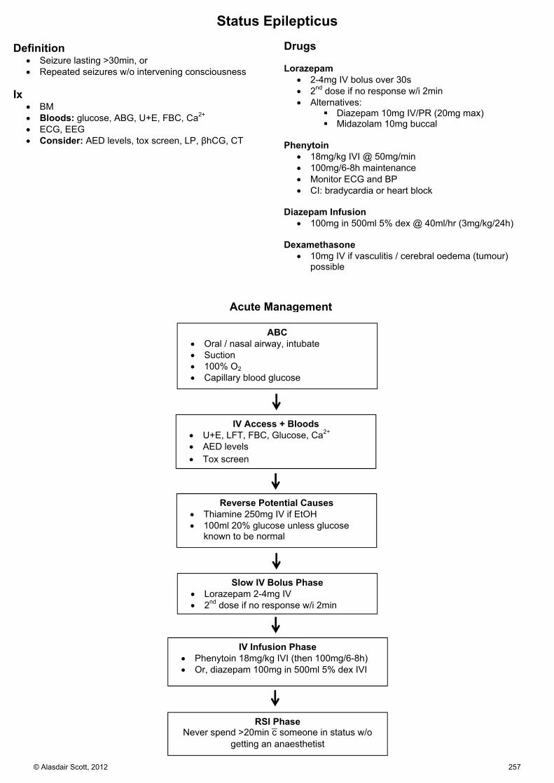
ABC Oral / nasal airway, intubate Suction 100% O2 Capillary blood glucose
Acute Management
IV Access + Bloods U+E, LFT, FBC, Glucose, Ca2+ AED levels Tox screen
Reverse Potential Causes Thiamine 250mg IV if EtOH 100ml 20% glucose unless glucose
known to be normal
Slow IV Bolus Phase Lorazepam 2-4mg IV 2nd dose if no response w/i 2min
IV Infusion Phase Phenytoin 18mg/kg IVI (then 100mg/6-8h) Or, diazepam 100mg in 500ml 5% dex IVI
RSI PhaseNever spend >20min c̄ someone in status w/o
getting an anaesthetist
Status Epilepticus Definition
Seizure lasting >30min, or Repeated seizures w/o intervening consciousness
Ix
BM Bloods: glucose, ABG, U+E, FBC, Ca2+ ECG, EEG Consider: AED levels, tox screen, LP, βhCG, CT
Drugs Lorazepam
2-4mg IV bolus over 30s 2nd dose if no response w/i 2min Alternatives:
Diazepam 10mg IV/PR (20mg max) Midazolam 10mg buccal
Phenytoin
18mg/kg IVI @ 50mg/min 100mg/6-8h maintenance Monitor ECG and BP CI: bradycardia or heart block
Diazepam Infusion
100mg in 500ml 5% dex @ 40ml/hr (3mg/kg/24h) Dexamethasone
10mg IV if vasculitis / cerebral oedema (tumour) possible
© Alasdair Scott, 2012 257

Head Injury Initial Mx 1O Survey
A: ? intubation, immobilise C-spine B: 100% O2, RR C: IV access, BP, HR D: GCS, pupils Treat seizures
Lorazepam 2-4mg IV Phenytoin18mg/kg IVI then 100mg/6-8h
E: expose pt. and look for other obvious injuries 2O Survey
Look for: Lacerations Obvious facial/skull deformity CSF leak from nose or ears Battle’s sign, Racoon eyes Blood behind TM C-spine tenderness ± deformity
Head-to-toe examination for other injuries Log role
Hx if possible
How and when? GCS and other vitals immediately after injury Headache, fits, vomiting, amnesia, EtOH
Ix
Bloods: FBC, U+E, glucose, clotting, EtOH level, ABG ? CT head + c-spine
Rx
Neurosurgical opinion if signs of ↑ICP, CT evidence of intracranial bleed significant skull #
Admit if: Abnormalities on imaging Difficult to assess: EtOH, post-ictal Not returned to GCS 15 after imaging CNS signs: vomiting, severe headache
Neuro-obs half-hrly until GCS 15 GCS Pupils HR, BP RR, SpO2 Temperature
Discharge Advice
Stay with someone for first 48hrs Give advice card advising return on:
Confusion, drowsiness, unconsciousness Visual problems Weakness Deafness V. painful headache that won’t go away Vomiting Fits
Intubate
GCS ≤ 8 PaO2 <9KPa on air / <13KPa on O2 or PCO2 >6KPa Spontaneous hyperventilation: PCO2 <4KPa Respiratory irregularity
CT head guidelines: BANGS LOC
Break: open, depressed or base of skull Amnesia > 30min retrograde Neuro deficit or seizure GCS: <13 @ any time or <15 2h after injury Sickness: vomited > once LOC or any amnesia and any of:
Dangerous mechanism: RTA, great height Age ≥ 65 Coagulopathy (inc. warfarin)
Risk of Intracranial Haematoma in Adults:
Fully conscious, no skull # = <1:1000 Confused, no skull # = 1:100 Fully conscious, skull # = 1:30 Confused, skull # = 1:4
GCS Eyes: 4 4 – Spontaneous eye opening 3 – Open to voice 2 – Open to pain 1 – No opening Verbal: 5 5 – Orientated conversation 4 – Confused conversation 3 – Inappropriate speech 2 – Incomprehensible sounds 1 – No speech Motor: 6 6 – Obeys commands 5 – Localises pain 4 – Withdraws to pain 3 – Decorticate posturing to pain (flexor) 2 – Decerebrate posturing to pain (extensor) 1 – No movement
© Alasdair Scott, 2012 258

ABC
Acute Management
Treat seizures and correct hypotension
Elevate bed to 40O
Neuroprotective Ventilation PaO2: >13kPa (100mmHg) PCO2: 4.5kPa Good sedation ± NM blockade
Mannitol or Hypertonic Saline ↓ICP short-term, but may → rebound ↑ICP later Mannitol 1g/kg (20% @ 5ml/kg)
Raised ICP Types of Cerebral Oedema
1. Vasogenic (↑ cap permeability): trauma, tumour, ischaemia, infection 2. Cytotoxic: e.g. from hypoxia 3. Interstitial: e.g. obstructive hydrocephalus, ↓Na+
Causes
Haemorrhage Tumours Infection: meningitis, encephalitis, abscess Hydrocephalus Status Cerebral oedema
Signs and Symptoms Headache n/v Seizures Drowsiness → coma Cushing’s reflex: ↑BP, ↓HR, irregular breathing 6th CN palsy (may be false localising) Cheyne-Stokes respiration Pupils – constriction → dilatation Papilloedema, loss of venous pulsation @ disc
Herniation Syndromes Tonsillar (Coning)
↑ pressure in posterior fossa → displacement of cerebellar tonsils through foramen magnum → compression of brainstem and cardioresp centres in medulla CN6 palsy, upgoing plantars → irregular breathing → apnoea
Transtentorial / uncal Lateral supratentorial mass → compression of ipsilateral inferomedial temporal lobe (uncus) against free margin of
tentorium cerebelli. Ipsilateral CN3 palsy: mydriasis (dilation) then down-and-out Contralateral hemiparesis Compression of contralateral corticospinal tracts → ipsilateral hemiparesis (Kernohan’s Notch)
Subfalcine
Frontal mass Displacement of cingulate gyrus (medial frontal lobe) under falx cerebri Compression of ACA → stroke
Contralateral motor/sensory loss in legs>arms Abulia (pathological laziness)
© Alasdair Scott, 2012 259
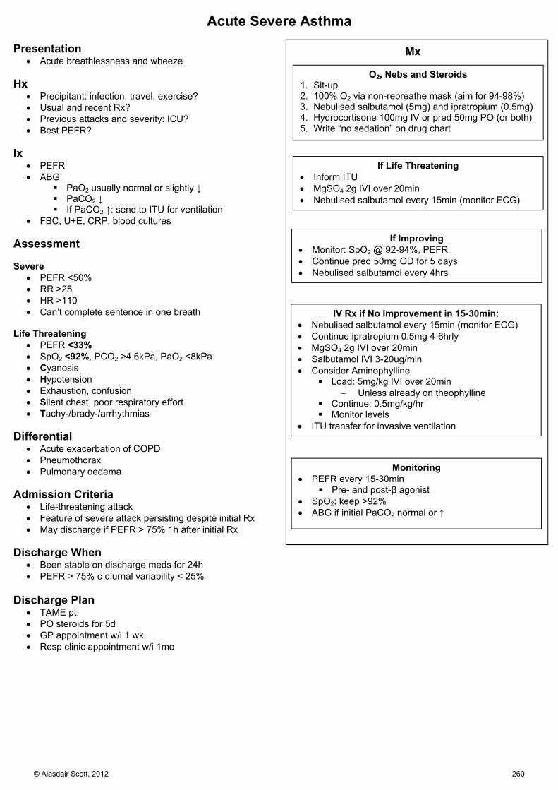
Acute Severe Asthma Presentation
Acute breathlessness and wheeze Hx
Precipitant: infection, travel, exercise? Usual and recent Rx? Previous attacks and severity: ICU? Best PEFR?
Ix
PEFR ABG
PaO2 usually normal or slightly ↓ PaCO2 ↓ If PaCO2 ↑: send to ITU for ventilation
FBC, U+E, CRP, blood cultures Assessment Severe
PEFR <50% RR >25 HR >110 Can’t complete sentence in one breath
Life Threatening
PEFR <33% SpO2 <92%, PCO2 >4.6kPa, PaO2 <8kPa Cyanosis Hypotension Exhaustion, confusion Silent chest, poor respiratory effort Tachy-/brady-/arrhythmias
Differential
Acute exacerbation of COPD Pneumothorax Pulmonary oedema
Admission Criteria
Life-threatening attack Feature of severe attack persisting despite initial Rx May discharge if PEFR > 75% 1h after initial Rx
Discharge When
Been stable on discharge meds for 24h PEFR > 75% c̄ diurnal variability < 25%
Discharge Plan
TAME pt. PO steroids for 5d GP appointment w/i 1 wk. Resp clinic appointment w/i 1mo
Mx
O2, Nebs and Steroids 1. Sit-up 2. 100% O2 via non-rebreathe mask (aim for 94-98%) 3. Nebulised salbutamol (5mg) and ipratropium (0.5mg) 4. Hydrocortisone 100mg IV or pred 50mg PO (or both) 5. Write “no sedation” on drug chart
If Life Threatening Inform ITU MgSO4 2g IVI over 20min Nebulised salbutamol every 15min (monitor ECG)
If Improving Monitor: SpO2 @ 92-94%, PEFR Continue pred 50mg OD for 5 days Nebulised salbutamol every 4hrs
IV Rx if No Improvement in 15-30min: Nebulised salbutamol every 15min (monitor ECG) Continue ipratropium 0.5mg 4-6hrly MgSO4 2g IVI over 20min Salbutamol IVI 3-20ug/min Consider Aminophylline
Load: 5mg/kg IVI over 20min Unless already on theophylline
Continue: 0.5mg/kg/hr Monitor levels
ITU transfer for invasive ventilation
Monitoring PEFR every 15-30min
Pre- and post-β agonist SpO2: keep >92% ABG if initial PaCO2 normal or ↑
© Alasdair Scott, 2012 260
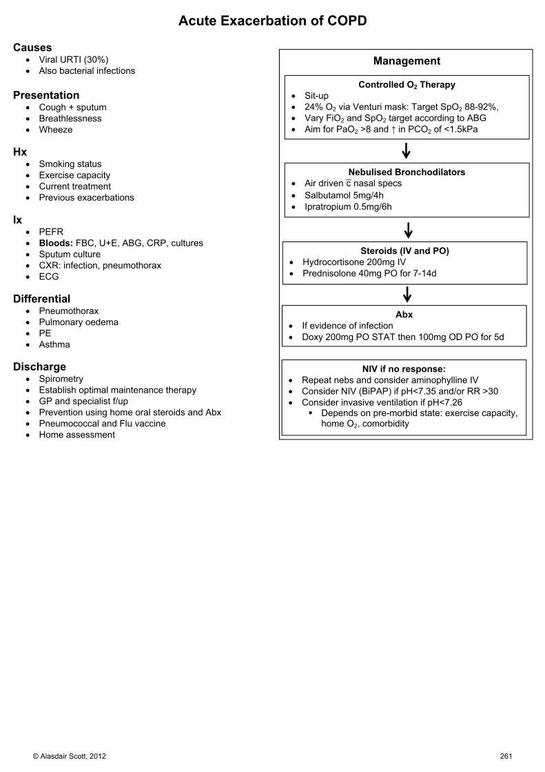
Acute Exacerbation of COPD Causes
Viral URTI (30%) Also bacterial infections
Presentation
Cough + sputum Breathlessness Wheeze
Hx
Smoking status Exercise capacity Current treatment Previous exacerbations
Ix
PEFR Bloods: FBC, U+E, ABG, CRP, cultures Sputum culture CXR: infection, pneumothorax ECG
Differential
Pneumothorax Pulmonary oedema PE Asthma
Discharge
Spirometry Establish optimal maintenance therapy GP and specialist f/up Prevention using home oral steroids and Abx Pneumococcal and Flu vaccine Home assessment
Controlled O2 Therapy Sit-up 24% O2 via Venturi mask: Target SpO2 88-92%, Vary FiO2 and SpO2 target according to ABG Aim for PaO2 >8 and ↑ in PCO2 of <1.5kPa
Management
Nebulised Bronchodilators Air driven c̄ nasal specs Salbutamol 5mg/4h Ipratropium 0.5mg/6h
Steroids (IV and PO) Hydrocortisone 200mg IV Prednisolone 40mg PO for 7-14d
Abx If evidence of infection Doxy 200mg PO STAT then 100mg OD PO for 5d
NIV if no response: Repeat nebs and consider aminophylline IV Consider NIV (BiPAP) if pH<7.35 and/or RR >30 Consider invasive ventilation if pH<7.26
Depends on pre-morbid state: exercise capacity, home O2, comorbidity
© Alasdair Scott, 2012 261

Pulmonary Embolism Causes
Usually arise from DVTs in proximal leg or iliac veins Rarely:
Right ventricle post MI Septic emboli in right sided endocarditis
Risk Factors: SPASMODICAL
Sex: F Pregnancy Age: ↑ Surgery (classically 10d post-op straining at stool) Malignancy Oestrogen: OCP/HRT DVT/PE previous Hx Immobility Colossal size Antiphospholipid Abs Lupus Anti-coagulant
Presentation
Symptoms and signs depend on size, number and distribution of emboli
Symptoms
Dyspnoea Pleuritic pain Haemoptysis Syncope
Signs
Fever Cyanosis Tachycardia, tachypnoea RHF: hypotension, ↑JVP, loud P2 Evidence of cause: DVT
Ix
Bloods: FBC, U+E, clotting, D-dimers ABG: normal or ↓PaO2 and ↓PaCO2, ↑pH CXR: normal or oligaemia, linear atelectasis ECG: sinus tachycardia, RBBB, right ventricular strain
(inverted T in V1-V4) S1, Q3, T3 is rare
Doppler US: thigh and pelvis (+ve in 60%) CTPA + venous phase of legs and pelvis
85-95% sensitivity V/Q scan no longer used
Dx
1. Assess probability using Wells’ Score 2. Low-probability → perform D-dimers
Negative → excludes PE Positive → CTPA
3. High probability → CTPA NB. –ve D-dimer has 95% NPV for PE Prevention
Risk assessment for all pts TEDS Prophylactic LMWH Avoid OCP/HRT if @ risk
O2 Sit-up 100% O2 via non-rebreather mask
Mx
Analgesia Morphine ± metoclopramide if distressed
SBP?
>90 Start Warfarin Confirm Dx
LMWH Heparin e.g. enoxaparin 1.5mg/kg/24h SC
<90 500ml colloid
Inotropes if BP still ↓ Dobutamine: aim for SBP >90 Consider addition of NORAD Consider thrombolysis (medical or surgical)
On-going Mx
TEDS stockings in hospital Graduated compression stockings for 2yrs if DVT:
prevent post-phlebitic syndrome (10-30%) Continue LMWH until INR >2 (at least 5d) Target INR = 2-3 Duration
Remedial cause: 3mo No identifiable cause: 6mo On-going cause: indefinite
VC filter if repeat DVT/PE despite anticoagulation
If critically ill c̄ massive PE consider thrombolysis
Alteplase 50mg bolus stat (Surgical or interventional embolectomy)
© Alasdair Scott, 2012 262

Pneumothorax Definition
Accumulation of air in the pleural space c̄ 2O lung collapse.
Classification
Closed: intact chest wall and air leaks from lung into pleural cavity.
Open: defect in the chest wall allows communication between PTX and exterior: may be sucking.
Tension: air enters pleural cavity through one-way valve and cannot escape → mediastinal compression.
Causes Spontaneous
1O: no underlying lung disease Young, thin men (ruptured subpleural bulla) Smokers
2O: underlying lung disease COPD Marfan’s, Ehler’s Danlos Pulmonary fibrosis, sarcoidosis
Trauma
Penetrating Blunt ± rib #s
Iatrogenic
Subclavian CVP line insertion Positive pressure ventilation Transbronchial biospy Liver biopsy
Presentation Symptoms
Sudden onset Dyspnoea Pleuritic chest pain Tension: respiratory distress, cardiac arrest
Signs
Chest ↓ expansion Resonant percussion ↓ breath sounds ↓VR
Tension: ↑JVP, mediastinal shift, ↑HR, ↓BP Crepitus: surgical emphysema
Ix
ABG US CXR (expiratory film may be helpful)
Translucency + collapse (2cm rim = 50% vol loss)
Mediastinal shift (away from PTX) Surgical emphysema Cause: rib #s, pulmonary disease (e.g. bullae)
Tension PTX Resuscitate pt. No CXR Large bore Venflon into 2nd ICS, mid-clavicular line Insert ICD
Mx
Traumatic Resuscitate pt. Analgesia: e.g. morphine 3-sided wet dressing if sucking Insert ICD
1O PTX
2O PTX
SOB and/or rim ≥2cm? Consider d/c
Aspiration successful?
Insert ICD
No
Yes
No
Yes
SOB and > 50yrs and rim ≥2cm?
Admit for 24h
Aspiration successful?
Insert ICDYes
No No
Yes
© Alasdair Scott, 2012 263
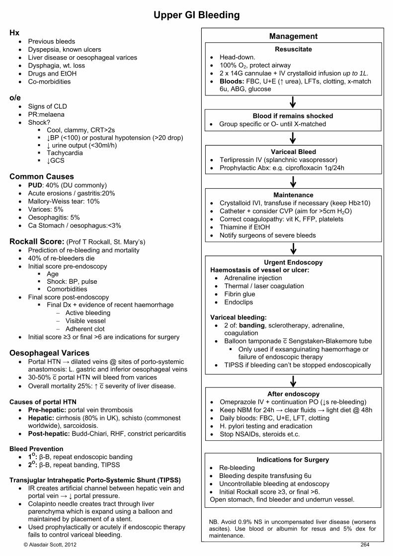
Upper GI Bleeding Hx
Previous bleeds Dyspepsia, known ulcers Liver disease or oesophageal varices Dysphagia, wt. loss Drugs and EtOH Co-morbidities
o/e
Signs of CLD PR:melaena Shock?
Cool, clammy, CRT>2s ↓BP (<100) or postural hypotension (>20 drop) ↓ urine output (<30ml/h) Tachycardia ↓GCS
Common Causes
PUD: 40% (DU commonly) Acute erosions / gastritis:20% Mallory-Weiss tear: 10% Varices: 5% Oesophagitis: 5% Ca Stomach / oesophagus:<3%
Rockall Score: (Prof T Rockall, St. Mary’s)
Prediction of re-bleeding and mortality 40% of re-bleeders die Initial score pre-endoscopy
Age Shock: BP, pulse Comorbidities
Final score post-endoscopy Final Dx + evidence of recent haemorrhage
Active bleeding Visible vessel Adherent clot
Initial score ≥3 or final >6 are indications for surgery Oesophageal Varices
Portal HTN → dilated veins @ sites of porto-systemic anastomosis: L. gastric and inferior oesophageal veins
30-50% c̄ portal HTN will bleed from varices Overall mortality 25%: ↑ c̄ severity of liver disease.
Causes of portal HTN
Pre-hepatic: portal vein thrombosis Hepatic: cirrhosis (80% in UK), schisto (commonest
worldwide), sarcoidosis. Post-hepatic: Budd-Chiari, RHF, constrict pericarditis
Bleed Prevention
1O: β-B, repeat endoscopic banding 2O: β-B, repeat banding, TIPSS
Transjuglar Intrahepatic Porto-Systemic Shunt (TIPSS)
IR creates artificial channel between hepatic vein and portal vein → ↓ portal pressure.
Colapinto needle creates tract through liver parenchyma which is expand using a balloon and maintained by placement of a stent.
Used prophylactically or acutely if endoscopic therapy fails to control variceal bleeding.
Resuscitate Head-down. 100% O2, protect airway 2 x 14G cannulae + IV crystalloid infusion up to 1L. Bloods: FBC, U+E (↑ urea), LFTs, clotting, x-match
6u, ABG, glucose
Management
Blood if remains shocked Group specific or O- until X-matched
Maintenance Crystalloid IVI, transfuse if necessary (keep Hb≥10) Catheter + consider CVP (aim for >5cm H2O) Correct coagulopathy: vit K, FFP, platelets Thiamine if EtOH Notify surgeons of severe bleeds
Urgent Endoscopy Haemostasis of vessel or ulcer: Adrenaline injection Thermal / laser coagulation Fibrin glue Endoclips
Variceal bleeding: 2 of: banding, sclerotherapy, adrenaline,
coagulation Balloon tamponade c̄ Sengstaken-Blakemore tube
Only used if exsanguinating haemorrhage or failure of endoscopic therapy
TIPSS if bleeding can’t be stopped endoscopically
Indications for Surgery Re-bleeding Bleeding despite transfusing 6u Uncontrollable bleeding at endoscopy Initial Rockall score ≥3, or final >6. Open stomach, find bleeder and underrun vessel.
Variceal Bleed Terlipressin IV (splanchnic vasopressor) Prophylactic Abx: e.g. ciprofloxacin 1g/24h
After endoscopy Omeprazole IV + continuation PO (↓s re-bleeding) Keep NBM for 24h → clear fluids → light diet @ 48h Daily bloods: FBC, U+E, LFT, clotting H. pylori testing and eradication Stop NSAIDs, steroids et.c.
NB. Avoid 0.9% NS in uncompensated liver disease (worsens ascites). Use blood or albumin for resus and 5% dex for maintenance.
© Alasdair Scott, 2012 264
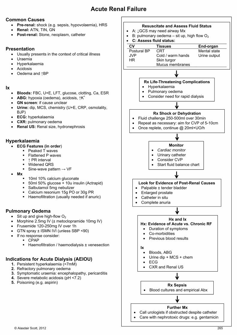
Acute Renal Failure Common Causes
Pre-renal: shock (e.g. sepsis, hypovolaemia), HRS Renal: ATN, TIN, GN Post-renal: Stone, neoplasm, catheter
Presentation
Usually presents in the context of critical illness Uraemia Hyperkalaemia Acidosis Oedema and ↑BP
Ix
Bloods: FBC, U+E, LFT, glucose, clotting, Ca, ESR ABG: hypoxia (oedema), acidosis, ↑K+ GN screen: if cause unclear Urine: dip, MCS, chemistry (U+E, CRP, osmolality,
BJP) ECG: hyperkalaemia CXR: pulmonary oedema Renal US: Renal size, hydronephrosis
Hyperkalaemia
ECG Features (in order) Peaked T waves Flattened P waves ↑ PR interval Widened QRS Sine-wave pattern → VF
Mx 10ml 10% calcium gluconate 50ml 50% glucose + 10u insulin (Actrapid) Salbutamol 5mg nebulizer Calcium resonium 15g PO or 30g PR Haemofiltration (usually needed if anuric)
Pulmonary Oedema
Sit up and give high-flow O2 Morphine 2.5mg IV (± metoclopramide 10mg IV) Frusemide 120-250mg IV over 1h GTN spray ± ISMN IVI (unless SBP <90) If no response consider:
CPAP Haemofiltration / haemodialysis ± venesection
Indications for Acute Dialysis (AEIOU)
1. Persistent hyperkalaemia (>7mM) 2. Refractory pulmonary oedema 3. Symptomatic uraemia: encephalopathy, pericarditis 4. Severe metabolic acidosis (pH <7.2) 5. Poisoning (e.g. aspirin)
Resuscitate and Assess Fluid Status A: ↓GCS may need airway Mx B: pulmonary oedema – sit up, high flow O2 C: Assess fluid status:
Rx Life-Threatening Complications Hyperkalaemia Pulmonary oedema Consider need for rapid dialysis
CV Tissues End-organ Postural BP CRT Mental state JVP Cold / warm hands Urine output HR Skin turgor Mucus membranes
Rx Sepsis Blood cultures and empirical Abx
Look for Evidence of Post-Renal Causes Palpable ± tender bladder Enlarged prostate Catheter in situ Complete anuria
Hx and Ix Hx: Evidence of Acute vs. Chronic RF Duration of symptoms Co-morbidities Previous blood results
Ix Bloods, ABG Urine dip + MCS + chem ECG CXR and Renal US
Rx Shock or Dehydration Fluid challenge 250-500ml over 30min Repeat as necessary: aim for CVP of 5-10cm Once replete, continue @ 20ml+UO/h
Monitor Cardiac monitor Urinary catheter Consider CVP Start fluid balance chart
Further Mx Call urologists if obstructed despite catheter Care with nephrotoxic drugs: e.g. gentamicin
© Alasdair Scott, 2012 265
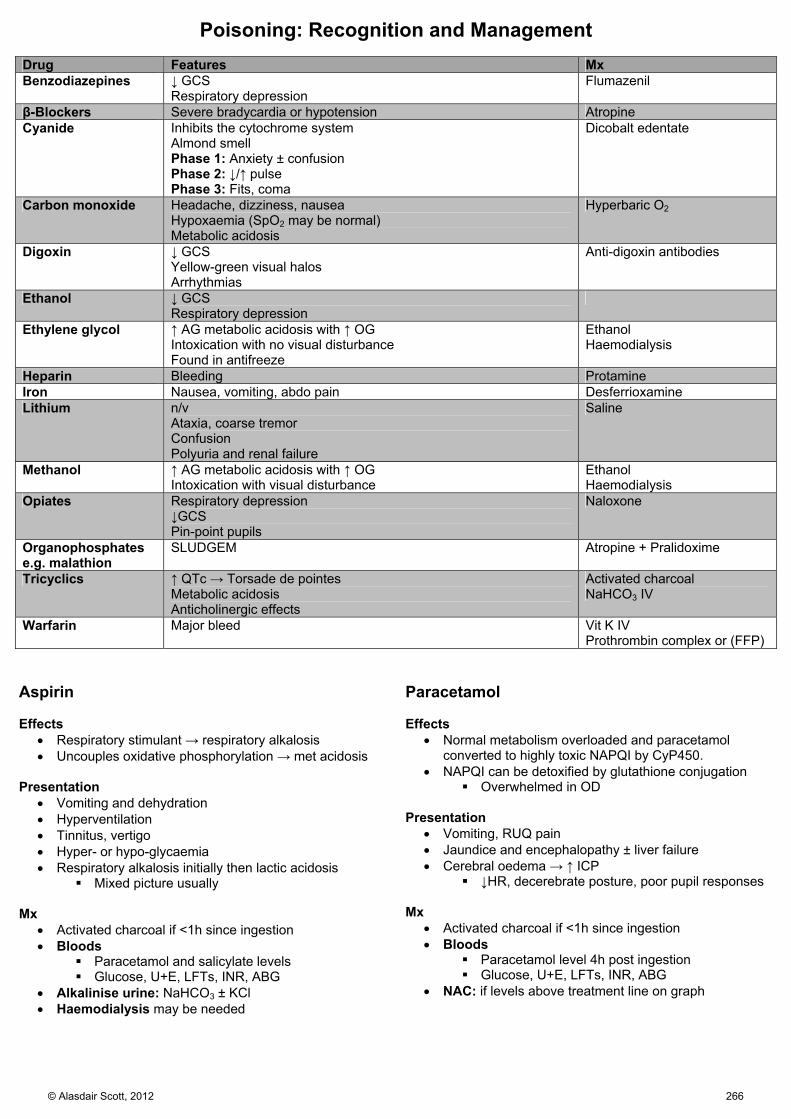
Poisoning: Recognition and Management Drug Features Mx Benzodiazepines ↓ GCS
Respiratory depression Flumazenil
β-Blockers Severe bradycardia or hypotension Atropine Cyanide Inhibits the cytochrome system
Almond smell Phase 1: Anxiety ± confusion Phase 2: ↓/↑ pulse Phase 3: Fits, coma
Dicobalt edentate
Carbon monoxide Headache, dizziness, nausea Hypoxaemia (SpO2 may be normal) Metabolic acidosis
Hyperbaric O2
Digoxin ↓ GCS Yellow-green visual halos Arrhythmias
Anti-digoxin antibodies
Ethanol ↓ GCS Respiratory depression
Ethylene glycol ↑ AG metabolic acidosis with ↑ OG Intoxication with no visual disturbance Found in antifreeze
Ethanol Haemodialysis
Heparin Bleeding Protamine Iron Nausea, vomiting, abdo pain Desferrioxamine Lithium n/v
Ataxia, coarse tremor Confusion Polyuria and renal failure
Saline
Methanol ↑ AG metabolic acidosis with ↑ OG Intoxication with visual disturbance
Ethanol Haemodialysis
Opiates Respiratory depression ↓GCS Pin-point pupils
Naloxone
Organophosphates e.g. malathion
SLUDGEM Atropine + Pralidoxime
Tricyclics ↑ QTc → Torsade de pointes Metabolic acidosis Anticholinergic effects
Activated charcoal NaHCO3 IV
Warfarin Major bleed Vit K IV Prothrombin complex or (FFP)
Aspirin Effects
Respiratory stimulant → respiratory alkalosis Uncouples oxidative phosphorylation → met acidosis
Presentation
Vomiting and dehydration Hyperventilation Tinnitus, vertigo Hyper- or hypo-glycaemia Respiratory alkalosis initially then lactic acidosis
Mixed picture usually Mx
Activated charcoal if <1h since ingestion Bloods
Paracetamol and salicylate levels Glucose, U+E, LFTs, INR, ABG
Alkalinise urine: NaHCO3 ± KCl Haemodialysis may be needed
Paracetamol Effects
Normal metabolism overloaded and paracetamol converted to highly toxic NAPQI by CyP450.
NAPQI can be detoxified by glutathione conjugation Overwhelmed in OD
Presentation
Vomiting, RUQ pain Jaundice and encephalopathy ± liver failure Cerebral oedema → ↑ ICP
↓HR, decerebrate posture, poor pupil responses Mx
Activated charcoal if <1h since ingestion Bloods
Paracetamol level 4h post ingestion Glucose, U+E, LFTs, INR, ABG
NAC: if levels above treatment line on graph
© Alasdair Scott, 2012 266
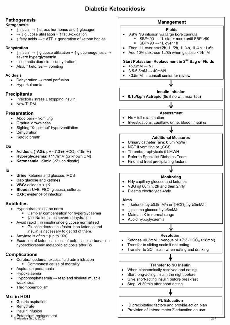
Diabetic Ketoacidosis Pathogenesis Ketogenesis
↓ insulin → ↑ stress hormones and ↑ glucagon → ↓ glucose utilisation + ↑ fat β-oxidation ↑ fatty acids → ↑ ATP + generation of ketone bodies.
Dehydration
↓ insulin → ↓ glucose utilisation + ↑ gluconeogenesis → severe hyperglycaemia
→ osmotic diuresis → dehydration Also, ↑ ketones → vomiting
Acidosis
Dehydration → renal perfusion Hyperkalaemia
Precipitants
Infection / stress ± stopping insulin New T1DM
Presentation
Abdo pain + vomiting Gradual drowsiness Sighing “Kussmaul” hyperventilation Dehydration Ketotic breath
Dx
Acidosis (↑AG): pH <7.3 (± HCO3 <15mM) Hyperglycaemia: ≥11.1mM (or known DM) Ketonaemia: ≥3mM (≥2+ on dipstix)
Ix
Urine: ketones and glucose, MCS Cap glucose and ketones VBG: acidosis + ↑K Bloods: U+E, FBC, glucose, cultures CXR: evidence of infection
Subtleties
Hyponatraemia is the norm Osmolar compensation for hyperglycaemia ↑/↔ Na indicates severe dehydration
Avoid rapid ↓ in insulin once glucose normalised Glucose decreases faster than ketones and
insulin is necessary to get rid of them. Amylase is often ↑ (up to 10x) Excretion of ketones → loss of potential bicarbonate →
hyperchloraemic metabolic acidosis after Rx Complications
Cerebral oedema: excess fluid administration Commonest cause of mortality
Aspiration pneumonia Hypokalaemia Hypophosphataemia → resp and skeletal muscle
weakness Thromboembolism
Mx: in HDU
Gastric aspiration Rehydrate Insulin infusion Potassium replacement
Fluids 0.9% NS infusion via large bore cannula
SBP<90 → 1L stat + more until SBP >90 SBP>90 → 1L over 1h
Then: 1L over next 2h, 1L/2h, 1L/4h, 1L/4h, 1L/6h Add 10% dextrose 1L/8h when glucose <14mM Start Potassium Replacement in 2nd Bag of Fluids >5.5mM → Nil 3.5-5.5mM → 40mM/L <3.5mM → consult senior for review
Management
Insulin Infusion 0.1u/kg/h Actrapid (6u if no wt., max 15u)
Assessment Hx + full examination Investigations: capillary, urine, blood, imaging
Resolution Ketones <0.3mM + venous pH>7.3 (HCO3 >18mM) Transfer to sliding scale if not eating Transfer to SC insulin when eating and drinking
Additional Measures Urinary catheter (aim: 0.5ml/kg/hr) NGT if vomiting or ↓GCS Thromboprophylaxis c̄ LMWH Refer to Specialist Diabetes Team Find and treat precipitating factors
Monitoring Hrly capillary glucose and ketones VBG @ 60min, 2h and then 2hrly Plasma electrolytes 4hrly Aims ↓ ketones by ≥0.5mM/h or ↑HCO3 by ≥3mM/h ↓ plasma glucose by ≥3mM/h Maintain K in normal range Avoid hypoglycaemia
Transfer to SC Insulin When biochemically resolved and eating Start long-acting insulin the night before Give short-acting insulin before breakfast Stop IVI 30min after short acting
Pt. Education ID precipitating factors and provide action plan Provision of ketone meter c̄ education on use.
© Alasdair Scott, 2012 267

Hyperosmolar Non-Ketotic Coma The Patient
Usually T2DM, often new presentation Usually older Long hx (e.g. 1wk)
Metabolic Derangement
Marked dehydration and glucose >35mM No acidosis (no ketogenesis) Osmolality >340mosmol/kg
Complications
Occlusive events are common: DVT, stroke Give LMWH
Mx
Rehydrate c̄ 0.9% NS over 48h May need ~9L
Wait 1h before starting insulin It may not be needed Start low to avoid rapid changes in osmolality
E.g. 1-3u/hr Look for precipitant
MI Infection Bowel infarct
LMWH
Hypoglycaemia Symptoms Autonomic Sweating Anxiety Hunger Tremor Palpitations
Neuroglycopenic Confusion Drowsiness Seizures Coma Personality change
Cause
Usually Exogenous: insulin, gliclazide Pituitary insufficiency Liver failure Addison’s Insulinomas
Mx Alert and Orientated: Oral Carb
Rapid acting: lucozade Long acting: toast, sandwich
Drowsy / confused but swallow intact: Buccal Carb
Hypostop / Glucogel Consider IV access
Unconscious or Concerned re Swallow: IV dextrose
50ml 50% or 100ml 20% glucose Deteriorating / refractory / insulin-induced / no access
1mg glucagon IM/SC Won’t work in drunks + short duration of effect (20min) Insulin release may → rebound hypoglycaemia
© Alasdair Scott, 2012 268
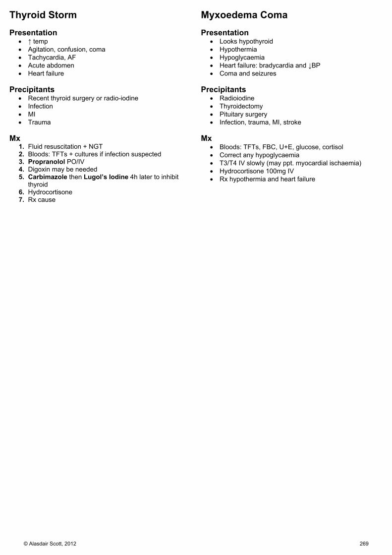
Thyroid Storm Presentation
↑ temp Agitation, confusion, coma Tachycardia, AF Acute abdomen Heart failure
Precipitants
Recent thyroid surgery or radio-iodine Infection MI Trauma
Mx
1. Fluid resuscitation + NGT 2. Bloods: TFTs + cultures if infection suspected 3. Propranolol PO/IV 4. Digoxin may be needed 5. Carbimazole then Lugol’s Iodine 4h later to inhibit
thyroid 6. Hydrocortisone 7. Rx cause
Myxoedema Coma Presentation
Looks hypothyroid Hypothermia Hypoglycaemia Heart failure: bradycardia and ↓BP Coma and seizures
Precipitants
Radioiodine Thyroidectomy Pituitary surgery Infection, trauma, MI, stroke
Mx
Bloods: TFTs, FBC, U+E, glucose, cortisol Correct any hypoglycaemia T3/T4 IV slowly (may ppt. myocardial ischaemia) Hydrocortisone 100mg IV Rx hypothermia and heart failure
© Alasdair Scott, 2012 269

Addisonian Crisis Presentation
Shocked: ↑HR, postural drop, oliguria, confused Hypoglycaemia Usually known Addisonian or chronic steroid user
Precipitants
Infection Trauma Surgery Stopping long-term steroids
Mx
Bloods: cortisol, ACTH, U+E Check CBG: glucose may be needed Hydrocortisone 100mg IV 6hrly IV crystalloid Septic screen Treat underlying cause
Hypertensive Crisis Cause
Phaeochromocytoma Presentation
Pallor Pulsating headache Feeling of impending doom ↑↑BP ↑ ST and cardiogenic shock
Precipitants
Stress Abdominal palpation General anaesthetic Parturition Contrast media
Rx
Phentolamine 2-5mg IV (α-blocker) or labetalol 50mg IV Repeat to safe BP (e.g. 110 diastolic)
Phenoxybenzamine 10mg/d PO when BP controlled May now give β-B to control tachycardia / arrhythmias Elective surgery after 4-6wks to allow full α-blockade
and volume expansion NB. Never give β-B alone first
© Alasdair Scott, 2012 270

Burns Risk Factors
Age: children and elderly Co-morbidities: epilepsy, CVA, dementia, mental illness Occupation
Classification Superficial
Erythema Painful E.g. sunburn
Partial Thickness
Heal w/i 2-3wks if not complicated Superficial
No loss of dermis Painful Blisters
Deep Loss of dermis but adnexae remain Healing from adnexae: e.g. follicles V. painful
Full Thickness
Complete loss of dermis Charred, waxy, white, skin Anaesthetic Heal from the edges → scar
Complications Early
Infection: loss of barrier function, necrotic tissue, SIRS, hospital
Hypovolaemia: loss of fluid in skin + ↑ cap permeability Metabolic disturbance: ↑↑K, ↑↑myoglobin, ↑Hb → AKI Compartment syndrome: circumferential burns Peptic ulcers: Curling’s ulcers Pulmonary: CO poisoning, ARDS
Intermediate
VTE Pressure sores
Late
Scarring Contractures Psychological problems
Assessment Wallace rule of 9s: % BSA burnt
Head and neck: 9% Arms: 9% each Torso: 18% front and back Legs: 18% each Perineum: 1% (Palm: 1%)
NB. may also use Lund and Browder charts
Mx
Based on ATLS principals Specific concerns c̄ burns
Secure airway Manage fluid loss Prevent infection
Airway
Examine for respiratory burns Soot in oral or nasal cavity Burnt nasal hairs Hoarse voice, stridor
Flexible laryngoscopy can be helpful Consider early intubation + dexamethasone (↓ inflam)
Breathing
100% O2 Exclude constricting burns Signs of CO poisoning
Headache n/v Confusion Cherry red appearance
ABG COHb level SpO2 unreliable if CO poisoning
Circulation
Fluid losses may be huge 2x large-bore cannulae in each ACF Bloods: FBC, U+E, G+S/XM Start 2L warmed Hartmann’s immediately
Parkland Formula to guide replacement in 1st 24hrs 4 x wt. (kg) x % burn = mL of Hartmann’s in 24h Replace fluid from time of burn Give half in 1st 8h Best guide is UO: 30-50mL/h
Muir and Barclay Formula to guide fluid replacement
(wt. x % burn)/2 = mL of Colloid per unit time Time units: 4, 4, 4, 6, 6, 12 = 36hrs total May need to use blood
Burn Treatments
Analgesia: morphine Dress partial thickness burns
Biological: e.g. cadaveric skin Synthetic Cream: e.g. Flamazine (silver sulfadiazine) +
sterile film. Full thickness burns
Tangential excision debridement Split-thickness skin grafts
Circumferential burns may require escharotomy to prevent compartment syndrome.
Anti-tetanus toxoid (o.5ml ATT) Consider prophylactic Abx: esp. anti-pseudommonal
© Alasdair Scott, 2012 271
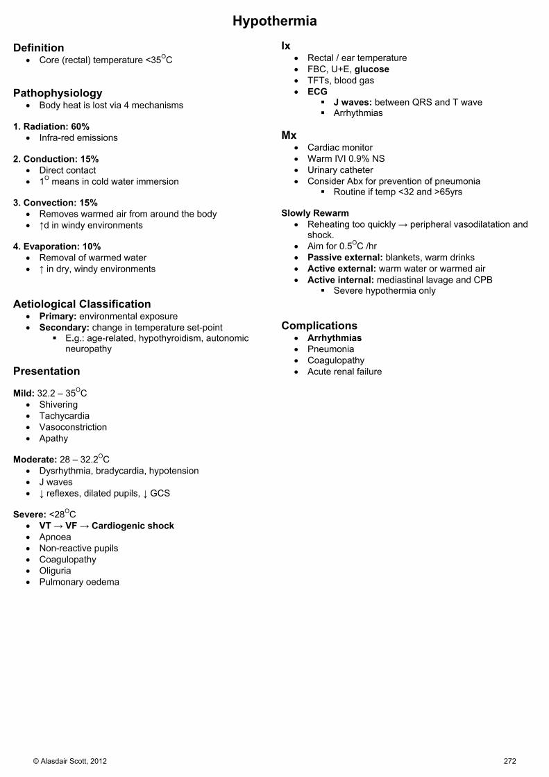
Hypothermia
Definition Core (rectal) temperature <35OC
Pathophysiology
Body heat is lost via 4 mechanisms 1. Radiation: 60%
Infra-red emissions 2. Conduction: 15%
Direct contact 1O means in cold water immersion
3. Convection: 15%
Removes warmed air from around the body ↑d in windy environments
4. Evaporation: 10%
Removal of warmed water ↑ in dry, windy environments
Aetiological Classification
Primary: environmental exposure Secondary: change in temperature set-point
E.g.: age-related, hypothyroidism, autonomic neuropathy
Presentation Mild: 32.2 – 35OC
Shivering Tachycardia Vasoconstriction Apathy
Moderate: 28 – 32.2OC
Dysrhythmia, bradycardia, hypotension J waves ↓ reflexes, dilated pupils, ↓ GCS
Severe: <28OC
VT → VF → Cardiogenic shock Apnoea Non-reactive pupils Coagulopathy Oliguria Pulmonary oedema
Ix
Rectal / ear temperature FBC, U+E, glucose TFTs, blood gas ECG
J waves: between QRS and T wave Arrhythmias
Mx
Cardiac monitor Warm IVI 0.9% NS Urinary catheter Consider Abx for prevention of pneumonia
Routine if temp <32 and >65yrs Slowly Rewarm
Reheating too quickly → peripheral vasodilatation and shock.
Aim for 0.5OC /hr Passive external: blankets, warm drinks Active external: warm water or warmed air Active internal: mediastinal lavage and CPB
Severe hypothermia only Complications
Arrhythmias Pneumonia Coagulopathy Acute renal failure
© Alasdair Scott, 2012 272
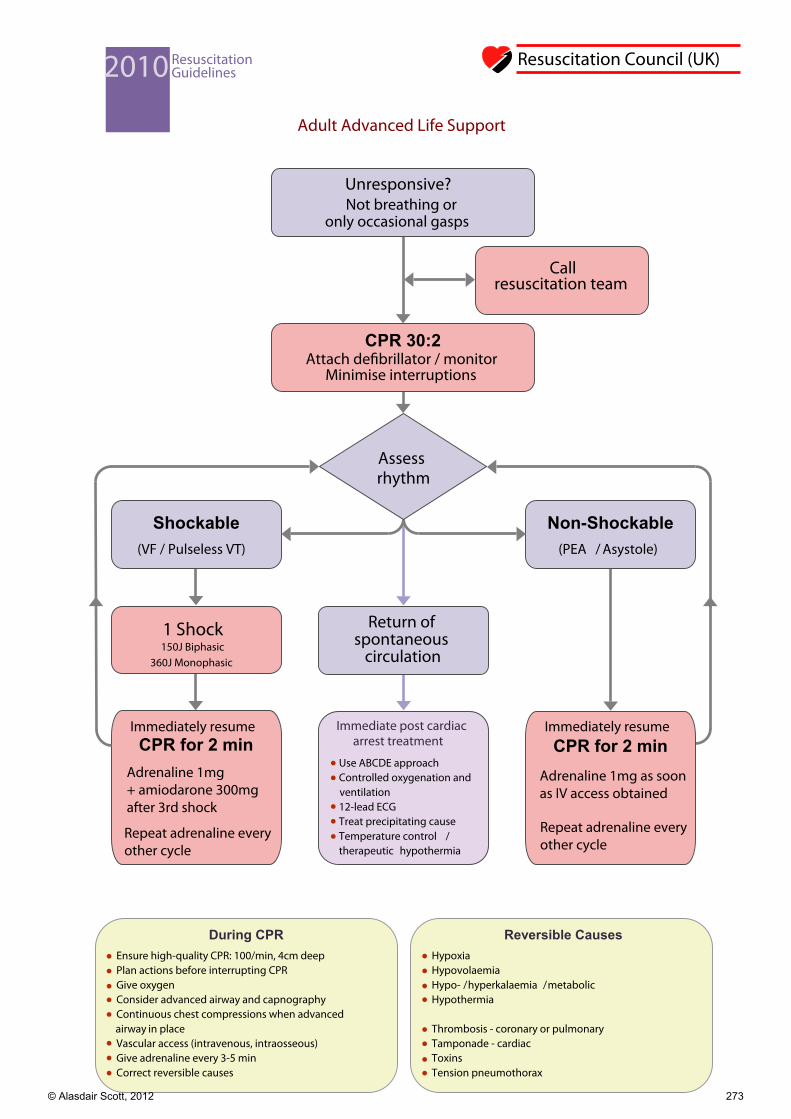
Adult Advanced Life Support
CPR 30:2 Attach de�brillator / monitor
Minimise interruptions
Shockable (VF / Pulseless VT)
1 Shock
During CPR Ensure high-quality CPR: 100/min, 4cm deep Plan actions before interrupting CPR Give oxygen Consider advanced airway and capnography Continuous chest compressions when advanced
airway in place Vascular access (intravenous, intraosseous) Give adrenaline every 3-5 min Correct reversible causes
Reversible Causes Hypoxia Hypovolaemia Hypo- / hyperkalaemia / metabolic Hypothermia
Thrombosis - coronary or pulmonary Tamponade - cardiac Toxins Tension pneumothorax
Non-Shockable (PEA / Asystole)
Call resuscitation team
2010 Resuscitation Guidelines
Unresponsive? Not breathing or
only occasional gasps
Return of spontaneous
circulation
Immediately resume CPR for 2 min
Immediately resume CPR for 2 min
Resuscitation Council (UK)
Immediate post cardiac arrest treatment
Use ABCDE approach Controlled oxygenation and
ventilation 12-lead ECG Treat precipitating cause Temperature control /
therapeutic hypothermia
Assess rhythm
150J Biphasic360J Monophasic
Adrenaline 1mg + amiodarone 300mgafter 3rd shock
Repeat adrenaline every other cycle
Adrenaline 1mg as soonas IV access obtained
Repeat adrenaline every other cycle
© Alasdair Scott, 2012 273

Adult tachycardia (with pulse)algorithm
Yes / Unstable
Assess using the ABCDE approach Give oxygen if appropriate and obtain IV access Monitor ECG, BP, SpO2 , record 12-lead ECG Identify and treat reversible causes (e.g. electrolyte abnormalities)
Amiodarone 300 mg IV over 10-20 min and repeat shock; followed by: Amiodarone 900 mg over 24 h
No / Stable
NarrowBroad
Irregular Narrow Complex Tachycardia
Probable atrial fibrillation Control rate with: -Blocker or diltiazem Consider digoxin or amiodarone if evidence of heart failure
Is QRS narrow (< 0.12 s)?
IrregularNarrow QRS Is rhythm regular?
Regular
Possibilities include: AF with bundle branch block treat as for narrow complex Pre-excited AF consider amiodarone Polymorphic VT (e.g. torsade de pointes - give magnesium 2 g over 10 min)
If ventricular tachycardia (or uncertain rhythm): Amiodarone 300 mg IV over 20-60 min; then 900 mg over 24 h If previously confirmed SVT with bundle branch block: Give adenosine as for regular narrow complex tachycardia
Use vagal manoeuvres Adenosine 6 mg rapid IV bolus; if unsuccessful give 12 mg; if unsuccessful give further 12 mg. Monitor ECG continuously
Probable re-entry paroxysmal SVT: Record 12-lead ECG in sinus rhythm If recurs, give adenosine again & consider choice of anti-arrhythmic prophylaxis
Yes
Sinus rhythm restored?
No
Possible atrial flutter Control rate (e.g. -Blocker)
Irregular Regular
Adverse features? Shock Syncope Myocardial ischaemia Heart failure
Synchronised DC Shock
Up to 3 attempts
2010 Resuscitation Guidelines
Resuscitation Council (UK)
Seek expert help
Seek expert help
Broad QRS Is rhythm regular?
!
!
© Alasdair Scott, 2012 274

Adult bradycardia algorithm
Seek expert help Arrange transvenous pacing
* Alternatives include: Aminophylline Dopamine Glucagon (if beta-blocker or calcium channel blocker overdose) Glycopyrrolate can be used instead of atropine
YES NO Adverse features?
Shock Syncope Myocardial ischaemia Heart failure
Atropine 500 mcg IV
YES
NO
NO
YES
2010 Resuscitation Guidelines
Resuscitation Council (UK)
Assess using the ABCDE approach Give oxygen if appropriate and obtain IV access Monitor ECG, BP, SpO2, record 12-lead ECG Identify and treat reversible causes
(e.g. electrolyte abnormalities)
Observe
Satisfactory response?
!
Interim measures: Atropine 500 mcg IV repeat to maximum of 3 mg Isoprenaline 5 mcg min-1 IV Adrenaline 2-10 mcg min-1 IVAlternative drugs * OR Transcutaneous pacing
Risk of asystole? Recent asystole Mobitz II AV block Complete heart block with broad QRS Ventricular pause > 3 s
© Alasdair Scott, 2012 275