Patient Care Protocols - Georgetown Scott County EMS
374
Georgetown Scott County EMS Patient Care Protocols EMT Advanced EMT Paramedic
-
Upload
khangminh22 -
Category
Documents
-
view
1 -
download
0
Transcript of Patient Care Protocols - Georgetown Scott County EMS

Georgetown Scott County EMS
Patient Care Protocols
EMT Advanced EMT Paramedic

Introduction
In the following protocols, procedures, and policies we have incorporated evidence-based guidelines with historically proven practice. It is impossible to address every possible variation of disease and injury however in these cases, providers should treat any present life-threats using whichever protocol is most appropriate as a foundation for care, consider utilizing additional resources and consider contacting On-Line Medical Control for guidance in how to proceed with patient care as well as destination guidance.
These protocols and procedures are to be used as guidelines for operation during EMS calls that require patient assessment and care. They are also intended to be used as guidelines to ensure that personnel are trained in proper pre-hospital patient care. Procedures are not considered rigid rules, but rather established standards against which EMS practice can be measured.
Treatment protocols are specific orders directing the actions pertaining to techniques and/or medications used by GSCEMS personnel who are required to practice under direct supervision of a physician and und Treatment protocols may and should be initiated without prior direct medical control contact, especially when the patient’s condition and/or situation is life threatening. As
These protocols are derived from the Kentucky State EMS protocols and have been tailored to suit the needs of Georgetown Scott County EMS. At the time of publishing they are up-to-date with best practices for pre-hospital care. Best practices do change and therefore these protocols will be updated to reflect those changes as they occur.
These protocols were developed by the Georgetown Scott County EMS Protocol Committee, special thanks to:
Corey Wood, NRP Uriah Tindle, NRP
Jonathan Wheeler, NRP Easton McClanahan, NRP
Jonathan Oesterman, NRP Patrick Yorba, MD

Table of Contents
General Protocols
On-Scene Medical Personnel ............................................................................................ GP – 1 Use of Lights and Sirens .................................................................................................... GP – 2 Determination of Death – Dead on Scene ........................................................................ GP – 3 Determination of Death – Discontinuance of Resuscitation by Paramedic ...................... GP – 4 Kentucky EMS DNR Order Form ........................................................................................ GP – 5 Trauma Triage ................................................................................................................... GP – 6 Trauma Triage Criteria Algorithm ..................................................................................... GP – 7 Verified Trauma Centers in or near Kentucky ................................................................... GP – 8 Air Medical/Helicopter Safety ........................................................................................... GP – 9 Helicopter Utilization Criteria for Scene Response ........................................................... GP – 10 Abuse or Neglect – Child, Elder, or Other Vulnerable Individuals .................................... GP – 11 Crime Scene/Preservation of Evidence ............................................................................. GP – 12 Sexual Assault ................................................................................................................... GP – 13 Restraint Protocol ............................................................................................................. GP – 14 Restraint and Transport Pediatric ..................................................................................... GP – 15 Safe Infants Act ................................................................................................................. GP – 16 Bloodborne/Airborne Pathogens ...................................................................................... GP – 17
Adult Cardiac Protocols
Routine Patient Care Guidelines ....................................................................................... ACP – 1 Acute Coronary Syndromes ............................................................................................... ACP – 2 Cardiac Arrest .................................................................................................................... ACP – 3 Post Cardiac Arrest Care .................................................................................................... ACP – 4 Bradycardia ....................................................................................................................... ACP – 5 Tachycardia ....................................................................................................................... ACP – 6 Congestive Heart Failure/Pulmonary Edema .................................................................... ACP – 7
Adult Medical Protocols
Allergic Reaction/Anaphylaxis ........................................................................................... AMP – 1 Asthma/COPD/RAD ........................................................................................................... AMP – 2 Behavioral Emergencies .................................................................................................... AMP – 3 Diabetic Emergencies ........................................................................................................ AMP – 4 Fever .................................................................................................................................. AMP – 5 Sepsis – Adult .................................................................................................................... AMP – 6 Nausea/Vomiting .............................................................................................................. AMP – 7 Non-Traumatic Abdominal Pain ........................................................................................ AMP – 8 Pain Management ............................................................................................................. AMP – 9 Poisoning: Overdose .......................................................................................................... AMP – 10 Poisoning: Cyanide ............................................................................................................ AMP – 11

Poisoning: Nerve Agents and Organophosphates ............................................................ AMP – 12 Poisoning: Radiation Injuries ............................................................................................ AMP – 13 Seizures ............................................................................................................................. AMP – 14 Suspected Stroke .............................................................................................................. AMP – 15 Obstetrical Emergencies ................................................................................................... AMP – 16 Unresponsive/Altered Mental Status ............................................................................... AMP – 17 Adrenal Crisis .................................................................................................................... AMP – 18
Pediatric Medical Protocols
Routine Patient Care Guidelines ....................................................................................... PMP – 1 Pediatric Assessment ........................................................................................................ PMP – 2 Apparent Life-Threatening Event (ALTE) .......................................................................... PMP – 3 Sudden Infant Death Syndrome (SIDS) ............................................................................. PMP – 4 Neonatal Resuscitation ..................................................................................................... PMP – 5 Cardiac Arrest .................................................................................................................... PMP – 6 Bradycardia ....................................................................................................................... PMP – 7 Tachycardia ....................................................................................................................... PMP – 8 Shock ................................................................................................................................. PMP – 9 Allergic Reaction/Anaphylaxis ........................................................................................... PMP – 10 Asthma/RAD/Croup .......................................................................................................... PMP – 11 Diabetic Emergencies ........................................................................................................ PMP – 12 Non-Traumatic Abdominal Pain ........................................................................................ PMP – 13 Poisoning: Overdose ......................................................................................................... PMP – 14 Poisoning: Cyanide ............................................................................................................ PMP – 15 Poisoning: Nerve Agents and Organophosphates ............................................................ PMP – 16 Poisoning: Radiation Injuries ............................................................................................ PMP – 17 Seizures .............................................................................................................................. PMP – 18 Fever .................................................................................................................................. PMP – 19 Nausea/Vomiting .............................................................................................................. PMP – 20 Dehydration ...................................................................................................................... PMP – 21 Unresponsive/Altered Mental Status ................................................................................ PMP – 22 Children with Special Health Care Needs .......................................................................... PMP – 23 Adrenal Crisis – Pediatric .................................................................................................. PMP – 24
Adult Trauma Protocol
Trauma Assessment and Management ............................................................................ ATP – 1 Selective Spinal Immobilization ........................................................................................ ATP – 2 Head Trauma ..................................................................................................................... ATP – 3 Chest Trauma .................................................................................................................... ATP – 4 Abdominal Trauma ............................................................................................................ ATP – 5 Pelvic Trauma .................................................................................................................... ATP – 6 Extremity Trauma .............................................................................................................. ATP – 7 Eye and Dental Trauma ..................................................................................................... ATP – 8 Burns (Thermal) ................................................................................................................. ATP – 9

Pediatric Trauma Protocols
Trauma Assessment and Management ............................................................................ PTP – 1 Head Trauma ..................................................................................................................... PTP – 2 Chest Trauma .................................................................................................................... PTP – 3 Abdominal Trauma ............................................................................................................ PTP – 4 Pain Management ............................................................................................................. PTP – 5 Pelvic Trauma ..................................................................................................................... PTP – 6 Extremity Trauma .............................................................................................................. PTP – 7 Eye and Dental Injuries ..................................................................................................... PTP – 8 Burns (Thermal) ................................................................................................................ PTP – 9 Selective Spinal Immobilization (> 8 yrs old) .................................................................... PTP – 10
Environmental Protocols
Electrical/Lightning Injuries .............................................................................................. EP – 1 Snake Bites ........................................................................................................................ EP – 2 Submersion Injuries .......................................................................................................... EP – 3 Heat Cramps/Heat Exhaustion .......................................................................................... EP – 4 Hyperthermia .................................................................................................................... EP – 5 Hypothermia ..................................................................................................................... EP – 6 Frostbite ............................................................................................................................ EP – 7
Airway and Ventilation Management
Universal Advanced Airway Management Algorithm ....................................................... AVM – 1 Crash Airway Algorithm ..................................................................................................... AVM – 2 Difficult Airway Algorithm ................................................................................................. AVM – 3 Failed Airway Algorithm .................................................................................................... AVM - 4 Airway Management – Pediatric ....................................................................................... AVM – 5 Post – Intubation Care ...................................................................................................... AVM – 6 Mechanical Ventilation ..................................................................................................... AVM – 7 CPAP/BiPAP Use ................................................................................................................ AVM – 8 Endotracheal Tube Introducer (Bougie) ........................................................................... AVM – 9 King LT – D ......................................................................................................................... AVM – 10 iGel Supraglottic Airway Device ........................................................................................ AVM – 11 Digital Intubation .............................................................................................................. AVM – 12 Blind Nasotracheal Intubation .......................................................................................... AVM – 13 QuickTrach Device Cricothyrotomy .................................................................................. AVM – 14 Mucosal Atomizer Device ................................................................................................. AVM – 15 Tracheostomy Care ........................................................................................................... AVM – 16 Advanced Suctioning ......................................................................................................... AVM – 17 Esophageal Intubation Detector Device ........................................................................... AVM – 18

Procedures
Application of ECG Monitor and Electrodes ..................................................................... PRO – 1 External Jugular IV Access .................................................................................................. PRO – 2 Intraosseous Access ........................................................................................................... PRO – 3 Umbilical Vein Cannulation ................................................................................................ PRO – 4 Vascular Access via Central Catheter ................................................................................. PRO – 5
Inter-Facility Protocols
Inter-facility Transfer ........................................................................................................ IFT – 1 Maintenance of Blood or Blood Products ......................................................................... IFT – 2 Thoracostomy Tube Monitoring ....................................................................................... IFT – 3 Amiodarone Hydrochloride Infusion Monitoring .............................................................. IFT – 4 Heparin Infusion Monitoring ............................................................................................. IFT – 5 Lidocaine Infusion Monitoring .......................................................................................... IFT – 6 Magnesium Sulfate Infusion Monitoring .......................................................................... IFT – 7 Nitroglycerin Infusion Monitoring ..................................................................................... IFT – 8 Potassium Chloride Infusion Monitoring ........................................................................... IFT – 9 Tissue Plasminogen Activator (TPA) Infusion Monitoring ................................................ IFT – 10 Propofol (Diprovan) Infusion Monitoring ......................................................................... IFT – 11
Medication List
Adenosine ......................................................................................................................... MED – 1 Albuterol ............................................................................................................................ MED – 2 Amiodarone ....................................................................................................................... MED – 3 Aspirin ................................................................................................................................ MED – 4 Atropine Sulfate ................................................................................................................. MED – 5 Calcium Chloride/Calcium Gluconate ................................................................................ MED – 6 Cefazolin ............................................................................................................................ MED – 7 Dextrose ............................................................................................................................. MED – 8 Diphenhydramine .............................................................................................................. MED – 9 Dopamine ........................................................................................................................... MED – 10 Epinephrine ........................................................................................................................ MED – 11 Fentanyl ............................................................................................................................. MED – 12 Glucagon ............................................................................................................................ MED – 13 Glucose – Oral .................................................................................................................... MED – 14 Ipratropium Bromide ......................................................................................................... MED – 15 Ketamine ............................................................................................................................ MED – 16 Ketorolac ............................................................................................................................ MED – 17 Lidocaine ............................................................................................................................ MED – 18 Magnesium Sulfate ............................................................................................................ MED – 19 Methylprednisolone........................................................................................................... MED – 20 Midazolam ......................................................................................................................... MED – 21 Naloxone ............................................................................................................................ MED – 22

Nitroglycerin ...................................................................................................................... MED – 23 Ondansetron ...................................................................................................................... MED – 24 Oxygen ............................................................................................................................... MED – 25 Promethazine .................................................................................................................... MED – 26 Sodium Bicarbonate 8.4 % ................................................................................................. MED – 27
Specialized Protocols
TASER Subdued Patient ..................................................................................................... SP – 1 Strenuous Activity/Firefighter Rehabilitation ................................................................... SP – 2 STARTBand Emergency Response Tag System .................................................................. SP – 3 START System of Triage ..................................................................................................... SP – 4 Mass Casualty Incidents .................................................................................................... SP – 5 EMS Plan for Responding to Pandemic Influenza ............................................................. SP – 6
ToxMedic Emergency Care Protocols
ToxMedic: Irritant gases/Toxic Inhalation ToxMedic: Asphyxiants – Simple ToxMedic: Systemic Asphyxiants ToxMedic: Systemic Asphyxiants – Carbon Monoxide ToxMedic: Systemic Asphyxiants – Cyanide ToxMedic: Cholinesterase Inhibitors ToxMedic: Corrosives ToxMedic: Hydrocarbons ToxMedic: Hydrofluoric Acid & Fluorides ToxMedic: Ethylene Glycol and Methanol ToxMedic: Chemo-terrorism Nerve Agents ToxMedic: Auto-Injector Kit Administration ToxMedic: Antidote Reference

GSCEMS Master Medication List
BLS Medications
- Albuterol - Aspirin - Epinephrine 1:1 000
- Naloxone - Oral Glucose - Oxygen
ALS Medications
- Adenosine - Amiodarone - Atropine - Calcium Chloride - Dextrose 10% - AEMT - Diphenhydramine - Dopamine - Epinephrine 1:10 000 - Fentanyl - Glucagon - AEMT - Ipratropium Bromide
- Ketamine - Ketorolac - Lactated Ringers – AEMT - Lidocaine - Magnesium Sulfate - Methylprednisolone - Midazolam - Nitroglycerin – AEMT - Normal Saline – AEMT - Ondansetron - Promethazine - Sodium Bicarbonate

General Protocols

On-Scene Medical Personnel
GP – 1
► The medical care provided at the scene is the responsibility of the highest level of EMS provider who has responded by usual dispatch systems to that scene. Passersby who stop to help, even though possibly more highly trained than the system providers, may not assume responsibility (except as outlined below) but may be allowed to help in care at the discretion of the lead EMS provider and assuming they have proof of licensure.
► When an EMS provider, under medical control (on- or off-line), arrives at the scene of an emergency, the provider acts as the agent of medical control.
► Any healthcare provider (MD, PA, RN, nurse midwife, non-KY licensed EMS provider, etc.) who is not an active member of the responding EMS unit, and who is either at the scene at the time of EMS‘ arrival or arrives after an EMS unit provider has initiated care, and who desires to continue to participate, should be put in touch with the on-line medical control physician.
► At no time should an EMS provider provide care outside of their scope of training and/or protocols.
► In the event that a Mass Casualty Incident (MCI) is declared, all Providers should follow the Mass Casualty Incidents Uniform Prehospital MCI Procedure outlined in this document or similar approved Incident Command System.

Use of Lights and Sirens
GP-2
Purpose
The estimated EMS fatality rate (12.7 per 100,000 workers) is more than twice the national rate. Vehicles crashes of all types remain the leading cause of death in EMS. The use of Lights and Sirens in the transport of a patient from the scene to the hospital by EMS personnel should be consistent with "best practices", be medically defensible and conform to Kentucky state law. It is not without risk and should be used only when there is a likely benefit to the patient. This is to ensure the safety of our patients, our staff, our citizens and ourselves.
Policy
KRS 189.910 to KRS 189.950 outline the legal parameters under which an emergency vehicle may be exempt from certain traffic regulations. The vehicle operator should be familiar with these statutes. Specifically:
189.940 Exemptions from traffic regulations.
► The speed limitations set forth in the Kentucky Revised Statutes do not apply toemergency vehicles: When responding to emergency calls; or To police vehicles when in pursuit of an actual or suspected violator of the law; or To ambulances when transporting a patient to medical care facilities; and The driver thereof is giving the warning required by subsection (5)(a) and
(b) of this section.
No portion of this subsection shall be construed to relieve the driver of the duty to operate the vehicle with due regard for the safety of all persons using the street or highway.
The law permits such emergency vehicles only on emergency calls or when transporting to a medical care facility to utilize lights and sirens. EMS personnel are instructed to follow the state laws and use lights and sirens while going to the hospital only when it is medically necessary for the patient to be rapidly transported. Rapid transport to the scene may be necessary in certain instances to evaluate the situation for possible life threats. It is then that the EMS personnel in charge of patient care will make the appropriate transportation decision. Although time is typically saved, studies have shown the savings to be from less than one minute to less than four minutes and rarely clinically significant to the patient. Transport in this manner is not without risk to the patient. The EMS personnel in charge will have to weigh the risks and benefits to the patient and document this rationale on the EMS run form. This policy does not restrict the EMS personnel from changing a non-emergency transport back into an emergency transport if conditions change

Determination of Death - Dead on Scene
GP – 3
If it appears that a patient you have been called to attend is dead, this protocol shall be followed prior to final determination. 1. The Paramedic shall determine and document that the following signs of death are
present: • Unresponsiveness • Apnea • The absence of a palpable pulse at the carotid site • Bilaterally fixed and dilated pupils; and • Asystole determined in two (2) leads on an electrocardiograph in accordance
with the American Heart Association guidelines, except in cases of trauma or when presented with a standard form or identification evidencing a patient’s desire not to be resuscitated in accordance with KRS 311.623 (DNR regulation).
2. The Paramedic shall determine, in addition, that one (1) or more of the following
factors or conditions exist: • Lividity of any degree • Rigor mortis of any degree (In the non-hypothermic patient) • The presence of venous pooling in the body • Damage or destruction of the body which is incompatible with life, or • A standard form or identification evidencing a patient’s desire not to be
resuscitated in accordance with KRS 311.623 (DNR regulation). 3. If the Paramedic has determined and documented that the conditions above
(sections 1 and 2) have been met, the Paramedic may declare the patient dead. 4. The Paramedic may contact the on duty MEDICAL CONTROL for advice and
assistance in making a determination required by this protocol. 5. If ANY patient meets the criteria described above as a non-resuscitation candidate,
access to the scene should be limited as much as possible with due care to disturb the scene as little as possible. As in all cases of out of–hospital deaths, every effort should be made to console family, friends, survivors, and witnesses without interfering with ongoing investigations.

Determination of Death - Dead on Scene continued
GP – 3
6. The Paramedic shall document all items required on the Kentucky EMS Ambulance
Run Report including the usual patient assessment, medical history, and surrounding events information. It is especially important to note:
• Body position and location when discovered, including differences from when last seen alive.
• Patient condition when last seen alive. • Clothing and condition of clothing. • Conditions of residence/business/location found. • Statements made on the scene by significant individuals. • Any unusual circumstances.
7. If the Paramedic determines a patient to be dead, the paramedic shall remain on the
scene until the arrival of a law enforcement officer or until the Paramedic is released from the scene by the coroner.
IT IS TO BE EXPRESSLY UNDERSTOOD THAT IN THE EVENT OF ANY UNCERTAINTY AS TO THE PATIENT STATUS, THE CREW IS TO INITIATE NORMAL RESUSCITATIVE EFFORTS
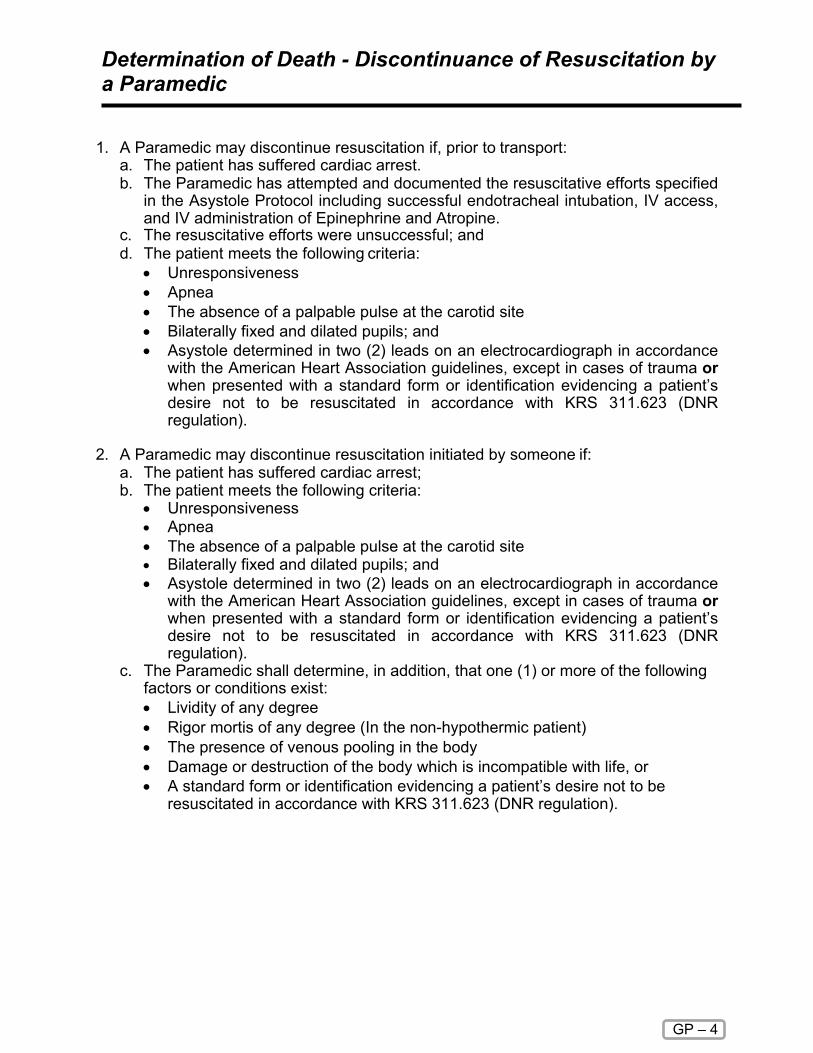
Determination of Death - Discontinuance of Resuscitation by a Paramedic
GP – 4
1. A Paramedic may discontinue resuscitation if, prior to transport:
a. The patient has suffered cardiac arrest. b. The Paramedic has attempted and documented the resuscitative efforts specified
in the Asystole Protocol including successful endotracheal intubation, IV access, and IV administration of Epinephrine and Atropine.
c. The resuscitative efforts were unsuccessful; and d. The patient meets the following criteria:
• Unresponsiveness • Apnea • The absence of a palpable pulse at the carotid site • Bilaterally fixed and dilated pupils; and • Asystole determined in two (2) leads on an electrocardiograph in accordance
with the American Heart Association guidelines, except in cases of trauma or when presented with a standard form or identification evidencing a patient’s desire not to be resuscitated in accordance with KRS 311.623 (DNR regulation).
2. A Paramedic may discontinue resuscitation initiated by someone if:
a. The patient has suffered cardiac arrest; b. The patient meets the following criteria:
• Unresponsiveness • Apnea • The absence of a palpable pulse at the carotid site • Bilaterally fixed and dilated pupils; and • Asystole determined in two (2) leads on an electrocardiograph in accordance
with the American Heart Association guidelines, except in cases of trauma or when presented with a standard form or identification evidencing a patient’s desire not to be resuscitated in accordance with KRS 311.623 (DNR regulation).
c. The Paramedic shall determine, in addition, that one (1) or more of the following factors or conditions exist: • Lividity of any degree • Rigor mortis of any degree (In the non-hypothermic patient) • The presence of venous pooling in the body • Damage or destruction of the body which is incompatible with life, or • A standard form or identification evidencing a patient’s desire not to be
resuscitated in accordance with KRS 311.623 (DNR regulation).

Determination of Death - Discontinuance of Resuscitation by a Paramedic continued
GP – 4
3. The Paramedic shall contact the on duty MEDICAL CONTROL, for advice and
assistance prior to making the determination. MEDICAL CONTROL approval must be obtained prior to the discontinuance of resuscitative efforts.
4. The Paramedic shall document all items required on the Kentucky EMS run report including, the usual patient assessment, medical history and surrounding events information. It is especially important to note:
• Body position and location when discovered, including differences from when
last seen alive. • Patient condition when last seen alive. • Clothing and condition of clothing. • Condition of residence/business/location found. • Statements made on the scene by significant individuals. • Any unusual circumstances.
IT IS TO BE EXPRESSLY UNDERSTOOD THAT IN THE EVENT OF ANY UNCERTAINTY AS TO THE PATIENT STATUS, THE CREW IS TO INITIATE NORMAL RESUSCITATIVE EFFORTS

GP – 5
Kentucky Emergency Medical Services
Do Not Resuscitate (DNR) Order
I, the undersigned person or surrogate who has been designated to make health care decisions in accordance with Kentucky Revised Statutes, hereby direct that in the event of my cardiac or respiratory arrest that this DO NOT RESUCITATATE (DNR) Order be honored. I understand that DNR means that if my heart stops beating or if I stop breathing, no medical procedure to restart breathing or heart function, more specifically the insertion of a tube into the lungs, or electrical shocking of the heart or cardiopulmonary resuscitation (CPR) will be started by emergency medical services (EMS) personnel. I understand this decision will not prevent emergency medical services personnel from providing other medical care. I understand that I may revoke this DNR order at any time by destroying this form, removing the DNR bracelet, or by telling the EMS personnel that I want to be resuscitated. Any attempt to alter or change the content, names, or signatures on the EMS DNR form shall make the DNR form invalid. I understand that this form, or a standard EMS DNR bracelet must be available and must be shown to EMS personnel as soon as they arrive. If the form or bracelet is not provided, the EMS personnel will follow their normal protocols which could include cardiopulmonary resuscitation (CPR) or other resuscitation procedures. I understand that should I die; EMS personnel will require this form and/or bracelet for their records. I give permission for information about this EMS DNR Order to be given to the prehospital emergency medical care personnel, physicians, nurses, or other health care personnel as necessary to implement this directive. I hereby state that this 'Do Not Resuscitate (DNR) Order' is my authentic wish not be resuscitated.
Person/Legal Surrogate Signature Date
Commonwealth of Kentucky County of ________________________ Subscribed and sworn to before me by to be his/her own free act and deed, this day of , 20 .
, Notary Public
My commission expires:
In lieu of having this Form notarized, it may be witnessed by two persons not related to the individual noted above. WITNESSED BY: 1. 2.
This EMS Do Not Resuscitate Form was approved by the Kentucky Board of Medical Licensure at their March 1995 meeting. Complete the portion below, cut out, fold, and insert in DNR bracelet
I certify that an EMS Do Not Resuscitate (DNR) form has been executed. Person's Name (print or type) Person's or Legal Surrogate's Signature

GP – 5
INSTRUCTIONS PURPOSE
This standardized EMS DNR Order has been developed and approved by the Kentucky Board of Medical Licensure, in consultation with the Cabinet for Human Resources. It is in compliance with KRS Chapter 311 as amended by Senate Bill 311 passed by the 1994 General Assembly, which directs the Kentucky Board of Medical Licensure to develop a standard form to authorize EMS providers to honor advance directives to withhold or terminate care. For covered persons in cardiac or respiratory arrest, resuscitative measures to be withheld include external chest compressions, intubation, defibrillation, administration of cardiac medications and artificial respiration. The EMS DNR Order does not affect the provision of other emergency medical care, including oxygen administration, suctioning, control of bleeding, administration of analgesics and comfort care. APPLICABILITY This EMS DNR Order applies only to resuscitation attempts by health care providers in the prehospital setting (i.e., certified EMT-First Responders, Emergency Medical Technicians, and Paramedics) — in patients' homes, in a long-term care facility, during transport to or from a health care facility, or in other locations outside acute care hospitals. INSTRUCTIONS Any adult person may execute an EMS DNR Order. The person for whom the Order is executed shall sign and date the Order and my either have the Order notarized by a Kentucky Notary Public or have their signature witness by two persons not related to them. The executor of the Order must also place their printed or typed name in the designated area and their signature on the EMS DNR Order bracelet insert found at the bottom of the EMS DNR Order form. The bracelet insert shall be detached and placed in a hospital type bracelet and placed on the wrist or ankle of the executor of the Order. If the person for whom the EMS DNR Order is contemplated is unable to give informed consent, or is a minor, the person's legal surrogate shall sign and date the Order and may either have the form notarized by a Kentucky Notary Public or have their signature witnessed by two persons not related to the person for which the form is being executed or related to the legal health care surrogate. The legal health care surrogate shall also complete the required information on the EMS DNR bracelet insert found at the bottom of the EMS DNR Order form. The bracelet shall be detached and placed in a hospital type bracelet and placed on the wrist or ankle of the person for which this Order was executed. The original, completed EMS DNR Order or the EMS DNR Bracelet must be readily available to EMS personnel in order for the EMS DNR Order to be honored. Resuscitation attempts may be initiated until the form or bracelet is presented and the identity of the patient is confirmed by the EMS personnel. It is recommended that the EMS DNR Order be displayed in a prominent place close to the patient and/or the bracelet be on the patient's wrist or ankle. REVOCATION An EMS DNR Order may be revoked at any time orally or by performing an act such as burning, tearing, canceling, obliterating or by destroying the order by the person on whose behalf it was executed or by the person's legal health care surrogate. IT SHOULD BE UNDERSTOOD BY THE PERSON EXECUTING THIS EMS DNR ORDER OR THEIR LEGAL HEALTH CARE SURROGATE, THAT SHOULD THE PERSON LISTED ON THE EMS DNR ORDER DIE WHILE EMS PREHOSPITAL PERSONNEL ARE IN ATTENDANCE, THE EMS DNR ORDER OR EMS DNR BRACELET MUST BE GIVEN TO THE EMS PREHOSPITAL PERSONNEL FOR THEIR RECORDS

Trauma Triage
GP – 6
Purpose Victims of major trauma have better outcomes when transported to a designated trauma center in a timely manner. The American College of Surgeons (ACS) has developed triage criteria that is useful in identifying patients that may benefit from evaluation at a trauma center. In general, consider the following guidelines: It is in the best interest of the patient to be transported to a designated trauma center if the patient meets ACS criteria and a designated trauma center is within thirty minutes transport time. Patients with a compromised airway may be best served by transport to the closest hospital with rapid transfer to a trauma center. Consider air medical resources but do not delay transport unnecessarily. (See Helicopter Criteria for Scene Transport).

Trauma Triage Criteria Algorithm
GP – 7

Verified Trauma Centers in Kentucky
GP – 8

Air Medical/ Helicopter Safety
GP – 9
Landing Zone and Safety. Without exception, safety is air medical service’s top priority. Requesting a helicopter ► Private Citizens - call 9-1-1. ► Police, fire and EMS - Request a helicopter through the appropriate agency, such
as your dispatch center, with the following information: Location cross street Location LAT/LONG coordinates Any prominent features at the scene Notify all involved communications centers if any other air medical service
has been contacted and the status of that agency. Always inform all communications centers if other aircraft are anticipated to be in the area.
Your call-back number Scene radio frequency and CTCSS tone Call sign of LZ (Landing Zone) Command. One person should be
designated to coordinate LZ setup and communicate with responding aircraft. This person should not be involved with patient care.
Weather, including low ceilings, poor visibility, icing, and high winds Patient status, such as number, condition, age, approximate patient
weight, mechanism of injury, and hazards LZ details. The preferred landing zone is 100 x 100 feet. ALWAYS RELAY ANY INFORMATION PERTAINING TO HAZMAT TO
THE COMMUNICATIONS CENTER WHEN REQUESTING AIR MEDICAL SERVICE.
Important Tips
Never approach the aircraft until instructed to do so and only as instructed by the pilot or flight crew aboard
Approach angles over obstacles should be less than 20 degrees
Always keep LZ clear of people and other potential hazards
Under no circumstances should you ever approach the aircraft from the rear

Air Medical/ Helicopter Safety continued
GP – 9
Landing Zone Setup ► Set up the LZ as follows:
SIZE should be 100 feet by 100 feet LEVEL: Select a LZ as level as possible (minimal slope) LANDING SURFACE: Select a hard surface, grassy surface, or hard-
packed snow. Avoid loose dirt, dust, or powder snow. CLEAR OVERHEAD free of obstructions such as wires, antennas, or
poles CLEAR AREA free of debris, large rocks, posts, stumps, vehicles, people,
animals, and other hazards MARK THE AREA clearly using five weighted cones or beacons, one at
each corner of the LZ and one on the side that wind is coming from SELECT AN ALTERNATE LZ. Plan for an alternate LZ because the pilot
may determine your LZ to be unsafe. HAZMAT: Always relay any information pertaining to HAZMAT to the
communications center when requesting air medical service. Always inform the pilot and medical crew of HAZMAT. When selecting a LZ find a site at least 1/4 to 1 mile UPWIND from the incident depending on the type and materials involved. Avoid low areas where vapors may collect. The patient must be removed from the hot zone. All patients must be decontaminated PRIOR to flight.
When the helicopter is overhead ► Air medical service will establish radio contact on the assigned frequency with LZ
Command three to five minutes out. Describe the following: LZ location Lighting Hazards Overhead wires, including wires along the approach path to the LZ Obstructions Slope Surface conditions Wind direction and speed if known Maintain radio contact at all times until the helicopter has landed, loaded,
and departed the area.

Helicopter Utilization Criteria for Scene Response
GP – 10
Night Landing Zone
► DO NOT SHINE LIGHTS DIRECTLY AT THE HELICOPTER
► Set up night landing zones with five strobes or other secured lights. Do not use cones, flares, or tape to mark the site.
► Emergency vehicles may be parked so their headlights intersect the middle of the landing site and/or parked underneath wires to mark them. Turn strobes of emergency vehicles off as the aircraft approaches.
► Lights may be shown onto poles indicating wires between the poles
► Night landing zones always require good communications, lighting, and alertness
► Turn off all emergency lights after aircraft has started approach
► One strobe should be on the side that the wind is coming from
► If no strobes are available mark with other lighting systems
► If no other portable lights are available, cross headlight beams into the wind at the center of the landing zone.

Helicopter Utilization Criteria for Scene Response
GP – 10
Purpose: Air Medical Services (AMS) are a valuable, yet limited resource in the Commonwealth. It is important that Emergency Medical Service personnel utilize consistent and appropriate criteria when requesting an air medical service for assistance with patient care and transport. Air Medical Services (AMS) are a valuable, yet limited resource. It is important that Emergency Medical Service personnel utilize consistent and appropriate criteria when requesting an air medical service for assistance with patient care and transport. The following represents a combination of the current criteria in use throughout the state. These criteria are consistent with national AMS utilization criteria. It is important that review of appropriate helicopter utilization be a part of EMS training, as well as a component of the agency and regional level retrospective quality assurance process. Criteria: 1. The helicopter is an air ambulance and an essential part of the EMS system. It
may be considered in situations wherein: The use of the helicopter would speed a patient's arrival to the hospital
capable of providing definitive care and this is felt to be significant to the patient's condition, or;
If specialized services offered by the air medical service would benefit the patient prior to arrival at the hospital.
2. The following criteria should be used when considering use of an air medical service: The patient's condition is a "life or limb" threatening situation demanding
intensive multidisciplinary treatment and care. This may include but not be limited to:
Patients with physical findings defined in the adult and pediatric major trauma protocols (see attached)
Critical burn patients (see attached) Critically ill medical patients requiring care at a specialized center to
include, but not be limited to: acute stroke or ST elevation MI. Patients in cardiac arrest who are not hypothermic should be
excluded from these criteria 3. Dispatch, Police, Fire or EMS will evaluate the situation/condition and if
necessary, may place the helicopter on standby.

Helicopter Utilization Criteria for Scene Response continued
GP – 10
4. The helicopter may be requested to respond to the scene when: ALS personnel request the helicopter. BLS personnel request the helicopter, when ALS is delayed or
unavailable. In the absence of an EMS agency, any emergency service may request
the helicopter, if it is felt to be medically necessary. 5. When EMS arrive, they should assess the situation. If the MOST HIGHLY
TRAINED EMS PERSONNEL ON THE SCENE determine, that the helicopter is not needed, it should be cancelled as soon as possible.
6. When use of air medical services is not specifically defined by the protocol, the on-scene EMS provider should establish communication with medical control to discuss the situation with the on-line physician.
7. Air medical services may be considered in situations where the patient is inaccessible by other means or, if utilization of existing ground transport services threatens to overwhelm the local EMS system.
8. The destination facility will be determined by the AMS crew based upon medical appropriateness with consideration for patient preference and on-line medical direction, in compliance with regional protocols.
9. An EMS service should not wait on the scene or delay transport waiting for the helicopter to arrive. If the patient is packaged and ready for transport, the EMS service should initiate transport to the hospital and reassign the landing zone. The helicopter may intercept with an ambulance during transport at an alternate- landing site.
Transfer of Patient Care, Documentation and Quality Assurance:
1. As with other instances where care of a patient is transferred, it is expected that all patient related information, assessment findings and treatment will be communicated to the flight crew.
2. At the completion of the EMS call, all of the details of the response, including, but not limited to all patient related information, assessment findings and treatment must be documented.
3. As with all EMS responses, helicopter utilization, the treatment and transportation of patients will be reviewed as a part of a Quality Assurance process.
THIS IS A GUIDELINE AND IS NOT INTENDED TO SPECIFICALLY DEFINE EVERY CONDITION IN WHICH AIR MEDICAL SERVICES SHOULD BE REQUESTED. GOOD CLINICAL JUDGEMENT SHOULD BE USED AT ALL TIMES.

Helicopter Utilization Criteria for Scene Response continued
GP – 10
Guidelines for Helicopter Utilization Criteria for Scene Response
ADULT MAJOR TRAUMA
1. GCS less than or equal to 13 2. Respiratory Rate less than 10 or more than 29 breaths per minute 3. Pulse rate is less than 50 or more than 120 beats per minute 4. Systolic blood pressure is less than 90mmHg 5. Penetrating injuries to head, neck, torso or proximal extremities 6. Two or more suspected proximal long bone fractures 7. Suspected flail chest 8. Suspected spinal cord injury or limb paralysis 9. Amputation (except digits) 10. Suspected pelvic fracture 11. Open or depressed skull fracture PEDIATRIC MAJOR TRAUMA 1. Pulse greater than normal range for patient's age 2. Systolic blood pressure below normal range 3. Respiratory status inadequate (central cyanosis, respiratory rate low for the
child's age, capillary refill time greater than two seconds) 4. Glasgow coma scale less than 14 5. Penetrating injuries of the trunk, head, neck, chest, abdomen or groin 6. Two or more proximal long bone fractures 7. Flail chest 8. Combined system trauma that involves two or more body systems, injuries or
major blunt trauma to the chest or abdomen 9. Spinal cord injury or limb paralysis 10. Amputation (except digits) CRITICAL BURNS 1. Greater than 20% Body Surface Area (BSA) second or third-degree burns 2. Evidence of airway/facial burns 3. Circumferential extremity burns **Note that for patients with burns and coexisting trauma, the traumatic injury should be considered the first priority and the patient should be triaged to the closest appropriate trauma center for initial stabilization.

Helicopter Utilization Criteria for Scene Response continued
GP – 10
CRITICAL MEDICAL CONDITIONS 1. Suspected Acute Stroke
Positive Cincinnati Pre-hospital Stroke Scale Total prehospital time (time from when the patient's symptoms and/or
signs first began to when the patient is expected to arrive at the Stroke Center) is less than two (2) hours.
2. Suspected Acute Myocardial Infarction
Chest pain, Shortness of breath or other symptoms typical of a cardiac event
EKG findings of o ST elevation 1mm or more in 2 or more contiguous leads OR o LBBB (QRS duration >.12msec and Q wave in V1 or V2

Abuse and Neglect – Child, Elder or other Vulnerable Individuals
GP – 11
To provide the process for identification, assessment, management and reporting of patients with suspected physical abuse (children, elderly, or other vulnerable individuals), exploitation, and/or neglect. PROCEDURE FOR ASSESSMENT
► Treat and document only physical injuries requiring immediate attention using the appropriate medical treatment protocol, without causing undue emotional trauma for non-life-threatening injuries.
► Secure and bag (in paper), whenever possible, any clothing or items that could be preserved for evidence.
► Interview with patient shall be conducted calmly, with respect and privacy, and should include close observation for Over-sedation Inappropriate fears Avoidance behaviors Poor parent-child bonding Inappropriate interaction with caregiver
► Do not address specifics of abuse or neglect. ► Obtain pertinent history relating to presenting injuries. ► Carefully and specifically, document verbatim any patient statements of
instances of rough handling, sexual abuse, alcohol/drug abuse, verbal or emotional abuse, isolation or confinement, misuse of property, threats, and gross neglect such as restriction of fluids, food, or hygiene.
► Note problems with living conditions and environment. ► Note any of the following potential indicators of an abusive history or environment
Unsolicited history provided by the patient Delay in seeking care for injury Injury inconsistent with history provided Conflicting reports of injury from patient and care-giver Patient unable, or unwilling, to describe mechanism of injury Lacerations, bruises, ecchymosis in various stages of healing Multiple fractures in various stages of healing Scald burns with demarcated immersion lines without splash marks Scald burns involving anterior or posterior half of extremity Scald burns involving buttocks or genitalia Cigarette burns Rope burns or marks Patient confined to restricted space or position Pregnancy or presence of sexually transmitted disease in a child less than
12 years

Abuse and Neglect – Child, Elder or other Vulnerable Individuals continued
GP – 11
SPECIAL CONSIDERATIONS
► Law enforcement may be contacted at the discretion of the EMS provider, however assure the safety of EMS personnel before entering the scene.
► If patient is not transported, the suspected abuse must still be reported. If a parent/guardian refuses treatment of a minor child whom you feel needs medical attention, contact law enforcement immediately.
► Careful and specific documentation is vital because the "story" often changes as the investigation proceeds.
► Minors do not need parental consent for treatment of sexually transmitted diseases (KRS 214.185).
► A minor 12 years of age or older may voluntarily submit himself to treatment for drug dependency as defined in KRS 214.185.
► Child Abuse: You must make a verbal report. Informing hospital personnel does not fulfill your legal reporting responsibilities.
► Child Abuse/Elder Abuse: KRS 620.030, if you have reason to believe achild/elder is being abused or neglected, you are obligated to report it. Call 1-800-752-6200: Department of Community Based Services or local law enforcement. (KRS 620.030/KRS 209.030).

Crime Scene/Preservation of Evidence
GP – 12
If you believe a crime has been committed, contact law enforcement immediately. Protect yourself and other EMS personnel. You will not be held liable for failing to act if a scene is not safe to enter. Initiate patient contact and medical care only after law enforcement has deemed the scene is safe.
► Do not touch or move anything at a crime scene unless it is necessary to do so for patient care.
► Have all EMS providers use the same path of entry and exit.► Do not walk through fluids on the floor.► Observe and document original location of items moved by crew.► When removing patient clothing, leave intact as much as possible.► Do not cut through clothing holes made by gunshot or stabbing.► If you remove any items from the scene, such as an impaled object or medication
bottle, document your action and advise investigating officers.► Do not sacrifice patient care to preserve evidence.► Consider requesting a law enforcement officer to accompany the patient in the
ambulance to the hospital.► Document statements made by the patient or bystanders on the EMS patient care
report.► Inform staff at the receiving hospital this is a "crime scene" patient.► If the patient is obviously dead, contact medical control for directions to withhold
resuscitative measures and do not touch the body.► For traffic accidents, preserve the scene by parking away from skid marks and
debris.

Sexual Assault
GP – 13
It is of the utmost importance that the sexual assault survivor feel acceptance and support regardless of his/her emotional response. Do not evaluate or pass judgment on the credibility of the circumstances of the assault. ► Routine Patient Care. ► Identify yourself to the patient and assure them that they are safe and in no
further danger. ► Contact law enforcement if they have not been notified. ► If no life-threatening situation is present, prehospital care may require waiting for
police to secure the scene which is a potential crime scene. ► Try to attend to maintenance of forensic evidence. Try not to cut through tears or
stains in clothing. Do not cleanse any skin area more than necessary, to provide immediate care.
► Advise the patient not to eat, drink, smoke, bathe, change clothing or go to the bathroom if at all possible to preserve any forensic evidence. If they must urinate, request that they do not wipe.
► If the patient changed clothes after the attack, each piece of clothing should be separately bagged in a PAPER BAG and brought to the hospital with the patient.
► If possible, suggest the victim take other clothing to be worn home. ► When transporting the patient, it is preferable whenever possible, to have a same
sex provider as the primary provider. If the assault is a same sex assault, a provider of the opposite sex may be preferable to the patient.
► For privacy and confidentiality, minimize radio communication and consider land line communication to hospital.
► IF possible, transport to a facility that has the capability of performing a SEXUAL ASSAULT FORENSIC EXAMINATION.
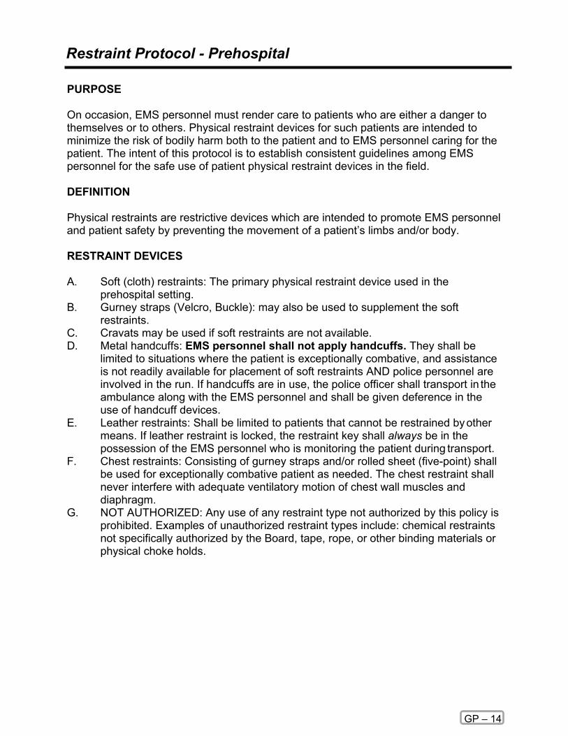
Restraint Protocol - Prehospital
GP – 14
PURPOSE On occasion, EMS personnel must render care to patients who are either a danger to themselves or to others. Physical restraint devices for such patients are intended to minimize the risk of bodily harm both to the patient and to EMS personnel caring for the patient. The intent of this protocol is to establish consistent guidelines among EMS personnel for the safe use of patient physical restraint devices in the field. DEFINITION Physical restraints are restrictive devices which are intended to promote EMS personnel and patient safety by preventing the movement of a patient’s limbs and/or body. RESTRAINT DEVICES A. Soft (cloth) restraints: The primary physical restraint device used in the
prehospital setting. B. Gurney straps (Velcro, Buckle): may also be used to supplement the soft
restraints. C. Cravats may be used if soft restraints are not available. D. Metal handcuffs: EMS personnel shall not apply handcuffs. They shall be
limited to situations where the patient is exceptionally combative, and assistance is not readily available for placement of soft restraints AND police personnel are involved in the run. If handcuffs are in use, the police officer shall transport in the ambulance along with the EMS personnel and shall be given deference in the use of handcuff devices.
E. Leather restraints: Shall be limited to patients that cannot be restrained by other means. If leather restraint is locked, the restraint key shall always be in the possession of the EMS personnel who is monitoring the patient during transport.
F. Chest restraints: Consisting of gurney straps and/or rolled sheet (five-point) shall be used for exceptionally combative patient as needed. The chest restraint shall never interfere with adequate ventilatory motion of chest wall muscles and diaphragm.
G. NOT AUTHORIZED: Any use of any restraint type not authorized by this policy is prohibited. Examples of unauthorized restraint types include: chemical restraints not specifically authorized by the Board, tape, rope, or other binding materials or physical choke holds.

Restraint Protocol - Prehospital continued
GP – 14
GENERAL POLICY Restraints are to be used only when necessary in situations where the patient is violent or potentially violent and may be a danger to themselves or others. EMS providers must remember that aggressive violent behavior may be a symptom of a medical condition such as but not limited to:
Shock Hypertension Myocardial ischemia/infarction Stroke Hypoglycemia Pulmonary embolism
Drug/alcohol intoxication Seizure Dysrhythmias Infection Head trauma Metabolic disorders
Toxicological ingestion Electrolyte imbalance Hypoxia Anemia Agitated Delirium
Protocol 1. Patient health care management remains the responsibility of the EMS provider.
The method of restraint shall not restrict the adequate monitoring of vital signs, ability to protect the patient's airway, compromise peripheral neurovascular status or otherwise prevent appropriate and necessary therapeutic measures. It is recognized that the evaluation of many patient parameters requires patient cooperation and thus may be difficult or impossible.
2. The least restrictive means shall be employed.
3. Verbal de-escalation a. Validate the patient’s feelings by verbalizing the behaviors the patient is
exhibiting and attempt to help the patient recognize these behaviors as threatening.
b. Openly communicate, explaining everything that has occurred, everything that will occur, and why the imminent actions are required.
c. Respect the patient’s personal space (i.e. asking permission to touch the patient, take pulse, examine patient, etc.).

Restraint Protocol - Prehospital continued
GP – 14
EMS personnel shall use an escalating scale of restraint options whereby the level of verbal or physical containment is appropriate to the patient’s presenting situation.
4. Assistance is Readily Available to EMS Personnel for Patient Restraint: a. Cooperative patient with mildly impaired judgment: Verbal Containment;
may or may not use soft restraints. b. Aggressively uncooperative patient with severely impaired judgment: Soft
restraints applied to all extremities (four-point restraint). A chest (five-point restraint) may be used only if the patient is exceptionally combative.
5. Assistance Is not Readily Available to EMS Personnel for Patient Restraint:
a. Cooperative patient with mildly impaired judgment: Soft restraints applied to all four extremities (four-point restraint).
b. Aggressively uncooperative patient with severely impaired judgment: Temporary application of cravats, only if other restraint devices are not readily available. Soft restraints shall always be applied when assistance is available, and the patient situation is controlled either in the prehospital setting or the receiving hospital setting. Leather restraints shall always be removed after arrival at the receiving hospital when additional personnel is available.
EMS personnel shall first try to restrain patients in the lateral position. The supine position is permitted if EMS personnel are unable to safely place the patient in the lateral position due to their combativeness. The applied restraints shall be attached to the gurney frame. EMS personnel shall frequently assess the patient to ensure that the restrained patient’s airway is patent, distal limb circulation is adequate, and that restraints can be released quickly should the patient require cardiopulmonary resuscitation. Airway and suction equipment shall always be available for the restrained patient. EMS personnel shall never leave the restrained patient unattended. IT IS NEVER OK TO TRANSPORT A PATIENT IN A PRONE POSITION OR HOBBLED. If a patient has been restrained by police in a prone position this patient must be turned to a supine position for transport. If a combative patient aggressively breaks away (escapes) from EMS personnel, the patient shall not be pursued unless there is adequate assistance from the appropriate public safety agency to secure the scene and assure safety.
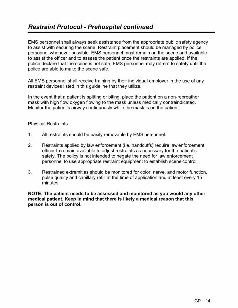
Restraint Protocol - Prehospital continued
GP – 14
EMS personnel shall always seek assistance from the appropriate public safety agency to assist with securing the scene. Restraint placement should be managed by police personnel whenever possible. EMS personnel must remain on the scene and available to assist the officer and to assess the patient once the restraints are applied. If the police declare that the scene is not safe, EMS personnel may retreat to safety until the police are able to make the scene safe. All EMS personnel shall receive training by their individual employer in the use of any restraint devices listed in this guideline that they utilize. In the event that a patient is spitting or biting, place the patient on a non-rebreather mask with high flow oxygen flowing to the mask unless medically contraindicated. Monitor the patient’s airway continuously while the mask is on the patient. Physical Restraints
1. All restraints should be easily removable by EMS personnel.
2. Restraints applied by law enforcement (i.e. handcuffs) require law enforcement officer to remain available to adjust restraints as necessary for the patient's safety. The policy is not intended to negate the need for law enforcement personnel to use appropriate restraint equipment to establish scene control.
3. Restrained extremities should be monitored for color, nerve, and motor function, pulse quality and capillary refill at the time of application and at least every 15 minutes
NOTE: The patient needs to be assessed and monitored as you would any other medical patient. Keep in mind that there is likely a medical reason that this person is out of control.

Restraint Protocol - Prehospital continued
GP – 14
Documentation of Restraints 1. Patient restraint shall be documented on the run sheet and address any or all the
following appropriate criteria:
a. That an emergency existed and the need for treatment was explained to the patient.
b. That the patient refused treatment or was unable to consent to treatment (such as unconscious patient).
c. Evidence of the patient's lack of decision-making capacity. d. Failure of less restrictive methods of restraint (if conscious, failure of
verbal attempts to convince the patient to consent to treat). e. Assistance of law enforcement officials with restraints, or orders from
medical control to restrain the patient, or any exigent circumstances requiring immediate action, or adherence to system restraint protocols.
f. That the treatment and/or restraints were for the patient's benefit and safety.
g. The type of restraint employed (soft, leather, mechanical). h. Any injuries that occurred during or after the restraint. i. The limbs restrained ("four points"). j. Position in which the patient was restrained. k. Circulation checks every 15 minutes or less (document findings and time).
2. The behavior and/or mental status of the patient before and after the restraint.

Restraint Protocol - Prehospital - continued
GP – 14
CAUTION: OVERSTEPPING THE BOUNDRIES OF RESTRAINT MAY BE PERCEIVED AS BATTERY, ASSAULT, CIVIL RIGHTS VIOLATION OR FALSE IMPRISONMENT. This is a controversial and dangerous area within the law. Each individual service utilizing this protocol should consult appropriate legal consultation. KRS 503.110 Use of force by person with responsibility for care, discipline, or safety of others. 1. The use of physical force by a defendant upon another person is justifiable when
the defendant is a person responsible for the operation of or the maintenance of order in a vehicle or other carrier of passengers and the defendant believes that such force is necessary to prevent interference with its operation or to maintain order in the vehicle or other carrier, except that deadly physical force may be used only when the defendant believes it necessary to prevent death or serious physical injury.
2. The use of physical force by a defendant upon another person is justifiable when the defendant is a doctor or other therapist or a person assisting him at his direction, and: a. The force is used for the purpose of administering a recognized form of
treatment which the defendant believes to be adapted to promoting the physical or mental health of the patient; and
b. The treatment is administered with the consent of the patient or, if the patient is a minor or a mentally disabled person, with the consent of the parent, guardian, or other person legally competent to consent in his behalf, or the treatment is administered in an emergency when the defendant believes that no one competent to consent can be consulted and that a reasonable person, wishing to safeguard the welfare of the patient, would consent.

Restraint and Transportation - Pediatric
GP – 15
Patient Transport
An ill or injured child must be restrained directly to the cot in a manner that prevents ramping or sliding in a collision. ► A belt/strap looped over each shoulder and attached to a non-sliding cot
member. ► A soft, sliding, or breakaway connector holding the shoulder straps together on
chest. ► Belt/strap anchored to non-sliding cot member and routed over thighs, not around
waist. Note: Standard belt systems do not adequately secure child to the cot during a crash. Ill or injured child/infant (5 to 80 lbs) who can tolerate a semi-upright position may be secured using a child passenger safety seat. ► Use a convertible child safety seat that has a front and
rear belt path. ► Position safety seat on cot facing the foot-end with
backrest fully elevated. ► Consider removing mattress. ► Secure safety seat with 2 pairs of belts in both the
forward & rear positions. ► Place the shoulder straps of the harness through slots just below child’s
shoulders. ► For infants, place rolled towels on sides of child to maintain centered position.
Note: Non-convertible safety seats cannot be secured properly to the cot.
For infants who cannot tolerate a semi-upright position or who must lie flat: ► Use car bed, if available, that can be secured against
both rearward and forward motion. ► Position car bed across cot so child lies perpendicular
to cot. ► Fully raise cot’s backrest and anchor car bed to cot with
2 belts. ► Fasten car bed harness snugly to infant.

Restraint and Transportation- Pediatric continued
GP – 15
Use of Child Passenger Safety Seat after Involvement in Motor Vehicle Crash Child safety seats may be used after involvement in a minor crash. All the following must apply to be considered a minor crash. ► Visual inspection including inspection under movable seat padding does not
reveal any cracks or deformation. ► The vehicle in which the child safety seat was installed was capable of being
driven from the scene of the crash. ► The vehicle door nearest the child safety seat was undamaged. ► There were no injuries to any of the vehicle occupants. ► The air bags (if any) did not deploy.

Safe Infants Act - Safe Infants Protocol for Prehospital Providers
GP – 16
Any parent or person acting on behalf of the parent may come to a police station, firehouse, EMS station, or hospital unannounced and leave a newborn infant. When this event occurs, the police officer, firefighter, EMS worker, or hospital worker SHALL accept the infant. This situation must meet the following criteria.
1. The newborn infant must be medically determined to be less than 72 hours old.2. The newborn infant cannot have indicators of child abuse, maltreatment, or
neglect after birth.
► Perform a primary and secondary survey of the infant and initiate any necessary procedure to protect the child’s health and safety. Keep the newborn warm especially the head.
► Consider rapid glucose determination.► Kentucky law requires that any care provider who suspects child abuse, neglect,
or maltreatment SHALL report it. You should call the Department for Community Based Services (DCBS) hotline at 1-800- 752-6200 to make your report. You have no authority to detain, follow or pursue the parent.
► Summon EMS for transport of the infant.► Notify your supervisor and follow any policies and procedures your agency has
implemented.► Retrieve and open an "Abandoned Infant Pack". Complete the enclosed checklist.► Place the numbered band around the ankle of the infant.► Ensure that the band’s stub remains attached to the Medical Information Form
and copy the stub number directly onto the Medical Information Form.► You will offer the parent information regarding medical needs of the mother who
is post-partum, a written explanation of the parent’s legal rights, and services available to the parent, which have been provided in the packet.
► Newborn infants should be transported in an age appropriate car seat if available. Otherwise, newborns should be transported using appropriate immobilization measures.
► Newborn infants may be fed with SIMILAC or ENFAMIL if a lengthy transport time is anticipated. Newborns normally eat 2-2.5 ounces of formula at feeding. Feeding is not advised for any infant that is experiencing any respiratory or circulatory abnormality.
KRS211.951, 2216B.190, 311.6526, 405.075 and 620.355 is known as the Thomas J. Burch Safe Infants Act. The law provides a safe place for unwanted newborn babies.Parents may now leave an unwanted infant with any Kentucky EMS provider, policestation, fire station or hospital without consequence. I hope that preventing anyunwanted newborn from being left in a dangerous or deadly environment.

Safe Infants Act - How to Keep Yourself Healthy
GP – 16
You’ve Just Had a Baby! "Copy and Provide to Mother”
You have made a courageous decision to leave your baby in the safe and good care of a hospital, police station, fire station or emergency medical services (EMS) provider. Your baby will be well taken care of and, eventually, be adopted into a safe, loving, permanent home. Now it’s time to make sure that you are healthy. It’s a good idea to see a doctor or go to the health department for an examination. For information about your local health department, call (800) 462-6122. What is normal after you’ve just had a baby? It takes your body about three to six weeks to return to its pre-pregnant state. You may experience several normal changes to your body during the first few days and weeks after delivery. Vaginal bleeding: This is blood coming from the uterus. It is a sign that the uterus is healing. At first, it is like a heavy period. The bleeding will start out as bright red, change to pink, and then change to a clear or yellow discharge. You should stop bleeding after three weeks. There should never be large blood clots or a foul odor. What to do: Use sanitary pads only (no tampons). Do not take tub baths until the bleeding stops. Call a doctor if the bleeding becomes bright red again, you pass large clots or there is a foul odor. Abdominal cramping: This is a sign that the uterus is contracting back down to its normal size. These cramps are like mild menstrual cramps and will last a few days. What to do: Take an over-the-counter pain reliever. Breast engorgement: This means the breasts are becoming full and very sore, and it is a sign that the breasts are filling with milk. This happens around the third day after delivery. Your breasts will become swollen, firm, tender and warm to the touch. Severe breast engorgement should not last more than 36 hours. What to do: Wear a good-fitting support bra at all times and remove it only for showers. Apply an ice pack to the breasts for 20 minutes, four times a day. Avoid things that will stimulate the breasts. Avoid heat and hot showers. "Postpartum blues": Most women feel depressed for one to two weeks after delivery. You may feel angry, sad, tired and unable to sleep or eat during this time. These feelings are brought on by the many changes that take place in your body and brain during and after delivery.

Safe Infants Act - How to Keep Yourself Healthy - continued
GP – 16
You’ve Just Had a Baby! "Copy and Provide to Mother‖
What to do: Know that this is normal and will go away. Find a family member or close friend to talk to about your feelings. Call a doctor if these feelings do not go away or if they intensify. Call a doctor if you have any of these warning signs: • Heavy, bright red vaginal bleeding • Foul-smelling vaginal discharge • Dizziness or fainting • Fever above 100.4 degrees F • Pain around your vaginal area that does not go away or gets worse • Pain or burning when you empty your bladder • Pain or swelling in your legs • Red streaks or painful new lumps in your breasts • Cramps that are more painful than normal menstrual cramps • Nausea and vomiting • Chest pain or cough • Feeling so sad that you aren’t able to take care of yourself • Feelings that you might hurt yourself Do these things to take care of yourself after your delivery: • Rest as much as you can. Your normal energy will return in a few weeks. • Eat healthy foods. Drink six to eight glasses of water a day. If you have prenatal
vitamins, continue to take one a day. • Continue to wear a good-fitting bra for about three weeks. • Change your pad every time you go the bathroom to prevent infection in the
vaginal area. Wipe yourself from front to back every time you urinate or have a bowel movement. Wash your hands every time you change your pad or go to the bathroom.
• Do not take a tub bath for three weeks. Take showers only. • Gradually resume your normal physical activity. Don’t lift anything over 10
pounds. Don’t drive a car for one week. Don’t climb stairs for one week (if you have to climb steps, climb one step at a time).
• Avoid sexual intercourse for at least six weeks after delivery. Do not have intercourse if you are still bleeding vaginally. It is possible to become pregnant before you start having periods again, so talk to a doctor about ways to prevent another pregnancy.

Safe Infants Act - How to Keep Yourself Healthy - continued
GP – 16
You’ve Just Had a Baby! "Copy and Provide to Mother"
• Get a medical examination four to six weeks after delivery. Your doctor or healthdepartment will keep your records confidential to protect you against anyinvasion of personal privacy.
For information about:
• Family planning and contraception, call (800) 462-6122.• Substance abuse counseling, call, toll free, (888) 729-8028.• Domestic violence and abuse, call (800) 752-6200.
For information about health care specific to women, log onto: Http://chfs.ky.gov/dph/ach/mch.htm
Important!
If you left your baby at a safe place and have decided that you want your baby back, contact the Kentucky Cabinet for Health and Family Services at (800) 752-6200. If you do not contact the Cabinet within 30 days after leaving your newborn, the Cabinet will proceed with termination of parental rights and place your baby for adoption.
A copy of this material may be obtained from the following Web site: http://chfs.ky.gov/dcbs/dpp/Child_Safety.htm
For more information about the Safe Infants Act, call (800) 752-6200

Bloodborne/Airborne Pathogens
GP – 17
BLOODBORNE PATHOGENS
Emergency Medical Services personnel should assume that all bodily fluids and tissues are potentially infectious with bloodborne pathogens including HIV (causing AIDS) and HBV (causing hepatitis) and must protect themselves accordingly by use of body substance isolation (BSI). Body substance isolation procedures include the appropriate use of hand washing, protective barriers (such as gloves, masks, goggles, etc.), and care in the use and disposal of needles and other sharp instruments. EMTs are also encouraged to obtain the hepatitis B vaccine series to decrease the likelihood of hepatitis B transmission. EMTs who have exudative lesions, weeping dermatitis, or open wounds should refrain from all direct patient care and from handling patient-care equipment as they are at increased risk of transmission and reception of bloodborne pathogens through these lesions. Transmission of bloodborne pathogens has been shown to occur when the blood of the infected patient is able to come in direct contact with the blood of the health-care worker. EMTs who have had a direct bloodborne pathogen exposure should immediately wash the exposed area with soap and water and a suitable disinfectant. The exposed area should then be covered with a sterile dressing. Upon arrival at the destination hospital, after responsibility for the patient has been transferred to the emergency department, the EMT should thoroughly cleanse the exposed site, complete a state of Kentucky Emergency Response/Public Safety Worker Incident Report Form, and sign in to the Emergency Department as a worker’s compensation patient. The only exception to this latter step is when the squad has a designated exposure officer and medical advisor wherein the exposed EMT has definitive and immediate medical care elsewhere. AIRBORNE PATHOGENS EMTs who believe they have been exposed to an airborne pathogen may proceed as above in getting timely medical care. It is expected that a properly filled out Patient Care Report will allow hospital infection control staff to contact EMTs involved in patient care where that patient was subsequently found to have a potential airborne pathogen such as Tuberculosis, Neisseria meningitis, SARS, etc. Airborne Personal Protective Equipment (APPE) ► Recommended APPE consists of a N95 respirator, prior fit testing is
recommended. ► Apply APPE if the patient presents with the following signs or symptoms
Cough Fever
Rash

Bloodborne/Airborne Pathogens - Continued
GP – 17
► Limit the number of personnel in contact with suspected patients to reduce the
potential of exposure to other providers and bystanders. ► Patients suspected of being infected with a possible airborne pathogen should be
masked if tolerated. ► Patients requiring oxygen therapy should receive oxygen through a mask with a
surgical mask placed over the oxygen mask to block pathogen release. Close monitoring of the patient’s respiratory status and effort should be maintained.
► APPE should be in place when performing suctioning, airway management and ventilation assistance (Bag-Valve-Mask) for suspect patients.
► Limit procedures that may result in the spread of the suspected pathogen, e.g. nebulizer treatments.
► Exchange of fresh air into the patient compartment is recommended during transport of patient with a suspected airborne pathogen.
► Early notification to the receiving hospital should be made such that the receiving hospital may enact its respective airborne pathogen procedures.
DECONTAMINATION ► In addition to accepted decontamination steps of cleaning surfaces and
equipment with an approved solution and proper disposal of contaminated disposable equipment, the use of fresh air ventilation should be incorporated (open all doors and windows to allow fresh air after arrival at the hospital).
► All personnel in contact with the patient should wash their hands thoroughly with warm water and an approved hand-cleaning solution.
► Ambulances equipped with airborne pathogen filtration systems should be cleaned and maintained in accordance with manufacturer guideline

ACP
Adult Cardiac Protocols

Routine Patient Care Guidelines – Adult
ACP – 1
All levels of provider will complete an initial & focused assessment on every patient, and as standing order, use necessary and appropriate skills and procedures for which the provider has been trained and certified or approved to perform in order to maintain the patient’s airway, breathing, and circulation. Initial Assessment Scene Size-Up
► Review dispatch information. ► Assess the scene for safety, mechanism of injury, number and location of
patients. ► General impression of patient. ► Assess for appropriate level of body substance isolation. Notify the receiving
facility as early as possible. ► Request additional resources as needed: e.g. ALS intercepts, air medical
transport, additional ambulances, extrication, hazardous materials team, etc. ► Use Incident Management/Command System (IM/CS) when possible.
Level of Consciousness
► Assess level of consciousness using the AVPU scale. ► Manually stabilize the patient’s cervical spine if trauma is involved or suspected.
Apply and use AED and initiate cardiopulmonary resuscitation in accordance with current guidelines, as trained and credentialed, if indicated.
Airway ► Assess the patient for a patent airway. ► Open the airway using a head-tilt/chin-lift, or a jaw thrust if suspicious of cervical
spine injury. ► Suction the airway as needed. ► Consider an oropharyngeal or nasopharyngeal airway. ► Consider advanced airway interventions as appropriate and if trained in use.
Breathing
► Assess patient’s breathing taking note of rate, rhythm, and quality of the respirations.
► Assess lung sounds. ► Look for nasal flaring or accessory muscle usage. ► Assess the chest for symmetrical chest rise, intercostal or supraclavicular
retractions, instability, open pneumothorax, tension pneumothorax, or other signs of trauma.
► Treat foreign body airway obstruction in accordance with current guidelines.

Routine Patient Care Guidelines – Adult
ACP – 1
Breathing (continued)
► Assist ventilations when the respiratory rate is less than 10 per minute or greater than 40 for adults, or when the patient exhibits signs of impending respiratory failure.
Circulation
► Assess the patient’s pulse taking note of rate, rhythm, and quality. ► Look for and control any obvious gross bleeding.
Assess patient’s skin color, temperature, and moisture. ► IV access and fluid resuscitation as appropriate for the patient’s condition per
appropriate protocol. After IV is established, administer fluids to maintain systolic blood pressure >90 mmHg. Routes of medication administration when written as “IV” can also include “IO”.
Disability
► Movement of extremities. ► Facial asymmetry. ► Speech.
Exposure
► Expose as needed to provide care and assessment of patient condition and examine head, neck, chest, abdomen, pelvis and back.
Secondary Assessment Head-to-toe Survey Neurological Assessment
► Glasgow Coma Score Assess Vital Signs
► Respiration. ► Pulse. ► Blood pressure. ► Capillary refill. ► Record Pain Level ► Skin condition
o Color o Temperature o Moisture
► Lung sounds.
Obtain Medical History
► Symptoms. ► Allergies. ► Medication. ► Past Medical History. ► Last Oral Intake. ► Events leading to Illness or Injury.
Other Assessment Techniques
► Cardiac Monitoring ► Pulse oximetry ► Glucose determination ► Temperature ► End-tidal CO2.

Acute Coronary Syndromes - Adult
ACP – 2
Basic Standing Orders
Routine Patient Care Assess SpO2. If below 90% or patient appears to be in distress, administer oxygen such that patient SpO2 remains below 100% but above 90%. Aspirin 324 mg PO (chewable) If patient states they cannot take ASA ascertain whether due to true allergy or unpleasant side effects. Facilitate administration of patient’s own nitroglycerin if SBP > 90, every 5 minutes up to 3 Avoid NTG for patients who have taken Sildenafil (Viagra), Tadalafil (Cialis) or Vardenafil (Levitra) in past 48 hours Obtain a 12 lead EKG Minimize Scene time Consider ALS Intercept
IV access and administer fluids to maintain systolic blood pressure > 90 mmHg. Nitroglycerin 0.4mg SL every 5 minutes while symptoms persist if SBP > 90 mmHg
Advanced Standing Orders
P Paramedic Standing Orders
Obtain serial 12 lead EKGs during transport. If EKG suggests AMI and pain is unrelieved by NTG dosing AND
SBP > 90 mmHg: o Administer Fentanyl 25 – 50 mcg IV every 5 minutes to
max of 100 mcg Treat dysrhythmias PRN; refer to appropriate protocol. Contact receiving facility as soon as possible for patients with
EKG indications of Acute Myocardial Infarction.

Acute Coronary Syndromes - Adult
ACP – 2

Cardiac Arrest – Adult
ACP – 3
Basic Standing Orders
Routine Patient Care – with focus on CPR. Apply and use AED as soon as possible. After High Quality CPR & AED in place, consider placing BIAD per protocol. For Trauma:
o Minimize on-scene time or consider not attempting resuscitation (See Determination of Death Protocol)
Request ALS Intercept
Consider treatable causes: overdose/poisoning, hypothermia; treat per protocol. IV/IO access and administer fluids wide open. For Trauma:
o DO NOT delay transport for IV/IO, advanced airway, or medication administration.
Advanced Standing Orders
Paramedic Standing Orders
P Document presenting cardiac rhythm in two separate leads if possible. Advanced airway management if indication. Capnography must be
recorded as soon as possible. Consider nasogastric or orogastric tube to decompress the stomach of
intubated patients. In any case of sudden increase of EtCO2, check for pulse and progress
to Post Arrest Care Protocol if pulse discovered.
Asystole or PEA Continue CPR for 2 minutes. Epinephrine (1:10 000) 1 mg IV/IO; repeat every 3 – 5 minutes. Resume CPR for 2 minutes. Then check rhythm. If no change, resume CPR and administer Epinephrine (1:10 000) 1 mg
IV/IO. o Treat reversible causes
Consider advanced airway management. Stop compressions for:
o Sudden increase in EtCO2 with pulses present o Shockable rhythm obtained, or o Decision made to discontinue further efforts
At every rhythm check if shockable rhythm found, follow VF/pVT guidance.
For Trauma Arrest – consider bilateral needle chest decompression.

Cardiac Arrest – Adult
ACP – 3
Paramedic Standing Orders Continued
VF/Pulseless Ventricular Tachycardia (pVT)
Defibrillate as soon as possible, beginning at 200 J. Immediately resume compressions.
Deliver 2 minutes of CPR then rhythm check. If shockable rhythm, repeat defibrillation dose at 300 J; Immediately resume compressions.
Deliver 2 minutes of CPR; During CPR, administer Epinephrine (1:10 000) 1 mg IV/IO; repeat every 3 – 5 minutes.
After 2 minutes of compressions; rhythm check; if still in shockable rhythm, defibrillate at 360 J for this and all following defibrillations. Immediately resume compressions.
Deliver 2 minutes of CPR; administer Amiodarone 300 mg IVP (repeat dose 150 mg in 3 – 5 minutes if indicated) OR Lidocaine 1 – 1.5 mg/kg IVP (repeat dose 0.5 – 0.75 mg/kg in 3 – 5 minutes if indicated)
P

Cardiac Arrest – Adult
ACP – 3

Post Cardiac Arrest Care – Adult
ACP – 4
Paramedic Standing Orders ► ►
Consider transitioning BIAD to ETT per appropriate airway protocol.Consider placing patient on transport ventilator, refer to Ventilatorprotocol for guidance on settings.
P HYPOTENSION: SBP remains < 90 mmHG after 2L fluid bolus or Pulmonary Edema Present: ► Consider either of following medications, titrate to SBP > 90:
• •
Epinephrine 0.1-0.5 mcg/kg/min IV/IO drip ORDopamine 5-10 mcg/kg/min IV/IO
STEMI Suspected: 12 lead EKG indicates possible STEMI ► ►
Refer to Acute Coronary Syndrome Protocol.Transport to PCI Capable Hospital, notify receiving hospital as soon aspossible of patient condition & presentation.
This protocol is to be implemented for any adult patient immediately once organized, perfusing rhythm is noted during cardiac arrest care.Basic Standing Orders
B Repeat Primary Assessment Optimize Ventilation and Oxygenation
o Maintain SpO2 90% or greatero Blind Inserted Airway if appropriate.o Respiratory Rate: begin at 10/min; target EtCO2: 35 – 40o Expect higher than normal EtCO2 immediately after ROSC. Do
not adjust ventilations to obtain normal levels.o DO NOT HYPERVENTILATE
Obtain 12 lead EKG. Consider ALS Intercept.
Advanced Standing Orders
A Consider treatable causes: overdose/poisoning, hypothermia; treat per
protocol. Establish IV/IO access, if not already in place. Only establish 1 IO site. If SBP < 90 mmHg, administer 500 mL Fluid Bolus.
o Repeat as needed if lungs remain clear to max of 2 L.

Post Cardiac Arrest Care – Adult
ACP – 4

Bradycardia (Symptomatic) – Adult
ACP – 5
Definition: Bradycardic and inadequate clinical perfusion e.g. acute altered mental status, ongoing chest pain, hypotension or other signs of shock.
Establish IV access.
Advanced Standing Orders
A
Basic Standing Orders
B Routine Patient Care If available, perform 12 – lead EKG. Consider ALS Intercept.
P Paramedic Standing Orders
Consider Atropine 0.5 mg IVP every 3 – 5 minutes to a total of 3 mg. Consider Transcutaneous pacing if available. Attempt capture at rate of
70 BPM at minimum output and increase until capture achieved. Use without delay for high degree AV block (Second Degree Type II or Third-Degree AV Block).
Consider procedural sedation prior to pacing: o Midazolam 2.5 mg IV may repeat once in 5 minutes
OR o Ketamine 1 mg/kg slow IVP, may repeat once in 5 minutes
Consider Epinephrine (2 – 10 mcg/min) or Dopamine (2 – 20 mcg/kg/min) infusion if pacing ineffective.

Bradycardia (Symptomatic) – Adult
ACP – 5

Tachycardia – Adult
ACP – 6
Paramedic Standing Orders
P Identify rhythm using cardiac monitor Unstable rhythm (Hypotension, altered mental status, signs of poor
perfusion) o Synchronized cardioversion
Narrow irregular: 120 – 200 J, 300 J, 360 J Narrow regular: 50 J, 100 J, 200 J, 300 J, 360 J Wide regular: 100 J, 200 J, 300 J, 360 J Wide irregular: defibrillation energy (unsynchronized)
o Consider procedural sedation if practical: Midazolam 2.5 mg IV
OR Ketamine 1 mg/kg Slow IVP
Stable: PSVT or narrow complex tachycardia (with ventricular rate consistently greater than 150 BPM)
o Consider vagal maneuvers. o If vagal maneuvers fail, administer Adenosine 6 mg rapid IVP.
May repeat Adenosine at 12 mg if initial dose is ineffective.
A Advanced Standing Orders
Establish IV access and administer fluids to maintain SBP > 90 mmHg.
B Basic Standing Orders
Routine Patient Care Consider application of AED if patient appears unstable. Administer oxygen as needed to maintain SpO2 > 90% Obtain a complete set of vitals including EtCO2. Identify and treat
reversible causes. If available, acquire 12 – lead EKG. Consider ALS Intercept.

Tachycardia – Adult
ACP – 6
Paramedic Standing Orders
P Stable continued o Atrial Fib, Atrial Flutter w/ Narrow complex (With ventricular rate
consistently greater than 140 -150 BPM) Monitor and transport
o Ventricular Tachycardia or unknown Wide Complex Tachycardia Monitor and transport
o For Polymorphic VT/Torsades Monitor and Transport
o For Known WPW Monitor and Transport
If patient becomes unstable during monitoring and transport, treat as unstable.

Tachycardia – Adult
ACP – 6

Congestive Heart Failure/Pulmonary Edema - Adult
ACP – 7
Basic Standing Orders
B ► ► ► ► ►
Routine Patient Care. Place patient in semi-sitting or full sitting position. Administer oxygen at a rate to keep oxygen saturation above 90%. If possible monitor EtCO2 using cannula. Facilitate administration of patient’s own nitroglycerin - if SBP> 90 every 5 minutes as needed. Consider ALS intercept. ►
Advanced Standing Orders
A ► IV access and administer fluids to maintain SBP > 90 mmHg.
Consider CPAP. Titrate pressure with maximum 10 cm H2O pressure support
►
Paramedic Standing Orders
► Consider nitroglycerin 0.4 mg SL every 5 minutes PRN if SBP > 90 mmHg.
P ► Consider other contributing factors in respiratory distress and treat as appropriate.
► Consider advanced airway management as needed.

AMP
Adult Medical Protocols

Allergic Reaction/Anaphylaxis – Adult – Mild
AMP – 1
Allergic reaction is suspected when there is a suspected exposure and patient(s) exhibit signs and symptoms consistent with an allergic reaction such as:
► ► ► ► ►
Skin: Hives, Itching, Flushing Respiratory: Wheezing, Dyspnea, Stridor, Sneezing, Coughing, Chest tightness Cardiovascular: Vasodilation, Tachycardia, Hypotension, Shock Gastrointestinal: Nausea, Vomiting, Cramping, Diarrhea CNS: Dizziness, Headache
Common allergens include venom (i.e. bee stings), foods, (i.e. nuts, berries, seafood), plant pollen, and medications.
Consider Mild, Moderate and Severe protocols to be hierarchical and building on prior intervention in Evolving Treatment Plan.

Allergic Reaction/Anaphylaxis – Adult – Mild
AMP – 1
Symptomatic, no dyspnea, unremarkable vital signs.
Basic Standing Orders
► ►
Routine Patient Care. Maintain airway and administer oxygen as needed to maintain oxygen saturation of at least 95%. B
Remove allergen Check vital signs frequently Begin transport, consider ALS intercept Consider patient assisted medication
Paramedic Standing Orders
P ► Consider Diphenhydramine 25-50 mg, PO, IM, IV.

Allergic Reaction/Anaphylaxis – Adult – Moderate
AMP – 1
Edema, hives, difficulty swallowing, facial swelling, etc., with stable vital signs BP ≥ 90 mmHg.
Basic Standing Orders
B ► ►
Routine Patient Care. Maintain airway and administer oxygen as needed to maintain oxygen saturation of at least 95%.
Remove allergen Check vital signs frequently Begin transport, consider ALS intercept Consider patient assisted medication
Consider Albuterol 2.5 mg in 3 ml of NS via nebulizer every 5 minutes x 4 total doses. Consider Epinephrine (1:1000) 0.3 mg IM.
►
►
Advanced Standing Orders
A ► Establish an IV at KVO or Saline Lock.
Paramedic Standing Orders
P ► Diphenhydramine 25-50 mg IM or IV.

Allergic Reaction/Anaphylaxis – Adult – Severe
AMP – 1
Basic Standing Orders
B
Edema, hives, severe dyspnea, wheezing, unstable vital signs (systolic < 90 mmHg), cyanosis, hoarseness.
Advanced Standing Orders
A ► Establish an IV at KVO or Saline Lock.
Paramedic Standing Orders
P In addition to prior therapies, consider: ► ► ►
Diphenhydramine 25-50 mg IM or IV. Methylprednisolone 125 mg IV push. For Severe, Decompensating patients:
• Consider Epinephrine 1:100 000, 0.1 mg SLOW IV over 5-10 Minutes. May repeat as needed up to Total dose of 1.0 mg.
• (Dilute 1 mL of Epinephrine 1:10 000 in 9 ml NS flush for 10 mL of 1:100 000 mL.)
Consider early intubation. ►
Ensure adequate ABCs. Administer oxygen to keep SpO2 > 90%. If patient was exposed to an allergen and exhibits severe respiratory
distress or shock, administer Epinephrine Autoinjector, or, Epinephrine (1:1 000) 0.3 mg (0.3 mL) IM.
Consider Albuterol 2.5 mg in 3 mL of NS via Nebulizer every 5 minutes x 4 total doses.
Begin transport and request ALS intercept. Monitor ABCs and Vital signs. Assist ventilations as needed.

Asthma/COPD/RAD (Reactive Airway Disease) - Adult
AMP – 2
►
►
►
Basic Standing Orders Routine Patient Care. Wear N95 mask if bioterrorism related event or highly infectious agent
suspected. Administer oxygen at the appropriate rate for the patient’s condition
and medical history. Patients with COPD who are on home oxygen, increase their rate by
1-2 liters per minute. Attempt to keep oxygen saturation above 90%; increase the rate with
caution and observe for fatigue, decreased mentation, and respiratory failure.
Request ALS intercept, transfer care upon arrival of ALS provider Assist patient with his/her own MDI, if appropriate; only MDIs
containing beta adrenergic bronchodilators (e.g. albuterol, Ventolin, Proventil, Combivent) may be used: 2 puffs; repeat every 5 minutes as needed while transporting.
Consider albuterol 2.5 mg in 3 ml normal saline via nebulizer prn every 5 minutes x 4 total doses.
B
Advanced Standing Orders
A ► IV access and administer fluids to maintain systolic blood pressure >90 mmHg.
► Consider Atrovent 0.5 mg in addition to Albuterol nebulizer, may administer 2 total doses.
► Consider CPAP for patients in respiratory distress not related to asthma exacerbation. Titrate to max pressure of 10 cmH2O.
Paramedic Standing Orders
P ► ► ►
For patients exhibiting symptoms of CHF refer to appropriate protocol. Consider methylprednisolone 125 mg IV. For patients who do not respond to nebulizer treatments or for impending respiratory failure, consider:
• Epinephrine (1:1,000) 0.3mg (0.3 ml) IM. • Magnesium sulfate 2 grams in 100 ml 0.9% NaCl (normal saline)
IV over 10 minutes.

Behavioral Emergencies - Adult
AMP – 3
Basic Standing Orders
► ►
Routine Patient Care Request Law Enforcement to secure scene & ensure patient is unarmed. Avoid areas/patients with potential weapons and avoid areas with only a single exit; do not allow patient to block exit. Keep environment calm by reducing stimuli. Transport in a non- emergent mode unless patient's condition requires Lights & Siren transport. Respect the dignity and privacy of the patient. Make eye contact when speaking to the patient. Speak calmly and in a non-judgmental manner; do not make sudden movements. Maintain non-threatening body language. Ask for permission to touch the patient before taking vital signs and explain what you're doing. Attempt to assess Blood Glucose levels and if out of normal range, treat per Hypoglycemia protocol. Make all efforts to rule out medical or traumatic causes of behavioral changes and treat as appropriate.
►
B ► ► ►
► ►
►
►
Advanced Standing Orders
A ► Consider IV access if medication administration anticipated.
Paramedic Standing Orders ► Apply cardiac monitor if clinically feasible, obtain 12 lead ECG and treat
dysrhythmias as appropriate. Position patient to ensure breathing is not impaired. For dangerously agitated patients who are presenting a danger to themselves, crew and law enforcement officers on scene, consider:
• Midazolam: 2 - 6 mg IV/IM/IN OR P ► ►
• •
Ketamine: 4 mg/kg IM to max dose of 400 mg IM. NOTE: Reduce dose by 50% for patients greater than 70 yrs old. This applies to both medications listed.
► If chemical sedation utilized, once patient is complaint, crew shall monitor vital signs including EtCO2 & Temperature. Remain vigilant for airway compromise, have suction readily available. In cases of hyperthermia, as soon as possible initiate IV fluid bolus. If patient condition degrades to cardiac arrest, consider early Sodium Bicarbonate 1 mEq/kg.
►
►
►
►

Diabetic Emergencies: Hypoglycemia
AMP – 4
Basic Standing Orders
B ► ► ►
Routine Patient Care. Obtain glucose reading via glucometer. If the patient can swallow and hypoglycemia is present, administer 15 grams oral glucose. If available and indicated, consider assisting family in administration of patient’s glucagon 1 mg IM Consider ALS intercept.
►
►
Advanced Standing Orders
IV access and administer fluids to maintain systolic blood pressure >90 mmHg. If glucose level is < 70 mg/dl with associated signs and symptoms, administer 5 mL/kg (0.5 g/kg) dextrose (D10) IV drip. Administer the D10 solution IV drip until patient regains normal level of consciousness or infusion complete. Re-check glucose 5 minutes after administration of dextrose or resolution of symptoms. Repeat dextrose (D10) drip en route to hospital if glucose level is less than 70mg/dL or patient remains symptomatic with signs of hypoglycemia.
►
A ►
►
► If unable to obtain IV access, administer glucagon 1 mg IM. NOTE: If Glucagon is administered, it should be performed while transporting the patient to the Emergency Department.

Diabetic Emergencies: Hyperglycemia
AMP – 4
Basic Standing Orders
B ► ► ►
Routine Patient Care. Obtain glucose reading via glucometer. Consider ALS intercept for abnormal vitals signs or altered level of consciousness. Maintain patent airway and adequate ventilations. Transport.
► ►
Advanced Standing Orders
A ► IV access and administer fluid to maintain systolic blood pressure >90 mmHg. Consider 500 mL fluid bolus. Repeat as necessary. ►
Paramedic Standing Orders
P Airway management as needed. ►
Non-transport of Insulin Dependent Diabetic
AMP – 4
Historical Findings
1. 2. 3.
Decreased level of consciousness without suspected trauma. Prior medical history of insulin-dependent diabetes mellitus. Following treatment, patient is conscious, alert to time, date and place, and requests that they not be transported to the hospital. No other associated findings of serious illnesses or circumstances that may have contributed to the hypoglycemic episode, including excessive alcohol consumption, shortness of breath, chest pain or headaches. The patient's history reveals circumstances that may have contributed to the hypoglycemic episode such as lack of oral intake or an insulin reaction. Not on oral hypoglycemic medication such as glipizide, glyburide, or chlorpropamide.
4.
5.
6.
Physical Findings
1. 2. 3. 4.
Patient is initially found to have a decreased level of consciousness. Systolic blood pressure > 90 mm Hg or child with normal perfusion. Patient has rapid glucose test of ≤ 70 mg/dL. The patient responds quickly (< 10 minutes) to oral or IV glucose to normal level of consciousness. Repeat rapid glucose test is > 100 mg/dL. 5.
EKG Findings
1. 2.
Heart rate > 60. Normal EKG.
Protocol
1. The patient is assessed and treated per the Diabetic Emergencies protocol. Repeat blood pressure is at least 90 mm Hg, pulse rate is at least 60, and the repeat rapid glucose test is at least 100 mg/dL. The patient is given instructions for follow-up care prior to being released. The patient is released to the care of a responsible adult who will remain with the patient as an observer for at least one hour and can call 911 should the symptoms recur.
2.
3. 4.

Non-transport of Insulin Dependent Diabetic
AMP – 4
Notes
1. Patients who have extensive medical history or other signs and symptoms unrelated to insulin-dependent diabetes mellitus should be strongly encouraged to be transported. If the patient is on an oral hypoglycemic medication such as glipizide, glyburide, or chlorpropamide, the hypoglycemic episode may last hours or days. Patients on oral hypoglycemic agents should be strongly encouraged to be transported, regardless of their response to field treatment. When treating patients who warrant transportation based on the above criteria but who refuse transport, paramedics shall contact medical control for assistance. Instructions for follow-up care should include the following:
2.
3.
4. a. Take action to prevent a recurrent episode such as remain in the
care of another adult observer, consume a light meal to maintain a sufficient blood glucose level, monitor their blood glucose, and advise their personal physician of this episode. Watch for signs and symptoms of another episode. If another episode occurs, contact 911 immediately!
b. c.
5. EMS should provide the patient with verbal instructions on follow-up care following the patient refusal of transport. Additionally, crews must document what verbal directions they provided.

Fever (>101.5 F/38.5C) – Adult
AMP – 5
This protocol is not intended for patients suffering from environmental hyperthermia (Hyperthermia Protocol).
Basic Standing Orders
B ► ►
Routine Patient Care. Wear N95 mask if bioterrorism related event or highly infectious agent suspected. Obtain temperature. Passive cooling; remove excessive clothing/bundling. Do not cool to induce shivering.
► ► ►
Paramedic Standing Orders
P ► No further interventions

Sepsis – Adult
AMP – 6
Sepsis is a rapidly progressing, life threatening condition due to systemic infection. Sepsis must be recognized early and treated aggressively to prevent progression to shock and death. Pre-Hospital Sepsis Alert Criteria: Suspected/known source of infection AND 2 or more of the following:
• • •
MAP < 65 mmHg; Resp Rate >22/min; New onset AMS or increasing mental status changes from baseline.
Basic Standing Orders
B ► ► ► ► ►
Routine Patient Care. Administer oxygen to maintain SpO2 greater than 90%, but less than 100% Ensure that EtCO2 is being monitored. Do not delay transport. If positive sepsis screen, notify receiving facility of "Sepsis Alert"
Advanced Standing Orders
A ► Initiate large bore IV access and administer fluid to maintain MAP >65 mmHg. Consider 30 mL/kg fluid bolus. Reassess frequently. Max total IVF of 3000 mL
►
Paramedic Standing Orders ► For patients remaining hypotensive following fluid bolus
Consider initiating either: • Dopamine infusion 5-20 mcg/kg/min titrated to SBP of
90 mmHg; OR P • •
Epinephrine infusion 2 - 10 mcg/min titrated to SBP of 90 mmHg. NOTE: If one of the above medications is running at max dose and patient remains hypotensive, consider adding second vasoactive medication.

Nausea/Vomiting – Adult
AMP – 7
Basic Standing Orders
B ► Routine Patient Care.
Advanced Standing Orders
A ► Consider IV access and administer fluids to maintain systolic blood pressure > 90mmHg.
Paramedic Standing Orders ► Ondansetron 4 mg IV/IM over 30 seconds. May repeat in 10
minutes. NOTE: Do not administer to women during 1st trimester of pregnancy. If no change in condition after repeating dose of Ondansetron, consider progressing to Promethazine.
Promethazine 6.25 mg IV diluted in 10 ml of normal saline, which is administered over 2 minutes. May repeat once after 10 minutes if nausea persists. For dystonic reactions caused by EMS administration of promethazine administer diphenhydramine 25 mg IV/IM.
P ►
►
► ►
Non-Traumatic Abdominal Pain – Adult
AMP – 8
This protocol should be used for patients that complain of abdominal pain without a history of trauma.
Assessment should include specific questions pertaining to the GI/GU systems.
Abdominal physical assessment includes: Ask patient to point to area of pain (palpate this area last). Gently palpate for tenderness, rebound tenderness, distension, rigidity, guarding, and pulsatile masses. Also palpate flank for costovertebral angle tenderness.
Abdominal history includes: History of pain (OPQRST) History of nausea/vomiting (color, bloody, coffee grounds) History of bowel movement (last BM, diarrhea, bloody, tarry)
History of urine output (painful, dark, bloody) History of abdominal surgery History of acute onset of back pain SAMPLE (attention to last meal)
Additional questions should be asked of the female patient regarding OB/GYN history. All female patients of childbearing age complaining of abdominal pain should be considered to have an ectopic pregnancy (even if vaginal bleeding is absent) until proven otherwise.
Non-traumatic abdominal pain can be caused by: appendicitis, cholecysistis, duodenal ulcer perforation, diverticulitis, abdominal aortic aneurysm, pelvic inflammatory disease and pancreatitis.
Basic Standing Order
B Routine Patient Care. Nothing by mouth. Consider administration of oxygen to maintain SpO2 > 90% but less 100%. Transport in position of comfort. Obtain 12 lead EKG if possible.
► ► ►
► ►
Advanced Standing Orders
A Establish IV with Saline Lock. ►
Paramedic Standing Orders
P ► ►
Place patient on monitor. If signs of decreased perfusion or shock develop, initiate fluid resuscitation (See Trauma Assessment and Management Protocol). Be aware that ischemic cardiac pain can present as abdominal pain. If patient appears to be in distress, proceed to pain management protocol
► ►

Pain Management – Adult
AMP – 9
Basic Standing Orders
Routine Patient Care. Place the patient in a position of comfort if possible. Give reassurance, psychological support, and distraction. Use ample padding for long and short spinal immobilization devices. Use ample padding when splinting possible fractures, dislocations, sprains and strains. Elevate injured extremities if possible. Consider application of cold pack for 30 minutes. Have the patient rate their pain on a 0 to 10 (or similar) scale*. Reassess the patient’s pain level and vital signs every 5 minutes. *0-10 Scale: Avoid coaching the patient, simply ask them to rate their pain on a scale from 0-10, where 0 is no pain at all and 10 is the worst pain ever experienced by the patient. *Wong-Baker “faces” scale: The faces correspond to numeric values from 0-10. The scale can be documented with the numeric value or the textual pain description. Consider ALS intercept if needed for pain management.
► ► ► ► B ► ►
►
►
O 2 4 6 8 10
NO HURT HURTS A HURTS A HURTS EVEN HURTS HURTS
LITTLE LITTLE MORE MORE WHOLE LOT WORST

Pain Management – Adult
AMP – 9
Advanced Standing Orders
A ► IV access and administer fluids to maintain systolic blood pressure >90 mmHg.
Paramedic Standing Orders
► Unless the patient has altered mental status, the paramedic may consider:
P Ketorolac: 30 mg IVP or 60 mg IM (no repeat) Consider as first line in renal colic. Avoid Ketorolac in patients with NSAID allergy, aspirin sensitive asthma, known peptic ulcer disease or if pregnant or nursing.
Fentanyl: 25 - 50 mcg slow IV or 1 mcg/kg IN to max of 100 mcg every 5 - 10 minutes, as needed. (Max 1 mL/nostril) Ketamine: 25 mg, IV. SLOW PUSH (over 1 min). SINGLE DOSE. - Use as an adjunct agent to opiates if opiate dosing is
ineffective, contraindicated, or patient reports a history of opiate abuse.
- Preferred agent in patients who may not tolerate decreases in blood pressure.
► For hypoventilation from opiate administration by EMS personnel, administer naloxone 0.4 mg IV
► If nausea develops, refer to Nausea Protocol.
FOR ALL PATIENTS RECEIVING ANALGESIA, EtCO2 SHALL BE MONITORED.
NOTE:

Poisoning: Overdose – Adult
AMP – 10
Basic Standing Orders
► Consider waiting for law enforcement to secure the scene for high index of suspicion of violence. Remove patient from additional exposure. Routine Patient Care. Ensure EtCO2 is monitored. Suspected Narcotic Overdose: Administer Naloxone 2-4 mg Intranasal (IN). Absorbed poison
► ► ►
►
Remove clothing and fully decontaminate. If eye is involved, irrigate at least 20 minutes without delaying transport. B Inhaled/injected poison: ►
Administer high-flow oxygen. Note: Pulse oximetry may not be accurate for some toxic inhalation patients.
► Ingested poison: Bring container to receiving hospital.
Envenomations: ► Immobilize extremity in dependent position. Consider ice pack
for bee stings. ► Contact Poison Control at (800) 222-1222 as soon as practicable, OR
review circumstances of overdose with medical control. Consider ALS intercept/Air Medical Transport. ►

Poisoning: Overdose – Adult
AMP – 10
Advanced Standing Orders
A ► IV access and administer fluids to maintain systolic blood pressure
>90 mmHg. Suggested Narcotic Antidotes: Naloxone 0.4–2 mg IV push, IM. IN: 0.1 mg/kg IN to max of 2 mg. If no response, may repeat dosing within range every 5 minutes. Note: If no response after 4 mg, consider alternate etiology of Altered mental Status.
►
►
Paramedic Standing Orders Consider following suggested antidotes if available in symptomatic patients.
P Tricyclic Beta-Blocker Ca Channel Blocker
Sodium bicarbonate 1 mEq/kg IV. Glucagon 2 – 5 mg IV, IM, SQ. Calcium Chloride 1-2 g IV bolus followed by 20-40 mg/kg/hr infusion. Glucagon 2– 5 mg IV, IM, SQ. Diphenhydramine 25 – 50 mg IVP for dystonic reactions induced by antipsychotics, such as haloperidol, or anti-emetics such as prochlorperazine or promethazine. Atropine: 2 mg IV every 5 min as needed.
► ► ►
► Dystonic Reaction
► Organophosphates
Poisoning: Cyanide – Adult
AMP – 11
Symptoms:
Signs:
headache, confusion, dyspnea, chest tightness, nausea.
change in LOC, seizure, dilated pupils, tachypnea + hypertension (early), bradypnea + hypotension (late), shock, vomiting.
Basic Standing Orders
B ► ►
Routine Patient Care. Decontamination concurrent with initial resuscitation
If patient exposed to gas only and does not have skin or ocular irritation, does not need decontamination. If patient exposed to liquid, decontamination required. Avoid
self-contamination.
► Consider ALS intercept/air medical transport.
A Advanced Standing Orders
► Obtain IV access if situation permits.
Paramedic Standing Orders
P
► Treat symptoms as present, ensure transport to facility capable of treating Cyanide exposure.

Poisoning: Nerve Agents and Organophosphates
AMP – 12
Basic Standing Orders Assessment of the Scene: ► Use dispatch information. Does something just not sound right about
the information you are receiving from dispatch? FIGHT “TUNNEL VISION”! Look at the whole scene on arrival. Are you sure it’s safe to enter? If you are not trained or equipped, DO NOT ENTER! Has there been a release of a known agent? Do you find multiple patients with signs and symptoms commensurate with nerve agent contamination? Are there multiple casualties at a large event or in a heavily populated area with no explained cause? Assess for SLUDGEM (salivation, lacrimation, urination, defecation, gastric upset, emesis, muscle twitching) and KILLERBs: (Bradycardia, Bronchorrhea, Bronchospasm).
►
► ►
►
►
General Patient Treatment ► ► ► ► ►
Take body substance isolation precautions. Remove patient’s clothing. Remove to cold zone after decontamination and monitor for symptoms. Administer oxygen if patient is hypoxic or in respiratory distress. Obtain baseline vital signs. Complete the decontamination process.
B ►
Advanced Standing Orders
A ► Obtain IV access if situation permits. ► Consider administering Atrovent 0.5 mg in addition to Albuterol 2.5
mg in 3 ml normal saline via nebulizer.

Poisoning: Nerve Agents and Organophosphates
AMP – 12
Paramedic Standing Orders
► Initiate cardiac monitoring and consider the administration of IV medications. If symptoms are severe administer:
Atropine: 2 mg IV, Repeat every 5 minutes until secretions cleared.
If seizures present administer: Midazolam 2.5-5.0 mg IM/IV repeat every 5 to 10 minutes as needed.
►
P ►

Poisoning: Radiation Injuries MCI
AMP – 13
Exposure to radioactive source or radioactive materials/debris.
Basic Standing Orders
► Remove patient from scene and decontaminate by appropriately trained personnel. Wear N95 mask. Triage tools for mass casualty incident:
o If vomiting starts B ► ►
Within 1 hour of exposure, survival is unlikely, and patient should be tagged “Expectant.” After less than 4 hours of exposure, patient needs immediate decontamination and evaluation and should be tagged “Immediate.” After 4 hours, re-evaluation can be delayed 24 –72 hours if no other injury is present and patient
should be tagged “Delayed.” Treat traumatic injuries and underlying medical conditions. Patients with residual contamination risk from wounds, shrapnel, and internal contamination should be wrapped in water-repellent dressings to reduce cross contamination.
► ►
Advanced Standing Orders
A ► IV access and administer fluids to adults hemodynamically unstable if situation permits.
Paramedic Standing Orders
P ► Consider anti-emetic. ► Consider pain control.

Seizures – Adult
AMP – 14
Basic Standing Orders
► ►
Routine Patient Care. Do not attempt to restrain the patient; protect the patient from injury. B
Suction as needed. Consider nasopharyngeal airway. Oxygen 15LPM via non-rebreather mask. Assist ventilations with 100% oxygen via bag valve mask if necessary to maintain oxygen saturation > 95%. Protect patient from injury & position left lateral recumbent.
► History preceding seizure is very important. Find out what precipitated seizure (e.g. medication non-compliance, active infection, trauma, hypoglycemia, substance abuse, third-trimester pregnancy, etc.). Has diazepam rectal gel been prescribed by patient’s
physician? If yes, advise caregiver to administer according to patient’s prescribed treatment. Determine if emergency is related to implanted vagus nerve stimulator. Ascertain when vagus nerve stimulator was implanted, when last checked by physician, current settings, history of magnet use, changes in seizure intensity.
► If blood glucose reading less than 70 mg/dl, see Diabetic Emergencies. Request ALS intercept for ongoing or recurrent seizure activity. ►
Advanced Standing Orders
A ► IV access and administer fluids to maintain systolic blood pressure >90 mmHg.
Paramedic Standing Orders ► ► ►
Consider advanced airway control as needed. Monitor vital signs, EKG and pulse oximeter. Saline lock or IV - 0.9% NaCl (normal saline) @ rate to maintain appropriate hemodynamic status. If generalized seizure activity is present, consider P ►
Midazolam 1 - 2.5 mg IV/IM/IO may repeat every 5 min to a max of 5 mg or until seizure activity is abolished. For IN administration route only: 0.3 mg/kg, max 10 mg, max 1 mL per nostril.
► Consider Magnesium Sulfate 4 grams IV over 5 minutes in presence of seizure in 3rd trimester of pregnancy or up to 7 days postpartum.

Suspected Stroke Protocol
AMP – 15
This protocol is for patients who have an acute episode of neurological deficit without any evidence of trauma. Signs consistent with acute Stroke:
► Sudden onset of weakness or numbness in the face, arm, or leg, especially on one side of the body Sudden onset of trouble seeing in one or both eyes Sudden onset of trouble walking, dizziness, loss of balance or coordination Sudden onset of confusion, trouble speaking or understanding Sudden onset of severe headache with no known cause Consider other causes of altered mental status, i.e. hypoxia, hypoperfusion, hypoglycemia, trauma, or overdose.
► ► ► ► ►
Basic Standing Orders
► ► ►
Routine Patient Care. Obtain glucose reading via glucometer. Administer oxygen to maintain SpO2 > 94%, suction as necessary, and be prepared to assist ventilation. Perform Cincinnati Pre-hospital Stroke Scale. If positive, determine time of onset of symptoms. Time of onset of stroke is critical:
► ►
To patient- “When was the last time you were normal?” To family or bystander- “When was the last time you saw the patient normal?”
B ► ► ► ► ►
Encourage transportation of a family member. Maintain normal body temperature. Protect any paralyzed or partially paralyzed extremity. Early notification of the emergency department is critical. Closest hospital may not be the best destination hospital: Consider JCAHO-approved stroke center if onset of symptoms to
definitive treatment can be within 3 hours (Generally within two hours of symptoms to arrival at facility). Consider air medical transport from the scene in lieu of closest hospital if the patient would otherwise not have access to definitive care at a JCAHO-approved stroke center within 3
hours (Generally within two hours of arrival at facility). Transport the patient to the closest appropriate hospital emergency department if:
►
The patient is in cardiac arrest, or The patient has an unmanageable airway, or The patient has another medical condition that warrants transport to the closest appropriate hospital emergency department as per protocol.
► ►
Obtain 12-Lead EKG during transport. Consider ALS intercept/air medical transport.

Suspected Stroke Protocol
AMP – 15
Advanced Standing Orders
A Do not delay transport for ALS procedures
► ►
IV access with saline lock. If possible 18 g in Right AC. IF Hypoglycemic, administer 12.5 G D10, or enough to restore normal mental status.
Paramedic Standing Orders
P Do not delay transport for ALS procedures ► Avoid treating blood pressure elevation without online medical
control authorization. Manage compromised airway. Continuously reassess. Perform Modified NIH exam during transport if trained.
► ► ►

Suspected Stroke Protocol
AMP – 15
Is this a stroke? Cincinnati Pre-Hospital Stroke Scale This scale evaluates for facial palsy, arm weakness, and speech abnormalities. Items are scored as either normal or abnormal.
Facial Droop The patient shows teeth or
Normal Both sides of face move
Abnormal One side of face does not move as well as the
Arm Drift The patient closes their eyes and extends both arms straight out for 10
Normal Both arms move the same, or both arms do not move
Abnormal One arm either does not move, or one arm drifts down compared to the other.
Speech The patient repeats “You can’t teach an old dog new tricks,” or some other simple, familiar saying. Normal The patient says correct words with no slurring of
Abnormal The patient slurs words, says the wrong words, or is unable to
http://www.metrohealth.org/?id=473&sid=1
How severe is this stroke? C-STAT
2 or >= Positive C-STAT

Obstetrical Emergencies
AMP – 16
Basic Standing Orders ► ►
Routine Patient Care. Gather specific information:
B Length of pregnancy, number of previous pregnancies,
number of previous live births, last menstrual period, due date, pre-natal care, number of expected babies, drug use. Signs of near delivery: membrane rupture (“water broke”) or bloody show, contractions, urge to move bowels, urge to push, etc.
Signs of pre-eclampsia: hypertension, swelling of face and/or extremities.
► If the mother is having contractions, perform a visual inspection of the external genitalia and perineum for bulging or crowning. Have your partner be present during the exam. IF THERE IS CROWNING, PREPARE FOR IMMEDIATE DELIVERY.
Update responding ALS unit if available. Inform the mother of the need for immediate delivery. Ensure a private, clean delivery area and sanitary equipment. Position and drape the mother. Get several towels, warm if possible.
► Do not digitally examine or insert anything into vagina. Exceptions: to manage baby’s airway in breech presentation or to treat prolapsed cord as below, you may insert a hand. Contact medical control if: ►
Active labor and delivery is imminent. Post-partum hemorrhage. Breech presentation. Prolapsed cord.
Place mother in left-lateral recumbent position if she has supine ► hypotension.
► ►
Never delay or restrain delivery under normal circumstances. Prolapsed cord: knee-chest position or Trendelenburg position; immediately and continuously support infant head or body with your hand to permit blood flow through cord. Transport at once to closest hospital with obstetrical capabilities.

Obstetrical Emergencies – Normal Delivery Procedure
AMP – 16
Basic Standing Orders Delivery Procedures: ► During delivery support the infant’s head with one hand while gently
guiding it out of the birth canal to prevent an explosive delivery. Using your other hand with a sterile dressing, support the perineum (area between the vagina and the anus) to help prevent tearing during delivery of the head. If the amniotic sac has not broken, use your finger or a clamp to puncture the sac and pull it away from the infant’s head and mouth as they appear. Attempt to prevent the infant’s head from coming in contact with fecal material or other contaminants. As soon as the head delivers continue to support the infant’s head with one hand. Tell the mother to stop pushing. Inspect the infant for the umbilical cord wrapped around the neck.
►
►
►
If the umbilical cord is wrapped around the infant’s neck: Gently loosen the cord and slip it over the infant’s head. If the umbilical cord is wrapped too tightly around the infant’s neck or wrapped around the neck more than once, preventing the delivery of the infant, immediately clamp the umbilical cord with two clamps and cut the cord between them. B
► Suction the infant’s oropharynx. Insert a compressed bulb syringe 1 –1 ½ inches into the
infant’s mouth. Suction the infant’s oropharynx while controlling the release of the bulb syringe with your fingers. Repeat suction as necessary.
► Suction each of the infant’s nostrils.
Insert a compressed bulb syringe no more than ½ inch into the infant’s nostrils. Suction the infant’s nostrils while controlling the release of the bulb with your fingers. Repeat suctioning as necessary.
► ►
Instruct the mother to begin pushing during contractions. As soon as the infant has delivered, quickly dry the infant and place the infant on a warm towel (if available) in a face-up position with the head lower than the feet. Keep the infant at the level of the mother’s vagina until the cord is cut!

Obstetrical Emergencies – Normal Delivery Procedure
AMP – 16
Basic Standing Orders
Delivery Procedures continued:
► Perform an initial assessment of the infant. Quickly assess the infant’s respiratory status, pulse and general condition. If the infant is breathing spontaneously and crying
vigorously and has a pulse greater than 100/min: o Clamp the umbilical cord with two clamps three inches apart and cut the cord between them. The first clamp will be 8 – 10 inches from the baby. Place the second clamp 3 inches from the first clamp towards the mother.
Cover the infant’s scalp with an appropriate warm o covering.
B Wrap the infant in a dry, warm blanket or towels and a o
layer of foil over the layer of blankets or towels, or use a commercial-type infant swaddler if one is provided with the OB kit. Do not use foil alone! o Ongoing assessment. Obtain and record vital signs, as often as the situation indicates.
Keep the infant warm and free from drafts. o Monitor the infant’s respirations continuously. If the infant is not breathing spontaneously and crying vigorously: o If the infant’s respirations are absent or depressed (less than 30/minute in a newborn):
i. ii.
Rub the infant’s lower back gently. Snap the bottom of the infant’s feet with your index finger gently.
o If the respirations remain absent or become depressed (less than 30/minute in a newborn) despite stimulation, or if cyanosis is present:
i. Clear the infant’s airway by suctioning the mouth and nose gently with a bulb syringe. Begin providing positive pressure ventilations ii. once airway is cleared. Consider oxygen with prolonged central cyanosis, not improved by PPV.

Obstetrical Emergencies – Normal Delivery Procedure
AMP – 16
Basic Standing Orders
Delivery Procedures continued: ► If respirations remain absent or depressed (less than 30/minute
in a newborn) despite stimulation and oxygen:
Insert the proper size oral airway gently. Ventilate the infant with high concentration oxygen at a rate of 30 – 60 /minute with an appropriately sized pocket mask or bag-valve-mask as soon as possible. Assure that the chest rises with each ventilation.
B ► Monitor the infant’s pulse rate continuously. If the pulse rate drops below 100 beats per minute at any
time, assist ventilations at a rate of 30 – 60/minute with supplemental oxygen. If the pulse rate drops below 60 beats per minute at any time add chest compressions to assisted ventilations following AHA/ARC/NSC guidelines.
Ongoing assessment of the newborn. Obtain and record the vital signs of all patients and repeat enroute as often as the situation indicates.
►
► Transport immediately, keeping the infant warm. Do not wait for the placenta to be delivered before transporting! Prepare for delivery of the placenta during transport. Delivery of the placenta usually occurs within 20 minutes of the delivery of
►
infant. After delivery of the placenta, place the placenta in a plastic bag or other container and deliver to the receiving hospital. Massage the mother’s abdomen where the fundus can be palpated. Ongoing assessment of the mother. ► Reassess the mother for hypoperfusion. Obtain and record the
vital signs of all patients, repeat enroute as often as the situation indicates.
Record all patient care information, including the mother’s medical history and all treatment provided for each patient on a separate run report for each patient.

Obstetrical Emergencies – Normal Delivery Procedure
AMP – 16
Advanced Standing Orders
A ► Establish at least 1 large bore saline lock. Be prepared for volume replacement with LR.
Paramedic Standing Orders
P Routine Patient Care. Follow Neonatal Resuscitation Protocol. Monitor the patient.
► ► ►

Obstetrical Emergencies – Complicated Childbirth
AMP – 16
Basic Standing Orders Breech Birth ► Do not delay transport! Load and Go to closest appropriate
hospital. If the buttocks presents first:
Administer high concentration oxygen to the mother. Attempt to establish an open path in the birth canal to the infant’s mouth with sterile-gloved fingers. If possible, turn the infant so that the back is toward you. Transport the mother immediately in a face-up position with her hips elevated, while maintaining an open path in the birth canal to the infant’s mouth. Allow mother to push baby out. DO NOT PULL.
o o
o
B If a limb presents first:
Administer high concentration oxygen to the mother. Place the mother in a face-up position with her hips elevated and transport immediately!
o
o
Prolapsed Umbilical Cord
Administer high concentration oxygen to the mother. Place the mother in a face-up position with her hips elevated, and using a sterile gloved hand, palpate the cord for pulses. Insert a sterile gloved hand into the vagina and gently push up on the presenting part of the fetus to keep pressure off of the cord. Continue to hold the presenting part away from the cord until you are relieved by the ED staff. Do not insert the cord back into the uterus! Wrap the exposed cord with sterile towel or dressings. The cord must be kept warm. Transport immediately while protecting the umbilical cord from pressure during transportation.
Multiple Births
Obtain additional help as needed. Deliver each multiple birth according to the above protocol for Uncomplicated Childbirth, making sure to clamp and cut each umbilical cord between births. If the anticipated second birth does not occur after 10 minutes, transport immediately!
A Prehospital Care Report (PCR) must be completed for each patient.

Obstetrical Emergencies – Complicated Childbirth
AMP – 16
Advanced Standing Orders
Paramedic Standing Orders
P ► Routine Patient Care. ► Follow Neonatal Resuscitation Protocol ► Monitor the patient.
A ► Establish at least one large bore saline lock. Be prepared for
volume replacement.

Unresponsive/Altered Mental Status (AMS) Patient – Adult
AMP – 17
Basic Standing Orders ► ► ►
Routine Patient Care. Scene and patient management per General Guidelines. Support ventilations if necessary and administer oxygen to maintain SpO2 greater than 90%. Immobilize if evidence of trauma. Determine level of consciousness (AVPU). Perform focused history and physical examination. Determine blood glucose level. If signs of narcotic overdose present
• Administer naloxone 2 mg IN. Transport. Consider ALS Intercept.
B ► ► ► ► ►
►
Advanced Standing Orders
A Establish intravenous access. Administer naloxone 0.4 - 2 mg IV/IN/IM. (0.1 mg/kg IN max 2 mg) If no response after total of 4 mg, provide high quality airway management and transport to ED. If hypoglycemia is suspected, go to Hypoglycemia Protocol.
► ►
►
Paramedic Standing Orders
P Maintain airway and ventilation. Continuously monitor ECG and EtCO2.
► ►

AMP – 18
Adrenal Crisis KBEMS Approved 2/11/2015
Adrenal Crisis or Acute adrenal insufficiency occurs in patients with a history of adrenal insufficiency in times of stress (infections, fevers, trauma, recent surgery) or non- compliance with medications. It would be a rare incidence that an EMS agency would encounter an undiagnosed acute adrenal insufficiency patient. Adrenal insufficiency results when the body does not produce the essential life-sustaining hormones cortisol and aldosterone. These hormones are vital to maintain blood pressure, cardiac contractibility, water and salt balance. Chronic adrenal insufficiency can be caused by number of conditions:
• Disorders of the adrenal gland • Disorders of the pituitary gland • Long-term use of steroids (DOPD, asthma, rheumatoid arthritis, and transplant
patients) Acute adrenal crisis can result in refractory shock or death in patients (on maintenance dose of hydrocortisone (SoluCortef)/ prednisone) who have acute illness or trauma in which there is a need for additional cortisone for the body to response to the acute stress. It is critical that these patients receive a stress dose of hydrocortisone as soon as possible. Signs and symptoms of acute adrenal crisis include
• Pallor • Dizziness • Headache • Weakness/lethargy • Abdominal pain • Vomiting/ nausea • Hypoglycemia • Hypernatremia • Hyperkalemia • Hypotension • Shock • Heart Failure • Fever • Confusion, disorientation
Treatment Goals:
1. Restore intravascular volume 2. Give stress dose Steroids 3. Treat hypoglycemia 4. Vasopressors for refractory shock

AMP – 18
Adrenal Crisis KBEMS Approved 2/11/2015
Dosing of steroids is as indicated below with HYDROCORTISONE being the PREFERRED medication if available (may use patient's own medication if available):
Adult patients: Administer hydrocortisone sodium succinate (Solu-Cortef) 100mg IM/IO/IV Push Or Administer methylprednisolone (Solu-Medrol) 125mg IM/IO/IV Push Or Administer dexamethasone (Decadron) 4 or 5 mg IM/IO/IV Push Pediatric patients: Administer hydrocortisone sodium succinate (Solu-Cortef) 2mg/kg IM/IO/IV push (to maximum 100mg) Or Administer methylprednisolone (Solu-Medrol) 2mg/kg IM/IO/IV Push (to maximum 125mg) Or Administer dexamethasone (Decadron) 4 or 5 mg IM/IO/IV Push Alternative Pediatric Dosing: Hydrocortisone Methylprednisolone Dexamethasone Newborn to infant (up to 1 year) 25mg 25 mg 1 mg 1 year old to 7 years old 50mg 50 mg 2 mg 7 years and older 100mg 125 mg 4-5 mg Solu-Cortef Act-O-Vial (most common home hydrocortisone prep): To Use: Push down on the top which will break the seal and mix the liquid and powdered hydrocortisone together. The vial contains 100mg of hydrocortisone in 2ml of diluent. Give the entire contents of the vial to the patient either IV/IM /IO. References:
1. Tucci V, Sokari T. The Clinical Manifestations, Diagnosis and Treatment of Adrenal Emergencies. Emerg Med Clin North Am 32 (2014) 465-484.
Treatment guide for Adrenal Crisis: Fluids: 20 mL/kg bolus of Normal Saline, repeat up to 60 mL/kg
Methylprednisolone: 125 mg IM/IV/IO
Glucose:
Adult: 25gm of D10 Infant up to age 12: 2.5 ml/kg of D10 Solution Kids > 12: 2.5 mL/kg D10 Solution
Vasopressors: Use for shock refractory to 60 mL/kg fluid bolus
Medical Pediatric Protocols

Routine Patient Care Guidelines - Pediatric
PMP – 1
All levels of provider will complete an initial and focused assessment on every patient, and as standing order, use necessary and appropriate skills and procedures for which the provider has been trained and certified or approved to perform in order to maintain the patient’s airway, breathing, and circulation. For the purposes of the protocol, a "pediatric patient” is defined as a child who fits on the length-based resuscitation tape (36 kg or 145 cm). If longer than the length-base resuscitation tape, they are considered an adult. Use of a length-based resuscitation tape is recommended if administering medications or performing other invasive procedures on all pediatric patients. While this recommendation does not address some emotional and developmental issues, for most therapies, the use of length-based determination of equipment and medications is evidence based. Use of the length-based resuscitation tape is particularly helpful in a situation where there is no confirmed weight or age (e.g. in a disaster setting). The legal definition of a child is one who has not yet reached his/her eighteenth birthday and is not emancipated. With the exception of life-threatening emergencies, EMS personnel are to attempt to contact the child’s parent or legal guardian and obtain the guardian’s informed consent to treat and transport the child. Initial Assessment
Scene Size-Up ► Review dispatch information. ► Assess the scene for safety, mechanism of injury, number and location of
patients. ► General impression of patient. ► Assess need for body substance isolation. ► Notify the receiving facility as early as possible. ► Request additional resources as needed: e.g. ALS intercepts, air medical
transport, additional ambulances, extrication, hazardous materials team, etc. ► Use Incident Management/Command System (IM/CS) when possible. Level of Consciousness ► Assess level of consciousness using the AVPU scale. ► Manually stabilize the patient’s cervical spine if trauma is involved or suspected. ► Use AED (if at least 1 year of age; use pediatric pads) and initiate
cardiopulmonary resuscitation in accordance with current guidelines.

Routine Patient Care Guidelines – Pediatric continued
PMP – 1
Airway ► Assess the patient for a patent airway. ► Open the airway using a head-tilt/chin-lift, or a jaw thrust if suspicious of cervical
spine injury. ► Suction the airway as needed. ► Consider an oropharyngeal or nasopharyngeal airway. ► Consider advanced airway interventions as appropriate and if trained in use. Breathing ► Assess patient’s breathing taking note of rate, rhythm, and quality of the
respirations. Assess lung sounds. ► Look for nasal flaring or accessory muscle usage. ► Assess the chest for symmetrical chest rise, intercostal or supraclavicular
retractions, instability, open pneumothorax, tension pneumothorax, or other signs of trauma.
► Treat foreign body airway obstruction in accordance with current guidelines. ► Assist ventilations when outside the ventilation guideline for pediatrics, and when
the respiratory rate is less than 10 per minute or greater than 40 for adults, or when the patient exhibits signs of impending respiratory failure.
Circulation ► Assess the patient’s pulse taking note of rate, rhythm, and quality. ► Look for and control any obvious gross bleeding. ► Assess patient’s skin color, temperature, and moisture. ► IV access and fluid resuscitation as appropriate for the patient’s condition per
appropriate protocol. After IV is established, administer fluids to maintain systolic blood pressure at age specific range for pediatric per chart "Pediatric Vital Signs by Age.” Routes of medication administration when written as “IV” can also include “IO”.
Disability ► Movement of extremities. ► Facial asymmetry. ► Speech. Expose ► Expose and examine head, neck, chest, abdomen, pelvis and back as
appropriate for care.

Routine Patient Care Guidelines – Pediatric continued
PMP – 1
Secondary Assessment
Head-to-toe Survey Neurological Assessment ► Glasgow Coma Score. ► Pupillary response to light. Assess Vital Signs ► Respiration. ► Pulse. ► Blood pressure. ► Capillary refill. ► Skin condition.
Color. Temperature. Moisture.
Obtain Medical History ► Symptoms. ► Allergies. ► Medication. ► Past Medical History. ► Last Oral Intake. ► Events leading to Illness or Injury. Other Assessment Techniques ► Cardiac Monitoring. ► Pulse oximetry. ► Glucose determination. ► Temperature. ► End-tidal CO2

PMP – 2
Developed by New York State EMSC (Estimate valid < 10 years.)

PMP – 2
For Epinephrine the IV route is strongly preferred over ET.
Developed by New York State EMSC

Apparent Life-Threatening Event (ALTE)
PMP – 3
Definition: An Apparent Life-Threatening Event (ALTE) is defined as an episode that is frightening to the observer and is characterized by some combination of: 1. Apnea (central or obstructive) 2. Color change (cyanosis, pallor, erythema) 3. Marked change in muscle tone 4. Unexplained choking or gagging INCIDENCE: The incidence of ALTE episodes for the general infant population is reported to vary between 0.5% and 6%. Although it usually occurs in infants <12 months old, any child under 24 months who experiences any of the above may be considered at risk for an ALTE episode. CONDITIONS RESPONSIBLE FOR ALTE: A wide variety of illnesses and etiologies are associated with an ALTE episode. These include: 1. Airway disease 2. Cardiac arrhythmias /anomalies 3. Child abuse 4. Gastroesophageal reflux 5. Infantile botulism Infections 6. Inborn errors of metabolism 7. Sepsis
8. Intracranial hemorrhage 9. Meningitis 10. "Near-miss” SIDS 11. Pertussis (whooping cough) 12. Respiratory syncytial virus 13. Seizure
Routine Patient Care. ABCs; consider use of the Pediatric Assessment Triangle. Measure and record temperature (and degree of any reported fever). Provide blow-by oxygen as tolerated; perform pulse oximetry for reported apneic events. Obtain glucose reading. Transport all infants with an ALTE. If transport is being refused contact medical control. Consider ALS transport, if patient is symptomatic.
► ► ►
► ► ► ►
Basic Standing Orders
Check cardiac rhythm. Consider possible overdose.
► ►
Paramedic Standing Orders

Sudden Infant Death Syndrome (SIDS)
PMP – 4
Definition: The unexpected, sudden death of seemingly normal, healthy infants that occur during sleep with no physical evidence of disease. Note the position and condition of the patient and surroundings and preserve the scene. Use extreme tact and professionalism. Do not let emotions or prejudices interfere with carrying out appropriate patient care or family support. ► Do not make judgments concerning the situation. ► Do not add to the parents’ sense of guilt or helplessness. ► Remember, people react differently to stressful situations.
KEY POINTS: Don’t let emotions interfere with treatment. Provide emotional support for the parents. Document all aspects of scene and environmental conditions.
Contact medical control. Call for ALS backup.
► ►
Transport continue treatment enroute.
Begin resuscitation immediately unless rigor mortis, severe lividity, or early tissue breakdown is evident. If any doubt, resuscitate. Refer to Pediatric Cardiac Arrest Protocol. If resuscitation is begun: ►
Basic Standing Orders
►
Paramedic Standing Orders
► Refer to Pediatric Cardiac Arrest Protocol.

Neonatal Resuscitation
PMP – 5
Advanced Standing Orders
< 60/min 60-100/min >100/min Continued assisted
ventilation. Begin chest
compression at a rate of 120 events/min. (i.e. 3:1 as 90 compressions and 30 breaths)
If no improvement after 30 seconds, provide positive pressure ventilations.
If no improvement, establish vascular access.
Consider paramedic intercept
Continue assisted ventilation.
Reassess heart rate and respiration enroute. Provide positive pressure ventilations.
Check skin color. If central cyanosis, give oxygen by mask or blow by.
Reassess heart rate and respirations enroute.
Basic Standing Orders Position the airway. Suction the mouth and nasopharynx. Dry and keep warm with thermal blanket or dry towel. Cover scalp
with stocking cap. Stimulate by drying vigorously including the head and
back. Clamp and cut the cord. Evaluate respirations. Assisted bag-valve-mask ventilation 40-60 breaths/minute with
100% oxygen if patient has apnea, severe respiratory depression, or heart rate < 100/min. Use blow by or mask with 100% oxygen for mild distress.
Check heart rate at umbilical cord stump, or brachial artery.

Neonatal Resuscitation (cont.)
PMP – 5
Paramedic Standing Orders
< 60/min 60-100/min >100/min
Continued assisted ventilation.
Begin chest compression at a rate of 120 events/min. (i.e. 3:1 as 90 compressions and 30 breaths)
If no improvement after 30 seconds, perform tracheal intubation.
If no improvement, establish vascular access and give epinephrine (1:10,000) 0.01 mg/kg (0.1 ml/kg) IV or IO, or 0.03 mg/kg (0.3 ml/kg) ET. Repeat q 3-5 min. prn.
Continue assisted ventilation.
Reassess heart rate and respiration enroute. Perform tracheal intubation if no improvement.
Check skin color. If central cyanosis, give oxygen by mask or blow by.
Reassess heart rate and respirations enroute.
If above treatments are unsuccessful, continue with more invasive management listed below.

Cardiac Arrest - Pediatric
PMP – 6
Basic Standing Orders Routine Patient Care – with focus on CPR. If age–appropriate AED is available on scene, providers may use/continue to use it.
Use age–appropriate pads. Follow manufacturer’s instructions
If age–appropriate AED is not available, may use adult pads if patient is > 1 year of age. Do not let pads contact each other. For trauma, minimize scene time. Consider treatable causes: overdose/poisoning, hypothermia; treat as per specific protocol. Request paramedic intercept. Consider placement of a Blind Insertion Airway Device (BIAD)
Advanced Standing Orders
Do not delay transport for IV/IO access, BIAD, or medications. Consider intraosseous access, or 1 or 2 large bore IV’s enroute, administer fluid bolus at 20 ml/kg.
Paramedic Standing Orders
Document presenting cardiac rhythm in two separate leads if possible. Advanced airway management. Capnography must be recorded as soon as possible. Consider intraosseous access IV/IO administration of medication is preferred over administration via ETT. Consider nasogastric or orogastric tube to decompress the stomach of intubated patients.
For Asystole or PEA Give Epinephrine (1:10,000) 0.01 mg/kg (0.1 ml/kg) IV. Repeat every 3 - 5 minutes. Give 5 cycles of CPR, then check rhythm.

Cardiac Arrest - Pediatric continued
PMP – 6
Paramedic Standing Orders continued
► If no rhythm, continue epinephrine and 5 cycles of CPR until: pulse obtained shockable rhythm obtained, or decision made to discontinue further efforts.
► If rhythm change noted, determine if it is shockable if so, go to VF/Pulseless VT; if not, continue therapy as listed above.
► Treat Reversable causes For VF/Pulseless VT ► Defibrillate at 2 J/kg; Immediately resume compressions. ► Deliver 2 minutes of CPR and recheck rhythm; if still a shockable
rhythm, defibrillate at 4 J/kg; Immediately resume compressions. ► Deliver 2 minutes of CPR; give Epinephrine (1:10,000) 0.01 mg/kg
(0.1 ml/kg) IV/IO Repeat every 3 - 5 minutes
► If still a shockable rhythm, defibrillate at 6 J/kg; Immediately resume compressions. Deliver 2 minutes of CPR; consider: Amiodarone 5 mg/kg (maximum 300 mg) IV or Lidocaine 1mg/kg IV Magnesium sulfate 25 – 50 mg/kg (max. 2 grams) IV/IO over
1 – 2 minutes for torsades de pointes. ► For subsequent defibrillation continue escalation of energy to 10
J/kg or max adult dose. ► If pulse obtained, begin post-resuscitation care. Consider treatable causes ► For trauma consider bilateral needle chest decompressions. ► For suspected or known hyperkalemia (dialysis patient), or known
tricyclic antidepressant overdose, consider sodium bicarbonate 1 mEq/kg IVP.
For Post-resuscitation hypotension ► IV Normal Saline 20 ml/kg and/or ► Consider:
*Dopamine infusion 5 – 20 mcg/kg/min. * Note: An infusion pump is recommended for the use of pressor agents.
P

Cardiac Arrest - Pediatric continued
PMP – 6

Bradycardia (Symptomatic) - Pediatric
PMP – 7
Paramedic Standing Orders
► Epinephrine 0.01 mg/kg IV (0.1 mL/kg of 1:10,000) every 3-5 minutes ► Consider atropine 0.02mg/kg (min single dose 0.1mg, total max dose
is 1 mg) for increased vagal tone or primary AV Block. ► Consider transcutaneous pacing at minimum output and increase until
capture achieved for rate appropriate to age. ► Consider procedural sedation prior to pacing
Midazolam 0.05 mg/kg IV or 0.3 mg/kg IN, max 1 mL/nostril or
Ketamine 2 mg/kg IV, slow IV Push ► Consider glucose if hypoglycemia suspected.
Advanced Standing Orders ► IV access and administer fluids boluses of 20 mL/kg to maintain
hemodynamic status.
Basic Standing Orders ► Routine Patient Care. ► Maintain airway. ► Consider underlying causes of bradycardia (e.g. hypoxia). ► Provide high-flow oxygen and consider assisting ventilations. ► Monitor vital signs, including pulse oximetry & EtCO2 ► Begin/continue CPR in child if HR< 60bpm and hypoperfusion
despite oxygen. ► Consider ALS intercept.
AGE MEAN Lower limit of normal Newborn to 3 months 140 85 (80 sleep) 3 months to 2 years 130 100 (70 sleep) 2 years to 10 years 80 60 > 10 years 75 60
A
B
P

Bradycardia (Symptomatic) - Pediatric
PMP – 7

Tachycardia - Pediatric
PMP – 8
Paramedic Standing Orders ► Identify rhythm using cardiac monitor and 12-lead EKG. ► Evaluate QRS duration. ► Consider treatable causes. Consider procedural sedation prior to cardioversion ► Midazolam 0.05 mg/kg IV or 0.3 mg/kg IN max 1 mL/nostril or ► Ketamine 2 mg/kg IV, Slow IV Push PSVT or narrow complex tachycardia ► Consider vagal stimulation unless patient is very unstable or if
it does not unduly delay chemical or electrical cardioversion: ♦ Infants and Young Children: apply ice to face without
occluding airway. ♦ Older Children: Valsalva. Blow through obstructed straw.
► Adenosine 0.1 mg/kg IV not to exceed 6 mg (first dose). May repeat once at 0.2 mg/kg not to exceed 12 mg (subsequent dose).
► If unstable, synchronized cardioversion 0.5 to 1 J/kg, increase to 2 J/kg if not effective.
For suspected VT (wide complex >0.09 sec) ► If unstable, synchronized cardioversion 0.5 to 1 J/kg
Advanced Standing Orders
► IV/IO access and administer fluid boluses of 20 mL/kg to maintain systolic blood pressure > minimum for age and signs of adequate perfusion.
Basic Standing Orders ► Routine Patient Care. ► Assess and support ABC’s as needed. ► Provide high-flow oxygen and consider assisting respiration. ► Consider Paramedic intercept.
AGE MEAN Upper limit of normal Newborn to 3 months 140 205 3 months to 2 years 130 190 2 years to 10 years 80 140 > 10 years 75 100
P A B

Tachycardia - Pediatric
PMP – 8

PMP – 9
Shock - Pediatric
Hypoperfusion or shock is defined as decreased effective circulation, with inadequate delivery of oxygen to tissues. Shock may be present in its early stage (compensated) or its late stage (decompensated). Pediatric shock may exist with normal, high, or low blood pressure.
Basic Standing Orders
Refer to Routine Patient Care Guidelines. Identify signs and symptoms of shock: Poor capillary refill
Decreased peripheral pulses Cool, mottled extremities Altered level of consciousness: lethargy, hallucinations, agitation, coma Tachycardia Tachypnea Decreased urine output
If trauma with ongoing bleeding, stop external hemorrhage. Use pulse oximeter, if available. Apply 100% oxygen by non-rebreather mask. Obtain blood glucose Airway management as appropriate and trained Transport and call for additional orders.
Advanced Standing Orders
HYPOVOLEMIA Vascular access. IO may be indicated if peripheral IV access attempts fail. Fluid boluses: 20 ml/kg IV or IO of NS or LR. Obtain blood glucose and follow hypoglycemia protocol if < 60 mg/dl. If suspected history of volume loss and no improvement after initial fluid bolus, administer additional fluid boluses at 20 ml/kg.
Paramedic Standing Orders
CARDIOGENIC
Consider rhythm disturbance. If supraventricular tachycardia or ventricular tachycardia with a pulse and evidence of low cardiac output, follow protocol for Pediatric Tachycardia. Fluid bolus, 20 ml/kg NS or LR, IV or IO. Consider dopamine infusion 2 – 20 mcg/kg/min.

Shock – Pediatric continued
PMP – 9
Key Points/Considerations ► Use appropriate barrier precautions.
Fluid boluses: 20 ml/kg NS or LR IV or IO. If history of fever or suspected infection, give additional boluses of 20 ml/kg prn, to 60 ml/kg. If suspected allergic reaction, follow protocol for Pediatric Anaphylaxis. Consider dopamine at rate of 2 – 20 mcg/kg/min. Stop dopamine for IV extravasation or extremity blanching distal to IV.
► ►
►
Paramedic Standing Orders continued
DISTRIBUTIVE (Septic) ► ►

Allergic Reaction/Anaphylaxis – Pediatric
PMP – 10
Anaphylaxis is determined by suspected exposure to an allergen and one or more of the following:
► Severe respiratory distress. ► Airway compromise/impending airway compromise (wheezing, swelling of the
lips/tongue, throat tightness). ► Signs of shock.

Allergic Reaction/Anaphylaxis – Pediatric: Minor
PMP – 10
Paramedic Standing Orders
Consider diphenhydramine 1 mg/kg (maximum 25 mg) IM or IV.
Basic Standing Orders
Routine Patient Care Maintain Airway and administer high flow oxygen:
Infants via infant mask @ 6-10 L/min. Small child (1-8 yrs.) via pediatric mask @ 6-10L/min. Older child (9-15 years) via non-rebreather mask @ 12-15 L/min. If mask not tolerated, administer via blow-by method. Remove allergen. Check vital signs frequently. Begin transport, consider Paramedic intercept. Consider patient assisted medication.
Consider: Albuterol 1 nebulizer treatment:
<1 year or < 10kg: mix 1.25mg in 1.5ml NS (Normal Saline) 0.083%)
> 1 year or > 10 kg: mix 2.5 mg in 3 ml NS (Normal Saline) 0.083%.
May repeat twice PRN
Paramedic Standing Orders
Consider diphenhydramine 1 mg/kg (maximum 25 mg) IM or IV.

Allergic Reaction/Anaphylaxis – Pediatric: Moderate
PMP – 10
Edema, hives, wheezing, normal perfusion
Basic Standing Orders Routine Patient Care Maintain Airway and administer high flow oxygen, preferably by non- rebreather facemask at 12-15L/min to maintain oxygen saturation of at least 95%.
Remove allergen. Check vital signs frequently. Begin transport, consider Paramedic intercept. Consider patient assisted medication.
Consider: Epinephrine (1:1000) 0.01 mg/kg (maximum 0.15 mg) IM. Albuterol 1 nebulizer treatment:
<1 year or < 10kg: mix 1.25mg in 1.5ml NS (Normal Saline) 0.083% 1 year or > 10 kg: mix 2.5 mg in 3 ml NS (Normal Saline) 0.083%.
May repeat twice PRN
Paramedic Standing Orders
Consider diphenhydramine 1 mg/kg (maximum 25 mg) IM or IV. Repeat Epinephrine (1:1 000) 0.01 mg/kg (0.01 mL/kg) IM, every 5 minutes as needed, max dose 0.3 mg (0.3 mL).

Allergic Reaction/Anaphylaxis – Pediatric: Severe
PMP – 10
Edema, hives, severe dyspnea, wheezing, poor perfusion, possible cyanosis and laryngeal edema.
Basic Standing Orders Ensure adequate ABCs. Administer oxygen to keep SaO2 >90%. If patient was exposed to an allergen and exhibits severe respiratory distress or shock administer ONE of the following:
Epinephrine 1:1000, 0.3 mg (0.3ml) (8 years of age or older) or Epinephrine 1:1000, 0.15 mg (0.15ml) (less than 8 years/55 lbs.).
Begin transport and request paramedic intercept. Monitor ABCs and vital signs. Assist ventilations if necessary. Consider:
Albuterol 1 nebulizer treatment: <1 year or < 10kg: mix 1.25mg in 1.5ml NS (Normal Saline) 0.083%) 1 year or > 10 kg: mix 2.5 mg in 3 ml NS (Normal Saline) 0.083%. May repeat twice PRN
NOTE: ***If signs and symptoms do not resolve, contact medical control for orders to repeat epinephrine.
Paramedic Standing Orders
Methylprednisolone (Solu-Medrol®) 2 mg/kg IV/IO, max dose 125 mg). Diphenhydramine 1 mg/kg (maximum 25 mg) IM/IV/IO. May repeat epinephrine (1:1000) 0.01 mg/kg (maximum 0.3 mg) IM. Consider early intubation.
Advanced Standing Orders
Obtain IV/IO access as appropriate. Consider Epinephrine (1:1 000) 0.01 mg/kg (0.01 mL/kg) IM every
5 minutes to max dose of 0.3 mg (0.3 mL). If hypotensive, administer fluid bolus of 20 ml/kg to maintain
hemodynamic status. A

Asthma/RAD/Croup - Pediatric
PMP – 11
BRONCHOSPASM A silent chest is an ominous sign indicating that respiratory failure or arrest is imminent. Definition: Bronchospasm is usually accompanied by respiratory distress with the following findings: ► wheezing ► prolonged expiration ► increased respiratory effort (decreased effort may be noted as patient’s condition
approaches respiratory failure) ► severe agitation, lethargy ► hypoxemia ► suprasternal and substernal retractions ► tripod positioning Basic Standing Orders
Routine Patient Care Wear N95 mask if bioterrorism related event or highly infectious
agent suspected. If suspected epiglottitis, limit evaluation/interventions to only those necessary.
If available request paramedic intercept/intervention as soon as possible.
Assist patient with his/her own MDI, if appropriate; only MDIs containing beta adrenergic bronchodilators (e.g. albuterol, Ventolin, Proventil) may be used: 2 puffs; repeated every 5 minutes as needed while transporting; contact medical control if delayed.
Obtain pulse oximetry reading. Oxygen 15 LPM via nonrebreather or 4 LPM via nasal cannula if
mask not tolerated. For patients with croup, provide humidified oxygen. Assist ventilations with BVM and 100 % oxygen if respiratory effort is
ineffective. Consider albuterol 2.5 mg (0.5ml of 0.5% solution) in 3ml normal
saline solution via nebulizer every 5 minutes x 4 total doses. Do not delay transport to administer medications

Asthma/RAD/Croup - Pediatric (Cont.)
PMP – 11
Key Points - It is extremely important to reassure a frightened child. IV access should be reserved for situations when the line is necessary to treat.
Paramedic Standing Orders
P
Consider combining ipratropium 0.5mg in 2.5 ml normal saline with albuterol 2.5mg in 3 ml normal saline as the first nebulizer treatment. Only administer 1 total dose of ipratropium to pediatric patients. Consider methylprednisolone 2 mg/kg (maximum 125 mg) IV for severe exacerbation or patient who does not respond after first nebulizer treatment. If airway not maintained by other means, including attempts at assisted ventilation or if prolonged assisted ventilation is anticipated, consider endotracheal intubation. Initiate cardiac monitoring. Perform focused history and detailed physical exam enroute to the hospital.
Advanced Standing Orders
Assess circulation, perfusion and mental status. Consider albuterol 2.5mg (0.5 ml of 0.5% solution) in 3 ml normal saline solution via nebulizer every 5 minutes x 4 total doses. Consider epinephrine (1:1,000) 0.01mg/kg IM (maximum 0.3mg = 0.3 ml) for patients unable to inhale nebulized albuterol. IV access and administer fluids to maintain hemodynamic status.

PMP – 12
Diabetic Emergencies: Hypoglycemia - Pediatric
Basic Standing Orders
► ►
►
Routine Patient Care. If the patient can swallow and hypoglycemia is present, administer 15 G oral glucose. Consider ALS intercept.
Advanced Standing Orders
► IV access and administer fluids to maintain hemodynamic status. ► Administer 2.5 mL/kg (0.25 g/kg) D10 via IV drip, utilizing a 60
mL syringe and 3 way stop cock to deliver age appropriate dose. If multiple doses are required to restore age appropriate mental status, patients should be evaluated in emergency department.
► In patients more than 30 days old, if unable to establish vascular access, while en route to Emergency Department, administer Glucagon 1 mg, IM.
A

Diabetic Emergencies: Hyperglycemia - Pediatric
PMP – 12
IV access and administer Normal Saline to maintain systolic blood pressure > minimum and signs of adequate perfusion. Maintain patent airway and adequate ventilations. Transport.
► ►
► Advanced Standing Orders
Airway management as needed. Transport.
► ►
Paramedic Standing Orders
Routine Patient Care. Obtain glucose reading via glucometer. Consider ALS intercept for abnormal vital signs or altered level of consciousness.
► ► ►
Basic Standing

Non-Traumatic Abdominal Pain - Pediatric
PMP – 13
This protocol should be used for patients that complain of abdominal pain without a history of trauma. Assessment should include specific questions pertaining to the GI/GU systems. Abdominal physical assessment includes: Ask patient to point to area of pain (palpate this area last). Gently palpate for tenderness, rebound tenderness, distension, rigidity, guarding, and pulsatile masses. Also, palpate flank for CVA tenderness.
Abdominal history includes: History of pain (OPQRST) History of nausea/vomiting (color, bloody, coffee grounds) History of bowel movement (last BM, diarrhea, bloody, tarry)
History of urine output (painful, dark, bloody) History of abdominal surgery SAMPLE (attention to last meal)
Additional questions should be asked of the female adolescent patient regarding OB/GYN history. An acute abdomen can be caused by appendicitis, diabetic ketoacidosis, incarcerated hernia, intussusception, UTI, kidney stone, pelvic inflammatory (PID).
Routine patient care. Nothing by mouth. Supplemental oxygen as warranted with NC, blow-by, or non-rebreather mask. Transport in position of comfort.
► ► ► ►
Basic Standing Order
Advanced Standing Orders
► Consider establishing an IV access and administer a fluid bolus of 20 ml/kg.
Paramedic Standing Orders
► ►
Cardiac Monitoring. Refer to Shock-Pediatric Protocol.

Poisoning: Overdose - Pediatric
PMP – 14
Basic Standing Orders
► Consider waiting for law enforcement to secure the scene. ► Remove patient from additional exposure. ► Routine Patient Care. ► Absorbed poison:
Remove clothing and fully decontaminate. If eye is involved, irrigate at least 20 minutes without
delaying transport. ► Inhaled/injected poison:
Administer high-flow oxygen. Note: Pulse oximetry may not be accurate for some toxic
inhalation patients. ► Ingested poison:
Contact Poison Control at (800) 222-1222 as soon as practicable if you have any questions.
Review circumstances of overdose with medical control and poison control.
Bring container to receiving hospital. ► Envenomations:
Immobilize extremity in dependent position. Consider ice pack for bee stings.
► For organophosphate exposure see Nerve Agents & Organophosphates.
► Suggested Narcotic Antidotes: Naloxone 0.1 mg/kg up to 2mg IN. If no response, may repeat initial does every 5 minutes to a total of 4 mg.
► Consider paramedic intercept/Air Medical Transport
B

Poisoning: Overdose - Pediatric continued
PMP – 14
Paramedic Standing Orders
Suggested Antidotes ► Tricyclic antidepressant Sodium Bicarbonate 1 mEq/kg IV. ► Beta – Blocker Glucagon 0.025 – 0.05 mg/kg IV, IM, SQ
► Ca+ Channel Blocker Ca+ Chloride 20 mg/kg IV over 5 min. Repeat if necessary. Glucagon 0.025 – 0.05 mg/kg IV
► Cyanide Cyanokit® *
► Organophosphates Atropine: 0.05 – 0.1 mg/kg IV or IM (minimum dose 0.1 mg, max dose 5 mg). Repeat 2 – 5 min as needed.
Advanced Standing Orders
IV access and administer fluids to maintain systolic blood pressure greater than minimum for age, and signs of adequate perfusion. Suggested Narcotic Antidotes: Naloxone 0.1 mg/kg up to 2mg IV/IM/IO/IN. If no response, may repeat initial does every 5 minutes to a total of 4.
P

PMP – 15
Poisoning: Cyanide Poisoning - Pediatric
Advanced Standing Orders
► Obtain IV access if situation permits.
Symptoms: headache, confusion, dyspnea, chest tightness, nausea Signs: change in LOC, seizure, dilated pupils, tachypnea + HTN (early),
bradypnea + hypotension (late), shock, vomiting. Basic Standing Orders
► Routine Patient Care. ► Decontamination concurrent with initial resuscitation:
o If patient exposed to gas only and does not have skin or ocular irritation, does not need decontamination.
o If patient exposed to liquid, decontamination required. ► Consider Paramedic intercept/air medical transport.
B
► Treat symptoms as present, ensure transport to facility capable of treating Cyanide exposure.
Paramedic Standing Orders
P

Poisoning: Nerve Agents and Organophosphates – Pediatric
PMP – 16
Advanced Standing Orders ► Obtain IV access if situation permits.
Basic Standing Orders
► Remove to cold zone after decontamination and monitor for
symptoms. ► Routine Patient Care ► Assess for SLUDGEM (salivation, lacrimation, urination,
defecation, gastric upset, emesis, muscle twitching) and KILLERBs: (Bradycardia, Bronchorrhea, Bronchospasm).
► Consider: Albuterol 2.5mg in 3ml normal saline via nebulizer.
► Obtain baseline vital signs ► Administer oxygen if patient is hypoxic or in respiratory
distress.
A ► Initiate cardiac monitoring and consider the administration of IV
medications. ► If symptoms are severe administer:
o Atropine 0.05 mg/kg IV repeat every 5 minutes until secretions dried/cleared.
► If seizures are present administer: o Midazolam 0.05 mg/kg IM/IV repeat every 5 to 10 minutes
as needed.
Paramedic Standing Orders
P

Poisoning: Radiation Injuries MCI
PMP – 17
Exposure to radioactive source or radioactive materials/debris.
► Treat traumatic injuries and underlying medical conditions. ► Patients with residual contamination risk from wounds,
shrapnel, and internal contamination should be wrapped in water-repellent dressings to reduce cross contamination.
within 1 hour of exposure, survival is unlikely, and patient should be tagged "Expectant” after less than 4 hours of exposure, patient needs immediate decontamination and evaluation and should be tagged ―"Immediate” after 4 hours, re-evaluation can be delayed 24 – 72 hours if no other injury is present and patient tagged ―"Delayed”
o o
o
Remove patient from scene and decontaminate by appropriately trained personnel. Wear N95 mask. Triage tools for mass casualty incident: If vomiting starts
► ► ►
Basic Standing Orders
Advanced Standing Orders
► IV access and administer fluids to adults hemodynamically stable if situation permits.
Paramedic Standing Orders
► ►
Consider anti-emetic. Consider pain control.

Seizures – Pediatric
PMP – 18
Basic Standing Orders ► Routine Patient Care. ► Do not attempt to restrain the patient; protect the patient from injury.
Suction as needed. Consider nasopharyngeal airway. Oxygen 15LPM via non-rebreather mask. Assist ventilations with 100% oxygen via bag valve mask if
necessary to maintain oxygen saturation > 95%. ► Protect patient from injury – place on side. ► History preceding seizure is very important. Find out what
precipitated seizure (e.g. medication non-compliance, active infection, trauma, hypoglycemia, substance abuse, third-trimester pregnancy, etc.). Has diazepam rectal gel been prescribed by
patient’s physician? If yes, advise caregiver to administer according to patient’s prescribed treatment.
Determine if emergency is related to implanted vagus nerve stimulator. Also determine: o when vagus nerve stimulator was implanted o when last checked by physician o current settings o history of magnet use o changes in seizure intensity
► Obtain patient’s temperature (rectal route preferred as appropriate), see Fever-Pediatric Protocol.
► Obtain Blood Glucose ► Request Paramedic intercept for ongoing or recurrent seizure
activity.
Advanced Standing Orders
Monitor vital signs and pulse oximeter. IV access and administer fluids to maintain hemodynamic status. If blood glucose reading less than 60 mg/dl see Diabetic Emergencies: Hypoglycemia.
Paramedic Standing Orders
Consider advanced airway control as needed. Monitor EKG.
If generalized seizure activity is present, consider: o Midazolam 0.1 mg/kg IV/IM/IO (single maximum dose 2 mg) For IN dosing only: 0.3 mg/kg, max 1 mL/ nostril. Max
single dose – 10 mg Any of the above may be repeated once in 5 minutes.
B

Fever (>101.5 F/38.5 C) – Pediatric
PMP – 19
This protocol is not intended for patients suffering from environmental hyperthermia.
Any child less than 60 days old with a documented temperature (by parent/caregiver or EMS) > 100.4o rectally (99.4o axillary) must be transported for evaluation. Medical control must be contacted prior to accepting any refusal.
Routine Patient Care. Wear N95 mask if bioterrorism related event or highly infectious agent suspected. Obtain temperature – (rectal temperature preferred as appropriate). Passive cooling; remove excessive clothing/bundling. Do not cool to induce shivering.
► ► ►
► ►
Basic Standing Orders
Paramedic Standing Orders
► Treat complications of fever per appropriate protocol.

Nausea/Vomiting - Pediatric
PMP – 20
Basic Standing Orders
B ► Routine Patient Care.
► Medications to be administered with online medical control. ► Ondansetron 0.1 mg/kg (maximum single dose of 4 mg).
Paramedic Standing Orders
Advanced Standing Orders
A ► IV access and administer fluids to maintain hemodynamic status.

Dehydration – Pediatric
PMP – 21
Dehydration may be caused by vomiting, diarrhea and poor fluid intake. This may be exhibited by poor capillary refill, tachycardia, decreased (altered) mental status, and lower blood pressure. Infants may have a sunken fontanelle or eyes, poor skin turgor and hypoglycemia.
Routine Patient Care. Maintain patient airway. Oxygen 15 LPM via non-rebreather or 4 LPM via nasal cannula, if mask is not tolerated, consider blow by. Obtain glucose reading via glucometer. Obtain history. Consider ALS backup if available, if patient is hemodynamically abnormal. Transport
► ► ► ►
► ► ►
Basic Standing Orders
If patient is hemodynamically abnormal, establish IV with Normal Saline or LR. If unsuccessful after 2 attempts, consider IO. If signs of inadequate perfusion, give 20 mL/kg bolus of normal saline, may repeat 3 times. Monitor vital signs and Pulse Oximetry.
► ►
► Advanced Standing Orders
Note: Monitor for signs of pulmonary edema when administering fluid bolus.
Monitor EKG. Contact medical control for further orders, if needed. Transport.
► ► ►
Paramedic Standing Orders

Unresponsive/Altered Mental Status (AMS) – Pediatric
PMP – 22
Basic Standing Orders
Routine Patient Care. Scene and patient management per General Guidelines. Administer 100% oxygen by bag-valve mask. Thorough medical history including recent illness, medication, accidental ingestion. Determine blood glucose level. Administer naloxone 2 mg intranasal (IN). If no response, may repeat initial dose every 5 minutes up to a total of 4 mg. Continuously monitor Sp02. Request paramedic intercept.
Advanced Standing Orders Establish intravenous access (intraosseous may be appropriate if cardiorespiratory compromise exists). Determine blood glucose level:
If BG < 80 mg/dl, or cannot be determined, administer dextrose via IV or IO*. Follow Diabetic Emergencies: Hypoglycemia Protocol.
Administer naloxone 0.1 mg/kg (maximum dose of 2 mg) IV/IO/IN. If evidence of shock (hypotension, tachycardia, poor capillary refill), administer NS bolus of 20 ml/kg. If evidence of shock persists, you may repeat the 20 ml/kg bolus 2 times.
Paramedic Standing Orders
Maintain airway and ventilation. Transport. Contact medical control for additional instructions.
A

Children with Special Health Care Needs
PMP – 23
These protocols cover specific types of special healthcare needs in pediatric patients. Children with special healthcare needs are those who have or are at risk for chronic physical, developmental, behavioral, and emotional conditions that necessitate use of health and related services of a type or amount not usually required by typically developing children. The general approach to children with special healthcare needs includes the following: ► Priority is given to the ABCs. ► Do not be overwhelmed by the machines. ► Listen to the caregiver. ► If a nurse is present, rely on their judgment. ► Remember: the child’s cognitive level of function may be altered. ► Assume that the child can understand exactly what you say. ► Bring all medications and equipment to the hospital. ► Ask about any form that may delineate specific resuscitation limitations. Obtaining a history includes asking the parent/caregiver the following: ► Child’s normal vital signs. ► Child’s actual weight. ► Developmental level of the child. ► Child’s allergies—include latex. ► Pertinent medications/therapies.
Listen to the caregivers. They know their child best. Inquire about:
► Assess & communicate with the child based on developmental age. ► Look for MedicAlert® jewelry or health forms, if usual caregiver is not available. ► Bring necessary specialized equipment into the ED with the
child if possible (ventilator, trach or gastrostomy tube, etc). ► Ask caregivers best way to move the child, particularly if the child is very
prone to fractures, such as in osteogenesis imperfecta (brittle bone disease). If child suffers a fracture & has a brace on the affected area, leave the brace on when immobilizing.
► Down Syndrome patients may have upper cervical instability and may be more prone to spinal cord injury. Immobilization is important in any mechanism of injury in which there has been significant movement of the neck.
► Cardiac patients may have absent pulses in some limbs. They may be chronically hypoxic or have hypoxic spells.
- Child’s baseline abilities
- Syndromes/Diseases - What is Different Today
- Devices and Medications
- Usual Vital Signs - Symptoms

Children with Special Health Care Needs – Central Intravenous Catheters
PMP – 23
Indwelling intravenous access. Uses ► Medication administration, parenteral (IV) hydration / nutrition administration.
Types ► Totally Implanted (such as Mediport®) or multilumen catheters (such as
Hickman® or Broviac® catheters). Assessment Issues ► Evaluate for DOPE & Infection
Displaced – total or partial dislodgement or movement out of vein into internal tissues
Obstructed – blood clot, protein, crystallized medications / IV nutrition Pericardial Tamponade - fluid in the pericardial sac due to perforation by
catheter or Pulmonary problems – pneumothorax, pulmonary embolism from clot or
catheter shear Equipment – tubing kinked or cracked, infusion pump failure.
Basic Standing Orders
► Direct pressure if bleeding at site or clamp/tie if tubing leaking. ► Administer oxygen as needed.
Paramedic Standing Orders
► Aspirate / flush only if in-serviced on special device. ► IV or IO fluids if signs of shock.
B

Children with Special Health Care Needs – Colostomy
PMP – 23
Drainage of fecal material. Uses ► Temporary or permanent malfunction or obstruction of intestine. Types ► Open stomas draining into plastic pouches. Assessment Issues ► Evaluate infection, irritation / trauma, peritonitis.
Paramedic Standing Orders
► IV or IO fluids if signs of dehydration or shock.
Basic Standing Orders
► Direct pressure if bleeding at site. Saline moistened sterile dressing covered by dry dressing if stoma exposed.

Children with Special Health Care Needs – CSF Shunt (Ventriculoperitoneal or V-P Shunt)
PMP – 23
Uses ► Post meningitis, brain injury / surgery / tumors,
hydrocephalus ("water on the brain”). Types ► Polyethylene tubing with reservoir from brain
ventricles to abdomen or heart. Assessment Issues ► Evaluate for DOPE & Infection (including meningitis or infected shunt)
Displaced – movement of tip into abdominal or heart lining Obstructed – blood clot, protein, kinked tubing causing increased
intracranial pressure Peritonitis, Perforation or Pseudocyst – of stomach / bowel Equipment – damaged or separated tubing or reservoir.
Administer oxygen as needed. Hyperventilate if signs of brain herniation such as unresponsiveness with unequal pupils, fixed dilated or unresponsive pupils, or increased BP and decreased heart rate.
► ►
Basic and Paramedic Standing Orders

Children with Special Health Care Needs – Enteral Tube
PMP – 23
Feeding tube
Uses
► Total or enhanced feeding & / or medication administration
► Abdominal / gastrointestinal problems ► Neurological or neuromuscular – brain damage,
muscular dystrophy, etc.
Types ► Gastrostomy (G) tube: Percutaneous into stomach. ► Jejunal (J) tube: Percutaneous into jejunum. ► Nasogastric (NG) or nasojejunal (NJ) tube
Assessment Issues
► Evaluate for DOPE & Infection (including peritonitis or cellulitis) Displaced – total or partial removal of tube Obstructed – blood, crystallized feeding / medications, abdominal tissues Peritonitis or Perforation of stomach / bowel Equipment – tubing kinked or cracked, feeding infusion pump failure
Direct pressure if bleeding at site. Dry sterile dressing over area if tube is dislodged, or tape partially dislodged tube in place. If tube blocked, stop feeding & plug tube. Transport for evaluation of abdominal symptoms or for reinsertion / replacement of tube. (Stoma can close off within hours). If abdominal distension or vomiting, may leave tube open and draining into a cup. Bring old tube to ED for sizing purposes.
►
► ► ►
Basic Standing Orders
► IV or IO fluids if signs of dehydration or shock. Paramedic Standing Orders

Children with Special Health Care Needs – Tracheostomy
PMP – 23
Technology-Assisted Children – Among Children with Special Health Care Needs is a growing sub-population of children with chronic illnesses who are dependent on medical devices. Several of the most common devices are summarized below with information to assist in the care of children with those devices. Tracheostomy – breathing tube into trachea through opening in neck.
Uses
► Respiratory problems – narrow or obstructed airways, bronchopulmonary dysplasia (chronic lung disease seen in premature babies), etc.
► Neurological or Neuromuscular conditions – brain damage, muscular dystrophy, etc.
► May be ventilator dependent totally or part of time or may breathe on own.
Types
► Uncuffed – infant & young child; Cuffed – older child (usually >age 8yr) & adolescent.
► Fenestrated – hole in stem allows breathing through vocal cords to permit talking, or weaning off tracheostomy.
► May be single tube or have inner cannula, which can be removed & cleaned.
Assessment Issues
► Evaluate for DOPE & Infection (tracheal or pulmonary). Displaced – total or partial removal of tube. Obstructed – mucus plug, blood, foreign body, or moved against soft
tissues. Pulmonary problems – pneumothorax, pneumonia, reactive airway,
aspiration. Equipment – ventilator malfunction, oxygen depletion, tubing kinked.
► Reassess pulse/respiratory rates frequently.

Children with Special Health Care Needs – Tracheostomy
PMP – 23
If on ventilator, disconnect and attempt to oxygenate with BVM using tracheostomy adaptor (if needed). Call ALS if available, especially if respiratory distress present. If not on ventilator, administer oxygen with mask or blow-by oxygen over trach as needed.
►
► Basic Standing Orders
.
If above do not work, and you are unable to ventilate, you may remove tube and either reinsert new tube or use endotracheal tube of same approximate size. If unable to find opening, thread suction catheter through tube and use catheter tip to probe opening, sliding tube over catheter into opening and then removing catheter. Suction as needed- no more than 10 sec. Insert no more than ¾ length of the neck. If unable to suction because of thick secretions, instill 2-3 ml of saline, then suction. If inner cannula present, may remove and clean with saline. If unable to ventilate, plug opening and ventilate over mouth and nose.
► ► ►
►
Paramedic Standing Orders
►
►

Children with Special Health Care Needs – Ureterostomy, Nephrostomy Tube or Foley Catheter
PMP – 23
Drainage of urine.
Uses
► Temporary or permanent malfunction or obstruction of urinary system.
Types
► Open stomas draining into plastic pouches or through catheter in urethra.
Assessment Issues
► Evaluate infection, irritation / trauma, peritonitis, blocked urinary drainage.
Basic Standing Orders
► Direct pressure if bleeding at site. Saline moistened sterile dressing covered by dry dressing if stoma exposed.
Paramedic Standing Orders
► IV or IO fluids if signs of dehydration or shock.

PMP – 24
Adrenal Crisis or Acute adrenal insufficiency occurs in patients with a history of adrenal insufficiency in times of stress (infections, fevers, trauma, recent surgery) or non- compliance with medications. It would be a rare incidence that an EMS agency would encounter an undiagnosed acute adrenal insufficiency patient. Adrenal insufficiency results when the body does not produce the essential life-sustaining hormones cortisol and aldosterone. These hormones are vital to maintain blood pressure, cardiac contractibility, water and salt balance. Chronic adrenal insufficiency can be caused by number of conditions:
• Disorders of the adrenal gland • Disorders of the pituitary gland • Long-term use of steroids (DOPD, asthma, rheumatoid arthritis, and transplant
patients)
Acute adrenal crisis can result in refractory shock or death in patients (on maintenance dose of hydrocortisone (SoluCortef)/ prednisone) who have acute illness or trauma in which there is a need for additional cortisone for the body to response to the acute stress. It is critical that these patients receive a stress dose of hydrocortisone as soon as possible.
Signs and symptoms of acute adrenal crisis include:
• Pallor • Dizziness • Headache • Weakness/lethargy • Abdominal pain • Vomiting/nausea • Hypoglycemia • Hypernatremia • Hyperkalemia • Hypotension • Shock • Heart Failure • Fever • Confusion • Disorientation
Treatment Goals: 1. Restore intravascular volume 2. Give stress dose Steroids 3. Treat hypoglycemia 4. Vasopressors for refractory shock
Adrenal Crisis KBEMS Approved 2/11/2015

PMP – 24
Dosing of steroids is as indicated below with HYDROCORTISONE being the PREFERRED medication if available (may use patient's own medication if available):
Adult patients: Administer hydrocortisone sodium succinate (Solu-Cortef) 100mg IM/IO/IV Push Or Administer methylprednisolone (Solu-Medrol) 125mg IM/IO/IV Push Pediatric patients: Administer hydrocortisone sodium succinate (Solu-Cortef) 2mg/kg IM/IO/IV push (to maximum 100mg) Or Administer methylprednisolone (Solu-Medrol) 2mg/kg IM/IO/IV Push (to maximum 125mg) Alternative Pediatric Dosing: Hydrocortisone Methylprednisolone Dexamethasone Newborn to infant (up to 1 year) 25mg 25 mg 1 mg 1 year old to 7 years old 50mg 50 mg 2 mg 7 years and older 100mg 125 mg 4-5 mg Solu-Cortef Act-O-Vial (most common home hydrocortisone prep):
To Use: Push down on the top which will break the seal and mix the liquid and powdered hydrocortisone together. The vial contains 100mg of hydrocortisone in 2ml of diluent. Give the entire contents of the vial to the patient either IV/IM /IO.
References:
1. Tucci V, Sokari T. The Clinical Manifestations, Diagnosis and Treatment of Adrenal Emergencies. Emerg Med Clin North Am 32 (2014) 465-484.
Treatment guide for Adrenal Crisis: Fluids: 20 mL/kg bolus of Normal Saline , repeat up to 60 mL/kg
Methylprednisolone: 125mg IM/IV/IO
Glucose:
Adult: 25gm of D50 Infant up to age 12: 2.5 ml/kg of 10% dextrose Kids > 12: 1 mL.kg of 25% dextrose
Vasopressors: Use for shock refractory to 60 mL/kg fluid bolus

PMP – 24
2.

ATP
Adult Trauma Protocols

ATP – 1
Trauma Assessment and Management - Adult
The priorities in trauma management are to prevent further injury, provide rapid transport, notify the receiving facility, and initiate definitive treatment. Trauma patients cannot be treated completely in the field. On-scene time should be as short as possible unless there are extenuating circumstances, such as extrication, hazardous conditions, or multiple patients. Document these circumstances on the patient record. Determine how the patient should be transported as soon as possible so that activation of a special transport service, such as an air ambulance, if appropriate, can be performed in a timely manner. Notification of the receiving hospital of patient conditions and status should be done as early as possible. This allows the receiving hospital additional time to mobilize any necessary resources. The pre-hospital assessment and management of a trauma patient should be performed under the direction of one person. Although the presence of alcohol or other drugs may mask some of the signs of severe trauma, assume that the patient’s condition is caused by trauma until proved otherwise. Despite a rapid and effective out-of-hospital and trauma center response, patients with out-of-hospital cardiac arrest due to trauma rarely survive. Those patients with the best outcome from trauma arrest generally are young, have treatable penetrating injuries, have received early (out-of-hospital) endotracheal intubation, and undergo prompt transport (typically <=10 minutes) to a trauma care facility. Cardiac arrest in the field due to blunt trauma is fatal in all age groups. Briefly assess and/or treat for field-correctable causes (e.g tension pneumothorax, airway obstruction). Further resuscitation is probably not indicated.

ATP – 1
Trauma Assessment and Management - Adult
Basic Standing Orders
► Take body substance isolation precautions. This is best performed en route to the call location.
► Ensure scene safety. Priority should be given to the safety of the rescuers and then to altering the scene to make it a safe working environment or, if necessary, moving the patient from the scene.
► Perform a scene survey to assess environmental conditions and mechanism of injury and number of patients.
► Establish patient responsiveness. Manually stabilize the spine. Protect patient from heat loss.
► Open the airway: Use the head tilt/chin lift if no spinal trauma is suspected. Use the modified jaw thrust if spinal trauma is suspected.
► Establish and maintain a patent airway while protecting the cervical spine. Suction as necessary. Insert an oropharyngeal or nasopharyngeal airway adjunct if the airway cannot be maintained with positioning. The nasopharyngeal airway is contraindicated in the presence of maxillary facial trauma.
► Evaluate breathing – Is the patient breathing spontaneously? Are respirations adequate in rate and depth? Environmental factors should be considered when removing the patient’s clothing for evaluation.
► Initiate pulse oximetry, if available.
LOOK LISTEN FEEL Cyanosis Breathing Rib Fractures Rapid Respirations Abnormal breath
sounds Crepitus
Retractions Stridor – Indicates partial airway obstruction
Asymmetry of chest wall
Gurgling sounds
Open wounds or bruising of chest wall
B
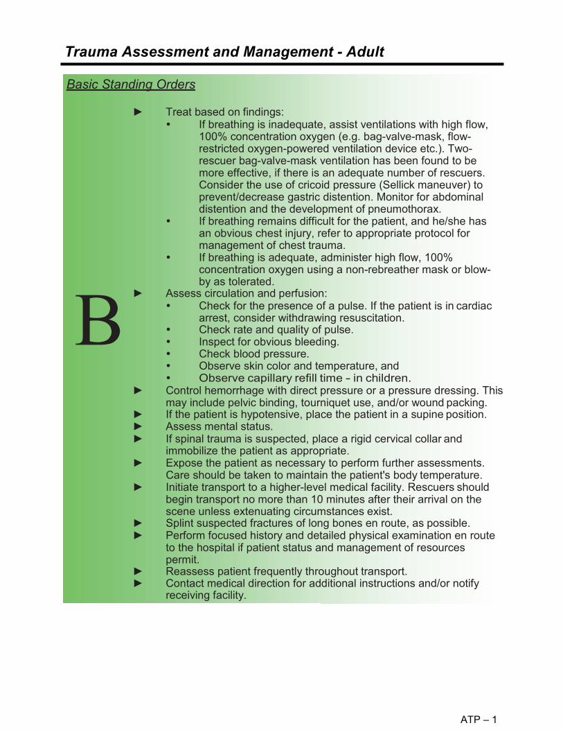
ATP – 1
Trauma Assessment and Management - Adult
Basic Standing Orders
► Treat based on findings: If breathing is inadequate, assist ventilations with high flow,
100% concentration oxygen (e.g. bag-valve-mask, flow- restricted oxygen-powered ventilation device etc.). Two-rescuer bag-valve-mask ventilation has been found to be more effective, if there is an adequate number of rescuers. Consider the use of cricoid pressure (Sellick maneuver) to prevent/decrease gastric distention. Monitor for abdominal distention and the development of pneumothorax.
If breathing remains difficult for the patient, and he/she has an obvious chest injury, refer to appropriate protocol for management of chest trauma.
If breathing is adequate, administer high flow, 100% concentration oxygen using a non-rebreather mask or blow-by as tolerated.
► Assess circulation and perfusion: Check for the presence of a pulse. If the patient is in cardiac
arrest, consider withdrawing resuscitation. Check rate and quality of pulse. Inspect for obvious bleeding. Check blood pressure. Observe skin color and temperature, and Observe capillary refill time - in children.
► Control hemorrhage with direct pressure or a pressure dressing. This may include pelvic binding, tourniquet use, and/or wound packing.
► If the patient is hypotensive, place the patient in a supine position. ► Assess mental status. ► If spinal trauma is suspected, place a rigid cervical collar and
immobilize the patient as appropriate. ► Expose the patient as necessary to perform further assessments.
Care should be taken to maintain the patient's body temperature. ► Initiate transport to a higher-level medical facility. Rescuers should
begin transport no more than 10 minutes after their arrival on the scene unless extenuating circumstances exist.
► Splint suspected fractures of long bones en route, as possible. ► Perform focused history and detailed physical examination en route
to the hospital if patient status and management of resources permit.
► Reassess patient frequently throughout transport. ► Contact medical direction for additional instructions and/or notify
receiving facility.
B
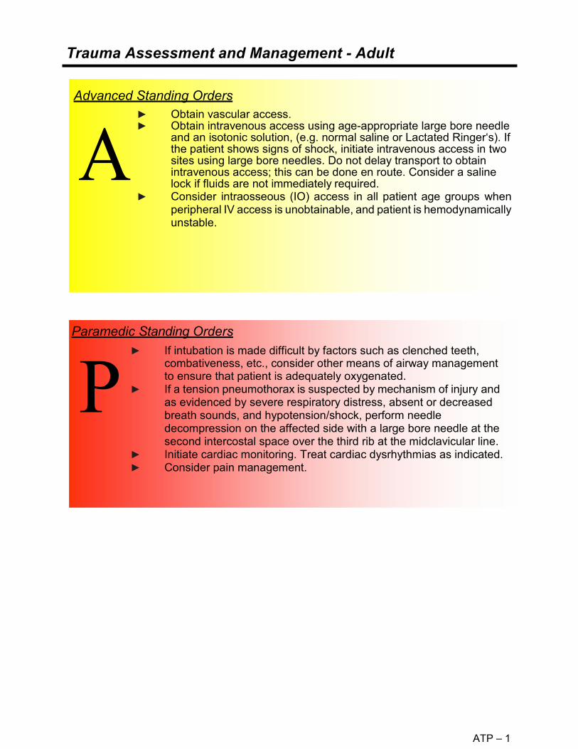
ATP – 1
Trauma Assessment and Management - Adult
Advanced Standing Orders ► Obtain vascular access. ► Obtain intravenous access using age-appropriate large bore needle
and an isotonic solution, (e.g. normal saline or Lactated Ringer‘s). If the patient shows signs of shock, initiate intravenous access in two sites using large bore needles. Do not delay transport to obtain intravenous access; this can be done en route. Consider a saline lock if fluids are not immediately required.
► Consider intraosseous (IO) access in all patient age groups when peripheral IV access is unobtainable, and patient is hemodynamically unstable.
Paramedic Standing Orders
► If intubation is made difficult by factors such as clenched teeth,
combativeness, etc., consider other means of airway management to ensure that patient is adequately oxygenated.
► If a tension pneumothorax is suspected by mechanism of injury and as evidenced by severe respiratory distress, absent or decreased breath sounds, and hypotension/shock, perform needle decompression on the affected side with a large bore needle at the second intercostal space over the third rib at the midclavicular line.
► Initiate cardiac monitoring. Treat cardiac dysrhythmias as indicated. ► Consider pain management.

ATP – 2
Selective Spinal Immobilization
KBEMS Approved 2/11/2015
Spine precautions are intended to prevent spinal cord injury in a patient presenting with an unstable spinal fracture, and to potentially prevent worsening cord injury. Traditional use of backboards for immobilization of patients has never been proven to be beneficial and newer studies show harm can occur from backboard immobilization without clear indications. Studies also show EMS providers are able to safely evaluate and identify patients with suspected spinal injuries in the field and employ selective spinal immobilization appropriately. The National Association of EMS Physicians and the American College of Surgeons Committee on Trauma believe that:
• Long backboards are commonly used to attempt to provide rigid spinal immobilization among emergency medical services (EMS) trauma patients. However, the benefit of long backboards is largely unproven.
• The long backboard can induce pain, patient agitation, and respiratory compromise. Further, the backboard can decrease tissue perfusion at pressure points, leading to the development of pressure ulcers.
• Utilization of backboards for spinal immobilization during transport should be judicious, so that the potential benefits outweigh the risks.
• Appropriate patients to be immobilized with a backboard may include those with: o Blunt trauma and altered level of consciousness o Spinal pain or tenderness o Neurologic complaint (e.g., numbness or motor weakness) o Anatomic deformity of the spine o High-energy mechanism of injury and any of the following:
Drug or alcohol intoxication Inability to communicate Distracting injury
• Patients for whom immobilization on a backboard is not necessary include those with all
the following: o Normal level of consciousness (Glasgow Coma Score [GCS] 15) o No spine tenderness or anatomic abnormality o No neurologic findings or complaints o No distracting injury o No intoxication
• Patients with penetrating trauma to the head, neck, or torso and no evidence of spinal
injury should not be immobilized on a backboard. • Spinal precautions can be maintained by application of a rigid cervical collar and
securing the patient firmly to the EMS stretcher, and may be most appropriate for: o Patients who are found to be ambulatory at the scene o Patients who must be transported for a protracted time, particularly prior to
interfacility transfer o Patients for whom a backboard is not otherwise indicated
• Whether or not a backboard is used, attention to spinal precautions among at-risk patients is paramount. These include application of a cervical collar, adequate security to a stretcher, minimal movement/transfers, and maintenance of inline stabilization during any necessary movement/ transfers.

ATP – 2
Patients should be removed from backboards as soon as practical in an emergency department. References: 1. National Association of EMS Physicians: Ems Spinal Precautions And The Use Of The
Long Backboard -Resource Document To The Position Statement Of The National Association Of Ems Physicians And The American College Of Surgeons Committee On Trauma. Prehosptial Emergency Care. (Jan/March 2014), 1-9. doi: 10.3109/10903127.2014.884197
2. Vallancourt et al: The Out of Hospital Validation of the Canadian C-Spine Rule by Paramedics. Annals of Emergency Medicine. 2009 Nov 54 (5); 663-670. doi: 10.1016/j.annemergmed.2009.03.008

Adult Trauma Protocol Selective Spine Immobilization
KBEMS Approved 2/11/2015
ATP – 2
If ANY of the following is true: If ALL the following is true:
NO long spine board Immobilization is needed (see c-spine clearance for c-collar
clearance)
Spine Immobilization Evaluation in the setting of trauma (age >8 yo and able to communicate)
• Blunt trauma with altered level of conscious- ness
• Spinal pain or tenderness • Neurologic Complaint (numbness or
motor weakness from traumatic injury) • Anatomic deformity of the spine • High-energy mechanism of injury and
any of the following: • Drug or alcohol intoxication • Inability to communicate • Distracting injury
• Normal level of consciousness (GCS 15)
• No spine tenderness or anatomic ab- normality
• No neurologic findings or complaints • No distracting injury • No intoxication.
Penetrating
Trauma
does NOT need Immobilization
YES NO
• Transport Immobilized to appropriate trauma center.
• Transport OFF long spine board but maintain spine precautions including C- collar
• Log roll, scoop stretcher, slider or LBB transfer to hospital stretcher.
Transport time > 20 minutes?
Spine precautions
Selective Spine Immobilization with C-Collar and Long Backboard
INTERFACILITY TRANSFERS DO NOT REQUIRE IMMOBILIZATION ON SPINE BOARDS FOR TRANSFER. SPINE PRECAUTIONS, WITH C-COLLARS, SHOULD BE OBSERVED DURING

Adult Trauma Protocol Selective Spine Immobilization
KBEMS Approved 2/11/2015
ATP – 2
YES
No C-spine
Immobilization
NO
NO
YES
C-Spine Immobilization
YES
NO
* Dangerous Mechanism
• fall from elevation > 3 feet / 5 stairs
• axial load to head, e.g. diving
• MVC high-speed > 65mph, rollover, ejection
• ATV
• Bicycle collision with object i.e. post, car **Simple Rear-ended MVC Excludes:
• Pushed into oncoming traffic
• Hit by bus / large truck
• Rollover
• Hit by high speed vehicle (> 65 mph)
3. Patient Voluntarily Able to Actively Rotate Neck 45° Left and Right When Requested, Regardless of Pain?
2. Any One Low-Risk Factor that may restrict Range of Motion Evaluation?
• More than a simple rear-end MVC** • Non-Ambulatory at scene • Neck pain at scene when asked • Pain during midline c-spine palpation
1. Any One High-Risk Factor Which Mandates Immobilization?
• Age > 65 • Dangerous mechanism* • Numbness or tingling in extremities
The C-Spine Rule is for alert (Glasgow Coma Scale score 15) and stable trauma patients for whom cervical spine injury is a concern, including patients with either posterior neck pain with any blunt mechanism of injury or no neck pain but some visible injury above the clavicles.
Reference:
Vallancourt et al: The Out of Hospital Validation of the Canadian C-Spine Rule by Paramedics. Annals of Emergency Medicine. 2009 Nov 54 (5);663-670. doi: 10.1016/j.annemergmed.2009.03.008
The Cervical Spine Evaluation

ATP – 3
Head Trauma - Adult
The recommendations for the management of traumatic brain injury (TBI) contained within these guidelines are adapted from the Prehospital Management of Traumatic Brain Injury developed by the Brain Trauma Foundation, © 2000. Field treatment is directed at preventing secondary injury, which is brain injury caused by hypoxia and shock after the initial injury has occurred. Evaluation and support of the patient’s ABC’s should be the first priority. As with all trauma patients, complete therapy for head and spine injuries must take place in the hospital. Delays at any level may be harmful to the patient. Patients with closed head injuries can worsen quickly, even though they appear stable initially. Although the presence of alcohol and other drugs may make evaluation of head injuries difficult, always assume symptoms are the result of the trauma and treat as such. Routine use of hyperventilation in the patient with traumatic brain injury is not recommended. Objects penetrating the head and neck should be stabilized whenever possible. Objects that are impaled in the cheek may be removed, as compression of both sides of the wound is easily accomplished.
Basic Standing Order
Follow Trauma Assessment and Management Protocol. If pulse oximetry is available, monitor and maintain oxygen saturation (SpO2) greater than 90%. Note that even a single instance of SpO2 less than 90% can significantly affect patient outcome. Ventilation and hyperventilation in the patient with TBI If breathing is inadequate, assist ventilation using a bag-
valve-mask device with high flow, 100% concentration oxygen.
Adult, 10 breaths/minute. ► If breathing is adequate, administer high flow, 100% concentration
oxygen using a non-rebreather mask or blow-by, as tolerated. ► If a TBI is suspected, hyperventilate the patient only if one or
more of the following signs of brain herniation exists:
Fixed or asymmetric pupils. Abnormal extension (decerebrate posturing). Rapid deterioration of Glasgow Coma Score to less than 9.

ATP – 3
Head Trauma - Adult
Basic Standing Orders Blood pressure in the head injured patient: Hypotension in an adult,
except as a terminal event, is not caused by isolated closed head injuries. You should assess the chest, abdomen, pelvis, and thighs for additional injuries. Patients with TBIs who also have external bleeding may suffer fatal blood loss; control bleeding with direct pressure.
Assess mental status using the GCS every five minutes to track changes. Changes in mental status are the most sensitive indicator of traumatic brain injury.
Evaluate pupil size and reactivity. A unilaterally dilated pupil or bilaterally fixed and dilated pupils is a sign of brain herniation and requires emergent interventions to lower the intracranial pressure (ICP). Unequal pupils in the conscious patient is not an indicator of brain herniation or increased ICP.
Remember to suspect spinal injuries in any patient with a head injury and significant mechanism of injury. Evaluate spinal cord integrity: In a conscious patient by recording ability to move extremities
to command. Perform gross sensory exam with sharp sensation or light touch.
Document patient complaints of numbness, tingling, or shooting pain.
In an unconscious patient by recording presence or absence of extremity movement to painful stimulus.
Reassess patient frequently throughout transport, as a head injured patient may deteriorate rapidly. Changes in the ongoing exam can be more important than the initial exam.
Advanced Standing Orders Perform airway management as appropriate and trained if the airway cannot be maintained by the patient, if prolonged assisted ventilation is anticipated, if hypoxemia is not corrected by supplemental oxygen, or if the GCS is 8 or less consider placing BIAD. Obtain intravenous or intraosseous access and, if needed, administer isotonic solution, (e.g. normal saline or lactated Ringer’s). Avoid the use of dextrose-containing IV fluids in TBI patients (Treat hypoglycemia as indicated). In patients with multi-organ trauma with an associated TBI, titrate IVs to maintain systolic blood pressure above 90. A systolic BP below 90 has been shown to increase morbidity and mortality in the patient with a TBI.

Head Trauma – Adult continued
ATP – 3
Endotracheal intubation if needed to maintain oxygenation or for therapeutic hyperventilation when indicated. Ventilate to maintain an ETCO2 of 30-35mmHg only if signs of herniation are present. If intubation required, consider administration of lidocaine 1 mg/kg IV (maximum dose 100mg) prior to intubation.
► ► ►
Paramedic Standing Orders

Head Trauma – Adult
ATP – 3
BEST EYE OPENING
Adult & Child Infant (12 months) Points Spontaneous Spontaneous 4
To Command To Voice 3
To Pain To Pain 2
None None 1
TOTAL
BEST VERBAL RESPONSE Adult & Child Infant (12 months) Points
Oriented Coos and Babbles (or crying after non-painful stimulation)
5
Confused Irritable Cry 4
Inappropriate Only cries to Pain 3
Incomprehensible Moans to Pain 2
None None 1 TOTAL
BEST MOTOR RESPONSE Adult & Child Infant (12 months) Points
Obeys Command Spontaneous Movements 6
Localizes Pain Withdraws (Touch) 5
Withdraws Withdraws (Pain) 4
Flexion to Pain Flexion to Pain 3
Extension to Pain Extension to Pain 2
None None 1 TOTAL
Total: Best Eye Opening Total: Best Verbal Response Total: Best Pain Response
Glasgow Coma Score

Chest Trauma – Adult
ATP – 4
► Chest trauma can lead to severe internal injuries that are often difficult to diagnose. A history of chest trauma should lead rescuers to suspect a severe injury, and patients should be treated with that expectation.
► Three major chest injury syndromes can lead to rapid death. They must be recognized and treated rapidly. They include: Bleeding from rupture of a major chest vessel; Mechanical decrease of cardiac output (which may be caused by tension
pneumothorax, cardiac tamponade or cardiac contusion with or without dysrhythmia);and
Respiratory distress (which may be caused by tension pneumothorax, flail chest, pulmonary contusion or an open chest wound).
► If chest injury interferes with breathing, it must be managed during the initial assessment.
► Objects penetrating the chest wall should be stabilized whenever possible, and not removed unless absolutely necessary for extrication or transport.
Basic Standing Order ► Follow Trauma Assessment and Management Protocol. Examine
the patient looking for distended neck veins. Look at the chest wall for asymmetry of movement, open wounds, and bruises. Expose the patient's chest, as needed, to inspect the entire chest wall, front and back, maintaining cervical immobilization and log rolling when indicated. Respiratory distress, despite an open airway, may suggest a tension pneumothorax, a flail chest, or an open chest wound. • Signs of a tension pneumothorax include diminished breath
sounds, hypotension, respiratory distress, distended neck veins, subcutaneous emphysema, shock, apprehension/agitation, and increasing resistance to ventilation.
• If a penetrating chest wound has been sealed, temporarily unseal the wound and allow air to escape.
• Assist ventilation with positive pressure oxygen if available. • Transport patient in the position of comfort unless otherwise
contraindicated ► Signs of flail chest may include paradoxical movement of the chest
wall, or crepitus of multiple ribs in two or more areas. Assist ventilation with positive pressure as needed to maintain adequate oxygenation.
► A wound in the chest may be an open chest wound, especially when it presents with subcutaneous emphysema, and air movement through the opening. • Cover with a sterile occlusive dressing taped on three sides. • Observe closely for signs of developing tension pneumothorax. • Uncontrolled external hemorrhage from a penetrating injury
should be packed with or without hemostatic dressings when necessary.
B

ATP – 4
Chest Trauma – Adult (cont.)
Advanced Standing Orders Follow Trauma Assessment and Management Protocol. Examine the
patient looking for distended neck veins. Look at the chest wall for asymmetry of movement, open wounds, and bruises. Expose the patient’s chest, as needed, to inspect the entire chest wall, front and back, maintaining cervical immobilization and log rolling when indicated. Respiratory distress, despite an open airway, may suggest a tension pneumothorax, a flail chest, or an open chest wound. Signs of a tension pneumothorax include diminished breath sounds,
hypotension, respiratory distress, distended neck veins, subcutaneous emphysema, shock, apprehension/agitation, and increasing resistance to ventilation.
If a penetrating chest wound has been sealed, temporarily unseal the wound and allow air to escape.
Assist ventilation with positive pressure oxygen if available. Consider airway management as appropriate and trained. Transport patient in the position of comfort unless otherwise
contraindicated Signs of flail chest may include paradoxical movement of the chest wall,
or crepitus of multiple ribs in two or more areas. Assist ventilation with positive pressure as needed to maintain adequate oxygenation.
A wound in the chest may be an open chest wound, especially when it presents with subcutaneous emphysema, and air movement through the opening. Cover with a sterile occlusive dressing taped on three sides. Observe closely for signs of developing tension pneumothorax.
Treat for hypotension.
Paramedic Standing Orders
P Positive pressure ventilation may be needed but is likely to worsen unrelieved tension pneumothorax. Be prepared to decompress the patient’s chest. If a tension pneumothorax is suspected by mechanism of injury and as evidenced by hypotension, respiratory distress, and/or diminished breath sounds, perform needle decompression with a large bore needle at the second intercostal space over the third rib at the midclavicular line.
This is an airway procedure and must be performed early, if indicated. A patient may have bilateral pneumothoraces; if condition does not improve after decompression of one lung, decompress the other side.
Initiate cardiac monitoring. Consider analgesia for isolated chest trauma. Treat for hypotension.

ATP – 5
Abdominal Trauma - Adult
Pre-hospital care of abdominal injuries should focus on controlling external bleeding and rapid transport as there are no specific prehospital treatments for internal bleeding. Penetrating trauma injures the area of entry and may damage any tissue along the line of penetration. Blunt trauma may be widely transmitted and cause damage to any or all organs within the abdominal cavity. Trauma to the abdomen may also cause injury to organs outside the abdominal cavity including those in the chest. Injuries from the nipple line through the tenth rib can involve either the chest and/or abdomen. Ongoing re-evaluation of the abdomen includes assessment of the chest as well. As with all trauma patients, complete treatment for abdominal injuries must take place in the hospital. Delays at any level can be harmful to the patient. Evaluation of abdominal trauma is part of the rapid trauma assessment. It should be performed only after the patient's ABCs have been evaluated and supported. Objects penetrating the abdominal wall should be stabilized whenever possible, and not removed unless absolutely necessary for extrication or transport.
Paramedic Standing Orders
► ALS considerations for the patient with abdominal injuries are those listed in the Trauma Assessment and Management Protocol.
Basic Standing Order
►
►
►
►
Follow Trauma Assessment and Management Protocol. Assess the abdomen for tenderness, rigidity, and distension. Uncontrolled external hemorrhage from a penetrating injury should be packed with or without hemostatic dressings when necessary. Reassess abdomen every 5 10 minutes, for tenderness, rigidity and distention. Shock, increasing distention, and abdominal rigidity are signs of intra-abdominal bleeding, although a person may have life- threatening bleeding without distention or abdominal rigidity. Any organs protruding from abdominal wounds should not be replaced into the abdominal cavity; cover the organs with saline-moistened gauze and a vapor barrier. If mechanism of injury permits, transport the patient in the position of comfort.
P

ATP – 6
Pelvic Trauma - Adult
A person may lose enough blood from pelvic fractures to exsanguinate. Disruption of the pelvic ring increases potential space in the pelvic cavity. This increased space will accommodate more blood than the standard pelvis. The goals of pelvic immobilization are to decrease movement of the bones and to decrease the potential space for bleeding. Apply circumferential pressure to tamponade internal hemorrhage. Signs of pelvic fracture may include instability, crepitus, decreased peripheral pulses, swelling, and blood at the urinary meatus. When assessing for pelvic trauma, gentle downward, then inward pressure should be applied to the iliac crests. If instability or crepitus is noted, this test should not be repeated.
Basic Standing Order ► ►
►
►
Follow Trauma Assessment and Management Protocol. Control external hemorrhage with direct pressure or a pressure dressing. Hemorrhage control may be improved by closing and stabilizing pelvic fractures. Uncontrolled external hemorrhage from a penetrating injury should be packed with or without hemostatic dressings when necessary. Pelvic fractures may be stabilized in several ways:
Use of the pelvic sheet wrapping technique Commercially available pelvic binding device
► Assess circulatory, motor, and sensory function before and after application of pelvic stabilization.
► Attempt to minimize unnecessary movement in patients with pelvic fractures.

Extremity Trauma - Adult
ATP – 7
In the severely injured patient, management of extremity injuries takes a relatively low priority. Most extremity hemorrhage can be controlled by direct pressure or pressure dressings. As with all trauma patients, definitive treatment for extremity injuries takes place in the hospital. Delays at any level can be harmful to the patient. Evaluation of extremity trauma is part of the focused physical exam and should be performed only after the patient’s ABCs have been evaluated and supported. Consider femur or pelvic fractures when the degree of shock seems greater than indicated by the amount of external bleeding.
Basic Standing Order
► Follow Trauma Assessment and Management Protocol. ► Control external hemorrhage with well-aimed direct pressure or a
pressure dressing, or elevation and pressure points. ► A tourniquet should be used if bleeding cannot be controlled by
other methods. Though tourniquets are infrequently needed, do not delay application when other bleeding control methods have failed.
► Hemorrhage control in a patient with femur fracture(s) may be improved by using a traction splint, apply pressure directly over the fracture.
► Examine the patient for extremity injures (deformities, contusions, avulsions, amputations, punctures, penetrations, burns, tenderness, lacerations, or swelling).
► Check for motion and sensation distal to deformities (both light touch and sharp sensation should be checked).
► Check circulation distal to deformities. ► The primary concern when treating extremity injuries is to maintain
proper distal circulation beyond the site of the injury. This may involve straightening the extremity. ("Make limbs look like limbs.”)
► Stop if severe resistance is encountered or if the patient has significantly increased pain during an attempt at straightening the extremity. No more than two attempts at straightening the limb should be made. In general, joint injures are left in the position found if there is
adequate circulation. If there is no pulse distal to the joint injury, an EMT should attempt to align the joint in its normal anatomic position by applying traction.
Straighten any grossly angulated long bone into its anatomic position by applying traction.
B

Extremity Trauma – Adult continued
ATP – 7
When splinting open fractures, apply the appropriate splint (e.g. traction splint for fractured femurs) in the usual manner. The bone ends may slip under the skin during splinting, this is acceptable, as the patient will need to have the wound cleaned in the operating room whether the bone ends remain above the skin or have slipped back into the wound. (Notify the receiving facility if this occurs.) Flush gross contamination from wounds before applying the splint. If, after attempting to straighten the extremity, the bone ends remain above the skin, cover with a moist dressing. Amputated parts should be wrapped in sterile gauze moistened with normal saline, protected from contamination (e.g., placed in an examination glove or Ziploc®-type bag) and put in ice water. Do not allow the amputated part to freeze. A cold pack may be applied to the site of an extremity injury to help reduce pain and swelling. Care should be taken not to freeze the tissues.
►
Basic Standing Order
►
►
► Pain management is strongly encouraged for patients with isolated extremity injuries, unless there is a contraindication to pain medication (e.g. hypotension, allergy). Medicating the patient before splinting may be appropriate in the patient with an isolated extremity injury.
► For patients with suspected open fractures: o Administer Cefazolin:
2 G IV if patient is less than 120 kg 3 G IV if patient is greater than 120 kg. Single dose.
o Patients receiving Cefazolin, must be transported to UK Chandler ER for treatment.
Paramedic Standing Orders

Eye and Dental Injuries-Adult
ATP – 8
Dental avulsions should be placed in an obviously labeled container with saline or cell-culture medium (Save-a-Tooth ).
► Dental Avulsions
Routine Patient Care Obtain visual history (use of corrective lenses, surgeries, use of protective equipment). Obtain visual acuity, if able. Chemical irritants: flush with copious amounts of water, or normal saline. Thermal burns to eyelids: patch both eyes with cool saline compress. Impaled object: immobilize object and patch both eyes. Puncture wound: place protective device over both eyes (e.g. eye shield). Do not apply pressure. Foreign body: patch both eyes. In the event patient is unable to close eyelids, keep eye moist with sterile saline compress. Consider ALS intercept.
► ► ►
►►
►
► ►
Basic Standing Orders
► ►
Paramedic Standing Orders
► Refer to the Pain Management Protocol. ► Refer to the Nausea Protocol.
Advanced Standing Orders
► Establish IV Access and administer fluid to maintain SBP>90 mmHg.A

Burns (Thermal) - Adult
ATP – 9
Effective treatment of patients with burns must be started as soon as possible after injury, as these patients frequently require specialized care which includes fluid resuscitation, pain management, and wound care. The goal is to transfer the patient to a facility capable of providing the necessary level of care for that individual.
Burns that require specialized care in a recognized burn center or unit include: ► Partial-thickness and full-thickness burns of greater than 10% total body surface
area (TBSA) in patients <10 years of age or >50 years of age.► Partial-thickness and full-thickness burns of greater than 20% TBSA in all other
patients.► Partial-thickness and full-thickness burns involving the face, eyes, ears, hands,
feet, major joints, genitalia, or perineum.► Full-thickness burns totaling 5% TBSA or more in any age group.► Electrical burns including lightning injury.► Significant chemical burns.► All burns associated with inhalation injury.► Circumferential burns of the chest, neck, or extremities.► Burns associated with concomitant major trauma.► Burn injury occurring in patients with pre-existing medical disorders.► Burn injury in patients who will require special social and emotional or long-term
rehabilitative support, including cases involving suspected child abuse andneglect.
► Stop the burning process. If on scene quickly after the burn occurred,cooling affected parts (e.g. with cool water immersion) may limit thedepth and extent of the burn. More than a few minutes after the burn,there is little benefit except pain relief. Note that with burns from tar,asphalt, paraffin or oils that retain heat (or when melted fabric adheresto skin) cooling may help for a longer period of time.
Brush dry chemicals off the skin before flushing. For chemical burns of the eye, flush the eye immediately with at least one liter of normal saline or water (at least 10 to 20 minutes is preferred). More fluids may be beneficial, especially if the chemical is alkaline.
When treating patients with chemical burns, it is imperative to ensure rescuer safety. Patients contaminated with chemicals should have their clothing removed. Do NOT transport patients prior to appropriate decontamination. Notify the receiving facility of a patient with chemical exposure to allow adequate time for preparation. All chemical burns should be flushed with copious amounts of water.
Basic Standing Orders
►

Burns (Thermal) – Adult continued
ATP – 9
Basic Standing Orders continued
► If cooling for pain relief, do not cool or moisten more than 10% ofthe TBSA at any one time. This can cause hypothermia.
► Remove all clothing and jewelry in the area of the burn and distal tothe injury.
► Administer high flow, 100% concentration oxygen by non-rebreather mask for potential inhalation injury or any serious burn.Consider the possibility of carbon monoxide or other toxicinhalation. Oxygen saturation readings may be falsely elevated.
► Assess circulation and perfusion. Circumferential burns ofextremities can interfere with perfusion of that extremity.
► If spinal trauma is suspected, place a rigid cervical collar andimmobilize the patient as appropriate.
► Consider ALS intercept for patients with serious burns andelectrical injuries; in electrical injuries there is a possibility ofcardiac dysrhythmias.
► Estimate the TBSA involved using the "Rule .o f Nines" provides a rough estimate of TBSA involved (see following page).
► Describe the body surface area as well as the depth of burn (e.g. 30% superficial burn, 20% partial thickness, and 15% full thickness burn).
► Apply dressings to burns as tolerated. In burns over 10% BSA, apply a dry sheet, a dry burn sheet or
dry sterile dressings to burn areas. Insulate the patient over this dressing to lessen the chance of hypothermia.
In burns less than 10% BSA, apply moist dressings (e.g. commercially available burn dressings or saline-soaked gauze)
A vapor barrier may be useful in patients with longer transport times.
B

Burns (Thermal) – Adult continued
ATP – 9
pigment is present. ► Consider Pain Management. Small doses IV titrated to effective
pain control; monitor for respiratory depression. ► Give all medications intravenously.
In electrical burns where there is a large amount of pigment (hemoglobin or myoglobin) in the urine, the urinary output should be maintained at 1.0 – 2.0 cc/Kg/hour until the urine is grossly clear, then fluids may be cut back to maintain the output in the range of 0.5 to 1.0 cc/Kg/hour in adults. In addition, 44 – 50mEq of NaHCO3 per liter of LR is administered to keep the urine alkaline as long as visible
Be alert for signs of inhalation injury (e.g. stridor, muffled voice, singed facial/nasal hairs, soot around nose or mouth, carbonaceous sputum, confinement in an enclosed space fire). Be prepared to secure the airway. If the injury involves an electrical burn, initiate cardiac monitoring. Treat cardiac dysrhythmias as directed. Electrical burn fluid management: ►
►
Paramedic Standing Orders
►
Example: A 70 Kg man who had sustained a 50% TBSA would require a total of 14,000cc in the first 24 hours, 7,000cc would be given in the first 8 hours. If the patient is not seen until 4 hours after the time of burn, that amount should be given over the next 4 hours. Second 24 hours: give normal maintenance fluids in sufficient volume to maintain a normal urinary output. 4cc normal saline (NS) or lactated Ringer’s (LR) x 70 (Kg) x 50 %TBSA = 14,000 cc in the first 24 hours
Start two large bore IVs in patients meeting any of the burn criteria in the beginning of this section. These may be inserted through burn area, if necessary. Fluid administration: First 24 hours: 4cc normal saline (NS) or lactated ringers
(LR) x patient weight (Kg) x %TBSA (for fluid calculations include only partial thickness and full thickness burns). If more than two or three liters of fluid are to be given lactated ringers is preferred.
Half of this amount is to be given in the first 8 hours after injury not the time after arrival at the patient’s side. (Note: this means that the EMS provider should determine the time of injury)
The remaining half is to be given over the next 16 hours.
►
Advanced Standing Orders
►

Burns (Thermal) – Adult continued
ATP – 9
Burn Chart – Rule of Nines
PTP
Pediatric Trauma Protocols

Trauma Assessment and Management - Pediatric
PTP – 1
Children experience different types of injuries and have different physiologic reactions to injury as compared to adults. Patient outcome depends on the time it takes to get the patient to the hospital. Therefore, assessment and treatment are frequently done at the same time and scene time should be minimized to less than 10 minutes, if possible. Continual assessment of children is imperative. A child may initially appear stable, then decompensate suddenly.
If tension pneumothorax is suspected, perform needle decompression with an over-the- needle catheter at the second intercostal space over the third rib at the midclavicular line. When obtaining intravenous access, use an age appropriate large-bore catheter with large- caliber tubing and administer normal saline or lactated ringers at a sufficient rate to keep the vein open. If the patient shows signs of shock, initiate intravenous access in two sites. Consider saline locking IVs if fluids are not immediately required. Carefully monitor fluid administration to avoid fluid overload in children. If signs of shock are present (such as, tachycardia, decreased level of consciousness, poor color, capillary refill greater than 2 seconds, decreased blood pressure, etc.) administer a bolus of normal saline or lactated ringers at 20 cc/kg. Bolus therapy with reassessment is more effective than high IV flow rates for ensuring pediatric patients receive adequate fluids. Two additional fluid boluses at 20 cc/kg may be given if the patient remains in shock. If intravenous access cannot be obtained, consider intraosseous access in pediatric trauma patients with decreased consciousness. The priorities in trauma management are to prevent further injury, provide rapid transport, notify the receiving facility, and initiate definitive treatment. Trauma patients cannot be treated completely in the field. On-scene time should be as short as possible unless there are extenuating circumstances, such as extrication, hazardous conditions, or multiple patients. Document these circumstances on the patient record. Determine how the patient should be transported as soon as possible so that activation of a special transport service, such as an air ambulance, if appropriate, can be performed in a timely manner. Notification of the receiving hospital of patient conditions and status should be done as early as possible. This allows the receiving hospital additional time to mobilize any necessary resources. The pre-hospital assessment and management of a trauma patient should be performed under the direction of one person. Although the presence of alcohol or other drugs may mask some of the signs of severe trauma, assume that the patient’s condition is caused by trauma until proved otherwise.

Trauma Assessment and Management - Pediatric
PTP – 1
Despite a rapid and effective out-of-hospital and trauma center response, patients with “out – of – hospital” cardiac arrest due to trauma rarely survive. Those patients with the best outcome from trauma arrest generally are young, have treatable penetrating injuries, have received early (out-of-hospital) endotracheal intubation, and undergo prompt transport (typically <=10 minutes) to a trauma care facility. Cardiac arrest in the field due to blunt trauma is nearly universally fatal in all age groups. Nonetheless, the literature suggests that survival in young children may be higher than that found in older children and adults for a variety of postulated reasons. Unless faced with a mass casualty situation or a patient with injuries obviously incompatible with life, briefly assess and/or treat for field-correctable causes (e.g tension pneumothorax, airway obstruction) followed immediately by transportation. During transportation, treat for other causes of arrest including hemorrhage. Early notification to the receiving hospital is extremely important.

Trauma Assessment and Management - Pediatric
PTP – 1
Basic Standing Orders
► Take body substance isolation precautions. This is best performed en route to the call location.
► Ensure scene safety. Priority should be given to the safety of the rescuers and then to altering the scene to make it a safe working environment or, if necessary, moving the patient from the scene.
► Perform a scene survey to assess environmental conditions and mechanism of injury and number of patients.
► Establish patient responsiveness. Manually stabilize the spine. Protect patient from heat loss.
► Open the airway: Use the head tilt/chin lift if no spinal trauma is suspected. Use the modified jaw thrust if spinal trauma is suspected.
► Establish and maintain a patent airway while protecting the cervical spine. Suction as necessary. Insert an oropharyngeal or nasopharyngeal airway adjunct if the airway cannot be maintained with positioning. The nasopharyngeal airway is contraindicated in the presence of maxillary facial trauma.
► Evaluate breathing – Is the patient breathing spontaneously? Are respirations adequate in rate and depth? Environmental factors should be considered when removing the patient’s clothing for evaluation.
► Initiate pulse oximetry, if available.
LOOK LISTEN FEEL Cyanosis Breathing Rib Fractures Rapid Respirations Abnormal breath
sounds Crepitus
Retractions Stridor – Indicates partial airway obstruction
Asymmetry of chest wall
Gurgling sounds
Open wounds or bruising of chest wall
B

PTP – 1
Trauma Assessment and Management - Pediatric continued
Basic Standing Orders continued
► Treat based on findings: If breathing is inadequate, assist ventilations with high flow,
100% concentration oxygen (e.g. bag-valve-mask, flow- restricted oxygen-powered ventilation device etc.). Two-rescuer bag-valve-mask ventilation has been found to be more effective, if there is an adequate number of rescuers. Consider the use of cricoid pressure (Sellick maneuver) to prevent/decrease gastric distention. Monitor for abdominal distention and the development of pneumothorax.
If breathing remains difficult for the patient, and he/she has an obvious chest injury, refer to appropriate protocol for management of chest trauma.
If breathing is adequate, administer high flow, 100% concentration oxygen using a non-rebreather mask or blow-by as tolerated.
► Assess circulation and perfusion: Check for the presence of a pulse. If the patient is in cardiac
arrest, consider withdrawing resuscitation. Check rate and quality of pulse. Inspect for obvious bleeding. Check blood pressure. Observe skin color and temperature, and Observe capillary refill time - in children.
► Control hemorrhage with direct pressure or a pressure dressing. This may include pelvic binding, tourniquet use, and/or wound packing.
► If the patient is hypotensive, place the patient in a supine position. ► Assess mental status. ► If spinal trauma is suspected, place a rigid cervical collar and
immobilize the patient as appropriate. ► Expose the patient as necessary to perform further assessments.
Care should be taken to maintain the patient's body temperature. ► Initiate transport to a higher-level medical facility. Rescuers should
begin transport no more than 10 minutes after their arrival on the scene unless extenuating circumstances exist.
► Splint suspected fractures of long bones en route, as possible. ► Perform focused history and detailed physical examination en route
to the hospital if patient status and management of resources permit.
► Reassess patient frequently throughout transport. ► Contact medical direction for additional instructions and/or notify
receiving facility.
B

PTP – 1
Trauma Assessment and Management - Pediatric continued
Advanced Standing Orders Airway management as needed. An assistant must maintain in-line cervical stabilization throughout this procedure. When obtaining intravenous access, use an age appropriate large- bore catheter with large-caliber tubing and administer normal saline or lactated Ringer’s at a sufficient rate to keep the vein open. If the patient shows signs of shock, initiate intravenous access in two sites. Consider saline locking IVs if fluids are not immediately required. Carefully monitor fluid administration to avoid fluid overload in children. If signs of shock are present (such as, tachycardia, decreased level of consciousness, poor color, capillary refill greater than 2 seconds, decreased blood pressure, etc.) administer a bolus of normal saline or lactated Ringer’s at 20 cc/kg. Bolus therapy with reassessment is more effective than high IV flow rates for ensuring pediatric patients receive adequate fluids. Two additional fluid boluses at 20 cc/kg may be given if the patient remains in shock. If intravenous access cannot be obtained, consider intraosseous access in pediatric trauma patients with decreased consciousness. Devices are available to initiate intraosseous (IO) access in all patient age groups and may be considered when peripheral IV access is unobtainable.
Paramedic Standing Orders
Consider placing a gastric tube in any patient who requires assisted ventilations. If tension pneumothorax is suspected, perform needle decompression with an over-the-needle catheter at the second intercostal space over the third rib at the midclavicular line. Initiate cardiac monitoring. Treat cardiac dysrhythmias as dictated by standing orders. Consider pain management. Consider pressors for shock refractory to adequate fluid resuscitation. This intervention should be made only after direct contact with physician medical control.

PTP – 1
Trauma Assessment and Management - Pediatric continued
A child is considered to have incurred serious trauma if any one of the following is met: ► A numerical triage score ≤ 9 using the Glasgow Coma Scale. ► A color triage score of one black box or two gray boxes using the Pediatric Trauma
Triage Criteria. ► Penetrating wounds to the head, neck, torso, or extremities proximal to the elbow or
knee. ► Two or more long bone fractures, pelvic fracture, or flail chest. ► Open or depressed skull fracture. ► Full thickness (3°) burns, partial thickness (2°) burns > 10% BSA or burns combined with
trauma. ► Paralysis. ► Amputation proximal to the wrist or ankle

Head Trauma - Pediatric
PTP – 2
Children are anatomically prone to head injuries because of their large heads, weak neck muscles, and immature brain tissue. Head injuries in children are common. Blunt mechanisms like falls and motor vehicle crashes are the most common causes of head injuries in children. Suspect a TBI in the child who: ► is inconsolable ► is irritable ► has a high-pitched cry ► vomits repeatedly ► is unusually quiet ► has difficulty walking (if ambulatory at the scene prior to EMS arrival) ► has a bulging fontanel, and/or ► has Battle’s sign or raccoon eyes The recommendations for the management of traumatic brain injury (TBI) contained within these guidelines are adapted from the Prehospital Management of Traumatic Brain Injury developed by the Brain Trauma Foundation, © 2000. Field treatment is directed at preventing "secondary injury” which is brain injury caused by hypoxia and shock after the initial injury has occurred. Evaluation and support of the patient’s ABCs should be the first priority. As with all trauma patients, complete therapy for head and spine injuries must take place in the hospital. Delays at any level may be harmful to the patient. Patients with closed head injuries can worsen quickly, even though they appear stable initially. Although the presence of alcohol and other drugs may make evaluation of head injuries difficult, always assume symptoms are the result of the trauma and treat as such. Routine use of hyperventilation in the patient with traumatic brain injury is not recommended. Objects penetrating the head and neck should be stabilized whenever possible. Objects that are impaled in the cheek may be removed, as compression of both sides of the wound is easily accomplished.

Head Trauma – Pediatric continued
PTP – 2
B
Basic Standing Order
► Follow Trauma Assessment and Management Protocol. ► If pulse oximetry is available, monitor and maintain oxygen
saturation (SpO2) greater than 90%. Note that even a single instance of SpO2 less than 90% can significantly affect patient outcome.
► Ventilation and hyperventilation in the patient with TBI: o If breathing is inadequate, assist ventilation using a bag-valve-
mask device with high flow, 100% concentration oxygen. o Consider advanced airway device. Monitor for gastric distention. Child, under age 8, 20 breaths/minute; and Infants, 25 breaths/minute.
o If breathing is adequate, administer high flow, 100% concentration oxygen using a non-rebreather mask or blow-by, as tolerated.
► If a TBI is suspected, hyperventilate the patient only if one or more of the following signs of brain herniation exists: o Fixed or asymmetric pupils. o Abnormal extension (decerebrate posturing). o Glasgow coma scale (GCS) of less than 9 with a further
decrease of 2 or more points. ► Blood pressure in the head injured patient: Hypotension, except as
a terminal event, is not caused by isolated closed head injuries. You should assess the chest, abdomen, pelvis, and thighs for additional injuries. Patients with TBIs who also have external bleeding may suffer fatal blood loss; control bleeding with direct pressure.
► Assess mental status using the GCS every five minutes to track changes. Changes in mental status are the most sensitive indicator of traumatic brain injury.
► Evaluate pupil size and reactivity. A unilaterally dilated pupil or bilaterally fixed and dilated pupils is a sign of brain herniation and requires emergent interventions to lower the intracranial pressure (ICP). Unequal pupils in the conscious patient is not an indicator of brain herniation or increased ICP.

Head Trauma – Pediatric continued
PTP – 2
► Reassess patient frequently throughout transport, as a head injured patient may deteriorate rapidly. Changes in the ongoing exam can be more important than the initial exam.
► Consider ALS intercept/air medical transport.
In a conscious patient by recording ability to move extremities to command. Perform gross sensory exam with sharp sensation or light touch. Document patient complaints of numbness, tingling, or shooting pain. In an unconscious patient by recording presence or absence of extremity movement to painful stimulus.
Remember to suspect spinal injuries in any patient with a head injury and significant mechanism of injury. Evaluate spinal cord integrity:
►
Basic Standing Order continued

Head Trauma – Pediatric continued
PTP – 2
Advanced Standing Order
► Children can present with signs of shock secondary to severe scalp lacerations. If a child with a severe scalp laceration is showing signs of shock, be sure to gain IV or IO access and give a 20 cc/kg bolus of normal saline or lactated Ringer’s. Be sure to evaluate the pediatric patient to rule out internal bleeding.
► Check blood glucose, if hypoglycemic see Diabetic Emergencies: Hypoglycemia Protocol.
► Obtain intravenous or intraosseous access and, if needed, administer isotonic solution, (e.g. normal saline or lactated Ringer’s). Avoid the use of dextrose-containing IV fluids in TBI patients (Treat hypoglycemia as indicated.).
► In patients with multi-organ trauma with an associated TBI, titrate IVs to maintain systolic blood pressure above 90. A systolic BP below 90 has been shown to increase morbidity and mortality in the patient with a TBI.
► Child: Administer fluid bolus 20 ml/kg, may repeat x 2 (maximum total 60 ml/kg to maintain SBP above. 12-16 years: 90 mmHg 5-12: 80 mmHg 1-5 years: 75 mmHg <1 year: 65 mmHg Administer fluid in children with normal SBP and who have other
signs of decreased perfusion including tachycardia, loss of central pulses, increased capillary filling time of > 2 seconds.
Paramedic Standing Orders
► If end-tidal CO2 is available, ventilate to maintain an end-tidal CO2 of 30-35 mmHg only if signs of herniation present.
► If intubation required, consider administration of lidocaine 1 mg/kg IV (maximum dose 100 mg) prior to intubation.
A

PTP – 3
Chest Trauma - Pediatric
► Chest trauma can lead to severe internal injuries that are often difficult to diagnose. A history of chest trauma should lead rescuers to suspect a severe injury, and patients should be treated with that expectation.
► Three major chest injury syndromes can lead to rapid death. They must be recognized and treated rapidly. They include: Bleeding from rupture of a major chest vessel; Mechanical decrease of cardiac output (which may be caused by tension
pneumothorax, cardiac tamponade or cardiac contusion with or without dysrhythmia); and
Respiratory distress (which may be caused by tension pneumothorax, flail chest, pulmonary contusion or an open chest wound).
► If chest injury interferes with breathing, it must be managed during the initial assessment.
► Objects penetrating the chest wall should be stabilized whenever possible, and not removed unless necessary for extrication or transport.
Basic Standing Order ► Follow Trauma Assessment and Management Protocol. Examine
the patient looking for distended neck veins. Look at the chest wall for asymmetry of movement, open wounds, and bruises. Expose the patient's chest, as needed, to inspect the entire chest wall, front and back, maintaining cervical immobilization and log rolling when indicated. Respiratory distress, despite an open airway, may suggest a tension pneumothorax, a flail chest, or an open chest wound. Signs of a tension pneumothorax include diminished breath
sounds, hypotension, respiratory distress, distended neck veins, subcutaneous emphysema, shock, apprehension/agitation, and increasing resistance to ventilation. o If a penetrating chest wound has been sealed, temporarily
unseal the wound and allow air to escape. o Assist ventilation with positive pressure oxygen if available. o Consider advanced airway adjuncts. o Transport patient in the position of comfort unless otherwise
contraindicated. ► Signs of flail chest may include paradoxical movement of the chest
wall, or crepitus of multiple ribs in two or more areas. • Use positive pressure ventilation. • Consider stabilizing with ipsilateral arm and swathe.
► A wound in the chest may be an open chest wound, especially when it presents with subcutaneous emphysema, and air movement through the opening.
o Cover with a sterile occlusive dressing taped on three sides. o Observe closely for signs of developing tension
pneumothorax. ► Uncontrolled external hemorrhage from a penetrating injury should
be packed with or without hemostatic dressings when necessary
B

PTP – 3
Chest Trauma - Pediatric
Advanced Standing Orders Treat for hypotension
Paramedic Standing Orders
Positive pressure ventilation may be needed but is likely to worsen unrelieved tension pneumothorax. Be prepared to decompress the patient’s chest. If a tension pneumothorax is suspected by mechanism of injury and as evidenced by hypotension, respiratory distress, and/or diminished breath sounds, perform needle decompression with an over- the-catheter needle placed at the second intercostal space at the midclavicular line. This is an airway procedure and must be performed early, if
indicated. A patient may have bilateral pneumothoraces; if condition does not improve after decompression of one lung, decompress the other side.
Initiate cardiac monitoring. Consider analgesia for isolated chest trauma. Treat for hypotension.

PTP – 4
Abdominal Trauma - Pediatric
Solid organs of the upper abdominal cavity (the liver, spleen and kidneys) are proportionally larger and more exposed in children, and the abdominal muscles of the child are relatively underdeveloped, and the ribs are more pliable. This predisposes pediatric patients to potentially serious blood loss and shock from abdominal injuries.
Pre-hospital care of abdominal injuries should focus on controlling external bleeding and rapid transport as there are no specific pre-hospital treatments for internal bleeding. Penetrating trauma injures the area of entry and may damage any tissue along the line of penetration. Blunt trauma may be widely transmitted and cause damage to any or all organs within the abdominal cavity. Trauma to the abdomen may also cause injury to organs outside the abdominal cavity including those in the chest. Injuries from the nipple line through the tenth rib can involve either the chest and/or abdomen. Ongoing re-evaluation of the abdomen includes assessment of the chest as well. As with all trauma patients, complete treatment for abdominal injuries must take place in the hospital. Delays at any level can be harmful to the patient. Evaluation of abdominal trauma is part of the rapid trauma assessment. It should be performed only after the patient's ABCs have been evaluated and supported. Objects penetrating the abdominal wall should be stabilized whenever possible, and not removed unless necessary for extrication or transport.
Basic Standing Order
►
► ► ►
Follow Trauma Assessment and Management Protocol. Assess the abdomen for tenderness, rigidity, and distension. Uncontrolled external hemorrhage from a penetrating injury should be packed with or without hemostatic dressings when necessary. Reassess abdomen every 5 10 minutes, for tenderness, rigidity and distention. Shock, increasing distention, and abdominal rigidity are signs of intra-abdominal bleeding, although a person may have life-threatening bleeding without distention or abdominal rigidity. Any organs protruding from abdominal wounds should not be replaced into the abdominal cavity; cover the organs with saline- moistened gauze and a vapor barrier. If mechanism of injury permits, transport the patient in the position of comfort.
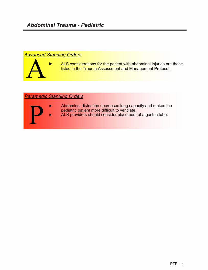
PTP – 4
Abdominal Trauma - Pediatric
Advanced Standing Orders
► ALS considerations for the patient with abdominal injuries are those listed in the Trauma Assessment and Management Protocol.
Paramedic Standing Orders
Abdominal distention decreases lung capacity and makes the pediatric patient more difficult to ventilate. ALS providers should consider placement of a gastric tube.

Pain Management - Pediatric
PTP – 5
O 2 4 6 8 10
NO HURT
HURTS A LITTLE
HURTS A LITTLE MORE
HURTS EVEN MORE
HURTS A
WHOLE LOT
HURTS THE
WORST
Routine Patient Care. Place the patient in a position of comfort if possible. Give reassurance, psychological support, and distraction. Use ample padding for long and short spinal immobilization devices. Use ample padding when splinting possible fractures, dislocations, sprains and strains. Elevate injured extremities if possible. Consider application of cold pack for 10 minutes. Have the patient rate their pain on a 0 to 10 (or similar) scale*. Reassess the patient’s pain level and vital signs every 5 minutes. *0- 10 Scale: Avoid coaching the patient, simply ask them to rate their pain on a scale from 0-10, where 0 is no pain at all and 10 is the worst pain ever experienced by the patient. *Wong-Baker "faces” scale: The faces correspond to numeric values from 0-10. The scale can be documented with the numeric value or the textual pain description. Consider paramedic intercept if needed for pain management.
► ►
► ►
► ► ► ►
Basic Standing Orders

Pain Management – Pediatric continued
PTP – 5
Paramedic Standing Orders
► IV access, obtain blood sample and administer fluids to maintain systolic blood pressure > minimum for age and signs of adequate perfusion.
► Unless the patient has altered mental status, multi-systems trauma or abdominal pain, the paramedic may consider • Ketamine: 0.3 mg/kg IV or 0.5 – 1 mg/kg IM.
o Give slowly IV push. o Be aware of possible Laryngospasm. o Maximum: 25 mg, single dose only. o Administer over 1 minute for IV administration.
• Fentanyl: 0.5 mcg/kg IV every 5 minutes. May be repeated up to 3 doses o Intranasal dosing: 1 mcg/kg to max of 100 mcg, max of 1 mL
per nostril, consider splitting dose between nostrils for volumes greater the 0.3 mL
► For hypoventilation from opiate administration by EMS personnel,
administer naloxone 0.1 mg/kg up to 2 mg prn. ► Nausea: See Nausea Protocol.
NOTE: Contact medical control for guidance with all patients with altered mental status, or for any other guidance necessary.
FOR ALL PATIENTS RECEIVING ANALGESIA, EtCO2 SHALL BE
MONITORED.
Advanced Standing Orders
► IV access and administer fluids to maintain systolic blood pressure > Minimum for age and signs of adequate perfusion.
P
A

PTP – 6
Pelvic Fractures– Pediatric
A person may lose enough blood from pelvic fractures to exsanguinate. Disruption of the pelvic ring increases potential space in the pelvic cavity. This increased space will accommodate more blood than the standard pelvis. The goals of pelvic immobilization are to decrease movement of the bones and to decrease the potential space for bleeding. Apply circumferential pressure to tamponade internal hemorrhage. Signs of pelvic fracture may include instability, crepitus, decreased peripheral pulses, swelling, and blood at the urinary meatus. When assessing for pelvic trauma, gentle downward, then inward pressure should be applied to the iliac crests. If instability or crepitus is noted, this test should not be repeated.
Basic Standing Orders Follow Trauma Assessment and Management Protocol. Control external hemorrhage with direct pressure or a pressure dressing. Hemorrhage control may be improved by closing and stabilizing pelvic fractures. Pelvic fractures may be stabilized in several ways, three of which are easily applied in the pre-hospital setting. Use of the pelvic sheet wrapping technique Commercially available pelvic binding device Assess circulatory, motor, and sensory function before and after application of pelvic stabilization. Attempt to minimize unnecessary movement in patients with pelvic fractures
Advanced Standing Orders Follow Trauma Assessment and Management Protocol. Control external hemorrhage with direct pressure or a pressure dressing. Hemorrhage control may be improved by closing and stabilizing pelvic fractures. Pelvic fractures may be stabilized in several ways Use of the pelvic sheet wrapping technique Commercially available pelvic binding device Assess circulatory, motor, and sensory function before and after application of pelvic stabilization. Attempt to minimize unnecessary movement in patients with pelvic fractures Treat for hypotension.
Extremity Trauma - Pediatric
PTP – 7
Bones in children are more pliable than those in adults; they are prone to fractures that involve the bone bending (e.g. "greenstick fracture”), which may be more difficult to straighten. Children may fracture their bones at the growth plates, which are located near joints. Injuries involving joints should only be straightened when there is decreased circulation distal to the injury (unless it is an ankle injury). If using commercially available devices to splint fractures in children, be sure that they are of an appropriate size for the child.
In the severely injured patient, management of extremity injuries takes a relatively low priority. Most extremity hemorrhage can be controlled by direct pressure or pressure dressings. As with all trauma patients, definitive treatment for extremity injuries takes place in the hospital. Delays at any level can be harmful to the patient. Evaluation of extremity trauma is part of the focused physical exam and should be performed only after the patient’s ABCs have been evaluated and supported. Consider femur or pelvic fractures when the degree of shock seems greater than indicated by the amount of external bleeding.

Extremity Trauma - Pediatric
PTP – 7
Basic Standing Order
► Follow Trauma Assessment and Management Protocol. ► Control external hemorrhage with well-aimed direct pressure or a
pressure dressing, or elevation and pressure points. ► A tourniquet should be used if bleeding cannot be controlled by
other methods. Though tourniquets are infrequently needed, do not delay application when other bleeding control methods have failed.
► Hemorrhage control in a patient with femur fracture(s) may be improved by using a traction splint, apply pressure directly over the fracture.
► Examine the patient for extremity injures (deformities, contusions, avulsions, amputations, punctures, penetrations, burns, tenderness, lacerations, or swelling).
► Check for motion and sensation distal to deformities (both light touch and sharp sensation should be checked).
► Check circulation distal to deformities. ► The primary concern when treating extremity injuries is to maintain
proper distal circulation beyond the site of the injury. This may involve straightening the extremity. ("Make limbs look like limbs.”)
► Stop if severe resistance is encountered or if the patient has significantly increased pain during an attempt at straightening the extremity. No more than two attempts at straightening the limb should be made. In general, joint injures are left in the position found if there is
adequate circulation. If there is no pulse distal to the joint injury, an EMT should attempt to align the joint in its normal anatomic position by applying traction.
Straighten any grossly angulated long bone into its anatomic position by applying traction.
► Pain management is strongly encouraged for patients with isolated extremity injuries, unless there is a contraindication to pain medication (e.g. hypotension, allergy). Medicating the patient before splinting may be appropriate in the patient with an isolated extremity injury.
► For open or suspected open fractures, administer Cefazolin: 25 mg/kg IV, max dose 2 G. Single dose. Patients receiving Cefazolin, must be transported to UK Chandler ER for treatment.
Paramedic Standing Orders
B

Eye and Dental Injuries-Pediatric
PTP – 8
Dental avulsions should be placed in an obviously labeled container with saline or cell-culture medium (Save-a-Tooth ).
► Dental Avulsions
Routine Patient Care Obtain visual history (use of corrective lenses, surgeries, use of protective equipment). Obtain visual acuity, if able. Chemical irritants: flush with copious amounts of water, or normal saline. Thermal burns to eyelids: patch both eyes with cool saline compress. Impaled object: immobilize object and patch both eyes. Puncture wound: place protective device over both eyes (e.g. eye shield). Do not apply pressure. Foreign body: patch both eyes. In the event patient is unable to close eyelids, keep eye moist with sterile saline compress. Consider intercept.
► ► ► ► ► ►
► ►
Basic Standing Orders
► ►
IV access and administer fluids to maintain systolic blood pressure > minimum for age and signs of adequate perfusion.
► Advanced Standing Orders
Refer to the Pain Management Protocol. Refer to the Nausea Protocol.
► ►
Paramedic Standing Orders

Burns (Thermal) Pediatric
PTP – 9
Children under 5 years of age represent the age group most often found with burns resulting from child abuse. Look for characteristic burns that should make you suspect they are the result of child abuse. The child with burns to the back, buttocks, and posterior neck should alert your suspicion of abuse. Circumferential scald burns of hands or feet that are clearly demarcated and uniform with no splash marks are also characteristic of child abuse. Effective treatment of patients with burns must be started as soon as possible after injury, as these patients frequently require specialized care which includes fluid resuscitation, pain management, and wound care. The goal is to transfer the patient to a facility capable of providing the necessary level of care for that individual. Burns that require specialized care in a recognized burn center or unit include: ► Partial-thickness and full-thickness burns of greater than 10% total body surface
area (TBSA) in patients <10 years of age. ► Partial-thickness and full-thickness burns of greater than 20% TBSA in all other
patients. ► Partial-thickness and full-thickness burns involving the face, eyes, ears, hands,
feet, major joints, genitalia, or perineum. ► Full-thickness burns totaling 5% TBSA or more in any age group. ► Electrical burns including lightning injury. ► Significant chemical burns. ► All burns associated with inhalation injury. ► Circumferential burns of the chest, neck, or extremities. ► Burns associated with concomitant major trauma. ► Burn injury occurring in patients with pre-existing medical disorders. ► Burn injury in patients who will require special social and emotional or long-term
rehabilitative support, including cases involving suspected child abuse and neglect.
Glucose may be necessary in a child with a severe burn. Monitor blood sugar periodically. When treating patients with chemical burns, it is imperative to ensure rescuer safety. Patients contaminated with chemicals should have their clothing removed. Do NOT transport patients prior to appropriate decontamination. Notify the receiving facility of a patient with chemical exposure to allow adequate time for preparation. All chemical burns should be flushed with copious amounts of water.
Basic Standing Orders

Burns (Thermal) Pediatric - continued
PTP – 9
Basic Standing Orders
Brush dry chemicals off the skin before flushing. For chemical burns of the eye, flush the eye immediately with at
least one liter of normal saline or water (at least 10 to 20minutes is preferred). More fluids may be beneficial, especially ifthe chemical is alkaline.
► Stop the burning process. If on scene quickly after the burnoccurred, cooling affected parts (e.g. with cool water immersion)may limit the depth and extent of the burn. More than a few minutesafter the burn, there is little benefit except pain relief. Note that withburns from tar, asphalt, paraffin or oils that retain heat (or whenmelted fabric adheres to skin) cooling may help for a longer periodof time.
► If cooling for pain relief, do not cool or moisten more than 10% ofthe TBSA at any one time. This can cause hypothermia.
► Remove all clothing and jewelry in the area of the burn and distal tothe injury.
► Administer high flow, 100% concentration oxygen by non- rebreather mask for potential inhalation injury or any serious burn. Consider the possibility of carbon monoxide or another toxic inhalation. Oxygen saturation readings may be falsely elevated.
► Assess circulation and perfusion. Circumferential burns of extremities can interfere with perfusion of that extremity.
► If spinal trauma is suspected, place a rigid cervical collar and immobilize the patient as appropriate.
► Consider ALS intercept for patients with serious burns and electrical injuries; in electrical injuries there is a possibility of cardiac dysrhythmias.
► Estimate the TBSA involved. The "Rule of Nines" provides a rough estimate of TBSA involved (see following page).
► Describe the body surface area as well as the depth of burn (e.g. 30% superficial burn, 20% partial thickness, and 15% full thickness burn).
► Apply dressings to burns as tolerated. In burns over 10% BSA, apply a dry sheet, a dry burn sheet or
dry sterile dressings to burn areas. Insulate the patient over this dressing to lessen the chance of hypothermia.
In burns less than 10% BSA, apply moist dressings (e.g. commercially available burn dressings or saline-soaked gauze).
A vapor barrier may be useful in patients with longer transport times.
B

Burns (Thermal) – Pediatric continued
PTP – 9
First 24 hours: 4cc normal saline (NS) or lactated ringers (LR) x patient weight (Kg) x %TBSA (for fluid calculations include only partial thickness and full thickness burns). If more than two or three liters of fluid are to be given lactated ringers is preferred. Half of this amount is to be given in the first 8 hours after injury not the time after arrival at the patient’s side. (Note: this means that the EMS provider should determine the time of injury) The remaining half is to be given over the next 16 hours.
Monitor blood sugar. Start two large bore IVs in patients meeting any of the burn criteria in the beginning of this section. These may be inserted through burn area, if necessary. Fluid administration:
► ►
►
Advanced Standing Orders
pigment is present. ► Insert nasogastric tube if burns are 20% TBSA or more. ► Consider Pain Management. Small doses IV titrated to effective
pain control; monitor for respiratory depression. ► Give all medications intravenously.
In electrical burns where there is a large amount of pigment (hemoglobin or myoglobin) in the urine, the urinary output should be maintained at 1.0 – 2.0 cc/Kg/hour until the urine is grossly clear, then fluids may be cut back to maintain the output in the range of 0.5 to 1.0 cc/Kg/hour in adults. In addition, 44 – 50mEq of NaHCO3 per liter of LR is administered to keep the urine alkaline as long as visible
Be alert for signs of inhalation injury (e.g. stridor, muffled voice, singed facial/nasal hairs, soot around nose or mouth, carbonaceous sputum, confinement in an enclosed space fire). Be prepared to secure the airway. If the injury involves an electrical burn, initiate cardiac monitoring. Treat cardiac dysrhythmias as directed. Electrical burn fluid management:
►
► ►
Paramedic Standing Orders

Rule of Nines - Burn Chart - Pediatric
PTP – 9
When measuring TBSA in children, an alternate method is to use the child’s palm (not including the fingers) or clenched fist, which equals 1% of the body surface area. This serves as a quick method. But be sure to use the child’s palm or fist and not your own.

PTP – 10
Selective Spinal Immobilization
KBEMS Approved 2/11/2015
Spine precautions are intended to prevent spinal cord injury in a patient presenting with an unstable spinal fracture, and to potentially prevent worsening cord injury. Traditional use of backboards for immobilization of patients has never been proven to be beneficial and newer studies show harm can occur from backboard immobilization without clear indications. Studies also show EMS providers are able to safely evaluate and identify patients with suspected spinal injuries in the field and employ selective spinal immobilization appropriately. The National Association of EMS Physicians and the American College of Surgeons Committee on Trauma believe that:
• Long backboards are commonly used to attempt to provide rigid spinal immobilization among emergency medical services (EMS) trauma patients. However, the benefit of long backboards is largely unproven.
• The long backboard can induce pain, patient agitation, and respiratory compromise. Further, the backboard can decrease tissue perfusion at pressure points, leading to the development of pressure ulcers.
• Utilization of backboards for spinal immobilization during transport should be judicious, so that the potential benefits outweigh the risks.
• Appropriate patients to be immobilized with a backboard may include those with: o Blunt trauma and altered level of consciousness o Spinal pain or tenderness o Neurologic complaint (e.g., numbness or motor weakness) o Anatomic deformity of the spine o High-energy mechanism of injury and any of the following:
Drug or alcohol intoxication Inability to communicate Distracting injury
• Patients for whom immobilization on a backboard is not necessary include those with all
of the following: o Normal level of consciousness (Glasgow Coma Score [GCS] 15) o No spine tenderness or anatomic abnormality o No neurologic findings or complaints o No distracting injury o No intoxication
• Patients with penetrating trauma to the head, neck, or torso and no evidence of spinal
injury should not be immobilized on a backboard. • Spinal precautions can be maintained by application of a rigid cervical collar and
securing the patient firmly to the EMS stretcher, and may be most appropriate for: o Patients who are found to be ambulatory at the scene o Patients who must be transported for a protracted time, particularly prior to
interfacility transfer o Patients for whom a backboard is not otherwise indicated
• Whether or not a backboard is used, attention to spinal precautions among at-risk patients is paramount. These include application of a cervical collar, adequate security to a stretcher, minimal movement/transfers, and maintenance of inline stabilization during any necessary movement/ transfers.

PTP – 10
Patients should be removed from backboards as soon as practical in an emergency department. References: 1. National Association of EMS Physicians: Ems Spinal Precautions And The Use Of The
Long Backboard -Resource Document To The Position Statement Of The National Association Of Ems Physicians And The American College Of Surgeons Committee On Trauma. Prehosptial Emergency Care. (Jan/March 2014), 1-9. doi: 10.3109/10903127.2014.884197
2. Vallancourt et al: The Out of Hospital Validation of the Canadian C-Spine Rule by Paramedics. Annals of Emergency Medicine. 2009 Nov 54 (5); 663-670. doi: 10.1016/j.annemergmed.2009.03.008

Trauma Protocol Selective Spine Immobilization
KBEMS Approved 2/11/2015
PTP – 10
If ANY of the following is true: If ALL the following is true:
NO long spine board Immobilization is needed (see c-spine clearance for c-collar
clearance)
Spine Immobilization Evaluation in the setting of trauma (age >8 yo and able to communicate)
• Blunt trauma with altered level of consciousness
• Spinal pain or tenderness • Neurologic Complaint (numbness or
motor weakness from traumatic injury) • Anatomic deformity of the spine • High-energy mechanism of injury and
any of the following: • Drug or alcohol intoxication • Inability to communicate • Distracting injury
• Normal level of consciousness (GCS 15)
• No spine tenderness or anatomic ab- normality
• No neurologic findings or complaints • No distracting injury • No intoxication.
Penetrating
Trauma
does NOT need Immobilization
YES NO
• Transport Immobilized to appropriate trauma center.
• Transport OFF long spine board but maintain spine precautions including C- collar
• Log roll, scoop stretcher, slider or LBB transfer to hospital stretcher.
Transport time > 20 minutes?
Spine precautions
Selective Spine Immobilization with C-Collar and Long Backboard
INTERFACILITY TRANSFERS DO NOT REQUIRE IMMOBILIZATION ON SPINE BOARDS FOR TRANSFER. SPINE PRECAUTIONS, WITH C-COLLARS, SHOULD BE OBSERVED DURING
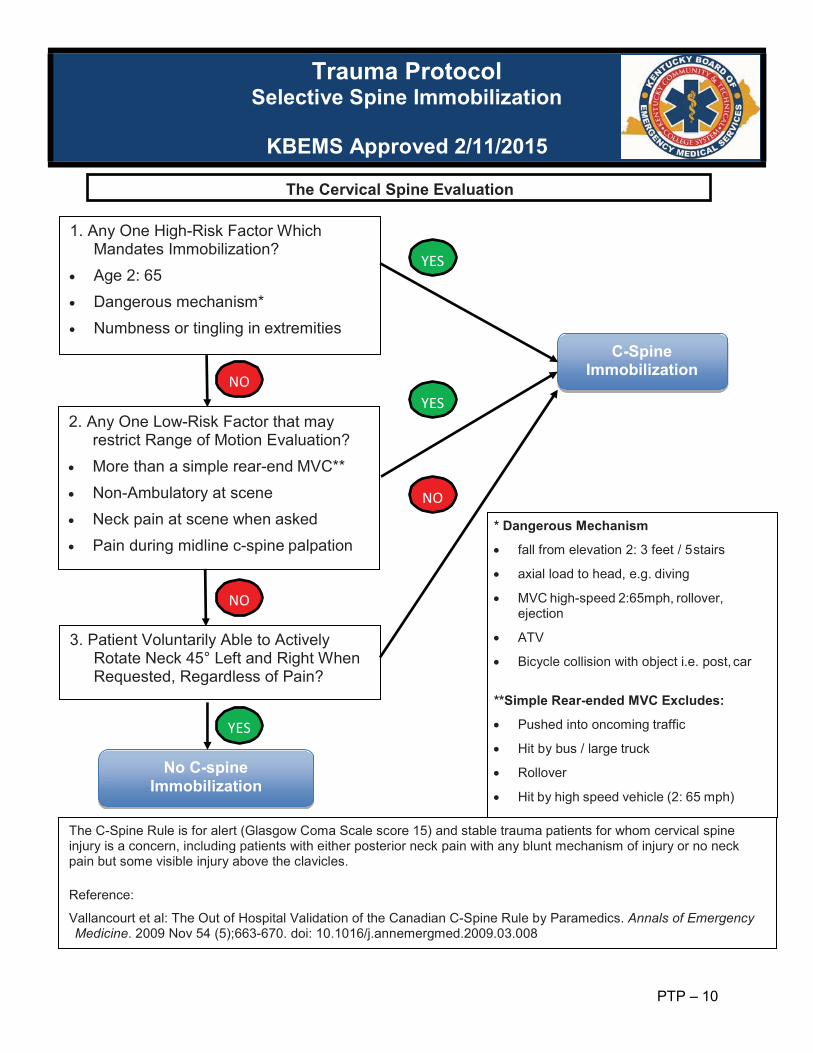
Trauma Protocol Selective Spine Immobilization
KBEMS Approved 2/11/2015
PTP – 10
YES
No C-spine
Immobilization
NO
NO
YES
C-Spine Immobilization
YES
NO
* Dangerous Mechanism
• fall from elevation 2: 3 feet / 5 stairs
• axial load to head, e.g. diving
• MVC high-speed 2:65mph, rollover, ejection
• ATV
• Bicycle collision with object i.e. post, car **Simple Rear-ended MVC Excludes:
• Pushed into oncoming traffic
• Hit by bus / large truck
• Rollover
• Hit by high speed vehicle (2: 65 mph)
3. Patient Voluntarily Able to Actively Rotate Neck 45° Left and Right When Requested, Regardless of Pain?
2. Any One Low-Risk Factor that may restrict Range of Motion Evaluation?
• More than a simple rear-end MVC** • Non-Ambulatory at scene • Neck pain at scene when asked • Pain during midline c-spine palpation
1. Any One High-Risk Factor Which Mandates Immobilization?
• Age 2: 65 • Dangerous mechanism* • Numbness or tingling in extremities
The C-Spine Rule is for alert (Glasgow Coma Scale score 15) and stable trauma patients for whom cervical spine injury is a concern, including patients with either posterior neck pain with any blunt mechanism of injury or no neck pain but some visible injury above the clavicles.
Reference:
Vallancourt et al: The Out of Hospital Validation of the Canadian C-Spine Rule by Paramedics. Annals of Emergency Medicine. 2009 Nov 54 (5);663-670. doi: 10.1016/j.annemergmed.2009.03.008
The Cervical Spine Evaluation

EP
Environmental Protocols

EP – 1
Electrical/Lightning Injury
First and foremost, be certain the scene is safe to approach. A unique feature to multiple patients affected by a lightning strike or electrical discharge is to treat those in respiratory arrest or cardiac arrest FIRST
Basic Standing Orders
► Routine Patient Care. ► High flow oxygen as indicated. ► Consider Burn Protocol. ► Remove rings, watches or constricting bands on affected
extremities. ► Transport. ► Consider 12-lead EKG. ► Request paramedic intercept.
Advanced Standing Orders
► Establish IV/IO access
Paramedic Standing Orders
► Assess airway status. ► Assess need for advanced airway skills. ► Initiate continuous Cardiac Monitoring. ► Monitor patient. ► Consider pain management protocol. ► If injury suggest risk of rhabdomyolysis, consider 50meq
Sodium Bicarbonate in 1000 ml NS wide open. ► Consider Trauma Center if available.
P

EP – 2
Snake Bites
General Considerations Important documentation items include appearance of snake, time of bite, prior first-aid by patient or friends and unusual symptoms such as peculiar or metallic taste sensations. Severe envenomation may result in hypotension, coma, and bleeding. Early systemic signs are a bad prognosticator.
Paramedic Standing Orders
► Consider pain management – see Pain Management Protocol.
Basic Standing Order
► Routine Patient Care. ► Provide oxygen. ► Remove rings or other bands which may become tight with
local swelling. ► Immobilize bitten extremity. ► Minimize venom absorption by keeping bite area still and
patient quiet. Apply lymphatic band 2-3” above bite. (Lymphatic band should be at least 1” wide and allow 2 fingers to be easily inserted under the band.)
► Mark time and extent of erythema and edema with pen. ► Transport promptly for definitive observation and treatment. ► Do not use ice or refrigerants. ► Consider ALS for systemic symptoms or pain control
Advanced Standing Orders
► Establish IV access.
B

EP – 3
Submersion Injuries – Adult and Pediatric Key points: Do not become a victim. Assess scene safety and rescue resources. Routine cervical spine stabilization may impede airway management and is not necessary
unless the circumstances indicate that trauma is likely. Aggressive pulmonary support is essential. If possible, rescue breathing should begin while the patient is in the water. Chest compressions while in the water are ineffective. There is no need to clear the airway of water prior to rescue breathing. Attempts to remove water from the breathing passages by any means other than suction
is not necessary. The Heimlich maneuver should be reserved for patients > 1 year of age with suspected
airway occlusion by a foreign body.
Basic Standing Orders Routine Patient Care. Conscious patients with submersion injuries should be transported to the hospital. Initiate resuscitation as quickly as possible. Follow specific resuscitation protocol including AED. Manage cervical spine based on likelihood of cervical trauma. Obtain specific history: Time Temperature. Consider hypothermia. Associated trauma. Request paramedic intercept
Advanced Standing Orders Routine Patient Care. Conscious patients with submersion injuries should be transported to the hospital. Initiate resuscitation as quickly as possible. Follow specific resuscitation protocol including AED. Manage cervical spine based on likelihood of cervical trauma. Obtain specific history: Time Temperature Consider hypothermia. Associated trauma. Treat for hypotension Consider CPAP if no contraindications Initiate IV/IO access, but do not delay transport. Request paramedic intercept
Paramedic Standing Orders
Follow appropriate ACLS algorithm.
A

Heat Cramps/Heat Exhaustion – Adult and Pediatric
EP – 4
Heat Cramps: Brief, intermittent and other severe muscle cramps associated with copious amounts of sweating with hypotonic fluid replacement. Heat Exhaustion: Water or salt depletion in the face of fluid loss in a hot environment. Symptoms are variable and nonspecific and include weakness, fatigue, headache, impaired judgment, vertigo, nausea and vomiting. Orthostatic hypotension may occur.
Routine Patient Care. Obtain glucose reading via glucometer. Remove victim to a cool area and shield from sun or any external heat source. Monitor vital signs and mental status. Consider active cooling with tepid water mist and fanning the patient Cardiac monitor If alert and oriented, provide commercially available flavored electrolyte solution.
► ► ►
► ► ►
Basic Standing Orders
Advanced Standing Orders
► Consider IV access. ► IV bolus of 0.9% NaCl (normal saline): 250 ml for adults, 20ml/kg
for pediatrics; may repeat if systolic pressure dictates.

Hyperthermia (Environmental) – Adult and Pediatric
EP – 5
Mental status changes in the heat-challenged victim signal the onset of potentially severe heat illness and heat stroke. Mortality and morbidity are directly related to the length of time the victim is subject to the heat stress. Consider pharmacological causes as well.
► Routine Patient Care ► Move victim to a cool area and shield from the sun or any
external heat source. ► Remove as much clothing as is practical and loosen any
restrictive garment remaining. ► If alert and oriented, give small sips of cool liquids. ► Monitor and record vital signs and level of consciousness ► If temperature >104F(40C) or if altered mental status: begin
active cooling by o Continually mist the exposed skin with tepid water while
fanning the victim o Truncal ice packs may be used, but are less effective than
evaporation o If shivering occurs discontinue active cooling and notify
medical control
Basic Standing Orders
► IV access and administer fluids to maintain systolic blood pressure >90 mm Hg (adults) or >minimum for age and signs of adequate perfusion.
► IV bolus of Isotonic fluid: 250 ml for adults, 20ml/kg for pediatrics; may repeat if systolic pressure dictates.
Advanced Standing Orders
► Adults: If uncontrolled shivering occurs during cooling: • Midazolam: 0.05 mg/kg IV max single dose is 5 mg.
Paramedic Standing Orders

Hypothermia (Environmental) – Adult and Pediatric
EP – 6
Basic Standing Orders
All Cold Patients ► Routine Patient Care ► Careful handling is the highest priority ► Prevent further heat loss.
o Insulate from the ground and shield from wind and water. o Remove wet clothing if in shelter. Cut clothing off to avoid
excessive movement. o Cover the head and neck. o Insulate above and below. o Protect from the wind. o Apply insulated heat packs to high heat transfer/loss areas such
as the head, neck, underarms, sides of the chest, and groin. o Cover with a vapor barrier (such as a plastic garbage bag). o Move the patient to a warm environment. o Consider covering patient's mouth and nose with a light surgical
mask to reduce heat loss through respirations. o Chemical heat packs slow cooling but do not rewarm. They are
best used on hands and feet to prevent frostbite. o Obtain temperature (rectal preferred as appropriate).
► Rewarm o If patient is alert enough to swallow, give food and drinks high in
calories. The calories will increase ability to shiver which is most effective field rewarming.
o Exercise drops temperature and then increases it but, this is not as effective as shivering. If dry and fed and shivering, mild exercise is OK.
► Oxygen should be heated and humidified, if possible to a maximum of 108 ° F (42° C).
► Splinting should be performed, when indicated, with caution to prevent additional injuries to frostbitten tissues.
► Treat and transport to a medical facility.
B

Hypothermia (Environmental) – Adult and Pediatric continued
EP – 6
MILD 97°F – 95°F (36.1°C –
35°C) cold sensation, shivering, unable to perform complex tasks with hands
MODERATE 95°F – 93°F (35°C - 33.9°C)
intense shivering, clumsy and uncoordinated, mild confusion, slow and labored movements
93°F –90°F (33.9°C – 32.2°C)
violent shivering, difficulty with speech, sluggish thinking, mild amnesia, may appear drunk
SEVERE 90°F –86°F (32.2°C - 30°C)
shivering stops, unable to walk, incoherent, irrational
<86°F (30°C) progressive stupor to unconsciousness, loss of awareness
<82°F (27.8°C) unconscious, respiration and heartbeat erratic, pulse not palpable, pulmonary edema, cardiac and respiratory arrest, death
Advanced Standing Orders
► IV access and administer fluids to maintain systolic blood pressure >90 mm Hg (adults) or > minimum for age and signs of adequate perfusion.
Paramedic Standing Orders
If core temperature <30°C (86°F) ► CPR if indicated. ► Withhold IV medications. ► Attempt defibrillation once (use 360 joules for monophasic and
120 – 200 joules for biphasic defibrillators). Peds: 2J/kg. If core temperature >30°C (86°F) ► CPR if indicated. ► Give IV medications based on dysrhythmia (but at longer
intervals). ► Repeat defibrillation for ventricular fibrillation/ventricular
tachycardia as core temperature rises.
P

Hypothermia (Environmental) – Adult and Pediatric- Mild
EP – 6
Cold sensation, shivering, unable to perform complex tasks with hands.
► Treat the patient as outlined above. ► If there is no way to get to a medical facility, rewarm the
patient gradually by: o Warm showers or warm bath if the patient is alert. o Placing patient in a sleeping bag and providing contact
with a warm body.
Basic Standing Orders
► Many hypothermic patients may require aggressive fluid resuscitation. The field goal is volume expansion not rewarming.
► Use bolus therapy for volume expansion to endpoint of normalization of vital signs; specifically, heart rate.
► IV's should be heated to patient’s current core temperature or greater. 98-104° F (37-40° C) is ideal.
► The recommended fluid for rehydration is a balanced salt solution, such as normal saline or ringer's lactate.
► Do not use TKO lines in hypothermic patients. Use a saline lock.
Advanced Standing Orders

Hypothermia (Environmental) – Adult and Pediatric - Moderate
EP – 6
Violent shivering, sluggish labored movements, confusion, may appear drunk.
IV THERAPY ► Many hypothermic patients are dehydrated and may
require aggressive fluid resuscitation. The field goal is volume expansion not rewarming.
► Use bolus therapy for volume expansion to endpoint of normalization of vital signs; specifically, heart rate.
► IV's should be heated to patient’s current core temperature or greater. 98-104° F (37-40° C) is ideal.
► The recommended fluid for rehydration is a balanced salt solution, such as normal saline or ringer's lactate.
► Do not use TKO lines in hypothermic patients. Use a saline lock.
Advanced Standing Orders
► Treat the patient as outlined above with the following exceptions: o Do not put patient in shower or bath. o Do not give a patient oral fluid unless he is capable of
swallowing and protecting his/her airway. o Do not attempt to increase heat production through
Basic Standing Orders
Paramedic Standing Orders
MEDICATIONS: ► Indications for medications are the same for mildly hypothermic
patients as they are for normothermic patients. ► In the patient with a core temperature of less than 86°F (30° C)
medications should be withheld. ► Medications are inefficient and poorly metabolized in the
hypothermic patient. In addition, due to delayed metabolism, medications given in normal therapeutic doses to severely hypothermic patients can result in toxicity when the patient is rewarmed.
► As with any person with altered consciousness, Narcan and dextrose should be considered when there is a reasonable suspicion that their use is warranted.
► Sodium bicarbonate is not to be used unless specifically ordered by a physician.

EP – 6
Hypothermia (Environmental) – Adult and Pediatric - Severe
Shivering stops, incoherent progressing to unconscious, erratic respiration and heartbeat, unstable vital signs.
Advanced Standing Orders Airway management as needed Avoid hyperventilation Use heated humidified oxygen if possible (42 C – 46 C or 108 F – 115 F) Rewarming is key to arrest survival in hypothermia. Field techniques are ineffective. The goal is to deliver a viable patient to a facility that can perform effective rewarming (most clinics and hospitals). Use AED as appropriate. If no pulse (after checking for up to 60 seconds) and no respirations and no contraindications, start CPR. Be careful to not hyperventilate.
Paramedic Standing Orders
If resuscitation has been provided in conjunction with rewarming techniques for more than 60 minutes without the return of spontaneous pulse or respiration, contact medical control for recommendations.
Basic Standing Orders
► If victim is not breathing, provide rescue breathing via bag-valve mask.
► Use heated, humidified oxygen if available. B

EP – 7
Frostbite – Adult and Pediatric
Management ► Concerns:
► Do not rub the frozen part. ► Do not allow the patient to have alcohol or tobacco. ► Do not apply ice or snow. ► Do not attempt to thaw the frostbitten part in cold water. ► Do not attempt to thaw the frostbitten part with high temperatures such as
those generated by stoves, exhaust, etc. ► Do not break blisters which may form.
Basic Standing Orders
► Treatment of deep frostbite is usually extremely painful and best accomplished in a medical facility. In most circumstances, the risks posed by improper rewarming or refreezing outweigh the risks of delaying treatment for deep frostbite.
► If transporting a patient with frostbite that will not be rewarmed in the field, the medical provider should protect the frostbitten parts from additional injury and temperature changes.
► Protect the rewarmed area from refreezing and other trauma during transport. A frame around the frostbitten area should be constructed to prevent blankets from pressing directly on the injured area.
► Do not allow an individual who has frostbitten feet to walk except when the life of the patient or rescuer is in danger. Once frostbitten feet are rewarmed, the patient becomes nonambulatory.
► Shock due to frostbite is very uncommon. However, medical personnel should always be alert for shock and begin treatment at the earliest sign it is developing. If the frostbite patient develops shock, personnel should perform a thorough examination for additional injuries.
B
AVM
Airway and Ventilation Management

NOTE: This is a restricted procedure. A service and paramedic will require specific authorization from the Board prior to utilizing this procedure and skill.
AVM – 1
Universal Advanced Airway Management Algorithm
FAILED AIRWAY ALGORITHM
Yes
>2 Attempts at Intubation by EMS Provider?
SpO2>90% with BVM using
100% and Oral or Nasal Airway?
Yes POST INTUBATION CARE
Oral Intubation without Meds
Successful Intubation?
Intubation Attempt
DAI/RSI Protocols
UNIVERSAL ADVANCED AIRWAY MANAGEMENT ALGORITHM
Unresponsive or Near Death? Yes
CRASH AIRWAY ALGORITHM
No
Patient Needs
Yes DIFFICULT AIRWAY ALGORITHM Difficult Intubation Predicted?
No
No
No
No
Yes
Yes

NOTE: This is a restricted procedure. A service and paramedic will require specific authorization from the Board prior to utilizing this procedure and skill.
AVM – 2
Crash Airway Algorithm
No
No
CRASH AIRWAY ALGORITHM
Attempt Intubation Push Succinylcholine
Repeat Intubation Attempt
Intubation Successful?
>3 Attempts at Intubation?
Patient Completely Flaccid/Relaxed?
Intubation Successful?
*Improve Bag Valve Mask Ventilation success with the following: Two-person BVM technique Nasal or Oral Airway Jaw Thrust Repositioning Head
FAILED AIRWAY ALGORITHM Maintaining SpO2 >90%
using BVM with Oral or Nasal Airway?
Trial of Oxygenation with BVM Ventilation*
SpO2 >90% Spontaneously?
Yes
No
Yes
No No
POST INTUBATION CARE
No
No
No
Yes
Yes
Yes
Yes

Difficult Airway Algorithm
AVM – 3
Rescue Airway If No Gag Reflex
SpO2>90%?
BVM with Oral/Nasal Airway
DIFFICULT AIRWAY ALGORITHM
CALL FOR ADDITIONAL HELP
No Yes
FAILED AIRWAY ALGORITHM
MAINTAIN SPONTANEOUS VENTILATION
No DAI/RSI Awake oral or BNTI
Patient Needs Intubation
Difficult Intubation Predicted?
Yes
POST INTUBATION CARE
Intubation Successful?
Transport for Definitive
Airway Management Early ED Notification
SpO2>90%
Oral Intubation Without Meds or DAI/RSI Protocols
Yes
No
Yes
No
No

Failed Airway Algorithm
AVM – 4
FAILED AIRWAY ALGORITHM
Patient meets Failed Airway Criteria Call for Additional Help
BVM with Oral or Nasal Airway And 100% Oxygen
Gag Reflex Present?
Continue BVM with Oral or Nasal Airway and 100% Oxygen
Yes
Yes
No
No
SpO2>90%?
SpO2>90%?
Transport ASAP
Notify ED of Need for Definitive Airway Management
Emergency Cricothyrotomy:
Adult: Percutaneous Kit Pediatric: Percutaneous Needle
SpO2>90%?
Rescue Airway with BVM
And 100% Oxygen
Attempt Rescue Airway Insertion and Ventilation
Yes
No
No
Yes

Airway Management - Pediatric
AVM – 5
PEDIATRIC AIRWAY MANAGEMENT I. Initial Assessment: Using Pediatric Assessment Triangle / Rapid
Cardiopulmonary Assessment
A. Appearance 1. Alertness 2. Distractibility 3. Consolability 4. Eye Contact
5. Speech/Cry 6. Spontaneous Motor
Activity 7. Color
B. Work of Breathing 1. Appearance 2. Use of Accessory Muscles
a. Retractions b. Diaphragmatic
3. Tidal Volume (chest rise)
4. Other Signs of Distress: a. Nasal Flaring b. Grunting Breathing c. Cyanosis
C. Circulation to the Skin
1. Strength of Pulses (central vs. peripheral) 2. Color / Temperature of Extremities 3. Capillary Refill Time 4. Blood Pressure
II. Initial Assessment Indicates Spontaneous Breathing Without Compromise.
A. Monitor breathing during transport. B. Administer oxygen
1. Infants via infant mask @ 10-12 L/min. 2. Small child (1-8 y/o) via pediatric mask @ 12-15 L/min. 3. If mask is not tolerated, administer by blow-by method.
III. Initial Assessment Indicates Spontaneous Breathing With Respiratory Distress
A. Maintain airway with manual maneuvers. B. Suction as needed. C. Administer oxygen (II. B above). D. If unable to maintain an airway, insert an oral or nasal airway. E. Assist ventilations with BVM as needed. F. Monitor with EKG and pulse oximetry as soon as possible and
capnography if available.

Airway Management - Pediatric
AVM – 5
IV. Initial Assessment Indicates Breathing is Absent or Severe Respiratory Distress
A. Maintain airway with manual maneuvers. B. Suction as needed. C. Insert an oral or nasal airway. D. Ventilate with BVM and 100% oxygen @ 20/min for a child and 30/ min for
an infant. E. Monitor EKG and pulse oximetry as soon as possible and capnography if
available. F. Establish IV or IO vascular access. G. Consider need for endotracheal intubation.
V. Continued BVM Ventilation vs. Endotracheal Intubation
A. BVM with oral and or nasal airway should be the initial technique used for ventilatory support .
B. Endotracheal intubation should be used when BVM ventilation is ineffective or transport time is prolonged.
C. Continued ventilation with BVM with oral/nasal airways can provide acceptable ventilation and oxygenation in the pediatric patient.
D. Pre-hospital pediatric intubation is a controversial skill: 1. Specific pediatric training and ongoing continuing education is required. 2. Proven to be a high risk / low frequency event. 3. Hypoxia /hypoventilation are risks during intubation attempts. 4. At present, no documented outcome benefit when compared to continued
BVM. VI. Airway Management with Bag Valve Mask (BVM) Ventilation
A. Purpose: 1. BVM ventilation is the preferred technique for providing rescue breathing for
pediatric patients with inadequate respiratory effort or cardio-respiratory arrest. Patients who are in respiratory distress and failure may respond to BVM ventilation and not require pre-hospital endotracheal intubation.
2. BVM may also be used to administer bronchodilators in patients with bronchospastic airway disease.
B. Indications: 1. Inadequate respiratory rate:
a. Adolescent: < 12/min b. Child: < 16/min c. Infant / Toddler: < 20 / min
2. Inadequate respiratory effort: a. Absent or diminished breath sounds. b. Paradoxical breathing (chest and abdomen moving in opposite
directions).

Airway Management - Pediatric
AVM – 5
c. Persistent cyanosis on 100% oxygen by non-rebreather mask.3. Symptomatic bradycardia:
a. Child: HR < 80 / minb. Infant: HR < 100 / min
4. Cardiac Arrest5. Altered Mental Status with GSC < 9
C. Contraindications: None
D. Adverse Effects / Complications
1. Gastric distension.2. Vomiting.3. Increased ICP or vagal reflex bradycardia if pressure is applied by mask over
the patient‘s eyes.
E. Procedure:1. Have suction available since vomiting may occur.2. Use an appropriately sized oral or nasal airway adjunct with BVM ventilation.3. Use an appropriate sized mask for best fit and to avoid pressure over the
eyes.4. For the single provider, use the "E-C clamp" technique of holding the mask.5. Monitor EKG, pulse oximetry and capnography if available.6. Ventilate with 100% oxygen with a tidal volume of approximately 6-10 cc/kg or
with just enough volume to see the chest rise.7. Rate of ventilation should be approximately 20/ min for a child and 30/ min for
an infant. If capnography is available, ventilate to maintain the end tidal CO2 at 35-40 mmHg. If head injury and signs of herniation are present, increase ventilation to maintain an end tidal CO2 of 30 mmHg.
8. If the patient does not have an adequate chest rise with BVM ventilations:a. Assure the airway is open and clear.b. Use a two hand jaw lift technique.c. Use an oral and nasal airway.d. Increase the volume of ventilation if the airway is clearly open and
maintained.e. Evaluate for gastric distension and the need for decompression with an
orogastric tube.f. Consider the need for endotracheal intubation if BVM is unsuccessful and
skilled personnel are available.
VII. Pediatric Orotracheal Intubation
A. Purpose:1. Oral endotracheal intubation involves the passage of an endotracheal tube
under direct vision via the oral cavity through the larynx and into the tracheato provide direct maximum ventilatory support of the patient.

Airway Management - Pediatric
AVM – 5
B. Indications:1. Cardiac arrest.2. Severe respiratory distress, patient without a gag reflex.3. Coma, patient without a gag reflex.4. Patient is extremis, severe respiratory distress with poor air exchange, or
agonal respirations.5. Unsuccessful airway management with BVM and oral/nasal airways.
C. Contraindications:1. Lack of equipment.2. Lack of skilled personnel.3. Successful BVM ventilation with a short transport time.
D. Adverse Effects / Complications:1. Unrecognized esophageal intubation.2. Prolonged hypoxia and hypoventilation during intubation attempts.3. Trauma to oropharynx, vocal cords, esophagus, or trachea.4. Right mainstem bronchus intubation.5. Vomiting and pulmonary aspiration.6. Increased intracranial pressure due to vagal stimulation.7. Pneumothorax or tension pneumothorax due to excessive ventilatory
pressures.
E. Procedure:1. Complete a "primary survey" and assure A-B-C‘s with basic life support skills
a. Oxygenate with 100% oxygen with non-rebreather mask.b. Ventilate with BVM if needed.c. Monitor EKG, pulse oximetry and continuous capnography if available.d. Manual cervical spine motion restriction if trauma mechanism.
2. Prepare equipment:a. BVMb. Suctionc. Working, appropriate sized laryngoscope (see chart for equipment sizes).d. Endotracheal tubes (ETT) and stylet
i. ETT size: (4 + Age/4).ii. Check chart or age based resuscitation tape.iii. ETT depth in cm : ( 12 + Age/2) or (ETT size X 3).iv. Place the lubricated stylet into the ETT and bend the distal tip into a
gentle curve.v. Assure that the tip of the stylet does not extend out the end of the
ETT.vi. Have the next half-size smaller ETT at hand.
e. Oral and nasal airways.f. Pediatric LMA‘s if available (see chart in LMA protocols).
3. Oxygenate and ventilate with BVM prior to laryngoscopy.4. Maintain cervical immobilization in trauma patients.

Airway Management - Pediatric
AVM – 5
5. Have an assistant apply cricoid pressure. 6. Insert laryngoscope into the right corner of the mouth, sweep tongue upward
and to the left by using a lifting motion, not a prying motion. 7. Identify the epiglottis. 8. Elevate the epiglottis exposing the glottic opening.
a. In infants and toddlers, the straight blade may be successfully used to place into the vallecula and elevate the epiglottis indirectly by lifting the base of the tongue. The shape and position of the infant/toddler epiglottis makes it more difficult to directly pick-up with the straight laryngoscope blade.
9. Holding the ETT like a dart, place the tube through the vocal cords and into the trachea under direct vision and insert approximately 2 cm below the cords. a. It is generally recommended to use an un-cuffed ETT in children < 8 y/o.
However, this is controversial in the current literature. A cuffed ETT may be used in the child < 8 y/o but extreme care must be used to assure that the cuff remains completely deflated unless a large air leak is detected.
10. Remove the laryngoscope and hold the ETT in place. 11. Attach the BVM and ventilate with 100% oxygen. 12. Confirmation of correct ETT placement: No single method of ETT confirmation
is 100% reliable, the position of the ETT must be assessed to be properly in the trachea by all means available to the pre-hospital EMS provider. The following methods may be used to confirm the correct placement of the ETT. a. Visualization of the ETT passing through the cords and into the trachea b. Auscultation of all lung fields to confirm adequate air exchange c. Auscultation of the epigastrium to confirm the absence of disturbance of
the gastric fluids during ventilation d. Observation of bilateral expansion of the thorax e. End tidal CO2 detection device:
i. At a minimum the colorimetric devices assessed initially and after six breaths.
ii. Capnometry devices that give a numeric end tidal CO2 reading. iii. The preferred device is continuous capnography that is downloadable
and printable. f. Esophageal intubation detector device.
i. Useful if child > 8 y/o g. Other clinical signs of improved perfusion and ventilation/oxygenation
i. Stable heart rate. ii. Pupillary response. iii. Stable and rising oxygen saturation. iv. Improved skin color.
13. Once correct ETT placement is confirmed, the ETT must be secured. a. Commercial device with built in bite block. b. Oral airway with tape. Tape ETT to the maxilla, not the mandible. c. Minimize head and neck movement with the use of a cervical collar,
cervical spine immobilization device and spine board. d. Note and document depth of ETT placement.
i. 3-5 cm of ETT movement may occur with neck flexion or extension.

Airway Management - Pediatric
AVM – 5
14. Ventilation:
a. Use care to avoid hyperventilation. b. Tidal volume of 6-10 cc/kg or just enough ventilation to see the chest rise. c. Use a rate of 20/min for a child and 30/min for an infant or toddler. d. If continuous end tidal CO2 monitoring is available, maintain an ETCO2 of
35-40 mmHg (30 mmHg in cases of head injury with signs of herniation). 15. Re-confirm correct ETT position during on-going assessments. At a minimum
reconfirmation should occur: a. Anytime patient is moved. b. Anytime dislodgement is suspected. c. Anytime care is transferred to another provider.
16. Documentation a. Full report to Emergency Department Physician.
i. Specifically report any intubation difficulties or airway management problems.
b. Complete supplemental NAEMSP Airway Form as well as the run report.
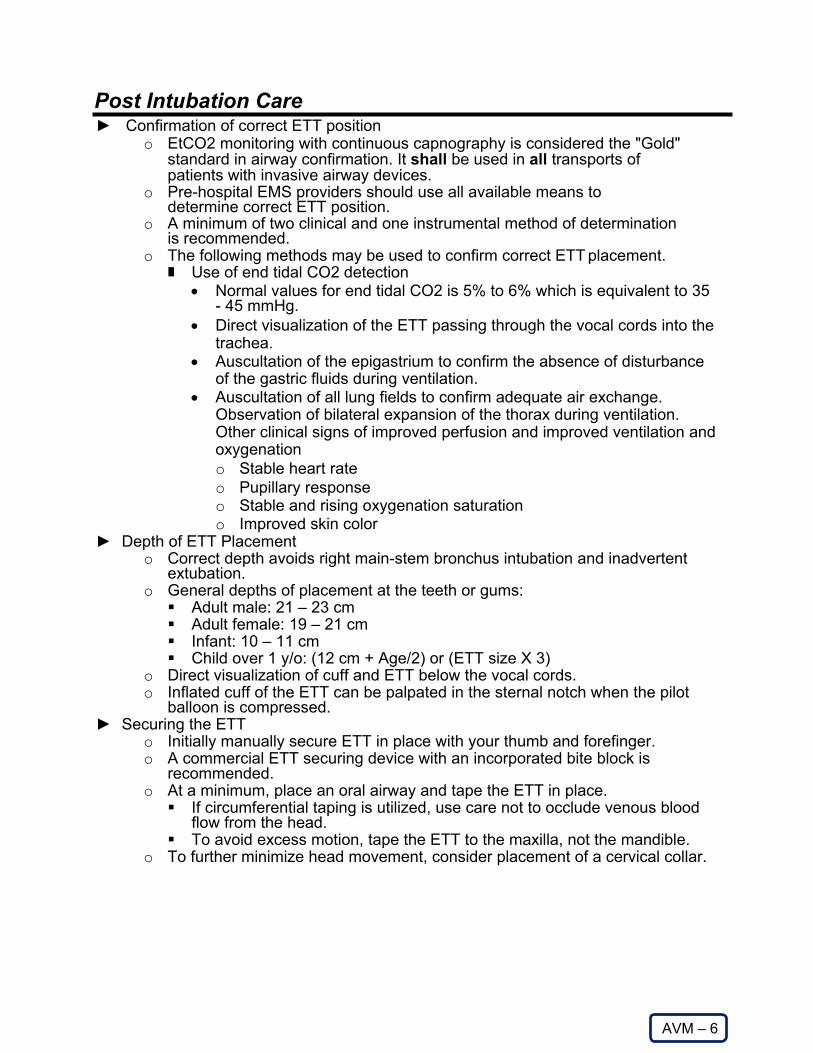
Post Intubation Care
AVM – 6
► Confirmation of correct ETT position o EtCO2 monitoring with continuous capnography is considered the "Gold"
standard in airway confirmation. It shall be used in all transports of patients with invasive airway devices.
o Pre-hospital EMS providers should use all available means to determine correct ETT position.
o A minimum of two clinical and one instrumental method of determination is recommended.
o The following methods may be used to confirm correct ETT placement. Use of end tidal CO2 detection
• Normal values for end tidal CO2 is 5% to 6% which is equivalent to 35 - 45 mmHg.
• Direct visualization of the ETT passing through the vocal cords into the trachea.
• Auscultation of the epigastrium to confirm the absence of disturbance of the gastric fluids during ventilation.
• Auscultation of all lung fields to confirm adequate air exchange. Observation of bilateral expansion of the thorax during ventilation. Other clinical signs of improved perfusion and improved ventilation and oxygenation o Stable heart rate o Pupillary response o Stable and rising oxygenation saturation o Improved skin color
► Depth of ETT Placement o Correct depth avoids right main-stem bronchus intubation and inadvertent
extubation. o General depths of placement at the teeth or gums: Adult male: 21 – 23 cm Adult female: 19 – 21 cm Infant: 10 – 11 cm Child over 1 y/o: (12 cm + Age/2) or (ETT size X 3)
o Direct visualization of cuff and ETT below the vocal cords. o Inflated cuff of the ETT can be palpated in the sternal notch when the pilot
balloon is compressed. ► Securing the ETT
o Initially manually secure ETT in place with your thumb and forefinger. o A commercial ETT securing device with an incorporated bite block is
recommended. o At a minimum, place an oral airway and tape the ETT in place. If circumferential taping is utilized, use care not to occlude venous blood
flow from the head. To avoid excess motion, tape the ETT to the maxilla, not the mandible.
o To further minimize head movement, consider placement of a cervical collar.

Post Intubation Care continued
AVM – 6
dfsd ► Following the securing of the ETT, note and document the depth of ETT placement. ► Ventilation
o With an ETT and 100% oxygen, large tidal volumes and hyperventilation are not necessary and have been shown in recent studies to be detrimental to patient outcome.
o Use care to avoid hyperventilation, place patient on mechanical ventilator if available.
o Ventilate with a tidal volume of approximately 6 – 8 mL/kg or clinically, just enough volume to see the chest rise with each administered breath.
o Rate of ventilation: Initially
• Adult: 10 – 12/min • Child: 20/min • Infant/Toddler: 30/min
Then, titrate rate using EtCO2 monitoring to maintain an EtCO2 of 35 – 45 mmHg. • If suspected acidosis (DKA, Aspirin overdose, etc.) maintain EtCO2 &
Respiratory rate post intubation to levels prior to intubation. Do not attempt to normalize EtCO2 readings.
• If the patient had a head injury and signs of herniation, titrate respiratory rate to achieve an EtCO2 of approximately 30 mmHg.
► Patient Positioning o As soon as possible, once patient is secured to EMS stretcher, position head of
cot at least 30 degrees to allow better expansion of lungs and ventilator compliance.
► Maintenance of Analgesia, sedation, and/or neuromuscular blockade o Purpose: To provide additional analgesia, sedation and/or neuromuscular
blockade in order to maintain ETT placement to facilitate continued oxygenation and ventilation in an intubated patient.
o Indications Patients awakening from medications used for drug assisted intubation. Comatose patients recovering from paralytic drugs used for rapid sequence
intubation. Patients initially intubated without pharmacological assistance but are now
recovering due to improved oxygenation and ventilation.

Post Intubation Care continued
AVM – 6
dsd o Medications: All should be administered IV or IO and in 50% reduced doses in
the elderly (Age 70+), debilitated or unstable patient. Analgesia
• Appropriate pain control will facilitate ventilation and oxygenation and allow the use of smaller doses of sedatives and may avoid the need for pharmacological paralysis with neuromuscular blockers.
• Narcotics are the primary medications used for analgesia in intubated patients. o Fentanyl Potent analgesic No histamine release as compared to morphine Large doses can rarely cause chest wall rigidity requiring
neuromuscular blockade. Rapid onset (5 min) and short duration (15 min) Dosage: Adult and Peds: 1 – 3 mcg/kg IV, may repeat every 20 min
PRN If giving IV, administer over 1 – 2 min.
o Ketamine Dissociative agent that provides both analgesia & sedation. Does not suppress respiratory function. Rapid push may result in laryngospasm. Rapid onset (30 – 45 sec) Duration of 10 – 15 min Dosage: 1 – 2 mg/kg IV/IO
Sedation • Benzodiazepines are the most commonly used medications for continued
sedation. • Single dose Etomidate (amidate) for initial intubation is safe. Repeat
doses should be avoided due to inhibition of endogenous steroid synthesis. o Midazolam (Versed): Adult and Ped dose: 0.05 – 0.1 mg/kg IV (Max single dose 5 mg)
repeat every 5 min PRN.
Neuromuscular Blocking Medications (NMB’s) (Paralytics)
• The non-depolarizing class of NMB’s are preferred for maintenance of paralysis in the RSI patient or for the initial paralysis of the successfully intubated patient with DAI meds or in patients intubated without pharmacologic assistance. In the RSI patient, additional NMB will have to administered quickly as the effects of the initial succinylcholine will begin to wear off in approximately 5 minutes.
Caution • Maintain adequate analgesia and sedation when administering
paralytics. • Continuously monitor oxygenation, ventilation, and ETT position
when administering paralytics, analgesia, and sedation.

Post Intubation Care continued
AVM – 6
► Reconfirming ETT Position
o Anytime patient is moved. o Any changes in patient condition. o Anytime dislodgement is suspected. o Anytime care is transferred to another provider. o Perform the initial ETT confirmation steps. o Repeat Laryngoscopy and directly confirm ETT position in the trachea if there is
any question of correct position or any of the other confirmatory tests are equivocal.
► Documentation o Full report to the receiving physician or designated staff Specifically report any difficulties or complications related to airway
management. Include times and dosages of all medications given.
o Complete the run report. o Complete the NAEMSP airway form and forward to the medical director.

Mechanical Ventilation
AVM – 7
Mechanical Ventilation – Automatic Transport Ventilator Only NOTE: This is an advanced procedure. Agencies utilizing ventilators for interfacility transfers should maintain documentation of device-specific training, competency, and medical director approval for each participating paramedic.
► Maintain oxygen saturation of greater than or equal to 92% o Attempt to maintain SpO2 between 92% and 99%.
► Attached cardiac monitor, and end-tidal CO2 Monitor. ► Assess and record vital signs, to include temperature, prior to transfer and every
5 minutes en-route. ► Reassess patient frequently during transport and document findings. ► Collect all transfer documentation:
o Transfer Sheet, o EKG o Labs o Other pertinent information
► Review Arterial blood gas prior to transport. ► Document ventilator settings and patient response. ► Document correct tracheal tube placement and secure appropriately. ► Maintain adequate analgesia.
o Fentanyl 1.0 – 3.0 mcg/kg slow IV push; duration of action 30 – 60 minutes
o Ketamine 2 mg/kg, Slow IV push, Duration of action 15 minutes. ► Maintain adequate sedation
o Inadequate sedation may present as an unexplained increase in heart rate or blood pressure; the non-paralyzed patient may also demonstrate agitation, anxiety and/or restlessness.
o Midazolam 0.035 mg/kg IV over 3 – 5 minutes

CPAP/BIPAP USE
AVM – 8
Indications: ► Adult Patient. ► Conscious patient in severe respiratory distress due to suspected pulmonary
edema, COPD or burn inhalation injuries. ► Shortness of breath with pulse oximetry < 92% on high-flow oxygen via NRB
mask. Contraindications: ► Suspected Pneumothorax. ► Inability to maintain own airway. ► Altered mental status. ► Agitated or Combative behavior. ► Facial trauma or burns. System Requirements: ► Prehospital CPAP/ BiPAP equipment that meets DOH requirements. Procedure: ► Assess patient and initiate high flow oxygen as indicated. ► Monitor pulse oximetry.1 ► Apply CPAP/ BiPAP if oxygen saturation < 92% on high flow oxygen via NRB
mask. o Connect CPAP/BiPAP device to suitable oxygen supply. o Attach breathing circuit to CPAP/BiPAP device and ensure device is
functioning properly. o Apply and secure appropriate size breathing circuit mask to patient. o Titrate positive airway pressure up until improvement in patient pulse
oximetry and symptoms. WARNING: Do not exceed pressures of 10 cm H2O.
► Reassess the patient. ► Follow CHF or Asthma protocols if appropriate.2,3 ► Transport ► Contact Medical control. 4
1Pulse oximetry should be monitored continuously during use of CPAP/BiPAP 2If appropriate, nebulized bronchodilators may be administered during PAP ventilation via a side port. 3When appropriate, nitroglycerine should be administered by tablets rather than spray when a patient is receiving PAP ventilation. 4Advise the receiving ED of CPAP use as soon as possible. Many EDs do not have CPAP within the ED and may need to obtain it from within the hospital.

AVM – 9
Endotracheal Tube Introducer (Bougie)
Indication: ► Patient meets clinical indications for oral intubation
Contraindication: ► Three attempts at orotracheal intubation ► Age less than 8-year-old or ETT size less than 6.5 mm Procedure: 1. Prepare, position and oxygenate the patient with 100% oxygen. 2. Select proper ET Tube without stylette, test cuff, and prepare suction. 3. Lubricate the distal end and cuff of the endotracheal tube (ETT) and the distal 1/2
of the Endotracheal Tube Introducer (Bougie) • NOTE: Failure to lubricate the Bougie and the ETT may result in being
unable to pass the ETT) 4. Load the ETT on the proximal end of the Bougie and manipulate the Bougie to be
able to rotate the distal tip of the Bougie left and right as well as manipulate it anteriorly and posteriorly.
5. Using Laryngoscopic techniques, visualize the vocal cords if possible 6. Introduce the Bougie with the coude tip anteriorly and visualize the tip passing
the vocal cords or above the arytenoids if the cords cannot be visualized. 7. Once inserted, gently advance the Bougie until you meet resistance or "Hold- up"
• If you do not meet resistance you have a likely esophageal intubation and insertion should be re-attempted or implement the failed airway protocol as indicated.
8. While maintaining a firm grasp on the proximal Bougie, introduce the ETT over the Bougie passing the tube to its appropriate depth.
9. If you are unable to advance the ETT into the trachea and the Bougie and ETT are adequately lubricated, withdraw the ETT slightly and rotate the ETT 90 degrees COUNTER clockwise to turn the bevel of the ETT posteriorly. If this technique fails to facilitate passing of the ETT you may attempt direct laryngoscopy while advancing the ETT (This will require an assistant to maintain the position of the Bougie and, if available, advance the ETT)
10. Once the ETT is correctly placed, hold the ETT securely and remove the Bougie. 11. Confirm tracheal placement per intubation protocol utilizing continuous waveform
capnography and secure the device. 12. Continue providing high quality care, frequently assessing ETT placement.
Document usage of Bougie device and whether skill was successful.

AVM – 10
King LT – D
Indications: ► Apneic patient when endotracheal intubation is not possible or not available.
Contraindications: ► Intact gag reflex. ► Known esophageal disease such as cancer. ► Caustic ingestion.
Procedure: ► Choose correct size:
Size Height Weight Cuff Volume (ml)
2 35-40 inches 12-25 kg 25-35
2.5 41-51 inches 25-35 kg 30-40
3 4-5 feet N/A 45-60
4 5-6 feet N/A 60-80
5 6 feet N/A 70-90
► Prepare King LT-D ► Test cuffs for leaks (see volume above). ► Lubricate device with water-soluble lubricant. ► Preoxygenate and hyperventilate the patient, if time permits. ► Grasp the patient's tongue and jaw with your gloved hand and pull forward. ► With the King LT-D rotated laterally at 45-90 degrees such that the blue
orientation line is touching the corner of the mouth, introduce tip into mouth and advance behind base of tongue.
► As tube tip passes under tongue, rotate tube back to midline (blue orientation line faces chin).
► Advance tube until base of connector is aligned with teeth or gums. ► Inflate cuffs to appropriate volume as listed above. ► Connect the King LT-D to a bag-valve device and ventilate the patient. If there is
resistance to ventilation slowly withdraw tube until resistance to ventilation is relieved.
► Assess for adequate placement by auscultation (equal breath sounds over the chest and lack of sounds over the epigastrium with bagging), symmetrical chest wall rise and at least one additional method: colorimetric end-tidal CO2 detector, capnography, or esophageal tube detector (note: this device should be used prior to ventilation to be accurate). This should be repeated often, especially after movement of the patient.
► Secure the device.

AVM – 11
i-gel Supraglottic Airway Device
Indications:
► Inability to place ETT for airway management. ► Alternative method for airway management. Contraindications: ► Intact gag reflex ► Trismus, or other limited ability to open the jaw ► Conscious or semi-conscious patient ► Use caution in patients with any condition which may increase the risk of a full
stomach... hiatus hernia, sepsis, morbid obesity, pregnancy or a history of upper gastro-intestinal surgery etc.
Procedure: Prep: Grasp the i-gel along the integral bite block and lubricate the back, sides and front of the cuff with a thin layer of lubricant. After lubrication has been completed, ensure that no BOLUS of lubricant remains in the bowl of the cuff or elsewhere on the device. Avoid touching the cuff of the i-gel with your hands. 1. Grasp the lubricated i-gel firmly along the integral bite block. 2. Position the device so that the i-gel cuff outlet is facing towards the chin of the patient 3. The patient should be in the ‘sniffing’ position with head extended and neck flexed.
The chin should be gently pressed down before proceeding to insert the i-gel. 4. Introduce the leading soft tip into the mouth of the patient in a direction towards the
hard palate. 5. Glide the device downwards and backwards along the hard palate with a continuous
but gentle push until a definitive resistance is felt. 6. Connect the i-gel to a bag-valve device and ventilate the patient. 7. Assess for adequate placement by auscultation (equal breath sounds over the chest
and lack of sounds over the epigastrium with bagging), symmetrical chest wall rise and capnography. This should be repeated often, especially after movement of the patient.
8. Secure the device. Use the supplied strap, if applicable, or use twill tape from maxilla to maxilla.
i-gel size
Patient size Patient weight guidance
(kg) Recommended
Gastric Tube Size
1 Neonate 2-5 N/a
1.5 Infant 5-12 10 Fr
2 Small pediatric 10-25 12 Fr
2.5 Large pediatric 25-35 12 Fr
3 Small adult 30-60 12 Fr
4 Medium adult 50-90 12 Fr 5 Large adult+ 90+ 14 Fr

Digital Intubation
AVM – 12
Indications: ► Inability to intubate via direct visualization or via BNTI. ► Patient should be either unresponsive or extremely cooperative. Contraindications: ► Patient condition that may result in biting of paramedic fingers. ► Caution: Human bite wounds can result in life/extremity threatening infections.
Use caution with this procedure. Consider the option of the RSI protocol. Procedure: ► Pre-oxygenate with 100% oxygen by NRB for 3-5 minutes ► Assemble equipment
o ETT'S, BVM, Stylet, Bite block, Suction, Syringe, Securing device. ► Monitor Patient
o EKG, B/P, SpO2, prepare ETCO2 detector device. ► Maintain manual cervical spine motion restriction if trauma is suspected. The
front of the cervical collar may be removed as long as manual immobilization is maintained.
► Lubricate stylet and place into the ETT with the distal tip bent into a "J" shape. ► Stand or kneel facing the patient. ► Place a bite block to prevent injury to the fingers. ► Using the index and middle fingers of the non-dominate hand, pull forward on the
tongue and jaw and walk down the tongue to palpate the epiglottis with the middle finger.
► Insert the lubricated ETT/stylet at the corner of the mouth on the side of the dominant hand.
► Advance the tube over the tongue, between the fingers, over the epiglottis and into the trachea. The index finger can be used to steer the tube into the glottis.
► The index finger keeps the ETT against the epiglottis. ► As the ETT is advanced toward the glottis lift the middle finger and press the tube
anteriorly. ► Spontaneous air movement will also guide the tube placement. ► Remove the stylet, inflate the cuff, ventilate and verify ETT position. ► Secure the ETT in place. Consider the use of a cervical collar to minimize head
movement. ► Re-check ETT position after each patient movement and upon transfer of care to
receiving hospital or other care provider.
References
1. Stewart, C. Advanced Airway Management. Brady. 2002. Chapt.6. Pg. 104-105.

Blind Nasal Endotracheal Intubation
AVM – 13
Indications: ► Adult spontaneously breathing patient requiring intubation. Contraindications: ► Apnea ► Severe maxillofacial injuries ► Abnormal pharyngeal/supraglottic anatomy (mass, abscess, etc). Procedure: ► Preoxygenate with 100% oxygen by NRB mask. ► Obtain secure IV access. ► Consider sedation (See Sedation/Analgesia Protocol) but avoid respiratory
depression. ► For the awake patient consider applying Lidocaine gel to the ETT and nasal
airways and spray the nasopharynx with Lidocaine or Cetacaine spray. ► Assemble equipment
o ETT (0.5-1.0 mm size smaller than for an oral intubation), BVM, Suction, Syringe, Securing device, Laryngoscopes and Rescue Airway Devices.
o An Endotrol ETT is often useful. ► Monitor the patient with EKG, B/P, Sp02, and prepare ETC02 detector device. ► Maintain manual cervical spine motion restriction if trauma is suspected. ► Apply a vasoconstrictor spray to both nares. Lubricate and insert progressively
larger sizes of nasal air ways to dilate the nasal passage. ► Gently insert a lubricated ETT and pass the ETT using steady, firm pressure. ► While advancing the ETT, use a jaw thrust or chin lift maneuver to elevate the
epiglottis. This may be performed by an assistant. Listen for continuous breath sounds coming through the ETT. Try to close the mouth with a gloved hand and occlude the opposite nare allowing for maximal breath sounds through the ETT.
► Apply cricoid pressure to minimize risk of regurgitation and aspiration and to manipulate larynx to obtain maximum breath sounds.
► Just proximal to the glottis, the breath sounds will become louder. Advance the ETT with inspiration.
► Inflate the ETT cuff, ventilate, and verify correct ETT position by two clinical methods and presence of ET C02. (See oral intubation protocol)
► Secure the ETT in place (approximately 26 cm in females, 27 cm for males at nares).
► Consider the use of a cervical collar to limit head movement. ► Re-check ETT position with each patient movement ► Assist ventilations with a BVM or use a mechanical ventilator (if approved). References:
1. Stewart, C. Advanced Airway Management. Brady. 2002 Chap. 6. Pg. 91-98. 2. Advanced Trauma Life Support Text. 7 th Edition. 2004. American College of
Surgeons. Pg. 57-58.
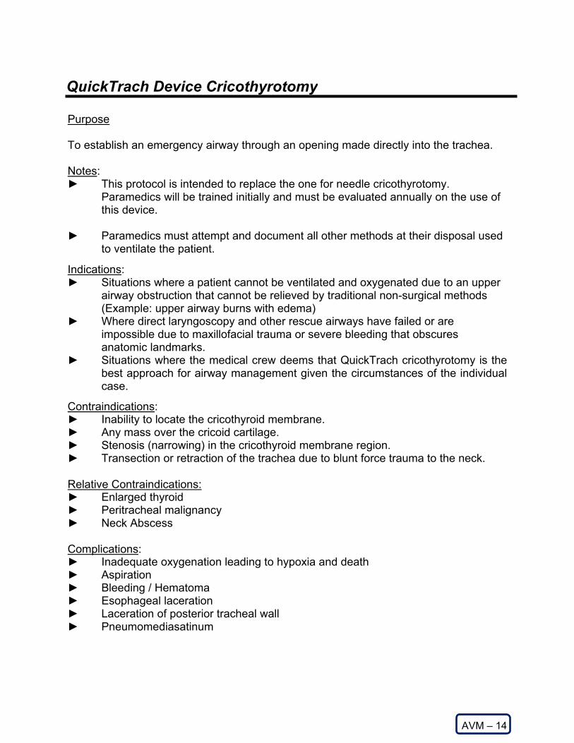
QuickTrach Device Cricothyrotomy
AVM – 14
Purpose
To establish an emergency airway through an opening made directly into the trachea.
Notes: ► This protocol is intended to replace the one for needle cricothyrotomy.
Paramedics will be trained initially and must be evaluated annually on the use of this device.
► Paramedics must attempt and document all other methods at their disposal used
to ventilate the patient.
Indications: ► Situations where a patient cannot be ventilated and oxygenated due to an upper
airway obstruction that cannot be relieved by traditional non-surgical methods (Example: upper airway burns with edema)
► Where direct laryngoscopy and other rescue airways have failed or are impossible due to maxillofacial trauma or severe bleeding that obscures anatomic landmarks.
► Situations where the medical crew deems that QuickTrach cricothyrotomy is the best approach for airway management given the circumstances of the individual case.
Contraindications: ► Inability to locate the cricothyroid membrane. ► Any mass over the cricoid cartilage. ► Stenosis (narrowing) in the cricothyroid membrane region. ► Transection or retraction of the trachea due to blunt force trauma to the neck. Relative Contraindications: ► Enlarged thyroid ► Peritracheal malignancy ► Neck Abscess Complications: ► Inadequate oxygenation leading to hypoxia and death ► Aspiration ► Bleeding / Hematoma ► Esophageal laceration ► Laceration of posterior tracheal wall ► Pneumomediasatinum

QuickTrach Device Cricothyrotomy continued
AVM – 14
Procedure: ► Continue ventilation and oxygenation with oral/nasal airways and BVM with
100% oxygen while preparations are being made. ► Remove the QuickTrach Cricothyrotomy kit from its packaging. ► Place patient into the supine position with the neck neutral. If no c-spine injury is
suspected, assure stable positioning of the neck region by placing a pillow or blanket underneath the patient's shoulders and hyperextending the neck.
► Palpate the thyroid notch, cricothyroid membrane, cricoid cartilage and sternal notch to obtain anatomic landmark orientation.
► Stabilize the thyroid cartilage laterally with the non-dominant hand, between the thumb and forefinger. The midline between the thyroid cartilage and the cricoid cartilage is the puncture site.
► Firmly hold the device and puncture the cricothyroid ligament at a 90-degree angle. (Note: Because of the sharp tip and conical shape of the needle, an incision of the skin with a scalpel is not necessary. The opening of the trachea is achieved by dilating through the skin. This reduces the risk of bleeding as only the smallest necessary opening is made.)
► After puncturing the cricothyroid ligament, check the entry of the needle into the trachea by aspirating air through the syringe. If air is present, the needle should be in the trachea.
► Change the angle of insertion to 60 degrees and advance the device forward into the trachea to the level of the stopper. The stopper reduces the risk of inserting the needle too deeply, causing damage to the rear wall of the trachea.
► Remove the stopper. After the stopper is removed, take care not to advance the device further with the needle still attached.
► Hold the needle and syringe firmly and slide only the plastic cannula along the needle into the trachea until the flange rests on the neck.
► Carefully remove the needle and syringe and secure the cannula with the neck tape.
► Apply the connecting tube to the 15mm connection and connect the other end to the resuscitation bag or ventilation circuit.
* Should no aspiration be possible in this step because of an extremely thick neck,
it is possible to remove the stopper and carefully insert the needle further until entrance into the trachea is made. Once this is verified, slide the cannula into the trachea until the flange rests on the neck. Secure the device and connect as described in the last two steps.

MAD Nasal ™ Device
AVM – 15
Indications:
For use as a medication route. This procedure may be useful when the time or the ability to gain IV access delays
administration of a needed appropriate medication. This method is not intended to replace theneed for an IV, but to add versatility on a case-by-case basis when IV access is not readilyavailable or prudent.
The MAD Nasal™ should only be used with the following medications: naloxone, midazolam andfentanyl.
Contraindications:
Epistaxis Severe nasal congestion Nasal trauma Suspected cocaine use (will prevent absorption)
Procedure:
Prepare the equipment: Draw up the medication in a 1cc or 3cc syringe. Some medications maybe packaged in prefilled syringes. See the dosing charts for the exact amount to be drawn. Thecharts reflect an extra 0.1 ml of the medication to be drawn up to account for dead space in theatomizer after administration.
Attach the MAD Nasal™ to the syringe. Place the atomizer into the nostril, about 1.5 cm. Briskly depress the plunger half-way; the remainder will be administered in the other nostril. Gently pushing the plunger will not result in atomization. Move to the other nostril and administer the other half by the same means. A maximum of 1 ml should be given in each nostril in adults and children >10 years old. A maximum of 0.5 ml should be given in each nostril in children 10 years and under.
Notes:
As with any administration of midazolam or fentanyl, be ready to treat possible hypotension or respiratory depression
An IV should still be established as soon as possible follow the use of the MAD Nasal™, especially after the administrations of fentanyl.
Doses of medications administered IN typically need to be 2 to 2.5 times the normal IV dose to achieve the same effect. Consult the dosing charts.

Tracheostomy Care – Adult
AVM – 16
Basic Standing Orders
► Routine Patient Care.► Consult with patient’s caregivers for assistance.► Assess tracheostomy tube: look for possible causes of distress that may be
easily correctable, such as a detached oxygen source.► Obtain pulse oximeter reading.► Consider ALS intercept.
Paramedic Standing Orders
► Assist ventilations using bag-valve-mask device with high flow oxygen.► If on a ventilator, remove patient from the ventilator prior to using bag-valve-mask
device, as there may be a problem with the ventilator or oxygen source.► Suction if unable to ventilate via tracheostomy or respiratory distress continues.
Use no more than 100 mm/Hg suction pressure. If the tracheostomy tube has acannula, remove it prior to suctioning. Determine proper suction catheter lengthby measuring the obturator. If the obturator is unavailable, insert the suctioncatheter approximately 2 to 3 inches into the tracheostomy tube. Do not useforce. Two to three ml of sterile saline may be used in the tracheostomy tube.
► If patient remains in severe distress:o continue ventilation attempt using bag-valve-mask with high-flow oxygen
via the tracheostomy. Refer to Asthma/COPD/RAD Protocol if indicated.o If patient’s breathing is adequate but exhibits continued signs of
respiratory distress, administer high flow oxygen via non-rebreather maskor blow-by as tolerated.
► If patient continues in severe respiratory distresso Remove tube and attempt bag-valve mask ventilation.o If another tube is available from caregivers, insert into stoma and resume
ventilation (a standard endotracheal tube may be used or the usedtracheostomy tube after being cleaned.)
o If unable to replace tube with another tracheostomy tube or endotrachealtube, assist ventilations with bag-valve-mask and high-flow oxygen.

Advanced Suctioning
AVM – 17
Indication:
► Obstruction of the airway (secondary to secretions, blood, and/or any other
substance) in a patient currently being assisted by an airway adjunct such as an endotracheal tube, Combitube, tracheostomy tube, or a cricothyrotomy tube.
Procedure: ► Ensure the suction device is operable. ► Pre-oxygenate the patient. ► While maintaining aseptic technique, attach the suction catheter to the suction
unit. ► If applicable, remove ventilation devices from the airway. ► Insert the sterile end of the suction catheter into the tube without suction. Insert
until resistance is met, pull back approximately 1-2 cm. ► Once the desired depth is met, apply suction by occluding the port and slowly
remove the catheter from the tube, using a twisting motion. ► Suctioning duration should not exceed 15 seconds. ► Saline flush may be used to help loosen secretions and facilitate suctioning. ► Re-attach the ventilation device and oxygenate the patient.

Esophageal Intubation Detector (EID) Device
AVM – 18
The EID's action is based on the principle that the trachea is held open as a semi rigid tube by the tracheal cartilages while the esophagus is usually collapsed flat. When an endotracheal tube is correctly placed into the trachea, the collapsed EID will re- inflate when attached to the endotracheal tube. If the EID is placed on an endotracheal tube incorrectly placed into the esophagus, the suction on the EID will cause the esophagus to further collapse and the EID will not inflate. Two variations of the EID are available. A suction bulb and a 60 cc syringe. The suction bulb is made more rigid by cold temperatures and is thus not as effective in extreme cold. The major benefit of the EID is in the cardiac arrest patient or the patient in profound shock who is not generating enough pulmonary blood flow to generate a reliable ETC02. The EID is not reliable in the following clinical situations: ► Endotracheal tube obstruction ► Morbid Obesity ► Pregnancy ► Pulmonary edema ► Mainstem bronchus intubation ► Severe bronchospastic lung disease ► Severe COPD

PRO
Procedure Protocols

PRO – 1
Application of Electrocardiogram Electrodes and Monitor (Optional Skill – EMT-B)
This protocol reflects an optional Kentucky EMT-B skill. An EMT-B working for an ambulance service contracted with a physician medical director and offering this procedure in patient care shall be required to obtain the necessary training based on the Board approved state curriculum. This protocol is primarily designed for EMT-B personnel to assist an Advanced Life Support (ALS) provider in patient care. It is not within the scope of practice of the EMT-B for them to discern the various heart rhythms. Distinguishing the various heart rhythms is the responsibility of ALS personnel. ► Electrode placement on the patient
o You can obtain different views of the heart by placing electrodes over different areas of the heart.
o Electrodes are sized as adult or pediatric. o Electrodes are placed on the patient to correspond to the preferred view (I,
II, or Modified Chest Left (MCL1) The skin under the electrode should be dry. The skin may need to be abraded to rid of old skin and dirt for better
adhesion of the electrode. Peel the electrode off the paper or peel the paper off the electrode
(depending on the brand of electrodes). Apply the electrode to the skin.
o Generally, only 3 lead placements are used. I II MCL1
o Placement of Lead I The positive electrode is placed on the left arm. The negative electrode is placed on the right arm. The Lead Selector is placed on Lead I.
o Placement of Lead II The positive electrode is placed on the left leg. The negative electrode is placed on the right arm. The Lead Selector is placed on Lead II.
o Placement of Lead MCL1 The positive electrode is placed on the 4th intercostal space to the
right of the sternum. The negative electrode is placed on the left arm. The Lead Selector is placed on Lead III.

PRO – 1
Application of Electrocardiogram Electrodes and Monitor (Optional Skill – EMT-B)
► EKG Monitor Set-up
o Turn the monitor on. o Connect the lead wires to the monitor. o Connect the lead wires to the electrodes.
Select the proper Lead selection on the monitor to correspond to the Lead placement on the patient.
Record a strip. ► Trouble shooting
o Monitor does not come on Not turned on Dead batteries
o Flat base line appears on monitor Lead wires not connected to the monitor
o Wavy base line appears on monitor Lead wires not connected to the electrodes 60 cycle interference
o Small complexes on screen Turn gain up Make sure Lead selected corresponds to Lead placement
o Volume Too low - turn volume up Too loud - turn volume down
o No printout Check if paper is jammed Replace paper if out

External Jugular IV Access
PRO – 2
Criteria ► Patient in need of fluid administration for volume expansion or medication
administration. Exclusion Criteria ► Patient has a functioning peripheral extremity IV. ► Patient has an indwelling central venous line and is hemodynamically unstable. Procedure ► All Patients
Explain the procedure to the patient whenever possible. Position the patient: supine, elevate feet if patient condition allows (this
may not be necessary or desirable if congestive heart failure or respiratory distress is present). Turn patient’s head to opposite side from procedure.
Expose vein by having patient bear down if possible, and isolate vein with finger pressure just above clavicle.
Scrub insertion site (Betadine v. alcohol is less important than vigor.) Do not palpate, unless necessary, after prep. Align the cannula in the direction of the vein, with the point aimed toward
the shoulder on the same side. Puncture the skin over the vein first, then puncture vein itself. Use other
hand to traction vein near clavicle to prevent rolling. Attach syringe and aspirate if the pressure in the vein is not enough to give
flashback. Advance cannula well into vein once it is penetrated. Occlude catheter with gloved finger until IV tubing is connected to help prevent air embolism. Attach IV tubing.
If initial attempt is unsuccessful, a second attempt may be made on the same side as the first prior to contacting medical control. Medical control must be contacted prior to making more than 2 attempts or if bilateral attempts are considered.
Open IV tubing clamp full to check flow and placement, then slow rate to TKO or as directed.
Cover puncture site with appropriate dressing. Secure tubing with tape, making sure of at least one 180° turn in the taped tubing to be sure any traction on the tubing is not transmitted to the cannula itself.
Recheck to be sure IV rate is as desired, and monitor. Document fluid type, size of catheter, site and complications on run report.

Intraosseous Infusion - EZ-IO®
PRO – 3
Definition ► Intraosseous infusion establishes access in a patient where venous access
cannot be rapidly obtained. The bone marrow space serves as a non-collapsible vein and provides access to the general circulation for the administration of fluids and resuscitation drugs. This protocol applies to all appropriate IO insertion sites.
Indications ► Adult patients age ≥ 8 and/or 40 kg or greater ► Intravenous fluids or medications needed and a peripheral IV cannot be
established AND exhibit 1 or more of the following: An altered mental status (GCS of 12 or less). Respiratory compromise (SaO2 80% after appropriate oxygen therapy,
respiratory rate < 10 or > 40 per minute. Hemodynamic instability (Systolic BP of < 90).
Contraindications ► Fracture of the tibia or femur (consider alternate tibia) ► Previous orthopedic procedures (IO within 24 hours, Knee replacement)
(consider alternate tibia) Pre-Existing medical condition (tumor near site or peripheral vascular
disease). Infection at insertion site. Inability to locate landmarks due to significant edema. Excessive tissue at insertion site.
Considerations ► Flow rates: Due to the anatomy of the IO space you will note flow rates to be
slower than those achieved with IV catheters. Ensure the administration of a 10 ml rapid bolus(flush) with a syringe. Use a pressure bag or pump for continuous infusions.
► Pain: Insertion of the EZ-IO® in conscious patients causes mild to moderate discomfort and is usually no more painful than a large bore IV. IO infusion may cause severe discomfort for conscious patients. Prior to IO bolus or flush on a conscious patient, slowly administer 20-50
mg of 2% lidocaine (preservative free), (i.e. 1-2.5 ml) through the EZ IO® hub.
Consider pain management.

Intraosseous Infusion - EZ-IO®
PRO – 3
Equipment EZ-IO® Driver EZ-IO® needle set Alcohol or betadine swab Extension set or EZ-connect 10 ml syringe Normal Saline Tape or gauze 2% lidocaine Procedure ► The preferred site is the proximal tibia, one finger width (1-3 cm) below the
prominence (tibial tuberosity) on the flat anteromedial surface. A different bone should be chosen if the primary bone is fractured or the overlying skin is burned or infected. Wear appropriate Body Substance Isolation Equipment. Determine EZ-IO® indications. Rule out contraindications. Locate insertion site (see note above). Cleanse insertion site using aseptic technique. For conscious patients, you may consider pain control. Prepare the EZ-IO® driver and needle set. Stabilize leg and insert EZ-IO® needle set. This should be done at a 90º
angle. Power the needle set through the skin until you feel it encounter the bone. If the 5mm mark is not visible, you should abandon the procedure as the needle set may not be long enough. Stop when the needle flange touches the skin.
Remove EZ-IO® driver from needle set while stabilizing catheter hub. Remove stylet from needle set, place stylet in shuttle or sharps container. Confirm proper placement and look for signs of infiltration. Proper
Placement is confirmed through any of the following: o The IO catheter stands straight up at a 90º angle. o Blood at the tip of the stylet. o Aspiration of a small amount of bone marrow with a syringe. o A free flow of drugs or fluids without difficulty or evidence of infiltration.
Site of Insertion

Intraosseous Infusion - EZ-IO®
PRO – 3
Procedure continued Connect primed EZ-Connect®. Conscious patients should receive 20 – 50 mg 2% lidocaine IO. Flush or bolus the IO catheter with 10 ml of normal saline. Begin infusion. Dress site, secure tubing and apply wristband. Monitor EZ-IO® site and patient condition.

Intraosseous Infusion - EZ-IO®
PRO – 3
Procedure – Secondary Site ► The head of the humerus is a site that has been approved by the FDA and the
manufacturer of the EZ-IO device.
Reference: Vidacare Corporation, EZ – IO AD humeral head insertion
Expose shoulder and adduct Humerus by placing patient in the supine position with arm against body and elbow resting on the ground and stretcher. The forearm should be resting on the abdomen.
Palpate and identify the mid-shaft Humerus and continue palpating toward the proximal aspect or humeral head.
With the opposite hand you may consider pinching the anterior and inferior aspects of the humeral head while confirming the identification of the greater tubercle.
Confirm proper identification of the greater tubercle. Clean site with aseptic technique. Stabilize and insert the EZ-IO AD catheter at a 90-degree angle. Stabilize catheter and remove driver. Stabilize hub and remove stylet. Confirm placement and flush with 10 ml of saline. Secure needle.
Insertion site identification summary
B 3 B 2 B 1
A 3 A 2 A 1

Intraosseous Infusion - EZ-IO®
PRO – 3
Notes
► Medications and fluids should be given push since gravity flow is often slow.
► If there is swelling around the site due to fluids in the soft tissues, consider the following: o The fluid may be leaking from a previous puncture site. o It may be leaking through the hole around the needle which was
enlarged by bumping or jiggling the needle. o The needle may have gone all the way through the bone and fluid
is leaking from the end of the needle on the other side. You must remove the needle and attempt access in another bone.

Intraosseous Access
PRO – 3
Definition ► Intraosseous infusion establishes access in a patient where venous
access cannot be rapidly obtained. The bone marrow space serves as a non-collapsible vein and provides access to the general circulation for the administration of fluids and resuscitation drugs. This protocol applies to all appropriate IO insertion sites.
Indication ► Drug or fluid resuscitation of a patient in need of immediate life-saving
intervention and unable to obtain peripheral IV access. Contraindications ► Placement in or distal to a fractured bone. ► Placement at a burn or infected site. Complications ► Infusion rate may not be adequate for resuscitation of ongoing hemorrhage or
severe shock, extravasation of fluid, fat embolism, and osteomyelitis (rare). Equipment ► 15 to 19 gauge bone marrow needle or FDA approved commercial intraosseous
infusion device ► Betadine and gloves ► Primed IV tubing, stopcock, IV solution ► 10 ml syringe with 0.9% NaCl (normal saline) ► Pressure pump/bag or 60 ml syringe for volume infusion or slow push ► 1 vial 1% lidocaine ► 5 ml syringe Procedure 1. When using a FDA-approved commercial IO device, follow manufacturer’s
instructions. 2. Place the patient in a supine position. 3. Identify the bony landmarks. The site of choice for pediatric patients is the
proximal tibia, 1-2 cm medially and 1-2 cm distal to the tibial tuberosity on the anteromedial surface.
4. Prep the site with Betadine.

Intraosseous Infusion Access continued
PRO – 3
5. When using bone marrow, direct and insert the needle with the stylet in place
perpendicular to the bone or angled away from the joint, avoiding the epiphyseal plate. Insert with pressure and a boring or screwing motion until penetration into the marrow, which is marked by a sudden lack of resistance, and then remove the stylet.
6. Needle is appropriately placed if the following are present: • Aspiration with syringe yields blood with marrow particulate matter. • Infusion of saline does not result in infiltration at the site. • Needle stands without support.
7. Attach IV tubing, with or without stopcock. 8. If the patient experiences pain during infusion, inject lidocaine into the marrow
cavity. • Adult: 2 - 5 ml (20 - 50mg) 1% lidocaine • Pediatric: 0.5mg/kg 1% lidocaine
9. Flow rates to gravity may be unacceptably slow. Consider placing an IV solution in a pressure bag inflated to 300 torr or pushing the fluid bolus with a syringe and 3-way stopcock.
10. Stabilize needle on both sides with sterile gauze and secure with tape (avoid tension on needle).

Umbilical Vein Cannulation
PRO – 4
Indication • Intravenous access needed for resuscitation and stabilization of a newborn. Contraindication • Ability to obtain peripheral venous access. Procedure • Prep umbilical cord with povidone-iodine solution. • Place a constricting loop around umbilical cord using umbilical tape, but
do not tighten at this time. • Cut umbilical cord proximal to previous clamp site. • Identify the umbilical vein. Typically, it is located at six o‘clock and
has a thinner wall and larger lumen than the umbilical arteries. • Insert umbilical vein catheter 3.5 Fr (preterm) or 5.0 Fr (full term) and
advance 1 – 2 cm beyond point at which blood returns freely. Advancing catheter too far can result in placement within the liver and can lead to liver necrosis. If a commercial catheter is not available, a peripheral angiocath can be used as an alternative.
• Gently tighten purse string to help secure catheter in place and to prevent bleeding.

Vascular Access Via Central Catheter
PRO – 5
NOTE: This is a restricted procedure. A service and paramedic will require specific authorization from the Board prior to utilizing this procedure and skill.
Indications • Emergent venous access when patient’s life is in imminent danger or patient is in
cardiorespiratory arrest, and • A peripheral IV cannot be established after two attempts (attempts can include
actual venipunctures or looking at two different sites to find a vein), and • Patient has central venous access device (CVAD) present (PICC Line, Port-a-
Cath). Contraindications • Prophylactic IV access • Suspected infection at skill site Determine type of catheter: PICC, Mediport or Port-a-Cath • Procedure for peripherally inserted central catheter
o Prepare equipment: 10 ml syringe (empty), 10 ml syringe (normal saline) and sterile gloves (if available).
o If more than one lumen is available (PICCs and Boviacs can have one, two, or three lumens), select the largest lumen available.
o Remove cap on the end of the catheter. o Prep the end of the lumen with an alcohol swab. o Using a 10 ml syringe, (after unclamping the lumen) aspirate 3-5 ml of blood
with the syringe and discard. If unable to aspirate blood, re-clamp the lumen and attempt to use another lumen (if present). clots are present, contact medical control before proceeding. Re-clamp the lumen.
o Flush the lumen with 3 – 5 ml normal saline using a 10 ml syringe. If catheter does not flush easily (note that a PICC line will generally flush more slowly and with greater resistance than a typical intravenous catheter), re-clamp the selected lumen and attempt to use another lumen (if present).
o Attach IV administration set and observe for free flow of fluid. o If shock is not present, allow fluid to run at rate of 10 ml/hour to prevent the
central line from clotting.

Vascular Access Via Central Catheter continued
PRO – 5
Note: The maximum flow rates for a PICC line is 125 ml/hour for less than 2.0 Fr. sized catheter and 250 ml/hour for catheters over 2.0 Fr. sized catheters. Note: Avoid taking a blood pressure reading in the same arm as the PICC. • Procedure for implanted catheter (portacath, Pas Port, mediport)
o Prepare all necessary equipment: 10 ml syringe (empty), 10 ml syringe (normal saline) and sterile gloves (if available). Identify the access site; usually located in the chest.
o Clean the access site with Betadine; remove Betadine with alcohol swab. o Secure the access point firmly between two fingers and attach 10 ml syringe
to Huber Needle. o Aspirate 3-5 ml of blood with the syringe. If unable to aspirate blood, re-clamp
the catheter and do not attempt further use. If clots are present, contact medical control before proceeding. Re-clamp the catheter.
o Flush the catheter with 3 – 5 ml normal saline using a 10 ml syringe. If catheter does not flush easily, re-clamp the catheter and do not attempt further use.
o Attach IV administration set and observe for free flow of fluid. o If shock is not present, allow fluid to run at rate of 10 ml/hour to prevent the
central line from clotting.

IFT
Inter-Facility Protocols

Inter-Facility Transfer
IFT – 1
Paramedic Inter-Facility Transfer (PIFT) Interfacility transfer means ―Any transfer, after initial assessment and stabilization, from and to a health care facility. [NHTSA. (2006). Guide for Interfacility Patient Transfer] Interfacility transfers are done every day within the normal scope of practice as defined by Kentucky EMS regulations. If the normal scope of practice is not exceeded, then no further assistance or special circumstance is needed if the patient is otherwise stable. Examples:
o EMT-B transfers a patient with a saline lock o EMT-P transfers a patient on a Lidocaine drip
Occasionally, the needs of the patient exceed the capabilities and scope of practice of the EMS provider. Under normal circumstances these patients would require hospital staff to accompany the crew in transport. The hospital RN would be part of the team and hold ultimate responsibility for administering and monitoring the "Non-EMS” medications, devices, and treatments. Examples:
o A stable patient with a chest tube o A 26-week pregnant female in active labor
Stability:
► To be eligible for a PIFT transfer, a patient must be "stable.” A patient is considered "stable" when there is no foreseeable likelihood of material deterioration in the condition of the patient because of or during the transport.
► Assessment of stability will require: Hemodynamic and neurologic signs which have demonstrated no deterioration from the acute presentation of the patient or are within acceptable limits of variation on existing therapy and may be reasonably predicted to remain so during the transport without the need for further adjustments to such therapy.
► The pathophysiology of the patient’s acute condition is known to favorably respond to the therapeutic interventions which have been undertaken at the sending hospital o Patient reports and detailed physician orders are critical components of a
stability decision. o The paramedic must have a detailed understanding of the patient history
as it relates to this current treatment plan as well as additional relevant patient history and physician instructions for managing patient change during transport.
The final decision on whether the patient can be transported under the PIFT program will be made by the transporting paramedic.

Inter-Facility Transfer continued
IFT – 1
Medical/Legal Aspects of Interfacility Transfer Overriding Principals ► The law requires that patients who are being transferred from one facility to another
facility for a higher level of care continue to receive appropriate medical care during transport.
► The sending facility is legally responsible for ensuring that the mode of transport and personnel accompanying the patient during the transport are appropriate for this particular patient at this particular time.
Emergency Medical Treatment and Active Labor Act (EMTALA)
► Originally passed in 1985 as part of The Consolidated Omnibus Budget Reconciliation Act (COBRA).
► Sometimes referred to as the Anti-Dumping law since it was passed to prevent hospitals from refusing to treat indigent persons or transferring them inappropriately to other facilities.
Principals of EMTALA
► Requires hospitals to provide a medical screening examination for all patients seeking medical attention in order to determine if an emergency medical situation exists.
► A patient may not be transferred to another facility if they are at risk to deteriorate from or during transfer unless the current hospital cannot meet the needs of the patient.
► The patient may not be transferred if they are unstable and remain at risk of deterioration unless the sending physician certifies in writing that the benefits to be obtained at the receiving hospital justify the risks of transfer.
► The patient must be accepted by the receiving hospital prior to transfer; the receiving hospital must accept the patient if it has the space and the skills necessary to care for the patient.
► The patient or a legally responsible person must request the transfer after being advised of the risks and benefits of transfer.
► The sending hospital must provide whatever treatment is within its capabilities to ensure that the patient is stabilized before transfer.
► All relevant medical records must be sent with the patient. ► There are many interfacility transfers that will not be eligible for paramedic
interfacility transfer and therefore must utilize hospital staff (physician, RN or other staff). o Patients who are not stable according to the definition listed previously. o Patients who are on medications or equipment that is not included in
paramedics training. o Situations where the paramedic is not comfortable transporting without
additional hospital personnel.

Inter-Facility Transfer continued
IFT – 1
Excluding a weather or disaster related incident that preclude the use of available air medical or ground-based specialty transport systems the transport of unstable patients by a ground transport system with paramedic without specialty training is ill- advised and may be a violation of federal law. Paramedic Interfacility Transport Allowable Medication Classification Paramedics, with documented training and approved protocols may transport patients who, in the sending physician's (and paramedic's) opinion, are hemodynamically stable and have one or more of the following classifications of medications infusing: Anticoagulants Anticonvulsants Antidiabetics Antidysrhythmic Antihypertensives Anti-infectives Antipsychotics Cardiac Glycosides Corticosteroids Gastrointestinal Agents (including H2
Blockers, PPl's, antiemetics, and Somatostatin or its analogues)
lV Fluids, Electrolytes (including Dextran, Albumin, and Hetastarch)
Pitocin Narcotics (including all routes except
epidural) Parenteral Nutrition and Vitamins OTC Medications Platelet Aggregation inhibitors (including
llb/llla inhibitors) Respiratory Medications (Beta Agonists,
Anticholinergics, Mucolytics and Steroids) Sedatives (Benzodiazepines, Barbiturates) Anesthetics (Propofol) Vasoactive Agents (Antihypertensives,
Pressors/sympathomimetics.

Maintenance of Blood or Blood Products
IFT – 2
Purpose Establish guidelines for the administration of blood.
Procedure
1. Type and Cross Matched Packed Red Blood Cells and administration orders should be obtained from referring facility prior to departure.
2. The referring physician should inform the patient or responsible party of the indications and risks and benefits of blood transfusion. Permission for the transfusion should be documented in the transfer record.
3. Assure that the patient is wearing an ID bracelet with his/her name and hospital ID number from the referring hospital. Confirm the patient’s name and ID number on the bracelet match those on the unit of blood and verify the patient’s identity and matching numbers with two providers.
4. Blood transfusions are administered through a blood filter and through a primary infusion of Normal Saline.
5. Obtain a complete set of vital signs. 6. Vital signs are monitored 5 minutes after the start of each unit of blood and at
least every 15 minutes thereafter unless needed more often as dictated by the clinical situation.
7. Paramedics may monitor and maintain pre–established blood or blood products infusions during inter – facility transfer. Once the last bag of blood has infused, paramedics will: a. Provide continuous cardiac monitoring. b. Provide oxygen therapy. c. Confirm flow rate and settings. d. Receive from transferring facility a bag of premixed Dopamine. e. Confirm flow rate settings on IV pump with the transferring physician’s written
or verbal order. 8. Document blood unit number, vitals (including temperature), and any signs and
symptoms of allergic reaction accordingly on the transport record. 9. Once the last bag of blood has infused, paramedics will:
a. Spike a bag of Normal Saline. b. Infuse the bag of normal Saline at KVO or a rate that will maintain patient’s
hemodynamic status. c. Document time of re-spiking of the bag, and amount of Saline that infused into
the patient. d. Upon arrival at the receiving facility give used blood bag to receiving facility
staff.

Maintenance of Blood or Blood Products continued
IFT – 2
e. The following documentation shall be provided for each bag of blood administered: i. Beginning Time ii. Ending Time iii. Lot number, and the patient ―R‖ number
10. Identification and Management of Complications: Contact Medical Control for
consultation. a. Hemolytic Transfusion Reaction
i. Signs and Symptoms: facial flushing, hyperventilation, tachycardia, hives, chest pain, wheezing, fever chills, cyanosis, dark urine, sense of impending doom.
ii. Management 1. Stop transfusion, change all tubing, infuse normal saline, collect blood
bags for lab analysis. 2. Maintain normovolemia with normal saline 3. Consider Lasix 0.5-1.0 mg/kg and/or Mannitol 12.5 grams IV. 4. If hypotensive consider Dopamine at 2-5 mcg/kg/min.
b. Febrile Non-hemolytic Transfusion Reaction i. Signs and symptoms: headache, fever, chills. ii. Management:
1. Stop transfusion, change all tubing, infuse normal saline, collect blood bags for lab analysis.
2. Benadryl 25-50 mg IV. 3. Acetaminophen 325 mg po.
c. Anaphylactic Reaction i. Signs and Symptoms: hives, hypotension, tachycardia, itching, wheezing. ii. Management:
1. Stop transfusion, change all tubing, if hypotensive bolus normal saline at 10-20 cc/kg IV, collect blood bags for lab.
2. Epinephrine (1:1 000) 0.3-0.5 mg SQ or IM in adults and mg/kg SQ or IM in children for mild allergic reactions
3. Epinephrine (1:10 000) 0.3 – 0.5 mg IV if the allergic reaction is severe and the patient is hypotensive.
4. Benadryl 25-50 mg IV. 5. Consider intubation for signs of upper airway obstruction
d. Circulatory Overload i. Signs and symptoms: dyspnea, orthopnea, hypertension, CHF. ii. Management:
1. Stop transfusion. 2. Elevate patient into a sitting position. 3. Consider Lasix 0.5-1.0 mg/kg IV.

Thoracostomy Tube Monitoring
IFT – 3
NOTE: This is an advanced procedure. Agencies providing interfacility transfer of a patient with a chest tube should maintain documentation of training, competency, and medical director approval for each participating paramedic. ► Maintain oxygen saturation of greater than or equal to 92%. ► Attach cardiac monitor. ► Assess and record vital signs, to include temperature, prior to transfer and every
5 to 10 minutes en-route. ► Reassess patient frequently during transport and document findings. ► Collect all transfer documentation: transfer sheet, EKG’s, lab, other pertinent
information. ► Contact the online medical director (medical control), document indication and
order for the thoracostomy tube during transport. ► Document order to maintain tube to gravity or to suction (specify amount of
suction to be maintained during transport) and patient response. ► If possible elevate head of gurney to 45 degrees. ► Tape all tube connections securely. ► In the event of an air leak, recheck all connections. ► Do not pull on the tube. ► Secure the collection chamber to the side of the gurney (do not tip over). ► Keep the collection chamber below the level of the chest. ► Avoid clamping or kinking of the tube and dependent loops of fluid filled tubing. ► If chest tube is partially pulled out:
o Do not push tube back into chest. o Secure the tube in place.
► If chest tube is pulled out, place occlusive dressing over the insertion site. ► If patient becomes dyspneic:
o Assess breath sounds. o Needle thoracostomy may need to be performed.

Amiodarone Hydrochloride Infusion Monitoring
IFT – 4
Intravenous Infusion of Amiodarone Hydrochloride ► Maintain oxygen saturation of greater than or equal to 92%. ► Attach cardiac monitor. ► Assess and record vital signs, to include temperature, prior to transfer and every
5 to 10 minutes enroute. ► Reassess patient frequently during transport and document findings. ► Collect all transfer documentation: transfer sheet, EKG’s, lab, other pertinent
information. ► The patient’s orders must provide for the maintaining of the Amiodarone infusion
during transport. ► The medication must be administered by an infusion pump. ► Contact the online medical director (medical control), document indication and
order for drug during transport. ► Document drip rate at the beginning of transport and patient’s response. ► Medication concentration must be a minimum concentration of 150mg/250ml (0.6
mg/ml); unstable in more dilute solutions. ► Infusion rates must remain constant during transport with no regulation of rates
being performed by the paramedic, except for the discontinuation of the infusion. ► Infusion rates may vary between 0.5 – 1.0 mg/min. ► Physician orders must specify the infusion rate. ► Vital signs are to be monitored as indicated in the transfer orders, not less
frequently than every 15 minutes. ► Y-injection incompatibility; the following will precipitate with amiodarone
hydrochloride: o Heparin Sodium o Sodium Bicarbonate
► Amiodarone hydrochloride intravenous infusion monitoring is not approved for patients < 14 years old without base physician contact.
► For infusions longer than one hour, amiodarone hydrochloride concentrations should not exceed 2mg/ml unless a central venous catheter is used.
► If the IV fails, the paramedic may restart a peripheral line use caution to prevent inadvertent overdose.
► Discontinue drip if infusion pump fails. Contact medical control.

Heparin Infusion Monitoring
IFT – 5
Intravenous Infusion of Heparin ► Maintain oxygen saturation of greater than or equal to 92%. ► Attach cardiac monitor. ► Assess and record vital signs, to include temperature, prior to transfer and every
5 to 10 minutes enroute. ► Reassess patient frequently during transport and document findings. ► Collect all transfer documentation: transfer sheet, EKG’s, lab, other pertinent
information. ► The patient’s orders must provide for the maintenance of the Heparin Infusion
during transport. The medication must be administered by infusion pump. ► Contact the online medical director (medical control), document indication and
order for drug during transport. ► Document drip rate at beginning of transport and patient response. ► Drip rate change during transport:
o Medication concentration will not exceed 100 units/ml of IV fluid (25,000 units/250 ml or 50,000 units/500 ml)
o Infusion rates must remain constant during transport with no regulation of rates being performed by the PARAMEDIC, except for the discontinuation of the infusion (e.g., as in a case of bleeding).
o If patient develops an unexplained decrease in blood pressure, discontinue drip and contact medical control.
o If patient develops unexplained neurological symptoms such as headache, numbness, weakness, seizure, etc., discontinue drip and contact medical control.
► If IV fails, the paramedic may restart a peripheral line. ► Discontinue drip if infusion pump fails. Contact medical control.

Lidocaine Infusion Monitoring
IFT – 6
Intravenous Infusion of Lidocaine ► Maintain oxygen saturation of greater than or equal to 92%. ► Attach cardiac monitor, end-tidal CO2 monitor. ► Assess and record vital signs, to include temperature, prior to transfer and every
5 to 10 minutes en-route. ► Reassess patient frequently during transport and document findings. ► Collect all transfer documentation: transfer sheet, EKG’s, lab, other pertinent
information. ► The patient’s orders must provide for the maintenance of the lidocaine infusion
during transport. ► The medication must be administered by an infusion drip. ► Contact the online medical director (medical control), document indication and
order for the mechanical ventilation during transport. ► Document drip rate at the beginning of transport and patient’s response. ► Infusion fluid shall be either NS or D5W. Medication concentration shall be either
1 gram/250cc or 2 grams/500cc. ► Regulation of the infusion rate shall occur within the parameters as defined by the
transferring physician, but in no case will changes be in greater than 1 mg/minute increments every 3 – 5 minutes.
► The paramedic may initiate two infusion rate changes prior to consulting with the base hospital. Any additional changes must be made only with base hospital approval.
► INFUSION RATE MAY NOT EXCEED 4 mg/min. ► If the IV fails, the paramedic may restart a peripheral line use caution to prevent
inadvertent overdose. ► Discontinue drip if infusion pump fails. Contact medical control.
Lidocaine Standard Strength 1 gram/250 cc D5W or NS or 2 gram/500 cc
cc/hr mg/min
15 1
30 2
45 3
60 4

Magnesium Sulfate Infusion Monitoring
IFT – 7
The Emergency Medical Treatment and Labor Act (EMTALA), specifically addresses the pregnant patient. A pregnant patient in labor is defined as an "unstable" patient until the woman delivers the child and the placenta. The transferring physician must declare, in part, that the benefits of the transfer outweigh the risks to the mother and unborn child. Within the Commonwealth, there are numerous specialty services that have the capability to manage the mother, monitor the child and provide state-of-the-art medical care while enroute. GSCEMS will transport patients receiving Magnesium Sulfate infusions to a receiving facility within a 20-mile radius of Georgetown Community Hospital. If the transferring physician does not anticipate imminent delivery, additional staff are not necessary for transport. Magnesium Sulfate (10 grams/100ml NS) Intravenous Infusion ► Maintain oxygen saturation of greater than or equal to 92%. ► Attach cardiac monitor. ► Assess and record vital signs including a baseline temperature every 5 to 10
minutes. These will include fetal heart tones, level of consciousness, patellar reflexes, respiratory rate and heart rate.
► Contact the online medical control, document indication and order for drug during transport.
► Transport patient on her left side. ► Indwelling urinary catheter should be in place for patients with Pregnancy
Induced Hypertension (PIH). ► Document urine output during transport. ► Document pump drip rate at the beginning of transport and patient’s response. ► Drip rate changes during transport. Consider magnesium sulfate if ordered by
medical control: o Begin with a loading dose of 4 - 6 grams of magnesium sulfate (8 ml of
50% solution in) 100ml of LR over 30 minutes. After loading dose, start magnesium sulfate infusion. Place 10 grams of magnesium sulfate (20ml of 50% solution) in 250ml of LR and infuse at 50 ml/hr (2grams/hr). Remember, magnesium sulfate can cause respiratory depression with cardiovascular collapse. Antidote is calcium chloride IV over 5 minutes.
o If patient experiences an absent patella reflex, decreasing respiratory rate or other evidence of respiratory difficulty, discontinue drip, prepare to manage airway, consider calcium gluconate or calcium chloride, contact medical control.
► Decrease the drip rate by half and contact the online medical control for any of the following: o Decrease in systolic pressure of 20mm from baseline. o Decrease in diastolic pressure of 10mm from baseline. o Decrease in patella reflex.

Nitroglycerin Infusion Monitoring
IFT – 8
Intravenous Infusion of Nitroglycerin ► Maintain oxygen saturation of greater than or equal to 92%. ► Attach cardiac monitor, end-tidal CO2 monitor. ► Assess and record vital signs, to include temperature, prior to transfer and every
5 to 10 minutes en-route. ► Reassess patient frequently during transport and document findings. ► Collect all transfer documentation: transfer sheet, EKG’s, lab, other pertinent
information. ► The patient’s orders must provide for the maintaining of the nitroglycerine
infusion during transport. ► Contact the online medical control, document indication and order for the
nitroglycerin infusion during transport. ► Document drip rate at the beginning of transport and patient’s response.
o If systolic blood pressure drops below 100, decrease the nitroglycerine by 5 mcg/min (1.5 ml/hr) or 3.3 mcg/min (1.0 ml/hr) depending on your pump and contact the online medical control.
o If systolic blood pressure drops below 90, stop the nitroglycerine drip, place patient in Trendelenburg, consider a fluid bolus and contact the online medical control.
► If IV fails, the paramedic may restart a peripheral line. Use caution to prevent inadvertent overdose.
► Discontinue drip if infusion pump fails. Contact medical control. ► Infusion will be either NS or D5W. Medication concentration will be either half-
strength (25 mg/250ml or 50 mg/500 ml) or full-strength (50mg/250 ml). ► Regulation of the infusion rate will occur within the parameters as defined by the
transferring physician, but in no case will changes be in greater than 5 mcg/minute increments every 5 – 10 minutes.
► Paramedic’s may institute two infusion rate changes prior to consulting with medical control. Any additional changes must be made only after contact with medical control.
► INFUSION RATE MAY NOT EXCEED 50 mcg/minute. ► In cases of severe hypotension (systolic pressure less than 90 mm Hg), the
medication infusion will be discontinued, and notification made to both transferring physician and medical control.
► Place the patient in Trendelenburg and consider fluid bolus.

IV Nitroglycerin Infusion Rates Chart
IFT – 8
ml/hr
mcg/min Half-strength Full-strength concentration concentration (100 mcg/ml) (200 mcg/ml) 25 mg/250 ml 50 mg/250 ml
or 50 mg/500 ml
1 1.7 3.3 2 3.3 6.7 3 5.0 10.0 4 6.7 13.3 5 8.3 16.7 6 10.0 20.0 7 11.7 23.3 8 13.3 26.7 9 15.0 30.0 10 16.7 33.3 11 18.3 36.7 12 20.0 40.0 13 21.7 43.3 14 23.3 46.7 15 25.0 50.0 16 26.7 17 28.3 18 30.0 19 31.7 20 33.3 22 36.7 24 40.0 26 43.3 28 46.7 30 50.0
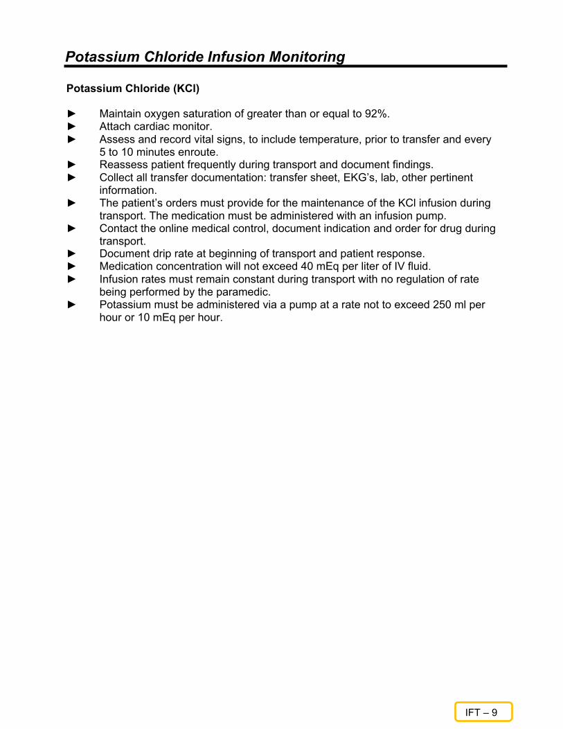
Potassium Chloride Infusion Monitoring
IFT – 9
Potassium Chloride (KCl) ► Maintain oxygen saturation of greater than or equal to 92%. ► Attach cardiac monitor. ► Assess and record vital signs, to include temperature, prior to transfer and every
5 to 10 minutes enroute. ► Reassess patient frequently during transport and document findings. ► Collect all transfer documentation: transfer sheet, EKG’s, lab, other pertinent
information. ► The patient’s orders must provide for the maintenance of the KCl infusion during
transport. The medication must be administered with an infusion pump. ► Contact the online medical control, document indication and order for drug during
transport. ► Document drip rate at beginning of transport and patient response. ► Medication concentration will not exceed 40 mEq per liter of IV fluid. ► Infusion rates must remain constant during transport with no regulation of rate
being performed by the paramedic. ► Potassium must be administered via a pump at a rate not to exceed 250 ml per
hour or 10 mEq per hour.

Tissue Plasminogen Activator (TPA) Infusion Monitoring
IFT – 10
In the treatment of acute myocardial infarction, T-PA is given intravenously as soon as possible in a total dose of 100 mg: the total dose should not exceed 1.5 mg/kg in patients weighing less than 65kg. ► Maintain oxygen saturation of greater than or equal to 92%. Attach cardiac
monitor. ► Assess and record vital signs, to include temperature, prior to transfer and every
5 to 10 minutes enroute. ► Reassess patient frequently during transport and document findings. ► Collect all transfer documentation: transfer sheet, EKG’s, lab, other pertinent
information. ► Contact the online medical control, document indication and order for drug during
transport. ► Document drip rate at beginning of transport and patient response. ► The patient’s orders must provide for the maintaining of the T-PA infusion during
transport. ► The medication must be administered by an infusion pump. Myocardial Infarction, Acute
Accelerated Infusion < 67 kg Dose: 15 mg IV x 1, then 0.75 mg/kg (max 50 mg) over 30 minutes, then 0.5 mg/kg (max 35 mg) over 60 minutes. Accelerated Infusion > 67 kg Dose: 15 mg IV x 1, then 50 mg/kg over 30 minutes, then 35 mg over 60 minutes. 3 Hr Infusion < 65 kg Dose: 1.25 mg/kg IV over three hours; give 60% of dose over one hour with 6-10% of dose as IV bolus, then 20% over second hour; then 20% over third hour. 3 Hr Infusion > 65 kg Dose: 100 mg IV over three hours; give 60 mg of dose over one hour with 6-10% of dose as IV bolus, then 20 mg over second hour; then 20 mg over third hour.

IFT – 11
Maintenance of Intravenous Medication PROPOFOL
Drug Characteristics:
Classification: Anesthetic/Sedative/hypnotic Onset action: 30-45 sec Duration: 2-64 mins. Indications for Use: Sedation for intubated, mechanically ventilated patients Contraindications: 1. Lack of ventilator support 2. Increased Intracranial Pressure 3. Impaired Cerebral Circulation 4. Lipid Metabolism Disorders 5. Severe cardiac dysfunction 6. Documented hypersensitivity to drug 7. Use in labor and delivery may cause neonatal depression Precautions: 1. Use in hypovolemic patients may see a significant drop in blood pressure (Hypotension is
seen in about 26% of adult and 17% pediatric patients) 2. Closely monitor patients for cardiac dysfunction including arrhythmias 3. Anxiety, agitation, and resistance to mechanical ventilation may occur with abrupt
withdrawal Adverse effects: Cardiac: bradycardia, hypotension, hypertension, decreased cardiac output, Torsades de Pointes (responds well to MgSo4) GI: Nausea and vomiting MSK: involuntary muscle movements Maintenance dose: Typical Range: 5-50 mcg/kg/min IV Available solution concentration: 100ml vial (10mg/mL)
Propofol (Diprivan) Interfacility Transfer Protocol
Approved 1/2/2017
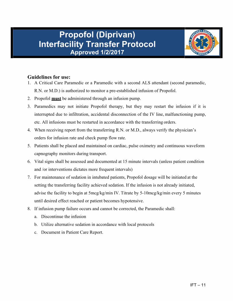
IFT – 11
Guidelines for use: 1. A Critical Care Paramedic or a Paramedic with a second ALS attendant (second paramedic,
R.N. or M.D.) is authorized to monitor a pre-established infusion of Propofol.
2. Propofol must be administered through an infusion pump.
3. Paramedics may not initiate Propofol therapy, but they may restart the infusion if it is
interrupted due to infiltration, accidental disconnection of the IV line, malfunctioning pump,
etc. All infusions must be restarted in accordance with the transferring orders.
4. When receiving report from the transferring R.N. or M.D., always verify the physician’s
orders for infusion rate and check pump flow rate.
5. Patients shall be placed and maintained on cardiac, pulse oximetry and continuous waveform
capnography monitors during transport.
6. Vital signs shall be assessed and documented at 15 minute intervals (unless patient condition
and /or interventions dictates more frequent intervals)
7. For maintenance of sedation in intubated patients, Propofol dosage will be initiated at the
setting the transferring facility achieved sedation. If the infusion is not already initiated,
advise the facility to begin at 5mcg/kg/min IV. Titrate by 5-10mcg/kg/min every 5 minutes
until desired effect reached or patient becomes hypotensive.
8. If infusion pump failure occurs and cannot be corrected, the Paramedic shall:
a. Discontinue the infusion
b. Utilize alternative sedation in accordance with local protocols
c. Document in Patient Care Report.
Propofol (Diprivan) Interfacility Transfer Protocol
Approved 1/2/2017

MED
Medication List

MED – 1
ADENOSINE
Class Endogenous Nucleotide
Mechanism of action Slows conduction time through the AV Node; can interrupt re-entrant pathways; slows heart rate; acts directly on sinus pacemaker cells. Drug of choice for PSVT.
Indications Conversion of PSVT to sinus rhythm. May convert PSVT due to Wolff- Parkinson-White syndrome. Not effective in converting atrial fibrillation / flutter.
Contraindications Second or third-degree " block or Sick Sinus Syndrome Atrial flutter / atrial fibrillation Ventricular Tachycardia Hypersensitivity to adenosine
Adverse Reactions Facial flushing, shortness of breath, chest pain, headache, paresthesia, diaphoresis, palpitations, hypotension, nausea, metallic taste.
Drug Interactions Methylxanthines (theophylline-like drugs) antagonize the effects of adenosine. Dipyridamole (Persantine) potentiates the effects of adenosine Carbamazepine (Tegretol) may potentiate the AV Node blocking effects of adenosine. May cause bronchoconstriction in asthmatic patients.
How Supplied Three mg/ml in 2-ml flip-top vials for IV injection
Dosage and Administration Adult:
• 6 mg over 1-3 seconds; If no response after 1-2 minutes, administer 12 mg over 1-3 seconds, Maximum total dose = 30 mgs.
Pediatric: • 0.1 – 0.2 mg/kg rapid IV; maximum single dose = 12 mgs.
Duration of action Onset and peak effects in seconds; duration 12 seconds.
Special Considerations Short half-life limits side effects in most patients. Pregnancy safety: Category C.

MED – 2
ALBUTEROL Class
Sympathomimetic, bronchodilator. Mechanism of Action
Selective β-2 agonist which stimulates adrenergic receptors of the sympathomimetic nervous system resulting in smooth muscle relaxation in the bronchial tree and peripheral vasculature.
Indications Treatment of bronchospasm in patients with reversible obstructive airway disease (COPD/asthma). Prevention of exercise-induced bronchospasm.
Contraindications Known prior hypersensitivity reactions to Albuterol. Tachycardia dysrhythmias, especially those caused by digitalis.
Adverse Reactions Often dose-related and include restlessness, tremors, dizziness, palpitations, tachycardia, nervousness, peripheral vasodilatation, nausea, vomiting, hyperglycemia, increased blood pressure and paradoxical bronchospasm.
Drug Interactions Tricyclic antidepressants may potentiate vasculature effects. Beta-blockers are antagonistic. May potentiate hypokalemia caused by diuretics.
How Supplied Solution for aerosolization: 0.5% (5 mg/ml) Metered Dose Inhaler: 90 mcg/metered spray (17 gm canister with 200 inhalations) Syrup: 2 mg/5 ml
Dosage and Administration Adult:
• Administer 2.5 mg. Dilute 0.5 ml of 0.5% solution for inhalation with 2.5 ml normal saline in nebulizer and administer over 10-15 minutes.
• MDI: 1-2 inhalations (90-180 mcg). Five minutes between inhalations Pediatric: • Administer solution of 0.01 - 0.03 ml (0.05 - 0.15 mg/kg/ dose diluted in 2 ml
of 0.9% Normal Saline. May repeat every 20 minutes three times. Duration of Action
Onset in 5-15 minutes with peak effect in 30-minutes - two hours and duration of 3-4 hours.
Special Considerations Pregnancy Safety: Category C. Antagonized by beta-blockers. May precipitate angina pectoris and dysrhythmias. Should only be administered by inhalation methodology in pre-hospital management.

MED – 3
AMIODARONE Class
Antidysrhythmic Mechanism of Action
Prolongation of Action Potential; non-competitive alpha and beta sympathetic blocking effects; Calcium channel blocking effects.
Indications Suppression of Ventricular Fibrillation refractory to defibrillation and Lidocaine. Suppression of Ventricular Tachycardia refractory to cardioversion and Lidocaine.
Contraindications Second or Third-Degree heart block. Medication-induced Ventricular dysrhythmias. Hypotension, Bradycardia, Torsades de Pointes. Profound Sinus Bradycardia.
Adverse Reactions Hypotension, Bradycardia, Pulseless Electrical Activity, Congestive Heart Failure, Nausea, fever, abnormal Liver Function Tests, Thrombocytopenia.
Drug Interactions Incompatible: Will precipitate with Sodium Bicarbonate Compatible with: Dopamine, Dobutamine, Isoproterenol, Lidocaine, NTG, Norepinephrine, Phenylephrine, KCL, Procainamide.
How Supplied: 150 mg in 3 ml vials.
Dosage and Administration Adult:
• 300 mg slow IV Push over 1-2 minutes in 10 ml Normal Saline, (For ACLS VF/ Pulseless VT)
• IV Drip 0.5-1mg per minute. (For malignant ventricular arrhythmias). Duration of Action:
Onset: 5-15 minutes. Peak Effect: Variable. Duration: Variable Special Considerations
Pregnancy safety: Category C Maintain at room temperature and protect from light in storage (light protection not required during administration). Hypotension usually responsive to slowing infusion rate, IV Normal Saline. Administer cautiously in patients with Heart Failure or poor systolic function. May be especially effective in high-risk patients with recent acute MI.

MED – 4
ASPIRIN Class
Platelet inhibitor, anti-inflammatory agent. Mechanism of Action
Prostaglandin inhibition. Indications
New onset chest pain suggestive of Acute Myocardial Infarction. Contraindications
Hypersensitivity. Gastrointestinal bleeding. Adverse Reactions
Heartburn. GI bleeding. Nausea, vomiting. Wheezing in allergic patients. Prolonged bleeding.
Drug Interactions Use with caution in patients allergic to NSAIDS.
How Supplied 81 mg or 325 mg tablets (chewable and standard).
Dosage and Administration 81 mg - 325 mg PO.
Duration of Action Onset: 30-45 minutes. Peak effect: variable. Duration: Variable.
Special Considerations Pregnancy Safety: Category D. Not recommended in pediatric population.

MED – 5
ATROPINE SULFATE Class
Anticholinergic agent. Mechanism of Action
Parasympatholytic: inhibits action of acetylcholine at postganglionic parasympathetic neuroeffector sites. Increases heart rate in life-threatening bradydysrhythmias.
Indications Hemodynamically significant bradycardia. Drug of choice for organophosphate poisoning. Bronchospastic pulmonary disorders.
Contraindications Tachycardia. Hypersensitivity. Unstable cardiovascular status in acute hemorrhage and myocardial ischemia. Narrow-angle glaucoma.
Adverse Reactions Headache, dizziness, palpitations, nausea and vomiting. Tachycardia, dysrhythmias, anticholinergic effects (blurred vision, dry mouth, urinary retention). Paradoxical bradycardia when pushed slowly or at low doses. Flushed, hot dry skin.
Drug Interactions Potential adverse effects when administered with digoxin, cholinergics, physostigmine. Effects enhanced by antihistamines, procainamide, quinidine, antipsychotics, benzodiazepines and antidepressants.
How Supplied Prefilled syringes: 1.0 mg in 10 ml of solution.
Dosage and Administration Adult:
• Bradydysrhythymias: 0.5 - 1.0 mg IV every 3-5 minutes as needed to maximum total dose of 3 mg.
Pediatric: • Bradydysrhythmias: 0.2 mg / kg IV / ET / IO (minimum single dose 0.1 mg,
maximum single dose 1.0 mg). If administered via ET, follow with 2.0 ml sterile Normal Saline Solution.
Duration of Action Onset: Immediate. Peak Effect: Rapid to 1-2 minutes. Duration: 2-6 hours.
Special Considerations Pregnancy Safety: Category C. Moderate doses dilate pupils.

MED – 6
CALCIUM CHLORIDE / CALCIUM GLUCONATE
Class Electrolyte.
Mechanism of Action Increases cardiac contractile state (positive inotropic effect). May enhance ventricular automaticity.
Indications Hypocalcemia, magnesium sulfate overdose, hyperkalemia, calcium channel blocker toxicity. Adjunctive therapy in treatment of insect bites and stings.
Contraindications Hypercalcemia, VF during cardiac resuscitation; digitalis toxicity.
Adverse Reactions Bradycardia, asystole, hypotension, peripheral vasodilation, metallic taste, local necrosis, coronary and cerebral artery spasm, nausea, vomiting.
Drug Interactions May worsen dysrhythmias secondary to digitalis. May antagonize effects of Verapamil. Flush line before and after administration of sodium bicarbonate.
How Supplied 10% solution in 10 ml ampules, vials and prefilled syringes (100 mg/ ml).
Dosage and Administration Adult:
• 2-4 mg/kg of 10% solution slowly IV over 5 minutes; may repeat in 10 minutes. (maximum: 1 gm dose)
Pediatric: • 20 mg/kg/dose of 10% solution slow IV/ IO (Maximum: 1 gm dose); May
repeat in 10 minutes. Duration of Action
Onset: 5-15 minutes. Peak effects: 3-5 minutes. Duration: 15-30 minutes but may persist for 4 hours (dose dependent).
Special Considerations Pregnancy safety: Category C. For pediatrics: if calcium gluconate is unavailable, 1-2 ml of 10% calcium chloride solution, diluted with IV fluid, may be substituted.

MED – 7
CEFAZOLIN SODIUM (Ancef)
Class: Cephalosporin Antibiotic
Action: Prevents and treats Infection
Onset: IV – Immediate Duration
8 Hours Indications:
Open or suspected open extremity fracture, being transported to UK Chandler ER
Contraindications: Allergy to cephalosporin antibiotics Prior anaphylactic reaction to penicillin (simple rash/itching is not a contraindication)
Side Effects: Rare
Form: Vial 1 g powder, reconstitute with 2 mL when needed
Dosing: Adult - Patient weight < 120 kg 2 G IV
- Patient weight > 120 kg 3 G IV
Pediatric: - 25 mg/kg IV up to 2 G For both Adult & Pediatric dosing, administer IV Push over 3 – 5 minutes
Special Considerations
Pregnancy safety: Category B.

MED – 8
DEXTROSE Class
Carbohydrate, hypertonic solution. Mechanism of Action
Rapidly increases serum glucose levels. Short-term osmotic diuresis. Indications
Hypoglycemia altered level of consciousness, coma of unknown etiology, seizure of unknown etiology, status epilepticus (controversial).
Contraindications Intracranial hemorrhage, delirium tremens, ineffective without thiamine,
Adverse Reactions Extravasation leads to tissue necrosis. Warmth, pain, burning, thrombophlebitis, rhabdomyolysis.
Drug Interactions Sodium bicarbonate, coumadin.
How Supplied 25 gm/ 250 ml pre-mix IV solution (0.1 g/mL) 25 gm/ 50 ml pre-filled syringes (500 mg/ml).
Dosage and Administration Adult:
• 12.5-25 gram slow IV; may be repeated as necessary. Pediatric: • 0.5-1 gm/kg/dose slow IV; may be repeated as necessary.
Duration of Action Onset: less than 1 minute. Peak effects: variable. Duration: Variable.
Special Considerations Draw blood sugar before administering.
Do not administer to patients with known CVA unless hypoglycemia documented.

MED – 9
DIPHENHYDRAMINE
Class Antihistamine; anticholinergic.
Mechanism of Action Blocks cellular histamine receptors; decreases vasodilation; decreases motion sickness. Reverses extrapyramidal reactions.
Indications Symptomatic relief of allergies, allergic reactions, anaphylaxis, acute dystonic reactions (phenothiazines). Blood administration reactions; used for motion sickness, hay fever.
Contraindications Asthma, glaucoma, pregnancy, hypertension, narrow angle glaucoma, infants, patients taking monoamine oxidase inhibitors (MOAIs).
Adverse Reactions Sedation, hypotension, seizures, visual disturbances, vomiting, urinary retention, palpitations, dysrhythmias, dry mouth and throat, paradoxical CNS excitation in children.
Drug Interactions Potentiates effects of alcohol and other anticholinergics, may inhibit corticosteroid activity, MAOIs prolong anticholinergic effects of diphenhydramine.
How Supplied 50 mg prefilled syringes, vials (IV or IM).
Dosage and Administration Adult:
• 25 - 50 mg IM or IV or P.O. Pediatric:
• 1-2 mg/kg IV, IO slowly or IM. If given PO: 5 mg./ kg./ 24 hours. Duration of Action
Onset: 15-30 minutes. Peak effect: 1 hour. Duration: 3-12 hours.
Special Considerations Not used in infants or in pregnancy: Category B. If used in anaphylaxis, will be in conjunction with epinephrine, steroids.

MED – 10
DOPAMINE
Class Sympathomimetic, inotropic agent.
Mechanism of Action Immediate metabolic precursor to Norepinephrine. Increases systemic vascular resistance, dilate renal and splanchnic vasculature. Increases myocardial contractility and stroke volume.
Indications Cardiogenic, septic or spinal shock, hypotension with low cardiac output states. Distributive shock.
Contraindications Hypovolemic shock, pheochromocytoma, tachydysrhythmias, VF.
Adverse Reactions Cardiac dysrhythmias, hypertension, increased myocardial oxygen demand, extravasation may cause tissue necrosis.
Drug Interactions Incompatible in alkaline solutions. MAOIs will enhance effects of dopamine. Beta blockers may antagonize effects of dopamine. When administered with Phenytoin: may cause hypotension, bradycardia and seizures.
How Supplied 200 mg / 5 ml - 400 mg / 5 ml prefilled syringes, ampules for IV infusion. 400 mg in 250 ml D5W premixed solutions.
Dosage and Administration Adult:
• 2- 20 mcg / kg / min. Pediatric: • 2 - 20 mcg / kg / min.
Duration of Action Onset: 1-4 minutes. Peak Effect: 5-10 minutes. Duration: Effects cease almost immediately after infusion shut off.
Special Considerations Pregnancy safety not established. Effects are dose-dependent Dopaminergic response: 2-4 mcg / kg / min.: dilates vessels in kidneys; increases urine output. Beta-adrenergic response: 4- 10 mcg / kg / min.: Increased chronotropy and inotropy Adrenergic response: 10-20 mcg / kg / min.: Primarily alpha stimulant / vasoconstriction. Greater than 20 mcg / kg / min.: reversal of renal effects / override alpha effects. Always monitor drip rate. Avoid extravasation injury.

MED – 11
EPINEPHRINE
Class Sympathomimetic.
Mechanism of Action Direct acting alpha and beta agonist. Alpha: bronchial, cutaneous, renal and visceral arteriolar vasoconstriction. Beta 1: positive inotropic and chronotropic actions, increases automaticity. Beta 2: bronchial smooth muscle relaxation and dilation of skeletal vasculature Blocks histamine release.
Indications Cardiac arrest, asystole, PEA, VF unresponsive to initial defib. Severe bronchospasm, asthma, bronchiolitis. Anaphylaxis, acute allergic reactions.
Contraindications Hypertension, hypothermia, pulmonary edema, coronary insufficiency, hypovolemic shock.
Adverse Reactions Hypertension, dysrhythmias, pulmonary edema, anxiety, psychomotor agitation, nausea, angina, headache, restlessness.
Drug Interactions Potentiates other sympathomimetics. Deactivated by alkaline solutions. MAOIs may potentiate effects of epinephrine.
How Supplied 1 mg / ml (1:1,000) ampules and 0.1 mg / ml (1:10,000) prefilled syringes. Auto- injectors: EPI-Pen: 0. 3 mg / ml EPI-Pen Jr.: 0.15mg/ml
Dosage and Administration Adult • Anaphylaxis/asthma: 0.3 - 0.5 mg (0.3 - 0.5 ml 1:1 000) IM • Anaphylaxis: 0.3 - 0.5 mg (3- 5 ml 1:10,000) IV • Cardiac Arrest: 1 mg IV/IO (1:10,000) every 3- 5 minutes. Pediatric • Anaphylaxis/asthma: 0.01 mg/kg (0.01 mL/kg 1:1000) IM max 0.5 mg. • Cardiac Arrest: 0.01 mg/kg IV/IO (1:10,000, 0.1mL/kg)
Duration of Action Onset: Immediate. Peak Effects: Minutes. Duration: Several minutes.
Special Considerations Pregnancy safety: category C. Syncope in asthmatic children.

MED – 12
FENTANYL CITRATE
Class Opioid analgesic (Schedule II drug).
Mechanism of Action Alleviates pain through CNS actions. Depresses respiration. Suppresses anxiety. Less histamine release than morphine.
Indications Analgesia for moderate to severe pain.
Contraindications Allergic to Fentanyl. Undiagnosed abdominal pain, depressed respiratory drive, head injury.
Adverse Reactions Respiratory depression, hypotension, decreased levels of consciousness, nausea, vomiting.
Drug Interactions Potentiates sedation with phenothiazines and other opiates and other sedative agents.
How Supplied Injection solution: 50 mcg/mL
Dosage and Administration Adult:
• 25-50 mcg slow IV over 5 minutes Pediatric: • 0.5 mcg/kg IV over five minutes.
Duration of Action Onset: immediate. Peak Effect: minutes. Duration: 1-2 hours.
Special Considerations Pregnancy safety: Category C. Use with caution in geriatric population, patient with respiratory depression or hypovolemia.

MED – 13
GLUCAGON Class
Hyperglycemic agent, pancreatic hormone, insulin antagonist. Mechanism of Action
Increases blood glucose by stimulating glycogenesis. Unknown mechanism of stabilizing cardiac rhythm in beta-blocker overdose. Minimal positive inotrope and chronotrope. Decreases GI motility and secretions.
Indications Altered level of consciousness when hypoglycemia is suspected. May be used as inotropic agent in beta-blocker overdose.
Contraindications Hyperglycemia, hypersensitivity.
Adverse Reactions Nausea, vomiting. Tachycardia, hypertension.
Drug Interactions Incompatible in solution with most other substances. No significant drug interactions with other emergency medications.
How Supplied 1 mg ampules (requires reconstitution with diluent provided).
Dosage and Administration Adult:
• 0.5 - 1 mg IM, SC, or slow IV; may repeat q 20 minutes PRN. Pediatric:
• 0.03 -0.1 mg / kg / dose (maximum dose 1 mg) q 20 min. IM, IO, SC, slow IV. Duration of Action
Onset: 1 minute. Special Considerations
Pregnancy safety: Category C. Ineffective if glycogen stores depleted. Should be used in conjunction with 50% dextrose whenever possible. If patient does not respond to second dose glucagon, 50% dextrose must be administered.

MED – 14
GLUCOSE - ORAL Class
Hyperglycemic. Mechanism of Action
Provides quickly absorbed glucose to increase blood glucose levels. Indications
Conscious patients with suspected hypoglycemia. Contraindications
Decreased level of consciousness, nausea, vomiting. Adverse Reactions
Nausea, vomiting. Drug Interactions
None. How Supplied
Glucose pastes and gels in various forms. Dosage and Administration Adult:
• 1 tube ~ 15 G, buccal Pediatric:
• Same as adult. Duration of Action
Onset: Immediate. Peak Effect: Variable. Duration: Variable. Special Considerations
As noted in indications section.

MED – 15
IPRATROPRIUM BROMIDE
Class Bronchodilator
Mechanism of Action Blocks the action of acetylcholine at the parasympathetic sites in bronchial smooth muscle causing bronchodilatation.
Indications Used in bronchospasm especially associated with COPD, and emphysema.
Contraindications Hypersensitivity to atropine or its derivatives.
Adverse Reactions Poorly absorbed from the lung, so systemic effects are rare. >10% CNS: Dizziness, Headache, Nervousness. Respiratory: Cough 1-10% Cardiac: Hypotension, palpitations.
How Supplied Nebulizing Ampule: 0.02% (2.5ml) Inhaler: 18mcg/actuation
Dosage and Administration Adult:
• 2-3 puffs via metered dose inhaler (MDI) tid-qid; maximum 12 puffs/day. • 500mcg NEB q 6-8hrs Pediatric:
- < 12 yo: 1-2 puffs(MDI) tid-qid; max: 8 puffs. ALT: - 250mcg NEB q 6-8hrs
Duration of Action Onset: 1-3 minutes after administration Peak effects: Within 1.5- 2 hours Duration of Action: Up to 4-6 hours T1/2: 2 hrs after inhalation
Special Considerations Pregnancy Safety: Category B.

MED – 16
KETAMINE (Ketalar) Class
Rapid Acting General Anesthetic Mechanism of Action
Blocks NMDA receptors producing a dissociative sedative/hypnotic state. It causes dissociative and partial agonism in opiate mu-receptors which permit painful procedures to be performed in a consistent state of sedation and patient comfort. Ketamine generally maintains normal pharyngeal and laryngeal reflexes, however there may be transient respiratory depression if administered too rapidly.
Indications Pain Management Procedural Sedation Post Intubation Mechanical Ventilation Excited Delirium
Contraindications Hypersensitivity Hydrocephalus Ruptured Globe
Adverse Reactions Rapid IV administration may lead to respiratory depression. Hallucinations Hypertension Tachycardia Salivation Nystagmus
How Supplied 500 mg in 10 mL vial – 50 mg/mL
Dosage and Administration Pain Management
• Adult: - 20 – 30 mg IV push
• Pediatric: - 0.1 – 0.3 mg/kg IV Max – adult dose - 0.5 – 1 mg/kg IM Max – adult dose
Post – Intubation/Mechanical Ventilations: • 2 mg/kg IV push
Excited Delirium: • 4 mg/kg IM max dose: 400 mg
Special Considerations Pregnancy Safety: Category .

MED – 17
KETOROLAC TROMETHAMINE Class
Non-steroidal anti-inflammatory drug. Analgesic Mechanism of Action
NSAID, blocks prostaglandin complex formation and production. It is a potent analgesic that does not possess any sedative or anxiolytic activities.
Indications Pain relief, short-term.
Contraindications Pain relief prior to major surgery. Allergy, bleeding risk, stroke suspected or confirmed, aspirin allergy.
Adverse Reactions Allergic reaction, bleeding, rash.
Drug Interactions Avoid other non-steroidal drugs, Furosemide.
How Supplied Injection solution: 15 mg/ml, 30 mg/ml Intravenous solution: 15 mg/ml, 30mg/ml
Dosage and Administration Adult:
• 15-30 mg IVP or 30-60 mg IM Duration of Action
Onset: Minutes Peak Effects: 30 minutes.
Special Considerations Pregnancy safety: Category C. Avoid with history of aspirin or NSAID drug. Increases risk of bleeding, therefore do not use in a patient that may need to go to surgery.

MED – 18
LIDOCAINE HCL (2%) Class
Antidysrhythmic. Mechanism of Action
Decreases automaticity by slowing the rate of spontaneous Phase 4 depolarization.
Indications Suppression of ventricular dysrhythmias (V-tach, VF, PVCs). Prophylaxis against recurrence after conversion from V-tach, VF.
Contraindications Second degree and third-degree blocks in absence of artificial pacemaker). Hypotension. Stokes Adams Syndrome.
Adverse Reactions Slurred speech, seizures, altered mental status, confusion, lightheadedness, blurred vision, bradycardia.
Drug Interactions Apnea induced with succinylcholine may be prolonged with high doses of Lidocaine. Cardiac depression may occur in conjunction with IV Dilantin. Procainamide may exacerbate the CNS effects. Metabolic clearance decreased in patients with liver disease or those patients taking beta-blockers.
How Supplied 100 mg in 5 ml solution prefilled syringes. 1 and 2-gram additive syringes. 100 in 5 ml solution ampules. 1 and 2-gram vials in 30 ml of solution.
Dosage and Administration Adult:
• Cardiac arrest VT/ VF: 1.5 mg / kg IV push; repeat q 3-5 minutes to maximum dose of 3 mg/kg.
Pediatric: • VF or Pulseless V-tach: 1 mg/kg IV / IO per dose. Infusion: 20-50 mcg/kg/min.
Duration of Action Onset: 1-5 minutes. Peak Effect: 5-10 minutes. Duration: Variable (15 minutes – 2 hours)
Special Considerations Pregnancy safety: Category B. Reduce maintenance infusions by 50% if patient is over 70 years of age, has liver disease, or is in CHF or shock. A 75-100 mg bolus maintains levels for only 20 minutes. Exceedingly high doses of Lidocaine can result in coma or death. Avoid Lidocaine for reperfusion dysrhythmias after thrombolytic therapy. Cross-reactivity with other forms of local anesthetics.

MED – 19
MAGNESIUM SULFATE Class
Electrolyte. Mechanism of Action
Reduces striated muscle contractions and blocks peripheral neuromuscular transmission by reducing acetylcholinesterase release at the myoneural junction; manages seizures in toxemia of pregnancy; induces uterine relaxation; can cause bronchodilation after beta-agonists and anticholinergics have been used.
Indications Seizures of eclampsia (Toxemia of pregnancy). Torsades de Pointes. Hypomagnesemia. TCA overdose-induced dysrhythmias. Digitalis-induced dysrhythmias. Class IIa agent for refractory VF and VT after administration of Lidocaine doses.
Contraindications Heart blocks. Renal diseases.
Adverse Reactions Respiratory and CNS depression. Hypotension, cardiac arrest and asystole may occur. Facial flushing, diaphoresis, depressed reflexes. Circulatory collapse.
Drug Interactions May enhance effects of other CNS depressants. Serious changes in overall cardiac function may occur with cardiac glycosides.
How Supplied 2 ml and 20 ml vials of a 50% solution.
Dosage and Administration Adult:
• Seizure activity associated with pregnancy: 1-4 gm IV push over 3 minutes. • For Torsades de Pointes or Refractory VF/VT: 1-2 grams IV push over 1-2
minutes. Pediatric:
• Not recommended. Duration of Action
Onset: Immediate. Peak effect: variable. Duration: 3-4 hours. Special Considerations
Pregnancy safety: Recommended that drug not be given in the 2 hours before delivery, if possible. IV calcium gluconate or calcium chloride should be available as antagonist if needed. The cure for toxemia is delivery of the baby. Use with caution in patients with renal failure. Magnesium sulfate is being used for acute MI patients in some systems under Medical Direction.
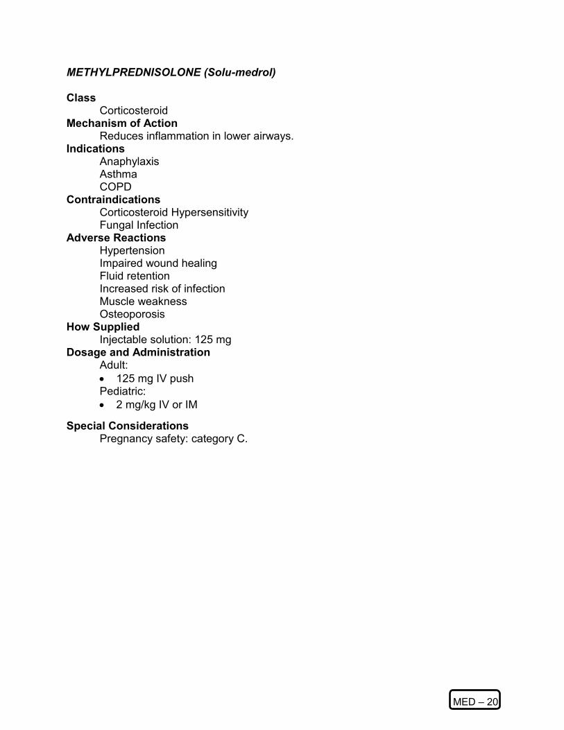
MED – 20
METHYLPREDNISOLONE (Solu-medrol)
Class Corticosteroid
Mechanism of Action Reduces inflammation in lower airways.
Indications Anaphylaxis Asthma COPD
Contraindications Corticosteroid Hypersensitivity Fungal Infection
Adverse Reactions Hypertension Impaired wound healing Fluid retention Increased risk of infection Muscle weakness Osteoporosis
How Supplied Injectable solution: 125 mg
Dosage and Administration Adult:
• 125 mg IV push Pediatric:
• 2 mg/kg IV or IM Special Considerations
Pregnancy safety: category C.

MED – 21
MIDAZOLAM
Class Short-acting benzodiazepine CNS depressant.
Mechanism of Action Anxiolytic and sedative properties like other benzodiazepines. Memory impairment.
Indications Sedation, Anxiolytic prior to endotracheal or nasotracheal intubation.
Contraindications Glaucoma, shock, coma, alcohol intoxication, overdose patient. Depressed vital signs. Concomitant use with other CNS depressants, barbiturates, alcohol, narcotics.
Adverse Reactions Hiccough, cough, over-sedation, nausea, vomiting, injection site pain, headache, blurred vision. Hypotension, respiratory depression and arrest.
Drug Interactions Should not be used in patients who have taken CNS depressant.
How Supplied 2, 5, 10 ml vials (1 mg / ml ). 1, 2 , 5, 10 ml vials (5 mg/ ml ).
Dosage and Administration Adult:
• 0.5 - 2.5 mg slow IV push; (may be repeated to total maximum: 0.1 mg / kg). Pediatric:
• Seizures: WMD: (See APPENDIX Dosing Table)
Duration of Action Onset: 1-3 minutes IV and dose dependent. Peak effect: variable.
Duration: 2-6 hours and dose dependent. Special Considerations
Pregnancy safety: category D. Administer immediately prior to intubation procedure. Requires continuous monitoring of respiratory and cardiac function. Never administer as IV bolus.

MED – 22
NALOXONE Class
Narcotic antagonist. Mechanism of Action
Competitive inhibition at narcotic receptor sites. Reverse respiratory depression secondary to depressant drugs. Completely inhibits the effect of morphine.
Indications Opiate overdose, coma. Complete or partial reversal of CNS and respiratory depression induced by opioids. Decreased level of consciousness. Coma of unknown origin.
Contraindications Use with caution in narcotic-dependent patients. Use with caution in neonates of narcotic-addicted mothers.
Adverse Reactions Withdrawal symptoms in the addicted patient. Tachycardia, hypertension, dysrhythmias, nausea, vomiting, diaphoresis.
Drug Interactions Incompatible with bisulfite and alkaline solutions.
How Supplied 0.01 mg / ml (neonate); 0.4 mg/ml, 1 mg/ml ; 2.0 mg / 5 ml ampules; 2 mg/5 ml
prefilled syringe. Dosage and Administration Adult:
• 0.4 - 2.0 mg IV, IM, SC. Nasal atomizer 2-4mg. ET (diluted); min. recommended = 2.0 mg.; repeat at 5 minute intervals to 10 mg maximum dose. (Medical Control may request higher amounts).
• Infusion: 2 mg in 500 ml of D5W (4 mcg/ml), infuse at 0.4 mg / hr (100 ml/hour).
Pediatric: • 0.1 mg / kg / dose IV, IM, SC, ET (diluted); maximum of 0.8 mg; if no
response in 10 minutes, administer an additional 0.1 mg/kg/dose. Duration of Action
Onset: within 2 minutes. Peak effect: variable. Duration: 30-60 minutes.
Special Considerations Pregnancy safety: category B. Seizures without causal relationship have been reported. May not reverse hypotension. Use caution when administering to narcotic addicts
(violent behavior, etc).

MED – 23
NITROGLYCERIN Class
Vasodilators. Mechanism of Action
Smooth muscle relaxant acting on vascular, bronchial, uterine and intestinal smooth muscle. Dilation of arterioles and veins in the periphery, reduces preload and afterload, decreases the myocardial work load and oxygen demand.
Indications Acute angina pectoris. Ischemic chest pain. CHF, pulmonary edema.
Contraindications Hypotension, hypovolemia. Intracranial bleeding or head injury.
Adverse Reactions Headache, hypotension, syncope, reflex tachycardia, flushing. Nausea, vomiting, diaphoresis, muscle twitching.
Drug Interactions Additive effects with other vasodilators. Incompatible with other drugs IV.
How Supplied Tablets: 0.15 mg (1/400 grain); 0.3 mg (1/200 grain); 0.4 mg (1/150 grain); 0.6 mg (1/100 grain). NTG spray: 0.4 mg - 0.8 mg under the tongue. NTG IV (TRIDIL).
Dosage and Administration Adult:
• Tablets: 0.3 - 0.4 mg SL; may repeat in 3-5 minutes to maximum of 3 doses. NTG spray: 0.4 mg under the tongue; 1-2 sprays. NTG IV infusion: 5 ug / min.; increase by 5-10 ug / min. every 5 minutes until desired effect.
Pediatric: • not recommended.
Duration of Action Onset: 1-3 minutes. Peak effect: 5-10 minutes. Duration: 20-30 minutes or. if IV, 1-10 minutes after discontinuation of infusion.
Special Considerations Pregnancy safety: category C. Hypotension more common in geriatric population. NTG decomposes if exposed to light or heat. Must be kept in airtight containers. Active ingredient may have a stinging effect when administered SL.

MED – 24
ONDANSETRON (Zofran) Class
Anti-emetic. Mechanism of Action
Zofran is an anti-emetic that acts by selective antagonism of the 5HT3 receptors. Indications
Nausea and vomiting. Contraindications
Avoid if history of known hypersensitivity Avoid repeat dosing in patients with known abnormal liver function.
Adverse Reactions Headache Dizziness Drowsiness
How Supplied 8 mg/ml and 2 mg/ml parenteral 8 mg and 4 mg oral
Adult: • 4 mg IV, may repeat X 1 in 30 minutes if needed Peds • 0.1 mg/kg IV up to a maximum of 4 mg

MED – 25
OXYGEN Class
Naturally occurring atmospheric gas. Mechanism of Action
Reverses hypoxemia. Indications
Confirmed or expected hypoxemia. Ischemic chest pain. Respiratory insufficiency. Prophylactically during air transport. Confirmed or suspected carbon monoxide poisoning. All other causes of decreased tissue oxygenation. Decreased level of consciousness.
Contraindications Certain patients with COPD, emphysema who will not tolerate Oxygen concentrations over 35%. Hyperventilation.
Adverse Reactions Decreased level of consciousness and respiratory depression in patients with chronic CO2 retention. Retrolental fibroplasia if given in high concentrations to premature infants. (maintain 30-40% 02)
Drug Interactions None.
How Supplied Oxygen cylinders (usually green and white) of 100% compressed oxygen gas.
Dosage and Administration Adult:
• Cardiac arrest and Carbon Monoxide poisoning: 100%. Hypoxemia: 10-15 L/ min. via non-rebreather.
• COPD: 0-2 L/ min. via nasal cannula or 28-35% venturi mask. Be prepared to provide ventilatory support if higher concentrations of oxygen needed.
Pediatric:
• Same as for adult with exception of premature infant. Duration of Action
Onset: Immediate. Peak effect: not applicable. Duration: Less than 2 minutes.
Special Considerations Be familiar with liter flow and each type of delivery device used. Supports possibility of combustion.
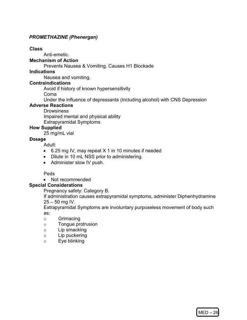
MED – 26
PROMETHAZINE (Phenergan) Class
Anti-emetic. Mechanism of Action
Prevents Nausea & Vomiting. Causes H1 Blockade Indications
Nausea and vomiting. Contraindications
Avoid if history of known hypersensitivity Coma Under the influence of depressants (including alcohol) with CNS Depression
Adverse Reactions Drowsiness Impaired mental and physical ability Extrapyramidal Symptoms
How Supplied 25 mg/mL vial
Adult: • 6.25 mg IV, may repeat X 1 in 10 minutes if needed • Dilute in 10 mL NSS prior to administering. • Administer slow IV push. Peds • Not recommended
Special Considerations Pregnancy safety: Category B. If administration causes extrapyramidal symptoms, administer Diphenhydramine 25 – 50 mg IV. Extrapyramidal Symptoms are involuntary purposeless movement of body such as: o Grimacing o Tongue protrusion o Lip smacking o Lip puckering o Eye blinking

MED - 27
SODIUM BICARBONATE 8.4%
Class Buffer, alkalinizer
Mechanism of Action Reacts with hydrogen ions to form water and carbon dioxide thereby acting as a buffer for metabolic acidosis.
Indications Known pre-existing bicarbonate-responsive acidosis. Upon return of spontaneous circulation after long arrest interval. TCA overdose. Hyperkalemia. Phenobarbital overdose. Alkalinization for treatment of specific intoxications.
Contraindications Metabolic and respiratory alkalosis. Hypocalcemia and hypokalemia. Hypocloremia secondary to GI loss and vomiting.
Adverse Reactions Metabolic alkalosis, hypokalemia, hyperosmolarity, fluid overload. Increase in tissue acidosis. Electrolyte imbalance and tetany, seizures. Tissue sloughing at injection site.
Drug Interactions May precipitate in calcium solutions. Half-lives of certain drugs may increase through alkalinization of the urine. Vasopressors may be deactivated.
How Supplied 50 mEq in 50 ml of solvent.
Dosage and Administration Bolus:1 mEq / kg IV; may repeat with 0.5 mEq / kg every 10 minutes. Infusion: 1 – 4 amps in 1-liter D5W or NS, rate determined by sending physician. Duration of Action
Onset: 2-10 minutes. Peak effect: 15-20 minutes. Duration: 30-60 minutes.
Special Considerations Pregnancy safety: Category C. Must ventilate patient after administration. Whenever possible, blood gas analysis should guide use of bicarbonate. Intracellular acidosis may be worsened by production of carbon dioxide. May increase edematous states. May worsen CHF.

SP
Specialized Protocols

Taser Subdued Patient
SP – 1
Local law enforcement agencies may use a conductive energy weapon called a Taser. This device is a less-lethal tool. When used, the device discharges a wire that contains at the distal end an arrow-like barbed projectile that penetrates the suspect's skin and embeds itself. This allows the officer to administer a 5-second or longer incapacitating electric shock. Officers may initiate EMS response when the device is discharged. EMS personnel shall transport the patient if the Taser strikes the patient in the face, neck, groin or spinal column or other complications arise. Transport shall be recommended for all Taser patients but may be refused by the patient or officer if standard decisional capacity criteria are met. The most common injury from Taser use is trauma from a fall due to the person’s incapacitation.
Scene Safety Consideration: Before touching any patient, who has been subdued using a Taser, EMS personnel shall ensure the officer has disconnected the wires from the hand held unit. In most cases the wires will be cut prior to EMS arrival.

Taser Subdued Patient continued
SP – 1
Basic Standing Orders
► Routine Patient Care. ► Perform initial assessment and treat ABC problems. ► Identify the location of probes on the patient's body. If a probe has contacted the
face, neck, groin, or spinal area, transport the patient to an Emergency Department.
► Confer with the officer and determine the patient's condition from the time of the Taser discharge until EMS arrival.
► Consider Overdose or other medical problems, which may be the underlying cause of the situation.
► Obtain baseline vitals. ► Obtain history from the patient including date of last tetanus shot and any cardiac
history. ► Consider accu-check glucose level.
Treatment and Follow up instructions: ► Clean puncture sites and bandage. ► Secure any embedded probes. ► Transport patient to the emergency department.
NOTE: If possible, transport should be under the care of a Paramedic with cardiac monitoring, and other ACLS as necessary.
Paramedic Standing Orders
► Evaluate for medical reasons for patient’s behavior. ► Consider EKG monitoring for cardiac abnormalities and, if patient is greater than 35
years old, consider 12-lead evaluation. ► Consider 12-lead EKG.

Strenuous Activity / Firefighter Rehabilitation
SP – 2
Medical Evaluation Of Firefighters And Other Emergency Responders On Emergency Incidents And training Exercises Purpose To examine and evaluate the physical and mental status of firefighters and other emergency responders working at an emergency incident or a training exercise and determine what treatment, if any, is necessary. Implementation A Rehab Area shall be set up at the discretion of the Incident Commander. When a Rehab Area has been established by the Incident Commander, the first available Paramedic will be responsible for the management and coordination of the Rehab Area. Location Establish a Rehab Area away from environmental hazards (e.g., shady, cool place, upwind away from smoke and traffic) that is readily accessed by Rescue personnel for transport and supplies. Air truck and canteen service will be stationed in this area. Multiple Rehab Areas may be needed on large incidents. Manning Assign a minimum of two Rescue personnel to monitor and assist firefighters and other emergency responders in the Rehab Area. Medical Evaluations When the Incident Commander has established a Rehab Area, firefighters and other emergency responders shall be evaluated following:
► Two SCBA‘s and/or thirty minutes of strenuous activity (e.g., use of chemical PPE, advancing hose lines, forcible entry, ventilation, etc.) Note: This does not preclude an Officer from having a member evaluated if he/she deems it appropriate. A member may be evaluated at any time he/she feels it is necessary.
► SCBA failure. ► Weakness, dizziness, chest pain, muscle cramps, nausea, altered mental status,
difficulty breathing, etc. ► Discretion of Incident Commander, Rehab Officer, Safety Officer, C.I.S.M.
Coordinator or Company Officer. NOTE - A medical evaluation form shall be completed on all personnel entering the Rehab Area (see form located in this section).

Strenuous Activity / Firefighter Rehabilitation continued
SP – 2
Examination Examination shall occur at ten-minute intervals and will involve a minimum of:
► Glasgow coma score ► Pupils ► Vital Signs (BP, P, R, CR) ► EKG (if applicable) ► Lung sounds ► Skin condition ► Temperature ► Signs and symptoms
An EMS Run Report shall be completed on each firefighter or other emergency responder when he or she is not routinely returned to normal duties. Guidelines for Rehab Normal Examination Findings - firefighter or other emergency responder will rehydrate and rest before reporting to Manpower.
► Abnormal Examination Findings ► Firefighter or other emergency responder will rehydrate and rest. ► Firefighter or other emergency responder will report to Manpower when findings
are normal. Findings should return to normal within fifteen minutes. ► Firefighter or other emergency responder will receive ALS treatment and
transport if findings are abnormal for more than fifteen minutes. ► Firefighter or other emergency responder with chest pain, difficulty breathing and
altered mental status will receive immediate ALS treatment and transport. ► Any other abnormal findings or complaints shall result in treatment and transport
if the paramedic deems it to be in the best interest of the firefighter/responder. Treatment Prior to taking anything orally, the firefighter or other emergency responder will clean hands and face. On scene rescue will provide water and cleaning agent.
► Rest ► Oral rehydration and nutrition; minimum of 1–2 quarts of fluids over a 15 minute
time period (Water first, then half-strength Gatorade® or 10-K®). Avoid any substance containing caffeine (e.g. soda, tea, coffee).
► Oxygen (humidified, nebulizer). ► Cool environment (e.g., shade, electric fan, air conditioning, removal of bunker
gear, showers, etc.). ► ALS Protocols

Strenuous Activity / Firefighter Rehabilitation continued
SP – 2
Return to Emergency Duties Report to Manpower or Incident Commander when:
► Vital signs within normal limits. ► Absence of abnormal signs and symptoms. ► Minimum period of 15 minutes for rest and rehydration.
Documentation A Rehab Medical Evaluation Form shall be completed on all personnel evaluated in the Rehab Area and forwarded to the appropriate Rescue (EMS) Division following all applicable patient confidentiality guidelines (e.g. HIPAA).

Strenuous Activity / Firefighter Rehabilitation continued
SP – 2
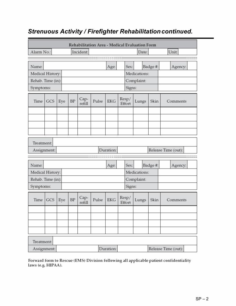
Strenuous Activity / Firefighter Rehabilitation continued.
SP – 2

STARTBand – Emergency Response Tag System
SP – 3
SB-Rapid, everyday emergency tag (LaserBand, Inc.). This all-in-one customizable triage tag enables responders to quickly and accurately identify, record and track the injured at the scene of an emergency.
This tag has been adopted by the Kentucky Public Health Preparedness Branch for use with the Patient/Evacuee Tracking system based on EMTrack. Each tag is bar-coded, weatherproof with multiple removable bar code tags and sections. It can be written on the reverse side. This is the preferred tag for major incidents within the Commonwealth. Example of its utility include: having multiple labels allows the building of relationships between the original person, and something or someone else. For example, in a multiple-car MVA a sticker could be placed on the wrecked vehicle to ID which car the patient came from, a sticker on a bag of personal belongings, and if a child is tagged (even with the RAPID tag) you could put a sticker from the PARENTS triage tag on the back (and a kids tag on the parents tag) to show that the kid belongs to the parent. If they are entered in to EMTrack, there is a place to enter the relationship tag numbers.

STARTBand – Emergency Response Tag System-continued
SP – 3
The tag numbers can be entered into EMTrack system via any of the following:
► from the field using a laptop with an aircard or connected through a WiFi hotspot; entered to a laptop or PDA and held until there is connectivity (store and forward);
► pre-hospital hardware (scanners and PDA units) connected to the internet (either directly or through a field "command kit");
► at the hospital going through triage (We have provided some hospitals with scanners, so they can connect a computer at the triage desk and scan people directly in to the system).
► entered by a dispatcher. ► entered by a registration clerk at a shelter site or POD location.
The RAPID Tag is recommended for day to day operations http://www.laserband.com.

START System of Triage
SP – 4
Introduction
This procedure will be based on the Simple Triage And Rapid Treatment or START method for adult victims and the JumpSTART adaptations for the pediatric victim. These methods of triage are designed to assess many victims objectively, efficiently, and rapidly and can be used by personnel with limited medical training.
Procedure ► Initial Triage—Using the START or JumpSTART methods (Sections III or IV).
► Locate and direct all the walking wounded into one location away from the incident if possible. Assign someone to keep them together (Fire
Department Personnel, Law Enforcement Officer, or capable bystander). ► Begin assessing all non-ambulatory victims where they lay. ► Utilize the Triage Ribbons (color-coded plastic strips). One should be tied
to an upper extremity in a VISIBLE location. RED - Immediate. YELLOW - Delayed. GREEN - Ambulatory (minor). BLACK - Deceased (non-salvageable).
► Independent decisions should be made for each victim. Do not base triage decisions on the perception of too many REDs, not enough GREENs, etc.
► If borderline decisions are encountered, always triage to the more urgent priority (e.g. GREEN/YELLOW patient, tag YELLOW).
► Secondary Triage
► Performed on all victims during the Treatment Phase. If a victim is ► identified in the initial triage phase as a RED and transport is available, do
not delay transport to perform a secondary assessment ► The Triage priority determined in the Treatment Phase should ► be the priority used for transport. If trauma related, the Trauma Criteria will
be applied to trauma victims during the secondary triage in the Treatment Phase (see General Protocol 1.10—Trauma Transport).
► Utilize the Triage Tags (Disaster Management System Tag or METTAGS) and attempt to assess for and complete all information required on the tag.
► Affix the tag to the patient and remove ribbon.
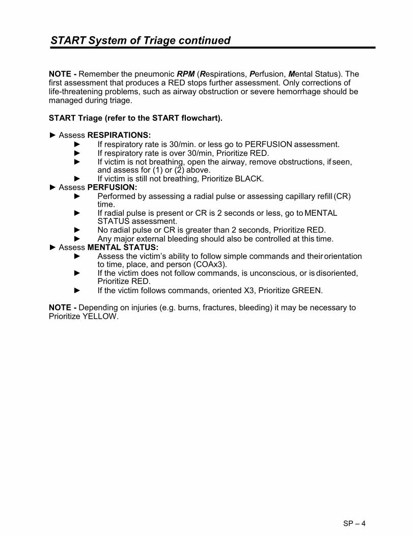
START System of Triage continued
SP – 4
NOTE - Remember the pneumonic RPM (Respirations, Perfusion, Mental Status). The first assessment that produces a RED stops further assessment. Only corrections of life-threatening problems, such as airway obstruction or severe hemorrhage should be managed during triage. START Triage (refer to the START flowchart). ► Assess RESPIRATIONS:
► If respiratory rate is 30/min. or less go to PERFUSION assessment. ► If respiratory rate is over 30/min, Prioritize RED. ► If victim is not breathing, open the airway, remove obstructions, if seen,
and assess for (1) or (2) above. ► If victim is still not breathing, Prioritize BLACK.
► Assess PERFUSION: ► Performed by assessing a radial pulse or assessing capillary refill (CR)
time. ► If radial pulse is present or CR is 2 seconds or less, go to MENTAL
STATUS assessment. ► No radial pulse or CR is greater than 2 seconds, Prioritize RED. ► Any major external bleeding should also be controlled at this time.
► Assess MENTAL STATUS: ► Assess the victim’s ability to follow simple commands and their orientation
to time, place, and person (COAx3). ► If the victim does not follow commands, is unconscious, or is disoriented,
Prioritize RED. ► If the victim follows commands, oriented X3, Prioritize GREEN.
NOTE - Depending on injuries (e.g. burns, fractures, bleeding) it may be necessary to Prioritize YELLOW.

SP – 4
START Triage Algorithm
Perfusion
Start Triage Walking Wounded/ After Triage
Minor (Green)
Respirations
Yes No
Under 30/min Position Airway
Over 30/min
No Yes
Decreased (Black)
Immediate (Red)
Radial Pulse ABSENT Or
Capillary Refill Greater than 2 seconds
Radial Pulse Present Or
Capillary Refill Less than 2 seconds
Control Bleeding Mental Status
CANNOT Follow Simple
Commands
CAN Follow Simple
Commands
Immediate (Red)
Delayed (Yellow)
Immediate (Red)
Immediate (Red)

START System of Triage -JumpSTART
SP – 4
JumpSTART TRIAGE (refer to the JumpSTART flowchart). Physiological differences in children necessitate adapting the standard START triage method to children ≤ 8 years of age or those victims with the anatomical or physiological features of a child in the age group. The same parameters (R.P.M.) will be utilized with the adaptations indicated. ► Assess RESPIRATIONS:
► If respiratory rate is between 15 and 45/min. go to PERFUSION assessment.
► If respiratory rate is over 45/min or under 15/min, Prioritize RED. ► If victim is not breathing, open the airway, remove obstructions if seen,
and assess for (1) or (2) above. ► If victim is still not breathing and no obstructions are present, check a
peripheral (radial or pedal) pulse. If peripheral pulse is present, provide five (5) ventilations (approximately 15 seconds) via any type of barrier device. If spontaneous respirations resume, Prioritize RED.
► If victim is still not breathing, Prioritize BLACK. ► Assess PERFUSION:
► Performed by assessing a peripheral pulse. ► If peripheral pulse is present, go to MENTAL STATUS assessment. ► If peripheral pulse is absent, Prioritize RED. ► Any major external bleeding should also be controlled at this time.
► Assess MENTAL STATUS: ► Assess the child through AVPU scale. Assess whether the victim is either
ALERT, responds to VERBAL stimuli, responds to PAINFUL stimuli, or is UNCONSCIOUS.
► If the victim is unconscious or only responds to painful stimuli, Prioritize RED.
► If the victim is alert or responds to verbal stimuli, assess for further injuries, Prioritize YELLOW or GREEN.
NOTE - Infants who are developmentally unable to walk should be triaged using JumpSTART algorithm either during initial triage or in the GREEN area if carried out by a non-rescuer. During triage, if they do not fulfill the criteria of a RED victim and there are no other outward signs of significant injury, they may be triaged as a GREEN victim. NOTE - START Triage system developed by Newport Beach Fire Rescue and Hoag Hospital. JumpStart Triage system developed by Lou Romig, MD (Miami Children‘s Hospital).

JumpSTART Triage Algorithm
SP – 4

Mass Casualty Incidents: Uniform Prehospital – MCI Procedure
SP – 5
Purpose
To efficiently triage, treat and transport victims of multiple casualty incidents (MCIs). The following protocol is applicable to all multiple victim situations. This protocol is intended for the everyday MCI when the number of injured exceed the capabilities of the first arriving unit, as well as large scale MCIs. The number of casualties may exceed the capabilities of the local jurisdiction and will require assistance from other EMS providers.
Procedure
► The officer of the first arriving unit will establish COMMAND and:
► Perform a size up and estimate the number of victims. ► Request a Level 1, 2, 3, 4, or 5 response (see II.D.) and request additional
units and/or specialized equipment as required. ► Identify a staging area. ► Direct the remaining crewmembers and any additional personnel arriving to
initiate triage using the START or JumpSTART system. ► Locate and direct the walking wounded to one location away from the
incident, if possible. These victims need to be assessed as soon as possible. Assign someone to keep the walking wounded together.
► As additional units arrive, COMMAND will designate the following officers:
o TRIAGE (Initially the responsibility of the First Arriving Officer). o TREATMENT. o TRANSPORT. o STAGING.
► Additional Branches/Sections may be required depending on the complexity of the incident. These officers may include, but are not limited to:
o Medical Branch o Landing Zone/Helispot o Extrication o Haz Mat o Rehabilitation o Safety
► Public Information Officer (PIO) ► Medical Intelligence— To Assist with
WMD Events For Decon, Antidotes And Treatment

Mass Casualty Incidents Uniform Prehospital – MCI Procedure - continued
SP – 5
MCI Pre-determined Response Plan
► Considerations: ► An MCI shall be classified by different levels, depending on the number of
victims. The number of victims will be based on the initial size-up, prior to triage.
► Levels of response will augment the units already on the scene. Units on scene or en-route will be included in the assignment. The exception would be when in conjunction with a Fire Alarm assignment (e.g. Fire with multiple victims may be a Second Alarm with a MCI Level 3 response—this will be two separate assignments).
► COMMAND can downgrade or upgrade the assignment at any time. All units will respond to the Staging Area unless otherwise directed by COMMAND. When announcing an MCI, specify the general category (trauma, HAZMAT, smoke inhalation, etc.).
► Any victim meeting Trauma Transport Criteria should go to a Trauma Center. Trauma Transport Criteria will be determined during the secondary triage in the Treatment Phase.
► Consider air transport for special needs, mass transit resources for multiple "walking wounded,” and private BLS transport units.
► Consider Mobile Command Vehicles, Medical Supply Trailers and Communication Trailers.
► Upon declaration of a MCI—Medical Control will gather each hospital’s capability and relay this information to the Transport Officer or Medical Communication Officer.
► Request Law Enforcement set up a safety perimeter.

Mass Casualty Incidents Uniform Prehospital – MCI Procedure - continued
SP – 5
► Definitions:
o Strike Team—is a specified combination of the same kind and type of resources with common communications and a leader (e.g. ALS Transport Unit Strike Team would be 5 ALS Transport Units with a leader).
o Task Force—is a group of resources with common communications and a leader (e.g. MCI Task Force would be 2 ALS Transport Units, 2 BLS Transport Units and 1 Suppression Unit with a leader).
o Litter Bearer—A team of personnel assigned to TRIAGE to move victims from the incident site to the Treatment Area or Transport Units.
► MCI LEVEL 1 (5–10 victims)
o 4 ALS Transport Units o 2 Suppression Units o 1 Shift Supervisor o 1 EMS Supervisor
**NOTE - The 2 closest hospitals & Trauma Center to the incident will be notified by Medical Control (MedCom or local communication center).
► MCI LEVEL 2 (11–20 victims)
o 6 ALS Transport Units o 3 Suppression Units o 2 Shift Supervisors o 2 EMS Supervisors
**NOTE - The 3 closest hospitals & 2 Trauma Centers to the incident will be notified by Medical Control.

Mass Casualty Incidents Uniform Prehospital – MCI Procedure - continued
SP – 5
► MCI LEVEL 3 (21–100 victims)
o 8 ALS Transport Units o 4 Suppression Units o 3 Shift Supervisors o 3 EMS Supervisors
o 1 Operations Chief o 1 Command Vehicle o 1 Supply Trailer
**NOTE - The 4 closest hospitals & 2 Trauma Centers to the incident will be notified by Medical Control. The Local Warning Point will notify the Emergency Management Agency.
► MCI LEVEL 4 (101–1000 victims)
o 5 MCI Task Forces (25 units) o 2 ALS Transport Unit Strike
Teams (10 units) o 1 Suppression Unit Strike Team
(5 units) o 2 BLS Transport Unit Strike
Teams (10 units) o 2 Mass Transit Buses
o 5 Shift Supervisors o 3 EMS Supervisors o 1 EMS Chief o 1 Operations Chief o 1 Command Vehicle o 2 Supply Trailers o 1 Communications Trailer
**NOTE - The 10 closest hospitals & 5 Trauma Centers to the incident will be notified by Medical Control. The Local Warning Point will notify the Emergency Management Agency. Metropolitan Medical Response System (MMRS) and Disaster Medical Assistance Team (DMAT) may be notified.
► MCI LEVEL 5 (Over 1000 victims)
o 10 MCI Task Forces (50 units) o 4 ALS Transport Unit Strike
Teams (20 units) o 2 Suppression Unit Strike
Teams (10 units) o 4 BLS Transport Unit Strike
Teams (20 units) o 4 Mass Transit Buses
o 10 Shift Supervisors o 6 EMS Supervisors o 2 EMS Chiefs o 2 Operations Chiefs o 2 Command Vehicles o 4 Supply Trailers o 1 Communications Trailer
**NOTE - The 20 closest hospitals & 10 Trauma Centers to the incident will be notified by Medical Control.

Mass Casualty Incidents Uniform Prehospital – MCI Procedure - continued
SP – 5
► Officer Responsibilities o Command
• Established by the First Arriving Officer. Radio designation: COMMAND. • Follow Field Operations Guide FOG #1. • Remain in a fixed and visible location, uphill and upwind. • Determine the MCI Level (1, 2, 3, 4, or 5). • Designate a Staging Area. • Assign positions to perform the functions of TRIAGE, TREATMENT,
TRANSPORT and STAGING. • Advise Communications Center of the number of victims and their
categories once triage is complete. • During large scale or complex MCIs (e.g. fire with multiple victims),
designate a Medical Branch to reduce the span of control. • If the incident is due to Weapons of Mass Destruction (WMD Event), refer
to FOG #8 and establish a Medical Intelligence Officer to assist with documentation, antidotes and treatment of victims.
• Ensure proper security of incident site, treatment area and loading area, as well as traffic control and access for emergency vehicles with law enforcement.
o Medical Branch
• Radio designation: MEDICAL. Follow FOG #2. • Work directly with COMMAND. • Assure TRIAGE, TREATMENT and TRANSPORT has been established. If
established by COMMAND, then TRIAGE, TREATMENT and TRANSPORT will report to MEDICAL.
• Work with COMMAND and direct and/or supervise on-scene personnel from agencies such as Medical Examiner‘s Office, Red Cross, private ambulance companies and hospital volunteers. Ensure notification of Medical Control (Medcom/MRCC).
• If the incident is due to a known or suspected Weapon of Mass Destruction (WMD Event), refer to FOG #8 and establish (in conjunction with COMMAND) a Medical Intelligence Officer to assist with decontamination, antidotes and treatment of victims.
• Ensure proper security of incident site, treatment area and loading area, as well as traffic control and access for emergency vehicles with law enforcement (in conjunction with COMMAND).

Mass Casualty Incidents Uniform Prehospital – MCI Procedure - continued
SP – 5
o Triage Officer.• Radio designation: TRIAGE. Follow FOG #3.• Organize the Triage Team to begin initial triaging of victims, utilizing the
START/JumpSTART triage system. Assemble the walking wounded and uninjured in a safe area. Use bullhorn/PA if necessary.
• Advise COMMAND (or MEDICAL if established), as soon as possible, if there is a need for additional resources.
• Coordinate with TREATMENT to ensure that priority victims are treated first.
• Ensure that all areas around the MCI scene have been checked for potential victims, walking wounded, ejected victims, etc., and that all victims have been triaged.
• Supervise the Triage Personnel, Litter Bearers and Medical Examiner Personnel.
• Maintain security and control of the Triage Area. Request Law Enforcement.
• Report to COMMAND or MEDICAL upon completion of duties for further assignments.
o Treatment Officer.• Reports to COMMAND or MEDICAL. Supervises the Treatment Managers
of the RED, YELLOW, and GREEN Areas.• Coordinates the re-triage and tagging of all victims and on-site medical
care.• Directs the movement of victims to loading area(s).• Radio designation: TREATMENT. Follow FOG #4.• Consider assigning a "Documentation Aide" to assist with paperwork.• Direct personnel to either begin treatment on the victims where they lay or
establish a centralized Treatment Area.• Considerations for a Treatment Area:• Capable of accommodating the number of victims and equipment.• Consider weather, safety and the possibility of hazardous materials.• Designate entrance and exit areas, which are readily accessible (funnel
points).• On large-scale incidents, divide Treatment Area into three distinct areas
based on priority. Designate a Treatment Manager for each area (Red, Yellow, Green). Use color tarps if available.

Mass Casualty Incidents Uniform Prehospital – MCI Procedure - continued
SP – 5
• Complete a "Treatment Log" as victims enter the area.• Ensure that all victims are re-triaged through a secondary exam and the
assessment is documented on the Triage tag (Disaster Management System Tag [DMS Tag] or METTAG). The rescuer filling out the DMS Tag or METTAG will keep a corner of the tag for future documentation.
• All Red tagged victims will be transported immediately as transport units become available. These victims should not be delayed in the Treatment Area.
• Ensure that enough equipment is available to effectively treat all victims.• Establish communicates with TRANSPORT to coordinate proper transport
of the appropriate victims. Direct movement of victims to the ambulance loading area(s).
• Provide periodic status reports to COMMAND/MEDICAL.
NOTE - RED, YELLOW, GREEN TREATMENT MANAGERS—report to the TREATMENT Officer and are responsible for the treatment and continual re-triaging of victims in their assigned areas. Notify TREATMENT Officer of victim readiness and priority for transportation. Assure that appropriate victim information is recorded.
o Transport Officer.• Reports to COMMAND or MEDICAL. Supervises the Medical
Communication Coordinator and Documentation Aide(s). TheTRANSPORT Officer is responsible for the coordination of victims andmaintenance of records relating to victim identification, injuries, mode oftransportation and destination.
• Radio designation: TRANSPORT. Follow FOG #5.• Assign a Documentation Aide with a radio to assist with paperwork and
communications.• Assign a Medical Communication Coordinator to establish continuous
contact with Medical Control (MedComm).• Establish a victim loading area. Advise STAGING of the location and
direction of travel. Consider Law Enforcement for security of loading area.• Arrange for the transport of victims from the Treatment Area. Maintain
"Hospital Transportation Log” #5B. Keep piece of triage tag for futuredocumentation.

Mass Casualty Incidents Uniform Prehospital – MCI Procedure - continued
SP – 5
• Communicate with the Landing Zone (LZ)/ Helispot Officer and relay the number of victims to be transported by air. Air transported victims should be assigned to distant hospitals, unless the victim‘s needs dictate otherwise (e.g. Trauma Center, burn unit, etc.).
o Medical Communication Coordinator.
• Reports to the TRANSPORT Officer and is responsible for maintaining communication with Medical Control to assure proper victim transport information and destination.
• Radio designation: COMMUNICATIONS. Follow FOG #5A. • Establish communication with Medical Control (Medcom/MRCC). Advise
Medical Control of the overall situation (e.g. smoke inhalation, trauma, burns, Hazmat exposure, etc.), amount and category of victims. Medical Control will survey area hospitals to determine their capabilities and capacities and then relay this information. Document this information on the Hospital Capability Worksheet #5C and maintain this for the duration of the incident.
• When units are prepared to transport, advise Medical Control and supply them with the following information:
• The unit transporting. • The number of the victims being transported. • Their priority: Red, Yellow, or Green. • Any special need victims (e.g. cardiac, burns, trauma, etc.). • The Medical Communication Coordinator, in conjunction with Medical
Control, will determine the most appropriate facility. Ground transported victims should be assigned to hospitals on a rotating basis.
• Once Medical Control receives the information from the Medical Communication Coordinator, Medical Control will notify the appropriate hospital.
• Transporting units will not contact the individual hospital on their own, unless there is a need for medical direction/care outside of protocols.
o Medical Supply Coordinator. • Reports to MEDICAL and is responsible for acquiring and maintaining
control of all medical equipment and supplies. • Radio designation: SUPPLY. Follow FOG #6. • Assure necessary equipment is available on the transporting vehicle. • Provide an inventory of medical supplies at the Staging Area for use on
scene.

Mass Casualty Incidents Uniform Prehospital – MCI Procedure - continued
SP – 5
o Staging Officer.• Reports to COMMAND and is responsible for managing all activities within
the Staging Area.• Radio designation: STAGING. Follow FOG #7.• Establish the location of a Staging Area and notify the Communications
Center to direct any incoming units.• Maintain a "Unit Staging Log" #7A.• Ensure that all personnel stay with their vehicles unless otherwise directed
by COMMAND. If personnel are directed to assist in another function, ensure that the keys stay with each vehicle.
• Coordinate with the TRANSPORT Officer the location for a victim loading area and best route to the area.
• Maintain a reserve of at least 2 transport vehicles. When the reserve is depleted request additional units through COMMAND.
► Documentation.o The Incident Commander will, at the completion of the incident, coordinate the
gathering of all pertinent documentation.o A Post Incident Analysis (PIA) should be conducted on all MCIs.
NOTE - MRCC - Medical Resource Coordination Center - prime function is to maintain a status as to the number of victims and the hospital readiness status to accept victims, coordinate transportation and direct them to the appropriate hospital during a disaster or other situation requiring a high demand of medical resources.

Mass Casualty Incidents Uniform Prehospital – MCI Procedure - continued
SP – 5
► MCI Kits. Each Unit will carry an MCI bag. Included in the MCI bag will be: o Two (2) Triage packs with: o Four (4) combine dressings o Six (6) 4 x 4s o Six (6) pairs of gloves o One (1) pediatric face mask, assorted oropharyngeal (OPA) and
nasopharyngeal (NPA) airways o Two (2) clip rings containing triage ribbons paired in red and yellow, green
and black. There are 15 ribbons of each color per ring. o One (1) additional set of triage ribbons. o Fifty (50) Triage tags—Disaster Management Tags (DMS tags) or
METTAGs. o Three (3) mechanical pencils and three (3) grease pencils.
► The following MCI FOG‘s, logs, and associated paperwork for each Officer: o Command FOG #1 o Medical FOG #2 o Triage FOG #3 o Treatment FOG #4 o Treatment Log #4A o Transport FOG #5 o Medical Communication FOG #5A o Hospital Transport Log #5B o Hospital Capability Worksheet #5C o Medical Supply FOG #6 o Staging FOG #7 o Unit Staging Log #7A o MCI-WMD/Terrorist Event FOG #8
► The following identification vests: o White for COMMAND. o Blue for the MEDICAL Officer. o Yellow for the TRIAGE Officer. o Red for the TREATMENT Officer. o Green for the TRANSPORT Officer. o Green Striped for the MEDICAL COMMUNICATION COORDINATOR. o Blue Striped for the MEDICAL SUPPLY Officer. o Orange for the STAGING Officer.

SP – 5
Mass Casualty Incidents Uniform Prehospital - MCI Procedure - continued
Basic MCI Command Structure for Medical Responses
Complex MCI Command Structure for Medical Responses
Command
Triage Treatment Transport Staging
Medical Branch
Triage Group Treatment Group Transport Group
Medical Supply
Triage Units
Litter Bearer Teams
Medical Examiner Personnel
Treatment Teams
Red Team Manager
Yellow Team Manager
Green Team Manager
Documentation Aide
Medical Communication
Coordinator

COMMAND - FOG #1
SP – 5
► Don the appropriate vest and use the radio designation "COMMAND.” Establish the Command Post in a safe, visible and fixed location, uphill and upwind.
o Consider assigning an aide. If WMD involved also use FOG #8.
► Perform the initial size-up, including wind direction. Determine any special needs such as fire suppression, Hazmat, extrication, etc. and request additional units as needed.
► Approximate the number of victims and category of injury (trauma, burns, smoke
inhalation, chemical exposure, etc.).
MCI Level 1 Level 2 Level 3 Level 4 Level 5
Victims 5–10 11–20 21–100 101–1000 >1000
► Establish Staging Area as soon as possible. Request additional units early, as needed: consider HAZMAT, TRT, extrication, Air Rescue.
► Utilize the EMS Tactical Command Worksheet when available. ► Assign positions to perform the following functions:
o MEDICAL BRANCH (as needed). o TRIAGE. o Litter Bearers. o TREATMENT. o RED, YELLOW, GREEN Treatment Managers. o TRANSPORT. Documentation Aide.
o Medical Communication Coordinator. o STAGING. o MEDICAL SUPPLY, REHAB, SAFETY, DECON, EXTRICATION, PIO (as
needed).
► Advise Communication Center of the exact number of victims and their categories once reported from TRIAGE.
► Request Law Enforcement for security for all areas, traffic control and access for emergency vehicles.
► When applicable, have a liaison of each involved party at the Command Post. ► Some examples would include: Law Enforcement, Medical Examiner, Emergency
Management Agency, Occupancy owner/representative, etc. ► If incident is due to a known or suspected WMD Event, refer to WMD FOG #8
and assign Medical Intelligence Officer to assist with decontamination, antidotes and treatment of victims.
(Paper color - White, Two-sided, with Predetermined Response Plan on back).

MCI Predetermined Response Plan (for Command FOG #1 and #2)
SP – 5
For the back of COMMAND FOG #1 and MEDICAL FOG #2 MCI LEVEL 1 (5–10 victims)
o 3 ALS Transport Units o 2 Suppression Units
o 1 Shift Supervisor o 1 EMS Supervisor
NOTE The 2 closest hospitals & Trauma Center to the incident will be notified by Medical Control (MedCom or local communication center).
MCI LEVEL 2 (11–20 victims)
o 6 ALS Transport Units o 3 Suppression Units
o 2 Shift Supervisors o 2 EMS Supervisors
NOTE The 3 closest hospitals & 2 Trauma Centers to the incident will be notified by MedCom. MCI LEVEL 3 (21–100 victims)
o 8 ALS Transport Units o 4 Suppression Units o 3 Shift Supervisors o 3 EMS Supervisors
o 1 Command Vehicle o 1 Operations Chief o 1 Supply Trailer
NOTE The 4 closest hospitals & 2 Trauma Centers to the incident will be notified by MedCom.
MCI LEVEL 4 (101–1000 victims)
o 5 MCI Task Forces (25 units) o 2 ALS Transport Unit Strike
Teams (10 units) o 1 Suppression Unit Strike Team
(5 units) o 2 BLS Transport Unit Strike
Teams (10 units) o 5 Shift Supervisors o 3 EMS Supervisors
o 2 Mass Transit Buses o 1 EMS Chief o 1 Operations Chief o 1 Command Vehicle o 2 Supply Trailers o 1 Communications Trailer
NOTE The 10 closest hospitals & 5 Trauma Centers to the incident will be notified by MedCom
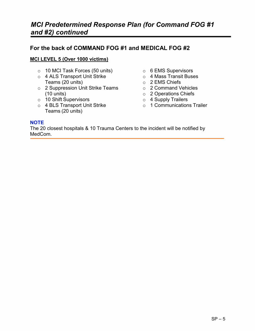
MCI Predetermined Response Plan (for Command FOG #1 and #2) continued
SP – 5
For the back of COMMAND FOG #1 and MEDICAL FOG #2
MCI LEVEL 5 (Over 1000 victims)
o 10 MCI Task Forces (50 units) o 4 ALS Transport Unit Strike
Teams (20 units) o 2 Suppression Unit Strike Teams
(10 units) o 10 Shift Supervisors o 4 BLS Transport Unit Strike
Teams (20 units)
o 6 EMS Supervisors o 4 Mass Transit Buses o 2 EMS Chiefs o 2 Command Vehicles o 2 Operations Chiefs o 4 Supply Trailers o 1 Communications Trailer
NOTE The 20 closest hospitals & 10 Trauma Centers to the incident will be notified by MedCom.

MEDICAL - FOG #2
SP – 5
► Don the appropriate vest and use the radio designation "MEDICAL.” ► Establish in a safe, fixed and visible location or co-join command post. ► Utilize the EMS Tactical Command Worksheet. ► Verify that COMMAND has requested appropriate number of units. ► Assign the following functions (if not done by COMMAND):
o TRIAGE. Litter Bearers. Medical Examiner Personnel.
o TREATMENT. RED, YELLOW, GREEN Treatment Managers.
o TRANSPORT. Documentation Aide. Medical Communication Coordinator.
o STAGING. Medical Supply Officer.
► Advise the Communication Center of the exact number of victims and their
categories, once reported from TRIAGE. ► Determine amount and type of additional medical supplies needed. Consider
Medical Supply Officer. ► If the incident is due to a known or suspected WMD Event refer to WMD FOG #8
and establish a Medical Intelligence Officer to assist with decontamination, antidotes and treatment of victims.
(Paper color - Blue, Two-sided, with Predetermined Response Plan on back).

Triage Officer – FOG #3
SP – 5
► Don the appropriate vest and use the radio designation "TRIAGE.” ► Assign personnel to triage the "walking wounded.” Use bullhorn/PA system to
direct victims to a specific location or to decon area if needed. ► Direct personnel to triage and tag victims where they lie if the scene is safe. ► Prioritize victims using colored triage ribbons. ► Request Litter Bearer Teams from COMMAND/MEDICAL to assist with
movement of victims from the incident site to the Treatment Area. Coordinate movement with the TREATMENT Officer.
► Victims that are Black tagged/deceased should be left where they are found and notify the medical examiner/law enforcement.
► Report to COMMAND/MEDICAL the number and category of victims. ► Ensure that all areas of the incident have been checked for victims and that all
victims have been triaged. ► Once triage is completed contact COMMAND for further assignment. ► If victims are contaminated, use the Disaster Management System (DMS) tag to
identify victims contaminated, and any antidotes administered. Have victims remove clothing and place in bags—use ID strip from DMS tags to label—have law enforcement secure items.
► If the incident is due to a known or suspected WMD Event refer to WMD FOG #8.
(Paper color—Yellow).

Treatment – FOG #4
SP – 5
► Don the appropriate vest and use the radio designation "TREATMENT.”► Direct personnel to either begin treatment on victims where they lie OR establish
a centralized Treatment Area. Ensure security with Law Enforcement.► Coordinate the movement of victims into the Treatment Area with the Litter
Bearers.► Consider obtaining a Documentation Aide to assist with paperwork.► Request additional medical supplies as necessary from the MEDICAL SUPPLY
Coordinator.► Ensure personnel perform a secondary triage and tag victims with a triage tag.► Personnel will then remove the colored ribbon.► If the incident size warrants it, designate a "Treatment Team Manager" for each
color category (RED, YELLOW, GREEN).► Advise TRANSPORT of victim(s) requiring immediate transportation.► Account for all victims triaged and treated on the Treatment Log.► Advise COMMAND/MEDICAL as to any changes in the victim count.► If victims are contaminated, use the Disaster Management System (DMS) tag to
identify victims contaminated, and any antidotes administered. Have victims remove clothing and place in bags—use ID strip from DMS tags to label—have law enforcement secure items.
► If incident is due to a known or suspected WMD Event, refer to WMD FOG #8.► Work with the Medical Intelligence Officer to assist with decontamination,
antidotes and treatment of victims.
(Paper color—Red).

Transport Officer – FOG #5
SP – 5
► Don the appropriate vest and use the radio designation "TRANSPORT.” ► Obtain a Medical Communication Coordinator to maintain continuous
communication with Medical Control and document the hospital information on the Hospital Capability Worksheet.
► Obtain a Documentation Aide(s) to record the triage tag numbers, victim name, age/sex, transporting unit and hospital destination for each victim on the Hospital Transport Log. Keep a portion of the triage tag.
► Establish a Victim Loading Area accessible to the Treatment Area and preferably having clear entry and exit points.
► Consult with TREATMENT on the amount and priority of victims. ► Coordinate the loading of patients by priority to transport units and helicopter—if
needed coordinate with the Landing Zone Officer/Helispot. ► Assign 2–3 victims to each unit, ensuring adequate transport crew. The severity
of victims should be mixed if multiple victims are assigned to a unit. ► Assign a hospital destination to each transporting unit; provide verbal and/or
written travel instructions. ► Request additional transport units from STAGING. ► If incident is due to a known or suspected WMD Event, refer to WMD FOG #8.
(Paper color—Green).

Medical Communications – FOG #5A
SP – 5
► Don the appropriate vest and use the radio designation "MEDICAL COMMUNICATIONS.”
► Establish early contact with Medical Control (MedCom/MRCC). ► Advise Medical Control of overall situation (e.g. smoke inhalation, trauma, burns,
HazMat exposure, etc.) amount and priority of victims. ► Medical Control will gather hospital capabilities and capacities. Document this
hospital information on the Hospital Capability Worksheet. ► When units are prepared to transport, advise Medical Control and supply them
with the following information: o The unit transporting. o The number of victims to be transported. o Patient priority: RED = Immediate. YELLOW = Delayed. GREEN = Ambulatory (minor).
o Any special need victims, cardiac, burn, trauma, etc. ► Ground transported victims should be assigned to hospitals on a rotating basis. ► Notify hospital of HAZMAT/WMD exposure and any antidotes given.
(Paper color—Green)

Supply – FOG #6
SP – 5
► Don the appropriate vest and use the radio designation "SUPPLY.” ► Assure necessary equipment is available on the transporting vehicle. ► Consult with TREATMENT on the need for medical supplies in the Treatment
Area. ► Provide an inventory of medical supplies at the Staging Area.

Staging Officer – FOG #7
SP – 5
► Don the appropriate vest and use the radio designation "STAGING.” ► Maintain Staging Area established by COMMAND or establish a location and
notify the Communication Center to direct all incoming units. ► Establish a visible location in the Staging Area. ► Maintain a Unit Staging Log. ► Ensure that personnel stay with their vehicle unless otherwise directed. ► Organize arriving units, keep like units together. If personnel leave their vehicle,
keep the keys with each vehicle. ► Have arriving units put BLS or ―ALS on their front windshield using a marker, sign
or tape. ► Coordinate with TRANSPORT the need for units and direct units to the victim
loading zone. ► Maintain a reserve of at least 2 transport units. Should this go down, advise
COMMAND.

MCI – WMD/Terrorist Event – FOG #8 continued
SP – 5
► Enroute
o Request additional resources. o Use DOT Emergency Response Guidebook (ERG) recommendations; Use
the Florida Incident Field Operations Guide (FOG) book, and/or Emergency Response to Terrorism Job Aid.
o Respond in a combined approach of Fire-Rescue, Law Enforcement, and HAZMAT Task Force.
o Approach cautiously; from uphill/upwind if possible. Establish a safe staging area early. Do not use radios/cell phones in close proximity to suspicious devices (within 500ft).
o Park a safe distance from an identified hazard or area that could endanger personnel or equipment. Use binoculars, look for unusual sights, sounds and be prepared to relocate if odor/cloud/casualties are noted, consider victim‘s signs, symptoms and mechanism (Thermal, Radiological, Asphyxiant, Chemical, Etiological, Mechanical, Psychological - TRACEM—P)
o Consider secondary devices and request Law Enforcement to sweep the area for a secondary device.
► On-Scene
o Establish Command be prepared to establish a Unified Command with all agencies having jurisdiction and assess security of command post.
o Initiate on-scene size up and hazard risk assessment, continually size up the incident, evaluate hazards and risks.
o Establish incident perimeter - Secure the scene, deny entry, establish control zones (Hot, Warm, Cold zones). Request Law Enforcement to assist with the safety perimeter.
o Direct victims using bullhorns/PA systems to gross decon area use large volumes of water (elevated master streams, hose lines, showers, sprinkler system, etc.). Be aware of run off.
o Ensure personnel wear proper PPE (consult with HAZMAT/poison control as needed).
o If needed use a HAZMAT toxic antidote kit from fire-rescue units or the MCI/WMD trailers. If a MARK 1 auto injector is administered tie an ORANGE plastic ribbon on the victim to verify type and amount of antidote given. If CANA (valium) auto injector is administered use a WHITE plastic ribbon. Also write this information on the Disaster Management System (DMS) tag.
o For contaminated victims -use the DMS tag to identify victims contaminated, direct the victims to remove all clothing and place in bags, use ID strip from DMS tags to label; advise Law Enforcements to secure. Preserve evidence if found and notify Law Enforcement.
o Notify hospitals/Medcom of HAZMAT hazard, antidotes given, and degree of decontamination completed; transport decontaminated victims only (gross decon as a minimum).

MCI – WMD/Terrorist Event – FOG #8 continued
SP – 5
► Emergency Evacuation Procedure
o The term "Emergency Traffic” shall be used to clear radio traffic. The communication center will sound a radio alert tone followed by clear text identifying the type of emergency. If an evacuation is warranted the Incident Commander (IC) shall designate a specific vehicle(s) to sound the evacuation signal. The signal will consist of repeated short blasts of the air horn for approximately 10 seconds, followed by 10 seconds of silence this will be done 3 times. Following this the IC should conduct a Personal Accountability Report (PAR).
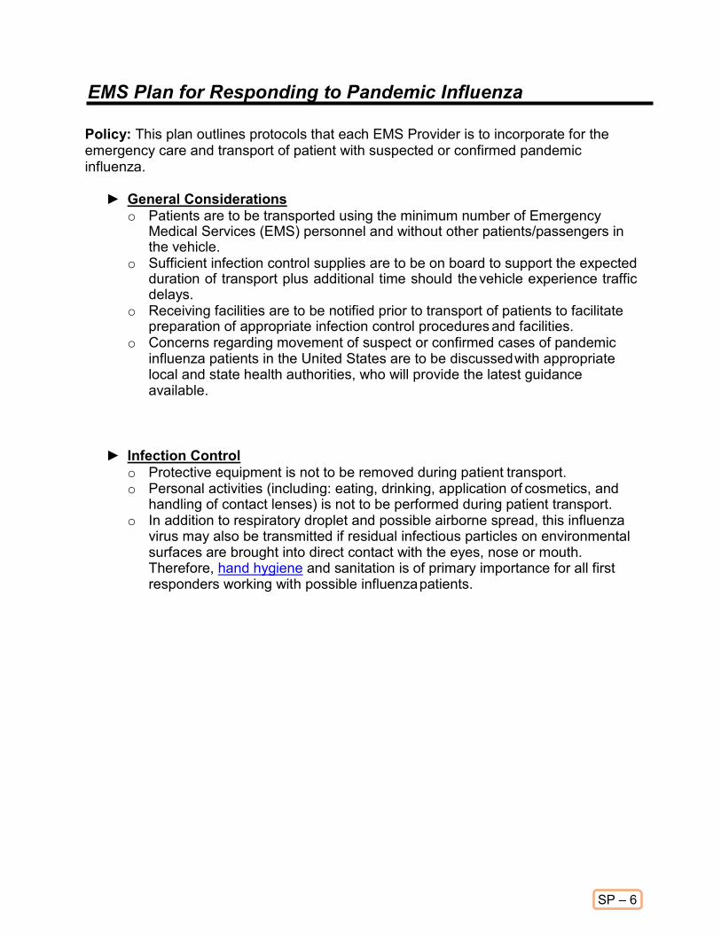
EMS Plan for Responding to Pandemic Influenza
SP – 6
Policy: This plan outlines protocols that each EMS Provider is to incorporate for the emergency care and transport of patient with suspected or confirmed pandemic influenza.
► General Considerations o Patients are to be transported using the minimum number of Emergency
Medical Services (EMS) personnel and without other patients/passengers in the vehicle.
o Sufficient infection control supplies are to be on board to support the expected duration of transport plus additional time should the vehicle experience traffic delays.
o Receiving facilities are to be notified prior to transport of patients to facilitate preparation of appropriate infection control procedures and facilities.
o Concerns regarding movement of suspect or confirmed cases of pandemic influenza patients in the United States are to be discussed with appropriate local and state health authorities, who will provide the latest guidance available.
► Infection Control o Protective equipment is not to be removed during patient transport. o Personal activities (including: eating, drinking, application of cosmetics, and
handling of contact lenses) is not to be performed during patient transport. o In addition to respiratory droplet and possible airborne spread, this influenza
virus may also be transmitted if residual infectious particles on environmental surfaces are brought into direct contact with the eyes, nose or mouth. Therefore, hand hygiene and sanitation is of primary importance for all first responders working with possible influenza patients.

EMS Plan for Responding to Pandemic Influenza continued
SP – 6
► Protective Equipment and Procedures
o Disposable, non-sterile gloves are to be worn for all patient contact. o Gloves are to be removed and discarded in biohazard bags after patient care
is completed (e.g., between patients) or when soiled or damaged. o Hands are to be washed or disinfected with a waterless hand sanitizer
immediately after removal of gloves. o Disposable fluid-resistant gowns are to be worn for all patient care activity. If
gowns were not used, ALL responders promptly change into clean attire upon return to station.
o Gowns are to be removed and discarded in biohazard bags after patient care is completed or when soiled or damaged.
o Goggles or face-shields are to be worn in the patient-care compartment and when working within 6 feet of the patient. Corrective eyeglasses alone are not appropriate protection.
o Hooded PAPR with appropriate HEPA cartridge or fit-tested N-95 respirators are to be worn by personnel in the patient-care compartment at all times.
o Hooded PAPR with appropriate HEPA cartridge or fit-tested N-95 respirators are to be worn by the driver, if the driver's compartment is open to the patient-care compartment. Drivers that provide direct patient care (including moving patients on stretchers) must wear a disposable gown, eye protection, and gloves as described above during patient-care activities. Gowns and gloves are not required for personnel whose duties are strictly limited to driving.
o Vehicles that have separate driver and patient compartments and can provide separate ventilation to these areas are preferred for transport of patients. If a vehicle without separate compartments and ventilation must be used, main dashboard vents should remain open with rear ventilation fans turned on at the highest setting during transport patients to maximize air-exchange.
o The patient may wear a mask to reduce droplet production, if tolerated. o Oxygen delivery with simple and non-rebreather facemasks may be used for
patient oxygen support during transport. o Cardiopulmonary resuscitation (CPR) should only be performed using a
resuscitation bag-valve mask, equipped with HEPA filtration of expired air or a separate filter in the airway circuit.
o All aerosolized treatments such as nebulizer or CPAP should use a HEPA filtration system. If HEPA filtration systems are not available, alternative treatment to aerosol medication must be utilized.
► Mechanically Ventilated Patients o Mechanical ventilators for patient transport must provide HEPA filtration of
airflow exhaust. o Emergency Medical Services MUST consult their ventilator equipment
manufacturer to confirm appropriate filtration capability and the effect of filtration on positive pressure ventilation.

EMS Plan for Responding to Pandemic Influenza continued
SP – 6
► Waste disposal
o Dry solid waste, e.g., used gloves, dressings, etc., is to be collected in biohazard bags for disposal as regulated medical waste in accordance with local requirements at the destination medical facility.
o Waste that is saturated with blood, body fluids, or excreta is to be collected in leak-proof biohazard bags or containers for disposal as regulated medical waste in accordance with local requirements at the destination medical facility.
o Sharp items such as used needles are to be collected in puncture resistant sharps containers for disposal as regulated medical waste in accordance with local requirements at the destination medical facility.
o Suctioned fluids and secretions are to be stored in sealed containers for disposal as regulated medical waste in accordance with local requirements at the destination medical facility. Handling that might create splashes or aerosols during transport are to be avoided.
o Suction device exhaust is not to be vented inside the vehicle without HEPA filtration. Portable suction devices are to be fitted with in-line HEPA filters.

EMS Plan for Responding to Pandemic Influenza continued
SP – 6
► Cleaning and Disinfection After Transporting Patient
o Compressed air that might re-aerosolize infectious material is not to be used for cleaning the vehicle or reusable equipment.
o Non-patient-care areas of the vehicle are to be cleaned and maintained according to vehicle manufacturer's recommendations.
o Cleaning personnel are to wear non-sterile gloves, disposable gowns, masks and eye protection while cleaning the patient-care compartment.
o Patient-care compartments (including stretchers, railings, medical equipment control panels, and adjacent flooring, walls and work surfaces likely to be directly contaminated during care) are to be cleaned using an EPA-registered hospital disinfectant in accordance with manufacturer's recommendations.
o Spills of body fluids during transport are to be cleaned by placing absorbent material over the spill and collecting the used cleaning material in a biohazard bag. The area of the spill is to be cleaned using an EPA- registered hospital disinfectant. Cleaning personnel are to be notified of the spill location and initial clean-up performed.
o Contaminated reusable patient care equipment is to be placed in biohazard bags and labeled for cleaning and disinfection utilizing proper procedures.
o Personnel are to wear non-sterile gloves, disposable gowns, eye protection and face masks while cleaning reusable equipment.
o Reusable equipment is to be cleaned and disinfected according to manufacturer's instructions.
o Periodic decontamination of the interior compartment of the transport vehicle with vaporized hydrogen peroxide should be considered if it is available based upon level of suspected contamination and/or number of transports of potentially infected patients.
► Follow-up of EMS Personnel Who Transport Patients o After transportation, the service is to provide the following information to the
medical director: date and route of transport; duration of patient transport; names, contact information, and specific duties (including estimated duration of direct patient care provided) of transport personnel.
o Services should designate individuals to monitor personnel who have transported patients for evidence of fever or respiratory illness. EMS personnel who transport patients are to be assessed (directly or by telephone) at least daily for 10 days after transporting patient.
o Asymptomatic personnel may continue work during the follow-up period. o Symptomatic personnel must be relieved of EMS duties, directed to seek
medical care, and be reported to the state health department.

TOXMEDIC Emergency Care Protocols
TOX
Developed by: Brandon Johnson, Anthony Schebon and Dr. Robert Hudepohl, Hebron Fire Protection District Purpose
► It is the intent of the Toxmedic program and these protocols to provide advanced medical care/support to the HAZMAT response team, coordinate rapid medical intervention to the victims of hazardous materials exposures and or acts of toxic terrorism. To operate in this environment, a provider should maintain Advanced Hazmat Life Support verification (Provider or Instructor). The objectives of the Toxmedic are to identify the offending substance, alter absorption by decontamination, determine the severity of the exposure, recognize toxidromes, and execute the appropriate treatment protocol. The goal of the Toxmedic program is to reduce morbidity and mortality from Hazardous materials exposures and acts of toxic terrorism.
Procedure
► The following Protocols are specifically designed for use during the treatment of victims of hazardous materials exposure and or acts of toxic terrorism. These interventions are only supplements to the standing orders and protocols; in the vast majority of cases basic and advanced life support supportive care will be sufficient. o Irritant gases: Ammonia, Formaldehyde, Sulfur dioxide, etc. o Asphyxiants: CO, Methemoglobin-forming compounds, Cyanides and
Cyanogenic compounds. o Cholinesterase inhibitors: Pesticides Organophosphates and Carbamates. o Corrosives: Acids, Bases, Oxidizers and Phosphorus. o Hydrocarbons and Halogenated Hydrocarbons. o Hydrofluoric Acid and Fluorides. o Hydrazines o Ethylene Glycol and Methanol o Chemoterrorism: Nerve Agents, Vesicants o Mark 1 Kit administration. o Antidote Reference.

TOX
TOXMEDIC Emergency Care Protocols continued Antidote/Medication List
Oxygen Atropine Pralidoxime / 2-PAMCL Pyridoxine Mark 1 Auto-injector kit or DuoDote injector (Atropine 2mg / Pralidoxime 600mg) Methylene Blue Amyl Nitrite Sodium Nitrite Sodium Thiosulfate Calcium Gluconate Diazepam Proparacaine Albuterol Thiamine (Vitamin B-1)

TOXMEDIC: Irritant gases / Toxic Inhalations
TOX
Description
Irritant gases are corrosive toxicants with local toxic effects. The primary routes of exposure are, contact with skin and mucous membranes and inhalation. Irritant gases damage the moist surfaces that they contact including the eyes, nose, mouth, and the upper/lower airways. Irritant gases are not absorbed systemically and generally do not cause systemic intoxication. Although their effects are usually localized to the tissues they contact, they will however cause airway compromise secondary to airway irritation, swelling, bronchospasm, and non-cardiogenic pulmonary edema. Irritant gases are classified by their water solubility. The depth and severity of injury to the airways and the tracheobronchial tree can be anticipated by determining the gases water solubility, concentration, and duration of the exposure. Examples of irritant gases are: Ammonia, Formaldehyde, Hydrogen Chloride, and Chlorine.
Treatment
► Remove patient from the toxic atmosphere and decon as appropriate.
► Administer oxygen 100% NRBM. Intubate if necessary. Administer Albuterol 2.5 mg via HHNT if bronchospasm or wheezing is present.
► Monitor ECG/ACLS per protocol.
► IV Normal Saline to KVO.

TOXMEDIC: Asphyxiants – Simple/Systemic
TOX
Description
Asphyxiants are chemicals that interfere with the body’s ability to perform aerobic metabolism. Simple asphyxiants are those, which displace oxygen from the ambient atmosphere effectively reducing the concentration of oxygen available for inhalation. Hypoxia will result from the low oxygen atmosphere. Examples of simple asphyxiants include carbon dioxide, methane, and propane. Systemic or chemical asphyxiants interfere with oxygen transport via hemoglobin, or they interfere with oxygen utilization at the cellular level by blocking the mitochondrial enzyme Cytochrome oxidase (Cytochrome a, a3). Cytochrome oxidase is the final electron receptor in the electron transport chain. Cytochrome oxidase acts directly with molecular oxygen to produce aerobic metabolism.
Treatment
► Remove the patient from the oxygen deficient atmosphere.
► Administer oxygen 100% NRBM. Intubate if necessary.
► Monitor ECG and perform 12 lead EKG/ACLS per protocol
► I.V. Normal Saline to KVO.
► If the patient develops seizures despite adequate oxygenation and normal blood glucose values, administer a benzodiazepine per the seizure protocol.
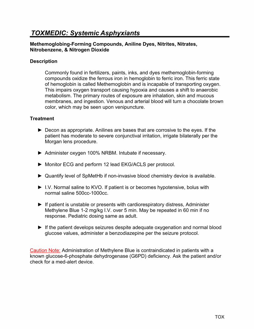
TOXMEDIC: Systemic Asphyxiants
TOX
Methemoglobing-Forming Compounds, Aniline Dyes, Nitrites, Nitrates, Nitrobenzene, & Nitrogen Dioxide Description
Commonly found in fertilizers, paints, inks, and dyes methemoglobin-forming compounds oxidize the ferrous iron in hemoglobin to ferric iron. This ferric state of hemoglobin is called Methemoglobin and is incapable of transporting oxygen. This impairs oxygen transport causing hypoxia and causes a shift to anaerobic metabolism. The primary routes of exposure are inhalation, skin and mucous membranes, and ingestion. Venous and arterial blood will turn a chocolate brown color, which may be seen upon venipuncture.
Treatment
► Decon as appropriate. Anilines are bases that are corrosive to the eyes. If the patient has moderate to severe conjunctival irritation, irrigate bilaterally per the Morgan lens procedure.
► Administer oxygen 100% NRBM. Intubate if necessary.
► Monitor ECG and perform 12 lead EKG/ACLS per protocol.
► Quantify level of SpMetHb if non-invasive blood chemistry device is available.
► I.V. Normal saline to KVO. If patient is or becomes hypotensive, bolus with normal saline 500cc-1000cc.
► If patient is unstable or presents with cardiorespiratory distress, Administer Methylene Blue 1-2 mg/kg I.V. over 5 min. May be repeated in 60 min if no response. Pediatric dosing same as adult.
► If the patient develops seizures despite adequate oxygenation and normal blood glucose values, administer a benzodiazepine per the seizure protocol.
Caution Note: Administration of Methylene Blue is contraindicated in patients with a known glucose-6-phosphate dehydrogenase (G6PD) deficiency. Ask the patient and/or check for a med-alert device.

TOXMEDIC: Systemic Asphyxiants – Carbon monoxide
TOX
Description
Carbon monoxide is an odorless, tasteless, non-irritating gas. Carbon monoxide binds to the oxygen binding sites in the hemoglobin molecules of the erythrocytes creating carboxyhemoglobin, which has a much-decreased ability to transport oxygen. Although not fully understood, carbon monoxide is known to have an effect on the mitochondrial enzyme cytochrome oxidase causing a disruption in oxygen utilization within the cell. Primary route of exposure is inhalation.
Treatment
► Remove patient from the toxic atmosphere.
► Administer oxygen 100% NRBM. Intubate if necessary.
► Monitor ECG and perform 12 lead EKG/ACLS per protocol.
► Quantify level SpCO with non-invasive CO Oximeter or exhaled breath CO device.
► I.V. Normal saline to KVO.
► Consider transport to a hospital with hyperbaric oxygen treatment capabilities.
► If the patient develops seizures despite adequate oxygenation and normal blood glucose values, administer a benzodiazepine per the seizure protocol.
Caution note: Standard pulse oximeters will report falsely high/normal oxygen saturations in patients with carbon monoxide exposure. Evaluate the patient‘s status by exposure history, signs, and symptoms.

TOXMEDIC Emergency Care Protocols continued
TOX

TOXMEDIC: Systemic Asphyxiants
TOX
Systemic Asphyxiants - Cyanides / Cyanogenic Compounds & Hydrogen Sulfide
Description
Cyanides/Cyanogenic compounds and Hydrogen Sulfide have a high affinity for ferric iron and bind with ferric iron in Cytochrome oxidase (Cytochrome a, a3). Cyanides inhibit the utilization of oxygen at Cytochrome a, a3, resulting in reduced ATP production. Inadequate ATP production results in cellular dysfunction and cell death. The CNS and the cardiovascular system are most dependent on a consistent supply of energy and thus are among the first systems to manifest signs and symptoms of cyanide toxicity. Routes of exposure are inhalation, ingestion, and contact with skin and mucous membranes.
Treatment
► Remove the patient from the toxic atmosphere and decon as appropriate.
► Administer oxygen 100% NRBM. Intubate if necessary.
► Monitor ECG and perform 12 lead EKG/ACLS per protocol.
► I.V. Normal saline to KVO.
► Administer Cyanokit / 2.5g Hydroxocabalamin I.V. over 15-20 minutes. Repeat initial dose after 30 minutes
► If the patient develops seizures despite adequate oxygenation and normal blood
glucose values, administer a benzodiazepine per the seizure protocol. Caution Note: It is imperative that all healthcare personnel avoid contact with bodily fluids and take respiratory precautions against off gassing of cyanide gas from patient respirations, belching, emesis, etc. Transport patient in a well-ventilated vehicle.

TOXMEDIC: Cholinesterase Inhibitors
TOX
Cholinesterase Inhibitors - Organophosphates and Carbamates Description
Organophosphates and Carbamates are commonly used as pesticides in agriculture and home pest control. Organophosphates and Carbamates target the CNS and PNS by binding to and inhibiting the enzyme acetylcholinesterase that is responsible for terminating the effects of acetylcholine at muscarinic and nicotinic receptors. An accumulation of acetylcholine at these receptor sites results in uncontrolled receptor stimulation and produces the classic cholinergic toxidrome. Acetylcholinesterase hydrolyzes the neurotransmitter acetylcholine into its constituents, choline and acetic acid, which are reabsorbed into the nerve cell for resynthesis into acetylcholine. Common routes of exposure are inhalation and contact with skin and mucous membranes.
Cholinergic Toxidrome / Signs & Symptoms Muscarinic Nicotinic
Diarrhea Mydriasis Urination Tachycardia Miosis Weakness Bradycardia, Bronchorrhea, Bronchospasm Hypertension Emesis Fasiculations Lacrimation Salivation, Sweating Treatment
► Remove the patient from the toxic atmosphere and decon as appropriate. ► Administer oxygen 100% NRBM. Intubate if necessary. Be prepared for acute
respiratory failure. ► Monitor ECG/ACLS per protocol. ► I.V. Normal Saline at KVO. ► If patient presents with muscarinic signs and symptoms, administer atropine 1-2
mg IV q5 minutes until bronchial secretions and hemodynamically significant bradycardia have abated. Pediatric dose is 0.02mg/kg to a maximum single dose of: o Infant < 2yrs 0.5 mg / 1.0 mg in severe cases. o Child 2-10yrs 1.0 mg / 2.0 mg in severe cases. o Adolescent 10-16yrs 2.0 mg / 4.0 mg in severe cases.

TOXMEDIC Emergency Care Protocols continued
TOX
Note: In severe cases (i.e. unconscious, seizures, respiratory extremis, or cardiorespiratory arrest), begin with a starting initial dose of 6 mg of Atropine I.V. in adults, and then repeat with 2 mg q5 minutes until the therapeutic end points from above are reached. Pediatric doses in severe cases are initial dose 2 times the single dose amounts calculated above (i.e. 0.04 mg/kg) with maximum single dose also doubled, then repeat with 0.02 mg/kg q5 minutes until the therapeutic end points from above are reached.
► Administer Pralidoxime (2-PAM) 1 gram (1000mg) I.V. over 10 minutes. Pediatric dose is 20-40 mg/kg over 10 minutes. Rapid I.V. administration of Pralidoxime can cause serious untoward effects (laryngospasm and neuromuscular blockade with paralysis) therefore it should be given slowly. Be prepared for rapid endotracheal intubation.
► If the patient develops seizures despite adequate oxygenation and normal blood glucose values, a benzodiazepine per the seizure protocol.
Note: The use of the Mark 1 antidote kit/DuoDote shall be reserved for those patients involved in MCI involving Organophosphates or acts of toxic terrorism. However, if I.V. Pralidoxime is not available due to shortage or other extenuating circumstances it is appropriate to substitute IM Pralidoxime. The provider will utilize the dosing guidelines from the Mark 1 administration protocol for IM Pralidoxime only. All other medications will be administered IV.

TOXMEDIC: Corrosives
TOX
Description
The primary toxicodynamic effect of virtually all acids, bases, oxidizers, and white phosphorus is the production of chemical burns due to their corrosive, local toxic effects. When acids contact tissue, they produce a pathological change termed coagulative necrosis, resulting in a thick hardened scab (coagulum). Bases act in much the same way except bases do not produce coagulative necrosis. Bases produce liquefactive necrosis. Liquefactive necrosis liquefies and destroys the tissue usually resulting in a much deeper burn. Routes of exposure are Inhalation, ingestion, and contact with the skin and mucous membranes.
Treatment
► Remove the patient from the toxic atmosphere and decon as appropriate. ► Administer oxygen 100% as needed. Intubate if necessary. Assess for airway
burns and signs of upper airway compromise/obstruction (i.e. hoarseness, stridor, coughing).
► Administer Albuterol 2.5 mg via HHNT if bronchospasm or wheezing is present. ► Monitor ECG. ► I.V. Normal saline to KVO. Severe burns may cause third spacing and
hypovolemia, monitor for shock and treat accordingly. ► Apply dry sterile dressings to burns after sufficient decontamination and protect
patient from exposure/heat loss.

TOXMEDIC: Hydrocarbons & Halogenated Hydrocarbons
TOX
Description
Volatile hydrocarbons and halogenated hydrocarbons predominantly affect the CNS and the myocardium. They are CNS anesthetic agents that produce decreased LOC, coma, and can lead to death. All hydrocarbons sensitize the myocardium to endogenous catecholamines thereby lowering the threshold for ventricular fibrillation. Dermal exposure can result in defatting dermatitis and chemical burns. Highly volatile hydrocarbons are simple asphyxiants producing potential hypoxia. The most significant hazard for most hydrocarbons is their flammability and their ability to form explosive mixtures in air. Routes of exposure are inhalation, ingestion, and contact with the skin and mucous membranes.
Treatment
► Remove the patient from the toxic atmosphere and decon as appropriate. ► Administer oxygen 100% NRBM. Intubate if necessary. ► Monitor ECG. Be prepared to manage ventricular dysrhythmias per current ACLS
protocols. ► I.V. Normal saline to KVO. ► If the patient develops seizures despite adequate oxygenation and normal blood
glucose values, administer a benzodiazepine per the seizure protocol. ► Hydrocarbons and halogenated hydrocarbons sensitize the myocardium to
endogenous catecholamines. Keep patient calm and avoid the administration of sympathomimetic agents if possible.

TOXMEDIC: Hydrofluoric Acid and Fluorides
TOX
Description
Although hydrofluoric acid is an acid and liberates the hydrogen ion in water, it is a relatively weak acid when compared to a strong acid such as hydrochloric acid. Nonetheless it will produce chemical burns. The primary toxicity of hydrofluoric acid, hydrogen fluoride, and all soluble fluorides is due to the fluoride anion. The fluoride anion combines with endogenous calcium and magnesium to produce insoluble calcium fluoride and magnesium fluoride. This results in hypocalcemia, hypomagnesemia, and hyperkalemia. These electrolyte abnormalities have the most profound effect on the excitable tissues in the nervous system, skeletal muscle, and the cardiac muscle. This is manifest as severe burning and pain at the site of contact. Toxic exposures may also present with muscle twitching, seizure, myocardial irritability, prolonged QT interval, and an increased potential for Torsades de Pointes. Common routes of exposure are inhalation, ingestion, and contact with skin and mucous membranes.
Treatment
► Remove the patient from the toxic atmosphere and decon as appropriate. ► Administer oxygen 100% NRBM as Needed / Intubate if necessary. ► Monitor ECG and obtain 12 Lead EKG. Be prepared to manage ventricular
dysrhythmias per current ACLS protocols. Consider Magnesium Sulfate as primary antidysrhythmic if hypomagnesemia is known or suspected.
► For local dermal exposure with severe pain, apply Calcium Gluconate gel to burned area. (Mix 10cc of a 10% Calcium Gluconate into a 2oz tube of water- soluble jelly). Massage into the affected area.
► If the patient develops seizures despite adequate oxygenation and normal blood glucose values, administer a benzodiazepine per the seizure protocol.

TOXMEDIC: Ethylene Glycol and Methanol
TOX
Description
Hydrazines are colorless, alkali liquids with ammonia like odors. Ammonia-like compounds such as hydrazines are excellent fuels as well as reducing agents. They are also widely used in the production of spandex. Hydrazines have numerous system toxicities; they are strong bases that produce severe burns with liquefactive necrosis. Hydrazines also antagonize the function of GABA and inhibit GABA formation in the brain. The resulting GABA deficiency results in over excitation of the brain and seizures that can be intractable, unless pyridoxine is given. Massive exposures of hydrazines may be hepatotoxic, which may result in acute liver failure.
Treatment
► Remove the patient from the toxic atmosphere and decon as appropriate. ► Administer oxygen 100% NRBM as needed. Intubate if necessary. ► Monitor ECG and obtain 12 Lead EKG. Be prepared to manage ventricular
dysrhythmias per current ACLS protocols. ► Normal saline to KVO. Severe burns may cause third spacing, monitor for shock
and treat accordingly. ► If seizures develop administer Pyridoxine 10% 25mg/kg (adult & peds) slowly
over 5 minutes. If seizures persist, administer a benzodiazepine per the seizure protocol.
Description Ethylene Glycol is a commonly found product (radiator fluid) and a common poisoning, especially in children. The clinical manifestations are described in three phases
1. 30mins to 12 hours – inebriation, metabolic acidosis, seizures, and coma. 2. 12 to 36 hours – tachycardia, tachypnea, hypertension, and pulmonary edema. 3. 36 to 48 hours – crystalluria, acute tubular necrosis with oliguria, and renal
failure. Methanol is a highly toxic alcohol commonly found in automobile windshield washer solvent and gas line antifreeze among others. Many new uses for methanol, predominantly as an alternative energy source, have also been proposed. If these new applications are developed, methanol is likely to become even more accessible in the future and therefore, more available for misuse.

TOXMEDIC: Ethylene Glycol and Methanol
TOX
Toxic Effects
► Cardiovascular effects – dysrhythmias, hypotension, and pulmonary edema. ► Respiratory effects – respiratory insufficiency or arrest, pulmonary edema and
chemical pneumonitis. ► CNS - CNS Depression, seizures, coma, headache, muscle weakness, and
delirium. ► GI – GI bleeding, nausea and vomiting, and diarrhea. ► Eye – Chemical conjunctivitis ► Skin – irritation to full thickness burns
Treatment
► Remove the patient from the toxic atmosphere and decon as appropriate. ► Administer oxygen 100% NRBM. Intubate if necessary. Be prepared for acute
respiratory failure. ► Monitor ECG/ACLS per protocol. ► I.V. Normal saline to KVO. ► In cases of metabolic acidosis i.e. respiratory rate twice the normal for the patient
consider sodium bicab 8.4% 1meq/kg IV. ► If the patient develops seizures despite adequate oxygenation and normal blood
glucose values, administer a benzodiazepine per the seizure protocol.

TOXMEDIC: Chemoterrorism Nerve Agents
TOX
Agent Synonym Aging Half-Time
Commercial organophosphates several days
Description Nerve agents were first developed in Germany during the early 1930‘s as chemical warfare agents. These agents are traditionally divided into two classes, the G and V agents. The G agents, Tabun (GA), Sarin (GB), and Soman (GD) were synthesized in Germany. The V agent, VX was developed in the United Kingdom. Nerve agents inhibit the enzyme acetylcholinesterase, which is responsible for deactivating the neurotransmitter acetylcholine. Therefore acetylcholine accumulates at all cholinergic receptors, resulting in uncontrolled receptor stimulation, thereby producing the cholinergic toxidrome. Nerve agents are derived from organophosphate compounds, nerve agents will irreversibly bind to acetylcholinesterase (aging) unless the antidote Pralidoxime is administered. Aging half-times
Soman GD < 2minutes Sarin GB 5 hours Tabun GA > 40 hours VX None > 40 hours
Cholinergic Toxidrome / Signs & Symptoms Muscarinic Nicotinic
Diarrhea Mydriasis Urination Tachycardia Miosis Weakness Bradycardia, Bronchorrhea, Bronchospasm Hypertension Emesis Fasiculations Lacrimation Salivation, Sweating

TOXMEDIC: Chemoterrorism Nerve Agents continued
TOX
Treatment ► Remove the patient from the toxic atmosphere and decon as appropriate. ► Administer oxygen 100% NRBM. Intubate if necessary. Be prepared for acute respiratory failure. ► Monitor ECG/ACLS per protocol. ► I.V. Normal saline to KVO. ► If patient presents with muscarinic signs & symptoms, Administer Atropine 1-2 mg I.V. q5 minutes until bronchial secretions and hemodynamically significant Bradycardia have abated. Pediatric dose is 0.02 mg/kg to a maximum single dose of: o Infant < 2yrs 0.5 mg / 1.0 mg in severe cases. o Child 2-10yrs 1.0 mg / 2.0 mg in severe cases. o Adolescent 10-16yrs 2.0 mg / 4.0 mg in severe cases. ► Administer Pralidoxime (2-PAM) 1 gram (1000mg) I.V. over 10 minutes. Pediatric dose is 20-40 mg/kg over 10 minutes. Rapid I.V. administration of Pralidoxime can cause serious untoward effects (laryngospasm and neuromuscular blockade with paralysis) therefore it should be given slowly. Be prepared for rapid endotracheal intubation. ► If the patient develops seizures despite adequate oxygenation and normal blood glucose values, administer a benzodiazepine per the seizure protocol. Note: The use of the Mark 1 antidote kit shall be reserved for those patients involved in MCI involving Organophosphates or acts of toxic terrorism. However, if I.V. Pralidoxime is not available due to shortage or other extenuating circumstances it is appropriate to substitute IM Pralidoxime. The provider will utilize the dosing guidelines from the Mark 1 administration protocol for IM Pralidoxime only. All other medications will be administered IV. Note: In severe cases (i.e. unconscious, seizures, respiratory extremis, or cardiorespiratory arrest), begin with a starting initial dose of 6 mg of Atropine I.V. in adults, and then repeat with 2 mg q5 minutes until the therapeutic end points from above are reached. Pediatric doses in severe cases are initial dose 2 times the single dose amounts calculated above (i.e. 0.04 mg/kg) with maximum single dose also doubled, then repeat with 0.02 mg/kg q5 minutes until the therapeutic end points from above are reached.

TOXMEDIC Emergency Care Protocols continued
TOX
Auto-Injector kit administration - Nerve agent Mass casualty Description
The Mark 1 Kit was developed by the US Armed Forces as an antidote auto-injector for battlefield management of nerve agent exposure. Today these kits have been made available to nearly all areas of the healthcare system. Recent history has proven that a mass casualty incident involving a nerve agent is not only possible, but also quite probable. However remote, the possibility does exist for a MCI involving one or more of these agents. The utilization of the Mark 1 Kit is reserved for an event such as this. The Mark 1 Kit is a highly pressurized auto-injector kit made to penetrate thick military uniforms. Due to the pain associated with administration, they should not be self-administered unless necessary. The kit contains Atropine 2mg I.M. and Pralidoxime 600mg I.M. Mark 1 dosing is based solely upon the severity of clinical signs and symptoms. See the dosage chart below. Note: Duodote, the newest auto-injector for the treatment of cholinesterase inhibitor poisoning contains Pralidoxime 600mg and Atropine 2mg in one injector. Administer per Mark I protocol.
Mild Moderate Severe Ambulatory Seated or prostrate In extremis
Miosis Moderate dyspnea Unconscious Eye Pain Coughing Seizures
Mild dyspnea Wheezing Paralysis Rhinorrhea Vomiting Resp Arrest
Blurred vision Fasiculation Card Arrest Lacrimation Weakness
Treatment 1 Mark 1 Kit 2 Mark 1 Kits 3 Mark 1 Kits

TOXMEDIC Emergency Care Protocols continued
TOX
Mark 1 kit administration - Nerve agent Mass casualty PROCEDURE FOR INTRAMUSCULAR INJECTION OF THE MARK 1 KIT
Apply firm, even pressure (not jabbing motion) to the injector until it pushes the needle into your thigh (or buttocks). Using a jabbing motion may result in an improper injection or injury to the thigh or buttocks. Hold the injector firmly in place for at least 10 seconds. The seconds can be estimated by counting "one thousand one," "one thousand two," and so forth. Firm pressure automatically triggers the coiled spring mechanism. This plunges the needle through the clothing into the muscle and at the same time injects the antidote into the muscle tissue. Carefully remove the auto injector from your injection site.

TOXMEDIC: Antidote Reference
TOX
Normobaric Oxygen
► Indications o Hypoxemia o Tissue hypoxia o Simple asphyxiant poisonings o Systemic asphyxiant poisonings o Significant methemoglobinemia o Carbon monoxide poisoning o Cyanide & cyanogenic compound poisonings o Azide & hydrozoic acid poisonings o Hydrogen sulfide & sulfide poisonings
► Contraindications (Relative) o Severe chronic lung disease (e.g., COPD) that requires hypoxic ventilatory
drive. o Paraquat poisoning, unless the patient cannot maintain his SpO2 90% while
breathing room air.
► Complications & Adverse Effects o Usually none. o Ablation of hypoxic ventilatory drive in patients with chronic obstructive
pulmonary disease (COPD) and chronic hypercarbia. o Long-term, high concentration administration can lead to ocular complications
in neonates and pulmonary complications in infants through adults.
► Dosage o As close to 100% as can be obtained by the various devices.
► Route of Administration o Inhalation
► How Supplied o 15 L/min nonrebreather reservoir mask (preferred, if the patient has adequate
ventilation). o Bag-valve-mask with 100% oxygen (preferred, if the patient has inadequate
ventilation prior to endotracheal intubation). o Endotracheal tube with 100% oxygen (if the patient requires endotracheal
intubation).
► Mechanism of Action o Supplies oxygen for aerobic metabolism.

TOXMEDIC: Antidote Reference
TOX
Hyperbaric Oxygen
► Indications o Significant carbon monoxide poisoning with syncope, seizures, coma, lactic
acidosis, myocardial ischemia, myocardial infarction, abnormal psychometric testing, a carboxyhemoglobin level > 25% in a nonpregnant patient, a carboxyhemoglobin level > 15% in pregnant patients, etc. Consult a hyperbaric physician.
o Significant cyanide & cyanogenic compound poisonings. Consult a hyperbaric physician.
► Experimental uses:
o Significant methemoglobinemia unresponsive to or with known contraindications to methylene blue, e.g., with known G6PD deficiency.
o Hydrogen sulfide & sulfide poisonings. o Azide & hydrazoic acid poisonings.
► Relative Contraindications o History of spontaneous
pneumothorax o Hyperthermia o Intractable claustrophobia o Pregnancy o Epilepsy o Active malignancy o Sinusitis o Cisplatinum o Otitis o Bleomycin o COPD o Adriamycin o Asthma
o Steroids o Lung diseases o Insulin o History of thoracic surgery o Sulfamylon o History of reconstructive ear
surgery o Opiates o Upper respiratory infection o Hereditary spherocytosis o Alcohol o Optic neuritis o Nicotine o Acidosis
► Absolute Contraindications
o Pneumothorax o Consult a hyperbaric physician for questions regarding contraindication.
► Complications & Adverse Effects o Seizures o Dysbaric injuries Pneumothorax Tension pneumothorax Middle ear damage Tympanic membrane hemorrhage & rupture Sinus squeeze

TOXMEDIC: Antidote Reference
TOX
Hyperbaric Oxygen
► Dosage o 100% oxygen at 2 to 3 atmospheres absolute (ATA) for 90 minutes and
repeated as required, with a second treatment no sooner than 8 hours after the first. Consult a hyperbaric physician.
► Route of Administration
o Inhalation ► How Supplied
o Hyperbaric chamber ► Mechanism of Action
o For carbon monoxide poisoning there is more rapid elimination of carbon monoxide and there is some evidence that post-carbon monoxide poisoning encephalopathy may be somewhat mitigated by HBO treatment.
o In cyanide and hydrogen sulfide poisoning the evidence is less clear and is
controversial, but anecdotal reports suggest patients who have not satisfactorily responded to supportive and specific antidotal therapy have survived after receiving HBO therapy.
o For patients with methemoglobinemia enough oxygen can be dissolved in the
plasma to sustain life even when very little hemoglobin is left to transport oxygen.

TOXMEDIC: Antidote Reference
TOX
Methylene Blue
► Indications o Methemoglobinemia with signs or symptoms of cardiac or cerebral hypoxia
such as dyspnea, chest pain, ischemic EKG changes, agitation, confusion, seizures, or coma. Methemoglobinemia ≥ 30%.
► Relative Contraindications
o Known methemoglobin reductase deficiency. o Lack of indications. A patient usually does not require treatment with
methylene blue if he is cyanotic, but is not in cardiorespiratory distress and has a methemoglobin level < 30%.
o Severe renal failure. o Reversal of nitrite-induced therapeutic methemoglobinemia for treatment of
cyanide or nitrile poisoning; however, some evidence indicates methylene blue can be used safely to treat rare cases of excessive methemoglobinemia caused by nitrite antidotes used to treat cyanide or cyanogenic compound poisoning.
► Absolute Contraindications
o Known glucose-6-phosphate dehydrogenase (G6PD) deficiency. Lethal hemolysis is possible!
o Allergy. ► Complications & Adverse Effects
o Nausea . o Vomiting o Headache o Blue-green urine (expected side effect) o Methemoglobinemia (if given in high doses [> 7mg/kg] or if given too fast) o Hemolysis (if given in normal doses to patients who are G6PD deficient, or if
patients with normal G6PD receive > 15 mg/kg)
► Dosage o Adult dosage: 1-2 mg/kg IV, slowly over 5 minutes. This dose can be
repeated in 30-60 minutes, if symptoms persist. If no response occurs after two doses, do not repeat again; consider G6PD deficiency or methemoglobin reductase deficiency.
o Pediatric Dosage: Same as adults. ► Route of Administration
o IV, over 5 minutes ► How Supplied
o 1% solution (10 mg /1mL). 10 mL /1 ampule.

TOXMEDIC: Antidote Reference
TOX
Methylene Blue
► Mechanism of Action o Methylene blue serves as a cofactor for NADPH-dependent methemoglobin
reductase, allowing more rapid reduction of the ferric iron (Fe+3) of methemoglobin back to the ferrous iron (Fe+2) of normal hemoglobin.
► Ineffective in the following:
o Sulfhemoglobinemia. o Hemoglobin M disease. o NADPH-dependent methemoglobin reductase deficiency.

TOXMEDIC: Antidote Reference
TOX
Amyl Nitrite
► Indications o First aid treatment for patients with significant sulfide poisoning. o Amyl nitrite's use and ambulance stocking can be deleted, if desired, because
of its lack of proved efficacy and abuse potential.
► Relative Contraindications o Significant hypotension o Methemoglobinemia > 40% o Carbon monoxide poisoning
► Absolute Contraindication o Allergy
► Complications & Adverse Effects o Headache o Hypotension o Reflex tachycardia o Hypoperfusion (shock)
► Dosage o 1 ampule inhaled for 30 seconds out of each minute; change ampules every 3
minutes. Ampules may be broken in medical gauze and held next to the mouth or nose of spontaneously breathing patients. Alternatively, ampules may be broken and placed inside the lip of a face mask or oxygen-powered breathing device (avoid ingestion or aspiration), or into a ventilation bag. Discontinue use when sodium nitrite is administered.
► Route of Administration
o Inhalation ► How Supplied
o 0.3 mL./crushable ampule. o 12 crushable ampules/ United States cyanide antidote kit.
► Mechanism of Action o The mechanism of action may be by inducing low levels of
methemoglobinemia. Another postulated mechanism is by acting through nitric oxide synthetase. Airway management and provision of supplemental oxygen increases efficacy.
► Ineffective
o For azide poisoning

TOXMEDIC: Antidote Reference
TOX
Sodium Nitrite
► Indications o Significant sulfide poisoning
► Relative Contraindications o Significant hypotension o Methemoglobinemia > 40% o Carbon monoxide poisoning
► Absolute Contraindication o Allergy
► Complications & Adverse Effects o Headache o Hypotension o Reflex tachycardia o Hypoperfusion (shock) o Excessive methemoglobinemia
► Dosage o Adult Dosage: 1 ampule, over no less than 5 minutes o Pediatric Dosage: 0.12 to 0.33 mL/kg (up to a maximum of 10 mL), over no
less than 5 minutes. o Adult or Pediatric Dosages o Half the initial dose may be repeated, if an adequate clinical response has not
occurred in 30 minutes.
► Route of Administration o IV, over absolutely no less than 5 minutes, with frequent blood pressure
monitoring. The usual dose can be diluted in 50 to 100 mL of D5W or NS and given more slowly.
► How Supplied
o 3% solution (300 mg / 10 mL ampule). o 2 ampules/ United States cyanide antidote kit.
► Mechanism of Action o Postulated to induce low levels of methemoglobinemia or by acting through
nitric oxide synthetase. Airway management and provision of supplemental oxygen increases efficacy.
► Ineffective
o For azide poisoning

TOXMEDIC: Antidote Reference
TOX
Pralidoxime
► Indications o Organophosphate pesticide or military nerve agent poisoning. o Unknown cholinesterase inhibitor poisoning. o Controversial for carbamate pesticide poisoning.
► Relative Contraindications o Myasthenia gravis o Renal failure
► Absolute Contraindications o Inability to perform endotracheal intubation, if neuromuscular blockade were
to occur (a rare, dose and rate related complication).
► Complications & Adverse Effects o Generally safe. o Neuromuscular blockade, laryngospasm, muscular rigidity, and tachycardia
have occurred with rapid IV administration, or with doses much higher than those usually administered.
o Rare: Visual disturbances, weakness, blurred vision, diplopia, dizziness, headache, nausea, hyperventilation, tachycardia, transiently elevated blood pressure, and asystole (1 reported case only).
► Dosage
o Adult Dosage: Bolus & Infusion: 1-2 g IV over 5-10 minutes. This dose may be repeated
1 hour after the first, if weakness or fasciculations have not resolved. 500 mg/h IV infusion for 24 hours after initial bolus.
o Pediatric Dosage: Bolus & Infusion: 20-40 mg/kg IV over 10 minutes. 5-10 mg/kg/h IV infusion for 24 hours after initial bolus.
► How Supplied
o 1g/20 mL solution ► Mechanism of Action
o Dephosphorylates (reactivates) phosphorylated (deactivated) cholinesterase that has not irreversibly "aged." The average "aging" time varies from minutes with some military nerve agents to a few days with some organophosphate pesticides.
► Ineffective
o For nicotine poisoning. o For tobacco (nicotine) poisoning.

TOXMEDIC: Antidote Reference
TOX
Atropine
► Indications o Organophosphate or carbamate pesticide poisoning o Organophosphate military nerve agent poisoning
► Relative Contraindications o Narrow angle (angle-closure) glaucoma o Obstructive uropathy o Myasthenia gravis o Patients who cannot tolerate an elevated heart rate such as those with
coronary artery disease (CAD), congestive heart failure, tachydysrhythmias, hypertension, thyrotoxicosis, etc.
► Absolute Contraindication o Absence of significant muscarinic effects
► Complications & Adverse Effects o Hot dry skin o Visual disturbances (blurred vision) o Photophobia (light hurts the eyes) due to cycloplegia (paralysis of the iris and
the ciliary muscle that focuses the lens) o Mydriasis (dilated pupils) o Acute narrow angle (angle-closure) glaucoma o Dry mouth o Constipation o Urinary retention o Tachycardia o Myocardial ischemia and infarction due to tachycardia with increased
myocardial oxygen demand o Central anticholinergic syndrome (hallucinations, agitation, combative)
► Dosage o Adult Dosage Range: 0.5 mg (usual IV ACLS dose) to 6 mg (maximum IM dose for
organophosphate military nerve agent severe poisoning). Recommendations: 1-2 mg, IV, every 5 minutes, until bronchial secretions
and significant bradycardia are controlled. Very large doses may be required with organophosphates.
o Pediatric Dosage Range: 0.01-0.04 mg/kg (Never give less than 0.1 mg!) Recommendations: 0.02 mg/kg, IV, every 5 minutes, until bronchial
secretions and significant bradycardia are controlled. Very large doses may be required with organophosphates. Required doses are larger than those used in ACLS.

TOXMEDIC: Antidote Reference
TOX
Atropine
► Routes of Administration o IV preferred o IM possible o Endotracheal possible
► How Supplied o 8 mg/20 mL solution o Use preservative (phenol)-free formulations for massive dosing that requires
many repeat boluses or a continuous IV atropine infusion.
► Mechanism of Action o Competitive antagonism of acetylcholine at muscarinic receptors
► Ineffective o At nicotinic receptors

TOXMEDIC: Antidote Reference
TOX
Calcium Gluconate
► Indications o Hydrofluoric acid burns. o Hydrogen fluoride or other fluoride systemic toxicity (such as hypocalcemia,
hypomagnesemia, and/or hyperkalemia.
► Contraindications o Concurrent toxicity of digoxin or other cardiac glycosides.
► Complications & Adverse Effects o Hypercalcemia o Precipitation or exacerbation of cardiac glycoside toxicity. o Local irritation & pain at injection sites.
► Dosage o Topical: Topical application (inunction) with an extemporaneously made gel
(2.5 to 10%), or used undiluted (10%) without gel, inside a surgical glove, for fingertip, thumb, and hand exposures.
o Subcutaneous: Subcutaneous injection titrated to relief of pain with 0.5 mL per cm2 of skin surface area.
o Eyes: a Morgan lens may be placed bilaterally, and the eyes flushed with a solution of normal saline 500 ml and 50 ml 10% Calcium Gluconate.
o Adult Dosage: 10-30 mL IV for systemic fluoride poisoning. Titrated to control cardiac
dysrhythmias, conduction disturbances, hypocalcemia, hypomagnesemia, and hyperkalemia.
o Pediatric Dosage: 0.2-0.3 mL/kg, IV
► Routes of Administration o Topical o Subcutaneous o IV o Morgan Lens
► How Supplied o Extemporaneously prepared 2.5% to 10% gel or solution for dermal
application. o 10% solution for intravenous, or subcutaneous injection. o Each 10 ml- vial of calcium gluconate contains 1 g of calcium gluconate.
► Mechanism of Action o Calcium ions bind with fluoride ions to produce the inactive calcium fluoride
salt (CaF2). Calcium repletes depleted stores of endogenous calcium and counteracts the effects of hyperkalemia.
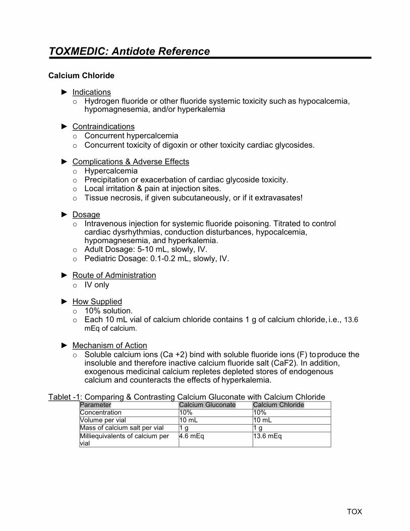
TOXMEDIC: Antidote Reference
TOX
Calcium Chloride
► Indications o Hydrogen fluoride or other fluoride systemic toxicity such as hypocalcemia,
hypomagnesemia, and/or hyperkalemia
► Contraindications o Concurrent hypercalcemia o Concurrent toxicity of digoxin or other toxicity cardiac glycosides.
► Complications & Adverse Effects o Hypercalcemia o Precipitation or exacerbation of cardiac glycoside toxicity. o Local irritation & pain at injection sites. o Tissue necrosis, if given subcutaneously, or if it extravasates!
► Dosage o Intravenous injection for systemic fluoride poisoning. Titrated to control
cardiac dysrhythmias, conduction disturbances, hypocalcemia, hypomagnesemia, and hyperkalemia.
o Adult Dosage: 5-10 mL, slowly, IV. o Pediatric Dosage: 0.1-0.2 mL, slowly, IV.
► Route of Administration o IV only
► How Supplied o 10% solution. o Each 10 mL vial of calcium chloride contains 1 g of calcium chloride, i.e., 13.6
mEq of calcium.
► Mechanism of Action o Soluble calcium ions (Ca +2) bind with soluble fluoride ions (F) to produce the
insoluble and therefore inactive calcium fluoride salt (CaF2). In addition, exogenous medicinal calcium repletes depleted stores of endogenous calcium and counteracts the effects of hyperkalemia.
Tablet -1: Comparing & Contrasting Calcium Gluconate with Calcium Chloride
Parameter Calcium Gluconate Calcium Chloride Concentration 10% 10% Volume per vial 10 mL 10 mL Mass of calcium salt per vial 1 g 1 g Milliequivalents of calcium per vial
4.6 mEq 13.6 mEq

TOXMEDIC: Antidote Reference
TOX
Pyridoxine
► Indications o Hydrazine poisonings
► Contraindications o None
► Complications & Adverse Effects o None acutely o Peripheral neuropathy with chronic, excessive dosing o Pyridoxine withdrawal seizures in neonates of mothers who took chronic,
excessive doses of pyridoxine during pregnancy
► Dosage o Adult & Pediatric: 25 mg/kg, over 5 minutes.
► Route of Administration o IV
► How Supplied o 10% solution. o 100mg/ml 1ml vial
► Mechanism of Action o Hydrazines deplete the major cerebral inhibitory neurotransmitter, gamma-
aminobutyric acid (GABA), resulting in intractable seizures that usually do not respond to standard anticonvulsants. Pyridoxine is a required synthetic cofactor that enables the brain to regenerate GABA and stop seizing.
► Ineffective
o For seizures not caused by GABA depletion

TOXMEDIC: Antidote Reference
TOX
Thiamine
► Indications o Ethylene glycol and methanol poisonings.
► Contraindications o Allergy
► Complications & Adverse Effects o Adverse effects are highly unusual. o Headache, irritability, tremors, nausea, vomiting, and palpitations. o Eczematous reactions and Herpes-Zoster may occur. o Anaphylactoid reactions: anxiety, pruritus, respiratory distress, nausea,
abdominal pain, angioneurotic edema and cardiovascular collapse.
► Dosage o Adult: 100mg. o Pediatric: 50mg.
► Route of Administration o IV
► How Supplied o 100mg/mL 2mL vial
► Mechanism of Action o Thiamine is one of the B vitamins, a group of water-soluble vitamins that
participate in many of the chemical reactions in the body. o Thiamine (vitamin B1) helps the body cells convert carbohydrates into energy.
It is also essential for the functioning of the heart, muscles, and nervous system.
► Ineffective
o Not applicable