Aggresome-Autophagy Involvement in a Sarcopenic Patient with Rigid Spine Syndrome and a p.C150R...
11
AGING NEUROSCIENCE ORIGINAL RESEARCH ARTICLE published: 19 August 2014 doi: 10.3389/fnagi.2014.00215 Aggresome–autophagy involvement in a sarcopenic patient with rigid spine syndrome and a p.C150R mutation in FHL1 gene Patrizia Sabatelli 1,2 , Silvia Castagnaro 3 , FrancescaTagliavini 1,2 , Martina Chrisam 3 , Francesca Sardone 1,2 , Laurence Demay 4 , Pascale Richard 5 , Spartaco Santi 1,2 , Nadir M. Maraldi 1 , Luciano Merlini 2 , Marco Sandri 6,7 and Paolo Bonaldo 3 * 1 Institute of Molecular Genetics, CNR-National Research Council of Italy, Bologna, Italy 2 SC Laboratory of Musculoskeletal Cell Biology, Rizzoli Orthopedic Institute, Bologna, Italy 3 Department of Molecular Medicine, University of Padova, Padova, Italy 4 UF Cardiogénétique et Myogénétique, Service de Biochimie Métabolique, Groupe Hospitalier Pitié-Salpêtrière, Paris, France 5 UF Cardiogénétique et Myogénétique, Centre de Génétique, Hôpitaux Universitaires de la Pitié Salpêtrière, Paris, France 6 DulbeccoTelethon Institute, Venetian Institute of Molecular Medicine, Padova, Italy 7 Department of Biomedical Science, University of Padova, Padova, Italy Edited by: Emanuele Marzetti, Catholic University of the Sacred Heart, Italy Reviewed by: Heinz Jungbluth, King’s College London, UK BerilTalim, Hacettepe University, Turkey *Correspondence: Paolo Bonaldo, Department of Molecular Medicine, University of Padova, Via U. Bassi 58/B, Padova I-35131, Italy e-mail: [email protected] The four-and-half LIM domain protein 1 (FHL1) is highly expressed in skeletal and cardiac muscle. Mutations of the FHL1 gene have been associated with diverse chronic myopathies including reducing body myopathy, rigid spine syndrome (RSS), and Emery–Dreifuss mus- cular dystrophy. We investigated a family with a mutation (p.C150R) in the second LIM domain of FHL1. In this family, a brother and a sister were affected by RSS, and their mother had mild lower limbs weakness.The 34-year-old female had an early and progressive rigid- ity of the cervical spine and severe respiratory insufficiency. Muscle mass evaluated by DXA was markedly reduced, while fat mass was increased to 40%. CT scan showed an almost complete substitution of muscle by fibro-adipose tissue. Muscle biopsy showed accumulation of FHL1 throughout the cytoplasm and around myonuclei into multiprotein aggregates with aggresome/autophagy features as indicated by ubiquitin, p62, and LC3 labeling. DNA deposits, not associated with nuclear lamina components and histones, were also detected in the aggregates, suggesting nuclear degradation. Ultrastructural analysis showed the presence of dysmorphic nuclei, accumulation of tubulofilamentous and gran- ular material, and perinuclear accumulation of autophagic vacuoles. These data point to involvement of the aggresome–autophagy pathway in the pathophysiological mechanism underlying the muscle pathology of FHL1 C150R mutation. Keywords: myopathy, sarcopenia, FHL1, autophagy, protein aggregates INTRODUCTION Four-and-half LIM domain protein 1 (FHL1) is a cysteine-rich double zinc-finger protein encoded by the FHL1 gene, localized on chromosome X (Dawid et al., 1995; Kadrmas and Beckerle, 2004). To date, three distinct FHL1 splicing isoforms have been identi- fied (Brown et al., 1999; Morgan and Madgwick, 1999; Ng et al., 2001; Purcell et al., 2004; Johannessen et al., 2006; McGrath et al., 2006). FHL1A, also known as skeletal muscle LIM protein 1, is the full-length protein. FHL1B, or SLIMMER, is composed of the first three LIM domains and contains nuclear localization and export sequences and a RBP-J binding region. FHL1C, or KyoT2, is the shortest isoform, which contains only the first two LIM domains and a RBP-J binding region and interacts with PIAS1 (Taniguchi et al., 1998; Wang et al., 2007). Four-and-half LIM domain protein 1 is highly expressed in skeletal and cardiac muscles (Lee et al., 1998; Brown et al., 1999; Greene et al., 1999; Morgan and Madgwick, 1999), where it localizes in the myofibrillar sarcomeres and in the sarcolemma (Bertrand et al., 2014). This protein has been demonstrated to be involved in several processes, including cellular architecture (McGrath et al., 2003, 2006), myoblast differentiation (Lee et al., 2012), mechanotransduction (Sheikh et al., 2008), and myofiber size (Cowling et al., 2008). FHL1 binds signaling and cytoskeletal proteins as well as transcription factors, acting as a transcriptional regulator of nuclear factor of activated T cells (NFATc1) to enhance the expression of genes that increase myofiber size (Cowling et al., 2008). Mutations in the FHL1 gene are responsible for a number of muscular disorders, which exhibit a broad spectrum of clin- ical features and disease severity ranging from severe childhood onset to milder adult-onset disorders. The diseases described so far include X-linked myopathy with postural muscle atrophy (XMPMA) (Windpassinger et al., 2008), reducing body myopathy (RBM) (Schessl et al., 2009; Shalaby et al., 2009; Selcen et al., 2011; Schreckenbach et al., 2013), X-linked dominant scapuloperoneal myopathy (Quinzii et al., 2008; Chen et al., 2010), rigid spine syn- drome (RSS) (Shalaby et al., 2008), hypertrophic cardiomyopathy (Friedrich et al., 2012), and Emery–Dreifuss muscular dystrophy Frontiers in Aging Neuroscience www.frontiersin.org August 2014 |Volume 6 | Article 215 | 1
-
Upload
independent -
Category
Documents
-
view
0 -
download
0
Transcript of Aggresome-Autophagy Involvement in a Sarcopenic Patient with Rigid Spine Syndrome and a p.C150R...
AGING NEUROSCIENCEORIGINAL RESEARCH ARTICLE
published: 19 August 2014doi: 10.3389/fnagi.2014.00215
Aggresome–autophagy involvement in a sarcopenicpatient with rigid spine syndrome and a p.C150R mutationin FHL1 genePatrizia Sabatelli 1,2, Silvia Castagnaro3, FrancescaTagliavini 1,2, Martina Chrisam3, Francesca Sardone1,2,Laurence Demay 4, Pascale Richard 5, Spartaco Santi 1,2, Nadir M. Maraldi 1, Luciano Merlini 2, Marco Sandri 6,7
and Paolo Bonaldo3*1 Institute of Molecular Genetics, CNR-National Research Council of Italy, Bologna, Italy2 SC Laboratory of Musculoskeletal Cell Biology, Rizzoli Orthopedic Institute, Bologna, Italy3 Department of Molecular Medicine, University of Padova, Padova, Italy4 UF Cardiogénétique et Myogénétique, Service de Biochimie Métabolique, Groupe Hospitalier Pitié-Salpêtrière, Paris, France5 UF Cardiogénétique et Myogénétique, Centre de Génétique, Hôpitaux Universitaires de la Pitié Salpêtrière, Paris, France6 Dulbecco Telethon Institute, Venetian Institute of Molecular Medicine, Padova, Italy7 Department of Biomedical Science, University of Padova, Padova, Italy
Edited by:Emanuele Marzetti, CatholicUniversity of the Sacred Heart, Italy
Reviewed by:Heinz Jungbluth, King’s CollegeLondon, UKBeril Talim, Hacettepe University,Turkey
*Correspondence:Paolo Bonaldo, Department ofMolecular Medicine, University ofPadova, Via U. Bassi 58/B, PadovaI-35131, Italye-mail: [email protected]
The four-and-half LIM domain protein 1 (FHL1) is highly expressed in skeletal and cardiacmuscle. Mutations of the FHL1 gene have been associated with diverse chronic myopathiesincluding reducing body myopathy, rigid spine syndrome (RSS), and Emery–Dreifuss mus-cular dystrophy. We investigated a family with a mutation (p.C150R) in the second LIMdomain of FHL1. In this family, a brother and a sister were affected by RSS, and their motherhad mild lower limbs weakness.The 34-year-old female had an early and progressive rigid-ity of the cervical spine and severe respiratory insufficiency. Muscle mass evaluated byDXA was markedly reduced, while fat mass was increased to 40%. CT scan showed analmost complete substitution of muscle by fibro-adipose tissue. Muscle biopsy showedaccumulation of FHL1 throughout the cytoplasm and around myonuclei into multiproteinaggregates with aggresome/autophagy features as indicated by ubiquitin, p62, and LC3labeling. DNA deposits, not associated with nuclear lamina components and histones, werealso detected in the aggregates, suggesting nuclear degradation. Ultrastructural analysisshowed the presence of dysmorphic nuclei, accumulation of tubulofilamentous and gran-ular material, and perinuclear accumulation of autophagic vacuoles. These data point toinvolvement of the aggresome–autophagy pathway in the pathophysiological mechanismunderlying the muscle pathology of FHL1 C150R mutation.
Keywords: myopathy, sarcopenia, FHL1, autophagy, protein aggregates
INTRODUCTIONFour-and-half LIM domain protein 1 (FHL1) is a cysteine-richdouble zinc-finger protein encoded by the FHL1 gene, localized onchromosome X (Dawid et al., 1995; Kadrmas and Beckerle, 2004).To date, three distinct FHL1 splicing isoforms have been identi-fied (Brown et al., 1999; Morgan and Madgwick, 1999; Ng et al.,2001; Purcell et al., 2004; Johannessen et al., 2006; McGrath et al.,2006). FHL1A, also known as skeletal muscle LIM protein 1, is thefull-length protein. FHL1B, or SLIMMER, is composed of the firstthree LIM domains and contains nuclear localization and exportsequences and a RBP-J binding region. FHL1C, or KyoT2, is theshortest isoform, which contains only the first two LIM domainsand a RBP-J binding region and interacts with PIAS1 (Taniguchiet al., 1998; Wang et al., 2007).
Four-and-half LIM domain protein 1 is highly expressed inskeletal and cardiac muscles (Lee et al., 1998; Brown et al., 1999;Greene et al., 1999; Morgan and Madgwick, 1999), where itlocalizes in the myofibrillar sarcomeres and in the sarcolemma(Bertrand et al., 2014). This protein has been demonstrated to
be involved in several processes, including cellular architecture(McGrath et al., 2003, 2006), myoblast differentiation (Lee et al.,2012), mechanotransduction (Sheikh et al., 2008), and myofibersize (Cowling et al., 2008). FHL1 binds signaling and cytoskeletalproteins as well as transcription factors, acting as a transcriptionalregulator of nuclear factor of activated T cells (NFATc1) to enhancethe expression of genes that increase myofiber size (Cowling et al.,2008).
Mutations in the FHL1 gene are responsible for a numberof muscular disorders, which exhibit a broad spectrum of clin-ical features and disease severity ranging from severe childhoodonset to milder adult-onset disorders. The diseases describedso far include X-linked myopathy with postural muscle atrophy(XMPMA) (Windpassinger et al., 2008), reducing body myopathy(RBM) (Schessl et al., 2009; Shalaby et al., 2009; Selcen et al., 2011;Schreckenbach et al., 2013), X-linked dominant scapuloperonealmyopathy (Quinzii et al., 2008; Chen et al., 2010), rigid spine syn-drome (RSS) (Shalaby et al., 2008), hypertrophic cardiomyopathy(Friedrich et al., 2012), and Emery–Dreifuss muscular dystrophy
Frontiers in Aging Neuroscience www.frontiersin.org August 2014 | Volume 6 | Article 215 | 1
Sabatelli et al. Aggresome–autophagy involvement in FHL1 pathogenesis
(Gueneau et al., 2009; Knoblauch et al., 2010). RBM is charac-terized by the presence of intracellular protein aggregates called“reducing bodies (RBs)”mainly containing mutated FHL1 protein,cytoskeletal and intermediate filament proteins, and componentsof the unfolded protein response pathway (Liewluck et al., 2007).Although scapuloperoneal myopathy, XMPMA, RSS, hypertrophiccardiomyopathy, and Emery–Dreifuss muscular dystrophy sharesome overlapping clinical features and muscle pathology withRBM, the involvement of protein aggregation in these disordersremains unclear (Wilding et al., 2014).
Reducing bodies morphologically resemble aggresomes, struc-tures proposed to facilitate the sequestration, and degradationof toxic misfolded proteins. Non-functional, damaged, and/ormisfolded proteins are removed from the cell by the ubiquitinproteasome system. However, when the capacity of the protea-some is impaired or overwhelmed, polyubiquitinated misfoldedproteins cannot be properly cleared and accumulate into the aggre-some (Goldberg, 2003; Kawaguchi et al., 2003). Accumulatingevidence suggests that aggresomes are substrates for autophagy.Autophagy is a degradation pathway that mediates bulk clear-ance of cytosolic proteins and organelles by the lysosome in ahighly regulated process involving the coordinated actions of alarge number of autophagy-related (Atg) genes. In response toparticular stimuli, such as proteasomal dysfunction, an isolationmembrane forms and expands to sequester portions of cytoplasminto double membrane structures called autophagosomes. Theautophagosomes eventually fuse with lysosomes and their con-tents are degraded by lysosomal hydrolases. One hypothesis is thataggresomes may concentrate aggregated proteins for more effi-cient autophagic degradation (Bjorkoy et al., 2005, 2006; Iwataet al., 2005). Recent evidence indicates that aggresome formationis mediated by dynein/dynactin-mediated transport of misfoldedproteins to the centrosome and involves several regulators, includ-ing the E3 ubiquitin-protein ligase parkin (Olzmann et al., 2008).Aggresome clearance is mediated by ubiquitin-binding proteinssuch as p62/SQSTM1 (Kirkin et al., 2009), an adaptor proteinthat decides the fate of protein degradation either through theubiquitin proteasome system or the autophagy–lysosome pathway(Komatsu et al., 2007; Kirkin et al., 2009; Komatsu and Ichimura,2010). Here, we report evidence of aggresome and autophagyinvolvement in the muscle of a sarcopenic patient with RSS andp.C150R mutation in the FHL1 gene.
MATERIALS AND METHODSGENOTYPINGThe six coding exons and introns boundaries of FHL1(NM_001159702) were screened for mutations by PCR on DNAfrom peripheral lymphocytes using primer pairs with a universalsequence (Table on demand). Exon 5 was sequenced with primers:
5PUF: 5′−ACCGTTAGTATGCGAGTTGGATTCAGGCAC
TGGATCCTA− 3′
5PUR: 5′−TCGGATAGTCAGTCGTTTGCTGTCGTGAGG
ATGGAATG− 3′.
Analysis of sequences was done with SeqScape software(Applied Biosystem).
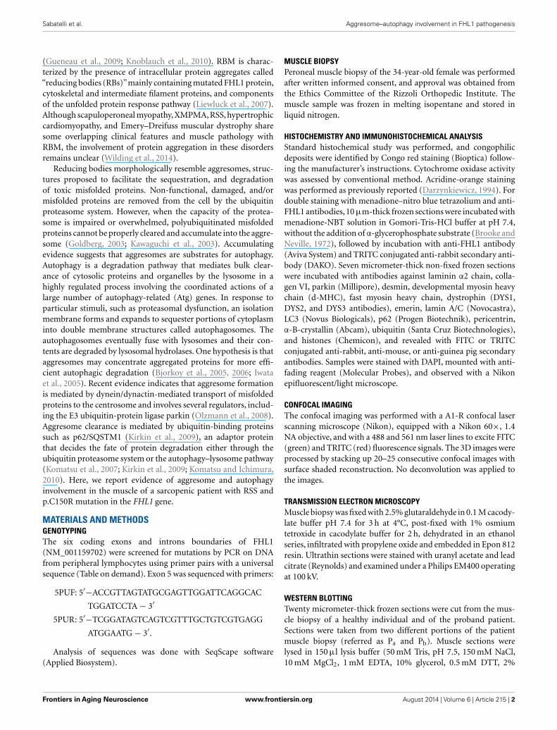
MUSCLE BIOPSYPeroneal muscle biopsy of the 34-year-old female was performedafter written informed consent, and approval was obtained fromthe Ethics Committee of the Rizzoli Orthopedic Institute. Themuscle sample was frozen in melting isopentane and stored inliquid nitrogen.
HISTOCHEMISTRY AND IMMUNOHISTOCHEMICAL ANALYSISStandard histochemical study was performed, and congophilicdeposits were identified by Congo red staining (Bioptica) follow-ing the manufacturer’s instructions. Cytochrome oxidase activitywas assessed by conventional method. Acridine-orange stainingwas performed as previously reported (Darzynkiewicz, 1994). Fordouble staining with menadione–nitro blue tetrazolium and anti-FHL1 antibodies,10 µm-thick frozen sections were incubated withmenadione-NBT solution in Gomori-Tris-HCl buffer at pH 7.4,without the addition of α-glycerophosphate substrate (Brooke andNeville, 1972), followed by incubation with anti-FHL1 antibody(Aviva System) and TRITC conjugated anti-rabbit secondary anti-body (DAKO). Seven micrometer-thick non-fixed frozen sectionswere incubated with antibodies against laminin α2 chain, colla-gen VI, parkin (Millipore), desmin, developmental myosin heavychain (d-MHC), fast myosin heavy chain, dystrophin (DYS1,DYS2, and DYS3 antibodies), emerin, lamin A/C (Novocastra),LC3 (Novus Biologicals), p62 (Progen Biotechnik), pericentrin,α-B-crystallin (Abcam), ubiquitin (Santa Cruz Biotechnologies),and histones (Chemicon), and revealed with FITC or TRITCconjugated anti-rabbit, anti-mouse, or anti-guinea pig secondaryantibodies. Samples were stained with DAPI, mounted with anti-fading reagent (Molecular Probes), and observed with a Nikonepifluorescent/light microscope.
CONFOCAL IMAGINGThe confocal imaging was performed with a A1-R confocal laserscanning microscope (Nikon), equipped with a Nikon 60×, 1.4NA objective, and with a 488 and 561 nm laser lines to excite FITC(green) and TRITC (red) fluorescence signals. The 3D images wereprocessed by stacking up 20–25 consecutive confocal images withsurface shaded reconstruction. No deconvolution was applied tothe images.
TRANSMISSION ELECTRON MICROSCOPYMuscle biopsy was fixed with 2.5% glutaraldehyde in 0.1 M cacody-late buffer pH 7.4 for 3 h at 4°C, post-fixed with 1% osmiumtetroxide in cacodylate buffer for 2 h, dehydrated in an ethanolseries, infiltrated with propylene oxide and embedded in Epon 812resin. Ultrathin sections were stained with uranyl acetate and leadcitrate (Reynolds) and examined under a Philips EM400 operatingat 100 kV.
WESTERN BLOTTINGTwenty micrometer-thick frozen sections were cut from the mus-cle biopsy of a healthy individual and of the proband patient.Sections were taken from two different portions of the patientmuscle biopsy (referred as Pa and Pb). Muscle sections werelysed in 150 µl lysis buffer (50 mM Tris, pH 7.5, 150 mM NaCl,10 mM MgCl2, 1 mM EDTA, 10% glycerol, 0.5 mM DTT, 2%
Frontiers in Aging Neuroscience www.frontiersin.org August 2014 | Volume 6 | Article 215 | 2
Sabatelli et al. Aggresome–autophagy involvement in FHL1 pathogenesis
sodium dodecyl sulfate, 1% Triton X-100) supplemented withphosphatase inhibitors (Sigma-Aldrich) and protease inhibitors(Roche), heated at 70°C for 10 min and centrifuged at 16,100 g for10 min at 4°C. The protein content of each lysate was determinedby the BCA Protein Assay kit (Pierce) and 30 µg of total proteinswere separated by SDS-PAGE (Invitrogen) and immunoblotted aspreviously described (Chen et al., 2014). Membranes were probedwith primary antibodies against FHL1 (ab23937 Abcam), LC3(Thermo Scientific), p62 (Progen Biotechnik), ubiquitin (Cell Sig-naling Technologies), beclin 1 (Cell Signaling), BNIP3 (Sigma),vinculin (Sigma), or GAPDH (Millipore). Proteins were revealedwith anti-rabbit, -mouse (Bethyl), -goat (Santa Cruz Biotechnolo-gies), or -guinea pig (Sigma) HRP-conjugated secondary antibod-ies using the ECL reagent (Pierce-Thermo Scientific). Densitomet-ric quantification of protein bands was performed by the ImageJsoftware (US National Institute of Health). Western blotting andquantifications are representative of at least three independentexperiments.
RESULTSThe proband is a 34-year-old woman who noticed the inabilityto extend the right thumb at age 20. Soon after, she manifestedneck weakness and limitation of flexion. Progression of weaknesswas rapid and she started to have difficulty in climbing stairs andgetting up from the floor. At age 24, she started falling severaltimes while walking. At age 26, the patient lost ambulation andwas wheelchair bound. Examination at age 34 showed an atrophicphenotype with marked diffuse muscle wasting and weakness andprominent contractures. She had normal facial muscle strength,a minimal residual motor function in the elbow extensors and inthe right biceps, but was profoundly weak in all the other muscles.She revealed marked contractures involving proximal and distaljoints. The most striking contractures were in the neck musclescausing a fixed hyperextended neck that was also impossible tomove in any direction. She showed a progressive decline in therespiratory function with a forced vital capacity 59% of predictedat age 25, 45% at age 27, and 13% at age 34. She refused to undergomechanical ventilation. Cardiac investigation, including echocar-diography and Holter, revealed no cardiac involvement. MuscleCT showed that all muscles were atrophic and substituted by fatand connective tissue, including the axial muscles, with a minimalsparing of the rectus femoris and vastus lateralis on the left, andof the peroneus on the right (Figure 1A). She was underweight(BMI= 17.1). However, according to her body composition, asrevealed by DXA, she was sarcopenic/obese (appendicular leanbody mass index of 3.27 kg/m2 and total body fat of 44.4%)(Baumgartner et al., 2004). Her brother had a similar atrophicphenotype with marked rigidity of the spine and diffuse contrac-tures but with more rapid progression, as he lost ambulation atage 18 and underwent tracheostomy at age 28. Their mother atage 58 had a mild lower limbs weakness and no contractures.
Sequencing of the FHL1 gene in the index case identified asingle missense mutation c.448T > C in exon 5 resulting in thereplacement at codon 150 of a cysteine residue with an arginineresidue (p.Cys150Arg). Nucleotide c.448 and residue p.150 of theFHL1 gene are highly conserved among species and evolution,and all prediction softwares conclude for a pathogenic mutation.
Analysis of the family showed that her brother and mother alsohave the same FHL1 mutation.
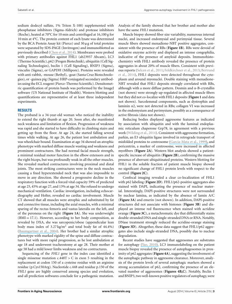
Muscle biopsy showed fiber size variability, numerous internalnuclei, and increased endomysial and perimysial tissue. Severalmuscle fibers showed menadione-NBT positive aggregates, con-sistent with the presence of RBs (Figure 1B). RBs were devoid ofoxidative enzyme activity and displayed an intense congophilia,indicative of the presence of amyloid deposits. Immunohisto-chemistry with FHL1 antibody revealed the presence of proteinaggregates in about 20% of muscle fibers. Consistent with previ-ous reports (Selcen et al., 2011; Feldkirchner et al., 2013; Bertrandet al., 2014), FHL1 deposits were detected throughout the cyto-plasm and around myonuclei. Double staining with menadione-NBT revealed that FHL1 deposits strongly correlated with RBs,although with a more diffuse pattern. Desmin and α-B-crystallin(not shown) were strongly up-regulated in affected muscle fibersbut they did not co-localize with FHL1 deposits (Figure 1 and datanot shown). Sarcolemmal components, such as dystrophin andlaminin α2, were not detected in RBs; collagen VI was increasedin the endomysium and perimysium, possibly as a consequence ofactive fibrosis (data not shown).
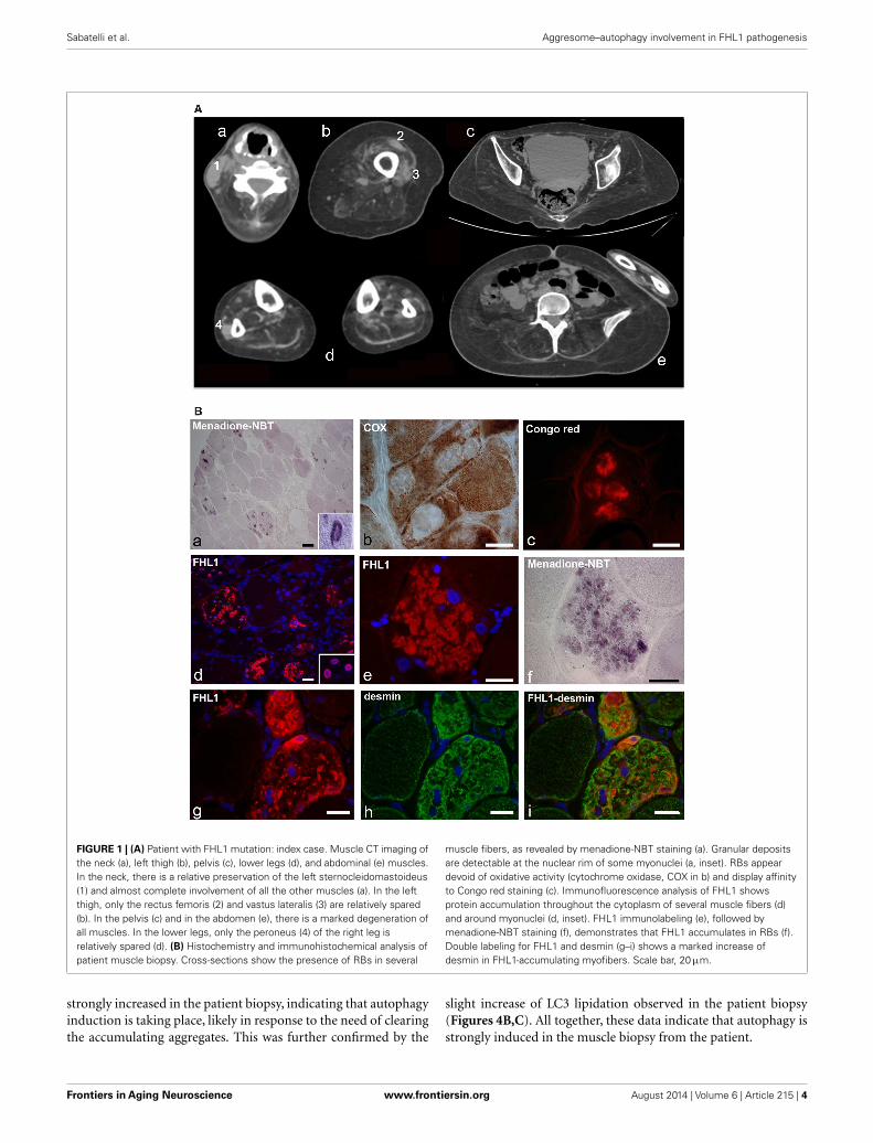
Reducing bodies displayed aggresome features as indicatedby association with ubiquitin and with the luminal endoplas-mic reticulum chaperone Grp78, in agreement with a previouswork (Wilding et al., 2014). Consistent with aggresome formation,parkin, an E3 ubiquitin ligase involved in retrograde transport ofmisfolded proteins to centrosome (Garcia-Mata et al., 1999), andpericentrin, a marker of centrosome, were increased in affectedmyofibers (Figure 2A). Western blot analysis showed a patient-specific increase of ubiquitin (Figure 2B), confirming the massivepresence of aberrant ubiquitinated proteins. Western blotting forFHL1 in the soluble fraction of patient muscle biopsy showedno significant change of FHL1 protein levels with respect to thecontrol (Figure 2C).
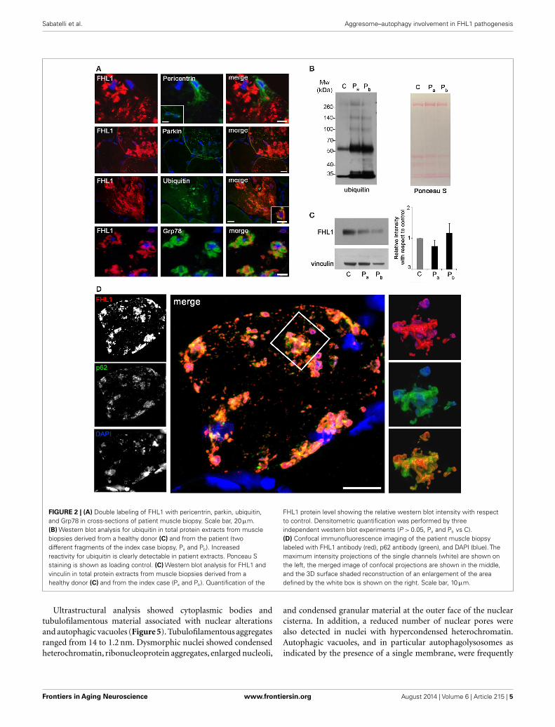
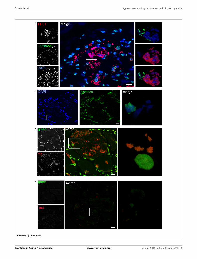
Confocal imaging revealed a clear co-localization of FHL1with p62 labeling (Figure 2D). FHL1/p62-positive aggregates alsostained with DAPI, indicating the presence of nuclear mater-ial. Interestingly, DAPI-positive structures were not surroundedby nuclear lamina, as indicated by the absence of lamin A/C(Figure 3A) and emerin (not shown). In addition, DAPI-positivestructures did not associate with histones (Figure 3B) and dis-played an intense red fluorescence when stained with acridineorange (Figure 3C), a metachromatic dye that differentially stainsdouble-stranded DNA and single-stranded DNA or RNA. Notably,DNase treatment strongly reduced the acridine-orange staining(Figure 3D). Altogether, these data suggest that FHL1/p62 aggre-gates also include single-stranded DNA, possibly due to nucleardegradation.
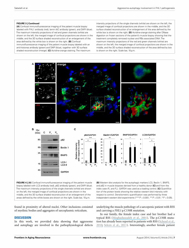
Recent studies have suggested that aggresomes are substratesfor autophagy (Yao, 2010). LC3 immunolabeling on the patientmuscle biopsy revealed the presence of autophagosomes in prox-imity of p62 aggregates (Figure 4A), suggesting the involvement ofthe autophagic pathway in aggresome clearance. Moreover, analy-sis of the protein levels of several autophagic markers showed astrong accumulation of p62, confirming the presence of an ele-vated number of aggresomes (Figures 4B,C). Notably, Beclin 1and BNIP3, two well-known positive regulators of autophagy, were
Frontiers in Aging Neuroscience www.frontiersin.org August 2014 | Volume 6 | Article 215 | 3
Sabatelli et al. Aggresome–autophagy involvement in FHL1 pathogenesis
FIGURE 1 | (A) Patient with FHL1 mutation: index case. Muscle CT imaging ofthe neck (a), left thigh (b), pelvis (c), lower legs (d), and abdominal (e) muscles.In the neck, there is a relative preservation of the left sternocleidomastoideus(1) and almost complete involvement of all the other muscles (a). In the leftthigh, only the rectus femoris (2) and vastus lateralis (3) are relatively spared(b). In the pelvis (c) and in the abdomen (e), there is a marked degeneration ofall muscles. In the lower legs, only the peroneus (4) of the right leg isrelatively spared (d). (B) Histochemistry and immunohistochemical analysis ofpatient muscle biopsy. Cross-sections show the presence of RBs in several
muscle fibers, as revealed by menadione-NBT staining (a). Granular depositsare detectable at the nuclear rim of some myonuclei (a, inset). RBs appeardevoid of oxidative activity (cytochrome oxidase, COX in b) and display affinityto Congo red staining (c). Immunofluorescence analysis of FHL1 showsprotein accumulation throughout the cytoplasm of several muscle fibers (d)and around myonuclei (d, inset). FHL1 immunolabeling (e), followed bymenadione-NBT staining (f), demonstrates that FHL1 accumulates in RBs (f).Double labeling for FHL1 and desmin (g–i) shows a marked increase ofdesmin in FHL1-accumulating myofibers. Scale bar, 20 µm.
strongly increased in the patient biopsy, indicating that autophagyinduction is taking place, likely in response to the need of clearingthe accumulating aggregates. This was further confirmed by the
slight increase of LC3 lipidation observed in the patient biopsy(Figures 4B,C). All together, these data indicate that autophagy isstrongly induced in the muscle biopsy from the patient.
Frontiers in Aging Neuroscience www.frontiersin.org August 2014 | Volume 6 | Article 215 | 4
Sabatelli et al. Aggresome–autophagy involvement in FHL1 pathogenesis
FIGURE 2 | (A) Double labeling of FHL1 with pericentrin, parkin, ubiquitin,and Grp78 in cross-sections of patient muscle biopsy. Scale bar, 20 µm.(B) Western blot analysis for ubiquitin in total protein extracts from musclebiopsies derived from a healthy donor (C) and from the patient (twodifferent fragments of the index case biopsy, Pa and Pb). Increasedreactivity for ubiquitin is clearly detectable in patient extracts. Ponceau Sstaining is shown as loading control. (C) Western blot analysis for FHL1 andvinculin in total protein extracts from muscle biopsies derived from ahealthy donor (C) and from the index case (Pa and Pb). Quantification of the
FHL1 protein level showing the relative western blot intensity with respectto control. Densitometric quantification was performed by threeindependent western blot experiments (P > 0.05, Pa and Pb vs C).(D) Confocal immunofluorescence imaging of the patient muscle biopsylabeled with FHL1 antibody (red), p62 antibody (green), and DAPI (blue). Themaximum intensity projections of the single channels (white) are shown onthe left, the merged image of confocal projections are shown in the middle,and the 3D surface shaded reconstruction of an enlargement of the areadefined by the white box is shown on the right. Scale bar, 10 µm.
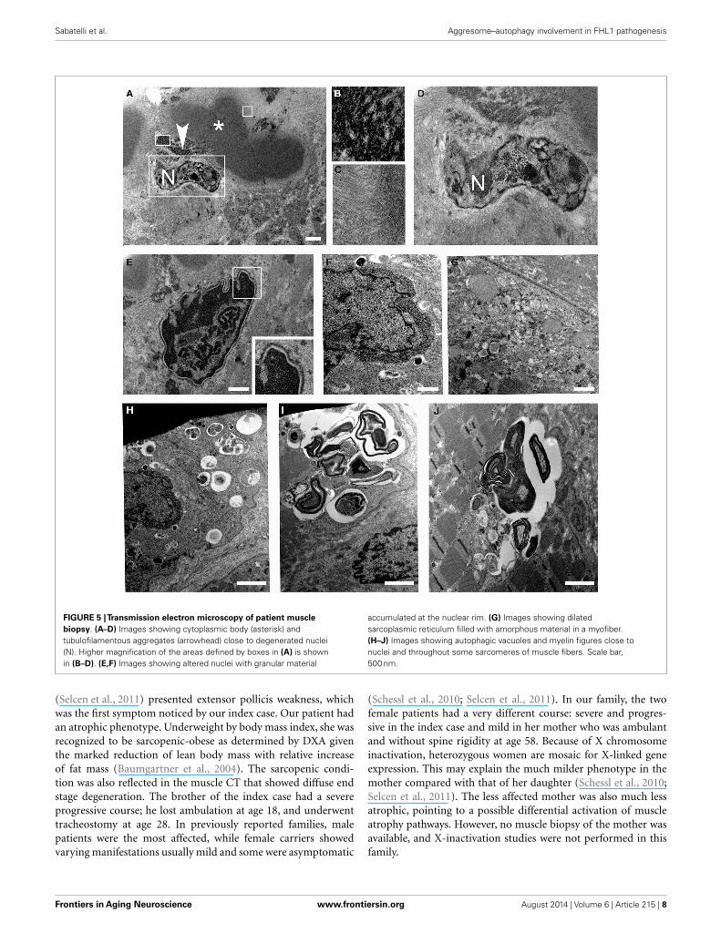
Ultrastructural analysis showed cytoplasmic bodies andtubulofilamentous material associated with nuclear alterationsand autophagic vacuoles (Figure 5). Tubulofilamentous aggregatesranged from 14 to 1.2 nm. Dysmorphic nuclei showed condensedheterochromatin, ribonucleoprotein aggregates, enlarged nucleoli,
and condensed granular material at the outer face of the nuclearcisterna. In addition, a reduced number of nuclear pores werealso detected in nuclei with hypercondensed heterochromatin.Autophagic vacuoles, and in particular autophagolysosomes asindicated by the presence of a single membrane, were frequently
Frontiers in Aging Neuroscience www.frontiersin.org August 2014 | Volume 6 | Article 215 | 5
Sabatelli et al. Aggresome–autophagy involvement in FHL1 pathogenesis
FIGURE 3 | Continued
Frontiers in Aging Neuroscience www.frontiersin.org August 2014 | Volume 6 | Article 215 | 6
Sabatelli et al. Aggresome–autophagy involvement in FHL1 pathogenesis
FIGURE 3 | Continued(A) Confocal immunofluorescence imaging of the patient muscle biopsylabeled with FHL1 antibody (red), lamin A/C antibody (green), and DAPI (blue).The maximum intensity projections of red and green channels (white) areshown on the left, the merged image of confocal projections are shown in themiddle, and the 3D surface shaded reconstruction of an enlargement of thearea defined by the white box is shown on the right. (B) Confocalimmunofluorescence imaging of the patient muscle biopsy labeled with ananti-histones antibody (green) and DAPI (blue), together with 3D surfaceshaded reconstruction (merge). (C) Acridine-orange staining. The maximum
intensity projections of the single channels (white) are shown on the left, themerged image of confocal projections are shown in the middle, and the 3Dsurface shaded reconstruction of an enlargement of the area defined by thewhite box is shown on the right. (D) Acridine-orange staining after DNasedigestion on frozen sections of the patient’s muscle biopsy showing that thetreatment completely removed nuclear and RBs associated DNA. Themaximum intensity projections of the red and green channels (white) areshown on the left, the merged image of confocal projections are shown in themiddle, and the 3D surface shaded reconstruction of the area defined by boxis shown on the right. Scale bar, 10 µm.
FIGURE 4 | (A) Confocal immunofluorescence imaging of the patient musclebiopsy labeled with LC3 antibody (red), p62 antibody (green), and DAPI (blue).The maximum intensity projections of the single channels (white) are shownon the left, the merged image of confocal projections are shown in themiddle, and the 3D surface shaded reconstruction of an enlargement of theareas defined by the white boxes are shown on the right. Scale bar, 10 µm.
(B) Western blot analysis for the autophagic markers LC3, Beclin 1, BNIP3,and p62 in muscle biopsies derived from a healthy donor (C) and from theindex case (Pa and Pb). GAPDH was used as a loading control. (C) Quantifica-tion of the protein levels showing the relative western blot intensity withrespect to control. Densitometric quantification was performed by threeindependent western blot experiments (***P < 0.001; **P < 0.01; *P < 0.05).
found in proximity of altered nuclei. Other inclusions consistedof myelinic bodies and aggregates of sarcoplasmic reticulum.
DISCUSSIONIn this work, we provided data showing that aggresomeand autophagy are involved in the pathophysiological defects
underlying the muscle pathology of a sarcopenic patient with RSSand carrying a FHL1 p.C150R mutation.
In our family, the female index case and her brother had atypical RSS (Moghadaszadeh et al., 2001). The p.C150R muta-tion has already been reported in patients with RSS (Schessl et al.,2010; Selcen et al., 2011). Interestingly, another female patient
Frontiers in Aging Neuroscience www.frontiersin.org August 2014 | Volume 6 | Article 215 | 7
Sabatelli et al. Aggresome–autophagy involvement in FHL1 pathogenesis
FIGURE 5 |Transmission electron microscopy of patient musclebiopsy. (A–D) Images showing cytoplasmic body (asterisk) andtubulofilamentous aggregates (arrowhead) close to degenerated nuclei(N). Higher magnification of the areas defined by boxes in (A) is shownin (B–D). (E,F) Images showing altered nuclei with granular material
accumulated at the nuclear rim. (G) Images showing dilatedsarcoplasmic reticulum filled with amorphous material in a myofiber.(H–J) Images showing autophagic vacuoles and myelin figures close tonuclei and throughout some sarcomeres of muscle fibers. Scale bar,500 nm.
(Selcen et al., 2011) presented extensor pollicis weakness, whichwas the first symptom noticed by our index case. Our patient hadan atrophic phenotype. Underweight by body mass index, she wasrecognized to be sarcopenic-obese as determined by DXA giventhe marked reduction of lean body mass with relative increaseof fat mass (Baumgartner et al., 2004). The sarcopenic condi-tion was also reflected in the muscle CT that showed diffuse endstage degeneration. The brother of the index case had a severeprogressive course; he lost ambulation at age 18, and underwenttracheostomy at age 28. In previously reported families, malepatients were the most affected, while female carriers showedvarying manifestations usually mild and some were asymptomatic
(Schessl et al., 2010; Selcen et al., 2011). In our family, the twofemale patients had a very different course: severe and progres-sive in the index case and mild in her mother who was ambulantand without spine rigidity at age 58. Because of X chromosomeinactivation, heterozygous women are mosaic for X-linked geneexpression. This may explain the much milder phenotype in themother compared with that of her daughter (Schessl et al., 2010;Selcen et al., 2011). The less affected mother was also much lessatrophic, pointing to a possible differential activation of muscleatrophy pathways. However, no muscle biopsy of the mother wasavailable, and X-inactivation studies were not performed in thisfamily.
Frontiers in Aging Neuroscience www.frontiersin.org August 2014 | Volume 6 | Article 215 | 8
Sabatelli et al. Aggresome–autophagy involvement in FHL1 pathogenesis
Muscle findings included menadione-NBT-positive aggregates,consistent with RBs, which also contained FHL1. The same FHL1mutation was previously reported in a family with RBM phenotype(Schessl et al., 2010) and in two patients with RBs and myofib-rillar myopathy (Selcen et al., 2011). Also in those patients, themutated FHL1 protein accumulated in RBs, pointing to a causativeeffect of this mutation in RB formation. The mutated cysteineresidue localizes in the second LIM domain of the protein and it isexpected to affect all FHL1 isoforms, i.e., full-length FHL1A andthe shorter FHL1B and FHL1C polypeptides. Cys150 is a crucialcoordinating residue in the second LIM domain (Michelsen et al.,1994) and mutations occurring at this site are predicted to induceprotein misfolding. It has been proposed that the accumulationof misfolded FHL1 polypeptides results in the characteristic RBaggregates observed in muscle of RBM patients as well as in C2C12myoblasts transfected with the mutant FHL1 protein (Schessl et al.,2008).
Aggresomes are structures proposed to facilitate the sequestra-tion and degradation of toxic misfolded proteins. In agreementwith previous reports (Bertrand et al., 2014), the RBs of theproband displayed characteristics of aggresomes, as indicated bythe increase of proteins involved in aggresome formation andby the accumulation of ubiquitin, Grp78, p62, and cytoskeletalcomponents, such as desmin and α-B-crystallin. Moreover, theFHL1-containing aggresomes were mainly accumulated aroundnuclei. It is well known that aggresome formation is mediated bythe dynein/dynactin-mediated transport of misfolded proteins tothe centrosome, as confirmed by the presence of aggresomes inthe perinuclear region and matching with centrosome markers(Olzmann et al., 2008). In muscle cells, the centrosome undergoesredistribution at the nuclear rim during differentiation (Bugnardet al., 2005). This pattern persists in adult muscle (Srsen et al.,2009), as indicated by the localization of centrosome markerson the outer membrane of the nuclear cisterna. This peculiarpositioning of the centrosome at the nuclear rim of muscle cellsaccounts for the recruitment of granular material with aggresome-like features we observed in nuclei of the FHL1 mutated musclefibers. In addition, we found that nuclei with perinuclear gran-ular material appeared dysmorphic, with dramatic changes ofthe nuclear envelope and hypercondensed heterochromatin. Thesedata, in addition to the finding of single-stranded DNA in aggre-somes, suggest that the aggresome accumulation at the nuclear rimmay induce nuclear degradation. This hypothesis is consistent withthe alterations of the nuclear envelope in cells containing inclusionbodies that were described in patients affected by Huntington dis-ease and in transgenic mice expressing mutant huntingtin (Waelteret al., 2001).
We also found that Grp78, an endoplasmic reticulum chap-erone up-regulated during the unfolded protein response, wasstrongly increased and associated to the FHL1 deposits in theproband muscle biopsy. This finding is in agreement with pre-vious work showing increased expression of Grp78 and unfoldedprotein response in patients with RBM (Liewluck et al., 2007).However, the association of Grp78 with aggresomes may be alsodue to retrograde transport from the endoplasmic reticulum, ashypothesized by the formation of aggresome-like inclusion bodiesinduced by mutant huntingtin (Garcia-Mata et al., 2002).
The association of FHL1 with p62 we detected in the probandmuscle biopsy indicates that the mutant FHL1 protein is tar-geted to degradative pathways. p62 is a multifunctional proteincontaining a number of protein–protein interaction motifs thatare involved in protein aggregation and degradation (Moscat andDiaz-Meco, 2009a,b). It has been hypothesized that p62 may act asa critical ubiquitin chain-targeting factor that shuttles substratesfor proteasomal degradation (Seibenhener et al., 2004). On theother hand, a role for p62 in aggregate formation for autophagicdegradation was also hypothesized (Komatsu et al., 2007; Kirkinet al., 2009; Komatsu and Ichimura, 2010). The strong increase ofBeclin 1 and BNIP3 levels we detected in the patient biopsy indi-cates that autophagy induction is taking place, likely to help theclearance of accumulating aggregates. This is further confirmed bythe slight increase of LC3 lipidation and by the accumulation ofLC3 deposits in proximity to aggresomes in the proband muscle.The presence of autophagic vacuoles and myelin figures furtherconfirms the involvement of the autophagic pathway in the patho-physiological alterations of this patient. Interestingly, the presenceof autophagosomes and autophagic vacuoles was also reportedin muscle biopsies of RBM patients (Bertrand et al., 2014). It isalso interesting to consider that FHL1 null mice, lacking globalFHL1 expression and without aggregates accumulation, displaysusceptibility to autophagy, as indicated by increased LC3 lipida-tion in skeletal muscle (Domenighetti et al., 2014). These findingspoint to a causative role of FHL1 deficiency in autophagy acti-vation, and indicate that activation of the autophagic pathway inFHL1-related myopathies may be a common pathophysiologicalmechanism, independent from the accumulation of protein aggre-gates. Although future studies of the autophagic flux in muscle cellsfrom patients and animal models for FHL1 deficiency are neededin order to understand in detail how and to which extent deregula-tion of autophagy contributes to the pathogenesis of FHL1-relatedmyopathies, our data demonstrate for the first time the coexistenceof aggresomes and autophagy in the muscle biopsy of a patientwith severe sarcopenia caused by p.C150R mutation in FHL1.These findings add new insights in delineating the altered mecha-nisms involved in the pathogenesis of FHL1-associated diseases.
ACKNOWLEDGMENTSThis work was supported by the Italian Ministry of Education,University and Research (FIRB RBAP10KCNS to Patrizia Sabatelli,and FIRB RBAP11Z3YA_003 to Paolo Bonaldo), and by “5 permille” 2010 Rizzoli Orthopaedic Institute.
REFERENCESBaumgartner, R. N., Wayne, S. J., Waters, D. L., Janssen, I., Gallagher, D., and Morley,
J. E. (2004). Sarcopenic obesity predicts instrumental activities of daily livingdisability in the elderly. Obes. Res. 12, 1995–2004. doi:10.1038/oby.2004.250
Bertrand, A. T., Bonnemann, C. G., and Bonne, G. (2014). 199th ENMC interna-tional workshop: FHL1 related myopathies, June 7-9, 2013, Naarden, the Nether-lands. Neuromuscul. Disord. 24, 453–462. doi:10.1016/j.nmd.2014.02.002
Bjorkoy, G., Lamark, T., Brech, A., Outzen, H., Perander, M., Overvatn, A., et al.(2005). p62/SQSTM1 forms protein aggregates degraded by autophagy and hasa protective effect on huntingtin-induced cell death. J. Cell Biol. 171, 603–614.doi:10.1083/jcb.200507002
Bjorkoy, G., Lamark, T., and Johansen, T. (2006). p62/SQSTM1: a missing linkbetween protein aggregates and the autophagy machinery. Autophagy 2, 138–139.doi:10.4161/auto.2.2.2405
Frontiers in Aging Neuroscience www.frontiersin.org August 2014 | Volume 6 | Article 215 | 9
Sabatelli et al. Aggresome–autophagy involvement in FHL1 pathogenesis
Brooke, M. H., and Neville, H. E. (1972). Reducing body myopathy. Neurology 22,829–840. doi:10.1212/WNL.22.8.829
Brown, S., Mcgrath, M. J., Ooms, L. M., Gurung, R., Maimone, M. M., and Mitchell,C. A. (1999). Characterization of two isoforms of the skeletal muscle LIM protein1, SLIM1. Localization of SLIM1 at focal adhesions and the isoform slimmer inthe nucleus of myoblasts and cytoplasm of myotubes suggests distinct roles inthe cytoskeleton and in nuclear-cytoplasmic communication. J. Biol. Chem. 274,27083–27091.
Bugnard, E., Zaal, K. J., and Ralston, E. (2005). Reorganization of microtubulenucleation during muscle differentiation. Cell Motil. Cytoskeleton 60, 1–13.doi:10.1002/cm.20042
Chen, D. H., Raskind, W. H., Parson, W. W., Sonnen, J. A., Vu, T., Zheng, Y., et al.(2010). A novel mutation in FHL1 in a family with X-linked scapuloperonealmyopathy: phenotypic spectrum and structural study of FHL1 mutations. J. Neu-rol. Sci. 296, 22–29. doi:10.1016/j.jns.2010.06.017
Chen, P., Cescon, M., Megighian, A., and Bonaldo, P. (2014). Collagen VI reg-ulates peripheral nerve myelination and function. FASEB J. 28, 1145–1156.doi:10.1096/fj.13-239533
Cowling, B. S., Mcgrath, M. J., Nguyen, M. A., Cottle, D. L., Kee, A. J., Brown, S., et al.(2008). Identification of FHL1 as a regulator of skeletal muscle mass: implicationsfor human myopathy. J. Cell Biol. 183, 1033–1048. doi:10.1083/jcb.200804077
Darzynkiewicz, Z. (1994). Simultaneous analysis of cellular RNA and DNA content.Methods Cell Biol. 41, 401–420. doi:10.1016/S0091-679X(08)61731-8
Dawid, I. B., Toyama, R., and Taira, M. (1995). LIM domain proteins. C. R. Acad.Sci. III, Sci. Vie 318, 295–306.
Domenighetti, A. A., Chu, P. H., Wu, T., Sheikh, F., Gokhin, D. S., Guo, L. T., et al.(2014). Loss of FHL1 induces an age-dependent skeletal muscle myopathy associ-ated with myofibrillar and intermyofibrillar disorganization in mice. Hum. Mol.Genet. 23, 209–225. doi:10.1093/hmg/ddt412
Feldkirchner, S., Walter, M. C., Muller, S., Kubny, C., Krause, S., Kress, W., et al.(2013). Proteomic characterization of aggregate components in an intrafa-milial variable FHL1-associated myopathy. Neuromuscul. Disord. 23, 418–426.doi:10.1016/j.nmd.2013.02.006
Friedrich, F. W., Wilding, B. R., Reischmann, S., Crocini, C., Lang, P., Charron, P.,et al. (2012). Evidence for FHL1 as a novel disease gene for isolated hypertrophiccardiomyopathy. Hum. Mol. Genet. 21, 3237–3254. doi:10.1093/hmg/dds157
Garcia-Mata, R., Bebok, Z., Sorscher, E. J., and Sztul, E. S. (1999). Characterizationand dynamics of aggresome formation by a cytosolic GFP-chimera. J. Cell Biol.146, 1239–1254. doi:10.1083/jcb.146.6.1239
Garcia-Mata, R., Gao, Y. S., and Sztul, E. (2002). Hassles with taking out thegarbage: aggravating aggresomes. Traffic 3, 388–396. doi:10.1034/j.1600-0854.2002.30602.x
Goldberg, A. L. (2003). Protein degradation and protection against misfolded ordamaged proteins. Nature 426, 895–899. doi:10.1038/nature02263
Greene, W. K., Baker, E., Rabbitts, T. H., and Kees, U. R. (1999). Genomic structure,tissue expression and chromosomal location of the LIM-only gene, SLIM1. Gene232, 203–207. doi:10.1016/S0378-1119(99)00125-0
Gueneau, L., Bertrand, A. T., Jais, J. P., Salih, M. A., Stojkovic, T., Wehnert, M., et al.(2009). Mutations of the FHL1 gene cause Emery-Dreifuss muscular dystrophy.Am. J. Hum. Genet. 85, 338–353. doi:10.1016/j.ajhg.2009.07.015
Iwata, A., Riley, B. E., Johnston, J. A., and Kopito, R. R. (2005). HDAC6 and micro-tubules are required for autophagic degradation of aggregated huntingtin. J. Biol.Chem. 280, 40282–40292. doi:10.1074/jbc.M508786200
Johannessen, M., Moller, S., Hansen, T., Moens, U., and Van Ghelue, M. (2006). Themultifunctional roles of the four-and-a-half-LIM only protein FHL2. Cell. Mol.Life Sci. 63, 268–284. doi:10.1007/s00018-005-5438-z
Kadrmas, J. L., and Beckerle, M. C. (2004). The LIM domain: from the cytoskeletonto the nucleus. Nat. Rev. Mol. Cell Biol. 5, 920–931. doi:10.1038/nrm1499
Kawaguchi, Y., Kovacs, J. J., Mclaurin, A., Vance, J. M., Ito, A., and Yao, T. P. (2003).The deacetylase HDAC6 regulates aggresome formation and cell viability inresponse to misfolded protein stress. Cell 115, 727–738. doi:10.1016/S0092-8674(03)00939-5
Kirkin, V., Lamark, T., Sou, Y. S., Bjorkoy, G., Nunn, J. L., Bruun, J. A., et al. (2009). Arole for NBR1 in autophagosomal degradation of ubiquitinated substrates. Mol.Cell 33, 505–516. doi:10.1016/j.molcel.2009.01.020
Knoblauch, H., Geier, C., Adams, S., Budde, B., Rudolph, A., Zacharias, U., et al.(2010). Contractures and hypertrophic cardiomyopathy in a novel FHL1 muta-tion. Ann. Neurol. 67, 136–140. doi:10.1002/ana.21839
Komatsu, M., and Ichimura, Y. (2010). Physiological significance of selective degra-dation of p62 by autophagy. FEBS Lett. 584, 1374–1378. doi:10.1016/j.febslet.2010.02.017
Komatsu, M.,Waguri, S., Koike, M., Sou,Y. S., Ueno, T., Hara, T., et al. (2007). Home-ostatic levels of p62 control cytoplasmic inclusion body formation in autophagy-deficient mice. Cell 131, 1149–1163. doi:10.1016/j.cell.2007.10.035
Lee, J. Y., Chien, I. C., Lin, W. Y., Wu, S. M., Wei, B. H., Lee, Y. E., et al. (2012). Fhl1as a downstream target of Wnt signaling to promote myogenesis of C2C12 cells.Mol. Cell. Biochem. 365, 251–262. doi:10.1007/s11010-012-1266-2
Lee, S. M., Tsui, S. K., Chan, K. K., Garcia-Barcelo, M., Waye, M. M., Fung,K. P., et al. (1998). Chromosomal mapping, tissue distribution and cDNAsequence of four-and-a-half LIM domain protein 1 (FHL1). Gene 216, 163–170.doi:10.1016/S0378-1119(98)00302-3
Liewluck, T., Hayashi, Y. K., Ohsawa, M., Kurokawa, R., Fujita, M., Noguchi, S.,et al. (2007). Unfolded protein response and aggresome formation in hereditaryreducing-body myopathy. Muscle Nerve 35, 322–326. doi:10.1002/mus.20691
McGrath, M. J., Cottle, D. L., Nguyen, M. A., Dyson, J. M., Coghill, I. D., Robinson, P.A., et al. (2006). Four and a half LIM protein 1 binds myosin-binding protein Cand regulates myosin filament formation and sarcomere assembly. J. Biol. Chem.281, 7666–7683. doi:10.1074/jbc.M512552200
McGrath, M. J., Mitchell, C. A., Coghill, I. D., Robinson, P. A., and Brown, S. (2003).Skeletal muscle LIM protein 1 (SLIM1/FHL1) induces alpha 5 beta 1-integrin-dependent myocyte elongation. Am. J. Physiol. Cell Physiol. 285, C1513–C1526.doi:10.1152/ajpcell.00207.2003
Michelsen, J. W., Sewell, A. K., Louis, H. A., Olsen, J. I., Davis, D. R., Winge, D. R.,et al. (1994). Mutational analysis of the metal sites in an LIM domain. J. Biol.Chem. 269, 11108–11113.
Moghadaszadeh, B., Petit, N., Jaillard, C., Brockington, M., Roy, S. Q., Merlini, L.,et al. (2001). Mutations in SEPN1 cause congenital muscular dystrophy withspinal rigidity and restrictive respiratory syndrome. Nat. Genet. 29, 17–18.doi:10.1038/ng713
Morgan, M. J., and Madgwick, A. J. (1999). The LIM proteins FHL1 and FHL3 areexpressed differently in skeletal muscle. Biochem. Biophys. Res. Commun. 255,245–250. doi:10.1006/bbrc.1999.0179
Moscat, J., and Diaz-Meco, M. T. (2009a). p62 at the crossroads of autophagy, apop-tosis, and cancer. Cell 137, 1001–1004. doi:10.1016/j.cell.2009.05.023
Moscat, J., and Diaz-Meco, M. T. (2009b). To aggregate or not to aggregate? A newrole for p62. EMBO Rep. 10, 804. doi:10.1038/embor.2009.172
Ng, E. K., Lee, S. M., Li, H. Y., Ngai, S. M., Tsui, S. K., Waye, M. M., et al. (2001). Char-acterization of tissue-specific LIM domain protein (FHL1C) which is an alter-natively spliced isoform of a human LIM-only protein (FHL1). J. Cell. Biochem.82, 1–10. doi:10.1002/jcb.1110
Olzmann, J. A., Li, L., and Chin, L. S. (2008). Aggresome formation and neu-rodegenerative diseases: therapeutic implications. Curr. Med. Chem. 15, 47–60.doi:10.2174/092986708783330692
Purcell, N. H., Darwis, D., Bueno, O. F., Muller, J. M., Schule, R., and Molkentin, J.D. (2004). Extracellular signal-regulated kinase 2 interacts with and is negativelyregulated by the LIM-only protein FHL2 in cardiomyocytes. Mol. Cell. Biol. 24,1081–1095. doi:10.1128/MCB.24.3.1081-1095.2004
Quinzii, C. M., Vu, T. H., Min, K. C., Tanji, K., Barral, S., Grewal, R. P., et al.(2008). X-linked dominant scapuloperoneal myopathy is due to a mutation in thegene encoding four-and-a-half-LIM protein 1. Am. J. Hum. Genet. 82, 208–213.doi:10.1016/j.ajhg.2007.09.013
Schessl, J., Columbus, A., Hu, Y., Zou, Y., Voit, T., Goebel, H. H., et al. (2010). Famil-ial reducing body myopathy with cytoplasmic bodies and rigid spine revisited:identification of a second LIM domain mutation in FHL1. Neuropediatrics 41,43–46. doi:10.1055/s-0030-1254101
Schessl, J., Taratuto, A. L., Sewry, C., Battini, R., Chin, S. S., Maiti, B., et al. (2009).Clinical, histological and genetic characterization of reducing body myopathycaused by mutations in FHL1. Brain 132, 452–464. doi:10.1093/brain/awn325
Schessl, J., Zou, Y., Mcgrath, M. J., Cowling, B. S., Maiti, B., Chin, S. S., et al. (2008).Proteomic identification of FHL1 as the protein mutated in human reducingbody myopathy. J. Clin. Invest. 118, 904–912. doi:10.1172/JCI34450
Schreckenbach, T., Henn, W., Kress, W., Roos, A., Maschke, M., Feiden, W., et al.(2013). Novel FHL1 mutation in a family with reducing body myopathy. MuscleNerve 47, 127–134. doi:10.1002/mus.23500
Seibenhener, M. L., Babu, J. R., Geetha, T., Wong, H. C., Krishna, N. R., and Wooten,M. W. (2004). Sequestosome 1/p62 is a polyubiquitin chain binding protein
Frontiers in Aging Neuroscience www.frontiersin.org August 2014 | Volume 6 | Article 215 | 10
Sabatelli et al. Aggresome–autophagy involvement in FHL1 pathogenesis
involved in ubiquitin proteasome degradation. Mol. Cell. Biol. 24, 8055–8068.doi:10.1128/MCB.24.18.8055-8068.2004
Selcen, D., Bromberg, M. B., Chin, S. S., and Engel, A. G. (2011). Reducing bodiesand myofibrillar myopathy features in FHL1 muscular dystrophy. Neurology 77,1951–1959. doi:10.1212/WNL.0b013e31823a0ebe
Shalaby, S., Hayashi, Y. K., Goto, K., Ogawa, M., Nonaka, I., Noguchi, S., et al.(2008). Rigid spine syndrome caused by a novel mutation in four-and-a-halfLIM domain 1 gene (FHL1). Neuromuscul. Disord. 18, 959–961. doi:10.1016/j.nmd.2008.09.012
Shalaby, S., Hayashi, Y. K., Nonaka, I., Noguchi, S., and Nishino, I. (2009). NovelFHL1 mutations in fatal and benign reducing body myopathy. Neurology 72,375–376. doi:10.1212/01.wnl.0000341311.84347.a0
Sheikh, F., Raskin, A., Chu, P. H., Lange, S., Domenighetti, A. A., Zheng, M.,et al. (2008). An FHL1-containing complex within the cardiomyocyte sarcomeremediates hypertrophic biomechanical stress responses in mice. J. Clin. Invest.118, 3870–3880. doi:10.1172/JCI34472
Srsen, V., Fant, X., Heald, R., Rabouille, C., and Merdes, A. (2009). Centrosome pro-teins form an insoluble perinuclear matrix during muscle cell differentiation.BMC Cell Biol. 10:28. doi:10.1186/1471-2121-10-28
Taniguchi, Y., Furukawa, T., Tun, T., Han, H., and Honjo, T. (1998). LIM proteinKyoT2 negatively regulates transcription by association with the RBP-J DNA-binding protein. Mol. Cell. Biol. 18, 644–654.
Waelter, S., Boeddrich, A., Lurz, R., Scherzinger, E., Lueder, G., Lehrach, H., et al.(2001). Accumulation of mutant huntingtin fragments in aggresome-like inclu-sion bodies as a result of insufficient protein degradation. Mol. Biol. Cell 12,1393–1407. doi:10.1091/mbc.12.5.1393
Wang, J., Qin, H., Liang, J., Zhu, Y., Liang, L., Zheng, M., et al. (2007). The transcrip-tional repression activity of KyoT2 on the Notch/RBP-J pathway is regulated byPIAS1-catalyzed SUMOylation. J. Mol. Biol. 370, 27–38. doi:10.1016/j.jmb.2007.04.010
Wilding, B. R., Mcgrath, M. J., Bonne, G., and Mitchell, C. A. (2014). FHL1 mutantsthat cause clinically distinct human myopathies form protein aggregates andimpair myoblast differentiation. J. Cell. Sci. 127, 2269–2281. doi:10.1242/jcs.140905
Windpassinger, C., Schoser, B., Straub, V., Hochmeister, S., Noor, A., Lohberger, B.,et al. (2008). An X-linked myopathy with postural muscle atrophy and general-ized hypertrophy, termed XMPMA, is caused by mutations in FHL1. Am. J. Hum.Genet. 82, 88–99. doi:10.1016/j.ajhg.2007.09.004
Yao, T. P. (2010). The role of ubiquitin in autophagy-dependent protein aggregateprocessing. Genes Cancer 1, 779–786. doi:10.1177/1947601910383277
Conflict of Interest Statement: The authors declare that the research was conductedin the absence of any commercial or financial relationships that could be construedas a potential conflict of interest.
Received: 26 June 2014; paper pending published: 23 July 2014; accepted: 04 August2014; published online: 19 August 2014.Citation: Sabatelli P, Castagnaro S, Tagliavini F, Chrisam M, Sardone F, DemayL, Richard P, Santi S, Maraldi NM, Merlini L, Sandri M and Bonaldo P (2014)Aggresome–autophagy involvement in a sarcopenic patient with rigid spine syn-drome and a p.C150R mutation in FHL1 gene. Front. Aging Neurosci. 6:215. doi:10.3389/fnagi.2014.00215This article was submitted to the journal Frontiers in Aging Neuroscience.Copyright © 2014 Sabatelli, Castagnaro, Tagliavini, Chrisam, Sardone, Demay,Richard, Santi, Maraldi, Merlini, Sandri and Bonaldo. This is an open-access articledistributed under the terms of the Creative Commons Attribution License (CC BY).The use, distribution or reproduction in other forums is permitted, provided the originalauthor(s) or licensor are credited and that the original publication in this journal is cited,in accordance with accepted academic practice. No use, distribution or reproduction ispermitted which does not comply with these terms.
Frontiers in Aging Neuroscience www.frontiersin.org August 2014 | Volume 6 | Article 215 | 11