Orientational order in a glass of charged platelets with a concentration gradient

Age-related changes in nitric oxide activity, cyclic GMP,and TBARS levels in platelets and erythrocytes reflectthe oxidative status in central nervous system
Elisa Mitiko Kawamoto &
Andrea Rodrigues Vasconcelos &
Sabrina Degaspari & Ana Elisa Böhmer &
Cristoforo Scavone & Tania Marcourakis
Received: 9 May 2011 /Accepted: 5 December 2011 /Published online: 26 January 2012# American Aging Association 2012
Abstract Aging is associated with an increased suscep-tibility to neurodegenerative disorders which has beenlinked to chronic inflammation. This process generatesoxygen-reactive species, ultimately responsible for aprocess known as oxidative stress, leading to changesin nitric oxide (NO), and cyclic guanosine monophos-phate (cyclic GMP) signaling pathway. In previousstudies, we showed that human aging was associatedwith an increase in NO Synthase (NOS) activity, adecrease in basal cyclic GMP levels in human platelets,and an increase in thiobarbituric acid-reactant substan-ces (TBARS) in erythrocytes. The aim of the presentwork was to evaluate NOS activity, TBARS and cyclic
GMP levels in hippocampus and frontal cortex and itscorrelation to platelets and erythrocytes of 4-, 12-, and24-month-old rats. The result showed an age-relateddecrease in cyclic GMP levels which was linked to anincrease in NOS activity and TBARS in both centralareas as well as in platelets and erythrocytes of rats. Thepresent data confirmed our previous studies performedin human platelets and erythrocytes and validate NOSactivity and cyclic GMP in human platelet as well asTBARS in erythrocytes as biomarkers to study age-related disorders and new anti-aging therapies.
Keywords Nitric oxide . Aging . Cyclic GMP.
TBARS . Peripheral markers
Introduction
Aging is the progressive accumulation of humoral,neural, and endocrine changes with time that are as-sociated with or responsible for the ever-increasingsusceptibility to disease and death which accompaniesadvancing senescence (Yu 1996). The nature of theaging process has been the subject of considerablespeculation and there are many evidences suggestingthat the sum of the deleterious free radical reactionsgoing on continuously throughout the cells and tissuesconstitutes the aging process or is a major contributorto it (Harman 1981). According to the “free radicaltheory of aging”, proposed by Denham Harman in
AGE (2013) 35:331–342DOI 10.1007/s11357-011-9365-7
Electronic supplementary material The online version of thisarticle (doi:10.1007/s11357-011-9365-7) contains supplementarymaterial, which is available to authorized users.
T. MarcourakisDepartment of Clinical Chemistry and Toxicology, Schoolof Pharmaceutical Sciences, University of São Paulo,São Paulo, Brazil
E. M. Kawamoto :A. R. Vasconcelos : S. Degaspari :A. E. Böhmer :C. Scavone (*)Department of Pharmacology, Institute of BiomedicalScience—ICB-1, University of São Paulo,Avenida Professor Lineu Prestes, 1524,05508-900 São Paulo, Brazile-mail: [email protected]
E. M. KawamotoLaboratory of Neurosciences, NIA, NIH,Baltimore, MD, USA
1956 (Halliwell and Gutteridge 2007; Harman 1998),aging is a result of the accumulation of biomoleculesdamaged by free radicals, produced during normalmetabolism, such as the superoxide anion (O2
•−) andhydroxyl radical (OH•). These reactive species cancause oxidative damage to proteins, lipids, and nucleicacids, leading for example to lipid peroxidation andDNA mutations (Mamelak 2007; Valko et al. 2007).
It is believed that those changes can be more intensein neurodegenerative disorders, including Alzheimer’sdisease (AD), Parkinson’s disease, and amyotrophiclateral sclerosis which have an age-related great inci-dence (Floyd 1999; Smith et al. 1991).
The most abundant source of reactive oxygen species(ROS) in the central nervous system (CNS) is the respi-ratory burst system of activated microglia. When thesystem is turned on, large amounts of O2
•− are generatedon the microglial external membrane, from where theyare released as a purposeful attack system. Nitric oxide(NO•) is synthesized from its precursor L-arginine by ahighly regulated family of enzymes called NO synthases(NOS) that utilize O2 and NADPH as co-substrates andgenerate citrulline as co-product. Three isoforms ofNOS (types 1–3) encoded by distinct genes have beenidentified. Two of these isoforms, NOS-1 and NOS-3,first identified in neurons and endothelial cells, respec-tively, are expressed constitutively. Both bind calmodu-lin in a reversible calcium-dependent manner and thusactivities are stimulated by agonists that increase intra-cellular calcium. In contrast, NOS-2, originally identi-fied in macrophages, is regulated by transcriptionalinduction and does not require calcium increases foractivity, apparently because it has tightly bound calmod-ulin. All isoforms contain flavin adenine dinucleotide,flavin adenine mononucleotide, iron protoporphyrin IX,and tetrahydrobiopterin as prosthetic groups (forreviews, see (Knowles andMoncada 1994; Förstermannand Kleinert 1995; Nathan 1997).
NO• in the presence of O2•− forms peroxynitrite
anion (ONOO−), which is quite unstable (T1/2 less than1 s) leading to the formation of OH•, one of theresponsibles for cellular death. NO• activates solubleguanylyl cyclase (sGC) with the formation of thesecond messenger cyclic GMP. Superoxide dismutase(SOD) is responsible for the dismutation of O2
•− lead-ing to hydrogen peroxide (H2O2) production, whichcan be eliminated as water (H2O) and O2 after theaction of the enzymes catalase and glutathione
peroxidase (GPx). In the presence of transition metalsas iron (Fe2+) and copper (Cu+), OH• can also beformed through Fenton reaction (Lipton et al. 1993;Beckman and Koppenol 1996; Bolanos et al. 1997).
In previous studies, we showed an age-related in-crease in NOS activity and a decrease in cyclic GMPcontents in platelets, and an increase in erythrocytesTBARS levels of healthy young and elderly subjects(Kawamoto et al. 2005). In addition, we have reporteda basal age-related decline in cyclic GMP–PKG path-way in rat cerebellum (Kawamoto et al. 2007). How-ever, the relationship between biochemical platelets anderythrocytes changes and impairments of oxidativestress, NOS enzyme activity and second messengers inCNS is still an open issue. The aim of the present workwas to evaluate if age-related changes in NOS activity,and TBARS and cyclic GMP levels in hippocampus andfrontal cortex are also present in platelets and erythro-cytes of 4-, 12-, and 24-month-old rats.
Material and methods
Animal and tissue preparation
Four, 12- and 24-month-old male Wistar rats (Biomed-ical Sciences Institute, University of São Paulo) werekept under 12 h light/dark cycle (lights on at 7:00 A.M.) and allowed free access to food and water. Allanimals were euthanized by decapitation (between9:00 and 11:00 AM) following procedure approvedby the Biomedical College of Animal Experimentation(COBEA). All procedures were also approved by theEthical Committee for Animal Research (CEEA) ofthe Biomedical Sciences Institute of the University ofSão Paulo. The brain was immediately removed andimmersed in cold PBS. The frontal cortex and hippo-campus were rapidly dissected, quickly immersed inliquid nitrogen, and stored at −80°C for later use.
Blood sampling and platelet preparation
Blood (5 mL) was drawn after each animal decapita-tion into citric acid dextrose anticoagulant solution(2.73% citric acid, 3.68% sodium citrate, 2% glucose).Platelet-rich plasma was isolated within 30 min afterblood collection by centrifuging the blood at 394×gfor 15 min. The plasma was then centrifuged at
332 AGE (2013) 35:331–342
1,500×g to obtain the platelet pellet. The pellet waswashed twice in Krebs buffer (pH 6.0) containing (inmillimolar): 140 NaCl, 5 KCl, 12 sodium citrate, 10glucose, 12.5 sucrose and centrifuged at 1,500×g. Allcentrifugations were performed for 15 min at roomtemperature.
Preparation of erythrocytes membrane fragments
Erythrocytes membrane fragments were preparedaccording to a previously described method (Baba etal. 1999). Briefly, 5 mL of whole blood was collected ina glass tube containing sodium citrate, centrifuged at394×g for 10 min and plasma and buffy coat wereremoved. Erythrocytes were washed with saline threetimes and then hemolyzed by addition of five volumesof distilled water. Hemolysate was centrifuged at1,100×g for 5 min and clear red supernatant was dis-carded by decanting. The pink sediment, composed ofmembrane fragments, was resuspended at 4°C in eryth-rocyte buffer, containing (in millimolar): HEPES 2,NaCl 150, MgCl2 1, and EGTA 0.1 (pH 7.4) and centri-fuged three times in 1,100×g (5, 10, and 15 min). Theliquid was drained from the washed membrane frag-ments by placing the centrifuge tubes upside down.
TBARS determination in erythrocytes and brainsamples
Thiobarbituric acid reacts with products of lipid perox-idation, mainly malondialdehyde, producing a coloredcompound. Lipid peroxidation in erythrocytes was de-termined through the production of TBARS, as previ-ously described (Kawamoto et al. 2005). Briefly,100 μL of 3% sodium dodecyl sulfate was thoroughlymixed to 100 μL of RBC. Then, 400 μL of 0.1 N HCland 60 μL of 10% phosphotungstic acid were added.Mixture was centrifuged at 900×g for 15 min and su-pernatant was transferred to 200 μL of 0.7% 2-thiobarbituric acid. Reaction was incubated at 100°Cfor 30 min and TBARS were extracted into 1.5 mL ofn-butanol. After a brief centrifugation, butanol layerfluorescence was measured spectrophotometrically at535 nm, using an extinction coefficient of 1.56×10⁵.Brain tissues were homogenized in saline buffer andprecipitated proteins were removed by centrifugationat 12,000×g for 10 min. The supernatant was mixedwith thiobarbituric acid (1% in NaOH 50 mm) and
HCl 25%. The samples were then heated in a boilingwater bath for 10 min and, after cooling, were extractedwith 1.5 mL of butanol. The mixture was centrifuged at12,000×g for 10 min and the absorbance of the super-natant was determined (Freitas et al. 2001).
NOS activity assay in platelets and brain samples
Tissue sample (frontal cortex or hippocampus) of eachrat was individually homogenized in ice-cold 0.32 Msucrose/20 mM HEPES buffer (pH 7.4) containing1 mM dithiothreitol (DTT) in an ice bath for 1 minusing a Teflon homogenizer. Each homogenate wascentrifuged at 10,000×g for 30 min at 4°C. The super-natant was passed through a Dowex AG 50 Wx-8 (Na+ form) column to remove the endogenous argi-nine. The arginine-free eluent was used to assay theNOS activity. The platelets were sonicated at 4°C in abuffer (pH 7.4; 20 mM HEPES, 0.32 M sucrose,1 mM dithiothreitol, 10 μg/mL leupeptin, 1 mMEDTA, 1 mM pepstatin, 1 mM PMFS), after treatmentwith an ion-exchange resin (Dowex 50WX8-400, so-dium form) to remove endogenous arginine, and thishomogenate was used to assay NOS activity. NOSactivity in frontal cortex, hippocampus and plateletfrom 4-, 12-, and 24-month-old rats was determinedby enzymatic conversion of [3H]arginine to [3H]citrul-line as described by (Mckee et al. 1994) with somemodifications. Briefly, the NOS assay reaction medi-um of 200 μL, containing 100 mM HEPES, pH 7.4;1 mM NADPH; 0.45 mM CaCl2; 1 μM L-[2,3-3H]-arginine (0.5 μCi), and 100 μL of hippocampus/front-al cortex cytosolic protein (0.2 μg/μL) or platelethomogenate (0.8 μg/μL). The reaction mixture wasincubated for 30 min at 37°C and stopped by theaddition of stop buffer containing 20 mM Hepes atpH 5.5. The entire reaction mixture was passedthrough a column packed with Na+ form of DowexAG 50 Wx-8 resin. The flow through fraction contain-ing [3H]-citrulline was counted for radioactivity usinga Beckman 6000 liquid scintillation counter. The NOSactivity was expressed as picomoles citrulline per mil-ligram protein per minute. Samples of rat cerebellumwere analyzed simultaneously as a positive control.Inhibition of the enzyme was evaluated in all tissuesusing N-nitro-L-arginine methyl ester hydrochloride(L-NAME, 10−6–10−4 M).
AGE (2013) 35:331–342 333

Measurement of cyclic GMP levels and plateletsand brain samples
Cyclic GMP levels in the hippocampus, frontal cortexand platelets were determined in triplicate by usingdirect, competitive enzyme immunoassay kits (Stress-gen, Enzo Life Sciences, Inc., USA). Hippocampi andfrontal cortex were homogenized in 10 volumes of0.1 M HCl and centrifuged at 2,500×g for 20 min atroom temperature. The supernatant was acetylated toimprove signal detection and samples were run in du-plicate. The concentration of cyclic GMP is expressed aspicomoles per milliliter. Platelets (3×10⁸ platelets/mL)were incubated for 15 min at room temperature withtheophylline (10 mM) and 3-isobuthylmethylxanthine(1 mM). For platelet disruption, 2 mL of ice-cold etha-nol/HCl (1%) was added and mix was kept in ice for30 min. Supernatant was obtained through centrifuga-tion at 2,500×g for 20min at 4°C. To assure total plateletdisruption, 0.5 mL of ice-cold ethanol/HCl (1%) wasadded to the pellet, and centrifuged again under similarconditions. The two supernatants were put together andevaporated to dryness at 50°C under compressed airstream. Residue was resuspended in sodium acetatebuffer (0.05 M) and stored at −20°C until analysis.
Chemicals and kits
L[2,3,4,5−3H]Arginine monohydrochloride (sp. act.63.0 Ci/mmol) was purchased from Amersham Inter-national, Buckinghamshire, England. Cyclic GMP En-zyme Immunoassay kits were purchased from EnzoLife Sciences, Inc., USA. Resin Dowex AG50WX-8 (H+ form converted to Na+ form using 1 N NaOH),sucrose, HEPES, EDTA, DTT, CaCl2, calmodulin,FMN, FAD, NADPH, L-arginine, Tris–HCl, and L-NAME were purchased from Sigma Chemical Co.,St. Louis, USA. Protein assay dye reagent from Bio-Rad Laboratories, CA, USA. All solutions were pre-pared immediately before use.
Statistics
The data, expressed as mean±standard error of themean were obtained from three independent experi-ments. In each experiment three replicate sampleswere quantified. Statistical comparisons were madeby one-way ANOVA, followed by the Newman–Keuls
test. All p values <0.05 were considered to reflect astatistically significant difference.
Results
Age-related NOS activity in hippocampus, frontalcortex, and platelets of rats
Aging induces a progressive increase of calcium-dependent NOS activity in frontal cortex. The NOSactivity at 12 and 24 months is increased to 26.5%and 67.8% of the values detected at 4 months,respectively. In addition, the enzyme activity at24 months is also increased (32.6%) when com-pared to the activity obtained in frontal cortex from12-month-old animals (Fig. 1a) [F(2, 20)041.91,p<0.001].
A similar result was observed in hippocampus asNOS activity increased to 16.8% at 12 months and51.8% at 24 months of the basal values measured at 4months. NOS activity at 24 months is also increased to29.9% compared to values observed at 12 months(Fig. 2a) [F (2, 18)047.67, p<0.001].
Regarding platelet samples, NOS activity in 24-month-old rat platelets also exhibited an age-relatedincrease of 38.8% when compared to 12-month-oldrat platelets and 121.4% when compared to plate-lets preparation from 4-month-old animals. The en-zyme activity in platelets preparation from 12-month-old animals is also increased (59.5%) whencompared tissue preparation from 4 months (Fig. 3)[F (2, 15) 0 11.54, p<0.001].
Age-related cyclic GMP content in hippocampus,frontal cortex, and platelets of rats
Changes in cyclic GMP content showed that aging islinked to progressive decrease in this second messen-ger in frontal cortex and hippocampus. The cyclicGMP levels at 12 and 24 months are decreased to24.3% and 53.0% of the values detected at 4 monthsin frontal cortex. The cyclic GMP levels at 24 monthsis also decreased (38.0%) when compared to valuesobtained from samples of 12-month-old animals(Fig. 1b) [F(2, 20)030.54, p<0.001].
Samples from rat hippocampus also showed thatcyclic GMP levels decrease to 25.6% at 12 monthsand 57.0% at 24 months when compared to the basal
334 AGE (2013) 35:331–342

values measured at 4-month-old animals. The enzymeactivity at 24 months is also decreased (42.2%) when
compared to values obtained at 12 months (Fig. 2b) [F(2, 17)040.78, p<0.001].
Fig. 1 NOS activity,TBARS, and cyclic GMPlevels in 4-, 12-, and 24-month-old rat frontal cortex.Values are the mean±S.E.M.of three individual experi-ments. Statistical analysis:ANOVA followed by New-man–Keuls. a p<0.05 vs.4-month-old basal sample, bp<0.05 vs. 12-month-oldbasal sample
AGE (2013) 35:331–342 335

Fig. 2 NOS activity,TBARS, and cyclic GMPlevels in 4-, 12-, and24-month-old rat hippocam-pus. Values are the mean±S.E.M. of three individualexperiments. Statisticalanalysis: ANOVA followedby Newman–Keuls. ap<0.05 vs. 4-month-oldbasal sample, b p<0.05 vs.12-month-old basal sample
336 AGE (2013) 35:331–342
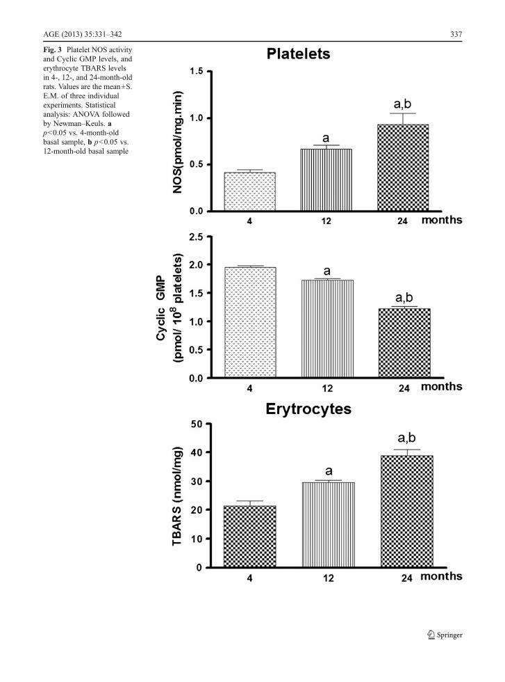
Fig. 3 Platelet NOS activityand Cyclic GMP levels, anderythrocyte TBARS levelsin 4-, 12-, and 24-month-oldrats. Values are the mean±S.E.M. of three individualexperiments. Statisticalanalysis: ANOVA followedby Newman–Keuls. ap<0.05 vs. 4-month-oldbasal sample, b p<0.05 vs.12-month-old basal sample
AGE (2013) 35:331–342 337
Platelet cyclic GMP levels showed a similar resultwhen compared to data obtained in brain tissues sincecyclic GMP levels in 24-month-old rat platelets alsoexhibited a decrease of 37.5% and 12-month-old rat of11.3% when compared to platelets preparation from 4-month-old animals. In addition, cyclic GMP levels at24 months also decreased to 29.5% when compared tovalues obtained from platelets preparation of 12-month-old animals (Fig. 3) [F (2, 15) 0 123.7, p<0.001].
Age-related TBARS content in hippocampus, frontalcortex, and erythrocytes of rats
TBARS determinations showed that aging is linked toprogressive increase in products of lipid peroxidation infrontal cortex and hippocampus. The TBARS levels at 12and 24 months are increased to 60.6% and 145.4% of thevalues detected at 4 months in frontal cortex. TheTBARS levels at 24 months are also increased (52.8%)when compared to values obtained from samples of 12-month-old animals (Fig. 1c) [F(2, 15)031.73, p<0.001].
Samples from rat hippocampus also showed thatTBARS levels increase to 69.6% at 12 months and166.6% at 24 months when compared to the basalvalues measured at 4-month-old animals. TBARS lev-els at 24 months are also increased (57.1%) whencompared to values obtained at 12 months (Fig. 2c)[F (2, 15)013.89, p<0.001].
TBARS levels in erythrocytes showed similarresults when compared to data obtained in brain tis-sues. In fact, TBARS levels in 12- and 24-month-oldrat erythrocytes are increased to 59.2% and 121.4%respectively when compared to platelets preparationfrom 4-month-old animals. In addition, the increase inproducts of lipid peroxidation levels at 24 months isalso increased to 38.8% when compared to valuesobtained from erythrocytes preparation of 12-month-old animals (Fig. 3c) [F (2, 15)027.97, p<0.001].
Discussion
The study of peripheral biological samples in order toidentify biomarkers either in aging or in neurodegenera-tive disorders could accelerate research on the etiology,pathophysiology, disease progression, and therapeuticdevelopment of many prevalent and devastating diseases.However, progress in the development and use ofbiomarkers for diseases of the central nervous system
has been limited by a number of difficulties of develop-ment of reliable biomarkers for brain disorders. Bio-markers of aging are biochemical or physical propertiesin the human body which indicate that the body is aging.This is important to consider that a biological age is moreindicative of healthy conditions than a chronological agesince some people reach the age of 85 in a very goodphysical and mental condition while others have cogni-tive deficits and physical disturbances around 60. Thepresent work showed an age-related correlation of plate-let NOS activity and cyclic GMP levels, and erythrocyteTBARS levels with the same biochemical assays inhippocampus and frontal cortex of rats. These two struc-tures are implicated in learning and memory and arerelated through a direct excitatory pathway (Fuster1991; Férino et al. 1987; Swanson 1981).
The presence of toxins in the environment cancause damage to mitochondrial enzymes, leading toincreased reactive oxygen species (Koutsilieri et al.2002). Stimulation of this pathway during the lifeappears to play an important role in the induction ofmolecular changes that modify the cellular adaptiveresponses, leading to a decreased function of severalsystems (Sapolsky 1992; McCann et al. 1998). Thisongoing process is the fundamental basis for the es-tablishment of the aging process, so it is always asso-ciated with processes involving prolonged exposure toagents that induce some kind of toxicity.
During the aging process, mitochondrial oxidativephosphorylation becomes less efficient, and this prob-ably contributes to an increase in the production offree radicals. The increase in O2
•− production wouldlead to an accumulation of intracellular calcium andactivation of calcium-dependent enzymes, like nitricoxide synthase, which is responsible for the formationof NO•. In fact, our data showed an age-related in-crease in NOS activity in frontal cortex, hippocampus,and in platelets of rats (Harman 1995; Harman 1998).
This increase in NOS activity in aging may bepartly due to the reduction of Ca2+-ATPase, with con-sequent increase in intracellular concentration of thision (Tanaka and Ando 1990; Hanahisa and Yamaguchi2001). Several studies showed that NO• protector ordeleterious effect is based on its redox status. NO•
donors or synthesized endogenously after NMDA(N-methyl-D-aspartate) receptor activation, can leadto neurotoxicity (Harman 1998; Yu 1996).
NO•-mediated neurotoxicity is engendered, at leastin part, by reaction with O2
•−, apparently leading to
338 AGE (2013) 35:331–342
formation of ONOO-, and not by NO• alone. However,if there is NO+ formation, NMDA receptor will beinhibited and a neuroprotective effect will appear.Therefore, NO behavior as a free radical or as anantioxidant agent will be dependent on O2
•− levels. Ifthe concentration of this anion is high, NO will lead tolipid peroxidation, otherwise NO will have an antiox-idant behavior (Wink et al. 1996). In fact, studies in ratcerebellum have showed no variation in the expressionof NOS isoforms with aging, confirming that the in-crease in nitric oxide synthase activity is associated tooxidative status linked to aging process (Blanco et al.2010; Siles et al. 2002).
Most of the neurodegenerative conditions are associ-ated with a chronic inflammation. Although there iscontroversy whether inflammation is causative or a con-sequence of the disease process, it is now clear that it cangreatly influence its pathogenesis (Floyd 1999; Teunissenet al. 2003). Even though aging and neurodegenerationshare the same basic mechanisms, it is difficult to estab-lish the limits between these two processes; there aremounting evidences that neurodegeneration might be anextension of the normal aging process, which mightincrease the susceptibility for neurotoxic events (Smithet al. 1991; Swerdlow 2007).
Although not a consensus (Ahlskog et al. 1995), thepresence of increased oxidative stress at peripheral levelin AD was evidenced in some studies. Kawamoto et al.(2005) detected in platelets and erythrocytes the involve-ment of ONOO− anion in aging and AD. Aging wasassociated to an increase in NOS activity, a decrease inbasal cyclic GMP content as showed in the Fig. 4a and b.Data from these studied also showed an increase inTBARS (Fig. 4c) and no change in SOD and Na,K-ATPase activities. AD patients showed a higher level ofTBARS and an increase in NOS, SOD, and Na,K-ATPase activities, without changes in cyclic GMP, whencompared to aged controls. Na,K-ATPase is the enzymeresponsible for the cell response to oxidative damage, asit maintains electrochemical sodium and potassium gra-dient. Its catalyticα subunit is sensitive to damage by freeradicals and it has been described that ONOO− can causecell membrane damage, which in turn leads to the dis-ruption of Na,K-ATPase activity (Xie et al. 1995; Gloor1997; Kim and Ko 1998). In addition, studies alsoshowed that APOE e4 allele carriers has higher plateletNOS activity than non-carriers in AD patients whencompared to aged controls (Marcourakis et al. 2008).The results presented by (Kawamoto et al. 2007)
demonstrated a disruption in systemic modulation ofoxidative stress in aging and with more intensity in ADand are consistent with the suggestion that neurodegen-eration and aging could share a common pathogenicpathway.
Evidence suggests that part of the physiologic NO•
effects occur mainly through stimulation of soluble GC,with a consequent increase in levels of cyclic GMP(Garthwaite 1991; Chiavegatto et al. 1998). The cyclicGMP, in turn, may act on ion channels or activateprotein kinases (Garthwaite and Boulton 1995). Thus,the increased NOS activity of elderly people withoutdementia compared to healthy young people was notaccompanied by a similar increase in the levels of intra-cellular cyclic GMP.
To evaluate cyclic GMP levels in rats in the presentstudy, the interval between decapitation and experi-mental procedure was kept to <75 s, and testing of4-, 12-, and 24-month-old animals were alternated.According to Steiner et al. (1972) levels of the nucle-otide cyclic GMP in mouse brain are stable after 60 sof ischemia. Our data showed a similar age-relateddecrease in cyclic GMP contents in platelets as wellas in hippocampus and frontal cortex of rats. Theseresults also confirmed previous studies that showed anage-related reduction in the levels of cyclic GMP inthe cerebral cortex, striatum, cerebellum, hippocam-pus, and hypothalamus of rats (Chalimoniuk andStrosznajder 1998; Puri and Volicer 1981; Schmidtand Thornberry 1978). These results also confirmedour previous evidences that showed a decrease in cyclicGMP levels and Na,K-ATPase activity in rat cerebellum(Scavone et al. 2005; Kawamoto et al. 2007).
These results therefore suggest that NO produced inexcess in the elderly is not activating soluble GC,which may be due to the increase in ONOO− produc-tion. In fact, the present results showed that rat age-related increase in TBARS in hippocampus and frontalcortex also occurred in erythrocytes, confirming previ-ous data obtained in human erythrocytes (Kawamoto etal. 2005). There is also evidence that excessive produc-tion of ONOO− may be responsible for the reduction incyclic GMP levels by inactivation of the center iron andsulfur in proteins (Beckman and Koppenol 1996).Finally, we cannot rule out that an increase in phospho-diesterase (PDE) activity as responsible for the decreasein cyclic GMP content. In fact, aging process has beenassociated with an increase in PDE expression andactivity and a decrease in cyclic GMP concentration
AGE (2013) 35:331–342 339

(for review, see Domek-Łopacińska and Strosznajder2010). This notion is in agreement with data indicatingthat inhibitors of PDE2 and PDE9 improve learning andmemory in older rats (van der Staay et al. 2008) as wellas in rat model of hepatic encephalopathy (Montoliu etal. 2010; Cauli et al. 2010).
In fact, studies performed in slices of rat cerebellumshowed that incubation of slices preparation with sodi-um nitrate or carbon monoxide (CO) induced increases
in the levels of cyclic GMP in the three age groups(Scavone et al. 2005). The same age-related responsewas also obtained in human platelets when incubatedwith NO donor (Kawamoto et al. 2005). Therefore,although there is a reduction in basal levels of cyclicGMPwith time, stimulation of soluble GC byNO or COremains unchanged suggesting that aging is affecting themodulation of the enzyme activity, instead of the totalamount of protein.
The aberrant NO–cyclic GMP cascade is not onlyobserved in neurodegenerative diseases, but in severalother diseases states, such as hypertension, atheroscle-rosis, and diabetes which has been linked to cognitivedeficits. Our data support that NO–sGC–cyclic GMPdysfunction can arise as a consequence of ROS-mediated decreases in NO bioavailability and an im-pairment of soluble GC activity through the oxidationof its prosthetic heme moiety. The relevance of im-paired NO-dependent cyclic GMP production in agingprocess is unknown at present but it might predisposeor potentiate an effect of specific factor(s) in the man-ifestation of age-related degenerative disorders.
Experimental data suggest that the hippocampal–pre-frontal network can participate in the formation andconsolidation of memories (Laroche et al. 1990;Dégenètais et al. 2003). Therefore, the similar changesobserved in NOS activity and TBARS/cyclic GMP levelsand the characteristics of the excitatory synaptic trans-mission in the hippocampal–prefrontal cortex pathwayfurther supports the existence of a cooperative relation-ship between two structures known to be involved inhigher cognitive processes. Taken together, the presentdata suggest that platelet NOS activity and cyclic GMP
0 25 50 75 1000
10
20
30
Age (years)
NO
S(f
mo
l/mg
.min
)
0 20 40 60 80 1000
1
2
3
4
5
Age (years)
Cyc
lic G
MP
(pm
ol/1
08 pla
tele
ts)
0 20 40 60 80 1000
100
200
300
400
Age (years)
TB
AR
s(n
mo
l/mL
)Human Platelets
Human Erytrocytes
A.
B.
C.
�Fig. 4 Age-related NOS activity and Cyclic GMP levels inplatelets, and human erythrocyte TBARS from young andaged human (data obtained from previous studies reportedin Kawamoto et al. 2005). a Correlation and linear regres-sion between values of NOS (in femtomoles per milligramper minute) activity from human platelets (n065). b Cor-relation and linear regression between values of cyclicGMP (picomoles per 108 platelets) levels from humanplatelets (n020). c Correlation and linear regression betweenvalues of TBARS (in femtomoles per milliliter) levels fromhuman erythrocytes (n020). Analyses were performed based onMotulsky (1995) by using GraphPad Prism Software. NOSactivity—Pearson correlation coefficient, r00.69; p<0.001;linear regression, r200.48, F059.7, p<0.001. Cyclic GMPlevels—Pearson correlation coefficient, r00.90; p<0.001;linear regression, r200.82, F084.7, p<0.001. c TBARSlevels—Pearson correlation coefficient, r00.73; p<0.001;linear regression, r200.52, F020.1, p<0.001
340 AGE (2013) 35:331–342

levels as well as erythrocytes TBARS are potential bio-markers that could be used to test anti-aging therapies andnew compounds that affect NO–cyclic GMP signalingpathway in CNS.
Acknowledgments The authors thank Larissa de Sá Lima fortechnical support and Fernando Kok for his helpful comments.E.M.K. and A.E.B. are supported by postdoc fellowship fromFAPESP; A.R.V and S.D. are Ph.D. student research fellowshipfrom FAPESP and Coordenação deAperfeiçoamento de Pessoal deNível Superior (CAPES), respectively, and L.S.L. is supported bygrants from Universidade de São Paulo. Work in the laboratory ofthe authors was supported by FAPESP, CNPq and NeuroscienceResearch Support Centers fromUniversity of São Paulo (NAPNA).C.S. and T.M. are research fellows of CNPq. The authors report nobiomedical financial interests or potential conflicts of interest.
References
Ahlskog JE, Uitti RJ, Low PA, Tyce GM, Nickander KK,Peterson RC, Kokmen E (1995) No evidence for systemicoxidant stress in Parkinson’s or Alzheimer’s disease. MovDisord 10:566–573
Baba A, Yoshikawa T, Mitamura H, Ogawa S (1999) Erythro-cyte Naþ, Kþ-ATPase activity in patients with congestiveheart failure. Int J Cardiol 69:117–125
Beckman JS, Koppenol WH (1996) Nitric oxide, superoxideand peroxynitrite: the good, the bad and ugly. Am J Physiol271:C1424–C1437
Blanco S, Molina FJ, Castro L, Del Moral ML, Hernandez R,Jimenez A, Rus A, Martinez-Lara E, Siles E, Peinado MA(2010) Study of the nitric oxide system in the rat cerebel-lum during aging. BMC Neurosci 11(78 11):78–91
Bolanos JP, Almeida A, Stewart V, Peuchen S, Land JM, ClarkJB, Heales SJ (1997) Nitric oxide-mediated mitochondrialdamage in the brain: mechanisms and implications forneurodegenerative diseases. J Neurochem 68:2227–2240
Cauli O, Rodrigo R, Llansola M, Montoliu C, Monfort P,Piedrafita B, el Mlili N, Boix J, Agustí A, Felipo V(2010) Cyclic GMP pathways in hepatic encephalopathy.Neurological and therapeutic implications. Metab BrainDis 1:69–80
Chalimoniuk M, Strosznajder JB (1998) Aging modulates nitricoxide synthesis and cGMP levels in hippocampus andcerebellum. Effects of amyloid beta peptide. Mol ChemNeuropathol 35(1–3):77–95
Chiavegatto S, Scavone C, Canteras NS (1998) Nitric oxidesynthase activity in the dorsal periaqueductal gray of ratsexpressing innate fear response. NeuroReport 9:571–576
Dégenètais E, Thierry A-M, Glowinski J, Gioanni Y (2003)Synaptic influence of hippocampus on pyramidal cells ofthe rat prefrontal cortex: an in vivo intracellular recordingstudy. Cereb Cortex 13(7):782–792
Domek-Łopacińska KU, Strosznajder JB (2010) Cyclic GMPand nitric oxide synthase in aging and Alzheimer’s disease.Mol Neurobiol 41(2–3):129–137
Férino F, Thierry AM, Glowinski J (1987) Anatomical andelectrophysiological evidence for a direct projection fromAmmon’s horn to the medial prefrontal cortex in the rat.Exp Brain Res 65:421–426
Floyd RA (1999) Neuroinflammatory processes are important inneurodegenerative diseases: an hypothesis to explain theincreased formation of reactive oxygen and nitrogen spe-cies as major factors involved in neurodegenerative diseasedevelopment. Free Radic Biol Med 26:1346–1355
Förstermann U, Kleinert H (1995) Nitric oxide synthase: expres-sion and expressional control of the three isoforms. NaunynSchmiedebergs Arch Pharmacol 352:351–364
Freitas JJ, Pompéia C, Miyasaka CK, Curi R (2001) Walker-256tumor growth causes oxidative stress in rat brain. J Neuro-chem 77(2):655–663
Fuster JM (1991) The prefrontal cortex and its relation tobehavior. Prog Brain Res 87:201–211
Garthwaite J (1991) Glutamate, nitric oxide and cell-cell signal-ing in the nervous system. Trends Neurosci 14(2):60–67
Garthwaite J, Boulton CL (1995) Nitric oxide signaling in thecentral nervous system. Annu Rev Physiol 57:683–706
Gloor SM (1997) Relevance of Na, K-ATPase to local extracel-lular potassium homeostasis and modulation of synaptictransmission. FEBS Lett 412:1–4
Halliwell B, Gutteridge JMC (2007) Free radical in Biology andMedicine., vol 4th. Free radical in Biology and Medicine.Oxford University Press, New York
Hanahisa Y, Yamaguchi M (2001) Decrease in Ca2+-ATPaseactivity in the brain plasma membrane of rats with increas-ing age: involvement of brain calcium accumulation. Int JMol Med 7(4):407–411
Harman D (1981) The aging process. Proc Natl Acad Sci U S A78(11):7124–7128
Harman D (1995) Free radical theory of aging: Alzheimer’sdisease pathogenesis. Age 18:97–119
Harman D (1998) Aging: phenomena and theories. Ann N YAcad Sci 854:1–7
Kawamoto EM, Munhoz CD, Glezer I, Bahia VS, Caramelli P,Nitrini R, Gorjão R, Curi R, Scavone C, Marcourakis T(2005) Oxidative stress in platelets and erythrocytes inaging and Alzheimer’s disease. Neurobiol Aging 26:857–864
Kawamoto EM, Munhoz CD, Lepsch LB, De Sá Lima L,Markus RP, Marcourakis T, Scavone C (2007) Age-related changes in cerebellar Phosphatase-1 and Na,K-ATPase activities. Neurobiol Aging 9:1149–1159, 2007.Neurobiology of Aging 9:1149–1159
Kim WK, Ko KH (1998) Potenciation of N-methyl-D-aspartate-mediated neurotoxicity by immunostimulated murinemicroglia. J Neurosci Res 54:17–26
Knowles RG, Moncada S (1994) Nitric oxide synthases inmammals. Biochem J 298:249–258
Koutsilieri E, Scheller C, Grünblatt E, Nara K, Li J, Riederer P(2002) Free radicals in Parkinson’s disease. J Neurol 249249(Suppl 2):II/1–II/5
Laroche S, Jay TM, Thierry AM (1990) Long-term potentiationin the prefrontal cortex following stimulation of the hippo-campal CA1/subicular region. Neurosci Lett 114:184–190
Lipton SA, Choi YB, Pan ZH, Lei SZ, Chen HSV, Sucher NJ,Loscalzo J, Singel DJ, Stamler JS (1993) A redox-basedmechanism for the neuroprotective and neurodestructive
AGE (2013) 35:331–342 341

effects of nitric oxide and related nitroso-compounds. Nature364:626–632
Mamelak M (2007) Alzheimer’s disease, oxidative stress andgammahydroxybutyrate. Neurobiol Aging 28:1340–1360
Marcourakis T, Bahia VS, Kawamoto EM, Munhoz CD, GorjãoR, Artes R, Kok F, Caramelli P, Nitrini R, Curi R, ScavoneC (2008) Apolipoprotein E genotype is related to nitricoxide production in platelets. Cell Biochem Funct 26(8):852–858. doi:10.1002/cbf.1516
McCann SM, Licinio J,WongM-L, YuWH,Karanth S, Rettorri V(1998) The nitric oxide hypothesis of aging. Exp Gerontol 7(8):813–826
Mckee M, Scavone C, Nathanson JA (1994) Nitric oxide,cGMP, and hormone regulation of active sodium transport.Proc Natl Acad Sci U S A 91(25):12056–12060
Montoliu C, Rodrigo R, Monfort P, Llansola M, Cauli O, Boix J,Elmlili N, Agusti A, Felipo V (2010) Cyclic GMP pathwaysin hepatic encephalopathy. Neurological and therapeuticimplications. Metab Brain Dis 25(1):39–48
Motulsky H (ed) (1995) Intuitive biostatistics. Oxford Univer-sity Press, New York
Nathan C (1997) Inducible nitric oxide synthase: what differ-ence does it make? J Clin Invest 100:2417–2423
Puri SK, Volicer L (1981) Age-related changes of cyclic nucle-otide levels in rat brain regions. Mech Ageing Dev 15:239–242
Sapolsky RM (1992) Stress, the aging brain, and the mechanismof neuron death. MIT Press, Cambridge
Scavone C, Munhoz CD, Kawamoto EM, Glezer I, de Sá LL,Marcourakis T, Markus RP (2005) Age-related changes incyclic GMP and PKG-stimulated cerebellar Na, K-ATPaseactivity. Neurobiol Aging 26(6):907–916
Schmidt MJ, Thornberry JF (1978) Cyclic AMP and cyclicGMP accumulation in vitro in brain regions of young, oldand aged rats. Brain Res 139:169–177
Siles E, Martinez-Lara E, Canuelo A, Sanchez M, Hernandez R,Lopez-Ramos JC, Del Moral ML, Esteban FJ, Blanco S,Pedrosa JA, Rodrigo J, Peinado MA (2002) Age relatedchanges of the nitric oxide system in the rat brain. BrainRes 956:385–392
Smith CD, Carney JM, Starke-Reed PE, Oliver CN, StadtmanER, Floyd RA, Markesbery WR (1991) Excess brain pro-tein oxidation and enzyme dysfunction in normal aging andin Alzheimer disease. Proc Natl Acad Sci U S A 88:10540–10543
Steiner AL, Parker CW, Kipnis DM (1972) Radioimmunoassayfor cyclic nucleotides. J Biol Chem 247:1106
Swanson LW (1981) A direct projection from Ammon’s horn toprefrontal cortex in the rat. Brain Res 217:150–154
Swerdlow RH (2007) Is aging part of Alzheimer’s disease, oris Alzheimer’s disease part of aging? Neurobiol Aging28:1465–1480
Tanaka Y, Ando S (1990) Synaptic aging as revealed by changesin membrane potential and decreased activity of Na+, K+-ATPase. Brain Res 506:46–52
Teunissen CE, Lütjohann D, von Bergmann K, Verhey F, VreelingF, Wauters A, Bosmans E, Bosma H, van Boxtel MP, MaesM, Delanghe J, Blom HJ, Verbeek MM, Rieckmann P, DeBruijn C, Steinbusch HW, de Vente J (2003) Combination ofserum markers related to several mechanisms in Alzheimer’sdisease. Neurobiol Aging 24:893–902
Valko M, Leibfritz D, Moncol J, Cronin MTD, Mazur M, TelserJ (2007) Free radicals and antioxidants in normal physio-logical functions and human disease. Int J Biochem CellBiol 39:44–84
van der Staay FJ, Rutten K, Barfacker L, Devry J, Erb C, Hea H(2008) The novel selective PDE9 inhibitor BAY 73–6691improves learning and memory in rodents. Neuropharma-cology 55:908–918
Wink DA, Cook JA, Pacelli REA (1996) The effects of variousnitric oxide-donor agents on hydrogen peroxide toxicity: adirect correlation between nitric oxide formation and pro-tection. Arch Biochem Biophys 331:241–248
Xie Z, Jack-Hays M, Wang Y, Periyasamy SM, Blanco G,Huang WH, Askari A (1995) Different oxidant sensitivitiesof the alpha 1 and alpha 2 isoforms of Na/K-ATPaseexpressed in baculovirus-infected insect cells. BiochemBiophys Res Commun 207:155–159
Yu BP (1996) Aging and oxidative stress: modulation by dietaryrestriction. Free Radic Biol Med 21:651–668
342 AGE (2013) 35:331–342
Copyright © 2022 FDOKUMEN