Affinity chromatography purification of mitochondrial inner membrane proteins with calcium transport...
13
. . . . 7, n!!,(= ELSEVIER Biochimica et Biophysica Acta 1373 (1998) 347-359 BIOCHIMICA ET BIOPHYSICA ACTA BBI Affinity chromatography purification of mitochondrial inner membrane proteins with calcium transport activity Ana Villa, Maria Isabel Garcia-Sim6n, Pablo Blanco l, B~irbara Sesr, Elena Bogdnez, Jorgina Satrustegui * Departamento de Biologla Molecular, Centro de Biologia Molecular 'Severo Ochoa', C.S.I.C.-Universidad Autrnoma de Madrid, Campus de Cantoblanco, 28049-Madrid, Spain Received 27 April 1998; revised 26 June 1998; accepted 2 July 1998 Abstract Immobilized calcium affinity chromatography was used to obtain a preparation enriched in calcium transporters from Triton X-100 extracts of rat liver mitochondria inner membranes (PPCT). The PPCT were reconstituted into preformed asolectin liposomes which contained 120 mM KC1 as internal high K + medium. 45Ca2+ uptake into proteoliposomes was studied under conditions favoring electrophoretic uptake, and + 45 ~2+ H i / Ca o or Na+/aSca 2+ exchange, to test for the presence of the three calcium transport modes present in mitochondria. 45Ca 2+ uptake in liposomes was studied in parallel. ~+/45p~2+ H i / Ca o exchange activity measured in the presence of a pH gradient Nu i / ~a o exchange activity was not detectable. + 45 ,2+ (acid inside) obtained after suspension in low K medium in the presence of nigericin, was 100-200 nmoles 45Ca2+ per mg protein in 30 s. 45Ca2+ uptake in voltage-dependent assays (a K + diffusion membrane potential induced by valinomycin in the presence of methylamine) was not electrophoretic since it was stimulated by carbonyl cyanide p-trifluoromethox- yphenylhydrazone (FCCP) and probably due to secondary Ca2+/H + countertransport. + 45 2+ H i / Ca o uptake showed a saturable component at around 80 ~tM Ca and was coupled to an increase in internal pH in pyranine-loaded PPCT proteoliposomes. 45Ca 2+ uptake in PPCT proteoliposomes could also be driven by a pH gradient obtained by raising external pH in high K + medium. The results are consistent with the presence of a functional nH+/Ca 2+ antiporter. Polyclonal antibodies raised against the PPCT were able to immunoprecipitate the H+/aSca2+ uptake activity and recognized two major bands in the PPCT with molecular masses of about 66 kDa and 55 kDa. This is the first report of a partial purified protein(s) which may represent the H+/Ca 2÷ exchanger of the inner mitochondrial membrane, and represents an important step towards its identification. © 1998 Elsevier Science B.V. All rights reserved. Keywords. Mitochondrion; H--/Ca 2+ exchange; Calcium uniporter; Na+/Ca2+ exchange; Affinity purification; Proteoliposome 1. Introduction * Corresponding author. Fax: +41 (91) 3974799; E-mail: [email protected] 1 Present address: Departamento de Quimica Orgfinica, Inor- gfinica y Bioquimica, Facultad de Ciencias Quimicas, Universi- dad de Castilla La Mancha, 13071-Ciudad Real, Spain. The mitochondrial calcium cycle has been known for a long time and it involves four different calcium transport mechanisms (reviewed in [1,2]). Electrogen- ic ejection of two protons drives electrophoretic up- take of one Ca 2+ via the inner membrane calcium uniporter, a process inhibited by ruthenium red [3]. 0005-2736/98/$ - see front matter © 1998 Elsevier Science B.V. All rights reserved. PII: S0005-2736(98)00 1 20-5
-
Upload
independent -
Category
Documents
-
view
2 -
download
0
Transcript of Affinity chromatography purification of mitochondrial inner membrane proteins with calcium transport...

. . . . 7,
n!!,(= E L S E V I E R Biochimica et Biophysica Acta 1373 (1998) 347-359
BIOCHIMICA ET BIOPHYSICA ACTA
BBI
Affinity chromatography purification of mitochondrial inner membrane proteins with calcium transport activity
Ana Villa, Maria Isabel Garcia-Sim6n, Pablo Blanco l, B~irbara Sesr, Elena Bogdnez, Jorgina Satrustegui *
Departamento de Biologla Molecular, Centro de Biologia Molecular 'Severo Ochoa', C.S.I.C.-Universidad Autrnoma de Madrid, Campus de Cantoblanco, 28049-Madrid, Spain
Received 27 April 1998; revised 26 June 1998; accepted 2 July 1998
Abstract
Immobilized calcium affinity chromatography was used to obtain a preparation enriched in calcium transporters from Triton X-100 extracts of rat liver mitochondria inner membranes (PPCT). The PPCT were reconstituted into preformed asolectin liposomes which contained 120 mM KC1 as internal high K + medium. 45Ca2+ uptake into proteoliposomes was studied under conditions favoring electrophoretic uptake, and + 45 ~2+ H i / Ca o or Na+/aSca 2+ exchange, to test for the presence of the three calcium transport modes present in mitochondria. 45Ca 2+ uptake in liposomes was studied in parallel.
~ + / 4 5 p ~ 2 + H i / Ca o exchange activity measured in the presence of a pH gradient Nu i / ~a o exchange activity was not detectable. + 45 ,2+ (acid inside) obtained after suspension in low K medium in the presence of nigericin, was 100-200 nmoles 45Ca2+ per mg protein in 30 s. 45Ca2+ uptake in voltage-dependent assays (a K + diffusion membrane potential induced by valinomycin in the presence of methylamine) was not electrophoretic since it was stimulated by carbonyl cyanide p-trifluoromethox- yphenylhydrazone (FCCP) and probably due to secondary Ca2+/H + countertransport. + 45 2+ H i / Ca o uptake showed a saturable component at around 80 ~tM Ca and was coupled to an increase in internal pH in pyranine-loaded PPCT proteoliposomes. 45Ca 2+ uptake in PPCT proteoliposomes could also be driven by a pH gradient obtained by raising external pH in high K + medium. The results are consistent with the presence of a functional nH+/Ca 2+ antiporter. Polyclonal antibodies raised against the PPCT were able to immunoprecipitate the H+/aSca2+ uptake activity and recognized two major bands in the PPCT with molecular masses of about 66 kDa and 55 kDa. This is the first report of a partial purified protein(s) which may represent the H+/Ca 2÷ exchanger of the inner mitochondrial membrane, and represents an important step towards its identification. © 1998 Elsevier Science B.V. All rights reserved.
Keywords. Mitochondrion; H--/Ca 2+ exchange; Calcium uniporter; Na+/Ca 2+ exchange; Affinity purification; Proteoliposome
1. Introduction
* Corresponding author. Fax: +41 (91) 3974799; E-mail: [email protected]
1 Present address: Departamento de Quimica Orgfinica, Inor- gfinica y Bioquimica, Facultad de Ciencias Quimicas, Universi- dad de Castilla La Mancha, 13071-Ciudad Real, Spain.
The mi tochondr ia l calcium cycle has been known for a long t ime and it involves four different calcium
t ranspor t mechanisms (reviewed in [1,2]). Electrogen- ic ejection o f two pro tons drives electrophoret ic up- take o f one Ca 2+ via the inner m e m b r a n e calcium
uniporter , a process inhibited by ru then ium red [3].
0005-2736/98/$ - see front matter © 1998 Elsevier Science B.V. All rights reserved. PII: S 0 0 0 5 - 2 7 3 6 ( 9 8 ) 0 0 1 20-5

348 .4. Villa et al . / Biochimica et Biophysica Acta 1373 (1998) 347-359
An additional rapid uptake mode (RAM) for calci- um influx [4] with a higher calcium affinity and lower ruthenium red sensitivity than the classical calcium uniporter has also been described. Efflux from mito- chondria is carried out through two different anti- port mechanisms that vary in a tissue specific way. A Na+/Ca 2+ antiporter operates in brain, heart and other excitable tissues, and to a minor degree in liver, whereas a H+/Ca 2÷ exchanger has been described in liver, lung, kidney and smooth muscle. In addition, there is a non-specific pathway for calcium release, the so-called permeability transition pore (PTP), a mechanism whereby the permeability of the inner membrane increases and allows the extrusion of ma- trix components [5].
The exact function of calcium interaction with mi- tochondria requires the understanding at the molec- ular level of the characteristics of these different cal- cium transporters. With respect to the calcium uniporter, the type of transport mechanism (channel or carrier) or the molecular identity responsible for the transport system has not yet been defined, despite intensive research on this issue. Zazueta et al. [6] reconstituted mitochondrial calcium transport pro- teins from rat kidney cortex making use of a system where mitochondrial membrane proteins where ex- tracted with 1.6% sodium cholate and incorporated to liposomes reconstituted with cytochrome oxidase. The 45Ca2+ transporter reconstituted in this system was driven by a membrane potential (negative inside) but was only partially (53%) sensitive to ruthenium red. A 20 kDa protein appeared to be responsible for this activity [7]. Mironova et al. [8,9] isolated calcium transport proteins of 40 kDa and 2 kDa from etha- nol extracts of beef heart mitochondria that were able to increase the conductivity of bilayer lipid membranes in a ruthenium red sensitive fashion. The 40 kDa was a glycoprotein and antibodies against it inhibited calcium transport in rat liver mi- tochondria [10], whereas the 2 kDa protein appeared to be responsible for the increase in conductivity [9]. Thus, it would appear that the calcium uniporter activity is associated with small peptides (2 kDa) and perhaps other larger components that are capa- ble to increase the conductance of lipid membranes in a ruthenium red sensitive fashion [6,8,10].
The sodium-dependent calcium efflux has been de- scribed as an electroneutral passive transport (2Na+/
Ca 2+) but recent reports indicate that it works as an electrogenic 3Na+/Ca 2+ or active 2Na+/Ca 2+ ex- changer [11] or a membrane potential driven electro- phoretic antiporter [12]. A protein of about 110 kDa that behaved as an electroneutral 2Na+/Ca 2+ anti- porter has been purified [13]. On the other hand, the sodium-independent mechanism has not been yet identified, and the H+:Ca 2+ stoichiometry and/ or energy requirements are poorly understood [14].
In the present study we describe the use of immo- bilized metal affinity chromatography with calcium ions as the immobilized cations, to obtain a prepa- ration of partially purified calcium transporters from the inner mitochondrial membrane. The activity of this preparation has been studied after reconstitution into preformed liposomes. Our results show that the calcium transport activity present in this preparation is largely dependent upon a pH gradient (acid in- side), consistent with a H+/Ca 2+ exchanger.
2. Methods
2.1. Preparation of submitochondrial particles and solubilization of membrane proteins
Rat liver mitochondria, mitoplasts and submito- chondrial particles were isolated from 3-month-old male Wistar rats following published procedures [15]. Livers were homogenized (1:3 w/v) in 0.25 M sucrose, 20 mM Tris-HC1, pH 7.4 at 0°C. Nuclei and cell debris were first removed by centrifugation (700Xg, l0 min) and mitochondria then spun down (14500Xg, l0 min). In order to obtain mito- plasts, the outer mitochondrial membrane was elim- inated by hypo-osmotic incubation in 20 mM Tris- HC1 pH 7.4 (3 ml/g initial tissue) during 20 min at 4°C and constant stirring and subsequent centrifuga- tion (12000Xg, l0 min). The pellet, containing mi- toplasts, was used as a source of submitochondrial particles (SMPs) which were obtained by incubation in hypo-osmotic medium 0 - 2 ml/mg protein, 20 min, 4°C). SMPs were collected by centrifugation (27000Xg, 10 min), washed twice with 20 mM Tris pH 7.4 (27000Xg, 10 min) and stored at -70°C until used. All the solutions employed were supple- mented with the following mixture of protease inhib- itors: 1 mM phenylmethylsulfonylfluoride (PMSF,

A. Villa et al. / Biochimica et Biophysica Acta 1373 (1998) 347-359 349
Fluka), 5 mM iodacetamide (Merck), 0.2 mg/ml ben- zamidine (Sigma), 0.1 mg/ml trypsin inhibitor (Sig- ma), 0.1 mg/ml bacitracin (Sigma).
To solubilize inner mitochondrial membrane pro- teins, SMPs were diluted (10 mg/ml) in 120 mM KC1, 20 mM Tris-HC1, pH 7.4, 3% Triton X-100, and incubated at 4°C and constant stirring during 30 min. Extracted proteins were then separated from unsolubilized material by centrifugation at 100000Xg for 30 min at 4°C. Contamination of SMPs by outer mitochondrial membranes was below 6% as determined by the activity of the outer mem- brane marker NADPH cytochrome c reductase [16].
2.2. Purification of calcium transporters
The calcium chelating resin used consisted of a chelating ligand, iminodiacetic acid, coupled to a cross-linked agarose matrix (Chelating Sepharose fast flow, Pharmacia). Preparation of the column was performed as described [17]. The resin, 200 ml (5 cmx 10.2 cm), was washed with 10 column vols. of distilled water, loaded with 2.4 column vols. of CaC12 (3 mg/ml) and washed with 4.8 column vols. of water to remove the remaining calcium. The resin was then equilibrated in 10% glycerol, 500 mM KC1, 1% Triton X-100, 20 mM Tris, pH 8. Solubilized mitochondrial particles (360 mg in 30 ml) were di- luted 1/3 with 'Triton-free equilibration medium' (10% glycerol, 500 mM KCI, 20 mM Tris, pH 8) and applied to the column over a period of 12 h at 0°C, by recirculating the sample at a rate of 0.4 ml/ min. Unbound proteins were removed by washing with 2 column vols. of 10% glycerol, 500 mM KCI, 1% CHAPS, 20 mM Tris, pH 8. Retained proteins were eluted with 'elution buffer' (10% glycerol, 120 mM KC1, 1% CHAPS, 50 mM EGTA, 20 mM Tris, pH 7.4). The protein peaks eluted from the column were monitored at 280 nm absorbance in an Hewlett Packard 8451A Diode Array Spectrophotometer. The elution fractions (around 100 ml) were pooled and concentrated in a 10 ml ultrafiltration cell (Ami- con, PM10 membranes) to a final volume of 0.2 ml.
2.3. Preparation of liposomes
Liposomes were prepared from purified asolectin [18] as described by Lopez-Corcuera and Arag6n
[19]. In order to eliminate residual Ca 2+ bound to phospholipids, purified asolectin (35.6 mg/ml in chloroform/methanol, 2:1, v/v) was washed in acid medium as follows: 0.2 vols. of distilled water were layered on top of the asolectin solution and mixed thoroughly. The two phases were separated by cen- trifugation, the polar (upper) phase was taken to pH 3, the two phases were again mixed, and then sepa- rated. The organic (lower) phase was dried under N2, dissolved in chloroform/methanol (4:1, v/v) to a final concentration of 0.356 g/ml, and stored at -20°C until used.
Aliquots (540 ~tl) of purified asolectin were dried in a rotatory evaporator and dispersed (192 mg/ml final concentration) in 1 ml HKM containing 120 mM KC1, 20 mM Tris-HC1, pH 7.4. plus 7.43 ~tl 1 M KOH/ml, and appropriate BCECF, pyranine, or EGTA concentrations. The lipid suspension was then sonicated (3 times for 30 s with 30 s intervals) in an MSE Probe Ultrasonic Disintegrator. Phospho- lipids were measured by the Bartlett method [20].
2.4. Reconstitution of part&lly purified calcium transporters
Two different methods were used for reconstitu- tion of partially purified calcium transporters.
2.4.1. Bio-Beads method In order to eliminate the EGTA present in PPCT
before reconstitution, concentrated PPCT fractions were diluted (50-fold) in EGTA-free buffer (120 mM KC1, 20 mM Tris-HC1, pH 7.4 (HKM) plus 1% CHAPS) and again concentrated by ultrafiltra- tion. The procedure was repeated 3 times. 32 p,1 of the sonicated liposome suspension was then mixed with 0.143 ml of PPCTs or solubilized SMPs (both diluted 1/20 in HKM) or with an equivalent solution lacking proteins to obtain detergent-treated lipo- somes. To remove CHAPS, 0.175 ml of the lipo- some/protein mixture were applied to 1.5 ml Bio- Beads SM-2 (Bio-Rad) columns that had been pre- equilibrated with HKM, and proteoliposomes were eluted (flow rate 0.3 ml/min) with the same medium. The elution fractions (500 p.l) were applied to 2 ml Sephadex G-50 (Pharmacia) columns pre-equili- brated in 'potassium-free LowKM' (120 mM choline chloride, 20 mM Tris, pH 7.4), in order to remove
350 A. Villa et al./ Biochimica et Biophysica Acta 1373 (1998) 347 359
external KC1. Detergent-treated liposomes were 're- constituted' in a similar way to test for non-specific activity.
2.4.2. Dialysis method The liposomes (loaded with 200 gM pyranine or
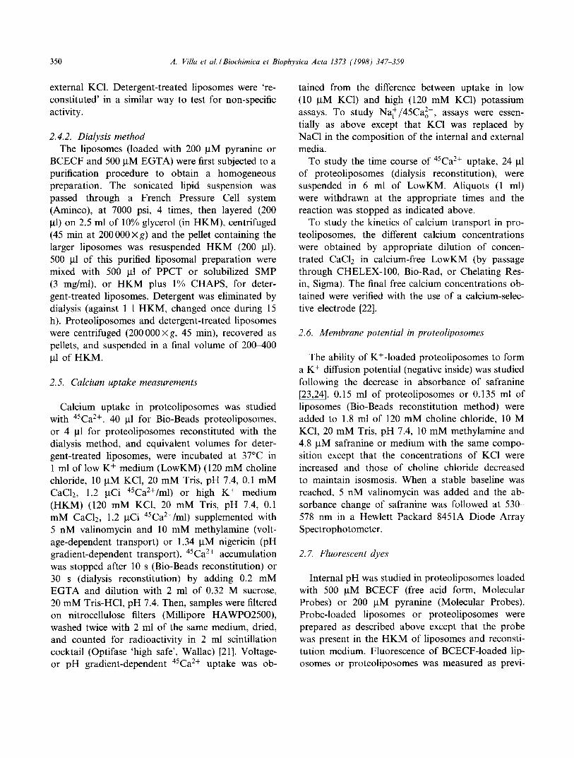
BCECF and 500 gM EGTA) were first subjected to a purification procedure to obtain a homogeneous preparation. The sonicated lipid suspension was passed through a French Pressure Cell system (Aminco), at 7000 psi, 4 times, then layered (200 gl) on 2.5 ml of 10% glycerol (in HKM), centrifuged (45 min at 200 000 x g) and the pellet containing the larger liposomes was resuspended HKM (200 gl). 500 gl of this purified liposomal preparation were mixed with 500 /.tl of PPCT or solubilized SMP (3 mg/ml), or HKM plus 1% CHAPS, for deter- gent-treated liposomes. Detergent was eliminated by dialysis (against 1 1 HKM, changed once during 15 h). Proteoliposomes and detergent-treated liposomes were centrifuged (200 000 ×g, 45 min), recovered as pellets, and suspended in a final volume of 200400 gl of HKM.
2.5. Calcium uptake measurements
Calcium uptake in proteoliposomes was studied with 45Ca2+. 40 gl for Bio-Beads proteoliposomes, or 4 gl for proteoliposomes reconstituted with the dialysis method, and equivalent volumes for deter- gent-treated liposomes, were incubated at 37°C in 1 ml of low K + medium (LowKM) (120 mM choline chloride, 10 gM KC1, 20 mM Tris, pH 7.4, 0.1 mM CaC12, 1.2 gCi 45CaZ+/ml) or high K + medium (HKM) (120 mM KC1, 20 mM Tris, pH 7.4, 0.1 mM CaC12, 1.2 gCi 45Ca2+/ml) supplemented with 5 nM valinomycin and 10 mM methylamine (volt- age-dependent transport) or 1.34 gM nigericin (pH gradient-dependent transport). 45Ca 2+ accumulation was stopped after 10 s (Bio-Beads reconstitution) or 30 s (dialysis reconstitution) by adding 0.2 mM EGTA and dilution with 2 ml of 0.32 M sucrose, 20 mM Tris-HC1, pH 7.4. Then, samples were filtered on nitrocellulose filters (Millipore HAWPO2500), washed twice with 2 ml of the same medium, dried, and counted for radioactivity in 2 ml scintillation cocktail (Optifase 'high safe', Wallac) [21]. Voltage- or pH gradient-dependent 45Ca2+ uptake was ob-
tained from the difference between uptake in low (10 gM KC1) and high (120 mM KCI) potassium assays. To study Na+/45Ca2o +, assays were essen- tially as above except that KCI was replaced by NaC1 in the composition of the internal and external media.
To study the time course of 45Ca2+ uptake, 24 gl of proteoliposomes (dialysis reconstitution), were suspended in 6 ml of LowKM. Aliquots (1 ml) were withdrawn at the appropriate times and the reaction was stopped as indicated above.
To study the kinetics of calcium transport in pro- teoliposomes, the different calcium concentrations were obtained by appropriate dilution of concen- trated CaC12 in calcium-free LowKM (by passage through CHELEX-100, Bio-Rad, or Chelating Res- in, Sigma). The final free calcium concentrations ob- tained were verified with the use of a calcium-selec- tive electrode [22].
2.6. Membrane potential in proteoliposomes
The ability of K+-loaded proteoliposomes to form a K + diffusion potential (negative inside) was studied following the decrease in absorbance of safranine [23,24]. 0.15 ml of proteoliposomes or 0.135 ml of liposomes (Bio-Beads reconstitution method) were added to 1.8 ml of 120 mM choline chloride, 10 M KC1, 20 mM Tris, pH 7.4, 10 mM methylamine and 4.8 gM safranine or medium with the same compo- sition except that the concentrations of KCI were increased and those of choline chloride decreased to maintain isosmosis. When a stable baseline was reached, 5 nM valinomycin was added and the ab- sorbance change of safranine was followed at 530- 578 nm in a Hewlett Packard 8451A Diode Array Spectrophotometer.
2.7. Fluorescent dyes
Internal pH was studied in proteoliposomes loaded with 500 gM BCECF (free acid form, Molecular Probes) or 200 gM pyranine (Molecular Probes). Probe-loaded liposomes or proteoliposomes were prepared as described above except that the probe was present in the HKM of liposomes and reconsti- tution medium. Fluorescence of BCECF-loaded lip- osomes or proteoliposomes was measured as previ-

A. Villa et al./ Biochimica et Biophysica Acta 1373 (1998) 347 359 351
ously described [25]. Excitation was at 450 and 508 nm and emission at 531 nm. Calibration of the en- trapped dye could not be completed accurately. Thus, calibration of pHi was carried out at the end of each experiment after addition of 1% Triton X- 100 in order to expose the dye to the extracellular medium. Fluorescence of pyranine-loaded samples was measured at 510 nm (emission) and 460 nm (ex- citation). Calibration of internal pH was carried out after Triton X-100 addition, by exposing the en- trapped dye to different external pH. The contribu- tion of external BCECF and pyranine (a maximum of 50%, tested from the changes in fluorescence after rapid changes in external pH) was not corrected for, and, therefore, reported pHi values are only relative.
2.8. Antibodies
Polyclonal antibodies were raised against PPCT in rabbits. For immunization, 50 ~tg of PPCT were emulsified with complete Freund's adjuvant and in- jected subcutaneously into New Zealand white rab- bits. For boostings (four), similar amounts of PPCT were emulsified with incomplete Freund's adjuvant and injected with a periodicity of 10 days or more. Antibodies were purified by affinity chromatography with protein A (Pharmacia) and stored at -70°C in 0.02% mercury[(o-carboxyphenyl)thio] ethyl sodium salt (thymerosal, Sigma).
2.9. Western blots
SDS-PAGE was carried out according to Laemmli [26]. Proteins were transferred from gels to PVDF (InmobilonP, Millipore), using a Bio-Rad transblot apparatus at 40 mV overnight in 25 mM Tris, 192 mM glycine, 0.5% SDS and 10% methanol. PVDF membranes were activated with methanol during 30 min before use, and then washed with water.
For immunoblot analysis, PVDF membranes were blocked with PBS containing 5% casein (Molico, Nestle), for 1 h at room temperature, washed 3 times with PBS (1 min each) and incubated with primary antibodies (1/1000 dilution) in PBS 5% casein (3 h). Membranes were washed with PBS, 0.05% Tween 20, 2% casein (6 times, 7 min each), and with PBS alone (3 times, 5 min each), then incubated with the sec- ondary antibody (goat anti-rabbit, linked to peroxi-
dase, Bio-Rad) diluted 1/4000 in PBS, 2% casein dur- ing 20 min, and processed with the luminescence technique (ECL, Amersham), following the indica- tions of the suppliers.
2.10. Immunoprecipitation
The PPCT was diluted 5-fold in 20 mM Tris HC1 pH 7.4, 0.5% Triton X-100 and incubated with PPCT antibodies, total antiserum or with non-immune anti- bodies (at 1/2-1/50 dilution) in the presence of pro- tease inhibitors (PMSF, 0.18 ~tg/180 ~tl) overnight at 4°C and constant gentle mixing. Immunoprecipitates were removed by adding protein A (0.3 mg/ml) and after an incubation of 2-3 h, the mixture was centri- fuged for 1 rain at top speed in a table top centrifuge. The supernatants were collected, made 120 mM KC1, reconstituted with the Bio-Beads method, and as- sayed for activity.
3. Results
3.1. Membrane potential and pH gradient in proteoliposomes
Fig. 1 shows that a K + diffusion membrane poten- tial (negative inside), manifested as a decrease in sa- franine absorbance, is formed upon suspension of liposomes or proteoliposomes in LowKM and after valinomycin addition. No decrease in absorbance oc- curred in high K + medium but a slow decrease also occurred in LowKM in the absence of valinomycin (results not shown). Nigericin addition collapsed the diffusion potential (see Fig. 1). On the other hand, Fig. 2B shows that the suspension of liposomes or proteoliposomes in LowKM results in a drop of the internal pH. This acidification was not observed in the absence of a KC1 gradient (Fig. 2A), indicating that H + entry might be driven by the membrane potential. Fig. 2C shows that this acidification was not avoided even in the presence of methylamine.
It has been shown that artificial membranes formed with different phospholipids have a relatively high passive K + and proton permeability [27,28]. With appropriate K + or H + concentration gradients, most of the K + flux appears to be electrogenic and that of H + electrophoretic, but in KC1 medium, both

352 A. Villa et al. I Biochimica et Biophysica Acta 1373 (1998) 347-359
0.06,
0"0t" tVAL NIG 0.02
o
-0.02
<] -0 .04
-0.06
-0.08 l t 20 40 60
Time (sec) Fig. 1. Diffusion potential formation in K+-loaded PPCT pro- teoliposomes. PPCT proteoliposomes (0.150 ml) prepared with the Bio-Beads reconstitution method were suspended in 1.8 ml of 120 mM choline chloride, 20 mM Tris, pH 7.4, 10 mM methylamine and 4.8 I.tM safranine or medium with the same composition except that it included the KCI concentrations (mM) indicated, and the concentrations of choline chloride were adjusted to maintain isosmosis. Where indicated 5 nM va- linomycin (VAL) or 1.34 gtM nigericin (NIG) were added.
H+-C1 - codiffusion and H + - K + exchange m a y oper- ate [28]. Our results are consistent with the existence o f a large passive p r o t o n permeabi l i ty in our p ro teo-
l iposomal p repara t ion . A t t empt s to reduce this per-
meabil i ty, using different buffers (zwitterionic, such
as PIPES) or sulfate salts instead of chloride salts as
repor ted by Levy et al. [28] were unsuccessful, as judged by the fact that the internal p H in the pro-
teol iposomes suspended in L o w K M was more acidic than in H K M (results not shown).
° I
T I : X
7.2
7.1
7.0
6.9
6.8
6.7
~, ~ .
I I I I 100 200 300 100 100 200
Time (s) Fig. 2. Internal pH in K+-loaded PPCT proteoliposomes. BCECF-toaded PPCT proteoliposomes (0.03 ml) prepared with the Bio-Beads reconstitution method were suspended in 3 ml of 120 mM KC1, 20 mM Tris pH 7.4 (A), or 120 mM choline chloride, 10 p.M KC1, 20 mM Tris pH 7.4 (B,C). 10 mM meth- ylamine was present in A and C. Where indicated, 5 nM vali- nomycin (Val), 0.1 mM CaC12 (Ca), or 1.34 laM nigericin (Nig) were added. The pHi values shown are relative rather than ab- solute values, since it was not possible to obtain a correct cali- bration of the probe while trapped in liposomes or proteolipo- somes.
presence o f methylamine ; and (ii) a p H gradient ob-
tained in the presence o f nigericin. These same assays were conducted with the unpurif ied Tr i ton X-100 extracts o f inner mi tochondr ia l m e m b r a n e proteins
(SMPs). Table 1 summarizes the results obtained. The
m e m b r a n e potential- or p H gradient -dependent cal-
3.2. Calcium transport activity in P P C T
proteol iposomes
The pro teo l iposomes p repared by the dialysis re-
const i tu t ion me t hod were homogeneous with an average d iameter o f 73 nm (see Fig. 3). The internal
vo lume of these p ro teo l iposomes was calculated to be a round 3.6 ktl/mg phosphol ip id , within the range of tha t used by Levy et al. [28]. P P C T proteol ipo- somes and p re fo rmed l iposomes were assayed for 45Ca2+ up take activity under condi t ions where a
m e m b r a n e potent ia l (negative inside) was present or absent. These condit ions were (i) a K + diffusion po- tential which was induced by val inomycin in the
Fig. 3. Negative stain electron microscopy of proteoliposomes and liposomes. Samples obtained by the dialysis reconstitution method (A, liposomes; B, proteoliposomes; bar, 500 nm) were stained with uranyl acetate. Both liposomes and proteolipo- somes had an average diameter of 73.3 nm, ranging from 30 to 100 nm. The internal volume calculated from the relation E= 1/3 Ar [41] (where A is the area of the membrane occupied by 1 mg of phospholipid, r is the radius of the vesicle, and E is the entrapped volume expressed as t.tl/mg phospholipid) was 3.6 ~tl/mg phospholipid.

353
45Ca2+ (mg phospholipid) -I 10 s -1, respectively. n + / 4 5 r ~ 2 + Na i / ~ t o uptake was studied in PPCT proteolipo-
somes and preformed liposomes, and found to be not detectable (results not shown).
45Ca2+ accumulation in PPCT proteoliposomes was due to the transport of calcium across the phos- pholipid membrane. Thus, provided that the calcium gradient was adequate (i.e., in the presence of exter- nal EGTA), the calcium ionophore A23187 was able to promote the complete efflux of the accumulated 45Ca2+ (results not shown; see also Fig. 5C).
Lipid/assay ( m g ) Protein/assay (I.tg) Nig activity Val activity
PPCT proteoliposomes 0.2 3~5 1.54 1.06 SMPs proteoliposomes 0.2 18 0.83 0.62
PPCT proteoliposomes were prepared by the dialysis reconstitution method. Nigericin activity and valinomycin activity correspond to 45Ca2+ uptake in pH gradient- or membrane potential-dependent assays, carried out as indicated in Section 2, and expressed as nmoles 45Ca2+ (mg phospholipid) -1 30 s -1 . The lipid:protein ratio (w/w) was 3 6 - 7 0 : 1 in PPCT proteoliposomes and 10:1 in SMP proteoliposomes. 4SCa 2+ uptake in liposomes (0.2 mg phospholipid per assay) was 0.86 or 0.48 nmoles 45Ca2+ (mg phospholipid) -~ 30 s -1 for nigericin- or valinomycin-dependent activity. For PPCT proteoliposomes reconstituted with the Bio-Beads method, the cor-
r e s p o n d i n g 45Ca2+ uptake activities were 0.18 and 0.13 nmoles 45Ca2+ (mg phospholipid) -1 10 s -I for nigericin and valinomycin assays, respectively.
cium uptake activity observed in SMP proteolipo- somes did not exceed that of liposomes. On the other hand, the results indicate that PPTC is enriched in proteins that are able to increase calcium uptake in proteoliposomes (0.68 or 0.48 nmoles 45Ca2+ (mg phospholipid) -1 30 s - l , after subtracting 4SCa 2+ in nigericin- or valinomycin-dependent assays).
The results obtained when the PPCT were recon- stituted by the Bio-Beads method were similar. These proteoliposomes were heterogeneous in size, with a smaller average internal volume (0.12 ~tl/mg lipid, estimated from the relative distribution of 3H20 and 14C-sucrose as markers of total and external mi- lieu), and 45Ca2+ uptake (corrected for 45Ca2+ up- take in liposomes) in nigericin and valinomycin assays was also smaller, 0.18 or 0.13 nmoles
3.3. Kinetics of calcium transport
To investigate the existence of a facilitated diffu- sion pathway for calcium uptake in our PPCT prep-
.05 .05 , . 0 5
04.
A I 0
¢ ~ 03
f 'o t.,,,d
I./'l • ,,1" .02
0
E r -
0 0 0
A. Villa et al. / Biochimica et Biophysica Acta 1373 (1998) 347-359
Table 1 Characterization of PPCT proteoliposomes
8 .04 -
t~+ .03 -
.02 -
C
i i i
20 40 60 80 l o o
[Ea2*] 0, pM
.04-
o ~
¢~+ .03 -
.02 -
0
E r -
01 -
_ 0 0 0 000.± i } i i i ,
20 40 60 80 100 120 20 40 60 80 100 120
[Ca2+]o, IJM [Ca2+]o, IJM Fig. 4. Kinetics of 45Ca2+ uptake in PPCT proteoliposomes. 45Ca2+ uptake in PPCT proteoliposomes (40 txl) (A,B) and liposomes (40 ktl) (C) (Bio-Beads reconstitution method) was assayed under conditions of voltage dependence (A) or pH gradient dependence (B,C) at the external calcium concentrations indicated. The results are means + S.E.M. of three to five different experiments. Data were fit to the Hill equation (Curve Fitter, Scientific Graphing Software, Sigma Plot, Jandel Scientific, San Rafael, CA, USA). The Vmax, S0.s, and Hill numbers obtained in A were 0.040 nmoles 45Ca2+/10 S, 50 ~M and 1.1, respectively; and 0.027 nmoles 45Ca2+/10 s, 45 ~tM and 1.27, respectively, in B.
354 A. Villa et al . / Biochimica et Biophysica Acta 1373 (1998) 347--359
aration, we studied the kinetics of 45Ca 2+ uptake (both pH gradient- and membrane potential-depend- ent uptake) in PPCT proteoliposomes as a function of the calcium concentration. The calcium depend- ence of 45Ca 2+ transport (pH gradient dependent, Fig. 4B) shows saturation at about 80 I.tM [Ca2+]o
(S0.5 of about 50 gM), a behavior not found in lip- osomes (Fig. 4C). The calcium dependence of the membrane potential-dependent uptake also has a sat- urable component in the same range of calcium con- centrations (Fig. 4A) which is absent in liposomes (results not shown). These results suggest that one or more saturable components in the PPCT are able to increase the liposome 45Ca2+ accumulation capacity.
3.4. Influence of the membrane potential on 45Ca2+ transport in PPCT proteoliposomes
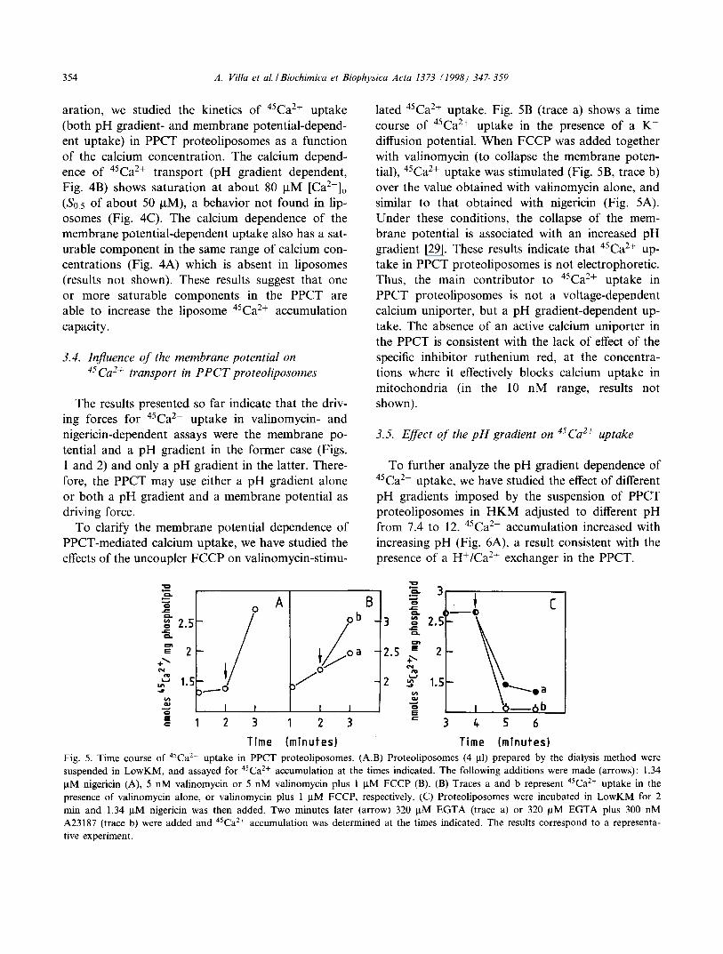
The results presented so far indicate that the driv- ing forces for 45Ca2+ uptake in valinomycin- and nigericin-dependent assays were the membrane po- tential and a pH gradient in the former case (Figs. 1 and 2) and only a pH gradient in the latter. There- fore, the PPCT may use either a pH gradient alone or both a pH gradient and a membrane potential as driving force.
To clarify the membrane potential dependence of PPCT-mediated calcium uptake, we have studied the effects of the uncoupler FCCP on valinomycin-stimu-
la ted 45Ca 2+ uptake. Fig. 5B (trace a) shows a time course of 45Ca2+ uptake in the presence of a K + diffusion potential. When FCCP was added together with valinomycin (to collapse the membrane poten- tial), 45Ca2+ uptake was stimulated (Fig. 5B, trace b) over the value obtained with valinomycin alone, and similar to that obtained with nigericin (Fig. 5A). Under these conditions, the collapse of the mem- brane potential is associated with an increased pH gradient [29]. These results indicate that 45Ca2+ up- take in PPCT proteoliposomes is not electrophoretic. Thus, the main contributor to 45Ca2+ uptake in PPCT proteoliposomes is not a voltage-dependent calcium uniporter, but a pH gradient-dependent up- take. The absence of an active calcium uniporter in the PPCT is consistent with the lack of effect of the specific inhibitor ruthenium red, at the concentra- tions where it effectively blocks calcium uptake in mitochondria (in the 10 nM range, results not shown).
3.5. Effect of the pH gradient o n 45Ca2+ uptake
To further analyze the pH gradient dependence of 45Ca2+ uptake, we have studied the effect of different pH gradients imposed by the suspension of PPCT proteoliposomes in HKM adjusted to different pH from 7.4 to 12. 45caa+ accumulation increased with increasing pH (Fig. 6A), a result consistent with the presence of a H+/Ca 2+ exchanger in the PPCT.
i 2 . 5
÷~ 1.5
IE c
o A
2 3
B ob _
2 3
3
2.5 %
++
..~1~ + . a 6b 3 ~ 5 6
Time (minutes) Time (minutes) Fig. 5. Time course of 45Ca2+ uptake in PPCT proteoliposomes. (A,B) Proteoliposomes (4 I.tl) prepared by the dialysis method were suspended in LowKM, and assayed for 45Ca2+ accumulation at the times indicated. The following additions were made (arrows): 1.34 ~tM nigericin (A), 5 nM valinomycin or 5 nM valinomycin plus 1 laM FCCP (B). (B) Traces a and b represent 45Ca2+ uptake in the presence of valinomycin alone, or valinomycin plus 1 I.tM FCCP, respectively. (C) Proteoliposomes were incubated in LowKM for 2 min and 1.34 ~M nigericin was then added. Two minutes later (arrow) 320 ~M EGTA (trace a) or 320 I.tM EGTA plus 300 nM A23187 (trace b) were added and 45Ca2+ accumulation was determined at the times indicated. The results correspond to a representa- tive experiment.

A. Villa et al. / Biochimica et Biophysica Acta 1373 (1998) 347-359 355
0.~
o 0.3 e -
o . ( / i o t - o .
=n 0.2 E
+~ ~ t m
W
0 E C
0.1
A j =
/
/ - I I
B
I I I I I
0 8 9 10 11 12 13 i I
Exferna[ pH 200 sec
Z
0.,p. units
t .~ N u a , ~
Fig. 6. pH dependence of 45Ca2+ transport and calcium-dependent pHi changes. (A) PPCT proteoliposomes or liposomes (4 lal) (re- constituted with the dialysis method) were suspended in HKM adjusted at the pH indicated, and 45Ca2+ accumulation after 30 s was determined. The results show 45Ca2+ accumulation in proteoliposomes after correction for that obtained in liposomes, and correspond to a representative experiment. (B) Pyranine-loaded PPCT proteoliposomes (dialysis reconstitution) were suspended in LowKM and the following additions were made: Nig, 1.34 laM nigericin; Ca, 80 ~tM CaCI2; EGTA, 800 laM EGTA; A23, 1 ~tM A23187.
3.6. Coupling o f H + and Ca 2+ fluxes
To study whether Ca 2+ fluxes are coupled to H + fluxes, pHi was determined in pyranine-loaded pro- teoliposomes (Fig. 6B). Nigericin addition to PPCT proteoliposomes suspended in LowKM results in a rapid and transient pH drop (reflected as a down- ward deflection in fluorescence). The subsequent ad- dition of Ca 2+ was followed by a gradual increase in pHi, indicative of coupled Ca 2+ influx-H + efflux. The Ca-dependent increase in intravesicular pH was not produced in HKM (results not shown). Furthermore, the addition of EGTA reversed the change in pHi (Fig. 6B). The magnitude of the CaZ+-dependent pHi increase is small (about 0.15 units in 2 min), especially when compared to the pHi changes in- duced by A23187 (see Fig. 6B), but it reflects an increase in internal H + concentration of 407 gM (as- suming that the internal concentration of Tris is 20 mM, which may be underestimated [30], and an in- flux of 1.46 nmol H+/mg phospholipid (in 2 min), i.e., within the range of PPCT-dependent C a 2+
fluxes.
80 kDa), and a group of bands of smaller molecular weight, at around 50 kDa (Fig. 7). After reconstitu- tion in proteoliposomes this pattern was consistently altered: the high molecular weight bands disap- peared, and the smear of smaller molecular weight bands becomes an intense band of 55 kDa. Thus, the reconstitution procedure by itself appears to behave as a purification step, in that it provides a selective enrichment of some bands, in particular the 66 kDa and 55 kDa bands. Moreover, we have consistently observed a total decrease of Coomassie blue stained material after the reconstitution step (see Fig. 7).
Since heat shock proteins could copurify with the mitochondrial calcium transporters [31], in further experiments we assayed the PPCT for immunoreac- tion with antibodies (anti-DR-l) recognizing the heat shock mitochondrial proteins of mammalian cells, p54 and p74 [32] and the related proteins p56 and p71 in Drosophila melanogaster [33]. Western blot analysis showed that these antibodies did not recog- nize any of the bands of the PPCT (results not shown). Thus, it appears that mitochondrial heat shock proteins are not present in the PPCT.
3.7. Protein bands in P P C T preparations 3.8. Antibodies
The PPTC contained major bands at about 66 kDa, a diffuse band of higher molecular weight (at
Our attempt to raise polyclonal antibodies against the PPCT proved to be successful. The c~-PPCT anti-
356 A. Villa et al . / Biochimica et Biophysica Acta 1373 (1998) 347-359
M
200
116
97.4
66
45
Fig. 7. Affinity chromatography purification of mitochondrial inner membrane proteins. Electrophoretic pattern of proteins in SDS-PAGE (10% acrylamide) stained with Coomassie blue. Lane SMPs corresponds to 3% Triton X-100 extract of inner membrane proteins (50 p,1); lanes PPCT and PPCTr correspond to 1/5 of the final eluted PPCT fraction (14.6 ~g protein), be- fore (PPCT) or after (PPCTr) reconstitution in proteoliposomes. Molecular weight markers are also shown (M).
bodies recognized predominantly the 66 kDa major band present in PPCT proteoliposomes showing a weaker reactivity towards the other bands with slow- er or faster (55 kDa) electrophoretic mobility (Fig. 8) that are still highly visible in the PPCT.
The c~-PPCT antibodies were incubated with the PPCT in order to immunoprecipitate the 66 kDa
Table 2 Immunoprecipitation of 45Ca2+ uptake activity of the PPCT
pH gradient-dependent activity (%)
c(-PPCT antibodies (1) 1/2 52.9 + 9.0 c~-PPCT antibodies (2) 1/20 90.47 a-PPCT antibodies (2) 1/50 96.2
The PPCT was incubated with different batches of antibodies (1 and 2) at the dilutions indicated and the supernatants were re- constituted in proteoliposomes (Bio-Beads reconstitution meth- od), and assayed for 45Ca2+ uptake activity in LowKM and in the presence of nigericin. The results represent the inhibition (expressed as percentage of the activity after incubation with preimmune antibodies) of the corresponding activity. The ex- periments were repeated 2-4 times with similar results.
2OO
97
m
46
30
29O
116 97
U
415
Fig. 8. Immunoreactivity of the antibodies against PPCT. (A) Western blot analysis of the PPCT and the PPCTr with purified antibodies (1/1000) raised against PPCT. (B) SDS protein pat- tern of the PPCT. Molecular weight markers are also shown (M).
and 55 kDa bands. The immunoprecipitates were separated by centrifugation and the supernatants re- constituted in proteoliposomes. The results from these experiments (Table 2) indicate that different batches of (~-PPCT antibodies were able to eliminate the calcium transport activity of the PPCT, albeit with different potency. Therefore, one or both of these protein bands is likely to be responsible for the calcium transport activity. On the other hand, we were unable to show any inhibition of calcium transport after incubation of PPCT proteoliposomes with these antibodies.
4. Discussion
We have used immobilized metal affinity chroma- tography, with calcium ions as the immobilized cat- ions, as a purification procedure to obtain a prepa- ration enriched in calcium transporters from the inner membrane of rat liver mitochondria (PPCT). The PPCT were reconstituted in preformed asolectin liposomes and we studied their ability to increase the calcium uptake over that of liposomes. Since the specific calcium transporters present in rat liver mitochondria are the electrophoretic uniporter and

A. Villa et aL /Biochimica et Biophysica Acta 1373 (1998) 347 359 357
the H+/Ca 2+ or Na+/Ca 2+ exchangers, we studied 45Ca2+ uptake under conditions where the predomi- nant driving forces were a membrane potential (neg- ative inside), a pH gradient (acid inside) or a Na gradient (higher inside). Our observations may be summarized as follows.
(a) The inclusion of the PPCT in proteoliposomes increases their capacity for calcium accumulation. This is not merely due to the calcium binding ca- pacity of the PPCT but reflects real transmembrane transport since (i) the PPCT itself (not reconstituted in proteoliposomes) did not bind 45Ca2+ under the conditions used for calcium transport assays (results not shown), and (ii) all of the 45Ca2+ accumulated in PPCT proteoliposomes could be discharged with the calcium ionophore A23187 (Fig. 5C).
(b) The [Ca2+]o dependence of calcium uptake was studied for both the voltage- and pH gradient-de- pendent modes. In both cases, PPCT proteolipo- somes appeared to contain a saturable component (at around 80 ~tM calcium) with low (S0.5 50 btM) calcium affinity (Fig. 4). An interpretation of this result is that the saturable component is a calcium transporter present in the PPCT.
(c) Na+/45Cao + exchange activity in PPCT proteo- liposomes was not detectable. Since rat liver mito- chondria have very low Na+/Ca 2+ activity and the Na+/Ca 2+ exchanger isolated by Li et al. [13] appears to correspond to a protein band of 110 kDa, the lack of bands of this size in the PPCT (Fig. 7) are con- sistent with this result.
(d) One possibility was that the PPCT catalyzes an electrophoretic uniport of calcium. This type of transport should be stimulated by a membrane po- tential (negative inside) and abolished in the presence of FCCP that collapses the membrane potential. Our experimental results show that while the addition of valinomycin in the presence of an outwardly directed K + gradient created a membrane potential (Fig. 1) and stimulated 45Ca 2+ uptake (Table 1), FCCP did not inhibit, but rather stimulated 45Ca2+ accumula- tion (Fig. 5B), indicating that the PPTC catalyzes mainly a non-electrophoretic transport of calcium.
(e) An alternative possibility is that the positive charges of Ca 2+ are compensated by a movement of a positive ion on the opposite direction. It is un- likely that the countercation is Ca 2+ since in experi- ments where the dialysis reconstitution method was
used, Ca~ + was lowered by washing off the calcium ions bound to asolectin and using 500 btM EGTA as an internal calcium chelator, to minimize any 45Ca2o+/ 4°Ca~+ exchange. Since the only other Ca 2+ exchang- er present in mitochondria (besides the Na+/Ca 2+ exchange) is the H+/Ca 2+ antiporter, we have ana- lyzed the possibility that a pH gradient (acid inside) drives 45Ca2+ uptake.
The first indication that 45Ca 2+ uptake depends upon a pH gradient (acid inside) was obtained from the stimulation of 45Ca2+ accumulation by ni- gericin when an outwardly directed K + gradient was present (Table 1). In fact, the calcium uptake activity observed in assays with valinomycin (Table 1) is probably due to the fact that a pH gradient (acid inside) is also built up in these assays (Fig. 2). In addition, 45Ca2+ uptake showed a pH dependence, with increased 45Ca2+ accumulation at higher exter- nal pH values (Fig. 6A). Furthermore, the enhance- ment of 45Ca 2+ accumulation produced by FCCP in the presence of valinomycin (see above and Fig. 5B) is probably also driven by the larger acidification of the internal HKM obtained upon FCCP addition [29].
All these results indicate that a pH gradient pro- duced by various means stimulates 45Ca2+ uptake in PPCT proteoliposomes. Moreover, our results indi- cate that proton efflux is coupled to Ca 2+ entry (Fig. 6B), providing a stronger evidence for the occurrence of an exchange of external calcium against internal H + .
Thus, we can conclude that a pH gradient-sensitive Ca 2+ transporter, possibly a H+/Ca 2+ antiporter, is present in the PPCT. At present we do not know whether the exchanger is electroneutral (2H+/Ca > ) or electrogenic (3H+/Ca2+), as suggested for the Na+/Ca 2+ exchanger [11,12]. We have tried to clarify this point by studying the effect of nigericin and va- linomycin on 45Ca2+ accumulation in PPCT proteo- liposomes incubated in HKM. This would allow to determine whether a pH gradient alone (2H+/Ca 2+) or in combination with a membrane potential (3H+/ Ca 2+) was formed during 45Ca2+ uptake. However, this was unsuccessful as we did not detect any sig- nificant calcium accumulation in the absence of a preformed pH gradient (results not shown).
The calcium uptake activity of the PPCT (100-200 nmoles/30 s/mg protein in PPCT proteoliposomes) is

358 A. Villa et al. / Biochimica et Biophysica Acta 1373 (1998) 347-359
about 500-fold higher than that o f the native H+/ Ca 2+ exchanger in native mi tochondr ia (about 0.2 nmoles/mg protein in 10 s in mi tochondr ia [21]). The P P C T prepara t ion contains several protein bands, two of which, the 66 k D a and 55 k D a bands, are reconst i tuted in proteol iposomes (Fig. 7). These two bands are also recognized by the tx-PPCT anti- bodies (Fig. 8) which are able to immunoprecipi ta te the calcium t ranspor t activity (Table 2). Therefore , it may be concluded that the H+/Ca 2+ exchanger cor- responds to one o f these bands. At tempts to separate these bands with the use o f other purification proce- dures that have been widely employed to purify mi- tochondria l carriers [34] or cat ion t ranspor t systems [31] such as hydroxyapat i te chromatography , for ba- sic or acidic proteins [35], or DEAE-ceUulose chro- matography, have not been successful.
To the extent o f our knowledge, this is the first repor t of a partial purification o f a protein which may represent the H+/Ca 2+ exchanger o f the inner mi tochondr ia l membrane. This exchanger and the Na+/Ca 2+ ant ipor ter appear to be separate entities, as indicated by the fact that they are affected differ- ently by inhibitors and have unequal t ranspor t ki- netics [36-38]. The absence o f detectable Na--/Ca e+ exchange in our P P C T prepara t ion supports that contention. The H+/Ca 2+ exchange activity of mito- chondr ia was identified as the mechanism responsible for Na+- independent Ca 2÷ efflux (see [1] for review) and has been controversial since its discovery; (i) it could not be detected in non-respiring rat liver mito- chondria , suggesting that coupling of Ca 2+ and H + fluxes might be indirect via other ions [39], (ii) it has been repor ted that it t ransports calcium out o f mito- chondr ia against a Ca 2+ concentra t ion gradient
many times greater than that possible for a passive 2H+/Ca 2+ exchanger, suggesting that it may use some form of energy source [14,40]. Thus, the present work represents an impor tan t step towards the iden- tification of the protein(s) responsible for this activ- ity.
Acknowledgements
This work was suppor ted by grants f rom the Di- recci6n General de Investigaci6n Cientifica y Trcnica (PB92-0145), Boehringer Ingelheim Espafia S.A., and
by an institutional grant to the Centro de Biologia Molecular 'Severo Ochoa ' f rom the Fundac i6n Ra- m6n Areces. We thank Dr. J.M. Cuezva for his gen- erous gift of anti-DR1 antibodies. M.I. Garcia-Si- m o n is the recipient o f a fellowship f rom the Comunidad de Madrid.
References
[1] T.E. Gunter, D.R. Pfeiffer, Mechanisms by which mitochon- dria transport calcium, Am. J. Physiol. 258 (1990) C755 C786.
[2] K.K. Gunter, T.E. Gunter, Transport of calcium in mito- chondria, J. Bioenerg. Biomembr. 26 (1994) 471-485.
[3] K.C. Reed, F.L. Bygrave, The inhibition of the mitochon- drial calcium transport by lanthanides and ruthenium red, Biochem. J. 140 (1974) 143-155.
[4] G.C. Sparagna, K,K. Gunter, S.S. Sheu, T.E. Gunter, Mi- tochondrial calcium uptake from physiological-type pulses of calcium. A description of the rapid uptake mode, J. Biol. Chem. 270 (1995) 27510-27515.
[5] M. Zoratti, I. Szabo, The mitochondrial permeability tran- sition, Biochim. Biophys. Acta 1241 (1995) 136-176.
[6] C. Zazueta, J.A. Holguin, J. Ramirez, Calcium transport sensitive to ruthenium red in cytochrome oxidase vesicles reconstituted with mitochondrial proteins, J. Bioenerg. Bio- membr. 23 (1991) 889-902.
[7] C. Zazueta, F. Masso, A. Paez, C. Bravo, A. Vega, L. Mon- atafio, M. Vazquez, J. Ramirez, E. Chavez, Identification of a 20-kDa protein with calcium uptake transport activity. Reconstitution in a membrane model, J. Bioenerg. Bio- membr. 26 (1994) 555-562.
[8] G.D. Mironova, T.V. Sirota, L.A. Pronevich, N.V. Trofi- menko, G.P. Mironov, P.A. Grigorjev, M.N. Kondrashova, Isolation and properties of Ca2+-transporting glycoprotein and peptide from beef heart mitochondria, J. Bioenerg. Bio- membr. 14 (1982) 213 225.
[9] G.D. Mironova, M. Baumann, O. Kolomytkin, Z. Krasich- kova, A. Berdimuratov, T. Sirota, I. Virtanen, N.E.L. Saris, Purification of the channel component of the mitochondrial calcium uniporter and its reconstitution into planar lipid bilayers, J. Bioenerg. Biomembr. 26 (1994) 231-238.
[10] N.-E.L. Saris, T.V. Sirota, I. Virtanen, K. Niva, T. Penttil~i, L.P. Dolgachova, G.D. Mironova, Inhibition of the mito- chondrial calcium uniporter by antibodies against a 40-kDa glycoprotein, J. Bioenerg. Biomembr. 25 (1993) 307-312.
[11] K. Baysal, D.W. Jung, K.K. Gunter, T.E. Gunter, G.P. Briefly, Na+-dependent Ca 2+ efflux mechanism in not a pas- sive 2Na+/Ca 2÷ exchanger, Am. J. Physiol. 266, (Cell Phys- iol. 35) (1994) C80042808.
[12] D.W. Jung, K. Baysal, G.P. Brierly, The sodium-calcium antiport of heart mitochondria is not electroneutral, J. Biol. Chem. 270 (1995) 672-678.

A. Villa et al. I Biochimica et Biophysica Acta 1373 (1998) 347-359 359
[13] W. Li, Z. Shariat-Madar, M. Powers, X. Sun, R.D. Lane, K.D. Garlid, Reconstitution, identification, purification, and immunological characterization of the l l0-kDa Na+/Ca 2-- antiporter from beef heart mitochondria, J. Biol. Chem. 267 (1992) 17983-17989.
[14] T.E. Gunter, K.K. Gunter, S. Sheu, C.E. Gavin, Mitochon- drial calcium transport: physiological and pathological rele- vance, Am. J. Physiol. 267, (Cell Physiol. 36) (1994) C313- 339.
[15] A. Colbeau, J. Nachbaur, P.M. Vignais, Enzymic character- ization and lipid composition of rat liver subcellular mem- branes, Biochim. Biophys. Acta 249 (1971) 462-492.
[16] J. Vitrrica, A. Machado, J. Satrrstegui, Age-dependent var- iations in peroxide-utilizing enzymes from rat brain mito- chondria and cytoplasm, J. Neurochem. 42 (1984) 351-356.
[17] C.A.K. Borrebaeck, B. Lrnerdal, E. Etzler, Metal chelate affinity chromatography of the Dolicus biflorus seed lectin and its subunits, FEBS Lett. 130 (1981) 194-195.
[18] Y. Kagawa, E.J. Racker, Partial resolution of the enzymes catalyzing oxidative phosphorylation, J. Biol. Chem. 246 (1971) 5477 5487.
[19] B. Lopez-Corcuera, C. Arag6n, Solubilization and reconsti- tution of the sodium and chloride glycine transporter from rat spinal cord, Eur. J. Biochem. 181 (1989) 519 524.
[20] G.R. Bartlett, Phosphorus assay in column chromatography, J. Biol. Chem. 234 (1958) 46~468.
[21] J. Vit6rica, J. Satrflstegui, Involvement of mitochondria in the age-dependent decrease in calcium uptake of rat brain synaptosomes, Brain Res. 378 (1986) 36-48.
[22] J. Vit6rica, J. Satrflstegui, The role of ADP in the modula- tion of the calcium efflux pathway in rat brain mitochondria, Biochem. J. 225 (1985) 41-49.
[23] T. Miki, M. Miki, Y. Orii, Membrane potential-linked re- versed electron transfer in the beef heart cytochrome bcl complex reconstituted into potassium-loaded phospholipid vesicles, J. Biol. Chem. 269 (1994) 1827 1833.
[24] K.E. Akerman, N.-E.L. Saris, Stacking of safranine in lip- osomes during valinomycin-induced efflux of potassium ions, Biochim. Biophys. Acta 426 (1976) 624-629.
[25] A. Martinez-Serrano, P. Blanco, J. Satrustegui, Calcium binding to the cytosol and calcium extrusion mechanisms in intact synaptosomes and their alterations with aging, J. Biol. Chem. 267 (1992) 4672-4679.
[26] U. Laemmli, Cleavage of structural proteins during the as- sembly of head of bacteriophage T4, Nature 227 (1970) 680- 685.
[27] C.E. Cooper, J.M. Wrigglesworth, P. Nicholls, The mecha- nism of potassium movement across the liposomal mem- brane, Biochem. Biophys. Res. Commun. 173 (1990) 1008- 1012.
[28] D. Levy, M. Seigneuret, A. Bluzat, J.-L. Rigaud, Evidence for proton countertransport by the sarcoplasmic reticulum Ca2+-ATPase during calcium transport in reconstituted pro-
teoliposomes with low ionic permeability, J. Biol. Chem. 265 (1990) 19524-19534.
[29] V.N. Orlov, Y.N. Antonenko, A.A. Bulychev, L.S. Yaguz- hinsky, Combination of the electrogenic ionophores, valino- mycin and CCCP, can lead to non-electrogenic K+/H + ex- change on bilayer lipid membranes, FEBS Lett. 345 (1994) 104-106.
[30] W.L. Erdahl, C.J. Chapman, R.W. Taylor, D.R. Pfeiffer, Effects of pH conditions on Ca 2+ transport catalyzed by ionophores A23187, 4-BrA23187, and ionomycin suggest problems with common applications of these compounds in biological systems, Biophys. J. 69 (1995) 2350~2363.
[31] K.D. Garlid, X. Sun, P. Paucek, G. Woldegiorgis, Mito- chondrial cation transport systems, Methods Enzymol. 260 (1995) 331-348.
[32] A. Alconada, A.I. Flores, L. Blanco, J.M. Cuezva, Antibod- ies against FI-ATPase alpha-subunit recognize mitochon- drial chaperones. Evidence for an evolutionary relationship between chaperonin and ATPase protein families, J. Biol. Chem. 269 (1994) 13670-13679.
[33] A.I. Flores, J.M. Cuezva, Identification of sequence similar- ity between 60 kDa and 70 kDa molecular chaperones: evi- dence for common evolutionary background?, Biochem. J. 322 (1997) 641-647.
[34] F. Palmieri, C. Indiveri, F. Bisaccia, V. Iacobazzi, Mito- chondrial metabolite carrier proteins: purification, reconsti- tution, and transport studies, Methods Enzymol. 260 (1995) 349-369.
[35] M.J. Gorbunoff, Protein chromatography on hydroxyapatite column, Methods Enzymol. 182 (1990) 329-339.
[36] D.E. Wingrove, T.E. Gunter, Kinetics of mitochondrial cal- cium transport. II. A kinetic description of the sodium-de- pendent calcium efflux mechanism of liver mitochondria and inhibition by ruthenium red and by tetraphenylphospho- nium, J. Biol. Chem. 261 (1986) 15159-15165.
[37] D.E. Wingrove, T.E. Gunter, Kinetics of mitochondrial cal- cium transport. I. Characteristics of the sodium-independent calcium efflux mechanism of liver mitochondria, J. Biol. Chem. 261 (1986) 15166-15171.
[38] M. Crompton, I. Heid, C. Baschera, E. Carafoli, The reso- lution of calcium fluxes in heart and liver mitochondria using the lanthanide series, FEBS Lett. 104 (1979) 352-354.
[39] N.-E.L. Saris, Non-respiring rat liver mitochondria do not have a Ca2+/2H + antiport, Acta Chem. Scand. B41 (1987) 79-82.
[40] K.K. Gunter, M.J. Zuscik, T.E. Gunter, The Na+-independ - ent Ca 2+ efflux mechanism of liver mitochondria is not a passive Ca2+/2H + exchanger, J. Biol. Chem. 266 (1991) 21640-21648.
[41] R.R.C. New, Characterization of liposomes, in: R.R.C. New (Ed.), Liposomes: a Practical Approach, IRL Press, Oxford, 1990, pp. 105-161.
![Clad Inner Surface Temperature l°C]](https://static.fdokumen.com/doc/165x107/633831c324ea072f160c74b1/clad-inner-surface-temperature-lc.jpg)