Ultrafiltration & Protein Purification Products - Sartorius
108
Lab Ultrafiltration and Purification Products
-
Upload
khangminh22 -
Category
Documents
-
view
1 -
download
0
Transcript of Ultrafiltration & Protein Purification Products - Sartorius

Lab Ultrafiltration and Purification Products

2
Table of Contents
General InformationUltrafiltration Applications 4Lab Ultrafiltration Devices 5Ultrafiltration Process Methods 5Membrane Performance Characteristics 6Membrane Selection Guide 7
Protein and Macromolecule Concentration 9Centrifugal and Pressurized Ultrafiltration - Vivaspin® 500 10 - Vivaspin® 2 12 - Vivaspin® Filtrate 14 - Vivaspin® Turbo 4 PES 16 - Vivaspin® 6 18 - Vivaspin® 15R 20 - Vivaspin® Endotest 22 - Vivaspin® Turbo 15 PES 24 - Vivaspin® Turbo 15 RC 26 - Vivaspin® 20 28 - Vivaspin® 100 30 - Vivaspin® Equipment and Accessories 32
Tangential Flow Filtration - Vivaflow® 50 34 - Vivaflow® 50R 36 - Vivaflow® 200 38 - Vivaflow® Equipment and Accessories 40
Ultrafiltration Membrane Discs 42

3
DNA Concentration 45Vivacon® 500 46Vivacon® 2 49
Protein Purification 53Vivaclear Centrifugal Filters 54Vivapure® Ion Exchange Purification Products 56
Virus Purification and Concentration 59Vivapure® Virus Purification and Concentration Kits 60
Adenovirus Purification 61 - Vivapure® Adenopack 20 62 - Vivapure® Adenopack 100 63 - 5Vivapure® Adenopack 500 65
Lentivirus Purification 66 - Vivapure® Lentiselect 40 67 - Vivapure® Lentiselect 500 68 - Vivapure® Lentiselect 1000 69
Application Notes 711. Desalting and Buffer Exchange with Vivaspin® Centrifugal
Concentrators 722. Treatment of Vivaspin® Concentrators for Improved Recovery
of Low-Concentrated Protein Samples 753. Scouting Protein Purification Conditions Using Vivapure® Centrifugal
Ion Exchange Membrane Absorbers 784. Concentration and Purification of Viruses by using Ultrafiltration,
Incl. Coronavirus - a Short Review 835. Sartorius Ultrafiltration Products in the Preparation of Biological
Nanoparticles and Medical Nanocarriers 956. Vivaflow® and Vivaspin® Workflow in Protein Research Laboratories 101
4 General Information
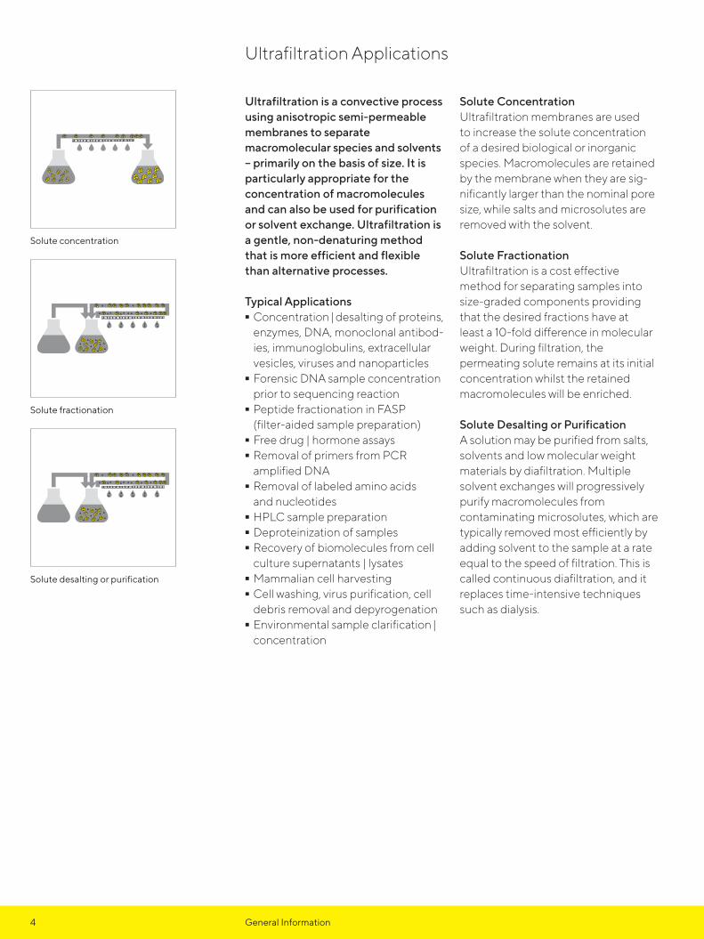
Ultrafiltration is a convective process using anisotropic semi-permeable membranes to separate macromolecular species and solvents – primarily on the basis of size. It is particularly appropriate for the concentration of macromolecules and can also be used for purification or solvent exchange. Ultrafiltration is a gentle, non-denaturing method that is more efficient and flexible than alternative processes.
Typical Applications - Concentration | desalting of proteins, enzymes, DNA, monoclonal antibod-ies, immunoglobulins, extracellular vesicles, viruses and nanoparticles - Forensic DNA sample concentration prior to sequencing reaction - Peptide fractionation in FASP (filter-aided sample preparation) - Free drug | hormone assays - Removal of primers from PCR amplified DNA - Removal of labeled amino acids and nucleotides - HPLC sample preparation - Deproteinization of samples - Recovery of biomolecules from cell culture supernatants | lysates - Mammalian cell harvesting - Cell washing, virus purification, cell debris removal and depyrogenation - Environmental sample clarification | concentration
Solute ConcentrationUltrafiltration membranes are used to increase the solute concentration of a desired biological or inorganic species. Macromolecules are retained by the membrane when they are sig-nificantly larger than the nominal pore size, while salts and microsolutes are removed with the solvent.
Solute FractionationUltrafiltration is a cost effective method for separating samples into size-graded components providing that the desired fractions have at least a 10-fold difference in molecular weight. During filtration, the permeating solute remains at its initial concentration whilst the retained macromolecules will be enriched.
Solute Desalting or PurificationA solution may be purified from salts, solvents and low molecular weight materials by diafiltration. Multiple solvent exchanges will progressively purify macromolecules from contaminating microsolutes, which are typically removed most efficiently by adding solvent to the sample at a rate equal to the speed of filtration. This is called continuous diafiltration, and it replaces time-intensive techniques such as dialysis.
Ultrafiltration Applications
Solute concentration
Solute fractionation
Solute desalting or purification
0.5
1.0
1.5
Vivaspin
® Turbo
5General Information
Sartorius develops devices dedicated to optimizing laboratory ultrafiltration processes with minimal time requirements while maximizing recovery, reliability and robustness.
In addition Sartorius are continually building technical and application
support tools to help users select the optimum device and process for their sample type.
Visit www.sartorius.com for more technical and application support material.
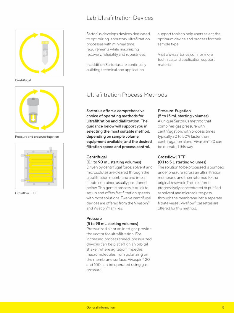
Sartorius offers a comprehensive choice of operating methods for ultrafiltration and diafiltration. The guidance below will support you in selecting the most suitable method, depending on sample volume, equipment available, and the desired filtration speed and process control.
Centrifugal (0.1 to 90 mL starting volumes)Driven by centrifugal force, solvent and microsolutes are cleared through the ultrafiltration membrane and into a filtrate container, usually positioned below. This gentle process is quick to set up and offers fast filtration speeds with most solutions. Twelve centrifugal devices are offered from the Vivaspin® and Vivacon® families.
Pressure (5 to 98 mL starting volumes)Pressurized air or an inert gas provide the vector for ultrafiltration. For increased process speed, pressurized devices can be placed on an orbital shaker, where agitation impedes macromolecules from polarizing on the membrane surface. Vivaspin® 20 and 100 can be operated using gas pressure.
Pressure-Fugation(5 to 15 mL starting volumes)A unique Sartorius method that combines gas pressure with centrifugation, with process times typically 30 to 50% faster than centrifugation alone. Vivaspin® 20 can be operated this way.
Crossflow | TFF(0.1 to 5 L starting volumes)The solution to be processed is pumped under pressure across an ultrafiltration membrane and then returned to the original reservoir. The solution is progressively concentrated or purified as solvent and microsolutes pass through the membrane into a separate filtrate vessel. Vivaflow® cassettes are offered for this method.
Lab Ultrafiltration Devices
Ultrafiltration Process Methods
Centrifugal
Pressure and pressure-fugation
Crossflow | TFF

6 General Information
Sartorius offers an extended range of membranes to cover the majority of ultrafiltration requirements. The following is a guide to selecting the most appropriate membranes according to their typical performance characteristics. However, membrane behavior and performance can be highly dependent on the specific characteristics of each sample. Therefore, it is recommended to experiment with multiple membrane materials when optimizing your ultrafiltration process.
Polyethersulfone (PES)This is a low binding membrane thatprovides excellent performance with most solutions and exceptional recovery of negatively charged target molecules. Polyethersulfone membranes are usually preferred for their low fouling characteristics, exceptional flux and broad pH compatibility.
Regenerated Cellulose (RC)The Sartorius regenerated cellulose membrane has been uniquely developed to ensure optimal performance in the lab ultrafiltration devices.
This is a hydrophillic membrane suitable for general samples, with ultra-low protein adsorption and high chemical compatibility. Regenerated cellulose is especially well suited to ultrafiltration of oligonucleotides and peptides.
Hydrosart® (HY)Demonstrating the same properties as regenerated cellulose, but with the added benefit of enhanced performance characteristics and extremely low protein binding. Hydrosart® is another membrane of choice for applications such as concentration and desalting of immunoglobulin fractions.
Cellulose Triacetate (CTA)High hydrophilicity and very low non- specific binding characterize this membrane. Cast without any support that could trap or bind passing microsolutes, these membranes are preferred for sample cleaning and protein removal, and when high recoveries from the filtrate solution is of primary importance.
Membrane Performance Characteristics
Membrane Performance Comparisons
Membrane Frequently preferred for:
Polyethersulfone & Regenerated Cellulose3 kDa MWCO5 kDa MWCO10 kDa MWCO30 kDa MWCO50 kDa MWCO100 kDa MWCO
ConcentrationDesalting Buffer exchangeFractionation
Cellulose triacetate5 kDa MWCO10 kDa MWCO20 kDa MWCO
DeproteinizationFree|bound drug studiesWhenever the filtrate is being analyzed
Hydrosart®
2 kDa MWCO5 kDa MWCO10 kDa MWCO30 kDa MWCO
ConcentrationDesaltingBuffer exchangeFractionationMembrane evaluation for scale up

7General Information
The advanced designs and low adsorption materials that characterize Sartorius ultrafilters, offer a unique combination of faster processing speeds and highest target molecule recoveries. Providing that the appropriate sample capacity, membrane material and MWCO are selected, these devices will typically yield recoveries in excess of 90% when the initial sample contains > 0.1 mg/mL of the solute of interest. The majority of any loss occurs through non- specific binding to the membrane surface and | or the sample container polymer.
Adsorption to the MembraneDepending on sample characteristics relative to the membrane type used, solute adsorption on the membrane surface is typically 2-10 μg/cm2. This can increase to 20-100 μg/cm2 when the filtrate is of interest and the solute must pass through the whole internal structure of the membrane. Typically a higher cut-off membrane will bind more than a low molecular weight alternative.
Adsorption to the Sample ContainerAlthough every effort is made to minimize this phenomenon by the selection of low binding materials and tool production to optical standards, some solute will bind to the internal
surface of the sample container. Whilst the relative adsorption will be proportionately less important than on the membrane, due to the higher total surface area, this can be the major source of yield loss.
Process OptimizationWhen the highest recoveries are crucial, particularly when working with solute quantities in the microgram range, Sartorius recommends that users consider the following: - Select the smallest device that suits
the sample volume. - Take advantage of the extra speed of Sartorius products by refilling a smaller device repeatedly. - Select the lowest MWCO membrane that suits the application. - Reduce pressure or centrifugal force to approximately half of the recommended maximum. - Avoid over-concentration. The smaller the final concentrate volume, the more difficult it is to achieve complete recovery. - If feasible, after sample retrieval, rinse the device with one or more drops of buffer. - Pretreat the device overnight with a passivation solution such as 5% SDS, Tween 20 or Triton X-100, then rinse thoroughly before use.
Membrane Selection Guide
Membrane Selection Guide (Recommended MWCO)
Application < 5 kDa 10 kDa 30 kDa 50 kDa 100 kDa > 300 kDa
Bacteria
Enzymes
Extracellular vesicles
Growth factors
IgG and mAbs
Nucleic acids
Oligonucleotides
Peptides
Viruses
Yeast
For highest recovery, select a membrane MWCO which is a maximum of one third to half the molecular weight of the solute to be retained

8 Chapter Site Title

9
Protein and Macromolecule ConcentrationTable of Contents
Vivaspin® 500 10
Vivaspin® 2 12
Vivaspin® Filtrate 14
Vivaspin® Turbo 4 PES 16
Vivaspin® 6 18
Vivaspin® 15R 20
Vivaspin® Endotest 22
Vivaspin® Turbo 15 PES 24
Vivaspin® Turbo 15 RC 26
Vivaspin® 20 28
Vivaspin® 100 30
Vivaspin® Equipment and Accessories 32
Vivaflow® 50 34
Vivaflow® 50R 36
Vivaflow® 200 38
Vivaflow® Equipment and Accessories 40
Ultrafiltration Membrane Discs 42

100
75
50
100
75
5050
10 Protein Concentration Centrifugal Filtration Vivaspin® 500
100 to 500 μL samplesVivaspin® 500 centrifugal filter units offer a simple, one step procedure for sample preparation. They can effectively be used in fixed angle rotors accepting 2.2 mL centrifuge tubes.
The legacy patented vertical membrane design and thin channel filtration chamber (US 5,647,990), minimize membrane fouling and provide fast concentrations – even with particle-loaded solutions.
Technical Specifications
Concentrator capacity
Swing bucket rotor do not use
Fixed angle rotor 500 µL
Dimensions
Length x diameter 50 x 11 mm
Active membrane area 0.5 cm²
Hold-up volume, membrane and support < 5 µL
Dead-stop volume 5 µL
Materials of construction
Body Polycarbonate (PC)
Filtrate vessel Polypropylene (PP)
Concentrator cap Polycarbonate (PC)
Membrane Polyethersulfone (PES)
Equipment Required
Centrifuge
Rotor type Fixed angle (min. 40°)
Rotor cavity To fit 2.2 mL (11 mm) conical bottom tubes
Maximum RCF 12,000 g
Concentrate recovery
Pipette type Fixed or variable volume
Recommended tip Thin gel loader type
Vivaspin® 500
11

11Protein Concentration Centrifugal Filtration Vivaspin® 500
Typical Performance Characteristics
Time to concentrate up to 30x at 20°C and solute recovery
Rotor Fixed angle
Centrifugal force 12,000 g
Start volume 500 µL
Time Recovery
Aprotinin 0.25 mg/mL (6.5 kDa)3 kDa MWCO PES 30 min 96%
BSA 1.0 mg/mL (66 kDa)5 kDa MWCO PES10 kDa MWCO PES30 kDa MWCO PES
15 min5 min5 min
96%96%95%
IgG 0.25 mg/mL (160 kDa)30 kDa MWCO PES50 kDa MWCO PES100 kDa MWCO PES
10 min10 min10 min
96%96%96%
Ordering Information
Vivaspin® 500 PES 25 pc 100 pc
3 kDa MWCO VS0191 VS0192
5 kDa MWCO VS0111 VS0112
10 kDa MWCO VS0101 VS0102
30 kDa MWCO VS0121 VS0122
50 kDa MWCO VS0131 VS0132
100 kDa MWCO VS0141 VS0142
300 kDa MWCO VS0151 VS0152
1,000 kDa MWCO VS0161 VS0162
0.2 µm VS0171 VS0172

1.5
1.0
100KMWCO
200µL
100
20
2.0mL
1.5
1.0
126
12 Protein Concentration Centrifugal Filtration Vivaspin® 2
0.4 to 3 mL samplesVivaspin® 2 bridges the gap between the 500 μL and 4 mL centrifugal concentrators. This device combines the speed of the classic Vivaspin® products with low internal surface and membrane areas for superior recoveries from very dilute solutions.
Available with a choice of polyethersulfone, Hydrosart® or cellulose triacetate membranes, Vivaspin® 2 offers the highest flexibility for process optimization.
Also unique to Vivaspin® 2 is the choice of directly pipetting the concentrate from the dead-stop pocket built into the bottom of the concen trator, or alternatively reverse spinning into the concentrator recovery cap. Both methods result in near total concentrate recoveries.
Technical Specifications
Concentrator capacity
Swing bucket rotor 3 mL
Fixed angle rotor 2 mL
Dimensions
Length x diameter 126 x 17 mm
Active membrane area 1.2 cm2
Hold-up volume, membrane < 10 μL
Dead-stop volume 8 μL
Materials of construction
Body Polycarbonate (PC)
Filtrate vessel Polycarbonate (PC)
Concentrator cap Polycarbonate (PC)
Membrane Polyethersulfone (PES)Hydrosart® (HY)Cellulose Triacetate (CTA)
Equipment Required
Centrifuge
Rotor type Swing bucket Fixed angle (min. 25°)
Rotor cavity To fit 15 mL (17 mm) conical bottom tubes
To fit 15 mL (17 mm) conical bottom tubes
Maximum RCF 4,000 g 8,000 g
Concentrate recovery
Pipette type Fixed or variable volume Fixed or variable volume
Recommended tip Thin gel loader type Thin gel loader type
Vivaspin® 2
17

13Protein Concentration Centrifugal Filtration Vivaspin® 2
Typical Performance Characteristics
Time to concentrate up to 30× at 20°C and solute recovery
Rotor Fixed angle
Centrifugal force 5,000 g
Start volume 2 mL
Time Recovery
Insulin chain A 0.1 mg/mL (2.5 kDa)2 kDa MWCO HY 35 min 95%
Aprotinin 0.25 mg/mL (6.5 kDa)3 kDa MWCO PES 50 min 96%
BSA 1.0 mg/mL (66 kDa)5 kDa MWCO PES5 kDa MWCO HY10 kDa MWCO PES10 kDa MWCO CTA10 kDa MWCO HY20 kDa MWCO CTA30 kDa MWCO PES30 kDa MWCO HY
12 min22 min 8 min10 min12 min5 min8 min5 min
98%98%98%96%98%96%97%97%
IgG 0.25 mg/mL (160 kDa)20 kDa MWCO CTA30 kDa MWCO PES50 kDa MWCO PES100 kDa MWCO PES
6 min10 min10 min8 min
97%96%96%95%
Ordering Information
Vivaspin® 2 PES 25 pc 100 pc
3 kDa MWCO VS0291 VS0292
5 kDa MWCO VS0211 VS0212
10 kDa MWCO VS0201 VS0202
30 kDa MWCO VS0221 VS0222
50 kDa MWCO VS0231 VS0232
100 kDa MWCO VS0241 VS0242
300 kDa MWCO VS0251 VS0252
1,000 kDa MWCO VS0261 VS0262
0.2 μm VS0271 VS0272
Vivaspin® 2 CTA
10 kDa MWCO VS02V1 VS02V2
20 kDa MWCO VS02X1 VS02X2
Vivaspin® 2 HY
2 kDa MWCO VS02H91 VS02H92
5 kDa MWCO VS02H11 VS02H12
10 kDa MWCO VS02H01 VS02H02
30 kDa MWCO VS02H21 VS02H22
1.5
1.0
100KMWCO
200µL
100
20
2.0mL
1.5
1.0
200µL
100
20
1.5
1.0
100KMWCO
2.0mL
1.5
1.0
PES, HY or CTA membranes
Integral deadstop avoids risk of concentrating to dryness
Reverse spin concentrate retrieval

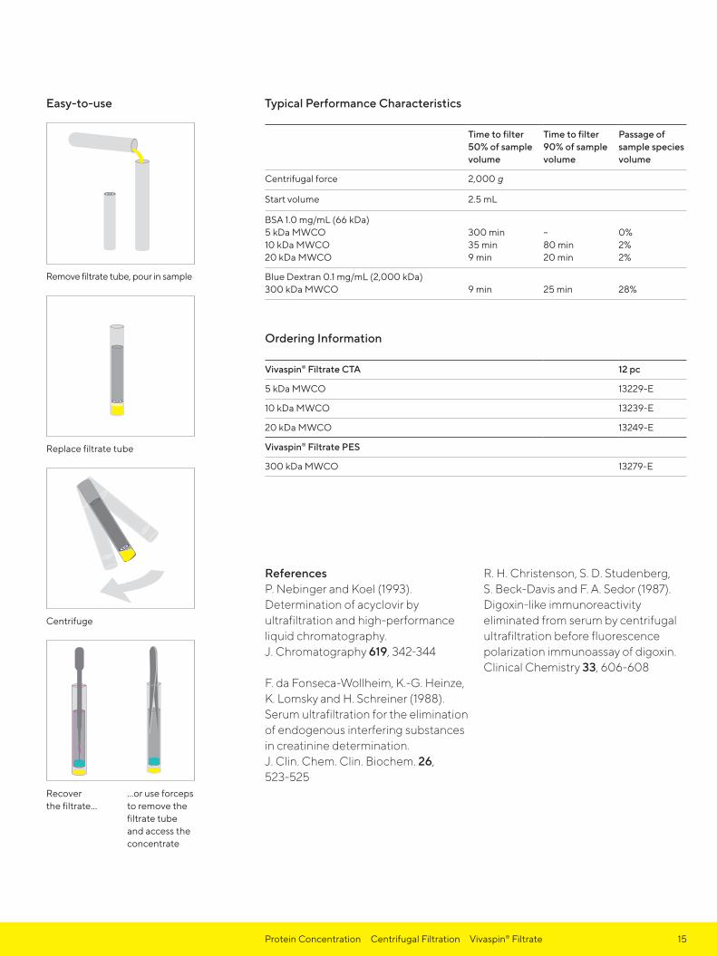
14 Protein Concentration Centrifugal Filtration Vivaspin® Filtrate
0.5–2.5 mL samplesVivaspin Filtratre® is a ready-to-use unit for low volume centrifugal ultrafiltration to separate proteins from low molecular weight substances in biological samples.
Vivaspin Filtratre® features a unique design that enables ultrafiltration in the direction opposite to centrifugal force. This is so effective in preventing premature blockage of the filter that even whole blood samples can be deproteinized.
The ultrafiltrate is collected in the floating filtrate tube, where it is readily accessible without disassembly.
Vivaspin® Filtrate is ideal for the following applications: - Drug binding studies - Isolation of metabolites from serum - Protein removal from blood samples - Cleaning of liposomes - Virus removal
Technical Specifications
Concentrator capacity
Swing bucket rotor 2.5 mL
Fixed angle rotor 2.5 mL
Dimensions
Length x diameter 93 x 14 mm
Active membrane area 0.79 cm2
Hold-up volume, membrane < 5 μL
Dead-stop volume 100 μL
Materials of construction
Centrifuge tube Polystyrene (PS)
Filtrate tube Styrene Acrylonitrile (SAN)
Concentrator cap Polyethylene (PE)
Membrane Cellulose Triacetate (CTA)Polyethersulfone (PES)
Equipment Required
Centrifuge
Rotor type Swing bucket Fixed angle (min. 25°)
Rotor cavity To fit 15 mL (17 mm)conical | flat bottom tubes
To fit 15 mL (17 mm)conical | flat bottom tubes
Maximum RCF 2,500 g 2,000 g
Concentrate recovery
Pipette type Fixed or variable volume Fixed or variable volume
Recommended tip Thin gel loader type Thin gel loader type
Vivaspin® Filtrate
max
. 2.5
mL
93
14
15Protein Concentration Centrifugal Filtration Vivaspin® Filtrate
Typical Performance Characteristics
Time to filter50% of samplevolume
Time to filter90% of samplevolume
Passage ofsample speciesvolume
Centrifugal force 2,000 g
Start volume 2.5 mL
BSA 1.0 mg/mL (66 kDa)5 kDa MWCO10 kDa MWCO20 kDa MWCO
300 min35 min9 min
–80 min20 min
0%2%2%
Blue Dextran 0.1 mg/mL (2,000 kDa)300 kDa MWCO 9 min 25 min 28%
Ordering Information
Vivaspin® Filtrate CTA 12 pc
5 kDa MWCO 13229-E
10 kDa MWCO 13239-E
20 kDa MWCO 13249-E
Vivaspin® Filtrate PES
300 kDa MWCO 13279-E
Remove filtrate tube, pour in sample
Replace filtrate tube
Centrifuge
Recover the filtrate…
…or use forceps to remove the filtrate tube and access the concentrate
Easy-to-use
ReferencesP. Nebinger and Koel (1993).Determination of acyclovir byultrafiltration and high-performanceliquid chromatography.J. Chromatography 619, 342-344
F. da Fonseca-Wollheim, K.-G. Heinze,K. Lomsky and H. Schreiner (1988).Serum ultrafiltration for the eliminationof endogenous interfering substancesin creatinine determination.J. Clin. Chem. Clin. Biochem. 26, 523-525
R. H. Christenson, S. D. Studenberg,S. Beck-Davis and F. A. Sedor (1987).Digoxin-like immunoreactivityeliminated from serum by centrifugalultrafiltration before fluorescencepolarization immunoassay of digoxin.Clinical Chemistry 33, 606-608

122
17
16 Protein Concentration Centrifugal Filtration Vivaspin® Turbo 4
2 to 4 mL samplesVivaspin® Turbo 4 PES offers the fastest sample concentration with the highest recoveries. This device can handle up to 4 mL sample volumes in swing bucket and fixed angle rotors that accept 15 mL conical bottom centrifuge tubes.
The optimized design and sleek internal profile ensure maximum process speeds all the way down to the last few micro liters, resulting in more than 100-fold concentration.
UV joining technology provides a smooth transition between membrane and housing, allowing collection of the entire concentrated sample from the unique, pipette tip-friendly angular dead-stop pocket.
Technical Specifications
Concentrator capacity
Swing bucket rotor 4 mL
Fixed angle rotor 4 mL
Dimensions
Length x diameter 122.5 x 17 mm
Active membrane area 3.2 cm2
Hold-up volume, membrane < 10 μL
Dead-stop volume, swing bucket 40 μL
Dead-stop volume, fixed angle 30 μL
Materials of construction
Body Styrene Butadiene Copolymer (SBC)
Filtrate vessel Polypropylene (PP)
Concentrator cap Polypropylene (PP)
Membrane Polyethersulfone (PES)
Equipment Required
Centrifuge
Rotor type Swing bucket Fixed angle (min. 25°)
Rotor cavity To fit 15 mL (17 mm) conical bottom tubes
To fit 15 mL (17 mm) conical bottom tubes
Maximum RCF 4,000 g 7,500 g
Maximum RCF, 100 kDa MWCO 3,000 g 5,000 g
Concentrate recovery
Pipette type Fixed or variable volume Fixed or variable volume
Recommended tip Thin gel loader type Thin gel loader type
Vivaspin® Turbo 4 PES
17Protein Concentration Centrifugal Filtration Vivaspin® Turbo 4
Typical Performance Characteristics
Time to concentrate up to 30× at 20°C and solute recovery
Rotor Swing bucket Fixed angle (25°)
Centrifugal force* 4,000 g 7,500 g
Start volume 4 mL 4 mL
Time Recovery Time Recovery
Cytochrome c (12.4 kDa)3 kDa MWCO PES5 kDa MWCO PES
60 min40 min
98%95%
80 min50 min
96%94%
Lysozyme (14.3 kDa)3 kDa MWCO PES5 kDa MWCO PES
65 min50 min
95%94%
70 min60 min
93%92%
α-Chymotrypsin (25 kDa)10 kDa MWCO PES 10 min 95% 8 min 95%
BSA (66 kDa)10 kDa MWCO PES30 kDa MWCO PES
10 min8 min
98%96%
7 min6 min
97%97%
IgG (160 kDa)30 kDa MWCO PES50 kDa MWCO PES100 kDa MWCO PES
18 min16 min17 min
94%93%94%
13 min12 min13 min
92%90%92%
Ordering Information
Vivaspin® Turbo 4 PES 25 pc 100 pc
3 kDa MWCO VS04T91 VS04T92
5 kDa MWCO VS04T11 VS04T12
10 kDa MWCO VS04T01 VS04T02
30 kDa MWCO VS04T21 VS04T22
50 kDa MWCO VS04T31 VS04T32
100 kDa MWCO VS04T41 VS04T42
* 3,000 g (swing bucket) or 5,000 g (fixed angle) centrifugal force for 100 kDa MWCO devices.
Visit us at www.sartorius.com/ VivaspinTurbo4 for further information.Here you can find instruc-tions on how to use Vivaspin® Turbo 4 PES for: - Desalting and buffer
exchange - Preparation of biological nanoparticles and medical nanocarriers - Concentration and purification of viruses - Urine protein concentra-tion - Separation of proteins and metabolites for disease detection

122
17
1
2
3
4
5
0.5
0.3
0.2
0.1
18 Protein Concentration Centrifugal Filtration Vivaspin® 6
2 to 6 mL samplesVivaspin® 6 concentrators have been developed to offer increased volume flexibility and performance.
Vivaspin® 6 can process an impressive 6 mL in either swing bucket or fixed angle rotors accepting standard 15 mL conical bottom centrifuge tubes.
Featuring twin vertical membranes for unparalleled filtration speeds and more than 100-fold concentration, the retentate volume is easily estimated from the printed graduations on the side of the concentrator. The modified dead-stop pocket further simplifies direct pipette retrieval of the final concentrate.
Technical Specifications
Concentrator capacity
Swing bucket rotor 6 mL
Fixed angle rotor 6 mL
Dimensions
Length x diameter 122 x 17 mm
Active membrane area 2.5 cm2
Hold-up volume, membrane < 10 μL
Dead-stop volume 30 μL
Materials of construction
Body Polycarbonate (PC)
Filtrate vessel Polycarbonate (PC)
Concentrator cap Polypropylene (PP)
Membrane Polyethersulfone (PES)
Equipment Required
Centrifuge
Rotor type Swing bucket Fixed angle (min. 25°)
Rotor cavity To fit 15 mL (17 mm) conical bottom tubes
To fit 15 mL (17 mm)conical bottom tubes
Maximum RCF 4,000 g 8,000 g
Maximum RCF, ≥100 kDa MWCO
4,000 g 6,000 g
Concentrate recovery
Pipette type Fixed or variable volume Fixed or variable volume
Recommended tip Thin gel loader type Thin gel loader type
Vivaspin® 6

19Protein Concentration Centrifugal Filtration Vivaspin® 6
Performance Characteristics
Time to concentrate up to 30×at 20°C and solute recovery
Rotor Swing bucket Fixed angle (25°)
Centrifugal force 3,000 g 7,500 g
Start volume 6 mL 6 mL
Time Recovery Time Recovery
Cytochrome c 0.25 mg/mL (12.4 kDa)3 kDa MWCO PES – – 90 min 97%
BSA 1.0 mg/mL (66 kDa)5 kDa MWCO PES10 kDa MWCO PES30 kDa MWCO PES
20 min13 min12 min
98%98%98%
12 min10 min9 min
98%98%97%
IgG 0.25 mg/mL (160 kDa)30 kDa MWCO PES50 kDa MWCO PES100 kDa MWCO PES
18 min17 min15 min
96%96%91%
15 min 14 min12 min
95%95%91%
Latex beads 0.004% in DMEM + 10% FCS (55 nm)300 kDa MWCO PES – – 25 min 99%
Latex beads 0.004% in DMEM + 10% FCS (240 nm)1,000 kDa MWCO PES – – 4 min 99%
Yeast 1.0 mg/mL (S. Cerevisiae)0.2 μm PES 4 min 97% 3 min 97%
Ordering Information
Vivaspin® 6 PES 25 pc 100 pc
3 kDa MWCO VS0691 VS0692
5 kDa MWCO VS0611 VS0612
10 kDa MWCO VS0601 VS0602
30 kDa MWCO VS0621 VS0622
50 kDa MWCO VS0631 VS0632
100 kDa MWCO VS0641 VS0642
300 kDa MWCO VS0651 VS0652
1,000 kDa MWCO VS0661 VS0662
0.2 μm VS0671 VS0672

30
1161.0
0.75
0.5
0.3
0.15
5
10
15
20 Protein Concentration Centrifugal Filtration Vivaspin® 15R
2 to 15 mL samplesVivaspin® 15R is designed for initial sample volumes up to 15 mL and features a modified regenerated cellulose membrane; Hydrosart®. This membrane is ideal where extremely high recovery with very low adsorption is needed. An example of this application includes desalting and concentration of immunoglobulin fractions.
- Ultimate recoveries (95 – 98%) - Extremely short concentration time (30-fold in 15 minutes) - Simple and convenient handling - Easy scale-up to 0.1 to 5 L with Vivaflow® 50R or 200 with Hydrosart® membranes - Very low hold-up volume (< 20 μL)
Technical Specifications
Concentrator capacity
Swing bucket rotor 15 mL
Fixed angle rotor 12.5 mL
Dimensions
Length x diameter 116 x 30 mm
Active membrane area 3.9 cm2
Hold-up volume, membrane < 20 μL
Dead-stop volume 30 μL
Materials of construction
Body Polycarbonate (PC)
Filtrate vessel Polycarbonate (PC)
Concentrator cap Polypropylene (PP)
Membrane Hydrosart® (HY)
Equipment Required
Centrifuge
Rotor type Swing bucket Fixed angle (min. 25°)
Rotor cavity To fit 50 mL (30 mm)conical bottom tubes
To fit 50 mL (30 mm)conical bottom tubes
Maximum RCF 3,000 g 6,000 g
Concentrate recovery
Pipette type Fixed or variable volume Fixed or variable volume
Recommended tip Thin gel loader type Thin gel loader type
Vivaspin® 15R

21Protein Concentration Centrifugal Filtration Vivaspin® 15R
Typical Performance Characteristics
Time to concentrate up to 30× at 20°C and solute recovery
Rotor Swing bucket Fixed angle (25°)
Centrifugal force 3,000 g 6,000 g
Start volume 15 mL 12.5 mL
Time Recovery Time Recovery
Aprotinin 0.1 mg/mL* (6.5 kDa)5 kDa MWCO 47 min 95% 45 min 95%
Cytochrome c 0.25 mg/mL* (12.4 kDa)5 kDa MWCO10 kDa MWCO
45 min25 min
96%94%
45 min18 min
96%94%
α-chymotrypsin 0.25 mg/mL* (25 kDa)5 kDa MWCO10 kDa MWCO
50 min25 min
98%98%
45 min18 min
98%98%
Ovalbumin 1.0 mg/mL* (45 kDa)10 kDa MWCO30 kDa MWCO
20 min15 min
98%94%
14 min12 min
98%94%
BSA 1.0 mg/mL* (66 kDa)30 kDa MWCO 18 min 98% 15 min 98%
IgG 0.1 mg/mL in DMEM (160 kDa)30 kDa MWCO 30 min 98% 25 min 96%
Ordering Information
Vivaspin® 15R HY 12 pc 48 pc
2 kDa MWCO VS15RH91 VS15RH92
5 kDa MWCO VS15RH11 VS15RH12
10 kDa MWCO VS15RH01 VS15RH02
30 kDa MWCO VS15RH21 VS15RH22
1.0
0.75
0.5
0.3
0.15
5
10
15
1.0
0.75
0.5
0.3
0.15
5
10
15
1.0
0.75
0.5
0.3
0.15 5 10 15
1.0 0.75
0.5
0.3
0.15
51015
1.0
0.75
0.5
0.3
0.15
5
10
15
Spin
Recover
* Proteins other than IgG made up in 50 mM potassium phosphate, 150 mM sodium chloride, pH 7.4

22
30
1161.0
0.75
0.5
0.3
0.15
5
10
15
2 to 15 mL samplesVivaspin® Endotest is a single-use ultrafiltration device for endotoxin concentration and removal of interfering substan ces from liquid samples prior to LAL testing.
These devices are certified pyrogen free (≤ 0.05 EU/mL) and are available with 20 kDa MWCO cellulose triacetate membranes.
The centrifugal design enables parallel preparation of multiple test samples, minimizing hands-on time.
Vivaspin® Endotest can effectively be used in swing bucket or fixed angle rotors accepting 50 mL conical bottom centrifuge tubes.
Technical Specifications
Concentrator capacity
Swing bucket rotor 15 mL
Fixed angle rotor 12.5 mL
Dimensions
Length x diameter 116 x 30 mm
Active membrane area 3.9 cm2
Hold-up volume, membrane < 20 μL
Dead-stop volume 30 μL
Materials of construction
Body Polycarbonate (PC)
Filtrate vessel Polycarbonate (PC)
Concentrator cap Polypropylene (PP)
Membrane Cellulose Triacetate (CTA)
Equipment Required
Centrifuge
Rotor type Swing bucket Fixed angle (min. 25°)
Rotor cavity To fit 50 mL (30 mm)conical bottom tubes
To fit 50 mL (30 mm)conical bottom tubes
Maximum RCF 1,000 g 1,000 g
Sample Mixing
Laboratory mixer Vortex
Concentrate recovery
Pipette type Fixed or variable volume Fixed or variable volume
Recommended tip Thin gel loader type Thin gel loader type
Vivaspin® Endotest
Protein Concentration Centrifugal Filtration Vivaspin® Endotest

23
Ordering Information
Vivaspin® Endotest CTA 12 pc
20 kDa MWCO VS15RXETO
1.0
0.75
0.5
0.3
0.15
5
10
15
1.0
0.75
0.5
0.3
0.15
5
10
15
1.0
0.75
0.5
0.3
0.15 5 10 15
1.0 0.75
0.5
0.3
0.15
51015
1.0
0.75
0.5
0.3
0.15
5
10
15
Spin
Recover
Protein Concentration Centrifugal Filtration Vivaspin® Endotest
0.5
1.0
1.5
Vivaspin
® Turbo
30
118
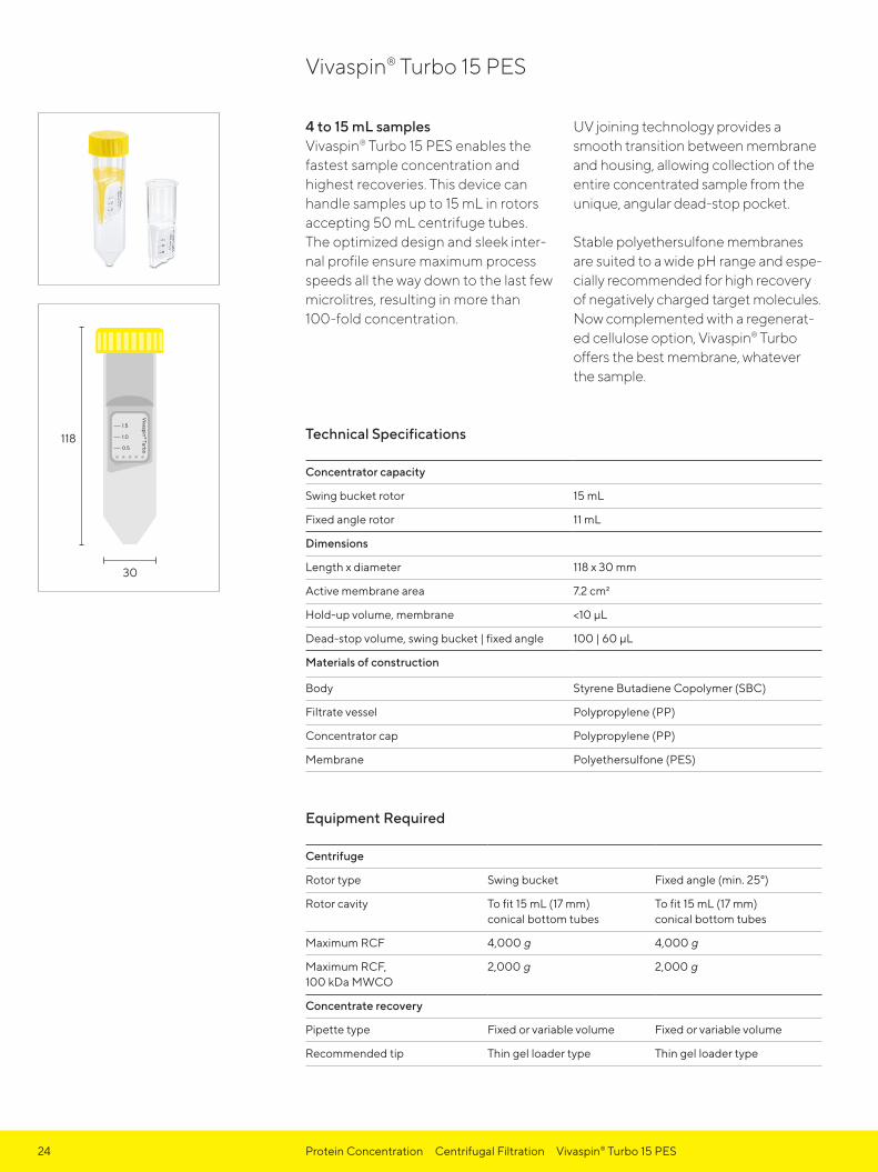
24 Protein Concentration Centrifugal Filtration Vivaspin® Turbo 15 PES
4 to 15 mL samplesVivaspin® Turbo 15 PES enables the fastest sample concentration and highest recoveries. This device can handle samples up to 15 mL in rotors accepting 50 mL centrifuge tubes. The optimized design and sleek inter-nal profile ensure maximum process speeds all the way down to the last few microlitres, resulting in more than 100-fold concentration.
UV joining technology provides a smooth transition between membrane and housing, allowing collection of the entire concentrated sample from the unique, angular dead-stop pocket.
Stable polyethersulfone membranes are suited to a wide pH range and espe-cially recommended for high recovery of negatively charged target molecules. Now complemented with a regenerat-ed cellulose option, Vivaspin® Turbo offers the best membrane, whatever the sample.
Technical Specifications
Concentrator capacity
Swing bucket rotor 15 mL
Fixed angle rotor 11 mL
Dimensions
Length x diameter 118 x 30 mm
Active membrane area 7.2 cm2
Hold-up volume, membrane <10 μL
Dead-stop volume, swing bucket | fixed angle 100 | 60 μL
Materials of construction
Body Styrene Butadiene Copolymer (SBC)
Filtrate vessel Polypropylene (PP)
Concentrator cap Polypropylene (PP)
Membrane Polyethersulfone (PES)
Equipment Required
Centrifuge
Rotor type Swing bucket Fixed angle (min. 25°)
Rotor cavity To fit 15 mL (17 mm) conical bottom tubes
To fit 15 mL (17 mm)conical bottom tubes
Maximum RCF 4,000 g 4,000 g
Maximum RCF, 100 kDa MWCO
2,000 g 2,000 g
Concentrate recovery
Pipette type Fixed or variable volume Fixed or variable volume
Recommended tip Thin gel loader type Thin gel loader type
Vivaspin® Turbo 15 PES

25Protein Concentration Centrifugal Filtration Vivaspin® Turbo 15 PES
Typical Performance Characteristics
Time to concentrate up to 20× at 20°C and solute recovery
Rotor Swing bucket Fixed angle (25°)
Centrifugal force 4,000 g 4,000 g
Start volume 15 mL 11 mL
Time Recovery Time Recovery
Cytochrome c 0.25 mg/mL (12.4 kDa)5 kDa MWCO PES 30 min 98% 50 min 98%
Lysozyme 0.25 mg/mL (14.3 kDa)5 kDa MWCO PES 33 min 96% 50 min 96%
α-Chymotrypsin 1.0 mg/mL (25 kDa)10 kDa MWCO PES 10 min 95% 10 min 95%
BSA 1.0 mg/mL (66 kDa)10 kDa MWCO PES30 kDa MWCO PES
10 min8 min
99%98%
10 min10 min
99%98%
IgG 1.0 mg/mL (160 kDa)30 kDa MWCO PES 23 min 95% 17 min 95%
Ordering Information
Vivaspin® Turbo 15 PES 12 pc 48 pc
3 kDa MWCO VS15T91 VS15T92
5 kDa MWCO VS15T11 VS15T12
10 kDa MWCO VS15T01 VS15T02
30 kDa MWCO VS15T21 VS15T22
50 kDa MWCO VS15T31 VS15T32
100 kDa MWCO VS15T41 VS15T42

0.5
1.0
1.5
Vivaspin
® Turbo
30
118
26 Protein Concentration Centrifugal Filtration Vivaspin® Turbo 15 RC
4 to 15 mL samplesVivaspin® Turbo 15 RC enables the fastest sample concentration and highest recoveries. This device can handle samples up to 15 mL in rotors accepting 50 mL centrifuge tubes. The optimized design and sleek internal profile ensure maximum process speeds all the way down to the last few microlitres, resulting in more than 100-fold concentration.
Solvent-free, heat weld technologyprovides a smooth transition between membrane and housing, allowing
complete concentrate recovery from the unique, angular dead-stop pocket.
Regenerated cellulose membranes developed specifically for Sartorius lab ultrafiltration devices are suited to general samples, with ultra-low adsorption and high chemical compatibility, and especially recommended for oligonucleotides and peptides. Complemented with a polyethersulfone option, Vivaspin® Turbo offers the best membrane, whatever the sample.
Technical Specifications
Concentrator capacity
Swing bucket rotor 15 mL
Fixed angle rotor 11 mL
Dimensions
Length x diameter 118 x 30 mm
Active membrane area 8.1 cm2
Hold-up volume, membrane < 28 μL
Dead-stop volume, swing bucket | fixed angle 120 | 140 μL
Materials of construction
Body Styrene Butadiene Copolymer (SBC)
Filtrate vessel Polypropylene (PP)
Concentrator cap Polypropylene (PP)
Membrane Regenerated Cellulose (RC)
Equipment Required
Centrifuge
Rotor type Swing bucket Fixed angle (min. 25°)
Rotor cavity To fit 15 mL (17 mm) conical bottom tubes
To fit 15 mL (17 mm)conical bottom tubes
Maximum RCF 4,000 g 6,000 g
Maximum RCF, 100 kDa MWCO
3,000 g 6,000 g
Concentrate recovery
Pipette type Fixed or variable volume Fixed or variable volume
Recommended tip Thin gel loader type Thin gel loader type
Vivaspin® Turbo 15 RC

27Protein Concentration Centrifugal Filtration Vivaspin® Turbo 15 RC
Typical Performance Characteristics
Time to concentrate up to 20× at 20°C and solute recovery
Rotor Swing bucket Fixed angle (25°)
Centrifugal force 4,000 g* 6,000 g
Start volume 15 mL 11 mL
Time Recovery Time Recovery
Cytochrome c 0.25 mg/mL (12.4 kDa)5 kDa MWCO RC 23 min 94% 37 min 92%
Lysozyme 0.25 mg/mL (14.3 kDa)5 kDa MWCO RC 23 min 94% 37 min 89%
α-Chymotrypsin 1.0 mg/mL (25 kDa)10 kDa MWCO RC 7 min 93% 9 min 92%
BSA 1.0 mg/mL (66 kDa)10 kDa MWCO RC30 kDa MWCO RC
8 min4 min
94%96%
10 min4 min
98%93%
IgG 1.0 mg/mL (160 kDa)50 kDa MWCO RC100 kDa MWCO RC
17 min18 min
95% 89%
11 min12 min
96%89%
Ordering Information
Vivaspin® Turbo 15 RC 12 pc 48 pc
5 kDa MWCO VS15TR11 VS15TR12
10 kDa MWCO VS15TR01 VS15TR02
30 kDa MWCO VS15TR21 VS15TR22
50 kDa MWCO VS15TR31 VS15TR32
100 kDa MWCO VS15TR41 VS15TR42
* 3,000 g for 100 kDa MWCO devices

5
10
1
0.75
0.5
0.2
15
30
116
28 Protein Concentration Centrifugal Filtration Vivaspin® 20
5 to 20 mL samplesVivaspin® 20 centrifugal concentrators have been developed to offer increased sample and process flexibility and high performance.
Featuring twin vertical membra nes for high filtration speeds, Vivaspin® 20 can achieve in excess of 100-fold concentrations factors.
The retentate volume is easily monitored using printed graduations and the modified dead-stop pocket simplifies direct retentate retrieval.
In addition, unique accessories are available for Vivaspin® 20, enabling pressurized, ultrafiltration and constant volume diafiltration.
Technical Specifications
Concentrator capacity
Swing bucket rotor 20 mL
Fixed angle rotor 14 mL
With pressure head 15 mL
Dimensions
Length x diameter 116 x 30 mm125 x 30 mm with pressure head
Active membrane area 6.0 cm2
Hold-up volume, membrane < 20 μL
Dead-stop volume 50 μL
Materials of construction
Body Polycarbonate (PC)
Filtrate vessel Polycarbonate (PC)
Concentrator cap Polypropylene (PP)
Pressure head Polyoxymethylene (POM) and Aluminium (ALU)
Membrane Polyethersulfone (PES)
Equipment Required
Centrifuge
Rotor type Swing bucket Fixed angle (min. 25°)
Rotor cavity To fit 50 mL (30 mm)conical bottom tubes
To fit 50 mL (30 mm)conical bottom tubes
Maximum RCF 4,000 g 6,000 g
Maximum RCF, ≥100 kDa MWCO
3,000 g 6,000 g
Pressure
Pressure accessories VCA002, VCA005 and VCA200
Maximum pressure 5 bar (75 psi)
Maximum RCF, pressure-fuge* 3,000 g
Maximum RCF, pressure-fuge, ≥100 kDa MWCO*
2,000 g
Vivaspin® 20
*Swing bucket only

29Protein Concentration Centrifugal Filtration Vivaspin® 20
Typical Performance Characteristics
Time to concentrate up to 30× at 20°C and solute recovery
Mode Centrifuge Centrifuge Bench top Press-fuge
Rotor Swing bucket 25° Fixed angle Pressure Swing bucket
Centrifugal force | pressure 3,000 g 6,000 g 4 bar 3,000 g + 4 bar
Start volume 20 mL 14 mL 10 mL 10 mL
Min. Rec. Min. Rec. Min. Rec. Min. Rec.
Cytochrome c 0.25 mg/mL (12.4 kDa)3 kDa MWCO PES 110 97% 180 96% 60 96% – –
BSA 1.0 mg/mL (66 kDa)5 kDa MWCO PES10 kDa MWCO PES30 kDa MWCO PES
231613
99%98%98%
291715
99%98%98%
503232
98%97%97%
1488
98%97%97%
IgG 0.25 mg/mL (160 kDa)30 kDa MWCO PES50 kDa MWCO PES100 kDa MWCO PES
272725
97%96%91%
202220
95%95%90%
464642
94%93%88%
131312
97%96%94%
Latex beads 0.004% in DMEM +10% FCS (55 nm)300 kDa MWCO PES 20 99% 35 99% 10 99% – –
Latex beads 0.004% in DMEM +10% FCS (240 nm)1,000 kDa MWCO PES 4 99% 12 99% 4 99% – –
Yeast 1.0 mg/mL (S. Cerevisiae)0.2 μm PES 15 95% 5 95% 20 95% 2 95%
Ordering Information
Vivaspin® 20 PES 12 pc 48 pc
3 kDa MWCO VS2091 VS2092
5 kDa MWCO VS2011 VS2012
10 kDa MWCO VS2001 VS2002
30 kDa MWCO VS2021 VS2022
50 kDa MWCO VS2031 VS2032
100 kDa MWCO VS2041 VS2042
300 kDa MWCO VS2051 VS2052
1,000 kDa MWCO VS2061 VS2062
0.2 μm VS2071 VS2072
Equipment Required (Continued)
Concentrate recovery
Pipette type Fixed or variable volume Fixed or variable volume
Recommended tip Thin gel loader type Thin gel loader type

30 Protein Concentration Centrifugal Filtration Vivaspin® 100
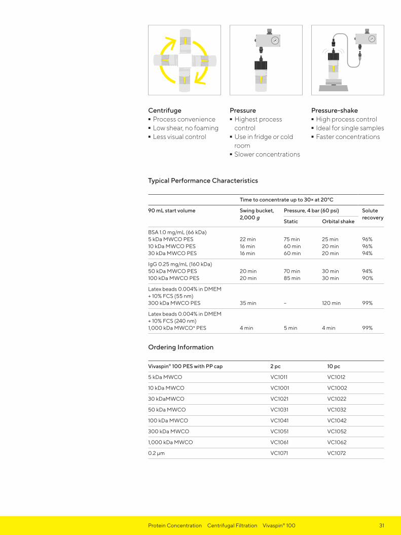
20 to 98 mL samplesVivaspin® 100 bridges the gap between centrifugal concentrators and crossflow cassettes. These devices feature vertical membranes for high speed processing of even high particle loaded samples. In addition, a unique choice between centrifugal, pressure or pressure-shake operating methods provides unrivaled process flexibility.
Fitting swing bucket rotors accepting 250 mL bottles, Vivaspin® 100 offers the highest sample capacity available in a centrifugal device – up to an astonishing 90 mL.
Vivaspin® 100 units can also be used for single or extremely sensitive samples of up to 98 mL when pressurized and left on the bench, or for temperature- sensitive samples, placed into a refrigerator. Pressurization is made easy by use of quick-release connectors and can be combined with orbital shaking for even faster sample concentration.
In whichever mode Vivaspin® 100 is used, the vertical membrane design inhibits membrane fouling while the integrated dead-stop impedes concentration to dryness and loss of sample.
Technical Specifications
Concentrator capacity
Swing bucket rotor 90 mL
With pressure head 98 mL
Dimensions
Length x diameter 123 x 62 mm197 x 62 mm with pressure head
Active membrane area 23.5 cm2
Hold-up volume of membrane < 250 μL
Dead-stop volume 350 μL
Materials of construction
Body Polycarbonate (PC)
Filtrate vessel Polycarbonate (PC)
Concentrator cap Polypropylene (PP)
Pressure head Polyoxymethylene (POM) and Aluminium (ALU)
Pressure head seal Thermoplastic Elastomer (TPE)
Membrane Polyethersulfone (PES)
Equipment RequiredCentrifuge
Rotor type Swing bucket
Rotor cavity To fit 250 mL (60 mm) centrifuge bottles(maximum cavity depth 105 mm)
Maximum RCF 2,000 g
Pressure
Pressure accessories VCA002, VCA800
Maximum pressure 5 bar (75 psi)
Vivaspin® 100
Device fits standard 250 mL rotors
31Protein Concentration Centrifugal Filtration Vivaspin® 100
Typical Performance Characteristics
Time to concentrate up to 30× at 20°C
90 mL start volume Swing bucket, 2,000 g
Pressure, 4 bar (60 psi) Solute recovery
Static Orbital shake
BSA 1.0 mg/mL (66 kDa)5 kDa MWCO PES10 kDa MWCO PES30 kDa MWCO PES
22 min 16 min 16 min
75 min 60 min 60 min
25 min 20 min 20 min
96% 96% 94%
IgG 0.25 mg/mL (160 kDa)50 kDa MWCO PES100 kDa MWCO PES
20 min20 min
70 min85 min
30 min30 min
94%90%
Latex beads 0.004% in DMEM + 10% FCS (55 nm)300 kDa MWCO PES 35 min – 120 min 99%
Latex beads 0.004% in DMEM + 10% FCS (240 nm)1,000 kDa MWCO* PES 4 min 5 min 4 min 99%
Ordering Information
Vivaspin® 100 PES with PP cap 2 pc 10 pc
5 kDa MWCO VC1011 VC1012
10 kDa MWCO VC1001 VC1002
30 kDaMWCO VC1021 VC1022
50 kDa MWCO VC1031 VC1032
100 kDa MWCO VC1041 VC1042
300 kDa MWCO VC1051 VC1052
1,000 kDa MWCO VC1061 VC1062
0.2 μm VC1071 VC1072
Centrifuge - Process convenience - Low shear, no foaming - Less visual control
Pressure - Highest process control - Use in fridge or cold room - Slower concentrations
Pressure-shake - High process control - Ideal for single samples - Faster concentrations

32 Protein Concentration Pressure Filration Vivaspin® Equipment
Vivaspin® Equipment and Accessories
5
10
1
0.75
0.5
0.2
15
5
10
1
0.75
0.5
0.2
15
5
10
1
0.75
0.5
0.2
15
5
10
1
0.75
0.5
0.2
15
5 101
0.75
0.5
0.2
15 510 1
0.75
0.5
0.215
5
10
1
0.75
0.5
0.2
15
5
10
1
0.75
0.5
0.2
15
Using the Vivaspin® 20 pressure cap
Pressurize
Spin or stand in rack
Gas Pressure UltrafiltrationWhen an appropriate centrifuge is unavailable, or for single sample processing, Vivaspin® 20 and 100 centrifugal concentrators may be pressurized with compressed gas for bench-top concentration.
For even faster processing of samples in Vivaspin® 20, gas pressure can be combined with centrifugal force. This pressure-fugation method is particularly suitable for difficult to filter or viscous samples, such as serum, or when using low process temperatures, which reduce filtration speed, and generally when minimum process time is essential. In a similar way, Vivaspin® 100 may be pressurized and placed on an orbital shaker for faster processing.
Constant Volume DiafiltrationIn this procedure following concentration, a diafiltration cup inserted into the Vivaspin® 20 concentrator body is filled with buffer and centrifuged once to achieve 98% salt removal. This compares to the need for two centrifugation steps to achieve the same result with the re-fill and re-spin approach for discontinuous diafiltration.
The improved performance is due to the constant washing action of the exchange buffer from the diafiltration cup, as it replaces the original solvent and salts when they pass through the ultrafiltration membrane.

Ordering Information
Vivaspin® Equipment and Accessories Pack Size Prod. No.
Air pressure controller (APC) fitted with pressure gauge,regulator, over-pressure safety valve and female coupling. APCis supplied with extension line (4 mm pneumatic tubing, 1 m) with male and female couplings, and inlet tubing (6 mm pneumatic tubing, 1 m)
1 VCA002
Charge valve for pressure head VCA200 1 VCA005
Female coupling 1 VCA010
Male coupling 1 VCA011
Replacement extension line (4 mm pneumatic tubing, 3 m)
1 VCA012
Vivaspin® 20 pressure head 1 VCA200
Vivaspin® 100 pressure head with seals 1 VCA800
Vivaspin® 100 pressure head seals 10 VCA014
Diafiltration cups 12 VSA005
33Protein Concentration Pressure Filtration Vivaspin® Equipment

34 Protein Concentration Tangential Flow Filtration Vivaflow® 50
0.1 to 3 LThe novel Vivaflow® 50 system provides a standard of ease of use, performance, flexibility and economy which is unrivalled by any laboratory or pilot scale filtration system on the market.
Unique features - Thin channel flip-flow path provides high turbulence and cross flow velocities for exceptional flux, even at high concentrations. - No need for pressure holders. - Crystal clear for simple control and visibility of membrane status. - Unique interlocking modules with series connectors for easy scale up. - Disposable | single use.
Unique performance - A single 50 cm2 module will typically reduce 500 mL to less than 15 mL in under 50 minutes. - Less than 10 mL minimum system recirculation for highest concentrations. - Less than 500 μL non recoverablehold up volume. - Near total recoveries achievable with a single 10 mL rinse.
Each package of two cassettes contains all of the required tubing and fittings for plug-and-play operation with a standard peristaltic pump accepting 6.4 mm OD (size 16) tubing.
Technical Specifications
Dimensions
Overall L | W | H 25 | 107 | 84 mm
Channel W | H 15 mm | 0.3 mm
Active membrane area 50 cm2
Minimum recirculation volume < 10 mL
Hold-up volume, cassette 1.5 mL
Non recoverable hold-up < 0.5 mL
Operating conditions
Pump flow rate 200 – 400 mL/min
Maximum pressure 3 bar (45 psi)
Maximum temperature 60°C
Materials of construction
Main housing Polycarbonate (PC)
Flow channel Polymethylpentene (PMP)
Membrane Polyethersulfone (PES)Regenerated Cellulose (RC)
Membrane support Polymethylpentene (PMP)
Seals and O rings Silicone (SIL)
Flow restrictor Polypropylene (PP)
Fittings Polyamide (PA)
Tubing Polyvinyl Chloride (PVC), medical grade
Vivaflow® 50
Multiple cassettes
Single cassette

35Protein Concentration Tangential Flow Filtration Vivaflow® 50
Typical Performance Characteristics
Time to concentrate up to 20x at 3 barinlet pressure, 20°C
Single device 250 mLstart volume
Three devices 1 Lstart volume
Solute recovery
Direct 10 mL rinse
BSA 1.0 mg/mL (66 kDa)5 kDa MWCO PES10 kDa MWCO PES30 kDa MWCO PES50 kDa MWCO PES
34 min22 min22 min20 min
49 min32 min32 min29 min
96%94%92%92%
> 99%> 99%99%98%
IgG 1.0 mg/mL (160 kDa)100 kDa MWCO PES100 kDa MWCO RC
43 min40 min
62 min58 min
92%92%
98%98%
Yeast 1.0 mg/mL (S. Cerevisiae)0.2 μm PES 33 min 47 min 92% 98%
Ordering Information
Vivaflow® 50 PES 2 cassettes
3 kDa MWCO VF05P9
5 kDa MWCO VF05P1
10 kDa MWCO VF05P0
30 kDa MWCO VF05P2
50 kDa MWCO VF05P3
100 kDa MWCO VF05P4
1,000 kDa MWCO VF05P6
0.2 μm VF05P7
Vivaflow® 50 RC
100 kDa MWCO VF05C4

36 Protein Concentration Tangential Flow Filtration Vivaflow® 50R
0.1 to 1 L samplesConcentrate 100 mL to under 20 mL in just a few minutes or concentrate one liter 50 times in less than 60 minutes. Alternatively, speed up your process by using two Vivaflow® 50R cassettes in parallel and concentrate 1 liters in under 30 min.
Vivaflow® 50R is a plug-and-play laboratory crossflow cassette for concentrating up to 1 L aqueous samples. The active membrane area per device is 50 cm2.
Each cassette is supplied with all the tubing and a pressure indicator for running the device with a laboratory pump and a size 16 pump head. For speeding up concentration, two cassettes can be run simultaneously.
- Fast and easy protein sample concentration - Reusable - Concentrates volumes from 0.1 L to 1 L - Optimal for concentration of culture supernatants and viruses - The most compact crossflow cassette with a premium Hydrosart® membrane
Technical SpecificationsDimensions
Overall L | W | H 24 | 100 | 100 mm
Channel W | H 7.5 | 0.4 mm
Active membrane area 50 cm2
Minimum recirculation volume 10 mL
Hold-up volume, cassette 1.7 mL
Non-recoverable hold-up < 0.5 mL
Operating conditions
Pump flow rate 200 – 400 mL/min
Maximum pressure 4 bar (60 psi)
Maximum temperature 60°C
Materials of construction
Main housing Acrylic
Flow channel Acrylic
Membrane Hydrosart® (HY)
Membrane support Polyethylene (PE)
Seals and O-rings Silicone (SIL)
Pressure indicator Polypropylene (PP), SS spring
Flow restrictor Polypropylene (PP)
Fittings Polyamide (PA)
Tubing Polyvinyl Chloride (PVC), medical grade
Vivaflow® 50R
Vivaflow® 50R – Single cassette
Vivaflow® 50R – Two cassettes

37Protein Concentration Tangential Flow Filtration Vivaflow® 50R
Typical Performance Characteristics
Time to concentrate up to 20× at3.0 bar inlet | 2.5 bar outlet pressure, 20°C
Start volume250 mL
Average fluxmL/min
Recovery
Direct 25 mL rinse
Lysozyme 0.25 mg/mL (14.3 kDa)5 kDa MWCO HY10 kDa MWCO HY
7023
3.410.3
96%94%
98%96%
BSA 1.0 mg/mL (66 kDa)10 kDa MWCO HY30 kDa MWCO HY
2415
9.915.8
98%97%
>99%>99%
IgG 1.0 mg/mL (160 kDa)100 kDa MWCO HY 46 5.2 97% >99%
Start volume 1 L (one Vivaflow® 50R at 3 bar), BSA 1.0 mg/mL10 kDa MWCO HY 95 10.0 98% >99%
Start volume 1 L (two Vivaflow® 50R in parallel at 3 bar), BSA 1.0 mg/mL10 kDa MWCO Hydrosart® 48 19.8 98% >99%
Ordering Information
Vivaflow® 50R HY 1 cassette
5 kDa MWCO VF05H1
10 kDa MWCO VF05H0
30 kDa MWCO VF05H2
100 kDa MWCO VF05H4
Visit us at www.sartorius.com/ Vivaflow50R for further information.Here you can find instructions on how to use Vivaflow® 50R for: - Preparation of biological
nanoparticles and medical nanocarriers - Concentration and purification of viruses

38 Protein Concentration Tangential Flow Filtration Vivaflow® 200
0.5 to 5 LConcentrate 250 mL to under 20 mL in just a few minutes or concentrate one litre 50 times in less than 30 minutes. Alternatively, use two Vivaflow® 200 cassettes in parallel and concentrate 5 litres in under 75 minutes.
Near total sample recoveries can be expected with most solutions.
Each cassette is supplied complete with all required tubing and a pressure indicator. All you need is a peristaltic pump capable of handling 6.4 mm OD (size 16) tubing. Should your pump head require larger tubing, link your own tubing up to the standard product, using the provided stepped hose barb connector.
Two cassettes in parallel will concentrate 5 litres in under 75 minutes.
Technical Specifications
Dimensions
Overall L | H | W 38 | 126 | 138 mm
Channel W | H 10 mm | 0.4 mm
Active membrane area 200 cm2
Minimum recirculation volume < 20 mL
Hold up volume, cassette 5.3 mL
Non-recoverable hold-up < 2 mL
Operating conditions
Pump flow 200–400 mL/min
Maximum pressure 4 bar (60 psi)
Maximum temperature 60°C
Materials of construction
Main housing Acrylic
Flow channel Acrylic
Membrane Polyethersulfone (PES)Hydrosart® (HY)
Membrane support Polypropylene
Seals and O rings Silicone (SIL)
Pressure indicator Polypropylene (PP), SS spring
Flow restrictor Polypropylene (PP)
Fittings Polyamide (PA)
Tubing Polyvinyl Chloride (PVC), medical grade
Vivaflow® 200
Vivaflow® 200 set-up for diafiltration

39Protein Concentration Tangential Flow Filtration Vivaflow® 200
Typical Performance Characteristics
Time to concentrate up to 20× at 3 bar inlet pressure, 20˚C
1 litre start volume
Average fluxmL/min
Recovery
Direct 25 mL rinse
Lysozyme 0.25 mg/mL (14.3 kDa)2 kDa MWCO HY3 kDa MWCO PES
160180
65
97%97%
> 99%> 99%
BSA 1.0 mg/mL (66 kDa)5 kDa MWCO PES5 kDa MWCO HY10 kDa MWCO PES10 kDa MWCO HY30 kDa MWCO PES30 kDa MWCO HY50 kDa MWCO PES
29702335252022
33144127384843
98%98%96%98%96%96%96%
> 99%> 99%> 99%> 99%99%> 99%98%
IgG 1.0 mg/mL (160 kDa)100 kDa MWCO PES 54 18 96% 99%
Yeast 1.0 mg/mL (S. Cerevisiae)0.2 μm PES 11 86 92% 98%
Dilute solute concentration, start volume 1 litre at 3 bar, 10 kDa MWCO PESBSA 0.001 mg/mLBSA 0.01 mg/mLBSA 0.1 mg/mL
182021
524745
90%92%94%
98%98%99%
Start volume 5 litres (two VF200 in parallel at 3 bar) 10 kDa MWCO PESBSA 1.0 mg/mL (66 kDa) 67 70 97% > 99%
Ordering Information
Vivaflow® 200 PES 1 cassette
3 kDa MWCO VF20P9
5 kDa MWCO VF20P1
10 kDa MWCO VF20P0
30 kDa MWCO VF20P2
50 kDa MWCO VF20P3
100 kDa MWCO VF20P4
0.2 μm VF20P7
Vivaflow® 200 HY
2 kDa MWCO VF20H9
5 kDa MWCO VF20H1
10 kDa MWCO VF20H0
30 kDa MWCO VF20H2
100 kDa MWCO VF20H4
Operation – Single cassette
Operation – Two cassettes
40 Protein Concentration Tangential Flow Filtration Vivaflow® Equipment
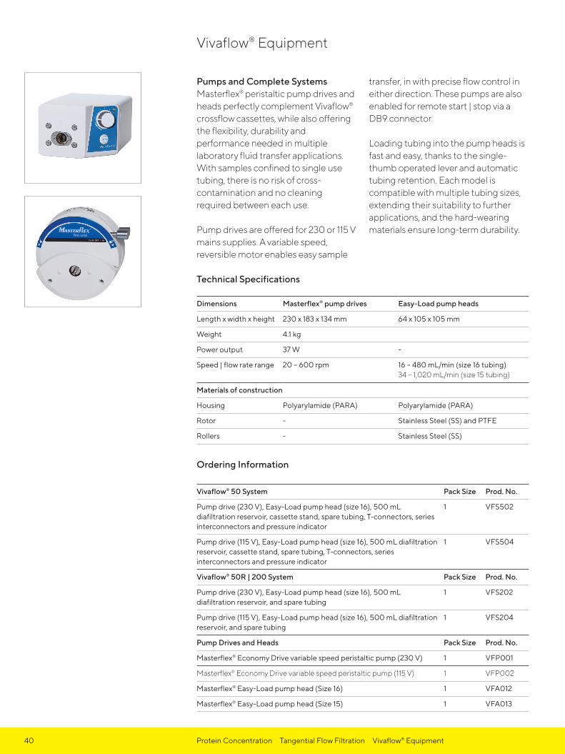
Pumps and Complete Systems Masterflex® peristaltic pump drives and heads perfectly complement Vivaflow® crossflow cassettes, while also offering the flexibility, durability and performance needed in multiple laboratory fluid transfer applications. With samples confined to single use tubing, there is no risk of cross-contamination and no cleaning required between each use.
Pump drives are offered for 230 or 115 V mains supplies. A variable speed, reversible motor enables easy sample
transfer, in with precise flow control in either direction. These pumps are also enabled for remote start | stop via a DB9 connector.
Loading tubing into the pump heads is fast and easy, thanks to the single-thumb operated lever and automatic tubing retention. Each model is compatible with multiple tubing sizes, extending their suitability to further applications, and the hard-wearing materials ensure long-term durability.
Technical Specifications
Dimensions Masterflex® pump drives Easy-Load pump heads
Length x width x height 230 x 183 x 134 mm 64 x 105 x 105 mm
Weight 4.1 kg
Power output 37 W -
Speed | flow rate range 20 – 600 rpm 16 – 480 mL/min (size 16 tubing) 34 – 1,020 mL/min (size 15 tubing)
Materials of construction
Housing Polyarylamide (PARA) Polyarylamide (PARA)
Rotor - Stainless Steel (SS) and PTFE
Rollers - Stainless Steel (SS)
Ordering Information
Vivaflow® 50 System Pack Size Prod. No.
Pump drive (230 V), Easy-Load pump head (size 16), 500 mL diafiltration reservoir, cassette stand, spare tubing, T-connectors, series interconnectors and pressure indicator
1 VFS502
Pump drive (115 V), Easy-Load pump head (size 16), 500 mL diafiltration reservoir, cassette stand, spare tubing, T-connectors, series interconnectors and pressure indicator
1 VFS504
Vivaflow® 50R | 200 System Pack Size Prod. No.
Pump drive (230 V), Easy-Load pump head (size 16), 500 mL diafiltration reservoir, and spare tubing
1 VFS202
Pump drive (115 V), Easy-Load pump head (size 16), 500 mL diafiltration reservoir, and spare tubing
1 VFS204
Pump Drives and Heads Pack Size Prod. No.
Masterflex® Economy Drive variable speed peristaltic pump (230 V) 1 VFP001
Masterflex® Economy Drive variable speed peristaltic pump (115 V) 1 VFP002
Masterflex® Easy-Load pump head (Size 16) 1 VFA012
Masterflex® Easy-Load pump head (Size 15) 1 VFA013
Vivaflow® Equipment

41Protein Concentration Tangential Flow Filtration Vivaflow® Accessories
Scaling UpThe maximum throughput or speed of filtration with Vivaflow® cassettes can be easily increased, thanks to the modular design. Up to 6x Vivaflow® 50 cassettes can be operated in series and parallel, while two Vivaflow® 50R or 200 cassettes may be run in parallel.
Accessories and Replacement PartsOptional accessories such as the diafiltration reservoir, make concentration and diafiltration exceptionally convenient. A sample is
first concentrated to the desired volume, then a length of tubing placed into a separate vessel containing the exchange buffer is connected to the reservoir. Airtight sealing in the lid enables constant volume diafiltration. As the original buffer and salts permeate the ultrafiltration membrane, they are replaced by an equal volume of exchange buffer, thereby avoiding the large buffer volumes and possibility of sample dilution, which can be common to alternative methods.
Technical Specifications
Materials of construction
Cassette stand Aluminium (ALU)
Tubing Polyvinyl Chloride (PVC), medical grade
Luer fittings Polyamide (PA)
Flow restrictors Polypropylene (PP)
Pressure indicator Polypropylene (PP), Stainless Steel (SS), Polyoxymethylene (POM), Silicone (SIL) and Polyamide (PA)
Diafiltration reservoir Polycarbonate (PC), Polyoxymethylene (POM), Silicone (SIL), Polyvinyl Chloride (PVC) and Polyamide (PA)
Ordering Information
Vivaflow® accessories for operating multiple cassettes Pack Size Prod. No.
T-connector (for 2-6x Vivaflow® 50 | 2x Vivaflow® 50R) 2 VFA030
Series interconnector (for 2-6x Vivaflow® 50) 6 VFA031
Y-connector (size 15 to 2x size 16, for 2x Vivaflow® 200) 1 VFA005
Vivaflow® accessories and replacement parts
Vivaflow® 50 cassette stand 1 VFA016
500 mL diafiltration reservoir 1 VFA006
Pump tubing, 3 m, with luer fittings (size 16) 1 VFA004
Pump tubing, 3 m, with luer fittings (size 15) 1 VFA003
Female Luer lock to hose barb fittings (size 16) 10 VFA032
Female Luer lock to hose barb fittings (size 15) 10 VFA036
Flow restrictors, 0.6 mm 6 VFA035
Flow restrictors, 0.4, 0.6 and 0.8 mm (2 of each) 6 VFA009
Pressure indicator (1-3 bar) 1 VFA020
Vivaflow® tubing set (2x 1 m feed tubing with Luer fitting, 2x 0.5 m retentate tubing with flow restrictor, 1x series interconnector)
1 VFA034
Vivaflow® Accessories

42 Protein Concentration Ultrafiltration Membrane Discs
Polyethersulfone (PES)This is a general purpose membrane that provides excellent performance with most solutions when high recoveries in the retentate are of primary importance. Polyethersulfone membranes exhibit no hydrophobic or hydrophillic interactions and are usually preferred for their low fouling characteristics, exceptional flux and broad pH compatibility.
Cellulose Triacetate (CTA)High hydrophilicity and very low non-specific binding characterize this membrane. Cast without any
membrane support that could trap or bind passing microsolutes, these membranes are preferred for sample cleaning, protein removal, and when high recoveries from the filtrate are of primary importance.
Hydrosart® (HY)These membranes are also highly hydrophillic and are often preferred for their higher protein recovery when processing very dilute solutions. Resistance to autoclaving, ease of cleaning and extended chemical compatibility also characterize this membrane material.
Technical Specifications and Typical Performance
Polyethersulfone, Type 146
Cellulose Triacetate, Type 145
Hydrosart®, Type 144
Thickness 120 μm 120 μm 180 μm
pH range 1 – 14 4 – 8 1 – 13
Flux with water
10 kDa MWCO 0.2 mL/min/cm2 0.11 mL/min/cm2 0.08 mL/min/cm2
Protein retention
Cytochrome c 95% 90% 99%
Ordering Information
PES Membrane Discs, Type 146 Diameter 10 pc
1 kDa MWCO 47 mm 14609--47------D
63 mm 14609--63------D
76 mm 14609--76------D
5 kDa MWCO 25 mm 14629--25------D
47 mm 14629--47------D
63 mm 14629--63------D
76 mm 14629--76------D
10 kDa MWCO 25 mm 14639--25------D
43 mm 14639--43------D
47 mm 14639--47------D
63 mm 14639--63------D
76 mm 14639--76------D
150 mm 14639-150------D
Ultrafiltration Membrane Discs

43Protein Concentration Ultrafiltration Membrane Discs
PES Membrane Discs, Type 146 (con't) Diameter 10 pc
30 kDa MWCO 25 mm 14659--25------D
47 mm 14659--47------D
63 mm 14659--63------D
76 m 14659--76------D
50 kDa MWCO 25 mm 14650--25------D
47 mm 14650--47------D
76 mm 14650--76------D
100 kDa MWCO 47 mm 14668--47------D
63 mm 14668--63------D
300 kDa MWCO 25 mm 14679--25------D
47 mm 14679--47------D
76 mm 14679--76------D
CTA Membrane Discs, Type 145
5 kDa MWCO 25 mm 14529--25------D
47 mm 14529--47------D
10 kDa MWCO 25 mm 14539--25------D
47 mm 14539--47------D
50 mm 14539--50------D
20 kDa MWCO 25 mm 14549--25------D
43 mm 14549--43------D
47 mm 14549--47------D
63 mm 14549--63------D
HY Membrane Discs, Type 144
2 kDa MWCO 47 mm 14419--47------D
63 mm 14419--63------D
5 kDa MWCO 25 mm 14429--25------D
44 mm 14429--44------D
47 mm 14429--47------D
63 mm 14429--63------D
76 mm 14429--76------D
10 kDa MWCO 25 mm 14439--25------D
47 mm 14439--47------D
63 mm 14439--63------D
76 mm 14439--76------D
30 kDa MWCO 25 mm 14459--25------D
47 mm 14459--47------D
63 mm 14459--63------D
76 mm 14459--76------D
100 kDa MWCO 47 mm 14468--47------D

44 Chapter Site Title

45
DNA ConcentrationTable of Contents
Vivacon® 500 46
Vivacon® 2 49

46 DNA Concentration Vivacon® 500
100 to 500 μL samplesVivacon® 500 centrifugal concentrators offer the optimal solution for DNA and protein concentration and buffer exchange applications. For optimal performance with very dilute samples, e.g. forensic samples, Vivacon® 500 is equipped with the patented regenerated cellulose membrane, Hydrosart®.
High recoveries and excellent reproducibilities are paired with convenience offered by molecular weight cut-off printed on individual devices.
The possibility of a reverse-spin after sampleprocessing assures complete con centrate recovery which is especially important when working with low sample concentrations.
Vivacon® 500 PCR GradeWhen using DNA amplification technologies, any traces of DNA originating from the equipment have to be eliminated.
Vivacon® 500 PCR Grade units are treated with ethylene oxide (ETO) in a validated process in order to deactivate all traces of DNA that might interfere with subsequent amplification procedures.
Ref.: K. Shaw et al., Int. J. Legal Med. (2008) 122: 29–33
Feature Benefit
Reverse-spin enabled Complete and highly reproducible sample recovery
Low binding material High recoveries of low sample concentrations
Technical Specifications
Concentrator capacity
Fixed angle rotor 0.5 mL
Dimensions
Length x diameter 45 x 12.4 mm47.5 x 12.4 mm reverse spin
Active membrane area 0.32 cm2
Hold-up volume, of membrane and support < 5 μL
Dead-stop volume (40° rotor) 5 μL
Materials of construction
Body Polycarbonate (PC)
Filtrate vessel Polypropylene (PP)
Membrane Hydrosart® (HY)Cellulose Acetate (CA)
Vivacon® 500
Reverse spin

47DNA Concentration Vivacon® 500
Conversion Table for MWCO to Nucleotide Cut-Off
Membrane Single-Stranded Cut-Off (SSCO)
Double-Stranded Cut-Off (DSCO)
2 kDa HY > 10 bases > 10 bp
10 kDa HY > 90 bases > 30 bp
30 kDa HY > 275 bases > 50 bp
50 kDa HY > 475 bases > 300 bp
100 kDa HY > 900 bases > 600 bp
125 kDa CA > 1,000 bases > 650 bp
Typical Performance Characteristics for DNAStart volume 0.5 mL, sample concentration 50 ng/mL
MWCO Nucleic Acid Length
Time to Concentrate up to 30× at 20°C
Concentrate Recovery
RCF
2 kDa 10 bp 60 min 93% 7,500 g
10 kDa 30 bp 25 min 94% 7,500 g
30 kDa 50 bp 18 min 88% 5,000 g
50 kDa 300 bp 18 min 91% 5,000 g
100 kDa 600 bp 10 min 87% 3,000 g
125 kDa 650 bp 12 min 85% 2,000 g
125 kDa 900 bp 9 min 94% 3,000 g
Typical Performance Characteristics for ProteinsStart volume 0.5 mL, sample and concentration of proteins as specified in table
MWCO Test Molecule Time to Concentrate up to 30× at 20°C
Concentrate Recovery
RCF
2 kDa 0.25 mg/mL cytochrome c
30 min 95% 14,000 g
10 kDa 0.25 mg/mL cytochrome c
15 min 92% 14,000 g
30 kDa 1.0 mg/mL BSA 10 min 95% 14,000 g
50 kDa 1.0 mg/mL BSA 10 min 92% 14,000 g
100 kDa 1.0 mg/mL bovine IgG
11 min 90% 8,000 g
125 kDa 1.0 mg/mL bovine IgG
10 min 81% 8,000 g

48 DNA Concentration Vivacon® 500
Ordering Information
Vivacon® 500 25 pc 100 pc 500 pc
2 kDa MWCO VN01H91 VN01H92 -
10 kDa MWCO VN01H01 VN01H02 -
30 kDa MWCO VN01H21 VN01H22 VN01H23
50 kDa MWCO VN01H31 VN01H32 VN01H33
100 kDa MWCO VN01H41 VN01H42 VN01H43
125 kDa MWCO VN01H81 VN01H82 VN01H83
Vivacon® 500 PCR Grade
30 kDa MWCO - VN01H22ETO VN01H23ETO
100 kDa MWCO - VN01H42ETO -
125 kDa MWCO - VN01H82ETO VN01H83ETO
Vivacon® 500 Accessories
Additional collection tubes, 100 pc VNCT01

2.0mL
1.5
1.0
2.0mL
1.5
1.0
49DNA Concentration Vivacon® 2
0.4 – 2 mL samplesVivacon® 2 centrifugal concentrators offer the optimal solution for DNA and protein concentration and buffer exchange applications. For optimal performance with very dilute samples, e.g. forensic samples, Vivacon® 2 is equipped with the patented regener-ated cellulose membrane Hydrosart®.
High recoveries and excellent repro-ducibilities are paired with conve-nience offered by volume graduation and molecular weight cut-off printed on individual devices.
The possibility of a re-spin after sampleprocessing assures complete con-centrate recovery which is especially important when working with low sample concentrations.
Vivacon® 2 PCR GradeVivacon® 2 PCR Grade units are treated with ethylene oxide (ETO) in a validated process in order to deactivate all traces of DNA that might interfere with subsequent amplification procedures.
Feature Benefit
Re-spin possibility Complete and highly reproducible sample recovery
Low binding material High recoveries of low sample concentration
Easy to remove re-spin cap Convenient sample handling
Graduation printed on Optimal process control
Technical Specifications
Concentrator capacity
Fixed angle rotor 2 mL
Dimensions
Length x diameter 125 x 16 mm115 x 16 mm reverse spin
Active membrane area 0.95 cm2
Hold-up volume, membrane and support 10 μL
Dead-stop volume (25° rotor) 55 μL
Materials of construction
Body Polycarbonate (PC)
Filtrate vessel Polypropylene (PP)
Reverse-spin recovery vial Polypropylene (PP)
Recovery vial cap Polypropylene (PP)
Membrane Hydrosart® (HY)Cellulose Acetate (CA)
Vivacon® 2
Reverse spin

50 DNA Concentration Vivacon® 2
Conversion Table for MWCO to Nucleotide Cut-Off
Membrane Single-Stranded Cut-Off (SSCO)
Double-StrandedCut-Off (DSCO)
2 kDa HY > 10 bases > 10 bp
10 kDa HY > 90 bases > 30 bp
30 kDa HY > 275 bases > 50 bp
50 kDa HY > 475 bases > 300 bp
100 kDa HY > 900 bases > 600 bp
125 kDa CA > 1,000 bases > 650 bp
Typical Performance Characteristics for DNAVolume 2 mL, sample concentration 50 ng/mL, start volume: 2 mL
MWCO Nucleic Acid Length
Time to Concentrate upto 30× at 20°C
ConcentrateRecovery
RCF
2 kDa 10 bp 120 min 92% 7,500 g
10 kDa 30 bp 60 min 94% 5,000 g
30 kDa 50 bp 60 min 95% 2,500 g
50 kDa 300 bp 45 min 96% 2,500 g
100 kDa 600 bp 30 min 93% 2,500 g
125 kDa 650 bp 30 min 88% 2,500 g
125 kDa 900 bp 30 min 89% 2,500 g
Typical Performance Characteristics for ProteinsStart volume 2 mL, sample and concentration of proteins as specified in table
MWCO Test Molecule Time to Concentrate up to 30× at 20°C
Concentrate Recovery
RCF
2 kDa 0.25 mg/mL cytochrome c
120 min 95% 7,500 g
10 kDa 0.25 mg/mL cytochrome c
90 min 96% 5,000 g
30 kDa 1.0 mg/mL BSA 40 min 96% 5,000 g
50 kDa 1.0 mg/mL BSA 30 min 94% 5,000 g
100 kDa 1.0 mg/mL bovine IgG
30 min 92% 5,000 g
125 kDa 1.0 mg/mL bovine IgG
27 min 81% 5,000 g

51DNA Concentration Vivacon® 2
Ordering Information
Vivacon® 2 25 pc 100 pc 500 pc
2 kDa MWCO VN02H91 VN02H92 -
10 kDa MWCO VN02H01 VN02H02 -
30 kDa MWCO VN02H21 VN02H22 VN02H23
50 kDa MWCO VN02H31 VN02H32 -
100 kDa MWCO VN02H41 VN02H42 VN02H43
125 kDa MWCO VN02H81 VN02H82 VN02H83
Vivacon® 2 PCR Grade
30 kDa MWCO - VN02H22ETO -
50 kDa MWCO - VN02H32ETO -
100 kDa MWCO - VN02H42ETO VN02H43ETO
125 kDa MWCO - - VN02H83ETO

52 Chapter Site Title

53
Protein PurificationTable of Contents
Vivaclear Centrifugal Filters 54
Vivapure® Ion Exchange Purification Products 56

54
Vivaclear centrifugal filters are dispos-able microfiltration devices for the fast and reliable clarification or filtration of biological samples in the range 100 to 500 μL. They can be used in fixed angle rotors accepting 2.2 mL centri-fuge tubes.
Product Features - High-flux polyethersulfone membrane - 0.8 μm pore size - Low hold-up volume (< 5 μL) - Fast and reproducible performance
Applications - Clarification of samples before loading onto Vivapure® protein purification spin columns - Removal of particles and participates - Filtration of plasma and serum - Removal of cells or cell debris
Vivaclear Centrifugal Filters
Protein Purification Vivaclear Centrifugal Filters

55
Technical Specifications
Filter capacity
Swing bucket rotor do not use
Fixed angle rotor 500 μL
Dimensions
Length x diameter 43 x 11 mm
Active membrane area 0.34 cm2
Hold-up volume, membrane and support < 5 μL
Materials of construction
Body Polypropylene (PP)
Filtrate tube Polypropylene (PP)
Membrane Polyethersulfone (PES)
Equipment Required
Centrifuge
Rotor type Fixed angle (40-45°)
Rotor cavity To fit 2.2 mL (11 mm) conical bottom tubes
Maximum RCF 2,000 g
Filtrate recovery
Pipette type Fixed or variable volume
Recommended tip Standard type
Ordering Information
Vivaclear Mini 100 pc
0.8 μm PES VK01P042
Protein Purification Vivaclear Centrifugal Filters
Spin Spin
Original sample
Protein is bound to membrane
Wash Elute Pure protein
56 Protein Purification Vivapure® Ion Exchange Protein Purification Products
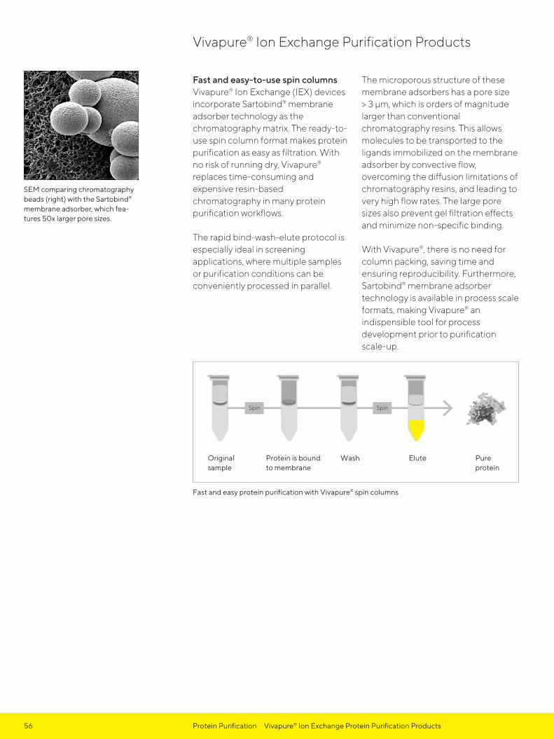
Fast and easy-to-use spin columnsVivapure® Ion Exchange (IEX) devices incorporate Sartobind® membrane adsorber technology as the chromatography matrix. The ready-to-use spin column format makes protein purification as easy as filtration. With no risk of running dry, Vivapure® replaces time-consuming and expensive resin-based chromatography in many protein purification workflows.
The rapid bind-wash-elute protocol is especially ideal in screening applications, where multiple samples or purification conditions can be conveniently processed in parallel.
The microporous structure of these membrane adsorbers has a pore size > 3 μm, which is orders of magnitude larger than conventional chromatography resins. This allows molecules to be transported to the ligands immobilized on the membrane adsorber by convective flow, overcoming the diffusion limitations of chromatography resins, and leading to very high flow rates. The large pore sizes also prevent gel filtration effects and minimize non-specific binding.
With Vivapure®, there is no need for column packing, saving time and ensuring reproducibility. Furthermore, Sartobind® membrane adsorber technology is available in process scale formats, making Vivapure® an indispensible tool for process development prior to purification scale-up.
Vivapure® Ion Exchange Purification Products
SEM comparing chromatography beads (right) with the Sartobind® membrane adsorber, which fea-tures 50x larger pore sizes.
Fast and easy protein purification with Vivapure® spin columns

57Protein Purification Vivapure® Ion Exchange Protein Purification Products
Membrane Availability
Functional groups Ion exchanger type
Sulphonic acid (S) Strong acidic cation exchanger: R-CH₂-SO₃-Na+
Quaternary ammonium (Q) Strong basic anion exchanger: R-CH₂-N+-(CH₃)₃Cl-
Diethylamine (D) Weak basic anion exchanger: R-CH₂-NH+-(CH₂H₅)₂
Typical Performance Characteristics
Vivapure® spin columns
Protein binding capacity*
Max. volume, swing bucket
Max. volume, fixed angle
Vivapure® Mini H 4 mg – 0.4 mL
Vivapure® Maxi H 60 – 80 mg 19 mL 10.5 mL
Typical Applications - Fractionation of protein mixtures prior to 1D or 2D-PAGE - Scouting purification conditions for new protein targets - Removal of endotoxins from monoclonal antibodies - Preparation of heme moiety from heme containing protein prior to functional analysis - General protein purification and polishing - Detergent removal from protein solutions - Purification of antibodies from serum, ascites or cell culture supernatant - Intermediate sample purification prior to further HPLC | FPLC - Purification of membrane-bound proteins
Detailed application notes are available on our website: www.sartorius.com
Ordering Information
Vivapure® Mini Spin Columns
Centrifuge Tubes
Prod. No.
Vivapure® D Mini H 24 48 VS-IX01DH24
Vivapure® Q Mini H 24 48 VS-IX01QH24
Vivapure® S Mini H 24 48 VS-IX01SH24
Vivapure® Maxi
Vivapure® D Maxi H 8 16 VS-IX20DH08
Vivapure® Q Maxi H 8 16 VS-IX20QH08
Vivapure® S Maxi H 8 16 VS-IX20SH08
* Binding capacities established using 1 mg/mL BSA in 25 mM Tris-HCl (pH 8) for Vivapure® Q and D devices, or 1 mg/mL cytochrome c in 25 mM sodium acetate buffer (pH 5.5) for Vivapure® S devices. Actual capacities depend on the target molecule and selected buffer conditions.
Vivapure® Mini H
Vivapure® Mini H
Vivapure® Maxi H
Vivapure® Maxi H
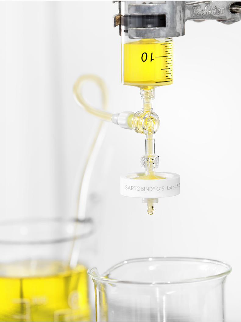
58 Chapter Site Title

59
Virus Purification and ConcentrationTable of Contents
Vivapure® Virus Purification and Concentration Kits 60
Adenovirus Purification 61
Vivapure® Adenopack 20 62
Vivapure® Adenopack 100 63
Vivapure® Adenopack 500 65
Lentivirus Purification 66
Vivapure® Lentiselect 40 67
Vivapure® Lentiselect 500 68
Vivapure® Lentiselect 1000 69

60 Virus Purification and Concentration Vivapure® Virus Purification and Concentration Kits
Recombinant virus vectors are thepreferred method for a wide range of gene delivery applications. Especially adenovirus type 5 and VSV-G pseudotyped lentivirus are two frequently utilized viral vectors for in vitro and in vivo applications.
Recombinant Adenovirus VectorsRecombinant adenovirus vectors are versatile tools in research and therapeutic applications for gene transfer and protein expression in cell lines that have low transfection efficiency with liposomes.
After entering cells, the virus remainsepichromosomal (i.e. does not integrate into the host chromosome, leaving the host genome unaffected). The delivery of RNAi into cells is becoming a major application for adenovirus vectors.
Lentivirus VectorsLentivirus vectors are frequently used in gene transfer studies, due to their ability of gene transfer and integration into dividing and non-dividing cells. The pseudotyped envelope with vesicular stomatitis virus envelope G (VSV-G) protein broadens their target cell range. Lentiviral vectors have been shown to deliver genes into cell types (e.g. neurons, lymphocytes and macrophages) which other retrovirus vectors could not be used for. The lentivirus vector is increasingly used to integrate siRNA efficiently in a wide variety of cell lines and primary cells, both in vitro and in vivo.
Rapid Virus Purification by Membrane ChromatographyThe Sartobind® ion exchange membrane adsorber technology used in Adenopack and Lentiselect is unique in its capability to efficiently and rapidly capture and recover large virus particles. When compared to chromatography media, membraneadsorbers provide large 3000 nmpores allowing unrestricted access and recovery of virus from the charged adsorber surface. Convective flow through the syringe filter devices provides high-speed separations not possible with traditional chromatography, cesium chloride density gradients and ultracentrifugation methods.
Our membrane adsorbers with porousmatrices, high capacities, low differ-ential pressures, high flow rates and low unspecific adsorption show an excellent performance in small scale virus purification. Additionally, they are also scalable and confirm to cGMP facilities to large volume, high performance separation, reducing the processing time by a factor of 10 in the final process.
Vivapure® Virus Purification and Concentration Kits

61Virus Purification and Concentration Adenovirus Purification with Vivapure® Adenopack Kits
Adenopack 20 | 100 | 500The Adenopack adenovirus purifi-cation and concentration kits offer researchers who need to recover up to 3 × 10¹³ purified recombinant adeno-virus particles for invitro transfection a fast, safe and easy to use solution. The kits include all reagents and devices necessary for clarification, purification and concentration of adenovirus type 5 from HEK293 cell cultures inonly two hours. These straight forward kits replace time-consuming and labor-intensive 48 hour CsCl density gradients.
Adenopack kits are offered as Adenopack 20, Adenopack 100 and Adenopack 500, for the purification and concentration of adenovirus type 5 from 20 mL to 500 mL cell culture, leading to 1 × 10¹¹- 3 × 10¹³ purified viral particles. For each sample volume, the most convenient handling method is offered for ultimate convenience.
To this end, preparations using Adenopack 20 are pursued in spin column format in a centrifuge, Adenopack 100 is a manuallyoperated kit in syringe filter format*, and Adenopack 500 is a pump driven kit.
Adenopack Advantages
Fast and Easy Virus Purification - Purification completed in 2 hours - Convenient, over 10 × faster alternative to CsCl density gradient
Quantitative Yields - In contrast to CsCl density gradient, the complete cell culture is used for virus purification and not only the viral pellet
Flexible Product Range - Applicable from initial construct screening to large scale virus production
Complete Kit - Including filtration devices, Adenopack units for virus purifica-tion, Vivaspin® and all buffers
Low Endotoxin Levels - High cell viability and infection rates due to endotoxin levels of < 0.025 EU/mL
Adenovirus Purification
* Vivapure® Adenopack 100 can optionally be operated with a laboratory pump and an infusion pump, for which protocols are pro-vided on our web page www.sartorius.com. Additionally, the tubes and adaptors needed for these operation modes can be ordered.
** after dialysis*** before buffer exchange
Purification of GFP-Ad5 Constructs
Purification method Process time Eluate Recovery*** Viral Particles
Adenopack 20 | 20 mL culture 1 hour 1 mL 65 – 70% 1 × 10¹¹-12
Adenopack 100 | 60 mL culture 1 – 2 hours 1 mL 65% 1 – 3 × 10¹²
Adenopack 100 | 200 mL culture 2 hours 1 mL 80% 1 × 10¹³
Adenopack 500 | 500 mL culture 2 hours 1 mL 80% 1 – 3 × 10¹³
500 mL CsCl density gradient 24 – 48 hours 1 – 2 mL** 60 – 70% 1 × 10¹¹-12

Final concentration| buffer exchange
1. Treat with Benzonase®** 2. Filter with Vivaclear Maxi3. Add 10× Loading Buffer
to sample4. Equilibrate Adenopack Maxi spin columns
5. Bind virus and wash away contaminants
6. Elute purified virus
62 Virus Purification and Concentration Vivapure® Adenopack 20
Vivapure® Adenopack 20 is the downscale kit in the Adenopack series, purifying up to 1 × 10¹² adeno-virus type 5 particles from 20 mL cell culture. Especially when testing new constructs, parallel and fast purifications of different adenoviruses are essential. This kit allows the rapid, simple and affordable spin column based purification of 6 different samples in parallel and bridges a gap
in the CsCl density gradient method – for the first time adenovirus type 5 can efficiently be purified from less than 100 mL cell culture volume!
Typical PerformanceFor a normal yielding vector, 1 × 15 cm culture plate purified using this method yields up to 1 × 10¹² viral particles.
Ordering Information and Kit Contents
Vivapure® Adenopack 20 VS-AVPQ020
Vivapure® Adenopack 20 RT* VS-AVPQ022
Adenopack Maxi spin columns 6
Vivaclear Maxi 0.45 μm PES 6
Empty 50 mL tubes 6
Loading Buffer (10×) 25 mL
Washing Buffer (10×) 30 mL
Elution Buffer 20 mL
Benzonase® (12.5 U/μL) 120 μL
Vivaspin® 20, 100 kDa MWCO 6
Instructions 1 each for Kit and Vivaspin®
Technical Data
Kit specifications
Sample size 20 mL of cell culture
Number of purifications 6 × 20 mL
Virus particles (VP) per mL Typically up to 1 × 10¹¹ – 1012
VP | IU 50–100
Processing time Typically 1 hour
Endotoxin level < 0.025 EU/mL
Vivapure® Adenopack 20 The optimal kit for construct screening
* Adenopack 20 RT does not include Benzonase®
** Benzonase® Nuclease is manufactured by Merck KGaA, Darmstadt, Germany and is covered by US Patent 5,173,418 and EP Patent 0,229,866. Nycomed Pharma A/S (Denmark) claims worldwide patent rights to Benzonase® Nuclease, which are licensed exclusively to Merck KGaA, Darmstadt, Germany. Benzonase® is a registered trade-mark of Merck KGaA, Darmstadt, Germany.
spin spin spin
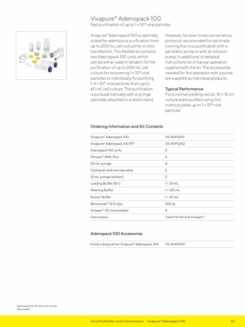
63Virus Purification and Concentration Vivapure® Adenopack 100
Vivapure® Adenopack 100 is optimally suited for adenovirus purification from up to 200 mL cell culture for in vitro transfection. This flexible kit contains two Adenopack 100 units, which can be either used in tandem for the purification of up to 200 mL cell culture for recovering 1 × 10¹³ viral particles or individually for purifying 1–3 × 10¹² viral particles from up to 60 mL cell culture. The purification is pursued manually with a syringe optimally attached to a retort stand.
However, for even more convenience, protocols are provided for optionally running the virus purification with a peristaltic pump or with an infusion pump, in additional to detailed instructions for a manual operation supplied with the kit. The accessories needed for the operation with a pump are supplied as individual products.
Typical PerformanceFor a normal yielding vector, 10 × 15 cm culture plate purified using this method yields up to 1 × 10¹³ viral particles.
Ordering Information and Kit Contents
Vivapure® Adenopack 100 VS-AVPQ101
Vivapure® Adenopack 100 RT* VS-AVPQ102
Adenopack 100 units 2
Minisart® NML Plus 4
20 mL syringe 4
Tubing set and one way valve 2
10 mL syringe (elution) 2
Loading Buffer (10×) 1 × 25 mL
Washing Buffer 1 × 120 mL
Elution Buffer 1 × 20 mL
Benzonase® 12.5 U/μL 200 μL
Vivaspin® 20 concentrator 4
Instructions 1 each for Kit and Vivaspin®
Adenopack 100 Accessories
Pump tubing set for Vivapure® Adenopack 100 VS-AVPA001
Vivapure® Adenopack 100 Fast purification of up to 1 × 10¹³ viral particles
* Adenopack 100 RT does not include Benzonase®

1. Add Benzonase® and clarify sample
2. Load cleared sample
3. Wash 4. Elute Concentrate
64 Virus Purification and Concentration Vivapure® Adenopack 100
Technical Data
Kit specifications
Sample size 20–200 mL of cell culture
Number of purifications 2 × 20–60 mL1 × 200 mL
Virus particles (VP) per mL Typically up to 1 × 10¹³
VP | IU 20–50
Processing time Typically 2 hours
Endotoxin level < 0.025 EU/mL

1. Add Benzonase® and clarify sample
2. Load sample and wash unspecifically bound proteins
3. Elute 4. Concentrate
65Virus Purification and Concentration Vivapure® Adenopack 500
Vivapure® Adenopack 500 is the direct upscale kit to the Adenopack 100, for adenovirus purification. In only 2 hours up to 3 × 10¹³ adenovirus particles are purified and concentrated from 500 mL cell culture. This completely ready-to-use kit is conveniently operated by a laboratory pump, offering optimal flow control and minimal hands-on time. This easy
to use product replaces lengthy and inefficient cesium chloride density gradient methods.
Typical PerformanceFor a normal yielding vector, 25 × 15 cm culture plate purified using this method yields up to 3 × 10¹³ viral particles.
Ordering Information and Kit Contents
Vivapure® Adenopack 500 VS-AVPQ501
Vivapure® Adenopack 500 RT* VS-AVPQ502
Adenopack 500 unit 1
Sartopore® 2 150 1
Tubing set and one way valve 2
10 mL syringe 1
Loading Buffer (10×) 60 mL
Washing Buffer (10×) 30 mL
Elution Buffer 20 mL
Benzonase® 12.5 U/μL 500 μL
Vivaspin® 20 concentrator 2
Instructions 1 each for Kit and Vivaspin®
Technical Data
Kit specifications
Sample size 500 mL of cell culture
Number of purifications 1 × 500 mL
Virus particles (VP) per mL Typically up to 3 × 10¹³
VP | IU 20–50
Processing time Typically 2 hours
Endotoxin level < 0.025 EU/mL
Vivapure® Adenopack 500 Pump driven kit for larger volumes
* Adenopack 500 RT does not include Benzonase®
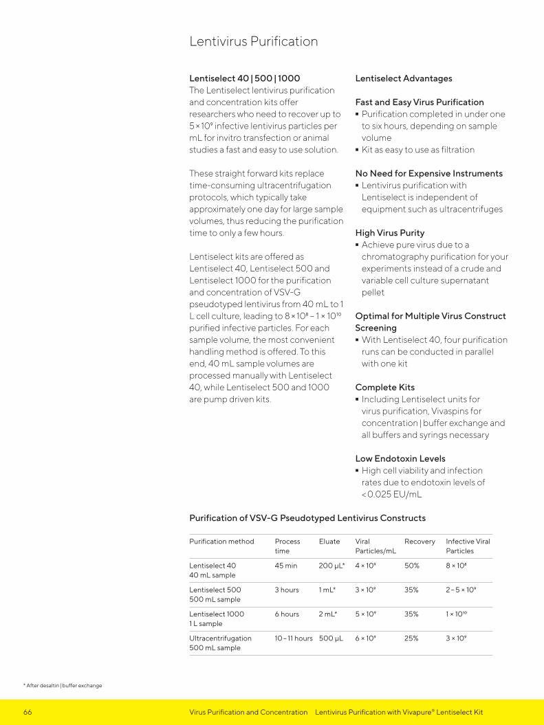
66 Virus Purification and Concentration Lentivirus Purification with Vivapure® Lentiselect Kit
Lentiselect 40 | 500 | 1000The Lentiselect lentivirus purification and concentration kits offer researchers who need to recover up to 5 × 10⁹ infective lentivirus particles per mL for invitro transfection or animal studies a fast and easy to use solution.
These straight forward kits replace time-consuming ultracentrifugation protocols, which typically take approximately one day for large sample volumes, thus reducing the purification time to only a few hours.
Lentiselect kits are offered as Lentiselect 40, Lentiselect 500 and Lentiselect 1000 for the purification and concentration of VSV-G pseudotyped lentivirus from 40 mL to 1 L cell culture, leading to 8 × 10⁸ – 1 × 10¹⁰ purified infective particles. For each sample volume, the most convenient handling method is offered. To this end, 40 mL sample volumes are processed manually with Lentiselect 40, while Lentiselect 500 and 1000 are pump driven kits.
Lentiselect Advantages
Fast and Easy Virus Purification - Purification completed in under one to six hours, depending on sample volume - Kit as easy to use as filtration
No Need for Expensive Instruments - Lentivirus purification with Lentiselect is independent of equipment such as ultracentrifuges
High Virus Purity - Achieve pure virus due to a chromatography purification for your experiments instead of a crude and variable cell culture supernatant pellet
Optimal for Multiple Virus Construct Screening - With Lentiselect 40, four purification
runs can be conducted in parallel with one kit
Complete Kits - Including Lentiselect units for virus purification, Vivaspins for concentration | buffer exchange and all buffers and syrings necessary
Low Endotoxin Levels - High cell viability and infection rates due to endotoxin levels of < 0.025 EU/mL
Purification of VSV-G Pseudotyped Lentivirus Constructs
Purification method Process time
Eluate Viral Particles/mL
Recovery Infective Viral Particles
Lentiselect 40 40 mL sample
45 min 200 μL* 4 × 109 50% 8 × 10⁸
Lentiselect 500 500 mL sample
3 hours 1 mL* 3 × 109 35% 2 – 5 × 10⁹
Lentiselect 1000 1 L sample
6 hours 2 mL* 5 × 109 35% 1 × 10¹⁰
Ultracentrifugation 500 mL sample
10 – 11 hours 500 μL 6 × 10⁹ 25% 3 × 10⁹
Lentivirus Purification
* After desaltin | buffer exchange

67Virus Purification and Concentration Vivapure® Lentiselect 40
Vivapure® Lentiselect 40 is optimallysuited for lentivirus purification for upto 40 mL cell culture and contains all components necessary for 4 purifications. Up to 8 × 10⁸ viral particles are recovered in less than one hour. In contrast to traditional ultracentrifu gation methods, virus purification with Vivapure® Lentiselect is fast and simple, without the need for expensive equipment like an ultra-
centrifuge. Additionally, this chromatographic procedure leads to pure virus samples in contrast to the crude ultracentrifuge pellet, resulting in higher reproducibility and increased gene transfer efficiency.
Typical PerformanceFor a normal yielding vector, 2 × 15 cmculture plate purified using this method yield up to 8 × 10⁸ particles.
Ordering Information and Kit Contents
Vivapure® Lentiselect 40 VS-LVPQ040
Lentiselect unit 4
50 mL syringe 4
10 mL syringe 4
Tube set with one-way valve 4
Loading buffer (10 ×) 30 mL
Washing buffer 150 mL
Elution buffer 20 mL
Vivaspin® 20, 100 kDa MWCO 8
Instructions 1 each for Kit and Vivaspin®
Technical Data
Kit specifications
Sample size 40 mL cell culture
Number of purifications 4 × 40 mL
Infective particles (P) per mL Typically up to 3 × 10⁹
VP | IU 5 – 15
Processing time Typically 45 minutes
Endotoxin level < 0.025 EU/mL
Vivapure® Lentiselect 40 Fast purification of up to 8 × 10⁸ viral particles
1. 2. Washing 3. Elute
10 mL syringecontainingelution buffer
LentiselectUnit
Purified virus
Tube containing24 mL buffer
LentiselectUnit

1. Filter 2. Add Loading Buffer3. Prepare Lentiselect Unit4. Bind virus an d wash away contaminants
5. Elute purified virus6. Concentrate and exchange
buffer using Vivaspin®
68 Virus Purification and Concentration Vivapure® Lentiselect 500
Vivapure® Lentiselect 500 is optimally suited for VSV-G pseudotyped lenti-virus purification from up to 500 mL cell culture and contains all reagents and devices necessary for purifying up to 2–5 × 10⁹ infective particles.
The whole purification procedure is simply operated by a laboratory pump, which minimizes hands-on time. Unlike conventional purification methods as ultracentrifugation,
Vivapure® Lentiselect 500 offers a fast and simple solution for purifying VSV-G pseudotyped lentiviruses making expensive purification equipment like ultracentrifuges redundant.
Typical PerformanceFor a normal yielding vector, 500 mL cell culture purified using this method yield up to 2–5 × 10⁹ infective particles per millilitre (total volume 1 mL).
Ordering Information and Kit Contents
Vivapure® Lentiselect 500 VS-LVPQ500
Lentiselect unit 1
Sartopore® 2 150 1
50 mL syringe 1
Tube set with one-way valve 1
Loading buffer (10 ×) 30 mL
Washing buffer 170 mL
Elution buffer 30 mL
Vivaspin® 20, 100 kDa MWCO 2
Operating manual 1 each for Kit and Vivaspin®
Technical Data
Kit Specifications
Sample volume 500 mL cell culture
Number of purifications 1 × 500 mL
Infective particles (IP) per mL Typically up to 2–5 × 10⁹*
Processing time Typically up to 3 hours
Endotoxin level < 0.025 EU/mL
Vivapure® Lentiselect 500 Fast purification of up to 2–5 × 10⁹ infective particles per mL from 500 mL cell culture
* 1 mL final elution sample
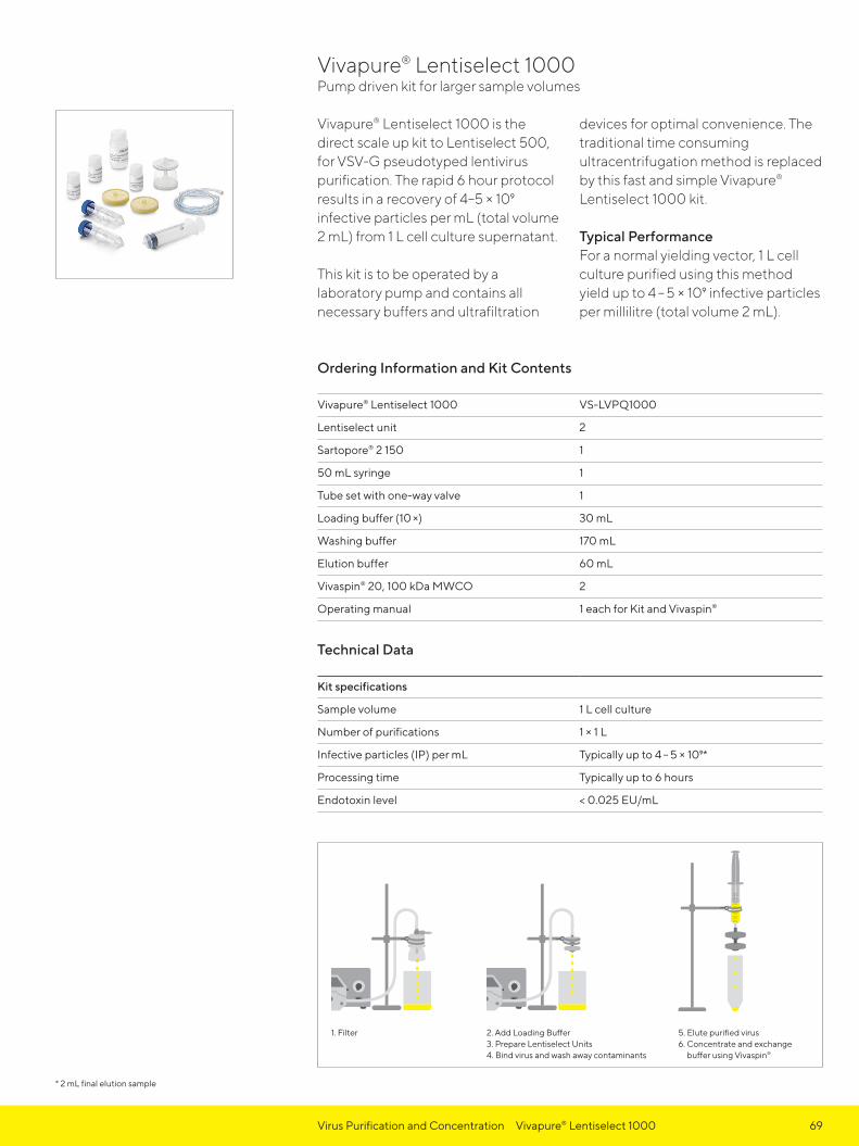
1. Filter 2. Add Loading Buffer3. Prepare Lentiselect Units4. Bind virus and wash away contaminants
5. Elute purified virus6. Concentrate and exchange
buffer using Vivaspin®
69Virus Purification and Concentration Vivapure® Lentiselect 1000
Vivapure® Lentiselect 1000 is the direct scale up kit to Lentiselect 500, for VSV-G pseudotyped lenti virus purification. The rapid 6 hour protocol results in a recovery of 4–5 × 10⁹ infective particles per mL (total volume 2 mL) from 1 L cell culture supernatant.
This kit is to be operated by a laboratory pump and contains all necessary buffers and ultrafiltration
devices for optimal convenience. The traditional time consuming ultracentrifugation method is replaced by this fast and simple Vivapure® Lentiselect 1000 kit.
Typical PerformanceFor a normal yielding vector, 1 L cell culture purified using this method yield up to 4 – 5 × 10⁹ infective particles per millilitre (total volume 2 mL).
Ordering Information and Kit Contents
Vivapure® Lentiselect 1000 VS-LVPQ1000
Lentiselect unit 2
Sartopore® 2 150 1
50 mL syringe 1
Tube set with one-way valve 1
Loading buffer (10 ×) 30 mL
Washing buffer 170 mL
Elution buffer 60 mL
Vivaspin® 20, 100 kDa MWCO 2
Operating manual 1 each for Kit and Vivaspin®
Technical Data
Kit specifications
Sample volume 1 L cell culture
Number of purifications 1 × 1 L
Infective particles (IP) per mL Typically up to 4 – 5 × 10⁹*
Processing time Typically up to 6 hours
Endotoxin level < 0.025 EU/mL
Vivapure® Lentiselect 1000 Pump driven kit for larger sample volumes
* 2 mL final elution sample

70 Chapter Site Title

71
Application NotesTable of Contents
1. Desalting and Buffer Exchange with Vivaspin® Centrifugal Concentrators 72
2. Treatment of Vivaspin® Concentrators for Improved Recovery of Low-concentrated Protein Samples 75
3. Scouting Protein Purification Conditions Using Vivapure® Centrifugal Ion Exchange Membrane Absorbers 78
4. Concentration and Purification of Viruses by using Ultrafiltration, Incl. Coronavirus – a Short Review 83
5. Sartorius Ultrafiltration Products in the Preparation of Biological Nanoparticles and Medical Nanocarriers 95
6. Vivaflow® and Vivaspin® Workflow in Protein Research Laboratories 101

72 Application Notes 1. Desalting and Buffer Exchange with Vivaspin®
IntroductionVivaspin® centrifugal concentrators, with patented vertical membrane technology, combine fast filtration with high recovery of target proteins. This makes Vivaspin® the technology of choice for desalting or buffer exchange, avoiding lengthy dialysis steps.
While proteins are retained by an ultrafiltration membrane, salts can pass freely through, independent of protein concentration or membrane MWCO. In consequence, the composition of the buffer in the flow-through and retentate is unchanged after protein concentration. By diluting the concentrate back to the original volume, the salt concentration is lowered. The concentrate can be diluted with water or salt-free buffer if simple desalting is required; however, it is also possible to dilute the concentrate with a new buffer, thereby exchanging the buffering substance entirely. For example, a 10 mL protein sample containing 500 mM salt, if concentrated 100-fold still contains 500 mM salt. If this concentrate is then diluted 100× with water or salt-free buffer, the protein concentration returns to the original level, while the salt concentration is reduced 100× to only 5 mM (i.e. a 99% reduction in salt concentration).
The protein sample can then be concentrated again to the desired level, or the buffer exchange can be repeated to reduce the salt concentration even further before a final concentration of the protein. This process is called dia filtration. For proteins with a tendency to precipitate at higher concentrations, it is possible to perform several diafiltration steps in sequence, with the protein concentrated each time to only 5 or 10×. For example, if a precipitous protein sample is concentrated to 5× then diluted back to the original volume, and this process is repeated a further two times, this still results in a >99% reduction in salt concentration, without over-concentrating the protein.
1. Desalting and Buffer Exchange with Vivaspin® Centrifugal Concentrators

73Application Notes 1. Desalting and Buffer Exchange with Vivaspin®
MethodsSelect an appropriate MWCO for your sample. For maximum recovery, select a MWCO ⅓ to ½ the molecular weight of the molecule of interest.
1. Add protein sample up to the maxi-mum fill volume of the concentrator (as stated in the device operating instructions). If the sample volume is lower than the maximum device volume, it can be diluted to the maximum fill volume before the first centrifugation step. This will increase the salt removal rate.
2. Centrifuge for the recommended amount of time at an appropriate spin speed (see device operating instructions).
3. Empty filtrate container and refill the concentrator with an appropriate exchange solvent.*
4. Centrifuge again as before.5. Recover the concentrated, desalted
sample from the bottom of the concentrate pocket with a pipette.
* Filtrate volumes should be retained until the concentrated sample has been analyzed.
Figure 1: Step-by-step method for desalting and concentration
1 2 3 4 5

74 Application Notes 1. Desalting and Buffer Exchange with Vivaspin®
Results
Vivaspin® 20
MWCO 5 kDa 30 kDa 50 kDa 100 kDa
Cytochrome C0.25 mg/mL
BSA 1 mg/mL
BSA 1 mg/mL
IgG 1 mg/mL
Protein Recovery
NaCLRemoval
Protein Recovery
NaCLRemoval
Protein Recovery
NaCLRemoval
Protein Recovery
NaCLRemoval
Spin 1 100% 99% 97% 99% 97% 99% 90% 98%
Spin 2 96% 100% 92% 100% 93% 100% 87% 100%
Four Vivaspin® 20 devices of each MWCO were tested with 20 mL sam-ples. Each sample contained 500 mM NaCl. To perform diafiltration, devices were centrifuged at 4,000 g for 45 min (5 kDa MWCO) or 30 min (>5 kDa MWCOs). After the first and second spins, the retentate samples were
brought up to 20 mL with ultrapure water from an Arium® system (Sartorius). OD readings were taken at 410 nm for Cytochrome C or 280 nm for BSA and IgG samples. Salt concentrations were measured using a Qcond 2200 conductivity measuring instrument.
Vivaspin® 6
MWCO 5 kDa 30 kDa 50 kDa 100 kDa
Cytochrome C0.25 mg/mL
BSA 1 mg/mL
BSA 1 mg/mL
IgG 1 mg/mL
Protein Recovery
NaCLRemoval
Protein Recovery
NaCLRemoval
Protein Recovery
NaCLRemoval
Protein Recovery
NaCLRemoval
Spin 1 98% 99% 92% 99% 93% 99% 92% 98%
Spin 2 85% 100% 86% 100% 83% 100% 89% 100%
Four Vivaspin® 6 devices of each MWCO were tested with 6 mL samples. Each sample contained 500 mM NaCl. To perform diafiltration, devices were centrifuged at 4,000 g for 45 min (5 kDa MWCO) or 30 min (>5 kDa MWCOs). After the first and second spins, the retentate samples
were brought up to 6 mL with ultrapure water from an Arium® system (Sartorius). OD readings were taken at 410 nm for Cytochrome C or 280 nm for BSA and IgG samples. Salt concentrations were measured using a Qcond 2200 conductivity measuring instrument.
ConclusionsAs the results show, the efficient de-sign of Vivaspin® devices allowed >95% of the salt to be removed during the first centrifugation step. Only one subsequent centrifugation step was needed to increase the typical salt
removal to 99% with >92% recovery of the target protein. Diafiltration using ultrafiltration devices such as Vivaspin® 6 and 20 represents a faster and more efficient solution to desalting and buffer exchange, than conventional techniques such as dialysis.
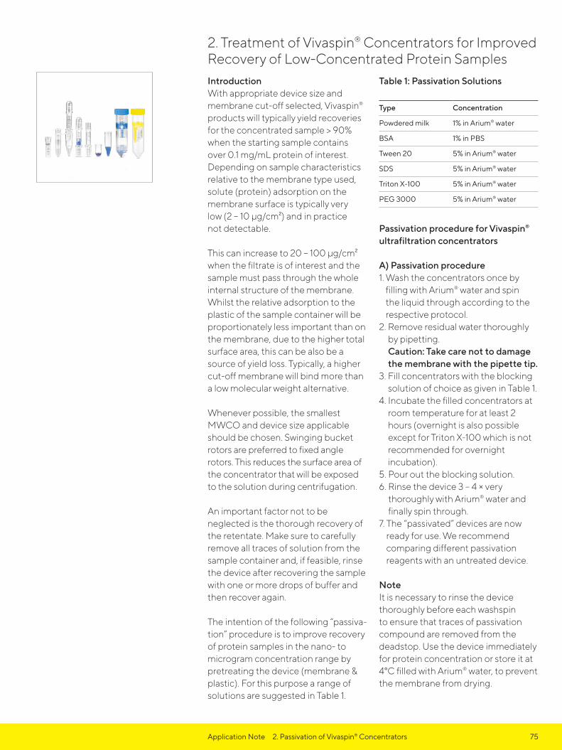
75
IntroductionWith appropriate device size and membrane cut-off selected, Vivaspin® products will typically yield recoveries for the concentrated sample > 90% when the starting sample contains over 0.1 mg/mL protein of interest. Depending on sample characteristics relative to the membrane type used, solute (protein) adsorption on the membrane surface is typically very low (2 – 10 μg/cm²) and in practice not detectable.
This can increase to 20 – 100 μg/cm² when the filtrate is of interest and the sample must pass through the whole internal structure of the membrane. Whilst the relative adsorption to the plastic of the sample container will be proportionately less important than on the membrane, due to the higher total surface area, this can be also be a source of yield loss. Typically, a higher cut-off membrane will bind more than a low molecular weight alternative.
Whenever possible, the smallest MWCO and device size applicable should be chosen. Swinging bucket rotors are preferred to fixed angle rotors. This reduces the surface area of the concentrator that will be exposed to the solution during centrifugation.
An important factor not to be neglected is the thorough recovery of the retentate. Make sure to carefully remove all traces of solution from the sample container and, if feasible, rinse the device after recovering the sample with one or more drops of buffer and then recover again.
The intention of the following “passiva-tion” procedure is to improve recovery of protein samples in the nano- to microgram concentration range by pretreating the device (membrane & plastic). For this purpose a range of solutions are suggested in Table 1.
Table 1: Passivation Solutions
Type Concentration
Powdered milk 1% in Arium® water
BSA 1% in PBS
Tween 20 5% in Arium® water
SDS 5% in Arium® water
Triton X-100 5% in Arium® water
PEG 3000 5% in Arium® water
Passivation procedure for Vivaspin®
ultrafiltration concentrators
A) Passivation procedure1. Wash the concentrators once by
filling with Arium® water and spin the liquid through according to the respective protocol.
2. Remove residual water thoroughly by pipetting. Caution: Take care not to damage the membrane with the pipette tip.
3. Fill concentrators with the blocking solution of choice as given in Table 1.
4. Incubate the filled concentrators at room temperature for at least 2 hours (overnight is also possible except for Triton X-100 which is not recommended for overnight incubation).
5. Pour out the blocking solution.6. Rinse the device 3 – 4 × very
thoroughly with Arium® water and finally spin through.
7. The “passivated” devices are now ready for use. We recommend comparing different passivation reagents with an untreated device.
NoteIt is necessary to rinse the device thoroughly before each washspin to ensure that traces of passivation compound are removed from the deadstop. Use the device immediately for protein concentration or store it at 4°C filled with Arium® water, to prevent the membrane from drying.
2. Treatment of Vivaspin® Concentrators for Improved Recovery of Low-Concentrated Protein Samples
Application Note 2. Passivation of Vivaspin® Concentrators

76
B) Evaluation of passivation effects(exemplary with BSA)1. Prepare a 10 μg/mL BSA stock
solution e.g. by diluting 90 μL of the 4 mg/mL stock solution in 36 ml 0.1 M sodium borate pH 9.3. Mix well.
2. Fill Vivaspin® 2 devices with 2 ml of this 10 μg/mL BSA solution and close with cap provided.
3. Spin the device in a swing-out rotor at 4,000 × g until the volume is to app. 100 μL.
4. Recover the concentrate and make back up to 2 mL with 0.1 M sodium borate pH 9.3
5. Determine recovered protein concentrations e.g. according to Bradford or BCA assays.
Results and discussionAs an example, the effect of milk powder was analysed. It could be shown (Figure 1) that the protein recovery of a 10 μg/mL BSA solution could be increased from around 70 to 90%. If milk powder is not interfering with sample purity and quality, it is a good starting point to improve recovery of diluted sample solutions.
Protein recovery (10 μg/mL BSA) with Vivaspin® PES 10 kDa after passivationIn another example, detergents were analysed with only 250 and 500 ng BSA (Figure 2) BSA recovery declined to 50 – 30% in untreated devices as the protein concentration was reduced. Significant improvement to 60 – 90% recovery could be demonstrated when using the passivation strategy. Often, Triton X-100 seemed to work though the optimal reagent has to be selected for the respective protein and its hydrophilic | -phobic characteristics.
Application Note 2. Passivation of Vivaspin® Concentrators

77
SummaryPassivation is an appropriate method to achieve increasing sample recovery when using very dilute samples. In ad-dition to skimmed milk, other proteins (BSA), detergents and compounds are possible. However, it should be noted that this is a general procedure, not specific for any particular application. Depending on the hydrophilic | -phobic character of the protein non-specific binding may be more or less of a problem and the suggested passivation solutions may lead to dif-ferent results. Even with the Hydrosart membrane, which is recommended for dilute samples, passivation of the
device will reduce losses on the plastic surface. One very important thing to remember is that the blocking agent is potentially introduced into the sample. It should be assured that this will not interfere with downstream analysis.
For example, proteins must not be used for passivation if a pure protein is intended to be concentrated for x-ray crystallography, as even the smallest traces would interfere with the diffraction pattern. Other subse-quent analyses methods include activity testing, gel electrophoresis or labelling are less problematic.
Application Note 2. Passivation of Vivaspin® Concentrators
100
80
60
40
20
0
250 ng 500 ng
Triton X-100
untreated
PEG
Tween
Recovery comparison of passivation with common buffer solutions
100
80
60
40
20
0milk powder untreated
Recovery comparison of passivation with milk powder
Figure 1: Protein recovery (10 µg/mL BSA) with Vivaspin® PES 10 kDa after passivation
Figure 2: Protein recovery (250 and 500 ng BSA) with Vivaspin® 2 PES 10 kDa after passivation

78
IntroductionFor separation and purification of proteins from biological samples, different characteristics of the target protein e.g. its size, charge, hydrophobicity or specifically engineered tags, are exploited.
With ion exchange chromatography, separation is achieved on the basis of charge differences between biomolecules. This makes it a versatile method often used for pre-fractionation or purification of a target protein from crude protein mixtures. To optimize the purification procedure for an individual target, several binding and elution conditions have to be tested on cation and anion exchange matrices.
In contrast to traditional column chromatography methods, Vivapure® IEX centrifugal columns allow scouting of several chromatography conditions in parallel, leading quickly to different fractions which can be further analyzed for enriched or even already purified target protein.
Here, we demonstrate the performance of Vivapure® IEX Mini spin columns for evaluation of optimal purification conditions of cloned SH2 domains from an E. coli lysate in a two step procedure. This protocol can generally be employed for identifying a purification method based on ion exchange chromatography for a given target protein, as it is fast and only uses small amounts of the sample.
In the first step of this study, binding conditions were evaluated by loading the sample on Vivapure® Q and S columns at various pH values, eluting bound proteins with a high salt concentration buffer and analyzing all fractions for the target protein.
The results from this experiment provided the optimal binding pH and the best ion exchange chemistry for the purification of SH2 domain.
In a second step, the best elution method was evaluated by applying increasing salt concentrations to columns which were shown to bind the target protein in step one, leading to a complete purification protocol in less than one hour.
Materials and MethodsBuffers tested to determine the optimum pH and salt concentration for binding and elution in ion exchange purification of SH2 domain
Buffer A: 25 mM Citrate, pH 4
Buffer B: 25 mM Potassium phosphate, pH 6
Buffer C: 25 mM HEPES, pH 8
Buffer D: 25 mM Sodium bicarbonate, pH 10
Buffer E: 25 mM Citrate, pH 4, supplemented with 1 M NaCl.
Buffer F: 25 mM Potassium phosphate, pH 6,supplemented with 0.2 M, 0.4 mM, 0.6 mM, 0.8 mM and 1 M NaCl, respectively.
Buffer G: 25 mM HEPES, pH 8, supplemented with 1 M NaCl
Buffer H: 25 mM Sodium bicarbonate, pH 10, supplemented with 1 M NaCl
3. Scouting Protein Purification Conditions Using Vivapure® Centrifugal Ion Exchange Membrane Absorbers
Application Note 3. Scouting Protein Purification Conditions

79
Scouting Binding Conditions300 mL LB media was inoculated with 4 mL of an overnight culture and incubated at 37°C, shaking at 150 rpm until an OD600 of 1.0 was reached. IPTG was added to a final concentration of 1 mM and the culture incubated for a further 4 h with shaking at 150 rpm. Cells were harvested by centrifugation at 4,000 g for 30 min at 4°C. The pellet was resuspended in 35 mL PBS (150 mM KPi, pH 7.3) and cells were lyzed by addition of lysozyme to a final concentration of 0.1 mg/mL and incubation for 1 h at 37°C. Insoluble particles and cell debris were removed by centrifugation at 10,000 g for 30 min at 4°C.
4 × 200 μL aliquots of the cell lysate were diluted with 1.8 mL binding buffer A to D, to adjust each sample to the respective pH being tested. To avoid clogging of the membranes in the Vivapure® Mini spin columns, samples were clarified by passage through 0.45 μm CA Minisart NML syringe filters (Sartorius).
4 × Q and 4 × S Vivapure® Mini spin columns were labeled 4, 6, 8 and 10, corresponding to the pH of the buffer to be used. To each spin column, 400 μL of the corresponding binding buffer was added and spun for 5 minutes at 2,000 g (45° fixed angle rotor).
400 μL of the clarified samples adjusted to pH 4, 6, 8 or 10 were applied to each of the correspondingly equilibrated Vivapure® Q and S spin columns. Columns were spun for 5 min at 2,000 g.
Afterwards, Vivapure® Mini spin columns were reloaded with 400 μL sample and spun again for 5 min at 2,000 g. Loosely bound proteins were washed away with the application of 400 μL of the respective binding buffer to each of the columns and spinning for 5 min at 2,000 g. Flow-through and wash fractions were collected for subsequent detection of the target protein.
200 μL of elution buffer E, F, G or H, were applied to the washed columns and spun for 3 min at 2,000 g. Eluates were saved for subsequent analysis.
4 μL of flow-through, wash, and eluate fractions from each column were analyzed by reducing SDS-PAGE followed by silver staining.
Optimizing Elution ConditionsTaking account of the results of the first experiment (Scouting Binding Conditions) 200 μL cell lysate was diluted with 1.8 mL binding buffer B (25 mM KPi, pH 6). To avoid clogging of the membrane in the Vivapure® Mini spin column, the pH adjusted sample was clarified by passage through a 0.45 μm CA Minisart NML syringe filter (Sartorius).
400 μL binding buffer B was applied to one Vivapure® S Mini spin column and spun for 5 minutes at 2,000 g.
400 μL of the clarified sample was applied to the equilibrated Vivapure® S column and spun for 5 min at 2,000 g. Afterwards, the Vivapure® S Mini spin column was reloaded with 400 μL sample and spun again for 5 min at 2,000 g.
Application Note 3. Scouting Protein Purification Conditions

80
Loosely bound proteins were washed away by application of 400 μL binding buffer to the column and spinning for 5 min at 2,000 g. Flow-through and wash fractions were savedfor analysis.
To elute the target protein, 100 μL elution buffer F, supplemented with 0.2 M NaCl was applied to the Vivapure® S Mini spin column and spun for 3 min at 2,000 g. The eluate was collected. For the next elution step, 100 μL of elution buffer F, supplemented with 0.4 M NaCl was applied and again spun for 3 min at 2,000 g. Elution was continued with 0.2 M NaCl increments until a final salt concentration of 1 M was reached, saving the eluates from each step.
4 μL of flow-through, wash, and eluate fractions from each column were analyzed by reducing SDS-PAGE followed by silver staining.
ResultsDilution of the E. coli lysate with binding buffer A (25 mM Citrate, pH 4) lead to complete precipitation of sample proteins. Thus, pH 4 could not be tested in this experiment. As can be seen on the SDS gel in (Figure 1), the target protein was present in the eluates from Vivapure® Q Mini spin columns at all pH values tested, together with most of the E. coli proteins (Lanes Q “e”). In contrast, using the Vivapure® S Mini spin column, at all pH values tested, most E. coli proteins did not bind to the membrane and were found in the flow-through (Lane S “f”), thus resulting in purer target protein in all eluate fractions (Lane S “e”).
Differences could be detected in the binding efficiency of the target protein. At pH 8, traces of the target protein were already found in the flow-through, with slightly higher amounts at pH 10 (Lane S “e”). At pH 6, the most efficient binding of the target protein to the S membrane was observed.
The purification conditions determined for Vivapure® S with potassium phosphate buffer (pH 6) were further optimized to determine the ideal salt concentration for SH2 domain elution. The target protein started to elute with 200 mM NaCl, however the main fraction eluted with 400 mM NaCl. Traces of the target protein were also found in the next elution step with 600 mM NaCl, but this might be due to the low elution volume.
Application Note 3. Scouting Protein Purification Conditions

81
Note: Scouting for optimal binding conditions of a SH2 domain expressed in E. coli. 12% reducing SDS gel, silver stained, shows the sample before purification (s), flow-through (f), wash (w) and eluate (e) fractions (1 M NaCl) from Vivapure® Q and S Mini spin columns, at the various pH values tested.
Figure 1
Application Note 3. Scouting Protein Purification Conditions
Sample Sample Volume (μL) Volume Loaded Onto Gel (μL)
M = Broad range marker
s = Sample before application 800 4
f = Flow-through 800 4
w = Wash fraction 400 4
e = Elution with 1 M NaCl 200 4
66 kDa
45 kDa
Target protein
31 kDa
22 kDa
pH = 6 pH = 8 pH = 10

82 Application Notes 3. Scouting Protein Purification Conditions
Note: Optimizing elution conditions for a SH2 domain expressed in E. coli, using Vivapure® S Mini spin column at pH 6. 12% reducing SDS gel, silver stained, shows the sample before purification (s), flow through (f), wash (w) and eluate (e1-5) fractions.
ConclusionA two-step procedure was used to rapidly scout optimal purification conditions for a target protein (a SH2 domain from E. coli lysate) with ion exchange chromatography. In the first step, the most suitable ion exchanger and buffer pH for binding the target protein was verified. In the second step, the elution condition was optimized, building on the results gained in step one. With the scouting procedure described here, it was possible to quickly and conveniently purify the target protein to homogeneity.
The results obtained in this experiment can be used to various ends, e.g: - Polishing a specific protein after
purification with another chromato-graphic technique - Quickly establishing a FPLC method for a new protein - Identification of the optimal purifica-tion method prior to scale up with Vivapure® IEX Maxi spin columns.
For these purposes Vivapure® IEX Mini and Maxi spin columns and Sartobind® membrane adsorber units with FPLC connectors are available.
Figure 2
66 kDa
45 kDa
31 kDa
22 kDa
Target protein
Sample Process Volume (µL) Volume Loaded Onto Gel (µL)
M = Broad range marker
s = Sample before application 800 16
f = Flow-through 800 16
w = Wash fraction 400 16
e1 = 25 mM KPi, pH 6, 200 mM NaCl 100 8
e2 = 25 mM KPi, pH 6, 400 mM NaCl 100 8
e3 = 25 mM KPi, pH 6, 600 mM NaCl 100 8
e4 = 25 mM KPi, pH 6, 800 mM NaCl 100 8
e5 = 25 mM KPi, pH 6, 1 M NaCl 100 8

83
IntroductionEvolutionary, viruses developed various mechanisms to interact and manipulate the genetic material of their target cells. Based on this, modern molecular biology utilizes viruses in a constantly growing number of applications.¹ They range from controlled genetic transfection of cells to a variety of different basic studies in medical science.² In medical studies the strategic focus is on recombinant vaccines and on the development of potential vectors for gene therapy.³,⁴
Besides the great relevance of viruses for medical applications, the assessment of virus type and content is important for the risk assessment of food and drinking water.⁵ Also, the classification of virus content is often of high relevance for the quality control of aquatic biotopes.⁶
During the preparation, handling, or analysis of viruses orvirus-like particles (VLPs), a concentration and | or purification step is frequently required.⁵ Typical viruses have a size within the range of about 20 nm up to several hundred nanometers.⁷ Therefore they are ideally suited for the retention on ultrafiltration membrane systems and such ultrafilters are widely used in basic virus research. The specifications of such ultrafiltration devices depend on the particular type of virus and the purpose of the subsequent application.
This short review highlights methods for the purification of various mammalian viruses for basic medical research. Also, the concentration of pathogenic viruses from water and food samples and the purification of marine bacteriophages (virioplankton) are highlighted. It will also give guidance for the selection of an ideal performing device with the optimum molecular weight cut-off (MWCO) for the user specified ultrafiltration process.
Concentration of mammalian viruses in medical researchIn medical research viruses and VLPs are of major interest, particularly for investigations on infectious viral diseases and for the development of vaccines or antiviral drugs. Moreover, certain VLPs can manipulate genetic material in a directed manner and are used broadly in the development of genetic therapy approaches. Additionally, viral vectors are well established as a transfection method for gene transfer to cell lines e. g. to manipulate mammalian cells in vivo and in vitro.
An overview of thematically linked publications using Sartorius ultrafiltration devices for the purification and concentration of viruses and VLPs in the medical context is given in Table 1. Among other applications, Vivaspin® devices were employed for the concentration of adeno-associated virus (AAV) and lentiviral vectors after purification via ion exchange chromatography,⁸–¹⁰ on blood sera to prepare blank samples from hepatitis C virus (HCV)-positive blood sera,¹¹ for the development of a vaccine against human immunodeficiency virus (HIV) and of an antiviral drug against Chikungunya virus.¹²,¹³
4. Concentration and Purification of Viruses by Using Ultrafiltration, Incl. Coronavirus – A Short Review
Application Notes 4. Concentration and Purification of Viruses

84
Table 1: Summarized examples of applications using Vivaspin® and Vivaflow® for viruses in medical research
Goal of Research (Type of virus, Host Organism)
Purpose of Filtration (Buffer System)
Sartorius Ultrafiltration Device (MWCO)
Subsequent Step Ref.
Gene therapy (Adenovirus type 5, VLP, human)
Diafiltration (20 mM Tris saline buffer)
Vivaflow® (100 kDa)
Storage, chromatography on Sartobind® STIC membrane absorber (FPLC)
14
Reduction of HCV-induced fibrosis (Hepatitis C Virus; human)
Removal of HCV from human blood serum (Blood serum)
Vivaspin®
(30 kDa)Preparation of negative control (from positive sample) for immunofluores-cence assay, fibrosis induction assays
11
Development of a viral entry inhibitor for HIV (HIV, human)
Removal of protein fraction from virus (PBS)
Vivaspin® 20(1,000 kDa)
Virus inactivation 12
Gene therapy for cancer treatment (adeno-associated virus; rAAV-2, human)
Concentration and purification after expression, Buffer exchange after His tag (FreeStyle 293 Expression Medium (Gibco), serum-free)
Vivaspin® 20(1,000 kDa)
Titer, ELISA, cell binding assay, apoptosis | cell cycle assay
8
System for controlled gene expression in mice brain (Adeno- associated virus, mice)
Concentration of eluate after anion exchange chromatog-raphy (elution buffer)
Vivaspin® 20(100 kDa)
Transduction of mice neurons
9
Efficient gene transfer into the CNS (Lentivirus, human)
Concentration after ion exchange chromatography (PBS)
Vivaspin®
(100 kDa)Quantification via real- time PCR and end-point dilution. Transduction of murine neuronal and glial cells in vivo
10
Identification of effective chikungunya antiviral drugs (Chikungunya-Virus, human)
Concentration Vivaspin® 20(100 kDa)
Quantification by TCID₅₀ 13
Gene therapy of achromatopsia in mice (Recombinant adeno-associated virus, human virus used in mice)
Concentration (Anion exchange chromatography elution buffer)
Vivaspin® 4(10 kDa)
Titer determination by dot-blot analysis, subretinal injections
15
Application Notes 4. Concentration and Purification of Viruses

85
Concentration of viruses from drinking water and food samplesThe guidelines for drinking-water quality by the world health organization describe safety plans to reduce potential risks from various virus infections.¹⁶ It states that, due to the increased resistance of viruses to disinfection methods, an absence of bacterial contamination after disinfection cannot be used as a reliable indicator of the presence | absence of pathogenic viral species in drinking water supplies. Considering this, ultrafiltration can play a vital role in detecting such viral contaminations for the research on drinking water quality and food safety.
For an ultrafiltration step, the water sample does not have to be pre-conditioned and its efficacy in concentrating the virus is virtually
independent of the chemical properties and the structure of the virus.¹⁷ Thus ultrafiltration is very well suited to isolate and concentrate virus particles from water samples and is a valuable aid during the assessment of water quality. Most of the viruses which are found in water and also food samples are of fecal origin. Screening for these viruses is crucial to prevent infections. The most frequent ones are hepatitis A, hepatitis E and norovirus.¹⁸ Ultrafiltration has been described as the most appropriate method for the recovery of hepatitis A virus from vegetables and other food items.¹⁹ Detection of infectious viruses is mainly done by propagation in cell culture (plaque assay) or the detection of the viral genomes by molecular amplification techniques such as quantitative reverse transcriptase polymerase chain reaction (RT-PCR).²⁰
Table 2: Summarized examples of ultrafiltration applications using Vivaspin® and Vivaflow® for viruses from drinking water and food samples
Goal of Research(Type of virus, Host Organism)
Purpose of Filtration (Buffer System)
Sartorius Ultra-filtration Device (MWCO)
Subsequent Step Ref.
Method for the detection of norovirus genogroup I (Norovirus, human)
Concentration (PBS processedfood samples)
Vivaspin® (5 kDa)
RNA extraction for real-time RT-PCR
22
Analysis of viral con-tent in groundwater(A set of pathogenic viruses, potentially human)
Concentration of drinking water sample (Drinking water)
Vivaflow® 200 (10 kDa)
Qualitative analysis(enterovirus) by RT-nested PCR and microtiter neutralization test
21
Comparative Analysis of Viral Concentration Methods (Hepatitis A virus, human)
Concentration (0.25 M threonine, 0.3 M NaCl, pH 9.5)
Vivaspin® 20 (100 kDa)
RNA extraction for real-time RT-PCR
19
Analysis of regional outbreak of gastro-enteritis due to drink-ing water contamina-tion (Norovirus, Astrovirus, Rotavirus, Enterovirus, HepatitisA virus; human)
Concentration (50 mmol/L glycine buffer, 1% beef extract)
Vivaspin® 2 Nucleic acid extraction 23
Application Notes 4. Concentration and Purification of Viruses

86
Concentration of viruses and bacteriophages from marinebiological samplesIn marine biology, the concentration and subsequent analysis of marine bacteriophages (virioplankton) is of major interest. They outnumber the bacterioplankton (their host organisms) by an order of magnitude and thus have an important influence on the whole marine biosphere.²⁴
As described by Wyn-Jones & Sellwood (ref. 17) ultrafiltration can be used to concentrate virus particles in water samples without any prior pretreatment of the sample and it is also practically independent from the
chemical and structural properties of the viruses. Thus, it finds wide use for the analysis of aquatic viruses. For instance, Schroeder et al. (ref. 26) were able to determine the diversity and monitor population dynamics of viruses that infect Emiliania huxleyi, a globally important form of photosynthetic plankton. In this study a reusable Vivaflow® 50 unit equipped with a polyethersulfone (PES) membrane with MWCO of 50 kDa was used to concentrate viruses in sea water samples prior to storage and analysis. For further examples of virus concentration from marine biological samples see table 3.
Table 3: Summarized examples of ultrafiltration applications using Sartorius Vivaflow® and Vivaspin® for samples from marine biology
Goal of Research (Type of virus, Host Organism)
Purpose of Filtration (Buffer System)
Sartorius Ultra-filtration Device (MWCO)
Subsequent Step Ref.
Assessment of virioplankton diversity (Virioplankton, Plankton)
0.2 μm filtration for clarification, filtrate subjected to 3 kDa filter for concentration (Sea water)
Vivaflow® 200 (0.2 μm and 30 kDa)
Subsequent analysis by DNA separation on Agarose gel
25
Classification of virus(MpRNAV-01B, Micromonas pusilla)
Vivaflow® 200: harvest and concentration of whole cell lysate; Vivaspin®: washing (removal of CsCl)
Vivaflow® 200, Vivaspin® (30 kDa)
Classification of new virus: genome, proteins, stability, etc.
28
Assessment of genetic diversity in virioplankton (Emiliania huxleyi Bloomvirus, Eukaryotic phytoplankton - alga)
After 0.45 μm filtration, concentration 1 L to 20 mL (Sea water)
Vivaflow® 50 (50 kDa)
PCR and Denaturing gradient gel electrophoresis
26
Investigation of gene expression during infection (Emiliania huxleyi virus strain86, Eukaryotic phytoplankton - alga)
Concentration from 5 L to 20 mL (f/2 medium)
Vivaflow® 50 (50 kDa)
CsCl-gradient 27
Study on host genome integration (virophage mavirus, Cafeteriaroenbergensis)
Clarification with 0.2 μm filter and concentration with 100 kDa filter (Cafeteria roenbergen-sis, f/2 medium)
Vivaflow® 200 (0.2 μm and 100 kDa)
CsCl gradients, electron microscopy
29
Application Notes 4. Concentration and Purification of Viruses

87
Concentration of Coronavirus for general research and protein research (spike protein)Coronaviruses are spherical, enveloped, RNA based viruses that are typically 80 – 120 nm in diameter, but in many cases have a diameter outside of this range. Coronavirus genomes are the largest of all RNA viruses which offers a relatively large area of study. Correlatingly the potential for future mutations in this large genome may lead to future human diseases that may evolve into epidemics and pandemics, such as the previous Middle East Respiratory Syndrome (MERS-CoV), and Severe Acute Respiratory Syndrome 1 (SARS-CoV-1) and 2 (SARS-CoV-2). Hence further research into the replication, transmission, genome and structure will continue with greater investment of time and funding in the years to come.
A key component to the infection cycle is the coronavirus spike (S) protein, that mediates entry into host cells, through both attachment and membrane fusion. As such, it is a primary target for the development of novel antiviral drugs and vaccines.
The concentration and purification of both the virons and the spike proteins from cell culture and supernatants is often a key requirement to isolate the respective target, prior to structural, functional analysis and binding assays, etc.
Table 4 highlights several applications where Vivaspin® centrifugal concentrators, or Vivaflow® tangential flow filtration cassettes have been used for the concentration or Coronavirus proteins, including the spike protein. References are also provided to direct readers for detailed reading.
Table 5 provides examples of concentration of intact virons, or Coronavirus virus like particles (VLPs), with the same devices.
Application Notes 4. Concentration and Purification of Viruses

88
Table 4: Summarized examples of ultrafiltration applications using Sartorius Vivaflow® and Vivaspin® for coronavirus protein samples.
Goal of Research(Type of virus, Host Organism)
Purpose of Filtration (Buffer System)
Sartorius Ultrafiltration Device (MWCO)
Subsequent Step Ref.
Neutralization of a SARS-CoV-2 antibody to a functionally conserved receptor binding domain (RBD) on the trimeric spike (S) protein
Buffer exchange of a SARS-CoV-2 RBD protein
Vivaspin® 20, PES (10 kDa)
Protein concentra-tion by UV/Vis and binding affinity by Streptavidin BLI (Sartorius Octet)
30
Investigation of neutralising antibody response on a SARS-CoV-2 spike glycoprotein RBD-SpyVLP (virus-like particle) platform
Concentration of SpyTag-RBD protein construct
Vivaspin® 20, PES (10 kDa)
Purification by SEC 31
Investigation of exosome based vaccines contain-ing coronavirus spike (S) protein, for SARS-CoV-1
Concentration of solubilized spike protein in supernatant
Vivaspin®, PES (10 kDa)
Western blot analysis
32
Analyze of the ability to redirect the functionality of the Mouse Hepatitis coronavirus spike (S) pro-tein to infect human cancer cells
Concentration of cellular receptor protein constructs
Vivaspin®, PES Western blot analysis
33
Structure determination of coronavirus SARS-CoV-1 non-structural protein 1 (nsp1)
Concentration of coronavirus nsp1 during purification process
Vivaspin®, PES Crystalization screening
34
Structure determination of the ADRP domain of Feline Coronavirus (FCoV) non-structural protein 3 (nsp3)
Concentration of coronavirus nsp3 during purification process
Vivaspin®, PES (10 kDa)
Crystalization screening
35
Investigation into the role of three transmembrane proteases in the activa-tion of SARS-CoV-1 spike (S) protein
Concentration of VLPSs from HEK 293T cell culture supernatant
Vivaspin®, PES Cell-cell fusion assay
36
Cryo-electron micros-copy of Human Corona-virus HCoV-NL63 spike glycoprotein trimer that is a potetial target for neutralizing antibodies during infection
Concentration of recombinant HCoV-NL63 viruses from clarified Drosophila S2 cell culture supernatant
Vivaflow®, PES (10 kDa)
Affinity purification 37
Application Notes 4. Concentration and Purification of Viruses

89
Table 5: Summarized examples of ultrafiltration applications using Sartorius Vivaspin® for coronavirus viron and VLP samples
Goal of Research(Type of virus, Host Organism)
Purpose of Filtration (Buffer System)
Sartorius Ultrafiltration Device (MWCO)
Subsequent Step Ref.
Characterisation of phenotypic changes in virus isolates, such as MERS-CoV, that could relate to pandemic potential
Concentration of MERS-CoV virus isolates
Vivaspin®, PES (100 kDa)
Quantification using plaque titra-tion Viral RNA sequencing analysis
38
Investigation of antiviral potential of Echinacea purpurea (Echinaforce®) against human coronaviruses; SARS-CoV and MERS-CoV
Concentration of MERS-CoV and SARS-CoV virus dilutions
Vivaspin® 20, PES
Limiting dilution assay (TCID₅₀)
39
Investigation into inactivation of SARS-CoV-2 through heating and chemical protocols
Concentration and separation of deactivat-ed SARS-CoV-2 from lysis buffer
Vivaspin® 500, PES
Inoculation onto Vero-E6 monolayer
40
Investigation of viral and cellular determinants governing hCoV-EMC entry into host cells
Concentration of SARS-CoV and hCoV-EMC virus like particles (VLPs)
Vivaspin®, PES Western blot analysis
41, 42
Application Notes 4. Concentration and Purification of Viruses

90
Concentration and capture of virons and | or viral RNA in wastewaterIn humans and birds Coronaviruses may inflict mild to fatal respiratory tract infections, but in other animal groups a range of other diseases may also occur, such as hepatitis and neurological illness⁴². SARS-CoV-2 is the most recent among a string of Coronavirus epidemics, which early indications suggest that due to its high infectivity, rates of asymptomatic infection, significant incubation time, our relatively limited knowledge of transmission dynamics and overall lack of global pandemic preparation, has evolved into a true global pandemic and has caused significant impact on global health, society and economy.
The severity of this pandemic is driving increased research and funding in all associated areas. Once area is on the tracking and epidemiological studies of SARS-CoV-2 infections. One area of focus is in the use of regional wastewater systems, where the compartmentalisation of these systems offers distinct tracking in real time, without the lag for symptom appearance and clinical diagnosis⁴³. In addition, the data collected can be used as a supplemental and low-cost surveillance indicator on the circulation of the virus in a community without the need to screen individuals. Further, it contributes to the tracking of infection prevalence, by adding another epidemic indicator⁴⁴.
RT-PCR is the standard method to test for SARS-CoV-2, but samples typically require concentration and removal of non-Coronavirus material prior to testing to ensure optimal results. Ultrafiltration is a successful method for this⁴³, and some examples have been given in Table 6.
Application Notes 4. Concentration and Purification of Viruses

91
Table 6: Summarized examples of ultrafiltration applications using Sartorius Vivaflow® and Vivaspin® for virus and viral RNA in wastewater samples
Goal of Research(Type of virus, Host Organism)
Purpose of Filtration (Buffer System)
Sartorius Ultrafiltration Device (MWCO)
Subsequent Step Ref.
Measurement of SARS-CoV-2 RNA in sewage
Concentration of viral RNA
Vivaspin®, PES (50 kDa)
Viral RNA extraction and purification RT-qPCR quantification
43, 44, 45
Benchmarking virus concentration methods for quantification of SARS-CoV-2 in raw wastewater
Concentration of viral RNA
Vivaspin®100, PES (10 kDa)
Viral RNA extraction and purification RT-qPCR quantification
46
Evaluation of two methods to concentrate SARS-CoV-2 from untreated wastewater
Concentration of viral RNA from 40 ml (total) to 700-1000 µl
Vivaspin® (10 kDa)
Viral RNA extraction and RT-qPCR and ddPCR quantification
47
Virus detection in full scale membrane bioreactor (MBR) plant by virus concentration monitoring, inc. Norovirus, Sapovirus and Rotavirus
Concentration of viral particles in effluent
Vivaflow® 50, PES
PEG precipitation Viral RNA quantification
48
Evaluation of membrane bioreactor wastewater virus removal, inc. Norovirus, Sapovirus, Adenovirus
Concentration of effluent from 1 L to 40 mL
Vivaflow® 50, PES
Nucleic acid extraction RT-PCR quantification
49
Evaluation of membrane bioreactor wastewater Norovirus removal
Concentration of viruses in effluent
Vivaflow® 50, PES
Nucleic acid extraction RT-PCR quantification
50
Evaluation of the association between number of hepatitis E cases in the community and concentration in local sewage
Concentration of viruses in effluent
Vivaflow® 50, PES
Nucleic acid extraction RT-qPCR quantification
51
Application Notes 4. Concentration and Purification of Viruses

92 Application Notes 4. Concentration and Purification of Viruses
ConclusionThe purification of virus by ultrafiltration is virtually independent of the chemical properties and the structure of the virus particles. As viruses have a size within the range of about 20 nm up to several hundred nanometers, they are typically several orders of magnitude bigger than even the largest protein complexes.⁷ Therefore, most viruses are unfailingly retained on membranes with MWCOs up to 1,000 kDa. The exact specifications of the ideal ultrafiltration membranes depend on the purpose of the subsequent application.
Ultrafiltration for the concentration of Coronavirus species plays and important role in a range of workflows. Perhaps due to the size distribution of viruses and VLPs, the exact MWCO used is not standard across each study. Although typically, for 80 – 120 nm particles the 100 kDa MWCO would provide the optimal balance between recovery, removal of interfering substances, speed and shear stresses. Whereas for the recovery of RNA material, lower MWCOs (10 – 50 kDa) are recommended to capture a greater range of RNA chain lengths. However, until further standardization is confirmed for each application, it is prudent to test specific devices before implementing into procedures.
During the preparation of viral vectors for medical studies, a buffer exchange after column purification can be performed with various MWCOs of all sizes.⁸,⁹,¹⁰,¹⁵ To separate virus particles from small proteins, a 1,000 kDa cut off has been shown to work.¹² For the complete removal of HCV from blood serum a 30 kDa MWCO has been utilized.¹¹ When the assessment of whole virus content is crucial (e.g. food, drinking water or marine water samples) smaller MWCOs (5 – 100 kDa) are used to ensure full recovery of virus particles.¹⁹,²¹,²²,²⁵-²⁹
AbbreviationsAAV Adeno-associated virus CNS Central nervous system
DNA Deoxyribonucleic acidCoV Coronavirus ELISA Enzyme-linked
immunosorbent assay FPLC Fast protein liquid
chromatography fCoV Feline Coronavirus hCoV Human CoronavirusHCV Hepatitis C virusHIV Human immunodeficiency
virus kDa Kilodalton (1,000 g per
mole)M Molarity (mole per litre)MERS Middle east respiratory
syndrome mol MoleMWCO Molecular weight cut-off nsp Nonstructural proteinPBS Phosphate buffered saline PCR Polymerase chain reaction PEG Polyethylene Glycol PES PolyethersulfoneRNA Ribonucleic acidSARS Severe acute respiratory
syndromeRBD Receptor binding domainBLI Bio-Layer InterferometryRT-PCR Reverse transcriptase-
polymerase chain reactionddPCR Droplet digital polymerase
chain reactionTCIDP50 50% Tissue culture infective
dose VLP Virus-like particle

93Application Notes 4. Concentration and Purification of Viruses
References1. Vannucci, L., Lai, M., Chiuppesi, F.,
Ceccherini-nelli, L. & Pistello, M. Viral vectors : a look back and ahead on gene transfer technology. New Microb. 36, 1–22 (2013).
2. Luo, D. & Saltzman, W. M. Synthetic DNA delivery systems. Nat. Biotechnol. 8, 33–37 (2000).
3. Ura, T., Okuda, K. & Shimada, M. Developments in Viral Vector-Based Vaccines. Vaccines 2, 624–41 (2014).
4. Mingozzi, F. & High, K. A. Therapeutic in vivo gene transfer for genetic disease using AAV: progress and challenges. Nat Rev Genet 12, 341–355 (2011).
5. Soule, H., Genoulaz, O., Gratacap-Cavallier, B. Chevallier, P., Liu, J.-X. & Seigneurin, J.-M. Ultrafiltration and reverse transcription-poly-merase chain reaction: an efficient process for poliovirus, rotavirus and hepatitis A virus detection in water. Water Res. 34, 1063–1067 (2000).
6. Bergh, O., BOrsheim, K. Y., Bratbak, G. & Heldal, M. High abundance of viruses found in aquatic environments. Nature 340, 467–468 (1989).
7. Hulo, C. et al. ViralZone: A knowledge resource to understand virus diversity. Nucleic Acids Res. 39, 576– 582 (2011).
8. Hagen, S. et al. Modular adeno-associated virus (rAAV) vectors used for cellular virus- directed enzyme prodrug therapy. Sci. Rep. 4, 3759 (2014).
9. Schindler, S. E. et al. Photo-activatable Cre recombinase regulates gene expression in vivo. Sci. Rep. 5, 13627 (2015).
10. Scherr, M. et al. Efficient gene transfer into the CNS by lentiviral vectors purified by anion exchange chromatography. Gene Ther. 9, 1708–1714 (2002).
11. Granato, M. et al. HCV derived from sera of HCV- infected patients induces pro-fibrotic effects in human primary fibroblasts by acti-vating GLI2. Sci. Rep. 6, 30649 (2016).
12. Martin, L. et al. Rational design of a CD4 mimic that inhibits HIV-1 entry and exposes cryptic neutralization epitopes. Nat. Biotechnol. 21, 71–76 (2003).
13. Karlas, A. et al. A human genome-wide loss-of-function screen identifies effective chi-kungunya antiviral drugs. Nat. Commun. 7, 11320 (2016).
14. Nestola, P. et al. Rational development of two flowthrough purification strategies for adenovirus type 5 and retro virus-like particles. J. Chromatogr. A 1426, 91–101 (2015).
15. Carvalho, L. S. et al. Long-term and age- dependent restoration of visual function in a mouse model of CNGB3-associated achromatopsia following gene therapy. Hum. Mol. Genet. 20, 3161–3175 (2011).
16. Guidelines for drinking-water quality - 4th ed. World Health Organization 2011.
17. Wyn-Jones, a P. & Sellwood, J. Enteric viruses in the aquatic environment. J. Appl. Microbiol. 91, 945–962 (2001).
18. Botzenhart, K. Viren im Trinkwasser. Bundesgesundheitsblatt - Gesundheits-forsch. - Gesundheitsschutz 50, 296–301 (2007).
19. Lee, K. B., Lee, H., Ha, S. D., Cheon, D. S. & Choi, C. Comparative analysis of viral concentration methods for detecting the HAV genome using real-time RT-PCR am-plification. Food Env. Virol. 4, 68–72 (2012).
20. Bosch, A. et al. Analytical Methods for Virus Detection in Water and Food. Food Anal. Methods 4, 4–12 (2011).
21. Masciopinto, C. et al. Unsafe tap water in households supplied from groundwater in the Salento Region of Southern Italy. J. Water Health 5, 129–148 (2007).
22. Dreier, J., Störmer, M., Mäde, D., Burkhardt, S. & Kleesiek, K. Enhanced reverse transcription-PCR assay for detection of norovirus genogroup I. J. Clin. Microbiol. 44, 2714–2720 (2006).
23. Maunula, L. et al. Enteric Viruses in a Large Waterborne Outbreak of Acute Gastro-enteritis in Finland. Food Environ. Virol. 1, 31–36 (2009).
24. Wommack, K. E. & Colwell, R. R. Virioplankton: viruses in aquatic ecosystems. Microbiol. Mol. Biol. Rev. 64, 69–114 (2000).
25. Parada, V., Baudoux, A.-C., Sintes, E., Weinbauer, M. G. & Herndl, G. J. Dynamics and diversity of newly produced virioplank-ton in the North Sea. ISME J. 2, 924–936 (2008).
26. Schroeder, D. C., Oke, J., Hall, M., Malin, G. & Wilson, W. H. Virus Succession Observed during an Emiliania huxleyi Bloom Virus. Appl. Environ. Microbiol. 69, 2484– 2490 (2003).
27. Allen, M. J. et al. Locus-Specific Gene Expression Pattern Suggests a Unique Propagation Strategy for a Giant Algal Virus. J. Virol. 80, 7699–7705 (2006).
28. Brussaard, C. P. D., Noordeloos, A. A. M., Sandaa, R. A., Heldal, M. & Bratbak, G. Discovery of a dsRNA virus infecting the marine photosynthetic protist Micromonas pusilla. Virology 319, 280–291 (2004).
29. Fischer, M. G. & Hackl, T. Host genome integration and giant virus-induced reactivation of the virophage mavirus. Nature 540, 288–291 (2016).
30. Liu, H. et al. Cross-Neutralization of a SARS-CoV-2 Antibody to a Functionally Con-served Site Is Mediated by Avidity. Immunity 53, 1272-1280 (2020)

94
31. Tan, T.K. et al. A COVID-19 vaccine candidate using SpyCatcher multimeriza-tion of the SARS-CoV-2 spike protein receptor-binding domain induces potent neutralizing antibody responses. Nature Communications 12:542 (2021)
32. Kuate, S. et al. Exosomal vaccines contain-ing the S protein of the SARS coronavirus induce high levels of neutralizing antibod-ies. Virology 362, 26-37 (2007).
33. Würdinger, T. et al. Soluble receptor- mediated targeting of mouse hepatitis coronavirus to the human epidermal growth factor receptor. J. Virology 79, 15314-15322 (2005)
34. Jansson, A. M. Structure of Alphacoronavirus transmissible gastroenteritis virus nsp1 has implications for coronavirus nsp1 function and evolution. J Virology 87, 2949-2955 (2013),
35. Justyna, A. et al. Structure of the X (ADRP) domain of nsp3 from feline coronavirus. Bio. Crystallography 65, 1292-1300 (2009).
36. Bertram, S, et al. Cleavage and activation of the severe acute respiratory syndrome coronavirus spike protein by human airway trypsin-like protease. J Virology 85, 13363-13372 (2011).
37. Walls, A. C. et al. Glycan shield and epitope masking of a coronavirus spike protein observed by cryo-electron microscopy. Nature Str & M. Biology 23 (2016).
38. Schroeder, S. et al. Functional comparison of MERES-coronavirus lineages reveals increased zoonotic potential of the recom-binant lineage 5. Pending publication.
39. Signer, J. In vitro antiviral activity of Echinaforce®, an Echinacea purpurea preparation, against common cold corona-virus 229E and highly pathogenic MERS-CoV and SARS-CoV. Pending publication
40. Pastorino, B. et al. Evaluation of heating and chemical protocols for inactivating SARS-CoV-2. Pending publication
41. Gierer, S. et al. The spike protein of the emerging betacoronavirus EMC uses a novel coronavirus receptor for entry, can be activated by TMPRSS2, and is targeted by neutralising antibodies. J. Virology 87, 5502-5511 (2013).
42. Masters, P. S. The Molecular Biology of Coronaviruses. Adv. Virus Research 66, 193-292 (2006)
43. Larsen, D. A, Wigginton K R. Tracking COVID-19 with Wastewater. Nature Biotechnology 38, 1151-1153 (2020).
44. Trottier, J. et al. Post-lockdown detection of SARS-CoV-2 RNA in the in the wastewater of Montpellier, France. One Health 10 (2020)
45. Hokkaido University. SARS-CoV-2 RNA Detected in Untreated Wastewater from Louisiana. ScienceDaily. (26 Aug 2020)
46. Jafferalli, M. H. et al. Benchmarking virus concentration methods for quantification of SARS-CoV-2 in raw wastewater. Science of the Total Environment 10, 755, (2021)
47. Dumke, R. et al. Evaluation of Two Methods to Concentrate SARS-CoV-2 from Untreat-ed Wastewater. Pathogens 195 (2021)
48. Takayuki, M. et al. Virus type-specific remov-al in a full-scale membrane bioreactor treat-ment process. Food and Env. Virology 10, 176-186 (2017).
49. Sima, L. C. et al. Calicivirus removal in a membrane bioreactor wastewater treat-ment plant. Applied and Inv. Microbiology 77, 5170-5177 (2011).
50. Schaeffer, J. et al. Improving the efficacy of sewage treatment decreases norovirus contamination in oysters. Int. J. of Food Microbiology 286, 1-5 (2018).
51. Takayuki, M. et al. Detection of hepatitis E virus in sewage after an outbreak on a French island. Food and Env. Virology 8, 194-100 (2016).
Application Notes 4. Concentration and Purification of Viruses

95
IntroductionPaul Ehrlich was inspired by the idea of the “magic bullet”* when he for the first time described in theory toxic drugs assembled to so-called “Nano-carriers” in 1908.¹ Today, Nanocarriers have found multiple applications in modern medicine and biotechnology. A key application for these special nanomaterials is a targeted delivery of drugs where they act as transport modules (i. e. as nanoparticles, vesicles, or micelles) for the active ingredi-ent.²,³,⁴,⁵ This is assumed to be more effective and less toxic to the (human) organism compared to traditionally administered drug substances.⁶ Besides drug delivery, various further fields using Nanocarriers evolved during the last decades; e. g. magnetic resonance imaging or stem cell gene therapy with metal-based nano-particles,⁷,⁸ or optical imaging with quantum dots.⁹
Nanocarriers can be categorized by their starting material (i. e. metal-, lipid-, polymer-, and protein-based) and by their formation after prepara-tion (i. e. vesicles, particles and mi-celles). In general, the preparation of a nanoparticle suspension or a vesicle dispersion in an aqueous medium consists of three steps: a) assembly of the Nanocarriers (for example, by injections, film hydration, or reverse phase evaporation), b) purification (for example, by chromatography, dialysis or ultrafiltration), and c) concentration (for example, by ultrafiltration or evaporation).
This short review provides examples of recent literature dealing with the preparation of Nanocarriers. Particular focus is laid on the concentration and purification steps which were per-formed via ultrafiltration with Sartorius Vivaspin® or Vivaflow® devices with different pore sizes (respectively
molecular weight cut-off, MWCO). The Vivaspin® portfolio spans a volume range from 0.1 to 20 mL, whereas the Vivaflow® system covers volumes from 0.1 to 5 liters. Thus, Sartorius offers an unrivaled wide range of processable sample volumes, membrane materials and MWCOs to meet the different requirements of their intended use. Challenges in this context are buffer exchange after synthesis, desalting and washing,¹⁰,¹¹ exclusion of solubi-lized compounds,¹²,¹³,¹⁴ or aggregates.¹⁵
Purification is essential to obtain isosmotic conditions for in vivo applications, to prevent aggregation or agglomeration and to remove free toxic drugs, ligands, or other sub-strates potentially triggering side effects. Concentration steps are essential to adjust the amount of pharmaceutical active ingredient in the drug and achieve the anticipated therapeutic or diagnostic effect.
During purification, the separation of free substances (starting material) from the desired Nanocarriers via size-exclusion chromatography (SEC) leads to an unavoidable dilution and to the necessity of a subsequent concentra-tion step. In contrast, dialysis purifies without significant dilution but a con-centration step can still be mandatory, if higher Nanocarrier concentrations are necessary. Both separation meth-ods require quite extensive, costly and timeconsuming manual handling. This drawback is overcome with the ultrafiltration utilized by centrifugation in Vivaspin® or with a peristaltic pump for the Vivaflow® system. This tech-nique is less expensive and quickly performed with very little manual input. Noteworthy is that purification and concentration steps are performed simultaneously.¹⁶
5. Sartorius Ultrafiltration Products in the Preparation of Biological Nanoparticles and Medical Nanocarriers
Application Note 5. Ultrafiltration of Nanoparticles
* In German “Zauberkugel”, opera “Freischütz” by Carl Maria von Weber

96
After the Nanocarrier is purified, the determination of drug loading (conjugation or encapsulation efficiency) is commonly performed. The conjugation or encapsulation efficiency is one of the reference values to describe and characterize Nanocarriers. Other important proper-ties are the zeta potential and the size distribution determined via photon correlation spectroscopy (PCS), high-resolution transmission electron microscopy (HRTEM) imaging, or dynamic light scattering (DLS).
Prior to performing these different characterizations, a successful purification and concentration of the suspension or dispersion is essential.
In the following tables you can find an overview of publications using ultrafiltration steps for the purification and concentration of different kinds of Nanocarriers. Table 2 provides guidance on which devices and MWCOs to use.
Table 1 summarizes examples of Nanocarrier ultrafiltration applications with Sartorius Vivaspin® or Vivaflow®:
Nanocarrier: Nanoparticle, Vesicle, Micelle
Size distribution obtained via (HR)TEM or DLS,Z-Average via PCS and others-if reported
Application Ref.
Nanoparticles from metal, metal oxides and functionalized metals
Iron oxides nanoparticles with cisplatinbearing polymer coating
SD: 4.5 ± 0.9 nm via X-Ray-Diffraction Analysis Magnetic resonance imaging 7
Functionalized iron oxide nanoparticles SD: 38 and 40 nm via DLS Stem cell gene therapy and tracking 8
Gold nanoparticles SD: 0.8 – 10.4 nm via Atomic Force Microscopy Antimicrobial activity 17
Protein coated gold nanoparticles SD: 15 and 80 nm via TEM Drug delivery 18
Functionalized gold nanoparticles Core-SD: 2 nm via TEM Targeted imaging tool and antigen delivery 19
Functionalized gadolinium-basednanoparticles
Z-Average: 1.1 ± 0.6 nm and 4 – 14 nm Diagnostic and therapeutic application 20, 21
Functionalized nanocrystals 10 to 20 nm Quantum dots for imaging 9
Nanoparticles from polymers, functionalized polymers and polymersomes
Polymer based Nanoparticles Drug delivery 22
Curdlan coated polymer nanoparticles Z-Average: 280 – 480 nm depending on thecomposition
Macrophage stimulant activity and drug delivery
23
Docetaxel-carboxymethylcellulose Polymer Nanoparticles
Z-Average: 118 ± 1.8 nm Anti-cancer efficacy studies 4
Functionalized Polymersomes Z-Average: 185 nm Surface functionalization studies 3
Application Note 5. Ultrafiltration of Nanoparticles

97
Lipid Nanoparticles and Liposomes
Liposomes and micelles Z-Average: 100 nm for Liposomes and 15 nm for micelles
Ischemia-reperfusion injury 25
Solid lipid Nanoparticles Z-Average: 100 – 120 nm depending on the used lipid
Drug delivery (Brain Targeting) 26
Bacterial outer membrane vesicles SD: 124 nm via TRPS Tunable resistive pulse sensing (TRPS)Analysis
27
Bacterial outer membrane vesicles Basic research 28
Bacterial outer membrane vesicles SD: 95 nm Basic research 29
Bacterial outer membrane vesicles SD: 50 – 150 nm via TEM Basic research 30
Liposomes Drug delivery 2
Liposomes Encapsulated hydrophilic drugs (Drug delivery) 31
Micelles
Micelles Drug delivery 4
Hydrophobic drug micelles based on polymers
SD via DLS: 39 – 165 nm depending oncompound in use
Drug delivery 14
Protein Nanoparticles
Protein Nanoparticles SD: 20 – 40 nm via DLS Drug carrier studies 32
SD = Size distribution
Application Note 5. Ultrafiltration of Nanoparticles
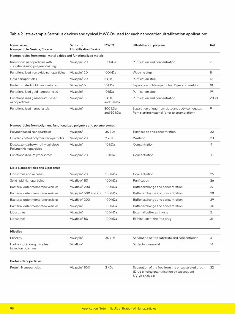
98
Table 2 lists example Sartorius devices and typical MWCOs used for each nanocarrier ultrafiltration application:
Nanocarrier: Nanoparticle, Vesicle, Micelle
Sartorius Ultrafiltration Device
MWCO Ultrafiltration purpose Ref.
Nanoparticles from metal, metal oxides and functionalized metals
Iron oxides nanoparticles with cisplatinbearing polymer coating
Vivaspin® 20 100 kDa Purification and concentration 7
Functionalized iron oxide nanoparticles Vivaspin® 20 100 kDa Washing step 8
Gold nanoparticles Vivaspin® 20 5 kDa Purification step 17
Protein coated gold nanoparticles Vivaspin® 6 10 kDa Separation of Nanoparticles | Dyes and washing 18
Functionalized gold nanoparticles Vivaspin® 10 kDa Purification step 19
Functionalized gadolinium-basednanoparticles
Vivaspin® 5 kDa and 10 kDa
Purification and concentration 20, 21
Functionalized nanocrystals Vivaspin® 300 kDa and 50 kDa
Separation of quantum dots-antibody conjugates from starting material (prior to enumeration)
9
Nanoparticles from polymers, functionalized polymers and polymersomes
Polymer based Nanoparticles Vivaspin® 30 kDa Purification and concentration 22
Curdlan coated polymer nanoparticles Vivaspin® 20 3 kDa Washing 23
Docetaxel-carboxymethylcellulose Polymer Nanoparticles
Vivaspin® 10 kDa Concentration 4
Functionalized Polymersomes Vivaspin® 20 10 kDa Concentration 3
Lipid Nanoparticles and Liposomes
Liposomes and micelles Vivaspin® 20 100 kDa Concentration 25
Solid lipid Nanoparticles Vivaflow® 50 100 kDa Purification 26
Bacterial outer membrane vesicles Vivaflow® 200 100 kDa Buffer exchange and concentration 27
Bacterial outer membrane vesicles Vivaspin® 500 and 20 100 kDa Buffer exchange and concentration 28
Bacterial outer membrane vesicles Vivaflow® 200 100 kDa Buffer exchange and concentration 29
Bacterial outer membrane vesicles Vivaspin® 100 kDa Buffer exchange and concentration 30
Liposomes Vivaspin® 100 kDa External buffer exchange 2
Liposomes Vivaflow® 50 100 kDa Elimination of the free drug 31
Micelles
Micelles Vivaspin® 30 kDa Separation of free substrate and concentration 4
Hydrophobic drug micelles based on polymers
Vivaflow® Surfactant removal 14
Protein Nanoparticles
Protein Nanoparticles Vivaspin® 500 3 kDa Separation of the free from the encapsulated drug (Drug binding quantification by subsequent UV-vis analysis)
32
Application Note 5. Ultrafiltration of Nanoparticles

99
References1. Strebhardt, K. & Ullrich, A.: Paul Ehrlich’s
magic bullet concept: 100 years of progress. 8, 473–480 (2008).
2 Jakoby, J., Beuschlein, F., Mentz, S., Hantel, C. & Süss, R.: Liposomal doxorubicin for active targeting: Surface modification of the nanocarrier evaluated in vitro and in vivo – challenges and prospects. Oncotarget 6, (2015).
3 Klermund, L., Poschenrieder, S. T. & Castiglione, K.: Simple surface functionalization of polymersomes using non-antibacterial peptide anchors. J. Nanobiotechnology 14, 48 (2016).
4 Mulder, W. J. M. et al.: Molecular imaging of macrophages in atherosclerotic plaques using bimodal PEG-micelles. Magn. Reson. Med. 58, 1164–1170 (2007).
5 Murthy, S. K.: Nanoparticles in modern medi-cine: state of the art and future challenges. Int. J. Nanomedicine 2, 129–41 (2007).
6 Voigt, R. & Fahr, A.: Pharmazeutische Technologie für Studium und Beruf. Deutscher Apotheker Verlag, 10th Edition (2010).
7 Unterweger, H. et al.: Development and characterization of magnetic iron oxide nanoparticles with a cisplatin-bearing polymer coating for targeted drug delivery. Int J Nanomedicine 9, 3659–3676 (2014).
8 Park, W. et al.: Multi-modal transfection agent based on monodisperse magnetic nanoparticles for stem cell gene delivery and tracking. Biomaterials 35, 7239–7247 (2014).
9 Chalmers, N. I. et al.: Use of quantum dot luminescent probes to achieve single-cell resolution of human oral bacteria in biofilms. Appl. Environ. Microbiol. 73, 630–636 (2007).
10 Hoffman, L. W., Andersson, G. G., Sharma, A., Clarke, S. R. & Voelcker, N. H.: New insights into the structure of PAMAM dendrimer | gold nanoparticle nanocomposites. Langmuir 27, 6759–6767 (2011).
11 Rademacher, T. & Williams, P.: Nanoparticle- peptide compositions. (2014).
12 Allard, E. & Larpent, C.: Core-shell type dually fluorescent polymer nanoparticles for ratiometric pH-sensing. J. Polym. Sci. Part A Polym. Chem. 46, 6206–6213 (2008).
13 Prach, M., Stone, V. & Proudfoot, L.: Zinc oxide nanoparticles and monocytes: Impact of size, charge and solubility on activation status. Toxicol. Appl. Pharmacol. 266, 19–26 (2013).
14 Zhang, Y. et al.: Therapeutic surfactant- stripped frozen micelles. Nat Commun 7, 11649 (2016).
15 Klasson, A. et al.: Positive MRI contrast enhancement in THP-1 cells with Gd2O3 nanoparticles. Contrast Media Mol. Imaging 3, 106–111 (2008).
16 Simonoska Crcarevska, M. et al.: Definition of formulation design space, in vitro bioactivity and in vivo biodistribution for hydrophilic drug loaded PLGA/PEO-PPO-PEO nanoparticles using OFAT experiments. Eur. J. Pharm. Sci. 49, 65–80 (2013).
17 Boda, S. K. et al.: Cytotoxicity of Ultrasmall Gold Nanoparticles on Planktonic and Biofilm Encapsulated Gram-Positive Staphylococci. Small 11, 3183–3193 (2015).
18 Schäffler, M. et al.: Blood protein coating of gold nanoparticles as potential tool for organ targeting. Biomaterials 35, 3455–3466 (2014).
19 Arosio, D. et al.: Effective targeting of DC-sign by α-fucosylamide functionalized gold nanoparticles. Bioconjug. Chem. 25, 2244–2251 (2014).
20 Miladi, I. et al.: Biodistribution of ultra small gadolinium-based nanoparticles as theranostic agent: application to brain tumors. J. Biomater. Appl. 28, 385–94 (2013).
21 Faure, A. C. et al.: Control of the in vivo biodistribution of hybrid nanoparticles with different poly(ethylene glycol) coatings. Small 5, 2565–2575 (2009).
22 Benita, S., Debotton, N. & Goldstein, D.: Nanoparticles for Targeted Delivery of Active Agent. (2008).
23 Tukulula, M. et al.: Curdlan-conjugated PLGA nanoparticles possess macrophage stimulant activity and drug delivery capabilities. Pharm. Res. 32, 2713–2726 (2015).
24 Ernsting, M. J., Tang, W. L., MacCallum, N. W. & Li, S. D.: Preclinical pharmacokinetic, biodistribution, and anti-cancer efficacy studies of a docetaxel-carboxymethyl-cellulose nanoparticle in mouse models. Biomaterials 33, 1445–1454 (2012).
25 Geelen, T., Paulis, L. E., Coolen, B. F., Nicolay, K. & Strijkers, G. J.: Passive target-ing of lipid-based nanoparticles to mouse cardiac ischemia-reperfusion injury. Contrast Media Mol. Imaging 8, 117–126 (2013).
Application Note 5. Ultrafiltration of Nanoparticles

100
26 Neves, A. R., Queiroz, J. F. & Reis, S.: Brain-targeted delivery of resveratrol using solid lipid nanoparticles functionalized with apolipoprotein E. J. Nanobiotechnology 14, 27 (2016).
27 Bogomolny, E. et al.: Analysis of bacteria- derived outer membrane vesicles using tunable resistive pulse sensing. Prog. Biomed. Opt. Imaging – Proc. SPIE 9338, 4–9 (2015).
28 Blenkiron, C. et al.: Uropathogenic Escherichia coli releases extracellular vesicles that are associated with RNA. PLoS One 11, 1–16 (2016).
29 Twu, O. et al.: Trichomonas vaginalis Exosomes Deliver Cargo to Host Cells and Mediate Host:Parasite Interactions. PLoS Pathog. 9, 22–24 (2013).
30 Tong, T. T., Mörgelin, M., Forsgren, A. & Riesbeck, K.: Haemophilus influenzae Survival during Complement-Mediated Attacks Is Promoted by Moraxella catarrhalis Outer Membrane Vesicles. J. Infect. Dis. 195, 1661–1670 (2007).
31 Prado, J. M. D., Antoranz, J. R. C., Barroeta, M. Á. E., Barroeta, B. E. & Diaz, M. C.: Liposomal formulations. (2009).
32 Achilli, E. et al.: Preparation of protein nanoparticle by dynamic aggregation and ionizing-induced crosslinking. Colloids Surfaces A Physicochem. Eng. Asp. 486, 161–171 (2015).
Application Note 5. Ultrafiltration of Nanoparticles

* Sample colour representative only
101Application Note 6. Vivaflow® and Vivaspin® Workflow
IntroductionEfficiency and efficacy of a multiple cycle experimental procedure was performed using Vivaflow® tangential flow cassettes for initial concentration and diafiltration of a cell culture super-natant. This was followed by Vivapure® Ion Exchange spin columns for the protein purification step and finally Vivaspin® 20 ultrafiltration devices for the final sample concentration and desalting. An artificial mixture of proteins in a RPMI-1640 culture medium was created to mimic the type of product that many researchers culture using e.g. the UniVessel device. This procedure further reflects a method that can be adapted to a large number of protein purification protocols, selecting alternative MWCOs and device sizes where necessary.
Methods
Part 1 – Creating and concentrating the culture medium2 bottles (4 g) of RPMI-1640 were dissolved into 1.8 L dd-H2O and 4 g of sodium acetate was added.
The pH was adjusted to 7.2 using 1M HCl. 2 g of BSA and 1 g of Lysozyme was added as protein samples, meant to be separated by chromatography. The volume of the cell culture super-natant sample was brought up to 2 L using dd-H₂O. After every preparation, concentration and purification step, 1 mL sample was set aside for SDS gel analysis.
Ion exchange chromatography was the method of choice for purifying lysozyme from the cell culture supernatant, especially from the “contaminant” BSA. For this, the 2 L cell culture supernatant needed to be concentrated and then diafiltered to adjust the sample to the starting conditions needed for the ion exchange chromatography binding step.
For concentration and diafiltration, the Vivaflow® 200 was used with a 5 kDa MWCO PES membrane. Vivaflow® 200 is a ready-to-use laboratory cross-flow cassette in an acrylic housing, which allows caustic cleaning and 4 – 5 re-uses. Two cassettes can be run in parallel for the concentration of up to 5 L sample volumes. For the 2 L sample to be concentrated in this experiment, one cassette was sufficient. A Masterflex pump with an Easy-Load, size 16 pump head was used to run the Vivaflow® 200 cassette. Figure 1a. and 1b. show the Vivaflow® 200 set up with one or two cassettes.
6. Vivaflow® and Vivaspin® Workflow in Protein Research Laboratories

102
The Vivaflow® 200 system was operated at 3 bar. Once 1.8 L of filtrate had been collected, the pump was stopped, the tubes removed from the cell culture medium concentrate and filtrate and the Vivaflow® system was purged with dd-H₂O. This solution now contained a 10-fold concentration of the constitu-ent proteins from the original culture medium.
A BCA protein detection assay con-veyed a 100% recovery of protein after this first concentration step. Table 1 indicates the time needed for the sample concentration.
Filtrate Volume (mL) Time (hr:min:sec)
0 0:00:00
100 0:03:16
200 0:06:50
300 0:10:45
400 0:14:38
500 0:18:36
600 0:22:43
700 0:26:57
800 0:31:14
900 0:36:01
1,000 0:40:50
1,100 0:45:46
1,200 0:50:36
1,300 0:55:32
1,400 1:00:24
1,500 1:05:26
1,600 1:10:28
1,700 1:15:52
1,800 1:21:50
Table 1: Vivaflow® 200, 5 kDa MWCO PES concentration speed
Figure 1a. Vivaflow® 200 – single cassette
Figure 1b. Vivaflow® 50R – two cassettes
Application Note 6. Vivaflow® and Vivaspin® Workflow
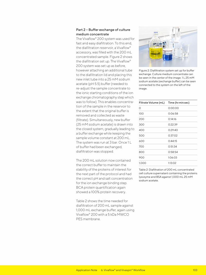
103
Part 2 – Buffer exchange of culture medium concentrateThe Vivaflow® 200 system was used for fast and easy diafiltration. To this end, the diafiltration reservoir, a Vivaflow® accessory, was filled with the 200 mL concentrated sample. Figure 2 shows the diafiltration set up. The Vivaflow® 200 system was set up as before, however attaching an additional tube to the diafiltration lid and placing this new inlet tube into a 25 mM sodium acetate (pH 5.5) buffer (needed to re-adjust the sample concentrate to the ionic starting conditions of the ion exchange chromatography step which was to follow). This enables concentra-tion of the sample in the reservoir to the extent that the original buffer is removed and collected as waste (filtrate). Simultaneously, new buffer (25 mM sodium acetate) is drawn into the closed system, gradually leading to a buffer exchange while keeping the sample volume constant at 200 mL. The system was run at 3 bar. Once 1 L of buffer had been exchanged, diafiltration was stopped.
The 200 mL solution now contained the correct buffer to maintain the stability of the proteins of interest for the next part of the protocol and had the correct pH and salt concentration for the ion exchange binding step. BCA protein quantification again showed a 100% protein recovery.
Table 2 shows the time needed for diafiltration of 200 mL sample against 1,000 mL exchange buffer, again using Vivaflow® 200 with a 5 kDa MWCO PES membrane.
Filtrate Volume (mL) Time (hr:min:sec)
0 0:00:00
100 0:06:58
200 0:14:16
300 0:22:39
400 0:29:40
500 0:37:02
600 0:44:15
700 0:51:34
800 0:58:54
900 1:06:03
1,000 1:13:02
Table 2: Diafiltration of 200 mL concentrated cell culture supernatant containing the proteins lysozyme and BSA against 1,000 mL 25 mM sodium acetate.
Figure 2: Diafiltration system set up for buffer exchange. Culture medium concentrate can be seen in the center of the image. 1 L 25 mM sodium acetate (exchange buffer) can be seen connected to the system on the left of the image.
Application Note 6. Vivaflow® and Vivaspin® Workflow

104
Part 3 – Purification of Lysozyme, the protein of interest The purification of Lysozyme was performed using a Vivapure® cation exchange membrane adsorber device (Vivapure® Maxi H S). The membrane adsorber matrix holds the active ligands and performs like a traditional cation exchanger. However, mem-brane adsorbers represent a special form of chromatography matrix. Unlike traditional resins, they make use of convective transport to bring proteins to the ion exchange surface; hence, binding, washing and elution is performed quickly and high binding capacities are achieved, even at high flow rates. This allows their use in fast and convenient centrifugal spin columns (Figure 3).
Figure 3: Electron micrograph of chromatogra-phy gel beads (upper right) in comparison to a Q ion exchange membrane adsorber (back-ground), revealing 100-fold larger pore sizes of the membrane adsorber.
Two Vivapure® Maxi H S type devices (Figure 4) were each equilibrated with 10 mL of 25 mM sodium acetate (pH 5.5), by filling with 10 mL of this buffer and centrifuging for 5 min. in a swing bucket centrifuge at 500 g and discarding the flow through. Using the concentrated and buffer exchanged sample from Part 2, 10 mL samples were pipetted into each of the equilibrated Vivapure® devices and centrifuged again for 5 min. in a swing bucket centrifuge at 500 g. The Vivapure® devices were washed with 10 mL of 25 mM sodium acetate, discarding the flow through, followed by an elution step with 5 mL of 1 M NaCl in 25 mM sodium acetate. A BCA assay revealed 95% lysozyme recovery.
Figure 4: Vivapure® Maxi spin columns can be used in a centrifuge for fast and easy protein purification.
Application Note 6. Vivaflow® and Vivaspin® Workflow

105
The eluate was then concentrated in a Vivaspin® 20 (5 kDa MWCO PES), Figure 5, and centrifuged at 5,000 g for 10 min. or until approximately 2 mL of concentrate had been collected. The device was then re-filled with 18 mL 50 mM potassium phosphate buffer (pH 7.2) to 20 mL for a final buffer exchange and desalting of the purified sample. The sample was again centrifuged until a final sample volume of 2 mL had been attained. A BCA assay revealed 97% lysozyme recovery.
Figure 5: Vivaspin® 20 ultrafiltration device, on the right with a pressure cap which allows pres-surization of the device as an alternative to the regular centrifugal operation.
Part 4 – Analyzing the samples The samples of the individual steps were analyzed by SDS gel, using reducing sample buffer (prepared by adding 50 μL 2-mercaptoethanol to 950 μL Laemmli sample buffer). For all steps, 5 μL of the 1 mL sample taken during the experiment were diluted with 95 μL reducing sample buffer, of which 20 μL were loaded onto a 12% Tris-HCl SDS gel (Figure 6).
Figure 6: Coomassie stained 12% Tris-HCl SDS gel loaded with 20 μL sample preparations. Lane 1: Marker (SDS Broad Range); Lane 2: Original sample; Lane 3: Original sample filtrate (Part 1); Lane 4: Marker; Lane 5: Buffer exchange concentrate (Part 2); Lane 6: Filtrate after binding (Part 3); Lane 7: Marker; Lane 8: Filtrate after elution (Part 3); Lane 9: Filtrate after concentrating and desalting (Part 3); Lane 10: Concentrate after concentrating and desalting.
Application Note 6. Vivaflow® and Vivaspin® Workflow
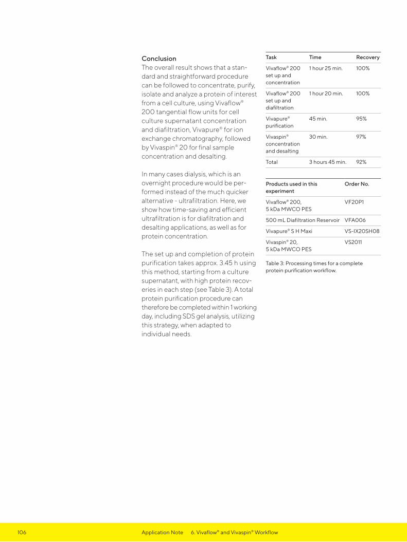
106 Application Note 6. Vivaflow® and Vivaspin® Workflow
ConclusionThe overall result shows that a stan-dard and straightforward procedure can be followed to concentrate, purify, isolate and analyze a protein of interest from a cell culture, using Vivaflow® 200 tangential flow units for cell culture supernatant concentration and diafiltration, Vivapure® for ion exchange chromatography, followed by Vivaspin® 20 for final sample concentration and desalting.
In many cases dialysis, which is an overnight procedure would be per-formed instead of the much quicker alternative - ultrafiltration. Here, we show how time-saving and efficient ultrafiltration is for diafiltration and desalting applications, as well as for protein concentration.
The set up and completion of protein purification takes approx. 3.45 h using this method, starting from a culture supernatant, with high protein recov-eries in each step (see Table 3). A total protein purification procedure can therefore be completed within 1 working day, including SDS gel analysis, utilizing this strategy, when adapted to individual needs.
Task Time Recovery
Vivaflow® 200 set up and concentration
1 hour 25 min. 100%
Vivaflow® 200 set up and diafiltration
1 hour 20 min. 100%
Vivapure® purification
45 min. 95%
Vivaspin® concentration and desalting
30 min. 97%
Total 3 hours 45 min. 92%
Products used in this experiment
Order No.
Vivaflow® 200, 5 kDa MWCO PES
VF20P1
500 mL Diafiltration Reservoir VFA006
Vivapure® S H Maxi VS-IX20SH08
Vivaspin® 20, 5 kDa MWCO PES
VS2011
Table 3: Processing times for a complete protein purification workflow.


Specifications subject to change without notice.Copyright Sartorius Lab Instruments GmbH & Co. KG.Status: 05| 2022
GermanySartorius Lab Instruments GmbH & Co. KG Otto-Brenner-Straße 20 37079 GöttingenPhone +49 551 308 0
USASartorius Corporation565 Johnson AvenueBohemia, NY 11716Phone +1 631 254 4249Toll-free +1 800 635 2906
For further information, visit www.sartorius.com