Advancing drug therapy for brain tumours: a current review of the pro-inflammatory peptide Substance...
12
Send Orders for Reprints to [email protected] 110 Recent Patents on CNS Drug Discovery, 2014, 9, 110-121 Advancing Drug Therapy for Brain Tumours: A Current Review of the Pro-inflammatory Peptide Substance P and its Antagonists as Anti-cancer Agents Kimberley Mander, Elizabeth Harford-Wright, Kate M. Lewis and Robert Vink* Adelaide Centre for Neuroscience Research, Discipline of Anatomy and Pathology, University of Adelaide, and *Division of Health Sciences, University of South Australia, Adelaide, Australia Received: September 09, 2014; Revised: November 04, 2014; Accepted: November 04, 2014 Abstract: Evidence for the involvement of the Substance P (SP)/NK1 receptor system in the development and progres- sion of cancer strongly supports its potential as a therapeutic target in malignancies. Novel strategies for approaching can- cer treatment are urgently required particularly with regard to tumours of the central nervous system (CNS), which are no- toriously difficult to effectively treat and associated with extremely poor prognosis for many patients. This is due, in part, to the presence of the highly specialised blood-brain barrier, which is known to restrict common treatments such as che- motherapy and hinder early tumour diagnosis. Additionally, tumours of the CNS are difficult to surgically resect com- pletely, often contributing to the resurgence of the disease many years later. Interestingly, despite the presence of the blood-brain barrier, circulating tumour cells are able to gain entry to the brain and form secondary brain tumours; how- ever, the underlying mechanisms of this process remain unclear. Tachykinins, in particular Substance P, have been impli- cated in early blood-brain barrier disruption via neurogenic inflammation in a number of other CNS pathologies. Recent evidence also suggests that Substance P may play a central role in the development of CNS tumours. It has been well es- tablished that a number of tumour cells express Substance P, NK1 receptors and mRNA for the tachykinin NK1 receptor. This increase in the Substance P/NK1 receptor system is known to induce proliferation and migration of tumour cells as well as stimulate angiogenesis, thus contributing to tumour progression. Accordingly, the NK1 receptor antagonist pre- sents a novel target for anti-cancer therapy for which a number of patents have been filed. This review will examine the role of Substance P in the development of CNS tumours and its potential application as an anti-cancer agent. Keywords: Blood-brain barrier, CNS tumours, glioma, metastases, substance P, tight junctions. INTRODUCTION Cancer is currently the leading cause of death and dis- ability worldwide with future projections estimating annual cases of cancer will rise from 14 million in 2012 to 22 mil- lion within the next two decades [1]. While improvements to treatment and early diagnosis have led to an increased sur- vival rate for many common cancers, the incidence of cancer burden is still increasing [2]. Tumours of the central nervous system (CNS) represent one of the more clinically feared diagnoses of cancer, primarily due to the challenge they pose with regard to treatment and poor patient outcome [3]. Spe- cifically, functional barriers unique to the CNS, such as the blood-brain barrier (BBB), and efflux transporters add fur- ther complexity with regard to diagnosis and treatment. The presence of the BBB is known to restrict the delivery of ef- fective and safe treatment agents such as chemotherapy [4, 5]. Accordingly, alternate treatment options are often aggres- sive, limited and largely unsuccessful, with many patients succumbing to their CNS tumour(s) within months of diag- nosis [6]. As such, a greater understanding of CNS tumours and the development of targeted treatments continues to be at the forefront of current research [7]. *Address correspondence to this author at the Division of Health Sciences, University of South Australia, South Australia, 5001, Australia; Tel: +61 8 8302 2029; Fax: +61 8 8302 2030; E-mail: [email protected] Primary brain tumours account for approximately 2% of all cancer incidence and despite this low rate, confer a dis- proportionate rate of morbidity and mortality due to their potential for progressive growth and interaction with critical neurological structures [8]. More than 256,000 new cases were diagnosed in 2012, with an alarming 189,000 deaths recorded for the same year [1]. Primary brain tumours arise from cells native to the brain [9]. Given that specialised glial cells such as astrocytes outnumber their post-mitotic neu- ronal counterparts and retain their proliferative capacity, it is unsurprising that neoplasms of glial origin account for the majority of primary adult CNS tumours [10]. Secondary brain tumours however, significantly outweigh the diagnosis of primary cerebral neoplasms - occurring 5-10 times more frequently [11, 12]. The development of metastases to the brain represents a crucial marker in the progression of the disease, specifically, the propensity of malignant cells to evade treatment, disseminate, survive and proliferate in a new location [6]. It is reported that approximately 20-40% of all adult patients with a systemic malignancy will present with a secondary symptomatic brain metastasis over the course of their disease [13]. Clinically, both primary and secondary tumours will often be accompanied by a myriad of neurological deficits which vary depending on tumour size, location and rate of progression [14]. Headache, nausea and vomiting are the most commonly described presenting fea- 2212-3954 /14 $100.00+.00 © 2014 Bentham Science Publishers
Transcript of Advancing drug therapy for brain tumours: a current review of the pro-inflammatory peptide Substance...

Send Orders for Reprints to [email protected]
110 Recent Patents on CNS Drug Discovery, 2014, 9, 110-121
Advancing Drug Therapy for Brain Tumours: A Current Review of the Pro-inflammatory Peptide Substance P and its Antagonists as Anti-cancer Agents
Kimberley Mander, Elizabeth Harford-Wright, Kate M. Lewis and Robert Vink*
Adelaide Centre for Neuroscience Research, Discipline of Anatomy and Pathology, University of Adelaide, and
*Division of Health Sciences, University of South Australia, Adelaide, Australia
Received: September 09, 2014; Revised: November 04, 2014; Accepted: November 04, 2014
Abstract: Evidence for the involvement of the Substance P (SP)/NK1 receptor system in the development and progres-
sion of cancer strongly supports its potential as a therapeutic target in malignancies. Novel strategies for approaching can-
cer treatment are urgently required particularly with regard to tumours of the central nervous system (CNS), which are no-
toriously difficult to effectively treat and associated with extremely poor prognosis for many patients. This is due, in part,
to the presence of the highly specialised blood-brain barrier, which is known to restrict common treatments such as che-
motherapy and hinder early tumour diagnosis. Additionally, tumours of the CNS are difficult to surgically resect com-
pletely, often contributing to the resurgence of the disease many years later. Interestingly, despite the presence of the
blood-brain barrier, circulating tumour cells are able to gain entry to the brain and form secondary brain tumours; how-
ever, the underlying mechanisms of this process remain unclear. Tachykinins, in particular Substance P, have been impli-
cated in early blood-brain barrier disruption via neurogenic inflammation in a number of other CNS pathologies. Recent
evidence also suggests that Substance P may play a central role in the development of CNS tumours. It has been well es-
tablished that a number of tumour cells express Substance P, NK1 receptors and mRNA for the tachykinin NK1 receptor.
This increase in the Substance P/NK1 receptor system is known to induce proliferation and migration of tumour cells as
well as stimulate angiogenesis, thus contributing to tumour progression. Accordingly, the NK1 receptor antagonist pre-
sents a novel target for anti-cancer therapy for which a number of patents have been filed. This review will examine the
role of Substance P in the development of CNS tumours and its potential application as an anti-cancer agent.
Keywords: Blood-brain barrier, CNS tumours, glioma, metastases, substance P, tight junctions.
INTRODUCTION
Cancer is currently the leading cause of death and dis-ability worldwide with future projections estimating annual cases of cancer will rise from 14 million in 2012 to 22 mil-lion within the next two decades [1]. While improvements to treatment and early diagnosis have led to an increased sur-vival rate for many common cancers, the incidence of cancer burden is still increasing [2]. Tumours of the central nervous system (CNS) represent one of the more clinically feared diagnoses of cancer, primarily due to the challenge they pose with regard to treatment and poor patient outcome [3]. Spe-cifically, functional barriers unique to the CNS, such as the blood-brain barrier (BBB), and efflux transporters add fur-ther complexity with regard to diagnosis and treatment. The presence of the BBB is known to restrict the delivery of ef-fective and safe treatment agents such as chemotherapy [4, 5]. Accordingly, alternate treatment options are often aggres-sive, limited and largely unsuccessful, with many patients succumbing to their CNS tumour(s) within months of diag-nosis [6]. As such, a greater understanding of CNS tumours and the development of targeted treatments continues to be at the forefront of current research [7].
*Address correspondence to this author at the Division of Health Sciences,
University of South Australia, South Australia, 5001, Australia; Tel: +61 8
8302 2029; Fax: +61 8 8302 2030; E-mail: [email protected]
Primary brain tumours account for approximately 2% of all cancer incidence and despite this low rate, confer a dis-proportionate rate of morbidity and mortality due to their potential for progressive growth and interaction with critical neurological structures [8]. More than 256,000 new cases were diagnosed in 2012, with an alarming 189,000 deaths recorded for the same year [1]. Primary brain tumours arise from cells native to the brain [9]. Given that specialised glial cells such as astrocytes outnumber their post-mitotic neu-ronal counterparts and retain their proliferative capacity, it is unsurprising that neoplasms of glial origin account for the majority of primary adult CNS tumours [10]. Secondary brain tumours however, significantly outweigh the diagnosis of primary cerebral neoplasms - occurring 5-10 times more frequently [11, 12]. The development of metastases to the brain represents a crucial marker in the progression of the disease, specifically, the propensity of malignant cells to evade treatment, disseminate, survive and proliferate in a new location [6]. It is reported that approximately 20-40% of all adult patients with a systemic malignancy will present with a secondary symptomatic brain metastasis over the course of their disease [13]. Clinically, both primary and secondary tumours will often be accompanied by a myriad of neurological deficits which vary depending on tumour size, location and rate of progression [14]. Headache, nausea and vomiting are the most commonly described presenting fea-
2212-3954 /14 $100.00+.00 © 2014 Bentham Science Publishers

Substance P and Brain Tumour Treatment Recent Patents on CNS Drug Discovery, 2014, Vol. 9, No. 2 111
tures and often indicate diffuse consequences of the space-occupying lesion, such as a raised intracranial pressure (ICP) [12]. As tumours develop or multiply, progressive focal neu-rological deficits such as visual disturbances, hemiparesis and aphasia will often become more pronounced and may elucidate tumour location [3].
The diagnosis of a brain tumour, either primary or secon-dary, presents a challenging and complex clinical situation in terms of devising an appropriate and effective treatment pro-tocol for patients [15]. Often treatment will be multidiscipli-nary involving a combination of definitive treatments di-rected against the tumour designed to eradicate malignancy, as well as adjunctive agents such as corticosteroids or anti-convulsants to control symptoms such as cerebral oedema and seizures. Standard treatment protocol involves surgery typically followed by radiotherapy and/or chemotherapy [5]. The total surgical resection of a tumour remains the gold-standard treatment for a localised primary intracranial neo-plasm [4]. Benign lesions are the most receptive to surgery as they contain well-defined margins enabling comprehen-sive removal of tumour tissue and improved patient survival [15]. However, a number of factors often preclude patients from undergoing curative surgery; for example, the anatomi-cal location of the tumour may render it inoperable due to close proximity of critical structures such as the brainstem or vital vasculature. For cases not conducive to complete surgi-cal resection, de-bulking surgery is often performed whereby maximal tumour tissue is removed allowing for a reduction in mass effect and the immediate alleviation of symptoms associated with increased ICP or seizures [16]. Unfortu-nately, microscopic foci of tumour cells commonly persist post surgery allowing the potential resurgence of the disease. Consequently, a course of postoperative radiotherapy is often administered to supplement surgery.
Radiotherapy is enlisted as an adjunctive therapy or pri-marily when a tumour is not amenable to complete surgical resection [17]. Whole-brain radiotherapy (WBRT) is consid-ered to be the mainstay therapy for patients with multiple intracranial metastases at diagnosis [18]. The median sur-vival of patients with brain metastases treated with WBRT is generally 2 to 6 months, with only 10% to 15% surviving beyond a year [19, 20]. Alternatively, stereotaxic radiosur-gery (SRT) will precisely deliver a high dose of radiation to the tumour area and thus requires the location and size of the tumour to be well characterised. Directed radiotherapy is advantageous as it allows for specific targeting of tumour cells whilst sparing the surrounding healthy brain tissue and reducing the side effects often associated with WBRT. Typi-cally SRT will be reserved for patients with a low number of cerebral tumours less than 3cm in diameter that are unable to be surgically resected [21]. Large tumours or tumours com-promised by oedema are often difficult to control with SRT due to an increased risk of radiation-associated necrosis and/or neurological deterioration at biologically effective doses [21]. As such, SRT administration is usually limited to one course for a particular site due to the high-risk of radia-tion necrosis.
The therapeutic success of chemotherapy in the treatment of both primary and metastatic CNS tumours is impeded firstly by reduced delivery of agents across the BBB and
secondly by an acquired resistance expressed by tumour cells [22]. This is particularly true during the early stages of tu-mour growth where evidence indicates that brain tumours are largely unresponsive to systemic chemotherapy due to the intact BBB [23]. In particular, brain metastases tend to re-spond poorly to chemotherapeutic agents, which may indi-cate a variation in chemosensitivity of metastatic tumour cells versus those of primary origin [24]. Additionally, pre-viously treated metastatic tumours may have acquired chemoresistant colonies that reduce the efficacy of com-monly administered chemotherapeutic agents; this is sug-gested to account for the 90% failure rate of drug therapies directed against secondary metastasis [25, 26]. Furthermore, methods for improving drug delivery as a means of bypass-ing the BBB or BTB are currently under investigation. More recently the biodegradable 1,3-bis (2-chloroethyl)-1-nitrosourea (BCNU) polymer wafers (Glidel) have been de-veloped for the use in malignant high-grade gliomas and evaluated for their effectiveness against multiple metastases [27]. These wafers allow the direct delivery of high doses of chemotherapeutic agents to the tumour tissue whilst bypass-ing the BBB. Following surgical resection of the glioma, up to 8 Glidel wafers are place directly on the resection cavity and release the chemotherapeutic agents over the following 2-3 weeks [28]. Clinical trials have demonstrated a statisti-cally significant improvement in survival following treat-ment with Glidel wafers post surgery, however their use has been limited by a high rate of complications [29-31]. Addi-tionally, pharmacological disruption of the BBB using hy-pertonic solutions such as mannitol and RMP-7 have been investigated as a means of improving chemotherapeutic penetration to the CNS [32, 33]. RMP-7 is a synthetic ana-logue of bradykinin, a known peptide that specifically binds to the bradykinin-P2 receptor and has a role in modulating brain endothelial tight junctions [34]. Despite positive results in both preclinical and phase I clinical trials, these results were not replicated at the phase II level, further demonstrat-ing the need for ongoing research to identify new avenues to improve options to deliver critical antitumor agents across the restricted BBB [23, 35]. In contrast to biochemical ap-proaches, the development of ultrasound and electromagnetic radiation methods have demonstrated transient and site spe-cific opening of the BBB suggested to be beneficial for CNS drug delivery [36].
Molecular targeted therapy (MTT) is a relatively new approach to cancer treatment whereby surface molecules uniquely or abnormally expressed by tumour cells are tar-geted with the intention of sparing normal cells and subse-quently reducing clinical side effects for patients [37]. The efficacy of MTT is inherently dependent on the assumption that the molecule of interest is solely expressed by cancer cells and has a primary function in the maintenance of the malignancy; thereby, an adaptive suppression of the mole-cule will not result in acquired resistance [38]. Glioblastoma Multiforme (GBM), the most common and lethal brain tu-mour in adults are known to exhibit an overexpression of a number of cell surface proteins and signalling pathway dis-ruptions, thus highlighting potential targets for MTT [39]. Given that GBMs are rarely amenable to aggressive surgical approaches, radiotherapy or chemotherapeutic agents, and are associated with a median survival rate of only one-year
112 Recent Patents on CNS Drug Discovery, 2014, Vol. 9, No. 2 Mander et al.
post diagnosis, the promise of specialised treatments such as MTT is eagerly anticipated. A number of MTTs have suc-cessfully entered clinical trials and received treatment ap-proval for various malignant conditions. However, the translation of MTTs to CNS neoplasms remains limited due to the exclusion of many cerebral metastatic patients from clinical trials, thus limiting their accessibility [40].
Increasingly, research has been directed towards identify-ing specific mediators that can be targeted in the treatment of cancer. Of particular interest are tachykinins and their recep-tors, which have been implicated in many aspects of cancer growth and progression, as well as in disruption to the BBB and development of cerebral oedema [41]. Furthermore, the revelation that tachykinin receptors, and in particular, sub-stance P (SP) receptors, are upregulated in a number of sys-temic and CNS malignancies, has stimulated research into the potential of SP-mediated interventions in the treatment of brain tumors. As such, this paper will build upon our previ-ous discussion of the role of neuroinflammatory peptides in cancer progression. More specifically, the potential applica-tions of therapies directed at modulating the inflammatory effects of SP at the BBB and the implications for limiting cancer progression within the brain.
THE BLOOD-BRAIN BARRIER
The blood-brain barrier (BBB) represents the tightest endothelial barrier of any organ and in this way is a key component for maintaining cerebral homeostasis by regulat-ing the traffic of both solutes and cells between the periph-eral vasculature and brain parenchyma [42]. It is now under-stood that this systematic method of regulation is much more complex and as such, the BBB represents a dynamic and vital structure which is now at the forefront of current re-search for a number of neurological pathologies.
Endothelial cells, astrocytes and pericytes comprise the crucial cellular elements of the BBB, however, additional cell types such as neurons, glial cells and smooth muscle cells of the vascular wall have recently been recognized to contribute to the function of the BBB [43]. Any increase in permeability of the barrier leads to a number of deleterious effects such as disordered transport of substances leading to alterations in cerebrovascular blood flow and disrupted ion content [44]. These changes will often produce additional damaging consequences such as increased ICP or seizures [44].
Both primary brain tumours and cerebral metastases are associated with impairment of the BBB, with alterations to the cerebral endothelial cells and tight junctions forming the focus of current research in tumour progression and development [45]. Commonly described characteristics of brain tumours include the opening of intercellular junctions, increased endo-thelial vesicles, modified endothelial cell morphology and defects in basement membrane integrity [45, 46]. Evaluation of BBB status in the presence of human cerebral metastases and malignant gliomas via MRI, demonstrate a compromised BBB at various intensities depending on the specific tumour type [47]. Interestingly, small metastatic tumours as well as those exhibiting a diffuse growth pattern have been shown to maintain an intact BBB suggesting unique neoplastic effects
for some cerebral metastases when compared to primary CNS neoplasms [46, 48].
As a CNS tumour outgrows its immediate blood supply, the process of neoangiogenesis quickly becomes critically required for additional tumour growth in both primary and metastatic lesions [49]. Current research indicates that these newly formed vessels do not maintain the physiological fea-tures of the BBB and are more commonly referred to as the ‘blood-tumour barrier’ (BTB) [50]. Typically, vessels of the BTB are more “leaky” when compared to the BBB due to compromised tight junction proteins, increased fenestrations and pinocytic vacuole activity, and reduced expression of efflux transporters [51]. Consequently, when a tumour is large enough to produce a ‘brain-tumour barrier’ systemi-cally administered chemotherapy can more effectively gain entry to the tumour site [23].
Indeed, the relationship between BBB permeability and the implications for both cancer growth and delivery of treatment have been well explored. Most recently however, the involvement of tachykinin neuropeptides, such as SP in BBB disruption and progression of peritumoural oedema have been investigated as potential targets to mediate brain tumour development.
THE TACHYKININ FAMILY
Tachykinins are a group of small structurally related pep-tides characterized by the specific C-terminal sequence, Phe-X-Gly-Lue-Met-NH2, and include SP, neurokinin A (NKA) and neurokinin B (NKB), as well as neuropeptide K (NPK) and neuropeptide � (NP�) [52]. The mammalian tachykinins are encoded by two distinct genes, preprotachykinin A (PPTA) and preprotachykinin B (PPTB). The PPTB gene gives rise to NKB, whilst alternate splicing of the PPTA gene results in four different forms of mRNA: �-PPT, �-PPT, �-PPT and �-PPT [52]. The �-PPT and �-PPT forms encode for SP alone, whilst �-PPT and �-PPT encode SP, NKA, NPK and NP� [52, 53]. Tachykinins are produced in both neuronal and glial cell bodies, and are widely distrib-uted throughout the CNS and PNS [54]. Specifically, within the PNS, SP is found in areas of immunologic importance such as the skin, gastrointestinal and respiratory tracts [55, 56]. In the CNS, levels of SP are higher in the grey rather than white matter [57] with the highest concentrations of SP occurring in the basal ganglia, hypothalamus, amygdala, locus coeruleus and dorsal root ganglia of the spinal cord [58].
Unlike classical neurotransmitters, tachykinins are pro-duced in the cell soma rather than in the nerve endings, and are released following calcium dependent mechanisms in response to stimuli [56]. Following release, tachykinins act on the transmembrane G-protein coupled receptors NK1, NK2 and NK3 to exert a myriad of effects, with each tachykinin demonstrating some degree of affinity for each tachykinin receptor due to the common COOH-terminal se-quence [59]. However, SP preferentially binds the NK1 re-ceptor, NKA the NK2 receptor and NKB the NK3 receptor.
Although the NK receptors are structurally closely re-lated, they are pharmacologically distinct, which has allowed detailed investigation of the structure and function of the

Substance P and Brain Tumour Treatment Recent Patents on CNS Drug Discovery, 2014, Vol. 9, No. 2 113
NK1 receptor. Binding to the NK1 receptor occurs at the second and third transmembrane domains and results in rapid endocytosis and internalisation of the receptor [60]. Within the CNS, NK1 receptors are found on neurons and glia [61], with expression of NK1 receptors highest in the caudate pu-tamen and superior colliculus [58]. The widespread expres-sion of SP and the NK1 receptor throughout the CNS has resulted in it being implicated in a number of neurological diseases, including CNS tumours. Accordingly, recent re-search has been focused on the potential of this particular tachykinin as a treatment target in numerous facets of cancer development.
SUBSTANCE P IN TUMOUR GROWTH
The last decade has seen a substantial rise in research into the expression and secretion of peptides by tumours [62] and in particular the tachykinins. Tachykinins are able to facilitate cell growth via induction of DNA synthesis and cellular proliferation. In addition, tachykinins have a stimu-lating or potentiating effect on lymphocyte proliferation and differentiation, cytokine secretion, and immunoglobulin pro-duction [63]. More specifically, SP and NK1 receptors have been implicated in numerous facets of cancer initiation and progression, with recent research implicating the PPT-1 gene and NK receptors in breast cancer development [64]. Fur-thermore, NK1 receptors have also been found in intratu-moural and peri-tumoural blood vessels, suggesting a poten-tial role for SP in tumour angiogenesis [65].
An increase in SP and the NK1 receptor expression has now been reported in a number of tumour types (Table 1) including glioma, retinoblastoma, colon, metastatic mela-noma, in situ melanoma, keratocystic odontogenic tumours and squamous cell and larynx carcinoma [66-78]. In a num-ber of glial tumour cell lines, the presence of NK1 receptors correlated with a SP and/or NK-A mediated increase in DNA synthesis and cellular proliferation [79, 80]. This increase in SP receptors in vivo is therefore thought to correspond with increases in tumour growth.
Oncogenes are genes that assist in the transformation of normal cells into cancerous ones, if mutated or expressed at high levels. Under normal physiological conditions, any cellular genetic damage activates one or more of the pro-grammed cell death pathways. In order to avoid this, tu-mour cells must set up ways to neutralise the multiple pathways leading to cell death. It has been proposed that the increase in the expression of NK1 receptors may play a role in malignant cells evasion of apoptosis [62, 63]. Such increased expression makes tumour cells highly dependent on SP stimulus, a known potent mitotic signal that counter-acts the different death signal pathways [63]. Indeed, ad-ministration of an NK1 antagonist for 2 weeks in a nude mice model of breast cancer was found to effectively in-hibit tumour growth [81], whilst administration of the SP receptor antagonist, L-733, 060, was found to inhibit the metastatic progression of NK1 expressing human glioma cancer cells [82]. Thus, antagonism of the NK1 receptor
Table 1. SP and NK1 receptor expression in tumours.
SP Expression NK1 Receptor Expression NK1 Antagonist Treatment
Human
Specimens
Present in:
Astrocytoma [78, 96],
Meningioma [96]
Breast carcinoma [78, 97],
Melanoma [67, 78]
Present in:
Astrocytoma [65, 78, 98],
GBM [78],
Breast carcinoma [65, 78, 94, 97, 98] ,
Melanoma [78]
N/A
Tumour cell
lines
Evident in:
Breast carcinoma (ZR-75–30, BT-474, T-47D,
MDA-MB-330, 184B5, CP-96 345-1, DU4475,
BT 483, MDA-MB-231, MDA-MB-231 Dox,
MCF7, LCC1, LCC2, SK-BR-3) [97, 99-101],
Melanoma [78]
Pancreatic carcinoma [102, 103]
Evident in:
Astrocytoma (UC11MG, U373MG, SNB-19,
DBTRG-05 MG, GAMG) [73, 75, 104-107]
Breast carcinoma (ZR-75–30, BT-474, T-
47D, MDA-MB-330, MDA-MB-231,
184B5, CP-96 345-1, DU4475, BT 483,
MCF7, LCC1, LCC2, SK-BR-3) [81, 94, 97,
100, 101]
Pancreatic carcinoma [102]
Melanoma (COLO 858, MEL HO, COLO
679) [108]
Induced apoptosis in: Astrocytoma
(GAMG, SNB-19, DBTRG-05 MG,
U373 MG) [68, 73, 75, 107],
Breast carcinoma (MDA-MB-231,
T47D, MDA-MB-468, MDA-MB-
453, SKBR3 MCF7, BT474) [81, 94,
109],
Pancreatic carcinoma (BxPC-3, MIA
PaCa-2) [102, 103]
Melanoma (COLO 858, MEL H0,
COLO 679) [68, 110]
Animal
Models
Present in breast carcinoma [111] Present in breast carcinoma [111] NK1 antagonist treatment causes
differential tumour response.
Caused decreased tumour growth in
astrocytoma (U373 MG) in the flank
of female nude mice [73], but did not
affect tumour growth in breast carci-
noma (MDA-MB-231) in the flank of
female nude mice [81].
114 Recent Patents on CNS Drug Discovery, 2014, Vol. 9, No. 2 Mander et al.
may allow cells to become more receptive to apoptotic death signals, leading to the death of mutated cells.
As well as exhibiting mitogenic and anti-apoptotic activ-ity, SP is known to stimulate the release of cytokines. The role of cytokines in cellular communication, coordination of cell growth and maturation, wound healing, the immune re-sponses and neo-angiogensis have been well documented [83]. Many of these processes can be utilised by tumour cells to facilitate their growth, thus supporting their own growth and facilitating their metastatic spread [83-85]. Furthermore, such cytokine release from malignant cells may also induce normal cells to synthesise additional cytokines perpetuating tumour progression [83]. Specifically, granulocyte-macrophage colony stimulating factor (GM-CSF) is released following SP mediated protein kinase C (PKC) activation and is responsible for the macrophage aggregation observed in malignant gliomas and the regulation of cellular growth and migration [64, 73]. Cytokines also influence the release of SP, with leukemia-inhibiting factor (LIF), a neuropoietin cytokine responsible for neurogenesis and neuronal differen-tiation, able to stimulate glioma proliferation by induction of SP and NK1 receptor expression [73]. LIF is produced and released by neuronal cells following exposure to proinflam-matory cytokines leading to increased SP production and NK1 expression [86].
Cytokines and other mediators are also responsible for the process of tumour angiogenesis, thereby enhancing tu-mour growth that is dependent upon a corresponding in-crease in vascularization [87]. Given the presence of its NK1 receptor in tumour vessels, it is hypothesised that SP may also facilitate tumour growth by mediating angiogenesis. Indeed, an increase in SP NK1 receptors has been demon-strated in tumoural and peri-tumoural vessels in glioblas-tomas, which is thought to be significant due to the known vasodilative and angiogenic effects of SP [65]. It has been reported that SP enhances capillary growth in an in vivo rab-bit cornea assay, an effect that was abolished following ad-
ministration of a SP receptor antagonist [88]. Furthermore, SP stimulates migration and proliferation of some types of endothelial cells, thus enhancing the angiogenic process in human tumours [87]. Thus, SP may play a major role in de-velopment of tumour stroma and facilitate tumoural blood supply.
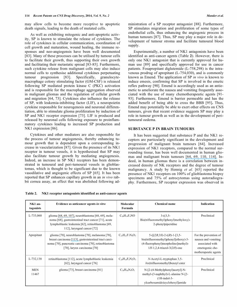
Experimentally, a number of NK1 antagonists have been identified as anti-cancer agents (Table 2). However, there is only one NK1 antagonist that is currently approved for hu-man use [89] and specifically approved for use in cancer patients. Fosaprepitant diglutemide (L-758,298) is the intra-venous prodrug of aprepitant (L-754,030), and is commonly known as Emend. The application of SP in vivo is known to induce emesis, confirming that SP is involved in the emetic reflex pathway [90]. Emend is accordingly used as an antie-metic to ameliorate the nausea and vomiting frequently asso-ciated with the use of many chemotherapeutic agents [91-93]. Furthermore, Emend is barrier permeable and has the added benefit of being able to cross the BBB [95]. Thus, Emend may potentially be able to exert other effects on CNS tumours, given that recent evidence suggests SP may play a role in tumour growth as well as in the development of peri-tumoural oedema.
SUBSTANCE P IN BRAIN TUMOURS
It has been suggested that substance P and the NK1 re-ceptors are particularly significant in the development and progression of malignant brain tumours [64]. Increased expression of NK1 receptors, compared to the normal sur-rounding tissue, has been well documented in human glio-mas and malignant brain tumours [64, 69, 110, 114]. In-deed, in human gliomas there is a correlation between in-creased density of NK receptors and the degree of tumour malignancy. A study by Hennig et al. [65] reported the presence of NK1 receptors on 100% of glioblastoma biopsy specimens and 75% of astrocytomas using autoradiogra-phy. Furthermore, SP receptor expression was observed in
Table 2. NK1 receptor antagonists identified as anti-cancer agents
NK1 an-
tagonists
Evidence as anticancer agents in vitro Molecular
Formula
Chemical name Indication
L-733,060 glioma [68, 69, 107], neuroblastoma [68, 69], mela-
noma [68], gastrointestinal tract cancer [71], acute
lymphoblastic leukemia [62], retinoblastoma [69,
112], laryngeal cancer [72]
C20H19F6NO 3-((3,5-
Bis(trifluoromethyl)phenyl)methyloxy)-
2-phenylpiperidine
Preclinical
Aprepitant glioma [70], neuroblastoma [70], melanoma [70],
breast carcinoma [113], gastrointestinal tract carci-
noma [70], pancreatic carcinoma [70], retinoblastoma
[70], larynx carcinoma [70]
C23H21F7N4O3 5-([(2R,3S)-2-((R)-1-[3,5-
bis(trifluoromethyl)phenyl]ethoxy)-3-
(4-fluorophenyl)morpholino]methyl)-
1H-1,2,4-triazol-3(2H)-one
For the prevention of
nausea and vomiting
associated with
emetogenic che-
motherapeutic agents
L-732,138 retinoblastoma [112], acute lymphoblastic leukemia
[62], laryngeal cancer [76]
C20H18F6N2O3 N-Acetyl-L-tryptophan 3,5-
bis(trifluoromethyl)benzyl ester
Preclinical
MEN
11467
glioma [73], breast carcinoma [81] C38H40N4O3 N-[2-(4-Methylphenyl)acetyl]-N-
methyl-(3-naphthyl)-L-alanine N-[2-
(1H-indol-3-
ylcarboxamido)cyclohexyl]amide
Preclinical
Substance P and Brain Tumour Treatment Recent Patents on CNS Drug Discovery, 2014, Vol. 9, No. 2 115
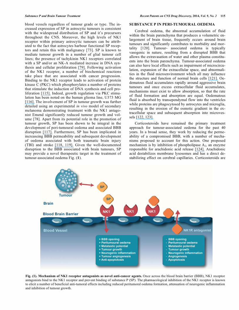
blood vessels regardless of tumour grade or type. The in-creased expression of SP in astrocytic tumours is consistent with the widespread distribution of SP and it’s precursors throughout the CNS. Moreover, the high levels of NK1 receptor within primary astrocytic tumours can be attrib-uted to the fact that astrocytes harbour functional SP recep-tors and retain this with malignancy [73]. SP is known to mediate tumour growth in a number of glial tumour cell lines; the presence of tachykinin NK1 receptors correlated with a SP and/or an NK-A mediated increase in DNA syn-thesis and cellular proliferation [79]. Following activation of the NK1 receptor, a number of biochemical reactions take place that are associated with cancer progression. Binding to the NK1 receptor leads to activation of protein kinase C (PKC) which phosphorylates a number of proteins that stimulate the induction of DNA synthesis and cell pro-liferation [115]. Indeed, growth regulation via PKC stimu-lation has been noted on the human glioma line, U373 MG [116]. The involvement of SP in tumour growth was further detailed using an experimental in vivo model of secondary melanoma demonstrating treatment with the NK1 antago-nist Emend significantly reduced tumour growth and vol-ume [78]. Apart from its potential role in the promotion of tumour growth, SP has been shown to be integral in the development of peri-tumoural oedema and associated BBB disruption [117]. Furthermore, SP has been implicated in increaseing BBB permeability and subsequent development of oedema associated with both traumatic brain injury (TBI) and stroke [118, 119]. Given the well-documented disruption to the BBB associated with brain tumours, SP may provide a novel therapeutic target in the treatment of tumour-associated oedema Fig. (1).
SUBSTANCE P IN PERI-TUMOURAL OEDEMA
Cerebral oedema, the abnormal accumulation of fluid within the brain parenchyma that produces a volumetric en-largement of brain tissue, frequently occurs around brain tumours and significantly contributes to morbidity and mor-tality [120]. Tumour- associated oedema is typically vasogenic in nature, resulting from a disrupted BBB that allows the extravasation of water and other plasma constitu-ents into the brain parenchyma. Tumour-associated oedema can also have local effects such as impairment of microcircu-lation, expansion of the extracellular space, and abnormali-ties in the fluid microenvironment which all may influence the structure and function of normal brain cells [121]. Oe-dematous fluid accumulates rapidly around aggressive brain tumours and once excess extracellular fluid accumulates, mechanisms must exist to allow absorption, so that the rate of fluid formation and absorption are equal. Oedematous fluid is absorbed by transependymal flow into the ventricles while proteins are phagocytosed by astrocytes and microglia, resulting in the erosion of the osmotic gradient in the ex-tracellular space and subsequent absorption into microves-sels [122, 123].
Corticosteroids have remained the primary treatment approach for tumour-associated oedema for the past 40 years. In a broad sense, they work by reducing the perme-ability of a compromised BBB, with a number of mecha-nisms proposed to account for this action. One proposed mechanism is by inhibition of phospholipase A2, an enzyme responsible for arachidonic acid release [124]. Arachidonic acid destabilizes membrane lysosomes and has a direct de-stabilizing effect on cerebral capillaries. Corticosteroids are
Fig. (1). Mechanism of NK1 receptor antagonists as novel anti-cancer agents. Once across the blood brain barrier (BBB), NK1 receptor
antagonists bind to the NK1 receptor and prevent binding of substance P (SP). The pharmacological inhibition of the NK1 receptor is known
to elicit a number of beneficial anti-tumoral effects including reduced peritumoral oedema formation, attenuation of neurogenic inflammation
and inhibition of tumour growth.

116 Recent Patents on CNS Drug Discovery, 2014, Vol. 9, No. 2 Mander et al.
also known to have the ability to reduce vascular endothelial growth factor (VEGF)-induced BBB permeability, an action reversed by a glucocorticoid receptor antagonist [125]. Therefore, corticosteroids may act to reduce the response of the cerebral capillary endothelial cells (CCEC) to VEGF or reduce the secretion of VEGF by tumour cells [125]. None-theless, the exact mechanism of action of corticosteroids is yet to be fully elucidated. Dexamethasone is the most com-monly used steroid for tumour-associated oedema. It has a long half-life, which allows for twice daily dosing, and im-proves generalised symptoms such as headache and altered mental status. The majority of patients with brain metastases show marked clinical improvement within 24 to 72 hours after beginning dexamethasone treatment [126]. Nonethe-less, dexamethasone is associated with a large number of potentially serious side effects, the severity of which de-pends on the dose and duration of steroid treatment [127]. Adverse side effects include immunosuppression, hyperten-sion, fluid retention and mood disturbances. These complica-tions have consequently prompted investigation into alterna-tive treatments for tumour associated brain oedema. Given its role in the development of oedema following other insults to the CNS, the tachykinin SP has become a potential target [128, 129].
Tachykinins are known to contribute to cerebral oedema in a number of brain pathologies through a process known as neurogenic inflammation, a neurally elicited inflammatory response characterised by vasodilation, plasma extravasation, mast cell degranulation and tissue swelling [130]. It results from the stimulation of capsaicin sensitive C-fibres causing the release of neuropeptides such as SP and calcitonin gene related peptide (CGRP). Numerous studies have provided evidence for a role for neuropeptides in most immunologic and inflammatory states within the periphery. Activation or damage to neurons can lead to changes in neuropeptide syn-thesis, which results from the induction of neuropeptide gene expression [53]. Such changes to neuropeptide expression in sensory neurons have been observed in models of acute and chronic inflammation. Furthermore, upregulation of NK1 receptor and PPT mRNA has also been shown in the periph-ery during noxious stimulation or neurogenic inflammation [54].
SP is thought to be the most potent initiator of neuro-genic inflammation because of its association with increased vascular permeability and subsequent protein plasma ex-travasation [131]. It may also potentiate inflammatory re-sponses by stimulating the production of inflammatory me-diators such as histamine, nitric oxide and kinins, interacting with adhesion molecules and the extracellular matrix causing leukocyte migration [131]. The role of SP in neurogenic in-flammation in the periphery has been well documented, par-ticularly in the skin, gastrointestinal and respiratory tracts where administration of SP has been found to induce neuro-genic inflammation [132, 133]. This role has been confirmed by findings that NK1 receptor antagonists completely abol-ished the inflammatory response [134]. In contrast, the con-cept of neurogenic inflammation in the CNS has remained largely unexplored until relatively recently.
Interest in the role of neurogenic inflammation in a number of brain pathologies is on the rise. As the cerebral
arteries have a dense supply of neuropeptide containing sensory neurons, an increase in neuropeptides post CNS damage may be involved in injury pathways [135]. Vink and colleagues (2003) were the first to investigate neuro-genic inflammation in the brain following traumatic brain injury (TBI) [135]. They found that depletion of neuropep-tides with capsaicin completely attenuated the changes in BBB permeability and subsequent oedema formation usu-ally observed following TBI. Subsequent abolition of neu-ropeptides also significantly improved functional outcome post trauma. Such findings were rationalised in terms of the high levels of NK receptors found in areas associated with both motor control and memory and learning [136]. Fur-thermore, successive research has demonstrated a signifi-cant upregulation of SP immunoreactivity in perivascular tissue and within the parenchyma occurs by 5 hours post trauma. This increase in SP is associated with significant increases in cerebral oedema and marked functional deficits [118, 137]. Administration of an NK1 antagonist was found to attenuate this oedema formation and improve neurologi-cal outcome in both male and female rats [118, 137]. These results are consistent with what is observed in the human condition, with SP immunoreactivity reportedly increased following human TBI [138]. A role for neuropeptides, namely SP, has also been demonstrated in other neurologi-cal diseases. Following ischaemic stroke, SP immunoreac-tivity was increased in the infarcted hemisphere and was associated with profound oedema formation. As was ob-served in TBI, administration of a SP antagonist resulted in marked improvement in functional outcome following stroke [119]. Several patents have described the use of NK1 antagonists to ameliorate SP induced vasogenic cere-bral oedema, particularly following TBI and ischemic reperfusion stroke [118, 139, 140]. Furthermore, these ef-forts to reduce cerebral oedema through NK1 antagonist treatment have led to the filing of patents that also promote this treatment to prevent subsequent increases in ICP [141].
Numerous studies have also demonstrated that SP and NK1 receptor immunoreactivity is increased surrounding brain tumours, particularly within the perivascular region [36, 78]. This localization is suggestive of a role in the formation of tumour-associated oedema and was further explored through an experimental in vivo model of secon-dary metastasis [117]. Results of this study demonstrate the involvement of SP in the development of tumour-associated BBB disruption and subsequent oedema forma-tion [117]. Futhermore, administration of the NK1 antago-nist Emend was found to significantly reduce BBB disrup-tion and brain water content indicating that the targeting the SP/NK1 pathway may be a beneficial approach to alle-viating deleterious symptoms associated with tumour growth. This was found to be as effective as the current clinical treatment of dexamethasone in reducing tumour associated brain oedema [117]. Interestingly, these results conflict with previously reported data, which detailed that treatment with an NK1 antagonist failed to exert any sig-nificant effect on oedema formation in an in vivo model of metastatic breast cancer [111]. However, it has been high-lighted that this apparent contradiction may be a reflection of the differing tumor cell lines, further reinforced by the findings observed in the study of human tumors indicating

Substance P and Brain Tumour Treatment Recent Patents on CNS Drug Discovery, 2014, Vol. 9, No. 2 117
that SP expression can differ between different tumor types. As such, continuation of this work will endeavor to further ascertain and characterise the differing role of SP in specific tumour types.
SUBSTANCE P IN THE EXTRAVASATION PROCESS
A key event in the development of any brain metastasis is the migration of cancer cells through the blood-brain barrier (BBB) [42]. Invasion of the brain is a multifaceted process of tightly controlled mechanisms which enable tumour cells to breach the cerebral microvessels and invade surrounding parenchyma to form micro and macrometastasis [142]. To date, the precise mechanisms underpinning this detrimental process remain unclear. As such, cancer cell interaction with the BBB and potential mediating substances form a valuable target for further investigation into the critical early stages of tumour extravasation [18]. Identification of such agents may prevent metastatic tumour formation; however to date there has been little success.
The quality of the surrounding stroma and ECM in terms of nutrients and immune cell presence is known to play a role in the survival and rate of growth for circulating tumour cells. The mere presence of tumour cells is known to induce biochemical alterations within the immediate brain microen-vironment, namely catalyse paracrine signalling between endothelial cells, stromal cells and the invading metastatic cells leading to an inflammatory state [143]. Cellular remod-elling of cerebral endothelial cells is apparent in the early extravasation stage of metastasis, with tumour cells shown to promote a rearrangement of the endothelial cytoskeleton resulting in an overt phenotypic change to cells which has downstream effects on their function [144]. Furthermore, histological analysis of resected human brain metastases revealed tumour cells have a clear interaction with activated microglia and astrocytes further supporting the tumour in-duced neuroinflammatory state of the parenchymal microen-vironment [145]. It is suggested that this pro-inflammatory state is beneficial for the growth and proliferation of metas-tatic cells. Tumour cell-derived factors such as interleukin-1 (IL-1), macrophage inhibitory factor, and plasminogen acti-vator inhibitor 1 are known to activate local astrocytes which consequently produce metastatic proliferative factors such as interleukin-6 (IL-6), interleukin-1beta (IL-1�) and tumour necrosis factor alpha (TNF-�) in vitro. Consistent with these findings, in vitro co-culture model of metastasis found the presence of reactive microglia induced a five-fold increase in metastatic cellular proliferation, suggesting that the activated microenvironment advances tumour growth and invasion [146]. Additionally, SP has been detected in cerebral capil-lary endothelial cells and is secreted by these cells in re-sponse to treatment with high dose of cytokines such as IL-1� and TNF-�. As a result of SP release, the concentration of calcium ions within endothelial cells of the BBB is in-creased by approximately 10 times above normal, and hence leads to increased permeability of the BBB through endothe-lial cell contraction. As previously stated, increases in SP and its NK1 receptor have been reported for a variety of dif-ferent tumour cell lines, further supporting the potential of this system as a target to inhibit the entry of cancer cells into the brain [74, 78]. Furthermore, in vitro studies of the BBB indicate that in the presence of SP, expression of crucial TJ
proteins, claudin-5 and zona occludin-1 is decreased, thus correlating with increased permeability of the BBB [147]. Recently, it has been demonstrated that SP expression is in-creased locally surrounding tumour-invaded microvessels, in a rodent model of brain metastatic breast cancer (BCC). The localised elevation of SP correlated with increased perme-ability of the BBB and the loss of endothelial barrier antigen staining, indicative of a disrupted BBB (See Fig. 1). Addi-tionally, it was confirmed that BCCs actively secrete SP that ultimately contribute to BBB TJ protein alteration. Moreo-ver, treatment with the SP inhibitor, spantide III demon-strated an inhibition of changes to the BBB TJs and the sub-sequent reduction of BCC brain colonisation in vivo. These results indicate the secretion of SP directly from tumour cells may increase the permeability of the BBB, thereby allowing for the movement of tumour cells into the brain, and subse-quent development of metastasis. As such, NK1 antagonists are a potentially promising preventative treatment for metas-tatic brain tumours, particularly for patients diagnosed with primary tumours known to preferentially spread to the brain.
The extensive research on the role of SP in cancer growth and the potential antiumoral action of NK1 antagonists in various cancer types has led to the filing of several patents. One patent by Munoz and collegues [112] describes the in-duction of apoptosis in cancer cells by NK1 antagonists in a number of cancer types, but in particular gliomas, mela-noma, lung and breast carcinomas. Similarly, the invention of an antibody specific against SP has been suggested for the treatment of cancers expressing NK1 or epidermal growth factor family receptors [148]. In contrast, several patents have also been filed in which SP is proposed as a treatment or preventative strategy for cancer, although these are largely targeting peripheral tissues. Aerosolized SP has been de-scribed as a prophylactic treatment for the prevention of can-cer in the lung, particularly in response to environmental toxins [149, 150]. Similarly, SP in combination with an im-munogenic composition has been proposed for use as a can-cer vaccine, acting through tumour antigen manipulation [151]. Clearly, treatment with either an NK1 antagonist or SP must be applied selectively based on tissue location of the cancer as well as the cancer type.
CURRENT & FUTURE DEVELOPMENTS
As the burden of cancer continues to rise worldwide, many of the underlying mechanisms contributing to cancer development and progression remain unclear. Consequently, prevention, early diagnosis and treatment options continue to be hampered by such limitations in our understanding. The pursuit of improved treatment strategies has led to the inves-tigation of multiple targets as a means of achieving improved outcome and prognosis for patients. Recent research has in-dicated that SP may play multiple roles in the pathology of CNS tumours, including tumour cell growth, the genesis of peri-tumoural oedema and the extravasation of tumour cells into the brain. Given that the NK1 antagonist is already ad-ministered clinically as part of cancer patient management, it remains the frontrunner in terms of further analysing the role of SP in pathways of cancer progression. Furthermore, a retrospective review of patient data may reveal administra-tion of NK1 antagonist providing beneficial effects on tu-mour growth and reduced metastatic potential for some can-
118 Recent Patents on CNS Drug Discovery, 2014, Vol. 9, No. 2 Mander et al.
cer types. To date there has been no such investigation into the effect of the treatment on cancer growth or progression in humans. Additionally, SP is expressed in varying quantities for a number of different tumour cells, thus further investiga-tion of whether these levels correlate with tumour aggres-siveness or propensity to colonise certain organs, such as the brain is required. Similarly, additional research to elucidate the effect of NK1 antagonist treatment on BBB dysfunction in the setting of experimental brain tumours will be of fur-ther benefit. Finally, there is sufficient evidence to suggest both SP and NK1 play a key role in tumour development and progression, particularly with regard to tumour dissemina-tion to the brain. As such further research is encouraged in order to harness the benefit of targeting this pathway in can-cer biology.
CONFLICT OF INTEREST
The authors confirm that this article content has no con-flict of interest.
ACKNOWLEDGEMENTS
Declared none.
DISCLOSURE
This is an extended and updated version of the authors’ previous manuscript published in Recent Pat. CNS Drug Disc. 6 (1), 31-40 (2011) entitled “Towards drug discovery for brain tumours: interactions of kinins and tumours at the blood brain barrier interface”.
REFERENCES
[1] Ferlay JSI, Ervik M, Dikshit R, Eser S, Mathers C, Rebelo M, Parkin DM, Forman D, Bray, F. In: Cancer IAfRo, Ed. Cancer
Incidence and Mortality Worldwide. IARC CancerBase No 11 Lyon, France: GLOBOCAN 2012: 2013.
[2] Siegel R, Ma J, Zou Z, Jemal A. Cancer statistics, 2014. CA: Canc J Clinicians 2014; 64(1): 9-29.
[3] Norden AD, Wen PY, Kesari S. Brain metastases. Curr Opin Neurol 2005; 18(6): 654-61.
[4] Behin A, Hoang-Xuan K, Carpentier AF, Delattre JY. Primary brain tumours in adults. Lancet 2003; 361(9354): 323-31.
[5] Buckner JC, Brown PD, O'Neill BP, Meyer FB, Wetmore CJ, Uhm JH. Central nervous system tumors. Mayo Clin Proc 2007; 82(10):
1271-86. [6] Gavrilovic IT, Posner JB. Brain metastases: epidemiology and
pathophysiology. J Neuro-oncol 2005; 75(1): 5-14. [7] Kefford RF. Drug treatment for melanoma: progress, but who
pays? Medical J Aus 2012; 197(4): 198-9. [8] Louis DN, Ohgaki H, Wiestler OD, Cavenee WK, Burger PC,
Jouvet A, et al. The 2007 WHO classification of tumours of the central nervous system. Acta Neuropathol 2007; 114(2): 97-109.
[9] Pereira A, Jr., Furlan FA. Astrocytes and human cognition: modeling information integration and modulation of neuronal
activity. Prog Neurobiol 2010; 92(3): 405-20. [10] Furnari FB, Fenton T, Bachoo RM, Mukasa A, Stommel JM, Stegh
A, et al. Malignant astrocytic glioma: genetics, biology, and paths to treatment. Genes Develop 2007; 21(21): 2683-710.
[11] Bafaloukos D, Gogas H. The treatment of brain metastases in melanoma patients. Canc Treat Rev 2004; 30(6): 515-20.
[12] DeAngelis LM. Brain tumors. New Eng J Med 2001; 344(2): 114-23.
[13] Vecht CJ. Clinical management of brain metastasis. J Neuro 1998; 245(3): 127-31.
[14] Ricard D, Idbaih A, Ducray F, Lahutte M, Hoang-Xuan K, Delattre JY. Primary brain tumours in adults. Lancet 2012; 379(9830):
1984-96.
[15] Drappatz J, Schiff D, Kesari S, Norden AD, Wen PY. Medical
management of brain tumor patients. Neurologic Clinics 2007; 25(4): 1035-71, ix.
[16] Silberman AW. Surgical debulking of tumors. Surg Gyne Obs 1982; 155(4): 577-85.
[17] Kondziolka D, Flickinger JC, Lunsford LD. Radiosurgery for brain metastases. Prog Neuro Surg 2012; 25: 115-22.
[18] Arshad F, Wang L, Sy C, Avraham S, Avraham HK. Blood-brain barrier integrity and breast cancer metastasis to the brain. Pathol
Res Int 2010; 2011: 920509. [19] Khan AJ, Dicker AP. On the merits and limitations of whole-brain
radiation therapy. J Clin Onco 2013; 31(1): 11-3. [20] Patchell RA. The management of brain metastases. Canc Treat Rev
2003; 29(6): 533-40. [21] Ranjan T, Abrey LE. Current management of metastatic brain
disease. Neurotherapeut 2009; 6(3): 598-603. [22] Fortin D. The blood-brain barrier should not be underestimated in
neuro-oncology. Revue Neurologique 2004; 160(5 Pt 1): 523-32. [23] Deeken JF, Loscher W. The blood-brain barrier and cancer:
transporters, treatment, and Trojan horses. Clin Canc Res 2007; 13(6): 1663-74.
[24] Regina A, Demeule M, Laplante A, Jodoin J, Dagenais C, Berthelet F, et al. Multidrug resistance in brain tumors: roles of the blood-
brain barrier. Canc Metastas Rev 2001; 20(1-2): 13-25. [25] Longley DB, Johnston PG. Molecular mechanisms of drug
resistance. J Pathol 2005; 205(2): 275-92. [26] Abdullah LN, Chow EK. Mechanisms of chemoresistance in cancer
stem cells. Clin Translat Med 2013; 2(1): 3. [27] Abel TJ, Ryken T, Lesniak MS, Gabikian P. Gliadel for brain
metastasis. Surg Neuro Int 2013; 4(Suppl 4): S289-93. [28] Westphal M, Hilt DC, Bortey E, Delavault P, Olivares R, Warnke
PC, et al. A phase 3 trial of local chemotherapy with biodegradable carmustine (BCNU) wafers (Gliadel wafers) in patients with
primary malignant glioma. Neuro-onco 2003; 5(2): 79-88. [29] Bregy A, Shah AH, Diaz MV, Pierce HE, Ames PL, Diaz D, et al.
The role of Gliadel wafers in the treatment of high-grade gliomas. Expert Rev Anticanc Ther 2013; 13(12): 1453-61.
[30] Nagpal S. The role of BCNU polymer wafers (Gliadel) in the treatment of malignant glioma. Neurosurg Clin North Am 2012;
23(2): 289-95, ix. [31] Brem H, Ewend MG, Piantadosi S, Greenhoot J, Burger PC, Sisti
M. The safety of interstitial chemotherapy with BCNU-loaded polymer followed by radiation therapy in the treatment of newly
diagnosed malignant gliomas: phase I trial. J Neuro-onco 1995; 26(2): 111-23.
[32] Jain KK. Nanobiotechnology-based strategies for crossing the blood-brain barrier. Nanomed (Lond) 2012; 7(8): 1225-33.
[33] Black, K.L., Ningaraj, N.S. Potassium channel mediated delivery of agents through the blood-brain barrier. WO2005025511 (2005).
[34] Liu LB, Xue YX, Liu YH, Wang YB. Bradykinin increases blood-tumor barrier permeability by down-regulating the expression
levels of ZO-1, occludin, and claudin-5 and rearranging actin cytoskeleton. J Neurosci Res 2008; 86(5): 1153-68.
[35] Emerich DF, Dean RL, Marsh J, Pink M, Lafreniere D, Snodgrass P, et al. Intravenous cereport (RMP-7) enhances delivery of
hydrophilic chemotherapeutics and increases survival in rats with metastatic tumors in the brain. Pharmaceut Res 2000; 17(10): 1212-
9. [36] Tajes M, Ramos-Fernandez E, Weng-Jiang X, Bosch-Morato M,
Guivernau B, Eraso-Pichot A, et al. The blood-brain barrier: structure, function and therapeutic approaches to cross it. Mol
Memb Biol 2014; 31(5): 1-16. [37] Hochwald SN. Molecular-targeted therapy for cancer and
nanotechnology. Meth Mol Biol 2010; 624: 11-23. [38] Rahma OKS. Biological Basis of Molecular Targeted Therapy. In:
Roland T, Skeel SNK, Ed. Handbook of Cancer Chemotherapy. Philidelphia: Lippincott Williams & Wilkins; 2011.
[39] Mischel PS, Cloughesy TF. Targeted molecular therapy of GBM. Brain Pathol 2003; 13(1): 52-61.
[40] Atallah E, Flaherty L. Treatment of metastatic malignant melanoma. Curr Treatm Opt Onco 2005; 6(3): 185-93.
[41] Palma C. Tachykinins and their receptors in human malignancies. Curr Drug Targ 2006; 7(8): 1043-52.
[42] Wilhelm I, Molnar J, Fazakas C, Hasko J, Krizbai IA. Role of the blood-brain barrier in the formation of brain metastases. Int J Mol
Sci 2013; 14(1): 1383-411.

Substance P and Brain Tumour Treatment Recent Patents on CNS Drug Discovery, 2014, Vol. 9, No. 2 119
[43] Nag S, Kapadia A, Stewart DJ. Review: molecular pathogenesis of
blood-brain barrier breakdown in acute brain injury. Neuropathol App Neurobiol 2011; 37(1): 3-23.
[44] Banks WA. Physiology and pathology of the blood-brain barrier: implications for microbial pathogenesis, drug delivery and
neurodegenerative disorders. J Neurovirol 1999; 5(6): 538-55. [45] Jia W, Martin TA, Zhang G, Jiang WG. Junctional adhesion
molecules in cerebral endothelial tight junction and brain metastasis. Anticanc Res 2013; 33(6): 2353-9.
[46] Harford-Wright E, Lewis KM, Vink R. Towards drug discovery for brain tumours: interaction of kinins and tumours at the blood brain
barrier interface. Rec Pat CNS Drug Discov 2011; 6(1): 31-40. [47] Nduom EK, Yang C, Merrill MJ, Zhuang Z, Lonser RR.
Characterization of the blood-brain barrier of metastatic and primary malignant neoplasms. J Neurosurg 2013.
[48] Zhang RD, Price JE, Fujimaki T, Bucana CD, Fidler IJ. Differential permeability of the blood-brain barrier in experimental brain
metastases produced by human neoplasms implanted into nude mice. Am J Pathol 1992; 141(5): 1115-24.
[49] Jain RK, di Tomaso E, Duda DG, Loeffler JS, Sorensen AG, Batchelor TT. Angiogenesis in brain tumours. Nat Rev Neurosci
2007; 8(8): 610-22. [50] Lockman PR, Mittapalli RK, Taskar KS, Rudraraju V, Gril B,
Bohn KA, et al. Heterogeneous blood-tumor barrier permeability determines drug efficacy in experimental brain metastases of breast
cancer. Clin Canc Res 2010; 16(23): 5664-78. [51] Groothuis DR. The blood-brain and blood-tumor barriers: a review
of strategies for increasing drug delivery. Neuro-oncol 2000; 2(1): 45-59.
[52] Maggi CA. The mammalian tachykinin receptors. Gen Pharmacol 1995; 26(5): 911-44.
[53] Hokfelt T, Pernow B, Wahren J. Substance P: a pioneer amongst neuropeptides. J Intern Med 2001; 249(1): 27-40.
[54] McCarson KE. Central and peripheral expression of neurokinin-1 and neurokinin-3 receptor and substance P-encoding messenger
RNAs: peripheral regulation during formalin-induced inflammation and lack of neurokinin receptor expression in primary afferent
sensory neurons. Neurosci 1999; 93(1): 361-70. [55] Pernow B. Substance P. Pharmacological Rev 1983; 35(2): 85-141.
[56] Severini C, Improta G, Falconieri-Erspamer G, Salvadori S, Erspamer V. The tachykinin peptide family. Pharmacological Rev
2002; 54(2): 285-322. [57] Helke CJ, Phillips ET. Substance P antagonist-induced spinal cord
vasoconstriction: effects of thyrotropin-releasing hormone and substance P agonists. Peptides 1988; 9(6): 1307-15.
[58] Shults CW, Quirion R, Chronwall B, Chase TN, O'Donohue TL. A comparison of the anatomical distribution of substance P and
substance P receptors in the rat central nervous system. Peptides 1984; 5(6): 1097-128.
[59] Hardwick JC, Mawe GM, Parsons RL. Tachykinin-induced activation of non-specific cation conductance via NK3 neurokinin
receptors in guinea-pig intracardiac neurones. J Physiol 1997; 504(Pt 1): 65-74.
[60] Malcangio M, Bowery NG. Effect of the tachykinin NK1 receptor antagonists, RP 67580 and SR 140333, on electrically-evoked
substance P release from rat spinal cord. Brit J Pharmacol 1994; 113(2): 635-41.
[61] Schaffer M, Beiter T, Becker HD, Hunt TK. Neuropeptides: mediators of inflammation and tissue repair? Archiv Surg 1998;
133(10): 1107-16. [62] Munoz M, Martinez-Armesto J, Covenas R. NK-1 receptor
antagonists as antitumor drugs: a survey of the literature from 2000 to 2011. Expert Opin Therapeut Pat 2012; 22(7): 735-46.
[63] Esteban F, Munoz M, Gonzalez-Moles MA, Rosso M. A role for substance P in cancer promotion and progression: a mechanism to
counteract intracellular death signals following oncogene activation or DNA damage. Canc Metastas Rev 2006; 25(1): 137-45.
[64] Lazarczyk M, Matyja E, Lipkowski A. Substance P and its receptors -- a potential target for novel medicines in malignant
brain tumour therapies (mini-review). Folia Neuropathologica Assoc Polish Neuropathol Med Res Centre, Polish Acad Sci 2007;
45(3): 99-107. [65] Hennig IM, Laissue JA, Horisberger U, Reubi JC. Substance-P
receptors in human primary neoplasms: tumoral and vascular localization. Int J Canc 1995; 61(6): 786-92.
[66] Brener S, Gonzalez-Moles MA, Tostes D, Esteban F, Gil-Montoya
JA, Ruiz-Avila I, et al. A role for the substance P/NK-1 receptor complex in cell proliferation in oral squamous cell carcinoma.
Anticanc Res 2009; 29(6): 2323-9. [67] Khare VK, Albino AP, Reed JA. The neuropeptide/mast cell
secretagogue substance P is expressed in cutaneous melanocytic lesions. J Cutaneous Pathol 1998; 25(1): 2-10.
[68] Munoz M, Perez A, Rosso M, Zamarriego C, Rosso R. Antitumoral action of the neurokinin-1 receptor antagonist L-733 060 on human
melanoma cell lines. Melanoma Res 2004; 14(3): 183-8. [69] Munoz M, Rosso M, Perez A, Covenas R, Rosso R, Zamarriego C,
et al. The NK1 receptor is involved in the antitumoural action of L-733,060 and in the mitogenic action of substance P on
neuroblastoma and glioma cell lines. Neuropep 2005; 39(4): 427-32.
[70] Munoz M, Rosso M, Robles-Frias MJ, Salinas-Martin MV, Rosso R, Gonzalez-Ortega A, et al. The NK-1 receptor is expressed in
human melanoma and is involved in the antitumor action of the NK-1 receptor antagonist aprepitant on melanoma cell lines. Lab
Investig 2010; 90(8): 1259-69. [71] Rosso M, Robles-Frias MJ, Covenas R, Salinas-Martin MV,
Munoz M. The NK-1 receptor is expressed in human primary gastric and colon adenocarcinomas and is involved in the antitumor
action of L-733,060 and the mitogenic action of substance P on human gastrointestinal cancer cell lines. Tumour Biol 2008; 29(4):
245-54. [72] Esteban F, Gonzalez-Moles MA, Castro D, Martin-Jaen MM,
Redondo M, Ruiz-Avila I, et al. Expression of substance P and neurokinin-1-receptor in laryngeal cancer: linking chronic
inflammation to cancer promotion and progression. Histopathol 2009; 54(2): 258-60. Epub 2009/02/12.
[73] Palma C, Bigioni M, Irrissuto C, Nardelli F, Maggi CA, Manzini S. Anti-tumour activity of tachykinin NK1 receptor antagonists on
human glioma U373 MG xenograft. Brit J Canc 2000; 82(2): 480-7.
[74] Munoz M, Covenas R. Involvement of substance P and the NK-1 receptor in cancer progression. Peptides 2013; 48: 1-9.
[75] Munoz M, Rosso M, Perez A, Covenas R, Rosso R, Zamarriego C, et al. Antitumoral action of the neurokinin-1-receptor antagonist L-
733,060 and mitogenic action of substance P on human retinoblastoma cell lines. Investig Ophthalmol Visual Sci 2005;
46(7): 2567-70. [76] Gonzalez MMA, Esteban F, Ruiz-Avila I, Gil MJA, Brener S,
Bascones-Martinez A, et al. A role for the substance P/NK-1 receptor complex in cell proliferation and apoptosis in oral lichen
planus. Oral Dis 2009; 15(2): 162-9. [77] Munoz M, Covenas R. NK-1 receptor antagonists: a new
generation of anticancer drugs. Mini Rev Medic Chem 2012; 12(7): 593-9.
[78] Harford-Wright E, Lewis KM, Vink R, Ghabriel MN. Evaluating the role of substance P in the growth of brain tumors. Neurosci
2014; 261: 85-94. [79] Luo W, Sharif TR, Sharif M. Substance P-induced mitogenesis in
human astrocytoma cells correlates with activation of the mitogen-activated protein kinase signaling pathway. Canc Res 1996; 56(21):
4983-91. [80] Friess H, Zhu Z, Liard V, Shi X, Shrikhande SV, Wang L, et al.
Neurokinin-1 receptor expression and its potential effects on tumor growth in human pancreatic cancer. Lab Investig 2003; 83(5): 731-
42. [81] Bigioni M, Benzo A, Irrissuto C, Maggi CA, Goso C. Role of NK-
1 and NK-2 tachykinin receptor antagonism on the growth of human breast carcinoma cell line MDA-MB-231. Anti-canc Drugs
2005; 16(10): 1083-9. [82] Lang K, Drell TLT, Lindecke A, Niggemann B, Kaltschmidt C,
Zaenker KS, et al. Induction of a metastatogenic tumor cell type by neurotransmitters and its pharmacological inhibition by established
drugs. Int J Canc 2004; 112(2): 231-8. [83] Dunlop RJ, Campbell CW. Cytokines and advanced cancer. J Pain
Symptom Manag 2000; 20(3): 214-32. [84] Jaiswal M, LaRusso NF, Burgart LJ, Gores GJ. Inflammatory
cytokines induce DNA damage and inhibit DNA repair in cholangiocarcinoma cells by a nitric oxide-dependent mechanism.
Canc Res 2000; 60(1): 184-90. [85] Vidal-Vanaclocha F, Fantuzzi G, Mendoza L, Fuentes AM,
Anasagasti MJ, Martin J, et al. IL-18 regulates IL-1beta-dependent

120 Recent Patents on CNS Drug Discovery, 2014, Vol. 9, No. 2 Mander et al.
hepatic melanoma metastasis via vascular cell adhesion molecule-
1. Proc Nat Acad Sci USA 2000; 97(2): 734-9. [86] Hu CP, Feng JT, Tang YL, Zhu JQ, Lin MJ, Yu ME. LIF
upregulates expression of NK-1R in NHBE cells. Med Inflamm 2006; 2006(5): 84829.
[87] Ribatti D, Conconi MT, Nussdorfer GG. Nonclassic endogenous novel [corrected] regulators of angiogenesis. Pharmacol Rev 2007;
59(2): 185-205. [88] Ziche M, Morbidelli L, Pacini M, Geppetti P, Alessandri G, Maggi
CA. Substance P stimulates neovascularization in vivo and proliferation of cultured endothelial cells. Microvascul Res 1990;
40(2): 264-78. [89] Hesketh PJ, Grunberg SM, Gralla RJ, Warr DG, Roila F, de Wit R,
et al. The oral neurokinin-1 antagonist aprepitant for the prevention of chemotherapy-induced nausea and vomiting: a multinational,
randomized, double-blind, placebo-controlled trial in patients receiving high-dose cisplatin--the Aprepitant Protocol 052 Study
Group. J Clin Oncol 2003; 21(22): 4112-9. [90] Gardner CJ, Twissell DJ, Dale TJ, Gale JD, Jordan CC, Kilpatrick
GJ, et al. The broad-spectrum anti-emetic activity of the novel non-peptide tachykinin NK1 receptor antagonist GR203040. Brit J
Pharmacol 1995; 116(8): 3158-63. [91] Vankawala, P., Elati, R., Kolla, N.K., Chlamala, S.R., Gangula, S.
Preparation of aprepitant. US20128133994 (2012). [92] Jain, P., Barabde, U.V., Kodipyaka, R., Bhushan, I., Mathad, V.T.,
Vankawala, P.J., Kumar, K.N., Muthulingam, A., Scrinivas, G., Rao, C.S., Chandrasekhar, E.R.R. Aprepitant compositions
US20090209541 (2009). [93] Ganeshbhatt, N., Raskilaltrivedi, N., Khedekar, M., Sinha, S.,
Ahmed, K.M., Yadav, R. Fosaprepitant Dimeglumine Intermediate, Neutral Fosaprepitant and amorphouse Fosaprepitant Dimeglumine
and processes for their preparations. WO2010018595 (2010). [94] Huang SC, Korlipara VL. Neurokinin-1 receptor antagonists: a
comprehensive patent survey. Expert Opin Therapeut Pat 2010; 20(8): 1019-45.
[95] Van Laere K, De Hoon J, Bormans G, Koole M, Derdelinckx I, De Lepeleire I, et al. Equivalent dynamic human brain NK1-receptor
occupancy following single-dose i.v. fosaprepitant vs. oral aprepitant as assessed by PET imaging. Clin Pharmacol Therapeut
2012; 92(2): 243-50. [96] Allen JM, Hoyle NR, Yeats JC, Ghatei MA, Thomas DG, Bloom
SR. Neuropeptides in neurological tumours. J Neuro-oncol 1985; 3(3): 197-202.
[97] Singh D, Joshi DD, Hameed M, Qian J, Gascon P, Maloof PB, et al. Increased expression of preprotachykinin-I and neurokinin
receptors in human breast cancer cells: implications for bone marrow metastasis. Proc Nat Acad Sci USA 2000; 97(1): 388-93.
[98] Schulz S, Stumm R, Rocken C, Mawrin C, Schulz S. Immunolocalization of full-length NK1 tachykinin receptors in
human tumors. J Histochem Cytochem 2006; 54(9): 1015-20. [99] Aalto Y, Forsgren S, Kjorell U, Bergh J, Franzen L, Henriksson R.
Enhanced expression of neuropeptides in human breast cancer cell lines following irradiation. Peptides 1998; 19(2): 231-9.
[100] Castro TA, Cohen MC, Rameshwar P. The expression of neurokinin-1 and preprotachykinin-1 in breast cancer cells depends
on the relative degree of invasive and metastatic potential. Clin Experim Metastas 2005; 22(8): 621-8.
[101] Ramkissoon SH, Patel PS, Taborga M, Rameshwar P. Nuclear factor-kappaB is central to the expression of truncated neurokinin-1
receptor in breast cancer: implication for breast cancer cell quiescence within bone marrow stroma. Canc Res 2007; 67(4):
1653-9. [102] Li X, Ma G, Ma Q, Li W, Liu J, Han L, et al. Neurotransmitter
substance P mediates pancreatic cancer perineural invasion via NK-1R in cancer cells. Mol Canc Res 2013; 11(3): 294-302.
[103] Munoz M, Covenas R. Involvement of substance P and the NK-1 receptor in pancreatic cancer. World J Gastroenter 2014; 20(9):
2321-34. [104] Fowler CJ, Brannstrom G. Substance P enhances forskolin-
stimulated cyclic AMP production in human UC11MG astrocytoma cells. Meth Findings Experimental Clin Pharmacol
1994; 16(1): 21-8. Epub 1994/01/01. [105] Johnson CL, Johnson CG. Characterization of receptors for
substance P in human astrocytoma cells: radioligand binding and inositol phosphate formation. J Neurochem 1992; 58(2): 471-7.
[106] Eistetter HR, Mills A, Brewster R, Alouani S, Rambosson C,
Kawashima E. Functional characterization of neurokinin-1 receptors on human U373MG astrocytoma cells. Glia 1992; 6(2):
89-95. [107] Akazawa T, Kwatra SG, Goldsmith LE, Richardson MD, Cox EA,
Sampson JH, et al. A constitutively active form of neurokinin 1 receptor and neurokinin 1 receptor-mediated apoptosis in
glioblastomas. J Neurochem 2009; 109(4): 1079-86. [108] Munoz M, Rosso M, Covenas R. A new frontier in the treatment of
cancer: NK-1 receptor antagonists. Curr Medic Chem 2010; 17(6): 504-16.
[109] Mayordomo C, Garcia-Recio S, Ametller E, Fernandez-Nogueira P, Pastor-Arroyo EM, Vinyals L, et al. Targeting of substance P
induces cancer cell death and decreases the steady state of EGFR and Her2. J Cell Physio 2012; 227(4): 1358-66.
[110] Munoz M, Rosso M. The NK-1 receptor antagonist aprepitant as a broad spectrum antitumor drug. Investig New Drugs 2010; 28(2):
187-93. [111] Lewis KM, Harford-Wright E, Vink R, Nimmo AJ, Ghabriel MN.
Walker 256 tumour cells increase substance P immunoreactivity locally and modify the properties of the blood-brain barrier during
extravasation and brain invasion. Clin Experim Metastas 2013; 30(1): 1-12.
[112] Munoz, S.M. Use of non-peptidic NK1 receptor anatagonist for the production of apaotosis in tumour cells. EP1803456 (2007).
[113] Lewis KM, Harford-Wright E, Vink R, Ghabriel MN. NK1 receptor antagonists and dexamethasone as anticancer agents in
vitro and in a model of brain tumours secondary to breast cancer. Anti-cancer Drugs 2013; 24(4): 344-54.
[114] Lee CM, Kum W, Cockram CS, Teoh R, Young JD. Functional substance P receptors on a human astrocytoma cell line (U-373
MG). Brain Res 1989; 488(1-2): 328-31. [115] Otsuka M, Yoshioka K. Neurotransmitter functions of mammalian
tachykinins. Physiol Rev 1993; 73(2): 229-308. [116] Yamaguchi K, Richardson MD, Bigner DD, Kwatra MM. Signal
transduction through substance P receptor in human glioblastoma cells: roles for Src and PKCdelta. Canc Chemother Pharmacol
2005; 56(6): 585-93. [117] Harford-Wright E, Lewis KM, Ghabriel MN, Vink R. Treatment
with the NK1 antagonist emend reduces blood brain barrier dysfunction and edema formation in an experimental model of
brain tumors. PloS One 2014; 9(5): e97002. [118] Donkin JJ, Nimmo AJ, Cernak I, Blumbergs PC, Vink R.
Substance P is associated with the development of brain edema and functional deficits after traumatic brain injury. J Cerebral Blood
Flow Metabol 2009; 29(8): 1388-98. [119] Turner RJ, Helps SC, Thornton E, Vink R. A substance P
antagonist improves outcome when administered 4 h after onset of ischaemic stroke. Brain Res 2011; 1393: 84-90.
[120] Unterberg AW, Stover J, Kress B, Kiening KL. Edema and brain trauma. Neurosci 2004; 129(4): 1021-9.
[121] Zhang M, Olsson Y. Hematogenous metastases of the human brain-characteristics of peritumoral brain changes: a review. J Neuro-
oncol 1997; 35(1): 81-9. [122] Thapar K, Rutka, JT, Laws, ER. Brain Edema, increased
intracranial pressure, vascular effects, and other epiphenomena of human brain tumours. In: Livingston C, Ed. Brain Tumours: An
Encyclopedic Approach. Edinburgh: 1995. [123] Reulen HJ, Huber P, Ito U, Groger U. Peritumoral brain edema.
Adv Neuro 1990; 52: 307-15. [124] Ohnishi T, Sher PB, Posner JB, Shapiro WR. Capillary
permeability factor secreted by malignant brain tumor. Role in peritumoral brain edema and possible mechanism for anti-edema
effect of glucocorticoids. J Neurosurg 1990; 72(2): 245-51. [125] Heiss JD, Papavassiliou E, Merrill MJ, Nieman L, Knightly JJ,
Walbridge S, et al. Mechanism of dexamethasone suppression of brain tumor-associated vascular permeability in rats. Involvement
of the glucocorticoid receptor and vascular permeability factor. J Clin Investig 1996; 98(6): 1400-8.
[126] Soffietti R, Ruda R, Mutani R. Management of brain metastases. J Neuro 2002; 249(10): 1357-69.
[127] Dietrich J, Rao K, Pastorino S, Kesari S. Corticosteroids in brain cancer patients: benefits and pitfalls. Exp Rev Clin Pharmacol
2011; 4(2): 233-42. [128] Vink R, Young A, Bennett CJ, Hu X, Connor CO, Cernak I, et al.
Neuropeptide release influences brain edema formation after
Substance P and Brain Tumour Treatment Recent Patents on CNS Drug Discovery, 2014, Vol. 9, No. 2 121
diffuse traumatic brain injury. Acta Neurochirurgica Supplement
2003; 86: 257-60. [129] Fromm L, Heath DL, Vink R, Nimmo AJ. Magnesium attenuates
post-traumatic depression/anxiety following diffuse traumatic brain injury in rats. J Am College Nutrit 2004; 23(5): 529S-33S.
[130] Campos MM, Calixto JB. Neurokinin mediation of edema and inflammation. Neuropeptides 2000; 34(5): 314-22.
[131] Holzer P. Neurogenic vasodilatation and plasma leakage in the skin. Gen Pharmacol 1998; 30(1): 5-11.
[132] Harrison S, Geppetti P. Substance p. Int J Biochem Cell Biol 2001; 33(6): 555-76.
[133] Scholzen TE, Stander S, Riemann H, Brzoska T, Luger TA. Modulation of cutaneous inflammation by angiotensin-converting
enzyme. J Immuno 2003; 170(7): 3866-73. [134] Emanueli C, Grady EF, Madeddu P, Figini M, Bunnett NW, Parisi
D, et al. Acute ACE inhibition causes plasma extravasation in mice that is mediated by bradykinin and substance P. Hypertens 1998;
31(6): 1299-304. [135] Nimmo AJ, Cernak I, Heath DL, Hu X, Bennett CJ, Vink R.
Neurogenic inflammation is associated with development of edema and functional deficits following traumatic brain injury in rats.
Neuropeptides 2004; 38(1): 40-7. [136] Hasenohrl RU, Frisch C, Nikolaus S, Huston JP. Chronic
administration of neurokinin SP improves maze performance in aged Rattus norvegicus. Behav Neural Biol 1994; 62(2): 110-20.
[137] Corrigan F, Leonard A, Ghabriel M, Van Den Heuvel C, Vink R. A substance P antagonist improves outcome in female Sprague
Dawley rats following diffuse traumatic brain injury. CNS Neurosci Therapeut 2012; 18(6): 513-5.
[138] Zacest AC, Vink R, Manavis J, Sarvestani GT, Blumbergs PC. Substance P immunoreactivity increases following human
traumatic brain injury. Acta Neurochirurgica Supplement 2010; 106: 211-6.
[139] Vink R., Nimmo, A.J. Brain, spinal and nerve injury treatment. WO200152844 (2001).
[140] Nimmo, A.J., Whitfield, K.M., Turner, R.J., Vink, R. Method and
composition for reducing reperfusion injury. US20100305183 (2010).
[141] Vink, R., Nimmo, A.J. Method for reducing intracranial pressure. US20100184819 (2010).
[142] Rahmathulla G, Toms SA, Weil RJ. The molecular biology of brain metastasis. J Oncol 2012; 2012: 723541..
[143] Caffo M, Barresi V, Caruso G, Cutugno M, La Fata G, Venza M, et al. Innovative therapeutic strategies in the treatment of brain
metastases. Int J Mol Sci 2013; 14(1): 2135-74. [144] Kumar S, Weaver VM. Mechanics, malignancy, and metastasis: the
force journey of a tumor cell. Canc Metastas Rev 2009; 28(1-2): 113-27.
[145] Steeg PS, Camphausen KA, Smith QR. Brain metastases as preventive and therapeutic targets. Nat Rev Canc 2011; 11(5): 352-
63. [146] Eichler AF, Chung E, Kodack DP, Loeffler JS, Fukumura D, Jain
RK. The biology of brain metastases-translation to new therapies. Nat Rev Clin Oncol 2011; 8(6): 344-56.
[147] Rodriguez PL, Jiang S, Fu Y, Avraham S, Avraham HK. The pro-inflammatory peptide Substance P promotes blood-brain barrier
breaching by breast cancer cells through changes in microvascular endothelial cell tight junctions. Int J Canc 2013.
[148] Almendro, N., Gascon, V.P. Antibodies for treating cancer. US20110027296 (2011).
[149] Witten, M., Harris, D.T. Substance P treatment for immunostimulation. EP0957930 (1999).
[150] Harris DT, Witten M. Aerosolized substance P protects against cigarette-induced lung damage and tumor development. Cell Mol
Biol (Noisy-le-grand) 2003; 49(2): 151-7. [151] Wilhelm, M., Siegel, H.N., Larrengia, A.T., Morelli, A.E.
Substance P and analogs thereof as a cancer immunogenic composition adjuvant. WO2009129498 (2009).