Adhesive microbeads for the targeting delivery of anticaries agents of vegetable origin
7
Adhesive microbeads for the targeting delivery of anticaries agents of vegetable origin Bice Conti a,⇑ , Barbara Colzani a , Adele Papetti a , Dora Mascherpa a , Rossella Dorati a , Ida Genta a , Carla Pruzzo b , Caterina Signoretto c , Edjia Zaura d , Peter Lingström e , Itzak Ofek f , Mike Wilson g , David A. Spratt g , Gabriella Gazzani a a Department of Drug Sciences, School of Pharmacy, University of Pavia, Via Taramelli 12, 27100 Pavia, Italy b DISTAV, University of Genoa, Corso Europa 26, 16132 Genoa, Italy c Dipartimento di Patologia e Diagnostica-Sezione di Microbiologia, Università di Verona, Strada Le Grazie 8, 37134 Verona, Italy d Department of Cariology, Academic Centre for Dentistry Amsterdam (ACTA), University of Amsterdam and Free University Amsterdam, Gustav Mahlerlaan 3004, 1081 LA Amsterdam, The Netherlands e Department of Cariology, Institute of Odontology, The Sahlgrenska Academy, University of Gothenburg, Box 450, SE-405 30 Gothenburg, Sweden f Department of Clinical Microbiology and Immunology, Sackler Faculty of Medicine, Tel Aviv University, 9778 Tel Aviv, Israel g Department of Microbial Diseases, UCL Eastman Dental Institute, 256 Gray’s Inn Road, London WC1X 8LD, UK article info Article history: Received 10 September 2012 Received in revised form 16 November 2012 Accepted 22 November 2012 Available online 5 December 2012 Keywords: Functional foods Anticaries Antigingivitis Microparticles Bioadhesion Chitosan Alginate abstract The formulation of quinic acid, a food constituent demonstrating potential anticaries and antigingivitis properties, was investigated in an adhesive microparticulate delivery system with the goal of improving its effect by prolonging its residence time at the site of action. Alginate and chitosan were selected as mucoadhesive polymers. The microspheres were prepared by coacervation. Different types of alginates, polymers blends and crosslinking agent concentrations were considered and evaluated. The best results in terms of encapsulation efficiency, in vitro active agent release profile and in vitro adhesive properties, both to oral mucosa and to teeth surface, were obtained with a blend of Alginate Protanal LF200S: Alginate Protanal LF120LS 1:1.5 w/w, 0.1 M CaCl 2 , and chitosan coating, prepared by a one-step complex coacervation method. This microparticulate delivery system showed prolonged release of quinic acid, and could be used as an active component in chewing gums or mouthwashes for both caries and gingivitis prevention. Ó 2012 Elsevier Ltd. All rights reserved. 1. Introduction The interest in dosage forms, where the active substance release can be controlled and modulated in order to optimise its therapeutic effect, has increased steadily during the last 50 years (George et al., 2006; Tonnesen & Karlsen, 2002). Prolonged release and site-specific delivery can be recognised as the two main strategies for drug release modulation. In both cases polymers play an important role as functional excipients. Polymeric microspheres have been chosen as the delivery system in this work. Generally speaking, microspheres are spherical polymeric matrices, whose size can range from a few microns up to 1000 lm. They are recognized as delivery systems able to modulate drug release rate depending on the characteristics of the polymer making the microsphere matrix. Microspheres have been considered suitable for this work because they can encapsulate high loadings of active substances and release them in a prolonged manner. Moreover, they can be made of suitable polymers in order to adhere to cheek and gingival mucosa and to teeth, achieving site-specific delivery of the active ingredient. Hence both drug release modulation strategies are exploited in this work. Some natural polymers such as alginates and chitosan combine muco- and teeth adhesion properties with the ability of prolonging drug release (Grabovac, Guggi, & Bernkop-Schnurch, 2005; Kharenko, Larionova, & Demina, 2009; Salamat-Miller, Chittchang, & Johnston, 2005). They seem to be particularly interesting for this purpose because they are safe substances, commonly used as food additives and pharmaceutical excipients, thus already approved by regulatory agencies for oral administration. 0308-8146/$ - see front matter Ó 2012 Elsevier Ltd. All rights reserved. http://dx.doi.org/10.1016/j.foodchem.2012.11.097 Abbreviations: SEM, scanning electron microscopy; MW, molecular weight; FDA, Food and Drug Administration; GRAS, generally recognized as safe; HA, hydroxyapatite; HPLC, high-performance liquid chromatography; DAD, diode array detector; TEAP, triethylamine phosphate; ALGN, alginate. ⇑ Corresponding author. Tel.: +39 0382 987378; fax: +39 0382 422975. E-mail address: [email protected] (B. Conti). Food Chemistry 138 (2013) 898–904 Contents lists available at SciVerse ScienceDirect Food Chemistry journal homepage: www.elsevier.com/locate/foodchem
Transcript of Adhesive microbeads for the targeting delivery of anticaries agents of vegetable origin

Food Chemistry 138 (2013) 898–904
Contents lists available at SciVerse ScienceDirect
Food Chemistry
journal homepage: www.elsevier .com/locate / foodchem
Adhesive microbeads for the targeting delivery of anticaries agentsof vegetable origin
Bice Conti a,⇑, Barbara Colzani a, Adele Papetti a, Dora Mascherpa a, Rossella Dorati a, Ida Genta a,Carla Pruzzo b, Caterina Signoretto c, Edjia Zaura d, Peter Lingström e, Itzak Ofek f, Mike Wilson g,David A. Spratt g, Gabriella Gazzani a
a Department of Drug Sciences, School of Pharmacy, University of Pavia, Via Taramelli 12, 27100 Pavia, Italyb DISTAV, University of Genoa, Corso Europa 26, 16132 Genoa, Italyc Dipartimento di Patologia e Diagnostica-Sezione di Microbiologia, Università di Verona, Strada Le Grazie 8, 37134 Verona, Italyd Department of Cariology, Academic Centre for Dentistry Amsterdam (ACTA), University of Amsterdam and Free University Amsterdam, Gustav Mahlerlaan 3004,1081 LA Amsterdam, The Netherlandse Department of Cariology, Institute of Odontology, The Sahlgrenska Academy, University of Gothenburg, Box 450, SE-405 30 Gothenburg, Swedenf Department of Clinical Microbiology and Immunology, Sackler Faculty of Medicine, Tel Aviv University, 9778 Tel Aviv, Israelg Department of Microbial Diseases, UCL Eastman Dental Institute, 256 Gray’s Inn Road, London WC1X 8LD, UK
a r t i c l e i n f o a b s t r a c t
Article history:Received 10 September 2012Received in revised form 16 November 2012Accepted 22 November 2012Available online 5 December 2012
Keywords:Functional foodsAnticariesAntigingivitisMicroparticlesBioadhesionChitosanAlginate
0308-8146/$ - see front matter � 2012 Elsevier Ltd. Ahttp://dx.doi.org/10.1016/j.foodchem.2012.11.097
Abbreviations: SEM, scanning electron microscoFDA, Food and Drug Administration; GRAS, generhydroxyapatite; HPLC, high-performance liquid chromdetector; TEAP, triethylamine phosphate; ALGN, algin⇑ Corresponding author. Tel.: +39 0382 987378; fax
E-mail address: [email protected] (B. Conti).
The formulation of quinic acid, a food constituent demonstrating potential anticaries and antigingivitisproperties, was investigated in an adhesive microparticulate delivery system with the goal of improvingits effect by prolonging its residence time at the site of action. Alginate and chitosan were selected asmucoadhesive polymers. The microspheres were prepared by coacervation. Different types of alginates,polymers blends and crosslinking agent concentrations were considered and evaluated. The best resultsin terms of encapsulation efficiency, in vitro active agent release profile and in vitro adhesive properties,both to oral mucosa and to teeth surface, were obtained with a blend of Alginate Protanal LF200S:Alginate Protanal LF120LS 1:1.5 w/w, 0.1 M CaCl2, and chitosan coating, prepared by a one-step complexcoacervation method. This microparticulate delivery system showed prolonged release of quinic acid, andcould be used as an active component in chewing gums or mouthwashes for both caries and gingivitisprevention.
� 2012 Elsevier Ltd. All rights reserved.
1. Introduction
The interest in dosage forms, where the active substance releasecan be controlled and modulated in order to optimise its therapeuticeffect, has increased steadily during the last 50 years (George et al.,2006; Tonnesen & Karlsen, 2002). Prolonged release and site-specificdelivery can be recognised as the two main strategies for drugrelease modulation. In both cases polymers play an important roleas functional excipients. Polymeric microspheres have been chosenas the delivery system in this work. Generally speaking,microspheres are spherical polymeric matrices, whose size can
ll rights reserved.
py; MW, molecular weight;ally recognized as safe; HA,atography; DAD, diode arrayate.: +39 0382 422975.
range from a few microns up to 1000 lm. They are recognized asdelivery systems able to modulate drug release rate dependingon the characteristics of the polymer making the microspherematrix. Microspheres have been considered suitable for this workbecause they can encapsulate high loadings of active substancesand release them in a prolonged manner. Moreover, they can bemade of suitable polymers in order to adhere to cheek and gingivalmucosa and to teeth, achieving site-specific delivery of the activeingredient. Hence both drug release modulation strategies areexploited in this work.
Some natural polymers such as alginates and chitosan combinemuco- and teeth adhesion properties with the ability of prolongingdrug release (Grabovac, Guggi, & Bernkop-Schnurch, 2005;Kharenko, Larionova, & Demina, 2009; Salamat-Miller, Chittchang,& Johnston, 2005). They seem to be particularly interesting for thispurpose because they are safe substances, commonly used as foodadditives and pharmaceutical excipients, thus already approved byregulatory agencies for oral administration.
B. Conti et al. / Food Chemistry 138 (2013) 898–904 899
Chitosan, a(1-4)-2-amino-2-deoxy b-D-glucan, is a deacetylatedform of chitin, an abundant polysaccharide present in crustaceousshells. The polymer is soluble at acidic pH and insoluble at neutralpH; however, forms such as chitosan chlorhydrate and glutamateare soluble also at neutral pH. Chitosan is recognised as biodegrad-able since it undergoes enzymatic degradation in vivo and itsdegradation products enter the human metabolic cycle. Its biocom-patibility and safety for human use is well documented. Its hydro-philicity and positive charge enable chitosan interaction withnegatively-charged polymers, macromolecules and with certainpolyanions upon contact in the aqueous environment. These inter-active forces and the resulting sol–gel transition stages have beenexploited for micro-encapsulation purposes. Because of its uniquepolymeric character, gel-forming properties and propensity forbiodegradation, chitosan-based controlled and targeted deliverysystems have been developed and examined. In particular, applica-tions of chitosan can be listed in oral, mucosal and topical drugdelivery in terms of mucoadhesive dosage forms (i.e. solutions,chitosan-coated liposomes, buccal discs, spray-dried particles,capsules, etc.), in parenteral drug delivery in terms of solutions,micro- and nanoparticles, in ocular and nasal (Alonso & Sanchez,2003; McNeela et al., 2004) drug delivery in terms of colloidalsuspensions, solutions and soft gels (Alonso & Sanchez, 2003; RaviKumar, 2000), as well as a vector for nucleic acids (Lai & Lin, 2009;Lee, Kim, & Yoo, 2009).
Nevertheless, there are a few drawbacks of chitosan, such as itspH-dependent solubility, which can be overcome using chitosansalts, or the slight toxicity due to the presence of positively charged–NH2 groups.
Alginates are natural polysaccharides isolated from Phaeophy-ceae, a brown seaweed; they are linear anionic polymers consistingof D-mannuronic acid and L-guluronic acid residues that are ar-ranged in the polymer chain in blocks. These homogeneous blockscalled G blocks or M blocks depending on the L-guluronic or D-man-nuronic acid composition respectively, are separated by blocksmade of random or alternating units of mannuronic and guluronicacids where b-1,4 glycosidic bond joins two residues. Alginatesfrom different sources vary in their proportions of G blocks andM blocks. Moreover, they can be tailor-made to suit the demandsof applicants within both the pharmaceutical and biomedical areas(Draget & Taylor, 2011; George et al., 2006; Tonnesen & Karlsen,2002).
Alginate is generally recognised by FDA as safe (GRAS sub-stance), biodegradable, neither toxic nor immunogenic whenadministered by the oral route. It is widely used in the food indus-try as thickening agent, emulsifier and stabiliser. It is a hydrophilicpolymer, it hydrates in the aqueous environment and forms hydro-gel structures that are particularly suitable to entrap molecules.Hydration of alginic acid leads to the formation of a high-viscosity‘‘acid gel’’ due to intermolecular bindings. Monovalent metal ionsform soluble salts with alginate whereas divalent and multivalentcations (except from Mg2+) form gels or precipitates. The variouscations show different affinity for alginate, and the selective ionbinding is the basis for the ability of alginate to form ionotropichydrogels. Alginates with a high content of G blocks give gels ofconsiderably higher strength compared to alginates rich in man-nuronate, as the G residues exhibit a stronger affinity for divalentions than the M residues. Swelling and viscoelasticity of alginategel membranes are highly affected by the M/G ratio. The calciumalginate gels are the most extensively studied. The ability of algi-nate to form two types of gel dependent on pH, i.e., an acid geland an ionotropic gel, gives the polymer unique properties com-pared to neutral macromolecules. These properties are exploitedin the pharmaceutical area to set up modified release drug deliverysystems where alginate gel system and its swelling process inaqueous environment activate the release of drugs by diffusion
through the polymer gel network. Alginate beads can be preparedin mild conditions, by dropping a solution of sodium alginate con-taining the desired active substance, into a divalent cross-linkingsolution such as Ca2+, Sr2+, or Ba2+. Polymer gelation and cross-linking are mainly achieved by the exchange of sodium ions fromthe guluronic acids with the divalent cations, and the stacking ofthese G blocks to form a the characteristic egg-box structureshown in Fig. 1.
Both alginate and chitosan are recognised as mucoadhesivepolymers (McNeela et al., 2004) since they display stronginteraction with mucosa, due to the polymer charge that enablesinteraction with sialic residues present in the mucous layer glyco-proteins. Several factors are involved and affect mucoadhesiveproperties, such as the polymer molecular weight, its flexibility,hydrogen bonding capacity, cross-linking density, charge, concen-tration and hydration (swelling) extent. A mucoadhesive polymercan adhere to the mucous layer, thus prolonging the residence timeof the polymeric drug delivery device and consequently the activesubstance therapeutic activity. Moreover, the potential of chitosanfor this specific application has been further enforced by its dem-onstrated capacity of opening tight junctions between epithelialcells of well-organised epithelia through structural reorganisationof tight junction-associated proteins.
While mucoadhesion is a property that has been extensivelystudied and exploited in drug delivery, few works have been foundin the literature involving the ability of a polymer to adhere to theteeth (Liu, Chen, Mao, & Gao, 2007). However the interaction ofpolymers with Ca2+ ions present as components of dental hydroxy-apatite, can be exploited to prolong the residence time of poly-meric microparticles or films in the oral cavity.
Moreover, nowadays there is a lot of interest in food constitu-ents that display biological/therapeutic activities because usuallythese compounds are well accepted and considered as safe forhuman use. Chicory and mushroom constituents with potentialanti-caries or anti-gingivitis activities were pointed out in a previ-ous work (Spratt et al., 2012). Afterwards quinic acid was identifiedby this research group as one of the most active mushroom compo-nents (paper to be published). In the present work, the formulationof this compound in an adhesive microparticulate delivery systemwas investigated with the goal of improving anti-caries and anti-gingivitis activities by prolonging its residence time at the site ofaction.
2. Materials and methods
2.1. Raw materials and reagents
Food-grade chitosan, deacetylation degree 90%, average MW200,000 Da was from Giusto Faravelli S.p.A. (Milano, Italy). Sodiumalginate Protanal, i.v. 200–400 mPas (LF 200 S) G/M% 65–75/25–35,sodium alginate Protanal, i.v 70–150 mPas (LF120 LS) G/M% 35–45/55–65, were from FMC Biopolymer (Philadelphia, PA). CaCl2 2H2O,and sodium tripolyphosphate (Na5P3O10) were from Sigma–Aldrich(St. Louis, MO). Quinic acid was from Acros Organics (Geel, Bel-gium). Hydroxyapatite discs, 23 � 2 mm thick, were from ClarksonChromatography Products Inc. (South Williamsport, PA). Sodiumchloride, potassium chloride, bibasic potassium phosphate, cal-cium chloride, and magnesium chloride, from Sigma Aldrich wereused to make the artificial saliva solution (pH 5.7). All solventsand reagents used were of analytical grade purity.
2.2. Microsphere preparation
Alginate microspheres loaded with quinic acid were prepared byionotropic gelation using calcium chloride solution as crosslinking
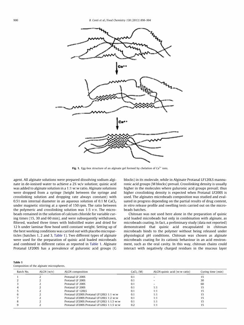
Fig. 1. Egg-box structure of an alginate gel formed by chelation of Ca2+ ions.
900 B. Conti et al. / Food Chemistry 138 (2013) 898–904
agent. All alginate solutions were prepared dissolving sodium algi-nate in de-ionised water to achieve a 2% w/v solution; quinic acidwas added to alginate solution in a 1:1 w:w ratio. Alginate solutionswere dropped from a syringe (height between the syringe andcrosslinking solution and dropping rate always constant) with0.51 mm internal diameter in an aqueous solution of 0.1 M CaCl2
under magnetic stirring at a speed of 150 rpm. The ratio betweenthe polymeric and crosslinking solution was 1:5 v:v. The micro-beads remained in the solution of calcium chloride for variable cur-ing times (15, 30 and 60 min), and were subsequently withdrawn,filtered, washed three times with bidistilled water and dried for12 h under laminar flow hood until constant weight. Setting up ofthe best working conditions was carried out with placebo micropar-ticles (batches 1, 2 and 3, Table 1). Two different types of alginatewere used for the preparation of quinic acid loaded microbeadsand combined in different ratios as reported in Table 1. AlginateProtanal LF200S has a prevalence of guluronic acid groups (G
Table 1Composition of the alginate microspheres.
Batch No. ALGN (w/v) ALGN composition
1 2 Protanal LF 200S2 2 Protanal LF 200S3 2 Protanal LF 200S4 2 Protanal LF 200S5 2 Protanal LF 120LS6 2 Protanal LF200S:Protanal LF120LS 1:1 w:w7 2 Protanal LF200S:Protanal LF120LS 1:2 w:w8 2 Protanal LF200S:Protanal LF120LS 1:1.5 w:w9 2 Protanal LF200S:Protanal LF120LS 1:1.5 w:w
blocks) in its molecule, while in Alginate Protanal LF120LS mannu-ronic acid groups (M blocks) prevail. Crosslinking density is usuallyhigher in the molecules where guluronic acid groups prevail; thushigher crosslinking density is expected when Protanal LF200S isused. The alginates microbeads composition was studied and eval-uated in progress depending on the partial results of drug content,in vitro release profile and swelling tests carried out on the micro-beads batches.
Chitosan was not used here alone in the preparation of quinicacid loaded microbeads but only in combination with alginate, asmicrobeads coating. In fact, a preliminary study (data not reported)demonstrated that quinic acid encapsulated in chitosanmicrobeads binds to the polymer without being released underphysiological pH conditions. Chitosan was chosen as alginatemicrobeads coating for its cationic behaviour in an acid environ-ment, such as the oral cavity. In this way, chitosan chains couldinteract with negatively charged residues in the mucous layer
CaCl2 (M) ALGN:quinic acid (w:w ratio) Curing time (min)
0.1 – 150.1 – 300.1 – 600.1 1:1 150.1 1:1 150.1 1:1 150.1 1:1 150.1 1:1 150.2 1:1 15
B. Conti et al. / Food Chemistry 138 (2013) 898–904 901
improving mucoadhesion relative to microspheres composed withalginate alone (Bravo-Osuna, Vauthier, Farabollini, Palmieri, &Ponchel, 2007; Draget & Taylor, 2011).
Alginate/chitosan microbeads were prepared by complex coac-ervation using sodium alginate as the gel core. The suitable algi-nate compositions, chosen on the basis of the results of alginatemicrobeads characterisation, were used in the preparation of algi-nate/chitosan microbeads. Two preparation protocols for alginate/chitosan microbeads were evaluated and further indicated as: (i)one-step preparation protocol, (ii) two-step preparation protocol.
One-step preparation protocol was performed by dropping the2% w/v alginate solution containing quinic acid in a 1:1 w:w ratiowith respect to alginate, into a 0.1 M or 0.2 M CaCl2 aqueous solu-tions in which 0.5% w/v of chitosan was dispersed. All other pro-cess conditions (height between the syringe and crosslinkingsolution, dropping rate, syringe inner diameter) were kept constantas explained above. The microbeads remained in the solution ofcalcium chloride for 15 min of curing time, and were subsequentlywithdrawn, filtered and washed three times with bidistilled waterand dried as described above.
A modification of the one-step protocol was performed by drop-ping the 2% w/v alginate/quinic acid solution (alginate:quinic acidratio 1:1 w:w) into a 1.3 M CaCl2 solution in acetic acid (1% w/v) inwhich 0.1% w/v chitosan was solubilised. The ratio between poly-meric and crosslinking solution was 1:3 v/v. All other processparameters were kept as explained above.
The two-step preparation protocol was performed by pouringthe already formed alginate microspheres into a 0.5% w/v chitosansolution in 1% w/v acetic acid solution. As described above, two dif-ferent CaCl2 concentrations were evaluated (0.1 and 0.2 M). Themicrospheres were soaked in the chitosan solution under agitationfor two hours. After this time the microspheres were withdrawn,filtered and washed three times with bidistilled water and driedas described above. Table 2 reports the composition of the algi-nate/chitosan microspheres. Microspheres without quinic acid(placebo) were prepared as control for each preparation protocolthat followed.
2.3. Scanning electron microscopy (SEM)
Shape, size and surface characteristics of the microspheres weredetermined by scanning electron microscopy. Samples of micro-beads of each batch were placed on carbon discs sample holders,and sputtered with Au/Pd in argon atmosphere under vacuum.The analyses were carried out using a Cambridge Stereoscan 200electronic microscope (Cambridge Scientific Instruments, Cam-bridge, UK) at different magnifications.
2.4. In vitro mucoadhesion and teeth adhesion tests
In vitro mucoadhesion test provides information on the in vivoadhesion of microbeads to buccal mucosa, thus it is important inthe development of mucoadhesive delivery systems. Several tests
Table 2Composition of the alginate/chitosan microspheres.
Batch No. Preparation method ALGNa:Quinic ac
10 One step –11 One step –12 Two steps –13 Two steps –14 One step (acid pH) –15 One step 1:1
a ALGN composition (w:v): Protanal LF200S:Protanal LF120LS 1:1.5 w:w (see batch 8
are available in the literature for in vitro mucoadhesion testing(Bonferoni, Rossi, Ferrari, & Caramella, 1999; Hombach &Bernkop-Schnürch, 2010; Kharenko et al., 2009; Takeuchi et al.,2005). The test was carried out on freshly excised porcine cheekmembrane as a model for human buccal mucosa, using a suitablymodified Franz cell as reported in the literature (Hombach &Bernkop-Schnürch, 2010). Briefly, the modified Franz cells wereset up with the freshly excised porcine membrane. A fixed numberof already hydrated microbeads was placed inside the cells andcovered with 5 ml of artificial saliva. The cells were fluxed withartificial saliva, at constant rate of 0.3 ml/min, and the micro-spheres recovered from the cells at scheduled times (20, 30, 45,60, 90, 120, 180, 240 min) were counted. The amount of adheredmicrospheres at the fixed times (Nt) expressed as percentage ofthe initial number of microspheres placed in the cell (N0) wascalculated from Eq. (1):
Nt ¼ N0NrN0� 100 ð1Þ
Nt1, Nt2, Nt3. . . = % of microbeads adhered at time, t2, t3, t4, . . . (20,30, 45, 90, 120, 180, 240 min), N0 = number of microbeads placedin the cell and initially adhered to the porcine membrane, Nr1,Nr2, Nr3. . . = cumulative number of microbeads recovered from thecell at times t1, t2, t3, t4.
Teeth are the target site of the anticaries compound loaded inthe microbeads. Thus adhesion to teeth should represent a suitableproperty of the microbeads. In vitro evaluation of adhesion to teethwas performed using hydroxyapatite (HA) discs. These are alreadystandardised systems known in the literature as teeth models(Faraj et al., 2007).
The procedure followed was the same as explained above forthe in vitro mucoadhesion test, where HA discs were used insteadof porcine excised mucosa. The amount of adhered microspheres atthe fixed times (Nt) expressed as percentage of the initial numberof microspheres placed in the cell (N0) was calculated from Eq. (1)as explained above.
All the tests were performed in triplicate for each microbeadsbatch.
2.5. Swelling evaluation
Microsphere swelling behaviour expresses polymer affinity forthe aqueous environment. This parameter changes depending onpolymer crosslinking density and it is important to evaluate be-cause swelling affects both microsphere adhesion and the releasebehaviour of active ingredient from the microbeads.
Swelling test was performed by soaking samples of 10 micro-spheres in simulated in vivo conditions represented by 5 ml of arti-ficial saliva (pH 5.7) at 35 �C. At scheduled times (30 s, 60 s, 2 min,10 min, 20 min, 1 h, 2 h, 4 h), the samples were withdrawn by fil-tration on a filter paper (VWR qualitative filter paper 417, size185 mm), the microsphere diameter was determined by analysis
id (w:w ratio) CaCl2 (M) Curing time (min)
0.1 150.2 150.1 15 + 1200.2 15 + 1201.3 150.1 15
) 2%.

902 B. Conti et al. / Food Chemistry 138 (2013) 898–904
by optical microscope and compared to the microsphere diameteranalysed before swelling.
Three samples of 10 microbeads for each batch underwentswelling test. Swelling was determined for each batch on the aver-age diameters with the following Eq. (2):
Swelling% ¼ dt � d0
d0� 100 ð2Þ
d0 = average microsphere diameter before swelling; dt = averagemicrosphere diameter after swelling at times t1, t2, t3, etc.
2.6. Evaluation of quinic acid encapsulation and in vitro release test
Microbeads quinic acid content was calculated as the differencebetween the initial amount of quinic acid to be loaded in themicrobeads, and the amount of unencapsulated quinic acid deter-mined by HPLC in the aqueous solutions coming from the prepara-tion process.
All experiments were performed using a 1100 Agilent HPLC sys-tem (Waldbronn, Germany) equipped with a gradient quaternarypump, a diode array detector (DAD), and an autosampler. TheAgilent Chemstation software was used for HPLC system controland data processing. The following experimental conditions wereused for quinic acid HPLC determination: stationary phase TSKgelcolumn Amide-80 (4.6 mm ID � 250 mm; Tosoh Bioscience, Tokyo,Japan), mobile phase 2.5 mM TEAP (pH 5.00 ± 0.02) in 75% v/vCH3CN, flow rate 0.5 ml/min, isocratic elution. UV spectra wererecorded in the 190–600 nm range, and chromatograms wereacquired at 210 nm.
Quantification of quinic acid was performed by the externalmethod using a six-point regression curve. Diluted 1:40 (75% v/vCH3CN) standard solutions were analysed by HPLC-DAD in therange 1–40 ppm.
The amount of quinic acid in the residual aqueous solutionsafter microbeads preparation was determined as explained above,and the amount of quinic acid encapsulated in the microbeads wascalculated from Eq. (3):
E:E:% ¼W1 �W2
W1� 100 ð3Þ
E.E. = encapsulation efficiency; W1 = initial weight (g) of quinic acidto be encapsulated in one microsphere batch; W2 = weight (g) ofunencapsulated quinic acid in the batch (from HPLC analysis)
The test was performed in triplicate for each batch ofmicrospheres.
In vitro release test was performed on samples of about 90 mgof quinic-acid-loaded microbeads for each batch. Each samplewas suspended in a vial containing 5 ml of artificial saliva, placedin a shaker incubator thermostated at 35 �C. Placebo microbeadswere also tested as control. At scheduled times (30 s, 1, 2, 10,20 min, 8 h), 1 ml of the artificial saliva was withdrawn from eachsample and replaced with fresh medium to keep the set-up volumeat 5 ml. The release medium (artificial saliva) withdrawn at thescheduled times was analysed by HPLC under the same conditionsreported here above for quinic acid content determination, todetermine the amount of active ingredient released at each sched-uled time. Quantification of quinic acid was performed by theexternal method using a six-point regression curve. 1:40 (75% v/vCH3CN) diluted saliva standard solutions were analysed by HPLC-DAD in the range 50–1000 ppm. The results were expressed as qui-nic acid percentage, with respect to the amount of quinic acidloaded in the microbeads.
3. Results and discussion
3.1. Characterisation of alginate microspheres
Analysis either by light microscope and by SEM highlighted thatthe sizes of dried microspheres (batches 1–9) were always be-tween 400 and 500 lm. Shape, as observed by SEM analysis, wasspherical and regular with smooth surfaces for all batches of algi-nate microspheres; no differences in microbeads morphology washighlighted between the batches irrespective of polymers compo-sition or CaCl2 concentration used (Fig. 2a and b).
Table 3 reports the results of alginate microspheres character-isation in terms of swelling percentage, encapsulation efficiencyand amounts of quinic acid released after 20 min and 4 h ofin vitro release test. Swelling behaviour was similar for all batchestested with the exception of batch 5 made of 100% LF120LS thatwas not stable and disintegrated after 2 min testing. The micro-spheres made of 100% LF200S (batch 4) demonstrated a goodencapsulation efficiency (77.5%), but they showed a very poor qui-nic acid release (12.8% after 20 min), because of the high crosslink-ing density of LF200S polymer. In order to improve quinic acidrelease percentage, different blends of the two alginates consid-ered were evaluated. In particular, alginate LF120LS was added toLF200S in different weight ratios (1:1, 1:2, 1:1.5), thus modifyingcrosslinking density of the polymeric matrix. Among the alginateblends assessed, either batch 6, made of Protanal LF200S:ProtanalLF120LS 1:1 w:w ratio, or batch 9, made of Protanal LF200S:Prot-anal LF120LS 1:1.5 w:w ratio with 0.2 M CaCl2, demonstrated highencapsulation efficiencies (48.2%, 92.2% respectively), but poorquinic acid release in the 4 h of in vitro release test. ProtanalLF200S:Protanal LF120LS 1:1.5 w:w ratio (batch 8) gave good re-sults both in terms of quinic acid encapsulation and release. Start-ing from these results, and in consideration of the extremely highencapsulation efficiency achieved for batch 9, the polymeric andcrosslinking agent composition of batches 8 and 9 were consideredsuitable to investigate the chitosan coating.
3.2. Characterisation of alginate/chitosan microspheres
Alginate/chitosan microspheres were prepared starting fromthe compositions of batches 8 and 9 and using two different chito-san coating methods, as reported in Table 2. Morphologic charac-terisation of the alginate/chitosan microspheres performed bylight microscope and scanning electron microscopy, showed thattheir size and shape were similar to that of alginate microbeads.However, chitosan-coated alginate microspheres presented arough surface (Fig. 2c and d) independently of the preparationmethod used.
Table 4 reports the results of mucoadhesion and dental adhe-sion expressed as % of microbeads still adhering on the mucosaland teeth surface after the scheduled times. The results of mucoad-hesion test showed that, generally speaking, the chitosan-coatedalginate microspheres prepared by the one-step method weremore mucoadhesive than those prepared by the two-step method.Batch 13, prepared by the two-step method and with higher cross-linking agent concentrations (0.2 M CaCl2), showed mucoadhesiveproperties even lower than the uncoated alginate microbeads (datanot reported). Among the microsphere batches prepared by theone-step method, batch 10, obtained in the presence of 0.1 M CaCl2
solution, presented the best mucoadhesive properties (about100%). The one-step method performed at acidic pH did not resultin microspheres with good mucoadhesive properties.
The obtained results suggest that the microspheres obtained bythe one-step preparation method, especially the microsphere com-position corresponding to batch 8, possess the best polymer muco-adhesive properties.

Fig. 2. Photomicrographs of: (a), (b) alginate microbeads (batch 8), (c) and (d) alginate/chitosan microbeads (batch 15) at different magnifications.
Table 3Results of alginate microbeads characterisation.
Batch No. Swelling (%) Encapsulation efficiency (%) Quinic acid release (%)
20 min 240 min 20 min 240 min
1 6.1 ± 5.4 6.1 ± 4.3 – – –2 4.1 ± 2.2 4.1 ± 2.2 – – –3 2.0 ± 1.0 2.0 ± 2.5 – – –4 6.3 ± 5.6 6.3 ± 4.9 77.5 ± 2.6 12.8 ± 2.8 19.3 ± 3.55 11.0 ± 2.3 N.D.* N.D.* N.D.* N.D.*
6 6.0 ± 1.2 6.1 ± 3.5 48.2 ± 5.6 34.1 ± 1.5 36.8 ± 1.07 6.4 ± 2.3 6.4 ± 1.9 13.8 ± 0.2 79.3 ± 4.8 85.2 ± 4.88 6.0 ± 1.2 6.0 ± 1.1 58.3 ± 6.6 38.6 ± 0.2 56.0 ± 0.19 5.9 ± 5.2 5.9 ± 4.1 92.2 ± 1.3 14.1 ± 1.5 14.2 ± 1.7
* N.D.: not detectable.
Table 4Results of alginate/chitosan microspheres characterisation.
Batch No. Swelling (%) Mucoadhesion (%) HA adhesion (%)
20 min 240 min 20 min 240 min 20 min 240 min
10 11.8 ± 2 13.4 ± 1 100.0 ± 0 100.0 ± 0 96.7 ± 6 90.0 ± 1011 8.5 ± 1 10.0 ± 1 98.0 ± 1 97.3 ± 0 90.7 ± 6 81.3 ± 712 8.0 ± 2 9.0 ± 2 96.7 ± 6 90.0 ± 10 93.4 ± 6 53.3 ± 1513 8.2 ± 2 9.5 ± 2 100.0 ± 0 66.9 ± 5 86.7 ± 5 53.3 ± 1514 10.8 ± 5 12.8 ± 5 90.0 ± 14 65.0 ± 7 100.0 ± 0 83.3 ± 615 11.4 ± 3 14.0 ± 3 100.0 ± 0 100.0 ± 0 96.7 ± 11 90.0 ± 10
B. Conti et al. / Food Chemistry 138 (2013) 898–904 903
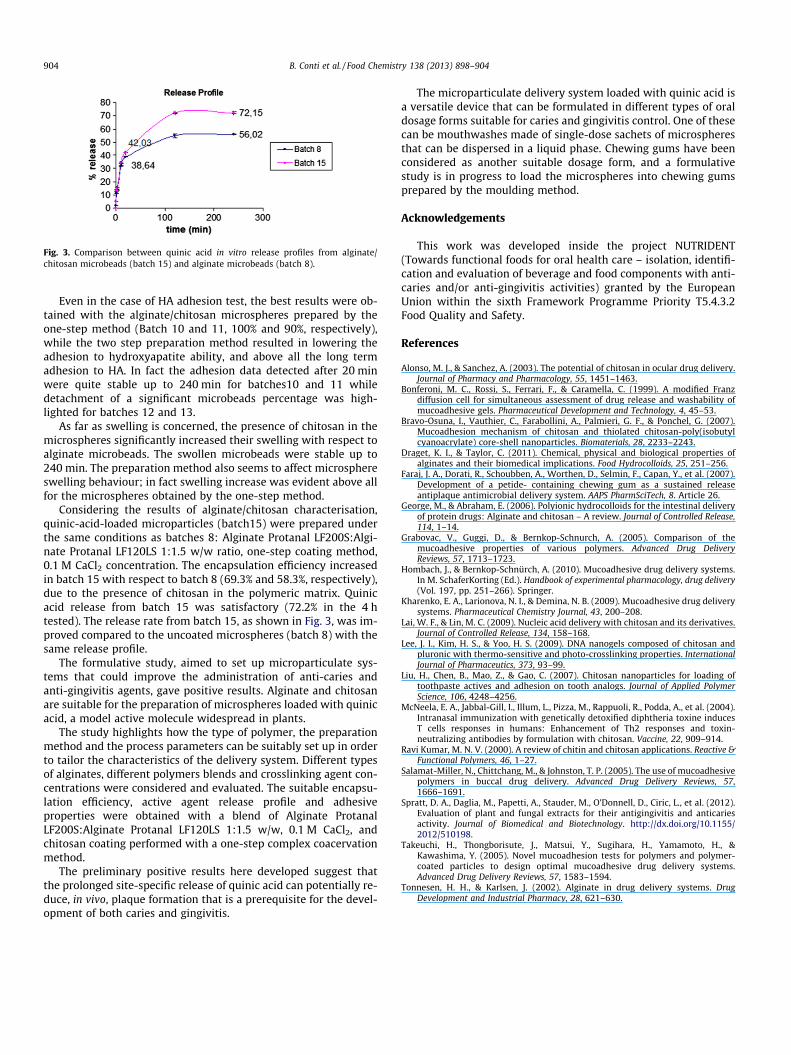
Fig. 3. Comparison between quinic acid in vitro release profiles from alginate/chitosan microbeads (batch 15) and alginate microbeads (batch 8).
904 B. Conti et al. / Food Chemistry 138 (2013) 898–904
Even in the case of HA adhesion test, the best results were ob-tained with the alginate/chitosan microspheres prepared by theone-step method (Batch 10 and 11, 100% and 90%, respectively),while the two step preparation method resulted in lowering theadhesion to hydroxyapatite ability, and above all the long termadhesion to HA. In fact the adhesion data detected after 20 minwere quite stable up to 240 min for batches10 and 11 whiledetachment of a significant microbeads percentage was high-lighted for batches 12 and 13.
As far as swelling is concerned, the presence of chitosan in themicrospheres significantly increased their swelling with respect toalginate microbeads. The swollen microbeads were stable up to240 min. The preparation method also seems to affect microsphereswelling behaviour; in fact swelling increase was evident above allfor the microspheres obtained by the one-step method.
Considering the results of alginate/chitosan characterisation,quinic-acid-loaded microparticles (batch15) were prepared underthe same conditions as batches 8: Alginate Protanal LF200S:Algi-nate Protanal LF120LS 1:1.5 w/w ratio, one-step coating method,0.1 M CaCl2 concentration. The encapsulation efficiency increasedin batch 15 with respect to batch 8 (69.3% and 58.3%, respectively),due to the presence of chitosan in the polymeric matrix. Quinicacid release from batch 15 was satisfactory (72.2% in the 4 htested). The release rate from batch 15, as shown in Fig. 3, was im-proved compared to the uncoated microspheres (batch 8) with thesame release profile.
The formulative study, aimed to set up microparticulate sys-tems that could improve the administration of anti-caries andanti-gingivitis agents, gave positive results. Alginate and chitosanare suitable for the preparation of microspheres loaded with quinicacid, a model active molecule widespread in plants.
The study highlights how the type of polymer, the preparationmethod and the process parameters can be suitably set up in orderto tailor the characteristics of the delivery system. Different typesof alginates, different polymers blends and crosslinking agent con-centrations were considered and evaluated. The suitable encapsu-lation efficiency, active agent release profile and adhesiveproperties were obtained with a blend of Alginate ProtanalLF200S:Alginate Protanal LF120LS 1:1.5 w/w, 0.1 M CaCl2, andchitosan coating performed with a one-step complex coacervationmethod.
The preliminary positive results here developed suggest thatthe prolonged site-specific release of quinic acid can potentially re-duce, in vivo, plaque formation that is a prerequisite for the devel-opment of both caries and gingivitis.
The microparticulate delivery system loaded with quinic acid isa versatile device that can be formulated in different types of oraldosage forms suitable for caries and gingivitis control. One of thesecan be mouthwashes made of single-dose sachets of microspheresthat can be dispersed in a liquid phase. Chewing gums have beenconsidered as another suitable dosage form, and a formulativestudy is in progress to load the microspheres into chewing gumsprepared by the moulding method.
Acknowledgements
This work was developed inside the project NUTRIDENT(Towards functional foods for oral health care – isolation, identifi-cation and evaluation of beverage and food components with anti-caries and/or anti-gingivitis activities) granted by the EuropeanUnion within the sixth Framework Programme Priority T5.4.3.2Food Quality and Safety.
References
Alonso, M. J., & Sanchez, A. (2003). The potential of chitosan in ocular drug delivery.Journal of Pharmacy and Pharmacology, 55, 1451–1463.
Bonferoni, M. C., Rossi, S., Ferrari, F., & Caramella, C. (1999). A modified Franzdiffusion cell for simultaneous assessment of drug release and washability ofmucoadhesive gels. Pharmaceutical Development and Technology, 4, 45–53.
Bravo-Osuna, I., Vauthier, C., Farabollini, A., Palmieri, G. F., & Ponchel, G. (2007).Mucoadhesion mechanism of chitosan and thiolated chitosan-poly(isobutylcyanoacrylate) core-shell nanoparticles. Biomaterials, 28, 2233–2243.
Draget, K. I., & Taylor, C. (2011). Chemical, physical and biological properties ofalginates and their biomedical implications. Food Hydrocolloids, 25, 251–256.
Faraj, J. A., Dorati, R., Schoubben, A., Worthen, D., Selmin, F., Capan, Y., et al. (2007).Development of a petide- containing chewing gum as a sustained releaseantiplaque antimicrobial delivery system. AAPS PharmSciTech, 8. Article 26.
George, M., & Abraham, E. (2006). Polyionic hydrocolloids for the intestinal deliveryof protein drugs: Alginate and chitosan – A review. Journal of Controlled Release,114, 1–14.
Grabovac, V., Guggi, D., & Bernkop-Schnurch, A. (2005). Comparison of themucoadhesive properties of various polymers. Advanced Drug DeliveryReviews, 57, 1713–1723.
Hombach, J., & Bernkop-Schnürch, A. (2010). Mucoadhesive drug delivery systems.In M. SchaferKorting (Ed.). Handbook of experimental pharmacology, drug delivery(Vol. 197, pp. 251–266). Springer.
Kharenko, E. A., Larionova, N. I., & Demina, N. B. (2009). Mucoadhesive drug deliverysystems. Pharmaceutical Chemistry Journal, 43, 200–208.
Lai, W. F., & Lin, M. C. (2009). Nucleic acid delivery with chitosan and its derivatives.Journal of Controlled Release, 134, 158–168.
Lee, J. I., Kim, H. S., & Yoo, H. S. (2009). DNA nanogels composed of chitosan andpluronic with thermo-sensitive and photo-crosslinking properties. InternationalJournal of Pharmaceutics, 373, 93–99.
Liu, H., Chen, B., Mao, Z., & Gao, C. (2007). Chitosan nanoparticles for loading oftoothpaste actives and adhesion on tooth analogs. Journal of Applied PolymerScience, 106, 4248–4256.
McNeela, E. A., Jabbal-Gill, I., Illum, L., Pizza, M., Rappuoli, R., Podda, A., et al. (2004).Intranasal immunization with genetically detoxified diphtheria toxine inducesT cells responses in humans: Enhancement of Th2 responses and toxin-neutralizing antibodies by formulation with chitosan. Vaccine, 22, 909–914.
Ravi Kumar, M. N. V. (2000). A review of chitin and chitosan applications. Reactive &Functional Polymers, 46, 1–27.
Salamat-Miller, N., Chittchang, M., & Johnston, T. P. (2005). The use of mucoadhesivepolymers in buccal drug delivery. Advanced Drug Delivery Reviews, 57,1666–1691.
Spratt, D. A., Daglia, M., Papetti, A., Stauder, M., O’Donnell, D., Ciric, L., et al. (2012).Evaluation of plant and fungal extracts for their antigingivitis and anticariesactivity. Journal of Biomedical and Biotechnology. http://dx.doi.org/10.1155/2012/510198.
Takeuchi, H., Thongborisute, J., Matsui, Y., Sugihara, H., Yamamoto, H., &Kawashima, Y. (2005). Novel mucoadhesion tests for polymers and polymer-coated particles to design optimal mucoadhesive drug delivery systems.Advanced Drug Delivery Reviews, 57, 1583–1594.
Tonnesen, H. H., & Karlsen, J. (2002). Alginate in drug delivery systems. DrugDevelopment and Industrial Pharmacy, 28, 621–630.