Adhesion and Proliferation of Human Mesenchymal Stem Cells from 2 Dental Pulp on Porous Silicon...
10
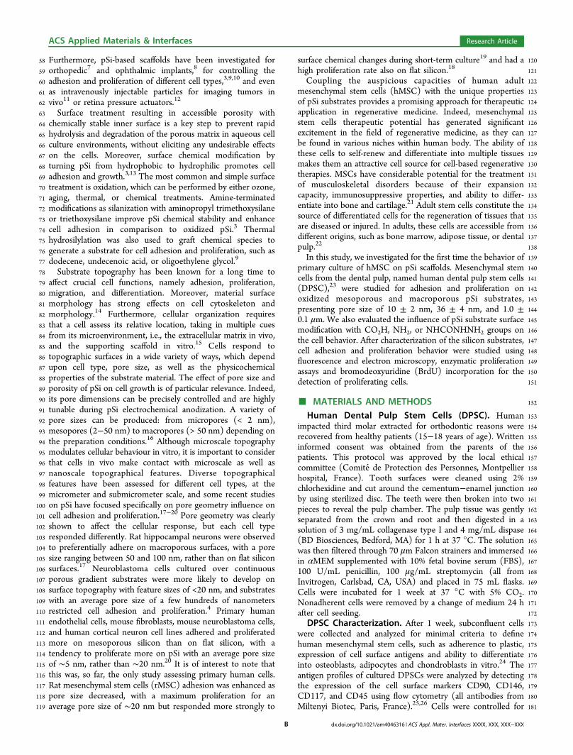
1 Adhesion and Proliferation of Human Mesenchymal Stem Cells from 2 Dental Pulp on Porous Silicon Scaolds 3 Pierre-Yves Collart-Dutilleul, † Emilie Secret, ‡ Ivan Panayotov, † Dominique Deville de Pe ́ rie ̀ re, † 4 Raú l J. Martín-Palma, § Vicente Torres-Costa, § Marta Martin, ⊥, Csilla Gergely, ⊥, Jean-Olivier Durand, ‡ 5 Fre ́ de ́ rique Cunin, ‡ and Fre ́ de ́ ric J. Cuisinier* ,† 6 † BioNano Laboratory EA 4203, Montpellier 1 University, Montpellier, France 7 ‡ Institut Charles Gerhardt Montpellier, UMR-5253 CNRS-UM2-ENSCM-UM1, Ecole Nationale Supé rieure de Chimie de 8 Montpellier, 34296 Montpellier, France 9 § Departamento de Física Aplicada, Universidad Autó noma de Madrid, 28049 Cantoblanco, Madrid, Spain 10 ⊥ Universite ́ Montpellier 2, UMR 5221 Laboratoire Charles Coulomb, Montpellier, France 11 CNRS, Laboratoire Charles Coulomb UMR 5221, Montpellier, France 12 * S Supporting Information 13 ABSTRACT: In regenerative medicine, stem-cell-based therapy often 14 requires a scaold to deliver cells and/or growth factors to the injured 15 site. Porous silicon (pSi) is a promising biomaterial for tissue engineering as 16 it is both nontoxic and bioresorbable. Moreover, surface modication can 17 oer control over the degradation rate of pSi and can also promote cell 18 adhesion. Dental pulp stem cells (DPSC) are pluripotent mesenchymal stem 19 cells found within the teeth and constitute a readily source of stem cells. 20 Thus, coupling the good proliferation and dierentiation capacities of DPSC 21 with the textural and chemical properties of the pSi substrates provides an 22 interesting approach for therapeutic use. In this study, the behavior of 23 human DPSC is analyzed on pSi substrates presenting pores of various sizes, 24 10 ± 2 nm, 36 ± 4 nm, and 1.0 ± 0.1 m, and undergoing dierent chemical 25 treatments, thermal oxidation, silanization with aminopropyltriethoxysilane 26 (APTES), and hydrosilylation with undecenoic acid or semicarbazide. DPSC 27 adhesion and proliferation were followed for up to 72 h by uorescence microscopy, scanning electron microscopy (SEM), 28 enzymatic activity assay, and BrdU assay for mitotic activity. Porous silicon with 36 nm pore size was found to oer the best 29 adhesion and the fastest growth rate for DPSC compared to pSi comporting smaller pore size (10 nm) or larger pore size (1 30 m), especially after silanization with APTES. Hydrosilylation with semicarbazide favored cell adhesion and proliferation, 31 especially mitosis after cell adhesion, but such chemical modication has been found to led to a scaold that is stable for only 32 24-48 h in culture medium. Thus, semicarbazide-treated pSi appeared to be an appropriate scaold for stem cell adhesion and 33 immediate in vivo transplantation, whereas APTES-treated pSi was found to be more suitable for long-term in vitro culture, for 34 stem cell proliferation and dierentiation. 35 KEYWORDS: porous silicon, dental pulp stem cells, cell adhesion, surface functionalization, tissue engineering 36 ■ INTRODUCTION 37 In regenerative medicine, tissue engineering applications are 38 based on the development of biological substitutes that can 39 restore, maintain or even improve tissue functions: tissue 40 engineering combines cells and bioactive factors in a dened 41 microenvironment created by a biomaterial scaold. A key 42 component for tissue engineering is the biomaterial scaold 43 that, ideally, should support cell attachment, proliferation and 44 dierentiation, and be biocompatible and biodegradable at a 45 controlled rate. 1 Stem-cell-based therapy often requires a 46 scaold to carry cells to the injured site. Porous silicon (pSi) 47 appears to be a promising biomaterial for tissue engineering as 48 it is both nontoxic and bioresorbable under physiological 49 conditions and dissolves progressively into nontoxic silicic 50 acid. 2 Its dissolution rate is dependent on the pore geometry 51 and surface chemical properties, and these two factors inuence 52 cell adhesion. 3,4 Moreover, this tunable, biocompatible, and 53 resorbable material has been reported to favor the growth of 54 hydroxyapatite, suggesting the possible bone implantability of 55 the material. 5 Its biocompatibility and immunogenicity has 56 already been demonstrated under dierent conditions and it 57 additionally oers useful photoluminescence properties. 6 Received: October 21, 2013 Accepted: January 16, 2014 Research Article www.acsami.org © XXXX American Chemical Society A dx.doi.org/10.1021/am4046316 | ACS Appl. Mater. Interfaces XXXX, XXX, XXX-XXX clp00 | ACSJCA | JCA10.0.1465/W Unicode | research.3f (R3.6.i4 HF01:4180 | 2.0 alpha 39) 2013/10/21 02:46:00 | PROD-JCAVA | rq_3159649 | 1/27/2014 11:50:12 | 10 | JCA-DEFAULT
-
Upload
univ-montp1 -
Category
Documents
-
view
2 -
download
0
Transcript of Adhesion and Proliferation of Human Mesenchymal Stem Cells from 2 Dental Pulp on Porous Silicon...
1 Adhesion and Proliferation of Human Mesenchymal Stem Cells from2 Dental Pulp on Porous Silicon Sca!olds3 Pierre-Yves Collart-Dutilleul,† Emilie Secret,‡ Ivan Panayotov,† Dominique Deville de Pe !rie "re,†4 Rau !l J. Martín-Palma,§ Vicente Torres-Costa,§ Marta Martin,!," Csilla Gergely,!," Jean-Olivier Durand,‡
5 Fre !de !rique Cunin,‡ and Fre !de !ric J. Cuisinier*,†
6†BioNano Laboratory EA 4203, Montpellier 1 University, Montpellier, France
7‡Institut Charles Gerhardt Montpellier, UMR-5253 CNRS-UM2-ENSCM-UM1, Ecole Nationale Supe !rieure de Chimie de
8 Montpellier, 34296 Montpellier, France9§Departamento de Física Aplicada, Universidad Auto !noma de Madrid, 28049 Cantoblanco, Madrid, Spain
10!Universite ! Montpellier 2, UMR 5221 Laboratoire Charles Coulomb, Montpellier, France
11"CNRS, Laboratoire Charles Coulomb UMR 5221, Montpellier, France
12 *S Supporting Information
13 ABSTRACT: In regenerative medicine, stem-cell-based therapy often14 requires a sca!old to deliver cells and/or growth factors to the injured15 site. Porous silicon (pSi) is a promising biomaterial for tissue engineering as16 it is both nontoxic and bioresorbable. Moreover, surface modi"cation can17 o!er control over the degradation rate of pSi and can also promote cell18 adhesion. Dental pulp stem cells (DPSC) are pluripotent mesenchymal stem19 cells found within the teeth and constitute a readily source of stem cells.20 Thus, coupling the good proliferation and di!erentiation capacities of DPSC21 with the textural and chemical properties of the pSi substrates provides an22 interesting approach for therapeutic use. In this study, the behavior of23 human DPSC is analyzed on pSi substrates presenting pores of various sizes,24 10 ± 2 nm, 36 ± 4 nm, and 1.0 ± 0.1 !m, and undergoing di!erent chemical25 treatments, thermal oxidation, silanization with aminopropyltriethoxysilane26 (APTES), and hydrosilylation with undecenoic acid or semicarbazide. DPSC27 adhesion and proliferation were followed for up to 72 h by #uorescence microscopy, scanning electron microscopy (SEM),28 enzymatic activity assay, and BrdU assay for mitotic activity. Porous silicon with 36 nm pore size was found to o!er the best29 adhesion and the fastest growth rate for DPSC compared to pSi comporting smaller pore size (10 nm) or larger pore size (130 !m), especially after silanization with APTES. Hydrosilylation with semicarbazide favored cell adhesion and proliferation,31 especially mitosis after cell adhesion, but such chemical modi"cation has been found to led to a sca!old that is stable for only32 24#48 h in culture medium. Thus, semicarbazide-treated pSi appeared to be an appropriate sca!old for stem cell adhesion and33 immediate in vivo transplantation, whereas APTES-treated pSi was found to be more suitable for long-term in vitro culture, for34 stem cell proliferation and di!erentiation.35 KEYWORDS: porous silicon, dental pulp stem cells, cell adhesion, surface functionalization, tissue engineering
36 ! INTRODUCTION
37 In regenerative medicine, tissue engineering applications are38 based on the development of biological substitutes that can39 restore, maintain or even improve tissue functions: tissue40 engineering combines cells and bioactive factors in a de"ned41 microenvironment created by a biomaterial sca!old. A key42 component for tissue engineering is the biomaterial sca!old43 that, ideally, should support cell attachment, proliferation and44 di!erentiation, and be biocompatible and biodegradable at a45 controlled rate.1 Stem-cell-based therapy often requires a46 sca!old to carry cells to the injured site. Porous silicon (pSi)47 appears to be a promising biomaterial for tissue engineering as48 it is both nontoxic and bioresorbable under physiological
49conditions and dissolves progressively into nontoxic silicic50acid.2 Its dissolution rate is dependent on the pore geometry51and surface chemical properties, and these two factors in#uence52cell adhesion.3,4 Moreover, this tunable, biocompatible, and53resorbable material has been reported to favor the growth of54hydroxyapatite, suggesting the possible bone implantability of55the material.5 Its biocompatibility and immunogenicity has56already been demonstrated under di!erent conditions and it57additionally o!ers useful photoluminescence properties.6
Received: October 21, 2013Accepted: January 16, 2014
Research Article
www.acsami.org
© XXXX American Chemical Society A dx.doi.org/10.1021/am4046316 | ACS Appl. Mater. Interfaces XXXX, XXX, XXX#XXX
clp00 | ACSJCA | JCA10.0.1465/W Unicode | research.3f (R3.6.i4 HF01:4180 | 2.0 alpha 39) 2013/10/21 02:46:00 | PROD-JCAVA | rq_3159649 | 1/27/2014 11:50:12 | 10 | JCA-DEFAULT
58 Furthermore, pSi-based sca!olds have been investigated for59 orthopedic7 and ophthalmic implants,8 for controlling the60 adhesion and proliferation of di!erent cell types,3,9,10 and even61 as intravenously injectable particles for imaging tumors in62 vivo11 or retina pressure actuators.12
63 Surface treatment resulting in accessible porosity with64 chemically stable inner surface is a key step to prevent rapid65 hydrolysis and degradation of the porous matrix in aqueous cell66 culture environments, without eliciting any undesirable e!ects67 on the cells. Moreover, surface chemical modi"cation by68 turning pSi from hydrophobic to hydrophilic promotes cell69 adhesion and growth.3,13 The most common and simple surface70 treatment is oxidation, which can be performed by either ozone,71 aging, thermal, or chemical treatments. Amine-terminated72 modi"cations as silanization with aminopropyl trimethoxysilane73 or triethoxysilane improve pSi chemical stability and enhance74 cell adhesion in comparison to oxidized pSi.3 Thermal75 hydrosilylation was also used to graft chemical species to76 generate a substrate for cell adhesion and proliferation, such as77 dodecene, undecenoic acid, or oligoethylene glycol.9
78 Substrate topography has been known for a long time to79 a!ect crucial cell functions, namely adhesion, proliferation,80 migration, and di!erentiation. Moreover, material surface81 morphology has strong e!ects on cell cytoskeleton and82 morphology.14 Furthermore, cellular organization requires83 that a cell assess its relative location, taking in multiple cues84 from its microenvironment, i.e., the extracellular matrix in vivo,85 and the supporting sca!old in vitro.15 Cells respond to86 topographic surfaces in a wide variety of ways, which depend87 upon cell type, pore size, as well as the physicochemical88 properties of the substrate material. The e!ect of pore size and89 porosity of pSi on cell growth is of particular relevance. Indeed,90 its pore dimensions can be precisely controlled and are highly91 tunable during pSi electrochemical anodization. A variety of92 pore sizes can be produced: from micropores (< 2 nm),93 mesopores (2#50 nm) to macropores (> 50 nm) depending on94 the preparation conditions.16 Although microscale topography95 modulates cellular behaviour in vitro, it is important to consider96 that cells in vivo make contact with microscale as well as97 nanoscale topographical features. Diverse topographical98 features have been assessed for di!erent cell types, at the99 micrometer and submicrometer scale, and some recent studies100 on pSi have focused speci"cally on pore geometry in#uence on101 cell adhesion and proliferation.17#20 Pore geometry was clearly102 shown to a!ect the cellular response, but each cell type103 responded di!erently. Rat hippocampal neurons were observed104 to preferentially adhere on macroporous surfaces, with a pore105 size ranging between 50 and 100 nm, rather than on #at silicon106 surfaces.17 Neuroblastoma cells cultured over continuous107 porous gradient substrates were more likely to develop on108 surface topography with feature sizes of <20 nm, and substrates109 with an average pore size of a few hundreds of nanometers110 restricted cell adhesion and proliferation.4 Primary human111 endothelial cells, mouse "broblasts, mouse neuroblastoma cells,112 and human cortical neuron cell lines adhered and proliferated113 more on mesoporous silicon than on #at silicon, with a114 tendency to proliferate more on pSi with an average pore size115 of $5 nm, rather than $20 nm.20 It is of interest to note that116 this was, so far, the only study assessing primary human cells.117 Rat mesenchymal stem cells (rMSC) adhesion was enhanced as118 pore size decreased, with a maximum proliferation for an119 average pore size of $20 nm but responded more strongly to
120surface chemical changes during short-term culture19 and had a121high proliferation rate also on #at silicon.18
122Coupling the auspicious capacities of human adult123mesenchymal stem cells (hMSC) with the unique properties124of pSi substrates provides a promising approach for therapeutic125application in regenerative medicine. Indeed, mesenchymal126stem cells therapeutic potential has generated signi"cant127excitement in the "eld of regenerative medicine, as they can128be found in various niches within human body. The ability of129these cells to self-renew and di!erentiate into multiple tissues130makes them an attractive cell source for cell-based regenerative131therapies. MSCs have considerable potential for the treatment132of musculoskeletal disorders because of their expansion133capacity, immunosuppressive properties, and ability to di!er-134entiate into bone and cartilage.21 Adult stem cells constitute the135source of di!erentiated cells for the regeneration of tissues that136are diseased or injured. In adults, these cells are accessible from137di!erent origins, such as bone marrow, adipose tissue, or dental138pulp.22
139In this study, we investigated for the "rst time the behavior of140primary culture of hMSC on pSi sca!olds. Mesenchymal stem141cells from the dental pulp, named human dental pulp stem cells142(DPSC),23 were studied for adhesion and proliferation on143oxidized mesoporous and macroporous pSi substrates,144presenting pore size of 10 ± 2 nm, 36 ± 4 nm, and 1.0 ±1450.1 !m. We also evaluated the in#uence of pSi substrate surface146modi"cation with CO2H, NH2, or NHCONHNH2 groups on147the cell behavior. After characterization of the silicon substrates,148cell adhesion and proliferation behavior were studied using149#uorescence and electron microscopy, enzymatic proliferation150assays and bromodeoxyuridine (BrdU) incorporation for the151detection of proliferating cells.
152! MATERIALS AND METHODS153Human Dental Pulp Stem Cells (DPSC). Human154impacted third molar extracted for orthodontic reasons were155recovered from healthy patients (15#18 years of age). Written156informed consent was obtained from the parents of the157patients. This protocol was approved by the local ethical158committee (Comite ! de Protection des Personnes, Montpellier159hospital, France). Tooth surfaces were cleaned using 2%160chlorhexidine and cut around the cementum#enamel junction161by using sterilized disc. The teeth were then broken into two162pieces to reveal the pulp chamber. The pulp tissue was gently163separated from the crown and root and then digested in a164solution of 3 mg/mL collagenase type I and 4 mg/mL dispase165(BD Biosciences, Bedford, MA) for 1 h at 37 °C. The solution166was then "ltered through 70 !m Falcon strainers and immersed167in "MEM supplemented with 10% fetal bovine serum (FBS),168100 U/mL penicillin, 100 !g/mL streptomycin (all from169Invitrogen, Carlsbad, CA, USA) and placed in 75 mL #asks.170Cells were incubated for 1 week at 37 °C with 5% CO2.171Nonadherent cells were removed by a change of medium 24 h172after cell seeding.173DPSC Characterization. After 1 week, subcon#uent cells174were collected and analyzed for minimal criteria to de"ne175human mesenchymal stem cells, such as adherence to plastic,176expression of cell surface antigens and ability to di!erentiate177into osteoblasts, adipocytes and chondroblasts in vitro.24 The178antigen pro"les of cultured DPSCs were analyzed by detecting179the expression of the cell surface markers CD90, CD146,180CD117, and CD45 using #ow cytometry (all antibodies from181Miltenyi Biotec, Paris, France).25,26 Cells were controlled for
ACS Applied Materials & Interfaces Research Article
dx.doi.org/10.1021/am4046316 | ACS Appl. Mater. Interfaces XXXX, XXX, XXX#XXXB
182 pluripotency with in vitro osteogenic, adipogenic and183 chondrogenic di!erentiation following a previously described184 protocol.27
185 Porous Silicon (pSi) Sca!old Preparation. Silicon wafers186 were obtained from Siltronix (Siltronix, Archamps, France). P+187 + type boron-doped crystalline silicon wafers with 0.0008 -188 0.0012 #cm resistivity were obtained from Siltronix. Wafers189 were etched in a custom-made Te#on cell at a constant current190 density of either 30 mA/cm2 for 10 min or 300 mA/cm2 for 2191 min 15 s, in a hydro#uoric acid (HF) solution in ethanol (3:1192 HF:ethanol solution, volume ratio). To create pSi with larger193 pores (1 !m), p-type !100" wafers with 7 - 21 #cm resistivity194 were etched at a constant current density of 4.5 mA/cm2 for 10195 min, in a 50% HF solution in dimethylformamide (DMF,196 Sigma, St Louis, MO, USA). Etched wafers were oxidized at197 800°C for 1 h. The wafers were cut into 0.5 cm2 pieces. Some198 of the samples etched in a 300 mA/cm2 current were also199 submitted to various chemical treatments: silanization with200 aminopropyltriethoxysilane (APTES) after thermal oxidation201 (as described above), hydrosilylation with undecenoic acid202 without pre-oxidation treatment, and hydrosilylation with203 semicarbazide without pre-oxidation treatment (all reagents204 from Sigma, St Louis, MO, USA). PSi surface modi"cations205 with APTES, and with undecenoic acid were described206 elsewhere.3,9 The covalent attachment of the semicarbazide to207 hydrogen-terminated pSi surfaces by thermal hydrosilylation208 was realized according to recently published procedure using209 tert-butyl-2[(allylamino)carbonyl]hydrazine carboxylate.28 The210 removal of the protecting group yields a semicarbazide-211 terminated monolayer.29 For use in cell culture, the wafers212 were sterilized with 70% ethanol (volume ratio) for 10 min213 before drying under sterile air#ow.214 Surface Characterization. The topography of the surface215 modi"ed pSi samples was analyzed by environmental scanning216 electron microscopy (SEM) (Analytic FEI Quanta FEG 200) to217 determine the pore size, and by atomic force microscopy218 (AFM) (Asylum MFP-3D, Asylum Research, Santa Barbara,219 CA) to determine the surface roughness. SEM images were220 treated and analyzed using imageJ® software to measure the221 mean diameter of the pores visible at the surface. For SEM, an222 acceleration voltage of 20.00 kV was used in a pressure of 0.5223 Torr. For AFM, gold-coated silicon nitride rectangular224 cantilevers were used with a typical spring constant of 30225 pN/nm#1, tip radius $30 nm (BL-RC150 VB-C1: Bio-Lever A,226 Olympus Optical Co., Ltd., Tokyo, Japan). The spring constant227 for each cantilever was determined by thermal noise method228 within the supplied software. Height images were recorded in229 tapping mode in liquid at room temperature. Typically, 512 !230 512 point scans were taken at a scan rate of 1 Hz per line.231 Surface tension was determined using sessile drop contact232 angle measurements, conducted in a custom-made set-up233 consisting of a syringe dispenser, a sample stage, a macro lens234 and a CCD camera. Five microliters of Milli-Q water was235 spotted onto the surface at room temperature. Images of the236 drop pro"les were captured and ImageJ software was used to237 measure contact angles on both sides of the droplet. A238 minimum of four replications was conducted for each sample239 surface.240 Cell Adhesion. DPSC attachment was monitored by241 #uorescence microscopy. Cells were seeded onto the surface242 of sterilized pSi at a cell density of 5 ! 104 cell/mL. Flat silicon243 (non etched silicon wafers) and glass coverslip were used as244 controls. Cells were incubated for 4, 24, 48, and 72 h at 37 °C
245with 5% CO2, in a humidi"ed incubator, in ultra low adherence24624-well plates (Corning, NY, USA) that inhibit cell attachment247on the tissue culture plate, allowing DPSC to attach only to the248pSi. After the incubation time, the cells were "xed with 2.5%249glutaraldehyde and stained with 50 !g/mL of 4,6-diamidino-2-250phenylindole (DAPI, Sigma, St Louis, MO, USA) for 30 min251before being washed with phosphate bu!er saline (PBS;252Invitrogen, Carlsbad, CA, USA) to remove any non-adherent253cells. Cells were observed under #uorescence microscopy at an254excitation wavelength of 290 nm. Controls were cells cultured255on glass coverslip and #at silicon, cut into 0.5 cm2 squares. Cell256counts were conducted at "ve di!erent locations on the surface257of each sample (four peripheral and one central) in areas of2581400 !m ! 1050 !m. the number of viable cells for each259experimental condition was counted and represented on a260linear graph. The doubling time (DT) was determined from the261growth curves and by using the formula
= ! !t t N NDT ( )log 2/(log log )0 0
262where t and t0 were the times at which the cells were counted,263and N and N0 were the cell numbers at times t and t0,264respectively.30
265Proliferation Assays. Cell proliferation was "rst measured266via quanti"cation of acid phosphatase activity using para-267nitrophenylphosphate phosphatase test (pNPP).31 By assessing268acid phosphatase activity, we considered the physiological269activity of the attached cells. Indeed, after adhesion, cells270require restarting their physiological activity and pNPP assay271has been shown to be simple, sensitive and convenient to detect272lysosomal enzyme activity.32 DPSC proliferation was assessed273after 4, 24, 48, and 72 h incubation. At the end of each274experimental time, the cells were washed three times with PBS275and lysed with 500 mL of the acid phosphatase lysis bu!er (0.1276M sodium acetate, 0.1% Triton X-100, pH 5.5), supplemented277with 1 mg/mL of pNPP (all products from Sigma, St Louis,278MO, USA). After 1 h incubation at 37 °C, the reaction was279stopped by the addition of 10 !L of 1 N NaOH (Sigma, St280Louis, MO, USA). The supernatant was removed and placed in281a new 24-well plate and the yellow colorimetric reaction was282measured by a microtiter plate reader at 405 nm. All283proliferation experiments were performed at least in triplicate284and results were normalized according to the glass coverslip285control at 72 h, for which 100% of proliferation was attributed.286The proliferation rate of DPSCs on pSi was also assessed by287bromodeoxyuridine (BrdU, Invitrogen, Carlsbad, CA, USA)288incorporation for 24 h.33 DPSC were seeded on the various pSi289sca!olds at a density of 105 cells/ml in "MEM supplemented290with 10% FBS with 1:100 diluted BrdU labeling (Invitrogen,291Carlsbad, CA, USA). After 24 h incubation at 37°C, samples292were rinsed 3 times in PBS, "xed in 4% paraformaldehyde,293rinsed again 3 times in PBS and put in 1.5M HCl (Sigma, St294Louis, MO, USA) for 30 min at room temperature to dissociate295DNA strands. Samples were washed with PBS, incubated in296PBS with 0.1% Triton X-100 and 1% BSA for 1 h at room297temperature, then incubated with mouse anti-BrdU primary298antibody (Milteny) overnight at 4 °C. After immunostaining,299cells were washed with PBS/1% BSA, incubated with FITC300conjugated rabbit anti-mouse secondary antibody for 30 min301and counterstained with 2!g/mL Hoeschst 33342 (Sigma, St302Louis, MO, USA) for nucleus staining. Samples were observed303under #uorescence microscopy at an excitation wavelength of304290 nm for nuclei (blue staining) and 490 nm for BrdU (green305staining). All experiments were performed at least in triplicate
ACS Applied Materials & Interfaces Research Article
dx.doi.org/10.1021/am4046316 | ACS Appl. Mater. Interfaces XXXX, XXX, XXX#XXXC
306 and the number of BrdU-positive cells was expressed as a307 percentage of the total number of cells, counted at "ve di!erent308 points per sample.309 Cellular Morphology and Viability. Fluorescein diacetate310 (FDA) and propidium iodide (PI) staining was used to observe311 cell morphology and to distinguish viable and dead cells (both312 products from Invitrogen, Carlsbad, CA, USA). FDA, which313 enters viable cells by energy-dependent endocytosis, yield a314 bright green color; while PI, which interacts with RNA and315 DNA of cells having disrupted cytoplasmic and nuclear316 membranes, produces a red color.34 Living cells were stained317 with 25 !g/mL of #uorescein diacetate (FDA) and 20 !g/mL318 of propidium iodide (PI), and incubated for 3 min at 37°C.319 After staining, samples were washed with PBS before being320 observed under a Nikon TE2000-E microscope equipped with321 a Nikon digital camera at an excitation wavelengths of 480 nm322 for FDA and 630 nm for PI. Observations were conducted at323 "ve di!erent locations on the surface of each sample (four324 peripheral and one central) at magni"cations !20 and !40. All325 experiments were made in triplicate.326 SEM Evaluation of DPSC Morphology and Spreading.327 The cells were cultured on the various pSi sca!olds and control328 for 24 h under normal conditions, as described above. After 24329 h incubation the cells were washed twice with PBS bu!er and330 "xed with 2.5% glutaraldehyde for 1 h at room temperature.331 After washing the specimens were dehydrated in graded ethanol332 solutions from 50% to 100%, and in hexamethyldisilazane333 (HMDS, Ted Pella, Redding, CA, USA). The samples were334 then sputter-coated with platinum. Scanning electron micros-335 copy (SEM) was performed on an Analytic FEI Quanta FEG336 200 microscope with an acceleration voltage of 15 kV in a337 pressure of 1 ! 10#5 Torr. From the SEM images, two338 parameters for characterizing the cell morphology were339 considered: the cell area (in !m2), de"ned as the area covered340 by the cell projected over the substrate and the cell circularity,341 de"ned as the ratio between the shorter and the longer axis of342 the cell (value between 0 for elongated cells and 1.00 for round343 cells). All experiments were made in triplicate.344 Statistical Analysis. All data were evaluated with a Shapiro-345 Wilk normality test. The cell attachment evaluations (cell346 counts) were plotted as mean ± standard error of the mean and347 statistical analyses were performed using a parametric Student348 t-test. The results of proliferation experiments with pNPP349 enzymatic assays were normalized according to the glass350 coverslip control at 72 h, for which 100% of proliferation was351 attributed. The data were plotted as mean ± standard error of352 the mean and statistical analyses were performed using a353 parametric Student t-test. The results of BrdU proliferation354 assay, cell surface area and cell circularity were analyzed with a355 non-parametric one-way Tukey ANOVA test (SigmaStats,356 SPSS Inc., Chicago, IL, USA). A p-value of <0.05 was357 considered to be signi"cant.
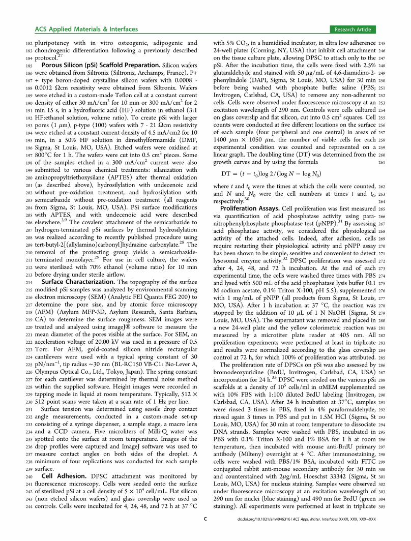
358 ! RESULTS359 pSi Samples. PSi substrates were generated from bulk360 boron-doped p-type silicon wafers via anodization.16 Substrates361 with various pore sizes were obtained by modulating the362 etching conditions and the doping level of the silicon wafer.
f1 363 Figure 1 shows representative SEM and AFM images of the pSi364 substrates; distinct textural features are depicted when365 comparing the di!erent pSi substrates.366 PSi substrates obtained from silicon wafers with a resistivity367 of 0.0008#0.0012 # cm by applying a constant current density
368of 30 mA/cm2 and 300 mA/cm2 had an average pore diameter369of 10 ± 2 nm (pSi 10 nm) and 36 ± 4 nm (pSi 36 nm),370respectively. The pSi layers produced from silicon wafers with3717#21 # cm resistivity, by applying a current density of 4.5 mA/372cm2, had an average pore diameter of 1020 ±100 nm (pSi 1373!m). The pSi substrates were quite uniformly porosi"ed.374Topographic images of pSi were obtained using AFM, and the375calculated root-mean-squared roughness (Rrms) of the substrate376were found at 1.9 ± 0.7 nm for “pSi 10 nm”, 7 ± 2 nm for “pSi37736 nm”, and 54 ± 20 nm for “pSi 1 !m”. Rrms is given by the378standard deviation of the z-values (surface heights) for the379sample area, as given by equation
"= ! #=
R Zn ZN
( )
n
N
rms1
2
380where N is the number of points in the image of the surface.381Although experimental determination of the surface rough-382ness is almost always conducted using AFM, there is some383di$culty is comparing our results to those reported in the384literature because of variation in the methods applied and the385sample areas examined.386Surface chemical treatments realized on “pSi 36 nm”387substrates are hydrosilylation with undecenoic acid or semi-388carbazide, and silanization (after thermal oxidation) with389APTES. The reaction schemes of the chemical treatments are390presented in Figure 1. The sample pSi 36 nm was chosen
Figure 1. Characterization of the porous silicon sca!olds. Surfacetopography of the silicon substrates imaged with (A) scanning electronmicroscopy and (B) atomic force microscopy. Porous silicon withmean pore diameter of 10, 36, and 1 !m are shown as A1-B1, A2-B2,and A3-B3, respectively. Schemas of surface chemical treatments arepresented as: nonoxidized pSi hydrosililation with semicarbazide;nonoxidized pSi hydrosililation with undecenoic acid; oxidized pSisilanization with APTES.
ACS Applied Materials & Interfaces Research Article
dx.doi.org/10.1021/am4046316 | ACS Appl. Mater. Interfaces XXXX, XXX, XXX#XXXD
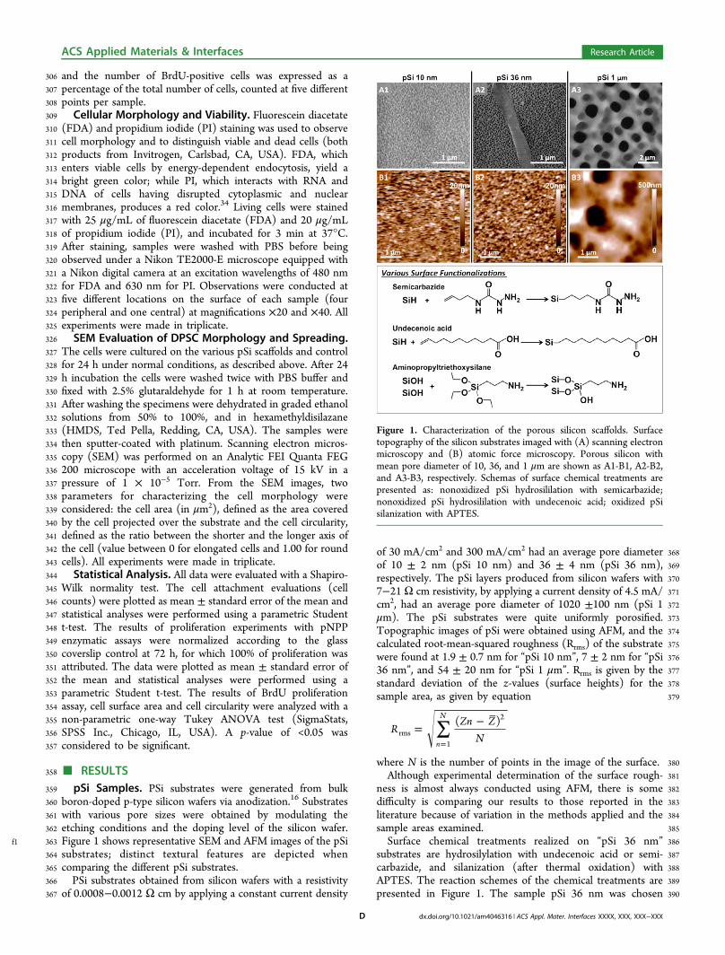
391 because it displayed the best results in term of cell adhesion,f2 392 compared to pSi 10 nm and pSi 1 !m (Figure 2). Water contact
393 angle measurements performed after surface functionalizationt1 394 revealed that all the substrates were hydrophilic (Table 1). All
395 the modi"ed surfaces were hydrophilic in agreement with the396 data of the literature: freshly etched pSi surfaces without
397oxidation are highly hydrophobic with contact angles of 108#398130°.29,35#38 Thermal oxidation increases wettability.36,38
399Oxidized pSi hydrophilicity was attributed to the removal of400SiySiHx species from the surface and to the formation of a polar401Si#OH capped surface after oxidation.39 After oxidation or402hydrosilylation the values drop from 12 to 37° showing the403successful functionalization of the surfaces with hydrophilic404groups. By analyzing SEM micrographs, the surface porosities405(ratio between total area of the pores and the area of the406considered region of interest) were found to be as follows: 40 ±4074% for pSi 10 nm, 45 ± 6% for pSi 36 nm, and 24 ± 4% for pSi4081!m.409Cell Adhesion and Growth. We examined DPSC410attachment to various surface-modi"ed pSi layers and411compared the results with those of DPSCs cultured on glass412coverslips and on #at silicon wafers after a period of 4, 24, 48,413and 72 h. The results are shown in Figure 2.414The experimental results showed that initial cell adhesion415and growth were very similar on glass coverslip, Flat Si, APTES-416treated pSi and semicarbazide-treated pSi, after 4 h and 24 h.417Initial cell adhesion on oxidized pSi (pSi 10 nm, pSi 36 nm and418pSi 1!m) and undecenoic acid-treated pSi were signi"cantly419lower compared to glass coverslip (p < 0.001, p = 0.003, p <4200.001, and p < 0.001, respectively). However, DPSC started to421proliferate on pSi 10 nm and pSi 36 nm after 24 h following the422same growth pattern observed on glass coverslip, whereas the423number of cells slightly decreased on pSi 1 !m and on424undecenoic acid-treated pSi after 24 and 48 h, respectively.425After 48 h, the numbers of cells were similar on glass coverslip426and on #at Si, signi"cantly higher on APTES-treated pSi427(p<0.001) and signi"cantly lower on semicarbazide-treated pSi428(p < 0.001), pSi 10 nm (p < 0.001), and pSi 36 nm (p < 0.001).429It is remarkable that there were signi"cantly more cells on pSi43036 nm than on pSi 10 nm after 48 and 72 h (p = 0.006 and p =4310.004, respectively), and that the number of cells on432semicarbazide-treated pSi increased during 24 h and decreased433abruptly after 48 h. After 72 h, DPSC reached con#uence on434glass coverslip, #at Si, and APTES-treated pSi. Cells exhibited a435mean doubling time of approximately 27, 30, 33, and 28 h on436glass coverslip, #at Si, pSi 10 nm, and pSi 36 nm, respectively.437On functionalized sca!olds (hydrosilylated or silanized 36 nm
Figure 2. DPSC attached on various type of pSi, after 4, 24, 48, and 72h of incubation (left columns to right columns, respectively). Glasscoverslip and #at silicon were used as control. Cells were counted persurface measuring 1400 !m ! 1050 !m, in "ve areas per samples.Mean values are expressed as cell numbers per mm2, with error barscorresponding to standard deviation. The bars protruding from theframe correspond to cells reaching con#uence.
Table 1. Water Contact Angle of the Various Sca!olds ("atSi and pSi)
water contact angle (deg)
#at Si pSi 10 nm pSi 36 nm pSi 1 !m
oxidized 26 ± 4 16 ± 2 15 ± 3 33 ± 5undecenoic acid 35 ± 5APTES 37 ± 6semicarbazide 12 ± 3
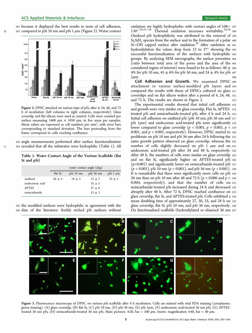
Figure 3. Fluorescence microscopy of DPSC on various pSi sca!olds after 4 h incubation. Cells are stained with vital FDA staining (cytoplasma:green staining). (A) glass coverslip, (B) #at Si, (C) pSi 10 nm, (D) pSi 36 nm, (E) pSi 1!m, (F) undecenoic acid-treated 36 nm pSi, (G) APTES-treated 36 nm pSi, (H) semicarbazide-treated 36 nm pSi. Main pictures: !20, bar = 100 !m. Insets: magni"cation !40, bar = 40 !m.
ACS Applied Materials & Interfaces Research Article
dx.doi.org/10.1021/am4046316 | ACS Appl. Mater. Interfaces XXXX, XXX, XXX#XXXE
438 pSi) cells showed a mean doubling time of 24 h on APTES-439 treated and semicarbazide-treated pSi. It was not possible to440 measure or calculate doubling time on pSi 1 !m and441 undecenoic-acid-treated pSi.442 Cells were stained with the vital dye FDA to observe cell443 adherence and cytoplasmic spreading. The morphologies of444 DPSC on surface-modi"ed pSi at early attachment stage (after
f3 445 4 h) are shown in Figure 3.446 From Figure 3, it is obvious that cells attached on all surfaces,447 with various shapes from normal "broblastic morphology448 (Figure 3A, B, and G) to less-spread cells with protrusions449 (Figure 3C, D), and even round cells (Figure 3E, F, and H).450 Fibroblastic morphologies were found mainly on glass451 coverslip, #at Si and APTES-treated pSi. Cells with protrusions452 were mainly found on pSi 10 nm and pSi 36 nm, whereas round453 cells with few protrusions were recovered on pSi 1 !m,454 undecenoic-acid-treated pSi and semicarbazide-treated pSi.455 These results correlate well with the results on cell adhesion456 described from Figure 2.457 Cell Proliferation. DPSC proliferation over a 3-day-time458 period, assessed by acid phosphatase activity, was evaluated for459 the various pSi sca!olds, and compared to that of glass460 coverslip and Flat Si. To normalize the results, we attributed461 100% cell enzymatic activity to the value found for glass
f4 462 coverslip after 72 h of cell incubation (Figure 4).
463 The experiments showed that DPSCs clearly proliferated at464 all time points on glass coverslip, #at Si, pSi 10 nm, pSi 36 nm,465 and APTES-treated pSi, whereas the proliferation was limited466 on pSi 1 !m and undecenoic-acid-treated pSi. For semi-467 carbazide-treated pSi, the proliferation rate was higher468 compared to glass coverslip until 24 h, and dropped469 dramatically after 48 h. After 24 h of incubation, the470 proliferation rate was equivalent on glass coverslip and on #at471 Si. At 24 h, APTES-treated pSi and semicarbazide-treated pSi472 improved DPSC proliferation of 27 % and 24 % (p = 0.042 and473 p = 0.039, respectively). The proliferation rate was lower on pSi474 10 nm, pSi 1 !m and undecenoic acid-treated pSi (p = 0.012, p
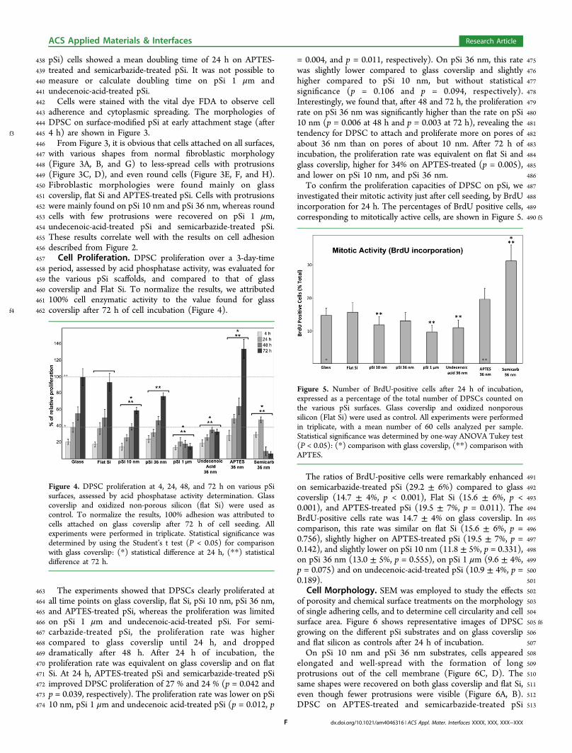
475= 0.004, and p = 0.011, respectively). On pSi 36 nm, this rate476was slightly lower compared to glass coverslip and slightly477higher compared to pSi 10 nm, but without statistical478signi"cance (p = 0.106 and p = 0.094, respectively).479Interestingly, we found that, after 48 and 72 h, the proliferation480rate on pSi 36 nm was signi"cantly higher than the rate on pSi48110 nm (p = 0.006 at 48 h and p = 0.003 at 72 h), revealing the482tendency for DPSC to attach and proliferate more on pores of483about 36 nm than on pores of about 10 nm. After 72 h of484incubation, the proliferation rate was equivalent on #at Si and485glass coverslip, higher for 34% on APTES-treated (p = 0.005),486and lower on pSi 10 nm, and pSi 36 nm.487To con"rm the proliferation capacities of DPSC on pSi, we488investigated their mitotic activity just after cell seeding, by BrdU489incorporation for 24 h. The percentages of BrdU positive cells,490 f5corresponding to mitotically active cells, are shown in Figure 5.
491The ratios of BrdU-positive cells were remarkably enhanced492on semicarbazide-treated pSi (29.2 ± 6%) compared to glass493coverslip (14.7 ± 4%, p < 0.001), Flat Si (15.6 ± 6%, p <4940.001), and APTES-treated pSi (19.5 ± 7%, p = 0.011). The495BrdU-positive cells rate was 14.7 ± 4% on glass coverslip. In496comparison, this rate was similar on #at Si (15.6 ± 6%, p =4970.756), slightly higher on APTES-treated pSi (19.5 ± 7%, p =4980.142), and slightly lower on pSi 10 nm (11.8 ± 5%, p = 0.331),499on pSi 36 nm (13.0 ± 5%, p = 0.555), on pSi 1 !m (9.6 ± 4%,500p = 0.075) and on undecenoic-acid-treated pSi (10.9 ± 4%, p =5010.189).502Cell Morphology. SEM was employed to study the e!ects503of porosity and chemical surface treatments on the morphology504of single adhering cells, and to determine cell circularity and cell505 f6surface area. Figure 6 shows representative images of DPSC506growing on the di!erent pSi substrates and on glass coverslip507and #at silicon as controls after 24 h of incubation.508On pSi 10 nm and pSi 36 nm substrates, cells appeared509elongated and well-spread with the formation of long510protrusions out of the cell membrane (Figure 6C, D). The511same shapes were recovered on both glass coverslip and #at Si,512even though fewer protrusions were visible (Figure 6A, B).513DPSC on APTES-treated and semicarbazide-treated pSi
Figure 4. DPSC proliferation at 4, 24, 48, and 72 h on various pSisurfaces, assessed by acid phosphatase activity determination. Glasscoverslip and oxidized non-porous silicon (#at Si) were used ascontrol. To normalize the results, 100% adhesion was attributed tocells attached on glass coverslip after 72 h of cell seeding. Allexperiments were performed in triplicate. Statistical signi"cance wasdetermined by using the Student’s t test (P < 0.05) for comparisonwith glass coverslip: (*) statistical di!erence at 24 h, (**) statisticaldi!erence at 72 h.
Figure 5. Number of BrdU-positive cells after 24 h of incubation,expressed as a percentage of the total number of DPSCs counted onthe various pSi surfaces. Glass coverslip and oxidized nonporoussilicon (Flat Si) were used as control. All experiments were performedin triplicate, with a mean number of 60 cells analyzed per sample.Statistical signi"cance was determined by one-way ANOVA Tukey test(P < 0.05): (*) comparison with glass coverslip, (**) comparison withAPTES.
ACS Applied Materials & Interfaces Research Article
dx.doi.org/10.1021/am4046316 | ACS Appl. Mater. Interfaces XXXX, XXX, XXX#XXXF
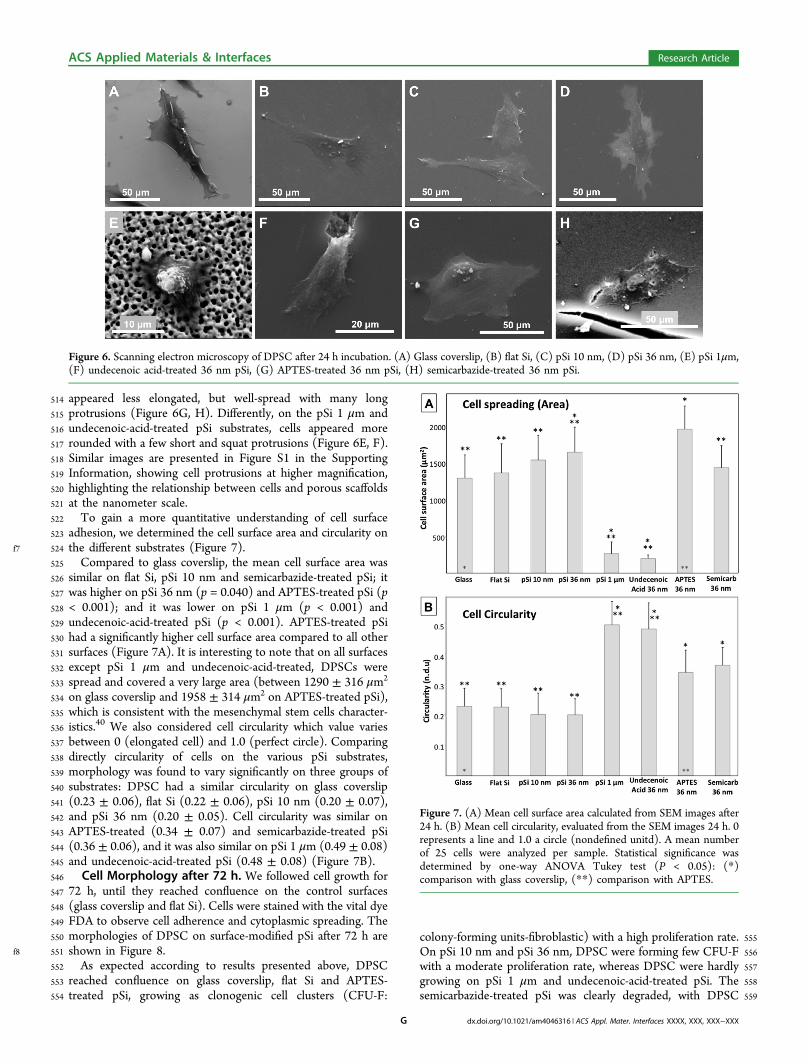
514 appeared less elongated, but well-spread with many long515 protrusions (Figure 6G, H). Di!erently, on the pSi 1 !m and516 undecenoic-acid-treated pSi substrates, cells appeared more517 rounded with a few short and squat protrusions (Figure 6E, F).518 Similar images are presented in Figure S1 in the Supporting519 Information, showing cell protrusions at higher magni"cation,520 highlighting the relationship between cells and porous sca!olds521 at the nanometer scale.522 To gain a more quantitative understanding of cell surface523 adhesion, we determined the cell surface area and circularity on
f7 524 the di!erent substrates (Figure 7).525 Compared to glass coverslip, the mean cell surface area was526 similar on #at Si, pSi 10 nm and semicarbazide-treated pSi; it527 was higher on pSi 36 nm (p = 0.040) and APTES-treated pSi (p528 < 0.001); and it was lower on pSi 1 !m (p < 0.001) and529 undecenoic-acid-treated pSi (p < 0.001). APTES-treated pSi530 had a signi"cantly higher cell surface area compared to all other531 surfaces (Figure 7A). It is interesting to note that on all surfaces532 except pSi 1 !m and undecenoic-acid-treated, DPSCs were533 spread and covered a very large area (between 1290 ± 316 !m2
534 on glass coverslip and 1958 ± 314 !m2 on APTES-treated pSi),535 which is consistent with the mesenchymal stem cells character-536 istics.40 We also considered cell circularity which value varies537 between 0 (elongated cell) and 1.0 (perfect circle). Comparing538 directly circularity of cells on the various pSi substrates,539 morphology was found to vary signi"cantly on three groups of540 substrates: DPSC had a similar circularity on glass coverslip541 (0.23 ± 0.06), #at Si (0.22 ± 0.06), pSi 10 nm (0.20 ± 0.07),542 and pSi 36 nm (0.20 ± 0.05). Cell circularity was similar on543 APTES-treated (0.34 ± 0.07) and semicarbazide-treated pSi544 (0.36 ± 0.06), and it was also similar on pSi 1 !m (0.49 ± 0.08)545 and undecenoic-acid-treated pSi (0.48 ± 0.08) (Figure 7B).546 Cell Morphology after 72 h. We followed cell growth for547 72 h, until they reached con#uence on the control surfaces548 (glass coverslip and #at Si). Cells were stained with the vital dye549 FDA to observe cell adherence and cytoplasmic spreading. The550 morphologies of DPSC on surface-modi"ed pSi after 72 h are
f8 551 shown in Figure 8.552 As expected according to results presented above, DPSC553 reached con#uence on glass coverslip, #at Si and APTES-554 treated pSi, growing as clonogenic cell clusters (CFU-F:
555colony-forming units-"broblastic) with a high proliferation rate.556On pSi 10 nm and pSi 36 nm, DPSC were forming few CFU-F557with a moderate proliferation rate, whereas DPSC were hardly558growing on pSi 1 !m and undecenoic-acid-treated pSi. The559semicarbazide-treated pSi was clearly degraded, with DPSC
Figure 6. Scanning electron microscopy of DPSC after 24 h incubation. (A) Glass coverslip, (B) #at Si, (C) pSi 10 nm, (D) pSi 36 nm, (E) pSi 1!m,(F) undecenoic acid-treated 36 nm pSi, (G) APTES-treated 36 nm pSi, (H) semicarbazide-treated 36 nm pSi.
Figure 7. (A) Mean cell surface area calculated from SEM images after24 h. (B) Mean cell circularity, evaluated from the SEM images 24 h. 0represents a line and 1.0 a circle (nonde"ned unitd). A mean numberof 25 cells were analyzed per sample. Statistical signi"cance wasdetermined by one-way ANOVA Tukey test (P < 0.05): (*)comparison with glass coverslip, (**) comparison with APTES.
ACS Applied Materials & Interfaces Research Article
dx.doi.org/10.1021/am4046316 | ACS Appl. Mater. Interfaces XXXX, XXX, XXX#XXXG
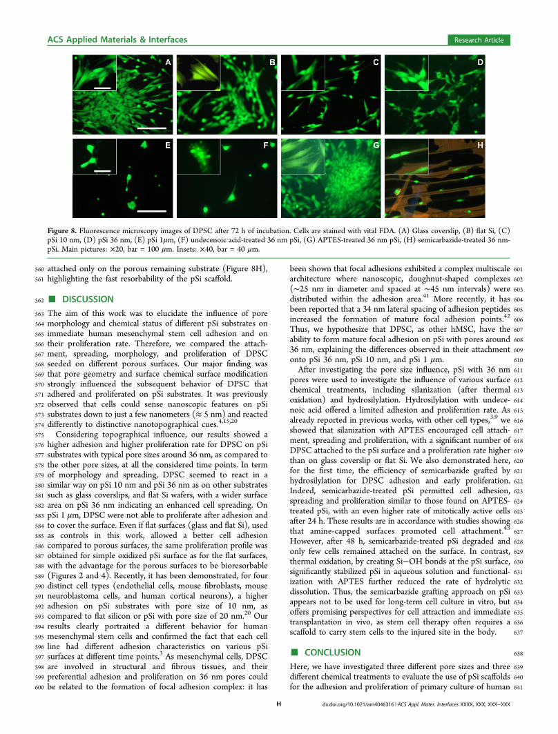
560 attached only on the porous remaining substrate (Figure 8H),561 highlighting the fast resorbability of the pSi sca!old.
562 ! DISCUSSION563 The aim of this work was to elucidate the in#uence of pore564 morphology and chemical status of di!erent pSi substrates on565 immediate human mesenchymal stem cell adhesion and on566 their proliferation rate. Therefore, we compared the attach-567 ment, spreading, morphology, and proliferation of DPSC568 seeded on di!erent porous surfaces. Our major "nding was569 that pore geometry and surface chemical surface modi"cation570 strongly in#uenced the subsequent behavior of DPSC that571 adhered and proliferated on pSi substrates. It was previously572 observed that cells could sense nanoscopic features on pSi573 substrates down to just a few nanometers (% 5 nm) and reacted574 di!erently to distinctive nanotopographical cues.4,15,20
575 Considering topographical in#uence, our results showed a576 higher adhesion and higher proliferation rate for DPSC on pSi577 substrates with typical pore sizes around 36 nm, as compared to578 the other pore sizes, at all the considered time points. In term579 of morphology and spreading, DPSC seemed to react in a580 similar way on pSi 10 nm and pSi 36 nm as on other substrates581 such as glass coverslips, and #at Si wafers, with a wider surface582 area on pSi 36 nm indicating an enhanced cell spreading. On583 pSi 1 !m, DPSC were not able to proliferate after adhesion and584 to cover the surface. Even if #at surfaces (glass and #at Si), used585 as controls in this work, allowed a better cell adhesion586 compared to porous surfaces, the same proliferation pro"le was587 obtained for simple oxidized pSi surface as for the #at surfaces,588 with the advantage for the porous surfaces to be bioresorbable589 (Figures 2 and 4). Recently, it has been demonstrated, for four590 distinct cell types (endothelial cells, mouse "broblasts, mouse591 neuroblastoma cells, and human cortical neurons), a higher592 adhesion on pSi substrates with pore size of 10 nm, as593 compared to #at silicon or pSi with pore size of 20 nm.20 Our594 results clearly portraited a di!erent behavior for human595 mesenchymal stem cells and con"rmed the fact that each cell596 line had di!erent adhesion characteristics on various pSi597 surfaces at di!erent time points.3 As mesenchymal cells, DPSC598 are involved in structural and "brous tissues, and their599 preferential adhesion and proliferation on 36 nm pores could600 be related to the formation of focal adhesion complex: it has
601been shown that focal adhesions exhibited a complex multiscale602architecture where nanoscopic, doughnut-shaped complexes603($25 nm in diameter and spaced at $45 nm intervals) were604distributed within the adhesion area.41 More recently, it has605been reported that a 34 nm lateral spacing of adhesion peptides606increased the formation of mature focal adhesion points.42
607Thus, we hypothesize that DPSC, as other hMSC, have the608ability to form mature focal adhesion on pSi with pores around60936 nm, explaining the di!erences observed in their attachment610onto pSi 36 nm, pSi 10 nm, and pSi 1 !m.611After investigating the pore size in#uence, pSi with 36 nm612pores were used to investigate the in#uence of various surface613chemical treatments, including silanization (after thermal614oxidation) and hydrosilylation. Hydrosilylation with undece-615noic acid o!ered a limited adhesion and proliferation rate. As616already reported in previous works, with other cell types,3,9 we617showed that silanization with APTES encouraged cell attach-618ment, spreading and proliferation, with a signi"cant number of619DPSC attached to the pSi surface and a proliferation rate higher620than on glass coverslip or #at Si. We also demonstrated here,621for the "rst time, the e$ciency of semicarbazide grafted by622hydrosilylation for DPSC adhesion and early proliferation.623Indeed, semicarbazide-treated pSi permitted cell adhesion,624spreading and proliferation similar to those found on APTES-625treated pSi, with an even higher rate of mitotically active cells626after 24 h. These results are in accordance with studies showing627that amine-capped surfaces promoted cell attachment.43
628However, after 48 h, semicarbazide-treated pSi degraded and629only few cells remained attached on the surface. In contrast,630thermal oxidation, by creating Si#OH bonds at the pSi surface,631signi"cantly stabilized pSi in aqueous solution and functional-632ization with APTES further reduced the rate of hydrolytic633dissolution. Thus, the semicarbazide grafting approach on pSi634appears not to be used for long-term cell culture in vitro, but635o!ers promising perspectives for cell attraction and immediate636transplantation in vivo, as stem cell therapy often requires a637sca!old to carry stem cells to the injured site in the body.
638! CONCLUSION639Here, we have investigated three di!erent pore sizes and three640di!erent chemical treatments to evaluate the use of pSi sca!olds641for the adhesion and proliferation of primary culture of human
Figure 8. Fluorescence microscopy images of DPSC after 72 h of incubation. Cells are stained with vital FDA. (A) Glass coverslip, (B) #at Si, (C)pSi 10 nm, (D) pSi 36 nm, (E) pSi 1!m, (F) undecenoic acid-treated 36 nm pSi, (G) APTES-treated 36 nm pSi, (H) semicarbazide-treated 36 nm-pSi. Main pictures: !20, bar = 100 !m. Insets: !40, bar = 40 !m.
ACS Applied Materials & Interfaces Research Article
dx.doi.org/10.1021/am4046316 | ACS Appl. Mater. Interfaces XXXX, XXX, XXX#XXXH
642 mesenchymal stem cells from the dental pulp. We have643 identi"ed, for the "rst time, two e$cient amino-grafted pSi644 sca!olds for human mesenchymal stem cells adhesion and645 growth, with optimized pore diameter, for in vitro proliferation646 (and further di!erentiation), with an interesting potential for in647 vivo transplantation.648 DPSC on pSi 36 nm were observed to have a better adhesion649 and a faster growth compared to pSi with smaller (10 nm) or650 larger (1 !m) pore size, in particular after silanization with651 APTES. Hydrosilylation with semicarbazide led to a new652 chemical modi"cation favoring cell adhesion and proliferation,653 especially mitosis after cell adhesion. As this modi"ed pSi654 surface was stable for only 24#48 h, it appeared to be655 potentially usable for stem cells adhesion and immediate in vivo656 transplantation, whereas APTES-treated pSi was more suitable657 for long term in vitro culture, for stem cells proliferation and658 di!erentiation.659 More studies are on course to investigate: (1) the ability of660 APTES-treated pSi as sca!old for stem cells di!erentiation in661 di!erent lineage, as pore size might also in#uence cell662 di!erentiation, (2) the optimization of surface stabilization663 with the semicarbazide treatment, (3) the e$ciency of664 semicarbazide-treated pSi as an immediate cell carrier for in665 vivo transplantation. Further studies will also elucidate the role666 played by the porosity on focal adhesion formation, as well as667 the implication on the cytoskeleton organization.
668 ! ASSOCIATED CONTENT669 *S Supporting Information670 Additional data showing cell protrusions with SEM at high671 magni"cation, highlighting the relationship between cells and672 porous sca!olds at the nanometer scale. Additional images673 showing water droplets on the various surfaces. This material is674 available free of charge via the Internet at http://pubs.acs.org.
675 ! AUTHOR INFORMATION676 Corresponding Author677 *E-mail: [email protected]. Phone: +33 4 11678 75 92 52.679 Author Contributions680 The manuscript was written through contributions of all681 authors. All authors have given approval to the "nal version of682 the manuscript.683 Notes684 The authors declare no competing "nancial interest.
685 ! REFERENCES(1)686 Wang, Y.; Kim, H. J.; Vunjak-Novakovic, G.; Kaplan, D. L.
687 Biomaterials 2006, 27, 6064#6082.(2)688 Collart-Dutilleul, P.Y.; Deville de Pe !rie "re, D.; Cuisinier, F.J.;
689 Gergely, C.; Cunin, F. In Porous Silicon for Biomedical Applications;690 Santos, H., Eds.; Woodhead Publishing: Cambridge, U.K., 2014; Vol.691 68, p 486.
(3)692 Low, S. P.; Williams, K. A.; Canham, L. T.; Voelcker, N. H.693 Biomaterials 2006, 27, 4538#4546.
(4)694 Khung, Y. L.; Barritt, G.; Voelcker, N. H. Exp. Cell Res. 2008, 314,695 789#800.
(5)696 Canham, L. T. Adv. Mater. 1995, 7, 1033#1037.(6)697 Ainslie, K. M.; Tao, S. L.; Popat, K. C.; Desai, T. A. ACS Nano
698 2008, 2, 1076#1084.(7)699 Whitehead, M. A.; Fan, D.; Mukherjee, P.; Akkaraju, G. R.;
700 Canham, L. T.; Coffer, J. L. Tissue Eng., Part A 2008, 14, 195#206.
(8) 701Cheng, L.; Anglin, E.; Cunin, F.; Kim, D.; Sailor, M. J.;702Falkenstein, I.; Tammewar, A.; Freeman, W. R. Br. J. Ophthalmol.7032008, 92, 705#711.
(9) 704Alvarez, S. D.; Derfus, A. M.; Schwartz, M. P.; Bhatia, S. N.;705Sailor, M. J. Biomaterials 2009, 30, 26#34.
(10) 706Torres-Costa, V. V.; Martínez-Mun$oz, G. G.; Sa !nchez-Vaquero,707V. V.; Mun$oz-Noval, A!. A!.; Gonza !lez-Me !ndez, L. L.; Punzo !n-Quijorna,708E. E.; Gallach-Pe !rez, D. D.; Manso-Silva !n, M. M.; Climent-Font, A. A.;709García-Ruiz, J. P. J.; Martín-Palma, R. J. R. Int. J. Nanomed. 2011, 7,710623#630.
(11) 711Park, J.-H.; Gu, L.; von Maltzahn, G.; Ruoslahti, E.; Bhatia, S.712N.; Sailor, M. J. Nat. Mater. 2009, 8, 331#336.
(12) 713Mun$oz Noval, A.; García, R.; Ruiz Casas, D.; Losada Bayo, D.;714Sa !nchez Vaquero, V.; Torres Costa, V.; Martín Palma, R. J.; García, M.715A.; García Ruiz, J. P.; Serrano Olmedo, J. J.; Mun$oz Negrete, J. F.; del716Pozo Guerrero, F.; Manso Silva !n, M. Acta Biomater. 2013, 9, 6169#7176176.
(13) 718Noval, A. M.; Vaquero, V. S.; Quijorna, E. P.; Costa, V. T.;719Pe !rez, D. G.; Me !ndez, L. G.; Montero, I.; Palma, R. J. M.; Font, A. C.;720Ruiz, J. P. G.; Silva !n, M. M. J. Biomed. Mater. Res. A 2012, 100, 1615#7211622.
(14) 722Dalby, M. J.; Riehle, M. O.; Yarwood, S. J.; Wilkinson, C. D. W.;723Curtis, A. S. G. Exp. Cell Res. 2003, 284, 274#282.
(15) 724Buxboim, A.; Ivanovska, I. L.; Discher, D. E. J. Cell Sci. 2010,725123, 297#308.
(16) 726Sailor, M.J. In Porous Silicon in Practice: Preparation, Character-727ization and Applications; John Wiley & Sons: New York, 2012.
(17) 728Sapelkin, A. V.; Bayliss, S. C.; Unal, B.; Charalambou, A.729Biomaterials 2006, 27, 842#846.
(18) 730Wang, P.; Clements, L.; Thissen, H.; Jane, A.; Tsai, W.-B.;731Voelcker, N. H. Adv. Funct. Mater. 2012, 22, 3414#3423.
(19) 732Clements, L. R.; Wang, P.-Y.; Tsai, W.-B.; Thissen, H.; Voelcker,733N. H. Lab Chip 2012, 12, 1480#1486.
(20) 734Gentile, F.; La Rocca, R.; Marinaro, G.; Nicastri, A.; Toma, A.;735Paonessa, F.; Cojoc, G.; Liberale, C.; Benfenati, F.; di Fabrizio, E.;736Decuzzi, P. ACS Appl. Mater. Interfaces 2012, 4, 2903#2911.
(21) 737Ma, T. World J. Stem Cells 2010, 2, 13#17.(22) 738Marolt, D.; Knezevic, M.; Novakovic, G. V. Stem Cell Res. Ther.
7392010, 1, 10.(23) 740Gronthos, S.; Mankani, M.; Brahim, J.; Robey, P. G.; Shi, S. Proc.
741Natl. Acad. Sci. U.S.A. 2000, 97, 13625#13630.(24) 742Dominici, M.; Le Blanc, K.; Mueller, I.; Slaper-Cortenbach, I.;
743Marini, F.; Krause, D.; Deans, R.; Keating, A.; Prockop, D.; Horwitz, E.744Cytotherapy 2006, 8, 315#317.
(25) 745Coppe, C.; Zhang, Y.; Den Besten, P. K. Pediatr. Dent. 2009, 31,746467#471.
(26) 747Shi, S.; Gronthos, S. J. Bone Miner. Res. 2003, 18, 696#704.(27) 748Ke !moun, P.; Laurencin-Dalicieux, S.; Rue, J.; Farges, J.-C.;
749Gennero, I.; Conte-Auriol, F.; Briand-Mesange, F.; Gadelorge, M.;750Arzate, H.; Narayanan, A. S.; Brunel, G.; Salles, J.-P. Cell Tissue Res.7512007, 329, 283#294.
(28) 752Secret, E.; Smith, K.; Dubljevic, V.; Moore, E.; Macardle, P.;753Delalat, B.; Rogers, M.-L.; Johns, T. G.; Durand, J.-O.; Cunin, F.;754Voelcker, N. H. Adv. Healthcare Mater. 2013, 2, 718#727.
(29) 755Coffinier, Y.; Olivier, C.; Perzyna, A.; Grandidier, B.; Wallart, X.;756Durand, J. O.; Melnyk, O.; Stie !venard, D. Langmuir 2005, 21, 1489#7571496.
(30) 758Tirino, V.; Desiderio, V.; d’Aquino, R.; De Francesco, F.;759Pirozzi, G.; Graziano, A.; Galderisi, U.; Cavaliere, C.; De Rosa, A.;760Papaccio, G.; Giordano, A. PLoS ONE 2008, 3, e3469.
(31) 761Werner, S.; Huck, O.; Frisch, B.; Vautier, D.; Elkaim, R.; Voegel,762J.-C.; Brunel, G.; Tenenbaum, H. Biomaterials 2009, 30, 2291#2301.
(32) 763Ueda, Y.; Walsh, E.; Nakanishi, H.; Yoshida, K. Neurosci. Lett.7641994, 165, 203#207.
(33) 765Horner, P. J.; Power, A. E.; Kempermann, G.; Kuhn, H. G.;766Palmer, T. D.; Winkler, J.; Thal, L. J.; Gage, F. H. J. Neurosci. 2000, 20,7672218#2228.
(34) 768Cao, D.; Wu, Y.-P.; Fu, Z.-F.; Tian, Y.; Li, C.-J.; Gao, C.-Y.;769Chen, Z.-L.; Feng, X.-Z. Colloids Surf., B 2011, 84, 26#34.
ACS Applied Materials & Interfaces Research Article
dx.doi.org/10.1021/am4046316 | ACS Appl. Mater. Interfaces XXXX, XXX, XXX#XXXI

(35)770 Naveas, N.; Costa, V. T.; Gallach, D.; Hernandez-Montelongo,771 J.; Palma, R. J. M.; Garcia-Ruiz, J. P.; Manso-Silva !n, M. Sci. Technol.772 Adv. Mater. 2012, 13, 045009.
(36)773 Jarvis, K. L.; Barnes, T. J.; Prestidge, C. A. J. Colloid. Interface Sci.774 2011, 363, 327#333.
(37)775 Bateman, J. E.; Eagling, R. D.; Horrocks, B. R.; Houlton, A.;776 Worrall, D. R. Chem. Commun. 1997, 2275#2276.
(38)777 Zangooie, S.; Bjorklund, R.; Arwin, H. J. Electrochem. Soc. 1997,778 144, 4027.
(39)779 Paski, J.; Bjo%rkqvist, M.; Salonen, J.; Lehto, V.-P. Phys. Status780 Solidi 2005, 2, 3379#3383.
(40)781 Krishna, O. D.; Jha, A. K.; Jia, X.; Kiick, K. L. Biomaterials 2011,782 32, 6412#6424.
(41)783 Patla, I.; Volberg, T.; Elad, N.; Hirschfeld-Warneken, V.;784 Grashoff, C.; Fa %ssler, R.; Spatz, J. P.; Geiger, B.; Medalia, O. Nat. Cell785 Biol. 2010, 12, 909#915.
(42)786 Frith, J. E.; Mills, R. J.; Cooper-White, J. J. J. Cell Sci. 2012, 125,787 317#327.
(43)788 Faucheux, N.; Schweiss, R.; Lu%tzow, K.; Werner, C.; Groth, T.789 Biomaterials 2004, 25, 2721#2730.
ACS Applied Materials & Interfaces Research Article
dx.doi.org/10.1021/am4046316 | ACS Appl. Mater. Interfaces XXXX, XXX, XXX#XXXJ