Adenoid cystic carcinoma of the head and neck - An update
10
Review Adenoid cystic carcinoma of the head and neck – An update q Andrés Coca-Pelaz a , Juan P. Rodrigo a,b , Patrick J. Bradley c,d , Vincent Vander Poorten d,e , Asterios Triantafyllou f , Jennifer L. Hunt g , Primoz ˇ Strojan h , Alessandra Rinaldo i , Missak Haigentz Jr. j , Robert P. Takes k , Vanni Mondin i , Afshin Teymoortash l , Lester D.R. Thompson m , Alfio Ferlito i,⇑ a Department of Otolaryngology, Hospital Universitario Central de Asturias, Oviedo, Spain b Instituto Universitario de Oncología del Principado de Asturias, University of Oviedo, Spain c Department of Otorhinolaryngology–Head and Neck Surgery, Nottingham University Hospitals, Queens Medical Centre Campus, Nottingham, UK d European Salivary Gland Society, Geneva, Switzerland e Otorhinolaryngology–Head and Neck Surgery and Department of Oncology, University Hospitals Leuven, KU Leuven, Leuven, Belgium f Oral Pathology, School of Dentistry, University of Liverpool, Liverpool, UK g Department of Pathology, University of Arkansas for Medical Sciences, Little Rock, AR, USA h Department of Radiation Oncology, Institute of Oncology, Ljubljana, Slovenia i University of Udine School of Medicine, Udine, Italy j Department of Medicine, Division of Oncology, Albert Einstein College of Medicine, Montefiore Medical Center, Bronx, NY, USA k Department of Otolaryngology–Head and Neck Surgery, Radboud University Medical Center, Nijmegen, The Netherlands l Department of Otolaryngology–Head and Neck Surgery, University of Marburg, Marburg, Germany m Department of Pathology, Woodland Hills Medical Center, Woodland Hills, CA, USA article info Article history: Received 27 January 2015 Received in revised form 1 April 2015 Accepted 3 April 2015 Available online xxxx Keywords: Adenoid cystic carcinoma Head and neck cancer Salivary gland Pathology Molecular biology Prognosis summary This article provides an update on the current understanding of adenoid cystic carcinoma of the head and neck, including a review of its epidemiology, clinical behavior, pathology, molecular biology, diagnostic workup, treatment and prognosis. Adenoid cystic carcinoma is an uncommon salivary gland tumor that may arise in a wide variety of anatomical sites in the head and neck, often with an advanced stage at diag- nosis. The clinical course is characterized by very late recurrences; consequently, clinical follow-up should extend at least >15 years. The optimal treatment is generally considered to be surgery with post- operative radiotherapy to optimize local disease control. Much effort has been invested into understand- ing the tumor’s molecular biological processes, aiming to identify patients at high risk of recurrence, in hopes that they could benefit from other, still unproven treatment modalities such as chemotherapy or biological therapy. Ó 2015 Elsevier Ltd. All rights reserved. Introduction With a reported yearly incidence of 3–4.5 cases per million [1], adenoid cystic carcinoma (AdCC) is an uncommon tumor, account- ing for about 1% of all head and neck malignancies [2] and about 10% of all tumors of the salivary glands [3]. It is the most com- monly reported malignant tumor of the minor salivary glands (MSGs) [1] and is also one of the most common cancers of the major salivary glands (the parotid, submandibular and sublingual salivary glands) [4]. AdCC can also involve lacrimal and ceruminous glands as well as other sites in the head and neck, including the nasal and paranasal sinuses, trachea and larynx [1,6–10]. AdCC was first described by Robin, Lorain and Laboulbene in two articles published in 1853 and 1854 reporting on one parotid and two nasal tumors. These authors described the characteristic cribriform arrangement of tumor cells on microscopy and noted the invasion of surrounding structures and the spread along nerves [11]. In 1856, Billroth suggested the term ‘‘cylindroma’’ for this tumor; the current name of ‘‘adenoid cystic carcinoma’’ was intro- duced by Spies in 1930. Despite the initial observations of Robin et al. the tumor was regarded as a variant of the benign mixed tumor. The malignant nature of this tumor was finally established by Dockerty and Mayo [12]. AdCC is a relentlessly growing tumor characterized by perineu- ral invasion and multiple local recurrences. Regional lymph node metastases are conventionally regarded as rare, but these may be under-recognized due to potentially occult, clinically undetectable http://dx.doi.org/10.1016/j.oraloncology.2015.04.005 1368-8375/Ó 2015 Elsevier Ltd. All rights reserved. q This article was written by members and invitees of the International Head and Neck Scientific Group (www.IHNSG.com). ⇑ Corresponding author at: University of Udine School of Medicine, Piazzale S. Maria della Misericordia, I-33100 Udine, Italy. E-mail address: [email protected] (A. Ferlito). Oral Oncology xxx (2015) xxx–xxx Contents lists available at ScienceDirect Oral Oncology journal homepage: www.elsevier.com/locate/oraloncology Please cite this article in press as: Coca-Pelaz A et al. Adenoid cystic carcinoma of the head and neck – An update. Oral Oncol (2015), http://dx.doi.org/ 10.1016/j.oraloncology.2015.04.005
Transcript of Adenoid cystic carcinoma of the head and neck - An update

Oral Oncology xxx (2015) xxx–xxx
Contents lists available at ScienceDirect
Oral Oncology
journal homepage: www.elsevier .com/locate /ora loncology
Review
Adenoid cystic carcinoma of the head and neck – An update q
http://dx.doi.org/10.1016/j.oraloncology.2015.04.0051368-8375/� 2015 Elsevier Ltd. All rights reserved.
q This article was written by members and invitees of the International Head andNeck Scientific Group (www.IHNSG.com).⇑ Corresponding author at: University of Udine School of Medicine, Piazzale S.
Maria della Misericordia, I-33100 Udine, Italy.E-mail address: [email protected] (A. Ferlito).
Please cite this article in press as: Coca-Pelaz A et al. Adenoid cystic carcinoma of the head and neck – An update. Oral Oncol (2015), http://dx.d10.1016/j.oraloncology.2015.04.005
Andrés Coca-Pelaz a, Juan P. Rodrigo a,b, Patrick J. Bradley c,d, Vincent Vander Poorten d,e,Asterios Triantafyllou f, Jennifer L. Hunt g, Primoz Strojan h, Alessandra Rinaldo i, Missak Haigentz Jr. j,Robert P. Takes k, Vanni Mondin i, Afshin Teymoortash l, Lester D.R. Thompson m, Alfio Ferlito i,⇑a Department of Otolaryngology, Hospital Universitario Central de Asturias, Oviedo, Spainb Instituto Universitario de Oncología del Principado de Asturias, University of Oviedo, Spainc Department of Otorhinolaryngology–Head and Neck Surgery, Nottingham University Hospitals, Queens Medical Centre Campus, Nottingham, UKd European Salivary Gland Society, Geneva, Switzerlande Otorhinolaryngology–Head and Neck Surgery and Department of Oncology, University Hospitals Leuven, KU Leuven, Leuven, Belgiumf Oral Pathology, School of Dentistry, University of Liverpool, Liverpool, UKg Department of Pathology, University of Arkansas for Medical Sciences, Little Rock, AR, USAh Department of Radiation Oncology, Institute of Oncology, Ljubljana, Sloveniai University of Udine School of Medicine, Udine, Italyj Department of Medicine, Division of Oncology, Albert Einstein College of Medicine, Montefiore Medical Center, Bronx, NY, USAk Department of Otolaryngology–Head and Neck Surgery, Radboud University Medical Center, Nijmegen, The Netherlandsl Department of Otolaryngology–Head and Neck Surgery, University of Marburg, Marburg, Germanym Department of Pathology, Woodland Hills Medical Center, Woodland Hills, CA, USA
a r t i c l e i n f o s u m m a r y
Article history:Received 27 January 2015Received in revised form 1 April 2015Accepted 3 April 2015Available online xxxx
Keywords:Adenoid cystic carcinomaHead and neck cancerSalivary glandPathologyMolecular biologyPrognosis
This article provides an update on the current understanding of adenoid cystic carcinoma of the head andneck, including a review of its epidemiology, clinical behavior, pathology, molecular biology, diagnosticworkup, treatment and prognosis. Adenoid cystic carcinoma is an uncommon salivary gland tumor thatmay arise in a wide variety of anatomical sites in the head and neck, often with an advanced stage at diag-nosis. The clinical course is characterized by very late recurrences; consequently, clinical follow-upshould extend at least >15 years. The optimal treatment is generally considered to be surgery with post-operative radiotherapy to optimize local disease control. Much effort has been invested into understand-ing the tumor’s molecular biological processes, aiming to identify patients at high risk of recurrence, inhopes that they could benefit from other, still unproven treatment modalities such as chemotherapyor biological therapy.
� 2015 Elsevier Ltd. All rights reserved.
Introduction
With a reported yearly incidence of 3–4.5 cases per million [1],adenoid cystic carcinoma (AdCC) is an uncommon tumor, account-ing for about 1% of all head and neck malignancies [2] and about10% of all tumors of the salivary glands [3]. It is the most com-monly reported malignant tumor of the minor salivary glands(MSGs) [1] and is also one of the most common cancers of themajor salivary glands (the parotid, submandibular and sublingualsalivary glands) [4]. AdCC can also involve lacrimal andceruminous glands as well as other sites in the head and neck,
including the nasal and paranasal sinuses, trachea and larynx[1,6–10].
AdCC was first described by Robin, Lorain and Laboulbene intwo articles published in 1853 and 1854 reporting on one parotidand two nasal tumors. These authors described the characteristiccribriform arrangement of tumor cells on microscopy and notedthe invasion of surrounding structures and the spread along nerves[11]. In 1856, Billroth suggested the term ‘‘cylindroma’’ for thistumor; the current name of ‘‘adenoid cystic carcinoma’’ was intro-duced by Spies in 1930. Despite the initial observations of Robinet al. the tumor was regarded as a variant of the benign mixedtumor. The malignant nature of this tumor was finally establishedby Dockerty and Mayo [12].
AdCC is a relentlessly growing tumor characterized by perineu-ral invasion and multiple local recurrences. Regional lymph nodemetastases are conventionally regarded as rare, but these may beunder-recognized due to potentially occult, clinically undetectable
oi.org/

2 A. Coca-Pelaz et al. / Oral Oncology xxx (2015) xxx–xxx
cervical metastases, infrequent neck dissections for this tumor andarguably a lack of detailed pathological assessment of lymphnodes. In sharp contrast, hematogenous metastasis is common,especially to lung, bone and liver [11,13].
Clinically, AdCC is regarded a high-grade neoplasm, and conse-quently the treatment of choice is radical surgical resection and isalmost always followed by postoperative radiotherapy [1,14].Chemotherapy (both cytotoxic chemotherapy and targeted molec-ular therapies) has been extensively studied in patients withadvanced AdCC, but the rather indolent course of the diseasemakes it difficult to observe clinical responses [15].
In the setting of incurable AdCC, the benefits and risks of treat-ment should be carefully weighed, as palliative chemotherapy forthis often indolent malignancy may be associated with toxicitywithout known impact in disease course and patient prognosis[16]. Therefore, some asymptomatic patients with incurable dis-ease may be observed without treatment, sometimes for years;chemotherapy is generally recommended when patients havedemonstrated rapidly progressive disease or are symptomatic [17].
Epidemiology
AdCC is most frequently found in the parotid, submandibularand MSGs. A large Danish population-based study estimates thatAdCC accounts for 27.9% of the overall incidence of salivary glandcancers (SGCs) (11/1,000,000/year), corresponding to an annualincidence of 3/1,000,000/year [1]. In Nova Scotia, the annual inci-dence raises to 4.5/1,000,000 cases [18]. It is important to note thatthese incidence estimates are certainly affected by challenges ofhistological diagnosis of SGCs, with reclassification rates rangingfrom 14% to 29% in several studies [1,19,20]. The cribriform patternis easily recognized, but other patterns may be less familiar to non-specialists; epidemiological studies of SGCs should therefore con-sider the center from which the data has been collected.
The proportion of AdCC among SGCs varies according to thesite/location of the primary tumor. In a Dutch nationwide studyof parotid carcinoma, AdCC was the most frequent histology,accounting for 1 out of every 6 parotid cancers [21]. The likelihoodof AdCC is even greater in the submandibular gland, where itaccounts for 40% of SGCs [4,22]. AdCC is the most common cancerin MSGs, where the proportion of AdCC ranges from 32% to 71%. InMSGs, AdCC is most commonly found in the palate, followed by theparanasal sinuses (14–17%) and other sites of the oral cavity[10,23].
The tumor occurs in all age groups with a high frequency inmiddle-aged and older patients [24], the 5th and 6th decades beingmost commonly affected [1,11,20,25–27]. There are no distinct riskfactors, and smoking is not known to affect the incidence [28].
Clinical behavior
AdCC has been described as having an apparently indolentcourse; however, it has an aggressive long-term behavior, withpersistent and recurrent growth pattern and late onset of metas-tases resulting in frequent eventual death [25]. It has beendescribed as ‘‘one of the most biologically destructive and unpre-dictable tumors of the head and neck’’ [29].
The most common presenting symptom is a slowly growingmass, followed by pain attributed to its tendency for perineuralinvasion. The association between pain, facial nerve dysfunctionand microscopic perineural invasion was emphasized in aNetherlands’ Cancer Institute cohort of patients with parotid carci-noma, where the majority of patients with these features had AdCC[30]. In the study of Nascimento et al. [24], 98% of patients reporteda mass, 48% had pain, 30% had ulceration, and one patient had
Please cite this article in press as: Coca-Pelaz A et al. Adenoid cystic carcinom10.1016/j.oraloncology.2015.04.005
facial nerve paralysis; these symptoms had been present from1 month to 4 years. The presenting symptoms of AdCC vary accord-ing to the site of disease. In major salivary glands the tumor pro-duces a mass, and when located in the parotid, facial nerve palsymay occur; in the palate a mass is common, though ulceration oreven oro-antral fistula may be seen; in the larynx dyspnea couldbe the first presenting symptom; in the nose and paranasal sinuses,nasal obstruction, deep facial pain, epistaxis and eye symptoms areat the forefront [5,10,31].
Pathology and diagnosis
On routinely-prepared histological sections of resection speci-mens examined with the naked eye and at scanning magnifica-tions, AdCCs are asymmetrical tumors, with variously lobulatedor invasive growth patterns (Fig. 1).
Morphologically, AdCC largely consists of non-luminal, basa-loid, hematoxyphilic cells, with small to moderate amounts ofcytoplasm, and far fewer luminal, short cuboidal, eosinophilic cells(Figs. 2 and 3A). The nuclei tend to be relatively bland with small orinconspicuous nucleoli. The luminal cells may be inconspicuous,though immunohistochemical markers assist in their distinction(Fig. 3B).
Three distinct architectural patterns have been described: tubu-lar (usually bilayered tubules lined by luminal cells surrounded bynon-luminal cells that often show ‘‘clear’’ cytoplasm); cribriform[basaloid cells arranged in variable sized, oval/rounded massespunched-out by rigid, oval, cyst-like spaces (pseudolumina) thatmay contain ‘‘cylinders’’/globules of hyaline material and/or myx-oid glycosaminoglycans, and occasional small true lumina lined byluminal cells]; and solid (largely basaloid tumor cells growing insheets without lumina formation) (Fig. 2) [32,33].
AdCC is traditionally regarded as originating from the interca-lated duct region – hence comprising a population of duct-likeand purportedly myoepithelial-like cells [34]. Clearly, the luminalcells show duct-like phenotypes. Caution should be exerted beforecharacterizing the basaloid cells as either neoplastic or modifiedmyoepithelia. Smooth muscle actin (SMA) immunoreactivity hasbeen described in AdCCs [35], but combined electron microscopyand stereology showed that typical myoepithelial cells are rare(3% of the tumor-cell population) [36]. Observations with specialstains or immunohistochemical markers should also be carefullyinterpreted (Fig. 4).
Classic AdCC often shows a combination of cribriform and tubu-lar patterns. In most studies, a solid growth pattern is associatedwith worse prognosis, advanced stage and development of distantmetastases [30]. Szanto et al. [37] proposed a histologic gradingscheme for AdCCs based on the degree of solid pattern. Threegrades were suggested: Grade I, tumors with tubular and cribri-form areas, but without solid components; Grade II, cribriformtumors that were purely or mixed with >30% of solid areas; andGrade III, tumors with a predominantly solid pattern.
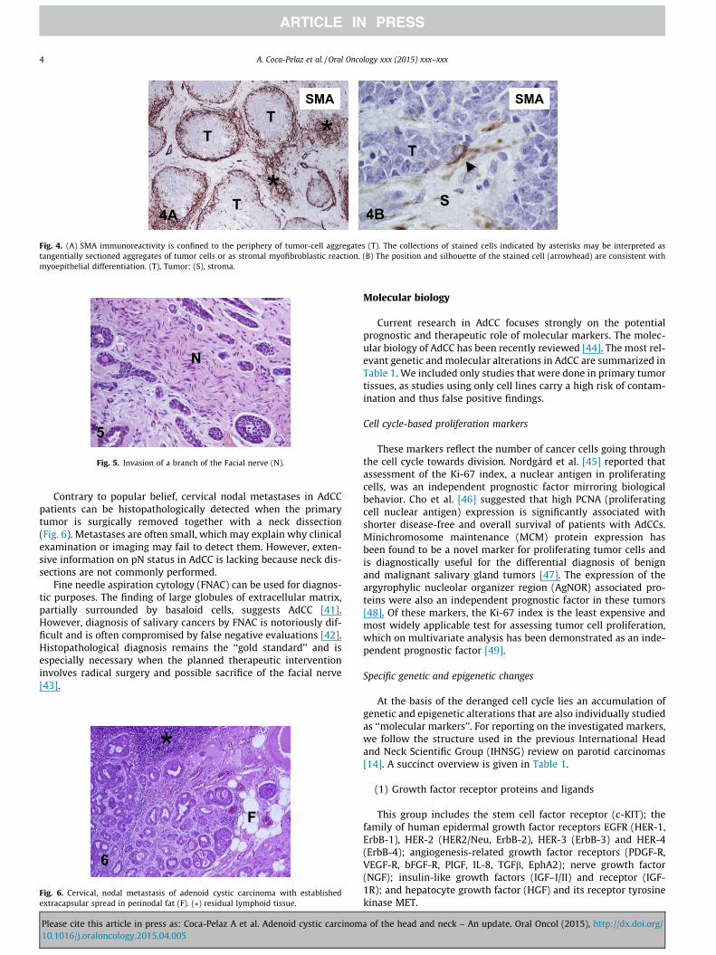
Neural invasion can be seen even in early-stage tumors and hasbeen regarded as an unfavorable prognostic factor, associated withdistant metastasis and adverse final outcome (Figs. 1C, 2C, and 5)[26,38]. Recently, however, an analysis of 495 AdCCs from 9 inter-national patient cohorts indicated that ‘‘while perineural invasionhas no impact on survival, intraneural invasion is an independentpredictor of poor prognosis’’ [39]. Another inference was that neu-ral invasion did not predict hematogenous spread; distant metas-tases were related to age, primary site and nodal (N)classification. Teymoortash et al. [40] reviewed 22 cases of AdCCwith documented perineural invasion and proposed a new classifi-cation scheme for AdCCs based on the presence of characteristicfeatures in perineural invasion. They classified tumors as p1 whentrue perineural or endoneural invasion was observed (Fig. 5) and
a of the head and neck – An update. Oral Oncol (2015), http://dx.doi.org/

T
M
E
1A 1B G
1C
N
N
Fig. 1. Note: Unless otherwise specified, the photomicrographs in this article are from sections of routinely processed tissue, which were stained with hematoxylin and eosin.It was not deemed necessary to give objective magnifications. Zooming on the electronic format of the photomicrographs would allow appreciation of detail, difficult to beseen on prints. (A) Scanned histological section of solid adenoid cystic carcinoma (T) of the floor of mouth; the hematoxyphilic (purplish) tumor is asymmetrical with asomewhat lobulated, advancing front. (E), oral epithelium; (M), skeletal muscle. (B) Invasion of salivary gland (G). (C) Highly invasive tumor penetrating soft tissues in theform of finger-like projections. Note the characteristic target-like arrangement around nerves (N). (For interpretation of the references to color in this figure legend, the readeris referred to the web version of this article.)
N
2B 2C L
2A
*
*
Fig. 2. (A) Cribriform pattern. Hematoxyphilic material is present in cyst-like spaces (asterisks). Appearances simulating the Roman-bridge pattern seen in salivary ductcarcinoma, but the strongly haematoxyphilic, basaloid cells preclude from considering that diagnosis. (B) Tubular pattern. Differences in tinctorial reactions between luminaland abluminal cells are discernible. (L), lumen. (C) Solid pattern. (Same case, as in Fig. 1A). Such tumors need to be differentiated from neuroendocrine carcinomas. Invasion ofa nerve (N) is seen.
3B
*
3A
* *
Fig. 3. Cribriform adenoid cystic carcinoma. (A) Compare the collapsed, true lumina (arrows) lined by eosinophilic, cuboidal cells with the cyst-like spaces (asterisk). (B)Selective p16 immunoreactivity of cells lining small collapsed lumina (arrows). The basaloid cells (arrowhead) surrounding the cyst-like spaces (asterisk) are unstained.
A. Coca-Pelaz et al. / Oral Oncology xxx (2015) xxx–xxx 3
p2 when tumor was adjacent to nerves without invading them.Patients with p1 tumors had a higher recurrence rate in compar-ison with p2 patients. However, the number of cases analyzedwas small, and it is recognized that perineural invasion is also a
Please cite this article in press as: Coca-Pelaz A et al. Adenoid cystic carcinom10.1016/j.oraloncology.2015.04.005
feature in polymophous low-grade carcinoma, a salivary glandmalignancy associated with a different prognosis. Tumor size andgrowth (‘pushing’ or frankly infiltrative) pattern may be of greaterinfluence.
a of the head and neck – An update. Oral Oncol (2015), http://dx.doi.org/
SMA SMA
T
T
T
T
* 4A 4B
*
S
Fig. 4. (A) SMA immunoreactivity is confined to the periphery of tumor-cell aggregates (T). The collections of stained cells indicated by asterisks may be interpreted astangentially sectioned aggregates of tumor cells or as stromal myofibroblastic reaction. (B) The position and silhouette of the stained cell (arrowhead) are consistent withmyoepithelial differentiation. (T), Tumor; (S), stroma.
N
5
Fig. 5. Invasion of a branch of the Facial nerve (N).
4 A. Coca-Pelaz et al. / Oral Oncology xxx (2015) xxx–xxx
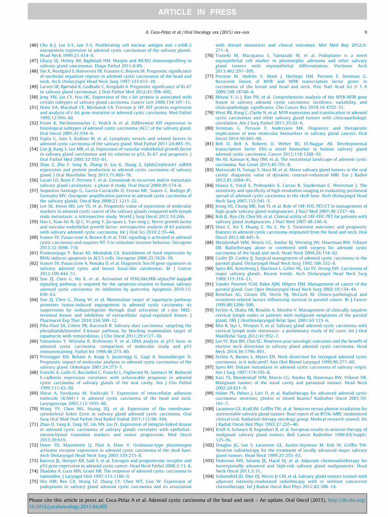
Contrary to popular belief, cervical nodal metastases in AdCCpatients can be histopathologically detected when the primarytumor is surgically removed together with a neck dissection(Fig. 6). Metastases are often small, which may explain why clinicalexamination or imaging may fail to detect them. However, exten-sive information on pN status in AdCC is lacking because neck dis-sections are not commonly performed.
Fine needle aspiration cytology (FNAC) can be used for diagnos-tic purposes. The finding of large globules of extracellular matrix,partially surrounded by basaloid cells, suggests AdCC [41].However, diagnosis of salivary cancers by FNAC is notoriously dif-ficult and is often compromised by false negative evaluations [42].Histopathological diagnosis remains the ‘‘gold standard’’ and isespecially necessary when the planned therapeutic interventioninvolves radical surgery and possible sacrifice of the facial nerve[43].
*
F
6
Fig. 6. Cervical, nodal metastasis of adenoid cystic carcinoma with establishedextracapsular spread in perinodal fat (F). (⁄) residual lymphoid tissue.
Please cite this article in press as: Coca-Pelaz A et al. Adenoid cystic carcinom10.1016/j.oraloncology.2015.04.005
Molecular biology
Current research in AdCC focuses strongly on the potentialprognostic and therapeutic role of molecular markers. The molec-ular biology of AdCC has been recently reviewed [44]. The most rel-evant genetic and molecular alterations in AdCC are summarized inTable 1. We included only studies that were done in primary tumortissues, as studies using only cell lines carry a high risk of contam-ination and thus false positive findings.
Cell cycle-based proliferation markers
These markers reflect the number of cancer cells going throughthe cell cycle towards division. Nordgård et al. [45] reported thatassessment of the Ki-67 index, a nuclear antigen in proliferatingcells, was an independent prognostic factor mirroring biologicalbehavior. Cho et al. [46] suggested that high PCNA (proliferatingcell nuclear antigen) expression is significantly associated withshorter disease-free and overall survival of patients with AdCCs.Minichromosome maintenance (MCM) protein expression hasbeen found to be a novel marker for proliferating tumor cells andis diagnostically useful for the differential diagnosis of benignand malignant salivary gland tumors [47]. The expression of theargyrophylic nucleolar organizer region (AgNOR) associated pro-teins were also an independent prognostic factor in these tumors[48]. Of these markers, the Ki-67 index is the least expensive andmost widely applicable test for assessing tumor cell proliferation,which on multivariate analysis has been demonstrated as an inde-pendent prognostic factor [49].
Specific genetic and epigenetic changes
At the basis of the deranged cell cycle lies an accumulation ofgenetic and epigenetic alterations that are also individually studiedas ‘‘molecular markers’’. For reporting on the investigated markers,we follow the structure used in the previous International Headand Neck Scientific Group (IHNSG) review on parotid carcinomas[14]. A succinct overview is given in Table 1.
(1) Growth factor receptor proteins and ligands
This group includes the stem cell factor receptor (c-KIT); thefamily of human epidermal growth factor receptors EGFR (HER-1,ErbB-1), HER-2 (HER2/Neu, ErbB-2), HER-3 (ErbB-3) and HER-4(ErbB-4); angiogenesis-related growth factor receptors (PDGF-R,VEGF-R, bFGF-R, PlGF, IL-8, TGFb, EphA2); nerve growth factor(NGF); insulin-like growth factors (IGF–I/II) and receptor (IGF-1R); and hepatocyte growth factor (HGF) and its receptor tyrosinekinase MET.
a of the head and neck – An update. Oral Oncol (2015), http://dx.doi.org/

Table 1Frequent molecular alterations in AdCC.
Function Markers Clinical relevance References
Cell proliferation markers – Ki-67 – Prognostic markers 45–49– PCNA– AgNORs
Growth factor receptor proteins and ligands – c-KIT – None 50–52– VEGF-C/VEGFR-3 – None 53– VEGF – Prognostic marker 54– EphA2/ephrinA1 – Prognostic marker 55– EGFR (HER-1) – Prognostic marker and therapeutic target 56–58– NGF – None 59– TrkC/NTRK3 – Potential therapeutic target 60
Cell cycle oncogenes – Cyclin D1 – Prognostic marker 57– SOX4 – Unknown 61– SOX10 – Diagnostic marker 62– PI3K/AKT pathway – Potential therapeutic target 63–65
DNA damage repair proteins – p53 – Prognostic marker 66
Cell adhesion proteins – E-cadherin – Prognostic markers 68– ICAM-1 69– Ezrin/CD44v6 70– ILK 71– uPAR 72
Estrogen receptors – Estrogen receptor – Potential therapeutic target 73–74, 80
Lymphangiogenesis markers – Podoplanin – Prognostic marker 75
Transcriptional factors – MYB – Diagnostic and prognostic marker 76–80– EN1 – Prognostic marker 81
AdCC = adenoid cystic carcinoma.
A. Coca-Pelaz et al. / Oral Oncology xxx (2015) xxx–xxx 5
c-KIT is detected in 80–94% of AdCC [50]. The relationshipbetween high c-KIT expression (>50%) and histologic grade isdebated. While Holst et al. [51] noted a significant association ofc-KIT expression with grade III or solid pattern AdCC, Freier et al.[52] described a high expression only in cribriform and tubularAdCC.
Limited amounts of VEGF-C are produced in AdCC, which via areduced interaction with VEGFR-3 may result in few lymphaticvessels; whether this relates to the purported low rate of cervicalmetastasis remains to be evaluated [53].
Data on angiogenesis-related growth factor receptors of salivarycancers generally suggest an association of VEGF expression withadvanced stage and worse disease-specific survival [54].Specifically for AdCC, overexpression of EphA2, a receptor tyrosinekinase involved in angiogenesis, and its ligand ephrin A1, havebeen recently reported. The overexpression is significantly greaterin solid AdCC than in tubular and cribriform types and correlateswith microvessel density, TNM staging and perineural and vascularinvasion [55].
For the human EGFR family, EGFR/HER-1 has been identified in82% of investigated patients with advanced AdCC. In patientsexpressing HER-1, treatment with cetuximab seemed to result ina higher proportion of stabilized disease [56]. In a study on 24AdCCs, HER-1/CCND1/PIK3CA coamplification was the most con-sistently observed pattern (29%). HER-1 amplification correlatedwith distant metastasis, and the cases with the HER-1/CCND1/PIK3CA coamplification tended towards a reduced survival [57].The observed HER-1 related biological aggressiveness, however,could not be confirmed by Lee et al. [58].
Increased expression of NGF is observed in 79% of solid typeAdCCs (or 68% of all studied AdCC) and may relate to its neu-rotropism [59]. In this context, the overexpression of a cluster ofneuronal genes grouped around TrkC/NTRK3 (a tyrosine kinaseneurotrophic receptor associated with neurogenesis and cancer)suggests that AdCC expresses genes involved in neural stem celldifferentiation [60].
Please cite this article in press as: Coca-Pelaz A et al. Adenoid cystic carcinom10.1016/j.oraloncology.2015.04.005
(2) Cell cycle oncogenes
The growth factor–receptor interaction activates cell cycleoncogenes. For AdCC these include cyclin D1 [57]; sex-determiningregion Y-box 4 and 10 (SOX-4, SOX-10) [33,61,62]; nuclear factorjB (NFjB); phosphatidylinositol 3 phosphate kinase/serine-thre-onine protein kinase (PI3K); sarcoma signal transducer and activa-tor of transcription 3 (STAT3); and mammalian target of rapamycin(mTOR) [63,64].
Cyclin D1 seems frequently overexpressed in AdCC tumors andcorrelates with prognosis [70].
Frierson et al. [33] found that SOX-4 was the most significantlyoverexpressed cell cycle oncogene in a microarray analysis ofAdCC. Apoptosis increases following SOX-4 knockdown, suggestingthat down-regulation of inhibitors of the NFjB pathway (inhibitorprotein I-jB-a) and up-regulation of apoptosis inhibitors such assurvivin are the downstream effects [61]. Very recently, the tran-scriptional factor SOX-10, normally expressed only during salivarygland differentiation, was found markedly upregulated in a major-ity of AdCC cells [62].
The PI3K/AKT/mTOR axis is critical in oncogenesis; dysregula-tion of this pathway involves alterations of various upstreamtumor-associated growth factors (EGFR, HER-2, PDGF and VEGF)as well as AKT, mTOR, and PTEN [63]. The PI3K/AKT pathway canbe targeted by blocking mTOR with temsirolimus [65].
(3) DNA damage repair proteins
p53 expression [66] and p53 mutations in AdCC appear gener-ally associated with worse prognosis [67].
(4) Cell adhesion proteins
Loss of E-cadherin expression (due to promoter hypermethyla-tion) is frequently found in AdCC, and has been correlated withpoor prognosis [68]. Also the reduced expression of intercellular
a of the head and neck – An update. Oral Oncol (2015), http://dx.doi.org/

6 A. Coca-Pelaz et al. / Oral Oncology xxx (2015) xxx–xxx
adhesion molecule-1 (ICAM-1) may promote immune evasion andmetastasis, resulting in poor prognosis in AdCC [69]. Increasedexpression of the membrane-cytoskeletal linker Ezrin, and its part-ner CD44V6, has been also associated with a more aggressivebehavior [70].
A recently revealed association in AdCC links integrin-linkedkinase (ILK) with epithelial-mesenchymal transition markers,where ILK plays a central role in cell-extracellular matrix interac-tions regulating cell proliferation, apoptosis, differentiation andmigration. Expression of ILK correlates strongly with solid typeAdCC, advanced TNM stage and increased risk of recurrence.Moreover, ILK over-expression and a neural invasive phenotypewent along with downregulation of E-cadherin and upregulationof Snail and N-cadherin; by this mechanism ILK is believed to havea key role in progression and metastasis in AdCC [71].
The urokinase-type plasminogen activator receptor influencestumor invasion and metastasis by facilitating the destruction ofextracellular matrices, so its expression seems to be a negativeprognostic factor [72].
(5) Estrogen receptors
Estrogen receptors have been described in 17–92% of studiedAdCC, suggesting a role for anti-estrogen therapy with tamoxifen[73] and supporting the plausibility of an earlier observed partialremission in a patient treated in this way [74].
(6) Markers for lymphangiogenesis
Podoplanin is a small mucin-like protein related to tissue devel-opment and repair. It is expressed in lymphatic endothelial cellsand is used as a marker for lymphangiogenesis. It is also expressedin certain tumor cells and is associated with migration/invasion incervical and oral squamous cell carcinomas. Recently, podoplaninwas found overexpressed in a subset of salivary AdCCs (32.5% oftumors); overexpression was significantly associated with dis-ease-free survival and distant metastasis, although it was not asso-ciated with recurrence and overall survival [75]. Despite thisobservation, caution should be exerted with this marker as podo-planin has been recently localized in nonluminal (myoepithelial-like) cells of pleomorphic salivary adenoma [76].
(7) Transcription factors
The reciprocal translocation t(6;9) (6q22–23; 9p23–24), result-ing in fusion of the MYB gene on chromosome 6q22–q23 and thetranscription factor NFIB on chromosome 9p23–p24, has recentlybeen described in AdCC [77]. The fusion was shown to upregulateMYB protein expression, which is believed to be the oncogenic dri-ver of this tumor. Mitani et al. confirmed this fusion, includingmultiple variant fusions, as specific to AdCC among salivary glandtumors [78].
Overexpression of MYB is observed in fusion-positive but also ina subset of fusion-negative tumors, presumably through differentmechanisms, confirming the critical role for MYB in all AdCCs irre-spective of fusion status [78]. West et al. [79] found the MYB-NFIBfusion in about half of AdCC cases, but also MYB rearrangementwithout NFIB in 16%, for a total of 65% of cases showing abnormalMYB patterns. This suggests alternative fusion partners for MYB. Atrend towards worse outcome in fusion-positive cases was alsofound in this study [79]. Recently, several laboratories are tryingto develop inhibitors of MYB, MYB–NFIB, and their molecular tar-gets, but early results have not been promising [80]. It is not clearwhether the translocation in AdCC will be useful as a diagnosticmarker, a prognostic marker (for poor prognosis) or as a target oftherapy.
Please cite this article in press as: Coca-Pelaz A et al. Adenoid cystic carcinom10.1016/j.oraloncology.2015.04.005
The expression of the transcription factor Engrailed Homeobox1 (EN1) is silenced by hypermethylation in AdCC [81]. EN1 isimportant for development of the central nervous system. EN1hypermethylation correlates with histologic tumor grade, tumorlocation and final patient outcome. EN1 protein expression seemstypical for solid AdCC and implies a significantly lower survivalrate. On these grounds EN1 could prove a potentially useful bio-marker in AdCC.
Mutational profile of AdCC
Chan and colleagues reported on exome and genome sequenc-ing of 60 AdCC tumor/normal pairs [82]. A low exonic somaticmutation rate (a mean of 22 somatic mutations per sample, corre-sponding to approximately 0.31 non-silent events per megabase)and wide mutational diversity was found. They identified potentialdriver mutations, including those in PIK3CA, TP53, PTEN,SMARCA2, KDM6A and CREBBP. Analysis of these driver genesdemonstrated marked enrichment in pathways involved in chro-matin remodeling, DNA damage, MYB, protein kinase A (PKA) sig-naling and PI3K signaling. In addition, they observed MYB-NFIBtranslocations and somatic mutations in MYB-associated genes,solidifying the role of these aberrations as critical events inAdCC. The discovery of genomic alterations in targetable pathwayssuggests potential avenues for novel therapies for a typicallychemoresistant malignancy.
Diagnostic imaging
As is the case for all SGCs, preoperative diagnostic imaging ofAdCC includes computed tomography (CT) and/or magnetic reso-nance imaging (MRI). This allows estimating the anatomical dis-ease extent, which is crucial for accurate surgical planning. It iswell accepted that CT is better at delineating bone invasion,whereas MRI superior for assessing soft tissue extension [83].
Evidence of sensory (pain or paresthesia) or motor nerve dys-function (VII nerve paresis/paralysis) mandates MRI investigationto assess the corresponding nerves. Hanna et al. [84] evaluatedthe sensitivity and specificity of CT and MRI in detecting perineuralspread of AdCCs along the base of the skull and concluded that MRIis clearly superior to CT for this purpose.
The main role of [18F] fluorodeoxyglucose positron emissiontomography (18F-FDG PET), preferably in combination with CT, isto exclude gross distant disease for head and neck tumors in gen-eral and also more specifically for SGCs and AdCC [85]. Roh et al.[86] investigated the role of 18F-FDG PET in the management ofpatients with salivary cancers and found it helpful in initial staging,histologic grading and monitoring after treatment. However, 18F-FDG PET-CT is not helpful to rule out distant metastasis if the pri-mary SGC does not show enhanced FDG uptake, a situation that isnot infrequent in AdCC. Furthermore, AdCC with relatively low FDGuptake might be obscured by the normal physiologic FDG uptake ofthe salivary glands; conversely, salivary glands are frequentlyaffected by inflammatory processes wherein increased FDG uptakemight result in a false-positive result.
Treatment
Treatment of AdCC is influenced by location of the tumor, stageat diagnosis and biologic behavior as reflected in histologic grade[87]. The ‘‘gold-standard’’ treatment for AdCCs, that is deemed aspotentially resectable after extensive workup is radical surgicalresection, ensuring free margins, and postoperative radiotherapy.Mendenhall et al. [88] compared radiotherapy alone to radiother-apy combined with surgery and concluded that combination
a of the head and neck – An update. Oral Oncol (2015), http://dx.doi.org/

Table 2Molecular targets and studied therapies in salivary gland cancers [10].a
Molecular target Salivary glandcancer type
Molecular therapy(selected)
c-KIT AdCC Imatinib [105–108]Sunitinib [109]
EGFR, ErbB-1 All types Cetuximab [56]Gefinitib [110]
HER2/Neu, ErbB-2 All types Trastuzumab [111]Lapatinib [112]
NFjB – proteasomes degradingits inhibitor (IjBa)
AdCC Bortezomib [113]
AdCC = adenoid cystic carcinoma.a Modified with permission.
A. Coca-Pelaz et al. / Oral Oncology xxx (2015) xxx–xxx 7
treatment is preferable. AdCC has a high propensity for infiltratinginto adjacent tissues, especially through perineural invasion soeven in ‘‘resectable’’ AdCC the goal of ‘‘free margins’’ is often notachieved. Incomplete resection is typically a problem in AdCC aris-ing in anatomical sites with difficult access. This is highlighted by astudy indicating that 80% of skull base AdCC had positive surgicalmargins, despite the preoperative impression of experienced sur-geons that resection with clear margins would be possible [89].
When AdCC arises in the parotid, the facial nerve should be pre-served if it is not determined to be paralyzed preoperatively andnot intimately involved by tumor at the time of surgery; postoper-ative radiotherapy would be an effective adjuvant treatment forresidual microscopic disease on a spared nerve branch [90–92].
Due to the historically low incidence of occult nodal metastasis,neck dissection is only performed in case of clinically positivelymph nodes. Clinically obvious lymph node metastasis is not fre-quent in AdCC, especially for parotid gland primaries [93].However, for MSG subsites the incidence of involved lymph nodesseems higher. Min et al. [94] described a general incidence oflymph node metastases in AdCCs of the head and neck of almost10%, which was mainly attributed to sites such as base of tongue,mobile tongue and floor of the mouth. These authors noted thatprimary tumor site and peri-tumoral lymphovascular invasionwere significantly associated with cervical lymph node metastasis.On this basis, selective neck dissection should be considered fortumors of those sites showing lymphovascular invasion. It is, how-ever, noted that lymphovascular invasion is more likely to be his-tologically detected in resection specimens rather than diagnosticbiopsies, which are also expected to be small when taken fromanatomically ‘‘difficult’’ sites. Lee et al. [95] recently reported that15.38% of the patients who received elective neck dissection hadoccult metastases, and they recommend performing elective neckdissection for staging and achieving regional control. It remainsunclear whether regional control, let alone survival, is improvedby performing an elective neck dissection in these patients as com-pared to a strategy of primary radiotherapy of the neck nodes. ForAdCC in locations like the submandibular gland, parotid and lar-ynx, lymph nodes could also be involved by direct extension ofthe primary tumor [96,97].
Unfortunately, local recurrences occur despite combined treat-ment with surgery and radiotherapy. Some authors postulate thatpostoperative radiation may delay rather than prevents recurrence[98]. Other modalities of radiotherapy, particularly neutron irradi-ation, have also been studied. Huber et al. [99] retrospectivelycompared radiotherapy with neutrons, photons and a photon/neu-tron mixed beam in patients with AdCC of the head and neck. Theypostulated that the neutron-specific reduced oxygen enhancementfactor, decreased variability of sensitivity through the cell cycleand lowered repair of sublethal cell damage explain the high 5 yearlocal control in up to 75% in unresectable AdCC cases treated withthis approach [100]. Unfortunately this does not result in a survivalbenefit, mainly because of unaffected distant metastasis (in 2 outof 5 patients after 51 months). Furthermore, severe late side effectsof this approach include necrosis of the soft tissues, mandible, tem-poral bone and the temporal brain lobe, as well as cervicalmyelopathy and sensorineural hearing loss [101,102].
In patients with adverse prognostic factors, chemoradiotherapyusing various agents may be considered, and preliminary evidencesuggests its usefulness [103]. In a series of SGCs (43% AdCC),Schoenfeld et al. [104] explored the use of carboplatin or paclitaxelas a radiosensitizer, and trastuzumab in HER2/Neu-positive tumors.
The role of palliative systemic therapies (including cytotoxicand targeted therapies) has recently been extensively reviewed[15]. Only level-3 evidence (case-control or cohort studies) exists,and the available trials have only involved small patient numberswith heterogeneity regarding histology, many having had prior
Please cite this article in press as: Coca-Pelaz A et al. Adenoid cystic carcinom10.1016/j.oraloncology.2015.04.005
systemic therapies and a proportion of patients with locoregionalrecurrence versus distant metastasis. Moreover, the majority ofAdCCs have a slow growth pattern, making it difficult to assessthe efficacy of the particular systemic therapy, as objective tumorresponses are uncommon [16]. Several studied targeted therapiesfor the disease are presented in Table 2 [10,56,105–113]. For anoverview of ongoing or planned clinical trials of targeted therapiesfor AdCC, the reader is referred to the recent publication of Dillonet al. [114]. The latest efforts combine cytotoxic and targeted treat-ments: in patients with EGFR-expressing AdCC, the efficacy ofcetuximab plus cisplatin-based chemoradiotherapy (for locallyadvanced tumors) or chemotherapy (for systemic disease)appeared encouraging and was associated with manageable toxic-ity [115].
One may conclude that to date, systemic treatment employingcytotoxic chemotherapy or targeted molecular therapies does notyet result in patient cure with advanced (locally recurrent or meta-static) AdCC. At best, temporary partial disease response or stabi-lization may be achieved. Chemotherapy therefore should bereserved as a palliative treatment for patients with poorly con-trolled disease or affected with symptomatic metastases [114].
Prognosis
In general, prognosis of AdCC of the head and neck is ratherpoor, and this is the reason why many authors consider AdCC a‘‘clinically high grade’’ neoplasm. Reported prognosis varieswidely, mainly because different reports have different qualityand length of follow-up. In a large European study on AdCC ofthe head and neck, the 10-year survival rate was 65% [116]. VanWeert et al. [23] reported 5-, 10- and 20-year survival rates of68%, 52% and 28%, respectively, on a series of 105 patients.Huang et al. [38] reported that overall and recurrence-free survivalrates at three years were 84.6% and 58.2%, respectively, while therate of survival with recurrence was 26.4%. After 15 years, theoverall survival rate was 24.5%, and the recurrence-free survivalrate was 22.6%.
Many clinicians assume that ‘‘cure is never achieved’’ in AdCC.Optimistic 5-year survival rates are occasionally reported (e.g.,92% in an Australian series) [117], but 10- and 20-year survivalrates continuously drop. In a UK series, 40% of AdCC patients werealive at 20 years and survival continued to drop until 30 years; theactuarial primary site recurrence rate at 30 years in that study was100% [118], and it was 54% in the Australian series.
The low long-term survival rate of AdCC patients is uniformlylinked to the failure to control distant disease. These distant metas-tases occur most frequently in the lung. Spiro [97] suggested thatthe incidence of distant metastasis to other anatomical sites islikely higher than previously recognized, because once lung metas-tases are detected no further investigations are performed. Van derWal et al. [119] found that the average time between the
a of the head and neck – An update. Oral Oncol (2015), http://dx.doi.org/

8 A. Coca-Pelaz et al. / Oral Oncology xxx (2015) xxx–xxx
occurrence of lung metastases and death was 32.3 months, andthat between the occurrence of metastases elsewhere and deathwas 20.6 months; it was hypothesized that metastases outsidethe lungs may be detected later in the course of the disease orinterfere with vital functions more rapidly. Umeda et al. [120]found that the estimated doubling time of pulmonary metastasisin AdCC ranges from 86 to 1064 days with an average of 393 days.These findings suggest that metastasis at the cellular level couldoccur prior (average, 227 months) to clinical presentation of pri-mary cancer.
Conclusion
Advances in therapeutic modalities have not had a significantimpact on the natural history of AdCC of the head and neck. Thepreferred treatment for the majority of the patients is radical sur-gery with postoperative radiotherapy. The frequent developmentof distant metastases continues to determine treatment outcome.Currently, the only options for patients with metastatic diseaseare at best supportive care, palliative systemic therapy or inclusionin clinical trials in order to establish effective and evidence-basedtreatment strategies.
Conflict of interest
None declared.
Acknowledgements
We declare no funds for this research.
References
[1] Bjørndal K, Krogdahl A, Therkildsen MH, et al. Salivary gland carcinoma inDenmark 1990–2005: a national study of incidence, site and histology.Results of the Danish Head and Neck Cancer Group (DAHANCA). Oral Oncol2011;47:677–82.
[2] Dodd RL, Slevin NJ. Salivary gland adenoid cystic carcinoma: a review ofchemotherapy and molecular therapies. Oral Oncol 2006;42:759–69.
[3] Bradley PJ. Adenoid cystic carcinoma of the head and neck: a review. CurrOpin Otolaryngol Head Neck Surg 2004;12:127–32.
[4] Vander Poorten VL, Balm AJ, Hilgers FJ, et al. Prognostic factors for long termresults of the treatment of patients with malignant submandibular glandtumors. Cancer 1999;85:2255–64.
[5] Husain Q, Kanumuri VV, Svider PF, et al. Sinonasal adenoid cystic carcinoma:systematic review of survival and treatment strategies. Otolaryngol HeadNeck Surg 2013;148:29–39.
[6] Azar T, Abdul-Karim FW, Tucker HM. Adenoid cystic carcinoma of the trachea.Laryngoscope 1998;108:1297–300.
[7] Saraydaroglu O, Coskun H, Kasap M. Unique presentation of adenoid cysticcarcinoma in postcricoid region: a case report and review of the literature.Head Neck Pathol 2011;5:413–5.
[8] Gu FM, Chi FL, Dai CF, Chen B, Li HW. Surgical outcomes of 43 cases withadenoid cystic carcinoma of the external auditory canal. Am J Otolaryngol2013;34:394–8.
[9] Argyris PP, Pambuccian SE, Cayci Z, Singh C, Tosios KI, Koutlas IG. Lacrimalgland adenoid cystic carcinoma with high-grade transformation tomyoepithelial carcinoma: report of a case and review of literature. HeadNeck Pathol 2013;7:85–92.
[10] Vander Poorten V, Hunt J, Bradley PJ, et al. Recent trends in the managementof minor salivary gland carcinoma. Head Neck 2014;36:444–55.
[11] Stell PM. Adenoid cystic carcinoma. Clin Otolaryngol Allied Sci 1986;11:267–91.
[12] Dockerty MB, Mayo CW. Primary tumors of submaxillary gland with specialreference to mixed tumors. Surg Gynecol Obstet 1942;74:1033–45.
[13] Kokemueller H, Eckardt A, Brachvogel P, Hausamen JE. Adenoid cysticcarcinoma of the head and neck – a 20 years experience. Int J OralMaxillofac Surg 2004;33:25–31.
[14] Vander Poorten V, Bradley PJ, Takes RP, Rinaldo A, Woolgar JA, Ferlito A.Diagnosis and management of parotid carcinoma with a special focus onrecent advances in molecular biology. Head Neck 2012;34:429–40.
[15] Vander Poorten V, Meulemans J, Delaere P, Nuyts S, Clement P. Molecularmarkers and chemotherapy for advanced salivary cancer. CurrOtorhinolaryngol Rep 2014;2:85–96.
Please cite this article in press as: Coca-Pelaz A et al. Adenoid cystic carcinom10.1016/j.oraloncology.2015.04.005
[16] Laurie SA, Ho AL, Fury MG, Sherman E, Pfister DG. Systemic therapy in themanagement of metastatic or locally recurrent adenoid cystic carcinoma ofthe salivary glands: a systematic review. Lancet Oncol 2011;12:815–24.
[17] Terhaard CH, Lubsen H, Van der Tweel I, et al. Salivary gland carcinoma:independent prognostic factors for locoregional control, distant metastases,and overall survival: results of the Dutch head and neck oncology cooperativegroup. Head Neck 2004;26:681–92.
[18] Bonaparte JP, Hart R, Trites J, Taylor MS. Incidence of adenoid cysticcarcinoma in Nova Scotia: 30-year population-based epidemiologic study. JOtolaryngol Head Neck Surg 2008;37:642–8.
[19] van der Wal JE, Snow GB, van der Waal I. Histological reclassification of 101intraoral salivary gland tumours (new WHO classification). J Clin Pathol1992;45:834–5.
[20] Vander Poorten VLM, Balm AJM, Hilgers FJM, Tan IB, Keus RB, Hart AAM. Stageas major long term outcome predictor in minor salivary gland carcinoma.Cancer 2000;89:1195–204.
[21] Vander Poorten VL, Hart AA, van der Laan BF, et al. Prognostic index forpatients with parotid carcinoma: external validation using the nationwide1985–1994 Dutch head and neck oncology cooperative group database.Cancer 2003;97:1453–63.
[22] Batsakis JG. Carcinomas of the submandibular and sublingual glands. AnnOtol Rhinol Laryngol 1986;95:211–2.
[23] Van Weert S, Bloemena E, van der Waal I, et al. Adenoid cystic carcinoma ofthe head and neck: a single-center analysis of 105 consecutive cases over a30-year period. Oral Oncol 2013;49:824–9.
[24] Nascimento AG, Amaral AL, Prado LA, Kligerman J, Silveira TR. Adenoid cysticcarcinoma of salivary glands. A study of 61 cases with clinicopathologiccorrelation. Cancer 1986;57:312–9.
[25] Spiro RH, Huvos AG, Strong EW. Adenoid cystic carcinoma of salivary origin. Aclinicopathologic study of 242 cases. Am J Surg 1974;128:512–20.
[26] Sequeiros Santiago G, Rodrigo Tapia JP, Llorente Pendás JL, Suárez Nieto C.Prognostic factors in adenoid cystic carcinoma of salivary glands. ActaOtorrinolaringol Esp 2005;56:361–7 [article in Spanish].
[27] Spiro RH, Thaler HT, Hicks WF, Kher UA, Huvos AH, Strong EW. Theimportance of clinical staging of minor salivary gland carcinoma. Am J Surg1991;162:330–6.
[28] Zvrko E, Golubovic M. Laryngeal adenoid cystic carcinoma. ActaOtorhinolaryngol Ital 2009;29:279–82.
[29] Conley J, Dingman DL. Adenoid cystic carcinoma in the head and neck(cylindroma). Arch Otolaryngol 1974;100:81–90.
[30] Vander Poorten VLM, Balm AJM, Hilgers FJM, et al. The development of aprognostic score for patients with parotid carcinoma. Cancer 1999;85:2057–67.
[31] Biswas KD, Saha J, Sen I, et al. Unusual presentations of adenoid cysticcarcinoma in extra-salivary gland subsites in head and neck region: a caseseries. Indian J Otolaryngol Head Neck Surg 2014;66(Suppl. 1):286–90.
[32] Cheng J, Saku T, Okabe H, Furthmayr H. Basement membranes in adenoidcystic carcinoma. An immunohistochemical study. Cancer 1992;69:2631–40.
[33] Frierson Jr HF, El-Naggar AK, Welsh JB, et al. Large scale molecular analysisidentifies genes with altered expression in salivary adenoid cystic carcinoma.Am J Pathol 2002;161:1315–23.
[34] Chen JC, Gnepp DR, Bedrossian CW. Adenoid cystic carcinoma of the salivaryglands: an immunohistochemical analysis. Oral Surg Oral Med Oral Pathol1988;65:316–26.
[35] Prasad ML, Barbacioru CC, Rawal YB, Husein O, Wen P. Hierarchical clusteranalysis of myoepithelial/basal cell markers in adenoid cystic carcinoma andpolymorphous low-grade adenocarcinoma. Mod Pathol 2008;21:105–14.
[36] Chisholm DM, Waterhouse JP, Kraucunas E, Sciubba JJ. A qualitative andquantitative electronmicroscopic study of the structure of the adenoid cysticcarcinoma of human minor salivary glands. J Oral Pathol 1975;4:103–19.
[37] Szanto PA, Luna MA, Tortoledo ME, White RA. Histologic grading of adenoidcystic carcinoma of the salivary glands. Cancer 1984;54:1062–9.
[38] Huang M, Ma D, Sun K, Yu G, Guo C, Gao F. Factors influencing survival rate inadenoid cystic carcinoma of the salivary glands. Int J Oral Maxillofac Surg1997;26:435–9.
[39] Amit M, Binenbaum Y, Trejo-Leider L, et al. International collaborativevalidation of intraneural invasion as a prognostic marker in adenoidcystic carcinoma of the head and neck. Head Neck 2014 Apr 7. [Epub aheadof print].
[40] Teymoortash A, Zieger L, Hoch S, Pagenstecher A, Hofer MJ. Distinctmicroscopic features of perineural invasion in adenoid cystic carcinoma ofthe head and neck. Histopathology 2014;64:1037–9.
[41] Nagel H, Hotze HJ, Laskawi R, Chilla R, Droese M. Cytologic diagnosis ofadenoid cystic carcinoma of salivary glands. Diagn Cytopathol 1999;20:358–66.
[42] Daneshbod Y, Daneshbod K, Khademi B. Diagnostic difficulties in theinterpretation of fine needle aspirate samples in salivary lesions: diagnosticpitfalls revisited. Acta Cytol 2009;53:53–70.
[43] Lü BJ, Zhu J, Gao L, Xie L, Xu JY, Lai MD. Diagnostic accuracy and pitfalls in fineneedle aspiration cytology of salivary glands: a study of 113 cases. ZhonghuaBing Li Xue Za Zhi 2005;34:706–10 [article in Chinese].
[44] Liu J, Shao C, Tan ML, Mu D, Ferris RL, Ha PK. Molecular biology of adenoidcystic carcinoma. Head Neck 2012;34:1665–77.
[45] Nordgård S, Franzén G, Boysen M, Halvorsen TB. Ki-67 as a prognostic markerin adenoid cystic carcinoma assessed with the monoclonal antibody MIB1 inparaffin sections. Laryngoscope 1997;107:531–6.
a of the head and neck – An update. Oral Oncol (2015), http://dx.doi.org/
A. Coca-Pelaz et al. / Oral Oncology xxx (2015) xxx–xxx 9
[46] Cho K-J, Lee S-S, Lee Y-S. Proliferating cell nuclear antigen and c-erbB-2oncoprotein expression in adenoid cystic carcinomas of the salivary glands.Head Neck 1999;21:414–9.
[47] Ghazy SE, Helmy IM, Baghdadi HM. Maspin and MCM2 immunoprofiling insalivary gland cancinomas. Diagn Pathol 2011;6:89.
[48] Xie X, Nordgård S, Halvorsen TB, Franzen G, Boysen M. Prognostic significanceof nucleolar organizer regions in adenoid cystic carcinomas of the head andneck. Arch Otolaryngol Head Neck Surg 1997;123:615–20.
[49] Larsen SR, Bjørndal K, Godballe C, Krogdahl A. Prognostic significance of Ki-67in salivary gland carcinomas. J Oral Pathol Med 2012;41:598–602.
[50] Jeng YM, Lin CY, Hsu HC. Expression of the c-kit protein is associated withcertain subtypes of salivary gland carcinoma. Cancer Lett 2000;154:107–11.
[51] Holst VA, Marshall CE, Moskaluk CA, Frierson Jr HF. KIT protein expressionand analysis of c-kit gene mutation in adenoid cystic carcinoma. Mod Pathol1999;12:956–60.
[52] Freier K, Flechtenmacher C, Walch A, et al. Differential KIT expression inhistological subtypes of adenoid cystic carcinoma (ACC) of the salivary gland.Oral Oncol 2005;41:934–9.
[53] Fujita G, Sato S, Kishino M, et al. Lymphatic vessels and related factors inadenoid cystic carcinoma of the salivary gland. Mod Pathol 2011;24:885–91.
[54] Lim JJ, Kang S, Lee MR, et al. Expression of vascular endothelial growth factorin salivary gland carcinomas and its relation to p53, Ki-67 and prognosis. JOral Pathol Med 2003;32:552–61.
[55] Shao Z, Zhu F, Song K, Zhang H, Liu K, Shang Z. EphA2/ephrinA1 mRNAexpression and protein production in adenoid cystic carcinoma of salivarygland. J Oral Maxillofac Surg 2013;71:869–78.
[56] Locati LD, Bossi P, Perrone F, et al. Cetuximab in recurrent and/or metastaticsalivary gland carcinomas: a phase II study. Oral Oncol 2009;45:574–8.
[57] Sequeiros-Santiago G, García-Carracedo D, Fresno MF, Suarez C, Rodrigo JP,Gonzalez MV. Oncogene amplification pattern in adenoid cystic carcinoma ofthe salivary glands. Oncol Rep 2009;21:1215–22.
[58] Lee SK, Kwon MS, Lee YS, et al. Prognostic value of expression of molecularmarkers in adenoid cystic cancer of the salivary glands compared with lymphnode metastasis: a retrospective study. World J Surg Oncol 2012;10:266.
[59] Hao L, Xiao-lin N, Qi C, Yi-ping Y, Jia-quan L, Yan-ning L. Nerve growth factorand vascular endothelial growth factor: retrospective analysis of 63 patientswith salivary adenoid cystic carcinoma. Int J Oral Sci 2010;2:35–44.
[60] Ivanov SV, Panaccione A, Brown B, et al. TrkC signaling is activated in adenoidcystic carcinoma and requires NT-3 to stimulate invasive behavior. Oncogene2013;32:3698–710.
[61] Pramoonjago P, Baras AS, Moskaluk CA. Knockdown of Sox4 expression byRNAi induces apoptosis in ACC3 cells. Oncogene 2006;25:5626–39.
[62] Ivanov SV, Panaccione A, Nonaka D, et al. Diagnostic Sox10 gene signatures insalivary adenoid cystic and breast basal-like carcinomas. Br J Cancer2013;109:444–51.
[63] Sun ZJ, Chen G, Hu X, et al. Activation of PI3K/Akt/IKK-alpha/NF-kappaBsignaling pathway is required for the apoptosis-evasion in human salivaryadenoid cystic carcinoma: its inhibition by quercetin. Apoptosis 2010;15:850–63.
[64] Sun ZJ, Chen G, Zhang W, et al. Mammalian target of rapamycin pathwaypromotes tumor-induced angiogenesis in adenoid cystic carcinoma: itssuppression by isoliquiritigenin through dual activation of c-Jun NH2-terminal kinase and inhibition of extracellular signal-regulated kinase. JPharmacol Exp Ther 2010;334:500–12.
[65] Piha-Paul SA, Cohen PR, Kurzrock R. Salivary duct carcinoma: targeting thephosphatidylinositol 3-kinase pathway by blocking mammalian target ofrapamycin with temsirolimus. J Clin Oncol 2011;29:e727–30.
[66] Yamamoto Y, Wistuba II, Kishimoto Y, et al. DNA analysis at p53 locus inadenoid cystic carcinoma: comparison of molecular study and p53immunostaining. Pathol Int 1998;48:273–80.
[67] Preisegger KH, Beham A, Kopp S, Jessernigg G, Gugl A, Stammberger H.Prognostic impact of molecular analyses in adenoid cystic carcinomas of thesalivary gland. Onkologie 2001;24:273–7.
[68] Franchi A, Gallo O, Bocciolini C, Franchi L, Paglierani M, Santucci M. ReducedE-cadherin expression correlates with unfavorable prognosis in adenoidcystic carcinoma of salivary glands of the oral cavity. Am J Clin Pathol1999;111:43–50.
[69] Shirai A, Furukawa M, Yoshizaki T. Expression of intercellular adhesionmolecule (ICAM)-1 in adenoid cystic carcinoma of the head and neck.Laryngoscope 2003;113:1955–60.
[70] Wang YY, Chen WL, Huang ZQ, et al. Expression of the membrane-cytoskeletal linker Ezrin in salivary gland adenoid cystic carcinoma. OralSurg Oral Med Oral Pathol Oral Radiol Endod 2011;112:96–104.
[71] Zhao D, Yang K, Tang XF, Lin NN, Liu JY. Expression of integrin-linked kinasein adenoid cystic carcinoma of salivary glands correlates with epithelial–mesenchymal transition markers and tumor progression. Med Oncol2013;30:619.
[72] Doerr TD, Marentette LJ, Flint A, Elner V. Urokinase-type plasminogenactivator receptor expression in adenoid cystic carcinoma of the skull base.Arch Otolaryngol Head Neck Surg 2003;129:215–8.
[73] Barrera JE, Shroyer KR, Said S, et al. Estrogen and progesterone receptor andp53 gene expression in adenoid cystic cancer. Head Neck Pathol 2008;2:13–8.
[74] Shadaba A, Gaze MN, Grant HR. The response of adenoid cystic carcinoma totamoxifen. J Laryngol Otol 1997;111:1186–9.
[75] Wu HM, Ren GX, Wang LZ, Zhang CY, Chen WT, Guo W. Expression ofpodoplanin in salivary gland adenoid cystic carcinoma and its association
Please cite this article in press as: Coca-Pelaz A et al. Adenoid cystic carcinom10.1016/j.oraloncology.2015.04.005
with distant metastasis and clinical outcomes. Mol Med Rep 2012;6:271–4.
[76] Tsuneki M, Maruyama S, Yamazaki M, et al. Podoplanin is a novelmyoepithelial cell marker in pleomorphic adenoma and other salivarygland tumors with myoepithelial differentiation. Virchows Arch2013;462:297–305.
[77] Persson M, Andrén Y, Mark J, Horlings HM, Persson F, Stenman G.Recurrent fusion of MYB and NFIB transcription factor genes incarcinomas of the breast and head and neck. Proc Natl Acad Sci U S A2009;106:18740–4.
[78] Mitani Y, Li J, Rao PH, et al. Comprehensive analysis of the MYB-NFIB genefusion in salivary adenoid cystic carcinoma: incidence, variability, andclinicopathologic significance. Clin Cancer Res 2010;16:4722–31.
[79] West RB, Kong C, Clarke N, et al. MYB expression and translocation in adenoidcystic carcinomas and other salivary gland tumors with clinicopathologiccorrelation. Am J Surg Pathol 2011;35:92–9.
[80] Stenman G, Persson F, Andersson MK. Diagnostic and therapeuticimplications of new molecular biomarkers in salivary gland cancers. OralOncol 2014;50:683–90.
[81] Bell D, Bell A, Roberts D, Weber RS, El-Naggar AK. Developmentaltranscription factor EN1–a novel biomarker in human salivary glandadenoid cystic carcinoma. Cancer 2012;118:1288–92.
[82] Ho AS, Kannan K, Roy DM, et al. The mutational landscape of adenoid cysticcarcinoma. Nat Genet 2013;45:791–8.
[83] Matsuzaki H, Yanagi Y, Hara M, et al. Minor salivary gland tumors in the oralcavity: diagnostic value of dynamic contrast-enhanced MRI. Eur J Radiol2012;81:2684–91.
[84] Hanna E, Vural E, Prokopakis E, Carrau R, Snyderman C, Weissman J. Thesensitivity and specificity of high-resolution imaging in evaluating perineuralspread of adenoid cystic carcinoma to the skull base. Arch Otolaryngol HeadNeck Surg 2007;133:541–5.
[85] Jeong HS, Chung MK, Son YI, et al. Role of 18F-FDG PET/CT in management ofhigh-grade salivary gland malignancies. J Nucl Med 2007;48:1237–44.
[86] Roh JL, Ryu CH, Choi SH, et al. Clinical utility of 18F-FDG PET for patients withsalivary gland malignancies. J Nucl Med 2007;48:240–6.
[87] Shen C, Xu T, Huang C, Hu C, He S. Treatment outcomes and prognosticfeatures in adenoid cystic carcinoma originated from the head and neck. OralOncol 2012;48:445–9.
[88] Mendenhall WM, Morris CG, Amdur RJ, Werning JW, Hinerman RW, VillaretDB. Radiotherapy alone or combined with surgery for adenoid cysticcarcinoma of the head and neck. Head Neck 2004;26:154–62.
[89] Casler JD, Conley JJ. Surgical management of adenoid cystic carcinoma in theparotid gland. Otolaryngol Head Neck Surg 1992;106:332–8.
[90] Spiro RH, Armstrong J, Harrison L, Geller NL, Lin SY, Strong EW. Carcinoma ofmajor salivary glands. Recent trends. Arch Otolaryngol Head Neck Surg1989;115:316–21.
[91] Vander Poorten VLM, Balm AJM, Hilgers FJM. Management of cancer of theparotid gland. Curr Opin Otolaryngol Head Neck Surg 2002;10:134–44.
[92] Renehan AG, Gleave EN, Slevin NJ, McGurk M. Clinico-pathological andtreatment-related factors influencing survival in parotid cancer. Br J Cancer1999;80:1296–300.
[93] Ferlito A, Shaha AR, Rinaldo A, Mondin V. Management of clinically negativecervical lymph nodes in patients with malignant neoplasms of the parotidgland. ORL J Otorhinolaryngol Relat Spec 2001;63:123–6.
[94] Min R, Siyi L, Wenjun Y, et al. Salivary gland adenoid cystic carcinoma withcervical lymph node metastasis: a preliminary study of 62 cases. Int J OralMaxillofac Surg 2012;41:952–7.
[95] Lee SY, Kim BH, Choi EC. Nineteen-year oncologic outcomes and the benefit ofelective neck dissection in salivary gland adenoid cystic carcinoma. HeadNeck 2014;36:1796–801.
[96] Ferlito A, Barnes L, Myers EN. Neck dissection for laryngeal adenoid cysticcarcinoma: is it indicated? Ann Otol Rhinol Laryngol 1990;99:277–80.
[97] Spiro RH. Distant metastasis in adenoid cystic carcinoma of salivary origin.Am J Surg 1997;174:195–8.
[98] Katz TS, Mendenhall WM, Morris CG, Amdur RJ, Hinerman RW, Villaret DB.Malignant tumors of the nasal cavity and paranasal sinuses. Head Neck2002;24:821–9.
[99] Huber PE, Debus J, Latz D, et al. Radiotherapy for advanced adenoid cysticcarcinoma: neutrons, photos or mixed beams? Radiother Oncol 2001;59:161–7.
[100] Laramore GE, Krall JM, Griffin TW, et al. Neutron versus photon irradiation forunresectable salivary gland tumors: final report of an RTOG-MRC randomizedclinical trial. Radiation therapy oncology group. Medical research council. IntJ Radiat Oncol Biol Phys 1993;27:235–40.
[101] Krüll A, Schwarz R, Engenhart R, et al. European results in neutron therapy ofmalignant salivary gland tumors. Bull Cancer Radiother 1996;83(Suppl):125–9s.
[102] Douglas JG, Lee S, Laramore GE, Austin-Seymour M, Koh W, Griffin TW.Neutron radiotherapy for the treatment of locally advanced major salivarygland tumors. Head Neck 1999;21:255–63.
[103] Pederson AW, Salama JK, Haraf DJ, et al. Adjuvant chemoradiotherapy forlocoregionally advanced and high-risk salivary gland malignancies. HeadNeck Oncol 2011;3:31.
[104] Schoenfeld JD, Sher DJ, Norris Jr CM, et al. Salivary gland tumors treated withadjuvant intensity-modulated radiotherapy with or without concurrentchemotherapy. Int J Radiat Oncol Biol Phys 2012;82:308–14.
a of the head and neck – An update. Oral Oncol (2015), http://dx.doi.org/

10 A. Coca-Pelaz et al. / Oral Oncology xxx (2015) xxx–xxx
[105] Hotte SJ, Winquist EW, Lamont E, et al. Imatinib mesylate in patientswith adenoid cystic cancers of the salivary glands expressing c-kit: aPrincess Margaret Hospital phase II consortium study. J Clin Oncol2005;23:585–90.
[106] Ochel HJ, Gademann G, Röcken C, Wördehoff H. Effects of imatinib mesylateon adenoid cystic carcinomas. Anticancer Res 2005;25:3659–64.
[107] Lin CH, Yen RF, Jeng YM, Tzen CY, Hsu C, Hong RL. Unexpected rapidprogression of metastatic adenoid cystic carcinoma during treatment withimatinib mesylate. Head Neck 2005;27:1022–7.
[108] Ghosal N, Mais K, Shenjere P, et al. Phase II study of cisplatin and imatinib inadvanced salivary adenoid cystic carcinoma. Br J Oral Maxillofac Surg2011;49:510–5.
[109] Chau NG, Hotte SJ, Chen EX, et al. A phase II study of sunitinib in recurrentand/or metastatic adenoid cystic carcinoma (ACC) of the salivary glands:current progress and challenges in evaluating molecularly targeted agents inACC. Ann Oncol 2012;23:1562–70.
[110] Glisson B, Colevas AD, Haddad R, et al. HER2 expression in salivary glandcarcinomas: dependence on histological subtype. Clin Cancer Res2004;10:944–6.
[111] Haddad R, Colevas AD, Krane JF, et al. Herceptin in patients with advanced ormetastatic salivary gland carcinomas. A phase II study. Oral Oncol 2003;39:724–7.
[112] Agulnik M, Cohen EW, Cohen RB, et al. Phase II study of lapatinib in recurrentor metastatic epidermal growth factor receptor and/or erbB2 expressingadenoid cystic carcinoma and non adenoid cystic carcinoma malignanttumors of the salivary glands. J Clin Oncol 2007;25:3978–84.
Please cite this article in press as: Coca-Pelaz A et al. Adenoid cystic carcinom10.1016/j.oraloncology.2015.04.005
[113] Argiris A, Ghebremichael M, Burtness B, Axelrod RS, Deconti RC, ForastiereAA. A phase 2 trial of bortezomib followed by the addition of doxorubicin atprogression in patients with recurrent or metastatic adenoid cysticcarcinoma of the head and neck: a trial of the Eastern CooperativeOncology Group (E1303). Cancer 2011;117:3374–82.
[114] Dillon PM, Chakraborty S, Moskaluk CA, Joshi PJ, Thomas CY. Adenoid cysticcarcinoma: a review of recent advances, molecular targets and clinical trials.Head Neck 2014 Dec 8. [Epub ahead of print].
[115] Hitre E, Budai B, Takácsi-Nagy Z, et al. Cetuximab and platinum-basedchemoradio- or chemotherapy of patients with epidermal growth factorreceptor expressing adenoid cystic carcinoma: a phase II trial. Br J Cancer2013;109:1117–22.
[116] Ciccolallo L, Licitra L, Cantú G, Gatta G. EUROCARE Working Group. Survivalfrom salivary glands adenoid cystic carcinoma in European populations. OralOncol 2009;45:669–74.
[117] DeAngelis AF, Tsui A, Wiesenfeld D, Chandu A. Outcomes of patients withadenoid cystic carcinoma of the minor salivary glands. Int J Oral MaxillofacSurg 2011;40:710–4.
[118] Jones AS, Hamilton JW, Rowley H, Husband D, Helliwell TR. Adenoid cysticcarcinoma of the head and neck. Clin Otolaryngol Allied Sci 1997;22:434–43.
[119] Van der Wal J, Becking AG, Snow GB, et al. Distant metastases of adenoidcystic carcinoma of the salivary glands and the value of diagnosticexaminations during follow-up. Head Neck 2002;24:779–83.
[120] Umeda M, Nishimatsu N, Masago H, et al. Tumor-doubling time and onset ofpulmonary metastasis from adenoid cystic carcinoma of the salivary gland.Oral Surg Oral Med Oral Pathol Oral Radiol Endod 1999;88:473–8.
a of the head and neck – An update. Oral Oncol (2015), http://dx.doi.org/