Additive Manufacturing/3D Printing WBC2020-299 Additive ...
972
Additive Manufacturing/3D Printing WBC2020-299 Additive manufacturing of antimicrobial micro-diffusion couple Ti-Ag alloys Morgan Lowther * 1 , Liam Grover 1 , Sophie Cox 1 1 School of Chemical Engineering, University of Birmingham, Birmingham, United Kingdom Introduction: Infection accounts for 22% of orthopaedic revision surgeries, requiring the removal or replacement of implants and increasing healthcare costs five-fold over the original surgery [1]. To combat this, empirical studies have sought to produce inherently antimicrobial Ti alloys through the addition of silver, with the Ti2Ag intermetallic associated with efficacy [2]. In-situ alloying via powder bed fusion (PBF) additive manufacturing allows rapid assessment of multiple alloy compositions using blended feedstock [3]. Whilst typically homogenisation occurs during manufacture, large differences in reflectivity can result in segregation due to incomplete melting of constituent powders. In this work, segregated Ti-Ag alloys have been manufactured. Treating segregated Ag within the Ti matrix as a micro- diffusion couple, evolution towards an intermetallic rich microstructure has been modelled directly with bulk diffusion couples, identifying heat treatments that maximise precipitate formation for a given total Ag concentration within the alloy (figure 1). Crucially, this has been correlated with changes in antimicrobial efficacy of the alloy. Experimental methods: Diffusion couples of Ti and Ag were contained within mild steel cans and sealed under vacuum before being hot isostatically pressed (850 °C, 150 MPa Ar pressure) to bond. Subsequent heat treatment to generate interdiffusion was performed at 800 °C for 24 hours under Ar, before furnace cooling. Segregated alloys were also produced by blending commercially Pure Ti (Grade 1) and high purity silver (Ag-999) powders under Ar atmosphere, and solidifying by laser PBF using a Renishaw RenAM 500M. A series of aging heat treatments to evolve microstructure were performed at 800 °C under Ar atmosphere to minimise oxidation. Microstructural assessment was performed by scanning electron microscopy (SEM), including compositional mapping with electron dispersive x-ray spectroscopy (EDS), and Vickers microhardness testing of polished samples. Phases were identified via x-ray diffraction (XRD). Reduced size tensile bars were machined from solid block samples and tested according to ASTM E8. Fracture surfaces were inspected by SEM. Bacterial assessment against s. aureus was performed according to ISO 22196 to identify antimicrobial behaviour in comparison to pure titanium controls. Image:
-
Upload
khangminh22 -
Category
Documents
-
view
1 -
download
0
Transcript of Additive Manufacturing/3D Printing WBC2020-299 Additive ...

Additive Manufacturing/3D Printing WBC2020-299 Additive manufacturing of antimicrobial micro-diffusion couple Ti-Ag alloys Morgan Lowther* 1, Liam Grover1, Sophie Cox1 1School of Chemical Engineering, University of Birmingham, Birmingham, United Kingdom Introduction: Infection accounts for 22% of orthopaedic revision surgeries, requiring the removal or replacement of implants and increasing healthcare costs five-fold over the original surgery [1]. To combat this, empirical studies have sought to produce inherently antimicrobial Ti alloys through the addition of silver, with the Ti2Ag intermetallic associated with efficacy [2]. In-situ alloying via powder bed fusion (PBF) additive manufacturing allows rapid assessment of multiple alloy compositions using blended feedstock [3]. Whilst typically homogenisation occurs during manufacture, large differences in reflectivity can result in segregation due to incomplete melting of constituent powders. In this work, segregated Ti-Ag alloys have been manufactured. Treating segregated Ag within the Ti matrix as a micro-diffusion couple, evolution towards an intermetallic rich microstructure has been modelled directly with bulk diffusion couples, identifying heat treatments that maximise precipitate formation for a given total Ag concentration within the alloy (figure 1). Crucially, this has been correlated with changes in antimicrobial efficacy of the alloy. Experimental methods: Diffusion couples of Ti and Ag were contained within mild steel cans and sealed under vacuum before being hot isostatically pressed (850 °C, 150 MPa Ar pressure) to bond. Subsequent heat treatment to generate interdiffusion was performed at 800 °C for 24 hours under Ar, before furnace cooling. Segregated alloys were also produced by blending commercially Pure Ti (Grade 1) and high purity silver (Ag-999) powders under Ar atmosphere, and solidifying by laser PBF using a Renishaw RenAM 500M. A series of aging heat treatments to evolve microstructure were performed at 800 °C under Ar atmosphere to minimise oxidation. Microstructural assessment was performed by scanning electron microscopy (SEM), including compositional mapping with electron dispersive x-ray spectroscopy (EDS), and Vickers microhardness testing of polished samples. Phases were identified via x-ray diffraction (XRD). Reduced size tensile bars were machined from solid block samples and tested according to ASTM E8. Fracture surfaces were inspected by SEM. Bacterial assessment against s. aureus was performed according to ISO 22196 to identify antimicrobial behaviour in comparison to pure titanium controls. Image:

Results and discussions: Diffusion couple models of the Ti-Ag binary system showed a transition from alpha Ti to regions rich in Ti2Ag and TiAg intermetallics within a Ti matrix, before reaching the Ag matrix. In the as manufactured state, AM alloys show segregation of Ag particles, with only partial interdiffusion of Ag into the matrix, associated with the high reflectivity of Ag compared to Ti. Aging precipitated Ti2Ag and TiAg intermetallic phases in the vicinity of Ag particles, whilst also increasing Ag in solution in the Ti matrix surrounding these regions. Heat treatment was found to improve tensile strength of PBF samples versus the as manufactured state, due to the reduction in residual stress. The presence of intermetallic precipitates in aged alloys increased micro-hardness versus undoped cp-Ti. Investigation with SEM, EDS and XRD indicated a heat treatment at which the greatest Ti2Ag was formed, before homogenisation occurred. Microbial assessment with s. aureus correlated this peak in phase concentration with improved efficacy. Conclusions: Segregated Ti-Ag alloys with non-equilibrium microstructures have been produced by additive manufacturing. Evolution of microstructure was modelled by the production of binary diffusion couples. An optimum series of heat treatments was identified for formation of Ti2Ag intermetallic, and correlated with antimicrobial efficacy. References/Acknowledgements: 1. Akindolire, J et al. Orthopaedic Proceedings 99: No. Supp_3 (2018) 2. Chen, M. et al. Materials Science & Engineering: C 75: 906-917 (2017) 10.1016/j.msec.2017.02.142 3. Lowther, M. et al. Additive Manufacturing 28, 565-584 (2019). 10.1016/j.addma.2019.05.033 Disclosure of Interest: None Declared Keywords: Antibacterial, Laser-based AM technologies, Metallic biomaterials/implants

Additive Manufacturing/3D Printing WBC2020-463 Modulation of transfection efficiency of polymeric non-viral vectors by mechanical stimulation of cells Federica Ponti* 1, Nina Bono1, Andrea Federica Zago1, Diego Mantovani2, Gabriele Candiani1 1Dpt Chemistry, Materials and Chemical Engineering "G. Natta", Politecnico di Milano, Milan, Italy, 2Dep of Min-Mat-Eng, University Hospital Research Center, Regenerative Medicine, Université Laval, Quebec City, Canada Introduction: Since their first introduction, non-viral vectors for gene delivery purposes have made strides forward. Generally, non-viral vectors are cationic lipids or polymers able to self-assemble with nucleic acids into micro/nanoparticles, with the purpose of protecting and driving the genetic material into cells to alter target functions1. The main challenge of current research relies on the design and synthesis of more and more performing non-viral vectors. However, the biological barriers that complexes have to overcome and the cytotoxic effect of such compounds are still hampering their clinical practice2. In this context, we propose an innovative in vitro transfection technology able to dramatically enhance the transfection efficiency of linear polyethyleneimine (lPEI)-based non-viral vectors, that is the gold standard polymeric vector3, on different cell lines, with no detrimental effects. The novelty of such work relies on the manipulation of cells behavior by means of an external vibration-based stimulation, in order to ease the cell/complexes interactions thus increasing the uptake and expression of the gene of interest. Experimental methods: The stimulation device (Figure 1A) consists of a sine wave generator, able to produce sinusoidal waves at different frequencies, connected to a mechanical wave driver. The driver converts the input signal into Z-axis displacements (in a range between 100 nm - 1 mm) of the driver arm, equipped with a commercial cell culture plate. In this way, 2D-cell monolayers are subjected to micro-to-nano vibrations depending on the applied frequency. Before transfection, a morphological inspection of cells during and after the application of stimulation was carried out to investigate the cell response to different stimulation patterns. HeLa, MG-63 and L929 cells were then transfected with lPEI/DNA complexes at N/P (i.e., amine-to-phosphate) ratio of 30, then stimulated at different frequencies for short periods (i.e., 5 min). Transfection efficiency and cytotoxicity were assessed 24 hours post transfection. Image:


Results and discussions: To shed light on the cell response to stimulation, morphological inspection of cells undergoing mechanical loading was carried out. Of note, cells stimulated from 100 Hz onward displayed blisters and protrusions all over their surface (Figure 1B), probably due to the blebbing phenomenon4, that is strictly related to a cytoskeletal reorganization5,6. Indeed, such membrane perturbations were reversed in an hour from the end of the stimulation. Such results highlighted the presence of a stimulation threshold, corresponding to 100 Hz for all the tested cell lines, able to trigger reversible cell membrane rearrangements without detrimental effects. As shown in Figure 1C-D, the transfection efficiency of lPEI-based polyplexes was dramatically increased after cell stimulation at 100 Hz for 5 min, with negligible cytotoxicity for all the tested cell lines, with respect to unstimulated (static) controls. Conclusions: We herein demonstrated the efficiency of a novel, simple and versatile transfection strategy aimed at improving cell/complexes interactions through the control of the cell behavior. Indeed, when cells were properly stimulated (i.e., from 100 Hz onward), there was a 10-to-100 fold-increase in the ultimate transfection efficiency of PEI/based polyplexes with respect to unstimulated transfected cells, with no effect on cell viability. Further investigations need to be carried out to get a better insight on the mechanisms of cells/complexes interactions responsible for such outstanding results. References/Acknowledgements: 1 C. Tros de Ilarduya et al (2010) Eur. J. Pharma. Sci. 40:150-170. 2 D. Pezzoli et al (2012) J Appl. Biomater. Function Mater. 10.2:82-91. 3 A.P. Pandey and K.K Sawant (2016) Mat. Sci. Eng. C. 68:904-918. 4 E.B. Babiychuk et al (2011) Cell Death Differ. 18:80-89. 5 G. Apodaca (2002) Am J Physiol Renal Physiol. 282: F179-F190. 6 N. C. Gauthier et al. (2012) Trends Cell Biol. 22, 527-535. Disclosure of Interest: None Declared Keywords: Biomaterials for gene therapy, Cell/particle interactions

Additive Manufacturing/3D Printing WBC2020-494 Extrusion-based bioprinting: Modelling and evaluation of interactions between material behaviour, mechanical forces and cells inside the printing needle Julia Emmermacher* 1, David Spura2, David Kilian1, Juliane Steingroewer3, Thomas Walther3, Michael Gelinsky1, Anja Lode1 1Centre for Translational Bone, Joint and Soft Tissue Research, 2Institute of Power Engineering, 3Institute of Natural Materials Technology, Technische Universität Dresden, Dresden, Germany Introduction: The technology of 3D bioprinting provides the opportunity to create pre-designed, volumetric objects with a spatially defined distribution of embedded cells, enzymes and biofactors, which makes it a unique tool for diverse applications. Systematic analysis of the extrusion process in 3D bioprinting is mandatory for process optimization concerning production speed, shape fidelity of the 3D construct and cell viability. In our work, “Engineering considerations on extrusion-based bioprinting: Interactions of material behaviour, mechanical forces and cells in the printing needle”, we applied an integrated approach which combines computational modelling as well as analytical calculation of the bioink flow with experimental validation and an investigation of factors influencing material flow such as elasticity of the material and the cell density inside the bioink. Experimental methods: Different bioinks were used as model containing alginate, pre-gelled agarose and/or methylcellulose. Rheological testing was done to determine parameter of the viscous flow model by measuring shear stress τxy in dependence on shear rate. In addition, first normal stress difference N1 was measured as in the assumed simple steady-state shear flow, elastic effects mainly appear as N1. Several flow-influencing factors were further analysed in rheometer, including the concentration, size and aggregation of the embedded cells. The viability of a culture of human mesenchymal stem cells and a plant cell culture (basil) were tested after printing and shearing in rheometer. We applied numerical and analytical modelling to describe the fluid flow inside the printing head based on a Herschel-Bulkley model. CFD simulation was conducted to numerically determine the distribution of flow parameters assuming laminar flow conditions and an incompressible fluid. Further an algorithm based on an analytical solution of the flow in arbitrarily shaped axisymmetric channels [1] was tested to reproduce the results of CFD. Experimental validation was done by comparing values of experimental and calculated pressure drop and mass flow rate. Results and discussions: The presented analytical calculation method nicely reproduces the results of Computational Fluid Dynamics (CFD) simulation. This allows an even faster modelling without the use of CFD. The calculation approach with dimensionless flow parameter enables the user to adapt rheological characteristics of a bioink, the printing pressure and needle diameter. Experimental validation shows a high accuracy of the calculated results for tested bioinks without pre-gelled components. With increasing polymer content and added pre-gelled agarose elastic effects in the fluid flow became more relevant. By analysing the bioink in the rheometer the ratio of N1 to τxy gave a first idea of the relevance of elastic effects for the flow of the respective bioink and the need of an extended model. It was shown how cells influence the flow and how mechanical forces inside the printing needle affect cell viability. Influences on both sides increased with cell (aggregation) size as well as a less spherical shape. Conclusions: This study suggests an approach to design the extrusion process in bioprinting. Flow characterization with dimensionless flow parameters enabled to describe interrelations between printing pressure, speed, needle outlet diameter, exposure time and shear stress. Bioink-specific nomograms summarized the results, which allows the user to adapt printing parameter with regard to processing time, shear sensitivity of the integrated cells, shape fidelity and strand dimension. This study contributes to a better understanding of influences and a systematic description of the extrusion-based bioprinting process and introduces a general strategy for process design, transferable to other bioinks. References/Acknowledgements: [1] Priyadharshini S and Ponalagusamy R 2015 Biorheological Model on Flow of Herschel-Bulkley Fluid through a Tapered Arterial Stenosis with Dilatation Appl. Bionics Biomech. 2015 1–12 Disclosure of Interest: None Declared Keywords: 3D bioprinting/biofabrication, Hydrogels for TE applications, Modelling of material properties

Additive Manufacturing/3D Printing WBC2020-675 Biofabrication of an in vitro human bone metastasis model Megan Cooke* 1, Antone Nour1, Pouyan Ahangar1, Michael Weber2, Derek Rosenzweig1 1Division of Orthopaedic Surgery, McGill University/RI-MUHC, 2Division of Orthopaedic Surgery, RI-MUHC, Montreal, Canada Introduction: Bone metastases are a common occurrence secondary to breast, lung and prostate cancer and they are most often found in the spine. The gold standard of treatment is surgical resection of the tumour; this often leaves microscopic amounts of residual tumour as well as a critical sized defect, requiring both systemic delivery of chemotherapeutic agents and a structural filler to give support to the defect site. In developing drug-eluting scaffolds that also give structural support, large numbers of animals would be needed for in vivo animal testing. To reduce the number of animals required, we are developing an in vitro 3D tumour model geared toward therapeutic screening. 3D tumour models have been shown to better represent the physiological microenvironment compared to standard 2D cultures. This model will allow for more appropriate and accurate testing of potential drug-eluting scaffolds prior to in vivo work. Experimental methods: Alginate-gelatin bioinks with or without hydroxyapatite were formulated and characterised using rheology to determine their viscosity and printability. They were then seeded with primary human osteoblasts for the outer portion of the model and GFP expressing MDA-MB-231 breast cancer cell line for the central portion. Multi-nozzle extrusion bioprinting (BioX, CellInk) was used to produce spatially defined bone-like and tumour-like regions. The production of bone-like matrix was then assessed by histological staining and western blot analysis. The formation of multicellular tumour spheroids (MCTS) was investigated using fluorescence microscopy to determine proliferation of the tumour cells before migration of tumour cells into the bone-like region was confirmed. Finally, doxorubicin was added at varying concentrations to prevent spheroid formation and reduce migration of the cancer cells. Results and discussions: The bioinks were of low viscosity which was beneficial in reducing shear stress on the cells but limited shape fidelity post-printing. To overcome this, the print-bed was cooled to enable the gelatin to gel rapidly upon extrusion such that the part maintained its shape; this was also modelled rheologically. Cell viability was not affected by the printing process compared to cast gels. Production of bone-like matrix was shown by Alizarin Red staining and identification of ALP and OPN by western blot. Following the addition of a GFP-tagged breast cancer cell line, they were shown to form MCTS by day 14, while the addition of 0.5 µM doxorubicin significantly reduced the formation of MCTS. Finally, migration of cancer cells into healthy bone-like regions was decreased by treatment with doxorubicin. Conclusions: Using bioprinting, healthy and tumour environments can be spatially defined with high reproducibility to compare the efficacy of drug treatments on both spheroid/cluster formation and cell migration through this scaffold. The findings show that with increasing concentrations of doxorubicin, formation of multicellular tumour spheroids and migration of cancer cells into healthy regions can be reduced. Our initial validation of this model shows its promise for screening therapeutics in a more physiological setting compared to standard 2D cultures. Future work will use this model to assess efficacy of multiple therapeutics against tumour cells isolated from patients with metastatic spine disease. References/Acknowledgements: The authors acknowledge the support of MUHC orthopaedic fellows for assistance in collection of human tissues as well as Prof Ehrlicher, Dr Lepry and Prof Nazhat for use of equipment in this study. Disclosure of Interest: None Declared Keywords: 3D bioprinting/biofabrication, Cancer Models, In vitro tissue models

Additive Manufacturing/3D Printing WBC2020-689 Bioprinting porous viscoelastic hydrogels Guangyu Bao* 1, 2, Jianyu Li1, Luc Mongeau1, 2 1Mechanical Engineering, McGill University, 2Centre for Interdisciplinary Research in Music Media and Technology, Montreal, Canada Introduction: Development of tissue-mimicking scaffolds with bioprinting is in high demand for broad applications including tissue repair and regeneration 1,2. As the extracellular matrix of soft tissues is highly porous and viscoelastic, recapitulating their structural and mechanical properties concurrently is important but proven to be extremely challenging, due to the resolution limit of bioprinting and the coupling between the porous and viscoelastic properties of bioprinted scaffolds. Herein a new strategy capable of printing 3D porous viscoelastic hydrogels (PVHs) is developed to enable precise control over a wide range of structural and mechanical properties of the printed scaffolds. Experimental methods: Our strategy consists of two steps: (i) embedding printing of the base bioink within a phase-separation inducing matrix (PSIM); (ii) reinforcement of the printed structures and removal of PSIM at elevated temperature (37 ℃). 1.5% chitosan solutions (95% deacetylation ratio) with 0-4% poly(ethylene glycol) (PEG) are chosen as the base bioinks. PSIM is made of gelatin particles containing various concentrations of sodium bicarbonate (SC). All the mechanical tests were performed with a torsional rheometer (TA Instruments) with parallel plates. Pore sizes were characterized by confocal and scanning electron microscopy. For confocal imaging, Rhodamine B isothiocyanate was conjugated to chitosan’s primary amine groups to gain fluorescence signal. Human vocal fold fibroblasts (hVFFs) were used to assess biocompatibility, spreading and migration. Image:
Results and discussions: Upon deposition within the PSIM, the bioink reacts with the diffusive SC to form bicontinuous micro-phases of water and chitosan due to the change of pH. The results show that a small change of pH (∆pH<1.0) within a physiological range results in variations of the storage modulus (0.5-27 kPa) encompassing three orders of magnitude. Such a wide range of storage moduli spans the range of most soft tissues such as skin, vocal fold, and muscle . The viscoelasticity of the PVHs can be tuned by the inclusion of biocompatible PEG into the chitosan network, independent of their stiffness. The relaxation time of PVH decreases substantially as the PEG concentration increases. Furthermore, the described strategy enables the formation of microscale interconnected porous structures via stimuli-triggered phase separation. The average pore size is 17.8±7.5 µm, comparable to the size of cells such as fibroblasts and stem cells. Cell viability for all the PVHs is greater than 90% throughout the culture period, with observation of hVFFs spreading and proliferation. PVHs allow cells to remodel and migrate through the matrix at a speed of 27.6±8.4 µm/hour. Filament sizes between 120-1500 µm are achieved using one single 31-gauge printing nozzle through the adjustment of

the pneumatic dispenser pressure and the writing speed. Viscoelastic gradients with a sharp transition zone can also be customized for specific applications such as wound healing and vascularization. Conclusions: In summary, we have developed a new bioprinting strategy to make tissue-mimicking scaffolds with a unique combination of structural and mechanical properties. This work has the potential to open new technological avenues to develop new engineered tissues for applications in tissue engineering, regenerative medicine, organ transplantation, disease modeling, and so forth. References/Acknowledgements: 1. Moroni, L. et al. Biofabrication strategies for 3D in vitro models and regenerative medicine. Nat. Rev. Mater. 3, 21–37 (2018). 2. Kang, H. et al. A 3D bioprinting system to produce human-scale tissue constructs with structural integrity. Nat. Biotechnol. 34, 312–319 (2016). Disclosure of Interest: None Declared Keywords: 3D bioprinting/biofabrication, Hydrogels for TE applications, Mechanical characterisation

Additive Manufacturing/3D Printing WBC2020-977 Mechanical Interfacial Strength of the Cement– RTT porous coating Haibo Qu* 1, Nikki Weiss2, John Bragg3, Steve Leisinger3, Jason Chavarria2, Weidong Tong1 1Front End R&D, 2Knee New Product Development, 3Biomechanics, Joint Reconstruction, DePuy Synthes, Warsaw, United States Introduction: Bone cement is an essential component in many total joint arthroplasty procedures, however, failure at the cement-implant interface has been found as a cause of aseptic loosening. Bone cement acts as grout to fill the space between the implant and surrounding bone and provide stability to the implant by mechanical interlocking with surface micro-feature (e.g. surface rugosity) or macrofeature (under-cut). A surface that can promote cement interdigitation and interlocking is beneficial [1,2]. A new rhombic trigonal trapezohedron (RTT) bone ingrowth porous structure was engineered and showed excellent bone ingrowth in a 12-week canine transcortical bone ingrowth model. The same RTT porous structure may also be used as a cement fixation surface. In this study, interfacial strength between the RTT coating and bone cement is investigated. Experimental methods: Ti64 RTT coating specimens were manufactured using a 3D printing process and subsequently post processed before cleaning. The RTT coatings are 19.05 mm in diameter, 1.2mm in thickness (Figure 1). Gravimetric porosity was measured by monolith witness coupon. The interfacial tensile (IFT) and interfacial shear (IFS) strength between the RTT coating and bone cement (SmartsetTM HV) were measured according to ASTM F1147 and F1044. Two different matching counterfaces - RTT coating (RTT-RTT) and aggressive grit blasted Ti64 surface (Ra=8.2μm, RTT-GRIT BLAST) - were selected for both IFS and IFT test. For each group, four or five test specimens were used. During the test preparation, the bone cement was hand mixed at room temperature according to the manufacturer’s instructions. The bone cement was cured for at least 24 hours before mechanical test. The interfacial shear (IFS) fatigue limit of the RTT coating/cement bonding was tested according to ASTM F1160. For the fatigue limit test, only RTT coating was selected as the matching counterface. Image:

Results and discussions: The gravimetric porosity of the RTT coating is 65%, measured by using monolith witness coupon. The mean IFS strengths of RTT-RTT group and the RTT-GRIT BLAST group are 25.7MPa and 10.0MPa, respectively. The difference is significant (P<0.01). Similarly, the mean IFT strength of RTT-RTT group is 29.6MPa, which is significantly higher than those of RTT-GRIT BLAST group (14.7MPa, P<0.01) (Fig. 2). The fracture surface analysis shows that regardless of the tensile or shear, the cement fails adhesively. The difference is that for RTT-RTT group, the adhesive failure happens at the cement/RTT interface (Fig 3B), while the failure of RTT-GRIT BLAST group occurs at the cement/grit-blasted surface interface (Fig.3A). With respect to HV cement, RTT shows a higher adhesion strength than roughened surfaces, due largely to the penetration of the HV cement into the porous space and formation of mechanical interlocking with struts. Fig.4 shows the S-N curve of the cement-RTT coating bonding. The fracture analysis of specimen failed before 10M cycles shows a mixture of adhesive and cohesive cement failure. The fatigue limit for the cement-RTT coating is approximated 11.5MPa. Conclusions: In summary, this study indicates that IFS and IFT strength of cement-RTT coating are significantly higher than those of cement-grit blasted surface. Additionally, the fatigue limit for the cement-RTT coating is approximately 11.5MPa. High interfacial cement-RTT coating bonding strength could be beneficial for an implant’s long-term performance. References/Acknowledgements: [1] Pittman, GT, et al, The Journal of Arthroplasty, 2006; 21:883-888, [2] Manley, MT, et al, J. Biomed. Mater. Res., 1985;19: 563-575. Disclosure of Interest: None Declared Keywords: Coatings, Laser-based AM technologies, Mechanical characterisation

Additive Manufacturing/3D Printing WBC2020-1002 Novel volumetric bioprinting approach for ultra-fast biofabrication of complex tissue architectures Paulina Nunez Bernal* 1, Paul Delrot2, Damien Loterie2, Yang Li1, Jos Malda1, 3, Christophe Moser2, Riccardo Levato1, 3 1Department of Orthopaedics, University Medical Center Utrecht, Utrecht, Netherlands, 2Laboratory of Applied Photonics Devices, École Polytechnique Fédéral Lausanne, Lausanne, Switzerland, 3Department of Equine Sciences, Faculty of Veterinary Science, Utrecht University, Utrecht, Netherlands Introduction: The generation of complex living structures of clinically-relevant size that can guide cell behavior remains an unsolved challenge in tissue engineering. 3D bioprinting is a promising approach to shape cell-laden biomaterials into tissue-mimetic constructs. Common bioprinting techniques like extrusion bioprinting (EB) and digital light processing (DLP) employ a layer-by-layer fabrication strategy. This results in extended printing times for large structures, which can negatively impact printed cells.1Moreover, such approaches cannot fully capture the convoluted porosity typical of native tissues and certain complex anatomical features necessary for patient-specific grafts and rely on supports to create overhanging structures. Novel optical tomography-inspired printing approaches in which visible light projections of a 3D object are used to rapidly fabricate large-scale structures in a single step overcome the aforementioned challenges.2 Herein, the concept of volumetric bioprinting (VBP) with a hydrogel bio-resin is introduced, demonstrating the fabrication of complex, cell-laden biological structures within seconds. Experimental methods: As bio-resin for VBP, a cell-laden, photosensitive gelatin methacryloyl formulation supplemented with the visible-light photoinitiator lithium phenyl-2,4,6-trimethylbenzoyl-phosphinate (LAP) was developed. Printing time of cm-scale constructs was compared with conventional bioprinting strategies EB and DLP. Viability and metabolic activity (resazurin assay) of bioprinted mesenchymal stromal cells (MSCs) and articular cartilage-derived progenitor cells (ACPCs) was assessed. Mid- and long-term cell functionality post-printing was assessed through the fabrication of an MSC-laden trabecular bone model subsequently seeded with endothelial cells to assess neo-vascularizationin vitro and an ACPC-laden meniscus model to evaluate biochemical and mechanical development over 28 days. Image:
Results and discussions: The gelatin-based bio-resin was printed into human auricle constructs from anatomical scans in 22.7s with high volume accuracy (5.71±2.31%). Printing time remained constant for samples scaled to 1.23 and 4.14 cm3. The same designs resulted in extended printing times for EBB (~30-90min) and DLP (~20-30min). Cells printed via VBP maintained high viability (>80%) and showed increasing metabolic activity over time, comparable to EB and DLP-prints and cast samples. The MSC-laden trabecular bone model presented the smallest resolved feature measuring 144.69±13.55μm and exhibited a complex porous network. After endothelial cell seeding, these constructs showed enhanced neo-vessel formation compared to cast controls. Finally, meniscus constructs cultured for 28 days produced increasing amounts of fibrocartilage-like matrix components and exhibited increasing compressive properties
over time, approaching values comparable to native meniscal fibrocartilage (~300kPa) (Figure 1).3
Conclusions: This study established a novel approach for shaping hydrogels into complex, tissue-like architectures within seconds. Short printing times and freedom of design shown by VBP compared to conventional bioprinting methods make the technique appealing for biomedical applications, like creating patient-specific grafts and in vitro disease models. The use of this technique with a cell-laden, photosensitive hydrogel did not affect cell viability and behavior. Complex

biological structures were successfully printed and cells in these printed constructs exhibited salient features post-printing and long-term biochemical and mechanical maturation. These findings open new avenues for designing the next generation of biomaterial-based bioprinted constructs of clinically-relevant size, a necessary step towards future clinical applications. References/Acknowledgements: 1. M. de Ruijter, A. Ribeiro, I. Dokter, M. Castilho, J. Malda, Adv. Healthcare Mater. 2019, 8, 1800418. 2. D. Loterie, P. Delrot, C. Moser, 2019, https://doi.org/10.13140/ RG.2.2.35613.26082. 3. A. J. S. Fox, B. Asheesh, S. A. Rodeo, Sports health. 2012, 4, 340. Disclosure of Interest: None Declared Keywords: 3D bioprinting/biofabrication, Hydrogels for TE applications, Novel AM technologies and tools

Additive Manufacturing/3D Printing WBC2020-1025 Directed Microfiber Alignment within Hydrogel Inks during Extrusion Bioprinting Margaret Prendergast* 1, Matthew Davidson1, Jason Burdick1 1Bioengineering, University of Pennsylvania, Philadelphia, United States Introduction: The mimicking of native extracellular matrix (ECM) cues such as fiber composition and orientation impacts cell behavior, including spreading and anisotropy [1]. While traditional extrusion bioprinting is a promising technology in tissue repair, current printed filament structures exhibit resolutions (≈0.2 mm) much larger than the fibrous structures of the ECM [2]. Here, we embedded short fibers within bioinks and printed filaments to fabricate constructs with tunable structures at multiple length-scales [3]. Additionally, control over fiber orientation introduces alignment to cells, a feature that is important particularly in the engineering of anisotropic connective tissues. Experimental methods: Norbornene-functionalized hyaluronic acid (NorHA) was used to generate fluorescent electrospun fibers, which were then fragmented into short fibers by repeatedly passing through a needle, and then mixed with gelatin methacrylate (GelMA) for bioprinting (Fig. 1Ai-iii). Structures were printed across a range of parameters (e.g., capillary diameter, print pressure/temperature, light intensity) through an in-situ crosslinking process to control fiber orientation, where inks were exposed to visible light (405 nm) during extrusion via a transparent capillary (Fig. 1Aiv). Meniscal fibrochondrocytes (MFCs) were mixed with fiber-laden inks and printed, with cell viability analyzed via Live/Dead imaging and fiber and cell orientation quantified via ImageJ and FiberFit software. Comparisons between angle orientations were assessed via a Watson’s Two-Sample Test of Homogeneity (R circular package) and viability results were analyzed with two-way ANOVA (GraphPad Prism, p<0.05 significant). Image:
Results and discussions: Electrospun fibers were successfully fragmented into short fibers that could be added to bioinks. Microfiber alignment within printed filaments (0.2 ± 0.1 mm diameter, n=6) was modulated by tuning various print parameters (e.g., greater pressures increased fiber alignment), with optimization resulting in high fiber alignment when compared to the same formulation simply pipetted (Fig. 1A, p<0.001). MFC-laden filaments maintained viability above 90% over 7 days, with no significant differences in viability within GelMA constructs with or without fibers (Fig. 1B). Printed and aligned microfibers induced MFC alignment over time, likely acting as directional cues as the MFCs degraded the GelMA filaments, with significant differences in cell orientation observed in constructs with or without fibers by 4 days of culture (Fig. 1B, p<0.001, arrows denotes direction of filament). Bulk constructs with circumferential and radial oriented filaments were successfully fabricated (Fig. 1C), with fiber orientation along filaments maintained, illustrating multi-scale fibrous structures (Fig. 1C, arrow denotes direction of filament). Conclusions: The in-situ printing process allowed for control of construct features on multiple length scales, including microfiber alignment that induced cell alignment with culture time. This is a significant advance in the design of extrusion bioinks, as there are currently few examples where the ink guides directionality of the cell behavior. Potential scalability of this method is

demonstrated through successful fabrication of bulk constructs with radial and circumferential oriented filaments, which we are exploring for the engineering of fibrous connective tissues (e.g., meniscus). References/Acknowledgements: 1. Ruijter, M et al. Adv Health Mater 2019, 8:1800418; 2. Wade, RJ et al. Adv Mater 2015, 27:1356-1362; 3. Ouyang, L et al, Adv Mater 2017, 29:1604983. Disclosure of Interest: None Declared Keywords: 3D scaffolds for TE applications, Biomaterials for extrusion printing, Fibre-based biomaterials incl. electrospinning

Additive Manufacturing/3D Printing WBC2020-1165 Customized gradient 3D printed scaffolds offering controlled release Stefan Lohfeld* 1 1School of Dentistry, University of Missouri-Kansas City, Kansas City, MO, United States Introduction: To enhance and control tissue regeneration growth factors, drugs, nutrients, or other second phases may be released from an implanted bone tissue engineering scaffold in a time delayed fashion. This requires a discontinuous material such as microspheres that encapsulate the phase to be released at a later stage. Defined placement within the scaffold of such microspheres can be achieved with 3D Printing techniques. To create an extrudable material and to keep the spheres in place during printing, a binding phase is required. However, one advantage of microsphere based scaffolds is that of an inherent porosity due to the particle shape, which may be lost due to filling with the binder. Hence, the binder cannot be used to provide stiffness to a scaffold. Consequently, the binding phase must not only allow to print scaffolds, but also for a subsequent post-processing step in which scaffold stability is generated while preserving the inherent porosity. Furthermore, both the binder and the post-processing step should allow for living matter encapsulation. Hence, a binding phase that does not need solvents to be removed was utilized and the microspheres were sintered in subcritical CO2. In this process, the particle surfaces are softened to be sintered together without heat. Reports on previous research have shown that this process does not harm cells placed on the scaffold [1]. We have explored the printing and post-processing of microsphere based scaffolds suitable for load bearing applications. Experimental methods: Monodispers microspheres were fabricated at various sizes between 100 and 400 µm from poly(lactic-co-glycolic acid) (PLGA). Carboxymethyl cellulose (CMC) was dissolved in water and mixed with the dried microspheres to create a printable paste. Scaffolds were printed on an EnvisionTec Bioplotter from the paste and subsequently sintered using subcritical CO2 by pressurizing the sintering container up to 25 bar for up to 20 minutes. The scaffolds were characterized via µCT and SEM for their degree of sintering. The scaffolds were loaded under compression to establish a relationship between printing material, sintering parameters, and stiffness. Results and discussions: The concentration of the binding phase, the ratio of binding phase to scaffold material as well as microsphere size had significant influence on the printing process. Due to the high water content, the binding phase was hardly visible on the spheres after drying. Within the tested range, the binding phase did not prevent sintering of the microspheres. Sintering parameters, however, have to be adhered to in order to create scaffolds with the desired stability and to prevent excessive sintering that potentially would damage encapsulated factors or the scaffold structure. The stiffness of the scaffolds can be controlled by the sintering parameters as well as by the microsphere size. A higher degree of sintering also reduces the inherent porosity and pore size, though. This would need to be compensated by the scaffold design. Conclusions: It was shown that microsphere scaffolds can be fabricated using a 3D printing process and subcritical CO2 sintering. This allows for the design of an internal architecture of the scaffold to optimize it for the demands for tissue regeneration in various sites in the body, e.g. load bearing or non-load bearing, tissue interface sites (bone and cartilage regeneration within different regions of the same scaffold, etc. In a next step, we want to investigate local differences of the stiffness of a scaffold, based on gradients of particle material and architecture. References/Acknowledgements: [1] Acta Biomater. 2010 Jan; 6(1): 137–143 Disclosure of Interest: None Declared Keywords: 3D bioprinting/biofabrication, 3D scaffolds for TE applications, Biomaterials for extrusion printing

Additive Manufacturing/3D Printing WBC2020-1381 3D-printed PLLA/PCL bioresorbable stents with tunable characteristics by solvent-cast direct-write technique Victor Chausse* 1, 2, Tobias Fox3, Brian Ségry1, Frank Mücklich3, Marta Pegueroles1, 2 1Biomaterials, Biomechanics and Tissue Engineering Group, Department of Materials Science and Metallurgical Engineering, 2Barcelona Research Center in Multiscale Science and Engineering, Technical University of Catalonia (UPC), Barcelona, Spain, 3Chair of Functional Materials, Faculty of Natural Sciences and Technology, Saarland University, Saarbrücken, Germany Introduction: Current bioresorbable stents (BRS) are designed to be a transient support to the artery vessel while releasing anti-proliferative drug to limit neointimal hyperplasia in response to vessel injury due to stent balloon deployment during implantation. BRS resorption should avoid potential late stent thrombosis (LST) associated to material/blood interaction of drug-eluting stents (DES) in the long term [1,2]. Nevertheless, the use of polymeric BRS has limitations of its own, such as the need for large strut thickness to achieve enough radial strength or the inherent lack of radiopacity and bioactivity of polymers [3]. The main objective of this project is the optimisation of the fabrication process of 3D-printed BRS by solvent-cast direct-write (SC-DW) technique with tunable features: improved mechanical properties, reduced strut thickness, radiopacity, inner surface micro-patterning to enhance endothelialisation and/or drug release capability to reduce restenosis. Experimental methods: Poly-L-lactic acid (PLLA) and poly(lactic-co-caprolactone) (PLCL) stents were obtained with a commercial 3D printer (BCN 3D+, BCN 3D technologies) modified by introducing a rotating mandrel in order to print cylindrical structures. The ink consisted in a solution of high molecular weight PLLA (Purac) or 3 different PLCL copolymers (with lactic-to-caprolactone ratios of 95/5 high Mw, 95/5 low Mw and 85/15, Purac) in chloroform in a range of 10 to 28% w/v in order to obtain the desired viscosity. Stents were printed by SC-DW on mandrels with 3 mm in diameter using a 250 µm tip (Nordson). After fabrication, stents underwent a thermal treatment during 12 h at 80 ⁰C. Inks were further modified in two ways: (i) with iodine to render radiopacity (0.1-1% w/v) and (ii) with antiproliferative drug Everolimus (0.2% w/v). Inner modified topographical stents were obtained by using a steel mandrel presenting a 20 µm linear µ-patterning and a nano-pattern in the orthogonal or parallel direction. Printed stents were characterised by SEM and DSC and mechanical properties were evaluated with expansion and compression tests. Radiopacity of iodinated stents was assessed in a µ-CT scanner (Skyscan 1272, Bruker). Everolimus release was characterised with HPLC (Prominence XR, Shimadzu). Cytotoxicity of stents was performed by means of LDH test (Roche) with endothelial cells (HUVECs). Statistical analysis was realized by non-parametric Mann-Withney U-test using Minitab software. Image:

Results and discussions: PLLA and PLCL stents with 3 mm diameter were obtained by means of SC-DW, with strut thickness in the range 130-230 µm. Thermal treatment eliminated chloroform residues from fabricated stents and increased crystallinity compared to non-treated stents (i.e. PLLA: from 21% to 30%). Compression tests showed an increment in radial strength for PLCL with respect to PLLA. SEM analysis showed good translation of patterning from mandrel to stent, with a periodicity of 20 µm and a perpendicular or parallel nanopatterning (Figure 1a). Moreover, porous surfaces were obtained for PLLA while smoother surfaces were observed for PLCL. Iodinated stents (Figure 1b) manifested radiopacity at the expense of a decrease in radial strength. Everolimus-loaded stents showed a steady release of the drug. Finally, biological tests confirmed that PLLA and PLCL stents were not cytotoxic. Conclusions: Fabrication of 3D-printed polymeric BRS of PLLA and PLCL was successfully achieved by SC-DW. The use of a µ-patterned mandrel allowed the fabrication of linear patterned stents on luminal surface. Ink modification with the addition of iodine or antiproliferative drug rendered radiopaque and everolimus-eluting stents, respectively. References/Acknowledgements: [1] Sotomi Y. et al., Circ Res 2017;120:1341–1352 [2] Wiebe J. et al., J Am Coll Cardiol 2014;64:2541–51 [3] Foin N. et al., Int J Cardiol 2014;177;800–808 Financial support was received from Spanish Government, MINECO/FEDER, (RTI2018-098075-B-C21 and DTS16/00133) and the Government of Catalonia (AGAUR 2017 SGR 1165 and FI scholarship for V. C.).
Disclosure of Interest: None Declared Keywords: Biomaterials for extrusion printing, Micro- and nanopatterning, Vascular grafts incl. stents

Additive Manufacturing/3D Printing WBC2020-462 Neomycin-based DNA nanocarriers as gene delivery and antimicrobial agents: synthesis, characterization and validation Nina Bono* 1, Chiara Pennetta1, Federica Ponti1, Alessandro Volonterio1, Gabriele Candiani2 1Dept. Chemistry, Materials and Chemical Engineering "G. Natta", 2Politecnico di Milano, Milan, Italy Introduction: Since the first attempts to delivery exogenous DNA to cells by cationic polymers1 and lipids2, different strategies have been envisioned towards the development of multifunctional vectors with improved gene delivery behavior. We herein propose the design, synthesis and characterization of an array of novel transfectants built on tetramino-tetrahexyloxycalix[4]arenes (calix[4]) scaffolds and generation 2 polyamidoamine (PAMAM) dendrimers, tethered with multivalent aminoglycoside Neomycin moieties (Neo, a naturally occurring antibiotic especially effective against Gram-negative bacteria3), as promising gene delivery tools with inherent antibacterial properties. Experimental methods: Calix[4]-Neo and PAMAM-Neo conjugates were synthetized using isothiocyanate/amine click-chemistry reactions (Fig. 1A), then the AGs-based were diluted in dH2O, and complexed with plasmid DNA (pGL3) at different transfectant nitrogen-to-nucleic acid phosphate (N/P) ratios. The size and surface charge of such assemblies were evaluated by DLS, while their DNA complexation ability was assessed by fluorophore titration assay. Cell transfections were performed on HeLa, U87-MG, COS7 cells challenged with complexes (DNA dose: 0.16 µg/cm2) for up to 48 hrs. Then the cytotoxicity was evaluated by AlamarBlue® assay and transfection efficiency by Luciferase Assay System. The antimicrobial activity of Neo conjugates, used either as aqueous solutions and in the form of suspension of complexes, was evaluated against Escherichia coli (E. coli) JM109 bacteria. The antibacterial efficacy at 24 hrs was evaluated by OD600nm measurements4. Image:

Results and discussions: Herein, we reported the development of new classes of AG-based transfectants5,6, which displayed good DNA packing ability already at N/P≥1.5. This is due to the inherent multivalency of Neo, which displays hexavalent binding sites for DNA. DNA complexation with calix[4]-Neo and PAMAM-Neo led to the formation of nanocomplexes (150–300 nm in size), with a positive surface charge (+20–35 mV), index of a good colloidal stability. At the optimal N/P, complexes invariably showed better transfection efficiency than the gold standard bPEI1, along with negligible cytotoxicity in HeLa, U87-MG and COS-7 cells (Fig. 1B). Besides, due to the grafting of the aminoglycoside, Neo derivatives exhibited remarkable antimicrobial activity. At optimal N/P, calix[4]-Neo-based complexes displayed an antibacterial efficiency of ≈100%, whereas PAMAM-Neo-based complexes inhibited the bacterial growth of ≈70%. Notably, the antibacterial efficiencies of calix[4]-Neo and PAMAM-Neo complexed with pDNA were even greater than the same derivatives in solution. Based on these findings, we can speculate that the antimicrobial effect of our derivatives specifically rely on the grafting of the pristine antibiotic moiety on the calix[4] and PAMAM.

Conclusions: Altogether, these findings highlight the potential of Neo-based derivatives as efficient multifunctional carries capable of delivering DNA and blunting Gram-negative bacteria at once. References/Acknowledgements: 1. Boussif, O. et al. A versatile vector for gene and oligonucleotide transfer into cells in culture and in vivo: polyethylenimine. Proc. Natl. Acad. Sci. 92, 7297–7301 (1995). 2. Fraley, R., Subramani, S., Berg, P. & Papahadjopoulos, D. Introduction of liposome-encapsulated SV40 DNA into cells. J. Biol. Chem. 255, 10431–5 (1980). 3. Feldman, M. B., Terry, D. S., Altman, R. B. & Blanchard, S. C. Aminoglycoside activity observed on single pre-translocation ribosome complexes. Nat. Chem. Biol. 6, 54–62 (2010). 4. Zimmermann, L. et al. Tuning the antibacterial activity of amphiphilic neamine derivatives and comparison to paromamine homologues. J. Med. Chem. 56, 7691–7705 (2013). 5. Bono, N. et al. Design and synthesis of biologically active cationic amphiphiles built on the calix[4]arene scaffold. Int. J. Pharm. 549, 436–445 (2018). 6. Bono, N. et al. Role of Generation on Successful DNA Delivery of PAMAM-(Guanidino)Neomycin Conjugates. ACS Omega (2019). doi:10.1021/acsomega.8b02757 Disclosure of Interest: None Declared Keywords: Biomaterials for gene therapy, Cell/particle interactions

Additive Manufacturing/3D Printing WBC2020-1941 Hyaluronan-based dual-stage crosslinking approach for 3D bioprinting of mesenchymal stem cells Leonard Forster* 1, Julia Hauptstein2, Jürgen Groll1, Torsten Blunk2, Jörg Teßmar1 1Department and Chair for Functional Materials in Medicine and Dentistry, University of Würzburg, 2Department of Trauma-, Hand-, Plastic- and Reconstructive Surgery, University Hospital, Würzburg, Germany Introduction: Hyaluronic acid (HA) represents a desirable material for biofabrication approaches since it is one of the major components of the native extracellular matrix (ECM), where it provides not only structural and mechanical support but also functions as signaling molecule. This study aims to develop a flexible hydrogel platform for 3D bioprinting by a dual-stage crosslinking process (Fig. 1) based on thiol-modified HA [HASH] and two different Poly(ethylene glycol)s [Acryl-PEG or Allyl-PEG]. Experimental methods: The chemical modifications of all polymers were established and optimized for gram scale production. The modified polymers were characterized by NMR and GPC. A 3D printable formulation of HASH and Acryl-PEG that partially crosslinks via Michael-Addition in stage 1 was identified using a custom-made screening method. Therefore, a fixed amount of HASH was combined with different amounts of Acryl-PEG in PBS, incubated 1h at 37°C and the resulting hydrogels were then analyzed with respect to their printability and mechanical properties by an ejectability test, where the hydrogel was pressed through a 3D printhead needle and the required force was monitored together with the obtained printed strands. For the 2nd cross-linking step (stage 2), a thiol-ene reaction was conducted with Allyl-PEG under UV-irradiation in presence of a photoinitiator (I2959) to fix the final shape and biomechanical properties of the 3D bioprinted construct. To evaluate this, a swelling test in PBS of the hydrogels with different amounts of PEG-Allyl was performed to identify a formulation that provides the most beneficial conditions for the construct during cell culture. With the finally optimized formulation, MSCs were bioprinted and the cell survival analyzed by live-dead staining. Image:
Results and discussions: A formulation of 1-1.5% HASH and 0.75-1.5% Acryl-PEG in PBS with high buffer capacity was found to be 3D printable after 1h incubation for a period of up to 4h. The viscosity of all combinations after the pre-crosslinking was high enough to prevent cell sedimentation in the syringe during the printing process. For the first crosslinking step (stage 1), the amounts of HASH and Acryl-PEG had to be in a certain range in order to keep the resulting hydrogel printable and ensure shape fidelity. Too high polymer contents resulted in very stiff networks that were irreversibly destroyed during extrusion and formed grainy, imprecise strands whereas lower contents only resulted in weak hydrogels that deliquesced after extrusion. 2-3% Allyl-PEG and 0.05% I2959 under UV light (5min, 365 nm) were sufficient to link the remaining thiols after the partial crosslinking in stage 1. The resulting bioink showed shear-thinning behavior in rheological measurements and ensured good cell survival during the extrusion in the bioprinting process. The final construct had good shape fidelity and didn’t dissolve in cell culture. A lack of Allyl-PEG leads to constructs with insufficient stability that either dissolve in cell culture media or are stable enough against dissolution but shrink during the cultivation due to the unreacted thiols that form disulfides by oxidation. Contrarily, an oversaturation of the thiols by an excess of Allyl-PEG causes swelling of the construct in culture media since the linkers react partially with only one end while the free ends draw additional water into the construct. Conclusions: The study demonstrated that it was possible to develop an ink formulation of HASH, Acryl-PEG and Allyl-PEG that is suitable for 3D printing of MSCs. The properties of the ink are tunable by varying the amount of the components within a certain range while the polymer content of the ink is very low. With the developed ink it was possible to 3D bioprint MSCs with good cell survival. Further development aims at the improvement in resolution and more stable pre-crosslinked bioinks.

References/Acknowledgements: This research has received funding from German Research Foundation (DFG) within the collaborative research center TRR 225 (subproject A02). Disclosure of Interest: None Declared Keywords: 3D bioprinting/biofabrication, Hydrogels for TE applications, Hyaluronic Acid

Additive Manufacturing/3D Printing WBC2020-1436 Development of bacterial nanocellulose based drug delivery systems of natural and synthetic bioactive compounds for colorectal cancer treatment. M. A. Osorio* 1, E. Martinez1, T. Naranjo2, M. Maldonado3, L. E. Cano4, 5, I. Ortiz6, C. Castro1 1New Materials Research Group, Universidad Pontificia Bolivariana, Medellin, 2Medical and Experimental Mycology Group, Corporación para Investigaciones Biológicas, 3Escuela de Nutrición y Dietetica, 4Medical and Experimental Mycology Group, Universidad de Antioquia, 5NanoBioCancer program, 6Grupo Biología de Sistemas, Universidad Pontificia Bolivariana, Medellín, Colombia Introduction: Bacterial nanocellulose (BNC) has great potential for drug delivery, due to its chemical purity, biocompatibility, among others [1]. BNC is a hydrophilic biomaterial, which allow the interaction with several bioactive compounds. Moreover, BNC can be fermented by colonic bacteria, releasing encapsulated bioactive components [2], making BNC suitable for colorectal cancer (CRC) drug delivery systems. Annually, more than 5.7 millions people present CRC [3]. Medication of CRC includes treatments with 5-Fluorouracil (5-FU) and/or natural compounds. Nevertheless, these treatments can be improved in their side effects and effectiveness if they were encapsulated on BNC. Accordingly, the goal of this research was to encapsulate natural (curcumin, norbixin and Andean berry extract) and 5-FU on BNC and to analyze the physical interactions of those compounds in their adsorption and desorption profiles at simulated environments. Experimental methods: The BNC was produced using a Komagataeibacter medellinensis strain. Cyclodextrin modified BNC (BNC-CD) was produced reacting monochloro-triazine β-cyclodextrin with BNC. curcumin, norbixin and Andean berry extract and 5-FU were mixed with BNC and BNC-CD to stablish the adsorption isotherm and kinetics. Then, at monolayer concentration the bioactive compounds were spray dried to generate a nanostructured capsule of BNC/BNC-CD bioactive compound. The capsule was summited under gastric and colonic simulated fluids to evaluate desorption profiles of the drugs. In addition, the system was characterized using scanning electron microscopy and transmission electron microscopy to visualize the morphology of the nanostructured capsules. Chemical studies, using Fourier Transformed Infrared analysis (FTIR) were carried out to show the presence of BNC, cyclodextrin and bioactive compounds in the system. Results and discussions: Adsorption isotherm was modeled using Langmuir and multilayer isotherms. The results showed that the monolayer adsorption of the bioactive compounds depends on the molecular weight, and the affinity of the compounds with BNC and BNC-CD. For instance, comparing the structures of norbixin with the components of the Andean berry extract (anthocyanins and derivatives of gallic acid) it was appreciated that norbixin can stablish hydrogen bounds with BNC (presenting higher affinity). However, its structure is linear and present higher molecular weight, rather than spherical, so that the adsorption in the monolayer is less homogeneous generating a lower monolayer component concentration (2.25 mg/mg) than Andean berry (7.14 mg/mg). The behavior of curcumin and 5-FU does not resemble a Langmuir isotherm, although it has an initial layer, a multilayer system is generated. Regarding to the adsorption kinetics, all the bioactive compounds reached the equilibrium after an hour. The spray dried process produced entangled nanoribbons that formed particles of c.a. 1 µm. FTIR analysis showed the bands related to cellulose, monochloro-triazine β-cyclodextrin and the vibrations of the bioactive compounds. Desorption profiles showed a controlled release under gastric and colonic simulated fluids. Conclusions: Accordingly, BNC and BNC-CD can interact with natural and synthetic bioactive compounds making them suitable for drug delivery systems of interest for CRC´s treatments. Furthermore, BNC can be converted into nanostructured powders and control de desorption of bioactive compounds. References/Acknowledgements: Authors want to thanks to COLCIENCIAS Colombia for the funding under the project Nanobiocancer (FP44842-211-201). [1] Ávila et al., “Biocompatibility evaluation of densified bacterial nanocellulose hydrogel as an implant material for auricular cartilage regeneration,” Appl. Microbiol. Biotechnol., vol. 98, no. 17, pp. 7423–7435, 2014. [2] R. A. and M. Samaan, dietary fiber for the prevention of cardiovascular disease, 1st ed. London: Elsevier, 2017. [3] M. Roser and H. Ritchie, “Cancer,” OurWorldInData.org., 2019. [Online]. Available: https://ourworldindata.org/cancer. [25-Feb-2019]. Disclosure of Interest: None Declared Keywords: Biomaterials for drug delivery

Additive Manufacturing/3D Printing WBC2020-1516 3D Printed Cochlea Models for Cochlear Implant Studies Iek Man Lei* 1, Chen Jiang2, Manohar Bance2, Yan Yan Shery Huang1 1Department of Engineering, 2Department of Medicine, University of Cambridge, Cambridge, United Kingdom Introduction: Cochlear implants have become widely recognised as a solution for patients with profound hearing loss. The implant restores auditory sensation by electrically stimulating nerve, remarkably improving patients’ quality of life. Though its successful clinical outcome, the main problem with the current implants is the frequency distortion in the perceived sound resulted from the spread of current within cochleae, an intrinsic consequence of the implant design. Furthermore, the clinical outcomes after implantation vary enormously among individuals, partly due to the fact that human cochlea is individually shaped, like a fingerprint. Hence, using animal models for pre-clinical hearing research cannot replicate the anatomical features and the individual variability of human cochleae. In an effort to reduce in vivo approaches and to develop a personalised model for cochlear implant testing, this work developed an in vitro model for cochlear implant studies with embedded bioprinting technology. Experimental methods: To precisely replicate the geometry of human cochlea, a fugitive template with the shape of human cochlea was printed inside a bath of polymer gel with conductive elements embedded. The composition of the gel bath was precisely tuned to match the conductivity of human temporal bone. After printing, the matrix was crosslinked and subsequently the fugitive ink was removed, leaving a hollow structure with the shape of cochlea inside the matrix. To evaluate its capability of replicating the clinical response in patients, electric field imaging (EFI), which is a clinical tool to monitor the intra-cochlear voltage distribution evoked by cochlear implants in patients, were performed with the bioprinted models. The profiles obtained from the models were then compared with patients’ profiles. Results and discussions: Our bioprinted model closely replicates the anatomical features of human cochlea and the conductivity properties of human temporal bone. We found that the EFI profiles obtained from the models are highly similar to the patients’ profiles, indicating that the model is able to replicate the physiologically current spread within human cochlea. This bioprinted model was then used to investigate how cochlear geometry and bone conductivity affect the voltage distribution. The results show that the current spread pattern is highly dependent on the shape and conductivity of cochlea. Personalised model was also produced to match the patient-specific current distribution resulted from individual cochlear geometry. Conclusions: We fabricated an in vitro cochlea model that precisely mimics the geometry and the conductivity of human cochlea with embedded bioprinting. Our bioprinted model is robust, easily tuneable and able to replicate the intra-cochlear current spread happened in patients with cochlear implants. This model potentially can be used as a pre-clinical model for testing new cochlear implants, or a tool to predict patient-specific clinical outcome after cochlear implantation. We anticipate that our bioprinted model can accelerate the advancement of cochlear implants and advance the development of a personalised model for testing cochlear implants. References/Acknowledgements: The authors would like to thank the European Research Council, the W D Armstrong Trust, the Evelyn Trust and the Wellcome Trust for their funding and Advanced Bionics Corporation for providing cochlear implants and software on this research. Disclosure of Interest: None Declared Keywords: 3D bioprinting/biofabrication, Clinical application, In vitro tissue models

Additive Manufacturing/3D Printing WBC2020-1573 Gelatin-aided Generalizable Bioink and 3D Bioprinting for Optimal Tissue Engineering Liliang Ouyang* 1, 2, 3, Molly Stevens1, 2, 3 1Department of Materials, 2Department of Bioengineering, 3Institute of Biomedical Engineering, Imperial College London, London, United Kingdom Introduction: Emerging 3D bioprinting technologies have offered new opportunities for tissue engineering. Despite considerable advances in the past decade, there is still an unmet demand for bioinks that can support an optimal tissue engineering outcome. Based on our previous studies with gelatin-alginate hybrid bioinks [1] and an in-situ-crosslinking bioprinting approach [2], we propose a gelatin-aided generalizable strategy for developing a library of bioinks from photo-crosslinkable hydrogels (Figure 1A), covering eight typical natural or synthetic biopolymers with twelve representative derivatives. This approach allows straightforward screening to identify the best bioink formulation for a specific application and achieve an optimal tissue engineering outcome. Experimental methods: Based on previously reported methods, we modified a library of biopolymers (gelatin, hyaluronic acid, chondroitin sulfate, chitosan, alginate, dextran, heparin, and polyethylene glycol) with functional groups supportive for either chain-growth (e.g., methacrylate, acrylate) or step-growth (e.g., norbornene, allyl) photopolymerization. Using a rheometer equipped with an in-situ photocuring unit, we monitored the rheological responses of individual bioinks (photo-crosslinkable hydrogels containing the same amount of gelatin) to temperature and light. We performed the bioprinting on a commercial extrusion-based Bioprinter. Rather than applying UV curing during the whole printing process as previous studies have, we post-treated the printed constructs with light for just few minutes, followed by incubation at 37℃. We systematically studied the characteristics of embedded gelatin regarding its release profile, its effects on mechanical properties and on cell activity. We performed bioink screening with 3D culture of osteoblasts and chondrocytes, and demonstrated fabrication of large (centimeter-scale) cell-laden constructs. Image:
Results and discussions: After doping with gelatin, all the tested photo-crosslinkable formulations exhibited thermal responsiveness, with gelation temperatures between 20-25℃. By using correspondingly optimized parameters, all the bioinks were printed into a standard tubular construct. Using fluorescently-labelled gelatin, we quantified the release of gelatin from the printed structures and obtained a 20-day release profile, which indicated a 60~80% release in the first 24 hours. Thiol-ene reaction-based crosslinking seemed to trap more gelatin than free radical-based crosslinking. Incorporating additional gelatin significantly enhanced the compression moduli of most hybrid bioinks before incubation, but the moduli reverted to values similar to those of gelatin-free inks after a 1-day incubation. This was likely due to the liquification and release of gelatin, and demonstrated that the presented strategy would not compromise the mechanical properties of the printed matrix long-term. From the bioink screening experiments, Gelatin methacryloyl (GelMA) was identified as a versatile biopolymer, from which we fabricated macro-tissue constructs from osteoblasts that displayed significant mineral formation after two weeks of culture (Figure 1B). Conclusions: We demonstrated the feasibility of using a gelatin-aided strategy to develop photo-crosslinkable bioinks suitable for extrusion-based 3D bioprinting. We achieved universally excellent printability with a library of bioinks. An optimized bioink formulation was used to fabricate specific tissue constructs. This work paves the way for improved tissue engineering by allowing generalizable printing of bioinks in 3D. References/Acknowledgements: [1] Ouyang L, Yao R, Zhao Y, Sun W. Biofabrication. 2016;8(3):035020.

[2] Ouyang L, Highley CB, Sun W, Burdick JA. Advanced materials. 2017;29(8):1604983. The authors acknowledge the financial support from EPSRC Programme Grant (EP/P001114/1). Disclosure of Interest: None Declared Keywords: 3D bioprinting/biofabrication, Biomaterials for extrusion printing, Hydrogels for TE applications

Additive Manufacturing/3D Printing WBC2020-1614 Magnetically responsive layer-by-layer microcapsules as biocompatible vehicles for targeted local delivery of biological molecules Jordan Read* 1, Dong Luo2, Tina Chowdhury3, Roderick Flower1, Robin Poston4, Gleb Sukhorukov3, David Gould1 1Biochemical Pharmacology, Queen Mary University of London, London, United Kingdom, 2Department of Radiology, Case Western Reserve Univeristy, Cleveland, United States, 3Institute of Bioengineering, 4Microvascular Research, Queen Mary University of London, London, United Kingdom Introduction: Local treatment of disease has many advantages including reduction in total drug dose, increased efficacy and reduced side effects. Nano-engineered vehicles have the potential to deliver cargo drugs to disease sites, but can be cleared by immune system cells or lymphatic drainage. Functionalisation of nano-engineered vehicles with a magnetic component may offer a means of control over cargo retention and release kinetics. Experimental methods: In this study we explore the use of magnetism to hold responsive microcapsules at a delivery site. We incorporated superparamagnetic iron oxide nanoparticles (SPIONs) into layer-by-layer (LbL) microcapsules, and tested the microcapsules for retention under flow conditions comparable to lymphatic flow. In addition, we applied magnetically responsive microcapsules to cells to assess microcapsule phagocytosis, biocompatibility and cell retention in a non-directional cell movement assay. As a model for cargo carrying microcapsules, steroid microcrystals synthesised by solvent evaporation, coated with polymer multilayers and a SPION layer were assessed. These microcapsules were tested for release kinetics of the steroid under flow conditions. Image:
Results and discussions: In flow conditions, under shear stress, a fixed magnet could be used to retain microcapsules with a full SPION layer at a wall shear stress 0.751 dyne/cm2. Even when the SPION content was reduced by approximately two thirds, 80% of capsules were still retained at the same shear stress. When magnetically responsive microcapsules were applied to live cells, we saw that the microcapsules with SPIONs were rapidly phagocytosed. We related iron content to Reactive Oxygen Species (ROS) production when delivered to cells to ensure biocompatibility of the microcapsules. When microcapsules at different ratios and with different amounts of incorporated SPIONs were delivered to cells they did not trigger ROS synthesis within 24 hours, whereas delivery of the equivalent amount of free SPIONs induced a significant elevation in ROS production soon after delivery. In a non-directional cell migration assay, SPION containing microcapsules were able to inhibit significantly the movement of phagocytosing cells when placed in a magnetic field. Upon LbL encapsulated steroid drug crystals, nanoparticles were interestingly confined to edges of the crystals. Despite a lower iron to volume content of these particles compared to microcapsules, they were still efficiently retained under shear stress conditions and displayed prolonged release of active drug, measured using a glucocorticoid sensitive reporter cell line generated in this study. Conclusions: The observations in this study support the safe use of SPIONs in the mediation of magnetic retention of microcapsules and drug containing LbL structures at delivered sites. Future studies will examine the effects of an external

magnetic field on the retention and local activity of SPION loaded particles following delivery in vivo and the capacity to use magnetism to trigger ‘on-demand’ release of the cargo. Disclosure of Interest: None Declared Keywords: Biocompatibility, Biomaterials (incl. coatings) for local drug and growth factor delivery, Cell/particle interactions

Additive Manufacturing/3D Printing WBC2020-1618 Extrusion bioprinting: delving deep into the printhead capillary for insights on cell and hydrogel flow Ally Abdulrahman1, Soher Jayash2, Alessia Candeo3, Gowsihan Poologasundarampillai* 2 1University of Birmingham, 2School of Dentistry, University of Birmingham, Birmingham, 3Central Laser Facility, Harwell Campus, Didcot, United Kingdom Introduction: Extrusion-based 3D bioprinting is currently the leading additive manufacturing technique for producing cell-laden tissue constructs. Rapid adoption of extrusion-based bioprinting in the biofabrication community is mainly due to its simple concept, the ability to produce constructs at centimetre-scale and the rapid developments in bioinks. Extrusion bioprinting delivers a living cargo, employing a hydrogel, via a capillary on the printhead. These capillaries can have diameters as big as a millimetre and smaller than the width of a human hair (50 µm). Our understanding of bioinks, their behaviour and interactions with the capillary and how this impacts printing and cell viability is based on rheological measurements and biological assays post printing, respectively. Although we can model flow of bioinks within capillaries using fluid mechanics and flow constants measured from rheological studies, we have no experimental data to confirm whether these models accurately describe the process which goes on during printing. Here, we present an investigation employing light sheet florescence microscopy (LSFM) to image and quantify flow of cell-laden hydrogels through a capillary. We believe this to be one of the first experiment to clarify the flow of cell-laden hydrogels through a capillary. Experimental methods: Gelatine methacryloyl (GelMA), pluronic and agar inks containing cell tracker violet stained-osteosarcoma cell line, SAOS-2 at 1x10⁶ cells/ml concentration was used for extrusion experiments. Leica SP8 LSFM was used to acquire quasi-real time images of the flow of the different inks in a capillary of 400 μm internal diameter (Fig. a,b). Real time images of the capillary cross-section and the full depth were recorded at 35 Hz with an exposure time of 10.568 ms. Acquired images were first analysed with TrackMate plugin on ImageJ to track cell travel profiles. Tracking data was further processed in MatLab to calculate velocity profiles and shear rate to quantify flow and fluid behaviour. Image:

Results and discussions: LSFM allowed high-speed real time imaging of extrusion of cells through the fullwidth of a 400 µm capillary (Fig. c). Image analysis performed with TrackMate successfully detected individual cells (Fig. d), from which velocity profiles and shear rates could be calculated (Fig. e,f). Velocity profiles obtained showed inks exhibited Newtonian (GelMA), shear thinning (pluronic) and clay-plug flow (gelled-GelMa and agar) (Fig. 1). Shear rate from the centre to the walls of the capillary were found to increase linearly for Newtonian fluids. Shear thinning and clay-plug flow fluids had zero shear rate up until wall of the fluid where it increased exponentially. Viability of cells in the Newtonian fluids remained high in comparison to the others. Conclusions: The LSFM is a useful method to study real time flow behaviour of different inks providing novel insights into flow and shear experienced by the cells contained within. GelMA once gelled is not shear thinning but exhibits a clay-like plug flow where solid particles glide over a fluid at the wall. Here, fluid mechanics predicts zero shear rate on the cells but cell viability was observed to be reduced in comparison to inks that exhibited Newtonian flow. References/Acknowledgements: Acknowledgements Central Laser Facility (CLF) for beamtime on the Light Sheet Fluorescence Microscope. Disclosure of Interest: None Declared Keywords: 3D bioprinting/biofabrication, Biomaterials for extrusion printing, Imaging

Additive Manufacturing/3D Printing WBC2020-1653 In-Air Ultrasound-Guided Particle Manipulation as a Novel Biofabrication Approach Jenna Shapiro* 1, Bruce Drinkwater2, Adam Perriman3, Mike Fraser1 1Department of Computer Science, 2Department of Mechanical Engineering, 3School of Cellular and Molecular Medicine, University of Bristol, Bristol, United Kingdom Introduction: The formation of a vascular network in a tissue engineered construct in vitro remains an open problem. Acoustic radiation forces are an attractive method of quickly patterning microparticles, including cells, within a support medium, with little to no cellular damage. While this method offers some measure of spatial control of the cells, the resolution of the full construct is not high enough to reproducibly generate defined, cellular-scale (10-5 m) architectures. Here, we introduce a proof-of-concept design for a modular ultrasonic particle manipulation system, capable of rapidly patterning droplets containing human umbilical vein endothelial cells (HUVECs) in air, as the basis for creating vascularised engineered tissues. Experimental methods: Arrays of ultrasonic transducers operating at a frequency of 40 kHz were positioned in 3D printed PLA supports, following computational modelling of predicted acoustic pressure fields. A piezoelectrically-actuated droplet-on-demand (DOD) generator (Microfab) was used to produce 80 µm diameter droplets of water or cell culture media containing HUVECs. Water-sensitive paper (Syngenta), which turns blue upon contact with aqueous solutions, was used to visualise deposition patterns. Cells were cultured using standard methods for 48 h after deposition to observe effects on viability via fluorescent assay. Tube formation assays were performed in order to assess the effect of the patterning. HUVECs were deposited and patterned onto a basement memebrane analogue (GelTrex, ThermoFisher), and their morphology observed via fluorescent microscopy. Image:
Results and discussions: Both water and droplets containing cells were patterned within seconds of exposure to the acoustic field. Spatial resolution is determined by a combination of the initial droplet size distribution and the ultrasonic standing wave pattern. Cellular viability was not affected by the droplet generation, nor by the ultrasonic patterning. Patterning did not negatively impact the tube formation behaviour of the HUVECs. Conclusions: The patterning we demonstrate forms a first step towards the generation of larger, high resolution 3D structures, including vasculature. References/Acknowledgements: This work was undertaken as part of project Interaction Design with Functional Plastics, supported by EPSRC grant EP/M021882/1. Disclosure of Interest: None Declared Keywords: 3D bioprinting/biofabrication

Additive Manufacturing/3D Printing WBC2020-1820 High loading Reduced Graphene Oxide 3D Scaffolds for Bone Tissue Engineering Maria Camara Torres* 1, Ravi Sinha1, Marco Scatto2, Rune Wendelbo3, Alberto Sanchez4, Alessandro Patelli5, Carlos Mota1, Lorenzo Moroni1 1MERLN Institute for Technology Inspired Regenerative Medicine, Maastricht University, Maastricht, Netherlands, 2Nadir S.r.l., Mestre, Italy, 3Abalonix AS, Oslo, Norway, 4Advanced Materials Department, Sustainable Construction Division, Fundación Tecnalia Research and Innovation, Donostia-San Sebastian, Spain, 5Department of Physics and Astronomy, Padova University, Padova, Italy Introduction: Graphene-derived materials have recently drawn enormous attention for biomedical applications and tissue engineering, due to their physicochemical properties affecting cell behaviour. Yet, their influence on the commitment of cells towards the osteogenic lineage is not fully understood. Additive manufacturing (AM) has emerged as one of the most appealing technologies to produce 3D polymeric scaffolds with interconnected, high porosity and with optimum mechanical properties for bone tissue engineering due to its capacity to design a priori these properties. Here, we aim at fabricating high loading reduced graphene oxide (rGO) polymer composite 3D scaffolds using AM and studying the influence of rGO loading on the composite processability, as well as on the osteogenic differentiation of human mesenchymal stromal cells (hMSCs). Experimental methods: Composites of the copolymer PEOT/PBT with different rGO concentrations (3, 10 and 15 wt%) were prepared by melt-blending via twin screw extrusion. rGO loading was confirmed using thermogravimetric analysis (TGA). Hot pressed films were prepared and protein adsorbance was evaluated by incubation in bovine serum albumin solutions. The electrical properties of filaments were evaluated. 3D scaffolds with all rGO percentages were fabricated using a melt-extrusion based AM technique (pores xy = 500 µm, pores z = 200 µm). Scaffolds morphology was evaluated using scanning electron microscopy. Cell attachment and viability was assessed in all rGO concentrations scaffolds. In addition, control PEOT/PBT scaffolds and 3 wt% rGO based scaffolds were cultured for 35 days in basic (BM) (alphaMEM + FBS+ L-ascorbic acid-2-phosphate) or mineralization media (MM) (BM + beta glycerol phosphate + dexamethasone). DNA content, cell morphology, alkaline phosphatase (ALP) activity and alizarin red staining (ARS) of calcium deposits were assessed over the cultured time. Results and discussions: Composites containing 10 wt% and 15 wt% rGO resulted in a 2- and 4-fold increase, respectively, in protein adsorption. Moreover, the material was rendered conductive by the addition of rGO, reaching to tens of MΩ electric resistance with 15 wt% rGO. Interestingly, an increase in scaffolds filaments surface roughness was observed with increasing amounts of rGO. Furthermore, cell attachment and viability comparable to control PEOT/PBT scaffolds was supported in scaffolds with all rGO concentrations. Notably, hMSCs in 3 wt% rGO scaffolds showed a gradual decrease in ALP activity, which linked to the observation and quantification of calcium deposits, suggested bone matrix formation, at levels comparable to PEOT/PBT scaffolds. The fabrication of scaffolds with an rGO loading higher than 3 wt% via melt extrusion was found to be challenging due to the poor interlayer bonding resulting from the high rGO and low polymer content within the filaments. This was also found to be batch to batch dependent, since the TGA measurements revealed variations in rGO content among highly loaded composites produced in different melt compounding processes. Impurities in the rGO source material, as well as particle aggregation due to rGO compaction for ease in the melt-blending process, might have led to lack of reproducibility between batches. Wet spinning AM is currently being investigated with the aim of fabricating high loading rGO based scaffolds with better structural stability for further biological evaluation. Conclusions: Scaffolds physicochemical properties can be enhanced by the addition of rGO. While up to 3 wt% rGO, composites have shown to allow for reproducible scaffold fabrication and osteogenic differentiation, achieving the reproducible fabrication of structurally stable scaffolds with higher rGO loadings would be of interest. Their increased protein adhesion, surface roughness and electrical conductivity may induce specific cell behaviours to potentiate osteogenic differentiation. References/Acknowledgements: Financial support H2020-NMP-PILOTS-2015 (GA n. 685825). Disclosure of Interest: None Declared Keywords: 3D scaffolds for TE applications, Composites and nanocomposites, Stem cells and cell differentiation

Additive Manufacturing/3D Printing WBC2020-2185 3D Printing of Double-Network Magneto-Thermally Responsive Hydrogels Krutika Singh* 1, Jacek K. Wychowaniec1, Patricia Monks2, Danielle Winning1, Eoin Mc Kiernan1, Shane Clerkin3, John Crean3, Emmanuel Reynaud3, Brian Rodriguez3, Dermot Brougham1 1School of Chemistry, University College Dublin, 2Department of Chemistry, Royal College of Surgeons in Ireland, 3Conway Institute of Biomolecular and Biomedical Research, University College Dublin, Dublin, Ireland Introduction: Three-dimensional (3D) bio-functional patterned hydrogels have been shown to have significant potential as platforms for drug testing and tissue engineering1. However, difficulties in achieving simultaneous 3D printability and cell-compatibility of hydrogels has limited their immediate applications2. Therefore, our objective was to develop magneto-responsive 3D printable hydrogel formulations by rationally varying (bio)polymer formulation to achieve tuneable stiffness, degradation and impact on cell behaviour. Magnetic nanoparticles (MNPs) are well used in the biomedical applications spanning for drug delivery to magnetic resonance imaging (MRI)3. The combination of MNPs and established 3D printable polymeric hydrogel formulations can provide multifunctional and stimuli-responsive nanocomposite delivery systems with spatial-, temporally-controlled magnetic stimulation and dose release properties. Experimental methods: A range of complex hydrogel structures were prepared and subsequently printed using an extrusion-based multi-head 3D printer successfully built in-house (NAIAD v1). Magnetic nanoflowers (MNFs) were synthesized using a thermal decomposition of an iron chloride precursors using a modified procedure adapted from Hugounenq et al.4. Oscillatory rheology was performed on Physica MCR301 rheometer (Anton Paar) in a parallel plate geometry. The prints of the polymers with and without MNFs were used to investigate controlled release of molecules through alternating magnetic field (AMF) in live-cell AMF setup (Nanotherics) and the heating dissipation in the gels was recorded using A655sc and A6735sc thermal cameras (Flir). Image:
Results and discussions: With the objective of preparing temperature-responsive 3D structures, hydrogels composed of temperature-unresponsive poly(ethylene glycol) diacrylate (PEGDA) with a shear-thinning agent Pluronics F127 (PF127) or gelatine methacrylate (GelMA) were formulated. In addition, thermo-responsive monomers of N-isopropylacrylamide (NIPAM) or nanogels of poly(N-isopropylacrylamide) (PNIPAM) were added to form shrinkable soft components within the gels. To achieve localised non-contact temperature control from within the structures, MNFs were added in the hydrogels which induced localised heat jumps upon exposure to alternating magnetic fields (AMF). To optimize the printing approach, we first investigated the rheological properties of fabricated gels; G՛(storage modulus) values in the ~20 kPa range for both magnetic and non-magnetic formulations were observed, suggesting suitability for 3D printing of well-defined architectures with high fidelity. The structures with both magnetic and non-magnetic formulations were then printed with a high resolution (~100 microns) into specific grids and their fidelity and degradation were further controlled by UV curing of acrylate groups post-printing. Conclusions: In summary, we demonstrate; (i) reproducible and 3D printable hybrid magnetic hydrogels with controllable physicochemical properties; (ii) controllable heat dissipation on a micron-scale level through printed structures and thermoresponsive behaviour including shape changes and release gradients. References/Acknowledgements: Acknowledgements

The authors acknowledge support from Science Foundation Ireland (16/IA/4584 and 13/IA/1840) and Royal Society of Chemistry (M19-6613). References 1. Hugounenq P. et al., Iron Oxide Monocrystalline Nanoflowers for Highly Efficient Magnetic Hyperthermia, J. Phys. Chem. C 2012, 116, 15702−15712 2. Chimene, D. et al., Advanced Bioinks for 3D Printing: A Materials Science Perspective. Annals of Biomedical Engineering 2016, 44 (6), 2090-2102 3. Stolarczyk, J. K. et al., Nanoparticle Clusters: Assembly and Control Over Internal Order, Current Capabilities, and Future Potential. Advanced Materials 2016, 28 (27), 5400-5424 4. Hugounenq P. et al., Iron Oxide Monocrystalline Nanoflowers for Highly Efficient Magnetic Hyperthermia, J. Phys. Chem. C 2012, 116, 15702−15712 Disclosure of Interest: None Declared Keywords: 3D bioprinting/biofabrication, Composites and nanocomposites, Stimuli-responsive biomaterials

Additive Manufacturing/3D Printing WBC2020-2188 Control of microarchitecture within biomaterial ink through 3D printing to guide cell migration Andrea Schwab1, David Eglin1, Mauro Alini1, Matteo D'Este* 1 1Musculoskeletal Regeneration Program, AO Research Institute Davos, Davos Platz, Switzerland Introduction: 3D Bioprinting is providing scientists and clinicians the ability to produce engineered tissues with desired shapes, chemical and biological gradients. However, this is mostly achieved at ~0.1 mm scale level, and macromolecular organization determining the tissue physical and biological properties is seldom reported. In this study we introduce a technique to control distribution and orientation of fibrillar collagen 1 (col 1) embedded within a hyaluronan (HA) bioink matrix via extrusion-based 3D printing. Cell-free and cell laden constructs were prepared and characterized, analyzing the influence of this controlled microscopic anisotropy on cell behavior and chondrogenic differentiation. A cartilage tissue analogue with internal col 1 organization (Fig A, F) was prepared. Experimental methods: Tyramine modified HA (THA, 2.5% w/v) was combined with acidic col 1 (0.5% w/v, Fig A). THA was crosslinked enzymatically with hydrogen peroxide (0.52-0.65 mM) [1] and col 1 fibrillogenesis was induced via pH shift to neutrality at 37°C. Shear thinning and mechanical properties of THA, col 1 and composites were characterized with rheology. THA-col was printed (3D Discovery, RegenHU) controlling col fiber alignment. Structural organization of col 1 was investigated with Second Harmonic Generation (SHG) and immunostaining/confocal microscopy. Images were processed with image J (NIH) to quantify fiber orientation (Fig G). Human bone marrow derived mesenchymal stromal cell (hMSC) aggregates were encapsulated in the bioink to evaluate cell migration and orientation, evaluated by F-actin and DAPI staining (Fig D). In vitro chondrogenic behavior of hMSC embedded in THA-col (chondrogenic media containing 10 ng/ml Transforming Growth Factor β1) was evaluated by histology, gene expression and proteoglycan quantification. Image:
Results and discussions: To achieve uniform distribution of col 1 fibers within an HA-based viscoelastic matrix, we developed a workflow starting from liquid precursors with simultaneous col fibrillation and HA crosslinking avoiding mutual interference (Fig A).

Shear-induced fiber alignment along the printing direction was shown by immunohistochemistry (Fig B), immunofluorescence (Fig C) and SHG. Quantification of fiber alignment (Fig G) showed an increase in anisotropy with decrease in diameter during the extrusion. Shear thinning behavior of THA was maintained for THA-col at different mixing ratios. THA-col showed 2-fold increase in storage modulus compared to THA. THA-col biomaterial showed cell instructive properties modulating cell adhesion and migration. Cell migration was stimulated by the presence and the orientation of col fibers. Actin filament staining showed cytoskeleton alignment along the printing direction and thus the fiber orientation (Fig D, E). Production of proteoglycan rich extracellular matrix during chondrogenesis was observed after 21 days in vitro culture of hMSC embedded in THA-col. Discussion: While biofabrication has achieved significant advances in controlling constructs shape and composition, control over microscopic architecture has been mostly overlooked. Yet, mechanical and biological properties of tissues depend the specific spatial arrangement of structural molecules and biological factors. Controlling the macroscopic architecture of these components is key to recapitulate complex tissue structure and morphology. Conclusions: In this work, we have presented a method to obtain an THA-col composite with macroscopic homogeneity and microscopic heterogeneity mimicking the macromolecular architecture of animal tissues. The possibility of printing matrix components with control over microscopic alignment brings biofabrication one step closer to capturing the complexity in animal tissues. References/Acknowledgements: References [1] Petta et al, Biofabrication 2018 10 044104 Acknowledgments: This work is part of the osteochondral defect collaborative research program supported by the AO foundation. The Graubünden Innovationsstiftung is acknowledged for its financial support. Disclosure of Interest: None Declared Keywords: 3D bioprinting/biofabrication, Biomaterials for extrusion printing, Hyaluronic Acid

Additive Manufacturing/3D Printing WBC2020-2201 DEVELOPMENT OF A MULTIPURPOSE ALG/GEL BASED PLATFORM FOR 3D BIOPRINTING AND ITS APPLICATION FOR BONE ENGINEERING Alvaro Sanchez-Rubio* 1, Vineetha Jayawarna1, Matthew Dalby1, Manuel Salmeron-Sanchez1 1Centre for the Cellular Microenvironment, University of Glasgow, Glasgow, United Kingdom Introduction: 3D Bioprinting has arisen as a key enabling technology which by combining biomaterials, cells and biomolecules is capable of fabricating structures that emulate tissues and organs present in the human body. This summed up to the fact that it allows the possibility of tailoring the fabricated constructs, via. patient based geometries or specific cell lading, represents a paradigm shift in medicine, allowing for extensive personalized medicine approaches. In this work we present a 3D Bioprinting platform, including a set of designs, workflows and a family of alginate-gelatine based bioinks that could be used for many different end applications due to granting control over their physicochemical properties and the possibility of further tuning with biomolecules tailored for specific tissue types. We also used the proposed platform for bone engineering with the creation of vascularized bone disks that could either be used as bone in vitro models or stacked to be used as bone defect fillers implants. Experimental methods: The family of Alg/Gel-based bioinks was developed by dissolving the given polymers at different ratios in various solvents (MiliQ water, PBS and cell culture media). The printed Alg/Gel structures were exposed to different concentrations of solutions for different times to crosslink after printing. Different cell types (HTERT, MSCs and HUVEC) previously expanded in regular cell culture conditions, were laden into the hydrogels and then loaded into printing cartridges. The cartridges were then placed in the printheads and layered into different pre-designed structures by using a commercially available bioprinter from RegenHu. Cellular viability was assessed using Live/ Dead assays. Several printed structures were designed having various end applications in mind. Some of these included multiple materials, belonging to the Alg/Gel family but also Gelatine-Methylacryloyl (GelMA) photocrosslinkable bioinks, which allowed for the creation of perfussable channels, by incorporating sacrificial geometries. Image:
Table: a-d)Diagram showing different multimaterial designs and its possible applications. e)2cm lattice structures. f)1cm lattice structures with 3,2.5,2mm infill spacing. g)2cm Human ear construct. h)Collapsible hollow tube. i)1cm Multiple material constructs. j)Droplet based microgels Results and discussions: Rheology data showed control over the mechanical properties amongst different Alg/Gel bioinks, being able to obtain materials with Young’s Modulus ranging from 4 KPa to more than 30 KPa. Cellular viability inside the printed constructs was also found to be around over 80% and even distributions of the cells throughout the construct was ensured. The proposed printing platform was used to print different structures spanning from standard lattices, commonly used for bone engineering to human-size ears that could represent future implants for cartilaginous body parts, collapsible hollow

tubes that could be used for vessel modelling in vitro and high-throughput droplet-based microgels. Printed constructs were composed by up to 80 layers, sizes varying from 1 cm to 4 cm depending on the construct. Additionally, vascularized bone disks constructs were designed and printed, which included multiple materials (different Alg/Gel formulations and GelMA) with both osteogenic and vasculogenic cell types. These disks have an inner channel network enclosed in an osteogenic solid structure, serving potentially as vascularized bone co-culture models for in vitro applications, but also allowing the stacking of several disks on a multi-layered manner to fill bone defects in vivo. Conclusions: The developed platform showed great versatility, firstly due to the tunability of the bioink’s mechanical properties facilitating the utilization of a variety of cell types, as well as per the different structures obtained using multiple printing approaches. Potentially, this platform could be employed for the creation of complex in vitro models used to study biological processes, systems or diseases, which could also include enhanced biomimicry, e.g. incorporating inner channels. Specific cell types obtained from patients could be added to these models to assess drug toxicity or efficacy, highlighting the platform’s potential in the development of future personalized medicine approaches. Disclosure of Interest: None Declared Keywords: 3D bioprinting/biofabrication, 3D scaffolds for TE applications, Bone

Additive Manufacturing/3D Printing WBC2020-2204 Effect of 3D bioprinted micro architecture and mechanical stimuli on skeletal muscle myotube formation Nehar Celikkin* 1, 2, Wojciech Swieszkowski2, Marco Costantini1 1Institute of Physical Chemistry Polish Academy of Sciences, Institute of Physical Chemistry Polish Academy of Sciences, 2Warsaw University of Technology, Materials Science and Engineering, Warsaw, Poland Introduction: Skeletal muscle tissue plays important role in locomotion, breathing, oxygen supply and metabolism regulation however it can only self-repair in response to tears, small lacerations, strains, or toxins. This repair process fails as in cases of traumatic injury, tumor ablation, or skeletal myopathies. In these cases stem cells or tissue based therapies, needed to be developed. Due to the biological and architectural complexity of muscle tissue, engineering an artificial muscle still is a daunting task. In the recent past, few strategies have been proposed to generate skeletal muscle tissue in vitro and in vivo. In most studies, researchers primarily focused on the obtaining highly oriented myotubes that could mirror the natural organization of muscle fascicles with manipulating the 3D architecture. In recent years, 3D bioprinting has been became prominent technique in this field to satisfy the need of large size scaffolds. Selection of a suitable bioink for the bioprinting application is the most crucial step for cell viability and proper tissue formation. Experimental methods: In this study, a GelMA based bioink was chosen as it is a promising candidate for skeletal muscle tissue regeneration, due to the similarity of its chemical structure with the native extracellular matrix (ECM). C2C12 mouse myoblasts were encapsulated in GelMA hydrogels prior to printing. The bioprinted constructs were cultured over 14 days, 7 days in growth media followed by 7 days in differentiation media. 3 different micro architecture (0- 90 orientation, ring orientation, 0-180 orientation ) were studied aiming to produce reproducible 3D bioprinted scaffolds suitable for skeletal muscle tissue regeneration. The biocompatibility and cell viability was evaluated with live- dead staining and DNA quantification. Cell alignment and myotube formation were assessed immunohistochemical staining. The constructs which represented the highest aligned myotube formation were exposed to cyclic mechanical stretching over 7 days during differentiation. Samples were kept in a bioreactor for 7 days under loading and unloading cycles 5% strain for 2 days, 7.5% strain following 2 days and 10% strain for following 3 days with 1s-1 frequency. Image:
Results and discussions: GelMA hydrogels was ejected from a nozzle without physical clogging, and they retained their shape to form integral structures with high structural fidelity after deposition. The C2C12 laden GelMA scaffolds were cultured in growth media for 7 days and following the growth phase 7 more days in differentiation media (DMEM +10% HS+ 1% Pen/Strep). Through the overall culture period, the myoblasts were migrating, spreading, differentiating and forming multinucleated myotubes within the 3D bioprinted fibers. Overall, the earliest cell alignment and highest myotube formation was observed when 0-180 fiber orientation was chosen as micro architecture. These constructs were then further stimulated by mechanical stretching to evaluate its effect on myotube size and content. As it may also been appreciated in the Figure, the mechanical stretching during the differentiation induced the myotube differentiation and thickening. Conclusions: GelMA scaffolds supported the cell viability, cell alignment and myosin formation over 14 days regardless of the micro architecture. 0-180 fiber orientation promoted cell alignment even at early time points and myotube formation closely mimicking the natural muscle tissue. Moreover improved myotube formation and thickening has been observed when mechanical streching was applied during the differentiation. References/Acknowledgements: This study was supported by the European Union’s Horizon 2020 under the Marie Skłodowska-Curie grant agreement No. 665778 (POLONEZ 3 fellowship nr. 2016/23/P/NZ1/03604 managed by the National Science Center, Poland) and National Centre for Research and Developments in the framework of "Consolidation of 3D printing, cell biology and material technology for the development of bioprinted meat – A prototype study"(grant no PL-TWIII/5/2016).

Disclosure of Interest: None Declared Keywords: 3D bioprinting/biofabrication, 3D scaffolds for TE applications, Hydrogels for TE applications

Additive Manufacturing/3D Printing WBC2020-2267 Gellan Gum Granular Gels as Suspension Media for Omnidirectional 3D Bioprinting Andrew McCormack* 1, Ferry Melchels 1 1School of Engineering and Physical Sciences, Heriot Watt University , Edinburgh, United Kingdom Introduction: Current 3D bioprinting strategies focus on the use of extrusion-based 3D printers to print very viscous, polymer rich bioinks. A major challenge is presented by the competing requirements of biomimicry and manufacturability. Specifically, scientists have grappled with printing soft, water-rich bioinks. Our work focusses on the exploitation of suspension media –yield stress fluid capable of supporting printed ink which is extruded within its volume - to deliver a new 3D bioprinting paradigm to the tissue engineering field. We developed granular suspension media, composed of particles which are closely packed together in a continuous fluid phase. We explore the application of these particulate media to the patterning of soft cell-laden hydrogels. Experimental methods: Gellan-gum (GG) media were prepared by dissolution of GG at 97oC in phosphate buffer solution, and left to cool with or without shearing at 300RPM. Functionalised gelatin proteins were included in our media to permit retention of the media using cross-linking chemistries, post-printing. Rheological evaluation was carried out, using a Bohlin Gemini rheometer, to characterize the yield stress and mechanical properties. Helix structures using poloxamer 407 were 3D printed with a Cellink BioX bioprinter into a media formulation of 0.15wt% GG and 5wt% gelatine-methacryloyl (gelMA). Image:
Results and discussions: Amplitude stress sweeps show unsheared GG gels with concentrations between 0.1-0.2wt%, exhibit a yield stress in the range of 1.8–8Pa respectively, while the yield stress of sheared GG gels with comparable concentrations ranged from 1-2.8Pa. This analysis indicates higher GG concentrations result in stiffer gels with higher yield stress, demonstrating that the application of shear provides a reproducible method to alter mechanical properties of GG-based media. Shear recovery analysis was performed to study response of our GG-gelMA media to alternating high and low shears. This analysis was used to model retracing of the printer nozzle in regions of the media which have been previously sheared by the shaft of the nozzle (Figure 1A). At low shear stress the storage modulus (G’) was greater than the loss modulus (G’’), while at high shear stress G’ was less than the G’’. This results reflects the capability of the media to transition between solid and liquid-like behaviour, dependent on the applied stress. Further characterisation included settling tests, where micro-beads (20 x106 beads/ml), were dispersed in our media. Figure 1B shows beads imaged after 48hrs, where no evidence of sedimentation was observed. Attempts at fabricating vascular channels in biomaterials, using extrusion-based 3D-printing approaches, have primarily produced channels with a square lattice or ‘woodpile’ arrangement. We demonstrate the use of our suspension media to print more complex, non-self-supporting structures, which closer resemble native vascular architectures (Figure 1C).

Conclusions: Our particle-based suspension media offer omnidirectional printing and deposition of printed ink in discrete locations –freedoms not provided by conventional extrusion technologies. We are able to control the mechanical properties of our media facilitating printing of soft, cell-laden hydrogels. References/Acknowledgements: [1] Miller et al. (2012) Nat. Mater 11, 768–774 Disclosure of Interest: None Declared Keywords: 3D bioprinting/biofabrication, 3D cell cultivation, Vascularisation of TE constructs

Additive Manufacturing/3D Printing WBC2020-2275 Acoustic waves cell patterning for spatially orchestrated vascular systems in tissue engineering. Nicola Di Marzio* 1, Mauro Alini1, David Eglin1, Tiziano Serra1 1AO Research Institute Davos, AO Research Institute Davos, Davos Platz, Switzerland Introduction: Localized cells density enhancement by surface acoustic wave (SAW) can influence morphogenesis, create patterns to better understanding of biophysical mechanisms, and bring novelty in tissue engineering processes. Surface acoustic wave (SAW) technologies, based on a Faraday wave principle, enable the generation of spatially orchestrated particulate systems (cells, spheroids, inorganic aggregates) at low frequencies. The pattern configuration can be specifically selected by varying a set of parameters, such as sound frequency, amplitude, and patterning chamber's shape. The SAW-based biofabrication platform technology developed by our group allows the generation of spatially-controlled particulate systems such as cells, spheroids or bioactive particles, and has been named 3D sound induced morphogenesis (3D-SIM)1. In this study we show that 3D-SIM makes possible to enhance endothelial cells local density in a very fast and controlled way which prove to be a powerful method for driving the organization of functional microvascular networks for tissue engineering purposes. Experimental methods: Patterns composed of calcium phosphate (CaP) particles (three different sizes: 32-75 µm, 125-250 µm and 250-500 µm) were tested. As hydrogels: i) gelatin methacryloyl (GelMA, 5%w/v) / Irgacure 2959 solution in PBS and ii) fibrin gel (fibrinogen-thrombin, SIGMA) were used. In order to predict the pattern shapes, particle dynamics simulation was created and used to select the desired pattern later used for cells organization. Primary human mesenchymal stem cells (hMSCs) and human umbilical vein endothelial cells (HUVECs) were patterned together as spheroids and left in culture to let them develop capillary vessels via physiological self-assembly. Image processing, µCT scan and were used to characterize the CaP patterns. Confocal microscopy and histology were used to analyse the cells organization and the vessel's functionality. Endothelial cell sprouting, and vessels organization were imaged until day 10. Results and discussions: Complex patterns covering up to 28 cm2 area of cells/particulates systems for different clinical/biological applications can be easily and quickly (>10 sec) fabricated through the application of acoustic waves and following hydrogels cross-linking. Through this approach is possible to biofabricate independent patterned layers of biological material. Each pattern configuration can be predicted by finite elements analysis. Thanks to the intrinsic mild fabrication process, the cells viability stays high (>95%) as confirmed by Live/Dead assay at day 1 and 4. A number of different geometrical configurations can be produced, ranging from concentric circles to honeycomb-like cells/particles patterns. Meso and micro scale organization of vessels network showed to be functional and mature already after 6 days. Conclusions: Among acoustic assembling technologies, 3D-SIM open the way to fast generation of large and geometrically complex in vitro models and new strategies in the tissue engineering field. 3D models of cells constructs can be created in less time then conventional additive manufacturing techniques. Moreover, the high cells viability is ensured due to the intrinsic mildness of the low frequency acoustic wave-based process. Capillary networks in hierarchic configuration and multiscale organization (meso-micro scale), can be integrated into cm-scale fluidic device where perfusion can be performed in a reproducible manner with a controlled flow rate. Functionality and morphologic relevance of such engineered cellularized structures, open towards new investigations for integrate them into more advanced tissue models. References/Acknowledgements: [1] Serra T, Eglin D, Alini M. Surface Acoustic Wave (SAW) 3D Printing Method - Patent Nr. 01058/17 Acknowledgments: The authors would like to thank BRIDGE programme (SNSF-Innosuisse) for providing financial support to this project (SNSF grant number: 20B1-1_178259). Disclosure of Interest: None Declared Keywords: 3D bioprinting/biofabrication, Vascularisation of TE constructs, In vitro tissue models

Additive Manufacturing/3D Printing WBC2020-2420 Scaffold-reinforced spheroids as a new kid on the block Olivier Guillaume* 1, Gregor Weisgrab1, 2, Aysu Arslan3, Peter Dubruel3, Sandra Van Vlierberghe 3, Aleksandr Ovsianikov1,
2 and 3D Printing and Biofabrication Group, Institute of Materials Science and Technology, TU Wien, Vienna, Austria 1Austrian Cluster for Tissue Regeneration (www.tissue-regeneration.at), 2Institute of Materials Science and Technology, 3D Printing and Biofabrication, Vienna, Austria, 3Centre of Macromolecular Chemistry, Department of Organic and Macromolecular Chemistry, Polymer Chemistry & Biomaterials Group, Ghent, Belgium Introduction: Current approaches in the field of tissue engineering are roughly represented by two options, either scaffold-based or scaffold-free. Scaffold-based strategies offer the advantage to guide specific cellular function using a temporary matrix made of biomaterials. Opposite to this technique, the scaffold-free option relies on cell-based building blocks (e.g. spheroids) with the ability to merge when cultured together to create larger tissue-like structures with high initial cell density. Amongst the limitations of those techniques, we can point-out the difficulty to obtain a homogenous cell seeding, and the lack of mechanically competent support for the respective two options [1]. The work here represents an emerging third option that combines some advantages and avoids drawbacks of the two initially divergent approaches. Experimental methods: Photo-polymerizable resin is based on a multifunctional acrylate-endcapped urethane-based poly(caprolactone) (AUP-PCL) [2] dissolved in THF with M2CMK at 10mM as photo-initiator. Fullerene-shape microscaffolds, called buckyballs (BB), were printed using 2-photopolymerization (2PP), with a femtosecond pulsed laser at 800nm, 10x microscope objective, at intensities ranging from 20 to 300 mW. After washing the remaining non-polymerized resin, the BB were individually deposited into agarose micro-well molds. Each well was then seeded using 5000 human-adipose derived stem cells (hASC) expanded in fully supplemented EGM-2 with 10% serum. After formation of the spheroid-loaded BB (48hrs), the molds were incubated in osteogenic (OM, for 4 weeks), chondrogenic (CM) or control media (for 5 weeks both). Morphology and viability of the cells growing inside the BBs were assessed using scanning electron microscopy (SEM) and Live/dead staining. Matrix deposition in terms of GAG, collagen and calcium deposition were analysed by histology. Image:
Results and discussions: Figure 1: SEM illustration of microscaffolds (A), agarose micro-well mold (B), drawing of the underlying concept (C) and Live/dead staining of 4-weeks old spheroid formed inside BB, with red revealing auto-fluorescent BB (D). BB of Ø 300 µm with struts of ± 20 µm based on degradable AUP-PCL were successfully produced using 2PP (Fig 1A). An optimal structural integrity could be reached when using a laser intensity of 75 mW. When seeded in agarose micro-well (Fig 1B), the resulting BB supported spheroid formation. The capability of the hASCs to form spheroids was not impacted by the presence of the printed BB (as previously shown [3]) and within 48 hrs, cells agglomerated to create spheroids (Fig 1C and D). Spheroids matured inside the BB, maintained high viability and preserved their osteo- or chondrogenic potential when cultivated in appropriate supplemented media. Conclusions: We propose a novel synergistic approach with great potential for tissue engineering, merging some important aspects of the common scaffold-based and scaffold-free approaches used so far. Highly porous spherical BB

can be produced with 2PP and host one single cellular spheroid. By manipulating spheroid-seeded BB, larger and multicellular assemblies can be further fabricated, which offers great perspectives to reconstitute complex tissue defects. References/Acknowledgements: [1] Ovsianikov A. et al., Trends in Biotechnology, Vol. 36, No. 4, 2018. [2] Arslan A. et al., TTO-058 P17-080 180809. [3] Silva K.R. et al., PLoS ONE 11(11): e0166073, 2016. This work was financially supported by the European Research Council (Consolidator Grant 772464 A.O.) Disclosure of Interest: None Declared Keywords: 3D scaffolds for TE applications, Scaffold-free models and organoids

Additive Manufacturing/3D Printing WBC2020-2443 Directed Phase Separation during Melt Electrowriting of Poly-blends Leads to the Creation of Fibril Bundles with Topography Mediated Differentiation Matthias Ryma* 1, Jürgen Groll1, Tina Tylek1 and Department for Funtional Materials in Medicine and Dentistry 1Department for Functional Materials in Medicine and Dentistry, University Clinic Würzburg, Würzburg, Germany Introduction: Poly(2-oxazoline)s are a group of polymers which recently gained increased interest, especially due to their excellent biocompatibility and thermoresponsive behavior in aqueous solutions. In this study, we present a polymer blend based on Poly(2-oxazoline)s, which leads to the formation of fibrillar, thread-like scaffold structures, after being processed via Melt Electrowriting (MEW). An effect, which seems to be induced by directed phase separation of the polymer melt based on pressure and the electrical field. Furthermore, these structures induce topography mediated cell elongation, which leads to differentiation in human macrophages and murine muscle cells. Experimental methods: To create blends with different ratios of Poly(2-npropyl-2-oxazoline) (PnPrOx) and Poly(2-cyclopropyl-2-oxazoline) (PcycloPrOx), the polymers were weighted, and a final amount of 1 g was dissolved in 50 ml deionized water and lyophilized. The blend was analyzed via stereomicroscopy and DSC measurement. For the process of Melt Electrowriting, we used a custom build device. In order to create homogenous fibers, the MEW-device was equipped with one heating zone at the syringe and another one at the nozzle. Scaffolds were crystallized afterwards by prolonged heating at 60°C for 24 hours. Additionally, human macrophages and the murine skeletal muscle cells were cultivated without differentiation supplements on the scaffolds for 7-14 days and differentiation was analyzed. Image:

Results and discussions: In this study, we present a method to create fibrous scaffolds made of micrometer sized fibril bundles of PnPrOx. For this, PnPrOx and amorphous PcycloPrOx were blended and printed via Melt Electrowriting. After bulk crystallization of the blend scaffold by prolonged heating, we were able to separate the crystalline PnPrOx from the amorphous PcycloPrOx by dissolution in water. Fibril bundles made of crystallized PnPrOx were exposed. The optical analysis and DSC of the blend shows phase separation and therefore no miscibility. We further showed, that the pressure itself leads to an initial fibril formation from the blend, however, a uniform fibril morphology was only achieved by introducing charge during MEW. The application for cell culture was tested with primary human macrophages and the murine skeletal muscle cell line C2C12. In both cases the cells aligned with the direction of the fibrils leading to cell elongation. For human macrophages, this elongation lead to anti-inflammatory M2-like polarization, proven by the increase of M2-markers and decrease of inflammatory M2-markers. For C2C12 cells, the alignment and elongation lead to the appearance of myotubes. Conclusions: Taken together, we developed a polymer blend based on Poly(2-oxazoline)s, which leads to the formation of fibrillar, thread-like scaffold structures, after being processed via Melt Electrowriting. Furthermore, these structured fibril bundles induce regenerative M2-marker expression in human macrophages and myogenic differentiation of muscle cells based on the topographical stimulus. Therefore, this combination of Poly(2-oxazoline)s and MEW has the potential to become a platform for topography mediated differentiation of several cell types for tissue engineering and regenerative purposes. References/Acknowledgements: Gaertner, F. C.; Luxenhofer, R.; Blechert, B.; Jordan, R.; Essler, M., Synthesis, biodistribution and excretion of radiolabeled poly(2-alkyl-2-oxazoline)s. J Control Release 2007, 119 (3), 291-300.

Hoogenboom, R., Poly(2-oxazoline)s: a polymer class with numerous potential applications. Angew Chem Int Ed Engl 2009, 48 (43), 7978-94 Hochleitner, G.; Hummer, J. F.; Luxenhofer, R.; Groll, J., High definition fibrous poly(2-ethyl-2-oxazoline) scaffolds through melt electrospinning writing. Polymer 2014,55 (20), 5017-5023. Disclosure of Interest: None Declared Keywords: 3D bioprinting/biofabrication, Fibre-based biomaterials incl. electrospinning, Melt Electro Writing (MEW)

Additive Manufacturing/3D Printing WBC2020-2467 Design, fabrication and testing of biomimetic lizard skins Ce Liang1, Alexander Kirby2, Sergio Bertazzo2, Matt Vickaryous3, Arkhat Abzhanov4, Anthony Herrel5, Susan Evans6, Mehran Moazen* 1 1Department of Mechanical Engineering, 2Department of Medical Physics & Biomedical Engineering, University College London, London, United Kingdom, 3Department of Biomedical Sciences, University of Guelph, Guelph, Canada, 4Department of Life Sciences (Silwood Park), Imperial College London, London, United Kingdom, 5Departement Adaptations du Vivant, Muséum National d'Histoire Naturelle, Paris, France, 6Department of Cell and Developmental Biology, University College London, London, United Kingdom Introduction: Osteoderms (OD) are mineralised structures consisting mainly of calcium phosphate and collagen. They form directly within the skin, with or without physical contact with the skeleton and are thought to act primarily as body armour. Among living tetrapods, lizards show the highest diversity of ODs in terms of their shape, spatial distribution, and interactions, yet we know little about what drives this extraordinary diversity, and what are the selective mechanical advantages of different ODs patterning1. The specific aims of this study were (1) to design and develop biomimetic sheets of ODs using 3D printing technologies based on three lizard species and (2) to characterise the structural properties of 3D printed sheets under impact loading. Experimental methods: Sheets of skins were obtained from Heloderma, Ophisaurus and Corucia i.e. three diverse pattern of ODs in lizards including non-overlapping and overlapping OD arrangements. The specimens were microCT scanned and their morphology was characterised. Computer aided design was used to develop biomimetic sheets of ODs. Designed sheets were then manufactured using a multi-material 3D printer in two configurations i.e. with and without a protective cover. The protective cover was there to mimic the stratum superficiale while each OD consisted of a hard component mimicking the mineralised osteoderm, and a soft base mimicking stratum compactum. Specimens were then tested under impact loading where different masses were dropped on the sheets while the reaction force was measured by a pressure sensor, and bounce-back height was measured with a camera. The energy absorption ratio was calculated by dividing the difference between the released height and bounce-back height by the original height of the mass. The coefficient of transmission was calculated by dividing the impact force measured by the pressure sensor by the theoretical impact force calculated. Results and discussions: The overlapping OD designs showed a lower energy absorption ratio and coefficient of transmission compared to the non-overlapping OD design. This essentially means that the former act as a better shock absorber than the latter. This could well be due to the micro displacements at the overlapping gaps between the ODs. The cover sheets protected the biomimetic ODs and enhanced the energy absorption ratios across all three designs. However, the cover sheets had a more significant role in enhancing the energy absorption ratios at lower impact loads than the higher impact loads. Conclusions: This study highlights the selective mechanical advantages of overlapping ODs compared to non-overlapping ODs. However, the question remains as why some lizards would develop non-overlapping ODs if the sole role of ODs is to act as a body armour. References/Acknowledgements: [1] Vickaryous, M. K., & Sire, J. Y. (2009). The integumentary skeleton of tetrapods: origin, evolution, and development. Journal of Anatomy, 214, 441–464. This study is partly funded by Human Frontier Science Program (RGP0039/2019). Disclosure of Interest: None Declared Keywords: Material/tissue interfaces, Skin and mucosa, Small animal models

Additive Manufacturing/3D Printing WBC2020-2553 Melt Electrowriting of Poly(Vinylidene Difluoride) and its Copolymers Juliane C. Kade* 1, Biranche Tandon 2, Sammy Florczak1, Robert Luxenhofer3, Paul Dalton1 1Department for Functional Materials in Medicine and Dentistry, University Clinic Würzburg, 2Department for Functional Materials in Medicine and Dentistry, University Clinic Wurzburg, 3Polymer Functional Materials, Department of Chemistry and Pharmacy and Bavarian Polymer Institute, Julius-Maximilians-University Würzburg, Würzburg, Germany Introduction: Additive manufacturing techniques are increasingly used for tissue engineering and regenerative medicine (TERM). Melt electrospinning writing (MEW) is one additive manufacturing technique to fabricate scaffolds using small diameter fibers, and there is a need to expand the range beyond the gold standard poly(caprolactone) [1]. In this study we established MEW of electroactive fibers and scaffolds based on poly(vinylidene difluoride) (PVDF) [2] and a copolymer, (PVDF-Trifluoroethylene; PVDF-TrFE). These polymers are known for their piezoelectric properties, which can be used for stimulating cells for applications in TERM. Experimental methods: A custom-built MEW printer was used to process PVDF and PVDF-TrFE (Solvene® 200/P200) into fibers. Air pressure of 0.5 bar and a potential difference of 3.7 ± 0.20 kV was used to drive the melt to a 22-gauge nozzle. The print head temperature was set to 170 ± 2 °C and the electrowriting was maintained at a collector distance of 4.4 ± 0.5 mm, while the heater collector was set to 120-135°C. The direct-writing speeds were varied between 10-150 mm/min. Image:
Results and discussions: The processed PVDF and PVDF-TrFE fibers are uniform in shape and have diameters varying from 10-55 µm with varying printing speeds from 10 – 150 mm/min. Due to the rapid solidification of the PVDF-based melt, a heated collector with a temperature of 120 - 135°C is necessary to adhere printed fibres on the collector. With this, it was possible to stack up to 20 alternating layers resulting in the typically MEW processed scaffolds, as shown in Figure 1. The resulting fibers showed differences in their surface morphology depending on the printing speed. This could be caused by a phase change from the α-phase into the piezoelectric β-phase indicated by the transformation from a spherulitic to a micro-fibrillar structure [3-5]. This phase change offers promising results regarding the piezoelectric properties of the MEW-processed fibers. Conclusions: The resulting fibers, processed using an applied electric field resulting in a thin jet from the polymer melt, can be varied in the fiber diameter ranging from 10-50 µm. Depending on the printing parameters like the printing speed, the fibers show differences in their surface morphology. References/Acknowledgements: References: 1. Robinson, T.M., D.W. Hutmacher, and P.D. Dalton, The Next Frontier in Melt Electrospinning: Taming the Jet. Advanced Functional Materials, 2019. 0(0): p. 1904664. 2. Florczak, S., et al., Melt electrowriting of electroactive poly(vinylidene difluoride) fibers. Polymer International, 2019. 68(4): p. 735-745. 3. Gomes, J., et al., Influence of the β-phase content and degree of crystallinity on the piezo- and ferroelectric properties of poly(vinylidene fluoride). Smart Materials and Structures, 2010. 19(6): p. 065010. 4. Lando, J.B. and W.W. Doll, The polymorphism of poly(vinylidene fluoride). I. The effect of head-to-head structure. Journal of Macromolecular Science, Part B, 1968. 2(2): p. 205-218. 5. Sencadas, V., R. Gregorio, and S. Lanceros-Méndez, α to β Phase Transformation and Microestructural Changes of PVDF Films Induced by Uniaxial Stretch. Journal of Macromolecular Science, Part B, 2009. 48(3): p. 514-525. Acknowledgements: We gratefully acknowledge financial support by the Volkswagen Foundation (grant number 93418).

Disclosure of Interest: J. Kade Conflict with: Grant number 93418 by Volkswagen Foundation, B. Tandon : None Declared, S. Florczak Conflict with: Grant number 93418 by Volkswagen Foundation, R. Luxenhofer Conflict with: Grant number 93418 by Volkswagen Foundation, P. Dalton Conflict with: Grant number 93418 by Volkswagen Foundation Keywords: 3D bioprinting/biofabrication, Materials for electric stimulation, Melt Electro Writing (MEW)

Additive Manufacturing/3D Printing WBC2020-2562 3D PRINTED HEARING AIDS AGAINST BIOFILM-RELATED INFECTIONS Carmen Alvarez-Lorenzo* 1, Maria Vivero-Lopez1, Xiaoyan Xu2, Ana Otero1, Angel Concheiro1, Simon Gaisford2, Abdul Basit2, Alvaro Goyanes3 1Universidade de Santiago de Compostela, Santiago de Compostela, Spain, 2University College London, London, 3FabRx Ltd, Ashford, United Kingdom Introduction: Hearing aids are the best examples of personalized medical devices that have benefited from the development of 3D printing technology. Nowadays more than 90% of the manufactured hearing aids are fitted to the patient’s ear anatomy by 3D scanning and subsequent 3D printing. Hearing loss affects 0.3% new borns and one third of people over 65 years. Multiple causal agents are behind sensorineural and conductive hearing loss, but ear infection is a relevant cause of temporary hearing loss. Also, the prolonged use of hearing aids may alter the ear canal flora and increases the risk of infections by providing local increase in temperature and humidity. Thus, the aim of this work was to explore, for first time, the feasibility of adding drugs to resins suitable for 3D printing of hearing aids in order to enable the use of the hearing aids in people with ear infections. This novel drug delivery system would avoid discontinuation of hearing aids use due to infections, and may be even useful for normal hearing people suffering ear infections. A combination of ciprofloxacin and fluocinolone acetonide was chosen since it is commonly used to address ear infections. The impact of the drug on the printability and mechanical properties of the hearing aids and their capability to provide controlled drug release were evaluated. As a proof of concept the drug-loaded and non-loaded hearing aids were challenging against biofilm-forming Pseudomonas aeruginosa and Staphylococcus aureus. Experimental methods: 3DSR Flexible resin and 3DSR ENG hard resin (from Kudo3D Inc., USA) were mixed with 12 % ciprofloxacin-1% fluocinolone or 6 % ciprofloxacin-0.5% fluocinolone (w/w) to a total volume of 20 mL. A digital light processing (DLP) 3D printer (Kudo3D Titan 2 HR, USA) was used. The 3D models - stereolithography files (.stl) - were obtained by 3D scanning of the ear mold of volunteers. Additionally, rectangular shape slabs (10mm x 20mm x 1mm) were printed for microbiology studies and material testing. The printing settings: 6s per layer (1st layer 60s) and layer thickness 25μm. The printed hearing aids were washed in isopropyl alcohol for 1 min and cured for 60 min at 60℃ under UV-visible light (254-366 nm). The 3D printed devices were characterized in term of dimensions, mechanical properties, hemolytic activity, and drug release. For the biofilm inhibition tests, the slabs were tested for 12 and 48 hours of growth against P. aeruginosa and S. aureus respectively. Biofilms were grown in a modified Amsterdam Active Attachment (AAA) model assembled with the tested materials and coverslips as control. After the incubation period at 37 °C, the viability of the bacterial biofilms was analyzed using MTT assay. Results and discussions: The two chosen resins allowed preparing in the canal (ITC) hearing aids showing Young´s modulus typical of soft and hard materials. Addition of the tested drugs did not alter the printability of the resins, which showed excellent fidelity with the stl file, and also had minor impact on the mechanical behavior. Moreover, the hearing aids successfully passed the hemolysis tests and provided sustained drug release. When tested against P. aeruginosa and S. aureus, the drug-loaded hearing aids totally eradicated S. aureus from the surface and the culture medium, while also eradicated P. aeruginosa from the surface and reduced more than 90% the population in the medium. Differently, non-loaded hearing aids did not hinder biofilm development and performed as the negative controls. Conclusions: This work demonstrates for first time the possibility of incorporating drugs into 3D printed hearing aids. Particularly, the combination of ciprofloxacin and fluocinolone acetonide typically used as drops to address ear infections can be efficiently integrated in the hearing aid to prevent/treat biofilm-related infections. References/Acknowledgements: MINECO (SAF2017-83118-R), AEI Spain, Xunta de Galicia (ED431C 2016/008, ED431E 2018/08), and FEDER. Disclosure of Interest: None Declared Keywords: 3D bioprinting/biofabrication, Biomaterial-related biofilms, Biomaterials for drug delivery

Additive Manufacturing/3D Printing WBC2020-2619 3D printed scaffolds for bone regeneration: a comparison of different geometries and additives Anders Westermark1, Anna Diez Escudero* 1, Cecilia Persson2, Nils Hailer1 1Department of surgical sciences, 2Department of Engineering Sciences, Applied Materials Science, Uppsala University, Uppsala, Sweden Introduction: Large bone defects require interventions to regenerate. Polylactic acid (PLA) has been widely used as bone regenerative candidates. PLA is often combined with hydroxyapatite (HA), the major phase in bone. However, only very rarely has a direct and systematic comparison of the presence or absence of HA in PLA-based scaffolds been undertaken, and neither has the effect of different pore geometries in PLA/HA composite scaffolds been addressed. Thus, PLA, either combined with HA microcrystals within customized filaments or not, was additively manufactured into scaffolds with different pore shapes. The aim of this study was to investigate whether triangular or hexagonal pore geometries improve osteoblast attachment and viability when compared with non-porous scaffolds, and whether the addition of HA additionally affects osteoconductivity. Experimental methods: PLA and HA filaments were prepared by solvent mix and extruded into 2.85mm filaments. Ultimaker fused deposition modeling (FDM)-based printer was used to print the scaffolds using a 0.4mm nozzle. Porous cylinders consisting of pure PLA and PLA-15 %wt. HA (PLA15HA) were printed with different pore geometries (hexagons, triangles), including a dense (non-porous) sample as control. Chemical characterization was conducted by differential scanning calorimetry (DSC), thermogravimetric analyses (TGA), Fourier transform infrared spectroscopy (FTIR), and X-ray diffraction (XRD). The morphology of the samples was analyzed by scanning electron microscopy (SEM-EDS) and micro-computed tomography (uCT). Primary mouse osteoblasts (mOB) were seeded on scaffolds and cultured in serum-supplemented, DMEM -based osteoinductive medium. Cell proliferation was measured using MethylThiazolTetrazolium (MTS), and cell differentiation with Alkaline Phosphatase (ALP) / Lactate Dehydrogenase (LDH) ratio. Staining of cell nuclei was performed using DAPI. Scaffolds were analyzed by fluorescence microscopy and cell numbers were quantified on micrographs using adapted Cell profiler software. Image:
Table:

Results and discussions: XRD analyses showed the characteristic amorphous structure of PLA which include the typical peaks for HA (JCPDS 01-074-0565) for the composite counterparts. FTIR also evidenced the incorporation of HA showing typical bands of both PLA and HA. The thermal characteristics analyzed by DSC and TGA of pure PLA and composite PLA15HA confirmed no changes on the melting (147.6 and 149.5°C, PLA15HA and PLA, respectively) and glass transitions temperatures (58 and 58.5°C, respectively). However, a slight decrease in crystallinity was observed when HA was incorporated. TGA analysis confirmed an effective incorporation of 14.6 %wt. of the nominal value of HA (15%wt.), further observed by elemental analyses through SEM-EDS. HA particles showed a homogeneous distribution at the composite´s cross-section while a smooth surface, lacking HA particles was observed. Pores consisting of hexagon and triangles showed through micro-computed tomography mean pore sizes of 575 and 250µm, respectively. Assessment of cell metabolism using MTS indicated a trend but no statistically significant difference towards higher proliferation in HA free scaffolds, but pore geometry seemed not to influence this readout (fig 1C). Osteoblastic differentiation, as measured by ALP/LDH, was similar between groups (fig 1D). There was also no statistically significant difference in cell numbers on the different scaffolds (fig 1E). Conclusions: Custom-made composite filaments based on PLA15HA were successfully produced and implemented in FDM printer to obtain controlled pore geometry and size. All substrates, regardless of composition, pore size and geometry, supported mOB proliferation and differentiation. However, no enhanced osteoconductivity was found after incorporation of HA into the PLA matrix. Pore geometry, ranging from 575 to 250µm, did not affect mOB proliferation. The results indicate that HA might need to be exposed on PLA implant surfaces in order to render biologically relevant effects. Disclosure of Interest: None Declared Keywords: 3D scaffolds for TE applications, Bone, Fused Filament Fabrication (FFF)

Additive Manufacturing/3D Printing WBC2020-2690 Benchmarking of additive manufacturing technologies for titanium scaffolds: processing-microstructure-property relationship Edgar B. Montufar* 1, Serhii Tkachenko1, Mariano Casas-Luna1, Pavel Škarvada1, Karel Slámečka1, Sebastian Diaz-De-La-Torre2, Daniel Koutný3, David Paloušek3, Zuzana Koledová4, Laura Hernández-Tapia1, Ladislav Čelko1, Jozef Kaiser1 1Central European Institute of Technology, Brno University of Technology, Brno, Czech Republic, 2Centro de Investigación e Innovación Tecnológica, Instituto Politécnico Nacional, Mexico, Mexico, 3Faculty of Mechanical Engineering, Brno University of Technology, 4Faculty of Medicine, Masaryk University, Brno, Czech Republic Introduction: Selective laser melting (SLM) is the most used technique for the fabrication of Ti parts. It is a versatile powder bed fusion process that allows the fabrication of complex lattice structures with low stiffness and high specific strength that may aid bone repair under load-bearing conditions. Another attractive additive manufacturing technology is robocasting, it is an extrusion technique with major developments in the fabrication of ceramic scaffolds, with almost no reports on metallic parts. The objective of this work is to stablish the processing-microstructure-property relationship for Ti scaffolds, obtained by SLM and robocasting, describing the main benefits and limitations of both technologies. A pressure less variation of spark plasma sintering (PL-SPS) was used for the fast consolidation of the Rob-scaffolds. Experimental methods: The same commercial Ti powder (ASTM Grade 1; <63 µm) was used for both robocasting and SLM. The ink for robocasting was prepared by mixing 8 g of Ti with 2.4 g of Poloxamer 407 (40%) solution. Cylindrical scaffolds (10 mm D, 14 mm h) with orthogonal pattern were deposited under air using a 410 μm tip. Afterwards, the scaffolds were dried (23°C for 24 h) and debinded (230°C for 12 h) in air. Each scaffold was sintered in vacuum at 1600°C for 15 min using PL-SPS at a heating rate of 250°C/min. Equivalent scaffolds were fabricated using a SLM 280 HL device working in the continuous wave mode in argon atmosphere. Comprehensive chemical (XRD, XPS, EDX), metallographic (SEM), structural (μCT), mechanical (compression and indentation tests) and in vitro evaluations (osteoblast culture) were performed to correlate the performance with the particular microstructure. Image:

Results and discussions: Fig. 1. Microstructure, stress-strain curves, cell morphology and metabolic activity.
The two technologies allowed to fabricate Ti scaffolds (Fig.1). Rob-scaffolds had equiaxed alpha grain microstructure with some inclusions of titanium carbide. In contrast, the microstructure of the SLM-scaffolds corresponded to martensitic plates due to high cooling rate during solidification. Similar composition between the two types of scaffolds was observed in the analysis of the surface. SLM-scaffolds showed better accuracy than Rob-scaffolds. The later experienced 15% of shrinkage after drying and sintering. The rods of the Rob-scaffolds showed low relative density (87%) with pore size around 15 μm. In contrast, the rods of the SLM-scaffolds were almost fully dense (93.6%), showing no pores, cracks or balling effect. The distance between rods and the total porosity was 524.7 ± 22.1 μm and 55% and 486.6 ± 55.5 μm and 71% for SLM- and Rob- scaffolds, respectively. These differences were reflected in the mechanical performance, Rob-scaffolds showing lower yield compressive strength and modulus (σy=23.54±2.25 MPa, E=1.14±0.34 GPa) than SLM-scaffolds (σy=75.04±3.62 MPa, E=7.22±0.52 GPa). Differences arising from the microstructural phases were neglected since alpha and martensitic phases did not show significant differences in the elastic response (p<0.01). Osteoblast were homogeneously distributed and progressively covered the surface of the scaffolds, forming and endothelium like layer on day 14 (Fig. 1). Cell number did not show differences between the two types of scaffolds, but cells had higher metabolic activity on Rob-scaffolds, in congruence with more filopodia extensions and bridging of Ti particles, as a signal of active cell migration. Nodules of mineralization were observed in the two types of scaffolds from the day 14, with slightly higher ALP activity on Rob-scaffolds. Conclusions: SLM produced scaffolds with lower microporosity and therefore higher strength than robocasting. However, Rob-scaffolds are as well reliable and the microporosity stimulates the metabolic activity and migration of osteoblasts. No effects of microstructural phases were observed. References/Acknowledgements: This work was supported by the GACR grant 19-22662S. EBM acknowledges the CONACYT grant 2019-000029-01EXTV-00070.

Disclosure of Interest: None Declared Keywords: 3D scaffolds for TE applications, Metallic biomaterials/implants, Novel AM technologies and tools

Additive Manufacturing/3D Printing WBC2020-2808 Triethylene Glycol Dimethacrylate in a Methacrylated Gelatin-based Composite Ink Improves 3D-Printability Patricia Comeau*, Thomas Willett1 1University of Waterloo, Waterloo, Canada Introduction: There is a growing interest in developing 3D printable tissue-mimicking nanocomposite inks towards various tissue engineering applications. 3D printing requires an extrudable ink with suitable printability, as determined by its rheological and curing properties, and this has proven challenging to optimize for many tissue-mimetic inks [1]. In dental nanocomposites triethylene glycol dimethacrylate (TEGDMA) is a common crosslinker added to improve such properties [2]. By replacing some of the water phase of highly concentrated methacrylated gelatin (GelMA)-based composites, TEGDMA was hypothesized to increase the shear yield stress of the composite ink, improve ink curing and printability, as well as improve print consistency. Additional additives in this composite system are hydroxyapatite nano-particles (nHA; the mineral phase of the composite) and calcium chloride salt (CaCl2). CaCl2 was added to the nanocomposite to further tune ink properties. Experimental methods: All inks were prepared by first adding GelMA [3] (33 %/v) to a 40⁰C aqueous solution (0, 50 mM or 100 mM CaCl2; respectively, “BlkG”, “50CaG”, “100CaG”) with lithium acylphosphinate (photoinitiator). TEGDMA at one of two levels (0 or 14 % /v; respectively, “0T”, “14T”), and nHA at 12 %/v were then added with the aid of a mixer. A cure-depth study was conducted using moulds (10 mm dia x ~2 mm tall) and a 385 nm PrimeCure™ light wand (Dymax®); cure depth, Cd, as a function of applied energy, Emax, was plotted. The critical energy for cure, Ec, and depth of penetration, Dp, for each ink was determined by fitting the natural log plot for cure depth (Cd = Dpln(Emax/Ec)) (n=3). In addition, the shear yield stress of each ink was measured using a Bohlin CS Rheometer with PP20 geometry (n=7). Finally, to assess the printability of the inks, an 8-layer loghouse (1 cm x 1 cm per layer) was printed using a System 30M 3D printer (Hyrel 3D) with a 385 nm PrimeCure™ light wand system (Dymax®), and a 22G (0.41 mm dia) needle attached to the extruder. ImageJ™ was used to measure the widths of the grid lines (n=7) and calculate the printability ratio (Pr = L2/16A, where L is the perimeter and A is the area; 0.9 to 1.1 is typical, while 1.0 is ideal) of the interior squares (n=16). Differences in outcome means in this study were assessed using a two-factor general linear model (p=0.05; IBM SPSS Statistics). Data are presented as the mean ± one standard deviation. Image:

Table: Table 1: Cure Depth Parameters. Statistically different means are assigned different letters (p<0.05).
Ink Name Dp (mm)
Ec (J/cm2)
BlkG-0T-12nHA 0.456 ± 0.004A 0.254 ± 0.007A BlkG-14T-12nHA 0.414 ± 0.019B 0.126 ± 0.021B 50CaG-0T-12nHA 0.464 ± 0.008A 0.248 ± 0.010A 50CaG-14T-12nHA 0.407 ± 0.001B 0.095 ± 0.004B,C 100CaG-0T-12nHA 0.455 ± 0.007A 0.229 ± 0.016A 100CaG-14T-12nHA 0.409 ± 0.001B 0.084 ± 0.003C
Results and discussions: Adding TEGDMA to the GelMA-nHA composite ink detectably increased ink shear yield stress (p<0.001) (Fig 1B), as well as reduced Dp (p<0.001), and Ec (p<0.001) compared to inks lacking it (Fig 1A, Table 1). As a result, TEGDMA improved the stability of the ink towards extrusion and formation of stable layers during 3D printing, and the efficiency of UV cure. Altogether, the aforementioned investigated properties resulted in the improved printability of the TEGDMA-added inks compared to those lacking TEGDMA when loghouse prints were assessed for Pr and line width (p<0.001). Independent of calcium addition, all TEGDMA-added inks had Pr’s much closer to 1.000 (Fig 1C). Line widths in the loghouse print were also more consistent for inks with TEGDMA added, and better approached that intended with a 0.41 mm diameter needle used in printing (p<0.001). Conclusions: Adding TEGDMA to the GelMA-nHA composite ink notably improved curing and stability of the ink during 3D printing. Printability and print consistency were also improved, with printed loghouses more closely matching the original design upon TEGDMA inclusion in the ink. Overall, the TEGDMA-GelMA-nHA composite system shows promise for use in 3D printing towards tissue engineering applications.

References/Acknowledgements: [1] Kyle, S. et al. Adv Healthc Mater 6, 2017., [2] Chen, M. J Dent Res 89, 2010., [3] Comeau, P. et al. Macromol Mater Eng 304, 2019. This work was supported by the Natural Sciences and Engineering Research Council of Canada (NSERC), and the Canadian Institute of Health Research (CIHR). Disclosure of Interest: None Declared Keywords: Biomaterials for extrusion printing, Composites and nanocomposites

Additive Manufacturing/3D Printing WBC2020-2939 Surface functionalization of porous poly(lactic acid) scaffolds prepared by 3D printing for bone tissue regeneration Sagar nilawar* 1, Kaushik Chatterjee2 1department of materials engineering, IISc ,Bangalore, BAnagalore, 2Department of materials engineeering, IISc ,Bangalore, Bangalore, India Introduction: The need for tissue replacement, regeneration and repair is constantly growing worldwide. This high demand is not fulfilled because of scarcity of donor, immune rejection due to biocompatibility issues of transplant. In recent decades, a newer strategy of tissue engineering started to emerge to overcome this issue. Episodes of bone replacement is rising steeply all over the world owing to acute trauma and disorders associated with obesity and aging. 3D printing is a rapid prototyping technique which generates porous scaffolds with high porosity and open, interconnected pores. Cerium is rare earth metals which exist in both trivalent (+3) as well as tetravalent (+4) state unlike other rare earth metals. This oxygen vacancy causes mobility of lattice oxygen. Ceria used as antioxidants anti-bacterial agent and can induce angiogenesis. Most of the work of ceria in biomedical applications has been in the area of soft tissues and little is reported for bone tissue regeneration. Poly lactic acid (PLA) is a non-toxic degradable polymer that can be prepared into 3D porous scaffolds but lacks the bioactivity for promoting osteogenesis. The objective of study was to prepare and characterize nanoceria functionalized 3D printed PLA scaffolds for bone tissue regeneration. Experimental methods: Fused filament fabrication (FFF) based printer was used to make 3D printed scaffolds from PLA. The circular porous scaffold were create in orthogonal (at 90°) manner to form the square pores. The strut diameter and distance between struts was kept as 500 µm each with overall dimensions was 9.5 mm x 2 mm (diameter x height). After fabrication, scaffolds were hydrolyzed by sodium hydroxide (NaOH) treatment to generate free -COOH and -OH groups on surface. Low molecular weight branched polyethyleneimine(PEI) was conjugated to hydrolyzed scaffolds by EDC-NHS chemistry followed by conjugation of citric acid(CA) as above. In the final step, above conjugated scaffolds were immersed in cerium chloride solution oxidized by NaOH to generate ceria particles suspension. Primary human mesenchymal stem cells (hMSCs) extracted from bone marrow were used for the study. Morphology of the cells on scaffolds were characterized using fluorescence microscopy. Cytotoxicity of scaffolds were evaluated using Live/Dead staining assay and proliferation was studied using water soluble tetrazolium salts (WSTs-1) assay up to 7 days. Image:

Results and discussions: SEM analysis confirms intact structure of scaffolds after modifications steps. After analyzing SEM micrographs by ImageJ software, road diameter and pore diameter was found to be ≈ 532 and ≈ 414 µm respectively. These changes arises mainly because of die swell phenomenon. Ceria coated scaffolds showed bright particles of nanometer to sub-micron range size on the surface due to the deposition of cerium oxide nanoparticles. PEI-CA conjugation facilitates the deposition of ceria particles on the scaffold surface while ceria particles forms agglomerates/large size deposits on NaOH treated scaffolds. Energy dispersive spectroscopy (EDS) confirmed the presence of cerium and oxygen on ceria modified surface. Ceria functionalization was stable when incubated in PBS for 48 hours confirmed by SEM/EDS. Fluorescence images shows the characteristic morphology of hMSCs which advocates proper material cell interactions. Live/Dead staining shows minimum cell death on functionalized scaffold. WST-1 assay exhibits similar cell proliferation in functionalized and NaOH treated scaffolds. Conclusions: In this study, we have fabricated 3D porous PLA scaffold by FFF-based 3D printing. Functionalization of ceria on scaffold was performed by conjugation of PEI followed by CA. Deposition of nanoceria was confirmed. In vitro studies show that ceria coating was non-toxic and the cell attachment and proliferation of hMSCs on ceria coated scaffolds was similar to NaOH treated scaffolds. Osteogenesis and other physiochemical characterization studies are currently undergoing. Disclosure of Interest: None Declared Keywords: 3D scaffolds for TE applications, Bone, Stem cells and cell differentiation

Additive Manufacturing/3D Printing WBC2020-3172 Upscaling Modular Tissue Engineering Gregor Weisgrab* 1, 2, Olivier Guillaume1, 2, Aleksandr Ovsianikov1, 2 1TU Wien, 3D Printing & Biofabrication, 2Austrian Cluster for Tissue Engineering, Vienna, Austria Introduction: The field of tissue engineering currently relies on two distinct approaches, either scaffold-based or scaffold-free cell culture. The first approach offers protected cell growth within a temporary structure at the expense of inhomogeneous cell seeding and an overall low initial cell number. The latter approach allows for initial higher cell densities and their homogeneous distribution at the cost of less controllable mechanical properties to favor rapid tissue formation. Recently, a third strategy was proposed combining both methods to provide tissue constructs with high cells density and biomimetic environment all the while shielding the bulk with a highly porous cage [1]. This approach promotes the fusion of cell spheroids into larger tissue constructs while also protecting the cells from mechanical damage. To generate a construct of a relevant size, a large amount of single building blocks is required. Therefore, we propose a microfluidic handling system to automate the separation, seeding and culture of single building blocks in an approach to reproducibly fabricate cell-laden scaffolds with a specific number of cells. Experimental methods: Scaffold fabrication: High-resolution 3D scaffolds were 3D-printed from a range of biomaterials, including polycaprolactone (PCL), poly-d, l-lactic Acid (PDLLA), poly(trimethylene carbonate) (PTMC) and zirconium-hybrid (ZrHb). M2CMK was used as a photoinitiator at 0.5 wt%. The scaffolds were fabricated with 2-photon polymerization (2PP) using a femtosecond-pulsed laser at 800 nm and a 10x objective. After polymerization, the structures were developed in THF. Microfluidic sorting: A multi-part mold of the microfluidic sorting chip was designed in SolidWorks (Dassault Systèmes, France) and 3D-printed using SLA. The molds were then casted from Polydimethylsiloxane (PDMS) and plasma bonded in between 2 glass slides. A flow regulator (OB1 Mk3, Elveflow, France) is used to introduce the scaffold-laden liquid into the sorting chip and to actuate the valves of the sorting mechanism pneumatically (figure 1B). Image:

Table: Figure 1 Automated tissue sorting system (A) 3D model of a scaffold for spheroid culture (B) CAD model of microfluidic sorting chip. Scaffolds (yellow) are either delivered to the waste channel (C) or the ejection channel (D). Results and discussions: Scaffolds were produced in a shape resembling a Buckminster fullerene with a diameter of 300 µm and an overall porosity of 96% to promote rapid cell invasion (Figure 1 A). From the proposed biocompatible materials, ZrHb was chosen for the initial test run as it proved easiest to separate the bulk into single units. Scaffolds made from PCL, PDLLA and PTMC formed clumps in the carrier solvent (1-Propanol) and clogged the microfluidic chip. The microfluidic chip was designed to dispense single scaffolds from the bulk solvent stream in as little volume as possible. A scaffold-laden liquid enters the chip (1) and is either directed to the waste channel (2) or the dispensing channel (3) based on the status of the valve system (4). The ejection from channel 3 is done via pressurized air (5) to minimize the amount of carrier liquid in the culture plate. Conclusions: We propose a microfluidic system to sort a large number of scaffolds into single cell culture wells for the reproducible fabrication of spheroid-laden scaffolds. This approach yields single scaffolds that can then be seeded with a precise number and type of cells. The separation of soft core and hard shell allows the tunability of the porous shell in terms of shape and material. Further research into surface modifications and other carrier solvents is required to process scaffolds from the other proposed biomaterials with this method. References/Acknowledgements: [1] Ovsianikov A. et al., Trends in Biotechnology, April 2018, Vol. 36, No. 4 The European Research Council (Consolidator Grant 772464 A.O.) supported this work financially. Disclosure of Interest: G. Weisgrab: None Declared, O. Guillaume: None Declared, A. Ovsianikov Conflict with: ERC Consolidator Grant 772464 A.O.

Keywords: 3D bioprinting/biofabrication, 3D cell cultivation, 3D scaffolds for TE applications

Additive Manufacturing/3D Printing WBC2020-3213 Bioprinting An In Vitro Head And Neck Cancer Model Using A Decellularized Extracellular Matrix Bioink Jacqueline Kort-Mascort* 1, Osama ElKashty2, Salvador Flores-Torres1, Jose G. Munguia-Lopez1, Tao Jiang3, Guangyu Bao4, Luc Mongeau4, Simon D. Tran2, Joseph Kinsella1 1Bioengineering, 2Dentistry, McGill University, Montreal, Canada, 3Mechanical Engineering, National University of Defense Technology, Changsha, China, 4Mechanical Engineering, McGill University, Montreal, Canada Introduction: Squamous cancer of the head and neck (HNSCC) is the 6th leading cancer by incidence worldwide and is known for commonly metastasizing to the lymphatic system and the bony structures in the head and neck regions. Techniques to study HNSCC including monolayer cell culture of immortalized cell lines and small animal models lack vital physiological characteristics such as cellular, biomolecular, and biophysical heterogeneity that occurs in vivo within human HNSCC tissues. To complement existing preclinical models of HNSCC we’ve developed an in vitro bioprinted model of HNSCC that is capable of incorporating more than a single relevant cell type. Cells are encapsulated into a “bioink” containing decellularized extracellular matrix (dECM) derived from porcine tongue tissue due to its capacity to mimic the mechanical and biochemical environment that exists at a potential site for HNSCC metastasis. While capable of providing biological cues and a relevant physiological environment dECM suffers from a limited yield strength resulting in an unstable structure limiting its use in bioprinting techniques. The ability of the cells within the model to proliferate, assemble, and simulate the response to interventions at a clinically relevant rate happens during a period of weeks-months, a timescale that was difficult to achieve with a pure dECM bioink. To overcome this we’ve incorporated alginate, a seaweed derived polysaccharide, and gelatin, a denatured collagen bovine or porcine derived, as rheological modifiers that impart mechanical integrity to the biologically active dECM. Given its ability to simulate the native physiological environment the model we’ve developed can be used for mechanistic cancer biology studies in addition to translational studies of high-throughput drug screening. Experimental methods: Porcine tongue was decellularized and solubilized. Biochemical characterization was performed including the total protein and glycosaminoglycan content, protein determination was evaluated using LC-MS/MS. Rheological and AFM characterization of dECM and the composite bioink were monitored to determine the viscoelastic properties and to compare them with small animal models (Fig 1B-C). To fabricate the model, UM-SCC-12 and UM-SCC-38 HNSCC cell lines were encapsulated within the composite bioink, extruded and crosslinked with calcium chloride after printing. We cultured the samples in standard conditions for up to 21 days and quantified cell viability using live-dead assay, confocal microscopy and pathological analysis (Fig 1A, E-G). We also performed 2D and 3D chemotherapeutic treatments using cisplatin, 5-fluorouracil and docetaxel (Fig 1D-H). Image:

Results and discussions: We successfully decellularized and solubilized the porcine tongue obtaining a gel that increases its storage modulus at 37°C. Biochemical analysis determined the presence of structural proteins important in cell adhesion and development. Rheological and AFM data confirms that the mechanical properties of the material can be tailored by changing the concentration of each molecular fraction within the composite. When staining the bioprinted samples with H&E at day 19 cell preserve their native morphology and capability to produce keratin which is characteristic of this HNSCC cells (Fig1.F-G). Our drug testing experiments show that the drug resistance values of the cells in 2D culture are comparable with our bioprinted model. Conclusions: We have successfully developed a HNSCC model using a composite bioink that contains biochemical characteristics of the original tissue, has appropriate mechanical properties for 3D bioprinting, supports cell proliferation over long-term culture and is compatible with chemotherapeutic experiments and imaging techniques. This alternative in vitro model has the potential to aid in the bottle-neck of drug discovery and in the understanding of physiological processes that occur during tumor development and metastasis References/Acknowledgements: JKM thanks McGill (MEDA) for scholarship funding; NSERC, CFI, and FRQS for funding. Disclosure of Interest: None Declared Keywords: 3D bioprinting/biofabrication, Hydrogels for TE applications, Cancer Models

Additive Manufacturing/3D Printing WBC2020-3272 Versatile thiol-ene clickable bioinks compatible with distinct biofabrication approaches for multiscale vascularization Bram Soliman*, Pau Atienza-Roca, Jun Li, Gabriella Lindberg, Gary Hooper, Tim Woodfield, Khoon Lim Introduction: Engineering of large scale tissues is hampered by limited oxygen and nutrition supply due to a lack of adequate vasculature. Various biofabrication approaches, such as extrusion-based bioprinting and sacrificial templating of cell-carrying hydrogels (i.e. bioinks) have demonstrated promise in spatial pattering of the macroscale (0.1-1 mm) vascular network [1]. Microscale vasculogenesis, which is typically driven through cell-directed capillary-like network formation (5-10 µm) in a cell-permissive environment provided by low-stiffness bioinks, is however problematic as current low-stiffness bioink formulations inherently possess low viscosity [2]. As a result, there is a demand for soft bioinks with adequate flow properties for use in biofabrication. In this study, we aimed to investigate thiol-ene clickable biomaterials [3] for this purpose, wherein it was hypothesized that spatial patterning of macroscale vessels, as well as capillary-like vessel formation, could be achieved, leveraging the flexibility of this chemistry to tailor the cellular environment through investigating the use of various alkene moieties and thiolated crosslinkers Experimental methods: Gelatin was functionalized with allyl (Gel-AGE, DoF: 45%) or norbornene (Gel-NOR, DoF: 29.9%) groups. Physicochemical (mass-loss) and mechanical (compressive modulus) properties were screened in Gel-AGE (3-5wt%) and Gel-NOR (5wt%) hydrogels (Ø5mm, 1mm height) photo-polymerised (5.4 kJ/cm2) with initiators ruthenium (0.2-1mM) and sodium persulfate (5-10mM) and thiolated crosslinkers dithiothreitol (DTT) or 8-arm thiol-functionalized poly(ethylene glycol) (PEG-8-SH). Rheological properties were measured and bioinks were either casted using a sacrificial template of printed Pluronics127® (30wt%) or directly printed through extrusion-based bioprinting (Bioscaffolder, Sys+Eng, 19-23G, speed 300-700 mms-1). Cell viability (Live/Dead®), capillary formation and macroscale vessel lining (CD31/F-actin) were investigated in a co-culture of human umbilical cord endothelial cells (5*106 mL-1) and mesenchymal stromal cells (1*106 mL-1), after 7-14 days culture in endothelial growth media. Image:
Results and discussions: Gel-AGE and Gel-NOR discs were successfully fabricated, demonstrating effective crosslinking (<20%sol fraction) and a low stiffness (5-20 kPa) tailorable through crosslinker size and functionality . Rheological assessment revealed favorable viscosity (3-4 Pa·s) and shear-thinning behavior of Gel-AGE for extrusion-based bioprinting, while Gel-NOR compositions possessed low-viscous (<0.01 Pa·s) properties, providing ideal flow properties for sacrificial templating. Both fabrication techniques were applied successfully and with high cell viability (80-90%), and capillary-like networks of CD31+ (Fig. 1A-B). Moreover, macroscale channels were successfully included in engineered constructs and long-term channel stability was observed, even after cell-induced contraction of the macroscale network (Fig. 1C).

Conclusions: Thiol-ene clickable bioinks allowed a wide window of physicochemical properties and provided flexibility in flow properties for various biofabrication methods. This was successfully leveraged towards vasculogenesis, demonstrating that thiol-ene bioinks hold great potential for micro- and macroscale vasculogenesis for tissue engineering. References/Acknowledgements: 1. Kolesky D et al. PNAS. 2016;113(12); 2. Datta P et al. Acta Biomater. 2017;15(51); 3. Bertlein S et.al. Adv Mater. 2017;29(44) Disclosure of Interest: None Declared Keywords: 3D bioprinting/biofabrication, Hydrogels for TE applications, Vascularisation of TE constructs

Additive Manufacturing/3D Printing WBC2020-3281 Rapid prototyping 3D parallelized microfluidic droplet generators using stereolithographic printing for high throughput micromaterial fabrication Liliana Moreira Teixeira* 1, Tom Kamperman1, Seyedeh Sarah Salehi1, Greet Kerckhofs2, Yann Guyot2, Mike Geven3, Liesbet Geris2, Dirk Grijpma3, Sebastien Blanquer3, Jeroen Leijten1 1Developmental BioEngineering, University of Twente, Enschede, Netherlands, 2Prometheus, Division of Skeletal Tissue Engineering, KULeuven, Leuven, Belgium, 3Department of Biomaterials Science and Technology, University of Twente, Enschede, Netherlands Introduction: Microfluidic droplet generation is highly suitable for numerous applications including (single) cell analysis, cell therapies, pharmacological screenings, emulsion chemistry, and micromaterial fabrication. However, the upscaling of microfluidic processes to clinically and industrially relevant quantities remains challenging. Specifically, upscaling is currently limited by the two dimensional (2D) or 2.5D nature of conventional fabrication methods, and current parallelization strategies often fail to preserve monodisperse droplet production. To overcome these limitations and enable the large-scale generation of monodisperse microdroplets, we used computational fluid dynamics simulations in combination with high-resolution stereolithographic printing to engineer 3D parallelized microfluidic droplet generators. We hypothesize that these 3D parallelized microfluidic devices can be compatible with the upscaled production of microdroplets and -particles of various sizes, compositions, and complexities. Specifically, we explored the production of cell-laden hollow micromaterials, which offer a 3D physically confined microenvironments to cells. This strategy efficiently allowed for the generation of cellular spheroids and organoids, which have a wide variety of applications in biomedical research. Experimental methods: Computer-aided design (CAD) software and stereolithographic printing of PIC100 resin were used to manufacture a flow focusing droplet generator with a 100 µm wide nozzle. 3D reconstructed microcomputed tomography (µCT) analysis was performed, and the degree of fidelity was evaluated by overlapping the µCT data with its corresponding CAD design in a 3D manner. The functionality of the 3D printed droplet generators was demonstrated by producing fluorescently labeled water-in-oil emulsions, which was used to create stable aqueous microdroplets. To achieve 3D parallelization (i.e., along three axes) of the microfluidic device, stacked microfluidic droplet generators were arranged in a radial manner. To demonstrate the compatibility of the production of micromaterials, mesenchymal stem cells (MSCs) were encapsulated into monodisperse microcapsules composed of dextran-tyramine conjugate. The microencapsulated cell’s viability, micro-spheroid formation, and function was investigated for up to three weeks. Image:

Results and discussions: Microdevices were successfully fabricated using stereolithography, which enabled the manufacturing of hollow channels with dimensions as small as 50 µm. The microdevices could be operated up to at least 4bar without breaking, structural damage, deformation of channels, or leakage of the on-chip printed Luer-Lok type connectors. The printed microdevices enabled the production of water-in-oil emulsions, as well as polymer containing droplets that acted as templates for both solid and core-shell hydrogel micromaterials. The MSCs that were encapsulated in monodisperse dextran microcapsules (diameter and shell thickness of ~325 µm and ~25 µm, respectively), showed high viability rates (>95%) post-encapsulation. Moreover, controlled formation of stem cell spheroids was achieved, which remained viable and metabolically active for at least three weeks in culture. Conclusions: In summary, we demonstrated that stereolithography fabricated microfluidic devices allow for the parallelization of droplet generators in a simple yet effective manner by enabling the realization of (complex) 3D designs. Thus, stereolithographic printing of 3D parallelized microfluidic droplet generators has shown significant potential to simplify and accelerate micromaterial production for biomedical, life sciences, cosmetics, food, and pharmacological applications. References/Acknowledgements: Dr. Leijten would like to thank the European Research Council (Starting Grant, #759425) and the Netherlands Organization for Scientific Research (Vidi, # 17522) for providing financial support to this project. Disclosure of Interest: None Declared Keywords: Organ-on-a-chip and microfludics

Additive Manufacturing/3D Printing WBC2020-3320 In silico modelling approach in extrusion 3D bioprinting: optimisation and quantification of print parameters with non-Newtonian material properties Sourav Mandal* 1, Esther Reina-Romo2, Nina Steenvoort3, Paulo Amorim, Veerle Bloemen3, Eleonora Ferraris3, Liesbbet Geris1 and 3D bioprinting, in silico modelling 1University of Liege, Liege, Belgium, 2University of Seville, Seville, Spain, 3KULeuven, Leuven, Belgium Introduction: Research in 3D bioprinting is booming due to its potential in overcoming a number of the current manufacturing challenges in regenerative medicine. However, the large-scale implementation of this technology is hindered by various technical hurdles, restricting the printability and cell survivability. For every new printing condition, an in vitro screening experiment is commonly employed, which is time consuming and resource intensive. Computational or in silico modelling of specific parts of the bioprinting process can be a viable option to the desired material parameters, the design or the process itself. Shear stress is well-known as a crucial factor for cell survivability in extrusion-based 3D bioprinting. Here, we sought to provide the appropriate choice of a nozzle design reducing maximum shear stress. We have executed this in silico study for commonly used shear-thinning bio-ink materials, focusing on the maximum shear stress. Experimental methods: We have modelled three widely used natural and synthetic shear-thinning hydrogel materials, namely alginate, alginate-gelatine and pluronic F127 (PF127) in two different nozzle configurations (conical and blunted). The model started with varying all the design parameters in the range relevant to practical application, using space-filling latin hypercube sampling (LHS). Individual models with combination of these design parameters were subjected to computational fluid dynamics simulations to obtain flow profile and shear stress responses (Fig. 1). The outcome from 1200 different combination is fitted into a machine learning method, known as Gaussian process (GP) to obtain the response of individual design parameters on the maximum shear stress generated in the fluid (Fig. 1b, c). Image:
Results and discussions: GP allows to estimate the relative influence of each variable on the maximum shear stress, as well as its relationship with increase or decrease in absolute values of each parameter (Fig. 1b). The importance of each parameter towards the magnitude of shear stress is quantified (Fig. 1c). These suggest that the lower nozzle length and nozzle exit radius are the most important parameters for blunted nozzles whereas middle and exit radii of the conical nozzle are crucial factors influencing shear stress. Material properties such as the value of power law index were shown to have an important effect. Highly shear thinning materials, in general, lead to lower shear stress when flowing through the nozzle. Conclusions: The importance of the current approach lies in eliminating non-influential variables and providing a quantitative description of influential ones. Such a broad screening would be almost impossible using conventional in vitro experimental screening. In summary, we demonstrate the efficacy of in silico modelling, as feasible approach to overcome costly experimental trial and error, optimize the printer parameters, and to develop new bio-printable materials in general. References/Acknowledgements: The authors would like to thank Bernard Hocq and Thierry Marchal for Ansys Polyflow license. E.R.R received support from the “José Castillejo grant” (CAS17/00179), S.M. acknowledges the ‘IPD-STEMA’ fellowship offered by University of Liège, Belgium. This work received financial support from FNRS (grant T.0256.16) and

the European Research Council under the European Union's Horizon 2020 research and innovation programme (FP/2014-2020)/ERC (Grant Agreement n. 772418). Disclosure of Interest: None Declared Keywords: 3D bioprinting/biofabrication, Modelling of material properties, Novel AM technologies and tools

Additive Manufacturing/3D Printing WBC2020-3321 Bioprinting alginate-gelatin-based scaffolds for patient-derived tissue development and drug screening. Salvador Flores-Torres* 1, Jose Gil Munguia-Lopez1, Jacqueline Kort-Mascort1, Tao Jiang2, Veena Sangwan3, Joseph Matthew Kinsella1 1Bioengineering, McGill University, Montreal, Canada, 2 Mechanical Engineering, National University of Defense Technology, Changsha, China, 3Cardiothoracic surgery, McGill University, Montreal, Canada Introduction: Cancers of the distal esophagus and proximal stomach are among the least studied and deadliest cancers worldwide due to their aggressiveness and low survival rate1. Recently, efforts in tissue engineering have begun transitioning from traditional 2D cell culture to favor more physiologically relevant 3D culture systems. These 3D cell culture systems can be used to recapitulate in vivo like malignant neoplasms by using hydrogel biomaterials and cancer cells. However, setting hydrogel-cell environments by hand results in non-reproducible conditions due to the lack of control over essential parameters such as thermal gelation. To overcome these difficulties, tissue engineering technologies have adopted the use of bioprinting techniques which can create highly-reproducible constructs for semi-high throughput workflows. We hypothesize that having control of the matrix biomaterial composition and initial cell conditions of 3D bioprinted cell culture models, malignant neoplastic phenomena can be recreated with high reproducibility and biological relevance. To test our hypothesis, we propose the use of extrusion bioprinting techniques and optimized novel biomaterial bioinks to engineer three-dimensional cancer platforms. To validate our platforms, we utilize our bio-banked patient-derived cancerous tissue alongside patient history to perform drug screens. Our bioprinted models allowed patient-derived cells to reorganize into tumor spheroids. Furthermore, we used iterative bioprinting to passage 3D cell cultures after 21 days to create multiple generations of cancer cells. Experimental methods: Hydrogels composed of 1% (w/v) alginate (Protanal LF 10/60 FT) and 7% (w/v) gelatin (AG-17) were prepared. Breast cancer (BC) cell line MDA-MB-231 and esophageal cancer cells derived from patients were cultured under standard conditions of 37ºC and 5% CO2. Cells were mixed into the hydrogels at 1x10⁶ cells/mL and used to generate 3D lattices using a BioScaffolder 3.1 extrusion bioprinter. Cell development was allowed for 21 days before performing passaging. Cell-laden scaffolds were digested with sodium citrate (55 mM). Fluorescent staining, confocal microscopy, and histology were performed following standardized protocols. Drug testing was conducted by considering patient medical history. We challenged our patient-derived cell-laden scaffolds using docetaxel, cisplatin, and 5-FU. Image:

Results and discussions: We found that MDA-MB-231 cell lines and patient-derived esophageal cancer cells reorganize into multicellular tumor spheroids and organoids by day 15. Histological slices from the bioprinted scaffolds revealed similarities between our cultured tumors and adenocarcinoma solid tumor in vivo tissues in both cell lines and patient-derived cells. We tested cells from 6 different patients. Patient-derived cells exhibited different growth rates and different spheroid sizes by day 21. We performed drug testing experiments considering patient medical history to strategize chemotherapeutical regimes. Results of IC50s are consistent with patient chemotherapeutical resistance when considering doses relevant to intertumoral tissues reported in the literature. Conclusions: We were able to establish a 3D cell culture method compatible with patient-derived cancer samples. Our technology allowed us to perform cell expansion and drug testing experiments. Spheroid frequencies were consistent throughout all passages, and this was studied by the exponential growth equation along with the geometric mean of the data distributions. Our alginate-gelatin based platform allowed screening of antineoplastic drugs that can be validated by patient’s clinical records. References/Acknowledgements: 1 Esophageal cancer: epidemiology, pathogenesis and prevention. Nature clinical practice. Gastroenterology & hepatology 5, 517-526, doi:10.1038/ncpgasthep1223 (2008).

ACKNOWLEDGEMENTS: SFT thanks McGill (MEDA) for scholarship funding . JMK thanks NSERC, CFI, and FRQS for funding. Disclosure of Interest: None Declared Keywords: 3D bioprinting/biofabrication, Hydrogels for TE applications, Cancer Models

Additive Manufacturing/3D Printing WBC2020-3327 Extrusion printing of cellulose-based nanocomposite hydrogel scaffolds Jéssica Heline Lopes1, Marcos Akira d'Ávila* 1 1School of Mechanical Engineering, University of Campinas, Campinas, Brazil Introduction: Cellulose and its derivatives are recognized for their potential use in several applications in the biomedical field. Moreover, with the advance of 3D printing technology, the development of cellulose-based hydrogels for bioprinting is one of the most recent and promising developments in this field [1]. The incorporation of cellulose nanocrystals (CNC) rod-like particles in carboxymethyl-cellulose (CMC) solutions can result in nanocomposite gels with rheological properties suitable for extrusion printing. In this work, rheological behavior and extrusion 3D printing of nanocomposite hydrogels of CMC/CNC were studied. Different formulations of CMC/CNC hydrogels in water were prepared and steady-state and oscillatory measurements were performed. All samples exhibited a pronounced shear-thinning behavior and drastic viscosity increase, which is associated with depletion flocculation. Frequency sweep tests data were typical for physical gels, where a solid-like behavior was observed. Extrusion printing was performed in order to study the correlation of rheological properties, processing conditions and filament characteristics. It was found that some of the gels studied presented adequate printability and CMC crosslinking with citric acide was performed afterwards, resulting in hydrogel cellulose-based scaffolds. Experimental methods: CMC (MW 250000, DS=0.7, Sigma-Aldrich), CNC (University of Maine, 2016-FPL-CNC-098) with different concentrations (CMC 0.5 and 1.0 wt%, CNC 2, 4, 6, 8 and 10 wt%) were dissolved/dispersed in DI water and stirred for at least 12 hours at 25 C. All samples were stored for 6h at 23 °C. Rheological behavior of CMC/CNC gels was analyzed using a rotational rheometer (Anton Paar MCR-102) at 25 ºC using plate-plate geometry with 50 mm diameter. Steady shear, oscillatory shear and viscosity recovery measurements were performed. Extrusion printing were performed following the procedure proposed Dávila and d´Avila [2], using a 3DCloner Lab printer with a home-made printing head for gel printing, which operates with the Marlin firmware and RAMPS1.4/Arduino electronics. Scaffolds were printed using the software BioScaffoldsPG to generate the g-code files with the printing paths. Before the tests, samples were centrifuged in order to eliminate air bubbles. Crosslinking was performed with citric acid according to the procedure described by Demitri et al [3]. Results and discussions: Rheological behavior presented a pronounced shear thinning and solid-like viscoelasticity in samples containing 6, 8 and 10wt% of CNC, due to depletion flocculation of CNC nanorods in the presence of CMC. Also, viscosity recovery above 80% was observed indicating suitability for extrusion printing. Based on the rheological behavior, extrusion printing was performed and it was found that scaffolds with 10 wt% of CNC presented adequate shape fidelity and printability. Porous scaffolds were obtained for 15 layers with pore size of about 0.5 mm. Crosslinked scaffolds were obtained, resulting in hydrogel scaffolds with potential for tissue engineering applications. Further studies are being conducted for applications in novel bioink formulations. Conclusions: Extrusion printing of CMC/CNC nanocomposite gels were successfully performed, showing a promising potential for applications in bioinks. Printed scaffolds were successflully crosslinked resulting in nanocomposite hydrogel scaffolds with potential for tissue engineering applications. Further studies will be conducted in order to address biological response of this systems. References/Acknowledgements: This work was supported by São Paulo Research Foundation (FAPESP) grant #2017/23776-5, Conselho Nacional de Desenvolvimento Científico e Tecnológico (CNPq-Brazil, 421745/2016-9, 131986/2018-0 and 157769/2019-4). [1] Sultan, S.; Siqueira, G.; Zimmermann, T.; Mathew, A. P.; Curr. Opin. Biomed. Eng. 2, 29 (2017). [2] Dávila, J. L.; d'Ávila, M. A.; Int. J. Adv. Manufact. Tecnhnol.101,675 (2019). [3] Demitri, C. et al.; J. Appl. Polym. Sci. 110, 4, 2453 (2008). Disclosure of Interest: None Declared Keywords: None

Additive Manufacturing/3D Printing WBC2020-3335 FULLY BIODEGRADABLE PEG-DENDRIMERS AS EFFICIENT CARRIERS FOR SINGLE AND DUAL siRNA DELIVERY Victoria Leiro* 1, 2, Ana Patrícia Spencer1, 2, 3, Luis Francisco1, 2, 3, Pedro Mota1, 2, Sara C Silva-Reis1, 2, Ana Paula Pêgo1, 2,
3, 4 1INEB - Instituto de Engenharia Biomédica , 2i3S - Instituto de Investigação e Inovação, 3Faculdade de Engenharia da Universidade do Porto (FEUP) , 4Instituto de Ciências Biomédicas Abel Salazar (ICBAS), OPO, Portugal Introduction: Among the gene therapy strategies, the down-regulation of protein expression via siRNA has a good therapeutic potential. Though the most common approaches consist of the silencing of a specific gene via a single siRNA, few works have recently showed an enhanced silencing due to a synergistic effect, upon multiple nucleic acid (NA) administration.1 Yet the success of these strategies requires the development of clinically safe and effective carriers with the ability to compact/protect naked NA. The intrinsic structural characteristics of dendrimers: globular, well-defined, very branched and controllable nanostructure, low polydispersity, multivalency, and capacity to complex and protect NA in compact nanostructures (“dendriplexes”), make them attractive non-viral vectors.2 Still, one downside of most used dendrimers is their nondegradability under physiological conditions, that can lead to toxicity by bioaccumulation. Also, vector stability can further hinder the intracellular NA release, leading to low transfection efficiencies. Thus, biodegradable dendrimers have been eagerly awaited.3 Here, we present a new family of fully biodegradable (fb) PEG-dendrimers4 and their function as vectors for single and dual delivery of siRNA. Experimental methods: Fully biodegradable PEG-dendrimers were synthesized and later functionalized by click chemistry with different amine groups. All dendrimers were characterized by NMR and FTIR. Dendriplexes with different siRNA sequences, and mixtures of them, were prepared at different N/P ratios (5-80). siRNA complexation ability of dendrimers was assessed by SybrGold® exclusion assay. The dendriplexes were characterized regarding size, polydispersion index (PdI) and zeta potential by DLS, and their morphology was studied by TEM. Relative metabolic activity (resazurin), and hemolysis and coagulation assays in the presence of dendrimers and dendriplexes were evaluated in U2OS and ND7/23, and red blood cells, respectively. Cellular association/uptake and silencing assays were carried out in U2OS and ND7/23 cells expressing the fusion protein eGFP-Luciferase and evaluated by flow cytometry and qRT-PCR. Results and discussions: Generation 3 of fb PEG-dendrimers were successfully synthesized and characterized. Their functionalization with different amine moieties allowed the efficient complexation and protection of siRNA and, thus, to explore them as vectors of multiple siRNAs. The resulting dendriplexes showed sizes, PdI’s, surface charge and morphologies very suitable for cellular uptake, no toxicity in U2OS and ND7/23 cells, and non-trombogenic and hemocompatible character. Moreover, these fb nanosystems showed a great ability to mediate the internalization of different siRNAs. Interestingly, the fully degradability was crucial for an efficient siRNA intracellular release, contributing to an excellent silencing effect in several cell lines. Also, a synergistic silencing effect upon co-delivery of two siRNAs was observed. Conclusions: New fb and biocompatible PEG-dendrimers as efficient vectors of multiple siRNAs are presented. These dendrimers allowed the efficient complexation, protection and successful intracellular siRNA delivery, obtaining excellent gene silencing values, as well as a synergistic silencing effect upon a dual siRNA delivery. Our fb dendritic nanosystems can be easily and efficiently functionalizated with different ligands by click chemistry, thus the present study puts forward then as versatile vectors for different use in theranostics. References/Acknowledgements: 1.Risnayanti C et al 2018 Sci Rep 8(1),7498. 2.a) Leiro V et al 2017 Curr Gene Ther 17(2),105. b) Leiro V et al 2017 Adv Funct Mater DOI: 10.1002/adfm.201700313. 3.a) Leiro V et al 2015 Bioconjug Chem 26(7),1182. b) Leiro V et al 2017 J Mat Chem B 5,4901. 4.Pêgo AP, Leiro V et al, inventors; Biodegradable Dendritic Structure, Methods and Uses Thereof. WO/2017/203437. FCT (SFRH/BPD/69110/2010, BAITS (PTDC/CTM-NAN/3547/2014); siRNAC (NORTE-01-0247-FEDER-033399); NORTE-01-0145-FEDER-000008 & NORTE-01-0145-FEDER-000012. Disclosure of Interest: None Declared Keywords: Biodegradation, Biomaterials for gene therapy

Additive Manufacturing/3D Printing WBC2020-3357 Melt Electrowriting: A Low-cost, High-resolution 3D Printing Technology for Research Innovation Paul Dalton* 1, Thomas Robinson1, Maxim Brodmerkel1, Christoph Böhm1, Ievgenii Liashenko2, Andrei Hrynevich1 1FMZ, University of Würzburg, Würzburg, Germany, 2Catalonia Institute for Energy Research, Barcelona, Spain Introduction: An emerging 3D printing technology, melt electrostatic writing (melt electrowriting; MEW), has developed rapidly to provide a new manufacturing tool for innovations in biomedical materials. This microscale 3D direct-writing technology can alter its diameter on-demand and, from the regulatory perspective, builds on decades of using melt processing to manufacture medical devices. This study outlines the new design tools, materials and printer configurations that will provide new scaffolds for tissue engineering and regenerative medicine. Experimental methods: Custom-built MEW printers (Figure 1A) could direct-write a range of different medical polymers with a history of clinical use, including poly(caprolactone), poly(lactide-co-glycolide) and poly(dioxanone). The printing parameters for each polymer are varied, depending on the desired diameter that is required. In general, an voltage between 4.5kV and 6kV was used in conjunction with a collector distance ranging from 3mm to 10mm. Air or nitrogen pressure was used to drive the polymer melt to the nozzle, where the jet is generated and is direct-written onto a planar or cylindrical mandrel. The use of camera vision aids in uderstanding and monitoring of the manufacturing process. Image:
Results and discussions: Figure 1B shows a thermal image of a melt electrowriting jet, showing the molten and the solid phases in the direct-writing process. Highly porous melt electrowritten fibers could be direct-written onto a planar or tubular substrate, from several clinically relevant polymers resulting in well defined scaffolds that promote cell adhesion and proliferation. Reproducible scaffolds with porosities of 80 wt% and above were obtained and the mechanics altered by varying their laydown pattern. Since the applied voltage stabilizes the MEW jet, direct-writing along the length of a cylindrical mandrel (Figure 1C) could be adopted to manufacture tubes for guided tissue regeneration strategies. Conclusions: Melt electrowriting relies on the electrohydrodynamic effect of stabilizing fluid columns with an applied voltage, and results in a range of new 3D objects with microscale and nanoscale features. The benefit for the biomaterials community in particular is a robust, reproducible and low-cost manufacturing technology that can be researched in university settings while providing a path to the clinic. References/Acknowledgements: Financial support by the Deutsche Forschungsgemeinschaft is appreciated (Project number 322483321) Disclosure of Interest: None Declared Keywords: Melt Electro Writing (MEW)

Additive Manufacturing/3D Printing WBC2020-3429 Three-dimensional printing of in vitro culture systems using melt electrowriting Ezgi Bakirci*, Andrei Hrynevich, Ouafa Dahri, Jaehyeon Kim, Dieter Janzen, Gabriella Lindberg, Esteban Ceballos, Khoon Lim, Tim Woodfield, Juergen Groll, Natascha Schäfer, Gary Brook, Carmen Villmann, Paul Dalton Introduction: Three-dimensional (3D) in vitro culture systems have received attention from the scientific community for the better representation of the in vivo cell microenvironment. Recent advances in additive manufacturing (AM) technologies have improved and allowed more accurate in vitro models [2]. One of these AM technologies, melt electrowriting (MEW), provides excellent control of small scale structures that have utility in 3D culture systems and is the focus of this study. Experimental methods: Multiple microscale MEW structures were used in different in vitro paradigms, including matrix reinforcement, compartmentalized systems, suspended fiber structures and single-layer fiber substrates. MEW scaffolds and structures were fabricated on microscope slides, with and without freshly prepared 6-arm, isocyanate-terminated star-shaped poly(ethylene oxide-stat-propylene oxide) (sP(EO-stat-PO)) as previously described [3]. This adhered the poly(ɛ-caprolactone) (PCL) fibers (Purasorb PC-12) to the slide and permitted numerous washing steps associated with in vitro culture. Chondrocyte-laden microspheres were biofabricated with photo-polymerized allylated gelatin (GelAGE), previously described polymerization method [4], using oil-emulsion microfluidics and were assembled into MEW scaffolds. Image:
Results and discussions: Different models were designed using MEW, with radial designs, reinforcement shapes and suspended fibers that were used in combination with either single cells or spheroids to form intricate 3D tissue constructs. Performing MEW onto NCO-terminated sP(EO-stat-PO)-coated slides resulted in strongly-adhered fibers/scaffolds that permitted extensive handling in vitro. Free-standing scaffolds could be made for reinforcement purposes, increasing the compressive modulus of ultra-soft Matrigel so that it can be manipulated during culture. Suspended fibers could be fabricated (Figure 1A) that supported the extensive growth of dorsal root ganglia. A migration assay could also be established, as shown in Figure 1B, where the various chambers enabled a 3D competitive cell migration assay. Furthermore, spheroids could be readily positioned in tailor-made MEW scaffolds to provide reinforced tissue constructs. The impact of the design and dimensions on the feasibility of in vitro culture systems was assessed.
Conclusions: The capacity to fabricate complex, microscale structures using MEW provides new opportunities in 3D bioprinting and in vitro culture. Five types of MEW designs were investigated; 1) reinforcing matrices, 2) guiding structure, 3) migration assays, 4) competitive assays and 5) spheroid incorporation. We found that MEW allows enables microscale, tailored objects for utility within complex 3D in vitro models
References/Acknowledgements: 1. Frega, Monica, et al. "Network dynamics of 3D engineered neuronal cultures: a new experimental model for in-vitro electrophysiology." Scientific reports 4 (2014): 5489.
2. Robinson, Thomas et al. The Next Frontier in Melt Electrospinning: Taming the Jet. Advanced Functional Materials(2019): 1904664

3. Groll, Juergen, et al. "A novel star PEG–derived surface coating for specific cell adhesion." Journal of Biomedical Materials Research Part A (2005): 607-617. 4. Bertlein, Sarah, et al. "Thiol–ene clickable gelatin: a platform bioink for multiple 3D biofabrication technologies." Advanced Materials 29.44 (2017): 1703404. This work was supported by the Deutsche Forschungsgemeinschaft (DFG, German Research Foundation)—Project number 326998133—TRR 225 (sub-projects A04 and B01) the Royal Society of New Zealand International Research Scientist Exchange Scheme funded by RSNZ Catalyst: (#CSG-UOO1804). Disclosure of Interest: None Declared Keywords: None

Additive Manufacturing/3D Printing WBC2020-3431 3D Printing of Vascular Wall-Resident Stem Cells into Vascular-like Structures Ruben G. Scheuring* 1, Leyla Dogan2, Süleyman Ergün2, Jürgen Groll1 1Department for Functional Materials in Medicine and Dentistry, University Hospital Würzburg, 2Institute of Anatomy and Cell Biology, University of Würzburg, Würzburg, Germany Introduction: Despite decades of intensive research, it has not yet been possible to create a functioning vascular system consisting of macro- and micro-vessels in vitro. One major shortcoming is the lack of a hierarchical wall structure that resembles the native morphology of macro-vessels. To achieve this, the recently discovered vascular wall-resident stem cells (VW-SCs) in the adventitia layer of blood vessels are an exciting opportunity. They exhibit the potential to differentiate into all cell types of the blood vessel (endothelial, smooth muscle and stromal cells) and can form perfusible blood vessels in vitro and in vivo [1]. Thus, VW-SCs represent a highly attractive alternative to commonly used mature vascular wall cells such as endothelial cells (ECs) and smooth muscle cells (SMCs) for the generation of mature vascular structures with the typical three-layer hierarchy. Here, we present the first attempt of using this new source of vascular stem cells for the biofabrication of macrovascular structures with the long-term goal of recreating the typical hierarchical organization and wall morphology consisting of intima, media, and adventitia. Beyond a suitable cell source, existing technical deficiencies have to be overcome for the generation of biomimetic macroscopic structures that include cells and extracellular matrix components. Experimental methods: For this, three different printing approaches were evaluated for their potential to print vascular-like structures, namely i) standard extrusion, ii) core-shell printing and iii) in-gel printing into physically crosslinked support baths. In particular, the latter is promising, as it allows the processing of low-viscous inks and generation of truly three-dimensional structures. We firstly processed VW-SCs using extrusion-based bioprinting and cultured them with differentiation medium over up to 4 weeks afterward. Furthermore, hollow filaments composed of VW-SCs, alginate and collagen I were fabricated using a customized core-shell nozzle setup. And finally, vascular structures were printed into optimized support baths based on either agarose or gelatin microgels. Results and discussions: Cells survived the standard extrusion procedure and started to express typical markers of smooth muscle cells (SMMHC+) or endothelial cells (CD31+ CD34+). The core-shell nozzle configuration could be used to fabricate cell-loaded filaments with a diameter between 0.6 and 1 mm, which were perfusible and exhibited good cellular viability. The gelatin and agarose-based support baths could be significantly improved by decreasing particle size and polydispersity, as well as optimizing rheological properties. Thereby, the printing resolution could be increased compared to older manufacturing protocols. Conclusions: Ongoing studies evaluate the behavior of VW-SCs after printing into vascular structures and will be presented and discussed. References/Acknowledgements: This research was funded by the Deutsche Forschungsgemeinschaft (DFG, German Research Foundation) – Projektnummer 326998133 – TRR 225 (subproject B04). References [1] S. Ergün, D. Tilki, D. Klein, Vascular Wall as a Reservoir for Different Types of Stem and Progenitor Cells, Antioxid. Redox Signal. (2010). doi:10.1089/ars.2010.3507. Disclosure of Interest: None Declared Keywords: 3D bioprinting/biofabrication, Biomaterials for extrusion printing, Stem cells and cell differentiation

Additive Manufacturing/3D Printing WBC2020-3455 Additive Manufacturing of Bovine Serum Albumin-based Hydrogels and Shape Memory Bioplastics Eva Sanchez* 1, Patrick Smith2, Haritz Sardon1, Alshakim Nelson2 1Polymer Science & Technology - Polymat, University of the Basque Country - UPV/EHU, Donostia - San Sebastian, Spain, 2Chemistry, University of Washington, Seattle, United States Introduction: Laser-based stereolithography (SLA) 3D printing provides micron-scale features with good accuracy and reproducibility. However, it is greatly limited by the availability of resins designed for additive processes.[1] One fundamental challenge that almost all new technologies must address is sustainability - the use of renewable raw materials and recyclable products during production. In addition, four-dimensional (4D) printing that enables 3D printed structures to change configurations over time has gained great attention because of its exciting potential in various applications. Herein, we demonstrate a protein-based resin for vat photopolymerization using bovine serum albumin (BSA) that can be utilized to 3D print complex structures capable of transforming in response to external stimuli. Experimental methods: A procedure for methacrylation of gelatin was modified and used to methacrylate BSA.[2] Then, different MA-BSA based resin formulations for vat photopolymerization were prepared. Based on the rheological characterization performed, the optimal resin formulation was selected. A Form 2 printer with a modified build plate and resin tray was used to fabricate the hydrogel constructs. Samples were air-dried overnight to obtain the bioplastics from the hydrogels printed. Differential Scanning Calorimetry (DSC) was used to study thermal transitions. To evaluate the shape memory feature of our bioplastics, they were compressed at 1 mm/min up to a 50% of deformation. Afterwards, samples were either heated above their glass transition temperature or dipped into DI water to assess their thermal/hydro recovery. Finally, for the in vitro degradation study, hexagonal lattice structures were printed and degraded using proteinase K. Image:


Table: wt% MA-
BSA Comonomer
(wt%) Printable Viscosity
(Pa·s) Gel point
(s) G’ rate of change
(kPa/s) 30 - No 0.035 18 0.006 30 PEG-DA (1) No 0.020 5.5 1.61 30 PEG-DA (5) Yes 0.061 4.0 14.0 30 PEG-DA (10) Yes 0.267 2.2 24.2 30 PEG-DA (10) Yes 0.072 3.2 13.5 35 PEG-DA (5) No 8.2 4.5 19.1
35 PEG-DA (10) No 3.4 4.8 50.1
Results and discussions: Commercially available BSA was converted into MA-BSA by reaction with methacrylic anhydride in an aqueous buffer. The dwell time and intensity of the laser (405 nm) on the Form 2 printer are not adjustable, and thus, the photo-curing rate of the resin formulation must be optimized for the printer. With the goal of maximizing protein content while maintaining the viscosity required for vat photopolymerization, we formulated resins with 30 wt% and 35 wt% MA-BSA (Table). The MA-BSA resin formulations performed comparably to a commercially available acrylic resin for the Form 2 printer, with ‘as printed’ dimensions as small as 200 µm (Figure 1). The printed and dehydrated samples afforded bioplastics that could be re-hydrated with water to re-form the hydrogel. Furthermore, the bioplastics that were compressed in their dehydrated state exhibited plastic deformation and flattened under compressive load. Interestingly, the compressed form of this novel biomaterial presents a glass transition around 80 ºC. Hence, when heated above this Tg, it has the ability to recover the printed structure (Figure 1d). Finally, the biodegradability of the printed constructs was confirmed by subjecting the printed resin to a protease. Conclusions: In conclusion, we developed a protein-based resin for SLA printing biodegradable hydrogels and shape-memory bioplastics using a commercial 3D printer. MA-BSA is a versatile platform to create resins on account of its high solubility in water and its low intrinsic viscosity, as well as its singular conformation. To the best of our knowledge, this is the first time that a protein’s folded and unfolded conformations have been utilized as a switch that facilitates its 3D printing and subsequent shape recovery performance. We expect this strategy to be useful any type of vat photopolymerization process including, digital light projection DLP and continuous layer interface printing (CLIP). These protein-based constructs could be well-suited for medical applications. References/Acknowledgements: The authors thank the European Commission for its financial support through the project 4D-Biogel (H2020-MSCA-IF-GF-2018-841879). [1] S. C. Ligon et al. Chem. Rev. 2017, 117, 10212-10290. [2] H. Shirahama et al. Sci. Rep. 2016, 6, 31036. Disclosure of Interest: None Declared Keywords: 3D bioprinting/biofabrication, Stimuli-responsive biomaterials

Additive Manufacturing/3D Printing WBC2020-3543 Synthesis, in vitro and in vivo Evaluation of 3D Printed Biodegradable Polyurethane Urea Elastomers for Skeletal Muscle Tissue Engineering Seyda Gokyer* 1, Emel Yilgor2, Iskender Yilgor2, Cagdas Oto3, Pinar Yilgor Huri1 1Biomedical Engineering, Ankara University, Ankara, 2Chemistry, Koc University, Istanbul, 3Veterinary Medicine, Ankara University, Ankara, Turkey Introduction: 3D bioprinting represents an important advancement to produce engineered functional skeletal muscle substitutes with the potential to recapitulate the complex structural organization of the tissue1. The most important challenge is the availability of suitable materials for 3D printed scaffold production with required elasticity and strength to carry the load during muscle contraction2. Synthetic 3D printable polymers (PCL, PLGA) generally lack the ability to provide the required elasticity, and hydrogel materials lack the mechanical stability to withstand the forces exerted during muscle contraction2. In this study, we synthesized biocompatible and biodegradable, elastomeric polyurethane ureas and investigated their applicability as 3D printed skeletal muscle substitutes in vitro and in vivo on a rat tibialis anterior defect model. Experimental methods: Polycaprolactone glycol and 1,6-hexamethylene diisocyanate were introduced into the reactor. Amine terminated poly(ethylene oxide) was added into the reaction mixture dropwise. Progress and completion of the reactions were monitored by FTIR spectroscopy. Synthesized polyurethane ureas (PUUs) (3 different formulations) were dissolved in dichloromethane and were 3D printed on an Envisiontech 3D Bioplotter system with low temperature print heads. Thermal, mechanical and biodegradation properties of PUU films and 3D printed PUU were characterized by DSC, TGA, tensile tests and SEM. For in vitro and in vivo studies, 3D prints were done with a 3D parallel aligned fiber design to mimic the architecture of native tissue (layer thickness: 0.1 mm, L: 10 mm, H: 0.85 mm). Adipose-derived stem cells (ASCs) were isolated from intra-abdominal fat pad of Wistar rats and seeded on the scaffolds within fibrin gel. Cell proliferation (Alamar Blue assay) and actin filament organization (Phalloidin-Draq5 staining) were evaluated prior to implantation. In vivo evaluation was done in the tibialis anterior defect (20% of the muscle volume) of wistar rats in 3 experimental groups (n=8): (i) empty defect, (ii) acellular scaffolds, and (iii) ASC-laden scaffolds. Animals were sacrificed at 4 weeks and histopathological and functional analysis were done. Image:

Results and discussions: FTIR spectra indicated urethane linkage formation in all three PUU formulations (characteristic N–H, C=O and C–N stretching). Tensile characterization of PUUs revealed lower (closer to the native tissue) stiffness as compared to PCL, which was used as a reference 3D printable polymer (PUU3: 17.09±4.71 MPa vs. PCL: 57.51±5.70 MPa). Moreover, PUU3 was found to be ca. 3 times more elastic as compared to PCL (Elongation at break PUU3: 91.31±34.69% vs. PCL: 28.19±10.80%) (Figure 1, upper row). SEM observation of the 3D printed PUU3 revealed well preservation of the 3D structure with respect to the 3D design and proper ASC attachment and growth along the fibers (Figure 1, upper row). 3D printed PUUs were implanted into the muscle defects successfully (Figure 1, middle row). Histological evaluation at 3 weeks indicated proliferating myoblasts within the scaffold as shown with H&E and Masson’s Trichrome staining. Moreover, neovascularization as well as regenerating muscle fibers were apparent (Figure 1, lower row). Conclusions: It was possible to 3D print elastomeric PUU scaffolds with desired mechanical properties (lower stiffness and higher elasticity). PUUs support cell attachment and proliferation as well as muscle regeneration within a tibialis anterior defect model. Therefore, we have synthesized a novel material that is suitable for use in 3D bioprinting of skeletal muscle constructs. References/Acknowledgements: 1. Huri PY., et al., Biochem Biophys Res Commun. 438:180–185, 2013. 2. Mi H., et al., J Mater Chem B. 5:4137–4151, 2017. We acknowledge Turkish Academy of Sciences (TUBA-GEBIP 2016) for providing financial support.

Disclosure of Interest: None Declared Keywords: 3D bioprinting/biofabrication, 3D scaffolds for TE applications, Small animal models

Additive Manufacturing/3D Printing WBC2020-3645 Implementation of OCT for in situ monitoring of 3D printing of biomaterials Luise Schreiber* 1, Vincenz Porstmann1, Thomas Schmalfuß1, Andreas Lehmann1, Benjamin Kruppke2, Sina Rößler2, Thomas Hanke2, Jörg Opitz1 1Bio- and Nanotechnology, Fraunhofer IKTS, 2Institute of Materials Science, Technische Universität Dresden, Dresden, Germany Introduction: Additive manufacturing processes are highly adaptable and allow the use of a variety of materials and material combinations including biomaterials. The use of optimally selected printing parameters and material treatment is decisive for the generation of scaffolds with selected properties. However, to achieve a successful commercial implementation of 3D printed biomedical scaffolds it is necessary to establish inline monitoring methods for quality assurance. Optical coherence tomography (OCT) is capable of fast acquisition of 3D information from transparent and semitransparent objects. The 3D information is then used to evaluate the geometry as well as the internal structure of the scaffold down to the layer-to-layer interface. Therefore, OCTis a method of interest. For controlling the printing process of biomedical scaffolds. Experimental methods: A special OCT measuring unit has been developed andintegrated into a RegenHU 3D-Discovery bioprinter. An OCT system with a central wavelength of 1325nm and a bandwidth of 100nm was used. The resolution of the system is 7µm, the maximum penetration depth is 7mm.A bone substitute material specially synthesized by the Institute of Materials Science at TU Dresden was used for printing. alginate was used as a reference material Due to its excellent optical properties. Image:
Results and discussions: The OCT unit was installed rigidly in the printer so the relative distance between the lens and the pArinting nozzle remained constant during the printing process. Different measuring geometries were tested to find out which laboratory setup is best for examining the discharge and deposit behavior. Printing parameters such as different deposition speeds, atomizing pressures and plane heights were varied to understand and optimize the strand deposition process. OCT was used to analyze the strand geometry and surface as well as the homogeneity. Special attention was paid to changes in volume and to the presence of air inclusions. The strand quality at different process parameters was investigated to identify those generating a printed product best homogeneity. In addition, multi-layer fabrics were printed to investigate the adhesion of the materials at the interface. Conclusions: It was possible to design a compact OCT module and successfully integrate it with a printing unit for inline observation of the strand deposition. Information provided by the OCT method was used to access the strand quality in terms of geometrical and morphological homogeneity. Various hydrogel-based materials were used for printing and the quality of the resulting strands was analysed and compared. The results of the analysis allowed for identification of the optimal composition of the hydrogel used for printing as well as the printing parameters. The preliminary results showed the real promise of the OCT method for in-line control. Further work will concentrate on the development of a permanent inline control system with automated evaluation of the strand deposition process and the quality of the printed scaffold.

Disclosure of Interest: None Declared Keywords: 3D bioprinting/biofabrication, Imaging, Novel AM technologies and tools

Additive Manufacturing/3D Printing WBC2020-3818 Jammed Norbornene-Modified Hyaluronic Acid Microgel Support Medium for 3D Printing Applications Julia Tumbic* 1, Christopher Highley1 1Biomedical Engineering, University of Virginia, Charlottesville, United States Introduction: Microvasculature inclusion in tissue engineering constructs remains a challenge. One approach to addressing this issue is using 3D printing (3DP) in which a fugitive ink is printed into a supporting material, then subsequently removed once the supporting material structure has been locked in through a crosslinking process. Support materials that have been used in 3DP include solid gels, such as gelatin [1,2] and granular media [3,4], with fugitive materials generally embedded within solid gels. Recently, jammed norbornene-modified hyaluronic acid (NorHa) microgels have been prepared using microfluidic devices [5]. These jammed NorHa microgels could be extruded as an ink, but have properties that would be desirable in a granular support material, including the potential for crosslinking to stabilize void spaces and channels. The goal of the work reported here is to develop a jammed NorHa microgel-based support and to assess the ability of these jammed microgels to support a fugitive ink. Experimental methods: NorHa microgels (18% norbornene modification) were prepared via inverse emulsification at varying homogenization speeds. The dispersed phase contained 0.063% (w/v) dithiothreitol (DTT) and 6.6mM lithium phenyl-2,4,6-trimethylbenzoylphosphinate (LAP) to induce crosslinking of the NorHa chains upon exposure to UV light. After crosslinking, the NorHa microgels were washed sequentially with isopropanol and deionized (DI) water. Excess DI water was removed using vacuum filtration to jam the microgels. Oscillatory strain sweeps were conducted on jammed microgels at room temperature using an 8mm parallel plate. The results of the strain sweeps were used to assess the effects of microgel diameter on storage modulus at low strains, and yielding behavior. As a proof-of-concept, a 40% (w/v) Pluronic F-127 ink was prepared with fluorescently-labelled poly(ethylene glycol). This ink was manually extruded through a sample of jammed Rhodamine-tagged microgels prepared at 5000 rpm, then imaged to assess the shape fidelity of the ink. Image:
Results and discussions: Towards the development of a NorHa microgel-based granular support, microgels of varying
diameter (Fig. 1a, bottom) were successfully prepared. At 3000 rpm, the average diameter is 11.77 � 5.29 �m, and this
decreases to 5.74 � 1.41 �m upon increasing the homogenization speed to 5000 rpm. Oscillatory strain sweeps showed an increase in storage modulus with a decrease in microgel diameter (Fig. 1a, top right). Interestingly, the decrease in

microgel diameter also caused a reduction in the strain at which the jammed microgels transition from a gel-like to liquid-like material. Microgels prepared at 3000 rpm had a G’-G” crossover at 348% strain, whereas those prepared at 5000 rpm had a G’-G” crossover at 133% strain. Lastly, a manually extruded Pluronic ink was shown to maintain shape when surrounded by jammed microgels (Fig. 1b, right), as evidenced by the lack of collapse of the microgels into the ink and vice versa. Conclusions: The jammed NorHa microgel support prepared in this study showed promising behavior for future 3DP applications. All jammed microgels showed a transition from gel-like to liquid-like states upon applying strain, illustrating that the jammed microgels could flow to accommodate a fugitive ink but behave as a solid once the strain is removed. The inclusion of a Pluronic ink extruded into jammed microgels showed high fidelity printing of a filament, pointing the potential to create vessel-like structures within the granular support. Ongoing work includes the stabilization and perfusion of channels after removal of the fugitive ink, continued assessment of the effects of microgel properties, such as diameter, on channel fidelity, and the incorporation of endothelial cells in the support medium. References/Acknowledgements: [1] Kolesky D.B. Adv. Mater. 2014; 26: 3124-3130; [2] Highley C.B. Adv. Mater. 2015; 27:5075-5079; [3] Bhattacharjee T. Sci Adv. 2015; 1:e1500655; [4] Hinton T.J. Science Advances 2015; 1:e1500758; [5] Highley C.B. Adv. Sci. 2019; 6: 1801076 Disclosure of Interest: None Declared Keywords: 3D bioprinting/biofabrication, Hyaluronic Acid

Biocompatibility and in vitro tests WBC2020-153 Multiparametric (bio)sensing platform for biomaterials risk evaluation during cell culture Ayman Chmayssem* 1, Karen Monsalve1, Nicolas Verplanck2, Véronique Mourier1, Céline Muller3, 4, Julien Barthes3, 4, Nihal Engin Vrana5, Pascal Mailley1 1Univ. Grenoble Alpes, CEA, LETI, DTBS, L2CB, 2Univ. Grenoble Alpes, CEA, LETI, DTBS, LSMB, F-38000 Grenoble, 3Protip Medical, 8 Place de l'Hôpital, 67000 Strasbourg, 4Inserm UMR 1121, 11 rue Humann, 67085 Strasbourg, 5Spartha Medical, 14B Rue de la Canardière, 67100 Strasbourg, France Introduction: To evaluate the risk assessment related to the immune response of cells in contact with biomaterials, a cytotoxicity-testing device is necessary. This device should be able to monitor the cell culture media in situ. In addition, the cell behavior under stress should monitored in real-time. In this way, PANBioRA project provides a modular platform to assess risks at different aspects and length scales. This allows a personalized diagnostics for biomaterial based applications. The cell culture monitoring is carried out through the use of two biosensing platform, one dedicated to the detection of cytokines and chemokines and the second associated to the real time monitoring of metabolites issued from the inflammatory process. This contribution focused on the development of the latter analytical tool. Experimental methods: To achieve our goal, a multiparametric (bio)sensing platform has been designed and developed in our laboratory. This platform was integrated into a microfluidic system allowing a direct contact with the cell culture media for real-time monitoring of the cell culture microenvironment under flow (25 - 50 µL.min-1). The development of all (bio)sensors was achieved using the same technological basis involving screen-printing of the electrodes and drop-casting or inkjet printing of the sensitive (bio)membranes. The main parameters selected for electrochemical detection by the (bio)sensors were: pH, hydrogen peroxide, nitric oxide (ROS) and lactate in addition to impedance for cell colonization and tissue maturation monitoring. Different electroanalytical methodologies were involved in the design of the different (bio)sensors. pH measurements deal with potentiometric detection owing to the use of a redox pH material (PANI) [1]. The ROS were detecting amperometrically owing to the use of catalyst modified-carbon materials. Lactate monitoring was effected using an amperometric enzymatic biosensor that embed the enzyme lactate oxidase [2]. The latter (bio)sensors were developed as an external multiparametric platform whereas impedance measurements were carried out directly in the cell culture compartment using non polarizable electrodes made of a biocompatible redox material (IrOx [3], Pt black [4]). Image:

Results and discussions: First, the microfluidic electroanalytical platform that embeds the overall (bio)sensors was designed taking account the volumes, the viscosity and the flux of fluids (Figure 1). Then, the (bio)sensors platform has been characterized in artificial fluid (PBS solution) by determining limit of detection, sensitivity, selectivity, response time and dynamic range. The positioning and the design of the different (bio)sensors were developed to avoid cross-interferences between the different sensing pads. The impact of the cell culture matrices on the different (bio)sensors performances has been further evaluated in real media such as DMEM, RPMI… Finally, the multiparametric (bio)sensing platform was evaluated under real conditions of use (under artificial stress of the cell culture). Conclusions: This work addressed the design and the qualification of an original multiparametric platform for the monitoring of cell cultures. The strategy that was used for the design and the fabrication of this platform, owing to its generic approach enables the detection of multiple parameters involved in cell culture, here inflammatory process. Thereby, the nature of the sensed parameters could be modulated to allow monitoring of cell cultures involved in different applications such as drug screening or organ-on-chip. References/Acknowledgements: [1] Anal. Chem. (2017), Volume 89, Issues 19, 10124-10128 https://doi.org/10.1021/acs.analchem.7b02394 [2] MRS communications (2015), Volume 5, Issue 3, 507-511 https://doi.org/10.1557/mrc.2015.52 [3] Mater. Sci. Eng., C. (2002), Volume 21, Issues 1–2, 167-175 https://doi.org/10.1016/S0928-4931(02)00098-X [4] Bioelectrochem. (2019), Volume 129, 79-89 https://doi.org/10.1016/j.bioelechem.2019.05.007 Disclosure of Interest: None Declared Keywords: Biosensors

Biocompatibility and in vitro tests WBC2020-164 Obstacles in Optical Hemocompatibility Evaluation – The Crucial Impact of Analyzed Area Johanna Charlotte Clauser* 1, Judith Maas1, Jutta Arens1, Thomas Schmitz-Rode1, Ulrich Steinseifer1, Benjamin Berkels2, 3 1Department of Cardiovascular Engineering, Institute of Applied Medical Engineering, Medical Faculty, 2AICES Graduate School, 3Institute for Geometry and Practical Mathematics, RWTH Aachen University, Aachen, Germany Introduction: The hemocompatibility of artificial materials remains the major challenge in the field of blood-contacting medical devices. However, in-vitro hemocompatibility assessment of materials is still lacking standardization and comparability, which makes research in this field even more complicated. With regard to the optical analysis following material hemocompatibility tests, the ISO 10993-4 only reveals the option for this analysis instead of clearly regulating the distinct steps. Consequently, different research groups have established their own analysis routines, which include most often a manual or semi-manual count of adherent platelets. Despite the lack of comparability, a manual platelet count is highly prone to errors and limited in reproducibility. Additionally, such time and work intensive steps lead to a very small number of analyzed microscopy images and hence to a small fraction of imaged sample area. Consequently, often less than 1 % of the entire material area is analyzed 1,2. We hypothesize that the currently analyzed material fractions do not depict a valid result of adherent platelets and thus the hemocompatibility of the investigated materials. We first developed a fully automatized method for platelet counting on microcopy images. Afterwards, we evaluated the impact of analyzed sample area on the hemocompatibility result and, based on this, determined a threshold of sample area that needs to be analyzed in order to reveal valid results. Experimental methods: A static in-vitro hemocompatibility test series (n=10) was carried out with human platelet rich plasma. Duplicates of each five different polymer materials were incubated for 1 hour, stained with glutaraldehyde and mounted on microscopy slides. Fluorescence microscopy was carried out, thereby as much sample area as possible was imaged. Following microscopy, a newly developed automatized analysis routine was applied to the images, including the segmentation of adherent components and the classification into the categories ‘platelet’ and ‘no platelet’ by means of a random forest. Additionally, information regarding e.g. the number and the area of adherent components was obtained. This data was used for a statistical analysis of the correlation between analyzed sample area and the result of platelet covered area. For this, smaller subsets of images were chosen randomly with a 10-fold repetition and analyzed with regard to the covered area. Subsequently, we determined the necessary threshold of analyzed sample area for statistically valid results. Image:
Results and discussions: The optical evaluation revealed an enormous inhomogeneity of platelet distribution on the material samples. With a distance of a few micrometers, either very few or many platelets (Fig. 1) are adherent on the same material sample. This is verified by the statistical analysis. Repeating the random choice of images for a certain fraction of analyzed sample area showed huge coefficients of variance (CVs) for small amounts of analyzed sample area. Increasing the analyzed sample area leads to a decrease in CV for all materials, nearly converging to a plateau value. Considering the results from image subsets compared to the total result, deviations fall below 10 % latest at the threshold

of 40 % analyzed sample area for all five materials. The remaining deviations are due to the variability of the test blood itself and can be considered as acceptable for hemocompatibility experiments. Conclusions: This study shows that the fraction of analyzed sample area plays a crucial role in optical hemocompatibility analysis. For the here-in investigated materials, 40 % of analyzed sample area is the threshold for statistically valid results. In the future, the presented findings should be taken into consideration during any hemocompatibility study. Furthermore, they offer an approach towards the standardization of optical hemocompatibility assessment. References/Acknowledgements: 1. Lutter, C., et al., 2015. 10.3233/CH-141839 2. Braune, S., et al., 2017. 10.1016/j.colsurfb.2017.06.053 Disclosure of Interest: None Declared Keywords: Biocompatibility, Imaging, Surface characterisation

Biocompatibility and in vitro tests WBC2020-603 NOVEL FLUORESCENT BASED NANO OXY-PH SENSORS FOR 3D TISSUE ENGINEERING APPLICATIONS Manohar Koduri * 1, 2, Sara I.a Alsalhi1, John Hunt3, James Henstock4, Tseng Fan Gang5, Jude Curran1 1Department of Mechanical, Materials and Aerospace,School of Engineering, University of Liverpool, Liverpool, United Kingdom, 2International Intercollegiate PHD program, National Tsing Hua University, Hsinchu, Taiwan, 3School of Science and Technology, Nottingham Trent University, Nottingham, 4Institute of Ageing and Chronic Disease, University of Liverpool, Liverpool, United Kingdom, 5Department of Engineering and system science, National Tsing Hua University, Hsinchu, Taiwan Introduction: Oxygen levels have been identified as an important parameter in stem cell cultivation and differentiation. In addition, pH levels can also be used as an indicator of the physiological conditions associated with the cell environment [1]. Therefore real-time monitoring of these factors, in spatially defined locations within a tissue/cell construct can provide abundant and valuable information directly relating to the optimal physiological conditions required to control cell function and performance within a 3D construct in vitro. For implantable medical devices and in vitro cell modeling this information can be used to optimize/develop novel materials and culture scenarios that can be used to control cell function, eliminating the need for supplementation with exogenous biological factors. A schematic of the proposed 3-Dimensional spherical hydrogel model is as shown in Figure 1. In addition, the sensors must not affect cell viability or induce changes in cell function or phenotype whilst optimizing sensitivity, accuracy, resolution, linearity, dynamic range and hysteresis. To validate the use of this technology in Human Mesenchymal Stem Cell (HMSC) culture systems, viability and functionality of nanosensors to monitor oxygen and pH levels in real time in vitro was assessed. Experimental methods: Nano Oxygen and Nano pH sensors were fabricated using polystyrene nanobeads (PSB) with surface modified by carboxyl groups (Thermo SCIENTIFIC, W050C). Synthesis and calibrations of Nano Oxygen sensors were followed in accordance with our previous work [2]. For Nano pH sensors, initially, an intermediate sulpho NHS ester was formed with the help of EDAC/ NHS coupling reaction. The intermediate compound was further mixed with amine conjugated FITC fluorophore resulting in the synthesis of Nano pH sensors and the calibration is as shown in Figures 2 and 3 Image:

Results and discussions: Three different concentrations (0.1, 1, and 10 µL/mL) of each sensor was tested and measured using live cell and MTT assays in 96 well plates. Cells cultured in 96 well plates at a seeding density of 5000 cells/ cm2 are as shown in Figures 4 and 5. Control cells, grown in the absence of any sensors, demonstrated a normal gradient of increase in cell and at a sensor concentration of 0.1µL/mL and 10µL/mL of both Oxygen and pH sensors a Gaussian distribution of cell proliferation was observed.This similarity in response might due to the sedimentation of nanoparticles at higher concentration in tissue culture well. For 0.1 µL/mL of both the sensors, no differentiation behaviour is observed when cultured under normal stem cell media as shown in Figure 6. However, under the osteo induction stem cell media the results showed that the pH sensors showed a higher staining of osteocalcin in osteo differentiation media indicating that there is a potential effect of amine conjugated FITC dye in HMSC differentiation into osteo lineage. Conclusions: In this study, we demonstrate the synthesis of a novel O2 and pH sensor synthesis and its cytotoxic effect in a HMSC cellular environment. We observed that the functionality of fluorescence based nano oxygen and pH sensors has a certain influence (sensor concentration/cell) on HMSC cell proliferation and differentiation. Lower concentrations (0.1 µL/mL) of sensors show better viability for HMSC cells, and are used for monitoring oxygen, pH in vitro 3D environmental applications. References/Acknowledgements: 1. Wang et al. Chemical Society Reviews 43.10, 3666-3761, 2014. 2. Koduri, Manohar Prasad, et al. ACS applied materials & interfaces10.36, 30163-30171 2018 Disclosure of Interest: None Declared Keywords: Biosensors, Cell/particle interactions, Stem cells and cell differentiation

Biocompatibility and in vitro tests WBC2020-653 Tuneable drug release from biodegradable load bearing Fe-based nanocomposites for orthopedic implants Aliya Sharipova* 1, 2, Ronald Unger3, Irena Gotman4, Sanjaya Swain1, Viacheslav Slesarenko5, Elazar Gutmanas1 1Materials Science and Engineering, Technion - Israel Institute of Technology, Haifa, Israel, 2Institute of Strength Physics and Materials Science SB RAS, Tomsk, Russian Federation, 3Institute of Pathology, University Medical Center of the Johannes Gutenberg University, Mainz, Germany, 4Department of Mechanical Engineering, ORT Braude College, Karmiel, Israel, 5Lavrentyev Institute of Hydrodynamics of RAS, Novosibirsk, Russian Federation Introduction: To decrease the risk of infection and kill remaining tumor cells after orthopedic surgery, implantation of biodegradable implants containing chemotherapeutic and antibacterial agents would be optimal. Such an approach could prevent growth of contaminating bacteria, kill remaining tumor cells at the surgical site and afterwards the biomaterial could serve as scaffold for bone regeneration. We have previously described Fe-Ag nanocomposites (n-c) with higher load-bearing properties than CaP and desirable degradation rates [1]. In the present study we describe the processing, degradation, mechanical and cell compatibility properties of these materials loaded with drugs and the evaluation of anti-bacterial and chemotherapeutic activity of the released drugs. Experimental methods: Fe-Ag, βTCP-FeAg, Fe-Fe2O3 n-c powders, prepared by high energy attrition milling of Fe, Ag2O, β-TCP and Fe2O3 or partial reduction of Fe2O3 nanopowder, were blended with Vancomycin (VH) and Doxorubicin (DOX), cold sintered at pressures up to 3GPa, at Troom. Alternatively VH and DOX were introduced into nanopores by vacuum. Microstructure was characterized by XRD, HRSEM with EDS. Mechanical properties were evaluated in compression and bending before and after degradation tests. Drug release kinetics were determined spectrophotometrically. Materials and released drugs were evaluated in vitro for toxicity on human osteoblasts, osteoblast cells lines and relevant bacterial strains. Cell compatibility of human bone cells on the materials was evaluated by following growth, proliferation and evaluating bone formation characteristics. Image:
Results and discussions: Near dense, 90 to 95% of theoretical density, Fe-based n-c with VH and DOX drugs loaded into interconnected nanopores system were prepared by cold sintering of attrition milled Fe-Ag, β-TCP-FeAg, Fe-Fe2O3 powders and of partially reduced nano-Fe2O3. Gradual release of drugs and high strength was observed for all compositions. Released drugs retained their respective biological antibacterial or chemotherapeutic activities. Nanocomposites from partially reduced nano-Fe2O3 with homogeneous distribution of Fe2O3 in Fe matrix and <100 nm grainsize exhibited high strength of 1035 MPa in compression: 4 times higher than previously reported for Fe-10Fe2O3 with grainsize of 25 μm [2]. The degradation rate for cold sintered Fe-based n-c was in the desirable range of several to 12 months, was higher than for cold sintered pure nano-Fe and considerably higher than for Fe-based composites with micron size grains [2]. After drugs were completely released from materials, the Fe-based n-c supported osteoblast growth and bone formation under differentiation conditions. A deterioration of Fe-based n-c properties during degradation is shown by finite element simulations. Plots of cumulative drug release from Fe-based n-c and examples of obtained nanostructures are shown in Fig.1. Conclusions: 1.The synthesis of Fe-based n-c allows loading of antibiotics and antitumor drugs into interconnected system of open nanopores with tunable drug release. 2. Nanostructuring of Fe-based materials with homogenous distribution of galvanic nanocouples results in high mechanical properties and high degradation rate necessary for biodegradable orthopedic implants. 3. In cell culture experiments, Fe-based n-c were osteoblast cell compatible, and VH and DOX released from the materials retained their respective biological activities.

References/Acknowledgements: 1. A. Sharipova, S. K. Swain, I. Gotman, D. Starosvetsky, S. G. Psakhie, R. Unger, E. Y. Gutmanas, J Mech Behav Biomed Mater, 2018, 86, 240–249. 2. J. Cheng, T. Huang and Y. F. Zheng, J Biomed Mater Res. Part A, 2014, 102, 2277–2287. Disclosure of Interest: None Declared Keywords: Biocompatibility, Biodegradable metals, Biomaterials for drug delivery

Biocompatibility and in vitro tests WBC2020-690 PEG Immunogenicity: Evaluating Host Response to PEG Hydrogels for Tissue Engineering Alisa Isaac* 1, Michael Criscitiello2, Daniel Alge1, 3 1Biomedical Engineering, 2Veterinary Medicine and Biomedical Sciences, 3Materials Science and Engineering, Texas A&M University, College Station, United States Introduction: Poly(ethylene glycol) (PEG) hydrogels are widely regarded as biologically inert “blank slate” biomaterials that can be engineered to promote tissue regeneration. However, there are growing concerns of PEG immunogenicity. A 2016 study found that 24% of healthy human subjects had PEG antibodies. Antibodies against PEG have also been linked to the accelerated blood clearance of PEGylated therapeutics, and anaphylaxis like reactions have been noted in patients who have been administered PEGylated pharmaceuticals. It has been suggested that the cause of PEG induced hypersensitivity is complement activation via the PEG antibody-antigen complex. Concerning the recent findings, there is a need to investigate how PEG/anti-PEG antibody interactions affect the host response to PEG hydrogels and efficacy of PEG based tissue engineering therapies. Experimental methods: PEG hydrogels were formed with 4 arm 20kDa PEG-norbornene (12 mM), crosslinking peptide (KCGPQGIAGQCK, 5mM) and cell adhesive peptide (CGRGDS, 1mM). For in vitro experiments, RAW 264.7 mouse macrophages were exposed to antibody opsonized PEG hydrogels. Phagocytosis of PEG hydrogel was examined using fluorescein-labelled PEG microparticles and fluorescence microscopy. To examine foreign body response, RAW cells were imaged using fluorescence microscopy to determine macrophage fusion. Inflammatory cytokine TNF-α release was measured via ELISA. To assess complement activation, opsonized PEG hydrogels were exposed to complement serum, and transwells with RAW cells were placed on top. The transwell membranes were analyzed for cell migration using light microscopy. PEG gels with no antibody bound were used as negative controls for all in vitro experiments. To examine host response to PEG in vivo, C57BL/6 mice were sensitized to PEG with weekly subcutaneous injections of PEG-KLH for 4 weeks. Plasma was collected to validate PEG antibody formation. Once validated, 3 10uL PEG hydrogels were implanted subcutaneously in each mouse. The effects of PEG sensitization on immune cell infiltration and fibrous encapsulation were evaluated via immunohistochemistry (IHC) and histology. Non-sensitized mice were used as controls. Image:

Results and discussions: RAW cells were observed by fluorescence microscopy to phagocytose opsonized PEG hydrogel microparticles (Figure 1A). Moreover, RAW cells show higher prevalence of macrophage fusion when exposed to opsonized PEG (Figure 1B) compared to the control. ELISA results revealed an increase in TNF-α secretion by RAW cells in response to opsonized PEG compared to the negative controls (Figure 1C). Images of transwell membranes show an increase in migrated macrophages when exposed to opsonized PEG compared to the control (Figure 1D - E). Plasma collected from PEG sensitized mice show detectable overall antibody titers at 1:800, IgG titers at 1:100 for sensitized female and 1:200 for sensitized male, and no detectable antibody titers for IgE (Figure 1G – I). IHC and histology results from PEG scaffolds implantation are pending. Conclusions: The data presented suggests opsonization of PEG causes an inflammatory response from macrophages in vitro. The prevalence of PEG antibodies in plasma show that antibodies can be developed despite PEG’s bioinert nature. The nonexistence of IgE antibodies developed and the increased migration seen in response to opsonized PEG induced complement activation suggest PEG induced hypersensitivity seen in patients may be mediated by complement instead of IgE. Future studies include further investigation of the macrophage response to opsonized PEG hydrogels using primary bone marrow derived macrophages, as well as an in vivo mouse study to examine anti-PEG response on tissue engineering efficacy when PEG hydrogels are used to deliver BMP-2 for bone regeneration. References/Acknowledgements: Research supported by the National Institute of Arthritis and Musculoskeletal and Skin Diseases of the National Institutes of Health (award number R21AR074635) and the National Science Foundation Graduate Research Fellowship Program. Disclosure of Interest: A. Isaac: None Declared, M. Criscitiello: None Declared, D. Alge Conflict with: NIH Award Number R21AR074635 Keywords: Biocompatibility

Biocompatibility and in vitro tests WBC2020-882 Computational modeling of in-vitro biodegradation of metallic scaffolds and bone implants Mojtaba Barzegari* 1, Liesbet Geris1 1KU Leuven, Biomechanics section, Department of Mechanical Engineering, Leuven, Belgium Introduction: Biodegradable metals, including magnesium (Mg), zinc (Zn), and iron (Fe), are gaining interest for bone repair applications in last decades. This is due to their acceptable mechanical properties and their non-toxic contribution to body metabolism. Despite these advantages, their fast degradation rate and uncontrolled release remain a challenge in practical orthopedic applications. These issues are usually investigated by conducting in-vitro and in-vivo tests of biodegradable metallic scaffolds and implants, which requires conducting multiple experiments for different scenarios and situations. In this study, we have developed a mathematical model to predict the biodegradation behavior of biodegradable metallic materials, which makes it possible to study the corrosion of implants and scaffolds in a simulated environment. Experimental methods: The biodegradation process is modeled as a set of partial differential equations, which are formulating the mass transfer phenomena as well as tracking the location of the surface of the implant during degradation. For the mass transfer model, the equations are derived from the chemistry of biodegradation of the metallic scaffolds in saline and buffered solutions, which usually includes the oxidation of the metallic part, reduction of water and oxygen, changes in pH, and formation of a protective film on the surface of the scaffold, slowing down the rate of degradation. Besides these aspects, it is also crucial to consider the effect of different ions in the medium on the rate of degradation. Additionally, investigating the structural changes of the scaffolds and implants in practical applications requires tracking the movement of the surface. This has been done by constructing an equation based on the Level Set principle, which captures the movement of the medium-scaffold interface by defining an implicit surface. Aforementioned equations are coupled and solved by the Finite Element method, mainly implemented in FreeFem++ and open source solvers. Degradation data to validate the models are collected from immersion tests of simple scaffolds made of Commercial Pure Mg, Zn, and Fe. Different models are being developed for different materials. Image:
Results and discussions: Postprocessed results of the solution of the equations clearly show the formation of a protective film on the surface of the implant. In addition to this, experimental corrosion graphs, which show the mass loss of the scaffold over time, were compared with the simulation predicted ones. Instead of direct mass loss measurement, we measured the volume of formed side chemical components (such as hydrogen gas in Mg corrosion, shown in figure 1), which can be converted to mass loss by considering the stoichiometry of the reactions. The general predicted degradation behavior was qualitatively similar to the behavior observed in experiments. A Bayesian optimization routine was used to calibrate the models by minimizing the difference between simulation output and experimental data. Figure 1: Formed hydrogen as a criterion for mass loss of a Mg scaffold over time, plotted for both simulation output and extracted experimental data (Experimental data from Mei et al., 2019)

Conclusions: Currently, validation, benchmarking, and calibration of the models are taking place. Once fully validated, the models will serve as an important tool to find the biodegradable metal properties and predict the biodegradation behavior of metallic implants for bone repair applications, that improve current workflows of designing biomedical implants. References/Acknowledgements: Acknowledgment: This research is financially supported by the Prosperos project, funded by the Interreg VA Flanders – The Netherlands program, CCI grant no. 2014TC16RFCB046. Reference: Mei, D.; Lamaka, S. V.; Gonzalez, J.; Feyerabend, F.; Willumeit-Römer, R. & Zheludkevich, M. L. The role of individual components of simulated body fluid on the corrosion behavior of commercially pure Mg, Corrosion Science, 2019, 147, 81-93 Disclosure of Interest: None Declared Keywords: Biodegradable metals, Metallic biomaterials/implants, Modelling of material properties

Biocompatibility and in vitro tests WBC2020-1132 A Non-Invasive Electrical Method to Study (Bone) Mineral Deposition and Resorption Alexander Guttenplan* 1, Marijn Lemmens2, Zeinab Tahmasebi Birgani1, Roman Truckenmüller1, Stefan Giselbrecht1, Ronald Thoelen2, Pamela Habibovic1 1MERLN Institute for Technology-Inspired Regenerative Medicine, Maastricht University, Maastricht, Netherlands, 2IMO-IMOMEC, Hasselt University, Diepenbeek, Belgium Introduction: The need for methods to repair critically-sized bone defects caused by disease or trauma has increased in recent years, as these defects have become more common due to an ageing population and increased standards of living. Currently, the ‘gold standard’ for the repair of such defects is a so-called autograft using the patient’s own bone, but this has serious practical limitations. Therefore, synthetic materials which mimic the properties of bone, including its ability to be resorbed by osteoclasts, are an important avenue of research [1]. The effect of other biomaterials, such as metals used in joint replacements, on the balance between bone resorption and bone deposition is also an important subject for study [2]. Current methods used to study the osteoclastic resorption of inorganic biomaterials are restricted to end-point assays in which the cells are removed in order to inspect resorption pits [3]. Here we detail a novel method which uses an electrical impedance-based sensor [4] to detect the resorption of a candidate biomaterial or model bone mineral, or the deposition of new bone matrix. This is to our knowledge the first method that can follow these processes in real time. Experimental methods: Pairs of interdigitated aluminium electrodes are fabricated using a process of photolithography, deposition and lift-off on a glass substrate. A set of PDMS cell culture chambers is bonded to the glass. A layer of model bone-like mineral is deposited on the electrodes, which are connected to an impedance analyser using a custom-built connector. When an alternating current is applied to the electrodes, the electric field passes through the layer of model mineral as shown in Figure A (top left). The impedance is measured over a range of frequencies. Many of the biomaterials of interest in studies of bone grafts and bone turnover are electrically insulating. Therefore, if a layer of such a material is placed in the path of the electric field between the two electrodes, this arrangement behaves as a capacitor. As the layer is removed, the capacitance, and therefore the electrical impedance, decreases. Image:
Results and discussions: As a proof of principle, we have used this system to track the deposition of a layer of octacalcium phosphate on top of a thinner layer of amorphous calcium phosphate, itself deposited on the electrodes. As the layer is deposited, the capacitance, and therefore the electrical impedance, increases (figure B). As each measurement does not directly affect the layer, medium, or any cells, and takes less than one minute, the process of deposition which takes place over a timescale of hours can be followed with high temporal resolution in a non-invasive manner. Similarly, the formation of osteoclastic resorption pits, taking place over a period of days to weeks, can be studied without disturbing the cells. Conclusions: This method can be used to study the kinetics of a range of bone mineral deposition and resorption processes. Future work will focus on integration of the sensor into microfluidic devices in which relevant cells such as osteoclasts and osteoblasts are cultured to model the process of bone remodeling and regeneration under biomimetic

conditions. In particular, these devices will be used to study the effects of material composition and of stimuli such as flow shear stress on the kinetics of these processes. References/Acknowledgements: This research has been made possible with the support of the Dutch Province of Limburg, the Interreg Vlaanderen/Nederland BIOMAT-on-microfluidic-chip collaboration and the Netherlands Organisation for Scientific Research Vidi grant (15604). PH, RKT and SG gratefully acknowledge the Gravitation Program “Materials Driven Regeneration”, funded by the Netherlands Organization for Scientific Research (024.003.013). [1] N. Kohli et al., Bone. 110 (2018) 38–46 [2] V. Sansone et al., Clin. Cases Miner. Bone Metab. 10 (2013) 34–40. [3] D. Ke et al., ACS Biomater. Sci. Eng. 5 (2019) 1302–1310 [4] G. Lee et al., J. Nanosci. Nanotechnol. 15 (2015) 7573–7577 Disclosure of Interest: None Declared Keywords: Biodegradation, Bone, Organ-on-a-chip and microfludics

Biocompatibility and in vitro tests WBC2020-1858 Multifunctional silk biomaterials for biointerfaced electronics Vamsi Yadavalli* 1, Meng Xu1, Sayantan Pradhan1 1Chemical and Life Science Engineering, Virginia Commonwealth University, Richmond, United States Introduction: Bioinspired materials provide exciting opportunities to develop physiologically compliant bioelectronic systems and scaffolds for tissue regeneration. Functional biocomposites with properties of mechanical robustness and+ flexibility, transparency and importantly, biocompatibility and biodegradability provide a unique palette of properties for biointerfacing sensors and electronics. They can serve as both structural and functional components of such devices. We will discuss recent results that show how silk biomaterials can function as the starting point to develop components of flexible, fully organic bioinspired electronics. These include electroactive coating materials for conventional electrodes, flexible electrodes, biosensors, and energy storage devices.[1-3] Experimental methods: We have demonstrated photopatternable silk inks that can be combined with the conducting polymer PEDOT:PSS to form high resolution microstructures, and flexible micropatterned surfaces.[4,5] The electroactive materials can be used as coatings for conventional electrodes, or form flexible electrodes, biosensors, and energy storage devices using a facile photolithographic process.[6,7] Mechanical, biochemical and electrochemical characterization were performed. To verify the biocompatibility and impact of patterning on cell adhesion, human bone marrow-multipotent stromal cells (hBM-MSCs) and fibroblasts were studied on flexible fibroin sheets. Image:
Results and discussions: Composites of silk proteins with conducting polymers allow the formation of intrinsically electroactive, stretchable, and flexible biomaterial sheets. The films are mechanically robust, can be formed at various thicknesses ranging from ultrathin (<1 um) to thick (10s of microns), and can be controllably degradable [1]. The surfaces can be tailored by decorating with patterns of silk fibroin using photolithography, which permits the formation of complex, high resolution architectures at high throughput and scale. Designs leading to engineered elastic behaviors and out-of-plane deformations can be facilitated by the use of kirigami inspired cuts [8]. The cell-adhesive micropatterns were shown to spatially direct cells. Finally, the electroactive systems are shown to function as flexible, mechanically conformable biosensors for the detection of biomolecules such as ascorbic acid and dopamine, and analytes such as glucose. For the

first time, we demonstrate how large biologically relevant macromolecules such as vascular endothelial growth factor (VEGF) can be detected in situ using flexible, conformable devices [7]. Conclusions: These systems in their different configurations are shown to be cell-friendly, biocompatible, and be proteolytically degradable. These systems in multiple formats are formed without the use of metals or other charge collector support materials, making them completely biodegradable. The possibility to modulate the mechanical characteristics of these microstructured composites suggest that they can provide structures towards flexible cell culture platforms and devices. The unique properties of these silk-based biomaterials and biocomposites suggest a host of applications as transient, “green”, functional biointerfaces and flexible bioinspired electronics. References/Acknowledgements: [1] M Xu, S Pradhan, F. Agostinacchio, RK Pal, G Greco, B Mazzolai, NM Pugno, A Motta, VK Yadavalli, Adv. Mater. Interfaces, 6, 1801822, 2019 [2] RK Pal, AA Farghaly, C Wang, MM Collinson, SC Kundu, VK Yadavalli, Biosens Bioelectron 81, 294-302, 2016. [3] RK Pal, SC Kundu, VK Yadavalli, Sens Actuators B, 259, 498-504, 2018 [4] RK Pal, AA Farghaly, MM Collinson, SC Kundu, VK Yadavalli, Adv. Mater. 28, 1406-1412, 2016. [5] NE Kurland, T Dey, C Wang, SC Kundu, VK Yadavalli, Adv. Mater. 26, 4431-4437, 2014. [6] RK Pal, SC Kundu, VK Yadavalli, ACS Appl. Mater. Interfaces, 10, 9620-9629, 2018 [7] M Xu, S Pradhan, V.K. Yadavalli, ACS Sensors, 4, 1040-1047, 2019 [8] S Pradhan, L Ventura, M Xu, F Agostinacchio, E Barbieri, A Motta, NM Pugno, VK Yadavalli, under review, 2019 Disclosure of Interest: None Declared Keywords: Biopolymeric biomaterials, Biosensors, Micro- and nanopatterning

Biocompatibility and in vitro tests WBC2020-2011 Raman spectroscopy for direct monitoring of cellular response to biomaterials Mary Josephine Morton* 1, Catherine Birt1, Hayley McDowell1, Jonathan Acheson1, Pardis Biglarbeigi1, Dewar Finlay1, Adrian Boyd1, Brian Meenan1 1Ulster University, Nanotechnology and Integrated Bioengineering Centre (NIBEC), Newtownabbey, United Kingdom Introduction: Traditional assays associated with in vitro cell culture testing fall into the main categories of immunochemical, biochemical and molecular and, are generally time consuming and resource intensive. In addition, they are normally destructive in nature. One method without many of these limitations is Raman spectroscopy; a rapid, non-destructive, non-invasive, label-free analytical technique that has been used to successfully monitor cells in the biomedical field [1]. However, there are still limitations with its use, due mainly to interpretation of complex spectral features. This study reports a data science approach to the interpretation of Raman spectra obtained from single human osteoblast-like cells exposed to bioinert and bioactive materials. Specifically, a Principal Component Analysis (PCA) approach has been used to provide information on cell response to various biomaterials. Experimental methods: U-2 OS cells were seeded onto three types of sterile substrates: (i) uncoated titanium, (ii) titanium coated with hydroxyapatite (HA) and (iii) HA coated titanium annealed at 500 °C. Samples (n=3) were cultured in supplemented medium for up to 28 days under standard conditions. A tissue culture plastic control was included and treated in the same manner as that of the test substrates. After 7, 14, 21 and 28 days cells were trypsinised, transferred to sterile quartz surfaces and chemically fixed. Confocal-Raman analysis (Renishaw InVia Qontor, λ=532 nm, 50x objective, 1 µm spot, 5 mW, 120 s) was carried out with sets of 12 spectra collected over the fingerprint region (600-1800 cm-1) for a single cell (n=3) at two cellular locations: nucleus and cytoplasm. The spectra were subjected to a number of data processing steps to enable PCA. MTT Assay (Merck Group, Germany) and Quant-iT™ PicoGreen™ dsDNA Assay (Thermo Fisher Scientific, USA) were carried out on cells after 1, 3 and 7 days as per their manufacturer’s protocol. Image:
Results and discussions: Figure 1 shows a typical set of processed Raman spectra for the nucleus of a single U-2 OS cell cultured on HA coated titanium for 28 days. The spectral features observed reflect the Raman shift between the different biomolecules present within the cell nucleus; nucleic acids, proteins, lipids and carbohydrates. A general trend

between the Raman spectra from cells at the different time points is observed by way of a decrease in peak intensity with increasing culture duration. The well-defined, strong peak at ~1004 cm-1 is indicative of phenylalanine and proves to be a useful “marker” to qualify cellular maturity in regard to this decreasing intensity with culture time. A notable finding for cells cultured on HA coated titanium substrates for 21 and 28 days is the peak at ~963 cm-1, indicative of v1 PO43- within HA. At this late culture time it is believed that this feature represents phosphate produced from the cell mineralisation events. Results from traditional in vitro cell assays support the proposition that HA-coated titanium substrates promoted bioactivity leading to cell mineralisation. Although, these differences were present on a visual level from inspection of the spectra, PCA has demonstrated that they can be observed computationally along with other notable differences such as, changes in Raman intensity associated with spectra at different cell locations. Conclusions: This study provides additional evidence of the utility of Raman spectroscopy to be a routine tool for monitoring the biological response of cells to biomaterials. The use of PCA as a data analytical method capable of recognising indicators of cell maturation in a rapid, non-destructive, non-invasive, label-free manner adds significant value to the data interpretation. References/Acknowledgements: 1. Boyd A R, Burke G A and Meenan B J. Monitoring cellular behaviour using Raman spectroscopy for tissue engineering and regenerative medicine applications. J Mater Sci: Mater Med. 2009; 21: 2317-2324. Authors would like to acknowledge funding from Invest Northern Ireland for the BioDevices Lab project. Disclosure of Interest: None Declared Keywords: Biocompatibility, Modelling of material properties, Surface characterisation

Biocompatibility and in vitro tests WBC2020-2203 Identification of protein biomarkers through the design of biomaterials with specific biological functions Francisco Romero-Gavilán* 1, Andreia Cerqueira1, Iñaki Garcia-Arnaez2, Mikel Azkargorta3, Félix Elortza3, Seda Ozturan4, Mariló Gurruchaga2, Isabel Goñi2, Julio Suay1, Nuno Araújo-Gomes 5 1Dept Industrial Systems and Design Engineering, Universitat Jaume I, Castelló, 2Facultad de Química, Universidad País Vasco, San Sebastián, 3Proteomic Platform, CicBiogune, Derio, Spain, 4Dept. of Periodontology, Biruni University, Istambul, Turkey, 5Department of Industrial Systems and Design Engineering, Universitat Jaume I, Castello, Spain Introduction: Given the need to develop new biomaterials with better properties, optimizing their production and characterization is crucial. The poor correlation shown between in vitro and in vivo results compromises the efficiency of this process, giving rise to the need of developing more efficient in vitro methodologies [1]. Upon implantation, the material contacts with body fluids resulting in the formation of a protein layer on its surface. This layer plays a key role on the biological processes triggering responses to the implant presence, leading to its success or failure [2]. This research focuses on the systematic development of biomaterials designed to activate different biological responses and their proteomic characterization, aiming to establish biomarkers that could predict the in vivo response of new biomaterials. Experimental methods: The sol-gel route was used for the design of the different compositions. Distinct organo-modified alkoxysilanes (methyltrimethoxysilane, 3- glycidoxypropyl-trimethoxysilane, triethoxyvinylsilane and tetraethyl orthosilicate) were employed in order to obtain networks with different functional groups. Additionally, these materials were doped with osteogenic compounds (CaCl2 and SrCl2). Ti discs were coated with prepared sol-gels by dip-coating and were chemically and morphologically characterized. The biomaterials were evaluated in vitro using MC3T3-E1 and RAW 264.7 cells and in vivo with a tibia rabbit model. Proteomic study was performed by incubating discs with human serum for 3 h followed by elution of the surface-adsorbed proteins. LC-MS/MS was done to characterize the eluted proteins and the resulting data were analysed using Progenesis and DAVID softwares. Image:
Results and discussions: Coatings with distinct functionalization levels were properly synthetized. The biological characterization revealed different osteogenic and inflammatory potentials both in vitro and in vivo. These systematic analyses allowed identifying as biomarkers a cluster of proteins such as CRP, SAMP, C1s and C1q, related to biocompatibility problems (Fig. 1). This cluster could be associated with an acute immune reaction, which may suppose an excessive inflammatory response and provoke the implant failure. The analysis of different materials, positively associated with the implant osseointegration, allowed identifying movements in protein patterns linked to specific biological responses. A common affinity pattern was detected for the proteins vitronectin and apolipoprotein E, which role on tissue regenerative processes is well-known and described in literature. Conclusions: This work allowed an improvement on the understanding of biomaterial-protein-cell interactions, as well as to identify biomarkers associated with different biological responses, which could form the basis for the development of new in vitro methodologies. References/Acknowledgements: [1] G. Hulsart-Billström et al. Eur Cells Mater 2016. 31:312–22. [2] E. Calciolari, N. Donos. J Proteomics 2018. 188:85-96. This work was supported by MINECO [MAT2017-86043-R]; MICINN [RTC-2017-6147-1]; Universidad País Vasco [GIU18/189]; Universitat Jaume I [Predoc/2014/25, UJI-B2017-37]; Generalitat Valenciana [GrisoliaP/2018/091] and Basque Government [Predoc/2016/1/0141]. Disclosure of Interest: None Declared

Keywords: Biocompatibility, Bone, Material/tissue interfaces

Biocompatibility and in vitro tests WBC2020-2998 In vitro wear study of ultra-low-wear polyethylene for artificial joint applications Yali Zhang1, Wen Cui* 1, Hongkai Zeng1, Zhongmin Jin1, 2 1Tribology Research Institute, Southwest Jiaotong University, Chengdu, China, 2School of Mechanical Engineering, University of Leeds, Leeds, United Kingdom Introduction: Ultra-low-wear polyethylene (ULWPE) is a new high-density polyethylene material independently developed by the China Petroleum and Petrochemical Research Institute. Previous studies have demonstrated that it has excellent biocompatibility and wear resistance [1]. However, as a newly developed material, its tribological behavior and wear resistance mechanism have not been well studied. Therefore, a pin-on-disc (POD) wear tester with multi-direction movement was used to investigate the wear resistance and wear mechanism of ULWPE contrasting with other biomaterials in the paper. Experimental methods: Four polymers including conventional UHMWPE (GUR 1020), high crosslinked UHMWPE (HXLPE-1020X), ULWPE (ULWPE-30), and high-density polyethylene (HDPE-UHXP4808) were selected. The wear tests were conducted with a multidirectional POD tester (BiotriboPOD-M732) [2] at an applied load of 200 N (2.55 MPa) under calf serum lubricated conditions for 1 million cycles (1 Mc) in total. The wear volume of four different polymers was measured after every test interval (0.25 Mc) using a high precision balance (Sartorius ME36S, German). After the wear tests, the surface morphology of the pin samples were investigated using an optical microscope (Leica DM-2500M, German). Results and discussions: Compared with UHMWPE, HXLPE, and HDPE, ULWPE had the least surface scratch. Under CoCr alloy paired wear test, the wear rate of ULWPE was 97% less than HDPE, 90% less than UHMWPE, 13% less than HXLPE. Both ULWPE and HXLPE had excellent wear resistance, especially ULWPE. Under ZTA ceramic paired wear test, ULWPE material also had the lowest wear rate, much lower than HDPE material. The wear rate of ULWPE was 86% lower than UHMWPE material, 17% lower than HXLPE material. Wear factor of ULWPE-on-CoCr/ZTA discs was 0.1146×10⁻⁶ mm3/N·m and 0.0955×10⁻⁶ mm3/N·m. From the analysis of surface wear morphology, a certain amount of multi-directional scratches and the plowing effect appeared on the surface of four kinds of polyethylene materials. For ULWPE material, micro-plow grooves appeared on the surface during 500,000 wear cycles. After 750,000 wear cycles, the wear forms of materials were mainly multi-direction abrasion and a small number of micro-plow grooves. Conclusions: ULWPE had the excellent wear resistance. After 1 million cycles, very few wear pits on the surface of ULWPE material were observed. It is a great potential polyethylene material for artificial joint applications. References/Acknowledgements: [1] Bian, Y.-Y., Zhou, L., Zhou, G., Jin, Z.-M., Xin, S.-X., Hua, Z.-K., Weng, X.-S., 2018. Study on biocompatibility, tribological property and wear debris characterization of ultra-low-wear polyethylene as artificial joint materials. J. Mech. Behav. Biomed. Mater. 82, 87-94. [2] Hua, Z., Zhang, H., Fan, Y., Jin, Z.J.R.A., 2014. Development of a BiotriboPOD testing methodology for the wear evaluation of orthopaedic biomaterials. RSC Adv. 4, 19987-19991. Disclosure of Interest: None Declared Keywords: Biomaterial-related clinical problems (wear, metal ions etc.)

Biocompatibility and in vitro tests WBC2020-3200 Towards affordable nanoparticle-based detection of implant failure Elena Zattera* 1, Nilotpala Pradhan2, Brian G. Cousins1, 3 1Division of Surgery & Interventional Science, University College London, London, United Kingdom, 2CSIR - Institute of Minerals and Materials Technology, Bhubaneswar, India, 3Department of Chemistry, Loughborough University, Loughborough, United Kingdom Introduction: The release of metal debris from metal-on-metal (MoM) hip prostheses is a well-established problem, which reflects wear of the implant. Degradation of the implant and metal debris can lead to adverse reactions, inflammation, osteolysis and pseudotumors, eventually causing implant failure. Blood metal ion levels are commonly used to monitor the condition of the implant and the health of the patient [1], and according to the Medicine and Healthcare Products Regulatory Agency (MHRA), levels exceeding 7 µg/L raise concern for the health of patients. However, the few technologies currently available to detect metal ions in physiological solution are neither affordable, nor easy to use. Gold nanoparticles (AuNPs) have been proven to be excellent colorimetric sensors due to their unique physicochemical and optical properties, and they can be fine-tuned to interact with specific ions and tailored towards specific reactions in the blood [2, 3]. Within this study, we aim to develop an affordable AuNP-based technology for the selective detection of metal ions in physiological solution as an indicator of implant failure. Experimental methods: AuNPs of various sizes were synthesised using the method of Frens [4]. Furthermore, chemical modification of AuNPs was performed by using Poly (ethylene) glycol (PEG) compounds. Both modified and unmodified formulations were treated with different metal ions (Cr(II), Cr(III), Cr(VI), Co(II), and Ni(II)) in Ultrapure water (UPW) to investigate selective and sensitive response of AuNPs towards specific ions through UV-vis spectroscopy, TEM, DLS, and Zeta-potential measurements. The stability of the systems in physiological solutions such as phosphate-buffered saline (PBS) and foetal bovine serum (FBS) was also tested. Image:
Results and discussions: Control AuNPs exhibited selective detection of Cr(II) ions in the part per billion (ppb or µg/L) range, indicated by a progressive colour change of the solutions caused by AuNPs aggregation corresponding to an increase in Cr(II) concentrations (Fig. 1 B). The colour change was reflected by a shift in the UV-visible spectra of the AuNPs (Fig. 1 A). Several parameters such as AuNPs concentration, size and incubation time had an impact on the sensitivity of the detection. Modification of AuNPs determined an increased stability of the colloidal system in physiological solutions (e.g. PBS and FBS) and a change in the selectivity and sensitivity towards metal ions in UPW. Further analysis investigating the reproducibility of the detection systems in physiological solutions (e.g. FBS and Human Serum) will be presented at this meeting. Conclusions: This study illustrates the development of an efficient Au-nanosensor for selective detection of Cr ions in the ppb range. By fine-tuning the properties of the system, this has great potential to become a cost-effective diagnostic test for the measurement of metal ion levels in patients with MoM hip implants and other devices, hence preventing potential adverse reactions caused by long-term exposure to released ions leading to failure of the device and systemic toxicity in patients. References/Acknowledgements: References 1. Sampson & Hart, Annals of Clinical Biochemistry, 2012; 49: 118-131 2. Priyadarshini & Pradhan, Sensor and Actuators B, 2017; 238: 888-902 3. Ajdari et al., Nanomedicine: NBM, 2017; 13:1531-1542 4. Frens, Nature Physical Science, 1973; 241: 20-22 Acknowledgments

The authors would like to thank the British Council (DST-UKIERI Award No. 540300) for financial support. Disclosure of Interest: None Declared Keywords: Biomaterial-related clinical problems (wear, metal ions etc.), Biosensors

Biocompatibility and in vitro tests WBC2020-2626 Substrate flexibility ameliorates the contraction-relaxation cycle in human iPSC derived cardiomyocytes Eline Huethorst* 1, 2, Francis Burton2, Nikolaj Gadegaard1, Godfrey Smith2 1Division of Biomedical Engineering, 2Institute of Cardiovascular and Medical Sciences, University of Glasgow, Glasgow, United Kingdom Introduction: Human iPSC-derived cardiomyocytes (hiPSC-CMs) are increasingly used for scientific and pharmaceutical research. Normally, hiPSC-CM are cultured on a fixed plastic or glass substrate with very low flexibility compared to the native myocardium, which consists of heart cells embedded in an extracellular matrix. Collagen type-1 is the most prominent extracellular matrix protein of the heart and provides the heart’s elasticity and stiffness. Recombinant hydrogels could be a useful tool to mimic these properties in vitro to improve cellular morphology and function. Here we assess the contractile behaviour and electrophysiology of hiPSC-CMs cultured on a substrate with a stiffness close to that of the neonatal heart. Experimental methods: Recombinant collagen-like polypeptide-based hydrogel with three different stiffnesses […] was […], added to a mould (Ø 6 mm x 200 µm thick) and crosslinked […]. Prior to seeding, hydrogels were soaked in standard culture medium. Spontaneous contracting hiPSC-CMs (Ncardia Cor.4U and CDI ICell2) were seeded on RCP of each stiffness or FN coated plastic- or glass dishes on day -2. On days 0, 1, 3, 5 and 7, videos were recorded (100 fps) and sub-divided into 30x30 grid squares (~0.15 mm2). Grid squares were analysed with the MUSCLEMOTION algorithm (Sala et al., 2018) to assess the contractile behaviour. Additionally, electrical activity of the hiPSC-CM was measured on day 7 using the voltage sensitive dye FluoVolt, excited at 470 nm by LED and recording at 10,000 fps with a photomultiplier tube. Image:
Results and discussions: […]. The action potential characteristics of cells cultured on […] was not significantly different from that recorded on the plastic or glass substrate. Mono-layers of cells cultured on FN-coated plastic or glass showed complex patterns of movement which differed significantly in adjacent areas, suggesting non-uniform attachment of hiPSC-CMs across the matrix. In contrast, with the 200 μm thick hydrogels, contraction synchronicity (IP90 TStart) and duration (IP90 CD50) in cultures on TCP and hydrogel were not significantly different. The percentage of grid squares with single-peaked transients was higher on both hydrogels (98.6±0.1% on D1 decreasing to 84.4±13.1% on D7) than the TCP group (80.1±0.7% on D1 decreasing to 60.9±3.9% on D7) (Figure 1), indicative of more uniform contraction patterns. Conclusions: The complex contraction signals from hiPSC-CMs cultured on inflexible plastic or glass indicates that many regions of the culture experience unphysiological mechanical conditions. The increased uniformity and the monophasic contraction transient seen on both hydrogels is closer to the normal myocardial situation. This suggests that hydrogel layers provide a more natural substrate for hiPSC-CMs than glass or plastic.

References/Acknowledgements: Sala, L., Van Meer, B. J., Tertoolen, L. G. J., Bakkers, J., Bellin, M., Davis, R. P., Denning, C., Dieben, M. A. E., Eschenhagen, T., Giacomelli, E., Grandela, C., Hansen, A., Holman, E. R., Jongbloed, M. R. M., Kamel, S. M., Koopman, C. D., Lachaud, Q., Mannhardt, I., Mol, M. P. H., Mosqueira, D., Orlova, V. V., Passier, R., Ribeiro, M. C., Saleem, U., Smith, G. L., Burton, F. L. & Mummery, C. L. 2018. MUSCLEMOTION: A Versatile Open Software Tool to Quantify Cardiomyocyte and Cardiac Muscle Contraction In Vitro and In Vivo. Circ Res, 122, e5-e16. Disclosure of Interest: None Declared Keywords: Cardiovascular incl. heart valve, In vitro tissue models

Biocompatibility and in vitro tests WBC2020-3872 3D Bioprinting of Functional Skeletal Muscle Anita Quigley* 1, 2, 3, 4, 5, Catherine Ngan2, 6, 7, Carmine Onofrillo2, 6, 7, Serena Duchi2, 6, 7, Cathal O'Connell2, Gordon Wallace2, 3, Peter Choong2, 6, 7, Robert Kapsa2, 3, 4, 5 1Department of Electrical and Biomedical Engineering, RMIT University, 2BioFab3D@ACMD, St Vincent's Hospital Melbourne, Melbourne, 3ARC Centre of Excellence for Electromaterials Science, University of Wollongong, Wollongong, 4Department of Medicine, Melbourne University, 5Clinical Neurosciences, St Vincent's Hospital Melbourne, 6Department of Surgery, Melbourne University, 7Department of Orthopaedics, St Vincent's Hospital Melbourne, Melbourne, Australia Introduction: Loss of skeletal muscle can occur through degenerative disease, ageing, injury and trauma, surgical ablation or through war-
related injury. All of these scenarios result from a lack of self-renewing capacity in the skeletal muscle either through disease or by surpassing the ability of the muscle to self-repair. Small lesions generally can repair by themselves, however volumetric loss of muscle remains a significant challenge for patients and surgeons. Repair of large muscle lesions cannot be achieved solely by injection of stem cells or muscle progenitor cells into the lesion in absence of a supportive scaffold that i. provides trophic support for the cells and the recipient tissue environment, ii. appropriate differentiational cues and iii. structural geometry for defining critical organ and tissue function. 3D bioprinting offers the possibility of creating orientated 3D structures that support tissue regeneration with provision for appropriately compartmentalised components ranging across regenerative to functional niches.The geometry of the scaffold is critical for creating an optimal environment mimicking the extracellular matrix, particularly for tissues that require cellular alignment, such as skeletal muscle. This work outlined in this study presents a 3D bioprinting technique with post-print crosslinking, that enables 3D bioprinting of primary muscle precursor cells in a gelatin methacrylate (GelMA) bioink at high viability. Experimental methods: Primary murine myoblasts were bioprinted in GelMa hydrogels at a concentration of 30 million cells per mL and crosslinked via photoinitiation. Cell migration and differentiation of myogenic precursors in printed scaffolds was characterised at different time-points using 3D confocal imaging and by quantitative gene expression. Calcium imaging was used to determine the presence of calcium transients in matured scaffold, indicating functionality. Matured scaffolds were implanted into nude rats using an arterio-venous chamber model, developed in our laboratories, allowing vascularization and innervation of the scaffolds to assess for in vivo integration. Implants were removed at 2 weeks and assess histologically for muscle maturation, innervation and vascularization. Results and discussions: At 4 days of differentiation the majority of myogenic precursors had begun to fuse into myotubes. By day 7 long multinucleated muscle fibres had formed, spiralling along the length of the scaffold. Increased expression of molecular markers associated with muscle maturation, including MyoG and MYH8, support the observed morphological changes characteristic of myotube formation and suggest that 3D differentiation may be accelerated in 3D structures.1 In addition, calcium imaging revealed active calcium transients in matured bioprinted constructs suggesting the presence of functional myofibres. On implantation of these matured scaffolds into nude rats using our arteriovenous loop model, we observed mature muscle formation, innervation and vascularization of the scaffolds after 2 weeks in vivo. Conclusions: In conclusion, myogenic progenitors can be printed with high viability in a GelMA bioink and can successfully migrate and differentiate around the scaffold to generate functional muscle fibres. These printed scaffolds have the ability to form de novo skeletal muscle in vivo that is vascularized and innervated. This study represents a step forward for the creation of functional skeletal muscle that can be used to repair large volumetric defects or other skeletal muscle defects caused by trauma or disease. References/Acknowledgements: 1. Ngan et al 2019. Biomed Mat. 4(3):035007. doi: 10.1088/1748-605X/ab09c4 Disclosure of Interest: None Declared Keywords: 3D bioprinting/biofabrication, Stem cells and cell differentiation, In vitro tissue models

Biocompatibility and in vitro tests WBC2020-3884 Conducting Polymer Based Bipolar Electrochemistry for Potential Wireless Cell Stimulation Chunyan Qin*, Zhilian Yue, Robert Forster, Fionn Ó Maolmhuaidh, Xu-Feng Huang, Gordon Wallace, Jun Chen Introduction: A novel non-pharmacological, bioelectric form of therapeutic intervention in tissue repairment and regeneration fields is urgently demanded to complement existing clinical surgical approaches that relies on direct electrical stimulation. Though the direct electrostimulation platform has offered a positive paradigm to enhance and accelerate functional recovery in clinical cases, its decent operative uses are severely limited by the dependent devices that are normally wires-connected to the external power supply, since the key challenge is to avoid bringing up any adverse side effects when implant or merge bioelectronic devices. However, during the past decade, bipolar electrochemistry (BPE) has drawn extensive attention as it could simultaneously generate a pair of oxidation and reduction reactions on the two extremities of conductive objects without any direct ohmic contact, which inspires us that it may offer an alternative pathway to stimulate cells via inducing an electric field wirelessly on implanted devices. Reviewing our group’s work in the past decades, we have desperately focused on synthesis, modification, 3D printing and patterning of multifunctional conducting polymers (CPs) supported direct electronic system for long-term cell culture and implantation as well as other biotechnological applications, which significantly advanced both intracellular and extracellular communication not only in vitro but also in vivo. Thus, The comprehensive goal of the proposed research is to develop novel bipolar electrochemistry based on bioactive conducting polymers for potential applications in modern portable wireless biodevices in complex living systems. Experimental methods: Bipolar Electrochemistry Results and discussions: Visually gradient colour change and all characterizations amply proved that the reversed and recovered bipolar electrochemical activity of CPs were realized in biological environment under low driving voltage. Good electrochemical properties and high cell survival revealed the nontoxicity and biocompatibility of synthetic CPs and custom-made chamber, which evidently ensured the supportive substrates and healthy circumstances for following BPS experiments. In order to mimic typical direct electrostimulation system, programmable BPS patterns had been customed for potential application in nerve cell with statistics analysis. Increased cell number, more neurite number per cell and longer neurite length demonstrated the significant effect from BPS platform on facilitating neural cell proliferation and differentiation. Conclusions: In conclusion, for the first time, we have demonstrated the use of tailored CPs based BPE platform capable of non-detrimentally programmable stimulating cells in a wireless manner. Current study demonstrated the versatile utility of this platform, the core ideas and the supporting technologies (material preparation and modification, 3D printing, cell culture and surgical implantation) in sub-disciplines and interdisciplinary (material chemistry, electrochemistry, spectro-electrochemistry, biochemistry, bioelectrochemistry and biomedicine) open the possibility of its broad applications in multi-fields in the future. Disclosure of Interest: None Declared Keywords: Biopolymeric biomaterials, Materials for electric stimulation, Stimuli-responsive biomaterials

Biomaterials for specific medical applications WBC2020-3700 Evaluation of hemostatic performance of Gelatin-Graphene Oxide Aerogels to promote wound healing processes Jessica Borges* 1, Sebastian Guajardo1, Rosario Castillo2, Manuel Meléndrez3, Claudio Aguayo4, Katherina Fernández1 1Department of Chemical Engineering, Faculty of Engineering, University of Concepcion, 2Department of Instrumental Analysis, Faculty of Pharmacy, University of Concepcion, 3Department of Materials Engineering, Faculty of Engineering, University of Concepcion, 4Department of Biochemistry and Inmunology, Faculty of Pharmacy, University of Concepcion, Concepcion, Chile Introduction: Uncontrolled bleeding and infections are serious affections that can delay the wound healing process in a patient, even causing his death. For that reasons, external hemostatic materials have been used to control these problems, such as gelatin (G). Gelatin is a biocompatibility polymer, non-toxic, biodegradability, and capability to be structurally modified, being functionalized with materials such as graphene oxide (GO), for the formation of aerogels (G-GO). Aerogels are materials of low density, high superficial area, porosity and electrical conductivity and good mechanical properties, being used as drug delivery systems and in studies of compatibility in vitro e in vivo. In terms of hemocompatibility, the material-blood interaction is directly related to the surface charge of the aerogels1. An alternative for surface modification of these materials is to use natural bioactive compounds such as the proanthocyanidins (PAs), a family of polyphenols composed of flavan-3-ol subunits. PAs have pharmacologic properties that allow its use in hemostatic applications. Thus, the scope of the study is to evaluate the hemostatic potential of gelatin aerogels supported in GO with the inclusion of PAs extracted from the grape skin. Experimental methods: Gelatin-reinforced GO aerogels were synthesized by microwave-assisted reactions at pH~11 and 10:1 G:GO ratio up to 90°C2. Subsequently, PAs (5% and 10% w/w) were added to the G-GO matrix, obtaining G-GO-PAs 5% aerogels and G-GO-PAs 10%. The physicochemical properties of these aerogels were evaluated by infrared spectroscopy (FTIR), Raman, X-ray photoelectronic spectroscopy (XPS), compression tests, potential-ζ, absorption capacity with simulated blood, using a phosphate saline solution (PBS) and spectral confocal microscopy to detect the presence of PAs in the aerogel. The hemostatic behavior of these materials was studied by blood absorption capacity tests and by in vitro dynamic whole-blood clotting, determining of hemoglobin absorbance at 540 nm. In these experiments, the aerogel-blood interaction was evaluated between 30 and 240 seconds by adding 50 µL of fresh human blood3. Besides, the adhesion of red blood cells and platelets in aerogels was analyzed by SEM microscopy and the in vitro cytotoxicity of these materials was studied in cell models of human dermal fibroblasts by determination of the cell viability. Image:

Results and discussions: The aerogels synthesized showed a porous and compact structure (Fig.1A), which was modified during the synthesis by covalent bond formation between GO and G according to FTIR and XPS results. The presence of PAs into the G-GO matrix was identified by confocal images (Fig.1B). Besides, the incorporation of PAs increased the elastic modulus of G-GO aerogels in a 15% and the surface charge of these materials was modified from -3.93 mV (G-GO aerogel) to -8.93mV and -5.85 mV (aerogels G-GO-PAs with 5% and 10% respectively). On the other hand, the kinetic study of blood absorption at 4 min reported values of 35%, 80% and 66% in G-GO, G-GO-PAs 5% and G-GO-PAs 10% aerogels, respectively. Finally, the presence of red blood cells in these aerogels was analyzed by SEM images (Fig.1C and Fig.1D) and in vitro cytotoxicity reported cell viability values greater than 80% in all aerogels. Conclusions: The hemostatic performance of these aerogels indicated that the incorporation of PAs favored the material-cell surface interactions by surface charge effects; which could help to accelerate the wound healing processes. References/Acknowledgements: (1) Quan, K.; Li, G.; Tao, L.; Xie, Q.; Yuan, Q.; Wang, X. ACS Appl. Mater. Interfaces 2016, 8 (12), 7666–7673, (2) Chen, G.; Qiao, C.; Wang, Y.; Yao, J. S. Aust. J. Chem. 2014, 67 (10), 1532–1537, (3) Quan, K.; Li, G.; Luan, D.; Yuan, Q.; Tao, L.; Wang, X. Colloids and Surfaces B: Biointerfaces 2015, 132, 27–33. Acknowledgements: The authors thank Project FONDECYT Nº1170681 for the financial support for this investigation and the Scholarship CONICYT-PFCHA 21180288. Disclosure of Interest: None Declared Keywords: Biocompatibility, Cell/particle interactions, Surface characterisation

Biomaterials for specific medical applications WBC2020-3815 Assessment of a Peripheral Nerve Extracellular Matrix Derived Hydrogel for Improving Functional Recovery Following Nerve Reconstruction Tyler Meder*, Bryan Brown1, Lucile Marchal1, Travis Prest1, Clint Skillen1, Valeria Yupanqui1, Zachary Clemens1 1Bioengineering, University of Pittsburgh, Pittsburgh, United States Introduction: In the US, peripheral nerve injury (PNI) occurs in 3% of all trauma cases and affects an estimated 20 million people increasing by 250,000 annually with a cost of $150 billion1-4. Without intervention, peripheral nerves show a slow and lacking regenerative response following injury, making surgical intervention an imperative5. When interventions are performed, satisfactory motor recovery only occurs half of the time, with poorer prognoses in more proximal nerves6. Furthermore, chronic denervation for prolonged periods results in irreversible loss of function7,8. Creating a therapy to both increase the regeneration rate and the extent of function regained following nerve reconstruction is of great clinical interest. The present study aims to evaluate a novel ECM-derived hydrogel, hypothesized to promote nerve regeneration via macrophage polarization, in multiple small animal PNI models. Experimental methods: A peripheral nerve matrix-derived (PNM) hydrogel’s efficacy was evaluated in rat sciatic injury models of increasing severity. PNM is a novel biomaterial hypothesized to treat PNIs and enhance functional returns via macrophage polarization-dependent pathways. Injury models were evaluated in comparison with the current clinical gold standard treatments based on PNI severity. Functional return was assessed longitudinally via hind limb motor assessment (sciatic functional indexing) and at terminal time point (12 and 24 weeks) via electrophysiological and histological assessments to measure innervating axons. Outcomes were not only chosen to measure extent of functional return, but also rate of recovery, as enhancements to axon regrowth rates are yet to be achieved with current reinnervation practices. Results and discussions: Results from the PNI models demonstrated both increased extent and rate of functional returns. Starting with least severe, PNM used to treat crush injuries halved the time to achieve full muscle function return, reaching uninjured values by 4 weeks. While crush injuries show good natural healing and generally require no intervention, untreated nerve comparisons achieved full muscle function return over 8 weeks. Furthermore, nerve conduction and axon counts at 12 weeks post-crush demonstrated a significantly increased population of reinnervating axons in groups treated with PNM compared to the clinical standard non-treatment, reaching 80% of uninjured values compared to 50% respectively. PNM combined with an inert silicone conduit used to treat transection with gap (8 mm, sub-critical) also demonstrated significant increases in all evaluation metrics over non-PNM treated conduits. Compared to the autograft clinical standard, PNM treated gap injuries achieved equivalent conduction and count of axon populations where the conduit alone did not, reaching 70% of control with only 40% for conduit without PNM. Conclusions: In the present study, a clear advantage was demonstrated in treating crush and gap PNI with PNM. With increased extent and rate of recovery on par with autografting treatment, PNM has clear regenerative advantages. Subsequent studies on PNM will focus on remodeling kinetics within the acute healing response to more clearly identify pathways through which PNM promotes regeneration. References/Acknowledgements: Noble J et al. Trauma and Acute Care Surg. 1998;45(1):116-22. Taylor CA et al. Am J Phys Med Rehabil. 2008;87(5):381-5. Mokarram, N., et al. (2017) Proc Natl Acad Sci U S A 114(26): E5077-E5084. Brattain K. Magellan Medical Technology Consultants, Inc.; 2013. p. 1-11. Grinsell, D. and C. P. Keating Biomed Res Int 2014: 698256. J. G. Thomson. Plastic and Reconstructive Surgery, vol. 116, no. 2, pp.495–496, 2005. S. Y. Fu and T. Gordon. Journal of Neuroscience, vol. 15, no. 5, pp. 3886–3895, 1995. Disclosure of Interest: T. Meder: None Declared, B. Brown Conflict with: Company using this patented technology, L. Marchal: None Declared, T. Prest: None Declared, C. Skillen: None Declared, V. Yupanqui: None Declared, Z. Clemens: None Declared Keywords: Biodegradation, Hydrogels for TE applications, Peripheral nerves and spinal cord

Biomaterials for specific medical applications WBC2020-3448 Silk fibroin-based tissue adhesives – mechanism of interaction with substrate Cooper Love1, Bogdan Serban2, Monica Serban* 2 1Pre-Medical Sciences, 2Department of Biomedical and Pharmaceutical Sciences, University of Montana, Missoula, United States Introduction: Tissue adhesives have gained popularity in recent years in many biomedical applications. Such adhesives are based on biopolymers which impart important clinical properties such as biocompatibility and bioresorption/biodegradation. Despite many advancements in the field, there are still several unmet needs to be addresses. Specifically, the lack of on-demand adhesion reversibility and compatibility with the target tissue’s viscoelastic properties is still a challenge. Silk fibroin (SF), a natural polymeric protein, lends itself well to the development of the next generation of adhesives. It has tunable mechanical properties and could be easily tailored to match the target tissue viscoelastic characteristics. More importantly, it has intrinsic adhesive properties and would require minimal engineering/chemical tailoring for desired performance. In this context, we sought to understand the underlying mechanism that enables SF’s adhesion to biological substrates. Our previous results indicate that hydrogen-bonding is critical for such interactions. Therefore, we generated a series of chemically engineered silks with different hydrogen bonding abilities and evaluated their adhesion to a biological substrate. Experimental methods: SF was extracted from commercially available medical device grade Bombyx mori silk yarn according to published protocols. SF chemical modifications were targeted to the serine side chains of the protein. Briefly, SF was reacted with halogenated reagents to yield hydroxy-SF, carboxy-SF and methyl-SF. A reaction control, in which SF was processed under the same experimental conditions as used for functionalization but without any halogenated substrates, was included in all our analyses. SF specific beta-sheets was assessed by FTIR. For lap-shear adhesion tests, modified silks were cast as films of 62 ±15 mm, and adhered to sheep skin (chamois leather). Results and discussions: The successful modification and purity of our engineered silks was confirmed by 1H-NMR. The generated silks’ hydrogen bonding ability increases from methyl-SF to carboxy-SF (methyl-SF < hydroxy-SF < carboxy-SF). When treated with methanol (a secondary structure inducer) all the samples except methyl-SF were able to form beta-sheet structures. This finding agrees with previously published data indicating that beta-sheet formation is mediated by hydrogen bonds. When modified silks were tested in lap-shear tests for their ability to adhere to a biological substrate, the results indicate that, regardless of the duration of the adhesion process, the material’s hydrogen bonding ability directly corelates with its adhesive strength. As biological substrate, pre-hydrated sheep skin (chamois leather) was used in these tests. Conclusions: Our results confirm our hypothesis that silks adhere to biological substrates via hydrogen bonding and set the foundation for the subsequent development of reversible silk-based adhesives. The ability to attach/detach surgical adhesives on demand is still a challenge in the medical field and we believe that with a fundamental understanding of silk’s innate adhesion mechanism to biological tissues, the development of reversible silk-based adhesives can be achieved in the near future. References/Acknowledgements: Johnston, E. R.; Miyagi, Y.; Chuah, J.-A.; Numata, K.; Serban, M. A. Interplay between Silk Fibroin’s Structure and Its Adhesive Properties. ACS Biomaterials Science & Engineering 2018, 4 (8), 2815–2824. https://doi.org/10.1021/acsbiomaterials.8b00544. Funding for this project was provided by a pilot project grant from the Center for Biomolecular Structure and Dynamics COBRE, NIH/NIGMS grant P20GM103546. Disclosure of Interest: None Declared Keywords: Wound healing and tissue adhesives

Biomaterials for specific medical applications WBC2020-3477 In situ electrochemical characterization of thin-film peripheral nerve interfaces during reactive-accelerated aging Cary Kuliasha* 1, Jack Judy1 1ECE, University of Florida, Gainesville, United States Introduction: Bioelectronic peripheral nerve interfaces (PNIs) are to used to record or electrically stimulate neural activity to restore and correct lost function for a patient population (e.g., amputees). There exists several PNI translation efforts with modest clinical success; however, devices still suffer from material degradation (e.g., delamination, corrosion) during chronic implants that can reduce device performance and longevity. In vitro reactive-accelerated aging (RAA) tests that use H2O2 as a source of reactive-oxygen species have recently been developed by the FDA to allow for rapid assessment of device performance under “worst” case scenarios.1 We previously reported on different PNI materials’ resistance to the RAA environment; however, due to packaging failures, the devices were not electrochemically characterized in situ making it difficult to interpret failure mechanisms and kinetics.2 Herein, we report on the in situ electrochemical performance of thin-film PNIs during RAA to determine failure kinetics experienced in an in vitro analogue to the implant environment. Experimental methods: PNIs were microfabricated using photolithography, thin-film deposition, and dry etching processes adapted from.2 Devices consisted of 400 nm patterned Ti/Pt/Au/Pt/Ti metal laminated between 5 µm BPDA-PDA polyimide layers. Amorphous silicon carbide (a-SiC) and aminopropyl triethoxysilane (APTES) adhesion agents were used to improve polyimide-metal adhesion. Electrode sites ranged in sizes from 200 – 160,000 µm2 (Fig. 1A). PNIs were bonded to printed circuit boards (PCBs) using microflex ball-bonding to create a robust connection. Insulated wires were soldered to the PCB and the package was encapsulated in silicone for channel-to-channel isolation. RAA tests were performed at 67 ˚C in saline maintained at 10–20 mM H2O2, and devices were characterized using electrochemical impedance spectroscopy (EIS) in situ from 100 kHz–0.1 Hz. The entire device and encapsulated PCB were immersed in the RAA for up to 18-days at 67 ˚C which equates to an estimated implant time of 4.7 months (ASTM F1980-16). Image:
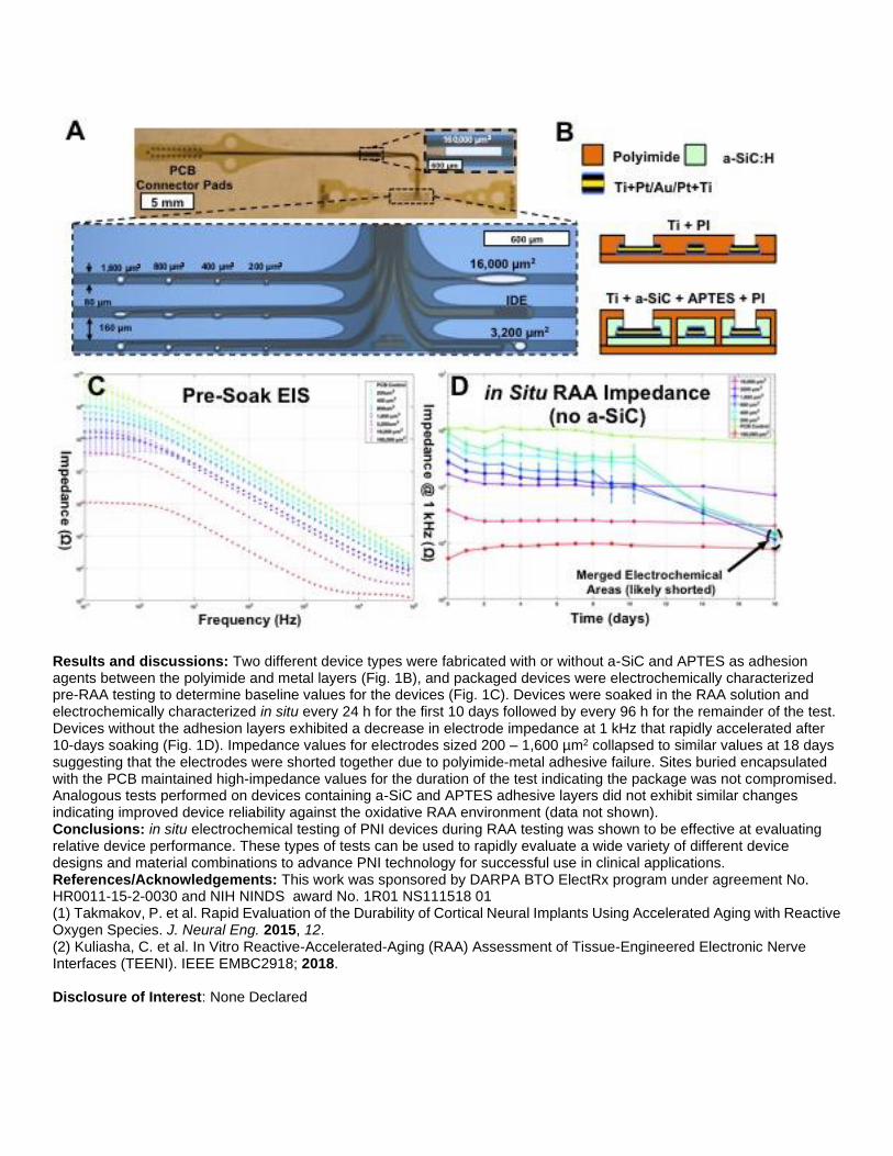
Results and discussions: Two different device types were fabricated with or without a-SiC and APTES as adhesion agents between the polyimide and metal layers (Fig. 1B), and packaged devices were electrochemically characterized pre-RAA testing to determine baseline values for the devices (Fig. 1C). Devices were soaked in the RAA solution and electrochemically characterized in situ every 24 h for the first 10 days followed by every 96 h for the remainder of the test. Devices without the adhesion layers exhibited a decrease in electrode impedance at 1 kHz that rapidly accelerated after 10-days soaking (Fig. 1D). Impedance values for electrodes sized 200 – 1,600 µm2 collapsed to similar values at 18 days suggesting that the electrodes were shorted together due to polyimide-metal adhesive failure. Sites buried encapsulated with the PCB maintained high-impedance values for the duration of the test indicating the package was not compromised. Analogous tests performed on devices containing a-SiC and APTES adhesive layers did not exhibit similar changes indicating improved device reliability against the oxidative RAA environment (data not shown). Conclusions: in situ electrochemical testing of PNI devices during RAA testing was shown to be effective at evaluating relative device performance. These types of tests can be used to rapidly evaluate a wide variety of different device designs and material combinations to advance PNI technology for successful use in clinical applications. References/Acknowledgements: This work was sponsored by DARPA BTO ElectRx program under agreement No. HR0011-15-2-0030 and NIH NINDS award No. 1R01 NS111518 01 (1) Takmakov, P. et al. Rapid Evaluation of the Durability of Cortical Neural Implants Using Accelerated Aging with Reactive Oxygen Species. J. Neural Eng. 2015, 12. (2) Kuliasha, C. et al. In Vitro Reactive-Accelerated-Aging (RAA) Assessment of Tissue-Engineered Electronic Nerve Interfaces (TEENI). IEEE EMBC2918; 2018. Disclosure of Interest: None Declared

Keywords: Biomaterial-related clinical problems (wear, metal ions etc.), Materials for electric stimulation, Peripheral nerves and spinal cord

Biomaterials for specific medical applications WBC2020-3565 Elastic powder-based lung tissue sealant for pleural defects in a mouse model Patrick Charron* 1, 1, Spencer Fenn1, Minara Aliyeva1, Nirav Daphtary1, Lennart Lundblad1, Rachael Oldinski1 1University of Vermont, Burlington, United States Introduction: Damage to the pleural tissue lining the lung can occur in a multitude of ways, resulting in fluid leak into the pleural cavity, which can result in mortality. Historically such cases were treated via pleurodesis. Recent developments of lung tissue sealants serve as alternatives but few options are available. While many sealant tests can deliver qualitative pass/fail data, there exists a lack of quantitative in vivo in methods to characterize sealing capabilities of these materials, particularly for pleural applications. In addition, previous materials have failed due to poor administration; thus, we have developed a powder sealant capable of adhering to tissue and crosslinking to form an elastic hydrogel. Experimental methods: Methacrylated alginate (AMA) was synthesized and the degree of methacrylation was verified via 1H-NMR spectroscopy. To enable gelation, visible green light crosslinkers were incorporated into the system. A sodium ion exchange was performed, the solution was lyophilized and processed into a powder. Gelation kinetics and viscosity of a powder solution were collected at 37℃. Gelation kinetics were collected via an oscillatory time sweep at 10% radial strain and 1 Hz during exposure to green light. Burst pressure experiments were conducted using a custom burst pressure device. AMA powder was placed over hydrated collagen sheets with a uniform hole, loaded into the burst pressure device, and pressurized until failure. Mice were anesthetized with pentobarbital sodium, tracheostomized with an 18-gauge cannula. Pancuronium was utilized for paralysis. Body temperature was maintained by keeping the animal under a lamp. Mice were connected to a computer-controlled small animal mechanical ventilator (flexiVent, Scireq) and ventilated at 200 breaths/min with a tidal volume of 0.25 mL against a PEEP (Positive end-expiratory pressure) of 3 cm H2O. A series of deep inflation maneuvers (Total Lung Capacity (TLC) and Trapezoidal deep inflation waveform) and a composite signal comprising of mutually prime frequencies ranging from 0-20 Hz (Z2) were performed to establish baseline for the animal prior to injury. Once the mice adjusted to the ventilators the thoracic cavity of the animal was open and lungs were punctured, followed by another series of baseline measurements to measure mechanics testing and verify injury. The injury was sealed using AMA powder and green light crosslinking, and tests were repeated to compare lung mechanics pre-injury and post-repair, allowing for real-time pressure data collection of lung mechanics during sealant testing. Animals underwent euthanasia by IP administration of sodium pentobarbital. Image:
Results and discussions: Figure 1B indicates physiologically relevant burst pressure values for the powder sealant collected during in vitro testing. Figure 1A depicts data for TLC, showing pressure vs. time curves for pre- and post-injury, and post-repair testing. As seen in the graph, after acute traumatic injury, there was a loss in pressure. A successful post-repair (i.e., application of the AMA powder, hydration and crosslinking) test indicated that the sealant maintained pressure

and returned lung capacity to pre-injury values, and that the sealant did not fail. A failure in the patch would result in data like the post-injury set. Conclusions: The data indicates efficacy of a novel powder-based elastic sealant for recapitulating lung mechanics post injury. During an in vitro burst pressure experiment, the powder formed an elastic crosslinked sealant capable of maintaining physiological pressure before failure. The powder-based sealant has the advantages of adhering to tissue upon application in situ and subsequent hydration, and remaining in place while crosslinking took place. Future work will explore long term in vivo studies to investigate lung tissue healing after repair with the novel powder sealant material. References/Acknowledgements: This work was funded in part by NIH R01EB020964 (Oldinski) and the University of Vermont. Disclosure of Interest: None Declared Keywords: Hydrogels for TE applications, Lung, bronchia and trachea, Wound healing and tissue adhesives

Biomaterials for specific medical applications WBC2020-3247 Molecular engineering of TMJ cartilage with biomimetic proteoglycans in TMJ pain model Melissa Franklin* 1, Elizabeth Kahle2, Megan Sperry3, Evan Phillips4, Prashant Chandrasekaran1, Biao Han1, Lin Han1, Beth Winkelstein3, Michele Marcolongo4 1Biomedical Engineering, 2Biomedical Science, Drexel University, 3Bioengineering, University of Pennsylvania, 4Materials Science and Engineering, Drexel University, Philadelphia, United States Introduction: Early cartilage degeneration occurs within the pericellular matrix (PCM)1, which is rich in aggrecan proteoglycan known to regulate tissue hydration and osmotic pressure. In temporomandibular joint (TMJ) OA, aggrecan levels decrease due to catabolic cleavage3, compromising matrix integrity and reducing PCM modulus4. Our lab developed biomimetic proteoglycans (BPGs), which resemble the structure and composition of aggrecan and are enzymatically resistant within adverse tissue microenvironments2. BPG10 (180kDa) consists of a 10kDa poly(acrylic acid) core with 8-9 chondroitin sulfate bristles and prior work supports its ability to retain hydration2. The purpose of this study was to use BPG10 to molecularly engineer normal and degenerative TMJ cartilage with tunable pain responses5 in order to characterize BPG10 diffusion, distribution, and the effect of BPG10 on PCM nanomechanical behavior. Experimental methods: BPG10 was synthesized, fluorescently tagged with DCCH, and solubilized at 10mg/mL in PBS as previously described2. Adult Holtzman rats were exposed to repeated jaw loading under anesthesia for 1 hr/day for 7 days at 2N to induce resolving sensitivity (n=4) or 3.5N to induce persistent sensitivity5 (n=4) (Penn IACUC #803831). On day 15, TMJs were harvested fresh following perfusion and stored in 10% protease inhibitor cocktail at 20℃. TMJs of non-loaded, weight-matched rats served as controls (n=4). Thawed condyles were PBS-washed then received BPG10 diffusion for 24hrs at room temperature without light. Sectioned condyles were imaged by confocal microscopy. Surface area of BPG10 in the cartilage matrix was quantified via a custom MATLAB script. To characterize BPG10 localization, collagen VI (1:50) and aggrecan NITEGE neo-epitope (1:25) were labelled. Mechanical effects of BPG10 were evaluated by Total Internal Reflection Fluorescence (TIRF)-AFM. Nanoindentation was performed (R ≈2.25μm, k ≈ 1 N/m, 10 μm/s, 20μm2 area) in PBS on non-loaded, BPG10-diffused and untreated control tissue, using fluorescent collagen VI to distinguish matrix regions and effective indentation modulus, Eind, was calculated6. Image:
Results and discussions: BPGs passively diffused into condyles of each experimental group. The average surface area percentage of BPG10 in 3.5N-loaded TMJs (24.0±1.4%) was greater than that of non-loaded (15.7±2.1%, p=0.0083) and 2N-loaded TMJs (18.6±1.9%, p=0.1000). In non-loaded condyles, BPG10 localized as thin pericellular rings whereas loaded condyles showed BPG10 dispersed throughout wider regions of the ECM (Fig.1A). In non-loaded TMJs, there was co-localization of BPG10 and pericellular collagen VI with minimal aggrecan neo-epitope; however, in 3.5N-loaded TMJs, there was more diffuse distribution of BPG10 with expanded collagen VI and elevated levels of aggrecan neo-epitope (Fig.1B). This difference suggests BPG10 localization depends on levels of naturally existent matrix molecules. Surface area quantification and colocalization assays reveal BPG10 dispersion occurs to a greater extent in the persistent pain compared to acute pain model. The average PCM Eind in normal BPG-diffused cartilage (160.9 ±69.5 kPa) was 65% higher than that of non-treated control tissue (96.9 ± 20.7 kPa, p< 0.0001). Similarly, average ECM Eind in BPG-diffused tissue (216.9 ±81.7 kPa) was 63% higher than that of control tissue (132.1 ± 41.854 kPa, p<0.0001) (Fig.1C),

demonstrating that BPG10 modulates mechanical properties of TMJ cartilage, likely due to increased fixed charge density, osmotic pressure and electrostatic intermolecular interactions introduced to the tissue7. Conclusions: Molecular engineering with BPGs that diffuse into cartilage matrix and modulate mechanics of the PCM and ECM may have implications in repair of degenerated cartilage in TMJ-OA. References/Acknowledgements: 1Chery et al., ORS Conf, 2019 2Prudnikova et al. Acta Biomat, 2018 3Gepstein et al. Histo & Cell Bio, 2003 4Danalache et al. Osteo & Cart, 2019 5Kartha et al. J Oral & Max Surg, 2016 6Han et al. ACS Biomat, 2017 7Dean et al. J Biomech, 2006. Disclosure of Interest: None Declared Keywords: Artificial extracellular matrix, Cartilage and osteochondral

Biomaterials for specific medical applications WBC2020-3309 Integration of Tissue-Engineered-Electronic-Nerve-Interface (TEENI) Threads into Magnetically Templated Hydrogels Mary Kasper* 1, Benjamin Spearman1, Ishita Singh2, Cary Kuliasha3, Jack Judy3, Carlos Rinaldi1, 2, Christine Schmidt1 1J. Crayton Pruitt Department of Biomedical Engineering, 2Department of Chemical Engineering, 3Department of Electrical and Computer Engineering, University of Florida, Gainesville, United States Introduction: Each year there are approximately 185,000 amputations. Although prosthetic-limb technology has rapidly advanced, nerve-interface technology has not progressed at the same rate. We have designed a novel tissue-engineered-electronic-nerve-interface (TEENI) platform that can exploit a 3D design to comprehensively engage with the electrical activity of a nerve. Our design uses thin-film polyimide-metal electrode “threads” that are integrated into a hydrogel platform with tubular anisotropy using a novel magnetic templating technique. The goal of this work is to determine an ideal hydrogel composition and successfully integrate TEENI threads into an implantable device. Experimental methods: Magnetically templated glycidyl methacrylate hyaluronic acid (GMHA) hydrogels were synthesized with 1.5 mg/mL of collagen I, 0.3% Irgacure 2959, varying concentrations of GMHA, and varying volume fractions of magnetic alginate microparticles (MAMs). Hydrogel solutions were added to cylindrical molds and placed within a magnetic array to align MAMs prior to photocrosslinking and collagen fibrillogenesis (Fig A). The hydrogels were treated with ethylenediaminetretraacetic acid to degrade the MAMs and were mechanically characterized using indentation. In vitro Schwann cell culture and subsequent immunohistochemistry (IHC) was conducted to determine the relationship between mechanical properties and cell migration into templated hydrogels. An optimal hydrogel composition was determined from mechanical and in vitro characterization to be used for in vivo assessment with integrated TEENI threads. Magnetically templated hydrogels were fabricated within a 1/16” diameter Tygon® tube trimmed to 5 mm in length. Hydrogels were embedded with a non-functional TEENI thread set and wrapped in a decellularized small intestine submucosa (SIS) prior to implantation (Fig B). 3 mm of sciatic nerve tissue was excised from 8-week-old Lewis rats, and the hydrogel was implanted by suturing the SIS to the nerve stumps. Animals were sacrificed after 6 and 12 weeks. Grafts were excised, IHC stained, and images were quantified for axon density and extracellular matrix deposition through various regions of the graft. Image:

Results and discussions: We have demonstrated successful integration of TEENI threads into magnetically templated hydrogels. Magnetic templating provides anisotropy and tunability of mechanical stiffness. Templated hydrogels of 10 mg/ml GMHA promoted better cellular infiltration in vitro and was chosen as the optimal regime for in vivo assessment. Although assessment of implanted integrated hydrogels with IHC show axon densities similar to fresh nerve in proximal regions of the graft (Fig C), axon densities gradually decrease through the graft into the distal stump. Remaining hydrogel can be visualized within the graft 6 weeks after implantation, which suggests insufficient hydrogel degradation slows remodeling and subsequent regeneration through the graft. Despite the formation of small fibrotic capsules around the TEENI threads (Fig C, arrow heads), we have been able to obtain high signal-to-noise-ratio recordings from functional devices. Conclusions: In conclusion, we have developed a TEENI device that integrates a magnetic templating technique within a 3D nerve interface. Further development will seek to increase hydrogel degradation, improve regeneration, and expand on successful recordings with TEENI devices. References/Acknowledgements: Research reported in this abstract was supported by the BTO of the Defense Advanced Research Projects Agency. No. HR0011-15-2-0030 Disclosure of Interest: None Declared Keywords: Hydrogels for TE applications, Peripheral nerves and spinal cord

Biomaterials for specific medical applications WBC2020-3316 Influence of Surfactant on PLLA/Mg Composites for Osteosynthesis Applications Meriam Ben Abdeljawad* 1, Xavier carette1, Jean-Marie Raquez1, Philippe Dubois1 1SMPC, University of Mons, UMons, Mons, Belgium Introduction: In the field of osteosynthesis, biodegradable composites have shown to be promoting candidates by combining good biocompatibility and low degradation rate with suitable mechanical properties1. In this regard, poly(L-lactide) (PLLA) was studied intensively as a polymeric matrix for biodegradable composites2. However, the low mechanical properties and the acidic environment around the implant during the degradation affect the use of PLA as implant for bone regeneration3. Therefore, due to their osteoconductivity, the lower degradation time compared with the monolithic polymer as well as the mechanical properties which are more similar to the bone compared with other metallics, Mg microparticles were incorporated4,5. The interfacial tension between the hydrophobic Mg particles and the hydrophobic PLA matrix compromise the final properties of the composite. For this reason, amphiphilic copolymer prepared with polyethylene glycol (PEG) and lactide was used to modify the matrix/filler interface. Experimental methods: Amphiphilic poly(ethylene glycol)-b-poly(L-lactide) (PEG-b-PLLA) block copolymer was synthesized in bulk by ring-opening polymerization (ROP). Two sets of composites, with (2) and without (1) surfactant, were producing by melt-processing of commercial PLLA (6201D) and Mg particles then molded à 180°C. In order to assess the H2 release, samples were immersed in PBS during 28 days in a beaker placed in a thermostatic bath at 37°C and an initial pH of 7.27. For the bioactivity assessment, discs were soaked in SBF with an initial pH of 7.4 during three months in an oven fixed at 37°C. Results and discussions: The morphological, thermal, mechanical properties as well as the degradation behaviour of the resulting biocomposites were investigated. After cryofracturing and soaking the samples in HCl, initial SEM analyses confirmed the beneficial addition of surfactant on the Mg dispersion within PLA matrix. In fact, the morphology was shown to be smoother and the pore size left by the Mg fillers was smaller in set (1). The thermal analyzes (DSC) revealed a decrease of the Tg in set (2), which could be explained by the plasticizing effect of the PEG. DMTA showed that Mg fillers enhanced the stiffness of the two sets of composites. For instance, in the set (1) the storage modulus was improved about 40% with the addition of 10wt.% of Mg. However, it remained lower than set (2). Compression tests confirmed the same behaviour when the surfactant was added. Subsequently, H2 release test showed that the presence of surfactant increases the H2 release as it was expected. In fact, as the copolymer enhances the dispersion of the Mg filler within the PLA, the specific area of the degradable Mg particles will be increased. Subsequently, the bioactivity of PLLA/Mg composites was studied and first analyzes showed that the addition of the copolymer to initial composites accelerate the apatite formation at the surface. Conclusions: The surfactant was used as a compatibilizing agent and, as an original approach, to modulate the bioactivity of PLLA/Mg composites. To fulfill the target of this study, a compromise has to be reached between the use of copolymers which could both improve the PLA/Mg interface and the bioactivity behaviour, and the mechanical performances which could be affected. References/Acknowledgements: 1. Thakur, V. K. et al., Biodegradable Materials. (John Wiley & Sons, Incorporated, 2017). 2. Raquez, J.-M. et al., Prog. Polym. Sci. 38, 1504–1542 (2013). 3. Cai, H. et al., Compos. Sci. Technol. 162, 198–205 (2018). 4. Cifuentes, S. C. et al., Acta Biomater. 32, 348–357 (2016). 5. Zhao, C. et al., Compos. Sci. Technol. 147, 8–15 (2017). The authors thank the M-ERA.NET for supporting POLYMAGIC Project. They also thank the Belgian Federal Government Office of Science Policy for general support and is much indebted to both ‘Région Wallonne’ and European Commission ‘FSE AND FEDER’ for financial support in the frame of Phasing-out Hainaut. Disclosure of Interest: None Declared Keywords: None

Biomaterials for specific medical applications WBC2020-3060 Fibre-based Degradable Ureteral Stents with Dynamic Antibacterial Surface: An In Vitro And In Vivo study Liheng Gao* 1, Yiwei Wang 2, Gang Sun3, Mingxi Xu2, Yimeng Li1, Jun Da2, Lu Wang1 1Textile College, Donghua University, 2School of medicine, Shanghai Ninth People’S Hospital, Shanghai Jiaotong University, Shanghai, China, 3UC Davis, California, United States Introduction: Everday, thousands of ureteral stents are implanted after urinary surgery to induce urine. Clinially used polyurethane stents(PUST) are undegradable and call for a secondary surgery. Besides, Bacterial invasion and subsequent formation of biofilm is the main cause of ureteral stent related ureteral infection. Hence, inspired by the metabolism of human skin, a Ag@Au NPs imbolized degradable ureteral stent were studyed. The stents are designed with dynamic contact-killing surface to prevent the adhension of bacteria and biofilm formation.
Experimental methods: •Synthesis of Ag@Au Nanoparticles The Ag@Au NPs were synthesised when the AuCl4 and AgNO3 were successively reduced by sodium citrate. Then the NPs were mixed with PAMAM and stirred at 120 ℃ for 2 hours.
•Fabrication of NPs Imbolized Ureteral Stents The PGA and PGLA multifilaments were braided into a tubular fabric and immersed in an aqueous solution of 0-800 mg / L Ag @ Au NPs, followed by air drying and heating at 200 ℃ for 2 minutes.
•In vitro Antibacterial Test Escherichia coli and Staphylococcus aureus were cultured and dulited by PBS solution. All tests were operated at 37℃. 2–4×10⁴ CFU/ml of bacterial solution and 2cm of stents were shaken for 15 hours at 120rpm for the antibacterial test of stents and stent extract and diluted for agar plate count.
• In Vitro Degradation Test 20cm of stents (400ppm) and 3ml artificial urine (pH=5.8), 50℃, 120rpm of shaking.
•In Vivo Test Ureteral stents(PUST, DST, 400ppm NPs-DST) were imersed in PBS solution with 108 CFU/ml of E.coil for 30min and implanted in both ureters(20cm,DJ ends) and bladders(5cm) of female white pigs. The stents in bladders were taken out for observation at day7. HE stain of pelvis was employed at day 21. Image:
Results and discussions: •Charaterization of NPs and ureteral stents (Fig. 1) The stents were divided by fibers and menbrane after heat treatment. For PGA fibers, the nanoparticles were half inserted on the surface with obvious bulges and totally embeded in the PGLA membranes. XPS analysis showed that NH2 groups reacted with hydrolyzed ester groups and formed more C-N bonds.
•In vitro antibaterial effect (Fig.2 ) NPs-DST showed siginicant antibacterial effect at 200mg/kg(>99.9999%).For both SA and E.coil, the stents showed obvious killing efficiency, while the extract didn’t. Proving the releasement of Ag+ couldn’t do harm to bacterials. Contact-killing was the main antibacterial mechanism.

•Degradation Test of Stents(Fig.3)The release of Ag were detected at low concentration, and much more negligible Au were released, indicating the NPs were well-fixed.
•In Vivo Test of Stents(Fig.4)Live/dead stain of stents showed, by contrast with PU stents, degradable surface prevented the formation of biofilm and NPs-DST exhibited best anti-biofilm effcet. HE stain demonatrated that animals implanted with NPs-DST showed less imflammation. The NPs didn’t bring obvious extra harm to tissue. Conclusions: Uniform NPs imbolized fiber-based ureteral stents were fabricated. The stents showed high contact-kill activity to both gram-positive and gram-nagetive bacterials,with negligible releasement of Ag+. The degradated fragments are small and NPs were well-fixed on the stent during degradation. In vivo test showed that, the DST had antifouling effect due to dynamic surface and NPs contributed to the detroying of bacterias. The as-prepared stents has satisfying biocompatibilitya and could degradate after 2 weeks. References/Acknowledgements: [1]. Lange D , Bidnur S , Hoag N , et al. Ureteral stent-associated complications—where we are and where we are going[J]. Nature Reviews Urology, 2014, 12(1):17-25. We gratefully acknowledge the financial support of the Chinese Universities Scientific Fund(No.2232019A3-06), 111 Project B07024 “Biomedical Textile Materials Science and Technology” Disclosure of Interest: None Declared Keywords: Biodegradation, Biomaterial-related biofilms, Bladder and urogenital tissues

Biomaterials for specific medical applications WBC2020-3081 Preparation and Evaluation of High-intensity Gradient-degradable Artificial Ligaments Xiaojing Xie*, Yunfan Huang, Fujun Wang, Jin Lin, Lu Wang
Introduction: •Anterior cruciate ligament (ACL) ACL is a dense connective tissue with limited vascularization, it is difficult
to heal endogenous after injury [1], 85% of ACL tears reconstruction require surgical intervention. •Biodegradable artificial ligaments have problems such as insufficient mechanical properties and mismatch between the degradation time of
ligaments and the growth rate of new tissues[2]. •Hence, we plan to develop a high-intensity gradient-degradable artificial ligament, to meet the current clinical functional requirements for artificial ligament grafts.
Experimental methods: •Fabrication of scaffold : Six types of scaffolds with PPDO、PGCL and PLGA filaments were
fabricated on a 12 bobbin braiding machine (Fig. 1).
•Morphology characterize: Using SMZ745T stereoscopic microscope and MB-ruler software to observe the structure of the scaffold.
•Mechanical testing:Tensile testing: The scaffolds were stretched to fracture at the rate of 24 mm/min (2%/s), 20 mm guage. Fatigue testing: The fixed elongation rate was 10%, the tensile speed and return speed were both 120 mm/min and repeated 500 times. Then the sample was stretched to fracture at a constant rate of 24 mm/min. Ultimate stress(MPa)
、Ultimate strain(%) and Modulus(N/mm) were measured.
•In vitro degradation: The scaffolds were immersed in PBS with pH of 7.4, and the degradation conditions were maintained at 50 ℃ and 60 r/min. The samples were sampled at 0, 3, 6, 9, 12, 18, 24, 30 and 36 days. The morphology and mechanical properties were tested.
•Cytotoxicity testing: The cells were inoculated with the sample extract at 105 inoculation density and the cytotoxicity was observed. Image:

Table:
Structure parameters Samples Aa Ab Ac Ba Bb Bc
Diameters (mm) 3.74 3.58 4.21 3.67 3.62 4.08 Shell pitch (mm) 4.88 4.47 4.95 4.46 4.22 4.90 Shell braiding angle (°) 64.76 66.78 65.61 66.00 64.15 67.85 Core diameters (mm) 1.64 1.07 1.64 1.65 1.10 1.65 Core pitch (mm) 1.64 1.07 1.64 1.65 1.10 1.65 Core braiding angle (°) 29.29 20.60 29.29 29.82 24.91 29.82
Results and discussions: •Morphoogy: The apparent structure of the core-shell tubular artificial ligament is shown in Fig. 2. The structural parameters are shown in table 1. Shell layers of the three structures have similar braiding angles, nuclear layer structure a has the same braiding angles as structure c, and structure b has the minimum braiding angles.
•Mechanical properties: The tensile test results are shown in Fig. 3(A). The tensile strengths and young's modulus of sample Ab and Bb were higher than that of human ACL. Structure b had the optimal mechanical properties. The cyclic tensile fatigue properties of the specimens are shown in Fig. 3(B). After 500 cycles of stretching, the tensile strengths of

all samples were still significantly greater than that of human ACL, and structure b had the lowest reduction rate of strength.
•Degradation properties: The changes of strength and pore diameter in vitro degradation are shown in Fig. 3(C) and (D). The strength decline rate of the sample basically met the rate of autologous tissue regeneration, which basically met the clinical demand for controllable gradient degradation of the artificial ligament.
•Cell compatibility: Cytotoxic results were shown in Fig. 4. The samples all had good cellular compatibility.
Conclusions: •This study developed 6 kinds of gradient-degradable artificial ligaments with core-shell structure using the
braiding procedure. •The A components ratio (PPDO: PGCL: PGLA = 6:4:2), b braiding structure (6x6 +Ⅰ) have
excellent tensile performance and fatigue strength, which can realize gradient controlled degradation. References/Acknowledgements: [1] Frank, C., Journal of Musculoskeletal and Neuronal Interactions, 2004. 4(2): p. 199. [2] Leong, N.L.. Journal of biomedical materials research Part A, 2014. 102(5): p. 1614-1624. This study was financially supported by : 1.National Key Research and Development Program of China(2018YFC1106200 and 2018YFC1106201). 2.The 111 Project “Biomedical Textile Materials Science and Technology” (grant no. B07024). Disclosure of Interest: None Declared Keywords: Biodegradation, Fibre-based biomaterials incl. electrospinning, Tendon and ligament

Biomaterials for specific medical applications WBC2020-2793 Development of an innovative collagen-based tubular device for urinary tracts regeneration Nunzia Gallo* 1, Claudia Curci2, Simona Bettini1, Marco Vulpi3, Antonio Vitarelli3, Ludovico Valli4, Pasquale Ditonno3, Fabio Sallustio2, Anna Gallone2, Luca Salvatore1, Alessandro Sannino1 1Department of Engineering for Innovation, University of Salento, Lecce, 2Department of Basic Medical Sciences, Neurosciences and Sense Organs, Polyclinic of Bari, 3Department of Emergency and Organ Transplant, Urology and Andrology Unit, University of Bari, Bari, 4Department of Cultural Heritage, University of Salento, Lecce, Italy Introduction: Urethral stenosis is a pathological condition that consists in the narrowing of the urethral lumen because of the formation of scar tissue mainly due to idiopathic, iatrogenic and traumatic reasons. It is one of the most widespread diseases of the urinary tract that affects about the 1% of the world male population and significantly influences their quality of life. The gold standard is the augmentation urethroplasty with buccal mucosa autograft or with a decellularized matrix from porcine Small Intestinal Submucosa (SIS patch) that actually is the only licensed xenograft for clinical use. Unfortunately, any of these approaches represent an optimal solution because of the high stricture recurrence rate (30-40%). Nowadays, several attempts are ongoing, but are all at the early stages. In this context, we developed an innovative collagen-based implantable device meant to be used as a substrate for urethral regeneration. Experimental methods: Two different fabrication techniques were employed to realize a two-component device. Air-drying of a collagen suspension allowed to obtain a non-porous, impermeable-to-cell tubular film that was inserted inside a spin-casted porous scaffold with an impermeable-to-cell external layer. Following fabrication, the two components were further cross-linked firstly by exposure to dry heat (121°C, 72h, under vacuum) and then by incubation in a 1-etil-3-(3-dimetilaminopropil)-carbodiimide crosslinking bath (14 mM, 2h, room temperature). After dry heat sterilization (160°C, 2h, under high vacuum) the assembled device was characterized from a morphological, physical-chemical, mechanical and biological point of view in comparison with the SIS patch. The micro-architecture was investigated by Scanning Electron Microscopy (SEM). The impact of crosslinking on the secondary/tertiary structure was evaluated by Fourier Transform Infrared Spectroscopy (FT-IR), while Differential Scanning Calorimetry (DSC) allowed to determine thermal properties. Wettability and swelling tests were carried out to ensure the hydrophilicity of the substrates and their capability to retain water. Tensile and suture retention test were performed to assess the material elastic response and implantability. Device’s half-life in-vitro was estimated through collagenase digestion test. Finally, the biocompatibility of the device was preliminary conduced with human Adult Renal Stem Progenitor Cells (ARPCs) for long observation times on the inner layer of the device. Image:
Results and discussions: SEM analysis confirmed the porosity of the spin-casted layer and the smooth surface of the air-dried layer. FT-IR confirmed the crosslinking efficacy and the collagen preservation after all treatments. DSC analysis demonstrated a device denaturation temperature much above 37°C. Tensile tests showed a great elastic response up to about 20% strain and a sufficient suture retain. Degradation resistance was comparable to SIS patch. Biocompatibility tests exhibited very strong differences between our device and SIS patch. While on SIS patch the proliferation rate was very low, the response on the air-dried layer of the two-component device enhanced stem cell growth so that the 100% of confluence was reached in 10 days and the three-dimensional organization in spheroids occurred in 20 days, without losing staminality.

Conclusions: The tubular scaffold was designed to mimic the anatomical structure of urethra in order to provide for each tissue urethra its composed of the appropriate regenerative support. Thus, the so treated device displays either mechanical properties compliant with the urinary tract physiological stress and the ability to better sustain cell grow compared to the SIS patch. The development of a monolayer in 10 days and of spheroids in 20 days, only by mean of a collagen substrate, suggests how the proposed device could be a good candidate for the development of new products for urethral regeneration. References/Acknowledgements: None Disclosure of Interest: None Declared Keywords: 3D scaffolds for TE applications, Stem cells and cell differentiation

Biomaterials for specific medical applications WBC2020-2866 Differential Modulation of Immune Response to Biomaterial Class by Aging Mangesh Kulkarni* 1, 2, Branimir Popovic2, Clint Skillen2, Bryan Brown1, 2, 3 1Bioengineering, University of Pittsburgh, 2McGowan Institute for Regenerative Medicine, 3Obstetrics, Gynecology, and Reproductive Sciences, University of Pittsburgh, Pittsburgh, United States Introduction: In past decade or so, biological materials are becoming increasingly popular alternatives to commonly used synthetic polypropylene meshes. One of the major advantages of these ECM based materials is their potential for tissue integration and induction of regeneration, achieved through divergent immune response. This shift in immune response wherein synthetic materials are characterized by chronic inflammatory response while non-crosslinked biomaterials present with more pro-regenerative response has been studied. However, factoring in aging as a possible determinant influencing the differential immune response is important. Aging as a determinant factor is of particular importance in the field of biomaterial implantation as a vast majority of patients in need of these implantation are aged and with increasing lifespan, the number as well as proportion of aging population are on the rise. Thus, a thorough understanding of immune response to biomaterials as a function of aging will pave way to newer therapeutic avenues and thereby improve outcome of surgical implantations in aged population. Experimental methods: This study focused on characterizing host response to synthetic material, in this case polypropylene mesh (PPM) and ECM-based biological material, in this case decellularized urinary bladder matrix (UBM) as a function of aging. We characterized the immune response at the implantation site at 3, 7, 14 and 90 days post-implantation in both young (4 months) and aged (18 months) mice in a muscle injury model. We also evaluated how aging impacts the phenotypic modulation of the macrophage populations and differential ability of PPM vs UBM to promote ECM deposition and tissue remodeling. For evaluating differential gene regulation, we performed next generation sequencing on Illumina HiSeqX platform. The SMARTer Stranded Total RNA-Seq Library Prep Kit V2 was used for library preparation. The differential expression analysis was performed using DESeq2 (R Bioconductor package). Pathway analysis was performed using Ingenuity Pathway Analysis (IPA). Image:

Results and discussions: In the young mice, the biological material tends to reduce the cellular infiltration after day 7, aged mice fail to achieve this reduction until 90 days (Fig 1a & b). However, ECM deposition induced by the biological material is higher compared to that in synthetic material despite the age of mice indicating the constructive tissue remodeling by biological material is not significantly affected by aging (Fig 1a & c). This can be explained by our observation that there was significantly higher infiltration of arginase producing macrophages in the UBM group vs the PPM group mice (Fig 1d). Since arginase production has been used widely as a marker of pro-regenerative M2 macrophages in mice, we wanted to further characterize the cellular response at molecular level. Next generation sequencing was used to study the differential gene regulation as a function of material type and aging. The results clearly showed that the differential gene regulation induced by synthetic (PPM) vs. biologic (UBM) materials was very pronounced in young animals with over 10,000 differentially upregulated genes. In aged animals, on the other hand, the differential upregulation was an order of magnitude less. Conversely, in case of downregulated genes, UBM led to more downregulated genes in aged than in young mice. Similarly, by characterizing the response in terms of overall biological functions, for example, hematological system development, immune cell trafficking, inflammatory response, cell-to-cell signaling interaction, cellular movement, cellular growth and proliferation, it was very evident that in young mice, UBM exhibited its effect by upregulation while in aged mice, the effect was pronounced by downregulation (Fig 1e). Conclusions: The phenotypic and transcriptome analysis showed mechanistic divergence in immune response to biological vs synthetic implant material as a function of aging. References/Acknowledgements: This work was funded in part by ACeLL®. Disclosure of Interest: None Declared

Keywords: None

Biomaterials for specific medical applications WBC2020-2516 The incorporation of type II collagen biomaterial into type I collagen-hyaluronate scaffolds improves cartilage matrix production/distribution without promoting tissue hypertrophy Claudio Intini*, Eric Farrell, John Gleeson, Fergal O'Brien Introduction: Tissue engineering strategies to regenerate stable hyaline cartilage without leading to unstable, hypertrophic tissue incapable of long-term clinical function remains an unmet challenge. Several biomimetic biomaterials have been explored and used in cartilage tissue engineering, showing some potential to regenerate tissue. In particular, type I collagen-hyaluronate (CHyA) scaffold biomaterials have been extensively studied by our group and have demonstrated significant regenerative capacity in directing de novo chondrogenesis preclinically1 and clinically1. Moreover, a type II collagen biomaterial, a key component of articular cartilage, has also demonstrated potential in promoting cartilage matrix accumulation when incorporated in hydrogels2. However, the need to develop improved biomimetic biomaterials capable of promoting better and stable long-term cartilage remains. Therefore, the aim of this work was to investigate the incorporation of type II collagen within type I collagen-hyaluronate scaffold biomaterials to assess its role in directing a better and stable cartilage regeneration. Experimental methods: Porous scaffolds incorporating type II collagen in the ratio 1:1 among collagen type I:II (0.25:0.25%w/v) and hyaluronate (0.05%w/v) were manufactured using a freeze-drying technique as previously described3. Type I collagen (0.5%w/v) hyaluronate (0.05%w/v) scaffolds were also produced. Scaffolds were physically crosslinked using a dehydrothermal (DHT) treatment. Scaffolds were subsequently cultured with 5x10⁵ human pediatric mesenchymal stem cells (MSCs) in vitro under chondrogenic condition for 21 days. Then, they were analysed histologically and by gene expression of specific markers to chondrogenesis and hypertrophy. The experiments were performed in triplicate by using three human pediatric MSCs donors. Image:

Table: Figure 1. A & B assessed after 21 days in culture with human pediatric MSCs. A) Representative histological sections of CHyA and CHyA + Col 2 (type II collagen) scaffold biomaterials. The sections were stained with thionin which stains the glycosaminoglycans in purple and nuclei in blue. B) The effect of type II collagen on gene expression of collagen type II (COL2A1) and collagen type X (COL10A1). Relative gene expression normalised to GAPDH. Results and discussions: The incorporation of type II collagen into type I collagen-hyaluronate scaffold biomaterials improved cartilage matrix production/distribution within the scaffolds. Histology revealed a better glycosaminoglycans (GAG) distribution toward the center of type II:I collagen-hyaluronate scaffolds (Fig.1A). Furthermore, these scaffolds incorporating type II collagen promoted same levels of collagen type II (COL2A1) and collagen type X (COL10A1) gene-expression to CHyA scaffolds (Fig.1B). Therefore, the incorporation of type II collagen as biomaterial in CHyA scaffold demonstrated an added chondrogenic benefit in terms of matrix production and distribution without promoting tissue and cellular hypertrophy. Conclusions: This study demonstrates the ability of naturally derived biomimetic biomaterials, such as collagen type II, to act as potent cues that can modulate chondrogenesis. The results support continued investigation and evaluation of these collagen-hyaluronate scaffold systems incorporating type II collagen as a potential off the shelf approach to long term stable cartilage tissue repair for localised cartilage lesions of the knee. References/Acknowledgements: 1Levingstone T et al., Biomaterials. 2016; 87:69-81. 2Choi B et al., ACS Applied Materials & Interfaces. 2014; 6(22):20110–21. 3Haugh MG et al., Tissue Eng Part C Methods. 2010; 16, pp. 887-894. This project has received funding from the European Union’s Horizon 2020 research and innovation programme under Marie Sklodowska-Curie grant agreement No 721432. Disclosure of Interest: None Declared Keywords: 3D scaffolds for TE applications, Cartilage and osteochondral, Stem cells and cell differentiation

Biomaterials for specific medical applications WBC2020-2522 Curcumin-loaded porous P4HB scaffolds for wound healing applications Eike Müller1, Maria Fernandez1, Sergio Diaz Abad1, Qun Ren1, Katja Nuss2, Brigitte von Rechenberg2, Katharina Maniura1, Markus Rottmar* 1 1Empa, Swiss Federal Laboratories for Materials Science and Technology, St.Gallen, 2MSRU, Vetsuisse Faculty ZH, University of Zurich, Zurich, Switzerland Introduction: Every year millions of patients develop hard-to-heal wounds as a result of elective operations or after trauma. In addition, the aging society is leading to a drastic increase in the numbers of chronic wounds. Therefore, wound treatment has become an important medical problem for individual patients as well as the healthcare system. However, current approaches mainly focus on the physical protection of the wound rather than providing a 3D matrix that actively supports tissue regeneration. Furthermore, prevention of scar tissue formation is currently largely neglected. We thus aimed to develop a synthetic and bioactive, biodegradable 3D-scaffold based on the FDA-approved material Poly-4-hydroxybutyrate (P4HB), which addresses these two key aspects of wound healing simultaneously, i.e. stimulating correct skin regeneration and prevention of scar formation. Besides precisely tuning porosity to steer the response of skin cells, scaffolds should be loaded with a drug that prevents scar formation upon release. Experimental methods: P4HB of different Mwwas obtained by fermentation and subsequent chemical hydrolysis. 3D porous scaffolds were produced by foaming with sCO2, where the pore size was tuned by changing the process parameters time (t), pressure (p) and temperature (T). Human fibroblast attachment, invasion and proliferation within the scaffolds was assessed after day 1 and 5 in culture. Curcumin (CU) loaded P4HB scaffolds were produced by mixing CU with P4HB prior to foaming. CU release as well as impact on TGF-β1induced myofibroblast differentiation of NHDF was analyzed. The inflammatory response towards the scaffolds was analyzed with THP-1-derived macrophages after 24 hours. Finally, the scaffolds were evaluated in a rat skin wound model. Results and discussions: Highly porous P4HB scaffolds with homogenous, interconnected pores were fabricated. By varying Mw, t, p, and T, the pore size was precisely adjusted from <10μm to ~1mm. Fibroblasts were found to attach and invade into the scaffolds with pore sizes ≥50μm. Upon incorporation of CU into the scaffolds (up to 10%wt), a homogenous distribution could be observed without affecting the porosity and a continuous release of the CU in medium was observed over 6 weeks. Notably, a low level inflammatory response of THP-1 macrophages towards the scaffolds could be mitigated by the incorporated CU. Furthermore, the released CU could efficiently reduce myofibroblast differentiation. In the rat skin wound model, treatment of the wounds with curcumin loaded P4HB scaffolds lead to reduced scarring with more organized tissue morphology and less vertical depression compared to untreated or non-loaded P4HB foam controls. Conclusions: P4HB foams with controlled micro- and macro-topography could be successfully produced by sCO2, a technique that allowed for simultaneous functionalization of the foam with the anti-scarring and anti-inflammatory drug curcumin. The performance of curcumin loaded P4HB scaffolds was demonstrated not only in vitro, but also in vivo, and thus shows promise for potential future application in clinics to treat hard-to-heal wounds and to reduce skin scarring. References/Acknowledgements: This project (GRS 006/16) was kindly supported by the Gebert Rüf Foundation. Disclosure of Interest: None Declared Keywords: Biopolymeric biomaterials, Immunomodulatory biomaterials, Skin and mucosa

Biomaterials for specific medical applications WBC2020-2239 Pickering emulsions for liver trans-arterial chemo-embolization Laurence Moine* 1, Frédéric Deschamps2, Thomas Isoardo3, Lambros Tselikas2, Elias Fattal4, Nicolas Tsapis1, Thierry de Baère2 1Institut Galien Paris-Sud, CNRS, Châtenay-Malabry, 2Département de radiologie interventionnelle, 3Institut Gustave Roussy, Villejuif, 4Institut Galien Paris-Sud, Université Paris-Sud, Châtenay-Malabry, France Introduction: Hepatocellular carcinoma (HCC) is among the most common lethal malignancies and causes of cancer mortality in the world [1]. For non-resectable, intermediate stage HCC, trans-arterial chemo-embolization (TACE) is recommended with a high level of evidence [2]. A drug delivery platform is currently used during TACE which acts as a transient reservoir, allowing slow release of the chemotherapy in the tumors’ supply for a prolonged exposure of the cancer’ cells while minimizing systemic effects. Water-in-oil (W/O) Lipiodol® emulsions remain the preferable choice for this purpose however their low stability severely hampers their efficiency [3]. Recently, our group has developed remarkably stable W/O Lipiodol® emulsions stabilized by biodegradable poly(lactide-co-glycolide) nanoparticles (PLGA NPs) thanks to Pickering technology. In this study, the in vitro and in vivo potential of these emulsions to efficiently encapsulate and release chemotherapy was investigated through the internalization of doxorubicin (Dox) and oxaliplatin (Oxa) into the aqueous phase. Experimental methods: PLGA nanoparticles (PLGA NPs) were prepared using a nanoprecipitation technique. Two different types of water-in-oil emulsions were formulated: conventional Lipiodol-emulsion and Pickering-emulsion. For the Pickering emulsions, the aqueous-phase used was a suspension of PLGA NPs (15 mg/mL) in doxorubicin solution (20 mg/mL) or oxaliplatin solution (5 mg/mL) and the oil phase was Lipiodol® (Guerbet, France) with different W/O ratios [4]. The in vitro release was evaluated in TRIS solution at 37°C. The animal study was performed with oxaliplatin emulsions on white New-Zealand rabbits implanted with VX2 liver tumors in accordance with the directive 2010/63/EU. Oxaliplatin was quantified in plasma and in the tumor. Image:
Results and discussions: All the Pickering emulsions were successfully prepared with the desired W/O type. Microscopic images show similar aspects for Pickering emulsions 1/3 and 2/3 whereas Pickering emulsion 1/1 and the conventional one (withou PLGA NPs) exhibit more heterogeneous morphologies. All the Pickering emulsions were stable without phase separation for 24 h according analysis by static light scattering. In vitro, Pickering-emulsion demonstrated a slow doxorubicin release compared to Lipiodol-emulsion (2.5 ± 0.2 vs. 19.0 ± 6% at 1 h and 30 ± 3.0 vs. 95 ± 3% at 24 h for W/O = 1/3) (Figure 1). A similar behavior was observed with oxaliplatin. In vivo, the plasmatic peak and the area under the curve were significantly lower with the Pickering emulsion compared to the conventional emulsion (Cmax = 0.49 ± 0.14 vs 1.08 ± 0.41 ng/mL, p = 0.01 and AUC = 19.8 ± 5.9 vs 31.8 ± 14.9, p = 0.03). This resulted in significantly lower oxaliplatin concentrations in tissues at 1 h with Pickering emulsion but higher ratio between tumor and left liver at 24 h (43.4 vs. 14.5, p = 0.04). Conclusions: Stable Lipiodol® Pickering-emulsions over a large range of aqueous volume fraction to an equal volume of oil were successfully developed. They provide enough choice for the surgeon to load desired concentrations of the selected chemotherapies. In vivo, sustained release of oxaliplatin from Pickering-emulsion results in significant decrease of systemic exposure. References/Acknowledgements: [1] Parkin et al. (2005) Cancer J. Clin. 55, 74–108. [2] Llovet et al. J Hepatol (2012); 56: 908-943. [3] Gaba et al. J Vasc Interv Radiol (2012); 23: 265-273. [4] Deschamps et al. Cardiovasc Intervent Radiol (2017); 40:1927-1932. Disclosure of Interest: None Declared

Keywords: Biomaterials for drug delivery, Kidney, liver and pancreas

Biomaterials for specific medical applications WBC2020-2574 Pre-clinical functional performance assessment of a new bio-inspired non-delaminating osteochondral construct using a physiological knee implantation model Raelene Cowie* 1, William Sanderson1, Reda Felfel2, Laura Macri Pellizzeri3, Colin Scotchford2, David Grant2, Virginie Sottile3, Louise Jennings1 1Institute of Medical and Biological Engineering, University of Leeds, Leeds, 2Faculty of Engineering, 3School of Medicine, University of Nottingham, Nottingham, United Kingdom Introduction: The aim of the study was to assess the functional performance of a new osteochondral construct (OCC) using a natural knee model1. The OCC is a bioresorbable non-delaminating chitosan scaffold with a graded pore size representative of cartilage and subchondral bone. The study investigated the in situ stability and wear/damage/deformation of the construct when tested under physiologically relevant loading and motion. The study also started to investigate the effect of surgical positioning of the OCC on wear/damage/deformation by positioning the graft either proud of the cartilage surface or flush with the surface but inverted. The wear/damage/deformation of the OCC was compared to a graft in current clinical use (BioMatrix CRD, Arthrex). Experimental methods: Porcine knees (4-6 months old) were mounted in a ProSim knee simulator (Simulation Solutions, Manchester, UK) in a physiological anatomical position with the ligaments removed and menisci intact.1 An 8mm diameter graft was implanted in the medial femoral condyle and the simulator run for 3 hours (10800 cycles) under a walking gait cycle using 25% bovine serum in PBS as a lubricant. After 3 hours, the wear/damage/deformation was assessed using a cartilage grading system (scored from 0-4 where 0 represents healthy, undamaged cartilage and 4 severe degeneration through to subchondral bone/meniscus tear)2 and by assessing the wear volume from replicas of the articulating surfaces. Four experimental groups were investigated, OCC flush, OCC 1mm proud, OCC inverted and BioMatrix CRD and compared to positive control samples consisting of a stainless steel pin positioned 1mm proud of the cartilage surface. Four samples were tested in each experimental group. Results and discussions: After 3 hours wear simulation, a stainless steel pin implanted in the femur 1mm proud of the cartilage surface gave a clear region of deformation in the meniscus and the highest wear/damage/deformation of the experimental groups with grade 1 or 2 damage observed on the menisci and tibial plateaus. There was no evidence of subsidence of the stainless steel pin. Against an OCC positioned flush with the articulating surface, there was discolouration of the meniscus in 1 sample but no visible damage. In all the samples in this experimental group, at the conclusion of the study, there was subsidence of the graft ~0.5mm below the cartilage surface. Positioning the graft 1mm proud of the articulating surface led to discolouration of the meniscus in 3 of 4 samples, and grade 1 damage. At the conclusion of the study, the graft was flush with the articulating surface. Inverting the OCC also led to discolouration of the meniscus and subsidence of the OCC. For the BioMatrix CRD grafts, there was evidence of deterioration of the surface of the graft in 2 out of 4 samples after 3 hours of testing, there was also visible scratching around the graft site, on the superior surface of the medial meniscus and the tibial plateau (grade 1 & 2). The BioMatrix grafts did not seem to subside. The wear/damage/deformation caused by the OCC scaffolds and BioMatrix was below a threshold measureable using the technique of surface replication and analysis using the Alicona optical profiler. Conclusions: The study investigated the functional stability and performance of a novel osteochondral graft by assessing the wear/damage/deformation of the graft prior to integration between graft and host tissue. Functional pre-clinical assessments such as that used in this study can be used to assess novel surgical interventions prior to carrying out in vivo animal trials. References/Acknowledgements: 1. Bowland, P., et al. J Biomechanics 77 (2018): 91-98. 2. ICRS Cartilage Injury Evaluation Package https://cartilage.org/content/uploads/2014/10/ICRS_evaluation.pdf Disclosure of Interest: R. Cowie: None Declared, W. Sanderson: None Declared, R. Felfel: None Declared, L. Macri Pellizzeri: None Declared, C. Scotchford: None Declared, D. Grant: None Declared, V. Sottile Conflict with: Medical Technologies IKC POC, L. Jennings Conflict with: Medical Technologies IKC POC Keywords: Cartilage and osteochondral

Biomaterials for specific medical applications WBC2020-2643 Experimental Wear Simulation of an All-Polymer Total Knee Replacement Raelene Cowie* 1, Adam Briscoe2, John Fisher1, Louise Jennings1 1Institute of Medical and Biological Engineering, University of Leeds, Leeds, 2Invibio Biomaterial Solutions, Thornton Cleveleys, United Kingdom Introduction: PEEK-OPTIMA™ has been considered as an alternative arthroplasty bearing material to cobalt chrome (CoCr) in the femoral component of total knee replacements (TKR) to create a metal-free implant.1 Prior to the implant entering clinical trials, it is important to understand the functional performance of this novel bearing material combination under a range of conditions. The focus of these studies was to understand the wear performance of UHMWPE-on-PEEK through a series of fundamental pin-on-plate studies to investigate the influence of lubricant temperature and third body wear performance as well as carrying out functional assessment of the tibiofemoral (TFJ) and patellofemoral (PFJ) joints using whole joint wear simulators. Experimental methods: The influence of lubricant temperature (rig running (~25°C) and elevated temperature (~36°C)) on the wear of UHMWPE-on-PEEK was investigated using a 6 station multi-directional pin-on-plate wear simulator. Third body damage was simulated in simple geometry using PMMA cement particles to damage the PEEK plates. The surface topography of the plates was then assessed before carrying out wear simulation against the damaged surfaces. The studies were repeated for UHMWPE-on-CoCr in order to compare relationships. Experimental wear simulation of the TFJ and PFJ under optimal alignment conditions was carried out using a 6-station Pro-sim knee simulator with displacement controlled kinematic input profiles to replicate a walking gait cycle.2,3 The all-polymer TKR was investigated in parallel with a conventional metal-on-polyethylene implant of similar initial surface topography and geometry. A minimum of n=3 and 5 million cycles of wear simulation was carried out for each study. For all studies, 25% bovine serum supplemented with 0.03% sodium azide was used as a lubricant and the wear of UHMWPE was assessed gravimetrically. Statistical analysis of the wear rates of the different material combinations using ANOVA (p<0.05). Results and discussions: Pin-on-plate wear simulation showed the UHMWPE-on-PEEK bearing couple to have a lubricant temperature dependence so when tested under rig temperature conditions, the wear of the UHMWPE-on-PEEK was similar to that of UHMWPE-on-CoCr (p=0.46). However, at elevated temperature, ~36°C there was a decrease in the wear of the UHMWPE-on-PEEK but not UHMWPE-on-CoCr (p=0.04), thought to be as a result of protein precipitation and deposition on the articulating surfaces. Third body wear simulation showed PEEK to be less resistant to scratching with cement particles than CoCr but wear tests against the scratched surfaces did not influence UHMPWE wear in fact, a polishing effect of the UHMWPE pin against the scratched PEEK plate was observed. Experimental simulation of the tibiofemoral and patellofemoral joints showed a low rate of wear, <5mm3/MC and <1mm3/MC respectively. There was no significant difference in the wear performance of UHMWPE when articulating against the different femoral component materials, p=0.27 and p=0.38 for the tibiofemoral and patellofemoral joints respectively. Conclusions: Pin-on-plate wear simulation showed the importance of selecting appropriate test conditions when investigating novel material combinations to minimise test artefacts such as polymer transfer, protein precipitation and protein deposition. The third body wear behaviour of UHMWPE-on-PEEK was different to UHMWPE-on-CoCr. Experimental wear simulation of the tibiofemoral and patellofemoral joints showed an equivalent rate of wear of UHMWPE against PEEK and CoCr femoral components. Before the product enters in vivo clinical trials, these studies show that in terms of wear performance, PEEK has promise as an alternative to CoCr in the femoral component of total knee replacements. References/Acknowledgements: 1. Cowie RM, et al. 2016. JEIM 230(11):1008-1015 2. McEwen, H.M.J. et al. 2005 J Biomechanics, 38(2):357-365 3. Maiti R., et al,2014. JEIM;228(2):175-181 Disclosure of Interest: R. Cowie: None Declared, A. Briscoe Conflict with: Paid employee of Invibio, J. Fisher Conflict with: Paid consultant to Invibio; received research support from Invibio, L. Jennings Conflict with: Received research support from Invibio Keywords: Biomaterial-related clinical problems (wear, metal ions etc.)

Biomaterials for specific medical applications WBC2020-2674 PLCL/PCL nanofibers-based scaffolds coated with fibrin assembly containing platelet lysate to enhance skin wound regeneration. Andreu Blanquer* 1, Johanka Kucerova2, Elena Filova1, Barbora Koprivova3, Renata Prochazkova4, Vera Jencova3, Eduard Brynda2, Lucie Bacakova1 1Biomaterials and Tissue Engineering, Institute of Physiology of the Czech Academy of Sciences, 2Institute of Macromolecular Chemistry of the Czech Academy of Sciences, Prague, 3Department of Chemistry, Technical University of Liberec, 4Regional Hospital Liberec, Liberec, Czech Republic Introduction: Chronic wounds are affecting millions of patients around the world and their prevalence will increase due to population aging1. It is therefore necessary to develop new drugs or medical devices to improve wound healing. In this study, we developed nanofiber scaffolds coated with fibrin assemblies containing platelet lysate (PL). The combination of biodegradable polymer dressing with PL allows a controlled release of growth factors and other bioactive molecules. Experimental methods: Electrospun nanofibers made of blend of poly(L-lactide-co-Ɛ-caprolactone) with poly-Ɛ-caprolactone nanofibers were coated with fibrin assemblies with different content of PL (0%, 10%, 20%, 50%, and 100%). Co-culture of human HaCat keratinocyte cell line and primary human saphenous vein endothelial cells (EC) (passage 3-4) was performed in vitro. Hacat cells were seeded on nanofibers placed in 24-well plate and EC were sedeed into inserts with a pore size of 0.4 µm (Cat. No. 140652, Thermofisher Scientific) placed in the top of the wells. On day 7, additional nanofiber layer was added into the inserts. Cell monocultures were used as the control. Cell culture media with restricted content of fetal calf serum or growth factors were used. Cell metabolic activity was quantified on days 1, 3 (4), 7, and 14 using MTS assay kit (Abcam). EC migration was measured using Corning FluoroBlock Cell Culture insert (pore size of 8 µm); EC migrated into the medium with added modified nanofibers. Differentiation markers of keratinocytes (basal cytokeratin 14 (CK14) and more differentiated CK10) and markers of endothelial cells (von Willebrand factor and CD31) were analysed by immunofluorescence staining and visualized by confocal laser scanning microscope (Leica SP8). Image:
Results and discussions: HaCat keratinocytes had the highest metabolic activity and more HaCat cells were positively stained for CK10 on nanofibers containing 50% and 100% of PL (Fig. 1). An optimum concentration of PL in nanofibers was 10% and 20% for improved EC metabolic activity, and 20% and 50% of PL for the highest EC migration. However, no significant differences were observed for EC differentiation markers. In co-culture experiments, in regard to PL content in nanofibers, nanofibers with PL acted in similar pattern with both monocultures. Moreover, we observed the positive effect of keratinocytes on EC metabolic activity on day 14 compared to EC monoculture. This co-culture system represents more complex model of cell culture in which metabolites of keratinocytes stimulated EC viability/metabolic activity. Fig 1. Immunofluorescence staining of CK10 (green) and CK14 (red) in HaCaT cells after 14 days in culture grown on PLCL/PCL nanofibers scaffolds with fibrin assembly containing 0% (A) and 100% of PL (B). Conclusions: Nanofibers with PL favourably affected both keratinocytes and endothelial cells, which are involved in wound healing. Moreover, this positive effect was even enhanced in HaCaT/EC co-culture. These findings could be important for improvement of chronic wound vascularization and regeneration. References/Acknowledgements: 1 Sen CK et al. Wound Repair Regen. 2009;17:763-771.

This project has received funding from the European Union’s Horizon 2020 research and innovation programme (grant agreement No. 101003407-ELECTROSKIN), and from the Czech Health Research Council (project No. NV18-01-00332). Disclosure of Interest: None Declared Keywords: Biomaterials (incl. coatings) for local drug and growth factor delivery, Skin and mucosa, Wound healing and tissue adhesives

Biomaterials for specific medical applications WBC2020-2123 Characterization on Stretchability and Porosity of Silk Microneedle Mesh for Secure Perivascular Drug Delivery Jiyong Lee* 1, Seunghyun Park1, Jae Ho Kim1, Eui Hwa Jang2, Jung-Hwan Kim2, Young-Nam Youn2, Wonhyoung Ryu1 1School of Mechanical Engineering, Yonsei University College of Engineering, 2Division of Cardiovascular Surgery, Severance Cardiovascular Hospital, Yonsei University College of Medicine, Seoul, Korea, Republic Of Introduction: Bypass graft surgery is a promising therapeutic treatment for coronary artery disease such as angina pectoris. However, graft failure is often caused by neointimal hyperplasia (NIH) which commonly leads to restenosis or occlusion. In order to prevent NIH after surgery, microneedle (MN), known as an efficient drug delivery device, could be applied to the external surface of blood vessel for reducing abnormal proliferation. However, adequate stretchability and porosity of MN device are indispensable to minimize any possible side effect of perivascular drug delivery. Pulsatile vessel movements should not be hindered by mechanical constriction and molecular exchange from vessel to surrounding is critical for biological interaction. In this study, we developed a highly stretchable and porous perivascular microneedle mesh (MNM) using Bombyx mori silk fibroin, and analyzed the stretchability and porosity. The tensile properties of silk MNM was evaluated compared to normal aorta. Additionally, the molecular permeability of silk mesh was analyzed using fluorescein conjugated molecules, FITC-Dextran (FD). Finally, silk MNM was applied to the external surface of injured aorta. After 4-week in vivo animal study, efficacy for inhibiting NIH was demonstrated. Experimental methods: An aqueous silk solution was cast and lyophilized to fabricate a porous silk mesh. To make drug embedded MNs, the silk solution was mixed with anti-proliferation drug for casting in MN molds and the molded MN were attached on a porous silk mesh. To measure the stretchability of silk MNM, a universal tensile machine was used for tensile testing and the Young’s modulus of each sample was estimated. To evaluate the rate of molecular exchange through silk mesh, a Franz diffusion cell was prepared. A donor chamber was filled with a 100 μM of FD solution that had FDs with molecular weights of 4, 70 and 150 kDa. A recipient chamber was filled with PBS and a silk mesh was mounted between both chambers. Aliquots of recipient solutions were periodically collected and the concentration was estimated. Finally, the permeability coefficient of silk mesh with each molecular weight was analyzed. Lastly, to demonstrate the efficacy of NIH reduction, in vivo studies were performed using a rabbit animal model. Silk MNM was applied around the injured abdominal aorta (Fig. D), and after 4-week, the efficacy was statistically analyzed. Image:
Results and discussions: Silk MNs were structured on silk mesh with thickness of 400 μm (Fig. A). A stress-strain curve was obtained (Fig. B) showing the much smaller Young's modulus of silk mesh (0.77 MPa), silk MNM (0.72 MPa) than the modulus of the aorta (14.3 MPa). This indicated that silk MNM rarely hinders the pulsatile vessel movements with minimal constriction to the vessel. As shown in Fig. C, FD concentrations in the recipient chamber at each time point were almost similar for FDs with different molecular weights. FD concentration increased up to around 10 μM after 6 hours, which is equivalent to 10% of donor concentration. Permeability coefficients were estimated to be 0.95 (4 kDa), 1.18 (70 kDa) and 0.92 (150 kDa) μm/min, and this indicated that the porosity of silk mesh is suitable for the sufficient molecular exchange between the blood vessel wrapped with the silk MNM and its surrounding environments. Lastly, NIH formation was calculated to evaluate the efficacy of MNM for NIH reduction. NIH was significantly reduced when silk MNMs were applied (10.82%) compared to control group (21.37%), which indicated the prominent efficacy and drug delivery efficiency for reducing NIH formation.

Conclusions: Silk MNM with excellent stretchability and adequate porosity was developed so that MNM could be securely applied to the injured blood vessel. Finally, after in vivo animal study, prominent drug delivery efficacy and efficiency of MNM for inhibiting NIH were evaluated. References/Acknowledgements: [JY. Lee. J Control Release, 2017;268, S.H. Park. Eur J Pharm Biopharm, 2018;133], [Research grant; Korea Health Technology R&D Project, HI18C1237] Disclosure of Interest: None Declared Keywords: Biomaterials (incl. coatings) for local drug and growth factor delivery, Mechanical characterisation, Vascular grafts incl. stents

Biomaterials for specific medical applications WBC2020-1776 A novel method to quantify bone density changes in total hip arthroplasty patients Magnús K. Gíslason* 1, Luca Cristofolini2, Olafur Sigurjonsson1, Luca Esposito3, Paolo Bifulco4, Massimiliano Fraldi5, Paolo Gargiulo1 1Institute of Biomedical and Neural Engineering, Reykjavik University, Reykjavik, Iceland, 2Department of Industrial Engineering, University of Bologna, Bologna, 3Department of Structures for Engineering and Architecture, 4Department of Electric Engineering and Information Technologies, 5Interdisciplinary Research Centre for Biomaterials, University of Naples Federico II, Napoli, Italy Introduction: After total hip replacement, changes in bone mineral density (BMD) can be seen in the femur over a period of years. The rate of these changes depends on many different variables such as age, sex, bone quality prior to surgery and type of implant to name a few. Few studies have looked into the quantification of BMD changes using DXA scans and two dimensional representation of the bone mineral density1 as well as using computational methods2. The presented study introduces a novel method to analyse the changes in bone mineral density over a period of one year, by using two sets of CT scans, giving a three dimensional representation of the regions where both increase and decrease in BMD are detected. This will give the clinician important information about how the remodelling process of the femur is occurring on a subject specific basis. Experimental methods: CT scans were taken of patients undergoing primary total hip arthroplasty. The scans ranged from the anterior superior iliac spine to the diaphysis of the femoral shaft and had an in plane resolution of 0.6mm x 0.6mm and slice thickness of 1mm. The scans were calibrated using a quasar phantom to facilitate the conversion from the Hounsfield units to bone mineral density values in g/cm3. Each subject was scanned twice, once 24 hours post operatively and another one at 1 year post operatively. An image artefact reduction algorithm was applied to remove any metal scattering from the implant. The two sets of scans were realigned by identifying anatomical landmarks and then a reslicing was carried out to ensure that all slices would be identical between the 24 hour scan and the 1 year scan. Boolean operations were then carried out on the aligned CT scans on a voxel basis. Increase in the bone mineral density was defined as an increase in the BMD values after subtracting the 24 hour scan from the 1 year scan. Similarly the decrease in the BMD values were calculated by subtracting the 1 year scan from the 24 hour scan. Each bone was normalized into 100 slices from proximal to distal. The procedure was carried out both on the operated leg and on the contralateral leg. Image:
Table: Distribution within the groups
Females Males Number Age
Numbe
r Age
Uncemented 10 57.
6 20 51.9
Cemented 17 66.
7 3 66.3
Results and discussions: A total of fifty subjects were recruited for the study, both receiving an uncemented and a cemented fixation prosthesis. The distribution between the groups can be seen in Table 1. The results showed that the

majority of the bone loss occurred around the Lesser Trochanter area, which is in agreement with previous findings using DXA scans. The figure shows the BMD changes in 3 different patients receiving uncemented implant where it can be seen how the bone loss patterns differ between individuals. Areas of loss (red) occur more on the medial side of the femur whereas gain (green) are more randomly distributed, but the area around the distal end of the prosthesis shows bone gain. The results indicated that the BMD loss was less for males receiving uncemented prosthesis and for women receiving cemented prosthesis, although there was no statistical difference between the groups. The analysis for the contralateral leg showed no distinct areas of bone gain and loss over a period of 1 year, which emphasizes the point that the remodelling process is more uniform. Conclusions: The study demonstrates a novel technique capable of identifying areas of bone gain and bone loss in patients undergoing total hip arthroplasty. Such analysis can become valuable in the clinical settings as detailed analysis of areas where bone is losing density and by understanding the three dimensional bone loss patterns, it is possible to adjust the rehabilitation on a subject specific basis in order to slow down the bone loss. References/Acknowledgements: 1. Tapaninen T, et al. Periprosthetic BMD after cemented uncemented total hip arthroplasty: a 10-year follow-up study. Journal of Orthopaedic Science, 2015, vol 25(4), pp:657-662. 2. Iuppariello L, et al. A CT-based method to compute femur remodelling after Total Hip Arthroplasty. Journal of Medical Engineering and Physics, 2019 Disclosure of Interest: None Declared Keywords: Bone, Clinical application

Biomaterials for specific medical applications WBC2020-2154 Recombinant Human Tropoelastin Day of Addition Increases Elastic Fibre Content in Human Vascular Smooth Muscle Cells Seeded in Fibrin-Collagen-Glycosaminoglycan Scaffolds Francisco Almeida-Gonzalez* 1, 2, Arlyng Gonzalez-Vazquez2, Anthony S Weiss3, Fergal J O'Brien2, Claire M Brougham1, 2 1School of Mechanical and Design Engineering, Technological University of Dublin (TU Dublin), 2Tissue Engineering Research Group, Royal College of Surgeons in Ireland, Dublin, Ireland, 3Charles Perkins Centre, University of Sydney, Sydney, Australia Introduction: Tissue engineered heart valves (TEHVs) could eliminate paediatric patients’ need for revision surgeries by facilitating growth and remodelling of the valve in vivo. However, this requires a TEHV with an extra cellular matrix (ECM) and a fatigue strength that mimics the native vasculature; therefore, we need a TEHV containing elastic fibres. One of the ways to induce elastic fibre formation (elastogenesis) in vitro is by using recombinant human tropoelastin [1]. In this set of experiments, we have investigated the effect of the day of addition of tropoelastin on the quantity and quality of elastic fibres created in human vascular smooth muscle cells (hVSMCs). Then, as our potential TEHV is a combination of autologous cells in a fibrin collagen and glycosaminoglycan (FCG) scaffold [2,3], we also examined the ability of this scaffold-cell platform to undergo elastogenesis following the addition of tropoelastin. Experimental methods: In a 2D setup, the effect of the day of addition of tropoelastin was assessed by treating groups of hVSMCs with 10 mg/ml tropoelastin on days 0 and 10. Subsequently, we compared quantity and quality of induced elastic fibres through immunofluorescence. For our 3D setup, we waited 7 days before adding tropoelastin using two methods: (1) adding 10 mg/ml tropoelastin and growth media at the same time, and (2) drop-loading 10 mg/ml tropoelastin 10 minutes before adding growth media. Samples were collected on days, 3, 7 and 14 for dsDNA quantification, mechanical testing, scaffold dimensional stability, RT-qPCR, and immunofluorescence. Image:
Results and discussions: Elastin deposition was dependent on the content of ECM in hVSMCs; by adding tropoelastin after 10 days of cell culture, mature elastic fibres were formed (Fig. 1). Drop-loaded tropoelastin induced a higher number of elastic fibres than the media-loaded method. Cell proliferation and Young’s modulus did not change as a result of tropoelastin addition. No dimensional changes were observed in the scaffolds. A two-fold increase in elastin expression occurred by day 3. This gene upregulation has not been seen in previous publications. Figure 1. 14 days after adding tropoelastin, hVSMCs treated on day 0 (A, B, C) presented no content of elastin (green) and mature elastic fibres (red) if compared with samples treated on day 10 (E, F, G). (A, E) Green channel, (B, F) red

channel, (C, G) merged channels, (D) control no tropoelastin, (H) control with tropoelastin without anti-elastin antibody. Scale bar 100 µm. Conclusions: Elastic fibre formation can be induced in this scaffold-cell platform by adding recombinant human tropoelastin. Waiting until cells secrete a proper ECM (7-10 days) ensures elastic fibre formation, without affecting cell proliferation and scaffold structural integrity. These promising results demonstrate the potential in this elastic fibre formation method, which may advance the fabrication of fully functional TEHVs with enhanced elastic recovery and fatigue life. References/Acknowledgements: 1. Martin S et al. Gene. 1995; 154(2):159-66, 2. Brougham CM et al. Acta Biomater. 2015; 26:205-14, 3. Brougham CM et al. Adv Healthc Mater. 2017; 6:1-7. Financial support was received through the TU Dublin Dean of Graduate Research School Award and the TU Dublin School of Mechanical and Design Engineering. Disclosure of Interest: None Declared Keywords: 3D scaffolds for TE applications, Cardiovascular incl. heart valve

Biomaterials for specific medical applications WBC2020-1954 Design of peptide-based nanoparticle delivery system for miR26a, antagomiR133a, and PTH1-34 for large bone defect repair Monika Ziminska* 1, Joanna Sadowska2, Fergal J. O’Brien2, Cole Ferreira3, Sam Wojda3, Seth Donahue3, Nicholas Dunne4, Helen O. McCarthy1 1School of Pharmacy, Queen's University Belfast, Belfast, United Kingdom, 2AMBER Centre, Tissue Engineering Research Group, Department of Anatomy & Regenerative Medicine, Royal College of Surgeons in Ireland, Dublin, Ireland, 3Department of Biomedical Engineering, University of Massachusetts Amherst, Amherst, United States, 4School of Mechanical and Manufacturing Engineering, Dublin City University, Dublin, Ireland Introduction: Large segmental bone defects commonly have complications, resulting in delayed or non-union. Recombinant growth factors such as rhBMP-2 used as treatment have limitations (e.g. expensive, require supraphysiological dose) and do not fully meet clinical needs. Gene therapy offers a cost-effective alternative as desired proteins can be produced by endogenous cells at physiological levels. PTH is an osteoanabolic compound approved by FDA to treat post-menopausal osteoporosis. MiR26a regulates key angiogenic factors: VEGF and angiopoietin 1 in mesenchymal stem cells (MSCs). MiR133a downregulates Runx2-mediated bone formation, therefore antagomiR133a represents a therapeutic target by upregulating Runx2. Combined, they offer an innovative approach for targeted repair of large bone defects. In this work, PTH1-34 gene, miR26a mimic and inhibitor to miR133a were delivered in vitro to promote osteogenesis. To improve the cellular uptake and screen nucleic acids from nuclease degradation, cargos were encapsulated using a novel peptide (RALA) to form nanoparticles (NPs) with physiochemical characteristics suitable for gene delivery.1 Experimental methods: PTH1-34 gene was delivered as plasmid DNA (pDNA). RALA-pDNA, RALA-miR26a and antagomiR133a NPs were prepared by incubating with RALA for 30 min at room temperature to facilitate electrostatic interaction.1 NPs were formulated at different molar ratios, N:P (RALA:nucleic acid), lyophilised with a trehalose at 5% w/v, and characterized via dynamic light scattering (DLS) and transmission electron microscopy (TEM). Viability of NCTC-929 cells transfected with NPs was determined using the MTS assay. NCTCs were transfected with Cy5-labelled NPs, and visualized with confocal fluorescence microscopy. Gene expression, osteoblastic differentiation and paracrine effect of osteogenesis in vitro was evaluated in bone marrow MSCs over a 28-day period. Image:
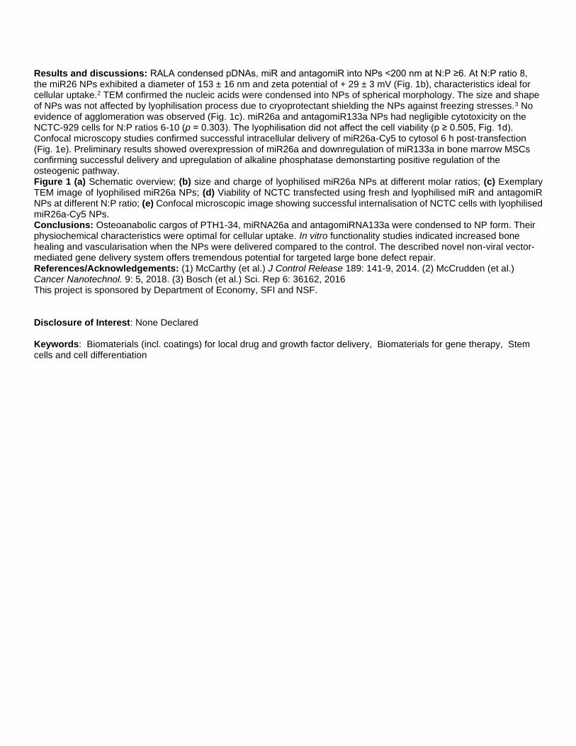
Results and discussions: RALA condensed pDNAs, miR and antagomiR into NPs <200 nm at N:P ≥6. At N:P ratio 8, the miR26 NPs exhibited a diameter of 153 ± 16 nm and zeta potential of + 29 ± 3 mV (Fig. 1b), characteristics ideal for cellular uptake.2 TEM confirmed the nucleic acids were condensed into NPs of spherical morphology. The size and shape of NPs was not affected by lyophilisation process due to cryoprotectant shielding the NPs against freezing stresses.3 No evidence of agglomeration was observed (Fig. 1c). miR26a and antagomiR133a NPs had negligible cytotoxicity on the NCTC-929 cells for N:P ratios 6-10 (p = 0.303). The lyophilisation did not affect the cell viability (p ≥ 0.505, Fig. 1d). Confocal microscopy studies confirmed successful intracellular delivery of miR26a-Cy5 to cytosol 6 h post-transfection (Fig. 1e). Preliminary results showed overexpression of miR26a and downregulation of miR133a in bone marrow MSCs confirming successful delivery and upregulation of alkaline phosphatase demonstarting positive regulation of the osteogenic pathway. Figure 1 (a) Schematic overview; (b) size and charge of lyophilised miR26a NPs at different molar ratios; (c) Exemplary TEM image of lyophilised miR26a NPs; (d) Viability of NCTC transfected using fresh and lyophilised miR and antagomiR NPs at different N:P ratio; (e) Confocal microscopic image showing successful internalisation of NCTC cells with lyophilised miR26a-Cy5 NPs. Conclusions: Osteoanabolic cargos of PTH1-34, miRNA26a and antagomiRNA133a were condensed to NP form. Their physiochemical characteristics were optimal for cellular uptake. In vitro functionality studies indicated increased bone healing and vascularisation when the NPs were delivered compared to the control. The described novel non-viral vector-mediated gene delivery system offers tremendous potential for targeted large bone defect repair. References/Acknowledgements: (1) McCarthy (et al.) J Control Release 189: 141-9, 2014. (2) McCrudden (et al.) Cancer Nanotechnol. 9: 5, 2018. (3) Bosch (et al.) Sci. Rep 6: 36162, 2016 This project is sponsored by Department of Economy, SFI and NSF. Disclosure of Interest: None Declared Keywords: Biomaterials (incl. coatings) for local drug and growth factor delivery, Biomaterials for gene therapy, Stem cells and cell differentiation

Biomaterials for specific medical applications WBC2020-2041 Tanfloc/Heparin Polyelectrolyte Multilayer Enhances Hemocompatibility and Antibacterial Activities of Titania Nanotubes Roberta Sabino* 1, Kirsten Kauk2, Liszt Madruga3, Matt Kipper4, Alessandro Martins5, Ketul Popat2 1School of Advanced Materials Discovery, 2Mechanical Engineering, Colorado State University, Fort Collins, United States, 3Institute of Chemistry, Federal University of Rio Grande do Norte, Natal, Brazil, 4Chemical and Biological Engineering, Colorado State University, Fort Collins, United States, 5Chemical and Materials Science & Engineering, Federal University of Technology, Apucarana, Brazil Introduction: Biomaterial-associated thrombus formation and bacterial infection are still major concerns. Therefore, it is vital to develop multifunctional surfaces, that can simultaneously prevent clot formation and bloodstream infections. Titanium-based implants have been primarily used due to their high mechanical properties, corrosion resistance, and remarkable cytocompatibility. However, titanium cannot suppress blood coagulation, as well as prevent bacterial infections [1]. To address this challenge, several approaches have been proposed to modify the titanium surface, including the use of layer-by-layer (LbL) assembly. LbL is a promising technique used to change the chemical surface of materials with polyelectrolyte multilayers (PEMs) by assembling polycation-polyanion polymer pairs in aqueous solutions. Heparin (HP) is a natural and highly negatively glycosaminoglycan, which prevents blood coagulation. Tanfloc (TN) is reported as a cytocompatible cationic tannin polymer derivative synthesized from condensed tannins. However, this material has a zwitterionic characteristic because condensed tannins naturally occur mixed with hydrolyzed tannins (anionic polymers of gallic acid) [2]. Here, we modify the titanium surface with PEMs based on TN/HP to evaluate the promising antifouling and anti-thrombogenic properties. Experimental methods: We developed novel TN/HP PEMs (5 layers) on titania nanotube arrays. First, the surface topography was modified by making titanium nanotubes (NT) via anodization process, as it has been shown that NT has better antibacterial properties and reduces immune response and thrombogenic effects on titanium. Then, NT surface was modified with PEMs by using TN or chitosan (CS) as polycationic layers, and HP or hyaluronan acid (HA) as polyanionic layers. The surfaces were characterized using scanning electron microscopy (SEM), X-ray photoelectron spectroscopy (XPS), and contact angle measurements. We evaluated protein adsorption, Factor XII activation, platelet adhesion and activation. The antibacterial activity was investigated against Gram-negative P. aeruginosa and Gram-positive S. aureus. Bacterial adhesion and morphology, as well as biofilm formation were analyzed using SEM and fluorescence images. Whole blood clotting kinetics was also investigated. Image:
Results and discussions: By XPS and SEM, we found that 5 layers did not entirely coat the NT surface. The TN/HP PEMs on NT reduced protein adsorption and FXII activation, as well as significantly decreased platelet adhesion and activation in comparison with the other surfaces (Fig. 1). The TN/HP surface also delayed the whole blood clotting time

and decreased the adhesion and proliferation of S. aureus and P. aeruginosa bacteria after 24 hrs of incubation. Also, the NT modified with TN/HP presented the highest number of dead bacteria with no biofilm formation. Conclusions: The results indicate that TN/HP PEMs on NT can be successfully fabricated, to create NT surface with antifouling, antibacterial, and antithrombogenic properties. The modified NT developed by LbL approach is a promising biomaterial to be applied in implants because of its enhanced blood biocompatibility and bactericidal properties. References/Acknowledgements: [1] X. Zhang, G. Zhang, H. Zhang, J. Li, X. Yao, B. Tang, Surface immobilization of heparin and chitosan on titanium to improve hemocompatibility and antibacterial activities, Colloids Surfaces B Biointerfaces. 172 (2018) 338–345. doi:10.1016/J.COLSURFB.2018.08.060. [2] A.F. Martins, S.P. Facchi, P.C.F. da Câmara, S.E.A. Camargo, C.H.R. Camargo, K.C. Popat, M.J. Kipper, Novel poly(ε-caprolactone)/amino-functionalized tannin electrospun membranes as scaffolds for tissue engineering, J. Colloid Interface Sci. 525 (2018) 21–30. doi:10.1016/J.JCIS.2018.04.060. Disclosure of Interest: None Declared Keywords: Antibacterial, Biocompatibility, Cardiovascular incl. heart valve

Biomaterials for specific medical applications WBC2020-315 Nanofiber-based Aerogels for Cranial Bone Regeneration Jingwei Xie*, John Johnson1, Ruiquan Li1, Hongjun Wang1 1Surgery-Transplant and Holland Regenerative Medicine, University of Nebraska Medical Center, Omaha, United States
Introduction: Critical-sized cranial defects are one of the most clinically relevant situations as it encountered in congenital anomalies, trauma, stroke, aneurysms, and cancer.1Although application of free vascularized bone grafts from distant sites may represent the most reliable procedure, it is associated with issues including the size and shape mismatch, bone resorption, and secondary morbidity.Most synthetic materials often fail to remodel and integrate with host tissue, easily become infected, and lead to multiple revision surgeries as bone continues to remodel throughout its lifetime.2There is an urgent need to develop an absorbable, synthetic bone graft. The goal of this project is to develop and validate novel, three-dimensional (3D) hybrid nanofiber aerogels for cranial bone regeneration that can simultaneously fulfill morphological and functional restorations. Experimental methods: The electrospinning set-up used in the present work is similar to that described in our previous publication.3,4The obtained nanofibers was minced using probe ultra sonication or cyo-cutting. The 3D hybrid nanofiber aerogels was formed through freeze casting and thermal crosslinking. Aerogels were implanted to critical-sized cranial bone defects in rats. Bone regeneration was characterized by micro-CT and histology. Results and discussions: We developed a method to generate novel, 3D, hybrid nanofiber aerogels composed of short electrospun poly(lactide-co-glycolide) (PLGA)/collagen/gelatin (PCG) and bioactive glass (BG) nanofibers with controlled shapes, pore structure and/or size and porosity. The final freezing temperature influenced the pore size distribution in the 3D nanofiber aerogel. Generally, larger the temperature gradient, smaller the pores as the temperature gradient affects the periodic growth of ice crystals during freezing. We demonstrated implantation of 3D nanofiber aerogels to calvarial bone defects, with a critical size (8 mm diameter) in rats.5The micro-CT analysis indicates an obvious increase in x-ray radiopacity due to new bone formation between 4 weeks and 8 weeks for all the implant groups. Among the different groups, the determined bone formation area and bone volume were maximum for the E7-BMP-2 peptide loaded 3D hybrid aerogel. Also, the neobone for E7-BMP-2 peptide loaded hybrid aerogel group exhibited extremely high radiopacity, with high-density bone mineral covering more than half of the cranial defect over 8 weeks. Histology results indicated the percentage of new bone area was significantly higher in the E7-BMP-2 peptide loaded aerogel group in comparison to the unfilled defect and the 3D hybrid aerogel groups.
Conclusions: We demonstrated for the first time the fabrication of 3D hybrid nanofiber aerogels composed of segmented electrospun polymer and bioactive glass nanofibers. We further demonstrated that BMP-2 peptides incorporated 3D hybrid nanofiber aerogels can induce ~60-70% closure of critical-sized (8 mm) rat calvarial bone defects after implantation for 8 weeks.
References/Acknowledgements: 1. Lee JC. et al.Adv. Healthc. Mater. 2017, 6, 1700232. 2. Ma B. et al.Nanomedicine 2013, 8, 1459-1481. 3. Weng L. et al.ACS Appl. Mater. Interface 2017, 9, 24484-24496. 4. Xie J. et al.Acta Biomater. 2012, 8, 811-819. 5. Weng L. et al.Adv. Healthc. Mater. 2018, 7, 1701415. Disclosure of Interest: None Declared Keywords: Bone, Craniofacial and maxillofacial

Biomaterials for specific medical applications WBC2020-1701 It takes two to heal: Combating bacterial biofilm and excessive inflammation in chronic wounds so they heal again Thomas Danny Michl* 1, Dung Thuy Thi Tran1, Hannah Frederike Kuckling1, Aigerim Zhalgasbaikyzy1, Laura Elena Gonzalez Garcia1, Rahul Madathiparambil Visalakshan1, Krasimir Vasilev1 1University of South Australia, School of Engineering, Mawson Lakes, Australia Introduction: Chronic wounds are the classic “chicken or egg” paradox because they are simultaneously infected and inflamed, while it isn’t known which came first.1 The vast majority of research avenues aim to prevent biofilm formation in the first place. However, this is a pointless endeavour for chronic wounds, which already have a pre-formed biofilm that requires dispersal. 2 The current medical practice is “mechanical” debridement of the chronic wound, as washing with aqueous solutions and systemic antibiotics have proven ineffective.3 In essence, most of the medical progress in the past 100 years has not led to substantial progress in how chronic wounds are treated. Thus, breaking the vicious cycle of chronic inflammation and bacterial infection simultaneously requires a two-in-one approach which we would like to present: Down-regulating inflammation and breaking up established bacterial biofilm by bestowing wound dressings with novel coatings; turning wound dressings from passive “passengers” into pro-active “actors”. Experimental methods: Thin plasma coatings were deposited using a custom made, low-pressure plasma apparatus. TEMPO ((2,2,6,6-Tetramethylpiperidin-1-yl)oxyl) was used as the precursor plasma polymerized at 13.56 Mhz in continuous mode for either 5, 15 or 30 minutes onto the blank substrate. The plasma polymerized coatings (TEMPOpp) were characterized via Ellipsometry (JA Woolam), XPS (Kratos Axis Ultra) and ToF-SIMS (PHI Drift). The opportunistic bacterium Staphylococcus epidermidis (ATCC 35984) was first grown in RPMI for 24h on blank substrate to create a biofilm and then sandwiched with either with a blank substrate or with the coated TEMPOpp for 2h. Live / dead staining was then used to visualise the biofilm morphology. THP-1 human monocytic cells in conjunction with ELISA was used to quantify the immune response to the surfaces. Image:


Results and discussions: Stable nitroxide radicals have been shown to inhibit biofilm formation and down-regulate inflammation.5 Such qualities are desirable for treating chronic wounds and hence we propose applying nitroxide-containing coatings onto wound dressings. Deposition of thin layers containing stable nitroxide radicals via the facile and industrially relevant process of plasma polymerisation, however, has been reported solely by us to date.6 We have already demonstrated the intricate ability of these coatings to prevent bacterial biofilm formation. Now, we would like to add to our findings by demonstrating that these coatings are also capable of dissolving already formed biofilm by the opportunistic bacteria Staphylococcus epidermidis (Fig 1a). Furthermore, we would like to show how these coatings modulate the inflammation response of macrophages in a dose-responsive manner (Fig 1b). Conclusions: We want to stress the fact that these coatings can disperse existing biofilm and simultaneously modulate the immune response: A two-in-one approach. This dual-action, biofilm dispersion and inflammation modulation, is a prerequisite to stop the vicious cycle that predominates in chronic wounds. Thus, these findings, taken together, are a crucial step towards a new generation of wound dressings that supersede the century-old method of how chronic wounds are currently treated. References/Acknowledgements: 1(a) T. Bjarnsholt et al., Wound Repair and Regeneration, 2008, 16, 2; (b) G. Subbiahdoss et al., Acta Biomaterialia, 2009, 5, 1399; (c) L. G. Harris et al., Injury, 2006, 37, S3. 2 D. Fleming et al., Microorganisms, 2017, 5, 15. 3(a) W.-L. Liu et al., Chinese Nursing Research, 2017, 4, 5; (b) A. R. Siddiqui et al., Clinics in Dermatology, 2010, 28, 519. 4(a) M. Lewandowski et al., International journal of molecular sciences, 2017, 18; (b) A. Verderosa et al., Molecules, 2016, 21, 841. 5 T. D. Michl et al., ACS Applied Nano Materials, 2018. Disclosure of Interest: None Declared Keywords: Antibacterial, Biomaterials (incl. coatings) for local drug and growth factor delivery, Coatings

Biomaterials for specific medical applications WBC2020-1250 Injectable Vascularized Mature Adipose Balls for Soft Tissues Regeneration Applications Fiona Louis* 1, Yoshihiro Sowa2, Shiro Kitano1, 3, Michiya Matsusaki1, 4 1Joint Research Laboratory (TOPPAN) for Advanced Cell Regulatory Chemistry, Graduate School of Engineering, Osaka University, Osaka, 2Department of Plastic and Reconstructive Surgery, Graduate School of Medical Sciences, Kyoto Prefectural University of Medicine, Kyoto, 3TOPPAN PRINTING CO., LTD., Tokyo, 4Department of Applied Chemistry, Graduate School of Engineering, Osaka University, Osaka, Japan Introduction: The recent safety concerns concerning artificial textured breast implants made adipose tissue reconstruction of major importance for soft tissue regeneration. However, the current implantation outcomes revealed up to 70% of volume loss in the long-term studies, due to the necrosis of the fat graft [1]. The main reason is the inefficient blood supply due to the lack of revascularization in situ. The in vivo adipose tissue is indeed mainly composed of mature adipocytes, adipose derived stem cells (ADSC) and endothelial cells, surrounded by collagen type I. So far, there is no in vitro regeneration of a vascularized adipose tissue using directly mature adipocytes, while the in vitro stimulation of both adipogenesis and endogenesis remains difficult [2]. In this context, we used physiological collagen microfibers (CMF), suitable for the long maintenance of mature adipocytes [3], to reconstruct vascularized adipose balls that can be injected in a non-invasive surgical way. Experimental methods: Cross-linked porcine type I collagen homogenized in microfibers (CMF) were mixed with human cells (mature adipocytes, ADSC and umbilical vein endothelial cell (HUVEC)) and fibrin gel to construct the adipose tissues balls (see Figure, A) on 96 well plates. Mature adipocytes and ADSC were isolated from adipose tissues for breast reconstruction surgeries performed by MD Sowa at Kyoto Prefectural University Hospital, in accordance with the Declaration of Helsinki (ERB-C-1317). Adipose balls were assessed using CD31 immunostaining for the blood vessels and NileRed for the adipocytes. Viability was monitored using Live/Dead kit and analyzed by ImageJ software. In vivo mouse experiment was performed on immuno-depressed mice of 1 year-old, following the Kyoto Prefectural University animal experimentation rules. Image:
Results and discussions: The reconstructed adipose tissues balls showed reproducible final diameters after 7 days of culture (Figure, B and C), allowing their injection using a syringe with 17G or 15G needles for 5µL and 10µL balls respectively. In the reconstructed in vitro vascularized tissues balls, the CMF allowed the maintenance of the specific unilocular shape of the mature adipocytes which were found in close contact to blood vessels (Figure, D) in a similar way than in vivo (average distance between vessels: 167±82 µm in vitro and 120±84 µm in vivo). Capillaries showed lumens (mean diameters of 11±7 µm) from the surface to the core of the tissues balls (Figure, D). A good cell viability was observed 24 hours after twice aspirations and releases of the balls from the syringe with needles (data not shown), and

the vascularized balls were able to merge with vasculature interconnection when maintained together in the same culture well for 7 additional days (data not shown). These results leaded to the in vivo mice injections of 100µL of the vascularized adipose balls, compared with only mature adipocytes balls and liposucted human adipose tissue injections. After one month, the tissues were extracted, showing a high blood vessels network only in the vascularized adipose balls tissues. In comparison, the injected liposucted adipose tissue displayed some adipocytes necrosis areas. Conclusions: The vascularized adipose balls constructed with collagen microfibers allowed to maintain mature adipocytes phenotype while establishing an in vivo-like vascularization in the tissues. The 3 months in vivo results should then allow to conclude of the potentiality of these tissues for soft tissue regeneration needs by enhancing the graft survival time on site after injection. These tissues were seeded using a usual multi-micropipette, leading to a possible automatization of the process for the latter construction of bigger sizes tissues. References/Acknowledgements: [1] Eto et al., Plast Reconstr Surg. 2012;129: 1081-92. [2] Muller et al., Scientific reports, 2019:9(1), 7250. [3] F. Louis et al., Acta Biomaterialia 2019, 84: 194–207. Disclosure of Interest: None Declared Keywords: Adipose tissue

Biomaterials for specific medical applications WBC2020-1266 Hexametaphosphate as a Potential Therapeutic for the Prevention and Treatment of Kidney Stones Thomas Robinson* 1, Sophie Cox1, Liam Grover1 1School of Chemical Engineering, University of Birmingham, Birmingham, United Kingdom Introduction: Kidney stones affect 8.8% of the population and incidence is increasing across all demographics1. They can cause intense pain, blockage of urine flow, urinary tract infections, and lead to more serious complications if left untreated2. Small stones may be passed unassisted, though often with considerable pain, but sizeable stones require intervention with the largest stones necessitating surgical removal3. The most common kidney stone constituent is calcium oxalate, though calcium phosphate, struvite, uric acid and cystine are also frequent components2. Citrate therapy is often used to prevent stone recurrence, in part because of its ability to bind divalent cations, such as calcium. The hypothesis of this study is that hexametaphosphate (HMP), another calcium chelator, may be a more potent binder of calcium and thus a more powerful dissolution agent of calcium-based kidney stones. Experimental methods: The dissolution capacity of HMP was first assessed against various calcium salts and other kidney stone components, using a mixture of residual mass and light extinction methods. These same methodologies were used to compare HMP and citrate at dissolving calcium oxalate, and the minimum concentration of each to clear the suspension was established. In addition, the size and zeta potential of calcium oxalate particles in solutions of citrate and HMP were measured. Kidney stones were then obtained from patients following surgical removal, and their composition was analysed by micro-computed tomography, X-ray fluorescence imaging, and other methods. Stones were then exposed to HMP in artificial urine, and their mass was monitored over time in order to assess the extent of dissolution. Image:
Results and discussions: HMP was able to dissolve calcium oxalate, phosphate (hydroxyapatite) and carbonate, and complete dissolution was observed at a HMP:Ca ratio of 1:3 for all three compounds, suggesting each calcium cation is bound between two phosphate moieties. HMP was also able to dissolve struvite, a magnesium salt, further displaying its ability to bind divalent cations. However, it did not affect organic components uric acid and cystine. HMP was significantly more effective than citrate at dissolving calcium oxalate, with a minimum dissolution concentration more than 12 times lower. Further, dissolved mineral was found to re-precipitate over time when treated with citrate, which was not the case for HMP. The zeta potential of calcium oxalate in HMP was found to be significantly more negative (-35 mV) than in artificial urine alone or with citrate (-17 mV), suggesting that in addition to dissolving the mineral, HMP also binds to it and prevents it from aggregating. This is not the case for citrate, where the crystals regrow and aggregate, leading to large flocs. Finally, in real stones, HMP was further shown to be a potent dissolution agent, dissolving stones by over 70% in 7 days. Conclusions: HMP is a potent chelator of divalent cations, and thus is able to dissolve the vast majority of kidney stone components. It is not only a more effective dissolution agent than citrate, but also binds to the crystal surface to prevent aggregation, a critical growth step in stone formation. Thus, in combination with stone type stratification and suitable delivery, HMP may present a new and potent therapy to prevent and treat kidney stones.

References/Acknowledgements: 1 C. D. Scales, A. C. Smith, J. M. Hanley and C. S. Saigal, Eur. Urol., 2012, 62, 160–165. 2 T. Alelign and B. Petros, Adv. Urol., 2018, 2018, 3068365. 3 F. L. Coe, A. Evan and E. Worcester, J. Clin. Invest., 2005, 115, 2598–608. Disclosure of Interest: None Declared Keywords: Calcium phosphates, Kidney, liver and pancreas

Biomaterials for specific medical applications WBC2020-1270 Development of a new biomedical material with immunosuppressive properties for stent application Oroitz Sánchez-Aguinagalde* 1, Ainhoa Lejardi1, Eva Sánchez-Rexach1, Emilio Meaurio1, Jose Ramon Sarasua1 1ZIBIO Group, Department of Mining-Metallurgy Engineering and Materials Science and POLYMAT, University of the Basque Country, Bilbao, Spain Introduction: Mycophenolic acid (MPA), a selective and uncompetitive inosine monophosphate dehydrogenase (IMPDH) inhibitor, is an immunosupressant drug used to prevent rejection of organ transplants. In addition, it has shown potential in many tumor treatments, it is antimicrobial, and can inhibit vascular smooth muscle cell (VSMC) proliferation [1], one of the main causes of in-stent restenosis (ISR). The aim of this work is to introduce MPA in a poly(ε-caprolactone)(PCL) matrix creating an amorphous solid dispersion (ASD) [2], in order to improve the bioavailability of the drug. Miscibility and interactions of the polymer/drug blend were studied, as well as the drug release mechanisms. Experimental methods: Different compositions of PCL/MPA blends were obtained by solvent casting. Differential Scanning Calorimetry (DSC) was used to study their Tgs, as well as melting point depression of MPA, in order to obtain the Flory-Huggins interaction parameter (χ). Hydroxyl and carbonyl stretching regions were observed by Fourier Transform Infrared Spectroscopy (FTIR). A drug release test was performed for 3 days, and the best fit of the curves to different kinetic models was found. Image:

Table: Thermal properties of PCL/MPA blends in a second scan
PCL/CA
M TgExp. (°C) TgFox (°C) TmPCL (°C) ΔHmPCL (J/g)
PCL -60 - 57.2 66.4 80/20 -44.1 -48.8 51.7 49.8 60/40 -36.3 -36.3 46.4 25.9 40/60 -25.7 -22.4 - - 20/80 -10.2 -6.8 - - MPA 11.1 - - -
Results and discussions: Using DSC, a single Tg intermediate between those of pure components was confirmed for blends covering the whole composition range (Fig.1a), close to those predicted using Fox equation (Table) . Melting point depression of MPA was analysed, and a negative F-H interaction parameter was obtained, χ(-1.18) (Fig.1b). Figure 1c shows the FTIR spectra of blends in the OH stretching region, where the OH peak of MPA moves to higher wavenumbers as PCL content increases. In the C=O stretching region (Fig.1d) two peaks can be seen for MPA, one at 1745 cm-1, attributable to ester groups, and another one at 1705 cm-1 related to dicarboxylic acid groups. The latter disappears when PCL is added to the blend: the MPA crystals break, the hydrogen bonds are dissociated and an association occurs with C=O groups of PCL. For 0.05% and 0.1% MPA, drug release was complete at the third day, while samples with 0.2%, 0.5%, 1% and 2% MPA continued releasing (Fig.1e). Figure 1f shows these amounts in μg. Samples with 0.05% and 0.1% MPA showed a first order release mechanism, while the rest of the compositions fitted Higuchi model. Korsmeyer-Peppas model confirmed the Fickian release mechanism. Conclusions: In this work, miscibility between PCL and MPA was confirmed by single intermediate Tg for all compositions, as well as by a negative Flory-Huggins interaction parameter. FTIR spectra showed that MPA crystals break with the addition of PCL, creating new bonds between both. Drug release tests showed diffusion and concentration dependant release kinetics. The released amounts were measured, in order to control the ongoing cell viability studies. References/Acknowledgements: The authors are thankful for funds from the Spanish Ministry of Innovation and Competitiveness MINECO (MAT2016-78527-P) and the Basque Government Department of Education, Language Policy and Culture (IT-927-16). [1] J. Park et al. Am. J. Transplant., vol. 4, no. 12, pp. 1982-1990, 2004. [2] E. Sanchez-Rexach et al. Eur. Polym. J., vol. 102, no. March, pp. 30-37, 2018. Disclosure of Interest: None Declared Keywords: Biomaterials for drug delivery, Vascular grafts incl. stents

Biomaterials for specific medical applications WBC2020-987 Scaffold and micro-environment designed to promote directional neuronal cell growth Shadi Houshyar* 1, G. Sathish Kumar2, Mamatha M Pillai2, Tanushree Saha3, Satya Ranjan Sarker4, Amitava Bhattacharyya2 1School of Engineering, RMIT University, Melbourne, Australia, 2Functional, Innovative and Smart Textiles, PSG Institute of Advanced Studies, Coimbatore, India, 3School of Fashion and Textiles, RMIT University, Melbourne, Australia, 4Dept. of Biotechnology & Genetic Engineering, Jahangirnagar University, Savar, Dhaka, Bangladesh Introduction: Poor sensory and motor function recovery outcomes after nerve injuries are the main issues of the current treatment techniques 1. This is due to the failing in duplicating the structure and bio-functionality of nerve tissue, leading in failure of connection or inappropriate connections. Several studies demonstrated positive effect of electrical conductivity and dopamine on extension of neurites in neural cells which resulted in improving neural migration and functional recovery 1-4. However, directional growth of neurite and migration of neuron and other types of cells are the challenging issues1. This study focused on developing conductive scaffold for nerve guide potential, by enhancing directional growth of neural regeneration and axonal regrowth. Experimental methods: Active ink including dopamine (DO), carbon nanofiber (CNF) and polycaprolactone (PCL) was printed on the surface of thin PCL film, through 3D bioplotting technique. The developed scaffolds were characterized for morphology and biological properties. Image:
Results and discussions: Analysis of cell viability revealed no increase in cell death on any of the polymers compared to tissue culture plastic control or compared to PCL polymer without printed DO and CNF. The scaffold showed good U87MG cell attachments and differentiation in the absence of cell adhesion molecules. Confocal images, Fig. 1., showed that the highest cell attachments and proliferation were achieved in the area of CNF-DO printed lines with good conductivity and moderate roughness. Conclusions: The scaffold showed directional neural regeneration and axonal growth in the direction of printed active ink. This technique is a promising method for using in neural applications in the area of bionics and tissue engineering when directional cell growth is necessary. References/Acknowledgements: 1. Houshyar, S.; et al., ACS Chem Neurosci 2019 2. Fernandez-Lopez, B. et al. Neural Plast 2015, 2015, 350750.

3. Gopinathan, J.; et al. J Biomed Mater Res A 2016, 104 (4), 853-65. 4. Parish, C. L. et al. Development 2007, 134 (15), 2881-7. Disclosure of Interest: None Declared Keywords: 3D scaffolds for TE applications, Materials for electric stimulation, Peripheral nerves and spinal cord

Biomaterials for specific medical applications WBC2020-1066 Biodegradable luminescent porous silicon nanoparticles as an imaging tissue adhesive for rapid closure and multifunctional repair of minimally invasive wound Qingyan Zeng* 1, Ning Cui1, Kai Han1, Tingli Lu 1School of Life Sciences, Northwestern Polytechnical University, Xi’an, China Introduction: Suture and staples are effective ways for wound closure and reconnection of injured tissues after surgery or trauma. However, they are not suitable for tension-free bonding wound such as minimally invasive wound. Recently, nanoparticles act as adhesive for biological tissue has raised great concern. Several inorganic NPs exhibit strong adhesive properties in the wound closure of animal models. However, few studies have reported biodegradable and imaging tissue adhesives. Porous silicon nanoparticles (PSiNPs) is an inorganic material have attracted increasing interest for its large surface area, controllable pore sizes and surface chemistry. Moreover, PSiNPs is biodegradable, biocompatible and non-toxic. Moreover, PSiNPs is intrinsic photoluminescence resulting in clear visualization of the PL in vivo. Therefore, PSiNPs could meet all above requirements of biodegradable and imaging tissue adhesive and can be considered as an excellent candidate of nano-tissue adhesive. Experimental methods: PSiNPs were fabricated by electrochemical etching method, and then PSi nano-glue was prepared by mixing with a certain proportion of aqueous solution. The physical structure and chemical composition of PSiNPs were characterized by scanning electron microscopy (SEM), laser particle size analyzer, N2 adsorption analyzer, Fourier transform infrared (FT-IR) spectroscopy, X-ray diffraction (XRD), and X-ray photoelectron spectroscopy (XPS). Furthermore, adhesion strength test, CELL TITER-GLO (CTG) assay, blood coagulation test, degradation and imaging experiment in vitro were used to investigate the tissue adhesion property, biocompatibility, degradability, hemostasis and fluorescence imaging performance. The wound healing effect of PSi nano glue was further evaluated by mouse skin wound model. Image:
Results and discussions: The results showed that PSiNPs were successfully prepared with the size were around 80nm and specific surface area around 100m2/g. The XRD and XPS results showed that PSiNPs changed to LPSiNPs after oxidation. Importantly, PSi nano-glue exhibited strong adhesive properties between two small pieces of pig liver. Besides, fluorescence imaging performance and rapid hemostasis within one minute were observed. Furthermore, this imaging PSiNPs showed much less cellular toxicity to L929 fibroblasts and fast degradation to orthosilicic acid, which was non-toxic and could excreted from body through the urine. Finally, this biocompatible BLPSiNPs achieved rapid and efficient closure and healing of wounds in comparison to control and mesoporous silicon. Conclusions: We prepared biocompatible biodegradable luminescent porous silicon nanoparticles (BLPSiNPs) solution, which served as nano-glue to achieve rapid closure and healing of critical size wound in skin, exhibiting not only strong adhesive properties but also high contrast effects for real-time luminescent imaging. Therefore, this biodegradable imaging tissue adhesive have great potential for rapid closure, visual detection of wound healing and repair of tension-free bonding wound. References/Acknowledgements: References 1. Rose S, et al. Nature, 2014, 505(7483): 382. 2. Meddahi‐Pellé A, et al. Angewandte Chemie International Edition, 2014, 53(25): 6369-6373. 3. Park J H, et al. Nature materials, 2009, 8(4): 331. 4. Shin K, et al. Nature communications, 2017, 8: 15807. Acknowledgements This work is supported by the National Science Foundation for Young Scientists of China (No.51903209).
Disclosure of Interest: None Declared

Keywords: Biodegradation, Imaging, Wound healing and tissue adhesives

Biomaterials for specific medical applications WBC2020-1152 Influencing the Osteoporotic Imbalance of Bone Cells by Calcium/Strontium Phosphates Benjamin Kruppke* 1, Alena Svenja Wagner2, Seemun Ray3, Marcus Rohnke4, Sabine Wenisch2, Thomas Hanke1 1Technische Universität Dresden, Institute of Materials Science, Dresden, 2Justus-Liebig-University Giessen, Department of Veterinary Clinical Sciences, Small Animal Clinic c/o Institute of Veterinary-Anatomy, -Histology and -Embryology, 3Justus-Liebig-University Giessen, Laboratory of Experimental Trauma Surgery, 4Justus-Liebig-University Giessen, Institute for Physical Chemistry, Gießen, Germany Introduction: In the case of osteoporosis, there is an imbalance of osteoblasts and osteoclasts, which causes increased fragility and worsened healing of the bone. To treat bone defects under these conditions, we focused on a degradable material of gelatin-modified calcium and strontium phosphates. This material is ought to release strontium ions and keep the cation concentration on a physiological level and thus stimulate osteoblastogenesis and furthermore support bone regeneration. To enhance ion release the calcium/strontium phosphates were varied according to crystal structure, composition and morphology. After osteoblast/osteoclast co-culture, which confirmed the decrease of osteoclast cell activity due to strontium ion release from the material, the same was proven after implantation and analysis in an osteoporotic rate model. Experimental methods: Gelatin was prestructured by mixing it with a phosphate solution. Afterwards calcium and/or strontium containing solutions were added under constant stirring. The precipitated organically modified mineral was cross-linked and lyophilized to obtain porous 3D samples. The resulting material was investigated according to ion release and degradability. Cell culture of human mesenchymal stromal cells (hBMSC) and human monocytes was performed in vitro in a co-culture. The in vivo characterization of the material was done by implantation in a femoral defect in osteoporotic rats over a period of 6 weeks and analysed immunohistochemically as well as using ToF-SIMS. Finally, the material degradation was analysed under different conditions of constantly flowing fluid in a specially designed chamber, to match the in vivo findings. Results and discussions: The materials caused an initial ion release in calcium-rich (2.0 mM) and low-calcium (0.4 mM) minimum essential medium. Co-cultivation of osteoblasts and osteoclasts on the material led to formation of osteoclast-like cells, able to migrate, fuse, and differentiate. The osteoblastic differentiation (proven by alkaline phosphatase, ALP) of hBMSC was increased with higher strontium content of the materials. Osteoclasts, on the other hand, showed a decrease in osteoclastic markers (e.g. RANKL) with lower Ca/Sr-ratios. After implantation the osteoblastic and osteoclastic reactions seen in vitro were confirmed from in vivo results. Mass spectrometric imaging (ToF-SIMS) of thin cuts of the rat femurs revealed an increased bone formation with increasing strontium in the material. Conclusions: The study showed, that bone substitute materials can be adjusted to demands of systemically altered bone – with respect to its degradation and ion release. Furthermore the degradation under conditions of constant flowing medium can be used to predict material degradation in vivo more precisely. Disclosure of Interest: None Declared Keywords: Biodegradation, Bone, Calcium phosphates

Biomaterials for specific medical applications WBC2020-1176 VCAM-1 targeted gene delivery nanoparticles localize to inflamed endothelial cells and atherosclerotic plaques Nicholas Distasio* 1, Hugo Salmon2, Stephanie Lehoux3, Maryam Tabrizian1, 2 1Biomedical Engineering, 2Dentistry , 3Experimental Medicine , McGill University, Montreal, Canada Introduction: Atherosclerosis is a leading cause of morbidity and mortality worldwide. One recent study demonstrated that targeting the inflammatory component of atherosclerosis can further curb the disease [1], highlighting the therapeutic potential of this strategy. New and innovative therapies targeted to the site of atherosclerotic plaque growth may provide a complementary benefit. Interleukin-10 (IL-10) is a potent anti-inflammatory cytokine whose small size and short bioavailability have so far limited its translation as a therapeutic [2]. We hypothesize that by targeting endothelial cells at the plaque to produce IL-10 themselves, local chronic inflammation within the plaque may be reduced and clinical events avoided. We have previously formulated gene delivery nanoparticles (NPs) made of poly(β-amino esters) (PBAEs) and IL-10 plasmid DNA (pIL-10) that could transfect primary mouse endothelial cells (mECs) with high efficiency and attenuate inflammation in an in vitro model of atherosclerosis [3]. Here, we coated these nanoparticles with a poly(glutamic acid)-poly(ethylene glycol)-peptide conjugate specific for VCAM-1, a receptor overexpressed by inflamed endothelial cells overlying atherosclerotic plaque. We assess the binding, uptake, and transfection of these targeted NPs at the molecular, cellular, and tissue level. Experimental methods: Experiments involving animals were approved by McGill University’s Animal Care Committee. Surface plasmon resonance with imaging (SPRi) was used to characterize affinity-based interactions between NPs (containing pIL-10) and VCAM-1 grafted on the sensor surface. NPs containing Cy3-pIL-10 were added to TNFα-activated mECs under static or dynamic flow conditions in an Ibidi μ-slide and uptake was assessed via flow cytometry and/or fluorescence microscopy. ApoE-/- and LDLR-/- mice were fed a high-fat diet for at least 9 weeks to induce atherosclerotic plaque formation. Tissues containing plaque (aortic sinus, brachiocephalic artery) were harvested and cryo-sectioned and aortas were freshly prepared en face. Targeted and non-targeted NPs (Cy3- or Cy5-pIL-10) were incubated on fixed tissues co-stained with VCAM-1. For live imaging and biodistribution, NPs containing Cy5- or Cy7-pIL-10 were intravenously injected in atherosclerotic mice and imaging was performed on live mice and on harvested organs using the IVIS Spectrum live imaging system. Image:

Results and discussions: Targeted NPs bound specifically to VCAM-1 in a dose dependent manner (Figure 1A). Under static flow conditions, targeted NPs were taken up significantly more by TNFa-activated mECs than non-targeted NPs (Figure 1B). The increase in NP uptake was also observed under dynamic flow conditions (shear stress of 15 dyn/cm2). Increased NP uptake was correlated with actual gene transfection. Targeted NPs bound to the endothelial layer of plaque-containing tissues and colocalized with VCAM-1 expression (Figure 1C). Although targeted and non-targeted NPs accumulated mostly in the liver 24h after injection, targeted NPs could be seen after 30m near the heart via live 2D imaging and 3D Fluorescence Imaging Tomography (FLIT) (Figure 1D, arrows). Conclusions: We present an innovative, multi-disciplinary, and targeted approach that has the potential to reduce inflammation in atherosclerosis via non-viral gene delivery and could be amenable to other applications. We also present for the first time the use of SPRi and dynamic flow culture conditions for the assessment of binding and uptake of soft polymeric gene therapy nanoparticles. References/Acknowledgements: The authors graciously acknowledge our funding sources, CIHR and NSERC. In addition, we thank France Dierick, Talin Ebrahimian, and Veronique Michaud for help with in-vivo experiments and members of the Biomat'X Lab at McGill University.

[1] P. M. Ridker et al., CANTOS Trial Group, N Engl J Med 2017, 377, 1119. [2] X. Wang, T. Coradin, C. Hélary, Biomater Sci 2018, DOI 10.1039/c7bm01024a. [3] N. DiStasio et al., ACS Applied Bio Materials 2018, DOI 10.1021/acsabm.8b00342. Disclosure of Interest: None Declared Keywords: Biomaterials for gene therapy, Cardiovascular incl. heart valve, Cell/particle interactions

Biomaterials for specific medical applications WBC2020-1204 Delivery of a microRNA cocktail by electrospun PVA/alginate/ciprofloxacin nanofibres: a novel genetic nanomedicine for impaired wound healing. Adrian Dario Juncos Bombin* 1, Nicholas Dunne2, Helen McCarthy1 1School of Pharmacy, Queen's University Belfast, Belfast, United Kingdom, 2School of Mechanical & Manufacturing Engineering, Dublin City University, Dublin, Ireland Introduction: Delays in wound healing are a significant financial burden to the NHS with annual costs estimated to be £3.2 billion (1). Wound healing is regulated by microRNAs (miRs), which impact multiple aberrant genetic pathways (2). miR-21 and miR-132 can promote re-epithelialisation and anti-inflammatory responses by targeting different molecular pathways (3,4) and miR-31 participates in angiogenesis and re-epithelialisation (2). All this is paramount in the treatment of chronic wounds. In this study, we have encapsulated RNA antisense oligonucleotides (ASO) with the sequence of these miR into nanoparticles (NP) by electrostatic interaction with RALA, a novel 30-mer amphipathic pH-responsive peptide that is designed to overcome biological barriers (5). A wound dressing with potential for NP loading and delivery has been fabricated using electrospinning. Polyvinyl alcohol (PVA) was chosen as the carrier polymer owing to its ability to be electrospun, sodium alginate (SA) was incorporated for its wound healing ability and ciprofloxacin was added as a wide-spectrum antibiotic (6). Experimental methods: RALA was complexed with the miR ASOs at a range of different N:P ratios to produce RALA/ASO NP. Particle size and zeta potential were measured using a Nano ZS Zetasizer (Malvern Instruments, UK). NP were imaged using a JEM-1400 TEM (JEOL, Japan). The RALA/ASO NP were fluorescently labelled (RALA/Cy3-ASO) and NCTC-929 fibroblasts (ATCC, USA) were transfected with RALA/Cy3-ASO. Internalisation efficiency was quantified by flow cytometry (BD Biosciences, USA). Cell viability was assessed via MTS assay (ThermoFisher, USA) in NCTC-929 and HaCaT keratinocytes (ATCC, USA) incubated with RALA/ASO NP. The polymers were dissolved in water at different concentrations and mixed at a 1:1 v/v ratio. Ciprofloxacin was added at concentrations of 0.5-2.0 mg/mL. The blend solution was electrospun in a 20 kV electrospinning kit (Spraybase, USA). Image:
Results and discussions: RALA condensed the ASO into NP ≤200 nm with a zeta-potential of ≥10 mV at N:P ratio ≥ 4 (Fig. 1a), confirmed by TEM (Fig. 1b) and ideal for cellular uptake (5). NCTC cells were fluorescent under the optical

microscope after incubation with RALA/Cy3-ASO. A high internalisation efficiency was detected using flow cytometry in NCTC fibroblast (Fig. 1c), HaCaT keratinocyte and HMEC endothelial cells. A significant increase in cell metabolic activity was detected for N:P ratios 4-10 in NCTC and 2-6 in HaCaT (Fig. 1d). Direct comparisons were made between all RALA/ASO NP for migration using wound scratch assays and angiogenesis using tubule formation assays to determine the optimal ‘cocktail’ of miR therapeutic. Using Design of Experiments, the optimal PVA and SA concentration, applied voltage, needle-collector distance and flow rate were determined to produce NF with the optimal morphology. Fig. 1e shows the morphology of one of the NF mats. Average diameter was found to be 356.74 ± 81.76 nm. Conclusions: RALA/ASO NP were successfully condensed and demonstrated excellent physiochemical characteristics for cellular uptake in fibroblast, keratinocyte and endothelial cell lines. A range of in vitro studies have determined the optimal combination of RALA/ASO NP to take forward in vivo. An optimal PVA/SA nanofibrous wound patch has been prepared using electrospinning. To ensure antimicrobial effectiveness, ciprofloxacin has been included in the nanofibrous wound patch, and was shown not to compromise its integrity. Taken together, this work proposes a holistic approach towards chronic wound healing based on the synergistic effects of a miR ‘cocktail’ genetic therapy delivered from a multifunctional, degradable, nanofibrous that mimic the extracellular environment and protect against microbial infection. References/Acknowledgements: (1) Guest et al. (2015). BMJ Open; (2) Mulholland et al. (2017). Mol. Ther. - Nucleic Acids (3) Li et al. (2017). J. Invest. Dermatol.; (4) Yang et al. 2011. Int. J. Biol. Sci.; (5) McCarthy et al. (2015). J. Control Release; (6) Kataria et al. 2014. Int. J. Pharm. Disclosure of Interest: None Declared Keywords: None

Biomaterials for specific medical applications WBC2020-378 Ultracompliant PEDOT-Gelatin-Based Neural Interfaces Applied for A Platform of Investigation on Perineural Invasion Yue-Xian Lin* 1, Wei-Chen Huang2, Yu Pei3 1Biomedical Engineering, Biomedical Engineering, Taipei city, 2Institute of Electrical and Computer Engineering, Hsinchu, 3Biomedical Engineering TMU, Biomedical Engineering, Taipei city, Taiwan Introduction: Perineural invasion (PNI), the neoplastic invasion of nerves, is related to the increase of cancer cells aggression that results in poor patient outcomes. Recent studies reveal that the supportive cells (Macrophages, Fibroblasts, and Schwann cells) in the nerve microenvironment activate cancer cells to spread along the nerve, proceeding the invasion. Unfortunately, the mechanism of PNI occurring between the related tumor cells and the nervous system remains unknown. A better understanding of the interactions between supportive cells and cancer cells could be the key to a therapeutic strategy for PNI. Herein, we developed a conductive microelectrode arrays which demonstrated mechanical properties comparable to that of neural cells to build up a nerve-mimicked microenvironment that can provide electrical and chemical cues for studying the intercommunication between Schwann and cancer cells. Experimental methods: To obtain PEDOT-GelMA (PG), gelatin methacryloyl (GelMA, 10%) and 3,4-ethylene dioxythiophene (EDOT, 0.1 M) was dissolved in deionized water at 60˚C and stirred for an hour. Then, Fe3+ ion (0.3 M) was added to the solution to carry out the oxidation polymerization for 24 hr. Fourier transform infrared (FTIR) spectrum was used to investigate the bonding interaction of PG. X-Ray Diffraction(XRD) spectrum was used to explore the crystallinity of hydrogels. The rheometer and electrochemistry analyzer were used to measure the mechanical and electrical/ionic conductivity of hydrogels. Finally, based on the water-dissoluble property of non-crosslinked GelMA at high temperature. thermal-responsive photolithography was used to create a micropatterned PG that were further fabricated into microelectrode arrays (MEA). Co-culture of Schwann cells and cancer cells were performed on the device. Image:
Results and discussions: By controlling the incorporated concentration of GelMA, the storage modulus of the hydrogel can be manipulated from the range of 1 to 300kPa that mimicked the nervous tissues. Furthermore, the addition of PEDOT not only increased the conductivity of the hydrogel but also resulted in a better ductility. The crosslinking of PG

hydrogel creates a deviation of temperature gel point between the two PG hydrogels (with and without UV light exposure) by 1˚C (Figure 1b). As a result, enabling us to apply photolithography with a water-dissoluble property at a higher temperature to fabricate PG hydrogel into patterned microelectrode arrays (Figure 1c). Co-culture of cancer cells with Schwann cells revealed that Schwann cells induced the formation of cancer cell protrusions in their direction, leading to cancer cell dispersion. Conclusions: In summary, we created a hydrogel-based microelectrode which features suitable electrical property plus the mechanical property that is comparable to neural cells. We presented a unique thermal-responsive photolithography method to fabricate microelectrode arrays pattern. With a simple fabrication method, PG microelectrode arrays were fabricated to exhibit nerve microenvironment mimicking properties suitable for investigating the process of PNI. Disclosure of Interest: None Declared Keywords: Biopolymeric biomaterials, Mechanical characterisation

Biomaterials for specific medical applications WBC2020-460 Self-healing bioadhesives with strong wet adhesive strength and multi-functionality Yazhong Bu* 1, Sergio M. Saldaña1, Amir Abdo1, Abhay Pandit1 1CÚRAM, SFI Research Centre for Medical Devices, National University of Ireland, Galway, Galway, Ireland Introduction: Adhesive systems have the potential to revolutionise wound closure procedures.[1-3] However, adhesives often lose their strength because of the adhesion fracture if the site of injury does not heal fast enough. In such cases, mechanical debridement is needed to remove residual adhesive to ensure an effective bonding between the new adhesive and the tissue. Should this be necessary, removing the sticky residue from a wound is painful; alternatively, surgical excision causes secondary damage to tissues especially when the surface of the wound is covered by adhesive. Adhesives with self-healing adhesive strength can solve this problem. After adhesion fracture, wounds need to be closed again and the adhesive strength can be restored without the need of removal of the previous application. Here, we hypothesize that hyaluronic acid (HA) and tannic acid (TA)-based adhesives using EDC/NHS can provide high adhesive strength with self-healing as well as bacteriostatic properties and facilitate wound healing by scavenging free radicals. Specific objectives for this study were to fabricate HA and TA-based adhesive with high adhesive strength and self-healing properties; evaluate the possible bacteriostatic and the antioxidant properties of the adhesive and finally to test the cytotoxic effects of these hydrogels against human dermal fibroblast (HDFs). Experimental methods: HASATA was fabricated by using freeze-drying method (Scheme 1). Adhesive strength and self-healing properties were assessed by the lap shear test. The bacteriostatic properties were evaluated against Escherichia coli (DSM 30083) and Staphylococcus Epidermidis (DSM 20044). The anti-free radical properties were assessed using DPPH assay, hydroxyl radical scavenging assay, nitric oxide scavenging assay as well as measurement of the lipid peroxide radical scavenging capacity. The cytotoxicity of the hydrogels was tested against primary HDFs using Alamarblue®, Quant-iT PicoGreen® dsDNA Assay Kit and live/dead® cell viability assay. Image:

Results and discussions: Adhesive strength and self-healing properties. The adhesive strength increases with time and, within 30 minutes, the maximum adhesive strength can be attained (Figure 1A). This was significantly larger than that seen in the commercial fibrin glue and nearly the same as that seen in cyanoacrylate (Figure 1B). Unlike the traditional fibrin glue and cyanoacrylate systems that loose almost their entire adhesive strength after the fracture, HASATA adhesive regains part of its adhesive strength even after a second fracture (Figure 1C), and this strength was found to increase with time (Figure 1D). Moreover, the adhesive retained most of the adhesive strength and self-healing property even after 50 attach/detachments cycles (Figure 1E and F). Bacteriostatic and antioxidant property: The adhesive showed bacteriostatic activity against E. coli and S. epidermidis. The adhesive system was able to scavenge free radicals including nitric oxide, lipid peroxide and hydroxyl radicals (Figure 2). Cell viability: The HDFs maintained their viability when incubated with the leaching solution of HASATA up-to a concentration of 1 mg/ml. Conclusions: In summary, by combining HA and TA, an adhesive with a high adhesive strength and self-healing properties was fabricated. Moreover, it displayed antibacterial and antioxidant properties, in addition to being biocompatible with the HDFs up-to 1 mg/ml of the leaching solution References/Acknowledgements: Acknowledgements This project has received funding from the European Union’s Horizon 2020 research and innovation programme under the Marie Skłodowska-Curie grant agreement No 713690. This research has also been conducted with the financial support of Science Foundation Ireland and the European Regional Development Fund under grant number 13/RC/2073. References [1] V. Bhagat, et al. Biomacromolecules 18 (2017) 3009-3039. [2] J. Guo, et al. Biomaterials 85 (2016) 204-217. [3] E.Y. Jeon, et al. Biomaterials 67 (2015) 11-19. Disclosure of Interest: Y. Bu Conflict with: Two IDF have been applied about this abstract, S. Saldaña: None Declared, A. Abdo: None Declared, A. Pandit Conflict with: Two IDF have been applied about this abstract Keywords: Wound healing and tissue adhesives

Biomaterial synthesis and characterisation WBC2020-3667 Penetration of the blood-brain barrier and anti-tumor effect of a novel PLGA-lysoGM1/DOX micelles drug delivery system Ying Yin1, Jun Wang1, Meng Yang1, Ruolin Du1, 2, Giuseppe Pontrelli3, Sean Mcginty4, Guixue Wang1, Tieying Yin1, Yazhou Wang* 1, 2 1Key Laboratory for Biorheological Science and Technology of Ministry of Education, State and Local Joint Engineering Laboratory for Vascular Implants, Bioengineering College of Chongqing University, 2Medical School of Chongqing University, Chongqing University, Chongqing, China, 3Via dei Taurini 19, 00185, Istituto per le Applicazioni del Calcolo - CNR, Roma, Italy, 4Division of Biomedical Engineering, University of Glasgow, Glasgow , United Kingdom Introduction: Effective treatment of glioma and other central nervous system diseases is hindered by the presence of the blood-brain barrier (BBB). A novel nano-delivery vehicle system comprised of PLGA-lysoGM1/DOX micelles was developed to cross the BBB for central nervous system administration. Experimental methods: PLGA-lysoGM1/DOX micelles were synthesized by dialysis method and characterized by dynamic light scattering particle size analyzer electron microscopy. In vitro drug release data were well-fitted by a diffusion-based biphasic mathematical model. Mouse brain microvascular endothelial cells bEnd.3 and rat C6 glioma cells were used for the micelles uptake mechanism experiments. Confocal laser scanning microscope (CLSM) characterization of the zebrafish and small animal imaging experiments of mice after PLGA-lysoGM1/DiR injection revealed in vivo distribution of micelles in the brain. The anti-glioma effect of micelles was evaluated in cerebrally implanted C6 glioma cells rat model. Results and discussions: Our in vitro experiments demonstrated that PLGA-lysoGM1/DOX micelles have a slow and sustainable pH-dependent drug release under physiological conditions and exhibit a high cellular uptake through the micropinocytosis and the autophagy/lysosomal pathways. In vitro drug release data were well-fitted by a diffusion-based biphasic mathematical model. In vivo experimental studies in zebrafish and mice confirmed that PLGA-lysoGM1/DOX micelles could cross the BBB and specifically accumulate in the brain. Moreover, an excellent anti-glioma effect presented in intracranial glioma-bearing rats. Conclusions: PLGA-lysoGM1/DOX micelles not only effectively crossed the BBB, but our results suggest great potential for anti-glioma therapy and the treatment of other central nervous system diseases. Disclosure of Interest: None Declared Keywords: Biomaterials for drug delivery, Cancer Models

Biomaterial synthesis and characterisation WBC2020-2704 Synthesis and characterisation of ultrasmall silver nanoparticles (1 to 3 nm) and their in vitro effect in cell and bacterial cultures Oliver Wetzel* 1, Matthias Epple1, Kateryna Loza1, Marina Breisch2, 3, Marc Heggen4, Christina Sengstock2, 3, Manfred Köller3, Shabnam Hosseini1 1University of Duisburg-Essen, 2University Hospital Bergmannsheil, Essen, 3University Hospital Bergmannsheil, Bochum, 4Forschungszentrum Jülich, Jülich, Germany Introduction: Ultrasmall nanoparticles are particles with a size of 1 to 3 nm are on the border between molecular clusters and metallic particles.[1] Due to their small size and the corresponding high specific surface area, the particle properties are different compared to lager particles. For instance, the continuous energy band of bulk metals is split into discrete energy values with decreasing particle size and the surface plasmon resonance disappears.[2] Due to the well-known antibacterial effect of silver based on the oxidative release of silver, silver particles are particularly interesting for biological applications.[3] In general, the release of reactive silver ions can be increased due to their size, whereby smaller particles have a higher release than larger particles. It can be expected that ultrasmall silver nanoparticles will exert a remarkable antibacterial effect. However, the applications of nanosilver are still controversial and exposure to nanosilver may induce additional risks for medical application such as unwanted tissue reactions. Thus, using ultrasmall particles could lead to an enhanced Ag+ release at a low absolute amount metallic nanosilver and may be highly effective for a limited time period. In addition, their extremely small diameter allows them to pass through a cell membrane and in some cases even to enter the cell nucleus.[1] These properties make ultrasmall particles fascinating for biomedical research. The aim of this study was to synthesise ultrasmall silver particles, to characterize them, and to analyse the antimicrobial effect in addition to the cell toxicity Experimental methods: In order to achieve a controllable synthesis of monodisperse ultrasmall particles with uniform diameter, silver nitrate was initially reacted with thiols like glutathione or L-cysteine and subsequently reduced with sodium borohydride to metallic silver. The antimicrobial activity was examined by determination of the Minimum Inhibitory Concentration (MIC) and the Minimum Bactericidal Concentration (MBC) towards Escherichia Coli and Staphylococcus aureus strains. Furthermore, the viability of hMSC cells was studied by Live-dead staining. The particles were thoroughly characterized by transmission electron microscopy (TEM), differential centrifugal sedimentation (DCS), UV-Vis spectrophotometry, X-ray photoelectron spectroscopy (XPS), and nuclear magnetic resonance spectroscopy (NMR). Results and discussions: TEM showed monodisperse particles with a size of about 2 nm, in good agreement with the diameter from DCS in dispersion. The lack of surface plasmon resonance in UV-Vis confirmed the absence of bigger particles. NMR spectroscopy confirmed a complete removal of impurities or synthesis leftovers. Most notably, the MIC and MBC values towards S. aureus and E. coli showed a stronger bactericidal effect at lower silver concentrations in contrast to larger silver nanoparticles (e.g. 10 nm). In addition, an enhanced toxicity towards hMSC cells of ultrasmall particles in comparison to larger silver particles was found, underscoring the effect of the very high specific surface area of ultrasmall particles. Conclusions: In conclusion, a controllable synthesis of ultrasmall silver nanoparticles was established. These particles show enhanced bactericidal and cytotoxic effects in contrast to "conventional" nanosilver. References/Acknowledgements: [1] Zarschler K, et al, Nanomedicine: NBM 2016, 12, 1663-1701. [2] M.M. Alvarez, R. L. Whetten, J. Phys. Chem. B, 1997, 101, 19, 3706-3712. [3] S. Kittler, C. Greulich, J. Diendorf, M. Köller, M. Epple, Chem. Mater. 2010, 22, 16, 4548-4554. Disclosure of Interest: None Declared Keywords: Antibacterial, Metallic biomaterials/implants

Biomaterial synthesis and characterisation WBC2020-2538 Photo-Modelable Chemically Responsive Disulfide Hydrogels Linke Wu*, Helena Azevedo1, Julien Gautrot1 1School of Material Science and Engineering, Queen Mary, University of London, LONDON, United Kingdom Introduction: Hydrogel are used in the biomedical field as platform for sustained protein release, targeted drug delivery, and tissue engineering [1]. Photo radical thiol-ene click chemistry are widely used to fabricate hydrogels displaying controlled mechanics, degradability and bioactivity, in mild conditions [2]. In such reactions, the contribution of photo radical thiol oxidation, resulting in disulfide bond formation, is largely ignored. Experimental methods: Here, we find that such photo-oxidative reactions can lead to the efficient formation of crosslinks in hyaluronic acid hydrogels (Figure 1). In addition, we find that the resulting networks can be dynamically remodeled using further photo-irradiation and free soluble thiols or for the healing of networks following fracture. Such dynamic covalent bonds endows materials responsive photocuring and biodegradation for application in the biomedical field. Image:

Results and discussions: In this project thiol-thiol coupling was mediated by photo-initiated radicals, allowing the fabrication of disulfide crosslinked hydrogels. The impact of photoinitiator type, concentration and cure time was explored. Furthermore the hydrogel could be degraded by chemical and photo irradiation. This enables to readily remodel the gels generated, for example to partially dissolve them or fully breakdown the matrix locally, allowing mechanical micropatterning. This also confers self-healing properties to these materials. Changes in mechanical properties and self-healing were characterised by photorheology (Figure 1), AFM indentation and tensile testing. In addition, we show that disulfide photo-crosslinked hydrogels can be readily degraded by cells encapsulated and spreading in these matrices. The matrices generated were found to sustain the encapsulation of human mesenchymal stem cells (hMSCs) and we observed that these cells responded to changes in the mechanical properties of these matrices. We confirmed that cell-mediated degradation was not based on enzymatic degradation of these hydrogels. Conclusions: Overall, we show that photo-induced disulfide formation of hydrogels allows the tuning of mechanical properties of network, their remodeling by chemical and photo-activated reactions and confers self-healing to these materials. The simplicity of this approach and the crosslinking in mild conditions enables the encapsulation of cells in 3D matrices, a feature that typically cannot be achieved in chemical-mediated disulfide crosslinking. As a result, these

matrices could display useful properties for the design of soft tissue engineering scaffolds, for stem cell encapsulation and as hydrogels for in vitro cell culture References/Acknowledgements: [1] Y. Jiang, J. Chen, C. Deng, E. J. Suuronen, and Z. Zhong, “Click hydrogels, microgels and nanogels: Emerging platforms for drug delivery and tissue engineering,” Biomaterials, vol. 35, no. 18, pp. 4969–4985, 2014. [2] C. E. Hoyle, A. B. Lowe, and C. N. Bowman, “Thiol-click chemistry: A multifaceted toolbox for small molecule and polymer synthesis,” Chem. Soc. Rev., vol. 39, no. 4, pp. 1355–1387, 2010. The authors gratefully acknowledge the financial support from China Scholarship Council(201506880023). Disclosure of Interest: None Declared Keywords: Biocompatibility, Hydrogels for TE applications, Hyaluronic Acid

Biomaterial synthesis and characterisation WBC2020-3526 Tetrazine click-mediated secondary interactions significantly alter hydrogel properties Samantha Holt1, Amanda Rakoski1, Faraz Jivan1, Lisa Perez2, Daniel Alge* 1, 3 1Department of Biomedical Engineering, 2Department of Chemistry, 3Department of Materials Science and Engineering, Texas A&M University, College Station, United States Introduction: Hydrogel crosslinking via click chemistry-based methods is of high interest because of the superior control that click reactions afford over gel properties. The radical-mediated thiol-norbornene (SH-NB) click reaction is particularly popular because it enables facile incorporation of extracellular matrix mimetic peptides. The inverse electron demand Diels-Alder click reaction between tetrazines and norbornene (Tz-NB) has recently emerged as a versatile bioorthogonal crosslinking chemistry. While previous studies have characterized hydrogels synthesized with these two click reactions separately, the ability of norbornene-functionalized precursors to participate in both reactions provides a unique opportunity to compare them directly. In this study, we performed this comparison and tested the hypothesis that the Tz-NB click reaction would significantly alter gel properties due to increased secondary interactions in the polymer network. Experimental methods: Poly(ethylene glycol) (PEG) hydrogels were synthesized by reacting PEG-di-norbornene (2 kDa) with either PEG-tetra-thiol (20 kDa) for SH-NB crosslinking or PEG-tetra-tetrazine (20 kDa) for Tz-NB crosslinking (Fig. A). Tetra-functional components were added to phosphate buffered saline at a concentration of 7.5% w/v and PEG-di-norbornene was added to achieve a 1:1 ratio of reactive groups. SH-NB crosslinking was initiated with 365 nm light (10 mW/cm2, 5 min) and 2 mM lithium acylphosphinate, whereas Tz-NB gels were allowed to crossslink for 30 min at room temperature. Storage moduli of the gels were measured by oscillatory shear rheology. Gel fraction and swelling ratio were also characterized. Susceptibility to degradation was also tested by immersion in 0.1 N NaOH to hydrolyze ester bonds within the polymer network. The propensity for secondary interactions in the gels was assessed via classical molecular dynamics simulations. Finally, to experimentally test if the Tz-NB click reaction increases chain-chain interactions, PEG-tetra-norbornene was mixed with PEG-mono-tetrazine at 10% w/v PEG and a 1:1 tetrazine:norbornene ratio, and storage modulus evolution was monitored by oscillatory shear rheology. Image:
Results and discussions: Although the SH-NB and Tz-NB crosslinked gels were prepared similarly, drastic differences in properties were observed. The Tz-NB click crosslinked gels exhibited storage moduli more than 6 times that of the SH-NB crosslinked gels (9.0±18 kPa vs. 1.4±0.1 kPa, p<0.05) (Fig. B). In addition, the swelling ratios of the Tz-NB crosslinked gels was about half that of the SH-NB crosslinked gels (18±0.7 vs. 38±1.2, p<0.05). These differences were not attributable to crosslinking efficiency, as there was no statistically significant difference in gel fraction (95±2.8% and 96±0.5 %, respectively, p=0.43). Drastic differences in susceptibility to degradation were also observed. The SH-NB crosslinked gels degraded completely in a matter of minutes, whereas the Tz-NB crosslinked gels exhibited no significant mass loss over the period of 24 hrs (Fig. C) and were even solid enough to handle after 4 weeks. Molecular dynamics simulations revealed that these differences are due to Tz-NB click products interacting via a combination of hydrogen bonding, parallel displaced and T-shaped pi-pi stacking, and hydrophobic interactions (Fig. D). Surprisingly, these secondary interactions alone were sufficient for gelation when PEG-tetra-norbornene and PEG-mono-tetrazine were

mixed. Although a chemically crosslinked network cannot form from these materials, solid gels with shear storage moduli of approximately 8 kPa formed after about 30 min. Conclusions: The results confirmed our hypothesis that Tz-NB click chemistry would alter gel properties due to increased secondary interactions in the network. This new phenomenon of tetrazine click-mediated secondary interactions has important implications for the use of this chemistry in hydrogels for tissue engineering. Efforts to exploit this phenomenon to develop novel materials are ongoing. Disclosure of Interest: None Declared Keywords: Hydrogels for TE applications

Biomaterial synthesis and characterisation WBC2020-3539 Tuning Fiber Diameter and Internal Fiber Porosity of Emulsion Electrospun Scaffolds via Solvent Properties and Relative Humidity Pamela Johnson* 1, Jennifer Robinson2 1Bioengineering Program , 2Chemical and Petroleum Engineering, University of Kansas, Lawrence, United States Introduction: Osteoarthritis and osteoporosis disproportionately affect women and while estrogen replacement therapy is a promising treatment option in promoting musculoskeletal health, negative off targets effects can include breast cancer and deep vein thrombosis. Therefore, a potential drug delivery option is through electrospun scaffolds encapsulating estrogen, which provides controlled and localized delivery and cues to promote cell migration and differentiation. Emulsion electrospinning is a technique that can produce both fibrous meshes with diameters mimicking native extracellular matrix proteins AND tunable internal fiber architectures that are modulated by controlling emulsion stability. Emulsions consist of two immiscible liquids with one phase dispersed within the other. By incorporating estrogen into either phase of the emulsion, the location and diffusional path for release based on internal fiber pore structure to the tissue is controlled. The work presented here focuses on tuning internal architectures, fiber diameter, and fiber topography of emulsion electrospun scaffolds by systematically altering the continuous phase solvent, volatility and conductivity, and increasing internal phase volume fraction for localized and controlled drug delivery to promote musculoskeletal tissue health and homeostasis. Experimental methods: Electrospinning processing parameters were kept at a flow rate of 0.5mL/h, an applied voltage of 15 kV, 20 cm distance from needle tip to collection plate, and 21-gauge blunted needles were used. All samples were electrospun in an environment of 50 ±5% RH and at 20± 5°C. Samples were fabricated with either chloroform (CHCL3) or a blend of 4 CHCL3:1 DMF solvent. Polymer polycaprolactone (PCL) (50,000 Mw) was added at a concentration of 20% w/v with the surfactant Span80 at a concentration of 30% w/w. Internal phase of water was added to achieve volume ratios of 0%, 2%, 4%, 8% w/o relative to the continuous phase. Scaffolds were analyzed with Scanning Electron Microscopy (SEM) for fiber homogeneity, surface topography, internal architecture, and diameter (quantified with DiameterJ). Image:

Table: CHCL3 4 CHCL3: 1 DMF DMF Boiling point (ºC) 61 79 153 Dielectric Constant (Conductivity) 4.8 11.1 38.3
Results and discussions: Role of Surfactant: Fibers displayed a decreased diameter and homogeneity and increased fiber splitting with the addition of surfactant (Fig. 1). Reduced fiber diameter is likely due to surfactant re-arrangement around the outside of the fibers, due to the higher ambient relative humidity allowing for greater whipping and stretching of the polymer jet. Bimodal fiber distribution may be due to changing solubility of the polymer in solvent over time. Role of Solvent Properties: Decreasing solvent conductivity and increasing solvent volatility (Table 1) decreased fiber diameters. Fiber topography ranged from porous to textured to smooth (Fig.1) likely due to the amount of stretching each fiber underwent and rate of solvent evaporation. Role of Internal Phase Volume Fraction: Electrospinning samples with internal phase demonstrated smaller fiber diameters compared to samples with no internal phase or surfactant (Fig. 1). However, increasing internal phase volume fractions from 2%, 4%, 8% w/o did not appear to influence fiber diameter (Fig. 1). This suggests the ability to increase internal phase volume fraction and resulting amount of loaded drug without significantly altering fiber diameter. The ability to de-couple fiber diameter and internal fiber porosity is crucial for determining mechanisms of drug diffusional release. Conclusions: Tuning emulsion electrospun scaffolds yields promise for controlling drug delivery of estrogen and promoting localized musculoskeletal tissue regeneration without the negative off-target effects. Future work will focus on encapsulating model drugs in scaffolds to determine release kinetics and accesses the corresponding effects on male and female fibrochondrocyte proliferation and extracellular matrix production. References/Acknowledgements: We would like to thank the following groups for support KU Startup, NIH T32 Training Grant, Go Grant. Disclosure of Interest: None Declared Keywords: 3D scaffolds for TE applications, Biomaterials for drug delivery, Fibre-based biomaterials incl. electrospinning

Biomaterial synthesis and characterisation WBC2020-3550 Development of injectable thermosensitive pectin/chitosan-based hydrogels as potential in vitro model of tumor microenvironment Giulia Morello, Alessandro Polini, Antonella stanzione, Eleonora Devitis, Lorenzo Moroni, Giuseppe Gigli, Francesca Gervaso* Introduction: Organoid systems represent an alternative approach for studying human organism and for modelling human tissues and diseases (1), allowing the development of targeted and personalized therapies for individual patients (2). These systems could be a valid tool for the study of tumor microenvironment through the use of 3D supports such as hydrogels that imitate the microenvironment of the original tumor. Objective of this work was to develop thermo-reactive injectable hydrogels based on naturally derived polymers such as chitosan (cationic polysaccharide) and pectin (anionic polymer), which interact to form polyelectrolyte complexes (3). Experimental methods: The hydrogels were prepared by solubilizing CH powder (3.33% w/v) in a HCl solution (0.1M) and PEC powder (3.33% w/v) in D.I. water at RT. The two solutions were mixed in an optimized ratio (1:1) and the mixed solution (CH+Pec) stored at 4°C until the usage. Appropriate concentrations of gelling agents solutions (GAs) like β-glycerophosphate (BGP), sodium hydrogen carbonate (SHC) and phosphate buffer (PB), were alternatively added, with or without the addition of cell culture medium (DMEM) simulating cell encapsulation (Table 1). The GAs were mixed with the CH+Pec solution by using two syringes joined by luer-lock connector and incubated at 37°C for two hours, to increase pH to a physiological value and ameliorating the sol-gel transition. All the obtained hydrogels were characterized in terms of pH measurement, injectability, thermo-responsive sol-gel transition, swelling properties, in vitro stability, morphology and cell viability. Image:

Table: Hydrogel Compositions
1) BGP 2.5 ml (CH+PEC) + 0.5 ml BGP (0.04 M) 2) PB 2.5 ml (CH+PEC) + 0.5 ml PB (0.04 M)
3) SHC 2.5 ml (CH+PEC) + 0.5 ml SHC (0.04 M) 1) BGP-DMEM 2.5 ml (CH+PEC) + 0.5 ml BGP (0,04 M) + 0.5 ml DMEM 2) PB-DMEM 2.5 ml (CH+PEC) + 0.5 ml PB (0,04 M) + 0.5 ml DMEM
3) SHC-DMEM 2.5 ml (CH+PEC) + 0.5 ml SHC (0,04 M) + 0.5 ml DMEM Results and discussions: All hydrogels resulted injectable through a 23G needle at RT, reached a pH value of 7.4 immediately after mixing and start to jellify at RT. However, the increase of T up to 37°C accelerate and ameliorate the process. The swelling test up to 21 days demonstrated that all hydrogels exhibit very high values of degree of swelling showing to be super-absorbent materials. Hydrogels are all stable in PBS at 37°C showing a maximum weight loss of about 20% in 25 days except PB samples that degrades in one week (Fig.1). DMEM accelerate the degradation process. Overall the BGP samples are the most stable. SEM images showed that all hydrogels present an open and highly interconnected porosity. SHSY5Y neuroblastoma cells were cultured with the mediums previously conditioned for 24, 48, 72 h with the different hydrogel formulations. All samples showed a viability > 70% after 48h and 72h compared to the control. Conclusions: In the present study, a novel composite injectable and biocompatible hydrogel made of CH and PEC has been developed. The preliminary characterization allowed the selection of GA concentrations able to confer physiological pH and gel state at 37°C to the system. The novel CH-PEC hydrogels(i) are viscous but injectable at RT, (ii) reach a stable gel state after 2h of incubation at 37°C (iii) have pH=7. Stability and swelling tests revealed that hydrogels are stable in weight up to 7 days and are able to retain high water amount. References/Acknowledgements: 1) Hu et al.,2018 DOI: 10.1083/jcb.201709054 2) Pauli et al.,2017 DOI: 10.1158/2159-8290 3) Nordby et al., 2003 doi.org/10.1021/bm020107 4) Birch et al.,2015 doi.org/10.1021/acs.biomac.5b00425 This work was supported by the Progetto FISR - C.N.R. "Tecnopolo di nanotecnologia e fotonica per la medicina di precisione" - CUP B83B17000010001 Disclosure of Interest: None Declared Keywords: Hydrogels for TE applications, In vitro tissue models

Biomaterial synthesis and characterisation WBC2020-3295 Exploiting cell morphology for rational nanotopography design Marie Cutiongco* 1, Bjørn S Jensen2, Paul Reynolds1, Nikolaj Gadegaard1 1University of Glasgow, Division of Biomedical Engineering, 2University of Glasgow, School of Computing Science, Glasgow, United Kingdom Introduction: Material surfaces textured with nanotopography potently affect biology. Thus, nanotopographical materials are well poised to replace materials of biological origin as laboratory models of cell behaviour or in vivo regenerative platforms. The biological response induced by nanotopography stems from altering molecular and submolecular components of the cell related to sensing and signalling, in a process called mechanostransduction. In contrast to biomolecular agents (e.g. proteins, nucleic acids) that contain interpretable structures related to biological functionality, the mechanotransductive process obscures the process of directly predicting biological response from nanotopography. This is issue severely narrows the scope of nanotopography exploration, which is mainly performed today by screening a preset library for positive hits. Experimental methods: Here, we established a new methodology to predict biological response to nanotopographies using cell morphology data [1]. Crucial to this task is the exploitation of changes in cell morphology under different nanotopographical regimes. We studied musculoskeletal cells on nanopit topographies with changes in geometric arrangement [2,3]. Cell morphological profiles were carried out by measuring characteristics of focal adhesions, actin and chromatin from images of single cells [4,5]. Machine learning techniques such as Bayesian linear regression, logistic regression and Bayesian optimization were utilized to quantify the relationships between nanotopography, cell morphology and cell function. Results and discussions: Image-based profiling revealed that nanotopography quite visibly affected cell components involved in mechanotransduction. We observed that profiles of radial distribution of focal adhesions, actin textures and chromatin granularity in cells were particularly changed by nanopit topographies with different geometrical arrangements. From this, we trained a model to predict dimensions (e.g. diameter and geometry) of nanosized pits underlying the cell with high accuracy (mean absolute error of 2-10%) using only cell morphology. Cell morphology also varied significantly between bone, cartilage, muscle and fibrotic cells, and allowed accurate distinction (classification accuracy of 98-99%). The cell-type specific response to nanotopography was clear in cell morphology, which we used to effectively model and accurately predict levels of expression (mean absolute error of 10-21%) from 14 musculoskeletal genes. We showed that cell morphology is an effective means of bridging the information from two realms: the external physical microenvironment (topography) and the internal cellular milieu (function). Afterwards, our predictive models were applied for rational design of a new nanotopography for osteogenesis. We used Bayesian optimization to maximize osteogenic gene expression and the corresponding optimal cell morphology. From the optimum cell morphology, the nanotopography parameters leading to maximum osteogenic response were then predicted. Nanopit topography with 255 nm diameter and 183 nm disorder from a hexagonal array or 398 nm diameter and 399 nm disorder from a square array were predicted to maximize osteogenic gene expression. For the first time we exhibit how function can be incorporated into the design of nanotopographical materials. Conclusions: Here, we showed that representing nanotopography structure and cell function into cell morphology paves the way for a truly rational approach in designing materials for directed cell behaviour. Through this functionally-guided method utilizing cell morphology, we envision a broader and explorative regimen for discovery of biologically-relevant nanotopographies. References/Acknowledgements: References: (1) Cutiongco MFA et al. bioRxiv 2019. (2) Dalby MJ et al. Nature Materials 2007. (3) McMurray RJ et al. Nature Materials 2011. (4) Caicedo et al. Nature Methods 2007. (5) Bray et al. Nature Protocols 2016. This work is supported by the European Research Commision (FAKIR 648892 Consolidator Award) Disclosure of Interest: None Declared Keywords: Artificial extracellular matrix, Cell adhesion and migration

Biomaterial synthesis and characterisation WBC2020-3296 Effect of the Ca/P ratio on the osteoclastic resorption of pure beta-TCP cylinders Bastien Le Gars Santoni* 1, Silvia Dolder2, Torsten Schwarz3, Olivier Loeffel4, Roman Heuberger4, Christoph Stähli1, Nicola Döbelin1, Baptiste Gault3, 5, Willy Hofstetter2, Paul Bowen6, Marc Bohner1 1Bioceramics and Biocompatibility Group, RMS Foundation, Bettlach, 2Department of Clinical Research, University of Bern, Bern, Switzerland, 3Department Microstructure Physics and Alloy Design, Max-Planck-Institut für Eisenforschung GmbH, Düsseldorf, Germany, 4Materials Group, RMS Foundation, Bettlach, Switzerland, 5Department of Materials, Imperial College London, London, United Kingdom, 6Laboratory of Construction Materials, Ecole Polytechnique Fédérale de Lausanne, Lausanne, Switzerland Introduction: β-tricalcium phosphate (Ca3(PO4)2;β-TCP, Ca/P molar ratio of 1.500) is extensively used as bone graft substitute because of its biocompatibility and its osteoclast-mediated resorption [1]. Commercially pure β-TCP may contain up to 5 wt% [2] of calcium phosphate (CaP) impurities. Assuming that minor changes of composition may affect β-TCP properties, the aim of this study was to investigate the effect of small deviations of the 1.500 Ca/P molar ratio on the physical properties and osteoclastic resorption of polished β-TCP cylinders. Experimental methods: Pure calcium deficient hydroxyapatite (CDHA) was synthetized by precipitation using ultra-pure raw chemicals (>99.95%). Dense β-TCP cylinders of varying Ca/P ratios were produced by CDHA calcination, milling, slip casting and sintering (3hrs, 1100°C) [3]. Phase purity, content of chemical impurities and surface chemistry were measured by X-ray diffraction (XRD), inductively coupled plasma mass spectrometry (ICP-MS) and X-ray photoelectron spectroscopy (XPS), respectively. Local analysis of grain boundaries (GBs) composition was analysed by atom probe tomography (APT). Densities of cylinders (n=3) were determined via the Archimedes method (in isopropanol) and grain sizes (n=3, 3000 grains) were measured by morphometric image analysis. Resorption experiments were realized in primary cell derived osteoclast cultures [4] for 24 hours on polished surface, with or without 500°C annealing. The mass of β-TCP resorbed by the osteoclasts was quantified by white light interferometry (WLI). Image:

Results and discussions: According to ICP-MS measurements, pure synthetized β-TCP cylinders were practically free of chemical contaminations (> 99.99 %; largest impurity: 30 ppm Sr). A rapid increase of density, decrease of grain size, and decrease of osteoclastic resorption were observed at Ca/P=1.500 (p < 0.01; fig 1a,b). Annealing increased significantly (p < 0.01) the Ca/P ratio of the surface, in agreement with recent finding [5], and reduced osteoclastic resorption (fig 1b). The latter reduction could be due to the diminution of porosity (surface area) or perhaps to a change in GB composition considering the effect of annealing on osteoclasts. An APT analysis revealed indeed a change of composition at GBs (fig 1c,d). Conclusions: Osteoclastic resorption of β-TCP cylinders decreased when the Ca/P ratio was higher than 1.500. This decrease may be due to a physical effect with the increase of density (lower contact surface) or due to a chemical effect with the chemical composition of the GBs. References/Acknowledgements: [1] M. Bohner, Materials Today 2010;13:24-30. [2] ISO Standard 13175-3:2012. [3] M. Descamps et al., Ceramics International 2008;34:1131-1137. [4] N. Ruef et al., Bone 2017; 97: 267-277 [5] Y. Maazouz et al., Acta Biomaterialia (in revision): first look: https://ssrn.com/abstract=3454942 The authors acknowledge Pascal Michel, Isabelle Heimgartner for technical support and the Swiss National Science Foundation for funding (grant n°200021_169027). Disclosure of Interest: None Declared Keywords: Biodegradation, Calcium phosphates, Ceramic biomaterials

Biomaterial synthesis and characterisation WBC2020-3300 Synthesis and characterization of bioactive six-oxide silicate sol-gel materials Elisa Fiume* 1, Enrica Verné1, Francesco Baino1 1Department of Applied Science and Technology, Politecnico di Torino, Turin, Italy Introduction: Bioactive glasses (BGs) are particularly appreciated for the treatment of bone defects for their peculiarity to induce the precipitation of a hydroxyapatite (HA) layer on the surface of the material, thus creating a stable bond with the native tissue. BG synthesis can be carried out by both traditional melt-quenching route and sol-gel process. Most of melt-derived BGs are affected by a series of limitations, including high processing temperatures and devitrification upon sintering, which might result in a dramatic decrease of the bioactive potential. Compared to the traditional melt-quenching route, sol-gel process offers the possibility to obtain more reactive materials in a wider compositional range due to the formation of mesopores, intrinsic in the process itself. In this challenging work, we describe, for the first time, the synthesis of a complex six-oxide bioactive glass-ceramic by sol-gel process. The present activity represents one of the few available studies aimed at directly comparing melt-derived and sol-gel materials with analogous composition. Experimental methods: 47.5B bioactive silicate glass-ceramic with composition 47.5SiO2-20CaO-10MgO-2.5P2O5-10K2O-10Na2O (mol.%) was produced by sol-gel route. For the materials synthesis, TEOS was added to a HNO3 solution as SiO2 precursor and the solution was mixed under continuous magnetic stirring for 15 min. All the other oxide precursors were then included in the solution and mixed until a clear and homogeneous sol was obtained. Gelation, ageing and drying treatments were performed to obtain a cross-linked dried-gel (DG-47.5B), which was then thermally stabilized at 625 °C (SG-625) or 800 °C (SG-800). 47.5B melt-derived silicate glass (MD-47.5B) with the same composition was used as reference for the present study. Thermal analyses (DTA/TGA) were performed on MD-47.5B and DG-47.5B powders (Ø ≤ 32 µm) in order to determine the characteristic temperatures of the materials. Powdered MD-47.5B, SG-625 and SG-800 were then investigated by SEM morphological analyses, EDS elemental composition study, X-ray diffraction, BET surface area analysis, bioactivity in Simulated Body Fluid (SBF). Table:
Material Class Calcination Temperature (°C) SSA (m2/g) MD-47.5B Glass as-quenched 0.6379 SG-625 Glass-Ceramic 625 2.233 SG-800 Glass-Ceramic 800 1.2307
Results and discussions: DTA curve related to the MD-47.5B revealed the typical features of a glass: glass transition, crystallization onset and maximum crystallization rate were respectively identified at 550, 700 and 750 °C. DG-47.5B DTA curve was instead characterized by a series of endothermic and exothermic peaks attributed to the continuous degradation and formation of metastable phases inside the material until complete stabilization, identified by TGA assessments at 800 °C. As expected, XRD and EDS analyses performed on DG-47.5B revealed the presence of organic compounds deriving from the synthesis precursors, then thermally degraded by sintering at high temperature (> 600 °C). Unlike MD-47.5B, SG-625 and SG-800 underwent crystallization upon calcination treatment, as confirmed by XRD measures. SG-625 and SG-800 exhibited higher surface area compared to MD-47.5B, thus confirming the effect of both the synthesis process and the thermal treatment (Table). However, these values are remarkably lower than those observed in typical mesoporous materials. This might be attributed to the complex composition of the system produced, but further studies are required to better elucidate this peculiar aspect. Consistently, bioactivity tests in SBF did not reveal any significant differences in HA deposition kinetics and, although crystallization occurred upon calcination treatment, both sol-gel materials retained the exceptional bioactive potential of the original amorphous system. Conclusions: Glass-ceramics with complex composition were successfully synthetized for the first time by sol-gel route. Even if it was not possible to avoid crystallization upon calcination, sol-gel 47.5B glass-ceramics exhibited a promising bioactive potential. Further studies deserve to be carried out to investigate the composition role on the mesoporous texture. Disclosure of Interest: None Declared Keywords: Bioglasses & silicates, Bone, Ceramic biomaterials

Biomaterial synthesis and characterisation WBC2020-3368 MAGNETIC INTERPENETRATING NETWORK FOR DRUG DELIVERY APPLICATIONS Ananjana K*, Radhika C G1, Sailaja G S1 and A novel magnetic interpenetrating network which exhibits pH dependent release of cancer therapeutic doxorubicin hydrochloride has been reported herein 1POLYMER SCIENCE AND RUBBER TECHNOLOGY, COCHIN UNIVERSITY OF SCIENCE AND TECHNOLOGY, Cochin, India
Introduction: Recent researches in medical care mainly focuses on the development of controlled and sustained drug delivery vehicles over the past six decades. Controlled drug delivery systems (CDDS) are promising for the release of drug in a predetermined rate over a specific period of time. Interpenetrating polymer network (IPN) is a blend of two or more physically cross-linked polymers in a network, which can be employed for the encapsulation of drug molecules. Herein we have attempted the formulation of novel IPN with varying ratios of ethylene vinyl acetate (EVA) and hydroxyethyl methacrylate (HEMA), and iron oxide nanoparticles (IONP) were incorporated into the best formed IPN. The magnetic behavior of the IONP incorporated IPNs were evaluated by vibrating sample magnetometry (VSM). The drug release studies using cancer therapeutic doxorubicin hydrochloride (DOX) demonstrate the enhanced delivery at pH 5.6, which is the pH of tumor cells. The magnetic behavior here can have an added advantage of releasing the drug molecules with external magnetic trigger. Experimental methods: Preparation of EVA-HEMA Interpenetrating network EVA-HEMA IPNs have been prepared in three different EVA:HEMA ratios such as 1:1, 1:0.5 and 1:0.25. Polymerisation of HEMA was initiated by peroxide (0.5 wt% of HEMA). The HEMA-BPO was added to EVA solution at 90 ̊ C and after attaining consistent viscosity, it was casted over a glass mould. Preparation of iron oxide incorporated EVA-HEMA IPN (Magnetic IPN) The EVA-HEMA IPN at ratio 1:0.5 has been chosen for the in situ incorporation of iron oxide. Magnetic IPNs were prepared with 5 wt% and 20 wt% loading of iron oxide in EVA-HEMA IPN. Iron oxide was made to suspend in EVA solution prior to the formation of IPN. Image:

Results and discussions: The IPNs of various compositions were characterized by differential scanning calorimetry (DSC), Fourier Transform Infrared Spectroscopy (FTIR), Thermogravimetric analysis (TGA) and VSM. The DSC results exhibit two peaks appeared at different temperature characteristic to EVA and HEMA which indicates the formation of IPN (Figure 1b). The heating curves of EVA is normally endotherms with melting temperature peaks at 73 ˚C for EVA 28. The glass transition temperature of HEMA is found to be at 103.7 ̊ C. The FTIR spectra of EVA-HEMA IPNs are presented in Figure 1c. the spectra show the characteristic peaks corresponding to C-H asymetric stretch at 2842 cm-1 and 2926 cm-1, C=O stretch of ester carbonyl group at 1730 cm-1 since both EVA and HEMA are having those functional groups. TGA results (Figure 1d) show shows two degradation steps, the first step starts at a temperature range of 350-370 ̊C which is attributed to the removal of acetate groups. The second step is around 433-470 ̊C, due to the degradation of the polyethylene backbone of the copolymer. The IPN with ratio 1:0.5 was loaded with 5 as well as 20 wt% of IONP and the

magnetization potential was evaluated by VSM analysis (Figure 1e). The IPNs exhibit magnetisation potential in accordance to the fraction of IONP in them. Magnetic IPN with 20wt% IONP was loaded with chemotherapeutic agent DOX and the release profile was studied in two pH values, 7.4 and 5.6 (Figure 1f). The enhanced release of drug was observed at pH 5.6, which is the pH of the tumor environment (55% within 24 h). The magnetic property of the IPN could be explored for magnetic guided cancer therapy and an external magnetic trigger can help to have a more sustained drug delivery profile. Conclusions: The synthesis of novel IPNs with varying ratios of polymers EVA and HEMA have been demonstrated. The iron oxide loaded IPN exhibits good magnetic property and pH dependent release of cancer therapeutic doxorubicin hydrochloride. The applicability of the system for magnetic guided cancer therapeutics has to be explored in detail. References/Acknowledgements: Acknowledgements 1. Department of Science and Technology-Science and Engineering Research Board, Government of India Disclosure of Interest: None Declared Keywords: Biomaterials for drug delivery

Biomaterial synthesis and characterisation WBC2020-3436 Co-self-assembly of drugs in charged self-assembled dual pH/temperature-responsive degradable microgels enables higher drug loadings and longer sustained release kinetics Eva Mueller* 1, Ridhdhi Dave 1, Gero Padberg2, Terrel Marshall1, Todd Hoare1 1Chemical Engineering , McMaster University, Hamilton, Canada, 2Chemistry, RWTH Aachen University, Aachen, Germany Introduction: The practical use of microgel for drug delivery in vivo necessitates the fabrication of monodisperse and degradable microgel particles [1, 2]. Microgels produced by the conventional free radical precipitation technique are monodisperse but typically contain non-degradable C-C backbones; in contrast, microgels prepared by inverse emulsion techniques can be degradable but typically have high polydispersities [3, 4]. Recently, we have reported an alternative method to create dynamic degradable poly(N-isopropylacrylamide)-(PNIPAM) based microgels using hydrazide (Hzd) and aldehyde (Ald)-functionalized PNIPAM oligomers; heating a solution of PNIPAM-Hzd above the polymer’s lower critical solution temperature results in the formation of stable nanoaggregates that are cross-linked by dropwise addition of PNIPAM-Ald [5, 6]. To expand the responsiveness for targeting specific microenvironments, we demonstrate the fabrication of charged monodisperse and degradable self-assembled PNIPAM microgels that enable both improved loading and slower release of drugs from microgels. Experimental methods: PNIPAM-Hzd and Ald precursor polymers were synthesized as previously described [5]. Cationic and anionic charges were introduced via copolymerization with anionic (acrylic acid) or cationic (N,N-dimethylaminoethyl methacrylate) comonomers. Titration, nuclear magnetic resonance, and gel permeation chromatography were used to characterize each precursor polymer. The optimal conditions (temperature, polymer ratio, pH) for producing pH-responsive PNIPAM microgels were evaluated in light of the resulting microgel size/size distribution (dynamic light scattering), charge (electrophoretic mobility), internal morphology (small angle neutron scattering) and degradation (light scattering). Naproxen sodium or doxorubicin hydrochloride were co-self-assembled with the polymers for loading, assessed via high performance liquid chromatography or fluorescence. Image:
Results and discussions: Cationic, anionic and amphoteric self-assembled degradable PNIPAM microgels exhibited the same pH swelling responses (Figures A-C) and surface charge densities as a function of pH (Figure D) as conventional charged microgels. The “mix-and-match” nature of hydrazone crosslinking allows for the production of amphoteric microgels with any desired charge ratio by simply mixing different quantities of cationic and anionic precursor polymers. Naproxen sodium and doxorubicin hydrochloride were successfully loaded into cationic and anionic self-assembled PNIPAM microgels, respectively (Figure E), with the drug loading capacity increasing four to five-fold by directly loading the drugs via self-assembly compared to passive diffusion following microgel assembly. Drug affinity can be engineered by changing the charge within the microgels during self-assembly; for example, naproxen sodium loaded into anionic self-

assembled PNIPAM microgels showed a six-fold decrease in encapsulation efficiency relative to that observed in cationic microgels. Conclusions: Charged, monodisperse, and hydrolytically-degradable microgels can be prepared that facilitate significantly higher (and tunable) drug loadings than observed with conventional microgels loaded via passive diffusion; of note, this process also reduces the time required for drug loading from several hours/days to minutes. The introduction of pH-sensitivity to the temperature-responsive self-assembled PNIPAM microgels enables multiple stimuli-responsive properties and thus improved performance in applications demanding dual pH/temperature-specific release (i.e. targeting of infection sites or tumours in vivo). References/Acknowledgements: Funding from the Natural Sciences and Engineering Research Council of Canada is gratefully acknowledged. [1] Guan et al., Soft Matter, 7(14), 2011, [2] Agrawal et al., Small, 14(39), 2018, [3] Gao et al., Langmuir, 19(13), 2003, [4] Bachman et al., Soft Matter, 11(10), 2018-2028, 2015, [5] Sivakumaran et al., Langmuir, 31 (21), 2015, [6] Simpson et al., Ind. Eng. Chem. Res., 57(22), 2018 Disclosure of Interest: None Declared Keywords: Biomaterials for drug delivery, Stimuli-responsive biomaterials

Biomaterial synthesis and characterisation WBC2020-3461 Bioprinting tumor models using nitrogen doped carbon nanotube/alginate/gelatin composite hydrogels Jose Gil Munguia-Lopez* 1, Tao Jiang2, Emilio Muñoz-Sandoval3, Joseph Matthew Kinsella1 1Department of Bioengineering, McGill University, Montreal, Canada, 2Department of Mechanical Engineering, National University of Defense Technology, Changsha, Hunan, China, 3Advanced Materials Department, Instituto Potosino de Investigación Científica y Tecnológica, A.C., San Luis Potosi, Mexico Introduction: Gelatin and alginate are two of the most common biocompatible materials used as printable hydrogels [1,2]. Nitrogen doped carbon nanotubes (CNx) have unique physicochemical-mechanical properties and can be incorporated into composite hydrogels to generate a surface roughness similar to the human extracellular matrix environment [3,4]. By controlling the method of incorporating CNx within alginate (i.e. covalently grafting alginate to the CNx surface at different densities to control conformation vs non-covalently adsorbing alginate to the CNx surface) we are able to create a bioprintable material with tunable mechanical, biophysical, and electrical properties. Experimental methods: Sodium alginate was oxidized at 1, 5 and 10% (w/v) with NaIO4. Oxidized-alginate and CNx were crosslinked via EDC/NHS. The composites obtained were named as Alg(1)-CNx, Alg(5)-CNx or Alg(10)-CNx (where the number in parenthesis refers to the degree of oxidization of the alginate). FT-IR was performed for material characterization. MDA-MB-231 breast cancer cells were mixed into 1% alginate (A1) or 1% alginate/7% gelatin (A1G7) blend including the new composites Alg(x)-CNx or CNx, hydrogels. 50 µl disks were created, crosslinked and cultured for 7 days. Cell viability was determined using a Live/Dead assay and measured using confocal microscopy. Image:
Results and discussions: The presence of new amide peaks at ≈1560 cm-1 and ≈1385 cm-1 in the FT-IR analysis confirms the chemical binding of CNx and the oxidized alginate derivatives. MDA-MB-231 cells proliferate and form

spheroids in all matrices (Fig. 1 a-b). Spheroid sizes (Fig. 1 a) and cell viability (Fig. 1 c) in A1/Alg(x)-CNx do not show significant difference compared with A1 hydrogels. The sizes of the spheroids are smaller in the Alg(x)-CNx or CNx matrices compared with A1G7 samples (Fig. 1 b). The viability of spheroids does not show significant difference compared with neat hydrogel (Fig. 1 c); however, A1G7/Alg(1)-CNx and A1G7/Alg(5)-CNx show more cell viability than A1G7/Alg(10)-CNx or A1G7/CNx hydrogels. Cell viability in any of A1G7 matrices is higher than A1 matrices; additionally, spheroid formation and size is promoted by the incorporation of gelatin to hydrogels which also increases the stability of post-printed 3D models (Fig. 1 d). Conclusions: The addition of alginate-CNx to A1G7 hydrogel does not affect the cell viability of MDA-MB-231 cancer cells; but the size of spheroids was noted to be smaller in the composite gels containing the Alg-CNx. This material could be useful in understanding the role microenvironmental factors, such as stiffness or surface roughness, can impart on the development of tumor spheroids. References/Acknowledgements: 1T Jiang et al (2017), Sci Rep 7, 4575.2T Jiang et al (2019), Biofabrication. 3EL Hopley et al (2014) Biotechnol Adv 32, 1000-1014. 4JG Munguia-Lopez et al (2019) J Nanopart Res 21:193 JGML thanks CONACYT for scholarship funding (291168 and 291258) and FRQNT (258421). JMK thanks NSERC, CFI, and FRQS for funding. The authors thank Prof. Morag Park and Veena Sangwan (McGill, GCRC) for labeled cells.
Disclosure of Interest: None Declared Keywords: 3D bioprinting/biofabrication, Biomaterials for extrusion printing, Composites and nanocomposites

Biomaterial synthesis and characterisation WBC2020-2236 Crosslinked Polyphosphazene Biomaterials with Surface Texturing to Control Microbial Infection Lichong Xu* 1, Chen Chen2, Meixian Tang3, Harry Allcock2, Christopher Siedlecki1 1Surgery, The Pennsylvania State University, Hershey, 2Chemistry, 3Bioengineering, The Pennsylvania State University, University Park, United States Introduction: Bacterial adhesion and biofilm formation are one of the main issues for the long term use of implants. The quest to design and fabricate new materials with improved microbial infection control is a high research priority. In this work, we synthesized new crosslinkable poly[bis(octafluoropentoxy) phosphazene] (X-OFP) and fabricated into films with surface texturing for the purpose of application in blood contacting medical devices. Results demonstrate that new crosslinked polymers improve the biocompatibility with significant reduction in risk of pathogenic infection. Experimental methods: X-OFPs were synthesized using standard Schlenk-line techniques from (NPCl2)n (Scheme 1)1, and three X-OFP materials (X-OFP3.3, X-OFP8.1, X-OFP13.6, numbers show the crosslinking percentages) were synthesized. The polymer films were further textured with pillars (500/500/600 nm) using a soft lithography two-stage replication molding technique. Polymers were crosslinked under UV . Surface properties were characterized by AFM, XPS and water contact angle. Bacterial adhesion and biofilm formations were tested with strains S. epidermidis, S. aureus, P. aeruginosa. Bacterial adhesion was carried out in Petri dishes containing bacteria 5×10⁷cfu/mL in PBS for 1 h. Biofilm formation was tested in a CDC reactor at 37˚C with stirring for 7 d. The sample were sequentially fixed, stained, and examined using a fluorescence microscopy. Image:

Results and discussions: Characterization of OFP and X-OFP films. The chemical structures of OFP and X-OFP were confirmed by 1H and 31P NMR. XPS data reveal the surface chemical compositions and no difference was observed on X-OFP before and after crosslinking. Smooth OFP and X-OFP films are hydrophobic. Surface texturing further increased hydrophobicity due to the Cassie-Baxter effect. AFM phase images show the significant phase angle negative

shifts on all X-OFP surfaces before crosslinked, while these negative angle shifts mostly disappeared after crosslinked, indicating that the polymer macromolecules are crosslinked under UV and make the polymer to be stiffer, which is evidenced by elastic modulus measurement (Fig. 1a). Results show that crosslinking improved the surface mechanical stiffness and textured surface structure. Crosslinking decreases bacterial adhesion on X-OFP surfaces. Bacterial adhesions on OFP or X-OFP polymer surfaces are lower than the adhesion on polyurethane (PU) MS0.4 biomaterial surfaces, suggesting the fluoropentoxy chemistry in polyphosphazenes reduced bacterial adhesion. It is interesting to see that the adhesion on crosslinked X-OFP surfaces is lower than the adhesion on corresponding X-OFP before crosslinking, indicating the surface modulus influences bacterial adhesion. Similar results were observed with the other two strains tested. Surface texturing further reduces bacterial adhesion and inhibits biofilm formation. Comparing to PU smooth surfaces, both OFP and X-OFP smooth materials significantly reduced bacterial adhesion in the range of 62-70% for S. epidermidis. The surface texturing further reduced bacterial adhesion with reduction rates up to 86% (Fig. 2). Similar results were observed with P. aeruginosa and S. aureus adhesion, suggesting the surface topography modification combined with OFP surface chemistry effectively inhibits bacterial adhesion. Biofilm studies show significant biofilm formation on PU smooth surfaces after 7 d, but no biofilm observed on textured OFP and X-OFP surfaces. Small biofilms/aggregates were occasionally observed on smooth OFP/X-OFP surfaces, but much less than that observed on PU smooth surfaces (Fig. 3). Conclusions: New, crosslinked polyphosphazene OFP biomaterials increased stiffness and can be modified with surface topographical modification. New textured surfaces are resistant to bacterial adhesion and biofilm formation, providing an alternative new polymer and approach with significant reduction in risk of pathogenic infection. References/Acknowledgements: Support from NIH R21AI3970 1. Xu et al., Acta Biomaterialia, 2018, 68, 87 Disclosure of Interest: None Declared Keywords: Biomaterial-related biofilms, Micro- and nanopatterning, Surface characterisation

Biomaterial synthesis and characterisation WBC2020-2240 Carbon Nanotubes-incorporated Collagen Hydrogels for Soft Actuators 3D Bioprinting Jorge Otero* 1, 2, Laura Gonzalez3, Hector Sanz-Fraile1, Anna Ureña1, Llorenç Roman1, Manel Puig-Vidal3, Ramon Farre1,
2, 4, Daniel Navajas1, 2, 5 1Unit of Biophysics and Bioengineering, University of Barcelona, Barcelona, 2Ciber - Centro De Investigación Biomédica En Red, Madrid, 3Departament d'Enginyeria Electronica i Biomedica, University of Barcelona, 4Institut d'Investigacions Biomediques August Pi i Sunyer, 5Institute for Bioengineering of Catalonia, Barcelona, Spain Introduction: Development of biocompatible soft sensors and actuators is of high interest in organ-on-a-chip applications. In particular, it is necessary to use a biomaterial with low stiffness and good electrical performance. Moreover, the bioprintability of the material is of high importance for the high-throughput fabrication of these devices. The aim of this work was to develop collagen hydrogels with incorporated carbon nanotubes to be used as soft electrostatic actuators in organ-on-a-chip applications compatible with 3D bioprinting technologies. Experimental methods: Type I Collagen was obtained from rat tails following a specific protocol [1], and then, solubilized at 10 mg/ml in 0.02N acetic acid at 4ºC. Carboxylic-functionalized multi-walled carbon nanotubes (MWCNTs, >8% carboxylic acid functionalized, avg. diam. x L 9.5 nm x 1.5 µm, Sigma-Aldrich) were incorporated into a 10 mL Col solution at 1wt%. Subsequently, the mixture solution was homogenized using a sonicator for 30 minutes (Bandelin Sonopuls HD 2070, 50% power, 6 pulsed cycles). To obtain the nanocomposite hydrogel, 1M NaOH was added to the pregel until pH reached 7.4. Pregels were casted in 3D bioprinted structures developed in pluronic F-127 hydrogel by using a custom-made bioprinter (Fig. 1A). Reticulated Vitreous Carbon (Sigma-Aldrich) electrodes were placed within the hydrogels prior to gelation to establish further electrical connections. The structures were then incubated at 37ºC for 30 minutes to jellify (Fig. 1B) and then pluronic was removed by immersing the structures in cold PBS for 10 minutes. Conductivity of the nanocomposite hydrogels was assessed by using a two-point probe measurement. An electronic circuit was set up to acquire current readouts (34410A, Keysight Technologies) at a frequency of 5 kHz when a 2Vpp signal was applied through the structures with and without MWCNTs. The developed actuators were then placed in front (1mm distance) of a metallic plate and a high voltage (pulsed squared signal, 1 Hz, 150Vpp) was applied with a piezoelectric amplifier driver (MDT693A, Thorlabs) between them. The motion of the structure when subjected to the electric field was measured by a camera (Dino-Lite Edge) and the images obtained were processed (ImageJ software) to quantify the displacement of the actuator (Fig 1C). Image:
Results and discussions: Carbon nanotubes showed a good incorporation within the collagen hydrogels, as no nanotube release was observed when structures were let immersed in water for 2 weeks. Conductivity for pure collagen hydrogels was 333mS/m while it was increased up to 500mS/m in the nanocomposite structures; those values agree with the ones reported in previous works [2]. The maximum displacement of the actuator was 485µm, with a sensitivity of 3.2µm/V. Conclusions: In conclusion, the developed soft actuator can be used for organ-on-a-chip applications.

References/Acknowledgements: [1] N. Rajan et al. Preparation of ready-to-use, storable and reconstituted type I collagen from rat tail tendon for tissue engineering applications. Nat. Prot. vol. 1; 2006, pp. 2753-2758. [2] H. Sun et al. Caron nanotube-incorporated collagen hydrogels improve cell alignment and the performance of cardiac constructs. Int. J. Nanomed. vol. 12; 2017, pp. 3109-3120. Disclosure of Interest: None Declared Keywords: 3D bioprinting/biofabrication, Composites and nanocomposites, Stimuli-responsive biomaterials

Biomaterial synthesis and characterisation WBC2020-2288 Platinum-loaded and selenium-doped hydroxyapatite nanoparticles selectively kill prostate and breast cancer cells without harming co-cultured stem cells Sander Leeuwenburgh* 1, Robin Nadar1, Alessandra Barbanente2, Jeroen van den Beucken1, Barbara Palazzo3, Michele Iafisco4, Lorenzo Degli Esposti4, Nicola Margiotta2 1Radboud University Medical Center, Nijmegen, Netherlands, 2University of Bari Aldo Moro, Bari, 3University of Salento, Lecce, 4CNR-ISTEC, Faenza, Italy Introduction: Bone is the site most prone to metastasis since its physiological environment facilitates the formation and growth of secondary tumors originating from e.g. prostate or breast primary tumors. These serious complications greatly increase patient morbidity and mortality. Unfortunately, systemic delivery of chemotherapeutic drugs causes severe side effects. Local codelivery of multiple chemotherapeutic agents offers the opportunity to overcome these shortcomings of systemic drug delivery. The main aim of this study was to functionalize hydroxyapatite nanoparticles with selenite anions and platinum-based drugs to achieve local codelivery of multiple potent chemotherapeutic agents. Selenite anions kill cancer cells by a combination of caspase-dependent apoptosis and generation of reactive oxygen species, whereas Pt-based drugs such as cisplatin induce cancer cell apoptosis by the formation of Pt-DNA adducts. Herein the chemotherapeutic activity of these selenite-doped and platinum-loaded hydroxyapatite nanoparticles is tested in novel coculture system where human prostate or breast cancer cells were cocultured together with human mesenchymal stem cells. This experimental design allows to investigate the specificity of selenite-doped and platinum-loaded hydroxyapatite nanoparticles for cancer cells. Experimental methods: Selenite-doped hydroxyapatite nanoparticles were precipitated by dripping an aqueous solution of calcium acetate to an aqueous solution of phosphoric acid and sodium selenite. These selenium-doped hydroxyapatite nanoparticles were loaded with platinum-pyrophosphate complexes [1] by means of adsorption. The calcium-binding pyrophosphate ligands were hypothesized to facilitate strong bonding between the platinum-pyrophosphate complexes and the surface of hydroxyapatite nanoparticles. The resulting nanoparticles were characterized using TEM, XRD and FTIR, whereas the loading amount and release kinetics of selenium and platinum were measured after soaking the particles in HEPES buffer for 7 days using ICP-MS. The cytotoxic effect of these hydroxyapatite nanoparticles as well as the free drugs (both dissolved sodium selenite and platinum-pyrophosphate complexes) was tested in vitro against human prostate cancer cells (PC3), human breast cancer cells (MDA-MB-231) and human bone marrow stem cells (hBMSCs). To this end, cell viability and proliferation of these cells were assessed using CCK-8 and DNA assays, respectively. The cytotoxicity of both free drugs and drug-loaded nanoparticles was also assessed in coculture experiments by culturing either PC3 or MDA-MB-231 cells together with hBMSCs (cancer/stem cell ratio of 1:1). Before co-culturing, cancer cells (PC3 or MDA-MB-231) and hBMSCs were tagged with CellTracker Green or CellTracker Deep Red, respectively. Fluorescence microscopy was used to quantify the total number of cancer cells relative to the total number of stem cells. Image:

Results and discussions: Selenium and platinum contents of synthesized hydroxyapatite nanoparticles range between 0-10 wt% and 1.5-3 wt%, respectively. The amount of platinum loading decreases with increasing selenium content. The cumulative release of selenium and platinum increases with soaking time up to values of ~10 wt% and ~65 wt% for selenium and platinum, respectively. In vitro cell culture results show that selenite-doped and platinum-loaded hydroxyapatite nanoparticles allow for precise and independent control over release of selenite and platinum-based anticancer drugs. At a Pt/Se ratio of 8, these particles effectively kill human prostate (PC3) and human breast cancer cells (MDA-MB-231) without harming cocultured human bone marrow stem cells (hBMSCs) (Figure 1). Conclusions: Selenite-doped and platinum-loaded hydroxyapatite particles effectively kill human prostate and breast cancer cells without harming cocultured human bone marrow stem cells. References/Acknowledgements: [1] Curci A, Gandin V, Marzano C, Hoeschele JD, Natile G, Margiotta N. Inorg Chem. 2017;56(13):7482-7493. Disclosure of Interest: None Declared Keywords: Biomaterials for drug delivery, Ceramic biomaterials, Cancer Models

Biomaterial synthesis and characterisation WBC2020-2424 A thermodynamic model for surface modification of calcium phosphate phases by phosphate evaporation Nicola Doebelin* 1, Christoph Wanner2, Yassine Maazouz1, Marc Bohner1 1RMS Foundation, Bettlach, 2Institute of Geological Sciences, University of Bern, Bern, Switzerland Introduction: Calcium phosphate (CaP) phases used for implantation are generally considered stable in their high-temperature polymorphs at temperatures up to their melting points. However, it was observed that the surface decomposes during thermal treatment (typically >1000°C) by evaporation of phosphate [1, 2], resulting in the formation of a Ca-rich surface layer. Understanding the decomposition process and being able to control the surface chemistry is of utmost importance for optimum implant performance. In this study, we present a thermodynamic model for thermal decomposition of CaP phases that is able to predict the surface composition obtained by heat treatment in open and closed systems, as well as experimental data validating the model. Experimental methods: A list of reactions of CPP, TCP, HA, or TTCP decomposing to TCP, HA, TTCP, or CaO by release of volatile P, P2, P4, PO, PO2, P4O6, or P4O10 was compiled. For all 70 reactions, the following thermodynamic parameters were calculated in the range from 298 to 1700K: Reaction energies ΔGr, equilibrium partial pressures of the gas species, and ΔGr of all solid-state reactions. Afterwards, selected CaP samples were calcined at 1100°C for 36 hours in an experimental setup that allowed monitoring the surface phase composition: Pills the size of a coin were stacked in groups of 3. After heat-treatment, the phase composition of the top pill was measured by XRD directly on the top surface that was in open contact with the furnace atmosphere, and on the bottom surface that was in close contact with underlying pill and largely shielded from the open atmosphere. Results and discussions: Results of the thermodynamic calculations demonstrated that all CaP phases can decompose by phosphate evaporation at high temperatures. The dominant gas species are PO2, PO, and P. The partial pressure of PO2 in equilibrium with CPP-TCP was 106.6 times greater than in the TCP-HA system, and 108.4 times greater than in the HA-CaO system. For a closed furnace, our model predicted the formation of a monophasic surface layer on the substrate of the following composition (substrate + layer): CPP + TCP, TCP + HA, HA + CaO, TTCP + CaO. Growth of the layer subsides when the atmosphere is saturated with the volatile species. The thickness of the layer thus depends on the equilibrium partial pressure of the released phosphate gas species, on the volume of atmosphere in the furnace, and on the permeability of the furnace. Calcination experiments resulted in the following surface modifications: CPP samples formed 25.2 ± 5.7 wt% TCP on the open surface, but only 2.2 ± 1.2 wt% on the covered surface, confirming the limiting effect of a minimized atmosphere volume. In a bi-phasic sample (BCP, TCP + HA) the HA content on the covered surface remained unchanged at 66.2 ± 0.3 wt%, but increased to 67.7 ± 0.6 wt% on the open surface. Results of pure HA were inconclusive due to Mg contamination. In a second series of experiments, sacrificial CPP material was placed in the furnace alongside, but spatially separated, with BCP, HA, and CaO, respectively. After calcination the BCP sample showed a reduction of the HA content from 67.7 ± 0.1 to 49.8 ± 7.1 wt%, HA showed complete elimination of CaO traces and formation of 1.1 ± 0.7 wt% TCP, and CaO showed formation of 14.8 ± 3.2 wt% HA on the surface. This experiment confirmed that supersaturating the atmosphere could be used not only to prevent phosphate evaporation, but also to enrich the surface with phosphate by condensation of volatile phosphate species. Conclusions: A thermodynamic model predicting that CaP phases decompose at the surface due to phosphate evaporation at high temperature was confirmed experimentally. Understanding of the thermodynamic properties allows us to optimize the biological performance of sintered CaP implants by promoting, suppressing, or inverting phosphate depletion at the surface. References/Acknowledgements: [1] Nagai, T. et al., Metal and Mater Trans 40B (2009), 544–549. [2] Nagai, T. et al., Metal and Mater Trans 42B (2011), 685–691. Disclosure of Interest: None Declared Keywords: Calcium phosphates, Ceramic biomaterials, Surface characterisation

Biomaterial synthesis and characterisation WBC2020-2007 Synthesis and characterization of tantalum-containing mesoporous bioactive glass via electrospinning Malvika Nagrath* 1, Alireza Rahimnejad Yazdi2, Aran Rafferty3, Saeed Ur Rahman4, Praveen Arany5, Mark Towler2 1Biomedical Engineering, 2Mechanical Engineering, Ryerson University, Toronto, Canada, 3AMBER Centre, Trinity College Dublin, Dublin, Ireland, 4Oral Biology, School of Dental Medicine, State University of New York, Buffalo, 5Oral Biology/Biomedical Engineering, School of Dental Medicine, State University of New York , Buffalo, NY, United States Introduction: Bioactive glasses (BGs), made via melt-quench and sol-gel process, have been proposed for hemostatic applications due to their high surface area and pore volume, negative surface charge, ordered structure, glass-effect, and provision of calcium ions [1]. Previously, hemostatic tantalum-containing mesoporous bioactive glass (MBG) powder compositions were produced via the sol-gel route [2]. The present study attempted to fabricate fibres from these compositions using electrospinning (ES). The fibrous form was chosen because it can provide better packing and compression of the wound compared to powders. Electrospun BG fibers have been previously proposed for drug delivery and osteogenic applications. To the best of our knowledge, our presentation of Ta-containing BG fibres for hemostasis has not been previously suggested. Experimental methods: MBG powders of (80-x)SiO2-15CaO-5P2O5-xTa2O5mol% composition were developed previously [2]. For electrospun samples, a 50:50 ratio by volume of inorganic sol (glass composition) and 10% poly-ethylene oxide solution was used. The as-spun fibers were heat treated to 600°C for 6h at a heating rate of 1°C/min. The fibrous samples with x=0 (0Ta), and 1 (1Ta) mol% were fabricated and compared to the respective powders. The heat-treated samples were characterized using X-ray diffraction, scanning electron microscopy (SEM), transmission electron microscopy, nitrogen gas-adsorption, small angle X-ray scattering (SAXS), and zeta potential. Image:

Results and discussions: Electrospun fibers of the hemostatic compositions were successfully fabricated (Fig. 1) with fibre diameters of approx. 300nm. Like MBG powders, the fibers were also amorphous and mesoporous. The fibers showed bi-modal porosity (Fig. 1 and 2) in the form of irregularly-patterned surface pores while powders displayed unimodal porosity (Fig. 2) with ordered 2D hexagonal porous channels. Due to the nature of the different porosities, the surface area and

mesopore volume of the fibers (27-5m2/g, 0.16-0.009cc/g) was lower than the powders (374-353 m2/g, 0.27-0.21cc/g). The zeta potential results favoured fibres over powders in terms of hemostatic applications. Conclusions: The structural analysis indicates that powders are better-suited for hemostatic applications due to their higher surface area and pore volume. In terms of zeta potential, fibers will tend to be more stable and functional in aqueous environments compared to powders. It is concluded that both physical forms (powders and fibers) possess critical features required for hemostasis and further in vitro and in vivoexperimentation is required to confirm their application. References/Acknowledgements: References: 1. Ostomel TA, et al. Langmuir 2007; 23(22):11233-38. 2. Mendonca A, et al. J Biomed Mater Res B Part B 2019; Jan:1-9. Acknowledgements: The research is supported by the Canadian Institutes of Health Research Project [appl.# 366716] and Ireland-Canada University Foundation. Disclosure of Interest: None Declared Keywords: Bioglasses & silicates, Clinical application, Fibre-based biomaterials incl. electrospinning

Biomaterial synthesis and characterisation WBC2020-1795 Self-assembling fibers as a tool for calcium phosphate bone cements tailored modifications: a feasibility study Maria Francesca Di Filippo* 1, Silvia Panzavolta1, Demetra Giuri1, Claudia Tomasini1, Giovanna Russo1 1Department of Chemistry 'G. Ciamician', University of Bologna, Bologna, Italy Introduction: Calcium phosphate bone cements (CPCs) are biocompatible, bioactive and osteogenic systems which can be molded into bone defects and implant sites and then harden in situ, mimicking the mineral phase of native bone. However, their mechanical properties are far from those of bone, not only in terms of strength,but especially in terms of toughness, ductility and fatigue resistance [1]. The incorporation of fibers into a brittle cement matrix has been proven to increase the fracture toughness of the composite as well as the tensile and flexural strength by the crack arresting processes. In fact, fiber reinforcement has been extensively explored even in the field of hydraulic cements and concretes for civil engineering and building applications [1,2]. Natural fibers and man-made fibers have been used for this purpose[1], but, on the best of our knowledge, they have always been introduced inside the pasty material after their synthesis. In order to obtain a better cohesion between fibers and cement paste thus improving the mechanical performances, in this work we demonstrate the feasibility of forming self-assembling fibers in just one step during cement setting.Fibers were obtained by the introduction of a low-molecular-weight gelator (MW<1000Da) able to form supramolecular structures stabilized by weak interactions. Addition of proper amount of Ca2+ promotes fibers assembling inside the cement paste thus producing a composite matrix where the fibers are strictly embedded[3].Our gelator is composed of Boc-L-Dopa(OBn)2-OH, which probably chelates Ca2+ions in order to arrange in fibers. Experimental methods: The cement powders are composed of a gelatin/α-TCP mix and CaHPO4·2H2O, synthesized as described in a previous article [4]. The liquid phase is made by an aqueous solution of the gelator at two different concentrations: 2 and 5% wt. The gelator was dissolved in ultra-pure water with 1.3 equivalents of NaOH and sonicated. CaCl2was added as a trigger to the gelator solution, which was mixed with the cement powders to obtain a paste of workable consistency. Different amounts of Ca2+and different powder to liquid ratio were evaluated. The obtained pastes were compacted for 1 min inside Teflon molds with a dynamometer. Then, cements were demolded and put in phosphate buffer pH 7,4 at 37°C for different periods of time in order to ensure cement hardening. Barium sulfate was added to the cement composition as radiopacifying agent. Mechanical properties under compression and flexural properties were evaluated, as well as setting and hardening times. Rheological measurements, morphological investigations by means of SEM and micro-CT analyses were also performed. The compositions were optimized in order to obtain injectable cements. Image:
Results and discussions: XRD patterns indicate that the cements loaded with 2% and 5% wt of gelator were almost totally converted after 7 days of soaking, suggesting that these amounts did not interfere with the hardening reaction. Mechanical tests confirmed that the gelator addition to the pristine cements enhances their compressive strength (fig 1a), especially with higher concentration of gelator. SEM images reveal the presence of the fibers, which cover the pores surfaces and are embedded into the cement’s matrix, thus demonstrating a good interaction with it (Fig 1b). Also the toughness and the G’ modulus of the materials are positively affected by the presence of the gel into cement composition. Conclusions: In this work we demonstrated that the approach tested here represents a new, simple and effective method to obtain cements reinforced with fibers in just a single step.The formation of fibers during the hardening reaction provides structural and mechanical support to the material. Biological assays will be performed. References/Acknowledgements: 1)C. Canal, M.P. Ginebra, J MechBehavBiomedMater 4 (2011), 1658–1671. 2)J.J.Beaudoin,(1990), NoyesPublications, New Jersey. 3)N. Zanna et al, ACS Omega 2 (2017), 2374–2381.

4)L.S.Dolci et al, EurJPharmBiopharm 122 (2018), 6–16. Disclosure of Interest: None Declared Keywords: Bone, Calcium phosphates, Fibre-based biomaterials incl. electrospinning

Biomaterial synthesis and characterisation WBC2020-1360 A robust strategy for anticancer drug delivery via dual acidic pH/glutathione responsive nanoparticles Arman Moini Jazani*, Newsha Arezi1, Chaitra Shetty1, Sung Hwa Hong1, Haowen Li2, Xiangtao Wang2, Jung Kwon Oh1 1Chemistry, Concorda University, Montreal, Canada, 2Chemistry, Institute of medicinal plant development, Beijing, China Introduction: Smart nanoassemblies based on well-controlled block copolymers degradable in response to dual endogenous stimuli, particularly acidic pH and glutathione (reduction) found in tumor tissues and cancer cells, are promising candidates as tumor-targeting drug delivery nanocarriers. Here, we present the synthesis and anticancer efficiency of a dual acidic pH/reduction-degradable block copolymer nanoassemblies having acid-cleavable ketal linkages at core/corona interfaces and disulfide pendants in micellar cores, thus attaining dual stimuli responses at dual locations. Experimental methods: Reversible addition–fragmentation chain transfer polymerization with a newly-synthesized poly(ethylene glycol)-based mediator labeled with a ketal linkage was employed to synthesize a well-controlled block copolymer. The formed copolymer was examined for aqueous micellization through nanoprecipitation method. The formed micelles were characterized for size and morphology by dynamic light scattering and transition electron microscopy. Doxorubicin (Dox, a clinically used anticancer drug) was encapsulated to study dual acidic pH/reduction-degradable release profile in the presence of both glutathione and acidic pH, compared with single stimulus and further control (pH = 7.4). Finally, Dox-loaded nanoassemblies were evaluated for biological efficiency in vitro by cell viability and cellular uptake. Image:
Results and discussions: Dual stimuli-responsive block copolymer composed of a hydrophilic poly(ethylene glycol) block connected through a ketal linkage with a hydrophobic methacrylate block having pendant disulfide linkages was successfully synthesized. The block copolymer retained amphiphilicity, thus forming Dox-loaded nanoassemblies with diameter = 116 nm at a concentration above its CMC to be 8.2 μg mL−1 determined by fluorescence spectroscopy with a Nile red probe. The formed Dox-loaded micelles responded to reduction, acidic pH, and their combination at different locations (micellar cores and core/corona interfaces). The biological results reveal the viability of HeLa cells is >80% in the

presence of empty micelles up to 300 μg mL−1. Further, Dox-loaded micelles can penetrate the cells and diminish the cell viability comparable to free Dox. Conclusions: We developed a robust block copolymer contains acidic cleavable linkage, ketal, at the junction and reductively degradable disulfide in the pendant chain, thus having dual location-dual stimuli responsive degradation. Promisingly, dual response at both the core and the interface exhibits the synergistic and accelerated release of encapsulated Dox, compared to single reduction at the core and acidic pH at the interface. Furthermore, they had great anti-tumor activity upon cellular uptake to inhibit the proliferation of cancer cells due to the effective and rapid release of Dox. Consequently, these results suggest the versatility of having cleavable linkage at two location for advanced drug delivery and cancer therapy. References/Acknowledgements: A. M. Jazani, N. Arezi, C. Shetty, S. H. Hong, H. Li, X. Wang and J. K. Oh, Polym. Chem., 2019, 10, 2840-2853 Disclosure of Interest: None Declared Keywords: Biodegradation, Biomaterials for drug delivery

Biomaterial synthesis and characterisation WBC2020-1379 BIOCOMPATIBILITY AND ANTIMICROBIAL ACTIVITY OF HYBRID 3D SCAFFOLDS Konstantinos Parkatzidis1, 2, Maya Kothris1, 2, Maria Kaliva1, 3, Rafaela-Maria Kavasi1, 3, Eleytherios Koufakis1, 3, Maria Farsari1, Maria Chatzinikolaidou1, 3, Maria Vamvakaki* 1, 3 1Institute of Electronic Structure and Laser, Foundation for Research and Technology Hellas, 2Department of Chemistry, 3Department of Materials Science and Technology, University of Crete, 700 13 Heraklion, Crete, Greece Introduction: Multifunctional biomaterials, which combine a variety of beneficial characteristics for use in biomedical applications, have attracted great attention recently. Notably interesting in bone and dental tissue engineering is the synthesis of biomaterials which present osteoconductive and antibacterial properties, to prevent possible infections at the place of insertion. Polymers are often combined with hard materials, such as ceramics or metals, and antimicrobial factors to address these issues. In the present work, two types of multifunctional hybrid materials, exhibiting cell biocompatibility and antibacterial properties, for bone and dental tissue regeneration, were developed. Experimental methods: A photosensitive, hybrid material based on 3-(trimethoxysilyl)propyl methacrylate, zircomium isopropoxide, 2-(dimethylamino)ethyl methacrylate and thymol methacrylate was prepared by the sol-gel method. Cell viability and proliferation on photopolymerized thin films and 3D scaffolds, fabricated by two-photon polymerization (2PP), was examined by human dental pulp stem cell cultures. Cell adhesion was visualized by immunocytochemical staining of the actin of the cytoskeleton and the cell nuclei. Chitosan (CS) was modified using glycidyltrimethylammonium chloride, to introduce quaternary ammonium salt moieties along the polymer backbone, and obtain a water soluble and antimicrobial natural polymer.1 Hybrid materials were prepared by the in-situ precipitation of calcium phosphate (CaP) in the presence of modified CS, followed by the reaction with tannic acid, a polyphenol with antibacterial properties, to crosslink the CS chains.2 Results and discussions: A photosensitive, hybrid material based on a double network structure and a thymol methacrylate derivative as a natural antimicrobial agent is discussed.3 Complex 3D scaffolds with good resolution were fabricated by 2PP. Cells adhered strongly onto the material surfaces and exhibited increased cell viability and proliferation, after 2, 4 and 7 days in culture. Moreover, the scaffolds exhibited excellent antimicrobial activity against Bacillus cereus (B. Cereus) and Escherichia coli (E. Coli), chosen as representative Gram-positive and Gram-negative bacteria, respectively. In the second example, a hybrid biomaterial with enhanced antibacterial and osteoconductive properties, based on a natural polymer derivative (quaternized CS) and CaP is presented.4 The biocompatibility of 3D printed scaffolds of the materials was assessed in pre-osteoblastic cell cultures, whereas their antimicrobial activity was evaluated against B. Cereus and E. coli. The scaffolds exhibited excellent cell adhesion and proliferation and notable antibacterial action again both bacteria strains. Conclusions: Multifunctional, hybrid 3D scaffolds exhibiting increased cell viability and proliferation and excellent antimicrobial activity have been developed, rendering them promising candidates in bone and dental tissue engineering. References/Acknowledgements: 1. H. Tan, et al., Molecular Sciences 2013, 14, 1854. 2. C. Wang, et al., Biomaterial Science 2018, 6, 3318. 3. S. Bedel et. al., Polym. Chem. 2015, 53, 1975. 4. J. Amber Jennings, J. D. Bumgardner, Chitosan based Biomaterials Fundamentals Vol. I, 2017. This project has received funding from the European Union’s Horizon 2020 research and innovation action under grant agreement No 814410 www.giottoproject.eu, and the FemtoSurf project, EU project No. 825512. Disclosure of Interest: None Declared Keywords: 3D bioprinting/biofabrication, Antibacterial, Composites and nanocomposites

Biomaterial synthesis and characterisation WBC2020-1648 Microscaled electric fields induced by galvanically coupled Ti-Mg metal-metal composites promote antibacterial activity Sihui Ouyang*, Kai Zheng, Aldo R Boccaccini, Yong Liu Introduction: Titanium (Ti)-based implant materials lack of intrinsic antibacterial effects. Bacterial infection can cause the implantation failure of Ti-based materials [1]. Many efforts to improve the antibacterial activity of Ti-based materials towards preventing bacterial adhesion and killing bacteria have been dedicated. Galvanically coupled heterogeneous metallic materials can induce electron transfer in solution, which can consequently lead to enhanced antibacterial and osteogenic activities [2]. In this work, Ti-Mg metal-metal composites (MMCs) produced by combining Ti implants with Mg-Zn alloy were developed. Incorporation of Mg-Zn alloy into Ti implants could induce microscaled electric fields and lead to increased reactive oxygen species (ROS) in bacteria cells, which might initiate antibacterial action. The antibacterial effect of MMCs was evaluated and the antibacterial mechanism was also discussed. Experimental methods: Mg-3wt.%Zn powder (99.9%, -180 mesh) and commercially pure Ti (CP-Ti) powder (99.8%, -325 mesh) were mixed by using a V-type mixer for 8h under a protective argon atmosphere. The well-mixed powder with 10, 20, 30 vol.% Mg-Zn were filled into graphite molds with an inner diameter of 40 mm. The mixed powder was sintered at 700 oC for 2 min under a load of 40 MPa by using spark plasma sintering (SPS-D 25/3, FCT Systeme GmbH, Germany). Results and discussions: The atomic force microscopy (AFM) results showed that microscaled electric fields could be introduced between the Ti matrix and Mg-rich area. Ti-Mg MMCs could effectively inhibit the adhesion and growth of both S. aureus and E. coli. The antibacterial behavior could be controlled by regulating the microscaled electric fields, revealing the important role of the electron transfer in the antibacterial process. In addition, the electron transfer between the Mg-rich area and Ti matrix could induce a local alkaline environment, which facilitated the inhibition of bacterial growth. The killing of bacteria could be attributed to the burst of ROS in bacteria cells, as ROS can induce intracellular oxidation, membrane potential variation, and cellular contents release. The findings can provide new insights for understanding the antibacterial action induced by Ti-Mg MMCs and can guide the design of novel metal-metal composite implants offering a promising antibiotic-free solution to combatting infection. Conclusions: By controlling the addition of Mg-Zn content, Ti-Mg MMCs with regulated microscaled electric fields can be fabricated by SPS. The antibacterial effects of Ti-Mg MMCs rely on the surface potential induced by the galvanic effect. The electron transfer taking place between the Ti matrix and Mg-rich area can produce OH- and consume H+, which can reduce bacteria viability. The electrons can also be transferred to bacteria, which can disrupt the respiratory process in bacteria and elevate the intracellular ROS level. The electron transfer induced oxidative stress provides evidence of the antibacterial mechanism of Ti-Mg MMCs. The developed Ti-Mg MMCs show great potential in orthopedic applications. References/Acknowledgements: Acknowledgement The authors gratefully acknowledge the financial support from the National Science Fund for Distinguished Young Scholars (51625404), and the funding from China Scholarship Council (201906370118). References: [1] Mouhyi J, Dohan Ehrenfest DM, Albrektsson T. The Peri-Implantitis: Implant Surfaces, Microstructure, and Physicochemical Aspects. CLIN IMPLANT DENT R 2012;14:170. [2] Cao H, Tang K, Liu X. Bifunctional galvanics mediated selective toxicity on titanium. MATER HORIZ 2018;5:264. Disclosure of Interest: None Declared Keywords: Antibacterial, Materials for electric stimulation, Metallic biomaterials/implants

Biomaterial synthesis and characterisation WBC2020-666 Development of Hydrophobically-modified Gelatin Hydrogel for Growth Factor-free Angiogenesis Yosuke Mizuno* 1, Tetsushi Taguchi1, 2 1Graduate School of Pure and Applied Sciences, University of Tsukuba, 2Biomaterials Field, National Institute for Materials Science, Tsukuba, Ibaraki, Japan Introduction: Angiogenesis is important for supplying oxygen and nutrients from host body to implanted cells or tissues and thereby maintain their survivability. Some kinds of growth factors such as vascular endothelial growth factor (VEGF) are incorporated in various materials to achieve angiogenesis. Although these materials can effectively induce angiogenesis in vivo, the use of growth factors are limited by their short half-life and costs. To induce endogenous secretions of VEGF, we focused on the inflammatory response of lipopolysaccharide, which included activation of pro-angiogenic growth factors. Thus, biocompatible gelatin molecule was modified with hydrophobic group to partially mimic the structure of LPS to induce endogenous secretion of VEGF and angiogenesis in vivo. Experimental methods: Gelatin derived from Alaska pollock, coldwater fish, (ApGltn) was modified with dodecanal to obtain dodecyl group modified ApGltn (C12-ApGltn) (Fig. 1A) [1, 2]. C12-ApGltn hydrogel was prepared by dissolving in phosphate buffered saline (PBS) at 20 w/v% and evaluated viscoelastic property with rheometer. Mouse macrophage-like RAW264 cells were initially cultured with Toll-like receptor 4 (TLR4) blocking peptide to block the inflammatory response through TLR4 and analyze the inflammatory pathways. Cells were then cultured with unmodified ApGltn (Org-ApGltn) and C12-ApGltn to evaluate proliferation and quantitate the secreted amount of VEGF. C12-ApGltn hydrogel was subcutaneously injected into the back of mice through a needle. After 3 days, skin tissue around the injection site was dissected for histological observation. Image:
Results and discussions: We successfully obtained C12-ApGltn with 33 mol% of the modification ratio to the residual amines in ApGltn. C12-ApGltn dissolved in PBS at 20 w/v% formed self-assembling hydrogel, which had thixotropic property (Fig. 1B). Cell viability of RAW264 cultured with C12-ApGltn was not significantly different with that with TCPS control and Org-ApGltn; however, C12-ApGltn promoted 5-fold higher VEGF secretion than TCPS or Org-ApGltn. Moreover, the enhanced secretion of VEGF caused by C12-ApGltn was suppressed by TLR4 blocking peptide, indicating C12-ApGltn worked as a TLR4 ligand. C12-ApGltn hydrogel subcutaneously injected the back of mice promoted capillary formation around the injected site (Fig. 2). For the histological analysis, the stained area of NF-κB, VEGF, and CD31 were significantly larger in mice injected with C12-ApGltn hydrogel compared with PBS and Org-ApGltn solution. From these results, C12-ApGltn hydrogel stimulated NF-κB expression seemingly through the activation of TLR4 and promoted endogenous VEGF secretion and angiogenesis. Conclusions: An injectable C12-ApGltn hydrogel stimulated endogenous VEGF secretion and angiogenesis in vivo without growth factors. C12-ApGltn hydrogel is a potential material for prevascularisation before cell transplantation. References/Acknowledgements: [1] Mizuno Y, Mizuta R, Hashizume M, and Taguchi T. Biomater. Sci. 2017; 5(5); 982-989

[2] Mizuno Y, and Taguchi T. J. Tissue Eng. Regen. Med. 2019, in press, (DOI: 10.1002/term.2957) Disclosure of Interest: None Declared Keywords: Biopolymeric biomaterials, Immunomodulatory biomaterials

Biomaterial synthesis and characterisation WBC2020-557 Preparation of a bifonazole-loaded emulsion stabilized with cellulose nanocrystals bearing polyphosphoesters for topical applications Suphatra Hiranphinyophat* 1, Yuta Asaumi2, Syuji Fujii3, 4, Yasuhiko Iwasaki5, 6 1Graduate School of Science and Engineering, Kansai University, 2Graduate School of Engineering, 3Faculty of Engineering, 4Nanomaterials Microdevices Research Center, Osaka Institute of Technology, 5Faculty of Chemistry, Materials and Bioengineering, 6ORDIST, Kansai University, Osaka, Japan Introduction: A skin infection is a phenomenon caused by bacteria, viruses or fungi that enter the skin through a wound and spread, which is a major cause of pain, itching, swelling or discoloration on the skin. Bifonazole (BFZ) is a type of lipophilic antifungal medicine known as an imidazole; however, due to the large retention of BFZ into the stratum corneum (SCR), the classical treatments of deep-seated infection are quite difficult.1 The emulsion system stabilized by particles is a promising strategy to develop a topical formulation for drug delivery in comparison with classical topical carriers due to their superior stability and cause no irritation to human skin. Furthermore, the encapsulation of drugs within the emulsion carrier can aid in preserving and protecting the drugs from enzymatic degradation. In this study, cellulose nanocrystals (CNCs) grafted with polyphosphoesters (PPEs) are proposed as a particle stabilizer for forming stable emulsions that are not only environmentally friendly but also good biocompatible materials. Additionally, isopropyl myristate is used to be the oil phase to enhance the skin penetration of the drug. Experimental methods: CNC grafted with poly(2-Isopropoxy-2-oxo-1,3,2-dioxaphospholane) (PIPP) (CNC-g-PIPP) was synthesized via ring-opening polymerization with adding benzyl alcohol as an external initiator and DBU as a catalyst under an argon gas atmosphere.2 The structure of CNC-g-PIPP was characterized by 1H NMR, FTIR, and XPS measurements. BFZ loaded particle-stabilized emulsions were prepared with the volumes of 3 mL comprising equal volumes of the aqueous dispersion and isopropyl myristate (WOR=1). The mixture was homogenized by using an ultrasonicator under an ice bath for 4 min. The investigation of the efficacy of CNC-g-PIPP as an emulsifier has been performed by laser diffraction particle size analyzer and stability measurement. Compared to the classical emulsion, BFZ loading efficiency and in-vitro release profile of BFZ from particle-stabilized emulsion were investigated. Image:
Results and discussions: O/W emulsions stabilized by CNC-g-PIPP were well-formed. Particle-stabilized emulsion displayed the average droplet size decreased (2.5-2.0 µm) with an increase in CNC-g-PIPP concentration (0.4-1 wt%), which is smaller in comparison with that of emulsion stabilized by pristine CNCs and polysorbate80. CNC-g-PIPP-stabilized emulsions exhibited high stability against coalescence over 1 month at room temperature. A high drug loading efficiency of BFZ in CNC-g-PIPP-stabilized emulsion was obtained to be 72-78% depending on the concentration of CNC-g-PIPP. Besides, the cumulative BFZ release from CNC-g-PIPP emulsion was only 20% within 48 h, which was significantly sustained compared to the one from the conventional emulsion. Conclusions: O/W particle-stabilized emulsion based on CNCs were successfully synthesized by grafting polyphosphoester onto the CNCs. CNC-g-PIPP displayed excellent performance in forming a stable emulsion with BFZ drug loading.
References/Acknowledgements: • Carbo, S. J. et al., Pharmaceutics 2019, 11, 66.
• Hiranphinyophat, S. et al., Langmuir 2019, 35, 11443-11451. Disclosure of Interest: None Declared

Keywords: Biocompatibility, Biomaterials for drug delivery, Biopolymeric biomaterials

Biomaterial synthesis and characterisation WBC2020-215 Multi-stage mechanisms of phosphate glass dissolution in neutral and acidic solutions Reece N. Oosterbeek* 1, Kalliope Margaronis1, Xiang C. Zhang2, Serena M. Best1, Ruth E. Cameron1 1Department of Materials Science and Metallurgy, University of Cambridge, Cambridge, 2Lucideon Ltd., Stoke-on-Trent, United Kingdom Introduction: Phosphate glasses are attractive materials for resorbable implants due to their solubility in water, and the ability for this to be tuned over many orders of magnitude by varying the composition. Despite several decades of research utilising phosphate glasses, uncertainty still remains over their dissolution mechanisms, in particular the non-linear dissolution sometimes observed for short dissolution times [1]. In addition, the changes in dissolution behaviour in varying solution conditions have not been extensively studied, which is especially relevant for applications in polymer-glass composites, where polymer degradation may affect glass dissolution. Here we investigate the changes in dissolution behaviour across a range of solution conditions, and propose a mechanism to explain this behaviour. Experimental methods: Ternary (P2O5)90-x(CaO)x(Na2O)10 phosphate glasses (where x = 40, 45, 50) were produced using melt quenching. After characterisation, their dissolution behaviour in deionised water (DI), phosphate-buffered saline (PBS), and pH-adjusted PBS (pH 3 or 5, adjusted by lactic acid addition) at 37°C was monitored by mass loss, pH, and Ca2+ ion activity. Image:
Results and discussions: Two stage dissolution behaviour was observed, with initial parabolic time dependence and later linear dissolution (Fig. 1). This was fitted using a shrinking core model similar to that used by Ma et al. [2], where we consider the thickness of the reacted layer x, before and after the transition time ttrans:
x(t < ttrans) = kDM t1/2 and x(t > ttrans) = kCVM t Here kDM and kCVM denote the rate constant in the diffusion model (DM) and contracting volume model (CVM) stages respectively. In both stages, dissolution was accelerated by lower Ca content in the glass, and lower solution pH (Fig. 1). Opaque surface layers were also observed on the glass surface, consisting of calcium and potassium phosphates, and sodium chloride. Based on these results, a new dissolution mechanism that considers complex dissolution media was developed. This involves an initial stage where water or ions diffuse into the glass, with t1/2 dependence, forming a conversion layer made up of hydrated phosphate anions, and metal hydroxides or chlorides. The second stage involves surface dissolution of the conversion layer, with linear t kinetics, which becomes rate limiting when solution conditions allow the conversion layer to become stabilised. In the initial diffusion-controlled stage, increased Ca content reduces the dissolution rate due to blocking of interstitial diffusion pathways, while in the later reaction-controlled stage Ca reduces the dissolution rate due to changes in the nature of the conversion layer. Solution pH also plays a significant role, with glass dissolution being accelerated in more acidic solutions. The transition time between the two dissolution stages can also be explained, in terms of the nature and solubility of the conversion layer species. The conversion layer is often dominated by alkali hydroxides or chlorides, however for glasses

with longer phosphate chains, chain hydrolysis can become rate limiting resulting in a conversion layer dominated by hydrated phosphates. Conclusions: Increased Ca content and solution pH reduce the glass dissolution rate, in both the initial diffusion-controlled stage, and the later reaction-controlled stage. The formation of a stable conversion layer results in transition between these two stages, and is determined by the species produced during layer formation. These results offer new insight into the mechanism of phosphate glass dissolution in complex media, and propose a mechanism to understand this process involving the formation and dissolution of a conversion layer. References/Acknowledgements: The authors thank Lucideon Ltd. for financial support. [1] Sharmin, N., & Rudd, C. D. (2017). J. Mater. Sci., 52(15), 8733-8760. [2] Ma, L., Brow, R. K., & Schlesinger, M. E. (2017). J. Non-Cryst. Solids, 463, 90-101. Disclosure of Interest: None Declared Keywords: Bioglasses & silicates

Biomaterial synthesis and characterisation WBC2020-278 Lauroyl hyaluronan films for drug delivery: factors influencing the loading and release of small molecules Josef Chmelar* 1, Barbora Brtková1, Martina Hermannová1, Jaromír Kulhánek1, Jiří Mrázek1, Tomáš Drmota1, Vladimír Velebný1 1Contipro a.s., Dolní Dobrouč, Czech Republic Introduction: Lauroyl esters of the polysaccharide hyaluronan are non-toxic and biodegradable materials[1] suitable for application in medicine. In this contribution, we explore the potential of water-insoluble films from lauroyl HA in the field of local drug delivery. Experimental methods: Lauroyl esters of HA (LHA) with various degrees of substitution (DS) were provided by Contipro a.s. Films were prepared by casting from aqueous alcohol solutions[1] with direct addition of the studied active pharmaceutical ingredients (API). Release experiments were carried out in batch setup with various release media (two phosphate buffers and simulated body fluid; with or without albumin). Quantification of API in films and samples from release experiments was carried out using HPLC. Results and discussions: Free-standing films from LHA are homogeneous, mechanically strong and flexible, with swelling and degradation controlled by the DS of LHA[1]. Since LHA is amphiphilic, the films could be loaded with both hydrophobic and hydrophilic API by their addition into the casting solution. The only limitations are API solubility and thermal stability. The release profiles were strongly influenced by API hydrophobicity. Hydrophilic API (diclofenac) was released within hours. The release of API with medium hydrophobicity (triclosan) depended on the DS of LHA, buffer ionic strength and presence of biomolecules (represented by albumin). The dependence on DS is interesting, since it enables to tune the release profile. In the case of highly hydrophobic API (octenidine), no release into pure buffers was observed, irrespective of DS and buffer ionic strength. However, the addition of albumin, which binds hydrophobic molecules, did promote the release. Conclusions: We showed that LHA films are suitable candidates for local drug delivery applications. The release profile of a given API is strongly influenced by its hydrophobicity and the composition of the release medium (ionic strength, presence of biomolecules). For API with medium hydrophobicity, also the effect of DS becomes significant. The strong effect of biomolecules (e.g., the abundant protein albumin) on the results suggests that simple experiments in pure buffers can be useful for qualitative comparison, but can´t be used to design the release from a final product. Experiments in more complex media or in-vivo studies are indispensable. References/Acknowledgements: 1. Chmelař J., Mrázek J., Hermannová M., et al. Carbohydr. Polym. 224:115162, 2019. Disclosure of Interest: None Declared Keywords: Biomaterials (incl. coatings) for local drug and growth factor delivery, Biopolymeric biomaterials

Biomaterial synthesis and characterisation WBC2020-280 The Influence of Deformation During Polymerization on Residual Stresses and Tensile Strength in PMMA Bone Cements Alexander T Boote* 1, Philip J Hyde1, Robert J A Bigsby2, David S Swailes3, David J Deehan4, Kenneth S Rankin4 1School of Engineering, Newcastle University, Newcastle-upon-Tyne, 2Zimmer Biomet, Bridgend, 3School of Mathematics, Statistics and Physics, Newcastle University, 4Freeman Hospital, Newcastle-upon-Tyne, United Kingdom Introduction: Polymethylmethacrylate (PMMA) based bone cement is frequently used as a space filler in total joint replacement to give immediate post-operative implant stability. Once the powder (ground PMMA) and liquid (methyl methacrylate) components are mixed together the viscosity increases from a liquid or pliable dough (depending on the brand of cement used) into a rigid solid over several minutes due to polymerization (1). At the gel point PMMA cement will start to store strains as stresses (2, 3). Even small residual stresses can significantly lower the tensile and fatigue strength in materials. Rheological characterization can be used to identify when the gel point occurs. Does deformation after the gel point generate residual stresses and therefore lower the strength of PMMA bone cement? Experimental methods: All testing was performed on a PMMA based bone cement frequently cited in literature and performed under conditions based on ISO 5833 (Relative humidity no less than 40% and a temperature of 23±1°C) (4). The first experiment consisted of rheological characterization of cement using a Kinexus rheometer (at Malvern Panalytical, UK) under conditions previously used for bone cement (5); the gel point can be determined through computation of rheological results. Nine sets of data were obtained from three batches of cement. Image:

Results and discussions: Rheological characterization showed that the gel point occurred before any measurements were taken which was around 200 seconds. This is prior to when the surgeon implants the cement for the brand used in this experiment. Tensile testing showed that cement that had been subjected to deformation had a statistically significant lower UTS than the cement that was allowed to rest before molding (p=0.0041) (figure 1). The data were normally distributed. SEM analysis showed that there was no statistically significant difference in the total area of the pores on the fracture surface (p=0.370). The data were not normally distributed. The pores in the deformed cement were significantly more numerous and smaller (p=0.000259 and p=0.005822 respectively) (figure 2). The results from this study suggest that unnecessary deformation of PMMA bone cement during polymerization should be avoided as it results in weaker solidified cement. This is a novel conclusion and is not currently acknowledged by cement manufacturers yet it may have a significant impact on the longevity of the cement mantle (6). Considering these findings current operative techniques and surgical instruments do not appear to be optimally designed. Limitations: Only one cement was used; therefore, to ensure that this finding is not specific to that cement more brands of cement should be tested. Tensile tests are a useful benchmark to obtain the strength of a specimen, but cement is more likely to fail due to fatigue. The author has plans to perform fatigue tests using the same methodology for cement preparation. Conclusions: The bone cement used in this study was significantly weaker due to deformation applied during the period that the cement is usually implanted. It may be advised that surgeons minimize any kneading or deformation that is not required. References/Acknowledgements: 1. Baroud G, Yahia F. Proc Inst Mech Eng J, Part H: J. Engineering in Medicine. 2004;218(5):331-8. 2. Winter HH, Mours M. Springer; 1997. p. 165-234. 3. Lewis G, Carroll M. J. Biomedical Materials Research Part A. 2002;63(2):191-9. 4. Standardization Of. ISO 5833:2002 Acrylic resin cements2002. p. 22. 5. Farrar D, Rose J. Biomaterials. 2001;22(22):3005-13. 6. Ahmed A, Pak W, Burke D, Miller J. J. biomechanical engineering. 1982;104(1):21-7. Disclosure of Interest: None Declared Keywords: Biomaterial-related clinical problems (wear, metal ions etc.), Mechanical characterisation

Biomaterial synthesis and characterisation WBC2020-353 Reconciling Contradicting Properties: Strong Biodegradable Thermoplastic Polyurethanes for Electrospun Small-Diameter Vascular Grafts Katharina Ehrmann* 1, 2, 3, Christian Grasl3, 4, Helga Bergmeister2, 3, Heinrich Schima3, 4, Bruno Podesser2, 3, Robert Liska1,
3, Stefan Baudis1, 3 and Research Group Polymer Chemistry and Technology, TU Wien 1Institute of Applied Synthetic Chemistry, TU Wien, 2Center for Biomedical Research, Medical University of Vienna, 3Austrian Cluster for Tissue Regeneration, 4Center for Medical Physics and Biomedical Engineering, Medical University of Vienna, Vienna, Austria Introduction: The use of biodegradable materials in tissue engineered scaffolds has come into focus for many applications1, including small-diameter vascular grafts2. While many ready-to-use materials exhibit excellent biodegradation, the combination of this property with excellent mechanical stability is usually not present in these materials, particularly in thermoplasts.2 We have developed hard block degradable thermoplastic polyurethanes (TPUs) through the design of cleavable chain extenders.3,4 (Figure 1) Additionally, the ideal choice of diisocyanate components was investigated to optimize the mechanical properties further.5 The integration of all obtained data has led to a new perspective on the reconciliation of the contradicting material properties biodegradability and mechanical stability. Experimental methods: Novel cleavable chain extenders were designed and synthesized. TPUs were prepared on the basis of these chain extenders by the prepolymer method. The resulting materials were tested for their mechanical properties, degradation properties, and biocompatibility. Electrospinning was utilized to process the materials into highly porous tubes with diameters of 1.5 – 2.0 mm. The resulting extracellular matrix mimicking grafts were tested mechanically and in vivo. Image:
Results and discussions: This study compares several generations of hard block degradable TPUs with special focus on their degradation and mechanical properties. The large influence of the chain extender and isocyanate choice on hard block quality and therefore on mechanical properties of TPUs is broadly acknowledged and was exploited during the design of chain extenders for this study. However, until now, degradability of TPUs is largely based on soft block degradation. We have developed chain extenders that integrate biodegradable moieties such as esters and carbonates in addition to the desired properties for developing strong microcrystalline domains. Their application in the right amount and right combination in TPUs yields hard block degradable TPUs with sufficient mechanical stability for small-diameter vascular prostheses. All materials exhibit superior biocompatibility and show promising performance as electrospun grafts in acute in vivo tests in rat aorta models. Conclusions: The study shows that the smart design of chain extending molecules in TPUs gives rise to materials that reconcile biodegradability with mechanical stability. The application of these materials in electrospun small-diameter vascular prosthesis gave promising results in vitro and in vivo. References/Acknowledgements: 1. Song, R.et al. Drug Des. Devel. Ther. 2018, 12, 3117-3145. 2. Li, Z.-K.et al. J. Biomater. Sci. Polym. Ed. 2016, 27, (15), 1534-1552. 3. Baudis, S.et al. J. Polym. Sci., Part A: Polym. Chem. 2012, 50, (7), 1272-1280. 4. Ehrmann, K.et al. In preparation. 5. Seidler, K.et al. J. Polym. Sci., Part A: Polym. Chem. 2018, 56, (19), 2214-2224. Disclosure of Interest: None Declared

Keywords: Biopolymeric biomaterials, Fibre-based biomaterials incl. electrospinning, Vascular grafts incl. stents

Biomaterial synthesis and characterisation WBC2020-831 ROS responsive theranostic nanoplatform with two-photon AIE bioimaging for atherosclerosis recognition and inhibition Boxuan Ma* 1, Gaocan Li1, Yunbing Wang1 1National Engineering Research Center for Biomaterials, Sichuan University, Chengdu, China Introduction: Atherosclerosis, a progressive inflammatory disease characterized by the accumulation of lipids, immune cells and fibrous elements in the artery wall, is a major contributor of cardiovascular diseases1. Prednisolone, a widely used anti-inflammatory glucocorticoid, generally exhibits fast drug metabolism and unsatisfactory accumulation in the plaque. Based on this, nanoparticles (NPs) show their advantages in prolonging the drug half-life in bloodstream and accumulating to the atherosclerotic plaque via the damaged blood vessel wall2. Furthermore, targeting the atherosclerotic inflammatory tissue with overexpressed reactive oxygen species (ROS), NPs with ROS responsiveness exhibit better accuracy in drug delivery and thereby produce more ideal treatment efficiency3. What’s more, to achieve a direct bioimaging of atherosclerotic plaque but avoiding the drawbacks of the traditional single-photon fluorophores such as aggregation-caused quenching (ACQ) effect and shallow imaging depth, new types of two-photon AIE florescent probes offers more potential for atherosclerosis recognition4. Experimental methods: Prednisolone (Pred) was linked to the two-photon fluorophore developed by us5 via a ROS sensitive bond. Then the two-photon fluorophore-Pred compound (TPP) was loaded with the amphipathic polymer PMPC-PMEMA (PMM) by self-assembling into the core-shell structured micelles. Furthermore, PMEMA could turn from hydrophobic to hydrophilic under high ROS level (Figure 1A). Two-photon CLSM was used to track the cellular uptake and identify the atherosclerotic plaque. The ROS-triggered prednisolone anti-inflammatory behavior against the foam cell formation was characterized in vitro and the in vivo anti-atherosclerosis activity was evaluated on ApoE-/- mice. Image:

Results and discussions: The TPP@PMM micelles were endowed with a uniform particle size of around 110 nm, which could be transported in bloodstream for a long period, and accumulated at atherosclerotic plaque through the broken vessel walls. TPP@PMM micelles exhibited strong two-photon florescence, which could be used to track the great cellular internalization of these micelles under CLSM (Figure 1B). Furthermore, after injection of TPP@PMM micelles to the atherosclerotic mice, the location of atherosclerotic plaque could be clearly demonstrated on the en face of aortas under two-photon CLSM (Figure 1C). On the other side, TPP@PMM micelles showed outstanding foam cell formation resistance, which would efficiently relieve the atherosclerosis formation (Figure 1D). The anti-atherosclerosis effect was further evaluated on the ApoE-/- mouse model following the treatment protocols in Figure 1E. As shown in Figure 1F, the TPP@PMM treated group showed the fewest plaques as compared with the free Pred and saline treated groups, which demonstrated a wonderful anti-inflammatory induced inhibition of the atherosclerosis formation. Conclusions: We have developed a theranostic nanoplatform with two-photon AIE bioimaging and ROS triggered anti-inflammatory for atherosclerotic plaque recognition and inhibition. TPP@PMM micelles exhibited well-performed two-photon AIE tissue imaging and exciting anti-inflammatory induced inhibition of the atherosclerosis formation, which would provide new insights for designing high-efficiency visible nanocarriers for atherosclerosis diagnosis and therapy. References/Acknowledgements: [1] P. Libby, Arterioscler., Thromb.; Vasc. Biol. 2012, 32, 2045. [2] Joner. M., Morimoto. K., et al.; Arterioscler. Thromb. Vasc. Biol. 2008, 28, 1960–1966. [3] Stocker. R., Keaney. J. F. Jr.; Physiol. Rev. 2004, 84, 1381-1478. [4] Situ. B., Gao. M., et al.; Materials Horizons 2019. [5] Ma. B., Zhuang. W., et al.; Nano Research 2019, 12 (7), 1703-1712. Acknowledgements: National 111 Project of Introducing Talents of Discipline to Universities (No. B16033), National Natural Science Foundation of China (No. 21502129) and China Postdoctoral Science Foundation Funded Project (Nos. 2017M612956, 2018T110969)

Disclosure of Interest: None Declared Keywords: Biomaterials for drug delivery, Cardiovascular incl. heart valve, Imaging

Biomaterial synthesis and characterisation WBC2020-2870 Solvent controls nanoparticle size during nanoprecipitation by limiting block copolymer assembly Giovanni Bovone* 1, Elia A. Guzzi1, Fabian Steiner1, Mark W. Tibbitt1 1Mechanical and Process Engineering, ETH, Zürich, Switzerland Introduction: Polymeric nanoparticles (NPs) are an important class of drug delivery vehicles. Despite the advances in academic research, one of the current challenges to the clinical translation of NPs is the robust scale-up of laboratory processes, which, at larger scale, are difficult to control.1,2 Amphiphilic block copolymers such as poly(ethylene glycol)-block-polylactide (PEG-b-PLA) can self-assemble into therapeutic-encapsulating NPs. Conventionally, the synthesis of polymeric NPs is carried out discontinuously in batch with limited throughput and reproducibility as well as reduced control over the NP size. Fluidic systems such as coaxial jet mixers (CJM) have emerged for continuous and high-throughput production.3 Precise tuning of the flow conditions enable control over NP properties. Here, we have developed an automated and continuous flow-based CJM for the production of drug-loaded NPs that was used both for small scale formulation screening (~5 mg min-1 ) and for high-throughput production (~110 mg min-1) of nanotherapeutics. The system enabled NP size control and stable production of NPs from different block copolymers as well as encapsulation of hydrophobic therapeutics. Further, the NPs produced with the CJM were used as building blocks in the assembly of injectable polymer–nanoparticle (PNP) hydrogels for local therapeutic delivery.4 Experimental methods: We have engineered an automated CJM for the controlled production of NPs with limited human intervention. A LabView script enabled automatic control of the production of nanoparticles by controling syringe pumps during refilling and NP production phases (Figure 1a). In the device, polymeric NPs were formed via nanoprecipitation of common amphiphilic block copolymers such as PEG-block-polycaprolactone (PEG-b-PCL), PEG-b-PLA, and PEG-block-poly(lactide-co-glycolide) (PEG-b-PLGA). NPs formed upon rapid mixing of a water miscible solvent containing 10 to 50 mg mL-1 of dissolved polymer with water. Flow rates, and thus the Reynolds number (400 ≤ Re ≤ 1200), and the water-to-organic stream ratio (R) were tuned to control NP production rates and NP size. The NP suspensions were characterized by dynamic light scattering and the encapsulation properties of small molecules were quantified. The properties of the formed injectable hydrogels were evaluated via shear rheometry. Image:

Results and discussions: The CJM showed stable operation over 24 h and produced NPs with a unimodal size distribution and a polydispersity index (Ð ≤ 0.1). The CJM found application in the synthesis of NPs as drug carriers. To further emphasize the suitability of the CJM for high-throughput production, the CJM was automated. Automated production of NPs from PEG-b-PLA (10 mg mL-1, R = 0.005) showed stable synthesis of NPs of similar sizes, as well as consistent size tuning between 56 and 80 nm (Figure 1b). Batch nanoprecipitation of PEG-b-PCL, PEG-b-PLA, and PEG-b-PLGA lead to NPs of 55, 76, and 60 nm (Figure 1c). The CJM was able to uniform the batch formulations by synthesizing NPs in the same range of 49 ± 5 nm (Figure 1d). CJM enabled the stable and reproducible synthesis of model-drug loaded NPs by producing NPs of similar size and drug loading over time. Automated high throughput production of NPs was used for scaling up the production of PNP hydrogels by ten-fold without relevant differences in rheological properties, and shear-thinning and self-healing behavior. Conclusions: The automated CJM permitted the reproducible and high-throughput production of polymeric NPs for drug delivery applications. Precise control over the process enabled size tuning of NPs. This automated approach will be useful in the robust engineered of NPs at larger scales. References/Acknowledgements: This work was supported the Swiss National Science Foundation (20021_184697). 1 Ragelle, H. et al. Expert Opin. Drug Deliv. 14, 851–864, (2017) 2 Liu, D. et al. Adv. Drug Deliv. Rev. 128, 54–83, (2018) 3 Lim, J. M. et al. ACS Nano 8, 6056–6065, (2014) 4 Appel, E. A., Tibbitt, M. W. et al. Nat. Commun. 6, 6295, (2015) Disclosure of Interest: None Declared

Keywords: Biomaterials (incl. coatings) for local drug and growth factor delivery, Biomaterials for drug delivery

Biomaterial synthesis and characterisation WBC2020-1244 Biodegradable Polyurethane Elastomers Synthesized with Osteogenic Chain Extenders as Hard Tissue Supports Eda Ayse Aksoy* 1, Betul Suyumbike Yagcı 1 1Department of Basic Pharmaceutical Sciences, Faculty of Pharmacy, Hacettepe University , Ankara , Turkey Introduction: In order to support hard tissue damages during healing period, development functional biomaterials with mechanical integrity is still important. In this study, it is aimed to develop polyurethane (PU) based biodegradable elastomers as hard tissue supports like bone regenerative films and barrier membranes. Biodegradable PUs are widely used in pharmaceutical and tissue engineering applications due to their chemical diversity that provide tuneable physicochemical and biodegradation properties. In this study, PU elastomers were synthesized by two-shot polycondensation reaction of polycaprolactone diol (PCL diol), 1-6-hexamethylene diisocyanate (HDI) and osteogenic chain extenders. β-glycerophosphate (βGP) and metformin (Met) were used as osteogenic chain extenders. Bone regenerative and osteogenic properties of β-glycerophosphate and metformin are known in the literature. Chain extenders were integrated into macromolecular structures via reaction between their diol and diamine functional groups and diisocyanate terminated prepolymer chain ends. The chemical, thermal, viscoelastic, mechanical, surface, biodegradation properties of chain extended PUs (PU- βGP, PU-Met) were characterized in terms of structure-property relationship. Also, β-tricalciumphosphate (β-TCP) containing composite film formulations (PU-βGP-βTCP, PU-Met- βTCP) were synthesized and characterized. Experimental methods: Firstly, prepolymer was synthesized with reaction of PCL diol (5mmol, 1.0 eq.) and HDI (9.98 mmol, 1.9 eq.) in DMF solvent system under N2 atm at 84°C for 3h. Then chain regulation reactions were carried by addition of eq. molar β-glycerophosphate or metformin at into polymerization reactor 1. Composite formulations were also prepared by addition of 10 % (w/w) β-TCP into chain extended prepolymer. The viscous chain extended prepolymers were taken into teflon molds and cured in vacuum oven at 60°C (Reaction as Figure). Chemical, thermal, viscoelastic and surface and morphological properties of films were analyzed by GPC, ATR-FTIR, DSC, TGA, DMA, tensile test, goniometer and SEM, respectively. Biodegradation properties were investigated in hydrolytic, oxidative and enzymatic media for 75 days period. MTT and cell culture studies were also performed. Image:
Results and discussions: Presence of chain extenders affected the average molecular weights and polydispersity index of PU prepolymers. Chain extended PU films were obtained with mechanical integrity. Dynamical mechanical analysis results proved the segmental elastomeric structure of chain extended PUs. Storage modulus values of PU-Met and PU-βGP under 1Hz tensile oscillation frequency at 37°C were recorded as 39 MPa and 13MPa, respectively. The surface free energy of PU-Met (41.2 mN/m) and PU-βGP (50.7 mN/m) films affected their biodegradation profiles. During 21 day of biodegradation period both films showed surface type of erosion in enzymatic media and PU-βGP degraded 53 % wt. while PU-Met degraded 41 % wt. Presence of 10 % (w/w) β-TCP in chain extended PU films mostly changed the mechanical and biodegradation properties. Promising results in MTT and cell culture studies.

Conclusions: The long term surface erosion type biodegradation profiles and excellent viscoelastic properties of the synthesized chain extended PU films are important for bone healing period. The synthesized chain extended PU films are potential candidates for designing barrier membranes and bone regenerative films. References/Acknowledgements: 1. Yagci BS., Odabas S., Aksoy EA., Eur. J. Pharm. Sci., 131, 84-92, 2019. Disclosure of Interest: None Declared Keywords: Biodegradation, Biopolymeric biomaterials, Bone

Biomaterial synthesis and characterisation WBC2020-1517 Acylated electrospun chitosan membranes for wound coverage, infection prevention, and pain relief Zoe Harrison*, Rukhsana Awais1, Vishnu Priya Murali2, Joel Bumgardner2, Daniel Baker3, J Amber Jennings2 1Biomedical Engineering, University of Memphis, TN, 2Biomedical Engineering, 3Chemistry, University of Memphis, Memphis, United States Introduction: There is a growing demand for local delivery systems to treat and prevent infection and pain during wound healing. Biofilm infections, caused by microbial communities adhering to surfaces of implanted devices and musculoskeletal wounds, are particularly difficult to treat due to tolerance of biofilm to antimicrobials. Previous research has shown that cis-2-decenoic acid (C2DA), a short chain fatty acid, disperses and inhibits biofilm formation [1]. Studies have also confirmed that in addition to analgesic effects, LA such as lidocaine, ropivacaine, and bupivacaine have antimicrobial effects [2]. Guided regeneration membranes can provide a template for healing tissue and delivering both antimicrobials and local anesthetics for use as wound dressings for traumatically injured tissues. The objectives of this study were to determine release and efficacy of C2DA and bupivacaine loaded into acylated electrospun chitosan membranes (ECSM). Experimental methods: Membranes were electrospun using a 71% degree of deacytylation, 311.5 kDa chitosan (Primex) at 5.5 (w/v) % in 70% (v/v) trifluoroacetic acid - 30% (v/v) dichloromethane solution at 26kV as previously described [3, 4]. Membranes were spun to 15 cm diameters and ~ 0.7 mm (30 ml spinning solution) thickness and treated using a 50-50 solution of pyridine and hexanoic anhydride. ESCM were punched into discs and sterilized. Membranes (1 cm diameter) were loaded with either 500 μg C2DA, 20 mg bupivacaine, or a combination of both treatments in ethanol, then dried aseptically in a laminar flow hood. Loaded membranes (n=5 per group) were placed in sterile PBS and eluates collected by complete solution change at time points of 3, 6, 9, 12, 24, 36, 48, 60, and 72 h. The concentration of C2DA and bupivacaine in the eluates was measured with high performance liquid chromatography (HPLC). Biofilm inhibitory properties were tested by direct inoculation of ESCM after elution in PBS for 72 hours. After 24 h, ESCM were removed from wells, rinsed twice with sterile PBS, and sonicated to remove biofilm-associated bacteria. PrestoBlue™ viability reagent was used to compare viable bacteria attached to each membrane type. Image:
Results and discussions: Elution tests show that C2DA is released from ESCM over the course of 3 days (Figure 1a). Modified ESCM has significant potential to address the local delivery gap due to the hydrophobic nature of C2DA, and to extend the antimicrobial activities of C2DA to up to 72 hours in between dressing. When loaded with hydrophobic local anesthetic, there was a nearly zero-order release after initial washout of bupivacaine over a period of 72 hours (Figure 1b). This sustained release further reinforces the advantages of ESCM that make them particularly suited for delivering hydrophobic molecules. After the 72 hour elution studies when the majority of therapeutics loaded were released, P. aeruginosa biofilm formation on the loaded ESCM was significantly inhibited (Figure 1c). Conclusions: These results reinforce the idea that both LA and fatty acid biofilm inhibitors are potential infection prophylactic agents that promote wound healing. Expanded research will include antimicrobial activity against other types of bacteria as well as in vivo models of infection. References/Acknowledgements: REFERENCES 1. Davies, D.G. and C.N. Marques, Journal of bacteriology, 2009. 191(5): p. 1393-1403.

2. Razavi, B.M. and B.S.F. Bazzaz, European Journal of Clinical Microbiology & Infectious Diseases, 2019: p. 1-12. 3. Norowski Jr, P.A., et al., Journal of tissue engineering and regenerative medicine, 2015. 9(5): p. 577-583. 4. Wu, C., et al., Cellulose, 2014. 21(4): p. 2549-2556. Disclosure of Interest: None Declared Keywords: Antibacterial, Biomaterial-related biofilms, Biomaterials for drug delivery

Biomaterial synthesis and characterisation WBC2020-1408 Strain-induced stiffening and charging of collagen fibrils Emilie Gachon* 1, Patrick Mesquida1 1Physics, King's College London, London, United Kingdom Introduction: Collagen is a fibrillary protein that provides strength, mechanical stability and shape to connective tissue in vertebrates. Collagen fibrils have a striated pattern, called D-banding, made up of peaks (overlap regions) and valleys (gap regions) distributed every 67 nm along the length of the fibril [1]. Nanoindentation using Atomic Force Microscopy (AFM), and Kelvin Force Microscopy (KFM), are well known techniques but we now are able to combine these with stretching to probe the mechanics and charge distribution of collagen fibrils at D-banding resolution. Understanding these nanoscale physical properties of fibrils will not only help to understand collagen from a biophysical perspective but will also help elucidate the role of the D-banding and will be of great value for scaffold characterization in tissue engineering. Experimental methods: In this study, single collagen fibrils are stretched along their entire length by depositing them on a highly stretchable foil of Polydimethylsiloxane (PDMS). As fibrils naturally attach strongly to the foil, stretching the latter macroscopically leads to a longitudinal strain of the fibrils themselves. Nanoindentation is performed on individual fibrils in air and liquid to obtain a stiffness map of both strained and unstrained collagen fibrils. Uniquely with this method, the stiffness of both the gap and overlap regions are tracked independently as a function of strain. KFM was also performed on strained and unstrained fibrils to probe their piezoelectric effect. These measurements were performed on fibrils dissected from adult rodent tail tendon. Measurements were performed on fibrils in the native state and on fibrils exposed to glutaraldehyde, which is a typical protein cross-linking agent for cell cultures. Image:
Results and discussions: Results show that both hydrated and dehydrated collagen fibrils strain-stiffen up to roughly 16% strain then strain-soften. Artificially crosslinked fibrils only strain-stiffen. Further analysis shows that the overlap is stiffer than the gap. Both regions undergo strain-stiffening followed by strain-softening. The difference in stiffness between the two regions subsides considerably in the initial stages of stretching. This suggests that un-stretched fibrils can be considered as a linear arrangement of two different materials of different stiffness. Upon stretching, the stiffness of the gap and the stiffness of the overlap converge, suggesting that one region undergoes molecular rearrangement to behave mechanically like the other. Tracking of single fibrils shows that once the PDMS released, fibrils are, to some extent, able to compress back to a smaller D-banding length and recover their native stiffness. According to accepted models, the gap contains the disorganised N- and C-terminal ends of the collagen molecules [1] whereas the overlap only hosts the helical part. Our results point towards the possible unfolding of the gap in the first instance of stretching followed by irreversible breakage of intermolecular crosslinks. Once crosslinks have been broken, fibrils seem to have undergone plastic deformation.

KFM results show that the surface potentials of the gap and overlap both increase up until 10% strain toward more positive values and then decrease again suggesting that the breaking of cross-links around 10% strain exposes positive charges at the surface of collagen fibrils. This change in charge could affect the deposition of cell binding proteins and the calcification of collagen fibrils. Conclusions: Deformation, strain and stresses are frequent and important phenomena for collagenous tissues such as tendons and bones. Our results provide experimental insight into the molecular mechanisms at play upon stretching of one of the important sub-structures of such tissues. References/Acknowledgements: [1] Orgel, J. P. et al. Proc Natl Acad Sci U S A 103 24 (2006) p. 9001-9005. Disclosure of Interest: None Declared Keywords: Mechanical characterisation, Tendon and ligament

Biomaterial synthesis and characterisation WBC2020-1493 Porous fibers as drug delivery system for wound healing Andreas Scherrieble* 1, Eva Dohle2, Lisa Lang1, Martin Hoss1, Martin Dauner1, Shahram Ghanaati2, Michael Doser1 1DITF Denkendorf, Denkendorf, 2Klinik für Mund-, Kiefer,- und Plastische Gesichtschirurgie FORM-Lab, Universitätsklinikum Frankfurt, Frankfurt am Main, Germany Introduction: Drug delivery systems play an important role in the highly innovative field of regenerative medicine. One application is the local delivery of signaling molecules into chronic wounds to support the healing process and therefore reduce treatment duration and costs. Porous fibers present a promising system for the deposition and sustained local release of drugs, especially when mechanical properties of the fibers allow the processing to woven or knitted fabrics. In present work VEGF, which is known to support angiogenesis, was deposited in the pores of recently developed porous fibers within a hydrogel carrier (Gellan Gum). These fibers had been processed to a knitted fabric. VEGF release and angiogenic effect was determined in release trials using an in vitro co-culture system for wound healing consisting of primary endothelial cells and primary fibroblasts. Experimental methods: Knitted fabric made of melt-spun fibers from a poly-L-lactide (PLLA) / polyethylene oxide (PEO) compound, processed to porous fibers by washing out PEO, were filled with Gellan Gum hydrogel. Porous structures were investigated via scanning electron microscopy (SEM). Loading of fiber pores was realized via a vacuum infiltration process. Cross sections of fibers were examined for gel content in the pores when fibers had been filled with fluorescent labelled Gellan Gum. Sterilized porous fibers filled with VEGF containing Gellan Gum in an aseptical process, were investigated for time dependent VEGF release and angiogenic effect in vitro. For this purpose, a co-culture of primary human dermal microvascular endothelial cells and human fibroblasts was established as an in vitro model system for wound healing. Induction of angiogenesis was chosen as functional parameter for improved wound healing and was evaluated via endothelial cell specific immunofluorescence staining after 7 days of co-cultivation on the material. In parallel, VEGF release into cell culture supernatants was estimated during the course of cultivation via ELISA Duo Set. Image:

Results and discussions: In SEM images of the fiber cross sections, the successful creation of sub-microscale pores with a medium diameter of 0,5 to 1 micrometer in fibers of 40 micrometer diameter as result of the washing step was shown (figure 1). Pore content was determined to 20%. Gel in the pores and thereby the success of the vacuum process was demonstrated with fluorescent microscopy by using Fluorescein isothiocyanate marked Gellan Gum. VEGF concentration in cell culture supernatants released from the material remained stable during the course of co-cultivation up to at least 7 days. In addition, endothelial cells were strongly organized into microvessel-like structures when seeded on the material revealing a beneficial proangiogenic effect. Conclusions: Due to its angiogenic effect, melt spun porous fibers filled with VEGF in Gellan Gum represent a promising structure for use as a drug delivery system to support the healing of chronic wounds. References/Acknowledgements: Frykberg, R.G. and Banks, J., Challenges in the Treatment of Chronic Wounds. Adv Wound Care (New Rochelle), 2015. 4(9): p. 560-582. Bacelar, A.H. et al., Recent progress in gellan gum hydrogels provided by functionalization strategies. Journal of Materials Chemistry B, 2016. 4(37): pp. 6164-74. Osmalek, T., Froelich, A. and Tasarek, S., Application of gellan gum in pharmacy and medicine, 2014. Int J Pharm. 466(1-2): pp. 328-40. Scherrieble, A., Buggisch, J., Lang, L., Hoss, M., Dauner, M., Doser, M. (2019, Sept.). Porous Fibers for Drug Delivery. Poster-Contribution presented at the Conference of the European Society for Biomaterials (ESB), Dresden, Germany. Abstract in Abstract Booklet p. 1643. Bao, P., et al., The role of vascular endothelial growth factor in wound healing. J Surg Res, 2009. 153(2): p. 347-58. This work was supported by the Federal Ministry for Economic Affairs and Energy on the basis of a resolution of the German Bundestag. Research association: Forschungskuratorium Textil e. V., grant number: 19523 BG. Disclosure of Interest: None Declared

Keywords: Biomaterials (incl. coatings) for local drug and growth factor delivery, Fibre-based biomaterials incl. electrospinning, Wound healing and tissue adhesives

Biomaterial synthesis and characterisation WBC2020-1756 An acellular collagen-glycosaminoglycan scaffold activated with matrix and factors from post-iPS fibroblasts. Francesco Santarella* 1, Rukmani Sridharan1, Milica Marinkovic1, Ronaldo J.f.c. Do Amaral1, Brenton Cavanagh1, Avi Smith2, Fergal J. O’Brien1, Jonathan Garlick2, Cathal J. Kearney1 1Anatomy - Tissue Engineering Research Group (TERG), Royal College of Surgeons in Ireland Coláiste Ríoga na Máinleá in Éirinn (RCSI), Dublin, Ireland, 2Sackler School of Graduate Biomedical Sciences, Tufts University, Boston MA, United States Introduction: Extracellular matrix (ECM) and fibroblasts play central roles in skin wound healing[1]. Aberrant fibroblast behavior, or an imbalance in constituents such as Collagen I and III, can lead to scar formation [2]. Chronic wounds are often seen in diabetes, where 20% of patients develop diabetic foot ulcers (DFUs). An accellular porous freeze-dried collagen-glycosaminoglycan (CG) scaffold (Omnigraft®) was approved in 2016 for DFU treatment. 50% healing was observed motivating further enhancements of this device. Recently, fibroblasts (pre-iPSF) were rejuvenated by cycling them through inducible pluripotent stem cell (iPS) reprogramming and redifferentiating into fibroblasts (post-iPSF). Post-iPSF had higher levels of Collagen III and growth factors (e.g., VEGF) mimicking behavior of embryonically-derived fibroblasts [2, 3]. By culturing the post-iPSF on CGs, we hypothesized that the cells will enrich the scaffolds with favorable additional ECM and growth factors, which we can then decellularize for DFU application. Experimental methods: Pre- and Post-iPSF were seeded on plastic at 16-64k and GC scaffolds (500k; 6mm Ø) and kept in culture for 0–3wk in media with ascorbic acid (AA, 0-100 μg/mL). Analysis was done using Live/Dead, DAPI/Phalloidin, picogreen, GAGs (biocolor), Collagen (biocolor), mRNA and protein expression of ECM (Collagen I, III, IV, LAMA1, 5, Fibronectin and Elastin). 4 types of decellularization were compared by picogreen: 1week TX100, 27h -10 or -40ºC freeze-dry and LN2 snap-freezing and lyophilization. Image:
Table: Results and discussions: Post-iPSF produced 2x more ECM than pre-iPSF and optimal conditions enhanced production 3x over baseline (Fig A, p<0.05). Although total collagen was similar, post-iPSF had enriched GAGs (Fig B,C; 125 vs. 75

μg/mL, p<0.05). RNA and blot analysis showed that at 3wks post-iPSF are producing more Collagen III, IV, Fibronectin and LAMA5 (p<0.05), while pre-iPSF produce more Collagen I, Elastin and LAMA1 (Fig D). Under optimized conditions, pre- and post-iPSF seeded CGs (3D) both had 4x the amount of matrix at 3wks versus empty CG scaffolds (n=5, 2.17mg pre- vs. 2.41mg post-iPSF). Confocal images showed post-iPSF had more complete scaffold penetration and more homogeneous distribution at 3wk (Fig. E). Little cell death or proliferation was observed across time and groups and collagen and GAG production was equal in 3D. Expression of all analyzed markers decreased from day 0 to 3wk, most likely due to filling of the scaffold, but post-iPSF consistently had higher Collagen I, III and LAMA5 expression than pre-iPSF (p<0.05). Sections showed a denser structure in post-iPSF indicating a possible acceleration in ECM deposition on 3D (Fig F, G, H and I). Decellularization protocols showed structure preservation with -40ºC (hard to rehydrate) and N2 protocols, with a 50% efficiency (n=5) in DNA reduction (Fig J). Further steps (i.e. DNAse digestion step), will help to further reduce DNA content. The maintainance of the key ECM proteins post-decellularization will be confirmed by IHC. Ongoing work is assessing the regenerative, angiogenic and immunogenic potential of these scaffolds and exploring the use of patient-derived iPS fibroblasts for scaffold enrichment. Fig. A) Matrix weight – 2D; B) GAGs 2-D and C) DNA-2D concentration under optimized condition, n=3, 2-way anova+bonferroni; D) WB of optimized condition 2D; E) Phalloidin (white)/Dapi(blue), F) SEM, G) SaphraninO, H) Collagen I/H&E, I) Fibronectin/H&E of CGs seeded with Post-iPSF; J) Decellularization of seeded CGs. Conclusions: In conclusion this work shows post-iPSF are a promising technology to produce regenerative ECM in loco and to provide an off-the-shelf product for DFU application References/Acknowledgements: Founded by: ERC#758064+IRCGOIPG/2019/2185 Ref:1Bainbridge,J Wound Care,2013.22(8):p.407-8,410-12;2Namazi,M.R.+Int J Dermatol,2011.50(1):p.85-93;3Shamis,Y.+PLoS One,2013.8(12):p.e83755 Disclosure of Interest: None Declared Keywords: 3D scaffolds for TE applications, Artificial extracellular matrix, Skin and mucosa

Biomaterial synthesis and characterisation WBC2020-1813 A new, fast and quantitative bioactivity test method for porous β-TCP granules Yassine Maazouz*, Iris Rentsch1, Bin Lu1, Bastien Le Gars Santoni1, Nicola Doebelin1, Marc Bohner1 1BK2, RMS Foundation, Bettlach, Switzerland Introduction: Currently, there is little understanding on the local changes induced by bone graft substitutes (BGS). Authors have speculated that a local increase of calcium and phosphate1 occurs in the direct vicinity of calcium phosphate (CaP) BGS while others have postulated the contrary2. Both release and depletion theories have been related to the osteoinductive potential of CaP based BGS without direct evidence. The objective of this study was threefold: (1) to design a new bioactivity test method measuring local effects within intergranular pores (2) to study the effect of design parameters on the bioactivity of β-TCP granules and (3) relate the influence of different factors to in vivo published data. Experimental methods: Particle size (S=fine, coarse), calcium to phosphorus ratio (Ca/P= 1.50, 1.51) Sintering temperature (ST: 900°C and 1100°C) and (micro) porosity (P= high, low) were varied in a full factorial design to produce 16 types of β-TCP granules. To increase the kinetics the concentration of the simulated body fluid (SBF) solution was increased 1.5 times. Magnesium ions that act as inhibitor of the calcium phosphate (CaP) precipitation and bicarbonate ions which act as strong buffer were not included in the formulation to increase the kinetics of the reaction and to be able to monitor it through pH measurements. A test method was developed: a 5 mL chamber constituted of 40µm mesh (Cell Strainer, BD Falcon) was filled with granules and a 3 mm outer diameter pH electrode (Biotrode, Metrohm) was introduced within the granules and another was placed in the incubation solution. The pH was recorded every second for up to 96h. ICP-MS measurements were performed to determine the Ca and P content of the incubation solution. Image:
Results and discussions: The pH within the intergranular pores and in the incubation solution differed drastically. Confinement provoked higher variation of pH, and the effect of the varied properties of β-TCP granules on the pH variations were markedly larger within the intergranular pores. A pH increase followed by a sharp decrease were observed within the intergranular porosity while in the incubation solution the pH remained constant before decreasing (Fig). The measured pH increase was attributed to the dissolution of a calcium rich alkaline substance at the surface of the granules, possibly Ca(OH)2 or CaCO3. Solubility calculations and closed system mass balance confirmed this hypothesis. The kinetics of the pH increase and subsequent decrease was significantly impacted by the granule size (p=0.001) and the sintering temperature (p=0.007). The measured pH decrease was attributed to the heterogenous precipitation of a CaP onto the granules, possibly a mixture of calcium deficient hydroxyapatite and octacalcium phosphate. This was confirmed by SEM observation and solubility calculations. Finally, a correlation between the effect of the different design parameters on osteoinduction and their effect on the responses analysed in the present study was established possibly indicating its predictive capacity.

Conclusions: A new fast and quantitative bioactivity test method is proposed. The bioactivity was assessed by monitoring the pH value of an SBF solution within the intergranular pores of β-TCP granules. This novel method unravels the substantial pH changes that may be provoked by β-TCP granules in vivo. The considerable kinetics differences in pH changes and bioactivity of β-TCP materials with similar compositions should be taken into account for the design of β-TCP granules. Finally, the present results open up new perspectives towards linking in vitro physicochemical phenomena with in vivo intrinsic osteoinduction properties. References/Acknowledgements: 1. Habibovic P et. al. 3D microenvironment as essential element for osteoinduction by biomaterials. Biomaterials. 2005;26(17):3565-3575. 2. Bohner M, Miron RJ. A proposed mechanism for material-induced heterotopic ossification. Mater Today. 2019;22:132-141. Disclosure of Interest: None Declared Keywords: Bone, Calcium phosphates, Ceramic biomaterials

Biomaterial synthesis and characterisation WBC2020-2047 Injectable and Mechanically Tough Adhesive Hydrogels for Biomedical Applications Selen Uman* 1, Jason A. Burdick1 1Bioengineering, University of Pennsylvania, Philadelphia, United States Introduction: Tissue adhesives play an important role in wound closure, tissue repair, and therapeutic delivery1. While biomedical adhesives that exhibit tough mechanical properties and adhesion in wet environments have been developed2, the combination of these properties with injectability is still needed for clinical translation. We previously developed injectable hydrogels using supramolecular guest-host (GH) chemistry with secondary crosslinking to alter properties3; however, the hydrogels had no specific mechanism to promote tissue adhesion. To address this, we formulated double network (DN) hydrogels through the combination of a non-covalent GH hyaluronic acid (HA) network and a covalent gelatin network crosslinked by microbial transglutaminase (mTGase) (Fig. 1A). In this design, adhesion is incorporated via toughness through the DN to dissipate fracture energy and covalent crosslinking to the glutamine and lysine residues in the tissue via mTGase. We are exploring this system for drug delivery to the myocardium, as adhesion is likely to improve the tissue interface and drug distribution. Experimental methods: HA was modified with β-cyclodextrin (CD-HA) or adamantane (Ad-HA) and mixed to form GH hydrogels (3 and 5 wt%), as reported elsewhere3. DN hydrogels were formed with the addition of gelatin (Gelita, 100 bloom; 2.5, 5, 10 wt%) crosslinked with mTGase (0.095, 0.19, 0.38 mg/mL). Gelation behavior was monitored via rheology and mechanical analysis was performed by tensile testing (Instron). For tensile testing, hydrogels were cast into dog-bone shaped PDMS molds (3x5 mm at the center) for 4 hours and extended at 5 mm/s. For adhesion testing, porcine myocardium was cut into rectangular pieces (45x9x4 mm), soaked overnight in PBS, and hydrogels injected (75 µL) between two pieces with overlapping area (9x9 mm). Adhesion tests on these interfaces were performed after 4 hours using tensile testing. Image:
Results and discussions: DN hydrogels were formed through the mixture of supramolecular HA GH networks with covalently crosslinked gelatin networks (Fig. 1A). The DN hydrogels were injectable, enabled by the rapid self-healing of the GH bonds. Rheological characterization demonstrated that GH only (3 wt%), GH+Gelatin (3+5wt%), Gelatin+mTGase (5wt%+0.38 mg/mL), and GH+Gelatin+mTGase (3+5wt%+0.38 mg/mL) (DN) all rapidly formed hydrogels on the order of

minutes (Fig. 1B). The storage modulus (G’) and toughness of DN hydrogels were increased significantly compared to GH only or Gelatin-mTGase networks (Fig. 1B,C). Further, the mechanical properties of hydrogels were dependent on the concentrations of individual components (e.g., GH, Gelatin, mTGase) (not shown). Adhesion testing showed that both Gelatin+mTGase and GH+Gelatin+mTGase groups had higher adhesion energy than that of fibrin glue, emphasizing the contribution of enzymatic crosslinking to tissue adhesion (Fig. 1D,E). The higher adhesion energy of DN hydrogels compared to the Gelatin+mTGase network also demonstrated the importance of toughness for energy dissipation and consequently, adhesion energy. Conclusions: The DN hydrogel design developed here introduces important biomaterial properties, such as (i) injectability due to the reversible GH network, (ii) enhanced moduli and toughness due to the covalent gelatin-mTGase network, and (iii) adhesiveness via toughness and hydrogel crosslinking with tissues through mTGase. In addition to mimicking native tissue properties, the DN hydrogels will likely enable integration with the surrounding tissue after implantation due to tissue adhesion towards applications such as drug delivery to the myocardium. References/Acknowledgements: 1Liu, Y. Gels. 2018; 4, 46. 2Li, J. Science. 2017; 357: 378-81. 3Rodell, C. Adv. Func. Mat. 2016; 28: 8419–24. Disclosure of Interest: None Declared Keywords: Biopolymeric biomaterials, Wound healing and tissue adhesives, Hyaluronic Acid

Biomaterial synthesis and characterisation WBC2020-2207 Novel citric acid-based biomaterials for vascular tissue engineering – development and characterization Filip Koper* 1, Wiktor Kasprzyk1, Agata Flis2, Tomasz Świergosz3, Elżbieta Pamuła2, Dariusz Bogdał1 1Department of Physical Chemistry and Biotechnology, Faculty of Chemical Engineering and Technology, Cracow University of Technology, 2Department of Biomaterials and Composites, Faculty of Materials Science and Ceramics, AGH University of Science and Technology, 3Department of Analytical Chemistry, Faculty of Chemical Engineering and Technology , Cracow University of Technology, Kraków, Poland Introduction: A need to treat small-diameter blood vessel diseases with artificial grafts led to the development of new, biocompatible materials using as tissue scaffolds. In our research, we focus on poly(diol citrate) (PDC) materials as potentially biodegradable and non-cytotoxic substrates for tissue engineering of small-diameter blood vessels [1]. Herein, we present the development and full characterization of PDC biomaterials based on 3 different diols (1,6-hexanediol, 1,8-octanediol, 1,10-decanediol) and various molar ratios (1:1, 2:3, 1:2). Experimental methods: Citric acid and diols at molar ratio 1:1, 2:3 and 1:2 were melted at 140oC for 40 min under stirring to synthesize prepolymers. The latter were purified and post-polymerized for 4, 6, 8, 10 days at 70oC at 200 mbar. Poly(1,6-hexamethylene citrate) (P6C) poly(1,8-octamethylene citrate) (P8C) and poly(1,10-decamethylene citrate) (P10C) were obtained. NMR spectra of prepolymers were acquired on Bruker Avance III HD 400 MHz spectrometer. Flow-injection analysis (FIA) and ESI-MS experiments were conducted on a mass spectrometric system (Shimadzu LCMS-8030). Acid value measurements were performed by dissolving 150 mg of the sample in 96% ethanol and then titrating with 0.05M KOH with phenolphthalein as an indicator. Gel permeation chromatography measurements of prepolymers were conducted using Shimadzu GPC system using THF as eluent. Mechanical properties were examined using Zwick 1445 extensometer, extension velocity 500 mm/min. Durability tests were performed with INSIZE Shore A durometer. Cytocompatibility studies with L929 fibroblasts were performed on 5% extracts from synthesized PDCs in DMEM: diluted by a factor of 1:2, 1:4 and 1:8. Cells were cultured in 96-well plates in supplemented DMEM. After 48 h cell viability was measured using Alamar Blue and CCK-8 tests. Cells after live-dead staining were observed using fluorescence microscopy (Axiovert, Zeiss). Results and discussions: NMR experiments confirmed the structure of prepolymers and allowed to determine the preference of diol binding to specific groups of citric acid. Mass spectra revealed a large diversity of oligomers forms and lengths in prepolymer solutions. The GPC results show that molecular weight of PDCs depends on the molar ratio of the polymers and applied diol. Measured acid values showed a variation of acidity of the materials depending on molar ratios and diol used. Mechanical and hardness tests of cross-linked (cPDC) materials shown changes in material properties depending on the polymerization time as well as the diol used. These results, together with NMR and MS spectra analysis, allow concluding on the impact of carboxyl group substitution on various oligomer formation and differences in cross-linking process, which translates into varied mechanical properties, directly dependent on the diol used and polymerization time. For the first time such complex an unique analyzes were made for these materials. Cells cultured in the extracts from 2:3 PDCs samples exhibited the highest viability as shown by Alamar Blue and CCK-8 tests, then cells cultured in 1:1 and 1:2 extracts. Microscopic observations after live-dead staining show that cells cultured in the diluted extracts from 1:1 and 2:3 were stained green (alive). Cells cultured in the diluted extracts from 1:2 were red (dead) in lower dilution, in 1:8 dilution cell morphology and the number were analogues to control sample. Conclusions: Three groups of PDC materials were obtained. They show low cytotoxicity and a large diversity of mechanical and chemical properties, which is a promising basis for further research towards the use as biomaterials in vascular tissue engineering. References/Acknowledgements: This research was financed by the Polish National Science Centre for years 2018-2021 - SONATINA No. UMO-2018/28/C/ST5/00461 [1] Yang et.al. Adv. Matter. 2004 (16): 511-516 Disclosure of Interest: None Declared Keywords: Biopolymeric biomaterials, Cardiovascular incl. heart valve, Mechanical characterisation

Biomaterial synthesis and characterisation WBC2020-2329 Development of 3D Titanium alloy/beta-Tricalcium Phosphate Hybrid Scaffolds for Orthopedic Applications Jiaping Li* 1, Pamela Habibovic2, Lorenzo Moroni3 1CTR/IBE, 2Instructive Biomaterials Engineering, 3Complex Tissue Regeneration, MERLN, Maastricht, Netherlands Introduction: Titanium and its alloys have shown the greatest potential to be the basis of implants for long-term load-bearing orthopedic applications due to their good biocompatibility and mechanical properties compared to ceramic and polymer. However, there are some limitations concerning application of metallic implants. The main issue is the fact that titanium is a bionert biomaterial. Another is stress shielding due to large stiffness differences betwee bone and metallic implant, which may result in bone resorption, leading to poor integration of the implant into the surrounding bone and implant loosening (1). The development of porous metals and coatings for osteointegration has revolutionized the field of orthopedics. A variety of roughened and porous surfaces has been used to enhance fixation of orthopedic implants into the surrounding tissue for over 40 years, particularly in total joint reconstruction. In this study, 3D porous titanium alloy (Ti) / beta-tricalcium phosphate (TCP) hybrid scaffolds were developed using additive manufacturing (AM). This hybrid scaffold not only exhibited mechanical properties comparable to a patient’s own bone tissue, but also bioactivity thanks to TCP osteoconductive and osteoinductive potential (2). Experimental methods: Ti6Al4V (Ti) powder with below 45 µm (AP&C Inc., Canada) was used. TCP particles having a size below 500 µm (Kuros Biosciences BV, Netherlands) was processed by ball mill to obtain a size of 63-125 µm. 3D Ti/TCP hybrid scaffolds were made by 3D fiber deposition by loading Ti with 10wt%TCP slurry which prepared in an aqueous binder solution. The 3D scaffolds were formed by layer-by-layer of Ti/TCP fiber based on a computer design. The resulting Ti-TCP bodies were first dried then sintered at 1150 oC under protective argon environment. Structure of 3D Ti/TCP and chemical composition and crystallinity were analyzed. Mechanical tests were performed. Scaffold degradation was studied by a saline physiological solution at pH 7.3 Image:
Results and discussions: TCP particles were observed on the surface as well as inside the hybrid fibers. The hybrid scaffolds were highly porous; Macropores , micropores and nanopores present in scaffold. chemical analyses proved the presence of TCP in the scaffolds and confirmed that no contamination of Ti powder occurred during sintering. The compressive strength and Young’s modulus of 3D Ti/TCP scaffolds with porosity of 55% were between the values for cancellous bone and cortical bone. All composites displayed the increasing release of both ions with time. No release was observed in the control sample of 3DTi scaffold without TCP, whereas pure TCP gave the highest ion release. Both ions

have previously been shown to not only enhance the proliferation and differentiation of cells into the osteogenic phenotype, but also to enhance new bone formation (3). Conclusions: 3D Ti/TCP hybrid scaffolds were successfully fabricated by 3D fiber deposition. The scaffolds exhibited a fully interconnected porous network with highly controllable porosity and pore size. The degradation experiment showed a release of calcium and phosphate ions, which may be important factor for bioactivity of these hybrid scaffolds. In vitro study is performing to examine the role of the released ions on (stem) cell proliferation and differentiation. Further in vivo study will be performed to investigate the osteointegration. These new hybrid implants may have the potential to provide long term implant stability. References/Acknowledgements: 1. Huiskes R and etc. Clinical orthopaedics and related research. 1992;274:124-34. 2. Duan R and etc. Journal of Orthopaedic Research. 2016, 34,1865-1873 3. Barradas, AMCand etc, 2012. Biomaterials 33, 3205–3215. This research has been made with the Dutch Research Council (NWO, Grant #16711) and the Dutch Province of Limburg (LINK project). The authors gratefully acknowledge the Gravitation Program “Materials Driven Regeneration”, funded by the Netherlands Organization for Scientific Research (024.003.013). Disclosure of Interest: None Declared Keywords: 3D scaffolds for TE applications, Biomaterials for extrusion printing, Composites and nanocomposites

Biomaterial synthesis and characterisation WBC2020-2527 Enhancing conduction in 2-D assemblies of amyloid fibers via dielectrophoresis deposition Suhail Usmani*, Joffrey Champavert1, Julien Hutaud1, Aina Irdeena Eddiza, Olivier Constantin2, Carole Mathevon1, Patrice Rannou3, Vincent Forge4 1IRIG LCBM, 2LETI DTBS, CEA, 3IRIG SyMMES, Université Grenoble Alpes, 4IRIG SyMMES, CEA, Grenoble, France Introduction: Amyloid fibers enter the arena of bioelectronics with remarkable potential towards biosensors and health devices [1]. These fibers, resulting from the self-assembling polypeptide chains into high aspect-ratio nanowires stabilized by hydrogen bonds between β-strands, are capable of conduction through electron or/and proton transport [2]. Although their biocompatibility and biodegradability is an asset, their low conductance is one of the limitations. Here, we present techniques for enhancing the conduction in the 2-D assemblies of amyloid fiber made of HET-s (218-289), a functional prion-domain whose structure is depicted in fig. 1(a). Experimental methods: Charge transport in the 2-D assemblies can vary significantly depending on the method used for the preparation. We study charge transport in assemblies deposited by drop casting and dielectrophoresis techniques over interdigitated Au electrodes shown in fig. 1(b). The dielectrophoretic force can attract more numbers of fibers under the suitable condition of frequency and applied potential. On deposition, the fibers are assembled in 2-D mesh as observed in SEM image shown in fig. 1(c). Image:
Results and discussions: Assemblies deposited by dielectrophoresis show significant improvement in the conduction compared to those prepared by simple drop casting [fig.1(d)]. We attribute these improvement to the larger amount of fibers deposited between the electrodes due to the dielectrophoretic force experienced by the fibers which results in lower charging energy. Apart from the deposition technique, functionalization of fibers is often employed to modify the conductance of the material. This method is currently under study and shall be presented as well. Conclusions: In conclusion, we have demonstrated that dielectrophoresis technique can be employed to form 2-D assemblies of amyloid fibers with better conduction properties compared to assemblies prepared by conventional method of deposition. References/Acknowledgements: [1] C. A. E. Hauser , S. Maurer-Stroh and I. C. Martins , Chem. Soc. Rev., 2014, 43 , 5326 -5345 [2] L. Altamura, C. Horvath, S. Rengaraj, A. Rongier, K. Elouarzaki, C. Gondran, A. L. B. Maçon, C. Vendrely, V. Bouchiat, M. Fontecave, D. Mariolle, P. Rannou, A. L. Goff, N. Duraffourg, M. Holzinger, V. Forge, Nat. Chem. , 2017, 9, 157-163 Disclosure of Interest: None Declared Keywords: Biomaterial-related biofilms, Fibre-based biomaterials incl. electrospinning

Biomaterial synthesis and characterisation WBC2020-2683 FATIGUE PERFORMANCE OF AUXETIC META-BIOMATERIALS Helena Kolken* 1, Aritz Fontecha2, Sander Leeflang1, Amir Zadpoor1 1Biomechanical Engineering, Delft University of Technology, Delft, Netherlands, 23D Systems, Leuven, Belgium Introduction: Unlike conventional meta-biomaterials, auxetic meta-biomaterials have a negative Poisson’s ratio and expand laterally in response to axial stretch. A recent study has proven their importance within the field of orthopaedics, in which a rational distribution of negative (auxetic) and positive Poisson’s ratios was used to improve implant-bone contact and potentially implant longevity in the femoral component of a Total Hip Replacement (THR). Since the hip stem is repeatedly loaded under bending, the lateral side of a conventional implant will be retracting from the bone under tensile loading. The bone-implant interface is not only more susceptible to failure when subjected to tension, but the implant’s retraction also reduces bone-implant contact and allows wear particles to enter the bone-implant interface space. Laterally applying an auxetic meta-biomaterial therefore resulted in compression on both of the implant’s contact lines with the surrounding bone, decreasing the chance of bone-implant interface failure and stimulating osseointegration. Despite their great potential, experimental data on the mechanical performance of additively manufactured (AM), metallic, auxetic meta-biomaterials is lacking. The quasi-static performance of auxetic meta-biomaterials, based on the re-entrant hexagonal honeycomb, has recently been evaluated for particular values of relative density (under review). The AM process itself was found to significantly influence the morphological and mechanical properties, which further highlights the importance of experimental studies. Many of the studied designs performed in the strength and stiffness range of those reported for both trabecular and cortical bone, which supports the conceptual application mentioned above. However, during daily activities the hip stem will be subjected to cyclic loading regimes. In this study, we will therefore explore the fatigue performance of various AM, CP-Ti auxetic meta-biomaterials. Experimental methods: The auxetic meta-biomaterial cylinders were built using the re-entrant hexagonal honeycomb unit cell. The mechanical properties of this unit cell can be tuned through slight alterations in its geometry, which has been done to obtain a variety of negative Poisson’s ratios. Four different designs were chosen, and selectively laser melted in three relative density classes. Some warping was observed in the specimens of the highest relative density class (0.45). A post-processing treatment made sure both ends of the cylinders were parallel, to efficiently transfer the loads during fatigue testing. The quasi-static mechanical properties were evaluated in a compression test, after which compression-compression fatigue tests were performed to assess the fatigue life at five different stress levels. Image:

Results and discussions: All designs were successfully manufactured in CP-Ti. In general, the as-printed relative density values varied between 0.07 and 0.45, slightly deviating from their design values. The quasi-static and fatigue properties were found to significantly decrease with decreasing relative density. The quasi-static mechanical properties were found to approach those of bone, slightly outperforming other bending-dominated unit cells (i.e. diamond and rhombic dodecahedron) given the same relative density. Similarities were found between the fatigue life of auxetic and non-auxetic meta-biomaterials, both being significantly lower than solid CP-Ti. For certain length to width ratios, the struts of these CP-Ti specimens primarily exhibit plastic deformation, without breaking. Pure titanium will therefore have advantages over titanium alloys, since loose struts may be associated with implant failure. Conclusions: Unprecedented properties like these could be used to simultaneously address the different challenges faced in the mechanical design of orthopaedic implants. References/Acknowledgements: The research for this paper was financially supported by the Prosperos project, funded by the Interreg VA Flanders – The Netherlands program, CCI Grant No. 2014TC16RFCB04. Disclosure of Interest: None Declared

Keywords: 3D scaffolds for TE applications, Mechanical characterisation, Metallic biomaterials/implants

Biomaterial synthesis and characterisation WBC2020-2938 Effect of rapid solidification on the microstructural evolution and mechanical properties of a biomedical Co – Cr alloy subjected to thermomechanical processing. Julio Juarez - Islas1, Juan Luna - Manuel2, Sebastian Lagar - Quinto2, Hugo Lopez3, Ana Ramirez - Ledesma* 2 1Materiales Metalicos y Ceramicos, Instituto de Investigaciones en Materiales, 2Departamento de Ingenieria Metalurgica, Universidad Nacional Autonoma de Mexico, Ciudad de Mexico, Mexico, 3Materials Science and Engineering Department, CEAS , University of Wisconsin-Milwaukee , Milwaukee, United States Introduction: Co – based alloys are widely used in various biomedical applications [1, 2]. For fabricating stents, commercial Co – based alloys contain a large amount of Ni due this element increases considerable the ductility of these materials but, also is well known that cause allergies [2]. In the present work, it is introduced, the microstructural features and mechanical properties of a hot rolled Ni – free Co – Cr alloy produced by implementing rapid solidification techniques [3]. Experimental methods: High purity cobalt (99.99 %) and chromium (99.99 %) were used for processing the experimental Co – 20 wt. % Cr alloy. The alloy was processed by vacuum induction melting under an argon (Ar) atmosphere. A drastic increase in the formation of athermal ε – martensite was experimentally found after casting the alloy in a water – cooled Cu – mold. Under these conditions, rapid solidification was achieved with an alloy cooling rate of 278 K/s (approx.) [3]. Plates with 10 cm length, 5 cm with and 7 mm thickness were hot - rolled at 1050 °C, to obtain plates with 800 microns thickness as a final product. Microstructural characterization was carried – out by scanning electron microscopy (SEM – JSM JEOL7600F) coupled with an energy dispersed X – ray microanalysis (EDS). Phase identification was performed by X – ray diffraction technique using a SIEMENS (D – 5000) diffractometer with Kα – Cu (1.5418 Å). Crystallographic and microstructural details were revealed by transmission electron microscopy (JEOL – TEM) and high – resolution transmission electron microscopy (HRTEM – JEM ARM200F). Mechanical properties were evaluated according to ASTM E8 – 04 specifications. The as – rolled samples were pulled to fracture at room temperature at a strain rate of 4.2 x 10 -5 s -1 using an Instron (1125) machine. Image:
Table: Table 1. Mechanical properties and ε - martensite percentage of the investigated Co – Cr alloys
Co - 20Cr UTS (MPa) YS (MPa) E (%) HCP (vol. %) As - Cast 357.09±34.2 215±25.5 3.1±0.8 91.23
As - Rolled
1159.61±66.1 600±53.0 13.74±3.2 92.8

Results and discussions: Diffraction patterns of the as – cast and as – rolled Co – 20Cr alloy are showed in Fig. 1, the amount of ε – martensite for each condition was obtained employing the equation proposed by Sage and Guillaud (Table 1) [4]. As – cast microstructure conformed by equiaxed dendrites is exhibit in Fig. 2 (a), where it is noticeable the athermal martensite surrounded by stacking faults. Fig. 2 (b – c) corresponds to bright and dark field images where it is possible to distinguish the γ – Co/ε – Co phase coexistence in a highly dislocated microstructure due to rapid solidification. The corresponding diffraction pattern is shown in Fig. 2 (d). A dramatic microstructural change can be observed in Fig. 3 (a) after hot rolling, with a microstructure conformed by recrystallized equiaxed grains with an average size of 10 mm. Bright and dark field images in Fig. 3 (b – c) revealed fine details such as the coexistence of the residual γ – Co and the ε – martensite induced by deformation morphology. The corresponding diffraction pattern is shown in Fig. 3 (d). A resume of the tensile properties for the Co – Cr alloy subjected to hot rolling is showed in Table 1. Conclusions: It is plausible to produce an almost fully HCP matrix since the as – cast condition in a Co – Cr alloy by implementing high cooling rates through rapid solidification. After hot rolling, the HCP phase is preserved but is present in its strain - induced kinetic mode and, an increment on the mechanical properties was observed. References/Acknowledgements: [1] M. Zhang et al., J. Alloy Compd. 750 (2018) 878 – 886. [2] M. Mori et al., J. Mech. Behav. Biomed. Mater. 55 (2016) 201 – 214. [3] A. L. Ramirez - Ledesma et al., Acta Mater. 111 (2016) 138 – 147. [4] M. Sage et al., Rev Metall. 49 (1950) 139 – 145. This work was supported by the Consejo Nacional de Ciencia y Tecnología (CONACYT). Disclosure of Interest: None Declared Keywords: Biocompatibility, Mechanical characterisation, Metallic biomaterials/implants

Biomaterial synthesis and characterisation WBC2020-3149 A dual-templating approach to design graphene oxide and composite scaffolds with three-dimensional hierarchical pores for bone tissue engineering Yiwen Chen* 1, Xinyun Su2, Dominic Esmail1, Emily Buck1, Simon Tran2, Thomas Szkopek3, Marta Cerruti1 1Department of Mining and Materials Engineering, 2Faculty of Dentistry, 3Department of Electrical & Computer Engineering, McGill University, Montreal, Canada Introduction: Scaffolds for bone tissue engineering (BTE) are three-dimensional (3D) porous matrices that provide the necessary sites for cell adhesion and proliferation, where the architecture plays an important role. Ideally, BTE scaffolds should have an interconnected network of both large and small pores to facilitate the infiltration of cells and the diffusion of growth factors and nutrients1. Scaffolds for BTE should also enhance osteogenic differentiation to improve bone regeneration. Graphene oxide (GO) can promote osteogenic differentiation of mesenchymal stem cells (MSCs) because it can provide biophysical cues and adsorb biological factors2. However, due to the lack of fabrication strategy, it remains a challenge to develop GO and GO composite scaffolds with a hierarchical architecture for BTE. In this study, we hypothesize that dual templating can control the assembly of GO sheets to design and achieve this architecture. Experimental methods: We modified the amphiphilicity of GO by adding different amounts of cetyltrimethylammonium bromide (CTAB), polyacrylic acid (PAA) and elastin from bovine neck ligament. Then we developed GO-CTAB, GO-CTAB-PAA and GO-elastin stabilized hexane in water emulsions. Afterwards, we froze the emulsions at different temperature (-190 oC and -20 oC), and upon freeze-drying we produced free-standing scaffolds. We studied the formation of emulsions and the structural and chemical properties of scaffolds through optical microscopy, scanning electron microscopy (SEM), X-ray photoelectron spectroscopy and attenuated total reflectance Fourier transform infrared. We seeded mouse bone marrow MSCs on GO-based scaffolds. Then, we analyzed the cells after 7 days of incubation using SEM and confocal microscopy, to study cell morphology, attachment and infiltration in the scaffolds. Image:
Results and discussions: The scaffolds based on GO, GO-PAA and GO-elastin have interconnected primary pores of 150-200 µm in diameter (Figure 1 a-f), which matches the size of the hexane droplet templates. Freezing at -190 oC results in smaller secondary pores around 10 nm in diameter (Figure 1 a-c) while freezing at -20 oC results in larger secondary pores with diameters around 30-50 nm (Figure 1 d-f). This result shows that the secondary pore formation is controlled by ice nucleation and growth in the aqueous phase of the emulsions at different temperatures. Scaffolds based

on GO-PAA and GO-elastin show an improved modulus of compression compared to GO-based scaffold (Figure 1 g). MSCs are well spread on the GO-based scaffolds with an elongated shape and filamentous extensions at day 7 (Figure 1 h). They infiltrate all the way to the center of GO-based scaffolds through primary pores (Figure 1 h, i). These results indicate that primary pores can facilitate the cell infiltration. Conclusions: We developed a novel strategy to control the assembly of GO sheets using emulsions and ice templates to fabricate GO-based scaffolds with an interconnected and hierarchical porous structure. The ability to fabricate GO composite scaffolds with similar architectures demonstrates the versatility of this strategy. The GO-based scaffolds show biocompatibility and allow for cell infiltration. We expect that this strategy can lead to improvements in the fabrication of GO family scaffolds for BTE. References/Acknowledgements: 1. Karageorgiou V et al. Biomaterials 26 (27):5474-91. 2005 2. Lee WC et al. Acs Nano 5 (9):7334-41. 2011 We acknowledge support from the NSERC Discovery, the Canada Research Chair Foundation and McGill Engineering Doctoral Award. Disclosure of Interest: None Declared Keywords: 3D scaffolds for TE applications, Bone, Composites and nanocomposites

Biomaterial synthesis and characterisation WBC2020-3386 Tissue Adhesive Nanofiber Membranes for Oral pH-sensing and pH-Responsive Therapeutic Delivery Sunil Kumar Boda* 1, Conrado Aparicio1 1Minnesota Dental Research Center for Biomaterials and Biomechanics, University of Minnesota, Minneapolis-St.Paul, United States Introduction: Tissue adhesive nanofiber membranes present a synchronous capability of oral tissue regeneration and bio-sensing. Natural polymers such as chitosan, pectin and their combinations have been previously characterized for mucoadhesion ex vivo [1]. On a similar note, the mucoadhesion of gecko-inspired synthetic elastomer nanopatterns was enhanced via surface coating with oxidized sugar/ polysaccharide [2]. Taking cue from the aforementioned studies, electrospun nanofiber membranes of chitosan were modified by surface coating with oxidized pectin for oral tissue adhesion. The pH-responsiveness of chitosan was exploited for oral pH sensing and pH-triggered therapeutic delivery. Experimental methods: Chitosan nanofiber membranes were fabricated by electrospinning a 5 wt% of chitosan dissolved in trifluoroacetic acid-dichloromethane (TFA: DCM = 70:30 v/v) solvent system. The nanofiber membranes were spin coated with 0.05 and 0.5 wt% of periodate oxidized pectin. A texture analyzer (TA-XT-plus, Stable Micro Systems) was used for recording the adhesion properties of the nanofiber membranes ex vivo to porcine oesophagus and hydroxyapatite discs representing the mucosal and enamel surfaces, respectively. The in vitro adhesion of human gingival fibroblasts for oral cytocompatibility and inherent antimicrobial nature of chitosan-based membranes was evaluated. The pH-responsiveness of the membranes was examined in 0.1 M acetate buffers of pH = 4.5, 5.5 and 6.5. Further, the pH-responsive delivery of therapeutics in the oral cavity was investigated. Image:

Results and discussions: The 0.05 wt% oxidized pectin coating on chitosan nanofiber membranes elicited maximum detachment force and work of adhesion to porcine oesophagus suggesting enhanced muco-adhesion resulting from the synergistic interaction of the nanofiber topography and surface aldehydes from oxidized pectin with the mucosal proteins. A higher 0.5 wt% oxidized pectin coating only enhanced the detachment force, but reduced the work of adhesion to mucosal mimic. Overall, our ex vivo mucoadhesion results are in agreement with an earlier report of oxidized dextran coating mediated improvement of mucoadhesion of elastomeric nanopatterns [2]. On the contrary, the uncoated chitosan nanofiber membrane exhibited the strongest adhesion to hydroxyapatite discs. The greater abundance of cationic amines (-NH3
+) in pure chitosan promoted adhesion to negatively charged phosphates (-PO43-) on hydroxyapatite surfaces. The in
vitro cytocompatibility of the membranes was illustrated by co-culture with human gingival fibroblasts for 24 h. The adhesion of Streptococcus gordonii M5, a primary colonizer of oral prosthesis was characterized on the chitosan-based nanofiber membranes. Lastly, the potential of the membranes for oral pH-sensing and pH-responsive delivery of therapeutics was demonstrated. Conclusions: A synergistic combination of surface topography and surface chemistry can promote the tissue adhesion properties of nanofiber membranes to hard, soft or hard-soft tissue interfaces. The pH-responsive adhesive nanofiber membranes can be utilized for oral pH-sensing and pH-induced delivery of therapeutics. References/Acknowledgements: [1] Hagesaether et al., Mucoadhesion and drug permeability of free mixed films of pectin and chitosan: An in vitro and ex vivo study, Eur. J. Pharm.Biopharm. 71 (2009) 325–331. [2] Mahdavi et al., A biodegradable and biocompatible gecko-inspired tissue adhesive, PNAS, 2008, 105 (7) 2307–2312. This study received support from the National Institute of Dental and Craniofacial Research of the National Institutes of Health, United States through the R01DE026117 grant funding.

Disclosure of Interest: None Declared Keywords: Fibre-based biomaterials incl. electrospinning, Stimuli-responsive biomaterials, Wound healing and tissue adhesives

Biomaterial synthesis and characterisation WBC2020-3569 Cotton-wool-like bioactive glass fiber scaffold in chronic wound healing Xingchen Zhao* 1, 2, Gavin Jell2, Julian Jones1 1Materials, Imperial College London, 2Surgical Biotechnology, Faculty of Medical Sciences, University College London, London, United Kingdom Introduction: Chronic wounds could be as lethal as cancer and are a great burden to our social health-care system both medically and economically. In 2017, the Mo-Sci cooperation developed Mirragen, a cotton-wool-like bioactive glass fiber mat for soft tissue engineering that was flexible in shape, easy to apply and efficient to produce. Mirragen promoted wound healing both in vitro and in vivo, but its mechanism remained unknown. The aim of this project is to investigate the mechanism in terms of individual therapeutic ions in glass and fiber morphology in order to develop a more effective glass scaffold in both wound and chronic wound healing. Experimental methods: Sol-gel electrospinning approach was applied in this study to create a 3D bioactive glass fiber scaffold. Based on the previous success in the synthesis of 70 mol % Si – 30 mol % Ca electrospun glass fiber scaffold, this research incorporated various therapeutic ions, such as Zn and B, into the scaffold to further promote angiogenesis, re-epithelialization, and secretion of growth factors, and prevent infection, cell necrosis due to high ROS, and cancerization. The challenge of electrospinning was to synthesize materials of different compositions with the same morphology. While the composition altered, adjustment should be made on all electrospinning parameters, which could be complex and time-consuming. Therefore, polyvinyl butyral (PVB) solution was added to the sol-gel precursor to enable similar viscosity and surface tension for all electrospinning solvent. In this case, only voltage and ambient atmosphere were to be altered for the desired glass morphology. Image:
Table: Table 1. Summary of the advantage of sol-gel electrospun fiber scaffold over melt-derived fiber scaffold
Sol-gel electrospun fiber scaffold Melt-derived fiber scaffold (Mirragen) Composition No need for Na and K Need Na or K to modify melting temperature and
degradation rate Scaffold structure
3D (scaffold thickness 0.5 – 2 mm) 2D (scaffold thickness <0.5 mm)
Fiber diameter 144±14 nm Only 25% of fiber within the range of 200-4000 nm Surface area 42.2 m²/c 2.7 m²/c Length:width More than 100:1 10:1
Results and discussions: The sol-gel electrospun method overplays the melt-derived method because it allows better control of glass composition and induces better glass morphology. Figure 1 shows the morphology of the glass under the human eye and SEM. The structure was determined as 3D, which was formed when Ca ions increased the charge density on the Taylor cone and repulsion overcome the cohesive forces within the jet to generate multiple branches [1]. As shown in Table 1., the sol-gel electrospun fibers scaffold consisted of long and homogeneous fiber with diameter 144±14 nm could better simulate the collagen fibrils, and had a much higher surface area of 42.2 m²/c which contributed to higher bioactivity. XRD and FTIR characterization proved that the scaffold had no sign of crystallization and unreacted NO3 or OH groups. The dissolution profile in TRIS buffer solution from ICP-AES indicated a steady release of all ions and a slight change in pH. Based on literature, the maximum ion concentrations were within the therapeutic range and would greatly induce VEGF and bFGF expression while not exceeding the toxic value that inhibited fibroblast proliferation and killed human endothelial cells.

While most bioactive glasses were reported to induce hydroxycarbonate apatite formation in body fluid, there was little sign of crystallization on the fiber surfaces after being exposed to DMEM for 7 days. Cell viability studies on human dermal fibroblast (HDF) cell line and following tests in HDF metabolic activity, total DNA amount and growth factor amount in cell supernatant are ongoing and could be substantial evidence on the effect of electrospun 3D bioactive glass scaffold in wound or chronic wound healing Conclusions: Electrospun 3D bioactive glass scaffold has the capability of mimicking the collagen fibril and inducing a more controllable and efficient therapeutic ion release, which resembles its high potential in wound and chronic wound healing application. References/Acknowledgements: Gowsihan Poologasundarampillai, D Wang, S Li, J Nakamura, R Bradley, PD Lee, MM Stevens, David S McPhail, Toshihiro Kasuga, and JR Jones. Cotton-wool-like bioactive glasses for bone regeneration. Acta biomaterialia, 10(8):3733–3746, 2014 Disclosure of Interest: None Declared Keywords: Bioglasses & silicates, Fibre-based biomaterials incl. electrospinning, Wound healing and tissue adhesives

Biomaterial synthesis and characterisation WBC2020-3858 Wound management fibres with nanoscale-regulated antimicrobial photodynamic effect Giuseppe Tronci* Introduction: Antibiotic-free antimicrobial fibres are critical to manage infections whilst supporting healing in non-self-healing wounds [1]. With the spread of antibiotic resistance, simple antibiotic-free antimicrobial chemistries are needed that can be integrated with current wound management devices to enable long-term antimicrobial functionality and tissue safety. To explore this challenge, this study aims to realise wound management fibres that can trigger antimicrobial activity on demand via photodynamic therapy (PDT) principles [2]. To enable long-lasting antimicrobial PDT and tissue safety, our strategy was to create fibres with tailored nanoscale to enable encapsulation of a photosensitiser (PS), so that the antibacterial effect could be activated on-demand following irradiation with visible light. Experimental methods: Electrospun fibres were obtained from clinically-approved polymers, e.g. poly(e-caprolactone) (PCL), with encapsulated PS, e.g. methylene blue (MB), and characterised with regards to PS content and stability (1H-NMR spectroscopy), microarchitecture (SEM), and antimicrobial PDT effect. The feasibility of integrating resulting fibres in a wound management prototype with customisable functionalities was also investigated [3]. Results and discussions: Electrospun fibres were successfully obtained with encapsulated PS molecules and compared to fibre variants where the PS was integrated at the nanoscale. PS-encapsulated fibrewebs displayed varying PS content based on the fibre nanoscale, whilst bead-free fibres were observed. Tailoring of fibre nanoscale proved to enable controlled PS retention following fibre incubation in vitro. Exposure of PS-encapsulated fibres to visible light successfully led to significant reduction in E. coli viability after light exposure whereas PS-free controls did not inactive microbes. Conclusions: This study successfully demonstrates the significant potential of PS-encapsulated fibres for the design of wound management devices with long-lasting antibiotic-free infection control capability and tissue safety. References/Acknowledgements: [1] M.T. Arafat, G. Tronci, D.J. Wood, S.J. Russell. In-situ crosslinked wet spun collagen triple helices with nanoscale-regulated ciprofloxacin release capability. Materials Letters 2019 (255) 126550 [2] A. Contreras, M.J. Raxworthy, S. Wood, J.D. Schiffman, G. Tronci. Photodynamically Active Electrospun Fibers for Antibiotic-Free Infection Control. ACS Appl. Bio Mater. 2019, DOI: 10.1021/acsabm.9b00543 [3] M.B. Bazbouz, G. Tronci. Two-layer Electrospun System Enabling Wound Exudate Management and Visual Infection Response. Sensors 2019, 19(5), 991; DOI: 10.3390/s19050991 Disclosure of Interest: None Declared Keywords: Antibacterial, Biomaterials for drug delivery, Fibre-based biomaterials incl. electrospinning

Biomaterials for tissue engineering applications WBC2020-623 Nanogrooved microdiscs for a bottom-up co-culture of ASCs and HUVECs within liquefied-core capsules Isabel Bjørge* 1, Clara Correia1, João Mano1 1CICECO, Department of Chemistry, University of Aveiro, Aveiro, Portugal Introduction: Surface topography has been shown to influence cell behaviour and direct differentiation of stromal cells into distinct lineages. Whereas this has been verified in a 2D context, the role of topography in 3D, which better mimics the natural cell environment, needs to be explored. We recently proposed nanogrooved microdiscs (topodiscs) as substrates for a bottom-up cell-mediated 3D-construct fabrication. Topodiscs led to the osteogenic differentiation of adipose-derived stromal cells (ASCs) even in the absence of osteoinductive factors.1 We will now combine the topodiscs concept and apply the technology to produce compartmentalised liquefied-core capsules to perform a sequential seeding of ASCs and human umbilical vein endothelial cells (HUVECs). Pre-formed 3D microaggregates (µAgg) of topodiscs and ASCs are co-encapsulated with freely dispersed HUVECs. After mild core liquefaction, the interaction of µAgg with HUVECs is maximised by direct contact. While topodiscs are expected to provide topographical cues to direct osteogenic differentiation of pre-adhered ASCs, the resultant 3D µAgg will provide living domains for junctional intercellular communication with HUVECs. Our hypothesis is that by combining topographical cues with cell signalling pathways, bone-like microtissues can be developed by a truly tissue engineering (TE) strategy, bypassing cell culture supplements. Such microtissues would present a close-to-native bone ultrastructure with a mineralized and vascularized extracellular matrix. Experimental methods: Topodiscs were produced via nanoimprinting (optimised time, temperature, and pressure) of spherical polycaprolactone microparticles between water-soluble polyvinyl alcohol countermoulds of nanogrooved templates. ASC-topodisc µAgg with a controlled size were produced overnight using Aggrewell400 well plates. Using a previously developed liquefied-core and multilayered encapsulation system,2 ASC-topodisc µAgg and HUVECs were dispersed within the core to allow for a sequential seeding. Encapsulated ASC-topodisc µAgg were characterised and imaged upon fluorescent live/dead and F-actin/nuclei staining. ASCs and HUVECs were labelled with lipophilic fluorescent dyes to assess relative cell positioning and alignment. The osteogenic potential of the topodisc-capsule system composed of ASCs alone or ASCs co-cultured with HUVECs was assessed up to 21 days. Image:

Results and discussions: Topodiscs produced via nanoimprinting presented a homogeneously nanogrooved surface topography. After 24h, uniform ASC-topodisc µAgg had been formed. Monoculture ASC-topodisc µAgg were encapsulated within liquefied-core capsules and F-actin/nuclei staining confirmed cell adhesion to topodiscs. Cell proliferation was also confirmed via the merging of several µAgg into defined 3D constructs. Upon 21 days in culture, osteogenic differentiation was verified by the presence of osteopontin and hydroxyapatite. This may be attributed to the nanogrooved surface of topodiscs, since a mineralised matrix was achieved even in the absence of osteoinductive factors. A co-culture was then established by dispersing HUVECs within the core with ASC-topodisc µAgg, where HUVECs adhered to the pre-formed µAgg. Conclusions: Mineralised µAgg composed of ASCs and topodiscs were effectively achieved within compartmentalised liquefied-core capsules. The nanogrooved surface of topodiscs proved to be an impacting factor to induce osteogenic differentiation of ASCs. ASC-topodisc µAgg were demonstrated to be optimal supports for HUVEC adhesion. Next, neo-vascularisation of the established co-culture will be studied, as well as the effects it may have on the quality of the bone-like microtissue formed. We envision to propose the developed technology as a patient-specific bone TE strategy for minimally invasive procedures. References/Acknowledgements: 1.Bjørge et al. Nanoscale 11(2019);2.Correia et al. Sci. Rep. 6(2016) The authors acknowledge grants from FCT (SFRH/BD/129224/2017 and PTDC/BTM-MAT/31064/2017) and European Research Council (ERC-2014-ADG-669858-ATLAS).

Disclosure of Interest: None Declared Keywords: Cell/particle interactions, Micro- and nanopatterning, Stem cells and cell differentiation

Biomaterials for tissue engineering applications WBC2020-451 Adaptable clot features on titania nanotube arrays of tunable diameters manipulate osteoimmunomodulation targeting osteointegration Long Bai* 1, Ruiqiang Hang2, Yin Xiao3, Changsheng Liu1 1Engineering Research Center for Biomedical Materials of Ministry of Education, East China University of Science and Technology, Shanghai, 2Taiyuan Univerisity of Technology, School of materials science and engineering, Taiyuan, China, 3Queensland University of Technology, Institute of Health and Biomedical Innovation, Brisbane, Australia Introduction: Miscellaneous sophisticated designed implantable materials confer favorable integrity to the implant, but often these materials are focusing on the modulation of osteogenesis and/or angiogenesis through relevant cell-lines [1]. However, persistent discordances between in vitro and in vivo studies connoting the underlying mechanisms pertain to the material-intervened osteointegration is not well-understand hitherto [2]. Blood clot exerts an important role in bone healing as it serves as a natural scaffold consisting fibrin fiber structure and myriad cytokines derived from the platelets activation for cell migration and differentiation. Macrophage (MΦ) is well-known in ameliorating osteointegration upon the implant through osteoimmunomodulation. The aim of this study is to unravel the influence of distinct surface properties on clot features, and whether the adaptable clot features are capable of steering osteoimmunomodulation targeting osteointegration. Experimental methods: Highly ordered self-assembled titania nanotube arrays with different diameters were applied to the substrates using an electrochemical cell. 50 μl platelet rich plasma and the whole blood were separately dropped on each specimen and introduced to SEM to investigate the platelet activation on the specimens. To invesitigate the relationship between the clot and the MΦ. 1 ml of MΦ contained DMEM was added in each well on each specimen. The macroscopical images of the clot after co-cultured were captured using the stereoscopic microscope. The expression levels of immune-related inflammatory cytokine genes in the co-cultured system were determined by the RT-qPCR. Image:


Results and discussions: Figure 1a presents the average diameter of TNAs is around 15, 60, and 120 nm, respectively, thus denoted as TNA 15, TNA 60, and TNA 120 hereinafter. Figures 1b-c present the surface of TNA 15 enables a significant morphological change of platelets manifested by a significant adhering area and perimeter. High magnification images further indicate that the platelet on TNA 15 is much larger with abundant lamellipodia and filopodia in comparison with that on Pure Ti. Figure 2 presents the interactions of MΦs and the clot in the specimens. Figure 2b presents that the clot on TNA 15 retains steady morphology in contrast to that on other groups, especially on pure Ti with a significant contraction. A much thin and dense fibrin fiber network is formed on TNA 15, while a thick and porous network formed on Pure Ti and TNA 120 (Figure 2c). Figure 2d clot featured with a thin and dense network on TNA 15 significantly manipulates a favorable immune microenvironment. Overall, the present study unravels the potential interaction between the clot and the immune response on the implants and advances the current understanding of the osteointegration of the implantable materials. Further study should pay attention to elucidate the underlying mechanism pertains to the clot-induced immune response of MΦs. Moreover, the specific findings on Ti substrates might be of far spacious significance. Since most implants get into contact with blood initially, negligence of the healing potential of the clot might elucidate the discordances between in vitro and in vivo studies. Therefore, this study provides a paradigm that is of promise to design the next generation implantable materials. Conclusions: We disclose here that the clot features can be regulated by TNAs with distinct diameters. The specific clot features on TNA 15 downregulate inflammation, manipulate a pro-healing M2 phenotype, and upregulate osteogenesis-related gene expression of MΦs. The observations shed light on the importance of the clot and deepen the current understanding of osteointegration and provide implications to further consideration of the role of a clot in response to the implantable materials. References/Acknowledgements: 1. Harada, S.-i.; Rodan, G. A. Nature 2003, 423, 349-355. 2. Yin, X.; Mead, Benjamin E.; Safaee, H. et al. Cell Stem Cell 2016, 18, 25-38. Disclosure of Interest: None Declared Keywords: Bone, Immunomodulatory biomaterials, Metallic biomaterials/implants

Biomaterials for tissue engineering applications WBC2020-137 Wave propagation and energy dissipation along collagen peptides: new insight for bioinspired tissue regeneration Mario Milazzo* 1, 2, Gang Seob Jung1, Serena Danti1, 2, 3, Markus J. Buehler1 1Dept. of Civil and Environmental Engineering, Massachusetts Institute of Technology, Cambridge, United States, 2The BioRobotics Institute, Scuola Superiore Sant'Anna, 3Dept. of Civil and Industrial Engineering, University of Pisa, Pisa, Italy Introduction: Collagen is one of the most interesting proteins for its abundancy in humans and animals. Researchers have classified more than twenty-five variations of this protein, and have studied its mechanical properties under different perspectives, including strength, toughness, and viscoelasticity. By exploiting simulations at the molecular scale, we study the mechanical energy propagation and dissipation along with collagen molecules upon impulsive loads. Such an investigation, poorly addressed in the previous literature, may represent the first step towards a better comprehension of specific tissue physiology (e.g., eardrum), and a contribution for developing a new generation of bioinspired materials for applications involving transient loads (e.g., impulses, fatigue). Experimental methods: We employ collagen peptides (with GXY triplets) to investigate, through molecular dynamics simulations, how hydration and load directionality affect wave propagation and energy dissipation. We prepare (GPO)20 collagen peptides with lengths of ≈180 Å in dry (DS) and hydrated (HS) states with a dedicated script. We constrain the structure by fixing the triple helix at one edge, and we apply at the opposite end either a longitudinal (LC) or transversal (TC) impulsive load of 10 Å displacement, with respect to the molecule axis. As for the TC, due to the similarity to a vibrating string, we perform our study under a pre-stretched condition with different strain levels, up to 10% tensile strains (Figure 1A-D). From the velocity field of Cα atoms during the propagation, we estimate the relaxation time by using an exponential function for the energy decay. Image:


Results and discussions: Concerning the LC, we observe higher wave speeds for the DS than for the HS (3082 m/s vs. 2190 m/s), with Young’s moduli of 8.05 GPa and 4.07 GPa, respectively. We discover that the kinetic energy is markedly dissipated, similarly for the DS and HS, and is strongly damped, already before the traveling wave reaches the fixed edge. We obtain similar relaxation from both DS and HS times in the order of 100 ps (Figure 1E-F). As for TC, we find a different behavior than the LC, with results that are strongly affected by the preliminary strain of the collagen molecules. Our results show a monotonical increase of the traveling wave speed with the tensile strain (up to 1400 m/s and 829 m/s for DS and HS, respectively). Concerning the dissipating phenomena, we observe a quasi-elastic propagation of the wave along the DS with a dissipation that is about five times smaller than that along the HS in the same loading conditions (i.e., relaxation time: 429 ps vs. 80 ps) (Figure 1G-L). Since collagenous tissues have different hydration levels (e.g., up to 62% in the tendon), our results may give new insights on the mechanical behavior of related tissues, whose response upon transient loads is not fully understood yet. This is the case, for instance, of the eardrum, a concave membrane with organized collagen fibers, which separates the outer and middle ear, filters and transfers external sounds to the ossicular chain before their amplification in the cochlea. The unveiled mechanisms of the mechanical energy propagation and dissipation will help to design a new generation of bioinspired materials for applications not limited to tissue engineering, where transient loads may affect the material behavior (e.g., healthy and diseased bone structures upon fatigue and impulse loads). Conclusions: This study offers a new method to investigate collagen at the molecular scale, giving new insights on how the triple helix behaves upon external transient loads in its dry and hydrated states. Although used to study energy propagation and dissipation in proteins, transient loads have not been employed to investigate the behavior of collagen-based materials so far, but they represent a key approach to model healthy and diseased tissues. References/Acknowledgements: We acknowledge the European Union’s Horizon 2020 research and innovation programme (COLLHEAR No 794614). Disclosure of Interest: None Declared Keywords: Biopolymeric biomaterials, Mechanical characterisation, Modelling of material properties

Biomaterials for tissue engineering applications WBC2020-270 Acellular hybrid dense collagen and bioactive glass S53P4 scaffolds enhance bone formation better than scaffolds delivered with dental pulp stem cells Hyeree Park* 1, Anne-Margaux Collignon2, 3, William C. Lepry1, Derek H. Rosenzweig4, Catherine Chaussain2, 3, Showan N. Nazhat1 1Mining and Materials Engineering, McGill University, Montreal, Canada, 2EA 2496 Orofacial Pathologies, Imaging, and Biotherapies, Dental School Faculty, University Paris Descartes and Life Imaging Platform (PIV), Montrouge, 3University Hospitals, AP-HP, Paris, France, 4Division of Orthopedic Surgery, McGill University, Montreal, Canada Introduction: Dense collagen (DC) gels fabricated through plastic compression (PC) provide an osteoid-like environment, which can support the osteoblastic differentiation of seeded cells and be functionalized by hybridization with bioactive particles (1). We have previously demonstrated enhanced bone formation in vivo, by dental pulp stem cell (DPSC) delivery (2) and also by hybridization with bioactive glass 45S5 (3). Yet, the efficacy of DPSC delivery in DC gels functionalized by bioactive glass has not been previously reported. In this study, DC was hybridized with commercially available osteostimulative bioactive glass S53P4 ((53) SiO2-(23) Na2O-(20) CaO-(4) P2O5, wt%, Bonalive®, Finland). The hybrid scaffolds were studied in vitro for differentiation and mineralization, and in vivo for bone formation with and without DPSC delivery. Experimental methods: DC and S53P4 were hybridized using the PC method to form DC-S53P4 scaffolds (1) and characterized through SEM and ATR-FTIR. Acellular DC-S53P4 scaffolds and their DC controls were exposed to simulated body fluid (SBF) in vitro for up to 7 days and investigated using SEM, ATR-FTIR, XRD and compression testing. Scaffolds were seeded with hDPSCs (150,000 cells/mL) pre-densification and cultured for 28 days in control and osteogenic media. Metabolic activity was investigated through Alamar Blue® (Invitrogen). Alizarin red and Von Kossa stainings were used to investigate scaffold mineralization. Western blot analysis, with b-actin as control, investigated alkaline phosphatase (ALP) and collagen I. Acellular and mDPSC-seeded (2 million/mL) DC-S53P4 scaffolds were implanted into critical-sized calvaria defects in C57bl6 mice. Two defects of 3.5 mm diameter were created in each parietal lobe of 14 mice and separated into two treatment groups. Both defects were filled with the same scaffold (n=14). Bone formation was assessed by in vivo micro-CT at weeks 3 and 8 and complemented by histological analyses to study collagen organization, vascularization within the scaffolds and further characterize mineralization. Image:


Results and discussions: We observed that DC-S53P4 scaffolds rapidly mineralize in SBF, where hydroxyapatite formation was confirmed by SEM, FTIR, XRD (Fig. 1a-g). Compression analysis indicated an 8-fold increase in gel stiffness (Fig. 1h). In vitro, hDPSC-seeded in DC-S53P4 scaffolds showed stable metabolic activity. ALP expression was increased in hybrid scaffolds in both culture media (p<0.05), suggesting enhanced osteogenesis by S53P4. Collagen I expression was significantly increased only for DC-S53P4 scaffolds in osteogenic media (p<0.05), indicating higher matrix formation (Fig. 1j-k). Alizarin red and Von Kossa showed mineralization in osteogenic media for both scaffolds, but also for hDPSC-seeded DC-S53P4 scaffolds in control media (Fig. 1i). These results strongly indicate mineralization and differentiation potential of DC-S53P4 scaffolds. In vivo micro-CT showed increased bone formation for both treatment groups (p<0.01) and improved bone microarchitecture for the acellular DC-S53P4 scaffolds (p<0.01) (Fig. 1l, m). Histological analysis showed the presence of cells in acellular scaffolds, suggesting host cell-migratory properties for DC-S53P4. Conclusions: DC-S53P4 gel scaffolds demonstrated a rapidly mineralizing and osteostimulative environment, in vitro. In vivo, increased bone formation was observed in acellular DC-S53P4 scaffolds when compared to cell-seeded scaffolds. These findings suggest that acellular DC-S53P4 scaffolds exhibited robust bioactive characteristics and do not require cell-delivery to optimize their bone regenerative properties. References/Acknowledgements: 1. Brown RA et al,. Adv Funct Mater. 2005;15(11):1762–70. 2. Chamieh F et al. Sci Rep. 2016;6:38814. 3. Miri AK et al. Biomaterials. 2016;85:128–41. Disclosure of Interest: None Declared Keywords: Bioglasses & silicates, Bone, Hydrogels for TE applications

Biomaterials for tissue engineering applications WBC2020-1213 Differentiation of myoblasts upon exposure to cerium oxide nanoparticles and different gravity regimes: transcriptional and post-transcriptional evidences of antioxidant nanoparticle effects Giada Graziana Genchi* 1, Andrea Degl'Innocenti1, Clarissa Braccia2, Andrea Armirotti2, Gianni Ciofani1 1Smart Bio-Interfaces, Istituto Italiano di Tecnologia, Pontedera, 2Istituto Italiano di Tecnologia, Genoa, Italy Introduction: Skeletal muscle tissue is vulnerable to oxidative stress associated to several pathological conditions, and to mechanical unloading from prolonged bed rest and even short-term exposure to space environment (Powers et al. 2011). Oxidative stress can be contrasted with organic antioxidants (from both dietary and pharmacological intake), but the need of their constant resupply motivates the elaboration of strategies based on novel antioxidants with inorganic nature and self-regenerative antioxidant behavior: among them, cerium oxide with nanometric size has proven to be highly effective and biologically safe both in vitro and in vivo (Hirst et al. 2011, El Shaer et al. 2017, Genchi et al. 2018). Experimental methods: Here, cerium oxide nanoparticles (also termed nanoceria, NC, shown in Figure 1a) were administered to myoblasts under differentiation during exposure to different gravity regimes (Earth gravity, and microgravity from permanence in low Earth orbit aboard the International Space Station): the purpose of this study was to verify the antioxidant effects of NC on differentiating cultures at transcriptional and post-transcriptional level, in view of elaborating protocols of targeted nanoparticle administration to humans. Model myoblast cultures (H9c2 from rat, passage 7) were performed in fully automated fluidic systems (see Figure 1b). 35,000 H9c2 myoblasts/cm2 were seeded on plastic substrates, kept for 7 days at (26-34°C), and finally exposed to the following culture protocol: Day 0, incubation with plain and 100 µg/ml NC-added cell culture medium (25 mM HEPES, DMEM with 10% fetal bovine serum and antibiotics) at 37°C; D1-2, incubation with plain cell culture medium at 37°C; D3, rinse with saline solution and fixative. At experiment completion, fully confluent and differentiated cultures were retrieved (see also Figure 1c), lysed and then total RNA was extracted with MirVana PARIS kit: RNA from both space and ground experiments underwent analysis with Clariom S Pico arrays, and proteins from ground experiment underwent digestion and analyses with liquid chromatography-mass spectrometry. Partek Genomic Suite software was used for transcriptomic data handling, LIMMA method, GOrilla software and local scripting were used for analysis; Protein Pilot software, PeakView and MLR method was used for proteomic data handling, and GOrilla was used for gene ontology analysis. Figure 1: NC imaged by transmission electron microscopy (a); the fluidic system qualified for space flight and used for experiment performance (b); fully confluent and differentiated cultures treated with NC in space (c). Image:

Results and discussions: Transcriptomic data evidenced adaptive responses to nanoparticle administration and to microgravity exposure, highlighting NC effects in processes related to aging, body fat growth, and mesodermal tissue proliferation; later on, these evidences were corroborated by proteomic data, strengthening involvement of nucleosome and nuclear compartments, and in particular of the histone family. Conclusions: These data encourage further studies aiming at the contrast of oxidative stress in pathological conditions and in life-hostile environments, with focus on post-transcriptional pathways and on targeting. References/Acknowledgements: Powers S.K. et al. 2011 Comprehensive Physiology 1(2) 941-969 / Hirst S.M et al. 2011 Environmental toxicology 28(2) 107-118 / El Shaer S.S. et al. 2017 Experimental and Toxicologic Pathology 69(7) 435-441 / Genchi G.G. et al. 2018 Nanomedicine 13(22) 2821-2833. This research was partially supported by the Italian Space Agency contract nr. 2016–7-U.0 (NANOROS, Nanotechnology solutions against oxidative stress in muscle tissue during long-term microgravity exposure). Rosaria Brescia (Istituto Italiano di Tecnologia) is acknowledged for electron microscopy imaging of NC. Disclosure of Interest: None Declared Keywords: Cell/particle interactions, In vitro tissue models

Biomaterials for tissue engineering applications WBC2020-1324 Tuning adenosine release from biodegradable microparticles for bone regeneration Hadi Hajiali* 1, Manuel Salmerón-Sánchez2, Matthew Dalby2, Felicity Rose3 1Division of Regenerative Medicine and Cellular Therapies, School of Pharmacy, University of Nottingham, Nottingham, 2Centre for the Cellular Microenvironment, University of Glasgow, Glasgow, 3Division of Regenerative Medicine and Cellular Therapies, School of Pharmacy, University of Nottingham, Nottingham, United Kingdom Introduction: There have been many studies conducted into the delivery of factors, such as small molecules, to support and augment new bone formation. It is essential to control the spatio-temporal release kinetics of such factors in order to enhance their efficiency and reduce the side effects of their high dose.[1] Recently, it has been demonstrated that induced human pluripotent stem cells can be differentiated into functional osteoblasts through the supplementation of adenosine (Ad) in the culture media.[2] This study aimed to investigate the effect of the ratio of lactic acid to glycolic acid on the adjustment of adenosine release from microparticles designed for local release of adenosine for bone tissue engineering applications. Experimental methods: Polymer microparticles (MPs) were formed using a solid-in-oil-in water(s/o/w) emulsion. In the s/o/w system, the adenosine (200 mg) was added directly into the PLGA (1000 mg) solid phase (PLGA with various ratios of lactic acid to glycolic acid (75:25, 80:20, and 85:15) dissolved in dichloromethane). The microparticles were prepared based on a previously published protocol [1]. After the MPs were prepared, their morphology and adenosine encapsulation efficiency were evaluted. The release profiles of adenosine were investigated by incubating MPs in PBS at 37 °C for 50 days. Primary human mesenchymal stem cells (MSCs; Batch No: 18TL262066 from Lonza ) were seeded into MPs sintered to form disks and cultured in osteogenic induction media. Alizarin Red staining were conducted after 3 weeks to evaluate the mineralisation and hence differentiation of MSCs to the osteoblast lineage. For quantification, acetic acid was added to each well, after heating to 80 °C for 10 min, and centrifuging at 20,000 xg for 15 min, absorbance was read at 420 nm. Image:
Results and discussions: The adenosine encapsulation efficiency ranged from 10% to 30% was obtained by using s/o/w system in different polymers. The data showed that the release of adenosine is complete in 4 weeks from MPs made from PLGA 80:20 (Fig. 1A). In contrast, a slower release of adenosine was observed from MPs fabricated from PLGA 75:25 and PLGA 85:15; MPs made from PLGA 75:25 release the greatest amount of adenosine at 6 weeks. The morphology of the MPs was evaluated by scanning electron microscopy (SEM) (Fig. 1B). Alizarin Red staining and quantification analyses illustrated a significant increase (p < 0.0001) in mineralisation when MSCs were cultured in the MPs releasing adenosine after 3 weeks of induction. Microscopy imaging also demonstrated improvement of calcification around the cells in the PLAGA/Ad samples. (Fig. 1C).

Conclusions: In this study, sustained release of adenosine from PLGA MPs was achieved by varying the ratio of lactic acid to glycolic acid. The in vitro study demonstrated that the mineralisation was significantly enhanced when the MSCs were cultured on PLGA/Ad MPs in osteoinduction media. Collectively, these data clearly illustrate the beneficial effect of release of adenosine from MPs in the osteogenic differentiation of MSCs on these microparticles. Further in vivo studies will investigate osteoinductivity of PLGA/Ad MPs when implanted in critical-sized bone defects. References/Acknowledgements: ACKNOWLEDGEMENTS: Financial support was received from Engineering and Physical Sciences Research Council (EPSRC); Reference: EP/P001114/; Engineering growth factor microenvironments - a new therapeutic paradigm for regenerative medicine. REFERENCES [1] L. J. White, et al (2013) Materials science & engineering. C, Materials for biological applications 33: 2578-2583. [2] H. Kang, et al (2016) Science advances 2: e1600691. Disclosure of Interest: None Declared Keywords: Biomaterials (incl. coatings) for local drug and growth factor delivery, Bone, Stem cells and cell differentiation

Biomaterials for tissue engineering applications WBC2020-1515 Development and Evaluation of Pluronic F127 based Bioinks for Bioprinting Cartilage Hongyi Chen* 1, Anastasia Papadopoulou1, Stavroula Balabani1, Jie Huang1 1Department of Mechanical Engineering, University College London, London, United Kingdom Introduction: 3D bioprinting has emerged as a promising technology for cartilage tissue engineering. Hydrogel is the gold standard bioink material. Pluronic F127, a PEO100-PPO65-PEO100 triblock copolymer, has unique thermogelling property, and is able to protect cells from excessive stress during printing, but mechanically weak. Therefore, Laponite, a synthetic nanoclay, is incorporated into F127 to improve its structural integrity. Printability is an important concept for 3D bioprinting in order to closely resemble both the external morphology and internal geometry of natural tissue. Rheological properties of inks have been shown to critically influence their printability (Paxton et al., 2017). Therefore, in this study the influences of Laponite reinforcement on the rheological properties of the F127 based bioinks were systematically investigated. Experimental methods: A series of inks with F127 varied from 0, 15 to 20% and Laponite concentration increased from 0, 4, 6 to 8% were formulated. Flow sweep tests were performed on a rheometer (Discovery HR-3) and the rheological properties of the inks were compared. In printability tests, different compositions were 3D printed into 0°/90° pattern scaffold structures up to 30 layers using a self-modified robocasting printer. The pore area of multi-layer scaffold structures and filament width of 1-layer structure were measured, and the ratio between the measured value and the designated value is defined as stability rate ▫▫ which is a quantitative measurement for shape fidelity. Image:

Table:
Ink Zero-shear viscosity (Pa·s)
Power index n
Yield Stress (Pa)
20%F 8%LP
526000 0.11 1010
15%F 8%LP
248200 0.11 600
20%F 4%LP
4200 0.26 100
15%F 4%LP
1100 0.31 80
20%F 600 0.22 1 8%LP 1119000 0 2400 6%LP 1090600 0 730 4%LP 413740 0 610

Results and discussions: Figure 1(A) and (B) shows the flow sweep test results of inks having 15 and 20% F127 and loaded with 4 and 8% of Laponite. Figure 1(A) demonstrated that all inks have shear-thinning behaviour which facilitates extrusion and Laponite content has led to significant increase of viscosity and more pronounced shear-thinning behaviour of the composite inks. Figure 1(B) shows the yielding behaviour of the inks and the yield stress increases as Laponite concentration increases. Zero-shear viscosity, yield stress and power index are listed in Table 1. The shape fidelity of the inks increases significantly as the viscosity and yield stress increases as shown in Figure 1(C). When the viscosity is below 103 Pa·s, the inks can not provide structural support for 2-layer scaffold structure. On the other hand, the highest viscosity of printable inks is 5.3×10⁶ Pa·s. The 4%, 6% and 8% Laponite inks, with high viscosity (4×10⁶ -107 Pa·s), were not printable due to filament breakage phenomenon: the extruded filaments won’t bend and adhere to the substrate normally and they were torn by the bending/shearing between the moving nozzle and the substrate. Hence, the printable viscosity range of the F127/Laponite composite inks was determined as 103 – 5.3×10⁶ Pa·s. High viscosity leads to high stress during shearing which contributes to filament breakage, however, viscosity was not the only aspect as the non-printable 4% Laponite has lower viscosity than the printable 20%F127/8%Laponite ink. 4% Laponite also possesses lower yield stress and low shear-thinning index which leads to greater viscosity difference between the bent/sheared part and its surroundings of the extruded filament. This shows that viscosity is only a starting point for obtaining an ‘ideal’ bioink and other rheological properties, such as yield stress, and shear-thinning index, should also be considered. Conclusions: This study has shown that the addition of Laponite into F127 has increased the viscosity, yield stress and shear-thinning behavior of the inks and the 8% Laponite/20%F127 ink is capable of printing self-supportable 30-layer scaffold structure as shown in Figure 1(D) indicating the great potential of F127/Laponite ink for 3D bioprinting. References/Acknowledgements: Paxton, N., Smolan, W., Böck, T., Melchels, F., Groll, J. and Jungst, T. (2017). Proposal to assess printability of bioinks for extrusion-based bioprinting and evaluation of rheological properties governing bioprintability. Biofabrication, 9(4), p.044107. Disclosure of Interest: None Declared Keywords: 3D bioprinting/biofabrication, Cartilage and osteochondral, Hydrogels for TE applications

Biomaterials for tissue engineering applications WBC2020-1538 Composite scaffolds with Nb-doped bioactive glasses for bone tissue regeneration Lorenzo Bonetti* 1, Lina Altomare1, 2, Nina Bono1, Eliana Panno1, Chiara Emma Campiglio1, Lorenza Draghi1, 2, Gabriele Candiani1, 2, Silvia Farè1, 2, Aldo Boccaccini3, Luigi De Nardo1, 2 1Chemistry, Materials and Chemical Engineering "G.Natta", Politecnico di Milano, Milan, 2National Interuniversity Consortium of Materials Science and Technology (INSTM) , Florence, Italy, 3Institute of Biomaterials, University of Erlangen-Nuremberg, Erlangen, Germany Introduction: The use of bioactive materials has emerged as promising approach for bone tissue engineering [1]. Bioactive glasses (BGs), possessing the ability to influence osteogenic cell functions, such as proliferation, differentiation and mineralization, have become attractive materials to improve loaded and unloaded bone regeneration [2]-[4]. BGs systems can be further modified by doping with several metallic ions (e.g., Ag, Sr, Cu, Nb) in order to confer antibacterial properties [5],[6]. In particular, Nb, when compared with different metal ions, has been reported to be less cytotoxic and possess the ability to enhance mineralization process in human osteoblast populations [7],[8]. However, to the best of our knowledge, Nb possible antimicrobial activity has not been yet investigated. The purpose of our research is thus to co-deposit, through one-pot electrophoretic deposition (EPD), chitosan (CS), gelatin (GE) and a modified BG containing Nb to obtain substrates with antibacterial activity for unloaded bone regeneration. Experimental methods: EPD route was selected to obtain 3D self-standing composite scaffolds, namely CS-GE, CS-GE-BG and CS-GE-BGNb. CS (5 g L-1) and GE (5 g L-1) were dissolved in a 1% w/w acetic acid solution in distilled water. BG powders (10% w/w on total polymer weight) were added prior to cathodic EPD. The obtained scaffolds were analyzed from a morphological (SEM), physico-chemical (TGA, FTIR, XRD) and in vitro biological point of view (indirect antibacterial test, indirect cytotoxicity test). Image:
Results and discussions: SEM images showed scaffolds with a defined porous structure and a homogeneous distribution of BG particles embedded in the CS-GE matrix. TGA analysis displayed a BG loading of 8.36 ± 0.10 % and of 10.28 ± 1.44 % for CS-GE-BG and CS-GE-BGNb, respectively. Swelling tests revealed an absorption rate for CS-GE samples (817 ± 97 %) higher than that of BG- and BGNb-containing samples (610 ± 58 % and 550 ± 80 %, respectively). Furthermore, two different degradation kinetics were found: CS-GE samples underwent 70% degradation after 28 days, conversely to CS-GE-BG and CS-GE-BGNb samples that, in the same timeframe, lost about 45% and 40% of their weight, respectively. The bone-bonding ability of the fabricated scaffolds was evaluated by soaking them in simulated body fluid (SBF) up to 28 days. While no morphological differences were found before and after soaking in SBF for CS-GE samples, CS-GE-BG and CS-GE-BGNb samples showed inhomogeneous aggregates on their surface (Figure 1a), compatible with the formation of an apatite layer. The presence of apatite crystalline phases on the glass surfaces was also confirmed by both FTIR spectroscopy and XRD analyses. FTIR spectra also confirmed the formation of electrostatic interactions between the COO- groups of gelatin and the NH3
+ groups of chitosan. The viability of cells (MG63) incubated with material extracts was comparable to those of cells cultured under standard conditions (i.e., cells cultured with fresh culture medium). These results demonstrated that the release of Nb from CS-GE-BGNb scaffolds was not cytotoxic (Figure 1b). Interestingly, CS-GE-BGNb scaffolds displayed improved antibacterial

behavior with respect to the pristine CS-GE-BG scaffolds, even though to a different extent depending on the bacterial strain tested (antibacterial activity at 8h: ~ 45% for E. coli; ~ 57% for S. lutea, Figure 1c). Conclusions: Altogether, these results disclose CS-GE-BGNb scaffolds as promising materials for bone tissue engineering applications with inherent antibacterial activity. References/Acknowledgements: [1] Pina S. et al., Adv. Mater., (2015) [2] Gerhardt L.C. et al., Materials, (2010) [3] Jell G. et al., J. Mater. Sci. Mater. Med., (2006) [4] Hoppe A. et al., Biomater. Sci., (2013) [5] El-Kady A.M. et al., Ceram. Int., (2012) [6] Rehman M.A.U. et al., Surf. Coatings Technol., (2019) [7] Tamai M. et al., J. Artif. Organs, (2007) [8] Obata A. et al., ACS Appl. Mater. Interfaces, (2012) Disclosure of Interest: None Declared Keywords: Antibacterial, Bioglasses & silicates, Bone

Biomaterials for tissue engineering applications WBC2020-1563 Towards mimicking natural gradients in 3D printed scaffolds for osteo-chondral regeneration Ivo Beeren* 1, Sandra Camarero-Espinosa1, Ravi Sinha1, Piet Dijkstra1, Carlos Domingues Mota1, Matt Baker1, Lorenzo Moroni1 1MERLN Institute, Maastricht University, Maastricht, Netherlands Introduction: Damaged articular cartilage is unable to regenerate itself due its avascular and aneural nature. A lesion is likely to degenerate into osteoarthritis, which inhibits the patient ability to do daily activities pain-free. Although tissue engineers are already designing constructs with a stratified cartilage-like structure, it remains challenging to regenerate functional cartilage. The cartilage zones in most constructs are designed discontinuously, whereas in reality cartilage cells and tissue gradually transform from cartilage into bone across the osteo-chondral region. Therefore, we hypothesized that for regeneration of functional cartilage tissue we need to develop an implant in which a smooth transition of seeded cells from chondrogenic to osteogenic phenotype is mimicked. To this end, we aim to design a 3D printed (3DP) construct which gradually controls human mesenchymal stromal differentiation (hMSCs) across the scaffold. Gradual differentiation of hMSCs is controlled by providing a ratio of chondrogenic- and osteogenic- inducing cues in countercurrent gradients to the cells (figure 1A). A custom made print head, which is able to extrude a mixture of different solid polymers, is used to create a countercurrent gradient of pre-functionalized materials in a scaffold (figure 1B). After printing, two different peptides can be attached to the same fiber. Experimental methods: We created a versatile chemical system to attach a wide variety of peptides to the 3DP fiber surfaces. PCL with either terminal azide (PCLA) or maleimide (PCLM) moieties were synthesized to click peptides with complementary alkyne or thiol groups. In this study, we used TGF-β and BMP-2 derived peptides to selectively drive hMSC differentiation towards either a chondrogenic or osteogenic phenotypes, respectively. Fluorescent molecules with alkyne or thiol groups were used as a model reagent to assess availability on the fiber surface. We validated our custom-made print head to create gradients of PCL-derived materials. For in-vitro studies, PCLA and PCLM were continuously printed by consecutive deposition to create biphasic scaffolds. After reacting the peptides on the fiber surface, hMSCs were seeded for 3 weeks to assess their effect on cell differentiation. Finally, scaffolds were implanted in an in-vivo rat model to assess biocompatibility. Image:

Results and discussions: The synthesis of PCL-derived polymers was confirmed with 1H NMR spectroscopy. Availability of the functional groups on the fiber surface was verified with spectrophotometric read-out and fluorescent microscopy. We validated the ability of the printer-head to create gradients with spectrophotometry, 1H NMR and fluorescent dyes were mixed into the polymer mixture to visualize the printed gradient (figure 1C). Furthermore, we observed that our peptides could selectively induce differentiation of hMSC in a biphasic scaffold. The differentiation was investigated with immunohistochemistry and biochemical assays. The animal model showed minimal toxicity towards the material with or without the attached peptides. Conclusions: We have created a versatile chemical system to attach peptides on a 3DP fiber surface. We are able to print material gradients with our custom-made printer-head. Additionally, we show selective control of hMSC differentiation in a biphasic scaffold. Also, our materials exhibited biocompatibility in a rat model. Our findings demonstrate that we have developed a new method to create countercurrent peptide gradients on a 3DP scaffold. Next, we aim to manufacture gradient scaffolds to assess in-vitro hMSC differentiation. References/Acknowledgements: We are grateful to the European Research Council starting grant “Cell Hybridge” for financial support under the Horizon2020 framework program (Grant # 637308). Also, we thank the European Union (H2020 grant FAST, #625825) for providing financial support to this project. Disclosure of Interest: None Declared

Keywords: 3D scaffolds for TE applications, Cartilage and osteochondral

Biomaterials for tissue engineering applications WBC2020-1567 Nitric Oxide Releasing Electrospun Nanofibers for Antimicrobial Bone Tissue Engineering Man Li* 1, Jenny Aveyard1, Judith Curran1, Fiona McBride2, Rasmita Raval2, Raechelle D’Sa1 1Department of Mechanical, Materials and Aerospace Engineering,, 2Department of Chemistry, University of Liverpool, Liverpool, United Kingdom Introduction: Bacterial adhesion and biofilm formation leading to infections is one of the major reasons for failure of guided bone regeneration.1 Nitric oxide (NO) is an attractive and highly effective antimicrobial that has a low tendency towards developing microbial resistance, in comparison to traditional bactericidal agents such as antibiotics and antiseptics.2-3 Delivery of this antimicrobial, a challenging as NO is a reactive gas with a relatively short half-life. This study aims to in order develop a membranes for guided bone regeneration that is capable of delivering NO therapeutics in a controlled and sustained manner. Nanofiber membranes were fabricated using blends of poly(ε-caprolactone) (PCL) and gelatin, These membranes were then tethered with the nitric oxide molecules, N-diazeniumdiolates. Antimicrobial efficacy was evaluated against Escherichia coli and Staphylococcus aureus. Experimental methods: Five blends of PCL: gelatin nanofibers were prepared in mass ratios of 100:0, 75:25, 50:50, 25:75 and 0:100. The membranes were crosslinked with genipin followed by tethering of the N-diazeniumdiolates. The compositional analysis of diazeniumdiolates-modified samples was undertaken using FTIR and XPS. The stability of the crosslinked samples was examined in PBS at 37°C for up to 90 days. Mechanical properties of the samples were evaluated by tensile testing. NO release was monitored using a chemiluminescent NO detector. The prevention of biofilm formation and eradication of formed biofilms were evaluated using CFU assays against E. coli and S. aureus biofilms over 24 hours. The cytotoxicity and osteoinduction capability of the NO-releasing nanofibers were investigated using human primary osteoblast cells. Image:
Results and discussions: The morphological characteristics of the PCL/gelatin mats were examined by scanning electron microscopy (SEM) (Fig. 1). XPS confirmed the tethering of the diazeniumdiolate group by curve fitting of the of N 1s peak giving components at ~401 eV for N+ and ~402 eV for N-O indicating the presence of the –(O-)N+=N(O-) group. The kinetics of NO release, were dependent on the pH; with lower pHs releasing NO with through a burst release mechanism.(pH 4, Fig 1) The sample with the highest NO release showed a 3 log reduction E. coli and S. aureus biofilms. These membranes did not demonstrate cytotoxicity, and alkaline phosphatase detected after 21 days primary osteoblast cell culture indicated the osteoinduction promotion. Conclusions: Diazeniumdiolates were tethered successfully on the PCL:gelatin nanofibers. These electrospun NO-releasing mats are potentially promising to be used as anti-infectives for guided bone regenerations. References/Acknowledgements: 1. Zhao, L.; Chu, P. K.; Zhang, Y.; Wu, Z., Antibacterial coatings on titanium implants. Journal of Biomedical Materials Research Part B: Applied Biomaterials 2009, 91 (1), 470-480.

2. Hetrick, E. M.; Shin, J. H.; Stasko, N. A.; Johnson, C. B.; Wespe, D. A.; Holmuhamedov, E.; Schoenfisch, M. H., Bactericidal efficacy of nitric oxide-releasing silica nanoparticles. Acs Nano 2008, 2 (2), 235-246. 3. Nablo, B. J.; Rothrock, A. R.; Schoenfisch, M. H., Nitric oxide-releasing sol–gels as antibacterial coatings for orthopedic implants. Biomaterials 2005, 26 (8), 917-924. Disclosure of Interest: None Declared Keywords: Antibacterial, Bone, Fibre-based biomaterials incl. electrospinning

Biomaterials for specific medical applications WBC2020-2638 A monocomponent polypyrrole membrane with super-flexibility and stable conductivity for biomedical applications Jifu Mao* 1, 2, 3, 4, Shujun Cui2, 3, 4, Mahmoud Rouabhia4, Ze Zhang2 1College of Textiles, Donghua University, Shanghai, China, 2Département de Chirurgie, Laval University, 3L’Axe médecine régénératrice, Centre de recherche du CHU de Québec, 4Faculté de Médecine Dentaire, Laval University, Quebec, Canada Introduction: Polypyrrole (PPy), one of p-type (semi)conducting polymers, has shown considerable potential for biomedical applications due to its electrical conductivity, biocompatibility, easy synthesis and further chemical modifications1,2. However, PPy is almost impossible to be used independently owing to the poor mechanical property and processability. This is why PPy is usually composed with other insulating materials, either as a coating layer, or in form of powders serving as fillers to gain mechanical or processing advantages. Yet, the thin surface coating layer of PPy cannot promise a stable conductivity in the physiological environment. It is meanwhile a challenge for PPy filler-based composites to offer a microscopically uniform conductive surface because of sea-island structure on surface. Therefore, sustained research efforts were concentrated on preparing a single component PPy membrane with stable conductivity and processability. Recently, a highly flexible PPy membrane was synthesized in our group through template assisted interfacial polymerization using methyl orange (MO) as template3. Noticeably, removal of MO is an onerous washing process and the PPy nanotubes on the membrane may cause biological test errors due to their high absorption capacity. Consequently, in this work, the nanotubes were removed to obtain a new flexible PPy membrane with decreased cytotoxicity and biological test errors, without any significant impact on the electrical conductivity and flexibility. Experimental methods: The flexible PPy membranes with PPy nanotubes on one side (PPy-N) were synthesized through a template assisted interfacial polymerization (TIP) as shown in Figure 13. Prior to washing, the nanotubes were removed by pipetting and brushing to obtain a nanotube free membrane (PPy-R). To test the biocompatibility of the PPy membranes, human skin fibroblasts were cultured on the membrane for 24, 48 and 72 hours. Fibroblast adhesion was ascertained by Hoechst staining. The cell viability and growth were measured by a colorimetric assay using MTT. Image:
Results and discussions: After removing the template, nanotubes were formed on the surface of an asymmetrical PPy-N membrane with the densely packed PPy nanotubes on one side and the micro-bubbles on the other. And the morphology of the nanotubular side of the PPy-R had significantly changed after eliminating the nanotubes. The cleaning of nanotubes was confirmed by the specific surface area decreasing from 14.5 m2 g-1 to 0.1 m2 g-1, as well as by water contact angle. Compared to PPy-N, PPy-R revealed a similar flexibility and conductivity (1.60 s cm-1 vs 1.54 s cm-1). Remarkably, the PPy-R membrane not only presents the imporved mechanical properties but also has a stable conductivity (10-4 s cm-1) for a long time (176 hours) in phosphate buffer solution. No obvious differences in density and distribution of the cells between PPy-N and PPy-R were observed according to Hoechst staining. But MTT test showed inconsistent results. The absorbance of the PPy-R was only slightly lower than that of the control group and it gradually increased with culture time from 8 to 96 hours. Yet, MTT reading of the PPy-N was much lower than that of the control and PPy-R groups, likely the result of the high absorption capacity. Conclusions: The PPy-R membrane presented the high flexibility, improved mechanical properties and stable conductivityin the physiological environment. Moreover, the PPy-R membrane supported fibroblast growth without significant cytotoxic. Removal of the PPy nanotubes significantly suppressed their interference to biological tests and

shorten the preparation time. High flexibility, the absence of cytotoxicity, sufficient mechanical strength and stable electrical properties may find this type of PPy membrane use in biomedical applications. References/Acknowledgements: 1. Bendrea et al. J Biomater Appl. 2011;26:3-84 2. Mao JF and Zhang Z. Adv Exp Med Biol. 2018;1078:347-370. 3. Mao et al.,. ACS Nano 2017 11 (10), 10409-10416 Disclosure of Interest: None Declared Keywords: Materials for electric stimulation

Biomaterials for specific medical applications ABS-SYMP-4101 Hyper-porous Multi-drug Loaded BioMOFs for Treating Mycobacterium Tuberculosis Infection in Mice Abhinav Acharya* 1, Steven Little2 1Chemical Engineering, Arizona State University, Tempe, 2Chemical Engineering, University of Pittsburgh, Pittsburgh, United States Introduction: Conventional drug delivery systems (e.g. polymer matrices, liposomes) have been applied to a myriad of different active ingredients, but were never intended to be specifically suited for a given agent (or even a combination of multiple agents). One example of this is in the treatment of Tuberculosis (TB), which requires the simultaneous delivery of 3 agents (Rifampicin, Isoniazid and Pyrazinamide) with high loading to the same intracellular compartment in order to effectively clear drug-resistant Mycobacterium tuberculosis (Mtb). Experimental methods: Herein, we put forth a new strategy, which designs and selects drug delivery materials based on the properties of (even multiple, simultaneously) encapsulated drugs, instead of attempting to accommodate these drugs into conventional delivery systems. BIO-MOF-100 was selected and synthesized using established previously published procedures. Alveolar macrophages were isolated frmo macaques, and C57BL/6j mice were used for in vivo experiments. Image:
Results and discussions: Through an in-silico screening process of 5109 MOFs using grand canonical Monte Carlo screening techniques, a customized MOF (referred as BIO-MOF-100) was selected and experimentally verified to be biologically stable, and capable of loading 3 anti-Tb drugs Rifampicin+Isoniazid+Pyrazinamide at 10%+28%+23% wt/wt (total >50% by weight, 2-orders of magnitude higher than is possible with any degradable polymer matrix to our knowledge). BIO-MOF-100 were also able to release these drugs in a sustained manner. Notably, the customized BIO-MOF-100 delivery system cleared naturally Pyrazinamide-resistant Bacillus Calmette-Guérin (BCG) vaccine strain, reduced growth of virulent H37Rv Mtb infection in macaque bronchoalveolar (BAL) macrophages 10-100-fold compared to soluble drugs (FIGURE 1) in vitro and was also capable of targeting the lungs of mice (FIGURE 2). These data suggest that the methodology of identifying-synthesizing materials can be used to generate solutions for challenging applications such as simultaneous delivery of multiple, small hydrophilic and hydrophobic molecules in the same molecular framework.

Conclusions: In summary, a methodology capable of screening thousands of potential molecular structures that can load high levels of multiple anti-TB drugs has been presented. Using this process, a new MOF structure (referred to here as BIO-MOF-100) was selected, experimentally synthesized, and deterred to load all three anti-TB small molecules at two orders of magnitude higher than has been seen to date. These new materials were able to eliminate BCG infection and prevent H37Rv infection in macrophages effectively. These new biomaterials were able to treat infections in mice effectiveely as well. Disclosure of Interest: None Declared Keywords: Biomaterials for antibiotics delivery

Biomaterials for tissue engineering applications ABS-SYMP-4237 Bone Angiogenesis via Bioactive Thermosensitive Injectable Hydrogels Fatma Zehra Kocak* 1, Muhammad Yar2, Ihtesham Ur Rehman1 1Engineering, Lancaster University, Lancaster, United Kingdom, 2Interdisciplinary Research Centre in Biomedical Materials (IRCBM), COMSATS , University Islamabad, Lahore, Pakistan Introduction: Bone treatment methods comprising transplantation techniques and solid scaffold biomaterial strategies suffer from multiple invasive surgical procedures. Recently, in-situ formed injectable hydrogels have drawn attentions since they can be introduced into tissue defects via minimally invasive methods. Thermosensitive (sol-gel) hydrogels are produced as a solution, and form gel at complex tissue defects by stimuli of the natural body temperature.The lack of sufficient blood supply to tissues due to the insufficient vascularity, the ultimate bone healing in major defects remain very challenging. Therefore, there is a huge demand of development of alternative pro-angiogenic biomaterials.The main aim of this project is to develop novel chitosan based thermosensitive injectable hydrogel composites to stimulate bone angiogenesis during bone regeneration. Experimental methods: Biodegradable chitosan (CS) matrix has been integrated with hydroxyapatite (HA) due to its inherited bioactivity. Heparin (Hep), which is a glycosaminoglycan was impregnated in CS/HA hydrogels and its angiogenesis potential investigated due to its highly anionic nature that can lead anchorage of physiologically present angiogenic growth factors.Chitosan based pH-dependent, thermoresponsive injectable solutions were produced with sol-gel technique at 4ºC by neutralizing with sodium bicarbonate (NaHCO3) and hydrogels were obtained upon incubation at 37ºC. Hydrogels were evaluated for their injectability and gelation properties. In addition, rheology measurements, chemical analyses and biological analyses involving angiogenesis via Chick Chorioallantoic Membrane (CAM) assay, bioactivity studies via Simulated Body Fluid (SBF) and in-vitro degradation studies were also carried out. Results and discussions: The solution injectability forces was under than the maximum manual injection force and allowing easy injection via 21 G and ticker sized needles which is suitable for orthopaedic and most dental administrations [1],[2]. Thermosetting gelation has occurred at the vicinity of body temperature and gelation was initiated from surface in 5-10 min at 37ºC. Interconnected porous hydrogels have exhibited pro-angiogenic response evaluated through CAM confirmed that the hydrogels with minimal heparin concentrations performed the best. Bioactivity studies with Simulated Body Fluid (SBF) showed needle-like carbonated apatite mineral deposition on hydrogels. In a six-weeks of in-vitro degradation study, hydrogels incubated in PBS and Lysozyme-PBS media had substantial weigh loss reaching up to 60 % and 70 %, respectively. Conclusions: Bioactive and biodegradable CS/HA/Hep injectable hydrogels can find potential application to stimulate bone angiogenesis for the repair and regeneration of non-load bearing bones. References/Acknowledgements: 1. E. F. Burguera, H. H. K., and L. S. Xu, “Injectable Calcium Phosphate Cement: Effects of Powder-to-Liquid Ratio and Needle Size Elena,” J. Biomed. Mater. Res. Part B Appl. Biomater., no. 493–502, pp. 340–344, 2007. 2. V. Burckbuchler, G. Mekhloufi, A. P. Giteau, J. L. Grossiord, S. Huille, and F. Agnely, “Rheological and syringeability properties of highly concentrated human polyclonal immunoglobulin solutions,” Eur. J. Pharm. Biopharm., vol. 76, no. 3, pp. 351–356, 2010. Disclosure of Interest: None Declared Keywords: Bone, Hydrogels for TE applications, Vascularisation of TE constructs

Biomaterials for tissue engineering applications WBC2020-2176 Transplantation of Insulin-like Growth Factor-1 Laden Scaffolds Combined With Rehabilitative Exercise Promotes Regeneration in a Muscle Injury Preclinical Model Karina Nakayama* 1, Cynthia Alcazar2, Caroline Hu2, Thomas Rando3, Ngan Huang1 1Cardiothoracic Surgery, Stanford University, Stanford, 2Research, Veterans Affairs Palo Alto Health Care System, Palo Alto, 3Neurology and Neurological Sciences, Stanford University, Stanford, United States Introduction: There is an unmet clinical need for off-the-shelf therapeutics for functional tissue replacements. Muscle regeneration can be permanently impaired by traumatic injuries, despite the high regenerative capacity of skeletal muscle. Implantation of engineered biomimetic scaffolds to the site of muscle ablation may serve as an attractive therapeutic approach. Localized regeneration and long-term recovery hinge on the host foreign body response and cascade of interactions between the biomaterial and a range of immune cells, stem/progenitor cells, and the tissue microstructure environment. The objective of this study is to modulate and enhance the regenerative process via myogenic growth factors and in conjunction with rehabilitative exercise for the treatment of volumetric muscle loss. Experimental methods: Aligned nanofibrillar collagen scaffolds were fabricated by extruding high concentration rat-tail collagen-Type I (30 mg/mL) from 22G blunt tip needles into pH neutral buffer to initiate fibrillogenesis (50 um fibril diameter). To create a 3D scaffold bundle, 8 scaffold strips were aggregated in parallel with dimensions that were 9mm x 2mm x 3mm. Growth factor laden scaffolds were generated by incubation of dehydrated scaffolds with 250µg/ml recombinant human IGF-1 diluted in 0.1% bovine serum albumin (BSA) at 37°C and 5% CO2 overnight. Control scaffolds were incubated in 0.1% BSA overnight. For in vitro characterization of IGF release, scaffold supernatant was collected over 7 days and IGF concentration was quantified by ELISA. For in vivo studies, scaffolds were transplanted into a mouse model of volumetric muscle loss (VML) that was created by surgical excision of 20% of the anterior tibialis (TA) muscle. Constructs were sutured at the distal and proximal ends of the defect followed by suture closure of the muscle and skin flaps. Following transplantation, animals were allowed to recover in traditional housing cages for 7 days, after which, animals were either transferred to individual cages containing cage wheels or remained in their original housing for 14 days [Fig A]. On day 21, the tail veins were injected with isolectin, a fluorescently labeled endothelial binding protein and the TA muscle was extracted and processed for histological analysis. Image:

Results and discussions: To enhance the regenerative potential of injured skeletal muscle, IGF-1 laden nano-patterned scaffolds were fabricated. Individual scaffolds released a cumulative total of 1250 ng ± 150 ng of IGF-1 in vitro over the course of 21 days. When implanted into the ablated murine tibialis anterior muscle, the growth factor laden nanofibrillar scaffolds in conjunction with voluntary caged wheel exercise could significantly improve the density of isolectin+/CD31+ perfused microvessels by greater than 3-fold in comparison to treatment of constructs without IGF-1. Enhanced myogenesis was also observed in the muscle treated with the IGF-1 laden scaffolds combined with exercise compared to the same IGF-1 laden scaffolds transplanted into mice that did not receive exercise [Fig B]. Furthermore, the abundance of neuromuscular junctions was greater when treated with IGF-1 laden scaffolds in conjunction with exercise, in comparison to the same treatment without exercise [Fig C]. Studies are currently underway to corroborate these findings with gait analysis. Conclusions: These findings demonstrate that voluntary exercise improved the regenerative effect of growth factor-laden scaffolds by augmenting neurovascular regeneration and myogenesis, and has important translational implications in the therapeutic design of off-the-shelf therapeutics for the treatment of traumatic muscle injury. Disclosure of Interest: None Declared Keywords: 3D scaffolds for TE applications, Biomaterials for growth factor delivery, Micro- and nanopatterning

Biomaterials for tissue engineering applications WBC2020-3762 A study on the bone guided membrane with Janus structure for rapid bone regeneration in diabetes mellitus Qinghao Zhang* 1, Changsheng Liu1, Hongyan He1, Jing Wang1 1Material Science and Engineering, East China University of Science and Technology, Shanghai, China Introduction: In the diabetes, the chronic systemic disease with vascular complications, the featured skeleton system complication and diabetic milieu decrease osteoblasts activity, delay bone-healing rates, and compromise bone repair quality[1]. Thus, the excruciating complications or death caused by implant failure, contributes diabetes to a major contraindication for the clinical application of implant materials. Consequently, the bone regeneration in diabetic patients intensely demands an effective therapy to accelerate the bone regeneration. In this study, we engineered an osteogenesis membrane with a Janus structure for bone guidance and bone regeneration in diabetes mellitus. With a mineralization layer coating on the upper surface, we show that the membrane induces the bone regeneration fast in Streptozotocin(stz)‐Induced type I diabetic mice. The therapeutic effect of Janus structure membrane treatment in a stz-induced diabetes model using bone damage repair tracing and stereology was investigated. Experimental methods: A layer-by-layer treatment and biomineralization was introduced to the membrane surface to format a Janus-structured membrane with an amorphous calcium phosphate (CaP) surface. The in-vitro tests were preformed for cytocompatibility. 20 male C57/BL mouse were used as a calvaria bone defect model in our in-vivo experiments with all procedures performed under the supervision and approval of the Ethics Committee for Research of the East China Normal University. Two 5 mm-diameter full-thickness craniotomy defects were prepared on each calvaria. The defect on the right side of calvaria was covered by a Gel or CaP membrane sample, with no coverage on the left side defect. The calvaria were harvested after 14 days for subsequent micro-CT, histological analysis, and FACs analysis. Image:
Results and discussions: In our study, the FTIR, EDS, and SEM results confirm that a hybrid CaP-HA/CS layer is fabricated on the upper surface of gelatin membrane, which constructs a Janus structured membrane with osteogenesis. The in-vitro tests show that the membrane has a good cytocompatibility for proliferation and can induce the differentiation of mesenchymal stem cells (MSCs), which are demonstrated by MTT, FACs, ALP, and fluorescent staining. The results of micro-CT support the osteogenesis by the noticeable enhancement of bone growth with CaP membrane implants in the repair of calvaria defect models. In addition, our micro-CT analyze also suggest that an increased percentage of angiogenesis with an associated more effective vasculature in the defect area, which consequently achieved bone healing benefit with fibroblasts invasion prevented. These studies have implications on the acceleration to bone repair and regeneration in diabetes patients. According to our demonstration, this technique is a scientifically robust validated strategy to access in vivo tissue responses to bone engineered constructs. Conclusions: In this study, a type of Janus structured gelatin membrane was achieved by biomineralization, which created a calcium phosphate-gelation hybrid layer on the surface. The Janus structure provided a guided microenvironment for bone tissue and vasculature regeneration in diabetic condition, while a barrier for fibroblasts presented. Since the amorphous calcium phosphate has good osteoinduction and osteogenesis, the bone tissue regeneration in the defect area was accelerated. In addition, the release of calcium ions is also known to promote the immobilization of HSC and endothelial cells, the angiogenesis and vasculature were highly enhanced in the area. Thus, it is thought that these Janus-structure membranes could be appropriate for the bone repair and dental implant in diabetic patients. References/Acknowledgements: 1. Hu X-F, Wang L, Xiang G, Lei W, Feng Y-F. Angiogenesis impairment by the NADPH oxidase-triggered oxidative stress at the bone-implant interface: Critical mechanisms and therapeutic targets for implant failure under hyperglycemic conditions in diabetes. Acta Biomaterialia. 2018;73:470-87 Disclosure of Interest: None Declared

Keywords: Calcium phosphates, Demands of clinicians concerning biomaterials, Vascularisation of TE constructs

Biomaterials for tissue engineering applications WBC2020-3846 Reduction of the initial blood volume in the preparation of platelet rich fibrin (PRF): A proof of the low speed centrifugation concept and new perspective for in vitro research Sarah Al-Maawi* 1, Robert Sader1, C James Kirkpatrick2, Shahram Ghanaati2 1clinic for oral, maxillofacial and plastic facial surgery, 2Clinic for oral, maxillofacial and plastic facial srurgery, Goethe University, FRankfurt am Main, Germany Introduction: Platelet rich fibrin is a blood concentrate system, gained by the centrifugation of patients’ own peripheral blood [1]. The low speed centrifugation concept (LSCC) showed that reducing the applied centrifugal force (RCF) leads to significant improvement of the PRF bioactivity [2]. The initial blood volume required to prepare PRF was 10 ml. The present study aimed to investigate the composition, regenerative capacity and therapeutic effect of PRF produced using 3 ml tubes to reduce the required initial blood volume.
Experimental methods: Peripheral blood was collected form 6 healthy volunteers after informed consent. 3 tubes of 10 ml served as control and 3 tubes of 3 ml were used in the test group [3]. 3 centrifugation protocols according to LSCC were investigated (High-RCF: 710 xg; Low RCF: 177 xg and Medium RCF: 44 xg; all for 8 minutes). For each protocol 1 tube of 10 ml and 1 of 3 ml were centrifuged at the same time. The PRF bioactivity was evaluated using automated cell count for blood cells and quantification of growth factor (GF) release (PDGF-BB, EGF and TGF-ß1) for 7 days. The therapeutic effect of PRF was evaluated by cultivating primary human osteoblasts (hpOBs) using PRF-conditioned medium (PRF-CM) for 7 days. In this part, hpOBs cultured using DMEM+20%FCS served as appositive control. Cell proliferation and differentiation was measured on day 3 and 7. Image:


Results and discussions: in both test (3ml tubes) and control groups (10ml tubes) PRF matrices prepared using a high RCF contained significantly lower number of Platelets and leukocytes compared to those of low RCF. No statistically significant difference was observed in the number of platelets when comparing the test and the control group in the respective preparation protocol. However, the control group (10 ml, low RCF) contained statistically significantly higher number of leukocytes compared to the test group (3 ml, low RCF). PRF matrices of high or medium RCF released significantly lower GFs compared to those of low RCF. On day 5 and 7 statistically significantly higher TGF-ß1 concentrations were measured in the control group (10 ml tube, low RCF) compared to the test group (3ml tube, low RCF). However, EGF and PDGF-BB no statistically significant differences were measured at any time point (fig.1). In both test and control groups, hpOBs that was cultured with PRF-CM of high or medium RCF showed statistically significantly lower proliferation compared to hPOBs cultured with PRF-CM of low RCF or DMEM+20%. No statistically significant differences were found between the test and the control groups. No statistically significant differences were found between PRF-CM (low RCF) and DMEM+20%FCS. Alizarin staining and quantification on day 7 showed that hpOBs cultured using PRF-CM of low RCF underwent statistically significantly higher differentiation compared to hpOBs cultucred with PRF-CM of high, medium RCF or DMEM+20%FCS Conclusions: The present study proved that PRF-matrices produced using 3ml tubs show comparable bioactivity and therapeutic effect to those prepared using 10 ml tubes. PRF-CM showed comparable results to DMEM+20%FCS in the proliferation of hpOBs more positive effect in the differentiation. These data outline new perspectives for the use of PRF-CM in the in vitro cell culture research and minimal invasive PRF indication e.g. in pediatric regenerative medicine. REFERENCES: 1. Ghanaati et. al 2014 J Oral Implantol. 2014 2. Choukroun and Ghanaati 2018Eur J Trauma Emerg Surg. 3. Ghanaati et al 2018Tissue Eng Part C. References/Acknowledgements: 1. Ghanaati et. al 2014 J Oral Implantol. 2014 2. Choukroun and Ghanaati 2018Eur J Trauma Emerg Surg. 3. Ghanaati et al 2018Tissue Eng Part C. Disclosure of Interest: None Declared Keywords: Biomaterials (incl. coatings) for local drug and growth factor delivery, Biomaterials for drug delivery, Biomaterials for growth factor delivery

Biomaterials for tissue engineering applications WBC2020-3328 Human umbilical vein endothelial cells in cultures on novel citric acid-based biomaterials for vascular tissue engineering Agata Flis* 1, Martina Trávníčková2, Filip Koper3, Wiktor Kasprzyk3, Lucie Bačáková2, Elżbieta Pamuła1 1Department of Biomaterials and Composites, AGH University of Science and Technology, Kraków, Poland, 2Department of Biomaterials and Tissue Engineering, Institute of Physiology of the Czech Academy of Sciences, Prague, Czech Republic, 3Department of Physical Chemistry and Biotechnology, Tadeusz Kościuszko Cracow University, Kraków, Poland Introduction: Poly(diol citrates) (PDCs) are novel biodegradable elastomers with potential application as substitutes of small blood vessels or as surface modifiers of the commercial Dacron® and ePTFE vascular prostheses. One of the substrates in PDCs synthesis is citric acid, which is a non-toxic metabolite in the human body. The advantages of PDCs are non-toxic monomers and simple synthesis, which can be performed under mild conditions, as well as controllable mechanical properties and biodegradation kinetics. In addition, PDCs structure can be easily modified to provide it with various properties, e.g. anti-oxidant or fluorescent. The aim of this study was to examine PDCs in different molar ratios (1:1, 2:3, 1:2) modified with glutathione or panthenol (in different concentration of the additives from 0 to 8 wt. %) in contact with human umbilical vein endothelial cells (HUVEC). Most promising materials will be further precisely examined in terms of ability to support endothelial, smooth muscle and mesenchymal stem cell adhesion, proliferation and differentiation with the final goal to reconstruct small blood vessels. Experimental methods: Citric acid was synthesised with various diols at molar ratio of 1:1, 2:3 and 1:2, creating PDCs through polycondensation reaction. Reagents were added to flasks and melted at 140oC for 40 min under stirring to create prepolymers, which were subsequently purified. In the next step, the materials were modified by adding different amount of glutathione or panthenol (0, 5, 10, 50, 100 mg per 1 ml of prepolymer, i.e. modifiers concentration of 0%, 0.4%, 0.8%, 4% and 8% wt), placed in 96-well plates and post-polymerized for 8 and 10 days in a vacuum oven at 80oC, 200 mbar. Poly(1,6-hexamethylene citrate) (P6C), poly(1,8-octamethylene citrate) (P8C) and poly(1,10-decamethylene citrate) (P10C) were obtained. In the next step, materials were sterilized with 70% ethanol (30 min) and UV light exposition (30 min). Then, HUVEC in passage 2 were seeded on materials at a density of 5000 cells per well into 96-well plates. Cells were cultured in EGM-2 medium with supplemental pack (PromoCell; FCS-10, hEGF-2.5, HC-100, VEGF-0.25, hBFGF-5, R3 IGF-1, AA-500, HEP-11.25) for 7 days at 37oC, 5% CO2). Cell viability and proliferation was evaluated at 24 h, 3 and 7 days of culture by MTT assay. Number of adhered cells and cell morphology were assessed after live-dead and hematoxylin/eosin staining. Cell spreading area was measured and cell aspect ratio was calculated. Results and discussions: High concentrations of glutathione and panthenol (4 and 8%) were found to impede the HUVEC adhesion and growth. On the other hand, materials with 0.4 and 0.8% of glutathione promoted proliferation of HUVEC. Materials which were the most cytocompatible were: P6C (2:3 molar ratio polymerised for 8, 10 days), P6C (1:2 molar ratio polymerised for 8, 10 days), P8C (2:3 molar ratio polymerised for 8, 10 days) with glutathione/panthenol modification of 0.4 and 0.8%. Cells were well-adhered and maintained their characteristic shape. P10C materials were disintegrating during in vitro experiment and were found cytotoxic likely due to insufficient degree of polymer crosslinking. Conclusions: PDCs at various molar ratios were successfully synthesised and modified with glutathione and panthenol. Cytocompatibility tests with HUVEC were performed and the best materials were chosen to conduct further experiments. Good adhesion and proliferation of HUVEC combined with adequate mechanical and physicochemical properties of some synthesised PDCs seems to be prospective for the vascular tissue engineering purposes. References/Acknowledgements: J. Yang et al., Adv. Mater. 2004, 16, No. 6, March 18 This study was financed by National Science Center, Poland (2018/28/C/ST5/00461). Disclosure of Interest: None Declared Keywords: Biocompatibility, Biopolymeric biomaterials, Cardiovascular incl. heart valve

Biomaterials for tissue engineering applications WBC2020-3355 Engineered microstructured substrates alter cell and nuclear organization to modulate mesenchymal stromal cell osteogenesis James Carthew* 1, Hazem Abdelmaksoud2, Roey Elnathan3, Nicolas Voelcker3, Victor Cadarso2, Jessica Frith1 1Materials Science and Engineering, 2Mechanical and Aerospace Engineering, Monash University, Clayton, 3Monash Institute of pharmaceutical sciences, Monash University, Parkville, Australia
Introduction: Cells are able to perceive complex mechanical ques from their microenvironment, which in turn influences their development [1]. Although our understanding of these intricate mechanotransductive signals are evolving, the precise roles of substrate topography on directing cell fate are less well understood. Here we present evidence suggesting that precise modulation of surface topography on the micro-scale can regulate changes in cell and nuclear morphology, in-turn influencing chromatin organization and subsequent stem cell fate determination. Experimental methods: Microstructure libraries were fabricated on bio-compatible Ormocomp, a hybrid organic-inorganic polymer, using soft lithographic methods. Following plasma activation, human bone-marrow mesenchymal stromal cells (hMSCs) were seeded at a density of 5,000 cells/cm2. hMSCs were then transferred to osteo-inductive media 24 h post seeding, and cultured over a period of 21 days with media changes performed every 3 days. Western blotting, FIB/SEM, immunofluorescence and PCR assessment was conducted to assess changes in cell morphology, protein expression and gene activity. Image:
Results and discussions: Immunofluorescent imaging and quantitative analysis showed significant changes to both cell, cytoskeletal and nuclear architecture as a result of direct contact with the microstructures. Upon closer inspection, a correlation between microstructure dimensions and nuclear phenotypes were observed, correlating to changes in chromatin organization as demonstrated through histone expression. The effects of microstructure-dependent nuclear deformation on osteogenesis were further probed and increased mineral deposition on specific microstructure designs was observed. Treating the cells with inhibitors of mechanotransduction further confirmed that these effects were caused by structure-dependent modulation of the internal mechanotransductory machinery. Conclusions: Collectively, this data demonstrates changes in the osteogenic capacity of hMSCs in response to varying substrate microtopography, thus enforcing our understanding of mechanotransduction as a key regulator of both cell and nuclear architecture. These findings help advance our fundamental understanding of

how microtopographies influence hMSC fate and support an exciting new avenue for future therapeutic applications in both research and clinical sectors. References/Acknowledgements: Financial support was received from the Australian Research Council (DP190100129) and the Monash University Interdisciplinary Research scheme. This work was performed in part at the Melbourne Centre for Nanofabrication (MCN) in the Victorian Node of the Australian National Fabrication Facility (ANFF). [1] Engler. A et al. Cell. 2006 ;126(4) :677-689 Disclosure of Interest: None Declared Keywords: Bone, Micro- and nanopatterning, Stem cells and cell differentiation

Biomaterials for tissue engineering applications WBC2020-3548 Controlling kinetics of tetrazine-norbornene click reactions for in situ hydrogel crosslinking Matthew Arkenberg* 1, Nathan Dimmitt2, Chien-Chi Lin1, 2 1Weldon School of Biomedical Engineering, Purdue University, West Lafayette, 2Department of Biomedical Engineering, Indiana University-Purdue University Indianapolis, Indianapolis, United States Introduction: Hydrogels with the capacity for in situ crosslinking are utilized for drug delivery and transplantation therapy1,2. Common chemistries for establishing in situ forming hydrogels include thiol-based Michael-type addition, hydrazone bonding, and azide-based click reactions, which afford ease-of-use and biocompatibility. However, tuning of hydrogel properties independently of gelation kinetics is challenging with current crosslinking chemistries. Here, we sought to utilize tetrazine-norbornene (Tz-NB) click reactions5 to develop an approach for highly tunable in situ crosslinking (Fig. 1A). We employ Tz and methyltetrazine (mTz) for rapid and slow gelation of poly(ethylene glycol) (PEG)-based hydrogels, respectively. Cytocompatible encapsulation of cells is achieved by simply mixing the cells with 8-arm PEGNB (PEG8NB) and either Tz or mTz single-functionalized PEG (PEGTz and PEGmTz) or with Tz/mTz dually-functionalized macromer (PEGTz/mTz). Experimental methods: Tz, Tz/mTz, or mTz-functionalized PEG conjugates were synthesized by reacting 4-arm PEG-COOH with either Tz-amine or mTz-amine or a combination of both using carbodiimide coupling chemistry. Second order rate constants for Tz-NB reactions were determined by mixing 1 mM PEGmTz or PEGTz with an excess of PEGNB. Tetrazine absorbance at 523 nm was monitored overtime to determine reaction kinetics. Values for kobs (s-1) were plotted against NB concentration, and linear regression was performed to obtain slopes representing the second order rate constants (k2 M-1s-1). The effects of PEGTz or PEGmTz on gelation kinetics were evaluated using in situ rheometry to obtain the elastic moduli (G’). Mouse insulinoma cell line MIN6 and isolated rat islets were encapsulated in Tz-NB hydrogels to assess cytocompatibility. Viability and morphology of the cells were evaluated using live/dead staining and confocal imaging, and the metabolic activity of the MIN6 cells was quantified using AlamarBlue assay. Image:

Results and discussions: The kinetics of PEG-conjugated Tz-NB and mTz-NB reactions were investigated by determining the second order rate constants (Fig. 1B). As expected, the Tz-NB reaction rate constant was over 10-fold higher than mTz-NB reaction rate. This is due to the electron donating methyl group in mTz increasing the energy gap required to drive the Tz-NB reaction5. The in situ rheometry results mirrored that of the reaction rate experiment (Fig. 1C). Utilizing 5 wt% PEGNB with PEGTz, with PEGmTz, or with a dually functionalized PEGTz/mTz (at a stoichiometric ratio of Tz/mTz and NB) yielded hydrogels with approximate gel points of 100, 300, and 500 seconds. The formation of increasingly smaller entrapped nitrogen gas bubbles were observed with respect to gelation rate (Fig. 1D). This occurs as N2 is the byproduct of Tz-NB reaction; however, this phenomenon did not hinder cell encapsulation. The precursors were initially red due to the presence of Tz; however, Tz consumption by NB yielded transparent hydrogels (data not shown). The cytocompatibility of Tz-NB crosslinking was investigated by encapsulating MIN6 cells in 2.5 wt% PEGNB and PEGTz hydrogels (Fig 1E). Encapsulated MIN6 cells remained alive and formed aggregates in the hydrogels after 10 days of culture. Additional study using isolated rat islets also demonstrated cytocompatibility (data not shown). Conclusions: We employed Tz-NB click reactions to develop hydrogels with tunable gelation kinetics. We observed a 10-fold difference between rate constants of Tz and mTz conjugated to PEG, which was reflected in the gelation rates. The Tz-NB crosslinking was cytocompatible for MIN6 and rat islet culture. Future work will extend this reaction to natural materials like gelatin or heparin and optimize the kinetics for in situ gelation and cell delivery. References/Acknowledgements: [1] Hozumi T et al. Biomacromolecules, 2018;19:288-297. [2] Waters R et al. Acta Biomater. 2018;69:95-106. [3] Oliveira B et al. Chem Soc Rev. 2017;46:4895-4950

Disclosure of Interest: None Declared Keywords: Hydrogels for TE applications

Biomaterials for tissue engineering applications WBC2020-3620 Dynamic and hypoxic culture conditions promote the fibrochondrogenic differentiation of dental stem cells in natural biomaterial scaffolds Maria Bousnaki1, Athina Bakopoulou1, Emmanouil Vereroudakis2, Anthie Georgopoulou3, Maria Chatzinikolaidou* 2, 3, Petros Koidis1 1Department of Prosthodontics, School of Dentistry, Aristotle University of Thessaloniki, Thessaloniki, 2Institute of Electronic Structure and Laser, Foundation for Research and Technology Hellas, 3Department of Materials Science and Technology, University of Crete, Heraklion, Greece
Introduction: Tissue engineering (TE) provides effective alternative treatment for challenging temporomandibular joint (TMJ) pathologies associated with disc malpositioning or degeneration leading to severe masticatory dysfunction. The aim of this study was to evaluate the potential of chitosan/gelatin (Ch/Gel) scaffolds seeded with dental pulp stem cells (DPSCs) and cultured in a perfusion bioreactor to promote fibro/chondrogenic differentiation and production of fibrocartilage tissue, serving as a replacement of the natural TMJ disc. Experimental methods: Porous Ch/Gel scaffolds, with a composition of 40%>60% chitosan-gelatin, were fabricated by chemical crosslinking with 0.1% glutaraldehyde and lyophilization [1]. DPSCs were isolated from third molars and seeded onto the Ch/Gel scaffolds (2x10⁶cells/scaffold). DPSC/scaffold constructs were cultured under normoxic (20% O2) or hypoxic (5% O2) conditions. For the viability assay constructs were cultured with standard culture medium, while for the assessment of differentiation constructs were cultured with chondrogenic medium. Live/dead staining was used to evaluate cell attachment and viability after 3, 7 and 14 days in culture. Real time PCR was used to evaluate the expression of specific fibro/chondrogenic markers (Collagen I-COLI, Collagen X-COLX, Sox9-SOX9) after 7 and 14 days in culture [2]. After 4 weeks in static culture, one group of DPSC/scaffold constructs was placed in the perfusion bioreactor (10 ml/min) and further cultured for 4 weeks. DPSC/scaffold constructs were assessed for extracellular matrix production by means of histology and dynamic mechanical analysis after 4 and 8 weeks. Results and discussions: Live/dead staining showed that more than 90% of the cells remainedviable inside the scaffoldsin both conditions. DPSCs cultured into Ch/Gel scaffolds under hypoxic conditions demonstrated a significant increase of gene expression of fibrocartilaginous markers (COLI, COLX, SOX9) after 2 weeks in culture compared to normoxic conditions.Histological data after 8 weeks indicated that only the constructs cultured in the perfusion bioreactor support abundant fibrocartilaginous tissue formation. Dynamic mechanical analysis revealed increased yield strain of the constructs cultured under dynamic conditions compared to static ones. Conclusions: Our data provide evidence of a promising strategy for TMJ disc TE-based replacement, by application of natural biomaterials combined with dental-tissue derived mesenchymal stem cells. References/Acknowledgements: [1] Georgopoulou A, Papadogiannis F, Batsali A, Marakis I, Alpantaki K, Eliopoulos A, Pontikoglou C, Chatzinikolaidou M. (2018) Chitosan/gelatin scaffolds support bone regeneration, J Mater Sci Mater in Medicine29(5):59, DOI: 10.1007/s10856-018-6064-2 [2] Bousnaki M, Bakopoulou A, Papadogianni D, Barkoula N-M, Alpantaki K, Kritis A, Chatzinikolaidou M, Koidis P. (2018) Fibro/chondrogenic differentiation of dental stem cells into chitosan/alginate scaffolds towards temporomandibular joint disc regeneration,J Mater Sci Mater in Medicine, doi: 10.1007/s10856-018-6109-6
This research is co-financed by Greece and the European Union (European Social Fund- ESF) through the Operational Programme «Human Resources Development, Education and Lifelong Learning» in the context of the project “Strengthening Human Resources Research Potential via Doctorate Research” (MIS-5000432), and has received funding from the European Union’s Horizon 2020 research and innovation programme under grant agreement No 814410www.giottoproject.eu. Disclosure of Interest: None Declared Keywords: 3D scaffolds for TE applications, Dental

Biomaterials for tissue engineering applications WBC2020-2730 Combination of injectable scaffold with pharmacological preconditioning of stem cells for improved therapeutic effect Francesco Touani Kameni* 1, 2, Mélanie Borie1, Noiseux Nicloas1, 3, Yasaman Alinejad1, 4, Shant Der Sarkissian1, 3, Sophie Lerouge1, 4 1Centre de Recherche du Centre Hospitalier de l’Université de Montréal (CRCHUM), 2Department of pharmacology and physiology, 3Department of Surgery, Université de Montréal, 4Department of Mechanical Engineering, École de technologie supérieure (ÉTS), Montréal, Canada Introduction: Stem cell therapy is increasingly used to treat various degenerative diseases, but Clinical outcomes are however limited by rapid death and low cell retention in the target tissues after transplantation, as well their non- optimal cytokine expression. We have developed a thermosensitive injectable chitosan hydrogel [1] as local cell delivery system to improve cell retention. Our team also showed that pharmacological preconditonning of stem cells with the potent antioxidant celastrol protected cells against hypoxia and oxidative damage resulting in increased expression of cytoprotective proteins namely HSP70 and HO-1 [2]. In this project, we created an innovative cellular therapy product combining both pharmacological preconditioning and injectable chitosan,-based hydrogel or microbeads, in order to demonstrate in vitro and in vivo therapeutic benefit, especially for ischemic diseases Experimental methods: Bone marrow MSC from rat (rMSC) and human (hMSC) origin were preconditioned in vitro for 1 hour with celastrol 1µM or vehicle (DMSO 0.1 % v/v) in low serum media and were let to recover in complete culture media. The preconditioned cells were then loaded in the hydrogel formed by combining an acidic solution of chitosan with the gelling agent (made of phosphate buffer and sodium hydrogen carbonate). As an alternative to macrogels, chitosan hydrogel microbeads were also created using a stirred emulsification process. Cell viability was evaluated by Alamarblue and live/dead assay. Paracrine activity (proangiogenic growth factors VEGFa, FGF2, HGF and SDF-1α) and proangiogenic function (by measuring the proliferation of HUVEC cocultured with encapsulated hMSC, and the velocity of wound closure in a scratch test) were measured in vitro. In vivo, the inflammatory response and the neovessels in the hydrogel peripheral region were quantified one week after subcutaneous injection in females Sprague Dawley rats. Image:

Results and discussions: In hydrogel encapsulated cells, celastrol preconditioning strongly increased viability of rMSC and hMSC, with 90 and 75 % versus 36 and 58 % in vehicle-treated cells respectively after 7 days in complete medium (Figure 1a), and 80 % of versus 64 % for vehicle-treated hMSC after 4 days in low serum medium (p<0.05). Secretion of proangiogenic cytokines was higher with celastrol-treated cells compared to the vehicle pretreated cell with a significant 3-fold increase of VEGFa (Figure 1b; p<0.05). The enhanced proangiogenic function of celastrol pretreated cells in vitro was also confirmed by significantly increased HUVEC proliferation by 1.28 and 1.26 fold at 24 and 48 hours respectively, and by accelerated wound closure compared to the vehicle group (Figure 1,b,c; p<0.05). Finally, the number of neovessels in the peri-implant region of celastrol-treated cells were increased by 1.4 fold and 3.2 compared to hydrogel with vehicle-treated cells and hydrogel blank respectively after 7 day of implantation. The emulsification process enabled to create microbeads of chitosan and chitosan-collagen of about 400 um diameter with excellent cell survival. The effect of preconditioning on microbeads encapsulated cells, on other cell types including human ADSC and human PBMC, as well as others scaffolds containing cell adhesive molecules (collagen-chitosan) is currently being studied in our lab, to confirm the broad applicability of this approach. Conclusions: Pharmacological preconditioning with celastrol significantly improves viability and pro-angiogenic paracrine activity of human MSC encapsulated within hydrogel, generating enhanced proliferation and wound closure velocity of HUVEC in vitro and increased neovessel numbers in the peri-implant region in vivo. This combined technology could potentiate MSC therapy for many diseases indications including ischemic diseases.

References/Acknowledgements: [1] Ceccaldi C, et al., Macromol Biosc 2017; 17 (6):1600435 [2] Der Sarkissian et al., Br J Pharm, 2014, ;171, p5265-79 FRQ-NT, Réseau ThéCell. Disclosure of Interest: None Declared Keywords: 3D scaffolds for TE applications, Hydrogels for TE applications

Biomaterials for tissue engineering applications WBC2020-2771 Fabrication of spatially controlled, cell-encapsulating membranes by electrophoretic deposition David Barrett*, Dan Bax1, Serena Best1, Ruth Cameron1 1Materials Science and Metallurgy, University of Cambridge, Cambridge, United Kingdom Introduction: Collagen membranes are widely used in surgeries and as wound dressings as they combine excellent biocompatibility, resorbability, and mechanical properties. Previously we have developed a platform technology based on electrophoretic deposition that allows that fabrication of free-standing collagen membranes [1]; in this work, we extend this technology to allow for production of membranes with living cells embedded during the fabrication process. By using electrophoretic deposition, a widely used and mature industrial forming technique, to produce cell-embedded membranes we can rapidly produce membranes with high levels of spatial control over embedded cell locations, including layering of different populations of cells within a single membrane, and maintenance of cell morphology. We show how electric fields can be used to manipulate and deposit cell-collagen suspensions, leading to rapid and repeatable production of cell-embedded membranes. Experimental methods: Insoluble collagen I was rehydrated using 0.05M acetic acid and dialysed before homogenisation until a smooth consistency was achieved. Suspensions for cell deposition were adjusted to 300mOsm using sucrose. Deposition was performed using custom deposition apparatus consisting of two 316L steel plates separated by adjustable silicone spacers. A DC electric field was produced using benchtop power supply. Cell deposition was performed with human dermal fibroblast (HDF) cells. Cells were deposited before being permeabilised with Triton-X and stained with EthD-1 and calcein AM. Imaging was performed with a fluorescent microscope. Image:

Results and discussions: Figure 1 shows a collagen membrane fabricated using our EPD technology, in which live cells are embedded throughout the membrane. Cells were stained to show living cells in green and dead cells in red, and figure 1b shows that cells transferred to the membrane were viable after 24 hours with minimal cell mortality. Spatial control over the deposition of cells was achievable, with no deposition occurring on areas of the membrane shielded from the electric field, shown in figure 1c. Current wound healing strategies often employ use of collagen-based scaffolds to provide barrier functions to unwanted bacteria or cells, as well as providing a scaffold for the body’s cells to proliferate in and remodel. By providing a membrane with living cells already embedded we expect that wound healing rates will be significantly improved, aiding the recovery process for patients and reducing the time to wound closure. Conclusions: We have shown that electrophoretic deposition can be used to produce collagen membranes with living cells embedded throughout the thickness of the membrane. By using electrophoretic deposition, these membranes can be rapidly produced in a wide range of shapes and sizes, allowing for production of membranes tailored to specific clinical needs and wound sites. References/Acknowledgements: [1] Fabrication of free standing collagen membranes by pulsed-electrophoretic deposition, D. J. Barrett et al., Biofabrication, 2019,11,4 Disclosure of Interest: None Declared Keywords: Fibre-based biomaterials incl. electrospinning, Wound healing and tissue adhesives

Biomaterials for tissue engineering applications WBC2020-2918 Immunomodulation of Islet Microenvironment via Synthetic Biomaterials Maria Coronel*, Claire McClain1, Michael Hunckler1, Edward Botchwey2, Esma Yolcu3, Haval Shirwan3, Andres Garcia1 1Mechanical Engineering, 2Biomedical Engineering, Georgia Institute of Technology, Atlanta, 3Immunology, University of Louisville, Louisville, United States Introduction: Type 1 diabetes (T1D) is a chronic condition in which patients produce little to no insulin due to the autoimmune progressive loss of beta cells. Whereas the disease can be managed via external insulin delivery, patients still experience a high incidence of mortality and chronic debilitating comorbidities. Islet transplantation has emerged as a potential strategy to restore a patient’s glycaemic control, yet long-term rejection and the need for chronic immunosuppression present a major barrier. We have developed a localized immunomodulation strategy using hydrogels capable of presenting immunomodulatory ligands for localized graft acceptance. Herein, we set to deliver a chimeric form of SA-PD-L1 to enhance tolerance to allogeneic islet grafts. We hypothesize that this localized immunomodulatory biomaterial-based strategy can boost tolerogenic phenotypes in infiltrating cells in the surrounding allograft of preclinical models of diabetes, resulting in improved transplant outcomes without the need for chronic systemic immunosuppression. Experimental methods: Hydrogel particles (microgels, 200 μm diameter) were fabricated as reported [1]. Briefly, biotinylated microgels were functionalized with a chimeric form of PD-L1 (streptavidin PD-L1; SA-PD-L1) and used for co-transplantation with islets in allogenic studies. Biotin-functionalized microgels without the chimeric ligand were used as controls. Diabetic animals were transplanted with synthetic biomaterials and primary beta-cell containing islets, at a 2:1 ratio. Grafts were explanted seven days post-transplantation and were stained with fluorescent dye-conjugated antibodies for immunophenotyping. Samples were stained with antibodies specific for: CD4, CD25, FoxP3, CD44 CD62L, CD8, Granzyme B, NK1.1, F4/80,CD45, Ly6G, CD11b, CD11c, CD19. Samples were analyzed by flow cytometry using FACSAria. Pseudotime projections of high-dimensional flow cytometry data was performed to characterize heterogeneity of immune cell subsets and cell lineages using the spanning-tree progression analysis of density-normalized events (SPADE) algorithm [2]. Image:

Results and discussions: Transplantation of islets from Balb/c mice into chemically induced diabetic C57BL/6 mice with control or chimeric microgels did not hinder the primary function of the transplanted mass, as observed by euglycemia achievement in all animals transplanted. SPADE analysis of the flow data set derived the tree topology observed in Fig 1A, The number of nodes was set to 60 as determined by XShift. Analysis of the number of events per node demonstrated enrichment in the CD4 effector and T regulatory compartment in the SA-PD-L1 receiving animals (n=3) compared to control animals (n=4) (Fig 1B and 1C). In addition, nodes for Granzyme B activated and effector CD8 cells were dominated by the control group compared to animals treated with SA-PD-L1. Lastly, innate immune phenotypes corresponding to markers for DC, macrophages, B cells and neutrophils were predominant in the treated SA-PD-L1 when compared to controls. Conclusions: These studies establish demonstrate the capability of our synthetic biomaterial to deliver potent immunomodulatory signals to regulate the local cellular microenvironment. Controlling the phenotype of infiltrating cells

via a biomaterial-enabled approach can provide an alternative to chronic immunosuppression and may significantly impact islet transplantation as a treatment for type 1 diabetes. References/Acknowledgements: [1] Headen, D*, et al., Nature Materials. 2018; 17 pp. 732–739. [2] Qiu, Peng, et al. Nature biotechnology. 2011; 29 pp. 886. Disclosure of Interest: None Declared Keywords: None

Biomaterials for tissue engineering applications WBC2020-2966 Injectable/Sprayable Reactive Oxygen Species-Scavenging Gelatin Hydrogels for Enhanced Wound Healing Efficacy Phuong Le Thi*, Yunki Lee1, Jeon Il Kang2, Kyung Min Park2, Ki Dong Park1 1Ajou University, suwon, 2Incheon National University, Incheon, Korea, Republic Of Introduction: In situ forming hydrogels have been widely used as injectable therapeutic carriers and scaffolds for tissue regeneration and drug delivery, owing to their extracellular matrix mimicking properties and minimally invasive surgical procedure. Among these applications, hydrogel wound dressing is promissing to promote the wound healing process because it can maintain a moist wound microenvironmet that protect the wound from bacterial infection and absorbs large amount of exudates. However, the excessive production of reactive oxygen species (ROS) at the injures sites can impair cutaneous wound healing by triggering deleterious processes such as necrosis, inflammation and fibrotic scarring. In this study, we developed an injectable hydrogel with highly free radical scavenging properties, using the antioxidant gallic acid conjugated gelatin (GGA) (Fig.1). The hydrogels were rapidly formed via HRP-catalyzed cross-linking reaction. The physico-chemical properties and free radical scavenging activities of hydrogels were characterized and controlled. Moreover, the effect of ROS scavenging GH/GGA hydrogels on promoting the wound healing and repair was investigated in vivo, using a full-thickness mice skin defect model. Experimental methods: GH and GGA polymers were synthesized through the amide coupling between the amino groups of gelatin and the carboxylic groups of 3-(4-hydroxyphenyl) propionic acid and gallic acid, respectively, using the EDC/NHS-mediated reaction. Hydrogels were fabricated by mixing polymer solutions containing GH and GGA with different compositions (GH/GGA = 5/1, 5/2.5 and 5/5 in w/w) in the presence of HRP and H2O2. Gelation time was determined using a vial tilting method. The mechanical strength of GH/GGA hydrogels was measured by rheological analysis. For ROS scavenging capability of hydrogels, the hydroxyl radicals and free DPPH radicals scavenging assay were performed. In vitro cell protection and ROS intracellular production of fibroblasts (hDFBs) under H2O2-induced ROS microenvironment were investigated. The in vivo wound healing efficay of hydrogels was evaluated by a full-thickness mice skin defect model. Image:

Results and discussions: GH and GGA polymers were characterized by 1H NMR and UV-Vis spectra. The GH/GGA hydrogels were rapidly formed in 15-23 sec and the elastic modulus of hydrogels was in the range of 3500-4500 Pa. The ROS scavenging ability of hydrogels can be simply modulated (hydroxyl radicals: 76-91%, DPPH radicals: 35-59%) by varying the GGA concentrations. Moreover, in an in vitro H2O2-induced ROS microenvironment, GH/GGA hydrogels significantly suppressed the oxidative damage of human dermal fibroblast (hDFBs) and preserved their viability by reducing ROS intracellular production. Owning to these ROS scavening activities, the hydrogels efficiently accelerated the

wound healing process with unexpected regenerative healing characteristics in a full-thickness skin defect model, featured by the hair follicle formation, promoted neovascularization and highly ordered aligment of collagen fiber (Fig. 2). Conclusions: We reported here the in situ HRP-crosslinked hydrogels with ROS scavenging properties. The ROS scavenging activities of the hydrogels could be controlled by varying the polymer compositions without significantly affecting physico-chemical properties. The hydrogels can suppress the intracellular ROS production and protect hDFBs from the damage of H2O2-induce oxidative stress. More importantly, the GH/GGA hydrogel significantly accelerates wound healing process in vivo. Therefore, we expect that injectable GH/GGA hydrogels can serve as promising biomaterials for tissue regeneration applications, including wound treatment and other tissue repair related with ROS overexpression. References/Acknowledgements: This work was supported by the National Research Foundation of Korea(NRF) grant funded by the Korea government(MSIP)(NRF-2018R1A2B2004529) Disclosure of Interest: None Declared Keywords: Hydrogels for TE applications, Wound healing and tissue adhesives

Biomaterials for tissue engineering applications WBC2020-3026 HIGHLY EFFICIENT PRESENTATION OF GROWTH FACTORS FOR BONE TISSUE ENGINEERING Oana Dobre* 1, A Alba-Perez1, 2, Matthew Dalby1, Manuel Salmeron-Sanchez1 1Centre for the Cellular Microenvironment, University of Glasgow, Glasgow, United Kingdom, 2Centre for Biomaterial and Tissue Engineering, Universitat Politecnica, Valencia, Spain Introduction: Different material-based strategies are being explored to improve the efficiency of growth factor (GF) delivery by recapitulating the characteristics of extracellular matrices for applications in regenerative therapies, including bone repair and regeneration. We have been developed a new technology using surfaces coated with poly (ethyl acrylate) (PEA) that were grafted with fibronectin (FN). PEA drives the spontaneous unfolding of the FN molecule and its assembly into physiological-like nano-networks. The simultaneous presentation of integrin and GF binding sites triggers a synergistic signalling cascade between integrin and GF receptors, that results in an enhanced cellular response both in vitro and in vivo1 2 3. Experimental methods: A custom-built inductively coupled plasma chamber was used to deposit a nanometer-thin layer of PEA onto synthetic biodegradable polymers or allogenic bone chips via plasma polymerisation (Figure 1a and b). The efficiency of plasma polymerization, as well as FN adsorption and interaction with bone morphogenetic growth factor (BMP-2), on plasma PEA coated surfaces was evaluated. We then studied the in vitro ability of the PEA/FN-coated material to induce human mesenchymal stem cells (hMSCs) differentiation into osteoblasts. A murine bone segmental defect was used to evaluate the potential of this technology to induce bone regeneration in vivo. Image:
Results and discussions: The fibrillar conformation of FN adsorbed onto poly(ethyl acrylate) (PEA) favours the simultaneous availability of the GF binding domain (FNIII12-14) next to the integrin binding region (FNIII9-10), compared to polymers such as poly(methyl acrylate) (PMA), a material with similar chemistry to PEA, but where FN adopts a globular conformation. The crosstalk between integrins and GF receptors improves the osteogenic differentiation of mesenchymal stem cells using BMP-2. The potential of this system as a way to recruit GFs was investigated in a critical-size bone segmental defect in a mouse model. The synergistic integrin-GF signalling, induced by fibrillar FN, promoted bone formation in vivo with ultra-low doses of GFs compared to current advanced technologies 1 2. Furthermore, we optimized the system for its potential use in translational research, seeking to address the clinical need of using biocompatible and biodegradable material implants (Figure 1c, d and e). This allowed us to apply the technology to

material systems with different geometries, including allogenic bone chips that were coated with a thin layer of plasma-polymerized PEA, which recruits and efficiently presents GF during healing of critical size defects. Conclusions: This technology, based on growth factor functionalised coatings, provides a new strategy to efficiently reduce the GF doses administrated in bone regenerative therapies and has been recently used to treat successfully a first veterinary patient.
References/Acknowledgements: 1. Llopis-Hern�ndez V, Cantini M, Gonz�lez-Garc�a C, et al. Material-driven fibronectin assembly for high-efficiency presentation of growth factors. Sci Adv. 2016;2(8):1-11. doi:10.1126/sciadv.1600188. 2. Cheng ZA, Alba-Perez A, Gonzalez-Garcia C, et al. Nanoscale Coatings for Ultralow Dose BMP-2-Driven Regeneration of Critical-Sized Bone Defects. Adv Sci. 2019;6(2). doi:10.1002/advs.201800361. 3.
Moulisov� V, Gonzalez-Garc�a C, Cantini M, et al. Engineered microenvironments for synergistic VEGF – Integrin signalling during vascularization. Biomaterials. 2017;126:61-74. doi:10.1016/j.biomaterials.2017.02.024. The support of the European Research Council (ERC, 306990), Engineering and Physical Sciences Research Council (EPSRC) Grant EP/P001114/1 and Find a better way is acknowledged. Disclosure of Interest: None Declared Keywords: 3D cell cultivation, Biomaterials for growth factor delivery, Hydrogels for TE applications

Biomaterials for tissue engineering applications WBC2020-3035 Unraveling the Effects of Hemodynamic Cues on Macrophage-driven Cardiovascular Tissue Regeneration from Resorbable Synthetic Scaffolds Tamar Wissing1, 2, Eline van Haaften1, 2, Valentina Bonito1, 2, Bente de Kort1, 2, Suzanne Koch1, 2, Nicholas Kurniawan1, 2, Carlijn Bouten1, 2, Anthal Smits* 1, 2 1Department of Biomedical Engineering, 2Institute for Complex Molecular Systems (ICMS), Eindhoven University Of Technology, Eindhoven, Netherlands Introduction: Materials-driven tissue regeneration is an attractive strategy to obtain living, adaptive cardiovascular replacement tissues, such as heart valves and blood vessels1. It allows for the implantation of acellular, resorbable synthetic scaffolds, which are degraded and replaced by endogenous tissue by infiltrating host cells, directly in situ. The interdependent processes of scaffold degradation and tissue formation are orchestrated by macrophages, in cross-talk with myofibroblasts. We recently demonstrated the proof-of-concept for a regenerative synthetic heart valve based on a resorbable polycarbonate-based supramolecular elastomer2. However, our lack of insight into macrophage-driven regeneration under the influence of hemodynamic loads has led to unpredicted and adverse outcomes in follow-up studies. To address this, our aim is to unravel how macrophages adapt their functionality to the mechanical cues exerted by the hemodynamic environment (i.e. cyclic strain and shear stress), and establish how this affects cardiovascular tissue regeneration. Experimental methods: Microfibrous scaffolds are fabricated from resorbable supramolecular elastomers via electrospinning. The scaffolds are seeded with human macrophages in mono-culture or in co-culture with human myofibroblasts. Cell-seeded scaffolds are exposed to various combinations of mechanical cues, using a recently developed bioreactor system that enables the individual or combined application of hemodynamic loads (i.e. shear stress and/or cyclic strain)3. Macrophage functionality is assessed in terms of phenotype, cytokine secretion, degradative capacity, and tissue formation. Results and discussions: Our experiments reveal that cyclic strain and shear stress have distinct and synergistic effects on the functionalities of scaffold-activated macrophages. Specifically, cyclic stretch enhances both pro- and anti-inflammatory cytokine secretion by macrophages (e.g. MCP-1, TNF-α, IL-10) and decreases the macrophage oxidative degradation potential. Cyclic stretch stimulates macrophage-driven tissue production, in cross-talk with myofibroblasts. However, these effects are overruled by shear stress, which has a stimulatory effect on tissue remodeling, for example via the enhanced secretion of MMPs. Conclusions: Our data uncovers important aspects of macrophage mechanobiology, pinpointing individual and synergistic roles of cyclic stretch during macrophage-driven tissue regeneration. These mechanistic findings are correlated to our recent preclinical findings in resorbable synthetic heart valves, and contribute to the engineering of improved next-generation cardiovascular scaffolds. References/Acknowledgements: 1Wissing et al. NPJ Regen Med 2017,2:18; 2Kluin et al. Biomaterials 2017;125:101; 3Van Haaften et al. Tissue Eng Part C 2018,24:418. These studies (436001003) are financially supported by ZonMw and Dutch Kidney Foundation. We gratefully acknowledge the Gravitation Program “Materials Driven Regeneration”, funded by the Netherlands Organization for Scientific Research (024.003.013). Disclosure of Interest: None Declared Keywords: 3D scaffolds for TE applications, Cardiovascular incl. heart valve, Immunomodulatory biomaterials

Biomaterials for tissue engineering applications WBC2020-3038 Tissue engineering the tendon synovial sheath for anti-adhesive properties Angela Imere* 1, Jason Wong2, Marco Domingos3, Sarah Cartmell1 1Department of Materials, School of Natural Sciences, 2Plastic Surgery Research, Institute of Inflammation and Repair, 3Department of Mechanical, Aerospace and Civil Engineering, School of Engineering, The University of Manchester, Manchester, United Kingdom Introduction: The clinical treatments for tendon lacerations can be compromised by adhesion formation1,2 due to tendon synovial sheath disruption and aberrant healing. Hence, there is the need for development of novel anti-adhesion systems capable of allowing tendons to glide. One of the most promising approaches relies on the introduction of a biomembrane that acts as a physical barrier for adhesion-forming cells whilst regenerating tendon synovial sheath3. Here, we propose a novel hybrid approach that combines electrospinning and 3D bioprinting techniques to produce a bilayer biomembrane for the restoration of tendon synovial sheath integrity and the prevention of post-operative adhesions. Experimental methods: Polymeric meshes were prepared by electrospinning, using a 10% w/v solution of poly(ε-caprolactone) (PCL) (Mn=50,000) dissolved in 1,1,1,3,3,3,-hexafluoro-2-propanol. After an initial optimisation stage, process parameters of 1ml/h (flow rate), 20kV (voltage), 20cm (needle-collector distance) and 1h (spinning time) were set to obtain nanofibres. Samples were imaged using Scanning Electron Microscopy (SEM) and mechanically tested under tension at a strain rate of 5 mm/min. Migration studies were performed using a double chamber separated by the PCL mesh. Human dermal fibroblasts (HDFs) were initially seeded onto PCL mesh and incubated for 24h. Afterwards, cell migration was evaluated by AlamarBlue and immunostaining. HIG-82s (synoviocytes) were encapsulated in Alpha4 self-assembling peptide hydrogel (4×10⁶ cells/ml) and cell-laden constructs were printed using a 3D Discovery. Cell viability and metabolic activity were evaluated over a 28-day period via LIVE/DEAD and AlamarBlue assays, respectively. Production of hyaluronic acid (HA) by encapsulated cells was assessed using Alcian Blue staining according to the Scott method4. Image:
Results and discussions: In order to prevent post-operative tendon adhesion, a bilayer biomembrane to be wrapped around the tendon is proposed, as shown in Fig. 1A. Results show that electrospinning produces a nanofibrous mesh (Mean=254nm), with pores <3µm (Fig. 1B) and mechanical properties (Fig. 1C) that can withstand the frictional and tensional forces generated in the tendon during motion. This mesh creates a physical barrier that stops the infiltration of HDFs in vitro and has the potential to prevent adhesion formation in vivo. 3D bioprinting allows accurate spatial distribution of the hydrogel phase without affecting synoviocyte viability and proliferation (Fig. 1D and E). Moreover,

Alpha4 provides a substrate that, mimicking the extracellular matrix (ECM) of the native tissue, stimulates cells to produce HA (Fig. 1E) for long-term lubrication. Conclusions: The biomembrane model has the potential to prevent post-operative tendon adhesion and restore the native content of HA. Future work will include anti-adhesion mechanical tests to investigate the anti-adhesive properties of the final product ex vivo and animal studies will be performed to evaluate effectiveness in vivo. References/Acknowledgements: [1] Rawson S et al. MLTJ. 2013; 3:220–228. [2] Wong JKF et al. Am. J. Pathol. 2009; 175: 1938–1951. [3] Khanna A et al. Br. Med. Bull. 2009; 90:85–109. [4] Ozturk, A. M. et al. J. Biomed. Mater. Res. - Part A. 2008; 84:1120–1126. The authors thank EPSRC & MRC for funding (grant no. EP/L014904/1). Disclosure of Interest: None Declared Keywords: 3D bioprinting/biofabrication, Hydrogels for TE applications, Tendon and ligament

Biomaterials for tissue engineering applications WBC2020-3088 Liquefied microcapsules as dynamic microbioreactors to study stem cells commitment Maryam Ghasemzadeh-Hasankolaei* 1, Clara R. Correia1, João F. Mano1 1Department of Chemistry, CICECO, University of Aveiro, Aveiro, Portugal Introduction: Biochemical and biophysical properties of biomaterials provide multiple mechanical cues influencing cell behavior. It is known that the viscosity of the extracellular matrix (ECM) can influence the path of differentiation towards a specific lineage. Recently, the importance of the microenvironment viscosity in the fate of stem cells was shown in 2D surfaces [1]. However, the physical interaction of cells with their microenvironment is more precisely mimicked in 3D systems. Herein, we propose our well-established cell encapsulation system of liquefied and multilayered capsules (LMC) [2,3], as microbioreactors in which the encapsulated cells are exposed to variable core viscosities. Taking advantage of the unique compartmentalized and liquefied core environment, microcapsules were cultured under a dynamic system by simply using an orbital shaker. The LMC technology was combined with electrospraying [4] to produce such microbioreactors at high rates, thus enabling the application of microcapsules for high-throughput screening. Experimental methods: Three different concentrations of alginate, namely 0.5%, 0.75%, and 1% w/v were prepared. Using the electrospraying technique, alginate microbeads encapsulating Wharton's jelly derived-mesenchymal stem cells (WJ-MSCs) and surface-modified microparticles were produced with alginate solution in calcium chloride bath (Fig.1A). Alginate microbeads were then used as templates to produce a multilayered membrane by Layer-by-Layer (LbL) technique using poly(L-lysine) (PLL), chitosan (CHT), and alginate (ALG) polyelectrolytes (n=12-layers). After a mild core liquefaction process, LMC were cultured in basal or osteogenic medium under a dynamic environment in order to better mimic the dynamic environment of native tissues. Image:
Results and discussions: According to the rheometry analysis, the three tested concentrations of ALG solution presented significantly different viscosities (Fig.1B). Light microscopy results showed the formation of cell and microparticles aggregates within the core of

LMC after 21 days of culture in both media (Fig.1C). Larger aggregates were found in LMC cultured in osteogenic medium. The fluorescence staining of F-actin filaments (Fig.1D) demonstrated the interaction and structural organization of the encapsulated cells with the microparticles inside the viscous environment of LMC with 1% core-alginate. MTS assay showed that the encapsulated cells remained viable up to 21 days of culture (Fig.1E). Conclusions: LMC were produced at high-rates by electrospraying technique encapsulating WJ-MSCs and microparticles. We demonstrated that highly viscous core environments allowed the encapsulation of living cells under a dynamic environment up to 21 days. We anticipate that the remaining less viscous alginate cores with 0.5% and 0.75% will not jeopardize the viability of the encapsulated cells. Our next step is to use the developed microbioreactor system to evaluate its influence on the differentiation of WJ-MSCs. References/Acknowledgements: [1] Bennett, M. et al., Proceedings of the National Academy of Sciences (2018); 115; 1192 [2] Correia C.R. et al., Scientific Reports (2016); 6; 21883 [3] Nadine S. et al., Biofabrication (2019); DOI:10.1088/1758-5090/ab3e16 [4] Correia C.R. and Ghasemzadeh-Hasankolaei M. et al., Plos One (2019); DOI:10.1371/journal.pone.0218045 The authors acknowledge funding from the Portuguese Foundation for Science and Technology (FCT) for the doctoral grant (SFRH/BD/147418/2019), and the project CIRCUS (PTDC/BTM-MAT/31064/2017), and from the European Research Council for project ATLAS (ERC-2014-ADG-669858-ATLAS). This work was developed within the scope of the project CICECO-Aveiro Institute of Materials, FCT Ref. UID/CTM/50011/2019, financed by national funds through the FCT/MCTES. Disclosure of Interest: None Declared Keywords: 3D cell cultivation, Bone, Stem cells and cell differentiation

Biomaterials for tissue engineering applications WBC2020-3119 Electrical and magnetic double stimulations via magnetic and conductive bi-functional scaffold promote cell proliferation Kun Li* 1, 2, Fengnian Zhu1, 2, Junwei Xu1, 2, Shupei Zhang1, 2, Ping Li1, 2, Yubo Fan1, 2, 3 1School of Biological Science and Medical Engineering, Beihang University, 2Beijing Advanced Innovation Center for Biomedical Engineering, Beihang University, 3National Research Centre for Rehabilitation Technical Aids, Beijing, China Introduction: Cell proliferation and tissue growth are affected by electrical and magnetic signals, by which some cell behaviors can be adjusted. Electrical stimulation (ES) or magnetic stimulation (MS) based on conductive or magnetic materials could promote tissue formation respectively according to the previous studies [1-2]. It was hypothesized that imposing simultaneously ES and MS (ES+MS) based on magnetic and conductive biomaterials will be helpful to tissue regeneration. Experimental methods: Magnetic Fe3O4/poly lactic acid-co-glycolic acid (PLGA) fibrous scaffold was prepared via electrospinning firstly. Then, poly (3,4-ethylenedioxythiophene) (PEDOT) was coated on this magnetic scaffold by in-situ polymerization of EDOT on above Fe3O4/PLGA fibrous scaffold to obtain the (PEDOT)/Fe3O4/PLGA magnetic-conductive bi-functional fibrous scaffold (Fig.1A). MC3T3-E1 pre-osteoblasts were cultured on fibrous scaffolds to detect the influence of different stimulations on cell viability. Fig.1 (A) Schematic diagram for preparation of Fe3O4/PLGA and PEDOT/Fe3O4/PLGA fibrous scaffolds and SEM images of (a) Fe3O4/PLGA fibrous scaffold (scale bar is 4 μm), (b) PEDOT/Fe3O4/PLGA fibrous scaffold (scale bar is 2 μm) and (B) cell viabilities cultured on different fibrous scaffolds under different stimulations ES, MS and ES+MS Image:
Results and discussions: From Fig.1B, when ES was conducted, cell viability on Fe3O4/PLGA fibrous scaffold had no significant difference compared with that without stimulations, but cell viability on PEDOT/Fe3O4/PLGA fibrous scaffold was enhanced obviously after ES, especially at 5d. The phenomenon supported that ES transferred by conductive scaffold can promote cell proliferation and possessed accumulative effect. Under MS, cell viabilities on Fe3O4/PLGA and PEDOT/Fe3O4/PLGA scaffolds were boosted greatly compared with that without stimulation. Fe3O4/PLGA and PEDOT/Fe3O4/PLGA scaffolds offered MS under static MF and facilitated cell growth. Under ES+MS, cell viabilities were elevated prominently compared to that in control group. There was no evident enhancement appearing in Fe3O4/PLGA scaffold after 1, 3, 5d between MS and ES+MS, indicating Fe3O4/PLGA scaffold only produce responsiveness to MS. After 5d, cell viability on PEDOT/Fe3O4/PLGA fibrous scaffold was much higher than that on Fe3O4/PLGA scaffold, attributed to responsiveness of PEDOT/Fe3O4/PLGA fibrous scaffold to ES+MS. Moreover, cell viability on PEDOT/Fe3O4/PLGA fibrous scaffold was still much higher than that on without stimulation, ES or MS, which declared that ES+MS could promote greatly cell growth compared with blank control, single ES or MS. The study also discovered that promoting effect of ES on cell growth on PEDOT/Fe3O4/PLGA fibrous scaffold mainly appeared at 5d culture, while promoting effect of MS on cell growth in PEDOT/Fe3O4/PLGA fibrous scaffold primarily focused on at 3d and 5d. The apparent increase in cell viability on PEDOT/Fe3O4/PLGA fibrous scaffold under ES+MS was observed at 3 and 5d, especially at 5d. The increasing trend indicated that ES+MS integrated the effects of single ES and MS. Conclusions: PEDOT/Fe3O4/PLGA magnetic-conductive bi-functional fibrous scaffold was obtained by EDOT in-situ polymerization on the surface of Fe3O4/PLGA fibrous scaffold successfully. The PEDOT/Fe3O4/PLGA fibrous scaffold was in favor of transporting ES and MS. Under ES, MS and ES+MS, MC3T3-E1 cells seeded on PEDOT/Fe3O4/PLGA fibrous scaffold after 5d exhibited much higher viability than that under no stimulation, MS or ES. Especially, the promotion effect of ES+MS on cell viability was stronger greatly than that of ES or MS. The ES+MS to pre-osteoblasts facilitated the proliferation of MC3T3-E1 cells, which was of great significance in bone tissue engineering. References/Acknowledgements: National Natural Science Foundation of China (NSFC) Research Grant (11472032, 31470901, 51401007, 31470915), 111 Project (B13003). References: [1] Z. G. Liu, et al. Sci. Rep. 7 (2017) 17926. [2] H. M. Yun, et al. Biomaterials 85 (2016) 88-98.

Disclosure of Interest: None Declared Keywords: Fibre-based biomaterials incl. electrospinning, Materials for electric stimulation

Biomaterials for tissue engineering applications WBC2020-3179 Screening of supramolecular materials for stem cell pluripotency and organoid development Johnick Van Sprang*, Patricia Dankers1 1Department of Biomedical Engineering, Institute for Complex Molecular Systems, Eindhoven, Netherlands Introduction: The field of regenerative medicine has strongly developed over the last decade, owing to the discovery of human induced-pluripotent stem cells (hiPSCs) and organoids. These advanced cell cultures heavily rely on natural-derived biomaterials, such as Matrigel or recombinant proteins. These materials are not always ideal for culture applications due to not being sufficiently biologically relevant with regards to the native situation.1 Synthetic biomaterials based on supramolecular moieties may offer a solution to these issues. The modular nature of supramolecular materials allows for unprecedented material tunability with seamless and stable incorporation of bioactive additives in the bulk material.2 These unique features enable these materials to fill the niche of customizable biomaterials. The design of small libraries of supramolecular functionalized bioactive cues allows for convenient screening of different biologically active material combinations in a ‘plug-and-play’ manner. This modular principle is highly complementary to material screening, in which the specific need of advanced and complex cell cultures can be assessed. Experimental methods: A small library of integrin-binding peptides was synthesized using standard-Fmoc solid phase peptide synthesis. The N-terminus of the peptides was functionalized with supramolecular moieties via amide-bond formation. Material conditions were fabricated by incorporating different combinations of these supramolecular additives into a thin elastomeric film or hydrogel, based on supramolecular polymers. Results and discussions: The power of combining supramolecular material systems with a screening approach is demonstrated in both two- and three-dimensional cultures. In the two-dimensional system, thin polymeric films were fabricated with different supramolecular additives in order to generate different material conditions. Subsequently, hiPSCs were seeded on the thin-films and screened by staining for pluripotency markers (i.e. OCT4 and SOX2). The percentage of pluripotent stem cells was quantified by counting co-localization of DAPI-stained nuclei with OCT4/SOX2-stained nuclei. The bioactive additive library is also applicable in a three-dimensional hydrogel-based system. Here, bioactive additives in supramolecular hydrogels were used for hiPSC-derived kidney organoid culture, based on the Takasato et al. protocol.3 Organoids were encapsulated in both non-cell adhesive and cell adhesive hydrogels. First results show proper development of the kidney organoids in both hydrogel types. This was demonstrated by staining for E-cadherin, nephrin, and LTL. These biomarkers showed the formation of many nephrons, the functional units of the kidney. Conclusions: To conclude, we expect the increase in cell culture complexity will demand the availability of rapid-customizable biomaterials. The power of modular, supramolecular materials in combination with high-throughput screening will be key to suffice this upcoming need. References/Acknowledgements: 1. Gjorevski, N. et al.Designer matrices for intestinal stem cell and organoid culture. Nature 539,560–564 (2016). 2. Dankers, P. Y. W., Harmsen, M. C., Brouwer, L. A., Van Luyn, M. J. A. & Meijer, E. W. A modular and supramolecular approach to bioactive scaffolds for tissue engineering. Nat. Mater.4,568–574 (2005). 3. Takasato, M. et al.Kidney organoids from human iPS cells contain multiple lineages and model human nephrogenesis. Nature 526,564–568 (2015). We thank Christian Freund (hiPSC core facility, LUMC, Leiden, the Netherlands) for providing the hiPSC line (LUMC0072iCTRL01). This work is supported by the partners of Regenerative Medicine Crossing Borders (www.regmedxb.com). Powered by Health~Holland, Top Sector Life Sciences & Health. Disclosure of Interest: None Declared Keywords: Hydrogels for TE applications, Stem cells and cell differentiation, Scaffold-free models and organoids

Biomaterials for tissue engineering applications WBC2020-3237 Computational modelling of the chemical and mechanical properties of polyethylene glycol hydrogels Sibusisiwe Maseko* 1, Neil Davies1, Peter Zilla1, Deon Bezuidenhout1 1University of Cape Town, Cape Town, South Africa Introduction: Hydrogels are crosslinked polymeric networks with an affinity to water, which allows them to imitate the physical properties of soft tissue as well as support their biocompatibility1. Michael-addition polyethylene (PEG) hydrogel are particularly versatile because of their many advantages: (1) injectability; (2) gelation at physiological conditions; (3) absence of byproducts at formation; (4) suitability as a carrier for biomolecules, drugs and genetic materials2. The greater relative control over the release profile and mechanical properties of PEG gels is particularly important in the latter case3. Gene therapy has a growing number of diverse applications4 and there is a ever increasing need to be able reverse engineer hydrogels from end-user specifications using a predictive modelling. In this study, critical gel point theories (Carothers, Modified Carothers, Statistical and Macosko), network theories on swelling (affine and phantom), general theories on mechanical behavior (rubber elasticity and viscoelasticity) and first principle chemical kinetics were combined in a single computational model, with the objective to accurately predict the gel time, water uptake and mechanical stiffness, based on known input parameters. Experimental methods: The gel time, swelling properties and mechanical stiffness of eight different PEG polymer combinations – with 5 different ratios of functional groups to one another – were experimentally determined and compared to the predictive model. Gel time and mechanical stiffness were evaluated via rheological assay (KinexusPro, Malvern UK, 1Hz, 37°C, 20 mm parallel-plate, 1% strain). Swelling was determined using densitometry. Post-gelation, gels (n = 3) were weighed (Vg), swollen overnight (Vs, 37°C) and weighed again and then dried (Vd, 50°C, overnight) and weighed a final time. Water uptake was then calculated from those volumes over a 90-day period (or until fully degraded): Q = (Vs-Vd)/Vd. Image:
Results and discussions: When predicting the gel time, the Macosko theory most accurately predicted the trends of gelation time, but not the absolute values. We added a steric coefficient to the Macosko model – to account for non-ideality due to steric hindrance and entanglements – and the modified Macosko model was able to most accurately predict gel time for all combinations. For predicting the swelling of different combinations of PEG polymer, the phantom network theory was most accurate. However, neither viscoelasticity or rubber elasticity theory for mechanical strength predicted the stiffness accurately. This may be due to the difficulty predicting the molecular weight between crosslinks (Mc). Further work is being conducted to either modify the calculations for Mc or find other calculations for Mc. Conclusions: In conclusion, it was determined that it was possible to create a single computational simulation model for the formation and degradation of PEG hydrogels. This model was able to accurately predict the gelation time and swelling properties of hydrogels. Although the mechanical simulation qualitatively predicts mechanical stiffness, further refinement is required to quantitatively calculate absolute values. References/Acknowledgements: 1. Zustiak SP, Leach JB. Hydrolytically degradable poly(ethylene glycol) hydrogel scaffolds with tunable degradation and mechanical properties. Biomacromolecules. 2010;11(5):1348-1357. 2. Metters A, Hubbell J. Network formation and degradation behavior of hydrogels formed by Michael-type addition reactions. Biomacromolecules. 2005;6:290-301.

3. Ullah F, Othman MBH, Javed F, Ahmad Z, Akil HM. Classification, processing and application of hydrogels: A review. Mater Sci Eng C. 2015;57:414-433. 4. Wolfram JA, Donahue JK. Gene Therapy to Treat Cardiovascular Disease. J Am Heart Assoc. 2013;2(4):e000119-e000119. Acknowledgements National Research Fund (NRF), Council for Scientific and Industrial Research (CSIR), Anel Oosthuysen, Dr Peter Roberts, Prof. Neil Davies Disclosure of Interest: None Declared Keywords: Biomaterials for gene therapy, Cardiovascular incl. heart valve, Modelling of material properties

Biomaterials for tissue engineering applications WBC2020-3262 In Vitro and In Vivo Evaluation of PVA-Tyramine Hydrogels for Controlled Release of Growth Factors Pau Atienza-Roca, Tim Woodfield, Khoon Lim* Introduction: Growth factors (GFs) play critical roles in the native developmental biology, and have been identified as potential therapeutic agents in a number of regenerative medicine applications. However, due to their short half-life (within hours) in the physiological environment, novel strategies are required to not only efficiently administer the growth factors, but also to mimic the native GFs expression profile to achieve optimum therapeutic effect. In the literature, hydrogels have been used widely to deliver GFs, but a number of limitations still persist. For instance, physical entrapment of GFs into hydrogels often result in a non-controllable burst release profile. In contrast, although covalently incorporating GFs into hydrogel networks can provide a more tailorable release profile, this strategy generally requires chemical modification processes which might affect the native bioactivity of the incorporated GFs. In this study, we developed a novel hydrolytically degradable poly(vinyl alcohol)-tyramine (PVA-Tyr) hydrogel that not only have tailorable degradation profile, but also able to covalently bind GFs via their naturally occurring tyrosine moieties, without any need of prior chemical modification. We further evaluated the potential of the PVA-Tyr hydrogel to deliver angiogenic GFs to re-vascularise the femoral head in an avascular necrotic piglet model. Experimental methods: PVA-Tyr hydrogels were fabricated using a visible light crosslinking system [1], consisting of ruthenium (Ru) and sodium persulfate (SPS). Hydrogels of various degradation profile were fabricated by varying the degree of tyramination (4-10Tyr/PVA chain) and photo-initiator concentrations (0.5/5–2/20mM/mM Ru/SPS). Vascular endothelial growth factor (VEGF) and fibroblasts growth factor (FGF) were incorporated into the PVA-Tyr hydrogels in their native state, and the release profile was evaluated using standard ELISA assay. Bioactivity of released VEGF and FGF were examined using both proliferation and migration transwell assays of human mesenchymal stromal cells (hMSCs) and human umbilical vein endothelial cells (HUVECs). PVA-Tyr hydrogels (0.5g) containing VEGF (500ng) were further injected through a Jamshidi needle percutaneously from the femoral neck to cross from the vascular metaphysis into the avascular necrotic femoral head using an established piglet model. The therapeutic effect of the injected VEGF loaded PVA-Tyr hydrogels was scored using histology and radiographic imaging. Image:

Results and discussions: In the present study, we confirmed that by controlling macromer concentration, degree of tyramination and Ru/SPS concentration, PVA-Tyr hydrogels of tailorable degradation time frame (7 to 95 days) can be fabricated (Fig 1A). Interestingly, we observed that the PVA-Tyr hydrogels have a linear degradation profile, indicating that the hydrolytic degradation might follow a surface erosion mechanism. Both VEGF and FGF can covalently bind to PVA-Tyr through di-tyrosine bonds using the visible light photo-initiating system, with release profiles identical to the linear mass loss degradation profiles. Cellular studies demonstrated that half-life of the incorporated GFs were extended, where VEGF and FGF released from the hydrogels (after 7 days) retained the ability to promote proliferation and migration of both HUVEC and MSC (Fig 1B). Our preliminary in vivo study further revealed that administering the VEGF loaded PVA-Tyr hydrogel into an avascular necrotic piglet model successfully demonstrated 35% re-vascularisation of the femoral head (Fig 1C). Conclusions: This study demonstrated the feasibility to fabricate PVA-Tyr hydrogels of tailorable degradation profile, with the ability to covalently bind native GFs. The half-life of the GFs were prolonged where their bioactivity was preserved post incorporation and release from the hydrogel, systematically evaluated using both in vitro cellular assays, and in vivo avascular necrotic model. References/Acknowledgements: 1. Biomaterials, 2013, 34, 7097-7105.

Disclosure of Interest: None Declared Keywords: Biomaterials for drug delivery, Biomaterials for growth factor delivery, Hydrogels for TE applications

Biomaterials for tissue engineering applications WBC2020-2647 Osteoblasts electrical stimulation with nanogenerators build on top of a Ti-based alloy Oriol Careta*, Gonzalo Murillo, Jordina Fornells, Andreu Blanquer, Elena Ibáñez, Jaume Esteve, Eva Maria Pellicer, Jordi Sort, Carme Nogués Introduction: The development of new materials with better properties for their orthopaedic use has opened a promising future. At the same time, the use of smart piezoelectric materials that allow remote electrical cellular stimulation has opened new doors in the field of tissue regeneration. Here we present a TiZrPdSiNb alloy with excellent properties for use in orthopaedic implants1, modified with a layer of piezoelectric material (ZnO) capable of creating local electric fields with the simple movement of the cells adhered on top. Experimental methods: Three different surfaces were analysed: untreated alloys (control), alloys with an unstructured ZnO layer (flat-ZnO) and alloys with a structured ZnO layer (nanosheets-ZnO). Alloys were individually inserted into 24-well cell culture plates and Saos-2 cells (osteoblasts) were seeded on top and cultured in DMEM (10% FBS) under standard conditions. Osteoblasts proliferation was evaluated by Alamar Blue at days 1, 3 and 7. Differentiation was assessed at 7, 14 and 21 days by determining the expression profile of six osteoblast genes (alkaline phosphatase (ALP), osteocalcin (OC), bone sialoprotein (BSP), type I collagen (COL1), osteonectin (ON) and osteopontin (OPN)). The potential inflammatory reaction was evaluated by quantifying the secretion of inflammatory cytokines (TNFa, IL-1b and IL-6) from macrophages exposed to the alloys. Electrostimulation was measured as the capacity of osteoblasts to increase their intracellular calcium content. Fluo-4 AM was used to visualize the calcium peaks. Images of osteoblasts were captured in time-lapse CLSM every 1 s for 30 min. Results and discussions: Cell proliferation was higher on nanosheets-ZnO alloys than on flat-ZnO or control alloys at the three time-points analysed, being the differences more significant after 3 and 7 days. No significant differences were observed between flat-ZnO and control alloys. Cells grown on nanosheets-ZnO alloys expressed higher levels of OC, ON, COL1 and ALP than control or flat-ZnO alloys, while expression of OPN and BSP was higher in cells grown on flat-ZnO alloys compared to control alloys. These results indicate different degrees of maturation of osteoblasts depending on the alloy surface. Results of inflammatory cytokines showed that the presence of the alloy did not activate the secretion of TNFa, IL-6 or IL-1b in macrophages. Finally, osteoblasts grown on nanosheet-ZnO alloys presented a significantly higher number of calcium peaks than cells grown on control and flat-ZnO alloys. This demonstrates the importance of the structuration of the ZnO layer in the generation of electrical fields. Only in the nanosheets structure, cell movement is able to produce a pressure over the piezoelectric material that is translated into an electrical stimulation strong enough to open the calcium channels present in the plasma membrane. Conclusions: In conclusion, although the three alloys are suitable for cell proliferation and differentiation, the nanosheets-ZnO alloy stimulates cell proliferation and differentiation better than the other alloys through piezoelectric stimulation. Therefore, the combination of a Ti-based alloy with excellent properties and a piezoelectric ZnO cover layer allow considering the biomaterial to be a promising orthopaedic implant for bone regeneration. References/Acknowledgements: 1. Blanquer et al. J Biomed Mater Res B Appl Biomater. 2018;106(2):834-842. doi: 10.1002/jbm.b.33892. The work was supported by MINECO (MAT2017-86357-C3-1-R and MAT2017-86357-C3-3-R) Disclosure of Interest: None Declared Keywords: Bone, Materials for electric stimulation, Metallic biomaterials/implants

Biomaterials for tissue engineering applications WBC2020-2654 Optimized and sterilized 3D biopolymer scaffold for immunomodulatory cell therapy of soft tissues Maylis Farno* 1, Camille Lamarche2, Lise Lefèvre3, Raya Bushkalova1, Daniel Cussac3, Angelo Parini3, Agnès Coste4, Brigitte Sallerin3, Sophie Girod Fullana1 1PPB team, CIRIMAT, Toulouse, 2ITHPP Alcen, Thégra, 3Team 6, I2MC, 4MRN2i, Pharma-dev, Toulouse, France Introduction: Biomaterial cell-based therapy holds great hope in the field of regenerative medicine including soft tissues. Cells delivery to the targeted organ via a 3D biomimetic scaffold present several advantages compared to direct cell injection, including cell retention at the injury site and improved viability and secretion. The success of such strategy lies on the scaffold’s design, as its biocompatibility and architecture may influence host’s reaction and seeded cells’ fate. Those past years immunomodulatory approaches have raised growing interest thanks to their ability to target inflammation, which is known to play a central role in tissue repair. In this context, we have developed original 3D scaffolds aimed to optimize the results of cell therapy of soft tissues. To that end, we first validated a pulsed electron beam based technique as a sterilization method and then evaluated the sterilized scaffolds in vitro and in vivo. Experimental methods: Macroporous biopolymer 3D scaffolds have been prepared according to the process we developed. According to operating conditions, they present various 3D architecture whose influence on bone marrow mesenchymal stem cells (MSCs) and macrophages’ phenotype, fate and secretion properties has been studied. First, a pulsed electron beam based sterilization technique was adapted to our scaffolds. Their sensitivity to ionizing doses, according to their alginate/chitosan ratio, was particularly explored, in order to determine the best conditions, combining sterilization efficacy (validated according to European Pharmacopeia) and scaffolds preservation (demonstrated by SEM, X-ray tomography, mechanical and swelling properties). Then, in vitro experiments such as viability assays or RT-PCR have been performed in order to study scaffolds’ influence on cell type and phenotype. The most promising formulations have been tested in vivo to put in evidence the influence of the seeded scaffolds on tissue regeneration, with a particular emphasis on angiogenesis and inflammation. Results and discussions: CIRIMAT has developed a family of macroporous biopolymer 3D scaffolds based on a combination of polyelectrolyte complex of opposite charge formation and production processes. According to operating conditions, the resulting scaffolds presented various 3D architecture whose beneficial influence on cells fate and secretion properties has been demonstrated [1,2]. Based on alginate and chitosan the scaffolds exhibited an excellent biocompatibility and showed pro-angiogenic properties by itselves [3]. A sterilization technique preserving scaffolds architecture is the key feature of their future clinical development. To that end, we tested a pulsed electron beam technique, particularly adapted for fragile and porous specimen. Several ionizing doses were studied, ranging from 2.5 to 25 kGy, on scaffolds’ architecture, and found that 2,5kGy did not only preserved their architecture and mechanical properties but was sufficient to sterilize them. Then we evaluated their ability to optimize soft tissue therapy. It is now recognized that the paracrine effects of cells are primarily responsible for their beneficial effects. In that sense developing strategies preserving MSC’s stemness and/or a M2-polarized macrophages phenotype is the current challenge. In vitro assays have shown good cell viability and cell retention into our sterilized scaffolds. Phenotype characterization and secretion profile (including growth factors or pro- and anti-inflammatory cytokines) were determined according to scaffolds’ polymer ratio and drying process. Conclusions: This work embraces the study of processing parameters, including sterilization mode, on biopolymer scaffolds in order to find the best one for the intended immunomodulation goal. In vivo assays are under way on damaged soft tissues on small animals, with regards to angiogenesis and inflammation. References/Acknowledgements: [1] Ceccaldi C et al. Acta Biomater 2014 [2] Bushkalova R et al. Wound Repair Regen 2014 [3] Bushkalova R et al. Int J Pharm 2019 accepted. Disclosure of Interest: None Declared Keywords: 3D scaffolds for TE applications, Biopolymeric biomaterials, Immunomodulatory biomaterials

Biomaterials for tissue engineering applications WBC2020-2659 BIOACTIVATION OF GELATIN-BASED SCAFFOLDS TO ENHANCE BONE TISSUE REGENERATION AT NANOSCALE LEVEL Alessandra Soriente* 1, Alfredo Ronca1, Maria Grazia Raucci1, Ugo D'Amora1, Ines Fasolino1, Christian Demitri2, Luigi Ambrosio1 1Institute of Polymers, Composites and Biomaterials, National Research Council, Naples, 2Department of Engineering for Innovation, University of Salento, Lecce, Italy Introduction: In bone tissue engineering, porous 3D scaffolds play a critical role in new tissue formation for their similar structure to natural bone. Indeed, the function of scaffold should be to provide a 3D spatial and temporal structure to guide cell infiltration and proliferation, leading to a new tissue. In this study, a biodegradable and biocompatible protein such as gelatin was chosen for scaffold development [1]. The presence of chemical groups on the polymer chain allows the bio-activation of scaffolds by specific signals able to trigger the cellular behavior in terms of proliferation and osteogenic differentiation of human cells. These types of scaffold modifications provide biochemical cues for promoting stem cell osteogenic commitment. Here, two different bio-activation routes of gelatin-based scaffolds were pursued through the functionalization with organic and inorganic signals, to enhance at nanoscale level bone tissue regeneration. Then, the effect of inorganic functionalization by biomimetic approach on mechanical properties and on in vitro biological behavior was evaluated through proliferation and early osteogenic differentiation studies by using human mesenchymal stem cells (MSCs). Experimental methods: The crosslinking of Gelatin was performed by soaking porous lyophilized scaffolds, at different time points (1, 3 and 6 h) at room temperature, in acetone-water solution (4:1v/v) containing a water-soluble EDC, followed by incubation at 4 °C for 24 h. Bio-mineralized scaffolds with bioactive solid signals on the gelatin scaffold surfaces, were obtained by using simulated body fluid solutions (5 x SBFs). Meanwhile, the organic functionalization of the scaffolds was performed by covalent immobilization of BMP-2 like-peptide. The peptide was characterized by analytical High Performance Liquid Chromatography (HPLC, Agilent) and mass spectrometry (micro-TOF; Bruker). Mechanical properties of scaffolds, before and after biomimetic treatment, were evaluated by compression tests. Furthermore, to identify the functional groups ATR-FT IR spectroscopy was implemented. The in vitro peptide release profile from gelatin scaffolds was studied by HPLC as reported in a previous study [2]. Cell metabolic activity was analyzed by using Alamar Blue assay. Meanwhile, the alkaline phosphatase activity (ALP) of cells seeded onto scaffolds before and after inorganic treatment and organic functionalization was determined at different days of in vitro cell culture. Results and discussions: The scaffold composition and crosslinking time influenced the scaffold performances in terms of physico-chemical, morphological and mechanical behavior. Furthermore, both bioactive signals were able to improve in vitro biological activities at different time. In particular, biomimetic approach improved cell attachment and early osteogenic differentiation at short time, meanwhile BMP-2 peptide decoration operated in vitro as bioactive signal at long time, so influencing the cellular behavior in terms of early osteogenic differentiation. Conclusions: The study reported the development and bio-functionalization of gelatin-based scaffolds by using two different approaches: inorganic and organic bioactive signals decoration. However, these two approaches allowed to study the possibility to functionalize at nanoscale level polymeric scaffolds by tuning the biological response at short and long time of MSCs. References/Acknowledgements: [1] Liang, H. C., Chang, W. H., Liang, H. F., Lee, M. H., and Sung, H. W. J. Appl. Polym. Sci. 2004; 91, 4017-4026. [2] Soriente, A., Fasolino, I., Raucci, M. G., Demitri, C., Madaghiele, M., Giuri, A., Sannino, A., Ambrosio, L. J. Mater. Sci. Mater. Med. 2018; 29:62. This study was supported through funds provided by Progetto PRIN 2017 - SAPIENT (Prot. N. 2017CBHCWF). Disclosure of Interest: None Declared Keywords: 3D scaffolds for TE applications, Biopolymeric biomaterials, Bone

Biomaterials for tissue engineering applications WBC2020-2628 Optimizing Scaffold Morphology and Composition for Vascular Tissue Engineering James Reid* 1, Alison McDonald1, Anthony Callanan1 1The Institute for Bioengineering, The University of Edinburgh, Edinburgh, United Kingdom Introduction: There are currently a large variety of vascular tissue engineering strategies utilized in the treatment of arterial disease1. These range from scaffold materials that mimic the native extracellular matrix (ECM) and promote ECM production to materials that aim to reduce inflammation, thrombogenicity, and promote angiogenesis2,3. However, there is gap between the performances of these materials as implants compared to autologous vessels4. Therefore, trying to find solutions that promote vascular regeneration leading to fully functional tissues is of crucial importance for the advancement of this field. Herein, we have looked at how the fiber diameter of electrospun polymer scaffolds affects human umbilical vein endothelial cells (ECs) and human umbilical aorta smooth muscle cells (SMCs) performance. Experimental methods: Scaffolds were electrospun using different polymer and solvent combinations with varying electrospinning parameters in order to achieve fibers of different diameters. Briefly, 8% and 12% w/v polycaprolactone (PCL) solutions in HFIP; and 14% and 19% w/v PCL solutions in 5:1 chloroform:methanol were electrospun at 250 RPM mandrel speed, resulting in four randomly aligned PCL scaffolds with different fiber diameters: small (S), medium (M), large (L) and extra-large (XL). Scaffold composition was further optimized through the addition of decellularized vascular ECMs. Scaffolds were punched out (diameter = 10mm) and seeded with either ECs or SMCs. Various biochemical and biomechanical quantification methods were performed at time points of 1 day, 6 days and 12 days, including cell viability, RT-qPCR and mechanical analysis, amongst others. Image:
Results and discussions: Figure 1: A) Representative SEM images of all four scaffolds and their fiber diameters. B) Z-stacks showing HUVEC infiltration in all four scaffolds. Green = Phalloidin (actin filaments), Blue = DAPI (cell nucleus). C) RT-qPCR gene expression for CD31 (Cluster of Differentiation 31). Data displayed ± 1 SD. Fiber diameter and pore size analysis showed that four unique architectures were created, with incrementally increasing diameters/widths ranging from approximately 1.5μm to 5μm (Figure 1A). We noted a increased cellular intravasation in the XL scaffold compared to the three others when seeded with ECs (Figure 1B), and noted interesting trends in gene expression for key angiogenic genes (Figure 1C). These findings suggest that increasing infiltration of cells (a result of increasing fiber diameter) influenced the angiogenic response of the seeded ECs. Furthermore, we noted interesting trends when seeding with SMCs which suggest that altering fiber diameter had effects on the performance of these cells. Conclusions: This systematic study has shown that altering the size of fibers/pores in electrospun PCL scaffolds influences the performance of ECs. We found that larger fibers lead to more cellular infiltration and increased the gene expression of CD31

and VEGF, suggesting an angiogenic response to the larger fibres. Furthermore, the addition of ECM had effects on the performance of the seeded cells. References/Acknowledgements: 1) Ravi S et al. Regen Med. 2010 5:1-21. 2) Reid J et al. J Biomed Mater Res B. 2019:1-15. 3) Reid J et al. J Appl Poly Sci. 2019 136:48181. 4) Pashneh-Tala S et al. Tissue Eng Part B Rev. 2016 22(1): 68-100. ESPRC no. EP/N509644/1. MRC grant MR/L012766/1. Disclosure of Interest: None Declared Keywords: 3D scaffolds for TE applications, Cell adhesion and migration, Vascular grafts incl. stents

Biomaterials for tissue engineering applications WBC2020-2681 BIOACTIVE CHITOSAN-BASED SCAFFOLD FOR BONE FRACTURE TREATMENT BY CONTROLLING ANTI-INFLAMMATORY, PRO-ANGIOGENIC AND OSTEOGENIC PROPERTIES Alessandra Soriente*, Ines Fasolino1, Maria Grazia Raucci1, Christian Demitri2, Luigi Ambrosio1 1Institute of Polymers, Composites and Biomaterials, National Research Council, Naples, 2Department of Engineering for Innovation, University of Salento, Lecce, Italy Introduction: In recent years several studies are aimed at developing systems based on natural and biocompatible polymers for bone tissue engineering. Realization of bone substitutes for tissue injury characterized by the lack of bone mass, an inflammatory reaction and blood vessels necrosis has a critical role in tissue engineering [1]. The promising field of regenerative medicine is working to repair damaged bone structure and functions controlling immune response and promoting vascularized tissue restoration in the site of implant. Here, we highlighted how the bio-activation of chitosan (CS)-based scaffolds by organic and inorganic signals is able to promote osteogenesis, angiogenesis and to modulate the inflammation response. Experimental methods: Chitosan scaffolds by using two different approaches based on inorganic and organic compounds, were bio-activated respectively [2]. In order to evaluate antinflammatory and angiogenic properties related to osteogenic potential of the scaffolds, the expression of inflammatory mediators and pro-angiogenic markers was estimated. In detail, the modulation of cytokines (TGF-β and IL-6) which play a crucial role in osteogenesis was measured on a co-culture model consisting of osteoblasts and macrophages stimulated by lipopolysaccharide (LPS) for better mimicking damaged bone. Moreover, oxidative stress metabolites, interleukins and COX-2 expression related to osteogenic markers production were analyzed in order to understand the correlation between bone focal inflammation and bone regeneration. Additionally, in order to investigate the effect of CS scaffold on angiogenesis, CD31 (specific marker of angiogenesis) expression, cell adhesion, growth, proliferation, migration and tube formation by using endothelial cells (HUVECs) were detected. In the end, morphological cell analysis by SEM and confocal microscopy to study cell-material interaction effect was performed. Results and discussions: The results demonstrated that bioactive CS-based scaffolds show good effect on cellular behavior than neat CS scaffolds. In particular, inorganic and organic signals allow to promote the cell proliferation and differentiation through osteoblast phenotype without significant differences between the material groups. Meanwhile, scaffolds bio-activated by using inorganic signals (hydroxyapatite nanoparticles) inhibit pro-inflammatory mediator’s production (IL-1β and IL-6), induce anti-inflammatory cytokine generation (IL-10) and reduce nitric monoxide metabolites (nitrites). Conversely, scaffolds bio-activated by using organic signals (BMP-2 mimicking peptide) were able to decrease pro-inflammatory markers without any effect on anti-inflammatory cytokines levels and on nitrites. Furthermore, scaffolds are able to promote angiogenesis by increasing endothelial cell proliferation, migration and tube formation. In detail, scaffolds decorated with BMP-mimicking peptide seem to show better values in terms of tube formation, even without matrigel component. These latter scaffold determined also higher proliferation values than those induced by CS bio-mineralized scaffolds. Finally, morphological investigations showed that all chitosan-based materials induced a good cell spreading. Conclusions: Bioactive signals on the CS scaffolds surface allow a desirable effect on inflammation inhibition and angiogenesis and osteogenesis promotion. Our results support the concept that CS biomaterials may be a source of novel implants for develop multi-target devices able to regenerate damaged bone and treat bone related inflammation stimulating neovascularization of tissue-engineered constructs. References/Acknowledgements: [1] Fernandez-Yague, M. A., Abbah, S. A., Namara, L. M., Zeugolis, D. I., Pandit, A., Biggs, M. J., Advanced drug delivery Reviews. 2015; 1-29. [2] Soriente, A., Fasolino, I., Raucci, M. G., Demitri, C., Madaghiele, M., Giuri, A., Sannino, A., Ambrosio, L. J. Mater. Sci. Mater. Med. 2018; 29:62. Disclosure of Interest: None Declared Keywords: 3D scaffolds for TE applications, Biopolymeric biomaterials, Stem cells and cell differentiation

Biomaterials for tissue engineering applications WBC2020-2459 CONSTRUCTION OF VASCULAR GRAFT COMBINING A BIODEGRADABLE SCAFFOLD AND HUMAN COLONY-FORMING ENDOTHELIAL CELLS Viktoriia Sevostianova* 1, Larisa Antonova1, Vera Matveeva1, Mariam Khanova1, Elena Velikanova1 1Department of Experimental and Clinical Cardiology, Research Institute for Complex Issues of Cardiovascular Diseases, Kemerovo, Russian Federation Introduction: Small diameter vascular prostheses are a promising alternative to autologous vessels for performing shunt operations and replacement of damaged vessels. Vascular grafts made using electrospinning from natural or synthetic biodegradable polymers are a three-dimensional substrate for cell adhesion and cell proliferation. Special attention in modern science is given to the creation of personalized products for medicine. Colony-forming endothelial cells (CFECs) are promising candidates for using in regenerative medicine, but it is difficult to get autologous CFECs due to the extremely low concentration of their precursors in peripheral blood. The possibility to obtain CFECs from ischemic heart disease (IHD) patients has not been studied. In vitro cell occupancy of patient’s own cells in biodegradable vascular prostheses under conditions similar to natural blood flow will ensure the preservation of endothelial layer formed after implantation of graft into the patient's vascular bed and will reduce the risks of thrombosis and immunological conflict. The aim of the study is to obtain CFECs from the peripheral blood of IHD patients and in vitro reconstitute an endothelial monolayer of CFECs on the inner surface of a small diameter biodegradable vascular graft in conditions of pulsatile flow. Experimental methods: 4 mm diameter and at least 7 cm length grafts based on polyhydroxybutyrate/valerate (PHBV) and polycaprolactone (PCL) dissolved in 1,1,1,3,3,3-hexafluoro-2-propanol (Sigma-Aldrich) were made using electrospinning. Type I collagen (Gibco) was introduced into the inner third of graft wall by separately supply a mixture of PHBV/PCL and 5 mg/ml collagen solution. To obtain CFECs, peripheral blood was collected from IHD patients (n=6) before, immediately after and a day after various intravascular interventions. The peripheral blood mononuclear fraction (MNF) cells were cultured on collagen and fibronectin in the complete EGM-2MV (Lonza) growth medium. The appearance of CFECs colonies was recorded by specific morphology and phenotype, the ability to absorb Ac-LDL, bind lectin and form capillary-like structures in Matrigel using phase-contrast, fluorescence and confocal microscopy, flow cytometry. Cultivation of CFECs in an amount of 1×10⁶ per 1 cm2 of the inner surface of tubular scaffolds PHBV/PCL/collagen was carried out in a flowing pulsating system for 3 days in conditions of shear stress of 2.85 dyne/cm2 and liquid flow rate of 1.79 ml/sec. Results and discussions: In the most cases CFECs were obtained from patients immediately after surgery. The isolated cultures characterized by the specific «cobblestone pavement» morphology, CD146+CD31+CD309+CD144+vWF+ phenotype typical for mature endothelial cells, the ability to bind lectin, absorb Ac-LDL and form capillary-like structures in Matrigel. After 3 days of CFECs cultivation on the inner surface of tubular scaffolds PHBV/PCL/collagen under conditions of flowing pulsating bioreactor, the cells were well adhered to the polymer surface of the scaffolds and retained their viability in 80% of cases. Conclusions: Thus, CFECs can be isolated from peripheral blood of IHD patients, and sufficient amount of such cells can be obtained in in vitro conditions to occupy the inner surface of polymeric vascular prosthesis. Our results can form the basis for creation of a personified tissue engineered small diameter vascular prosthesis. References/Acknowledgements: The study was supported by the Russian Science Foundation (grant No. 17-75-20004). Disclosure of Interest: None Declared Keywords: 3D scaffolds for TE applications, Cell adhesion and migration, Vascular grafts incl. stents

Biomaterials for tissue engineering applications WBC2020-2514 DEVELOPMENT OF A COLLAGEN-BASED SCAFFOLD FUNCTIONALIZED WITH ON-DEMAND NANOPARTICLE DELIVERY Tauseef Ahmad, Sean McGrath1, 2, Renuka Sitram1, 2, Fergal O'Brien2, 3, 4, Cathal Kearney* 1, 2, 3, 4 1Kearney Lab, Department of Anatomy and Regenerative Medicine, 2Tissue Engineering Research Group, Royal College of Surgeons in Ireland, 3AMBER, SFI funded Advanced Materials and BioEngineering Research Centre, RCSI and Trinity College Dublin, 4Trinity Centre for Bioengineering, Trinity College Dublin, Dublin, Ireland Introduction: The majority of tissue generation and regeneration processes follow a carefully sequenced set of events, with various signaling factors switching on/off to co-ordinate tissue growth. In angiogenesis, for example, VEGF (vascular endothelial growth factor) delivery is relatively sustained, and PDGF (platelet-derived growth factor) signaling is delayed to direct maturation of the vessels. This motivates the development of on-demand or responsive drug delivery systems capable of mimicking natural temporal profiles to enhance healing. To further enhance the efficacy of these delivery systems, it is beneficial to provide a regenerative template to the wound. Porous, collagen-based freeze-dried scaffolds have shown widespread efficacy and have already been translated to the clinic for select applications (e.g., Integra® dermal regeneration template). In this work we combined an ultrasound-responsive on-demand delivery system with a collagen-glycosaminoglycan scaffold and demonstrated the ability to selectively release nanoparticles at precise timepoints using ultrasound in vitro and ex vivo. When bioactive nanoparticles (PDGF-coated particles) were incorporated, their maintained bioactivity following release was confirmed. Experimental methods: As model nanoparticles, PEGylated gold (PEG-AuNP) or iron oxide (PEG-FeNP) nanoparticles were incorporated into combinations of high (~250kDa; 0 – 1%) and low (~70kDa; 0 – 1.5%) molecular weight alginate. For homogeneously distributed drug delivery, alginate microparticles (MP) were generated using electrohydrodynamic spraying (11kV) and ionically crosslinked using CaCl2 (30mins, 100mM). A CG slurry was blended using type I bovine collagen (5 mg/ml) and chondroitin-6-sulfate (0.44 mg/ml) in 0.05M acetic acid solution. The microparticles (20% final volume) were mixed with the CG slurry and freeze-dried. Scaffolds were crosslinked (by UV, dehydrothermally or by carbodiimide chemistry). Ultrasound was applied (0 – 35% amplitude) to trigger AuNP release. A functionalized PDGF-AuNP was used to confirm that bioactive particles can be incorporated within the system, released using ultrasound, and maintain their bioactivity (enhanced cell proliferation). In a second version, discrete pockets were created within the CG scaffolds, and filled with alginate; this version allowed for individual activation of the pockets and isolated release of different nanoparticles (i.e., PEG-AuNP or PEG-FeNP). Finally, the ability to release individual components was confirmed with an ex-vivo chicken leg model. Image:

Results and discussions: For the homogeneously distributed microparticles, SEM images revealed local disruption of pore structure neighbouring the microparticles but an open interconnected porous structure in the remainder of the scaffold. Up to 50% AuNPs was released in <5 min using ultrasound; baseline release of AuNPs was negligible. With the proof-of-concept confirmed using model nanoparticles, we next tested a bioactive nanoparticle in the device. PDGF conjugated PEG-AuNPs were developed and their bioactivity confirmed using MSCs and fibroblast proliferation assay. PDGF-PEG-AuNP doped CG scaffolds were then fabricated and subjected to ultrasound. PDGF functionalised AuNPs could be successfully released from within the scaffolds and retained their bioactivity (fig. A). To enhance the functionality, the drug delivery component was compartmentalized to pockets within the scaffold; these pockets could be individually addressed with ultrasound at different timepoints and demonstrated concomitant release of PEG-AuNP or PEG-FeNP in media and also in an ex-vivo model in response to ultrasound. Conclusions: This work demonstrates the ability to release bioactive nanoparticles on-demand within a collagen-based regenerative scaffold. Future work will demonstrate the devices’ full potential to coordinate angiogenesis and direct healing. References/Acknowledgements: European Research Council (ERC) StG: BONDS, (758064); Marie Skłodowska‐Curie Grant Agreement No. 659715. Disclosure of Interest: None Declared Keywords: 3D scaffolds for TE applications, Biomaterials (incl. coatings) for local drug and growth factor delivery, Vascularisation of TE constructs

Biomaterials for tissue engineering applications WBC2020-2547 Microfluidically generated single cell microgels as pericellular niches with temporally controlled biochemical and biophysical properties Jeroen Leijten* 1 1Developmental BioEngineering, University Twente, Enschede, Netherlands Introduction: The modular design of tissues is of indispensable importance for proper organ function. A key example of this phenomenon is extracellular matrix, which is naturally modular. Specifically, cells are entrapped in a niche composed of a thin layer of pericellular matrix, which in turn is located in a bulk of territorial matrix. These two matrix types are highly distinct in their biochemical and biophysical properties: while the pericellular matrix provides a stimulating cellular microniche, the territorial matrix gives rise to organ level characteristics. Incorporating such a modular design into biomaterials is expected to allow engineered tissues to more accurately emulate native tissues. However, it has remained a grand challenge to engineer the functional counterpart of life’s smallest living building block: a cell within its pericellular matrix. Experimental methods: A microfluidic droplet generation platform with delayed gelation was designed to produce enzymatically crosslinked single cell microgels that were mere micrometers larger than the single cell they encapsulated. The single cell microgels were engineered with on-demand tunable biophysical (temporal stiffening) and biochemical (dynamic macromolecular displacement) properties. The effects of temporal stiffening on the microgels were investigated using AFM and fluorescence quantitation and its effect on stem cell lineage commitment using histology, immunohistochemistry, label-free imaging (i.e., Hyperspectral Raman and CARS), qPCR, and RNAseq. Results and discussions: Single cell laden microgels with a diameter of 35 micrometer were produced in a monodisperse manner via on-chip enzymatic crosslinking of discrete prepolymer droplets. The single cell microgels remained metabolically active for at least month without any notable cell egression. The microgels’ Young’s modulus could be dynamically tuned from 2 to 50 kPa. Single cell analysis revealed that softer microgels stimulated adipogenesis, while stiffer microgels induced osteogenesis. Importantly, temporal stiffening of microgels revealed that the first three days of differentiation determined the stiffness-induced stem cell fate decision. Subsequently, we combined our single cell microgels with distinct biomaterials to create advanced bioinks. This modular approach effectively uncoupled the engineered tissues pericellular and territorial environments, which allowed for an unprecedented control over the design and behavior of living implants. Conclusions: We here present a novel microfluidic single cell microgel-based concept that advances the engineering of hierarchical tissues by incorporating pericellular microniches within biomaterials in a facile yet highly controllable manner. References/Acknowledgements: Dr. Leijten would like to thank the European Research Council (Starting Grant, #759425) and the Netherlands Organization for Scientific Research (Vidi, # 17522) for providing financial support to this project. Disclosure of Interest: None Declared Keywords: Cell/particle interactions, Hydrogels for TE applications, Stem cells and cell differentiation

Biomaterials for tissue engineering applications WBC2020-2579 Spatiotemporal material functionalization via competitive supramolecular complexation of avidin and biotin analogs Tom Kamperman1, Michelle Koerselman1, Jan Hendriks1, Piet Dijkstra1, Marcel Karperien1, Jeroen Leijten* 1 1Developmental BioEngineering, University Twente, Enschede, Netherlands Introduction: Native tissues are characterized by a dynamic nature. Recapitulating such dynamicity in engineered tissues requires the temporal control over their biochemical composition. Typically, spatiotemporal modification of biomaterials relies on photoresponsive strategies, which pose the inherent risk of cytotoxic UV-light and radical-based reactions[1]. Here, we pioneered supramolecular desthiobiotin/avidin complexation to enable the dynamic modification of biomaterials. Desthiobiotin is a non-sulfur containing analog of biotin that also interacts with avidin, but with substantially lower binding affinity than biotin (Kd,biotin~10-15 M vs Kd,desthiobiotin~10-13 M)[2,3]. We hypothesized that a supramolecular desthiobiotin/biotin displacement strategy would grant spatiotemporal control over the biochemical composition of biomaterials in a novel, facile, and cytocompatible manner. Experimental methods: Dextran-tyramine-biotin (Dex-TA-biotin) was synthesized as previously described. Hydrogels were prepared by mixing 5% Dex-TA-biotin, 3 U/ml horseradish peroxidase, and 0.05% H2O2. Hydrogels were further functionalized with 1 µM tetravalent neutravidin (i.e., avidin analog) and 1 µM desthiobiotin-FITC, biotin-atto565, and/or biotin-FITC, and subsequently analyzed using fluorescence recovery after photobleaching (FRAP) and fluorescence confocal microscopy. Results and discussions: Fluorescence confocal microscopy and FRAP confirmed that biotin-FITC was coupled to Dex-TA-biotin hydrogels via neutravidin, but not to non-functionalized (i.e. Dex-TA) hydrogels, which validated the successful generation and functionality of Dex-TA-biotin hydrogels. The reversible and sequential modification of hydrogels was demonstrated by displacing desthiobiotin-FITC (i.e. green) with biotin-atto565 (i.e. red). By tuning the concentration and incubation time of biotin-atto565, we could reproducibly control its penetration depth into the hydrogels. This strategy granted spatial control over the hydrogels’ biochemical composition by determining the thickness of the biotin-displaced shell. Performing the supramolecular displacement strategy in the presence of cells did not reveal a cytotoxic effect, as assessed by live/dead cell staining. Moreover, the method enabled the spatiotemporal capturing and presentation of, for example, bioactive peptides (e.g., RGD) and endogenous growth factor, which was validated using surface plasmon resonance. Conclusions: In situ tuning of the biochemical composition of engineered tissues is key to mimic the dynamic nature of native tissues. We have successfully demonstrated a novel method for the spatiotemporal modification of biomaterials based on reversible and cytocompatible desthiobiotin/avidin complexation[4]. References/Acknowledgements: References 1. Brown, T.E. and K.S. Anseth, Chem Soc Rev, 2017. 46(21): p. 6532-6552. 2. Hirsch, J.D., et al., Analytical Biochemistry, 2002. 308(2): p. 343-57. 3. Green, N.M., Avidin. Adv Protein Chem, 1975. 29: p. 85-133. 4. Kamperman, T., et al., Nature Communications, 2019. 10(1):4347. Acknowledgements The authors acknowledge the following funding: Dutch Arthritis Foundation (#12-2-411 and #LLP-25), NWO VENI #14328, and ERC Starting #759425). Disclosure of Interest: None Declared Keywords: Hydrogels for TE applications, Material/tissue interfaces, Stimuli-responsive biomaterials

Biomaterials for tissue engineering applications WBC2020-2593 Potent bone healing ability of biomimetic nano-structured calcium phosphate scaffolds and enhanced mesenchymal stromal cell engraftment compared with standard β-tricalcium phosphate constructs sharing the same macroporosity. Meadhbh Brennan* 1, 2, Cristina Canal3, Bénédicte Brulin1, Kanupriya Khurana3, Sara Gallinetti3, Maria-Pau Ginebra3, Pierre Layrolle1 1Faculty of Medicine, University of Nantes, INSERM, Nantes, France, 2Harvard School of Engineering and Applied Sciences, Harvard University, Cambridge, United States, 3Biomaterials, Biomechanics and Tissue Engineering, Dpt. Materials Science and Metallurgy , Technical University of Catalonia (UPC), Barcelona, Spain Introduction: Biomaterial scaffolds for the delivery of mesenchymal stromal cell (MSC) therapy targeting bone regeneration require macro-porosity for cell and tissue infiltration, and micro- and nano-porosity to improve osteoinduction. This study presents the first comparison of two calcium phosphate (CaP) ceramics sharing the same interconnected macro-porosity but possessing different micro-porosity for their bone healing abilities using MSCs. Experimental methods: Biomimetic calcium deficient hydroxyapatite (CDHA) scaffolds were prepared by foaming a liquid phase (1 wt.% Tween 80) and a powder phase (α-tricalcium phosphate (α -TCP) with 2 wt.% hydroxyapatite (HA) and 10 wt.% Pluronic F-127) at a ratio of 0.65 mL/g. β-Tricalcium Phosphate (β-TCP) scaffolds were obtained by sintering CDHA scaffolds at 1100°C. The scaffolds were evaluated in vitro by culturing human BMSCs and peripheral blood monocytes, with differentiation towards osteoblasts and osteoclasts, respectively. To investigate osteoinduction in vivo, 3 × 10⁶ human bone marrow MSCs were seeded onto the biomaterials and overlain over critical sized cranial defects in nude mice. Masson trichrome staining identified bone, Vimentin antibody indicated human cells, while TRAP staining identified osteoclasts. Results and discussions: Total porosity of both scaffolds was 82 %, with interconnected macropores with a pore entrance size of 80 µm. CDHA presented additional pores in the nanometric range (between 0.01 and 0.3 µm) and in the micrometric range (2 µm). The microstructure of CDHA consisted of combined crystal structures of needles and plates, while β-TCP displayed the typical sintering necks and polyhedral crystal grains. SSA of CDHA was 17.2 m2/g, while β-TCP was 0.60 m2/g. CDHA scaffolds showed osteoinductive properties, while β-TCP scaffolds exhibited osteoconductive properties only. Both biomaterials formed abundant bone tissue in unison with BMSCs, while the bone distribution was significantly different; CDHA scaffolds contained bone dispersed throughout the entirety of the scaffold, while bone was formed only along the edges of BMSCs deposition in the β-TCP scaffolds. Cell engraftment was significantly higher in the CDHA scaffolds compared with β-TCP. Conclusions: These results suggest that the novel biomimetic low-temperature setting CDHA biomaterial construct holds immense promise and is a clinically relevant biomaterial for bone regeneration strategies. References/Acknowledgements: This study was financially supported by the European Commission (Fp7/2007-2013), Grant agreement n° 241879 (Reborne), and the Spanish Government Project MAT2012-38438-C03-01, and the Marie Curie Individual Fellowship PARAGEN H2020-MSCA-IF-2015-708711. We thank Audrey Renaud for her technical expertise Disclosure of Interest: None Declared Keywords: Bone

Biomaterials for tissue engineering applications WBC2020-1338 Controlled Delivery of Insulin-Like Growth Factor-1 Mimics Regenerates the Rotator Cuff Enthesis in an Acute Full-Thickness Tear-Repair Model Anupama Prabhath* 1, 2, 3, Varadraj Vernekar * 1, 2, Ellen Eisenberg4, Mary Badon1, Takayoshi Otsuka1, 2, Godwin Dzidotor 1, 2, 5, Jean-Emmanuel Avochinou1, Sangamesh Kumbar2, 3, 6, Nathaniel Dyment7, Stavros Thomopoulos8, 9, Eckhard Weber10, Cato Laurencin1, 2, 3, 5, 6 and (* AP and VV equal contribution) 1Connecticut Convergence Institute for Translation in Regenerative Engineering, 2Department of Orthopaedic Surgery, 3Department of Biomedical Engineering, 4Department of Pathology and Laboratory Medicine, UConn Health, Farmington, 5Department of Chemical Engineering, 6Department of Material Science and Engineering, University of Connecticut, Storrs, 7McKay Orthopaedic Research Laboratory, Perelman School of Medicine, University of Pennsylvania, Philadelphia, 8Department of Orthopedic Surgery, 9Department of Biomedical Engineering, Columbia University, New York, United States, 10Novartis Institutes for Biomedical Research (NIBR), Basel, Switzerland Introduction: Surgical repair is almost always recommended for acute injuries of the rotator cuff, a lesser investigated problem causing significant morbidity that affects younger patients1. Delay in repairing these tears results in loss of tendon elasticity, and; therefore, the repaired tendon-to-bone interface (enthesis) heals poorly2. We aimed to improve the structural healing of the rotator cuff enthesis via engineered delivery of insulin-like growth factor-1 (IGF-1) mimics over the first 4 weeks after repair. IGF-1 can accelerate tendon repair by increasing proliferation of reparative fibroblasts and matrigenesis of collagen and proteoglycan3. However, its short half-life has limited its therapeutic use. Therefore, we encapsulated IGF-1-mimic and its pegylated isoform (PEG-IGF-1-mimic), with close sequence homology to the native IGF-1, within a protective and biodegradable matrix engineered for controlled release and tested it in a rat rotator cuff acute injury-repair model. Experimental methods: Matrix Fabrication and In Vitro Characterization. The lyophilized IGF-1 mimics were encapsulated in a poly (L-lactic acid)-co-poly(ε-caprolactone) (LA:CL 28:72) matrix, using a thin-film casting technique. This fabrication process resulted in 300 μm-thick growth factor-encapsulated flexible (Fig. 1a) and highly porous (Fig. 1b) polymeric matrices. Controlled protein release assessment was done in vitro using model protein BSA-encapsulated matrices under physiological conditions. In Vivo Repair Augmentation. 28 male Sprague Dawley rats (475±25 g; IACUC approved) underwent unilateral supraspinatus tendon detachment and transosseus repair. The animals were randomly divided into 4 repair groups (n = 7 each): repair via suture, suture + matrix, suture + matrix + IGF-1-mimic, and suture + matrix + PEG-IGF-1-mimic and allowed 4 or 8 weeks of cage activity, after which animals were sacrificed for histomorphometry assessment of repair by an independent pathologist blinded to the groups4. Image:

Results and discussions: The bioengineered matrices (Fig. 1a, b) showed controlled release of model protein BSA in vitro (Fig. 1c) with closely maintained native protein conformation (Fig. 1d). The implanted matrix (Fig. 2a) was remodeled and retained at the repair site (Fig. 2b and c). Improvement in tissue organization at the enthesis was observed in all the matrix-augmented groups by 8 weeks (Fig. 3 f, g, and h). However, the pegylated-IGF-1-mimic treated groups showed accelerated improvement at both 4- and 8- weeks, with highly-organized collagen fibers anchoring into the cortical bone (Fig.3d and h), and regeneration of the transitional fibrocartilage with tidemark (Fig. 3h), closely resembling the native enthesis (Fig. 3g) by 8 weeks, corroborating with the best histomorphometric enthesis maturity score (80% that of sham control) compared to the other repair groups (data not shown). Conclusions: Whereas, all repair groups showed improvement in tissue organization after repair, the pegylated-IGF-1 mimic treated group showed best regeneration of the specialized enthesis compared to other groups within 8 weeks of repair. The improved performance of the pegylated group may be due to slower release and better-preserved bioactivity, both attributed to pegylation. Our results provide a simple, safe, and highly translatable engineered alternative for the controlled delivery of a single growth factor to improve rotator cuff healing outcomes in patients with acute tears to the currently-used ECM-derived grafts and cell delivery approaches, with the potential to benefit 17 million individuals in the United States alone. References/Acknowledgements: References. [1] Aagaard KE, et al. Acta Orthop. 2015;86(5):558-562. [2] Hantes ME, et al. Knee Surgery, Sports Traumatology, Arthroscopy. 2011;19(10):1766-1770. [3] Dahlgren LA, et al. Journal of orthopaedic research. 2006;24(2):183-192. [4] Yokoya S, et al. The American Journal of Sports Medicine. 2008;36(7):1298-1309. Acknowledgements. Novartis G600795; University of Connecticut OVPR 401543, NIH Pioneer 1DP1OD019349-0. Disclosure of Interest: A. Prabhath: None Declared, V. Vernekar * : None Declared, E. Eisenberg: None Declared, M. Badon: None Declared, T. Otsuka: None Declared, G. Dzidotor : None Declared, J.-E. Avochinou: None Declared, S. Kumbar: None Declared, N. Dyment: None Declared, S. Thomopoulos: None Declared, E. Weber Conflict with: NIBR Employee, C. Laurencin Conflict with: Funded by Novartis Grant G600795 Keywords: Biomaterials for growth factor delivery, Tendon and ligament, Translational research

Biomaterials for tissue engineering applications WBC2020-1345 Extracellular Matrix in Electrospun Fibres with Tailored Topographical Features for Liver Tissue Engineering Yunxi Gao* 1, Thomas Bate1, Anthony Callanan1 1Institute for Bioengineering, Institute for Bioengineering, EDINBURGH, United Kingdom Introduction: Currently more than 500 million people have chronic or active liver disease worldwide1. Whole liver transplantation is the only way to cure liver disease, but donor liver demands far outweigh supply1. The liver performs many important functions like detoxification, metabolism and protein synthesis1. At present, liver tissue engineering shows promise for the treatment of liver disease1. Nanofibre scaffolds produced via electrospinning have shown good compatibility for the support of liver cells1. The morphology of fibres has been shown to significantly affect cell growth2. Previous studies have shown the addition of extracellular matrix (ECM) into nanofibre scaffold influenced mechanical properties and cell behaviours3. Herein, we first investigated the effect of different surface topographies of electropsun scaffold on cell behaviours. We found that HepG2s had better cell responses on small surface depression nanofibre compared to large depression and no depression nanofibre scaffolds. We then combined the decellularized liver ECM into topographically tailored scaffolds to fabricate a new ECM-PCL hybrid scaffold for hepatocyte culture. Experimental methods: The small surface depression scaffolds were made by electrospinning the polymer solution of 14%w/v PCL/Chloroform (CFM)/DMSO (adding non-solvent to polymer solution can induce phase separation resulting in topographical changes4). Rat livers were perfusion decellularized by SDS method and ball milled to a powder. Then the ECM powder was dissolved into the polymer solution at two different concentrations. PCL only was used as a control group. Scaffold morphologies and mechanical properties were assessed by scanning electron microscopy (SEM) and tensile testing. HepG2 liver cells were seeded onto the scaffolds and cultured for 24h, 7days and 14days. Biochemical quantification and gene expression analysis were assessed. Image:
Results and discussions: Three electrospun fibre samples with different surface morphologies were successfully fabricated, as shown in Fig. 1. The diameters of these nanofibres are similar at roughly 3µm. Interestingly, changing the solvent system also influenced the mechanical properties of nanofibre scaffolds (Fig. 2 A). Biochemical quantification shows that small surface depression nanofibre scaffolds can preferentially support HepG2s in differentiation and proliferation, and was better than the other groups for long-term culture. Furthermore, differences were noted in cellular performance between the ECM-PCL and PCL scaffolds. Conclusions: In this study, we successfully combined ECM and electrospun topographical tailored nanofibre scaffold. Cellular testing revealed HepG2s reacted to the large and small surface depressions and showed different cellular

responses at all time points. The addition of ECM into the scaffolds altered the mechanical properties of scaffold. These results therefore provide evidence on the modification potential of nanofibre cell scaffolds for liver tissue engineering. References/Acknowledgements: ACKNOWLEDGEMENT: MRC grant MR/L012766/1. REFERENCES: 1. Grant, R et al. (2018). Biomed. Phys. Eng. Express 4.065015. 2. Burton, T et al. (2017). Biomed. Mater. 13.015006. 3. Reid JA, et al. (2019). J Appl Polym Sci.;136:48181. 4. Huang, C et al. (2018) Eur. Polym. J. 99, 464–476. Disclosure of Interest: None Declared Keywords: 3D scaffolds for TE applications, Fibre-based biomaterials incl. electrospinning, Kidney, liver and pancreas

Biomaterials for tissue engineering applications WBC2020-1351 Investigation of small molecule induced rat bone marrow mesenchymal stem cells' behavior on natural biomaterials Ezgi Irem Bektaş* 1, Görke Gürel Peközer2, Nergis Abay Akar3, Fatma Neşe Kök3, Gamze Torun Köse1 1Genetics and Bioengineering , Yeditepe University, 2Biomedical Engineering, Yıldız Technical University, 3Molecular Biology and Genetics, İstanbul Technical University, İstanbul, Turkey Introduction: Designing a biomimetic scaffold based on extracellular matrix of native cartilage from natural and polysaccharide-based biopolymers is thought to be a suitable approach for tissue engineering applications. Scaffolds in the form of macroporous cryogels, having a high water holding capacity, allows cell attachment-migration, differentiation and secretion of cartilage-specific ECM, sustained release of growth factors, bioactive molecules or drugs as well as acting as lubricant for the protection of defect site from shear stress. Gums, which are the secretions of plants and/or bacteria, are polysaccharide based, natural, biocompatible and biodegradable biomaterials capable of forming cryogels are convenient for use as scaffolds. Locust bean gum (LBG), mastic gum (MG) and xanthan gum (XG) are three of these biomaterials. In this study, it is aimed to obtain a cartilage tissue engineering scaffold in macroporous cryogel form using LBG and XG in combination with MG to improve the regeneration of cartilage tissue. This cryogel scaffold is also act as a sustained delivery reservoir for a small molecule called Kartogenin (KGN) used to induce cartilage regeneration. In the study, the scaffolds were tested mechanically and analyzed for small molecule release, cell attachment, proliferation and differentiation. Experimental methods: The cryogels were prepared with the biomaterials LBG, XG and MG. LBG and XG were mixed with the ratio of 1:1-2:1 and 3:1(w/w), and total polymer concentrations were arranged to 1%, 2% and 3%(w/v). Afterwards, MG was mixed with LBG-XG polymer solutions. Following cryogel preparation, the chemical and mechanical characterization studies were performed. Pore size and surface structure were investigated with scanning electron microscopy (SEM). Degradation and KGN release profile analysis were also accomplished. In order to determine cell attachment and proliferation on scaffolds, rat bone marrow mesenchymal stem cells were seeded on cryogels and the MTS assay was performed. The in vitro differentiation capacity of the cells on cryogels will be examined. Image:

Results and discussions: Chemical characterization studies of LBG-XG and LBG-XG-MG cryogels revealed that all of the biomaterials preserved their characteristics because of the synergistic relationships with each other. It was found that cryogels had suitable pore size (100-400µm) for mesenchymal stem cell attachment, proliferation and differentiation[1].Furthermore, these cryogels promoted KGN’s prolonged release to the environment(A). Degradation analysis showed that all of the LBG-XG and LBG-XG-MG cryogels maintained 65% of their weight after 60 days. Compression, shear and tensile moduli of the cryogels were calculated to designate mechanical properties(C). The highest compression, shear and tensile values belonged to 3%(w/v) - 1:1 (w/w) LBG-XG and 3%(w/v)-1:1(w/w) LBG-XG+5mg/mL MG cryogels. In order to investigate cell viability on cryogels, MTS analysis was performed, and it was found that more cells attached and proliferated on 3% (w/v)-1:1 (w/w) cryogels compared to other concentrations(B). Considering those results, the in vitro analyses will be continued with the 3% (w/v)-1:1 (w/w) LBG-XG and 3% (w/v)-1:1 (w/w) LBG-XG +5mg/mL MG cryogels. Conclusions: The LBG-XG and LBG-XG-MG hydrogels had mechanical properties consistent with the other cryogels and hydrogels in the literature and promoted the sustained release of the KGN. Even though all of the cryogels supported cell growth, mesenchymal stem cells were attached and proliferated more on 3% (w/v)-1:1 (w/w) LBG-XG cryogels compared to others. The studies that have been performed so far revealed that these scaffolds, which were constructed using natural biomaterials, can be a promising candidate for cartilage tissue engineering applications. References/Acknowledgements: 1.Im G.et al.Chondrogenesis of adipose stem cells in a porous polymer scaffold: Influence of the pore size. Cell Transplant. 2012;21(11):2397-2405. The authors would like to thank TUBITAK(Grant no:117M287) for financial support. Disclosure of Interest: None Declared Keywords: 3D scaffolds for TE applications, Biomaterials for growth factor delivery, Hydrogels for TE applications

Biomaterials for tissue engineering applications WBC2020-1934 Tailoring Biosynthetic Hydrogel Systems for Living Bionic Devices Martina Genta* 1, Catalina Vallejo-Giraldo1, Olivia Cauvi1, Josef Goding1, Rylie Green1 1Bioengineering, Imperial College London, London, United Kingdom Introduction: Different strategies including coating techniques and biofunctionalisation methods have been explored to improve the device-tissue integration and reduce the foreign body response of bionic devices. Despite these advances, long-term performance and stability of neural interfaces still remain a challenge. Next generation bionic devices suggest the incorporation of living components directly within neural implants to foster the formation of natural synaptic connections between the device and the host tissue. To address this need, this study investigates the use of poly(vinyl alcohol) - norbornene (PVA-NB) hydrogels as a material system for topographical and biochemical functionalisation in a spatio-temporal controlled manner. It is hypothesised that this material can be used to provide directional cues to cells and support cell development and differentiation towards the formation of functional neural networks. Experimental methods: PVA-NB synthesis was performed as described in Qin et al1. The product was then dialysed and recovered through lyophilisation. Hydrogels were fabricated by visible light photopolymerisation. Compression testing was performed to assess the mechanical properties of the hydrogel, while mass swelling ratio and mass loss characteristics were monitored over time to evaluate the degradation behaviour. Cytocompatibility of PVA-NB hydrogels was evaluated by encapsulating primary E14 Sprague-Dawley rat embryonic ventral mesencephalic (VM) cells in the hydrogel matrices and performing quantitative immunostaining of relevant neural biomarkers such as β-tubulin for neural outgrowth and GFAP for astrocyte presence. Image:

Results and discussions: PVA-NB hydrogels were successfully polymerised (Fig. 1A) and their mechanical and swelling properties were tailored for the specific application. Young’s moduli ranged between 29.95 ± 1.80 kPa immediately after polymerisation and 8.85 ± 0.40 kPa after ten days of incubation in Dulbecco's phosphate-buffered saline (DPBS), showing a softening of the material over time (Fig. 1B). This degradation behaviour was also confirmed by the mass loss percentage and mass swelling ratio over time that were found in line with common characteristics of hydrogel systems for neural tissue engineering applications (Fig. 1C-D). Cytocompatibility was assessed by the promotion of VM cell presence for up to ten days in culture (Fig. 1E-G), compared to previous PVA-based biosynthetic hydrogels with sericin and gelatin (PVA-SG) reported by Aregueta-Robles et al2. Astrocyte and neuron development within the hydrogel was limited, with growth observed only within isolated groups of cells. This suggests that additional biochemical and topographical cues are necessary to develop connections towards neighbouring cell groups.

Conclusions: PVA-NB hydrogels presented in this study support primary cell proliferation and differentiation within groups of cells and show tailorable mechanical properties for neural tissue engineering applications. Connections between cell groups are, however, still limited, suggesting that further topographical cues are needed. Future studies will, therefore, exploit the use of two-photon techniques on remaining free norbornene groups to add 3D topographical functionalisation for the development of connected functional neural networks. Furthermore, the addition of biochemical cues will be considered to potentiate matrix-inspired therapeutic strategies at the neural-tissue interface. References/Acknowledgements: This work was funded through an ERC Consolidator Grant 2017. 1. Qin, X. H. et al. Adv. Mater. 2018, 30, 1705564. 2. Aregueta-Robles U. A. et al. Acta Biomaterialia, 2019, 95, 269-284. 3. Goding J. A. et al. Adv. Funct. Mater. 2018, 28, 1702969. Disclosure of Interest: None Declared Keywords: 3D cell cultivation, Hydrogels for TE applications, Material/tissue interfaces

Biomaterials for tissue engineering applications WBC2020-1964 Surface-Mediated Gene Delivery from functionalized Polyelectrolyte Multilayer Scaffolds in Tissue Engineering Catharina Husteden*, Thomas Groth, Christian Wölk Introduction: In recent decades, the development of therapeutic strategies in which the drug is a nucleic acid, is an important research focus in gene therapy. [1] A major challenge for the establishment of gene therapy is still the overcoming of biological barriers by means of suitable delivery systems. [2] A new strategy for nucleic acid delivery systems is the encapsulation or immobilization of gene vectors within biomaterial surfaces. This allows the DNA to be positioned in cellular microenvironment to achieve localized and efficient gene delivery to tissues or cells. [3] Experimental methods: In this study, we have developed a multi-layered polyelectrolyte film that permits both, the immobilization and controlled release of DNA from the surface of glass cover slips. Our approach makes use of the layer-by-layer method for the assembly of nanostructured thin films consisting of alternating layers of hyaluronic acid as polyanion and chitosan as polycation. Here, lipid/DNA complexes (lipoplexes), consisting of novel cationic lipids in combination with a helper lipid are embedded within polyelectrolyte multilayers (PEMs). We focused on the development of methods to achieve effective loading of the PEMs with DNA using confocal fluorescence microscopy and gel electrophoresis and on the intensive surface characterization using ellipsometry, AFM and SEM. In addition, interactions between C2C12 myoblasts and the functionalized (fibronectin-coated) PEMs, e.g. cell adhesion and cell viability as well as transfection experiments on hen's egg chorioallantoic membranes (HET-CAM-Assay) were investigated. Results and discussions: Observations on the confocal microscope using fluorescence-labeled DNA and lipid show uniform distribution and loading of the PEM-film with both, DNA and cationic lipid. The DNA loading efficiency was >600 ng/cm³. SEM and AFM images as well as ellipsometric measurements prove the presence of a nanostructured system with thicknesses of 15-25 nm. Cell-Studies on C2C12 cells show focal adhesion to the extracelullar matrix for fibronection-coated and lipoplex-loaded PEM's and no cell toxicity. First in-vivo experiments on hen's eggs (HET-CAM) show a pronounced transfection efficiency for cells of the chorioallantoic membrane. Conclusions: In summary, a system based on hyaluronic acid and chitosan could be produced which on the one hand can be loaded effectively with DNA using lipid/DNA-complexes (lipoplexes) and on the other hand can trigger localized surface-based transfection on C2C12 cells. Surface- und cell studies show that the PEM-scaffold is a nanostructured system which is capable of cell adhesion to extracellular matrix and very good cell viability. First in-vivo experiments were carried out in which a good transfection could also be achieved with our established system. References/Acknowledgements: [1] S. L. Ginn, I.E. Alexander, M.L. Edelstein, M. R. Abedi, J. Wixon, The journal of gene medicine, 15 (2), 65-77 (2013) [2] B. L. Davidson, P. B. McCray, Nature reviews. Genetics, 12 (5), 329-40 (2011) [3] T. Kasputis, E. Farris, G. Guerreiro, J. Taylor, A. K. Pannier, The World Scientific Encyclopedia of Nanomedicine and Bioengineering, 219-260 Acknowledgements: The project is funded by Deutsche Forschungsgemeinschaft (DFG, German Research Foundation) – project grand number 396823779 Disclosure of Interest: None Declared Keywords: Biomaterials for gene therapy, Cell adhesion and migration, Surface characterisation

Biomaterials for tissue engineering applications WBC2020-2003 Dynamic changes in microgel-entrapped cells during vascularized microtissue formation Ana Torres* 1, Silvia Bidarra1, Daniela Vasconcelos1, Judite Barbosa1, Eduardo Silva2, Diana Nascimento1, Cristina Barrias1 1i3S/INEB, i3S - Instituto de Investigação e Inovação em Saúde, Porto, Portugal, 2Biomedical Engineering, University of California - Davis, Davis, United States Introduction: Ischemic vascular diseases are the leading cause of mortality worldwide and several remain untreatable. Also, tissue-engineering strategies often fail due to inefficient neovascularization of implanted cellular constructs. Accordingly, the ability to promote controlled vascularization and restore blood supply to ischemic tissues remains a central priority in regenerative medicine. While advanced vascularization therapies combining vessel-forming cells and biomaterials show promise, those involving in vitro formation of primitive vascular beds are still scarce. Also, dynamic cellular changes associated with the establishment of such structures have been poorly characterized. Here, we report a bioengineering strategy, where cell-instructive microgels have been used to foster bottom-up assembly of co-entrapped human cells (OEC-outgrowth endothelial cells and MSC-mesenchymal stem cells) into pre-vascularized microtissues. Cellular alterations occurring during microtissue formation were characterized in detail, both in vitro and in vivo, and correlated with the vasculogenic/angiogenic potential of this system. Experimental methods: To unveil the effect of in vitro priming and vascularized microtissue formation in microgels, human umbilical-cord derived MSC and OEC were co-entrapped in bioengineered alginate microgels1, and cell response in matured (14-days culture, i.e. microtissues) vs. non-matured microgels (1-day culture, i.e. non-assembled cells) was analysed. Cell distribution/morphogenesis/extracellular matrix (ECM) deposition were analysed by immunostaining/confocal microscopy. Recovered cells were characterized by targeted transcriptomic profiling. Secretomes were analyzed by multiplex analysis of relevant growth factors/cytokines, followed by in vivo validation (CAM assay/air-pouch mouse model of inflammation). Matured and non-matured microgels were subcutaneously implanted into immunodeficient mice, for longitudinal evaluation of cell retention/survival at the implant site and in vivo vascularization potential analysis. Image:
Results and discussions: Bioengineered microgels guided the assembly of co-entrapped OEC and MSC into cohesive vascularized microtissues. During in vitro maturation, OEC formed capillary-like networks enveloped in newly-formed ECM. Gene expression profiling showed that OEC acquired a mesenchymal-like phenotype, suggesting the occurrence of partial endothelial-to-mesenchymal transition (EndMT), while MSC remained transcriptionally stable. The secretome of entrapped cells became more pro-angiogenic, with no significant alterations of the inflammatory profile. Importantly, matured microgels showed improved cell survival/retention after implantation in mice, with preservation of capillary-like networks, ECM deposition at the implant region and de novo formation of human vascular structures. Conclusions: These findings support that in vitro priming and morphogenesis of microgel-entrapped vessel-forming cells into vascularized microtissues improves their vasculogenic/angiogenic potential, which is of therapeutic relevance, shedding some light on the associated mechanisms. References/Acknowledgements: 1 A. L. Torres, Biomaterials 2018; 154:34-47.

Project ANGIONICHE ref. POCI-01-0145-FEDER-028744 and PTDC/MED-ECT/28744/2017, financed by FEDER funds via COMPETE-POCI (PT2020) and Portuguese funds via FCT. FCT for grant SFRH/BD/94306/2013 and IF position IF/00296/2015. Disclosure of Interest: None Declared Keywords: Hydrogels for TE applications, Stem cells and cell differentiation, Vascularisation of TE constructs

Biomaterials for tissue engineering applications WBC2020-2179 Large-scale preparation of hair follicle germs using spontaneous shrinkage of cell-embedded hydrogels Ayaka Nanmo* 1, Tatsuto Kageyama1, 2, Junji Fukuda1, 2 1Graduate School of Engineering Science, Yokohama National University, 2Kanagawa Institute of Industrial Science and Technology (KISTEC), Kanagawa, Japan Introduction: Epithelial-mesenchymal interactions play a crucial role in morphogenesis of various tissues and organs. In the development of hair follicles, extensive interactions between epithelial and mesenchymal layers in the hair follicle germs (HFGs) trigger follicular morphogenesis. Thus, many studies have attempted to fabricate HFGs in vitro for better understanding of hair biology and for preparing tissue grafts for hair regenerative medicine. In the latter case, considering that thousands of hair follicles are required for a single patient for alopecia treatment, it is important to prepare a large number of HFGs necessary for clinical application. Previously, we have reported an approach to prepare a large number of HFGs by self-assembly and separation of epithelial and mesenchymal cells1. More recently, we reported that HFGs prepared with mesenchymal cell-embedded microgels improve hair regeneration on subcutaneous transplantation2. In this study, we propose a scalable and automated approach using a microgel bioprinter for large-scale preparation of HFGs. Experimental methods: Two types of hair follicle stem cells, epithelial and mesenchymal cells, were isolated from the skin of mice, and were then suspended in 2.4 mg/mL of collagen gel solution. Collagen drops were placed adjacent to each other using an electromotive pipette or bioprinter to prepare collagen-containing HFGs (Fig. 1). Nylon thread guides were inserted in the HFGs by placing each collagen drop on the guides. Changes in the microgel diameter and cell distribution in HFGs were observed over 3 days of culture. To investigate the relationship between spontaneous contraction of microgels and recovery of trichogenous gene expression, myosin II ATPase inhibitor, blebbistatin, was added to the culture medium. HFGs with/without nylon thread guides after 3 days of culture were transplanted into shallow stab wounds prepared on the back skin of mice under anesthesia. The number of hair generated site per transplanted site was evaluated after 3 weeks of transplantation. Image:
Table: Fig. 1 Preparation of HFGs using a bioprinter Results and discussions: The two microgel-merged constructs spontaneously shrunk by cell attraction forces and formed HFG-like structures in the culture (Fig. 1). The long-side diameter changed from 3 mm to less than 700 μm after 3 days of culture. Interestingly, the cell attraction force driven by myosin II was significantly inhibited in the presence of blebbistatin and was found to be crucial for upregulating versican gene expression in the HFGs. The HFGs efficiently regenerated hair follicles and shafts when transplanted to mice compared to the HFGs without collagen gel. However, most of generated hair shafts remained under the skin epithelial layer. We therefore prepared HFGs with a nylon thread

guide and examined its effects on hair regeneration. As expected, the nylon thread guide lead to outgrowth of hair shafts (~80% of HFG per hair-regenerated HFG). HFGs were prepared automatically with a bioprinter, suggesting that this approach may have clinical application. Conclusions: We demonstrated that HFGs can be prepared through spontaneous shrinkage of cell-embedded hydrogels in the culture. The cell- and collagen-dense microenvironments were suitable for cellular trichogenic functions. The preparation steps were automatable using a bioprinter and thus scalable for preparation of a large number of HFGs. This simple and robust HFG preparation approach may provide a promising strategy for advancing hair regenerative medicine. References/Acknowledgements: 1. T. Kageyama et al., Biomaterials, 154, 2018 2. T. Kageyama et al., Biomaterials, 212, 2019 Disclosure of Interest: None Declared Keywords: 3D bioprinting/biofabrication, 3D cell cultivation

Biomaterials for tissue engineering applications WBC2020-2284 One-step Oil-free Interfacial Membrane Assembly for Cell Encapsulation Using Aqueous Biphasic Systems Sara Vilabril1, Catarina S. Neves1, Sara S. Nadine1, Clara R. Correia1, Mara G. Freire1, João A.P. Coutinho1, Mariana B. Oliveira* 1, João F. Mano1 1CICECO- Institute of Materials, University of Aveiro, Aveiro, Portugal Introduction: Compartmentalized systems allow the confinement of important biomolecules and cells, creating an appropriate and controlled environment for bioassays, cell mimicry of regulatory mechanisms and (micro)niches studies1. For a long time, these hollow structures were produced using organic solvents, including oil-based coacervation processes. However, the demand for more biocompatible strategies to encapsulate chemically sensitive biomolecules, such proteins, enzymes and, ultimately, cells, resulted in the pursuit of water-based techniques2. Thus, aqueous biphasic systems (ABS) emerged as a promising technology. Experimental methods: The chosen and optimized ABS consisted of a ‘phase 1’- dextran and a ‘phase 2’ – poly(ethylene glycol). To generate macrocapsules, ‘phase 1’ comprising cells, poly-ɛ-caprolactone microparticles (µPCL) and a negatively charged polyelectrolyte was dispensed dropwise to ‘phase 2’ with a cationic polyelectrolyte. For their physical characterization, membrane stability tests were performed by centrifugation for different concentrations in mass (w/w) of polyelectrolytes (PEs) (Fig.1- 4 left) and for a range of complexation times for the chosen set of PEs’ concentration (Fig.1- 4 right). Membrane’s cut-off was characterized by permeability studies using dextrans with different molecular weights. Scanning electron microscopy (SEM) was used as an auxiliary tool to observe capsules’ morphology and estimate shell’s dry thickness (Fig.1- 2). The ability of the developed structures to encapsulate biomaterials and cellular cargo was proved using µPCL and human umbilical cord mesenchymal stem cells (hUCMSC). Suspended cells and microparticles were co-encapsulated and left in culture for 21 days, either in static or dynamic regimes. Cell viability was evaluated by live-dead staining, and metabolic activity by the MTS assay. Cellular morphology was analyzed by nuclei/F-actin staining, and DNA quantification was also performed. Micronized capsules were produced by electrohydrodynamic atomization. Image:
Results and discussions: Continuous polymeric capsules were achieved by an interfacial complexation phenomenon which is influenced by the concentration of PEs and the complexation time (Fig.1- 1). Parameters to produce cell-laden capsules were selected based on the membrane stability test by centrifugation (Fig.1- 4). Cut-off studies elucidated

capsules’ permeability to culture media, with effective nutrient and oxygen diffusion, while cargo was protected from external factors with high molecular weight. For both static and dynamic systems, live-dead assay showed a growing ratio of living cells over dead cells overtime, with the formation of aggregates of living cells (Fig.1- 3). MTS corroborated the presence of metabolically active cells in the capsules over the studied timepoints (Fig.1- 5). Nuclei and F-actin staining demonstrated cell adhesion to microparticles and aggregate formation. Capsules’ size versatility was enabled by adapting the system to an electrodynamic atomization setup. Cell-laden microcapsules with low polydispersity and average diameter of 552.96 ± 71.88 (µm) were obtained (Fig.1- 6). Conclusions: The developed system based on the exploitation of ABSs to locally tailor polyelectrolyte complexation allowed the rapid fabrication of water-based compartmentalized hollow and permeable structures with potential applicability in myriad areas such as regenerative medicine, environment and in the food industry. References/Acknowledgements: 1. C. R. Correia, et al., Acta Biomater. 53 (2017) 483-494. 2. L. Zhang, et al., Angew. Chem. Int. Ed. 55 (2016) 13470. This work was supported by the European Research Council grant agreement ERC-2014-ADG-669858 for project “ATLAS”. The work was developed within the scope of the project CICECO Aveiro Institute of Materials, POCI-01-0145-FEDER-007679 (FCT Ref. UID /CTM /50011/2013), financed by national funds through the FCT/MEC and when appropriate co-financed by FEDER under the PT2020 Partnership Agreement. Disclosure of Interest: None Declared Keywords: 3D cell cultivation, Biopolymeric biomaterials, Cell/particle interactions

Biomaterials for tissue engineering applications WBC2020-2332 Oxygen Imaging of Biomaterials Shreyan Majumdar, Boris Epel, Mrignayani Kotecha* Introduction: Insufficient oxygen delivery to metabolic cells is a crucial hurdle in creating three dimensional functional and viable artificial tissues that are more than a few mm of thickness. Different tissue types thrive under different oxygen conditions. Therefore, monitoring of partial oxygen pressure (pO2) in three-dimensional tissue starting from cell seeding is important and is expected to lead to viable functional artificial tissues. Electron paramagnetic resonance oxygen imaging (EPROI) is an established method of mapping pO2. EPROI works on a principle similar to magnetic resonance imaging (MRI) where instead of manipulating nuclear spins, unpaired electrons are introduced into the biological sample via the use of water-soluble trityl radicals (OX063-D24) as contrast agent, whose spin-lattice relaxation rates indicate the average local oxygen concentration. In this study, we tested the feasibility of oxygen mapping using EPROI in commonly used acellular biomaterials. This is the first step in the monitoring of the viability of artificial tissue grafts with active cells. Experimental methods: Materials and Methods Biomaterial Preparation: Three different biomaterials, Agarose, Gelatin, and VitroGel were tested. These are commonly used biomaterials in tissue engineering and regenerative medicine. The gels were created using standard or vendor-supplied method. The samples were prepared in a 10 mm OD tube that has 8 mm inner diameter and 15 mm height. Vitrogel was also tested using a configuration similar to the standard 96-well plate, a 3D printed single-well with the dimensions identical to the 96-well plate. The samples were cycled through hypoxic (0 torr) to normoxic (160 torr) conditions using air and N2 gas flow. The ability of biomaterials to go from hypoxic to normoxic conditions was measured using oxygen maps. Oxygen Imaging Experiments: All oxygen imaging experiments were performed using O2M’s instrument JIVA-25 (Model V) (Figure 1). The vertical orientation instrument has a 25 mT magnet with 11 cm bore with about 5 cm of homogenous volume. The experiments were performed using 1mM of trityl added to the media on top of the biomaterials. All experiments were performed using a 10 mm vertical access resonator (Figure 1B and 1C) that is specially designed for in vitro tissue graft oxygen imaging. Image:

Results and discussions: Results: Gelatin and Agarose gels demonstrated high oxygen diffusion (Figure 2). Both gels were able to cycle through the hypoxic to normoxic conditions quickly. Vitrogel showed poor oxygen diffusion when the gel thickness was beyond 5 mm. However, in the case of 96-well plate experiments with 5 mm layer, the oxygen diffusion in the gel was excellent as shown in Figure 3.

Conclusions: Conclusion: JIVA-25 can produce pO2 maps and demonstrated the compatibility of EPROI with commonly used biomaterials by tissue engineers. The knowledge of oxygen concentration is expected to be a vital tool for producing viable functional artificial tissues. References/Acknowledgements: Acknowledgements: O2M team acknowledges the support provided by NIH R43CA224840 and NSF 1819583 grants. References: 1. Carreau et al., J. Cell. Mol. Med. 15, 1239–1253 (2011). 2. Kotecha Et al., Tissue Eng. Part C Methods, 24 (1), 14-19 (2018). 3. Epel et al., J.Mag. Reson., 280, 149-157 (2017). 4. Magnetic Resonance Imaging in Tissue Engineering, Eds: M. Kotecha, R Magin, J. Mao, Wiley, 2017. Disclosure of Interest: None Declared Keywords: 3D scaffolds for TE applications, Imaging, Vascularisation of TE constructs

Biomaterials for tissue engineering applications WBC2020-2408 Organoid culture for in vitro hair follicle model Riki Anakama* 1, Akihiro Shimizu1, Tatsuto Kageyama1, 2, Junji Fukuda1, 2 1Graduate School of Engineering Science, Yokohama National University, 2Kanagawa Institute of Industrial Science and Technology (KISTEC), Kanagawa, Japan Introduction: Hair loss is a common hair follicle disorder, caused by many factors including genetics, aging, autoimmune reactions, and anti-cancer drugs. Current treatments for hair loss mainly rely on drugs and autologous hair transplantation. However, both approaches are associated with obstacles such as the limited effect of drugs, and the inability to increase the number of hair follicles in the scalp. Hence, drugs that effectively increase hair numbers are crucial. During embryonic development and the following hair cycles throughout life, hair follicular morphogenesis is initiated through interactions between epithelial and mesenchymal cells in hair follicle germs (HFGs). Hence, researchers have attempted to replicate these interactions by fabricating HFG-like aggregates in vitro. Recently, we have reported that epithelial and mesenchymal cells formed HFG-like aggregates in vitro through self-organization, and hair shafts were efficiently generated when intracutaneously transplanted1,2. Here, we demonstrate the generation of hair shafts from the HFG-like aggregates in an in vitro culture. This in vitro HFG model may be useful in assessing hair drug development. Experimental methods: Epithelial and mesenchymal cells were isolated from the skin of C57BL/6 mice. These cells were suspended in a culture medium supplemented with/without Matrigel, and seeded into U-shaped microwells to permit the formation of HFG-like aggregates. Using a time-lapse microscope, changes in morphology of HFGs were observed after 3 weeks in culture. Generated hair shafts from HFGs were captured with a digital microscope and scanning electron microscope. Morphological features of generated hair shafts, such as hair cortex and melanosomes, were evaluated using a transmission electron microscope. Image:
Table: Fig. 1 In vitro hair shaft generation in three-dimensional culture Results and discussions: Hair shafts sprouted from the HFGs at day 12 of culturing, and reached ~200 µm in length at day 23. However, the efficiency was very low, and less than 1% of HFGs (a few/300 HFGs) generated hair shafts. Thus, we optimized the culture conditions and found that the addition of a low concentration of Matrigel to the culture medium significantly improved the hair shaft generation efficiency (~90%, 275/300 HFGs). Hair shafts were generated at day 4 in the Matrigel-supplemented culture medium, which then extended to ~1,400 µm in length at day 17 in culture. Typical morphological features of hair shafts including hair cortices, hair cuticles, melanosomes, and micro-fibrils were observed in the generated hair shafts, comparable to hair shafts on our body. In a preliminary experiment, supplementation of melanocyte-stimulating hormone in the culture medium significantly improved the pigmentation on the hair shafts. These results indicate that this HFG model may be useful for testing hair drugs. Conclusions: This study demonstrated that a long-term culture of HFGs resulted in the generation of hair shafts in vitro, and the addition of Matrigel to the culture medium significantly enhanced the efficiency of hair shaft generation. We believe that the HFG model is promising in hair drug development. References/Acknowledgements: 1. T. Kageyama et al., Biomaterials, 154, 291-300, 2018

2. T. Kageyama et al., Biomaterials, 212, 55-63, 2019 Disclosure of Interest: None Declared Keywords: 3D cell cultivation, In vitro tissue models

Biomaterials for tissue engineering applications WBC2020-2414 Cell surface modification with recombinant proteins for controlling multicellular aggregate formation Yusuke Arima* 1, Kosuke Shibanuma2, Yusuke Hirai2 1Instutute for Materials Chemistry and Engineering, Kyushu University, Fukuoka, 2Institute for Frontier Life and Medical Sciences, Kyoto University, Kyoto, Japan Introduction: Introduction: Multicellular aggregates consisting of different cell types has attracted attention both for fundamental studies on morphogenesis and for biomedical applications including tissue engineering and drug screening. However, it is difficult to form multicellular aggregates from cells expressing different types of cell adhesion molecules. We have used poly(ethylene glycol) conjugated to both stranded DNA and phospholipid (ssDNA-PEG-lipid) for inducing cell attachment. Modification with ssDNA-PEG-lipid results in the display of ssDNA on the cell surface. Hybridization with complementary ssDNA’ allows for attachment of cells to substrates or different cells [1,2]. Further conjugation of PEG-lipid with biomolecules allows for functionalization of cell surface. In this study, we designed PEG-lipid derivatives to induce natively occurring cell-cell interaction by modification of cell surface with recombinant proteins (Fig. 1). Experimental methods: Materials and Methods: We synthesized PEG-lipids carrying metal-chelated nitrilotriacetic acid (NTA-PEG-lipid) or benzylguanine (BG-PEG-lipid), specific ligands for His-tag and SNAP-tag incorporated in recombinant proteins (Fig. 1). Human T cell lymphoblast-like cell line (CCRF-CEM) was incubated with either NTA-PEG-lipid or BG-PEG-lipid followed by incubation with enhanced green fluorescent protein (EGFP) carrying both His-tag and SNAP-tag (SNAP-EGFP-His). Cells were analyzed with a confocal microscope and a flow cytometer. In order to form aggregate, CCRF-CEM or two epithelial cell lines (HeLa and MCF-7) were modified with extracellular domain of E-cadherin. Suspensions of single cell population or mixture labeled with fluorescent dyes were incubated in agarose microwells to form aggregates and then observed with a confocal microscope. Image:

Results and discussions: Result and Discussion: We first modified cells with EGFP to examine modification efficiency. Both PEG-lipids allowed for modification of EGFP on cell surface through specific tag-ligand binding. We next tested control of cell-cell adhesion by cell surface modification with E-cadherin, a membrane protein mediating cell-cell adhesion. Modification of CCRF-CEM cells with NTA-PEG-lipid and extracellular domain of E-cadherin carrying His-tag induced the formation of cell aggregates, indicating homophilic E-cadherin interaction among neighboring cells. We then examined formation of multicellular aggregates from HeLa and MCF-7 cells. When native HeLa and MCF-7 cells were mixed, fragile core-shell like aggregate was formed due to the lack of E-cadherin expression on HeLa cells. In contrast, intermixed aggregate was formed by E-cadherin-modified HeLa cells and native MCF-7 cells (Fig. 2). This result demonstrates that cell segregation behavior can be controlled by E-cadherin modification. Figure. (a) Cell surface modification with recombinant proteins using ligand-carrying PEG-lipids. (b) Confocal images of multicellular aggregates from native HeLa and MCF-7 cells (left) and from HeLa cells modified with E-cadherin and native MCF-7 cells (right). Conclusions: Conclusion: Ligand-carrying PEG-lipids allows for rapid and efficient modification of cell surface with recombinant proteins. The combination of recombinant proteins and ligand-carrying PEG-lipids provides the potential methodology to control cell-cell interactions. References/Acknowledgements: This work was supported by JSPS KAKENHI Grant Numbers JP17H01579 and JP19K12805. References 1. T. Matsui, Y. Arima, N. Takemoto, and H. Iwata, Acta Biomater. 13, 32, 2015. 2. I. T. Hoffecker, N. Takemoto, Y. Arima, and H. Iwata, Biomaterials 53, 318, 2015. Disclosure of Interest: None Declared

Keywords: Cell adhesion and migration, Scaffold-free models and organoids

Biomaterials for tissue engineering applications WBC2020-131 FABRICATION AND CHARACTERIZATION OF BIOMIMETIC BIOSHEET USING GOAT HOOF KERATIN/GELATIN IMPREGNATED WITH CHAMOMILE FLOWER EXTRACT FOR WOUNDED TISSUE REGENERATIVE APPLICATIONS Logeshkumar Sellappan* 1, Anandhavelu S2, Swathy Manoharan3, Hyun-Seok Kim4, Dhanasekar Vikraman4 1Department of Biomedical Engineering, Dr. N. G. P. Institute of Technology, Coimbatore, 2Department of Chemistry, Vel Tech Multi Tech Engineering College, Chennai, 3Department of Biomedical Engineering, KPR Institute of Engineering and Technology, Coimbatore, India, 4Division of Electronics and Electrical Engineering, Dongguk University-Seoul, , Seoul, Korea, Republic Of Introduction: Wounded tissue regeneration is a multifunctional process for tissue restoration & wound closure. The wounded tissues gets promoted by mimic the function of extracellular matrix with a compatible biomaterial. The goat hoof waste is a protein rich keratin (KE) biopolymer, which remains as unused in slaughter house. Gelatin (GE), the denatured form of collagen with potential and ideal mechanical properties. The presence mucoadhesive property in GE and cell binding motifs nature in KE exhibit excellent mimicking rate at wound sites and undergoes cell proliferation in ECM. In order to improve the antimicrobial activity of natural biopolymers, the chamomile drug (CH) was extracted from flower part of Matricaria recutita contains a variety of pharmacological properties. The prime objective was to investigate and fabricate a low cost biomimetic biosheet by utilizing the abundant natural waste. Thus the potential KE-GE-CH biosheets were characterized to investigate its physio-chemicalproperties (FTIR,XRD,TGA,SEM), tensile strength, antimicrobial properties, in vitro cell culture and wound scratch studies in wounded tissue regenerative application. Experimental methods: KE from goat hoof waste was extracted using my previous reports[1]. CH from flower part of M.recutita was extracted using the method as reported earlier[2]. To fabricate KE-GE biosheet, 2ml of KE solution, 2 wt% concentration of GE solution were blended at three different ratio (1:1,1:2,1:3) were prepared by dissolving in water and uniformly mixed using Homoginizer at 10,000 rpm for 20 mins in 4°C. 1.5 ml of ethylene glycol was added as plastizer. Finally the mixture was poured into a polyethylene tray and air dried at room temperature to get KE-GE biosheet. Similarly the chamomile drug impregnated biosheet (KE-GE-CH) was prepared by incorporating of 2ml of CH solution. Image:


Table:
S.No Sample (KE-GE-CH)
Mean Maximum Load (N)
Mean Tensile Strength (MPa)
Mean Elongation Break (%)
Mean Young's Modulus (MPa)
1 1:1:1 28.94 4.68 16.50 0.69
2 1:2:1 34.32 7.65 5.94 1.38
3 1:3:1 38.36 9.72 4.83 1.84
Results and discussions: In FTIR spectra, KE-GE-CH biosheet characterized by the amide I and amide II, observed at 1654 cm-1 and 1546cm-1 in Fig.1. CH also exhibit chemical compositions like apigenin 7-glucoside(C=O) as 1726cm-1& alpha bisabolol (C-O) as 1078cm-1 which indicates effective blending of CH along with KE & GE. XRD spectra analyze (Fig.2) crystalline and phase structure of fabricated biosheet. Fig.3 shows TGA curve, weight loss at 210°C & 430°C by initial water evaporation and decomposition of GE & denature of α-helix in KE. Increase in tensile strength with increase in the amount of GE was observed, based on Young’s modulus of the biosheet in sample No.3 (Table1). Furthermore, the swelling ability of biosheet will enhance oxygen permeability with good swelling behavior (Fig.4). Invitro release studies indicates that the controlled release of CH ( 65%)at the end of 96h (Fig.5). The developed biomimetic biosheet shows 92% cell viability with better cell adhesion and proliferation on fibroblst cells (Fig.6,7).In vitro wound scratch, biosheet reveals an excellent wound closure in fibroblast cells (Fig.8). Bacterial evaluation of biosheets against bacterial species shows clear zone of inhbition (Fig.9. Conclusions: The KE-GE based biomimetic biosheet impregnated with CH would provide dual purposes the highly biocompatibility not only support cell proliferation, but also by releasing antibiotic in sustained manner at the wound site. Results highlighted the better physiochemical characteristics, biocompatibility and invitro wound closure. Bioavailability of biosheet becomes a naturally medicated, prominent biomaterial in both wound healing and skin tissue regenerative applications. References/Acknowledgements: 1) Logeshkumar, S,et.al.,(2017). Integrated Ferroelectrics, 184(1), 41-49. 2)Slavov, A,et.al., (2019). Chamomile Wastes (M.recutita): New Source of Polysaccharides. Waste and Biomass Valorization, 10(9), 2583-2594. The author, (L.K.S), thanks to his institute, Dept of Biotechnology (DBT) and partial support by National Research Foundation of Korea (NRF) funded by the Ministry of Education (No.2017R1D1A1A09000823). Disclosure of Interest: None Declared Keywords: Biocompatibility, Biomaterial-related biofilms, Biopolymeric biomaterials

Biomaterials for tissue engineering applications WBC2020-514 Synthesis and characterization of biocompatible hydrogels for in vitro vascular tissue engineering Christine Gering* 1, Jenny Parraga1, Hanna Vuorenpää1, Susanna Miettinen1, Minna Kellomäki1 1Tampere University, Faculty of Medicine and Health Technology, Tampere University, Tampere, Finland Introduction: Vascularization of 3D in vitro tissue constructs is one of the main targets in disease modeling and organ-on-chip technology. The presence of a blood vessel network is essential in mimicking in vivo tissue function by providing nutrient, waste and gas flow, as well as signaling molecules and cell migration, thus allowing the study of organ-level functions. With the appropriate growth factors, co-culture of endothelial cells and supporting stromal cells induces formation of vascular networks.1 Moreover, hydrogels are an attractive tool to develop 3D tissue constructs, as they permit diffusion while supporting a hierarchical 3D structure. Here, were use a combination of gellan gum (GG), providing mechanical strength, and gelatin, proving cell attachment sites to the hydrogel matrix. Chemical crosslinking between the two polymers is enabled by hydrazone modification of gelatin and oxidation of GG.2 In this study, we developed and characterized a novel biomaterial for vascular tissue engineering with focus on mechanical testing and rheology of the hydrogel formulations. Experimental methods: Gelatin was functionalized with hydrazide groups using carbodihydrazide to form gela(CDH). Gellan gum was oxidized using periodate reaction to yield GG(A).2 Polymer components were dissolved at 40 mg/mL in media and sterile filtered. For hydrogel preparation, gela(CDH) and GG(A) were mixed in equal ratio and cast to the mold, forming self-supporting hydrogels. Compression test samples were tested with BOSE Electroforce 5100 using uniaxial, unconfined compression at 10 mm/min in air. Rheology samples were analyzed using TA Instruments Discovery HR-2 (20mm parallel plate) using logarithmic amplitude (0.01-1500% strain, 1Hz) and frequency sweep (0.1-10Hz, 1% strain) at 37°C. Chemical composition of the components was analyzed using 1H-NMR and FT-IR. Human adipose stem cells (hASC, 0.5 x10⁶ cells/ml) and human umbilical vein endothelial cells (HUVEC, 0.1 x 10⁶ cells/ml) were encapsulated in the hydrogel and cultured for 8 or 14 days in angiogenic medium. Vascular network formation was analyzed with immunocytochemical staining using α-von Willebrand factor and α-collagen IV antibodies. Cell-free hydrogels were implanted subcutaneously to male rats from Wistar stock, age 50-90 days, which were sacrificed postoperatively at intervals of 1, 2, 3 and 4 weeks. The samples were embedded in paraffin and micrometer sections were stained with hematoxylin and eosin (H&E). Results and discussions: The gel formation and mechanical properties were found to be adequate for cell encapsulation. Compression behavior of gela(CDH)-GG(A) is similar to living soft tissues, with tendency for strain-hardening and fracture strength of 97 kPa. The fracture strain between 60-75% indicates high elasticity.1 Rheological measurements (n=5) demonstrate a linear viscoelastic range between 0.1 - 20% strain, with a storage modulus of 79.4 ±7.2 Pa and loss modulus of 6.8 ±0.7 Pa. As seen from in vitro studies, the hydrogel supports the co-culture of hUVEC and hASC. The cells could be maintained for at least 14 days in the 3D hydrogel, confirming the material’s biocompatibility. In vivo, the hydrogels did not provoke noticeable host inflammatory response. A mild capsule was present with macrophages adjacent to the implant forming multinucleated giant cells. Ingrowth of blood vessels were observed near the borders indicating angiogenic capacity. Likely due to the presence of the gelatin component, vascularization of the hydrogel is possible. Conclusions: The ability to simulate vascularization in vitro with a hydrogel scaffold is an important step towards organ- and body-on-chip applications. We have found that our gela(CDH)-GG(A) hydrogel formulation supports angiogenic induction of HUVECS and hASC, leading to the formation of stable vascular network. References/Acknowledgements: 1. Sarkanen JR, et al. Cells Tissues Organs 2012;196:385–397 2. Koivisto JT, et al. ACS Appl. Mater. Interfaces 2019; 11(23):20589-20602 Disclosure of Interest: None Declared Keywords: Biocompatibility, Hydrogels for TE applications, Mechanical characterisation

Biomaterials for tissue engineering applications WBC2020-536 A Polymeric Fibrous Scaffold for Heart Valve Tissue Engineering Bahram Mirani* 1, 2, 3, Sean Mathew4, Brian Amsden4, Craig Simmons1, 2, 3 1Mechanical & Industrial Engineering, 2Institute of Biomaterials & Biomedical Engineering (IBBME), University of Toronto, 3Translational Biology and Engineering Program (TBEP), Ted Rogers Centre for Heart Research, Toronto, 4Chemical Engineering, Queen’s University, Kingston, Canada Introduction: Heart valve tissue engineering has made considerable progress towards clinical application. However, tissue fibrosis and retraction, leading to abnormal transvalvular pressure and valvular insufficiency, remain as challenges for tissue-engineered heart valves (TEHV) [1,2]. The failure of the current TEHVs is in part because they do not closely mimic key characteristics of the native valve such as microarchitecture and mechanical properties that define biomechanical performance of the valve. In particular, deviation from the anisotropic and non-linear mechanical properties of the native valve tissue leads resident cells to experience non-physiological mechanical stress and induce unwanted cellular responses [3]. This work aims to address this shortcoming by developing a fibrous scaffold with sinusoidal fibres in orthogonal directions to provide non-linear, anisotropic mechanical properties that mimic those of heart valve tissue. Experimental methods: Fibrous scaffolds, composed of stacked orthogonally-oriented layers of sinusoidal fibres (Fig. 1A), were modeled by finite element (FE) analysis in COMSOL Multiphysics to simulate their mechanical behaviour as functions of the fibre morphology (diameter, wave amplitude, wavelength) and fibre spacing in each perpendicular direction. Using a factorial design of experiments (DOE), fibre architectural parameters were optimized to achieve mechanical properties similar to those of the native valve tissue, as reported in a previous study (Fig. 1B) [4]. To fabricate fibrous scaffolds with target fibre morphologies, melt electrospinning writing (MEW) of polycaprolactone (PCL) was used. The relationships between MEW system parameters (collector speed, print-head pressure, voltage, and working distance between the extruder and collector bed) and fibre diameter, wave amplitude, and wavelength were characterized one-at-a-time while keeping all other parameters constant. Subsequently, the factorial DOE method was used to predict the MEW parameters needed to achieve target fibre morphology, determined by the FE model, with validation by measurement of printed fibre morphologies. Image:

Results and discussions: DOE using the FE model yielded target fibre diameter, wavelength, wave amplitude, and spacing in the circumferential and radial directions predicted to achieve tissue-mimetic mechanical properties (Fig. 1B). In the MEW process, fibre wave amplitude and wavelength were sensitive to collector speed. Fibre diameter was sensitive mostly to the print-head pressure. Pressure also significantly influenced the wavelength. Voltage determined wave amplitude but not other morphological parameters. In contrast to all other MEW parameters, working distance did not consistently change any of the fibre morphological properties. Therefore, collector speed, print-head pressure, and voltage were chosen to control the fibre morphology. Factorial DOE using 9 sets of MEW parameters (Fig. 1C) yielded two sets of optimized MEW parameters predicted to meet target fibre morphologies. Fibres printed with those conditions indeed achieved target fibre diameter, wave amplitude, and wavelength, with average deviations between the predicted and measured parameters of 1.9 to 6.7% (Fig. 1D). Conclusions: The MEW system was able to generate sinusoidal fibres while affording control over the fibre morphological characteristics. The factorial DOE method enabled estimation of MEW parameters to achieve target fibre morphologies for fibrous scaffolds with valve tissue-mimetic mechanical properties. References/Acknowledgements: [1] C. A. Simmons, Nature 2018, 559, 42. [2] S. E. Motta, V. Lintas, E. S. Fioretta, S. P. Hoerstrup, M. Y. Emmert, Expert Rev. Med. Devices 2018, 15, 35. [3] S. Loerakker, T. Ristori, F. P. T. Baaijens, J. Mech. Behav. Biomed. Mater. 2016, 58, 173. [4] M. R. Labrosse, R. Jafar, J. Ngu, M. Boodhwani, Acta Biomater. 2016, 45, 303. Disclosure of Interest: None Declared Keywords: 3D bioprinting/biofabrication, 3D scaffolds for TE applications, Cardiovascular incl. heart valve

Biomaterials for tissue engineering applications WBC2020-551 Injectable memory-shape cryogels for minimally invasive delivery of cardiac progenitors into the heart Daniel Hachim*, Ioanna Mylonaki, Camilla Dondi, Daniel Stuckey, Liliana Brito, Sian Harding, Molly Stevens Introduction: Cell therapy has emerged as a promising strategy for cardiac repair, currently showing modest improvements in cardiomyocyte protection and reducing infarct size [1]. However, poor retention of implanted cells remains a challenge, due to numerous factors that involve suboptimal health and delivery of cells [1,2]. Patches are attractive delivery devices [3] for implantation of functional cardiac cells but often require open chest surgery, increasing risks during the surgical procedure and recovery of patients. Therefore, an injectable memory-shape cryogel has been developed for minimally invasive implantation of cardiac cells into the heart. Experimental methods: Alginate-methacrylamide (AlgiMA) was synthesized via amino-propyl-methacrylamide grafting using EDC/NHS coupling. Cryogel was fabricated by mixing an solution containing 1% AlgiMA, 0.25% APS in 0.5% TEMED buffer, an equimolar amount of polyethyleglycol-dithiol (PEG-diSH, 1 kDa) and immediately poured into a pre-cooled mold (-20ºC), then left overnight at -20ºC. Patches were extensively washed and freeze dried. Immortalized murine epicardium-derived cells were seeded at different densities to evaluate cell viability, proliferation and distribution. Luciferase-expressing mesenchymal stem cells (luc-MSCs) were seeded on 4 x 4 mm patches for ultrasound-guided injection into the heart of Sprague Dawley rats. Image:
Results and discussions: Degree of alginate methacrylation was estimated to be ~ 45%, based on H1 NMR peak integration. Cryogel patches were capable of preserving multiple shapes after numerous injections though needles up to 18 gauge, confirming its memory-shape properties. This is in agreement with its mechanical properties, showing a Young’s modulus of 2.5 kPa, with high capacity of deformation under low strain percentages. Cryogel patches showed an interconnected network of pores ranging 10 - 120 μm under SEM. Confocal microscopy showed that viability, proliferation and distribution of cells into the patch varied according to seeding technique and density. Live imaging and explanted heart evaluation showed that ultrasound-guided injection of patches containing luc-MSCs resulted in appropriate deployment and adhesion of the patch into the myocardial surface, while post-procedure heart function in rats was preserved. Luc-MSCs implanted within the patch expressed luciferase during all 7 days of the study, indicating that cells were viable and endured cryogel compression during injection with 18 gauge needles. Conclusions: Cryogel patches exhibit adequate mechanical performance and biocompatibility for injection of cardiac progenitors. In rats, minimally invasive implantation of patches containing cells into the myocardial surface is feasible and safe approach using ultrasound-guided injection, preserving cell viability and heart function during and after the procedure. References/Acknowledgements: [1] Cambria E, et al. NPJ Regenerative medicine. 2017, 2(1):17. [2] Amer MH, et al. NPJ Regenerative medicine. 2017, 2(1):23. [3] Bencherif, SA, et al. PNAS. 2012, 109(48): 19590. Acknowledgments This work was supported by the British Heart Foundation Centre of Regenerative Medicine. Disclosure of Interest: None Declared Keywords: Cardiovascular incl. heart valve, Clinical application, Hydrogels for TE applications

Biomaterials for tissue engineering applications WBC2020-823 Contractile large muscle fibres based on 3D-bioprinting and electric stimulation Andrea García Lizarribar* 1, 2, Josep Samitier1, 3, 4 1Nanobioengineering, IBEC, Barcelona, 2CIBER, Madrid, 3CIBER, 4Electronic and Biomedical Engineering, Universitat de Barcelona, Barcelona, Spain Introduction: The muscle fibres result from the fusion of myoblasts that are in close contact. Fibres must be aligned in parallel and develop sarcomeres to mimic the genuine muscle tissue. In tissue engineering, muscle has been differentiated using biomaterials based in fibrin combined with Matrigel1, or with gelatin and hyaluronic acid2. These materials are soft enough to allow the cell proliferation, however, they result in unstable 3D structures that often collapse and are easily degraded by the metabolic activity of the cells. They need to use sacrificial scaffolds to shape the biomaterial or large biomaterial volumes to avoid early degradation, but in the last case, they cannot control the structure shape. To differentiate de muscle cells in those 3D structures, they are chemically stimulated through the change of medium nutrients. They fuse into myotubes and upregulate contractil protein expression. Nevertheless, that fibres neither show the characteristic sarcomere patterning that is present in highly mature tissue nor muscle contractility, meaning that are still not functional. Experimental methods: We modify the physical properties of our previously developed photocrosslinking composite biomaterial3, based on gelatin methacryloyl (GelMA) and alginate methacrylate (AlgMA), and we explore the addition of fibrin to the composite. To engineer a customized thick 3D structure that promotes muscle tissue development, we modify the material porosity to facilitate the nutrient diffusion, while we improve its mechanical properties. Then, we bioprint mouse myoblasts-laden rings and we combine chemical stimulation with electric pulse stimulation (EPS) to enhance muscle differentiation. We set up an EPS platform and study the electric parameters and their effects in the muscle rings. We finally subject the muscle rings to EPS training for 3 days and we observe the sarcomerogenesis and the changes in tissue contractility. Image:

Results and discussions: The new composite biomaterial demonstrated good post-printing stability, improved porosity and consequent high cell proliferation in structures thicker than 400 µm. We fabricated 3D-bioprinted rings for long-term culture with accurate shape, avoiding the use of complementary materials. These rings promoted muscle fibre alignment emulating their native tissue conformation. When we combined the chemical stimulation with EPS, we observed that 1Hz, 20V and 2ms square pulses induced the maximum contraction amplitude. After 3 days of EPS training, we obtained advanced mature fibres that showed sarcomere pattern with intercalated bands of F-actin and α-actinin. Sarcomere distances were in concordance with those observed in mice in the literature. Regarding the muscle functionality, the video recordings of the beating tissue showed an increase in the contraction twitch, meaning that EPS training improved the muscle contraction amplitude. Conclusions: We develop a bioprintable composite biomaterial to create long-lasting and stable myoblast-laden rings with an accurate shape able to drive the alignment of the muscle fibres in parallel and mimic the muscle tissue organization. Combining 3D bioprinting with electric pulses, we obtain highly mature differentiated muscle fibres with contractile capability. In that differentiation process, we can study the muscle sarcomere development phases in vitro and monitor the tissue contraction force changes. Thus, the bioprinted rings are valuable candidates as in vitro models for drug testing or for basic research on muscle tissue development avoiding its implantation in animals. References/Acknowledgements: 1. Richard J. Mills et al. Development of a human skeletal micro muscle platform with pacing capabilities. Biomaterials 198. 217-227 (2019). 2. Kang, H.-W. et al. A 3D bioprinting system to produce human -scale tissue constructs with structural integrity. Nat. biotechnol. 34, 312-319 (2016). 3. García-Lizarribar, A. et al. Composite Biomaterials as Long-Lasting Scaffolds for 3D Bioprinting of Highly aligned Muscle Tissue. Macromol. Biosc. 1800167 (2018). Disclosure of Interest: None Declared

Keywords: 3D bioprinting/biofabrication, Hydrogels for TE applications, In vitro tissue models

Biomaterials for tissue engineering applications WBC2020-873 ROS-responsive polyurethane nanofibrous patches loaded with methylprednisolone (MP) for restoring structures and functions of infarcted myocardium in vivo Yuejun Yao* 1, Jie Ding1, Changyou Gao1 1Department of Polymer Science and Engineering, Zhejiang University, Hangzhou, China
Introduction: Myocardial infarction (MI), the result of blockage of the coronary artery, is currently one of the leading causes of death in the world. Right after MI, a large quantity of reactive oxygen species (ROS) are overproduced in the microenvironment, which are linked to disrupt cellular homeostasis, cause damage to the cardiomyocytes, and promote inflammation and fibrosis. ROS have been considered as a pivotal role in the pathological processes of cardiac remodeling after MI.1 Methylprednisolone (MP) has been suggested to decrease inflammation, reduce myocardial injury and collagen deposition, and increase capillary density in experimental models of MI.2 The cardiac patches are designed to restrain the MI area and subsequently reduce myocardium wall stress, having functions of preventing left ventricular dilation and remodeling. In this regard, the ROS-responsive biomaterials cardiac patches are ideal carriers for delivery of anti-inflammatory drugs for MI. Besides the ROS-triggered drug release, the polymers with ROS-responsive bonds in their backbones or side chains can consume excessive ROS, reduce injury to cardiac tissue, and have the potential in preventing and treating cardiovascular disease. Experimental methods: ROS-responsive biodegradable elastomeric polyurethane containing thioketal (PUTK) linkages was synthesized from polycaprolactone diol, 1,6-hexamethylene diisocyanate (HDI), and ROS-cleavable chain extender. The PUTK was electrospun into nanofibrous patches with the option to load glucocorticoid methylprednisolone (MP), which were then used to treat MI of rats in vivo. The molecular structures of PUTK and nanofibrous cardiac patches are characterized. The mechanical strength, antioxidant properties and degradability of PUTK nanofibrous patches are assessed in vitro. The therapeutic effect of MI rats by the implanted patches in vivo is assayed by echocardiography, H&E staining of tissue sections, TUNEL
staining of cells, and immunohistochemical staining of -smooth muscle actin (-SMA) and CD31. Results and discussions: The nanofibrous patches exhibited suitable mechanical properties and high elasticity. The molecular weight of PUTK was decreased significantly after incubation in 1 mM H2O2 solution for 2 weeks due to the degradation of thioketal bonds on the polymer backbone. Both the PUTK and PUTK/MP nanofibrous patches showed good antioxidant property in an oxidative environment in vitro. Implantation of the ROS-responsive polyurethane patches in MI of rats in vivo could better protect cardiomyocytes from death in the earlier stage (24 h) than the non ROS-responsive ones. Implantation of the PUTK/MP nanofibrous patches for 28 days could effectively improve the reconstruction of cardiac functions including increased ejection fraction, decreased infarction size, and enhanced revascularization of the infarct myocardium. Conclusions:
In conclusion, the ROS-responsive polyurethane nanofibrous cardiac patches, especially those loaded with the anti-inflammatory MP, have shown appealing comprehensive performance in rebuilding post infarcted cardiac functions in vivo, and thus possess the great promise for further optimization to promote their applications in MI treatment. References/Acknowledgements: 1. Grieve DJ, Byrne JA, Cave AC, Shah AM. Role of Oxidative Stress in Cardiac Remodelling after Myocardial Infarction. Heart, Lung and Circulation. 2004;13:132-8. 2.Van Kerckhoven R, van Veghel R, Saxena PR, Schoemaker RG. Pharmacological therapy can increase capillary density in post-infarction remodeled rat hearts. Cardiovasc Res. 2004;61:620-9. Disclosure of Interest: None Declared Keywords: Cardiovascular incl. heart valve, Fibre-based biomaterials incl. electrospinning, Stimuli-responsive biomaterials

Biomaterials for tissue engineering applications WBC2020-935 ECM mimicking wavy scaffolds for anterior cruciate ligament tissue regeneration. Sandra Camarero-Espinosa* 1, Yuan Huipin1, 2, Lorenzo Moroni1 1MERLN Institute, Maastricht University, Maastricht, 2Kuros Bioscience, Bilthoven, Netherlands Introduction: Anterior cruciate ligament (ACL) provides stability to the knee while allowing for motion. ACL accounts for a very limited regenerative capability and injuries to the tissue usually result on the need of surgical intervention and full replacement. Common clinical treatments rely on the replacement of the tissue with non-degradable materials or the use of tissue grafts from the patients’ tendon. Thus, a solution that allows for the regeneration of the tissue with the patient own cells and biodegradable materials is needed. Electrospun aligned and brided scaffolds of biodegradable polymers have arisen as a potential solution to this issue. However, these scaffolds display a mismatch of their mechanical properties compared to the characteristic delayed response on the stress induced strain (toe region) of the ACL. Furthermore, the composition of the produced extra cellular matrix (ECM) and the phenotype that cells maintain on these scaffolds are also different compared to the native tissue. Experimental methods: We hypothesized that mimicking the gradient aligned-curly-aligned structure of the native ECM will result in a better match on the mechanical behavior of the scaffolds and will also influence the phenotype and ECM deposition of native ACL cells in-vitro and in-vivo. We compared the structural and mechanical properties of traditional aligned and non-aligned electrospun scaffolds with curly scaffolds developed under the same conditions. We also compared the biological impact of these structures on cell phenotype and tissue formation in-vitro with primary cells isolated from ACL tissue explants and in-vivo on a rabbit model. Results and discussions: Aligned, non-aligned, curly and gradient scaffolds were fabricated from poly(ethylene oxide terephthalate)-poly(butylene terephthalate) (PEOT-PBT) via electrospinning onto aligned polylactide (PLA) films. Curliness was induced by thermally shrinking the supporting PLA films, resulting in a controllable pattern period that was adjusted to a wavelength of 100-200 µm as shown by scanning electron microscopy (SEM).1 The mechanical properties of the different scaffolds were analysed under tension showing the characteristic toe-region present on the native tissue, on curly and gradient scaffolds. Culture of primary ligament cells on the electrospun scaffolds resulted on marked differences on cell morphology and distribution, as shown by immunofluorescence (IF). Cells cultured on the curly scaffolds immediately formed aggregates after only 4h of culture while cells cultured on aligned and non-aligned scaffolds presented an elongated morphology that followed the fiber pattern. After 21 days of culture, a superior matrix deposition that appears aligned on the direction of the wave pattern was observed on curly scaffolds via IF and SEM. Cell phenotype was also influenced by the structure of the underlying scaffold as shown by gene expression analysis. In-vivo studies on rabbits with aligned, curly and gradient scaffolds showed the same tendency observed for the mechanical properties and matrix deposition as shown by tensile analysis, histological and IF analysis. Conclusions: Our data suggests that mimicking the curly ECM structure present on the native ACL via gradient aligned-curly-aligned electrospun scaffolds favors the formation of a more coherent tissue with mechanical properties that match those of the native one. References/Acknowledgements: This work was supported by the ERC proof-of-concept grant 779939 under the Horizon2020 framework program. 1. Chen, H.; Baptista, D. F.; Criscenti, G.; Crispim, J.; Fernandes, H.; van Blitterswijk, C.; Truckenmüller, R.; Moroni, L., From fiber curls to mesh waves: a platform for the fabrication of hierarchically structured nanofibers mimicking natural tissue formation. Nanoscale 2019, 11 (30), 14312-14321. Disclosure of Interest: None Declared Keywords: Artificial extracellular matrix, Fibre-based biomaterials incl. electrospinning, Tendon and ligament

Biomaterials for tissue engineering applications WBC2020-976 Bioresorbable Electrospun Silica Gel Scaffolds with Open Porosity Enabling the Ingrowth of Human Cells Bastian Christ* 1, Tobias Weigel1, Jörn Probst1 1Translational Center Regenerative Therapies TLC-RT, Fraunhofer ISC, Würzburg, Germany Introduction: For many years, the regenerative medicine is in urgent need of new bioresorbable scaffolds. One of many pursued strategies in the development of scaffold structures is to mimic the nanofibrous cellular surrounding, the extracellular matrix, via the technique of electrospinning. This concept has been implemented with various synthetic and natural organic polymers so far. In recent years also inorganic oxidic materials are getting more attention; not only for the regeneration of hard tissue, but also for soft tissue replacement.1 In particular, silicon is the third most abundant trace element in human body.2 The water-soluble form is known as o-silicic acid. In our case this molecule is released from a resorbable silica gel fiber fleece, which is CE-approved for the in-vivo regeneration of chronic wounds (e.g. diabetic ulcer).3 This fibrous non-woven consists of fibers with a diameter of 50 µm and mesh sizes of >150 µm. Despite its excellent regenerative performance in in-vivo applications, this material performs ineffective when used for in-vitro tissue engineering. The cells can hardly adhere when seeded on the pure scaffold and simply fall through the fiber meshes. Here, the authors present the synthesis of a benign solvent based silica sol and its further processing to an electrospun and bioresorbable fiber fleece. This fibrous structure is still effective in in-vitro cell seeding procedures, but additionally contains an interconnected porous structure for the ingrowth of cells. Experimental methods: Spin sols are produced by sol gel routes using the liquid precursor TEOS. Via electrospinning techniques fiber fleece structures are obtained and characterized regarding their composition (e.g. TGA/DTA), structure porosity (confocal microscopy & SEM), mechanical properties and resorption rates. Under physiological conditions, the resorption rates of different fibers are determined gravimetrically and visualized via SEM. Cell culture experiments are performed with human primary cells and visualized regarding the ingrowth into the scaffold structure by immunofluorescence imaging. The release profiles of drug-loaded fibers are recorded photometrically. Results and discussions: Despite the synthesized sol consisting of non-linear and low molecular weight silica clusters, the sol was successfully spun to homogenous silica gel fiber fleeces via electrospinning. Depending on the condensation grade of the silica gel matrix within the fiber, the resorption rate is varying. By changing the spin parameters, different fiber diameters between 200 nm and 2 µm and mesh sizes larger than 10 µm are obtained. Open porous scaffolds with a resorption period of at least 4 weeks were seeded with primary human dermal fibroblasts (hdf). The cell seeding shows no loss of cells and no cytotoxic effects of the materials are obtained. The ingrowth of the cells throughout the whole scaffold is visualized via confocal microscopy. In ongoing studies the impact of released water-soluble Si-compounds to hdf is examined. In parallel drugs are integrated into the silica gel matrix to gain a drug-loaded scaffold with retarded release properties. Conclusions: The authors present the synthesis and fabrication of novel bioresorbable silica gel based scaffolds via electrospinning. The resorption rate of the electrospun fibers is influenced by the condensation grade within the silica matrix, which again can be tuned by process parameters. In addition to its resorbability, the fibrous matrix shows no cytotoxic effects and allows an ingrowth of cells into its open porous structure. In future this material will be evaluated not only with respect to a potential implantable scaffold structure in regenerative therapies, but also an implantable drug carrier with retarded release properties. References/Acknowledgements: 1 U. M. Krishnan, in Smart Materials for Tissue Engineering: Fundamental Principles, The Royal Society of Chemistry, 2017, 269; 2 A. Boguszewska-Czubara, K. Pasternak, J. Elem., 2011, 16, 489; 3 B. Christ et al., Chemie Ingenieur Technik, 2018, 90, 1205. Disclosure of Interest: None Declared Keywords: 3D scaffolds for TE applications, Biodegradation, Fibre-based biomaterials incl. electrospinning

Biomaterials for tissue engineering applications WBC2020-1019 Bone replacement grafts for Oral and Maxillofacial Surgery – Uses and the body’s response From autogenous bone through synthetic materials, there is a wide spectrum of products available for the dental and maxillofacial surgeon. They bring different physical, biological and structural advantages. Depending on the ultimate characteristics of the restored area, the surgeon can use any one or a combination of these to accomplish his or her goals. Multiple sourced grafts are "seen" by the body in different ways. The graft materials can act as a scaffold, be resorbed or even be bioactive. All will affect the final result in terms of structure or osseointegration in the long-term. Robert Horowitz* 1 1Oral Surgery, Periodontics, Biomaterials, New York University College of Dentistry, NY, United States Introduction: For many years, physicians and oral surgeons have utilized autogenous bone as the “gold standard” or the ideal bone replacement graft. Disadvantages include a second surgical site with morbidity and limited availability of sufficient quantity of graft material. When autogenous bone from the hip was utilized for grafting of the maxillary sinus, shrinkage of the graft and significant loss of volume also occurred. Allografts issues include procurement, risk of disease transmission (actual or perceived) and legality of where it can be sold. Xenografts have become more popular as of late but come from different sources, have varied inorganic and biologic compositions and are processed in a variety of manners. These lead to the different histologic results in terms of resorption rates and amount of vital bone formation seen in the published studies in the dental literature. Experimental methods: In the maxillofacial arena, there are 3 major goals that may be totally separate or intertwined after bony surgery. One can be structural repair or enhancement. In these cases, physical alteration of the shape of bone with no regard to the bioactivity of the final product is the desired result. In this type of case, non- or slowly resorbing HA of either synthetic or xenogenic origin may be all that is required. These grafts are usually the least expensive and the most plentiful for the surgeon to obtain. The greater the bioactivity in the surgical site, the more care the surgeon has to take in both the procedure and choice of materials. The quicker a blood supply is re-established, the sooner osteoblasts can follow endothelial cells into the site, preferentially taking the space over other components of fibrous tissue and marrow. The more slowly resorbing the graft material, the less space there will be between the particles for vital bone formation. The same is true for particles that are dense and cortical in nature compared to those which are more cancellous or scaffolded. The more open the structure, the greater surface area for graft resorption (whether cell mediated, dissolution or osteoclastic in nature) and bone deposition on the graft itself or from where it has been resorbed. The cases shown will be from intraoral surgical sites treated by the author in his private practice. Bone harvested at the time of implant placement was evaluated histologically for vital bone formation and graft resorption with emphases on tissue-biomaterial interaction and new bone formation. Image:

Results and discussions: The cancellous allografts shown in this study acted as excellent biocompatible scaffolds. They had 35-40% vital bone around and between the particles with good interface as shown in the included image. When hydrated in L-PRF or mixed with calcium sulfate, the vital bone increased by 20 - 30%. The addition of L-PRF or rhPDGF to porcine xenograft, while increased vital bone counts and decreased the amount of residual graft particles simultaneously. The alloplasts studied resorbed at variable rates, contributing high vital bone, but potentially limiting volume available for structural reconstruction or osseointegration. Conclusions: The surgeon has many choices to make at the time of reconstruction in the maxillofacial arena. When physical space is all that is required, the duration that it is needed is of paramount importanc. If osseointegration is a key factor, the addition of biologic activators may decrease the time for healing and increase the amount of vital bone while still enabling significant amount of volume to be preserved. References/Acknowledgements: Horowitz, R., Holtzclaw, D. and Rosen, P.S., 2012. A review on alveolar ridge preservation following tooth extraction. Journal of Evidence Based Dental Practice, 12(3), pp.149-160. Disclosure of Interest: None Declared Keywords: Biocompatibility, Demands of clinicians concerning biomaterials, Material/tissue interfaces

Biomaterials for tissue engineering applications WBC2020-1022 Development of photo-crosslinkable collagen precursors for vascular tissue engineering Nele Pien* 1, 2, Daniele Pezzoli2, Dimitria Camasao2, Fabrice Bray3, Margot Vansteenland4, Christian Rolando3, Madalina Albu5, Dimitrios Zeugolis6, José Martins7, Bruno De Meulenaer8, Diego Mantovani2, Sandra Van Vlierberghe1, Peter Dubruel1 1Polymer Chemistry & Biomaterials Research Group, Centre of Macromolecular Chemistry, Ghent University, Ghent, Belgium, 2Laboratory for Biomaterials and Bioengineering, CRC-I, Laval University, Quebec, Canada, 3Miniaturisation pour la Synthèse, l’Analyse & la Protéomique (MSAP), Université Lille 1, Lille, France, 4Ghent University, Ghent, Belgium, 5Department of Collagen Research, National Research & Development Institute for Textiles and Leather, Bucharest, Romania, 6Regenerative, Modular & Developmental Engineering Laboratory, National University of Ireland, Galway, Ireland, 7NMR and Structure Analysis Unit, 8Research Group Food Chemistry and Human Nutrition, Department of Food Safety and Food Quality, Ghent University, Ghent, Belgium Introduction: The aim of vascular tissue engineering (vTE) is the design of responsive, living conduits, with properties similar to those of native tissue.1 Collagen type I, being the main component of native vessels, is a promising scaffold material for vTE owing to its favourable biological properties and to the ability of cells to remodel its matrix.2 However, one of the most important limitations hampering the use of collagen in vTE applications remains its low mechanical properties.3 Therefore, the present work targets the development of collagen-based precursors functionalized with photo-crosslinkable moieties. The developed methacrylamide-modified collagen will be benchmarked to methacrylamide-modified gelatin, as that can be considered one of the gold standards in the field of TE.4 It is anticipated that the functionalization of collagen will lead to tunable mechanical properties and to superior cell-biomaterial interactions when compared to methacrylamide-modified gelatin. Experimental methods: In brief, methacrylated gelatin and collagen were prepared via the reaction of the primary amines present in collagen and gelatin with 0.5, 1 or 2.5 eq of methacrylic anhydride (MeAnH).5 An ortho-phthalic dialdehyde assay was applied to evaluate the degree of functionalization. Identification of the biopolymer peptide sequence and the position of the introduced functionalities was performed via proteomic analysis on unmodified and functionalized precursors. Crosslinked hydrogel films were characterized in depth in terms of crosslinking efficiency, gel fraction, swelling ratio and rheological properties. Potential in vitro cytotoxicity of the functionalized collagen was evaluated using human umbilical vein endothelial cells (HUVECs). Results and discussions: The targeted lysine groups were successfully modified, with a tunable degree of functionalization depending on the equivalents MeAnH added and the biopolymer type. The sequence identification experiments indicated a homogeneous methacrylamide distribution throughout the gelatin and collagen backbones. Rheological analysis confirmed that an increasing number of crosslinkable moieties yields a higher storage modulus, due to a higher network density. Additionally, the collagen hydrogels typically exhibited a higher storage modulus in comparison to the methacrylamide-modified gelatins (with similar MeAnH eq). The crosslinking efficiency determined via high-resolution magic angle spinning (HR-MAS) NMR spectroscopy indicated a high double bond conversion (i.e. > 86 %). Biological in vitro assays to evaluate cell-biomaterial interactions of the crosslinked hydrogel films indicated an excellent cell viability, a good cell adhesion and a homogenous spreading of the HUVECs upon seeding onto the developed materials. Furthermore, they exhibited a preference towards the developed collagen derivatives over the methacrylamide-modified gelatins. Conclusions: Collagen type I has been successfully functionalized with photo-crosslinkable moieties. Different degrees of functionalization have been obtained which influence the material properties. Characterization of the functionalized materials has shown the development of hydrogel films with a high crosslinking efficiency. In vitro biological assays showed an excellent cell adhesion towards the developed collagen-based materials. In conclusion, the developed collagen-based precursors have tunable mechanical properties while maintaining excellent cell-biomaterial interactions. Furthermore, the collagen derivatives outperformed the widely reported methacrylamide-modified gelatins in terms of mechanical and biological properties. References/Acknowledgements: 1. Seifu, D. G. et al. Nat. Rev. Cardiol.10, 410–421 (2013). 2. Loy, C. et al.Biomater. Sci.5, 153–162 (2017). 3. Copes, F. et al. Front. Bioeng.Biotechnol. 7, 1–15 (2019). 4. Van Hoorick, J. et al. Acta Biomater.(2019). 5. Tytgat, L. et al. J. Mater. Chem. B., 7, 3100-3108, (2019) This work has been supported by the Research Foundation Flanders (FWO) and Vanier Canada Graduate Scholarship. Disclosure of Interest: None Declared

Keywords: Biopolymeric biomaterials, Hydrogels for TE applications

Biomaterials for tissue engineering applications WBC2020-1160 Metabolite-Based Modulation of Dendritic Cells for Developing Effective Immunotherapy Joslyn Mangal* 1, Sahil Inamdar2, Deepanjan Ghosh 1, Subhadeep Dutta3, Kaushal Rege2, Abhinav Acharya2 1Biological Design , 2Chemical Engineering, 3School of Molecular Sciences, Arizona State University , Tempe, United States Introduction: Aging and diabetes associated chronic / slow healing cutaneous wounds have hallmark immune signatures, including delayed T-cell and macrophage infiltration, and increased pro-inflammatory immune cell infiltration for extended periods1,2 (compared to non-aged control). If there were a method to modulate the local immune responses in a temporal manner then it would be possible to accelerate the wound healing of cutaneous wounds in aged and/or diabetic immune system.Notably, metabolic pathways control immune cell functions, and modulating these pathways can determine the overall immune responses.3 Therefore, in this work, polymers were generated with metabolite monomers that can deliver metabolites and directly modulate the function of immune cells. Experimental methods: paKG polymers were synthesized from the monomers aKG and 1,10 decanediol using esterification reaction. The metabolite-based polymeric particles were generated by oil in water emulsions. Scanning Electron Microscopy (SEM) was used to determine MP morphology and size. The size of the MPs was determined by Dynamic Light Scatter (DLS). Nuclear Magnetic Resonance (NMR) and release kinetics assessed the rate of polymer degradation. In vitro studies of immunofluorescence, enzyme-linked immunosorbent assay (ELISA) and flow cytometry determined MP modulation of adaptive immune responses. In vivo experiments were performed in a BALB/c wound mouse model. Mice were topically administered 2mg of MPs for treatment studies. These mice were then sacrificed on day 10, and the spleen, skin and inguinal lymph nodes (LNs) were harvested to analyze the level of dendritic cell activation and inflammatory T cell suppression, specifically T-helper 1 (Th1) and T-helper 17 (Th17). Image:

Results and discussions: This work utilizes metabolites to modulate immune-metabolic responses in wound healing. The metabolite-based polymeric particles were generated by oil in water emulsions. SEM and DLS confirmed the MP size to be, on average 1mm. NMR demonstrated that these particles degraded slowly in phosphate buffered saline solution over 60 days, losing approximately 50% of their mass and release aKG in a sustained manner. It was observed from immunofluorescence that the bone marrow derived DCs were able to phagocytose these MPs effectively. Importantly, it was shown that these polymers could modulate the function of mouse bone marrow derived dendritic cells and allogenic T-cells in a mixed lymphocyte reaction. Lastly, we demonstrate that these polymers when applied topically in the presence of tegaderm were able to modulate the wound healing rate in BALB/c mice. Conclusions: In summary, the metabolite-based polymers that were generated were capable of controlling the pro- and anti-inflammatory responses of immune cells in vitro and in vivo. Notably, these polymers were able to lead to healing of cutaneous wounds in mice. References/Acknowledgements: 1. Landén, N. X., Li, D. & Ståhle, M. Transition from inflammation to proliferation: a critical step during wound healing. Cellular and Molecular Life Sciences(2016). doi:10.1007/s00018-016-2268-0

2. Salazar, J. J., Ennis, W. J. & Koh, T. J. Diabetes medications: Impact on inflammation and wound healing. Journal of Diabetes and its Complications(2016). doi:10.1016/j.jdiacomp.2015.12.017 3. Chang, C. H. & Pearce, E. L. Emerging concepts of T cell metabolism as a target of immunotherapy. Nature Immunology(2016). doi:10.1038/ni.3415 Disclosure of Interest: None Declared Keywords: Biomaterials (incl. coatings) for local drug and growth factor delivery, Cell/particle interactions, Immunomodulatory biomaterials

Biomaterials for tissue engineering applications WBC2020-620 3D Screening platform of cell-adhesion peptides: going beyond RGD Mariah Sarwat1, Denver Surrao2, Nick Huettner3, James St. John2, Tim Dargaville1, Aurelien Forget* 1Institute for Health and Biomedical Innovation, Queensland University of Technology, 2The Clem Jones Centre for Neurobiology and Stem Cell Research, Griffith University, Brisbane, Australia, 3School of Chemistry, Trinity College, Dublin, Ireland Introduction: In tissue engineering, cell-adhesion peptides (CAPs) such as the ubiquitous arginine-glycine-aspartic acid (RGD) sequence have allowed the functionalization of synthetic materials to mimic macromolecules of the extracellular matrix (ECM). However, the variety of ECM macromolecules makes it challenging to reproduce all of the native tissue functions with only a limited variety of CAPs. Screening of libraries of CAPs, analogous to high-throughput drug discovery assays, can help to identify new sequences directing cell organisation. However, challenges to this approach include automation of cell seeding in three dimensions and characterization methods. Here, we report a method for robotically generating a library of 16 CAPs to identify microenvironments capable of directing a chain-like morphology in olfactory ensheathing cells (OECs), a cell type of particular interest for spinal cord injury to guide axon growth. Experimental methods: We made 15 μL hydrogels loaded with mouse olfactory ensheathing cells (OECs) genetically modified to express a green fluorescent protein (GFP) with a liquid handling robot. Stock solution of 4-arms poly(ethylene glycol) (PEG) terminated with vinyl sulfone (PEG-4VS) or thiol (PEG-4SH) were prepared manually. Cell-adhesion peptides terminated with a cysteine that can react with the PEG-4VS through thiol coupling were solubized at 24 mM were in 200 ml strip tubes. OECs (1500 cells/µL) were added to a 1.5 ml tube. First, the robot dispensed the crosslinker PEG-4SH (2 µL) into a 384 well plate. The peptide (3 µL), PEG-4VS (33 µL) and the cell suspension (3 µL) were mixed in a 96 well plate. Then 13 µL was added to the PEG-4SH solution in the 384 well plate, mixed and allowed to gel for 20 mins. Every day, the 100 µl of cell culture media were replenished. Fluorescent images were obtained on a Nikon A1R confocal microscope. Immunostaining was conducted on cells cultured for 14 days. The metabolic activity of cells was assessed with a Water Soluble Tetrazolium salt (WST-1) cell proliferation assay. Image:


Results and discussions: In the blank PEG hydrogels the cells maintained a rounded shape which was also observed in several CAPs of our library: CSVVYGLR, CGFGER, CKAEDITYVRLKF, CMNYYSNS, CVPGIG, and CIDAPS. In contrast, the OECs spread into a spindle shape - typical of cells cultured on a planar substrate on tissue culture poly(styrene) (TCPS) - and in the PEG hydrogels functionalized with CRGDSGK, CIKVAV, CYIGSR, CAELDVP, CDGEA, and CTWYKIAFQRNRK. Conversely, for CLDV and CLALERKDHSG, the OECs organised into spheroids. Interestingly, the sequences CPRARI and CKRSR directed the OECs to form a chain organization which could be valuable for spinal cord injury repairs. In addition to the morphology, OECs cultured in the PEG hydrogels functionalized with CRGDSGK, CPRARI and CKRSR showed that p7NTR - a marker characteristic of this lineage - was expressed similarly to the OECs cultured on TCPS (Figure 1A). In addition to preserve OECs phenotype, these three selected CAPs induced an increase of metabolic activity compare to TCPS and blank PEG (Figure1B). This suggests that the organization of the cells into chain-like structure does not affect the cell metabolic activity. This is a critical property in designing hydrogels as cell carriers for spinal cord injury, as such hydrogels could allow supporting cell growth after injection. Conclusions: This approach resulted in the identification of two CAPs not previously reported to interact with OECs to direct their morphology into structures potentially suitable for axon guidance. The same screening approach should be applicable to any cell types to discover new CAPs to identify relevant peptide sequences to direct the organization of cells into structures pertinent for tissue regeneration. Disclosure of Interest: None Declared Keywords: Cell adhesion and migration, Hydrogels for TE applications, Peripheral nerves and spinal cord

Biomaterials for tissue engineering applications WBC2020-633 Cellularly-Responsive Poly(ethylene glycol) (PEG) Hydrogels to Engineer the Periodontal Ligament David Fraser* 1, Danielle Benoit2 1Translational Biomedical Science, University of Rochester School of Medicine and Dentistry, 2Biomedical Engineering, Chemical Engineering, Orthopaedics, and Biomedical Genetics, University of Rochester, Rochester, United States Introduction: Severe periodontitis affects more than 1 in 10 people globally and results in the irreversible destruction of the tooth-supporting structures1. Therapies aimed at rebuilding these tissues suffer from poor predictability as they lack the ability to control the fate and function of host regenerative cells2. Multipotent periodontal ligament cells (PDLCs) reside within the periodontal ligament (PDL) surrounding the tooth root, contributing to the formation of new mineralized tissues (bone and cementum) in response to specific extracellular matrix (ECM) cues3. Poly(ethylene glycol) (PEG) hydrogels hold particular promise as a regenerative biomaterial by acting as a synthetic matrix for precise presentation of ECM cues4. This study aimed to incorporate two ECM cues within PEG hydrogels: 1) crosslinkers to enable cell-mediated matrix remodeling and 2) cell binding ligands to replicate essential aspects of the physiologic PDL ECM. Our overarching hypothesis is that appropriately engineered extracellular matrices will foster control over PDLC activity and enable development of periodontal regenerative therapies. Experimental methods: PDLCs obtained from extracted human teeth were encapsulated within PEG hydrogels. Hydrogels were formed via radical-mediated thiol-ene polymerization of 8-arm PEG-norbornene (-ene) with either a non-degradable (PEG dithiol) or a matrix metalloproteinase (MMP)-degradable peptide crosslinker GKKCGPQGIWGQCKKG (cysteine residues (C) providing -thiol groups), and 2 mM of either CGRGDS or a scrambled control, CGRDGS. Encapsulated cell viability, spreading, proliferation, and expression of periodontal matrix genes were evaluated. Alkaline phosphatase (ALP) activity was used as a measure of PDLC osteogenic induction following culture for 1 or 2 weeks in growth or osteogenic medium. Image:
Results and discussions: PDLCs maintained viability greater than 90%, proliferated, and exhibited robust cell spreading only when encapsulated in MMP-degradable hydrogels functionalized with the RGD ligand (Figure 1A-D). ALP activity increased 3 to 6-fold in MMP-degradable hydrogels over non-degradable hydrogels. However, neither inclusion of RGD, culture in osteogenic medium, nor extending culture time to 2 weeks increased ALP activity (Figure 1E). Investigation of PDL ECM gene expression during osteogenic culture showed Asporin, a negative regulator of mineralization5, was downregulated 2-fold only when RGD was present. Furthermore, matrices containing both MMP-degradable crosslinks and RGD supported a 3-fold increase in COL1A1 expression in both growth and osteogenic conditions, indicating the potential for cellularly-responsive hydrogels to support production of both fibrous and mineralized periodontal ECM. Conclusions: This study demonstrates that tuning PEG hydrogels to degrade in response to cell-secreted MMPs is critical for promoting osteogenic differentiation of encapsulated PDLCs. Furthermore, inclusion of the RGD ligand to enable PDLC-matrix interactions ensures high cell viability and promotes a PDLC phenotype responsive to extracellular cues. Overall, continued development of engineered PEG hydrogel extracellular matrices holds great promise for directing PDLC activity to support periodontal regeneration. References/Acknowledgements: 1. Kassebaum NJ, Bernabé E, Dahiya M, Bhandari B, Murray CJL, Marcenes W. Global Burden of Severe Periodontitis in 1990-2010: A Systematic Review and Meta-regression. J Dent Res. 2014;93(11):1045-1053.

2. Bartold PM, Gronthos S, Ivanovski S, Fisher A, Hutmacher DW. Tissue Engineered Periodontal Pproducts. J Periodont Res. 2016;51(1):1-15. 3. Luan X, Dangaria S, Ito Y, et al. Neural Crest Lineage Segregation: A Blueprint For Periodontal Regeneration. J Dent Res. 2009;88(9):781-791. 4. Tibbitt MW, Anseth KS. Hydrogels as Extracellular Matrix Mimics for 3D Cell Culture. Biotechnol Bioeng. 2009;103(4):655-663. 5. Yamada S, Tomoeda M, Ozawa Y, et al. PLAP-1/Asporin, A Novel Negative Regulator of Periodontal Ligament Mineralization. J Biol Chem. 2007;282(32):23070-23080. Disclosure of Interest: None Declared Keywords: Craniofacial and maxillofacial, Hydrogels for TE applications, Stem cells and cell differentiation

Biomaterials for tissue engineering applications WBC2020-271 Manipulate Intestinal Organoids with Niobium Carbide Nanosheets Brittney Reding1, Prerana Carter, Yijun Qi, Zhe Li, Yue Wu, Michael Wannemuehler, Kaitlin M Bratlie, Qun Wang* 1 1Chemical and Biological Engineering, Iowa State University, Ames, United States Introduction: Multifunctional two-dimensional nanosheet materials have attracted attentions in biomedical fields due to their unique physiochemical and biological properties. Interactions between intestinal stem cells and Engineered Nanomaterials (ENMs) is an essential area in research with the growing diagnosis of gastrointestinal (GI) diseases. One unique type of two-dimensional metal carbide nanomaterial, niobium carbide (Nb2C), has shown promising properties for potential applications in this field. The results have shown overall potential benefits of placing low concentration Nb2C nanosheets in intestinal systems to protect and stimulate cell survivability when undergoing various treatments. Experimental methods: The overall procedures of the project were illustrated in Figure 1a. In this study, Nb2C nanosheets were prepared from spark plasma sintering and HF etching. Various concentrations of Nb2C nanosheets were placed inside intestinal organoids, which mimic the real functions of an intestinal system. Murine intestinal organoids were isolated and cultured using the procedure described by Peng et al1, 2. These organoids were formed from intestinal crypts that were isolated from mice and grew into self-maintained systems. Through growth analysis, surface area calculations, and cell viability tests, it was concluded that an optimal concentration of nanosheets exists that may offer stimulation to intestinal cells while having no toxic effects. A high concentration of nanosheets in the organoids inhibited growth, whereas the control and low concentration of nanosheets showed no reduced growth rate. When placed under infrared exposure, the organoids with nanosheets offered stimulation and showed more viability after time as compared to the control organoids with no nanosheets. Image:

Results and discussions: The Nb2C nanosheets were characterized using transmission electron microscopy (TEM) and X-Ray diffraction (XRD) shown in Figure 1 (b, c). To understand the impact of adding nanosheets to the organoids, growth of the organoids was analyzed for various concentration of nanosheets. As Nb2C nanosheets have interesting thermoelectric properties, a biological functionality test was carried out by exposing the organoids to Infrared Radiation (IR) on Day 7 of organoid growth. Cell viability on IR exposure was carried out using a Live/Dead cell viability test (Figure 2). The viability tests affirm the results obtained from the organoid growth data suggesting that low concentration of nanosheets do not show detrimental effect on organoid health. It was seen that the organoids exposed to high concentrations of nanosheets had inhibited growth. However, for medical purposes it can be presumed that lower concentrations of nanosheets showed no significant difference as compared to the control. For the 1 minute and 3-minute

IR groups, it was observed that the control was mostly dead on IR exposure. In comparison to the control, organoids exposed to nanosheets showed a higher viability. This could be possible due to the thermoelectric properties of the nanosheet providing a “shield-like” effect. In addition to this, cell viability was determined quantitatively through an ATP test. The results from the ATP test indicated that the organoids not exposed to radiation showed similar viability between control and lower concentrations (10-50 μg/ml). On IR treatment, however, it was observed that on IR exposure the organoids that had nanosheets showed an increased viability. This trend is similar to what was observed from the Live/Dead test. Conclusions: The project revealed the interactions between engineered Nb2C nanosheets and a realistic GI tract system. From these tests it can be concluded that an optimal concentration of nanosheets can offer protection to organoids from IR. This could have potential benefits in protecting healthy cells from damage caused by target cells undergoing radiation treatments. References/Acknowledgements: 1. Peng H., et al. Nanoscale 2015; 7, 4354-4360. 2. Peng H., et al. ACS Biomaterials Science & Engineering 2015; 1, 37-42. Disclosure of Interest: None Declared Keywords: Cell/particle interactions, Materials for electric stimulation, Scaffold-free models and organoids

Biomaterials for tissue engineering applications WBC2020-302 Methacrylic Acid-Based Hydrogels Improve Muscle Repair after Volumetric Muscle Loss Injury Miranda Carleton* 1, Michael Sefton1, 2 1Institute of Biomaterials and Biomedical Engineering, 2Department of Chemical Engineering and Applied Chemistry, University of Toronto, Toronto, Canada Introduction: Skeletal muscle has a remarkable ability to repair itself; however, large muscle injuries, termed volumetric muscle loss (VML), cannot heal on their own1. The current treatment for large muscle damage, tissue grafting from donors, is of limited supply and results in scar tissue formation2. Regenerative medicine approaches using biomaterials and/or cells are promising, but the resulting muscle function remains well below that of native tissue3. Reasons for this impairment include a lack of graft vascularization and/or a persistent “pro-inflammatory” macrophage response4. A novel method of enhancing muscle repair is through regenerative biomaterials, such as those containing methacrylic-acid (MAA). In the skin, MAA-based materials bias macrophages towards a “pro-regenerative” phenotype5. Moreover, these materials promote new blood vessel formation. As macrophage polarization and vascularization are key skeletal muscle repair processes, it was hypothesized that MAA-based materials will polarize macrophages favourably leading to the regeneration of vascularized, functional skeletal muscle. Here, we investigate the ability of a MAA-collagen hydrogel to repair a VML injury. Experimental methods: The sodium salt of poly-MAA was activated with 1-ethyl-3-(3-dimethylaminopropyl) carbodiimide (EDC) and N-hydroxysuccinimide (NHS). The activated MAA was then blended with collagen and neutralized with sodium hydroxide to form an MAA-collagen hydrogel. A VML injury in the tibialis anterior (TA) muscle of CD1 mice was made using a 3mm biopsy punch. Either a 100 wt% collagen, 30 wt% MAA/70 wt% or no material was directly applied to the injury. After 1 or 3 weeks, the TA was explanted and analyzed using histology and flow cytometry. Using flow cytometry, macrophage (CD45+CD11b+F4/80+) phenotype was analyzed based on MHCII and CD206 expression. Image:

Results and discussions: One week after the VML injury, muscle treated with the MAA-collagen hydrogel had significantly larger regenerating muscle fibers, indicated by centrally located nuclei, (diameter of 23.5 µm) compared to muscles treated with no material (19.0 µm, p < 0.05) or collagen (18.5 µm, p < 0.05) (Figure 1A). This difference was maintained after three weeks; however, it was no longer significant. The muscles treated with the MAA-collagen hydrogel also had a higher SMA+ vessel density (104 vessel/ µm2) than the no material (57 vessels/ µm2, p < 0.05) and collagen (84 vessels/ µm2, not significant) treated muscles after 3 weeks (Figure 1B). This suggests that the MAA-collagen hydrogels are increasing muscle regeneration and vascularization. No differences were seen in the number of macrophages at either timepoint among the three conditions (Figure 1C, left). At one and three weeks, a lower percentage of macrophages were MHCII+CD206- (pro-inflammatory) in the MAA-collagen treated muscles compared to the controls (Figure 1C, middle). No differences were seen in the percentage of MHCII-CD206+ (pro-regenerative) macrophages (Figure 1C, right). This suggests that macrophage polarization may play a role in the ability of MAA to enhance muscle regeneration. Conclusions: We demonstrated that a MAA-collagen hydrogel enhances muscle regeneration after a VML injury. The MAA hydrogel increased the size of regeneration muscle fibers and vessel density. We expect that the increase in muscle fibre size will result in larger force production. Moreover, the MAA-collagen hydrogel reduced the presence of pro-inflammatory macrophages. Further studies are underway to determine the effect of the MAA hydrogels on innervation. This hydrogel has potential to be a cell- and growth factor- free treatment for patients do not recover muscle function after injury. References/Acknowledgements: 1. Grasman JM. Acta Biomater. 2015;37;2-15 2. Bianchi B. Cranio-Maxillofacial Surg. 2009; 37; 438-442 3. Corona BT. Tissue Eng. Part A. 2012; 18; 1213-1228 4. Novak, M.L. J. Pathol. 2014; 232; 244-55 5. Tailor-Volodarky I. Biomaterials. 2017; 144; 199-210

Disclosure of Interest: None Declared Keywords: Hydrogels for TE applications

Biomaterials for tissue engineering applications WBC2020-1216 Pressurised Gyration to fabricate Polyhydroxyalkanoate Aligned fibre Scaffolds for Peripheral Nerve Repair Caroline S. Taylor* 1, Mehrie Behbehani 2, Pooja Basnett3, Barbara Lukasiewicz3, Eranka Illangakoon4, Suntharavathanan Mahalingam4, Mohan Edirisinghe4, Ipsita Roy1, John W. Haycock1 1Materials Science and Engineering, The University of Sheffield, Sheffield, 2The Electrospinning Company, Didcot, Oxford, 3Applied Biotechnology Centre, The University of Westminster, 4Department of Mechanical Engineering, University College London, London, United Kingdom Introduction: 2.8% of all trauma patients will occur a Peripheral Nerve Injury (PNI). Current hollow Nerve Guide Conduits (NGCs) are still not comparable with autografts. The addition of guidance scaffolds, such as polymer fibres to NGCs, has been shown to increase nerve regeneration distances1. Pressurised gyration is a relatively new technique to fabricate fibrous scaffolds for tissue enginering purposes. Polyhydroxyalkanoates (PHAs) are used widely in many tissue engineering applications. PHAs are biocompatible, biodegradable and can be tailored to a specific application. Experimental methods: PHAs were produced by bacterial fermentation and characterised as per the methods by Basnett et al2. Purified PHA was used to generate aligned fibres by pressurized gyration3. Fibres were quantified by scanning electron microscope. NG108-15 neuronal cells, and rat primary Schwann cells were cultured on PHA fibres for 6 days. Chick Dorsal Root Ganglion (DRG) bodies were extracted and explanted whole on to the ends of a 3D in vitro fibre testing method4. Chick DRGs and NG108-15 neuronal cells were labelled for β III tubulin, and primary Schwann cells labelled using S100b. Image:
Results and discussions: PHA gyrated fibres supported NG108-15 neuronal and primary Schwann cell adhesion and growth. The longest average neurite lengths were detected on blended fibres (83.90 ± 20.35µm), followed by P(3HB) fibres (76.89 ± 19.80µm). Primary Schwann cell phenotype was maintained on both fibre blends. When investigated using a novel 3D ex vivo model, average neurite outgrowth length was measured as 0.59 ± 0.16 mm and 0.49 ± 0.19 mm on P(3HB) and P(3HB)/P(3HO-co-3HD) 80:20 blend fibres, respectively. Maximum neurite lengths of 1.39 ± 0.41 mm were detected on P(3HB) fibres. Conclusions: PHAs are a very promising material for fabricating intraluminal fibre scaffolds to aid and improve nerve regeneration. An increasing need exists for scalable fabrication methods for nerve regeneration. Significant potential exists for pressurised gyration as a method to fabricate aligned fibres.

References/Acknowledgements: 1. Bell J et al. Tissue Eng Part B Rev. 18: 116-128, 2012. 2. Basnett et al. Microbial Biotechnology 10(12), 2017. 3. Basnett P et al. Front. Bioeng. Biotechnol. Conference Abstract: 10th World Biomaterials Congress. 2016. 4. Behbehani et al. Int J Bioprint, 4(1):123, 2018. We acknowledge the University of Sheffield, The University of Westminster and University College London for funding and their contributions. Disclosure of Interest: None Declared Keywords: 3D scaffolds for TE applications, Biopolymeric biomaterials, Peripheral nerves and spinal cord

Biomaterials for tissue engineering applications WBC2020-1468 Scaffold choice for bioreactor-assisted self-organization of tubular tissue from mixed cell lineages Kazutomo Baba* 1, Andrey Mikhailov2, Yoshiyuki Sankai2, 3 1Graduate School of Systems and Information Engineering, 2Center for Cybernics Research, 3Faculty of Engineering, Information and Systems, University of Tsukuba, Tsukuba, Japan
Introduction: Development of tissue engineered vascular graft (TEVG) is a challenging, yet highly demanded pursuit. Ideally, TEVG would be developed scaffold-free, however there are several advantages scaffolds could offer: mechanical support helping to withstand pulsatile flow and shear forces after implantation, anchoring for the cells forming TEVG, spatial stability of the final implant, and the ease for suturing during implantation. However, use of scaffolds brings the difficulties in monitoring of the tissue maturation, decrease in tissue flexibility and (for non-biodegradable scaffolds) we must ensure life-long safety of the utilized material. Experimental methods: We have tested ability of two cell lineages to form scaffold-free joint tubular tissue, as well as utilized carbon fibers sleeves (Figure-A) and glass fibers sheets (Figure-B) for cellular support. Self-organization of the tissues were assessed on morphological and gene expression level. To create the environment allowing tissue to fuse and mature we developed a bioreactor system capable of long-time supporting 3D tubular cell culture, and loaded it with up to 960 spheroids consisting of randomly mixed primary fibroblasts and endothelial cells (HUVEC). Image:
Results and discussions: Bioreactor sustained the growth, fusion and formation of the tubular tissue over 3 weeks period. For scaffold-free constructs immune staining of extracted tissues revealed that most of the endothelial cells were located at the surface of the formed tissue without any exogenous guidance aids (Figure-C). RNA expression analysis indicated that several stress-related genes were deregulated during spheroid formation, but their levels returned back to the initial values upon tissue fusing and remodeling. Conclusions: Our results suggest the possibility of reproducing the natural blood vessel structure relying on tissue self-organization from mixed lineage cell spheroids in three-dimensional culture within a bioreactor. Disclosure of Interest: None Declared Keywords: 3D scaffolds for TE applications, Bioreactors and monitoring of TE constructs, Cell adhesion and migration

Biomaterials for tissue engineering applications WBC2020-1764 Bioactive membranes for the treatment of osteoporosis-related fractures Ana Sofia Silva* 1, Lúcia Santos1, Sara Nadine 1, Clara Correia1, João Mano1 1CICECO - Aveiro Institute of Materials, University of Aveiro, Aveiro, Portugal Introduction: Fragility fractures are a main consequence of osteoporosis, a major public health problem, and their treatment remains a challenge in the orthopedic field. Recent evidences have demonstrated that an impaired periosteal activity is responsible for recurrent fractures. Thus, bone tissue engineering strategies focusing on periosteum repair may represent a promising approach to treat the fragility factures occurring in osteoporotic people. We suggest the development of a natural-based regenerative membrane fixing biological active capsules. For that, a laminarin hydrogel was microfabricated with suitable mechanical properties and adequate resorbable times for bone regeneration. This membrane acted as on-site fixing agent for the biological active capsules and is expected to be implanted by wrapping the membrane around the defect to guide bone regeneration. Experimental methods: Liquified capsules were produced following well-stablished procedures in the group,[1,2] and comprised i) microparticles for cell adhesion and for the transport of osteogenic-differentiating factors, and ii) a cell niche of ASCs and HUVECs. Cells and microparticles were resuspended in a low viscosity sodium alginate solution. Alginate microgels (capsules) were produced by electrohydrodynamic spraying using calcium chloride solution as a crosslinking bath. Then, layer-by-layer was performed using 3 different polyelectrolytes and the process was repeated until a 10-layered membrane was created. Methacrylated laminarin (MeLam) hydrogels were produced by bringing a solution of MeLam in contact with an optimized PDMS master,[3] previously treated with plasma. The mold was exposed to UV irradiation. Capsules with optimized sizes were entrapped within the micropillars of the laminarin hydrogels. To avoid the rupture of the capsules, the liquefied core was only obtained after capsules’ entrapment within the laminarin-based membrane, by chelation with ethylenediaminetetraacetic acid solution (EDTA). Membranes and capsules were characterized in terms of their physical and chemical properties. Also, in vitro osteo- and angiogenic potential was assessed both in the individual components and after their conjugation (membrane + capsules). Image:

Results and discussions: Liquified capsules, containing cells and microparticles, were obtained with sizes ranging from 160-500µm. A laminarin hydrogel was microfabricated using soft lithography: a membrane with pillars’ height of 300µm and a micropillars’ space of 160µm was fabricated. Capsules with 160µm were successfully entrapped within the laminarin membrane and chelated with EDTA. The bioactive membrane (laminarin micropatterned hydrogel + capsules) was placed onto a 6-well plate filled with culture medium (capsules facing down), in an orbital shaker and incubated over a period of 21 days. The results demonstrated that the capsules were released from the membranes in the first days of culture. Also, cells remained viable and

osteogenic differentiation was accomplished. To analyze the interaction of this membrane with other cell phenotypes, the same procedure was repeated but with fibroblasts previously seeded within the 6 well-plate. The results showed that the membrane has integrated within the fibroblast cell layer. Conclusions: A bioinspired and naturally based membrane was microfabricated to accommodate bioactive capsules for periosteum regeneration purposes. The bioactive membrane is expected to induce regional bone formation and an overall stimulation of bone regeneration. References/Acknowledgements: [1]-10.1038/srep21883; [2]-10.1073/pnas.1813336116; [3]-10.1021/acs.biomac.5b01736. The authors thank the financial support of ERC-2014-ADG-669858 for the project “ATLAS” and the FCT project “PROMENADE” (FCT Ref. PTDC/BTM-MAT/29830/2017). The work was developed within the scope of the project CICECO-Aveiro Institute of Materials, POCI-01-0145-FEDER-007679 (FCT Ref. UID /CTM /50011/2013), financed by national funds through the FCT/MEC and when appropriate co-financed by FEDER under the PT2020 Partnership Agreement. Disclosure of Interest: None Declared Keywords: Biomaterials for growth factor delivery, Bone, Hydrogels for TE applications

Biomaterials for tissue engineering applications WBC2020-2137 Modulation the activity of signaling molecules in heparin-based hydrogels Yanuar Dwi Putra Limasale* 1, Passant Atallah1, Uwe Freudenberg1, Carsten Werner1, 2, Ralf Zimmermann1 1Leibniz Institute of Polymer Research, 2Center for Regenerative Therapies Dresden, Technische Universität Dresden, Dresden, Germany Introduction: Incorporation of sulfated glycosaminoglycans (GAGs) into cell-instructive polymer network has been successfully applied to control the mobility and activity of many soluble signaling molecules in various applications [1, 2]. However, maintaining a subtle balance between the retention of signaling molecules and the amount that can be delivered into the surrounding tissues or to the encapsulated cells can be crucial in determining cell fate decisions. In particular, mathematical models can be instrumental in designing materials to precisely tune the transport and adjust the local concentration of the signaling molecules within the polymer network [3, 4]. In this study, we investigated the effect of tailoring the heparin content as well as heparin sulfation pattern on the local concentration and bioactivity of the vascular endothelial growth factor (VEGF) within hydrogels formed from heparin and 4-arm poly(ethylene glycol)-(starPEG) peptide conjugates by using mathematical modeling in combination with experimental approaches. Since VEGF is a heparin-binding protein that plays a major role in angiogenesis, in here its ability to promote vascular morphogenesis of human umbilical vein endothelial cells (HUVECs) within the hydrogels has been analyzed. Experimental methods: Hydrogels with varying heparin content and heparin sulfation pattern were prepared to adapt protocols as described in detail elsewhere [1, 5]. The mechanical properties of the hydrogels and the diffusivity of various signaling molecules were characterized by rotational rheometry and fluorescence recovery after photobleaching (FRAP), respectively. Moreover, the release of VEGF165 from the hydrogels for two weeks was analyzed using an enzyme-linked immunosorbent assay. For cellular studies, HUVECs were cultured in hydrogels functionalized with matrix metalloproteases (MMP)-cleavable peptide sequence as previously described [6], either as a free-standing gel droplet or embedded within a microfluidic device. The extent of the endothelial vascular structures formation was then evaluated using spinning disk confocal microscopy after three days of culture. Results and discussions: Mathematical modeling and experimental approaches provided a detailed insight into the influence of the heparin content and heparin sulfation pattern of the GAG-based biohybrid material on the local concentration and bioactivity of the signaling molceules within the polymer network. The hydrogels with variable heparin content and heparin sulfation can be prepared with comparable mechanical properties. The variation of the heparin content or the sulfation pattern of the GAG building block of the hydrogels were shown to modulate the diffusivity of heparin-affine proteins as well as the release of VEGF165 out of the matrices. The simulation and experimental results also revealed the determining impact of the availability of free (unbound) VEGF as well as the presence of GAGs with a specific sulfation pattern on the formation of capillary networks within the scaffold. Finally, we demonstrate that the VEGF gradient within the hydrogel controls the spatial formation of capillary structures. Conclusions: In conclusion, the approach of combining experimental results and mathematical modeling for predicting the availability and distribution of signaling molecules within the hydrogel and the rational design concept for customizing GAG-based hydrogel networks provide the fundamentals to precisely modulate the bioactivity of signaling molecules to control the cell fate decisions within GAG-based biohybrid polymer network, paving the way for advanced 3D cultures and precision tissue engineering. References/Acknowledgements: [1] Atallah P. et al. Biomaterials. 2018;181:227-39. [2] Freudenberg U. et al. Adv Mater. 2016;28:8861-91. [3] Vulic K & Shoichet MS. Biomacromolecules. 2014;15:3867-80. [4] Sakiyama-Elbert SE & Hubbell JA. J control release. 2000;69:149-58. [5] Tsurkan MV. et al. Adv Mater. 2013;25:2606-10. [6] Chwalek K. et al. Sci Rep. 2014;4:4414. Disclosure of Interest: None Declared Keywords: 3D scaffolds for TE applications, Hydrogels for TE applications, Vascularisation of TE constructs

Biomaterials for tissue engineering applications WBC2020-2680 The influence of pore structure and glycosaminoglycan content on macrophage polarization and osteogenesis of mineralized collagen scaffolds Marley Dewey* 1, Andrey Nosatov1, Vasiliki Kolliopoulos2, Kiran Subedi3, Brendan Harley2 1Materials Science and Engineering, 2Chemical and Biomolecular Engineering, 3School of Chemical Sciences, University of Illinois at Urbana-Champaign, Urbana, United States Introduction: Cranio-maxillofacial (CMF) defects are critical-sized bone defects in the head and jaw, such as cleft palate, that require biomaterial implantation in order to heal. Unfortunately, biomaterial implantation can often lead to chronic inflammation, foreign body response, and implant rejection. This leaves a crucial niche for developing a material that can both modulate the immune response to aid healing as well as promote bone formation. Our lab has developed a mineralized collagen-glycosaminoglycan scaffold to induce mesenchymal stem cell osteogenic differentiation and CMF bone regeneration in the absence of bone marrow stem cells and BMP-2 supplementation. However, these scaffolds do not use the immune system to aid in regeneration, and translation to critical-sized defects resulted in limited bone repair. Our aim is to increase osteogenesis and cell recruitment, and to fabricate a scaffold with immunomodulatory cues. We investigate achieving this aim by two modifications to our scaffolds, the first through pore alignment, and secondly by altering the glycosaminoglycan content. Experimental methods: Isotropic mineralized collagen scaffolds were fabricated by pipetting a suspension of type I collagen and a mixture of phosphoric acid and calcium hydroxide into an aluminum mold and freeze drying into highly porous structures. Anisotropic scaffolds were lyophilized using a Teflon mold with a copper base to create unidirectional heat transfer and elongated pores in one direction. Glycosaminoglycans chondroitin-6-sulfate, chondroitin-4-sulfate, and heparin sulfate (CS6, CS4, Heparin) were homogenized into separate suspensions in equal amounts prior to freeze drying. Human mesenchymal stem cells were seeded on scaffolds for 28 days and osteogenesis was measured by an alkaline phosphatase assay, RT-PCR, ICP, a cytokine array, and micro-CT. Migration into isotropic and anisotropic scaffolds was evaluated using human mesenchymal stem cells seeded onto transwells above the scaffolds. To evaluate the immunomodulatory cues, M0 macrophages were seeded on anisotropic and isotropic scaffolds over the course of 7 days and macrophage phenotype was measured using gene expression and an ELISA. Image:

Results and discussions: Mineral formation was significantly greater in the anisotropic scaffold than the isotropic scaffold, indicating that an aligned pore architecture may be more beneficial to bone formation. CS6 had significantly greater mineral formation than CS4 and Heparin scaffolds, however, Heparin scaffolds had higher alkaline phosphatase levels at day 7 and day 28, indicating Heparin scaffolds may be more beneficial for both early and late active bone formation (Fig. 1). A cytokine array revealed higher levels of migratory chemokine PDGF-BB in the anisotropic variants than the isotropic variants. The impact of macrophage phenotype and cell migratory effects on scaffolds with varying pore orientation are ongoing efforts and will be discussed. Conclusions: Changing the pore alignment in mineralized collagen scaffolds led to an increase in mineral formation and migratory chemokines. CS6 proved to have the most mineral formation at the end of the study, and Heparin had the most active bone formation throughout the study. Overall, anisotropy had a positive impact on bone formation, and ongoing work investigates its impact on the immune system. Both Heparin sulfate and CS6 seem to be the most beneficial to bone regeneration, and future studies will examine glycosaminoglycan impact on the immune system as well. References/Acknowledgements: The authors would like to acknowledge the Carl R. Woese Institute for Genomic Biology, the Beckman Institute, and funding from the NSF, USAMRMC, and NIH. Disclosure of Interest: None Declared Keywords: Bone, Craniofacial and maxillofacial, Immunomodulatory biomaterials

Biomaterials for tissue engineering applications WBC2020-2646 ECM Functionalised Alginate as a Cartilage Regeneration Platform Alexander Sturtivant*, Anthony Callanan Introduction: Osteoarthritis is a leading cause of disability worldwide[1]. In 2016 it was reported that 8.75 million people sought treatment for osteoarthritis in the UK alone[1]. It is expected that the occurrence of osteoarthritis is to increase along with the aging population[2]. Current treatments lack the ability to repair natural cartilage and aim to either mitigate pain or totally replace the native tissue. Tissue engineering has emerged as a potential treatment for osteoarthritis. Much focus has been applied to investigating how scaffold structure or the addition of functional moieties to scaffolds can enhance cartilage repair[3–5], but often such methods result in mechanically inferior fibrocartilage[6]. Here we seek to combine directionally frozen alginate sponges with decellurized ECM (dECM) functionalised alginate hydrogels to investigate the effect a multifaceted alginate scaffold. Experimental methods: Alginic acid sodium salt was modified via methacrylation with methacrylic anhydride. ECM was isolated from bovine cartilage and decellurised using a custom sodium dodecyl sulfate (SDS) decell process. dECM was then also modified using methacrylic anhydride. Sponges were formed by freezing a solution of 4% w/v alginate methacrylate in a mould, this was cross-linked with UV and the ice was sublimated out. Hydrogels were fabricated using 4% w/v alginate methacrylate, which either contained dECM or was dECM free. Hydrogel solutions were then pipetted into the sponge structures and cross-linked with UV to encapsulate bovine chondrocytes. Scaffolds were assessed using scanning electron microscopy, mechanical properties of individual and combined scaffolds were assessed under compression. Chondrocyte viability, function and ECM production was also assessed. Image:
Table: Table 1- Porosity and pore size analysis data for directionally frozen sponges.
Sponge Top Sponge Base
Surface Porosity (%) 13.90 ± 2.71 35.29 ± 5.65 Average Pore Area (µm2) ± SD 180.46 ±
44.17 91.22 ± 19.05
Results and discussions: Figure 1 shows SEM images of the directionally frozen alginate sponge. Pore size analysis showed anisotropic sponges could be formed, with larger pores at the surface and smaller pores at the base (see figure 1a and table 1). Compression testing demonstrated materials with compressive properties up to 80kPa perpendicular to the pore direction (figure 1b). Biochemical analysis showed Chondrocytes were viable on the scaffolds, with differences in cell viability and function observed between dECM modified dECM free structures. Conclusions: Multicomponent scaffolds were successfully fabricated and cells cultured over 4 weeks. It was possible to see clear mechanical differences between scaffold components, and biological differences were observed between dECM functionalised and non-functionalised scaffolds. Further work is needed to enhance the mechanical properties of such structures for cartilage tissue engineering, and to understand how additional support components could be further improved to enhance cartilage tissue formation. References/Acknowledgements: References: 1)Arthritis UK. STATE OF MUSCULOSKELETAL HEALTH 2018 2) Zhang Y, Jordan JM. Epidemiology of osteoarthritis. Clin Geriatr Med. 3) Steele JAM et al. Combinatorial scaffold morphologies for zonal articular cartilage engineering. Acta

Biomater. 4) Munir N et al. A combinatorial approach: Cryo-printing and electrospinning hybrid scaffolds for cartilage tissue engineering. Bioprinting. 5) Jiang J et al. Bioactive Stratified Polymer Ceramic-Hydrogel Scaffold for Integrative Osteochondral Repair. 6). Falah M, et al. Treatment of articular cartilage lesions of the knee. Int Orthop. Acknowledgements: EPSRC number: EP/R5132091. UKRMPII grant MR/L02297/1 Disclosure of Interest: None Declared Keywords: 3D scaffolds for TE applications, Cartilage and osteochondral, Hydrogels for TE applications

Biomaterials for tissue engineering applications WBC2020-2665 Eumelanin bioactive surfaces of 3D polylactic acid electrospun substrates: a novel guide for spinal cord injury treatment Ines Fasolino* 1, Maria Grazia Raucci1, Irene Bonadies1, Alessandra Soriente1, Alessandro Pezzella2, Eva Carvalho3, Ana Paula Pêgo4, Luigi Ambrosio1 1Institute of Polymers,Composites and Biomaterials (IPCB), National Research Council of Italy, 2Department of Chemical Science, University of Federico II, Naples, Italy, 3nBTT - nanoBiomaterials for Targeted Therapies Group, NEB - Instituto de Engenharia Biomédica and i3S - Instituto de Investigação e Inovação em Saúde, 4nBTT - nanoBiomaterials for Targeted Therapies Group, NEB - Instituto de Engenharia Biomédica and i3S - Instituto de Investigação e Inovação em Saúde, Porto, Portugal Introduction: Traumatic and chronic neurodegenerative disorders affect millions of people worldwide. Injury to spinal cord causes the distruption of axonal pathways interrupting the communication between the brain and other parts of the body. Specifically, early and later stages of spinal cord injury are characterized by a decreased neurotrasmission and an overexpression of inflammatory genes. Indeed, recent findings have identified the neuroinflammation as the major cause of neurodegeneration [1]. Neuroinflammation is marked by an uncontrolled activation of human microglia (the resident innate immune cells) involved in neuronal homeostasis and response to injury through secretion of inflammatory mediators. Current studies suggest natural and synthetic melanins show several biological and pharmacological properties, including antioxidant, radioprotective and immunomodulatory actions. Here, the effect of 3D substrates based on eumelanin-coated PLA aligned microfibers on neuroinflammation in terms of cytokine levels modulation, expression of genes coding for inflammatory mediators, oxidative stress species production and the involvement of acute or chronic inflammation pathways, was investigated. Experimental methods: 3D electrospun substrates were prepared combining electrospinning, spin coating and solid-state polymerization processes [2]. Biological investigations on 3D substrates effect was performed using an in vitro model of neuroinflammation obtained through the stimulation of microglial cells with lipopolysaccharide (LPS). Later, the antioxidant and antinflammatory potential of 3D substrates [NF-kB and pro-inflammatory interleukins downregulation, nitrites and reactive oxygen species (ROS) inhibition, antinflammatory marker overexpression] was investigated. Furthermore, cell morphology by using SEM analysis and confocal analysis was assessed. Finally, 3D substrates mechanism of action was quantified in terms of Toll-like receptor-4 (TLR-4) expression using confocal analysis. Results and discussions: 3D electrospun substrates represent a valid tool for reproducing more realistically 3D in vivo spinal cord injury inflamed microenvironment. Biological results showed that 3D electrospun substrates were able to decrease nitrite production, ROS levels, NF-кB expression and IL-6 secretion induced by LPS in microglial cells. Additionally, morphological studies (SEM and confocal microscopy) suggested that 3D electrospun substrates were able to counteract LPS induced cell morphological changes. Finally, confocal analysis revealed 3D electrospun substrates completely inhibit Toll-like receptor-4 expression selectively activated by LPS signal. Conclusions: Our results show that 3D electrospun substrates functionalized with eumelanin show promise as new therapeutic strategies for the control of neuroinflammation related to spinal cord injury without adding any antinflammatory drugs. References/Acknowledgements: 1. Chen, W.W. et al. Mol Med Rep. 133391-6, 2016 2. Fasolino I. et al. ACS applied materials & interfaces 9:40070-76, 2017 The study was supported by Progetto Premiale di Area Science Park “OPEN LAB - A System of Open Research Facilities”, Short Term Mobility (CNR) Program 2018 and project MECHANO (FCT, Portugal). Disclosure of Interest: None Declared Keywords: 3D scaffolds for TE applications, Biomaterials (incl. coatings) for local drug and growth factor delivery, Fibre-based biomaterials incl. electrospinning

Biomaterials for tissue engineering applications WBC2020-2575 Resveratrol-loaded Gellan Gum/Manuka Honey hydrogels for cartilage repair Maria A. Bonifacio1, Andrea Cochis2, Stefania Cometa3, Piergiorgio Gentile4, Annachiara Scalzone4, Lia Rimondini2, Elvira De Giglio* 1 1Department of Chemistry, University of Bari, Bari, 2Department of Health Sciences, Center for Translational Research on Autoimmune & Allergic Diseases CAAD, Università del Piemonte Orientale , Novara, 3JABER srl, Rome, Italy, 4School of Engineering, Newcastle University, Newcastle, United Kingdom Introduction: Chronic inflammation represents one of the major causes of cartilage and osteochondral damage. Several efforts have been focused on exploring Tissue Engineering to repair osteochondral defects. Recently, natural materials such as Gellan Gum (GG) and Manuka Honey (MH), reinforced with natural clays, have been proposed with this purpose, benefiting of the high stability and non-toxic nature of the GG polymer, combined with the antibacterial potential of MH and the excellent mechanical reinforcement provided by the inorganic fillers [1]. As an improvement of this research, the use of resveratrol (RESV) was considered, due to its anti-inflammatory, antioxidant and immunomodulatory properties. Indeed, RESV is able to alleviate cartilage damage as it can regulate inflammation signalling in human chondrocytes [2]. However, these properties are hindered by its poor water solubility and rapid in vivo metabolism. In addition, RESV reacts very fast with oxygen and is easily degraded by radiation. In this research, we exploited Diatomaceous Earth (DE) to encapsulate, vehicle and protect RESV. The RESV-modified clays were employed both to act as drug-delivery system and to improve the mechanical performances of the GG-MH based scaffold. The resulting composite gel showed optimal response in terms of human bone marrow mesenchymal stem cells (hBMSCs) in vitro cytocompatibility. Experimental methods: GG was dissolved in water with MH in 1:1 weight ratio, as previously reported [1]. Then, DE, bare or loaded with RESV, was added to the mixture that was poured into molds and ionically crosslinked (with Mg2+). Freeze-dried samples (Figure 1) underwent physico-chemical (by mean of FT-IR/ATR, XPS, SEM and TGA) as well as mechanical and biological evaluations. Figure 1: Photograph (a) and scanning electron micrograph of GG-MH-DE-RESV freeze-dried scaffold. Image:
Results and discussions: RESV loading in inorganic clay has been optimized and quantified by means of FT-IR/ATR, XPS and TGA analyses, which accordingly demonstrated the stability of the entrapped biomolecule in DE. Successively, the prepared hydrogels containing DE-RESV were analyzed in terms of mechanical performances, by compression and stress-relaxation tests, highlighting the great advantage gained by the presence of the inorganic filler as reinforcement for the scaffold. Moreover, hBMSCs viability and chondrogenesis assays were performed both in physiological and oxidative stress (by means of ROS generated by hydrogen peroxide) environments, indicating a protective role of DE-RESV in the hBMSCs behavior under stress conditions. Therefore, the proposed system represents an interesting and versatile material for cartilage regeneration. Conclusions: Overall, these data suggest that the developed smart composite has a great potential as tool for cartilage tissue engineering, in terms of composition, morphology, mechanical and biological features. Further investigations are ongoing to demonstrate the benefits of the anti-inflammatory and antioxidant activity of the RESV-loaded scaffolds on cartilage tissue. References/Acknowledgements: [1] Bonifacio M.A. et al., Advances in cartilage repair: the influence of inorganic clays to improve mechanical and healing properties of antibacterial Gellan gum-Manuka honey hydrogels. Mater Sci Eng C, under review. [2] Csaki C. et al., Regulation of inflammation signaling by resveratrol in human chondrocytes in vitro. Biochem Pharmacol. 2008; 75(3):677–687.

[3] Bonifacio M.A. et al., Antibacterial effectiveness meets improved mechanical properties: Manuka honey/gellan gum composite hydrogels for cartilage repair. Carb Pol, 198:462-72, 2018. Disclosure of Interest: None Declared Keywords: Biopolymeric biomaterials, Cartilage and osteochondral, Hydrogels for TE applications

Biomaterials for tissue engineering applications WBC2020-2576 3D additive manufactured composite scaffolds mimicking the bone composition and the bone-to-soft tissue interface: effect of high loadings of hydroxyapatite on the osteogenic differentiation of hMSCs Maria Camara Torres* 1, Ravi Sinha1, Alberto Sanchez2, Alessandro Patelli3, Carlos Mota1, Lorenzo Moroni1 1MERLN Institute for Technology Inspired Regenerative Medicine, Maastricht University, Maastricht, Netherlands, 2Advanced Materials Department, Sustainable Construction Division, Fundación Tecnalia Research and Innovation, Donostia-San Sebastian, Spain, 3Department of Physics and Astronomy, Padova University, Padova, Italy Introduction: While the average mineral content on long bones goes up to 55 wt%, in the hard-to-soft tissue interfaces, such as bone-to-cartilage, this concentration gradually decreases towards the soft tissue in a continuous spatial gradient fashion. Here, we aim at fabricating polymeric composite scaffolds with loadings of hydroxyapatite close to the native bone for non-union bone tissue engineering. With the aim of mimicking native hard to soft tissue interfaces, as well as providing spatial continuous gradients in mechanical and biochemical signals to cells in a non-union bone defect, scaffolds with continuous gradients in HA composition were fabricated. This was achieved using a newly developed single-head AM melt extrusion technique. The effect of different HA concentrations on the osteogenic differentiation of human mesenchymal stromal cells (hMSCs) was evaluated. Experimental methods: Composites of the copolymer poly(ethylene oxide terephthalate)/poly(butylene terephthalate) (PEOT/PBT) with different nanoHA concentrations (45 and 20 wt%) were prepared by solvent blending. These were used to fabricate 3D scaffolds via a melt extrusion based AM technology. Single concentration scaffolds (45, 20 and 0 wt% HA) were seeded with hMSCs and cultured for 35 days in basic (BM) (alphaMEM + FBS+ L-ascorbic acid-2-phosphate) or mineralization media (MM) (BM + beta glycerol phosphate + dexamethasone). DNA content, alkaline phosphatase (ALP) activity and osteocalcin release in media were quantified at different time points. Alizarin red staining (ARS) of calcium deposits and immunofluorescence of osteogenic related factors and proteins (RUNX2, collagen I and osteopontin) were assessed at endpoints. Ultimately, continuous gradient scaffolds with varied HA concentration from 0 to 45 wt% along the axial direction were fabricated, in which the neat PEOT/PBT and the 45 wt% HA composite were continuously mixed during the printing process. Scaffolds seeded with hMSCs were cultured in BM and MM and stained for AR at the end of the culture period. Image:
Results and discussions: HA content in the scaffolds did not show to affect cell attachment and proliferation. ALP activity showed a gradual decrease over the course of the culture in scaffolds with all HA concentrations, while increasing amounts of osteocalcin were released from scaffolds over time, both results suggesting matrix maturation over time. No significant differences were observed in the expression of RUNX2, Collagen I and osteopontin among scaffolds with different HA concentrations. However, ARS quantification at day 35 showed significant increase in mineralization with increasing HA concentration in scaffolds cultured in MM and, interestingly, scaffolds with 45% HA cultured in BM exhibited as much ARS as neat PEOT/PBT scaffolds cultured in MM, suggesting the ability of the highly loaded composite material to promote matrix mineralization

and maturation without the addition of any osteogenic factors. In scaffolds with a gradient in HA composition cultured in BM, ARS was also preferentially observed in the 45% HA regions. In an attempt to further investigate the osteoinductivity of 45% HA scaffolds, local Ca and P deposits on scaffolds are being evaluated using EDAX and ICP-MS evaluation is planned to determine the potential Ca and P release to the media or adsorption in the scaffolds. Conclusions: These data elucidate the possibility of achieving high HA loadings and continuous HA gradient constructs by AM as novel types of scaffolds for bone and bone interface tissue engineering. High filler loadings have shown potential beneficial effects for bone tissue engineering and thus, in depth investigations on mechanisms of improved hMSC performance are underway. References/Acknowledgements: Financial support H2020-NMP-PILOTS-2015 (GA n. 685825). Disclosure of Interest: None Declared Keywords: Biomaterials for extrusion printing, Composites and nanocomposites, Stem cells and cell differentiation

Biomaterials for tissue engineering applications WBC2020-2600 Deposition of a honeycomb-like polymer-based membrane on bioactive glass as a biphasic scaffold for bone tissue engineering application Audrey Deraine*, Jonathan Massera1, Michel Boissière2, Teresa Calejo1, Emmanuel Pauthe2, Minna Kellomäki1 1Laboratory of Biomaterials and Tissue Engineering, Faculty of Medicine and Health Technology, Tampere University, Tampere, Finland, 2Biomaterials for Health Group, Laboratory ERRMECe, Department of Biology, University of Cergy-Pontoise, Neuville-sur-oise, France Introduction: Bone grafts represent the second most frequent tissue transplantation and the need for bone tissue substitutes increases constantly. However, not many strategies are available to face the challenges of large bone defects. Therefore, the development of synthetic bone graft substitutes has become a major and crucial field of research. In this study, combining the bioactive glass S53P4 and a polymer-based honeycomb film, a new type of biphasic scaffold is proposed. The materials developed exhibit interesting osteoinductive bone graft properties, with a topography promoting cell adhesion and proliferation, and aim at preventing soft tissue in-growth, while showing antimicrobial properties [1]. Experimental methods: S53P4 glass was cut into 2mm-thick discs and functionalized to consolidate the link with the honeycomb film. The functionalization consists of either grafting APTES at the surface (silanization) or a pre-immersion of the S53P4 glass in TRIS 24h prior to film deposition. The honeycomb film was then produced using medical grade copolymer 96/04 L-lactide/D-lactide, by casting on top of the S53P4 glass using the Breath Figure (BF) method at a relative humidity of 80%. Stability of the biphasic scaffold was evaluated by immersion in TRIS (wet conditions) and by incubation in a desiccator (dry conditions) up to four (4) weeks. Optical microscopy, AFM and SEM/EDX were used to characterize the materials. Preliminary cell culture with MC3T3 pre-osteoblastic cells was conducted. Cells morphology and adhesion were evaluated by immunofluorescence and images were taken with a confocal microscope. Image:

Results and discussions: The characteristic pore homogeneity of honeycomb films seemed to be dependent on the glass substrate used for casting (Figure 1A-C). In dry conditions the scaffolds were stable and the films did not detach irrespective of the glass pre-treatment. On the other hand, in wet conditions, films deposited on silanized and pre-immersed S53P4 seemed more stable than the ones deposited on controls (untreated bioactive glass and non-bioactive control glass), as some of the films detached from the glass substrate. After 4 weeks of incubation in TRIS, a precipitate could be observed inside the pores (Figure 1A-C). The precipitate was analyzed using SEM EDX and appears to contain calcium, phosphorus and silicon. Therefore, this precipitate may be an indication of the dissolution of the glass substrate and precipitation into hydroxyapatite and/or dissolution-reprecipitation of the polymer, containing elements from the glass dissolution by-products. Further experiments should be made in order to better understand the nature of the precipitate. Cells adhere to the surface of all materials (Figure 1D-F). However, the results suggest that cells grow and adhere better on films deposited on pre-immersed S53P4 glass than on the other substrates.

Conclusions: This study shows that the nature of the glass substrate and the treatments applied have a measurable effect on the stability of the casted film, pore homogeneity and cell adhesion and proliferation. This new biphasic scaffold is a starting point to other studies aiming at the development of an optimal solution for synthetic bone substitutes, for instance by using a 3D porous inorganic substrate. References/Acknowledgements: Reference [1] K. Manabe, S. Nishizawa, and S. Shiratori, ACS Appl. Mater. Interfaces, vol. 5, no. 22, pp. 11900–11905, Nov. 2013. Acknowledgments We would like to thank BioMediTech and ERRMECe for financial support and for hosting the experiments and also the IAS, the UCP Foundation, and the ReTis Chair for their financial help. We also thank Ayush Mishra (BioMediTech), Remi Agniel (ERRMECe) and Lamia El Guermah (ERRMECe) for their help in this work. Disclosure of Interest: None Declared Keywords: Bioglasses & silicates, Biopolymeric biomaterials, Coatings

Biomaterials for tissue engineering applications WBC2020-2464 In vitro and in vivo study using an electroactive reduced graphene-layered PCL nanofibrils for peripheral nerve regeneration Wei Mao* 1, Jiun Shin1, Juwon Lee1, Jae Keun Park1, Vu N.O.Pham1, Hyuk Sang Yoo1 1Medical Biomaterials Engineering, Kangwon National University, Chuncheon, Korea, Republic Of Introduction: Graphene, graphene oxide (GO), and reduced grapheme oxide (rGO) have been extensively employed to dress biomedial materials for in vitro cultivation and differentiation neural cells, and in vivo neuron regeneration, because they are superior in protein adsorption, which facilitate cell adhesion, and, more importantly, their intrinsic electron transmission property endows them ability to induce neural differentiation under electromagnetic fields. Among them, rGO is the most prevalent materials used for both in vitro and in vivo neural regeneration studies as it is easier to be obtained than grapheme and shows stronger electrical conductivity than GO. Electrospun PCL nanofibers are also a widely used materials for tissue engineering due to their biocompatibility biodegradability and ECM-mimic structure. We here reported reduced graphene oxide (rGO)/branched polyethyleneimine (bPEI) layer-by-layer coated hydrolyzed polycaprolactone (hPCL) nanofibrils (NFs) as novel scaffolds for in vitro 3D cell cultivation and electrical stimulation-induced differentiation, and in vivo neuron regeneration. Experimental methods: Electrospun PCL nanofibrous meshes were hydrolyzed with NaOH obtain anionic PCL nanofibrils (NFs). Purified PCL NFs were coated with GO by electrostatic interaction in a layer-by-layer manner. Briefly, NFs were first incubated with cationic bPEI and the bPEI-coated NFs and were then incubated with anionic GO after removal of exceed amount of bPEI. The GO coating was repeated 5, 10, 20 and 30 times. GO on NFs was reduced by ascorbic acid to obtain rGO-layered PCL NFs (L#@NFs, # = 0, 5, 10, 20 and 30; L0@NFs indicates prestine PCL NF). L#@NFs were pre-coated with poly-D-lysine for PC12 cell cultivation. Cytotoxicity of the L#@NFs to PC12 cells were determined by co-cultivation of the cells and L#@NFs with different ratios. To investigate the electricity influence on the cell viability as well as cell differentiation, direct current was applied to the PC12/L@#NF co-culture for 1 h/day. For co-cultivation with ASC, L#@NFs without further modification were used. ASC were co-cultivated with L#@NFs and the spontaneous formation of the ASC/L#@NF sheets were monitored for 4 days. In vivo neuron regeneration using the cell sheet will be investigated in our further work. Image:

Results and discussions: GO amount on NFs increased significantly along with the increased layer numbers (Figure 1A), and the surface of the NFs became rougher after GO decoration (Figure 1B), indicating a successful rGO decoration by electrostatic interaction. The L#@NFs were biocompatible at low concentration whereas cytotoxic at high concentration; the L#@NFs with more rGO layers also showed higher cytotoxicity (Figure 2). The cytotoxicity may attribute to the release of the bPEI that used for layering. Cell with electrical stimulation showed suppressed proliferation, whereas the groups without electrical stimulation exhibited ~3 times higher viability at day 7. We hypothesis that this may because cells proceeded differentiation upon electricity exposure (Figure 3). CLSM images also revealed the differentiation of electrical stimulated PC12 cells, where well-organized F-actin and β-tubulin (Figure 4). When cultivated L#@NFs with the adherent ASC, an ASC/L#@NF sheet spontaneously formed within 24 h and further shrank as time goes by (Figure 5), whereas no cell sheet formation was observed when cultivated ASC with L0@NF, implying that rGO could facilitate cell adhesion.

Conclusions: The coated rGO on NFs could be controlled by varying the layering numbers, which also showed affection on cell viability, electrical stimulation-mediated cell differentiation and cell adhesion. Thus, L#@NFs could be a potent candidate for in vivo neuron regeneration. Disclosure of Interest: None Declared Keywords: 3D cell cultivation, Biopolymeric biomaterials, Materials for electric stimulation

Biomaterials for tissue engineering applications WBC2020-2504 Composite scaffolds of gold nanoparticles and gelatin for photothermal ablation of breast tumor cells and adipose tissue engineering Guoping Chen* 1, Xiuhui Wang1, Naoki Kawazoe1 1Research Center for Functional Materials, National Institute for Materials Science, Tsukuba, Japan Introduction: Immobilization of photothermal therapy nanomaterials in porous scaffolds has been recently explored to realize repeated heating and local tumor therapy. Meanwhile, the porous scaffolds can support regeneration of surgically resected tissues. Therefore, in this study, bifunctional composite scaffolds of gold nanoparticles and gelatin were prepared for photothermal ablation of breast tumor cells and adipose tissue engineering. Experimental methods: Gold nanorods (AuNRs) were synthesized by a seed-mediated growth method. The AuNRs were mixed with gelatin aqueous solution and pre-prepared ice particulates. The mixture constructs were frozen, freeze-dried and cross-linked by EDC and NHS to obtain the AuNRs-gelatin composite porous scaffolds. The morphology of AuNRs and AuNRs-gelatin composite scaffolds was characterized with TEM and SEM, respectiveky. VIS−NIR spectrum of aqueous solution of AuNRs was measured with a UV-660 UV-VIS spectrophotometer. The AuNRs-gelatin composite scaffolds hydrated with culture medium were exposed to a near infrared (NIR) laser (805 nm) at different power intensity to investigate the heating effect. After MDA-MB231-Luc cells were cultured in the composite scaffolds for 24 hours, they were irradiated with NIR laser (805 nm). After irradiation, cell viability was analyzed by live/dead cell staining and WST-1 assay. Furthermore, the in vivo photothermal ablation effect of breast tumor cells by AuNRs-gelatin scaffold was evaluated by subcutaneous implantation in athymic nude mice and the whole-body bioluminescence imaging after laser irradiation. Finally, adipogenic differentiation of hMSC in the composite scaffolds was investigated by culturing the cells in the scalds. Cell attachment was analyzed by SEM observation. Cell proliferation was investigated by measuring DNA amount after being cultured for 1, 7 and 14 days. Adipogenic differentiation of hMSCs was analyzed by using Oil Red O staining and expression of adipogenesis-related genes (CEBPA, PPARG, LPL, FABP4 and FASN) by real time PCR. Results and discussions: The AuNRs showed a rod-like shape with a dimension of 66.1 ± 2.3 nm × 13.6 ± 2.5 nm. The visible-near infrared (VIS-NIR) absorption spectrum indicated that AuNRs had a strong absorption peak in NIR region. SEM observation showed that the AuNRs-gelatin composite scaffolds had spherical large micropores with good interconnectivity. AuNRs were individually distributed on the micropore walls of the composite scaffolds. Cell viability in the composite scaffolds significantly decreased after NIR laser irradiation. The AuNRs-gelatin composite scaffolds showed high photothermal conversion effect, whose photothermal temperature could be modulated by the amount of incorporated AuNRs, NIR laser power intensity and irradiation time. Bioluminescent images showed that the bioluminescent signal of living MDA-MB231-Luc cells in the composite scaffolds evidently decreased after NIR laser irradiation. The results indicated that most of the breast cancer cells were killed by the AuNRs-gelatin scaffold under NIR laser irradiation. When hMSCs were cultured in the composite scaffolds, the cells adhered in the scaffolds and proliferated with increase of culture time. Oil Red O staining showed that lipid vacuoles were detected. Furthermore, expression of genes showed that all these genes were upregulated when hMSC were cultured in the composite scaffolds. Conclusions: AuNRs-gelatin porous scaffolds were prepared by introducing AuNRs in the porous structure of gelatin matrices. The composite scaffolds showed high photothermal conversion efficiency and excellent photothermal ablation capacity of breast tumor cells in vitro and in vivo. Furthermore, the composite scaffolds supported adhesion, promoted proliferation and enhanced adipogenic differentiation of hMSCs. References/Acknowledgements: The research was funded by Japan Society for the Promotion of Science (JSPS) KAKENHI Grant Number 19H04475. Disclosure of Interest: None Declared Keywords: 3D scaffolds for TE applications, Adipose tissue, Composites and nanocomposites

Biomaterials for tissue engineering applications WBC2020-2551 Bone-inspired coated membrane to provide a favorable immune-environment for bone/soft tissue applications Marie Dubus* 1, Hassan Rammal1, Halima Alem2, Johan Sergheraert1, Fabienne Quilès3, Fouzia Boulmedais4, Sophie C. Gangloff1, Cédric Mauprivez1, Halima Kerdjoudj1 1Université de Reims Champagne-Ardenne, EA4691 Biomatériaux et Inflammation en Site Osseux, Reims, 2Université de Lorraine , Institut Jean Lamour, 3Université de Lorraine, CNRS, LCPME, Nancy, 4Université de Strasbourg, Institut Charles Sadron, Strasbourg, France Introduction: Alveolar bone resorption following tooth extraction or periodontal disease compromises the bone volume required to ensure stability of implant. Guided bone regeneration (GBR) is one of the most attractive technique for restoring oral bone defects, where an occlusive membrane is positioned over bone graft material, providing space maintenance enabling to seclude soft tissue infiltration and to promote bone regeneration. However, bone regeneration is in many cases impeded by a lack of an adequate tissue vascularization. A bone inspired coating was therefore built on one side of a GBR collagen membrane in view of promoting bone regeneration. Experimental methods: The bone inspired coating made of calcium phosphate (CaP) / chitosan (CHI) / hyaluronic acid (HA), was built by simultaneous spray coating of interacting species (SSCIS) process on one side of a GBR collagen membrane (Bio-Gide®). Characterization of the resulting coating was performed by scanning electron microscopy, infrared and Raman spectroscopies and high-resolution transmission electron microscopy. The stiffness of the resulted CaP-CHI-HA coated membrane was measured by dynamic mechanical analysis. Cytocompatibility of CaP-CHI-HA coated membrane was evaluated after seven days of contact with human mesenchymal stem cells (MSCs) through metabolic activity and DNA quantification studies. MSCs immunomodulatory activities in contact with the coated membrane were firstly assessed through the secretion of cytokines (i.e quantified by ELISA) and secondly through their ability to recruit human neutrophils (Boyden migration assay) and endothelial cells (Transwell assay). Finally, the inflammatory response was evaluated through the secretion of pro- and anti-inflammatory cytokines by human monocytes in contact with the coated membrane. Table: Results and discussions: CaP-CHI-HA coating resulted in the formation of carbonated apatite crystals embedded within an amorphous (brushite) matrix and polymeric film. Interestingly, dynamic mechanical analysis did not reveal an important increase in coated membrane stiffness compared to bare membrane. CaP-CHI-HA coated membrane revealed a good cytocompatibility highlighted by a great MSCs proliferation along with a significant reduction of IL-1β and TNF-α pro-inflammatory cytokines release by human monocytes (versus bare membrane). MSCs paracrine evaluation revealed a modest increase in interleukin (IL) -6 and 8 secretion (compared to bare membrane), as well as a low neutrophils recruitment (20 % versus positive control), confirming the neutral role of the coating. Furthermore, in addition to the secretion of osteoprotegerin, a significant increase in angiogenic growth factors (b-FGF and VEGF) production, accompanied by an increase in endothelial cell recruitment (85 %) was observed. Conclusions: These significant data shed light on the potential regenerative application of CaP-CHI-HA bioinspired coating in providing a suitable environment for stem cell bone regeneration. In vivo investigations of CaP-CHI-HA
osteoinductive properties are in progress. References/Acknowledgements: Dubus et al., Boosting mesenchymal stem cells regenerative activities on biopolymers-calcium phosphate functionalized collagen membrane, Colloids and Surfaces B: Biointerfaces (2019). Rammal et al., Bio-Inspired Nano-Featured Substrates: Suitable Environment for Bone Regeneration. ACS Appl. Mater. Interface (2017). The authors thank INTERREG IV program, Grand Est Region, FEDER LCFM and RW for supporting TEXTOS project. Disclosure of Interest: None Declared Keywords: Bone, Coatings, Immunomodulatory biomaterials

Biomaterials for tissue engineering applications WBC2020-2937 Leveraging supramolecular assemblies for non-covalent conjugation of biomolecules to hydrogel scaffolds Greg Grewal* 1, Vincent Gray1, Rachel Letteri1, Christopher Highley1, 2 1Chemical Engineering, 2Biomedical Engineering, University of Virginia, Charlottesville, United States Introduction: Hydrogel-based tissue constructs have emerged as platforms for engineering in vitro extracellular matrix (ECM) as they mimic numerous relevant features of natural tissue microenvironments1. Due to the innate intricacy of the ECM, achieving the dynamicity of the milieu – for example the controlled spatiotemporal presentation of biochemical cues – is challenging2. The ability to exert control over dynamic and reversible biochemical signals within hydrogel platforms would enable recapitulation of salient features of the ECM in vitro. Here, we work towards the development of peptides functionalized for light-directed tethering to the surface of a hydrogel (“tethered peptide”) that are capable of reversible supramolecular interactions with soluble complementary components (“soluble component”). Experimental methods: Norbornene (Nor)-functionalized hyaluronic acid (NorHA) hydrogels were prepared with 5 wt% NorHA, 1 mM of LAP photoinitiator, and a 0.3 stoichiometric ratio of dithiothreitol crosslinker. Supramolecular assembly for dynamic fluorophore conjugation was achieved via guest (adamantane, Ad) and host (-cyclodextrin, CD) interactions3. To achieve this assembly, a thiolated Ad peptide was covalently photopatterned onto a NorHA hydrogel prior to being swelled with a fluorescein-tagged CD-functionalized HA (CD-HA) to facilitate the supramolecular attachment of the CD-HA to the Ad peptide. Multiple washes were conducted to remove non-adhered CD-HA from the hydrogel substrate. An alpha-helical peptide motif capable of transient coiled-coil supramolecular assemblies4 was synthesized with a cysteine residue incorporated (“receptor peptide”) to allow for light-based photoligations. Peptides were synthesized via a standard FMOC chemistry solid phase procedure. Synthesis was confirmed by MALDI-TOF spectroscopy and, in the case of peptide secondary structures, by circular dichroism. Image:
Results and discussions: Functionalization of HA with Nor or CD was confirmed to be ~18% or ~25%, respectively, of the HA repeat units using NMR spectroscopy. MALDI-TOF confirmed synthesis of peptides and the alpha-helical structure of the receptor peptide was observed through circular dichroism. Using photopatterning techniques, 100 m-wide stripes of the Ad peptide were immobilized on the surface of the NorHA hydrogel (Fig 1A-B). Immobilization was confirmed through the Ad interactions with soluble fluorescein-tagged CD-HA. This fluorescent molecule bound to the Ad-peptide (Fig 1C-D) through a non-covalent interaction that can be disrupted by adding free CD to release the CD-HA.

A peptide-based supramolecular (coiled-coil) interaction will enable facile functionalization of the soluble component through a single peptide synthesis—in contrast to the Ad-CD method, which requires multiple steps to synthesize a peptide and conjugate it to CD-HA. In a coiled-coil peptide system, functional peptide sequences could be included directly during synthesis of the alpha-helical soluble component. We have preliminarily photopatterned 100 m stripes of thiolated alpha-helical peptides onto HA surfaces with high fidelity (Fig 1E). Complementary peptides will be designed to adhere to the stationary peptide, forming a complex4. Furthermore, peptide design will include short “overhang” sequences that can allow for displacement of biomolecules4 – providing the reversibility aspect. Conclusions: We have developed a preliminary platform for the supramolecular assembly of biomolecules to hydrogel surfaces. This, in conjunction with the extension towards coiled coil complexes, is anticipated to be a valuable step forwards in the ability to recapitulate the innate dynamicity of natural ECM for in vitro studies. References/Acknowledgements: [1] Caliari S R, et al. 2016 Nat. Methods 13(5) 405-414. [2] Deforest C A, et al. 2012 Angew. Chemie Int. Ed. 51 1816-1819. [3] Highley C B, et al. 2015 Adv. Mater. 27(34) 5075-5079. [4] Grçger K, et al. 2017 Angew. Chemie Int. Ed. 56 14217-14221. Disclosure of Interest: None Declared Keywords: Hydrogels for TE applications, Hyaluronic Acid

Biomaterials for tissue engineering applications WBC2020-3132 Laminarin hydrogels as self-feeding platforms for 3D cell culture Mehrzad Zargarzadeh* 1, Cláudia Nunes1, Manuel Coimbra1, Catarina Custódio1, João Mano1 1Department of Chemistry, Aveiro University, CICECO - Aveiro Institute of Materials, Aveiro, Portugal Introduction: A major challenge in tissue engineering is providing adequate supply of nutrients, in particularly glucose, a main source of energy, to sustain all the cells within a tissue [1, 2]. Recently laminarin, as a low molecular weight β-glucan storage polysaccharide have been applied for the development of photo-cross-linkable hydrogels [3]. Here, we develop radically novel self-sustained 3D bioscaffolds for cell culture that take advantage of the degradation products (mainly glucose) of the laminarin hydrogels (Fig1.A). The degradation mechanism of choice in this work is enzymatic which will lead to the production of glucose readily accessible for cells to carry out their metabolic and biological functions. Such innovation is expected to circumvent the limitations of the current hydrogels strategies that lack on nutrients diffusion and boosting the application of hydrogels in tissue engineering. Experimental methods: Laminarin hydrogels with low degree of modification were synthesized following a previous report [3]. Degradation assays were performed on laminarin hydrogels by immersing the hydrogels in enzyme (glucoamylase) solution. The glucose composition of the laminarin hydrogel degradation were determined by sugar analysis using chromatograph/mass spectrometer (GC-MS) as alditol acetates and quantified using 2-deoxyglucose as internal standard. In order to obtain sustained degradation and consequently, a gradual production of glucose over time, enzyme encapsulated laminarin hydrogels were fabricated by incorporating enzyme (6.7 mg/ml) into laminarin solution (10%) before UV exposure.In vitro studies were performed to investigate the impact of glucose release from hydrogel on cells’, by culturing encapsulated cells (A549 human lung adenocarcinoma cell line and L929 mouse fibroblasts) in Dulbecco's Modified Eagle Medium (DMEM) without glucose. As such, any difference in cells response could then be attribute directly to the presence of enzyme and consequently glucose accessibility for encapsulated cells. Image:

Results and discussions: Sugar analysis results revealed that after 10 days’ incubation of laminarin hydrogels (10% w/v) in glucoamylase (6.7 mg/ml) solution, 20 mg/ml of glucose was produced (Fig1.B). Degradation of enzyme encapsulated laminarin hydrogel was confirmed after 7 days incubation in PBS while native laminarin hydrogel stayed intact (Fig1.C). In order to monitor the degradation of enzyme encapsulated laminarin, the wet weight was tracked over 28 days. Hydrogels (n=7) were incubated in PBS solution at 37°C with moderate shaking (Fig1.D). No degradation was detected in native laminarin hydrogel while enzyme encapsulated hydrogels lost approximately 30% of their initial weight by enzymatic degradation. Live-Dead viability assay confirmed encapsulated cells (5*106 cells/ml) retained their viability over a week in enzyme encapsulated hydrogel, while in native laminarin hydrogels (no glucose production by degradation) more than 80% of cells died (Fig1.E). Conclusions: In vitro studies of enzyme encapsulated laminarin hydrogel (with DMEM no glucose supplementation), proved high viability of cells in presence of essential amount of glucose resulted of enzymatic degradation. This self-sustained laminarin hydrogel will potentially overcome the current limitations of biomaterials scaffolds by providing sufficient nutrients for encapsulated cells.

References/Acknowledgements: 1. Rouwkema, J., et al., Biotechnol Genet Eng Rev, 2010. 26: p. 163-78. 2. Figueiredo, L., et al., Tissue Eng Regen Med. 2018. 12(5): p. 1238-1246. 3. Custódio, C.A., et al., Biomacromolecules, 2016. 17(5): p. 1602-1609. Disclosure of Interest: None Declared Keywords: 3D cell cultivation, Biodegradation, Hydrogels for TE applications

Biomaterials for tissue engineering applications WBC2020-3037 Computational and Experimental investigation of cells’ proliferation in 3D Electrospun Scaffold in a bioreactor system Aikaterini Manara1, Foteini Kozaniti* 1, Despina Deligianni1 1Mechanical Engineering & Aeronautics, University of Patras, Rio Patras, Greece Introduction: Identification of the appropriate combination of physicochemical and mechanical signals is crucial to enhance bone regeneration, simulating the actual microenvironment where cells reside in tissue. Novel AM techniques have been developed for constructing 3D porous scaffolds, with suitable porosity and pore size. Cell density and spatial distribution in the 3D scaffold play an important role. Cell seeding is a critical step in tissue engineering. A high number of cells evenly distributed in scaffolds after seeding are associated with a more functional tissue culture. Furthermore, high cell densities have shown the possibility to reduce culture time or increase the formation of tissue. Experimentally, it is difficult to predict the cell-seeding process. Computational methods contribute to this direction [1], [2], [3]. In this study, computational methods were used to evaluate the cell seeding process in a 3D scaffold. Consequently, the results were verified experimentally by seeding mesenchymal stem cells (MSCs) in A 3D porous scaffold and culturing them in a bioreactor system. Experimental methods: Computational simulations were conducted changing the parameters of the bioreactor in order to optimize them and use them subsequently in experiments. The cells are treated as spherical particles dragged by the fluid media. Inlet velocity of the media, inlet number of cells inserted, and time of culture were taken into account. The optimum inlet velocity was found, and the distribution of the cells was depicted. The simulation’s results enable the design and implementation of the cell seeding process in our laboratory’s custom-made bioreactor experimentally. A 3D porous scaffold was manufactured, using the electrospinning process and a layer-by-layer technique. In particular, poly-caprolactone pellets were dissolved in glacial acetic acid by gently heating while on roller. To fabricate a porous layer, a stainless-steel mesh was used as a receiver to replace the traditional collector. The final morphology of the scaffold was obtained by assembling multiple electrospun layers of patterned nanofiber meshes. MSCs were obtained from umbilical cord and were seeded in the scaffold. A bioreactor has been employed to perfuse culture medium directly through the pores of the seeded 3D scaffold. Time of culture, the initial number of seeded cells and the velocity of the fluid were variables in the study. MTT test was used for measuring cells’ proliferation. The seeding procedure was modelled, using Computational Fluid Dynamics to evaluate the experimental results. The scaffold was designed, using Solidworks Dassault Systems. The seeding process was simulated in COMSOL Multiphysics 5.2a. Image:

Results and discussions: Randomly distributed fibers of an electrospun layer are obtained and the 10 layers of the scaffold were successfully assembled. Macroscopically, the required multilayered scaffold seems to have the desired mesh textured morphology. Cells’ distribution in the 3D scaffold was depicted in different planes (fig. 1). After MTT tests, cell number was measured experimentally. Differences in the time of culture, the initial number of seeded cells and the velocity of the fluid do influence the MTT results. The bioreactor experiments revealed that the cell seeding process has an important impact on cell behavior. The fluid shear stress applied on the construct’s surface by the culture medium, computed by CFD, showed a close relationship with the distribution of cells in an in vitro experiment under the same seeding conditions. Conclusions: Cell behavior was better understood after the combined computational and experimental study paving the way to successful scaffold implantation. References/Acknowledgements: [1] J. Song et al, Biofabrication, vol. 9, 015018, 2017. [2] A. Olivares et al, Tissue Engineering: Part C, vol. 18, 8, 2012. [3] S. Dermenoudis et al, Journal of Biomechanics, vol. 43, 1426-1431 ,2010. Special thanks are due to Dr. S. Michalopoulos of the Biomedical Research foundation (Academy of Athens) for providing MSCs. Disclosure of Interest: None Declared Keywords: 3D scaffolds for TE applications, Bone, Composites and nanocomposites

Biomaterials for tissue engineering applications WBC2020-3452 Antioxidant scaffolds for cartilage tissue engineering Nimrah Munir1, Anthony Callanan* 1 1School of Engineering , University of Edinburgh , Edinburgh, United Kingdom Introduction: Osteoarthritis is on the rise and effective treatments for cartilage defects are still being sought [1]. Cartilage tissue in vivo encompasses complex structures and composition, both of which influence cells and many properties of the native cartilage. Additionally, environmental factors such as reactive oxidative species (ROS) has been linked to the pathogenesis of osteoarthritis. Overproduction of ROS can damage cell DNA, impair the production of proteins and promote apoptosis. Vitamin E, an antioxidant which provides protection against ROS, is known for its chondroprotective effects when supplemented into culture media [2-3]. The aim of this study was to investigate the effect of multizone scaffolds, made through the combination of cryo-printing and electrospinning, on chondrocytes and protective effect of vitamin E incorporated Polycaprolactone (PCL) scaffolds against hydrogen peroxide induced oxidative stress. Experimental methods: The bottom zone of the scaffold was fabricated using cryo-printing, which involves printing of an 8% w/v PCL/1, 4-Dioxane solution directly onto a cold plate set at -40oC. The middle and top electrospun layers are composed of randomly orientated and aligned electrospun fibers, respectively (8% w/v PCL and HFIP). Multizone scaffolds were seeded with primary human chondrocytes and cultured for 24 hours, 1, 3 and 5 weeks. Vitamin E scaffolds were fabricated, using electrospinning, at two concentrations: 200µM vitamin E in 8% PCL w/v in HFIP and 500µM vitamin E in 10% PCL w/v in HFIP. Vitamin E scaffolds were seeded with chondrocytes for 24 hours, 3 and 6 day time points. Scaffold morphologies was assessed using scanning electron microscope (SEM) and mechanical properties of all scaffolds were evaluated. Biochemical quantification and gene expression were analysed at all time points. The antioxidant capacity of vitamin E scaffolds were determined using a hydrogen peroxide detection assay. Image:
Results and discussions: Scaffolds faribacated successfully mimic some of the collagen fiber orientation of the native cartilage. Moreover, chondrocyte seeded multizone scaffolds demonstrated the ability to support long-term chondrocyte attachment and survival over a 5 week culture period. Furthermore, chondrocyte seeded multizone scaffolds were found to regulate expression of key genes in comparison to the controls, as well as allowing the production of glycosaminoglycans. All vitamin E scaffolds display a fibrous architecture which allowed cellular attachment and viability (Fig 1). Vitamin E scaffolds exhibited antioxidant capabilities which were noted through the reduction of hydrogen peroxide. Moreover, trends in the expression of key genes and DNA quantification were also noted. Figure 1: (A) SEM images of scaffolds and MSC seeded scaffolds. (B) Error bars=SE, n=4. *p<0.05, **p<0.01; one-way ANOVA. (B) SEM images of scaffolds and MSC seeded scaffolds. (C) Cell viability at 24hr, 3 and 6 day time points (CellTiter blue assay) Error bars=SE, n=4. *p<0.05, **p<0.01; one-way ANOVA. (C) CARS images of chondrocytes on vitamin E and PCL scaffolds at 6-day time point. Conclusions: Multizone scaffolds influence gene expression of chondrocytes as demonstrated by the expression of Collagen II and Aggrecan. Hybrid PCL/vitamin E scaffolds were successfully fabricated using electrospinning which supported cell attachment and viability. These scaffolds also displayed antioxidant capabilities, as shown through the reduction of hydrogen peroxide and the modulation of lactate dehydrogenase, an oxidative stress marker. Future long-term studies with bioactive multizone scaffolds are needed to determine the full potential of these scaffolds. References/Acknowledgements: 1. Fox S et al. Sports Health. 1: 461-468, 2009 2. Bhatti F et al.Inflammation Research. 62: 781-789, 2013.

3. Yudoh K et al. Arthritis Research & Therapy. 7: 380-391. Acknowledgements EPSRC and MRC grant MR/L012766/1. Disclosure of Interest: None Declared Keywords: Cartilage and osteochondral, Fibre-based biomaterials incl. electrospinning

Biomaterials for tissue engineering applications WBC2020-3840 Harnessing Nerve-Muscle Cell Interactions for Biomaterials-based Skeletal Muscle Regeneration Meng Deng, Naagarajan Narayanan* 1, Paul Lengemann1, Liangju Kuang, Kun Ho Kim1, Tiago Sobreira, Victoria Hedrick, Aryal Uma, Shihuan Kuang1 1Purdue University, W Lafayette, United States Introduction: Current cell-based therapies for skeletal muscle regeneration are hindered by low survival and long-term engraftment of the transplanted cells in the injured muscle. Therefore, there is a critical need for developing biomaterial strategies that can provide cellular and structural support in regeneration of new functional skeletal muscles. Nerve cells secrete neurotrophic factors, which have been implicated in cell survival, proliferation and muscle regeneration. The long-term goal of this work is to develop a biomaterial-based cell niche for effective muscle regeneration based on mechanistic understanding of the nerve-muscle and cell-material interactions. In the present study, we studied the effects of paracrine factors secreted by P12 cells on myoblasts in vitro, and further developed a composite HCP hydrogel consisting of hyaluronic acid, chondroitin sulfate, and polyethylene glycol as a 3D matrix encapsulating PC12 cells to promote myoblast function. It is hypothesized that the paracrine factors secreted by P12 cells can be optimized to regulate myoblast behavior involving cell survival, proliferation, and differentiation. Experimental methods: In vitro studies were performed using C2C12 myoblasts on both tissue culture polystyrene and 3D aligned electrospun fibers made from poly(lactide-co-glycolide) (PLGA 85:15) [1]. Myoblasts were treated with PC12 secretome obtained from varying cell densities (300k, 1M and 5M cells per 10mL serum-free (SF) DMEM) (Fig.1A). Cell viability and proliferation were analyzed by live/dead assay and MTS assay, respectively. Myoblast differentiation was assessed by RT-PCR and immunofluorescent staining for myogenic differentiation markers including MyoD, MyoG, and MYH8. HCP hydrogels were synthesized using a previously reported protocol [2]. Co-culture transwell studies with PC12 encapsulated in HCP hydrogels were performed to assess effects of in situ PC12 secreted factors on myoblasts. Additionally, proteomics analysis was performed on PC12 secretome using label free LC/MS/MS technique. Image:

Results and discussions: C2C12 cells treated with secretome obtained from 1M and 5M showed a significant increase in cell proliferation as compared to those treated with SF (Fig. 1B),. Furthermore, a significant increase in MyoG and MYH8 expression was observed for myoblasts treated with secretome obtained from 300k and 1M (Fig. 1C). HCP hydrogels characterized by a well-defined porous structure exhibited a storage modulus G’ of 13.0±1.8 kPa (Fig. 1D), which is within the range of skeletal muscle tissue [1]. These hydrogels supported the encapsulation of PC12 and co-culture with high viability (Fig. 1E). Interestingly, there was a significant increase in proliferation of C2C12 cells co-cultured with PC12 cells compared to blank gels (Fig. 1F), which demonstrated the feasibility of using HCP hydrogel encapsulation to harness effects of in situ nerve cell secreted factors on myobasts. Further proteomics analysis of a total of 2091 protein/peptides identified from the secretome revealed the biological role and overlapping functions of nerve secreted proteins for skeletal muscle regeneration involving myoblast behavior, nerve function, and angiogenesis. Conclusions: Our in vitro studies have elucidated the role of PC12 secreted paracrine factors on myoblast survival, proliferation, and differentiation. We have successfully demonstrated the feasibility of utilizing a biomimetic HCP hydrogel for PC12 encapsulation to enhance myoblast proliferation in a co-culture model. These experiments provide insights into the nerve-muscle interactions and pave the way for developing advanced biomaterials strategies incorporating nerve secreted paracrine proteins for accelerated skeletal muscle regeneration. References/Acknowledgements: References: [1] Narayanan, et al., Regen Eng Transl Med. 2016; 2:69-84; [2] Kuang, et al., J Appl Polym Sci. 2019; 136:47212. Acknowledgements: Funding support from R03AR068108 is greatly appreciated. Disclosure of Interest: None Declared Keywords: None

Biomaterials for tissue engineering applications WBC2020-3854 Effect of external stimulus on biofabricated skeletal muscle microtissues Mairon Trujillo-Miranda1, Claudia Müller1, Sahar Salehi* 1 1Department of Biomaterials, University of Bayreuth, Bayreuth, Germany Introduction: In musculoskeletal system, the contractile loads, developed in the muscle, will be transmitted by tendons into the bones to produce motion. Muscle tissue involves fusion of mononucleated myoblasts and formation of elongated multinucleated myofibers which are surrounded by a collagen-rich ECM that is involved in transmission of lateral force between adjacent myofibers. To form a functional muscle tissue, such an organization must be induced by providing the topographical cues as well as external stimulus to muscle cells similar to the natural condition [1]. This refers to the importance of applying static and cyclic loading on formation of contractile muscle tissue with an organized structure [2]. Here, in this study, the focus is on biofabrication of skeletal muscle tissues using 3D printing and effect of printing pressure and mechanical stimulation on pre-alignment of cells was studied. Experimental methods: The murine C2C12 undifferentiated myoblasts cells were encapsulated in methacrylated gelatine (GelMA) bioink with two different concentrations and under various physical and photocrosslinking systems. The crosslinking and mechanical properties of GelMA was tuned by pre-incubation at 4 °C. The compression modulus of the GelMA at various incubation temperatures was tested using dynamic mechanical analysis (DMA). The cell viability and swelling behavior of GelMA was analyzed after 24 and 3 days of incubation. Next, 3D bioprinting and differentiation of myoblasts to muscle fibers was evaluated by staining actin and myosin. To analyze the effect of mechanical stretching, the hydrogel bioink was printed as a predefined structure inside a polydimethylsiloxane (PDMS) chamber which was stretched by motorized system (Ionopitx). As a control, the muscle cell alignment and myotube formation was compared with cells which were exposed to stretching cycles every day. Scanning electron microcopy was used to analysis the porosity and morphology of the cell encapsulated hydrogels after different crosslinking system and mechanical stimulation. Results and discussions: It was shown that incubating at 4 °C can be a solution for the fast degradation of GelMA, which interferes with its impeding long-term experiments. This physical crosslinking had no significant effect on the cell viability or on the swelling of the hydrogel, but the printed construct appears to be more stable in its shape, which was due to the changing the stiffness of the material as it was measured by DMA. The muscle cells in GelMA hydrogel showed building of networks and on 7th day of differentiation, in the control sample with no stretching, few linear myofibers in the direction of printing were visible. The myofiber formation after differentiation was enhanced and highly orientated as soon as frequent stretching was applied. Conclusions: In this study, the orientation of myotubes was in the direction of applied pressure. However, by changing the frequency and applied strain during cyclic stretching, the enhanced formation of highly aligned myofibers within the printed construct was detected. References/Acknowledgements: References [1] S. Ostrovidov, S. Salehi, M. Costantini, K. Suthiwanish, M. Ebrahimi, R. B. Sadeghian, T. Fujie, X. Shi, S. Cannata, C. Gargioli, A. Tamayol, M. R. Dokmeci, G. Orive, W. Swieszkowski, A. Khademhosseini, 3D Bioprinting in Skeletal Muscle, Small 2019, 15 (24) , 1805530. [2] H. Kim, M-C., Kim, H. H. Asada, Extracellular matrix remodeling induced by alternating electrical and mechanical stimulations increases the contraction of engineered skeletal muscle tissues, Scientific Report (2019) 9:2732. Acknowledgements Funded by the Deutsche Forschungsgemeinschaft (DFG, German Research Foundation) – Projektnummer 326998133 – TRR 225 (B03). Disclosure of Interest: None Declared Keywords: 3D bioprinting/biofabrication, Hydrogels for TE applications, Mechanical characterisation

Biomaterials for tissue engineering applications WBC2020-3572 3D printed and multiphoton polymerized scaffolds of GelMA, chitosan and calcium phosphate for bone tissue engineering Maria Kaliva1, Maya Kothris 2, Rafaela-Maria Kavasi1, 3, Kostas Parkatzidis 1, Maria Farsari1, Maria Vamvakaki1, 3, Maria Chatzinikolaidou* 1, 3 1Institute of Electronic Structure and Laser, Foundation for Research and Technology Hellas, 2Department of Chemistry, 3Department of Materials Science and Technology, University of Crete, Heraklion, Greece Introduction: The design and fabrication of 3D matrices that support the adhesion, proliferation and differentiation of cells is crucial in bone tissue engineering (TE) rendering their chemical composition, physical and mechanical properties, biodegradability, and functionality essential features [1].3D printing and Direct Laser Writing (DLW) are powerful techniques for the fabrication of 3D structures via a computer-aided design [2]. Chitosan has many advantages as a biomaterial, yet, its insolubility in either water or organic solvents is a limitation. The purpose of this study is (i) the synthesis of gelatin methacrylamide (GelMA) and a water-soluble chitosan derivative, employed for the preparation of a hybrid material with the in-situ synthesis of calcium phosphate (CaP), (ii) the fabrication of 3D scaffolds via 3D printing and DLW, and (iii) the assessment of their ability to support cell growth and mineralized matrix, following functionalization with bone morphogenetic protein 2 (BMP-2) in cell cultures.
Experimental methods: Near IR laser irradiation, operated at 800 nm, and a 3D printer (Lulzbot TAZ) were employed to fabricate the 3D scaffolds, in the presence of eosin-Y, as a water soluble, FDA-approved photoinitiator and without any other co-initiators or co-monomers [3]. Cell behavior on the hybrid materials was examined using pre-osteoblastic and bone marrow derived mesenchymal stem cells. Cell adhesion on the 3D scaffolds was visualized by immunocytochemical staining of the actin of the cytoskeleton and the cell nuclei as well as by scanning electron microscopy (SEM). In addition, the 3D scaffolds were functionalized with physically adsorbed BMP-2 and cultured for 21 days. Results and discussions: Grid-shaped porous scaffolds with a pore size of 100 μm were fabricated by means of 3D printing and multi-photon polymerization.We observed a strong initial cell adhesion and a subsequent cell proliferation increase over a period of 7 days in culture. The results indicate the absence of any cytotoxic effects and suggest that the biopolymer-based, hybrid material is biocompatible and exhibits better biocompatibility than the TCPS control. Both immunocytochemistry staining of the actin cytoskeleton and SEM characterization showed a strong cell attachment and an increased proliferation over time within the 3D porous scaffolds, demonstrating an enhanced matrix mineralization and alkaline phosphatase activity compared to the control. Conclusions: The 3D scaffolds fabricated from an organic/inorganic composite material comprising GelMA, water-soluble chitosan and calcium phosphate, by 3D printing and DLW, promote the proliferation and differentiation potential of stem cells, and thus present promising structures for bone tissue engineering. References/Acknowledgements: [1] A. Georgopoulou et al, J. Mat. Sci.: Mat. Med., 2018, 29, 59. [2] K. Parkatzidis et al, Mac. Biom. Eng.,2018, 1800458. [3] M. Farsari et al, J Photoch Photobio A, 2006;181:132-5. This project has received funding from the European Union’s Horizon 2020 research and innovation programme under grant agreement No 814410www.giottoproject.eu, and the FemtoSurf project, EU project No. 825512. Disclosure of Interest: None Declared Keywords: 3D scaffolds for TE applications, Bone

Biomaterials for specific medical applications WBC2020-521 Rapamycin carriers for prevention of chondrosenescence and induction of autophagy for treatment of Osteoarthritis Kaamini M.D* 1, Ameya A Dravid1, Rachit Agarwal1 1Centre for BioSystems Science and Engineering, Indian Institute of Science, Bengaluru, India Introduction: Osteoarthritis (OA), the most prevalent joint disease associated with pain and disability, has been forecasted to affect 25% of the adult population by the year 2020, causing rise in morbidity and physical limitation among individuals over the age of 40 [1]. Chondrocytes ability to repair and remodel the cartilage diminishes with age or with trauma due to impaired autophagy [2] and increased chondrosenescence [3]. Current treatment strategies involve symptomatic treatment with no disease-modifying drug available that can prevent, reverse or halt the progression of the disease. Rapamycin, an FDA approved drug has shown its capability of cartilage repair by autophagy activation and senescence prevention [2], [3]. The main drawback of using rapamycin in OA treatment is that, systemic administration causes undesirable side effects, and local administration in the joint causes rapid clearance via lymphatics requiring multiple injections. A delivery system based on biocompatible polymers would allow tuneable drug release, loading and degradation rate and could serve as an attractive option for OA treatment. Here, we use Poly (Lactic-co-Glycolic acid) (PLGA) particles to deliver rapamycin to chondrocytes exposed to different stress environments experienced by cells during OA. Here, we have evaluated these rapamycin micro particles (RMPs) for their senescence rescue capability and autophagy induction for efficient nutrient recycling and cell survival under various stresses. Experimental methods: PLGA particles with or without rapamycin were prepared by single emulsion of oil in water (o/w). PLGA encapsulated rapamycin release profiles were evaluated to assess the release pattern of the drug from different molecular weights of PLGA over several days at 37oC. Genotoxic (Bromodeoxyuridine, BrdU) and oxidative (Hydrogen peroxide, H2O2) stress was given to human chondrocyte cell line (C28/I2) for 48 hours and the ability of RMPs to prevent senescence and restoration of matrix production was tested in monolayer and micro mass culture respectively. Senescence associated Secretory Phenotype factors (SASP) were analysed by measuring IL-6 and IL-8 using ELISA from the media supernatants of chondrocytes exposed to BrdU and co treated with rapamycin for 5 days. Immunostaining was done for autophagy induction by RMPs in chondrocytes. Image:

Results and discussions: Rapamycin was encapsulated in PLGA micro particles of different molecular weights with encapsulation efficiency of 49 ± 16.73 % and we were able to achieve sustained release of the drug for up to few weeks (Fig.1A). Rapamycin released from PLGA particles (~1 mg) on chondrocytes monolayer culture was able to significantly prevent cellular senescence in the presence of BrdU (data not shown) or H2O2 (Fig.1B) evaluated using senescence associated β Gal staining. The amount of proteoglycans synthesised under different stress conditions (BrdU/H2O2) was evaluated in micro mass cultures, which showed that co-treatment with RMPs restored the proteoglycans content (Alcian blue staining) (Fig.1B). Rapamycin was also able to successfully prevent the secretion of SASPs (IL-6 and IL-8) by the stressed cells, helping in preventing the microenvironment from turning into inflammatory phenotypes (up to 5 days) (data not shown). RMPs also induced autophagy in chondrocytes evaluated using immunostaining for LC3B (Fig. 1C). Conclusions: The PLGA formulation has a high loading capacity for rapamycin, near linear release kinetics up to 14 days, and retains the drug’s functionality as evidenced by promising results in both senescence prevention as well as autophagy induction in chondrocytes. This formulation could be tested on animal models by using intra articular injections to evaluate RMPs effectiveness in OA disease prevention and progression. References/Acknowledgements: 1. WHO, World Health Statistics-2019 2. Carames, B., et al., Arthritis Rheum, 2010.62(3): p.791-801 3. Wang, R., et al., Aging Cell, 2017. 16(3): p.564-574

Disclosure of Interest: None Declared Keywords: Biomaterials for drug delivery, Cartilage and osteochondral, Translational research

Biomaterials for specific medical applications WBC2020-602 Multifunctional lipid-based magnetic nanovectors with enhanced targeting properties for the treatment of glioblastoma multiforme Carlotta Pucci* 1, Daniele De Pasquale1, 2, Gianni Ciofani1 1Smart Bio-Interfaces, Italian Institute of Technology, 2The Biorobotics Institute, Sant'Anna School of Advanced Studies, Pontedera, Italy Introduction: Glioblastoma multiforme (GBM) is the most aggressive brain tumor with a prognosis of only 11-15 months. GBM treatment is difficult due to its topographical diffuse nature and genetic variability, to the low penetrability of the blood-brain barrier (BBB), and the poor selectivity of drugs. For this reason, researchers are focused on finding more efficient therapies. Here, we propose a new approach based on multifunctional lipid-based magnetic nanovectors (LMNVs), encapsulating an activator of the p53 tumor suppressor protein, nutlin-3a, and superparamagnetic iron oxide nanoparticles (SPIONs), for magnetic hyperthermia. LMNVs are functionalized with a peptide, angiopep-2, that binds the low density lipoprotein receptor-related protein 1, overexpressed in GBM cells and on the BBB, to improve the BBB crossing and to target tumor tissues, without affecting the healthy ones. Experimental methods: LMNVs were prepared by hot ultrasonication, then functionalized to obtain Ang-LMNVs, and characterized by DLS, TEM, TGA, fluorescence spectroscopy and SDS-PAGE. The drug loading and release was measured by HPLC. Ang-LMNV cytotoxicity, with or without alternated magnetic field (AMF) stimulation, was assessed by WST-1 assay and immunocytochemistry. Ang-LMNV uptake by U-87 cells (a GBM cell line), human astrocytes, endothelial cells, and neuronal-like cells was evaluated by flow cytometry and confocal microscopy. The selective uptake of Ang-LMNVs was studied in dynamic conditions with a home-made fluidic bioreactor, exposing the 4 different cell lines to the same dispersion of either Ang-LMNVs or plain LMNVs. Intracellular fate of Ang-LMNVs was investigated by confocal microscopy. Results and discussions: In this work, we proved the ability of Ang-LMNVs to selectively target GBM cells. In dynamic conditions, Ang-LMNVs were predominantly uptaken by GBM cells, whereas the uptake by other cells was, in comparison, negligible. LMNVs uptake was at least 2 times lower and not specific for GBM. This highlights the striking targeting properties of angiopep-2. Ang-LMNVs were found to be internalized in lysosomes. Here, the stimulation with AFM, and the resulting heat generation mediated by the SPIONs, caused lysosome membrane permeabilization (LMP), with leakage of lysosomal enzymes in the cytosol and consequent cell death. After the stimulation, the GMB cells viability was less than 1%, and, even after 3 days from the end of the stimulation, no further cell proliferation was detected, showing the great potential of Ang-LMNVs in successfully treating GBM. Alongside, the encapsulation of nutlin-3a in Ang-LMNVs lowered the toxic dose of the drug for GBM cells, compared to its free form, due to an easier uptake of the nanoparticles. Nutlin-3a release was pH-dependent and higher in acid conditions, occurring in cancer cells and in acidic organelles, such as lysosomes. LMP induced by AMF also fostered the release of nutlin-3a from the lysosomes, allowing it to carry out its therapeutic activity and increasing its efficiency. Conclusions: The combination of two therapeutic approaches, magnetic hyperthermia and p53 pathway activation, results into severe GBM cells death, with no tumor cell proliferation after the treatment. This, combined with Ang-LMNVs targeting properties and their simple preparation procedure, make the proposed system a great promise for the treatment of GBM. Disclosure of Interest: C. Pucci Conflict with: This work has received funding from the European Research Council (ERC) under the European Union’s Horizon 2020 research and innovation program (grant agreement no. 709613, SLaMM), D. De Pasquale: None Declared, G. Ciofani: None Declared Keywords: Biomaterials for drug delivery, Cell/particle interactions, Stimuli-responsive biomaterials

Biomaterials for specific medical applications WBC2020-1697 Combination immunotherapy using TLR agonist-loaded nanoparticles improves the treatment outcome of checkpoint inhibitors and focused ultrasound ablation on colon cancer Ching-Hsin Huang*, Natalie Mendez Introduction: Immunomodifiers are rapidly emerging as a new class of cancer therapeutics which stimulate the immune system to recognize and destroy cancer cells, and can be used to improve current treatments, such as high intensity focused ultrasound (HIFU) and checkpoint inhibitors. Mechanical HIFU using PFP-filled silica nanoshells at a low duty cycle can generated intact tumor antigen for immune response induction. The nanoshells also have imaging capabilities which allows for ultrasound guidance. Checkpoint inhibitor antibodies, another promising cancer treatment, targeting programmed cell death protein 1 (a-PD-1) and cytotoxic T-lymphocyte-associated protein 4 (a-CTLA-4), block the inhibitory signals between T cells/tumor cells and T cells/antigen presenting cells. However, a large portion of patients do not respond to immune checkpoint inhibitors and thus need more immune stimuli. A previously synthesized nanoparticle-based immunostimulatory agent (1V209-NS), which consists of a TLR7 agonist (1V209) conjugated to 100 nm silica nanoshells (NS), showed improved TLR7 immune adjuvant activity including increased IL-12 secretion and Th1 immunity. In the present study, 1V209-NSwas employed to amplify the immune response together with checkpoint inhibitors/HIFU in an animal model. Such combination therapy has the potential to treat patients by developing tumor antigen specific T cells at both the tumor site and systemically and induce the tumor into complete remission. Experimental methods: Mouse colon cancer cell line CT26 was purchased from American Type Culture Collection (ATCC). 106cells/50 μL in PBS were injected into the right and left flanks subcutaneously, and treatment was started at a tumor size of approximately 100 mm3. Each mouse had two CT26 tumors at right and left flanks. 1V209-NS in 50 μL was injected i.t. every other day for a total of 6 doses. 200 μg of checkpoint inhibitor, anti-mouse PD-1 (a-PD-1) antibodies or anti-mouse CTLA-4 (a-CTLA-4) antibodies were injected i.p. three times weekly. Mice were sacrificed on day 14 for tumor infiltrating lymphocytes analysis. Image:

Results and discussions: Two CT26 tumors implanted in each mouse but only one tumor is treated. Untreated tumor (contralateral) only treated by systemic immune effects. Nanoshells-assisted mechanical HIFU was applied to releases naive antigens. 1V209-NS stimulated dendritic cells to present antigens to killer T cells. HIFU/a-PD-1/1V209-NS therapy or a- PD-1/1V209-NS decreased tumor size by 10x on day 8 in treated and untreated tumor. The triple combination with HIFU/aPD-1/1V209-NS has complete remission in both treated and untreated tumors. Triple therapy with 1V209-NS, a-PD-1 and a-CTLA-4 was also investigated. Triple therapy induced complete remission at both treated tumor sites (complete remission rate: 80%) as well as at the contralateral untreated tumor sites (complete remission rate: 60%). To validate that triple therapy induced tumor-specific adaptive immune responses, the growth of re-challenged tumors was monitored. In the mice that had tumor remission after triple therapy, 106of CT26 cells were inoculated again. None of the cured mice re-grew tumors (re-challenged mouse tumor free rate: 100%), proving that immune memory was produced during the initial treatment/remission.
Conclusions: In this study, the therapeutic efficacy of TLR7 agonists conjugated to silica nanoshells when used as combination therapy with checkpoint inhibitors and/or HIFU was described. The combination therapy induced a 10-100x stronger immune response and complete remission was observed. The response was both systemic and tumor antigen-specific. Disclosure of Interest: C.-H. Huang Conflict with: National Institute of Health grants U54CA132379, 5T32CA153915, Conflict with: ViewPoint Medical Inc, N. Mendez: None Declared Keywords: Biomaterials for drug delivery, Immunomodulatory biomaterials

Biomaterials for specific medical applications WBC2020-1110 Here come the fungal superbugs. What are the biomaterials strategies for combatting them? Stephanie Lamont-Friedrich1, Carla Giles1, Hans Griesser1, Bryan Coad* 1, 2 1Future Industries Institute, The University of South Australia, 2School of Agriculture, Food & Wine, The University of Adelaide, Adelaide, Australia Introduction: Many people are now aware of the problem of antibiotic resistant “bacterial superbugs”. Yet few are aware that drug-resistant fungal superbugs are an emerging threat on the horizon. Combatting the fungal superbugs will require new biomaterials strategies that cannot be borrowed from now quite popular research base on antibacterial surfaces and coatings.1 The yeast Candida auris already has a fearsome reputation in clinics as a pathogen that can quickly spread through hospitals, infect patients, and close entire wards.2 It is a highly efficient coloniser of medical device surfaces and is resistant to many of the approved front-line antifungals. Since it is a eukaryotic cell type, like human cells, there are few targets of inhibition that are not also toxic to host cells; thus, applications for tissue-contacting biomaterials (such as implant devices) will have to deal somehow with this narrow window of safety.3 Our previous research has shown that the fungal cell wall is an attractive target for developing antifungal surface coatings in biomedical applications.4 Furthermore, we have evidence that supports a hypothesis that a surface-bound drugs from the class known as the echinocandins have a novel mechanism of action against human fungal pathogens.5 How does this combination of strategies fare against the fungal superbugs? Experimental methods: Antifungal surfaces were prepared by covalent coupling antifungal drugs onto a plasma polymer interlayer. Washing studies were performed to ensure complete removal of physisorbed compound. This was further verified by surface analysis using ellipsometry and x-ray photoelectron spectroscopy. Antifungal susceptibility against two clinical strains of Candida auris was evaluated using a static biofilm assay. Results and discussions: Results show a promising first step in inhibiting Candia auris showing a statistically significant inhibition on antifungal surface coatings. The use of a drug-based coating against a drug-resistant pathogen raises some interesting questions about how pathogens attach to antimicrobial surface coatings, and the way in which cellular structures (i.e. the fungal cell wall) interface with covalently attached drugs. This has led us to question the meaning of “minimum inhibitory concentration (MIC)” -- which is a determinant of pathogen susceptibility in solution (i.e. a volumetric concentration) -- and its relationship to inhibition experienced by pathogens caused by a certain concentration of drugs on a surface (i.e. a surface density). Conclusions: Antifungal surface coatings using covalently attached echinocandins are effective in significantly inhibiting Candia auris attachment and biofilm formation on surfaces. References/Acknowledgements: 1. Giles, C.; Lamont-Friedrich, S. J.; Michl, T. D.; Griesser, H. J.; Coad, B. R. The importance of fungal pathogens and antifungal coatings in medical device infections. Biotechnol Adv 2018, 36 (1), 264-280. 2. Eyre, D. W.; Sheppard, A. E.; Madder, H.; et al. A Candida auris Outbreak and Its Control in an Intensive Care Setting. N. Engl. J. Med. 2018, 379 (14), 1322-1331. 3. Coad, B. R.; Griesser, H. J.; Peleg, A. Y.; Traven, A. Anti-infective Surface Coatings: Design and Therapeutic Promise against Device-Associated Infections. PLoS Pathog. 2016, 12 (6), e1005598. 4. Coad, B. R.; Lamont-Friedrich, S. J.; Gwynne, L.; Jasieniak, M.; Griesser, S. S.; Traven, A.; Peleg, A. Y.; Griesser, H. J. Surface coatings with covalently attached caspofungin are effective in eliminating fungal pathogens. J. Mater. Chem. B 2015, 3 (43), 8469-8476. 5. Naderi, J.; Giles, C.; Saboohi, S.; Griesser, H. J.; Coad, B. R. Surface coatings with covalently attached anidulafungin and micafungin prevent Candida albicans biofilm formation. J. Antimicrob. Chemother. 2019, 74 (2), 360-364. Disclosure of Interest: None Declared Keywords: Antibacterial, Biomaterial-related biofilms, Biomaterials for antibiotics delivery

Biomaterials for specific medical applications WBC2020-727 Homotypic self-recognition strategy to target boron nitride nanotubes to glioblastoma multiforme cells Daniele De Pasquale* 1, 2, Attilio Marino3, Gianni Ciofani4 1Smart Bio-Interfaces, Italian Institute of Technology, 2The Biorobotics Institute, Sant'Anna School of Advanced Studies, 3Smart Bio-Interfaces, Italian Institute of Technologies, 4Smart Bio-Intefaces, Italian Institute of Technology, Pontedera, Italy Introduction: Active targeting of nanomaterials to cancer represents a key challenge of nanomedicine to reduce side effects of chemotherapy. Homotypic recognition is a new promising targeting strategy, that exploits cell membranes self-recognition. This approach could be useful as an innovative way to treat glioblastoma multiforme (GBM), the most aggressive and lethal brain cancer. In this work we present a new nanovector, composed of boron nitride nanotubes (BNNTs), coated with a cell membrane extract from GBM cells and loaded with a pro-apoptotic drug, doxorubicin, that specifically targets GBM cells and efficiently deliver its therapeutic cargos. Experimental methods: U87 MG cell cultures were used to obtain GBM cell membrane extract, by high pressure homogenization followed by ultracentrifugation. BNNTs were coated with the cell membranes or with DSPE-PEG, as control, using ultrasonication to obtain cell membrane-coated BNNTs (CM-BNNTs). Doxorubicin was loaded adding the drug in BNNT suspension before the coating. CM-BNNTs and PEG-DSPE-BNNTs were characterised through DLS, XPS, TGA, FTIR, TEM and the presence of proteins in the coating was assessed with BCA analysis and mass spectrometry. Doxorubicin loading and release was evaluated with fluorescence spectroscopy. U87 MG, C8D1A (astrocytes), and SH-SY5Y in differentiative conditions were used to test CM-BNNT targeting properties, in static conditions and in dynamic conditions using a home-made fluidic bioreactor. The targeting was tested with different techniques: confocal acquisitions, ICP to quantify boron content inside cells, and flow cytometry analysis. Intracellular fate was investigated staining acid compartment with Lysotracker dye. Dox-CM-BNNTs anticancer effect was tested in static and dynamic conditions, and the final effect was evaluated with cell viability tests and immunocytochemistry against Ki-67 and p53 markers. Results and discussions: CM-BNNTs and PEG-DSPE-BNNTs showed good stability in water. TGA, DSC, and XPS demonstrated the full coverage of BNNTs by the cell membrane coating, whereas BCA confirms proteins presence in an amount comparable with that one of native cell membranes. Mass spectrometry of CM-BNNTs coating revealed a list of proteins involved in cell-cell interactions, responsible for homotypic recognition. Targeting tests revealed strong CM-BNNT uptake by U87 MG cells with respect to the other cell lines (about 20-fold higher than C8D1A and 24-fold higher than SH-SY5Y in static conditions). This uptake selectivity was enhanced in dynamic conditions. CM-BNNTs were found to accumulate in lysosomes, the acidic compartments of the cell. This is interesting, since the release studies of doxorubicin from Dox-CM-BNNTs showed slow but significant release of the drug at pH 4.5, compared to other pH conditions. Selective apoptosis experiments showed a significant reduction of viability of U87 MG cells treated with Dox-CM-BNNTs, whereas astrocyte and neurons were not affected by the treatment. Moreover, the expression of p53, an apoptotic marker, inside U87 nuclei, and the parallel decrement of Ki-67, a proliferative marker, proved the anticancer activity of the nanovectors. Conclusions: Dox-CM-BNNTs, thanks to their good targeting properties and selective anticancer effects, demonstrated to be a promising candidate for further investigation in nanomedicine for the treatment of glioblastoma multiforme. Disclosure of Interest: None Declared Keywords: Biomaterials for drug delivery, Cell/particle interactions, Coatings

Biomaterials for specific medical applications WBC2020-169 Shape Memory polymers as embolic devices in cancer therapy Subbu Venkatraman1, Yee Shan Wong*, Weimin Huang2 1Materials Science and Engineering, 2Mechanical and Aerospace Engineering, Nanyang Technological University, Singapore, Singapore Introduction: In medicine, shape memory polymers (SMPs) have been hitherto used mostly as sutures that self-tighten, and as soft tissue anchors (Eclipse™), which are class II devices. In this presentation, we will highlight a novel application of an SMP, as an embolic device. Embolic devices are devices that temporarily or permanently block blood flow in a vessel, either through induced clotting or by simple volume filling. The permanent embolic devices include particles, liquid agents that polymerize and/or crosslink after injection or metal coils that induce clotting. The permanent agents are used to treat aneurysms and arteriovenous malformations (AVM), or uterine fibroid treatment. Temporary embolization is required in certain applications, such as in the procedure called TACE (Trans-Catheter Arterial Chemoembolization) for treatment of hepatocellular carcinomas. Here re-canalization of the blocked artery is needed for continued treatment. Currently used devices include gelatin form particles in suspension that may be injected; however these particles may form clots distal to the desired arterial segment, and are usually not well-anchored. The approaches for temporary embolization will be reviewed. Experimental methods: Our concept is to develop a biodegradable radiopaque shape memory embolic device that can be delivered to the target site in a low profile temporary shape and self-expanded to the fully functional shape upon contact with body fluid, giving complete occlusion in less than two minutes. This embolic device consists of radiopaque poly (DL-lactide-co-glycolide) PLGA core, coated with crosslinked poly (ethylene glycol) diacrylate (PEGDA) hydrogel, to enhance the occlusion seal. The mechanical and swelling properties of the device, its radio-opacity, the degradation timeframe and the effect of programming conditions on the shape memory behavior were evaluated. Additionally, the in-vitro performance of the device was evaluated in peristaltic flow model at different flow rates and different sizes of tubing. Further, in-vivo performance of the device was assessed in a rabbit model for feasibility of deployment, duration and degree of occlusion. Image:
Results and discussions: For the radio-opaque core, PLGA was compounded with various radio-opaque fillers (barium sulfate, tantalum and bismuth oxychloride) and it was found that 50% bismuth oxychloride-loaded PLGA embedded in a 7.5% PEGDA gel precursor exhibited the optimum radio-opacity and shape recovery of ~90%. In-vitro testing of the device in peristaltic flow model demonstrated that the shape recovery (activated by the combination of water and temperature) was started within 20 seconds and completed within 160s. Feasibility study of the embolic devices in vivo

was performed and devices were deployed in different arteries (carotid, hepatic, superior mesenteric and renal) via either 4F or 2.7F microcatheter. Complete vascular occlusion occurred under 2 mins in all cases and no vessel rupture was seen in the explanted specimens. Conclusions: The results demonstrate that the Shape Memory Effect (SME) concept works well in this application. However, it is important to balance radio-opacity with shape recovery. A hydrogel acts as a shape-filling component, while a filler-incorporated PLGA polymer acts as a scaffold and as a radio-opaque tracer in this novel composite. Both components are fully biodegradable. References/Acknowledgements: YS Wong, AV Salvekar, KD Zhuang, Hui Liu, W Birch, KH Tay, WM Huang, S Venkatraman, Biodegradable water-responsive shape memory embolization plug for vascular occlusion, Biomaterials, 102, 98-106, 2016 Disclosure of Interest: None Declared Keywords: Biodegradation, Biopolymeric biomaterials, Stimuli-responsive biomaterials

Biomaterials for specific medical applications WBC2020-1280 Synthetic hydrogel for promoting muscle stem cell migration and engraftment into the dystrophic diaphragm muscle Woojin Han1, Mahir Mohiuddin1, Shannon Anderson1, Foteini Mourkioti2, Mark Prausnitz1, Young Jang1, Andres Garcia* 1 1Georgia Institute of Technology, Atlanta, 2University of Pennsylvania, Philadelphia, United States Introduction: Duchenne muscular dystrophy (DMD) is a genetic disorder that affects approximately 1 in 5,000 boys [1]. Respiratory failure due to diaphragm muscle degeneration is one of the major causes of death in DMD patients. Current respiratory care, such as mechanically assisted ventilation, is palliative and does not restore the deteriorating respiratory function, and therefore, a strategy to reinstate the dystrophic diaphragm muscle function is in a critical need. DMD is caused by an absence of functional dystrophin, a protein necessary for providing mechanical support between the muscle fiber cytoskeleton and the extracellular matrix. Recent reports demonstrate that transplantation of muscle satellite cells successfully restores the expression of dystrophin, and subsequently improves the muscle function [2]. However, there are currently no strategies for delivering satellite cells to the anatomically deep-seated and dimensionally thin diaphragm muscle. The objective of this work is to develop a biomaterial-based strategy to facilitate the delivery and engraftment of satellites into the dystrophic diaphragm muscles. Experimental methods: 20 kDa 4-arm poly(ethylene glycol)-maleimide macromers (PEG-4MAL) were conjugated to RGD peptides, and crosslinked using protease-degradable peptide sequence (GCRDVPMSMRGGDRCG) or non-degradable cysteine-flanked PEG-dithiol to form hydrogels. To assess in vivo retention, RGD peptides were fluorescently labeled with Alexa- 750 dye prior to synthesizing the hydrogel, and fluorescently labeled hydrogel or uncrosslinked PEG-RGD macromers were surgically placed on the inferior surface of the murine diaphragm. The fluorescence was longitudinally tracked using IVIS-CT. On day 7, internal organs were harvested and imaged using IVIS-CT. For transplantation assessment, 30,000 primary GFP+ satellite cells encapsulated in the hydrogel were surgically delivered to the diaphragm of a severe dystrophic mouse model (mdx/mTR-KO) [3]. To test whether disruption of epimysium promotes cellular engraftment, we have employed a solid, stainless steel microneedle array to create micro-pores on the diaphragm surface prior to cell delivery. Engraftment was assessed via immunofluorescence on day 28 post-transplantation. Image:
Results and discussions: Previously, we have engineered a synthetic matrix that supports primary satellite cell function using hydrogels based on PEG-4MAL macromers [4,5]. In this study, we have developed a biomaterial-based delivery strategy to firmly adhere the engineered hydrogel to the inferior surface of the dystrophic diaphragm. Fluorescently tagged hydrogel placed on the diaphragm retained at the site of delivery, but uncrosslinked hydrogel precursor solution delivery

resulted in a non-specific distribution to other organs, including the stomach, large intestine, and liver. To demonstrate the feasibility of satellite cell delivery and engraftment in the dystrophic diaphragm, we encapsulated primary GFP+ satellite cells in the engineered hydrogel and adhered on the inferior surface of the injured or non-injured dystrophic diaphragm. Donor satellite cells delivered to the injured host diaphragm muscle successfully migrate and engraft within the dystrophic diaphragm. We also demonstrate that disruption of epimysium using an array of microneedles is necessary to promote cellular engraftment. Conclusions: We have developed a biomaterial-based strategy to facilitate the delivery of the satellite cells to the dystrophic diaphragm. The engineered hydrogel efficiently integrates on the dystrophic diaphragm upon delivery, increasing its localization and retention. Satellite cells delivered using the engineered hydrogel successfully migrate and engraft within the dystrophic diaphragm. References/Acknowledgements: [1] Walter and Reilich J Cachexia Sarcopenia Muscle 2017, [2] Cerletti+ Cell 2010, [3] Sacco, Mourkioti+ Cell 2010, [4] Han+ Sci Adv 2018, [5] Han+ Acta Biomater 2019. This research was supported by the National Institutes of Health under Award Numbers R01AR062368, R21AR072287, F32HL140821 Disclosure of Interest: None Declared Keywords: Hydrogels for TE applications, Stem cells and cell differentiation

Biomaterials for specific medical applications WBC2020-2039 CD40 agonists delivered via injectable hydrogel reservoirs safely stimulate anticancer immune response Santiago Correa* 1, Emily Gale2, Aaron Mayer3, Zunyu Xiao4, Joseph Mann1, Eric Appel1 1Materials Science and Engineering, 2Biochemistry, 3Bioengineering, 4Radiology, Stanford University, Stanford, United States Introduction: Biologics that potently activate the immune system (e.g., CD40 agonists, cytokines like IL-12, and bispecific antibodies) provoke antitumor responses even in poorly immunogenic tumors. In combination with checkpoint inhibitors, these drugs could significantly increase the number of patients that respond to cancer immunotherapy. However, immuno-stimulants cause significant adverse side effects, limiting their tolerability even as monotherapies. In the case of CD40 agonists (CD40a), clinical trials resort to low dose (0.2 mg/kg) systemic infusions or local bolus injections to manage toxicity. Here, we use positron emission tomography (PET) to demonstrate that an injectable polymer-nanoparticle (PNP) hydrogel favorably redistributes CD40a exposure to the injection site and to tumor draining lymph nodes (dLNs), reducing toxicity without compromising efficacy in a murine model of melanoma. Experimental methods: PNP hydrogels were formed by mixing CD40 agonist antibody (CD40a), dodecyl-modified hydroxymethylcellulose (HPMC-C12), and poly(ethylene glycol)-b-poly(lactic acid) nanoparticles (PEG-PLA NPs) to generate gels with 2 wt% HPMC-C12, 10 wt% PEG-PLA NPs, and the desired dose of CD40a (ranging from 10-100 μg). HPMC-C12 and PEG-PLA NPs were prepared as described previously[1]. Free lysines on CD40a (clone FGK45) were chelated with p-SCN-Bn-deferoxamin under basic conditions, and then radiolabeled with 89Zr-oxalate. Female C57BL6 mice were subcutaneously injected with 3x10⁵ B16F10 cells. After 7 days, mice were treated with a single peritumoral injection of CD40a formulated in saline or PNP gel. For PET studies, mice were imaged daily using an Inveon microPET-CT. Image:

Results and discussions: PNP gels slowly released CD40a to local tissues, prolonging therapeutic effects while minimizing toxic accumulation in off-target tissues. Mice bearing B16F10 tumors were peritumorally injected with 100 μg of 89Zr-labeled CD40a in either saline or PNP hydrogel. PET imaging confirmed that PNP hydrogels redistributed CD40a

to target tissues (Figure 1a). Compared to bolus injection, PNP gels increased the AUC of CD40a by 77% in the injection site and by 17% in the tumor dLN (Figure 1b). PNP gels reduced CD40a AUC by 26% and 19% in the spleen and liver, respectively. Consistent with the off-target exposure observed with PET, bolus injection of 100 μg of CD40a led to acute weight loss (Figure 1c). In contrast, PNP gels significantly attenuated weight loss, and mice treated with PNP gels recovered their original weight more quickly. These results indicate PNP gels reshape the biodistribution of CD40a, which resulted in improved safety. PNP gels loaded with 100 μg of CD40a extended the survival of tumor-bearing mice, and achieved superior results compared to weekly PD1 checkpoint therapy (Figure 1d). Because the tumor dLN was saturated at this dose, we hypothesized that a lower dose of CD40a would be equally effective but more tolerable. To test this, 50, 25, and 10 μg of CD40a in PNP gel was administered to tumor bearing mice. We found a dose-response relationship between CD40a and the maximum weight loss observed (Figure 1e). In contrast, all doses tested exhibited similar anticancer activity and extended overall survival (Figure 1f). Overall, these results indicate that delivery via PNP gel maintain efficacy of CD40a at low doses that further improve treatment safety. Conclusions: When formulated in injectable PNP hydrogels, CD40a maintains its potent efficacy while becoming much less toxic. This is because PNP hydrogels alter the biodistribution and pharmacokinetics of CD40a, notably without modifying the cargo in any way. We anticipate this approach can also improve the safety of other potent immuno-stimulants, and is thus a highly translatable solution to the problem of immune-related adverse effects. References/Acknowledgements: [1] E. A. Appel, M. W. Tibbitt, M. J. Webber, B. A. Mattix, O. Veiseh, R. Langer, Nat Commun 2015, 6, 6295. Disclosure of Interest: None Declared Keywords: Biomaterials for drug delivery, Immunomodulatory biomaterials, In vivo imaging

Biomaterials for specific medical applications WBC2020-2152 Collagen-Based Scaffolds for use in Breast Cancer Research John Redmond* 1, Paul Buchanan2, 3, Tanya Levingstone1, 4, Nicholas Dunne1, 5, 6 1Mechanical and Manufacturing Engineering, 2Nursing, Psychotherapy and Community Health, 3National Institute of Cellular Biotechnology, Dublin City University, 4Royal College of Surgeons in Ireland, Dublin, Ireland, 5Queen’s University Belfast,, Belfast, United Kingdom, 6Trinity College Dublin, Dublin, Ireland Introduction: Breast cancer is the most common cancer amongst women and is responsible for the 2nd highest number of cancer deaths among females [1]. Current lab-based breast cancer research often uses immortalised breast cancer cell lines in 2D or simple 3D culture models that do not mimic the tumour microenvironment thus their usage is often limited [2]. This research programme proposes a significant advancement in breast cancer research by developing a novel collagen-based 3D model that can be adopted to grow fresh human tumours tissue outside of the body (ex vivo). This research holds the potential to achieve significant global clinical impact in the treatment of breast cancers, both through expanding knowledge of breast cancer, accelerating the development of new treatment approaches and assisting the development of personalised medicine strategies. Experimental methods: Five different scaffold compositions were fabricated (Collagen 0.5 wt.% + Gelatin 0/0.05/0.1/0.15/0.25 wt.%) using an established freeze-drying process [3]. Scaffolds then underwent dehydrothermal crosslinking (105ºC) and 6mM EDAC chemical crosslinking [3]. Each scaffold composition was characterised for architectural, mechanical and degradative properties. Porosity was determined gravimetrically. Pore size was determined through SEM analysis. Mechanical properties were determined using compressive testing @ 10% strain/min with a 5N load cell. Degradation properties were determined through in vitro study with culture media in a CO2 incubator at 37 ºC/5% CO2. Next, cell work began. Initial studies used the alamarBlue assay for assessing general cell viability/proliferation within the scaffold. DNA quantifications took place next using the Hoechst 33258 assay which is based on the dye interaction with double stranded DNA. Scaffolds were seeded with 105 cells in both alamarBlue/DNA studies. The DNA quantification required scaffolds to be digested with papain enzyme at various timepoints prior to analysis. Analysis of the scaffolds usefulness as a test bed for drug treatments was assessed using a tamoxifen based cytotoxicity study using the MTT assay. Image:

Results and discussions: SEM analysis showed highly porous scaffolds with an interconnected pore structure. FT-IR analysis confirmed expected collagen composition. Porosity analysis confirmed high porosity levels (>99%) (Fig.1 A) for all scaffold compositions. Pore size measurements showed predominantly homogenous pore sizes with ovular structure, with pores within a range of 150-300µm (Fig.1B). The addition of gelatin to the scaffolds significantly reduced porosity however it did not affect pore size. In Fig.1C, EDAC crosslinked scaffolds displayed ideal mechanical properties (all >1 kPa) which correlated in excellent stability in vitro (Fig.1D). Gelatin did cause a noticeable increasing scaffold stiffness however it did not have a positive effect on degradation rates – instead the gelatin containing scaffolds degraded faster. Cell line biocompatibility tests using alamarBlue (Fig. 1E) showed sustained MCF7 proliferation over 7 days – no scaffold had superior cellular activity. Further alamarBlue assays and DNA quantifications (Fig.1F) confirmed continued proliferation within the scaffolds within a 14-day period. Conclusions: We successfully fabricated biomimetic collagen and collagen/gelatin scaffolds that were highly porous (>99%) with an interconnected pore structure and suitable pore size (100-300 µm) for culture applications. Scaffolds were mechanically strong with excellent stability in culture that allowed successful proliferation of MCF7 breast cancer cells over 14+ days. Studies to date highlight the huge potential of our scaffolds to serve as biomimetic constructs in breast cancer research, delivering valuable knowledge on the disease and aiding in drug discovery. References/Acknowledgements: [1] Ferlay et al., Int J Cancer. 2015; 136: 359-96 [2] Xu, X. et al. (2014). Biotechnol. Adv., 32(7), pp.1256-1268 [3] Levingstone, T. et al. Acta Biomaterialia. 2014; 10(5), pp.1996-2004 Disclosure of Interest: None Declared Keywords: 3D cell cultivation, Biocompatibility, Cancer Models

Biomaterials for specific medical applications WBC2020-2438 Development of a mRNA Vaccine in a Microneedle Patch for Castrate Resistant Prostate Cancer Emma M McErlean1, Grace Cole1, Ryan F Donnelly1, Nicholas J Dunne2, Helen O McCarthy1, James Bojdo* 3 1School of Pharmacy, Queen's University Belfast, Belfast, United Kingdom, 2School of Mechanical and Manufacturing Engineering, Dublin City University, Dublin, Ireland, 3Queens University Belfast, Belfast, United Kingdom Introduction: The RALA peptide has significantly improved the potency of a DNA vaccine for prostate cancer by delivering prostate stem cell antigen (PSCA) via polymeric microneedle (MN) patches. RALA-PSCA nanoparticles (NPs) induced a potent cytolytic T-cell response, with delayed tumour initiation in a prophylactic model, and retardation of tumour growth in a therapeutic model [1–4]. Messenger RNA (mRNA) vaccines have been shown to be more potent than DNA, with smaller quantities required to stimulate the immune responses [5,6]. In this study, mRNA encoding a prostate tumour associated antigens (TAAs) were designed to correlate with the stages of castrate resistant prostate cancer (CRPC). The RALA/mRNA NPs were lyophilised and loaded into MN patches for delivery to resident immune cells in the skin. Experimental methods: The RALA peptide complexed mRNA over a range of ratios. The NPs were lyophilised and characterised by Dynamic Light Scattering. In vitro functionality was assessed by transfection in HaCaT keratinocyte, NCTC 929 fibroblast and DC2.4 dendritic cells. The NPs were loaded into polyvinyl alcohol (PVA) MN patches and strength and penetration across the stratum corneum was analysed using optical coherence tomography. In vivo NP release from MN patches and gene expression was determined in C57/BL6 mice, before analysis of cytolytic T-cell response via LDH assay. Therapeutic vaccine activity was assessed in a TRAMP-C1 prostate tumour model. Image:
Results and discussions: RALA/mRNA formed stable NPs and transfected HaCaT, NCTC 929 and DC2.4 cells successfully, with negligible toxicity. NP loaded MN patches were resistant to compression with no fracturing observed; indicating that the MN/NP patches retain integrity. Lyophilisation of NPs facilitated an increased dose of mRNA delivered. MN/NP integrity and functionality confirmed that RALA protects genetic cargo in the MN polymeric matrix. Following in vivo application, NPs were released from the MN patch resulting in localised gene expression. Furthermore, immunisation with RALA/TAA loaded MNs elicited a tumour-specific immune response against TRAMP-C1 tumours ex vivo. Vaccination with RALA/mRNA TAA loaded MNs demonstrated anti-tumour activity in a therapeutic model in vivo. Conclusions: This technology consists of i) the RALA peptide to condense mRNA into NPs, protect from degradation, facilitate intracellular delivery of mRNA; and ii) a polymeric MN patch encapsulating NPs, dissolving upon intradermal insertion, releasing mRNA cargo to skin-resident dendritic cells. Future work will involve further analyses of various immune markers to fully characterise the immune response elicited by the MN/RALA/mRNA platform and assessment of this technology for prophylactic vaccination against CRPC. References/Acknowledgements: Acknowledgement This project is funded by Prostate Cancer UK (PCUK), grant number: RIA16-ST2-001. References 1. Cole G, Ali AA, McErlean EM et al. Acta Biomaterialia 2019; 96:480-490.

2. Ali AA, McCrudden CM, McCaffrey J, et al.. Nanomedicine. 2016;13:921–32. 3. McCaffrey J, McCrudden CM, Ali AA, et al. J Control Release. 2016;226:238–47. 4. Cole G, McCaffrey J, Ali AA, et al. Hum Vaccines Immunother. 2017;13(1):50–62. 5. Phua KKL, Nair SK, Leong KW. Nanoscale. 2014;6(14):7715–29. 6. Udhayakumar VK, De Beuckelaer A, McCaffrey J, et al. Adv Healthc Mater. 2017;6(13):1601412. Disclosure of Interest: None Declared Keywords: Biomaterials for drug delivery, Immunomodulatory biomaterials, Cancer Models

Biomaterials for specific medical applications WBC2020-2499 Fe-doped calcium phosphate nanocomposites as thermo-seeds for the hyperthermia treatment of cancer of bone and brain Alessio Adamiano*, Michele Iafisco1, Anna Tampieri1, Francesca Carella1, Vuk Uskokovic2, Victoria Wu2 1ISTEC-CNR, CNR, Faenza, Italy, 2Department of Biomedical and Pharmaceutical Sciences, Chapman University, Irvine, United States Introduction: Magnetic hyperthermia is an cancer treatment modality with great potential in the therapy of glioblastoma and osteosarcoma based on the heating generation ability of magnetic nanoparticles under an alternate magnetic field. However, its applications are limited by the lack of selective susceptibility of neoplastic cells interspersed within healthy tissues. Here we describe the benefits of the use of two different types of magnetic calcium phosphates nancomoposites in the attempt to achieve this selectivity with respect to brain and bone cancers: iron-doped hydroxyapatite nanoparticles (FeHA) and iron oxide nanoparticles (Mag) coated with amorphous calcium phosphate (Mag@CaP). Experimental methods: Superparamagnetic iron-substituted hydroxyapatite (FeHA) nanoparticles were synthesized according to the method reported by Tampieri et al. with minor modifications [1].Iron-free hydroxyapatite (HA) nanoparticles were prepared similarly to FeHA, but without adding any iron precursor to the Ca(OH)2 suspension. Iron oxide (Mag) nanoparticles were synthesized as reported by Pušnik et al.[2] with minor modifications. Calcium phosphate (CaP) nanoparticles were synthesized by the method reported by Iafisco et al. [3]. Results and discussions: In the attempt to elucidate the effect of the cell uptake on the treatment efficacy, we chose to make the AMF intensity relatively weak. Whereas a strong field would be expected to reduce the viability of cells treated with the adsorbed and the uptaken nanoparticles to a similarly significant degree, the purpose of the weak field in these experiments was to allow for the difference between the effect of the AMF on the viability of cells with and without the particle uptake to be noticeable. A significantly higher viability of cells subjected to an AMF without being allowed to uptake the MNPs (FeHA and Mag@CaP) than the viability of cells allowed to uptake the nanoparticles before being subjected to the AMF waws recorded. While both FeHA and Mag@CaP nanoparticles produced a significant reduction in viability in E297 cells that were allowed to internalize the nanoparticles prior to the AMF treatment compared to cells treated without internalization, no effect was detected for Mag nanoparticles. Two essential insights can be drawn from the fact that the toxic response of Mag@CaP is present only after the nanoparticles are being uptaken prior to the AMF exposure: i) Ms of a nanoparticle need not be in the range of the bulk materials and can be as low as 4 emu/g for the MH treatment to be effective; ii) the positive effect of CaP in Mag@CaP must be due to increased nanoparticle uptake and intracellular localization. Conclusions: Hyperthermia experiments demonstrated that allowing the cancer cells to uptake FeHA or Mag@CaP particles before employing the AMF reduces the cancer cell viability significantly more than running the same experiment on cells in a superficial contact with the nanoparticles. The uptake of the particles was confirmed using immunofluorescent staining and flow cytometry, which showed that 44.9 ± 12.0 % of the cells incubated with FeHA nanoparticles and 17.7 ± 1.6 % of cells incubated with Mag@CaP displayed increased granularity due to uptake. While the effect was statistically extremely significant for Mag@CaP or FeHA particles, it was negligible for Mag, highlighting the definite benefit of combining iron oxide with calcium phosphates. References/Acknowledgements: [1] Tampieri A, D’alessandro T, Sandri M et al. Intrinsic magnetism and hyperthermia in bioactive Fe-doped hydroxyapatite. Acta Biomater. 8(2), 843-851 (2012). [2] Pušnik K, Goršak T, Drofenik M, Makovec D. Synthesis of aqueous suspensions of magnetic nanoparticles with the co-precipitation of iron ions in the presence of aspartic acid. Journal of Magnetism and Magnetic Materials 413 65-75 (2016). [3] Delgado-López, J. M., Iafisco, M., Rodríguez, et al. Crystallization of bioinspired citrate-functionalized nanoapatite with tailored carbonate content. Acta biomaterialia, 8(9), 3491-3499 (2012). Disclosure of Interest: None Declared Keywords: Calcium phosphates, Cell/particle interactions, Composites and nanocomposites

Biomaterials for specific medical applications WBC2020-2245 Living Therapeutic Materials: Hydrogel-encapsulated bacteria for smart drug release Shrikrishnan Sankaran*, Shardul Bhusari1, Priyanka Dhakane1, Aránzazu del Campo1 1Dynamic Biomaterials, INM - Leibniz Institute for New Materials, Saarbruecken, Germany Introduction: “Living therapeutics” refers to the use of engineered bacteria in the human body to produce drugs on-site. In spite of expected advantages for targeted delivery and cost savings in drug synthesis/isolation/encapsulation, clinical applicability of living therapeutics has not been realized yet. Many important issues are associated with the delivery of engineered bacteria into the body, which remain to be solved. “Living materials”, where active bacteria are encapsulated in a synthetic matrix that sustains their activity, regulates proliferation and prevents bacterial escape, might overcome many problems associated with the use of bacteria in a biomedical context. Similar to natural biofilms, the mechanical properties and geometry of the synthetic matrix play crucial roles in modulating bacterial behavior. Likewise, depending on the bacterial strain, matrix modifications and drug production traits can vary. I will present our work in developing optogenetically-engineered bacterial hydrogels capable of producing and releasing proteins[1] or metabolically synthesized drugs[2] regulated by light. The effects of chemical and physical cross-linking of the synthetic matrix on bacterial growth, division and drug production will be discussed. The behavior of two very different bacterial strains, Escherichia coli and Streptomyces albus within these environments will also be described. Experimental methods: Agarose, PEGDA and pluronic-based hydrogels were used for bacterial encapsulation and varied in their composition to obtain gels with different material properties. Mechanical characterizations were performed using a rheometer and bacterial growth/viability studies were followed through phase-contrast and fluorescence microscopy. E. coli were optogenetically engieneered to produce both protein- and small-molecule-based drugs in a light-regulated manner. Analysis of drug production was followed spectrophotometrically. 3D biopriting was performed with bacteria-loaded hydrogels to obtain living therapeutic materials of desired dimensions and geometries. Results and discussions: Appropriate strategies were developed to encapsulate the bacteria within the different types of hydrgoels while maintaining their functionality and preventing their escape into the external medium. Correlations were identified between mechanical properties of the gels and bacterial growth/viability that in turn led to tunability of drug release kinetics. Light-regulated drug release provided the possibility of remotely controlling drug release in a locally confined and dosable manner. The bacterial-hydrogels maintained viability and drug-releasing functionality for several weeks to months. 3D bioprinting of these living therapeutic materials allowed the possibility to customize their dimensions and geometries to suit different potential therapeutic applications. Conclusions: Living therapeutic materials represent a unique approach to smart drug delivery with the advantages of prolonged activity, in situ drug production, complex stimuli-responsive capabilities and cost-effectiveness. Our work lays the foundation for developing such materials towards clinical applications. References/Acknowledgements: [1] Sankaran, S. & del Campo, A. Optoregulated Protein Release from an Engineered Living Material. Advanced Biosystems 3, 1800312 (2019). [2] Sankaran, S., Becker, J., Wittmann, C. & del Campo, A. Optoregulated Drug Release from an Engineered Living Material: Self-Replenishing Drug Depots for Long-Term, Light-Regulated Delivery. Small 15, 1804717 (2019). Disclosure of Interest: None Declared Keywords: 3D bioprinting/biofabrication, Biomaterials for drug delivery, Stimuli-responsive biomaterials

Biomaterials for specific medical applications WBC2020-3490 UV-free orthogonal crosslinking and stiffening of PEG-based hydrogels for dynamic cancer cell culture Hunter Johnson*, Chien-Chi Lin Introduction: The extracellular matrix (ECM) is dynamic with varying stiffness and bioactive sites for cell interaction. These properties vary between tissues and directly control cell behaviors [1,2]. ECM remodeling also takes place during (patho)physiological events, including cancer progression. For example, during pancreatic cancer progression, ECM stiffness increases and hyaluronic acid accumulates (HA) [3,4]. In this study, we aim to develop an orthogonally crosslinked dynamic cell-laden hydrogel free of ultraviolet (UV) light exposure. Initial crosslinking of poly(ethylene glycol) (PEG)-based gels is achieved by visible light-initiated thiol-norbornene crosslinking, whereas dynamic stiffening occurs via a tetrazine-norbornene (Tz-NB) click reaction. NB is designed in excess in the network, so hydrogels swollen in Tz-modified macromer solution can be dynamically stiffened. Experimental methods: NB-modified 8-arm PEG (PEG8NB) was synthesized following published protocols [5]. 4-arm PEG-thiol (PEG4SH) and lithium phenyl-2,4,6-trimethylbenzoylphosphinate (LAP) were purchased from Laysan Bio and Sigma, respectively. Gels were crosslinked by mixing PEG8NB, PEG4SH and 4 mM LAP. Total thiol-to-NB ratio was kept at 0.5 for Tz-NB stiffening. A halogen lamp was used to initiate crosslinking (70 kLux; 5-min). Pre-formed gels were swollen for 1-hr prior to testing. Next, hydrogels were transferred to solutions containing Tz-modified hyaluronic acid (HA-Tz; 14.8 kDa) or PEG (4-arm PEG-Tz; 10 kDa) for 24-hours. Storage moduli of the gels were characterized via oscillatory rheometry (5% strain; 1 Hz). To evaluate cytocompatibility, COLO357 (a PDAC cell line) cells were encapsulated at a density of 2x10⁶ cells/mL. Cell-laden gels were stiffened as described above and cell viability was evaluated by live/dead staining and imaged by an automated cell imager. Image:
Table: Tz-mediated dynamic gel stiffening; B: Live/Dead staining of COLO357 cells in HA-Tz and PEG-Tz-stiffened hydrogels. Results and discussions: Hydrogels formed with visible light-initiated thiol-norbornene photochemistry were dynamically stiffened with Tz-modified macromers. Tz-mediated stiffening was highly effective and the degree of stiffening could be readily tuned, depending on the Tz-modified macromer content and the concentration of excess NB in the network (Fig. 1A). Interestingly, PEG-Tz was more reactive and resulted in a higher degree of stiffening than HA-Tz. For example, gels that were stiffened by 1 wt% HA-Tz reached storage moduli of ~6.5 kPa, as compared to ~9 kPa for PEG-Tz stiffened gels. COLO357 cells are highly viable following encapsulation (>95% viable; Day 1). As early as Day 2 (the day after post-stiffening), HA-Tz stiffened gels surprisingly yielded lower levels of viable cells than PEG-Tz stiffened gels (Fig. 1B). While low MW HA has been shown to increase PDAC cell motility and angiogenesis [3], the results in this study suggests that immobilized low molecular weight HA and a stiffened matrix might provide an unfavorable microenvironment for pancreatic cancer cell growth. Conclusions: An orthogonally crosslinked, dynamic cell-laden hydrogel system was developed free of UV light irradiation. This system can be tuned to fit a wide range of stiffness via adjusting initial network and Tz-modified macromer contents. HA-Tz-mediated stiffening adversely affected COLO357 cell cluster size and viability. Utilizing this system, a

dynamic gel was established that could mimic PDAC progression (i.e. stiffening of the matrix and increased presence of HA). Understanding the atypical phenomenon caused by post-stiffening with low MW HA could facilitate the production of better biomimetic models and novel therapy innovation for PDAC. References/Acknowledgements: [1] Mabry et al, Biomaterials 2015(49) 47-56 [2] ACS Appl. Material Interfaces 2018(10) 26016-26027 [3] Sato et al, Cancer Sci. 2016(107) 569-575 [4] Rice et al, Oncogenesis 2017(6) e352 [5] Lin CC, et al Biomaterials 2011(32) 9685-9695 Disclosure of Interest: None Declared Keywords: Cancer Models

Biomaterials for specific medical applications WBC2020-3655 Silver-doped bioactive glass particles and its antibacterial mechanisms against resistant bacteria. Natalia Pajares-Chamorro* 1, Yadav Wagley2, Kurt Hankenson2, Neal Hammer1, Jonathan Hardy1, Xanthippi Chatzistavrou1 1Michigan State University, East Lansing, 2University of Michigan, Ann Arbor, United States Introduction: Infections are a major concern in orthopedics. Bacteria colonization is often the reason for prosthetics failure that causes biofilm formation and induces bone degenerative diseases. Antibacterial agents such as silver ions are of great interest as broad-spectrum biocides due to multiple mechanisms of action that minimize the development of resistance. silver has been incorporated into bioactive glass-ceramic microparticles (BG) controlling the release of ions to a bactericidal level. Although the antibacterial mechanism of Ag ions are well reported, the use of a bioactive vehicle was expected to provide additional inhibiting routes. Silver-doped BG (Ag-BG) with advanced antibacterial and bioactive properties were successfully synthesized through the sol-gel technique. The degradation of the amorphous structure provided a sustained release of Ag ions below the cytotoxic concentration to mammalian cells. In this work, the antibacterial capabilites these Ag-BG were explored with the goal of revelaing the mechanisms of inhibition of the system against antibiotic resistant bacteria under different conditions. Experimental methods: Planktonic MRSA US300 JE2 were studied under growth arrested conditions and growth induced conditions. MRSA JE2 was treated with increasing concentration of Ag-BG to determine the minimum inhibitory concentration. Additionally, growth arrested MRSA JE2 were treated with Ag-BG under aerobic and anaerobic conditions to determine the role of reactive oxygen species in the inhibitory mechanism. Anaerobic conditions were provided by treating bacteria in an hydrogen chamber. The ability of Ag-BG punctuate holes in bacteria membrane was evaluated using a bioluminescence strain of Escherichia coli. Bacteria pellets after Ag-BG treatment were prepared electron microscopy. Additionally, the capability of Ag-BG to treat a biofilm was studied using a bioluminescent MRSA mutant by two approaches: in vitro and in vivo. Results and discussions: The antibacterial ability of Ag-BG is based on a physcochemical degradation process. Physical degradation leaded to the debris of Ag-BG releasing nano-size particles able to punctuate tunnels through bacteria cell-wall and accumulate in the cytoplasm, affecting bacteria integrity. The nanotunnel formation and nanoparticle accumulation were observed by electron microscopy images. The increased in intensity after Ag-BG treatment in the supernatant of fire-fly E. coli further demonstrated the formation of these channels through which the cytoplasms was leached. Additionally, the chemical degradation of the glass structure released ionic species (i.e Ag, Si, Ca,P) in a bacteridal level. The presence of these species outside and inside the bacteria contributed to their inhibition. MRSA JE2 were sensitive to Ag-BG and Ag-free BG under both aerobic and anaerobic conditions. Thus, demonstrating that the inhibiting mechanism of Ag-BG is not limited to te release of Ag ions. Our experiment also revealed a significant role of reactive oxygen species in the toxicicity of Ag-BG. Planktonic MRSA JE2 were unable to develop resistance after treatment to several MRSA generation due to the various mechanisms of inhibition of this novel Ag-BG system. Biofilm experiments were also performed in vitro and in vivo against bioluminesce MRSA presenting the anti-biofilm properties of this system. Conclusions: In conclusion, the sol-gel derived Ag-BG was able to provide a sustained release of silver ions toxic to bacteria. However, in this work, we have shown that the antibacterial properties of Ag-BG are not limited to the delivery of silver ions but rather a combination of degradation processes. In particular, the punctuation of holes in bacteria membrane was demonstrated. Ag-BG was able to generate reactive oxygen species that contributed to almost 40% of MRSA JE2 inhibition. This research shows the potential of Ag-BG as therapy for antibiotic resistant infections due to the anti-biofilm properties observed . Disclosure of Interest: None Declared Keywords: Antibacterial, Bioglasses & silicates

Biomaterials for specific medical applications WBC2020-2728 Hyaluronic acid-based hydrogel for the prevention of glioblastoma recurrence Isabelle Texier* and Unviersity Grenoble Alpes, CEA, LETI-DTBS and Clinatec Introduction: Glial tumors (or gliomas) are the second leading cause of cancer mortality in children and the third in adults. Surgery is the first-line treatment. After tumor resection, recurrence occurs in about 90% of patients. Chronic inflammation at the site of post-surgical lesion and angiogenesis probably act as tumor promoters of recurrence.1 The objective of the present study was to design a biocompatible anti-inflammatory hyaluronic acid (HA) hydrogel able to fill the surgical cavity and prevent glioblastoma recurrence. Experimental methods: HA hydrogel was cross-linked and characterized for its chemical, structural, and mechanical properties (NMR, SEM, rheology). Gel biocompatibility was assessed by in vitro MTS-PMS assay in L929 cells, then rat cortical implantation of HA hydrogel cylinders (d:1 mm; h:1 mm) in 6 animals (+ 6 used as positive controls (polyurethane rod containing 0.75% ZDEC) or negative controls (high density polyethylene rod)). During the 3 months implantation, hydrogel degradation was evaluated by MRI imaging. After animal sacrifice at 3 months, biological and anti-inflammatory effect of the hydrogel was studied by tissue histology (Nissl staining, GFAP, Iba 1, SMI71, Laminine, NeuN labeling). Image:

Results and discussions: HA hydrogels presented suitable mechanical properties (Figure 1a). While being cohesive and syringable, they appeared soft with elastic modulus (G ’̴ 4 kPa) close to that of brain tissue.2 After in vitro assays demonstrating hydrogel sterility and no cytotoxicity for 29 days incubation (according to ISO standards 10993_5, cell viability>70%), the hydrogels were implanted in cortical part of rat brain. Their in vivo degradation was visualized by MRI T2 hyper-signal. During the 3 months study, all animals behaved and grew normally, and the HA hydrogel was visible at the insertion site (Figure 1b). After 3 months, the number of cells at the hydrogel insertion site was increased by the presence of HA. However, the cortical layers appeared well preserved and no sign of apparent bleeding or necrosis was observed. Two types of inflammation are most common in brain tissue: astrocytic gliosis evidenced by expression of GFAP (glial fibrillary acidic protein, an intermediate filament found in some glial cells including astrocytes), and microglial activation evidenced by Iba1 (ionized calcium-binding adapter molecule 1 protein, expressed in microglia). We observed astrocytic gliosis was activated by the surgical procedure (cortectomy) and reduced by the HA hydrogel deposition (Figure 1c). No microglial activation was observed after implantation (Figure 1d). Conclusions: The newly designed HA hydrogel appeared as perfectly biocompatible during the in vivo study. It did not cause any inflammatory reaction during the 3 months of implantation and seemed to favor the reduction of the inflammation caused by the surgical act. Therefore, it appeared as a perfect candidate for future preclinical assays in an animal model of glioma tumor recurrence. References/Acknowledgements: 1. Hamard et al. J Neuro Oncol 128.1 (2016) 1-8. 2. MacManus et al. J Biomech 48.12 (2015) 3213-3218 ; Van Dommelen et al. J Mech Behav Biomed Mater 3.2 (2010) 158-166. CEA LETI is supported by the French National Research Agency in the framework of Labex Arcane CBH-EUR-GS (ANR-17-EURE-0003), and Glyco@Alps “Investissement d’avenir” program (ANR-15-IDEX-02). Disclosure of Interest: None Declared Keywords: Biopolymeric biomaterials, Clinical application, Hyaluronic Acid

Biomaterials for specific medical applications WBC2020-3797 Inhibition of Streptococci Biofilm Growth on Hydroxyazopolymer Resins Dylan Mori* 1, Alexa Powell2, Michael Schurr3, Devatha Nair1, 4 1Department of Craniofacial Biology, University of Colorado Anschutz Medical Campus, Aurora, 2Department of Biology, University of Colorado Denver, Denver, 3Department of Microbiology and Immunology, University of Colorado Anschutz Medical Campus, Aurora, 4Materials Science and Engineering, University of Colorado Boulder, Boulder, United States Introduction: Streptococci are a genus of Gram-positive bacteria responsible for a variety of infectious diseases, including dental caries, pneumonia, meningitis, and sepsis. Because Streptococci biofilms can colonize in difficult-to-access areas, such as the oral cavity, the vaginal tract, and the pharynx, they are difficult to treat with conventional antibacterial protocols. This, in addition to a rise in antibiotic resistance, has driven a need for materials-based anti-bacterial strategies in healthcare. Our work has demonstrated that utilizing photoresponsive azobenzenes, which are a class of molecules that can undergo light-induced mechanical motion, have the ability to physically remove established biofilms (alive or dead) from surfaces of biomaterials. Their ease of incorporation into a variety of medical devices make them a promising candidate for antifouling surfaces. When combined with azobenzene molecules that can selectively inhibit species-specific biofilms, these copolymer networks can work as antifouling and antibacterial systems. Experimental methods: Phenolic acrylic azobenzene monomers (OH-AAZOs) were synthesized, characterized, and polymerized both as the ultimate layer of acrylic resin substrates and as an internal bulk component of the substrates. The polymerization was monitored via near Fourier-transformed infrared spectroscopy by observing the disappearance of the acrylate peak. The azobenzene-coated resins were characterized via UV-Vis spectroscopy, and their biocompatibility was confirmed via cytotoxicity assays (ISO9993 using L929 mouse fibroblast cells). Streptococci biofilms were grown on the surface of azobenzene-coated acrylic resins, removed after 24 h via sonication, and serially diluted/plated to quantify the colony forming units (CFUs). Biofilm growth and disruption was imaged via a Zeiss digital microscope. The loss in bacterial cell viability was determined via a membrane potential assay, DIBAC4 (3). Image:
Results and discussions: OH-AAZO coatings at surface concentrations of >5 µg/mm2 were shown to completely inhibit Streptococci biofilm formation in sucrose dependent conditions (0 CFUs, n>3) relative to biofilms grown on uncoated substrates (108 CFUs, n>3) over a 24 h time period. OH-AAZO was also effective at killing bacteria in the surrounding media. In some cases, OH-AAZO substrates also demonstrated long-term inhibition (0 CFU’s, n=3) over 72 h. To the best of our knowledge, the inhibitory effect of the azopolymer is unique to Streptococci biofilms. The effect is believed to be

due to the structure of the azobenzene monomer, requiring both an azobenzene and a phenol group to inhibit Streptococci growth. Preliminary membrane potential assay results indicate an increase in membrane hyperpolarization at higher concentrations of OH-AAZO. The cytocompatibility of the acrylic resin was not impacted by the presence of azopolymer coatings. Conclusions: Preliminary results indicate that OH-AAZO can inhibit Streptococci biofilm formation on acrylic resins and in the surrounding media over 24 h due to surface-induced contact killing and ion pump inhibition. Future work will involve elucidating the specific biofilm elimination pathway via additional bioassays and designing azobenzene monomers that can more effectively inhibit and detach biofilms. References/Acknowledgements: Donlan, R. M.; Costerton, J. W. Biofilms: Survival Mechanisms of Clinically Relevant Microorganisms. Clin. Microbiol. Rev. 2002, 15 (2), 167–193. Kehe, G. M., Mori, D. I., Schurr, M. J. & Nair, D. P. Optically Responsive, Smart Anti-Bacterial Coatings via the Photofluidization of Azobenzenes. ACS Appl. Mater. Interfaces 2019, 11 (2), 1760–1765. We acknowledge financial support from NIH-NIDCR (grant no. K25 DE027418). Disclosure of Interest: None Declared Keywords: Antibacterial, Biomaterial-related biofilms

Biomaterials for specific medical applications WBC2020-3891 Noninvasive Replenishment of Drug-Eluting Biomaterials Yevgeny Brudno* Introduction: Drug-eluting depot show promise in many clinical settings, including prevention of restenosis, cancer treatment, and enhancing wound healing. These systems benefit from tunable drug release kinetics, days or even weeks of continuous drug release, and local delivery, which together provide spatiotemporal control over drug availability and can diminish drug toxicity. However, existing drug-eluting systems have a finite depot of drug and become unneeded when spent or may need surgical removal. For many therapeutic applications, an invasive procedure is needed to implant the drug-eluting device, and these devices cannot be refilled or replaced without another invasive surgery. We propose a new paradigm in drug delivery, the noninvasive refilling of drug-eluting depots in vivo, via click chemistry-enabled capture of drug payloads from the blood. In this paradigm (Fig 1A), the injectable depot is modified to capture non-toxic prodrug refills given systemically. Systemic refills extravasate into target tissues and are captured by the depot, enabling sustained release of drug at the target site. Experimental methods: A variety of ionically and click-crosslinked hydrogels were conjugated to azides and tetrazines and injected into sites of mouse tumors, muscles, bones and subcutaneously. Small molecule chemotherapeutics and fluorophores conjugated to cyclooctynes and cyclooctenes were tested efficient targeting of the gels. Drug refills consisting of drugs conjugates to cyclooctynes and cyclooctenes were synthesized and tested both for in vivo toxicity and in therapeutic models of tumor recurrence. Image:

Results and discussions: Depots carrying click chemistry motifs including azides and tetrazines efficiently capture and concentrated circulating drug refills at a variety of in vivo sites, including in the brain, bone, muscles and intradermal. Refillable depots injected at disease sites efficiently capture small molecule probes and therapeutic drugs from the circulation through biooorthogonal click chemistry (Fig 1B). The depot devices retained their ability to capture therapeutics from the circulation for months (Fig 1C) and the replenishments could be repeated numerous times (Fig 1D). Sites in the body could be spatially encoded through the use of gels with orthogonal chemistry groups and different systemic therapies could be targeted to different parts of the body through site-selective click chemistry. Efficient refilling or depot was accomplished with oral administration of molecules. The drug-click conjugates were shown to be non-toxic at doses 10X of the LD50 of parent molecule. However, in combination with intratumoral refillable depots, the drug-click conjugates were as effective as free drug in preventing tumor growth in a model of tumor recurrence (Fig 1E). Conclusions: Selective targeting of small-molecule drugs to specific locations in the body is critical for disease therapy, especially in cases where therapy is highly toxic or falls within a narrow therapeutic window. Bioorthogonal click chemistry provides the specificity and selectivity for targeting of therapeutics to specific areas of the body through the use of refillable drug delivery depots. Click-mediated targeting exhibits a high degree of specificity for the target sites and the targeting could be repeated over a period of four months through multiple administrations. These studies demonstrate the potential of click-mediated refillable systems for targeting for drugs in a wide range of applications, including in cancer, ischemia and infarct and in infections. References/Acknowledgements: Brudno, Y. et al. Biomaterials 2018; 178: 373-382 Brudno, Y. et al. ChemMedChem 2015; 10: 617–20 Brudno, Y. et al. PNAS USA 2014; 111: 12722–7 Disclosure of Interest: None Declared

Keywords: Biomaterials for drug delivery, Biomaterials for growth factor delivery, Biopolymeric biomaterials

Biomaterials for specific medical applications WBC2020-3915 Phage Mimicking, Broad Spectrum, Antibacterial Nanoparticles Prakash Nallathamby*, Juliane Hopf1, Margo Waters2, Veronica Kalwajtys3, Francisco Fields3, Joshua Shrout1, Shaun Lee3 1civil and environmental engineering, 2Advanced Diagnostics and Therapeutics/University of Notre Dame, 3Biological Sciences, University of Notre Dame, Notre Dame, United States Introduction: We present a broad-spectrum antibacterial therapeutic platform technology that kills antibiotic-resistant bacteria at >99.999% efficiency. Our technology is antibiotic-free. Mode of action is by structurally mimicking bacteriakilling viruses (phages) at the nanoscale. It's a four-component nanoparticle assembled modularly. Our technology is highly relevant in this current era of blind antibiotic usage, which has driven the rapid evolution of multidrug-resistant pathogens. Antibiotic resistance has continued to outpace the development of new antibiotics, and by 2050, minor infections from antibiotic-resistant bacteria will kill more patients per year than all cancers combined.1,2 Antimicrobial resistance increases the morbidity, mortality, length of hospitalization, and healthcare costs. Gram(+) MRSA, and Gram(-) antibiotic resistant bacteria are currently a major global healthcare problem. The only way to curb antibiotics resistance will be to develop entirely new strategies to fight these pathogens. Experimental methods: The phage-mimicking nanoparticles possess Experimental methods: The phage-mimicking nanoparticles possess a silica core (65 nm or 130 nm) on which goldsilver nanoalloy spheres (3-4 nm) were distributed to mimic the protein turrets density distribution on bacteriophages (e.g., PRD1).3 The gold-silver nanoalloy spheres were further surface modified with antibacterial peptides derived from bacteriocins or from the skin of the Asiatic grass frog.4,5 The bactericidal activity was tested against antibioticresistant Staphylococcus aureus USA300, Pseudomonas aeruginosaFRD1, Corynebacterium striatum, Enterococcus faecalis.6 Bactericidal activity was quantified using growth curve assays and bacteria live/dead assay. The phagemimicking nanoparticles mode of bactericidal action was expounded using dark-field microscopy to track nanoparticle interactions with bacteria. Biocompatibility was confirmed using HaCaT skin model cell lines. Image:

Results and discussions: In vitro validation of the phage-mimicking antibacterial nanoparticles (phANPs) demonstrated a >99.999% kill-rate against all four antibiotic-resistant bacteria, and 100% biocompatibility to human skin cells (HaCaT). Significantly, our technology works with high efficacy against two of the ESKAPE class of pathogens, which are among the leading cause of hospital-acquired infections worldwide. The technology works in solution-phase and on solid-phase (immobilized on implant metals). The technology is provisionally patented by the University of Notre Dame (Tech. id. 18- 117). Currently, the Centers for Disease Control estimates that antibiotic-resistant infections kill 23,000 Americans annually. Antibiotic-resistant infections are also costing taxpayers more than $28 billion each year. Costs are expected to double every decade. Our technology will strengthen the capabilities of hospitals and health care systems to maximize good health by preventing infections thereby reducing the economic burden on patients and taxpayers. Conclusions: We successfully mimicked the nanoarchitecture of antimicrobial viruses (Phages), and clearly demonstrated a nanostructure dependent antimicrobial effect that will be a viable alternative to traditional antibiotics.7 Our technology’s current iteration is being validated in vivo and creates universal treatment options for broad classes of bacteria, ensuring access to life-saving medical countermeasures. The modular assembly provides high adaptability to fight emerging bacterial threats making our technology future-ready. References/Acknowledgements: [1] Ventola, C.L. et al. P T 40, 277-83 (2015) [2]Zaman,S.B. et al. Cureus 9, e1403 (2017) [3]Benson, S.D., et al.Cell 98, 825-33 (1999) [4] Dong,W et al. Sci Rep 7, 40228 (2017) [5] Fields, F et al. The Journal of Antibiotics 71, pages592–600 (2018) [6] Penes, N.O. et al. Rom J Morphol Embryol 58, 909-922 (2017) [7] Abedon, S.T., Front Microbiol 8, 981 (2017) Disclosure of Interest: None Declared

Keywords: Antibacterial, Biocompatibility, Cell/particle interactions

Biomaterials for specific medical applications WBC2020-3974 Spiky Gold nanostar decorated Graphene hybrid nanoprobe: A multifunctional Cancer Theranostics Jayasree R S* 1, Jibin Kunnumpurathu1 1Biophotonics and Imaging, Sree Chitra Tirunal Institute for Medical Sciences and Technology, THIRUVANANTHAPURAM, India Introduction: Owing to the potential in real time diagnostic capabilities, theranostics, an integrated form of imaging and therapy, plays a decisive pivotal role in personalized cancer treatment. Fabrication of cost-effective theranostic nanoagents with minimal side effects and precise targeting at therapeutic window with enhanced efficacy can be considered as the thrust area in nanomedicine1. Moreover, the development of diagnostic formulations based on multifunctional hybrid platform is essential for the accurate selection of the therapeutic regime and concordant visualization of same disease with different sensitivity and resolution to elucidate disease from morphological behaviors to physiological mechanisms. In the last few decades, nanomedicine has achieved an appreciable advancement in registering a hall mark in the medical field and several classes of nanomaterials have been adjusted to the early stages of clinical testing. Here we report the development of plasmonic gold-graphene hybrid nanostructures, which is designed with site specific triple imaging guided triple therapy to form a new paradigm in targeted cancer theranostics. Experimental methods: Graphene oxide (GO) was synthesized through modified Hummers method. Synthesis of Au-rGO was carried out using a reported procedure. Protoporphyrin 1X conjugated chitosan was synthesized through
EDC/NHS coupling. Folic acid was also coupled to CS-PpIX through EDC. 100�L of CS-PpIX-FA nanoparticle was allowed to mix with Au-rGO through bath sonication. The resulting solution was incubated for 30 minutes to ensure the effective loading of CS-PpIX FA on the surface of reduced graphene oxide, electrostatically. For drug loading, different ratios of TTNP were mixed together and subsequently sonicated for 30 minutes and kept for overnight stirring. Non-entrapped drug in the supernatant was removed. Loading content and encapsulation efficiency was examined by piloting the characteristic photoluminescence of DOX centered at 580 nm which were calculated. The material was characterised for its properties Image:
Results and discussions: The plasmonic nanoarchitectures present in TTNP were visible from the TEM image and exhibited a spiky morphology. The polymeric assembly of CS-PpIX-FA present in TTNP undergo strong self-quenching in aqueous solution due to the interaction between phenanthrene ring of PpIX and hydrogen bonded water molecules, resulting in weak emission at 630 nm (λex= 405 nm). However, the dense core of PpIX gets disintegrated in the intracellular tumor environment via the disassembly of polymeric assembly present in TTNP, thereby resulting in the restoration of fluorescence activity of PpIX. TTNP showed it’s unique Raman scattering abilities and was able to retain the
characteristic D and G bands of Au-rGO. TTNP showed rapid rise of temperature (57.2� C) against GO(40.2�C) and
Au-rGO(49.7�C). The drug release of DOX from TTNP has been measured by evaluating the Raman and fluorescence

signal under dual stimuli (pH and light). Again, therapeutic output of PTT and drug delivery were studied in cancer cells. Finally, the fluorescence imaging and therapeutic effects ofTTNP were demonstrated in tumor bearing mouse models. Conclusions: In conclusion, a multi-functional targeted theranostic nanohybrid, TTNP was developed for highly orchestrated photo-therapies with simultaneous visualization from fluorescence and SERS dual imaging modalities. The combined effects of PDT, PTT and chemotherapy exerted by the light responsive plasmonic properties of the fabricated probe produced high cytotoxicity in a target specific fashion. Moreover, the bimodal diagnostic imaging modalities facilitated the effective monitoring of therapeutic responses in a time dependent manner to elucidate the fate of cancer cells during the treatment. Furthrtmore, the platform demonstrated excellent potential in the tumor regression as evidenced through both in vivo and ex vivo analysis. In view of the significant advancements happened in the field of cancer nanomedicine, this smartly fabricated plasmonic nanoarchitecture is expected to serve as a potential candidate in effective cancer diagnosis and treatment. References/Acknowledgements: In conclusion, a multi-functional targeted theranostic nanohybrid, TTNP was developed for highly orchestrated photo-therapies with simultaneous visualization from fluorescence and SERS dual imaging modalities. The combined effects of PDT, PTT and chemotherapy exerted by the light responsive plasmonic properties of the fabricated probe produced high cytotoxicity in a target specific fashion. Moreover, the bimodal diagnostic imaging modalities facilitated the effective monitoring of therapeutic responses in a time dependent manner to elucidate the fate of cancer cells during the treatment. Furthrtmore, the platform demonstrated excellent potential in the tumor regression as evidenced through both in vivo and ex vivo analysis. In view of the significant advancements happened in the field of cancer nanomedicine, this smartly fabricated plasmonic nanoarchitecture is expected to serve as a potential candidate in effective cancer diagnosis and treatment. Disclosure of Interest: None Declared Keywords: Imaging, In vivo imaging, Cancer Models

Biomaterials for tissue engineering applications WBC2020-2623 Biocompatibility study of 3D ceramics scaffolds Renaud Siboni1, Léa Thoraval1, Christine Guillaume1, Julien Braux1, Olivier Dugauguez2, Bruno Cauwe2, Delphine Auzène2, Clémence Demangel2, Sophie Gangloff1, Frédéric Velard* 1 1EA 4691 BIOS, URCA, Reims, 2CRITT-MDTS, CRITT-MDTS, Charleville-Mézières, France Introduction: Bone engineering requires adequate biodegradable, porous and osteoinductive scaffolds. Calcium-phosphate biomaterials and bioactive glasses are commercially available and currently used in clinic1 but reaching the goal to ideal structured biomaterial has not been yet achieved2. In this context, we elaborated a porous scaffold using powder injection molding (PIM) intended to combine both osteoconductive properties of hydroxyapatite and osteoinductive properties of bioactive glass. The present work aims to optimize in vitro, primary human osteoblastic cells culture condition in these scaffolds to assess their biocompatibility. Experimental methods: Hydroxyapatite (Biocetis – 90%) and 45S5 Bioactive glass (Noraker - 10%) were embedded in Embemould®C before inclusion of NaCl spacer. This feedstock was then used for PIM. Embemould®C and NaCl were removed by water immersion then calcination at 800°C. PIM manufactured blocks were characterized by Thermogravimetric analysis (TGA), scanning electron microscopy (SEM), energy dispersive X-ray spectroscopy (EDXS), mercury intrusion porosimetry and microcomputed tomography. Human primary osteoblasts were obtained as previously described3. Cell density (10,000, 20,000, 40,000, 80,000 / scaffold), scaffold desorption time (4 h, 24 h, 72 h) and droplet versus continous seeding methods were assessed to favor human primary osteoblasts culture in the scaffolds. Analysis of cell adhesion to biomaterials was performed by microscopy after PhalloidineAlexFluor®488/DAPI staining (Zeiss Axiovert200M) and scanning electron microscopy (JEOL JSM-7900F). Viability was assessed trough mitochondrial activity measurement (WST-1 assay), DNA quantitation and LDH activity assay in cell culture supernatants. Results and discussions: TGA indicated that less than 1.5% of polymer remains after aqueous and thermal debinding and EDXS confirmed absence of remaining NaCl. Tomography highlighted a high interconnected open porosity. SEM analysis clearly evidenced the presence of disseminated macropores (several hundreds microns) and a second scale of porosity in the micrometer range. These results were confirmed by mercury intrusion revealing three distinct porosity at 700 µm, 35 µm and 0.4 µm. In vitro analyses on primary human osteoblasts demonstrated that best cell recovery with higher viability in the scaffolds after 2, 4, 7, 14 and 21 days of culture were obtained with 40000 cells/scaffold density with a 25 mg maximal weight of scaffold/mL. Cells had to be slowly deposited (12 mL/h) onto the scaffolds and medium renewal every 48 h to ensure maximum viability. Conclusions: Our data indicated that highly porous scaffolds may be obtained with PIM process, which could be promising for bone tissue engineering. In vitro data on human primary osteoblasts culture evidenced the need of dedicated and optimized protocols for 3D culture. In vivo critical bone defect repair in rat using these scaffolds is in progress to conclude on regenerative properties of theses scaffolds. References/Acknowledgements: 1Yoshikawa H, et al. J R Soc Interface. 2009 2Hoppe A, et al. ACS Appl Mater Interfaces. 2014 Authors would like to thank PICT platform for imagery, Institut Carnot MICA (BiomateriOs exploratory program, OptimOs R&D program) and “Fondation des Gueules Cassées” (PorOs program) for funding the project. Disclosure of Interest: None Declared Keywords: 3D scaffolds for TE applications, Calcium phosphates

Biomaterials for specific medical applications WBC2020-218 Costimulation blockade via cell-selective delivery of rapamycin-loaded nanobiomaterials Jacqueline Burke* 1, Xiaomin Zhang2, Sharan Kumar Reddy Bobbala1, Molly Frey3, Guillermo Ameer1, 2, Evan Scott1, 3 1Biomedical Engineering, Northwestern University, Evanston, 2Department of Surgery, Northwestern University, Chicago, 3Chemistry of Life Processes Institute, Northwestern University, Evanston, United States Introduction: Nanomedicine can reduce the side effects associated with immunosuppressive drugs for transplantation patients by modulating the drug's biodistribution, reducing effective dosages and altering mechanisms of action.1 The mTOR inhibitor rapamycin (sirolimus) is the most commonly prescribed immunosuppressive drug for islet transplantation patients.2 Rapamycin is a lipophilic compound with a broad systemic biodistribution that primarily partitions into red blood cells (95%) and eventually accumulates in the heart, kidneys, intestines and testes in rodent models. Combined with the diverse and cell-specific impact of mTOR inhibition, rapamycin has a plethora of serious side effects including islet toxicity, nephrotoxicity, hyperlipidemia, thrombocytopenia, alopecia, hypertension, peripheral edema, gastrointestinal issues, and gonadal disfunction.2,3 Herein, we demonstrate that delivery via poly(ethylene glycol)-block-poly(propylene sulfide) (PEG-b-PPS) polymersomes modulates the mechanism of action of rapamycin, resulting in a cell selective costimulation blockade that achieves islet transplantation tolerance without systemic immunosuppression in diabetic mice. Experimental methods: Rapamycin was loaded into PEG-b-PPS polymersomes via thin film hydration.4 C57BL/6J mice were subcutaneously injected at 1 mg rapamycin per kg body weight using a standard dosage (11 injections) or a low dosage (6 injections) protocol. Intraporal, allogenic islet transplantation was performed using 2 Balb/c donors per recipient. Cell populations were analyzed via flow cytometry. Image:

Results and discussions: Rapamycin-loaded PEG-b-PPS polymersomes (rPS) uniquely change the cellular biodistribution of rapamycin to reduce or completely avoid side effects while decreasing the complexity and cost of standard immunosuppressive regimens. In contrast to typical broad rapamycin organ distributions, subcutaneous injection of rPS results in selective, concentrated delivery within lymphoid organs containing phagocytic immune cells, such as the draining lymph nodes, spleen and liver (Fig. 1A). Incredibly, rPS switched the mechanism of rapamycin-based immunosuppression from T cell inhibition to a potent stimulation blockade of CD40 in dendritic cells and CD80/86 in macrophages (Fig. 1B). Of note, rPS prevent direct uptake of rapamycin by T cells, and instead enhance regulatory T cell activation via induction of tolerogenic antigen presenting cells in the spleen (Fig. 1C). PEG-b-PPS nanocarriers demonstrate structure-dependent biodistributions within immune cell populations and have no intrinsic inflammatory response.4,6-8 This is in contrast to other reported rapamycin-nanoparticle, such as liposomes and polylactide-glycolide copolymers, wherein nanocarriers alone elicit background immunomodulation.5 Importantly, our preliminary data reveals rPS to be the first delivery system capable of maintaining transplanted islet viability in vivo for >100 days when delivered at a dosage 45% lower than the standard free form rapamycin and while avoiding side effects (Fig. 1D). For example, no injection site alopecia was observed in the low dosage rPS groups, as compared to the standard and low dosage rapamycin group where significant alopecia presented (Fig. 1E). Furthermore, rPS-treated mice were found to have normal levels of activated CD8 T cells, verifying maintenance of system immune homeostasis.

Conclusions: Delivery PEG-b-PPS polymersomes redefines the biodistribution, mechanism, effective dose and side effect profile of rapamycin, presenting a novel nanobiomaterial-mediated form of therapeutic costimulation blockade. References/Acknowledgements: 1. Bobo et al. Pharm Res 33, 2372-2387 (2016). 2. Berney, et al. Transpl Int 22, 153-161 (2009). 3. Hafiz et al. Transplantation 80, 1718-1728 (2005). 4. Allen et al. Nano Research 11, 5689-5703(2018). 5. Haeri et al. Artif Cells Nanomed Biotechnol 46, 1-14 (2018). 6. Dowling et al. J Allergy Clin Immunol 140, 1339-1350 (2017). 7. Karabin et al. Nat Commun 9, 624 (2018). 8. Yi et al. ACS Nano 10, 11290-11303 (2016). Disclosure of Interest: None Declared Keywords: Biomaterials for drug delivery, Immunomodulatory biomaterials, Translational research

Biomaterial synthesis and characterisation WBC2020-359 Catechol-chitosan thermosensitive hydrogels for cell therapy: impact of steric hindrance and oxidation on hydrogel properties Capucine Guyot* 1, 2, Werner Taillades1, 2, Marta Cerruti3, Sophie Lerouge1, 2 and Laboratoire des Biomatériaux Endovasculaires 1Mechanical Engineering, École de Technologie Supérieure, 2Centre de Recherche du CHUM, 3Materials Engineering, McGill University, Montréal, Canada Introduction: Despite chitosan's inherent cationic properties, hydrogels made with this biocompatible polysaccharide lack adhesiveness to tissues. Modifying chitosan (CH) with catechol (cat), a compound found in marine mussel's feet, drastically increases its bioadhesive properties (1). In this study, cat-CH was combined with physical gelling agents, namely sodium bicarbonate (SHC) and phosphate buffer (PB) (2). We thereby designed an injectable hydrogel for targeted delivery that is highly adhesive and compatible with cell encapsulation. Given that catechol can readily self-oxidize and crosslink chitosan (3), we also studied how oxidation impacted mechanical properties, adhesive properties and cytotoxicity of cat-CH hydrogels. Experimental methods: Various catechol bearing molecules were used to fabricate cat-CH with different oxidation kinetics (4) and steric hindrance. Hydrocaffeic acid (HCA, longer chain), 2,3-dibenzoic acid (DHBA) and 3,4-DHBA (shorter chains) were grafted to CH by amide bond formation with EDC and NHS; 2,3-dibenzaldehyde (DHB) and 3,4-DHB were grafted to CH by reductive amination with NaBH4. Grafting degree for each cat-CH was calculated after NMR1H spectra acquisition and confirmed by UV-Visible spectrometry at 280 nm (HCA, DHB) or 307 nm (DHBA). To make hydrogels, HCA-CH, DHBA-CH and DHB-CH were solubilised at 3.33% w/w and mixed with SHC at concentrations ranging from 0 to 80 mM, with or without PB. Oxidation of catechol was triggered by mixing NaIO4 (oxidizing agent) with SHC/PB prior to making hydrogels, and was clearly observed in hydrogels by a change of colour from white to orange. Gelation rate at 37°C was evaluated with an Anton Paar Physica MCR301 rheometer. Unconfined compression of hydrogels was done on a Mach-1 instrument (Biomomentum) after 24h of gelation at 37°C. Indirect cytotoxicity was evaluated by measuring L929 fibroblasts viability (Alamar Blue assay) in contact for 24h with media previously conditioned with hydrogels extracts. Adhesion shear-test assays on glass sides were performed on an Electroforce 3000 (Bose). Image:

Results and discussions: Amide bond formation with various ratios of CH and EDC allowed us to obtain from 3 to 26% of grafting with HCA, 2-8% of grafting with 2.3DHBA and no more than 1% with 3.4DHBA, which was therefore abandoned. Reductive amination led to higher yields while requiring less reagents, around 20-40% grafting with 2.3DHB and 15-40% with 3.4DHB. Modulating SHC concentration had a significant impact on the mechanical properties of cat-CH

hydrogels. We showed the existence of an optimum concentration, which strongly depended on grafting degree, to get the highest rigidity. Due to their higher steric hindrance, HCA-CH hydrogels presented a low secant modulus in compression and slow gelation at 37°C. They oxidised within 24h, and their extracts were cytotoxic at day 1. DHB-CH hydrogels were oxidized as well, on the account of their high grafting degree, and were not able to form physical hydrogels. Among all thermosensitive formulations, 2.3DHBA-CH 2% / 80 mM SHC 80 mM PB hydrogels had the quickest gelation at 37°C (under a minute), good mechanical properties (E = 30 kPa at 30% strain), no visible oxidation, good cell survival and high detachment force in shear tensile tests (F = 12 N/mm2). Triggering oxidation with NaIO4 prior to gelation significantly decreased their mechanical and adhesive properties. Increasing catechol grafting degree to 8% led to self-oxidation and was detrimental to adhesion on account of lowered rigidity. Conclusions: This project proved it possible to create thermosensitive physical hydrogels with cat-CH without any toxic crosslinkers. By using 2.3DHBA as a catechol bearing molecule, we were able to suppress self-oxidation and minimize steric hindrance to create cytocompatible hydrogels with excellent mechanical properties and high adhesive strength. References/Acknowledgements: Funding by FRQNT. (1) Kim et al., 2015, Biomaterials (2) Assaad et al., 2015, Carbohyd Polym (3) Xu et al., 2012, Langmuir (4) Maier et al., 2018, Biomater Sci Disclosure of Interest: None Declared Keywords: Biopolymeric biomaterials, Hydrogels for TE applications, Wound healing and tissue adhesives

Biomaterial synthesis and characterisation WBC2020-362 Electroforming E-Fe/Fe-Co bilayer for biomedical applications Majid Lotfollahi* 1, Carlo Paternoster1, Sofia Gambaro1, Diego Mantovani1 1Mining, Material and Metallurgy Engineering, Laboratory for Biomaterials and Bioengineering, CRC-I, Quebec/Quebec, Canada Introduction: Casting, thermomechanical treatment, laser cutting and machining are the today’s approach for the fabrication of implants, structures, and semi-finished biomedical products, even in case of thin (10-100 micrometres) thicknesses. In this context, at the nanotechnology era, electroforming can be envisaged as a very promising alternative method to produce thin walled structures, even with complex shapes (replacing massive micromachining) [1]. Furthermore, electroforming even allow the control and tuning of chemical composition, microstructure and thickness of electroformed products [2]. Pure Fe and its alloys are interesting biodegradable metallic candidates due to their controllable microstructure, improved mechanical properties and higher corrosion rate [3]. In contrast to pure metals, electroforming constitute a challenge for binary and ternary alloys [4]. Herein, Co-deposition of Fe and Co was investigated as an exploratory approach. In addition, electroforming has also the potential to produce layer by layer structure. This process enables the tuning the properties not only at microscopic level, but also at macroscopic level, such as layers with different properties [5]. Therefore, this work investigates the effect of electrolyte composition and duration time on the chemical composition and structural features of electroformed E-Fe/Fe-Co bilayers. Experimental methods: A dual bath technique was used to produce the bilayer structures. At first, pure Fe layer was electrodeposited from an aqueous Fe chloride electrolyte. At second step, a binary Fe-Co layer was electrodeposited from a chloride-based solution containing Fe and Co salts. The effect of three different CoCl2·6H2O concentrations (2, 4, 9.5 g/L) were investigated. SEM, OM, EPMA and Vickers indentation techniques were used to explore the microstructure, chemical composition and mechanical properties. Image:


Results and discussions: Top Fe-Co layer of bilayers structure appears uniform, crack-free and with an acicular morphology (Figure 1). The average roughness of top layer for all investigated conditions is around Ra= ~1.2 (Rq= ~1.5). The homogeneity of Co distribution was confirmed with EDS map analysis in the all conditions. OM at the cross-section of bilayer structure is characterized with columnar grain growth perpendicular to substrate on the both layers. Moreover, no gap or cracks were seen between electroformed iron and Fe-Co layers, indicating a good adherence between these two layers. In addition, the grain size on the Fe2.5Co layer seems larger than E-Fe layer, indicating that grain growth happens in continuation of E-Fe layer. EPMA at the cross-section of the bilayer shows that Co content is relatively constant (~2.5 wt.%) in the FeCo layer, then decrease to zero in the pure iron layer (figure 3). The O content is close to zero throughout the profile. Vickers microhardness values increased from ~170 HV to ~195 HV, with increasing CoCl2.6H2O from 2 to 9.5 g/L in the electrolyte. Higher mechanical properties could be due to solid solution hardening effect of alloying with higher Co content. Conclusions: A good adherence at E-Fe/Fe2.5Co interface was characterized with a columnar structure on the both layers. Surface analysis showed an integrated and defect-free Fe2.5Co layer with acicular morphology. Co content of deposits was relatively constant in the Fe2.5Co layer, and the O was close to zero. Vickers microhardness values increased for FeCo layer with higher Co content. References/Acknowledgements: 1 M. Moravej et al. (2011) Int Mol Sci 12: 4250-4270. 2 M. Izaki et al. (2010) Modern Electroplating, John Wiley & Sons 3 M. Moravej et al. (2011) Mater SciEng B 176: 1812-1822 4 A. Brenner (1963) Electrodeposition of alloys, Academic Press 5 A. Mostavan et al. (2017) Mat. Sci. and Eng. C 70: 195–206. Special thanks to the NSERC of Canada, College/University Industry to Innovation program, continuous discovery, research and tool instruments, the Canada Research Chair Tier Ι program, PRIMA, Quebec Ministry of Economy and Innovation (Research Support Program). Disclosure of Interest: None Declared Keywords: None

Biomaterials for specific medical applications WBC2020-520 Dry powder micro- and nano- carriers to enable phage therapy for Tuberculosis Pallavi Raj Sharma* 1, Yeswanth C Kalapala1, Rachit Agarwal1 1Centre for BioSystems Science and Engineering, Indian Institute of Science, Bangalore, India Introduction: Tuberculosis (TB) is an infectious disease caused by the bacterium Mycobacterium tuberculosis (Mtb) that leads to high global morbidity and mortality. The current treatment strategies are being rendered ineffective because of long treatment time, low patient compliance and emergence of antibiotic-resistant strains. Hence, there is a pressing need to develop effective treatment regimens. Phage therapy is a promising alternative, which involves application of viruses which specifically infect and lyse host bacteria. Inhalation based dry powder delivery is a patient compliant method that directly targets the lungs and can be used to increase TB patient compliance. However, for effective phage delivery through inhalation route, it is necessary to use polymeric carriers for deep lung targeting where it can be taken up by alveolar macrophages. Enhanced targeting to Mtb-infected macrophages and the intracellular niche can be achieved by engineering particle properties such as size, surface charge or attaching a ligand. Thus, polymeric carriers can potentially serve as effective vehicles for delivering phages. Here, we report engineering of PLGA microparticles for optimal delivery to Mtb-infected human macrophages that can be used to translate phage therapy. Experimental methods: Mycobacteriophage D29 was propagated using M. smegmatis as bacterial host. Bacterial growth kinetics with phage was performed by measuring OD600. PLGA particles were prepared using single emulsion method. Poly-L-Lysine was covalently attached to particle surface using EDC-NHS chemistry to impart positive surface charge. Human monocytic cell line, THP-1 was activated and infected with avirulent Mycobacterial strain H37Ra for in vitro infection assays. Particle uptake kinetics was assessed by flow cytometry and co-localization analysis was done using fluorescence microscopy. Image:

Results and discussions: Lytic mycobacteriophages D29, DS6A and TM4 formed plaques in a double agar overlay with M. smegmatis lawn indicating their ability to lyse mycobacteria (Fig a). When phages were added to a mid-log phase bacterial culture, significant reduction in bacterial growth was observed (Fig b). Phages also retained their lytic activity in acidic medium of pH 6, which mimics the acidified endosomal environment that they may face when delivered intracellularly (Fig b).

PLGA microparticles were synthesized with different sizes of particle diameter of 500 nm, 1 µm and 2 µm. Positive surface charge was imparted to particles by conjugating Poly-L-Lysine, a positively charged polymer. Consequently, surface zeta potential was modified from -29.1 ± 1.17 mV to 21.5 ± 4.2 mV. Particle uptake by H37Ra infected THP-1 macrophages was found to be enhanced by particles with positive surface charge for all particle sizes (Fig d). The particles were also found to be co-localizing with the intracellular bacterial niche (Fig c). Conclusions: We propose a particle mediated approach to deliver phages as an alternative treatment for Tuberculosis. The particle properties were successfully tuned to enable uptake by infected macrophages and target intracellular Mtb. References/Acknowledgements: 1. K Liu et al (2016) Journal of Aerosol Meicine and Pulmonary Drug Delivery.29: 393–405. DOI: 10.1089/jamp.2015.1233 2. L Broxmeyer et al (2002) Journal of Infectious Diseases.186: 1155–1160. DOI:10.1086/343812 3. A Danelishvili et al (2006) Microbial Drug Resistance.12(1):1-6. DOI: 10.1089/mdr.2006.12.1 4. R Agarwal et al (2018) Nature Biomedical Engineering. DOI : 10.1038/s41551-018-0263-5 Acknowledgements: Dr. Sujoy Das (Bose Institute, Kolkata) and Prof. Graham Hatfull (University of Pittsburgh) for mycobacteriophages. Dr. Amit Singh (IISc), Prof. Deepak Saini (IISc) and Prof. Lalita Ramakrishnan (University of Cambridge) for THP-1 cell line and H37Ra-GFP strain. Ramanujan fellowship, Department of Science and technology (DST, India), Department of Biotechnology (DBT, India), Bill & Melinda Gates Foundation (OPP1210498) and IISc for funding. Disclosure of Interest: None Declared Keywords: Antibacterial, Biomaterials for drug delivery, Cell/particle interactions

Biomaterials for specific medical applications WBC2020-616 Functionalising collagen-based scaffolds with platelet-rich plasma for enhanced skin wound healing potential Ronaldo do Amaral* 1, Noora Zayed2, Elena Pascu1, Brenton Cavanagh3, Chris Hobbs4, Francesco Santarella1, Christopher Simpson1, Ciara Murphy1, Rukmani Sridharan1, Arlyng Gonzalez-Vazquez1, Barry O'sullivan5, Fergal O'brien1, Cathal Kearney1 1Tissue Engineering Research Group, Royal College of Surgeons in Ireland, Dublin, Ireland, 2Khalifa University, Abu Dhabi, United Arab Emirates, 3Cellular and Molecular Imaging Core, Royal College of Surgeons in Ireland, 4Centre for Research on Adaptive Nanostructures and Nanodevices, Trinity College Dublin, 5Beaumont Hospital, Royal College of Surgeons in Ireland, Dublin, Ireland Introduction: Porous collagen-glycosaminoglycan (collagen-GAG) scaffolds have shown promising clinical results for full-thickness skin wounds as alternatives to autologous grafts. Nevertheless, their use involves a two-stage surgical procedure, in which the dermal and epidermal layers are not replaced simultaneously, and a long period for total wound healing. Therefore, functionalising collagen-GAG scaffolds with signaling factors, and/or additional matrix molecules, could help overcome these challenges. This motivated us to incorporate platelet-rich plasma (PRP) – a natural reservoir of growth factors that also has additional extracellular matrix molecules – within the pores of collagen-GAG scaffolds and to test the ability of this composite scaffold to enhance skin wound healing. Experimental methods: We developed a method to incorporate PRP into collagen-GAG scaffolds which resulted in a composite scaffold whose structural characteristics were analysed by histological techniques (Masson’s Trichrome stain, immunofluorescence and cryo- SEM). Tensile properties were measured with 5N load cell and tribology studies were performed using a rheometer. The release of VEGF, bFGF, PDGF-BB and TGF-β1 from the scaffolds was measured by ELISA. To analyse the effect of the composite scaffolds on cell proliferation and migration, the scaffolds were placed in a trans-well system, releasing their content towards human endothelial cells, mesenchymal stromal cells, fibroblasts and keratinocytes. Moreover, scaffolds angiogenic potential, a key step for successful wound healing, was tested in vitro (tubulogenesis assay with Matrigel and a vascularisation assay with a co-culture of endothelial and mesenchymal cells within the scaffolds) and in vivo (chick chorioallantoic membrane (CAM) assay). Finally, the scaffolds capacity to support an organotypic skin model with a co-culture of fibroblasts and keratinocytes was tested in vitro. Image:
Results and discussions: PRP-derived fibrin was visualised within the pores of the collagen-GAG scaffolds histologically and in SEM. The low tensile modulus of a PRP alone in a gel form (0.1 kPa), which is one of the limitations to its use in skin wound healing, was overcome when incorporated into the collagen-GAG scaffolds forming the composites (0.88 kPa). Tribological analysis revealed an increased adhesiveness of the composite scaffolds, which is an important feature for skin tissue engineered constructs. Growth factors were released from the composite scaffolds for up to 14 days, which

allowed cells proliferation and migration even in the absence of foetal bovine serum (FBS) in cell culture medium. There were 2x more endothelial tubes formed after 72 hours in the Matrigel assay in the group exposed to composite scaffolds compared to control medium. Also, there were 1.7x more vessel-like structures formed in the composite scaffolds compared to collagen-GAG scaffolds in the vascularisation assay. In the CAM assay, there was a 33% increase of vascularised area surrounding the composite scaffolds compared to collagen-GAG scaffolds. Finally, in the organotypic skin culture, keratinocytes were constrained to the upper surface of the composite scaffold forming an epidermal-like layer; by contrast, keratinocytes were observed infiltrating the collagen-GAG scaffolds. Conclusions: There was an increase of collagen-GAG scaffolds wound healing potential after incorporation with PRP, as evidenced by the release of growth factors, increase in angiogenic properties in vitro and in vivo as well as the capacity to successfully support a fibroblast and keratinocyte co-culture (Image). The novel composite scaffolds have the potential for rapid translation to the clinic by isolating PRP from a patient intraoperatively and combining it with regulatory approved collagen-GAG scaffolds to enhance wound repair. References/Acknowledgements: Funding: SFI - 13/RC/2073, 12/RC/2278; MSCA - 713690, 659715; ERC - 758064; RCSI - GR 14‐0963. Disclosure of Interest: None Declared Keywords: 3D scaffolds for TE applications, Biomaterials for growth factor delivery, Skin and mucosa

Biomaterial synthesis and characterisation WBC2020-915 Dehydration controlling mechanism of cryoprotection of membrane non-penetrating polyampholytes cryoprotectants by solid-state NMR Kazuaki Matsumura* 1, Fumiaki Hayashi2, Toshio Nagashima2, Robin Rajan1, Suong-Hyu Hyon3 1Japan Advanced Institute of Science and Technology, Nomi, 2Riken, Yokohama, 3Kagoshima University, Kagoshima, Japan Introduction: Cryopreservation enables the long-term preservation of biological materials such as cells, tissues, and organs, at ultra-low temperatures for a desired period of time, allowing them to be revived and restored as necessary. Dimethyl sulfoxide (DMSO) was found to efficiently cryopreserve cells. Although it was later found that DMSO is cytotoxic to cells, the use of these cryoprotectants (CPAs) is inevitable due to a lack of efficient alternatives, and so the development of more efficient CPAs is of paramount importance. Thus, we previously developed a polyampholyte composed of carboxylated ε-poly-ʟ-lysine (COOH-PLL), and found it to exhibit significant cryoprotective properties. However, the mechanism of cryopreservation from the outside membrane remains unknown, thereby hindering the further expansion of polyampholytes as CPAs. Thus, we herein report our investigation into cell viability during freezing and thawing with CPA solutions, in addition to the use of solid-state NMR spectroscopy to elucidate the mechanism behind the cryoprotective properties of polyampholytes. We also determine the soluble states and molecular mobilities of CPA, water, and salts at low temperatures to reveal the ability of polyampholytes to control dehydration and osmotic damage to cells during cryopreservation. Experimental methods: COOH-PLL was prepared as described in our previous study1. More specifically, a 25% (w/w) aqueous PLL aqueous solution was mixed with SA at molar ratios ranging from 0 to 100% (SA/PLL amino groups) and incubated at 50 °C for 1 h to convert the amino groups into carboxyl groups. The ratio of carboxylation, which was determined by 1H NMR spectroscopy and indicated in parentheses [e.g., PLL (0.65)], suggested that 65% of the α-amino groups had been converted into carboxyl groups by SA addition. Solid-state NMR experiments were performed on a 700-MHz JEOL ECA spectrometer, using a Doty Scientific Inc. (DSI) 4 mm HXY CP/MAS NMR probe. The solution samples containing a CPA were sealed in DSI inner-sealing cells for an XC4 rotor were and spun at 3.6–5.8kHz at temperatures ranging from 1 to −41 °C. All 1H, 23Na, and 35Cl data were collected with a single pulse experiment, providing sharp signals from the soluble part and broad signals from frozen part. All data were processed using NMRPipe or Delta ver. 5.1.3 (JEOL). NMRViewJ or Delta ver. 5.1.3 (JEOL) was employed for spectral visualisation and analysis. Image:
Results and discussions: This study reports our use of solid-state NMR spectroscopy to investigate the mechanism of cell cryopreservation by polyampholyte membrane non-penetrating CPAs. The polymer chain dynamics and ion mobilities in the presence of different membrane penetrating and non-penetrating CPAs were monitored at low temperatures to mimic the cryopreservation conditions. NMR measurements revealed that the signals of COOH-PLL broadened significantly upon cooling, suggesting a greater restriction of the chain mobility upon glass transition of the polymer chain. In addition, low temperature solid-state NMR spectroscopy revealed that due to strong intermolecular interactions, COOH-

PLL can trap water and salt in the gaps or patches of its reversible matrix, preventing ice growth and osmotic shock during freezing, thereby leading to cryoprotection (Figure 1). Conclusions: We believe that our study makes a significant contribution to the literature because our novel understanding of the cryopreservation mechanism could be beneficial for promoting the clinical usage of polymeric CPAs, in addition to being applicable in the molecular design of much-needed novel polymeric CPAs to serve as efficient alternatives to DMSO. This study could also open new avenues for understanding the synthetic functional materials from the viewpoint of the chemical structures of polyampholytes.
References/Acknowledgements: 1.K. Matsumura, SH Hyon, Biomaterials 30, 4842-4849 (2009).
Disclosure of Interest: None Declared Keywords: Biopolymeric biomaterials

Biomaterials for specific medical applications WBC2020-1093 Fully polymeric nanowire-based conductive elastomers for soft and flexible bioelectronic devices Estelle A. Cuttaz* 1, Christopher A. R. Chapman1, Josef A. Goding1, Rylie A. Green1 1Bioengineering, Imperial College London, London, United Kingdom Introduction: Neuroprosthetic devices have been extensively investigated as methods to treat neurological disorders. These technologies have typically been constrained to metals that transduce and inject charge into the surrounding tissue [1]. However conventional metallic electrode arrays present several drawbacks including high stiffness and poor tissue integration. These features lead to a significant mechanical mismatch at the device-tissue interface, resulting in chronic inflammatory reactions, further compromising long-term electrode performance and leading to device failure. Furthermore, the low charge injection limit exhibited by metals hinder their miniaturisation for high density electrode arrays, required for high resolution stimulation. Flexible and soft polymeric-based bioelectronics are a strong candidate to address the limitations of metallic electrodes. One approach includes the incorporation of conducting polymer (CP) into a flexible polymer matrix to obtain a stretchable and soft, electrically active material, referred to as a conductive elastomer (CE). In this work, nanowires produced from the CP poly(3,4-ethylenedioxythiophene) (PEDOT) were embedded into an elastomeric matrix of polyurethane (PU). PEDOT nanowire content and dispersibility within the PU matrix was investigated and a functional and stable biomaterial was fabricated. Experimental methods: PEDOT nanowires synthesised via a self-assembled micellar soft-template approach, were dispersed into dissolved PU at varying PEDOT loadings, from 1wt% to 40wt%. CE films were fabricated by solvent casting of the obtained solution. Electrochemical properties of the CE films were characterized through electrochemical impedance spectroscopy, cyclic voltammetry, and conductivity measurements. Nanoscale electrical mapping was investigated by tunnelling atomic force microscopy (AFM-TUNA). Electrode arrays were fabricated by micromachining CE films using standard laser micromachining techniques followed by polydimethylsiloxane (PDMS) encapsulation. Image:
Results and discussions: PEDOT nanowires of diameters ranging from 50 to 100 nm and lengths from 2 to 3 µm were incorporated within a PU matrix, resulting in flexible CE films of thicknesses ranging from 36 to 92 µm. Increasing the PEDOT nanowire content from 1 to 40wt% led to enhanced electrochemical properties, including increased charge

storage capacity from 0.98 to 84.56 mC.cm-2and a decrease in impedance from 534.65 to 11.42Ω.cm-1at 1kHz. PEDOT nanowire-based CEs also exhibited increased conductivity at higher PEDOT loadings, ranging from 0.01 to 29.06 S.cm-
1 (see Figure 1). This increase in conductivity coupled with AFM TUNA profiles suggest the formation of a conductive percolated PEDOT nanowire network within the PU matrix. A plateau in conductivity above 20wt% indicated that the nanowire network is not fully dispersed at high loadings. Finally, fabrication of high density electrode arrays by laser cutting CE films showed the compatibility of CEs with conventional device manufacturing processes. Conclusions: Composites of PEDOT nanowires dispersed in polyurethane resulted in a flexible electroactive biomaterial with high conductivity and high charge injection capabilities. Higher PEDOT loadings showed enhanced electrochemical characteristics and increased conductivity. Such a CE composite shows promise for applications in bioelectronic interfaces such as stimulation and recording of neural tissue. Additionally, processing of CEs films into electrode arrays presents a unique opportunity to move beyond conventional metallic implants toward fully organic platforms for long-term device functionality and stability. Future studies will focus on enhancing the dispersion of PEDOT nanowires within the elastomer matrix, as well as further characterization of CE-based high-density electrode arrays. References/Acknowledgements: 1Cogan, S.F., 2008. Neural stimulation and recording electrodes. Annu. Rev. Biomed. Eng., 10, pp.275-309. The authors acknowledge funding from HTCA grant of the EPSRC. Disclosure of Interest: None Declared Keywords: Composites and nanocomposites, Material/tissue interfaces, Materials for electric stimulation

Biomaterials for specific medical applications WBC2020-1135 DELAYED BURST DELIVERY OF BOVINE TUBERCULOSIS VACCINE: ENGINEERING OF POLYMER-BASED DELIVERY VEHICLE Ferry Melchels1, Kerr Samson* 2, Esmat Jalalvandi1 1School of Engineering and Physical Sciences, Heriot Watt University, Edinburgh, 2Institute of Biological Chemistry, Biophysics & Bioengineering, Heriot-Watt University, Edinbrugh, United Kingdom Introduction: Nearly all vaccines, including bovine tuberculosis (TB) vaccine, require repeated administration for maintenance of immunity. This means follow up visits to the doctor and higher cumulative cost of vaccination. To eliminate the need for booster shot administration, a new delivery system was developed exploiting osmosis to trigger delayed burst release of an active compound (e.g., TB vaccine). Previously, poly(ɛ-caprolactone) based osmotic devices were produced and proved to be an effective technique for delayed release 1. However, lack of elasticity in the capsule material resulted in a prolonged rather than immediate release after burst. Herein, we present an elastic delivery vehicle, which combines a delayed and immediate release of its payload without applying external triggers. In this presentation, some of the preliminary in-vitro results including release profile, accelerated degradation, mechanical properties, burst pressure as well as the challenges in the design of this device and manufacturing methods (e.g., 3-D printing and dip-coating) will be discussed
Experimental methods: Methacrylated poly(caprolactone-trimethylene carbonate) was synthesized with two different molecular weights (4000 and 10000 g/mol) (Fig 1, A). These photo-curable polymers were then fabricated into tubular delivery vehicles using dip-coating technology (Fig 1, D). The burst pressure of the tubes was measured through a custom-made mechanical testing setup with controlled inflation of the tube using a liquid-filled syringe. In-vitro osmosis-driven delayed burst release was assessed when the tubes were filled with osmogent and dye and immersed in PBS at 37 °C. Mechanical properties of the two materials (low and high molecular weight specimens) were assessed through tensile tests. Accelerated degradation of the crosslinked networks were carried out at 120 °C in sodium hydroxide media to encourage faster hydrolysis. Image:
Results and discussions: After immersing osmogent-filled capsules in PBS, the swelling pressure inside the tubes increases as water is slowly drawn into these hydrophobic tubes due to osmosis. The payload after a certain period of

delay (maximum swelling) (Fig 1, E). Comparing to previously reported networks, the current elastomeric networks are promising in an increased lag time for instant burst delivery 1 as confirmed by in-vitro burst pressure study. This is mainly due to the higher elongation at break of these elastomers, leading to a higher water uptake; thus longer delay until the burst pressure strength is exceeded. Furthermore, the release of elastic energy stored in the tube wall over the swelling process (delay period) ensured a more immediate delivery at burst. Mechanical tests revealed that the polymer with higher molecular weight (10 kg/mol) resulted in a network with higher degree of elasticity and with a more reproducible strain at break (Fig 1, B and C). This means longer delay time until burst for the capsules made with higher molecular weight polymer. The degradation study confirmed that these crosslinked networks are prone to hydrolysis. The lag time for burst delivery can be tailored through the choice of polymer, mechanical properties, capsule geometry, and concentration of the osmogent
Conclusions: In this work, a new delivery system was developed, exploiting osmosis to trigger delayed burst release of an active compound. This device releases its payload (e.g., TB vaccine) after a certain lag time without applying external triggers. This delivery platform owes its immediate burst release mechanism to its elastic nature, which provides an instantaneous release when the burst pressure of the membrane is overcome. Various lag times for burst delivery of different compounds can be achieved through changes in physical and chemical properties of the polymers. This delivery system would be useful in time-delay delivery of various medications such as antibiotics, chemotherapeutics both in animals and in humans. References/Acknowledgements: 1. Melchels et al., Biotechnology and Bioengineering 2015;112: 1927–1935. Disclosure of Interest: None Declared Keywords: Biocompatibility, Biodegradation, Biomaterials for drug delivery

Biomaterials for specific medical applications WBC2020-1149 Antimicrobial-loaded calcium phosphate nanoparticles for the counteraction of biofilm formation and antibiotic resistance: towards a potential new therapy for cystic fibrosis related infections. Francesca Carella* 1, Cecilia Velino1, Lorenzo Degli Esposti1, Alessio Adamiano1, Francesca Bugli2, 3, Maurizio Sanguinetti2, 3, Alberto Vitali4, Daniele Catalucci5, 6, Anna Tampieri1, Michele Iafisco1 1Istitute of Science and Technology for Ceramics (ISTEC), National Research Council (CNR), Faenza, 2Dipartimento di Scienze di Laboratorio e Infettivologiche, Fondazione Policlinico Universitario A. Gemelli IRCCS, 3Istituto di Microbiologia, Università Cattolica del Sacro Cuore, Roma, 4Institute for the Chemistry of Molecular Recognition (ICRM), National Research Council (CNR), 5Humanitas Clinical and Research Center, 6Institute of Genetic and Biomedical Research (IRGB) , National Research Council (CNR), Milano, Italy Introduction: Cystic fibrosis (CF) is a recessive disease caused by mutations in cystic fibrosis transmembrane conductance regulator (CFTR) protein that is responsible for the regulation of chloride ions secretion and sodium ions balance and is encoded by CFTR gene located on chromosome 7.[1] The consequence of mutations involving this protein, leads to an hypersecretion of thick mucus difficult to clear. The over-production of this mucus outcomes in airway obstruction that makes the lungs susceptible to recurrent and persistent bacterial infections.[2] The current treatment of this disease involves the use of drugs via inhalation routes. The aim of the work consists of synthesis and characterization of biocompatible and biodegradable inorganic calcium phosphate nanoparticles (CaP-NPs) functionalized with selected antibiofilm and antimicrobial peptides to treat CF. The use of NPs for the delivery of peptides provides different advantages: (i) co-localization of biomolecules with synergistic therapeutic actions, and (ii) peptides protection against early degradation. Therefore, a multifunctional NP-based therapeutic formulation that interferes with bacterial biofilm construction certainly represents an effective alternative to traditional therapies. Experimental methods: Different kinds of CaP-NPs were prepared using a one-pot synthesis carried out via controlled thermal decomposition of calcium/citrate/phosphate/carbonate solutions.[3] Selected peptides were synthesized following stepwise solid phase peptide synthesis microwave-assisted Fmoc-protocols. Peptides were added to the reaction medium to obtain an incorporation of the molecules into the CaP, or by a post synthesis surface functionalization. Physical, chemical, morphological, and structural composition of loaded and unloaded CaP-NPs were evaluated via different techniques including: dynamic light scattering (DLS), thermal gravimetrical analysis (TGA), inductively coupled plasma (ICP) atomic emission spectroscopy, X-ray diffraction (XRD), vibrational spectroscopies (FTIR and Raman), scanning electron microscopy (SEM), transmission electron microscopy (TEM). In vitro stability of the CaP-NPs in relevant media and at different pH values were evaluated by ICP, DLS and TEM. The release kinetics of the loaded peptide from the CaP-NPs were quantified by UV-Vis spectroscopy, measuring the concentration of the released biomolecules. In parallel to peptides, colistin, a cyclic polypeptide widely used for its antibacterial properties having a wide spectrum of action against the pathogens characteristic of CF disease, was also tested. The antibacterial activities of functionalized Ca-NPs were determined against Pseudomonas aeruginosa, derived from cases of pulmonary infection. Results and discussions: Different CaP-NPs were prepared and characterized. In particular two CaP-NPs having different crystallinity degree (amorphous vs crystalline) were tested. Results revealed that the crystalline ones were more stable and less degradable than the amorphous ones. Thus crystalline CaPs were selected for further functionalization procedures with peptides and colistin. Loading of CaP-NPs with both peptides and colistin was successfully achieved, mainly by surface functionalization. Preliminary in vitro tests revealed good microbicidal and antibiofilm activity of the loaded CaPs. Conclusions: A promising new therapeutic formulation based on biodegradable CaP-NPs functionalized with selected antibiofilm and antimicrobial peptides was achieved in order to potentially impair biofilm formation and microorganism vitality as well as to lower the drug resistance phenomenon in the treatment of CF related infections. References/Acknowledgements: [1] Ibrahim, B. M. et al. (2011) Expert Opin. Drug Deliv., 8(4), 451–466 [2] Bahadori, M. et al. (2012) Tanaffos, 11(4), 18–22 [3] Iafisco, M. et al. (2018) Scientific reports, 8(1), 17016 The present study was supported by a grant from Fondazione per la Ricerca sulla Fibrosi Cistica-Onlus (Verona, Italy; Project FFC#20/2018) Disclosure of Interest: None Declared

Keywords: Biomaterials for drug delivery, Calcium phosphates, Lung, bronchia and trachea

Biomaterials for specific medical applications WBC2020-1179 Supramolecular hydrogels for sustained release and enhanced thermal stability of biotherapeutics Catherine Meis* 1, Anthony Yu1, Erika Salzman2, Anton Smith1, Caitlin Maikawa1, Eric Appel1 1Stanford University, Stanford, 2California Institute of Technology, Pasadena, United States Introduction: The current generation of drugs is dominated by biotherapeutics such as antibodies, hormones, and other proteins for critical treatment applications ranging from cancer to autoimmune diseases to vaccines. As of 2018, eight out of the top ten drugs by global sales are biotherapeutics.1 Drugs of this class typically suffer from short half-lives in vivo, necessitating frequent administrations in order to maintain their concentrations at therapeutic levels in the body.2 In addition, these macromolecules are structurally complex and often unstable in formulation, requiring cold-chain distribution and limiting their prolonged-delivery formulations in the body.3 To address these challenges, we have developed an injectable delivery system utilizing supramolecular hydrogels that can sustain the release of protein drugs while simultaneously maintaining their stability, even at high drug loading concentrations under stressed conditions. We demonstrate the potential for these materials to form the back-bone of a long-term drug product for passive immunization against HIV through months-long release of broadly neutralizing antibodies. Experimental methods: We have developed polymer-nanoparticle (PNP) hydrogels comprised of hydrophobically-modified hydroxypropylmethylcellulose (HPMC) that is non-covalently cross-linked by core-shell PEG-PLA nanoparticles (NPs) through multivalent and dynamic adsorption of the HPMC polymers to the NPs.4 Hydrogel viscoelastic properties can be varied by changing relative weight percentages of polymer and nanoparticle components, as characterized with oscillatory rheometry. In vitro plate assays conducted under stressed aging conditions are used to simulate relevant environmental and physiological conditions and investigate how hydrogel formulations influence the biostability of various model protein drugs when encapsulated into the hydrogel bulk (Fig. 1a). In parallel, we have investigated the kinetics of antibody release in vitro from these hydrogels. The ability of these hydrogels for enhance the pharmacokinetics of short half-life broadly neutralizing antibodies against HIV has been assessed following subcutaneous administration in mice. Image:
Results and discussions: We tested insulin as an initial model biotherapeutic because of its poor stability, where commercial formulations aggregate in 15-20 hours following continuous agitation at 37 °C. Even at nearly 3x typical commercial formulation loading concentration, PNP hydrogels were able to completely prevent insulin aggregation for the durations of the 28 day assay at 37 °C and 12 day assay at 50 °C, representing a significant increase compared to the recombinant insulin control group (Fig. 1a). Other model biotherapeutic cargo were tested under similar stressed aging conditions to demonstrate general ability of our PNP hydrogels to stabilize biologic cargo. Utilizing in vitro and in vivo release characteristics of antibodies from these PNP hydrogels, pharmacokinetic simulations suggest that our materials can potentially extend the therapeutic window for anti-HIV antibodies from less than one month to nearly 6 months in humans (Fig. 1b).

Conclusions: Our supramolecular PNP hydrogel platform results in materials that are injectable, allowing them to be easily administered in a clinical setting. We demonstrate that these hydrogels are capable of extending the thermal stability of sensitive biotherapeutics at high drug loading concentrations, potentially enabling new drug formulation strategies. The most potent and broadly neutralizing anti-HIV antibodies that have been developed are still limited by poor pharmacokinetics despite modern protein engineering techniques, and we anticipate that prolonged biologic delivery with our materials platform will help to enable passive immunization as a treatment strategy. References/Acknowledgements: 1) Urquhart, L. Nat. Rev. Drug Discov. 2019, 2) Fenton, O. S. Adv. Mater.2018, 3) Mitragotri, S. Nat. Rev. Drug Discov. 2019, 4) Appel, E. A. Nat. Commun. 2015 Disclosure of Interest: None Declared Keywords: Biomaterials for drug delivery, Biopolymeric biomaterials, Stimuli-responsive biomaterials

Biomaterial synthesis and characterisation WBC2020-1187 Study on rough surface of copper-bearing titanium alloy with multifunctions of osteogenic ability and antibacterial activity Ke Yang* 1, Hui Liu1, Rui Liu2, Ling Ren1 1Institute of Metal Research, Shenyang, 2Shanghai Renji Hospital, Shanghai Jiaotong University, Shanghai, China Introduction: Titanium and its alloys are widely used as orthopedic and dental implants materials. Whereas peri-implantitis and insufficient or poor prior bone healing in the early post-implantation stage still remain great challenges. To settle these problems, surface roughness treatment on titanium-copper alloy (TiCu) with both antibacterial activity and osteogenic ability was performed. Rough surfaces of both TiCu and pure YZXTi (SLA-TiCu and SLA-Ti) were achieved by SLA treatment. Compared with mechanically grinded surfaces (M-TiCu and M-Ti), effects of material surfaces with different roughness on proliferation, adhesion, apoptosis, differentiation and bone-related gene expressions, as well as antibacterial properties, were investigated, which would make contributions to the development of novel Cu-bearing titanium alloys with multi bio-functions Experimental methods: Surface topography and roughness were measured by surface mapping microscope, surface morphology and cross sectional microstructure were observed by SEM. Samples were immersed in 0.9% NaCl solution to measure the Cu2+ ions release, and static contact angles were also examined. Cell viability was demonstrated by the relative growth rate (RGR): RGR=(ODsamples-ODblank)/(ODnegative-ODblank)×100% (1). Samples were stained by rhodamine–phalloidin and DAPI solution, and cell apoptosis was determined through Annexin V–fluorescein isothiocyanate apoptosis detection kit. ALP activity was quantified using a reagent kit, and ECM was quantified by detecting the optical density at 620nm. The effect of various surfaces on MC3T3-E1 cells differentiation was analyzed by mRNA expressions of bone related genes. Antibacterial rate (R) was obtained from calculation: R=(Ccontrol-Cmaterial)/ Ccontrol×100% (2) Image:

Table: Table1 Composition of TiCu alloy detected by EDS analysis
Weight %
Ti Cu C O Cu/Ti
Point 1 85.77 1.41 6.75 6.07 1.64% Point 2 69.62 19.78 6.01 4.59 28.41% Point 3 82.28 7.65 5.98 4.09 9.30%
Results and discussions: The Sa values of M-Ti and M-TiCu were about 0.25μm, and the Sa values of SLA-Ti and SLA-TiCu were around 1.93μm, obviously rougher than the mechanically treated alloys. After pits corrosion on Ti (Fig.3 a1), the material transportation was hard in the holes, leading to crevice corrosion to occur, so that SLA-Ti presented honeycomb-like holes. However, the TiCu alloy consisted of α-Ti matrix and ellipsoidal Ti2Cu precipitation, thus a large number of streaks and numerous round cavities appeared on the SLA-TiCu surface. The distributions of Ti2Cu were multidirectional and closely related to the cross sectional morphology of the round cavities, suggesting that the galvanic corrosion between Ti2Cu and α-Ti phase also resulted in occurrence of corrosion. Since the Ti2Cu was nobler, the α-Ti matrix dissolved preferentially, Ti2Cu was separated and dissolved with smaller size and blurred the grain boundary, finally forming the serrate inclined morphology. The cell density on the rough surface was less than that on the grinded surface and the apoptosis rate of the rough surface was slightly higher . These suggest that the rough surface slightly decreased the cell adhesion at the early stage. SLA-TiCu remarkably promoted the osteogenic differentiation and ECM. Owen has classified the formation of bone extracellular matrix into three periods (Fig. 12). The present results suggested SLA-Ti significantly promoted the expressions of ALP and COL I genes compared to M-Ti, but there was no difference in the OPN gene expression and ECM, demonstrating that the rough surface can only promote the osteogenesis at the early stage. It was interestingly found that SLA-TiCu possessed obviously improved osteogenic effect in all the stages of osteoblast differentiation, due to the cooperation between SLA surface and Cu2+ ions. Besides, SLA-TiCu was found to

have lower gene expressions of the two bacterial species compared to SLA-Ti. Additionally, the antibacterial rate of SLA-TiCu was higher than that of M-TiCu, revealing significantly enhanced antibacterial activity. Conclusions: It was proved that the SLA-TiCu alloy owned both osteogenic ability and antibacterial activity, which makes it great potential to regenerate the lost bone as a novel implant Disclosure of Interest: None Declared Keywords: Antibacterial, Dental, Metallic biomaterials/implants

Biomaterial synthesis and characterisation WBC2020-1339 G-QUADRUPLEX HYDROGEL FOR pH-RESPONSIVE DRUG DELIVERY Juliana Sakamoto Yoneda* 1, Fiorenza Sella2, Paolo Mariani2, Daniele R. de Araujo3, Rosangela Itri1 1Department of Applied Physics, Institute of Physics - University of São Paulo, São Paulo, Brazil, 2Dept. of Life and Environmental Science, Marche Polytechnic University, Ancona, Italy, 3Human and Natural Sciences Center, Federal University of ABC, Santo André, Brazil Introduction: Nature can inspire the formation of biomaterials by the observation of structures such as the G-quadruplex found in the DNA or RNA. This non-canonical conformation of acid nucleic can be built in sequences rich in guanine, arising from the Hoogsteen hydrogen bonds among four guanines, resulting in the planar G-quartets. These tetrads can stack, in the presence of a monovalent cation (Na+ or K+), and form the G-quadruplexes. The guanosine is known as a low molecular weight gelator, being a building block to produce supramolecular hydrogels. The G-quadruplexes can entangle into a 3D network able to entrap a large quantity of water. The basic molecule is a natural compound and therefore, the guanosine-derived hydrogel exhibits biocompatibility and biodegradability, which promotes its bio-application. In our work we explore the use of two precursor molecules to form binary gels, by mixing the nucleotide GMP and the nucleoside guanosine (G) to obtain stable and transparent gels. Different ratios G:GMP modulate the hydrogel characteristics such as the viscosity, the propensity to erode, and the pore size. Bearing this in mind, in this work we have explored these tailored aspects to verify the potential of the guanosine-based hydrogel as a scaffold to entrap and release drugs. Experimental methods: The hydrogels were prepared by mixing G and GMP in water. The mixed suspension was heated to 80 °C to ensure the complete solubilization and it was cooled down to room temperature to form the hydrogel. Small angle X-ray scattering (SAXS) and Atomic Force Microscopy (AFM) have been carried out to infer about structure and morphology. Oscillatory rheology was performed to measure the viscous and elastic moduli. Swelling experiments were performed to measure the amount of solution that the gel could entrap without its disruption. Methylene blue and cytochrome C were used as drug models. Drug penetration into hydrogels was macroscopically observed over the time. The drug diffusion was analyzed using the vertical Franz-type cells and the drug release, depending on the pH of the surrounding solution, was also performed in a dissolution experiment fashion. Results and discussions: Higher GMP proportion induces the electrostatic repulsion between the G-wires resulting in a weaker gel with bigger size pores, confirmed by SAXS and AFM. In this sense, the drug penetration and release were quicker compared to those hydrogels with higher proportion of G. For the sample G:GMP = 1:1, in which there is a tighter and more viscous hydrogel, a Zero-order release mechanism was detected. Furthermore, as the flexibility and adhesiveness of G-wires are driven by, at least in part, by the electrostatic repulsion, the 3D organization can respond to pH by the protonation or deprotonation of the phosphate group at GMP. Indeed, the swelling experiment showed the hydrogel was able to entrap a larger amount of solution when it was placed in an acidic buffer, while in basic solution, the hydrogel was readily dissolved. Following the same train of thought, the release of the encapsulated drug was faster in a higher pH of the surrounding solution, where the sample was placed. Conclusions: The binary guanosine-based hydrogel is able to entrap and release the drug in a pH-responsive way, turning this biomaterial as a promise tunable drug delivery system. References/Acknowledgements: Carducci F, Yoneda JS, Itri R, Mariani P. Soft Matter. 2018; 14(15):2938-2948. Nava G, Carducci F, Itri R, Yoneda JS, Bellini T, Mariani P. Soft Matter. 2019; 15(11):2315-2318. This work was supported by FAPESP - grant number: 2018/07194-9; CNPq and CAPES. Disclosure of Interest: None Declared Keywords: Biomaterials for drug delivery, Stimuli-responsive biomaterials

Biomaterial synthesis and characterisation WBC2020-1343 Semi-Randomized Zwitterionic Peptides Grant Anti-Fouling Behavior to Nanoparticles Clyde Overby* 1, Jorge Jimenez1, Danielle Benoit1 1Biomedical Engineering, University of Rochester, Rochester, United States Introduction: Nanoparticles (NP) are now a clinically proven siRNA delivery platform [1]. However, recent data indicate that NPs exhibit poor systemic delivery properties due to protein adsorption, which accelerates NP clearance via opsonization and macrophage uptake [2,3]. Anti-fouling NP surface modifications with poly(ethylene glycol) (PEG) and zwitterionic (ZI) polymers and peptides have been shown to be highly efficacious [4]. However, PEG has been linked to immunological reactions due to consumer product overuse [3]; ZI polymers (sulfobetaines and carboxybetaines) and peptides are likely to face the same fate due to use in consumer products and regular repeated structures. As an alternative, we propose the use of ZI peptides (ZIPs) synthesized with semi-randomized sequences (srZIPs) via controlled random substitution of similar amino acids (AAs) in a single sequence to produce libraries (103-106) of related peptides to modify NP. We hypothesize that srZIPs will provide anti-fouling characteristics to NP and also be immunologically unique, resisting adaptive immune responses that result in antibody reactions. To investigate these hypotheses, the design, synthesis, and characterization of srZIP-NPs in vitro and in vivo was explored. Experimental methods: A diblock copolymer comprised of a poly(dimethylaminoethyl methacrylate) (DMAEMA) first block and 25% DMAEMA, 50% butyl methacrylate (BMA), and 25% propylacrylic acid (PAA) second block was synthesized via reverse addition fragmentation chain transfer (RAFT) polymerization, which spontaneously self-assemble into NPs in neutral aqueous conditions (Fig 1A) [5]. Peptide sequences were generated in silico and scored using an anti-fouling algorithm based upon a peptide-peptide interaction model(PASTA) [6]. Peptide charge sequences were analyzed by Monte Carlo sampling of discrete sequences to identify charge sequences with the lowest interaction potential (ΔG = 2.5 kJ/mol). srZIPs were synthesized via solid phase peptide synthesis, with mixtures of multiple AA precursors for certain additions to achieve semi-randomization. srZIP synthesis was confirmed through mass spectrometry and 1H-NMR. The diblock-NP was conjugated via ‘click’ chemistry using a terminal alkyne with azide-terminated ZIPs to form srZIP-NP (Fig 1B) or 2 kDa monofunctional PEG to form PEG-NP controls. Conjugations were confirmed through the fluoraldehyde assay and NMR. NP-conjugate aggregation was evaluated via dynamic light scattering (DLS) in the presence of 10% fetal bovine serum (FBS) and protein adsorption was determined through the bicinchoninic acid assay. Macrophage uptake of NP was determined by flow cytometry in the presence of FBS. In vivo pharmacokinetics was monitored via intravital fluorescent microscopy of Cy7 labeled NP-conjugates using a 2 mg/kg dose. Image:
Results and discussions: srZIP-NPs maintain similar size to unconjugated NPs (~25 nm) in PBS, while PEG-NP diameter increases to 40 nm. In 10% FBS, srZIP-NPs and PEG-NPs have a dramatic >14-fold reduced aggregate size compared with unmodified NPs. Protein adsorption by srZIP-NPs in FBS is reduced by 65% compared to NP and 35% to PEG-NPs. Furthermore, srZIP-NPs uptake by macrophages is reduced by 30% compared to NPs and to a similar level to PEG-NPs. srZIP-NPs circulation time was increased >3-fold over NPs, with a slow-phase half-life of 40 minutes (Fig 1C, representative curves). Conclusions: Data suggest that srZIP functionalization bestows anti-fouling properties to NPs, reducing macrophage uptake and enhancing systemic circulation time. Future work will focus on evaluating in vivo biodistribution and immunological behavior of srZIP-NPs. References/Acknowledgements: 1. David, A. et al. NEJM 379(1) (2018) 11-21. 2. Malcolm, D.W., et al. Bioeng transl med 1.2 (2016): 193-206. 3. Verhoef, J.F., et al. Drug Discov. Today 19.12 (2014): 1945-1952. 4. Garcia, K. P. et al.,

Small 10(13) (2014) 2516-2529. 5. Convertine, A.J. & Benoit, D.S.W., et al. J. Control. Release 133.3 (2009): 221-229. 6. Trovato, A., et al. PEDS 20.10 (2007): 521-523. Disclosure of Interest: None Declared Keywords: Biomaterials for drug delivery, Biomaterials for gene therapy, Cell/particle interactions

Biomaterial synthesis and characterisation WBC2020-1400 Synthesis of magnesium oxychloride cements for elution from additively manufactured titanium implants Morgan Lowther* 1, Liam Grover1, Sophie Cox1 1School of Chemical Engineering, University of Birmingham, Birmingham, United Kingdom Introduction: Use of additive manufacturing enables new implant geometries such as porous or hollow structures. These structures create the possibility to embed therapeutically active materials within the implant, from which release may be controlled by modifying either component (figure 1) [1]. The resorbability and inherent antimicrobial behaviour of magnesium oxychloride cements (MOC) makes them ideal for this application. Supplementing the efficacy of micron scale MgO powders [2] with an inorganic antimicrobial such as silver can disrupt biofilm formation, further optimising this formulation for preventing implant associated infections. In this work, the potential for antimicrobial MOC formulations and their integration into porous Ti structures has been investigated. The mechanism of inherent antimicrobial behaviour, addition of silver phosphate to increase efficacy, and tailoring of metallic pore structure have been studied. Importantly, correlations between critical implant requirements have been explored, identifying a two-phase structure that balances mechanical properties and efficacy. Experimental methods: Cements were manufactured by mixing MgO powder and Ag3PO4 with MgCl brine [3]. Samples were produced by extruding into moulds of 6 mm diameter and 12 mm height then cured for 48 hours. Compression testing was performed with load rate of 1 mm/min. Cements were ground and subjected to helium pycnometry and X-ray diffraction (XRD). Semi-quantitative assessment of spectra was made, normalised based on the ratios of reagents used. Elution of Mg and Ag ions was assessed by inductively coupled plasma spectroscopy (ICP-OES) over a week long period, and the pH of media also measured. Generation of reactive oxygen species was investigated by use of a chemiluminescence assay. Hollow 10 mm Ti cylinders with a range of pore structures were manufactured using a Renishaw RenAM 500M. Cements were loaded into 5 mL syringes and injected through 18G needles into the internal void of the cylinders. The proportion of void volume filled was measured by X-ray micro-computed tomography (XCT). The antibacterial efficacy of both cement samples, and two-phase structures, was assessed against s. aureus. Zones of inhibition were established on LB agar after 24 hours incubation. Efficacy against planktonic bacteria in broth was assessed both in direct contact, and inoculation of bacteria into the supernatant of aged cements. Image:
Results and discussions: Addition of Ag3PO4 had negligible effect on compressive strength despite retardation of 5Mg(OH)2.MgCl2.8H2O (5-phase) formation. The depletion of chloride ions by AgCl is the likely cause of this change in phase and resulting remnant magnesium hydroxide. Elution indicated the low porosity of the cements limits release of silver. Similar rates of Mg release were measured in both unmodified and silver doped formulations. Varying the pore structure of Ti samples significantly altered release characteristics of contained cements.

Antibacterial efficacy was shown both by the formation of zones of inhibition, and reduced viability of planktonic bacteria in broth versus controls. Unmodified cements showed significant reduction in viability, confirming that MOC shows inherent antimicrobial efficacy. The addition of Ag3PO4 enhanced the efficacy of cements. Conclusions: MOC cements have been shown to possess inherent antimicrobial efficacy. Addition of silver phosphate improved efficacy, both against adhered and planktonic bacteria in broth. The ability to tailor a porous Ti structure for elution of MOC and resulting antimicrobial efficacy was demonstrated. This formulation shows promise as a component of a two-phase implant concept. References/Acknowledgements: 1. Lowther, M. et al. Additive Manufacturing 28, 565-584 (2019). 10.1016/j.addma.2019.05.033 2. Sawai, J. et al. World J. Microbiol. Biotechnol. 16, 187–194 (2000). 10.1023/A:1008916209784 3. Tan, Y et al. J. Biomed. Mater. Res. Part A 103, 194–202 (2015). 10.1002/jbm.a.35166 Disclosure of Interest: None Declared Keywords: Antibacterial, Biomaterials for antibiotics delivery, Laser-based AM technologies

Biomaterial synthesis and characterisation WBC2020-1562 An interpenetrating, patternable electroconductive hydrogel covalently bonded to gold for electrically tuneable drug delivery Mahima Bansal* 1, Zaid Aqrawe2, Johanna Montgomery3, Zimei Wu1, Darren Svirskis1 1School of Pharmacy, 2Department of Anatomy and Medical Imaging, 3Centre for Brain Research, University of Auckland, Auckland, New Zealand Introduction: Conducting polymer hydrogels (CPHs) have found use on electrode surfaces offering improved neural interfacing and drug delivery (1). CPH based delivery systems have the potential to be used as an interface with the nervous system to facilitate communication between neurons, creating novel treatment strategies for neurological disorders, and have the potential to release drugs through the application of an electrical trigger (2). The common weakness of such systems is delamination from the gold electrode surface hampering the functioning of the device. This study represents a new CPH system covalently bonded to the electrode surface forming an interpenetrating network, consisting of the hydrogel gelatin methacryloyl (GELMA), conducting polymer polypyrrole (PPy) and 0.1M glutamic acid as a dopant ion and model drug.
Experimental methods: A. Fabrication of hybrid material
The GELMA hydrogel was covalently bonded to the gold electrode surface by the introduction of self- assembled monolayers of cysteamine followed by a Michael addition reaction between cysteamine and GELMA (3). The hydrogel was polymerised on the gold electrode surface by UV exposure in the presence of 2-hydroxy-4’-(2-hydroxyethoxy)-2-methylpropiophenone (Irgacure 2959, photoinitiator). The resulting hydrogel was freeze dried and then lyophilized for 3 days and then was soaked in a solution containing 0.2M pyrrole and 0.1M glutamic acid to allow equilibration of monomer throughout the hydrogel. PPy/glutamate was polymerized through the hydrogel by potentiostatic deposition at 0.8V until a charge of 360 mC/cm2 was passed.
B. Characterization
The hybrid material was characterized by fourier transform infrared spectroscopy (FTIR) and cyclic voltammetry (CV) in phosphate buffer saline (PBS) solution between -0.6V and +0.8V at a scan rate of 50 mV/s. The release of the model drug glutamate from the hybrid material in PBS was investigated. Active release was determined using 10 minutes periods of cathodic stimulation (-0.6V) every 1 hour, for a total of 4 hours. Results and discussions: FTIR confirmed the attachment of cysteamine to the gold and subsequent binding of GELMA followed by growth of PPy. Selective cross-linking and surface attachment were achieved by UV exposure through a photomask. The CPH coating was strongly adhered to the gold with no delamination observed from the gold surface after continuously pulsing at ±0.6V for 4 hours. CV revealed reversible oxidation and reduction processes occurring in the CPH layer with the CPH exhibiting a charge storage capacity (CSC) of 2.9 ± 0.02 mc/cm2. Electrical stimulation facilitated significantly higher release of glutamate compared to periods of passive release. A total of 46.1 ± 18.94 µg of glutamate was released in 4 hours. Conclusions: We report the formation of a CPH consisting of a GELMA with interpenetrating PPy chains, with desirable electrical properties and electrically reversible redox activity. The hydrogel can be covalently bonded to the electrode surface by introduction of self-assembled monolayers of cysteamine and crosslinked upon UV exposure. The CPH system will be explored for electrically controlled release of glutamate for neural stimulation application. References/Acknowledgements: 1. Kleber C, Bruns M, Lienkamp K, Rühe J, Asplund MJAb. An interpenetrating, microstructurable and covalently attached conducting polymer hydrogel for neural interfaces. 2017;58:365-75. 2. Simon DT, Kurup S, Larsson KC, Hori R, Tybrandt K, Goiny M, et al. Organic electronics for precise delivery of neurotransmitters to modulate mammalian sensory function. Nature materials. 2009;8(9):742. 3. He L, Lin D, Wang Y, Xiao Y, Che JJC, Biointerfaces SB. Electroactive SWNT/PEGDA hybrid hydrogel coating for bio-electrode interface. 2011;87(2):273-9 Disclosure of Interest: None Declared Keywords: Biomaterials for drug delivery, Materials for electric stimulation

Biomaterial synthesis and characterisation WBC2020-1849 Polypyrrole membrane reinforced with electrospun PU/PLLA fibers for biomedical applications Shujun Cui* 1, Ze Zhang2, Mahmoud Rouabhia3 1Faculté de médecine, 2L’Axe médecine régénératrice, Centre de recherche du CHU de Québec, 3Faculté de Médecine Dentaire, Université Laval, Quebec, Canada Introduction: Polypyrrole (PPy) has been suggested for biomedical applications because of its properties such as conductivity, redox activity, tissue compatibility, etc. [1]. However, PPy is rigid, insoluble and infusible, presenting a poor processability. Therefore, PPy is often combined with other materials to improve its mechanical property and processability. Compared with pure PPy, these PPy composites are heterogeneous and have a low electroactivity because of the significant amount of insulating materials [2]. Recently, a soft, free-standing and microporous PPy membrane was successfully synthesized, showing a low cytotoxicity, a good processability and a relatively stable conductivity; but its mechanical strength is low [3]. The goal of this study was to ameliorate the mechanical strength of this PPy by electrospun fibres. Experimental methods: Free-standing and microporous PPy membranes were synthesized through a template-assisted interfacial polymerization (TIP) technique [3]. A handheld electrospinning tool was used to spin polyurethane (PU) and polylactide (PLLA) solutions into microfibers that were deposited on the bubble side of the membrane, leaving the other side of the membrane fiber free for cell culture. The morphology of the electrospun fibres on the PPy membranes was analyzed with SEM, and the surface chemistry was analyzed with FTIR at ATR mode. A tensile tester was used to test the mechanical property of the reinforced membranes. Membrane surface conductivity was measured with a four-point probe. A peeling test was performed to study the fiber adhesion to the membrane. Finally, TGA analysis was carried out to investigate the thermal stability of the reinforced membranes with respect to the original. Image:

Results and discussions: The PU and PLLA fibers formed a fairly uniform network on the PPy membranes. The compliant PU fibers were found to fit the contour of the PPy bubbles, providing a strong adhesion and an excellent elasticity. On the other hand, the stiff PLLA formed straight fibers that prevented the PPy membranes from deformation and contribute to the high Young's modulus, as illustrated in Figure1A and showed in Figure 1B. The PU/PLLA fiber reinforced PPy membranes can be easily manipulated without broken owing to the significantly improved mechanical strength. At the same time, the electrical conductivity on the fiber-free side was not affected because the electrospinning didn’t affect the chemical components of the fiber free surface. Conclusions: The mechanical strength of the soft PPy membranes was significantly improved with composite polymer fibers produced by a handheld electrospinning tool. This PU/PLLA fiber reinforced PPy membrane can be easily manipulated and installed into an electrical cell culture plate. Such a reinforced PPy membrane and the handheld electrospinning tool are useful for a variety of biomedical applications. References/Acknowledgements: [1] Bendrea A-D, et al. J Biomater Appl. 2011;26:3-84. [2] Graeme A. Snook, et al. J Power Sources. 2011;196:1-12. [3] Jifu Mao, et al. ACS Nano 2017, 11, 10409-10416. The study was funded by the Canadian Institutes of Health Research CIHR. The first author acknowledges the studentship from the Fondation du CHU de Québec de l’Axe Médecine Régénératrice. Disclosure of Interest: None Declared Keywords: Materials for electric stimulation

Biomaterials for specific medical applications WBC2020-1933 Microfluidic generation of enzymatically crosslinked ultrathin polyethylene glycol-tyramine microcapsules for minimally invasive delivery of immunoprotected beta cells Barbara Zoetebier-Liszka* 1, Bas Van Loo1, Marcel Karperien1, Jeroen Leijten1 1Developmental BioEngineering, University of Twente, Enschede, Netherlands Introduction: Type 1 Diabetes is an auto-immune disease that affects β-cells. Transplantation of a donor’s pancreas, islets, or β-cells is not a viable option as these implants will be rapidly rejected by the patient’s immune system. Consequently, biomaterial strategies have been designed to protect transplanted islets from the patient’s immune system. Although several immunoprotective materials have been developed, these materials are often non-permanent (e.g., ionically crosslinked polymers) or associated with an fibrotic response upon implantation. Therefore, a biomaterial strategy that could sustain long-term in vivo function of implanted β-cells has remained highly desired. We here report on the development of a novel non-degradable immunoprotective biomaterial based on polyethylene glycol tyramine that can be used for cytocompatible microencapsulation of β-cells. Experimental methods: 8 arm polyethylene glycol–tyramine (PEG-TA) was prepared by NHS ester-activation, followed by amidation by TA. The TA allowed for an enzymatic crosslinking reaction in the presence of horseradish peroxide and cytocompatible levels of hydrogen peroxide (H2O2). To this end, we designed a novel microfluidic flow focus droplet generator platform that was suited for the creation of hollow PEG-TA microgels via delayed outside-in crosslinking. The permeability of hollow microgels composed of 20 or 40 kDa with polymer concentrations of 2.5%, 5%, or 10% w/v was studied by incubation with labeled immunoglobulin (IgG) and bovine serum albumin (BSA). Fluorescent confocal images were taken to quantitate the microgels permeability for IgG and BSA. MIN6 cells (β cell line) were microfluidically encapsulated in hollow PEG-TA microgels and investigated on their ability to remain viable, metabolically active, and intact, over time. Results and discussions: PEG-TA was prepared in which 85% of the OH groups of the 8 arm PEG were functionalized with TA moieties. Microfluidic droplet generation allowed for highly monodisperse formation of PEG-TA droplets with a diameter of 160 ±5µm. Delayed out-side in crosslinking allowed for the formation of hollow microgels in which highly controlled micrometer thin shells. Shell thickness could be tuned between 11 and 20 µm depending on the used H2O2 concentration and crosslinking time. The mesh size of the hollow PEG-TA microgels could be controlled by PEG-TA concentration. It was revealed that the formulation of 20 kDa and 10% w/v polymer concentration had immunoprotective properties, which was confirmed by fluorescence confocal analysis of microgels incubated with IgG-FITC. Moreover, BSA-FITC readily diffused through the microgels, which indicated its capacity to prevent antibody diffusion while allowing for diffusion of smaller molecules such as hormones and nutrients. MIN6 cells were subsequently microencapsulated in this PEG-TA formulation. Encapsulated MIN6 demonstrated high cell survival (>95%) and steadily proliferated over time. Notably, this produced highly monodisperse microencapsulated MIN6 microspheroids within the microgels, which remained fully intact demonstrating no signs of degradation throughout the duration of the experiments. Conclusions: We here report on a 8 arm PEG-TA, which could be enzymatically crosslinked in a highly cytocompatible manner. In combination with a custom designed microfluidic strategy, hollow microgels could be formed in a robust and highly controlled manner. The produced microcapsules acted as ultrathin membranes with permselective behavior that excluded the diffusion of molecules ≥150 kDa while allowing for the diffusion of small molecules. In this manner, we created immunoprotective microenvironments for glucose responsive cells that readily formed highly monodisperse islet-shaped microspheroids. The results shows that PEG-TA hollow microgels can be considered as a promising novel material for cell injection therapy for the transplantation of pancreatic cells. References/Acknowledgements: This work was supported by the Juvenile Diabetes Research Foundation (JDRF) (2-SRA-2018-684-S-B) Disclosure of Interest: None Declared Keywords: Kidney, liver and pancreas

Biomaterial synthesis and characterisation WBC2020-2192 Hydroxyapatite-based antibacterial bone substitute: mechanical properties and degradation profiles Tatiana Padrão* 1, 2, 3, Catarina Coelho2, 3, 4, 5, Laura Costa2, 3, 4, Valentina Domingues6, Paulo Costa7, Paulo Quadros5, Susana Sousa1, 2, 3, Fernando Monteiro2, 3, 4 1ISEP, Instituto Superior de Engenharia, Instituto Politécnico do Porto, 2i3S, Instituto de Investigação e Inovação em Saúde, Universidade do Porto, 3INEB, Instituto Nacional de Engenharia Biomédica, 4FEUP, Faculdade de Engenharia, Universidade do Porto, Porto, 5FLUIDINOVA, S.A., Moreira da Maia, 6REQUIMTE, LAQV/GRAQ, ISEP, Instituto Superior de Engenharia, Instituto Politécnico do Porto, 7UCIBIO, REQUIMTE, Laboratório de Tecnologia Farmacêutica, Departamento de Ciências do Medicamento, Faculdade de Farmácia, Universidade do Porto, Porto, Portugal Introduction: Nowadays, there is an urgent need for bone regenerative therapies and bone substitutes are the ideal solution due to their unlimited availability, safety and long shelf-life [1]. Most of bone substitutes are produced using calcium phosphates, being hydroxyapatite (HA) the commonly used [2]. Despite HA presents excellent biocompatibility, osteoconduction and great performance in bone regeneration there is still the need to reduce their brittleness [3]. In fact, one of the most challenging problems of these materials is to ensure a proper mechanical strength for commercial application [4]. In the bone substitute market, there are several shapes available, however spherical granules present the advantage of filling irregular and patient-specific bone defects [5]. Spherical granules can be easier to manipulate by surgeons during the implantation process due to their flowability when compared to those presenting irregular shapes [6]. For this purpose, a granular hydroxyapatite bone substitute was produced in spherical shape. Different sintering temperatures were used during production and their influence on materials properties were evaluated. Magnesium oxide (MgO) was also included in this material to provide antibacterial properties as a means to fight device-associated infections [7]. Experimental methods: Spheres of HA/MgO were produced by ionotropic gelation method, thereafter they were sintered at different temperatures and the respective effect on morphology and chemical composition was assessed. In addition, their mechanical properties were also evaluated in terms of compressive strength and friability. The degradation behavior of the materials was studied using Tris-HCl as immersion medium following ISO 10993-14:2001. Additionally, the degradation was also assessed using simulated body fluid. For both degradation tests the results were expressed in terms of weight loss and Mg2+ release. Image:
Results and discussions: The images obtained with scanning electron microscopy for HA/MgO spheres revealed a distinct surface morphology and microstructure in terms of HA grain size, which is related to the sintering temperature. Furthermore, the use of different sintering temperatures resulted in materials with distinct mechanical properties and friability. Spheres sintered at higher temperatures presented superior compression strength and lower friability values, as well as reduced weight loss and Mg2+ release during the degradation assay.

Conclusions: The use of higher sintering temperatures allowed the development of HA/MgO spheres with appropriate mechanical properties for their potential commercial application. Additionally, these materials can also release magnesium ions that may be beneficial for bone regeneration purposes. Therefore, HA/MgO spheres are promising materials to be used as bone substitutes with great potential for clinical application. References/Acknowledgements: 1. García-Gareta et al. Bone. 2015; 81:112-21. 2. Barradas et al. European cells & materials. 2011; 21:407-29. 3. Kim, et al. Biomaterials. 2003, 24:3277-3284. 4. Bohner. Materials Today. 2010; 13:24-30. 5. Bernhardt et al. Journal of Materials Science: Materials in Medicine. 2013; 24:1755-66. 6. Sivakumar et al. Journal of Biomedical Materials Research Part A. 2003; 65:222-28. 7. C. Coelho et al. Materials Science and Engineering C. 2019; 97:529–538. This work was financed by FEDER funds through the COMPETE 2020– POCI, NORTE 2020 and Portugal 2020, in the framework of the project Institute for Research and Innovation in Health Sciences (POCI-01-0145-FEDER-007274), by projects Biotherapies (NORTE-01-0145FEDER-000012) and NoMic2Bone (NORTE-01-0247-FEDER-017905). The provision of HA by FLUIDINOVA, S.A. (Portugal) is acknowledged. Disclosure of Interest: None Declared Keywords: Biodegradation, Bone, Mechanical characterisation

Biomaterial synthesis and characterisation WBC2020-2223 Nanoneedles harvesting of biomolecules from tissue Davide Martella* 1, Ciro Chiappini1, 2 1Centre for Craniofacial and Regenerative Biology, King's College London, 2London Centre for Nanotechnology, London, United Kingdom Introduction: Nanoneedles (nNs) are arrays of conical shaped structures made of porous silicon with a ~2 um pitch, ~5 um high, ~600 um base diameter and ~50 um tip dimension. Safety of nNs has been assessed in vivo, by looking at the animal inflammation response, tissue integrity and immune reaction [1]. Their ability to sense the intracellular environment has been previously proven in vitro by using a pH-dependent fluorophore covalently bonded to the nNs as a probe and by FIB-SEM images of the interface between nNs and cells at different time points during the interaction [2]. In another study, they have been decorated with fluorophore-jointed Cathepsin B (CTSB) cleavable peptides to sense the intracellular environment of tumour (CTSB enriched) against the healthy tissue (CTSB restricted) [3]. This material is mechanically stable and can be further exploited to harvest biomolecules from the tissue with little perturbance of the cell activity and store them in the porous structure for further analysis, with possible applications in clinical diagnostics. The physical and chemical properties of the material can play a role in the harvesting efficacy and screening of the adsorbed molecules. Tuning the physicochemical properties of nanoneedles has the potential to modulate the capability of the platform in harvesting molecular information from biological systems with low invasiveness and spatial resolution. Experimental methods: Among the fabrication steps, a combination of metal assisted chemical etching (MACE) and reactive ion etching (RIE) processes are the most substantial in defining geometry and porosity. APTES functionalization and O2 plasma oxidation are used to modify the surface chemistry. Standard biological assays are employed for the total quantification of harvested biomolecules and mass spectrometry techniques provide their characterization. Image:
Results and discussions: Figure 1: (a) Sharpening and shortening of the nanoneedles (nNs). Representative SEM images of the nanoneedles (top) and quantification (bottom) from SEM images of the height (red), base diameter (green) and tip diameter (blue), as a function of the RIE processing time. (b) Native oxidized nNs biomolecules harvesting. Total quantification of the harvested RNA, proteins and lipids (green) in comparison with the biomolecules from the original tissue (red). In the presented work, we investigate the efficiency of harvesting of biomolecules from a tissue, following interfacing with a nanoneedle chip. We controlled the MACE and RIE fabrication parameters to tune the shape and porosity of nanoneedles (Figure 1a). We then characterised the harvesting of proteins, lipids and nucleic acids (Figure 1b) as a function of shape, porosity and surface chemistry, in comparison with the biomolecular profile of the original tissue. Conclusions: These results provide fundamental understanding regarding the suitability of nNs as a sensing platform to characterise the molecular composition of complex biological systems. References/Acknowledgements: [1] C. Chiappini et al., “Biodegradable silicon nanoneedles delivering nucleic acids intracellularly induce localized in vivo neovascularization,” Nature Materials, 2015. [2] C. Chiappini et al., “Biodegradable nanoneedles for localized delivery of nanoparticles in vivo: Exploring the biointerface,” ACS Nano, 2015. [3] C. Chiappini et al., “Mapping Local Cytosolic Enzymatic Activity in Human Esophageal Mucosa with Porous Silicon Nanoneedles,” Advanced Materials, 2015.

Disclosure of Interest: None Declared Keywords: Biosensors, Micro- and nanopatterning, Surface characterisation

Biomaterial synthesis and characterisation WBC2020-2242 Novel approach to microbial synthesis of hydroxyapatite on nanocellulose pellicles of low cost origin Mareeswari P* 1, Sampath Kumar T S2, Chandra T S1 1Department of Biotechnology, 2Department of Metallurgical and Material Engineering, Indian Institute of Technology Madras, Chennai, India Introduction: The aim of this work is to synthesize a nanocomposite material consisting of nano-hydroxyapatite(HA) biomimetically deposited on nanocellulose (NC) pellicle synthesized during kombucha fermentation. In-situ and ex-situ approaches to produce NC-HA composites chemically, have been reported so far.[1] In the present study we explored a nanocellulosic jelly like thick pellicle called as symbiotic colony of bacteria and yeast (SCOBY) naturally formed during the fermentation of bio-tea could serve as a low cost abundantly available nanocellulose (NC)[2][3] material for preparing nanocomposites substituting for the pure bacterial cellulose. Experimental methods: Serratia sp deposits HA on NC by two different methods, such as bacteria grown on solid media (NCHA-A) and another in nutrient broth (NCHA-B). NC-HA nanocomposite synthesized by these methods were characterized by Fourier transform infrarerd spectroscopy (FTIR), X-ray diffractometer (XRD), thermogravimetric analyzer (TGA) and scanning electron microscope (SEM) fitted with an energy Dispersive Spectroscopy (EDS) analyser. Results and discussions: FTIR results of both samples evidenced the presence of functional groups corresponding to HA at 1032 cm -1, 602 cm -1 and 563 cm-1 along with intensities at 1160 cm-1 & 1050 cm-1 corresponds to C-O-C asymmetric stretching and C-O bond stretching modes of NC. The peaks at 1422 cm-1 and showed 874 cm-1 showed the presence of carbonate ions in the synthesized HA.[4] XRD peaks located at 14.2 o, and 22.4 o are assigned to crystallographic planes of Cellulose I which is composed of Iα and Iβ allomorphs. Peaks characteristic of HA were observed at 26o, 32o, 40oand 47o. TGA analysis revealed that carbonaceous residue of around 20% was observed in pure NC and 55% in NCHA-A and 70% in NCHA-B, from which deposition of HA on NC fibers were around 35% and 50% respectively. SEM analysis revealed that NC membranes were highly porous in nature composed of fibers 30 to 50 nm in diameter. Small rod shaped HA crystals of size 10 to 15 nm were seen around the nanofibers with a well-organized 3-D network on both the NCHA-A and NCHA-B samples. Trace ions such as magnesium, zinc, sodium, chlorine, strontium were less than 2% in HA composition was evidenced in EDX analysis. In vitro cytotoxicity assay was performed using MG 63 osteosarcoma cells showed that synthesized nanocomposite was not toxic. Ongoing studies on adhesion, proliferation and differentiation of MG 63 cells on NC-HA nanocomposite will be demonstrated. Conclusions: This nanocomposite NC-HA resembles bone in terms of structure and composition, so it can be used as a potential biomaterial in the field of tissue engineering. References/Acknowledgements: T.R. Stumpf, X. Yang, J. Zhang, X. Cao, In situ and ex situ modifications of bacterial cellulose for applications in tissue engineering, Mater. Sci. Eng. C. 82 (2018) 372–383. doi:10.1016/j.msec.2016.11.121. [2] C. Zhu, F. Li, X. Zhou, L. Lin, T. Zhang, Kombucha-synthesized bacterial cellulose: Preparation, characterization, and biocompatibility evaluation, J. Biomed. Mater. Res. - Part A. 102 (2014) 1548–1557. doi:10.1002/jbm.a.34796. [3] S.O. Dima, D.M. Panaitescu, C. Orban, M. Ghiurea, S.M. Doncea, R.C. Fierascu, C.L. Nistor, E. Alexandrescu, C.A. Nicolae, B. Trica, A. Moraru, F. Oancea, Bacterial nanocellulose from side-streams of kombucha beverages production: Preparation and physical-chemical properties, Polymers (Basel). 9 (2017) 5–10. doi:10.3390/polym9080374. [4] A.H. Verma, T.S.Sampath Kumar, K. Madhumathi, Y. Rubaiya, M. Ramalingan, M. Doble, Curcumin Releasing Eggshell Derived Carbonated Apatite Nanocarriers for Combined Anti-Cancer, Anti-Inflammatory and Bone Regenerative Therapy, J. Nanosci. Nanotechnol. 19 (2019) 6872–6880. doi:10.1166/jnn.2019.16640. Disclosure of Interest: None Declared Keywords: Biopolymeric biomaterials, Calcium phosphates, Composites and nanocomposites

Biomaterial synthesis and characterisation WBC2020-2316 Reproducible Polymer Nanoparticle Synthesis and Characterization Kenneth Sims*, Julian Maceren, Alexander Strand, Brian He, Clyde Overby, Danielle Benoit Introduction: Despite FDA approval of >50 nanomedicines, clinical translation of polymer nanoparticle (NP) research has been modest considering the large investment in this area over the past two decades 1-3. Strategies to translate nanomedicines from lab scale to full scale manufacturing must minimize heterogeneity due to process variation 3, 4. Particularly, characterization studies are needed to correlate polymer reaction conditions to product critical quality attributes, such as polymer molecular weight (Mn) and polymeric, self-assembled NP size. Thus, continuous improvement methods were used to enhance reproducibility in NP synthesis and characterization across personnel, analytical equipment, and reaction scale. Experimental methods: Diblock copolymers, which were self-assembled into NPs, were synthesized via two-step reversible addition-fragmentation chain transfer polymerizations to yield poly(DMAEMA)-b-poly(DMAEMA-co-BMA-co-PAA) diblocks (DMAEMA=2-(dimethylamino) ethyl methacrylate, BMA=butyl methacrylate, PAA=2-propylacrylic acid) (Figure 1A). Syntheses were performed to obtain empirical Mn data as a function of Theoretical Degree of Polymerization (DP). Predictive models were used to examine reproducibility of synthesis and characterization data via statistical process control methods. Image:
Table: Figure 1. Reproducibility of diblock co-polymer NP synthesis. A) Structure of diblock copolymer and self-assembled NP with hydrophilic corona (black) and hydrophobic core (grey). B) Block 1 and 2 Mn data for 11 NPs synthesized by 3 individuals (e.g., A, B, C). C) Reaction and lyophilization yields for each NP batch. NP size (D) and zeta potential (E) data shown as the mean ± standard deviation from n=3 (D) or n=5 (E) measurements. ns=no significant difference per Tukey’s One-way ANOVA. Dashed boxes indicate 10x scale-up batch showing reproducibility. F) Process performance analysis of 5 Alpha batches and process capability analysis of 6 Beta batches. Results and discussions: Figure 1 depicts characterization of 11 diblock copolymers synthesized by 3 individuals. Block 1 and 2 Mn results were consistent, irrespective of personnel (Figure 1B), including a scaled-up 10x pilot batch (A6). Reaction and lyophilization yields (Figure 1C) were similar for all batches, and no significant differences were found among NP diameters (Figure 1D) or zeta potentials (Figure 1E). Altogether, these results demonstrated diblock copolymer synthesis reproducibility for one NP design in terms of personnel, analytical equipment, and reaction scale. To confirm polymer NP synthesis robustness, process performance (Ppk) and capability (Cpk) were evaluated (Figure 1F). The Ppk result (0.99) indicated an inadequate level of statistical control (i.e., Ppk < 1.67) for the initial process. However, use of

predictive modeling insights provided clarity around key design parameters and improved process control. These results were evident for Beta batches synthesized chronologically after Alpha batches since the diblock copolymer NPs were well-positioned within specifications. Moreover, the Cpk value of 1.35 demonstrated that the process was in statistical control (i.e., Cpk ≥ 1.33), and the standard deviations for every batch overlapped with the process mean (Figure 1F). Conclusions: Overall, predictive modeling data coupled with process capability analysis provided key process design insights and a predictive tool to facilitate translational efforts for polymer NP technology. References/Acknowledgements: References: [1] Ventola CL. Pharm and Ther. 2017;42, 742-755. [2] Havel HA. AAPS J. 2016; 18, 1351-1353. [3] PCAST. Exec. Office of the President. 2014. [4] Stavis SM, et al. ACS App. Nano Mat. 2018;1, 4358-4385. Acknowledgements: NIH (R01DE018023 to DB, and F31DE026944 to KS) and NSF (DMR1206219 to DB). Disclosure of Interest: None Declared Keywords: Biopolymeric biomaterials, Modelling of material properties, Stimuli-responsive biomaterials

Biomaterial synthesis and characterisation WBC2020-2323 Synthesis and characterization of a growth factor-loaded elastic polymer Yunqing (kevin) Kang* 1, Negar Firoozi1 1Ocean and Mechanical Engineering, Florida Atlantic University, Boca Raton, United States Introduction: In spite of the vast research on developing a highly elastic polymer for tissue regeneration, using a renewable resource and a simple, environment-friendly synthesis route to synthesize an elastic polymer has not been successfully achieved yet. The objective of this study was to use a simple melt condensation polymerization method to develop an elastic polymer and then load growth factor for tissue regeneration applications. A nature-derived renewable, non-toxic, and inexpensive monomers, xylitol, and a crosslinking agent, dodecanedioic acid, were used to synthesize the new polymer named poly(xylitol-dodecanedioic acid)(PXDDA). Over the last twenty years, much attention has been placed on the physical immobilization of growth factor. However, the loading efficiency is low. Polydopamine coating is a promising method to load growth factor. Polydopamine mainly tethers GFs onto substrates through the covalent bonding between the amino groups of GFs and the quinone groups (the catechol groups in dopamine can be oxidized into quinone groups) of polydopamine via Michael-type addition reactions and Schiff-base reactions onto various matrices, because it is easy to accomplish under mild conditions at room temperature. In this study we used a simple method polydopamine (PDA) coating to deposit the polydopamine and fibroblast growth factor (FGF) on the surface of our PXDDA polymeric films. Experimental methods: A pre-polymer of PXDDA was first synthesized by simultaneously melting of a 2/1, 1/1, and 1/2 molar ratios of xylitol and dodecanedioic acid. The pre-polymer was dissolved in ethanol and cast onto aluminum molds for curing. For coating, PXDDA discs were immersed into a dopamine hydrochloride solution with growth factors. The synthesized polymer was then fully characterized by FTIR, SEM, and 1H NMR. The FGF release from the GF-loaded PXDDA was test in vitro. Cell proliferation on the PXDDA film and GF-loaded film was tested. Image:

Results and discussions: According to the results, the monomer ratios have significant effects on degradation and mechanical properties. Contact angle, hardness, Young’s modulus, and stiffness increased with an increase in molar ratio of dodecanedioic acid and consequently crosslink density, whereas glass-transition temperature, swelling, degradation and dye release decreased. Further, this new polymer showed auto-fluorescence property with continuous blue, green, and red-light illumination under the fluorescence microscope. Biocompatibility studies also indicated that the PXDDA polymer and related degraded products are not cytotoxic. Compared with the other ester polymer classes, PXDDA showed a better potential for cell adhesion and proliferation. The polydopamine coating on the PXDDA films enhanced the binding sites to growth factor, and FGF bound on the PXDDA surface slowly released over 30 days in vitro. In vitro studies showed that the FGF-polydopamine-PXDDA films have a significant role for supporting adhesion, spreading, and proliferation of fibroblast cells. Quantification of FGF on coated PXDDA films showed that increasing the amount of FGF in the treatment solution, enhanced the level of immobilized FGF. Studies with fibroblast cells also indicated initial adhesion and proliferation of cells cultured on PXDDA films modified with FGF. Conclusions: These results showed that polydopamine coating is a cheap, simple, and effective method for GF immobilizing onto the biomaterial surfaces, and the FGF immobilized polydopamine-PXDDA films are a promising candidate for guided tissue regeneration. This new, highly elastic polymer with fluorescent properties will hold great promise in tissue regeneration.

References/Acknowledgements: 1. Negar Firoozi, Yunqing Kang. ACS Biomaterials Science & Engineering 2019, 5, 3, 1257-
1267 Disclosure of Interest: None Declared Keywords: Biomaterials for drug delivery

Biomaterials for specific medical applications WBC2020-2392 A biomaterials approach to inhibiting neutrophil extracellular trap formation by adenosine delivery Michael Sayegh* 1, 2, Kimberly Cooney1, Lanfang Wang1, Eric Shin1, Woojin Han2, 3, Milton Brown4, Michael Davis2, 4, Frederick Strobel5, Andrés García2, 3, Rebecca Levit1, 2 1Medicine, Emory University, 2Biomedical Engineering, 3Mechanical Engineering, Georgia Institute of Technology, 4Pediatrics, 5Chemistry, Emory University, Atlanta, United States Introduction: Neutrophil extracellular traps (NETs) have been observed in multiple diseases of the cardiovascular system, such as myocardial infarction and atherosclerosis. We and others have recently shown that adenosine inhibits NET formation and may be an important endogenous regulator [1, 2]. CD73 is expressed on many cell types and catalyzes the extracellular formation of adenosine from AMP. In this study we first quantify the pro-inflammatory and toxic effects of NET on the myocardium. Second, we prototype a CD73-functionalized hydrogel to augment tissue adenosine and test its function in vitro and in vivo. Experimental methods: NETs obtained from stimulated neutrophils were introduced into healthy hearts by intramyocardial injection in rats. Echo was used to assess cardiac function at days 0, 1, 3, 7, 14 and 28 of injection. Flow cytometry was done at days 1 and 3 after injection, with immunostaining for CD45, myeloperoxidase, CD68, CD3, B220 and citrullinated histone 3. Blood serum troponin I was measured at day 1 and fibrosis by histology at day 28. To deliver adenosine as a NET antagonist, a polyethylene (PEG) hydrogel was employed, composed of a 4-armed PEG incorporating a VPM protease-degradable crosslinker and an RGD peptide. Gel polymerization was tested by rheometry at 4% versus 6% PEG (w/v) and 0 versus 1 mM RGD. Cargo release was assessed by encapsulating and tracking fluorescently labelled dextran. CD73 was incorporated in the gel for adenosine production. Conversion of AMP to adenosine in vitro was measured by high performance liquid chromatography (HPLC). Neutrophils in co-culture with the hydrogel were assessed for hydrogen peroxide production by Amplex Red and DNA extra-cellularization using SYTOX Green. Local delivery and function of the gel in a mouse hindlimb ischemia model was tested for adenosine production by HPLC and immune infiltration by PCR. Hydrogel function in rat hearts undergoing ischemia/reperfusion injury was tested by measuring left ventricular adenosine. Results and discussions: A significant decrease in cardiac function assessed by global longitudinal strain was found at day 1-3 in the NET group compared to saline. Flow cytometry demonstrated an increase in neutrophils, macrophages and NET formation at day 1, while B- and T-cells were increased at day 3. Increased fibrosis was observed at day 28 on histology. These results show the toxic effects of NETs on the myocardium. Next, a PEG hydrogel was designed and tested to encapsulate CD73 and produce adenosine, a NET inhibitor. On mechanical testing, increasing the PEG content of the hydrogel increased stiffness, while incorporating RGD decreased the magnitude of the storage modulus. In vitro, most of the hydrogel’s cargo was released by 12 hours, and the rate of release significantly increased in the presence of collagenase. CD73 encapsulation in the hydrogel rapidly catalyzed the formation of adenosine in vitro. When co-cultured with neutrophils, the CD73-functionalized hydrogel significantly decreased hydrogen peroxide production and reduced extracellular DNA staining. In vivo, there was a trend towards increased adenosine in ischemic hindlimb skeletal muscles with the CD73 hydrogel compared to an empty hydrogel control, however that trend was not observed in the heart at 24 hours. Conclusions: In conclusion, we found that NETs negatively impact cardiac function and immune infiltration, and successfully prototyped an adenosine delivery PEG hydrogel. We tested the hydrogel in vitro to inactivate neutrophils, and in vivo to augment adenosine in ischemic skeletal muscle. Next, we will optimize the hydrogel for cardiac delivery, and study the effect of our gel on NETosis in vivo. References/Acknowledgements: 1. Xu, K., et al., J Leukoc Biol, 2019. 105(6):1225-1234 2. Ali, R.A., et al., Nat Commun, 2019. 10(1):p.1916 Disclosure of Interest: None Declared Keywords: Biomaterials for drug delivery, Cardiovascular incl. heart valve, Immunomodulatory biomaterials

Biomaterials for specific medical applications WBC2020-2546 Design of Polypeptide-based Stimuli-Responsive Nanocapsules for Smart Skin Delivery Iraida Loinaz*, Aitziber López, Beatriz Palla, Natividad Díaz, Adrian Perez, Hans-Jürgen Grande, Gaëlle Le Fer, Elisabeth Garenger, Sébastien Lecommandoux, Dror Cohen, Meital Portugal-Cohen, Ze'evi Maor, Damien Dupin Introduction: Stimuli-responsive delivery systems based on liposomes, vesicles, micelles, nanogels have been extensively studied especially for pharmaceutical applications. Those carriers, administered systemically, are designed to offer a controlled release targeted by some characteristic conditions at the required site such as change of pH, temperature and/or presence of enzymes. In that sense, delivery systems for skin delivery require a specific design to penetrate in the deeper layer of the epidermis and to adapt to enzymes that are overexpressed by some skin conditions such as MMP2 and MMP9 for irritant and allergic contact dermatitis. Here, poly(ethylene glycol)-polypeptide [PEG-P(Glu-Val)] diblock copolymers have been designed to encapsulate hydrophilic or hydrophobic biologically active compounds based on an emulsion template nanocapsule. More interestingly, the judicious composition of the diblock copolymer has shown pH and enzyme responsive properties. Fragile anti-oxidants, such as vitamin A and curcuma, have gained higher stability when encapsulated in those nanocapsules. Application of the nanocapsules on ex-vivo skin models has shown an increase of the bioavailability of the active molecule which could improve its anti-inflammatory properties. Experimental methods: Application of nanocapsules. 5 µL of the nanocapsules dispersion were applied on the surface of the skin (healthy or previously treated sodium dodecylsulfate (ICD), 10 wt% of aqueous solution of 4-nitrobenzyl bromide (ACD) to induce skin conditions). After peeling the epidermis form the dermis, biomarkers for cell viability (MTT assay) and inflammation (IL-1α and IL-18) were recorded. Image:
Results and discussions: PEG-P(Glu-Val) diblock copolymer were prepared via ring opening polymerization. Longer PEG block and longer polypeptide block resulted in smaller nanocapsules when the diblock was used as emulsifier for oil-in-water or water-in-oil emulsion. Interestingly the incorporation of Valine copolypeptide to polyglutamate polypeptide block increased significantly the stability of the nanocapsules up to 2 years. The nanocapsules produced proved to be stable at pH 5.5 but destabilize at physiological values. On the other hand, the incorporation of PVGLIG peptide sequence in between both block conferred enzyme responsiveness

to the nanocapsules. Stability studies monitored by HPLC showed that the stability of curcuma and vitamin A could be increased by 50% in 3 weeks when exposed to UV light and oxygen. Fluorescence microscopy showed that the bioavailability of the encapsulated phase containing rhodamine dye was increased by 50% while as judged by intensity ratio compared to the non-encapsulated dye. More interestingly, when applied to skin model affected with ICD and ACD, it has been observed that the performance of the biologically active compound could be significantly enhanced when encapsulated in our nanocapsules. While the cellular damage could be decreased, the anti-inflammatory properties of dipotassium glycyrrhizinate (DPG) and ceramides encapsulated in our nanocapsules were increased up to 4 fold when applied on skin models with ICD and ACD (Figure 1). Conclusions: A new generation of stable nanocapsules has been produced with the pH and/or enzyme responsiveness. Thanks to its better penetration and triggered release at physiological pH and in the presence of enzyme associated with the skin models, the bioavailability of the biologically active compound was increased by 50% which resulted in a significant increase of its performance, especially its anti-inflammatory properties. Disclosure of Interest: None Declared Keywords: None

Biomaterial synthesis and characterisation WBC2020-2698 New Fast and Reliable Process for Isolation of Regenerated Silk Fibroin Michael Woeltje* 1, Dilbar Aibibu1, Chokri Cherif1 1Institute of Textile Machinery and High Performance Material Technology, Technische Universität Dresden, Dresden, Germany Introduction: Due to its good biocompatibility, regenerated silk fibroin is used as a biomaterial for a variety of applications. To obtain regenerated silk fibroin from the silkworm Bombyx mori, cocoons can be dissolved in various solvents such as sodium thiocyanate, copper ethylene diamine, strong acids, lithium bromide or in a calcium/ethanol/H2O mixture. However, temperatures between 60 °C and 100 °C are used, damaging the silk fibroin protein structure. The solvents used also have a partially degrading effect on the structure of the silk protein. Additionally, the most prominent method for generation of regenerated silk is the method using lithium bromide (Rockwood et al. 2011). This process needs four days from the raw material (cocoons) to the final aqueous solution. This is probably the most important reason why the use of silk fibroin at industrial scale is limited. Thus, a process is needed which enables isolation of regenerated silk fibroin within a shorter time period and a reproducible manner. Experimental methods: Regenerated silk fibroin was prepared applying a new process for isolation and desalting. For evaluation of the new process the silk fibroin generated by means of the novel process was compared to silk fibroin isolated by the two most frequently used solvent systems (lithium bromide and calcium/ethanol/H2O). The following parameters were investigated: viscosity, FTIR spectrum, and molecular integrity using polyacrylamide gel electrophoresis. Furthermore, the influence of the storage time of silk fibroin in the different solvents at 4 °C on the viscosity and the molecular structure have been investigated. Results and discussions: By applying a shorter degumming procedure using microwave radiation, a different solvent system and a new clean-up process the time from cocoon to the final silk fibroin aqueous solution was reduced drastically (from 4 days to 6 hours). In addition, the final obtained silk fibroin solution could be stored at 4 °C for more than a month without solidification. Conclusions: In conclusion, the presented process accelerates the isolation of regenerated silk fibroin from the cocoon to an aqueous solution substantially. Therefore, the presented method represents a first step to industrial scale isolation of regenerated silk fibroin. References/Acknowledgements: Rockwood D.N., Preda R.C., Yücel T., Wang X., Lovett M.L., Kaplan D.L (2011). Materials fabrication from Bombyx mori silk fibroin. Nat Protoc. 6(10):1612-31. Disclosure of Interest: None Declared Keywords: None

Biomaterials for specific medical applications WBC2020-2700 Gene delivery with the calcium phosphate nanoparticles into the colon by stomach acid-resistant capsules and suppositories Shabnam Hosseini*, Matthias Epple Introduction: The colon is an important target location to treat diseases like Morbus Crohn by drug delivery systems, and also in general for a systemic delivery of all kinds of drugs (including proteins, peptides, and genes).1, 2 Nanoparticles are extensively used as drug carriers in biomedicine. However, the delivery of drugs into the colon is difficult because after oral administration, many nanoparticles and drugs are degraded at the low pH in the stomach. Thus, a colon-specific drug delivery system should be capable to protect the drug after oral administration on its way through the stomach to the colon. Here, an enteric coating for dispersed systems was developed to prevent a dissolution of the functional nanoparticles in the acidic environment of the stomach. Calcium phosphate nanoparticles are well suited to deliver all kinds of bioactive agents due to their biocompatibility, biodegradability, and comparatively easy synthesis. They can be taken up by cells, serving as carriers for molecules that on their own cannot penetrate the cell membrane.3 Experimental methods: Here, soft gelatine capsules containing freeze-dried biofunctional calcium phosphate nanoparticles were covered by five different enteric coating polymers for an oral administration. These pH-sensitive polymers are insoluble in gastric acid but dissolved at slightly basic pH (as in the colon). By adjusting the coating composition, we were able to control the drug release in a time- and site-specific way. The nanoparticles were also employed to make suppositories on the basis of hard fat for rectal administration. Results and discussions: Calcium phosphate-based nanoparticles were prepared and characterized by dynamic light scattering (DLS), scanning electron microscopy (SEM), and atomic absorption spectrometry (AAS). The release rate of the freeze-dried nanoparticles (together with the cryoprotectant trehalose) from the capsules was analysed by ultraviolet-visible spectrophotometry (UV/Vis) and atomic absorption spectrometry (AAS) and cell studies (MTT, uptake, transfection, gene silencing). Conclusions: By cell culture experiments, it was shown that the particles inside the coated capsules survived in the acidic environment of the stomach (pH 1). An increase in the pH to 7.4 (as it would occur in the colon after passage of the stomach) led to a quick release of the dispersed nanoparticles out of the dissolved capsule. Particle uptake, DNA transfection and siRNA gene silencing experiments confirmed that the bioactive nanoparticles did not lose their bioactivity during the simulated passage of the stomach or the incorporation into hard fat in the form of a suppository. References/Acknowledgements: [1] A.K. Philip, S. Dabas, K. Pathak. J Drug Target, 17 (2009) 235-241. [2] O.A. Odeku, J.T. Fell. J Pharm Pharmacol, 57 (2005) 163-168. [3] V. Sokolova, M. Epple. Angew Chem Int Ed, 47 (2008) 1382-1395. Disclosure of Interest: None Declared Keywords: Biomaterials for drug delivery, Calcium phosphates, Coatings

Biomaterial synthesis and characterisation WBC2020-2781 Effects of Diblock Chirality Patterns on Peptide Self-Assembly Conor L. O'Neill* 1, Tara Clover2, Rajagopal Appavu2, Mark White3, 4, Giriraj Lokhande5, Akhilesh Gaharwar5, Ammon Posey1, Jai Rudra1 1Biomedical Engineering, Washington University in St. Louis, St. Louis, MO, 2Pharmacology and Toxicology, 3Biochemistry and Molecular Biology, 4Sealy Center for Structural Biology and Molecular Biophysics, University of Texas Medical Branch, Galveston, TX, 5Biomedical Engineering, Texas A&M University, College Station, TX, United States Introduction: Self-assembling oligopeptides composed of alternating hydrophobic and hydrophilic residues are attractive minimal systems that assemble into highly soluble fibrillar scaffolds with diverse applications in engineering and medicine.1 An established method of controlling scaffold degradation and bioactivity is to integrate peptides composed of non-natural D-amino acids. As opposed to complete substitution, selective incorporation of D-amino acids is a facile approach to fine-tune the physicochemical and biological properties of the resulting scaffolds.2,3 This study investigates the impact of diblock chirality on self-assembly and bulk material properties using the model amphipathic peptide KFE8 analogs LL (FKFEFKFE), DD (fkfefkfe), LD (FKFEfkfe), and DL (fkfeFKFE). Experimental methods: All peptides were synthesized using standard Fmoc chemistry. Purity and identity were confirmed by HPLC and MALDI-MS, respectively. The assemblies were visualized by TEM and supramolecular structures were characterized by CD, FTIR, SAXS, and WAXS. Hydrogel mechanical properties were determined using parallel plate rheometry. Image:


Results and discussions: TEM revealed marked morphological differences between homochiral (LL and DD) and heterochiral (LD and DL) peptide assemblies. Average width increased from ~9 nm to ~108 nm, and the heterochiral peptide ribbons adopted helical structures with ~1 μm pitch. CD confirmed the homochiral analogs’ expected cross-ꞵ structure, while LD and DL spectra were indicative of cross-ꞵ and random coil structures. The random coil contribution may be explained by a staggered alignment within the heterochiral peptide β-sheets, as the frayed edges produced by a multi-residue register shift would not be subjected to the same order-promoting intermolecular interactions. FTIR verified β-sheet secondary structure and the presence of antiparallel β-strands. SAXS analysis revealed that Lorentzian gel length parameters increased from ~100 Å for LL and DD to ~350 Å for LD and DL, representing a larger gel mesh size, and
Debye-Bueche heterogeneity factors decreased from 35-40 Å to ~22 Å, representing a loss of crosslinking. Two unique
heterochiral peptide gel peaks correspond to a 5.4 Å reflection observed in Aꞵ(42) amyloid films and to ꞵ-solenoid spacing. As the role of ꞵ-solenoid structure in the assembly of these ribbons is unclear, this peak may instead be evidence of currently yet-to-be determined features. All gels exhibited excellent shear-thinning properties and similar capacities for shear recovery. Storage moduli of LD and DL gels were ~three-fold lower than those of LL and DD gels. Conclusions: This work identifies block chirality as a complementary design tool for modulating self-assembly and bulk material properties. We expect that the morphological and rheological transitions induced by diblock chirality patterns will lead to the application of this strategy to other oligopeptides for biological and medical applications. References/Acknowledgements: [1] Bowerman, CJ et al., Biopolymers (2012) 98(3):169-84; [2] Marchesan, S. et al., Nanoscale (2014) 6(10):5172-80; [3] Nagy, K. J. et al., J. Am. Chem. Soc. (2011) 133(38):14975-7 This work was supported by NIH (R01 AI130278) and Washington University in St. Louis. Disclosure of Interest: None Declared Keywords: Biopolymeric biomaterials

Biomaterials for specific medical applications WBC2020-2853 Piezo-acoustic devices based on polymeric nanofibers for cochlear implants Jinke Chang* 1, Wenhui Song1, Giuseppe Viola1, Thomas Maltby2, Antonio Vilches2 1UCL, UCL Centre for Biomaterials in Surgical Reconstruction and Regeneration, UCL Division of Surgery & Interventional Science, University College London,/ Royal Free Hospital, 2London South Bank University, Electrical and Electronic Engineering, London, United Kingdom Introduction: Hearing loss is the Top 6 most common disability all over the world, which can be partly alleviated by using cochlear implants. The majority of commercially-available cochlear implants are based on external microphones and signal processing units that digitize the incoming sound waves and transmit the signals to spiral ganglion neurons via an array of electrodes. Patients can thereby distinguish different sound frequencies and have a possibility of communication. The aim of this work is to develop a bio-inspired frequency analyser and transducer device based on piezoelectric nanofibres which could be implanted inside the cochlea, restoring its intrinsic function by directly exciting the neurons of the auditory nerve. Herein, we report the performance of polymeric membranes made-up of electrospun poly(vinylidene fluoride-trifluoroethylene) (PVDF-TrFE) piezoelectric nanofibers. The devices were characterised by analysing the vibrational spectrum and the voltage output as a function of the frequency of acoustic stimuli, the membrane size and shape, and the structure of the piezo-nanofibers. Experimental methods: P(VDF-TrFE) nanofibers were produced by electrospinning. The morphology of the fibres was observed via scanning electron microscopy and the diameter of fibres was quantified (Fig. a). The fibres were deposited and sandwiched between two arrays of rectangular apertures with different widths and lengths. The piezoelectric voltage output and vibrational displacement maps were characterized by a laser vibrometer system. The vibrational properties of the membranes were studied as a function of the frequency of the acoustic signals (100Hz-10kHz) generated with a speaker (Fig. b, c). The 3D morphology and ferroelectricity of fibres were further characterized by atomic force microscopy (AFM) and piezoelectric force microscopy (PFM) (Fig. d-f). Image:
Results and discussions: The control of electrospinning conditions is a key issue to make functional devices and to tailor their performance. During the electrospinning process, the diameters of fibres could be controlled by changing the concentration of the solution. As shown in Fig. a, as the concentration of P(VDF-TrFE) increase from 12 wt% to 24 wt%, the diameter change from 308nm±280nm to 742±180nm. The fibres membranes were characterised by the laser vibrometer system for a graphical displacement map, where different channels showed different vibrational modes and largest voltage outputs at resonance conditions (Fig. b, c). The largest voltage output of each window decreasing with the window sizes and the largest voltage output of 1050mV was obtained from the largest window. AFM and PFM revealed further properties of the P(VDF-TrFE) fibres. Fig. d, e show representative amplitude and phase images of fibres with

smooth surface morphology. Fig. f present the typical off-field and on-field hysteresis loops for applied bias voltage at 100V. The on-field amplitude loop shows a butterfly-like shape, as typically observed in ferroelectric materials at the microscale. Conclusions: In conclusion, electrospinning methods were investigated to achieve the controllability on the fibre diameter. The fibres membrane were further used to develop a piezo-acoustic device with rectangular windows with a gradient change in the lengths and widths. The piezo-performance of the device was characterized by a laser vibrometer system. A possibility for tailoring the resonance frequency, the vibrational behaviour and the acoustic-electric conversion properties, by varying the geometry of the devices and the diameter of the fibres was observed. These results suggest that P(VDF-TrFE) fibres are indeed suitable for the further development of cochlea-like systems. References/Acknowledgements: [1] D. Calero, et al.. Annu. Rev. Biomed. Eng. 17(1) (2018) 23. [2] H. Shintaku et al.. Sensors and Actuators A 158 (2010) 183–192. [3] M.K.Cosetti and S.B. Waltzman, Expert Rev. Med. Devices 8 (3) (2011), 389–401. This work is supported by the UK Engineering and Physical Sciences Research Council (EPSRC EP/M026994/1). The authors acknowledge financial support from the Engineering and Physical Science Research Council (EPSRC, EP/M026884/1). J. C thanks for UCL-GRS/ORS studentship Disclosure of Interest: None Declared Keywords: Biosensors, Fibre-based biomaterials incl. electrospinning, Materials for electric stimulation

Biomaterial synthesis and characterisation WBC2020-2871 Synthesis of a Biohybrid Triblock Polymer for Hydrogel Cell Scaffolds Amanda Brissenden* 1, Brian Amsden1 1Chemical Engineering, Queen's University, Kingston, Canada Introduction: For many tissues it is desirable for scaffolds to mimic the properties of the native extracellular matrix (ECM) as cell-scaffold interactions influence cell fate1. Biohybrid materials have been explored for their ECM mimetic properties; these harness the reproducibility and strength of synthetic polymers and biological activity of biomolecules. Herein we present a triblock polymer combining a poly(trimethylene carbonate) (PTMC) with oligoethylene glycol (OEG) side chains and an enzymatically cleavable peptide. A PTMC backbone was selected for its mechanical resilience and resistance to non-enzymatic hydrolysis, while the OEG side chains provide thermoresponsive properties2 The OEG-bearing PTMC, referred to as P(TMCM-MOEnOM), where n is the length of OEG, undergoes coacervation near 37 °C, with the exact temperature varying with molecular weight and n, thus allowing for the formation of thermogelling hydrogels2. Combining P(TMCM-MOEnOM) with an enzymatically cleavable peptide may allow cells to remodel the hydrogel. Heterobifunctional P(TMCM-MOEnOM) was prepared with terminal vinyl sulfone (VS) and methacrylate groups to allow for a targeted reaction between the VS and the thiol group of cysteine residues (Fig. 1a). This strategy allows the copolymer to undergo in situ, crosslinking with the methacrylate termini and a thermal initiator. Experimental methods: Details of the triblock copolymer synthesis are shown in Fig. 1b. The reagents for the first step were prepared using published protocols2,3. Two lengths of OEG were used (n = 2 or 3) to allow for tuning of the solubility and thermal properties. The peptide was prepared with a solid state peptide synthesizer. The prepolymer and triblock copolymer were characterized by 1H NMR and gel permeation chromatography (GPC). The peptide, GCGPQG↓IWGQCG, was characterized by ESI-mass spectroscopy. Thermally triggered crosslinking was initiated with APS/TEMED and incubation at 37 °C. Image:

Results and discussions: The thiol-sulfone reaction was optimized using polyethylene glycol dithiol (PEG-(SH)2) as a model. Using 0.3 M triethanolamine the reaction proceeded rapidly, reaching 100% conversion in 4 h, and showed high selectivity for the VS, leaving the terminal methacrylate free. The molecular weight (MW) distributions of the resultant triblock copolymer (Fig. 1c) had dispersities from 1.33 to 1.46 indicating that additional chain extension due to the methacrylate was not occurring. The (P(TMCM-MOE2OM))2–PEG had low solubility in water (<1 w/v%), therefore conjugation with the peptide was not pursued. The (P(TMCM-MOE3OM))2–PEG and (P(TMCM-MOE3OM))2–Peptide were both soluble in water up to 25 w/v%. The coacervation seen with the P(TMCM-MOE3OM) homopolymer was not observed following conjugation with either PEG or peptide. The P(TMCM-MOE3OM)-based materials were subsequently chemically crosslinked to form hydrogels of low sol content which are currently being studied for their swelling and mechanical properties, as well as cell interactions. Conclusions: The highly selective VS-thiol reaction allows this synthesis scheme to produce highly controlled copolymer structures, with the versatility to switch the peptide component for different applications. While (P(TMCM-MOE2OM))2–PEG was hydrophobic at the molecular weight studied it may be possible to improve solubility by changing the molecular weight. P(TMCM-MOE3OM) was successfully reacted with both PEG-(SH)2 and the peptide and could be readily chemically crosslinked to form hydrogels. While a phase change was not observed with these triblock copolymers, manipulating P(TMCM-MOE3OM) block length is being examined to achieve this objective. Nevertheless, the aliphatic nature of the backbone will still influence the mechanical and swelling properties of the gel. References/Acknowledgements: 1) Levesque, S. G.; Shoichet, M. S. Bioconjug. Chem. 2007, 18 (3). 2) Ajiro, H.; Takahashi, Y.; Akashi, M. Macromolecules 2012, 45 (6), 2668–2674. 3) Taghavi, S.; Brissenden, A.; Amsden, B. G. J. Mater. Chem. B 2019, 2819–2828. Disclosure of Interest: None Declared Keywords: Hydrogels for TE applications

Biomaterial synthesis and characterisation WBC2020-3406 The influence of processing parameters on the properties of gelatin nanoparticles Negar Hassani Besheli* 1, Fang Yang1, Sander C.G. Leeuwenburgh1, Jos Olijve2 1Department of Dentistry - Regenerative Biomaterials, Radboud University Medical Center, Radboud Institute for Molecular Life Sciences, Nijmegen, Netherlands, 2Rousselot bvba, Gent, Belgium Introduction: Gelatin-based nanoparticles are increasingly applied in the field of drug delivery and regenerative medicine due to its biocompatibility, low cost and versatility in terms of biochemical functionalization [1]. Since the physico-chemical characteristics of gelatin particles such as size, surface charge and morphology are critical factors influencing their behavior in the final application, fundamental understanding of the effect of processing parameters on surface properties is of crucial importance. However, most of the commercial gelatins which are used in regenerative medicine applications show a high level of endotoxin which may stimulate inflammatory responses [2]. Therefore, in this study, a new source of ultra-pure gelatins with low endotoxin level (10 EU/g) was used to form gelatin nanoparticles with tunable physico-chemical properties. To this end, the effect of various processing parameters on the final surface properties of nanoparticles was studied in detail. Experimental methods: Gelatin type A (from porcine skin, 299 Bloom, isoelectric point ≈ 8) and gelatin type B (from bovine skin, 247 Bloom, isoelectric point ≈ 5) were supplied by Rousselot to produce gelatin nanoparticles by a two-step desolvation method as described previously [3]. Acetone and glutaraldehyde (GA) were used as a non-solvent and crosslinking agent, respectively. To quantify the number of free amine groups of crosslinked and non-crosslinked gelatins, a TNBS assay was performed. Processing parameters including i) the addition rate of acetone, ii) crosslinking concentration, iii) gelatin type, and iv) lyophilization conditions were varied during the nanoparticle preparation. Subsequently, the properties of the prepared nanoparticles were characterized in terms of size, size distribution, surface charge and morphology by dynamic light scattering (DLS) and scanning electron microscopy (SEM), respectively. Image:

Results and discussions: Results showed the particle size decreased for both types of gelatin with increasing injection rate of non-solvent, resulting in monodispersed particles (Figure 1a). Rapid injection rates give rise to fast particle growth and the formation of smaller nanoparticles. Moreover, gelatin type A formed smaller nanoparticles compared to gelatin type B with the same acetone injection rate, which can be attributed most likely to the higher molecular weight of gelatin type A. To study the effect of GA concentration, different ratios of GA to free amine groups in gelatin structure based on TNBS assay results were used. As shown in Figure 1b & 1c, increasing the amount of GA resulted in decreased particle size and surface charge. The changes could be attributed to the crosslinking of free amine groups at the surface of the nanoparticles, which caused hardening of particles and led to a reduction in size and surface charge. Moreover, SEM images (Figure 2) showed that the lyophilization condition also plays a critical role in preserving the particles: nanoparticles suspended in water agglomerated and lost their spherical structure after freeze-drying whereas nanoparticles suspended in acetone- water mixture kept their round shape. Conclusions: Gelatin nanoparticles with narrow size distribution and different size and surface charge were prepared from low-endotoxin gelatins by tuning the processing parameters such as non-solvent addition speed, crosslinking concentration and gelatin type. References/Acknowledgements: The authors would like to acknowledge the Rousselot company for providing different types of gelatins. 1. Elzoghby, A.O., J Control Release, 2013. 172(3): p. 1075-91. 2. Lieder, R., et al., Tissue Eng Part B Rev, 2013. 19(5): p. 391-402. 3. Wang, H., et al., Adv Mater, 2011. 23(12): p. H119-24. Disclosure of Interest: None Declared Keywords: Biopolymeric biomaterials, Surface characterisation

Biomaterial synthesis and characterisation WBC2020-3480 Conversion of Lignin into Biodegradable Plastic by Rhodopseudomonas palustris CGA009 Brandi Brown* Introduction: Poly-3-hydroxybutanoate (PHB) is a biopolymer produced by bacteria under certain stress conditions that has similar properties as petroleum-based plastics. Due to its biodegradation and thermoprocessing properties, PHB is an attractive biopolymer for biomaterials applications. However, PHBs are inhibited by higher production costs due primarily to the selected strain, carbon source, and the need for more efficient extraction methods. Thus, the objective of this study was to engineer one of the most metabolically versatile bacteria, Rhodopseudomonas palustris CGA009, to produce PHB from lignocellulosic biomass as a renewable carbon source. Experimental methods: As part of this study, R. palustris’ native metabolic and regulatory PHB production pathways on lignin breakdown products (LBPs) and Kraft lignin were evaluated. R. palustris’ PHB gene expression was analyzed via qPCR to further characterize its native pathways. Results confirmed that R. palustris grows both anaerobically and aerobically on the major LBPs p-coumarate and coniferyl alcohol, as well as on Kraft lignin supplemented with acetate. Using aqueous two-phase extraction and gas chromatography-mass spectrometry, we found that R. palustris does store PHB without nutrient deprivation, but that nitrogen starvation results in significantly higher accumulations. Results and discussions: Preliminary results show that R. palustris produces approximately 5g PHB in 13.5mL 1mM LBP after four days of nitrogen starvation (anaerobic), while the optimum timing for maximum production is being investigated. Phosphorous deprivation will be introduced to decipher whether nitrogen, phosphorous, or a combination of nutrients is the optimum stress induction for PHB production from the native strain. Our ongoing genetic engineering efforts utilize these findings to improve PHB production in R. palustris by knocking out PHB depolymerase genes and creating new strains with optimized PHB granule associated proteins. Lastly, the physiochemical and surface properties of PHB derived from this strain will be compared with PHB from other strains to decipher variation in properties amongst species, between induced stress, and with aqueous two-phase extraction. Conclusions: Ultimately, this study is novel in that it combines genetic engineering and efficient fermentation process design to advance PHB production from a non-model and metabolically flexible bacteria using lignin as a renewable carbon substrate through a multifaceted approach: (i) characterizing R. palustris’ native growth and PHB production on LBPs, (ii) genetically engineering R. palustris’ granule associated proteins for the overproduction of PHB, and (iii) designing an effective PHB recovery method specific for this strain. Disclosure of Interest: None Declared Keywords: Biopolymeric biomaterials

Biomaterial synthesis and characterisation WBC2020-3553 Multivalency of C-Type Lectin-Like Molecule 1 (CLL1)-binding Peptide Enhances Leukemia Cell Uptake of Nanoparticles Marian Ackun-Farmmer* 1, 2, Kharimat Alatise1, Griffin Cross3, Danielle S.W. Benoit1, 2, 4, 5 1Biomedical Engineering, 2Center for Musculoskeletal Research, University of Rochester, Rochester, 3Biomedical/Medical Engineering, Washington University in St. Louis, St Louis, 4Materials Science Program, 5Department of Chemistry, University of Rochester, Rochester, United States Introduction: The five-year survival rate of acute myeloid leukemia (AML) has been < 30% for over 30 years despite development of new drugs1. Leukemic stem cells (LSCs) contribute to poor AML relapse rates by evading standard chemotherapy, thus are a target for novel treatments. LSCs express surface antigens including C-type lectin domain family 12 member A (CLL1)2. A CLL1 binding peptide (CBP) (sequence: CDLRSAAVCG) was recently identified3. To test the hypothesis that targeting ligands presented on drug delivery systems can be tuned to enhance LSC receptor interactions via CLL1, CBPs were incorporated into nanoparticle (NP) coronas, including the chain end-termini (Fig. 1A,i) or only within NP corona (Fig. 1A,ii). Additionally, ligand densities were varied to identify CBP quantities that would enhance nanomedicine targeting to CLL1 expressing LSCs. Experimental methods: Poly(styrene-alt-maleic anhydride)-b-poly(styrene) (PSMA-b-PS) diblock copolymers were polymerized via one-step reversible addition-fragmentation chain transfer (RAFT) reactions4. Two chain transfer agents; 1) 4-cyano-4-dodecylsulfanyltrithiocarbonyl sulfanyl pentanoic acid (DCT) (Fig. 1A,i), with a terminal carboxyl group; and 2) 2-cyano-2-propyl dodecyl trithiocarbonate (CPDT) (Fig. 1A,ii) without a terminal carboxyl group were synthesized to facilitate controlled peptide presentations. Polymers were characterized via gel permeation chromatography and 1H NMR. NP self-assembly was achieved via solvent exchange4. CBP was synthesized via solid-phase peptide synthesis and molecular weight was verified via matrix-assisted laser desorption/ionization-time of flight. CBP was labeled with Alexa Fluor 488 and conjugated to NPs using carbodiimide chemistry in various CBP to diblock molar ratios, dialyzed against ddH20 and incorporation verified via AF488 absorbance and nanoparticle tracking analysis (Fig. 1B). NP size and surface charge were characterized via dynamic light scattering. To assess CBP-NP cellular uptake, CLL1 expressing MV411 cells were incubated with 50 μg/mL AF488 labeled CBP NPs for 0.5 and 24 hr. Flow cytometry was performed using 0.01% trypan blue in 2% FBS/PBS. Image:
Results and discussions: By increasing CBP:diblock ratios, peptide density increased from 1500 to 3500 peptides/NP for DCT NPs and 1100 to 2300 for CPDT NPs (Fig. 1B). DCT NPs exhibited ~70% greater CBP incorporation versus

CPDT NPs, as expected, due to available chain terminus carboxyl groups. CBP-NP sizes increased by ~2-fold (Fig. 1C) while zeta potentials were maintained at ~ -30 mV (not shown). Both designs showed that untargeted NPs exhibit greater uptake compared to CBP-NPs with lower densities (1300, 1500, and 1800 CBP per NP) (Fig 1D, E). However, uptake of NPs with > 2200 CBPs per NP for CPDT NPs was > 2-fold greater than untargeted NPs at 0.5 hrs (Fig. 1D). At 24 hrs, high CBP densities for both NPs enhanced uptake by ~ 2-fold compared to low CBP densities, but uptake was not significantly different from untargeted NP controls (Fig. 1E). While direct comparisons were difficult due to different CBP incorporations, no major differences were observed between DCT and CPDT NP uptake. It is likely that targeted NP uptake is modulated by receptor-mediated processes while untargeted NPs is via passive endocytosis5. Therefore, this data suggests that CLL1 may require multivalent binding for internalization, which is favored by high density CBP conjugation. Conclusions: This data suggests that underlying mechanistic differences of receptor-mediated versus non-specific processes play a role in CBP-NP uptake. Ongoing efforts include verification of mechanistic differences in CBP-NP versus NP uptake, including use of scrambled peptide controls and cell uptake pathway inhibitors, as well as treatment using drug-loaded NPs. References/Acknowledgements: [1]Howlader, N., et. al., SEER Cancer Statistics Review, 1975-2016.[2]Marshall, A.S.J., et al., J Biol Chem. 2004.[3]Zhang, H.Y., et al. Nanomed-Nanotechnol. 2012.[4]Baranello et al., Biomacro,2014.[5] Kuhn, D.A., et al. Beilstein J. Nanotech. 2014 Disclosure of Interest: M. Ackun-Farmmer Conflict with: F31 CA228391, K. Alatise: None Declared, G. Cross: None Declared, D. S.W. Benoit Conflict with: CBET-145098, R01 AR064200 Keywords: Biomaterials for drug delivery, Biopolymeric biomaterials, Cell/particle interactions

Biomaterial synthesis and characterisation WBC2020-3611 Ultrasound-triggered enzymatic hydrogelation Valeria Nele*, Carolyn Schutt, James Armstrong, Molly Stevens Introduction: Hydrogels are hydrated, three-dimensional polymeric networks that are widely used for applications including tissue engineering and drug delivery.1 Hydrogel networks are formed from liquid precursors by the formation of various noncovalent or covalent bonds, typically triggered by chemical addition, pH adjustment, temperature changes or light irradiation. One potentially valuable trigger for hydrogelation is ultrasound: mechanical pressure waves that oscillate at high frequency (≥ 20 kHz). In the clinic, ultrasound is widely used to visualize tissue structure and blood perfusion using ultrasonography2 and it has been applied in clinical trials to enhance the delivery and distribution of chemotherapeutics for brain cancer.3 Overall, ultrasound offers high biocompatibility, excellent tissue penetration, and the capacity to remotely and spatiotemporally trigger molecule release in vivo.4 Here, a new method for forming hydrogels is introduced: ultrasound-triggered enzymatic gelation. Experimental methods: We encapsulated calcium in unilamellar interdigitation-fusion liposomes, which were characterized using small-angle neutron scattering and cryogenic transmission electron microscopy. We then used ultrasound (20 kHz, 25% duty cycle) to trigger calcium release from the liposomes. The free calcium ions could subsequently trigger the activity of a calcium-dependent enzyme, tissue transglutaminase, which was measured via a dansylcadaverine-based fluorescence assay. The calcium-activated transglutaminase was then used to catalyze the formation of fibrinogen hydrogels, which were characterized by rheometry. For these experiments, an 8 mm parallel plate operated a 1% strain and a 1 rad s-1 was used. Calcium-loaded liposomes were also conjugated to the surface of gaseous microbubbles. Confocal and structured illumination microscopy were used to confirm successful conjugation. Results and discussions: Here, we report the development of a new strategy which exploits ultrasound to trigger and modulate enzyme activity and gelation of a protein-based hydrogel. We show that short applications of ultrasound (1-50 s) can be used to liberate a large proportion of the calcium ions from the liposomes and modulate the activity of tissue transglutaminase. We then use this process, the first example of an ultrasound-mediated enzyme catalysis, to covalently crosslink fibrinogen to form viscoelastic hydrogels. We show that the storage modulus of fibrinogen hydrogels can also be tuned by varying the ultrasound exposure time. Moreover, we extend this technology by triggering the release of calcium from liposomes conjugated to calcium-loaded microbubble-liposome conjugates, which exhibit an enhanced response to the applied acoustic field and can also be used for ultrasound-triggered, enzyme-mediated hydrogelation of fibrinogen. Conclusions: Taken together, our strategy is the first example of an ultrasound-triggered enzymatic gelation, offering high versatility and the potential to be readily translated to other ion-dependent enzymes or gelation systems. These results represent an entirely new class of stimuli for enzyme activity and hydrogelation that sit alongside the traditional triggers of light, pH, temperature and chemical addition. Additionally, this work lays the foundation for the use of ultrasound as an external trigger for in vivo hydrogelation. References/Acknowledgements: 1. Place, E. S., Evans, N. D. & Stevens, M. M. Complexity in biomaterials for tissue engineering. Nat. Mater. 8, 457 (2009). 2. Moore, C. L. & Copel, J. A. Point-of-Care Ultrasonography. N. Engl. J. Med. 364, 749–757 (2011). 3. Carpentier, A. et al. Clinical trial of blood-brain barrier disruption by pulsed ultrasound. Sci. Transl. Med. 8, 343 (2016). 4. Ibsen, S., Schutt, C. E. & Esener, S. Microbubble-mediated ultrasound therapy: a review of its potential in cancer treatment. Drug Des. Devel. Ther. 7, 375–388 (2013). Disclosure of Interest: None Declared Keywords: Hydrogels for TE applications, Stimuli-responsive biomaterials

Biomaterial synthesis and characterisation WBC2020-3769 Synthesis of Polyurethanes Based on Castor Oil with Chitosan and Heparin addition for Biomedical Applications Kelly Johanna Navas-Gómez1, Yomaira Uscátegui1, Said Arévalo-Alquichire* 1, Luis Eduardo Diaz2, Manuel Valero1 1Group of Energy, Materials and Environmental (GEMA), 2Bioprospecting Research Group (GIBP), Universidad de La Sabana, Chía, Colombia Introduction: Polyurethanes (PUs) are very important materials in the medical field due to their attractive physical properties and good biocompatibility [1,2]. Among the applications of PUs as biomaterials are cardiovascular devices [3]. It is known that implanting a medical device in the body causes competition for adhesion between cells and bacteria on the surface of the material. The bacterial adhesion favors the biofilm formation which causes severe pathogenic side effects [4]. Chitosan and heparin are natural polymers commonly used in medical applications; research suggest the addition of these to PUs due to their antibacterial and non-stick characteristics [5,6]. Experimental methods: The PUs were synthesized by the prepolymer method. Castor oil was modified by triethanolamine (TEA) [7]. The polyol was brought to 45-50C and the isophorone diisocyanate (IPDI) was added the reactor at a constant NCO:OH ratio (1:1). Them the heparin / chitosan were then added. The formation of the PUs sheets was carried out by pouring the prepolymer into a steel mold. Curing was performed at 110 C for 12 h [8]. The PUs will be evaluated for their mechanical properties using contact angle analysis, thermogravimetric analysis, Differential Scanning Calorimetry, Dynamic Mechanical Analysis and stress–strain curves. The biological response of the Pus was evaluated by determining their cell viability in vitro and antimicrobial activity. Results and discussions: PUs synthesized with castor oil modified and IPDI, showed better mechanical properties in terms of modulus and breaking stress at a greater amount of modification. A relationship was found by incorporating chitosan and heparin into the materials depending on the concentration. The addition of chitosan and heparin presented high flexibility with values of elongation at break greater than 100%. Elongation is a determining factor for the material selection, such as biomaterials. This property allows to design materials that do not cause injuries in soft tissues. The cell viability results of the PUs in contact with the cell lines were greater than 70%. Conclusions: The materials synthesized have characteristics that make them candidates for use in medical applications. There were no toxic effects on the tested cell line and they presented antibacterial activity against the bacteria evaluated. References/Acknowledgements: We would like to thank the Universidad de La Sabana for financing the ING-202-2018 research project and COLCIENCIAS for the Postdoctoral fellowships 811-2018. [1] Zhou, L., et al. Macromol., 44 (2011) 857-864 [2] K.M. Zia et al. / Int. J. Biol. Macromo. 44 (2009) 182–185 [3] K. Rocco, et al. Tissue Eng Part B Rev. 20, 6 (2014) 628-40 [4] F. Kara et al. Appl. Surf Sci 357 (2015) 1692–1702 [5] K. S. Lavery, et al. Adv Drug Delivery Rev. 112 (2017) 2–11 [6] M. Rinaudo. Prog. Polym. Sci. 31 (2006) 603–632 [7] L.E. Díaz, M. Valero. J Biomater Tiss Eng 7, 9 (2017) 901-909 [8] Y.L. Uscátegui, et al. Molecules 2019, 24, 237 Disclosure of Interest: None Declared Keywords: Antibacterial, Biopolymeric biomaterials, Mechanical characterisation

Biomaterials for specific medical applications WBC2020-3875 Engineering Injectable Artificial T-Cell Stimulation Microparticles Natalie Livingston* 1, John Hickey1, Sebastian Salathe2, Mekha Thomas3, Mary Omotoso1, Hai-Quan Mao4, 5, Jonathan Schneck2 1Department of Biomedical Engineering, 2Department of Pathology, 3Johns Hopkins University, Baltimore, United States, 4Biomedical Engineering, 5Department of Materials Science and Engineering, Johns Hopkins University, Baltimore, United States Introduction: T cell therapies such as adoptive cell transfer (ACT) and chimeric antigen receptor T cell therapy (CAR T) have shown promising clinical results, but still suffer from several drawbacks. In these therapies, T cells must be isolated from the patient, rare antigen-specific cells must be isolated and engineered, and then these cells must be cultured ex vivo for several weeks before infusion into the patient. Not only are these processes time consuming and expensive, but the ex vivo culture of T cells often leads to exhaustion and loss of cytotoxic activity following transplantation. To address these challenges, we are developing injectable and biodegradable hydrogel-based microparticles (MPs) for in vivo T cell activation and expansion. Previous studies in our labs have developed a hyaluronic acid (HA) hydrogel, with a stiffness mimicking the lymph node and conjugated signals 1 and 2, that improves T cell expansion as compared to traditional expansion methods.1 This artificial T-cell activation matrix (aTM) resembles the native LN extracellular matrix (ECM), which provides a myriad of biochemical cues that influence lymphocyte activation. Here, we adapt this LN-mimicking material to develop injectable T cell activating MPs. Experimental methods: HA MPs were generated from crosslinked polycaprolactone (PCL) nanofiber-HA hydrogel composites.2 The PCL nanofibers serve to improve the mechanical property and integrity of the MPs. CD8+ T cells and MPs were incubated in a 12-well plate with a miniturized spinner device3 and proliferation was measured by CFSE dilution on day 3 and cell count on day 7. Stiffness of the hydrogels was altered by varying the amount of PEGDA crosslinker added, and proliferation was measured as before. Image:

Results and discussions: PCL nanofiber-HA hydrogel composite microparticles (MPs) are created by passing the HA hydrogel mixture through a stainless-steel mesh (Fig. 1A), producing particles with irregular shape and size (Fig. 1B) ranging from 76–250 µm. In a miniaturized spinner plate, these MPs float in medium with T cells under spinning conditions (90–100 rpm). This dynamic culture promotes MP-T cell contacts and results in effective activation of CD8+ T cells (Fig. 1C). We have previously shown that the 2D aTM hydrogel activates CD8+ T cells in a stiffness-dependent manner.1 To assess whether the stiffness of MPs influences T cell activation and proliferation, we prepared MPs and aTMs with stiffnesses ranging from 100 Pa to 4 kPa. Interestingly, on the 2D gel there was a sharp cut off in proliferation at the highest stiffness, whereas MPs still showed at least 20-fold proliferation at this stiffness (Fig. 1D). This is likely due to the pseudo-3D environment created by the MPs as compared to the 2D gels, allowing for better contact with T cells. Conclusions: We have extended the aTM concept by designing injectable, biodegradable LN-mimicking MPs that incorporate T-cell activation signals and critical ECM cues of the LN (Fig. 1A). These MPs are able to produce more than

four times the amount of functional T cells than current state-of-art stimulation techniques. In a step towards in vivo T cell activation, these HA hydrogel-based MPs are able to effectively activate CD8+ T cells ex vivo. On-going studies aim to demonstrate the feasibility of this system for direct in vivo T cell expansion to eliminate additional barriers with ACT, including cost, time to treatment, and exhaustion of T cells during ex vivo expansion. References/Acknowledgements: 1. Hickey, J. W. et al. Engineering an Artificial T‐Cell Stimulating Matrix for Immunotherapy. Adv. Mater. 31(23): e1807359 (2019). 2. Li, X. et al. Nanofiber-hydrogel composite-mediated angiogenesis for soft tissue reconstruction. Sci. Transl. Med. 11, eaau6210 (2019). 3. Qian, X. et al. Generation of human brain region-specific organoids using a miniaturized spinning bioreactor. Nat. Protoc. 13, 565–580 (2018). Disclosure of Interest: None Declared Keywords: Cell/particle interactions, Composites and nanocomposites, Immunomodulatory biomaterials

Biomaterial synthesis and characterisation WBC2020-3893 Homopolymer Self-assembly of Poly(propylene sulfone) Nanogels Evan Scott* 1, Fanfan Du, Baofu Qiao1, Sharan Bobbala1, Sijia Yi1, Monica Olvera de la Cruz1 1Northwestern University, Evanston, United States Introduction: Nanostructure formation by supramolecular self-assembly primarily involves the hydrophobic/hydrophilic equilibrium of amphiphiles within aqueous environments. The biocompatibility and chemical versatility permitted by block copolymer amphiphiles has allowed the fabrication of a wide range of nanoscale biomaterials. Despite these advances, considerable challenges remain. Self-assembled nanocarriers experience difficulties with the encapsulation of molecules, with many (often rare and expensive) proteins and hydrophilic small molecules achieving encapsulation efficiencies well below 20%. Here, we report on poly(propylene sulfone) (PPSU), a synthetic homopolymer that self-assembles into nanoscale hydrogels of various morphologies that can stably load any molecule, biologic or mixture therof at >95% encapsulation efficiency. Experiments and simulations demonstrate that while the polymer chains are roughly extended and minimally aggregated in dimethylsulfoxide, the addition of water overcomes the steric limitations imposed by the sulfones and induces formation of molecular networks through sulfone-sulfone bonding. Upon hydration, these networks collapse and controllably reorganize into distinct spherical, vesicular, or cylindrical nanogel morphologies, endowing an exceptional capability for capturing organic molecules. This simple system presents a robust platform for controlling nanobiomaterial fabrication and therapeutic loading. Experimental methods: PPSU was synthesized and verified for composition and purity via NMR. Following solubilization in dimethylsulfoxide, water was added at different rates and volumes to initiate the crystallization. Resulting nanocrystals were characterized by X-ray diffraction and cryogenic electron microscopy. Both MTT assay and flow cytometry were performed to investigate cell viability upon incubation with PPSU up to 1 mg/mL. Diverse molecular payloads were encapsulated into PPSU nanogels by inclusion in water or PBS solutions during PPSU network collapse. Encapsulation efficiency was determined by fluorescence spectroscopy or HPLC UV/Vis, depending on the molecule of interest. Image:

Table:
Entry
Moleculeb Mass ratio (drug/PPSU20)
Encapsulation efficiency (%)c
100 μL/step
10 μL/step
1 Nile red 0.004 >98 >96 2 FITC 0.004 >99 >99 3 DOX/HCl 0.08 >97 >96 4 Dextran 0.08 >96 >96 5 Albumin 0.8 >96 >96 6 GFP 0.016 >99 >99 7 RNA 0.016 >96 >98 8 DNA 0.016 >96 >97
Results and discussions: Both experiments and molecular dynamics simulations demonstrated that PPSU self-assembles upon hydration to form robust sulfone zippers that are biomimetic of steric zippers observed in amyloid fibers (Fig. 1A,B). Depending on the rate of water addition, these zipper networks (ZipNets) collapsed into distinct and nontoxic nanogel morphologies: fibrous bundles, vesicular polymersomes, and spherical micelles (Fig. 1C). The loading efficiency for both hydrophobic and hydrophilic large and small molecules were reproducibly found to be >96% for all payloads, including protein, small molecules, mRNA, DNA and even plasmids (Table 1). Loading capacities up to 1.6 mg albumin /

mg PPSU were found, which is the highest ever reported for a self-assembling system. This exceptional loading efficiency is likely due to the rapid collapse of ZipNets into nanoscale assemblies as well as the chemical similarity of PPSU with the nontoxic solvent dimethyl sulfone. Conclusions: In summary, our research demonstrates that molecular networks can be formed by noncovalently bonding PPSU chains via sulfone zippers. The system consists of unzipped segments that respond to hydration and collapse in aqueous solution. Molecular encapsulation was achieved by dynamically reorganizing PPSU networks via the easily controllable stepwise addition of water. A wide range of organic molecules, including proteins and nucleic acids, are captured by these hydrogels at nearly 100% encapsulation efficiency. PPSU is thus a simple, nontoxic and facilely synthesized homopolymer that presents a versatile platform for nanofabrication with potential applications in biomedicine, diagnostics, catalysis and purification. Disclosure of Interest: None Declared Keywords: Biomaterials for drug delivery, Stimuli-responsive biomaterials

Biomaterial synthesis and characterisation WBC2020-3912 Copolymerization of click nucleic acids as a versatile route to functional biomaterials Alex Anderson* 1, Heidi Culver1, Nicholas Bongiardina2, Stephanie Bryant1, 2, 3, Christopher Bowman1, 2, 3 1Chemical and Biological Engineering, 2Material Science and Engineering, 3Biofrontiers Institute, University of Colorado, Boulder, Boulder, United States
Introduction: Nucleic acids are a powerful biopolymer capable of encoding information and directing microstructure. The field of nucleic acid research has grown in recent years, largely due to the automation of DNA synthesis. However, the iterative, solid-phase synthetic strategy used suffers from low yields, length restrictions, and relatively high cost.1 “Click” Nucleic Acids (CNAs) were recently designed to alleviate these issues by synthesizing oligonucleotides via a solution-based thiol-ene click reaction. Oligomers of these xenonucleic acids are polymerized under ambient conditions within minutes, bind to complementary DNA, and are biocompatible.2,3 Here, we demonstrate that polymerization in the presence of other thiolated compounds is an efficient strategy to synthesize hybrid copolymers with oligonucleotide functionalities that can be used for a variety of biological applications including DNA delivery, chemotherapy delivery, and antisense targeting. Specifically, we created linear copolymers that interact with mRNA, multifunctional star copolymers that can direct the self-assembly of 3D gel networks, and microparticles that efficiently load and release intercalating molecules. Experimental methods: Copolymer synthesis: Thymine CNA monomer was copolymerized with thiolated precursors (linear poly(ethylene glycol) (PEG), 8 armed PEG (8PEG), and thiolated microparticles) at ratios ranging from 8-15 eq. of monomer with respect to thiol functionality in conditions described previously (Figure 1A).2 Polymer characterization: Linear and multi-armed CNA polymers were characterized by size exclusion chromatography and 1H NMR. Microparticle copolymerization was analyzed by size exclusion chromatography after degradation in base. Techniques to assess CNA binding: Microscale thermophoresis (MST) was used to assess oligonucleotide binding. Gels made from multiarmed CNA copolymers were characterized by oscillatory shear rheology. UV-VIS spectroscopy was used to assess loading of intercalators in microparticles. Image:

Results and discussions: CNA was effectively polymerized from all thiolated precursors, demonstrating the versatility of this strategy for creating CNA containing copolymers (Figure 1B). In each case, the copolymer was found to interact with nucleic acid recognizing molecules. Specifically, linear CNA copolymers were found to effectively bind to the poly-A tail of mRNA, multiarmed CNA structures successfully formed gels when crosslinked by complementary DNA strands, and intercalating molucules were efficiently loading into microparticles functionalized with CNA. These results establish CNA as an effective oligonucleotide substitute (Figure 1C). Conclusions: We demonstrated the ability to create a variety of CNA functionalized materials through a simple copolymerization technique. Further, the CNA maintained functionality as evidenced by its binding to different types of target molecules. These results suggest that this synthetic technique is useful for the development of CNA based materials for biological applications. References/Acknowledgements: 1) Fairbanks, Benjamin D., et al. Trends in Chemistry (2019). 2) Han, Xun, et al. Biomacromolecules 19.10 (2018): 4139-4146. 3) Anderson, Alex J., et al. Biomacromolecules 19.7 (2018): 2535-2541. NSF MRSEC (DMR 1420736) and US DoEd GAANN Fellowship to AJA. Disclosure of Interest: None Declared Keywords: Biocompatibility, Biopolymeric biomaterials

Biomaterials for specific medical applications ABS-SYMP-4053 Novel Mechano-Luminescent Signal Generation for Conformal Large Strain Sensing Cong Wang1, Ding Wang1, Valery Kozhevnikov2, Xingyi Dai3, Graeme Turnbull2, Xue Chen1, Jie Kong3, Ben Zhong Tang4, Yifan Li* 1, Ben Bin Xu1 1Mechanical and Construction Engineering, 2Applied Sciences, Northumbria University, Newcastle upon Tyne, United Kingdom, 3School of Science, Northwestern Polytechnical University, Xi'an, 4Chemistry, The Hong Kong University of Science and Technology, Hong Kong, China Introduction: One of the latest trends in next generation sensing technology is to develop flexible optical sensors, which holds promises in large strain sensing, wearable devices, electronic skin, camouflaging, etc. [1-5] Inspired by nature, researchers have been able to amplify signal by generating luminescent molecular dominos, multistate optical switching by engineered micro-structures, and mechano-responsive luminescence (MRL) [2, 6-8]. However, the optical performance (e.g. pattern resolution, signal-noise ratio) has been discounted by aggregation caused quenching (ACQ), thus limit the further applications for MRL materials. Whereas the recent advances in aggregation-induced-emission (AIE) have achieved emergence characteristics at molecular level to overcome the drawbacks of ACQ [9-10], novel optical sensing mechanisms remain yet to be exploited. Here, we introduce a novel topo-optical sensing strategy by initiating micro-scale surface folds on pre-patterned areas with a thin optical indicator layer. An inherited optimization on optical contrast is enabled by oxygen quenching the coated Iridium-III (Ir-III) complex optical indicator layer, which lead to an ultra-high contract by significantly reducing the background noise. We anticipate this high-contrast topo-optical sensing strategy with the demonstrated conceptual devices will open new windows for future applications as flexible/wearable electronics and bio-devices. Experimental methods: A robust and precise optical signal generation is achieved by actuating folds (micrometer sized) under high compressive strains (> 0.4) applied to a multi-layer elastomer system. It consists of a soft polydimethylsiloxane (PDMS) film, on a stiff vinylpolysiloxane substrate. Reactive oxygen plasma created a hard skin layer on the soft PDMS. By applying a uniaxial compression, surface wrinkles and folds will develop. The concept of translating surface topography into optical signal (topo-optical) is facilitated through casting and drying a drop of solution containing Ir-III complex, an oxygen-quenching phosphorescent material. The localized hypoxia zone created by targeted surface folding, has generated high intensity photoluminescent, while the oxygen in open air has quenched optical noises outside the folds. This leads to a topo-optical signal with ultra-high contrast luminescent patterns responding to mechanical strain changes applied. The controllable fold formation has been studied with various geometrical inputs, such as micro surface hole-arrays with various dimensions. Numerical analysis was also performed to understand the mechanism of generating targeted folding. Image:

Results and discussions: We have demonstrated optical pattern generation selectively respond to compression strains range from 0.42 – 0.58. The signal-noise ratio (SNR) from Ir-III is 5-6 times higher than the reference samples employing ordinary fluorescein o-acrylate. In addition, we’ve demonstrated conceptual applications such as a 2D “spy” barcode, bi-axial compression triggered complex optical patterns and a bending sensor. Conclusions: This approach bridges the gap in current MRL mechanisms, by utilizing the unwanted oxygen quenching effect of Ir-III fluorophores to enable a high contrast signal. It hosts a rich set of attractive features from which we hope will enable new opportunities for next generation flexible/wearable devices. References/Acknowledgements: [1] Wu, W., et al., Science. 340, 952– 957, 2013. [2] Guo, Q. et al., Mater. Horiz. 6, 996–1004, 2019 [3] Li, Y.-F. et al., Adv. Mater. 31, 1807516, 2019 [4] Cao, Y. et al., Nat. Electron. 2, 75–82, 2019

[5] Bu, T. et al., Adv. Mater. 30, 1800066, 2018 [6] Zhao, H. et al., PNAS, 13239–13248, 2019 [7] Sagara, Y., et al., Adv. Mater. 28, 1073–1095, 2016 [8] Zeng, S. et al., Nat. Commun. 7, 11802, 2016 [9] Qiu, Z. et al., Adv. Mater. 30, 1803924, 2018 [10] Ji, X. et al., Adv. Mater. 1902365, 2019 Disclosure of Interest: None Declared Keywords: Biosensors, Coatings, Micro- and nanopatterning

Biomaterials for tissue engineering applications WBC2020-VIR-4556 Hybrid polymer biomaterial for tissue engineering scaffold using stereolithography Thomas Brossier* 1, Vincent Lapinte1, Sebastien Blanquer1 1ICGM - IAM, ICGM, Montpellier, France Introduction: Tissue engineering holds great promise to restore, maintain or improve tissue function by using a scaffold. Scaffold properties and architectures are central for successful regeneration of tissue and organs. Hydrogels are by far to be the most attractive materials for its similar properties to the body tissues1. In addition, suitable tissue engineering requires accurate microarchitectures and sophisticated scaffolding structures able to mimic the intricate architecture and complexity of the native organs and tissues. The emergence of additive manufacturing technologies offers an opportunity to design such sophisticated 3D structures. Among all the technologies, stereolithography2 is the best approach for biomedical applications and especially in tissue engineering. Indeed, stereolithography can generate complex geometries with high resolution and quickness. From these perspectives, some resins based on gelatin have already been reported to provide favorable cellular adhesion3 but display ineffective mechanical properties. To that end, we investigated a new hybrid polymer based on gelatin (for its biological properties) and poly(trimethylene carbonate) (PTMC) (for its mechanical properties) and build high sophisticated scaffold by stereolithography to overcome the tissue regeneration challenge. Experimental methods: Two synthetic ways were studied: PTMC grafts onto the gelatin (grafting onto) and polymerization of trimethylene carbonate (TMC) from the gelatin (grafting from). Polymers were synthesized by increasing grafted PTMC on gelatin ratio (from 0.2 to 20 eq. of TMC) to evaluate the impact on the mechanical and biological properties. The materials were functionalized to lead to photosensitive resins, which then have been used to build porous scaffolds by stereolithography. In vitro cytocompatibility and cellular adhesion/proliferation within the built scaffolds have been performed with adipose stem cell. Mechanical resistance has been evaluated by tensile and compression testings on photo-crosslinked films. Image: Results and discussions: We successfully demonstrated the grafting of PTMC on the gelatin and the resulting polymeric materials were fully characterized. The different synthetic pathways alloy to tune polyvalent materials from hydrogel to elastomer only by adjusting the composition of gelatin and PTMC. We determined that by increasing the PTMC on the gelatin ratio, the resistance and elongation of the material have been significantly improved. The built scaffold by stereolithography showed no cytotoxicity and remarkable cell adhesion and proliferation. Conclusions: This work shows the great potential of these polymers to prepare scaffold and the excellent cell behavior remains promising to restore the tissue function. These polymers with photosensitive group leads to a new kind of resin for stereolithography. References/Acknowledgements: [1] Van Vlierberghe, S., et al. Biomacromolecules 2011, 12, 5, 1387-1408. [2] Melchels, F. P. W., et al. Biomaterials 2010, 31, 6121-6130. [3] Hu, X., et al. Macromolecular Bioscience 2009, 9, 12, 1194-1201. Would you like your abstract submission to be considered for an oral presentation: Yes Disclosure of Interest: None Declared Keywords: 3D scaffolds for TE applications, Biopolymeric biomaterials

Biomaterial synthesis and characterisation WBC2020-2340 Visible Light Responsive Self-Immolative Micelles for Intraocular Corticosteroid Delivery Ronghui Qi* 1, 2, Emily Mundy1, Brian Amsden1, 2 1Chemical Engineering, Queen’s University, 2Human Mobility Research Centre, Kingston General Hospital, Kingston, Canada Introduction: Intravitreal release of corticosteroids shows promise as an effective treatment for ocular diseases such as diabetic macular edema and uveitis.1,2 However, the treatment effect is limited due to the hydrophobic nature of most corticosteroids and the requirement for frequent injections. Stimuli-responsive micellar drug delivery formulations are of notable interest, as their successful development would enable controlled drug release at the back of the eye, improving efficacy and reducing side-effects when compared to existing approaches. In this work we examined the potential of utilizing photo-triggered micelle degradation to facilitate drug release. Experimental methods: A polycarbonate-based photo-responsive micelle formulation consisting of poly(5-hydroxy-trimethylene carbonate) (PHTMC) was prepared wherein the pendant hydroxyl groups were protected by blue light-labile diethylamino-coumarin-4-yl (DEACM) (Figure 1A). The photo-labile groups provide the self-immolative feature, as, upon the removal of protecting groups by photo-irradiation, PHTMC degrades rapidly via intramolecular cyclization.3 An amphiphilic diblock copolymer was prepared by catalyst-free ring-opening polymerization using poly(ethylene glycol) methyl ether (mPEG) (Mn = 5k) as a macro initiator combined with trimethylene carbonate (TMC) and 5-benzyloxy-trimethylene carbonate (BTMC) as monomers. The resulting diblock copolymer was then completely debenzylated to yield mPEG-P(TMC-co-HTMC). Protective functionalization was conducted with DEACM using 4-nitrophenyl chloroformate as a coupling agent to yield mPEG-P(TMC-co-CTMC). mPEG-P(TMC14-co-CTMC14.5) and mPEG-P(TMC26-co-CTMC9.5) were prepared to form the micelles with P(TMC-co-CTMC) as hydrophobic blocks and PEG as hydrophilic blocks. The ability of blue light to induce the degradation of micelles in phosphate buffered saline (PBS) was confirmed by DLS, 1H NMR and GPC. Image:

Table: Table 1: Summary of the Mn obtained for both mPEG-P(TMC14-co-CTMC14.5) and mPEG-P(TMC26-co-CTMC9.5) non-irradiated and irradiated micelles using GPC.
Polymer sample TMC/CTMC Ratioa
Mn, NMR (g/mol)
Mn, gpc (g/mol)b
Dispersity
Non-Irradiated mPEG-P(TMC14-co-CTMC14.5)
12.5/10 10.2k 12.1k 1.13
Irradiated mPEG-P(TMC14-co-CTMC14.5)
8.5/2 6.7k 8.4k 1.02
Non-Irradiated mPEG-P(TMC26-co-CTMC9.5)
31/11 12.5k 11.5k 1.27
Irradiated mPEG-P(TMC26-co-CTMC9.5)
26/4 9.2k 8.7k 1.02
Average Mn 5k g/mol mPEG - - 7.9k 1.02 a Determined by 1H NMR. b Determined by GPC using THF as the eluent and PS as the standard.
Results and discussions: The prepared diblock polymers self-assembled into micelles in PBS with diameters in the range of 76-101 nm. The DLS-obtained particle size distributions demonstrated the photoresponsivity of the micelles after various irradiation periods (Figure 1B). The small diameter peak decreases in intensity with irradiation time until ultimately disappearing leaving behind a peak at very large diameter, suspected to be an aggregation of degradation fragments. Furthermore, the 1H NMR indicated significant reduction in the degree of polymerization post irradiation, particularly in the CTMC segment, corresponding to reduced Mn (Figure 1C). The reduction in Mn was confirmed by observation of the same trend with respect to changes in molecular weight using GPC (Table 1).

Conclusions: The described photo-responsive self-immolative release system can be used to encapsulate hydrophobic drugs, and triggered using blue light, followed by self-immolation, as demonstrated by these proof-of-concept experiments. Such a blue light-triggerable release system is important to the field of ocular medicine, as blue light is capable of penetrating into the retina and as such can be used to activate the release of therapeutics at the targeted location. References/Acknowledgements: (1) Yang, Y. I. T.; Mbc, H. B.; Phth, F.; Bailey, C.; Phth, F.; Loewenstein, A.; Massin, P. Intravitreal Corticosteroids in Diabetic Macular Edema: Pharmacokinetic Considerations. Retina 2015, 35. (2) Cabrera, M.; Yeh, S.; Albini, T. A. Sustained-Release Corticosteroid Options. J. Ophthalmol. 2014 . (3) Chen, F.; Qi, R.; Huyer, L. D.; Amsden, B. G. Degradation of poly(5-Hydroxy-Trimethylene Carbonate) in Aqueous Environments. Polym. Degrad. Stab. 2018, 158, 83–91. The authors would like to acknowledge funding for this work provided by the Natural Sciences and Engineering Research Council of Canada. Disclosure of Interest: None Declared Keywords: Biomaterials for drug delivery, Stimuli-responsive biomaterials

Biomaterial synthesis and characterisation WBC2020-924 Fabrication of Hemin-Loaded Hyaluronic Acid hydrogel with potency to scavenge Nitric oxide in the biological systems Amir Abdo* 1, Pau Farràs2, Abhay Pandit1 1CÚRAM, SFI Research Centre for Medical Devices, 2School of Chemistry , National University of Ireland Galway, Galway, Ireland Introduction: As a pleiotropic regulator, NO produces various cellular effects through the nitrosation of certain molecules, its oxidation into different congeners, as well as its concentration.1 NO-synthase (NOS) was found to induce the Akt phosphorylation under NO-mediated pathways for initiating the related oncogenic signalling pathways.2 Different types of NOS-inhibitors have been developed, but were found to have effects other than NOS inhibition.3 For that reason, the design of certain molecules which can scavenge NO can be considered a safer approach for cancer treatment. To achieve that, we hypothesized that the fabrication of an implantable hyaluronic acid (HA)-based hydrogel loaded with hemin (Fe(III)-porphyrin complex) conjugates can provide a functional construct able to scavenge NO sustainably and can be a possible treatment of breast cancer. The objectives are: 1. to construct hyaluronic acid (HA) hydrogels loaded with different hemin conjugates; 2. to test the NO-scavenging potential of the different hydrogels with different final concentrations of the loaded NO-scavengers; to evaluate the cytotoxicity of the hydrogels and the loaded molecules and assess the downstream effects of NO-scavenging once the cells are cultured with the hydrogels as well as the molecules themselves. Experimental methods: Hemin was conjugated to a number of aromatic molecules forming five conjugates of different molecular weight and molecular conformation, followed by loading each one to a hyaluronic acid hydrogel (fig. 1). Carboxy-PTIO (C-PTIO) was used as a standard NO-scavenger and C-PTIO-loaded HA hydrogel was also fabricated and their properties were compared with the hemin conjugates at the same concentrations. The binding affinity between the conjugates and NO was tested experimentally using a luminol-H2O2-based assay, followed by a study of the effectiveness once each molecule is loaded within the hydrogel matrix under both neutral and acidic medium conditions at 37oC. In parallel, these interactions were investigated using DFT methodologies (fig. 2). The cytotoxic effects of these molecules and hydrogels were evaluated against the triple negative cancer cell line MDA-MB-231. After stimulating NO-synthesis by LPS, the effects of the candidate concentrations and hydrogels on the cell viability were assessed along with evaluating the potency of NO-scavenging and the possible downstream effects. These included determining the intracellular cGMP levels by radioimmunoassay, the levels of ornithine decarboxylase, cyclin D1, Akt, and phosphorylated Akt by western blotting, the iNOS transcripts by reverse transcriptase polymerase chain reaction, iNOS protein by western blot, and the enzyme activity by NOS assay kit. Image:

Results and discussions: The different hemin conjugates showed different NO-scavenging effectiveness, with the hemin/tyrosine molecule exerting the highest NO-binding potential in solution. After loading each molecule to the HA hydrogel, the efficiency was improved referring to a change in the conformation of the molecule once conjugated with the polymer chains causing increased NO-binding affinity, especially in the acidic medium. Example for the computed frontier orbitals in case of hemin and hemin/tyramine before and after binding with NO is shown in fig. 3. The different molecules showed different cytotoxic effects on the cultured MDA-MB-231 cells. Moreover, the cells were insensitive to the NO released from sodium nitroprusside in the culture medium; however, they were stimulated to release NO after incubation with LPS; the effects of different hemin concentrations as example for the downstream effects of NO-scavenging are shown in fig.4. Conclusions: Loading hemin and hemin conjugates into HA hydrogel can maintain the NO-binding efficiency of the molecules and provide a sustainable NO-scavenging construct. Relying on the experimental and theoretical work, this type of hydrogels can be a potential mean of therapies for breast cancer. Disclosure of Interest: None Declared Keywords: Stimuli-responsive biomaterials, Hyaluronic Acid

Biomaterial synthesis and characterisation WBC2020-535 Hybrid biomaterials with tuneable mechanical property gradients Gloria Young* 1, Francesca Tallia1, Jeff Clark1, 2, Silvia Ferreira3, Jonathan Jeffers4, Sara Rankin3, Julian Jones1 1Department of Materials, 2Department of Mechanical Engineering, 3National Heart and Lung Institute, 4Mechanical Engineering, Imperial College London, London, United Kingdom Introduction: A novel hybrid system of silica/poly(tetrahydrofuran) (SiO2/PTHF) with tuneable mechanical properties and the ability to form bulk structures with graded mechanical properties is presented. This is a non-degradable derivation of a bouncy, 3D printable hybrid system recently patented [1]. Gradients of stiffness are present in cartilaginous structures in the body like the meniscus and intervertebral disc (IVD). The aim of this work is to match the stiffness gradient of the IVD, a potential solution to the unmet clinical need of a biomaterial device that can replace the IVD. Experimental methods: SiO2/PTHF hybrids were produced by sol-gel process with in situ polymerisation of THF initiated by (3-glycidyloxypropyl)trimethoxysilane, following the method in [1], which results in covalent coupling between the inorganic and organic components. Samples were tested in tension, compression to failure, and dynamically. Further, samples were joined at different stages of gelation and the effect of joining time on mechanical properties was investigated, to determine the strength of the joining process. Digital Image Correlation (DIC) was used to map the strain over stiffness gradients. Cytotoxicity was assessed following ISO 10993-5 and cell attachment to hybrid surface was assessed using ATDC-5 cell line. Image:
Results and discussions: SiO2/PTHF hybrids were synthesised with compositions 4-45 wt.% SiO2, having a compressive strength at failure of 5-30 MPa and stiffness varying between 2 and 200 MPa with increasing silica content. Hybrids were tested to 10000 cycles at 20 % strain and physiological strain rate without deterioration of the mechanical properties, dry and soaked in phosphate buffer saline (PBS). Cells attached to the surface and spread, remaining viable after 14 days. Extract solutions containing the hybrid were non-cytotoxic following ISO 10993-5. When two hybrid sols were joined, no interface or join was visible after gelation and graded mechanical properties within a single sample were obtained, without weakness at the joining site.

Figure: a. two-phase hybrid with the silica content gradient shown as measured in 2 mm sections by Thermogravimetric analysis, and b. compression test of a sample of the same composition. Inset photos of the sample at approximately 10% strain intervals up to just before failure. †(blue) labels the higher silica content region and * (red) the lower. Samples joined in this way were as strong as single phase samples in tension, and did not fail at the join. DIC showed that the strain was higher around the join when there was a large discrepancy in the stiffness, but not because of the formation of the join itself. Over the whole composition range, the hybrids show compressive strengths greater than stress experienced by native IVD [2], and the ability to deform significantly in compression before failure (60% for 20 wt.% SiO2 samples), with respect to typical compressive strain in the IVD [3]. Conclusions: The ability to form a continuous interface between two hybrid compositions allowed the fabrication of structures with graded mechanical properties, which have potential for IVD replacement. References/Acknowledgements: [1] Tallia, F., Russo, L., Li, S., Orrin, A. L. H., Shi, X., Chen, S., Steele, J. A. M., Meille, S., Chevalier, J., Lee, P. D., Stevens, M. M., Cipolla, L., and Jones, J. R. Mater Horiz, 2018. 5(5): p. 849-860. [2] Wilke, H.-J., Neef, P., Caimi, M., Hoogland, T., and Claes, L. E. Spine, 1999. 24(8): p. 755-762. [3] Costi, J. J., Stokes, I. A., Gardner-Morse, M., Laible, J. P., Scoffone, H. M., and Iatridis, J. C. J Biomech, 2007. 40(11): p. 2457-2466. EPSRC is acknowledged for funding of this work. Disclosure of Interest: None Declared Keywords: Mechanical characterisation, Spinal disc, Translational research

Biomaterial synthesis and characterisation WBC2020-172 Design of zwitterionic polymers and degradable glycopolymeric micelles for the protection and delivery of proteins Robin Rajan* 1, Kazuaki Matsumura1 1School of Materials Science, Japan Advanced Institute of Science and Technology, Nomi, Japan Introduction: Therapeutic protein drugs are an indispensable class of pharmaceutical medicines used by patients to treat serious conditions, which cannot be otherwise treated by other drugs. However, the development of this field is hampered by the instability of proteins, owing to their tendency to undergo aggregation.1 One such protein is insulin which regulates sugar in the bloodstream and is widely used in the treatment of diabetes. It is usually injected in the body, which is not always suitable for everyone. Insulin is extremely prone to aggregation, which has hampered widespread development of alternate forms of its delivery. Although many compounds have been developed to combat this issue, but currently available methods are not efficient enough to be used clinically. In the present study, we developed poly-sulfobetaine (poly-SPB), a zwitterionic polymer, which showed excellent efficiency to protect proteins under severe stress.2 By adding hydrophobicity to the polymer, the efficiency was increased manifold. To further increase the activity and applicability for targeted delivery, a novel and biodegradable micelle consisting of sugars (for targeting cells), poly-SPB and a hydrophobic and a degradable moiety, poly-caprolactone (PCL) has been synthesized. Experimental methods: Polymers with different degrees of hydrophobicity and molecular weight were synthesized using RAFT polymerization. The micelle was formed by adding sugars and PCL to poly-SPB. The polymeric systems were characterized by NMR and GPC. Insulin was primarily employed to study the activity of polymer to suppress protein aggregation. For investigating aggregation, UV-Vis spectroscopy, Thioflavin T (ThT) Assay, TEM, circular dichroism (CD) spectroscopy and NMR were used. Image:

Results and discussions: UV-Visible experiments clearly showed that insulin aggregates on incubation at 37 °C and addition of polymer markedly suppresses aggregation. Thioflavin T assay unambiguously demonstrated that these polymers are effective in suppressing fibrillation. Incorporating hydrophobicity lead to a massive increase in its overall efficiency, and polymer containing 30% BuMA shows suppression of around 98% fibrillation (Fig. 1). Lag times for aggregation were almost quadrupled in presence of these polymers, indicating that these polymers arrest the process of fibrillations and aggregation. CD spectroscopy revealed that secondary structural elements of insulin are retained by the addition of zwitterionic polymers. Studies with the development of glycopolymeric micelles are currently underway. In this project, we intend to develop a targeted protein delivery nanoparticle capable of delivering proteins in their native state by protecting them from aggregation/denaturation. The core-shell micelles are degradable owing to the presence of PCL in the core and sugars on the shell will be used for targeting and for increasing compatibility. Presence of poly-SPB will protect the protein against denaturation. Conclusions: We have been able to develop a series of compounds using poly-SPB. The molecular weight, degree of hydrophobicity and architecture were controlled by RAFT polymerization. These zwitterionic polymers have extremely high efficiency in inhibiting protein aggregation. Polymer backbone is extremely active against protein aggregation, it inhibits amyloid formation and stabilizes the protein’s secondary structure by acting as molecular shields. Hydrophobicity increases the efficiency by masking the hydrophobic surfaces of proteins. These polymers exhibit protection by preventing any destructive change in the higher order structure of insulin. These polymers were also found to facilitate refolding of insulin, which opens up new avenues for the development of novel systems as efficient protein protection and refolding agents, which can be used to prevent and cure numerous disorders. References/Acknowledgements: 1. W. Wang, Int. J. Pharm., 1999, 185, 129–188 2. R. Rajan, et al., Macromol. Biosci.2018, 18, 1800016 Disclosure of Interest: None Declared

Keywords: Biocompatibility, Biodegradation, Biomaterials for drug delivery

Biomaterial synthesis and characterisation WBC2020-179 The efficacy of Bis[2-(methacryloyloxy)ethyl] phosphate as a primer in self-etching dentine bonding systems Rana Alkattan* 1, Sanjukta Deb1, Subir Banerji1 1King's College, London, United Kingdom Introduction: Dental decay is a problem that affects a significant proportion of the global population. Dental composites are the most widely used aesthetic biomaterial for restoration of cavities and the primary aim of dental adhesives is to provide retention of composite fillings. Bonding to enamel is more predictable, however dentine bonding is more complex due to the inherent structure and moisture content. Resin-dentine bonding involves etching to expose dentinal collagen followed by infiltration of an adhesive resin. This interface created between the dentine and resin composite is termed the ʺhybrid layerʺ. Enzymatic and hydrolytic degradation of collagen fibrils within the hybrid layer are thought to destabilise the resin-dentine interface. Self-etching primers and adhesives presented a significant advance in bonding technology and current strategies continue to explore the function of acidic functional monomers with phosphate groups, towards stabilizing the dentine-bonded interface. This study reports the effect of concentration of Bis[2-(methacryloyloxy)ethyl] phosphate (BMEP) into a potential primer to constitute a two-step self-etching adhesive to evaluate the efficacy of dentine bonding and further understand the process of overall bonding. Experimental methods: Two experimental primers were formulated containing either 15 or 40 wt% BMEP, designated as BMEP15 and BMEP40, respectively, and were used with an experimental adhesive. ClearfilTM SE Bond (Kuraray) containing 10-MDP was used as a commercial reference (CFSE). The etch pattern on dentine was viewed under scanning electron microscopy (SEM) and the resin-dentine interface using confocal laser scanning microscopy. Measurement of nanoleakage and microtensile bond strengths (mTBS) after 24 h, 3 months dynamic ageing, 6 months static ageing and 5000 thermal cycles are reported. Image:
Table: Table 1. Mean ±SD microtensile bond strength (mTBS) at 24 hours, percentage of silver nitrate nanoleakage (NL) and pH values of the primers
Group
mTBS at 24hr (MPa) NL (%) pH of primer
BMEP15
33.5 ±12.1a 0.48a 1.7
BMEP40
35.7 ±13.1a 6.46b 1.5
CFSE 32.2 ±13.7a 0.92a 2.0 Different letters indicate significant difference between the groups
Results and discussions: The BMEP-containing primers (Figure 1) exhibited distinct etch patterns on dentine indicating the etching ability. mTBS results after 24 h storage in distilled water did not show any significant difference between the groups, however after 3 & 6 months and thermocycling BMEP15 primer had comparable or higher values than CFSE with the lowest percentage of adhesive failures, while BMEP40 demonstrated lower bond strengths after thermocycling (p<0.0001). Silver nitrate was used to study nanoleakage, which showed that BMEP15 and CFSE had significantly lower nanoleakage compared to BMEP40 (p<0.0001) (Table 1). Conclusions: BMEP15 clearly indicated the ability to etch dentine and although the interaction between dentine and resin monomers is complex, the mode of failure confirmed an interaction between the primer monomer with hydroxyapatite. However, a higher concentration of BMEP accelerated degradation of the resin-dentine interface and led to greater leakage and lowered bond strengths with time. References/Acknowledgements: Münchow EA, da Silva AF, da Silveira Lima G, Wulff T, Barbosa M, Ogliari FA, Piva E. Polypropylene glycol phosphate methacrylate as an alternative acid-functional monomer on self-etching adhesives. J Dent 2015;43:94-102.

Tjäderhane L, Nascimento FD, Breschi L, Mazzoni A, Tersariol IL, Geraldeli S, Tezvergil-Mutluay A, Carrilho M, Carvalho RM, Tay FR, Pashley DH. Strategies to prevent hydrolytic degradation of the hybrid layer—A review. Dent Mater 2013;29:999-1011. Disclosure of Interest: None Declared Keywords: Dental, Mechanical characterisation

Biomaterial synthesis and characterisation WBC2020-1317 Corneal regeneration using porous cross-linked poly-ε-lysine hydrogels Georgia Duffy* 1, Don Wellings2, Kate Black3, Rachel Williams1 1Eye and Vision Science, University of Liverpool, Liverpool, 2SpheriTech LTD, Runcorn, 3School of Engineering, University of Liverpool, Liverpool, United Kingdom Introduction: The cornea is a window at the front of the eye, which acts to focus light onto the retina1. If the cornea becomes damaged it will opacify, a disorder which accounts for around 5 % of blindness worldwide2. The leading treatment of corneal opacities is the replacement with a cadaveric donor cornea, however this has several limitations, such as tissue rejection, low availability and a high cost. This research aims to produce an artificial cornea made of a porous poly-ɛ-lysine hydrogel as a suitable alternative to donor corneal tissue. A prototype hydrogel has been produced and its mechanical properties, transparency and in vitro cytotoxicity have been characterised. Experimental methods: Gel chemistry: Poly-ε-lysine (PεK) hydrogels were manufactured using carbodiimide chemistry with N-hydroxysuccinimide (NHS) and 1-ethyl-3-(3-dimethylaminopropyl) (EDCI) as activators to cross-link PεK with an octanedioc acid. Gel preparation: The four reagents were dissolved in water and combined in a stirrer device, which fragmented the hydrogel during polymerisation. The fragments were removed and left to set between two glass plates to create a porous sheet of 0.5 mm. Percentage light transmittance: The light transmission was measured for fragmented and cast gels with varying polymer density, all with a 30 % cross-linking. Samples were cut and placed into a 96-well plate with 100 mm3 of water. The absorbance was read at 486 nm with an emission filter of 520 nm in a spectrophotometer and converted to transmittance. Mechanical properties: Compressive modulus was measured for both fragmented and cast gels with varying polymer density, using the CellScale UniVert mechanical test system with a 10 N load cell. Gel samples were prepared using a 12.5 mm diameter trephine and the tests were performed at a strain rate of 2 mm/min until the yield point was reached. Stromal fibroblast response: Varying cell densities of primary human stromal fibroblasts were seeded onto the surface of the porous hydrogels. They were imaged over 5 weeks using a Nikon Ti-E fluorescent microscope to assess their position and morphology within the hydrogel structure. SEM and Porosity: All SEM images were taken on a tabletop SEM TM3030 according to the manufacturers protocol. The images were analysed using ImageJ software and the average pore size for each sample was calculated. Table: Results and discussions: Percentage light transmission: Each of the fragmented gels had a lower transmission than the cast gel with the same polymer density, with the highest light transmittance of the porous gels at 84%. This is approaching the value of 90 % observed by the human cornea. Except for the highest polymer density, the differences were not significant. Mechanical properties: Each of the porous gels had a lower compressive modulus than the cast gel with the same polymer density, due to stress points caused by the fragmented method. The highest compressive modulus of the porous hydrogels had a value of 0.086 MPa. Stromal fibroblast: During the 5 weeks, the stromal fibroblasts migrated from the surface into the porous structure of the hydrogel. They developed from a rounded morphology into a more typical fibroblastic phenotype. SEM and Porosity: The SEM images demonstrate that the fragmented gels have a higher porosity than the cast gels, which was supported by the porosity calculations. The lower density hydrogel had the highest porosity of 67% and the highest pore size of 117 µm. Conclusions: In conclusion, a porous poly-ε-lysine hydrogel has been produced with 30 % cross-linking and 0.0714 g/ml polymer density, which demonstrates a high transparency and handleable mechanical properties. Future work includes the analysis of their transparency and mechanical properties in response to cell ingrowth over time. References/Acknowledgements: 1. Bakhshandeh, H. et al. Int J Nanomedicine. 6: 1509-1515, 2011; 2. Van den Bogerd, B. et al. J Tissue Eng Regen Med. 12(4): 2020-2028, 2018. Disclosure of Interest: None Declared Keywords: Hydrogels for TE applications, Ophthalmology

Biomaterial synthesis and characterisation WBC2020-2778 ANTIBIOFILM CONTACT LENSES THAT ELUTE RESVERATROL Maria Vivero-Lopez* 1, Angel Concheiro1, Carmen Alvarez-Lorenzo1 1Universidade de Santiago de Compostela, Santiago de Compostela, Spain Introduction: Soft contact lenses are widely used to correct refractive problems. However, their use is limited by the inherent risk of ocular infections (1). The aim of this work was to prepare poly(hydroxyethyl methacrylate) (pHEMA) and silicone hydrogels with the antifouling monomer 2-methacryloyloxyethyl phosphorylcholine (MPC) (2) and loaded with resveratrol. This natural polyphenol combines antioxidant and antimicrobial properties due to its performance as quorum sensing (QS) inhibitor (3). Experimental methods: pHEMA and silicone-based monomer solutions with and without MPC were polymerized inside molds at 50 °C for 12 h and 70 °C for 24 h. Then, the hydrogels were boiled in water, cut as discs, and washed until the complete removal of unreacted monomers. Resveratrol loading was carried out by soaking of dried discs in resveratrol solution (5 mL; 12-30 µg/mL) and quantified from the absorbance recorded at 305 nm. Release experiments were carried out in (a) 3 mL of NaCl 0.9% at 36 °C and 180 rpm; and (b) a microfluidic cell at 36 °C under continuous flow (3 µL/min) of NaCl 0.9% (4). HET-CAM test was carried out with hydrogel pieces swollen in NaCl 0.9%. The vessels of CAM were monitored for 5 min. For the antibiofilm tests, hydrogels with and without resveratrol were tested for 6 hours of growth against P. aeruginosa in Luria-Bertani Broth. Biofilms were grown in a modified AAA-model. After the incubation period, the viability of the bacterial biofilms was analyzed using an MTT assay modified.
Results and discussions: All hydrogels showed physical properties adequate for CLs. Both types of hydrogels loaded similar amounts of resveratrol. However, the silicone hydrogels strongly retained resveratrol and released less than 5% amount loaded in three weeks, while pHEMA hydrogels provided sustained release of 20% in the first 24 h. Tested compositions successfully passed the HET-CAM and can be considered as non-irritant. The biofilm formation, after 6 hours of incubation, was higher in hydrogels without MPC and without resveratrol. The biofilm formation decreased with addition of MPC in the silicone-based hydrogels and this decrease was even greater when resveratrol was incorporated. In pHEMA hydrogels the effect was smaller, but the same trend was observed. Conclusions: The designed hydrogels can host therapeutically relevant amounts of resveratrol, and the strong interaction of resveratrol with the silicone hydrogels slows down the release. Relevantly, antibiofilm features were observed with the designed changes in the contact lenses composition. References/Acknowledgements: References 1. Shimizu T, Goda T, Minoura N, Takai M, Ishihara K. Biomaterials 2010, 31, 3274–80. 2. Zhang N, Chen C, Melo MA, Bai YX, Cheng L, Xu HHK. Int. J. Oral Sci. 2015, 7, 103–9. 3. Zhou JW, Chen TT, Tan XJ, Sheng JY, Jia AQ. Int. J. Antimicrob. Agents 2018, 52, 35–41. 4. Pimenta AFR, Valente A, Pereira JMC, Pereira JCF, Filipe HP, Mata JLG, et al. Drug Deliv. Transl. Res. 2016, 6, 755–62. Acknowledgments: MINECO (SAF2017-83118-R), AEI Spain, Xunta de Galicia (ED431C 2016/008, ED431E 2018/08), and FEDER. Disclosure of Interest: None Declared Keywords: Antibacterial, Biomaterials for drug delivery

Biomaterial synthesis and characterisation WBC2020-2846 Novel laminarin-based microcarriers for drug delivery applications Edgar Castanheira* 1, Tiago Correia 1, João Rodrigues1, João Mano1 1CICECO - Department of chemistry, University of Aveiro, Aveiro, Portugal Introduction: Fabrications of biocompatible polymeric carriers for a sustained/controlled drug-delivery have been extensively explored over the years. Furthermore, systems based on polymers from natural origins exceed conventional polymers in biocompatibility, biodegradability and cost efficiency1. Polysaccharides are one of the most common biopolymers found in nature and they can achieve a high degree of complexity and biological properties. Biodegradable carriers based on bioactive marine polysaccharides have been reported2, however these materials are often based on charged or high molecular weight (Mw) biopolymers. A high Mw biopolymer leads to reduced bioavailability and to increased blood circulation periods, since more bonds need to be broken. Additionally, charged biopolymers, can have increased toxicity due to unspecific binding sites3. Herein, we propose a biodegradable and biocompatible microcarrier synthesized from laminarin (LAM), a low Mw marine polysaccharide based on glucose units with a great biological activity, such as, immune modulation and antimicrobial properties. Experimental methods: LAM was modified with propargyl and azide groups to allow the further polymer conjugation via click chemistry. Additionally, this modification was conducted by the formation of biodegradable carbonate esters that hydrolyze under physiological conditions. The compounds were characterized by 1H and 13C NMR and IR spectroscopy. The reaction was conducted in a surfactant-free microemulsion environment to obtain spherical microcarriers with an average size of 7 µm, which were characterized by SEM. Release studies, using a fluorescent dye and biodegradability assays, were performed at 37 ºC in PBS (pH= 7.4). Lastly, different concentrations of microparticles were seeded in direct contact with human adipose stem cells (hASCs) to evaluate their biocompatibility using Live/Dead assay and confocal laser microscopy for 24h. Image:

Results and discussions: Within this work, controlled size microparticles, as promising microcarriers, were synthesized from novel modifications of LAM. Furthermore, the particles have shown a profile of controlled delivery, achieving a 50% release after 24 h. Additionally, full degradability of the microcarriers in physiological conditions were achieved after 11 days. Live/Dead assays have shown no significant cellular death after 24 h to concentrations up to 100 µg/mL. Moreover, from confocal microscopy and by an assessment of the morphology, no membrane was seemed to have been ruptured and neither no nucleus seemed to have been affected. These results show that the developed microcarrier is capable of a controlled and sustained release and demonstrated a biodegradable and biocompatible character when in contact with hASCs for 24h. This system is expected to have a significant reduction of in blood circulation time and increased bioavailability, important characteristics for drug-delivery, from those reported in the literature using high Mw polymers. Conclusions: Microparticles synthesized from a low Mw biopolymer, proved to be a cost efficient, biocompatible and biodegradable system. We forecast these biomaterials to work in drug-delivery targeted to cancer cells due to the cell’s overexpression of glucose receptors. Furthermore, considering the microparticles biodegradability, into LAM and consequently into glucose units, these systems can also be used to deliver growth factors and other nutrients to cells while still supplying a natural energy source. References/Acknowledgements: This work was developed within the scope of the project CICECO – Aveiro Institute of Materials (FCT Ref. UID/CTM/50011/2019) and project “COP2P” (PTDC/QUI-QOR/30771/2017) financed by Programa Operacional Regional do Centro – Centro2020, in the component FEDER, national funds (OE) through the FCT/MCTES. Edgar Castanheira also thanks FCT for his individual PhD grant (SFRH/BD/144880/2019). [1] R. Mammadov, Biomacromolecules, 2011, 12, 3508-19. [2] Bruno G. De Geest, Chem. Commun., 2008, 190. [3] Yue Cong, Scientific Reports, 2016, 6, 21891.

Disclosure of Interest: None Declared Keywords: Biodegradation, Biomaterials for drug delivery, Cell/particle interactions

Biomaterial synthesis and characterisation WBC2020-2847 Variable Immobilization of Glycosaminoglycans for Controlled Biomaterial-Protein Interaction Nicholas Cornell* 1, Blaise Pfaff1, Lauren Pruett1, Donald Griffin1 1Biomedical Engineering, University of Virginia, Charlottesville, United States Introduction: Glycosaminoglycans (GAGs) are naturally-occurring linear polysaccharides that regulate both homeostasis and wound healing.1 Sulfated GAGs, such as chondroitin sulfate (CS) and heparin, act through electrostatic interactions and the strength of these interactions is heavily influenced by the structural flexibility of the GAG to conform to proteins.1,2 GAGs have been incorporated into biomaterial systems as highly modified polymeric backbones, however using GAGs as a polymeric backbone heavily affects conformational freedom and bioactivity.3 Here we present a tunable method for the immobilization of GAGs within biomaterials that creates both a conformationally restricted (highly modified: high mod) and a more conformationally free (single modification: low mod) form that mimics in vivo GAG presentation.1 We hypothesize that conformationally free GAGs enhance growth factor retention compared to conformationally restricted GAGs. Experimental methods: Target modifications for high and low mod GAG were set at 3 and 0.333 modifications per GAG molecule, respectively, with modification level determined via degree of substitution (DS), i.e. the fraction of available carboxyl groups modified (DSHighCS=10.27%, DSHighHep=12.27%, DSLowCS=1.15%, DSLowHep=1.36%). CS and heparin were dissolved in water and equivalents of PDPH (3-(2-pyridyldithio)propionyl hydrazide) were added based on the target DS. GAGs were modified in a three day reaction with daily additions of DMTMM (4-(4,6-dimethoxy-1,3,5-triazin-2-yl)-4-methyl-morpholinium chloride) for carboxyl activation. The product was dialyzed in successive NaCl solutions, then lyophilized for analysis. DS was determined via a PDPH deprotection assay4 and confirmed with 1H-NMR. Hydrogels were synthesized via Michael addition of PEG-maleimide and PEG-thiol ([CS]=1.27 mg/mL for CS-hydrogels). PEG-maleimide w/v% was varied to mechanically match all hydrogels as determined via compression testing. BLI was performed on an Octet RED96 with a kinetics protocol. Briefly, GAGs were biotinylated and loaded onto Streptavidin probes, equilibrated in PBS, then exposed to 10 ug/ml epidermal growth factor (EGF). Dissociation was facilitated by incubating probes in PBS. Fraction of EGF released was determined as the biolayer thickness (i.e., spectral shift) of the dissociation phase divided by that of the association phase at their respective steady states. Image:

Results and discussions: PDPH reactions yielded high and low modifications of both CS (DSHighCS=11.97%, DSLowCS=1.30%) and heparin (DSHighHep=13.18%, DSLowHep=1.42%), and were found to be replicable and tunable. High and low modified CS uniquely impacted the network of PEG hydrogels and required adjustment of backbone polymer to mechanically match standard gels (no CS). High mod CS increased crosslinking resulting in stiffer gels, while low mod occupied crosslinking sights leading to additional swelling and decreased stiffness. BLI analysis of EGF-CS binding showed significantly increased growth factor retention in low mod CS compared to high mod (p=0.002). These results demonstrate functionally distinct presentations for both high and low GAG modification. Conclusions: We confirmed our hypothesis that GAG extensive modification diminishes growth factor retention. These findings present a novel method for incorporation of GAGs within a biomaterial system. Further BLI studies are ongoing to determine precise binding kinetics for a range of GAGs and growth factors. In vitro proliferation studies incorporating these GAGs are being investigated to observe the effects of GAG presentation on the cellular level. References/Acknowledgements: 1Gandhi, N. S. Chem Biol Drug Des. 72, 455–482 (2008). 2Khan, S. J Biol Chem 288, 27737–27751 (2013). 3Cai, S. Biomaterials. 26, 6054–6067 (2005). 4User Guide: PDPH, ThermoFisher Disclosure of Interest: None Declared Keywords: Artificial extracellular matrix, Biomaterials for growth factor delivery, Biopolymeric biomaterials

Biomaterial synthesis and characterisation WBC2020-3055 INORGANIC AND ORGANIC-BASED CARRIERS TO VEHICLE AND RELEASE GROWTH FACTORS FOR BONE REGENERATION APPLICATIONS Federica Banche-Niclot* 1, Giorgia Montalbano1, Sonia Fiorilli1, Chiara Vitale-Brovarone1 1Department of Applied Science and Technologies, Politecnico di Torino, Turin, Italy Introduction: Bone is a dynamic tissue that undergoes a lifelong process, known as bone remodelling, where bone resorption by osteoclast (Oc) and new bone deposition by osteoblast (Ob) are synergistically coupled. During this process, the resorption of bone portions involves the excretion of enzymes that are able to digest the collagenous fibers and cause the release of biomolecule as growth factors (GFs) stored in the bone matrix1. GFs are responsible for regulating several cellular processes, including the stimulation of Ob migration and activity1. This study aims to design and characterise different carriers able to incorporate and vehicle GFs. In this context, two possible routes have been explored: the development of mesoporous silica particles with large-pores (LP-MSs) able to incorporate the GFs inside their pores and the encapsulation of GFs using resorbable polymeric particles as carriers. With the final purpose to design a biomimetic 3D printed scaffold able to reproduce the natural bone architecture and biology, the developed carriers are incorporated in a type I collagen matrix in order to develop a bioactive composite system able to gradually release the GFs and support bone regeneration. Experimental methods: Inorganic-based carriers Mesoporous particles were synthesised by combining the sol-gel method with the use of surfactants. Large pore dimensions were obtained by the addition of 1,3,5-trimethyl benzene as a swelling agent and setting the aging temperature at 140°C. Horseradish Peroxidase (HRP) was used as model protein due to molecular size and charge properties similar to those of biological growth factors2 in order to evaluate the ability of LP-MSs to adsorb and release proteins. Organic-based carriers Transforming growth factor βeta1 (TGFβ1) was used in this study and PLGA was selected as carrier thanks to its biocompatibility and tuneable degradation enabling a controlled spatial-temporal release of TGFβ1. PLGA containing TGFβ1 particles were synthesised through double evaporation technique. Release tests at physiological conditions (Phosphate Buffered Saline pH 7.4, 37°C) were carried out on both carriers to obtain their release kinetics. Bioactive composite system LP-MSs or PLGA particles were incorporated in a 0.5M acetic acid solution of type I collagen obtaining a suspension and the viscoelastic properties of the systems were investigated with rheological tests. Image:
Table: Fig.1: Field emission scanning electron microscopy images of (a.) LP-MSs and (b.) PLGA particles Results and discussions: Nitrogen physisorption analysis showed that LP-MBGs have a high exposed surface area and uniform accessible pores. Preliminary release test has shown that loaded HRP was almost fully released in the first 5 hours while PLGA particles provided a more gradual release of TGFβ1. The viscoelastic properties assessment of the composite systems and the release tests from the composite are currently ongoing. Conclusions: This study has shown two different approaches to successfully vehicle GFs and obtain different release kinetics for bone regeneration applications. The swelling of LP-MSs pore dimension resulted to be a good strategy to host large molecular weight molecules as GFs. LP-MSs work is currently ongoing with TGFβ1 and Western Blot analysis will be performed to assess GFs functionality with both carriers.

References/Acknowledgements: 1. Florencio-Silva R et al., Biomed Res Int. 2015 2. Banci L et al., Biochemistry 1994, 33, 41 This project has received funding from the European Research Council (ERC) under the European Union’s Horizon 2020 research and innovation programme (grant agreement No. 681798-BOOST) (www.ercprojectboost.eu) and by the Italian Minister of Education, Universities and Research (MIUR), Progetto FARE Ricerca in Italia (GRACE). Disclosure of Interest: None Declared Keywords: Bioglasses & silicates, Biomaterials for growth factor delivery, Bone

Biomaterial synthesis and characterisation WBC2020-3243 Radiopaque polymer based liquid embolic system for vascular embolization: Development and evaluation Roy Joseph* 1, Reshma L Raveendran1, Gopika V Gopan1, V S Swathi Krishna1, Enakshy Rajan Jayadevan2 1Department of Medical Devices Engineering, 2Imaging Sciences and Intervention Radiology, Sree Chitra Tirunal Institute for Medical Sciences and Technology, Trivandrum, India Introduction: Arteriovenous malformation (AVM) is an abnormal connection between arteries and veins. When it occurs in the brain it bypasses normal brain tissue and diverts blood from the arteries to the veins. The symptoms associated with AVMs are headaches, seizures, excruciating pain and even intracranial haemorrhage (Choi and Mohr, 2005). AVMs can be treated by injecting embolic materials to the target site using microcatheters. Liquid embolic agents and coils are used for this purpose. Since these embolic materials are delivered under X-ray fluoroscopic guidance, adequate radiopacity of these materials is crucial. Radiopacity can be achieved either by mixing heavy metal powder with polymer or by chemically attaching a heavy atom to the polymer. Iodinated compounds can be used to achieve adequate radiopacity due to high density and attenuation coefficient of iodine present in it. Two approaches have been employed for making a polymer radiopaque with iodine. In the first approach organic compounds containing covalently bound iodine atoms were grafted onto polymers (Agusti et al., 2015). Other approach is polymerization of iodinated monomers. The former approach is employed here. Experimental methods: 4-amino benzoic acid was iodinated using iodine monochloride so that resultant 3,5-diiodo-4-aminobenzoicacid (ADIB) would be radiopaque. ADIB was covalently grafted on to ethylene vinyl alcohol copolymer (EVOH) using an esterification procedure reported by Neises and Steglich (1978). Both ADIB and ADIB grafted EVOH (ADIB-g-EVOH) were characterized by nuclear magnetic resonance (NMR) spectroscopy, Fourier transform infrared (FTIR) spectroscopy, and thermogravimetry. The radiopacity of ADIB-g-EVOH, taken in a pellet form, was determined using a dental X-ray with aluminium as reference. Liquid embolic formulations were prepared by dissolving ADIB-g-EVOH in dimethyl sulfoxide (DMSO) and their viscosities were measured by using a rolling-ball viscometer and values are reported in centistokes (cSt). The radiopacity of embolic formulations were measured using computed tomography (CT). The precipitation behavior of the liquid embolic system was tested by injecting it into saline using a syringe. Biocompatibility of the system was assessed in vitro by performing test on extract and MTT (3-(4,5-dimethylthiazol-2-yl)-2,5-diphenyltetrazolium bromide) assay using L929 fibroblast cell lines. Results and discussions: The iodination of 4-aminobenzoic acid was confirmed from 1H NMR analysis. The FTIR spectrum showed the presence of ester carbonyl group confirming the esterification between hydroxyl group of EVOH and carboxyl group of ADIB. Thermogravimetric data showed that ADIB-g-EVOH was thermally stable up to 154°C indicating that the product could be sterilized by autoclaving. Radiopacity of ADIB-g-EVOH powder compacted into pellet of thickness 1mm was found to be equivalent to 4.067 mm thick aluminium. The kinematic viscosities of 25% solution of ADIB-g-EVOH in DMSO at temperatures 25oC and 37oC were 40.6 cSt and 28 cSt, respectively. This viscosity is appropriate for the embolization of vascular malformations. Under CT scan radiopacity of the above solution was 2160 Hounsfield Units which is adequate for clinical use. When the polymer solution was injected into saline it precipitated into a cohesive mass. In vitro cell culture cytotoxicity tests indicated that ADIB-g-EVOH is non-cytotoxic to L929 fibroblast cells. Conclusions: Iodination of 4-aminobenzoic acid and subsequent grafting of the resultant product on EVOH copolymer yielded a non-toxic radiopaque polymer. The liquid embolic system formulated with this polymer exhibited good radiopacity and precipitation behavior in saline. The overall results indicated that the system has the potential to be used for vascular embolization. References/Acknowledgements: JH Choi and JP Mohr, The Lancet Neurology, 2005, 4, 299. G Agusti, O Jordan, G Andersen, E Doelker and Y Chevalier, J. Appl. Polym. Sci., 2015, 132, 41791. B Neises and W Steglich, Angew. Chem. Int. Ed., 1978, 17, 522. Disclosure of Interest: None Declared Keywords: Clinical application, In vivo imaging, Translational research

Biomaterial synthesis and characterisation WBC2020-3115 Screening of osteogenic differentiation by nanoscale topographical arrays Christian Karlsson* 1, 2, David Kraft3, Lisbeth Abildstrup3, Jesper Hanberg4, Maiken Berglund Davidsen1, Xingyu Jiang5, Morten Foss1, 2, 6 1Interdisciplinary Nanoscience Center, Aarhus University, 2Sino-Danish Center for Education and Research, 3Department of Dentistry and Oral Health, Aarhus University, Aarhus, 4Danchip, Technical University of Denmark, Lyngby, Denmark, 5National Center for Nanoscience and Technology, Chinese Academy of Sciences, Beijing, China, 6Department of Physics and Astronomy, Aarhus University, Aarhus, Denmark Introduction: Cell interactions with topographies have been extensively examined during the last decades and is still a field of great interest. Most studies have focused only on a few topographies at a time and are mostly found in the 10-100 nm or micrometer scale 1–3. The variation of topographies are only limited by the investigators imagination (feature, their size and distance between features etc.) and therefore an effective screening technique is crucially needed to unravel the detailed interaction between cells and nanometer size topographies. In this study, a new topographical screening array was developed with 60 different topographies on a single silicon array. The topographies consist of highly structural pillars with diameters and inter-pillar distances in range of 250-2000 nm organized in square or hexagonal patterns. The topographical arrays have been characterized by SEM to analyze the resolution of the fabrication and roughness. Furthermore, the arrays was tested for their effect on important cells involved in implantations such as fibroblasts and mesenchymal stem cells (bone marrow derived or human dental pulp stem cells (hDPCS). Experimental methods: The topographical arrays was fabricated by Deep-UV photolithography (energy density of 145 J/m2) followed by a Bosch-process on Si <001> wafers. The structures are produced by a 23 cycle Bosch-process with C4F8 as a preservation layer and etching by SF6 plasma. The resolution of the pillars was analyzed with SEM (FEI Magellan 400) with an acceleration voltage of 5 KeV and a current of 60 pA. The roughness analysis was calculated based on the structures imaged by SEM. Fibroblast and hDPSC have been analyzed for both the adhesion, proliferation and morphology on the topographical arrays and was investigated after 4 hours, 2 days, 4 days and 7 days. The mineralization of hDPSC cells on top of the different topographies on the array was investigated by Alizarin red analysis after 18 and 21 days. Results and discussions: The SEM images showed highly ordered pillars in the intended sizes both for the diameters and the inter-pillar distances. The statistical analysis of the parameters shows very small standard deviation in the diameter and inter-pillar distance (between 0.2 and 4 nm depending on structure). Cell experiments have shown very promising results with a high rate of topographical guidance of both cell attachment, proliferation and morphology. After 7 days some structures had visible less cells with cells moving into the plane areas in between each topography or onto other structures. Furthermore, other areas was very populated by cells with clear contact guidance following the hexagonal or square patterns of the topography. Indicating that minor changes in the nanoscale can have huge impact on cell reactions. Mineralization of hDPSC cells also showed topographic guidance with diameters and inter-pillar distance being a more important factor than the square or hexagonal pattern. Conclusions: A new topographical screening array was fabricated with features in the size between 250-2000 nm in a region previously not studied. High guidance by topographical surfaces for different cell types was observed and analysed. References/Acknowledgements: 1. Bandyopadhyay, A., Shivaram, A., Mitra, I. & Bose, S. Electrically polarized TiO2 nanotubes on Ti implants to enhance early-stage osseointegration. Acta Biomater. 96, 686–693 (2019). 2. Unadkat, H. V et al. An algorithm-based topographical biomaterials library to instruct cell fate. Proc. Natl. Acad. Sci. U. S. A. 108, 16565–70 (2011). 3. Kolind, K. et al. Control of proliferation and osteogenic differentiation of human dental-pulp-derived stem cells by distinct surface structures. Acta Biomater. 10, 641–650 (2014). Thanks to Sino-Danish Center for Education and Research for financial support. Disclosure of Interest: None Declared Keywords: Bone, Material/tissue interfaces, Stem cells and cell differentiation

Biomaterials for specific medical applications WBC2020-3120 Intravitreal Protein Delivery through Injectable Microgel/Hydrogel Composites for Treatment of Age-related Macular Degeneration Si Min Lee* 1, Joo Young Son1, Hye Kyoung Hong2, Min Hee Ham2, Se Joon Woo2, Ki Dong Park1 1Molecular Science & Technology, Ajou University, Suwon, 2Ophthalmology, Seoul National University Bundang hospital, Bundang, Korea, Republic Of Introduction: Age-related macular degeneration (AMD) is a major reason of visual impairment including vision loss, blurred and finally irreversible blindness. Currently, intravitreal injection of anti-VEGF is the primary therapy for treatment of AMD. However, intravitreal injection is needed by monthly for a satisfactory treatment, which significantly enhances the risk of complications, patients’ psychological and economic burdens.1 Moreover, frequent intravitreal injection for effective dose can cause not only serious side effects such as endophthalmitis, increased intraocular pressure and anaphylaxis, but also systemic adverse phenomenone such as myocardial infarction, stroke, hypertension and kidney disease.2 To overcome these potential side effects, we designed a hyaluronic acid microgel (HM) encapsulated gelatin-PEG-tyramine hydrogels (HM/GPT) to extend the sustained intraocular delivery of anti-VEGF (Fig. 1). Experimental methods: Hyaluronic acid was modified with divinyl sulfone and cystamine dihydrochloride by click chemistry and EDC/NHS-mediated reaction, termed as HA-VS and HA-SH, respectively. GPT conjugates were synthesized and characterized as previously reported.3 Ranibizumab (Rb), a model protein drug, loaded HM was prepared by emulsification via thiol-ene reation. The HM was incorporated within the in situ GPT hydrogel network via enzymatic-mediated crosslinking of HRP/H2O2. The physico-chemical properties such as gelation time, mechanical strength, swelling ratio, and degradation rate were characterized. The in vitro Rb release rate of HM/GPT was estimated compared to HM by ELISA. To assess the in vivo PK, Rb concentrations of vitreous, retina, anterior and plasma were investigated by ELISA followed by intravitreal injection in non-diseased New Zealand White (NZW) rabbit model. The histological analysis was performed by TUNEL assay. Image:

Results and discussions: The synthesis of HA-VS and HA-SH polymers were confirmed by 1H NMR and Ellman’s assay. The physico-chemical properties of HM/GPT were controllable by varying concentrations of HRP and H2O2. The

Rb release could be sustained upto 90 days from the HM/GPT composites, significantly prolonged than that from HM or GPT alone. The drug release kinetic could be tunable by varying the affinity between microgel and hydrogel or by the crosslinking density of the hydrogel nework. In vivo PK showed that, compared to the controls, the initial burst release was surpressed and the therapeutic amount of Rb in vitreous were remained upto 120 days at the HM/GPTs (Fig. 2a). Moreover, histological analysis of HM/GPT showed non-apoptosis and aggregates nearby retinal layer (yellow circle), facilitating accumulated delivery of Rb to the macula which is located near the center of the retina compared to normal saline control (Fig. 2b). Conclusions: A novel polymeric microgel encapsulated hydrogel has been developed for prolonged intravitreal protein delivery. The HM/GPT significantly suppressed initial burst and extended protein delivery in eyes to minimize the adverse effects. In addition, the accumulation of Rb in retina upto 120 days could be explained due to the aggregates of HM/GPT nearby retina. Therefore, we expect that HM/GPT as a promising carrier to overcome the current challenges of intravitreal anti-VEGF therapy for the treatment of AMD. References/Acknowledgements: [1] Y. Yu et al., TVST 2015; 4(2); 5 [2] K. Ghasemi et al., eye 2013;27; 787-794 [3] Y. Lee et al. J. Mater. Chem. B 2013;1; 2407–2414 This research was supported by the Bio & Medical Technology Development Program of the National Research Foundation (NRF)& funded by the Korean government (MSIT) (No. 2018M3A9B5021319) Disclosure of Interest: None Declared Keywords: Biomaterials (incl. coatings) for local drug and growth factor delivery, Composites and nanocomposites, Ophthalmology

Biomaterials for specific medical applications WBC2020-3531 Fabrication, sterilization and characterization of a novel scaffold for heart valve tissue engineering Marcia Simbara* 1, Arnaldo dos Santos Jr.2, Aron de Andrade1, Sônia Malmonge2 1Institute Dante Pazzanese of Cardiology, Sao Paulo, 2Federal University of ABC, São Bernardo do Campo, Brazil Introduction: Heart valves (HV) are specialized structures that ensure unidirectional blood flow through the heart. There are many diseases that can affect their performance and eventually lead to HV replacement surgery. Currently, the two main substitutes are mechanical and biological prostheses, and while they have saved many lives, they still present disadvantages. One important drawback, especially to pediatric patients, is that none of them can keep up with their growth. Tissue engineering comes as a promise for those cases (and practically all others), as it aims to build living substitutes, capable of growth, repair and remodeling [1]. The aim of this work was to create a new polymeric scaffold for heart valve tissue engineering (HVTE) that could be sterilized using a largely available method, without negatively affecting its properties or performance. Experimental methods: The scaffold’s design was based on biological prostheses. The stent (base) was created in SolidWorks and 3D printed, using Fused Deposition Modeling (FDM), with a poli(lactic acid) (PLA) filament. The leaflets were made of poli(ε-caprolactone) (PCL) fibers, which were deposited over a mold placed inside the stent, using a technique called solution blow spinning (SBS). The scaffold’s hydrodynamical performance was tested as previously published [2]. Hydrogen peroxide plasma was chosen as the sterilization process, and to verify whether it caused any alterations in the leaflets’ structure or composition, simplified SBS samples (rectangular strips) were characterized before and after sterilization. The techniques used for that were Scanning Electron Microscopy (SEM), Fourier-Transform Infrared Spectroscopy (FTIR), tensile tests and cytochemical methods (toluidine blue [TB] and xylidine ponceau [XP]). To validate the sterilization process, the direct inoculation method (DIM) was used [3]. Image:


Results and discussions: The scaffold withstood a pressure of 120 mmHg for 30 minutes and showed minimal regurgitation, so the design, materials and fabrication techniques were considered adequate [2]. Samples tested using the DIM showed no signs of fungal or microbial growth, so sterilization was effective. The analyzed properties (before and after sterilization) are shown in Figure 1. Figure 1. Comparison of non-sterile and sterile samples: A) SEM images, B) FTIR analysis, C) stress x strain curves, and D) cytochemical assays. SEM images showed nanofibers, with some bundles and beads, and no differences were detected after sterilization. No differences were spotted in the FTIR analysis. Tensile tests revealed almost isotropic samples, with a slight difference in one direction of the sterilized sample. This was most likely caused by fiber alignment during fabrication rather than the sterilization process, and it did not negatively affect mechanical properties. AT and XP staining showed high activity cells growing as monolayers and cell aggregates for both conditions. Conclusions: Both 3D printing and SBS allowed the fabrication of a complex-shaped, resistant and promising scaffold for HVTE. The sterilization process was effective and did not cause chemical, mechanical or microstructural changes in the scaffold material. References/Acknowledgements: [1] Butcher JT, Mahler GJ, Hockaday LA. Aortic valve disease and treatment: The need for naturally engineered solutions. Adv Drug Deliv Rev, 2011;63(4):242–68. [2] Bazan et al. In Vitro Hydrodynamic Evaluation of a Scaffold for Heart Valve Tissue Engineering. Artif Organs, 2019;43(2):195-98. [3] United States Pharmacopeia. <71> Sterility tests [Internet]. 2016. Available from: https://www.usp.org/ Disclosure of Interest: None Declared Keywords: 3D scaffolds for TE applications, Cardiovascular incl. heart valve

Biomaterials for specific medical applications WBC2020-3561 Immobilization of a CD31-peptide and a multi-step plasma-based strategy: A biomimetic surface to improve the in-vitro and in-vivo performance of CoCr coronary stents Sergio Diaz-Rodriguez1, Jules Mesnier2, Pascale Chevallier1, Giuseppina Caligiuri2, Diego Mantovani* 1 1Department of Min-Met-Materials Eng., Laboratory for Biomaterials and Bioengineering, Université Laval, Quebec, Canada, 2Department of Cardiology, Bichat Hospital, Institut National de la Santé et de la Recherche Médicale U1148, Paris, France Introduction: L605 CoCr alloy has substituted other alloys in the manufacture of cardiovascular stents due to its superior mechanical properties [1]. Nevertheless, its biocompatibility needs to be improved to reduce post-implantation complications: fast endothelialization, low thrombus formation and anti-inflammatory response are desired by clinics [2]. An approach that avoids the use of polymeric-based coatings as intermediate layer has been developed. It allows the direct functionalization of the metallic surface with reactive amine groups used as anchor points to immobilize the bioactive peptide [3]. This work focuses on the biological performances, in vitro and in vivo, of the as-grafted L605. The in vitro tests allowed to evaluate human coronary artery endothelial cells (HCAEC) behavior on the modified surface, in terms of adhesion, distribution and phenotype. As regards in vivo studies, the comparison of commercial stents and the modified stent, in porcine coronary arteries was performed in two periods of time: 7 days, to evaluate the re-reendothelialization and 28 days for in-stent restenosis. Experimental methods: After cleaning and electropolishing, L605 was plasma treated in a MW reactor with a mixture of N2 and H2, as described in [3]. Then, PEG was grafted onto the surface in a MES/EDC solution and used as a linking arm to graft the CD31-peptide. For in vitro tests, samples were incubated with HCAECs which distribution was valued by fluorescence whereas their phenotype by quantifying specific soluble factors release. Three groups of stents were implanted for in vivo tests: BMS, DES and PEG-Pept. Stented arteries were explanted at day 7, to study the re-endothelialization assessed by SEM, and at day 28 to study in-stent restenosis evaluated by coronary angiography. Image:
Results and discussions: In vitro performance: PEG-Pept grafting onto L605 flat surfaces increased the presence of HCAECs (133±35 to 292±29 cells), furthermore, it exhibited a decrease on the inflammation markers (VCAM-1 and IL-6) and with an increased anti-thrombotic behavior (TFPI) compared to the bare metallic surface. In vivo performance: As shown in Figure 1, after 7 days of implantation PEG-Pept stents presented a better re-endothelialization with less leukocytes/platelets compared to commercial DES and BMS, and at day 28, they did not exhibit in-stent restenosis (less than 5% for DES and PEG-Pept), as BMS (~30%). Conclusions: This multi-step approach allowed the direct functionalization of commercial stents with a bioactive peptide with anti-thrombotic, anti-inflammatory and pro-endothelialisation activity. The PEG-Pept grafted surface increased both in vitro and in vivo biological performance compare to bare metal: it promoted the presence of HCAECs with an anti-

thrombotic and anti-inflammatory phenotype, it improved re-endothelialisation and limited in-stent restenosis. Demonstrating a performance like BMS and DES without carrying their known negative side effects. Thus, this strategy represents a new approach create biomimetic surfaces which can potentially decrease complications after stent implantation. References/Acknowledgements: This work was supported by the NSERC, the CHU de Québec Research Center, and the NSERC Create Program for Regenerative Medicine (NCPRM). 1 G. Mani, et al. Biomaterials, 28, 1689-1710, (2007). 2 B. Ratner, et al. Annu. Rev. Biomed. Eng., 6 (1), 41–75 (2004). 3 Diaz-Rodriguez, S. et al. Plasma Process. Polym., 15, e1700214 (2018). Disclosure of Interest: None Declared Keywords: Biomaterial-related clinical problems (wear, metal ions etc.), Cardiovascular incl. heart valve, Large animal models

Biomaterials for specific medical applications WBC2020-3628 PEGylated PLGA Microspheres for the Sustained Delivery of Hydrocortisone 17-Butyrate Nathaniel Myers* Introduction: Recently, PLGA microspheres (MSs) have become an extended-release cornerstone in anti-rheumatic medication formulations, resulting in the first approved extended release formulation for intra-articular injections (Zilretta®). Such products aim to sustain a therapeutic dosage of corticosteroid for osteoarthritic knee pain through a localized, extended-release mechanism thus reducing dosing frequency. Even still, current treatments can only be administered 3-4 times a year due to local and systemic toxicity, while patients report disappearance of therapeutic effects after just a few weeks. Therefore, a targeted PLGA based MS formulation is required to alleviate corticosteroid clearance, improve therapeutics localization, and subsequently immobilize the payload. Experimental methods: PLGA 50/50, mPEG-NH2, EZ-Link Sulfo-NHS-LC-Biotin, Biotin-PE, NHS-palmitic acid, hydrocortisone 17-butyrate (HCB), avidin from egg white, and all other solvents were obtained from Sigma (USA). HCB release samples and total entrapment samples in PBS and EA respectively were analyzed via HPLC. Particle morphology was imaged using SEM. Average particle size was confirmed using dynamic light scattering (DLS). Biotin-avidin conjugation was quantified via fluorescent activated cell sorting (FACS). Avidin and biotin conjugates were unpurified and synthesized prior to MS fabrication. HCB loaded MSs were produced using an oil/water emulsion procedure, washed (x3), and dried. Percent conversion was defined as yielded mass divided by PLGA and HCB mass added. Computational modeling was completed using MATLAB with a two-part (desorption and diffusion) release system: where M(t)/M(∞) is the fraction of entrapped HCB released, is the surface loaded HCB fraction, φ is a mass transfer coefficient related to the radius (r), and D is a fickian diffusion coefficient for anytime post drug induction (td). Image:

Results and discussions: HCB loaded MSs both with (a) and without (b) surface modification are shown in the compiled image (Fig1). Surface expression of avidin and subsequent attachment of biotinylated species was quantified in Fig 2. It is demonstrated that initially (top row), there are discernable fluorescent shifts in both the red (right) and green (left) emission spectrum. These shifts correspond to 98.64% of fluorescein entrapped avidinated MSs and 98.54% of the same, but biotinylated MSs could be distinguished via green emission from their blank counterparts, while 87.15% of the biotinylated samples were unique in their red emission. After two weeks (bottom row) there is evidence that only fluorescein remains. Figure 3 depicts non-modified, HCB-loaded MSs’ release profile over the first 19 days, which had a percent encapsulation and percent conversion of 71.98% and 69.82% respectively. (n=6) Computational modeling yielded the values; φ = 0.244, Kd = 6.83, D = 7.91E-08, r = 1.17 µm, and td = 25.0 days. Similarly, DLS yielded a particle diameter of 2.20 +/- 0.001 µm.

Conclusions: Evidence of a two-part release mechanism for HCB in PLGA MSs has been established, suggesting that roughly 75% of loaded HCB can be released via fickian diffusion. Additionally, time-dependent functionalization of surface and entrapped groups was characterized such that they can perform as unique fluorescent tracers. Future work includes surface co-expression of fluorescent tracers and targeting antibodies, IgG1 m909, to selectively bind folate receptor beta for highly selective localization in patients experiencing autoimmune diseases. Co-loading antidegradation factors will also be investigated to mitigate local toxicity effects. Further computational modeling of formulation variations will be completed to optimize the therapeutic release of HCB. References/Acknowledgements: Thank you to my PI and all my lab mates for their support! Disclosure of Interest: None Declared Keywords: Biodegradation, Biomaterials for drug delivery, Modelling of material properties

Biomaterials for specific medical applications WBC2020-3656 Addressing the Cell Infiltration Limitations of Electrospun Blood Vessel Scaffolds with External Fibre Bundle Sheaths Richard A. O'Connor1, Paul A. Cahill2, Garrett B. McGuinness* 1 1School of Mechanical and Manufacturing Engineering, 2School of Biotechnology, Dublin City University, Dublin , Ireland Introduction: Small-diameter blood vessels such as coronary arteries that have been affected by cardiovascular disease present a challenging clinical problem. Effective natural and synthetic bypass grafts remain limited due to long-term patency issues. The development of a tissue engineered blood vessel (TEBV), with properties mimicking that of the native vessel to be replaced, may provide a potential solution. Electrospinning has been studied extensively for tissue engineering applications due to the high surface area-to-volume ratios and porosity levels of the materials produced. A significant trade-off of the process however, is the decrease in mechanical strength of electrospun materials when porosity is increased to improve cell infiltration. In this study, dynamic liquid collection techniques are used to produce electrospun nanofibre bundles [3]. Membranes constructed from nanofibre bundles exhibit are known to have increased pore volumes and fibre alignment compared to traditional electrospun membranes [4]. This study examines the mechanics and cell infiltration characteristics of a bi-layer scaffold consisting of a small diameter core layer with distinct micro and nano fibre populations paired with an outer layer of highly porous nanofibre bundles. Experimental methods: Electrospun tubular core layers possessing multi-modal fibre diameter populations were created using poly(▫▫-caprolactone) solutions prepared in chloroform and ethanol. Factorial and statistical primer methods were employed during the spinning process to assess the importance of several key input parameters in order to optimise the response characteristics of the electrospun materials. Dynamically liquid collected electrospun fibre bundles were subsequently deposited on the core layers using optimised spinning solutions. Fibre bundles were formed through electrospinning into a free vortex in a circulating, draining water bath (Figure 1). Image:
Results and discussions: The bi-layer vessels fabricated possessed UTS properties of 2.584 ± 0.453 MPa and compliance rates of 2.366 ± 0.732 % per mmHg x10-2. These properties were highly comparable to that of native vascular tissue indicating the potential long-term success of these scaffolds. In addition, the use of nanofibre bundles was shown to provide increased cell infiltration rates compared to traditional electrospun nanofibre membranes, with cells shown to

penetrate to depths of ~30µm after 14 days in culture. These combined findings are highly promising for the development of next generation tissue engineered blood vessels. Conclusions: Vessels were found to possess equivalent compliance characteristics to those of native arterial tissues. The ability of the bi-layer vessel to support the adhesion and proliferation of seeded Multi Potent Vascular Stem Cells (MVSCs) was confirmed through metabolic activity, DNA content and cell nuclei quantification. Increased infiltration properties of the bi-layer vessels compared to traditional electrospun nanofibre membranes was demonstrated. References/Acknowledgements: 1. O’Connor RA, McGuinness GB. Proc. Inst. Mech. Eng. H J. Eng. Med. 230(11):987-98, 2016. 2. Huang Z-M. et al., Compos Sci Technol. 63:2223-2253, 2003 3. Li D., Xia Y., Adv Mater. 16:1151-1170, 2004 4. Zhong S. et al., Tissue Eng Pt B-Rev. 18:77-87, 2012 The work was supported by the Irish Research Council under the Embark Initiative under the Embark Initiative (RS/2012/52). Disclosure of Interest: None Declared Keywords: 3D scaffolds for TE applications, Fibre-based biomaterials incl. electrospinning, Vascular grafts incl. stents

Biomaterials for specific medical applications WBC2020-3823 Nanoparticles for Targeted Ophthalmological Care: studies on bacteria-gold nanoparticle adhesion and nanoparticle penetration in excised corneas Amber Doiron* 1, Kyle Reeser2 1Electrical and Biomedical Engineering, University of Vermont, Burlington, 2Biomedical Engineering, Binghamton University, Binghamton, United States Introduction: Microbial keratitis is a serious infection of the cornea with nearly one million combined clinical visits in the United States related to the condition annually [1]. A significant percentage of bacterial keratitis infections are caused by Pseudomonas aeruginosa, a ubiquitous opportunistic pathogen. A novel approach to the treatment of bacterial keratitis that overcomes antibiotic resistance and patient compliance concerns is needed. Here we present preliminary studies fundamental to the development of a pulsed plasmonic laser photoablation (PPLP)-based therapy for bacterial keratitis including the in vitro labeling of P. aeruginosa and visualizing the penetration of nanoparticles into human cornea ex vivo. Experimental methods: To visualize the penetration into the excised human cornea (Gift of Life Donor Program, Eye Bank (Hershey, PA)), a PEG5000-coated 20 nm fluorescent nanosphere was used. For oblique penetration of nanoparticles into cornea tissue, a center strip was cut from the corneal button, soaked in 1% mPEG-nanospheres for 90 minutes, rinsed 5 times, and placed in the cornea holder for imaging. For en face penetration of nanospheres, the cornea was placed epithelial side down into 1% mPEG-nanospheres, incubated for 90 minutes, rinsed 5 times, and a corneal button was punched for imaging. Second-harmonic generation (SHG) signal of corneal collagen and fluorescence signal from nanoparticles were imaged using an Olympus FVMPE-RS multiphoton twin laser-scanning microscope. Anti-Pseudomonas aeruginosa antibodies were directionally attached to the surface of 20 nm gold nanospheres through a heterobifunctional thiol-poly(ethylene glycol)-hydrazide linker (MW 1000). Particle size was measured using nanoparticle tracking analysis and zeta potential via dynamic light scattering. Particles were exposed to Boston 41501 P. aeruginosa in suspension at a ratio of 1000 NPs per bacterium for 30 minutes. The bacterial cell surface was visualized using darkfield microscopy coupled with hyperspectral imaging and spectral angle mapping. Image:
Results and discussions: Nanoparticle Penetration in Human Cornea: Strong punctate signaling in the FITC channel was observed between the epithelial and endothelial surfaces, indicating oblique penetration of the nanoparticles into the cornea tissue to a depth of at least 360 µm from the cut surface of the tissue sample. With en face exposure to particles, punctate signaling indicated clear penetration of the nanoparticles into the cornea tissue from the epithelial side to a depth of 120 µm (Figure). We observed a gradient of nanoparticle penetration from the epithelial surface with seemingly little penetration originating from the endothelial surface. Additionally, light transmission through the human cornea was measured from 700-1060 nm at 20 nm intervals; transparency of light was found to be above 90% across this wavelength range. Bacterial Attachment of Gold Nanoparticles: A redshifted peak of 2 nm with no change in full width half max indicated surface modification with no aggregation for immunogold bioconjugates. Bioconjugates had a size of 67.4 ± 1.7 nm, an increase of approximately 34.1 nm from bare gold nanospheres. Darkfield imaging indicated the presence of bacteria with up to five clearly-defined regions of immunogold bioconjugates bound on the surface of targeted P. aeruginosa, and hyperspectral angle mapping confirmed spots to be gold.

Conclusions: We demonstrated targeting of gold bioconjugates to P. aeruginosa, penetration of fluorescent nanoparticles into the excised human cornea, and light transmission through the human cornea in order to advance research toward PPLP as a therapy for bacterial keratitis. References/Acknowledgements: The authors thank Daniel Eversole for intellectual contributions in study design and Dr. Seth Pantanelli for corneal tissue and project contributions to project conception. 1. S. A. Collier et al., Morbidity and Mortality Weekly Report 63(45): 1027–1030, 2014. Disclosure of Interest: None Declared Keywords: Antibacterial, Cell/particle interactions, Ophthalmology

Biomaterials for specific medical applications WBC2020-3861 Supercritical Fluids-Based Fabrication and Impregnation of Drug-Loaded Macroporous Hydrogel Scaffolds for Accelerated Burn Wound Healing Samaneh Toufanian* 1, Kelli-Anne Johnson1, Nicola Muzzin1, Vishrut Panchal1, Paul Moquin2, Bernhard Seifreid2, Todd Hoare1 1Chemical Engineering, McMaster University, Hamilton, ON, 2Ceapro Inc., Edmonton, AB, Canada Introduction: Hydrogel-based wound dressings can improve skin regeneration by maintaining moisture at the wound site and thus prevent scab formation [1]. Co-delivering non-steroidal anti-inflammatory drugs at the proper dose can further improve wound healing [2] by providing pain relief and reducing scar formation without fully disrupting inflammation-mediated wound healing [3]. However, the effective dose of drug that can be loaded is limited by the hydrophobic nature of most anti-inflammatory drugs coupled with the hydrophilic nature of hydrogels. Pressurized Gas eXpanded (PGX) Technology offers a new method to overcome these limitations by enabling mass production of macroporous structures with high surface areas using a supercritical mixture of carbon dioxide and ethanol [4]. PGX Technology does not require any additives to create macroporous scaffolds, unlike other methods; in addition, the high solubility of most hydrophobic drugs in supercritical carbon dioxide, coupled with the high surface area of the scaffolds produced via PGX Technology, enables the deposition of high drug doses on the scaffolds using adsorptive precipitation [5] to overcome the incompatibility of the drug and hydrogel. Experimental methods: Sodium alginate was dried, purified and micronized using PGX Technology by Ceapro, Inc. (Edmonton, Canada) to generate open-porous scaffolds. Microstructure was assessed using scanning electron and helium ion microscopy. Surface area was measured using a Brunauer-Emmett-Teller (BET) apparatus. Ibuprofen was deposited by recirculating supercritical carbon dioxide over the drug and the scaffolds and then rapidly depressurizing to nanoprecipitate ibuprofen onto the scaffold. Ionic crosslinking was achieved by exposing drug-loaded scaffolds to a calcium carbonate/glucono-δ-lactone solution. A full thickness in vivo burn wound model on Balb/c mice was used to assess the performance of the ibuprofen-loaded PGX hydrogel scaffolds compared to blank scaffolds and a saline control. Dressings were changed every 3-4 days, with wounds assessed for colour, size, and hardness as well as histology at endpoints. Image:
Results and discussions: PGX Technology processing of alginate resulted in highly fibrous and macroporous sodium
alginate scaffolds (surface area 165�27 m2/g). Subsequent adsorptive precipitation of ibuprofen resulted in 8.6 wt% ibuprofen loading onto the scaffolds; negligible ibuprofen could be loaded on unprocessed alginate powder. Gelation of the scaffold with calcium resulted in swelling of the scaffold but maintenance of a coherent macroporous hydrogel structure. Alternately, co-pumping calcium chloride during the PGX Technology process to create in situ-crosslinked
hydrogel scaffolds in a single step resulted in even higher dry surface areas (280�40 m2/g). In vivo burn wound models showed substantially less discoloration, lower hardness, and faster wound healing with ibuprofen-PGX hydrogel dressings relative to both hydrogel-only treated and non-treated controls, with full wound healing observed within 21 days. Histological analysis confirmed regeneration of the damaged skin layer. Conclusions: Combining two supercritical fluid-based technologies - PGX Technology to form highly open-porous scaffolds with supercritical drug impregnation by adsorptive precipitation - was demonstrated to create alginate-based ibuprofen-loaded hydrogels that accelerated wound healing. This process represents a platform technology for the development of next-generation drug delivery systems to address key and emerging health challenges. References/Acknowledgements: Acknowledgements Funding from the Natural Sciences and Engineering Research Council of Canada (NSERC) is gratefully acknowledged. References [1] E.A. Kamoun, E.-R.S. Kenawy, X. Chen, J Adv Res 8(3) (2017) 217. [2] H. Zhao-Fleming, A. Hand, et al., Burns & Trauma 6(1) (2018) 25. [3] K. Anderson, R.L. Hamm, J Am Coll Clin Wound Spec 4(4) (2014) 84. [4] F. Temelli, B. Seifried, U.S. Patent No. 9,249, 266. [5] P. Gurikov, I. Smirnova, J Supercritical Fluids 132 (2018) 105.

Disclosure of Interest: None Declared Keywords: Biomaterials for drug delivery, Biopolymeric biomaterials, Wound healing and tissue adhesives

Biomaterials for specific medical applications WBC2020-3935 In Situ Forming Nanocomposite Providing Long-Acting Drug Release of Simvastatin and Dexamethasone for Intravitreal Drug Delivery Kritika Nayak1, 2, Lucy Bosworth , Tom McDonald 1, Victoria Kearns* 1 1University of Liverpool, Liverpool , United Kingdom, 2National Institute of Pharmaceutical Education and Research, Ahmedabad, India Introduction: Several sight-threatening eye diseases, including diabetic retinopathy and age-related macular degeneration, requires delivery of drugs to the retina over several years. This is usually achieved by repeated injection of drugs into the vitreous, which has the risk of complications. It is also inconvenient for patients, places a burden on healthcare systems and is unsuitable for developing countries. There is an unmet clinical need to deliver therapeutic concentrations of drugs over extended periods, particularly those that are poorly soluble. We have previously demonstrated the ability of nanogels to deliver drugs over extended periods in a non-ophthalmic application. This study aimed to study the effect of different sizes of nanogel particles on the ability to form a solid implant and extend the release of two relevant drugs. It also assessed cytocompatibility. Experimental methods: Three polyNIPAm nanogels were synthesised by dispersion polymerisation and nanogels were characterised by dynamic light scattering. Two drugs in powder form, simvastatin and dexamethasone, were loaded into the polyNIPAm dispersions at different concentrations. Nanocomposites were formed by injecting the dispersions into PBS at 37°C. The drug release behaviour (measured using UV-vis) and bulk stability (assessed qualitatively) of these implants were then assessed in vitro over at least 500 hours. The cytotoxicity of the implants to a retinal pigment epithelial cell line (ARPE-19) was assessed by rezasurin and Live/Dead® cell viability assays. Image:
Results and discussions: Nanogels with mean diameters of 85, 230, and 420 nm were obtained. These nanogels showed dual-stimuli responsive behaviour, with aggregation occurring under physiological conditions. (Town et al., 2019, 2017). The exposure of a mixed dispersion of the nanogels and the drug powders resulted in the formation of a solid nanocomposite due to aggregation of the nanogels (Figure 1A). All the nanogels were cytocompatible, irrespective of the drug loading concentration. The samples showed long-acting drug release with the smaller nanogels generally providing slower drug release (Figure 1B). We hypothesise that the larger nanogels result in more porous nanocomposites which leads to faster drug release. Additionally, the nanocomposites that demonstrated faster drug release also exhibited higher release behaviour at the end of the measurement period. Conclusions: The synergistic dual-stimuli responsive behaviour of the nanogels offers the potential to form nanocomposites in situ. These materials offer long-acting drug release with the potential for tuning the rate of release with no evidence of cytotoxicity. This system may offer benefits for intravitreal drug delivery by reducing the frequency of

injections needed. Ongoing work is focused on assessing the injectability into vitreous humour and optimising drug loading. References/Acknowledgements: The authors are grateful for support from EPSRC (EP/M01973X/1 and EP/R024839/1) and the Newton-Bhabha PhD Placements Programme Town, A., Niezabitowska, E., Kavanagh, J., Barrow, M., Kearns, V.R., García-Tuñón, E., McDonald, T.O., 2019. Understanding the Phase and Morphological Behavior of Dispersions of Synergistic Dual-Stimuli-Responsive Poly( N -isopropylacrylamide) Nanogels. J. Phys. Chem. B acs.jpcb.9b04051. https://doi.org/10.1021/acs.jpcb.9b04051 Town, A.R., Giardiello, M., Gurjar, R., Siccardi, M., Briggs, M.E., Akhtar, R., McDonald, T.O., 2017. Dual-stimuli responsive injectable microgel/solid drug nanoparticle nanocomposites for release of poorly soluble drugs. Nanoscale 9, 6302–6314. https://doi.org/10.1039/C6NR07858C Disclosure of Interest: None Declared Keywords: Biomaterials for drug delivery, Ophthalmology

Biomaterials for specific medical applications WBC2020-3249 Synthesis of bioinspired antifouling and nitric oxide (NO) releasing polymer interfaces to reduce infection improve longevity of insulin infusion sets Elizabeth Brisbois*, Manjyot Kaur Chug Introduction: Indwelling medical devices, such as, such as insulin infusion cannulas, suffer from clinical challenges due to biofouling of proteins and bacteria that leads to inflammation and infection. For insulin infusion sets, the subcutaneous insulin cannula suffers from such inflammatory and infection issues that reduces the efficiency of insulin delivery and requires frequent rotation of the insulin infusion site (every 2-3 d). In this study, an insulin infusion cannula developed with nitric oxide (NO) releasing polymers is combined with an antifouling, slippery surface. Polymers that mimic the endogenous range of NO release rates (ca. 0.5-4x10⁻¹⁰ mol cm-2 min-1) are expected to exhibit similar antimicrobial properties. Incorporation of NO donor molecules such as S-nitroso-N-acetylpenicillamine (SNAP) into polymers via a solvent impregnation method results in the NO-releasing polymer interface. The NO is a potent endogenous antimicrobial and anti-inflammatory agent. The antifouling interface on the cannula is prepared by infusion of silicone oil (Si). The Si-SNAP cannulas were investigated for their NO release lifetime, shelf-life stability, antimicrobial and anti-inflammatory properties, which are key factors needed to facilitate translation to the clinical setting. Experimental methods: Silicone rubber cannulas were fabricated by soaking silicone cannulas in a tetrahydrofuran solution containing SNAP (150 mg/mL) for 24 h. Cannulas were removed, dried, and then infused with silicone oil (10 cst) for 24 h. The Si-SNAP cannulas were incubated in 10 mM PBS with 100 µM EDTA at 37°C. NO release from the cannulas under physiological conditions was determined via a chemiluminescence NO analyzer (NOA) (Sievers, 280i, Boulder, CO). The stability of SNAP within the cannulas was evaluated during shelf storage at room temperature. Antimicrobial activity was studied using a CDC bioreactor using pathogens common to insulin infusion cannulas (S. aureus) over 7 d and imaged by SEM. NO-releasing cannulas and controls were implanted subcutaneously in mice and investigated for their inflammatory effects over 14 d. Results and discussions: The liquid-infused, NO-releasing cannulas can release NO at physiological levels for at least 14 days. Incorporating SNAP within silicone cannulas does not adversely impact the physical and biological attributes of the silicone polymer. After 1-month storage at room temperature, the cannulas retained > 90% of the initial SNAP content with minimal effects on the NO release levels. The liquid-infused, NO-releasing cannulas also significantly reduced the viable bacteria (ca. 2 log reduction) as compared to controls. SEM images demonstrate the significant reduction of bacterial fouling during the CDC experiment. The in vivo studies also suggest the capability of the SNAP-Si interface to improve the indwelling lifetime of insulin cannulas by reducing local inflammation, resisting protein adsorption, and reducing migration of inflammatory cells to the implant site. Conclusions: The NO-releasing and antifouling interface developed on the insulin cannulas provides physiological levels of NO for > 14 d and are stable during shelf storage at room temperature. The liquid-infused, NO-releasing polymer shows great promise to improving the biocompatibility of insulin infusion cannulas by reducing local inflammation and infection. The SNAP-Si polymer also has great potential of reducing biofouling and infection on a wide range of medical devices (extracorporeal circulation, catheters, vascular grafts). Disclosure of Interest: None Declared Keywords: Antibacterial, Biocompatibility, Material/tissue interfaces

Biomaterials for specific medical applications WBC2020-3280 A sequential drug release system to promote healing of diabetic ulcers by altering immune phenotype and function Jayashree Vijaya Raghavan* 1, Siddharth Jhunjhunwala1 1Centre for BioSystems Science and Engineering, Indian Institute of Science, Bengaluru, India Introduction: Diabetic Foot Ulcer (DFU) is a common complication in patients with type II diabetes, and a significant percentage of them fail to heal [1]. Poor healing has been linked to increased infiltration of neutrophils and macrophages in the wound, resulting in the establishment of a local chronic inflammatory environment [2]. However, much of the immune repertoire in DFUs remains to be characterized. Further, strategies to promote wound healing in DFUs are limited [2] and most of them rely on wound management or suggest use of growth factors [3] that are rather expensive and may be ineffective as they miss crucial steps of the natural wound healing process. We hypothesized that mimicking natural wound healing processes, by converting the local chronic inflammatory milieu into an immunosuppressive environment followed by provision of necessary factors for tissue regrowth would result in appropriate healing of DFUs. Herein, we demonstrate the first steps towards developing such a sequential delivery system for promoting DFU healing, and describe our efforts to characterize the immune phenotype and function in individuals with DFUs. Experimental methods: Chitosan scaffolds were prepared by lyophilizing chitosan gel followed by crosslinking with tripolyphosphate. Rapamycin was loaded during the lyophilization process and growth factors (GF) were injected into the scaffold. Drug and GF loaded scaffolds were tested on surgical wounds in streptozotocin induced rat model for diabetes. In parallel, phenotyping and functional analysis of immune cells from leptin receptor knockout (lepR KO) mice was performed on cells isolated from various tissues. Immuno-phenotyping of innate immune cells from blood of DFU patients was performed using flow cytometry. Image:

Results and discussions: Release studies demonstrated that the fabricated chitosan scaffolds released most of the loaded rapamycin (1.65 µg) within a period of 7 days. Independent scaffolds loaded with GF demonstrated a faster release, with all the loaded proteins releasing within 2 days (57.56 µg and 14.46 µg) (Fig 1 A). Preliminary studies using these scaffolds in a diabetic rat model for wound healing suggest that the sequential release scaffolds show accelerated healing compared to no-treatment. Additionally, immunophenotyping of lepR KO mice indicate increased neutrophil numbers in blood compared to wild type (WT) mice. Further, ex-vivo functional analysis show that neutrophils from lepR KO mice also have significantly increased phagocytic capacity compared to WT cells (Fig 1 B). Independently, analysis of blood from individuals with DFU indicates neutrophils are activated. Efforts are currently underway to use dimensionality reduction techniques to further characterize the nature of immune cell activation in individuals with DFU. Conclusions: Scaffolds capable of releasing rapamycin and growth factors were successfully fabricated with satisfactory release kinetics. Preliminary testing of these scaffolds in diabetic rat models of wound healing suggest the sequential release systems have higher efficacy. Simultaneous studies to characterize immune cells in diabetic mice and humans suggest increased numbers and activation of neutrophils. Together these results suggest that neutrophils are specifically activated in both mice and humans with elevated blood sugar levels, and our scaffolds have the potential to treat diabetic wounds.

References/Acknowledgements: [1] H. Bremet et al. J. Clin. Invest., vol. 117, pg. 1219–1222, 2007. [2] D. Baltzis et al. Adv. Ther., vol. 31, pg. 817–836, 2014. [3] J. Berlanga-Acosta et al. BioMed Res. Int., vol. 2017, pg. 1–10, 2017. This work is funded by SERB, DST, Govt. of India grant to SJ. Disclosure of Interest: None Declared Keywords: Hydrogels for TE applications, Translational research, Wound healing and tissue adhesives

Biomaterials for specific medical applications WBC2020-3391 THE ROLE OF PIEZO 1 IN CYTOSKELETAL DYNAMICS: PROMOTING NERVE REGENERATION Oana Dobre1, Mariana Azevedo Gonzalez Oliva* 1, Douglas Cormac Venters1, Sara Trujillo-Muñoz1, Matt Dalby1, Massimo Vassali1, Susan Lindsay2, Susan Barnett2, Manuel Salmeron-Sanchez1 1Centre for the Cellular Microenvironment, 2Institute of Infection, Immunity and Inflammation, College of Medical, Veterinary and Life Sciences, University of Glasgow, Glasgow, United Kingdom Introduction: Local dynamic changes in tissue mechanics have been shown to exert a critical function in pathophysiological processes. For instance, at a cellular level, neuronal outgrowth can be modulated by mechanical stresses such as tension between neurons and their microenvironment during the regenerative process after injury [1]. These local cues are converted into electrical, chemical or biochemical responses via mechanotranduction. Piezo1 (P1) is a recently emerged mechanosensitive ion channel present in the cell membrane of various cell types, that relays mechanical changes exerted on cells onto the nucleus [2]. Mechanosensing has been extensively studied for neural stem cells, and explored in function of the P1 channel; where its functionality has been shown to be modulated by elasticity-dependent changes in membrane tension mediated by the activity of the cytoskeletal protein Myosin II [3]. In turn, the activity of this channel has also shown to affect cytoskeletal arrangement, suggesting a feedback mechanism for P1 in maintaining the cell structural integrity as well as mediating the interplay between the cell and the extracellular matrix (ECM). The aim of this work is to investigate the role of P1 in cytoskeletal dynamics, particularly relating to the neural niche. Experimental methods: In this project, wild type (wt), hP1 overexpressing and hP1 Knock-out (KO) HEK 293T cells were seeded on flat Fibronectin (FN)/Poly(ethylene) glycol (PEG) hydrogels of ranging stiffness (2.5-15 KPa). To understand how the cells’ interaction with each substrate differed, cell morphology was studied and quantified. Furthermore, nanoindentation experiments were conducted to assess cell stiffness (elasticity) in function of hP1 channel expression. Image:
Results and discussions: The morphometry and cytoskeletal arrangement of the HEK 293T cells were characterised in function of ranging stiffness and hP1 channel expression after 5 days. Cell stiffness was assessed to determine the role of cytoskeletal re-arrangement in maintaining cell integrity and function. We observed that hP1 expression altered the cell size and shape. Morphology was also altered by substrates of different stiffness, which correlates with past literature [4] that relates the elasticity of the environment to cellular behaviour. Furthermore, hP1 expression also altered the cell’s own elasticity, speaking to the role of this channel in modulating cytoskeletal dynamics, alluding to a possible feedback loop mechanism. Conclusions: We have established a system in which to investigate the function of hP1 in mediating cytoskeletal dynamics, which are a critical component in neuronal regeneration. Finally, as living tissues behave as viscoelastic solids, where the viscous component is of relevance for cell proliferation and differentiation, this project will aim, in the future, to investigate the role of P1 in transducing viscoelastic behaviour in cells in the neural niche. References/Acknowledgements: [1] Song, Y. et al. (2019) ‘The Mechanosensitive Ion Channel Piezo Inhibits Axon Regeneration’, Neuron, 102(2), pp. 373-389.e6. doi: 10.1016/j.neuron.2019.01.050. [2] Ridone, P., Vassalli, M. and Martinac, B. (2019). Piezo1 mechanosensitive channels: what are they and why are they important. Biophysical Reviews, 11(5), pp.795-805. [3] Pathak, M. M. et al. (2014) ‘Stretch-activated ion channel Piezo1 directs lineage choice in human

neural stem cells’, Proceedings of the National Academy of Sciences of the United States of America, 111(45), pp. 16148–16153. doi: 10.1073/pnas.1409802111. [4] d’Angelo, M. et al. (2019) ‘The Role of Stiffness in Cell Reprogramming: A Potential Role for Biomaterials in Inducing Tissue Regeneration’, Cells, 8(9), p. 1036. doi: 10.3390/cells8091036. This work was supported by the Engineering and Physical Sciences Research Council (EPSRC).
We would like to acknowledge the contribution of Ines L�chtefeld and Tomaso Zambelli (LBB, ETH Zurich, Switzerland) and Lucia Gardini and Marco Capitanio (LENS, University of Florence, Italy) to the presented work. Disclosure of Interest: None Declared Keywords: Biomaterials for growth factor delivery, Hydrogels for TE applications, Peripheral nerves and spinal cord

Biomaterials for specific medical applications WBC2020-3421 DECIPHERING THE INNATE ANTIMICROBIAL POTENTIAL OF NANOSTRUCTURED CALCIUM PHOSPHATES Montserrat Espanol* 1, 2, Teresa Andreu3, Martí Biset3, Joanna Maria Sadowska1, 2, Camille Lafferrenderie2, Maria Pau Ginebra1, 2, 4 1Barcelona Research Center in Multiscale Science and Engineering, 2Department of Materials Science and Metallurgical Engineering, Universitat Politècnica de Catalunya, 3Catalonia Institute for Energy Research, 4Institute for Bioengineering of Catalonia, Barcelona Institute of Science and Technology, Barcelona, Spain Introduction: Since the discovery in 2012 that nanotopographic features on the surface of insect wings could kill bacteria, substantial advances have been done in the design of antimicrobial substrates mimicking nature [1]. One major area of application is the medical field where implants with antimicrobial properties would be highly desirable to fight potential infections while restoring the damaged tissue. The major limitation of this strategy is the transfer of the desired topography to the material. In this regard, the flexibility in the processing of calcium phosphates (CaPs), allows obtaining topographies that range from nanometric needle-like crystals and plate-like crystals, to larger structures consisting of polyhedral grains of different sizes. Moreover, CaPs are well known for their intrinsic ionic reactivity with the surrounding environment which could synergistically help killing bacteria. It is thus the goal of this work 1) to decipher if topography in CaPs can help fighting infection and 2) to decouple the effects of ionic reactivity from topography by creating an impervious TiO2 atomic layer coating to help impermeabilize the sample while preserving its topographical features. Experimental methods: Nanostructured CaPs with needle-like (F-CDHA) and plate-like crystals (C-CDHA) were prepared by cementitious reaction involving the hydrolysis of alpha-tricalcium phosphate at 37oC [2]. Substrates consisting of polyhedral grains were obtained by sintering the cements at 1100oC (β-TCP). In addition, flat surfaces were prepared grinding the cements followed by compaction with a hydraulic press. Impermeabilization of the substrates was investigated by atomic layer deposition using tetrakis-dimethyl amido titanium as TiO2 precursor. S. aureus and P.aruginosa were used to assess the antimicrobial properties of the different substrates. Adhesion and proliferation assays were performed on the different samples. Live/dead staining and scanning electron microscopy (SEM) were used to determine the % of life/dead bacteria by fluorescence and to visualise both: bacteria and substrate interaction. Image:

Results and discussions: Bacterial incubation on the different substrates showed different behaviours depending on the sample and bacteria type (Figure 1). The needle like substrate proved to be the most efficient fighting bacteria growth. Even if it could not impede S. aureus adhesion, it halted its proliferation. Instead, the same substrate was observed to kill P. aeruginosa to a greater extent than other CaPs. Currently, additional studies are being done to understand the kinetics of the process and to disclose the role that ionic exchanges play during incubation. Figure 1: Incubation of S. aureus (left) and P. aeruginosa (right) on different CaPs followed by live/dead staining. On the left, graph plotting the percentage of S. aureus surface coverage after 4 and 48 h incubation. On the right, live/dead staining and SEM images of P. aeruginosa on C-CDHA and F-CDHA. False coloured SEM images showing in green=live bacteria with well-preserved rod-shape and in pink and red=dead bacteria (pink: bacteria begin to collapse, red: bacteria with totally collapsed structure). Conclusions: The study demonstrates that it is feasible to halt S. aureus proliferation, and to even kill P. aeruginosa by proper tuning of surface topography. References/Acknowledgements: 1. Ivanova E.P. et al., Small 8, 2489–2494, 2012 2. Díez-Escudero A. et al., Acta Biomat 60, 81-92, 2017 Acknowledgements to: Spanish Government for funding through MAT2015-65601-R (MINECO/FEDER, EU) and Generalitat de Catalunya for 2017 SGR11-65 and the Icrea Academia of MPG. ME acknowledges the Serra Hunter Fellowship from the Generalitat de Catalunya. Disclosure of Interest: None Declared Keywords: Antibacterial

Biomaterials for specific medical applications WBC2020-3056 Fibronectin hydrogels promote synchronous contraction of hiPSC-derived cardiomyocyte monolayers Ana Da Silva Costa* 1, Sara Trujillo2, Oana Dobre2, Matthew J Dalby2, Francis Burton1, Manuel Salmeron-Sanchez2, Godfrey L Smith1 1Institute of Cardiovascular and Medical Sciences, 2Centre for Cellular Microenvironments, University of Glasgow, Glasgow, United Kingdom Introduction: Cardiotoxicity studies have been conducted mostly in animal models during early development stages, which are functionally different from human myocardium. Human-induced pluripotent stem cell-derived cardiomyocytes (hiPSC-CMs) have provided a suitable alternative to adult primary cells but have an embryonic phenotype. Monolayer cultures of hiPSC-CMs are typically cultured on glass/plastic are not presented with the same mechanical environment to embryonic hearts and the abnormal mechanics may affect the electrical and mechanical activity. This study is designed to investigate the use of soft fibronectin hydrogel biomaterials that approximates the stiffness of embryonic hearts. Experimental methods: PEG-Fibronectin (FN) hydrogels were prepared at different stiffnesses: 5% and 10% FN, of 3 and 10kPa, respectively. 5% hydrogels contained 50mg/mL PEG-acrylate (PEG-AC), 10% contained 100mg/mL PEG-AC. Both gels were prepared with 1mg/mL FN, 20mg/mL VPM protease degradable crosslinker), 500ug/mL Irgacur, and diluted in PBS. The gels were curated with 10mW/cm2 UV for 10min. The gels were further coated with 10µg/mL human FN plasma for 3h at 37°C to promote cell adhesion. Commercially available hiPSC-CMs (iCell2 CDI Inc) were cultured in 3mm cell patches onto hydrogels using a silicone stencil at 22,500 cells/gel. Controls were plated onto FN-coated glass-bottom 35mm petri dishes. Silicone stencils were removed after 48h. The cultures were maintained in serum-containing medium and cell movements were recorded daily for 5 days in a CellOPTIQ platform, with a 4x objective to capture the complete 3mm cell patch. On day 5, the cultures were electrically stimulated at 1, 2 and 3Hz. Spatial analysis was performed using image analysis of video recordings of cell motion. Results and discussions: Cell movement analysis demonstrated that cell on hydrogels had contraction characteristics with a higher proportion of single peaks compared to control (47.1±6.2% vs. 20.8±4.8%). The time for complete activation of the 3mm patch was similar between controls and cells cultured on hydrogels with the exception of day 5, when the time for activation was significantly shorter in the fibronectin hydrogel group (201.4±42.5ms vs. 481.8±137.2ms). Conclusions: Culture of hiPSC-CM monolayers on (3kPa) fibronectin layer generates a monophasic contraction with a more synchronous coupling across the 3mm area. This suggests that soft monolayer culture surfaces are mechanically more suitable that glass for culture of iPSC-CMs. Disclosure of Interest: None Declared Keywords: Biomaterials (incl. coatings) for local drug and growth factor delivery, Cardiovascular incl. heart valve, Stem cells and cell differentiation

Biomaterials for specific medical applications WBC2020-2709 Biomimetic scaffolds functionalized with magnetic nanoparticles: a new approach to follow the in-vivo bone regeneration Elisabetta Campodoni* 1, Marisela Velez2, Eirini Fragogeorgi3, Patricia De la Presa4, 5, Dimitri Stanicki6, Samuele Maria Dozio1, Maritina Rouchota7, George Loudos3, 7, Pilar Marin4, 5, Sophie Laurent6, Anna Tampieri1, Monica Sandri1 1Institute of Science and Technology for Ceramics (ISTEC), CNR, Faenza, Italy, 2Catalysis and Oil-chemistry Institute , CSIC, Madrid, Spain, 3Institute of Nuclear & Radiological Sciences, Technology, Energy & Safety (INRASTES), NCSR “Demokritos”, Ag. Paraskevi-Athens, Greece, 4Instituto de Magnetismo Aplicado, 5Department of Material physics, Complutense University of Madrid, Madrid, Spain, 6Department of General, Organic, Biomedical Chemistry , UMONS, Mons, Belgium, 7Bioemission Technology Solutions (BIOEMTECH), NCSR “Demokritos”, Ag. Paraskevi-Athens, Greece Introduction: Mimicking the natural process in bone, biomineralization is a biomimetic process in which mineral phase (MgHA) is nucleated on the organic phase (Coll) exploiting the interaction between those phases and a strict stereo-chemical control performed the polymeric template [1]. Hybrid biomaterial (Coll/MgHA) developed is very close to natural hard tissues such as bone, teeth, and shell. Magnetic nanoparticles (MNPs) are used as contrast agent in the existing non-invasive magnetic resonance imaging techniques (MRI), iron oxide nanoparticle (MNPs) were coated with 3-(triethoxysilyl) propylsuccinic anhydride (TEPSA) to obtain a thin polysiloxane shell presenting carboxylic acid functions (TEPSA-MNPs) in order to avoid owing aggregation [2]. This research wants to provide the possibility to functionalize Coll/MgHA with MNPs with or without coating to follow, for the first time, the integration and cell differentiation activity in vivo, using an existing non-invasive MRI techniques. Experimental methods: TEPSA-MNPs were activated with 1-Ethyl-3-(3-dimethylaminopropyl) carbodiimide (EDC) and sulfo-N-Hydroxysuccinimide (sulfo-NHS) (ActMNPs) to promote the bound between collagen and MNPs. Different labelling protocols (simultaneously and post-synthesis) were evaluated to achieve a homogeneous functionalization with ActMNPs and MNPs without losing the properties of the hybrid material. Different properties of hybrid materials such as morphology, porosity, stability, crystallinity magnetic features and preliminary in vitro and in vivo tests were investigated. Results and discussions: Results demonstrated that in both protocols the labelling of hybrid scaffold had succeeded using ActMNPs. Contrarily, in the “post- synthesis” approach, the labelling of MNPs is not homogenous and during drying overnight they started to precipitate. Furthermore, ICP revealed that the amount of iron inside scaffold is homogenous only in the “simultaneous” approach highlighting that labelling through a soaking method leads to heterogeneous material. Regarding biological characterization, in vitro tests were carried out to confirm that no cytotoxicity effect was due to the presence of MNPs or ActMNPs. MTT highlighted a no cytotoxicity effect of all materials and a better proliferation labelling simultaneously with ActMNPs. Preliminary in vivo experiments confirmed the material efficacy to bone regeneration. Conclusions: “Simultanously” approach was found the better protocol to functionalize both nanoparticles (ActMNPs and MNPs), whereas ActMNPs resulted better for cell proliferation. This research has successfully developed a biomaterial allowing at the same time to promote bone regeneration and to follow the regeneration through MRI, providing an accurate tool for bone healing monitoring. References/Acknowledgements: [1] Krishnakumar, Gopal Shankar, et al. "Evaluation of different crosslinking agents on hybrid biomimetic collagen-hydroxyapatite composites for regenerative medicine." International journal of biological macromolecules 106 (2018): 739-748. [2] Bridot, Jean‐Luc, et al. "New carboxysilane‐coated iron oxide nanoparticles for nonspecific cell labelling." Contrast media & molecular imaging 8.6 (2013): 466-474. Acknowledgements: This study is part of the EU Horizon 2020 research and innovation VIVOIMAG project under the Marie Skłodowska-Curie grant agreement No 645757. Disclosure of Interest: None Declared Keywords: Bone, Ceramic biomaterials, In vivo imaging

Biomaterials for specific medical applications WBC2020-2823 Evaluation of Chitosan-Graphene Oxide Aerogels Loaded with Proanthocyanidins as Hemostatic Agent Katherina Fernandez*, Satchary Carmona1, Claudio Aguayo2, Manuel Melendrez3 1Department of Chemical Engineering, University of Concepción, 2Pharmacy, University of Concepcion, 3Materials, University of Cocnecpion, Concepcion, Chile
Introduction: Chitosan (CS) is a high biodegradable and biocompatible natural biopolymer. CS can interact with graphene oxide (GO) to produce aerogels by means of noncovalent interactions and so, to avoid GO cytotoxicities problems1. These aerogels have a high absorption capacity and favorable surface properties, which allows them to interact with blood, acting as a hemostatic device. In addition, these aerogels can be loaded with polyphenols, which could modify their surface and properties changing their clotting behavior2. The objective of this study was to develop GO-CS aerogels to acidic and basic pH conditions by means of noncovalent interactions, with and without proanthocyanidins (tannins) extracted from seed grapes, to evaluate it as a hemostatic device. Therefore, their surface properties, blood coagulation behavior, cytotoxicity, the clotting capacity were evaluated and compared. Experimental methods: Aerogels were synthesized mixing a CS solution (1 mg/mL) prepared in Milli-Q water at 60°C for an hour, with a GO suspension synthesized by modified Hummer's method, in a GO:CS mass ratio of 10:1. The GO suspension was prepared in acidic conditions (pH=4) and alkali-conditions (pH=10). The mixture was stirred by 1 hour for noncovalent interactions and after that, 3 washed were carried out to 4500 rpm for 5 min. The samples were subjected to a cross-linking process and lyophilization to obtain the final product. The grape`s extract (tannins) was added in 6 w% and 12 w% to the solution, previous to the cross-linking process, at constant stirring for 1 hour. The clotting capacity was evaluated by an in-vitro coagulation test. 50 µL of fresh blood was dropped in the aerogels and 10 mL of Milli-Q water was added to wash the sample and to determine, the amount of non-coagulated blood by UV-Vis at 540 nm. The clotting capacity results were compared with clinical gauze as a blank sample. The morphology of these blood cells was evaluated by SEM images with glutaraldehyde-immobilized samples. Image:
Results and discussions: When the samples were exposed to blood, cells as erythrocytes and leukocytes were adsorbed at aerogel surface, which is a sign of clot formation. The coagulation capacity analysis showed better performances in aerogels, than clinical gauzes, and the tannin addition and alkali-environment on the aerogel improved the coagulation capacity of the material, which was in the best of cases over 97% in less than 30 sec, with a swelling ratio of 50 g/g at the equilibrium (Figure 1).
Figure 1. Aerogel after the blood absorption at equilibrium
Conclusions: Therefore, GO-G aerogels could be a potential dressing material with uses in biomedicine in the field of wound healing and hemostasis. References/Acknowledgements: References. 1.Bao, H.; Pan, Y.; Ping, Y.; Sahoo, N. G.; Wu, T.; Li, L.; Li, J.; Gan, L. H., Chitosan-functionalized graphene oxide as a nanocarrier for drug and gene delivery. Small (Weinheim an der Bergstrasse, Germany) 2011, 7 (11), 1569-78. 2.Mellado, C.; Figueroa, T.; Baez, R.; Castillo, R.; Melendrez, M.; Schulz, B.; Fernandez, K., Development of Graphene Oxide Composite Aerogel with Proanthocyanidins with Hemostatic Properties As a Delivery System. Acs Applied Materials & Interfaces 2018, 10 (9), 7717-7729.

Acknowledgments: Fondo Nacional de Ciencia y Tecnología, Project Fondecyt n°1170681 (2017-2020). Disclosure of Interest: None Declared Keywords: Biomaterials for drug delivery, Wound healing and tissue adhesives

Biomaterials for specific medical applications WBC2020-2863 Nano-charged Contrast Agents for Enhanced Computed Tomography of Cartilage and Early Osteoarthritis Diagnosis Chenzhen Zhang* 1, Armin Vedadghavami1, Julia Charles2, Ambika Bajpayee1, 3 1Bioengineering, Northeastern University, 2Orthopaedics, Brigham and Women’s Hospital and Harvard Medical School, 3Mechanical Engineering, Northeastern University, Boston, United States Introduction: Early diagnosis of osteoarthritis (OA) is critical as there is a narrow time-window for therapeutic intervention. Computed tomography (CT) of cartilage which exhibits early OA changes is not clinically viable. Anionic contrast agents like Ioxaglate (IOX) are repelled by negatively charged cartilage hindering their intra-tissue penetration resulting in poor CT attenuation. This is further complicated by their short joint residence time. We have shown that the high negative fixed charge density of cartilage (Fig. 1A) can be converted from a barrier to solute entry into a depot by using electro-diffusive transport1. We synthesized optimally charged multi-arm Avidin (mAv) nano-construct and a cationic peptide carrier (CPC+8) and for the first time, showed their rapid penetration through full cartilage thickness in high concentrations using weak-reversible electrostatic interactions2,3. Here we use them to develop cationic nano-charged contrast agents (NCAs) for CT imaging of cartilage. Experimental methods: NCAs were synthesized by conjugation IOX to mAv (mAv-IOX) using thionyl chloride to activate carboxyl in IOX, and to arginines in CPC using N,N′‐Disuccinimidyl Carbonate to activate hydroxyl group in IOX (Fig. 1B). Bovine cartilage was equilibrated with NCAs to estimate uptake. NCA depth of penetration into cartilage was estimated using confocal imaging. CT data were 2D color mapped to show change in contrast with increasing cartilage depth. To acquire correlation between contrast enhanced CT attenuation (CECT) and biochemical composition, cartilage discs were treated with chondroitinase to induce different levels of GAG depletion. Image:
Results and discussions: NCAs, mAv-IOX and CPC-IOX resulted in 65x and 43x higher intra-cartilage uptake than IOX within 24h (Fig. 1C). Both NCAs penetrated through full cartilage thickness within 4h that continued to increase in intensity by 24h (Fig. 1D). 2D color maps showed that a low 0.5 mg of iodine per mL (mgI/mL) of NCAs produced high CECT attenuation and spatial distribution similar to 16 mgI/mL IOX (Fig. 1E). 0.5 mgI/mL IOX did not show any signal

(similar to blank). NCA distribution was higher in tissue deep zones; high positive correlation was observed between tissue GAG content and CECT attenuation (R2 = 0.85). An inverse correlation was observed between GAG and anionic IOX CECT attenuation (Fig. 1F). We have developed cartilage penetrating nano-charged contrast agents that demonstrate high CECT attenuation at 32x lower IOX concentration. High 80 mgI/mL of IOX has been used for cartilage CT4; NCAs can achieve a similar result with 160x lower concentration, thereby significantly minimizing side-effects associated with high dose IOX. NCAs can rapidly penetrate through full cartilage thickness and correlate strongly with spatial GAG distribution, which can make clinical diagnosis of early OA and its staging a possibility. Conclusions: This work demonstrates a new class of cartilage penetrating nano-charged contrast agents that can safely diagnose early stage OA and has the potential for clinical CT of joint soft tissues. The work has broad applications as it can be extended to imaging of other negatively charged tissues. References/Acknowledgements: [1] Bajpayee+, Nature Rheum 2017 [2] Vedadghavami+, Acta Biomater 2019 [3] Bhakta+, SFB 2018 [4] Stewart+, J. Med. Chem. 2017 US Department of Defense (CDMRP, W81XWH-17-1-0085) and NIH NIBIB R03 (5R03EB025903-02) grant. Disclosure of Interest: None Declared Keywords: Cartilage and osteochondral, Clinical application, Imaging

Biomaterials for specific medical applications WBC2020-2954 Lipid-based, stimuli-sensitive drug delivery systems to the lungs Elżbieta Pamuła* 1, Katarzyna Reczyńska1, Wojciech Chrzanowski2 1AGH University of Science and Technology, Kraków, Poland, 2The University of Sydney, Sydney, Australia Introduction: Lung cancer and particularly non-small cell lung cancinoma (NSCLC) is the most commonly diagnosed type of cancer in men and the third most common type of cancer in women with 5-year survival below 6% at advanced stages (III/IV) and 21-35% at early stages (I-II) [1]. Thus, there is a need for the development of novel therapies for the treatment of NSCLC. There are multiple anti-cancer drugs available on the market, however their use is limited due to severe toxicity associated with systemic administration. Hence, the aim of our project was to develop novel, inhalable, stimuli-sensitive drug carriers that enhance the efficacy of lung cancer therapy through guided accumulation directly at the tumour site and controlled drug delivery triggered by alternating magnetic field resulting in local increase in temperature. Such drug delivery carriers are in a form of solid microparticles (MPs) composed of fatty acids, loaded with superparamagnetic iron oxide nanoparticles (NP) and anticancer drug (paclitaxel - PAX). The MPs must fulfil sever criteria including appropriate size for inhalation (1-5 µm in diameter), melting temperature 42-47°C, high drug loading efficiency, sufficient mobility in magnetic field and in vitro efficacy. Experimental methods: Saturated fatty acids (C:10-C:18) and their mixtures were evaluated in terms of their thermal properties and in vitro influence on lung epithelial cells of malignant (A549) and normal origin (BES2B). This allowed us for selection of the most appropriate fatty acids for further fabrication of the MPs. The studies on NPs and their surface modification with silica coatings were performed simultaneously to prevent undesired iron release. Chemical composition, magnetic properties and morphology of the modified NPs were characterized. Detailed in vitro studies in contact with A549 and BES2B cells were also performed. The next task was related to fabrication of unloaded fatty acid based MPs in order to optimise manufacturing conditions to obtain spherical MPs with diameter of 1-5 µm. The final task was aimed at fabrication of the MPs based on selected fatty acids (lauric acid (LAU)) and eutectic mixture of myristic and palmitic acids (MYR/PAL) and loaded with modified NP and PAX (MPsLAU+NP+PAX and MPsMYR/PAL+NP+PAX). Obtained MPs were characterized in terms of their physiochemical properties and efficacy in vitro. Image:

Results and discussions: MPs based on LAU and MYR/PAL were effectively fabricated using hot oil-in-water emulsification [2]. In LAU+NP+PAX, 99.6% of the drug was incorporated into the MPs and PAX loading was close to theoretical one. Melting temperature of MPsLAU+NP+PAX was 45.4°C and of MPsMYR/PAL+NP+PAX it was 49.3°C. Magnetic mobility of MPs containing NPs in external magnetic field was proved. The MPs were spherical in shape (FIG.1A), with the diameters appropriate for inhalation and effective deposition in lower respiratory tract and alveoli (FIG.1B). MPs were easily uptaken by the cells (FIG.1C). The superb encapsulation efficiency of PAX resulted in high in vitro efficacy of the MPs against A549 cells (FIG.1D). Toxicity of MPsLAU+NP+PAX towards these cells was enhanced at 45 oC, i.e. in simulated hyperthermia conditions in comparison to 37oC (FIG.1E). Conclusions: It is possible to fabricate fatty acid-based MPs of preferable physicochemical properties and supreme in vitro anticancer efficacy. MPs were suitable for inhalation and showed efficacy towards lung epithelial cells of malignant origin. References/Acknowledgements: Supported by the National Science Centre Poland (Grant No 2014/14/M/ST5/00649) [1] F Bray, et al., CA: A Cancer Journal for Clinicians, 2018. [2] E Pamula, W Chrzanowski, K Reczyńska, Patent application P-425547 Disclosure of Interest: E. Pamuła Conflict with: Supported by the National Science Centre Poland (2014/14/M/ST5/00649), K. Reczyńska Conflict with: National Science Centre Poland (No 2014/14/M/ST5/00649), W. Chrzanowski: None Declared Keywords: Biomaterials for drug delivery, Lung, bronchia and trachea, Stimuli-responsive biomaterials

Biomaterials for specific medical applications WBC2020-3029 Multilayered Micro/Nanofibrous Scaffolds with Enhanced Osteogenic Ability for Bone Tissue Engineering Huihua Li* 1, Min Wang1 1Department of Mechanical Engineering, The University of Hong Kong, Hong Kong, China Introduction: Scaffolds play an important role in scaffold-based tissue engineering. For bone tissue engineering, desirable scaffolds should be biocompatible, degradable and contain osteoconductive or osteoinductive factors to facilitate cell adhesion and growth and stimulating subsequent osteogenic processes [1]. Electrospun nanofibrous scaffolds have many advantages for bone tissue engineering, which can resemble the native structure and biological functions of the extracellular matrix (ECM) [2]. Our previously published work demonstrated the potential of the multifunctional electrospun scaffolds for bone tissue engineering [3]. Dopamine is an adhesive protein and dopamine-assisted immobilization strategy can improve the regenerative ability of scaffolds [4]. This study investigated the effect of the hierarchical fibrous structure as well as the bioactive molecules on the osteogenic ability of cells with the assistance of dopamine. Experimental methods: PLGA (LA:GA =75:25) was used to prepare electrospun nanofibrous membranes, followed by surface modification based on the oxidative self-polymerization of dopamine. Finally, these modified membranes were immersed into bioactive molecules solutions (chitosan and gelatin) to produce hierarchical fibrous structures. The scaffolds produced were characterized by various techniques (SEM, water contact angle, XPS, etc.). Cellular proliferation (MTT), cellular viability (live/dead staining) and cellular morphologies and microstructures (SEM and laser scanning microscopy (CLSM)) of MC3T3-E1 cells were assessed at different culturing time. Image:
Results and discussions: It can be observed that PLGA membrane exhibited smooth and uniform fibrous structures. After modification, irregular and interconnected chitosan and gelatin fibers covered on the surface of PLGA membranes (Fig.1 (A, B, C)). For the measurement of surface hydrophilicity, pure PLGA membrane showed the highest water contact

angle value around 124° because of the intrinsic hydrophobicity of PLGA. As the introduction of chitosan and gelatin, the value reduced to 71.2±0.3° and 54.8±0.2°. This is because chitosan and gelatin contain a large amount of hydrophilic amino acids. Cell micrographs of MC3T3-E1 cells after 1 day seeding on the different membranes were observed by SEM. As expected, the amount and spreading area of MC3T3-E1 cells on all the membranes increased with the extension of culturing time, demonstrating that the experimental scaffolds were favorable for cell attachment and spreading. The adherent MC3T3-E1 cells on PLGA-CS and PLGA-G composite membranes fully stretched, and presented irregular or polygon shapes with pseudopodia, while those on the original PLGA membrane still display an elongated morphology with few or no pseudopodia. The results revealed that the introduction of chitosan and gelatin layers are more suitable for cells adhesion, growth and differentiation. Conclusions: This study demonstrates that this facile method can produce interconnected and hierarchical fibrous structure and improve the hydrophilicity. These scaffolds can promote the early adhesion, proliferation of MC3T3-E1 cells. References/Acknowledgements: 1. L Roseti, V Parisi, et al. Scaffolds for bone tissue engineering: state of the art and new perspectives. Materials Science and Engineering: C, 2017, 78: 1246-1262. 2. S Khorshidi, A Solouk, et al. A review of key challenges of electrospun scaffolds for tissue‐engineering applications. Journal of Tissue Engineering and Regenerative Medicine, 2016, 10(9): 715-738. 3. C Wang, M Wang. Dual-source dual-power electrospinning and characteristics of multifunctional scaffolds for bone tissue engineering. Journal of Materials Science: Materials in Medicine, 2012, 23(10): 2381-2397. 4. S K Madhurakkat Perikamana, J Lee, et al. Materials from mussel-inspired chemistry for cell and tissue engineering applications. Biomacromolecules, 2015, 16(9): 2541-2555. Disclosure of Interest: None Declared Keywords: Biopolymeric biomaterials, Bone, Coatings

Biomaterials for specific medical applications WBC2020-2043 Bone implant- associated infection development: role of human bone marrow derived mesenchymal stem cells following Cutibacterium acnes infection Marie DUBUS1, Jennifer VARIN-SIMON1, Steve PAPA1, Julie CHEVRIER1, Cédric MAUPRIVEZ1, Céline MONGARET1, Sophie C. GANGLOFF1, Halima KERDJOUDJ1, Fany REFFUVEILLE* 1 1Biomatériaux et Inflammation en site Osseux, University of Reims Champagne Ardenne, UFR de pharmacie, Reims, France Introduction: Mesenchymal stem cells (MSCs) and bacterial cross talk plays an important role in regulating regenerative capacities of MSCs, in combating infections, in modulating immune responses and in maintaining tissue homeostasis. It was recently shown that commensal Cutibacterium acnes (C. acnes) becomes an opportunistic pathogen causing implant-associated infections. The present study suggests that during bone surgery, the bone marrow, the soft inner tissue of bone, is susceptible to be contaminated with C. acnes. Thus, studying the potential interaction of bone marrow derived MSCs with C. acnes is important for better understanding of immunoregulatory role of MSCs in bone healing and prosthesis integration or infection. Experimental methods: Human bone marrow derived MSCs (from six healthy donors) were directly infected by a C. acnes clinical isolated strain and a CIP 53.117 strain. We evaluated the bacterial invasion rate by MSCs, the biofilm formation through Crystal violet staining, and macrophage phagocytosis. The immunomodulatory profile of infected MSCs was quantified by ELISA experiments and indirect neutrophils stimulation. Results and discussions: Following 3h of interaction, all bacterial strains were able to invade MSCs. Whatever their origin, C. acnes are able to invade MSCs, inducing the potential transition of commensal C. acnes to an opportunistic pathogen in implant-associated infections (i.e. by increasing biofilm formation and resistance of macrophage phagocytosis). Although direct and indirect (through neutrophil stimulation) antibacterial effect of MSCs secretome was not increased following C. acnes infection, our results showed that C. acnes clinical strains are able to license MSCs to become immunomodulatory. Indeed, following 48h post-infection, ELISA experiments showed a significant increase in IL-6, IL-8, PGE-2, VEGF, TGF-β and HGF release in infected MSCs supernatant. IL-1 β and TNF-α were not detected in all studied conditions. Regarding bone regeneration, an increase in OPG production suggested that MSCs/C. acnes interaction hinders bone homeostasis out in favor of bone healing and remodeling. Conclusions: This study supplies provide information about the interaction and relationship between C. acnes clinical strains and MSCs immunomodulatory function, providing then new insights on the development of C. acnes during implant-associated infections. References/Acknowledgements: This work was supported by «Fondation des Gueules cassées program-BioReg 58-2018». Disclosure of Interest: None Declared Keywords: Antibacterial, Bone, Stem cells and cell differentiation

Biomaterials for specific medical applications WBC2020-2205 Development of Scleral Plugging Microneedle for Ocular Drug Delivery Seunghyun Park* 1, Jiyong Lee1, Ho Yun Jung1, Wonhyoung Ryu1 1School of Mechanical Engineering, Yonsei University, Seoul, Korea, Republic Of Introduction: Ocular diseases such as retinoblastoma and age-related macular degeneration that occurs in the posterior segment of the eye can damage the optic nerve and macular, which can lead to loss of eyesight. Although intravitreal injection or implantable device for sustained drug release have been utilized as typical treatment methods, they pose risks of infection and detachment of tumor cells since a hypodermic needle can leave an opening through the sclera with full-thickness tissue penetration. Thus, it is required to develop a drug delivery system without need for removal of an injected needle. In this study, we have developed a scleral plugging microneedle (SPMN) capable of both sealing the scleral hole and drug delivery to the vitreous humor of the eye. A polymer microneedle (MN) with an ultra-high aspect ratio (A/R) was fabricated to approach the retina for drug delivery. Swellable polymer was coated on the rear part of MN to seal the scleral hole after puncturing. Experimental methods: To fabricate a SPMN, polylactic acid (PLA) was thermally drawn to have ultra-high A/R. First, PLA pellets were placed between a heated substrate and a stainless steel pillar, and were gently drawn to fabricate a column-shaped MN structure. The SPMN was composed of a scleral plug part and a drug delivery part (Fig. A). Both functional parts were formed by a dip-coating process. Methacrylated hyaluronic acid (MeHA), which swells under an aqueous condition after UV crosslinking, was synthesized and coated over the rear 30% surface of the PLA MN. A mixture of rhodamine B (RB) and carboxymethyl cellulose sodium salt (CMC) in deionized water was coated the front 70% surface of the PLA MN. An ex vivo leakage test was performed using the porcine eye to confirm that the scleral hole was securely sealed. The change of intraocular pressure (IOP) was monitored in real-time using a pressure gauge and a three-way-valve (Fig. B). Finally, the amount of RB delivered into the vitreous space of the eye was measured through an ex vivo drug release test. After SPMN implantation, the gel-like vitreous humor was extracted and dissociated in a liquid form using pen-type sonication and the fluorescent signals of RB in the vitreous samples were measured by a plate reader (Fig. D-F). Image:
Results and discussions: A ultra-high A/R PLA MN was fabricated to have the thickness of 500 μm and the length of 2 cm by thermal drawing (Fig. A). The length of MN was controlled to be about 2 cm which is similar to the diameter of the human eye. MeHA for scleral plugging part and RB for drug delivery part were uniformly coated with the thickness of about 10 μm over the surface of SPMN. As the results of the ex vivo leakage test, IOP remained constant at 280 mmH2O which was the average pressure of the normal eye for 1 minute after SPMN application (Fig. C). It was demonstrated that the MeHA coated over the SPMN surface swelled after implantation and clogged a scleral hole without leakage. About 13.2 μg of RB was successfully delivered into the vitreous space for an hour of implantation for RB delivery (Fig. G). Conclusions: We have developed an implantable SPMN for ocular drug delivery that deliver drug into the vitreous humor in a sustained manner and seal the puncture hole in the sclera by swelling of MeHA coating on the device after

implantation. Using a thermal drawing method and dip-coating process, the ultra-high A/R SPMN with two functions such as drug delivery and scleral plugging were fabricated. Through an ex vivo leakage and release test, SPMN devices successfully sealed the scleral holes as well as achieved drug delivery into the vitreous space of the eye. References/Acknowledgements: [1] Lee, Chang Yeol, et al. Advanced healthcare materials 2.6 (2013): 812-816. [2] Chang, Hao, et al. Advanced Materials 29.37 (2017): 1702243. This research was supported by Basic Science Research Program through the National Research Foundation of Korea(NRF) funded by the Ministry of Education(2019R1A6A3A01095616). Disclosure of Interest: None Declared Keywords: Biomaterials for drug delivery, Biopolymeric biomaterials, Ophthalmology

Biomaterials for specific medical applications WBC2020-1326 Degradable chitosan-based stimuli-responsive hydrogels loaded with hydroxyapatite nanoparticles for enhanced bone regeneration Monika Ziminska1, Nicholas Dunne1, 2, Helen McCarthy1, Lynn Jena* 3 1School of Pharmacy, Queen's University Belfast, Belfast, United Kingdom, 2School of Mechanical and Manufacturing Engineering, Dublin City University, Dublin, Ireland, 3School of Pharmacy, Queens University Belfast, Belfast, United Kingdom Introduction: Injectable hydrogels can be utilised for minimally invasive surgery to treat bone defects. Advantages of such hydrogels include ease of injectability and homogenous cargo delivery. Chitosan (Cs) is biocompatible, biodegradable, and has a structure resembling extracellular matrix (1). In this study, a thermo-responsive poly(N-isopropylacrylamide) (PNIPAAm) was grafted onto the backbone of Cs to produce an injectable hydrogel that undergoes an in situ sol-gel transition at body temperature and degrades within 8 weeks. Hydroxyapatite (HA) is routinely used in fracture repair but is not readily bioavailable. Osteogenesis is significantly increased when the RALA peptide is used create HA nanoparticles (NPs) with physiochemical characteristics designed for cellular entry (2). In this investigation, a thermo-responsive Cs-g-PNIPAAm hydrogel was loaded with HA-NPs and physiochemical, degradation, release and functional properties were assessed. Experimental methods: The Cs-g-PNIPAAm hydrogel was synthesised with 10 wt.% of Cs via free radical polymerization (3) (Fig 1a). HA-NPs were formulated with a mass ratio of 7:1 (RALA:HA) and characterized via dynamic light scattering and transmission electron microscopy (TEM) (Fig. 1a). The lyophilised hydrogel was solubilised to 5% w/v with HA-NPs (100 μg/mL). Rheological properties were tested using a stress-controlled AR-2000ex rheometer. Viability of NCTC-929 cell line was determined by MTS assay. Eight weeks in vitro degradation rate was measured in 0 and 3 mg/ml lysozyme-containing PBS at 37°C. In vivo degradation profile in C57 mice (n=3) was characterised up to 8 weeks following subcutaneous injection. Finally, hydrogel with 10 wt.% HA-NPs loading was injected into 8 mm calvarial defect of adult male Sprague-Dawley rats (n=5), and bone healing efficiency was assessed over 8 weeks. Image:
Results and discussions: HA-NPs had diameter of 134 ± 23 nm (Fig 1a). The copolymer exhibited a sol-gel transition at 35.2 ± 1.13°C (Fig. 1b) due to the thermo-responsive nature of PNIPAAm and the sol-gel temperature was unaffected on HA-NPs incorporation (35.1 ± 0.5°C, p=0.383). At 37°C the storage modulus of hydrogel loaded with NPs was higher (0.17 ± 0.03 kPa) compared to empty hydrogel (0.13 ± 0.04 kPa) due to an increase in physical cross-links and the formation of particle-polymer networks (Fig. 1c) (4). The hydrogel degradation products with and without NPs had negligible cytotoxicity on the NCTC cells and the increased cell proliferation was attributed to chitosan as a high degree of deacetylation (75-85%) has stimulatory effects on fibroblasts (5). Hydrogel degradation was studied to develop an in vitro-in vivo correlation. Eight weeks post injection 82.7% ± 4.4% of the hydrogel degraded in vivo. Incorporating 3 mg/mL of

lysozyme concentration to the PBS solution increased the degradation rate in vitro from 13.3 ± 2% to 79.3% ± 18% at 8 weeks, which matched the in vivo degradation rate. Preliminary results showed enhanced bone regeneration in rat cranial defects when HA-NPs are incorporated into the hydrogel compared to pristine hydrogel or HA alone. Figure 1: (a) Hydrogel and HA NPs synthesis. Inset of TEM image with HA NPs; (b) storage/loss modulus of Cs-g-PNIPAAM ± HA NPs; (c) in vitro and in vivo degradation of Cs-g-PNIPAAM ± HA NPs. Conclusions: Free-radical polymerisation was used to fabricate a Cs-g-PNIPAAm hydrogel. HA-NPs were prepared using the RALA peptide. Using PBS-containing lysozyme with 3 mg/ml concentration enabled simulation of the in vivo degradation process. In vivo functionality studies indicated increased osteogenesis when HA NPs are incorporated into the hydrogel. Taken together, this NP loaded injectable hydrogel has demonstrated tremendous potential for repairing critical size defects. References/Acknowledgements: (1) Kharkar (et al.) Chem. Soc. Rev. 42: 7335-72, 2013; (2) McCarthy (et al.) J Control Release 189: 141-9, 2014. (3) Ziminska (et al.) BinI, Ireland, 2019 (4) Agrawal (et al.) Langmuir 24: 13148-54, 2008 (5) Howling (et al.) Biomaterials 22: 2959-66, 2001 Disclosure of Interest: None Declared Keywords: Biomaterials for drug delivery, Bone, Stimuli-responsive biomaterials

Biomaterials for specific medical applications WBC2020-1420 In vitro Biomechanical Testing of a Peptide Hydrogel for Spinal Nucleus Augmentation Andrew Dixon1, Matthew Culbert1, James Warren, Danielle Miles, Marlène Mengoni1, Paul Beales2, Ruth Wilcox* 1 1School of Mechanical Engineering, 2School of Chemistry, University of Leeds, Leeds, United Kingdom Introduction: Back pain is ranked as the leading cause of years lived with disability [1]. Degenerative changes to the structure and shape of the intervertebral disc are frequently implicated in low back pain [2], but there are limited clinical approaches to prevent or treat progressive disc degeneration. One promising therapy involves the injection of a hydrogel into the nucleus (‘nucleus augmentation’) with the aim of restoring disc height and reducing associated pain. However, the development and optimisation of nucleus augmentation treatments have been hampered by the limitations in current in vitro testing methods. We have previously shown that a class of self-assembling peptide–glycosaminoglycan (pep-GAG) hybrid hydrogels can be designed to mimic the natural properties of the nucleus pulposus. The gels can be injected as a 2-part solution through very fine needles, minimising damage to the annulus fibrosis, and form a gel instantaneously in situ [3]. The aim of this work was to develop a robust in vitro testing method and examine the biomechanical performance of the pep-GAG hydrogels. Experimental methods: An accelerated in vitro testing method was developed using bovine coccygeal specimens. Bone-disc-bone units were sectioned, the bone/endplates flushed, and then biomechanically tested sequentially in intact, artificially degenerated and treated states. A series of initial tests were undertaken to minimise the timescales of each step whilst maintaining consistent outcomes. Each biomechanical test in the final protocol comprised a 24 hour period of static loading (40N) in PBS to reach osmotic equilibrium, followed by a short period of cyclic loading (100 cycles) to extremes of physiological loads (Figure 1a). Degeneration was induced by injection of a fixed volume of papain followed by an inhibitor (Ebselen) after 24 hours in a 42°C bath. Treatment involved the injection of the peptide-GAG hydrogel through 2 parallel 25G needles, such that the solutions instantaneously formed a gel in situ. Two pep-GAG hydrogel variants were examined; the stiffness (measured over final 10 cycles) and height changes of the specimens (n = 6 for each hydrogel) between intact, degenerated and treated states were compared (univariate repeated measure ANOVA, post-hoc paired t-tests with Bonferroni correction, p<=0.01). Image:
Table: Results and discussions: Statistically significant changes in specimen stiffness and height were found between the intact and artificially degenerated disc states (mean difference in stiffness = 25 +- 9%, height = 5.0 +- 2.0% across all specimens), providing a positive and negative control against which to compare the treated discs. Previous methods typically required over a week to create consistent degeneration in the tissue, and lengthy cyclic loading in a bioreactor to evaluate changes in biomechanical performance. The accelerated degeneration and short-cycle high-load approach used here enables more rapid screening and optimisation of gel parameters at an earlier stage in product development. When augmented with a fixed volume of either of the pep-GAG hydrogel variants, the disc height and stiffness were found to be significantly different to the degenerated state and not significantly different to the intact state. These results indicate that the pep-GAG hydrogels can mimic the natural nucleus properties and restore healthy biomechanics. Images pre- and post- augmentation and typical biomechanical response over the cyclic loading periods are shown in Figure 1 (b, c, d respectively).

Conclusions: The biomechanical testing protocol developed here provides a robust method to evaluate nucleus augmentation biomaterials. Results for the pep-GAG hydrogels indicate that they are capable of restoring disc height and biomechanics. Further work is underway to define optimum injection parameters for different patient characteristics. References/Acknowledgements: Funded by grants from the EPSRC and ERC. References: [1] Hurwitz et al., Eur Spine J, 2018; [2] Brinjikji et al., Am J Neuroradiol, 2015; [3] Miles et al, J Mater Chem B, 2014 Disclosure of Interest: None Declared Keywords: Mechanical characterisation, Spinal disc, In vitro tissue models

Biomaterials for specific medical applications WBC2020-1428 A metal oxide to create innovative antibacterial bone graft substitutes Catarina Coelho* 1, 2, 3, 4, Tatiana Padrão1, 2, 5, Marta Pinto1, 6, Valentina Domingues7, Paulo Quadros4, Susana Sousa1, 2, 5, Fernando Monteiro1, 2, 3 1i3S - Instituto de Investigação e Inovação em Saúde, 2INEB - Instituto de Engenharia Biomédica, 3FEUP - Faculdade de Engenhria da Universidade do Porto, Porto, 4FLUIDINOVA, S.A., Moreira da Maia, 5ISEP - Instituto Superior de Engenharia do Porto, 6IPATIMUP - Instituto de Patologia e Imunologia Molecular , 7REQUIMTE/LAQV/GRAQ, ISEP - Instituto Superior de Engenharia do Porto, Porto, Portugal Introduction: Due to population ageing, there is an increasing demand for bone regenerative solutions (1). Secure and widely available, synthetic bone substitutes are a promising solution (2), particularly the ones containing hydroxyapatite (HAp) because of their excellent biological properties (3). Microorganisms universally attach to living and non-living surfaces and those from bone grafts are not an exception, resulting in the development of device-associated infections (4). Some commercial bone grafts contain antibiotics to avoid the development of these infections. However, with the global rise of antibiotic resistance, it is necessary to find alternative solutions. The inclusion of magnesium oxide (MgO) is an interesting alternative since it has broad-spectrum activity, low cost and long shelf-life (5). Moreover, Mg2+ plays a role in several biological processes including bone regeneration, which makes it more appealing than other metal oxides (6). For that purpose, HAp/MgO granules were produced and their antibacterial potential was evaluated towards Staphylococcus epidermidis and Pseudomonas aeruginosa. Additionally, angiogenic and inflammation potentials were assessed using the in vivo chick embryo chorioallantoic membrane (CAM) model. Experimental methods: HAp/MgO granules were produced using distinct heat-treatment cycles and characterized by scanning electron microscopy (SEM) and x-ray diffraction (XRD). In vitro antibacterial activity of the granules was evaluated in terms of planktonic bacteria and initial bacterial adhesion to the materials. Bacteria were inoculated in agar plates and colony count was carried out to determine the number of bacteria. CAMs were inoculated pairwise with the granules at embryonic development day (EDD) 10. At the EDD13, CAMs were fixed, excised from the embryo and photographed ex ovo. The pictures were used to determine the number of new vessels (<20 µm) growing towards the inoculation site. An inflammatory score analysis was also made on macroscopic images of excised CAM and confirmed with histologic sections. Image:
Results and discussions: Distinct heat-treatment cycles originated granules with different surface topographies and without other calcium phosphate phases as observed by SEM and XRD. HAp/MgO granules were able to significantly

reduce bacterial growth, when compared with pure HAp granules. Moreover, in vivo CAM assay showed that granules containing MgO presented superior angiogenesis and reduced inflammation, for all the conditions tested. This fact may be related with the presence of magnesium in the materials. Conclusions: Bone substitutes containing MgO seem to be a promising strategy to prevent device-associated infections since they are able to significantly reduce growth of relevant bacterial strains. Additionally, they can stimulate angiogenesis and reduce inflammation, which is valuable for bone regenerative applications. References/Acknowledgements: 1. Salgado et al. Macromolecular Bioscience 4 (2004) 743-775. 2. Zimmermann et al. Injury 42 (2011) S16-S21. 3. Barradas et al. European Cells&Materials 21 (2011) 407-429. 4. Donlan. Emerging Infectious Diseases 7 (2001) 277-281. 5. Dizaj et al. Materials Science and Engineering C 44 (2014) 278-284. 6. Laurencin et al. Biomaterials 32 (2011) 1826-1837. Financial support was received from FEDER funds through the COMPETE 2020– POCI, NORTE 2020, Portugal2020 and Portuguese funds through FCT/MCTES, in the framework of the project Institute for Research and Innovation in Health Sciences (POCI-01-0145-FEDER-007274), by projects Biotherapies (NORTE-01-0145FEDER-000012) and NoMic2Bone (NORTE-01-0247-FEDER-017905) and by the PhD grant (SFRH/BDE/108971/2015). Disclosure of Interest: None Declared Keywords: Antibacterial, Bone, Calcium phosphates

Biomaterials for specific medical applications WBC2020-1476 Strontium crosslinked alginate-g-poly(PEGMA) xerogels for diabetic wound healing : An in vitro analysis Rajalekshmy G P* 1, Rekha M R2 1Sree ChitraTirunal Institute for Medical Sciences and Technology, Trivandrum, Kerala, India, sree ChitraTirunal Institute for Medical Sciences and Technology, Trivandrum, Kerala, India, 2Sree ChitraTirunal Institute for Medical Sciences and Technology, Trivandrum, Kerala, India, ree ChitraTirunal Institute for Medical Sciences and Technology, Trivandrum, Kerala, India, Trivandrum, India Introduction: People with diabetes are more prone to delayed wound healing response, which leads to lower limb amputation. A wound care material which aids in rapid wound closure without any skin irritation have increased demand in wound care market. Alginate based wound care materials are well known for exudate management. But the inflammatory responses caused by the deposition of alginate on wound surface arestill remaining as serious issue. Here we were interested in developing an alginate based wound dressing having good mechanical integrity. It is hypothesised that it can aid in easy removal from wound site without disturbing granulating tissues. Biomaterial based delivery of therapeutic biomolecule to the wound site is very significant in rapid wound closure. Inorganic ions plays significant role in wound healing activity. Here in this study, another aim was to evaluate the sustained delivery of strontium ions from the matrix and its in vitro wound healing activity. Experimental methods: Synthesis of xerogels, characterization techniques - FTIR, 1H NMR, XPS, swelling studies at varying pH, WVTR, release studies, bioadhesion, in vitro cytotoxic studies, scratch wound assay, collagen deposition analysis. Image:
Results and discussions: Alginate was grafted with poly(PEGMA) and crosslinked by free radical polymerization reaction. The hydrogels were further ionically crosslinked with varying concentration of strontium ions (AGPMS1, AGPMS2, AGPMS3, AGPMS4). The FTIR spectra showed the peaks at 1723cm-1are the carbonyl groups of poly(PEGMA) molecule. The proton NMR spectra confirmed the grafting with poly(PEGMA) chains at 2.87 to 3.59 and 1.82 ppm for methylene and methyl group protons respectively. The AGPMS xerogels showed good swelling about 1700-1900 % at physiological pH (Figure 1 A). The material had a WVTR of 1395±74 g/m2/hr. The porosity of the xerogels ranges from 60-85%. The bioadhesion analyzed on normal and wounded rat skin showed less adhesive nature of 5.5±0.37g. The leaching out of alginate was reduced to less than 20%, while that of commercial alginate products were around 80%. The direct contact assay performed on fibroblast cells showed non cytotoxic nature, while the commercial dressings showed cytotoxicity due to alginate deposition. The presence of strontium ions were identified by XPS analysis with a binding energy of 132.8eV. The material could able to release 34±5.3% strontium ions within 48 hours. The in vitro wound healing ability of strontium was studied by scratch wound assay on fibroblast cells. It was observed that, 53% wound closure obtained with strontium crosslinked alginate hydrogels, while without strontium, there was only 32% wound closure. There was 25% increase in collagen deposition noticed after crosslinking with strontium ions (Figure 1B). The hemostatic activity of the xerogels were also analyzed and observed with >80% blood clotting within 10 minutes (Figure 2). Figure 1: A) Percentage swelling of AGPMS xerogels at pH 7.4 (* p<0.001) B) Collagen deposition by fibroblast cells treated with extracts of xerogels compared with untreated cells as control (* p<0.01) Figure 2: Percentage hemostatic activity of AGPMS xerogels at various time points (* p<0.01) Conclusions: The AGPMS3 xerogels have excellent characteristics as a wound care material compared with commercially available alginate based products, by the controlled release of therapeutic biomolecules. The topical

delivery of strontium ions to the wound site is one of a safer and cost effective wound healing strategies.This helps in faster wound closure by promoting fibroblast migration and also stimulate collagen deposition. References/Acknowledgements: We are grateful to the Head BMT wing and Director of SCTIMST for providing facilities and support for the completion of this work. We express sincere thanks to TRC Project (SCTIMST) and CSIR-HRDG (Ref.No: 09/523(0087)/2017-EMR-1), New Delhi for financial assistance. Disclosure of Interest: None Declared Keywords: Wound healing and tissue adhesives

Biomaterials for specific medical applications WBC2020-1511 Fully polymeric implanted drug delivery platform for active release of targeted chemotherapy Christopher Chapman* 1, Estelle Cuttaz1, Josef Goding1, Rylie Green1 1Imperial College London, London, United Kingdom Introduction: Glioblastoma multiforme is an aggressive untreatable brain cancer with a 14.6-month median survival time and a 2% 5-year survival rate after diagnosis1. Many of these tumours cannot be removed surgically leaving only the non-targeted treatments of radiotherapy and systemic chemotherapy. Temozolomide (TMZ) is the most common drug used for treatment because of its limited ability to cross the blood brain barrier. Implanted devices that mechanically cross the blood brain barrier to deliver liquid drug cocktails to the tumour have been developed to address the poor bioavailability of systemically delivered drugs. Unfortunately, these devices have seen limited efficacy due to diffusion limitations as well as volume displacement of the liquid drug cocktail causing increased intracranial pressure2. A potential solution for this problem lies in ionic drug delivery where the drug is driven into the tissue without the use of a liquid carrier, termed ‘dry delivery’3. These devices cause no increase in the volume to the surrounding tissue and have the potential to significantly improve bioavailability of chemotherapeutic drugs directly at the site of the tumour. Fully polymeric conductive elastomers consisting of a dispersion of a doped conducting polymer poly(3,4-ethylenedioxythiophene):polystyrene-sulfonate (PEDOT:PSS) in polyurethane4 demonstrate promise as a unique platform for the voltage controlled dry release of drug molecules. Experimental methods: PEDOT:PSS pellets were dispersed into a polyurethane/dimethylacetamide solution for 3 days under 60 °C heat and constant stirring. Drug was then loaded into the dispersion and mixed for an additional 24 hours. The solution was dip coated onto wires exposed to a length of 5 mm. After each layer of dip coating the wires were placed into a vacuum oven for 5 minutes at 60 °C. This process was repeated until 8 layers were present, after which drying time in the oven was increased to 15 minutes. The process was continued until a total of 12 layers was achieved. Active release was undertaken using a potentiostat (Autolab, Metrohm, UK) to maintain a voltage between the drug loaded conducting elastomer wire and a platinum counter electrode wire. Release measurements were taken using a fluorospectrometer to measure the concentration of the released molecule. Image:

Results and discussions: The drug loaded conductive elastomer system was able to actively release both fluorescein (a model drug) and doxorubicin (a chemotherapy drug) in concentrations ranging from 1 to 100 µM through the application of electrical potential. This active release showed an approximately 7-fold increase compared to no applied potential. By tuning material parameters such as the weight percent of PEDOT:PSS, further control of the drug release profiles was

achieved. Finally, fluorescein and doxorubicin loaded devices were implanted into agarose tissue phantoms simulating both normal brain tissue and cancerous tissue. In both cases successful release and diffusion through the tissue phantom was observed up to 5 mm away from the implanted device. Conclusions: Experiments are underway to confirm the active anti-cancer effect of the device in vitro. Through implantation and release of the doxorubicin loaded device into rat glioblastoma spheroids, the toxic effects will be assessed through spheroid size reduction. Ultimately, this technology constitutes a significant step forward in biomaterials-based cancer treatments and shows promise for ease of translation into clinical use to improve the outcome of patients with non-resectable glioblastoma multiforme. References/Acknowledgements: 1. Przystal, Justyna Magdalena, et al. EMBO molecular medicine (2019): e8492. 2. Jahangiri, Arman, et al. J Neurosurg (2017). 3. Proctor, Christopher M., et al. Science Advances (2018). 4. Cuttaz, Estelle, et al. Biomaterials science (2019). The authors gratefully acknowledge the EPSRC for their support through the IRC on targeted delivery for hard-to-treat cancers. Disclosure of Interest: None Declared Keywords: Biomaterials (incl. coatings) for local drug and growth factor delivery, Biomaterials for drug delivery, Clinical application

Biomaterials for specific medical applications WBC2020-1522 Tunable injectable degradable hydrogel for temporary retinal tamponade with physiological benign crosslinking Yuri Svirikin1, Mimoza Xheka1, Gavin Braithwaite* 1 1Cambridge Polymer Group, Inc., Boston, United States Introduction: Retinal detachment can occur at any age and if improperly treated can cause permanent vision loss. Current treatment relocates the retina to its correct position to allow healing and is usually successful. However, gas and liquid tamponades are often used to aid healing suffer from a number of drawbacks. There is therefore a need for a temporary, injectable and physiologically matched material for tamponade. Here we describe the development of a biomimetic injectable degradable hydrogel-based tamponade that avoids the weaknesses of the current treatments. Experimental methods: The hydrogel is formed by crosslinking thiolated poly(vinyl alcohol) (TPVA) with poly(ethylene glycol) diacrylate (PEGDA) through Michael addition. This reaction proceeds under physiological conditions, generates no excess heat and does not produce by-products. Potential residual unreacted and degradation products are limited to PEGDA and PVA. By design, the molecular weights of these components are small enough that they will be readily cleared from the eye. The development and optimization of this device will be described culminating in a pilot safety and degradation data using New Zealand White Rabbits where animals received a conventional three port vitrectomy, with either a hydrogel or control air replacement. Ophthalmic exams, gross ocular exams, IOP, and fundus images were performed at 1 and 3 days post vitrectomy, and then weekly for 8 weeks. Three animals (2 gel and 1 control) were sacrificed at Days 7, 14, 28, and 57 ± 1 day (or weeks 1, 2, 4, and 8) post vitrectomy. Image:
Results and discussions: The developed hydrogel was injectable, with a viscosity of ~ 10 mPa.s, exhibited a refractive index of 1.33, a gelation time on the order of 5-20 minutes (depending on formulation) and in vitro degradation in ~ 3 weeks. Delivery methods were developed using existing vitrectomy hardware and standard manufacturing procedures compatible with sterile filtering. Both test and control animals exhibited drops in IOP immediately following surgery, with a trend upwards towards the IOP of the unoperated eye over the first days post-surgery. The hydrogel does not therefore cause changes in the IOP relative to a simple vitrectomy and achieves baseline IOP within 14 days. Data are presented in Figure 1. No statistically

significant differences were observed between the control and test eyes for hyperemia, chemosis, discharge, flare (anterior and posterior) and cell proliferation. Fundus images indicated some variations between animals, but non-ideal visual outcomes could be assigned to surgical issues during implantation. Using new analytical methods developed here the constituents were detected in test eyes at 7, 14 and 28 days post injection. After 28 days the crosslinker is present at less than 1%, whereas the higher molecular weight TPVA is still present at 18%. However at 57 days both compounds are present at less than 0.2%, suggesting that the hydrogel is substantially degraded at 21 days with the difference arising from clearance rates. Data are presented in Figure 1. Conclusions: In summary the optimization and characterization of a new injectable TPVA hydrogel presented here demonstrate tunable degradation times, moduli, and transparency and refractive index suitable for a vitreous tamponade. The pilot animal study demonstrated degradation of the gel at 21 days and clearance of the TPVA degradation products from the eye with no major differences observed between the control and test articles. References/Acknowledgements: The animal study was performed by Ora, Inc, Worcester, MA. Surgery was performed by Dr Demetrios Vavvas. Research reported in this publication was supported by the National Eye Institute of the National Institutes of Health under Award Number R43EY027635. The content is solely the responsibility of the authors and does not necessarily represent the official views of the National Institutes of Health. Disclosure of Interest: Y. Svirikin Conflict with: None, M. Xheka Conflict with: None, G. Braithwaite Conflict with: None Keywords: Ophthalmology

Biomaterials for specific medical applications WBC2020-471 Mechanical and Tribological Characterisation of a Novel Hydrogel as a Meniscus Replacement Material Maria Kristina Bartolo* 1, Elena Provaggi2, Joseph White3, Wenzhe Song4, Gavin Braithwaite3, Mario Alberto Accardi2, Daniele Dini1, Andrew A. Amis1 1Mechanical Engineering, Imperial College London, 2Orthonika Ltd., London, United Kingdom, 3Cambridge Polymer Group, Boston, United States, 4Aeronautics, Imperial College London, London, United Kingdom Introduction: While meniscal surgery is the most common orthopaedic surgical intervention (1), there are currently no effective long-term joint-preserving treatment options for patients. The aim of this study was to characterise compressive and tensile properties, fatigue, fracture toughness and coefficient of friction of a novel cross-linked polyvinyl alcohol (PVA) hydrogel and determine its suitability in a meniscus replacement implant. Experimental methods: Cross-linked PVA hydrogel samples were tested in unconfined compression relaxation, tensile, cyclic compression and cyclic tensile at physiological walking frequency, and for fracture toughness. For unconfined compression, 8mm diameter samples were tested at room temperature in deionised water on the Biomomentum Mechanical Tester Model Mach-1. For fracture toughness, a 5mm notch was cut in a 20mm by 10mm, 2.5mm thick specimen using a scalpel blade. The hydrogel samples were fixed to a rigid polymer sheet using Loctite adhesive and tested on a DMA (TA Instruments RSA-G2 equipped with immersion cell tensile fixture) in deionised water at body temperature. Results were compared to published data on natural meniscus or cartilage. Coefficient of friction tests were performed on a custom pin-on-disc tribometer (2) at body temperature for two hours with bovine calf serum (BCS) acting as lubricant. Hydrogel and porcine meniscus samples of 8mm diameter were reciprocated against flat porcine cartilage, with the porcine meniscus samples acting as controls. Image:
Results and discussions: Unconfined compression relaxation tests performed at 6% strain, with a strain rate of 32%/s and a relaxation of 20 minutes, measured a peak compressive modulus comparable to that of the native human meniscus (3). Tribology data shows a similar coefficient of friction between the porcine meniscus and PVA hydrogel when tested against native cartilage at a contact pressure of 0.1 MPa, with a stroke length of 12.5mm and a sliding speed within the range of gait. Fracture toughness, measured at a loading rate of 10mm/min, was found to be at least 1.7 times higher than that of native canine cartilage (4) (Figure 1).

Conclusions: Our findings provide invaluable mechanical and tribological data on a novel cross-linked PVA hydrogel designed to constitute a key component of a total meniscus replacement for the knee joint. Comparable mechanical and tribological properties to the native meniscus are key to ensure adequate performance of the implant as well as preservation of the native joint. Our research informs future work related to total meniscal replacements, a proposed solution for restoring knee biomechanics and potentially delaying or preventing the onset of osteoarthritis. References/Acknowledgements: 1. Thorlund JB, Englund M, Christensen R, Nissen N, Pihl K, Jørgensen U, et al. Patient reported outcomes in patients undergoing arthroscopic partial meniscectomy for traumatic or degenerative meniscal tears: comparative prospective cohort study. BMJ. 2017;356:1–9 2. Milner PE, Parkes M, Puetzer JL, Chapman R, Stevens MM, Cann P, et al. A low friction, biphasic and boundary lubricating hydrogel for cartilage replacement. Acta Biomater [Internet]. 2018;65:102–11. Available from: https://doi.org/10.1016/j.actbio.2017.11.002 3. Chia HN, Hull ML. Compressive moduli of the human medial meniscus in the axial and radial directions at equilibrium and at a physiological strain rate. J Orthop Res. 2008;26(7):951–6. 4. Chin-Purcell M V., Lewis JL. Fracture of Articular Cartilage. J Biomech Eng [Internet]. 1996;118(4):545. Available from: http://biomechanical.asmedigitalcollection.asme.org/article.aspx?doi=10.1115/1.2796042 Disclosure of Interest: M. K. Bartolo Conflict with: Orthonika Ltd., E. Provaggi: None Declared, J. White Conflict with: Orthonika Ltd., W. Song: None Declared, G. Braithwaite Conflict with: Orthonika Ltd., M. A. Accardi: None Declared, D. Dini Conflict with: Orthonika Ltd., A. A. Amis Conflict with: Orthonika Ltd. Keywords: Biopolymeric biomaterials, Mechanical characterisation, Translational research

Biomaterials for specific medical applications WBC2020-1150 Fighting bacterial contamination with an innovative bacteriophages-delivery system with enhanced bone tissue healing properties Joana Barros* 1, Luís Melo2, Maria Ferraz3, Joana Azeredo2, Maria Fernandes4, Pedro Gomes4, Fernando Monteiro1 1i3S/INEB, i3S – Instituto de Investigação e Inovação em Saúde/INEB – Instituto de Engenharia Biomédica, Porto, 2CEB- Center of Biological Engineering, Laboratório de Investigação em Biofilmes Rosário Oliveira, Center of Biological Engineering, University of Minho, Braga, Portugal, Braga, 3FP-ENAS/CEBIMED, Energy, Environment and Health Research Unit/Biomedical Research Center, University Fernando Pessoa, FCS-Faculdade de Ciâncias da Saúde, Universidade Fernando Pessoa, 4FMDUP, Laboratory for Bone Metabolism and Regeneration—Faculty of Dental Medicine, U. Porto—Porto, Portugal; REQUIMTE/LAQV—U. Porto—Porto, Portugal., Porto, Portugal Introduction: The implantation of biomaterials entails a serious risk of bacterial contamination, once bacteria are opportunistic organisms that take advantage of local immune depression to colonize and infect implant or surrounding tissues, leading to implant failure or tissue destruction. Additionally, the intensive use of antibiotics in clinical practice has vastly contributed to the emergence of antibiotic-resistant bacteria, such as methicillin-resistant S. aureus or vancomycin-resistant enterococci, increasing bacterial pathogenicity and defaulting the treatment of infections caused by these pathogens1. The use of local strategies as vehicles for the delivery of antimicrobial agents has emerged as a regular adjunct action in the prevention and treatment of implant-related infections2. Moreover, lytic bacteriophages have been considered an alternative approach for prophylaxis and treatment of bacterial infections3. Due to their properties such as bactericidal activity without affecting the commensal microbiome, capacity to replicate in the local of infection, ability to produce new viruses which can infect a new focus of infection and absence of toxicity for eukaryote cells3. In the present study, a new bacteriophages-delivery system, based on an alginate-nanohydroxyapatite hydrogel aiming to prevent and control bacterial colonization and proliferation on bone tissue-related applications was developed and characterized. Experimental methods: Active bacteriophages against S. aureus, E. faecalis and E. coli strains were encapsulated within alginate-nanohydroxyapatite hydrogels. Hydrogels structure and bacteriophage encapsulation were observed by scanning electron microscopy and transmission electron microscopy, respectively. The effect of pH on the swelling behavior of the hydrogels and associated bacteriophages release was determined by the quantification of the swelling ratio and phages titer. Antimicrobial activity was evaluated within the in vitro system characterizing the planktonic and sessile population (on hydrogels and on tissue culture plate). Antimicrobial and functional activity on the bone healing process were also evaluated within a proof of concept ex vivo chicken femoral model. Image:

Results and discussions: Bacteriophages were efficiently encapsulated, without jeopardizing phage viability and functionality, nor affecting morphology and chemical composition of the hydrogel. The bacteriophages delivery occurred by swelling-disintegration-degradation processes of the alginate’s structure, which were affected by environmental pH. Encapsulated bacteriophages showed a large phage diffusion from hydrogels, inhibiting attachment, proliferation and colonization of planktonic and sessile bacteria on biomaterials and in their surrounding media. Moreover, this system showed excellent antimicrobial activity inhibiting the attachment and colonization of multidrug-resistant bacteria surrounding and inside of the femoral tissues, further inducing the bone healing process, in an ex vivo femoral model. Conclusions: This novel approach could be used as an alternative local delivery system to prevent and control bacterial contamination, even those showing multi-drug resistance profiles. In addition, an increased bone healing response was attained, supporting an adequate biocompatibility and functionality of the developed system. References/Acknowledgements: 1Arciola CR, Campoccia D, Montanaro L. Nat Rev Microbiol. 2018;16:397-409. 2Gallo J, Holinka M, Moucha CS. Int J Mol Sci. 2014;15:13849-80. 3Harada LK, Silva EC, Campos WF, Del Fiol FS, Vila M, Dabrowska K, Krylov VN, Balcão VM. Microbiol Res, 2018;212:38-58. Disclosure of Interest: None Declared Keywords: Antibacterial, Biomaterials for drug delivery, Composites and nanocomposites

Biomaterials for specific medical applications WBC2020-117 Bio-inspired solution to a medical problem: Small-diameter artificial blood vessel Subbu Venkatraman* 1, Jean Marc Behr1, Scott Alexander Irvine1 1Materials Science and Engineering, Nanyang Technological University, Singapore, Singapore Introduction: The development of a small-diameter artificial blood vessel has long been an elusive target for biomaterials researchers. Such small diameter (<6mm diameter) blood vessels are needed for coronary by-pass as well as peripheral by-pass for blocked blood vessels. The current gold standard of treatment is an autologous vein, which has patency rates of about 60% at 5 years: such veins are in short supply, especially for multiple="multiple" by-pass operations. The main artificial material used in bigger-diameter replacement, is PTFE. But PTFE shows even poorer patency rates, with only 40% remaining patent at 5 years after implantation. This mismatch leads to flow disturbances, which is turn initiates smooth muscle proliferation and/or platelet aggregation, lading to loss of patency. At the other end, when blood pressure fluctuates, or rises, if the compliant artery does not offer resistance, aneurysms can result. Thus the blood vessel material cannot exhibit the non-linear behavior of the native artery over the range of physiological pressures. We propose that a biomimetic design construct using a composite material, can address this mismatch problem. Experimental methods: Based on the collagen-elastin composite construct in native arteries, we hypothesize that a construct based on an elastin-like tube surrounded by disordered collagen-type fibers, would have a non-linear compliance behavior approaching that of the native artery. We therefore evaluated an elastomeric tubular material, from a copolymer of Poly(lactide-co-caprolactone). This mimics the elastin material. Tubes were made by dip-coating using a sacrificial shaft as template. The selection of the particular copolymer was based on measurements of modulus, creep and recovery under physiological pressures For the selection of materials to match collagen fibers, we evaluated Polycaprolactone copolymer filaments using an Instron. The selected polycaprolactone material was then bio-printed in various designs and then attached to the tubular material using on linear strip of adhesive. The composite was then evaluated for static and dynamic compliance using the above set-up. Fiber designs surrounding the tube were generated by a CAD programme, and the design tested is shown in the 2nd figure. Figure: A custom-built dynamic/static compliance measurement setup was used for this purpose. A CCD micrometer/camera monitors the diameter changes while a peristaltic pump is used to generate fluid pressures within the tube conduit used Image:
Results and discussions: Based on the composite design, we were able to more closely match the compliance of the artery than with a single material. The inner tube, made by a dip-coating process, is made of PLC 70:30 (a copolymer of PLA (70%) and Caprolactone (30%) whereas the fibers surrounding the tube were made of Polycaprolactone, PCL; both were chosen based on a differential in modulus (PCL modulus > PLC 70-30 modulus) and recoverability within the deformation encountered at physiological pressures. The construct design was optimized (figure not shown) as a zig-zag pattern with 2 adhesive strips attached to the tube by solvent bonding. This was one among several patterns tested, and found to generate non-linear complicance under physiological pressures.

The construct was further tested in a pig model for suturability, and showed patency for 1 month. Conclusions: Using a biomimetic construct, and materials that match elastin and collagen mechanically, we were able to develop a small-diameter artificial blood vessel that stayed patent for 1 month in animal studies. Longer-term studies are needed to confirm patency, as well as resistance to aneurysm formation. The construct exhibits non-linear compliance behavior over low and high physiological blood pressures, as expected of the native artery. Disclosure of Interest: None Declared Keywords: Biopolymeric biomaterials, Cardiovascular incl. heart valve, Mechanical characterisation

Biomaterials for specific medical applications WBC2020-862 New Family of Redox Responsive Folate-Targeted Amino Acid-based Biodegradable Poly(ester amide) Nanoparticles for Doxorubicin Delivery and Cancer Therapy Qinghua Xu* 1, Hiu-Yee Kwan2, Ruihong Gong2, Zhaoxiang Bian2, Chih-Chang Chu1 1Biomedical Engineering and Fiber Sci Programs, Cornell University, Ithaca, United States, 2School of Chinese Medicine, Hong Kong Baptist University, Hong Kong, China Introduction: Nanoparticles capable of responding to biological stimuli, such as abnormal oxidative stress and intracellular reduction circumstance, have been extensively investigated to reduce the side effects and enhance intracellular drug uptake and release in cancer therapy.1 Stimuli-responsive nano-carriers showed apparent advantage for overcoming biological barriers in drug delivery.2 We developed a novel redox-responsive and folate-targeted biodegradable amino acid poly(ester amide) based nanoparticles (AA-PEA NPs) as a nano-carrier for Doxorubicin (DOX) to treat triple negative breast cancer (TNBC), one of the most challenging subtype of breast cancer. The higher intracellular glutathione (GSH) concentration (1-10 mM), compared to the extracellular environment (2-20 μM), can lead to site-specific cleavage of the disulfide bonds in AA-PEA NPs for controlled release of DOX. The in vitro and in vivo anticancer effects of DOX loaded AA-PEA NPs toward TNBC were evaluated. Experimental methods: The copolymer Phe-SS-Met-PEA containing a disulfide bond was synthesized from phenylalanine (Phe) and methionine (Met) diesters and di-p-nitrophenyl adipate. Folate acid was tagged to FA-Phe-SS-Met-PEA. The polymer self-assembled to nanoparticle (NP) and DOX was loaded by dialysis method. The morphology of DOX-NP was observed via TEM and in vitro release of DOX in the presence of GSH was measured. The in vitro cytotoxicity of DOX-NP was evaluated on TNBC cells (HCC1806), and male nude mice were subcutaneously inoculated with HCC1806 as an in vivo model to determine anti-TNBC tumor effect and the off-target side effects. Image:


Results and discussions: The FA-Phe-SS-Met-PEA and self-assembled nanoparticles (NP) has a diameter around 110 nm. The release of DOX increased dramatically in the presence of 10 mM GSH compared to PBS (30% and 10% after 48 h, respectively). HCC1806 cells started DOX-NP uptake after 2 hr culture (Figure 1A). The free DOX entered the nuclei, but most DOX-NP was in the cytoplasm. The MTT data indicated both DOX-NP and free DOX had concentration and time-dependent cytotoxicity toward HCC1806 cells. DOX-NP showed a lower level of cytotoxicity than free DOX in vitro. However, the difference in cytotoxicity between DOX-NP and free DOX treatments became insignificant at longer incubation periods and higher DOX concentrations. This disparity toward HCC1806 cytotoxicity was smaller in the presence of GSH. The in vitro cytotoxicity toward HCC1806 and the GSH data suggest that the in vivo anti-TNBC effect, where a higher intracellular GSH concentration exists, can help DOX-NP treatment have similar potency as free DOX (Figure 2A and 2B). The in vitro apoptosis assay and mitochondrial depolarization data showed similar trends. DOX-NP induced an apparent cell apoptosis and mitochondrial depolarization on HCC1806 cells, but these effects were not as significant as the free DOX treatment. The in vivo anticancer effect of DOX-NP (Figure 2) shows DOX-NP treatment had the same anti-TNBC tumor efficacy in reducing tumor size and tumor weight as free DOX over the 15 days. But, the free DOX treatment at 8 mg/kg shows a dramatic body weight loss (13.24 ± 0.5 g), while the DOX-NP treatment showed a similar body weight (19.84 ± 0.87 g) as saline control, i.e., DOX-NP is well-tolerated with significantly lower off-target side-effect than free DOX. Conclusions: This study shows the new redox responsive biodegradable DOX loaded FA-Phe-SS-Met-PEA NPs show similar anti-TNBC effect as free DOX, but with a significantly lower off-target side effect, suggesting a preferential higher accumulation of DOX-NP at tumor site in vivo. References/Acknowledgements: 1. Cabral, H.; Kataoka, K., J. Controlled Release. 2014; Vol. 190. 2. Yu, S.;et.al., Adv. Healthc. Mater. 2014, 3 (5), 752-760. Disclosure of Interest: None Declared Keywords: Biomaterials for drug delivery, Stimuli-responsive biomaterials

Biomaterials for specific medical applications WBC2020-1254 On-demand endothelialization induced by a stage-specific cell manipulation platform Qilong Zhao* 1, Xuemin Du1 1Shenzhen Institutes of Advanced Technology, Chinese Academy of Sciences, Shenzhen, China Introduction: Endothelialization is a major challenge in vascular regeneration. Popular strategies for improving endothelialization is to develop appropriate vascular tissue engineering scaffolds with well-designed properties mimicking the extracellular matrix (ECM)-mediated effects in the native endothelialization process [1]. However, existing vascular tissue engineering scaffolds usually possess static properties, which cannot replicate the complicate dynamic ECM-mediated effects on the behaviors and functions of vascular endothelial cells (VECs) at the different stages throughout the endothelialization process. In our recent work, we have proven that the scaffolds formed based on shape memory polymers (SMPs) can offer changing microenvironments for dynamic cultivation of VECs [2]. In this study, we establish a topographically dynamic platform (TDP) based on SMPs. Its topographies can be altered from microgroove array to micropillar array by a near infrared (NIR) light, which subsequently offers stage-specific guidance on VECs and better mimics the dynamic ECM-mediated effects throughout the native endothelialization process (Fig. 1). Experimental methods: The TDP has two layers, which were made of a SMP, poly(L-lactide-co-D, L-lactide) (PLLADLLA), and the blends of PLLADLLA and Au nanorods (AuNRs), respectively. The upper PLLADLLA layer was replicated from a silicon mold with periodic microhole array via a replica molding method, while the bottom PLLADLLA-AuNR layer was formed by a casting method. These two layers were bonded with the assistance of a small amounts of chloroform. The resulting bilayer platform has a permanent micropillar array topography. The micropillars were bended in a certain direction to generate the temporary microgroove array topography of the platform, which recovered to the permanent topography after the irradiation of a NIR light (808 nm). Primary human umbilical vein endothelial cells (HUVECs) were cultured on the platforms for evaluating the inducing effects of the TDP on the behaviors and functions of VECs. Image:

Results and discussions: The TDP has an initially microgroove array topography, which is stable at the thermostatic physiological condition (37 °C). HUVECs on the platform with such anisotropic topography show elongated geometries and enhanced migration, which meet the requirements of rapid VEC recruitment at the early stage of endothelialization. After NIR irradiation, the topographies of the platform at the thermostatic physiological condition (37 °C) convert from anisotropy to isotropy, leading to negligible influence on cell viability, altering cell geometries from elongated states to expanded states, and subsequently enhanced adhesion/spreading of HUVECs, which meet the requirements at the later stage of endothelialization. Due to the stage-specific guidance on the behaviors and functions of VECs by the TDP, the

formation of a confluent endothelial cell monolayer can be accelerated on the platform with NIR-controlled dynamic topographies. Conclusions: The TDP established in this study enables stage-specific EC manipulation and on-demand inducing endothelialization resembling the native ECM-mediated effects. With the capabilities of inducing specific VEC functions in a well-defined sequential manner, in vitro endothelialization can be effectively promoted by this platform. References/Acknowledgements: 1. Qilong Zhao, Huanqing Cui, Juan Wang, Hongxu Chen, Yunlong Wang, Lidong Zhang*, Xuemin Du*, Min Wang, Regulation effects of biomimetic hybrid scaffolds on vascular endothelium remodeling, ACS Applied Materials & Interfaces 2018, 10(28):23583-23594. 2. Qilong Zhao, Juan Wang, Huanqing Cui, Hongxu Chen, Yunlong Wang, Xuemin Du*, Programmed Shape-Morphing Scaffolds Enabling Facile 3D Endothelialization, Advanced Functional Materials 2018, 28:1801027. Disclosure of Interest: None Declared Keywords: Vascular grafts incl. stents

Biomaterials for specific medical applications WBC2020-1646 Medical device development: Bioactive putty for bone regeneration, an innovative platform for bone tissue engineering Cyril d'ARROS* 1, 2, Pascal BORGET2, Boris HALGAND1, 3, Olivier MALARD1, 4, Guy DACULSI1 1INSERM, UMR 1229, RMeS, Regenerative Medicine and Skeleton, Université de Nantes, ONIRIS, F-44042 Nantes, 2Biomatlante SA, F-44360 Vigneux-de-Bretagne, 3CHU Nantes, 4CHU Nantes, Service Chirurgie oto-rhino-laryngologie et chirurgie cervico-faciale, PHU4 OTONN, F-44093 Nantes, France Introduction: Many clinical situations need materials to restore and regenerate bone, and able to replace autologous bone graft. Results of clinical trial combining smart synthetic bone scaffold with autologous stem cells seems to be one promising solution to repair large bone defects (Reborne European project). A new ambitious European program (OrthoUnion), divided in two main parts will allow to determine, the appropriate cell dose seeded on Biphasic Calcium Phosphate granules (BCP + stem cells compared to autograft condition), and to develop a new synthetic scaffold for bone tissue engineering, improving in the same time the usability for the surgeons. It appears that the handling and the ease of use of bone medical void filler as injectable or moldable paste is greatly awaited. To assess that, we developed a Freeze Dried Bone Scaffold (FDBS), a powder composed of BCP and hydro soluble hydrogel carrier, easy to rehydrate (i.e. with blood, bone marrow, cells…) before using it as a putty. This new concept of medical device to be combined is manufactured according to process and sterilization validations. Experimental methods: in vivo and in vitro studies were performed: 1) A first implantation in rat epiphyses (4 weeks, n=10/conditions), allow to check the efficiency of several ratio of NaCl 0.9%/FDBS powder (0, 1, and 2 in weight) compared to BCP granules and current ready-to-use putty approved (CE mark and US FDA). A second implantation was performed in rabbit epiphyses at 4, 13, and 26 weeks (FDBS ratio=1 compared to BCP granules, n=6). Evaluation of bone explants was performed by SEM and light microscopy on thin sections. Lymphatic nodes were collected and analyzed. 2) Viability of h-BMMSCs mixed into FDBS putty and the same stem cells seeded on BCP granules were evaluated for bone regeneration strategy by metabolism activity (CCK8) coupled with DNA content (PicoGreen) for 7 days. Results and discussions: The in vivo study (rat model) allowed to demonstrate the same bone regeneration between FDBS and the controls with no significant statistical differences of newly formed bone (more than 40% of material + new bone in each condition). An improvement of the handling properties of FDBS (ratio=1) were observed compared to controls and no impact of liquid/powder ratio (0, 1, and 2) was noticed related to bone regeneration. A tendency showed that less material was expulsed out of the defect: about 4% of FDBS compared to ready-to-use putty and BCP granules with approximately 7 and 10% respectively. Histo-pathology revealed the formation of new mature bone (osteocytes), rich in hematopoietic cells and the presence of osteoid border (bone remodeling). In the rabbit model, whatever the implanted material, there is no significant adverse inflammatory reaction. In the 4 and 13 weeks, we noticed the evolution towards trabecular ossification of physiological quality. A resorption of at least 50% for FDBS and BCP granules was reached at time 13 weeks and were replaced by normal trabecular bone. Mechanical properties evolution of regenerated bone by micro indentation analysis is ongoing (at 4, 13, and 26 weeks). In vitro, after one day of 3D cell culture the value of metabolic activity for FDBS is about 80% compared to the positive control (BCP at 1 day). At day 3 and 7, the metabolic activity increased slightly for FDBS compared to BCP. The DNA dosage demonstrated an increase at day 3 and 7 for FDBS mixing, but stable quantity for the BCP control. Conclusions: FDBS is suitable for bone regeneration with great biological performances and better usability than current medical devices. FDBS could also be a versatile platform for bone tissue engineering (stem cells or other strategies). References/Acknowledgements: We aknowledge J. Veziers, J. Lesoeur, F. Autrusseau from SC3M platform, MicroPicell platform for their technical support and S. Roques, S. Catros, J-C. Fricain, L. Couraud, M. Durand from CIC-IT and Biotiss laboratory for their help in implantation studies (surgery and analyses). Disclosure of Interest: C. d'ARROS Conflict with: Biomatlante business, P. BORGET Conflict with: Biomatlante business, B. HALGAND: None Declared, O. MALARD: None Declared, G. DACULSI: None Declared Keywords: 3D scaffolds for TE applications, Bone, Calcium phosphates

Biomaterials for specific medical applications WBC2020-1789 Improving flexibility: Using plasticisers to alter material mechanics in electrospun corneal membranes Thomas Paterson* 1, Danilo Villanueva Navarrete1, Hala Shakib Dhowre1, Robert Moorehead2, Frederik Claeyssens2, Sheila Macneil2, Ilida Ortega1 1School of Clinical Dentistry, 2Department of Materials Science and Engineering, University of Sheffield, Sheffield, United Kingdom Introduction: Corneal blindness is the third most common cause of vision impairment worldwide, which disproportionately impacts populations in the global south. An opaque clouding of the transparent corneal surface is a complication from damage to the limbal area by trauma such as thermal and chemical burns or infection. This results in conjunctiva cells invading and occluding the cornea, leading to reduced vision or blindness. A recently developed surgical technique (SLET, Simple Limbal Epithelial Transplantation) uses small pieces of healthy autologous limbal tissue (limbal explants) delivered to the wound bed using human amniotic membrane as a carrier. This technique relies on the availability of tissue banks for supplying amniotic membrane, which are rarely available in the global south and has the increased risk of disease transition, variability and problematic supply chain. Our group has recently produced a synthetic electrospun membrane in collaboration with surgeons in India to be used as a synthetic and readily available replacement for the amniotic membrane. Previous work in India has resulted in a human safety trial using these membranes but surgeons found the membranes were to be quite stiff and brittle to handle. The aim of this project is to produce membranes with a lower Young’s modulus than previous membranes while keeping all other properties as similar as possible. Experimental methods: Plasticisers polyethylene glycol (PEG) and glycerol were added to PLGA 50/50 for electrospinning into membranes at 1, 5 and 10 wt%. A fixed ratio of 10% methanol and 90% chloroform was used to dissolve the polymer for electrospinning. The resulting membranes underwent mechanical testing to determine their Young’s modules. DSC was used to determine their glass transition temperature. AFM was undertaken to investigate micromechanical properties and material/plasticiser mixing. The impact of plasticisers on storage degradation was tested at a range of temperatures from -80°C to 70°C for 6 weeks and analysed using SEM. Cell viability with human corneal epithelial cells (HCE-2) and primary porcine cornea cells was investigated to test membrane toxicity using PrestoBlue metabolic assays alongside live/dead staining. Porcine corneal explant outgrowth on the membranes was measured using SEM and haematoxylin staining. Confocal microscopy was used to image cell morphology on the membranes using DAPI and FITC phalloidin. Results and discussions: The addition of plasticisers altered the mechanical properties of the membranes, reducing both the Youngs modulus and the glass transition temperature. Fibre morphology within the electrospun mats was partially changed by the addition of plasticisers at higher percentages which reduced membrane shrinkage in solution. Membrane storage tests found that adding plasticisers, which reduced glass transition temperatures, required storage at lower temperatures to preserve fibre morphology. No toxicity was observed with the addition of the plasticisers and cells grew on the membrane surfaces without the surface coatings usually required for HCE-2 cell culture. Porcine explants showed an outgrowth of cells on all membranes. In summary membranes for cornea SLET surgery have been manufactured with a lower Young’s modulus while maintaining the desired properties of un-plasticised PLGA. Conclusions: This project will allow the manufacture of more flexible membranes which are required for use in human corneal surgery and will potentially avoid the use of amniotic membrane in SLET surgery. References/Acknowledgements: Financial support was received from the University of Sheffield MRC Confidence in concept scheme. Disclosure of Interest: None Declared Keywords: Fibre-based biomaterials incl. electrospinning, Mechanical characterisation, Ophthalmology

Biomaterials for specific medical applications WBC2020-1835 Promoting favorable regenerative outcomes at the injured spinal cord by using a combined therapy based on graphene oxide foams and a motor training protocol Ana Domínguez-Bajo1, Ankor González-Mayorga2, Elisa López-Dolado2, Maria Concepcion Serrano* 1 1Energy, Environment and Health, Instituto de Ciencia de Materiales de Madrid, Consejo Superior de Investigaciones Científicas, Madrid, 2Hospital Nacional de Parapléjicos, SESCAM, Toledo, Spain Introduction: An effective cure for spinal cord injury (SCI) remains a therapeutic challenge to date. Strategies as diverse as pharmaceuticals, robotics, stem cell therapies, and biomaterials are under investigation in an attempt to bring this cure as a reality. In this scenario, graphene-based materials (GBMs) display properties of great interest for biomedical applications including customizable nanometric features, mechanical compliance and electrical conductivity with soft tissues. Although biocompatibility questions remain unanswered, outstanding efforts are prompting their entrance in the biomedical arena.1 When focused at the neural tissue, GBMs have already proved capacity to modulate diverse neural cell parameters such as survival, neurite growth, differentiation,2 electrical signalling and ion channel function,3 among many others. Specifically, 3D porous foams composed of reduced graphene oxide (rGO) have been already shown as outstanding biomaterials to support neural repair at the injured spinal cord.4 In this work, we explore the beneficial effect of such biomaterials in combination with a motor training routine that intends to mimic that applied to SCI patients in the clinics. Experimental methods: GO slurry was purchased from Graphenea, S.A. Chemical reagents were purchased from Sigma-Aldrich. Adult male Wistar rats were provided by the animal facilities of the Hospital Nacional de Parapléjicos. 3D porous rGO foams were fabricated by a freeze-casting methodology. The lesion model of choice was a cervical right hemisection at the C6 level. Half of the animals receiving the foam underwent motor training in the treadmill. Animals were sacrificed at either 10 (subacute) or 30 days (early chronic stage) by using a standard perfusion-fixation protocol. Organs were stained with hematoxylin-eosin. Spinal cord samples were examined by immunofluorescence for the presence of neural, glial, inflammatory, and vascular markers. Fluorescence images were collected with a Leica TCS SP5 microscope. Image:
Table: Figure caption. Major features at the injured spinal cord in rats receiving the combined therapy (3D rGO foams and motor training) as early as 10 days after lesion. (A) Neural axons (tau+, red) and macrophages (ED1+, green). (B) Astrocytes (GFAP+, green) and neurites (β-III tubulin+, red). (C) Basal membranes (laminin+, red) and blood vessels (RECA1+, green). Cell nuclei appear in blue (Hoechst). Bright field images are merged for localization of the rGO foam elements. Scale bars: 100 µm. Results and discussions: At the subacute stage, the application of a motor training routine slightly impact the inflammation response of the injured spinal cord tissue to the 3D rGO implanted, supporting the existence of neurites and blood vessels inside the foam but reducing the population of macrophages (Figure). More importantly, beneficial effects are more significantly found after 1 month of training, in which angiogenesis, neurite growth and immunomodulation features are more extensively observed at the lesion site in trained rGO animals. Conclusions: Motor training routines applied in combination with 3D rGO foams boost the occurrence of favorable outcomes regarding tissue regeneration at the injured spinal cord. Future studies will deepen on longer times of implantation, more complex motor training routines and tracing techniques to identify the origin of new neurites growing inside the foams. References/Acknowledgements: References: 1. Fadeel B. et al., ACS Nano 2018, 12, 10582-10620; 2. Park S.Y. et al., Adv Mater. 2011, 23, H263-H267; 3. Pampaloni N.P. et al., Nat. Nanotechnol. 2018, 13, 755-764; 4. Domínguez-Bajo A. et al., Biomaterials 2019, 192, 461-474. Acknowledgements: This work was funded by the Agencia Estatal de Investigación and the Fondo Europeo de Desarrollo Regional (MAT2016-78857-R, AEI/FEDER, UE). Disclosure of Interest: None Declared Keywords: 3D scaffolds for TE applications, Biocompatibility, Peripheral nerves and spinal cord

Biomaterials for specific medical applications WBC2020-2422 Ultra-tunable and sustained drug delivery strategy to prevent delayed implant-associated infection Fatemeh Jahanmard* 1, Michiel Croes1, Miguel Castilho1, Ahmadreza Majed1, Mies Steenbergen2, Bart van der Wal1, Charles Vogely1, Daphne Stapels3, Jos Malda1, Tina Vermonden2, Saber Amin Yavari1 1Orthopedics, University Medical Center Utrecht, 2Pharmaceutics, Utrecht University, 3Medical Microbiology, University Medical Center Utrecht, Utrecht, Netherlands Introduction: The significant increase of ageing and life expectancy, together with improvements in surgical techniques, have resulted in a steep increase in orthopedic and dental implant usage during the past decades. Implant-associated infection (IAI) is one of the biggest challenges for patient and surgeon1. IAI can arise from outside contamination, despite the scrupulous sterilization procedures during surgery, or from infections present at distal body sites which are spread to the implant via the blood (hematogenous infections)2. The associated cost and morbidity are extremely high in particular when a second - often complex surgery is required. In this project, for the first time, we developed a new 3D hybrid structure by coating antibiotics laden-nanofibers on 3D printed porous titanium (Ti) rods. Two different types of antibiotics (i.e., Vancomycin and Rifampicin) were loaded in nanofibers (PCL and PLGA) individually and also in a Bi-Layered structure on Ti rods. Using this strategy, IAI could be prevented for more than 6 weeks. Experimental methods: Poly (lactic-co-glycolic acid) (PLGA; Corbion) and Polycaprolactone (PCL; Corbion) solution were used to load Rifampicin (Rif; Sigma) and vancomycin (Van; Sigma), respectively. A solution electrospinning system (made in-house) was employed to produce the drug-laden nanofibers in Bi-Layered structure on 3d-printed porous Ti rods (1-mm diameter, 12-mm length, 3D Systems). Antibacterial activity of the coatings was tested on agar plates covered with 10 μL of Staphylococcus aureus (ATCC 49230) suspension, where the S. aureus growth-inhibition zone around the implants was measured after incubating the plates for 24h at 37 °C. For actin organization, the cells were stained with TRITC-labelled phalloidin (Sigma) and DAPI (Abcam) to stain nuclei. Then, the samples were imaged by confocal microscopy. Image:


Results and discussions: In order to achieve a bio-stable and ultra-tunable system against early and late implant infections, we used 3d printed porous titanium rods together with electrospun nanofibers of high drug loading efficiency, with capacity of accommodating physico-chemically various therapeutic agents and capable of being modulated for their release profile. A random structure of actin filament on as-manufactured (AsM, Implant without coating) was observed in figure 1 whereas Bi-Layered coating structure displayed an aligned actin filament. Figure 2 shows the bacterial inhibition zone of different groups on day 1 (early infection) and day 42 (delayed infection). The groups with Rif (PLGA-Rif and Bi-Layered) reflect a strong antibacterial effect untill 6 weeks. The results provide evidence that the PCL layer render the implants with a sustained release due to its elution-resistant nature. Moreover, rifampicin as a high molecular weight and hydrophobic antibiotics was found to retain its bioactivity for a long time here (i.e. 6 weeks) which should be considered for biomaterials design to prevent late IAI’s. Conclusions: Here, for the first time, we demonstrated an ultra-tunable and long-lasting antibacterial system that prevents infection for more than 6 weeks in vitro. This strategy can be potentially used to prevent biofilm formation due to peri-operative (early) and hematogenous (delayed) IAI’s occur in the clinic. References/Acknowledgements: 1 Arciola, C. R., Campoccia, D. & Montanaro, L. Implant infections: adhesion, biofilm formation and immune evasion. Nature Reviews Microbiology 16, 397 (2018). 2 Hickok, N. J. & Shapiro, I. M. Immobilized antibiotics to prevent orthopaedic implant infections. Advanced drug delivery reviews 64, 1165-1176 (2012). Disclosure of Interest: None Declared Keywords: Antibacterial, Biomaterials (incl. coatings) for local drug and growth factor delivery, Metallic biomaterials/implants

Biomaterials for specific medical applications WBC2020-2440 Development of Nanoparticle Platforms for Targeted Delivery and Immune Enhancement of Nuclear Acid Vaccines Bing Sun*, Zhi Ping Xu1, Wenyi Gu1 1AIBN, University of Queensland, Brisbane, Australia Introduction: Nucleic acid (NA) vaccines have been proven as a promising anti-cancer immunotherapeutic agent because of their safety, rapid designing and Th1 immune preference feature. However, they face some issues such as poor stability, insufficient antigen presentation, and low immunogenicity, thus needing an effective delivery system and/or adjuvant. Developing new delivery vehicles to overcome these shortcomings is the top priority for the wide application of NA vaccines. The advances of nanotechnology provide excellent opportunities to develop nano-platforms to facilitate the targeting delivery of vaccines into the antigen-presenting cells (APCs), increase antigen expression of NA vaccine and antigen presentation, and eventually enhance the immune responses. Calcium phosphate (CP) based nanoparticles (NPs) is one of the promising candidates for the delivery of NA cargos thus is first considered. Experimental methods: We used a bisphosphonate (BP) drug alendronate (ALN), bovine serum albumin (BSA) and, mannose conjugated BSA (MBSA) to improve the stability, dispersibility, and targeting property of CP NPs (named BCP NPs). The physicochemical characterisation and cytotoxicity of BCP NPs were profiled and tested. The DNA vaccine and OVA protein loading efficiency were measured and the release profile was examined by electrophoresis. The BCP NPs loaded with pEGFP (BCP- pEGFP, as a model vaccine), BCP loaded with DNA vaccine pcDNA3.1/OVA (BCP-pOVA), and protein vaccine of OVA (BCP-OVA) were prepared. The cellular uptake, endo/Lysosomal escape, and BCP-pEGFP transfection were tested on macrophages (MΦs, RAW 264.7 cell line) and other APCs. Finally, the BCP-pOVA and BCP-OVA were delivery to MΦs, and the antigen presentation efficiency and kinetics were compared using MHC-I/SIINFEKL (the MHC-I and OVA epitope complex) antibody stained and FACS analysis. Image:

Results and discussions: Compared to CP NPs, BCP NPs show better stability in blood mimic conditions, higher efficiency in cellular uptake, and faster dissolution in acidic environments, which are essential requirements for NA vaccines. The cell viability (MTT) assay indicates that BCP NPs are safe to MΦs. Furthermore, BCP NPs exhibit 85% plasmid DNA loading efficiency and a good endo/lysosomal escape property. The transfection efficiency of BCP-pEGFP was 68.7% and showed a high-level EGFP expression (MFI) in MΦs, which is even greater than the commercial liposome and electroporation method (Figure 1). After the treatment of BCP-pOVA, the MHC-I/SIINFEKL expression level reached the maximum at 48 h with 40% positive MΦ cells, and about 25% of MΦs still had MHC-I/SIINFEKL expression at 96 h. As a comparison, the MHC-I/SIINFEKL expression level peaked at 36 h with 26% positive cells after the treatment of BCP-OVA, and only 10% of MΦs still had MHC-I/SIINFEKL expression at 96 h. Conclusions: The synthesized BCP NP platform in our study is a promising NA vaccine delivery system, that is superior to commercial liposome or electroporation transfection kit on MΦs. The calcium phosphate-based nanoparticles can be prepared in water from low-cost materials within 2 h, which is simple and environment-friendly to produce. Compared to typical CP NPs, the modified BCP NPs have better colloidal stability in mimic blood conditions, higher cellular uptake efficiency, APCs targeting property, better endosomal escape ability, and superiority in promoting antigen expression in APCs. Furthermore, as expected, compared to protein vaccine, BCP NPs delivered DNA vaccine (at 1/10 mass ratio) can induce higher and more lasting antigen presentation in macrophages.

Disclosure of Interest: None Declared Keywords: Biodegradable metals, Biomaterials for gene therapy, Calcium phosphates

Biomaterials for tissue engineering applications WBC2020-1584 Developing modified Poly (vinyl alcohol) hydrogels as reservoirs for in-situ release of Nicotinic Acid Mononucleotide Rebecca Sehnert* 1, Lindsay Wu2, Robert Gilchrist3, Penny Martens1 1Graduate School of Biomedical Engineering, 2School of Medical Sciences, 3School of Women's and Children's Health, UNSW, Sydney, Australia Introduction: Nicotinamide adenine dinucleotide (NAD+) is a vital cellular redox enzyme as well as a substrate for many biological processes. Deficiencies in NAD+ have been proven to contribute to a wide range of pathophysiologies, such as female fertility damage resulting from chemotherapeutic insult [1]. Boosting NAD+ levels via supplementation with its metabolic intermediates, such as nicotinic acid mononucleotide (NaMN), has been shown to be a promising treatment for many diseases [2]. However, the inability to target delivery of these intermediates, as well as their extremely quick metabolism by the body, severely limits their application. Therefore, the development of a delivery method to allow for directed and sustained release is vital. One strategy to achieve this is incorporation of these intermediates into a hydrogel reservoir to be placed at the therapeutic site, however this has been limited previously due to rapid burst release of the molecule. We proposed to mitigate this through the chemical conjugation of NAD+ intermediate molecules directly into poly (vinyl alcohol) (PVA) hydrogels via a spacer composed of biodegradable ester bonds. We hypothesise that this method of incorporation will facilitate longer retention and targeted release of the molecule. Experimental methods: PVA chains (16 kDa) were functionalised with methacrylate groups allowing for the formation of UV crosslinked hydrogels. To create a custom tether containing hydrolytically degradable bonds, succinic anhydride was reacted with ethylene glycol to create a symmetrical multi-ester bonded molecule. By varying the molecular ratio and reaction conditions, the properties of this molecule could be tailored. The formation of ester bonds was observed via FTIR. This ester molecule could then be conjugated to the previously modified PVA polymer. NaMN was conjugated to these tether molecules via carbodiimide chemistry through the addition of also cleavable amide bonds (Fig. 1). Reactions and products were analysed via FTIR and 1H NMR. Products were purified via stirred cell filtration to minimise hydrolysis. Cell viability of the synthesised polymer precursors was evaluated via an alamarBlue assay on L929 fibroblast cells (polymer concentrations: 1mg/ml, 0.1mg/ml and 0.01mg/ml). Hydrogels (20wt%) were synthesised from the resulting polymer via UV polymerisation (6 min, 30mW intensity) and the sol fraction was quantified. Image:
Results and discussions: Fabrication of the symmetrical ester molecule was verified via 1H NMR spectrum. Molar ratios of 1.9:1, 2.0:1, 2.1:1 succinic anhydride to ethylene glycol were evaluated and the latest found to have the highest average efficiency when conjugated to the PVA backbone (30%). The relatively low efficiency of this reaction is hypothesised to be due to competing hydrolysis or repeated self-addition of the ester. This ester molecule was conjugated to the PVA backbone with a final ratio of 10 tethers/chain, or 3% esterification. When NaMN was then conjugated to this tether, approximately 90% efficiency was achieved, verified by the complete disappearance of the n,n-dicyclohexylcarbodiimide peak via FTIR and analysis of the resultant 1H NMR spectrum. Preliminary cell proliferation results (up to 72 hrs) indicate that all synthesised polymer precursors exhibited no reduction in cell proliferation or viability.

Conclusions: This work demonstrated the first known conjugation of a NAD+ precursor into a hydrogel system for the purpose of sustained drug release. As the conjugation is achieved via hydrolytically cleavable ester linkages, it is anticipated that the precursor will demonstrate sustained release over time as the links degrade. Current work and next steps for this project include quantifying the in vitro release profile of NaMN from the hydrogel matrix, as well as cellular biocompatibility studies. References/Acknowledgements: This work is supported by an NHMRC Project Grant (APP1139763). References: [1] North BJ et al. EMBO J. 2014; 33: 1438-53; [2] Iami S, et al. Trends Cell Biol. 2014; 24(8) 464-71 Disclosure of Interest: None Declared Keywords: Biomaterials for drug delivery, Hydrogels for TE applications

Biomaterials for tissue engineering applications WBC2020-2027 In Vivo Evaluation of Simvastatin Loaded Electrospun Chitosan Membranes for Guided Bone Regeneration Joel D Bumgardner* 1, V. Priya Murali1, M Najib Ghadri2, James Christian2, J. Amber Jennings1, A. Blass Watson1, Fernanda Delbuque Guerra1 1Biomedical Engineering, The University of Memphis, 2School of Dentistry, University of Tennessee Health Science Center, Memphis, United States Introduction: Electrospun chitosan membranes (ESCM) have promise for use in guided bone regeneration (GBR) applications due in part to their biocompatibility, degradability and nanofibrous structure that mimics native extracellular matrix and supports cell growth while remaining cell occlusive [1]. The nanofibers also provide increased surface area for drug delivery. Simvastatin (SMV) is an anti-cholesterol drug that has been recently reported to promote bone growth and healing after local delivery, by antagonizing TNF-α inhibition of BMP-2, inhibiting osteoclast activity and improving angiogenesis[2]. Acylation modifications of ESCM developed in our lab have the ability to control SMV release. This study examined the osteogenic potential of SMV loaded ESCM with different release profiles in a rat calvarial defect model. Experimental methods: Membranes: Membranes were electrospun using 71% DDA Chitosan (Primex), dissolved in 70% (v/v) trifluoroacetic acid 30% (v/v) dichloromethane at 25kV and treated with either acetic anhydride (AA) or hexanoic anhydride (HA)[1]. Ethylene gas sterilized ESCM (~6mm diameter) were aseptically loaded with 0 (AA0, HA0),10 (AA10) or 50 (AA50, HA50) µg SMV. Membranes were selected for range of release profiles; AA10 for rapid release (~90% release within 2weeks), AA50 for fast release (~50% within 4weeks) and HA50 for slow release (~20% within 4 weeks) [3]. In vivo Study: Two 5-mm diameter defects were made on either side of the mid suture in the rat calvaria (IACUC#0791). Animals were divided into 3 groups, with 8 animals per group per time point: AA0-AA10, AA0-AA50 and HA0-HA50. One SMV loaded and non-loaded membrane were implanted to cover defects in each animal. Implants were secured in place using sutures that crossed over the membranes. Animals were euthanized with CO2 and tissues retrieved at 4- and 8-weeks and analyzed for percent bone fill via microCT and then histologically prepared. H&E sections were scored from 0 (no) to 5 (severe) for inflammation. Data were statistically analyzed at 0.05 level of significance. Image:

Table:
Table: MicroCT % bone fill (mean ± std dev). Membrane
Week 4 Week 8
AA0 13.1±6.9a 37.2±16.5a AA10 23.9±10. a 40.7±13.0a AA0 18.5±13.7a 23.0±16.9a AA50 31.0±5.9b 38.2±11.3a HA0 41.8±19.0a 33.9±29.7a HA50 31.9±12.1a 61.7±8.9b
Letters indicate differences for given pair (p<0.05) Results and discussions: All ESCM elicited mild inflammatory responses (median scores 1.1-1.8) at 4 and 8 weeks. There were no differences (p>0.05) between groups or over time. Histological observations indicated that AA-treated ESCM had resorbed by week 4, whereas HA-treated ESCM were still intact at 4 and 8 weeks. In all groups, bone grew into defects along the membranes with most of the HA50 and some of the AA50 covered sites showing complete bridging, whereas AA10 and non-loaded ESCM did not (Figure ). At week 4, defects covered with AA50 ESCMs had significantly greater bone fill than their paired AA0 membranes (p=0.036) while there was no difference for the AA0-AA10 or HA0-HA50 pairs (Table). At 8 weeks, there was greater bone fill for the HA50 as compared to the HA0 covered defects (p=0.32), but there were no differences for the other paired groups (Table).

Conclusions: All ESCM with and without SMV were biocompatible, elicited low inflammation, and prevented soft tissue infiltration. AA membranes resorbed faster than HA membranes, indicating flexibility with our modifications to manipulate ESCM degradation rates. The rapid and short burst release of SMV from the AA10 ESCM was insufficient to stimulate bone healing in the rat calvarial defect. The fast but more extended delivery of the 50µg dose of SMV from the AA ESCM stimulated more bone formation at early time point but the slower and more sustained release of the 50µg dose from the HA ESCM resulted in more bone healing at the later time point. It may be that a slower release of a higher loading dose e.g. 100µg may result in faster sustained bone healing. References/Acknowledgements: 1. Wu C et al., Cellulose 2014, 21, 2549-2556 2. Ayukawa Y, Clin Oral Implants Res, 2004,15, 346-350 3. Murali, VP et al, SFB Annual Mtg Trans, 2019, #670. Work supported by NIDCR R01DE026759 and Biomaterial Applications of Memphis (BAM) laboratories at UofM &UTHSC. Disclosure of Interest: None Declared Keywords: Biomaterials for drug delivery, Dental, Fibre-based biomaterials incl. electrospinning

Biomaterials for tissue engineering applications WBC2020-1283 Viscoelastic hydrogels drive chondrogenesis of mesenchymal stem cells Matthew Walker* 1, Marco Cantini1 1Centre for the Cellular Microenvironment (CeMi), School of Engineering, The University of Glasgow, Glasgow, United Kingdom Introduction: While the stiffness of elastic hydrogels has been long recognised to determine human mesenchymal stem cell (hMSC) differentiation, viscous interactions are only recently emerging as powerful regulators of stem cell fate (1). Indeed, viscoelastic hydrogels, where mechanical stresses relax over time, have been shown to promote hMSC spreading and proliferation, as well as formation of an interconnected bone matrix from osteoblasts. On the other hand, elastic hydrogels can inhibit cell proliferation and, in the case of chondrocytes, limit cartilage matrix formation (2). Hydrogels can also be functionalised with peptide motifs to improve hMSC attachment, as well as influence cell fate: for example, chondrogenesis can be controlled by peptide gradients using integrin receptor RGD and cadherin ligand HAVDI (3). However, investigations into how the viscoelastic properties of hydrogels influence the chondrogenic differentiation of hMSCs are lacking. In this work, we investigate whether viscoelastic hydrogels, functionalised with specific peptide combinations, can provide a cellular microenvironment conducive to the chondrogenic differentiation of hMSCs. We have fabricated polyacrylamide (PAAM) hydrogels with variable viscous properties that show consistent elasticity and we have shown differences in hMSC spreading, mechanotransduction and chondrogenesis as a consequence of viscosity. Experimental methods: Polyacrylamide gels were prepared using different ratios of acrylamide and cross-linker N, N'-Methylenebisacrylamide. Elastic and viscous properties of the hydrogels was measured using AFM and rheology. Human MSCs were cultured on hydrogels functionalised with RGD/HAVDI and analysed by immunoflourescence microscopy for spreading behaviour, as well as markers of mechanotransduction, early chondrogenesis, cartilage matrix, and chondrocyte hypertrophy. Image:

Table: Results and discussions: Nanoindentation and microrheology measurements via AFM showed that the Young’s modulus of the polyacrylamide hydrogel family prepared was maintained at ~13 kPa, whilst there was an almost two-fold difference in loss modulus between the least and most viscoelastic gel. This trend was confirmed by rheological measurements, which showed a consistent storage modulus of ~4 kPa (confirming that the gels have equivalent compressive Young’s moduli) and a loss modulus ranging from 74 to 164 Pa. Adhering MSCs were studied for their adhesive properties on hydrogels functionalised with RGD/HAVDI, where clear differences in cell spreading and circularity could be observed across the gels as a function of viscosity or peptide ratios. We could observe homogenous distribution of peptides on the gel surface using fluorescently-labelled RGD/HAVDI. Also, differences in mechanotransduction were observed across the viscoelastic gels based on YAP nuclear translocation and lamin A/C:B1 ratio. Early chondrogenesis was investigated, confirming a chondrogenic phenotype on viscous gels with increased SOX9 and decreased RUNX2 expression. Cartilage matrix markers Col2a1 and aggrecan were also upregulated on more viscous gels, while Col1a (fibrocartilage marker) and ColX (hypertrophic marker) were downregulated, suggesting production of neocartilage. Conclusions: These novel findings show that viscous interactions can be modulated to promote chondrogenesis of MSCs. Gels will be modified to control the interplay between cell-ECM and cell-cell interactions as a strategy to further drive cell fate towards the chondrogenic phenotype. Finally, translation will be tackled by using degradable, PEG-based gels and investigating the effect of their viscoelastic properties on MSCs in 3D cultures. References/Acknowledgements: References: (1) Chaudhuri O et al. Nat Mater 15:326 (2016). (2) Lee HP et al. Nat Mater 16:1243 (2017). (3) Vega S et al. Nat Comm 9 (2018).

Acknowledgements: The authors acknowledge funding from MRC (MR/S005412/1). This work was funded by a grant from the UK Regenerative Medicine Platform. Disclosure of Interest: None Declared Keywords: Hydrogels for TE applications

Biomaterials for tissue engineering applications WBC2020-358 Incorporating fibrous structure within hydrogel biomaterials to promote stem cell migration for bone regeneration Eden Ford* 1, April Kloxin1, 2 1Chemical & Biomolecular Engineering, 2Material Science & Engineering, University of Delaware, Newark, United States Introduction: Properties of the extracellular matrix (ECM) (e.g. mechanics, structure, ligand presentation, and soluble factors) modulate cell behavior such as cytoskeletal organization, proliferation, and migration, initiating bone repair.1-3 Healing involving large bone defects may be delayed or incomplete and require a scaffold to encourage normal regeneration. We have engineered synthetic hydrogel matrices that recapitulate aspects of the collagen-rich ECM found in early stages of bone healing with the ultimate goal of improved bone regeneration. Here, we examine the interplay of structure, mechanics, and biochemical content of these synthetic matrices in promoting the migration and invasion of human mesenchymal stem cells (hMSCs) for both fundamental insights into cell-matrix interactions and identification of materials design for translation to improve healing. Experimental methods: Solid phase peptide synthesis was used to synthesize a multifunctional collagen mimetic peptide (mfCMP) that incorporates the Proline-Hydroxyproline-Glycine repeat found in native collagen, charged groups to promote fibrillar assembly, and an alloxycarbonyl reactive handle to covalently incorporate the mfCMP fibrils into a larger hydrogel network.4 Assembled mfCMP fibrils were covalently crosslinked within poly(ethylene glycol) (PEG)-based hydrogels containing cell-degradable motifs, and the resulting hydrogel mechanical properties were characterized with rheometry. To study cell migration in vitro, a modified Boyden chamber assay was developed to monitor cell migration through the hydrogels in response to protein gradients. hMSCs were seeded on top of these hydrogel constructs with and without mfCMPs, and the migration was monitored via confocal microscopy. Image:
Results and discussions: Fig 1: Material properties and hMSC response. A) Modulus of hydrogels with and without mfCMPs. B) Cells exhibit unique morphologies with mfCMP. Blue: nuclei, Red: F-Actin, Magenta: Collagen I. C) Schematic of modified Boyden chamber assay. D) Fluorescently-labeled cells (green) migrate into the hydrogel within 48 hours (source protein concentrations: 10 ng/mL). Scale bars: 100 μm.

Self-assembled mfCMP fibrils were stable at physiological temperatures and similar to native collagen fibrils in size and morphology.4 Hydrogels with in situ storage moduli in a physiologically relevant range for early stages of bone healing (E ~ 3 x G’ ~ 10 kPa) were generated with and without assembled mfCMP (Fig 1A). hMSCs encapsulated and cultured within mfCMP hydrogels showed high viability (~ 85%) at 10-day culture times and demonstrated unique morphologies, where cells appear to wrap around objects approximately 25 μm in diameter (Fig 1B).Using our modified Boyden chamber assay (Fig 1C), hMSCs seeded on to these materials exhibited significant affinity, and enhanced directional migration was observed when a protein gradient was applied (PDGF-BB and SDF-1α, Fig 1D). In addition, increased cell motility was observed with increasing concentrations of mfCMP. Conclusions: Innovative materials with independent control of fibrous structure, mechanical properties, and biochemical content were used to probe individual and synergistic effects of different matrix properties on hMSC function. A modified Boyden chamber assay allowed facile monitoring of hMSC motility and invasion in response to various protein gradients. Incorporation of fibrous mfCMPs in a hydrogel network promotes cell-matrix interactions that result in increased cell motility, allowing for increased cell invasion and cell-cell interactions. Ongoing studies of hMSCs within these materials (e.g. gene and protein expression for mechanistic insights) support the relevance of this collagen mimetic material for multidimensional cell culture and translation for promoting tissue regeneration. References/Acknowledgements: 1. Lo Sicco, C. et al. Front. Bioeng. Biotech. 5, (2017). 2. Newman, M. R. et al. Curr. Opin. Biotech. 40, (2016). 3. Rehmann, M. S. et al. Soft Matter. 9, (2013). 4. Hilderbrand, A. M., Ford, E.M. et al. Submitted. (2019). Disclosure of Interest: None Declared Keywords: Artificial extracellular matrix, Cell adhesion and migration, Hydrogels for TE applications

Biomaterials for tissue engineering applications WBC2020-443 Liposomal SDF-1α Release from Nanocomposite Hydrogels Stimulates Migration of Mesenchymal Stem Cells Justine Yu* 1, 2, 3, Miriam Janssen1, Barry Liang1, Huang-Chiao Huang1, John Fisher1, 2 1Fischell Department of Bioengineering, 2Center for Engineering Complex Tissues, University of Maryland, College Park, 3University of Maryland School of Medicine, Baltimore, United States Introduction: Chronic, non-healing wounds are characterized by an inflammatory microenvironment with reduced mesenchymal stem cells (MSCs) that normally secrete pro-healing cytokines.1 To address this disparity, our specific goal is to develop an immunomodulatory skin construct to stimulate local MSC recruitment using SDF-1α, a protein widely used in tissue regeneration and vascular remodeling applications for its ability to stimulate the migration of MSCs.2,3 We have developed a nanocomposite liposome/gelatin methacrylate (GelMA) hydrogel system to locally release SDF-1α for continuously inducing cell recruitment (Fig. 1A). We hypothesize that SDF-1α may be stably incorporated in an anionic liposomal formulation and further embedded in a GelMA hydrogel so as to allow for the sustained release of protein that in turn controllably induces cell migration. Experimental methods: Liposomes were formed by combining ovine wool cholesterol, 1,2-distearoyl-sn-glycero-3-phosphocholine (DSPC), and 1,2-dioctadecanoyl-sn-glycero-3-phospho-(10-rac-glycerol) (sodium salt) (DSPG) at a molar ratio of 25:65:10. The mixture was evaporated to generate a lipid film, which was then rehydrated with 12.5 μg/mL SDF-1α. This lipid suspension was extruded through a 200 nm filter and dialyzed against PBS to remove unencapsulated proteins. Following dialysis, particle size, polydispersity index, and zeta potential were measured by dynamic and electrophoretic light scattering. GelMA hydrogels were synthesized and prepared as previously described.4 Cumulative release of SDF-1α from liposomes was measured over 7 days and quantified by ELISA. These results were compared to GelMA hydrogels loaded with unencapsulated SDF-1α as controls. Western blotting was conducted to assess the activity of signaling pathways known to be activated by SDF-1α, and the extent of cell migration was also quantified through a transwell migration assay. Image:
Results and discussions: Our preliminary studies indicate that, at 1 week, liposomal SDF-1α release from GelMA hydrogels occurs exponentially after a brief delay (Fig. 1B). This suggests that our nanocomposite delivery system may be useful in locally delivering SDF-1α at physiologically relevant concentrations over time. Furthermore, we have shown that GelMA-embedded liposomal SDF-1α is capable of inducing increased cell migration in MSCs over 1 week, while no such

trend was observed with unencapsulated SDF-1α (Fig. 1C). Our future efforts will be directed towards studying the effects of the released SDF-1α on the activity of the ERK and AKT signaling pathways downstream of the cognate receptor CXCR4. Conclusions: This project seeks to develop a localized method for delivering factors to guide wound healing and revascularization, with the goal of supporting translation of this technology to improve the clinical outcomes of patients with chronic non-healing wounds. Our ongoing efforts aim to study the release kinetics of embedded liposomes in GelMA and the effects of released SDF-1α on the migration and phenotype of MSCs. References/Acknowledgements: 1 Sridharan, et al. Mater Today. 2015; 18(6):313-325. 2 Purcell, et al. Biomaterials. 2012; 33:7849-7857. 3 Krieger, et al. Biomaterials. 2016; 77:280-290. 4 Kuo, et al. ACS Biomater Sci Eng. 2016; 2(10):1817-1826. We would like to gratefully acknowledge our funding support from the University of Maryland School of Medicine – Medical Scientist Training Program (UMSOM MSTP), as well as the NIH Center for Engineering Complex Tissues (P41 EB023833). Disclosure of Interest: None Declared Keywords: Biomaterials for growth factor delivery, Composites and nanocomposites, Hydrogels for TE applications

Biomaterials for tissue engineering applications WBC2020-2261 Functionalisation of 3D Printed Scaffolds with Polymer Brushes to Enhance BMP-2 Presentation and Osteogenic Differentiation of Mesenchymal Stem Cells. Mark Sprott* 1, Matthew Dalby1, Marco Cantini1, Manuel Salmeron-Sanchez1 1Centre for the Cellular Microenvironment, University of Glasgow, Glasgow, United Kingdom Introduction: Solid-phase presentation of growth factors has been emerging as an efficient strategy to enhance their activity for tissue engineering applications (1). For example, it has previously been shown that fibronectin (FN) nanofibrils organised at the material interface can mediate bone morphogenic protein 2 (BMP-2) presentation to human mesenchymal stem cells (hMSCs), leading to their efficient osteogenic differentiation (2). However, translation of this technology for clinal application in bone repair is hindered by the need of new surface modification strategies able to confer this ability to biodegradable structural materials commonly used in tissue engineering, such as Poly L-Lactic Acid (PLLA). Recently, we have shown that Surface Initiated Atomic Transfer Radical Polymerisation (SI-ATRP) can favourably enhance the presentation of key biological factors on PLLA (3). Indeed, growing surface-bound Poly (Ethyl Acrylate) (PEA) brushes on PLLA facilitates material-driven FN fibrillogenesis upon adsorption of the protein on the modified substrate. This allows the exposure of cryptic binding domains previously inaccessible in FN adsorbed on unmodified PLLA, promoting a favourable cell response. Here, we aim to assess the potential of using this functionalisation strategy in 3D scaffolds (Figure A) to produce osteogenic implants that drive the differentiation of human mesenchymal stem cells (hMSCs) through an efficient solid-phase presentation of BMP-2. Experimental methods: PLLA scaffolds were designed and printed prior to SI-ATRP treatment, which involves functionalisation by aminolysis, immobilisation of initiator and ultimately polymerisation of ethyl acrylate brushes. Surface characterisation of the samples was performed alongside characterisation of bulk polymer properties. Biological functionality of the resulting PLLA-bPEA scaffolds was evaluated in terms of fibronectin adsorption, growth factor presentation and osteogenic differentiation of hMSCs. Image:

Results and discussions: SI-ATRP treated scaffolds were observed to retain similar bulk mechanical properties to untreated controls, i.e. degradation rates, pore area, surface roughness and Young’s modulus. The SI-ATRP process was observed to penetrate the whole scaffold forming functional PEA brushes also at the centre of the samples. The PLLA-bPEA scaffolds were able to drive fibrillogenesis of FN (Figure B) and these nanonetworks were observed to increase exposure of the growth factor binding domain (HepII). As a result, SI-ATRP-treated surfaces were able to bind significantly

higher concentrations of BMP-2 than PLLA and PEA controls (Figure B-C). This enhanced solid-phase growth factor binding was investigated in hMSC differentiation assays and showed significantly higher extracellular matrix mineralization and osteogenic markers in 3D SI-ATRP treated scaffolds than any other condition (Figure D). These results highlight the ability for this SI-ATRP system, utilised with 3D scaffolds, to facilitate and drive growth factor-regulated osteogenic differentiation. Conclusions: This work outlines the production of a biodegradable PLLA-bPEA scaffold able to enhance initial cellular adhesion, optimally presenting ultra-low doses of BMP-2 and therefore driving hMSC differentiation to an osteogenic lineage. This highlights the potential for this system as a promising tissue engineering implant for bone tissue repair. References/Acknowledgements: The authors acknowledge ESPRC (EP/P001114/1, EP/F500424/1) & MRC (MR/S005412/1) funding. This work was funded by a grant from the UK Regenerative Medicine Platform. (1) Salmerón-Sánchez M, Dalby, MJ. Chem Comm 52, 13327–13336, 2016 (2) Llopis-Hernandez V et al. Science Advances 2(8) 2016 (3) Sprott MR et al. AHM 1801469-e1801469 2019. Disclosure of Interest: None Declared Keywords: 3D scaffolds for TE applications, Biomaterials for growth factor delivery, Bone

Biomaterials for tissue engineering applications WBC2020-3074 Polyethylene glycol Hydrogels and Poly(ethyl acrylate) Surfaces used to Mimic the Bone Marrow Niche Sam Donnelly* 1, Sara Trujillo1, Mark Sprott1, Emily Cross2, Dave Adams2, Manuel Salmeron-Sanchez1, Matthew Dalby1 1Centre for the Cellular Microenvironment, 2Department of Chemistry, University of Glasgow, Glasgow, United Kingdom Introduction: INTRODUCTION: Polyethylene glycol (PEG) is a bioinert and biocompatible material that can be used to form hydrogels for 3-dimensional cell culture [1,2]. Using PEG hydrogels, we can control gel physiochemical properties and composition, such as stiffness and degradability, through various PEG volumes and/or the addition of a degradable crosslinker. Poly(ethyl acrylate) (PEA) can be used as a surface coating which drives fibronectin fibrillogenesis aiding in cell adhesion and allowing synergistic presentation of growth factors [3]. Here we show a model combining both PEG hydrogels and PEA surfaces used for mesenchymal stem cell (MSC) culture to produce a bone marrow like environment in vitro. Experimental methods: EXPERIMENTAL METHODS: Hydrogels were produced with different PEG volumes (3, 5 and 10 % wt.) and the differences in stiffness shown using rheology. Surfaces were coated in PEA using either plasma polymerisation or surface initiated- atom transfer radical polymerisation (SI-ARTP) and analysed with x-ray photoelectron spectroscopy (XPS). MSCs were seeded onto PEA coated microbeads which had been treated with FN and in some cases bone morphogenic protein 2 (BMP-2) and neural growth factor (NGF). These beads were then embedded into PEG hydrogels at the point of gel formation completing the model. After several weeks culture in the model cells were analysed to assess the maintenance of a stem cell population, osteogenic differentiation and proliferation. Image:

Results and discussions: RESULTS AND DISCUSSION: Rheology shows that by altering the volume of PEG in the hydrogels we can tune the stiffness of the gels to that desired for the model. Stiffness has been shown to have an influence on cell behaviour including driving cell differentiation down a specific lineage. XPS analysis shows the presence of PEA on the surface of both plasma coated samples and those that underwent SI-ARTP. Results of MSC culture in this model indicate the ability of the cells to maintain a naive phenotype, as well as committing osteogenic lineage when exposed to the growth factor treated surfaces. This allows us to create a co-culture from a single cell type that represents multiple cell types found within the bone marrow niche. Conclusions: CONCLUSION: By combining these two materials, PEG hydrogels & PEA surfaces, we are able to control physiochemical properties of the model. This allows us to better control the cells within the model progressing toward a more accurate in vitro representation of the bone marrow niche. Further work with the introduction of haematopoietic stem cells could help us understand the interactions between these cell type in vivo. References/Acknowledgements: ACKNOWLEDGEMENTS: We would like to thank EPSRC for grant EP/P001114/1 and funding S. Donnelly’s PhD. We also thank Carol-Anne Smith for technical assistance. REFERECES: [1] Zhu J. Biomaterials. 2010; 31(17): 4639-4656. [2] Raeber G et al. Biophysical Journal. 2005; 89(2): 1374-88 [3] Llopis-Hernandez V et al. Science Advances. 2016; 2(8) Disclosure of Interest: None Declared Keywords: Biomaterials for growth factor delivery, Hydrogels for TE applications, Stem cells and cell differentiation

Biomaterials for tissue engineering applications WBC2020-3496 Injectable Cell-Adhesive Polyethylene Glycol Cryogel Scaffolds Joseph Bruns* 1, Silviya Zustiak1, Sara McBride-Gagyi2 1Biomedical Engineering, 2Orthopaedic Surgery, Saint Louis University, St. Louis, United States Introduction: Cryogels have been used for various biomedical engineering applications. One of the main attributes that make cryogels useful and versatile is their macroporosity, the main advantage over nanoporous hydrogels. Cryogels have the ability to closely emulate most soft body tissues in mechanical and biochemical properties. The cryogel microstructure alleviates some of the main concerns with hydrogels, such as poor nutrient diffusion and mechanical performance under loading. Cryogels also have shape-memory properties, which allow them to be compressed to large amounts of strain without failure, allowing for injectable via syringe. Here, we developed novel cell-adhesive, injectable polyethylene glycol (PEG) cryogels and investigated factors that modulate cryogel properties, such as porosity, pore size and Young’s modulus. We also investigated the ability of the cryogels to support cell infiltration and proliferation. Experimental methods: Cryogels were formed using acrylated PEG (PEGAc), ammonium persulfate and TEMED at -20°C. Various combinations of the following polymers were tested: 4-, 6-, or 8-arm PEGAc, PEG diacrylate (PEGDA), and RGDS-functionalized 4-arm PEGAc. Cryogels properties were varied by using different ratios of 4-arm PEGAc to PEGDA, RGDS concentration, number of PEGAc arms, freezing time, 4-arm PEGAc concentration, TEMED concentration, solvents and freezing rate. Porosity was determined using swelling characteristics and micro-computed tomography (CT). Pore size was determined from scanning electron microscopy (SEM) and CT images. Mechanical properties were found by performing unconstrained axial compression of hydrated cryogels on MTS Criterion Model 42. Young’s modulus was calculated for the linear portion of the stress strain curve of the cryogel. NIH3T3 fibroblasts (105 cells/ml) were used to test the ability of the cryogels to support cell viability via an MTS assay on days 1, 4, and 7. Image:


Results and discussions: Cryogels formed with varying ratios of 4-arm PEGAc and PEGDA, RGDS concentration, and number of arms of PEGAc were formed at -20°C for 14 h, with 4-arm PEGAc cryogels formed at -20°C for 14, 19, and 24 h. There were no trends present in the porosity, pore size or Young’s modulus between any of the above conditions. These results were expected as total concentration of PEG and reaction/ice crystal formation rate were not altered. Increasing the freezing rate of cryogels, decreasing the salinity of solvent and using a non-aqueous phase in the solvent increased the porosity without altering the PEG concentration, which yields flexibility to use different types of PEG, such as functionalized PEG, without changing cryogel properties. Cryogels formed with varying concentration of 4-arm PEGAc and TEMED for 14 h at -20°C yielded trends in porosity, pore size and Young’s modulus. Specifically, pore size and porosity increased and Young’s modulus decreased as 4-arm PEGAc concentration decreased (Fig. 1A-E). As the concentration of TEMED increased, the pore size, porosity and Young’s modulus decreased. These results indicated that it was possible to modulate the pore size and porosity of PEG cryogels to target a specific application. Most cryogels had pore size of 40-60 µm, porosity of 60-80% and modulus of 4-20 kPa. All cryogels were injectable through a 26-30G needle, where injectability closely correlated with high porosity and low modulus. Cryogels were formed with 0, 0.2, 0.4, 0.75, and 1 w/v% RGDS-functionalized 4-arm PEGAc to modulate cell adhesion. Cell viability increased with increased concentration of adhesive ligand (Fig. 1F). Conclusions: The ability to control pore size and porosity while pre-functionalizing PEG cryogels with RGDS, yielded a promising method for the development of an injectable cell-adhesive cryogel scaffold. More studies will be performed to develop further methods to further modulate the cryogel pore size, such as the use of porogens. Cryogels developed here would be useful for a variety of cell encapsulation and delivery applications. Disclosure of Interest: None Declared Keywords: 3D scaffolds for TE applications, Cell adhesion and migration, Surface characterisation

Biomaterials for tissue engineering applications WBC2020-3513 Human mesenchymal stem cells response to polydopamine: evidence supporting its use as multifunctional coating in bone tissue engineering Alexander Steeves* 1, Fabio Variola2 1Ottawa-Carleton Institute for Biomedical Engineering, 2Mechanical Engineering, University of Ottawa, Ottawa, Canada Introduction: Polydopamine (PDA) is a bioinspired polymer that has been investigated for its adhesive and antibacterial properties. In this context, while classical dip-coating (sPDA) has been most exclusively employed to date, recent investigation into rotational deposition (rPDA) has shown a critical increase in antibacterial activity.1 However, examination of differential bioactive properties of these PDA surfaces has yet to be done. Our research aims to determine if physicochemical differences exist between classical and rotational deposited PDA and whether they modulate the human mesenchymal stem cell (hMSC) response in a direction that supports its use as an orthopaedic bioadhesive. Experimental methods: PDA surfaces were prepared through static or rotational deposition on borosilicate surfaces immersed in a 2 mg/mL Dopamine-HCl, 25 mM Tris-HCl (pH=8.4) solution for 24 h. Physicochemical analysis was undertaken with Scanning Electron Microscopy (SEM), Force Modulation Atomic Force Microscopy (FM-AFM) as well as Raman and X-ray Photoelectron (XPS) Spectroscopy. Biological analysis utilized marrow-derived hMSCs that were deposited on respective surfaces for set periods of time. Proliferation, morphology, cell structure (e.g., focal adhesion), osteogenic markers and migration were analysed via immunofluorescence, SEM and Alizarin Red S. Raman spectroscopy was employed to probe the quality of deposited bone mineral. Image:

Results and discussions: Physicochemical analysis revealed topographically distinct surfaces, with rPDA possessing a denser distribution of larger aggregates, though chemically indistinguishable. Investigation into the mechanical properties of these surfaces discovered, for the first time, that while common properties (e.g., stiffness) were similar, there exists a

significant enhancement of adhesive properties on rPDA. We were able to source these differences to previously unreported structural traits thereby implicating a model for all PDA surfaces. These differences contributed to accelerated adhesion, spreading and proliferation of MSCs with further evidence signifying potentiation of osteogenic differentiation and early migratory behaviour. Conclusions: Rotationally deposited PDA surfaces have shown to generate a unique surface with enhanced hMSC response, including osteogenic induction, beyond that of PDA generated by widely used protocols. When taken in context with previously reported properties1, our work provides evidence suggesting that the rPDA promises to become an ideal orthopaedic bioadhesive that will promote osseointegration while reducing post-operative infection, ultimately lowering the potential of implant rejection. References/Acknowledgements: 1 L. Su et al. (2016) Strong antibacterial polydopamine coatings prepared by a shaking-assisted method. Sci. Rep. 6:24420. Disclosure of Interest: None Declared Keywords: Biopolymeric biomaterials, Cell adhesion and migration, Surface characterisation

Cell-material interactions WBC2020-3808 Macrophage origin impacts the progression of the foreign body response to poly(ethylene glycol) hydrogels Leila Saleh* 1, Sophie Gibbings2, William Janssen2, Scott Alper2, Stephanie Bryant1 1University of Colorado Boulder, Boulder, 2National Jewish Health, Denver, United States Introduction: The foreign body response (FBR) is an innate immune response to implanted synthetic materials and is characterized by localized chronic inflammation and fibrous encapsulation. The FBR has long been considered to be driven by macrophages, but their distinct origin has not received much attention in the progression of the FBR. Macrophages can be thought to arise from two distinct populations. Resident macrophages develop during embryogenesis and reside in tissues throughout the body, while recruited macrophages originate from circulating monocytes in the blood and are called to the site of injury through their receptor CCR2. This work seeks to determine the different contributions of resident and recruited macrophages in the FBR to PEG hydrogels by utilizing genetically modified mice with controlled macrophage populations: CCR2-/- mice, which inhibit monocyte/macrophage recruitment, and Macrophage Fas-Induced Apoptosis (MaFIA) mice, which allow for the depletion of both recruited and resident macrophages through the injection of a dimerizing agent (AP20187). By comparing the FBR to poly(ethylene glycol) (PEG) hydrogels in these modified mice, we can delineate the contribution of the subpopulations of macrophages to the FBR. Experimental methods: PEG-diacrylate (PEG-dA) was synthesized following established methods1.The hydrogel precursor solution (20wt% PEG-dA with photoinitiator (Irgacure2959) in PBS) was sterile filtered and polymerized under 352nm light. Hydrogel disks 2 mm in height were confirmed endotoxin-free and implanted into dorsal subcutaneous pockets of 6-8 week old male wildtype C57BL/6 (WT), CCR2-/-, or MaFIA mice. Mice were euthanized via carbon dioxide asphyxiation followed by cervical dislocation and hydrogels and surrounding tissue were explanted, fixed, and analyzed by Gomori Trichrome histology staining or flow cytometry. All procedures were approved by CU Boulder IACUC and the NIH guidelines for animal care were followed. Image:
Results and discussions: Flow cytometry analysis identified immature monocytes and maturing macrophages at the implant surface in WT and CCR2-/- mice. WT mice exhibited high percentages of immature monocytes at the implant surface at 2, 14, and 28 days (Fig1a). It was confirmed that CCR2-/- mice had dampened monocyte recruitment, with

fewer cells overall and a lower percentage of monocytes for all timepoints. The thickness of the inflammatory cell layer surrounding the implant was measured after 14 days and the fibrous capsule was measured after 28 days. Remarkably, there were no significant differences seen between the WT and CCR2-/- mice (Fig 1b,c). MaFIA mice received injections three days prior to implantation and again every 2-3 days for the duration of the study. Hydrogels were explanted after 10 days (Fig 1d). In MaFIA mice receiving the injection vehicle only, a distinct layer of inflammatory cells was seen surrounding the implant and a fibrous capsule had begun to form (Fig 1e,f). MaFIA mice that received the dimerizing agent exhibited some inflammatory cells around the implant, though to a much lesser extent (p=0.0008) compared to the control. A slight fibrous capsule was visible in some explants from mice that received the dimerizing agent, though not in all. In those samples that did exhibit a fibrous capsule, it was significantly reduced (p<0.0001) compared to the controls Conclusions: The preliminary results shown here implicate resident macrophages as being the primary drivers of the fibrotic capsule associated with the FBR. In the absence of both macrophage populations, the FBR was severely handicapped. However, as the fibrous capsule is usually resolved after 3-4 weeks, later time points will be necessary to investigate the full FBR response in macrophage depleted MaFIA mice References/Acknowledgements: The authors wish to thank Claudia L. Jakubzick for providing the CCR2-/- and WT control mice. Funding provided by NIH Grant #1R21AR071550-01A1 and DoEd GAANN fellowship to LSS. References: 1) Swartzlander MD, et al. Biomaterials 41, 79-88 (2015) Disclosure of Interest: None Declared Keywords: Biocompatibility, Material/tissue interfaces

Cell-material interactions WBC2020-3564 Chemically-defined dynamic hydrogels for maintenance and differentiation of induced pluripotent stem cells Matthew Arkenberg* 1, Chien-Chi Lin1, 2 1Weldon School of Biomedical Engineering, Purdue University, West Lafayette, 2Department of Biomedical Engineering, Indiana University-Purdue University Indianapolis, Indianapolis, United States Introduction: Induced pluripotent stem cells (iPSCs) demonstrate promise in tissue engineering1, disease modeling2, and drug screening3. Studies with iPSCs on two-dimensional substrates are abundant, but effects of three-dimensional stem cell niche factors are less studied. Further, the effect of spatiotemporally dynamic extracellular matrix (ECM) cues on iPSC behaviors is not well characterized. Therefore, we employed chemically-defined and enzyme-responsive hydrogels with stiffening capability to investigate the effect of dynamic ECM properties on iPSC fate. Thiol-norbornene (NB) photoclick chemistry and mushroom tyrosinase (MT) enzymatic reaction were used to encapsulate iPSCs and stiffen cell-laden hydrogels, respectively. Thiol-NB reaction is cytocompatible, and MT-mediated di-tyrosine crosslinking4 enables simple and effective stiffening of iPSC-laden poly(ethylene glycol)-(PEG)-based hydrogels. Experimental methods: The iPSCs were cultured on vitronectin-coated plates with Essential 8 (E8) media. Prior to encapsulation, cells were dissociated and mixed with 8-arm PEGNB, matrix-metalloproteinase-(MMP)-sensitive peptide linker KCGPQGIWGQCK, and cysteine-containing integrin binding peptide CRGDS (1 mM) at a stochiometric ratio of NB to thiol. Photoinitiator lithium aryl phosphinate (LAP) (1 mM) was used to initiate gelation under longwave UV-light exposure (365 nm, 5 mW/cm2) for 2 minutes. For stiffening studies, dityrosine-containing peptide KCYGPQGIWGQYCK was used. Encapsulated iPSCs were maintained with E8 media supplemented with ROCK inhibitor Y-27632. Stiffening of cell-laden hydrogels was induced by incubating with MT (1 kU/mL) for 6 hours. Morphology and viability were monitored by live/dead staining and confocal imaging 4 days post-encapsulation. Circularity was determined using ImageJ software. Definitive endoderm (DE) differentiation was conducted day 4 post-encapsulation using Wnt3a/Activin-A treatment. Differentiation was evaluated by immunostaining and imaging using a confocal microscope. Image:

Results and discussions: We first investigated the effect of static matrix stiffness on iPSC behavior (Fig. 1A). The cells were encapsulated in hydrogels exhibiting ~700 Pa and ~2,100 Pa, respectively (data not shown). In both conditions, cells were viable and formed aggregates 4 days post-encapsulation. However, increasing matrix stiffness induced significant cell outgrowth, whereas cells encapsulated in soft hydrogels appeared compact with less protrusions. Quantification of cell cluster circularity using live/dead staining demonstrated a significantly lower circularity of iPSC aggregates in stiff hydrogels (Fig. 1B). DE differentiation was achieved in both soft and stiff hydrogels as indicated by SOX17+ cells; however qualitatively fewer cells expressed the DE marker in the stiff hydrogels (Fig. 1C), suggesting a profound effect of matrix stiffness on iPSC differentiation. Lastly, the effect of dynamic matrix stiffening on iPSC morphology and viability was investigated (Fig. 1D). Aggregates exhibited rounded morphologies as in the soft hydrogels in Fig. 1A prior to MT treatment (data not shown); however, dynamic stiffening resulted in irregularities in cell shape akin to those described in the statically stiff hydrogels. Conclusions: We utilized chemically-defined thiol-NB PEG-peptide hydrogels susceptible to MT-mediated dityrosine crosslinking as a dynamic strategy for iPSC culture and differentiation. Matrix stiffness played a key role in affecting the morphology and DE differentiation of iPSCs. Also, dynamic tuning of stiffness significantly altered the morphology of iPSC aggregates. Future work includes gene expression analysis of DE markers post stiffening and investigation of iPSC-related mechanotransduction pathways with respect to dynamically tuned matrix stiffness. References/Acknowledgements: [1] Bruin J et al. Stem Cell Reports. 2015;4:605-620. [2] Abdelalim E et al. Stem Cell Rev Rep. 2014;19:327-37. [3] Magdy T et al. Annu Rev Pharmacol Toxicol. 2018;58:83-103. [4] Liu H et al. Acta Biomater. 2017;48:258-269.

Disclosure of Interest: None Declared Keywords: Stem cells and cell differentiation

Cell-material interactions WBC2020-3127 Tunable viscoelastic gelatin-based hydrogel via reversible boronic ester bonding Han Nguyen* 1, Chien-Chi Lin1, 2 1Weldon School of Biomedical Engineering, Purdue University, West Lafayette, 2Department of Biomedical Engineering, Indiana University-Purdue University Indianapolis, Indianapolis, United States Introduction: Since many native tissues are viscoelastic1, viscoelasticity or stress-relaxing rate must be considered when designing biomimetic materials to recapitulate the physiological changes occurring within the extracellular matrix (ECM). Seveal reversible chemical2,3,4 and physical bonds5 have been used to fabricate stress-relaxing hydrogels; however, these gels may be unstable in glucose containing media,2 rely on toxic reagents3, biologically inert macromers, or may require complex chemical synthesis.4 In this work, we developed a stress-relaxing gelatin-based hydrogel system with tunable viscoelasticity that can mimic the soft tissues. This is achieved via using a dually functionalized macromer: gelatin-norbornene-boronic acid (GelNB-BA). The hydrogel was crosslinked by multifunctional thiol (PEG4SH) via thiol-norbornene photopolymerization. Upon mixing poly(vinyl alcohol) (PVA) to GelNB-BA/PEG4SH precursor and photocrosslinking, a viscoelastic hydrogel with both permanent covalent thiol-norbornene and dynamic boronic ester bonds was readily formed. Experimental methods: GelNB was synthesized following a reported procedure.6 GelNB was next reacted with 3-carboxyphenylboronic acid via standard carbodiimide coupling reaction to obtain GelNB-BA. To form viscoelastic gels, PVA (146 – 186 kDa) was mixed with GelNB-BA, PEG4SH, and LAP initiator, then irradiated under 365 nm light (5 mW/cm2) for 2 minutes. Gel shear moduli (G’ & G”) were measured by oscillatory rheometry in strain-sweep mode. Stress relaxation experiments were performed at 10% strain. To study the effect of viscoelasticity on cell behavior, NIH3T3 fibroblasts were cultured on elastic (GelNB-BA/(-)PVA) or on viscoelastic gels (GelNB-BA/(+)PVA). Cells were stained with Calcein AM to visualize morphology. Cells spreading and circularity were evaluated using Image J. Image:
Results and discussions: The addition of PVA to the chemically crosslinked thiol-norbornene hydrogels permitted the formation of dynamic boronate ester bonds, which increased viscoelasticity of the hydrogel (Fig. 1A). In principle, PVA complexes with immobilized BA moieties on GelNB-BA to form a viscous solid. When incorporating this viscous network within an elastic thiol-norbornene gel, G” and Tan(δ) (G”/G’) of the hydrogel increased significantly. Importantly, G” of the

network could be tuned independently from G’ by simply adjusting PVA content (Fig. 1B). Gels made with the same PVA content but different in thiol to norbornene ratios also exhibited varied degree of viscoelasticity and stress-relaxing rate (Fig. 1C). Unlike many boronic ester hydrogels that degrade in neutral pHs, our hydrogels were stable for several weeks due to the presence of irreversible thiol-norbornene crosslinks. Cell study revealed difference in cell morphology when NIH3T3 fibroblasts were cultured on elastic and viscoelastic gels. Although cells attached to and proliferated on both gels, cells grown on viscoelastic gels (+PVA) exhibited higher degree of spreading (lower circularity), while cells cultured on elastic gel (-PVA) were more circular and had lower spread area (Fig. 1D). Conclusions: In summary, by combining thiol-norbornene and boronic ester chemistries within a gelatin-based hydrogel network, we have developed a hydrogel system with tunable viscoelasticity that promotes cell growth and spreading. The viscoelasticity can be controlled by adjusting PVA content or thiol to norbornene ratio. The hydrogel system was highly cytocompatible and stable. Current work is focused on optimizing the system to achieve higher stress relaxing rate and investigating the effect of viscoelastic gels on multiple cell types. Further characterization of cell response to viscoelasticity through focal adhesions, proliferation, and gene expression will be the focus of future studies. References/Acknowledgements: [1]Charrier, E. et al. Nat. Commun.2018 [2]Accardo, J., et al. Chem Sci.2018 [3]McKinnon, D. et al, Adv. Mater.2014 [4]Tang, S. et al. Adv Sci.2018 [5]Chaudhuri, O., et al. Nat. Commun.,2015 [6]Munoz, Z., et al. Biomater. Sci.,2014 Disclosure of Interest: None Declared Keywords: Biopolymeric biomaterials, Cell adhesion and migration, Hydrogels for TE applications

Cell-material interactions WBC2020-1582 Tropoelastin as a strong modulator of mesenchymal stem cell behaviour Giselle Yeo* 1, Anthony Weiss1 1Charles Perkins Centre, The University of Sydney, Sydney, Australia Introduction: Stem cells such as mesenchymal stem cells (MSCs) are increasingly being used to treat a range of disorders, due to their multi-lineage differentiation potential, immunomodulatory properties, and migratory capacity toward sites of injury and disease. However, a significant hurdle hindering clinical translation is the limited natural availability of these cells. There is strong demand for new strategies which promote cost-effective MSC expansion while maintaining cell phenotype and function, and which effectively recruit and retain MSCs in target sites to reduce therapeutic cell doses. The extracellular matrix protein, tropoelastin, is classically regarded as a structural component that confers mechanical strength and resilience to elastic tissues. We describe its unexpected ability to strongly modulate the migration and expansion of MSCs – cells which are not typically associated with elastic tissues – and in a manner highly atypical of structural matrix proteins (1). Experimental methods: Human bone marrow-derived MSCs were exposed to tropoelastin that was either coated on the culture substrate or added in media as a soluble factor. Key properties including MSC adhesion, proliferation, and migration were compared, using commercial standard growth factors as controls. MSC phenotype and function was validated by surface marker expression via flow cytometry, and with tri-lineage differentiation tests. Tropoelastin-receptor interactions and signalling mechanisms were characterised by a series of inhibition assays. Results and discussions: Tropoelastin alone strongly attracts MSCs and drives MSC proliferation, comparable to the combined effects of potent growth factors such as insulin-like growth factor 1 and basic fibroblast growth factor. In addition, tropoelastin functionally surpasses these growth factors, as well as the matrix protein fibronectin, in allowing substantial media serum reduction without loss of proliferative potential. MSC expansion in the presence of tropoelastin is associated with robust maintenance of stem cell phenotype and multipotent differentiation potential. Tropoelastin elicits MSC responses either as an immobilized substrate or as a soluble additive, via direct interactions with αvβ3 and αvβ5 cell surface integrins. Conclusions: The biological activity of tropoelastin challenges the conventional view of structural proteins as simple cell scaffolds. Furthermore, the ability of tropoelastin to induce strong cell responses when either substrate-bound or in free solution converges the long-held mechanistic dichotomy between adhesive matrix proteins and soluble growth factors. These findings also reveal the untapped potential of tropoelastin for clinical MSC expansion and recruitment. We propose that the potent, growth factor-like mitogenic and motogenic abilities of tropoelastin are biologically rooted in the need for rapid stem cell homing and proliferation during early development and/or wound repair. References/Acknowledgements: 1. Yeo, G.C. & Weiss, A.S. (2019) Soluble matrix protein is a potent modulator of mesenchymal stem cell performance. Proceedings of the National Academy of Sciences USA, 116(6): 2042-2051. Disclosure of Interest: None Declared Keywords: Cell adhesion and migration, Stem cells and cell differentiation

Cell-material interactions WBC2020-1489 The power of peptides to mimic bone extracellular matrix: development of a multifunctional engineered scaffold Lluís Oliver-Cervelló* 1, 2, Helena Martin-Gómez1, 2, Maria-Pau Ginebra1, 2, 3, Carlos Mas-Moruno1, 2 1Biomaterials, Biomechanics and Tissue Engineering Group (BBT), Department of Materials Science and Metallurgical Engineering, 2Barcelona Research Center in Multiscale Science and Engineering, Universitat Politècnica de Catalunya, 3Institute for Bioengineering of Catalonia (IBEC), Barcelona, Spain Introduction: Mimicking bone extracellular matrix (ECM) on biomaterials is essential to regulate cell-material interactions. This may be achieved reproducing integrin and growth factor (GF) signaling [1-2]. Nonetheless, most of the current approaches rely on the use of bone morphogenetic protein 2 (BMP-2), which entails clinical risks, such as ectopic bone formation and uncontrolled inflammation. The use of synthetic BMP-2 osteogenic domains may be an alternative way to promote integrin-GF signaling. Herein, we have thus identified sequences derived from the wrist and knuckle epitopes of BMP-2 and evaluated their osteogenic potential. Subsequently, the most active sequences have been combined with integrin-binding RGD peptides within an engineered biomimetic scaffold, aiming at exploiting integrin and GF signaling. Such scaffold has been further used to functionalize titanium surfaces to recreate bone ECM. The feasibility of this strategy has been corroborated by means of cell adhesion and osteodifferentiation assays with mesenchymal stem cells (MSCs). Experimental methods: Synthetic peptides were prepared by solid-phase peptide synthesis (SPPS) covering the wrist (DWIVA) and knuckle (KIPKASSVPTELSAISTLYL) motifs, along with several modifications. To test the osteoinductivity of the peptide sequences, C2C12 myoblastic cells were stimulated with the peptides and their capacity to differentiate into osteoblast-like cells was evaluated by ALP activity measurements and myosin heavy chain immunostaining. Furthermore, ELISA was used to study the affinity of the peptides with BMP receptors (BMPRs). Multifunctional peptide scaffolds containing an integrin-binding sequence (i.e. RGD) and DWIVA-derived peptides were anchored to titanium via chemisorption with catechol groups. Physicochemical characterization of the functionalized titanium surfaces was performed by XPS analysis, Raman spectroscopy and fluorescence microscopy. The synergistic effect between the two peptides was assessed via cell adhesion, proliferation and differentiation studies with human MSCs. Results and discussions: Screening of BMP-2 derived peptides: Myosin heavy chain staining of C2C12 myoblast cells demonstrated that the DWIVA sequence displayed the highest biological potential, showing the lowest amount of myosin fibrils and the highest ALP activity compared to the rest of the conditions. ELISA measurements confirmed the greatest interaction between this sequence and BMPRs. Multifunctional biomimetic scaffold on titanium: The physicochemical characterization of the functionalized titanium demonstrated a successful binding, distribution and stability of the peptidic scaffold on the surfaces. The crosstalk between the integrin-binding peptide and the BMP mimetic synergistically enhanced the number of MSCs adhering to the substrates compared to controls. Furthermore, mineralization and ALP activity were synergistically increased in the functionalized samples. Moreover, when cyclic RGD was used, the osteogenic effects were potentiated. Conclusions: Through a screening of BMP-2 wrist and knuckle epitopes, we have been able to identify peptide sequences with osteogenic potential. Of note, their combination with integrin-binding peptides using a biomimetic multifunctional scaffold has shown promoted integrin and GF signaling on biomaterial surfaces, thus demonstrating the feasibility of this strategy. References/Acknowledgements: References: [1] Salmeron-Sánchez M, et al. Chem Commum. 2016; 52(91);13327-36. [2] Mas-Moruno C, et al. Adv. Healthcare Mater. 2018; 1801103. Acknowledgments: We thank the Spanish Government for financial support through a Ramon y Cajal grant of C.M.M (RYC-2015-18566) and Project MAT2017-83905-R (MINECO/FEDER), co-funded by the European Union through European Regional Development Funds, and the Government of Catalonia (2017 SGR1165). LL.O.C. thanks to AGAUR FI-2018 predoctoral fellowship. Disclosure of Interest: None Declared Keywords: Bone, Coatings, Stem cells and cell differentiation

Cell-material interactions WBC2020-1519 Anchored Enzyme Therapy for the Amelioration of Imiquimod-induced Psoriasis Sabrina Freeman* 1, Benjamin Keselowsky1, Gregory Hudalla1 1J. Crayton Pruitt Family Department of Biomedical Engineering, University of Florida, Gainesville, United States Introduction: Indoleamine 2,3-dioxygenase (IDO) is an enzyme that catalyzes the rate limiting step of tryptophan catabolism and has been shown to play a critical role in the promotion of immune tolerance1. Tryptophan metabolites directly interact with immune cells to induce anergy, apoptosis or halt proliferation of effector T cells by inducing regulatory T cells2,3. These properties make IDO an attractive protein therapy candidate; however, it faces the challenge of quick clearance from sites of administration. We have addressed this by fusing IDO to Galectin 3 (gal3) to act as an anchor. Gal3 is a member of the carbohydrate binding lectin family, with affinity for sugar present in proteins of the extracellular matrix and surface receptors on cells4. Previous work has demonstrated the ability of this fusion to anchor to subcutaneous tissue5, and we believe that the construct’s capacity for anchoring to subcutaneous tissue makes it an excellent candidate for psoriasis therapy. In the current study, we demonstrate the ability of IDO-Gal3 to reduce inflammation in a pre-clinical model of imiquimod-induced psoriasis. Experimental methods: Mice. All mice were female C57BL6/J at 14-15 weeks of age purchased from The Jackson Laboratory (Bar Harbor, ME). Animals were housed in standard SPF conditions. All animal experiments were approved by the University of Florida IACUC. Imiquimod-induced psoriasis and scoring. Mice were treated as described previously6. Briefly, sedated and the back skin of the mice was shaved and treated with depilatory cream. After skin was dried 62.5 mg of 5% imiquimod cream (Patterson Veterinary, Ocala, FL) was applied daily for 14 consecutive days. A modified version of the psoriasis area and severity index was used to quantify the severity of erythema (redness of the skin), scaling, and skin thickening. Scores were on a scale from 0 to 4: 0, none; 1, slight; 2: moderate; 3: marked; 4: very marked. Treatment with IDO-Gal3. Recombinant IDO-Gal3 was expressed in-house using E. coli and purified using lactose affinity chromatography and endotoxin removal. Forty-eight hours following the first application of imiquimod, mice were treated with 5 injections of IDO-Gal3 (50 µg total) or a non-therapeutic saline control. Statistical analysis. Results (mean ± SD) were compared using a Mann Whitney U-Test at each time point. P values < 0.05 were considered significant. *** p < 0.0005. Image:
Results and discussions: Mice were treated 48 h following the first application of imiquimod to follow a more clinically relevant timeline. The rational is that when a patient notices or senses a flare-up, they will go to their clinician’s office to be treated. The cumulative clinical scores of mice treated with IDO-Gal3 were significantly decreased when compared to the clinical scores of mice treated with a non-therapeutic control during the entire second week of imiquimod application (Fig. 1, n =12). The is some evidence in the literature that IDO has a therapeutic effect on samples collected from psoriasis patients7,8. Those data combined with these results indicate that IDO-Gal3 may be changing the cellular pathology caused by imiquimod. Currently, we hypothesize that IDO-Gal3 is preventing proliferation of T helper 17 cells and promoting induction of regulatory T cells.

Conclusions: From this study, we can support the hypothesis that IDO-Gal3 is a promising therapeutic for the treatment of psoriasis. Further experimentation is required to determine the minimum effective dose in this model and to characterize the cellular mechanism of the decrease in clinical scores. References/Acknowledgements: 1. Baban, B. et al. J Reprod Immunol (2004). 2. Fallarino, F. et al. Cell death differ (2002). 3. Belladonna, M. L. et al. Transplantation (2007). 4. Rabinovich, G. a. & Toscano, M. a. Nat Rev Immunol (2009). 5. Farhadi, S. A. et al. Nat Commun (2018). 6. van der Fits, L. et al. J Immunol (2009). 7. Llamas-Velasco, M. et al. Brit J Dermatol (2017). 8. Elizei, S. S. et al. Cell Transplant (2018). Disclosure of Interest: S. Freeman: None Declared, B. Keselowsky Conflict with: Anchor Biologics, G. Hudalla Conflict with: Anchor Biologics Keywords: Immunomodulatory biomaterials

Cell-material interactions WBC2020-1564 Substrate Stiffness and Extracellular Matrix Protein Coordinate Vascular Smooth Muscle Cell Migration Dynamics Alex Rickel1, Zhongkui Hong* 1 1Biomedical Engineering, University of South Dakota, Sioux Falls, United States Introduction: Cardiovascular diseases are the primary cause of death worldwide, of which atherosclerosis is the primary culprit. Vascular smooth muscle cells (VSMCs) play a critical role in the progression of atherosclerosis, undergoing phenotypic switching, alterations in cell adhesion, and migration toward the site of inflammation in the vessel wall. As atherogenesis progresses, VSMCs move from the media to the intima of the vessel, contributing to the formation of the necrotic core of the plaque as well as the fibrous cap covering the plaque. During migration, VSMCs experience a change in extracellular matrix (ECM) stiffness, from the soft core of the plaque to the stiff fibrous cap as well as a differing ECM composition. Our work is dedicated to understanding the interplay of changing ECM stiffness and composition on VSMC migration and how the micromechanical environment affects the progression of atherosclerosis. Experimental methods: VSMCs were isolated from the descending aorta of male Sprague Dawley rats. VSMCs were seeded on polyacrylamide (PA) gel substrates of varying stiffness with either type 1 collagen or fibronectin coating. A JuLI stage microscope was employed image VSMCs at 10 or 15-minute intervals over the course of 24 hours. Cells were manually tracked using Image J and one-way ANOVA was used to determine statistical significance. Image:
Results and discussions: Substrate stiffness was found to have a significant effect on VSMC migration on both collagen and fibronectin coated PA gels (Figure 1). However the trend differed between the two proteins with VSMC migration decreasing as substrate stiffness increased for collagen and migration increasing for fibronectin as stiffness increased. Additional migration measurements including motility index showed substrate stiffness had a significant effect for fibronectin but not collagen. Measures of migration dynamics including the number of stops and the oscillation of cell speed also showed substrate stiffness had a significant effect as well as VSMCs on fibronectin coated gels showing a higher frequency of speed oscillations. Conclusions: Conclusion: Substrate stiffness and ECM composition have a significant influence on VSMC migration and migration dynamics. This demonstrates the ability of VSMCs to sense and adapt to their micromechanical environment and potential of the micromechanical environment of atherosclerotic plaques to influence the progression of the disease. Disclosure of Interest: None Declared Keywords: Cell adhesion and migration

Cell-material interactions WBC2020-1629 Cell Biomaterials Interactions On Chip Maria Gabriella Fois* 1, Alexander P. M. Guttenplan 1, Zeinab Tahmasebi Birgani1, Stefan Giselbrecht1, Pamela Habibovic1, Roman Truckenmüller1 1MERLN Institute for Technology-Inspired Regenerative Medicine, Maastricht University, Maastricht , Netherlands Introduction: 2D in vitro assays, currently used for screening of newly-developed biomaterials, fall short in recapitulating the structural and biological complexity of human organism and remain inferior to in vivo models [1]. This has resulted in a paradigm shift towards 3D cell culture models for assessing biological interactions at cell-biomaterials interface [2], among which multicellular spheroids hold great promise for establishing a benchmarked 3D model, since they more closely resemble physiological cell-cell and cell-extracellular matrix interactions [3]. Moreover, the use of higher throughput platforms for screening new biomaterial-based regenerative therapies has been explored for shortening time and cutting costs. Microfabrication and microfluidics represent powerful tools for the development of these platforms, providing precise and more reliable analysis [4]. Here, we attempt to embrace the technologies of 3D cell spheroid and on-chip microfluidics, to develop a standard and physiologically relevant tool for high throughput testing of cell-biomaterial interactions. Experimental methods: Micro-well arrays, fabricated by microscale thermoforming of polycarbonate films [5], were used to co-seed human mesenchymal stromal cells (hMSCs) and a series of different biomaterials in the form of micro-particles in varying amounts to form cell–biomaterial spheroids in each micro-well. Cell-biomaterial interaction was investigated by analyzing spheroid formation, viability and biological functionality through scanning electron microscopy, fluorescent microscopy and alkaline phosphatase (ALP) activity, respectively. Current efforts focus on designing and fabricating a microfluidic PDMS device via soft lithography that features multiple micro-well arrays for combinatorial testing of the biomaterials. Image:

Results and discussions: Homogeneously distributed spheroids formed in the micro-well chips within the first day of the culture. The results demonstrated the feasibility of monitoring cell-biomaterial interaction in spheroids using both imaging-based techniques and functional bioassays. A live-dead assay for the cell viability showed reliable readouts with limited cell death in spheroids mixed with biocompatible materials micro-particles and lack of living cells in the presence of a known cytotoxic biomaterial, i.e. metallic copper. ALP activity, known as a standard bioassay for evaluating osteogenesis, was employed to indicate any changes in the respective biological functionality of cell-biomaterial spheroids. The results showed sensitive and dose-dependent detection of ALP in the spheroids mixed with biomaterials known to trigger osteogenic differentiation, i.e. calcium phosphate ceramics. Conclusions: We present a miniaturized on-chip biomaterial screening model, based on cell-biomaterial spheroids formed in micro-well chips, that allows for conducting robust bioassays in a biologically more relevant 3D setting. Embedding the micro-well chip in a microfluidic device will be the next step in order to develop a new combinatorial tool for screening biomaterials under more physiological condition of flow. References/Acknowledgements: 1. Duval K. et al, American Physiological Society, 2017. Physiology 32 2. Pampaloni F. et al, Nature reviews Molecular Cell Biology, 2007. 8 3. Laschke M.W. et al, Trends in Biotechnology 2017. 35(2) 4. Barata D. et al, Acta Biomaterialia, 2016. 34 5. Truckenmüller R. et al, Advanced materials, 2011. 23(11) This research is supported by the Interreg Vlaanderen-Nederland as part of Biomat-on-Microfluidic Chip project and the Dutch Province of Limburg. We acknowledge the Gravitation Program “Materials Driven Regeneration”, funded by the Netherlands Organization for Scientific Research (024.003.013). Disclosure of Interest: None Declared

Keywords: 3D cell cultivation, Organ-on-a-chip and microfludics, Scaffold-free models and organoids

Cell-material interactions WBC2020-1635 DISCRIMINATION OF BREAST CANCER MALIGNANCIES BY NUCLEAR DEFORMATION ON MICROPATTERNED SURFACES Ezgi Antmen Altunsoy* 1, Utkan Demirci2, Vasıf Hasırcı3 1BIOMATEN, METU Center of Excellence in Biomaterials and Tissue Engineering , Middle East Technical University (METU), Ankara, Turkey, 2Department of Radiology, Stanford University, Palo Alto, United States, 3Department of Medical Engineering, Acibadem Mehmet Ali Aydınlar University, İstanbul, Turkey Introduction: Information about mechanical properties of cancer cells leads to new insights about their malignancy levels1. The more flexible the cancer cells and their nuclei are, the more aggressive and invasive they are2. Differences in the mechanical properties of cancer cells with different malignancies could be modulated by topography and chemistry of the substrate3. In this study, our main hypothesis was that deformability of nuclei could serve to distinguish cancer cells from healthy cells by amplifying their responses by surface topography. To achieve this, three breast cancer cell lines with different malignancies were seeded on micropatterned surfaces and after 24 h, nuclei of malignant cell lines were significantly deformed while benign cells were not. However, after 48 h, nuclei of all cells were deformed. When an actin disrupting drug, Cytochalasin D, was added, nuclear deformation in malignant cell lines was lost. Moreover, both the qPCR and immunocytochemistry (ICC) staining results of Nesprin-2 and Lamin A/C proteins showed decreased expression levels with increasing deformation. In brief, micropatterned surfaces could be used as a tool to discriminate breast cancer cell lines. Experimental methods: Micropatterned PMMA films were produced with 4x4 µm² area, 8 µm length and 4 µm gaps. MCF10A benign, MCF7 malignant and MDAMB231 invasive malignant breast tumor cells were cultured and their nuclear deformation were studied by fluorescence and confocal microscopy. Images of the nuclei were analyzed with Image J program to calculate “circularity”, a parameter calculated from 4π([Area]/[Perimeter]2. qRT-PCR was performed to study expression of GAPDH, Lamin A/C, Nesprin-2. ICC staining was applied for Lamin A/C and Nesprin-2 proteins. Anti-cancer drug Cytochalasin-D (10 μM) was used to evaluate the changes in the nuclear deformation after disruption of actin filaments by the drug. All quantitative data in this study were expressed as mean ± standard deviations with n≥2 unless otherwise stated. Shape analyses were performed using 100 cell nuclei from 5 images of each surface. Image:

Results and discussions: In Figure 1A-i, fluorescence micrographs of the three cells on micropatterned surfaces showing their nuclear deformation at different time points are presented. The micrographs of MCF10A show that nuclei were not deformed until 36 h and after 36 h only a slight deformation was observed. However, the nuclei of MCF7 and MDAMB231 malignant cells were highly deformed as early as 12 h. The quantitative analysis in Figure 1A-ii supports these qualitative observations. In qPCR analysis, MCF7 and MDAMB231, showed a decrease of Lamin A/C and Nesprin-2 expression while MCF10A showed an increase (Figure 1B) showing that the increase in the cancer state and the extent of invasiveness of the cells have a direct relation with the downregulation of the LINC complex and nuclear lamina component genes. After the addition of drug CytoD, the actin cytoskeleton of the cells decreased in area (shrunk) and nuclei lost their deformability on patterned surfaces (Figure 1C). Also, signal intensities of both Lamin A/C and Nesprin-2 decreased after drug treatment as observed from the confocal micrographs. Signal intensities of these proteins were calculated for the quantification of the difference and presented in Figure 1C.

Conclusions: As a conclusion, this study shows that there is a correlation between nucleus circularity values and cancer state of the cell lines on these specific surfaces. This property can be used to discriminate the cells with different malignancies. References/Acknowledgements: Authors acknowledge the Ministry of Development of Turkey, METU BAP-01-08-2013-003 and BAP-08-11-DPT2011K120350, and METU BIOMATEN 1. Dahl, K. N., Ribeiro, A. J., Lammerding, J. Circ. Res. (2008). 2. Antmen, E., Ermis, M., Demirci, U., Hasirci, V. J. Biomed. Mater. Res. B (2018). 3. Antmen, E., Demirci, U., Hasirci, V. Colloids and Surfaces B: Biointerfaces (2019). Disclosure of Interest: None Declared Keywords: Imaging, Micro- and nanopatterning

Cell-material interactions WBC2020-1637 Extracellular mimicking BTA supramolecular viscoelastic hydrogels for chondrocytes culture Shahzad Hafeez* 1, Nicholas Matsumoto2, Rene Lafleur2, Antonio Feliciano1, Lorenzo Moroni1, Clemens Blitterswijk1, Egbert Meijer2, Matthew Baker1 1Institute for Technology-Inspired Regenerative Medicine, Department of Complex Tissue Regeneration, Maastricht University, Maastricht, 2Institute of Complex Molecular Systems (ICMS), Eindhoven University of Technology, Eindhoven, Netherlands Introduction: The extracellular matrix (ECM) is the non-cellular component present within all tissues and provides essential biological and mechanical cues required for tissue growth. Traditional hydrogels are covalently crosslinked network of hydrophilic polymers and have been investigated for 3D cell culture and probing cell-matrix interactions. They mimic mechanics of soft tissues, however they lack the viscoelasticity found in native extracellular matrix (ECM)1,2,3. To overcome these limitations, dynamic and reversible supramolecular polymer hydrogels have been developed. In our lab, we are working with 1,3,5-benzenetricarboxamides (BTAs) supramolecular hydrogels. BTAs molecules self-assembled via 3-fold hydrogen bonding to form long one-dimensional aggregates, which physically interact to form hydrogels. BTAs are of interest owing to their proteins-like fibrous structures, and ease of adjusting viscoelastic properties by controlling interactions at the molecular level4,5. We are tuning viscoelastic properties of hydrogels by mixing dumbbell (DB) and small molecule (SM) BTAs to recapitulate native tissues’ ECM viscoelasticity. Experimental methods: The rheological properties characterization and cell culture studies were carried with different formulations (by varying the ratio of DB to SM). Rheological properties were measured by oscillatory strain and frequency sweeps. For cell viability, cultured chondrocytes (ATDC5) were encapsulated within BTAs gels and stained with calcein-AM and ethidium homodimer-1. For cytotoxic analysis, an absorbance-based LDH and CyQUANT® assays were carried out. Next, we investigated different BTAs formulations for cell viability and cartilage ECM production. In parallel to tuning the viscoelasticity and investigating chondrocytes with existing BTAs, we have explored a desymmetrization route of a BTAs precursor—1,3,5 benzene triester penta-fluorophenol (BTE-F5Ph). This molecule has been synthesized and desymmetrized using model reagents towards the creation of new BTAs architectures. Image:
Results and discussions: All tested formulations showed a characteristic viscoelastic behavior, with frequency dependent storage and loss moduli. Modular mixing of the two component systems allows tunability of the hydrogel viscoelasticity. Quantification of live/dead staining shows cytotoxicity comparable to that of an alginate control gel (<20% of max). The LDH assay also confirms this trend showing a low amount of LDH release under the culture conditions. A CyQUANT® proliferation assay showed that total DNA content stayed constant over 7 days. All tested BTA formulations

support cell viability. Notably, DB-BTA support faster aggregation of chondrocytes. Cell studies are in progress to investigate influence of viscoelasticity on cartilage ECM production. On creating new BTA architectures, BTE-P5Ph has been desymmetrized successfully and it allowed creating new BTAs gelator library with a control on viscoelastic properties. Conclusions: Mixing of DB and SM allow fine tuning of viscoelastic properties of BTAs hydrogels, and BTAs showed little cytotoxicity and support aggregation of chondrocytes. Synthesis of BTAs via desymmetrization enables the creation of a small library of hydrogels with varying viscoelasticity and fiber-like ECM structure that can help in developing a better understanding of BTAs self-assembly and cell-ECM interactions. References/Acknowledgements: 1. HW Ooi et al., Mater. Horiz., 2017, 4, 1020 -1040 2. A. M. Rosales et al., Nat. Rev. Mater., 2016, 1, 1–15 3. OJ Goor, Chem. Soc. Rev., 2017, 46, 6621 4. CMA Leenders et al., Mater. Horiz., 2014, 1, 116-120 5. MB Baker et al., ChemBioChem, 2016, 17, 207-213 This project/research has been made possible with the support of the Dutch Province of Limburg and European Research Council under H2020. Disclosure of Interest: None Declared Keywords: 3D cell cultivation, Artificial extracellular matrix, Hydrogels for TE applications

Cell-material interactions WBC2020-1705 Role of Collagen III Mutations on its Structure and Integrin binding in vascular Ehlers-Danlos Syndrome Sonal Gahlawat* 1, Cody L. Hoop2, Madison Godesky1, Jean Baum2, David I. Shreiber1 1Department of Biomedical Engineering, 2Department of Chemistry and Chemical Biology, Rutgers, The State University of New Jersey, Piscataway, United States Introduction: Vascular Ehlers-Danlos Syndrome (vEDS) is a rare, inherited connective tissue disorder that is characterized by thin, translucent skin and the tendency for arterial, intestinal, and uterine rupture. It is usually caused by missense mutations in collagen-III, one of the major structural components of extracellular matrix (ECM) in highly elastic skin and vascular tissues. Mutations causing vEDS can affect the triple helical structure of collagen-III. Several such mutations occur in and near critical binding partners, consequently affecting their interactions with collagen-III. One such mutation site is within the integrin-binding motif (GROGER sequence) of collagen. Interactions between the GROGER sequence and the α2I domain of integrins play a crucial role in regulating cellular functions such as cell adhesion, migration, and ECM assembly. In this work, we have designed collagen-mimetic peptides (CMPs) to investigate the effect of vEDS mutations within the GROGER motif on collagen-III structure and its ability to bind integrin α2I domain using cellular adhesion assays, molecular dynamics (MD) simulations, and biophysical techniques. Experimental methods: CMPs, purchased from LifeTein LLC (Somerset, NJ), were dissolved in assay buffer and equilibrated at 4°C overnight. CD Spectroscopy was used to evaluate the melting temperature of the peptides. Adhesion of recombinant α2I to CMPs was performed using ELISA. For cell adhesion assays, human mesenchymal stem cells (hMSCs) were cultured in complete MEM-alpha media. Collagen-III was used as a positive control. The GROGER hexapeptide, which cannot form the necessary triple helix for cell binding, and bovine serum albumin (BSA) were used as negative controls. 96-well plate was coated with CMPs at 4°C overnight followed by BSA blocking for 2 hours at RT. Five thousand cells were plated in each well, and images were taken at different time points. Image:
Results and discussions: Three G→X substitutions at residue #240 within the integrin binding GROG240ER motif of collagen-III were studied. These substituted amino acids are WT, Ala (G240A), Arg (G240R), and Val (G240V). We found that the melting temperature of G240R and G240V was significantly lower (~10°C) compared to the WT and G240A. Also, these mutant peptides showed reduced binding to α2I domain compared to WT. This reduced α2I binding trend was also

reflected in the ability of cells to adhere to certain mutant peptides. Cells grown on collagen III, WT, and G240A CMPs exhibited strong cell adhesion whereas cells grown on negative control and G240V CMPs exhibited weaker attachment (Figure 1). These results could be explained by perturbations in the triple helix, where bulging in the backbone was seen around the mutation site resulting in destabilization of crucial interaction between the GROGER sequence and the α2I domain of integrins. Hence, G→X substitution in collagen-III can alter vital interactions between cell and collagen, resulting in biological consequences. Conclusions: In this work, we have demonstrated how Gly substitutions within the GROGER sequence can disrupt the triple helical structure of collagen III, thereby inhibiting crucial interactions between the cell surface integrin receptor and collagen. These findings provide critical insight into the pathology of vEDS, where the compromised integrity of collagen III within the ECM affects key interactions between smooth muscle cells and collagen III.
References/Acknowledgements: This work was supported by American Heart Association Postdoctoral Fellowship 17POST33410326 to CLH and NIH grant GM 45302 to JB. Disclosure of Interest: None Declared Keywords: Cardiovascular incl. heart valve, Cell adhesion and migration

Cell-material interactions WBC2020-1719 Influence of shape and surface properties of thermoresponsive core-corona type particles on phagocytic behavior of macrophages Akihiko Kikuchi* 1, Masatoshi Kawase1, Syuuhei Komatsu1, Taka-Aki Asoh2 1Department of Materials Science and Technology, Tokyo University of Science, Tokyo, 2Department of Applied Chemistry, Osaka University, Osaka, Japan Introduction: Micro- and nanoparticles have been utilized as drug carriers and base materials for the conventional diagnosis in the biomedical fields. For biomedical use, it is necessary to regulate size, shape and surface property of the particles. Macrophages are known to act primarily immune response in the body and phagocytize particulate materials with relatively hydrophobic surface properties and/ or spherical shapes [1,2]. We have been investigating the preparation of thermoresponsive core-corona type nanoparticles with controlled surface hydrophilic/ hydrophobic property alterations [3] and shape changes [4]. In a series of research, we elucidated the controlled phagocytosis of the core-corona type particles can be achieved with relatively hydrophobic and spherical particles, while minimum phagocytosis was observed for hydrophilic, rod-shaped particles [5]. In the present research, we prepared spherical- and rod-shaped core-corona type particles with variety of surface properties with temperature to clarify the effects of surface property and shape of particles on phagocytic behavior of macrophages. In addition, we also investigated the effects of thermoresponsive surface property and shape alteration from rod to sphere on phagocytosis by macrophages. Experimental methods: Poly(N-isopropylacrylamide-co-N,N-dimethylacrylamide) (PID) macromonomers were prepared by ATRP followed by end group functionalization and used as corona forming materials. Poly(propyl methacrylate-co-methyl methacrylate) (PPM) as the core forming materials. Core-corona type thermoresponsive spherical particles were prepared by radical dispersion polymerization according to the previous reports [3,4]. Both corona transition temperature and core glass transition temperature (Tg) were regulated by the copolymer compositions and elucidated by either LCST measurement of the corresponding corona and Tg measurement by DSC, respectively. Prepared particles were mixed in a culture of mouse RAW264.7 macrophages for 24 h at 37°C and phagocytized particles were counted under a microscopic observation. Results and discussions: Core-corona type particles were prepared using PIDs having Mn of 11000 and transition temperature at 34-46°C and PPMs with Tg at 34-49°C, respectively. The size of the particles used was 700 nm. The spherical particles underwent uniaxial extension with aspect ratio around 4. These particles were mixed in macrophage cultures to evaluate the effect of shape and surface property of the particles on phagocytosis. In this research, we selected particles with Tgs of above 45°C which meant the particles has minimum shape changes during phagocytosis. Spherical particles showed higher phagocytosis regardless of the surface property than rod type particles. Hydrophobic and spherical particles showed the highest phagocytosis among the particles tested. Conclusions: The results indicated that macrophages recognized both surface property and shape of the particles during the phagocytosis. Thus by controlling the surface property and shape of the particles during the interaction of the cells, it would be possible to regulate the phagocytosis of the desired particles. The obtained results may be important information for the controlled delivery of the antigens to macrophages using shape-controlled particles. References/Acknowledgements: [1] J. W. Yoo and S. Mitragotri, PNAS, 107, 11205-11210 (2010) [2] E. Ayano, et al., Coll. Surf. B, 99, 67-73 (2012) [3] T. Matsuyama, et al., Langmuir, 29, 15770-15777 (2013) [4] T. Matsuyama, et al., Coll. Surf. B, 123, 75-81 (2014) [5] A. Kikuchi, et al., 2016 Trans. of the CRS Meeting (2016). Disclosure of Interest: None Declared Keywords: Cell/particle interactions

Cell-material interactions WBC2020-1730 Measuring cell adhesion and traction forces of bone and osteosarcoma cells using micropatterned substrates with varying elastic moduli Menekse Ermis*, Cagla Mirasyedioglu, Utkan Demirci, Vasif Hasirci Introduction: Cell surface interactions are significant for developing implant surfaces, mechanobiology and detection. In this study, micropatterned surfaces were prepared from lactic acid and glycolic acid polymers with Young's modulus ranging from 0.1 to 2.0 GPa and water contact angles from 70 to 110°. These surfaces were seeded with human osteoblast like cells and osteosarcoma cells to study micropillar bending and calculate cellular traction forces. The forces generated by healthy bone cells and osteoblasts were compared. There are several methods to study forces generated by cells like cell traction force microscopy at two dimensions (1), micropillars are three dimensional structures with dimensions relevant to the cells. Cell first forms a focal adhesion at the contact site and cytoskeletal structures connect to the focal adhesion to provide the traction (2). This study aims to compare the traction forces of cancer and healthy cells to find out whether different cell types or diseases result in the change the amount of forces a cell could apply to its surroundings. Experimental methods: Micropatterns were prepared with photolithography. Wafers were copied to PDMS and these were used as molds to prepare replicas of the wafer from PLGA:PLLA blends (100:0 to 0:100) using solvent casting. Surfaces contained pillar with 8 µm width pillars sperated from one another with 4 or 8 µm spaces. Surfaces were characterized for replication efficiency, water contact angles and tensile modulus. Cellular forces were calculated from SEM images using Image J and modified beam bending formulas. Focal adhesion genes were studied using a qPCR array. Image:


Results and discussions: Young’s moduli of PLGA and PLLA blends show an increasing trend as the PLLA in the blend increased (Fig.1A). PLLA polymer was found to be semi-crystalline. Blending disrupts crystallinity and increases the amorphousness of the polymer chains. For the PLGA:PLLA blends, as the lactic acid ratio in the blend increased the contact angle increased which is probably because introduction of crystalline compartment resulting in a more crystalline polymer (Fig.1B). 14 days of culture on PLGA micropatterned surfaces revealed a similar trend for all the surfaces and unpatterned control , where proliferation rate accelerated after day 7 of culture. Calculation of pillar bending forces was modeled according to beam bending equations (Fig.1C). the amount of dislocation from original location was measured from SEM images (Fig.1D). hOB cells led to smaller pillar displacements compared to Saos-2 cells (Fig.1E). Consequently, forces calculated were smaller in the hOB group (Fig.1F). SEM images were studied to compare number of pillar each cell interacted. Saos-2 cells interacted with a lower number of pillars (Fig.1G). hOB cells are elongated and span more pillars throughout their cellular processes. Average force per cell was calculated by adding up the forces for all the pillars that each cell interacted. Surprisingly, both hOB and Saos-2 cells had a similar average force per cell (Fig.1H). This indicates that even though Saos-2 cells interact with a lower number of pillars, exerted more force per pillar than hOB cells. Focal adhesion genes of hOB and Saos-2 cells showed upregulation on micropatterns. When the effect of pillar density on Saos-2 cells swas studied using 4 µm and 8 µm spaced pillars, it was observed that P8G4 surfaces (higher pillar density) induced slightly higher upregulation than lower density surface (P8G8). Conclusions: In conclusion, cancer cells apply stronger forces per pillar but healthy bone cells interacted with more pillars resulting in similar total cellular forces. During these interactions focal adhesions genes were upregulated. This shows that the micropatterned substrates affect cells at gene expression level. References/Acknowledgements: 1-Wang, J. H., & Lin, J. S. (2007). Biomech Model Mech., 6(6), 361. 2-Huang, S., & Ingber, D. E. (2005). Cancer Cell, 8(3), 175-176. Authors acknowledge the support by the Ministry of Development BAP-08-11KB2016K121520 grant. Disclosure of Interest: None Declared Keywords: Cell adhesion and migration, Micro- and nanopatterning

Cell-material interactions WBC2020-1816 Electrospun microfiber fibrin bundles for maturation of hPSC-derived myoblasts towards a tissue engineered skeletal muscle construct for the treatment of VML Sarah Somers1, 2, Jordana Gilbert-Honick1, 2, In Young Choi3, Ho Tae Lim3, Hai-Quan Mao1, 2, 4, 5, Gabsang Lee3, 6, 7, Warren Grayson* 1, 2, 4, 5 1Biomedical Engineering, 2Translation Tissue Engineering Center, 3Institute for Cell Engineering, 4Materials Science & Engineering , 5Institute for NanoBioTechnology, 6Neurology, 7The Solomon H. Snyder Department of Neuroscience, Johns Hopkins University, Baltimore, United States
Introduction: Volumetric muscle loss (VML) results in irrecoverable loss of function and has no ideal clinical treatment. Human pluripotent stem cells (hPSCs) are a translatable cell source for tissue engineered skeletal muscle, but are limited by low myogenic outcomes. We have tested the in vitro and in vivo myogenic potential of hPSC-derived myoblasts followed by hPSC-derived myoblasts sorted for a Pax7+ cell population on scaffolds shown previously to enhance myoblast differentiation and VML regeneration [1]. Experimental methods: Electrospun microfiber fibrin bundles with internal alignment and myogenic stiffness were fabricated as previously described (Fig 1A) [2]. hPSC-derived myoblasts were derived from h9 human embryonic stem cells using a previously described myogenic commitment protocol [3]. In studies where further sorting of the cells was utilized, a Pax7::GFP reporter was knocked in before the commitment protocol and cells were sorted for GFP expression following commitment. Committed cells were then seeded onto the fibrin microfiber bundles to create a muscle construct. To determine the ability of hPSC-derived myoblasts to engraft and survive in a muscle defect, pre-cultured constructs were implanted in murine tibialis anterior defects. Image:
Results and discussions: When cultured on microfiber bundles, unsorted hPSC-derived myoblasts formed aligned, multinucleated myotubes expressing myosin heavy chain (MHC) (Fig 1B), but were limited by significant variability. Upon implantation into VML defects (Fig 1C), unsorted constructs maintained the defect volume and exhibited vascular infiltration and human cell survival up to 2 weeks (Fig 1D). Myogenic outcomes

and reproducibility improved with the use of sorted Pax7+ hPSC-derived myoblasts. In vitro constructs seeded with sorted cells demonstrated early expression of the satellite cell marker Pax7 and late expression of the mature muscle marker MHC (Fig 1E,F), mimicking muscle’s protein expression timeline. Myotubes from sorted cells were aligned, multinucleated, and striated (Fig 1E). Sorted constructs implanted into muscle defects for 1 week demonstrated cell survival and vascular infiltration. In an ongoing study, sorted constructs were implanted at either early (day 3) or late (day 10) in vitro culture time points to evaluate the role of myoblast maturity in cell survival and engraftment in vivo. Conclusions: In this work, we present an exciting strategy for the formation of a translatable human skeletal muscle construct. In vitro studies have demonstrated that electrospun scaffolds seeded with hPSC-derived myoblasts promote differentiation of both unsorted and Pax7+ sorted cells. Preliminary in vivo experiments have demonstrated the ability of both cell populations to survive in a muscle defect model with vascular infiltration and volume retention. Due to the increase in in vitro myogenic potential of constructs containing sorted hPSC-derived myoblasts and preliminary studies showing their survival for 1 week in vivo, we are interested in understanding how construct pre-cultivation time will impact engraftment and myogenesis.
References/Acknowledgements: [1] J. Gilbert-Honick, et al. Biomaterials 2018, 164, 70. [2] S. Zhang, et al. Biomaterials 2014, 35, 3243. [3] I. Y. Choi, et al. Cell Rep. 2016, 15, 2301.
**Somers & Gilbert-Honick contributed equally to this work Disclosure of Interest: None Declared Keywords: 3D scaffolds for TE applications, Fibre-based biomaterials incl. electrospinning, Stem cells and cell differentiation

Cell-material interactions WBC2020-1837 Mammalian and bacterial cells sense and respond differently to nano/submicron features produced by reactive ion etching on titanium surface Khashayar Modaresifar*, Mahya Ganjian, Michelle B. Minneboo, Linda G. Otten, Peter-Leon Hagedoorn, Lidy E. Fratila-Apachitei, Amir A. Zadpoor Introduction: Surface features with certain designs and arrangements have been shown to be perceived differently, and in some cases oppositely, by different types of cells. Investigating these effects and the mechanisms behind them could eventually improve the function of currently available bone implants. Here, reactive ion etching (RIE) was used to fabricate specific types of nanostructures on Ti surfaces (a relevant choice of material for the fabrication of orthopedic implants) in an attempt to generate surfaces with optimized dual biofunctionality, namely bactericidal and osteogenic properties. The effects of these surfaces on bacterial and mammalian cells were studied. Experimental methods: Surface modification of polished Ti was performed using an ICP RIE machine (PlasmaLab System 100, Oxford Instruments, UK) under selected conditions based on a previous study [1] to create distinct structures, namely a porous network and nanowires (Fig. 1a and b, respectively). MC3T3-E1 preosteoblast cells were cultured on both modified and flat samples using αMEM culture media. Similarly, S. aureus bacteria (RN0450 strain) were grown in the brain heart infusion (BHI) medium and then cultured on the samples. The bacterial cell viability was evaluated using PrestoBlue assay at 1, 4, 18, and 24 hours of culture, along with SEM observations. Fluorescence live/dead imaging and CFU counting were also performed after 18 hours to evaluate the bactericidal activity of the surfaces. Similarly, PrestoBlue assay was used to assess the viability of preosteoblast cells at day 1, 4, 7, 10, and 14 after culture. Cells were stained for nuclei, actin filaments, and vinculin on day 2 to evaluate the cell area, cytoskeleton orientation, and the size of focal adhesions. Finally, matrix mineralization was evaluated by osteopontin staining. Image:
Results and discussions: The modified Ti surfaces were able to support the attachment and proliferation of preosteoblast cells. However, the cells responded differently to different surfaces in terms of their spreading area, the size of focal adhesions, and matrix mineralization. Moreover, although the nanostructured surfaces could decrease the viability of bacterial cells relative to the non-modified titanium surfaces, the killing efficiency of such surfaces could vary from one design to another. While varying these parameters can significantly alter the behavior of cells, the exact mechanisms by which that behavior is regulated are still a matter of debate. Altering the distribution of focal adhesions in mammalian cells and the disruption of the bacterial cell wall are among the most important points of discussion. Conclusions: We harnessed RIE to fabricate nanostructures with specific sizes and shapes on titanium. The two distinct modified surfaces of this study triggered differential effects on two types of cells. To optimize the dual biofunctionality of such surfaces, further studies are needed on mechanotransduction pathways activated in those cells when sensing such structures. References/Acknowledgements: This research has received funding from the European Research Council under the ERC grant agreement n° [677575]. [1] M. Ganjian et al., Reactive ion etching for large-scale fabrication of biofunctional titanium nanostructures, Under Review in Scientific Reports Disclosure of Interest: None Declared

Keywords: Biomaterial-related biofilms, Bone, Stem cells and cell differentiation

Cell-material interactions WBC2020-1312 Corneal endothelial regeneration using patient dental pulp stem cells and biomimetic substrates Begona Bosch* 1, Roman A Perez1 1Bioengineering Institute of Technology, Universitat Internacional de Catalunya, Sant Cugat (Barcelona), Spain Introduction: Keratoplasties are nowadays the gold standard to corneal related disease which lead to visual impairment. Nevertheless, these keratoplasties are often performed since the inner layer of cells, mainly corneal endothelial cells (CEC), are damaged. This is related with their non proliferative state, causing cells to reduce their functional activity. For this, we propose the use of substrates and stem cell therapies to allow the replacement of CEC and hence reduce the need of the highly invasive keratoplasties. Currently, stem cells are the main type of cells used to regenerate CEC, being pluripotent stem cells the most commonly used [1]. Despite their great potential, they present limitations such as ethical or safety problems, limiting their clinical translation. Therefore, the aim of this study is to analyze other source of stem cells that are accessible and can be obtained in large number. We propose the use of patient dental pulp stem cells (DPSC) for the formation of CEC. Our hypothesis is that due to the common embryological origin of DPSC and CEC, both arising from neural crest stem cells (NCSC), the use of pluripotent stem cells can be avoided for the production of CEC. In fact, DPSC express NCSC markers, which facilitates their differentiation into NCSC, which is an intermediate step for CEC formation. Furthermore, in combination with the stem cell therapy, we believe that the use of substrates that mimic the chemical and the macroscopic and microscopic behavior of native CEC may provide optimized cell functionality. Experimental methods: We tested different dedifferentiation processes and compared them with the gold standard using induced pluripotent stem cells. For this, we analyzed different NCSC formation pathways to dedifferentiate human DPSC. To increase the cell to cell contact, we compared an adhesion 2D culture with a 3D suspension method. Cell morphology, gene and protein expression were analyzed during the differentiation. Finally, to assess the substrates that promoted higher phenotypic expression of CEC markers, we designed collagen based substrates with concave and convex morphology and compared them with a flat surface to understand how the macroscopic morphology affects cell behavior. To increase the cell to cell contact, we incorporated different micropatterns on the collagen matrixes by a replicating a mold while maintaining the curved morphology. Cell morphology and functionality was evaluated at 3 and 6 days. Image:

Results and discussions: Our results showed that the formation of NCSC from DPSC using a suspension method presented elevated levels of expression of pluripotent and NCSC typical markers. The results were significantly better compared to the iPSC culture together with a reduce amount of time and cost. Furthermore, the suspension method increased the typical NCSC markers compared to the adhesion method probably related to similar conditions found during embryogenesis. Moreover, NCSC-derived from DPSC were able to generate CEC, which exhibited its characteristic hexagonal morphology and presented an up-regulation in the expression of CEC markers at the end of the differentiation, mainly ATP1A1 and ZO-1. Furthermore, the use of curved patterned substrates enhanced cell function and morphology at short time of culture, as determined by the expression of characteristic markers ZO-1 and ATP1A1. Our results showed that curved patterned culture ended in a polygonal shape and cell sizes similar to native cells. Conclusions: NCSC and CEC were generated from patient cells using an efficient and simple protocol. The formation of patient-derived CEC would solve the current problems of corneal endothelial regeneration. Moreover, we present an innovative method to improve CEC culture by combining for the first time a curved surface with microtopography, mimicking its native environment. Results showed that topography and curvature culture enhanced CEC cell morphology and functionality at short times of culture. References/Acknowledgements: [1] Chackrabarty, K et al. Stem Cell Res. Ther. 9 (2018) Disclosure of Interest: B. Bosch: None Declared, R. A. Perez Conflict with: 2017 SGR 708 Keywords: 3D cell cultivation, Ophthalmology, Stem cells and cell differentiation

Cell-material interactions WBC2020-352 Protein Nanosheet Design for the Long-Term Culture of Mesenchymal Stem Cells on Emulsions Lihui Peng* 1, Dexu Kong1, William Megone1, Stefania Di Cio1, Khai Duong Quang Nguyen1, Julien Gautrot1 1School of Engineering and Materials Science, Queen Mary, University of London, London, United Kingdom Introduction: Adherent cell culture is typically thought to require cell spreading on rigid substrates. However, it was proposed that cells may be able to sense nanoscale physical and mechanical properties rather than directly bulk mechanics1. In fact, several reports indicate that cell adhesion and culture may be possible at the surface of liquids when a mechanically strong protein nanosheet is assembled at the corresponding liquid-liquid interface to counteract cell mediated traction forces2-4. Here, we show that mesenchymal stem cells (MSCs) can be long term-cultured at the surface of liquids and demonstrate that this behavior is enabled via protein self-assembly at the liquid-liquid interface and the associated nanoscale mechanical properties. Using emulsion-based microcarriers developed from such oil system, MSCs were also easily expanded and harvested without using enzymatic digestion. Experimental methods: Poly(L-lysine) (PLL) and pentafluorobenzoyl chloride (PFBC) were selected as polyelectrolyte and pro-surfactant for nanosheets formation at the interface between PBS and oil, the process of which were studied by X-ray photoelectron spectroscopy (XPS), Fourier-transform infrared spectroscopy (FTIR), scanning electron microscopy (SEM) and atomic force microscopy (AFM). Interfacial rheology was performed to characterise mechanical properties of the nanosheets at the liquid-liquid interface. After coating fibronectin on PLL, a long-term MSC culture was conducted on emulsions developed from such liquid-liquid systems. During this process, fluorescence microscopy was used to study the spreading and proliferation of MSCs, Alizarin Red Staining and Oil Red Staining were used to characterise osteogenesis and adipogenesis, respectively. The phenotypes of cells were investigated via polymerase chain reaction (PCR) and flow cytometry. Image:
Results and discussions: Interfacial rheology demonstrated the assembly of PLL nanosheets, with mechanical properties modulated by factors such as the molecular weight of PLL, the pH of PLL solutions and the concentration of the pro-surfactant. Interfacial shear moduli ranged over several orders of magnitude (from 10-4 to 100 N/m). We propose that such modulation results from variations in the surfactant functionalization level of PLL, as indicated by XPS. Immunostaining shows stable focal adhesion formation and F-actin assembly, despite the absence of bulk mechanics of oil substrates used. MSC long term expansion was studied and their proliferation profile was correlated to the nanoscale mechanical properties of the interfaces they were cultured on. Culture on emulsions did not affect MSC phenotype in long-term culture, compared to direct culture on tissue culture plastic or microcarriers.

Conclusions: Cell adhesion and proliferation is regulated by nanoscale mechanical properties of protein interfaces assembled on oils, rather than their bulk mechanical properties. The interfacial mechanics can be optimized for cell culture by controlling nanosheet chemistry and self-assembly via protein molecular weight, solution pH and pro-surfactant concentration. Such platforms can be applied for the design of emulsion-based 3D bioreactors for stem cell expansion, which offer advantages over current microcarrier-based culture systems in terms of cost and simplicity of cell recovery. References/Acknowledgements: 1.Trappmann, B. et al. Extracellular-matrix tethering regulates stem-cell fate. Nat. Mater. 11, 742–742 (2012). 2.Giaever, I. & Keese, C. R. Behavior of cells at fluid interfaces. Proc. Natl. Acad. Sci. U. S. A. 80, 219–222 (1983). 3.Gautrot, J., Kong, D., Nguyen, K., Megone, W. & Peng, L. The culture of HaCaT cells on liquid substrates is mediated by a mechanically strong liquid-liquid interface. Faraday Discuss. (2017). doi:10.1039/C7FD00091J 4.Kong, D., Peng, L., Cio, S. Di, Novak, P. & Gautrot, J. E. Stem Cell Expansion and Fate Decision on Liquid Substrates Are Regulated by Self-Assembled Nanosheets. ACS Nano 12, 9206–9213 (2018). Disclosure of Interest: None Declared Keywords: Cell adhesion and migration, Stem cells and cell differentiation

Cell-material interactions WBC2020-638 Sex-specific valvular myofibroblast activation in response to nano-scale stiffness cues Brian Aguado* 1, Michaela Wenning, Joseph Grim1, Cierra Walker1, Kristi Anseth1 1Chemical and Biological Engineering, University of Colorado Boulder, Boulder, United States Introduction: Aortic valve stenosis (AVS) is a progressive disease characterized by aberrant stiffening of the aortic valve, leading to inadequate blood flow and ultimately heart failure. Stiff, spherical calcium-phosphate nanoparticles have been observed to accumulate in stenotic valves, where particle size and abundance increase with disease progression. In addition to particle accumulation, clinical data suggest sexual dimorphic AVS progression, where male aortic valves are generally more calcified, while female valves exhibit more scar-like tissue fibrosis. We hypothesize nano-scale stiffness cues in the valve tissue microenvironment exacerbate valvular myofibroblast activation in a sex-specific manner, which may partially explain sexually dimorphic disease progression in the valve tissue microenvironment. To test this hypothesis, we describe an bioinspired hydrogel cell culture platform to interrogate the role of nanoscale stiffness cues in modulating the valvular interstitial cell (VIC) to myofibroblast transition in male and female cells cultured on poly(ethylene glycol) (PEG) hydrogels. Experimental methods: Male and female VICs were isolated from porcine aortic valves and cultured on soft RGD-functionalized PEG hydrogels (E ~ 6 kPa, characterized using shear rheology) known to maintain the quiescent VIC phenotype and stiff hydrogels (E ~ 41 kPa) known to activate VICs to a myofibroblast state, which were fabricated using thiol-ene polymerization. Spherical polystyrene nanoparticles (PS-NPs, 0.01% to 1% solid weight in solution, 200 nm and 2000 nm particle diameter sizes) were incorporated into the soft hydrogel precursor solution prior to polymerization. After 3 days in culture, VICs were immuno-stained for the myofibroblast marker alpha smooth muscle actin (α-SMA) and quantified for percent activation. Image:
Table: Figure 1: (A) Male and female VICs remain quiescent on soft hydrogels and activate to myofibroblasts with 200 nm PS-NPs. (B) Male VICs on PS-NP-embedded hydrogels de-activate with PI3K/AKT inhibition. (C) Female VICs remain activated on PS-NP-embedded hydrogels despite PI3K/AKT inhibition (de-activation on stiff hydrogel controls was observed). Sample size: n=15 images, **p<0.01, ***p<0.001, ****p<0.0001 comparisons as shown, ##p<0.01, ####p<0.0001 relative to control. Results and discussions: Male and female VICs activate to myofibroblasts on soft hydrogels with 200 nm (Fig. 1A) and 2000 nm PS-NPs present on the surface of the hydrogel. We observed increases in activation with increasing particle concentrations (comparable to stiff hydrogel activation controls), and sex-specific differences in response to PS-NP size (Fig. 1B, 1C). Specifically, female VICs have an increased fibrotic response to PS-NPs of increasing size relative to male VICs, which may corroborate clinical observations regarding increased fibrosis in female valve leaflets. With our initial evidence that VICs are mechanosensitive at the nanoscale, we sought to evaluate the role of PI3K/AKT signaling in mediating myofibroblast activation on soft hydrogels with PS-NPs, as the pathway has been previously implicated in modulating valvular myofibroblast activation on stiff hydrogels. Using the PI3K/AKT inhibitor LY294002 (10 μM), we show male VICs de-activate in response, suggesting PI3K/AKT partially regulates male VIC activation when exposed to 200 nm

and 2000 nn particles (Fig. 1B). However, female VICs respond minimally to LY294002 when seeded on PS-NP soft hydrogels, even though female VICs significantly de-activate on stiff hydrogels in response to the inhibitor (Fig. 1C). Conclusions: Upon separating cells by sex, we reveal different molecular mechanisms in how male and female VICs activate in the presence of nanoscale stiffness cues presented on the hydrogel surface. Male VICs likely activate to myofibroblasts partially via the PI3K/AKT when exposed to nano-scale stiffness cues, whereas female VICs likely activate via different signaling pathways that regulate α-SMA expression. Ongoing work seeks to characterize VICs adopting an osteogenic phenotype in the presence of PS-NPs. Collectively, our work suggests male and female VICs have differential responses to nano-scale stiffness cues, indicating sexually dimorphic AVS progression may be partially regulated via nano-scale stiffness cues in the extracellular microenvironment. References/Acknowledgements: [1] Simard+, Circ Res. 2017. [2] McCoy+, PLOS, 2012. [3] Bertazzo+, Nat Mater, 2013. Disclosure of Interest: None Declared Keywords: Cardiovascular incl. heart valve, Cell/particle interactions, In vitro tissue models

Cell-material interactions WBC2020-670 The role of ECM components on BMP2-mediated osteogenic differentiation Julius Sefkow-Werner* 1, Paul Machillot1, Adria Sales1, Elaine Castro-Ramirez1, Melissa Degardin2, Didier Boturyn2, Ada Calvacanti-Adam3, Corinne Albiges-Rizon4, Catherine Picart1, Elisa Migliorini1 1Departement of Bioengineering, Institut Polytechnique de Grenoble and CNRS, 2DCM I2BM, CNRS UMR 5250, ICMG FR2607, UGA, Grenoble, France, 3Cellular Biophysics, MPIMF Heidelberg, Heidelberg, Germany, 4IAB, CNRS UMR 5309, UGA Inserm, Grenoble, France Introduction: The use of bone morphogenetic protein 2 (BMP2) in bone regeneration is clinically approved but the negative side effects, due to a lack of control in amount and localization specificity, demand for new insights about the interaction between cells and BMP2. BMP2 can be trapped and slowly released in polymeric multilayer films [1] or chemically grafted on a nano-patterned surface [2]. However, in nature BMP2 is bound to extracellular matrix (ECM) components, as the heparan sulfate (HS) [3]. It has been shown that HS enhances the bioactivity of BMP2 [4, 5] and that the BMP receptors signaling cross-talks with the integrin pathway [6]. To efficiently engineer bioactive scaffolds for bone regeneration it is crucial to further quantify this cross-talk in the dimension of (i) integrin engagement at the cell membrane, (ii) the BMP2 mediated osteogenic signaling cascade as well as (iii) the effect of HS. Experimental methods: A biomimetic approach is chosen to study the effect of two ECM components on osteogenic differentiation: i) HS, which naturally binds BMP2, and ii) a peptide derived from ECM proteins, the cyclic RDG peptide (cRGD), that interacts with the adhesion receptors integrins. cRAD with a 100-fold lower affinity to integrins than cRGD serves as a negative control. Complimentary β1- and β3- integrins were knocked-down using silencing RNA. A Streptavidin (SAv) monolayer on gold-sputtered glass surfaces serves as a platform to co-immobilize matrix molecules via the SAv-biotin binding. The molecular adsorption of each component is tracked with surface sensitive techniques, including quartz-crystal microbalance with dissipation monitoring and spectroscopic ellipsometry. These platforms were then used to quantify cellular adhesion and BMP2 signaling using C2C12 myoblasts as BMP2 responsive cell line and human periosteum stem cells (hPDSCs) as bone progenitors. Image:
Results and discussions: We find that cells adhere well to the cRGD surface specifically via β3-integrins but much less and in an integrin-independent manner on cRAD. BMP2 enhances cell adhesion on a critical cRGD surface concentration of 0.25 ng/cm2 where only ~30% of cells remain adherent. Assuming an even distribution of cRGD ligand this corresponds to an average inter-molecular distance of ~51 nm. 1 h after stimulation with BMP2 Western Blot and immuno fluorescence microscopy reveal that pSMAD1/5/9 is up-regulated on cRGD platforms with respect to cRAD. This means that integrin engagement at the cell membrane is fundamental for SMAD1/5/9 phosphorylation. This difference is less evident after one day where cells express osteogenic transcription factors like Osterix and Runx2 also on low adhesive cRAD platforms. Silencing β1- or β3-integrins each down-regulates pSMAD1/5/9 and also ALP activity after three days, proving that both integrins are involved in BMP2 bioactivity and BMP2-mediated osteogenic differentiation. Lastly on cRGD and cRAD platforms co-presenting HS with aBMP2, C2C12 and hPDSCs differentiation was enhanced which demonstrates the positive influence of a biomimetic approach. Conclusions: The versatile SAv platform can be engineered to study the molecular mechanisms of cell adhesion and differentiation in response to BMP2 stimulation and to adhesion receptor activation. β1- and β3-integrins play a significant role in BMP2 bioactivity and HS is increasing osteogenic differentiation. It might be a promising candidate for surface treatment of bone implants but the mechanism behind still has to be identified. Topics of future studies are the geometrical

structure of HS and the possible effect of HS on BMP2 conformation which both might influence the BMP receptors-integrins crosstalk. References/Acknowledgements: [1] T. Crouzier et al. 2011 [2] E. Schwab et al. 2015 [3] R. Ruppert et al. 1996 [4] E. Migliorini et al. 2017 [5] D. Bramono et al. 2012 [6] L. Fourel et al. 2016 ANR (CO-DECIDE, ANR-17-CE13-022), FRM (contract DEQ20170336746), IDEX-IRS 2018, MSCA-IF-2015 Osteonano (658334) Disclosure of Interest: None Declared Keywords: Artificial extracellular matrix, Stem cells and cell differentiation, Surface characterisation

Cell-material interactions WBC2020-728 Controlled delivery of bivalent metallic ion cocktail rescues bone loss in osteoporosis via targeting TRPM7 Wei Qiao* 1, 2, Karen H. M. Wong1, 2, Jie Shen1, 2, Kenneth M. C. Cheung1, Yun Wah Lam3, Kelvin W. K. Yeung1, 2 1Department of Orthopaedics and Traumatology, Li Ka Shing Faculty of Medicine, The University of Hong Kong, Hong Kong, 2Shenzhen Key Laboratory for Innovative Technology in Orthopaedic Trauma, The University of Hong Kong-Shenzhen Hospital, Shenzhen, 3Department of Chemistry, City University of Hong Kong, Hong Kong, China Introduction: Osteoporosis, characterized by low bone mass, abnormal resorption of bone tissue and disruption of bone architecture, is one of the most important public health concerns worldwide. Therapeutic intervention of osteoporosis and the prevention of subsequent fractures have been challenging for clinicians, especially for postmenopausal women. Nevertheless, most of the currently used therapeutic agents for osteoporosis are reported to have some side effects (e.g., stomach and joint pain) that limit their long-term administration and adherence. In this study, we explored a more affordable and less risky way to rescue bone loss through the controlled delivery of four bivalent metallic ions (i.e., magnesium, zinc, copper, and strontium) into bone tissue microenvironment. The combination of this ion cocktail was specifically tailored that enabled in situ intramembranous bone formation. Lastly, the underlying mechanism was also investigated. Experimental methods: Metallic salts containing the bivalent cations were homogeneously incorporated into the polycaprolactone (PCL) on a thermo-mixer. The ion-releasing profile and cytotoxicity of the implant were studied by inductively coupled plasma optical emission spectrometry (ICP-OES) and MTT assay using human mesenchymal stem cells (hMSC). For in vivo animal study, the multi-cation incorporated biodegradable composite was either placed in a tunnel defect at the lateral epicondyle of the femur in rats or in the intact distal femur of ovariectomized (OVX) rats by intramedullary implantation. The effect of the bivalent metallic ions on new bone formation was evaluated by micro-CT, mechanical testing, as well as histological analysis. The osteogenic effect of the bivalent metallic ions on hMSC was investigated using alkaline phosphatase (ALP) assay, quantitative reverse transcription polymerase chain reaction (RT-qPCR), and western blots. In some groups, Fingolimod (FTY720) or Alpelisib (BYL-719) was added to study the involvement of transient receptor potential cation channel, subfamily M, member 7 (TRPM7) or phosphatidylinositol 3-kinase- protein kinase B (PI3K-AKT) signal cascade in the multi-cation induced osteogenesis. Results and discussions: The bivalent cations incorporated implant could contribute to sustained and controlled delivery of the four metallic ions in physiological condition, which resulted in the formation of new bone around the implants in the lateral epicondyle. Moreover, after the intramedullary implantation of metallic ion releasing PCL, a significant new bone formation could be observed at the endocortical site and periosteal sites of the femora in the OVX rat model. The mechanical test showed that the elastic modulus and the stiffness of the composite implanted femur were increased compared with the internal control. Our data also demonstrated that simultaneous administration of the four metallic ions can achieve the desired osteogenic effect at a much lower concentration than solely using either of them. We further found that the TRPM7-dependent entry of the bivalent cations and the subsequent activation of PI3K-AKT signalling are vital for the multi-cation induced osteogenic differentiation of hMSC. Conclusions: This study demonstrated the therapeutic potential of cocktail bivalent cations to incorporated implant in the treatment and the reinforcement of osteoporotic bone. Through controlled delivery of bivalent metallic ion cocktail, the proliferation and osteogenic differentiation of bone-forming cells can be re-activated, leading to the condensation of cancellous bone and the thickening of cortical bone. Furthermore, we revealed that TRPM7 and PI3K-AKT signalling to be the potent target of hMSC in response to the stimulation of metallic ion releasing biomaterials. Disclosure of Interest: None Declared Keywords: Biopolymeric biomaterials, Bone

Cell-material interactions WBC2020-746 Nanogrooved sandwich culture model to study cell behaviour Isabel Bjørge* 1, Manuel Salmerón-Sánchez2, Clara Correia1, João Mano1 1CICECO, Department of Chemistry, University of Aveiro, Aveiro, Portugal, 2Division of Biomedical Engineering, School of Engineering, University of Glasgow, Glasgow, United Kingdom Introduction: Cell adhesion is mediated through integrins that tether to extracellular matrix proteins. When cells are exposed to a 2D or 3D environment, these integrin-mediated adhesions are formed on one or both ventral and dorsal receptors, respectively.1 Grooved surface topography and inherent cell contact guidance has been shown to influence cell proliferation, morphology, and differentiation into specific lineages. However, these studies are mainly limited to 2D approaches, which do not mimic the native tissue environment. To bridge the 2D-3D gap, sandwich-culture (SW) systems have been developed where simultaneous ventral and dorsal cell surface stimulation occurs. SW systems were thus employed to study surface patterning in a quasi-3D approach. Nanogrooved or non-nanogrooved substrates were assembled with different combinations and relative orientations. We hypothesized that these varying SW conditions would impact on cell behaviour. Experimental methods: Polystyrene petri dishes were laser cut and nanoimprinted at an optimised time, temperature, and pressure between grooved moulds. Plasma treated substrates were seeded with pre-osteoblastic MC3T3-E1 cell line and allowed to adhere for 3h at 37ºC. After this time period, cell-seeded substrates were placed in contact with an unseeded substrate (single-seeding) or another previously seeded substrate (double-seeding) to close the SW. In the first case, a total of 9 combinations were performed, namely (i)varying relative groove orientation (0º and 90º), (ii)combination of grooved and non-grooved substrates, and (iii)cell-seeded substrates were flipped upside down onto lower non-seeded substrates. For double-seeding, grooved/non-grooved assemblies as well as relative orientations of 0º and 90º were studied. Focal adhesions were visualised by Laser Scanning Confocal Microscopy to confirm cell contact between upper and lower substrates. Imaging of F-actin was combined with a posterior examination of cell elongation and filament alignment using automated software. Lipophilic fluorescent staining was used to track cell migration between upper and lower substrates. Image:
Results and discussions: Substrates presented homogeneous grooves throughout the whole sample. Cell contact with both upper and lower substrates within the SW was confirmed via focal adhesion staining. Cell elongation was effectively conditioned within SW conditions when compared to control 2D substrates. For both single and double-seeded SWs, cells tended to acquire the orientation of the substrate on which they were seeded. Hence, for cells initially seeded on non-grooved surfaces, cell spreading occurred in a random fashion, whereas for cells seeded on grooved surfaces, a clear

alignment was observed, independently of the stimuli offered by the opposing substrate (Fig.1). This led us to affirm the impacting influence of initial contact, more so than further stimuli from opposing substrates. We have recently proposed nanogrooved microdiscs (topodiscs) for a bottom-up cell-mediated 3D aggregation. The formation of a hydroxyapatite-containing microaggregate was observed even in the absence of osteoinductive factors.2 Studying osteogenic differentiation within the proposed SW system could in fact shed light on how cell commitment occurred, even with stimuli from differently oriented topodiscs. Conclusions: The effect of grooves on dorsal and ventral cell receptors under highly controlled orientation conditions was here explored for the first time. The significant impact of initial contact on cell morphology and orientation was highlighted. Furthermore, studying the differentiation of stromal cells into specific lineages under this controlled, quasi-3D sandwich-culture system could provide important knowledge on how biomechanical and biophysical cues impact cell commitment. References/Acknowledgements: 1.Baker et al. J. Cell Sci. 125(2012);2.Bjørge et al. Nanoscale 11(2019) The authors acknowledge grants from FCT (SFRH/BD/129224/2017 and PTDC/BTM-MAT/31064/2017) and European Research Council (ERC-2014-ADG-669858-ATLAS). Disclosure of Interest: None Declared Keywords: Biopolymeric biomaterials, Cell adhesion and migration, Micro- and nanopatterning

Cell-material interactions WBC2020-747 Modulation of Macrophage Phenotype through Cellular Backpacks for Cancer Immunotherapy Li-Wen Wang* 1, 2, C. Wyatt Shields Iv3, Michael A. Evans2, 4, Neil Baugh5, Siddharth Iyer6, Debra Wu2, 4, Zongmin Zhao2, 4, Anusha Pusuluri2, 4, Samir Mitragotri2, 4 1Health Sciences & Technology, Harvard-MIT , 2John A Paulson School of Engineering and Applied Sciences, Harvard University, Cambridge, 3Chemical and Biological Engineering, University of Colorado Boulder, Boulder, 4Wyss Institute for Biologically Inspired Engineering, Harvard University, Cambridge, 5Department of Materials Science and Engineering, NC State University, Raleigh, 6Department of Materials Science and Engineering, Johns Hopkins University, Baltimore, United States Introduction: Adoptive cell therapy has proven in clinic as a potent approach due to the targetability and specific interactions with diseased cells (1). However, cell, as living entities, can be largely affected by the subtle changes of environment, which sometimes may lead to adverse effects or even promote disease progression. For instance, macrophage, one of the most plastic immune cells, plays a critical role as the first line of defense in many diseases. However, what hinders the success of the adoptive cell transfer of macrophages for cancer immunotherapy is their tendency to shift their phenotype (i.e., from pro-inflammatory (M1) to anti-inflammatory (M2) phenotype), which promotes tumor growth and progression (2). Thus, there is an urgent need to develop new tools to maintain the phenotype and functions of therapeutic immune cells in the influences of tumor microenvironment. Here, we describe a new strategy to modulate the macrophage phenotype via “backpacks”. Backpacks are micro-sized particles with discoidal shape, which can adhere on the cell surface and evade phagocytosis up to several days. Interferon-gamma (IFNγ) is chosen as a model cytokine that can be released from backpacks to enhance pro-inflammatory (M1) phenotype of macrophages. We show that the IFNγ-loaded backpacks control the polarization of therapeutic macrophage both in vitro and in vivo. Experimental methods: Backpacks were composed of poly(lactic-co-glycolic acid) (PLGA) and poly(vinyl alcohol) (PVA), and IFNγ was loaded in the PVA layer (Fig. 1A (i)). Backpacks were fabricated via microcontact printing (Fig. 1A (ii)). The IFNγ loading efficiency and release profile were determined by ELISA. To assess the surface binding efficiency and degree of phagocytosis, rhodamine labeled backpacks were incubated with primary murine macrophages for certain times, followed by the examination via flow cytometry. The control of cellular phenotype was evaluated both in vitro and in vivo by determining the expression of M1 markers (iNOS, MHCII and CD80) and M2 markers (VEGF, HIF-1α and CD206) via immunohistochemistry. Image:

Results and discussions: We fabricated discoidal backpacks with an approximate size of 8 µm in diameter and 1.5 µm in thickness. Backpacks can stably store and release IFNγ (>80 fg/backpack) as well as remain on murine cell surface without being phagocytosed for at least two days (Fig. 1B). IFNγ was released from backpacks up to 5 days (Fig. 1C). IFNγ-loaded backpacks strongly directed the polarization of macrophages toward M1 pro-inflammatory phenotypes, as indicated by the upregulation of M1 markers (CD80, MHCII, and iNOS) in tumor mimicking environment in vitro (Fig. 1D). A 4T1 breast tumor model that provides potent M2-polarizing environment was built to investigate the phenotype change of adoptively transferred macrophages in vivo. IFNγ-loaded backpacks allowed the maintenance of macrophage M1 phenotype up to at least seven days, leading to the attenuation of tumor growth and inhibition of metastasis. Conclusions: In summary, we reported a strategy to direct adoptively transferred macrophages toward M1 pro-inflammatory phenotype. Cell-binding backpacks can escape from phagocytosis and release cytokines to control macrophage polarization over several days. We demonstrated backpack-laden macrophages potentiate the antitumor response against 4T1 triple negative breast cancer. Looking forward, backpack-laden adoptive cell therapy serves as a promising and versatile platform, allowing to address a range of inflammatory diseases, including cancer, autoimmune diseases, and infectious diseases. References/Acknowledgements: 1. Milling L, Zhang Y, & Irvine DJ (2017) Delivering safer immunotherapies for cancer. Adv Drug Deliv Rev 114:79-101. 2. Weagel E, Smith C, Liu PG, Robison R, O’Neill K (2015) Macrophage Polarization and Its Role in Cancer. J Clin Cell Immunol 6:338. doi:10.4172/2155-9899.1000338 This work was supported by the National Institutes of Health through Grant R01 HL143806 and the Wyss Institute for Biologically Inspired Engineering. Disclosure of Interest: None Declared Keywords: Biomaterials (incl. coatings) for local drug and growth factor delivery, Cell/particle interactions, Immunomodulatory biomaterials

Cell-material interactions WBC2020-1189 Unravelling the immunomodulatory potential of a fibrinogen and magnesium combination through the macrophages-mesenchymal stem/stromal cells crosstalk Mafalda Bessa-Gonçalves* 1, 2, 3, Bérengère J. C. Luthringer - Feyerabend4, Regine Willumeit-Römer4, Mário A. Barbosa1,
2, 3, Susana G. Santos1, 2, 3 1i3S – Instituto de Investigação e Inovação em Saúde, Universidade do Porto, 2INEB - Instituto de Engenharia Biomédica, Universidade do Porto, 3ICBAS - Instituto de Ciências Biomédicas Abel Salazar da Universidade do Porto, Porto, Portugal, 4Helmholtz-Zentrum Geesthacht, Institute of Materials Research, Geesthacht, Germany Introduction: Modulating macrophage phenotype by degradable and bioactive biomaterials is an increasingly explored strategy to promote tissue repair/regeneration. Previous work from our group, showed that Fibrinogen (Fg) scaffolds modulated in vivo the local and systemic response to injury, and improved bone repair(1). Also, Magnesium (Mg) ions may have beneficial effects in immunomodulation, reducing LPS-stimulated M1 macrophage polarization(2). Thus, the hypothesis underlying this work was that the combinatorial use of Fg and Mg could act synergistically to modulate macrophage activation towards a pro-regenerative phenotype. Experimental methods: Fg scaffolds were prepared by freeze-drying(1), from a solution of Fg (from human plasma, Grifols S.A.), alone or in combination with pure Mg discs, previously cleaned ultrasonically(3). Structure and porosity of Fg and FgMg was evaluated using scanning electron microscopy (SEM). Human monocyte-derived macrophages were obtained from buffy coats (kindly donated by Serviço de Immunohemoterapia, CHUSJ), and their capacity to respond to the degradation products of Fg, Mg and FgMg biomaterials, was evaluated by flow cytometry, ELISA and Western Blot. The potential of the secretome produced by macrophages preconditioned with biomaterials extracts, to promote Mesenchymal Stem/Stromal Cells (MSCs) osteogenic differentiation was assessed by their ALP activity. Results and discussions: Scaffolds of Fg alone or in the FgMg combination presented an interconnected porous structure. Macrophages culture in presence of Fg, Mg or FgMg degradation products in unstimulated or pro-inflammatory (M1, LPS-IFNγ) conditions, revealed that extracts from FgMg materials impair LPS-IFNγ-driven macrophage M1 polarization, resulting in a decreased percentage of CD86 positive cells. Interestingly, when macrophages were exposed
to FgMg extracts, secretion of TNF-alpha and IL-12 was not increased by M1 stimulation. MAPK and NFB p65 signalling pathways were not activated in response to biomaterial extracts per se. However, the presence of materials extracts reduce phosphorylation levels of NF-kB p65, activated in response to M1 stimulation. Although biomaterial extracts per se did not enhance MSC ALP activity at 14 days, the secretome from unstimulated macrophages exposed to FgMg extracts significantly increased ALP activity on MSC, when compared with secretome of naïve macrophages and those exposed to Fg extracts. Conclusions: The obtained results indicate that FgMg extracts impact macrophage activation following pro-inflammatory stimulation, and their crosstalk with MSC. References/Acknowledgements: (1)Vasconcelos DM et al., Biomaterials. 2016. 111:163-178. (2)Vasconcelos DM et al., Biomaterials.84:262-275. (3)Xu L et al., Acta Biomater. 2019.pii: S1742-7061(19)30122-9. Acknowledgements: Grifols SA for donating clinical grade fibrinogen, Serviço de Ortopedia and Serviço de Imunohemoterapia, CHUSJ for donating bone marrow samples and buffy coats, respectively. Work funded by NORTE-01-0145-FEDER-000012, NORTE 2020, under PORTUGAL 2020 Partnership Agreement, through ERDF; Bessa-Gonçalves M funded by FCT through PhD fellowship PD/BD/135489/2018. Disclosure of Interest: None Declared Keywords: Biodegradable metals, Cell/particle interactions, Immunomodulatory biomaterials

Cell-material interactions WBC2020-577 Optimal concentration of vitamin E in prosthetic components for minimal biological response of wear debris Hiroaki Kida* 1, Alaa Terkawi1, Gen Matsumae1, Taku Ebata1, Yuan Tian1, Hend Alhasan1, Daisuke Takahashi1, Norimasa Iwasaki1 1Department of Orthopedic Surgery, Hokkaido University Graduate School of Medicine, Sapporo, Japan Introduction: Total joint replacement is one of the most successful surgical procedures for treating severe joint diseases. Aseptic loosening due to osteolysis is the most common mode of arthroplasty failure that represents a great challenge to orthopedic surgeons and a significant economic burden for patients and healthcare services throughout the world. Inflammatory osteolysis occurs when macrophages phagocytize the implant debris and release inflammatory mediators. That is the leading cause of this clinical problem [1, 2]. Given the importance of bearing materials and their wear debris as the major determinant of long-term durability of orthopaedic implant, research has been focused on manufacturing safer prosthetic devices comprising ultra-high molecular weight polyethylene (UHMWPE), the most commonly used material in hip replacement worldwide. However, improving the performance of prosthetic components remains one of central challenge in orthopaedic field. In fact, numerous reports and publications have documented that wear debris derived from UHMWPE trigger macrophages to produce high inflammatory responses needed to initiate osteolysis. Recently, vitamin E (VE) added to UHMWPE acetabular cups (VE-UHMWPE) has proven to reduce osteolytic potential [3]. However, the optimal concentration of VE in prosthetic components that leads to a reduced osteolytic potential has not been documented. Therefore, the purpose of this study is to examine the effects of wear debris generated from VE-UHMWPE with different concentrations of VE on the development of inflammatory osteolysis. Experimental methods: Pseudo-wear debris of VE-UHMWPE with VE concentration of 0, 0.1, 0.3, 0.5, and 1% was generated and they wereimplanted directly on the surface of the calvarial bone of eight-week old male C57/B6 mice to induce osteolysis. Calvariae were collected after 7 days and high-resolution micro CT scanning (µCT) was performed for qualitative and quantitative analyses of resorbing areas on calvariae. Furthermore, five-micron tissue sections were stained by hematoxylin and eosin (H&E) for histology and histomorphometry[3]. Image:
Results and discussions: In the µCT analyses, all debris induced varied degree of bone resorption that were significantly larger in mice received 0.5 % VE-UHMWPE debris and 1% VE-UHMWPE debris. It is worth mention that 0.3 % VE-UHMWPE showed the littlest osteolytic activity as compared to other wear debris (Fig. 1). In 0.3% VE-UHMWPE, bone resorption was significantly reduced compared to 0.5% and 1.0% VE-UHMWPE debris. In consistent with µCT results, histological examinations of calvarial bone showed that 0.5% and 1.0% VE-UHMWPE particles were associated with increased osteolytic activity. Osteolysis due to wear debris limits the lifespan of joint arthroplasty. Oxidation is the major cause of ageing process of UHMWPE occurred over the time and is associated with a reduced strength, fatigue resistance and wear. Addition of vitamin E to UHMWPE improves oxidation resistance and fatigue strength. The positive impact of vitamin E on ageing of UHMWPE is most likely due to its ability to donate of hydrogen that reacts with the free bonds and interrupts chemical reaction cascade between the macromolecules and oxygen [3].A growing evidence indicates that wear particles derived from VE-UHMWPE

trigger lesser biologic activities and osteolytic potentials than conventional UHMWPE in vitro and in vivo [4].In the current study, we reported that 0.3% VE-UHMWPE triggered the lowest degree of osteolysis in debris-induced osteolysis murine model. Conclusions: This is the first study demonstrating the optimal concentration of VE in prosthetic components that associated with a reduced osteolytic activity in vivo. Our data provide essential clues for designing safer biomaterial for orthopedic implants. References/Acknowledgements: [1] Kandahari et al., 2016, Bone Res., 4, 16014. [2] Cobelli et al., 2011, Nat. Rev. Rheumatol. (10) 600-608. [3] Terkawi et al., 2018, Acta Biomater. 65:417-425. [4] Bracco & Oral, 2011, Clin. Orthop. Relat. Res., 469. Disclosure of Interest: None Declared Keywords: Biomaterial-related clinical problems (wear, metal ions etc.), Cell/particle interactions

Cell-material interactions WBC2020-579 3D linear stiffness gradient hydrogel to study the effect of cell volume expansion in stem cell mechanotransduction and differentiation Yu Suk Choi* 1, Luke Major1, Andrew Holle2, Jennifer Young2, Matt Hepburn3, Yongsung Hwang4, Hyun Woo Park5, Kun-Liang Guan6, Brendan Kennedy3, Joachim Spatz2 1Human Sciences, University of Western Australia, Perth, Australia, 2Cellular Biophysics, Max Planck Institute for Medical Research, Heidelberg, Germany, 3Mechanical and Chemical Engineering, University of Western Australia, Perth, Australia, 4Soonchunhyang Institute of Medi-bio Science, Soonchunhyang University, Cheonan, 5Biochemistry, Yonsei University, Seoul, Korea, Republic Of, 6Phamacology, University of California, San Diego, San Diego, United States Introduction: Recent studies suggest that dimensionality may alter cells’ responses to extracellular matrix (ECM) stiffness differently in 2D and 3D culture. For example, while cell size increases at high ECM stiffness in 2D [1], cell volume has been shown to decrease at high ECM stiffness in 3D [2]. A recent study using viscoelastic hydrogels with controllable stress-relaxation characteristics has proposed that volume expansion is a key regulator of stem cell fate in 3D [3], but a complete characterization of this behaviour across a wide range of ECM stiffnesses has not yet been achieved [4]. Experimental methods: By regulating UV exposure using a gradient photomask with 30 – 100% transparency, a gelatin methacryloyl (GelMA) linear stiffness gradient hydrogel suitable for 3D cell encapsulation was fabricated. The stiffness gradient, verified through force indentation by atomic force microscopy (AFM), was consistently reproduced from 5 to 38 kPa. Optical Coherence Elastography (OCE) confirmed the existence of a stiffness gradient in 3D by measuring compressive stiffness at a 500 mm depth. In addition, scanning electron microscopy (SEM) revealed a decrease in mean pore area along the hydrogel gradient from 10.7 mm2 to 2.7 mm2. Adipose-derived stem cells (ASCs) were encapsulated in this hydrogel, and their morphological characteristics, as well as expression of mechanosensitive proteins (Lamin A, YAP, and MRTFa) and differentiation markers (PPARγ and RUNX2) were analysed. Image:
Results and discussions: Low stiffness regions (~8 kPa) permitted increased cellular and nuclear volume and enhanced mechanosensitive protein localization to nucleus. This trend was reversed in high stiffness regions (~30 kPa), where decreased cellular and nuclear volumes and reduced mechanosensitive protein nuclear-localization were observed. Interestingly, cells in soft regions exhibited enhanced osteogenic RUNX2 expression, while those in stiff regions upregulated the adipogenic regulator PPARγ, suggesting that volume, not substrate stiffness, is sufficient to drive 3D stem cell differentiation. Inhibition of myosin II (Blebbistatin) and ROCK (Y-27632), both key drivers of actomyosin contractility, resulted in reduced cell volume, especially in low stiffness regions, causing a decorrelation between volume expansion and mechanosensitive protein localization. Constitutively active and inactive forms of the canonical downstream mechanotransduction effector TAZ were stably transfected into ASCs. Activated TAZ resulted in stable cellular volume despite increasing stiffness and a consistent, stiffness-independent translocation of YAP and MRTFa into the nucleus. Conclusions: Our results suggest that low volume stimulates adipogenesis while high volume drives osteogenesis, mirroring classical 2D work showing similar area-induced differentiation patterns. To our knowledge, this is the first multi-lineage observation extending the size-differentiation theory into three dimensions, where it may be more aptly referred to as a volume adaptation-differentiation theory. As there is a great interplay between cell volume, material stiffness, and mechanotransduction, this material can provide a holistic mechanoscape that few others offer, which is highlighted by the intriguingly novel response of stem cells to the gradient microenvironment. References/Acknowledgements: [1] A. Engler et al. Substrate compliance versus ligand density in cell on gel responses, Biophysical journal 86(1 Pt 1) (2004) 617-628. [2] S.R. Caliari et al. Dimensionality and spreading influence MSC YAP/TAZ signaling in hydrogel environments, Biomaterials 103 (2016) 314-323.

[3] H.-p. Lee et al. Volume expansion and TRPV4 activation regulate stem cell fate in three-dimensional microenvironments, Nature communications 10(1) (2019) 529. [4] A.W. Holle et al. Saif, Cell–Extracellular Matrix Mechanobiology: Forceful Tools and Emerging Needs for Basic and Translational Research, Nano letters 18(1) (2018) 1-8. Disclosure of Interest: None Declared Keywords: 3D cell cultivation, Cell adhesion and migration, Stem cells and cell differentiation

Cell-material interactions WBC2020-160 Engineering immunomodulatory matrixes for dual release of ions and drugs for bone regeneration applications Leire Díez-Tercero* 1, Luis M Delgado1, Èlia Bosch-Rué2, Román A Pérez1 1Bioengineering Institute of Technology, Universitat Internacional de Catalunya, Sant Cugat del Vallès, 2Bioengineering Institute of Technology, Universital Internacional de Catalunya, Sant Cugat del Valles, Spain Introduction: Bone healing is a complex biological process that is controlled by several cell types and stimuli that direct the different overlapping phases. However, when the size of the defect exceeds the regenerative capacity of bone, scaffolds are required to guide the growth of new tissue. Their implantation causes an innate immune response in which the macrophage polarization from M1 (pro-inflammatory) to M2 (anti-inflammatory) phenotype is crucial to avoid chronic inflammation and to enhance tissue regeneration [1]. Several strategies have been used to improve the functionality of these scaffolds to control the immune response, which have mainly focused on the physical and chemical properties [2]. However, the use of a dual ion/drug delivery strategy to sequentially release anti-inflammatory molecules and ions to modulate macrophage response has not been yet explored. The aim of this work is to develop a dual drug delivery matrix that allows releasing bioactive ions and molecules with osteogenic properties that could modulate macrophage response. Experimental methods: The matrix is composed of type I collagen containing ceramic based microspheres. Microspheres were produced by spraying an alginate-hydroxyapatite solution into a crosslinking solution that contained the bioactive ions. Different ion concentrations and crosslinking times were studied to obtain microspheres with a controlled degradation and subsequent release of the ions. To efficiently modulate immune response, different ions were initially screened. Then, the microspheres were mixed with type I collagen containing dexamethasone to obtain a hydrogel. The ion release profile and dexamethasone delivery were quantified by ICP-OES and HPLC, respectively. Monocyte-derived THP-1 macrophages were cultured for 24 and 48h. Then, cell response was analyzed by morphology, metabolic activity and gene expression of M1 and M2 markers. The anti-inflammatory potential of these ions was tested in a more severe environment, co-stimulating cells with 10ng/ml lipopolysaccharide (LPS). Results and discussions: To determine which ions and concentrations would induce an anti-inflammatory response, a first screening was performed. Co2+ produced an increase in pro-inflammatory markers, while Cu2+ at 1 and 10 µM modulated cytokine expression towards an anti-inflammatory phenotype. On the other hand, Mg2+ stimulated the expression of M2 markers, indicating an anti-inflammatory effect of this ion. When Cu2+ and Mg2+ were used to stimulate macrophages with 10ng/ml LPS as pro-inflammatory stimulus, only the highest concentration of Mg2+ (12.8mM) reduced the expression of pro-inflammatory cytokines and increased the expression of anti-inflammatory cytokines. Thus, Mg2+ ions were used to cross-link the alginate of our alginate-hydroxyapatite microsphere platform. Microspheres produced with Mg2+ ions had a lower size distribution and a much faster degradation rate (<24h) compared to those crosslinked with Ca2+. To improve their stability in an aqueous solution, Mg2+ ions were combined with Ca2+ and a significant reduction in their degradation rate was achieved (>7days). The incorporation and release of biologically relevant concentrations of the ion (>10mM) was confirmed by ICP. Then, the concentration of microspheres mixed with collagen was optimized to allow the formation of a hydrogel. Finally, the concentration of dexamethasone was adjusted to achieve a sustained release of 0,1μM for 3 days. Conclusions: Mg2+ and Cu2+ promoted the differentiation of macrophages towards an anti-inflammatory phenotype. However, only high concentrations of Mg2+ were able to have an anti-inflammatory effect, when cells were co-stimulated with pro-inflammatory stimulus. Finally, Mg2+ solutions were mixed with Ca2+ to cross-link the alginate microsphere and to obtain lower degradation rates. However, the immunomodulation and osteogenic properties of this matrix still need to be analyzed. References/Acknowledgements: [1] Delgado, LM. Tissue Eng Part B Rev. 21 (2015) [2] Alvarez, M. J Control Release 240 (2016) Disclosure of Interest: L. Díez-Tercero: None Declared, L. M. Delgado: None Declared, È. Bosch-Rué: None Declared, R. A. Pérez Conflict with: 2017SGR708 Keywords: Biomaterials for drug delivery, Bone, Immunomodulatory biomaterials

Cell-material interactions WBC2020-223 2D and 3D titanium nanoscale topographies that lower Pseudomonas aeruginosa adhesion, reduce the effects of quorum signalling molecules on MSCs and enhance MSC differentiation to bone. Laila Damiati* 1, Virginia Llopis-Hernández1, Peifeng Li2, Richard O.C. Oreffo3, Gordon Ramage4, Penelope M. Tsimbouri1, Bo Su5, Manuel Salmeron-Sanchez1, Matthew J. Dalby1 1CeMi, University of Glasgow, 2School of Engieering, Glasgow, 3Bone and Joint Research Group, University of Southampton, Southampton, 4Institute of Infection Immunity and Inflammation, University of Glasgow, Glasgow, 5Biomaterials Engineering Group, University of Bristol, Bristol, United Kingdom Introduction: Implant infection can cause serious complications post-surgery. Thus, there is a growing focus on implant surface modification with, e.g. high aspect ratio nanotopography, to reduce the bacterial attachment; however, such features can also inhibit mesenchymal stem cell (MSC) adhesion. Further, as well as bacteria themselves, quorum-sensing signal molecules (QSSMs) produced by pathogens can also affect MSC adhesion and function on 2D/3D Ti implants. In order to improve bone forming cell attachment to such titanium surfaces, we employ an ultra-thin polyethylacrylate (PEA) coating applied via plasma polymerisation, followed by fibronectin (FN) and ultra-low dose BMP2 coating. This is applied to the metal after the nanotopography has been formed by anodization with the hypothesis that the topography will reduce bacterial adhesion and the coating will improve MSC adhesion. Experimental methods: 2D Ti discs, or 3D Ti scaffolds (produced using the selective laser melting technique (SLM)) were subject to alkaline hydrothermal treatment to create antimicrobial high-aspect ratio nanotopographies. PEA/FN/BMP2 coatings were applied pre- MSC or bacterial culture. MSCs were also cultured on the test substrates in the presence of quorum-sensing virulence factors (C12-HSL and C4-HSL), and cell viability was analysed by flow cytometry using annexin V, JC-1 and the cell cycle analysis. P. aeruginosa were cultured on the substrates the changes of the bacterial metabolome were analysed. MSC bone mineralisation in response to the test substrates was examined using Raman spectroscopy, calcein blue, Giemsa and Alizarin red staining. Results and discussions: 1. pPEA/FN/BMP2 coating on the surface with nanotopography reduced the bacterial growth and the bacterial metabolites that may play a role on biofilm formation. 2. The current coating enhanced MSC growth and differentiation with an improvement in bone mineralisation 3. The nanotopography with bioactive coating showed a synergistic effect on reducing the P. aeruginosa QSSM toxicity on MSCs at a 200 µM concentration. 4. We showed ability to translate these surfaces from 2D to 3D by printing Ti lattices with 900 µm diameter struts. Conclusions: This work represents a new strategy to improve the biological activity and biofilm reduction of Ti implants. Disclosure of Interest: None Declared Keywords: 3D bioprinting/biofabrication, Biomaterials (incl. coatings) for local drug and growth factor delivery, Stem cells and cell differentiation

Cell-material interactions WBC2020-229 Exploring the Effect of Nanogel Uptake on Cell Behavior Laura Macdougall* 1, Heidi Culver1, Mingwei Min2, Sabrina Spencer2, Christopher Bowman1, Kristi Anseth1 1Chemical and Biological Engineering, 2Biochemistry, University of Colorado Boulder, Boulder, United States Introduction: Nanogels are used for a wide range of biomedical applications (e.g., drug delivery, protein adsorption and bioimaging).1, 2 They can be readily taken up by cells, however there is little research into the effects they have on cellular behavior after uptake and the downstream effects on metabolic pathways.3, 4 This is important as nanogel uptake could affect many communication pathways in a cell. For example, nanogels may induce molecular crowding, prevent protein interactions, or trigger stress pathways which could reduce the effectiveness of the delivered therapeutics. To address this, we are exploring how poly(N-isopropylacrylamide-co-methacrylic acid) (P(NIPAM-co-MAA) nanogels can be post modified to allow for cellular uptake. After effective uptake, cell behavior and metabolic processes (e.g., transcription and proliferation) are being analyzed to further our understanding into how nanogels themselves affect cell behavior and growth, which will be informative in the design and use of nanogels as drug-delivery vehicles. Experimental methods: P(NIPAM-co-MAA) nanogels were synthesized and postmodified as previously reported.5 Nanogel size and zeta potential was determined through dynamic light scattering (DLS). For cellular uptake the nanogels were diluted in serum free media and incubated with MCF10A cells for 2 hours. Live cell movies were then recorded for 24 hours in full growth media. Image:
Results and discussions: To understand the effect of nanogel uptake by cells on downstream cell behavior and processes, P(NIPAM-co-MAA) nanogels were synthesized through precipitation polymerization. This polymerization technique enabled the formation of uniform nanogels (PDI = 0.03) in water without surfactants. Residual carboxylic acid groups present throughout the nanogels allowed for post modification of the nanogels to enable fluorescent tagging and to alter the pore size and zeta potential of the nanogel. These modifications enabled effective uptake and visualization of the nanogels into MCF10A cells. Indeed, live cell fluorescence imaging and flow cytometry confirmed that the nanogels were efficiently taken up by cells (Figure 1).6 Further analysis showed a combination of passive and active processes were used for cellular uptake. Conclusions: This project explores the effect of nanogel uptake by MCF10A cells and the downstream effect on cellular processes. By using functional nanogels, a range of different cellular uptake routes have been explored and the resultant effect has been analyzed. Future studies will explore how nanogels affect biological processes and cell fate. These results will enable us to deliver therapies more effectively to cells and enhance our understanding into cellular uptake of nanogels. References/Acknowledgements: 1. J.-Z. Du, et.al., Angew. Chem., Int. Ed., 2010,49, 3621-3626. 2. M. Goldberg, et.al., J. Biomater. Sci., Polym. Ed., 2007,18, 241-268. 3. R. T. Chacko, et.al., Adv. Drug Delivery Rev., 2012,64, 836-851. 4. S. Hajebi, et.al., Acta Biomater., 2019, 92, 1-18. 5. H. R. Culver, et.al., Analyst, 2017, 142, 3183-3193. 6. L. Macdougall, H. Culver, et.al.In Preparation, 2019.

This work is supported by the Defense Advanced Research Projects Agency (DARPA). Grant number W911NF-19-2-0024. Disclosure of Interest: None Declared Keywords: Biocompatibility, Biomaterials for drug delivery, Cell/particle interactions

Cell-material interactions WBC2020-2337 Effect of Amino Acid Composition of Elastin-Like Polypeptide Nanoparticles on Nonspecific Protein Adsorption, Macrophage Cell Viability and Phagocytosis Markian Bahniuk* 1, Van Ortega2, Abdullah Alshememry3, James Stafford2, Greg Goss2, Larry Unsworth1 1Chemical and Materials Engineering, 2Biological Sciences, University of Alberta, Edmonton, Canada, 3Pharmaceutics, College of Pharmacy, King Saud University, Riyadh, Saudi Arabia Introduction: Development of elastin-like polypeptides (ELPs) as a biomaterial is widespread, but information critical for their clinical deployment is limited. To date, biocompatibility studies are limited to a narrow cross-section of ELP sequences and applications, with nanoparticle (NP) blood-contacting delivery systems completely untested. Biomaterials can be impaired when isolated or degraded by various components of the innate immune system including macrophage cellular phagocytosis, blood components such as complement, proteases, as well as by additionally activated immune functions. These cellular responses are dictated by the adsorbed protein corona which forms on the material surface. The initial protein adsorption process is influenced by the surface characteristics of the material. This study examines the macrophage response to ELP NPs induced by the adsorbed protein corona as a function of ELP guest amino acid, chain length and particle diameter, providing insight into the early immune biocompatibility of ELP NPs. Experimental methods: 200 and 500 nm ELP NPs were made using either hydrophobic, leucine-containing sequences composed of 40, 80 or 160 repeats of the VPGLG pentapeptide or 40 repeats of the more hydrophilic VPGVG pentapeptide. Zeta potential measurements demonstrated that the surface charges of the NPs were all narrowly distributed in the -7 to -8 range. These 8 ELP NP systems were incubated with the RAW 264.7 mouse macrophage cell line in order to examine their impact on cell viability using the Annexin V/Propidium iodide apoptosis assay, and their capacity to interfere with the phagocytic uptake of fluorescent GFP-Escherichia coli, using flow cytometry. The adsorbed protein coronae of a subset of the ELP NP systems was characterized using mass spectroscopy to attempt to relate the physical characteristics of the ELP NPs to the nonspecific adsorption of proteins to the phagocytic responses of the RAW 26.7 cells. Image:


Results and discussions: The breadth of proteins that adsorbed to ELP NP surfaces was found to vary from 7 to 30, with both of the more hydrophilic valine-containing ELP NPs adsorbing a narrower range of proteins (7 or 8 for 200 and 500 nm NPs) than either leucine-containing construct (30 or 12 for 200 and 500 nm NPs). Diameter was also found to play a role in protein adsorption, with the smaller diameter leucine-containing ELP NPs adsorbing a wider range of proteins. None of the 8 tested ELP NP systems triggered any apoptotic or necrotic responses in the macrophage, regardless of hydrophobicity, chain length or particle diameter. Their phagocytic capabilities were unimpeded except when incubated with a 500 nm valine-containing 40-mer construct, as shown in Figure 1h. While this NP did not affect the proportion of cells actively internalizing E. coli, pre-exposure to this ELP NP was found to significantly decrease the normalized mean fluorescence intensity (0.83 ± 0.035), a measure of phagocytic capacity of individual macrophage cells, relative to the control (V40-0; 1.39 ± 0.045) and to a corresponding 500 nm leucine-containing 40-mer (1.03 ± 0.023). A combination of NP size and hydrophobicity may have led to the adsorption of a macrophage-activating protein corona including fetuin-B, vitamin D-binding protein and serotransferrin. Conclusions: While most of the tested ELP constructs did not elicit or impair macrophage phagocytosis, certain combinations of sequence hydrophilicity and particle size can result in an adsorbed protein corona which may negatively impact macrophage function. That this system was able to discern the consequences of altering single amino acids upon both nonspecific protein adsorption and the phagocytic response of a macrophage cell line, demonstrates the value of this approach for generating critical information for the future development of all protein-based biomaterials. References/Acknowledgements: The authors would like to acknowledge the Proteomics Core Facility at the University of British Columbia for assistance with mass spectroscopy data processing and analysis. Disclosure of Interest: None Declared Keywords: Biocompatibility, Cell/particle interactions, Stimuli-responsive biomaterials

Cell-material interactions WBC2020-2317 Macrophage phenotype and function is dependent on both the composition as well as stiffness of the tissue microenvironment Martin Haschak* 1, 2, Bryan Brown1, 2, 3, 4, Branimir Popovich2 1Bioengineering, 2McGowan Institute for Regenerative Medicine, 3Obstetrics, Gynecology, and Reproductive Sciences, 4Clinical and Translational Science Institute, University of Pittsburgh, Pittsburgh, United States Introduction: Tissue stiffness varies greatly throughout the body and is a function of organ extracellular matrix composition and structure, degree of crosslinking of matricellular proteins, any external loading applied to the tissue, and organization of cells within the tissue. In addition to tissue stiffness variability between organs, considerable variability in stiffness exists within an individual tissue throughout an organism’s lifespan. In the cardiovascular system, tissue stiffness tends to increase with increasing age or pathophysiology development due to reactive fibrosis characterized by deposition and crosslinking of fibrillar collagen subtypes, degradation of elastin, disorganized matrix protein organization, and cardiomyocyte hypertrophy. While much work has been done to characterize the deleterious impacts of cardiac stiffening on cardiac function, the effects of microenvironmental mechanical changes on the numerous cell types present in the heart remain to be fully elucidated. Recent work has identified a previously unknown heterogeneity in cardiac tissue resident macrophages, with each subset exhibiting unique phenotype and function within the cardiovascular system. In addition, these macrophage subsets have been shown to undergo age-related changes in population size, with the aging process promoting an increase in pro-inflammatory macrophage subsets. However, the mechanisms governing this differential regulation of resident macrophage populations remain to be elucidated. Thus, we sought to understand how age-specific, microenvironmental mechanical alterations between young and aged cardiac tissue differentially impact macrophage morphology, polarization, and functionality. Experimental methods: To accomplish this, young(1-2mo) and aged(21-22mo) murine cardiac extracellular matrix was decellularized and coated onto functionalized poly-dimethyl-siloxane hydrogels ranging in stiffness from 8kPA to 64kPA. Bone marrow was isolated from young(2mo) mice and cultured for 7 days with L929 supernatant supplemented media to promote macrophage differentiation, after which time macrophages were seeded onto young or aged cardiac extracellular matrix coated gels. Finally, macrophages seeded onto gels received either macrophage growth media only(M0), media supplemented with Th-1 cytokine IFN-g and LPS(M1), or media supplemented with Th-2 cytokine IL-4(M2). Results and discussions: Gel stiffness was found to have a substantial impact on cell morphology, with cells seeded onto softer gels exhibiting rounder morphologies with few filopodia. Cells seeded onto stiffer gels were observed to have spread morphologies often with several filipodia extensions. Gel stiffness was also found to have an impact on both pro- and anti-inflammatory macrophage function. Stiffer gels were found to promote enhanced secretion of radical oxidant nitric oxide, with the greatest effect observed in macrophages cultured on stiffer gels in the presence of Th1 cytokines. Macrophage culture on softer gels attenuated nitric oxide production and promoted enhanced arginase activity, a functional response associated with alternatively activated macrophage subsets. Finally, we observed an effect of matrix-coating age on macrophage function, with gels of equivalent stiffness but coated with aged cardiac extracellular matrix promoting greater production of nitric oxide and reduced arginase activity as compared to young matrix coated gels, suggesting that macrophage polarization and functionality is a function of both tissue mechanical properties and age-related compositional alterations. Conclusions: This study provides insight into the mechanoresponsivity of bone marrow-derived macrophages in a cardiac-specific context, further elucidating the mechanisms governing the observed increase in inflammatory cytokines and immune cell trafficking observed in aged or pathophysiological cardiac tissue. In addition, the results of this study provide important guidance for cardiovascular biomaterial fabrication and design. Disclosure of Interest: None Declared Keywords: Cardiovascular incl. heart valve

Cell-material interactions WBC2020-2068 A Long-Range Sensing Mechanism of Cell Sensing of Substrate Nanotopography Julien Gautrot*, Stefania Di Cio1, John Connelly1, Thomas Iskratsch1 1Queen Mary University of London, London, United Kingdom Introduction: The mechanical properties and nanotopography of the extracellular matrix have an important impact on cell phenotype. Such physical cues have been shown to regulate cell adhesion and spreading, cell motility, proliferation and differentiation in a wide range of cells, stem cells and in cancer. However, detailed mechanisms underlying mechanical and nanotopography sensing remain unclear. We developed nanoscale engineered extra-cellular matrices to study such mechanisms. In particular, we show that focal adhesions, typically regarded as essential mechanosensors, are not primary sensors of the nano-scale geometry of the ECM and that the dynamics of the microscale acto-myosin network acts instead as a global sensor of the ECM nanoscale geometry. Experimental methods: We used nanopatterned polymer brushes to control the nanoscale geometry of the ECM and cell adhesions. We characterised cell and focal adhesion morphology via confocal microscopy. We studied actin dynamics via live imaging and study the dynamics of regulators of actin assembly and disassembly. We use engineered protein expression, siRNA and inhibitors to explore the imapct of focal adhesion formation, actin contractility and dynamics on nanotopography/nanogeometry sensing. Image:

Results and discussions: Here, we use nanotopographical patterns of polymer brushes generated via electrospun nanofibre lithography (ENL) to investigate the mechanisms of nanotopography sensing by cells. We observe the dysregulation of actin dynamics, resulting in the surprising formation of actin foci. This alteration of actin organisation is regulated by myosin contractility but independent of adapter proteins such as vinculin. This process is highly dependent on differential integrin expression as β3 integrin expressing cells, more sensitive to nanopattern dimensions than β1 integrin cells, also display increased perturbation of actin assembly and actin foci formation. We propose that, in β3 integrin expressing cells, contractility results in the destabilisation of nanopatterned actin networks, collapsing into foci and sequestering regulators of actin dynamics such as cofilin that orchestrate disassembly. Conclusions: In contrast to the sensing of substrate mechanics and ECM ligand density, which are directly orchestrated by focal adhesion assembly, we propose that nanotopography sensing is regulated by a long-range sensing mechanism, remote from focal adhesions and mediated by the actin architecture. Disclosure of Interest: None Declared Keywords: Cell adhesion and migration, Micro- and nanopatterning, Stem cells and cell differentiation

Cell-material interactions WBC2020-1368 The blind pathogen. Bioinspired and responsive materials for understanding the role of microbial surface sensing in the causes of plant and animal fungal diseases. Bryan Coad* 1 1School of Agriculture, Food & Wine, The University of Adelaide, Adelaide, Australia Introduction: Fungal pathogens deserve more recognition as major contributors to diseases that adversely impact food production and human health.1 Like some mammalian cell types, these eukaryotic pathogens use chemo- and mechano-sensing to perceive the physicochemical properties of interfaces and respond to these cues using directional growth and morphogenesis. However, cunning fungal invaders use their surface sensing abilities to harm and opportunistically spread infection in a host. We are employing a biomaterials approach to better understand fungal surface sensing and its role in causing devastating plant and animal diseases. We aim to understand the precise physicochemical cues on interfaces that trigger yeasts and moulds to express virulence traits. One project involves development of a bioinspired artificial leaf surface to better understand the causes of foliar fungal crop diseases caused by moulds. Foliar pathogens first recognise and adhere to the waxy coatings on leaf surfaces, then use hydrolysis to recognise long-chain aldehyde compounds.2 We have developed a surface coating that uses chemical modifiers of surface and sub-surface layers that promotes adhesion and induction of morphogenesis. A second project is developing a better understanding of how pathogens use secreted hydrolases to breach host tissues. We have developed a hydrolysable surface coating with embedded luminescent probe molecules that reports on the matrix breakdown and acquisition of the hydrolysis products by the fungus. Thus, pathogens that take up and concentrate the dye molecules produce a fluorescent signal that can be seen in epifluorescent microscopy. Experimental methods: Mimetic and biodegradable coatings were fabricated using spin coating various polymer precursors and surface modifiers. Coating thicknesses were determined by ellipsometry. Chemical composition of the surface coatings was verified by XPS and ToF-SIMS. ICP-MS confirmed that embedded luminescent dye molecules could only be released by active fungal hydrolysis and not passively from diffusion.3 Results and discussions: The bioinspired leaf coating was successful in promoting conidia adhesion to the hydrophobic model and inducing spore germination. When long chain aldehyde compounds were embedded within the coating, significant morphogenesis of the fungus was induced, and was visible as increased germ tube formation and differentiation into appressoria. The uptake of fungal hydrolysis products from a biodegradable mimetic was clearly visible when adhered fungal pathogens liberated and concentrated luminescent dye molecules intracellularly from the hydrolysable matrix. The difference between acquired probe molecules and fungal cells’ natural auto-fluorescence was distinguished using time-gated fluorescent imaging. It was verified that that liberated probe molecules were due to fungal hydrolysis of the matrix, and not caused by passive leaching. Conclusions: Bioinspired and mimetic materials are promising models for the first stages of fungal pathogenesis. The platforms provided 1) a inductive mimetic of a plant tissue and 2) a general sensing platform for in situ visualisation of tissue hydrolysis. Knowledge of the precise physicochemical properties of interfaces and how they are sensed is a key step in searching for novel inhibitors of plant and animal fungal diseases. References/Acknowledgements: 1. Fones, H. N. et al. Emerging Fungal Threats to Plants and Animals Challenge Agriculture and Ecosystem Resilience. Microbiology Spectrum 2017, 5 (2). 2. Hansjakob, A. et al. Very-long-chain aldehydes promote in vitro prepenetration processes of Blumeria graminis in a dose- and chain length-dependent manner. New Phytologist 2010, 188 (4), 1039-1054. 3. Coad, B. R. et al. Visualizing biomaterial degradation by Candida albicans using embedded luminescent molecules to report on substrate digestion and cellular uptake of hydrolysate. ACS Applied Bio Materials 2019, Accepted. DOI 10.1021/acsabm.9b00520. Disclosure of Interest: None Declared Keywords: Cell adhesion and migration, Coatings, In vitro tissue models

Cell-material interactions WBC2020-1385 Fabrication of Artificial Basement Membranes for Cell Compartmentalization in Complicated 3D Tissues Jinfeng Zeng* 1, Michiya Matsusaki1 1Applied chemistry, Osaka University, Osaka, Japan Introduction: In the body, basement membranes (BMs) play a major role in the compartmentalization of mesenchymal and endothelial (or epithelial) tissues for maintaining complex organ structures. Although researchers reported the construction of 3D tissues in vitro using multiple cell types by scaffolds1 and non-scaffold2 techniques, compartmentalized complex tissues have not been achieved yet. The technique for cell compartmentalization will be a powerful method to fabricate complex 3D tissues (Fig. 1a). Here, we try to fabricate artificial basement membranes (A-BMs) by layer-by-layer (LbL) assembly using type IV collagen (Col-IV) and laminin (LM) for the construction of compartmentalized 3D tissues in vitro and report cell compartmentalization property. Experimental methods: Quartz crystal microbalance (QCM) chips were alternating immersed into Col-IV and LM/Tris-HCl (50 mM) solution (pH=7.4, 37 oC), and incubated for 15 mins. 1.0×10⁶ of NHDF stained by cell tracker (deep red) were seeded into 24-well insert and then cultured for one day forming NHDF layers. 1.0×10⁵ of GFP-Human Umbilical Vein Endothelial Cells (GFP-HUVEC) were seeded on the surface of NHDF layers, forming HUVEC monolayer. (Col-IV/LM)5 films were put between NHDF and HUVEC layers. The migration of HUVEC across NHDF layers was observed by confocal laser scanning microscopy (CLSM). Image:
Results and discussions: LbL assembly of Col-IV and LM was analyzed by QCM. The thickness of the films increased with increasing solution concentration (Fig. 1b). The obtained films were stable in PBS solution at 37oC over 1week incubation. Since both endothelial and mesenchymal cells are separated and adhere well to the natural BMs, cell adhesion properties of two types of cell were evaluated on the obtained A-BMs. NHDF (mesenchymal) and HUVEC (endothelial) showed good adhesion morphologies on the A-BMs films, suggesting high cell adhesion property for both types of cells. Endothelial and mesenchymal cells are separated by BMs in vivo, forming compartmentalized and organized tissues. The barrier function of Col-IV/LM multilayered films was confirmed by comparing the penetration of

HUVEC through NHDF layers with or without A-BMs (Fig. 1c). Within four days incubation, HUVEC migrated across through the NHDF layers, reaching to the bottom of cell layers, forming mixed cell layers on the bottom. However, with the barrier effect of Col-IV/LM films, migration of HUVEC were prevented effectively.
Conclusions: Artificial basement membranes were successfully constructed by LbL assembly and the thickness were easily controlled by altering assembly step and proteins concentration. Both mesenchymal and endothelial cells adhered very well on the A-BMs. The barrier function of A-BMs was also confirmed by stopping the migration of HUVEC. The A-BMs reported here will be useful as a new technology for cell compartmentalization in Tissues Engineering. References/Acknowledgements: 1. A.V. Do, et al., Adv. Healthcare Mater., 4, 1742 (2015). 2. M. Matsusaki, et al., Adv. Drug Deliv. Rev.,74, 95 (2014). Disclosure of Interest: None Declared Keywords: Artificial extracellular matrix, Biomaterial-related biofilms, Material/tissue interfaces

Cell-material interactions WBC2020-1449 Tailoring Composition and Structure of Inorganic Biomaterial for Cancer Immunoadjuvants Xiupeng Wang* 1, Xia Li1, Yu Sogo1, Atsuo Ito1 1National Institute of Advanced Industrial Science and Technology (AIST), Tsukuba, Japan Introduction: Cancer immunotherapy is a promising therapy for cancer, because it uses the body's own immune system to treat cancer. For cancer immunotherapy, a proper adjuvant which can help to stimulate anti-tumor immune response is important. Several adjuvants including aluminum hydroxide, calcium phosphate, Freund’s incomplete and complete adjuvant, heat shock proteins, toll-like receptor ligands and others were developed. Only alum adjuvant has been approved by the FDA for human use at present. The alum adjuvant is widely used for vaccines for infectious diseases. However, the alum adjuvant can’t stimulate anti-tumor immune responses. Therefore, a proper adjuvant which can help to stimulate anti-tumor immune responses is important for cancer immunotherapy. Experimental methods: Several inorganic biomaterials with controlled composition and structure were synthesized and evaluated as cancer immunoadjuvants. The composition, morphology, particle size, pore size, zeta potential, antigen adsorption/release property, in vitro immunogenic activity, in vivo anti-tumor immunity and safety of the synthesized inorganic biomaterials were systemically studied. Results and discussions: As regards to composition, hydroxyapatite (HA) rods, zinc oxide nanospheres, mesoporous silica (MS) were synthesized. The composition of the inorganic biomaterials influences the strength and type of induced immune responses. HA nanorods promote antigen cellular uptake, both type 1 and 2 cytokine secretions, and anti-tumor immunity. Zinc oxide nanospheres promote antigen cellular uptake, CD4 and CD8 T cell proliferation, and anti-tumor immunity. Hollow MS nanospheres promote antigen cellular uptake and presentation, antigen-specific Th1 (IFN-γ and IL-2) and Th2 (IL-4 and IL-10) cytokines secretion, effector memory T cell population and anti-tumor immunity. As regards to structure, the effects of particle size, pore size, hollow structure of MS nanospheres, and length of rod-shaped HA particles on anti-tumor immunity were studied. MS with particle size about 100-200 nm shows better immunogenic activity as compared with those with particle size about 30-50 nm. MS with pores sized about 10 nm shows better immunogenic activity as compared with those with pores sized about 3-7 nm. Hollow structured MS significantly improves cellular uptake of loaded cancer antigen and immunogenic activity than non-hollow structured MS. In addition, HA rods with length about 100 nm, 200 nm, 500 nm, 1 μm and 10 μm were synthesized by a hydrothermal method. HA with different length shows anti-tumor immunity by different mechanisms. HA with length 100-500 nm promotes antigen cellular uptake and dendritic cells (DC) maturation. HA with length 500 nm-10 μm prolongs antigen release and increases DC accumulation. HA with length of 500 nm shows optimized antigen release and uptake, DC accumulation and maturation, and best anti-tumor immunity among the HA rods. Conclusions: Composition and structure of inorganic biomaterials influence antigen cellular uptake and presentation, Th1 and Th2 cytokine secretion, CD4 and CD8 T cell population, the strength and the type of induced immune responses. Therefore, anti-tumor immune responses of cancer immunoadjuvants can be controlled by the composition and structure of inorganic biomaterials. References/Acknowledgements: 1. XP Wang, et al. ACS Nano. 2019, 13, 7705−7715 2. X Li, XP Wang, et al. Applied Materials Today. 2019, 16, 102-111 3. XP Wang, et al. Chem. Commun. 2018, 54, 1057-1060 4. XP Wang, et al. Small. 2017, 13, 1701816 5. XP Wang, et al. ACS Appl. Mater. Interfaces. 2017, 9, 43538-43544 6. XP Wang, et al. Angew. Chem. Int. Ed. 2016, 55, 1899-1903 7. XP Wang, et al. Chem. Commun. 2016, 52, 7078-7081 8. XP Wang, et al. Adv. Healthc. Mater. 2016, 5, 1169-1176 9. X Li, XP Wang, A Ito, Chem. Soc. Rev. 2018, 47, 4954-4980 10. XP Wang, et al. Small. 2016, 12, 3510-3515 Disclosure of Interest: None Declared Keywords: Immunomodulatory biomaterials

Cell-material interactions WBC2020-1459 Altered macrophage function following phagocytic uptake of surface modified micro-particles Preeti Sharma* 1, Siddharth Jhunjhunwala1 1Centre for BioSystems Science and Engineering, Indian Institute of Science, Bengaluru, India Introduction: Micrometer sized particulates have the potential to be used in several applications due to their ability to deliver large drug doses, mimic cellular interactions, and passively target antigen presenting cells [1,2]. In some of these applications it would be beneficial to have reduced uptake by phagocytic immune cells, and in other cases the opposite may be better. Recently, we demonstrated that standard methods for reducing phagocytic uptake of nanoparticles, such as the use of PEG or albumin, are not applicable to micrometer sized particles. We observe that regardless of the surface modification, a micro-particle will be taken up by phagocytic cells [3]. This led us to question the effect of particle uptake on the phagocytic cell, which to the best of our knowledge has not been addressed in detail previously. Herein, we study the functional changes in macrophages after they have taken up non-modified and surface modified particles. Experimental methods: RAW 264.7 macrophages have been used as a model system for in vitro studies, and polystyrene (PS) particles as model particulates. PS particles with 500 nm and 2.9 µm diameters were surface modified using carbodiimide crosslinking chemistry. Sequential uptake was performed using 500 nm PS particles containing different fluorophores. In vivo phagocytic uptake studies were performed using BALB/c mice bred in our institution. Measurement of intracellular reactive oxygen species (ROS) and Nitric oxide production was performed using DCFDA and DAF-2A assay respectively. To study bacterial neutralization capacity, macrophages (with or without particles) are cultured with E. coli or S. typhimurium following particle uptake, and their ability to kill these organisms is currently being measured using the gentamicin-protection assay. Image:
Results and discussions: The first question we asked with regard to changes in macrophage function following phagocytosis was the cell's ability to perform a second (or sequential) phagocytic event. Our data on sequential uptake (Figure 1A) suggests that cells which have already phagocytosed 500 nm non-modified PS particles take up 500 nm PEGylated and carboxylated PS particles in significantly higher numbers as compared to cells which have not

phagocytosed a particle. Similar sequential uptake experiments have been carried by switching the order of particles provided for phagocytosis. In all these experiments, the data suggests that a phagocytic event primes the cells (even though the particles are free of any contaminating TLR ligands) to become more phagocytic with a slight bias towards the first phagocytic event involving non-modified particles. Results from in vitro sequential uptake experiments correlated with the in vivo data in two independent experiments with BALB/c mice. The mechanisms leading to improved second uptake ability are currently being probed through experiments analyzing the changes in the transcriptome of the macrophages post particle uptake. Additionally, ROS assays indicate that a phagocytic event involving 2.9 µm particles induces ROS production in macrophages, while the same is not true for 500 nm PS particles (Figure 1B). However, in these assays the particle surface modification does not affect the level of ROS production. We are in the process of testing the bactericidal capacity of macrophages post particle uptake and determine if surface-modification of particles affects the same. Conclusions: Macrophages that have phagocytosed a particle are primed for and are significantly better at a second phagocytic event both in vitro and in vivo. Additionally, uptake of smaller particles (500 nm), does not induce increased ROS production in macrophages. We are currently working to understand how a macrophage's killing capacity might be altered when it is more phagocytic but does not change its ROS production. References/Acknowledgements: 1. Jiang W. et. al. Adv Drug Deliv Rev. 2005 Jan 10;57(3):391-410 2. Kohane DS. et. al. Biotechnol Bioeng. 2007 Feb 1;96(2):203-9 3. Sharma P. et. al. Biomater. Sci., 2019,7, 1411-1421 Disclosure of Interest: None Declared Keywords: Cell/particle interactions

Cell-material interactions WBC2020-2613 Generating an in vitro 3D model of angiogenesis in a 384-well plate format Nicholas Dennison* 1, Maximilian Fusenig1, 2, Uwe Freudenberg1, Martin Bornhaeuser2, Carsten Werner1, 3 1Leibniz Institute of Polymer Research, 2Faculty of Medicine, Universitätsklinikum Carl-Gustav Carus, Technische Universität Dresden, 3Center for Regenerative Therapies Dresden (CRTD), Dresden, Germany Introduction: The formation of blood vessels, also known as angiogenesis, is a fundamental part of tissue development and regeneration as well as of pathological conditions. It is therefore crucial to fully understand and characterize this process in drug screenings and cancer research. A comprehensive screening of relevant parameters for angiogenesis would prove too expensive and time consuming in traditional cell culture experiments. Low volume setups, such as 96- and 384-well plate formats, combined with high throughput automated technologies have proven to be cost effective alternatives1,2. However, the cell culture protocols used in traditional experiments must first be adapted to the physical constraints of such low volume setups. Experimental methods: Human umbilical vein endothelial cells (HUVECs) are widely used when studying angiogenesis and were therefore considered as a model cell type in this study. In situ forming star polyethylene glycol – glycosaminoglycan (starPEG-GAG) hydrogels were used as biocompatible scaffolds for HUVEC 3D monocultures3,4. This hydrogel platform allows for the decoupling of mechanical and biomolecular properties whose combinations play a critical role in in vitro cell cultures. The combinatorial, high-throughput approach known as design of experiments (DOE) allowed for the efficient and cost-effective screening of relevant parameters towards the optimal conditions for HUVEC monocultures in the 384-well plate format. A pipetting robot was implemented for the casting of the in situ gels, the changing of cell media and the staining of the grown cell networks, greatly reducing the time and resources spent on each experimental run. Semi-automated confocal spinning disk microscopy was employed to acquire z-stack images of each individual cell culture on the 384-well plate. Any differences in HUVEC network formation between cultures were quantified through tailored image analysis algorithms. Results and discussions: Several conditions were identified that allowed for the cell sprouting, network formation and survival of HUVEC monocultures up to at least one week. Specific combinations of biochemical queues were identified that allowed for network formation already after three days of culture. Furthermore, distinct combinations of cell density and biochemical queues resulted in different degrees of HUVEC network formation. Conclusions: The established in vitro 3D angiogenesis model allows for further screening of parameters relevant to the formation and density of angiogenetic networks. In order to achieve a more realistic assay, HUVECs will be cocultured with mesenchymal stem/stromal cells (MSCs) and eventually with healthy or malignant hematopoietic stem/progenitor cells (HSPCs) in a triculture format5. References/Acknowledgements: 1. Hertzberg, R. P. & Pope, A. J. High-throughput screening: New technology for the 21st century. Curr. Opin. Chem. Biol. 4, 445–451 (2000). 2. Leung, B. M. et al. Microscale 3D Collagen Cell Culture Assays in Conventional Flat-Bottom 384-Well Plates. J. Lab. Autom. 20, 138–145 (2015). 3. Tsurkan, M. V. et al. Defined polymer-peptide conjugates to form cell-instructive starpeg-heparin matrices in situ. Adv. Mater. 25, 2606–2610 (2013). 4. Chwalek, K., Tsurkan, M. V., Freudenberg, U. & Werner, C. Glycosaminoglycan-based hydrogels to modulate heterocellular communication in in vitro angiogenesis models. Sci. Rep. 4, 4–11 (2014). 5. Bray, L. J. et al. A three-dimensional tri-culture model for the ex vivo study of acute myeloid leukemia. Haematologica 102, 1215–1226 (2017). Financial support was provided by the German Research Foundation (Deutsche Forschungsgemeinschaft) through grant number SFB-TR 67. The authors gratefully thank Ms Dagmar Pette, Ms Juliane Drichel, Dr Mikhail Tsurkan and Dr Manfred Maitz for their technical assistance and advice. Disclosure of Interest: None Declared Keywords: 3D cell cultivation, Hydrogels for TE applications

Cell-material interactions WBC2020-2532 Is it worth collapsing a polymer chain? In vitro biocompatibility and in vivo distribution studies of Dextran based SCPNs and non-crosslinked polymer Iraida Loinaz*, Raquel Gracia, Unai Cossio, Jordi Llop, Vanessa Gómez-Vallejo, Felipe Goñi-de-Cerio, Amaia García, Marco Marradi, Hans-Jürden Grande, Damien Dupin Introduction: SCPNs are soft nano-objects obtained by controlled compaction of a unique chain of the polymeric precursor. In spite of the progress in terms of synthesis and characterization and the promising scenery for future applications in biomedicine, there are no examples of SCPN-based nanomedicines at present. In addition, no experimental data on the functional improvements or different behaviour of SCPNs with respect to the corresponding polymeric precursors in biological systems have been reported up to date. Here, a validated in vitro model of the human airway epithelium and in vivo SPECT lung imaging were carried out to demonstrate the advantages of SCPN compared to the corresponding non-crosslinked polymer. Experimental methods: Functionalized SCPN. Dextran-based SCPNs (DXT-SCPNs)1 and the corresponding non-crosslinked DXT were labelled with fluorescent dye for in vitro studies while 67Ga-NODA contrast agent functionalization were used for SPECT images. Measurement of the Pulmonary Epithelial Barrier integrity. The confluence of the pulmonary epithelium was determined by the measurement of the transepithelial electrical resistance (TEER). TEER measurements were performed twice per week for 100 days by a voltmeter (EVOM2, WPI Germany. Administration of the radiolabelled SCPN-NODA-Ga and DXT-NODA-Ga. Six-to–eight weeks-old female Sprague Dawley rats (Janvier, Le Genest-Saint-Isle, France) were treated with 67Ga-radiolabelled entities via intratracheal nebulization with the Penn Century device. . Image:
Results and discussions: After demonstrating the cytocompatibility of both DXT-SCPNs and DXT analogue with A549 and BEAS-2B cell lines, cellular uptake of both compounds was studied at 2 concentrations. Regardless of the concentration, DXT-SCPNs showed a faster internalization in both cell types. This effect was more evident in the case of BEAS2b cells, where in just 1 hour of incubation >90% of the cells incubated with the NPs at a concentration of 0.5 mg/mL were SCPN-AF positive, while similar levels of DXT-AF positive cells were only achieved after 16 hour of incubation with the polymer at 0.5 mg/mL. TEER values of the cell monolayer were measured using MucilAir™ Pulmonary
After In vivo administration, SPECT-CT images gave a visual and intuitive idea about the residence time of each radiolabelled species within the rats’ lungs. Visual inspection of the images suggests that the residence time of SCPN-NODA-Ga nanoparticles and DXT-NODA-Ga polymer is longer than the residence time for 67Ga-Citrate (used as control). Additional experiments where animals were sacrificed at different time after administration showed that 40 % of DXT-SCPN-NODA-Ga were cleared form the lung whereas the whole non-crosslinked DXT

remained, indicating limited clearance of the linear DXT. barrier to verify the barrier integrity in presence of DXT-SCPNs. It was found that TEER remained constant after exposure to DXT-SCPNs and therefore this system is not expected to alter the pulmonary epithelium. Conclusions: Herein, we provide evidence of the biocompatibility of dextran-based SCPNs with lung epithelial cells in vitro. SCPNs proved to be better internalized by lung cells compared to the corresponding non-crosslinked polymer. The non-crosslinked polymer appeared to accumulate in rat lung whereas DXT-SCPN showed steady clearance, confirming the advantage of using SCPN technology. References/Acknowledgements: 1. Gracia, R., Marradi, M., Cossío, U., Benito, A., Pérez-San Vicente, A., Gómez-Vallejo, V., Grande, H.-J., Llop, J. & Loinaz, I. Synthesis and functionalization of dextran-based single-chain polymer nanoparticles in aqueous media. J. Mat. Chem. B, 2017, 5, 1143–1147. Disclosure of Interest: None Declared Keywords: None

Cell-material interactions WBC2020-2545 Design of an injectable and porous hydrogel as support for skeletal muscle regeneration Louise Griveau* 1, Emilie Christin2, Aurore Berthier3, Romain Debret3, Vincent Gache2, Jérôme Sohier1 1MATEIS CNRS UMR 5510 , 2INMG - CNRS UMR 5310 – INSERM U1217, 3LBTI - CNRS UMR 5305, Lyon, France Introduction: Volumetric muscle loss resulting from traumatic incidents decreases muscle regeneration capacity and lacks treatments1. Injectable hydrogels are promising therapeutic candidates and recently, we have developed a biocompatible hydrogel composed of poly-lysine dendrimers (DGL) cross-linked with NHS-polyethylene glycol (PEG-NHS). This hydrogel, showed high bioactivity enhanced by the addition of an elastic derived peptide (EDP)2. However, as the regeneration potential implies the injected hydrogels to be porous to allow cell infiltration3, the DGL/PEG hydrogel requires a porosity and is thus non injectable per se4. Therefore, the aims of this study were to investigate the ability of this hydrogel to sustain muscle cells progenitor (myoblasts) proliferation and differentiation through hydrogel stiffness and DGL/PEG ratio variation. Its potential as a candidate for muscle repair was then evaluated, by studying muscle cell infiltration in porous DGL/PEG hydrogels, created by a novel effervescent approach, compatible with injection. Experimental methods: Hydrogels were prepared by mixing various ratios of DGL and PEG with or without EDP, and their mechanical properties were measured by DMA. To determine the most suitable environment for myoblasts and study the effect of every component on their behavior, mouse myoblasts (C2C12) were cultured on 2D hydrogels or onto DGL and EDP coated on plastic. C2C12 proliferation, spreading and mobility were quantified using image analysis and time-lapse video microscopy. Formation of skeletal muscle cells (myotubes) was appreciated with myosin heavy chain (MHC) antibody staining after 6 days of differentiation. To obtain a porosity suitable with injection, DLG and PEG were mixed with an acid and a base to induce an effervescence concomitant to hydrogel cross-linking. The effervescent porous hydrogels (ePH) ability to sustain muscle cell proliferation in 3D was explored by seeding C2C12 on ePH and maintaining them in proliferative conditions for 7 days. Image:


Results and discussions: As hydrogels stiffness was modulated from 10 to 160kPa (Fig.A), C2C12 spreading, morphology and mobility on the support were strongly influenced with an optimal behavior observed for hydrogels between 50 and 130kPa (Fig.B). Interestingly, C2C12 subsequent ability to differentiate was linked to the DGL/PEG ratios (Fig.C), with the best differentiation percentage observed for 1:15 DGL:PEG molar ratio (Fig.D), irrespectively of hydrogels stiffness. This was correlated with a decrease in the differentiation percentage of C2C12 on DGL coating (about 5%) compare with EDP coating (about 20%). All together, these results suggest a role of the stiffness on C2C12 proliferation and an impact of DGL:PEG ratio on their ability to differentiate. In view of these results, two concentrations of interest were targeted to evaluate an effervescent porous formulation. A specific mixing of acid and base to the hydrogel components induced a homogeneous porosity through a strong effervescence concomitant to the hydrogel cross-linking. As a striking result, the porosity was highly interconnected. Confirming a potential to support tissue formation, C2C12 seeded on the top of ePH were able to fully infiltrate the 2mm thick ePH after 7 days in proliferative condition (Fig.E). Conclusions: Through a versatile hydrogel, environments of interest for C2C12 in regard of stiffness and composition were highlighted, while an interconnected porosity could be induced through an effervescent approach. Muscle cell viability and behavior inside the ePH comfort its potential for muscle regeneration. On-going experiments focus on the effect of the EDP on mouse primary myoblasts differentiation in 3D and on finding the optimal porosity and stiffness to enhance functional tissue repair. References/Acknowledgements: The French ANR is acknowledged for financial support. 1.Corona et al. Cells Tiss Org 2016;202:180-188 2.Lorion et al. J of Biomat Sci, 2014, 25 (2) 136-149 3.Annabi et al. tissue eng: Part B 2010; 16 (4) 4.Debret et al. patent WO/2017/194761 Disclosure of Interest: None Declared Keywords: 3D cell cultivation, Cell adhesion and migration, Hydrogels for TE applications

Cell-material interactions WBC2020-2717 A Dual-Sized Microparticle System to Treat a Mouse Model of Multiple Sclerosis Alexander Kwiatkowski* 1, Joshua Stewart1, Eric Helm2, Ashley Zungia1, Theodore Drashansky2, Dorina Avram2, Benjamin Keselowsky1 1J. Cryaton Pruitt Family Department of Biomedical Engineering, 2Department of Anatomy and Cell Biology, University of Florida, Gainesville, United States Introduction: Multiple sclerosis (MS) is an autoimmune disease characterized by demyelination of axons in the central nervous system. The pathology consists of autoreactive T-cells attacking autoantigen on oligodendrocytes and the myelin sheath that surrounds axons, leading to physical symptoms. A mouse model used to study MS in vivo is experimental autoimmune encephalitis (EAE. We show that an antigen-specific microparticle system that delivers four separate microparticles encapsulating the autoantigen myelin oligodendrocyte glycoprotein (MOG35-55) along with granulocyte-macrophage colony-stimulating factor (GM-CSF), transforming growth factor beta-1 (TGF-β1), and vitamin D3 (VD3) can alleviate disease in C57/BL6 mice suffering from EAE. GM-CSF serves as a recruitment chemokine to localize dendritic cells (DCs) to the injection site, while TGF-β1 and VD3 tolerize DCs, while autoantigen is presented. Experimental methods: Mice were treated using VD3, TGF-β, GM-CSF and EAE-specific antigen (MOG35-55) or an irrelevant antigen (Ovalbumin; OVA323-339) encapsulated in 50:50 poly(lactic-co-glycolic acid) PLGA microparticles (MPs). The VD3-loaded MPs and antigen-loaded MPs were fabricated to be 1 µm in diameter to allow for phagocytosis by DCs, while TGF-β1-loaded MPs and GM-CSF-loaded MPs were fabricated to be 30 µm in diameter. MPs were fabricated by standard emulsion and solvent evaporation techniques. The University of Florida IACUC approved all animal experiments. EAE was induced in C57/BL6 mice using a MOG35-55/Complete Freund’s Adjuvant emulsion (Hooke Laboratories Inc.). Clinical scores were assessed daily to evaluate physical symptoms. A cocktail of 2.5 mg of each VD3, TGF-β1, GM-CSF, and either OVA323-339 or MOG35-55 MPs were suspended in sterile PBS and was injected subcutaneously with a booster injection three days later. Following treatment, the mice were euthanized for analysis via flow cytometry or histology. Image:

Results and discussions: dMP MOG35-55 treatment was able to halt EAE at onset (Fig 1A), reverse disease at peak (Fig. 1B), and maintain a reduction in clinical score for over 10 days post treatment. Preliminary results show mice treated at the onset of disease (score of 1) with dMP MOG35-55 have a reduction in the number of CD11b+ CD11c+ DCs and macrophages/microglia cells expressing MHCII compared to treatment with dMP OVA323-339. Additionally, there were fewer cytotoxic CD8 T-cells, fewer cells expressing GM-CSF, and fewer tbet positive cells in dMP MOG35-55 treated mice. With treatment at the peak of disease dMP MOG35-55 mice show fewer CD11b+ CD11c+ DCs expressing MHCII than dMP OVA323-339 in brains 8-10 days following treatment. Luxol fast blue staining for myelin sheath shows mice treated with dMP MOG35-55 had less demyelination in the spinal cord. Conclusions: In this study, we show that the dMP MOG35-55 formulation can halt or reverse EAE in an antigen specific manner. The lack of demyelination and physical impairment can in part be attributed to a reduction in activated DCs and

fewer cytotoxic T-cells in the CNS of dMP MOG35-55 treated mice compared to dMP OVA323-339 treated mice. Future directions include to look at this treatment in the relapsing remitting model of MS. References/Acknowledgements: R01 AI133623 to B.G.K and D.A. supported this project.
We would also like to acknowledge Xiaoping Luo for care of mice. Disclosure of Interest: A. Kwiatkowski: None Declared, J. Stewart: None Declared, E. Helm: None Declared, A. Zungia: None Declared, T. Drashansky: None Declared, D. Avram: None Declared, B. Keselowsky Conflict with: OneVax Keywords: Biomaterials for drug delivery, Immunomodulatory biomaterials

Cell-material interactions WBC2020-2637 Modulation of miR-99a levels changes extracellular matrix components Sara R. Moura1, 2, Joao P. Bras1, 2, 3, Hugo Osorio4, Mario A. Barbosa1, 2, 3, Susana G. Santos1, 2, 3, Maria Ines Almeida* 1, 2 1Microenvironments for New Therapies , i3S - Instituto de Investigação e Inovação em Saude; University of Porto, 2INEB - Instituto de Engenharia Biomedica, 3ICBAS - Instituto de Ciências Biomédicas Abel Salazar, 4IPATIMUP, i3S - Instituto de Investigação e Inovação em Saude; University of Porto, Porto, Portugal Introduction: Osteoporosis is a chronic skeletal disorder characterized by an unbalance between bone formation and bone resorption. Modulation of Mesenchymal Stem/stromal Cells (MSC) towards a pro-osteogenic profile is a potential strategy to induce bone regeneration of osteoporotic fractures. miRNA has been shown to regulate several cellular processes including differentiation, proliferation, angiogenesis and inflammation [1,2]. In this context, miRNA delivery to MSC or pre-osteoblasts may promote bone regeneration/repair. Moreover, combination of miRNA-engineered cells with biomaterials might led to a synergistic effect on bone regeneration. The aim of this study is 1) to explore the impact of miR-99a in osteogenic differentiation and 2) to address the effect of miR-99a on extracellular matrix proteins. Experimental methods: Human MSCs and MC3T3 cells were differentiated to osteoblasts and miR-99a expression was quantified by RT-qPCR. Levels of miR-99a were modulated by transfection of miR-99a mimics, inhibitor or the respective controls, using liposomes. The effect on osteogenic differentiation was evaluated by ALP and calcium deposits histochemical staining. Cell proliferation/metabolic activity and cell apoptosis were evaluated by resazurin assay and flow cytometry, respectively. Novel miR-99-5p downstream targets were analyzed by mass spectrometry-based proteomics. Protein identification was performed with Sequest HT search engine against entries from the UniProt database. Clustering of cell differentiation and extracellular matrix proteins were performed according to Gene Ontology (GO) annotations. Results and discussions: miR-99a-5p expression was significantly down-regulated during early stages of hMSCs osteogenic differentiation and during MC3T3 differentiation. miR-99a overexpression in MC3T3 led to a decrease of osteogenic differentiation markers, including RUNX2 and ALP gene expression and staining, whereas its inhibition had the opposite effect. Moreover, mineralization was decreased in miR-99a-overexpressing cells. Modulation of miR-99a levels had no effect on proliferation or apoptosis of MC3T3 cells. High-throughput analysis showed that distinct intracellular proteins related with osteogenic differentiation, as well as extracellular matrix proteins, were modulated by miR-99a. These include 2 proteoglycans, namely Fibromodulin (Fmod) and Lumican (Lum), which participate in the assembly of the collagen fibers of the extracellular matrix [3]. Conclusions: miR-99a-5p acts as a negative regulator of osteogenic differentiation. Simultaneously, inhibition of miR-99a levels can program pre-osteoblasts for the increased production of extracellular matrix components. In future studies, we will explore the miR-99a-modified extracellular matrix for bone repair in an osteoporotic animal model. of osteogenic differentiation markers, including RUNX2 and ALP gene expression and staining, whereas its inhibition had the opposite effect. Moreover, mineralization was decreased in miR-99a-overexpressing cells. Modulation of miR-99a levels had no effect on proliferation or apoptosis of MC3T3 cells. High-throughput analysis showed that distinct intracellular proteins related with osteogenic differentiation, as well as extracellular matrix proteins, were modulated by miR-99a. These include 2 proteoglycans, namely Fibromodulin (Fmod) and Lumican (Lum), which participate in the assembly of the collagen fibers of the extracellular matrix [3]. References/Acknowledgements: References: [1] Almeida MI et al. Oncotarget, 2016; [2] Bras JP et al., PLoS One, 2017; [3] Svensson L et al., FEBS Lett, 2000. Acknowledments: This project is supported by FCT - in the framework of the project POCI-01-0145-FEDER-031402 - R2Bone, under the PORTUGAL 2020 Partnership Agreement, through ERDF. Disclosure of Interest: None Declared Keywords: Artificial extracellular matrix, Bone, Stem cells and cell differentiation

Cell-material interactions WBC2020-2678 Curvature as a regulator for preosteoblast organization on precisely microfabricated substrates Sebastien Callens*, Ingmar van Hengel1, Daniel Fan2, Michelle Minneboo1, Lidy Fratila-Apachitei1, Amir Zadpoor1 1Biomechanical Engineering, 2Precision and Microsystems Engineering, Delft University of Technology, Delft, Netherlands Introduction: It is well known that cells respond to several cues in their environment, contributing to tissue morphogenesis, pathology and repair. In addition to biochemical cues, signals of a physical nature also play a prominent role in determining cell fate, with examples such as substrate stiffness or surface nanotopography. In recent years, there has been an increasing interest in substrate curvature as an extracellular cue, capable of regulating the spatiotemporal organization of individual cells and tissues. The mathematical concept of (surface) curvature provides a fundamental description of the local geometry that cells encounter. However, most curvature-guidance studies have primarily focused on a few types of substrates, providing a limited perspective of the in vivo observable curvature spectrum. Here, we investigate the spatiotemporal organization of preosteoblasts on microfabricated substrates with mathematically defined curvature fields that have not been addressed before. Through rational substrate design, we aim to better delineate the effects of mean and Gaussian curvature on cell shape, migration and collective organization. Experimental methods: We designed several substrates based on surfaces of revolution with specific mean and Gaussian curvature distributions (see Figure 1A for curvature definitions). The micrometer-scale substrates were fabricated in a two-step replica-molding process involving two-photon polymerization printing and subsequent PDMS molding. The PDMS substrates were cleaned with oxygen plasma and coated with fibronectin to promote cell adhesion. Mouse preosteoblasts (MC3T3-E1) were seeded on the substrates and cultured in standard culture medium. Z-stack confocal laser scanning microscopy was used to image the cell nuclei, actin and substrate fibronectin coating at different time points during cell culturing, and the images were subsequently processed in Fiji. Image:
Results and discussions: We show that the two-step replica-molding process can successfully be applied to fabricate micrometer-scale substrates with precise curvature fields, suitable for cell culture. Furthermore, we observe curvature-driven effects on temporal cell organization and stress fiber orientation. For example, the concave and convex variants of the same type of substrate (i.e. same Gaussian curvature but opposite mean curvature) elicit significantly different responses (see Figure 1B-C), attributed to the different ability of cells to avoid curvature on each substrate. Additionally, we show that substrates with the same mean curvature but different Gaussian curvature distributions cause different migration and organization patterns, which is increasingly apparent at longer time scales when cells act collectively. In general, our results indicate that cells initially prefer curvatures that enable a relaxed cell state (e.g. concavities instead of convexities), but that cells could collectively “conquer” undesirable curvature fields at longer time scales. Conclusions: We demonstrate curvature-driven organization of preosteoblasts on micrometer-scale substrates with mathematically defined curvature distributions. By invoking the concepts of mean and Gaussian curvature, we aim to offer new insights on curvature-guidance from a formal differential geometry perspective. This could eventually lead to the development of curvature-optimized scaffolds for bone tissue regeneration. References/Acknowledgements: This research has received funding from the European Research Council under the ERC grant agreement n° [677575].

Disclosure of Interest: None Declared Keywords: 3D cell cultivation, Bone, Imaging

Cell-material interactions WBC2020-3265 The influence of 3D printed scaffold architecture on osteoblast-derived extracellular vesicles mineralisation potency Kenny Man* 1, Ioannis Azoidis1, Adam McGuinness2, Luke Carter1, Daniel Kelly3, Sophie Cox1 1School of Chemical Engineering, 2Physical Sciences for Health Doctoral Training Centre, University of Birmingham, Birmingham, United Kingdom, 3Trinity Centre for Bioengineering, Trinity College Dublin, Dublin, Ireland Introduction: Extracellular vesicles (EVs) isolated from stem or progenitor cells are considered promising therapeutic agents for bone tissue engineering applications. To date, EVs are harvested from cells cultured on tissue culture plastic (TCP), limiting the surface area for cell growth as well as cell-cell and cell-extracellular matrix interactions, which do not replicate the conditions in situ. Hence, there is great precedence to refine the culture condition of cells to enhance EVs production yield and potency. Numerous studies have demonstrated the influence of the 3D culture environment on promoting the osteogenic capacity of cells. Therefore, this present study aimed to determine the influence of different 3D printed scaffold architectures on the production yield and potency of osteoblast-derived EVs for bone tissue engineering applications. Experimental methods: 3D printed scaffolds with different pore sizes and permeabilities, but with similar porosity and interconnectivity, were fabricated. EVs were harvested from preosteoblasts cultured with different 3D printed scaffolds in mineralisation medium for 2 weeks. Cells cultured on tissue culture plastic (TCP) were used as the control. Relative EV size and concentration were defined using nanoparticle tracking analysis, dynamic light scattering, transmission electron microscopy, and CD63 ELISA. Results and discussions: We report that compared to cells cultured on TCP, scaffold-seeded preosteoblasts secreted significantly enhanced EV production yields. Moreover, it was demonstrated that EVs isolated from scaffold-seeded cells promoted human bone marrow stromal cells osteoblast-related gene and protein expression, alkaline phosphatase activity and extracellular matrix mineralisation. Conclusions: In summary, these findings demonstrate how 3D printed scaffold architecture can influence osteoblast-derived EVs production yield and potency for bone augmentation strategies. Disclosure of Interest: None Declared Keywords: 3D bioprinting/biofabrication, 3D cell cultivation, Bone

Cell-material interactions WBC2020-3165 Probing Interactions of Endothelial Cells with Hyaluronan Immobilized on Surfaces via Peptide Self-assembled Monolayers Xinqing Pang* 1, Wei-Qi Li 1, Wen Wang1, Helena Azevedo 1School of Engineering & Materials Science, Queen Mary University of London, London, United Kingdom Introduction: Hyaluroran (HA) is an important component of the endothelial glycocalyx and plays an important role in the maintenance of the vascular integrity1, but fundamental questions about the precise molecular regulation mechanisms remain unanswered. We are interested in investigating the role of immobilized HA in the regulation of endothelial cell behaviour compared to the generally use of soluble HA. Toward this goal, we have developed 2D model surfaces using self-assembled monolayers (SAMs) formed by HA-binding peptide (Pep-1)2 for HA immobilization3 (Figure A). Experimental methods: Pep-1 (GAHWQFNALTVR)2 bearing a terminal thiol group (HS-Pep-1) was synthesized by solid phase methods and purified by reverse-phase HPLC. HS-Pep-1 was then attached to gold surface followed by the immobilization of HA of different molecular weights. Micro-contact printing (µCP) was used to create HA-patterned surfaces and fluorescein-HA was used to detect HA localization. Human umbilical vein endothelial cells (HUVECs) were cultured on HA immobilized surfaces or in presence of soluble HA. Cell migration and spreading on the various surfaces was investigated and complemented with analysis of focal adhesions and expression of CD44 receptor. Image:
Results and discussions: This method of HA immobilization enables the localization of HA in defined patterns (Figure B). Cell culture experiments show that HUVECs are sensitive to the chemistry displayed on the various surfaces and the method of HA presentation (immobilized or free in solution) also influences cell behaviour (Figure C). Conclusions: We believe these studies can provide insights into the mechanisms of HA regulation of vascular integrity in health and disease and facilitate the development of potential HA-based therapeutic solutions for vascular diseases.

References/Acknowledgements: 1. F. E. Lennon, P. A. Singleton, Hyaluronan regulation of vascular integrity, Am. J. Cardiovasc. Dis. 2011, 1(3): 200-213. 2. M. E. Mummert, M. Mohamadzadeh, D. I. Mummert, N. Mizumoto, A. Takashima, Development of a peptide inhibitor of hyaluronan-mediated leukocyte trafficking, J. Exp. Med. 2000, 192: 769–779. 3. X. Pang, C. O’Malley, J. Borges, M. M. Rahman, D. W. P. Collis, J. F. Mano, I. C. Mackenzie, H. S. Azevedo, Supramolecular presentation of hyaluronan onto model surfaces for studying the behaviour of cancer stem cells, Adv. Biosys. 2019, 1900017. https://doi.org/10.1002/adbi.201900017. Disclosure of Interest: None Declared Keywords: Cell adhesion and migration, Material/tissue interfaces, Hyaluronic Acid

Cell-material interactions WBC2020-3032 Chitosan/poly(γ-glutamic acid) nanoparticles and radiotherapy: a winning combination to fight cancer Flávia Castro* 1, 2, 3, Marta L. Pinto*1, 2, Catarina L. Pereira*1, 2, Karine Serre4, Mário A. Barbosa1, 2, Fátima Gartner1, Raquel M. Gonçalves*1, 2, Olivier De Wever*5, 6, Maria J. Oliveira*1, 2 1i3S- Instituto de Investigação e Inovação em Saúde, Universidade do Porto, Porto, Portugal , 2INEB - Instituto de Engenharia Biomédica, Universidade do Porto, Porto, Portugal, 3ICBAS - Instituto de Ciências Biomédicas Abel Salazar, Universidade do Porto, Porto, Portugal, Porto, 4IMM- Instituto de Medicina Molecular João Lobo Antunes, Faculdade de Medicina, Universidade de Lisboa, Lisboa, Portugal, 5LECR- Laboratory of Experimental Cancer Research, Ghent University, 6CRIG, Cancer Research Institute Ghent, Ghent University, Ghent, Belgium Introduction: Anticancer immune response depends on the efficiency of tumor antigens presentation and co-stimulatory signals provided by antigen-presenting cells (APCs). However, it is known that APCs, namely macrophages and dendritic cells (DCs), frequently exhibit an immature/immunosuppressive phenotype at the tumor site, which limits T cell activities and supports tumor progression. Thus, APCs appear as promising targets to generate more efficient anticancer therapies. Biomaterials have been widely studied for vaccine delivery, since they could protect antigen and adjuvant molecules from degradation, increase their lymphoid organ accumulation and, importantly, modulate APCs functions. In fact, several biomaterial-assisted cancer vaccines have shown great potential in preclinical and clinical development1. We have recently reported that chitosan (Ch)/poly(γ-glutamic acid) (γ-PGA) nanoparticles (NPs) modulate immature and immunosuppressive APCs, namely macrophages and DCs, towards an immunostimulatory profile, impairing their ability to induce cancer cell invasion2. Furthermore, Ch/γ-PGA NPs have also been shown to be potential carriers for immunomodulatory drugs, such as diclofenac3 or interferon (IFN)-γ4. Therefore, the combination of immunostimulatory and delivery properties of Ch/γ-PGA NPs makes them good candidates for cancer therapeutic vaccines. In this study, we addressed the potential synergetic effects of Ch/γ-PGA NPs combined with radiotherapy treatment in an orthotopic breast tumor mouse model. Experimental methods: NPs were prepared by co-acervation method and characterized by dynamic light scattering. 4T1-luciferase cells were injected orthotopically in the mammary fat pad of BALB/cByJ. Animals were divided in 4 groups: non-treated (control), treated with Ch/γ-PGA nanoparticles (NPs), with radiotherapy (RT) or with the combination of both treatments (NPs+RT). After 7 days, animals from RT and combinatorial therapy groups were locally irradiated with hypofractionated 10Gy, using a SARP. Then, animals from NPs and combinatorial therapy groups were subcutaneously injected with 6 doses of NPs (0.7 mg/mL). Tumor volume was measured using a caliper and calculated as (length × width × width)/2 (mm3) and tumor progression was followed by bioluminescence imaging every week. Animals were sacrificed at day 28. Tumor burden, lung metastasis formation and immune cell profile were explored. Tumor size was normalized, considering the initial size, prior any treatment. Results and discussions: Treatments had no significant impact on mice weight nor liver or kidney structure, confirming their safety. Non-treated animals had progressive tumor growth and developed lung metastasis. Additionally, these animals presented a leukemic reaction in the spleen, characterized by an exuberant reactive follicular hyperplasia, a condition previously reported by others. In treated animals, we verified a negligible effect of Ch/γ-PGA NPs on tumor progression, while RT treatment decreased the tumor burden. When both treatments were combined, breast tumor progression was further impaired. Notably, this combinatorial treatment potentiated the NPs effect in 46% and RT in 30% and, reduced tumor growth in 56% comparing to non-treated animals. The splenomegaly previously described in non-treated animals was reverted by the combinatorial treatment. Regarding the metastasis formation, despite all groups presented a similar bioluminescence signal in the lungs, animals from the combinatorial therapy presented less and smaller metastatic foci in comparison to control or single treatments. Systemically, the protumoral cytokines IL-4, IL-10 and the chemokine CCL4 were significantly decreased in the combinatorial treatment. Conclusions: Overall, these results suggest that Ch/γ-PGA NPs potentiate and synergize with radiotherapy, opening new perspectives to be used in cancer vaccines in clinical settings. References/Acknowledgements: 1. 10.3389/fimmu.2018.01499 2. 10.1016/j.actbio.2017.09.016 3. 10.1007/s10856-015-5496-1 4. 10.1039/c9bm00393b Disclosure of Interest: None Declared

Keywords: Immunomodulatory biomaterials, Cancer Models

Cell-material interactions WBC2020-3661 Balancing Paracrine Macrophage - Myofibroblast Interactions in a 3D in vitro Coculture Model Franziska Ullm* 1, Katja Franke1, Tilo Pompe1 1Faculty of Life Sciences, Leipzig University, Leipzig, Germany Introduction: Sustained inflammation in multi-diseased patients frequently leads to impaired repair and dysfunction of the regenerated tissue in dermal wound healing. The underlying mechanisms are not yet well understood, but dysregulated cell-cell and cell-matrix signaling play a crucial role. 3D in vitro models offer striking options to study these interactions by allowing to mimic microstructure, mechanics and molecular composition of the extracellular microenvironment as well as control cell-cell signaling of pathological and physiological situations. We developed a modular matrix engineering platform based on fibrillar 3D collagen I matrices, which facilitates detailed in vitro examinations of various cell-cell and cell-matrix interactions. In the present study we deployed it to the analysis of the subtle balance of interactions between primary human fibroblasts (FB) and macrophages (Mɸ) during early and late stages of wound healing. Experimental methods: Fibrillary 3D matrices reconstituted from collagen I were the basis of our model system. Pore size, fibril diameter and matrix stiffness were adjusted by reconstitution conditions and optional modifications by subsequent functionalization with other ECM molecules, e.g. glycosaminoglycans (GAGs) or fibronectin. Primary human dermal FB and human monocyte-derived Mɸ were examined in their response to these matrices in coculture studies over 6 days. Mɸ were either polarized into inflammatory (M1-Mɸ) or anti-inflammatory (M2-Mɸ) phenotype using GM-CSF or M-CSF, respectively, prior to coculture. FB were optionally stimulated by TGF-β1 to trigger a myofibroblast (MyoFB) differentiation. Cells were directly evenly distributed in the 3D matrices during matrix reconstitution at densities of 500-1500 cells/µL Collagen I solution. Gene expression analysis of markers of MyoFB phenotype (alpha smooth muscle actin (αSMA), matrix protein synthesis (Coll I, Coll III, EDA-FN)) and immunofluorescence of αSMA incorporation into the actin stress fibers were used to study FB differentiation and dedifferentiation. We further determined IL-10 cytokine secretion using ELISA. As a marker of M2-Mɸ, IL-10 is also known to affect myofibroblast dedifferentiation. Results and discussions: Coculture experiments of MyoFB and anti-inflammatory M2-Mɸ revealed a dose-dependent regulation of MyoFB differentiation. The overall amount of MyoFB decreased in dependence on the amount of cocultivated M2-Mɸ. The analysis of gene expression related to the formation of new ECM proteins confirmed these results of an altered MyoFB phenotype. Additionally, M2-Mɸ were shown to be the main source of secreted IL-10 cytokine. These findings indicated a direct paracrine impact of IL-10 secreted from M2-Mɸ to the ascending dedifferentiation of MyoFB in coculture. On the other side, experiments with inflammatory M1-Mɸ demonstrated a more subtle paracrine interaction. Coculture of M1-Mɸ with MyoFB led to a dose-dependent increase of IL-10 in the supernatant, indicating paracrine effects of MyoFB on the polarization of M1-Mɸ towards an anti-inflammatory phenotype. However, gene expression data showed still a dose-dependent increase of MyoFB characteristic gene expression with cocultivated M1-Mɸ, indicating an unaltered MyoFB phenotype. Moreover, constant stimulation of M1-Mɸ with GM-CSF in coculture with MyoFB suppressed IL-10 release and Mɸ polarization towards anti-inflammatory phenotypes. Conclusions: In sum, our 3D collagen I based matrices resemble a highly relevant biomimetic model of early and late stages of wound healing with a control of the paracrine interactions of MyoFB and Mɸ in balancing inflammatory and anti-inflammatory conditions. Perspective experiments will uncover the regulation of the paracrine IL-10 and TGF-β1 signal exchange between both cell types by an interaction with GAG-modified 3D matrices. References/Acknowledgements: Support of grant from Deutsche Forschungsgemeinschaft (SFB/TRR67 project B10) is acknowledged. Disclosure of Interest: None Declared Keywords: 3D cell cultivation, Immunomodulatory biomaterials

Cell-material interactions WBC2020-3675 What Caging Force Cells Feel in 3D Hydrogels: A Rheological Perspective Giuseppe Ciccone, Oana Dobre, Graham M. Gibson, Manuel Salmeron-Sanchez, Manlio Tassieri* Introduction: It has been established that the mechanical properties of hydrogels control the fate of (stem) cells. However, a one-to-one correspondence between gels' stiffness and cell behaviour is still missing. In this work, the viscoelastic properties of Poly(ethylene-glycol)-based hydrogels are investigated by means of rheological measurements performed at different length scales. The outcomes of this work reveal that conventional bulk rheology measurements commonly overestimate the stiffness of hydrogels by up to an order of magnitude. It is shown that the actual stiffness of the hydrogels is instead accurately determined by means of passive video particle tracking microrheology measurements, which are inherently performed at cells length scales and in absence of any externally applied force. These results underpin the methodology for measuring linear viscoelastic properties of hydrogels that are representative of the mechanical constraints felt by cells in 3D hydrogel cultures. Experimental methods: Bulk Rheology measurements were carried out by using a stress-controlled rheometer (MCR302, Anton Paar) equipped with a parallel plate geometry, with upper plate diameter of 15 mm, at a temperature of 23°C. A series of strain sweep tests were performed for each sample at different normal forces. Microrheology measurements were performed by using a passive video particle tracking method. The apparatus comprised of an inverted microscope, with an objective lens (100X, 1.3 Numerical Aperture, oil immersion, Zeiss, Plan-Neouar) used to image the thermal fluctuations of entrapped PSS particles of 0.77 μm radius within the hydrogel network. Images were processed in real time at 4 KHz using a custom made LabVIEW (National Instruments) particle tracking software. Image:

Results and discussions: Measurements were performed on both degradable and non-degradable PEG hydrogels at PEG concentrations ranging from 3.5% to 15%w/v. Measurements of the gels' shear elastic modulus (G') were performed by gradually varying the normal force applied to the samples, starting from a minimum value of circa 0.01 N, as shown in Figure 1. Only relatively soft hydrogels were tested with microrheology as stiffer hydrogels would damp the thermal fluctuations of the probe particle below the detector spatial resolution. Nonetheless, the six comparisons reported in Figure 1 reveal the important information that cultured cells may actually feel a much lower constraining force than generally thought. Conclusions: Multi-scale rheological characterization of PEG hydrogels provides a possible justification for the yet undetermined one-to-one correlation between the stiffness of cell culture gels and cell fate. Indeed, the experimental evidences provided in this work reveal that conventional bulk rheology measurements commonly overestimate the mechanical properties of hydrogels by up to an order of magnitude. Passive microrheology measurements not only confirms the findings reported in this work, but uncovers a new strategy for the mechanical characterization of biomaterials for biomimetic culture platforms aimed at reproducing in vitro tissue-realistic cell behaviour.

Disclosure of Interest: None Declared Keywords: 3D scaffolds for TE applications, Biopolymeric biomaterials, Hydrogels for TE applications

Cell-material interactions WBC2020-3447 Implication of spatial statistics in the human mesenchymal stem cells response to semi-ordered titanium nanotube architectures - from early adhesion to bone mineral deposition Alexander Steeves* 1, Fabio Variola2 1Ottawa-Carleton Institute of Biomedical Engineering, 2Mechanical Engineering, University of Ottawa, Ottawa, Canada Introduction: Modification of nanoscale topography has long been investigated in tissue engineering to direct cell behaviour including stem cell fate. Defined modification of the titanium (Ti) oxide layer has led to the generation of nanotube (NT) structures possessing variable parameters (e.g., size, distribution) that have been shown to influence cell morphology and differentiation of human mesenchymal stem cells (hMSCs) into osteogenic lineages.1 In this context, variables in NT morphology (e.g., eccentricity, wall thickness) and spatial statistics (e.g., Voronoi entropy) have been overlooked. Our work aims to fill these research gaps through the generation of controlled NT surfaces, including the creation of a bioinspired hierarchical substrate that incorporates multifactorial aspects, to better understand how hMSCs are guided by these engineered surfaces. Experimental methods: To ensure constant distance between electrodes during anodization, an aspect that can critically affect resultant morphology, a custom 3D-printed apparatus was designed in Fusion360 and printed with 1.75 mm PLA filament (Figure 1A). Ti NT surfaces were generated via a two-step anodization procedure in an electrolyte solution containing 0.3 wt% NH4F, 2 wt% H2O in ethylene glycol at prearranged voltages and times. Nanotubular morphology and chemistry were visualized via Scanning Electron Microscopy (SEM), Atomic Force Microscopy (AFM) and Raman spectroscopy. Spatial analysis for Voronoi entropy analysis was completed using CellProfiler and OriginPro. Biological analysis utilized marrow-derived hMSCs that were deposited on respective surfaces for set periods of time. Proliferation, morphology and cell structure (e.g., focal adhesion)were analyzed via immunofluorescence and SEM. Raman spectroscopy was employed to probe the quality of deposited bone mineral through analysis of content, crystallinity and maturity. Simulations to analyze the effects of tube eccentricity and diameter deviation on surface entropy were generated in Mathematica 12.0. Image:

Results and discussions: Morphological analysis confirmed the generation of amorphous single level nanotubes, with diameters ranging 20 - 100 nm (Figure 1B), and a multilevel bioinspired hierarchical NT surface (HC) possessing depressed 20 nm nanoclusters outlined by larger NTs (Figure 1C,D). Spatial analysis indicated differences in key variables including order, morphology and inter-tube parameters with the smaller HC domain (s-HC) possessing the greatest entropy (Figure 1E). These differences are attributed to the beneficial cellular effects of HC which includes proliferative potential (Figure 1F) and the quality of bone, including mineral-to-matrix (mineralization) and carbonate-to-phosphate (mineral maturity) measures, deposited after osteogenic differentiation (Figure 1G). Subsequently, simulations were performed to determine how the morphological characteristics of individual tubes (e.g., deviation in diameter, eccentricity) may affect the overall generation of the surfaces and thereby the entropy. Results indicate specific regions in these variables that, especially in concert, can greatly impact the architecture and thereby the cellular response. Conclusions: Our work indicates that previously overlooked variables may be keys to understanding the cell-substrate response on NT surfaces, beyond customary diameter. The genesis of multifactorial hierarchical surfaces (HC), that incorporate shared aspects between conditions, may work synergistically in the positive induction of stem cell proliferation and osteogenic differentiation. These surfaces, in turn, show the promise to be further optimized for potential use in orthopaedic interfaces. References/Acknowledgements: 1 S. Oh et al. (2009) Stem cell fate dictated solely by altered nanotube dimension. PNAS 106(7) :2130-5

This work was supported by NSERC, CFI and the Ontario MRI. Disclosure of Interest: None Declared Keywords: Bone, Metallic biomaterials/implants, Stem cells and cell differentiation

Cell-material interactions WBC2020-3499 Injectable, Macroporous Cryogels as Macrophage Repolarization Depots Evan Glass* 1, Abigail Manning1, Sohini Roy2, Shirin Masjedi1, Christopher Haycook1, Stephanie Dudzinski1, Todd Giorgio1, Benjamin Hacker3, Young Kim1, Young Kim1 1Biomedical Engineering, 2Pharmacology, 3Chemical and Biomedical Engineering, Vanderbilt University, Nashville, United States Introduction: Tumor-associated macrophages (TAMs) are the most abundant immune cell in most types of cancer, and high levels of TAMs correlate with poor prognosis and reduced survival.1 TAMs are similar to alternatively-activated “M2” macrophages, which release immunosuppressive signals that prevent anti-tumor immune function. However, macrophages display phenotypic plasticity that allows them to be “repolarized.” By stimulating the TAMs to function as classically-activated “M1” macrophages, the resulting production of pro-inflammatory signals as well as endocytotic function of the macrophages can stimulate an anti-tumor response. This study establishes an alginate-based hydrogel that functions as a localized macrophage repolarization depot. These injectable, macroporous scaffolds can be loaded with TAM-specific chemoattractants and pro-inflammatory cytokines that will enable macrophage repolarization in a localized context. Experimental methods: The alginate cryogel system has been previously established.2 After fabrication, gels were prepared for scanning electron microscopy (SEM) by dehydrating in ethanol before freezing and lyophilizing. SEM was also performed on gels injected through a 16G needle to evaluate changes in pore size/shape. To load the gels, the chosen chemoattractant (CCL2) and cytokine (interferon-gamma, IFNg) were added to the alginate solution prior to cryogelation. The gels were then incubated in 1% bovine serum albumin in PBS at 37°C and supernatant was collected at several timepoints to be used in ELISAs. To evaluate IFNg-induced repolarization, primary bone marrow-derived macrophages (BMDMs) were polarized to M2 and then treated with IFNg for 48 hours and RNAs collected for use in RT-PCR. Chemoattraction was evaluated by labeling CCL2-loaded gels with Cy5-amine, incubating the gels with M2 BMDMs, and then labeling cells with DAPI for use in confocal microscopy. Image:

Results and discussions: Successful fabrication of porous cryogels was confirmed with SEM (Figure 1A). Maintenance of pore size/shape after injection was also confirmed with SEM (Figure 1B). These results indicate that the gel can be injected without altering physical properties. Cumulative release of CCL2 and IFNg over a 4-day period were confirmed via ELISA (Figure 1C-D). These results show that the cell-signaling molecules were successfully loaded into the gels and demonstrate burst release kinetics over the first 24 hours. Repolarization of M2 BMDMs was confirmed by the increase in iNOS, an M1 marker, in IFNg-treated macrophages as evaluated with RT-PCR (Figure 1E). Finally, chemoattraction of BMDMs into the CCL2-loaded cryogels was confirmed with confocal microscopy (Figure 1F). Taken together, these initial results demonstrate the ability of the loaded cryogel to attract macrophages and induce repolarization. Conclusions: The establishment of this biomaterials system for use as a macrophage repolarization depot is important for future applications. The injectability of the cryogels allows for direct delivery without the need for surgery, and targeting TAMs allows for a novel immunotherapy that can potentially be used in conjunction with current immunotherapies. Future work includes treating TAMs with the loaded cryogels to evaluate a change in phenotype as well as evaluating the ability of the repolarized TAMs to induce anti-tumor effects. References/Acknowledgements: [1] Noy R and Pollard JW. Immunity 2014. [2] Shih TY, et al. Adv. Healthc. Mater. 2017. This work was supported by grant W81XWH-19-1-0089 from the Department of Defense Congressionally Directed Medical Research Programs. We thank Dr. Hmelo from the Vanderbilt Institute of Nanoscale Science and Engineering for assistance with SEM. Disclosure of Interest: None Declared

Keywords: Immunomodulatory biomaterials

Cell-material interactions WBC2020-3851 In vitro Quantitative Evaluation of MSCs from Three Unique Donors in Response to Macro-Micro and Macro-Micro-Nano Rough Titanium Anastasia Hale1, Coreena Chan, Michelle Gallagher2, Jennifer Schneider2, Justin Brown* 3 1Biomedical Engineering, The Pennsylvania State University, University Park, 2Titan Spine/Medtronic, Mequon, 3The Pennsylvania State University, University Park, United States Introduction: Introduction: It has been established that in response to a single MSC donor, hierarchical macro-micro-nano (MMN) rough titanium (Ti6Al4V) surfaces demonstrate accelerated differentiation to osteoblast phenotypes as compared to micro and unroughened titanium. However, one consideration when designing surfaces to stimulate differentiation of mesenchymal stem cells, MSCs, is validating that the unique surface properties promote a consistent response in the MSCs regardless of source. To address this, we sourced MSCs from three donors across both genders, specifically: a 25 year old male, a 21 year old male, and a 26 year old female. Experimental methods: Methods: MSCs were seeded at 10,000 cells/cm2 onto three surfaces of increasing complexity from a smooth titanium to a macro-micro rough titanium (MM) and finally a MMN titanium. Samples were maintained in alpha-MEM supplemented with 10% FBS, 10mM beta-glycerophosphate and 50µg/mL ascorbic acid. Samples were lysed at 7 and 14d post seeding, ALP was assayed with an enzymatic p-nitorphenol reaction, OPN and OCN were assayed with ELISAs. Calcification was assayed through Alizarin Red S. n=6 Image:
Results and discussions: Results: One of the earliest indicators of osteogenic differentiation of MSCs is expression of the enzyme, ALP. ALP expression typically peaks within the first two weeks of differentiation, but can peak earlier in rapidly differentiating MSCs. Examination of ALP expression in Figure 1 demonstrates that MSCs on Smooth Ti are following a typical differentiation timeline, with a significant increase between 7d and 14d on two out of the three MSC donors and an increasing trend on the third. In contrast, MMN demonstrated a significant decreased between the 7d and 14d time points in all three MSC donors. Additionally, MSCs on MMN demonstrated significantly less ALP than those on Smooth Ti or MM at all time points. These results begin to suggest that MSC differentiation to osteoblasts may be accelerated on MMN surfaces. Examining the ECM proteins OPN and OCN in Figure 1, which should both be increased as an MSC differentiates from a preosteoblast to a mature osteoblast, all MSC donors, with the exception of OPN expression on MM with the 25 y/o male donor and OCN expression on Smooth Ti with the 25 y/o male donor, demonstrated a significant increase in expression between days 7 and 14. Comparing across titanium surfaces, MMN demonstrated the highest OPN at 14 days as compared to either Smooth Ti or MM in the 25 y/o and 21 y/o male MSC donors. Similarly, MSCs on MMN demonstrated increased expression of the mature osteoblast marker OCN at 14d as compared to Smooth Ti regardless of the donor, and furthermore, demonstrated increased expression of OCN at 14d as compared to MM in MSCs from the 21 y/o male donor. These results

advance the assertion that MMN increases the rate at which MSC differentiation to osteoblasts occurs regardless of MSC cell source. Conclusions: Conclusion: This study sought to confirm previous efforts demonstrating increased osteogenic differentiation in cell culture on macro-micro and macro-micro-nano textured titanium regardless of the MSC cell source. MSCs derived from three donors and representing both genders, clearly demonstrated improved osteogenic differentiation on MMN as compared to Smooth Ti and MM surfaces. Disclosure of Interest: None Declared Keywords: Metallic biomaterials/implants, Stem cells and cell differentiation

Cell-material interactions WBC2020-3860 Using high-throughput polymer and micro-topographical biomaterial libraries to identify xeno-free models that mediate maturation of Human Pluripotent Stem Cell-Cardiomyocytes Aishah Nasir* 1, Laurence Burroughs1, Jordan Thorpe1, Grazziela Figueredo1, Jan de Boer2, Chris Denning1, Morgan Alexander1 1University of Nottingham, Nottingham, United Kingdom, 2Biomedical Engineering, Eindhoven University of Technology, Eindhoven, Netherlands Introduction: Cardiovascular disease (CVD) remains one of the leading causes of ill-health and mortality. Advances in stem cell technology allows cardiovascular cells to be derived from patients and healthy individuals, thereby, defining how CVDs are studied in humans. Current in vitro cardiomyocyte differentiation protocols using human-induced pluripotent stem cells often use animal-derived products to produce human induced pluripotent stem cell-cardiomyocytes (hiPSC-CMs) that often lack the maturity and functionality of adult cardiovascular cells in vivo. The use of biomaterials has been shown to improve hiPSC-CM functionality by altering the biophysical and biomechanical properties to better represent the in vivo environment. Experimental methods: To date, only a limited number of biomaterial surfaces have been tested. Therefore, in this study, we have taken a high-throughput micro-array screening approach to individually assess 24,924 cell-polymer interactions (previously described in [1]) and >2000 unique topographies (previously described in [2]). A third platform, ChemoTopoChip: combining polymer and topography ‘hits’ was subsequently fabricated to identify a xeno-free topographically enhanced cell culture system that mediates improved functionality of hiPSC-CM. At scale-up, contractility measurements were taken using the CELL-OPTIQ platform (Clyde Biosciences) and cell attachment was assessed over 7-10 days (using the High-throughput imaging system Operetta and high-content image analysis software Harmony developed with PhenoLOGIC machine-learning algorithms; Perkin Elmer). Results and discussions: From a first-generation screening consisting of ~280 homopolymers, high attachment of hiPSC-CMs was achieved and maintained over 7 days with amine-containing polymers including N-(3-aminopropyl) methacrylamide (pAPMA) which we previously identified could improve hiPSC-CM functionality [3]. From this group, 24 polymers that supported high hiPSC-CM attachment were combined to produce a combinatorial array of 576 co-polymers. 4/20 co-polymers supported high attachment of both hiPSCs as well as hiPSC-CMs at scale-up. These were pAPMA containing co-polymers combined with amine-containing polyacrylates which showed improved hiPSC-CM contractility. Preliminary screening of hiPSCs and hiPSC-CMs with the large libraries of diverse topographies have identified how feature size and frequency influences stem cell pluripotency and colony expansion as well as structural maturation respectively. Mathematical algorithms were applied to identify surfaces that can support both cell types (hiPSCs and hiPSC-CMs) to identify surfaces that support both stem cell expansion and matured differentiations. Conclusions: Topo-chemical combinations using the ChemoTopoChip are now being tested to investigate the interplay between surface chemistry and topography. The aim being that this will identify xeno-free topographically enhanced culture systems that mediate both hiPSC expansion and in-situ differentiation of hiPSC-CMs with improved maturity and functionality for better CVD modelling. References/Acknowledgements: [1] A. K. Patel et al., “A defined synthetic substrate for serum-free culture of human stem cell derived cardiomyocytes with improved functional maturity identified using combinatorial materials microarrays,” Biomaterials, vol. 61, pp. 257–265, Aug. 2015. [2]H. V Unadkat et al., “An algorithm-based topographical biomaterials library to instruct cell fate.,” Proc. Natl. Acad. Sci. U. S. A., vol. 108, no. 40, pp. 16565–70, Oct. 2011. [3] M. Alvarez-Paino et al., “Polymer Microparticles with Defined Surface Chemistry and Topography Mediate the Formation of Stem Cell Aggregates and Cardiomyocyte Function,” ACS Appl. Mater. Interfaces, vol. 11, no. 38, pp. 34560–34574, Sep. 2019. Disclosure of Interest: None Declared Keywords: Biopolymeric biomaterials, Micro- and nanopatterning, Stem cells and cell differentiation

Cell-material interactions WBC2020-3949 Studying the role of extracellular matrix components on macrophage behaviour using cancer-mimicking hydrogels Laura Bahlmann* Introduction: Solid tumours that are resistant to therapy are similar in terms of their high deposition of extracellular matrix proteins
and complex cell composition that supports immune evasion.1 Notwithstanding the heterogeneity of immune cell infiltrate, tumour-
associated macrophages (TAMs) in aggressive cancer microenvironments have been broadly linked to poor prognosis.2It is known that
cancer cells can promote macrophage recruitment and activation to support tumour growth and metastasis through matrix
remodelling3; however, the role of the extracellular matrix itself in modulating macrophage behaviour in cancer has not been well
studied. To this end, we have designed a tunable hydrogel scaffold that mimics the extracellular matrix in aggressive tumour
microenvironments and enables the study of individual biochemical and mechanical cues on macrophage polarization and migration. Experimental methods: The base hydrogel scaffold was synthesized with hyaluronan-aldehyde and gelatin-
oxyamine. Functionalization was confirmed and quantified using 1H NMR and hydrogels were formed through an oxime click
reaction. Full length proteins and peptides were incorporated into hydrogels through soluble incorporation and oxyamine
functionalization, respectively, to study the effects of additional biochemical cues on macrophage infiltration into the hydrogels.
Oxyamine modified peptides were characterized using mass spectrometry. To quantify the effects of hydrogel composition on
macrophage infiltration, human CD14+ monocytes were differentiated using 50 ng/mL M-CSF and cultured on hydrogels seeded in 96
well plates for a total of 7 days. On day 1, 4, and 7 after seeding, cells were fixed and stained with Hoechst. Fluorescent polystyrene
beads were added to the tops of gels to label the surface. Finally, cells were imaged using confocal microscopy and invasion was
quantified using Imaris software to identify cell and surface bead locations, and a Matlab algorithm to calculate the percent of
invading cells and average depth of invasion. To explore the influence of hydrogel components on macrophage polarization,
monocytes were differentiated and cultured for 24h in media (M0 control), LPS/IFN(M1 control), IL4/IL13 (M2 control), or gel
components for 24h. Cells were lysed and RNA was processed for quantitative polymerase chain reaction (qPCR) using a primer
panel for M1 and M2 genes. Image:

Results and discussions: 1H NMR characterization showed that HA- aldehyde synthesis yielded ~50% aldehyde substitution, and
gelatin-oxyamine modification yielded ~0.2 mmol oxyamines per gram of gelatin. Oxyamine functionalization of protein- mimicking
peptides was confirmed through mass spectrometry. Protein and peptide incorporation into hydrogels was confirmed using fluorescent
tags. Invasion experiments indicated that macrophages migreated into gelatin-HA hydrogels and that biochemical cue addition altered
the percentage of invading cells. Furthermore, qPCR experiments revealed that gel components gelatin and hyaluronic acid
significantly enhanced CD206 and MMP2 expression while decreasing IL6 expression. Conclusions: Together, these studies demonstrate that the microenvironment itself has the potential to influence macrophage
polarization and migration. Future work will focus on studying the influence of additional hydrogel properties on macrophage
behaviour and comparing these effects to cell-cell interactions relevant to cancer. References/Acknowledgements: Acknowledgements: We are grateful to CIHR and NSERC (NSERC-Canadian Graduate Scholarship awarded to LCB) for funding this project. 1. Turley, S. J. et al. Immunological hallmarks of stromal cells in the tumour microenvironment. Nat. Rev. Immunol.15, 669–682 (2015). 2. Rebelo, S. P. et al. 3D-3-culture: A tool to unveil macrophage plasticity in the tumour microenvironment. Biomaterials163, 185–197 (2018). 3. Guiet, R. et al. The Process of Macrophage Migration Promotes Matrix Metalloproteinase-Independent Invasion by Tumor Cells. (2019). doi:10.4049/jimmunol.1101245 Disclosure of Interest: None Declared

Keywords: Cell adhesion and migration, Immunomodulatory biomaterials, Cancer Models

Cell-material interactions WBC2020-3963 Functional interaction between light-sensitive conjugated polymer and Cytochrome C for active control of intracellular signalling Ilaria Abdel Aziz* 1, Marco Malferrari2, Francesco Roggiani, Gabriele Tullii3, Stefania Rapino2, Maria Rosa Antognazza1 1Center for NanoScience and Technology, Italian Institute of Technology - Politecnico di Milano, Milano, 2Dipartimento di Chimica "Giacomo Ciamician", Università degli studi di Bologna, Bologna, 3Istituto per lo studio delle macromolecole, Consiglio Nazionale delle Ricerche, Milano, Italy Introduction: Controlling intracellular signalling of living cells is an interesting research field: many diseases, indeed, have a component of metabolic dysfunction (e.g., protein with lacking electron binding capability). Chemical and biochemical cues are still the golden standard, even though they exhibit limited resolution. Light-driven modulation of cell activity has been considered as a valuable alternative for its high resolution and low invasiveness [1]. Therefore, the lack of natural absorbers has led to the development of tools able to transduce optical stimulus into biological signals through a combination of photo-thermal, photo-electrochemical, and photo-capacitive effects. In this framework, organic poly-thiophene-based materials have been demonstrated to be reliable transducers for both in vitro and in vivo applications [2, 3, 4, 5]. In this work, we discuss the behaviour of the Poly-3-hexyl thiophene (P3HT) as an active modulator for redox metabolic signalling, characterizing the interaction between the polymer and a mitochondrial membrane protein (i.e., Cytochrome C). Experimental methods: We employ different electrochemical techniques to characterize the electron transfer occurring at the interface between the P3HT and the Cytochrome C. First, we investigate the charge separation in an aqueous environment by means of the Scanning ElectroChemical Microscopy (SECM), to spatio-temporally characterize the photogenerated faradaic currents. Thanks to the spectral fingerprint of the Cytochrome C at its reduced and oxidized states, it is possible to follow the dynamics of the electron transfer processes occurring through the protein structure. Therefore, we couple the spectral detection of the protein absorption spectrum with the electrochemical measurement. Through this, we correlate the catalytic activity of the polymer in relation to the oxidation state of the protein. Furthermore, since Cytochrome C can be easily oxidized by reduced oxygen moieties generated by the polymer, we carry out spectro-electrochemical measurements in controlled nitrogen atmosphere to decouple the direct transfer between polymer and protein, from the one mediated by the oxygen. Results and discussions: We first demonstrate that the photocatalytic activity of the P3HT in aqueous environment is spatially and temporally confined to the illuminated area by means of the SECM. Then, we focus on two possible acceptor moieties present in the cell cytosol, namely the Cytochrome C and oxygen, since their energetic levels are well aligned with the P3HT’s. Spectro-electrochemical measures demonstrate that the photo-excited P3HT actively modulates the oxidation state of the Cytochrome C. Measurements carried out by applying an external bias and changing the illumination time, which influence the catalytic activity of P3HT, further corroborate this hypothesis, since the oxidation state of the Cytochrome C changes accordingly to the catalysis efficiency of the polymer. Finally, we demonstrate for the first time a direct transfer occurring between the photo-excited P3HT and the Cytochrome C in an extracellular environment. Conclusions: Our results offer a new technique to actively tune redox reactions for cell metabolism, through on demand, light-activated smart organic interfaces, paving the way for a new generation of therapies for cell metabolism. References/Acknowledgements: [1] Li et al, 2019. [2] Feyen et al, 2016. [3] Lodola et al, 2017. [4] Zucchetti et al, 2016. [5] Bossio et al, 2018. Disclosure of Interest: None Declared Keywords: Stimuli-responsive biomaterials, Surface characterisation

Cell-material interactions WBC2020-3800 Nanoimmunomaterials to Modulate the Immune System Jordan Green* 1, 2, 3, 4 1Biomedical Engineering, 2Materials Science and Engineering, 3Translational Tissue Engineering Center, 4Institute for Nanobiotechnology, Johns Hopkins University, Baltimore, United States Introduction: Nanomaterials can direct and mimic biological signals to engineer immune cell function. Our lab is developing enabling nanoimmunomaterials (NIM) to engineer immune cells from the “inside out” (through intracellular nucleic acid delivery to program cells) and from the “outside in” (through biomimetic surface presentation, such as with artificial Antigen Presenting Cells (aAPC)) (Figure). Next-generation polymeric nanoparticle (NP) libraries are developed to overcome the challenges of intracellular delivery to immune cells, including low efficacy, poor safety and toxicity profile, lack of cell-specific/antigen-specific delivery, and difficulty delivering large nucleic acids. Extracellular presentation from biomaterial surfaces is also critical for engineering the immune system. We mimic the surface of biological cells by combining biomimetic size, shape, elasticity, proteins, and surface membrane fluidity to enhance the interaction between biological T cells and an artificial surface. We investigate aAPC-T cell interactions to either stimulate cytotoxic T cells against cancer or regulatory T cells to treat autoimmune diseases. Experimental methods: We have developed a new class of cationic biomaterials, which we term branched ester amine quadpolymers (BEAQs), to create multifunctional NPs that are biodegradable, safe, and enable enhanced and specific transfection efficacy. These new BEAQ NP formulations incorporate both hydrophobic alkyl or fluorinated monomers as well as tri- or N-functional monomers that confer a branching structure to the polymer backbone. Together, these advances endow BEAQs with many of the attributes of both biodegradable cationic polymers and solid lipid nanoparticles, as well as the ability to more precisely tune important parameters, such as hydrophobicity and amine density. Polymer characterization is evaluated by 1H-NMR and GPC, nanoparticle characterization by DLS, NTA, and TEM, and successful transfection by high throughput in vitro assays, fluorescence microscopy, and flow cytometry. aAPCs are synthesized by coupling Signal 1 (peptide in MHC) and Signal 2 (co-stimulation) onto a polymeric particle. Particle size, shape, elasticity, density of co-stimulatory ligands, and surface fluidity are evaluated as critical parameters that modulate T cell activation and proliferation. NIM are also created by combining intracellular delivery particles encapsulating nucleic acids with Signal 1 and Signal 2 surface proteins to enable both extracellular and intracellular signaling of target immune cells. Image:
Results and discussions: BEAQ polymers were successfully synthesized and characterized. Branched structures enabled a higher density of end-capping groups per polymer, which in turn improved both DNA binding affinity and endosomal buffering capability compared to analogous linear polymers. Transfection was superior to commercially available reagents among a range of cell types. In vivo, polymeric nanoparticles were found that could reprogram the tumor microenvironment to generate cytotoxic immune responses to both local and distant tumors. Biodegradable nanoscale aAPCs were created that could expand antigen-specific cytotoxic T cells and extend survival in a melanoma model, showing synergy with checkpoint inhibition. Alternate particles were capable of expanding regulatory T cells. NIM

particles were successfully created, with the ability to target antigen-specific T cells for extracellular signaling and intracellular delivery. Conclusions: A new library of BEAQ polymers was synthesized to improve nucliec acid delivery to multiple cell types. Next-generation aAPCs were also created and evaluated, decoupling physical properties of the particle (including size, shape, and elastic modulus) from chemical properties of the particle surface (protein composition and density), and the role of these parameters on biological interactions with immune cells in vitro and in vivo was elucidated. NIM particles enable precise engineering of T cells for immunomodulation. Disclosure of Interest: J. Green Conflict with: Grants: NIH & JDRF, Conflict with: Related patents from JHU Keywords: Biomaterials for gene therapy, Biopolymeric biomaterials, Immunomodulatory biomaterials

Cell-material interactions WBC2020-3826 Novel Immunomodulatory Biomaterials for Vascularized Composite Allograft Rejection Prevention Xiaofei Wang* 1, Rui Zhang1, Bryce Lindaman1, Dylan Weir1, Caitlin Leeper1, Bret Ulery1 1Biomedical, Biological & Chemical Engineering, University of Missouri, Columbia, United States Introduction: In the first year after vascularized composite allograft (VCA), around 71% of hand recipients and around 60% of face recipients experienced at least one acute rejection episode due to inefficient immunosuppression [1]. Additionally, traditional systemic immunosuppressive strategies have been shown to increase the risk of developing cancer. Previous studies indicate that vasoactive intestinal peptide (VIP) possesses immunosuppressive functions polarizing immune cells towards anti-inflammatory and/or tolerogenic phenotypes [2]. Novel drug delivery systems of a triblock peptide amphiphiles were designed for localized delivery of VIP and to enhance its immunosuppressive performance without influencing systemic immune responses. VIP served as a hydrophilic head group for which hydrophobic palmitic acid (Palm) was conjugated. Alternating glutamic acid (E) and lysine (K) repeats afford a zwitterion region which helps facilitate aggregation of amphiphiles into complex micelle [3]. Experimental methods: Four VIP amphiphiles (VIPAs - PalmK-(EK)4-VIP, Palm2K-VIP-(KE)4, PalmK-VIP-(KE)4, and Palm2K-(EK)4-VIP) were created by modifying VIP with single or double Palm tails and internal or external EK repeats using Fmoc solid-phase synthesis (Table 1). The VIPAs were investigated for their capacity to self-assemble into micelles using critical micelle concentration (CMC) analysis and transmission electron microscopy (TEM). Their bioactivity was measured by stimulating macrophages (Mɸs) and dendritic cells (DCs) with 0.1 µg/mL of lipopolysaccharide (LPS) followed by treatment with VIP or a VIPA at different concentrations. Supernatants of Mɸs were assessed using a tumor necrosis factor alpha (TNF-α) enzyme-linked immunosorbent assay (ELISA), cells were analyzed for their DNA content by a PicoGreen dsDNA assay. Supernatants from DCs were also analyzed by CCL22 ELISA. Image:


Table: Table 1. Peptide Amphile micelle Formation. CMC analysis of four VIPAs indicates their ability to self-assemble into micelles.
Peptide amphiphiles CMC(µM) PalmK-(EK)4-VIP 6.174 PalmK-VIP-(KE)4 1.494 Palm2K-(EK)4-VIP 0.347 Palm2K-VIP-(KE)4 0.021
Results and discussions: All four VIPAs were found to form micelles (VIPAMs) with very different, but all reasonable, CMCs (Table 1). TEM indicated PalmK-(EK)4-VIP forms spherical micelles (~ 20 nm in diameter), whereas, Palm2K-VIP-(KE)4, PalmK-VIP-(KE)4, and Palm2K-(EK)4-VIP all yield cylindrical micelles ~ 10 nm in diameter and varying lengths from ~ 30 nm to ~ 2 µm (Fig 1). Their TNF-α suppression capacity was measured using Mɸs (Fig 2) for which the VIPA CMCs (Table 1) helped identify whether micellization was necessary for this effect. All four VIPAs, when aggregated into VIPAMs, effectively enhanced the anti-inflammatory function of VIP without causing significant cell death. Slight cell population reduction was observed for PalmK-(EK)4-VIP and Palm2K-VIP-(KE)4 and more significant cell population decrease was found with PamK-VIP-(KE)4 indicating toxicity at 100 µM. Additionally, while 10 uM VIP induced slightly, but statistically insignificant, greater CCL22 secretion over the LPS Alone group (Fig 3), 10 µM PalmK-(EK)4-VIP facilitated statistically significant increases in the production of this cytokine. This behavior may be useful in recruiting regulatory T cells (Treg) furthering immunosuppression outcomes. Conclusions: Triblock VIPA were found to effectively suppress Mɸs TNF-α secretion and a further enhancement of this suppression was seen upon micelle formation. CCL22 secretion enhancement was observed with PalmK-(EK)4-VIP making it a promising immunosuppressive drug that can also potentially influence Treg recruitment and differentiation. In summary, the results reveal the significant potential triblock VIPAs could have for transplant rejection prevention applications. References/Acknowledgements: [1] K. Jean, et al. "Chronic rejection in human vascularized composite allotransplantation (hand and face recipients): an update." Transplantation 100.10 (2016): 2053-2061.[2] D. Mario, et al. "The significance of vasoactive intestinal peptide in immunomodulation." Pharmacological Reviews 56.2 (2004): 249-290.[3] R. Zhang, et al. "Immunomodulatory vasoactive intestinal peptide amphiphile micelles." Biomaterials science 6.7 (2018): 1717-1722. Disclosure of Interest: None Declared Keywords: None

Cell-material interactions WBC2020-3218 Tunable substrate functionality controls electric field mediated neural differentiation of hMSCs on PVDF nanocomposites Asish Kumar panda* 1, Ravi Kumar k1, Amanuel Gebrekrstos2, Suryasarathi Bose2, Bikramjit Basu1 1MRC, 2materials engineering, IISc, Bangalore, India Introduction: Stem cell differentiation has been a major area of focus among biomedical engineers for the advancements in regenerative medicine. Among the various biophysical and biochemical cues to direct stem cell differentiation, the critical role of external electric field (EF) has been established in the recent past. Besides biochemical factors, physical factors like substrate stiffness, topography, and mechanical forces can affect cell fate processes. The functional properties of substrate can directly influence the EF stimulation to direct the stem cell differentiation. Experimental methods: In order to investigate the effect of substrate functionalities on EF mediated differentiation of hMSCs, PVDF/MWNT/BT nanocomposites has been prepared using 2 wt% MWNT and 20 wt% Barium titanate (BT). The processing of polymer nanocomposites using twin screw extruder followed by mechanical rolling to induce high content of electroactive beta phase will be briefly discussed. The anaysis of beta phase content of PVDF nanocomposites using FTIR will be described. In addition, the electrical properties (conductivity and dielectric properties) measured using impedence spectroscopy will be discussed. The orientation of nanofiller in PVDF nanocomposites will be explained using the electron microscopy images (SEM and TEM). The rationale behind the electric field stimulation on hMSCs will discussed. The proliferation arrest with onset of hMSCs differentiation will be explained using WST-1 assay, fluorosence microscopy, immunocytochemistry and RT-PCR. Image:
Results and discussions: The combinatorial effect of substrate functionalities (conductivity, dielectric and electroactive property) on proliferation arrest and the onset of neurogenesis of hMSCs on polyvinylidene-difluoride (PVDF)-nanocomposites, will be demonstrated. To this end, the substrates with varying degree of functionalities were obtained by addition of nanofillers (multiwalled-carbon-nanotubes; MWNT and Barium-Titanate; BaTiO3). The polymer composites were further subjected to mechanical rolling under fixed strain to obtain electroactive β-phase in PVDF in the range of ~27% to ~94%. The conductivity of PVDF composites was tuned from ~10-11 S/cm to ~10-3 S/cm and dielectric constant from ~10 to ~400. It has been observed that hMSCs cultured on PVDF nanocomposites having MWNTs show significant changes in morphology having filopodial extensions like neurite outgrowths. The neurite length on rolled-PVDF/MWNT

was higher compared to unrolled-PVDF/MWNT. A greater number with more branch points for neurite outgrowths on non-elongated hMSCs on rolled-PVDF/BT/MWNT has been reported. The genotypical analysis using immunocytochemistry and reverse-transcriptase-polymerase-chain-reaction (RT-PCR) of neural markers at day 7 and day 14 confirmed the early differentiation of hMSCs towards neuronal pathway on rolled-PVDF/MWNT and late differentiation towards glial pathway on rolled-PVDF/BT/MWNT. Conclusions: The present study conclusively establishes that the tailored substrate conductivity with dielectric and electroactive properties can induce switching of neural differentiation towards neuronal and glial pathways in EF-stimulated culture without any differentiation inducers. An early commitment of EF (1 V/cm) stimulated hMSCs for neuronal differentiation was observed on conductive r-PVDF/MWNT followed by PVDF/MWNT. This has been quantitatively analysed by mRNA expression level using early differential markers (Nestin) and late differential markers (MAP2, β-III tubulin). The GFAP expression along with Nestin and β-III tubulin on r-PVDF/BT/MWNT at day 14, confirmed the late induction of stem cell differentiation towards glial pathway. Such differentiation strategies on PVDF has the potential to be adopted into clinical applications in neuronal conduit for peripheral nerve regeneration, nerve patch and stem cell therapies. References/Acknowledgements: Acknowledments: Department of Science and Technology (DST) and Department of Biotechnology (DBT), Government of India, DST-INSPIRE fellowship. Disclosure of Interest: None Declared Keywords: Stem cells and cell differentiation

Clinical applications of biomaterials WBC2020-3010 Clinical retrieval and analysis of percutaneous bone-anchored hearing implants using multiple analytical methodologies Martin Lars Johansson* 1, 2, Furqan Shah1, Omar Omar1, Margarita Trobos1, Tim Calon3, Peter Thomsen1, Anders Palmquist1 1Department of Biomaterials, University of Gothenburg, Gothenburg, 2Research & Technology, Oticon Medical , Askim, Sweden, 3Department of Otorhinolaryngology, Head and Neck Surgery, University Medical Center Utrecht, Utrecht, Netherlands Introduction: The percutaneous bone-anchored hearing system (BAHS) is an established form of hearing treatment for conductive or mixed hearing loss and single sided deafness [1]. The system consists of a titanium implant inserted in the temporal bone and mounted with an abutment onto which a sound processor is attached. It is considered to be a successful treatment with generally good outcomes in terms of audiology and quality of life [2]. However, associated adverse outcomes, such as peri-abutment inflammation and infection, pain and numbness may necessitate intervention, device removal or implant loss [2, 3]. The clinician must rely on subjective clinical measurements, whereas information on the biological events at the tissue-BAHS interface remain inaccessible. Reports of analyses of planned, electively retrieved BAHS implants, performed under perfectly controlled circumstances, are rare [4, 5]. The aim with this study was to gain insight into the biological processes around percutaneous bone-conducting devices by analysing retrieved implants. Experimental methods: Through the establishment of a bilateral collaboration network with European clinics, a retrieval and analytical protocol have been implemented. This will allow correlation of the clinical data with the underpinning microbiological, molecular and morphological fingerprints at the tissue interface. Multiple analytical and correlative strategies have been used, enabling multiscale and multimodal investigation of the tissue interface. Different sampling procedures and analytical tools were employed, including X-ray micro-computed tomography (micro-CT), histology/histomorphometry, fluorescence in situ hybridization (FISH), microbiology, quantitative polymerase chain reaction (qPCR), backscattered electron scanning electron microscopy (BSE-SEM) and Raman spectroscopy. Image:
Results and discussions: So far, retrieval, preservation and investigation of six BAHS implants with surrounding tissue have been performed. Causes for removal (1-7 years after implantation) were chronic pain, recurrent inflammation, cancer and mechanical complications. After micro-CT analysis and the samples were embedded for histological and ultrastructural analyses. This presentation describes the sample preparation route allowing assessment of the different hierarchical levels of interest of the tissue interface. Examples of the results from the different analyses will be presented, with emphasis on correlating the clinical outcome with the analytical findings. Conclusions: The implementation of a retrieval protocol combined with a subsequent multi-scale analytical strategy enables a correlation between the clinical history of patients and the underpinning microbiological, molecular and morphological events in the tissues interfacing the electively removed or failed percutaneous bone-anchored hearing implants.

References/Acknowledgements: We are grateful to Prof Dr Malou Hultcrantz, Dr Peter Monksfield, Dr Howard Savage Jones and Prof Dr Robert Stokroos for providing the retrievals and the clinical data. This work was supported by Oticon Medical (Askim, Sweden). 1. Snik, A.F., et al., Consensus statements on the BAHA system: where do we stand at present? Ann Otol Rhinol Laryngol Suppl, 2005. 195: p. 2-12. 2. Verheij, E., et al., A Systematic Review on Complications of Tissue Preservation Surgical Techniques in Percutaneous Bone Conduction Hearing Devices. Otol Neurotol, 2016. 37(7): p. 829-37. 3. Dun, C.A., et al., Assessment of more than 1,000 implanted percutaneous bone conduction devices: skin reactions and implant survival. Otol Neurotol, 2012. 33(2): p. 192-8. 4. Mlynski, R., et al., Histologic and morphologic evaluation of explanted bone anchors from bone-anchored hearing aids. Eur Arch Otorhinolaryngol, 2009. 266(5): p. 745-52. 5. Mylanus, E.A., C.B. Johansson, and C.W. Cremers, Craniofacial titanium implants and chronic pain: histologic findings. Otol Neurotol, 2002. 23(6): p. 920-5. Disclosure of Interest: M. L. Johansson Conflict with: Oticon Medical, F. Shah: None Declared, O. Omar: None Declared, M. Trobos: None Declared, T. Calon: None Declared, P. Thomsen: None Declared, A. Palmquist: None Declared Keywords: Biocompatibility, Biomaterial-related clinical problems (wear, metal ions etc.), Clinical application

Clinical applications of biomaterials WBC2020-2887 Injectable in-situ gel system for sustained delivery of bupivacaine to provide post-operative pain relief following arthroplasty Hani Abdeltawab* 1, Darren Svirskis1, Andrew Hill2, Simon Young2, Manisha Sharma1 1School of Pharmacy, 2School of Medicine, University of Auckland, Auckland, New Zealand Introduction: Knee arthroplasty is commonly performed worldwide and the number of Americans with replaced knee in 2010 was 4.7 million 1. However, it is associated with intense post-operative pain, limiting patient’s mobility and complete recovery. Currently, post-operative pain is managed by administration of systemic opioids and or by regional anesthesia /analgesia (epidurals). Opioids are associated with systemic side effects, whereas epidurals reduce patient mobility by blocking motor nerves 2. The intra-articular administration of local anesthetics is an effective, alternative approach to avoid these side effects. Nonetheless, it is limited by the rapid clearance of the drug from the joint cavity requiring frequent injections, thus increased hospitalization time and cost 3. Here we present the development of an in-situ gel system comprising of biocompatible poloxamers for the sustained delivery of the local anesthetic following single injection. Poloxamers are thermoresponsive, FDA approved polymers, which can be formulated as liquids at room temperature allowing ease of administration via injection, and transforms into a gel depot at the injection site providing sustained drug release. Experimental methods: In-situ gel formulations containing bupivacaine were prepared and optimized using poloxamers 407 (P407) and 188 (P188) in various concentration by the cold method 4. The formulations were then characterized with respect to the gelation temperature, mechanical (gel strength and injectability) and rheological (flowability and viscoelasticity) properties. An in-vitro drug release study was carried out in phosphate buffer saline (pH 7.4) at 37°C, under sink conditions Image:
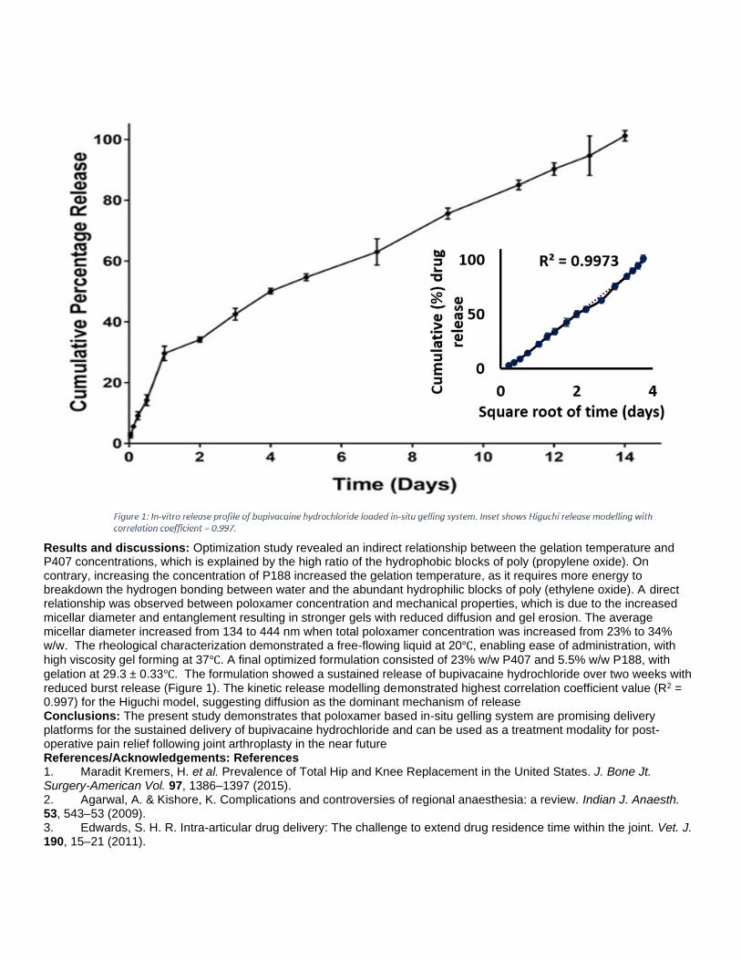
Results and discussions: Optimization study revealed an indirect relationship between the gelation temperature and P407 concentrations, which is explained by the high ratio of the hydrophobic blocks of poly (propylene oxide). On contrary, increasing the concentration of P188 increased the gelation temperature, as it requires more energy to breakdown the hydrogen bonding between water and the abundant hydrophilic blocks of poly (ethylene oxide). A direct relationship was observed between poloxamer concentration and mechanical properties, which is due to the increased micellar diameter and entanglement resulting in stronger gels with reduced diffusion and gel erosion. The average micellar diameter increased from 134 to 444 nm when total poloxamer concentration was increased from 23% to 34% w/w. The rheological characterization demonstrated a free-flowing liquid at 20℃, enabling ease of administration, with
high viscosity gel forming at 37℃. A final optimized formulation consisted of 23% w/w P407 and 5.5% w/w P188, with
gelation at 29.3 ± 0.33℃. The formulation showed a sustained release of bupivacaine hydrochloride over two weeks with reduced burst release (Figure 1). The kinetic release modelling demonstrated highest correlation coefficient value (R2 = 0.997) for the Higuchi model, suggesting diffusion as the dominant mechanism of release Conclusions: The present study demonstrates that poloxamer based in-situ gelling system are promising delivery platforms for the sustained delivery of bupivacaine hydrochloride and can be used as a treatment modality for post-operative pain relief following joint arthroplasty in the near future References/Acknowledgements: References 1. Maradit Kremers, H. et al. Prevalence of Total Hip and Knee Replacement in the United States. J. Bone Jt. Surgery-American Vol. 97, 1386–1397 (2015). 2. Agarwal, A. & Kishore, K. Complications and controversies of regional anaesthesia: a review. Indian J. Anaesth. 53, 543–53 (2009). 3. Edwards, S. H. R. Intra-articular drug delivery: The challenge to extend drug residence time within the joint. Vet. J. 190, 15–21 (2011).

4. Sharma, M. et al. In vitro and ex vivo characterisation of an in situ gelling formulation for sustained lidocaine release with potential use following knee arthroplasty. Drug Deliv. Transl. Res. 8, 820–829 (2018). Disclosure of Interest: None Declared Keywords: Biomaterials for drug delivery, Clinical application

Clinical applications of biomaterials WBC2020-2907 Mucoahdesive Micelles for the Treatment of Dry Eye Disease Frances Lasowski* 1, Ben Muirhead1, Talena Rambarran1, Lina Liu1, Emily Anne Hicks1, Aftab Taiyab1, Heather Sheardown1 1McMaster University, Hamilton, Canada Introduction: Dry eye disease (DED) is a chronic condition estimated to affect over 16.4 million Americans alone. [1] Patients suffer with sore, scratchy eyes that impact their vision and decrease their quality of life. These patients have limited treatment options that require multiple drops daily that can be painful to administer and can take months to show clinical efficacy. The market leading product, Restasis, uses cyclosporine A (CycA) as its active compound, but twice a day dosing is required and clinical efficacy can take 6 months to manifest. We previously proposed the use of mucoadhesive self-assembling micelles as a solution to these problems [2]. Incorporating phenylboronic acid (PBA), a synthetic molecule with a strong affinity for mucin due to its ability to complex with 1,2-cis-diols, into their outer shells, a series of block copolymer micelles was developed capable of targeted drug delivery to ocular tissues. Here we demonstrate the efficacy of these micelles in a diseased animal model and show the pharmacokinetic profile of CycA in the tissues. Experimental methods: Copolymers of poly(L-lactide)-b-poly(methacrylic acid-co-phenylboronic acid) were synthesized by free radical polymerization. Micelles were formed by precipitation into purified water from acetone. Cyclosporine A (CycA) entrapment efficiency and subsequent release from micelles was determined using high performance liquid chromatography (HPLC). A scopolamine-induced dry eye disease (DED) model was created in Norway Brown rats through the secretion of 25mg/day from a microosmotic pump in desiccating conditions. This model was used to compare the effectiveness of copolymer micelles with the current DED industry leader Restasisâ through tear volume measured with phenol red threads and fluorescein staining of the corneal epithelium measured on the Phoenix Micron IV System slit lamp and scored using a modified Oxford Schema. CycA-loaded micelles were dosed either twice a day, once every three or once every five days while Restasis was dosed either twice a day or once every three days. New Zealand White Rabbits were used for a 15 day pharmacokinetic study with four cohorts: twice a day and once every three days dosing with CycA-loaded micelles and twice a day dosing with Restasis and twice a day dosing with empty micelles. Ocular tissues, including aqueous humour, and blood were harvested, homogenized and extracted to determine respective CycA levels via liquid chromatography-mass spectrometry (LC-MS). Image:
Results and discussions: In the rat dry eye model, both Restasis and the CycA-loaded micelles administered twice a day resolved dry eye symptoms by all three metrics. When dosing was extended to once every three days, Restasis drops did not resolve symptoms but the CycA micelles were able to resolve the condition by all three metrics, indicating the prolonged action of the micelle formulation. Some resolution was also seen with micelles dosed every 5 days. Conclusions: These mucoadhesive micelles offer an improved treatment for dry eye disease as they are able to reduce the overall dosage and decrease the frequency of dose for CycA-based treatments. In rat and rabbit models, we have shown we can achieve similar results using the CycA loaded micelles every three days as Restatis dosed twice daily, demonstrating the prolong effect of the micelles. References/Acknowledgements: Funding Acknowledgement: C20/20 Ontario Research Fund [1] Farrand et al. Am J Ophthalmol. 2017;182:90-98. [2] Prosperi-Porta G et al. Biomacromolecules. 2016; 17: 1449-1457.

Disclosure of Interest: None Declared Keywords: Biomaterials for drug delivery, Ophthalmology, Translational research

Clinical applications of biomaterials WBC2020-2745 Antioxidant capacity of Olive Leave Extract (OLE) in 2D and 3D endothelial cell models Jose Gustavo De La Ossa * 1, Francesca Felice2, Bahareh Azimi3, Jasmine Esposito Salsano1, Maria Digiacomo4, Marco Macchia4, Serena Danti5, Rossella Di Stefano2 1Dept. of Life Sciences, University of Siena, Siena, 2Dept. of Surgical, Medical and Molecular Pathology and Critical Care Medicine, University of Pisa, Pisa, 3INSTM, Florence, 4Dept. of Pharmacy, 5Dept. of Civil and Industrial Engineering, University of Pisa, Pisa, Italy Introduction: Olive Leaf Extract (OLE) is used in traditional medicine for its anti-inflammatory and anti-atherosclerotic effects. Recent studies have highlighted the role of oxidative stress in the pathogenesis of vascular disease. The imbalance in the Reactive Oxygen Species (ROS) produces a tissue damage; thus, antioxidant molecules can be used to counteract ROS effects. OLE contains active components with potential antioxidant activity. Novel approaches that can ameliorate antioxidant supplementation based on Tissue Engineering have risen special clinical interest. In this study, we tested OLE protective effects in a 2D and 3D vascular model, the latter being fabricated using poly(vinylidene fluoride-co-trifluoroethylene) [P(VDF-TrFE] electrospun fibers, chosen for their piezoelectricity and chemical inertia, allowing the administration of oxidative insults. Experimental methods: Olive leaves were collected manually from Olivastra seggianese, Follonica (LI), Italy. The total polyphenol (TP) content, was obtained by Folin-Ciocalteau’s method. HPLC was carried out for TP characterization. P(VDF-TrFE) fiber meshes were fabricated via electrospinning (Linari srl., Italy) on a rotative collector (500 rpm, 35 kV, 15 cm distance and 0.016 ml/min flow rate, using a 20% w/v solution in Methylethylketone). Human umbilical vein endothelial cells (HUVECs) were isolated. Cell viability at different OLE doses was determined by WST-1. Protective effect of OLE was evaluated pre-treating HUVECs with different OLE TP concentrations for 2 h and 24 h and then with 100 μM of H2O2 for 1 h. At the end of each treatment, cells were analyzed for viability and ROS production (fluorescent probe CM-H2DCFDA). The scaffolds were seeded with HUVECs at 1×10⁵ cells/sample. Cell/scaffold constructs were cultured in EGM-2 medium for 1 week. The in vitro antioxidant activity effect of OLE was tested on cell/scaffold constructs using OLE pretreatment (100 µg/ml GAE for 24 h) and H2O2 (100 µM for 1 h) as follows: 1) Untreated; 2) + OLE; 3) + H2O2; 4) + OLE + H2O2. Metabolic activity was assessed via alamarBlue® assay. SEM and DAPI staining were used to image cell attachment and morphology on to the scaffolds. Image:


Results and discussions: The TP content of OLE was 23.29 mg GAE/g. Oleuropein was the major component at 1.47 % (w/w%). Dose-dependent metabolic curve showed no significant cytotoxic effects at OLE concentrations < 250 µg/ml TP even after a long treatment period (Fig. 1A,1B). Fig. 1C shows that only 24 h pre-incubation at a concentration range between 50 - 250 µg/ml of TP prevented H2O2-induced viability reduction of HUVECs (p < 0.05). ROS cellular accumulation was evaluated after 24 h pre-treatment of OLE at 10-250 µg/ml of TP. In Fig. 1D, our results highlighted a significantly protective effect at a concentration < 250 µg/ml TP (p < 0.0001 vs. H2O2). A concentration of 100 µg/ml TPs was sufficient both to reduce the effect of oxidative stress induced by H2O2. OLE was also able to produce cytoprotective effects in 3D model (Fig.1E,F). However, the drop in cell metabolic activity after the H2O2 insult in the 2D model higher than in the 3D model. This shows that the 3D model is much more predictive of real tissue response in disease conditions. Conclusions: Many medical applications can potentially benefit from OLE, including cardiovascular disease and wound healing for wounds compromised by ROS stress. Disclosure of Interest: None Declared Keywords: 3D scaffolds for TE applications, Clinical application, In vitro tissue models

Clinical applications of biomaterials WBC2020-2513 Radiopaque polyethylenes to aid surgical device positioning and patient follow up Fedra P Zaribaf1, Harinderjit S Gill1, Elise C Pegg* 1 1Department of Mechanical Engineering, University of Bath, Bath, United Kingdom Introduction: The X-ray attenuation of polyethylene is comparable to that of tissue, so it is not currently possible to visualise medical devices made from polyethylene using X-ray or CT imaging. The clinical impact is that surgeons cannot use techniques such as fluoroscopy to assist with implant positioning, or monitor implant performance and condition after the surgery using X-ray based methods. These issues are particularly relevant to devices such as ligament repair sutures [1] or maxillofacial implants [2] where accurate positioning is essential, and joint replacement devices where early diagnosis of complications such as wear can be critical. This work uses oil-based contrast agents to create radiopaque polyethylene devices, and examines the potential of this biomaterial. Experimental methods: Ultra-high molecular weight polyethylene (UHMWPE) in solid form (GUR 1050 moulded sheets, Celanese, Germany), and UHMWPE braided into sutures (ACL Tightrope, Arthrex, Germany), were examined. For the solid UHMWPE, tensile test samples (ISO-572, type 1AB) were immersed in pure iodised oil (Lipiodol, Guerbert, France) and held at 85oC, 105oC, and 115oC for 12 h. The UHMWPE sutures were cut into 500 mm lengths and immersed in pure iodised oil at 80 oC for 1h. Tensile tests were performed using an electromechanical test machine (Instron) in accordance with the standards (ISO 572 for the solid UHMWPE, ISO 2062 for the sutures). The radiopacity was quantified using μCT imaging (Nikon XT H 225 kV) alongside a sample of distilled water for calibration, the grayscale values were then converted into Hounsfield units (HU). The concentration of iodised oil was determined using Fourier Transform Infrared (FTIR) Spectroscopy (32 scans, 4000 – 600 cm-1, assigned peaks summarised in Table 1), and the FTIR spectra were also used to quantify polyethylene oxidation (ASTM F2102). The crystallinity of UHMWPE was quantified using Differential scanning calorimetry (DSC). All experiments used a sample size of 5, included untreated controls, and were compared statistically using Kruskal-Wallis or Mann Whitney U tests. Image:
Table:
Wavenumber (cm-1) Functional group Reference 1263 Phenolic C-O (tocopherols), methyl ester, v(C-O-C) [3] 1730 Ethyl ester (due to starching band of the carbonyl group) [4] 2900 Symmetric v(C-H) from phytyl chains (tocopherols) [3] 3050 v(C-H), v(=CH) [3]
Results and discussions: The iodised oil diffused into both the solid and suture UHMWPE samples at the elevated temperatures. In the solid samples held at 115oC the treatment caused a weight increase of 33±4%, and in the sutures 32±3%. The radiopacity significantly increased in all samples (p<0.05, see Figure 1: X-ray of a treated partial joint replacement). The average radiopacity of the solid UHMWPE held at 115oC was 1562.0±426 HU, and the treated sutures was 744.5±85 HU. The solid UHWMPE had similar weight increase but a higher HU than the braided UHMWPE; this may be due to the higher porosity of the braided sutures. The tensile tests found no difference in ultimate tensile strength (p=0.22) or elongation at break (p=0.1) of the solid or sutures samples, but the tensile modulus was lower in the treated samples (p=0.013). No change was observed in the crystallinity or the melting point of the polyethylene, and the oxidation index was always less than 1. Conclusions: Radiopaque polyethylene has been created successfully through diffusion of iodised oil. No difference was found in the crystallinity, oxidation, or mechanical properties of the solid or suture samples, with the exception of the

tensile modulus. Current work is examining the long-term behaviour of this material (fatigue, creep, wear, aging and leaching) so its safety for clinical use can be assessed. References/Acknowledgements: The authors would like to thank Mrs Clare Ball and Prof Whitney for their technical support, Celanese
for providing the solid UHMWPE sheets, Arthrex for providing the sutures, the University of Bath for funding the work on the solid samples, and the Medical Research Council for funding the research on the sutures. [1] Jameson, S. S. et al. The Knee 19, p14-19 (2012) [2] Oh, J. Maxillofacial plastic and reconstructive surgery 40, p2 (2018) [3] Zagonel, G.F. et al. Talanta 63, p1021–1025 (2004). [4] Carrión-Prieto, P. Pharmacogn Journal 9, p157-162 (2017). Disclosure of Interest: None Declared Keywords: Clinical application, Imaging, Mechanical characterisation

Clinical applications of biomaterials WBC2020-2458 An implant for sustained post-operative delivery of lidocaine Darren Svirskis* 1, Manisha Sharma1, Georgina Procter2, Priyanka Agarwal1, Prabhat Bhusal3, Jacqueline Hannam 1, Gavin Andrews2, Andrew Hill1, David Jones2 1University of Auckland, Auckland, New Zealand, 2Queen's University Belfast, Belfast, United Kingdom, 3University of Otago, Dunedin, New Zealand Introduction: The World Health Organisation estimated that more than 300 million surgical procedures occurred globally in 2012 and that these numbers are increasing (1). The most common method of managing post-operative severe pain remains the systemic administration of opiates. However, due to their adverse effects and their potential for misuse and addiction, alternative treatment options have been actively sought (2) and use of other targeted pain management modalities is of increasing interest globally. Here we explore a non-opioid treatment modality comprising the sustained delivery of the local anaesthetic lidocaine directly into the peritoneal cavity aiming to produce local vagal afferent blockade following abdominal surgery. Experimental methods: To achieve the sustained release of the local anaesthetic lidocaine directly to affected tissues, non-biodegradable poly ethylene-co-vinyl acetate (EVA) intraperitoneal implants were prepared. Cylindrical implants were manufactured by hot melt co-extrusion with a core/shell structure. The inner core imparted the implant with the required mechanical properties comprising 80% w/w EVA (12% VA) and 20% w/w barium sulphate to render the device radio-opaque. The outer layer was formed from EVA (33% VA) loaded with uniformly distributed lidocaine (at 20% w/w), stabilised with myristic acid. For comparison, blank implants were prepared with the same inner core but with the outer layer devoid of lidocaine and myristic acid. The performance, safety and toxicity of the implants were determined in sheep who underwent a laparotomy and small bowel resection, with animals divided into three groups (Table 1). In all sheep a silicone drain was implanted to facilitate sampling of peritoneal fluid and as a comparator implanted material. Image:
Table: Table 1: In vivo study group descriptions.
Group
Number of sheep
Implant Type Days implanted
1 5 Drug loaded 7 2 3 Blank 7 3 3 Drug loaded 3
Results and discussions: The total diameter of the implants was c.a. 4 mm to facilitate future administration and removal through laparoscopy ports. Following in-vivo implantation for 3 or 7 days both Young’s modulus and tensile strength at break increased, important parameters ensuring the implant can be removed without breakage when required. Lidocaine was observed in both peritoneal fluid and plasma of the ovine model at decreasing concentrations over 7 days (Fig 1) with levels of lidocaine in the peritoneal fluid several orders of magnitude higher than in the plasma, which

remained well below toxic levels (5 mg/L). Histological analysis revealed similar responses between the co-extruded implant and commercially available silicon drains. Figure 1: Following intraperitoneal administration of the non-opioid analgesic implant concentrations of lidocaine were several fold higher in peritoneal fluid (A) compared to plasma (B). Data points represent individual values, the solid line represents median values with the dashed lines representing 5th and 95th percentiles. Conclusions: The described non-opioid analgesic implant provided sustained release of lidocaine in an ovine model without any safety or toxicity concerns and is suitable for moving onto first in human trials. References/Acknowledgements: References 1. Weiser TG, Haynes AB, Molina G, Lipsitz SR, Esquivel MM, Uribe-Leitz T, et al. Size and distribution of the global volume of surgery in 2012. Bull World Health Organ. 2016;94(3):201-9F. 2. Brown AK, Christo PJ, Wu CL. Strategies for postoperative pain management. Best Pract Res Clin Anaesthesiol. 2004;18(4):703-17. Acknowledgements: Wiremu MacFater, Ahmed Barazanchi, Laura Bennett, Kaushik Chandramouli, Sree Sreebhavan, Satya Amirapu Disclosure of Interest: None Declared Keywords: Biomaterials for drug delivery, Clinical application, Translational research

Commercialisation of biomaterials WBC2020-2278 Orthopedists appreciate the speed of insertion and colleaques' user experiences when deciding to adopt or reject innovations Minna Veiranto* 1, Saku Mäkinen1, Minna Kellomäki1 1Tampere University, Tampere, Finland Introduction: Individual forms a favorable or an unfavorable attitude towards the innovation before deciding to adopt or reject a new idea or an innovation into use. Certain characteristics, like relative advance, compatibility, complexity, trialability and observability, of the innovation are known to affect the attitude formation. Available information about the innovation plays an important role in the decision-making. The innovators and early adopters are the first ones to use novelties and their user experiences are important sources of information for later adopting categories i.e. early and late majority. These early segments value technological sophistication over technological or operational readiness, they are tech-savvy and want to test new things. On contrary, later adopters value operationally ready, easy to use and tested products. (1) Our hypothesis is that general characteristics of these segments are reflected also in medical innovations, and adoption of innovations is influenced by these characteristics. The current study is preceding a wider, global investigation related to adoption of different medical innovations from commonly used surgical implants to advanced therapy medicinal products (ATMP). The aim of the study was to find out the most important attributes affecting the adoption of biodegradable fixation implants (i.e. innovations) in use Experimental methods: Analyzed data was collected in 2010 by interviewing 19 orthopedists (adopter status unknown) who worked either at university or private hospital in Finland. 58 questions were in the form that they were answered yes, no or same. Data was analyzed with chi-square Fisher exact test using IBM SPSS statistical software. A p value of ≤0.05 was considered as significant. Relationship was considered strong when r value was ±0.40 to ±0.69 and very strong when it was ±0.70 or higher. Results and discussions: All together 13 question pairs had either strong or very strong negative or positive relationship. It was clear that orthopedists appreciate the insertion speed of the implant. General handling, preparations and insertion of biodegradable implants were experienced more complicated, difficult and complex than traditionally used products, like metal equivalents. (r 0.68-0.86) Interesting finding was that although the preparation of biodegradable products was seen more difficult and complex, in vivo degradation was seen as a great advance. (r 0.78) This indicates the importance of some overpowering characteristic of the innovation on decision-making process. It was also clear that colleagues influence highly the attitude and decision formation. Study showed a very strong relationship (r 0.71) between the influence of colleagues’ user experiences and information heard from colleagues about the innovation. It also showed a very strong (r 0.85) relationship between the increase of interest toward biodegradable products by reading scientific publications and influence of colleagues’ user experience. This observation is logical. In many cases scientific publications are written by orthopedists who already use or consider using the innovation. Conclusions: This preliminary study showed that orthopedists appreciate the speed of insertion and colleagues’ user experience when deciding to adopt or reject the biodegradable implants to be used in a specific indication. This highlights the importance of user-oriented approach and well-considered and -produced information about the innovation from the early stages of innovation-development process. Analyzed data did not include e.g. questions related to regulations, ethics and type of information, but in the actual investigation at least those topics must be included in. We obtained important knowledge that helps us to plan detailed structure and questions for the larger study group. References/Acknowledgements: The authors acknowledge Krisse Poutala. (1) Rogers. E.M. 2003. Diffusion of innovations, 5th edition, Free Press, New York Disclosure of Interest: None Declared Keywords: None

Commercialisation of biomaterials WBC2020-3196 Therapeutic refinement of tissue engineered cellular implants for non-union fracture repair Peter G. Childs* 1, Monica P. Tsimbouri1, Cristina Gonzalez-Garcia1, Paul Campsie2, Stuart Reid2, Matthew J. Dalby1, Manuel Salmeron-Sanchez1 1Centre for the Cellular Microenvironment, University of Glasgow, 2Department of Biomedical Engineering, University of Strathclyde, Glasgow, United Kingdom Introduction: Non-union fractures impact >700,000 patients p/a globally1 and cost an estimated $9.2billion/year in the US2. Despite a number of synthetic graft materials being available autograft is still considered the gold standard. In this project a combined cell therapy has been developed, incorporating osteogenically primed mesenchymal stromal cells (MSCs) with a collagen carrier material for repair of non-union defects. The focus of this work has been to define the final therapy format and upscale cellular production to industrially relevant numbers for first-in-human trial. Unlike other MSC therapies osteogenic priming is achieved via mechanical stimulation (nano-amplitude vibration) without the need for ancillary osteogenic media supplementation3. Here, in vitro and in vivo data demonstrates the functionality of these cells. Experimental methods: Purpose built nano-vibrational platforms were used to mechanically stimulate mesenchymal stromal cells over 4 weeks in multi-layer cell stacks. Interferometric data was used to confirm transmission of nano amplitude vibration. Both commercial marrow derived MSCs and therapeutic adipose MSCs were tested for osteogenic response to mechanical stimulation using qPCR and in cell western of key osteogenic markers: runt-related transcription factor-2 (RUNX2), osterix (OSX), osteonectin (OSN), osteocalcin (OCN) and osteopontin (OPN). In vivo regenerative capacity of these cells was assessed through combined administration with a collagen carrier gel at a concentration of 1 million cells/ml using an immunocompromised murine model of critical sized radial defect. Bridging of the critical sized defect was assessed by µCt 8 weeks following implantation. Collagen source was examined for impact on therapeutic efficacy. Image:

Results and discussions: Interferometric data indicated that nano-vibrational stimulation could be delivered to 5 layer cell stacks, each producing a yield of up to 40 million MSCs. qPCR and in cell western analysis of the upscaled cells demonstrated that stimulation was successful in larger culture vessels and that industrially relevant MSCs from adipose tissue could be osteogenically primed in a similar manner to marrow derived MSCs. Increased bone regeneration was observed in vivo for mechanically stimulated MSCs. Comparison of different sources of collagen also showed differences with implications for the final therapy format. Conclusions: The use of mechanical stimulation to osteogenically prime MSCs removes the need for ancillary osteogenic reagents, reducing supply chain and quality control complexity when moving to GMP manufacture. Here we have demonstrated that this process is compatible with industrially relevant cell manufacturing vessels at a scale to supply small clinical trials. We have also demonstrated that the pre-stimulation of these cells increases their in vivo regenerative capacity. Based on these findings the technology is in a strong position to be moved into GMP cell manufacture, pre-clinical safety studies and a planned first-in-human application in phalangeal fracture repair. References/Acknowledgements: 1Corporate Presentation, Bone Therapeutics, July 2019 2Non-Union Fractures Market: Global Industry Trend Analysis 2013 to 2017, Persistence Market Research, 2019 3Tsimbouri, P.M., et al. 2017. Stimulation of 3D osteogenesis by mesenchymal stem cells using a nanovibrational bioreactor. Nature biomedical engineering, 1(9), p.758. The authors would like to acknowledge funding from the Sir Bobby Charlton Foundation, EPSRC (EP/P001114/1) and BBSRC (BB/S018808/1), along with Histocell for supplying adipose derived MSCs. Disclosure of Interest: None Declared Keywords: Bone, Stem cells and cell differentiation, Translational research

In vivo tests and animal models WBC2020-2051 Real-Time Visualization of Intrathecally Administered Nanoparticles via Intravital Microscopy Jennifer Cotter1, Shruti Dharmaraj1, Yoshihiro Otani1, Kyle Householder1, Balveen Kaur1, Rachael Sirianni* 1 1Vivian L. Smith Department of Neurosurgery, University of Texas Health Science Center, Houston, United States Introduction: Biological barriers such as the blood-brain barrier and blood-spinal cord barrier prevent effective treatment of many central nervous system (CNS) diseases. Intrathecal (IT) administration, i.e., injection of substances directly into the cerebrospinal fluid (CSF) that surrounds the brain and spinal cord, is one method of bypassing these barriers. IT administered agents can achieve a high concentration within the CNS while reducing toxicity to peripheral tissues. However, efficacy of freely administered agents is limited by their biophysical properties: hydrophilic molecules distribute and clear rapidly with CSF, while hydrophobic molecules experience poor tissue penetration. Our laboratory is focused engineering polymeric nanoparticles (NPs) to enhance the pharmacologic profiles of IT administered agents. Here, we hypothesized that NP properties, include size and surface characteristics, would be important for governing their distribution within the CNS after IT administration. To test this hypothesis, we utilized intravital microscopy to measure kinetics of NP movement through the subarachnoid space (SAS) of living mice. This non-destructive method for assessing NP delivery yielded significant new observations regarding mechanisms of CSF flow. Our studies also establish new and important structure-function relationships for designing nanomedicine for effective CNS delivery. Experimental methods: Fluorescent 100nm polystyrene NPs (FluoSpheres™) were surface modified with poly(ethylene glycol) (PEG) via EDC chemistry. NSG mice were fitted with a cranial window over the cortex of the right hemisphere. NPs were administered via injection into the cisterna magna and imaged using a Nikon FN-1 microscope and a Nikon A1R-MP-HD Dual Beam (with Coherent Insight 3 Laser) confocal system (n=3 mice/group). Particle tracking software (NIS-Elements C) was used to determine velocity and direction of different NP types administered at various concentrations. Results and discussions: Non-PEGylated NPs (20mg/ml) were observed to move freely within the SAS with directional velocity that was a function region within the SAS. In contrast, PEGylated NPs (20mg/ml) were observed to oscillate with CSF but did not experience net flow. PEGylated NPs were observed to experience net flow when the injection concentration was lowered (5mg/ml). Unexpectedly, we observed that a Z-plane change affected both speed and directionality of particle movement significantly. Deeper NPs moved much more rapidly than more superficial NPs, and the direction of NP movement was observed to reverse as imaging depth increased. Conclusions: Surface properties and concentration of IT administered NPs will be critical variables governing NP distribution and mixing within the SAS. We speculate that the lack of flow of the 20mg/ml PEGylated NPs was due to entrainment of PEG chains. The change in direction of CSF flow with depth is a fundamentally novel observation that provides direct evidence for microregional mixing of CSF, most likely due to the presence of trabeculaer sheets within the SAS. Companion studies evaluating delivery of NPs with positron emission tomography (PET) and intravital imaging of NP localization with trabeculae are currently in progress. References/Acknowledgements: We thank Clay Williams and Kalli Landua of Nikon. This work was supported by the Ian’s Friends Foundation and National Institutes of Health (R01NS111292, R01HD099543). Disclosure of Interest: None Declared Keywords: In vivo imaging, Small animal models

In vivo tests and animal models WBC2020-1708 Development of Near-Infrared Fluorescent Nanoparticles for in vivo Bioimaging in the Second and Third Biological Window Masao Kamimura* 1, Kohei Soga1 1Department of Materials Science and Technology, Tokyo University of Science, Tokyo, Japan Introduction: Near-Infrared (NIR) fluorescent nanoparticles for in vivo bioimaging have recently received tremendous attention. Because, most of current fluorescence bioimaging mainly uses visible (400-700 nm) or short wavelength NIR (700-900 nm: first biological window) emission of fluorescent probes and thus, is suffer from low penetration depth due to the strong absorption of biomolecules in the body. In contrast, NIR light, especially in over-1000-nm wavelength region (1100-1350 nm: second biological window, 1550-1700 nm: third biological window) can penetrate into the tissues more deeply than first biological window region, due to low absorption or scattering of biomolecules and water in the body. Thus, NIR fluorescence bioimaging in the second and third biological window region is promising idea for live imaging of deep tissues. In the past decade, we reported various NIR fluorescent probes based on biocompatible polymers and NIR nanophosphors, such as quantum dots (QDs), rare-earth doped ceramic nanoparticles (RED-CNPs), and organic dye loaded polymer micelles for fluorescence in vivo bioimaging in the second and third biological window region.1-8 In this presentation, we will present the recent progress of NIR in vivo bioimaging in the second and third biological window region. Experimental methods: Biocompatible polymer, poly(ethylene glycol)(PEG)-modified QDs was synthesized by co-precipitation method with end-functional PEG as a capping agent. As a RED-CNPs, rare-earth doped NaYF4 NPs or NaGdF4 NPs were synthesized by thermal decomposition method and then, the surface of obtained NPs was modified with PEG-based block copolymer. Furthermore, NIR fluorescent polymer micelles were prepared by using NIR dye and PEG-based block copolymer possessing hydrophobic side chain. Image:

Results and discussions: The prepared various polymer conjugated nanophosphors displayed high dispersion stability and showed strong NIR emission in the second and third biological window region. The stability of NIR fluorescence intensity of these nanoparticles was quite stable under physiological conditions. In addition, these nanoparticles also showed high biocompatibility against living cells. Furthermore, NIR in vivo imaging of live mice by using these probes was successfully performed. Conclusions: The prepared polymer conjugated nanoparticles displayed high stability and optical property under physiological conditions. In addition, this imaging technique can observe the inside of deep tissue of the living mice. Thus, the developed bioimaging method in this study is promising candidate for novel bioanalysis and medical diagnosis technique. References/Acknowledgements: (1) M. Kamimura, K. Soga et al., Nanoscale 3 (2011) 3705. (2) M. Kamimura, K. Soga et al., Journal of Materials Chemistry B 5 (2017) 1917 (3) M. Kamimura, K. Soga et al., Chemistry Letters 46 (2017) 1076. (4) M. Kamimura, K. Soga et al., Polymer Journal 49 (2017) 799. (5) Y. Tsai, M. Kamimura, K. Soga et al., Theranostics 8 (2018) 1435. (6) S. Sekiyama, M. Kamimura, K. Soga et al., Scientific Reports 8 (2018) 16979.

(7) S. Sekiyama, M. Kamimura, K. Soga et al., Langmuir 35 (2019) 831. (8) T. Chihara, M. Kamimura, K. Soga et al., Scientific Reports 9 (2019) 12806. Disclosure of Interest: None Declared Keywords: None

In vivo tests and animal models WBC2020-1621 Designed degradation and drug release of injectable non-covalent hydrogels Rebecca Rothe* 1, 2, Yong Xu3, Alvin Kuriakose Thomas3, Sebastian Meister1, Yixin Zhang3, Jens Pietzsch1, 2, Sandra Hauser1 1Department of Radiopharmaceutical and Chemical Biology, Helmholtz-Zentrum Dresden-Rossendorf, Institute of Radiopharmaceutical Cancer Research, 2Technische Universität Dresden, School of Science, Faculty of Chemistry and Food Chemistry, 3BCube-Center for Molecular Bioengineering, Technische Universität Dresden, Dresden, Germany Introduction: Adverse foreign body response, acute inflammation or fibrous encapsulation are undesired host tissue reactions towards implanted biomaterials. Current focus is on the development of innovative smart biomaterials attenuating mentioned obstacles and fulfilling desired purposes like drug release or management of cellular processes. In this study, designed injectable hydrogels with targeted properties regarding tunable degradation and drug release as well as evoked cellular effects at tissue-material interface were investigated. Experimental methods: Four non-covalent hydrogels were examined in a mouse model. Basic hydrogel system composes a starPEG backbone, repetitive lysine-alanine ((KA)5) peptide linker and oligosaccharide dextran sulfate ((KA)5-DS). Further, modifications regarding an additional matrix metalloproteinase (MMP) cleavage site ((KA)5-MMPiw-DS), altered peptide sequence from L- to D-amino acids ((ka)5-DS) or oligosaccharide exchange to sulfated hyaluronic acid ((KA)5-sHA) were introduced. Hydrogels were injected subcutaneously in immunocompetent nude mice. In vivo, biodegradation and inguinal lymph node size, latter indicating inflammation, were determined by magnetic resonance imaging. Additionally, optical imaging (OI) of a non-covalently incorporated fluorescent dye ((KA)7-Cy7) as model compound visualized drug release. In initial study period, fluorescent imaging agent MMPSenseTM645 was administered intravenously to follow MMP activity in hydrogel surroundings by OI. Immunohistochemical stainings and quantification of both angiogenesis (VEGF, CD31) and inflammation (COX-2, CD68) markers on cryosectioned tissue samples defined hydrogel biocompatibility ex vivo. Image:
Results and discussions: In vivo, (ka)5-DS remained unchanged as D-amino acids exhibit proteolytic stability, whereas (KA)5-DS and even more MMP-cleavable (KA)5-MMPiw-DS were gradually degraded during study period. (KA)5-sHA showed fastest degradation and significantly enhanced MMP activity, in turn accelerating enzymatic degradation. Further, sustained release of affinity-coupled (KA)7-Cy7 from (KA)5-sHA was visible correlating with hydrogel degradation due to high oligosaccharide sulfation degree. For all dextran sulfate-based hydrogels by contrast, fluorescent dye diffused rapidly (Figure 1). Independent of hydrogel composition, lymph node size of all animals was similar to untreated controls. Immunohistochemistry revealed basal macrophage recruitment and inflammation marker expression. In addition to a suitable biocompatibility, enhanced VEGF expression and increased blood vessel size around (KA)5-sHA demonstrates pro-angiogenic effects.

Conclusions: Designed injectable, non-covalently assembled hydrogels provided definite degradation profiles based on structural variations in peptide linker or oligosaccharide component. Lymph node measurements and immunohistochemistry proved hydrogel biocompatibility with no adverse inflammation occurring in surrounding tissues. According to in vivo imaging, (KA)5-sHA is appropriate for sustained degradation-dependent drug release and, beyond, evoked pro-angiogenic effects possibly initiated by macrophages. For stated reasons, injectable hydrogels provide convenient scaffolds for local drug therapy with improved tissue regeneration. References/Acknowledgements: The authors thank the Deutsche Forschungsgemeinschaft (DFG) for supporting this work within the Collaborative Research Center Transregio 67 “Functional Biomaterials for Controlling Healing Processes in Bone und Skin - From Material Science to Clinical Application“ (CRC/TRR 67/3). This work was supported by the China Scholarship Council and the German Federal Ministry of Research and Education (BMBF grants 03Z2EN12 and 03Z2E511). Disclosure of Interest: None Declared Keywords: Biocompatibility, In vivo imaging, Material/tissue interfaces

In vivo tests and animal models WBC2020-396 Optimization of a Preclinical Induced Membrane Rat Model Renaud Siboni1, Xavier Ohl1, Aurélie Moniot1, Christine Guillaume1, Sophie Gangloff1, Julien Braux1, Frédéric Velard* 1 1EA 4691 BIOS, URCA, Reims, France Introduction: In orthopaedics, large bone defects management is challenging. They are often due to high traumatic injury, osteomyelitis or carcinological diseases. One approach for such large defects healing is called induced membrane technique. It is a two-stage procedure with the use of polymethyl methacrylate (PMMA) scaffold that induces a synovial membrane [1]. The latter is used in the second step of the procedure to hold in bone filling materials (i.e. autograft or biomaterials) because of its osteogenic abilities. This technique provides good results but, in some cases, it requires supplementary graft procedures (up to 6) to have satisfactory bone healing [2]. We aimed to optimize a preclinical model in rats in order to improve osteotomy procedure and bone fixation to possess a reliable technique for novel bone filling scaffolds assessment. Experimental methods: All experiments were realized in compliance with local ethics committee. Eight weeks old, male Sprague-Dawley rats were used. In a first procedure, we evaluated two sizes of titanium plates (0.6 and 1 mm thickness) and screws (1.5 and 2.0 mm diameter) for their ability to stabilize a critical bone defect of 5 mm in length. After 6 weeks, bone anchorage was analyzed using microtomography. Then we evaluated, three osteotomy procedures; ball cutter, diamond saw and piezotomy, through histologcal stainings and scanning electron microscopy for their ability to ensure bone structure conservation at the cutting edge. In a second time, an induce membrane technique was performed based on optimal conditions defined above. Groups were (n=2 each): control rats (no PMMA, no graft), no PMMA then syngeneic bone graft, PMMA no graft and PMMA then syngeneic bone graft. Histological analyses were performed on induced membrane collected at the end of the first procedure. The bone healing was evaluated every 4 weeks by low resolution microCT and at 3 months by high resolution micro CT and histological analyses. Results and discussions: Results indicated that plates of 1 mm thickness with 4 screws of 2 mm diameter exhibited the best fixative abilities for the model as compared to plates with thickness 0.6 mm and screws with diameter 1.5 mm which led to dismantling. The osteotomy with piezotome provided a clean bone cut with permeable Haversian channels and was very conservative of orthopaedics materials (plate and screws) adversely of ball cutter or diamond saw techniques which evidenced burned bone and damages on the osteosynthesis materials. After 3 months of bone repair, only rat with PMMA in the first stage and syngeneic graft on the second stage demonstrated evidence of a tissue healing. Conclusions: We optimized an induced-membrane model in rats during this work. The procedure is now reproducible and standardized. Future directions will focus on PMMA spacer replacement by new innovative materials able to trigger both induced-membrane development and bone repair in a single step procedure in order to reduce the duration and/or number of surgeries to allow complete bone regeneration. References/Acknowledgements: [1] Masquelet AC, et al. Ann Chir Plast Esthet. 2000;45(3):346‐53. [2] Siboni R, et al. Orthop Traumatol Surg Res. 2018 Oct;104(6):911-915. Authors would like to thank PICT platform for imagery, Institut Carnot MICA (BiomateriOs exploratory program, OptimOs R&D program) and “Fondation des Gueules Cassées” (PorOs program) for funding the project. Disclosure of Interest: None Declared Keywords: Clinical application, Small animal models

In vivo tests and animal models WBC2020-422 The Application of Hexametaphosphate to Heterotopic Ossification Produced via Achilles Tenotomy in a Rodent Model Thomas Robinson* 1, Sophie Cox1, Liam Grover1 1School of Chemical Engineering, University of Birmingham, Birmingham, United Kingdom Introduction: Heterotopic ossification (HO) is the pathological formation of bone outside of the skeleton, which can be caused by musculoskeletal trauma, injury to the central nervous system, burns, and rare genetic conditions1. Current prophylaxes for this condition are inconsistently effective and can have deleterious side effects, which make them unsuitable for highly injured patients2. This leaves surgical excision of the bone as the only available treatment option for many patients. Hexametaphosphate (HMP) is a potent calcium chelator, which has been shown to dissolve hydroxyapatite, the main mineral constituent of bone3. The hypothesis of this study is that HMP might be used to dissolve the mineral constituent of bone, and thus prevent the pain, skin ulceration, and problems with prosthetic limb fitting experienced by HO sufferers. Experimental methods: HO was induced in Sprague-Dawley rats by Achilles tenotomy surgery, a relatively low-severity model of the condition. HMP was loaded into an alginate solution and injected directly into the limb, in various dosing regimens over 10 weeks, as the bone formed. The ectopic bone was then analysed by micro-computed tomography, and both the leg tissue and filter organs were analysed by histological analysis. Image:
Results and discussions: The Achilles tenotomy model produced HO in 100% of rats after 10 weeks. The bone formed in discrete islands within the soft tissues, via endochondral ossification. The HO has a similar microstructure and osteocyte density as normal skeletal bone, but has far more osteoblast and osteoclast activity. In addition to the appearance of the bone and cartilage, the surrounding soft tissue has regions of more dense and aligned collagen, with a rounded cell type unlike the fibroblasts usually found there. Initial results showed that HMP reduced HO length compared to controls, but did not affect the density of the adjacent skeletal bone. However, further exploration of dosing frequency and delivery vehicle has indicated a complex response, whereby increased frequency of both active and benign injections

seem to increase HO formation. Histological analysis also showed no sign tissue damage or of retention of the HMP or alginate delivery vehicle at the site of injection, nor in any of the filter organs. Conclusions: Achilles tenotomy is a reliable way to produce HO, though the volume can vary greatly. Histology allows both the bone and surrounding soft tissue to be studied, which may allow a greater understanding of the condition in this and other models. HMP has shown some promise for the prevention of HO however, the effects of dose and delivery are complex. However, both HMP and alginate in solution appear safe to use in vivo, warranting further study of their use in this and other conditions. References/Acknowledgements: 1 E. M. Shore and F. S. Kaplan, Clin. Rev. Bone Miner. Metab., 2005, 3, 257–259. 2 K. A. Alfieri, J. A. Forsberg and B. K. Potter, Bone Joint Res., 2012, 1, 192–7. 3 N. Eisenstein, R. Williams, S. Cox, S. Stapley and L. Grover, J. Mater. Chem. B, 2016, 4, 3815–3822. Disclosure of Interest: None Declared Keywords: Biomaterials for drug delivery, Bone, Small animal models

In vivo tests and animal models WBC2020-736 Nanoreactor with multi-component loading for enhanced therapeutic effect. Seoungkyun Kim1, Manse Kim2, Secheon Jung1, Kiyoon Kwon* 1, Inchan Kwon1, Giyoong Tae1 1Materials Science and Engineering, Gwangju Institute of Science and Technology (GIST), Gwangju, Korea, Republic Of, 22Division of Infectious Diseases, university of north carolina at chapel hill, north carolina, United States Introduction: Nanoreactor that can load multiple enzymes were applied to achieve enhanced therapeutic effect. Urate oxidase (UOX) is a therapeutic enzyme that catalyzes the oxidation of uric acid (UA) to 5-hydroxyisourate and hydrogen peroxide (H2O2). And, AuNP has a catalase-mimic effect in physiological pH environment. These two enzymes were loaded into biocompatible hydrogel nanocarriers (NCs) to enhance catalytic effect of UOX and to reduce the side effect associated with H2O2 Experimental methods: Expression and purification of UOX The plasmid encoding a gene of recombinant urate oxidase derived from Aspergillus flavus under control of T5 promoter (pQE80-UOX) was transformed into TOP10 Escherichia coli cells. The cell lysate supernatant incubated with NI-NTA resins was loaded on a polypropylene column. And, the eluted UOX solution was buffer-exchanged with PBS buffer using a PD-10 column Preparation of UOX-AuNP@NCs UOX and AuNPs were co-encapsulated into pluronic-based NCs by the temperature-dependent size change of the nanocarrier. Determination of UA degradation rate by UOX and H2O2 level by AuNPs. Uric acid (UA) was mixed with UOX in PBS buffer and the absorbance at 293 nm was monitored to determine the degradation rate of UA. Concentrations of H2O2 generated by UOX systems upon the addition of UA into the samples were estimated using degradation of N,N-dimethyl-4-nitroaniline (RNO). N,N-dimethyl-4-nitroaniline (RNO) concentration by measuring absorbance at 440 nm Determination of in vivo uric acid level To induce hyperuricemia state in the mice, they were treated with hypoxanthine and potassium oxonate. After sample injection, the blood was collected at several time points. The UA concentration in the serum was analyzed to figure out the pharmacokinetics. Results and discussions: UOX-AuNP was encapsulated into the nanocarrier with high loading efficacy (> 90%). There was no significant change in size and surface charge of NC upon loading of UOX and AuNPs. In the presence of UA, UOX samples showed significant cell death. Simple mixture of UOX and AuNPs showed a significantly reduced cytotoxicity than UOX alone. More importantly, NC-loaded samples showed further reduced cytotoxicity compared to UOX-AuNP in vitro. Also, UOX-AuNP@NC showed the lower concentration of UA than other groups in serum. Furthermore, no significant toxicity in major organ especially was observed in contrast to the inflammation observed in UOX and UOX-AuNP groups. Conclusions: Biocompatible NCs could encapsulate both therapeutic protein (UOX) and enzyme-mimic nanoparticles (AuNPs) in a high loading efficiency (>90%). The nanoreactor did not alter the enzymatic activity of UOX, nor hinder the transport of substrate and product of the reaction across the NCs. The nanoreactor system showed enhanced UA degradation without H2O2–associated toxicity to suppress the hyperuricemia. Furthermore, the longer therapeutic
enzyme effect was achieved by using NCs.
* This research was supported from the National Research Foundation of Korea (NRF, Grant No. 2018R1A4A1024963) Disclosure of Interest: None Declared Keywords: Biocompatibility, Biomaterials for drug delivery

In vivo tests and animal models WBC2020-204 Anti-Polyethylene Glycol (PEG) Antibody Mouse Model for Rigorous Assessment of PEG-Based Therapies Helena Freire Haddad* 1, 2, Jacqueline Burke1, 2, Xiaomin Zhang3, Evan Scott1, Guillermo Ameer1, 2, 3 1Biomedical Engineering Department, Northwestern University, 2Center for Advanced Regenerative Engineering, Evanston, IL, 3Feinberg School of Medicine, Northwestern University, Chicago, IL, United States
Introduction: Type 1 diabetes is a chronic, endocrine disorder characterized by the autoimmune destruction of insulin-producing pancreatic ꞵ-cells. A promising alternative treatment that offers a long-term solution for the disease is the transplantation of functional pancreatic islets. However, transplanted islet destruction and dysfunction often occur due to the host's inflammatory and immune response. To combat this, a steric barrier can be created around the islets by covalently attaching poly(ethylene glycol) (PEG), a non-toxic polymer known to mitigate inflammation and lower immune response, to the native amines on the surface of the cells.1 Currently, there are over 10 PEG-related therapies approved by the FDA,2 but it has been recently found that around 72% of people without prior exposure to PEGylated drugs carry detectable levels of anti-PEG antibodies (Ab) due to the prevalence of the polymer in beauty, personal care and cleaning products.3 The presence of anti-PEG Ab, with an average IgG concentration of 52 ng/ml in the general population,3 can jeopardize efficacy of PEG-based treatments, provoking hypersensitivity, rapid drug clearance and treatment failure.2 We aimed to create a passive transfer mouse model with sustained anti-PEG Ab levels comparable to the average in the general population, that can be used to assess their role in PEGylated pancreatic islet transplantation and other PEG-related therapies. Experimental methods: Healthy C57BL/6J mice were given IV (7.5 µg/kg) or subcutaneous (SC) injections (15 µg/kg) anti-PEG monoclonal IgG. At each timepoint, the mice (n=6) underwent terminal blood collection and their serum was analyzed via an in-house ELISA. The ELISA was designed to detect anti-PEG Ab that would react with PEGylated islets. To mimic the PEGylation of the islet’s native amines (Fig. 1A), amine-coated plates were incubated with 5k mPEG-NHS.1 To increase specificity, the wells were incubated overnight with PEG blocking buffer. Anti-PEG Ab served as the primary antibody, binding to the PEG backbone, and a secondary HRP conjugate antibody amplified the signal. A standard curve of known anti-PEG IgG concentrations and their absorbances (Fig. 1B) was used to calculate the Ab concentration of each unknown sample. Image:

Results and discussions: Published studies1 have shown that PEGylation does not affect islet viability or functionality in vitro. The in-house anti-PEG Ab ELISA showed a linear profile from concentrations ranging from 0 to 100 ng/ml, in order to detect antibody at relevant levels for this study. When comparing the IV (Fig. 1C) and the SC (Fig. 1D) injections, the latter is a better fit for our application, since it slowly enters the bloodstream, so the mice can be injected more sporadically, and it is a simpler procedure. Samples at 7 and 14 days after the SC injection will be taken to study the antibody decay. With this data, we can begin to establish a rigorous murine model that maintains an anti-PEG Ab concentration of 52 ng/ml for 120 days for an in vivo islet transplant study. Conclusions: We have successfully established a protocol to detect anti-PEG IgG in mouse serum and have studied the pharmacokinetics of the Ab in mice through IV and SC injections. We aim to establish a dosing schedule that will maintain an average anti-PEG IgG Ab concentration of 52 ng/ml in a murine model, that can be used to study the effects of the antibody on PEGylated islet transplantation, as well as other widespread PEG-based therapies. We believe this model will be crucial to determine the safety and efficacy of these novel treatments, preventing unforeseen complications that may arise due to the pre-existing antibodies References/Acknowledgements: 1Giraldo et al. Acta Biomaterialia 49 (2017) 272–283. 2Zhang et al. Anal Chem. 2017 August 15; 89(16): 8217–8222. 3Yang et al. Anal Chem. 2016 December 06; 88(23): 11804–11812. Disclosure of Interest: None Declared Keywords: Demands of clinicians concerning biomaterials, Small animal models, Translational research

In vivo tests and animal models WBC2020-1141 Investigating the efficacy of continuous hydroxyapatite gradient scaffolds for bone regeneration in a rabbit ulna segmental bone defect model Ravi Sinha1, Maria Cámara-Torres1, Hong Liu2, Marloes Peters3, Pieter van Hugten3, Ana Filipa Lourenço1, Nicole Bouvy2, Pieter Emans3, Huipin Yuan1, Carlos Mota1, Lorenzo Moroni* 1 1MERLN Institute for Technology Inspired Regenerative Medicine, 2Internal Medicine Department, MUMC+, 3Department of Orthopaedic Surgery, MUMC+, Maastricht University, Maastricht, Netherlands Introduction: Multi-material scaffolds can offer distinct advantages for bone tissue engineering, such as the combination of a stiff (but brittle) osteoinductive material with a compliant (but less load-bearing) osteoconductive material, to minimize the drawbacks of using each material alone. Using a newly developed additive manufacturing technology, which can produce continuous composition gradient scaffolds using thermoplastic materials with high filler loadings, we produced bone tissue engineering scaffolds with hydroxyapatite (HA) filler composition gradients. Previous tests showed that the continuous gradient scaffolds displayed improved resistance to failure at the interface between compositions, when compared to discrete gradients. This study reports on the performance of the gradient scaffolds in a rabbit ulna segmental bone defect model after a 12-week implantation. Experimental methods: 15mm long, 4mm diameter scaffolds were additively manufactured (filament diameter 250µm, pore size 500µm in xy and 200µm in z) and implanted with or without cells (rabbit MSCs) in 15mm long defects created in female NewZealand rabbit ulnae. The periosteum of the radii were removed and Teflon sheets were used between the radii and scaffolds to prevent growth from radii and fusion. Further, Teflon block spacers were used where needed to align the scaffolds with the cut bone ends, and the scaffolds were finally sutured to the radii to stabilize them (fig. 1a). 9 rabbits per scaffold type were tested with and without cells in the following biomaterial groups: polymer alone, polymer with 45% w/w HA, and two dual composition scaffolds with continuous gradients transitioning between the two compositions. One dual composition scaffold had 45% w/w HA in the middle, and polymer alone at the ends, touching the free bone ends in the defect and the other had the polymer alone in the middle and with 45% HA at the ends. The polymer in all cases was PEOT/PBT – a block co-polymer of poly(ethylene oxide terephthalate) and poly(butylene terephthalate) (Polyvation B.V.) and the HA used was a ≤200nm particle size powder. In each study (with and without cells), 6 positive (BMP2 infused collagen sponge in place of scaffold) and 6 negative controls (empty 15mm long defects in the ulnae) were also performed. To get an estimate of the time course of new bone development, fluorophores were injected in the rabbits at intermediate time points. 12 weeks after the implantation, the rabbits were sacrificed and the legs with the scaffold implants were extracted and x-ray imaged. At least 3 legs per scaffold type in each study were also microCT scanned (Scanco). At least 3 scaffolds per type were mechanically tested and at least 3 embedded in polymethylmetacrylate, sectioned and stained colorimetrically. A few samples will be demineralized, sectioned and immunostained to get further insights. Image:


Results and discussions: The x-rays and microCT results obtained so far show that mineralization in the scaffold pores occurs only when the polymer alone is in contact with the cut bone ends, the HA in the middle gradient in the study without cells, qualitatively appearing the best. Although in vitro tests with human mesenchymal stromal cells (hMSCs) suggested that the 45% HA material can spontaneously drive their differentiation towards bone, no such effects were apparent from the scans. Histology and immunostaining will further reveal the cell infiltration and tissue maturation in the scaffolds.
Conclusions: Two types of HA composition gradients were tested in vivo for bone regeneration. While detailed analysis on biological outcomes is currently ongoing, initial microCT results suggest that the presence of osteoinductive fillers can affect osteoconduction in similar structure scaffolds. References/Acknowledgements: We thank the European Union (H2020 grant FAST, #625825) for providing financial support to this project. Disclosure of Interest: None Declared Keywords: 3D scaffolds for TE applications, Bone, Small animal models

In vivo tests and animal models WBC2020-2365 In vitro 3D-Blood Capillary Models for High-throughput Screening of Blood Vessel Live-Imaging Probe Muhammad Asri Abdul Sisak* 1, Fiona Louis1, Michiya Matsusaki1, Young-Tae Chang2 1Department of Applied Chemistry, Osaka University, Osaka, Japan, 2Department of Chemistry, Pohang University of Science and Technology, Pohang, Korea, Republic Of Introduction: Live imaging has transformed biomedical research by enabling visualization and analysis of dynamic cellular processes as they occur in their native systems. Fluorescence-based imaging is one of the utmost technique in live imaging owing to its sensitivity, selectivity and technical ease. Our study concerns on blood capillary imaging where it is vital to determine the efficacy of anti-tumor and anti-angiogenesis drugs in tumor biology. Some study has reported live imaging of human umbilical vein endothelial cells (HUVECs) in two-dimensional (2D) culture. However, 2D evaluation does not adequately re-create the in vivo complexity in the three-dimensional (3D). In order to fully understand the biological processes, it must be studied in their native biological contexts. In this study, 3D-blood capillary models were constructed by using collagen microfiber (CMF)1,2 and able to be cultured on 48-well plates as shown in Figure 1a in order to have a high-throughput screening system of the fluorescence probe for live-blood capillary imaging. The fluorescent probes emit in the near infrared (NIR) at 670 nm due to their core structure of cyanine capable to penetrate biological entities, particularly skin and blood, more deeply and effectively as compared to visible light. The variety of probes relies then on the different of R groups (Figure 1b). Experimental methods: The 10 µL of fibrin gels bearing the 0.1 mg of collagen microfiber (CMF), 1.5 x 10⁴ of normal human dermal fibroblast (NHDF) and 7.5 x 10³ of GFP-HUVECs were cultured for 5 days on each of microwell. The 1 µM of possible probes (240 samples) were stained onto the 3D-blood capillary models for 20 minutes incubation at 37 °C with 5% CO2 and subsequently rinsed with phosphate buffered saline (PBS). Image:

Results and discussions: After probe staining, the stained structures were observed by confocal laser scanning microscope (CLSM) and were evaluated by Imaris software to estimate the co-localization of blood capillary networks and probes. Figure 2 shows the total probe adsorption on the whole tissue (coded as P), the specific probe adsorption on the blood capillaries (codes as P’) and the merge P’ on the GFP signal of blood capillary. Then, the selectivity (%) was calculated by simply divide the P’ to the P where these values represent the fluorescence intensity counted by Imaris. In this study, 240 of chemical probes were screened and it was found that the screening systems is simple and gave reliable data. After culturing the tissue for several days, it can be used directly for staining with the chemical probes without any additional procedure. Conclusions: Thus, this screening method will be a powerful assay for selecting a suitable fluorescence probe, which specifically stains the blood capillary on the 3D-tissue models. This method is also potential to be used for the construction of other types of vascularized tissues and the later screening of various probes. References/Acknowledgements: (1) Y. T. Chang, M. Matsusaki et al. Chem. 2018, 4, 1128-1138. (2) F. Louis, M. Matsusaki et al. Acta Biomater. 2019, 84, 194. Disclosure of Interest: None Declared Keywords: Imaging, In vitro tissue models

In vivo tests and animal models WBC2020-2397 Magnetic Particle Imaging for Quantitative Tracking of Adoptive Cell Transfer in Cancer Immunotherapy Angelie Rivera-Rodriguez* 1, Lan Hoang-Minh2, 3, Leyda Marrero-Morales1, Duane Mitchell3, 4, 5, Carlos Rinaldi1, 5, 6 1J. Crayton Pruitt Family Department of Biomedical Engineering , 2Department of Neuroscience, McKnight Brain Institute, 3Preston A. Wells, Jr Center for Brain Tumor Therapy, 4Lillian S. Wells Department of Neurosurgery, McKnight Brain Institute, 5UF Health Cancer Center, 6Department of Chemical Engineering, University of Florida, Florida, United States Introduction: Adoptive cell therapies (ACT) are one strategy to boost the immune response against cancer. ACT has emerged as an effective treatment for blood cancers, such as leukemias and lymphomas, but it has faced significant challenges when applied to solid tumors and cancers in privileged locations, such as the brain. Glioblastoma (GBM) is the most common and aggressive cancer of the central nervous system in adults, with a dismal prognosis of 15-18-month average patient survival after diagnosis. The optimal delivery route of ACT for the treatment of GBM remains an important and unanswered question. Most ongoing immunotherapy clinical trials deliver ACT intravenously, but their accumulation in brain tumors is regulated by the blood-brain barrier, potentially diminishing treatment effectiveness. In the context of cell therapies, in vivo biomedical imaging such as IVIS®, PET/SPECT, and MRI has been used to track cells delivered in preclinical disease models. Each imaging modality has advantages and disadvantages. The innovation of the present study lies in the use of a novel and powerful molecular imaging technology, Magnetic Particle Imaging (MPI), that can unambiguously detect and quantify superparamagnetic iron oxide (SPIO) magnetic tracers in vivo.1,2 Because the tracer is not normally found in the body, MPI images have excellent contrast, sensitivity, and resolution. Experimental methods: Murine pmel-DsRed T-cells were isolated from the spleen of a transgenic C57BL/6 mouse, and were exposed to different concentrations (25-200 µgFe/ml) of positively and negatively charged SPIOs to optimize labeling conditions. Cell viability and phenotypic characteristics were analyzed to determine if T cells were affected by the magnetic nanoparticles. Cytotoxic activity against glioblastoma KR158B-Luc gp100 cell line was analyzed through the release of interferon-gamma. Moreover, in vivo experiments were performed in a murine glioblastoma model, where labeled T cells were injected intravenously and tracked using MPI. Image:
Results and discussions: Our studies demonstrate the first use of MPI to track SPIO-labeled adoptively transferred T cells in vivo after systemic administration. Mouse cytotoxic and tumor-specific T cells can be magnetically labeled with positively and negatively charged nanoparticles and tracked using MPI without affecting cell viability, phenotype, or cell effector function. Positively charged SPIO suggest an improvement in cell uptake. Furthermore, the phenotypic and functional activities of T cells were unchanged at all tested incubation conditions demonstrating that nanoparticle loading do not affect T cells. Labeled T cells had cytotoxic activity and released interferon gamma when co-cultured with murine GBM cells, similar to unlabeled T cells. Figure 1A shows the MPI signal of SPIO-labeled T cells that were directly injected in the brain tumor of a mouse, and figure 1B shows MPI ex vivo biodistribution after systemic delivery of labeled-T cells. Immunohistochemistry was also performed on brain sections of treated mice, and data suggest that T cells can reach the tumor periphery in the brain. The use of MPI to track cells is of vital importance for a more comprehensive understanding of adoptive cell therapy. Conclusions: MPI can dynamically track the systemic administration of adoptively transferred T cells labeled with magnetic nanoparticles. Among all the available imaging techniques, MPI shows unique combination of sensitivity, resolution, and longitudinal monitoring with linear signal relative to iron mass. The use of MPI may help accelerate the development and optimization of cell therapies. References/Acknowledgements: This material is based upon work supported by the National Science Foundation Graduate Research Fellowhship under Grant no. DGE-1315138 and DGE-1242473.

References: 1. P. Chandrasekharan et al., Br J Radiol, vol. 91, no. 1091, p. 20180326, Nov 2018. 2. B. Zheng et al., Theranostics, vol. 6, no. 3, pp. 291-301, 2016. Disclosure of Interest: None Declared Keywords: Cell/particle interactions, In vivo imaging, Cancer Models

In vivo tests and animal models WBC2020-3711 Intracavitary Glycomaterial Implants Mediate Chronic Functional Repair Following Severe Traumatic Brain Injury Charles-Francois Latchoumane1, Martha Betancur1, Nivedha Balaji2, Ramya Mohankumar3, Hannah Mason4, Gregory Simchick5, Aws Ahmed1, Meghan Logun1, Christopher Lenear1, Qun Zhao5, Philip Holmes6, Lohitash Karumbaiah* 1 1Regenerative Bioscience Center, 2Biology, 3Cellular Biology, 4Biochemistry and Molecular Biology, 5Physics and Astronomy, 6Psychology, University of Georgia, Athens, United States Introduction: Severe traumatic Brain Injuries (sTBI) result in devastating neurological consequences and long-term disability. There are currently no treatments to facilitate the regeneration and functional recovery of damaged neural tissue after sTBI. We previously demonstrated that the implantation of BDNF and FGF2 sequestering brain-mimetic glycomaterial scaffolds in an sTBI defect promoted neural stem cell proliferation, neuroprotection, and brain tissue preservation over a period of 4 weeks post-TBI [1]. Based on these findings, we hypothesized that the implantation of growth factor sequestering glycomaterial scaffolds acutely after a sTBI would enhance chronic functional repair and re-functionalization of damaged neural networks in a rodent model of sTBI. Experimental methods: A total of 45 Sprague-Dawley (Age: 7 weeks, weight: ~200 g) rats were obtained from Harlan Laboratories and randomly assigned to control no injury (SHAM group), Controlled Cortical Impact (CCI; TBI group), and CCI implanted with trophic factor conditioned GAG scaffolds (TBI GAG-TF group). CCI injuries were performed at a speed of 2.25 m/s and with a dwell time of 250 ms, resulting in a 3-mm-diameter injury with a depth of 2 mm. The injuries were delivered at stereotaxic locations pertaining to the motor cortex and the rostral and caudal forelimb regions to induce significant motor and forelimb deficits. Animal cohorts were subjected to weekly behavioral testing for a period of 20 weeks post-TBI. T/T2 MRI and phase gradient mapping was performed at the end of the timecourse to determine lesion volume and to quantify cerebral blood flow. Evoked action potentials in the intralesional space were recorded. Finally terminal assessments of tissue response were conducted using standard immunohistochemical methods as well as confocal and light-sheet imaging methods. Results and discussions: In this study, we investigated the tissue-level, electrophysiological, and behavioral function recovery over a chronic period of 20 weeks after sTBI and glycomaterial scaffold implantation. Tissue-level assessments suggest significantly enhanced neurogenesis, plasticity, and revascularization leading to a sustained chronic recovery of cerebral blood flow as well as motor function in scaffold implanted animals when compared to untreated controls. MRI assessments indicate a significantly reduced lesion volume and enhancement of vascular repair as determined by the enhanced cerebral blood flow observed in GAG-TF treated animals. In ongoing studies, recording of evoked responses in the motor cortex (Forelimb M1 cortex) will determine the contribution of neural network repair induced rehabilitation of both balance related behavior as well as skilled motor control in rats. Conclusions: These results reveal opportunities for the application of rationally designed glycomaterial scaffolds to facilitate the directed repair and re-functionalization of damaged brain tissue post-sTBI. Future studies will determine the mechanistic basis for glycomaterial scaffold mediated neural network repair, and investigate opportunities for concomitant administration of functional electrical stimulation routines to accelerate functional recovery post-sTBI. References/Acknowledgements: [1] Betancur, Martha I., et al. "Chondroitin sulfate glycosaminoglycan matrices promote neural stem cell maintenance and neuroprotection post-traumatic brain injury." ACS biomaterials science & engineering 3.3 (2017): 420-430. Disclosure of Interest: C.-F. Latchoumane: None Declared, M. Betancur: None Declared, N. Balaji: None Declared, R. Mohankumar: None Declared, H. Mason: None Declared, G. Simchick: None Declared, A. Ahmed: None Declared, M. Logun: None Declared, C. Lenear: None Declared, Q. Zhao: None Declared, P. Holmes: None Declared, L. Karumbaiah Conflict with: NIHR01NS099596 Keywords: Biomaterials (incl. coatings) for local drug and growth factor delivery, Hydrogels for TE applications, Stem cells and cell differentiation

In vivo tests and animal models WBC2020-3308 MicroCT and computational modeling assisted improved mechanical characterization of bone tissue engineering scaffolds before and after an in vivo study Ravi Sinha1, Maria Cámara-Torres1, Hong Liu2, Marloes Peters3, Pieter van Hugten3, Ana Filipa Lourenço1, Huipin Yuan1, Nicole Bouvy2, Pieter Emans3, Giovanni Gonnella4, Bert van Rietbergen4, Carlos Mota1, Lorenzo Moroni* 1 1MERLN Institute for Technology Inspired Regenerative Medicine, 2Internal Medicine Department, MUMC+, 3Department of Orthopaedic Surgery, MUMC+, Maastricht University, Maastricht, 4Orthopaedic Biomechanics, Biomedical Engineering department, Eindhoven University of Technology (TU/e), Eindhoven, Netherlands
Introduction: The mechanical performance of in vivo implanted bone tissue engineering constructs is generally evaluated by testing the entire limbs under torsion. While providing information about the combined response of the body to the presence of the scaffold as well as the scaffold independent response, it does not provide direct information about the evolution of the scaffold mechanical properties, which could be useful to design future scaffolds. This should be especially useful in multi-material scaffolds, with composition dependent varying mechanical properties, in determining the amount of each material to be used. In this study, we focus on scaffold mechanical properties before and after an in vivo study. The study includes single and dual composition scaffolds. MicroCT scans of scaffolds before and after in vivo implantation, coupled with computational modeling, provide a way to account for structural differences between individual scaffolds. Imaging based strain evaluations during mechanical tests provide a way to assess the contributions of each composition in multi-composition constructs in load bearing. Experimental methods: 4 types of scaffolds were tested – polymer only, polymer with 45% w/w hydroxyapatite (HA) filler, and two multi-composition scaffolds – one with 45% HA region surrounded by polymer only at both ends and another with the 45% HA at the ends and polymer alone in the middle. The transition between compositions was made in a continuous manner using a newly developed additive manufacturing technology. All scaffolds had a fiber diameter of 250µm, center-to-center fiber spacing of 750µm, layer height of 200µm, total scaffold height of 15mm and diameter 4mm. The polymer used was poly (ethylene oxide terephthalate) / poly(butylene terephthalate) (Polyvation B.V.) and the HA used was a ≤200nm particle size powder. The in vivo study was a 12-week long New Zealand rabbit ulna segmental bone defect model, done with or without cells (rabbit MSCs) seeded on the scaffolds. Three scaffolds per type were microCT scanned and then mechanically tested (ramp load till failure at 1% strain/s) while imaging the deformation. Three scaffolds per type were microCT scanned and implanted in vivo. After the in vivo study, animals were sacrificed and legs with scaffolds were extracted and fixed using 10% formalin for at least 1 week. The radius will next be removed in an at least 5 mm long part parallel to the scaffold, so that when compressed, the scaffold is the only load bearing part. Samples will be clamped in the mechanical tester using remnant ulna parts and minimally stretched (<1% strain) to get an estimate of the scaffold to bone integration. Next, the sample will be compressed to 5-10% strain, while imaging, to obtain a stress strain curve for evaluating the elastic modulus. Using assumed or previously determined bulk mechanical properties for scaffold materials, computational models will be fitted with the 3D structures determined by microCT and the mechanical test results to predict unknown material properties and visualize the stress distributions in the various parts of the scaffolds. Image:

Results and discussions: Mechanical tests of multi-composition scaffolds, along with imaging have demonstrated the possibility to determine the individual contribution of each material to the load bearing. MicroCT scans of scaffolds before and after implantation have been acquired and bone regions around the scaffold have been extracted after the in vivo study. Mechanical tests on the explanted scaffolds and computational modeling assisted evaluations are planned next. Conclusions: Using multiple imaging modalities (microCT before, and optical imaging during mechanical testing) and computational models linking structure and mechanical properties, we are generating much needed knowledge about mechanical property evolution of scaffolds in vivo, including those of multi-composition gradient scaffolds.

References/Acknowledgements: We thank the European Union (H2020 grant FAST, #625825) for providing financial support to this project. Disclosure of Interest: None Declared Keywords: Bone, Mechanical characterisation, Modelling of material properties

In vivo tests and animal models WBC2020-3365 Injectable chitosan hydrogel as a T cell delivery scaffold: an in vivo demonstration of localised cancer immunotherapy Nicholas Cunningham* 1, 2, Yasaman Alinejad1, 2, Pavel Chrobak3, 4, John Stagg3, 4, Paméla Thébault3, 4, Réjean Lapointe3,
4, Sophie Lerouge1, 2, 3 1Department of Mechanical Engineering, École de Technologie Supérieure, 2Laboratory of Endovascular Biomaterials (LBeV), Centre de recherche du CHUM (CRCHUM), 3Université De Montréal, 4Institut du Cancer (ICM), Centre de recherche du CHUM (CRCHUM), Montréal, Canada Introduction: Adoptive cell therapy, where cancer-responsive T lymphocytes (T cells) are administered systemically to patients, is a promising cancer immunotherapy. However, limitations remain with the treatment such as the high cell number required and side effects associated with cytokines such as IL-2 used in the treatment. To address these issues, we proposed to develop an injectable T cell growth and delivery system for use in localised cancer immunotherapy. Our group has developed a chitosan hydrogel scaffold, liquid at room temperature and gelling at 37°C, where T cells grow and escape over time (1). Here we demonstrate the escape and anti-tumor activity of encapsulated T cells in vitro, the gel biocompatibility and degradation rate in vivo, as well as the efficacy of T cell loaded gel in decreasing tumour growth in a mice model, compared to intravenous T cell delivery. We used the OT-I model, where cytotoxic T lymphocytes (OT-I) specifically reactive to the ovalbumin (OVA) protein are recovered from genetically modified mice. OT-I can be administered as an immunotherapy for mice with tumours from OVA-conjugated tumour cell lines. Experimental methods: Purified shrimp shell chitosan (Primex, 150-250KDa) was dissolved in hydrochloric acid (0.09M) and mixed with sodium hydrogen carbonate (SHC) and phosphate buffer (PB) (1). The pre-hydrogel solution was mixed at room temperature with T cell suspension, before rapid gelation at 37°C as assessed by rheometry. In vitro biocompatibility was tested with human T cells (PBMC) and OT-I (from spleens of female C57Bl6-Tg(TcraTcrb)1100Mjb/J mice) using Alamar blue metabolic assay and Live/dead assays. Flow cytometry was performed to measure the transwell migration and activation of OT-I in response to three OVA-positive cancer cell lines (EG7, B16 and MC38). To assess in vivo cohesion, biocompatibility and degradation of the gel, 200 µl gel was subcutaneously injected in C57/Bl6 mice. The efficacy of the T cell-loaded gel in vivo was tested in a MC38 mouse adenocarcinoma model. One week after 1M MC38-OVA cell injection, mice were allocated to one of the three treatment groups: untreated, systemic delivery (intravenous injection of 5 M OT-1; IV), and local delivery (1.6M OT-I in 200ul gel injected adjacent to the tumour; Gel). Tumour growth was measured with calipers, while intravital microscopy (Optix MX2) was performed to follow cell density and distribution, as OT-I were stained with Vybrant™ DiD prior to injection. At sacrifice the tumours and gel were recovered and analysed by histology (HPS, CD8, Ki67). Image:

Results and discussions: The gel allowed PBMC survival and growth over 2 weeks in vitro (Fig, 1 A, B). Subcutaneous implantation in vivo showed no adverse effects in mice, with the gel demonstrating degradation through a reduction in area of 78% at 8 weeks, which is a suitable timeframe for immunotherapy. OT-I cells escape the gel and respond to cancer cells in vitro, supporting transition to the in vivo model. In the in vivo MC38 model, both intravenous and gel-treated mice showed short term delayed tumour growth, even despite the smaller cell number administered in the gel, though a greater decrease in growth was observed in the IV group (Figure 1 C, D). Intravital microscopy imaging showed that IV cells were dispersed throughout the body, while cells encapsulated in the hydrogel were much more highly concentrated around the tumour (Fig. 1 E, F). Conclusions: We have shown that this injectable thermosensitive hydrogel allows T cell survival and growth, is biocompatible and biodegradable in vivo and is able to reduce tumor growth, as least in the short term. Further work is needed to investigate the parameters affecting gel efficacy, but these results demonstrate the potential of the gel as an alternative immunotherapy delivery system, which could reduce the high required cell number and hence limitations of current ACT. References/Acknowledgements: References 1. Monette et al. Biomaterials 2016 ;75:237-49 Acknowledgements

Funding by CIHR (PP-144256) and the ThéCell network (FRQS) Disclosure of Interest: None Declared Keywords: Immunomodulatory biomaterials, Small animal models, Cancer Models

In vivo tests and animal models WBC2020-3587 Developing a Bioabsorbable Hemostatic Agent that Manages Non-Compressible Hemorrhage by Self-Propelling Throughout Body Cavities Massimo Cau* 1, Nabil Ali-Mohamad1, James Baylis2, Fergal Donnellan3, Andrew McFadden3, Hugh Semple4, Andrew Beckett5, Christian Kastrup1 1Michael Smith Laboratories, University of British Columbia, 2CoMotion Drug Delivery Systems, 3Department of Gastroenterology, University of British Columbia, Vancouver, 4Defence Research and Development Canada , Suffield, 5Department of Surgery, McGill University, Montreal, Canada Introduction: Bleeding is the leading cause of preventable death following trauma in both civilian and combat situations.1 The trunk is the most common site of lethal hemorrhage; 67% of hemorrhage deaths are from the thorax, abdomen, or pelvis, because these bleeds are unamenable to control by compression.2 Currently, intra-abdominal hemorrhage (IAH) cannot be effectively managed without surgery, and the devices under advanced development can only delay surgery for 1-4 hr. Powerful hemostatic agents such as thrombin are available to stop bleeding, but they are ineffective in areas such as within the abdomen because they cannot be reliably delivered to the local site of bleeding, due to pooling or outward flowing blood.3 We have demonstrated that bioabsorbable self-propelling particles (SPPs) can be loaded with cargo to increase the delivery of that cargo into and throughout bleeding wounds.4-6 These SPPs, consisting of calcium carbonate and tranexamic acid, propel and disperse by releasing carbon dioxide upon contact with blood. Here, we present that SPPs loaded with thrombin can control non-compressible hemorrhage, including upper gastrointestinal hemorrhage (UGIB) and IAH from a lethal spleen resection. Experimental methods: To test the efficacy of SPP in treating severe non-compressible hemorrhage located within the body, two animal models were used. A single-arm study using a swine model of acute severe UGIB was used, where gastroepiploic arteries were exposed in a laparotomy and inserted into the stomach through 1 cm gastrostomies. Arteries were punctured and SPPs were applied using a 7 FR catheter through a gastroscope. To test the ability of SPPs to manage intra-abdominal hemorrhage, a swine spleen transection model was used with one pilot animal. The spleen was accessed by midline laparotomy and completely transected approximately 3 cm from its origin to initiate brisk bleeding. SPPs were delivered into pooling blood within the abdominal cavity via a trocar. Results and discussions: In the model of UGIB, SPPs stopped arterial bleeding in 4.3±1.4 min (mean±SEM). The mean amount of SPPs required to achieve hemostasis was 2.4±0.6 g. In the model of intra-abdominal hemorrhage, bleeding stopped within 20 minutes and mean arterial pressure stabilized at 40 mmHg. SPPs dispersed throughout the abdomen and were found in clotted blood, and robust clotting was seen on the spleen. This suggests that SPPs could be very useful for managing bleeds that originate within body cavities. Conclusions: Approximately 1/3 of all civilians and combat casualties with non-compressible truncal hemorrhage (NCTH) die during transport to the hospital and another 1/2 die in the trauma bay before surgery, due to profound hemorrhagic shock.7 Conventional hemostatic agents are poorly suited to treating NCTH, but when delivered with SPPs, these hemostatic agents rapidly stopped bleeding without requiring compression in models of UGIB and IAH. References/Acknowledgements: [1] Eastridge, B. J. et al. J. Trauma Acute Care Surg. 73, S431–S437 (2012). [2] van Oostendorp, S. E., et al. Scand. J. Trauma. Resusc. Emerg. Med. 24, (2016). [3] Kheirabadi B and Klemcke HS. RTO Meeting Proceedings. (2004). [4] Baylis et al. Sci. Adv. 1, e1500379–e1500379 (2015). [5] Baylis et al. Shock 46, 123-128 (2016). [6] Baylis et al. J Thrombosis Hemostasis [Published online ahead of print 30 May 2019]. [7] Kuckelman et al. J Trauma Acute Care Surgery 87, 9-17 (2019). Disclosure of Interest: M. Cau: None Declared, N. Ali-Mohamad: None Declared, J. Baylis Conflict with: Chief Executive Officer of CoMotion Drug Delivery Systems, F. Donnellan Conflict with: Member of CoMotion Drug Delivery Systems and involved in commercialization, A. McFadden: None Declared, H. Semple: None Declared, A. Beckett: None Declared, C. Kastrup Conflict with: Chief Scientific Officer of CoMotion Drug Delivery Systems Keywords: Biomaterials (incl. coatings) for local drug and growth factor delivery, Large animal models

In vivo tests and animal models WBC2020-3595 Fate of liposomes in the eye after subconjunctival injection in mice. Su Yin Chaw* Introduction: Delivering ocular drugs via subconjunctival administration has garnered much interest due to its ability to utilize the trans-scleral route. However, the exact distribution or fate of nano-carriers is still largely unknown. In order to achieve and maintain therapeutic drug concentrations in the eye, it is important to know the in vivo fate of the nano-carriers in the eye after administration. Factors such as liposomal charge, size, incorporation of cholesterol and chain saturation were studied on their effect on in vivo ocular distribution and disposition. Experimental methods: Fluorescently labelled liposomes were fabricated using a thin flim hydration method. 10µl of liposomes were subconjunctivally injected into mice eyes and monitored by using a Cellvizio® Dual Band Imaging System. In order to identify the landmarks of the eye, evans blue dye was intravenously injected into the mice prior to the study. During each time point, videos were taken (frame rate: 9Hz) and flags were made when the probe was placed on each region of interest (3S, 3N, 3I, 3T, 4S, 4N, 4I, 4T). Each flagged frame was then exported from the video as an image. Image:
Table:

Factors Factors Charge (100nm liposomes)
Neutral n=4 Incorporation of Cholesterol (100nm neutral liposomes)
POPC/Cholesterol n=5
Positively charged n=5 Chain Saturation (100nm neutral liposomes)
DPPC n=5
Negatively charged n=5 Natural Lipid (100nm neutral liposomes)
EggPC n=5
Size (Neutral liposomes)
100nm n=5 EggPC Components (100nm neutral liposomes)
SLPC n=5
250nm n=5 PLPC n=5 MLVs n=5
Results and discussions: The main factors that affect the location at which liposomes were distributed to were charge and size. Due to the negatively charged sclera, positive charged liposomes tend to stick to the sclera until they were fully cleared. Both neutral and negatively charged liposomes do not have affinity for the negatively charged sclera so they tend to move away from the sclera and reside in the limbus region. Size of the liposomes affected their mobility in the subconjunctival space. Despite being neutral in charge, MLVs were too big to move away from the injected site and stayed at the sclera region until they were cleared. Liposomes with cholesterol were cleared at a slower rate compared to liposomes without cholesterol. This could be attributed to cholesterol providing stability to the lipid bilayers thus reducing protein interactions which in turn reduce uptake and clearance by the circulation. As observed from DPPC liposomes, chain saturation in lipids increased protein-lipid interactions and resulted in faster clearance of liposomes from the eye. It was also observed that the distribution of EggPC liposomes was very different from POPC liposomes. EggPC liposomes were found to be cleared at a much slower rate than POPC liposomes and they tend to stay around at the sclera region over time. This was found to be attributed by PLPC, another constituent of EggPC. Conclusions: It can be concluded that the main factors that affect the distribution of liposomes in the eye are charge and size of the liposomes. 100nm positively charged liposomes and micron sized neutral liposomes can act as a depot in the injection site and be cleared from the eye at a slower rate compared to 100nm neutral liposomes. This can be a crucial factor to consider when designing drug delivery system for subconjunctival injection. Disclosure of Interest: None Declared Keywords: Biomaterials for drug delivery, In vivo imaging

In vivo tests and animal models WBC2020-3677 Boron-loaded injectable alginate-based hydrogels promote muscle regeneration after injury Ana Rodriguez Romano* 1, Jesús Ciriza2, José Carlos Rodriguez-Hernández3, José Luis Pedraz2, Patricia Rico1 1Center for Biomaterials and Tissue Engineering, Biomedical Research Networking Center in Bioengineering, Biomaterials and Nanomedicine; Universitat Politècnica de València, Valencia, 2NanoBioCel Group, Laboratory of Pharmacy and Pharmaceutical Technology, University of the Basque Country UPV/EHU; Biomedical Research Networking Center in Bioengineering, Biomaterials and Nanomedicine , Vitoria, 3Center for Biomaterials and Tissue Engineering, Universitat Politècnica de València, Valencia, Spain Introduction: Skeletal muscle is the most abundant tissue in the human body comprised of differentiated myofibers. Following injury of adult muscle, quiescent stem satellite cells, re-enter the cell cycle and generate myoblasts that will participate in myofiber reconstitution or repair. Regeneration process takes place within a short period (21 days in young adult mice) following a considerable crosstalk between endothelial, fibro-adipogenic and myogenic cells to coordinate angiogenesis, connective tissueformation/remodelling and myogenesis. A wide range of biomaterials have been employed for the controlled delivery of bioactive molecules. However, material-based approaches have been unable to recapitulate the cues demanded by the regeneration process, which is highly dependent on adhesion mechanism and soluble molecules. We have recently identified boron (B) as a molecule that promote myoblast differentiation, and induces vascularisation [1,2]. We have exploit this peculiar ability of B for engineering injectable boron-loaded alginate-based hydrogels with different gelation times and boron-release profiles for promotion of muscle regeneration after injury. The material systems have been tested in an in vivo mouse model after induction of an acute skeletal muscle injury by cardiotoxin injection. Experimental methods: Ultrapure sodium alginate with different concentrations of boron (0.59 mM and 1.47 mM) was dissolved in 1% D-mannitol at a concentration of 1.5% and filtered through a 0.22 µm pore. For gelation, alginate solutions were mixed with CaSO4·2H2O until complete homogenisation. For retardation of the gelation time, the cross-linking reaction was tested with Na2HPO4·2H2O at different concentrations (0.3 M and 0.5 M). Selected scaffolds were injected after 3 days of injuring with cardiotoxin in the right tibialis anterior (TA) muscle of 6-8 weeks old mice. After 18 days post injury and euthanasia, muscle tissues were harvested by fixing with frozen isopentane. TA muscles were cryostat sectioned and visualised after routine haematoxylin/eosin staining. Results and discussions: We have first optimised the B-loaded alginate-based material systems in vitro, by assessing their rheological properties and the optimal initial B concentrations for obtaining the desired release from the systems. The results showed that addition of B to the systems resulted in an extremely retarded gelation time originating time-stable injectable alginate hydrogels, optimal for performing injection instead of surgical procedures after the muscle injury in the mice, minimising the pain of the animals and reducing the surgical side effects such as inflammation. Only one composition of the material systems was selected and injected in the in vivo mouse model 3 days post-induction of a muscle injury with cardiotoxin. The obtained results showed that injectable B-loaded alginate material systems strongly induced myofiber regeneration, giving rise to eosinophilic regenerated myofibers characterised by centrally located nuclei (Figure 1). Figure 1: Analysis of skeletal muscle regeneration after 18 days post-injury with cardiotoxin. The images show large regenerated eosinophilic myofibers, with centrally located nuclei. Conclusions: Injectable B-loaded alginate-based material systems are capable to regenerate muscle fibers after implantation on injured muscles with cardiotoxin, triggering signalling pathways required for myoblast differentiation and muscle regeneration. We propose the use of B in the described material system as a new tool to engineer microenvironments with applications in regenerative medicine for muscular diseases. References/Acknowledgements: [1] P. Rico et al. Tissue Engineering. 2015, 21:2662. [2] P. Rico et al. Adv. Biosys. 2018, 3: 1800220. The support of the project RTI2018-096794-B-100 (including the FEDER financial support) as well as CIBER are kindly acknowledged. Disclosure of Interest: None Declared Keywords: Biomaterials for drug delivery, Hydrogels for TE applications

Surface modifications WBC2020-3373 Micro-fabricating the future of tissue culture plastic Hazem Abdelmaksoud1, James Carthew2, Roey Elnathan3, Nicolas Voelcker3, Jessica Frith* 2, Victor Cadarso1 1Mechanical and Aerospace Engineering, 2Materials Science and Engineering, 3Monash Institute of pharmaceutical sciences, Monash University, Melbourne, Australia Introduction: Cells are able to perceive a wide range of environmental cues across the micro/nanometer range through a complex process of converting biophysical cues into biochemical responses, termed mechanotransduction. Although the precise nature of mechanotransduction is poorly understood, furthering our understanding of this complex cellular pathway has far reaching implications to modulate cellular behavior through the fine-tuning of both micro- and nano-scale signals. Unfortunately, nature does not designate an optimal surface geometry for a given biomedical application. Current technologies can create patterns down to nanometer scale, with a virtually unlimited number of biologically relevant surfaces. However, the nature of these techniques means it is often difficult to isolate the sole effect of surface topography on cell behaviors due to variations in the substrate material properties (storage modulus, surface chemistry, etc) to that of conventional tissue culture plastic (TCP). Herein, we successfully mitigated the hurdle of varied surface characteristics by directly generating micro- and nano-engineered surface topographies into a range of TCP formats using hot embossing technology, enabling a rapid screening system to test libraries of surface topographies on cell behavior. Experimental methods: Micro/nano-structure libraries were fabricated directly within conventional tissue-culture plates using hot-embossing techniques following Taguchi methods. Patterns were imaged using scanning electron microscopy (SEM) and the integrity of the culture surface was tested using contact angle analysis and XPS. Following plasma activation, a range of cell types (including mesenchymal stromal cells, keratinocytes, fibroblasts, endothelial and myoblasts) were cultured for 72 h and immunofluorescence analysis was performed to quantify both morphological changes and cell type-specific functional responses across the pattern library. Image:
Results and discussions: SEM imaging showed that our fabrication method supported high-resolution transfer of micro and nanostructures across a large surface area and wide range of feature types and sizes. This method was adaptable to a wide range of off the shelf culture plate formats (6 well up to 96 well). Comparison to unmodified culture surfaces confirmed that no significant changes were made to the chemistry, hydrophilicity or ability to support cell adhesion and spreading. Cellular analyses across a range of cell types showed that viability was unchanged between embossed and flat samples, but that micro/nano surface topographies drastically alter cellular phenotypes. Functional assessment of the micro/nano-topographic effects showed distinct effects of distinct architectures on cell-specific markers and functionality. Conclusions: Overall, we demonstrate the potential for this method to generate high-resolution libraries of topographic features in the conventional plate format used ubiquitously around the world, enabling high-throughput scanning with

conventional methods and tools. Further demonstration of cell-specific responses across a wide range of cell types, confirms that this technology could have enormous future impact by providing a means to optimize or understand cell behaviour in a readily accessible format. References/Acknowledgements: Financial support was received from the Australian Research Council (DP190100129) and the Monash University Interdisciplinary Research scheme. This work was performed in part at the Melbourne Centre for Nanofabrication (MCN) in the Victorian Node of the Australian National Fabrication Facility (ANFF). Disclosure of Interest: None Declared Keywords: Cell adhesion and migration, Micro- and nanopatterning, Surface characterisation

Surface modifications WBC2020-2844 Functionalization and coating of Ti6Al4V with extracts or compounds of natural origin for increased tissue integration, antibacterial action or modulation of inflammatory response Silvia Spriano*, Sara Ferraris1, Enrica Vernè1, Gissur Örlygsson2, Paulo Tambasco3, Chuen How Ng4, Antonella Bosso5, Massimo Guaita5, Giuliana Banche6, Valeria Allizond6, Cinzia Bertea6, Lia Rimondini7, Andrea Cochis7, Vincenzo Guarino8, Claudia Vineis9, Alessio Varesano9, Iriczalli Cruz Maya8 1DISAT, POLITECNICO DI TORINO, Torino, Italy, 2Innovation Center Iceland , Reykjavik, Iceland, 3University of Sao Paulo, Sao Paulo, Brazil, 4Genis hf, Siglufjörður, Iceland, 5CREA-VE , Asti, 6Università di Torino, Torino, 7Università del Piemonte Orientale, Novara, 8CNR-IPCB , Napoli, 9CNR-STIIMA, Biella, Italy Introduction: The aim of the research is exploitation of the great potential benefits of extracts or compounds of natural origin (polyphenols, essential oils, keratin, chitosan) by coupling them to Ti6Al4V alloy. Both functionalization (surface grafting of molecules) or coating (continuous or as fibers) have been explored. A local action or delivery can be obtained together with the increase of biomolecule stability and bioavailability. Final results are enhancement of mineralization in bone cells, contact guidance of fibroblasts, redox and radical scavenging activity, anti-bacterial action and modulation of inflammatory response. Experimental methods: Ti6Al4V surface un-treated, plasma irradiated or chemically treated [1] was used as substrate for: 1) functionalization with an extract of polyphenols from grape pomaces (Ti64-F-PPHE), 2) functionalization (Ti64-F-MP) and continuous coating (Ti64-C-MP) with essential oil of peppermint mentha, 3) functionalization with keratin molecules (Ti64-F-K) and coating with random or oriented keratin fibers (Ti64-C-K), 4) continuous coating with chitosan (obtained from Genis hf; Ti64-C-PDC) Specific protocols of synthesis were developed case by case; characterization was performed through FTIR, SEM, AFM, XPS, fluorescent microscopy, Folin & Ciocalteu test, DPPH, contact angle, zeta potential titration measurements, HPLC, GCMS, tape test, in vitro microbiological tests with bacteria (S. aureus) and different types of cells (UMR-106, RAW264.7, hFOB1.19, HGF, MG-63). Image:

Results and discussions: Un-treated Ti6Al4V surface has not functional groups suitable for chemical grafting. In order to improve functionalization ability, a nanotextured oxide layer rich in OH-groups has been obtained by a chemical treatment [1] and natural biomolecules grafted with high chemical stability for an increased biological response of bone tissue (Ti64-F-PPHE: increased mineralization, redox and radical scavenging activity) or antibacterial action (Ti64-F-MP). The explored surface chemical treatment can be also effective in order to get good adhesion and high coverage of Ti6Al4V by a continuous coating for modulation of inflammatory response, bone stimulating action (Ti64-C-PDC) and/or antibacterial effect (Ti64-C-MP). On the other side, surface activation through plasma irradiation can be effective in order to functionalize or coat Ti6Al4V alloy with keartin (Ti64-F-K, Ti64-C-K), effective on fibroblasts for higher adhesion and contact guidance effect (as fibers) on soft tissues; doping with silver ions can also add antibacterial properties. Cytocompatibility with different cells were verified case by case and some critical issues are discussed. Sterilization effect and stability during storage were tested. Conclusions: Natural compounds, oils and extracts with plant or animal origin were successfully grafted to Ti6Al4V alloy through functionalization or coating. The obtained surfaces are of interest in contact with bone and soft tissues. Some cytocompatibility issues have to be considered case by case. The use of natural molecules and compounds is in line with a sustainable use of resources and the valorization of local economies. References/Acknowledgements: [1] S. Ferraris J.Mat. Sci.: Mat. Med. 22 (2011) 533-545 ACKNOWLEDGMENTS M-ERA.NET program is acknowledged for funding NAT4MORE project. Disclosure of Interest: None Declared Keywords: Coatings, Material/tissue interfaces, Metallic biomaterials/implants

Surface modifications WBC2020-3359 Cellular interactions with polyurethanes after surface modification with poly(acrylic acid-co-acrylamide) with different comonomer feed ratios Olivia Wimberley* 1, Neil Davies1, Peter Zilla1, Deon Bezuidenhout1 1Surgery, University of Cape Town, Cape Town, South Africa Introduction: Tissue engineering requires cell adhesion and proliferation on a scaffold to create a functional construct. Cells do not always sufficiently bind to synthetic scaffolds, and surface modifications can then be used to improve desired cellular interaction [1, 2]. Previous research [3] and own experience (unpublished) shows that poly (acrylic acid) (PAA) enhances cell adhesion to certain scaffolds. This study aims to show the effect of increasing AA and acrylamide (Aam) copolymer (PAA: PAam) surface chemistry and its effect on cell adhesion, persistence and proliferation. Experimental methods: Polyurethane (Pellethane) films were surface modified with PAA: PAam (total concentration=5M) in increments of 20 % from 0M PAA: 5M PAam - 5M PAA: 0 PAam. Surface chemistry was analysed using Energy-dispersive X-ray spectrometry (SEM-EDX) and Toluidine Blue Carboxyl Assay (TBCA). Human endothelial cells (ECs) and fibroblasts (FBs) were isolated from saphenous veins and dermal tissue and seeded onto films (8 000 cells/film). XTT cell viability assays were performed over 72 hrs, and live/dead stains were imaged using fluorescent microscopy at 24 hrs. Unmodified (control) and collagen-coated films (positive control) were used as controls. Table: Results and discussions: The number of COOH groups increase as [AA] increased from 0M-2M PAA (p<0.05) before plateauing. Steric effects due to the large toluidine blue dye molecule explains the plateau (TBCA). N/O ratio decreased with increasing [AA], confirming the increased incorporation of this monomer onto the surface (SEM-EDS). XTT assays showed that as [PAA] increased, cell adhesion and persistence also increase, with EC adhesion at concentrations 4M and 5M PAA significantly higher those at 1M and 2M PAA (p<0.05). ECs have a viability at 4M - 5M PAA similar to that of collagen, and FBs showed similar viability levels at all concentrations. Live/Dead staining further confirmed this observation. Cells (EC and FBs) had an almost complete homogenous layer of viable cells similar to collagen at 5M PAA, whereas lower [PAA] had sparse coverage. This is in contrast to findings of Bisson, et al who found that higher [AA] prevented bladder smooth muscle cell adhesion due to toxicity [3]. Possible reasons for this discrepancy could include variances in cell type and chemistry used. Conclusions: Increasing [AA] concentration during PAA: PAam copolymerisation enhances cell adhesion, persistence and proliferation on the films. This information can be translated to 3D degradable scaffolds to be used for tissue engineering. References/Acknowledgements: References: 1. Mol, A., et al., Autologous human tissue-engineered heart valves: prospects for systemic application. Circulation, 2006. 114(1 Suppl): p. I152-8. 2. Bezuidenhout, D., et al., Covalent surface heparinization potentiates porous polyurethane scaffold vascularization. J Biomater Appl, 2010. 24(5): p. 401-18. 3. Bisson, I., et al., Acrylic acid grafting and collagen immobilization on poly(ethylene terephthalate) surfaces for adherence and growth of human bladder smooth muscle cells. Biomaterials, 2002. 23(15): p. 3149-58. Disclosure of Interest: None Declared Keywords: Biocompatibility, Cell adhesion and migration

Surface modifications WBC2020-2289 Using electron beams to generate high coefficient of friction surfaces for primary fixation of uncemented implants Tom Pinto* 1, Edward Draper2, Piotr Geca2, Vitalijs Jefimovs1, Sarrawat Rehman2, Michael Nunn1 1TWI Ltd, Cambridge, 2JRI Ltd, Sheffield, United Kingdom Introduction: The main aim of this 12-month feasibility study was to develop an innovative, cost-effective in-line manufacturing process to create surface features on bone/implant interfaces of orthopaedic implants. The key challenges address in the feasibility study were: - To define specifications and critical design for orthopaedic implants. - To develop a range of surface structures achievable through a proprietary manufacturing process, Surfi-Sculpt®, that redistributes materials across the surface of a metal component using a staged approach with increasing complexity in translating surface patterns designs for surface processing on flat to hemispherical substrates. - To characterise the chemistry, physical appearance and mechanical performance of the surface features and the underlying substrate. Experimental methods: Four designs of surfaces created for the primary fixation of uncemented implants using a rough surface created by application of the electron beam texturing (EBT) and Surfi-Sculpt processes to enhance the initial mechanical interlock through an increase in the coefficient of friction (COF) between the implant and the bone. Design 2 (Sawtooth) and Design 4 (Dimpled texture pattern) were selected for testing due to the fact that these surfaces had the highest feature density. Test specimens were created for the mechanical testing detailing the assessment of the shear and friction properties of the surfaces for Design 2 and Design 4. The shear testing was performed to ASTM F1044 - 05(2017)e1 ‘Standard Test Method for Shear Testing of Calcium Phosphate Coatings and Metallic Coatings’ (ASTM Standards, 2017) whilst the friction test was performed following Bishop et al. (2014). Prototypes of acetabular cups were also produced (Figure 1). Image:
Results and discussions: The testing showed a high COF for Design 2, similar to JRI’s existing HA coating, accompanied with high abrasion of the test foam used. This aspect of Design 2 could be of benefit as it was self-rasping which would reduce the surgical instruments required. Conclusions: Bone tissue has an open-cell structure whereas the foam used in testing had a closed-cell structure; therefore, when testing with bone, it was predicted that the abraded debris will be pushed deeper between trabeculae. This would mean that the debris would act as a beneficial bone graft and the particles would be less prone to generation of a debris layer between the surface and the bone. Furthermore, since the foam utilised in the testing was softer than bone, this also increased the creation of debris. The key achievements were: 1. Generating a library of surface features suitable for orthopaedic implants 2. Creating demonstrator parts

3. Developing new test methods and validating the chemical, physical & mechanical integrity of surface features and underlying substrate were not adversely affected by the surfacing process References/Acknowledgements: References ASTM Standards, 2017. ASTM F1044 - 05(2017)e1 - Standard Test Method for Shear Testing of Calcium Phosphate Coatings and Metallic Coatings. West Conshohocken, PA, USA: ASTM International. Bishop, N. E., Höhn, J. C., Rothstock, S., Damm, N. B., Morlock, M. M., 2014. The influence of bone damage on press-fit mechanics. Journal of Biomechanics, 47(6), pp. 1472–1478. Acknowledgments Innovate UK Ref: 132496 Disclosure of Interest: None Declared Keywords: Metallic biomaterials/implants

Surface modifications WBC2020-2306 Improving linking interface between collagen-based hydrogels and bone-like substrates Anna Mas-Vinyals, Joan Gilabert-Porres, Laura Figuers-Esteve, Salvador Borros* and GEMAT Introduction: Regenerative medicine requires the use of heterogeneous scaffolds when the tissue that needs to be repaired presents a gradient in its properties and cannot be replaced by a homogeneous graft. Then, an intimate contact between the different layers is critical to guarantee the optimal performance of the construct. Experimental methods: This work presents a procedure that allows the immobilization of collagen-based hydrogels by self-assembly onto any desired substrate, by means of a pentafluorophenyl methacrylate (PFM) coating obtained by plasma enhanced chemical vapor deposition and a collagen monolayer. The latter is attached onto the PFM-coated substrate thanks to its high reactivity towards amines and it will act as anchoring point for the subsequent collagen fibrillation and hydrogel formation. The interaction between collagen and PFM-coated substrates has been evaluated using the quartz crystal microbalance with dissipation (QCM-D) technique. In addition, QCM-D has been used to design and monitor the collagen fibril formation process. Results and discussions: As expected, PFM-coated substrates bound a great amount of protein through covalent binding, which prevents the hydrogel detachment when changing environmental conditions such as pH and ionic strength. Moreover, QCM-D technique has been successfully used to optimize and monitor the collagen fibril formation process. Besides, in vitro assays have demonstrated that PFM coatings have no cytotoxic effects towards hNDFs cells, making them suitable to be used as linking interface between the different parts of heterogeneous scaffolds for biomedical applications. The versatile technology is capable of immobilizing collagen-based hydrogels onto substrates significantly different in nature, ranging from stainless steel, which has a high surface energy and presents affinity towards biomolecules, to PTFE, which, due to its high hydrophobicity prevents protein attachment. Conclusions: Finally, we would like to point out that the protocol proposed present high versatility, as it provides means to immobilize a collagen-based hydrogel onto any substrate while it guarantees the maintenance of its viscoelastic properties. A correlation between QCM-D data and optical microscopy has been established, and fibril formation has been confirmed by atomic force microscopy (AFM). Disclosure of Interest: None Declared Keywords: 3D scaffolds for TE applications, Cartilage and osteochondral, Coatings

Surface modifications WBC2020-2325 Combinatorial Approaches to Improve Biocompatibility of Biomaterial Surfaces Christopher Siedlecki* 1, Lichong Xu2, Mark Meyerhoff3 1Surgery and Bioengineering, 2Surgery, The Pennsylvania State University, Hershey, 3Chemistry, University of Michigan, Ann Arbor, United States Introduction: Surface modification is an important strategy for improving the biocompatibility of materials, but current modifications often focus on only one approach at a time. In this work, we created polyurethane (PU) biomaterials using combinatorial approaches encompassing both suface topography modification and nitric oxide (NO) release. S-nitroso-N-acetylpenicillamine (SNAP) was incorportated into submicron textured PU biomaterials so that the materials has dual functional properties (texturing and NO release). Results show combinatorial strategy provides an effective approach to improve the biocompatibility of materials in combating thrombosis and microbial infection. Experimental methods: PU biomaterials were textured with ordered arrays of pillars using a soft lithography two-stage replication molding technique. The PU films were either impregnated with NO donor or doped with SNAP forming PU-SNAP-PU films1,2. Plasma coagulation, factor XII (FXII) contact activation, and platelet adhesion/activation were investigated. Bacterial adhesion were carried out in a 12-well plate and biofilm foramtion was carried out in a rotating disc system at 37°C. Four strains (S. epidermidis, S. aureus, P. aeruginosa, E. coli) were used to study bacterial adhesion. Image:

Results and discussions: Submicron textured PU films with NO release. Both impregnation and doping techniques successfully incorporate SNAP into textured PU films. The doped polymer film initiates NO release with a lifetime of 10 d at flux levels >0.5×10⁻¹⁰ mol min-1 cm-2 for a textured PU layer containing 15 wt% SNAP while the impregnated films

released NO slowly and lasted up to 38 d for 15% SNAP load. Water contact angle measurements show surface hydrophobicity increased from 91° to 123° after texturing. SNAP impregnation slightly decreased the surface hydrophobicity (~104°) due to the presence of SNAP at the surface. Combinatorial approaches reduce plasma coagulation, platelet adhesion/activation, and improve blood compatibility. Thrombotic responses to polymer films were measured as coagulation time (CT) after plasma was incubated with polymers. Results showed that the normal PU films induced plasma coagulation significantly faster than NO releasing films (Fig. 1a). Further, the CT of textured films was longer than that of smooth films, indicating that NO and surface texturing inhibit plasma coagulation. Contact activation of FXII with polymer films shows the amount of activated FXII (FXIIa) on smooth PU films was ~ 3 times higher than on NO releasing materials, and FXII activation on the textured surfaces was significantly lower than that on smooth PU films (Fig. 1b), consistent with the CT of plasma in contacting with polymers. Surface texturing and NO release also reduced platelet adhesion and activation. The results show that surface texturing reduced platelet adhesion ~58% and NO release reduced adhesion ~76% compared to smooth PU. The combination of surface texturing and NO release further reduced platelet adhesion and reduction rates were 83% and 89% for textured films containing 5% and 10% SNAP, respectively. Combinatorial approaches reduce bacterial adhesion and inhibit biofilm formation. Higher bacterial adhesion was observed on PU smooth surfaces, and both texturing and NO release reduced adhesion (Fig. 2a, b). Statistical analysis shows that surface texturing and NO release produced synergistic or additive effects on bacterial adhesion depending on bacterial strains. Biofilm experiments show significant biofilm formation on smooth PU surfaces after 2 d (Fig. 2c), while no biofilms were observed on 15% SNAP-loaded textured surfaces after 28 d (Fig. 2e) Conclusions: A combination of surface texturing and NO release significantly reduces both coagulation and platelet adhesion thereby improving the overall thrombotic response. At the same time, modifications reduce bacterial adhesion and biofilm formation. References/Acknowledgements: References. 1. Xu, LC et al., Acta Biomaterialia. 2017, 51, 53-65 2. Wo. Y et al., Biomaterials Science 2017, 5, 1265-1278 Disclosure of Interest: None Declared Keywords: Biocompatibility, Micro- and nanopatterning, Surface characterisation

Surface modifications WBC2020-2811 Topographical control of hMSCs differentiation: micropatterning and femtosecond laser based strategies for surface modification of hard ceramics Angela Carvalho* 1, 2, 3, Maria H Fernandes4, 5, Fernando J Monteiro1, 2, 3 1INEB - Instituto de Engenharia Biomédica, 2i3S - Instituto de Investigação e Inovação em Saúde, 3Faculdade de Engenharia, Departamento de Engenharia Metalúrgica e Materiais, U. Porto, Portugal, 4Laboratory for Bone Metabolism and Regeneration, Faculdade de Medicina Dentaria, U. Porto, Portugal, 5LAQV/REQUINTE, U.Porto, Portugal, Porto, Portugal Introduction: Topography-mediated fate determination of hMSC has enormous potential to improve the design of specific implant surfaces capable of promoting a rapid and more effective osteointegration. It is known that both micro- and nano-scale topography can influence cell interactions with surfaces and enhance cell adhesion and proliferation, up-regulate specific cytoskeletal and extracellular matrix proteins and increase osteogenic differentiation. 1,2 Experimental methods: In this work, two surface modification methods were applied to improve the surface functionality of Alumina toughened Zirconia (ATZ). A micropatterning approach, based on the combination of sol-gel and soft-lithography was used to develop micropatterned silica (SiO2) coatings onto the ATZ surface. The other approach based on femtosecond laser was applied to generate micropatterns directly on the ceramic surface. A flat SiO2 coating and an untreated ATZ surface were used as controls, respectively. Materials were characterized in terms of morphology, chemistry, roughness and wettability. In vitro cultures with human mesenchymal stem cells (hMSCs) were performed under basal conditions up to 21 days. Cells morphology, metabolic activity, proliferation and osteogenic differentiation were evaluated. Image:

Results and discussions: Both surface modifications applied to improve ATZ biological properties were successfully developed. Line and pillar-shape SiO2 micropatterns were stamped on the surface of the ceramic faithfully reproducing the designed patterns. Likewise, the surface functionalization with the femtosecond laser developed well defined groove-like microstructures with texturing at the nanoscale level. The surface topography and roughness were clearly altered with both surface modifications and led to increased hydrophobicity on all modified surfaces. Materials chemistry was maintained after both surface modification processes. The results obtained from both in vitro cultures show that a range of microtopographic features can modulate cell attachment, morphology and proliferation from day 1 (Fig.1). On the micropatterned SiO2 coatings, cells aligned according to the lines patterns and spread to multiple directions on the pillars. On the laser treated surface, hMSCs showed to be greatly affected by the well-defined microgrooves rather than by nanotexturing. On the untreated surfaces, cells were well spread, with no defined orientation and randomly organized. Cells metabolic activity and proliferation increased with the time of culture for all modified surfaces up to day 14. After 21 days of culture, the micropatterned groups induced significantly higher levels of osteogenic differentiation with increased expression of osteoblast-associated markers and extracellular matrix mineralization than those of the flat controls. Both the bioactive micropatterned SiO2 coatings and the bioinert microstructured ATZ enhanced cell response, osteogenic differentiation and mineralization due to the surface topographical cues. Conclusions: High performance ceramics with adequate mechanical characteristics and tailored surface, capable of modulating host-implant interactions, would greatly improve the current strategies. The obtained results point towards the possibility of developing Alumina toughened Zirconia based biomaterials that might withstand long-term shelf-life and be used in load-bearing conditions such as dental and maxillofacial implants in order to produce guided tissue regeneration and enhance implant osteointegration. References/Acknowledgements: This works was financed by the project NORTE-01-0145-FEDER-000012, supported by NORTE 2020, under the PORTUGAL 2020 Partnership Agreement, through the European Regional Development

Fund (ERDF) and by Portuguese funds through FCT/MCTES in the framework of the project "Institute for Research and Innovation in Health Sciences" (POCI-01-0145-FEDER-007274) and in the framework of grant FCT/SFRH/BD/87624/2012. 1. Watari, S. et al. Biomaterials 2012, 33, 128-136. 2. Niu, Haoyi et al. ACS Biomater. Sci. Eng. 2017, 3, 12, 3161-3175. Disclosure of Interest: None Declared Keywords: Ceramic biomaterials, Micro- and nanopatterning, Stem cells and cell differentiation

Surface modifications WBC2020-810 Photo-crosslinked bioactive coatings based on poly(2-hydroxypropyl acrylamide) for selective capture of mesenchymal stem cells Chiaki Yoshikawa* 1, Tadashi Nakaji-Hirabayashi2, Helmut Thissen3 1National Institute for Materials Science, Tsukuba, 2University of Toyama, Toyama, Japan, 3CSIRO Manufacturing, Clayton, Australia Introduction: The control of interactions that occur at the interface between materials and their biological environment is of outstanding importance in a broad range of biomedical applications, ranging from biosensors to implantable medical devices. Thus, in this work, we aimed to develop a simple and versatile coating method which is able to selectively bind a target cell (bioactive property). Herein, we synthesized a photo-crosslinkable copolymer using 2-hydroxypropyl acrylamide (HPA) as the bioinert component, benzophenone acrylamide (BPA) as the photoreactive crosslinker, and polymerisable peptide (PPA) as a bioactive component. CD44 binding peptide was used as a bioactive signal. Since human mesenchymal stem cell (hMSC) has CD44 antigens on the surface, we expected that the coatings with the peptide could selectively collect hMSCs. Experimental methods: The PPA (QQGWF-GAGK-acrylamide) was synthesized by a Fmoc solid phase procedure. 3.0 wt% of the copolymer in DMF was spin-coated on a PET film. The coating was crosslinked by UV irradiation for 4 min (HLR100T-1, 170mW/cm2). Image:
Results and discussions: The copolymers of HPA, BPA and PPA were prepared by conventional radical polymerization. While the initial feeding ratio of BPA monomer was fixed at 1 mol%, that of PPA monomer was varied from 0 to 16 mol%. The peptide content in the polymer chain was evaluated by μBCA assay, and it was increased with increasing the initial

feeding PPA ratio. The circular dichroism measurement confirmed that peptides in the polymer chain formed random coil structure independent of the PPA content. The coatings with different peptide contents were spin-coated on PET films, and were crosslinked by UV irradiation. Protein adsorption (fetal bovine serum) was investigated on the coatings by μBCA assay. Independent of the peptide contents, no protein adsorption was observed on the coatings. Then, the attachment of hMSC, HepG2 and HEK293 cells was evaluated on the coatings. While HepG2 and HEK293 little adhered on the coatings independent of the peptide contents, hMSC significantly adhered on the coatings (PPA: 2-16 mol%). Furthermore, the adherent hMSC number increased with increasing peptide contents (figure 1). These results indicated that bioinert polyHPA suppressed protein adsorption and cell adhesion, but the CD-44 binding peptide selectively captured hMSC. The details will be discussed. Conclusions: We developed a simple one-step coating for selective capture of hMSC. Since we can introduce other bioactive signals such as peptides and drugs, our coating approach will be useful in biomedical device applications due to its simplicity, versatility and ease of manufacture. References/Acknowledgements: This work was supported by JSPS KAKENHI (18K05249, C. Yoshikawa) and (18K19907, T. Nakaji-Hirabayashi). Disclosure of Interest: None Declared Keywords: Biocompatibility, Cell adhesion and migration

Surface modifications WBC2020-2627 Influence of Cell Size, Shape and Elongation of Mesenchymal Stem Cells on Gene Transfection Yongtao Wang* 1, 2, Yingjun Yang1, 2, Naoki Kawazoe1, Guoping Chen1, 2 1Research Center for Functional Materials, National Institute for Materials Science, 2Department of Materials Science and Engineering, University of Tsukuba, Tsukuba, Japan Introduction: Gene transfection in mammalian cells, which delivers exogenous DNAs to target cells for genetic modification, has attracted considerable attention due to the increased potential for gene silencing and reprogramming. Although many methods have been developed for gene transfection of primary cells, it remains a challenge to efficiently deliver exogenous genes into cells. Until now, numerous efforts have been attempted for creation of highly efficient gene carriers and development of new transfection techniques. However, little is known about the influence of cell morphology such as cell size and shape on gene transfection. In order to elucidate how cells of different morphology interact with and affect transmembrane delivery of exogenous genes, micropatterns of different size, shape and elongation (aspect ratio) were prepared and used to control cell morphology in this study. The influence of cell morphology on exogenous gene delivery and transfection was investigated. Human mesenchymal stem cells (hMSCs) are used for the study because hMSCs are considered as one of the ideal primary cells for gene transfection and they have relatively low immunogenicity.
Experimental methods: A photo-reactive derivative of poly(vinyl alcohol) (PVA) was synthesized by introducing azidophenyl group of 4-azidobenzoic acid into PVA. The photo-reactive PVA solution was casted onto 1.5×1.5 cm2 tissue culture polystyrene plates and then dried overnight in the dark. The photo-reactive PVA-coated polystyrene plates were covered with a pre-designed photomask containing micropatterns of different shape, size and aspect ratio and exposed to UV light to generate different micropatterns. hMSCs were cultured on the micropatterned surfaces. Cationic liposome/GFP-plasmid DNA complexes were added in the culture medium for transfection into the hMSCs. Transfection efficiency was compared by using the fluorescent images. BrdU staining, uptake of cationic nanoparticles and actin staining were conducted to investigate nuclear activity, cell-nanoparticle interaction and cytoskeleton structure. Image:
Results and discussions: Formation of polystyrene micropatterns of different size, shape and elongation that were surrounded with PVA areas was confirmed by observation with optical microscopy and atomic force microscopy. hMSCs cultured on the micropatterns showed the same size, shape and elongation as those of the micropatterns, suggesting that cell morphology could be controlled by the micropatterns. Transfection efficiency of GFP gene was analyzed by calculating the cell number expressing GFP. The results indicated that transfection efficiency increased significantly with an increase in cell size (spreading area) and elongation (aspect ratio). On the other hand, different shapes with the same spreading area exhibited little influence on transfection efficiency. BrdU staining and uptake of cationic nanoparticles showed that cell nuclear activity and uptake capacity of nanoparticles increased with cell spreading area and elongation, while had little change with cell shape. Staining of actin filament showed that hMSCs with high spreading area and elongated morphology formed thick and well-organized actin filament stress fibers, which could generate high cell stiffness. Therefore, the high gene transfection efficiency of large and elongated hMSCs was correlated with their high nuclear activity and high uptake capacity of exogenous genes, which could be explained by the highly organized cytoskeleton structures of large and elongated cells.

Conclusions: Cell size, shape and aspect ratio showed different effect on transfection of exogenous gene. Large and elongated cells had high efficiency of gene transfection, while cell shape had little effect on gene transfection. The results will provide some useful information for development of new methods for highly effective gene therapy.
References/Acknowledgements: This work was supported by the JSPS KAKENHI Grant Numbers 18K19947, 18K19945 and 19H04475. Disclosure of Interest: None Declared Keywords: Biomaterials for gene therapy, Mechanical characterisation, Micro- and nanopatterning

Surface modifications WBC2020-2760 Designing Non-wetting Microtopographies to Limit Pathogen Transmission Desmond van den Berg* 1, Dalal Asker2, 3, Benjamin Hatton1, 2 1Institute of Biomaterials and Biomedical Engineering, 2Materials Science and Engineering, University of Toronto, Toronto, Canada, 3Food Science & Technology, Alexandria University, Alexandria, Egypt Introduction: Healthcare-associated infections (HAI) affect 1 in 20 patients admitted to a hospital and represent nearly 10% of total inpatient costs – amounting to an annual economic burden exceeding $35 billion to global health systems. In health care settings, high-touch surfaces have the greatest degree of microbial contamination and transmission through contact with such surfaces is the primary mode for HAIs (40% of cases attributed to cross-infection via the hands of health care practitioners). For this reason, we need better understanding of the design and role that medical gloves have in pathogen transmission. One strategy to reduce the fouling of surfaces is to limit the surface area available for microbial adhesion through surface topography. In this work, we investigate rates of microbial adhesion to elastomers molded with micro- and nanopost arrays, where we specifically test the role of feature size (relative to microbial cell size). Our goal is to reduce rates of pathogen adhesion to medical gloves, and subsequent transfer rates to other touch surfaces. Experimental methods: Microtopographies (generated via photolithography) from 0.5 to 150 micrometers in width were molded with a UV-curable polyurethane compound. Functionalization of these topographies to obtain a superhydrophobic interface was performed by plasma oxidation and vapor phase deposition of a trichlorosilane. Pathogen adhesion experiments were carried out with four strains to probe the dependence of the available surface area available for adhesion on the critical dimension of a pathogen; the chosen strains were C. albicans, S. aureus, P. aeruginosa, and E. coli. Adhesion of these pathogens to the various micropost arrays was measured using fluorescence microscopy after suspensions of each pathogen were exposed to the arrays for 5 minutes. Time-study experiments to study the mechanism of attachment, as well as to study the stability of the superhydrophobic interface, were carried out with the bacterial species for timepoints between 15 seconds to 30 minutes. Results and discussions: Adhesion experiments showed a reduction of 103 to 104 cells between a flat sample and the smallest superhydrophobic microtopographies. The reduction occurs in a step-wise manner, with the largest reduction present for posts of a similar size to the pathogen (1.5μm for the bacterial strains, and 5μm for the C. albicans). It was found that adhesion increased by an order of magnitude each time the post diameter is doubled compared to the pathogen size up to four times the diameter; For P. aeruginosa, this meant an order of magnitude increase between 1.5, 3, 5, and 10μm posts. Time-study experiments showed that the adhesion of the bacteria followed an adsorption isotherm with a plateau beginning after an estimated three minutes. Based on the results presented, it is theorized that the adhesion of bacteria to superhydrophobic micropost arrays is dominated by the available surface area for adhesion of a monolayer. Conclusions: These results show that non-wetting surface topographies can play a significant role in reducing rates of microbial adhesion, if attention is paid to the feature size itself (to widths smaller than microbial cells themselves). By extension, this will reduce the overall role that medical gloves play in the transmission of HAI causing pathogens. Disclosure of Interest: None Declared Keywords: Antibacterial, Micro- and nanopatterning, Translational research

Surface modifications WBC2020-2881 Surface functionalization of stereolithography resins for bone in vitro models. Alexander Sieberath* Introduction: Additive manufacturing allows fabrication of complex biomedical devices that are otherwise difficult to produce by traditional manufacturing methods. However, the use of stereolithography-based additive manufacturing techniques (SLA) for biomedical applications is hindered by the hydrophobic surface properties and cytotoxicity profile of photopolymer SLA resins[1]. Here, we report a biomimetic calcium phosphate surface coating of a commercial SLA resin for its use in bone cell in vitro culture while mitigating resin-based cytotoxic effects through a developed post processing protocol. Experimental methods: Disc shaped polymer samples were printed using the Ember DLP printer (Autodesk) and postprocessed in various ways and assessed for their cytotoxicity. Potential direct- and indirect cytotoxic effects of the used resin were then evaluated using biochemical assays (lactate dehydrogenase and resazurin cell viability assay). The biomimetic surface coating was conducted in a two-step process. In the first step the surface of the printed polymer was activated by using either a polydopamine coating[2], a phase-transited lysozyme coating[3] or chemical treatment with sodium hydroxide[4]. Secondly, the activated polymer samples were coated with calcium phosphates by incubation in a supersaturated calcium phosphate solution. The effect of the different activation steps on the later calcium phosphate coating and the coating itself were evaluated using methods such as inductively coupled plasma mass spectrometry, Fourier-transform infrared spectroscopy and Energy-dispersive X-ray spectroscopy. Lastly, the coating was tested for its use in bone in vitro cell culture by analysing cellular adherence, proliferation and cytotoxicity using biochemical assays. Disc shaped polymer samples were printed using the Ember DLP printer (Autodesk) and postprocessed in various ways and assessed for their cytotoxicity. Potential direct- and indirect cytotoxic effects of the used resin were then evaluated using biochemical assays (lactate dehydrogenase and resazurin cell viability assay). The biomimetic surface coating was conducted in a two-step process. In the first step the surface of the printed polymer was activated by using either a polydopamine coating[2], a phase-transited lysozyme coating[3] or chemical treatment with sodium hydroxide[4]. Secondly, the activated polymer samples were coated with calcium phosphates by incubation in a supersaturated calcium phosphate solution. The effect of the different activation steps on the later calcium phosphate coating and the coating itself were evaluated using methods such as inductively coupled plasma mass spectrometry, Fourier-transform infrared spectroscopy and Energy-dispersive X-ray spectroscopy. Lastly, the coating was tested for its use in bone in vitro cell culture by analysing cellular adherence, proliferation and cytotoxicity using biochemical assays. Image:

Results and discussions: The results of the cytotoxicity assays show that the printed resin does not exert detectable direct and indirect cytotoxic effects when using the established postprocessing protocol. The evaluation of the different surface activation steps showed that the treatment with NaOH led to the most homogenous calcium to phosphate deposition and strongest adherence. The analysis of the coating showed the formation of an apatite-like calcium phosphate layer on the surface of the material after the incubation in a supersaturated calcium phosphate solution. The measured calcium to phosphate ratio of the deposits of 1.5 indicated the generation of a bone microenvironment. Concerning bone cell culture performance, we found a comparable attachment, proliferation and cytotoxicity profile of our coated surfaces in comparison to commercially coated cell culture plates. Conclusions: The developed postprocessing- and coating protocol allows the utilization of SLA printed parts in bone in vitro culture in the future.

Disclosure of Interest: None Declared Keywords: Bone, Calcium phosphates, Coatings

Surface modifications WBC2020-2886 Versatile Nitric Oxide-Releasing/Generating Polymer with Selenium Interface for Clinical Applications Megan Douglass* 1, Arnab Mondal1, Sean Hopkins1, Priyadarshini Singha1, Martin Tran1, Hitesh Handa, Elizabeth Brisbois2 1College of Engineering, University of Georgia, Athens, 2Materials Science & Engineering, University of Central Florida, Orlando, United States Introduction: Though substantial research has gone into developing medical devices, two clinical complications still occur: infection and thrombosis. Current practice for blood-contacting devices requires systemic anticoagulation but can result in uncontrolled bleeding. Moreover, nearly half of nosocomial infections are related to the use of medical devices1, and antibiotic resistance has made treatment increasingly difficult. While much effort has been placed on tackling these problems separately, these difficulties have yet to be addressed through a singular platform. Due to the role of nitric oxide (NO) as an endogenous antimicrobial and platelet inhibitor, we report a novel coating combining the NO donor S-nitroso-N-acetylpenicillamine (SNAP), which acts as an NO reservoir capable of releasing NO at physiologically relevant concentrations, and a selenium (Se) interface, which can generate NO from S-nitrosothiols (RSNOs) present in the blood over an extended period. The NO-releasing/generating (NOrel/gen) interface enhances the concentration of NO present at the surface, increasing antimicrobial and antiplatelet activity and highlighting potential value for clinical applications. Experimental methods: SNAP-Se composites were prepared by coating SNAP-doped CarboSil with a Se interface. Composites consisting of only Se (C-Se), SNAP (C-SNAP), and CarboSil were also prepared. NO release and generation were measured using a chemiluminescence NO analyzer at 37°C. Antimicrobial activity was analyzed after incubation with Staphylococcus aureus (S. aureus) and Escherichia coli (E. coli) for 24 h at 37°C. Composites were subsequently homogenized in a buffer solution, and resulting solutions were plated to evaluate adhered bacterial viability. Hemocompatibility was measured after exposure to platelet-rich plasma (PRP) at 37°C for 2 h. A Roche Cytotoxicity Detection Kit was used to measure platelet adherence. Cytotoxicity was assessed after exposure to 3T3 mouse fibroblast cells using a Cell Counting Kit-8. Image:
Table:
Bacterial Strain C-Se C-SNAP SNAP-Se S. aureus % reduction
73.0±21.3 99.2±0.3 99.7±0.3
E. coli % reduction 62.5±15.0 80.0±11.4 99.2±0.1

Results and discussions: SNAP-Se composites generated several positive effects including NOrel/gen capabilities, decreased viability in both Gram-positive and Gram-negative bacteria, increased antiplatelet activity, and no adverse cytotoxic effects towards mammalian cells. NOrel kinetics indicated that SNAP-Se composites sustained an average flux of 5.9±1.2x10⁻¹⁰ and 0.8±0.5x10⁻¹⁰ mol cm-2 min-1 after 24 h and 7 d, respectively, while SNAP composites only had an average flux of 2.2±0.3x10⁻¹⁰ and 0.1±0.1x10⁻¹⁰ mol cm-2 min-1 after the same time periods. Moreover, when immersed in a solution containing 1 µM of RSNOs, C-Se composites generated an NO flux >0.5 mol cm-2 min-1, similar to the native endothelium. Bacterial viability assessed after 24 h indicated that SNAP-Se composites had the greatest effect on viability, exhibiting ~2.4 and ~2.3 log reductions against S. aureus and E. coli (Table 1). SNAP-Se composites reduced platelet adhesion by 85.5% after 2 h of exposure to PRP compared to control composites (Fig. 1). All fabricated materials were found to be non-cytotoxic towards mammalian cells. Conclusions: For the first time, we report a versatile NOrel/gen composite capable of reducing adhered bacterial viability and
platelet adhesion while presenting no cytotoxic effect towards mammalian cells. SNAP-Se composites sustained an NO release over a
7-day period and generated an NO flux similar to the native endothelium when immersed in an RSNO solution. Therefore, the
resulting composite not only releases NO at a flux which mitigates initial risk of infection and platelet adhesion, but also can
sustainably generate NO over an extended period. This work highlights the potential in NOrel/gen materials for medical device
coatings, resulting in superior hemocompatible and antimicrobial activity for clinical applications. References/Acknowledgements: 1. Magill SS, Edwards JR, Bamberg W, Beldavs ZG, Dumyati G, Kainer MA, et al. Multistate point-prevalence survey of health care-associated infections. N Engl J Med. 2014;370(13):1198-208. Disclosure of Interest: None Declared Keywords: Antibacterial, Clinical application, Coatings

Surface modifications WBC2020-2960 Enhanced Blood-compatibility of Biomaterial Surfaces through One-step Co-immobilization of Heparin and NO-Catalytic Agent Dieu Linh Tran* 1, Thi Phuong Le1, Ki Dong Park1 1Molecular Science & Technology, Ajou University, Suwon, Korea, Republic Of Introduction: The clinical success of cardiovascular devices (e.g. stents, vascular grafts, catheters) remains challenges, owing to the potential thrombosis occurring at the blood/material interfaces. Surface modification of these devices using bioactive molecules are considered as efficient approaches to overcome the drawbacks. Heparin is a well-known anticoagulant drug for the surface coating due to their ability to reduce the thrombus formation [1]. Meanwhile, nitric oxide (NO) is a cell-signalling molecule which can widen the blood vessels and increase blood flow by reducing the platelet activation and regulating the behaviour of the vascular cells [2]. In this study, we developed a dual-functional surface by combining the therapeutic effects of both heparin and NO to improve the antithrombogenicity of blood-contacting devices. Heparin and copper nanoparticles (CuNPs), a NO-catalytic agent, were co-immobilized onto polyvinyl chloride (PVC) substrates via one-step Tyrosinase (Tyr)-mediated reaction [3] (Fig. 1a). Experimental methods: Heparin-tyramine conjugate (HT) was synthesized and characterized as described previously [4]. The PVC
substrates were incubated in the solution containing HT (1 wt%), CuSO₄ (0.25-1 mg/ml), and Tyr solution (0.4 kU/ml) for 4 hrs, at 37°C.
The successful co-immobilization of HT and Cu NPs onto the PVC surfaces (HT/Cu) was confirmed by measurement of water contact angle (WCA), XPS, and FE-SEM. The release of Cu ions and catalytic generation of NO from HT/Cu was measured by ICP-OES and Griess assay, respectively. In vitro activation of platelets, antithrombosis were investigated by FE-SEM, cGMP ELISA and Activated Partial Thromboplastin Time (APTT). The effect of HT/Cu on the attachment and proliferation of the vascular cells (HUVECs and HUASMCs) were also evaluated Image:

Results and discussions: XPS spectra showed the new peaks of S2p (166 eV) and Cu2p (933.6 eV and 952.38 eV), indicating the co-immobilization of HT and Cu NPs, respectively. The immobilized heparin content can be controlled by varying the feeding amount of HT and reach highest amount at a concentration of 0.55 µg/cm² (Fig. 1b). HT/Cu catalyzed the in situ generation of NO up to 80 µM for 14 days, depending on the initial of Cu²⁺ concentration (Fig. 1c). Without NO donor, both HT and HT/Cu surfaces can reduce the attachment of platelets and prolong the APTT due to the anticoagulant activity of heparin (Fig. 1d). However, in the addition of NO donor, released NO from HT/Cu can stimulate the synthesis of cGMP from platelets to inhibit the platelet activation, resulting in enhanced antithrombogenesis (Fig. 2d). Owning to the specific affinity of heparin to EC growth factor and the EC-friendly evironment provided by released NO, HT/Cu significantly promoted the proliferation and migration of ECs, while selectively inhibited the proliferation of SMCs. Conclusions: A simple and efficient method for co-immobilization of heparin and NO-catalytic agent on the biomaterial surfaces to enhance the blood-compatibility was developed. The resulting HT/Cu surface exhibited the controllable surface heparin amount and long-term release of NO. Due to the immobilization of heparin and the production of NO, the HT-coated surfaces not only increase the APTT and antiplatelet activation, but also selectively regulate the behaviours of vascular cells. This approach provided a novel and simple surface modification strategy to improve the blood-compatibility of biomaterial surfaces. References/Acknowledgements: References [1] R. Chandrawati et al. Adv. Sci. 2018; 5: 1701043 [2] N. Naghavi et al. Small 2013; 9(1): 22–35 [3] K. M. Park et al. J.Mater.Chem. 2011; 21: 15906 [4] T. P. Le et al. ACS Appl.Mater.Interfaces 2017; 9: 20376 Acknowledgements This research was supported by the Bio & Medical Technology Development Program of the NRF funded by the Korean government, MSIP(NRF-2015M3A9E2028578) Disclosure of Interest: None Declared Keywords: Biocompatibility, Coatings

Surface modifications WBC2020-1051 Cytological Characteristics of Microarc Oxidation-treated Ti6Al4V Scaffold Following Low-intensity Pulsed Ultrasound Stimulation in Vitro Jie Chen* 1, Jiongjiong Li1, Qin Zou1, Jidong Li1, Yubao Li1, Yi Zuo1 1Analytical & Testing Center, Sichuan university, Chengdu, China Introduction: Porous Ti6Al4V alloy has emerged to solve the biomechanical mismatch between implant and bone as its tunable mechanical properties.1 Cell-surface interaction is related to numerous factors—the surface’s chemical composition, morphological structure, and external effect.2 Experimental methods: Ti6Al4V scaffolds were fabricated by utilizing the electron beam melting (EBM) system based on a well-defined computer-aided design program. The micro-arc oxidation (MAO) process was then performed in an aqueous electrolyte with the Ti6Al4V mesh structure as an anode. MG63 cells were used to investigate the compatibility of different scaffolds in vitro. Low-intensity pulsed ultrasound (LIPUS) was set for 20 min daily via a 2 mm gel to stimulate the bottom of the cell culture plates at 37 °C. Then cell adhesion and differentiation were assessed. Image:
Results and discussions: After the MAO treatment, the rough uniform structure of the titanium scaffolds revealed numerous micron-sized pores, as in Figure 1(a, d). Plentiful oxygen and phosphate elements exposed continuously in the coating field (red and blue spots in Figure 1 b,c), while few spots were observed in the brittle fracture fields—the area without coating (BF marked area in Figure 1(b, c)). MG63 cells were represented a stereoscopic cytoskeleton structure with numerous cellular filopodia/lamellipodia on the MAO-treated scaffold as rich extracellular matrix secretion, while flat and sheet-like cells were observed on the untreated scaffold. In addition, LIPUS also effectively promoted the production of F-actin in cells. Conclusions: MAO-treated scaffolds exhibited better biocompatibility. LIPUS stimulated cell adhesion and improved cellular morphology stereoscopically. MAO treatment cooperated with LIPUS stimulation is an effective combined method to promote the ossification of Ti6Al4V scaffold in vitro. References/Acknowledgements: 1. B. J. C. Luthringer, F. Ali, H. Akaichi, et al. Production, characterisation, and cytocompatibility of porous titanium-based particulate scaffolds[J]. Journal of Materials Science: Materials in Medicine, 2013, 24 (10): 2337. 2. Roman A. Surmenev, Maria A. Surmeneva, et al. Significance of calcium phosphate coatings for the enhancement of new bone osteogenesis – A review[J]. Acta Biomaterialia, 2014, 10 (2), 557. Disclosure of Interest: None Declared Keywords: 3D bioprinting/biofabrication, Biocompatibility, Bone

Surface modifications WBC2020-929 Polydopamine enhances fibre/matrix interface for bioresorbable bone fixation devices Reda Felfel* 1, Menghao Chen1, Andrew Parsons2, David Grant1 1Advanced Materials Research Group, 2Composites Research Group, University of Nottingham, Nottingham, NG7 2RD, United Kingdom Introduction: Fibre/matrix interface strength has a crucial influence on mechanical performance of fibre reinforced composites. The effect becomes more substantial for composites that are intended for use under wet conditions, such as biomedical applications. Most of the previous trials with coupling agents failed to retain a strong interface after a week in an aqueous medium. Polydopamine was introduced as an alternative coupling agent in this study. Experimental methods: Composites Production – Phosphate glass fibre reinforced polycaprolactone (PCL/PGF) composites with fibre volume fractions of 35% were produced by in situ polymerisation (ISP) and laminate stacking (LS) methods. Details of glass, fibres and composites preparation was published earlier by Chen et al. [1]. Amounts of PGF were dip coated with polydopamine for 6h using dopamine hydrochloride aqueous solution (2 mg/ml, pH ~ 8.5). The fibres were aligned and fastened onto a plastic mesh using soft rubber bands to avoid fibre damage and breaking during the coating process. Morphology assessment – The fibres were examined before and after polydopamine coating using scanning electron microscopy (XL30 SEM) and atomic force microscopy (AFM). Mechanical testing – Composite samples were individually immersed in 30 ml of Phosphate Buffered Saline (PBS) solution (pH = 7.4 ± 0.2) and kept in an oven at 37 °C for 7 days. Flexural tests were performed on non-degraded (0d) and 7-day degraded composites samples (7d). All flexural tests were performed according to standard BS EN ISO 14125:1998. Degraded samples were tested in PBS at 37 °C. Image:
Results and discussions: The presence of polydopamine coating was confirmed via x-ray photoelectron spectroscopy (XPS) and Raman spectroscopy (results not presented). AFM and SEM micrographs of uncoated and coated PGF can be seen in Figure 1A. The SEM images show the development of polydopamine particulate features on the fibre surfaces

after coating. AFM indicates an increasingly rough surface at the 10s of nanometers scale, with much larger features interspersed. The increase in surface roughness could improve additional strength to the PCL-PGF interface through mechanical interlock. Figure 1B shows the flexural strength and modulus of the composites at 0 and 7 days. No significant differences (p>0.05) were observed between non-coated (NC) and coated (C) ISP composites either for strength or modulus at day 0, while LS composites showed a 40% increase in flexural strength with coated PGF. ISP composites are 3 times stronger than LS composites. This indicated that the ISP method delivered enhanced interfacial bonding in dry state (0d) whether the fibres were coated or not. After 7 days of degradation, strength and modulus of C-ISP composites were ca. 40 and 60% greater than NC-ISP composites. Moreover, C-LS composites revealed ca. 140% increase in flexural strength and modulus compared with NC-LS composites. These significant differences (P<0.001) in flexural properties were attributed to the polydopamine coating, which was suggested to act as a water resistant coupling agent. Conclusions: These studies show that polydopamine coating can significantly retain the mechanical properties of the composite under wet conditions. This is in contrast to other sizing agents where water ingress along the fibre-matrix interface has been shown to dramatically and quickly reduce the mechanical properties resulting in suboptimal performance during degradation. Thus these results are of significance to the field of bioresorbable composites and longer time points are currently underway. References/Acknowledgements: 1. M. Chen, A.J.Parsons, R.M. Felfel, C.D. Rudd, D.J. Irvine, I. Ahmed, Journal of the Mechanical Behaviour of Biomedical Materials, 2016, 59, 78–89 The authors would like to acknowledge funding support from the Nottingham Hermes Fellowship funded by the Higher Education Innovation Fund. Disclosure of Interest: None Declared Keywords: Coatings, Composites and nanocomposites, Mechanical characterisation

Surface modifications WBC2020-828 Development and antimicrobial characterisation of liquid-infused poly(vinyl chloride) surfaces Matthew Wylie* 1, Steven Bell2, Peter Nockemann2, Colin McCoy1 1School of Pharmacy, 2School of Chemistry and Chemical Engineering, Queen's University Belfast, Belfast, United Kingdom Introduction: Indwelling medical devices are a crucial component of secondary healthcare. However, device surfaces are vulnerable to bacterial colonisation which can lead to biofilm formation and patient infection 1. Therefore, strategies to prevent these infections are of paramount importance. Slippery liquid-infused porous surfaces (SLIPS) are increasingly being applied to medical devices to combat against biological fouling due to their anti-adherent properties2-3. In addition, ionic liquids are emerging as a new avenue for the development of new antimicrobial agents with customisable properties4. This work reports on the development of an anti-fouling surface created by infusing roughened poly(vinyl chloride) (PVC) surfaces with phosphonium ionic liquids (PILs) which provides a dual antiadherent/antimicrobial surface to prevent bacterial colonisation and could be applied to polymeric medical device surfaces. Experimental methods: PVC samples were coated with a thin layer of metallic silver and infused with a series of PILs, namely trihexyltetradecylphosphonium docusate [P666 14][AOT], trioctyltetradecylphosphonium docusate [P888 14][AOT], trioctadecylmethylphosphonium docusate [P181818 1][AOT], tributylhydroxyethylphosphonium docusate [P666 2OH][AOT] and trihexyltetradecylphosphonium bis(triflimide) [P666 14][Tf2N], and assessed for antimicrobial activity via minimum inhibitory concentration (MIC) determination against S. aureus and P. aeruginosa according to a standard method 5. SLIPS were characterised using static water contact angle (WCA) measurements and assessed for aqueous stability over 14 days. Anti-adhesive properties against S. aureus and P. aeruginosa were determined using anti-adherence assays. Statistical significance was determined using a one-way ANOVA with post-hoc comparisons evaluated using Dunnett’s multiple comparisons test (p < 0.05, n=5). Image:
Table: Table1.Minimum inhibitory concentration and minimum bactericidal concentration determination for PILs against S. aureusand P. aeruginosa. (Results denoted as >1000 μg mL-1indicate results above the tested concentration range).

Sample S. aureus P. aeruginosa MIC
(μg mL-1) MBC
(μg mL-1) MIC
(μg mL-1) MBC
(μg mL-1) [P666 14] [AOT] 500 1000 >1000 >1000 [P888 14] [AOT] 3.91 7.81 250 1000
[P181818 1] [AOT] 250 500 >1000 >1000 [P444 2OH][AOT] 62.5 250 >1000 >1000 [P666 14][Tf2N] >1000 >1000 >1000 >1000
Results and discussions: The data in Table 1 shows that all PILs displayed antibacterial activity against S. aureus, except [P666 14][AOT], however, the efficacy varied by >100-fold across the tested PILs, with [P888 14][AOT] the most potent. Activity against Gram-negative P. aeruginosa was negligible for all PILs, with only [P888 14][AOT] inhibiting growth within the assessed concentration range and this was 64-fold higher than the MIC for S. aureus. Durability of the infused PVC surfaces in aqueous conditions was assessed by measurement of contact angle over 14 days with no significant change in WCA for all substrates. Infused surfaces were challenged with 106CFU/mL inoculi and were found to reduce adherence of bacteria by at least 4 log10 reductions at 24 h compared to PVC surfaces. The antiadherent effect of SLIPS was reduced upon repeated exposure to bacteria over a 7 day period, with no significant difference in adherence compared to control. This was likely caused by bacteria attaching to small areas of uncoated surface from which biofilm could develop. However, [P888 14][AOT]-SLIPS were found to reduce viable adherence by >6 log10CFU/mL at 7 days (Figure 1) and this may be due to the improved antimicrobial activity of [P888 14][AOT] compared to other ionic liquids. Conclusions: There is an urgent need for novel strategies to address the increasing threat of medical device-related infections. This study has described an ionic liquid-infused PVC surface that can withstand bacteria adherence for up to 7 days and highlights the potential of both ionic liquids and SLIPS in preventing infection. References/Acknowledgements: 1. Chem. Soc. Rev.2006,35, 780-789 2. ACS Appl. Mater. Interfaces 2016, 8, 33, 21214-21220 3. Adv. Mater. 2018, 30, 1802724 4. Antimicrobial ionic liquids. In Ionic Liquids: Applications and Perspectives. IntechOpen. 2011 5. Green Chemistry. 2009;11(4):492-7. The authors thank the Department for Employment and Learning, Northern Ireland, for financial support. Disclosure of Interest: None Declared Keywords: Antibacterial, Biomaterial-related biofilms, Coatings
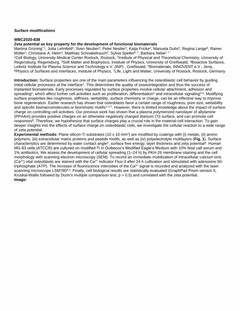
Surface modifications WBC2020-838 Zeta potential as key property for the development of functional biomaterials Martina Grüning* 1, Jutta Lehnfeld2, Sven Neuber3, Peter Nestler3, Katja Fricke4, Manuela Dubs5, Regina Lange6, Rainer Müller2, Christiane A. Helm3, Matthias Schnabelrauch5, Sylvia Speller6, 7, Barbara Nebe1, 7 1Cell Biology, University Medical Center Rostock, Rostock, 2Institute of Physical and Theoretical Chemistry, University of Regensburg, Regensburg, 3Soft Matter and Biophysics, Institute of Physics, University of Greifswald, 4Bioactive Surfaces, Leibniz Institute for Plasma Science and Technology e.V. (INP) , Greifswald, 5Biomaterials, INNOVENT e.V., Jena, 6Physics of Surfaces and Interfaces, Institute of Physics, 7Life, Light and Matter, University of Rostock, Rostock, Germany Introduction: Surface properties are one of the main parameters influencing the osteoblastic cell behavior by guiding initial cellular processes at the interface1. This determines the quality of osseointegration and thus the success of implanted biomaterials. Early processes regulated by surface properties involve cellular attachment, adhesion and spreading1, which affect further cell activities such as proliferation, differentiation2 and intracellular signaling3-4. Modifying surface properties like roughness, stiffness, wettability, surface chemistry or charge, can be an effective way to improve bone regeneration. Earlier research has shown that osteoblasts favor a certain range of roughness, pore size, wettability and specific biomacromolecules or biomimetic motifs2, 5-7. However, there is limited knowledge about the impact of surface charge on controlling cell activities. Our previous work has shown that a plasma polymerized nanolayer of allylamine (PPAAm) provides positive charges on an otherwise negatively charged titanium (Ti) surface, and can promote cell responses8. Therefore, we hypothesize that surface charges play a crucial role in the material-cell interaction. To gain deeper insights into the effects of surface charge on osteoblastic cells, we investigate the cellular reaction to a wide range of zeta potential. Experimental methods: Plane silicon-Ti substrates (10 x 10 mm2) are modified by coatings with (i) metals, (ii) amino polymers, (iii) extracellular matrix proteins and peptide motifs, as well as (iv) polyelectrolyte multilayers (Fig. 1). Surface characteristics are determined by water contact angle4, surface free energy, layer thickness and zeta potential4. Human MG-63 cells (ATCC®) are cultured on modified Ti in Dulbecco’s Modified Eagle’s Medium with 10% fetal calf serum and 1% antibiotics. We assess the development of cellular spreading (1–24 h) by PKH-26 membrane staining and the cell morphology with scanning electron microscopy (SEM). To record an immediate mobilization of intracellular calcium ions (Ca2+) vital osteoblasts are stained with the Ca2+ indicator Fluo-3 after 24 h cultivation and stimulated with adenosine 50-triphosphate (ATP). The increase of fluorescence intensities of the Ca2+ signal is recorded and analyzed with the laser scanning microscope LSM7803-4. Finally, cell biological results are statistically evaluated (GraphPad Prism version 6, Kruskal-Wallis followed by Dunn's multiple comparison test, p > 0.5) and correlated with the zeta potential. Image:


Results and discussions: We were able to generate zeta potentials in the range of -120 and +50 mV by various surface modifications. Similar to Staehlke et al.9, we found on positively charged surfaces a significant enhancement of the initial spreading after 1 h compared to cells on surfaces with negative zeta potential. Intracellular Ca2+ mobilization after ATP stimulation could be increased with surfaces with positive zeta potential, e.g. an amino group containing nanolayer. Interestingly, modifications achieving highly positive surface potential (>50 mV) showed significant impairment in the Ca2+
mobilization and spreading after 24 h. Conclusions: Cell biological results could be correlated with the zeta potential, in contrast to wettability and surface free energy. We found out for the first time, that surfaces with weak positive zeta potential resulted in an enhanced Ca2+
mobilization, this effect could not be displayed with ultra positive surfaces. This suggests that osteoblasts prefer only surfaces featuring a certain range of zeta potential. Therefore, zeta potential may be an important property to consider when developing new implant surfaces. References/Acknowledgements: 1Anselme, 2000, Biomaterials 21 2Bacakova et al. 2011, Biotechnol. Adv. 29 3Staehlke et al. 2015, Biomaterials 46 4Staehlke et al. 2018, Cell Biosci 8:22 5Eisenbarth et al. 2006, J. Biomed. Mater. Res A 79 6Stamp et al. J Mater Sci: Mater Med 20 7Mörke et al. 2017, ACS Appl. Mater. Interfaces 9 8Nebe et al. 2019, Polymers 11:6 9Staehlke et al. 2019, Mater. Sci. Eng. C 101 Disclosure of Interest: None Declared Keywords: Bone, Coatings, Surface characterisation

Surface modifications WBC2020-2721 Multi-functional additively manufactured bone implants Ingmar Van Hengel* 1, Munzur Lacin1, Michelle Minneboo1, Sander Leeflang1, Lidy Fratila-Apachitei1, Iulian Apachitei1, Amir Zadpoor1 1Biomechanical Engineering, TU Delft, Delft, Netherlands Introduction: For prolonged orthopaedic implant functioning, the prevention of both implant associated-infection and aseptic loosening is essential. Such implants should, therefore, possess a wide spectrum of functionalities. Additive manufacturing has enabled the synthesis of novel and complex designs for which the functionalities do not depend on the material properties, but directly stem from the geometrical design of the implant. Rationally designing the geometry of additively manufactured implants has, therefore, resulted in implants with mechanical properties resembling those of the native bone tissue. However, the porous nature of these structures may make these implants more prone to infection. Biofunctionalisation of the implant surface is, therefore, a promising strategy to simultaneously prevent infection and stimulate bony ingrowth, yet has proven to be challenging on highly porous structures. In this study, we applied plasma electrolytic oxidation (PEO) to incorporate Ag nanoparticles (NPs) and strontium on rationally designed titanium bone implants synthesised by selective laser melting (SLM). Experimental methods: Porous titanium implants were fabricated from medical grade Ti-6Al-4V powder by SLM and subsequently biofunctionalised by PEO using electrolytes consisting of Ca/P species and strontium acetate, as well as Ag NPs. Antibacterial testing was performed against methicillin-resistant Staphylococcus aureus (MRSA), a pathogen commonly involved in implant-associated infection. The antibacterial activity was studied by using in vitro assays and a murine ex vivo infection model. To investigate the effects of PEO parameters on the surface morphology and subsequent osteogenic effects, the current density and oxidation time were varied from 20 to 40 A/dm2 and 5 to 60 minutes, respectively. The osteogenic properties of the implants were demonstrated by culturing pre-osteoblastic MC3T3-E1 cells on the implant surface and performing an analysis of the metabolic activity and alkaline phosphatase (ALP) expression. Results and discussions: PEO biofunctionalisation of the porous titanium implants resulted in a micro/nano-porous TiO2 surface layer containing immobilised Ag NPs that facilitated the release of Ag and strontium ions for at least 28 days. Enhanced current densities and oxidation time led to the roughening of the implant surface, thickening of the surface TiO2 layer, and an increased rate of strontium ion release. We observed a synergistic antibacterial effect between Ag and strontium ions, allowing us to reduce the bactericidal concentration of Ag ions by 4 – 32 fold. In addition, Ag-bearing implants fully eradicated a bacterial inoculum ex vivo within 24 hours. The biofunctionalised implant surfaces that contained strontium enhanced the metabolic activity of MC3T3-E1 cells after 7 and 11 days as well as their ALP expression after 11 days. The oxidation time and current density during PEO processing did not significantly alter the observed osteogenic behaviour. Conclusions: Implant biofunctionalisation by PEO with Ag NPs and strontium resulted in potent bactericidal properties in vitro and ex vivo as well as an osteogenic behaviour in vitro. Altogether, the multi-functional titanium implants presented here are suitable candidates for further preclinical development towards a future where patients outlive their implants. References/Acknowledgements: This research was financially supported by the Prosperos project, funded by the Interreg VA Flanders – The Netherlands program. Disclosure of Interest: None Declared Keywords: Antibacterial, Laser-based AM technologies, Metallic biomaterials/implants

Surface modifications WBC2020-2555 Bioactive surface as a platform for the delivery of immobilised NGF and BDNF and the effect in neurite outgrowth Ana Sandoval-Castellanos* 1, Frederk Claeyssens1, John Haycock1 1Materials Science and Engineering, The University of Sheffield, Sheffield, United Kingdom Introduction: Peripheral nerve injury is a major cause of disability, affecting 1 in 1000 patients [1]. Injuries affect life quality and health as a consequence of a decreased sensory and motor function [2]. Autografts are the clinical gold standard, but their performance has limitations. Nerve guide conduits (NGCs) are an alternative. Nevertheless, they do not stimulate sufficient nerve regeneration. Research has been conducted to improve the performance of NGCs, which includes the addition of growth factors [3]. However, nerve repair has not been encouraged significantly, due to the short half-life of the growth factors [4]. Therefore, it is important to design a platform for the sustained delivery of growth factors [5]. The development of bioactive surfaces may target where and how these growth factors are released and how this affect neurite outgrowth. To build these bioactive surfaces, positively charged amine groups and negatively charged heparin were considered, as heparin has affinity to bind growth factors [6] and it could capture and release growth factors. The aim of this study was therefore to 1) develop a bioactive surface enriched with positive amine groups and in turn heparin. This platform was then employed to load NGF, BDNF or a combination of NGF and BDNF for local release, and thereafter 2) evaluate how the bioactive surfaces affects neurite outgrowth. Experimental methods: Heparin was passively conjugated to a pre-coated amine surface 96 well plate. This surface modification was characterized with water contact angle and XPS analysis. Concentrations of 1pg/mL, 1ng/mL, 10ng/mL, 100ng/mL and 1μL/mL of NGF, BDNF and NGF and BDNF were immobilised passively on bioactive surfaces. The release profiles were measured by ELISA assay. Chick embryo dorsal root ganglia (DRGs) were cultured on bioactive surfaces, one DRG per well, for 7 days. Nuclei and βIII-tubulin protein were stained with DAPI and immunocytochemistry to measure neurite outgrowth. Results and discussions: Water contact angle confirmed a change in hydrophilicity, with presence of amine groups and later heparin conjugation. XPS analysis detected nitrogen and sulphur on bioactive surfaces. The release profile of NGF and BDNF from surfaces was assessed by ELISA and showed that at 24 h an initial burst release was minimal and was detectible after 168 h. Interestingly, surfaces immobilized with NGF at 1 ng/mL showed no growth factor release after 24 h and 48 h. By day 7, 1% of NGF was detected. DRGs grown on surface with immobilized 1 ng/mL of NGF developed neurites that were significantly longer in comparison to control surfaces, or surfaces with BDNF and NGF / BDNF. From these results, it can be suggested that surfaces immobilized with 1 ng/mL of NGF supported the longest neurite length, possibly because it did not display an initial burst enabling the DRG to adhere on the surface. The immobilized complex of NGF-TrkA triggered the PI3k pathway, as this complex was not internalized. Then, the NGF-TrkA complex was internalized and retrogradely transported to the body of the cell, stimulating microtubule formation. Conclusions: We concluded that bioactive surfaces with immobilized NGF at 1 ng/mL supported the largest neurite outgrowth using relatively low concentrations of growth factors. In summary, locally delivered neurotrophin surfaces are a promising approach for stimulate nerve regeneration using bioactive nerve guides. References/Acknowledgements: [1] R. Hughes, Pract. Neurol., 2008. [2] C. J. Pateman et al., Biomaterials, 2015. [3] S. Madduri, K. Feldman, T. Tervoort, M. Papaloïzos, and B. Gander, J. Control. Release, 2010. [4] S. Tang, J. Zhu, Y. Xu, A. P. Xiang, M. H. Jiang, D. Quan, Biomaterials, 2013. [5] N. Gomez and C. E. Schmidt, J. Biomed. Mater. Res. - Part A, 2007. [6] B. Casu, A. Naggi, and G. Torri, Carbohydrate Research, 2015./CONACyT for funding AMSC Disclosure of Interest: None Declared Keywords: Biomaterials (incl. coatings) for local drug and growth factor delivery, Peripheral nerves and spinal cord

Surface modifications WBC2020-1603 Plasma grafting or Coaxial electrospinning: comparison of functionalized PP meshes with anticoagulant activity Malo Dufay* 1, Frédéric Cazaux1, Chai Feng2, Nicolas Blanchemain2, Séverine Bellayer1, Grégory Stoclet1, Mathilde Casetta1, Maude Jimenez1, Stéphanie Degoutin1 1UMR 8207 – UMET, VILLENEUVE D ASCQ, 2Inserm, CHU Lille, U1008 , Lille, France
Introduction: As part of intra-abdominal operations, postoperative adhesions occur in more than 50% of cases1 and result in pain. This phenomenon consists in the excessive formation of a fibrin matrix between organs and peritoneum 5 to 7 days following the surgery and is induced by the coagulation cascade. In order to limit these postoperative adhesions, one approach could be to develop a long-term anticoagulant active implant. Nowadays, a large variety of processes is used to design biomedical textile implants. Among them, electrospinning offers advantages in this field especially due to the nanoscale diameter range of electrospun fibers and their high porosity and surface area. In addition, cold plasma process can be used to activate and/or functionalize electrospun fibers. In the literature, cold plasma is indeed mainly used to enhance hydrophilicity or to graft polymers at the nanofibers surface2. In this context, our strategy is to develop polypropylene (PP) implants, coated with polycaprolactone (PCL) electrospun nanofibers and functionalized by 2-acrylamido-2-methylpropane sulfonic acid (AMPS) (monomer with heparin-like groups). Two pathways were investigated: graft-copolymerization of AMPS onto PCL nanofibers induced by cold plasma and coaxial electrospinning in order to obtain core-sheath (PCL-PolyAMPS) nanofibers. Experimental methods: First, the electrospinning conditions of PCL, AMPS simple and core-shell solutions were optimized in terms of concentration, voltage, flow, tip-to-collector distance (Figure 1A and C). The graft-copolymerization of AMPS onto PCL nanofibers was optimized in order to obtain cytocompatible nanofibers rich in SO3H groups at the surface. Adequate physico-chemical characterizations were performed (FTIR, SEM, TEM, TGA…) at each step of the process. Image:

Results and discussions: SEM images revealed an increase in the nanofibers diameter after the plasma induced graft-copolymerization (Figure 1B), and mapping confirmed the presence and distribution of AMPS onto nanofibers obtained by the two pathways (Figure 1D). Biological in vitro assays revealed an improved anticoagulant activity of these membranes for both pathways and the comparison between the groups, using repeated measures analysis of variance (ANOVA), proved the real added value of these membranes. Conclusions: We have successfully optimized the electrospinning of PolyAMPS, PCL as well as AMPS grafting onto PCL mats by cold plasma, and we highlighted the presence of sulfonate groups onto the surface of nanofibers. The cytocompatibility, haemocompatibility and anticoagulant activities of the functionalized implants were demonstrated. Future work will focus on the comparison of these two strategies in terms of in vivo bioactivity and industrial scale up costs. References/Acknowledgements: References : [1] D. Menzies et al., Ann. R. Coll. Surg. Engl. (1993) 75 : 147-153 [2] S. Kaur et al., Langmuir (2007) 23(26) : 13085-92, doi: 10.1021/la701329r Acknowledgments Agence nationale de la recherche (ANR JCJC CAPSPIN: ANR-17-CE09-0003-01) and competitiveness cluster UpTex are acknowledged for supporting and funding this work. Disclosure of Interest: None Declared Keywords: Biocompatibility, Surface characterisation

Surface modifications WBC2020-1678 The Recruitment of Endothelial Progenitor Cells on Combinatorial Bio-mimetic Functionalized Surfaces Mohamed Elkhodiry* 1, Omar Bashth1, Gaétan Laroche2, 3, Jean-François Tanguay4, 5, Corinne Hoesli1 1Chemical Engineering, McGill University, Montreal, 2Centre de Recherche du CHU de Québec, 3Département de Génie des Mines, des Matériaux et de la Métallurgie, Université Laval, Québec, 4Montreal Heart Institute, 5Department of Medicine, Université de Montréal, Montreal, Canada Introduction: New vascular implants with surface-immobilized capture antibodies targeting circulating endothelial progenitor cells (EPCs) have shown clinical promise in accelerating the formation of neo-endothelium to cover the implant. The absence of subsequent adhesion-based signaling needed for EPC proliferation and differentiation could explain the lack of long-term improvement in vascular re-stenosis and overall vascular recovery[1]. We have previously shown that extracellular matrix (ECM)-derived peptides, such as RGD, can promote the adhesion and enhance the clonal expansion of EPC-derived endothelial colony forming cells (ECFCs) via integrin based signaling[2]. The objective of the current study was to evaluate the potential of a novel bio-mimetic surface functionalization strategy the combines extracellular matrix (ECM)-derived peptides and capture antibodies in promoting capture, adhesion, and proliferation of EPCs. Experimental methods: Peripheral blood from 22 adult human donors was used to obtain donor specific ECFCs. Aminated culture surfaces were modified by either the covalent conjugation of a fluorophore-labeled RGD peptide (CGK(PEG3-TAMRA)GGRGDS, referred to as “RGD-TAMRA”), immobilization of CD144 (VE-cadherin) antibody, or combining both biomolecules at different ratios using an optimized process. Unmodified surfaces were used as a negative control. Surface concentration of the peptide and antibody were analyzed using fluorescence microscopy (Figure 1A). The expanded ECFCs were first seeded under static conditions and cell adhesion was analyzed using immunocytochemistry. ECFCs capture and adhesion under dynamic conditions at 1 dyn/cm2 wall shear stress were then analyzed through live cell imaging followed by immunocytochemistry. Image:

Results and discussions: Surfaces with immobilized anti-CD144 antibodies displayed significantly enhanced ECFC capture potential compared to RGD-TAMRA and unmodified controls as quantified by the number of adhered cells under both static and dynamic conditions. Average ECFC spreading was significantly increased on RGD-TAMRA surfaces compared to the anti-CD144 and unmodified controls on both static and dynamic conditions. ECFC surface coverage was maximized when combining anti-CD144 and RGD-TAMRA (Figure 1B & C). By increasing the peptide concentration on the combinatorial surface, ECFC spreading was enhanced without significantly affecting the number of adhered cells. These results suggest an additive (i.e. independent) effect of capture antibodies on ECFC tethering vs. RGD-TAMRA peptides on subsequent ECFC spreading. Conclusions: To our knowledge, this is the first study showcasing the ability of an antibody/peptide combinatorial surface functionalization strategy in promoting ECFC capture and adhesion. Our results also display a clear distinction between the mechanisms by which human derived-ECFCs react to immobilized capture antibodies and conjugated ECM-peptides. This strategy creates sequential steps of EPC trafficking to mimic endogenous processes of vascular healing which can potentially create an effective regenerative capacity for vascular implants. References/Acknowledgements: [1] P. Ndunda, M.R. Vindhyal, T. Muutu, Z. Fanari, Clinical outcomes of the Dual-Therapy CD34 antibody-covered sirolimus-eluting stent versus standard drug-eluting coronary stents: A meta-analysis, Cardiovascular revascularization medicine : including molecular interventions (2019). [2] M.A. Elkhodiry, M.D. Boulanger, O. Bashth, J.-F. Tanguay, G. Laroche, C.A. Hoesli, Isolating and expanding endothelial progenitor cells from peripheral blood on peptide-functionalized polystyrene surfaces, Biotechnology and Bioengineering 116(10) (2019) 2598-2609. Disclosure of Interest: None Declared Keywords: Biocompatibility, Cell adhesion and migration, Vascular grafts incl. stents

Surface modifications WBC2020-1157 Investigations into Novel Titanate Conversion of DC Magnetron Sputtered Titanium Thin Films for Biomedical Applications Matthew Wadge* 1, Burhan Turgut1, James Murray1, Bryan Stuart1, Reda Felfel1, Ifty Ahmed1, David Grant1 1Mechanical, Materials and Manufacturing Engineering, University of Nottingham, Nottingham, United Kingdom Introduction: The current process for improving implant surfaces to be bioactive, therefore, providing a more natural fixation, is reliant on high temperature (>1500 K [1]) plasma spraying of hydroxyapatite (HA). However, these surfaces have been shown to spall due to their brittle nature, high internal stresses, and weak mechanical adhesion [2]. Titanate surfaces have been developed as an alternative by Kokubo et al. [3], however, their applicability have been limited to titanium (Ti) and its alloys only via chemical conversion routes. The authors propose a novel method for generating nanoporous titanate surfaces on non-Ti biomedical materials through conversion of DC magnetron sputtered Ti films. Experimental methods: Commercially pure polished 10 mm 316L S.S. discs were subjected to DC magnetron sputtering using a cp-Ti (Miba Coatings; 99.5% purity) target (1.56 kW/cm2 power density; optional 0 to -100 V substrate bias and up to 300 oC substrate heating). The produced ca. 4 μm films were then treated in NaOH (5 M; 60 oC; 24 h) to assess the effect of sputtering parameters on titanate conversion. The samples have been labelled according to the following convention: negative substrate bias (V)/applied substrate temperature (oC), e.g. 100V/150oC for -100 V bias and 150 oC applied temperature. Characterisation using SEM, EDX, XPS, Raman, FTIR, XRD, and texture coefficient analysis was conducted. Image:


Results and discussions: SEM micrographs demonstrated 3.89 ± 0.04, 3.90 ± 0.03, 3.71 ± 0.04 and 3.68 ± 0.02 μm thick Ti coatings for the 0V, 100V, 100V/150oC and 100V/300oC samples, respectively. Through application of a substrate bias, and bias in conjunction with substrate heating, the density of the films increased (reduction in coating thickness and surface voids). Furthermore, texture coefficient analysis of measured XRD spectra using the Harris equation [4], exhibited a shift from columnar (preferred orientation in the Ti HCP (002) plane (PDF 00-044-1294); Texture coefficient (Tc(002) = 3.39) in the 0V sample, to more equiaxed (Tc(002) = 1.54; for pure equiaxed, Tc = 1) in the 100V/300oC sample. Subsequent titanate conversion of the above samples produced 1.12 ± 0.04, 1.20 ± 0.02, 1.20 ± 0.03, and 1.63 ± 0.06 μm thick titanate layers for the 0V, 100V, 100V/150oC, and 100V/300oC samples, respectively. Despite the proposed hypothesis that increased porosity would allow better NaOH penetration, therefore, increasing titanate conversion, this was not observed in the samples tested. EDX, XPS and Raman analysis, showed incorporation of 7.6 ± 0.1, 8.9 ± 0.1, 11.5 ± 1.8, and 7.6 ± 0.1 at.% of Na in the 0V, 100V, 100V/150oC, and 100V/300oC samples, respectively. Conclusions: Overall, the data presented demonstrates the successful conversion of DC magnetron sputtered Ti coatings into titanate structures, with increased Na inclusion for the unbiased (7.6 at.%), biased (8.9 at.%), and biased/heater runs (11.5 at.% & 7.6 at.%), respectively. The hypothesis that increased penetration in the more porous unbiased film was not evidenced, however, clear structural differences in the film produced have an effect on Na inclusion and titanate morphology of the conversion layer. References/Acknowledgements: [1] Tsui, Y.C., et al., Biomaterials 1998, 19, 2015-2029. [2] De Groot, K., et al., J. of Biomed. Mat. Res. A 1987, 21, 1375-1381. [3] Kokubo, T., et al., The Open Biomed. Eng. J. 2015, 9. [4] Harris, G., The London, Edinburgh, and Dublin Phil. Mag. and J. of Sci. 1952, 43, 113-123. Disclosure of Interest: None Declared Keywords: Coatings, Metallic biomaterials/implants, Surface characterisation

Surface modifications WBC2020-1236 Design of surface functionalised polycaprolactone: ensuring clearance of the surface layer Alexandra Mutch* 1, Oscar Paredes Trujillo1, Lisbeth Grondahl1, 2 1School of Chemistry and Molecular Biosciences, 2The Australian Institute for Bioengineering and Nanotechnology, The University of Queensland, Brisbane, Australia Introduction: Polyesters such as poly(ε-caprolactone) (PCL) have been widely used as biomaterial implants for tissue engineering as they are biodegradable and can be easily processed into different structures. Many recent studies have aimed to fine tune properties of polyesters through a variety of surface modifications including chemical treatment, coating with biological materials, high energy radiation, and surface grafting. However, many of these studies have failed to consider how the surface layer will interact with the body upon degradation of the underlying polyester.1 Some surface modifications can result in high Mw or crosslinked polymer layers that are non-biodegradable and too large for clearance from the body, and some can generate toxic by-products upon degradation of the surface layer.1 It is therefore important to consider the full lifetime and ultimate fate of any surface modification to ensure time and resources are not spent investigating materials that will not be viable implants. Experimental methods: In this study PCL has been modified by irradiation-induced grafting using 2-aminoethyl methacrylate (AEMA) to introduce amine functionalities that are useful for conjugation to biopolymers, and 3-sulfopropyl acrylate (SPA) to introduce sulfonate functionalities useful for electrostatic interactions with growth factors. Optimisation of the grafting conditions on 2D films and 3D-printed scaffolds included evaluation of the effect of monomer concentration, radiation dose, solvent, and presence of homopolymer inhibitor. The biostable graft copolymers were evaluated through NMR and GPC. Degradation studies investigated the surface later stability and bulk degradation in phosphate buffer at physiological conditions, as well as in dilute HCl for accelerated degradation. Image:
Results and discussions: The grafting extent of SPA onto PCL films was mainly affected by the solvent and the radiation dose while the monomer concentration affected the Mw of the grafted chains. It is therefore possible to control both parameters in the final surface modified material. Grafting of AEMA was not greatly affected by varying the grafting parameters, in agreement with previous studies on a different polyester substrate.2 Grafting of AEMA onto 3D scaffolds was achievable on all external and internal surfaces. The Figure shows that there is no difference in the surface topography between unmodified PCL (A) and AEMA-grafted PCL (B), correlating with relative low Mw of the grafted chains. The CLSM image in Figure 1C illustrates that amine functionality is successfully introduced and that the amine groups are reactive as shown by attachment of FITC. Bulk degradation under accelerated conditions in 3 M HCl revealed that there was no change in mass loss for unmodified, irradiated or grafted materials. Surface layer stability in phosphate buffer found the layer to be maintained for more than 21 days. Conclusions: This study demonstrates that graft co-polymerisation of different functional monomers onto PCL is a viable method of surface modification in the field of biomaterials science. The process does not greatly affect the bulk properties and the surface layer is stable for more than 21 days. The surface layers have been selected and their synthesis

optimised with respect to the Mw of the grafted chains such that clearance of these biostable chains is assured after degradation of the underlying PCL. References/Acknowledgements: 1. Mutch, A. L.; Grøndahl, L., Challenges for the development of surface modified biodegradable polyester biomaterials: A chemistry perspective. Biointerphases 2018, 13 (6), 06D501. 2. Luk, J. Z.; Cork, J.; Cooper-White, J.; Grøndahl, L., Use of Two-Step Grafting to Fabricate Duel-Functional Films and Site-Specific Functionalised Scaffolds. Langmuir 2015, 31, 1746-1754. Acknowledgements: Dr M Bartnikowski for 3D printing, Y Zhou for assistance with sample preparation, Dr V Sagulenko for CLSM. A Mutch acknowledges UQ RTP Scholarship. Disclosure of Interest: None Declared Keywords: Biodegradation, Biomaterials for growth factor delivery, Surface characterisation

Surface modifications WBC2020-3122 Flexible and self-healing electrochemical hydrogel sensor with high sensing efficiency toward glucose monitoring Zhen liang*, Jieyu zhang, Yunbing wang and Xuefeng Hu, Yuhui Lu Introduction: The global prevalence of diabetes and impaired glucose tolerance has increased significantly in recent decades, and the number of diabetic patients will rise to 629 million by 2045 as predicted by the International Diabetes Federation (IDF). For the usual routes (oral or subcutaneous) of administration of hypoglycemic drugs, the dosage may not match the instant blood glucose level, and over suppression of the blood glucose level or insufficient drug dosage may induce fatal hypoglycemia or ketoacidosis. Thus, the continuous glucose monitoring system (CGMS) is needed for the accurate delivery of insulin in patients with severe diabetes. The key component of CGMS is the glucose sensor. Although there are various methods for glucose measurement, electrochemical analysis is the most widely used one due to its fast response, broad detection range and quantitative operating mode. The commercial sensor of a continuous glucose monitoring system is usually constructed by coating glucose oxidase and the electrocatalytic medium on an electrode, but this strategy suffers from restricted penetration of glucose in the dense coating, susceptibility to mechanical damage and easy leakage of the glucose oxidase and electrocatalytic medium, which compromise the sensing efficiency. Herein, a self-healing hydrogel based on quaternized chitosan and oxidized dextran is designed, and the CeO2/MnO2 hollow nanospheres are covalently linked in the hydrogel as the electrocatalytic medium. Glucose oxidase is loaded via the strong electrostatic interactions with the CeO2/MnO2 hollow nanospheres, and an extra covering agent is used to prevent its leakage. Covalent linkage of the hydrogel on a bendable chip constructs a flexible glucose sensor through dynamic covalent bond, which shows a wide linear range, fast responseand high sensitivity due to the effective diffusion of glucose in the hydrogel and fast electron transfer in the hollow CeO2/MnO2 nanospheres. In addition, the electrical signal of the hydrogel sensor disappeared due to mechanical damage, and the sensing response recovered after 2 h of self-healing. Thus, this study provides a method to simultaneously prevent the leakage of the electrocatalytic medium, promote the sensitivity of glucose detection and tolerate the mechanical damage, which shows great potential for continuous glucose monitoring. Experimental methods: KMnO4, Ce(NO3)3·6H2O, chitosan, GTMAC, chloride, GOx, dextran, APTES. Hollow nano spheres were prepared by hydrothermal method, modified and grafted onto quaternized chitosan, and then reacted with dextran oxide to form self-healing conductive hydrogel. Finally, it was prepared on the surface of flexible electrode treated by plasma and silane coupling agent and assembled with glucose oxidase to form glucose electrochemical sensor. Image:
Results and discussions: The conductive hydrogel has excellent self-healing and injectable properties and can be quickly and firmly prepared on the surface of flexible electrode treated by plasma and silane coupling agent and assembled into sensors. In addition, the electrical signal of the hydrogel sensor disappeared due to mechanical damage, and the sensing response recovered after 2 h of self-healing. The sensor shows a wide linear range, fast response, high sensitivityand good anti-interference performance in glucose detection. Conclusions: The electrocatalytic medium was grafted into the self-healing hydrogel through covalent bond to form conductive hydrogel and prepare electrochemical glucose sensor, so as to solve the problem of electrocatalytic medium precipitation in the test process, so as to improve the overall performance of the sensor. The self-healing hydrogel sensor is able to recover its sensing abilityafter significant mechanical damage.At the same time, the fast fixation and encapsulation of glucose oxidase were realized by using electrochemical oxidation. References/Acknowledgements: International Diabetes Federation. Diabetes Atlas [M]. 8th ed. 2017. Disclosure of Interest: None Declared Keywords: Biocompatibility, Biosensors, Composites and nanocomposites

Surface modifications WBC2020-1433 Microarchitected Mg-based scaffolds modified with Directed Plasma Nanosynthesis Viviana Marcela Posada Perez* 1, Patricia Fernandez-Morales2, Juan Ramírez1, Jean Paul Allain3 1School of Mines, Universidad Nacional de Colombia, 2Facultad de Ingeniería Industrial , Universidad Pontificia Bolivariana, Medellin, Colombia, 3Ken and Mary Alice Lindquist Department of Nuclear Engineering, Penn State College of Engineering, State College, United States Introduction: The ability to manufacture Mg scaffolds with predesigned pores represents many opportunities for the development of biodegradable orthopedic materials. However, the highly reactive potential of Mg together with the augmented exposed area increase the corrosion rate and limits its biological application. Coatings have been proposed as the principal strategy to control the corrosion rate of Mg porous materials 1,2. However, the use of coatings results in the need to modify pore size and material geometry. Moreover, surface coatings on complexes structures are confronted with their adhesive potential and can affect the lightweight properties of the porous Mg. Directed plasma nanosynthesis (DPNS) is an additive nanomanufacturing process able to induce compositional and topographical modifications in an atomic scale. In the present work, the DPNS process variables: low particle energy, normal incidence angle, and high ion fluence conditions are combined with the scaffold microarchitecture to elicit different properties, including bioactivity and degradation control without affecting the bulk properties of the scaffold. Experimental methods: Microarchitected Mg-based scaffolds with two different unit cells: diamond (Di) and truncated octahedron (TO) were fabricated by infiltration casting from the same AZ31 rod. Scaffolds were modified with DPNS with normal incidence, low energy, and high fluence. The surface nanostructures, phases, and morphology were evaluated by SEM/EDS. Mg samples were immersed in DMEM and hydrogen release was measured by the gas collecting method. After 5 and 10 days of immersion, corrosion products were removed from the surface with CrO3 (after ASTM G1), and the corrosion characteristics were evaluated with SEM. The Mg release was measured by ICP-AES. In vitro biocompatibilities were evaluated with direct cell attachment and indirect cytotoxicity using preosteoblastic cell l ine (MC3T3-E1). Material extracts were prepared according to ISO 10993. Cell viability was tested by Alamar blue after 1 and 5 days. Cell morphology was evaluated by immunostaining using DAPI and Alexa Fluor 488-Phalloidin. Image:
Results and discussions: Two different microarchitected Mg scaffolds were successfully obtained by infiltration casting. The porosity and pore size were individually controlled with the pore microarchitectures. After DPNS, different nanostructured surfaces were obtained depending on the geometry of the scaffolds (Fig 1). These surfaces changed the corrosion dynamics of the materials due to an Al supply from the interior of the alloy to the near-surface. In the case of TO, DPNS improved corrosion resistance and decreased Mg ion release up to 40%. However, for Di samples, the same irradiation parameters caused the opposite effect, and this combination of DPNS conditions was detrimental for Di corrosion.

After 24 hours of incubation, cells presented flattened morphology and grew well on all the surfaces, no apparent toxic effects were observed in any of the samples. However, after four days in culture TO surface modified with DPNS presented 32% more attached cells compared to no irradiated TO. TO modified with DPNS positively supported the growth of MC3T3 cells, while in the Di surface, the cells attached diminished around 15% compared to non-modified Di. Conclusions: Mg scaffolds with microarchitected pores were successfully obtained and modified with DPNS. The effect of DPNS on the scaffolds varied according to the geometry of the unit cells. An Mg scaffold with truncated octahedron unit cells was bioactivated with DPNS which successfully decreased the corrosion rate an augmented the cell attachment and osteoblastic activity of MC3T3 cells. References/Acknowledgements: 1. M.Rúa, J., Zuleta, A. A., Ramírez, J. & Fernández-Morales, P. Micro-arc oxidation coating on porous magnesium foam and its potential biomedical applications. Surf. Coatings Technol. 360, 213–221 (2019). 2. Julmi, S. et al. Processing and coating of open-pored absorbable magnesium-based bone implants. Mater. Sci. Eng. C 98, 1073–1086 (2019). Disclosure of Interest: None Declared Keywords: Biodegradable metals, Biodegradation, Surface characterisation

Surface modifications WBC2020-1455 Cell-mediated release of biomolecules from functionalised block copolymer thin films Isabela Monteiro A.* 1, 2, Tarek Kollmetz1, 2, David S. Musson3, Sue R. McGlashan4, Jenny Malmström1, 2 1Department of Chemical & Materials Engineering, The University of Auckland, Auckland, 2MacDiarmid Institute for Advanced Materials and Nanotechnology, Wellington, 3Department of Medicine, 4Department of Anatomy and Medical Imaging, The University of Auckland, Auckland, New Zealand Introduction: Cell biologists and biomaterials scientists are used to presenting biochemical signals in solution in typical cell culture techniques, although in vivo those molecules are known to interact to the components of the extracellular matrix (ECM). To better mimic the in vivo environment, previous researchers have attached growth factors to the substrate and observed crosstalk between adhesion and growth factor receptors (GFRs).1,2 However, some biomolecules, such as epidermal growth factor, need to be internalised by the cell to propagate its signal and, therefore, cannot be physically attached to the substrate. This study aims to address this issue by the development of a novel biointerface that allows the release of biomolecules upon cell adhesion. The release is regulated by a biodegradable layer, which covers the polymer film and can be digested by enzymes produced by adherent cells. This approach ensures cargo release close to cell adhesions and, consequently, adhesion and GFRs signalling crosstalk and their effect on cell behaviour can be better understood. Figure 1 presents a schematic of the platform developed. Experimental methods: Thin nanopatterned films were prepared by spin coating self-assembling polystyrene-block-ethylene oxide (PS-b-PEO) on clean silicon wafers. In vitro cytocompatibility of PS-b-PEO was investigated using murine chondrocytes. PEGylated lysozymes were chosen as a representative biomolecule and co-assembled with PS-b-PEO. Lysozyme qualitative release was analysed by using atomic force microscopy (AFM) in liquid. Additionally, the quantitative release was studied through fluorometric enzyme-linked immunosorbent assay (ELISA). A method to produce a homogeneous biodegradable layer made of collagen type I was designed to avoid the natural release of the lysozymes. The degradation of the topping layer was studied by incubation of collagen-coated PS-b-PEO with different concentrations of matrix metalloproteinases (MMPs). Image:

Results and discussions: In the cytocompatibility tests performed with PS-b-PEO, viability was greater than 90% throughout the 5 days culture with abundant cell spreading, showing that PS-b-PEO is biocompatible and a promising substrate for long term cell studies. Furthermore, we have successfully established a method for the preparation of a homogeneous biodegradable layer made of collagen type I and confirmed that its degradation occurs during the first hour when in contact with MMP type I. Preliminary results show promising incorporation of PEGylated lysozymes into the PEO domain of the polymer film and permanence of its enzymatic activity. Conclusions: We have developed a new platform made of biocompatible self-assembled PS-b-PEO thin films for the release of biomolecules upon collagen type I degradation by MMP-I. The developed surface offers a solution for the lack of experimental methods to unravel synergies between adhesion and growth factor receptors. Additionally, the presented engineered surface is a generic smart interface for cell-controlled delivery of biomolecules, which can be applied to a range of cell types and signalling molecules. References/Acknowledgements: 1. Crouzier, T., Fourel, L., Boudou, T., Albigès-Rizo, C. & Picart, C. Presentation of BMP-2 from a Soft Biopolymeric Film Unveils its Activity on Cell Adhesion and Migration. Adv. Mater. 23, H111–H118 (2011). 2. Monteiro A., I., Kollmetz, T. & Malmström, J. Engineered systems to study the synergistic signaling between integrin-mediated mechanotransduction and growth factors (Review). Biointerphases 13, No. 06D302 (2018). The authors greatly acknowledge financial support from the Marsden Fund Council, the Rutherford Discovery Fellowship and The University of Auckland for providing a doctoral scholarship for IMA. Disclosure of Interest: None Declared Keywords: Biomaterials (incl. coatings) for local drug and growth factor delivery, Coatings, Micro- and nanopatterning

Surface modifications WBC2020-1504 Scavenging of bone catabolic proteins by functional materials – Proof of concept and evaluation of linker characteristics Mathis Gronbach* 1, Benno Müller1, Vicky Lidzba, Franziska Mitrach1, Stephanie Möller2, Matthias Schnabelrauch2, Sandra Rother3, Vera Hintze3, Michael Hacker1, Michaela Schulz-Siegmund1 and Pharmaceutical Technology Leipzig 1Pharmaceutical Technology, University of Leipzig, Leipzig, 2INNOVENT e.V., Jena, 3Max Bergmann Center of Biomaterials, TU Dresden, Dresden, Germany Introduction: For guided tissue regeneration, surface properties of bone substitution materials are of high relevance. Our approach is to modify 3-armed methacrylated oligolactid-based macromer scaffolds1 that have been shown to be well tolerated in a bone defect model in diabetic rats2. As a means for straight forward surface characterization, we established macromer films as a model system for equally composed scaffolds.3 We incorporated a small molecular anchor into the films to bind a suitable linker molecule subsequently. Covalent surface decoration with sulfated glycosaminoglycans (sGAG) is intended to improve bone defect regeneration by taking advantage of the described sGAG ability4 to scavenge bone-catabolic proteins. This property was also quantified in this study by SPR measurements. This study investigated surface with varying amounts of anchor and different chemistries of the polyetheramine linker. Experimental methods: Film synthesis: Three-armed biodegradable oligolactide-based macromer Tri134LA6 (Mw trimethylolpropane core: 134 g/mol, 6 equivalents of oligolactide per arm) were cross-copolymerized with glycidyl methacrylate (GMA). Amount of anchor: 5 – 40% m/m. Stepwise surface decoration: 1. Addition of polyetheramine (Jeffamine®) linker, quantification via fluorescence scanning after reaction with (5(6)-SFX). We tested polyetheramines of different geometries and molecular weights (two and three-armed, Mw ranging from 148 to 3000). 2. Immobilization of sulfated GAG (sGAG, Mw= 75,000, degree of sulfation= 3.4), ATTO565 labeled) via EDC/NHS. Interaction studies via surface plasmon resonance spectroscopy: SPR measurements were performed on Series S Sensor Chips CM5 chips using Biacore T200. Affinity of sGAG towards sclerostin & DKK1 was measured. Scavenging experiments: Protein incubation in McCoy's 5A + 10% FBS, quantification of sclerostin/DKK-1 via ELISA. Cell culture: SaOS-2 cells in DMEM + 10% FBS, osteogenic supplements: dexamethasone, b-glycerophosphate, ascorbic acid. Image:

Results and discussions: We established a protocol with defined polymerization and incubation conditions for the film production process. Depending on the linker chemistry, we observed different reaction kinetics resulting in the highest number of reactive amino groups provided by the smallest (Mw 148), two-armed linker. We showed the necessity for a certain length of the linker (equalling a Mw of 600) in order to introduce functionalization. An incorporation of more than 20% GMA (m/m) during polymerization resulted in no significant increase of functionalization. For the final formulation, we chose the linker ED900 and 20% GMA in order to assure a good reactivity towards sGAG functionalization while still relying on the basic materials properties determined by the biodegradable crosslinked macromer.SPR measurements supported the binding to DKK1 but also another bone catabolic protein, sclerostin.For proof of concept, sGAG decorated films were incubated with a solution of sclerostin and DKK-1 (8 ng/ml). 24 h later, significantly less amounts of both proteins were determined in the supernatant of functionalized materials compared to the amounts found in medium in contact with control films. In the final step, we seeded osteoblast-like SaOS-2 cells on the films, which secrete sclerostin and DKK-1. We found less DKK1 in the supernatant of sGAG modified films than of control films. The reduced DKK1 content in the supernatant may have supported osteogenic differentiation of SaOS-2 cells on sGAG-modified films. Conclusions: Overall, we were able to prove the SPR-predicted scavenging ability of our sGAG functionalized material and its potential positive impact on bone regeneration. By varying the nature and density of our polyetheramine-based linkers, we optimized this parameter towards an effective scavenging of bone catabolic proteins. References/Acknowledgements: 1 Loth, Acta Biomater. 2015;26:82-96 2 Picke, Biomaterials 2016, 96:11-23 3 Müller, Acta Biomater. 2017, 51:148-160 4 Salbach-Hirsch J, Biomaterials 2015, 67:335-45 Disclosure of Interest: None Declared Keywords: 3D scaffolds for TE applications, Bone, Surface characterisation

Surface modifications WBC2020-1883 A click-based chemical toolkit to functionalize biomaterials Helena Martin-Gomez1, 2, Iván Sánchez1, 3, Lluis Oliver-Cervelló1, 2, Vicente Marchán3, Carles Mas-Moruno* 1, 2 1Department of Materials Science and Engineering, Biomaterials, Biomechanics and Tissue Engineering Group (BBT), Universitat Politècnica de Catalunya, 2Barcelona Research Center in Multiscale Science and Engineering, Universitat Politècnica de Catalunya, 3Departament de Química Inorgànica i Orgànica, Secció de Química Orgànica, IBUB, Universitat de Barcelona, Barcelona, Spain Introduction: In biomaterials science, peptides and peptidomimetics are widely used to functionalize material surfaces in order to confer bioactivity to otherwise inert substrates [1,2]. The attachment of biomolecules to biomaterials is commonly achieved using specific anchors with chemical affinity for the substrates. Well-known examples include the use of thiols to bind gold substrates or catechol groups to bind titanium and other metallic oxides. However, this implies that each synthesized peptide can be used only for a narrow range of materials. Thus, in the majority of cases, changing the material of study requires synthesizing the same peptide with a distinct anchor, resulting in time-consuming and repetitive procedures. To solve this, here we report on the development of a novel and versatile click-based solid-phase synthetic strategy to prepare peptidic coatings suitable for functionalizing a variety of biomaterials (a chemical toolkit). In detail, we focused on the synthesis of a model peptide and the optimization of the copper-catalysed azide-alkyne cycloaddition reaction to introduce to the peptidic backbone distinct anchoring groups in an efficient and modular fashion. Subsequently, model substrates of different nature commonly used as biomaterials were functionalized with the peptidic toolkit by simply selecting the appropriate anchor. The feasibility of this method was validated by physicochemical surface characterization and cellular assays. Experimental methods: A model branched peptidic structure (containing the RGD and DWIVA sequences) was synthesized by solid-phase peptide synthesis (SPPS). Anchoring groups were introduced in solid phase by copper-catalysed azide-alkyne cycloaddition (CuAAC). The synthetic building blocks and molecules were characterized by NMR, HPLC and mass spectrometry. Biomaterial substrates made of gold, poly(L-lactic acid) and titanium were functionalized, respectively, with toolkit peptides bearing either a thiol, an amino or catechol groups. The extent and specificity of the binding was analysed by physicochemical methods, including contact angle measurements, XPS and Raman spectroscopy. The response of human mesenchymal stem cells (MSCs) was analysed by means of cell adhesion and differentiation studies. Results and discussions: Toolkit design: The Click CuAAC reaction was optimized in solid-phase under mild conditions selecting an azido-modified lysine (Lys(N3)) at the C-terminus of the peptide and alkyne-derivatized functional groups, i.e. alkyne-L-cysteine, propargylamine and alkyne-L-3,4-dihydroxyphenylalanine (DOPA). This strategy allowed the introduction of surface-specific anchors (thiol, amine, catechol) to a model peptide sequence in a straightforward fashion. Toolkit validation: Model substrates were functionalized with toolkit peptides in a material-specific manner, i.e. according to the chemistry of the surface the more appropriate anchors of the toolbox were selected. The chemoselectivity and success of the grafting procedure was corroborated by XPS and physicochemical methods. The surfaces selectively functionalized with the RGD/DWIVA peptide fostered improved MSC adhesion and osteogenic differentiation. Conclusions: The chemical toolkit reported in this communication proved useful to selectively functionalize biomaterials of different nature and consequently fine tune MSC response. It is expected this new method will find applications to coat a wide range of biomaterials in a straightforward and cost-efficient fashion. References/Acknowledgements: References: [1] Mas-Moruno C, Su B, Dalby MJ. Adv. Healthcare Mater. 2019, 8(1):e1801103. [2] Mas-Moruno C, Fraioli R, Rechenmacher F, Neubauer S, Kapp TG, Kessler H. Angew. Chem. Int. Ed. 2016, 55:7048-67. Acknowledgements: We thank the MINECO/FEDER for financial support through a Ramon y Cajal grant of C.M.M (RYC-2015-18566) and Project MAT2017-83905-R, co-funded by the European Union through European Regional Development Funds, and the Government of Catalonia (2017 SGR1165). Disclosure of Interest: None Declared Keywords: Coatings, Material/tissue interfaces

Surface modifications WBC2020-2862 Controlling bacterial adhesion to titanium surfaces: new strategies for surfaces with tailorable anti-adhesive/antibacterial properties Sara Ferraris1, Silvia Spriano*, Sergio Perero1, Piero Costa2, Giovanna Gautier di Confiengo3, Andrea Cochis4, Fernando Warchomicka5, Seiji Yamaguchi, Lia Rimondini4, Enrica Vernè1, Monica Ferraris1 1DISAT, POLITECNICO DI TORINO, 2INTRAUMA srl, 3CNR-IMAMOTER, Torino, 4Università del Piemonte Orientale, Novara, Italy, 5Graz University of Technology, Graz, Austria Introduction: Bacterial contamination is an issue in different fields (from everyday life to aerospace) and it becomes critical in biomedical implants: because of increasing bacterial resistance to antibiotics, the development of alternative antibacterial solutions is a high medical need. Implantable biomaterials are frequently associated with infection; a limited colonization and, eventually, a localized active antibacterial action are a challenge for reducing the risk of implant associated infections. The present research summarizes different strategies aimed at reduction of bacterial adhesion to titanium surfaces moving from simply anti-adhesive surfaces to anti-bacterial and even to both bioactive and antibacterial ones. Experimental methods: Ti6Al4V was used as substrate. Different surface treatments were considered in order to improve the biological response of Ti6Al4V in specific applications and simultaneously reduce bacterial adhesion. They are briefly summarized in the following: - Electron beam structuring has been investigated for the realization of aligned micro-grooves aimed at fibroblast alignment for soft tissue healing. The process induces peculiar surface microstructure and crystallographic structure which can affect bacterial attachment [1] - Sputtering coating: silica, alumina and zirconia coatings containing metallic silver nanoclusters were deposited on titanium surfaces in order to obtain anti-adhesive coatings for temporary fixation devices with tailorable antibacterial activity [2] - Bioactive nanotextured titanium oxide layers doped with silver ions/nanoparticles were obtained onto titanium surfaces by means of thermo-chemical treatments in order to combine bioactivity and antibacterial activity in bone contact applications [3, 4]. Image:

Results and discussions: As first, all the produced surfaces have mean surface roughness lower than 0.2 µm, reported in literature as threshold to avoid an increase in bacterial adhesion [5] compared to the mirror polished uncoated controls. Moreover, despite of absence of an active antibacterial agent, both EB-structured and nano-textured surfaces are able to reduce bacterial adhesion due to their peculiar microstructure and nanotexture [1, 6]. This strategy is of particular interest because poorly explored: it can be challenging for surfaces with limited bacterial contamination without the introduction of any antibacterial compound. In presence of a high number of bacteria, the anti-adhesive activity is mainly concentrated in the first days, due to the absence of an active bactericidal element. The introduction of silver allows to induce a tailorable antibacterial activity (in fact the amount of Ag can be modulated both in sputtering coating and in thermo-chemical treatments) with limited risk of resistance development. The most challenging point for Ag containing surfaces is a proper modulation of Ag content in order to have simultaneously high antibacterial activity and biocompatibility. The antibacterial activity of these surfaces is based mainly on a ion release mechanism and its duration is strictly related to duration of ion release. Conclusions: Surface modification of titanium surfaces is a versatile strategy in order to obtain anti-adhesive or antibacterial surfaces of interest in the biomedical field. Different processes can be considered depending on the specific application; the control of bacterial adhesion can be coupled with modulation of the interaction between the metallic surface and the biological environment. References/Acknowledgements: [1] S. Ferraris et al. J Mater Proces Tech. 266 (2019) 518–529 [2] M. Ferraris et al. Appl Surf Sci 396 (2017) 1546-1555 [3] S. Ferraris et al. Surf Coat Tech 344 (2018) 177–189 [4] Y. Okuzu Acta Biomaterialia 63 (2017) 383–392 [5]A. Al-Ahmad et al Arch. Oral Biol. 58 (2013) 1139–1147 [6] S. Ferraris et al. Front. Bioeng. Biotechnol. 7 (2019)103 Acknowledgments

MANUNET-2017 and MAECI are acknowledged for founding Easy-Fix and GLOBAL projects, PolitoBIO MedLab for cooperation. Disclosure of Interest: None Declared Keywords: Antibacterial, Coatings, Metallic biomaterials/implants

Tissue and organ models WBC2020-1959 Vascularized Mature Adipose Tissue Model for Obesity-Related Drug Screening Application Fiona Louis* 1, Yoshihiro Sowa2, Shiro Kitano1, 3, Michiya Matsusaki1, 4 1Joint Research Laboratory (TOPPAN) for Advanced Cell Regulatory Chemistry, Graduate School of Engineering, Osaka University, Osaka, 2Department of Plastic and Reconstructive Surgery, Graduate School of Medical Sciences, Kyoto Prefectural University of Medicine, Kyoto, 3TOPPAN PRINTING CO., LTD., Tokyo, 4Department of Applied Chemistry, Graduate School of Engineering, Osaka University, Osaka, Japan Introduction: The scale of the global obesity epidemic requires an in-depth understanding of the mechanisms underlying this complex disease which is impossible with animal models. Most of the current available in vitro adipose tissues models used adipose-derived stem cells (ADSC) differentiated in adipocytes for drug screening assessment. However, these ADSC-derived adipocytes are still in an immature differentiation state with associated upregulations of de novo lipogenesis or tissue remodeling genes compared to native adipose tissue [1]. In addition to mature adipocytes, an adipose tissue model should also contains ADSC as well as a high vascularization, all surrounded by collagen type I. So far, no model exists allowing both the in vitro maintenance of mature adipocytes in parallel to the generation of blood vessels vasculature. To overcome this need, we used physiological collagen microfibers (CMF), which can maintain in vitro mature adipocytes [2], to reconstruct an obesity-related adipose drug screening model. Experimental methods: Cross-linked porcine type I collagen homogenized in microfibers (CMF) was mixed with cells (human mature adipocytes, human ADSC and human umbilical vein endothelial cell (HUVEC)) and fibrin gel. Tissues drops were seeded on 96 well plates (see Figure, A). Mature adipocytes and ADSC were isolated from adipose tissues surgeries performed by MD Sowa at Kyoto Prefectural University Hospital, following the Declaration of Helsinki (ERB-C-1317). Adipose and vascularized drops tissues were assessed using CD31 immunostaining for the blood vessels and NileRed for the adipocytes. Viability was checked using Live/Dead kit and analyzed by ImageJ software. Cell functionality was monitored by glucose (2-NBDG) and fatty acids (BODIPY™ 500/510) analogues uptake and release, as well as leptin secretion measurement. Graphs showed the average of 1-4 different patients, analyzed by Tukey multiple comparison test (double-way ANOVA), *p < 0.05, **p < 0.01 and ***p < 0.001. Image:

Results and discussions: The CMF allowed the in vitro maintenance of mature adipocytes up to 2 weeks in the drops tissues(Figure, B and C). The adipose drops tissues displayed a maintained functionality as seen by the insulin-induced glucose and fatty acids uptakes (Figure, D +83±21% of glucose uptake at day 7) or its inhibition by using Cytochalasin B and Apigenin for blocking GLUT-4 and GLUT-1 transporters respectively (Figure, D) and TNFα for the fatty acids uptake inhibition (data not shown). By adding ADSC and HUVEC, a vascularized adipose model was reconstructed, where mature adipocytes were found in close contact to a highly blood vessels vasculature (Figure, E). In this vascularized drops tissues, a faster insulin answer was observed (Figure, F, +384±67% after 5 minutes compared to adipose drops (AD)) as well as a maintained leptin secretion up to 2 weeks in vitro. The use of mature adipocytes from patients with different BMIs in adipose drops had a tendency to be related to a different insulin intensity response (Figure, H insulin sensitivity: percentages difference in glucose uptake compared to untreated drops). Healthy adipocytes showed the most sensitive answer with +103±35% of insulin-induced glucose uptake, while anorexia and severely obese adipocytes got only +1±1% and +42±43% respectively. Both pathologies are known to be linked with insulin resistance metabolism [3]. Conclusions: The use of CMF in these models was found to maintain the mature adipocytes viability and functionalities while allowing the generation of a high blood vessels vasculature in the drops tissues. These small tissues being constructed in 96 well plates, they provide a suitable high-throughput drug screening model for the study of obesity-related metabolism, where insulin response is dependent to the patient’s BMI. References/Acknowledgements: [1] Volz et al., Differentiation 2019. [2] F. Louis et al., Acta Biomaterialia 2019, 84: 194–207. [3] Pannacciulli et al., The Journal of Clinical Endocrinology & Metabolism 2003, 88(4), 1748-1752. Disclosure of Interest: None Declared Keywords: In vitro tissue models

Tissue and organ models WBC2020-1975 Tumor engineered model for low-grade serous ovarian carcinoma (LGSOC), mimicking the tumor environment on a cellular and biophysical level Elly De Vlieghere* 1, 2, Eva Blondeel1, 2, Wim Ceelen2, 3, Bruno G. De Geest2, 4, Geert Berx2, 5, 6, Sandra Van Vlierberghe7, Olivier De Wever1, 2 1Department of Human Structure and Repair - Laboratory of Experimental Cancer Research, 2Cancer Research Institute Ghent (CRIG), 3Department of Human Structure and Repair - Laboratory of Experimental Surgery, 4Laboratory of Pharmaceutical Technology, 5Department of Biomedical molecular biology, Ghent University, 6Inflammation research center, VIB, 7Polymer Chemistry and Biomaterials Group, Centre of Macromolecular Chemistry, Ghent University, Ghent, Belgium Introduction: Ovarian cancer is a heterogeneous disease and can be subdivided into at least five different histological subtypes that have different identifiable risk factors, cells of origin, molecular compositions, and clinical features (1). One subtype, low-grade serous ovarian carcinoma (LGSOC) is considered a rare histotype of ovarian cancer. LGSOC is a chemoresistant disease, both upfront and in the recurrent setting. New therapeutic approaches, and thus representative preclinical models, are an urgent medical need as recurrent disease is considered invariably incurable. During previous research, we have developed a preclinical model for SOC (2). In the present work, an up-graded model is presented. More specifically, our former model made use of a commercial SOC cell line, while we now work with patient-derived LGSOC (3) cells combined with patient-derived cancer associated fibroblast (CAF) cells. In addition, previously, porous polylactic acid (PLA) scaffolds served as carrier structure. Unfortunately, tumor stiffness, an important parameter causing therapy failure, is neglected in this model. By introducing a new polymer (acrylate-endcapped urethane-based poly(ethylene glycol) (AUP)) (4) with tunable chemical structure, we can control the stiffness of the scaffold by altering the length of the PEG backbone (fig 1 A-C). Experimental methods: The scaffolds are produced by extrusion-based 3D-printing with the Bioplotter to have similar dimensions in swollen state as a peritoneal metastasis nodule. The scaffolds are cylinder-shaped with 5mm diameter, 4mm height, 500µm pore size and 300µm strut diameter. Plasma treatment improves interaction with methacrylamide-modified gelatin (GelMA) coating and enables cell interaction. The scaffolds are seeded with a mixture of cancer cells and cancer associated fibroblasts (CAFs) suspended in type I collagen solution (1mg/ml) through vacuum pressure. In vitro cultures were monitored by phase-contrast microscopy and IncuCyte Zoom® (real-time imaging). After in situ spheroid formation, the scaffolds were implanted in the peritoneal cavity by suturing to the inner peritoneal wall of nude mice and followed-up by bioluminescence imaging (BLI). After 5 weeks, the mice were sacrificed and scaffolds were analyzed by histology. Image:
Results and discussions: Co-seeding of CAF and cancer cells initiates spheroid formation and self-organization in vitro. The speed of this process is influenced by the stiffness of the scaffolds. Spheroid formation occurs within 48h when cells are seeded in an AUP scaffold

compared to 3 weeks in a PLA scaffold. The AUP 4k (PEG 4000 Da backbone) is a 1000-fold softer and mimics the physiological environment of cells (fig 1D). The in vivo implanted scaffolds represent viable (BLI, fig 1E) tumor tissue with infiltrating host cells and blood vessels (histology, fig 1F). The struts of the scaffolds are deformed indicating that during tumor development, the cells exert pressure on the environment as similar in a patient’s tumor. Conclusions: Tissue relevant stiffness facilitates in vitro self-organization. After in vivo implantation, host cells infiltrate the scaffold, resulting in a vascularized heterogenic tumor. These results reveal that the AUP scaffolds are biocompatible and support in vivo cell viability. Furthermore, these engineered scaffolds recapitulate a human LGSOC at the microscopic and biophysical level. References/Acknowledgements: 1) Matulonis et al. Nature Reviews 2016 2) De Jaeghere - De Vlieghere et al. Biomaterials 2018 3) De Thaye et al., submitted 4) Houben et al., 2017, Materials Today Chemistry Elly De Vlieghere is supported by FWO as a post-doc fellow (12Y8119N) and CRIG YIPOC grant Disclosure of Interest: None Declared Keywords: Small animal models, Vascularisation of TE constructs, Cancer Models

Tissue and organ models WBC2020-2146 Engineering a heterotypic 3D model to study the dynamics of breast tumor vascularization Silvia Joana Bidarra* 1, Sara Lúcia Chaves, Filipa Raquel Teixeira, Cristina C. Barrias1, 2 1INEB/i3S, 2ICBAS, Porto, Portugal Introduction: The microenvironment of breast tumors, namely the vasculature, has a key role in tumor development and metastatic spread. Tumor vascularization occurs mainly via sprouting angiogenesis, resulting from aberrant expression of pro-angiogenic factors. This “angiogenic switch” is a complex and dynamic multistep process, involving endothelial cells (EC) activation, migration, proliferation and organization. The development of refined 3D in vitro models incorporating not only tumor cells, but also stromal/vascular compartments, will certainly help clarifying molecular/cellular mechanisms involved in tumor vascularization. In this context, this work addressed the development of a bioengineered heterotypic 3D model of breast tumor, combining gel-embedded tumor cells with a pre-vascularized 3D porous scaffold. This 3D model is expected to provide a helpful tool to study the dynamics of EC angiogenic response in breast tumors.
Experimental methods: Stromal compartment: 3D scaffolds were fabricated using RGD-modified alginate gel-precursor solutions combined with NaCl particles (150-250 µm) and crosslinking agents. Cylindrical porous hydrogel scaffolds were formed by internal ionic gelation, freeze-dried and then immersed in water for salt leaching. Scaffoldswere seeded with outgrowth endothelial cells (OEC) and mammary fibroblasts (hMF), and cells/ECM organization was analyzed.Tumor compartment: epithelial cells (MDA-MB-231) were added to gel-precursor solutions, and hydrogels were casted as discs1. Cellular metabolic activity/morphology and angiogenic potential (conditioned media, Matrigel assay) were analyzed. The heterotopic 3D model was assembled by combining both compartments: the pores of pre-vascularized scaffolds were filled with a gel-precursor containing MDA-MB-231 cellsfor in situhydrogel formation, and cells/ECM organization was analyzed. Results and discussions: We were able to promote uniform cell distribution and endogenous ECM deposition (collagen I and II, fibronectin) in porous alginate scaffolds co-seeded with OEC and hMF. OEC assembled into 3D capillary-like structures, both on top of scaffolds and within the core. For investigating the dynamics of EC organization in a tumor context, scaffolds must allow cell motility and outward/inward migration. As a proof of concept, we embedded vascularized 3D scaffolds in collagen type I and fibrin, both of which are found in the extracellular microenvironment and are adequate substrates for EC tubulogenesis. Cellular outgrowth was detected in both, being higher in fibrin. Also, OEC sprouting was only detected in fibrin. The metabolic activity of MDA-MB-231 cells in RGD-alginate 3D matrices increased along the first week of culture and then stabilized. Cells formed multicellular spheroids after 7 days of culture, which increased in size along time. We tested the paracrine angiogenic activity of tumor 3D cultures, which promoted higher endothelial tubulogenesis as compared to control.In the heterotopic 3D model, tumor cells grown as spheroids within the pores of 3D scaffolds, in close contact with the vascular/stromal components. Conclusions: We were able to set up epithelial and vascular/stromal compartments of mammary tissue and combine them into a heterotopic 3D model of breast tumor. Thisplatform may help to improve knowledge on tumor vascularization, for developing more effective therapeutics for breast cancer. References/Acknowledgements: 1. Bidarra SJ, et al. Sci Rep 2016,6:27072. Project 3DEMT funded by POCI via FEDER (POCI-01-0145-FEDER-016627) and FCT via OE (PTDC/BBB-ECT/251872014); FCT for contracts 57/2016/CP1360/CT0006 and IF/00296/2015. Disclosure of Interest: None Declared Keywords: Cell adhesion and migration, Cancer Models, In vitro tissue models

Tissue and organ models WBC2020-667 Large-scale preparation of hair beads and hair follicle germs for regenerative medicine Tatsuto Kageyama* 1, 2, Junji Fukuda1, 2 1Kanagawa Institute of Industrial Science and Technology, Kawasaki, 2Yokohama National University, Yokohama, Japan Introduction: Hair regenerative medicine, in which autologous follicular stem cells are transplanted into bald regions, has emerged as a promising approach for regenerating hair. Because cells transplanted as single cell suspensions do not generate much hair; researchers have explored the engineering of three-dimensional tissues before transplantation. Recent studies have shown that the transplantation of engineered hair follicle germs (HFGs), which are fabricated by integrating two respective three-dimensional aggregates of epithelial and mesenchymal cells in vitro, can lead to efficient hair regeneration with multiple hair cycles after transplantation onto the backs of mice1. This approach is excellent and may open a new avenue for hair regenerative medicine, but preparing the large number of HFGs necessary for human treatment remains a challenge. Recently, we reported on a technique for preparing large numbers of HFGs, in which a mixture of epithelial and mesenchymal cells are seeded in lab-made microwell array plates and allowed to form aggregates in microwells2. While the two types of cells are initially randomly distributed in individual aggregates, they eventually spontaneously and spatially separate from each other to form compartmentalized HFGs. Although this approach is scalable for the simultaneous preparation of > 5000 HFGs, the trichogenic ability of those cells upon transplantation is limited. Because the functionality of follicular stem cells can be influenced by the microenvironment of hair follicles, our present study aimed to investigate whether the trichogenic ability of HFGs could be increased by using collagen, an abundant extracellular matrix component. Experimental methods: Mesenchymal cells were isolated from the skin of mice and then suspended in a collagen solution. The resultant solution was spotted as 2-μL droplets into U-shaped microwells using an electromotive pipette or microdispenser, and incubated for 30 min at 37 °C for gelation. Then, epithelial cells suspended in culture medium were seeded into the wells and cultured for 3 days to fabricate HFGs. Afterward, the HFGs were transplanted into shallow stab wounds prepared on the backs of mice under anesthesia. Image:
Results and discussions: In the preliminary experiment, we found that collagen gel drops containing mesenchymal cells contracted and formed collagen- and cell-enriched aggregates, hereinafter referred as “hair beads,” within 3 days of culturing. Within 24 h after epithelial cells were added to the microwells, the epithelial cells formed spherical aggregates

and attached to the collagen microgel in each well. During the 2 days of culturing that followed, the collagen microgel spontaneously contracted, and dumbbell-like HFGs comprising the aggregates (i.e., bead-based hair follicle germs; bbHFGs)3 were formed (Figure 1). Because bbHFGs spontaneously formed through cell–cell adhesion and cell attraction forces in collagen gels, this approach was scalable by using an automatic microdispenser. Upon intracutaneous transplantation into the backs of nude mice, the bbHFGs generated hair follicles more efficiently than did the HFGs we had previously prepared without collagen2. Conclusions: One key issue in the field of hair regenerative medicine is the large-scale preparation of HFGs that exhibit high trichogenicity upon transplantation. The present study demonstrated that the microenvironment of hair beads, prepared by encapsulating mesenchymal cells in collagen microgel, favors the formation of HFG-like constructs that generate hair more efficiently than do the HFGs produced using previous methods. We believe that this approach is suitable for the preparation of tissue grafts for hair regenerative medicine. References/Acknowledgements: 1. K. Toyoshima et al., Nat. Commun., 3, 2012 2. T. Kageyama et al., Biomaterials, 154, 2018 3. T. Kageyama et al., Biomaterials, 212, 2019 Disclosure of Interest: None Declared Keywords: 3D bioprinting/biofabrication, 3D cell cultivation

Tissue and organ models WBC2020-522 Matrix metalloproteinase-responsive PVA hydrogel with tunable properties regulates dermal fibroblast behavior Ke Yang* 1, Berna Senturk1, Kongchang Wei2, Claudio Toncelli2, Katharina Maniura1, Markus Rottmar1 1Laboratory for Biointerfaces, 2Laboratory for Biomimetic Membranes and Textiles, Empa, Swiss Federal Laboratories for Materials Science and Technology, St. Gallen, Switzerland Introduction: Skin is the body’s front line of protection against external hazards. Skin injuries due to trauma or disease can be healed by the body via a complex series of events but often require clinical intervention. However, current treatment options are frequently not sufficient and evaluation of new treatment concepts remains difficult due to ethical concerns regarding animal models or lack of control over the properties of collagen-based skin equivalents. In this work, we are therefore developing 3D skin equivalents with a highly controllable polyvinyl alcohol (PVA) hydrogel system that is photo-crosslinked by different matrix metalloproteinase (MMP)-sensitive peptides. Importantly, this hydrogel offers tunable cellular responsiveness, which is required since the interaction of keratinocytes and fibroblasts, also with their surroundings, regulates the homeostasis and the wound healing process. Experimental methods: The precursor solutions containing modified PVA, MMP-sensitive crosslinkers (GP and VP) and photoinitiator were exposed to UV to form hydrogels. The hydrogel system was characterized for mechanical properties as a function of crosslinker type and concentration. For cell-laden hydrogel, HDFs were mixed with gel precursors and thiolated RGD prior to UV-exposer. After up to 14 days of cultivation, the viability of cells in these hydrogels were assessed with calcein-AM/ethidium homodimer staining. Image:
Results and discussions: Despite using peptides with different MMP-sensitivity, cell-free hydrogels crosslinked with the same concentration shared similar storage modulus, which increased with the concentration of crosslinker. The storage modulus of cell-laden hydrogels decreased after 14 days, and VP groups showed a stronger reduction of storage modulus (Fig. A), indicating faster degradation than the GP groups. Cell spreading and cell density were higher in VP groups (Fig. B), suggesting an increased cell proliferation when compared to GP groups. Conclusions: These results suggested that the mechanical properties of PVA hydrogels were tunable by MMP-sensitive peptide, cell behavior in the gels were highly responsive to crosslinker type rather than concentration determining initial modulus. Specifically, VP peptide that was more proteolytically sensitive to MMPs facilitated the degradation of hydrogel, which were essential to create sufficient space for cell proliferation and spreading within this 3D environment. The further exploration of keratinocyte-fibroblast interactions based on this PVA hydrogel platform with tunable cellular responsiveness will facilitate the development of 3D skin equivalents. Disclosure of Interest: None Declared Keywords: Hydrogels for TE applications, Skin and mucosa, In vitro tissue models

Tissue and organ models WBC2020-2182 Effect of 3D mechanical microenvironment on breast tumor spheroid formation within microfluidics-generated microgels Dongjin Lee1, Chaenyung Cha* 1 1School of Materials Science and Engineering, Ulsan National Institute of Science and Technology, Ulsan, Korea, Republic Of Introduction: Tumor spheroids have been considered valuable miniaturized three dimensional (3D) tissue models for fundamental biological investigation as well as drug screening applications. Most tumor spheroids are generated utilizing the inherent aggregate behavior of tumor cells, and the effect of microenvironmental factors such as extracellular matrix on tumor spheroid formation has not been extensively elucidated to date. Herein, a microfluidic flow-focusing device was adopted to create uniform-sized spherical micro-scale hydrogels (‘microgels’) encapsulated with spheroid-forming breast tumor cells, in order to investigate the effect of 3D microenvironment on the cellular behavior, leading to spheroid formation. Experimental methods: 1. Microfluidic fabrication of cell-laden microgels The flow-focusing microfluidic device was used to generate cell-laden droplets, which were photocrosslinked in situ to develop cell-laden microgels. Aqueous solution phases 1 and 2 (Aq1 and Aq2) both consisted of methacrylic gelatin (MGel) and 0.2 % Irgacure 2959® in phosphate buffered saline (PBS, pH 7.4). The channel geometry allowed Aq1 to enter Aq2 prior to droplet generation, with Aq1 becoming the core of a droplet (Fig. a). Oil phase (O) consisted of 20 % Span®80 as a surfactant in mineral oil. In Aq1, breast tumor cells, MDA-MB-231, at 1 × 10⁷ cells mL-1, were dispersed. The flow rates of Aq and O were controlled to generate droplets having 100 μm in diameter, which were immediately photocrosslinked to develop cell-laden microgels. 2. In vitro evaluation of spheroids in microgels The viability of the cells encapsulated in the microgels were measured using LIVE/DEAD Cell Viability Assay kit. The proliferation rate (kP) of encapsulated cells was determined by counting the number of live cells at various time points up to 7 days, and the plot of the normalized number of viable cells (Nt/N0) vs. time (t) was fitted with the following power-law equation, Nt/N0 = 2kP t, where Nt was the number of viable cells at time, t, and N0 was the initial number of viable cells at t=0. To visualize the actin structure and nuclei of cells during spheroid formation, the cells were labeled with fluorescein-labeled phalloidin and DAPI, respectively. Image:

Results and discussions: To assess the effect of microenvironmental mechanics on the cell behavior and spheroid formation, the rigidity of the microgels was controlled by varying the MGel concentration. Five different pairs of concentrations were explored, denoted from ‘C1’ to ‘C5’. Their elastic moduli ranged from 1.2 to 40.3 kPa (Fig. b) MDA-MB-231 cells encapsulated in the microgels with varying rigidity maintained high viability (Fig. c and d). Interestingly, the cell proliferation increased substantially with microgel rigidity, with increased number of smaller spheroids within the microgels, suggesting the increased mechanotransduction signals imparted by higher microgel rigidity promoted the cellular proliferation (Fig. e and f). Several cells were shown to spread and display lamellipodial projections, suggesting the increase in migratory potential (Fig. g-i). In addition, the cells merged to form larger cells with multiple nuclei at earlier times at grew in size, while still demonstrating lamellipodial projections. This morphological changes suggested that the cells formed polyploid giant cancer cells (PGCC), a hallmark of aggressive tumor progression. Further cultured up to 21 days clearly demonstrated that at higher microgel rigidity of C4 and C5, the smaller cell aggregates merged and formed larger and more cohesive spheroids, occupying most of the microgels (Fig. j). It can be inferred that increased rigidity, coupled with restrictive environment elevating hypoxia, may have facilitated the spheroid formation. Conclusions: Taken together, the results of this study highlight the importance of both cellular and extracellular factors on the formation and pathophysiology of 3D tumor spheroids, and provide biomaterial-based design criteria for generating tumor spheroids. Disclosure of Interest: None Declared Keywords: 3D bioprinting/biofabrication, Hydrogels for TE applications, Cancer Models

Tissue and organ models WBC2020-2285 Design of a Microphysiological System to Model Ischemic Cardiac Tissue Adrián López* 1, 2, 3, Eduardo Yanac1, Uxue Aizarna1, Romén Rodríguez3, Oscar Castaño1, 2, 3, Elisabeth Engel1, 2, 4 1Biomaterials for Regenerative Therapies, Institute for Bioengineering of Catalonia, Barcelona, 2CIBER in Bioengineering, Biomaterials and Nanomedicine, CIBER-BBN, Madrid, 3Department of Electronics and Biomedical Engineering, University of Barcelona, 4Department of Materials Science and Metallurgical Engineering, Polytechnic University of Catalonia, Barcelona, Spain Introduction: One of the overarching goals of cardiac tissue engineering is to create an in vitro model that closely resembles native cardiac tissue in order to perform developmental or disease studies (Tandon et al., Nature Protocols 2009). Traditional cell culture methods are not reliable models because they lack many of the guiding cues (topographical, electrical,…) that determine the functional properties of cardiac tissue (Bhatia et al., Nature Biotechnology 2014). The emergence of microphysiological systems has opened up unprecedented possibilities to create a biomimetic environment that can recapitulate specific environmental changes at the cellular scale that are the onset of pathological conditions, such as the dramatic decrease of oxygen and nutrient levels (ischemia). Experimental methods: The design of this platform was performed using CAD software and it consists of a main cell culture chamber (1.7 mm3) flanked by two media channels, with two lateral stimulation electrodes. The main chamber is delimited by two lines of microposts, which are used to confine the cell suspension. Master molds were obtained in 4″ silicon wafers using standard photolithography techniques in a clean room environment. PDMS replicas were obtained from these masters and bonded to 0.17 mm coverslips with deposited random or aligned nanofibers, which were created by electrospinning a solution of polylactic acid (PLA) in 2,2,2-trifluoroethanol. To obtain the aligned fibers, the coverslips were fixed to a grounded rotary collector, which was set to rotate at a speed of 1000 rpm. The applied voltage was of 11 kV and the distance between the tip and the collector of 20 cm. Regarding the electrical stimulation, two gold rod electrodes were placed 2 mm away from the cell chamber inlets and connected to a function generator, in which a biphasic square wave of 10 Vpp, duration of 2 ms and frequency of 1 Hz was programmed. Ischemic conditions in the tissue were also mimicked by creating a hypoxic gradient using in one of the channels regular media and in the opposing one media treated with an oxygen scavenger. A finite element model of our system was created and validated to ensure that it is possible to obtain a homogeneous electrical field of at least 2.5 V/cm inside the cell chamber and an area of the tissue with oxygen levels of around 1 %. HL-1 cells (cardiac muscle cell line) were loaded alone or in co-culture with primary cardiac fibroblasts. We evaluated the morphology and maturation of the cardiac cells after 4 days by performing an immunostaining for sarcomeric α-actinin and connexin-43 (electrical stimulation started at day 2). Image:

Results and discussions: We confirmed that cardiac cells showed an elongated morphology with the cytoskeleton aligned following the orientation of the nanofibers (see image). Higher expression levels of the tight junction protein Cx-43 were also found upon electrical stimulation, which is generally used as indicator of cardiac cell maturation. The generation of a hypoxic gradient was also confirmed by performing measurements with optical oxygen sensors. Conclusions: We were able to create a microfluidic cell culture platform in which a physiologically relevant 2D cardiac model was obtained combining the use of electrospun PLA fibers as guiding cues to mimic the anisotropy of cardiac tissue and electrical stimulation using a user-friendly setup based on gold rod electrodes to enhance tissue maturation. We were also able to create a gradient of hypoxia using a perfusion setup with regular media in one channel and an oxygen scavenger in the opposing one. Future work will focus on further developing the setup and studying if we are able to replicate the key cell responses of an ischemic cardiac tissue (such as cardiac fibroblasts proliferation, collagen deposition, etc.) References/Acknowledgements: This work was funded by the Spanish Ministry of Economy and Competitiveness (MINECO) through the project MAT2015-68906-R and the Spanish Ministry of Education, Culture and Sports with the FPU grant (FPU17/06161). Disclosure of Interest: None Declared Keywords: Fibre-based biomaterials incl. electrospinning, In vitro tissue models, Organ-on-a-chip and microfludics

Tissue and organ models WBC2020-1534 Engineering the tumour microenvironment in vitro using self-assembling peptide hydrogels Helen Clough* 1, Aline Miller2, 3, Alberto Saiani1, 2, Olga Tsigkou1 1Department of Materials, 2Manchester Institute of Biotechnology, 3Department of Chemical Engineering & Analytical Sciences, University of Manchester, Manchester, United Kingdom Introduction: The tumour microenvironment (TME) is a diverse interplay between cancer cells, other cell types, extracellular matrix (ECM) and vasculature [1]. Self-assembling peptide hydrogels (SAPH) are a class of biomaterials gaining popularity due to their nanofibrous structure, tuneable properties and chemical definition [2]. This project aims to validate SAPH as a potential platform to support cancer cell growth, mimicking many features of the in vivo TME as well as its suitability for testing anti-cancer drugs and disease modelling in vitro. Experimental methods: MCF-7 and MDA-MB-231 breast adenocarcinoma cells were encapsulated within PeptiGelAlpha-1 (Manchester BioGEL, UK) for up to 14 days in culture, representing early stage and metastatic breast cancer respectively. Cell viability and proliferation were assessed throughout the culture duration. Histological and immunohistochemical staining were carried out to observe ECM deposition and markers of epithelial to mesenchymal transition (EMT). Transmission electron microscopy (TEM) was carried out on cell-laden gels after 14 days in culture, with rheology used to assess the mechanical properties over time. Transmission electron microscopy (TEM) was carried out on cell-laden gels after 14 days in culture, with rheology used to assess the mechanical properties over time. Cell-laden gels were treated with increasing concentrations of doxorubicin and tamoxifen, with cell metabolic activity and proliferation assessed. Gene expression of EMT related markers were assessed in MCF-7/MDA-MB-231 cell laden hydrogels. Image:
Results and discussions: MCF-7 and MDA-MB-231 cells were viable and proliferating after 14 days within the hydrogel. H&E staining showed MCF-7 cells formed spheroid-like structures whereas MDA-MB-231 cells formed loose aggregates. MCF-7 cells were observed to express mesenchymal markers such as N-cadherin and vimentin after 14 days, suggestive of epithelial to mesenchymal transition (EMT). TEM imaging and rheology suggests that cancer cells were remodelling the hydrogel. Doxorubicin was able to penetrate the hydrogel only at higher concentrations, showing the material provides a physical barrier to drug penetration, as seen in vivo. Drug toxicity and gene expression data will also be presented. Conclusions: Self-assembling peptide hydrogels are a suitable platform for the in vitro modelling of different stages of breast cancer as well as testing of anti-cancer drugs. References/Acknowledgements: [1] Balkwill F et al. J Cell Sci. 2012; 125: 5591-5596. [2] Koutsopoulos S. J Biomed Mater Res Part A. 2016; 104:1002-1016.

The authors acknowledge financial support via the EPSRC-MRC Centre for Doctoral Training at The University of Manchester (grant no. EP/L014904/1) and the Henry Royce Institute for Advanced Materials, funded through EPSRC grants EP/R00661X/1, EP/P025021/1 and EP/P025498/1. Disclosure of Interest: H. Clough: None Declared, A. Miller Conflict with: CEO of Manchester BioGel, A. Saiani: None Declared, O. Tsigkou: None Declared Keywords: Hydrogels for TE applications, Cancer Models, In vitro tissue models

Tissue and organ models WBC2020-1543 The soft side of the brain: a novel platform to unveil the role of mechanobiology in demyelinating diseases Eva Carvalho* 1, 2, 3, Helena Ferreira1, 2, 3, 4, Miguel Morais1, 2, Marco Araújo1, 2, Hendrik Hubbe5, Shane Allen6, Juliana Dias7, Nuno Alves7, Laura Suggs6, Eduardo Mendes5, Cristina Barrias1, 2, 4, Ana Paula Pêgo1, 2, 3, 4 1i3S - Instituto de Investigação e Inovação em Saúde, 2INEB - Instituto de Engenharia Biomédica, 3Faculdade de Engenharia da Universidade do Porto, 4Instituto de Ciências Biomédicas Abel Salazar, Porto, Portugal, 5Delft University of Technology, Chemical Engineering Department, Delft, Netherlands, 6Cockrell School of Engineering, The University of Texas at Austin, Austin, United States, 7CDRSP - Centro para o Desenvolvimento Rápido e Sustentável do Produto , Leiria, Portugal Introduction: With over 2.5 million people affected worldwide, multiple sclerosis (MS) represents the most frequent demyelinating disease of the central nervous system affecting young people. So far, strategies aiming to overcome the loss of myelin sheath have been unsuccessful. Although there are oligodendrocyte precursor cells (OPCs) capable of differentiating in oligodendrocytes (OLs) and producing myelin sheaths in denuded axons, the process of remyelination fails with disease progression leading to irreversible functional failure. Growing evidences suggest that matrix rigidity plays a crucial rule throughout OPC differentiation. During demyelination the formation of a glial scar containing reactive astrocytes producing high amounts of extracellular matrix (ECM) proteins contributes to mechanical alterations in the lesioned tissue. Our working hypothesis is that by tuning mechanosensing pathways one can promote remyelination. Here we describe the development of a biomimetic 3D tissue engineered model to study the impact of the mechanical properties on MS progress and prognosis. The platform consists on an array of polydimethylsiloxane (PDMS) microstructures designed to act as surrogate axons embed within a modified alginate matrix to fully recreate the 3D environment of the ECM. Experimental methods: PDMS microstructures were fabricated through replica moulding by mixing PDMS base and curing agent. PDMS stiffness was varied by adding to the PDMS mixture a low viscosity PDMS. Ultrapure high-molecular weight alginate (guluronic acid content 68%) was chemically modified with the cell adhesion peptide RGD (GGGGRGDSP) by carbodiimide chemistry. The matrix metalloproteinase sensitive peptide PVGLIG (GGYGPVG↓LIGGK) was grafted to alginate either by carbodiimide chemistry or reductive amination on partially oxidized alginates. Different hydrogel discs were prepared by varying alginate content (0.5-1.5% wt/v), PVGLIG and RGD concentration (40-400µM), oxidation status (5 and 10%) and gelation time (30 and 60 min). OPCs were isolated from P1 pup cortices and cultured either on the PDMS microstructures or embedded within alginate matrices. OPC morphology and differentiation status was assessed through gene or protein expression analysis of OPC/OL characteristics markers recurring to automated image analysis algorithms. Astrocytes were embedded within alginate matrices cultured with meningeal fibroblast conditioned medium (MFCM). Results and discussions: OPCs were shown to differentiate and generate spread myelin sheaths when cultured on the PDMS structures. Interestingly softer PDMS structures (Young’s modulus of 1250 kPa vs 364 kPa) promoted higher expression levels of OL differentiation markers. Alginate hydrogels were non-toxic to OPCs and differentiation status was conditioned by hydrogel characteristics, including the resultant stiffness. The introduction of a cleavable sequence decreased the Young’s modulus of the hydrogels from 1.5 kPa in non-modified alginate to 1.2 kPa in alginate containing PVGLIG. For the same PVGLIG concentration, alginate oxidation induced a 2-fold decrease in these values (0.6 kPa). OPC differentiation was increased for higher PVGLIG concentrations in oxidized alginates (0.25 kPa), showing that softer matrices are more permissive for OL differentiation. Astrocytes embedded within the modified alginate matrices were found to rapidly extend long processes and create an astrocytic network. MFCM was used to activate astrocytes and astrocytic morphology remained unchanged. Conclusions: This work contributes to the process of development of an in vitro platform that can function as a toolbox to dissect (de)myelination processes occurring under differential mechanical conditions, opening further avenues towards the understanding of the impact of ECM mechanical properties on OPC differentiation. References/Acknowledgements: EC acknowledges FCT for her Ph.D. fellowship (SFRH/BD/140363/2018) and project “MECHANO” (UTAP-EXPL/NTec/0057/2017). Disclosure of Interest: None Declared Keywords: 3D cell cultivation, Artificial extracellular matrix, In vitro tissue models

Tissue and organ models WBC2020-1640 An in vitro human cartilage repair model of the Chondrosphere™ technique Annachiara Scalzone* 1, Ana Ferreira-Duarte1, Xiao Wang2, Kenny Dalgarno1, Piergiorgio Gentile1 1School of engineering, 2Institute of Cellular Medicine, Newcastle University, Newcastle upon Tyne, United Kingdom Introduction: Articular cartilage (AC) is a widely hydrated, avascular and non-innervated tissue with a lack of ability to self-renew when damaged. Thus, its repair is the main goal of several regenerative medicine approaches, with autologous chondrocyte implantation (ACI) providing an effective treatment. A new variation on ACI called Chondrosphere™ (or Spherox; developed by CO.DON AG), considered the 4th generation of ACI, has recently been approved and recommended by UK National Institute for Health and Care Excellence to treat symptomatic AC defects of up to 10 cm2[1,2]. This scaffold-free Tissue Engineering approach is based on the preparation of human autologous chondrocyte spheroids and their transplantation into the cartilage defect suspended in isotonic sodium chloride solution [3]. Despite the current progress of translation Chondrosphere into clinical practice, the way the cells form spheroids, their development and fusion when put together to mature into a tissue are still poorly understood[2]. The purpose of this work was to develop an in vitro human cartilage repair model as a quality assessment tool for potency, which can be applied to Chondrosphere™ product [4]. To achieve the aim, we firstly analysed the way the cells aggregate in spheroids, their viability and metabolic activity within a spheroid culture in vitro; then investigated the spheroids tendency in fusing and promoting the formation of a mature cartilage-like tissue. Experimental methods: Spheroids of Human Fetal Articular Chondrocytes were formed in a 96-round bottom well plate (2x10⁶ cells/spheroid). Spheroid formation, maturation, their morphology (SEM) and microstructure (TEM) were evaluated over a 7 day time period, as well as cell viability (Live/Dead) and ATP production (CellTiter-Glo® 3D). After 7 days of culture 10 spheroids (per each Chondrosphere™) were placed on a gelatin-coated Poly(ε-caprolactone) (PCL)
membrane (area ~1 cm2) in a 48-well plate. Spheroids fusion was morphologically assessed by SEM, while the cartilaginous tissue formation was investigated up to 21 days of Chondrosphere™ culture with GAGs quantification, histological (Collagen) and qRT-PCR (ACAN, SOX9, COL2) analyses. Image:
Results and discussions: Chondrocytes formed a spheroid after 24h of culture characterised by a diameter that decreased significantly from day 1 to day 7 (from 1200±80µm to 850±90µm;p<0.0001), suggesting that cells adapted their packing density during spheroid formation. As demonstrated by Live/Dead assay, cells remained viable for 7 days without showing a necrotic core. However, the ATP production was significantly decreased at 7 days (p<0.05), when cells started to produce their ECM. The chondrosphere formation was monitored over time on coated membrane used to emulate the in vivo environment (Fig:Chondrosphere morphological analysis (SEM), with arrows pointing at the fusion zones (A), quantification of GAGs production (B) and histological analyses on Collagen production(C) up to 21days.Statistics: **p<0.0001, *p<0.05). A total fusion between adjacent spheroids and between the spheroids and the PCL membrane was observed at day 21 respectively (Fig.A). Finally, GAGs quantification (Fig.B) and collagen histological analysis and

showed an enhanced production from day 0 to day 21 (Fig.C). These results were further confirmed by qRT-PCR analysis. Conclusions: In this work we demonstrated that Articular Chondrocytes effectively formed spheroids, condensed and maintained their viability up to 7 days. When placed adjacently on a membrane, spheroids tended to arrange in 3D cell aggregates, fusing among themselves and adhering to the gelatin-coated PCL membrane. Finally chondrospheres produced high amount of collagen and GAGs after long term in vitro cultivation, generating a cartilage-like tissue. References/Acknowledgements: Authors acknowledge the EPSRC for Scalzone’s Ph.D. studentship(Grant EP/R51309X/1). [1]Omelyanenko et al.2018 Cartilage 1-11 [2]Davies et al.2019 Bioengineering 6:22 [3]Schubert et al. 2009 Int J Mol Med 23:455 [4]Libera et al.US7887843B2 Patent Disclosure of Interest: None Declared Keywords: Cartilage and osteochondral, In vitro tissue models, Scaffold-free models and organoids

Tissue and organ models WBC2020-925 Instructive surface engineering for alignment and differentiation of muscle tissue David Barata* 1, Katharina Hennig1, Inês Martins1, Judite Costa1, Cláudio Franco1, Edgar Gomes1, William Roman1 1Instituto de Medicina Molecular, Faculdade de Medicina da Universidade de Lisboa, Lisbon, Portugal Introduction: Current protocols to generate in vitro muscle cell cultures can now yield highly mature myofibers with fully established intracellular architecture (Falcone et al., 2014). Such protocols have thus far enabled high-end assays for mechanistic studies in cell biology, particularly in mechanotransduction and cytoskeletal dynamics. Nevertheless, from a tissue engineering perspective is important to give a higher focus to organization of tissues, in special cell alignment, cell-matrix interactions and 3D tissue formation. Here, we present an initial parametric study for the physical-chemical modularity of ECM-properties, i.e. topography, stiffness, biochemistry. Experimental methods: Micropatterning methods allowed testing the influence of different surface properties as an instructive factor for muscle cell alignment and differentiation. For biochemical patterning, a deep-UV method was used to mask fibronectin-peg lines on a glass substrate; for topographical patterning, wet chemical etching methods was used to selectively etch grooves on glass. Primary muscle cells were isolated from mice and directly seeded on substrate materials and cultured as described elsewhere (Pimentel et al., 2017). Substrates were characterized by optical microscopy, scanning electron microscopy (SEM) and optical profilometer. Live microscopy and fluorescent-confocal microscopy was used to assess myofiber maturation, analyzing cell morphology and biomarkers (F-actin, nuclei, α-actinin) expression. Image:
Table: Fig. 1. Myofibers alignment on substrates. Differentiated myofibers on A) matrigel-coated culture dish matrigel, B) 200 µm-wide matrigel lines and C) 5 µm-wide PDMS grooves coated with matrigel. [red: actin; blue: nucleus; scale bar: 100 µm] Results and discussions: Different types of instructive surfaces were fabricated: fibronectin micropatterns (in 2D) and microgrooves (2.5D). Primary muscle cells grew and differentiated over a 9 days protocol before assessment. After adding differentiation media to cultures, cells started showing high fusion rates, forming bundles of myofibers. Although biochemical patterns show good ability to confine cells and induce alignment, the formation of thick fibers seems to be more expressive when there is a topographical cue (Fig 1). Accordingly, different levels of sarcomere formation occur at same time point. Conclusions: Different contribution from biochemical 2D and 2.5D topography contribute to create an organized muscle tissue in vitro. The use of instructive topographies towards alignment can support higher biomimicry to culture and maturation of fibroblasts, complementing prior knowledge on biochemical chemical cues from media. References/Acknowledgements: [1] Falcone et al. (2014) EMBO Molecular Medicine 6(11), 1455–1475. [2] Pimentel et al. (2017) J. Vis. Exp. (119), e55141. This project is funded by European Union’s Horizon 2020 FET-Open (ID: 801423) MyoChip. Disclosure of Interest: None Declared Keywords: Micro- and nanopatterning, In vitro tissue models, Organ-on-a-chip and microfludics

Tissue and organ models WBC2020-637 Sex-specific valvular myofibroblast activation on engineered hydrogel substrates Brian Aguado* 1, Cierra Walker1, Joseph Grim1, Kristi Anseth1 1Chemical and Biological Engineering, University of Colorado Boulder, Boulder, United States Introduction: During aortic valve stenosis (AVS) progression, valvular interstitial cells (VICs) found in aortic valve tissue differentiate into pathogenically activated myofibroblasts, which contribute to excessive matrix deposition and eventual valve tissue stiffening. Recent clinical studies have revealed sex-specific differences in how AVS progresses in men and women. For example, male patients typically show increased valve tissue calcification during AVS progression, whereas female patients with equivalent disease severity show extensive scar-like tissue fibrosis in the aortic valve microenvironment. Considering these sex-specific clinical observations, we engineered poly(ethylene glycol) (PEG) hydrogels as valve matrix mimics to recapitulate sex-specific AVS progression and identify molecular mechanisms guiding sex-specific myofibroblast activation. Experimental methods: VICs were isolated from porcine aortic valves and seeded on soft RGD-functionalized PEG hydrogels (E ~ 6 kPa) known to maintain the quiescent VIC phenotype and stiff hydrogels (E ~ 41 kPa) known to activate VICs to a myofibroblast state. Male and female VICs were cultured separately and seeded on hydrogels for 3-9 days (tissue culture plastic was used as a control for activation). VICs were immuno-stained for the myofibroblast marker alpha smooth muscle actin (α-SMA) and the early osteogenic marker runt-related transcription factor 2 (RUNX2) and imaged to quantify myofibroblast and osteogenic activation. The male and female VIC transcriptomes were analyzed on soft and stiff hydrogels using the HISAT2-Rsubread-EdgeR differential gene expression pipeline, and pathway enrichment analysis was performed in ToppGene (freeware). Image:
Table: Figure 1: (A) Female VICs have increased α-SMA expression relative to male VICs on soft and stiff hydrogels (n=36 images from 12 gels, *p<0.05, ***p<0.001 between sex, ##p<0.01 relative to male soft, $$$$p<0.0001 relative to female soft). (B) Counts of up- and down-regulated genes for male and female VICs on soft and stiff hydrogels (n=4 VIC batches, p<0.05). (C) Male VICs have increased RUNX2 stain intensity relative to female VICs on stiff hydrogels (n=9 images from 3 gels, ***p<0.001).

Results and discussions: Our data suggest male and female VICs exhibit sex-specific myofibroblast responses to hydrogel substrates at day 3 in culture (Fig. 1A). On soft hydrogels that recapitulate healthy valve stiffness (elastic modulus, E ~ 6 kPa), both male and female VICs appeared to be quiescent with low levels of alpha smooth muscle actin (α-SMA), although female VICs were more activated relative to male VICs. On stiff gels that recapitulate fibrotic valve tissue (E ~ 40 kPa), both male and female VICs had increased activation, with female VICs once again showing increased activation relative to male VICs. Our transcriptomic data suggest hundreds of sex-specific gene expression differences between male and female VICs cultured on soft and stiff hydrogels (Fig. 1B). Preliminary pathway enrichment analysis suggests the mechano-sensitive PI3K/AKT, MAPK, and hypoxia inducible factor 1 (HIF-1) signaling pathways may mediate female myofibroblast activation. Female VICs also revealed elevated integrin expression (e.g. vinculin) on stiff hydrogels relative to male VICs. Male VICs have elevated expression of genes associated with the cell cycle and proliferation on stiff hydrogels, which are suggested to precede activity of calcification signaling pathways. Male VICs also express significantly higher levels of RUNX2 (a marker of early osteogenesis and calcification) relative to female VICs after 9 days of culture on stiff hydrogels (Fig. 1C). Conclusions: To the best of our knowledge, this is the first report of sex-specific mechanotransduction responses in VICs cultured on hydrogel substrates of varied stiffness. Our in vitro results suggest we can recapitulate clinically-relevant phenotypes using our engineered hydrogel substrates (e.g., female tissues showing more fibrosis, male tissues showing more calcification). Ongoing experiments with small molecule antagonists seek to validate key canonical signaling pathways as the main drivers of sex-specific myofibroblast and osteogenic activation. As there is currently no small molecule treatment to reverse AVS, we posit our sex-specific engineered valve microenvironments may provide a translational tool toward identifying sex-specific small molecule therapeutics for AVS. References/Acknowledgements: [1] Simard+, Circ Res. 2017. [2] McCoy+, PLOS, 2012. [3] Sritharen+, Physiology, 2017. Disclosure of Interest: None Declared Keywords: Cardiovascular incl. heart valve, Hydrogels for TE applications, In vitro tissue models

Tissue and organ models WBC2020-3681 The MechanoBioTester: A Decoupled Multi-Stimulus Device for Studying Complex Microenvironments In-Vitro Bryan James* 1, Nicolas Montoya1, Josephine Allen1 1Materials Science & Engineering, University of Florida, Gainesville, FL, United States Introduction: In recent years, many novel bioreactor systems have been developed to expose cells to physiological stimuli such as fluid wall shear stress, cyclic stretching, hydrostatic pressure, substrate stiffness, substrate topography, and extracellular matrix proteins. However, few approaches are material-independent and allow for the systematic variation of multiple combinations of stimuli in a single device. Being able to study the interactions of these stimuli will lead to more robust tissue engineered therapies. To enable this, we have developed the MechanoBioTester– a bioreactor system for independently and dynamically varying fluid flow, stretch, applied pressure, and cell culture substrate including the substrate’s stiffness, topography, and extracellular matrix components in both 2D and 3D co-culture settings. Experimental methods: A Sylgard 184 polydimethylsiloxane elastomer (PDMS) chamber was designed with two independent inset regions, termed the cell culture regions (CCR) (Fig. a). The CCR was first treated with sulfo-SANPAH and then filled with Advanced Biomatrix EZ-Col, 5 mg/mL type I collagen gel precursor containing human aortic smooth muscle cells (HAoSMCs). Subsequently, green fluorescent protein expressing human umbilical vein endothelial cells (HUVECs) were seeded on top of the collagen gel. The CCR was also able to be filled with polyacrylamide gel, Sylgard 527 PDMSe gel, and poly(1,8-octanediol citrate). Extensive finite element (FEM) and computational fluid dynamics simulations were conducted to understand the fluid-structure interactions of the chamber. ELISAs for EC-SMC interaction (e.g. NO, ET-1, PGI2 production) and RT-qPCR for gene regulation were used to study the cellular response to co-culture conditions and to both physiological and pathological mechanical stimulation such as fluid shear stress and cyclic stretch. Image:

Results and discussions: The chamber featured a rectangular flow channel (100x10x2 mm) and a pair of perpendicularly oriented protruding struts for stretching. The location of the CCR was determined from FEM simulations of the chamber, defined as the region in which a near-uniform strain field developed; specifically, a centrally located 20x10 mm rectangle in the flow channel wall (Fig. a). Based on fluid-structure simulations, the system conditions were optimized to minimize variation in wall shear stress over the CCR during stretching using a sinusoidal flow rate out-of-phase with the stretching frequency (Fig. c-e). The chamber was stretched using a bidirectional linear actuator; the hydrostatic pressure was varied by changing the relative height between a media reservoir and the chamber; and the flow was varied using a peristaltic pump (Fig. b). Strain was transferred for all CCR fillers during stretching. The chamber was shown suitable for co-culture and demonstrated unique cellular responses specific to the mechanical stimulus combinations (Fig. f-g). Conclusions: The MechanoBioTester is able to decouple and independently control mechanical stimuli. The CCR allows for material-independence from the chamber construction. Moreover, it supports the testing of new approaches for material design–mechanically-stimulated co-culture systems and advance tissue engineering’s ability to direct cell behavior. References/Acknowledgements: Research reported in this publication was supported by the University of Florida Clinical and Translational Science Institute, which is supported in part by the NIH National Center for Advancing Translational Sciences under award number UL1TR001427. The content is solely the responsibility of the authors and does not necessarily represent the official views of the National Institutes of Health. Disclosure of Interest: B. James Conflict with: USPTO 62/894,215, N. Montoya: None Declared, J. Allen Conflict with: USPTO 62/894,215

Keywords: Bioreactors and monitoring of TE constructs, In vitro tissue models

Tissue and organ models WBC2020-664 Organotypic micropatterned platforms for the generation of epithelial tubules Minerva Bosch Fortea* 1, 2, Alejo Rodriguez Fraticelli3, Mariam Hachimi3, Fernando Martin-Belmonte2 1School of Engineering and Material Science, Queen Mary University of London, London, United Kingdom, 2Centro de Biologia Molecular Severo Ochoa, Madrid, Spain, 3Stem Cell Program and Dept. of Hematology/Oncology, Boston Children's Hospital, Boston, United States Introduction: Most of our internal organs are made of polarised epithelial cells, which organize as an intricate network of interconnected tubular structures. The variability of tubular organs derives for the broad diversity of different strategies to form epithelial tubes during development. However, such complexity process makes it difficult to generate in vitro platforms able to reproduce the morphognetic events occurring during tubulogenesis. Yet, the establishment and control of a correct tubular architecture in vitro are crucial not only for the understanding of the development of organs but also to achieve physiologically relevant models able the reproduce the biological responses to extrinsic factors. Thus, it is paramount to develop new tools and strategies allowing the generation of suitable in vitro systems. Experimental methods: We have used microcontact-printing technology to generate line-shaped adhesive micropatterns for the culture of renal epithelial cells. Supplementing extracellular matrix –based components into the medium has promoted the acquisition of a 3D cellular architecture. We have characterised the dynamics of tube formation by video microscopy and analysed the tube morphology and cell organisation by fluorescence microscopy. We have used contractility inhibitors, transfection techniques and impedance measurements to study lumen coalescence. Different tubes shapes and micropattern coating have been used to analyse changes in tube morphogenesis in response to extrinsic factors. We performed further analyses to assess the suitability of the developed device to predict drug nephrotoxicity by analysing the expression of drug transporters and the response to known nephrotoxic components. Image:
Results and discussions: Our device allows renal epithelial cells to grow in a 3D architecture and form tubes in vitro through a process that closely resembles in vivo tubulogenesis. The mechanisms controlling lumen resolution are poorly

understood and these tubular platforms provide a very potent tool to study these events since we are able to tackle processes known to regulate lumen coalescence such as actomyosin contractility, membrane repulsion, and paracellular ion transport and analyse morphogenetic responses. By changing micropattern shape, we have observed changes in lumen formation and morphology, proliferation, and spindle orientation. We have also studied cell response to extracellular matrix composition and found that laminin promotes the formation of tubes with more opened lumens and lower cell adhesion whereas fibronectin coating favours the formation of more elongated lumens and higher cell spreading. We also have found that some proximal tubule drug transporters are upregulated in tubes compared to monolayers, making cells more functional and favouring drug transport, which in turn makes them highly sensitive to nephrotoxic effects. Treatment with low doses of gentamicin only induces increased apoptosis in tubes on micropatterns but not in monolayers. Moreover, we are able to observe further morphological defects in micropatterns that we are not able to see in monolayers suggesting further visual endpoints that could serve as biomarkers of drug-induced nephrotoxicity. Conclusions: Altogether, we have developed an epithelial tube model system for the study of tubular morphogenesis and for nephrotoxicity testing. Besides, it can highly accelerate research as it is easy to handle and manipulate and it presents a way to circumvent animal experimentation. This platform is very interesting since the resulting tubules are well suited for high-throughput screening, high-content imaging and analysis and chemical modifications. References/Acknowledgements: Bosch-Fortea, M. et al. (2019) Biomaterials, 218: 119339 Disclosure of Interest: None Declared Keywords: 3D cell cultivation, Micro- and nanopatterning, Organ-on-a-chip and microfludics

Tissue and organ models WBC2020-972 Micro-patterning porous Si nanoneedles to direct stem cell fate Valeria Caprettini* 1, Masami Miura2, Ciro Chiappini1, 3 1Centre for Craniofacial and Regenerative Medicine, 2King's College London, 3London Centre for Nanotechnology, London, United Kingdom Introduction: Tissue engineering and regenerative medicine focus their attention on how to reinstate structure and functions of organs and tissues. To achieve these ambitious goals, they often employ the ability of stem cells to migrate and differentiate in response to specific mechano-chemical stimuli within their microenvironment, the niche1,2. However, there is still a fundamental lack of knowledge on how to direct stem cell fate. At the same time, nanostructured surfaces are gaining increasing interest into the biological and medical studies thanks to their capability of interfacing with cells and tissues. Recent studies showed the ability of porous silicon nanostructures to deliver specific molecules and nucleic acids to in vitro cells in culture3. Combining these abilities with stem cells properties holds an enormous potential for unravelling the mechanisms that regulates stem cells migration and differentiation into the right cells at the damaged site, providing new insights to direct stem cell behaviours. In this work we develop a porous silicon nanofabricated platform to spatially control the signalling cues within a stem cell colony, mimicking the niche environment. Experimental methods: Our device is made of an array of porous needle shapes nanostructures (nNs), that enhance cell uptake of molecules stored in the pores without compromising cell viability3,5,6. We developed a method for patterning the surface of the nNs by a stepwise removal of antifouling coatings, with arbitrary geometry at the nanoscale resolution, to direct stem signalling within a colony. The antifouling effect is achieved by surface adsorption of poly-L-lysine followed by conjugation of polyethylene glycol-SVA (mPEG-SVA). Selective removal of the antifouling agent is accomplished by UV illumination in presence of a UV-sensitive photoinitiator through a photolithographic mask. The photoinitiator excitation generates reactive oxygen species that cleave the PEG. Biomolecules are incubated on the substrate for loading, and cells are cultured on top of the device. Image:
Results and discussions: Fig. 1: a) schematic of the method. b) Cells preferentially adhere on the 400um patterned dot. c) Double patterning of different molecules (fibrinogen in green and nucleic acid in red). Scale bar 100 mm. Cells preferentially adhere on patterned region, while avoiding mPEG-SVA functionalized regions. Proteins, nucleic acids and nanoparticles can be patterned on the nNs and will be loaded into the pores. Multiple UV exposure can be performed, allowing the patterning of diverse motifs and molecules, designing complex chemical scenarios to reproduce the stem cell niche. Conclusions: In this work we develop a patterning porous Si nNs platform able to direct stem cell fate, to mimic the microenvironment and direct stem cell fate. This ambitious goal is achieved by spatially localized functionalization of the porous Si nNs that will provide chemical and mechanical cues to specific areas of the cell colony. Multiple gradients of signalling molecules can be patterned, and their interaction with the stem cells colonies will give rise to the perfect orchestrated choreography that is recognized as a key master regulator of stem cell self-organization (tissue patterning) and development of organs (organogenesis). Effective treatments for damaged tissues and organs can be formulated recapitulating these synchronised conditions in vitro. References/Acknowledgements: (1) Lutolf, M. P., et al., Nature 2009, 462 (7272), 433–441. (2) Heller, E., et al., J. Cell Biol. 2015, 211 (2), 219–231. (3) Chiappini, C., et al., Nat. Mater. 2015, 14 (5), 532–539. (4) Makris, E. A.. et al., Nat. Rev. Rheumatol. 2015, 11 (1), 21–34. (5) Gopal, S., et al., Adv. Mater. 2019, 1806788, 1806788. (6) Capozza, R., et al., ACS Appl. Mater. Interfaces 2018, 10. Disclosure of Interest: None Declared

Keywords: Micro- and nanopatterning, Stem cells and cell differentiation, In vitro tissue models

Tissue and organ models WBC2020-260 Stacked tumor model for cell-driven hypoxia using silk scaffolds Kimberly Ornell1, Katelyn Mistretta1, Coulter Ralston1, Jeannine Coburn* 1Biomedical Engineering, Worcester Polytechnic Institute, Worcester, United States Introduction: In solid tumors, rapid proliferation and highly levels of oxygen consumption yield to depleted levels of oxygen, particularly in cells far from vasculature. Hypoxic stress has been demonstrated to cause adaptations in tumor cells that promote a more aggressive tumor phenotype. These changes include promotion of migration, invasion, resistance to apoptosis, and increased angiogenesis. Neuroblastoma (NB) is a pediatric cancer that develops from immature neural cells in the body. In NB, the presence of hypoxic factors within the microenvironment have been linked to disease progression. While many studies have been performed to engineer 3D tumor models (both using scaffolds and in spheroid form), few studies have developed systems with controlled oxygen gradients. In this work, scaffolded culture of NB was achieved using lyophilized silk fibroin scaffolds. To achieve this, we utilized a stacking method, where thin (200 µm) scaffolds seeded with NB cells were stacked using a custom scaffold holder. Experimental methods: Silk fibroin from B. mori silkworm cocoons was extracted as previously described (Rockwood. Nat Protoc. 2016:10:1612-31.), concentrated to 5% (w/v), and lyophilized. Lyophilized silk fibroin was vibratome sectioned to 200 µm. SK-N-AS NB cell lines were seeded on silk fibroin scaffolds at a concentration of 1 million cells/10 µL and allowed to attach for 4 h before submerging in medium. Scaffolded cultures were maintained in 21% O2 for 3 d, after which scaffolds were stacked and grown on a rotating shaker at 75 rpm, under 21% O2. Control scaffolds were cultured in 1% O2 or 21% O2 in parallel experiments. Pimonidazole (Hypoxyprobe) was added 24 h prior to the end of culture at a concentration of 300 µM. PCR analysis was performed using SYBR green, expression was normalized to the housekeeping gene, SDHA and cells grown in monolayer at 21%. Staining was performed using Hoechst (blue), and Anti-pimonidazole (red). Image:
Results and discussions: SKNAS cells grown in scaffolds 1% O2 vs. 21% O2 demonstrate an increase in hypoxia relevant genes Vascular endothelial growth factor (VEGF) and carbonic anhydrase 9 (CAIX) expression. This change in expression could be controlled when scaffolds were stacked in a custom scaffold holder. Stacks of four scaffolds demonstrated a gradient of gene expression where the interior of the stack more closely resembled the gene expression of a single scaffold in 1% O2 and the exterior of the stack more closely resembled a single scaffold grown in 21% O2. This gradient could be visualized using pimonidazole. Obvious gradients of hypoxia were observed in stacks of two and four scaffolds, where the interior of the stack exhibited higher amounts of pimonidazole binding then the exterior. No clear gradient in the single scaffold at 21% O2, but positive hypoxia staining was observed. The difference in staining in a single scaffold system is likely due to the lack of a holder or a shaker used, limiting O2 diffusion. Conclusions: We demonstrate an ECM-free, scaffolded NB model with cell-driven oxygen gradients, that can be broadly applied to many cancer types. This silk-based culture system offers many advantageous to traditional monolayer and spheroid models. Use of different number of scaffolds has the potential to support a gradient of oxygen tensions throughout the stack, thus allowing for an understanding of fundamental biology and pathway changes. Analysis can be performed at each layer of the stack allowing for understanding of the cell population at precise regions of the model. In addition, we expect that additional cell types such as stromal and immune cells frequently found within the tumor microenvironment can be incorporated into different layers of the model. This would allow for a tumor model with oxygen

and cytokine gradients similar to that of a physiological tumor. Future work will also examine therapeutic testing and work with other cancer types. Disclosure of Interest: None Declared Keywords: 3D cell cultivation, Cancer Models

Tissue and organ models WBC2020-316 Modeling Load-Induced Disease Phenotypes in Human Engineered Cardiac Tissues with Patient-Specific Desmoplakin Mutations Jacqueline Bliley* 1, Mathilde Vermeer2, Rebecca Duffy1, Ivan Batalov1, Josh Tashman1, Dan Shiwarski1, Rachelle Palchesko1, Peter van der Meer3, Adam Feinberg1 1Biomedical Engineering, Carnegie Mellon University, Pittsburgh, United States, 2University of Groningen, Groningen, Netherlands, 3University of Groningen, Groningen, United States Introduction: Increased hemodynamic loading on heart muscle, including preload (stretch during chamber filling), can contribute to adverse cardiac disease progression. Engineered cardiac tissues using induced pluripotential stem cell (iPS) cardiomyocytes from patients with cardiac disease can provide insight into loading-induced disease progression. However, few systems can simultaneously preload tissues, while also examining important indicators of disease, including tissue thinning, lengthening, and force generation, which limits their ability to assess how loading influences disease pathogenesis. Here, we engineered a system that addresses these limitations by engineering cardiac tissues with an integrated polydimethylsiloxane (PDMS) strip that provides mechanical preload and measurement of contractile force based on strip bending. This system is also cultured dynamically permitting us to observe the tissue morphological and functional changes that occur with disease. We engineered cardiac tissues from a patient with a severe haploinsufficiency in desmoplakin and demonstrate that the disease phenotype is worsened with increased loading suggesting that we can reproduce this common clinical observation in vitro1. Experimental methods: Cardiac tissues were formed around PDMS strips applying either a 1x or 8x load by changing the bending stiffness of the incorporated strip. Tissues were then exposed to either constrained or dynamic loading. In the constrained condition, the strip was held stagnant within a well, whereas in the dynamic condition, the tissue beat freely against the strip. On day 28, all tissues were assessed for contractile force by analyzing strip bending. Image:
Table: Figure 1. (A-D) Control engineered cardiac tissues after being exposed to different loading amounts (1x or 8x) and types (dynamic or constrained) for 14 days. Overlays show relative tissue displacements with contraction with peak contraction/systole (green) and relaxation/diastole (red). (E)Desmoplakin expression from human iPS-derived cardiomyocytes in control (DspWT/WT) and diseased (Dspc6687delA/c.273+5G>A) engineered cardiac tissues.(F) Comparison of contractions in control (DspWT/WT) and diseased (Dspc6687delA/c.273+5G>A) engineered cardiac tissues. Results and discussions: We first validated that PDMS strips could be used to load control engineered cardiac tissues (DspWT/WT), then diseased patient-derived cardiac tissues (DSPc6687delA/c.273+5G>A) were used to model loading-induced disease progression. We observed dramatic differences in tissue morphology and function with loading (Figure 1) with no difference in tissue length being observed with 1x loading (Fig 1, A-B). In contrast, when 8x constrained (Fig. 1C) tissues

were pulled out of the well, a statistically significant increase in tissue length was observed compared to both 1x conditions. With repetitive cycles of 8x loading (i.e. dynamic loading) (Fig. 1D), tissue length further increased suggesting 8x loading provides significant preload to the tissue. Once significant loading with the PDMS strip was confirmed, diseased patient derived iPS cardiomyocytes were incorporated. Reduced desmoplakin was confirmed in diseased tissues compared to healthy controls (Fig. 1E). Dynamic 8x loading of diseased tissues led to thinning, lengthening, and reduced contractile stress compared to healthy controls (Fig. 1F). Conclusions: We have created a system to study loading-induced disease progression of patient-derived cardiac tissues. Diseased tissues replicated aspects of loading-induced progression, including tissue lengthening and reduced contractile stress, which are akin to chamber thinning and reduced cardiac output observed in desmoplakin-associated cardiomyopathies. This was only observed in the dynamic 8x loading condition suggesting that loading type (dynamic or constrained) and amount (1x or 8x) are important in eliciting a disease phenotype. References/Acknowledgements: Acknowledgments:We would like to thank our funding sources: Human Frontier Science Program, Dowd Fellowship, and the Office of Naval Research. References:1Bezzina et al. Circ Res (2015). Disclosure of Interest: None Declared Keywords: None

Tissue and organ models WBC2020-1106 Bioprinted Breast Cancer Models: New Molecular Insights and Potential Drug Targets Thomas Boland* 1, Aleli Campbell1, Sorour Hosseini1, Md Nurul Huda1, Alexander Philipovskiy2, Luis F Aguirre1, Antonio D Barron1, Ulysses Cano1, Aibhlin A Esparza1, Gisel Fregoso1, Michelle Gamboa1, Denise Gandara1, Isaac N Gandara1, Denisse A Gutierrez1, Austin M Hernandez1, Alberto Madariaga1, Victor A Madrid1, Angelica Marquez1, Laura A Martinez1, Jonathon E Mohl1, Emily R. Morales1, Brittany R Rodriguez1, Austre Y Schiaffino Bustamante1, Carleigh E Simmons1, Dina Torres1, Armando Varela-Ramirez1 1The University of Texas at El Paso, 2Texas Tech University Health Science Center Paul Foster School of Medicine, El Paso, United States
Introduction: The molecular effect of bioprinting breast cancer cells is unknown. Previous studies have suggested that bioprinting can be used to model tissues for drug discovery and pharmacology. Here we present an exhaustive study of phosphorylations, and RNA sequence of bioprinted MCF7 breast cancer cells.
Experimental methods: Trypan blue was used to estimate live cells. An Annexin VA-FITC apoptosis stain was used in combination with flow cytometry 2h and 24h following bioprinting. The exosomes in the supernatant of the printed cells were collected at 24h, and 48h post-bioprinting. Antibody arrays using a Human phospho-MAPK array kit and RNA sequence analysis was performed on cell samples collected at 2h, 7h, and 24h post-bioprinting. Image:


Results and discussions: The post-bioprinting cell viability averages were 77 and 76% at 24h and 48h with 31% and 64% apoptotic cells. In the phospho-MAPK array, a total of 21 kinases were phosphorylated in the bioprinted cells and 9 were phosphorylated in the manually seeded cells. Mean exosome diameters were 178 nm and 212 nm at 24h and 48h. The size average of manually seeded samples was 160 nm. Zeta potential and polydispersity Index were -7.80 mV and 0.39. The RNA seq analysis identified a total of 12,235 genes, of which 10% were significantly differentially expressed. Using a ± 2 fold change as the cutoff, 266 upregulated and 206 downregulated genes were observed, with the following 5 genes uniquely expressed NRN1L, LUCAT1, IL6, CCL26, and LOC401585. The phospho MAPK array results show the following uniquely phosphorylated factors: GSK 3β, JNK, RSK1/2, HSP27, p38, and MSK2, suggesting that bioprinting activates
cancer progression, antiestrogen resistance, resilience, angiogenesis, proliferation and cell survival. Bioprinting significantly alters the “normal” cancerous behavior of cancer cells or is more selective for more aggressive cells. While one would like to work with cancer models that accurately predict real life outcomes, this model appears to show many desired traits in multiple target drug discovery. Other cancer cells may also show similar activated pathways and gene expressions. Most bioprinting systems stress cells via sheer, temperature or both, and one would expect these results translatating to many bioprinting setups. Collectively, these findings mimic a novel tumor model that should be studied further with respect drug discovery and which will positively lead to key findings in future cancer treatments. The figure above shows a schematic of the various pathways activated by bioprinting (top) and a network of the critical kinases and their pathways in the actiuvated cells vs the unactivated cancer cells. Conclusions: Thermal inkjet bioprinting is stimulating large scale gene alterations in breast cancer cells such as as activating key pathways implicated in drug immunity, cell motility, proliferation, survival and differentiation, that can potentially be exploited for drug discovery. Many of the activated factors when are implicated in biologically aggressive behaviors making this in vitro tumor model an ideal candidate to explore drug discovery. References/Acknowledgements: This study was supported by the National Institute of General Medical Sciences of the National Institutes of Health under linked Award Numbers RL5GM118969, TL4GM118971, and UL1GM118970, and NIH-NIMHD-RCMI Grant No. 5G12MD007592 and Grant 2U54MD007592 from National Institute on Minority Health and Health Disparities (NIMHD), a component of the National Institutes of Health (NIH) Disclosure of Interest: None Declared Keywords: 3D bioprinting/biofabrication, Cancer Models

Tissue and organ models WBC2020-1148 Spider silk nanomembranes support cell co-cultures Christos Tasiopoulos* 1, Linnea Gustafsson2 1Protein Science, 2Macro and Nanosystems, KTH - Royal Institute of Technology, Stockholm, Sweden Introduction: In tissue engineering, there is a great demand to replicate the complex structure of basal lamina onto which epithelial or endothelial cells are anchored in vivo. The engineered construct should have adequate mechanical properties, be permeable to molecules of different sizes, and support cellular adhesion and proliferation. Recombinant spider silk can spontaneously self-assemble into a nanofibrillar network under physiological conditions (e.g. temperature and pH) and has previously been successfully used as substrate for mammalian cell cultures1. In this study, we evaluated the ability of recombinant spider silk proteins to self-assemble into ultrathin and freestanding sheets, and investigated the degree of permeability for different compounds, the ability to support cell growth, and the mechanical properties. Experimental methods: Solutions of 1.0 mg/mL silk protein were placed in wells of a 24-well plate containing a holder and the silk was allowed to self-assemble at the air-liquid interface for 24 hours. The holder could then be lifted and the formed sheet was used for different kind of analyses. Image:
Results and discussions: Within minutes, the silk proteins in the solution self-assembled at the air-liquid interface and continued building up a sheet for the next 8 hours. Over a period of 24 hours, a transition from α-helices to β-sheet structures was noticed, which resulted to a robust and free-standing silk sheet. Scanning electron microscopy (SEM) images revealed that the sheet is 280 nm thick with a textured surface at the liquid side and a smoother one at the air side (Figure). Immunofluorescence and live/dead images showed that HaCaT cells could equally well adhere and grow from either side of the sheet, establishing a confluent layer with cells displaying essential cell-cell contacts after 3 days in culture. The silk sheet was also found to be permeable to macromolecules (e.g. proteins in human serum), but not permeable to 100 nm gold particles or 3 μm polystyrene beads. Conclusions: A simple and robust method was herein developed to result in ultrathin and free-standing sheets with capacity to be permeable to macromolecules of different size as well as able to support cellular growth. The results hold great promise to replicate the complex nature of basal lamina. References/Acknowledgements: 1Nileback L., et al., Self-Assembly of Recombinant Silk as a Strategy for Chemical-Free Formation of Bioactive Coatings: A Real-Time Study. Biomacromolecules, 2017. 18(3): p.846-854 Disclosure of Interest: None Declared Keywords: Artificial extracellular matrix, Biomaterial-related biofilms, Skin and mucosa

Tissue and organ models WBC2020-2986 Design and characterization of a three-dimensional cancer cell culture model based on the composite dacron/collagen scaffold Xingxing Liu* 1, Yongjie Jiao1, Mengbo Hu2, Chaojing Li1, Lu Wang1 1Key Laboratory of Textile Science and Technology, Ministry of Education, College of Textiles, Donghua University, 2Department of Urology, Huashan Hospital, Fudan University, Shanghai, China Introduction: Three-dimensional (3D) multicellular cancer cell culture has been rapidly accepted during the past decade in biology research, tissue engineering, novel drug selection and disease modelling. The in-vitro 3D construct mimics in-vivo microenvironment, so that the gene/protein expressions and functions of target cell be more parallel to that within traditional in-vivo studies[1]. Different 3D culture of prostate epithelial or cancer tissues have been reported, whereas the model based on artificial scaffold has rarely been studied. The present study reported a novel dacron/collagen compact scaffold to construct an 3D prostate cancer cell culture model. Experimental methods: Preparation of dacron/collagen compact scaffolds: Three kinds of three-dimensional (3D) fabrics with different structure, type 1, type 2 and type 3, were used as textile stent to study the relationship between the structure and properties. The stent of a specific size is infiltrated with type I collagen, then freeze-dried and cross-linked to form a composite structure of micron fiber and nanoscale porous collagen. Cell culture: Prostate cancer cells DU-145 (1x10⁴) were inoculated and co-cultured with the scaffold for 1-5 days and cell adhesion and growth were observed. Image:

Results and discussions: Results show that the three scaffolds have the regular structure of elliptic holes, square holes and rhombus holes on the front and back respectively along with porous collagen (pore size S1>S3>S2), as shown in Figure S1,S2(a,b),S3. The prepared composite scaffolds presented with following characteristics: nanoscale collagen loading on the micron fibers, three-dimensional and multi-pore structure for cell adhesion and migration. Qualitative analysis was also performed by CCK-8 to confirm theadhesion and growth of cancer cells. The qualitative analysis (Figure a) was confirmed that after 1day induction, the number of cultivated DU145s on the scaffolds were less than that of two-dimensional plane; after 3 days, the number of cancer cells in S2 was higher than that of the other two scaffolds; in 3-5 days, the cancer cells in the two-dimensional plane did not proliferate any longer since growth inhibition whereas the infiltration growth can continue in the three-dimensional scaffold. Under specified culture conditions, the present 3D construct culture outcome demonstrated that DU-145 cells successfully grew into the scaffold (Figure b), presented adequate structures, with some characteristics different from two-dimensional system. Conclusions: The present study reported a novel 3D cell culture model based on the composite dacron/collagen scaffold. The human prostate cancer cell DU-145 formed spatial structure when co-cultured with the scaffold under specified culture conditions. The present 3D culture system aimed to advance the understanding of cancer microenvironments and cell interactions. References/Acknowledgements: [1] Danh T, Roberto F, Barrientos E S, et al. A three-dimensional (3D) organotypic microfluidic model for glioma stem cells – Vascular interactions[J]. Biomaterials, 2019, 198: 63-77.

We gratefully acknowledge the financial support of “the Fundamental Research Funds for the Central Universities (2232019D3-16)”. The Initial Research Funds for Young Teachers of Donghua University, the 111 Project “Biomedical Textile Materials Science and Technology” (grant no. B07024). The National Natural Science Foundation of China (81802569) and Shanghai Sailing Program (17YF1401700). Disclosure of Interest: None Declared Keywords: 3D cell cultivation, Fibre-based biomaterials incl. electrospinning, Cancer Models

Tissue and organ models WBC2020-3106 Biomimetic Matrix Interfaces Trigger Invasive Phenotype of Breast Cancer Cells Philipp Riedl* 1, Tilo Pompe1, Jiranuwat Sapudom2, Laura Orgus1 1Leipzig University, Institute of Biochemistry, Leipzig, Germany, 2New York University, Abu Dhabi Campus, Abu Dhabi, United Arab Emirates Introduction: Matrix interfaces define the transition between distinct extracellular compartments of native tissue and influence physiological and pathological processes. Cancer cell processes like migration, differentiation and proliferation are known to be regulated by the interface between tumor tissue and physiological tissue. In malignant tumors, single cancer cells leave the primary tumor and invade other tissues. Important factors promoting cancer cell invasion and metastasis rely on altered expression of matrix degrading enzymes, cell adhesion, cell motility as well as properties of the surrounding extracellular matrix (ECM). Recently, we found that distinct biomimetic ECM interfaces induce a directed cell migration of breast cancer cells.1 In this study, we aimed to characterize the persistency and characteristics of the changed cellular phenotype. Experimental methods: We engineered topologically and mechanically defined matrices based on collagen I by varying collagen concentration (1 - 3 mg/ml) with optional post-modification using EDC crosslinking. We used a sequential fibril formation strategy to form two individual compartments of the 3D matrices directly touching each other to mimic ECM interfaces. Invasive breast cancer cells (MDA-MB-231) were studied concerning invasion into compartments of different pore size (4 - 10 µm) and elastic modulus (50 - 200 Pa). Long-term single cell tracking inside the 3D matrices over several days was used to characterize cell migration behavior and cell proliferation prior and after crossing the matrix interface. Moreover, subpopulations of cells after crossing the interfaces were harvested from the setup, reseeded in different collagen matrices and were investigated concerning changes of morphology, migration, proliferation as well as for expression of characteristics marker genes using RNA microarrays. Results and discussions: ECM interface of defined topology and mechanics and composition of the ECM were formed avoiding any artifacts (gap, density increase) at the interface. The directed migratory phenotype of the cells was found to be persistent after reseeding in different ECM networks. The directed migratory phenotype, an elongated morphology as well as an increased proliferative capacity was only found for the cell population crossing the interfaces from a small pore size to a large pore size, correlating to the directed cell migration behavior. Furthermore, the phenotype of this subpopulation correlated with a distinct genetic profile of upregulated marker genes, which are indicators of highly invasive and aggressive breast cancer cells (LMNA, AXL, UBE2C, MYH9). Conclusions: In sum, these results show that biomimetic matrix interfaces of fibrillar collagen I matrices are suitable models to investigate the invasive behavior of tumor cells at tissue interfaces. Moreover, our study implies that tissue boundaries might trigger a persistent change of transmigrating cells into an aggressive and invasive phenotype of breast cancer cells. References/Acknowledgements: 1 Sapudom et al. Adv Healthcare Mater 5:1861 (2016). The authors acknowledge the support of grants from EFRE and Free State of Saxony (SAB: 100144684) and Deutsche Forschungsgemeinschaft (INST 268/293-1 FUGG). Disclosure of Interest: None Declared Keywords: 3D cell cultivation, Cancer Models, In vitro tissue models

Tissue and organ models WBC2020-3143 A 96-well Format Human Microvascularized Microphysiologic Lung-on-a-Chip Platform for Fibrosis Joscelyn Mejias* 1, Michael Nelson1, Krishnendu Roy1 1Biomedical Engineering, Georgia Institute of Technology, Atlanta, United States Introduction: Recent airway-on-a-chip microfluidic devices composed of a pseudostratified epithelium separated from a single layer endothelium by a synthetic polymer have allowed for simplified aspects of the lung environment to study biochemical and metabolic activity in normal and diseased states1. However, replicating interstitial diseases such as idiopathic Pulmonary Fibrosis (iPF) has been proven more difficult as the incorporation of fibroblasts in a physiologically relevant structure has been lacking. Recently published methods have demonstrated that perfusable microvasculature can be recreated using microfluidics and that these in vitro techniques can reproduce normal and diseased states2. Improvements on the vascular design have created multi-layer microfluidic devices to observe the interactions between vasculogenesis and cancer organoids3. Here we have designed a microvascularized-lung-on-a-chip device in a 96-well plate format that recapitulates the air-liquid interface of the upper respiratory airways on top of a hydrogel with perfusable endothelial network that incorporates healthy or iPF derived fibroblasts. Experimental methods: This device was designed with a 5-channel vascular layer (photolithography) overlaid with a single airway channel layer (3D printed) separated by a polyester track etched or vitrified collagen membrane, Fig 1A. A bottomless 96-well plate (Greiner Bio-One) was bonded to the 10:1 polydimethylsiloxane (PDMS) airway channel using techniques previously described4, membranes were then placed using techniques previously described4,5, and plasma bonded to the vascular layer, Fig 1B. The devices were seeded and grown for 8-12 days with human umbilical vein endothelial cells (HUVECs), normal or iPF human lung fibroblasts (HLF), and human small airway epithelial cells (SAECs), Fig 1C. Devices were perfused with 70kDa dextran, rinsed, fixed, and stained for nuclei or actin, and CD31 (endothelial), aSMA (myofibroblast), and tubulin beta 4A (Tubb4). Image:
Results and discussions: The microphysiologic devices created perfusable vasculature below the airway epithelium, Fig 1D. Healthy and iPF fibroblast-HUVEC-SAEC cultures on day 6-9 were treated with 5 or 10 ng/mL of TGFβ1 to increase myofibroblasts, measured by αSMA fluorescence staining. The anti-fibrotic Pirfenidone (100 ug/mL) was added on day 8

to a subset of devices. αSMA fluorescent area was increased between the untreated NHLF and all iPF devices and within the NHLF the αSMA percentage increased from 0.4 (unt) to 2.4, 1.5, and 2.5% across treatment groups, Fig1E. Additionally, decreases in the epithelium expression of Tubb4 was seen within some treatment groups relative to the healthy untreated NHLF, Fig 1E. Notably, the anti-fibrotic drug Pirfenidone appeared to be ineffective in this 24h timeframe. Conclusions: We have developed a microphysiologic device that allows the recapitulation of microvascularized airway epithelium in healthy and fibrosis microenvironments with corresponding physiologically relevant changes. The medium throughput format of a 96 well plate allows for increased sample numbers as well as integration with high throughput analytical tools such as automated imagers. References/Acknowledgements: [1] Benam, K et al (2016) Nature Methods, 13 (2), 151-157. [2] Kim, S et al (2013) Lab Chip, 13 (8), 1489-1500. [3] Oh, S et al (2017) Lab Chip, 17(20), 3405-3414. [4] Aran, K et al (2010) Lab Chip, 10 (5), 598-552. [5] Mondrinos, MJ et al (2017) Lab Chip, 17 (18), 3146-3158. Funding provided by the Marcus Center for Therapeutic Cell Characterization and Manufacturing, the Georgia Tech Foundation and the Georgia Research Alliance; JCM and MRN supported by DGE 1650044 Disclosure of Interest: None Declared Keywords: Lung, bronchia and trachea, Organ-on-a-chip and microfludics

Tissue and organ models WBC2020-3219 Development of an in vitro three-dimensional colorectal tumor model for drug screening Gerard Rubi-Sans* 1, 2, Agata Nyga3, Jordi Camps4, 5, Soledad Perez-Amodio1, 2, 6, Miguel Angel Mateos-Timoneda1, 2, Elisabeth Engel1, 2, 6 1Biomaterials for Regenerative Therapies, Institute for Bioengineering of Catalonia (IBEC), The Barcelona Institute of Science and Technology, Barcelona, 2CIBER en Bioingeniería, Biomateriales y Nanotecnología (CIBER-BBN), Madrid, 3Mechanics of Development and Disease, Intitute for Bioengineering of Catalonia (IBEC), The Barcelona Institute of Science and Technology, 4Gastrointestinal and Pancreatic Oncology Group, Institut D'Investigacions Biomèdiques August Pi i Sunyer (IDIBAPS), CIBEREHD, Barcelona, 5Unitat de Biologia Cel·lular i Genètica Mèdica, Departament de Biologia Cel·lular, Fisiologia i Immunologia, Facultat de Medicina, Universitat Autònoma de Barcelona (UAB), Bellaterra, 6Dept. Materials Science and Engineering, EEBE, Technical University of Catalonia (UPC), Barcelona, Spain Introduction: The use of cell-derived matrices (CDM) is a promising alternative to decellularized tissues/organs as these are bioactive and biocompatible materials consisting of a complex assembly of proteins, growth factors and matrix macromolecules. 3D cell-cultured poly-lactic acid microparticles combined with macromolecular crowding (MMC) effect, offers the possibility to tailor-made bioactive materials for tissue engineering applications. We propose CDMs as potential colorectal tumor models for personalized medicine by mimicking tissue microenvironment properties [1]. Experimental methods: Poly-lactic acid (PLA) microcarriers were made by jetting this polymer through a coaxial needle into a coagulation bath [2] and coating them with Fibronectin to enhance cell adhesion. Human adipose mesenchymal stem cells are seeded at 100,000 cells/mg of PLA microparticles [2] in 24 well plates and cultured for 10 days under stirring conditions. Obtained CDMs are characterized by quantifying total protein and DNA; topography by SEM; gene and protein expression by qRT-PCR, semi-quantitative immunofluorescence staining of key components of CDMs and mass spectrometry; and mechanical properties by nanoindentation. Besides, tissues are decellularized and particles are removed to be then recellularized in a perfusion bioreactor with colon cancer cells and cancer associated fibroblasts (CAFs) [3] to further characterize cell-cell interactions and their CDMs remodeling potential. Image:

Table: Figure 1. Development of an in vitro three-dimensional colorectal tumor model for drug screening. A) hAMSCs CDMs, immunofluorescent staining of collagen types I (red) and III (green) and nuclei (blue). B) Decellularized and microparticles free CDMs, immunofluorescent staining of cell cytoskeleton (red) fibronectin (green) and nuclei (blue). C) Recellularized CDM’s immunofluorescent staining of fibronectin D) Spheroids immunofluorescent staining of cytokeratin-20 (green) and nuclei (blue). E) Histological cut of Signet ring cell carcinoma. Scale bars = 100µm. Results and discussions: Addition of MMCs enhances protein deposition in CDMs. Fibrillary proteins collagen types I, III and fibronectin, which are highly present in colon tumor extracellular matrix (ECM), are over expressed after 10 days of culture (Figure 1A, B). Tissues density and size is greater, and final tissue stiffness is increased. Cells (Figure 1B) and microparticles were successfully removed from CDMs, and their recellularization (Figure 1C, D) and cancer CDM characterization are taking place to finally produce an in vitrotumor model to understand cancer promoting mechanisms, to develop a patient-specific drug screening platform and to identify potential therapeutic targets. Conclusions: CDMs composition, like expression of fibrillar proteins of the ECM, and the tunable matrix stiffness provides reproducible tissue microenvironment. By repopulating the tissue microenvironment with cancer and stroma cells, we pretend to mimic native tissue structure (Figure 1E) to obtain promising platform for in vitrotumor model generation. References/Acknowledgements: ACKNOWLEDGEMENTS:Authors would like to thank MINECO (MAT2015-68906-R) for funding. REFERENCES [1]Crotti S, et al.J Cell Phys, 2017, 232(5), 967–975

[2] Levato R, et al.Macromol Biosci, 2012, 12, 557–566 [3] Balkwill FR, et al.J Cell Sci, 2012, 125, 5591–5596 Disclosure of Interest: None Declared Keywords: Cancer Models, In vitro tissue models

Tissue and organ models WBC2020-1205 Modelling the Hematopoietic Stem Cell Niche through Biomimetic Emulsions Minerva Bosch Fortea* 1, Dexu Kong1, Lihui Peng1, Julien Gautrot1 1SEMS, QMUL, London, United Kingdom Introduction: Hematopoietic stem cells (HSCs) are the source of all blood cells, making them irreplaceable for the treatment of haematological disorders. In vitro expansion of HSCs has been pointed as the most promising approach to overcome the scarcity of compatible donors and the insufficient cell numbers for clinical purposes. However, this is particularly challenging since HSCs undergo a rapid differentiation in culture, leading to the loss of their reconstituting potential. Likewise, the discrepancy between HSC behaviour in vitro and in physiological conditions makes it difficult to gain a deeper understanding of HSCs biology. The complexity of bone marrow has prompted very convoluted attempts to mimic its microenvironment but these approaches typically lack resemblance and scalability. Hence, accurately mimicking the microenvironment under which HSCs can self‐renew and proliferate while maintaining stemness is essential to enable effective HSC expansion in vitro. Experimental methods: We recently showed that adherent stem cells can readily grow at the surface of liquid droplets1,2,3. This process is enabled by the assembly of protein nanosheets at interface, that mimic the nanoscale mechanical properties and bioactivity of the extracellular matrix. Thus, we have used fluorinated oil microdroplets stabilised by poly-L-lysine nanosheets funtionalised with fibronectin to support cell growth. We have analysed the architecture of the emulsions by cryo-scanning electron microscopy. We have grown mesenchymal stems cells (MSCs) on the surface of these microdroplets and analysed the production and secretion of cytokines by quantitative PCR and western blot. We have examined phenotypical and growth patterns by immunofluorescence and flow cytometry. We have co-cultured HSC with MSCs on emulsions and performed morphologic, phenotypic, and functional analysis to understand how culture conditions influence cell proliferation and differentiation potential during long-term expansion. Image:

Results and discussions: We have found that nanosheet-stabilised emulsions displayed a similar architecture to the bone marrow, with an abundant adipocyte ‘oil droplet’ fraction (as observed by comparing with porcine bone marrow SEM micrographs) and interstitial ECM and cellular components (including MSCs and HSCs). MSCs could grow for at least 30 days when cultured on emulsions presenting cell adhesive (fibronectin functionalised) nanosheets and were found to upregulate the secretion of specific cytokines (including CXCL12 and SCF) compared to 2D systems. MSCs also assembled a network of ECM proteins (collagen I and fibronectin) around the emulsions, supporting cell attachment and signalling, thus maturing a bone marrow-like niche. Once MSCs reached confluence, HSCs were added to the systems to examine growth and interaction with MSCs. HSCs adhered to MSCs and expanded when co-cultured on emulsions. Cytokine analysis showed that both SCF and CXCL12 secreted by MSCs were internalised by HSCs, confirming crosstalk between the cell types. After 8 days in culture HSCs proliferation rate was still high, while no MSC staining for Ki-67 was observed, suggesting that MSCs remain quiescent when they reach confluence but still support HSC proliferation on

emulsions. Further analyses after 15 days in culture showed that HSC populations in co-culture retained stemness markers and were still capable of multilineage differentiation. Conclusions: Hence, preliminary results on the co-culture of MSCs and HSCs on emulsion endorse the potential of the system as a platform to mimic the bone marrow microenvironment since: 1. Emulsion system reproduces the high adipocyte content typical of the HSC niche; 2. MSCs secrete specific cytokines supporting HSC growth; 3. MSCs prepare the extracellular niche by assembling an ECM network; 4. HSCs can be expanded long term whilst maintaining stemness. References/Acknowledgements: 1. D. Kong, et al. Faraday Discuss; 204:367-381 (2017) 2. D. Kong, et al. Nano Letters; 18(3),1946-1951 (2018) 3. D. Kong, et al. ACS Nano ; 12,9 (2018) Disclosure of Interest: None Declared Keywords: Stem cells and cell differentiation, In vitro tissue models

Tissue and organ models WBC2020-1943 Construction of Vascularized Gingival Tissue as a Model of Porphyromonas gingivalis Infection Naoko Sasaki* 1, Hiroki Takeuchi2, Shiro Kitano1, 3, Shinji Irie1, 3, Atsuo Amano2, Michiya Matsusaki1, 4 1Joint Research Laboratory (TOPPAN) for Advanced Cell Regulatory Chemistry, Graduate School of Engineering, Osaka University, 2Department of Preventive Dentistry, Osaka University Graduate School of Dentistry, Suita, 3TOPPAN PRINTING CO., LTD., Tokyo, 4Department of Applied Chemistry, Graduate School of Engineering, Osaka University, Suita, Japan Introduction: A gingival tissue is composed of an epithelial cell layer and a connective tissue layer, and has an important function related to the onset of periodontal diseases, such as protective function against bacterial invasion. Porphyromonas gingivalis is one of the most virulent periodontal disease-causing bacteria. Recently, it was reported that P. gingivalis is also involved in various diseases such as diabetes and Alzheimer’s disease. In this context, the 3D gingival tissue containing collagen similar to the bio-tissue with vascular structure in the connective tissue layer was constructed and infected with P. gingivalis. Then, we investigated the defense mechanism against the periodontal pathogens using this tissue model. Experimental methods: Porcine skin-derived collagen microfibers (CMFs) were mixed with human gingival fibroblasts (HGF) and human umbilical vein endothelial cells (HUVEC) to prepare connective tissue layer. Immortalized human gingival epithelial cells (Epi4) were seeded on the top of the connective tissue layer to construct gingival tissue model (Figure A). P. gingivalis ATCC 33277 was anaerobically maintained on blood agar plates and grown in trypticase soy broth supplemented with haemin and menadione. The gingival tissue model was infected with P. gingivalis at 37℃ in 5% CO2 atmosphere. The expression of molecules related to infection and protection in the gingival tissue model was evaluated by quantitative PCR and immunostaining. Image:
Results and discussions: First, the formation of capillary network was confirmed in the connective tissue layer of the 3D gingival tissue model. Furthermore, it was found that the presence of CMFs in the connective tissue layer enhanced the expression of cell adhesion molecules (CLAUDIN-1, E-CADHERIN, OCCLUDIN-1, COLLAGEN IV, ZO-1, LAMININ), suggesting that CMFs contribute to the barrier formation of epithelial cell layer. At 4 h after infection, the process of P. gingivalis passing through the epithelial cell layer and invading the connective tissue layer was observed (Figure B), and P. gingivalis was detected for a long period of time (at 24 h or 48 h after infection) in this tissue. Finally, the cell number of P. gingivalis in the tissue was increased to about twice the multiplicity of infection. Conclusions: Our findings suggest that P. gingivalis is capable of proliferating in the 3D gingival tissue model. This gingival tissue model can be used not only to explore the mechanism of P. gingivalis invasion, but also to develop research into the treatment of periodontal diseases such as drug screening. Disclosure of Interest: None Declared Keywords: In vitro tissue models

Tissue and organ models WBC2020-2556 Fabrication of topographically controlled electrospun scaffolds to mimic the stem cell microenviroment in the dermal-epidermal junction David Ramos* 1, Sheila MacNeil1, Frederik Claeyssens1, Ilida Ortega Asencio2 1Materials Science and Engineering, 2The School of Clinical Dentistry, The University of Sheffield, Sheffield, United Kingdom Introduction: The human skin is a multilayered tissue that requires a highly efficient regeneration mechanism to serve as the first protective barrier in the body. Adult stem cells provide skin with a certain degree of self-renewing potential as they can proliferate or differentiate to support tissue regeneration. These adult stem cells are located within specific microenvironments (stem cell niches) that provide biochemical and spatial cues that control stem cell fate; these microenvironments are known as Rete ridges. These microtopographies are present across the dermal-epidermal junction and they increase mechanical strength, enhance nutrient diffusion, and allow stem cell maintenance [1]. In recent years, several techniques have been proposed to replicate the close cell-cell interactions that occur at the bottom of the ridges. However, current approaches to recreate stem cell environments focus on the biological cues such as soluble factors or integrins, lacking the topographical cues needed to control stem cell differentiation and proliferation [2]. Electrospun membranes have potential to be used as tissue engineering models due to their capacity of mimicking the ECM. Therefore, the aim of this study is to develop a electrospun membrane that can introduced topographical cues that mimic the Rete ridges while providing a fibrous extracellular environment for stem cells to proliferate. Experimental methods: Topographically controlled electrospun scaffolds (TCES) were fabricated using polycaprolactone (PCL). 3-D printed collectors were fabricated to create patterned scaffolds that mimic the Rete ridges morphology and size. Air plasma treatment was performed to enhance cell attachment. To evaluate the effect In vitro of the TCES, metabolic activity was measured using human dermal fibroblasts (HDF). The effect of the microtopographies on skin regeneration was assessed using skin tissue engineered models. The skin models were prepared by seeding HDF and keratinocytes onto de-epidermised acellular human dermis (DED) and cultured at air–liquid interface for 7 days. The TCES were placed above the DED and then seeded as mentioned. Histology and lightsheet microscopy were used to study the performance of the TCES on skin models. Cells were stained with DAPI, Phalloidin -TRITC, and anti-cytokeratin-5. Histological sections where stained with hematoxylin and eosin. Image:

Results and discussions: TCES were successfully manufactured including different topographies that mimic to a certain extent the morphology of the Rete ridges. The metabolic activity of HDFs in vitro, showed statistical significance between TCES and a random PCL electrospun membrane (Figure 1-a). Histology sections of the skin models showed cell proliferation on the TCES groups. Cell infiltration into the DED and epidermal organization was observed on the TCES groups. Topographical cues proved to be a key factor for tissue integration, with low cell attachment and no infiltration for conventional PCL membranes. Immunostaining of cytokeratin-5 showed activity of basal keratinocytes (Figure 1-b). Conclusions: The inclusion of topographical cues in the scaffold design is a promising approach to recreate to a degree the native morphology of the Rete Ridges in the skin. The work presented here shows that cells can proliferate on the TCES and integrate with the DED. The structural changes that the TCES introduced into the skin models show the potential of this fabrication technique to mimic the Rete ridges. Further experiments will be focused on closely mimicking the dimensions of the Rete ridges to study the effects of different topographies. References/Acknowledgements: [1] K. T. Lawlor and P. Kaur, Int. J. Mol. Sci., 2015. [2] M. Gizaw, A. Fagile, M. Pieper, S. Poudel, and S.-F. Chou, Med One, 2019. Disclosure of Interest: None Declared Keywords: 3D scaffolds for TE applications, Micro- and nanopatterning, Skin and mucosa

Tissue and organ models WBC2020-3925 Hypoxia and Perfusion Synergize to Enhance Chondrogenesis of Mesenchymal Stem Cells in GAG-Chitosan Microcapsules Amin Vossoughi* 1, Howard Matthew1 1Chemical Engineering & Materials Science, Wayne State University, Detroit, United States Introduction: The avascular essence of the cartilage has made it as a tissue with an extremely low regenerative capacity. Currently, cartilage damage is treated by invasive therapies such as microfracture and autologous chondrocyte implantation (ACI). The limited success of these methods has inspired research into new treatment modalities to address their significant shortcomings, such as poor mechanical performance due to fibrocartilage formation. Cartilage tissue engineering seeks to overcome these shortcomings by fabrication in-vitro of an cartilage construct that recapitulates the characteristics of native cartilage tissue, including production of appropriate extracellular matrix (ECM) components (e.g. collagen type II and aggrecan). Bone marrow mesenchymal stem cells (MSCs) are among the most widely used stem cells in tissue engineering. Their ability to differentiate into chondrocytes has also made them the foremost option in cartilage repair studies. We previously demonstrated the encapsulation and culture of MSCs in microcapsules composed of glycosaminoglycan (GAG) chitosan polyelectrolyte complexes, and now seek to evaluate the ability of this culture system to generate cartilage-like tissue in vitro. Among the many environmental factors shown to affect MSC chondrogenic differentiation, perfusion and hypoxia (< 5% oxygen) appear to have the potential to substantially enhance the quality of cartilage generated in culture. In this study, MSCs were encapsulated in hollow hyaluronan-chitosan complex capsules and the effects of hypoxia and perfusion on chondrogenic differentiation was investigated. Experimental methods: Highly uniform microcapsules, 300-400 µm in size, were made using an electrospraying technique. Rat bmMSCs were suspended (1×10⁷ cells/ml) in a solution of 2 wt% hyaluronan and 4 wt% chondroitin 4 sulfate. The suspension was then electrosprayed into a stirred and grounded chitosan solution (0.6 wt%). Electrostatic interactions between negatively charged GAGs and positively charged chitosan results in the formation of hollow microcapsules containing MSCs at high density. To examine the effects of an interior collagen gel, similar MSC capsules were generated using a GAG solution containing 1 mg/ml type I collagen. Both capsule types were subjected to static or perfused culture in chondrogenic medium over 4 weeks, and some capsules were additionally cultured under perfused hypoxic conditions (~2% oxygen) for 4 weeks. At intervals, capsules were harvested, imaged and analyzed for total S-GAG content, DNA, and total collagen content as hydroxyproline, in addition to standard histological analyses. After 4 weeks, sampled capsules were also analyzed for expression of chondrocytic genes collagen type II, aggrecan, and SOX-9 via PCR. Image:

Results and discussions: Fluorescence and phase contrast imaging showed that cell numbers were higher in perfused capsules copared to static cultured capsules (Figure 1). In addition, cell numbers and distribution were higher and more uniform in collagen-containing capsules. Cell numbers and distribution were highest in perfused, collagen-containing capsules maintained under hypoxic conditions. The quantitative assays showed that microcapsules containing a collagen I gel had higher S-GAG and DNA content compared to microcapsules without collagen. Gene expression results showed that collagen incorporation, perfusion and hypoxia together combined to enhance expression of COL2A1, ACAN and SOX9 by 2-fold (COL2A1) to 10-fold (SOX9). Conclusions: These results demonstrate the suitability of the GAG-chitosan capsule system as a platform for in vitro assembly of MSC-derived cartilage. Disclosure of Interest: None Declared Keywords: None

Tissue and organ models WBC2020-3227 A novel bioelectronic intestine-on-a-chip device replicating human intestine complexity Charalampos Pitsalidis* 1, Chrysanthi-Maria Moysidou1, Janire Saez1, Aimee Withers1, Roisin Owens1 and Bioelectronic Systems Technology 1Chemical Engineering and Biotechnology, University of Cambridge, Cambridge, United Kingdom Introduction: Over the last decade, organ-on-chip technology has come to the fore as a powerful alternative to animal models for studying human (patho-)physiology and/or testing new drugs.1 Advances in 3D cell culture materials and techniques have fostered the development of complex in vitro systems that mimic the structure and function of native tissues. While 3D tissues can be generated, sensing technologies that can assess the functionality of these complex models in a dynamic manner, is not well compatible with the biological tissue. We herein report on an in vitro human intestinal model integrated with a 3D bioelectronic device based on electrically conducting polymer (CP) scaffolds.2 We have adopted a tubular geometry that recapitulates that of the human intestine, while facilitating free flow of nutrients. This platform supports the formation of 3D cell multi-cultures and integrates electronic components that allow non-invasive electronic readouts of tissue formation. Experimental methods: CP scaffolds based on poly(3,4-ethylenedioxythiphene)poly(styrenesulfonate) (PEDOT:PSS) were developed in-situ via freeze-drying technique. To replicate the intestinal morphology a lumen was constructed within the CP scaffold. Fig.1a shows the design and an SEM image of the fabricated CP scaffolds. To emulate the intestinal epithelial barrier we established co-cultures of enterocytes (i.e. Caco-2 and HT29MTX), along with fibroblasts (TIF) which were used to construct the support tissue. The resulting 3D tri-culture formation was continuously monitored over 26 days using both electrochemical impedance spectroscopy and transistor-based measurements. At the end of the experiment the 3D scaffolds were assessed by immunofluorescence, SEM and histological analysis. Image:
Results and discussions: The tubular CP scaffolds replicating the intestinal morphology, have served both as hosts and active monitoring devices of the complex 3D cell culture. The incorporation of TIF cells have favored the formation of a stratified intestinal epithelium acting as the support tissue (via the secretion of ECM), as shown in Fig.1b. In addition they resulted in the formation of well-dispersed tissue layers within the interconnected pores of the scaffold and at the lining of the lumen. The bioengineered scaffold facilitates the formation of a 3D intestinal tissue with distinct epithelial

characteristics (i.e., high density packing, tight junctions, microvilli formation). Immunostaining assay and histological analysis revealed the formation of a protective mucus layer on top of the Caco-2/HT29-MTX cells layer, indicative of an extensive mucin (MUC2) secretion. The dual-mode electrical characterization system (see Fig.1c) allows to assess in real-time electrical changes in the culture scaffolds during cell attachment and growth. Electrical changes in the magnitude of the impedance for the different frequency regimes allowed the non-invasive monitoring of the epithelilal resistance/capacitance (Fig.1d). Additionally, the measured saturated current and the switching characteristics of the transistor device were affected during the formation of a barrier tissue (Fig.1e). Conclusions: To generate a complete gut-microbiome model, we are currently working on co-culturing bacterial cells – representative of the human microbiome - with our intestinal model. We anticipate that this bioelectronic platform will serve as a powerful tool for studying host-microbiome interactions, as well as a diagnostic tool for personalised medicine applications. this platform represents a first step towards the development of novel 3D multifunctional models (i.e., gut-brain axis) that can be used as predictive cell-based assays for new drugs and therapies. References/Acknowledgements: [1] Bein A. et.al., Microfluidic Organ-on-a-Chip Models of Human Intestine, Cell Mol Gastroenterol Hepatol. 2018; 5(4): 659–668. [2] C. Pitsalidis, M. P. Ferro, D. Iandolo, L. Tzounis, S. Inal and R. M. Owens, Transistor in a tube: A route to three-dimensional bioelectronics, Science Advances, (2018);4(10):eaat4253. Disclosure of Interest: None Declared Keywords: 3D scaffolds for TE applications, Bioreactors and monitoring of TE constructs, Organ-on-a-chip and microfludics

Tissue and organ models WBC2020-3228 Engineering an Innervated Adrenal Organ-Chip Model Ryan Koppes* 1, Jon Soucy1, Abigail Koppes1 1Chemical Engineering, Northeastern University, Boston, United States Introduction: The continuous activation of the adrenal gland via the sympathetic nervous system (SNS) results in poor cardiovascular health, in addition to a range of life debilitating diseases such as chronic fatigue, depression, or posttraumatic stress disorder. Cardiovascular disease (CVD) can lead to dysautonomia by promoting SNS overaction, thereby exacerbating symptoms of the disease. Understanding the underlying cellular mechanism of adrenal regulation may lead to the elucidation of novel therapeutic targets for the treatment of CVD and other stress-related disorders. To better understand the how physical and psychological stresses affect the adrenal gland, we have developed a biomimetic in vitro model of the sympathetic adrenalmendulary (SAM) axis. Experimental methods: Primary adrenal chromaffin cells (ACCs) from the adrenal gland, preganglionic SNS neurons (PSNs) from the sympathetic division of spinal cord (T8-L1), and cardiac cells from the heart were isolated from 2-day-old neonatal rat pups. Dissociated ACCs and PSNs were cocultured in a photocrosslinkable gelatin-based hydrogel within custom 3D organ-chip. Custom microfluidic chips were fabricated using a novel “cut and assembly” method via a commercial laser engraver system to cut and shape polymethyl methacrylate (PMMA) acrylic sheets, double sided 3M adhesive to form flow paths, and a polycarbonate track etched membrane. The devices use polyethylene terephthalate (PET) phaseguides to compartmentalize cell-laden 3D hydrogels in the basal channel (figure 1), and polycarbonate membrane to enable medium diffusion from the apical channel and mimic circulation. Cardiomyocytes were utilized as natural transducers to evaluate the production of catecholamines from ACCs. Image:
Results and discussions: Immunostaining of organ-chip models demonstrates that neurites from PSNs can extend towards the target ACCs in 3D while maintaining physiological compartmentation. Notably, this device design permits neurons to be encapsulated the day prior to the addition of ACCs, allowing for the necessary handling time of each cell population. This custom chip contained a tight and well-defined but continuous hydrogel boundary between compartments so that innervation is unobstructed and quantifiable. Additionally, “cut and assemble” manufacturing technique presented herein provides rapid (hours), simple, and inexpensive (< $2 per chip) access to multilayer 3D organs-on-chips with standard fluidic connectors. Ongoing work is tracing neural processes to determine the rate of innervation on-chip. Without the presence of PSNs, ACC cultures physically stressed under hypoxic conditions (7% O2) for 4 hours were triggered to release catecholamines. However, once innervation is established after 5 days in culture, the effects of hypoxia-induced catecholamine exocytosis was diminished.

Conclusions: In this report, we established the first microphysiological model of the cardiac SAM axis for investigating the impact of stress on the heart. Here, our results demonstrate previously unknown mechanisms of both the neurogenic and nonneurogenic regulation of catecholamine exocytosis. Specifically, adrenal synaptogenesis inhibits hypoxia-induced catecholamine exocytosis and confirm that following innervation, ACCs mature and require a neural stimulus to cue to secrete catecholamines. These results highlight the importance including neural cell populations for establishing organ-chips that reflect the physiology of healthy adults. Further, we demonstrate that acute cytokine exposure is insufficient to cause exocytosis or upregulate catecholamine biosynthesis. Altogether these approaches and the resulting conclusions may further help to develop more curative therapies to treat chronic stress and inflammatory related disorders. Disclosure of Interest: None Declared Keywords: 3D bioprinting/biofabrication, Bioreactors and monitoring of TE constructs, Organ-on-a-chip and microfludics

Tissue and organ models WBC2020-3235 Dense synthetic 3D printed vasculature and spatially controlled oxygen tensions enabling organ tissue models with in-vivo like cell densities Milan Finn Wesseler* 1, Sean Harrison2, Mathias Nørbæk Johansen1, Lia Walhagen3, Aysel Kızıltay1, 4, Rujing Zhang1, Nayere Taebria3, Gareth Sullivan2, 5, 6, Niels Bent Larsen1 1Health Tech, Technical University of Denmark, Copenhagen, Denmark, 2Hybrid Technology Hub Centre of Excellence, Institute of Basic Medical Sciences, Oslo, Norway, 3DTU Health Tech, Technical University of Denmark, Copenhagen, Denmark, 4Central Laboratory, Middle East Technical University, Ankara, Turkey, 5Department of Paediatrics, Oslo University Hospital, 6Department of Immunology, Institute of Clinical Medicine, University of Oslo, Oslo, Norway Introduction: In vitro 3D organ models for drug development, disease modelling and personalized medicine require in vivo like cell densities to recapitulate organ function. This in turn requires dense vascularization. Vascularization strategies employing angiogenesis are challenged in their integrity for pressurized flow, while synthetic vascularization strategies often come with challenges in micromanufacture and inadequate transport properties. We have met numerous of the latter challenges by the development of a 3D projection lithography printing method along with a design for a synthetic vasculature scaffold that enable reproducible and simple fabrication. In parallel we have established a phosphorescence lifetime based 3D oxygen mapping method for design guidance and validation of oxygen gradients in vitro throughout long-term cultures. Experimental methods: The 3D printed vasculature scaffolds are based on an aqueous pre-polymer solution that is composed of monomer (Poly(ethylene glycol) diacrylate Mn 700 g mol-1, PEGDA, 200 mg ml-1), photoinitiator (lithium phenyl-2,4,6-trimethylbenzoylphosphinate, LAP, 5 mg ml-1) and photoabsorber (quinoline yellow, QY, 12 mg ml-1). Details on the printing setup can be found in [1]. For optical oxygen mapping, CPOx Beads Red with 50 µm diameter (Colibri Photonics, Germany) are integrated at 0.2 mg ml-1 into cell suspensions before seeding into synthetic vasculature scaffolds. Readout is performed on a DCS-120 confocal lifetime imaging microscope (Becker & Hickl, Berlin, Germany). Data processing and oxygen mapping is done using Matlab (Mathworks, Natick, USA). Microscopy incubators for long-term culture and online oxygen mapping were fabricated in-house. Image:
Results and discussions: The 3D printed tissue cavities of 6x7x6 mm3 contained up to 100 synthetic vasculature channels that exhibit maximum 120 µm vessel to cell distances and 80 µm innter vessel diameters. Cancer models with close to in vivo cell densities (60 mio. cells/cm3, SK-N-BE neuroblastomas) with controlled and adjustable oxygen tensions were cultured. In addition, hiPSC-derived hepatocyte spheroids were seeded into vasculature chips at high densities (see fig. 1), cultured for 4 weeks and exhibited fusion of spheroids into larger tissues in densely populated areas. The method for validation of oxygen tensions facilitated oxygen mapping to depths of 500 µm into dense tissues and exhibited high accuracy (± 1.5 mmHg for 0-35 mmHg, ± 7 mmHg for 35-70 mmHg, ± 14 mmHg for 70-120 mmHg, all values in p(O2)). 0 mmHg results in a lifetime of 65.2 µs, 160 mmHg in 15.7 µs. The spatial resolution is mainly limited by

the size of the embedded oxygen-sensor loaded polystyrene microbeads. The robust analysis scheme allowed for standard deviations in lifetimes <1 µs for variations in oxygen sensor probe intensities by up to 200 times. This was achieved by correcting for detector saturation. Optimized fitting procedures allow reliable oxygen readings with as little as 500 photons per oxygen sensor microbead, keeping phototoxicity low. Conclusions: The combination of reproducible, simple 3D printing of synthetic vasculature chips along with experimental infrastructure for long-term culture and accurate 3D oxygen mapping capabilities, allowed long-term culture of tissues with in-vivo like cell densities and oxygen tensions. In addition, in liver models fusion of spheroids into larger tissues was observed. Current work focuses on functional characterization of the created liver tissues. Future work will explore zonation in long-term liver cultures with oxygen gradients mimicking gradients present in the liver and explore the application of the developed system to other dense 3D organ models. References/Acknowledgements: [1] Zhang, Rujing, and Niels B. Larsen. "Stereolithographic hydrogel printing of 3D culture chips with biofunctionalized complex 3D perfusion networks." Lab on a Chip 17.24 (2017): 4273-4282. We thank Gareth Sullivan and Sean Harrison from the University of Oslo for fruitfull discussion and providing hiPSC-derived hepatocyte spheroids. Disclosure of Interest: None Declared Keywords: 3D cell cultivation, In vitro tissue models, Organ-on-a-chip and microfludics

Tissue and organ models WBC2020-2788 A bioengineered multi-compartment 3D model to study the crosstalk between epithelial-stromal compartments of breast tissue Mariana Coelho1, 2, 3, Patrícia Barros Da Silva* 1, 2, 3, Sílvia J. Bidarra1, 2, 3, Sara C. Neves1, 2, 3, Cristina C. Barrias1, 2, 3 1I3S, Instituto de Inovação e Investigação em Saúde, Porto-Portugal, 2INEB, Instituto de Engenharia Biomédica, Porto-Portugal, 3ICBAS, Instituto de Ciências Biomédicas Abel Salazar, Universidade do Porto, Portugal, Porto, Portugal Introduction: Breast tissue is complex, containing multiple components, including mammary gland epithelial cells and stroma. Improved knowledge on parenchymal-stromal cells and cell-matrix crosstalk will be central for dissecting dynamic alterations of breast tissue, namely during tumour development/progression. So far, most studies were carried out using too simplistic 2D models. 3D in vitro models are promising alternatives, as they recapitulate key features of native tissues, being more physiologically relevant. Still, 3D models combining different cell types and/or cell-derived ECM are still scarce. To address this, we developed a multicomponent 3D model of breast tissue. The system is composed by a 3D printed hydrogel scaffold, which is seeded with mammary fibroblast (hMF) for ECM production, and then filled with an in situ forming gel with epithelial cells (MCF10A). Such hybrid system, combining parenchymal/stromal compartments, provides a useful in vitro platform for studying cell-cell and cell-ECM interactions in breast tissue, with relevance for regeneration and/or cancer research. Experimental methods: Alginate grafted with RGD peptides was used as scaffolding material. Customized multi-layered porous scaffolds were produced with ionic-crosslinked RGD-alginate by extrusion 3D printing, and then seeded with hMF. We varied alginate type/concentration, RGD amount and cell density. Cell colonization/ECM production were characterized by immunostaining/confocal microscopy. RGD-alginate gel-precursor solution mixed with MCF10A cells was infused into scaffold’s pores forming a hydrogel in situ. Cell viability/distribution/morphogenesis were evaluated by confocal microscopy and histology. After co-culture, cells were recovered upon hydrogel dissolution and analysed by flow cytometry using CD90 and E-cad as markers for hMF and MCF10A cells, respectively. Image:
Results and discussions: Multi-layered porous scaffolds with shape fidelity were successfully produced by extrusion 3D printing. After optimizations, uniform scaffold colonization by hMF, without pores clogging, was achieved. hMF adhered, spread, proliferated and produced ECM, rich in fibronectin and collagens I and IV, uniformly coating the scaffold surface. Within pores, MCF10A cells proliferated, forming spheroids that matured into acinar-like structures and expressed prototypical markers, recapitulating the mammary gland. The whole hybrid system could be easily dissolved with a chelating agent, enabling mild and high-yield cell recovery, for cell characterization after co-culture. As proof of concept, recovered cells were stained with specific markers and analysed by flow cytometry, for discriminating the different cell populations. This suggests that cells could be further sorted for individual phenotypic analysis, both at gene/protein levels, for more comprehensive evaluation of cellular interactions. We also developed miniaturized systems with downsized roundly shaped scaffolds that fitted into 48- or 96-well plates, for high-throughput screening (HTS). Conclusions: This study proposes a 3D in vitro model for studying stromal-parenchymal interactions in breast tissue under pathophysiological conditions. The platform is easy to use, is compatible with HTS, and can be adapted for mimicking other tissues, and/or using patient-derived cells for personalized medicine approaches. References/Acknowledgements: Project 3DEMT funded by POCI via FEDER (POCI-01-0145-FEDER-016627) and by FCT via OE (PTDC/BBB-ECT/251872014); FCT for scholarship SFRH/BD/131757/2017 and position IF/00296/2015. Disclosure of Interest: None Declared

Keywords: 3D bioprinting/biofabrication, 3D scaffolds for TE applications, In vitro tissue models

Tissue and organ models WBC2020-2801 Advances in blood-brain barrier modeling in microphysiological systems: Engerning IPS cells, materials and profusion control. Jacquelyn Brown* 1, Shannon Faley2, Eric Spivey2, Ethan Lippmann3, John Wikswo1 1Physics and Astronomy, 2BME, 3Chemical and Molecular Engineering, Vanderbilt University, Nashville, United States Introduction: The highly selective and dynamic blood-brain barrier (BBB) regulates molecular transport into and out of the central nervous system (CNS), thereby protecting the brain and isolating it from the rest of the body. As the CNS gatekeeper, the BBB is in itself a unique micro-organ necessary for understanding and treating diseases or disruptions to the CNS. Given its critical role and the relatively poor BBB recapitulation currently provided by standard cell culture and animal models, our team is developing a tissue chip called the NeuroVascular Unit (NVU) whose materials, microfluidics, and perfusion system support more faithful characterization of the BBB. Experimental methods: Miniature pumps and valves independently perfuse the two compartments and control drug/toxin delivery and sampling, while minimizing the perfusate-to-cell volume ratio to avoid diluting secreted factors and metabolites. The platform supports a breadth of analytical measurements, including ELISA, IM-MS, and qPCR. We assess NVU/BBB integrity and function using FITC-dextran diffusion and transendothelial electrical resistance (TEER). The chip itself was devised to measure stress in the vascular compartment while having little to no stress in the brain chamber. This device, depending on surface modification and the extracellular matrix used, has successfully supported both primary and iPSC-derived cells of the BBB, including brain microvascular cells, pericytes, astrocytes, and neurons. Image:

Results and discussions: This NVU enables both real-time and endpoint analysis of BBB function following exposure, and provides a window into the neuronal consequences of BBB alterations. We have demonstrated barrier disruption and inflammatory response to lipopolysaccharide or cytokine cocktails, shown Pgp efflux, and demonstrated appropriate transport of opioids. In addition, the NVU outperforms standard Transwells in terms of both passive diffusion and variance. Conclusions: Our NVU/BBB holds great potential for evaluating threats to the CNS and screening therapeutic intervention strategies, as well as a providing a new platform for testing. By combining advances in materials science , microfluidics, and perfusion technology with tissue and cell culture engineering, we can provide researchers with more faithful and tractable testing platforms to investigate the BBB and treatment of the CNS. References/Acknowledgements: This work is supported in part by the NIH National Center for Advancing Translational Sciences (NCATS), National Institute of Neurological Disorders and Stroke (NINDS), and Eunice Kennedy Shriver National Institute of Child Health and Human Development (NICHD) under Award UG3/UH3TR002097, and by NCATS Award U01TR002383. Disclosure of Interest: None Declared Keywords: Biocompatibility, Stem cells and cell differentiation, Organ-on-a-chip and microfludics

Tissue and organ models WBC2020-2658 Development of Bone - Soft Tissue Interface Models Using Ultra-low Growth Factor Presentation to Allow Assessment of Nanovibrational Stimulation for Bone Regeneration. Egle Morta Antanaviciute* 1, Penelope M. Tsimbouri1, Paul Campsie2, Vineetha Jaywarna1, Manuel Salmeron-Sanchez1, Stuart Reid2, Matthew J. Dalby1 1Centre for Cellular Microenvironment, University of Glasgow, 2Department of Biomedical Engineering, University of Strathclyde, Glasgow, United Kingdom Introduction: Vibration of nanoscale amplitude delivered at high frequencies has been shown to promote mesenchymal stem cell (MSC) osteogenesis even in absence of chemical differentiation inducers (1). This suggests that it could be used to supplement or even replace pharmacological therapy for patients with osteoporosis and aid bone healing after fracture. However, it is unknown how cells in the soft tissue surrounding the bone respond to this type of stimulation, with concerns of inducing pro-fibrotic phenotype in soft tissue fibroblasts (2). We are developing bone-soft tissue interface models to determine cellular responses to nanovibrational stimulation in mechanical environments similar to those in vivo. Our vision is to develop bone models comprising MSCs cultured on 3D printed scaffolds coated with poly(ethyl acrylate) (PEA). PEA drives unfolding of fibronectin (FN) to allow ultra-low dose tethering of growth factors such as bone morphogenetic protein 2 (BMP-2) to drive osteogenesis (3). The MSC laden scaffolds are then placed into soft collagen gels comprising fibroblasts. This both provides the soft tissue interface and integrates the scaffolds into the nanovibrational bioreactor. Such a model will allow us to apply our nanovibrational stimulus and observe interfacial effects. Experimental methods: Scaffolds for bone models were 3D printed using polycaprolactone and coated with PEA, FN and BMP-2 for osteoinductive properties. These scaffolds were then seeded with MSCs and treated with nanovibration for up to 4 weeks. Bovine collagen type I was used for 3D fibroblast cultures to model soft tissue. Transmission of nanovibrations through both the scaffolds and hydrogels was investigated using laser interferometry. Expression of osteogenic markers in nanovibration treated MSCs was analysed to confirm osteogenic differentiation and cell viability was investigated in stimulated 3D fibroblast cultures. Fibroblast response to nanovibration in 2D and 3D was also investigated using in-cell western analysis and proliferation assays. Results and discussions: Interferometric measurements of surface vibration in scaffolds and hydrogels showed that both mediums transmit nanovibrations without major alterations to vibration amplitude. Nanovibration-treated MSCs on scaffolds showed higher expression of osteoblast markers compared to static controls, indicating osteogenic differentiation. In 3D fibroblast cultures, nanovibrational treatment did not negatively affect cell viability, suggesting that collagen type I is a suitable matrix for our model. Results from 2D fibroblast cultures suggest that fibroblasts are sensitive to nanovibrational stimulus, as it increased fibroblast proliferation, potentially by stimulating the MAPK/ERK pathway. In addition, calcium sensing receptor (CaSR) was found to be upregulated in nanovibration-stimulated fibroblasts indicating that nanovibration might affect calcium homeostasis. Conclusions: We generated bone models by seeding MSCs on 3D printed scaffolds and showed that we can drive MSC osteogenesis using nanovibrational stimulus, as indicated by upregulation of bone markers. Mineralisation and collagen deposition assays will be carried out in future to confirm this further. We selected a suitable matrix for our soft tissue interface and completed a set of preliminary experiments in 2D cultures to help us identify potential fibroblast responses to nanovibration in 3D. We are now combining our bone models with the 3D fibroblast cultures to create bone-soft tissue interface model, which will allow us to study the effects of nanovibration in a complex environment. References/Acknowledgements: Acknowledgements: We would like to thank EPSRC for grant EP/P001114/1 and the MRC for providing studenship for E. M. Antanaviciute. We also thank Carol-Anne Smith for technical assistance. References: 1. Tsimbouri, P. M. et al., Nature Biomedical Engineering. 2017; 1:758-770 2. Tschumperlin, D.J. et al, Journal of Clinical Investigation. 2018; 128(1):74-84 3. Llopis-Hernandez, V. et al. Science Advances. 2016; 2(8) Disclosure of Interest: None Declared Keywords: Bone, Stem cells and cell differentiation, In vitro tissue models

Tissue and organ models WBC2020-2694 Bioinspired synovium on-a-chip model based on a photo-crosslinked silk-fibroin hydrogel Susanna Piluso* 1, 2, 3, Yang Li1, 2, Liliana Moreira Teixeira3, 4, Jeroen Leijten3, René Van Weeren2, 4, Marcel Karperien3, Jos Malda1, 2, 4 1Department of Orthopaedics, University Medical Center Utrecht, 2Regenerative Medicine Utrecht, Utrecht University, Utrecht, 3Department of Developmental BioEngineering, Technical Medical Center, University of Twente, Enschede, 4Department of Equine Sciences, Faculty of Veterinary Medicine, Utrecht University, Utrecht, Netherlands Introduction: The synovial cavity is exposed to a high degree of mechanical stress under both normal and pathological conditions [1]. Due to the load of body weight on the joint and the movement of the joint itself, mechanical stress is constantly exerted on the synovial cavity, which is, in turn, reflected as shear stress towards the surrounding tissues [2]. Excessive mechanical loading on the joints plays a role in the development and progression of osteoarthritis (OA). Since these mechanical stimuli are likely to influence the function of synovial cells, we developed a synovium on-a-chip using silk fibroin hydrogels to evaluate the effect of shear stress on fibroblast-like synoviocytes and to assess potential similarities with synovial inflammation during OA. Experimental methods: The compartmentalised microfluidic device (Fig.1) was prepared following a previously published procedure [3]. The channel mimicking the synovial membrane, was filled with a silk fibroin solution mixed with a photo-initiator, and crosslinked in situ by exposure to visible light. Fibroblast-like synoviocytes (hFLS), isolated from human synovium tissues, were seeded at the interface between the silk hydrogel and the synovial cavity channel and subjected to a shear stress level of 3, 8 and 15 dyne/cm2 for 8 h, 24 h and 72 h, with and without lipopolysaccharide endotoxin (LPS) stimulation. At each time point, the chips and medium were collected for DNA quantification and analysis of released metabolites, respectively. The release of TNFα and lubricin was measured using an enzyme-linked immunosorbent assay (ELISA). Total matrix metalloproteinase (MMP) activity was determined based on cleavage of the substrate FS-6. Glycosaminoglycan (GAG) content of the medium was measured using a 1,9-dimethylmethylene blue assay. Cell morphology was evaluated by F-actin/nuclei staining. Image:
Results and discussions: In the synovium-on-a-chip, hFLS cells were exposed to a shear stress level between 3-15 dyne/cm2 for 8h, 24h and 72h. The DNA content of hFLS on-chip increased up to 4-fold from 8h to 72h at a shear of 3 dyne/cm2, indicating that cells were able to proliferate on-chip. Stimulation of hFLS with LPS at 3 dyne/cm2 led to elevated levels of MMPs in the medium at 8h and 24 h. These results are consistent with previous in vivo observations in a LPS-induced joint inflammation model[4]. Further, the concentration of TNFα in the medium was elevated at 24h and correlated with the increase in MMPs. In addition, GAGs levels in presence of LPS were higher at 72h compared to the control.

Conclusions: In this study, we characterized the response of hFLS at different values of shear stress and in presence of LPS-induced inflammation. Interestingly, the release patterns of MMPs followed a similar pattern as described in previous in vivo experiments. Understanding how the response of synovial fibroblasts to increasing shear stress contributes to the pathophysiology of OA might help the discovery of potentially effective therapeutic agents. References/Acknowledgements: ACKNOWLEDGEMENTS: Financial support was provided by the strategic alliance program entitled: Advanced biomanufacturing, funded by the University of Twente, Utrecht University and University Medical Center Utrecht. References [1] G. Schett et al. Arthritis Research & Therapy 2001, 3, 80. [2] E. G. Estell et al. Journal of biomechanics 2017, 60, 91. [3] A. Yamada et al. Lab on a Chip 2016, 16, 4691. [4] J. C. de Grauw et al. Arthritis research & therapy 2009, 11, R35. Disclosure of Interest: None Declared Keywords: Hydrogels for TE applications, In vitro tissue models, Organ-on-a-chip and microfludics

Tissue and organ models WBC2020-2588 Engineering a Novel In Vitro Model of the Blood Brain Barrier Christina Schofield* 1, Aleixandre Rodrigo-Navarro1, Tom Van Agtmael2, Matthew Dalby1, Manuel Salmeron-Sanchez1 1Centre for the Cellular Microenvironment, 2Institute of Molecular Cell & Systems Biology, University of Glasgow, Glasgow, United Kingdom Introduction: The Blood-Brain Barrier (BBB) is a dynamic interface which regulates the movement of solutes destined for the brain. While the barrier function is predominantly exerted by the endothelial cells, these properties are not intrinsic to the cells and are induced by their relationships within the neurovascular unit (NVU) [1]. Neither static nor dynamic in vitro BBB models reproduce in vivo like conditions. While coculturing EC monolayers with other NVU cell types has induced better barrier properties, the complexity of these culture conditions detracts from their usefulness for high throughput drug discovery, testing, and disease modelling. The system we propose utilises material-driven fibrillogenesis by poly(ethyl acrylate) to present an EC monolayer grown on an electrospun membrane with growth factors in synergy with the integrin binding sites available on fibronectin (FN). Few papers have been written on BBB in vitro models using electrospun fibres, which are favourable to commercially available membranes, which are stiff and nonbiodegradable, and thick [2,3]. We hypothesise that an effective in vitro model can be created with the use of key growth factors and EC monolayer grown on a PEA and fibronectin-coated electrospun membrane scaffold without the need to establish a co-culture. Experimental methods: The model is based on an electrospun membrane (both random and aligned fibres in grid arrangements), produced from PLLA 8% solution in HFIP and coated with plasma-polymerized PEA. Nanofiber morphology was measured using SEM, TEM and AFM. Inserts were printed in PLA. hCMEC/D3 (immortalised cell line) were grown on Fn-coated membranes with or without PEA coating, or with additional growth factors added, FGF-1 (100ng/ml), NGF, PDGF, BMP2 and VEGF. Cell/barrier characterisation includes cell characterisation, permeability and TJ immunofluorescence. Results and discussions: hCMEC/D3 cells were grown on FN-coated glass, un/coated with PEA and with/out FGF-1. Cells grown on PEA/FN conditions showed faster proliferation. Tight junctions (claudin-5 and occludin) were expressed in all conditions, although localisation at cell-cell contacts was increased in the FN/PEA/FGF-1 condition. The cells were then successfully grown on PLLA electrospun membranes under the same conditions, and TEER measurements were taken. hCMEC/D3 cells have low expression of TJs on membranes, as shown by Biemans et al., 2017, and their overall TEER values are considered very low[4]. This was then tested again using NGF and other growth factors in order to increase barrier characteristics, including exploring arranged electrospun fibres in differing geometries. Ultimately hCMEC/D3 cells were replaced by hiPSC-derived brain endothelial cells and same assays repeated. Conclusions: We demonstrate that a versatile and tuneable design is capable of inducing barrier characteristics in both hiPSC and immortalised hCMEC/D3 cells. There is room for further optimisation, particularly for the electrospun scaffold and growth factor choice and their combinations, although in the model proves promising even at this early stage. This system offers a promising platform, with prospects for the study of BBB physiology and pathology, as well as a platform for high-throughput BBB drug permeability testing. References/Acknowledgements: [1] S. Liebner, R. M. Dijkhuizen, Y. Reiss, K. H. Plate, D. Agalliu, G. Constantin, Acta Neuropathol. 2018, 135, 311. [2] L. L. Bischel, P. N. Coneski, J. G. Lundin, P. K. Wu, C. B. Giller, J. Wynne, B. R. Ringeisen, R. K. Pirlo, J. Biomed. Mater. Res. - Part A 2016, 104, 901. [3] D. Qi, S. Wu, H. Lin, M. A. Kuss, Y. Lei, S. Ahmed, C. Zhang, H. J. Kim, P. Jiang, B. Duan, D. Holland, R. Medicine, U. States, U. States, U. States, U. States, U. States, U. States, U. States, 2018, 10, 21825. [4] D. E. Eigenmann, G. Xue, K. S. Kim, A. V Moses, M. Hamburger, M. Oufir, Fluids Barriers CNS 2013, 10, 33. Disclosure of Interest: None Declared Keywords: Biomaterials for growth factor delivery, Fibre-based biomaterials incl. electrospinning, In vitro tissue models

Tissue and organ models WBC2020-3361 From waste to gold: Human Liver ECM-PCL electrospun scaffolds for in-vitro Liver tissue models Thomas Bate* 1, S Ferreira-Gonzalez2, Tak Yung Man2, Hannah Esser2, Stuart Forbes2, Anthony Callanan1 1School of Engineering, 2Scottish Centre for Regenerative Medicine, University of Edinburgh, Edinburgh, United Kingdom Introduction: Globally, Liver disease is responsible for 2 million deaths worldwide and is the only leading cause of death to exhibit a rising mortality percentage compared to other leading causes of death [1]. The only current treatment option available is a liver transplant as there are no licensed drug treatments for chronic liver disease and transplant availability currently meets 10% of demand [1]. This study further explores the potential for donated Human Liver extracellular matrix, destined for clinical waste, to be utilised in electrospun in-vitro liver tissue models. Previously, Human ECM has been shown to elicit unique responses from hepatic cells in comparison to individual ECM proteins[2]. Here we investigate the responses from HepG2 cells to five different donor ECMs electrospun in our hybrid PCL-ECM scaffolds. Experimental methods: Human liver tissue from five separate donors has been decellularised by adapting a previously defined method using a custom in-house perfusion device. Briefly, 35mm diameter by 4mm thick liver tissue discs were perfused with 0.5% Sodium dodecyl Sulphate (SDS) at 30 mmHg for 24 Hrs before washing under dH2O perfusion for a further 24 Hrs. The resulting decellularised discs were then lyophilised and ball-milled to a powder. These powders were each subsequently incorporated into 5 separate electrospinning solutions with Poly-caprolactone (PCL) and electrospun into fibres of consistent morphology. A preliminary cellular activity study was conducted using the HepG2 cell line; collecting Cell viability, DNA quantitation, Immunohistochemistry (IHC) and RT-qPCR results. Standard Histology, IHC, and Fourier Transform Infra-Red (FTIR) Spectroscopy data were collected for each donor liver tissue, ECM and scaffold to assess the relative compositions. Image:
Results and discussions: SEM imaging of the fibre structures demonstrates the production of consistent micro-fibre scaffolds containing different donor ECM. Differences between each liver donor were characterised both visually and through histological analyses, with confirmation of ECM protein presence within the scaffolds also shown via FTIR and IHC staining. Each scaffold group supported the proliferation of HepG2 cells. RT-qPCR data shows the trends in genes associated with liver function, differentiation and ECM production. Conclusions: This study confirms that our method is appropriate for the production of ECM-PCL scaffolds with different human liver donor tissues. HepG2 cells survive on the scaffolds with key genes maintained. Therefore, there remains scope for utilising waste donor tissue for controllable electrospun liver tissue models. References/Acknowledgements: [1] S.K Asrani, Journal of Hepatology, Volume 70, Issue 1, Pages e1-e16, 1-220 (January 2019) [2] R. Grant, Scientific Reports, volume 9, Article number: 6293 (2019) Disclosure of Interest: None Declared Keywords: 3D scaffolds for TE applications, Fibre-based biomaterials incl. electrospinning, In vitro tissue models

Tissue and organ models WBC2020-2856 Utilizing a well-defined biomimetic 3D culture system for elucidating the dynamic interplay between the microenvironment and cancer autophagy and dormancy Devonte Moore* 1, Lina Pradham2, April Kloxin3 1Chemistry and Biochemistry, 2Chemical and Biomolecular Engineering, 3Chemical and Biomolecular Engineering; Materials Science, University of Delaware, Newark, United States Introduction: Reactivation of dormant estrogen receptor positive (ER+) breast cancer cells (BCCs) is increasingly recognized as a mechanism for late recurrence, accounting for a large percentage of deaths. The molecular regulators of this long-term and complex process, which occurs at metastatic sites 5+ years after successful treatment of the primary tumor, are not fully understood.1 Understanding how these disseminated tumor cells (DTCs) evade eradication is crucial to the development of more robust treatment strategies to reduce late recurrence.2 Dormant cancer cells have been postulated to use a homeostatic cell survival mechanism, autophagy. To examine this, we have established a dynamic 3D co-culture model of the bone marrow (BM) niche for assessing the impact of microenvironmental cues on ER+ breast cancer dormancy, activation, or re-activation, including the effects of BM niche cells, human osteoblast cells (hFOBs), and secreted signaling proteins. With this model, we tested the hypothesis that secreted factors, including inflammatory cytokines and extracellular vesicles, from niche cells promote dormancy and re-activation of BCCs through an autophagy-based mechanism. Experimental methods: We established a 3D BM niche dormancy model of ER+ BCCs (T47D) with bone marrow niche cells (hFOBs), secreted factors, and extracellular vesicles (Figure 1). We examined cell proliferation and viability, expression of key autophagy-related intracellular protein targets (LC3B), and cell fates (immunocytochemistry). Image:

Results and discussions: We investigated the role of cell-cell and cell-ECM interactions in dormancy and re-activation of ER+ BCCs utilizing a biomimetic 3D co-culture model (Figure 1A). This 3D model utilizes multiarm poly(ethylene glycol) linked with cell-degradable and integrin-binding peptides,3 to mimic aspects of the collagen-rich BM niche and allows the selective introduction of niche cells, hFOB cells, signaling proteins, receptor blocking antibodies, and extracellular vesicles. We observed that hFOB cells promoted ER+ BCC dormancy (viable, non-proliferative cells), which was associated with an array of secreted factors identified with Luminex. Blocking of associated receptors on the dormant breast cancer cells led to a switch from dormancy (Figure 1B, D), either by induction of cellular proliferation or apoptosis. Further, introduction of key cell signaling cytokines induced dormancy and autolysosome formation in the absence of hFOB cells (Figure 1C, E), confirming their importance and providing targets for either preventing or maintaining autophagy and dormancy. Conclusions: Our results reveal a key relationship between the metastatic site microenvironment and DTC dormancy and re-activation, specifically within the niche of the BM. Using this dynamic 3D biomimetic system, we establish a promising paradigm for the regulation of breast cancer dormancy. We not only provide new tools for studying the induction of autophagy in dormant breast cancer cells, but also establish key targets and an effective system for screening of therapeutics for inhibition of autophagy toward preventing late recurrence. References/Acknowledgements: 1. Brewster, A. M., et al., J Natl Cancer Inst 2008, 100, 1179-1183.; 2. Ghajar, C. M., et al., Nat. Rev. Cancer 2015, 4, 23; 3. Sawicki, L. A., et al., APL Bioeng. 2019, 3, 016101 Work supported by grants from the Susan G. Komen Foundation made possible through funding from American Airlines and the NIH.

Disclosure of Interest: None Declared Keywords: Artificial extracellular matrix, Cancer Models, In vitro tissue models

Tissue and organ models WBC2020-3322 Study of breast cancer cell invasion into bone using a biphasic three-dimensional mineralized starPEG-heparin hydrogel model Jana Sievers* 1, Petra Welzel1, Claudia Fischbach2, Peter Fratzl3, Carsten Werner1 1Institute of Biofunctional Polymer Materials, Leibniz Institute of Polymer Research, Dresden, Germany, 2Department of Biomedical Engineering, Cornell University, Ithaca, NY, United States, 3Department of Biomaterials, Max Planck Institute of Colloids and Interfaces, Potsdam-Golm, Germany Introduction: Among women, breast cancer (BC) is the most common type of cancer and is responsible for the majority of cancer-related deaths. About 70% of patients with advanced BC develop bone metastasis. So far, it is only possible to treat symptoms but not significantly enhance survival. The bone is a highly organized and complex organ that with its mineralized extracellular matrix (ECM) has a unique composition and structure compared to the ECM in other metastatic organs. Complex interactions between BC cells and the local microenvironment are thought to play a crucial role in determining the fate of the BC cells within the bone. While there is a lot of knowledge about the later osteolytic stage of bone metastasis, much less is known about the initial colonialization, where most of the studies often disregard the contribution of the bone ECM, and in particular the role of the inorganic component – the bone mineral. However, in the last years more and more studies have shown that the mineral of the bone can affect the BC cell behavior. The development of new in vitro metastasis models can serve as a highly useful tool to better dissect key microenvironmental mechanisms that influence BC cell invasion into the bone. Experimental methods: In the presented work, we developed and characterized a biphasic three-dimensional in vitro hydrogel model that consists of a bulk hydrogel containing the BC cells casted on top of a mineralized macroporous hydrogel (cryogel) representing the bone niche. For the mineralization of the cryogel scaffolds, a solution based-approach was established to mineralize the cryogels in a highly defined manner. Both hydrogel networks are based on the synthetic polymer poly(ethylene glycol) (starPEG) and the natural glycosaminoglycan heparin. The system allows a precise modulation of its network properties. These are the tuning of mechanical properties, customized administration of cytokines and chemokines via the cytokine affine heparin component, cell-based degradability (incorporation of matrix metalloproteinase-cleavable sequences) and presentation of cell adhesion ligands (covalent attachment of short peptide sequences e.g. RGD). Results and discussions: To better study the influence of the mineral on BC cell behavior, one key aspect of this model is the defined mineralization of the cryogel. Detailed X-ray analysis was performed to characterize the type of mineral and crystal size, which were found to be comparable to human bone. We overall show that the presence of a mineral phase in our hydrogel system significantly alters the invasion response of BC cells, as compared to unmineralized control. Thanks to the modular hydrogel system, we are able to systematically vary selected biophysical and biochemical cues of the ECM, such as introduction of cell adhesion ligands (here: RGD) or pre-loading of the cryogel scaffold with the stromal derived factor 1, which were found to further alter the invasion behavior of BC cells in this context (mono-culture). Moreover, the impact of other cells (co-culture) on the BC cell behavior was studied. We found that human mesenchymal stem cells seeded in the bone niche strongly influence the invasion behavior of BC cells. Conclusions: In summary, our results demonstrate the utility of the presented in vitro system to investigate cell-matrix and heterotypic cell-cell communications in BC migration to bone. Disclosure of Interest: None Declared Keywords: 3D cell cultivation, Biopolymeric biomaterials, Cancer Models

Tissue and organ models WBC2020-3324 Custom 3D printed Lab-on-a-Chip device to study blood vessel formation and maturation employing iPSC-derived vascular cells. Francois Chesnais*, Lorenzo Veschini1, Trevor Coward1 1Academic Centre of Reconstructive Sciences, King's College London, London, United Kingdom Introduction: The circulatory system transport blood to and from all tissues delivering oxygen and nutrients and removing metabolic waste. Very few cell types have evolved to adapt to and to withstand chronic hypoxia thus, most cells in human body live at few (1-3) cell-diameters distance from a capillary. Microvasculature in different organs is phenotypically heterogeneous to accommodate different functions and its development is closely intertwined with that of the specific organ. Recreating functional microvasculature in vitro is of utmost importance for regenerative medicine and tissue engineering as, despite exciting advances in stem cells-derived micro tissue cultures (organoids), it is currently not possible to grow any functional micro tissue beyond sub-millimeter size. Our main aim is to generate induced pluripotent stem cells (hiPSC)-derived microvascular networks into bespoke lab-on-chip devices manufactured PDMS soft lithography on 3D printed moulds. Experimental methods: We are developing and refining protocols to differentiate and culture human induced pluripotent stem cells (hiPSC)-derived vascular cells (arterial/venous endothelial cells, a/vEC, Pericytes, PC and smooth muscle cells, SMC). We have designed and manufactured (DLP based 3DP followed by PDMS soft lithography) lab-on-chip devices to culture these cells aiming to recreate perfusable venous and arterial compartments (Fig 1A). We culture vascular cells in 3D matrices within our LOC device to induce vascular sprouting and anastomosis. Image:
Table: Figure 1: A) Schematic of the differentiation protocol for stem cell towards aECs and vECs using the “five factor protocol” previously described3 (X100). Immunostaining for vECs (B) and aECs (C) for EFNB2. (scale bar, 200 μm). D) 3D design of the custom lab-on-a-chip device. E) Picture of the 3D printed molds. F) Picture of the

PDMS chip casted on the mold and sealed on a glass slide. G) RFP-HUVEC embedded in a fibrin gel in the central well of the LOC, after 5 days of culture (X100) G) Brightfield, H) Red fluorescent signal. Results and discussions: In this study, we first differentiated stem cells into arterial and venous endothelial cells as a starting population for the creation of the in vitro blood vessel model. We successfully reproduced a protocol (Zhang et al. 2017) to produce a pure population of endothelial cells (ECs) in chemically defined and serum-free conditions (Fig.1A). These stem cell-derived vECs and aECs constitute a mature population of ECs as shown by their expression of the mature EC marker ERG and their VE-cadherin junction and the aEC express specific arterial markers as shown by EFNB2 immunostaining (Fig.1B,C). This protocol allows the differentiation of hiPSC into two distinct populations of vECs and aECs both morphologically and phenotypically. This will enable the study of processes such as blood vessel formation and maturation as well as developmental processes in vitro which can hardly be recapitulated with primary cell lines. In order to create a model of blood vessel in vitro, cells need to be in right environment to allow the formation of perfusable networks. The technology of microfluidics and Lab-on-a-Chip devices (LOC) allows the creation of closed circuits with a controlled perfusion. Here, we create a mold via 3D printing, which can be printed with resin and easily modified (Fig 1E,F). The design (Fig 1.D) by 3D printing allows an easy and cheap way to create custom and scalable chips. Preliminary experiments with HUVEC embedded in a fibrin gel (Fig.1G,H) show the possibility of creating microvascular networks, viable for several days. Conclusions: Overall, we were able to differentiate hiPSC towards EC and were able to induce their specification into venous or arterial fate. Aiming to create a human relevant model to study blood vessel development, we designed and fabricated custom and scalable LOC device, enabling creation of perfusable networks. The coupling of LOC devices and stem cell technology will improve our knowledge over cellular and molecular mechanisms underpinning fundamental biological processes such as vasculogenesis and angiogenesis and deliver new platforms for tissue engineering purposes. References/Acknowledgements: - Zhang, J., Chu, L., Hou, Z., Schwartz, M., Hacker, T., Vickerman, V., Swanson, S., Leng, N., Nguyen, B., Elwell, A., Bolin, J., Brown, M., Stewart, R., Burlingham, W., Murphy, W. and Thomson, J. (2017). Functional characterization of human pluripotent stem cell-derived arterial endothelial cells. Proceedings of the National Academy of Sciences, 114(30), pp. E6072-E6078. Disclosure of Interest: None Declared Keywords: Stem cells and cell differentiation, In vitro tissue models, Organ-on-a-chip and microfludics

Tissue and organ models WBC2020-3347 Microfabricated systems for the study of neurodegenerative disorders Eleonora De Vitis1, Velia La Pesa2, Francesca Gervaso1, Alessandro Romano2, Angelo Quattrini2, Giuseppe Gigli1, 3, Lorenzo Moroni1, 4, Alessandro Polini* 1 1CNR NANOTEC – Institute of Nanotechnology, CNR, Lecce, 2Neuropathology Unit, Institute of Experimental Neurology and Division of Neuroscience, IRCCS San Raffaele Scientific Institute, Milan, 3Dipartimento di Matematica e Fisica E. De Giorgi, University of Salento, Lecce, Italy, 4Complex Tissue Regeneration, Maastricht University, Maastricht, Netherlands Introduction: Organ-on-a-chip systems (OoCs), are proposed as complementary, and occasionally replacing, technology beside conventional in vitro systems and animal models in preclinical research. This is particularly true in the study of neurodegenerative disorders of the central and peripheral nervous system, for which a deep understanding of the cellular and molecular physiopathological mechanisms behind the disease are often lacking. Focusing on this context, we propose a microfabricated in vitro model for studying amyotrophic lateral sclerosis (ALS) pathological mechanisms where different neuronal populations, glial and skeletal muscle cells can grow and communicate in a perfusable environment, establishing a basic 3-unit functional motor circuit. This platform will enable to study the role of protein aggregates, such as TDP 43, in the cytoplasm of each cell type and the subsequent effects on the cell-cell crosstalk will be evaluated. Experimental methods: Microfluidic multi-compartmentalized devices were fabricated by SU-8-based multi-level optical lithography and PDMS-based soft lithography (replica molding), displaying a series of microchannels that connect three different compartments (aimed at hosting three different cell types) and promote neurite elongation unidirectionally from one cell compartment to another one. Several geometric features as well as microfluidic setups and coating materials were tested in order to obtain successful cell adhesion and culture conditions, and favor intercellular interactions. The platform was optimized employing human neuroblastoma cells and primary glia cells, partially differentiated on chip towards neuron-like cells and myelin-forming Schwann cells, respectively. Results and discussions: A family of microfabricated systems showing different perfusable compartments (500 µm wide, 6 mm long) with distinct inlets and outlets, but interconnected through a series of narrow microchannels (2.5-10 µm wide, 250 µm long), were proposed for hosting different cell types. While the microchannels were designed to allow cell-cell communication and unidirectional axonal formation from one cell compartment to the adjacent one, they were optimized to avoid any cell migration through them. We evaluated the contribution of different coating materials on the adhesion and growth of different target cells. As hypothesized, cells showed differences in their response to the different substrates starting from their adhesion behaviour at 6 hours. Furthermore, the passage of cells and/or cell filaments across the microchannels can be finely tuned by choosing the proper design geometry. Preliminary co-culture (neuronal/glial cells) studies show the remarkable potential of these systems for studying complex multicellular environments. The present platform will be further utilized for mimicking a complete motor circuit, by introducing a muscular cells component in the third cell compartment and the cell-cell interactions will be studied in depth. Conclusions: We designed a robust microfabricated platform for studying the motor circuit components and the role of protein aggregates in ALS. References/Acknowledgements: This work was supported by the Progetto FISR - C.N.R. "Tecnopolo di nanotecnologia e fotonica per la medicina di precisione" - CUP B83B17000010001. Disclosure of Interest: None Declared Keywords: In vitro tissue models, Organ-on-a-chip and microfludics

Tissue and organ models WBC2020-3453 Vitamin K2 modulates vitamin D induced mechanical properties of human 3D bone spheroids in vitro Maria Schroeder*, Elisabeth Aurstad Riksen1, Jianying He2, Bjørn Helge Skallerud2, Mona Elisabeth Møller3, Aina-Mari Lian4, Unni Syversen5, Janne Elin Reseland1 1Department of Biomaterials, University of Oslo, Oslo, 2Department of Structural Engineering, Norwegian University of Science and Technology, Trondheim, 3Axial Vita AS, 4Institute of Clinical Dentistry, University of Oslo, Oslo, 5Department of Clinical and Molecular Medicine, Norwegian University of Science and Technology, Trondheim, Norway Introduction: Rotational culture promotes primary human osteoblasts (hOBs) to form 3D multicellular spheroids with bone tissue-like structure without any scaffolding material1. Cell-based bone models enable us to investigate the effect of different agents on the mechanical strength of bone2. Vitamin D has a known function in bone homeostasis. Vitamin K2 is a cofactor of gamma-carboxylase, and essential for the pathway of gamma-carboxylation of vitamin K-dependent proteins like the bone matrix proteins osteocalcin (OC) and periostin3,4. Low dietary intake of both vitamin D and K is negatively associated with fracture risk5,6. In this study, the in vitro effects of vitamin D and K, alone and in combination on the biomechanical properties of 3D bone spheroids of primary hOBs were tested. Furthermore, the effect of these vitamins on the secretion of proteins and cytokines involved in bone metabolism was analzyed in 2D cell cultures of primary hOBs and in the 3D bone constructs. Experimental methods: Primary hOBs were cultures in 2D and in 3D (BioArray Matrix drive BAM v4, CelVivo, Blommenslyst, Denmark). Synthetic vitamin K2 menaquinone-4 (MK-4) (10 µM and 25-hydroxy- vitamin D3 (25(OH)D3) (0.01 μM) were added alone and in combination. Untreated cells were used as control. After 21 days, osteospheres were characterized in terms of their mechanical response by nanoindentation (Hysitron, Minneapolis, USA), and the mineralized extracellular bone matrix was characterized by confocal microscopy. The secretion of cytokines and bone factors to the medium was analyzed applying multiplex immunoassays. Results and discussions: Mechanical testing revealed that 25(OH)D3 induced a stiffer and MK-4 a softer or more flexible osteosphere compared to control. Combined vitamin conditions induced the same flexibility as MK-4 alone. Enhanced expression of periostin (p < 0.001) and an altered collagen type I distribution were found in osteospheres supplemented with MK-4 alone, whereas 25(OH)D3 stimulated the deposition of mineral. The two vitamins in combination showed extended osteoid formation into the central region, and increased mineral deposition over the whole area. Moreover, we observed significantly enhanced levels of OC in 2D and osteopontin in 3D cultures exposed to 25(OH)D3 alone and in combination with MK-4. Conclusions: In conclusion, the two vitamins seem to affect bone mechanical properties differently, vitamin D enhancing stiffness and K2 conveying flexibility to bone. These effects may translate to increased fracture resistance in vivo. References/Acknowledgements: JE Reseland is a member of Cost action CA16119 CellFit. 1 M. S. Clark et al. (2013) Acta biomaterialia 9: 7908-7916 2 S. Haugen et al. (2018) Front Endocrinol (Lausanne) 9:236 3 G. Lombardi et al. (2015) Endocrine 48: 339- 48 4 L. Wen (2018) Mol Med Rep. 18: 3–15 5 P. Lips and N.M. van Schoor (2011) Best Pract Res Clin Endocrinol Metab. 25: 585-91 6 S.L. Booth et al. (2000) Am J Clin Nutr. 71: 1201-8 Disclosure of Interest: M. Schroeder: None Declared, E. A. Riksen: None Declared, J. He: None Declared, B. H. Skallerud: None Declared, M. E. Møller Conflict with: Mona Møller is shareholder in Axial Vita AS. Axial Vita AS sells vitamin K2. , A.-M. Lian: None Declared, U. Syversen: None Declared, J. E. Reseland: None Declared Keywords: 3D cell cultivation, Scaffold-free models and organoids

Tissue and organ models WBC2020-3504 Unraveling tumorogenesis: Alginate/gelatin hydrogels as 3D in vitro culture platforms for multicellular tumor formation Jose Gil Munguia-Lopez* 1, Salvador Flores-Torres1, Tao Jiang2, Jacqueline Kort-Mascort1, Joseph Matthew Kinsella1 1Department of Bioengineering, McGill University, Montreal, Canada, 2Department of Mechanical Engineering, National University of Defense Technology, Changsha, Hunan, China Introduction: Tumor heterogeneity and the microenvironment play a key role in tumor evolution and resistance to therapy[1]. Using 3D culture it is possible to re-create a native tumor environment for in vitro studies to elucidate the molecular mechanisms behind tumorogenesis and progression. However, up until now, the molecular mechanisms of how multicellular tumors spheroids (MCTSs) are assembled is poorly understood. Here, we report the formation of MCTSs in alginate/gelatin hydrogel using MDA-MD-231 as a model system to evaluate biochemical and biophysical pathways to tumorogenesis-in-a-dish.
Experimental methods: MDA-MB-231 GFP- and mCherry-labelled cells were culture, harvested and mixed separately with alginate 1% and gelatin 7% hydrogel (A1G7); then, 50 µl disks were created, crosslinked with 100 mM CaCl2 and cultured at 37 °C/ 5% CO2.Three different sets of experiments were carried out to understand the molecular mechanism of MCTSs formation: 1) GFP and mCherry single cells were cultured inside of the 3D matrix for 28 days. 2) independently, GFP and mCherry were cultured for 21 days, and MCTSs were released from the matrix, separated by size (≤100 µm; 100-300 µm and 300-500 µm) and re-cultured in fresh A1G7 matrix for another 28 days. 3) MCTSs of 14 days were isolated from A1G7, mixed with singles cells and cultured for 28 days. MCTSs formation was tracking by confocal imaging. Image:
Results and discussions: When both GFP and mCherry cells are mixed into A1G7, cells begin to form mono-colored MCTSs after 7 days of culture. After 14 days of culture, mCherry-MCTSs and GFP-MCTSs increase their size and are located near each other but as independent spheroids (Fig1 Panel B, f, red dotted circles). Large GFP-spheroids contain some mCherry-cells in the edges, suggesting that these single cells can attach to GFP-MCTSs (Fig1 Panel B, g, red arrows). At day 28, poorly formed, or self-dissembled, MCTSs are present in the samples, where cells are mixed together forming multicolour cells aggregates (Fig1 Panel B, h). In a parallel experiment, well-formed MCTSs (red and green grown independently for 21 days) are cultured together for a subsequent 28 days, GFP-MCTSs are located near mCherry-MCTSs but do not combine with spheroids containing the alternative labelling, suggesting that cells belonging to a particular MCTS keep within their own spheroid without fusing with another MCTS by proximity or biochemically. Following the characterization of MCTSs formation, when single cells are co-cultured with well-formed MCTSs, single cells do not attach to MCTSs; instead, they grow as new independent MCTSs, conforming previous results that MCTSs are formed principally for a single cell in A1G7 hydrogel system[2].

Conclusions: Using a 3D in vitro culture system, we have found that combining either single cell populations, well-formed MCTSs, or well-formed MCTS with single cells, that tumorogenesis originates from a single cell, where MCTSs were not able to fusion by physical proximity or biological pathways. However, cells that disassemble from MCTSs can attach to the periphery of previously-formed MCTSs. References/Acknowledgements: 1D. Quigley and A. Balmain (2009) Nat. Rev. Gen. 10,651-657.2 T. Jiang et al (2019) Biofabrication doi: 10.1088/1758-5090/ab3a5c JGML thanks CONACYT for scholarship funding (291168 and 291258) and FRQNT (258421). JMK thanks NSERC, CFI, and FRQS for funding. The authors thank Prof. Morag Park and Veena Sangwan (McGill, GCRC) for GFP-labeled cells and Dr. Philipp Berger (Paul Scherrer Institute) for mCherry plasmid. Disclosure of Interest: None Declared Keywords: 3D bioprinting/biofabrication, Hydrogels for TE applications, Cancer Models

Tissue and organ models WBC2020-3736 Microstructured gel engineering of vascular network inside a microfluidic chip Tommaso Zandrini* 1, Masafumi Watanabe2, Marica Markovic1, Kohei Ono2, Markus Lunzer1, Jasper Van Hoorick3, Agnes Dobos1, Sandra Van Vlierberghe3, Ryo Sudo4, Aleksandr Ovsianikov1 1Institute of Materials Science and Technology, TU Wien, Vienna, Austria, 2School of Integrated Design Engineering, Graduate School of Science and Technology, Keio University, Yokohama, Japan, 3Department of Organic and Macromolecular Chemistry, Ghent University, Ghent, Belgium, 4School of Integrated Design Engineering, Graduate School of Science and Technology, Department of System Design Engineering, Keio University, Yokohama, Japan Introduction: Vascular network creation inside gels is a crucial step towards the fabrication of reliable in-vitro models and implantable tissue grafts. Biocompatible hydrogels are widely used to support the proliferation of cells in a 3D environment, and the creation of vascular networks is possible inside them, but the control of their formation and connection remains challenging, mainly because in isotropic or disordered materials, it is based on their self-organization. In this work, we are injecting a photo-structurable and biocompatible gel inside a microfluidic chip, that has already been validated for angiogenesis promotion [1]. In order to guide vascular network formation along a predefined path, we exploit two-photon induced cleavage to pattern the material in a controlled fashion. This unique process allows realization of high-resolution structures directly within already assembled microfluidic chips [2]. Experimental methods: The gelatin is formed through Michael’s thiol-ene reaction between a thiolated gelatine [3], and an acrylated PEG-based linker with a photo-labile o-nitrobenzyl group, mixed in stochiometric proportion. The efficiency of the photoinduced cleavage can be enhanced through a two-photon sensitizer [4]. The optical setup employed for 3D photostructuring of the gel is based on an infrared femtosecond laser. The laser is focused through a microscope objective, in order to obtain sufficient power in the focal volume to trigger two-photon absorption, and scanned through a galvo-scanner head. A depiction of the laser cleaving process is visible in Figure 1a. The microfluidic chip chosen for this study has three parallel channels, that communicate through a central chamber. Five pillars per side reduce the opening between the side channels and the central one, to allow gel insertion only in the chamber. Image:

Results and discussions: We could successfully form a vascular branch shaped microchannel inside the photocleavable gelatin, as shown in Figure 1b. The cleavable gelatin-based hydrogel was inserted inside the microfluidic chip’s central channel. The chosen design of the laser pattern mimics a vascular tree with bifurcated branches of decreasing diameter of 30, 20, and 15 µm. The laser power threshold for two-photon cleaving in the pure material was 200 mW, while addition of 0.5 mM DAS as a photosensitizer allowed to reduce it to 60 mW.
Conclusions: We have shown using photocleavable gelatin as material platform allows realization of controlled microchannels in a inside a microfluidic device. Endothelial stem cells can be seeded from one of the lateral channels,

from where also the culture medium can be supplied. Support cells, such as adipose derived stem cells, can be incorporated inside the gel already during the injection of the material or be introduced into the second lateral channel.
References/Acknowledgements: 1 Uwamori, H., Higuchi, T., Arai, K. & Sudo, R. Integration of neurogenesis and angiogenesis models for constructing a neurovascular tissue. Scientific reports 7, 17349 (2017). 2 Mandt, D. et al. Fabrication of placental barrier structures within a microfluidic device utilizing two-photon polymerization, International Journal of Bioprinting 4:2 (2018) 3 Van Hoorick, J. et al. Highly Reactive Thiol‐Norbornene Photo‐Click Hydrogels: Toward Improved Processability. Macromolecular rapid communications 39, 1800181 (2018). 4 Lunzer, M. et al. A Modular Approach to Sensitized Two‐Photon Patterning of Photodegradable Hydrogels. Angewandte Chemie 130, 15342-15347 (2018). Disclosure of Interest: None Declared Keywords: 3D bioprinting/biofabrication, Micro- and nanopatterning, Vascularisation of TE constructs