Adaption of cardio-respiratory balance during day-rest compared to deep sleep—An indicator for...
24
Author's Accepted Manuscript Adaption of cardio-respiratory balance during day-rest compared to deep sleep – an in- dicator for quality of life? Dietrich von Bonin, Vincent Grote, Caroline Buri, Dirk Cysarz, Peter Heusser, Max Moser, Ursula Wolf, Kurt Laederach PII: S0165-1781(14)00483-1 DOI: http://dx.doi.org/10.1016/j.psychres.2014.06.004 Reference: PSY8332 To appear in: Psychiatry Research Received date: 5 August 2013 Revised date: 28 May 2014 Accepted date: 1 June 2014 Cite this article as: Dietrich von Bonin, Vincent Grote, Caroline Buri, Dirk Cysarz, Peter Heusser, Max Moser, Ursula Wolf, Kurt Laederach, Adaption of cardio-respiratory balance during day-rest compared to deep sleep – an indicator for quality of life?, Psychiatry Research, http://dx.doi.org/10.1016/j. psychres.2014.06.004 This is a PDF file of an unedited manuscript that has been accepted for publication. As a service to our customers we are providing this early version of the manuscript. The manuscript will undergo copyediting, typesetting, and review of the resulting galley proof before it is published in its final citable form. Please note that during the production process errors may be discovered which could affect the content, and all legal disclaimers that apply to the journal pertain. www.elsevier.com/locate/psychres
Transcript of Adaption of cardio-respiratory balance during day-rest compared to deep sleep—An indicator for...

Author's Accepted Manuscript
Adaption of cardio-respiratory balance duringday-rest compared to deep sleep – an in-dicator for quality of life?
Dietrich von Bonin, Vincent Grote, CarolineBuri, Dirk Cysarz, Peter Heusser, Max Moser,Ursula Wolf, Kurt Laederach
PII: S0165-1781(14)00483-1DOI: http://dx.doi.org/10.1016/j.psychres.2014.06.004Reference: PSY8332
To appear in: Psychiatry Research
Received date: 5 August 2013Revised date: 28 May 2014Accepted date: 1 June 2014
Cite this article as: Dietrich von Bonin, Vincent Grote, Caroline Buri, DirkCysarz, Peter Heusser, Max Moser, Ursula Wolf, Kurt Laederach, Adaption ofcardio-respiratory balance during day-rest compared to deep sleep – anindicator for quality of life?, Psychiatry Research, http://dx.doi.org/10.1016/j.psychres.2014.06.004
This is a PDF file of an unedited manuscript that has been accepted forpublication. As a service to our customers we are providing this early version ofthe manuscript. The manuscript will undergo copyediting, typesetting, andreview of the resulting galley proof before it is published in its final citable form.Please note that during the production process errors may be discovered whichcould affect the content, and all legal disclaimers that apply to the journalpertain.
www.elsevier.com/locate/psychres

Adaption of cardio-respiratory balance during day-rest compared to deep sleep – an indicator for quality of life?
Dietrich von Bonina, Vincent Groteb, Caroline Buric, Dirk Cysarzd, Peter Heusserd, Max Moserb, Ursula Wolfa, Kurt Laederachc, *
a Institute of Complementary Medicine, University of Berne, Imhoof-Pavillon, Inselspital, 3010 Berne, Switzerland
b Institute of Physiology, Medical University of Graz, Austria and HUMAN RESEARCH, Institute for Health Technology and Prevention Research, Weiz, Austria
c Department of Endocrinology, Diabetology and Clinical Nutrition, Autonomic Lab, Inselspital, University of Berne, Switzerland
d Chair for Theory of Medicine, Integrative and Anthroposophic Medicine, Faculty of Health, University of Witten/Herdecke, Germany
*Correspondence: Prof. Kurt Laederach, MD University Hospital Inselspital Dept. of Endocrinology, Diabetology, and Clinical Nutrition Autonomic Lab Murtenstrasse 21 CH-3010 Bern / Switzerland P: +4131 632 8313 F: +4131 632 4167 E: [email protected] Abstracts
Heart rate and breathing rate fluctuations represent interacting physiological oscillations. These
interactions are commonly studied using respiratory sinus arrhythmia (RSA) of heart rate variability
(HRV) or analyzing cardiorespiratory synchronization. Earlier work has focused on a third type of
relationship, the temporal ratio of respiration rate and heart rate (HRR). Each method seems to reveal a
specific aspect of cardiorespiratory interaction and may be suitable for assessing states of arousal and
relaxation of the organism. We used HRR in a study with 87 healthy subjects to determine the ability
to relax during five day-resting periods in comparison to deep sleep relaxation. The degree to which a
person during waking state could relax was compared to somatic complaints, health-related quality of
life, anxiety and depression. Our results show, that HRR is barely connected to balance (LF/HF) in
HRV, but significantly correlates to the perception of general health and mental well-being as well as
to depression. If relaxation, as expressed in HRR, during day-resting is near to deep sleep relaxation,
the subjects felt healthier, indicated better mental well-being and less depressive moods.

Keywords
Heart respiration rate, heart rate variability, well-being, depression, vegetative balance, day-nap, deep
sleep
1. Introduction
Beat-to-beat changes of heart rate (HR), i.e. heart rate variability (HRV), can be analyzed in the “time
domain” by statistical measures (e.g. average, standard deviation) or in the “frequency domain” using
spectral analysis. In the frequency domain different frequency bands have been defined (high
frequency - HF, low frequency - LF, very low frequency - VLF), ultra low frequency - ULF) and it has
been shown that they may be used as indicators of autonomic nervous system (ANS) activity (Task
Force). The high frequency band (HF) reflects vagal activity, whereas low frequency (LF) is an
indicator of both sympathical and parasympathical influences on HR and captures baroreflex rhythm.
VLF expresses vagal and renin-angiotensin system effects and ULF circadian influences on HRV
(Stein and Pu, 2012).
Respiratory activity modulates cardiac action, giving rise to respiratory sinus arrhythmia (RSA). By
analyzing beat-to-beat changes of the RR-interval, i.e. the RR interval series, using a single channel
ECG with a sufficiently high sampling rate and without artifacts from physical activity, it is possible
to obtain clinically reliable respiration frequency from RSA during resting periods. Additionally, as a
consequence of respiration induced diaphragm movements, changes of the electrical axis of the heart
can be used to derive respiration frequency from the ECG (Cysarz et al., 2008b; Moody et al., 1986).
Heart rate and breathing rate represent two weakly coupled physiological oscillations. Analysis of this
interaction has been a challenge for decades (Pessenhofer et al, 1975). Recent work focused e.g. on
cardio-respiratory phase synchronization. This type of coordination represents the occurrence of
heartbeats at the same phase of consecutive respiration cycles and changes significantly during sleep
stage transitions, but seems to be hardly correlated with RSA (Bartsch et al., 2012). Further methods to
determine synchronization of the two rhythms have been used. (Hamann et al., 2009; Moser et al.,
1995; Schafer et al., 1998). Recently, cardiorespiratory coordination has been suggested as an
indicator of general health (Cabiddu et al., 2012).
However, some recent scientific work analyzing the temporal ratio of respiration and heart rate
regardless of synchronization or coordination (heart respiration ratio, HRR) has not gained much
attention although this ratio is the basis for the analysis of cardiorespiratory synchronization. HRR
exhibits a circadian rhythm (Bettermann et al., 2002) which varies considerably during the day in the
same individual and between different individuals. HRR is particularly influenced by physical activity
(Moser et al., 1995) and possesses the unique feature of approaching a median of four (4) heart beats
per breathing cycle) during night sleep among larger samples. This ratio seems to be independent of
the individual’s heart rate pattern during day and at night and even of the study population being
examined (Cysarz et al., 2008a ; Hildebrandt, 1999). The decrease of heart rate is primarily attributed
to the change of posture from upright to the supine position during nocturnal sleep, whereas the
reduction of respiration rate is mainly a consequence of the transition from waking to sleeping (Naifeh
et al., 1987).
HRR can be used as a simple measure of cardio-respiratory coordination (Hoyer et al., 2004) and to
assess the ability to recover after physical activity (Hildebrandt, 1999; Matthiolius et al., 1995). Even
if HRV and HRR detection uses the same source of physiological information from the basic data of
an ECG, HRR and all other HRV parameters barely correlate during night sleep and there is only a
weak correlation of HRR and HRV observed during the day (Cysarz et al., 2008a).
Accordingly, the information contained in HRR differs from the information contained in HRV and its
relationship with physiological and psychological parameters is still unsatisfactorily explained.
1.1. Heart rate variability, sleep and health
HRV shows a significant 24 hour circadian variation. It has been suggested that deep sleep is an
optimal condition for determining HRV (Brandenberger et al., 2005). Among all, the first deep sleep
phase is usually the longest and therefore particularly suitable for investigating cardio-respiratory
variables. HRV can be used to differentiate between REM sleep and NREM sleep. Heart rate
decreases in association with decreased variability in sleep stages 1 - 4, whereas HR increases in REM
sleep. HF increases in deep sleep, peaking in stages 3 and 4 (Zemaityte et al., 1984). During deep
sleep, the LF/HF quotient is low and also the interbeat autocorrelation coefficient decreases
(Otzenberger et al. 1998). Before and during REM sleep phases, sympathetic activity increases and
likewise LF/HF (Cabiddu et al., 2012).
Spectral bands in the electroencephalogram (EEG) are closely linked to cardiac autonomic activity in
the LF and HF-band of HRV. Among the EEG bands, the delta power band varies mostly in response
to HF variability, reflecting vagal cardiac autonomic regulation. It has also been observed, that
changes in cardiac autonomic activity precede changes in the EEG power bands during sleep (Jurysta
et al., 2003). Deep sleep is characterized by long-wavelength EEG activity (Slow-Wave-Activity,
SWA). SWA peaks during the first hours of sleep and diminishes evenly during a sleep cycle.
Cardio-respiratory phase-synchronization is high during deep sleep and low during REM sleep.
Episodes of n:m synchronization occur in all sleep stages with a dominance of n:1 synchronization.
There is a consistent decrease in synchronization observed in stage transitions from deep sleep to
REM (Bartsch et al., 2007; Bartsch et al., 2012).

Cardio-respiratory phase synchronization changes in accordance with the state of health and is more
pronounced in athletes than in non-athletes (Cabiddu et al., 2012). Deep sleep deprivation had a
substantial effect on sleepiness, motor and cognitive performance, and mood during the following day
(Ferrara et al., 1999). In conclusion, a sufficient duration of deep sleep seems to be important for
physical recovery, alertness and concentration ability.
A short resting period during the day of <30 minutes improved alertness and learning ability. In
contrast, longer resting periods have been associated to less favorable mental performance and are
correlated to higher morbidity and mortality, particularly in the elderly (Dhand and Sohal, 2006).
Little is known of how much the duration of a resting period influences HRR and HRV.
In an earlier study (von Bonin et al., 2001) we found indications for a correlation between the state of
health and the ratio of HRR during day-resting (D-HRR) and HRR during the first deep sleep phase
(NREM-HRR). This ratio D-HRR / NREM-HRR was defined as day-night-index-HRR (DNI-HRR).
Thus, it might represent a persons’ ability to quickly achieve a parasympathetic state during the day
and could be used as an indicator for adaptation and recovery ability. In this exploratory case study,
we investigated the behavior of HRV and HRR of healthy individuals under normal life conditions in
relation to physical health and well-being, using five short resting-periods during the day and the first
deep sleep phase of the night for assessment.
We hypothesize that HRV and HRR contain different information. Secondly, a DNI-HRR close to 1
corresponds to a better state of physical health and better well-being than a DNI-HRR considerably
higher than 1. The more HRR (and possibly the HRV-variables) during a day-rest corresponds to the
same values during the first phase of deep sleep, the better the regenerative ability of the organism will
be.
2. Methods
2.1. Study participants All holders of a life insurance policy (in one company) of >200,000 Swiss Francs in the canton of
Berne, Switzerland, born between 1954 and 1968 (n=300) received a postal invitation to participate in
the study. Out of these, 167 persons announced interest in participating. They received a detailed
description of the study, including an informed consent. On receiving written consent, a first
consultation was arranged with the study physician. Of those interested (n=167), 140 persons fulfilled
the entry criteria and were enrolled in the study. They received a second appointment at the Insel
Hospital Berne for a detailed physical examination, taking of blood sample, and instructions. Final
inclusion criteria were good physical health and mental well-being (see table 2 and 3). Exclusion
criteria were heart disease, hyper- or hypotension, diabetes mellitus, obesity, smoking, menopause, as

well as treatment at the time with beta-blockers, other antiarrhythmics, antibiotics and psycho-active
medication and severe sleep disorders. The study was approved by the local ethics committee (KEK
21/04). No remuneration was provided to the participants.
2.2. Examination sequence
A medical intern collected a structured history, performed a physical examination and draw a blood
sample of the subjects. To assess subjective somatic complaints, health-related quality of life and the
most frequent mental impairments, the Freiburg List of Complaints FBL, the Short-Form-12 Health
Questionnaire SF-12 and the Hospital Anxiety and Depression Scale HADS-D were applied
(Laederach-Hofmann et al., 2007). The participants then received instructions and a Holter ECG was
attached. The participants had to fill in an activity protocol (diary) to note activities and sleeping times
during the recording. They were instructed to lie down and relax at their home or work place for 15
minutes at 9am, 11am, 1pm, 3pm, and 7pm. For this purpose, they were given a resting-set, containing
pillow, blanket and a portable mat. On the following day, the participants returned the Holter ECG and
materials and completed the study.
2.3. Calculation of HRV and HRR
HRV data were extracted from a single channel Holter ECG (Medikorder MK3, TOM-Medical, Graz,
Austria). The sampling rate for obtaining the RR-tachogramm to calculate HRV was 4096 Hz. The
ECG was saved at a sampling rate of 128 Hz to reduce memory consumption of the Holter recorder.
Before analysis, the ECG was visually inspected for artifacts and analyzed with MATLAB Software
(The Mathworks, Natick, MA, USA). HRV parameters in the time and frequency domains were
calculated according to the standards of the Task Force (Anonymous, 1996). In the time domain, the
standard deviation of normal-to-normal intervals (SDNN) reflects total HRV, whereas RSA (logRSA)
was calculated using the mean absolute difference between each heartbeat interval and the successive
one. The parameters in the frequency domain were calculated using the Fast Fourier transformation of
the re-sampled RR-interval series (sampling rate: 4 Hz). High-frequency power (HF) was defined as
the power in the frequency range 0.15–0.4 Hz, low-frequency power (LF) in the range 0.04–0.15 Hz,
and very-low-frequency power (VLF) < 0.04 Hz respectively. In the following, HF, LF and VLF are
presented as ln(ms2). The parameters were calculated for all consecutive 5-minute epochs of the
recording.
Additionally, the autocorrelation of the RR-interval series was calculated to determine NREM sleep
phases (Otzenberger et al., 1997) using the following procedure. Each RR-interval was plotted against
the preceding RR-interval, RRi+1 vs. RRi (so-called Poincaré plot) and, subsequently, Pearson’s
correlation coefficient rRR was calculated for this diagram. The correlation coefficient was also
calculated for all consecutive 5-minute epochs of the recording. During sleep the time course of rRR
resembles sleep stages very closely. The lower rRR, i.e. the more uncorrelated the series of successive

RR-intervals, the deeper the sleep (Otzenberger et al., 1997). The epoch length was set to 5 minutes
because shorter epoch lengths show more fluctuations and have a larger variance compared to epoch
lengths of e.g. 5 minutes or 10 minutes. Figure 1 shows an example illustrating the effect of different
epoch lengths. The epoch length of 1 minute (top diagram) shows the first local minimum at about
23:30 straight after the subject fell asleep according to the diary. However, the sequence of rRR-values
fluctuates largely and, hence, this sleep phase cannot be regarded as a stable NREM-sleep phase.
Using an epoch length of 5 minutes (middle diagram) the rRR-values slowly decrease and the local
minimum is at 0:30. This effect is even more pronounced for the epoch length of 10 minutes (lower
diagram). Longer epoch lengths act like a low pass filter on the rRR series compared to the rRR series
obtained by the 1 minute epoch length. The epoch length of 5 minutes is a good compromise because
it permits a reasonable identification of the first stable NREM sleep phase and its temporal resolution
is still feasible.
(Insert Fig 1)
In this study, the first stable NREM sleep phase was defined as the first local minimum of rRR during
nighttime sleep that is followed by low values of rRR for at least 10 minutes. The beginning of sleep
was determined according to the diary and indicated by a clear increase of the average RR-interval.
Note that rRR is able to reflect the sleep cycles more closely than e.g. the ratio LF/HF from spectral
analysis of HRV (Otzenberger et al., 1998).
The mean respiratory rate (AF) of all 5-minute epochs of the ECG was calculated using the ECG
derived respiration technique (Cysarz et al., 2008b; Moody et al., 1986) HRR was determined for each
of the five 15-minute resting periods during the day and the mean of all five resting periods was
calculated (D-HRR). Deep sleep HRR was calculated as the mean ratio of heart rate and respiratory
rate during the first deep sleep as defined above (NREM-HRR). Mean HRR was obtained also for the
total duration of sleep (Sleep-HRR).The day-to-night amplitude of HRR (AMP-HRR) served as the
key value for the circadian variation of HRR. The day/night-index-HRR (DNI-HRR) was defined as
the ratio D-HRR / NREM-HRR.
2.4. Statistics
HRV variables, as well as the standardized psychometric scales from the questionnaires are
represented as continuous variables. General linear models (GLM) were used to perform several
independent univariate tests like Student’s t-test and single factor variance analysis (ANOVA). No
replacements of missing data were performed (listwise case exclusion). Calculations were performed
with SPSS (IBM, La Jolla, California, USA) and MATLAB (The Mathworks, Natick, MA, USA)
software. T-HRR, NREM-HRR, Sleep-HRR, AMP HRR (and DNI-HRR) are approximately normally
distributed. For further statistical analysis, the first four variables were grouped into tertiles. According
to the second hypothesis the (ln)DNI-HRR (+1) was dichotomized (see Table 1).

(Insert Table 1)
3. Results
Of the 140 participants, 127 Holter ECG were suitable for analysis. To be included in the statistical
analysis, all resting periods and all questionnaires had to be completed. Because it was challenging for
the participants to integrate all five resting periods into their daily routine, only 87 of the 127 data sets
fulfilled all criteria to be included in the final analysis. The socio-demographic data of the study
population is shown in Table 2. As expected, there are significant differences in height, weight and
BMI between men (n=48) and women (n=39). There was no difference in mean age and gender. The
average age of the subjects was 41.9 ± 4.3 years.
(Insert Table 2)
3.1. Psychometric scales
According to the questionnaires participants showed few physical and mental complaints. Most values
of the psychometric scales were within the normal range for age and gender.
(Insert Table 3)
There were significant associations among several dimensions/scales of the three psychometric
instruments: Total complaints in the FBL correlated with SF12 somatic and mental scale: (r= -0.47/-
0.44) and with HADS-D anxiety and depression: r=0.57/0.53. The mental scale-total of the SF-12
correlated with anxiety and depression in the HADS-D (r= -0.54/-0.53). Most associations showed
gender dependence. The correlations among the variables were more pronounced in women, e.g. FBL
total complaints with HADS-D anxiety and depression in men (r =0 .47/0.46) in woman (r =
0.59/0.75).
3.2. HRV and HRR
In Table 4, the mean values of HRR during day and night, as well as the values DNI-HRR and AMP-
HRR, are depicted. It is evident that the average day-resting HRR was close to 4, i.e. on average 4
heartbeats occurred during each respiratory cycle. The first deep sleep (NREM sleep) appeared on
average 40 minutes after the beginning of sleep as noted in the diary and was indicated by a clear
increase of the average RR-interval). During this sleep epoch HRR decreased to 3.8 whereas the mean
HRR during sleep was again close to 4. HRR was similar during the 5 day-time resting periods and all
HRV measures and HRR strongly correlated among the 5 day-resting periods (Fisher’s Z' = 0.785-
0.809, p< 0.01).
(Insert Table 4)

Regarding gender differences, we observed a main effect and significant interaction in the diurnal
variations of LF/HF and sex (F=2.62, p=0.024). The male participants showed a marked activation of
sympathetic tone in the morning resting period compared to deep sleep, which fell continuously during
the day. For women, an evenly increasing activation of LF/HF balance during the day was recognized
(Fig. 2)
(Insert Fig 2)
Table 5 shows the relationships between HRR variables and HRV in waking state, sleep and during
the day-resting periods. D-HRR and NREM-HRR were significantly correlated with heart rate and
respiratory rate in all conditions. DNI-HRR related to respiratory rate only during sleep.
Among the HRR variables, the DNI-HRR correlated significantly with D-HRR (r=0.33, p<0.01), with
NREM-HRR (r=-0.51, p<0.01) and with AMP-HRR (r=0.26, p<0.05).
With respect to HRV, the DNI-HRR (in contrast to T-HRR, NREM-HRR and AMP-HRR) apperared
as an independent parameter of cardiorespiratory interaction analysis. No significant correlations with
HRV variables were found.
Noteworthy was the limited relationship of balance (LF/HF) to the different other HRR variables.
Respiration is represented mainly in the HF band of HRV. However, HF only showed a weak
correlation with the parameters of HRR, except DNI-HRR, even if partially significant (see Table 5).
More pronounced were the relationships of VLF and TP to D-HRR during wake state and in sleep.
(Insert Table 5)
3.3. Psychometric data and HRR
In Table 6, significant effects of the DNI-HRR with the scales “perception of general health” and
“mental well-being” in SF-12, as well as “depression” in HADS are shown. The participants showing
a DNI-HRR < = 1.06 scored a mean of 86.15 (SD 10.93) in perception of general health, whereas the
group with DNI-HRR >1.06 scored 79.09 (SD 14.35). Regarding mental well-being, those participants
with DNI-HRR < = 1.06 scored a mean of 81.36 (9.25), and the group with DNI-HRR >1.06 scored
75.65 (SD 13.00). Similarly, depressive moods were less pronounced among those with DNI-HRR < =
1.06 (mean 1.83, SD 1.58) and higher in the group DNI-HRR >1.06 (mean 2.79, SD 2.4). In
conclusion, participants with a DNI-HRR < 1.06 felt healthier, indicated better mental well-being and
less depressive moods. Of the other HRR values, only NREM-HRR had a significant effect on
perception of general health. From the Freiburg complaints list (FBL) nervousness correlated weakly
with D-HRR (F = 4.17, p<0.05; not illustrated).
(Insert Table 6)

4. Discussion
In this exploratory study, healthy subjects aged 35-50 years and holding a life insurance policy, were
investigated with respect to the ratio between heart rate and respiratory rate (HRR) during wake state,
day-rest and sleep. HRR is regarded as an indicator of an ergotropic, respectively a trophotropic state
of the organism (Perlitz et al., 2004). Of special interest was the relationship of HRR during the day-
resting periods, to HRR in deep sleep (DNI-HRR), which we examined for its capacity to express the
degree to which a subject is able to relax during day-resting. We examined the hypotheses generated in
earlier studies (Cysarz et al., 2008a; von Bonin et al., 2001) where the HRR and especially the DNI-
HRR included information other than heart rate variability (HRV) which, however, was related to
physical and mental health.
The first part of our hypotheses was confirmed: The DNI-HRR emerged as an independent parameter
compared to HRV parameters. In particular, DNI-HRR only minimally correlated with LF/HF. A more
pronounced relationship would be expected because LF/HF is regarded as an indicator of the
sympathico-vagal balance, as is HRR, and both are connected with activation and recovery states of
the organism. Although the influence of breathing on heart rate variability is mediated by vagal tone
and expressed in HF, logRSA and LF/HF, these parameters correlated only weakly with the DNI-
HRR.
However, there were clear indications for a correlation of the DNI-HRR with perceived health:
Although the study population exhibited a high level of physical and mental well-being and thus only
showed a limited variance of psychometric values, the DNI-HRR was significantly correlated with
mental well-being, awareness of physical health and depressive symptoms. In this context, we would
expect that such a relationship will become more prominent in patients with psychiatric or medical
conditions.
Following our hypothesis, only a few individuals reached a state of relaxation during the day deeper
than in non-REM sleep (DNI-HRR <1). Whether this is a sign of a specific pathology cannot be
inferred from our sample.
Among participants with reduced ability to relax (DNI-HRR >1), the associations of DNI-HRR and
psychometric values are more prominent than in others: The less a person attains the HRR of deep
sleep during day-rest, the more unfavorable perceived health, mental well-being and depressive mood
will be. In contrast, the information obtained from patient history and clinical examination produced
no significant results (not illustrated).
In our view, the high intra-subject test-retest-reliability of all variables in the five daytime resting
periods allows to only use one resting period for determining the day-rest HRR variables in future
studies. This parameter is easily derived from a routine recording of a Holter ECG with appropriate

software to calculate the respiratory rate. In considering the circadian position and duration of the
resting period, there is evidence for a start around 3 pm and a duration of 10 - 45 min. to optimally
contribute to recovery and mood elevation (Dhand and Sohal, 2006). Patients with major depression
(MD) benefited in subjective well-being from a daytime rest of about 50 minutes between 2 and 3 pm
(Peth et al., 2012).
On the other hand, a resting period in the morning after sleep deprivation produced unpleasant mood
changes compared to an afternoon sleep in such patients. Furthermore, neither the amount of deep
sleep nor REM-sleep seemed to have any direct influence on mood changes (Wiegand et al., 1993).
MD patients showed significantly lower REM-latencies during day-napping but no difference in
REM-density compared to control persons (Peth et al., 2012). In healthy subjects, the amount of REM-
sleep determined the positive effect of an afternoon nap on emotional stability. Hence, the progressive
responsiveness during the day to anger and fear of others was eliminated by an afternoon rest and the
ratings of positive emotions enhanced (Gujar et al., 2011).
As depressive mood in our study is correlated with an elevated DNI-HRR, we propose to investigate,
whether the limited cardio-respiratory recovery ability in people with depressive moods could be
improved - as expressed in DNI-HRR reduction - by a short afternoon sleep.
The sensitivity of the DNI-HRR to depression in our study is not self-understood, as the degree of
depression only has a weak influence on autonomic modulation of HRV and blood pressure variability
(BPV) (Rottenberg, 2007). Even the non-linear parameters of HRV and BPV showed no correlation to
the severity of the illness (Voss et al., 2011). In contrast to non-linear parameters, the HRR as the
simple temporal ratio of two independent but weakly coupled physiological oscillations in the human
organism, is compellingly straight forward in derivation, calculation and understanding. In earlier
investigations, the HRR served also as a continuous parameter for recovery during therapy. There,
HRR was usually determined by a simple method based on direct clinical observation (Hildebrandt et
al., 1998). A “normalization” i.e. a trend towards the quotient of 4 from the initial HRR value was
shown during therapy. If the DNI-HRR presented here will also be suitable for monitoring therapy has
to be investigated.
4.1. Limitations In interpreting the study results, several limitations need to be considered. The sampling procedure
was realized by sending a letter of invitation to holders of a life insurance policy with a payout of
more than 200’000 Swiss Francs in the region of the Swiss capital, Berne. Thus, the sample was taken
from a high-income population with a high proportion of freelance workers at an age of increased
professional pressure, which, therefore, cannot be regarded representative.
Furthermore, the individuals within this population might be more health-conscious than an average
population group: This is visible in the values of all psychometric scales, the majority being in the

normal range or better (HADS-D depression 15% below healthy controls). The good state of health in
our population is confirmed further by the limited number of pathological findings in the clinical
examinations. Interestingly, for a mainly self-employed population with a high income, the
participants only noted a moderate average working time (mean 43h/week, SD18.2). Additionally, the
direction of influence between the DNI-HRR and subjective health impairment cannot be inferred
from the data of a correlational study. However, the well-known modulation of heart rate by emotions
via the sympathetic system as well as the pronounced changes in respiratory amplitude and rate caused
by stress (Ohsuga et al., 2001), provide fair clues for a causal influence of depression, perceived health
and mental well-being on DNI-HRR.
In this study, the first stable NREM sleep phase (deep sleep) was determined by quantifying RR-
interval dynamics (rRR) as a surrogate for polysomnographic recordings. The temporal resolution to
detect this sleep phase was 5 minutes instead of 30 seconds as in polysomnograpy. However, in this
study the epoch duration of 5 minutes is advantageous because it acted like a low pass filter on
fluctuations of rRR occurring at epochs of shorter duration (e.g. 30 seconds or 1 minute). Hence, the
local minimum of rRR using 5-minute epochs represents a stable NREM sleep phase in the sense that
preceding and following 5-minute epochs also show low values of rRR. NREM Sleep phases could
have been also detected using spectral parameters of HRV because ultradian oscillations in delta wave
activity of EEG and spectral parameters of HRV are inversely coupled during sleep (Brandenberger et
al., 2001). However, this coupling occurs only on average whereas in the individual recording the link
between rRR and sleep phases is closer (Otzenberger et al., 1998).
The size and characteristics of the sample examined make it available as a comparative group in good
health with average mental and physical characteristics in the upper normal range. The conclusions
regarding DNI-HRR, mental health and well-being will have to be confirmed in future studies with
patients suffering psychiatric and medical conditions.
4.2. Conflict of interest
None for all authors

References
Anonymous, 1996. Heart rate variability: standards of measurement, physiological interpretation and
clinical use. Task Force of the European Society of Cardiology and the North American Society of
Pacing and Electrophysiology. Circulation 93, 1043-1065.
Bartsch, R.P., Kantelhardt, J.W., Penzel, T., Havlin, S., 2007. Experimental evidence for phase
synchronization transitions in the human cardiorespiratory system. Physical Review Letter 98, 054102.
Bartsch, R.P., Schumann, A.Y., Kantelhardt, J.W., Penzel, T., Ivanov, P., 2012. Phase transitions in
physiologic coupling. Proceedings of the National Academy of Sciences USA 109, 10181-10186.
Bettermann, H., von Bonin, D., Cysarz, D., Frühwirth, M., Moser, M., 2002. Effects of speech therapy
with poetry on heart rate rhythmicity and cardiorespiratory coordination. International Journal of
Cardiology 84, 77-88.
Brandenberger, G., Ehrhart, J., Piquard, F., Simon, C., 2001. Inverse coupling between ultradian
oscillations in delta wave activity and heart rate variability during sleep. Clinical Neurophysiology
112, 992-996.
Brandenberger, G., Buchheit, M., Ehrhart, J., Simon, C., Piquard, F., 2005. Is slow wave sleep an
appropriate recording condition for heart rate variability analysis? Autonomic Neurosciences 121, 81-
86.
Cabiddu, R., Cerutti, S., Viardot, G., Werner, S., Bianchi, A.M., 2012. Modulation of the sympatho-
vagal balance during sleep: Frequency domain study of heart rate variability and respiration. Frontiers
in Physiology 3, 45.
Cysarz, D., von Bonin, D., Brachmann, P., Buetler, S., Edelhauser, F., Laederach-Hofmann, K.,
Heusser, P., 2008a. Day-to-night time differences in the relationship between cardiorespiratory
coordination and heart rate variability. Physiological Measurement 29, 1281-1291.
Cysarz, D., Zerm, R., Bettermann, H., Fruhwirth, M., Moser, M., Kroz, M., 2008b. Comparison of
respiratory rates derived from heart rate variability, ECG amplitude, and nasal/oral airflow. Annals of
Biomedical Engineering 36, 2085-2094.

Dhand, R., Sohal, H., 2006. Good sleep, bad sleep! The role of daytime naps in healthy adults. Current
Opinion in Pulmonary Medicine 12, 379-382.
Ferrara, M., De Gennaro, L., Bertini, M., 1999. The effects of slow-wave sleep (SWS) deprivation and
time of night on behavioral performance upon awakening. Physiology and Behavior 68, 55-61.
Gujar, N., McDonald, S.A., Nishida, M., Walker, M.P., 2011. A role for REM sleep in recalibrating
the sensitivity of the human brain to specific emotions. Cerebral Cortex 21, 115-123.
Hamann, C., Bartsch, R.P., Schumann, A.Y., Penzel, T., Havlin, S., Kantelhardt, J.W., 2009.
Automated synchrogram analysis applied to heartbeat and reconstructed respiration. Chaos 19,
015106.
Hildebrandt, G., 1999. Physiologische Grundlagen der Hygiogenese, in: Heusser, P. (Ed.),
Akademische Forschung in der anthroposophischen Medizin. Peter Lang, Bern, pp. 105-120.
Hildebrandt, G., Moser, M., Lehofer, M., 1998. Chronobiologie und Chronomedizin. Hippokrates,
Stuttgart, pp. 52-53.
Hoyer, D., Pompe, B., Friedrich, H., Zwiener, U., Baranowski, R., Muller-Werdan, U., Schmidt, H.,
2004. Autonomic Information Flow during awakeness, sleep, and multiple organ dysfunction
syndrome assessed by mutual information function of heart rate fluctuations. Conference Proceedings
of the IEEE Engineering in Medicine and Biology Society 1, pp. 628-630.
Jurysta, F., van de Borne, P., Migeotte,P. F., Dumont, M., Lanquart, J.P., Degaute, J.P., Linkowski, P.,
2003. A study of the dynamic interactions between sleep EEG and heart rate variability in healthy
young men. Clinical Neurophysiology, 2146–2155.
Laederach-Hofmann, K., Rohrer-Gubeli, R., Messerli, N., Meyer, K., 2007. Comprehensive
rehabilitation in chronic heart failure--better psycho-emotional status related to quality of life, brain
natriuretic peptide concentrations, and clinical severity of disease. Clinical and Investigative Medicine
30 E, 54-62.
Matthiolius, H., Thiemann, H.M., Hildebrandt, G., 1995. Wandlungen der rhythmischen
Funktionsordnung von Puls und Atmung im Schulalter. Der Merkurstab 4, 297-312.

Moody G.B., Mark R.G., Bump M.A., Weinstein J.S., Berman A.D., Mietus J.E., Goldberger A.L.,
1986. Clinical validation of the ECG-derived respiration (EDR) technique. Computers in Cardiology
13, 507-510.
Moser, M., Lehofer, M., Hildebrandt, G., Voica, M., Egner, S., Kenner, T., 1995. Phase- and
frequency coordination of cardiac and respiratory function. Biological Rhythm Research 26, 100-111.
Naifeh, K.H., Severinghaus, J.W., Kamiya, J., 1987. Effect of aging on sleep-related changes in
respiratory variables. Sleep 10, 160-171.
Ohsuga, M., Shimono, F., Genno, H., 2001. Assessment of phasic work stress using autonomic
indices. International Journal of Psychophysiology 40, 211-220.
Otzenberger, H., Simon, C., Gronfier, C., Brandenberger, G., 1997. Temporal relationship between
dynamic heart rate variability and electroencephalographic activity during sleep in man. Neuroscience
Letters 229 (3), 173-176.
Otzenberger, H., Gronfier, C., Simon, C., Charloux, A., Ehrhart, J., Piquard, F., Brandenberger, G..
1998. Dynamic heart rate variability: a tool for exploring sympathovagal balance continuously during
sleep in men. American Journal of Physiology. 275 (3), H946-H950.
Perlitz, V., Cotuk, B., Lambertz, M., Grebe, R., Schiepek, G., Petzold, E.R., Schmid-Schonbein, H.,
Flatten, G., 2004. Coordination dynamics of circulatory and respiratory rhythms during psychomotor
drive reduction. Autonomic Neuroscience 115, 82-93.
Pessenhofer, H., Kenner, T.. 1975. Method for the continuous measurement of the phase relation
between heart beat and respiration. Pflügers Archive 355 (1), 77-83.
Peth, J., Regen, F., Bajbouj, M., Heuser, I., Anghelescu, I., Hornung, O.P., 2012. The influence of
daytime napping versus controlled activity on the subjective well-being of patients with major
depression. Psychiatry Research 200, 368-373.
Rottenberg J., 2007. Cardiac vagal control in depression: a critical analysis. Biolgical Psychology 74,
200-211.
Schafer, C., Rosenblum, M.G., Kurths, J., Abel, H.H., 1998. Heartbeat synchronized with ventilation.
Nature 392, 239-240.

Stein, P.K., Pu, Y., 2012. Heart rate variability, sleep and sleep disorders. Sleep Medicine Reviews 16,
47-66.
von Bonin, D., Fruhwirth, M., Heusser, P., Moser, M., 2001. Effects of speech therapy with poetry on
heart rate variability and well-being. Forschende Komplementarmedizin und Klassische
Naturheilkunde 8, 144-160.
Voss, A., Boettger, M.K., Schulz, S., Gross, K., Bar, K.J., 2011. Gender-dependent impact of major
depression on autonomic cardiovascular modulation. Progress in Neuropsychopharmacology and
Biological Psychiatry 35, 1131-1138.
Wiegand, M., Riemann, D., Schreiber, W., Lauer, C.J., Berger, M., 1993. Effect of morning and
afternoon naps on mood after total sleep deprivation in patients with major depression. Biological
Psychiatry 33, 467-476.
Zemaityte, D., Varoneckas, G., Sokolov, E., 1984. Heart rhythm control during sleep.
Psychophysiology 21, 279-89.
Tables Table 1: Classification key for arrangement of study parameters
Tertiles 1 2 3
D-HRR n 25 31 31
range ≤ 3.77 3.78 - 4.23 ≥ 4.24
NREM-HRR n 30 28 29
range ≤ 3.73 3.74 - 4.40 ≥ 4.41
AMP-HRR n 26 32 29
range ≤ 0.77 0.78 - 1.59 ≥ 1.60 1 2
DNI-HRR n 40 47
range ≤ 1.06 > 1.06

Caption: D-HRR HRR mean value of all resting times NREM-HRR HRR during first 15 min. of first deep sleep phase AMP-HRR day/night amplitude of HRR DNI-HRR HRR day/night index
Table 2: Socio-demographic data of study sample (n=87)
Sex Mean SD min max F(1,85) p Age
female 41.33 4.32 35.0 49.0 male 42.48 4.29 36.0 50.0 total 41.97 4.32 35.0 50.0 1.53 0.220
Weight
female 60.58 8.25 47.5 79.0 male 78.52 7.83 62.0 100.0 total 70.48 12.00 47.5 100.0 107.68 0.000
Height female 167.28 4.95 158.0 177.0 male 177.81 5.08 164.0 190.0 total 173.09 7.26 157.0 190.0 94.50 0.000
BMI
female 21.62 2.55 17.2 28.1 male 25.09 2.31 21.0 32.2 total 23.54 2.97 17.2 32.2 44.41 0.000
Caption: Age Years Weight Kilograms Height Centimetre BMI Body Mass Index

Table 3: Selected psychometric data of study sample (n=87)
Sex Mean SD min max F(1,85) p
Somatic scale
female 55.04 4.39 39.5 60.7 male 56.70 3.15 50.2 63.3 total 55.96 3.82 39.5 63.3 4.08 0.047
Mental scale
female 52.40 5.58 38.1 59.7 male 52.45 6.07 33.0 60.4 total 52.43 5.82 33.0 60.4 0.00 0.968
Anxiety female 4.43 2.73 0.0 10.0 male 4.11 2.74 0.0 14.0 total 4.24 2.72 0.0 14.0 0.28 0.599
Depression female 2.06 2.00 0.0 8.0 male 2.51 2.17 0.0 7.0 total 2.32 2.10 0.0 8.0 0.94 0.336
Caption: Somatic scale Somatic scale total score in SF 12 Mental scale Mental scale total score in SF 12 Anxiety Anxiety total score in HADS Depression Depression total score in HADS

Table 4: Heart Respiratory Rate Quotient (HRR) during day and night
mean SD
D-HRR (9 am-7 pm) 4.10 0.56
9 am 4.19 0.67
11 am 4.08 0.63
1 pm 4.17 0.61
3 pm 4.07 0.63
7 pm 4.01 0.63
NREM-HRR (deep sleep) 3.84 0.59
Sleep-HRR 4.12 0.74
AMP-HRR 1.31 0.85
DNI-HRR 1.08 0.13
Caption: D-HRR HRR mean value of all resting times NREM-HRR HRR during first 15 min. of first deep sleep phase Sleep-HRR mean HRR during all sleep phases AMP-HRR day/night amplitude of HRR DNI-HRR HRR day/night index HRR = D-HRR/NREM-HRR

Table 5: ANOVA - F statistics and significance-levels for HRR with HRV-variables during day-resting, sleeping and waking state
D-HRR NREM-HRR
AMP-HRR
DNI-HRR
F(2, 84) F(2, 84) F(2, 84) F(1, 85)
supi
ne HR 75.71*** + 19.23*** + 5.51** + 1.65
AF 6.21*** - 4.65* - 1.37 0.03 LF/HF 0.86 0.65 0.05 0.93
slee
p
AF 2.63 27.32*** - 0.44 3.88* +HR 40.41*** + 25.88*** + 0.64 0.02 HF 3.42* - 1.69 2.27 0.01 LF 7.84** - 2.83 6.04** ~ 0.01 TP 11.14*** - 4.29* - 5.89** ~ 0.06 VLF 16.34*** - 6.53** - 5.87** ~ 0.29 logRSA 9.82*** - 8.96*** - 3.10 0.01 SDNN 12.63*** - 6.27** - 5.83** ~ 0.05 LF/HF 3.34** ~ 0.08 0.28 0.08
awak
e
AF 3.64* - 11.03*** - 19.41*** - 0.11 HR 41.38*** + 16.80*** + 6.29** + 0.42 HF 4.63* - 3.25* - 6.13** ~ 0.12 LF 5.07** - 1.62 10.08*** ~ 0.04 TP 14.04*** - 6.08** - 7.94** ~ 0.01 VLF 19.84*** - 8.55 - 7.23** ~ 0.00 logRSA 15.38*** - 12.43*** - 10.17*** ~ 0.05 SDNN 16.58*** - 7.06** - 7.58** ~ 0.09 LF/HF 1.58 2.05 7.34** + 0.10
Caption: D-HRR HRR mean value of all resting times NREM-HRR HRR during first 15 min. of first deep sleep phase AMP-HRR day/night amplitude of HRR DNI-HRR HRR day/night index HRR = D-HRR/NREM-HRR
AF Respiratory Rate HR Heart Rate HF High Frequency-HRV LF Low Frequency-HRV TP Total Power-HRV VLF Very Low Frequency-HRV logRSA Respiratory Sinus Arrhythmia SDNN Standard Deviation of RR-intervals LF/HF Quotient of Low Frequency-HRV/High Frequency-HRV + The HRV variable increases in the group obtained by HRR classification from 1 to 3 (2) ~ The HRV variable shows a non-linear trend in the group obtained by HRR classification from 1 to 3 (2) - The HRV variable decreases in the group obtained by HRR classification from 1 to 3 (2) Significances: * p< 0.05 ** p<0.01 *** p<0.001

Table 6: HRR relationships (ANOVA - F statistics) with selected items of questionnaires SF-12 and HADS-D
D-HRR F(2, 84)
NREM-HRR F(2, 84)
AMP-HRR F(2, 84)
DNI-HRR F(1, 85)
SF-1
2
Somatic scale total 0.90 0.09 0.20 0.54
Mental scale total 0.97 2.16 0.08 2.21
Functional physical ability 0.66 0.58 1.24 0.06
Physical role function 0.11 0.70 1.77 0.07
Somatic pains 2.29 0.85 0.60 0.38
Perception of general health 0.50 3.34* + 0.10 6.33* -Alertness 1.09 0.60 0.35 0.36
Social function ability 0.91 0.80 0.21 1.95
Emotional role function 2.23 2.77 0.82 0.31
Mental well-being 0.67 1.13 0.02 5.25* -Change in health 0.28 0.53 1.85 0.19
HA
DS Anxiety 0.06 0.88 0.10 0.39
Depression 0.04 1.31 0.16 4.49* +
Caption: D-HRR HRR mean value of all resting times NREM-HRR HRR during first 15 min. of first deep sleep phase AMP-HRR day/night amplitude of HRR DNI-HRR HRR day/night index SF-12 Short-Form-12 Health Questionnaire HADS-D Hospital Anxiety and Depression Scale
+ The psychometric variable increases in the group obtained by HRR classification from 1 to 3 (2) - The psychometric variable decreases in the group obtained by HRR classification from 1 to 3 (2) Significance: * p=0.05
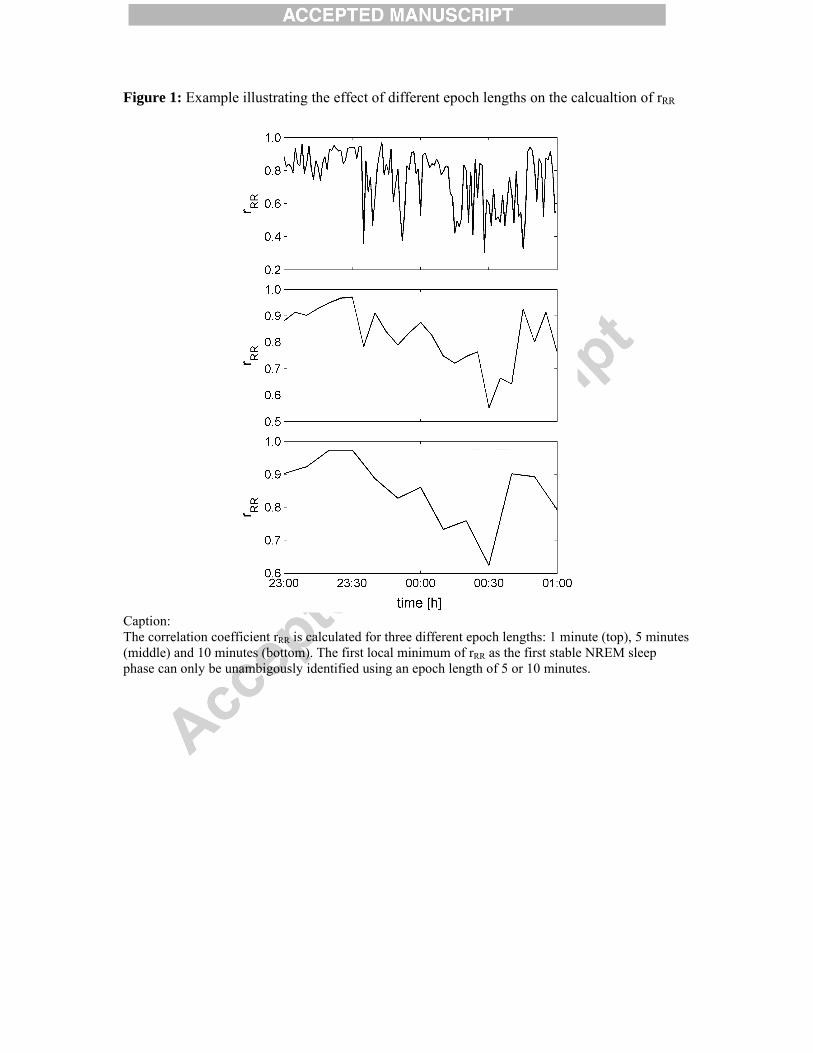
Figure 1: Example illustrating the effect of different epoch lengths on the calcualtion of rRR
Caption: The correlation coefficient rRR is calculated for three different epoch lengths: 1 minute (top), 5 minutes (middle) and 10 minutes (bottom). The first local minimum of rRR as the first stable NREM sleep phase can only be unambigously identified using an epoch length of 5 or 10 minutes.

Figure 2: Gender differences for mean LF/HF ratio
Caption: LF/HF Ratio of high frequency and low frequency according to sex Median difference between sexes = 1.659, SE=0,566; CI 95%: 0,534 – 2,785, main effect sex: p = 0.004 (interaction: time x sex: p =.024) TS Deep sleep L1-L5 Resting times during the day Red Male Blue Female

Highlights
Heart and breathing rate changes (HRR) represent interacting oscillations.
We used HRR determine healthy subjects’ ability to relax during the day.
We compared the day relaxing ability to deep sleep relaxation.
We found that HRR correlates to the perception of health and mental well-being.
When resting day HRR is near to deep sleep relaxation, the subjects felt healthier.