กล องเสียงและหลอดลมอักเสบเฉียบพลัน (Acute laryngotracheobron
Upload
independentCategory
view
1download
0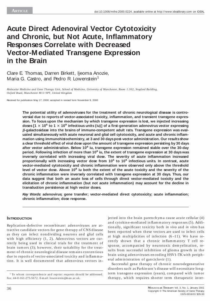
ARTICLE doi:10.1006/mthe.2000.0224, available online at http://www.idealibrary.com on IDEAL
Acute Direct Adenoviral Vector Cytotoxicityand Chronic, but Not Acute, InflammatoryResponses Correlate with DecreasedVector-Mediated Transgene Expressionin the Brain
Clare E. Thomas, Darren Birkett, Ijeoma Anozie,Maria G. Castro, and Pedro R. Lowenstein1
Molecular Medicine and Gene Therapy Unit, School of Medicine, University of Manchester, Room 1.302, Stopford Building,Oxford Road, Manchester M13 9PT, United Kingdom
Received for publication May 17, 2000; accepted in revised form November 9, 2000
The potential utility of adenoviruses for the treatment of chronic neurological disease is contro-versial due to reports of vector-associated toxicity, inflammation, and transient transgene expres-sion. To focus upon the mechanism by which transgene expression is lost, we injected increasingdoses [1 3 106 to 1 3 109 infectious units (iu)] of a first-generation adenovirus vector expressingb-galactosidase into the brains of immune-competent adult rats. Transgene expression was eval-uated simultaneously with acute neuronal and glial cell cytotoxicity, and acute and chronic inflam-mation using immunohistochemistry, at 3 and 30 days post-vector administration. Our results showa clear threshold effect of viral dose upon the amount of transgene expression persisting by 30 daysafter vector administration. Below 108 iu, transgene expression remained stable over the 30-dayperiod. Following infection of more than 108 iu, the extent of transgene expression at 30 days wasinversely correlated with increasing viral dose. The severity of acute inflammation increasedproportionally with increasing vector dose from 106 to 109 infectious units. In contrast, acutevector-mediated cytotoxicity and chronic inflammation were observed only above the thresholdlevel of vector dose. Above 108 iu both the extent of the acute toxicity and the severity of thechronic inflammation were inversely correlated with transgene expression at 30 days. Thus, ourdata suggest that both an acute loss of cells through direct vector-mediated toxicity and theelicitation of chronic inflammation (but not acute inflammation) may account for the decline intransduction persistence at high vector doses.
Key Words: adenovirus; gene transfer; vector-mediated direct cytotoxicity; acute inflammation;chronic inflammation; dose response.
INTRODUCTION
Replication-defective recombinant adenoviruses are at-tractive candidate vectors for gene therapy of CNS diseaseas they can infect nondividing neurons and glial cellswith high efficiency (1, 2). Adenovirus vectors are cur-rently being used in clinical trials for the treatment ofbrain tumors (3); however, their suitability for the treat-ment of chronic neurological disease remains contentiousdue to reports of vector-associated toxicity and inflamma-tion. It is well documented that adenovirus vectors in-
1 To whom correspondence and reprint requests should be addressed.Fax: 44-0-161-275-5672. E-mail: [email protected].
36
jected into the brain parenchyma cause acute cellular (4)and cytokine-mediated inflammatory responses (5). Addi-tionally, significant toxicity both in vivo and in vitro hasbeen reported when these vectors are used to infect cellsat high multiplicities of infection (6–11). We have re-cently shown that a chronic inflammatory T cell re-sponse, accompanied by neurotoxic demyelination, re-sults from successful inhibition of glioma growth in thebrain using adenoviruses encoding HSV1-TK with periph-eral administration of ganciclovir (1).
Successful gene therapy of chronic neurodegenerativedisorders such as Parkinson’s disease will necessitate long-term transgene expression (years), compared with tumortherapy, which requires shorter term therapeutic inter-
MOLECULAR THERAPY Vol. 3, No. 1, January 2001Copyright © The American Society of Gene Therapy
1525-0016/01 $35.00
1
httEc
ARTICLE
C
vention (weeks–months). Previous work from our labora-tory has demonstrated that first-generation adenovirusvectors injected into the CNS of unprimed immune-com-petent animals are able to sustain transgene expressionfor up to 12 months [(2) and unpublished observations],a prolonged period of time compared with first-genera-tion vector-mediated transduction of peripheral organssuch as the liver, in which transgene expression is elimi-nated within 2–3 weeks. Other investigators studying ad-enovirus-mediated transduction of the rat facial nucleus(12), the rat spinal cord (13), and the primate caudatenucleus (14) have demonstrated that persistent transduc-tion cannot be achieved with high doses of vector. Themechanism(s) responsible for the lack of persistence of
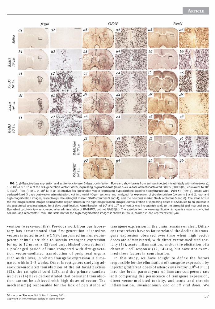
FIG. 1. b-Galactosidase expression and acute toxicity seen 3 days postinfecti3 106–1 3 109 iu of the first-generation vector RAd35, expressing b-galacto
iu (E109) (row f); or 1 3 109 iu of an alternative first-generation vector expperfused-fixed 3 days post-vector administration, cut into serial 40-mm sectio
igh-magnification images, respectively), the astroglial marker GFAP (columnhe low-magnification images delineates the region shown in the high-magnifihe anatomical area transduced by 3 days postinjection. Administration of 10quivalent cytotoxicity was observed after administration of RAdHPRT, but notolumn, and represents 1 mm. The scale bar for the high-magnification imag
MOLECULAR THERAPY Vol. 3, No. 1, January 2001opyright © The American Society of Gene Therapy
transgene expression in the brain remains unclear. Differ-ent researchers have so far correlated the decline in trans-gene expression observed over time when high vectordoses are administered, with direct vector-mediated tox-icity (13), acute inflammation, and/or the elicitation of achronic T cell response (12, 14–16), but have not exam-ined these factors in combination.
In this study, we have sought to define the factorsresponsible for the elimination of transgene expression byinjecting different doses of adenovirus vector (106–109 iu)into the brain parenchyma of immune-competent ratsand comparing the persistence of transgene expression,direct vector-mediated toxicity, and acute and chronicinflammation, simultaneously and at all viral doses. We
Rows a–g show brains from animals injected intrastriatally with saline (row a);se (rows b–e); a dose of heat-inactivated RAd35 [RAd35(In)] equivalent to 109
ing hypoxanthine-guanine ribosyltransferase, RAdHPRT (row g). Brains wereand analyzed for expression of b-galactosidase (columns 1 and 2, low- and
and 4), and the neuronal marker NeuN (columns 5 and 6). The small box inion images. Administration of increasing doses of RAd35 led to an increase ind 109 iu of vector was increasingly toxic to the astroglial and neuronal cells.35(In). The scale bar for the low-magnification images is shown in row a, first
s shown in row a, column 2, and represents 200 mm.
on.sidaressns,
s 3cat
8 anRAdes i
37

pepccBtb1rcc
ddl
i
1
ARTICLE
show that prolonged transgene expression is not achievedafter administration of more than 108 iu of vector. Ourdata suggest that acute inflammatory responses to adeno-virus injection are not responsible for the eventual loss oftransgene expression. Rather, expression is lost followingadministration of high doses of vector, through a combi-nation of direct vector-mediated acute cytotoxicity andthe elicitation of a chronic inflammatory response.
MATERIALS AND METHODS
Adenoviral vectors. RAd35 (expressing b-galactosidase) and RAdHPRT (ex-ressing hypoxanthine-guanine phosphoribosyl transferase) are first-gen-ration replication-defective recombinant adenovirus type 5 vectors ex-ressing transgenes under the transcriptional control of the humanytomegalovirus intermediate early promoter, within the E1 region. Theonstruction of RAd35 and RAdHPRT has been described earlier (17–19).oth vectors were propagated on 293 cells, purified, and titered for infec-ious units (iu) by end-point dilution (20) and for particle concentrationy optical absorbance (21). The ratio of particles to infectious units was3:1 for RAd35 and 8:1 for RAdHPRT. Virus preparations were screened foreplication-competent adenovirus (RCA) contamination by serial amplifi-ation on HeLa cells as described in Dion et al. (22) and for lipopolysac-haride (LPS) contamination, using the Limulus amebocyte gel clot assay
(BioWhittaker, UK). Virus preparations used were free from RCA and LPScontamination. RAd35 was inactivated by incubating 20 ml of purifiedstock in a 500-ml microcentrifuge tube at 56°C for 20 min. No plaques were
etected on 293 cells following titration of inactivated virus by end-pointilution, indicating that the inactivation had been efficient and there were
ess than 102 infectious units per milliliter of virus. Viruses were diluted insterile saline solution for injection.
Animals and surgical procedures. Adult Sprague–Dawley rats of 250 g bodyweight (Charles River, UK) were anesthetized with halothane and placedin a stereotaxic apparatus which was modified for use with inhalationalanesthetic. Animals were injected in the left striatum (0.6 mm forwardfrom bregma and 3.4 mm lateral and 5.0 mm vertical from the dura) with1 3 106, 1 3 107, 1 3 108, or 1 3 109 infectious units of RAd35; 1 3 109
infectious units of RAdHPRT; or a dose of heat-inactivated RAd35[RAd35(In)], equivalent to1 3 109 infectious units. Virus was administeredn a volume of 2 ml using a 10-ml Hamilton syringe and each injection was
performed over a period of 3 min, with the needle being left in place for afurther 5 min before withdrawal. Anesthesia was maintained throughoutsurgery with 1% halothane in 67% medical O2 and 33% medical NO2.Three or thirty days after virus administration, rats were terminally anes-thetized with an intraperitoneal injection of pentobarbitone and transcar-dially perfused-fixed with heparinized Tyrode solution followed by 4%paraformaldehyde in PBS (n 5 4 per virus dose per time point). Brainswere removed, postfixed in 4% paraformaldehyde for 5 h, and stored inPBS containing 0.01% sodium azide.
Immunohistochemistry, TUNEL, and Nissl staining. Serial coronal sections,40-mm-thick, were cut through the whole extent of the striatum using aVibratome. Free-floating immunohistochemistry was performed to detecttransgene or inflammatory and immune cell markers. Endogenous perox-idase was inactivated with 0.3% hydrogen peroxide and sections wereblocked with 10% horse serum (Life Technologies, Paisley, UK) beforebeing incubated overnight with primary antibody diluted in PBS contain-ing 1% horse serum and 0.5% Triton X-100. The primary antibodies andthe dilutions at which they were used were anti-b-galactosidase (1:1000;Promega, Southampton, UK), anti-MHC class I (OX-18, 1:200; Serotec,Kidlington, UK), anti-CD8 (1:500; Serotec),anti-CD4 (1:200; Serotec), ED1(1:1000; Serotec), anti-GFAP (1:200; Roche, UK), NeuN (1:50; AutogenBioclear, UK), and anti-myelin basic protein (MBP, 1:2000; DAKO, Cam-bridge, UK). All primary antibodies were mouse monoclonal anti-rat, ex-cept for anti-MBP, which was rabbit polyclonal anti-human. Secondaryantibodies were biotinylated rabbit anti-mouse or biotinylated swine anti-rabbit (DAKO), diluted 1:200 in 0.5% Triton X-100 with 1% horse serum,and were detected using the Vectastain Elite ABC horseradish peroxidasemethod (Vector, Bretton, UK). After being developed with diaminobenzi-
38
dine and glucose oxidase, sections were mounted on gelatinized glassslides and were dehydrated through graded ethanol solutions and xylenebefore being coverslipped with DPX mountant (Sigma, UK).
TdT-mediated dUTP nick end labeling (TUNEL) was performed on sec-tions mounted on gelatinized glass slides, using the in situ death detectionkit, AP (Roche), according to the manufacturer’s instructions. Histologicalstaining of Nissl substance was performed by incubating slide-mountedsections in 0.1% cresyl violet in 1% acetic acid for 15 min at roomtemperature, before washing in distilled water and differentiating in 70%alcohol for approximately 30 s.
Quantification. Quantitative image analysis to determine the area occu-pied by cells immunohistochemically stained with anti b-galactosidase,ED1, or anti-CD8 antibodies, or to determine the area of loss of GFAPimmunoreactivity within single 40-mm brain sections was performed us-ing a Leica Quantimet 600 Image Analysis System controlled by QWINsoftware (Leica Microsystems, Cambridge, UK) as described previously (2).Brain sections containing the needle track (and thus displaying the high-est levels of immunoreactivity) were used for the quantitative analysis.Student’s t test was used to determine the degree of statistical significancebetween values from different experimental groups.
Neutralizing antibody assay. Titers of adenovirus neutralizing antibodiesin the serum of all animals killed 30 days after the intrastriatal injectionwere measured using an in vitro assay as described in Thomas et al. (2).
RESULTS
Administration of Increasing Doses of First-GenerationAdenovirus Vector to the Rat Striatum Correlateswith Increased Astroglial and NeuronalCytopathogenicity
Administration of increasing doses of RAd35 (1 3 106 to3 109 iu) in a small, fixed volume to the striatum of
adult rats led to an increase in the anatomical area trans-duced at 3 days post-vector injection (Figs. 1 and 2).b-Galactosidase immunoreactivity was seen only close tothe site of the needle injection after administration of 106
iu of vector (Figs. 1b1 and 1b2), but was progressivelymore widely distributed throughout the ipsilateral stria-tum and corpus callosum after injection of 107, 108, and
FIG. 2. Quantification of the area of 40-mm brain sections occupied byb-galactosidase immunoreactivity 3 and 30 days after administration of in-creasing doses of adenovirus vector.
MOLECULAR THERAPY Vol. 3, No. 1, January 2001Copyright © The American Society of Gene Therapy

awo1tavaswj
f
wNNtidpa
(ap
(
i
ARTICLE
C
109 iu (Figs. 1c1, 1c2, 1d1, 1d2, 1e1, 1e2). Transducedcells appeared to be predominantly of astroglial-likemorphology.
Administration of all doses of virus caused astrocyteactivation (GFAP upregulation) throughout the ipsilateralstriatum (Fig. 1, columns 3 and 4). An absence of GFAPimmunoreactivity directly within the needle track wasevident after injection of saline (Figs. 1a3 and 1a4), indi-cating glial cell loss as a result of mechanical injury. Asimilar “lesion” within the needle track was also seen afterinjection of 106 and 107 iu of RAd35 (Fig. 1, columns 3nd 4). The area of glial cell loss increased proportionallyith administration of increasing doses of 108 and 109 iuf RAd35 (Figs. 1d3, 1d4, 1e3, and 1e4). Administration of09 iu of vector caused GFAP-immunoreactive cell losshroughout the ipsilateral striatum and white matter, inn area overlapping with the area transduced by the viralector (compare e3 and e4 with e1 and e2 of Fig. 1). Therea of loss of GFAP immunoreactivity in the ipsilateraltriatum after injection of the different doses of vectoras quantified using image analysis software (Fig. 3). In-
ection of 108 or 109 iu of RAd35 caused a loss of GFAPimmunoreactivity which was significantly greater thanthe loss observed after injection of saline. At doses below108 iu, the “lesion” areas were not significantly differentrom the saline controls.
A similar dose-dependent pattern of neuronal cell lossas reflected in sections stained with the neuronal markereuN (Fig. 1, columns 5 and 6). This loss of GFAP andeuN immunoreactivity possibly reflects cell death
hrough vector-mediated toxicity at high multiplicities ofnfection. Cell death in brains injected with the highestose of vector was confirmed by the profusion of TUNEL-ositive cells detected throughout the ipsilateral striatumnd white matter (Fig, 4).Injection of 109 iu of a different first-generation vector,
RAdHPRT, expressing a potentially less inflammatorytransgene, resulted in a pattern of glial and neuronal cellloss similar to that of RAd35 (Figs. 1g3–1g6), whereas
FIG. 3. Quantification of the area of loss of GFAP immunoreactivity in thentrastriatal injection of increasing adenovirus vectors.
MOLECULAR THERAPY Vol. 3, No. 1, January 2001opyright © The American Society of Gene Therapy
brains injected with the equivalent dose of heat-inacti-vated RAd35 exhibited patterns of GFAP and NeuN im-munoreactivity comparable to the saline control (Figs.1f3–1f6 vs 1a3–1a6). These results indicate that the con-stituents of the vector particles per se appear to be non-toxic; rather the cytopathogenicity is mediated by viral in-fection and/or viral gene expression and it does not seem todepend upon the type of transgene being expressed.
Increased Viral Dose Is Not Correlated with IncreasedTransduction 30 Days after Vector Administration
By 30 days after vector administration, b-galactosidase-immunoreactive cells could still be detected in all animals(Fig. 5). Brains injected with 106 and 107 iu of RAd35Figs. 5a1, 5a2, 5b1, and 5b2) showed no decline in therea of transduction at 30 days postinfection (p.i.) com-ared with 3 days p.i. (Fig. 2). Conversely, b-galactosidase
expression 30 days after injection of 108 iu had declinedfrom 3 days p.i., and a dramatic decrease in transgeneexpression was observed 30 days after injection of 109 iuFigs. 5c1, 5c2, 5d1, 5d2, and 2). Brains injected with 109
iu of vector showed massive ventricular enlargement (par-ticularly on the ipsilateral side) and a large lesion in theipsilateral striatum; however, scattered b-galactosidase-positive cells could still be detected around the lesion(Figs. 5d1 and 5d2).
Acute Inflammation Increases Proportionally withIncreasing Viral Dose from 106 to 109 iu
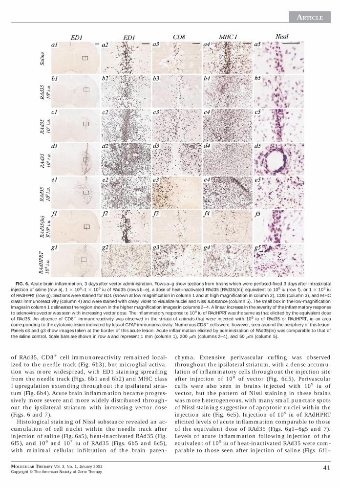
A viral dose-dependent increase in acute brain inflam-mation was seen 3 days after intrastriatal administrationof RAd35 (Figs. 6 and 7). The inflammatory responses tothe injection of saline (Figs. 6a1–6a5) and heat-inacti-vated RAd35 (Figs. 6f1–6f5) were negligible, with micro-glial activation (evidenced by increases in ED1 and MHCclass I immunoreactivity) and CD81 cell immunoreactiv-ity restricted to the needle track. After injection of 106 iu
lateral striatum of 40-mm brain sections, from animals sacrificed 3 days after
ipsi39

Ts
1
ia
ARTICLE
FIG. 4. Cell death seen 3 days after administration of 1 3 109 iu of RAd35. 40-mm sections from brains injected with 1 3 109 iu of RAd35 were evaluated usingUNEL for cell death. TUNEL-positive cells were found throughout the ipsilateral side of the brain, particularly localized to the site of virus administration. Thecale bars for the low- and high-magnification images represent 1 mm and 50 mm, respectively.
FIG. 5. Persistence of b-galactosidase expression, 30 days postinfection. Rows a–d show brains from animals injected with increasing doses of RAd35 from 1 306 to 1 3 109 iu and analyzed for expression of transgene 30 days post-vector administration. Low- and high-magnification images are shown in columns 1
and 2, respectively. b-Galactosidase-immunoreactive cells could be detected in all animals, but those brains injected with 108 or 109 iu of vector showed a declinen transgene expression compared to the 3-day time point, the magnitude of which was correlated with increasing vector dose. Scale bars are shown in row and represent 1 mm and 200 mm for the low- and high-magnification images, respectively.
40 MOLECULAR THERAPY Vol. 3, No. 1, January 2001Copyright © The American Society of Gene Therapy
itfItso(
ci6w
ctla
p
c
ARTICLE
C
of RAd35, CD81 cell immunoreactivity remained local-zed to the needle track (Fig. 6b3), but microglial activa-ion was more widespread, with ED1 staining spreadingrom the needle track (Figs. 6b1 and 6b2) and MHC classupregulation extending throughout the ipsilateral stria-
um (Fig. 6b4). Acute brain inflammation became progres-ively more severe and more widely distributed through-ut the ipsilateral striatum with increasing vector doseFigs. 6 and 7).
Histological staining of Nissl substance revealed an ac-umulation of cell nuclei within the needle track afternjection of saline (Fig. 6a5), heat-inactivated RAd35 (Fig.f5), and 106 and 107 iu of RAd35 (Figs. 6b5 and 6c5),ith minimal cellular infiltration of the brain paren-
FIG. 6. Acute brain inflammation, 3 days after vector administration. Rows ainjection of saline (row a), 1 3 106–1 3 109 iu of RAd35 (rows b–e), a dose oof RAdHPRT (row g). Sections were stained for ED1 (shown at low magnificatioclass I immunoreactivity (column 4) and were stained with cresyl violet to visuaimages in column 1 delineates the region shown in the higher magnification imto adenovirus vector was seen with increasing vector dose. The inflammatory rof RAd35. An absence of CD81 immunoreactivity was observed in the striaorresponding to the cytotoxic lesion indicated by loss of GFAP immunoreactiv
Panels e3 and g3 show images taken at the border of this acute lesion. Acutethe saline control. Scale bars are shown in row a and represent 1 mm (colum
MOLECULAR THERAPY Vol. 3, No. 1, January 2001opyright © The American Society of Gene Therapy
hyma. Extensive perivascular cuffing was observedhroughout the ipsilateral striatum, with a dense accumu-ation of inflammatory cells throughout the injection sitefter injection of 108 of vector (Fig. 6d5). Perivascular
cuffs were also seen in brains injected with 109 iu ofvector, but the pattern of Nissl staining in these brainswas more heterogeneous, with many small punctate spotsof Nissl staining suggestive of apoptotic nuclei within theinjection site (Fig. 6e5). Injection of 109 iu of RAdHPRTelicited levels of acute inflammation comparable to thoseof the equivalent dose of RAd35 (Figs. 6g1–6g5 and 7).Levels of acute inflammation following injection of theequivalent of 109 iu of heat-inactivated RAd35 were com-
arable to those seen after injection of saline (Figs. 6f1–
show sections from brains which were perfused-fixed 3 days after intrastriatalat-inactivated RAd35 [RAd35(In)] equivalent to 109 iu (row f), or 1 3 109 iucolumn 1 and at high magnification in column 2), CD8 (column 3), and MHCnuclei and Nissl substance (column 5). The small box in the low-magnifications in columns 2–4. A linear increase in the severity of the inflammatory responsense to 109 iu of RAdHPRT was the same as that elicited by the equivalent dosef animals that were injected with 109 iu of RAd35 or RAdHPRT, in an areaumerous CD81 cells were, however, seen around the periphery of this lesion.
ammation elicited by administration of RAd35(In) was comparable to that of), 200 mm (columns 2–4), and 50 mm (column 5).
–gf he
n inlizeageespota oity. Ninfln 1
41
io[
ARTICLE
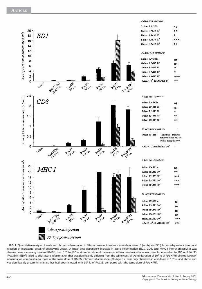
FIG. 7. Quantitative analysis of acute and chronic inflammation in 40-mm brain sections from animals sacrificed 3 (acute) and 30 (chronic) days after intrastriatalnjection of increasing doses of adenovirus vector. A linear dose-dependent increase in acute inflammation (ED1, CD8, and MHC I immunoreactivity) wasbserved over increasing doses of RAd35, from 106 to 109 iu. Administration of the amount of heat-inactivated adenovirus vector equivalent to 109 iu of RAd35RAd35(In) E109] failed to elicit acute inflammation that was significantly different from the saline control. Administration of 109 iu of RAdHPRT elicited levels of
inflammation comparable to those of the same dose of RAd35. Chronic inflammation (30 days p.i.) was only observed at viral doses of 108 iu and above andwas significantly greater in animals that had been injected with 109 iu of RAd35, compared with the same dose of RAdHPRT.
42 MOLECULAR THERAPY Vol. 3, No. 1, January 2001Copyright © The American Society of Gene Therapy

dIojteaC
tiNiciaa
Mt
6 8 7 8
si
ve
tcddadd
ARTICLE
C
6f5 and 7). Thus, these data show that, like vector-medi-ated direct cytotoxicity, vector-induced acute inflamma-tion appears to be independent of the transgene expressedby the vector, but is mediated by the intact virus capsid,virus infection, and/or viral gene expression.
Chronic Inflammation and T Cell Infiltration IsElicited Only above a Threshold Level of Viral Dose
By 30 days after virus administration, brains which hadbeen injected with saline, heat-inactivated RAd35, or 106
or 107 iu of RAd35 showed negligible inflammation (Figs.7 and 8). No CD81 cells could be detected in any of thesebrains (Figs. 8a3, 8b3, 8c3, and 8f3), and microglial/mac-rophage activation (ED1 and MHC I immunoreactivity)was confined to the site of the needle injection (Figs. 8a1,8a2, 8a4, 8b1, 8b2, 8b4, 8c1, 8c2, 8c4, 8f1, 8f2, and 8f4).Nissl staining showed an accumulation of cell nucleiwithin the needle track, but no evidence of cellular infil-tration of other parts of the parenchyma. In contrast,brains that had been injected with 108 iu of RAd35showed more severe inflammation; CD81 cells could be
etected within the injection site and ED1 and MHC classimmunoreactivity was more widely distributed through-ut the ipsilateral striatum (Figs. 8d1–8d5). Animals in-ected with 109 iu of RAd35 showed extensive inflamma-ion throughout the ipsilateral side of the brain andxtending through the contralateral side. Very strong ED1nd MHC class I immunoreactivity and large numbers ofD81 cells could be detected in the ipsilateral striatum,
particularly densely localized around the large lesion re-maining in the site of injection (Figs. 8e1–8e4). Microglialactivation (ED1 and MHC class I staining) and scatteredCD81 cells could also be detected throughout the cortexand corpus callosum on both sides of the brain. TUNEL-positive cells were detected throughout the ipsilateral stri-atum and the cortex and corpus callosum of both sides ofthe brain, i.e., in all regions where CD81 cells were de-ected (not shown). Perivascular cuffs and tightly packednflammatory cells were detected within the lesions byissl staining (Fig. 8e5). A similarly large lesion contain-
ng perivascular cuffs and tightly packed inflammatoryells was also detected within the injection site of brainsnjected with 109 iu of RAdHPRT (Fig. 8g5). These brainslso showed intense microglial/macrophage activationnd CD81 cell infiltration (Figs. 8g1–8g4), although this
was restricted to the ipsilateral striatum and white matterand did not extend into the contralateral side of the brainas seen in those animals injected with the equivalent doseof RAd35. Quantitation of the levels of ED1, CD81, and
HC I immunoreactivity showed that chronic inflamma-ion in brains injected with 109 iu of RAdHPRT was sig-
nificantly less severe than in brains injected with theequivalent dose of RAd35.
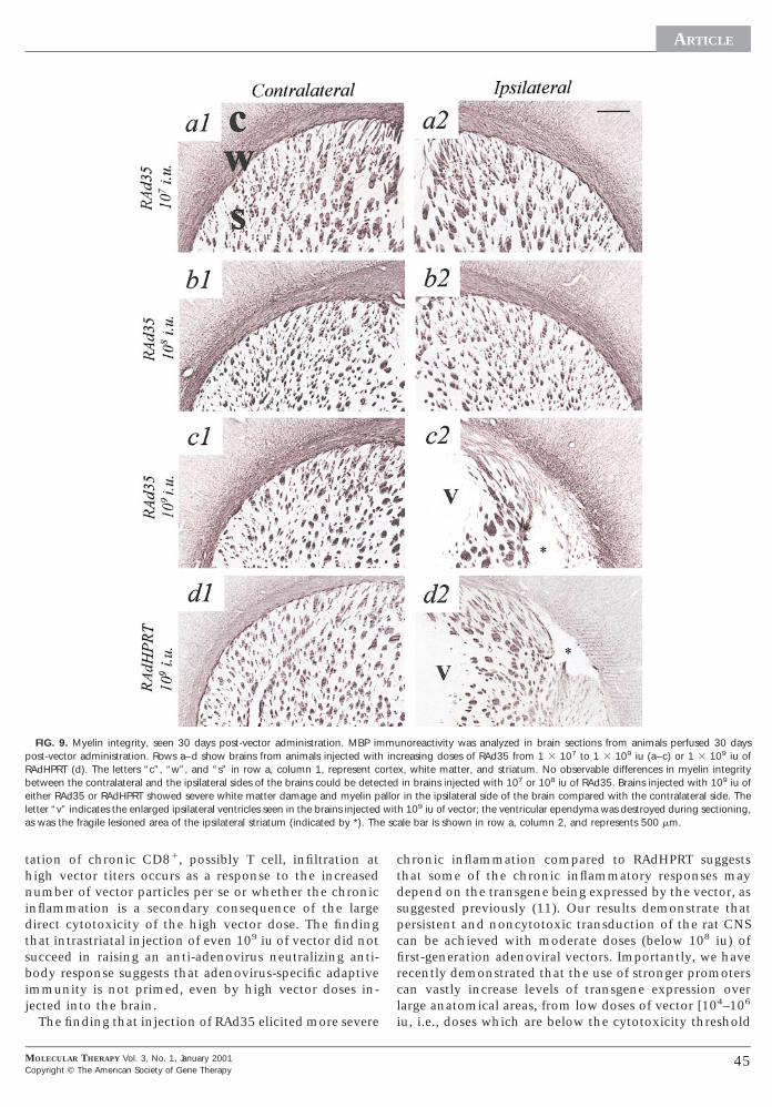
The integrity of brain myelination was evaluated in allanimals by immunohistochemical detection of myelinbasic protein (Fig. 9). Thirty days after vector administra-tion, no damage to myelin integrity could be detected inany of the brains injected with saline, heat-inactivated
MOLECULAR THERAPY Vol. 3, No. 1, January 2001opyright © The American Society of Gene Therapy
RAd35, or 10 –10 iu of RAd35 (10 and 10 doses shownin Figs. 9a1, 9a2, 9b1, and 9b2). In contrast, the architec-ture of the white matter tracts in the ipsilateral side of thebrains injected with 109 iu of either RAd35 or RAdHPRTwas severely disrupted; the white matter fibers appearedpulled apart and paled (suggesting edema), and in somebrains, loss of MBP immunoreactivity was also evidentaround the lesion in the striatum (Figs. 9c2 and 9d2).Consistent with our earlier report of vector-induced neu-rotoxic demyelination following adenovirus-mediated in-hibition of tumor growth in Lewis rats (1), the localiza-tion of the damage observed in this study indicated thatloss of MBP immunoreactivity was also most likely due tovector-mediated cytotoxicity, rather than to autoimmunedemyelination.
To assess whether high doses of adenovirus vector in-jected into the brain could leak to the periphery andprime an anti-adenoviral neutralizing antibody response,we measured titers of virus-neutralizing antibodies in thesera of two of the four animals, per virus dose group, thatwere sacrificed 30 days after the intrastriatal injection.Although we have previously detected high neutralizing-antibody titers in the serum of animals which had re-ceived an intradermal injection of adenovirus (2), no neu-tralizing antibodies could be detected in any of the serumsamples from this study. Thus, intrastriatal injection ofeven extremely high doses of adenovirus (109 iu) does notucceed in priming an adaptive anti-adenovirus neutral-zing antibody response.
DISCUSSION
Although adenoviruses possess numerous characteristicsthat make them attractive candidate vectors for gene ther-apy of neurological disease, their administration to thebrain is associated with inflammation, cytotoxicity, andtransient transgene expression, particularly after injectionof high vector doses. To determine the processes respon-sible for the short-lived transgene expression, we haveinjected increasing adenoviral vector doses into the brainand analyzed in the same anatomical site the effects ofvector dose on direct brain cell toxicity, acute and chronicbrain inflammation, and acute and long-term transgeneexpression. Our results show a clear threshold effect ofviral dose upon the amount of transgene expression per-sisting by 30 days after vector administration. Up to 1 3108 iu, levels of vector-mediated transgene expressionincreased proportionally with increasing viral dose andremained stable over the 30-day period. Above 1 3 108 iu,iral dose was inversely correlated with levels of transgenexpression remaining by 30 days postinfection.This article demonstrates that both the loss of cells
hrough vector-mediated toxicity and the elicitation ofhronic, but not acute, inflammation may account for theecline in transduction persistence seen at high vectoroses. Acute inflammatory responses to vector injectionre unlikely to account for the loss of transduction at 30ays postinfection: administration of increasing vectoroses up to 1 3 108 iu elicited a dose-dependent increase
43

s
cvptDicsv
amitt
3
p
(
ARTICLE
in acute brain inflammation but also resulted in stabletransgene expression over the 30-day period. Direct virus-mediated acute neuronal and glial cell toxicity andchronic inflammation/CD81 cell infiltration were ob-erved only above the threshold dose of 1 3 108 infectious
units. Above this dose both the magnitude of the acutedirect cytotoxicity and the severity of the chronic inflam-mation increased with increasing vector dose.
In support of our conclusions, Lawrence et al. (16) re-ported that administration of the anti-inflammatoryagent dexamethasone immediately before, and for only 8days following, vector delivery had no effect upon theduration of adenovirus-mediated transgene expression inSt. Kitts green monkeys. In rats, Hermens et al. (15)showed that acute dexamethasone administration for 3days post-vector injection prolonged adenovirus-medi-ated transgene expression in astrocytes (but not neurons)
FIG. 8. Chronic inflammation and T cell infiltration, 30 days after adenovira0 days after intrastriatal injection of saline (row a), 1 3 106–1 3 109 iu of RA
iu (row f), or 1 3 109 iu of RAdHPRT (row g). Sections were processed for EDreviously. A threshold of vector titer was observed (around 108 iu) above whic
iu of RAd35 was more severe than that elicited by the equivalent dose of RAdcolumns 2–4), and 50 mm (column 5).
44
of the rat facial nucleus. Interestingly, Hermens et al.observed reduced infiltration of macrophages and ab1 Tells by 14 days after injection of relatively high doses ofector, which may suggest that dexamethasone sup-resses the extent of the chronic inflammation, ratherhan the initial acute inflammatory response. Further,urham et al. (23) reported that administration of the
mmunosuppressant FK506 (which specifically inhibits Tell function) succeeded in prolonging transgene expres-ion after administration of very high doses of adenovirusector to the mouse striatum.Although we demonstrate in this study that both direct
denoviral vector-mediated toxicity and chronic inflam-atory and immune responses contribute to the decline
n vector-mediated transgene expression in the brain,heir relative contributions remain to be determined. Fur-her, it also remains to be determined whether the elici-
tor injection. Rows a–g show sections from brains which were perfused-fixed(rows b–e), a dose of heat-inactivated RAd35 [RAd35(In)] equivalent to 109
CD81, and MHC class I immunoreactivity and stained for Nissl substance ashronic inflammation was observed. The chronic inflammatory response to 109
RT. Scale bars are shown in row a and represent 1 mm (column 1), 200 mm
l vecd351,h cHP
MOLECULAR THERAPY Vol. 3, No. 1, January 2001Copyright © The American Society of Gene Therapy
fircl
ARTICLE
C
tation of chronic CD81, possibly T cell, infiltration athigh vector titers occurs as a response to the increasednumber of vector particles per se or whether the chronicinflammation is a secondary consequence of the largedirect cytotoxicity of the high vector dose. The findingthat intrastriatal injection of even 109 iu of vector did notsucceed in raising an anti-adenovirus neutralizing anti-body response suggests that adenovirus-specific adaptiveimmunity is not primed, even by high vector doses in-jected into the brain.
The finding that injection of RAd35 elicited more severe
FIG. 9. Myelin integrity, seen 30 days post-vector administration. MBP impost-vector administration. Rows a–d show brains from animals injected withRAdHPRT (d). The letters “c”, “w”, and “s” in row a, column 1, represent cbetween the contralateral and the ipsilateral sides of the brains could be deteceither RAd35 or RAdHPRT showed severe white matter damage and myelin pletter “v” indicates the enlarged ipsilateral ventricles seen in the brains injectedas was the fragile lesioned area of the ipsilateral striatum (indicated by *). Th
MOLECULAR THERAPY Vol. 3, No. 1, January 2001opyright © The American Society of Gene Therapy
chronic inflammation compared to RAdHPRT suggeststhat some of the chronic inflammatory responses maydepend on the transgene being expressed by the vector, assuggested previously (11). Our results demonstrate thatpersistent and noncytotoxic transduction of the rat CNScan be achieved with moderate doses (below 108 iu) of
rst-generation adenoviral vectors. Importantly, we haveecently demonstrated that the use of stronger promotersan vastly increase levels of transgene expression overarge anatomical areas, from low doses of vector [104–106
iu, i.e., doses which are below the cytotoxicity threshold
noreactivity was analyzed in brain sections from animals perfused 30 daysreasing doses of RAd35 from 1 3 107 to 1 3 109 iu (a–c) or 1 3 109 iu ofx, white matter, and striatum. No observable differences in myelin integrityin brains injected with 107 or 108 iu of RAd35. Brains injected with 109 iu ofin the ipsilateral side of the brain compared with the contralateral side. The
h 109 iu of vector; the ventricular ependyma was destroyed during sectioning,ale bar is shown in row a, column 2, and represents 500 mm.
muinc
orteted
allorwit
e sc
45
n3
eo
er
gG
dt
tn
si
t2
i
a
cr
kd
(
s
ARTICLE
described in this article (24)]. Secreted transgene productsshould further increase the anatomical area available forgenetic manipulation (25).
New-generation high-capacity adenovirus vectors (HC-Ads), deleted of all viral genes, have been shown to be lesstoxic in the rat liver, allowing higher viral doses to beadministered compared with first-generation vectors (26).We have recently shown that unlike first-generation vec-tors, HC-Ads mediate stable transgene expression in thebrain without eliciting any chronic inflammation, even inthe presence of an anti-adenoviral immune response elic-ited through peripheral infection with adenovirus (2).Thus, novel HC-Ads, combined with powerful transcrip-tional elements, will be able to transduce clinically signif-icant areas of the brain in the absence of deleteriousinflammatory and direct cytotoxic responses.
ACKNOWLEDGMENTS
This work was supported by the Parkinson’s Disease Society, UK (Project Grants3087 and MAP96/28), the Wellcome Trust (Project Grants 051248 and050283), and the EU-Biomed Programme (QLK3-CT-1999-00364 and BMH4-CT98-3277). We are very grateful to Ms. Tricia Maleniak and Miss Emma Jonesfor LPS measurements, Mr. Peter Stanley for technical assistance during thepreparation of RAd35, and Mrs. Ros Poulton for expert secretarial assistance. Wealso acknowledge Professors A. M. Heagerty, F. Creed, and D. Gordon for theircontinuous support and encouragement. We also acknowledge the support whichour laboratory receives from Action Research, the MRC, the British Heart Foun-dation, the Royal Society, the BBSRC (UK), REMEDI, and the Lister Institute forPreventive Medicine. P.R.L. is a Research Fellow of the Lister Institute of Preven-tive Medicine.
REFERENCES
1 Dewey, R. A., et al. (1999). Chronic brain inflammation and persistent herpes simplexvirus 1 thymidine kinase expression in survivors of syngenic glioma treated by adenovirus-mediated gene therapy: Implications for clinical trials. Nat. Med. 5: 1256–1263.
2 Thomas, C. E., Schiedner, G., Kochanek, S., Castro, M. G., and Lowenstein, P. R. (2000).Peripheral infection with adenovirus causes unexpected long-term brain inflammation inanimals injected intracranially with first-generation, but not with high-capacity adenovirusvectors: Towards realistic long-term neurological gene therapy for chronic diseases. Proc.Natl. Acad. Sci. USA 97: 7482–7487.
3 Trask, T. W., et al. (2000). Phase I study of adenoviral delivery of the HSV-tk gene andganciclovir administration in patients with recurrent malignant brain tumors. Mol. Ther. 1:195–203.
4 Byrnes, A. P., Rusby, J. E., Wood, M. J. A., and Charlton, H. M. (1995). Adenovirus genetransfer causes inflammation in the brain. Neuroscience 66: 1015–1024.
5 Cartmell, T., Southgate, T. D., Rees, G. S., Castro, M. G., Lowenstein, P. R., and Luheshi,G. N. (1999). Interleukin-1 mediates a rapid inflammatory response after injection ofadenoviral vectors into the brain. J. Neurosci. 19: 1517–1523.
6 Morral, N., O’Neal, W., Zhou, H., Langston, C., and Beaudet, A. (1997). Immuneresponses to reporter proteins and high viral dose limit duration of expression with adeno-
46
viral vectors: Comparison of E2a wild type and E2a deleted vectors. Hum. Gene Ther. 8:1275–1286.
7 O’Leary, M. T., and Charlton, H. M. (1999). A model for long-term transgene expressionin spinal cord regeneration studies. Gene Ther. 6: 1351–1359.
8 Smith, J. G., et al. (1997). Intracranial administration of adenovirus expressing HSV-YKin combination with ganciclovir produces a dose-dependent, self-limiting inflammatoryresponse. Hum. Gene Ther. 8: 943–954.
9 Brand, K., Klocke, R., Poßling, A., Paul, D., and Strauss, M. (1999). Induction ofapoptosis and G2/M arrest by infection with replication-deficient adenovirus at high mul-tiplicity of infection. Gene Ther. 6: 1054–1063.
10 Easton, R. M., Johnson, E. M., and Creedon, D. J. (1998). Analysis of events leading toeuronal death after infection with E1-deficient adenovirus vectors. Mol. Cell. Neurosci. 11:34–347.11 Cowsill, C., et al. (2000). Central nervous system toxicity of two adenoviral vectors
ncoding variants of the herpes simplex virus type 1 thymidine kinase: Reduced cytotoxicityf a truncated HSV1-TK. Gene Ther. 7: 679–68512 Hermens, W. T. J. M. C., and Verhaagen, J. (1997). Adenoviral vector-mediated gene
xpression in the nervous system of immunocompetent Wistar and T cell-deficient nudeats: Preferential survival of transduced astroglial cells in nude rats. Hum. Gene Ther. 8:
1049–1063.13 Franklin, R. J. M., Quick, M. M., and Haase, G. (1999). Adenoviral vectors for in vivo
ene delivery to oligodendrocytes: Transgene expression and cytopathic consequences.ene Ther. 6: 1360–1367.14 Bohn, M. C., et al. (1999). Adenovirus-mediated transgene expression in nonhuman
primates. Hum. Gene Ther. 10: 1175–1184.15 Hermens, W. T. J. M. C., and Verhaagen, J. (1998). Suppression of inflammation by
examethasone prolongs adenoviral-mediated transgene expression in the facial nucleus ofhe rat. Brain Res. Bull. 47: 133–140.
16 Lawrence, M. S., et al. (1999). Inflammatory responses and their impact on b-galac-osidase transgene expression following adenovirus vector delivery to the primate caudateucleus. Gene Ther. 6: 1368–1379.17 Shering, A. F., et al. (1997). Cell type-specific expression in brain cell cultures from a
hort human cytomegalovirus major intermediate early promoter depends on whether it isnserted into herpesvirus or adenovirus vectors. J. Gen. Virol. 78: 445–459.
18 Southgate, T. D., et al. (2000). Adenoviruses encoding HPRT correct biochemicalabnormalities of HPRT-deficient cells and allow their survival in negative selection medium.Metab. Brain. Dis. 14: 207–222.
19 Wilkinson, G. W. G., and Akrigg, A. (1992). Constitutive and enhanced expression fromhe CMV major IE promoter in a defective adenovirus vector. Nucleic Acids Res. 20: 2233–239.20 Southgate, T. D., Kingston, P., and Castro, M. G. (2000). Gene transfer into neural cells
n vitro using adenoviral vectors. Curr. Protocols Neurosci. 4.23.1–4.23.40.21 Mittereder, N., March, K. L., and Trapnell, B. C. (1996). Evaluation of the concentration
nd bioactivity of adenovirus vectors for gene therapy. J. Virol. 70: 7498–7509.22 Dion, L. D., Fang, J., and Garver, R. I. (1996). Supernatant rescue assay vs. polymerase
hain reaction for detection of wild-type adenovirus-contaminating recombinant adenovi-us stocks. J. Virol. Methods 56: 99–107.
23 Durham, H. D., et al. (1997). The immunosuppressant FK506 prolongs transgeneexpression in brain following adenovirus-mediated gene transfer. NeuroReport 8: 2111–2115.
24 Gerdes, C. A., Castro, M. G., and Lowenstein, P. R. (2000). Strong promoters are theey to highly efficient, noninflammatory and noncytotoxic adenoviral-mediated transgeneelivery in the brain in vivo. Mol. Ther. 2: 330–338.25 Ghosdi, A., Stein, C., Derksen, T., Yang, G., Anderson, R. D., and Davidson, B. L.
1998). Extensive b-glucuronidase activity in murine central nervous system after adenovi-rus-mediated gene transfer to the brain. Hum. Gene Ther. 9: 2331–2340.
26 Morral, N., et al. (1998). High doses of a helper-dependent adenoviral vector yieldupraphysiological levels of a1-antitrypsin with negligible toxicity. Hum. Gene Ther. 9:
2709–2716.
MOLECULAR THERAPY Vol. 3, No. 1, January 2001Copyright © The American Society of Gene Therapy
Copyright © 2022 FDOKUMEN