Activity of the plasma membrane H(+)-ATPase is a key physiological determinant of thermotolerance in...
10
Microbiology (1 994), 140, 1881-1 890 Printed in Great Britain Activity of the plasma membrane H+-ATPase is a key physiological determinant of thermotolerance in Saccharomyces cerevisiae P. J. Coote, M. V. Jones, I. J. Seymour, D. L. Rowe, D. P. Ferdinando, A. J. McArthur and M. 6. Cole Author for correspondence: P. J. Coote. Tel: +44 234 222377. Fax: +44 234 222277. Microbiology and Analytical Sections, Unilever Research, Colwort h Laboratory, Sharnbrook, Bedford MK44 lLQ, UK The role of membrane integrity and the membrane ATPase in the mechanism of thermotolerance in Saccharomyces cerevisiae was investigated. The resistance to lethal heat of a mutant strain with reduced expression of the membrane ATPase was significantly less than that of the wild-type parent. However, prior exposure to sub-lethal temperatures resulted in the induction of similar levels of thermotolerance in the mutant compared to the parent strain, suggesting that the mechanism of sub-lethal heat-induced thermotolerance is independent of ATPase activity. Supporting this, exposure to sub-lethal heat stress did not result in increased levels of glucose-induced acid efflux at lethal temperatures and there was little correlation between levels of acid efflux and levels of heat resistance. ATPase activity in crude membrane preparations from sub-lethally heat-stressed cells was similar to that in preparations from unstressed cells. Study of net acid flux during heating revealed that pre-stressed cells were able to protect the proton gradient for longer. This may confer an 'advantage' to these cells that results in increased thermotolerance. This was supported by the observation that prior exposure to sub-lethal heat resulted in a transient protection against the large increase in membrane permeability that occurs at lethal temperatures. However, no protection against the large drop in intracellular pH was detected. Sub-lethal heat-induced protection of membrane integrity also occurred to the same extent in the reduced-expression membrane ATPase mutant, further implying that the mechanism of induced thermotolerance is independent of ATPase activity. To conclude, although the membrane ATPase is essential for basal heat resistance, thermotolerance induced by prior exposure to stress is largely conferred by a mechanism that is independent of the enzyme. Keywords : Saccharomy-es cerevisiae, t hermotolerance, plasma membrane H'- A TPase, membrane permeability INTRODUCTION The stress response in J'acdvzromjces ceerevisiae is charac- terized by the acquisition of increased thermotolerance and the expression and synthesis of heat-shock proteins (HSPs) (Lindquist & Craig, 1988). The precise role of the HSPs in thermotolerance remains unclear. There is considerable evidence to suggest that HSPs are not involved in the major mechanism allowing cells to become Abbreviations: DES, diethylstilboestrol; HSP, heat-shock protein; pH,, intracellular pH. more thermotolerant (Susek & Lindquist, 1989; Smith & Yaffe, 1991; Barnes et al., 1990). The principal role of HSPs may be to act as chaperones to aid in the sequestration and removal of damaged or improperly folded proteins (Schlesinger, 1990 ; Sanchez & Lindquist, 1990). In this respect, HSPs may be essential for cellular recovery from stress damage, but they are probably not the principal factor allowing induction of a rapid re- sistance to lethal stress. Other physiological factors that may confer thermotolerance include trehalose (Wiemken, 1990), unfreezable cell water (Komatsu et a/., 1991), Go cell cycle arrest (Plesset et a/., 1987), CAMP-independent 0001-8797 0 1994 SGM 1881
-
Upload
independent -
Category
Documents
-
view
1 -
download
0
Transcript of Activity of the plasma membrane H(+)-ATPase is a key physiological determinant of thermotolerance in...

Microbiology (1 994), 140, 1881-1 890 Printed in Great Britain
Activity of the plasma membrane H+-ATPase is a key physiological determinant of thermotolerance in Saccharomyces cerevisiae
P. J. Coote, M. V. Jones, I . J. Seymour, D. L. Rowe, D. P. Ferdinando, A. J. McArthur and M. 6. Cole
Author for correspondence: P. J. Coote. Tel: +44 234 222377. Fax: +44 234 222277.
Microbiology and Analytical Sections, Un ilever Research, Colwort h Laboratory, Sharnbrook, Bedford MK44 lLQ, UK
The role of membrane integrity and the membrane ATPase in the mechanism of thermotolerance in Saccharomyces cerevisiae was investigated. The resistance to lethal heat of a mutant strain with reduced expression of the membrane ATPase was significantly less than that of the wild-type parent. However, prior exposure to sub-lethal temperatures resulted in the induction of similar levels of thermotolerance in the mutant compared to the parent strain, suggesting that the mechanism of sub-lethal heat-induced thermotolerance is independent of ATPase activity. Supporting this, exposure to sub-lethal heat stress did not result in increased levels of glucose-induced acid efflux at lethal temperatures and there was l i t t le correlation between levels of acid efflux and levels of heat resistance. ATPase activity in crude membrane preparations from sub-lethally heat-stressed cells was similar to that in preparations from unstressed cells. Study of net acid flux during heating revealed that pre-stressed cells were able to protect the proton gradient for longer. This may confer an 'advantage' to these cells that results in increased thermotolerance. This was supported by the observation that prior exposure to sub-lethal heat resulted in a transient protection against the large increase in membrane permeability that occurs at lethal temperatures. However, no protection against the large drop in intracellular pH was detected. Sub-lethal heat-induced protection of membrane integrity also occurred to the same extent in the reduced-expression membrane ATPase mutant, further implying that the mechanism of induced thermotolerance is independent of ATPase activity. To conclude, although the membrane ATPase is essential for basal heat resistance, thermotolerance induced by prior exposure to stress is largely conferred by a mechanism that is independent of the enzyme.
Keywords : Saccharomy-es cerevisiae, t hermo tolerance, plasma membrane H'- A TPase, membrane permeability
INTRODUCTION
The stress response in J'acdvzromjces ceerevisiae is charac- terized by the acquisition of increased thermotolerance and the expression and synthesis of heat-shock proteins (HSPs) (Lindquist & Craig, 1988). The precise role of the HSPs in thermotolerance remains unclear. There is considerable evidence to suggest that HSPs are not involved in the major mechanism allowing cells to become
Abbreviations: DES, diethylstilboestrol; HSP, heat-shock protein; pH,, intracellular pH.
more thermotolerant (Susek & Lindquist, 1989; Smith & Yaffe, 1991; Barnes e t al., 1990). The principal role of HSPs may be to act as chaperones to aid in the sequestration and removal of damaged or improperly folded proteins (Schlesinger, 1990 ; Sanchez & Lindquist, 1990). In this respect, HSPs may be essential for cellular recovery from stress damage, but they are probably not the principal factor allowing induction of a rapid re- sistance to lethal stress. Other physiological factors that may confer thermotolerance include trehalose (Wiemken, 1990), unfreezable cell water (Komatsu e t a/., 1991), Go cell cycle arrest (Plesset e t a/., 1987), CAMP-independent
0001-8797 0 1994 SGM 1881

P. 1. COOTE a n d O T H E R S
protein phosphorylation (Coote e t al., 1992), and thc membrane-bound ATPase (Coote e t al., 1991 ; Panaretou & Piper, 1990).
The proton-pumping ATPase in the plasma membrane of J'. wwisiae couples ATP hydrolysis, consuming up to 509G of total cellular ATP, to the expulsion of protons, thus generating a proton gradient (Serrano, 1984). The resulting transmembrane electrochemical gradient is used for active nutrient uptake (Eddy, 1982) and regulates the activity of some pH-sensitive intracellular enzymes (Serrano, 1984). In addition, the membrane ATPase maintains the intracellular pH (pH,) of S. cerevisiae between 6.0 and 7.0 even when large variations in extracellular pH occur (Malpartida & Serrano, 1981). Indeed, growth on acid media results in an increase in ATPase activity (Eraso & Gancedo, 1987), which has been shown to be due to increased catalytic activity rather than increased amount of the enzyme (Eraso e t al., 1987). The enzyme is a major component of the plasma membrane and also seems to have a significant role in maintaining the structural integrity of the cell, as evidenced by the altered cell morphology present in some ATPase mutants (McCusker e t a/., 1987). Significantly, the ATPase has been shown to be essential for growth and proton pumping in vivo because a null mutation in haploid cells is lethal (Serrano e t a/., 1986).
Since a minor intracellular acidification due to sub-lethal stress has been implicated as a triggering mechanism inducing increased thermotolerance (Coote e t al., 1991 ; Weitzel e t al., 1987), the activity of the ATPase in regulating pH, homeostasis may be important in main- taining viability during lethal stress. Indeed, experiments using the membrane- ATPase inhibitor diethylstilboestrol (DES) (Coote e t al., 1991) or reduced-activity ATPase mutants (Panaretou & Piper, 1990) appear to support this hypothesis. In addition, in vitro generation of random mutations in the ATPase gene (PMAI) and subsequent selection for thermosensitive growth resulted in cells with more thermolabile ATPases (Cid & Serrano, 1988). Strains harbouring these mutations were very slow- growing at 37 "C, and glucose-induced proton efflux after incubation at 37 "C was drastically reduced. These effects were believed to be due to the thermolabile ATPases. Interestingly, if both the wild-type and mutant strains were exposed to a sub-lethal temperature for 2 h prior to harvesting, the reduction in proton pumping at 37 "C was less severe ; evidence, perhaps, of stress-induced acti- vation, or protection, of the ATPase. Further evidence indicating a possible role for the membrane-ATPase in stress-resistance comes from the observation that the PM.41 gene is one of the few genes in S. cerevisiae still effectively transcribed after sub-lethal heat stress at 42 "C (Curran e t a/., 1988). Clearly, there is sufficient indirect evidence to suggest a role for the membrane ATPase in the stress-response, but experimental evidence for the precise physiological function of the enzyme during stress is lacking. In addition, there is little definitive evidence to show whether the activity of the enzyme confers, either by direct or indirect action, enhanced ability to survive lethal heating. Use of the membrane ATPase inhibitor DES has
provided some evidence of the importance to the cell of the enzyme during stress. However, because enzyme inhibitors often have other unquantifiable secondary effects (Goffeau & Slayman, 1981), by taking a genetic approach a more specific manipulation of ATPase activity can be achieved.
A reduced-expression ATPase mutant @ma 1-205) was employed in this study. Mutant strain pmal-205 was constructed by deletion of a small T-rich region followed by insertion of foreign DNA (containing LEU2) into the upstream promoter sequences of P M A I cloned into a plasmid (Vallejo & Serrano, 1989). Study of this mutant revealed a 72% decrease in levels of ATPase mRNA compared to the wild-type and an 85 O h decrease in levels of ATPase protein. However, the activity of the enzyme was less affected because proton pumping was only reduced by approximately two-thirds (Vallejo & Serrano, 1989).
In the present work, evidence was sought for a role of the membrane-ATPase in the stress-protective mechanism of S. cerevisiae. Acid efflux and thermotolerance experiments were carried out on a wild-type parent and an ATPase mutant strain, followed by more detailed studies of net proton flux during heat stress. In addition, 31P-NMR and fluorescence microscopy were used to measure, respect- ively, pH, shifts and membrane permeability changes during temperature stress. Finally, specific membrane ATPase activity was measured to examine any stress- induced physiological changes to the enzyme.
METHODS
Organisms and growth conditions. Three strains of S. cerevisiae were employed: CMC 3236 (a wild-type control); strain RS-514 (PMA I), the parent strain with fully functional ATPase ; and strain RS-516 @ma 1-205), the reduced-expression ATPase mutant (Vallejo & Serrano, 1989). RS-514 and RS-516 were kindly provided by Professor Ramon Serrano, Departamento de Biotecnologia, Universidad Politecnica, Valencia, Spain. Cells were grown, with shaking, to late-exponential phase at 30 "C in YEPD (3%, w/v, glucose; YO, w/v, yeast extract; 1 YO, w/v, Bacto-peptone). Strains RS-514 and RS-516 were maintained on galactose plates (Vallejo & Serrano, 1989). Measurement of thermotolerance. Thermotolerance was de- termined as described by Coote e t al. (1992). Because of their differing heat resistance, strain 3236 was heated to 52 "C, and strains P M A I and pma 1-205 to 53 OC. Measurement of acid efflux and net proton flux. Glucose- induced acid efflux has been shown by two methods to be an accurate reflection of membrane ATPase activity in vivo (Serrano, 1980 ; Cid e t al., 1987). Cells were prepared as described by Coote e t al. (1991) except the final cell density was between 40 and 50 mg wet weight m1-l. A Corning temperature probe allowed measurement of the cell suspension pH over a wide range of temperatures. Glucose-induced acid efflux was determined at 25 "C after 30 min incubation at 25,30,35,40,45 and 53 OC and also after 0, 5, 10, 15 and 20 min incubation at 53 "C alone.
To measure net proton flux during heating, cells were prepared as before. Populations were incubated with or without the presence of glucose (10 g 1-') for 1 h prior to measurement to study the effect of an energy source on net proton flux. The change in external pH of the cell suspension (net proton flux)
1882
Membrane-ATPase and yeast thermotolerance
was determined after 5 min heating at 25,30, 35,40,45 or 52 "C from cell populations before and after a sub-lethal incubation (42 "C for 30 min). Net proton flux was also determined at the lethal temperature of 52 "C over a time course of 15 min in both unstressed control cells and cells exposed to a sub-lethal incubation.
Measurement of pH, by 31P-NMR. Late-exponential-phase cells were harvested, washed and resuspended in a medium based on that of Nicolay etal . (1982) consisting of 2.5 mM MgCl,, 10 mM KC1,2*4 mM CaCl,, 25 mM glucose, 20 % (v/v) D,O (to act as a frequency lock for the NMR spectrometer), in citrate buffer (50 mM citric acid, 50 mM sodium citrate) a t pH 4.5. Cell pellet volume was approximately 40 YO of the total sample volume and the samples were kept on ice until they were used. 31P-NMR spectra were obtained at 121.497 MHz on a Bruker MSL-300 NMR spectrometer using a 10 mm VSP multi-nuclear probe. The spectra were acquired every 15 min in blocks of 1000 scans at a pulse angle of 60" with a repetition time of 1 s. The chemical shift of the intracellular inorganic phosphate (Pi) was measured relative to an unshifting capillary standard of 50 mM MDP (methylene diphosphonic acid), which had a chemical shift of 0 p.p.m. relative to 85 YO phosphoric acid. The Pi peak was identified using the assignments of Navon e t al. (1979), Den Hollander e t al. (1981) and Campbell-Burk & Shulman (1987). Spectra were accumulated from the cell suspensions while being heated at a constant temperature by a Bruker VT-unit in a 10 mm sample tube within the NMR magnet. Control popu- lations were heated at 25 "C, sub-lethally heated populations at 42 "C, and lethally heated populations at 52 O C . After heating, the 31P-NMR chemical shifts of the intracellular P, resonance were determined, and the intracellular pH calculated from NMR titration curves. These were obtained by titrating a sample of 10 mM KH,PO, and 100 mM KC1 closely resembling the intracellular ionic concentration (Roberts e t al., 1981). Separate titration curves were obtained for each temperature at which pHi was measured.
Measurement of membrane permeability. Membrane per- meability during heating was measured using the fluorescent nuclear stain ethidium bromide (Arkhammar e t a/., 1990). Mid- exponential-phase cells were harvested, washed and re- suspended in 25 mM MES (pH 5.5). Individual aliquots were then heated in a modified thermocouple block calibrator DB- 40L (Techne, Cambridge) in the presence of ethidium bromide (10 pg m1-l) at 25,42 or 53 "C, and at 53 "C after prior sub-lethal incubation at 42 "C for 30 min. At various time points during heating a 5 p1 aliquot of the cell sample was dispensed onto a clean microscope slide. The cells were visualized using a Bio- Rad MRC confocal scanning laser microscope (CSLM) fitted with a 20 mW Krypton Argon mixed gas laser (Bio-Rad) and an objective magnification of x 60 (Nikon ~ 6 0 1.4 numerical aperture, Plan Apo objective). Simultaneous images were acquired using dual-channel collection mode. The first channel was a fluorescent image of bound intracellular ethidium bromide (excitation line 568 nm); the second channel was a transmitted illumination phase-contrast image. Each image was averaged over 12 frames to reduce background noise. To measure permeability of ethidium bromide, the average cellular fluorescence was determined by locating a 10 x 10 pixel frame over each cell in the image and expressing intracellular fluorescence as pixel intensity. For each time point at least 20 individual cells were assayed from two independent experi- ments.
Membrane preparation. Late-exponential-phase cells were harvested, washed and resuspended in 25 mM MES (pH 6.0) with 50 mM glucose to a final concentration of 30-150 mg wet
weight ml-'. Samples were either incubated at 25 "C for 30 rnin as a control or sub-lethally heated at 42 "C for 30 min. Concentrated solutions of Tris/HCl (pH 7-0), EDTA, dithio- threitol and phenylmethylsulphonyl fluoride (PMSF) (Sigma) were added to 100, 5, 2 and 1 mM, respectively, and the cell samples frozen rapidly in liquid nitrogen and stored at - 85 "C. Crude membranes were prepared by a method based on that of Serrano (1978). Samples were thawed and all subsequent procedures carried out at 0-4 "C. Aliquots (2 ml) were then vortexed eight times for 1 min with 1.5 ml 0.5 mm glass beads (Sigma) with 1 min intervals on ice. The lysate was then diluted fivefold with 6 % (w/v) sorbitol, 0.1 M Tris/HCl (pH 7*5), 5 mM EDTA, 2 mM dithiothreitol and pepstatin A (final concentration 2 pg ml-l). Glass beads, intact cells and fragments of cell walls were removed by centrifugation at 3000 r.p.m. at 4 "C for 3 min. The supernatant was then centrifuged at 65 000 g at 4 "C for 30 min. The supernatant was discarded and the crude membrane pellet resuspended in 20 Y (v/v) glycerol, 10 mM Tricine (pH 7.5), 0.1 mM EDTA and 0.1 mM dithiothreitol and stored at - 85 "C.
Protein determination. The amount of protein in the crude membrane preparation was determined by the Bio-Rad protein assay procedure using BSA as standard. Both the standard and microassay procedures were as described in the manufacturer's instructions.
ATPase assay. ATPase activity was assayed in crude membrane fractions by following the release of Pi from ATP as described by Serrano (1983). The assay was carried out in a medium containing 50 mM MES/Tris (pH 6.0), 10 mM MgSO,, 50 mM KC1,5 mM sodium azide (to inhibit mitochondria1 ATPase) and 50 mM KNO, (to inhibit vacuolar ATPase).
Reproducibility of results. Unless otherwise stated all experi- ments were carried out at least twice and representative results are shown.
100
10 cn
.- 5 5 v)
m w C Q,
Q, Q ?
0.1
0.01 1 , , , , , I , I , , 2 4 6 8 1 0
Time (min)
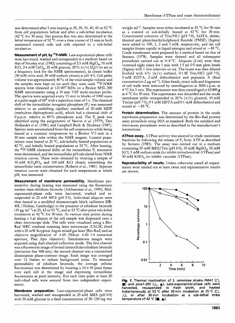
Fig. 1. Thermal inactivation of 5. cerevisiae strains PMAl (0, W) and p a l - 2 0 5 (A, A). Late-exponential-phase cells were harvested, resuspended in fresh broth, and heated instantaneously at 53 "C after 30 min incubation at 25 "C (0, A), or after 30min incubation at a sub-lethal stress temperature of 42 "C (W, A).
1883

P. J. COOTE a n d OTHERS
- -
- -
-
-
-
-
-
-
RESULTS
Effect of reduced expression of the ATPase on thermotolerance and acid efflux
2000
1600 .- B L 2
1200 -5 2 2
800 *&
2 400 i?
m W
m w
Q,
- 0
The thermal inactivation of the reduced-expression ATPase mutant was compared with the parental strain. After heating at 53 "C for 9 min the parental strain ( P M A I ) showed over a 10-fold loss in initial viable numbers (Fig. 1). However, cells having a prior sub-lethal incubation at 42 "C for 30 min showed less than a fivefclld reduction in starting numbers after 9 min at 53 "C. In contrast, pma 1-205, the mutant with reduced expression of the ATPase gene, showed over a 1000-fold loss in initial viable numbers after heating at 53 O C for 9 min, but less than a 10-fold loss after a pre-incubation at 42 "C for 30 min followed by 9 min at 53 "C (Fig. 1). Its basal thermotolerance is therefore much lower.
As the pre-incubation temperature was increased from 2!5 to 40 O C there was no change in glucose-induced acid efflux from the P M A 1 parent but over a 600 % increase in survivors after subsequent heating at 53 "C for 6 min (Fig. 2). In contrast, initial acid efflux rates from the mutant were approximately 30 YO less than in the P M A I parent and subsequently decreased further as the in- cubation temperature was increased from 25 to 45 OC:.
I I ' I I I I I
20 25 30 35 40 45 50 55 Temperature ("C)
............................................................................................................................... . ............................ Fig, 2. Effect of prior incubation temperature (30 rnin in sterile distilled water) on acid efflux and viability in the reduced- expression ATPase mutant pmal-205 and the parent strain of PMAl 5. cerevisiae. Following addition of glucose (10 g I-'), acid efflux rate was measured at 25°C from PMAl (0) and pmal- 205 (A) (error bars indicate the standard deviation of three experiments) and the induction of increased thermotolerance in PMAl (M) and pmal-205 (A). Percentage increase in survivors indicates the percentage increase in cell numbers due to sub-lethal incubation followed by heating a t 53 "C for 6 min, the number of survivors after incubation a t 25°C for 30min followed by heating a t 53 "C for 6 min being taken as 0 %.
1 I 1 I,,,, -2 t 5 10 15 20
Time (min)
Fig. 3. Effect of prior sub-lethal incubation (42 "C for 30 min) (filled symbols) compared to control incubation (25 "C for 30 min) (hollow symbols) on viable numbers and the rate of glucose-induced (10 g I-') acid efflux from 5. cerevisiae 3236 after 0, 5, 10, 15 and 20 min heating a t 52 "C. a, A, Viable cell numbers, expressed as percentage survivors, the number surviving after 0 min heating being taken as 100%; 0, m, acid efflux.
However, pre-incubation at the same temperatures in- duced over a 1900 YO increase in survivors after heating at 53 "C for 6 min. Incubation at 50 "C for 30 min resulted in significant reductions in acid efflux rates from both strains and also a corresponding decrease in induced thermo- tolerance (Fig. 2).
Effect of sub-lethal heat stress on thermotolerance and glucose-induced acid efflux
Measurement of glucose-induced acid efflux at a lethal stress temperature of 52 "C revealed a transient increase in acid efflux rates from control cells of approximately threefold after heating for 5 min (Fig. 3). Subsequently, acid efflux declined to below starting levels after 10 min and virtually ceased after 20 min at 52 "C. In contrast, glucose-induced acid efflux from a population exposed to a prior sub-lethal incubation (42 O C for 30 min) showed no peak : it declined after 5 min at 52 "C and continued to decline, reaching virtually zero after 20 min. Viable numbers remained unchanged after 15 min at 52 "C for both populations. However, after 20 min at 52 "C control cells had decreased over 10 000-fold compared to initial levels, but cells exposed to a prior sub-lethal incubation less than 1000-fold (Fig. 3).
Effect of sub-lethal heat stress on the activity of the plasma-membrane ATPase
Measurement of ATPase activity at 30 "C in crude membrane preparations from control cells (incubated at 25 "C for 30 min) and from sub-lethally heated cells

Membrane-ATPase and yeast thermotolerance
3*0 r I-
2 4 6 8 1 0
0
Time (min)
Fig. 4. Effect of sub-lethal heat on membrane ATPase activity of 5. cerevisiae 3236. Crude membrane preparations were made from cells incubated at 25 "C for 30 min (0) and from cells sub- lethally heated a t 42 "C for 30 rnin (A). Rate of ATPase activity was determined at 30 "C after exposure to 52 "C for 0, 0.5, 1, 2 and 10 min. The rate was measured over the first 2 min of the assay for each time point.
0*50 1 T
u
- 1 -00
- 1 *25
1
20 25 30 35 40 45 50 55 Temperature ("C)
Fig. 5. Effect of increasing temperature on the rate of net acid flux from cell suspensions (40 mg wet weight ml-' in distilled water) of: unstressed control (25 "C, 30 min) 5. cerevisiae PMA1 (0) and pmal-205 (A), and sub-lethally stressed (42 "C, 30 min) PMAl (m) and pmal-205 (A) expressed as net proton efflux (positive values)/influx (negative values). The mean values of two experiments are shown; the error bars indicate the minimum and maximum values obtained.
(incubated at 42 "C for 30 min) revealed little difference in overall activity between the two preparations over a 10 min period (results not shown). To investigate whether sub-lethal incubation conferred any protection, or thermal
stability, on the ATPase, crude membrane preparations were prepared from control and sub-lethally heated cells and ATPase activity determined at 30 "C after exposure to 52 "C for up to 10 min (Fig. 4). In both membrane preparations the rate of ATPase activity had increased approximately fourfold after the first 2 min of exposure to 52 "C (Fig. 4). After this initial rise in activity, there was no further increase during the remaining period at 52 "C. Most significantly, there was no indication of a higher rate of activity in the membranes exposed to a prior sub-lethal incubation (Fig. 4).
Effect of sub-lethal heat stress, glucose and reduced expression of the membrane ATPase on net acid flux during heating
The effect of increasing temperature on the net acid flux from control and sub-lethally heated populations of P M A 1 and pmal-205 is shown in Fig. 5. In unstressed control populations of both wild-type and mutant cells, the rate of net acid influx increased from 0.2 nmol min-' mg-' at 30 "C to a maximum of approximately 0-6 nmol min-' mg-' at 40 "C (Fig. 5). Significantly, there was little difference in the rates of acid influx between wild-type and mutant cells. In contrast, sub-lethally heated populations (42 "C for 30 min) of both wild-type and mutant cells maintained a small net acid efflux (approxi- mately 0.2 nmol min-' mg-') when the temperature was raised from 25 to 35 "C. However, at 40 "C there was a net balance between acid efflux and influx (no change in external pH) and above this temperature the net rate of acid influx increased sharply until at 52 "C the rate was the same as in control cells (Fig. 5). Again, there was little significant difference between wild-type and mutant cells.
The experiment was repeated on energized cells pre- incubated with glucose (10 g 1-') for 1 h prior to measure- ment. Similar to the above results, sub-lethally heated populations of both wild-type and mutant cells prevented net acid influx at higher temperatures than control cells (results not shown). As before, at temperatures above 40 "C the rate of acid influx increased rapidly until it was similar to that in control cells. Significantly, pre- incubation with glucose resulted in a threefold increase, compared to cells incubated without glucose, in the rate of acid influx at the highest incubation temperature of 52 "C (results not shown). Again, in all cases there was little difference between wild-type and mutant cells.
Effect of sub-lethal heat stress, glucose and reduced expression of the membrane ATPase on net acid flux and cell viability at 52 "C
The previous results indicated that sub-lethal incubation may protect the cell from the large proton influx that occurs at higher temperatures. To study this further, net acid flux was measured at 52 "C in both control and sub- lethally heated populations of the wild-type and the reduced-expression ATPase mutant. The viability of the individual populations was measured simultaneously to determine any correlations between changes in net acid- flux and heat resistance.
~-
1885

P. J . COOTE a n d O T H E R S
+ + I0
-3 I 1 10-4
+ + I0
0 3 6 9 1 2 1 5
-3 t I 110-4
0 3 6 9 1 2 1 5 Time (min)
+ + I0 5 s EC2 Y
+ + I0 a 2 E2 Y
1
0
-1
-2
-3 t \ \ 8
h 110-4
-4 1 (b) I I 10-5
0 3 6 9 1 2 1 5
0 3 6 9 1 2 1 5 Time (min)
Fig. 6. Effect of prior sub-lethal incubation (42 "C, 30 min) (filled symbols) compared to unstressed control (25 "C, 30 min) (hollow symbols) on viable numbers (triangles) and the rate of change of the extracellular pH (squares) in cell suspensions (40 mg wet weight ml-l) of S. cerevisiae during heating a t 53 "C. (a) PMAl and (b) p a l - 2 0 5 in distilled water alone; (c) PMA7 and (d) prna7-205 in distilled water with 10 g glucose I-'. Acid efflux was expressed as proton efflux (positive values)/influx (negative values). Error bars indicate the standard deviation of two experiments. Viable numbers were expressed as percentage survivors, the number surviving after 0 min heating being taken as 100%. Arrows indicate that the cell numbers were less than the points indicated on the graph.
In unstressed control populations of wild-type and mutant cells there was an initial rapid increase in the rate of acid influx within the first 2 min of exposure to 52 O C (Fig. 6). As before, the increase in rate of influx was far greater in cells pre-incubated with glucose (Fig. 6c, d). However, after the first 2 min of exposure the rate of acid influx in both unenergized cells and cells pre-incubated with glucose slowed and had returned to near starting levels after 15 min (Fig. 6). There was only a minor indication that the rate of acid influx was greater in the mutant strain compared to the wild-type when measured under the same conditions (Fig. 6). In contrast, prior exposure of both
1886
wild-type and mutant populations to sub-lethal stress (42 O C for 30 min) before heating at 52 "C generally resulted in a reduction in the rate of acid influx, particularly during the first 2 min of exposure, compared to control populations (Fig. 6).
Simultaneous measurement of cell viability during heating at 52 O C revealed significant increases in thermotolerance in the sub-lethally heated populations of both P M A I and pmal-205 when compared to control cells (Fig. 6). This was particularly evident in the cells pre-incubated with glucose (Fig. 6c, d). Despite this, after heating at 52 "C for

Membrane-ATPase and yeast thermotolerance --
7.0
6.8
6.6 .- I Q
6.4
6.2
250
225
t v
100
T
I I I I I I I I I I I 6.0
15 30 45 60 75 5 10 15 20 25 Time (min) Time (rnin)
............... . ...................................................................................................................................................... I
Fig. 7. Effect of heat on (a) pH, (measured by 31P-NMR) and (b) membrane permeability (measured by fluorescence microscopy) of 5. cerevisiae 3236. pH, and membrane permeability were measured a t 25 "C (O), 42 "C (m) and 52 "C (A), and a t 52 "C after a prior sub-lethal incubation a t 42 "C for 30 rnin (A). Due to deterioration of the spectra after 15 min at 52°C it was not possible to make definite peak assignments and thus determine pH,. The mean values of four experiments are shown; the error bars indicate the minimum and maximum values obtained.
between 10 and 15 min, when optimum levels of increased thermotolerance were evident, there was little evidence of a corresponding decrease in the rate of net acid influx from pre-stressed cells (Fig. 6).
In both P M A 1 andpmal-205, after a certain heating time, numbers of unstressed survivors rapidly dropped to undetectable levels long before the pre-stressed popu- lations. However, the rate of net acid influx remained almost identical in both strains (Fig. 6).
Effect of heating on pH, and membrane permeability
During a 75 min period at 25 OC in the presence of glucose the pHi of S. cerevisiae 3236 declined by approximately 0.1 pH unit (Fig. 7a). Over 25 min at the same temperature there was little significant increase in intracellular fluo- rescence, or membrane permeability to ethidium bromide (Fig. 7b). However, over 75 min heating at a sub-lethal temperature of 42 "C the decline in pH, was approximately 0.6 pH unit (Fig. 7a) and after 25 min at the same temperature there was a corresponding approximately 50 '/o increase, compared to initial levels, in intracellular fluorescence (Fig. 7b). Exposure to a lethal temperature of 52 OC resulted in an immediate drop in pHi of 0.7 pH unit within 15 min (Fig. 7a) and a corresponding approxi- mately 100 % increase in intracellular fluorescence (Fig. 7b). Measurement of pH, was not possible after longer exposure times than 15 min at 52 O C because disruption of the NMR spectra, caused by the high temperature, made assignment of the Pi peak, and thus pH, determination, impossible. Significantly, prior sub-lethal incubation (42 "C for 30 min) followed by heating at 52 OC also
resulted in a rapid drop in pH, of 0.7 pH unit within 15 min. In addition, cells exposed to a prior sub-lethal incubation, followed by heating at 52 O C , displayed a similar increase (approximately 100 % compared to start- ing levels) in intracellular fluorescence, or membrane permeability, as control cells (Fig. 7b).
DISCUSSION
Previous studies have shown that sub-lethal heat stress results in the disruption of the proton gradient of S. cereviside (Weitzel e t al., 1987). Other studies have impli- cated a role for the membrane-ATPase, the enzyme that maintains pHi homeostasis, in heat resistance of yeast (Coote e t al., 1991 ; Panaretou & Piper, 1990). However, the precise role of the membrane-ATPase, proton gradient and maintenance of pH, homeostasis in the overall mechanism of stress-induced thermotolerance in S. cerevisiae has yet to be determined.
Physiological role of the membrane ATPase during exposure to sub-lethal and lethal heat
In the present work, in vivo measurement of membrane permeability during sub-lethal heating revealed that there was sufficient membrane damage to cause a mild in- tracellular acidification (Fig. 7a). This supports our previous hypothesis (Coote e t al., 1991) that perturbation of pH, acts as a physiological trigger inducing thermo- tolerance. Heating at lethal temperatures resulted in greater membrane permeabilization, and consequently, a larger more rapid drop in pHi. Clearly, it would be advantageous to the cell if the membrane ATPase were
1887

P. J . COOTE a n d O T H E R S
activated by the drop in pH, induced by sub-lethal heat stress and, via proton pumping, could help to counteract the larger drop in pH, induced by exposure to a lethal temperature. Consequently, more effective maintenance of pH, homeostasis, which is essential for cellular metabolism (Busa & Nuccitelli, 1984), may allow the cell to remain viable at higher temperatures. Indeed, reduced expression of the membrane ATPase (Vallejo & Serrano, 1787) resulted in poor resistance to lethal heat compared to the wild-type parent (Fig. 1). This implies that fully functional ATPases are essential for the cell to cope with sudden lethal heat and thus supports the previous results of Coote e t a/. (1991). Although these results appear contradictory to those of Panaretou & Piper (1990), in their study a reduced-activity ATPase mutant of S. cerevisiae was used which has not yet been extensively characterized (Ulaszewski e t a/., 1983). In fact, Ulaszewski e t al. (1783) suggest that the reduced-activity phenotype could be explained by mutation in either the membrane- ATPase itself, or an effector protein which modifies the activity of the enzyme. In this study we hoped to avoid this uncertainty by using the well-characterized reduced- expression mutants of Vallejo & Serrano (1989).
Prior sub-lethal heating still induces levels of thermo- tolerance in the mutant that are comparable to those induced in the parent strain, suggesting that the mech- anism of stress-induced thermotolerance is largely un- affected by the reduced expression of the membrane ATPase. Alternatively, the similar levels of induced thermotolerance could be due to sub-lethal heat stress modifying the membrane-ATPase, e.g. increasing its activity, thus still allowing the induction of thermo- tolerance in both wild-type and mutant cells. However, activation or protection of the ATPase during sub-lethal incubation seems unlikely for three reasons. Firstly, there was little correlation at lower incubation temperatures between levels of induced heat resistance and glucose- induced acid efflux in either the mutant or the wild-type strain. In fact, at incubation temperatures up to 40 O C
glucose-induced acid efflux was reduced approximately 50% in the mutant compared to the parent strain but induced thermotolerance at the same temperature was approximately 2.5-fold higher (Fig, 2). Secondly, measurement of viability and glucose-induced acid efflux at a lethal temperature revealed no difference in rates of proton efflux between unstressed control cells and sub- lethally stressed cells when induced thermotolerance became evident (Fig. 3). Indeed, a transient peak in acid efflux was observed from unstressed control cells only. Thirdly, measurement of specific ATPase activity from crude membrane preparations revealed that activity from sub-lethally heat-stressed membranes was not significantly greater than that from unstressed membranes. In fact, comparison of the rate of ATPase activity in both pre- stressed and unstressed membranes, exposed to a lethal temperature of 52 OC, revealed a large increase in ATPase activity after the first 3 min of exposure (Fig. 4) [which could explain the large peak in acid efflux observed after 5 min exposure to 52 "C (Fig. 3)]. Although it seems surprising that exposure to a temperature of 52 "C,
increases ATPase activity, we believe these data compare favourably with those of Cid & Serrano (1988), who detected maximal ATPase activity after approximately 7 min exposure to 45 OC. Increasing the incubation temperature to 50 "C resulted in a decline from this maximum value but ATPase activity was still greater than that measured at 21 OC. Taking into account the different experimental conditions, incubation times, and the fact that our experiments were performed on crude membrane preparations (compared to purified plasma membranes) we believe that increased ATPase activity does indeed occur when S. cerevisiae is exposed to lethal temperatures.
ILI vivo, this activation of the enzyme could be a mechanism that allows the cell to restore pH, homeostasis after the large drop in pH, which occurs upon exposure to heat. However, no significant overall difference in rate was observed between unstressed control and sub-lethally stressed membranes. So, no net 'advantage' in terms of enhanced ability to pump protons was conferred by sub- lethal heat stress. Significantly, Panaretou & Piper (1992) have shown that sub-lethal heat stress results in a 50% reduction in the levels of membrane-associated ATPase. If true, to main- tain an overall rate of ATPase activity similar to that observed in unstressed cells it follows that there would have to be a twofold activation of the enzyme. Never- theless, exposure to prior sub-lethal stress would still not confer any net 'advantage' on the cell in terms of ability to pump protons.
This cumulative evidence leads to the conclusion that although the membrane ATPase is essential for basal heat resistance, thermotolerance induced by prior exposure to sub-lethal stress is largely conferred by a mechanism that is independent of the enzyme.
Role of membrane integrity and the proton gradient in the mechanism of sub-lethal heat-induced thermotolerance
Study of total net acid flux during heating reveals that sub-lethally heated cells maintain a net acid efflux (or a balance between efflux/influx), as opposed to a net influx, at higher temperatures than do unstressed control cells (Fig. 5). This occurs in cells with and without the presence of glucose, so the protective effect does not appear to require a large external energy source. However, pre- incubation of cells with glucose for 1 h results in greater acid influx at higher temperatures. Presumably this is due to the membrane-permeabilizing effect of glucose (Hagler & Lewis, 1974) and because the starting external p H was approximately 3.5, due to glucose metabolism, compared to 5.0 in cells incubated without glucose. Significantly, sub-lethal stress-induced protection of the cells' ability to prevent a net proton influx, or maintain the proton gradient, also occurred to a similar extent in the strain with reduced expression of the membrane ATPase (Fig. 5). Therefore, if this apparent protection of the proton gradient plays a significant role in conferring thermo- tolerance on the cell it would almost certainly appear to be independent of membrane ATPase activity, thus sup-
1888

Membrane-ATPase and yeast thermotolerance
porting our previous observations. This implies that if the reduced level of acid influx is not due to membrane ATPase activity, then the most likely mechanism would be enhanced membrane integrity. Indeed, further study of net proton flux and viability (either with or without an external energy source of glucose) in the wild-type and mutant strain indicated that sub-lethal stress-induced protection of the proton gradient via enhanced membrane integrity may help to confer thermotolerance upon exposure to lethal temperatures (Fig. 6). At 52 O C , sub- lethal stress-induced protection against the larger increase in net proton influx that occurs in unstressed control cells was evident during the first 3 min of exposure. After this time the rate of net proton influx was virtually identical in both unstressed control and sub-lethally stressed cells. Nevertheless, perhaps the minor protection observed during the initial 3 min of exposure may not confer thermotolerance alone, but could provide the cell with an initial ' advantage ' that could contribute to an overall mechanism that prevents these cells being inactivated as rapidly as unstressed cells. This conclusion is partly supported by the observation that prior exposure to sub- lethal heat results in an apparent minor protection against the large increase in membrane permeability that occurs over the first 5 min of heating at 52 "C (Fig. 7b). Despite this, there is no detectable protection against the large drop in pH, (Fig. 7a). However, it must be remembered that the most rapid determination of pH, was only possible after 15 min at 52 O C and any protection of pH, induced after 5 min heating would thus have been missed.
Elliot & Futcher (1993) have shown that exposure to different carbon sources affects the levels of thermo- tolerance in yeast. Supporting this, we observed that pre- incubation with glucose results in differing levels of thermotolerance and proton influx in both strains, but most importantly there appears to be some correlation between rates of proton efflux/influx and levels of thermotolerance.
Co ncl usio ns
Although sub-lethal heat stress results in a 50 O/o reduction in the levels of membrane-ATPase, Panaretou (1 993) has demonstrated that the remaining 50% is highly phos- phorylated and twice as active. As already mentioned, this implies that although the ATPase is activated there is still no net 'advantage' in terms of the cells' ability to pump protons. Also, it would make little physiological sense for the cell to reduce the overall level of the enzyme if it was the principal mechanism conferring enhanced heat re- sistance. Speculatively, it could be that the 50 % reduction in ATPase levels is coincidental with the observed minor protection of membrane integrity, thus implying that the cell reduces the number of ATPases in the membrane because they could be acting as weak points, or channels, allowing greater proton influx into the cell.
There is now sufficient evidence to suggest that sub-lethal stress- induced heat resistance is conferred by a mechanism largelj separate from the mechanism of basal resistance. Furthermore, the mechanism of basal resistance is clearly
influenced by the proton-pumping membrane ATPase. However, the mechanism induced by sub-lethal stress that confers additional thermotolerance is not dependent on enhanced membrane-ATPase activity.
Clearly, the maintenance of membrane function and pH, homeostasis are essential for the cell to survive, but the additional resistance conferred by prior exposure to sub- lethal stress must also depend on the interaction of many other inducible physiological mechanisms, e.g. reduction in intracellular water mobility (Komatsu e t d., 1991), and is not solely due to enhanced membrane integrity.
Many thanks are due to Professor R. Serrano for providing mutant strains and to D r P. Piper and D r B. Panaretou for invaluable assistance in the preparation of this manuscript.
Arkhammar, P., Hallberg, A., Kindmark, H., Nilsson, T., Rorsman, P. & Berggren, P.-0. (1990). Extracellular ATP increases cyto- plasmic free Ca2+ concentration in clonal insulin-producing RINm5F cells. Biocbem J 265, 203-21 1. Barnes, C. A., Johnston, G. C. & Singer, R. A. (1990). Thermo- tolerance is independent of induction of the full spectrum of heat shock proteins and of cell cycle blockage in the yeast Saccbaromyces cerevisiae. J Bacteriol 174, 4352-4358. Busa, W. B. & Nuccitelli, R. (1984). Metabolic regulation via intracellular pH. A m J Pbysiol246, 409-438. Campbell-Burk, 5. L. & Shulman, R. G. (1987). High-resolution NMR studies of Saccbaromyces cerevisiae. Annu Rev Microbiol 41,
Cid, A. & Serrano, R. (1988). Mutations of the yeast plasma membrane H+-ATPase which cause thermosensitivity and altered regulation of the enzyme. J Biol Cbem 263, 14134-14139. Cid, A., Perona, R. & Serrano, R. (1987). Replacement of the promoter of the yeast plasma membrane ATPase gene by a galactose-dependent promoter and its physiological consequences. Curr Genet 12, 105-1 10. Coote, P. J., Cole, M. B. & Jones, M. V. (1991). Induction of increased thermotolerance in Saccharomyces cerevisiae may be trig- gered by a mechanism involving intracellular pH. J Gen hlicrobiol
Coote, P. J., Jones, M. V., Edgar, K. & Cole, M. B. (1992). T P K gene products mediate CAMP-independent thermotolerance in Saccbaromyces cerevisiae. J Gen Microbiol138, 2551-2557. Curran, B., Davies, M. W., Hirst, K., Seward, K. & Piper, P. W. (1 988). Genes transcribed efficiently both before and after heat shock : their promoter operation and roles in stress protection. Yeast 4, S324. Den Hollander, J. A., Ugurbil, K., Brown, T. R. & Shulman, R. G. (1 981). Phosphorus-31 nuclear magnetic resonance studies of the effect of oxygen upon glycolysis in yeast. Biochemistr_y 20,5871-5880. Eddy, A. A. (1982). Mechanisms of solute transport in selected eukaryotic microorganisms. A d v Microb Pbysiol23, 1-78. Elliott, B. & Futcher, B. (1993). Stress resistance of yeast is largely independent of cell cycle phase. Yeast 9, 33-42. Eraso, P. & Gancedo, C. (1987). Activation of yeast plasma membrane ATPase by acid pH during growth. FEBS Left 224,
595-616.
137, 1701-1708.
187-1 92.

P. J . COOTE a n d O T H E R S
Eraso, P., Cid, A. & Serrano, R. (1987). Tight control of the amount of yeast plasma membrane ATPase during changes in growth conditions and gene dosage. FEBS Lett 224, 193-197. Goffeau, A. & Slayman, C. W. (1981). The proton-translocating ATPase of the fungal plasma membrane. Biocbim Biopbys A c t a 639,
Hagler, A. N. & Lewis, M. 1. (1974). Effect of glucose on thermal injury of yeast that may define the maximum temperature of' growth. J Gen Microbiol 80, 101-109. Komatsu, Y., Obuchi, K., Iwahashi, H., Kaul, S., Ishimura, M., Fahy, G. M. & Rall, W. F. (1991). Deuterium oxide, dimethyl- sulfoxide and heat shock confer protection against hydrostatic pressure damage in yeast. Biocbem Biopbys Res Commun 174.
Lindquist, 5. & Craig, E. A. (1988). The heat shock proteins. Annxl Rev Genet 22, 631-677. Malpartida, F. & Serrano, R. (1 981). Proton translocation catalysecl by the purified yeast plasma membrane ATPase reconstituted in liposomes. FEBS Lett 131, 351-354. McCusker, 1. H., Perlin, D. 5. & Haber, J. E. (1987). Pleiotrophic plasma membrane ATPase mutations of Saccbaromyces cerevisiae. Mol Cell B i d 7 , 4082-4088. Navon, G., Shulman, R. G., Yamane, T., Eccleshall, T. R., Lam, K., Baronofsky, J. 1. & Marmur, J. (1979). Phosphorus-31 nuclear magnetic resonance studies of wild-type and glycolytic mutants of Saccharomyces cerevisiae. Biochemistry 18, 4487-4499. Nicolay, K., Scheffers, W. A., Bruinenberg, P. M. & Kaptein, R. (1982). Phosphorus-31 nuclear magnetic resonance studies of intracellular pH, phosphate compartmentation and phosphate transport in yeasts. Arch Microbioll33, 83-89. Panaretou, B. (1993). The plasma membrane in the tolerance ofyeast to stress. PhD thesis, University College London. Panaretou, B. & Piper, P. W. (1990). Plasma-membrane ATPasc action affects several stress tolerances of Saccbaromyces cerevisiae and Schixosaccbaromyces pombe as well as the extent and duration of the heat shock response. J Gen Microbioll36, 1763-1770. Panaretou, B. & Piper, P. W. (1992). The plasma membrane of yeast acquires a novel heat shock protein (Hsp30) and displays a decline in proton-pumping ATPase levels in response to both heat shock and the entry to stationary phase. Eur J Biocbem 206, 635-640. Plesset, J., Ludwig, J. R., Cox, B. 5. & McLaughlin, C. 5. (1987).
197- 223.
1 141 -1 147.
Effect of cell cycle position on thermotolerance in Saccharomyces cerevisiae. Bacteriol 169, 779-784. Roberts, 1. K. M., Wade-Jardetzky, N. & Jardetzky, 0. (1981). Intracellular pH measurements by 31P nuclear magnetic resonance. Influence of factors other than pH on chemical shifts. Biochemistry 20, 5389-5394. Sanchez, Y. & Lindquist, S. L. (1990). HSP104 required for induced thermotolerance. Science 248, 11 12-1 115. Schlesinger, M. 1. (1990). Heat shock proteins. J Biol Cbem 265,
Serrano, R. (1978). Characterization of the plasma membrane ATPase of Saccbaromyces cerevisiae. Moi Cell Biocbem 22, 51-63. Serrano, R. (1980). Effect of ATPase inhibitors on the proton pump of respiratory deficient yeast. Ear J Biocbem 105, 419-424. Serrano, R. (1983). In vivo glucose activation of the yeast plasma membrane ATPase. FEBS Lett 156, 11-14. Serrano, R. (1984). Plasma membrane ATPase of fungi and plants as a novel type of proton pump. Curr Top Cell Regzll23, 87-126. Serrano, R., Kielland-Brandt, M. C. & Fink, G. R. (1986). Yeast plasma membrane ATPase is essential for growth and has homology with (Na+ + K+) and Ca+-ATPases. Nature 319, 689-693. Smith, B. J. & Yaffe, M. P. (1991). Uncoupling thermotolerance from the induction of heat shock proteins. Proc Natl AcadSci U S A
Susek, R. E. & Lindquist, 5. (1989). HSP26 of Saccbaromyces cerevisiae is related to the superfamily of small heat shock proteins but is without demonstrable function. Mol Cell Biol 9, 5265-5271. Ulaszewski, S., Grenson, M. & Goffeau, A. (1983). Modified plasma-membrane ATPase in mutants of Saccharomyces cerevisiae. Etlr J Biocbem 130, 235-239. Vallejo, C. G. & Serrano, 5. (1989). Physiology of mutants with reduced expression of plasma membrane H+-ATPase. Yeast 5 ,
Weitzel, G., Pilatus, U. & Rensing, L. (1987). The cytoplasmic pH, ATP content and total protein synthesis rate during heat-shock protein inducing treatments in yeast. E x p Cell Res 170, 64-79. Wiemken, A. (1990). Trehalose in yeast, stress protectant rather than reserve carbohydrate. Antonie Leeuwenhoek 58, 209-21 7.
121 11-121 14.
88, 11091-11094.
307-3 1 9.
Received 11 November 1993; revised 31 January 1994; accepted 10 February 1994.