Actin-induced perturbation of PS lipid–cholesterol interaction: A possible mechanism of...
10
Actin-induced perturbation of PS lipid–cholesterol interaction: A possible mechanism of cytoskeleton-based regulation of membrane organization S. Garg a , J.X. Tang b , J. Rühe c , C.A. Naumann a, * a Indiana University-Purdue University Indianapolis, Department of Chemistry and Chemical Biology, 402 North Blackford Street, Indianapolis, IN 46202-3274, USA b Brown University, Department of Physics, Providence, RI, USA c Universität Freiburg, Department of Microsystem Engineering, Germany article info Article history: Received 11 November 2008 Received in revised form 2 April 2009 Accepted 3 April 2009 Available online 12 April 2009 Keywords: Actin Phosphatidylserine lipids Fluorescence Lipid bilayer Raft domain abstract To obtain insight into the potential role of the cytoskeleton on lipid mixing behavior in plasma mem- branes, the current study explores the influence of physisorbed actin filaments (F-actin) on lipid–lipid phase separations in planar model membrane systems containing raft-mimicking lipid mixtures of well-defined compositions using a complementary experimental approach of epifluorescence micros- copy, fluorescence anisotropy, wide-field single molecule fluorescence microscopy, and interfacial rhe- ometry. In particular, we have explored the impact of F-actin on cholesterol (CHOL)–phospholipid interactions, which are considered important for the formation of CHOL-enriched lipid raft domains. By using epifluorescence microscopy, we show that physisorbed filamentous actin (F-actin) alters the domain size of lipid–lipid phase separations in the presence of 1-palmitoyl-2-oleoyl-sn-glycero-3-phos- phatidylserine (POPS) and cholesterol (CHOL). In contrast, no actin-induced modification in lipid–lipid phase separations is observed in the absence of POPS or when POPS is replaced by another anionic lipid, 1-palmitoyl-2-oleoyl-sn-glycero-3-phosphatidylglycerol (POPG). Wide-field single molecule fluorescence microscopy on binary lipid mixtures indicate that PS and PG lipids show similar electrostatic interactions with physisorbed actin filaments. Complementary fluorescence anisotropy experiments on binary PS lipid-containing lipid mixtures are provided to illustrate the actin-induced segregation of anionic lipids. The similarity of electrostatic interactions between actin and both anionic lipids suggests that the observed differences in actin-mediated perturbations of lipid phase separations are caused by distinct PS lipid–CHOL versus PG lipid–CHOL interactions. We hypothesize that the actin cytoskeleton and some peripheral membrane proteins may alter lipid–lipid phase separations in plasma membranes in a similar way by interacting with PS lipids. Ó 2009 Elsevier Inc. All rights reserved. 1. Introduction The modern view of the plasma membrane is that of a highly complex, compartmentalized system, where heterogeneities and dynamic processes occur on a broad range of different length and time scales. This current perspective is largely based on the recognition that cytoskeleton–membrane interactions have impor- tant implications for the organization, dynamics, and functionality of plasma membranes (Chichili and Rodgers, 2007; Kwik et al., 2003; Liu and Fletcher, 2006; Sheetz et al., 2006). It is now widely accepted that the cytoskeleton plays a key role in immobilizing membrane proteins and compartmentalizing the membrane via specific membrane-cytoskeleton linkages. The cytoskeleton typi- cally forms such membrane linkages via various protein com- plexes consisting of actin-binding domains and membrane proteins. Furthermore, there is growing evidence that phosphory- lated derivatives of phosphatidylinositol such as phosphatidylino- sitol 4,5-bisphosphate (PIP2) may create membrane-cytoskeleton attachments by electrostatically binding to particular cytoskele- ton-binding proteins such as MARCKS (Kwik et al., 2003). The possibility of direct cytoskeleton attachments to lipids has also been suggested because filamentous proteins of the cytoskeleton, including spectrin and filamin, possess lipid binding domains (Dia- kowski et al., 1999). Single particle tracking and laser tweezer experiments on mammalian cells have revealed that cytoskele- ton-membrane attachments have a profound impact on mem- brane fluidity by forming fence-like diffusion obstacles, thereby compartmentalizing the plasma membrane (Sako and Kusumi, 1994, 1995). The average size of cytoskeleton-induced membrane compartments was determined by tracking integral membrane proteins, GPI anchored proteins, and phospholipids at high time resolution (Fujiwara et al., 2002; Murase et al., 2004; Ritchie et al., 2003). Another potentially important, but less understood cytoskele- ton–membrane interaction mechanism is that of a cytoskeleton-in- 1047-8477/$ - see front matter Ó 2009 Elsevier Inc. All rights reserved. doi:10.1016/j.jsb.2009.04.001 * Corresponding author. Fax: +1 317 2744701. E-mail address: [email protected] (C.A. Naumann). Journal of Structural Biology 168 (2009) 11–20 Contents lists available at ScienceDirect Journal of Structural Biology journal homepage: www.elsevier.com/locate/yjsbi
Transcript of Actin-induced perturbation of PS lipid–cholesterol interaction: A possible mechanism of...

Journal of Structural Biology 168 (2009) 11–20
Contents lists available at ScienceDirect
Journal of Structural Biology
journal homepage: www.elsevier .com/locate /y jsbi
Actin-induced perturbation of PS lipid–cholesterol interaction: A possiblemechanism of cytoskeleton-based regulation of membrane organization
S. Garg a, J.X. Tang b, J. Rühe c, C.A. Naumann a,*
a Indiana University-Purdue University Indianapolis, Department of Chemistry and Chemical Biology, 402 North Blackford Street, Indianapolis, IN 46202-3274, USAb Brown University, Department of Physics, Providence, RI, USAc Universität Freiburg, Department of Microsystem Engineering, Germany
a r t i c l e i n f o a b s t r a c t
Article history:Received 11 November 2008Received in revised form 2 April 2009Accepted 3 April 2009Available online 12 April 2009
Keywords:ActinPhosphatidylserine lipidsFluorescenceLipid bilayerRaft domain
1047-8477/$ - see front matter � 2009 Elsevier Inc. Adoi:10.1016/j.jsb.2009.04.001
* Corresponding author. Fax: +1 317 2744701.E-mail address: [email protected] (C.A. Nauma
To obtain insight into the potential role of the cytoskeleton on lipid mixing behavior in plasma mem-branes, the current study explores the influence of physisorbed actin filaments (F-actin) on lipid–lipidphase separations in planar model membrane systems containing raft-mimicking lipid mixtures ofwell-defined compositions using a complementary experimental approach of epifluorescence micros-copy, fluorescence anisotropy, wide-field single molecule fluorescence microscopy, and interfacial rhe-ometry. In particular, we have explored the impact of F-actin on cholesterol (CHOL)–phospholipidinteractions, which are considered important for the formation of CHOL-enriched lipid raft domains.By using epifluorescence microscopy, we show that physisorbed filamentous actin (F-actin) alters thedomain size of lipid–lipid phase separations in the presence of 1-palmitoyl-2-oleoyl-sn-glycero-3-phos-phatidylserine (POPS) and cholesterol (CHOL). In contrast, no actin-induced modification in lipid–lipidphase separations is observed in the absence of POPS or when POPS is replaced by another anionic lipid,1-palmitoyl-2-oleoyl-sn-glycero-3-phosphatidylglycerol (POPG). Wide-field single molecule fluorescencemicroscopy on binary lipid mixtures indicate that PS and PG lipids show similar electrostatic interactionswith physisorbed actin filaments. Complementary fluorescence anisotropy experiments on binary PSlipid-containing lipid mixtures are provided to illustrate the actin-induced segregation of anionic lipids.The similarity of electrostatic interactions between actin and both anionic lipids suggests that theobserved differences in actin-mediated perturbations of lipid phase separations are caused by distinctPS lipid–CHOL versus PG lipid–CHOL interactions. We hypothesize that the actin cytoskeleton and someperipheral membrane proteins may alter lipid–lipid phase separations in plasma membranes in a similarway by interacting with PS lipids.
� 2009 Elsevier Inc. All rights reserved.
1. Introduction
The modern view of the plasma membrane is that of a highlycomplex, compartmentalized system, where heterogeneities anddynamic processes occur on a broad range of different lengthand time scales. This current perspective is largely based on therecognition that cytoskeleton–membrane interactions have impor-tant implications for the organization, dynamics, and functionalityof plasma membranes (Chichili and Rodgers, 2007; Kwik et al.,2003; Liu and Fletcher, 2006; Sheetz et al., 2006). It is now widelyaccepted that the cytoskeleton plays a key role in immobilizingmembrane proteins and compartmentalizing the membrane viaspecific membrane-cytoskeleton linkages. The cytoskeleton typi-cally forms such membrane linkages via various protein com-plexes consisting of actin-binding domains and membraneproteins. Furthermore, there is growing evidence that phosphory-
ll rights reserved.
nn).
lated derivatives of phosphatidylinositol such as phosphatidylino-sitol 4,5-bisphosphate (PIP2) may create membrane-cytoskeletonattachments by electrostatically binding to particular cytoskele-ton-binding proteins such as MARCKS (Kwik et al., 2003). Thepossibility of direct cytoskeleton attachments to lipids has alsobeen suggested because filamentous proteins of the cytoskeleton,including spectrin and filamin, possess lipid binding domains (Dia-kowski et al., 1999). Single particle tracking and laser tweezerexperiments on mammalian cells have revealed that cytoskele-ton-membrane attachments have a profound impact on mem-brane fluidity by forming fence-like diffusion obstacles, therebycompartmentalizing the plasma membrane (Sako and Kusumi,1994, 1995). The average size of cytoskeleton-induced membranecompartments was determined by tracking integral membraneproteins, GPI anchored proteins, and phospholipids at high timeresolution (Fujiwara et al., 2002; Murase et al., 2004; Ritchieet al., 2003).
Another potentially important, but less understood cytoskele-ton–membrane interaction mechanism is that of a cytoskeleton-in-

12 S. Garg et al. / Journal of Structural Biology 168 (2009) 11–20
duced regulation of lipid–lipid mixing behavior. Cytoskeleton-med-iated modification of cholesterol (CHOL)–phospholipid interactionscould be particularly interesting because of the key role of CHOL inthe formation of raft domains. Due to the relevance in raft research,the topic of CHOL–phospholipid interactions has been intenselyexplored and several, partially contradictory models of CHOL–phospholipid interaction have emerged, including the umbrellamodel (Huang and Feigenson, 1999), the superlattice model (Chong,1994), and the model of condensed CHOL-phospholipid complexes(Radhakrishnan and McConnell, 1999). Although the fundamentalprinciples of CHOL–phospholipid interactions remain a topic ofopen debate, their importance in plasma membrane organizationis now widely recognized. On that basis, it appears plausible thatthe cytoskeleton may influence the membrane organization of theplasma membrane by altering CHOL–phospholipid interactions.Such a mechanism of cytoskeleton-regulated lipid–lipid mixingbehavior may, for example, help explain the observed discrepancybetween small, unstable raft domains in plasma membranes andlarge, stable raft-mimicking domains in model membranes (Baum-gart et al., 2007; Kusumi et al., 2004). In addition, it may elucidateother experimental findings. One such finding is that CHOL playsan important role in regulating PIP2 concentrations in the plasmamembrane. For example, Kwik et al. reported that the depletion ofCHOL also depletes PIP2 content in the membrane (Kwik et al.,2003). Interestingly, these authors also observed that a decreasein PIP2 alters the cytoskeleton organization on the membrane sur-face, thus also affecting membrane fluidity. In another study, itwas shown that the presence of CHOL in the membrane can de-crease the interactions between membrane surface proteins andthe plasma membrane (Sun et al., 2007). It was also suggested thatCHOL increases the bending modulus of lipid membranes and,therefore, increases in CHOL lead to decreases in cytoskeleton-in-duced membrane deformations, thus affecting membrane fluidityand organization (Byfield et al., 2004).
In addition to cellular studies, the topic of cytoskeleton-inducedregulation of lipid–lipid phase separations has been studied usingmodel membrane systems. For example, Fletcher and coworkershave explored properties of PIP2-containing giant unilamellar ves-icles (GUVs) (Liu and Fletcher, 2006). Their work suggests anintriguing actin-mediated switch of spatial and temporal reorgani-zation of membrane constituents. In this elegant study, actin net-works were found to induce lateral rearrangements within themembrane in the presence of PIP2-N-WASP links. Similarly, physi-sorbed proteins were found to induce the phase separation of an-ionic lipids in supported lipid membranes, thereby strengtheningthe electrostatic binding between membrane and membrane-bind-ing proteins (Kasbauer and Bayerl, 1999). It has also been sug-gested that divalent ions, such as Ca2+, play an important role inactin–membrane interactions because they can form a bridge be-tween the phosphate group of phospholipids and the amine groupsof proteins (Niggli, 2001). In general, these interactions will in-crease with higher concentrations of anionic lipids in the lipid bi-layer. PIP2 and lipids with PS and PG headgroups are anioniclipids which have been studied extensively for their role in cyto-skeleton–membrane interactions (Comfurius et al., 1989; Fernan-des et al., 2006; Gerke et al., 2005; Hoffmann et al., 2001; Kwiket al., 2003; Speelmans et al., 1997; Yin and Janmey, 2003). Ac-tin–lipid interactions not only cause lipid–lipid phase separations,but may also influence the organization of the actin network. Forexample, in a system containing lipid vesicles made of zwitterioniclipids and polymerizing actin filaments, it has been shown thatafter polymerization, the packing density of actin filaments aremuch higher on the vesicle surface as compared to the rest ofthe medium (Gicquaud, 1989).
To explore the regulative role of the cytoskeleton on membraneorganization, the current study reports on the impact of physi-
sorbed actin filaments on phospholipid–CHOL interactions usingplanar solid-supported lipid bilayers of well-defined lipid composi-tions. In particular, we compare lipid mixtures that are representa-tive of two types of phospholipid–CHOL interactions:sphingomyelin (SM)–CHOL interactions and phosphatidylserine(PS) lipids–CHOL interactions. While the understanding is thatSM–CHOL interactions largely dependent on the existence of satu-rated acyl chains (Huang and Feigenson, 1999), PS–CHOL interac-tions have been mainly associated with the charge on the PSheadgroup (Epand et al., 2002). Epifluorescence microscopy exper-iments are presented, which show that large-scale phase separa-tions in PS lipid/CHOL-containing bilayers are perturbed byphysisorbed F-actin. In contrast, no actin-mediated perturbationsare observed in the case of SM/CHOL-containing bilayers (withoutPS lipids) or when anionic PS lipids are replaced by anionic phos-phatidylglycerol (PG) lipids. Interfacial rheometry, wide-field singlemolecule fluorescence microscopy, and fluorescence anisotropystudies are presented to explore the electrostatic interactions be-tween actin and lipid layers containing anionic lipids. Overall, ourexperimental findings indicate that the observed actin-induced per-turbation of lipid–lipid mixing in the presence of PS lipids can beinterpreted in terms of competing PS lipid-actin and PS lipid–CHOLinteractions, which are distinct from those in the presence of PG lip-ids. In that sense, our experimental data suggest an intriguingmechanism of cytoskeleton-induced regulation of membrane orga-nization in the presence of PS lipids.
2. Materials and methods
2.1. Materials
All non-fluorescently labeled membrane constituents, 1-stearoyl-2-oleoyl-sn-glycero-3-phosphocholine (SOPC), 1,2-dioleoyl-sn-gly-cero-3-phosphocholine (DOPC), 1,2-dilaureoyl-sn-glycero-3-phos-phocholine (DLPC), cholesterol (CHOL), egg sphingomyelin (SM),1-palmitoyl-2-oleoyl-sn-glycero-3-phosphatidylserine (POPS), 1-palmitoyl-2-oleoyl-sn-glycero-3-phosphoethanolamine (POPE),1,2-dilaureoyl-sn-glycero-3-phosphatidylglycerol (DLPG), and 1-palmitoyl-2-oleoyl-sn-glycero-3-phosphatidylglycerol (POPG) werepurchased from Avanti Polar Lipids (Alabaster, AL). The fluores-cently labeled phospholipid N-(7-nitrobenz-2-oxa-1,3-diazol-4-yl)-1,2-dihexadecanoyl-sn-glycero-3-phosphoethanolamine, tri-ethylammonium salt (NBD-DPPE) was obtained from Avanti PolarLipids (Alabaster, AL), whereas N-(6-tetramethylrhodaminethioc-arbamoyl)-1,2-dihexadecanoyl-sn-glycero-3-phosphoethanolamine,triethylammonium salt (TRITC-DPPE) was purchased from Invitro-gen/Molecular Probes (Eugene, OR). The lipopolymer dioctadecyl-amine [poly (2-ethyl-2-oxazoline) 8988] (DODA-E85) wassynthesized, as described previously (Lehmann, 1999). Monomericactin (G-actin) was prepared from an acetone powder of rabbitskeletal muscle according to Spudich and Watt (1971). G-actinwas maintained in G-buffer, which contains 0.2 mM CaCl2,0.5 mM ATP, 2.0 mM Tris–HCl pH 8.0, 0.5 mM DTT, final pH 7.95.G-actin was polymerized to filamentous actin (F-actin) using F-buffer, which was obtained by adding 2 mM MgCl2 and 50 mMKCl to G-buffer. All other chemicals were purchased from Fisher(Fair Lawn, NJ). Glass cover slides, which were employed for thedeposition of lipid membranes, were purchased from VWR Scien-tific and Fisher. Water utilized was purified using a Milli-Q waterpurification system (Millipore, Milford, MA).
2.2. Bilayer preparation
The preparation of polymer-tethered lipid membranes has beenreported elsewhere (Deverall et al., 2005). In short, glass coverslipswere first baked in a kiln at 515 �C for 1 h and then cleaned by sub-

Table 1List of lipid compositions employed in this study. Note bilayer Compositions I–V alsocontain 5 mol% DODA-E85 in the LB monolayer.
Composition Molar ratio of lipids
Composition I SM/CHOL/SOPC (33.3/33.33/33.3)Composition II POPE/SOPC/CHOL/SM/POPS (28.5/7.4/46.3/5.3/12.5)
(30.4/7.9/49.5/5.7/6.4)(32.5/8.5/52.9/6.1/0)
Composition III SM/CHOL/SOPC/POPS (29.1/29.1/29.1/12.5)Composition IV SM/CHOL/SOPC/POPG (29.1/29.1/29.1/12.5)Composition V POPE/SOPC/CHOL/SM/POPG (28.5/7.4/46.3/5.3/12.5)
(30.4/7.9/49.5/5.7/6.4)(32.5/8.5/52.9/6.1/0)
Composition VI DLPC 100Composition VII DLPC/DLPG (80/20)Composition VIII SOPC/POPS (97/3)
(93/7)(87.5/12.5)
Composition IX SOPC/POPG (97/3)(93/7)(87.5/12.5)
Composition X SOPC/CHOL (95/5)(90/10)(85/15)(80/20)
S. Garg et al. / Journal of Structural Biology 168 (2009) 11–20 13
sequent sonication (30 min for each cleaning solution) using a bathsonicator in three different cleaning solutions: 1% SDS in Milli-Q,spectroscopy grade methanol saturated with NaOH and 0.1% HClin Milli-Q. Following each sonication step, the slides were rinsedand stored in Milli-Q water. The coverslips were rinsed extensivelyin Milli-Q after each sonication step. Prior to film deposition, theglass slides were stored in Milli-Q until used, but not longer than1 week. Polymer-tethered phospholipid bilayers were built layer-by-layer via successive Langmuir–Blodgett (LB) and Schaefer (LS)monolayer depositions using an LB film balance system (Labcon,Darlington, UK). Chloroform solutions of corresponding lipid mix-tures were prepared and spread on the air–water interface of theLB trough. Each monolayer was compressed to 30 mN/m followedby an equilibration time of �20 min prior to film transfer. The firstmonolayer was built by moving the dipper arm of the LB troughusing a dipping speed of 400 lm/s. After LB deposition, the troughis cleaned and a glass spacer with a shallow well is placed at thebottom of the trough. To complete the bilayer, another lipidmonolayer of desired lipid composition is spread on the air–waterinterface. Again the monolayer is compressed to 30 mN/m andequilibrated for �20 min. Then, the glass substrate with the previ-ously prepared LB monolayer is pushed manually through themonolayer at the air–water interface using a rubber holder, thuscompleting the bilayer. The LB/LS substrate is placed on top ofthe glass spacer at the bottom of the trough. In such a closed geom-etry (glass holder + LB/LS substrate), the LB/LS substrate can beplaced on the microscope for imaging purposes. To study the effectof actin filaments, the bilayer needs to be transferred into an opengeometry. Therefore, under water, the LB/LS substrate and spacerare transferred into a separate container, which is filled withMilli-Q water. Then the LB/LS substrate is carefully removed fromthe glass spacer and mounted into an open cuvette geometry usinga glass cylinder with diameter 12 mm. This cylinder can be filledwith up to 1 cm3 of liquid for rinsing purposes. To facilitate exper-iments in the presence of actin, Milli-Q is replaced with F-buffer.Finally, the LB/LS-containing cuvette, which is filled with Milli-Qor F-buffer, is removed from the cuvette mounting container toallow the addition of actin and/or imaging experiments in an opengeometry.
For dynamics studies, symmetric solid supported phospholipidbilayers were prepared by vesicle fusion. Here, a desired lipid mix-ture is prepared at the concentration of 5 mg/ml of lipids in water.The solution was sonicated using a rod sonifier (Branson Ultrason-ics) until the vesicles were translucent. The titanium dust from therod was removed from the vesicle solution by centrifugation at2000 rpm for 5 min using a table top centrifuge. The solution isadded on the substrate surface and after waiting for 1 h, the solu-tion is carefully rinsed with Milli-Q to remove excess vesicles. Priorto the addition of actin, the membrane was again rinsed with F-buffer. To explore the impact of physisorbed actin filaments onthe various bilayer systems, G-actin in F-buffer was added to thelipid membrane surface, thus allowing the formation of F-actin.The final concentration of actin is approximately 0.3 mg/ml. About4–5 drops of this actin solution were added to the bilayer surface,which resulted in a large excess of actin. Under these conditions,significant inhomogeneities of physisorbed actin are unlikely.Furthermore, due to the similar electrostatic nature of PS and PGlipids, no substantial differences in the amount of physisorbed ac-tin are expected on PS and PG-containing bilayers of otherwisecomparable lipid composition. Imaging experiments on bilayersin the presence of actin were taken 3 h after the addition of G-ac-tin. Actin-induced perturbations of lipid phase behavior becamevisible after about 2 h. No intermediate stage of actin-induced per-turbation of lipid phase behavior was noted. Interfacial rheologyexperiments of physisorbed G-actin on lipid monolayers at theair–water interface confirmed that, after this time period, polymer-
ization and physisorption of G-actin (in F-buffer) reached steadystate conditions (data not shown). In those experiments, the lipidmonolayer was spread on the air-buffer (F-buffer) interface andwas compressed to a film pressure of 30 mN/m. After equilibriumwas reached, G-actin was introduced into the F-buffer phase, thusallowing actin polymerization. The strength of and the kinetics ofthe adsorption processes of actin filaments on the lipid monolayerwas monitored using an interfacial rheometer. Here, the storagemodulus G0 was determined over time using a vibrating Pt/Ir DeNoüy ring that is embedded in the monolayer. Two lipid composi-tions made of neutral lipids (100 mol% DLPC) and charged lipids(80/20 mol% DLPC/DLPG) were used for this study.
2.3. Bilayer compositions
A variety of lipid compositions were studied to explore theinfluence of physisorbed actin filaments on large-scale lipid–lipidphase separations using epifluorescence microscopy. To investi-gate the impact of actin filaments on SM–CHOL interactions, Com-position I was prepared, which is comprised of a SM/CHOL/SOPCmixture (molar ratio: 33.3/33.3/33.3). To explore the effect ofphysisorbed F-actin on PS-CHOL complexes, two different lipidcompositions were studied. Composition II contains lipid composi-tions of POPE/SOPC/CHOL/SM/POPS, which mimics the inner leafletof a plasma membrane. Here the relative POPS molar concentrationwas varied from 0 to 12.5 mol% (see Table 1). Composition III isbased on the mixture SM/CHOL/SOPC/POPS (molar ratio: 29.1/29.1/29.1/12.5). Furthermore, two lipid compositions were in-cluded as control, where the anionic POPS was replaced by the an-ionic POPG. Here Composition IV contains SM/CHOL/SOPC/POPGand Composition V is comprised of POPE/SOPC/CHOL/SM/POPG inthe same POPG molar ratios as the corresponding bilayers contain-ing POPS (see Table 1). All lipid compositions for epifluorescenceexperiments also contained 5 mol% of the lipopolymer DODA-E85
in their LB (bottom) monolayer. As described before, lipopolymerswere added to uplift the bilayer from the glass substrate, thus min-imizing bilayer-substrate effects, which perturb inter-monolayerdomain coupling (Garg et al., 2007). It has been shown before thatsuch a low molar concentration of lipopolymers does not affectlarge-scale lipid–lipid phase separations (Garg et al., 2007). Tovisualize phase separations using epifluorescence microscopy,

14 S. Garg et al. / Journal of Structural Biology 168 (2009) 11–20
0.5 mol% TRITC-DPPE was added to the corresponding bilayer com-positions, which preferably associates with liquid-disorderedmembrane regions. Interfacial rheometry experiments were con-ducted on DLPC (Composition VI) and DLPC/DLPG monolayers(Composition VII) at the air–water interface in the presence of F-ac-tin. Monolayer experiments at the air–water interface experimentswere conducted using saturated lipids to avoid any oxidative dam-age of lipids. To explore the impact of physisorbed actin on actin-induced segregation of anionic lipids and membrane dynamics,binary mixtures of SOPC/POPS (Composition VIII) and SOPC/POPG(Composition IX) were investigated in bilayer systems using wide-field single molecule fluorescence microscopy. Fluorescenceanisotropy experiments were conducted on Composition VIII andbinary mixtures of SOPC/CHOL (Composition X). To conduct wide-field single lipid tracking experiments, 10�8 mol% of the dye-la-beled TRITC-DPPE was added to Compositions VIII and IX. Fluores-cence anisotropy experiments were conducted on bilayerscontaining 10�3 mol% TRITC-DPPE. All lipid compositions are sum-marized in Table 1.
2.4. Methodology
2.4.1. Epifluorescence microscopyAs described before, epifluorescence microscopy experiments
were conducted using an inverted optical microscope in epiillu-mination (Axiovert 200 M, Zeiss, Oberkochen, Germany) (Garget al., 2007). Here, the beam is focused to the sample by amicroscopy objective (Zeiss, water immersion, 40� numericalaperture = 1.2) and images were acquired using a CoolSNAPfxCCD (Roper Scientific, Princeton, NJ) and Roper Scientific Imagingsoftware.
2.4.2. Interfacial rheologyInterfacial rheology on lipid monolayers at the air–water inter-
face were conducted using a Camtel CIR-100 interfacial rheometer(Camtel, UK) as described previously (Coffman and Naumann,2002; Foreman et al., 2003). This oscillating stress–strain rheometeris equipped with a Pt/Ir De Noüy ring attached to a virtually friction-less suspension wire. The movement of the ring is detected by a dis-placement sensor. This sensor detects laser light, which is reflectedoff a target on the saddle of the ring. In the current study, the systemis operated in normalized resonance mode using a frequency of2 Hz. The system allows the calculation of storage modulus, G0,and loss modulus, G00, independent of instrumental factors (Camtel,1999). The interfacial rheometer is operated with a Labcon Molecu-lar Photonics 700 series film balance system (Labcon, UK). The filmbalance system is equipped with a circular protein trough in themiddle of the LB trough. Connection between the protein troughand the remaining LB trough is achieved using two channels, whichare closed after compression of the lipid bilayer to the target filmpressure and prior to injection of proteins.
2.4.3. Wide-field single molecule fluorescence microscopyWide-field single molecule fluorescence imaging was em-
ployed to determine the lateral diffusion of TRITC-DPPE in bilay-ers of Compositions VIII and IX. As reported before (Deverall et al.,2005), a 100 mW frequency doubled Nd:YAG laser (532 nm) isused as excitation source. A circular, continuously variable neu-tral density filter is inserted into the beam path to regulatethe laser intensity. A combination of lenses is employed to ex-pand and collimate the excitation beam and a quarter waveplate is added to transform linear polarized into circular polar-ized light. A diaphragm spatially filters the excitation beam toobtain a clean Gaussian intensity profile, which is delivered tothe EPI port of an inverted microscope (Zeiss Axiovert S100TV).Inside the microscope, the beam is guided to a dichroic mirror
(Omega XF1051) and focused onto the sample by an objective(Zeiss, oil immersion, 100� NA = 1.3) to achieve a spot size ofabout 20 lm (diameter). The optical power was set so that thelight intensity at the focus of the microscope objective was�1.6 kW/cm2. Fluorescence image acquisition was accomplishedusing an intensified CCD camera (iPentaMAX512EFT, PrincetonInstruments) mounted at the TV port of the microscope. Thefluorescence signal, centered at 566 nm, is guided through thedichroic mirror and a Raman edge filter (Omega XR3002540AELP) to block out excitation light and focused onto the cam-era using a focusing lens. An external Uniblitz shutter (VMM-D1)of 3 mm open aperture is synchronized with the electronic shut-ter of the CCD camera to control the exposure time and the timelag between subsequent images. The exposure time and time lagwere set to 10 and 38 ms, respectively. Shutter control, imageacquisition, and image analysis were achieved using Isee imagingsoftware operated on a Linux platform. The whole imaging setupis mounted on a vibration isolation table. As described before,tracking data from individual TRITC-DPPE molecules were ini-tially analyzed in terms of square displacements, r2, at a con-stant time, and, to achieve statistical significance, each samplewas analyzed using 150 time steps (Deverall et al., 2005). Themean-square-displacement, hr2i, was determined using
hr2i ¼ 1Pti�tj¼tlag
X
ti�tj¼tlag
½~rðtiÞ �~rðtjÞ�2 ð1Þ
Here,~rðtiÞ and~rðtjÞ are the position vectors of the tracer mole-cule at times ti and tj, respectively. Based on the chosen numberof time steps, the deviation in hr2i was determined to be 1.5%. Thisuncertainty was obtained by repeating the tracking experimentson a newly prepared sample. By tracking the positional change ofCdSe quantum dots, which are immobilized on the glass substrate,the mechanical stability of the experimental setup was determinedto be �0.0008 lm2 (tlag = 38 ms).
2.4.4. Fluorescence anisotropyFluorescence anisotropy experiments were conducted using a
two-channel fluorescence correlation spectroscopy system(Confocor 2, Zeiss) attached to Axiovert200 M Inverted Micro-scope (Zeiss), where both channels are equipped with crossedanalyzers. In this setup, the emitted light divides into twoseparate beams of equal intensity, which are guided into eachof the two APD channels. Due to the crossed analyzers, the fluo-rescence signal in both APD channels is perpendicularly polar-ized. The anisotropy, A, can be calculated using Eq. (2)(Lakowicz, 2006)
A ¼ I1 � GI2
I1 þ 2GI2ð2Þ
Here, I1 and I2 are the intensities of the two APD channels. G isthe correction factor that accounts for the different sensitivities ofthe two channels. G can be determined by measuring the anisot-ropy of Rhodamine 6G and comparing it with a standard value(Clayton et al., 2002). Anisotropy experiments are complementaryto single molecule tracking experiments because they provideinformation about the short range mobility of the tracer molecules,in particular, their rotational diffusion. The experimental error wasdetermined from �15 anisotropy readings at different regions ofdifferent lipid bilayers of comparable lipid composition. Typically,anisotropy readings were taken at the central regions of the sam-ples about 3 mm from the side of the coverslip on which mem-brane is deposited. The anisotropy did not vary significantly forthese different readings, thus suggesting the homogeneity of thebilayer. The experimental system was calibrated on a regularly ba-sis using rhodamine 6G.

S. Garg et al. / Journal of Structural Biology 168 (2009) 11–20 15
3. Results and discussion
To explore the effect of physisorbed actin filaments on mem-brane organization in lipid mixtures containing phosphocholine(PC) lipids and CHOL, we first compared the large-scale lipid–lipidphase separation of Composition I in the absence and presence ofactin filaments. This ternary lipid composition of SM/CHOL/SOPC(molar ratio: 33/33/33), which is widely considered as a modelcomposition of the outer leaflet of the plasma membrane, lacks an-ionic lipids. To reduce substrate-induced artifacts of membraneorganization, the bottom monolayer also contains 5 mol% of thelipopolymer DODA-E85 to uplift the bilayer from the solid sub-strate, as described previously (Garg et al., 2007). Fig. 1 illustratesepifluorescence micrographs of Composition I before (left) and after(right) addition of actin. Importantly, the comparison of bothmicrographs shows that physisorbed actin filaments do not per-turb the large-scale phase separations between liquid-ordered(lo) and liquid-disordered (ld) lipid phases. Remarkably, despitethe actin addition, the phase separations in the LB and LS monolay-ers of the bilayer remain in registration, thus being in good agree-ment to results on actin-free polymer-tethered bilayer systems ofcomparable compositions (Garg et al., 2007). The results of Fig. 1are interesting because lipid–lipid phase separations in Composi-tion I have been associated with the ability of SM and CHOL to formSM–CHOL condensed complexes (Jacobson et al., 2007; McConnelland Radhakrishnan, 2003). At the same time, SM and SOPC arezwitterionic lipids, which have been reported to interact electro-statically with surface proteins (McManus et al., 2003). Therefore,one might expect that actin should show similar electrostaticinteractions with these lipids with potential implications on li-pid–lipid mixing behavior. Remarkably, however, Fig. 1 shows thatsuch electrostatic interactions between actin and the zwitterionicSM/SOPC do not alter large-scale lipid–lipid phase separations.
Next, we investigated the influence of physisorbed actin fila-ments on lipid mixtures containing PS lipids and CHOL. First, weexplored the lipid–lipid mixing behavior of Composition II [POPE/SOPC/CHOL/SM/POPS (see Table 1)] in the absence and presenceof actin filaments. To reduce substrate-induced artifacts, the bi-layer was uplifted again by 5 mol% DODA-E85 in the bottom leafletof the bilayer and the molar concentration of the anionic lipid POPSwas varied from 0 to 12.5 mol%. Fig. 2 illustrates epifluorescencemicrographs of these experiments. At 12.5 mol% POPS, the epifluo-rescence micrograph shows a large-scale lipid–lipid phase separa-tion prior to actin addition (A1). Here, the appearance of threedifferent gray levels suggests that the inner and outer monolayer
Fig. 1. Fluorescence micrographs of Composition I [SM/CHOL/SOPC (molar ratio: 33.3/33.3lo � ld phase separations before (left) and after (right) physisorption of F-actin in F-buffe
lipid domains are not in registration. In this particular membranecomposition, physisorbed actin filaments cause a significant per-turbation of lipid–lipid phase separation (A2). Notably, this pertur-bation only appears to affect the upper (actin-exposed) leaflet ofthe bilayer. At 6.4 mol% POPS, the lipid composition shows someheterogeneity prior to actin addition, although the size of lipid do-mains remains moderate (B1). Again the existence of three differ-ent gray levels suggests the lack of domain registration, althoughother mechanisms, such as complex phase behavior or substrate/polymer-induced perturbations cannot be excluded. Interestingly,this membrane heterogeneity disappears in the presence of physi-sorbed actin filaments (B2). In the absence of POPS and actin fila-ments, no phase separation is observed (C1). In the case of thislipid mixture, the presence of actin does not induce large-scale li-pid–lipid phase separations (C2). Overall, the epifluorescencemicrographs in Fig. 2 indicate that actin-induced changes in li-pid–lipid phase separations can be observed in the presence ofPOPS in a mixture, which mimics the lipid composition of the innerleaflet of a plasma membrane.
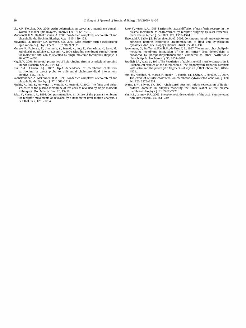
To confirm the results obtained using Composition II, we also ex-plored the ability of actin filaments to perturb lipid–lipid phaseseparations using another PS lipid-containing lipid mixture (Com-position III). Fig. 3 presents epifluorescence micrographs of the bi-layer Composition III [SM/CHOL/SOPC/POPS (molar ratio: 29.1/29.1/29.1/12.5)] before (left) and after (right) actin addition. Inter-estingly, when 12.5% POPS is added to SM/CHOL/SOPC, the lo � ldphase separation is significantly changed relative to the POPS-freecase (Fig. 1, left). This observed change indicates the ability of POPSto compete with SM for CHOL. Interestingly, after physisorption ofactin on the membrane surface, the phase separation is alteredagain (Fig. 3, right), thus supporting the notion of a close relation-ship between actin-POPS, POPS–CHOL, and SM–CHOL interactions.The formation of bright spots in Fig. 3 is most likely caused byaggregation of dye-labeled lipids. Similar aggregation phenomenahave been observed on other lipid mixtures before (e.g., Radha-krishnan and McConnell, 1999), although the mechanism of theirformation remains unclear.
Next, to investigate whether the observed actin-induced changein lipid mixing behavior in Figs. 2 and 3 is PS lipid-specific, the12.5 mol% POPS in Composition III was replaced with an equal mo-lar concentration of the anionic lipid POPG (Composition IV). Fig. 4shows corresponding epifluorescence micrographs of CompositionIV before (left) and after (right) addition of actin. Remarkably, nochange in domain organization is evident. The lack of actin-in-duced changes in lipid phase separations in Fig. 4 is interesting be-
/33.3) + 5 mol% DODA-E85 in bottom monolayer] illustrating unchanged large-scaler. Both leaflets of bilayer are labeled with 0.5 mol% TRITC-DPPE (scale bar: 30 lm).
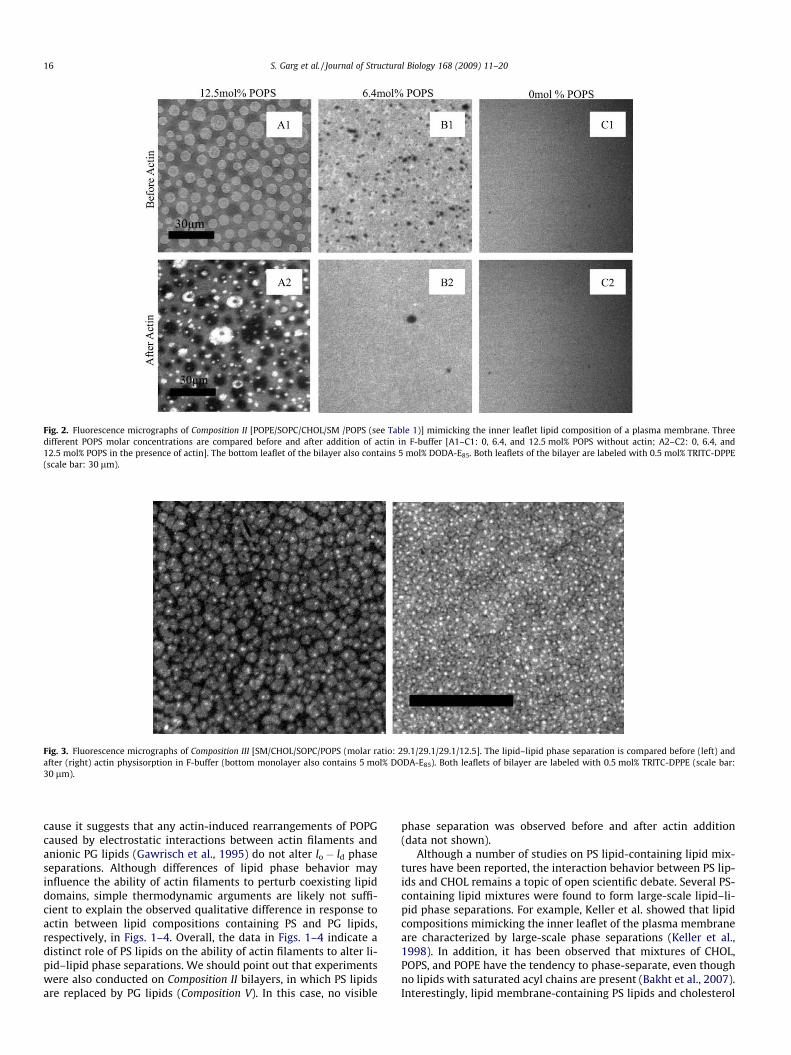
Fig. 2. Fluorescence micrographs of Composition II [POPE/SOPC/CHOL/SM /POPS (see Table 1)] mimicking the inner leaflet lipid composition of a plasma membrane. Threedifferent POPS molar concentrations are compared before and after addition of actin in F-buffer [A1–C1: 0, 6.4, and 12.5 mol% POPS without actin; A2–C2: 0, 6.4, and12.5 mol% POPS in the presence of actin]. The bottom leaflet of the bilayer also contains 5 mol% DODA-E85. Both leaflets of the bilayer are labeled with 0.5 mol% TRITC-DPPE(scale bar: 30 lm).
Fig. 3. Fluorescence micrographs of Composition III [SM/CHOL/SOPC/POPS (molar ratio: 29.1/29.1/29.1/12.5]. The lipid–lipid phase separation is compared before (left) andafter (right) actin physisorption in F-buffer (bottom monolayer also contains 5 mol% DODA-E85). Both leaflets of bilayer are labeled with 0.5 mol% TRITC-DPPE (scale bar:30 lm).
16 S. Garg et al. / Journal of Structural Biology 168 (2009) 11–20
cause it suggests that any actin-induced rearrangements of POPGcaused by electrostatic interactions between actin filaments andanionic PG lipids (Gawrisch et al., 1995) do not alter lo � ld phaseseparations. Although differences of lipid phase behavior mayinfluence the ability of actin filaments to perturb coexisting lipiddomains, simple thermodynamic arguments are likely not suffi-cient to explain the observed qualitative difference in response toactin between lipid compositions containing PS and PG lipids,respectively, in Figs. 1–4. Overall, the data in Figs. 1–4 indicate adistinct role of PS lipids on the ability of actin filaments to alter li-pid–lipid phase separations. We should point out that experimentswere also conducted on Composition II bilayers, in which PS lipidsare replaced by PG lipids (Composition V). In this case, no visible
phase separation was observed before and after actin addition(data not shown).
Although a number of studies on PS lipid-containing lipid mix-tures have been reported, the interaction behavior between PS lip-ids and CHOL remains a topic of open scientific debate. Several PS-containing lipid mixtures were found to form large-scale lipid–li-pid phase separations. For example, Keller et al. showed that lipidcompositions mimicking the inner leaflet of the plasma membraneare characterized by large-scale phase separations (Keller et al.,1998). In addition, it has been observed that mixtures of CHOL,POPS, and POPE have the tendency to phase-separate, even thoughno lipids with saturated acyl chains are present (Bakht et al., 2007).Interestingly, lipid membrane-containing PS lipids and cholesterol

Fig. 4. Fluorescence micrographs of Composition IV [SM/CHOL/SOPC/POPG (molar ratio: 29.1/29.1/29.1/12.5)]. The phase separation is compared before (left) and after (right)actin physisorption. The bottom leaflet of the bilayer also contains 5 mol% DODA-E85. Both leaflets of the bilayer are labeled with 0.5 mol% TRITC-DPPE (scale bar: 30 lm).
S. Garg et al. / Journal of Structural Biology 168 (2009) 11–20 17
were found to show no detergent resistance (Wang and Silvius,2001). On the other hand, there is evidence that CHOL transfersless readily from PS-containing vesicles than from PC-containingvesicles, indicating a stronger affinity between PS lipids and CHOLthan between PC lipids and CHOL (Leventis and Silvius, 2001; Niuand Litman, 2002). NMR studies suggest that PS lipids do not spe-cifically bind to CHOL, but instead induce the formation of orderedsterol structures in the presence of PS headgroups (Epand et al.,2002). Despite the existing uncertainties about the specific natureof PS-CHOL interactions, the various findings illustrate that CHOLbehaves differently in the presence of PS lipids. In light of thesefindings, it appears plausible that the observed differences be-tween PS- (Fig. 3) and PG-containing (Fig. 4) lipid mixtures arecaused by differences in PS lipid–CHOL versus PG lipid–CHOLinteractions.
Alternatively, however, the detected differences between Figs. 3and 4 could also be caused by distinct electrostatic interactions ofPS and PG lipids with actin. In fact, various studies have shown thatactin filaments interact with lipid headgroups via electrostaticinteractions (Chichili and Rodgers, 2007; Edidin, 2006). In orderto explore these electrostatic interactions, we first determinedthe storage modulus, G0, of F-actin (in F-buffer) after physisorptionto DLPC (Composition VI) and DLPC/DLPG (80/20) (Composition VII),respectively, monolayers at the air–water interface using an inter-facial rheometer. The binary lipid mixture was chosen due to thegood miscibility of DLPC and DLPG. Fig. 5 shows that a notablyhigher G0 can be observed when the monolayer contains 20 mol%anionic lipids relative to the case of pure zwitterionic lipids. Thiselevated storage modulus can be attributed to a higher packingdensity of actin filaments on the membrane surface because thestorage modulus of the fluid lipid monolayer without actin is neg-ligible. Fig. 5 also illustrates that G0 increases with time as actinpolymerizes and attaches to the membrane surface. Importantly,the increase in G0 is much more pronounced in the presence ofthe anionic DLPG. As predicted before, the enhanced attractive ac-tin–membrane interactions in the case of DLPG are most likelymediated by Ca2+ ions, which form bridges between the phosphatemoiety of the lipid headgroups and the amine moiety of actin fila-ments (Niggli, 2001). The interfacial rheometry results are infor-mative because they confirm that the strength of interaction
between actin filaments and lipid layer is dependent on electro-static interactions.
Previously, it has been suggested that physisorbed proteins maynot only induce lipid–lipid phase separations through preferredbinding to anionic lipids, but that the resulting clustered protein-binding lipids may also obstruct the fluidity of non-bound lipids byacting as diffusion obstacles (Janmey, 1998). Consequently, lipid dif-fusion experiments could provide a tool to probe electrostatic inter-actions between actin filaments and lipid membranes. To addressthis topic, Fig. 6 presents hr2i values of TRITC-DPPE before and afteractin addition (in F-buffer) in binary mixtures of SOPC/POPS (left)and SOPC/POPG (right). In both cases, tracking experiments are con-ducted using molar concentrations of anionic lipids of 3, 7, and12.5 mol%. Fig. 6 shows that the diffusion results obtained fromSOPC/POPS (Composition VIII) and SOPC/POPG (Composition IX) mix-tures are qualitatively similar. In both cases, the addition of actinleads to a notable reduction in TRITC-DPPE lateral mobility. Interest-ingly, the degree of actin-induced reduction in TRITC-DPPE diffusiv-ity remains largely independent of the molar concentration ofanionic lipids studied, both before and after addition of actin. Thesedata indicate that the observed actin-induced reduction in lipid lat-eral mobility is mainly the result of frictional coupling between thebilayer and the polymerized actin above and to a lesser extentcaused by actin-bound lipids acting as diffusion obstacles. The weakinfluence of obstacle-induced obstructed diffusion is not surprisingif one considers that obstacles are likely comprised of clusters of an-ionic lipids, thus keeping the overall density of obstacles in the cur-rent study relatively low. Most importantly, however, the data inFig. 6 illustrate that PG and PS lipids show similar electrostatic inter-actions with physisorbed actin filaments.
To probe the impact of electrostatic interactions between PS lip-ids and actin filaments on membrane dynamics at a smaller lengthscale, Fig. 7 illustrates fluorescence anisotropy data of TRITC-DPPEin a bilayer with binary lipid mixtures of SOPC/POPS (left) beforeand after actin addition together with corresponding control stud-ies on SOPC/CHOL (right). While the single molecule trackingexperiments presented in Fig. 6 probe a length scale of about100 nm, the fluorescence anisotropy studies probe the direct envi-ronmental conditions of the probe molecule. As presented in Fig. 7(left), in Milli-Q water, there is no effect of POPS molar concentra-

Fig. 5. Storage modulus-time plot for DLPC and DLPC/DLPG (80/20) monolayers at the air–water interface in the presence of actin in F-buffer (left). The lines are included toguide the eye. They do not represent fitting curves. These data were obtained using an oscillating stress–strain interfacial rheometer operated with an oscillating ring, asillustrated in the schematic (right).
Fig. 6. Mean square displacements,<r2>, from TRITC-DPPE tracking experiments in lipid bilayers of SOPC/POPS (left) and SOPC/POPG (right). In both cases, the lateral mobilityis compared before and after physisorption of actin filaments in F-buffer.
Fig. 7. Fluorescence anisotropy values obtained from TRITC-DPPE in binary mixtures of SOPC/POPS (left) and SOPC/CHOL (right) before and after physisorption of actin. Priorto actin addition, each lipid composition is studied in Milli-Q and F-buffer. In the presence of actin, experiments are conducted using F-buffer.
18 S. Garg et al. / Journal of Structural Biology 168 (2009) 11–20
tion on fluorescence anisotropy. However, in F-buffer, a consider-able increase in anisotropy with increasing POPS molar concentra-tions can be observed. This increase is understandable, as Ca2+ ionscan form bridges between headgroups of lipids, thereby loweringthe mobility of lipids and increasing the fluorescence anisotropy.Fig. 7 (left) also shows a remarkable decrease in fluorescenceanisotropy with increasing POPS after addition of actin. These datasuggest that there is an ordering effect in the bilayer if actin isadded at low POPS molar concentrations. This ordering effect be-comes weaker with increasing PS lipid molar concentrations andresults in an effective disordering effect at 12 mol% POPS. Overall,these data can be interpreted in terms of a competitive bindingprocess of anionic and zwitterionic lipids to actin filaments. As an-ionic lipids diffuse and cluster underneath actin-adsorbed mem-
brane regions, the rest of the bilayer becomes increasingly free ofanionic lipids. As a consequence, the interaction between actinand the rest of the bilayer will be reduced. In that sense, the fluo-rescence anisotropy experiments support the concept that physi-sorbed actin triggers the phase separation of the anionic POPSfrom the zwitterionic SOPC. As the single molecule tracking exper-iments on binary SOPC–POPS and SOPC–POPG lipid mixtures inFig. 6 indicate, anionic PS and PG lipids show very similar electro-static interactions with physisorbed F-actin. Therefore, both binarymixtures also show a comparable fluorescence anisotropy re-sponse. To highlight the effect of actin-induced lipid segregationon fluorescence anisotropy, a set of control experiments was con-ducted on SOPC/CHOL mixtures (Fig. 7, right). Here, increasingCHOL levels lead to elevated bilayer viscosities and reduced bilayer

S. Garg et al. / Journal of Structural Biology 168 (2009) 11–20 19
fluidities, respectively (Crane and Tamm, 2004). Due to these ele-vated membrane viscosities, the anisotropy is expected to increasewith increasing concentration of CHOL, as observed in Fig. 7, right.Once again, there is an additional increase in fluorescence anisot-ropy (relative to bilayer in Milli-Q) in the presence of divalent ions,which indicates reduced lipid mobility due to Ca2+ binding, as re-ported before (McManus et al., 2003). However, unlike in the pres-ence of anionic PS lipids (Fig. 7, left), no decrease in fluorescenceanisotropy is observed after addition of actin. In this case, the effectof the segregation of actin-binding lipids on fluorescence anisot-ropy appears to be less pronounced than in the presence of anioniclipids. Interestingly, the anisotropy remains largely unchanged forall the concentrations of CHOL studied. As the anisotropy would beexpected to increase with increasing concentration of CHOL, thedata suggest that the coupling between the membrane and physi-sorbed actin decreases with increasing CHOL concentration. Theseresults are in agreement with other studies, which suggest that themembrane affinity for adsorbed proteins or actin filaments de-creases with increasing CHOL concentration (Sun et al., 2007).Fluorescence anisotropy experiments were not conducted on Com-positions I–V because in these cases it is much more challenging todistinguish between anisotropy changes caused by segregation ofanionic lipids or differences in CHOL molar concentration. The for-mation of lipid–lipid phase separations in mixtures also makes theunambiguous domain-specific anisotropy analysis more challeng-ing. However, it is plausible to expect that the domain-specific seg-regation of PS lipids due to physisorbed actin depends on the ratiobetween PS lipids in complex with CHOL and free PS lipids, whichlocalize in different lipid phases.
4. Conclusion
Overall, the current study provides insight into the potentialrole of PS lipids on actin-induced perturbations of lipid–lipid phaseseparations. Our epifluorescence microscopy experiments showthat physisorbed actin filaments are able to perturb the large-scaledomain formation in lipid compositions containing PS lipids andCHOL. In contrast, no actin-induced perturbation of lipid mixingbehavior is observed if PS lipids are replaced with PG lipids. WhilePS and PG lipids are markedly different in their ability to perturblipid–lipid phase separations in response to interacting actin fila-ments, they appear to show similar electrostatic interactions withactin filaments. These findings support a concept of competinginteractions between actin and PS lipids and between PS lipidsand CHOL, which are distinct from those involving PG and PC lip-ids. Although the experiments have focused on actin, we hypothe-size that other proteins, such as peripheral membrane proteins,may have similar effects on membrane organization and dynamics.Our model membrane data are potentially interesting because theysuggest that interactions between the cytoskeleton and PS lipidsmay at least partially prevent domain formations in the inner leaf-let of the plasma membrane by lowering the ability of PS lipids tointeract with CHOL.
Acknowledgment
This work was supported by National Science Foundation GrantMCB-0416779 and by the IUPUI Nanoscale Imaging Center.
References
Baumgart, T., Hammond, A.T., Sengupta, P., Hess, S.T., Holowka, D.A., Baird, B.A.,Webb, W.W., 2007. Large-scale fluid/fluid phase separation of proteins andlipids in giant plasma membrane vesicles. Proc. Natl. Acad. Sci. USA 104, 3165–3170.
Bakht, O., Pathak, P., London, E., 2007. Effect of the structure of lipids favoringdisordered domain formation on the stability of cholesterol-containing ordered
domains (lipid rafts): identification of multiple raft-stabilization mechanisms.Biophys. J. 93, 4307–4318.
Byfield, F.J., Aranda-Espinoza, H., Romanenko, V.G., Rothblat, G.H., Levitan, I., 2004.Cholesterol depletion increases membrane stiffness of aortic endothelial cells.Biophys. J. 87, 3336–3343.
Camtel, 1999. CIR 100 Interfacial Rheometer. Operating Manual.Chichili, G.R., Rodgers, W., 2007. Clustering of membrane raft proteins by the actin
cytoskeleton. J. Biol. Chem. 282, 36682–36691.Chong, P.L.-G., 1994. Evidence of regular distributions of sterols in liquid crystalline
phosphatidylcholine bilayers. Proc. Natl. Acad. Sci. USA 91, 10069–10073.Clayton, A.H.A., Hanley, Q.S., Arndt-Jovin, D.J., Subramaniam, V., Jovin, T.M., 2002.
Dynamic fluorescence anisotropy imaging microscopy in the frequency domain(rFLIM). Biophys. J. 83, 1631–1649.
Coffman, J.P., Naumann, C.A., 2002. Molecular weight dependence of viscoelasticproperties in two-dimensional physical polymer networks: amphiphiliclipopolymer monolayers at the air–water interface. Macromolecules 35,1835–1839.
Comfurius, P., Bevers, E.M., Zwaal, R.F., 1989. Interaction betweenphosphatidylserine and the isolated cytoskeleton of human blood platelets.Biochim. Biophys. Acta 983, 212–216.
Crane, J.M., Tamm, L.K., 2004. Role of cholesterol in the formation and nature of lipidrafts in planar and spherical model membranes. Biophys. J. 86, 2965–2979.
Deverall, M.A., Gindl, E., Sinner, E.K., Besir, H., Ruehe, J., Saxton, M.J., Naumann, C.A.,2005. Membrane lateral mobility obstructed by polymer-tethered lipids studiedat the single molecule level. Biophys. J. 88, 1875–1886.
Diakowski, W., Prychidny, A., Swistak, M., Nietubyc, M., Białkowska, K., Szopa, J.,Sikorski, A.F., 1999. Brain spectrin (fodrin) interacts with phospholipids asrevealed by intrinsic fluorescence quenching and monolayer experiments.Biochem. J. 338, 83–90.
Edidin, M., 2006. Switching sides: the actin/membrane lipid connection. Biophys. J.91, 3963.
Epand, R.M., Bain, A.D., Sayer, B.G., Bach, D., Wachtel, E., 2002. Properties ofmixtures of cholesterol with phosphatidylcholine or with phosphatidylserinestudied by 13C magic angle spinning nuclear magnetic resonance. Biophys. J.83, 2053–2063.
Fernandes, F., Loura, L.M.S., Fedorov, A., Prieto, M., 2006. Absence of clustering ofphosphatidylinositol-(4, 5)-bisphosphate in fluid phosphatidylcholine. J. LipidRes. 47, 1521–1525.
Foreman, M.B., Coffman, J.P., Murcia, M.J., Cesana, S., Jordan, R., Smith, G.S.,Naumann, C.A., 2003. Gelation of amphiphilic lipopolymers at the air–waterinterface. 2D analogue to 3D gelation of colloidal systems with grafted polymerchains? Langmuir 19, 326–332.
Fujiwara, T., Ritchie, K., Murakoshi, H., Jacobson, K., Kusumi, A., 2002. Phospholipidsundergo hop diffusion in compartmentalized cell membrane. J. Cell Biol. 157,1071–1082.
Garg, S., Ruehe, J., Luedtke, K., Jordan, R., Naumann, C.A., 2007. Domain registrationin raft-mimicking lipid mixtures using polymer-tethered lipid bilayers. Biophys.J. 92, 1263–1270.
Gawrisch, K., Barry, J.A., Holte, L.L., Sinnwell, T., Bergelson, L.D., Ferretti, J.A., 1995.Role of interactions at the lipid–water interface for domain formation. Mol.Membr. Biol. 12, 83–88.
Gerke, V., Creutz, C.E., Moss, S.E., 2005. Annexins: linking Ca2+ signaling tomembrane dynamics. Nat. Rev. Mol. Cell Biol. 6, 449–461.
Gicquaud, D.S.-O.a.C., 1989. Evidence of direct interaction between actin andmembrane lipids. Biochem. Cell Biol. 67, 297–300.
Hoffmann, P.R., deCathelineau, A.M., Ogden, C.A., Leverrier, Y., Bratton, D.L., Daleke,D.L., Ridley, A.J., Fadok, V.A., Henson, P.M., 2001. Phosphatidylserine (PS)induces PS receptor-mediated macropinocytosis and promotes clearance ofapoptotic cells. J. Cell Biol. 155, 649–660.
Huang, J., Feigenson, G.W., 1999. A microscopic interaction model of maximumsolubility of cholesterol in lipid bilayers. Biophys. J. 76, 2142–2157.
Jacobson, K., Mouritsen, O.G., Anderson, R.G.W., 2007. Lipid rafts: at a crossroadbetween cell biology and physics. Nat. Cell Biol. 9, 7–14.
Janmey, P.A., 1998. The cytoskeleton and cell signaling: component localization andmechanical coupling. Physiol. Rev. 78, 763–781.
Kasbauer, M., Bayerl, T.M., 1999. Formation of domains of cationic or anioniclipids in binary lipid mixtures increases the electrostatic coupling strengthof water-soluble proteins to supported bilayers. Biochemistry 38, 15258–15263.
Keller, S.L., Pitcher, W.H., Huestis, W.H., McConnell, H.M., 1998. Red blood cell lipidsform immiscible liquids. Phys. Rev. Lett. 81, 5019–5022.
Kusumi, A., Koyama-Honda, I., Suzuki, K., 2004. Molecular dynamics andinteractions for creation of stimulation-induced stabilized rafts from smallunstable steady-state rafts. Traffic 5, 213–230.
Kwik, J., Boyle, S., Fooksman, D., Margolis, L., Sheetz, M.P., Edidin, M., 2003.Membrane cholesterol, lateral mobility, and the phosphatidylinositol 4, 5-bisphosphate-dependent organization of cell actin. Proc. Natl. Acad. Sci. 100,13964–13969.
Lakowicz, J.R., 2006. Principles of Fluorescence Spectroscopy, third ed. Springer,New York.
Lehmann, T., 1999. Synthese von kovalent an Oberflächen fixiertenPolyethyloxazolinfilmen zum Aufbau polymergestützter Biomembran-Modelle. PhD thesis. Johannes Gutenberg-Universität Mainz, Mainz, Germany.
Leventis, R., Silvius, J.R., 2001. Use of cyclodextrins to monitor transbilayermovement and differential lipid affinities of cholesterol. Biophys. J. 81, 2257–2267.
20 S. Garg et al. / Journal of Structural Biology 168 (2009) 11–20
Liu, A.P., Fletcher, D.A., 2006. Actin polymerization serves as a membrane domainswitch in model lipid bilayers. Biophys. J. 91, 4064–4070.
McConnell, H.M., Radhakrishnan, A., 2003. Condensed complexes of cholesterol andphospholipids. Biochim. Biophys. Acta 1610, 159–173.
McManus, J.J., Raedler, J.O., Dawson, K.A., 2003. Does calcium turn a zwitterioniclipid cationic? J. Phys. Chem. B 107, 9869–9875.
Murase, K., Fujiwara, T., Umemura, Y., Suzuki, K., Iino, R., Yamashita, H., Saito, M.,Murakoshi, H., Ritchie, K., Kusumi, A., 2004. Ultrafine membrane compartmentsfor molecular diffusion as revealed by single molecule techniques. Biophys. J.86, 4075–4093.
Niggli, V., 2001. Structural properties of lipid-binding sites in cytoskeletal proteins.Trends Biochem. Sci. 26, 604–611.
Niu, S.-L., Litman, B.J., 2002. Lipid dependence of membrane cholesterolpartitioning; a direct probe to differential cholesterol–lipid interactions.Biophys. J. 82, 153a.
Radhakrishnan, A., McConnell, H.M., 1999. Condensed complexes of cholesterol andphospholipids. Biophys. J. 77, 1507–1517.
Ritchie, K., Iino, R., Fujiwara, T., Murase, K., Kusumi, A., 2003. The fence and picketstructure of the plasma membrane of live cells as revealed by single moleculetechniques. Mol. Membr. Biol. 20, 13–18.
Sako, Y., Kusumi, A., 1994. Compartmentalized structure of the plasma membranefor receptor movements as revealed by a nanometer-level motion analysis. J.Cell Biol. 125, 1251–1264.
Sako, Y., Kusumi, A., 1995. Barriers for lateral diffusion of transferrin receptor in theplasma membrane as characterized by receptor dragging by laser tweezers:fence versus tether. J. Cell Biol. 129, 1559–1574.
Sheetz, M.P., Sable, J.E., Dobereiner, H.-G., 2006. Continuous membrane-cytoskeltonadhesion requires continuous accommodation to lipid and cytoskeletondynamics. Ann. Rev. Biophys. Biomol. Struct. 35, 417–434.
Speelmans, G., Staffhorst, R.W.H.M., de Kruijff, B., 1997. The anionic phospholipid–mediated membrane interaction of the anti-cancer drug doxorubicin isenhanced by phosphatidylethanolamine compared to other zwitterionicphospholipids. Biochemistry 36, 8657–8662.
Spudich, J.A., Watt, S., 1971. The Regulation of rabbit skeletal muscle contraction. I.Biochemical studies of the interaction of the tropomyosin-troponin complexwith actin and the proteolytic fragments of myosin. J. Biol. Chem. 246, 4866–4871.
Sun, M., Northup, N., Marga, F., Huber, T., Byfield, F.J., Levitan, I., Forgacs, G., 2007.The effect of cellular cholesterol on membrane-cytoskeleton adhesion. J. CellSci. 120, 2223–2231.
Wang, T.-Y., Silvius, J.R., 2001. Cholesterol does not induce segregation of liquid-ordered domains in bilayers modeling the inner leaflet of the plasmamembrane. Biophys. J. 81, 2762–2773.
Yin, H.L., Janmey, P.A., 2003. Phosphoinositide regulation of the actin cytoskeleton.Ann. Rev. Physiol. 65, 761–789.