Abutment selection in fixed partial denture
83
Abutment selection in fixed partial denture Dr Abhinav Gupta Prosthodontics Dr.Z.A.D.C A.M.U Aligarh.
-
Upload
khangminh22 -
Category
Documents
-
view
5 -
download
0
Transcript of Abutment selection in fixed partial denture

Abutment selection in fixed partial denture
Dr Abhinav GuptaProsthodontics
Dr.Z.A.D.CA.M.U Aligarh.

• Fixed partial dentures transmit forces through the abutments to the
periodontium
• Failure are due to poor engineering, the use of improper materials, inadequate
tooth preparation and faulty fabrication

• Of particular concern to dentists is the selection of teeth for abutment
• Successful selection of abutments for fixed partial dentures requires sensitive
diagnostic ability

Definition Types of abutment Ideal requirements Root and their supporting structure:
Crown – root ratioRoot configurationPeriodontal ligament area
Biological consideration:Bending or deflectionSecondary abutment
Factors influencing abutment selection Special problems:
Pier abutmentTilted molar abutment Canine – replacement fixed partial denturesCantilever fixed partial denture

Definition:-
A tooth, a portion of a tooth, or that portion of a dental implant that serves to support
and/or retain a prosthesis
GPT – 8th edi

Different type of abutments:
• Cantilever abutment
• Pier abutment
• Tilted abutment
• Extensively damaged abutment
• Implant abutment

Ideal requirements:
• Ideal crown root ratio
• Adequate thickness of enamel and dentin
• Adequate bone support
• Absence of periodontal disease
• Proper gingival contour

The roots and their supporting tissues should be evaluated for three factors:-
Crown – root ratio
Root configuration
Periodontal ligament area

Crown – root ratio:
The physical relationship between the portion of the tooth within alveolar bone compared
with the portion not with in the alveolar bone, as determined by radiograph
• This relationship have 2 aspect:
• Clinical ratio
• Anatomical ratio

The anatomic portions are defined by the location of cemento-enamel junction
The clinical portion are defined by the level
of supporting alveolar bone as determined
radiographically

* Crown – root ratio:This ratio is a measure of the length of tooth
occlusal to the alveolar crest of bone compared with the length of root embedded
in the bone
Ideal crown-root ratio
for a tooth to be
utilized as a fixed
partial denture
abutment is 2:3

According to Reynolds JM in 1968 ratio of :-1:2 – ideal
1: 1.5 – acceptable 1:1 minimal or doughtful
But not below
1:1

Treatment
Considerations For
Teeth With Poor
Crown-to-root Ratio

Plaque control:plaque control and adequate oral hygiene are
of primary concern in teeth having poor crown-to-root ratio
Inadequate Plaque control
Periodontitis
Treatment failure

Periodontal support regeneration:
• Bone grafting (most reliable method)
• Ingber in 1974 presented the rationale and technique of forced eruption as a method of treating
Ingber JS. J Perodontol,1974

Occlusal reduction:
• Clinical crown occlusal reduction of extruded teeth is a valid approach to improve the crown root ratio
• Bohannan and Abrams in 1961 found that, in crown shortening, for each mm of posterior tooth reduction, a resultant decrease in VDO and increase of 3mm of anterior overbite will occur
Bohannan HM, Abrams L. J Prosthet Dent 1961

Increase stability:
• According to Nyman, Lindhe, and Lundgren in 1975 Mobility commonly seen in poor crown root ratio can be reduced by selective grinding occlusal surface and minimizing horizontal forces in the existing dentition
• Mobile tooth can be retained through splinting
Nyman S, Linghe J and Lundgren D. J Clin Periodontol 1975
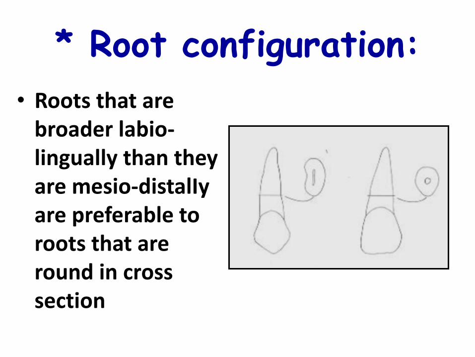
* Root configuration:
• Roots that are broader labio-lingually than they are mesio-distalIy are preferable to roots that are round in cross section

• Multi-rooted posterior teeth with widely separated roots will offer better periodontal support than roots that converge, fuse, or generally present a conical configuration

• The tooth with conical roots can be used as an abutment for a short-span fixed partial denture
• A single-rooted tooth with evidence of irregular configuration or with some curvature in the apical third of the root is preferable to the tooth that has a nearly perfect taper

* Periodontal Ligament Area:
Another consideration in the evaluation of prospective abutment teeth is the
• Root surface area,
or
• Area of periodontal ligament attachment of the root to the bone
Larger teeth have a greater surface area and are better able to bear added stress
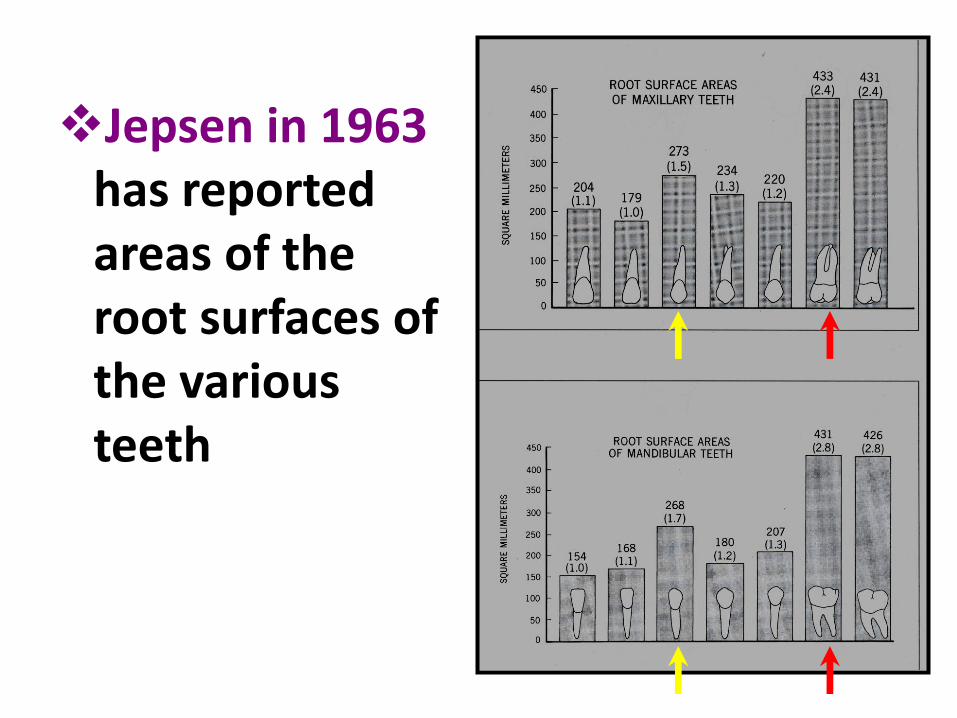
Jepsen in 1963has reported areas of the root surfaces of the various teeth

• In a statement designated as "Ante's Law" by Ante in 1926, “The root surface area of the abutment teeth had to equal or surpass that of the teeth being replaced with pontics”

• Fixed partial dentures with short pontic spans have a better prognosis than do those with excessively long spans

Biomechanical consideration:-

Bending or Deflection:
• All fixed partial dentures, long or short spanned - bend and flex
• Stuteville in 1934 experimented and proved that bending (deflection) varies directly with the cube of the length and inversely with the cube of occluso-gingival thickness of the pontic
Stuteville OH. The bulletin of the
chicago Dental society 1934

Compared with a fixed partial denture having a single tooth pontic span, a two tooth pontic
span will bend 8 times as much

A three tooth pontic will bend 27 times as much as a single pontic

Secondary abutment:
• Double abutment can be used in case when there is unfavorable crown root ratios and long spans
• Secondary abutment must have at least as much root surface area and as favorable a crown - root ratio as the primary

• For e.g. a canine can be used as a secondary abutment to a first premolar primary abutment
• But lateral incisor cannot be used as secondary abutment

Factors influencing abutment selection:
• Reynolds in 1968 suggested some guideline for the selection of abutments which can bear the loads of the oral function with a maximum of service
Reynolds JM. JPD 1968

Long axis relationship:-• The long axis relationship of the abutment
teeth should vary no more than 25o to 30o
from parallel
• Forces are best withstand when they are directed along the long axis of the tooth
• Severely inclined teeth will not withstand forces as well as one that is erect

Arch form:• Restorations involving anterior teeth are
shaped in the form of an arc
• When forces are applied to the pontics, a rotational effect occurs on the abutments and a vertical force is exerted on the terminal ends of the fixed partial denture

The counterbalancing force supplied by the abutments should equal or exceed that of pontics as indicated by the length of the lever arm
Fulcrum line
Lever arm

Rigidity:• All fixed partial dentures, long or short
spanned bend and flex
• Bending or deflection varies directly with the cube of the length and inversely with the cube of occluso-gingival thickness of the pontic
• The lack of sufficient rigidity in a fixed prosthesis is a frequent cause of failure

Compared with a fixed partial denture having a single tooth pontic span, a two tooth pontic
span will bend 8 times as much

• Flexure can cause damage to the abutments and may result in eventual loosening of the retainers, and fatigue of the metal
• The induced stresses must not exceed the yield strength of the alloy used

Margin location :• When conditions permit, margins of
restorations should be kept away from the gingival tissues
• The most accurate margin for any restorative material irritates the gingiva when it is extended, Subgingival margins of cemented restorations have been identified as a major factor in periodontal disease

MARGIN PLACEMENT – Subgingival margins of cemented
restorations are a major factor in periodontal disease
–No difference between subgingival and supragingival margins
Block. JPD 1987; Bader. JPD 1991
Richer & Uno. JPD 1973, Koth. JPD 1982

Advantages of supragingival margins:-
• They can be easily finished
• They are more easily kept clean
• Impressions are more easily made, with less potential for soft tissue damage
•Restorations can be easily evaluated at recall appointments

Indications for subgingival margins:-
• Dental caries, cervical erosion, or restorations extending subgingivally
• crown lengthening procedure is not indicated
• The proximal contact area extends to the gingival crest
• Additional retention is required
• The margin of a metal – ceramic crown is to be hidden behind the labio-gingival crest

Occlusal anatomy:Nature’s own anatomy and contour
should be recreated in all restorations It has the indirect influence on the loads
transmitted to the teeth
The ridges and grooves increase the sharpness and shearing action of teeth and reduce friction between opposing surfaces by keeping the contacting area to a minimum

Such anatomy permits the most efficient mastication of food, thus reducing the load transmitted

Bucco-lingual dimension of teeth:
Occlusal surface of the pontic should harmonize with the bucco-lingual dimension of the natural teeth, and recreate the normal buccal and lingual form to the height of contour

Reducing the width of the pontics does notmaterially reduce the forces transmitted to the abutments, but merely places heavier per unit stress on the restoration

Special problems

Pier abutment:-
A natural tooth located between
terminal abutments that serve to
support a fixed dental prosthesis

Rigid connectors between pontics and retainers are the preferred way of fabricating most fixed partial dentures which provides
desirable strength and stability to the
prosthesis while minimizing the stressesassociated with the restoration
• In some cases, a completely rigid restoration is not indicated
• In case of pier abutment due to following reasons a rigid FPD is contraindicated:-
Physiologic tooth movement
Arch position of the abutment
Disparity In the retentive capacity of the retainers

• The faciolingual movement ranges from 56 to 108µm,
• Intrusion is 28 µm
• Teeth in different segments of the arch move in different directions
Rudd, O’Leary, Stumpf. Periodontics 1964
Parfitt J Dent Res 1960
Chayes, McCall, Hugel. Dent Items Interest 1949

• Because of the curvature of the arch, the faciolingual movement of an anterior tooth occurs at a considerable angle to the faciolingual movement of a molar
• These movements of measurable magnitude and in divergent directions can create stresses in a
long-span prosthesis that
will be transferred to the
abutments

• Because of the distance through which movement occurs, the independent direction and magnitude of movement of the abutment teeth, and the tendency of the prosthesis to flex, stress can be concentrated around the abutment teeth as well as between retainers and the abutment preparation

• According to Shillingburg and Fisher in 1973 the use of a nonrigid connector has been recommended to reduce this hazard
• The most commonly
used non-rigid design
consists of a T-shaped
key that is attached to
the pontic, and a
dovetail keyway
placed within a retainer

Indications:-• Short span fixed partial denture
Contraindications:-• When abutment teeth exhibit significant
mobility
• When the posterior abutment opposes the edentulous space or a RPD
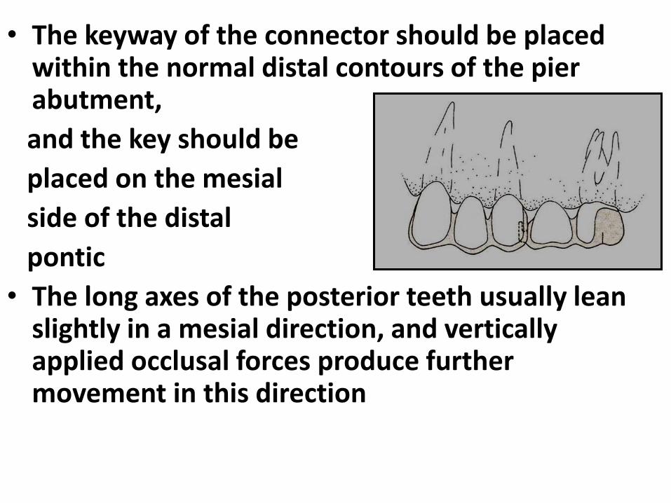
• The keyway of the connector should be placed within the normal distal contours of the pier abutment,
and the key should be
placed on the mesial
side of the distal
pontic
• The long axes of the posterior teeth usually lean slightly in a mesial direction, and vertically applied occlusal forces produce further movement in this direction

• According to Picton in 1962 nearly 98% of posterior teeth tilt mesially when subjected to occlusal forces
• If the keyway of the connector is placed on the distal side of the pier abutment, mesial movement seats the key into the keyway more solidly
Shillingburg & Fisher. JADA 1973

Tilted molar abutment:
• Common problem that frequently seen in the mandibular second molar abutment that has tilted mesially into the space formerly occupied by the first molar

• In these cases it is impossible to prepare the abutment teeth for a fixed partial denture along the long axes of the respective teeth and achieve a common path of insertion
• There is further complication if the third molar is present

• The path of insertion for the fixed partial denture will be dictated by the smaller premolar abutment
• As a result, the mesial surface of the tipped third molar will encroach upon the path of insertion of the fixed partial denture, thereby preventing it from seating completely

• If the encroachment is slight, the problem can be remedied by restoring or recontouring the mesial surface of the third molar
• However, the over tapered second molar preparation must have its retention bolstered by the addition of facial and lingual grooves
• If the tilting is severe, more extensive corrective measures are called for and following treatment of choice will be followed:-
Up righting the molar
Proximal half crown preparation
Telescopic crown with coping
Use of non rigid connector

Uprighting of molar:-• Uprighting is best accomplished by the use of a
fixed appliance
• The average treatment time required is 3 months
• The third molar, if present, is often removed to facilitate the
distal movement of the
second molar

• Both premolars and the canine are banded and tied to a
passive stabilizing
wire
• A helical uprighting spring is inserted into a tube on the banded molar and activated by hooking it over the wire on the anterior segment
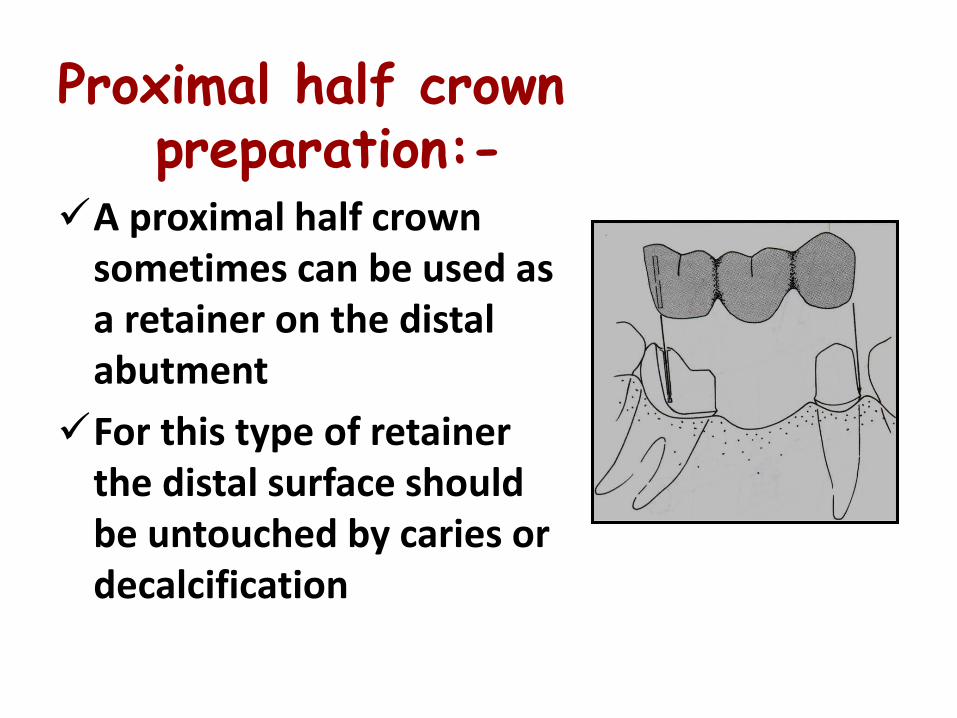
Proximal half crown preparation:-
A proximal half crown sometimes can be used as a retainer on the distal abutment
For this type of retainer the distal surface should be untouched by caries or decalcification

The patient must also demonstrate an ability to keep the area exceptionally clean

Telescopic crown with coping :-A telescope crown and coping can also be used as a
retainer on the distal abutment
An inner coping is made to fit the tooth preparation, and the proximal half crown
that will serve as the retainer for the fixed
partial denture is fitted over
the coping

Non rigid connector:-A full crown preparation is done on the molar,
with its path of insertion parallel with the long axis of that tilted tooth
A box form is placed in the distal surface of the premolar to accommodate a keyway in the distal of
the premolar crown

Placing the connector on the mesial aspect of the tipped molar, can lead to even greater tipping of the tooth

Canine-Replacement Fixed Partial Dentures
• It is difficult to place a missing canine because the canine often lies outside the inter-abutment axis
Prospective abutments are:-
• Lateral incisor (weakest tooth in the entire arch)
• First premolar (weakest posterior tooth)

A fixed partial denture replacing a maxillary canine is subjected to more stresses than that replacing a mandibular
canine
maxillary canine is subjected to more damaging
stresses because the forces are directed outward
and the pontic lies farther outside the inter-
abutment axis.

mandibular canine is more favorable
because the forces are directed inward
and the pontic will be closer to the inter-
abutment axis

Cantilever Fixed Partial Dentures:
• Cantilever fixed partial denture is one that has an abutment or abutments at one end only, with the other end of the pontic remaining unattached

• In the routine three-unit fixed partial denture, force that is applied to the pontic is distributed equally to the abutment teeth
• When a cantilever pontic is employed to replace a missing tooth, forces applied to the pontic have an entirely different effect on the abutment tooth
• The pontic acts as a lever that tends to be depressed under forces with a strong occlusal forces

Pontic of a routine fixed partial denture are
transmitted to both abutment teeth
Cantilever fixed partial denture
tend to tip the fixed partial
denture or the abutment tooth

According to Ewing in 1957 prospective abutment teeth for cantilever fixed partial dentures should be evaluated for:
Lengthy roots with a favorable configuration,
Long clinical crowns,
Good crown-root ratios, and
Healthy periodontium

Generally, cantilever fixed partial dentures should replace only one tooth and have at least two abutments
Ideally cantilever used for replacing a maxillary lateral incisor
There should be no occlusal contact on the pontic in either centric or lateral excursions
Wright. JPD 1986

Canine must be used as an abutment, and it can serve in the role of solo abutment only if it has a long root and good bone support
There should be a rest on the mesial of the pontic against a rest preparation in an inlay or other metallic restoration on the distal of the central incisor to prevent rotation of the pontic and abutment

Cantilever pontic can also be used to replace a missing first
premolar
This scheme will work best if occlusal contact is limited to the distal fossa
Retainers should be on both the second premolar and first molar
Both teeth must exhibit excellent bone support

• Cantilever fixed partial dentures can also be used to replace molars when there is no distal abutment present
• Most commonly, this type of fixed partial denture is used to replace a first molar, Although occasionally it is used to replace a second molar to prevent superaeruption of opposing teeth

According to Schweitzer in 1968, when the pontic is loaded occlusally, the adjacent
abutment tends to act as a fulcrum, with a lifting tendency on the farthest retainer
To minimize the leverage effect, the pontic should be kept as follows:-
• Small as possible, more nearly representing a premolar
than a molar
• Light occlusal contact with absolutely no contact in any excursion movement
• Maximum occluso-gingival height to ensure a rigid prosthesis

References:-
• Shillingburg HT et al. Fundamentals of fixed prosthodontics. 3st ed. Qintessence Publishing Co. Inc –1991. p. 89–102
• Rosenstiel SF, Land MF, Fujimoto J. contemporary fixed prosthodontics. 3rd ed. Missouri (CN). Mosby – 2001. p. 166 – 201
• SMYD ES. Dental engineering. J D Res 1948; 27: 649-660

• Penny RE, Kraal JH. J Prosthet Dent.1979;42: 34-38
• Jacobi R, Shillingburg HT, Duncanson MG. J Prosthet Dent. 1985;54:178-183
• Reynold JM. Abutment selection for fixed prosthodontics. J Prosthet Dent. 1968;19:483-488