Microgrid management with weather-based forecasting ... - ORBi
Upload
khangminh22Category
view
3download
0
ABSTRACTS FROM THE 2011 EVDI ANNUAL MEETING
London, EnglandAugust 30–September 3, 2011
Veterinary Radiology & Ultrasound, Vol. 52, No. 6, 2011, pp 674–705.
COMPUTED TOMOGRAPHIC EVALUATION OF THE CANINE SALIVARY GLANDAPPARATUS
T. Liuti, A.I. de Castro Marques, T. Schwarz. Hospital for Small Animals, The Royal (Dick)School of Veterinary Studies, University of Edinburgh, UK
Introduction/Purposes:Salivary gland pathology is rarely reported in the veterinary literature. Contrast radiographyhas been traditionally used to characterize the salivary gland apparatus and aid diagno-sis. Computed tomography (CT) eliminates organ superimposition maximizing visibility ofsoft tissue and bone structures. The aim of this study was to characterize the anatomicalappearance of nondiseased canine salivary gland apparatus using contrast-enhanced CTsialography.Materials and Methods:Five dogs, euthanized on humane grounds, for reasons unrelated to diseases of the head,were used for anatomical and CT evaluation. In the first part of the study, ethylene blue wasinjected in the mandibular, sublingual, parotid, and zygomatic salivary ducts of two dogs.Surgical dissection was performed and the anatomical location, appearance, and orientationof the salivary gland apparatus were photographed. In the second part of the study, 2-mm thick, sequential CT images of three dogs were acquired before and after injection ofiodine-based, ionic, contrast medium (iothalamate meglumine, Conray 250 R©). Bone andsoft-tissue reconstructions were available for evaluation. Duct cannulation was achievedby a staff surgeon using a 26-g, 19-mm intravenous catheter (Anicath IV Cannula R©): thesalivary papilla was first identified and the catheter sleeve was slowly advanced. Five to tenmillilitres of contrast agent was injected into each cannulated duct.Results:Mandibular, sublingual, parotid, and zygomatic glands and respective ducts were easilyrecognized and followed on CT images after injection of contrast medium. Orientation, size,and anatomic appearance of the salivary apparatus on CT images were consistent withthe anatomical dissection findings. The salivary glands were all identified as lobulated andhighly contrast enhancing.Discussion/Conclusion:The results of this study show that contrast CT is an effective method of outlining nondis-eased salivary gland apparatus. The salivary duct was easily identified in all cases andthese images may facilitate future diagnosis of salivary gland pathology. A prospective clin-ical study is currently being undertaken at the same institution.
CT SIALOGRAPHY IN THE DOG—A CADAVER STUDY
S. Weidner, N. Pfammatter, A. Probst, S. Kneissl. Department of Small Animals and Horses,Division of Diagnostic Imaging and The Department of Pathobiology, Anatomy and Histology,University of Veterinary Medicine, Vienna, Austria
Introduction/Purpose:To document computed tomography (CT) topography of salivary glands and their ducts indogs, a retrograde filling with methylcellulose and iodinated contrast medium was performedin three cadavers.Methods:After plain multislice CT of three canine cadaver heads, the orifices of the salivary ducts werecannulated with a lacrimal cannula. The device was fixed with submucosal sutures, and anextension set was mounted on it. A mixture of methylcellulose and iodinated contrast mediumwas injected with force. Subsequently, contrast CT was performed. Technical settings were130 kV and 96 mA, and the estimated slice thickness was 0.8 mm. Data were archivedin a picture archiving and communication system. Multiplanar reconstructions and surfacemodels were calculated using a syngo MultiModality Workplace.Results:Demarcation of the parotid, mandibular, and zygomatic glands was achieved. Surround-ing structures were imaged without beam hardening artifacts. Landmarks for the parotid,mandibular, and zygomatic glands were the external acoustic canal, the mandibular angle,and the pterygopalatine fossa, respectively. Sialograms of the parotid, mandibular, and zy-gomatic ducts were achieved, whereas contrast enhancement of the sublingual glands andtheir ducts was not possible.
Discussion:To our knowledge, only two studies have been published on the normal radiographic appear-ance of salivary glands and ducts in the dog1,2. CT sialography has not been documented inveterinary medicine. Diameter of the lacrimal cannula (∼1.4 mm) may have been too largeto cannulate the major sublingual duct. Alternatively, the mixture of methylcellulose (MC)and water may have been too viscous to enter smaller ducts. In humans, bitter substancesincrease salivation and ease retrograde cannulation. This technique could be of help in livingdogs as well. Compared with other invasive techniques, sialography is relatively simple andcould provide valuable information, especially when imaged with CT. Additionally, it wouldpreserve salivary duct integrity and continuation when sialoliths or salivary duct rupturesand other problems are suspected.References:1. Harvey CE, O’Brien JA, Rossman LE. Oral, dental, pharyngeal and salivary gland disor-ders. In: Ettinger S. J. (ed.) Textbook of Veterinary Internal Medicine, Vol. II. Diseases of theDog and Cat, 2nd edn. Philadelphia: Saunders. 1983; pp. 1177–1187.2. Tadjalli M, Dehghani SN, Basiri M. Sialography in dog: normal appearance. Vet Arh2004;74:225–233.
EFFECT OF SKULL CONFORMATION ON PARAMETERS OF THE BONYNASOPHARYNX IN DOGS
A.K. Hussein, J. Penderis, M. Sullivan. School of Veterinary Medicine, University of Glasgow,UK
Introduction:Apnoea, hypopnoea, oxygen desaturation, otitis media, snoring, and obstruction sleep ap-noea syndrome (OSAS) have been associated with narrowing of the bony nasopharynx,oropharynx, and soft palate in men and animals. A protrusion of the nasoturbinates intothe pharynx may also lead to obstruction in the caudal choanal area. This study aimedto determine if there was a correlation between the shape of the bony nasopharynx (at aspecific level) and different skull conformations.Methods:Thirty-two dogs (12 Cavalier King Charles spaniels, 3 boxers, 3 shih-tzus, 2 chihuahuas,2 Labradors, 2 West Highland white terriers, and 1 deerhound, English springer spaniel,giant schnauzer, mastiff, miniature schnauzer, pointer, pug, Staffordshire bull terrier) withno clinical signs or pathology of either cranial or nasopharyngeal cavities were recruitedto this study. Using a 1.5-T magnet, the following measurements were made on T1w/T2wimages: (1) Evans and Stockard skull indices; (2) area and shape of the bony nasopharynx(transverse plane) at the level of caudal nasal spine of the hard palate; (3) height of thenasopharynx (mid-sagittal plane) calculated by drawing a line through the midline point ofthe olfactory bulb area to intersect with a base line at a right angle (a line passing throughthe intercondylar notch caudally and hard palate rostrally); (4) olfactory bulb angulation andorientation (midline sagittal plane) in relation to the base.Results:The findings revealed (1) a significant relationship between the area of the nasopharynxin the transverse plane and both body weight and skull phenotype [Evans’ index (P <
0.0001), Stockard’s index (P < 0.0001), and olfactory bulb angulation (P < 0.0001)], (2)the height of the nasopharynx correlated with the skull phenotype (P < 0.0001) but not thebody weight (P = 0.1355) measured in the mid-sagittal plane. Three main shapes wereidentified at transverse section: apart from oval shape, peanut shell, and mouth/invertedmouth-like shapes. When the area, height, and shape were compared to the orientation ofthe olfactory bulb, a more ventral olfactory bulb orientation was associated with (1) smallerarea, (2) a peanut-shell-like shape (which sometimes included nasoturbinates) to oval in amore rostrally orientated olfactory bulb. The area (corrected for body weight) was 4.1 ± 0.6,3.7 ± 0.4, 3.1 ± 0.5 for oval, mouth/inverted mouth and peanut shell shapes, respectively.It was noticed that the irregular shape of the bony part of the nasopharynx on transversesection largely occurred in dogs with a high cephalic index (i.e., more brachycephalic) dueto a bent hard palate and nasopharyngeal roof (which is formed by either the vomer orpresphenoid bones associating with the caudal point of nasal spine of hard palate ventrally),sometimes with nasoturbinates present in the cavity.Conclusion:More brachycephalic breeds have not only a smaller nasopharyngeal area, but also adifferent shape to other skull types. Selective breeding may contribute to normalizing (orexacerbating) these features and result in a reduction in respiratory disorders.
674

VOL. 52, NO. 6 675
THE COMPUTED TOMOGRAPHIC APPEARANCE OF NORMAL CANINE AND FELINENASAL TURBINATES
R. Uosyte, D.A. Gunn-Moore, E. Fraga Manteiga, D. W. Shaw, T. Schwarz. The Royal (Dick)School of Veterinary Studies, Division of Clinical Veterinary Sciences, Roslin, UK
Introduction:Visibility of nasal turbinates in computed tomography (CT) is used for the diagnosis ofcanine and feline nasal disease. The purpose of this study was to determine the influenceof technical and patient factors on normal turbinate visibility.Methods:Transverse rostral and caudal nasal slabs of a feline and canine cadaver were bandsawedand CT-scanned before and after submersion in water with the following variables: slicewidth, pitch, sequential/helical mode, and image reconstruction algorithm. Turbinate visibilitywas assessed objectively with manual tracing. Object contrast was determined via standarddeviation from mean attenuating values. Different window settings were tested.Results:Turbinate visibility is higher in the dog versus cat, caudally versus rostrally, native ver-sus water-submersed, correlating with object contrast magnitude. Turbinates appear moreabundant and better delineated in thin-slice, high-frequency image-reconstruction algorithm,sequential mode, low-pitch images with wide and low window settings.Discussion:Optimal CT settings are essential for accurate turbinate assessment. Nasal fluid masksturbinate detail mimicking turbinate destruction. This is particularly critical for differentiationof feline rhinitis from aggressive neoplasia and mycosis.
CT FINDINGS OF NASAL ADENOCARCINOMA IN FOUR MOON BEARS (URSUSTHIBETANUS)
H. Bacon, N. Webster, M. Bando. Animals Asia Foundation, Chengdu, China and VeterinaryImaging Associates, Sydney, Australia
Introduction:The purpose of this study was to describe the CT findings of confirmed nasal adenocarci-noma in four moon bears (Ursus thibetanus).Methods:Four bears with clinical signs of nasal disease were evaluated at the Animals Asia Foundationbetween October 2008 and January 2010. The bears were all adults; however, specific agingwas not possible due to unknown birth history. Clinical signs were subtle in the early stagesof disease and included reduced activity, decreased appetite, and bilateral mucopurulentnasal discharge. Clinical signs progressed to mucosanguinous discharge and head pressingor placing a paw over the rostral cranium (head holding). The bears were anesthetized forexamination and CT study of the head. One bear had a thoracic CT performed. Nasalbiopsies were taken using biopsy forceps and a blind biopsy technique. The tissue was sentfor histological evaluation. One bear with no clinical signs of nasal disease had a CT of thehead performed as part of the clinical work up. This was used as a control study.Results:CT Findings: All four bears had an extensive, aggressive soft-tissue mass within the nasalchambers. The histological findings were consistent with adenocarcinoma. The bear withno clinical signs of nasal disease had a normal nasal CT.
Cribiform Extension into Osteolysis Extension Extension
plate calvarium and (maxilla, nasal into frontal into
destruction mass effect septum) sinus Nasopharynx
Bear 1 Y N Y Y Y
Bear 2 Y Y (Mild) Y Y Y
Bear 3 Y Y Y Y Y
Bear 4 Y Y Y Y Y
Discussion:The CT findings of nasal adenocarcinoma in four moon bears consists of nasal mass effect,turbinate destruction, osteolysis, and extension into adjacent cavities. The CT findings aremore advanced than usually seen in companion animals due to the difficulty in detectingsubtle clinical signs in a nondomesticated and very stoic species. We would recommendthat any collections holding captive bears pay close attention to nasal discharge as a signof potential nasal carcinoma, even in bears exhibiting normal behaviors.
OPTIMIZED CANINE DENTAL COMPUTED TOMOGRAPHIC PROTOCOL
M.C. Esmans1, J.W. Soukup2, T. Schwarz1. 1The Royal (Dick) School of Veterinary Studies,Roslin, UK, 2The University of Wisconsin-Madison, MA
Introduction:Computed tomography (CT) is a frequently used diagnostic tool for the oral cavity in dogs,including the dental apparatus. Very high anatomic detail is necessary for dental diagnostics.Optimized CT settings to achieve this have not been published. The purpose of this studywas to establish an optimized imaging protocol for canine dental imaging using single- andmultidetector-row CT.Methods:CT was performed on two canine cadaver heads with a one- and four-slice CT unit using adifferent slice thickness, table increment, pitch, image reconstruction algorithm, sequential,and helical scan modes. Images were blindly reviewed by two examiners.Results:A sequential mode, 1-mm slice thickness and interval with high-frequency image reconstruc-tion algorithm were optimal. A helical mode 1-mm slice thickness also revealed acceptableimage quality. In thin-slice helical mode, no influence of pitch (max pitch of 2) was identified.Both CT scanners showed equal results.Discussion:Thin-slice sequential mode CT images reveal optimal dental anatomic detail. Helical scan-ning can be performed with moderate loss of anatomic detail.
MAGNETIC RESONANCE IMAGING AND ULTRASOUND FOR DETECTION OFNORMAL PARATHYROID GLANDS AND HYPERFUNCTIONING PARATHYROIDNODULES IN DOGS
R. Cruz, S. Kruth, T. Gibson, S. Nykamp. University of Guelph, Canada
Introduction/Purpose:Primary hyperparathyroidism (PHPTH) is uncommon in dogs. Ultrasound is commonly usedto identify parathyroid nodules, however, some researchers have reported that hyperfunc-tional parathyroid nodules (hfPTHN) are not always identified and in some cases they cannotbe differentiated from thyroid nodules due the close anatomic relationship. In humans, MRIhas been reported to have sensitivity and specificity similar or superior compared to 99mTc-methoxyisonitrile (MIBI) scintigraphy and US. The use of MRI to evaluate parathyroid glands(PTHg) has not been previously studied in dogs. Objectives: (1) to describe normal MRIanatomy of the PTHg in dogs; (2) to compare MRI and US for the detection of hfPTHN indogs with PHPTH.Methods:The PTHgs were imaged with MRI and US in six control dogs (two small, two medium,and two large dogs) and three hyperparathyroid (HPTH) dogs. US was performed with an8–12 MHz linear transducer. T1, T2, and diffusion-weighted MR images were acquired witha 1.5-T unit. PTHg was considered to be a nodule if it was greater than 2 mm. hfPTHNswere surgically resected and submitted for histopathology. Images were reviewed by twoblinded board-certified radiologists. Presence of the parathyroid, size, echogenicity, or signalintensity, and a subjective assessment of normal or abnormal were recorded.Results:Control dogs were normal on physical examination and had serum calcium, i-calcium, PTH,rPTH levels within reference values. According to one of the observers, none of the PTHgswas observed on any of the MRI sequences in normal dogs. According to the other observer,the two cranial PTHgs were identified in one large breed dog and the left cranial PTHg wasobserved in the other large breed dog. In the clinical cases, the radiologists had completeagreement, except for the DWI. An abnormal right cranial PTH nodule in one HPTH dogand a left caudal PTH nodule in another dog were detected. The nodule in the first dogwas extrathyroidal (ETh) on US, MRI, and surgery whereas in the second HPTH dog, itwas intrathyroidal (ITh) on US and surgery, and ETh on MRI. Adenoma and hyperplasiaprogressing to adenoma was diagnosed at histopathology, respectively. In the third HPTHdog, two PTH nodules were identified on US, one ITh (left caudal) and the other ETh (rightcaudal). Only the ETh nodule was present on MRI. This nodule was found on surgery (ETh)whereas the second nodule seen on US could not be found. An adenoma was diagnosedon histopathology. Relative to the thyroid gland, all the nodules were hypoechoic on US,isointense to the thyroid lobes on T1WI, and slightly hyperintense on T2WI with mild contrastenhancement compared to thyroid glands. hfPTHN were hypointense compared to thyroidgland on DWI for one reviewer and isointense for the other one. Calcium levels return tonormal limits and dogs were free of clinical signs following surgery.Conclusions:MRI can detect hfPTHN in dogs. The nodule seen on US but not in MRI can be interpretedas likely to be a thyroid colloid nodule and not an hfPTHN. MRI may be able to discriminatebetween thyroid colloid nodules and hfPTHN but further evaluation is needed.

676 2011
DIFFUSION-WEIGHTED MAGNETIC RESONANCE IMAGING OF SPINAL CORDSPECIMENS FROM DOGS WITH DEGENERATIVE MYELOPATHY COMPARED TOHISTOPATHOLOGY
M.J. Schmidt, U. Pilatus, A. Oevermann, K.S. Grohmann, M. Vandevelde, M. Kramer. Klinikfur Kleintiere-Chirurgie, Justus-Liebig-Universitat, Gießen, Germany
Purpose:Examination of formalin-fixed spinal cord specimen of dogs with degenerative myelopathy(DM) with diffusion-weighted magnetic resonance imaging (DWI) compared to histopatho-logic examination to evaluate the benefit of the technique for in vivo studies.Methods:The spinal cord of eight dogs with suspected DM and two unaffected dogs were examinedafter exenteration and formalin fixation. Formalin does not alter diffusion characteristics ofthe spinal cord. Thirty 10-cm cuts were examined using a 3-T imaging unit and a wristcoil. T2- and diffusion-weighted images and calculated apparent diffusion coefficient (ADC)mappings were obtained. Three different b values of 500, 1000, and 1500 were used todetermine differences in diffusion along each of the three axes.Results:Ten specimens of the suspected DM-affected dogs showed a diffuse increase in signal inten-sity in DWI and inversion of the signal in ADC. In 10 of the DM specimen, no signal changescould be seen. In unaffected dogs, four specimens showed increased signal intensity, sixspecimens showed no signal changes. Histopathology revealed classic characteristics ofDM in all suspected dogs. Widespread axonal loss, swollen axons with axonal spheroids,and invasion of macrophages into disintegrated myelin sheaths were evident. Nerve fiberswere widely replaced by astroglial cells and fibers. The only difference between specimenswas the degree of astrogliosis. The unaffected dogs were diagnosed having widespreadautolytic areas.Discussion:A hyperintense signal in DWI is indicative for decreased diffusion. The reduction in free mo-tion of water can be caused by intramyelinic edema and severe astrogliosis. The differencebetween the DM-positive specimen that showed hyperintense signals in DWI and those withabsent hyperintensities was related to the grade of the astrogliosis. We suspect that theamount of glial cells and fibers have a major influence on the diffusion of water molecules indogs with DM in our study. These might be different from nonchronic lesions in which axonalloss is the major finding.Hyperintense signals in the dogs without DM can be explained by autolysis, mechanicaltrauma, and malacia of the myelon, which is reported to go along with bright signal intensityin DWI. DWI seems to be a beneficial technique for the in vivo diagnosis of DM.
LINEAR, AREA, AND VOLUMETRIC EVALUATION OF DOG LATERAL VENTRICLESUSING LOW-FIELD MAGNETIC RESONANCE IMAGING
S. Borgonovo, D.D. Zani, O. Travetti, M. Moioli, F. Manzoni, D. De Zani, A. Zecconi, M. DiGiancamillo. Faculty of Veterinary Medicine, Milano, Italy
Introduction:The aim of the study was to compare both linear and area measurements of lateral ventricleswith corresponding volumetric values, in order to evaluate possible differences in the resultsobtained, according to the age of patients, skull conformations, and presence/absence ofany abnormalities causing mass effect on the ventricular system.Materials and Methods:Brains of 135 dogs, aged from 4 months to 15 years, were examined using transverseT1-weighted turbo spin-echo sequences, acquired by a low-field magnet (0.18 T, Vet-MREsaote S.p.A., Genova, Italy). Dogs were divided into different groups according to skullconformation (brachycephalic, mesocephalic, and dolicocephalic), age (young and adultgroups, under and over 15th month, respectively), and presence/absence of any intracraniallesion (mass and no mass groups) causing mass effect on the ventricular system. Ventricularand brain height (Vh, Bh) and ventricular and hemispheric area (VA, HA) were measuredat interthalamic adhesion level. Total lateral ventricle volume (VVtot) and singular lateralventricle volume (VV) were obtained by thresholding-based segmentation algorithm. Theratio Vh/Bh, VA/HA, VV/Bh, and largest/smallest ventricle, in terms of area (rVA) and height(rVh), were calculated. VV/Vh, VA/VV, rVh/rVA, Vh/Bh, and VV/Bh ratio (with previous agedifferentiation) relationship were statistically investigated by linear regression. For VVtot,Vh/Bh, and VA/HA ratio-significant differences between age groups, skull conformation, andpresence/absence of mass were assessed by GLM procedure.
Results:Thirty-nine dogs were brachycephalic, 73 mesocephalic, and 23 dolicocephalic. Twentywere classified as young and 115 as adult; 21 dogs were included in the mass group.Asymmetry between ventricles (rVA > 1.5) was found in 24.4% of investigated cases.Brachycephalic dogs with normal-sized ventricles (Vh/Bh: 0–14%) were 41% and 38.5% forright and left ventricles, respectively, compared to about 70% in dogs showing different skullconformations. Significant difference between VVtot brachycephalic and mesocephalic dogsand in Vh/Bh ratio between brachycephalic and other skull conformations were observed(P < 0.05). In patients with normal-sized ventricles, significant difference was found for Vh/Bhand VA/HA ratio between groups mass and no mass (P < 0.05). Regression proceduredemonstrated a statistical relationship for each compared couple of parameters.Discussion:Prevalence of ventricular asymmetry was lower than previously reported, probably dueto sample heterogeneity. More than 50% of dogs showing ventriculomegaly were brachy-cephalic and the significant difference observed in Vh/Bh ratio between brachycephalic andthe other skull conformations, disappeared after sample stratification according to ventricu-lar size classification. Brachycephalic breeds have larger lateral ventricles than other dogs,hence the current lateral ventricle dimensional classification is not appropriate due to ex-treme morphologic variability of canine skull and brain. To our knowledge, VV/Bh ratio fordetermination of lateral ventricle size was not previously reported and we believe that thismethod could represent a good compromise between reproducibility and repeatability, be-cause brain height, as a linear measurement, showed higher reproducibility and repeatabilitythan manual tracing method as reported in previous studies.
MRI FINDINGS IN 15 ACROMEGALIC CATS
B. Posch, J. Dobson, M.E. Herrtage. Department of Veterinary Medicine, University ofCambridge, Cambridge, UK
Introduction/Purpose:Feline acromegaly is characterized by chronic excessive growth hormone secretion, causinginsulin-resistant diabetes mellitus, and overgrowth of connective tissue, bone, and viscera.The most common cause is a functional pituitary adenoma. A diagnosis of acromegaly iscurrently based upon a combination of clinical signs, measurement of growth hormone,and/or insulin-like growth factor-1 (IGF-1) and intracranial imaging. The purposes of thisstudy were to evaluate if pituitary abnormalities were present on MRI in acromegalic cats, todescribe the MRI findings, and to assess if specific morphologic criteria can be established.Materials and Methods:Medical records between January 2003 and August 2010 were searched for cats in whichclinical signs and laboratory findings were compatible with acromegaly, and in which an MRIwas performed. Inclusion criteria were as follows: elevated serum IGF-1 (>1000 ng/ml) as asurrogate marker of hypersomatotropism and insulin resistant diabetes mellitus. Fifteen catsmet the inclusion criteria. The mean age was 10.5 years (range 6–14 years). Neutered malecats (14/15) and domestic short haired (13/15) were overrepresented. MRI of the brain wasperformed using a 0.2-T scanner. Precontrast and postcontrast T1W and T2W images wereacquired in transverse and sagittal planes. MRI findings were reviewed retrospectively. Thepituitary gland was measured on postcontrast T1W images using a standardized protocol.The following MRI criteria were used to assess the pituitary gland: size, shape, margin, signalintensity, homogeneity, degree, and pattern of contrast enhancement, suprasellar extension,mass effect on surrounding structures (hypothalamus, third ventricle, cavernous sinus),invasion of sphenoid bone, and/or sinus, evidence of peritumoral edema, and transtentorialor cerebellar herniation.Results:Enlargement of the pituitary gland with suprasellar extension was present in all cats. Thepituitary gland had a variable appearance on precontrast T1W and T2W images. No charac-teristic signal patterns were identified on either T1W or T2W sequences. Contrast enhance-ment was nonuniform in all cats. The degree of contrast enhancement was mild in sevencats, moderate in five cats, and strong in three cats. Compression/invasion of the adjacenthypothalamus was suspected in all cases. A mass effect on the cavernous sinus and thirdventricle was present in 13 cats. Invasion of the sphenoid sinus was not found; however,there was a suspicion of invasion of the sphenoid bone in four cats. Mild peritumoral edemawas present in four cats, and moderate edema in one cat. Transtentorial herniation waspresent in one cat, and mild cerebellar herniation in six cats. Histopathology confirmed thepresence of a pituitary adenoma in two cases.Discussion/Conclusion:In conclusion, pituitary abnormalities were present in all 15 acromegalic cats but therewas no consistent morphologic criterion. However, the pituitary gland in most cats hada heterogeneous appearance on both T1W and T2W images, with nonuniform contrastenhancement.

VOL. 52, NO. 6 677
COMPUTED TOMOGRAPHIC-BASED VERTEBRAL HEART SCALE IN AWAKENORMAL CATS AND CATS WITH CARDIAC DISEASE
C.R. Oliveira, K. Stadler, R.T. O’Brien. University of Illinois at Urbana-Champaign, Urbana,IL
Introduction/Purpose:Recently, the use of computed tomography (CT) in awake normal cats and cats with heartdisease was described. The goals of this study were to establish the mean for CT-basedVHS of awake normal cats and cats with heart disease, and to compare the findings withthe VHS measured on radiographs.Methods:Eight cats confirmed by echocardiogram to be free of heart disease, 23 cats diagnosedwith noncardiac-related diseases, and eight cats with echocardiographic confirmation ofhypertrophic (n = 4), restrictive (n = 3), and arrythmogenic (n = 1) cardiomyopathies werescanned awake using a 16-slice CT machine and a positioning device (VetMouseTrapTM).Left and right lateral, and dorsoventral or ventrodorsal radiographs were performed beforeor after the CT. All images were randomized. In radiographs, the VHS based on the widthof the heart on a DV or VD view was measured according to a previously establishedmethod. For CT images, the dorsal plane was used to measure the width of the heart inits widest dimension following the same established method for VHS in radiographs. Thenumbers in millimeters (mm) were plotted against the cranial aspect of T4 using the sagittalplane images. Normalcy of the data was confirmed with a Kolmogorov–Smirnov test. Themean, standard deviation (SD), and range were calculated for width of the heart in mm andVHS in number of vertebrae length for each group of cats. A chi-square test was used tocompare the results between normal cats and cats with noncardiac-related disease. Sinceno difference was found, the results of these groups were analyzed together. The results ofall measurements between noncardiac and cardiac cats were compared using independentsample t-test. A P value <0.05 was considered significant.Results:The mean, SD, and range for width of the heart in mm were as follows: (1) radio-graphs: noncardiac group = 41.12 (4.3, 33–49), cardiac group = 56 (5.8, 48.3–65.1);(2) CT: noncardiac = 32.1 (3.6, 24.5–38.7), cardiac = 40.8 (4, 35.8–49.1). The resultsfor VHS were as follows: (1) radiographs: noncardiac = 3.5 v (0.3, 3–4.1), cardiac =4.7 v (0.35, 4.2–5.2); (2) CT: noncardiac = 3.1 v (0.28, 2.2–3.5), cardiac = 4.1 v (0.12,3.9–4.3) . A statistically significant difference was found between all measurements innoncardiac × cardiac in both modalities (P < 0.0001, P < 0.0003, P < 0.0001, and P <
0.0001, respectively).Discussion/Conclusion:CT of awake cats allows evaluation of cats presented in heart failure that not always arestable for radiographic examination and therefore a new insight into cardiac disease can begained. By measuring the VHS in the CT images, an objective evaluation of heart size isobtained and therefore a presumptive diagnosis of heart disease can be made. This canbe of paramount importance in cats presented in respiratory distress as management ofthe heart failure can be initiated immediately. Our results indicate that CT-based VHS canbe used to objectively diagnose cardiomegaly with confidence and no overlap was foundbetween normal cats and cats with heart disease. We propose an upper limit for CT-basedVHS of heart width of 3.5v. A CT-based VHS equal to or above 3.9 v is highly indicative ofcardiomegaly.
DETECTION OF CANINE PULMONARY NODULES: COMPARISON OF CT,COMPUTED RADIOGRAPHY, AND FILM-SCREEN RADIOGRAPHY
K. Alexander, H. Joly, L. Blond, M.A. d’Anjou, M.E. Nadeau, J. Olive, G. Beauchamp. Facultede medecine veterinaire, Universite de Montreal, St-Hyacinthe, Quebec, Canada
Introduction:Diagnostic imaging of the canine thorax for the staging of neoplasia is evolving. Computedtomography (CT) has become more widely available and digital and computed radiography(CR) have replaced film-screen (FS) radiography in many veterinary practices. However,few have compared these modalities in a clinical veterinary setting as a method to detectpulmonary nodules representing pulmonary metastasis. This study compared CT, CR, andFS radiography for the presence and number of pulmonary nodules in dogs.Methods:Client-owned dogs with a confirmed nonpulmonary primary neoplastic process were usedboth prospectively and retrospectively. All underwent CT and CR; those recruited prospec-tively also received FS radiographs. All images were evaluated in random order by twoveterinary radiologists to form a consensus on the presence, total number, and size of pul-monary nodules. In addition, the lung was divided into six nonanatomically representativeregions of the thorax (right cranial, middle, caudal; left cranial, middle, caudal) and the num-ber and size of nodules in each region was evaluated. Additionally, for CT examinations,pulmonary atelectasis was graded semiquantitatively in each of the pulmonary regions.Nodule numbers and sizes were compared between all three modalities.Results:Twenty-one dogs underwent CT and CR; nine also received FS radiographs. Posi-tive/negative classification for the presence of nodules agreed between all three modalitiesin 8/9 cases and between CR and CT in the remaining 12 cases. Reported confidence washighest for CT examinations (17/21 definitely positive or negative). CT detected the greatest(P = 0.002) total number of nodules; no difference was seen between CR and FS. Thegreatest number of nodules was seen in the right middle and both caudal regions, and onlyusing CT (P < 0.0001). CT and CR detected a significantly smaller minimal nodule sizethan films when all nodules were considered together (P = 0.0007). CT detected a smallernodule size in both caudal and middle regions than radiographic modalities. Pulmonaryatelectasis was significantly greater in the middle regions (P < 0.0001).
Discussion:CT, CR, and FS performed similarly in determining the presence or absence of pulmonarynodules. Because the true nature of each pulmonary nodule could not be established, diag-nostic accuracy of each modality should not be extrapolated from this study. Nonetheless,CT detected a greater number of smaller nodules, particularly in the caudal regions andoverlying the heart, thereby potentially contributing to greater diagnostic confidence withthis modality.
EFFECT OF CONTRAST MEDIUM INJECTION DURATION ON THE CANINEPULMONARY ARTERIES PEAK ENHANCEMENT ANDTIME-TO-PEAKENHANCEMENT USING DYNAMIC COMPUTED TOMOGRAPHY
M. Makara1,2, M. Dennler3, K. Kuhn3, K. Kalchofner4, P. Kircher3. 1Section of Diagnos-tic Imaging, Vetsuisse Faculty, University of Zurich, Switzerland, 2St. George’s University,School of Veterinary Medicine, Grenada, 3Section of Diagnostic Imaging, 4Section Anaes-thesiology, Vetsuisse Faculty, University of Zurich, Switzerland
Introduction/Purpose:The goal of this project was to investigate the effect of contrast medium injection durationon pulmonary artery peak enhancement and time-to-peak enhancement.Methods:Fourteen dogs were allocated into one of seven predefined weight categories, each categorycontained two dogs. Dogs in each weight category were assigned to one of two differentcontrast medium injection protocols. In protocol A, a fixed injection rate of 5 ml/s wasused. In protocol B, the iodine injection rate was calculated as follows: flow rate = contrastvolume/scanning duration + 10 s. Time-to-peak enhancement and peak enhancement ofthe main left and right pulmonary arteries were measured on single-level, dynamic CTimages for a fixed time of 30 s. Rank correlation (Spearman) coefficients between injectionduration and time-to-peak enhancement and between body weight and peak enhancementwere calculated.Results:For group A, there was a significant negative correlation between patient weight and peakenhancement (r = −0.94; P = 0.005), while for group B, there was no significant correlation(r = −0.64 and P = 0.18). There was a significant correlation between injection durationand time-to-peak enhancement for both groups (group A: r = 0.99; P = 0.006 and group B:r = 0.85; P = 0.02).Conclusion:This study confirmed that the injection duration is a key feature in a CT angiography injectionprotocol. According to our results, an injection protocol with an injection duration adjustedto the scanning duration seems to be particularly suitable for veterinary applications wherea population with great weight variability is studied.
CANINE AND FELINE BICAVITY EFFUSIONS: A RETROSPECTIVE STUDY OF 164CASES PRESENTING 2003–2008
A.R. Mugford1, A. Parry2, M. Bevan1, P. Mantis1. 1Department of Veterinary Clinical Sci-ences, The Royal Veterinary College, University of London, 2Willows Referral SpecialistsSolihull Birmingham, UK
Introduction:Bicavity effusions (BCE) have previously been associated with diagnoses of neoplasia andcardiovascular disease. The aim of this study was to evaluate the signalment, laboratoryresults, clinical signs, diagnoses, and outcome associated with animals with a diagnosis ofBCE and to identify the most common diseases presented with BCE in the dog and cat.Materials and Methods:One hundred and sixty-four cases of BCE admitted to the Queen Mother Hospital for Ani-mals (QMHA) between March 2003 and June 2008 were identified from electronic patientrecords. Signalment data, presenting clinical signs, hematology, and biochemistry resultsand diagnosis were recorded for each BC patient and their control matched by species anddate of admission. Outcome was recorded for BCE animals. All data were analyzed usingchi-square tests for factors of significant for BCE. Logistic regression was then performedfor factors, which remained significant when they were considered together with controls.Results:During the study period, BCE represented 0.37% of the total hospital admissions. Of the164 animals (99 dogs and 65 cats), most commonly observed diseases included cardiovas-cular diseases (46), neoplastic diseases (42), inflammatory/infectious diseases (20), renaldiseases (7), non-neoplastic liver diseases (6), and pancreatitis (3). The majority of BCEanimals were 8–12 years old (mean age 93.3 months for BCE and 79.3 months for non-BCEanimals). Male animals appear to be more affected by BCE. Of BCE patients: 56% survivedto discharge (only 2% with resolution of disease), 50% were alive at last contact, 35% wereeuthanized during hospitalization, 4% of discharged patients were subsequently euthanized,and 9% died. There was no species predisposition. Among the feline population, there wasno breed predisposition for BCE while for dogs, BCE was more likely to be seen in Labradorand spaniel dogs than terrier dogs.Conclusions:Dogs with BCE were most likely to have cardiovascular disease followed by neoplasia.Cats with BCE were equally likely to have infectious/inflammatory disease as neoplasia.Cardiovascular disease was less likely than both in cats. BCE in cats and dogs should carrya very guarded prognosis since only 2% of cases were known to have recovered from theunderlying disease process post discharge.

678 2011
ELASTOGRAPHY AND CONTRAST-ENHANCED ULTRASONOGRAPHY OF FOCALLESIONS IN AN EXPERIMENTAL MODEL OF HEPATOCELLULAR CARCINOMA
C.F. Carvalho, M.C. Chammas, V.M.R. de Lima, C.P.M.S. Oliveira. Instituto de Radiologiada Faculdade de Medicina da Universidade de Sao Paulo, Sao Paulo, Brazil
Introduction/Purpose:Early detection of focal hepatic lesions is a challenge in clinical routine and becomes evengreater in the presence of a diffuse parenchymal disease. Ultrasound is still the first choice forthe screening of these diseases. Contrast-enhanced ultrasonography (CEUS) with perflubu-tane microbubbles improves the diagnostic accuracy to differentiate benign and malignantfocal liver lesions. Hepatocelular carcinoma is the fifth most common cancer in human beingand is a recognized complication of advanced nonalcoholic steatohepatitis (NASH).This study aimed to evaluate the diagnostic performance of CEUS and elastogra-phy for early detection of malignant hepatic nodules in an experimental rat model1 ofNASH.Methods:CEUS and elastography was performed in 10 normal adult Sprague-Dawley rats (controlgroup). B-mode and Doppler ultrasonography was performed weekly in 10 experimental ratswith focal liver lesions until nodule detection with suggestive malignancy. Then the animalsunderwent elastography and CEUS, sacrifice, and assessment by microscopy. Tissue stiff-ness of the nodules on elastography was classified in negative (elastic strain) or positive(hard and no strain) comparing with surrounding liver parenchyma.Results:In the early vascular phase with CEUS, wash in was significantly associated with malignancywith sensitivity of 71% and specificity of 100%. In the late vascular phase, wash out patternwas significantly associated with malignancy, sensitivity of 86%, and specificity of 67%.Elastograms of positive lesions showed area of high shear stiffness, which were indicativeof malignancy confirmed on histology evaluation, with sensitivity of 100% and specificity of90%.Discussion/Conclusions:In the early and late vascular phase with CEUS were strongly suggestive of malignancy.Elastography provided promising perspectives for the assessment of malignancy of fo-cal hepatic lesions in our sample. Both techniques allow making right diagnosis withhigh accuracy in an experimental rat model of NASH. Further studies are warrantedto evaluate the effectiveness of this method for diagnosis of this disease in anotherspecies.Reference:1. Lima VMR, Oliveira CPMS, Alves VAF et al. A rodent model of NASH with cirrhosis, ovalcell proliferation and hepatocellular carcinoma. J Hepatol 2008;49:1051–1061.
ULTRASONOGRAPHIC MEASUREMENTS OF FELINE ADRENAL GLANDS IN CATSWITH HYPERTHYROIDISM
A. Combes, E. Vandermeulen, L. Duchateau, K. Peremans, S. Daminet, J.H. Saunders.Faculty of Veterinary Medicine, Ghent University, Merelbeke, Belgium
Introduction:Hyperthyroidism in cats is suggested to be associated with exaggerated responsivenessof the adrenal cortex. Little data are available about adrenal gland size in hyperthyroidcats.Methods:The adrenal glands of 27 hyperthyroid cats were examined ultrasonographically and com-pared to the adrenal glands of 25 healthy cats of similar age.Results:There was no difference in the shape of the adrenal glands between the two groups. Theyappeared as bean-shaped, well-defined, hypoechoic structures surrounded by a hypere-choic halo (35/50 adrenal glands in healthy cats, 40/54 adrenal glands in hyperthyroid cats).Some cats showed more ovoid shape (11/50 adrenal glands in healthy cats, 11/54 adrenalglands in hyperthyroid cats) or more elongated shape (4/50 adrenal glands in healthy cats,3/54 adrenal glands in hyperthyroid cats). The adrenal glands of hyperthyroid cats presentedmore often hyperechoic foci (9/27 hyperthyroid cats, 2/25 healthy cats). The adrenal glandswere significantly larger in hyperthyroid cats than in the healthy cats (using the paired t-test), although there was an overlap in the range of measurements. In hyperthyroid cats,the adrenal length ranged from 7.9 to 19.4 mm, the cranial height from 2.8 to 6.5 mm, andthe caudal height from 3.0 to 6.3 mm. In healthy cats, the adrenal length ranged from 5.8 to14 mm, the cranial height from 1.5 to 5.9 mm, and the caudal height from 2.3 to 5.2 mm.The mean difference in length was 1.6 mm (SD = 0.4; P = 0.0002), the mean difference incranial height 0.6 mm (SD = 0.2; P = 0.0039), and the mean difference in caudal height0.5 mm (SD = 0.2; P = 0.02).Discussion:The enlargement of the adrenal glands in hyperthyroid cats was most likely associated withthe increase in the functional adrenal capacity of the adrenal cortex documented in hyper-thyroid cats. This bilateral homogeneous enlargement should be taken in account when ahyperthyroid cat is evaluated for adrenal disease or when hyperthyroidism and adrenal dis-eases can be both suspected in a feline patient. Indeed, hyperthyroidism is an alternative tohyperadrenocorticism and hyperaldosteronism in cats presenting ultrasonographic bilateraladrenal moderate enlargement.
ULTRASONOGRAPHIC REFERENCE VALUES FOR ADRENAL GLAND SIZE INYORKSHIRE TERRIERS AND LABRADOR RETRIEVERS
T. de Chalus2, A. Combes1, A.S. Leperlier2, P. Pey1, L. Duchateau1, J. Saunders1. 1GhentUniversity, Ghent, Belgium, 2Ecole Nationale Veterinaire d’Alfort, Maisons-Alfort, France
Introduction/Purpose:An upper threshold of 7.4 mm for maximal adrenal diameter is commonly used to detectpituitary-dependent hyperadrenocorticism (PDH) using ultrasonography in dogs. However,there is a substantial overlap between adrenal glands (AG) diameter of healthy dogs andthose with PDH. The aim of the present study is to define the size of the AG in the York-shire terrier and in the Labrador retriever, two breeds widely represented in the populationsuspected of hyperadrenocorticism, and, from these normal values, to determine the upperthresholds for the left and right height of the caudal pole in a longitudinal plane.Methods:Twenty-three healthy Labrador retrievers and 30 healthy Yorkshire terriers were includedin this study. AG measurements of the length in a longitudinal plane, of the height at thecranial, and caudal pole in a longitudinal plane and transverse plane, and of the width at thecranial and caudal pole in a transverse plane, for a total of seven measurements per gland,were done on static images. A statistical analysis evaluated for each AG measurement thesignificance of the differences between the Labrador retrievers and the Yorkshire terriers.For each breed, the effects of age, weight, body length, body weight, and gender on eachadrenal measurement were evaluated. Reference ranges including 95% of the dogs werecalculated for each breed, each side (right or left), and each measurement, in order todetermine the upper limits for the left and right AG in these two breeds.Results:New upper thresholds for the left and right height of the caudal pole in a longitudinal planewere determined: 7.9 and 9.45 mm, respectively, for the Labrador retrievers and 5.4 and 6.7mm, respectively, for the Yorkshire terriers. All the measurements were significantly differentbetween the 2 breeds and Labradors had larger AG measurements than Yorkshire terriers.There was a significant relationship between height of the caudal pole and the age for bothbreeds. The relationship between the size of the AG and the weight was significant onlyfor Yorkshire terriers. No relationship was found between body height, body length, and AGmeasurements. The gender had no effect on the measurements.Discussion/Conclusions:Yorkshire terriers have significantly smaller AG than Labrador retrievers and the age hasa significant effect on the size of their glands, mainly on the caudal pole. The new upperthreshold values for the AG height at the caudal pole are 7.9 mm (left AG) and 9.45 mm(right AG) for the Labrador retrievers and 5.4 mm (left AG) and 6.7 mm (right AG) for theYorkshire terriers. Further studies will determine the sensitivity and the specificity of thesenew upper thresholds as a diagnostic test for PDH.
ENDOSCOPIC ULTRASONOGRAPHIC ASSESSMENT OF THE ESOPHAGEAL WALLIN DOGS
P. Baloi, P. Kook, P.R. Kircher. Division of Diagnostic Imaging and Clinic for Small AnimalInternal Medicine, Vetsuisse-Faculty, University of Zurich, SwitzerlandIntroduction/Purpose:The diagnostic approach for esophageal wall disease in dogs is currently limited to radio-graphy, esophagoscopy, or rarely CT/MRI. However, none of these modalities is useful forcomplete investigation of the esophageal architecture. In humans, endosonography (EUS)is employed for the detailed evaluation, as it provides highly accurate imaging of the walland periluminal structures. In dogs, EUS has already been successfully used for the diag-nosis of intrathoracic lesions, however no information exists on EUS characterizations ofthe esophagus and imaging of its five layers. Therefore, the goal of the present study wasto endosonographically evaluate the different layers of the esophageal wall in healthy dogsand to compare them with previously provided histological results.Methods:Fourteen healthy beagle dogs (six intact males, eight intact females) with a mean bodyweight of 13.4 kg, a median age of 2 years (1–5 years) were included in the study. Dogswere judged to be healthy based on the results of physical examination, CBC, serum bio-chemistry, and urinalysis. EUS examination was performed in lateral recumbency undergeneral anesthesia. A radial endosonoscope (UE 160-AL5 Radial endosonoscope, Olym-pus Schweiz AG, Volketswil, Switzerland) was used and images were stored on the attachedultrasound unit (Pro sound alpha 10, Aloka Holding AG, Switzerland). The endosonoscopewas initially placed cranial to the cardia and then pulled up to the pharynx. To evaluate thewall layering and measure the thickness of the wall, images were obtained every 3 cm.Images were taken with the probe directly in contact with the esophageal wall and with awater-filled balloon as a stand-off pad.Results:The esophageal wall thickness increased constantly from oral to aboral. In the proximalthird, thickness amounted 2.19 mm (1.03–5.62 mm), in the middle 2.15 mm (1.10–4.45mm), and in the distal third 2.84 mm (range of 1.35–5.92 mm). There was a significantdifference between proximal third and distal third (P < 0.001). The results were similarwith and without stand-off pad with only slightly smaller values when using the balloon.The wall layering appeared as five alternating hyperechoic and hypoechoic bands thatwere not constantly identified. There were three groups recognizable when assessing theidentification of single layers: Group 1 was without visible layering, group 2 with three layers,and group 3 with all five layers visible. In 55.5% of all images, three layers could be identifiedbut only in 15.7% all layers were visible. There was no significant difference in the visibilityof the layering between the measurement with and without balloon.Discussion/Conclusion:Although the complete layering was not visible in all instances, the assessment of thearchitecture of the esophageal wall is possible with EUS. As opposed to studies in humans,we could not constantly identify all layers. The significant thicker wall in the caudal part ofthe esophagus does not correlate to histologic findings in standard textbooks of veterinaryhistology. With this study, we provide information about normal EUS appearance of theesophagus.

VOL. 52, NO. 6 679
NORMAL ULTRASONOGRAPHIC APPEARANCE OF THE PYLORUS AND CARDIA INFRENCH BULLDOGS
D.N. Rault1, L. Gatel2, E. Cauvin1, I. Testault3, L. Couturier1, P. Belli2. 1AZURVET, Re-ferral Center in Veterinary Diagnostic Imaging and Neurology, F 06800 Cagnes-sur-Mer,2Universite de Lyon, VetAgro-Sup, F 69280 Marcy L’Etoile, 3ATLANTIA, Veterinary HospitalCenter, F 44000 Nantes, France
Purpose:Disorders of the pylorus and cardia have been reported to be common in brachycephalicdogs, especially French Bulldogs, in association with brachycephalic upper respiratory syn-drome. The aim of the current study was to describe the normal ultrasonographic appearanceof the cardia and pylorus in French Bulldogs and to assess the correlation between histologicand ultrasonographic architecture. The effects of sex, weight, age, and stomach filling werealso investigated.Materials and Methods:A prospective study was performed in 35 French Bulldogs without respiratory or digestiveabnormality. They weighed between 8 and 21 kg, (mean 12.5 kg), and were 0.8–10.5 yearsof age (mean 5.2 years). There were 19 females and 16 males. The stomach was empty,moderately and markedly dilated in 21, 6, and 7 dogs, respectively. Ultrasound examinationswere performed without sedation in dorsal recumbency using a microconvex transducer.The thickness of the pyloric wall (Pw), pyloric hypoechoic triangle (Pt), cardial (Cw), andantral wall (Aw), abdominal esophageal wall (Ew), and the esophageal diameter (Ed) wererecorded. The pylorus was only measured when stretched out in an elongated position.Measurements were excluded if the pylorus was folded toward the stomach, thus protrudinginto the antral lumen. Measurements, effects of sex, weight, age, and stomach filling wereanalyzed using Shapiro and Wilcoxon tests and linear regressions. One of the 35 FrenchBulldogs was euthanized after ultrasound because of a neurological disorder and underwenthistological examination.Results:Measurement of the pylorus was obtained in 94% cases, of the cardia in 80% cases,and of the esophagus in 86% cases. Ultrasonographic layers were similar to histologiclayers described in literature and confirmed in one case. Ultrasonographically, the cardiaappeared as a “band like” thickening at the junction between the esophagus and stomachwith hypertrophy of the muscularis. The pylorus was a short sphincter, bulging into thedigestive lumen; the muscularis had a triangular shape. Aw ranged from 2 to 5.8 mm, (m =4.2 mm), Pw from 5.9 to 8.4 (m = 7.5 mm), Pt from 3 to 5.3 mm (m = 4.3 mm), Cw from6 to 11.3 mm (m = 8.7 mm), Ed 8.4–15 mm (m = 10.7 mm), Ew from 3.8 to 8 mm (m =5.6 mm). Distribution of measurements was normal (P < 0.05). There was no significantdifference between males and females, and empty or full stomach (except for Aw-P < 0.05).No correlation was found between any measurements and age. Only a correlation betweenPw and weight was noticed (P < 0.05).Discussion/Conclusion:There was a good correlation between ultrasonographic and histologic appearance of thepylorus and cardia. Measurement of the cardia and pylorus was not possible in agitateddogs or those with a hiatus hernia. Adequate longitudinal sections of the pylorus weredifficult to obtain because of the positioning, contraction or dilation of the stomach. Thepylorus thickness in this section was less than 9 mm. However, an oblique or folded pylorusartificially increased the thickness. Further investigations are needed to evaluate pyloricthickness in vomiting French Bulldogs with abnormal gastric emptying.
ACTIVATION OF PERIPHERAL, BUT NOT CENTRAL, ALPHA-2-ADRENOCEPTORSWITH DEXMEDETOMIDINE INFLUENCES PERFUSION PARAMETERS MEASUREDWITH CONTRAST-ENHANCED ULTRASOUND
M. Leinonen1, F. Restitutti1, M. Raekallio1, R.T. O’Brien*,2, O. Vainio1. 1Department ofEquine and Small Animal Medicine, Faculty of Veterinary Medicine, University of Helsinki,Finland, 2*Department of Veterinary Clinical Medicine, University of Illinois at Urbana-Champaign, IL
Introduction/Purpose:Contrast-enhanced ultrasound (CEUS) has been used to detect organ perfusion in humansand dogs. No studies exist that compare the effect of dexmedetomidine (DEX) on perfusion ofthe kidneys and small intestine (SI) in dogs. MK-467 is a peripheral alpha (α)-2-adrenoceptorantagonist that has shown to attenuate the early peripheral cardiovascular responses of α-2-adrenoceptor agonists in dogs. The purpose of this study was to compare the perfusionpatterns in the kidney and SI in awake, sedated (with DEX and DEX combined with MK-467)healthy dogs.Materials and Methods:The kidney and segment of SI of six healthy dogs was imaged with CEUS (iU22, PhilipsHealthcare, Andover, MA). Qualitative (CEUS) was performed in six awake healthy labora-
tory beagles. The animals were imaged both awake and after sedation with DEX 10 μg/kgIV and with DEX 10 μg/kg + MK-467 500 μg/kg IV in a randomized, cross-over design witha 14-day wash out between treatments. Mechanical index was maintained at 0.05–0.07 de-pending on depth of view and adjustable parameters were standardized. All dogs receivedmultiple bolus injections (0.05 ml/kg) of contrast medium (Sonovue R©) per imaging. A 1-to 2-min digital clip was recorded for each contrast injection and analyzed off-line (QLAB,Philips). Standardized time-intensity curves were drawn from selected regions of interest ineach organ and each recording separately. The perfusion parameters analyzed were arrivaltime (AT); time-to-peak intensity from the injection (TTPinj); baseline intensity (BI); peakintensity (PI); and wash-in rate (Wi). Analysis of variance with repeated measurements wasused to compare the parameters of each organ, with the level of significance set as P < 0.05.When F values were significant, means were compared by the least-significant-differencemethod.Results:DEX significantly changed all parameters in the kidney [AT↑(P < 0.001), TTPinj↑(P <
0.001), PI↓(0.028), Wi↓(0.036)]. Changes in AT↑(0.001), TTPinj↑(0.001), Wi↓(0.009)], butnot PI(0.073) were found in the SI when compared to awake control animals. When DEXwas compared to MK-467, significant change in AT↓(P = .018), TTPinj↓(P = 0.006) in bothkidney and SI and Wi↑(P = 0.008) in kidney only was found.Discussion/Conclusion:These results indicate marked differences in the perfusion of the kidney cortex and SI as-sessed by CEUS in awake and DEX-treated animals. Concomitant use of MK-467 withDEX-maintained perfusion is similar to awaken dogs. Our results indicate that peripheral ac-tivation of α-2-adrenoceptors influences both the timing and the intensity detected by CEUS.Thus, the effects of sedative agents on organ perfusion should be taken into considerationwhen interpreting the parameters measured by CEUS.
MULTIDETECTOR-COMPUTED TOMOGRAPHY ENTEROGRAPHY USING WHOLEMILK AS A GASTROINTESTINAL CONTRAST AGENT IN DOGS
D. Rodriguez1, M. Levy1, N. Rademacher1, P. Queiroz2, L. Gaschen1. 1Section of Diag-nostic Imaging, 2Section of Veterinary Anesthesiology and Pain Management, Departmentof Veterinary Clinical Sciences, School of Veterinary Medicine, Louisiana State University,Baton Rouge, LA 70803
Introduction/Purpose:Multidetector-computed tomography enterography (MDCTE) evaluates the gastrointestinalsystem by using enteric contrast medium and intravenous-iodinated contrast medium to vi-sualize enteric and extraenteric structures in people. MDCT angiography combined with lowattenuation gastrointestinal contrast media such as whole milk allows detection of intestinaldisease such as inflammatory bowel disease (IBD), ulceration, ischemia, and neoplasia inpeople. The purpose of this study is to evaluate whole milk as a gastrointestinal contrastagent in dogs, to compare it to traditional precontrast and postcontrast abdominal CT withoutmilk, and to develop a protocol for MDCTE with bolus-tracking software.Materials and Methods:Five healthy purpose bred mongrel dogs were given 10 ml/kg of whole milk (4% fat) 30–90 min before induction of general anesthesia. Each dog was scanned with 16-slice CTscanner before and after administration of IV-iodinated contrast at standard doses usingbolus-tracking software. A control group consisted of three MDCTA scans, pre- and post-IVcontrast, without administration of milk. The images were subjectively assessed by threeboard-certified radiologists and graded for degree of intestinal distension: none (0), fair(1), good (2) and ideal (3), small intestinal wall enhancement (yes/no), and the ability todistinguish lumen from mucosa (yes/no). Mean attenuation values (HU) of the pancreas,outer intestinal wall, mucosa, and lumen of the intestines were calculated in each dog andcompared between groups.Results:The small intestinal wall was enhanced in all evaluated groups, in the MDCTE group (milk)the mucosa was discriminated from the outer layers subjectively and by measuring meanattenuation values (HU). The lumen could only be differentiated from the mucosa in thosedogs that received milk. Only the milk group postangiography had significantly different(P < 0.001) attenuation values (HU) of all measured structures. Enteric distention wasconsidered either good or ideal in the MDCTE group. Lack of enteric luminal distentioncreated a pancreatic pseudolesion in one dog. Milk-related side effects were mild, andrequired no treatment and consisted of mild stool softening that lasted on average one day.Discussion/Conclusions:MDCTE resulted in superior intestinal wall enhancement, mucosal conspicuity, and eithergood or ideal lumen distension compared with MDCTA studies without milk. The use ofMDCTE protocols may have value for the diagnosis of intestinal diseases in dogs. Futurestudies comparing MDCTE studies to conventional ultrasound for detecting intestinal dis-ease are necessary to establish whether the need for anesthesia and intravenous contrastagents are justified.

680 2011
EVALUATING GASTROINTESTINAL WALL THICKNESS AND DIAMETER IN NORMALDOGS USING COMPUTED TOMOGRAPHY
S. Hoey1, R. Drees1, X.Yu2. 1Department of Surgical Sciences, School of VeterinaryMedicine, 2CALs Laboratory, Animal Science, University of Wisconsin-Madison, Madison,WI 53706
Introduction/Purpose:Intestinal wall thickness and diameter can change with multiple disease processes,which can include infiltrative disease, inflammation, infection, edema, and infarction.Normal limits for gastrointestinal diameters and wall thickness have been establishedusing radiography and ultrasonography, respectively. No information exists regardingthe use of computed tomography in assessing intestinal wall thickness and diame-ter in normal canine patients. It was hypothesized that a reference range for caninegastrointestinal thickness and diameter could be formulated in relation to age andweight.Methods:In this retrospective study, the records of the Veterinary Medicine Teaching Hospital at UW-Madison were searched for dogs that had no signs of gastrointestinal disease and werereferred for abdominal CT studies between November 2007 and March 2011. NineteenCT studies were identified that included both precontrast and postcontrast exams. Theenhancement pattern and ability to visualize wall layering was noted. Measurements ofmultiple gastrointestinal segments for wall thickness and diameter were obtained usingelectronic calipers at 13 different anatomical locations. These values were assessed inrelation to patient age and weight. Linear model and Kruskal–Wallis test were applied.Results:Detection of distinct wall layering of one or more anatomical section of the gastrointestinaltract was present in 13 patients (68.4%) with stomach wall layering seen most consistently.Age was not significantly associated with intestinal diameter or wall thickness. With increasein weight, there was a significant increase in intestinal diameter and/or wall thickness ofgastric fundus, body, pylorus, pyloric canal, duodenal cranial flexure, ascending duodenum,jejunum, and ascending colon.Discussion/Conclusions:Gastrointestinal wall thickness and diameter increases with increase in weight as evaluatedusing postcontrast CT studies, thus CT evaluation of the abdomen can be a valuable choicefor assessment of these parameters in dogs. Additional information about gastrointestinalwall layering can also be obtained from postcontrast exams. Patient age did not affectgastrointestinal wall thickness or diameter in this study. Further studies to evaluate theapplication of CT assessment of gastrointestinal changes diameter in wall thickness indiseased animals are needed.
RELATIONSHIP BETWEEN ULTRASONOGRAPHIC AND RADIOGRAPHIC FETALBIOMETRY, MATERNAL, AND PREGNANCY-RELATED FACTORS, AND TIME TOPARTURITION IN QUEENS
L. Gatel1, D.N. Rault2, K. Chalvet-Monfray1,3, S. Buff1. 1Universite de Lyon, VetAgro-Sup– Campus Veterinaire de Lyon, 1 Av Bourgelat F 69280 Marcy L’Etoile, 2AZURVET, ReferalCenter in Veterinary Diagnostic Imaging and Neurology. Hippodrome, 2 Boulevard KennedyF 06800 Cagnes-sur-Mer, France, 3INRA, UR 346 Epidemiologie Animale, F-63122 Saint-Genes-Champanelle, France
.Purpose:The aim of this study was to create a model to predict more accurately the timing of theonset of parturition in queens, using radiographic and ultrasonographic measurements offetal femoral length (FL) and biparietal diameter (BPD). The effects of litter size, weight, andage of the queen on the accuracy of the prediction were also investigated.Materials and Methods:A prospective study was performed in 24 purebred queens of 11 different breeds, in normalbody condition. Pregnant queens with aborted or malformed fetuses were excluded from thestudy. Ultrasonographic and radiographic examinations were performed without sedation,during the second half of pregnancy, once to three times in each queen. Ultrasonographicmeasurements were performed on each fetus using a microconvex transducer; the maximalFL and the transversal BPD were recorded. Digital radiographs were obtained and reviewedto determine the litter size and to measure the FL and the BPD. The parturition time wasestimated using linear mixed-effects models on R Package, version 2.5.1.Results:The best multiple linear regression included ultrasonographic measurement of the femorallength (FL), the weight of the queen before pregnancy (W), the litter size (LS), and the ageof the queen (A). All these parameters were significant (P < 0.01). The formula to predictthe number of days to parturition (Y) was Y = 37.864 − 0.193 × FL + 1.227 × W −0.615 × LS − 0.832 × A. The 70% prediction interval was Y ± 1.6 days. Models using theFL provided a more accurate prediction of the onset of parturition than models using BPD.Time to parturition significantly increased with the weight of the queen before mating (P <
0.01). Time to parturition was shorter when the femur was longer (P < 0.01), the litter sizelarger and when the queen was older (P < 0.01). No correlation was found between time toparturition and breed or wither height.Discussion/Conclusion:Radiography is essential to count the number of fetuses but it is not accurate to predict thetime to parturition. Combining ultrasonographic measurement of the fetal femoral length,the queen’s weight and age, and the number of fetuses, provided a model of prediction ofthe onset of parturition in the second half of pregnancy. This study shows that models usingthe FL provide a more accurate prediction of the onset of parturition than previous modelsusing BPD in queens. A larger population of cats should now be investigated to allow fordiscrimination between the specific effects of breed and female size. Further investigationsmay also allow quantification of the effects of oversize, obesity, or illness.
MINIMALLY INVASIVE ULTRASOUND-GUIDED RETRIEVAL OF MIGRATINGSUBLUMBAR PLANT AWNS IN EIGHT DOGS
D. Della Santa. Clinica Veterinaria Mugello, via Provinciale 3D, San Piero a Sieve (FI), Italy
Introduction/Purpose:Paraspinal muscle infection and abscessation in the lumbar region is a relatively commoncondition often induced by the migration of plant awns into the sublumbar region after havingbeen inhaled previously. In such cases, the treatment of choice is surgical exploration of thesublumbar musculature through a transabdominal approach. The aim of the present paperis to report the results of ultrasound-guided nonsurgical retrieval of sublumbar migratingplant awns in dogs.Methods:In this retrospective study were included dogs with clinical signs consistent with foreignbody-induced paraspinal infection where a migrating plant awn could be clearly identifiedsonographically in the sublumbar region and an ultrasound-guided nonsurgical retrievalprocedure with Hartmann forceps was attempted.Results:In the period 2008–2011, eight cases fulfilled the inclusion criteria and were included in thestudy. In all cases, the foreign body was located at the level of the mid-lumbar spine either ina iliopsoas muscle (6/8) or retroperitoneal fat (2/8). In all cases, the Hartmann forceps wereinserted in the subcutaneous tissue of the dorsal lumbar region and then directed ventrally orcranioventrally pointing toward the sublumbar foreign body. In 2/8 cases, ultrasound allowedidentification of a fistulous tract connecting the sublumbar foreign body and the subcuta-neous tissue that could be used as a pathway for the Hartmann forceps without the need ofextensive tissue dissection. In 5/8 cases, more extensive tissue dissection was necessarybecause of the absence of a fistulous tract suitable to be used for reaching the foreign body;in such cases, blunt dissection was performed with Hartmann forceps. In the last case, atrocar was used to dissect the soft tissues and used as a port to insert the Hartmann forcepsin close proximity of the foreign body. Finally, in all cases, the foreign bodies were graspedand retrieved. In 6/8 cases, the plant awn was completely retrieved and the condition did notrecur. In one case, one foreign body was successfully retrieved, but the condition recurredbecause of a second plant awn (not detected during the first procedure) that was retrievedwith a second procedure performed with the same technique; no recurrence was observedafter the second procedure. In the last case, the foreign body was divided into two parts andonly one of them could be retrieved because of cranial displacement of the second fragmentduring the procedure. No harmful complication occurred during the procedures, nor wasobserved at follow-up examinations.Discussion/Conclusions:Ultrasound-guided minimally invasive retrieval of sublumbar plant awns is a valuable op-tion, alternative to standard surgical operation, for treatment of dogs with sonographicallyconfirmed sublumbar foreign bodies.
RELATIONSHIP BETWEEN COMPUTED TOMOGRAPHIC SIGNS ANDARTHROSCOPIC FINDINGS IN DOGS WITH MEDIAL CORONOID PROCESSDISEASE: WORK IN PROGRESS
E. Kulendra, M. Tivers, J. Grierson, C.R. Lamb. The Royal Veterinary College, University ofLondon, UK
Introduction:Computed tomography (CT) is routinely used to examine the elbows of dogs with sus-pected medial coronoid process (MCP) disease; however, there is poor correlation be-tween CT signs and arthroscopic findings in these patients. The aim of this study is toelucidate the relationship between CT signs and arthroscopic findings in dogs with MCPdisease.Methods:Prospective cross-sectional study of dogs having arthroscopic treatment for MCP disease.The appearance of the MCP, with emphasis on fissures and fragmentation, was compared inpreoperative and postoperative CT images and the differences correlated with the surgicalreports.Results:Fifteen dogs (22 elbows) have been studied to date. Median age at arthroscopy was 13months (range 7–96 months). There were 13 males and 2 females. The most frequentbreed was Retriever (n = 6). Fragments were removed arthroscopically from the MCP in18 (82%) elbows; the remaining four elbows had chondromalacia affecting the MCP. Preop-erative CT images showed signs of fissures affecting the MCP or articular calcified bodiescompatible with fragments in 14 (78%) elbows in which fragments were subsequently foundarthroscopically. Single fragments were evident in CT images in five (23%) and multiple frag-ments were evident in five (23%) elbows. Neither fissures nor fragments were evident in CTimages of elbows with chondromalacia only. Comparison of preoperative and postoperativeCT images demonstrated complete removal of fragments in two (11%) instances, reducednumber of multiple fragments in five (28%) instances, and reduced size of fragments in four(22%) instances. Fragments were visible only in postoperative CT images in three (14%)instances. Evidence of debridement of subchondral bone was evident in postoperative CTimages in 15 (68%) instances.Conclusions:Arthroscopy did not result in removal of all suspected fragments identified by CT, and somelesions suspected to be fragments were reduced in size rather than removed. Each of thesefindings suggests that suspected fragments identified by CT are not always visible arthro-scopically, for example, because they are nondisplaced, covered by cartilage or beyond therange of the scope. The occurrence of fragments only in some postoperative CT imagessuggests that the act of debriding the MCP can itself cause fragmentation.Reference:This study is funded by Petsavers.

VOL. 52, NO. 6 681
ANALYSIS OF THE CAUSES OF LAMENESS IN LARGE AND GIANT BREED DOGSOBSERVED DURING THE YEARS 1990–2007 IN THE EAST REGION OF POLAND:RETROSPECTIVE RESEARCH
A. Lojszczyk-Szczepaniak. University of Life Science in Lublin, Laboratory of Radiology andUltrasonography, Poland
Introduction:The presented research is a continuation of research from the 1980s on the occurrence ofseveral diseases in young dogs during the period of their skeletal maturation. The researchwas published in Veterinary Radiology (1988, volume 29) by Professor Stanislaw Koper.This period was characterized by the lack of necessary food for dogs both from the per-spective of quantity and quality. From the beginning, the 1990s dog food produced by manyinternationally known companies appeared. In these conditions, an excessively calorific diet,especially for dogs in the period of skeletal maturation, became highly probable.Methods:The study presents radiographically diagnosed causes of lameness in 954 dogs of bothsexes, mainly large and giant breed up until their first year of life. The dogs underwentclinical and radiographic examination, which was conducted in the years 1990–2007. Dogswere divided into groups encompassing the type of disease diagnosed radiographically. Thefollowing diseases were noted in the subsequent 3-year periods: secondary nutritional hy-perparathyroidism (SNHPT), panosteitis eosynophilica (PE), osteochondrosis (OC—takinginto account all the known clinical forms). Hypertrophic osteodystrophy (HOD) was alsotaken into account.Results:The research confirmed that the most frequently burdened with lameness dog breeds wereGerman Shepherds: 358 dogs (37.53%) and Rottweilers: 90 (9.43%). Panosteitis was ac-knowledged as the main cause of lameness. The disease mainly affected German Shep-herds. The frequency with which PE occurred in the years 1990–1992 was 17 cases from 48patients (35.42%) and its frequency decreased by degrees to the level of 58 cases from 220patients (26.36%) in the 2005–2007. Similarly, a decline in SNHPT and osteochondrosiswas observed, with the exception of the early period of the 1990s, in which a slightly largernumber of cases of OC (17, 13.49%) were noted. As a disease unit rickets occasionallyoccurred. There was a high percentage of dogs with a few diseases recognized at the sametime. The most frequently confirmed diseases were HOD and SNHPT, primarily in GermanShepherds.Discussion:Diseases from the period of skeletal maturation in dogs are conditioned by genetic andenvironmental factors, the latter primarily from their diet. The results of the conducted obser-vations prove the decline of the examined diseases, which constitutes a fairly advantageouscondition for developing breeding programs in eastern Poland. The insignificant growth ofOC cases at the beginning of the 1990s was caused by the lack of sufficient knowledgeconcerning feeding dogs with manufactured dog food. An excess of energy connected withan excessively calorific diet was highly probable.
COMPUTED TOMOGRAPHIC DEMONSTRATION OF THE CALCANEO-CENTRALTARSAL BONE ARCHITECTURE IN DOGS
G. Galateanu1, I. Aizenberg2, T.B. Hildebrandt1, D. Apelt3. 1Leibniz Institute for Zoo andWildlife Research, Berlin, Germany, 2Hebrew University Veterinary Teaching Hospital, Beit-Dagan, Israel, 3Tierarztliche Klinik fur Klein- und Heimtiere, Berlin, Germany
Introduction/Purpose:The mechanism of central tarsal bone (CTB) fractures is not fully understood and additionalinformation regarding its etiology is necessary. While in racing dogs, these fractures aremostly defined as stress fractures of the CTB body, in nonracing dogs are mainly describedfracture/luxations of the CTB concomitant with a fracture/avulsion of the plantar process ofthe CTB (PPCTB). Therefore, the role of the PPCTB was investigated, though the assess-ment of pathological changes at this level can be challenging. Clinical orthopedic evaluation,standard radiographic examination, and even special radiographic views are often insuffi-cient to rule out pathology at this site. The main disadvantage of conventional radiographictechniques is the projection of a three-dimensional structure in a two-dimensional planeresulting in superimposition of various and irregular bony structures. We therefore used128-slices high-resolution computed tomography (CT) to achieve better evaluation of thePPCTB and its spatial relationship with the TBs.
Methods:Images resulting from CT examinations of 52 tarsal joints belonging to 27 dogs (24 dogsof 18 breeds and three mixed breeds), among them, two active police German Shepherddogs, were analyzed. The position of the PPCTB, its shape, cortical features, and articularsurface with the calcaneus were investigatedResults:Thanks to its technical advantages, we were able to visualize, using the CT, that in allstudied joints, the proximolateral part of the PPCTB was the only topographic surface wherethe calcaneus is juxtaposed to the CTB. In joints under stress (working dogs), high-resolutionCT images revealed cortical remodeling of the PPCTB.Discussion/Conclusions:Based on tarsal architecture and spatial relationship between the calcaneus and the PPCTBdemonstrated in this study, we suggest that at least some of CTB fractures in dogs either startor are induced at the level of the PPCTB as a result of the impingement forces exercisedby the calcaneus on the PPCTB. We postulate that during some movement phases (i.e.,adduction) the PPCTB, and, respectively, the rest of the CTB, have to absorb more impactshocks due to calcaneal compressive effect on a wider, shock-absorber surface of thePPCTB. This complementary mechanism may explain the pathogenesis of CTB fractures inracing, working and nonworking dogs.
COMPUTED TOMOGRAPHY OF THE SCAPULOHUMERAL JOINT ANDPERIARTICULAR REGION: A RETROSPECTIVE STUDY OF FINDINGS IN 75 DOGSPRESENTING WITH THORACIC LIMB LAMENESS
T.W. Maddox1, C. May2, B. Keeley2, J.F. McConnell1. 1Small Animal Teaching Hospital,University of Liverpool, 2North West Surgeons, Sutton Weaver, Cheshire, UK
Introduction/Purpose:Computed tomography (CT) is increasing employed as an aid to the diagnosis of thoraciclimb lameness; however, few studies have assessed the prevalence and significance of thelesions that may be identified by this imaging modality in the scapulohumeral (shoulder) jointand periarticular structures.Methods:Records of two referral veterinary clinics were searched for client-owned dogs with lame-ness in one or both thoracic limbs that had CT examination of both shoulder and elbowjoints. Dogs were excluded if they had not undergone concurrent full orthopedic examina-tion (including lameness grading and localization of the site of lameness). All CT imageswere reconstructed using soft-tissue and bone windows, multiplanar reconstruction wasemployed when required. Images were evaluated for the presence of articular and periartic-ular elbow and shoulder pathology in both thoracic limbs. Clinical records were interrogatedfor signalment data, the results of orthopedic examination, and further diagnostic tests (in-cluding arthroscopy) and case outcome. Associations between the presence of shoulderlameness and CT findings were examined using multilevel logistic regression.Results:In total, 80 dogs were identified as having undergone shoulder and elbow CT examination,five were excluded as they had not undergone orthopedic examination. The study populationconsisted of 27 Labrador retrievers, 9 spaniel breeds, 7 Border collies, and 20 other breeds.Thirty-six dogs had lameness attributable to the elbow joint, 16 to the shoulder joint, 17to both shoulder and elbow, with the remainder having no localizable lameness. AbnormalCT findings were documented in 31 dogs (41%). A CT diagnosis of humeral head osteo-chondrosis was made in 7 dogs (9%) and 15 dogs (20%) had evidence of scapulohumeralosteoarthritis. Twenty-five dogs (33%) had evidence of mineralization of the supraspinatus (n= 20), infraspinatus (n = 6), biceps (n = 4), or subscapularis (n = 2) musculature/tendons.Only six of these animals had evidence of ipsilateral shoulder pain and periarticular mineral-ization was not associated with lameness localizable to the shoulder (P = 0.28). Periarticularmineralization was not significantly associated with any breed (P = 0.37). The diagnosis ofosteochondrosis by CT was the only lesion significantly associated with shoulder lameness(P < 0.01). Twelve cases underwent shoulder arthroscopy and for 10 cases the CT andarthroscopic diagnosis agreed; osteochondrosis (n = 6), articular fracture (n = 1), shoulderosteoarthritis (n = 1), and no detectable lesions (n = 2). The remaining two cases hadcartilage changes not detected by CT examination.Discussion/Conclusion:The prevalence of abnormal CT findings relating to the scapulohumeral joint is relativelyhigh in dogs with thoracic limb lameness, but of these only osteochondrosis, lesions appearto have a significant association with lameness localized to the shoulder. Mineralization ofperiarticular musculature/tendons appears common but would seem to have little clinicalsignificance.

682 2011
ULTRASOUND GUIDANCE FOR THE FEMORAL NERVE BLOCK IN CATS: ANIMAGING STUDY
P. Haro Alvarez1, F. Gil2, F.G. Laredo1, M.D. Ayala2, E. Belda1, M. Soler1, A. Agut1.1Department of Medicine and Surgery, 2Department of Anatomy and Compared Patholog-ical Anatomy, University of Murcia, Spain
Introduction/Purpose:The ultrasound (US)-guided blockade of the femoral nerve (FN) has been described in dogsat the level of the femoral triangle. To the author’s knowledge, information regarding the USlocation and blockade of the FN has not been documented in the cat. The aim of this studywas to develop a technique for a US-guided dorsal approach to the FN in cats.Materials and Methods:Anatomic study: Four fresh adult feline cadavers were employed for dissection and crosssectional study of the FN. Imaging study: Another four adult feline cadavers were used todevelop the intended FN approach. The cats were positioned on lateral recumbence andscanned from L2 to the sacrum using a 13 MHz linear transducer. The probe was placedperpendicular to the spine with the mark in a dorsal position. One milliliter of blue ink wasinjected around the FN to confirm the accuracy of the FN location. Two of the cadaverswere immediately dissected; the other two were frozen and cut on transverse cryosectionsto study the spread pattern of the ink. In other four cadavers, 1 ml of iodinated contrast(150 mgI/ml) was injected around the FN to verify the feasibility of the studied approach.Then, orthogonal radiographs and a CT scan of the lumbar spine (L2–S3) were performedto assess the location of the needle and the spread of the contrast.Results:Anatomic study: The FN was located between the iliacus and the psoas major muscles(iliopsoas complex). Imaging study: The anatomic landmarks used to localize the FN werethe dorsal process of L6–L7 lumbar vertebrae and the cranial border of the wing of the osilii. The iliopsoas complex had an oval shape and a hypoechoic appearance related to theepaxial muscles and retroperitoneal fat. The US appearance of the FN was as a hypoechoicround structure surrounded by a hyperechoic rim. The insulated needle was inserted usingan in plane technique between the transverse process of L6–L7. The ink injected wasobserved staining the L5 and L6 nerve roots, the FN, the quadratus lumborum, psoas major,psoas minor, and iliacus muscles in all cases. A cranial and caudal distribution of the inkfrom the site of injection was also observed. The radiographs and CT scans confirmedthe location of the needle between the L6–L7 transverse process and within the iliopsoascomplex in all cases. The contrast media was observed spreading at the level of the ventralpart of the vertebral body of L5 in one case (1/4). A small amount of contrast was observedoutside the iliopsoas into the retroperitoneal space in three cases (3/4).Conclusions:The US-guided dorsal approach may be useful to localize and block the FN in cats. Furtherstudies are necessary to prove its feasibility in a clinical context.Reference:1. Echeverry DF, Gil F, Laredo F, et al. Ultrasound-guided block of the sciatic and femoralnerves in dogs: a descriptive study. Vet J 2010; 186:210–215.
ASSESSMENT OF A VENTRAL ULTRASOUND-GUIDED SUPRAINGUINALAPPROACH TO BLOCK THE FEMORAL NERVE IN THE DOG
D.F. Echeverry1, A. Agut2, F. Laredo2, E. Belda2, M. Soler2, F. Gil3. 1Department of An-imal Health, Faculty of Veterinary Medicine and Zootechnic, University of Tolima, Ibague,Colombia, 2Department of Medicine and Surgery, 3Department of Veterinary Anatomy andEmbryology, Faculty of Veterinary, University of Murcia, Spain
Introduction/Purpose:The anesthetic blockade of the femoral (FN) and sciatic nerves has demonstrated to providesimilar analgesia than the epidurals with a lower incidence of adverse effects. The successof this technique is highly dependent on the adequate location of target nerves. In a recentstudy, ultrasound (US) techniques of nerve location were optimal to block the sciatic nervein the dog. However, a suboptimal blockade of the FN (50–62.5%) was reported when usinga ventral US-guided infrainguinal approach in dogs. A new ventral US-guided suprainguinalapproach to block the FN is evaluated in this study. The ultrasonographic characteristics ofthe FN and the feasibility and accuracy of this approach are investigated in cadavers andexperimental dogs.Materials and Methods:Anatomical nerve study: The evaluation of the anatomical characteristics of the FN requiredto obtain an optimal acoustic window was performed in four intact dog cadavers. The dis-section of the FN was carried out in two cadavers, and the other two were frozen and cut toperform a cross sectional study. In the “in vitro” part of the study the left and right FN of fiveintact cadavers were identified by US. The US probe (13 MHz linear array) was orientatedperpendicular to the midline and slightly cranial to the inguinal nipple. From this point, theprobe was directed cranially, trying to trace the projection of the nerve on the abdomen. Theultrasonographic features of the FN and related structures were described. The accuracy ofthe US-guided nerve localization was demonstrated by injecting a staining solution aroundthe nerve. In this phase, a success FN block was defined as the staining of the nerve ina length ≥2 cm. In the “in vivo” ultrasound-guided nerve blockade, the left and right FNwere located by US in five experimental Beagles sedated with medetomidine (10 μg/kggiven IM). When the needle was seen close to the FN lidocaine 2% (0.3 ml/kg) was injectedaround the nerve. The spreading of the local anesthetic was observed during the injection.At the end of the experience, atipamezole (10 μg/kg given IM) was administered to reversesedation. In this phase, a successful FN block was confirmed by the presence of motor andproprioceptive deficit, and inability to move the hindlimb. The dogs were re-evaluated 12and 24 h after the blockades.Results:The FN was observed as a hypoechogenic rounded structure surrounded by a markedhyperechogenic thick ring, located within the iliopsoas muscle body on transverse scans.The FN was accurately located by US in all the cases. This was confirmed in vitro by thestaining of the FN in a length superior to 2 cm, and in vivo, by the presence of a successfulFN block. No neurological complications were observed.
Discussion/Conclusion:This ventral US-guided suprainguinal approach may be a feasible and accurate method toapproach and block more efficiently the FN in the dog.
MAGNETIC RESONANCE IMAGING FEATURES OF DISCOSPONDYLITIS IN DOGS
I. Carrera1, R. Goncalves2, F. McConnell2, M. Sullivan1. 1Faculty of Veterinary Medicine,University of Glasgow, Glasgow, 2Small Animal Teaching Hospital, University of Liverpool,Neston, UKIntroduction:The diagnosis of discospondylitis is based on clinical signs, diagnostic imaging, and labo-ratory results. To date, radiography has been the imaging technique most widely used. Thisretrospective study aimed to describe the magnetic resonance imaging (MRI) findings ob-served in dogs with confirmed discospondylitis and evaluate the advantages and limitationsof MRI in the management of this condition.Materials and Methods:The inclusion criteria comprised complete clinical history and neurological examination,radiographs, and MRI study of the region of the vertebral column affected, including in allcases sequences precontrast and postcontrast T1-weighted sequences: urine, blood, and/orintervertebral disc cultures, and follow-up of the progression of the clinical signs in responseto antibiotic treatment.Results:Thirteen dogs were included with confirmed discospondylitis. In total, there were 17 sitesof discospondylitis. Eleven (81.1%) of the dogs had spinal pain for more than 3 weeksand a variable degree of neurological signs. Two dogs had spinal pain and ataxia for 4days. In MRI images, there was always involvement of two adjacent vertebral endplates andits associated intervertebral disc. The involved endplates and adjacent bone marrow wereT1W hypointense with hyperintensity in fat suppression techniques. All dogs had contrastenhancement of endplates and paravertebral soft tissues. The intervertebral discs werehyperintense in T2W and fat suppression and characterized by contrast enhancement in 15sites (88.2%). Endplate erosion was present in 15 sites (88.2%) and was associated withT2-hypointense bone marrow adjacent to it. In two sites (11.8%), endplate erosion was notseen in MRI or radiography. The vertebral bone marrow in these cases was hyperintense inT2W and in fat suppression sequences, and also showed contrast enhancement. Epiduralextension was conspicuous in postcontrast images in 15 cases (88.2%). Spinal cord com-pression was present in 15 cases (88.2%), which presented clinical evidence of neurologicalsigns. Subluxation was present in two sites (11.8%).Discussion/Conclusion:MRI shows characteristic features of discospondylitis and allows the recognition of the exactlocation and extension (to the epidural space and paravertebral soft tissues) of the infection.Furthermore, MRI increases lesion conspicuity in early discospondylitis that may not bevisualized by radiography.
FLEXED SAGITTAL MRI FOR EVALUATION OF THE OCCIPITAL BONE ANDFORAMEN MAGNUM
R. Dennis. Centre for Small Animal Studies, Animal Health Trust, Lanwades Park, Kentford,Newmarket, Suffolk, UK
Introduction/Purpose:Chiari-like malformation (CM) due to changes in the occipital bone is a well-recognizeddeformity in several small dog breeds, notably the Cavalier King Charles spaniel, in whichit seems to be almost ubiquitous. The morphological abnormality results in a mismatchbetween cerebellar size and caudal fossa volume resulting in foramen magnum “crowding”or cerebellar herniation. It is thought that this causes disturbance of CSF flow, leading tosyringomyelia (SM). MRI is widely used for CM/SM diagnosis and screening, a midlinesagittal T1- or T2-weighted image being used to assess CM.Materials and Methods:In August 2010, the protocol for MRI of CKCS undergoing investigation of suspected CM/SMat the Animal Health Trust was extended to include a moderately flexed, sagittal T2W scanas well as the usual extended sagittal scan. Dogs are scanned supine, and following theinitial scans, the head is flexed to about 125o at the occipito-atlantal junction and held inplace using small foam pads and sticky tape. After a further three-plane localizer, a secondsagittal T2W scan (+ /− a sagittal T1W scan) is performed. Flexed sagittal scans have alsobeen performed on a number of other dogs of varying breed, size, and conformation forcomparison.Results:The appearance of the occipital bone varies markedly between breeds. In larger dogs, theportion of the occipital bone, which forms the dorsal margin of the foramen magnum, isslightly thickened, forming a “toe” with a distinct, hyperintense medullary cavity. The bone iseasily distinguished from the dorsal ligaments of the joint, which are folded when the headand neck are moderately extended. In smaller dogs, the occipital bone is thinner and lacksthe marrow-filled “toe.” It appears as a narrow band of signal void intensity, which cannot bedifferentiated from the ligaments. On flexion, the folded ligaments flatten out and their pointof attachment to the occipital bone is more easily identified. In the majority of CKCS and insome other small breed dogs, marked increase in height of the foramen magnum is evidenton flexion. This shows that the cerebellar vermis is usually herniated, rather than followingthe apparent contour of bone.Discussion/Conclusions:The study has shown that similarity of signal intensity between the occipital bone and thedorsal ligaments of the occipito-atlantal joint may lead to incorrect assessment of occipitalbone shape and underestimation of the height of the foramen magnum and the degree ofcerebellar herniation on scans obtained with the dog’s head and neck in moderate exten-sion. This has an impact of research into CM/SM where the foramen magnum diameter ismeasured on sagittal MR images. The flexed sagittal position is therefore recommended forclinical diagnosis and morphometric studies. It also shows more accurately whether or notcisternal puncture for CSF collection is hazardous.

VOL. 52, NO. 6 683
CRANIAL THORACIC SPINAL STENOSIS IN LARGE BREED DOGS
P.J. Johnson1, L. De Risio1, J.F. McConnell2, A.H. Sparkes1, R. Dennis1, A. Holloway1.1Animal Health Trust, Kentford, Newmarket, UK, 2University of Liverpool Faculty of Veteri-nary Science, Liverpool, UK
Introduction/Purpose:Developmental stenosis of the cranial thoracic spinal canal has been suggested to occurin large breed dogs, and is documented in the Dogue de Bordeaux (DdB). The purposesof the study were to investigate developmental cranial thoracic stenosis (CTS) further bycalculation of reference data in large breed dogs and to determine whether morphometricand/or subjective criteria could be used to characterize CTS.Materials and Methods:Magnetic resonance imaging (MRI) studies of large breed dogs (>20 kg), which includedtransverse images of the first to fifth thoracic vertebrae, were evaluated for the period 2001–2011. A single, blinded observer graded the morphological appearance of the vertebralcanal and spinal cord as grade 0, controls (no bony stenosis), grade 1, moderately stenotic(stenosis without cord compression), or grade 2, severely stenotic (stenosis with cord com-pression). Morphometric analysis consisted of cross-sectional area measurements of thevertebral canal at mid-vertebral and intervertebral disc levels to form the canal-area ratio(CAR) and of the spinal cord and vertebral canal at the intervertebral disc level to form thecord-canal ratio (CCR). Normal reference ranges were calculated for CAR and CCR andagreement between the subjective grading and objective data was evaluated. The signal-ment, neurological presentation, and morphological features of CTS were documented.Results:Seventy-seven MRI studies of the cranial thoracic spine were included. Fifty-eight weregraded as controls (grade 0) and 19 as moderately or severely stenotic (grade 1 or 2,respectively). There was a significant difference in CAR and CCR values at different thoracicvertebral sites and between breeds in the control group. CAR discriminated better betweenthe control and stenosis cases than CCR. CCR had poor agreement even with the useof breed specific reference ranges whereas CAR had substantial agreement when breedspecific reference ranges were utilized. CTS was recognized in predominantly young (most<3 years of age) dogs. Breeds affected included the DdB, Neapolitan mastiff, Bullmastiff,Chow Chow, St Bernard, Staffordshire Bull terrier, German Shepherd dog, Rottweiler, andDoberman. Vertebral morphological changes included enlarged pedicles, facet hypertrophy,altered vertebral body shape, and dorsal lamina cleft formation. In 10/19 of the dogs, CTSwas the only spinal abnormality. Eight of these dogs had pelvic limb ataxia and posturalreaction deficits and two were neurologically normal. Nine of nineteen cases had concurrentneurological disease of which cervical stenotic myelopathy was the most common.Discussion/Conclusion:Within the control group there was significant variation between breeds and between sites.Ideally, breed-specific reference ranges should be generated for the ratios at each vertebralsite. CAR gave a better discrimination between the control and stenosis cases and hadbetter agreement with the subjective grading system than CCR. CTS should a differentialdiagnosis in young large breed dogs that present with pelvic limb neurological dysfunction.CTS may be subclinical and should be considered in dogs with cervical stenotic myelopathy.
USEFULNESS OF A HALF-FOURIER ACQUISITION SINGLE-SHOT TURBOSPIN-ECHO PULSE SEQUENCE IN IDENTIFYING ARACHNOID DIVERTICULA INDOGS
G.S. Seiler1, I.D. Robertson1, W. Mai2, W.R. Widmer3, J. Suran2, S. Nemanic4, C.R.Lamb5, J. Lang6, J.L. Johnson7, D.E. Thrall1. 1North Carolina State University, Raleigh,NC, 2University of Pennsylvania, Philadelphia, PA, 3Purdue University, West Lafayette, IN,4Michigan State University, East Lansing, MI, 5The Royal Veterinary College, University ofLondon, UK, 6University of Bern, Bern, Switzerland, 7Duke Comprehensive Cancer Center,Durham, NC
Introduction/Purpose:Single-shot turbo spin-echo sequences are heavily T2-weighted sequences that are excep-tionally well suited to evaluate the subarachnoid space. In the T2-weighted fast spin-echosequences that are used routinely in spinal magnetic resonance (MR) imaging, the sub-arachnoid space is not well differentiated from the surrounding epidural fat, which could leadto decreased detection of lesions of the subarachnoid space such as arachnoid diverticula.Our purpose was to determine the added value of a single-shot turbo spin-echo sequencein identifying focal dilations of the subarachnoid space in dogs.
Methods:MR images of six dogs with a confirmed arachnoid diverticulum, and 24 dogs with otherconfirmed spinal disease were included. Six observers were asked to interpret only T2-weighted images initially, and in a second session T2-weighted and half-Fourier acquisitionsingle-shot turbo spin-echo (HASTE) sequences. The MR images were anonymized, andno signalment, history or clinical information was provided. Each time, the observers wereasked to review the images, localize the lesion(s) they considered significant, and to make adiagnosis to the best of their ability. Sensitivity, specificity, positive, and negative predictivevalues for detection of arachnoid diverticula were calculated and the Wilcoxon signed-ranktest was used to determine if the difference in paired observations was statistically significant.Results:Without the HASTE sequences, 25% of arachnoid diverticula were identified. Adding theHASTE sequence increased the diagnosis of arachnoid diverticulum to 52.8%. The result-ing improvement of 27.8% after adding the HASTE sequence was statistically significant(P = 0.002). No false positive diagnoses of arachnoid diverticulum were made with ei-ther sequence. This results in a specificity and positive predictive value of 100% for bothsequences. Mean sensitivity and negative predictive value of the T2-weighted sequencealone were 25% (range 0–83%) and 84.2% (80–96%), respectively, and increased to 52.8%(0–83%) and 89.4% (80–96%), respectively, when the HASTE sequence was added.Discussion/Conclusions:Although sensitivity in this study was artificially low due to the lack of clinical information pro-vided and variable experience of the observers with the HASTE sequence, the significantlyincreased detection rate of arachnoid diverticula when using HASTE imaging indicates thatthis sequence is a valuable addition to MR imaging protocols for the canine spine. Otheradvantages of this sequence for spinal MR imaging is the speed of acquisition, ability todisplay changes in CSF composition, and sensitivity to detect extraspinal lesions due to highsensitivity to strongly T2 hyperintense lesions. Where available, single shot fast spin-echosequences should be included routinely into spinal MRI protocols.
NOVEL STIR FINDING OF CERVICAL MUSCLE HYPERINTENSITY WITH SUSPECTEDINFLAMMATORY SPINAL CORD DISEASES
S. Eminaga, G.B. Cherubini, E. Villiers, A. Caine. Dick White Referrals, Six Mile Bottom, UK
Introduction/Purpose:MRI features of dogs with cervical spinal hyperesthesia and neurological signs suggestive ofinflammatory spinal cord diseases are poorly described. MRI signal change affecting musclehas not been noted as a feature of inflammatory spinal cord disease. This retrospectivestudy aimed to assess the sensitivity of a distinctive pattern of STIR muscle hyperintensityfor predicting a clinical diagnosis of inflammatory spinal cord disease.Methods:All dogs presenting with clinical and neurological signs suspicious of cervical spinal corddisease, which had cervical spinal MRI including STIR sequence and CSF analysis per-formed between July 2009 and February 2011 were included. Twenty-seven cases fulfilledthe inclusion criteria. A board-certified radiologist and neurologist retrospectively reviewedall MRI images to identify any STIR abnormalities and a board-certified clinical pathologistand neurologist reviewed the CSF analysis.Results:Thirteen of 27 cases had a distinctive STIR appearance characterized by hyperintensityalong the cervical vertebral body extending into the adjacent muscle with an ill-definedmargin. In 9/13 cases faint to moderate gadolinium-based contrast enhancement was seenon T1W in the location of the STIR abnormality. Thirteen of the 27 cases had inflammatoryCSF, 11 of which were showing distinctive STIR changes. Two cases with inflammatory CSFhad normal STIR images. Two cases with the distinctive STIR changes had normal CSF.The latter two cases were successfully treated for suspected meningoencephalomyelitis ofunknown etiology (MUA), so an inflammatory cause was retrospectively diagnosed. In all13 cases with distinctive STIR changes, the neurological diagnosis was MUA. These resultsgave the distinctive STIR changes 100% positive predictive value, 71.4% negative predictivevalue, 76.4% sensitivity, and 100% specificity for clinical diagnosis of MUA.Discussion/Conclusions:This is a preliminary report of a novel MRI finding of STIR muscle change, which has beenassociated with inflammatory cervical spinal cord disease in our small population of dogs.We speculate that the pathogenesis may include extension of the central inflammation into apolyradiculoneuritis, and myositis since the change extends into the adjacent muscles. Theability of STIR to identify the region of interest prior to contrast administration was essentialto acquiring images of the affected areas. We propose that this MRI feature may be helpfulin the diagnosis of spinal inflammatory disease and STIR sequences should be consideredpart of the MRI protocol in case of suspected spinal cord inflammatory diseases.

684 2011
CONTRAST-ENHANCED CONE BEAM CT IMAGING FOR TUMOR LOCALIZATIONDURING RADIATION THERAPY
A. Søvik1,2, J. Rødal2,3, H.K. Skogmo1, E. Malinen2. 1Norwegian School of VeterinaryMedicine, 2Oslo University Hospital, Oslo, Norway, 3The Norwegian University of Scienceand Technology, Trondheim, Norway
Introduction/Purpose:Daily cone beam CT (CBCT) imaging is increasingly being used to improve setup accuracyin radiotherapy. CBCT gives images of the patient in the treatment position. These CBCTimages are compared to the planning CT scan, and patient position is usually adjusted toachieve the optimal match between bony structures the two image sets, as tumor identifica-tion in the CBCT images may not always be possible. The purpose of the present study wasto investigate the potential of contrast-enhanced CBCT (CECBCT) for tumor localizationand treatment monitoring during radiation therapy.Methods:Three dogs with spontaneous head and neck tumors were enrolled in a prospective studyinvestigating the role of multimodality tumor imaging for radiation treatment planning andmonitoring. The dogs were treated with fractionated IMRT with 6 MV photons to a totaldose of 40–47 Gy delivered in 10 fractions. Dynamic 18F-FDG PET and contrast-enhancedCT (CECT) imaging was performed prior to treatment. CECBCT imaging was performedat 5 of 10 treatment fractions. Subtraction images of postminus precontrast CBCT imageswere generated to aid in tumor visualization. Furthermore, the CECBCT images were com-pared with corresponding CECT and PET images to verify the correspondence between theenhancing region in the CBCT images and the tumor volume.Results:While unprocessed CECBCT images were judged inadequate for accurate tumor delin-eation, the tumor volumes were clearly visualized in the CECBCT subtraction images for alldogs at all treatment fractions. Furthermore, the tumor volumes generated from the CECBCTsubtraction images showed high spatial correlations with the corresponding CECT and PETimages (see figure). For one of the tumors, an adenocarcinoma, a substantial reductionin tumor volume was observed in the CECBCT subtraction images during radiotherapy.
Discussion/Conclusion:CECBCT subtraction images can be used for tumor localization and monitoring duringradiation therapy. Potential applications currently being investigated include automatic cor-rections of patient positioning based on localization of the tumor volume and adaptation oftherapy in response to changes in tumor volumes during treatment. Furthermore, the useof subtraction images to improve visualization of pathological processes, for example, incontrast-enhanced CT and MRI, is being explored.
COMPUTED TOMOGRAPHIC LYMPHOGRAPHY IN THE DIAGNOSIS OF SENTINELLYMPH NODE METASTASES IN CANINE MAMMARY GLAND TUMORS:PRELIMINARY RESULTS
C.V. Soultani1, M.N. Patsikas1, L.G. Papazoglou1, N.G. Papaioannou1, M.Karayiannopoulou1, A. Charitanti2, P.L. Papadopoulou1, E. Flouraki1 K. Pavlidou1.1School of Veterinary Medicine, 2School of Medicine, Aristotle University of Thessaloniki,Greece
Introduction:Computed tomographic lymphography (CT-LG) is used in the localization and diagnosis ofsentinel lymph node (SLN) metastases of breast cancer in women. It has also been usedsuccessfully in the localization of the mammary glands SLNs in normal bitches, but thereare no published studies investigating the role of CT-LG in the diagnosis of SLN metastasesin canine mammary gland tumors.
Methods:Fifteen bitches with unilateral tumors of the third, fourth, and fifth mammary gland wereused in this study, since lymph from those glands usually drains into the superficial inguinallymph nodes, which are easily resected. The SLN of each neoplastic gland was visualizedwith CT-LG after intratissue injection of 0.5–1 ml of iopamidol (300 mgI/ml), depending onthe size, around the nipple. Transverse images with slice thickness and interslice spacingof 3 mm were taken from the thoracic inlet to the sacral bone before and within 1 and 5 minafter the injection. In some cases, additional thinly collimated CT images of the SLNs weretaken. The same procedure was performed in the contralateral normal gland, which servedas normal control. In each injected gland, the topography and pattern of opacification of eachSLN were noted. Mastectomy of the neoplastic mammary gland and resection of the SLNsof both sides were performed 2–3 days after CT-LG. The CT-LG findings were correlatedwith histologic findings.Results:The location of each resected node appeared to be consistent with that of CT-LG images.All SLNs of the 15 normal mammary glands opacified fully and homogeneously and werefound normal in histologic examination (true negative). Four of 15 SLNs of the neoplas-tic mammary glands were inhomogeneously opacified and were found to be positive formetastasis in histologic examination (true positive). One of them did not opacify at all, whilethe others opacified only peripherally or demonstrated irregular opaque spots throughoutparenchyma. Ten of 15 SLNs of the neoplastic glands were opacified fully and uniformly andwere found free of metastasis (true negative). In one neoplastic gland, the SLN was opaci-fied peripherally only, but was found reactive and not metastatic in histologic examination(false positive).Conclusion:Irregular patterns or complete absence of opacification of the SLN in CT-LG may be sugges-tive of its metastatic status, providing information for tumor staging, treatment and prognosis.
COMPARISON OF SONOGRAPHIC FEATURES OF BENIGN AND NEOPLASTIC DEEPLYMPH NODES IN DOGS
M. de Swarte1, K. Alexander, M.A. d’Anjou, B. Rannou, G. Beauchamp2. 1University Vet-erinary Hospital, University College Dublin, Dublin, Ireland, 2Departement de sciences clin-iques, Faculte de medecine veterinaire, Universite de Montreal, St-Hyacinthe, Quebec,Canada
Introduction/Purpose:The differentiation of benign versus neoplastic lymph nodes impacts patient management.Specific sonographic features such as size, shape, echogenicity, and Doppler flow patternsare typically considered when assessing lymph nodes in dogs. However, the usefulnessof these criteria in distinguishing benign versus malignant lymph nodes remains largelyunknown, especially for deep lymph nodes. Our aim was to compare sonographic featuresin benign and neoplastic deep lymph nodes in order to identify predictive criteria.Methods:Thirty-one deep lymph nodes (16 mesenteric, 10 medial iliac, three hepatic, one sternal, andone cranial mediastinal) in 31 dogs were examined prospectively with B-mode and color flowDoppler. Lymph nodes were aspirated using ultrasound guidance and final diagnoses wereestablished based on cytologic and/or histopathologic interpretation. Lymph nodes werecategorized into benign (B: hyperplastic and lymphadenitis) or neoplastic (N: lymphoma,histiocytic sarcoma, and metastasis) groups. Prevalence of each sonographic feature andcombinations of two features were calculated for each group and compared using a chi-square test or Student’s t-test for unequal variances. Prevalence of short- and long-axisdiameters and short-axis diameter to long-axis diameter (SA/LA) ratio of mesenteric LNwere also calculated for each group and compared using a chi-square test.Results:Ten lymph nodes were benign (hyperplastic and/or inflammatory) and 21 were neoplastic.All were hypoechoic, except for one neoplastic lymph node. Maximal short-axis diameter(P = 0.0006) and long-axis diameter (P = 0.01), and SA/LA ratio (P = 0.008) were in-creased significantly for neoplastic (mean 2.8 ± 1.7 cm, 5.5 ± 2.4 cm, and 0.50 ± 0.17 cm,respectively) versus benign (mean 1.2 ± 0.4 cm, 3.8 ± 1.0 cm, and 0.34 ± 0.13 cm, re-spectively) lymph nodes. Specifically, when mesenteric LN measurements were compared,significant difference remained between maximal short-axis (P = 0.01) and long-axis di-ameter (P = .03) but not in SA/LA ratio (P = 0.22). The prevalence of other features wassimilar between groups. Doppler evaluation was possible in 77% of lymph nodes, with nosignificant difference between groups. When any two ultrasound features were combined,the only significant difference was for the combination of contour regularity and appearanceof the perinodal fat (P = 0.03).Discussion/Conclusion:Most ultrasonographic features were not significantly associated with a specific LN cate-gory. The greater diameter of neoplastic lymph nodes was consistent with previous studies.Doppler examination of lymph nodes appeared to be of limited clinical value. Despite otherreports of the usefulness of combined LN features, only one combination (contour regularityand appearance of the perinodal fat) helped to differentiate benign and neoplastic LN. Thesmall sample size, somewhat different patient size between groups and anatomic variabilityof the LN herein were study limitations.

VOL. 52, NO. 6 685
IMAGE-GUIDED, PERCUTANEOUS MICROWAVE ABLATION OF ABDOMINAL ANDTHORACIC LESIONS IN SMALL ANIMALS
G.M. Gerboni1, F. Rossi2, M. Brusati1, G. Capra1, M. Olivieri1, R. Santilli1, G.P. Carrafiello3.1Clinica Veterinaria Malpensa, Samarate (VA), Italy, 2Centro Oncologico Veterinario 2ClinicaVeterinaria dell’Orologio, Sasso Marconi (BO), Italy, 3Dipartimento di Radiologia, Universitadell’Insubria, Varese, Italy
Introduction:Microwave ablation (MA) is a modern thermoablative technique that enables direct applica-tion of heat with consequent cellular death via coagulation necrosis. Under imaging guid-ance, a microwave antenna is placed directly into the lesion and electromagnetic waves areemitted from the noninsulated part of the antenna. Advantages of MA compared to other ther-moablative techniques such as radiofrequency are higher intralesion temperatures, largerablation volumes, faster ablation times, ability to use multiple applicators, improved convec-tion profile, optimal heating of cystic masses, and less procedural pain. Indications in humanmedicine include abdominal neoplasia (liver, kidney, adrenals), lung and bone tumors andcystic lesions. Computed tomography (CT) and ultrasound (US) are used to guide the pro-cedure. Contrast-enhanced CT (CECT) and US (CEUS) allow evaluation of vascularizationbefore and after the treatment. Up to date, only experimental studies in research animalsare published.Methods:Six dogs, four cats, and one ferret with abdominal (n = 6), thoracic (n = 4) or neck (n =1) diseases were treated with MA. Lesions included benign hepatic cysts (n = 2), adrenalneoplasia (n = 2), renal neoplasia (n = 2), lung neoplasia (n = 2), heart base tumor(n = 2), and thyroid adenoma (n = 1). All patients had clinical symptoms related to thelesion and they were nonsurgical candidates. Diagnosis was made by US (n = 10) or CT(n = 1), a contrast-enhanced study was performed and a biopsy was taken. A microwaveablation system (Vivant Medical, Mountain View, CA) was used for the treatment. Undergeneral anesthesia, one or multiple microwave applicators were introduced so that thelesion’s volume was covered by the heat-exposed part of the antenna. The applicator wasconnected to a generator producing 60 W of power at a frequency of 915 MHz. Heatingtime was between 7 and 10 min and was followed by a second contrast study. After theprocedure, CEUS or CECT were repeated to evaluate residual vascularization.Results:MA was performed in all patients without complications. Posttreatment contrast studiesshowed a lack of enhancement of the lesion in 10/11 cases, only in one large lung carcinomathere was peripheral residual perfusion. Animals recovered quickly without demonstratingpain and could be discharged the day after the procedure. Follow-up imaging showedunchanged volume for the solid lesions in 10/11 cases and almost complete resolution ofthe cysts. Follow-up varied between 2 weeks and 30 months (mean 7 months). Five animalsare still alive, six died or were euthanized because of metastatic diseases or deteriorationof the primary conditions.Conclusions:MA is a technically easy and safe technique for treatment of abdominal and thoracic lesionsin small animals. MA successfully induced tissue necrosis of solid tumors and resolutionof cystic lesions, with a clinically positive result. Validation of these preliminary data withlarger number of cases and longer follow-up is necessary. In most cases, MA represents apalliative treatment, therefore careful patient selection is mandatory.
MAXIMUM STANDARD UPTAKE VALUES OF 18FDG IN CONFIRMED CANINEPRIMARY MALIGNANT TUMORS AND THEIR METASTASES
S.L. Kraft1, D.S. Gibbons3, S. Ryan2, S. Lana2, J. Custis1, S. LaRue1.1Department ofEnvironmental and Radiological Health Sciences, 2Animal Cancer Center, Colorado StateUniversity, Fort Collins, CO 80523, 3Antech Diagnostics, Irvine, CA 92614,
Introduction/Purpose:The maximum standard uptake value (mSUV) is the most clinically utilized parameter of18FDG accumulation in tissues, allowing for a semiquantitative estimation of the rate ofglycolysis based on radiopharmaceutical metabolism.1 The pattern of 18FDG uptake andstandard uptake values have been described in the normal dog1 and cat2, and the patternof 18FDG uptake was described in dogs diagnosed with lymphoma and mast cell tumors.3
This report presents the mSUVs of confirmed canine primary malignant tumors and/or theirmetastases.Methods:18FDG-PET/CT imaging was performed on 11 dogs diagnosed with a primary malignanttumor, and one dog for metastasis of a known primary malignant tumor. One dog underwent asecond 18FDG-PET/CT procedure following stereotactic body radiation therapy of a primarybone tumor. Whole body CT and PET imaging was performed using a standard protocol, with
PET images acquired 60 min following 18FDG administration for all dogs. Tumor volume andmSUVs were derived from commercial software (Extended Brilliance Workstation, PhillipsMedical Systems Philips Healthcare, 3000 Minuteman Road, Andover, MA).Results:Eight breeds were represented. Ages ranged from 4.98 to 12.92 years, (mean 9.44 years).Twelve primary tumor types were identified in 11 dogs. Metastasis was identified in three ofthese 11 dogs and in the dog imaged specifically to determine the extent of metastatic dis-ease secondary to left humeral osteosarcoma. Thirteen metastatic lesions (regional lymphnodes, lung, liver, and pleura/mediastinum) were identified. Primary tumor volumes rangedfrom 1.46 to 160.6 cm3, with a mean of 57.96 cm3. Maximum SUVs of the primary tumorsranged from 3.4 to 25.9, with a mean of 11.72 (n = 12). Maximum SUVs for metastaticlesions ranged from 1.9 to 7.8, with a mean of 4.45 (n = 13) and mean volume of 30.98 cm3
(range 0.48–254.86 cm3).Discussion/Conclusions:A wide range of tumor volumes and mSUVs were detected for canine primary and metastaticneoplasms. Primary tumor mSUV was not related to its tumor volume (R2 = 0.0983).However, the lower mSUVs of metastatic lesions may relate, at least in part, to some of thelesion volumes being small relative to PET spatial resolution. Maximal SUVs have prognosticvalue for many human malignancies1 but a similar utility of canine mSUVs has not yet beendetermined as this technology is new to veterinary medicine. Maximal SUVs are influencedby numerous factors including tumor volume, histology, anatomic location, plus equipment,and protocol factors.1
References:1. LeBlanc, AK, Jakoby, B, Townsend, DW, Daniel, GB. Vet Rad & Ultrasound 2008;49(2):182–188.2. LeBlanc, AK, Wall, JS, Morandi, F, et al. Vet Rad & Ultrasound 2009; 50(4):436–441.3. LeBlanc, AK, Jakoby, BW, Townsend, DW, Daniel, GB. Vet Rad & Ultrasound 2009;50(2):215–233.
ONLINE DOSIMETRY FOR TEMOPORFIN-MEDIATED INTERSTITIALPHOTODYNAMIC THERAPY OF THE PROSTATE IN DOGS
J. Swartling1, K. Hansson2, O. Hoglund2, F. Sodersten3, J. Axelsson1, A-S. Lagerstedt2.1Spectracure AB, Lund, Sweden, 2Department of Clinical Sciences, 3 Department ofBiomedical Sciences and Veterinary Public Health, SLU, Uppsala, Sweden
Introduction:Prostate cancer is the most common cancer in men. Interstitial photodynamic therapy (PDT)is an alternative treatment. The dog prostate model has been used in previous studies ofinterstitial PDT but with no or only limited PDT dosimetry. The purpose of the present studywas to verify the efficacy of online dosimetry as used by the SpectraCure P18 system usingthe dog prostate as model with temoporfin as photosensitizer. The study was designed toaddress two issues: (1) the correlation between dose plans and tissue response evaluatedby magnetic resonance imaging (MRI) and histopathology, (2) the light dose needed toachieve necrosis of prostate tissue.Methods:Nine clinically healthy intact male Beagles divided into three groups were used. Temoporfin(Foscan R© , Biolitec Pharma, Germany) was administered 3 days before PDT. Transrectalultrasound was used to create a pretreatment dose plan. Seven 18-guage needles wereinserted transperineally under ultrasound guidance, aided by a mechanical template lockedto the ultrasound probe. Optical fibers were inserted into the needles for light delivery anddosimetry. A fixed light dose was used for the initial group of three dogs and online dosimetryfor the following two groups. MRI was performed before administration of temoporfin andon day 7 after PDT treatment in a 0.27-T permanent magnet (Hallmarq Veterinary Imaging)using T1W SE precontrast and postcontrast and T2W FSE sequences. Directly after thesecond MRI examination, the dogs were euthanized and subjected to autopsy.Results:All dogs showed similar intensity changes on MRI with a marked contrast enhancement inT1W SE and slight to moderate hyperintensity in T2W FSE that corresponded to hemorrhagicchanges on histopathology. Areas of signal void in contrast-enhanced T1W SE scans wereseen in all but two dogs. These were found to be necrotic foci. In all dogs, the prostaticcapsule was irregular and diffuse and the periprostatic fatty tissue contained irregular linearstructures. Intensity changes in the ventral rectum were seen in four dogs. Dose planningand online dosimetry led to improved treatment results, both in terms of focusing the PDTeffect to the target volume and to achieve the objective of attaining the threshold dose toinduce necrosis. A light dose–response relationship was established where the thresholddose to induce prostate gland necrosis was estimated to 20–30 J/cm2.Conclusion:The results show that temoporfin-mediated PDT can cause necrosis of the canine prostate.PDT is a potential treatment alternative for prostate cancer.

686 2011
THE VALUE OF RADIOGRAPHY FOR EVALUATION OF THE MORPHOLOGY OFDISTAL BORDER SYNOVIAL INVAGINATIONS OF THE DISTAL SESAMOID BONE INTHE HORSE
S. Claerhoudt1, H.J. Bergman2, H. van der Veen2, L. Duchateau3, K. Vanderperren1,E.V. Raes1, J.H. Saunders1. 1Veterinary Medical Imaging, Faculty of Veterinary Medicine,Ghent University, Merelbeke, Belgium, 2Lingehoeve Diergeneeskunde/VetCT, Lienden, TheNetherlands, 3Department of Physiology and Biometry, Faculty of Veterinary Medicine,Ghent University, Merelbeke, Belgium
Purpose:To investigate the value of radiography in the evaluation of the morphology of distal bordersynovial invaginations in the horse.Methods:Computed tomography (CT) scans and three dorsoproximal–palmarodistal oblique (DPr-PaDiO) radiographs were obtained on 50 cadaver forefeet from 25 Warmblood horses. CTwas assumed to be the gold standard. The number, shape, and depth of penetration of distalborder synovial invaginations into the distal sesamoid bone (DSB) were evaluated with bothmethods, and the comparison of their measurements was statistically described.Results:A statistically significant mean difference for number of distal synovial invaginations be-tween CT and all three DPr-PaDiO projections was found, and approximately equal to 2,meaning that CT permits visualization of an average of 2 more invaginations compared toradiography. In none of the cases, radiography had a higher number observed than CT.A large variation in the difference of measurements for depth of penetration against theirmean difference between CT and the three radiographic projections was seen. Radiographywas underestimated when high mean values applied. There was no statistically significantmean difference found between both techniques for depth. A moderate to good agreementbetween measurements on CT and the three DPr-PaDiO projections for shape was seen, inwhich the D55◦Pr-PaDiO projection showed the best agreement. A high specificity (90–99%)and low sensitivity (65%) for all projections for shape were found.Discussion and Conclusion:For the evaluation of the number, depth, and shape of distal synovial invaginations in theDSB, radiography shows only partially the morphology seen on CT and is thus of very limitedvalue for evaluation of the distal navicular border synovial invaginations.
OSSEOUS CYST-LIKE LESIONS IN THE NAVICULAR BONE OF LAME HORSES
M. Biggi, S. Dyson. Centre for Equine Studies, Animal Health Trust, Lanwades Park,Kentford, CB8 7UU, UK
Introduction/Purpose:Osseous cyst-like lesions (OCLLs) are occasionally observed on magnetic resonance (MR)images as fluid-filled lesions in the distal third of the navicular bone, but their clinical sig-nificance is unclear. We aimed to evaluate the distribution of OCLLs in lame horses and toevaluate their potential contribution to lameness by analyzing MR images of lame horses.Methods:Horses had forelimb or hindlimb lameness abolished by palmar/plantar digital nerve blocksperformed at the base of the proximal sesamoid bones. Horses were included if an OCLLwas observed on MR images, defined as a discrete fluid-filled lesion surrounded by amineralized rim extending from the palmar/plantar to the dorsal cortex of the navicularbone, separate from synovial invaginations. The lateromedial distribution of OCLLs wasrecorded; additional lesions in the navicular bone were also documented. Radiographswere evaluated, if available, to determine sensitivity and specificity of radiology for detectionof OCLLs identified on MR images. Final diagnosis was determined both prospectivelybased on responses to diagnostic analgesia and the result of all imaging techniques andretrospectively by evaluation of MR images for the presence and extent of lesions involvingall structures in the digit. Lesions were classified as primary deep digital flexor tendon injury,navicular bone lesion with or without desmitis of the distal sesamoidean impar ligament(DSIL) or collateral sesamoidean ligaments (CSL) and “other” lesions.Results:Thirty-nine horses were included and 55 OCLLs were observed in 77 feet. There was nodifference in the lateromedial distribution of the OCLLs, however lesions were observedmore commonly at the junction between the distal horizontal and sloping borders then atother locations. A total of 58.2% of the OCLLs were seen proximal to a distal border fragment;an additional 14.5% of the navicular bones had a fragment, but the OCLL was not closelyrelated to it. A total of 84.2% of the unilaterally lame horses had the OCLL in the lame limb;76.0% of the bilaterally lame horses had an OCLL in the lamest limb. However, in 15.8%of unilaterally lame horses an OCLL was observed in the nonlame limb only. Radiographswere available for 33 horses; the majority of the OCLLs were observed on radiographs asa discrete or ill-defined radiolucency immediately proximal to the distal horizontal or slopingborder. Sensitivity and specificity of radiology for detecting OCLLs using MR imaging asthe gold standard were 93.1 and 76.9, respectively. In all horses navicular disease, withor without desmitis of the DSIL or CSL, was considered likely to contribute to pain andlameness; in 41% of horses, navicular disease was the only diagnosis. In 20.5% of horses aDDFT lesion was considered to be a major contributor to lameness. The remaining horseshad “other” lesion(s) in addition to navicular bone disease, which may have contributed tolameness.Discussions/Conclusions:OCLLs are observed most commonly in a navicular bone with distal border fragments andare often located proximal to a fragment. Navicular bone disease characterized by OCLLsin the distal third of the bone may be the only source of pain in some horses. A carefulevaluation of radiographs allows identification of the majority of OCLLs.
COMPARISON OF RADIOGRAPHY AND MRI FOR THE EVALUATION OF NAVICULARBONE ABNORMALITIES IN HORSES
B. Marquis, K. Alexander, Y. Rossier, G. Beauchamp. Faculte de medecine veterinaire,Universite de Montreal, St-Hyacinthe, Quebec, Canada
Introduction:“Navicular syndrome” is a series of diseases affecting the soft tissue and osseous structuresof the palmar pedal region in horses. Radiographs are traditionally used for initial screeningdue to their low cost and broad availability, yet MRI is superior for evaluating soft tissues andcertain osseous lesions such as bone marrow edema. Nonetheless, few guidelines existfor evaluating navicular bone abnormalities during MRI of the distal limb. We hypothesizedthat certain features of navicular bone disease could be evaluated by adapting traditionalradiographic criteria to MRI.Methods:Images of horses that received radiographs and MRI examination of the distal limb wereretrospectively reviewed randomly and independently by three observers. A detailed inter-pretation grid was applied that used similar criteria for evaluating various features of navicularbone disease on both MRI and radiographs: enthesophyte formation, distal border fragmen-tation, medullary sclerosis, synovial invagination modifications, flexor cortex erosions, andcyst-like lesions. Radiographic and MRI findings were compared. Interobserver agreementwas also calculated separately for each radiographs and MRI.Results:Thirty-one feet in 22 horses were examined. Of these, 24 feet had soft tissue or osseoussigns causing palmar pedal pain. Between MRI and radiographs, a statistically differentprevalence of positive findings was found for medial distal border defects (MRI 29%, radio-graphs 7% of cases), marked synovial invaginations (MRI 23%, radiographs 7%), medullarysclerosis (MRI 61%, radiographs 39%), and poor corticomedullary definition (MRI 16%,radiographs 32%). Between modalities, there was substantial agreement for lateral distalborder fragmentation (k 0.76). Moderate agreement was found for corticomedullary defini-tion (k 0.58), flexor cortex thickening (k 0.51), lateral enthesophyte formation (k 0.46), andmedullary sclerosis (k 0.45). Fair-to-poor agreement was found for synovial invaginationmodifications (k 0.38), medial border fragmentation (k 0.35), proximal and medial border en-thesophyte formation (k 0.22 and 0.14, respectively), lateral and medial distal border defects(k 0.35 and k 0.09, respectively), and flexor cortex erosions/cysts (k 0.21). Interobserveragreement was generally higher for radiographic parameters (k median 0.47, range 0–1)than for MRI (k median 0.32, range 0–0.78), with the exception of distal border fragmentation,where MRI agreement was higher among all three observers.Discussion:Navicular bone abnormalities with moderate-to-substantial agreement have the potential tobe appropriately assessed on MRI using criteria extrapolated from traditional radiographicevaluation. A higher prevalence of some findings on MRI may indicate an improved abilityto assess the navicular bone, although this remains to be proven. Greater interobservervariability on MRI should warrant some caution when evaluating navicular bone morphologicabnormalities using this emerging modality and likely reflects the current paucity of specificinterpretation guidelines.
STANDING LOW-FIELD MAGNETIC RESONANCE IMAGING (MRI) OF THE FOOT IN205 HORSES (2007–2009) AND ANALYSIS OF OUTCOME ACHIEVED
J. Dixon1, R. Weller1, N. Bolas2, T. Mair3. 1The Royal Veterinary College, Universityof London, 2Hallmarq Veterinary Imaging Ltd., Guildford, 3Bell Equine Veterinary Clinic,Maidstone, UK
Introduction:In the last decade MRI has opened a new diagnostic chapter in lameness diagnosis, withover 20,000 completed low-field standing MRI scans since 2003. However, little is still un-derstood about the consistency of findings from MRI within a population, and long-termoutcome. This study evaluated use of standing MRI for equine lameness in a referral popu-lation, and reported on its impact upon long-term outcome. Hypotheses: (1) specific lesionsdetected at MRI significantly correlate with a poor outcome, in particular deep digital flexortendinopathies; (2) maximal athletic performance reached post-MRI will be significantlyworse than prior to lameness.Methods:Retrospective evaluation of cases (n = 205) subjected to MRI of the foot for lameness (2007–2009) was undertaken with information collected including: history, signalment, lamenessduration and grade, and prior diagnostics. MRI lesions were categorized by structure andseverity. Long-term follow-up assessment was made and outcome assessed with respect tothe imaging diagnoses, athletic performance, and lameness duration.Results:Successful outcome (sound with a full return to work) was attained in 32.7% of cases.Common MRI findings included DDFT, navicular bone, and collateral ligament of the distalinterphalangeal joint lesions. Outcome was significantly worse for cases where concurrentDDFT and navicular bone lesions occurred. Lameness duration was not observed to be afactor influencing recovery. Athletic performance post-MRI and recovery was found to besignificantly worse than prior to lameness.Conclusions:Soft-tissue lesions in the equine distal limb continue to be associated with unsuccessfuloutcomes. Combined DDFT and navicular bone lesions are associated with a poor progno-sis. An enhanced understanding of lesions may allow more targeted treatment and improveoutcome. A greater understanding of interlesion relationship can therefore be developed.Use of MRI to detect early/subclinical disease may allow effective preventive strategies tobe adopted.

VOL. 52, NO. 6 687
MAGNETIC RESONANCE IMAGING IN FOALS WITH INFECTIOUS ARTHRITIS
L. Gaschen, A. LeRoux, J. Trichel, L. Riggs, H.H. Bragulla, N. Rademacher, D. Rodriguez.Department of Veterinary Clinical Sciences, Louisiana State University, Baton Rouge, LA
Introduction/Purpose:The purpose of this study is to describe and establish MRI findings in foals with noninfectiousand infectious arthritis and to correlate these findings with currently accepted diagnostictests, that is, radiography and joint fluid analysis. We hypothesize that abnormalities in thearticular cartilage and bone of the epiphysis, metaphysis, and physis can be diagnosedbetter with MRI than radiography.Methods:Client-owned foals younger than 4 months of age that presented with joint effusion, pain,and swelling affecting the fetlock, carpus, and/or tarsus were included in the study. Foalswere either examined under anesthesia or immediately post euthanasia. Joint fluid anal-ysis, digital radiographic, and MRI examinations were performed in each foal. Foals werecategorized into two groups, septic and nonseptic arthritis, based on joint fluid analysis.The following pulse sequences were performed on each affected joint: sagittal T2, STIR,RSSG 3D Water Excitation and PD, transverse PD, and T2 and dorsal T2-weighted pulsesequences.Results:A total of 28 joints were examined in six septic, three nonseptic and one control foal rangingin age from 1.5 to 16 weeks. A total of 14 septic joints and seven nonseptic joints wereexamined in nine foals. Nineteen bone lesions in six septic joints were detected with MRIand only four of these lesions were detected radiographically. T2, STIR, and PD pulse se-quences demonstrated 19 hyperintense bone lesions with a hypointense halo in six differentjoints in four of the six septic foals, but not in those with nonseptic arthritis or in the one foalwith presumed normal joints. All except for one lesion was hyperintense on additional RSSGWater Excitation pulse sequences and the contrast with the signal void of the surroundingbone made them stand out more than on other pulse sequences. Four lesions were epiphy-seal, four metaphyseal, five physeal, five in cuboidal bones, and one lesion was detected inarticular cartilage. The three foals with nonseptic arthritis showed no bone lesions in MRIor radiographically. Four of the six foals with septic arthritis had to be euthanized due tothe severity of the disease. These foals had a minimum of one to maximal six MRI lesionsper joint, whereas two of the surviving septic arthritis foals had no bone lesions and one ofthese had only two lesions in one joint. Of the six joints that had bone lesions detected withMRI, three had at least one lytic lesion detected radiographically.Discussion/Conclusion:MRI appears to be better than radiography in the detection of bone lesions in foals with septicarthritis. Hyperintense T2, STIR, PD, and RSSG Water Excitation subchondral lesions werethe most common bony abnormalities in septic joints. Foals with septic arthritis are morelikely to have heterogenous signals in the joint fluid of affected joints compared to those withnonseptic arthritis. Assessment of cartilage damage with MRI warrants further investigationand comparisons with histology. The results of this study warrant further investigation as tothe predictive value of the presence of osteomyelitis detected in MRI and the prognosis of thefoal. Osteomyelitis may be clinically indistinguishable from septic arthritis and preinterventionMRI may be a future screening test for early disease.
ULTRASONOGRAPHY OF THE COLLATERAL LIGAMENTS OF THE DISTALINTERPHALANGEAL JOINT IN HORSES: TECHNIQUE AND REFERENCE IMAGES
L. Evrard1, G. Bolen1, N. Maquet2, V. Busoni1. 1Department of Clinical Sciences of Com-panion Animals and Equids, Faculty of Veterinary Medicine, Diagnostic Imaging Section,University of Liege, 2University of Liege, Faculty of Medicine, Department of Clinical Sci-ences, Diagnostic Imaging Section, Liege, Belgium
Introduction/Purpose:Desmopathies of the collateral ligaments (CLs) of the distal interphalangeal joint (DIPJ) area common cause of foot lameness in horses. The purposes of this study were to describeultrasound (US) technique and reference images of the equine DIPJ CLs and to evaluatethe CLs portion assessable by US in normal feet.Methods:Transverse and longitudinal US images were obtained (3500 SSD, Aloka N.V./S.A., Meche-len, Belgium—7.5 MHz linear transducer) on 15 equine cadaver forelimbs and two horsesdestined to euthanasia for medical reasons unrelated to lameness. All the feet were exam-ined by magnetic resonance imaging (MRI; Trio, Siemens, Marie Curiesquare, Bruxelles,Belgium —knee human radiofrequency coil) to confirm normalcy. On six limbs a needle was
placed under US-guidance at the distal limit of US visualization of each CL. The distancebetween needle and CL proximal insertion fossa in comparison to the distance betweenproximal and distal insertion fossae was evaluated on computed tomography (CT) images(16 slices Somatom, Siemens, Marie Curiesquare, Bruxelles, Belgium).Results:All limbs were normal at MRI. Four US transverse images were obtained with the probeplaced on the coronary band and using an increasing angle between US beam and hori-zontal plane. A centro-dorsal hypoechoic image appeared with increasing probe inclination.On the most distal transverse US image, CLs appeared homogeneously hypoechoic. Twofascicles, a deep and a superficial, distally divergent, were visible on longitudinal images.The proportion of CL accessible by US estimated at CT was more than 50% in 9/12 CLs.Discussion/Conclusions:Different fiber bundles oriented in different planes are visible on CLs longitudinal imagesand create a hypoechoic image appearing because of anisotropy with increased probeangulation. Awareness of this morphology is crucial to reduce false positive results onclinical cases. The proportion of the CL accessible by US is higher than what suggested byprevious literature.
COMPARATIVE IMAGING OF EQUINE FETLOCK INJURIES: RADIOGRAPHY,ULTRASONOGRAPHY, AND STANDING MRI
F. Audigie1,2, A-G. Dupays1,2, L. Bertoni1,2, M. Poupot1,2, V. Coudry1,2, S. Jacquet1,2,J-M. Denoix1,2. 1Universite Paris-Est, Ecole Nationale Veterinaire d’Alfort, CIRALE-14430Goustranville, 2INRA, USC BPLC 957-94704 Maisons-Alfort, France
Introduction/Purpose:Standing MRI is used to further investigate equine distal limb injuries particularly when con-ventional imaging modalities are inconclusive or when a more complete evaluation is needed.The purposes of this study were to evaluate the diagnostic performance of standing MRIcompared to conventional imaging in fetlock disorders and to describe the correspondingabnormal MR findings.Methods:A retrospective study was conducted on 40 clinical cases (20 sports horses and 20 racingones) referred at the CIRALE for MR evaluation of the fetlock or distal limb. In all patients, thedefinitive diagnosis confirmed that fetlock injuries had or might have a clinical significance.Radiographic and ultrasonographic examinations were conducted prior to standing MRI.For all cases, the diagnostic performance (DP) of standing MRI with respect to conventionalimaging was quantified on a nine-grade scale where −4 = major clinical injuries identified byconventional imaging but undiagnosed by MRI; 0 = no MR diagnostic improvement; + 4 =MR identification of major clinical injuries undiagnosed by conventional imaging. Statisticalanalysis was performed using either χ2 or Student t-tests (P < 0.05).Results:Examinations were mainly performed on the forelimbs (77.5% of horses). Bilateral fetlockimaging was acquired in 65% of patients. Injuries identified were of four main types: bonecontusion (presence of increased signal on STIR), bone sclerosis, osteoarthrosis, and liga-ment injuries. The distribution of these entities was statistically different between both groupswith a higher frequency of bone contusion in racing horses (80% of patients) comparedto sport horses (30%), which showed a higher proportion of osteoarthrosis and ligamentlesions. Bone contusion injuries were more frequently dominant in the metacarpal/tarsalcondyle (76%) followed by the proximal aspect of the first phalanx (18%) and the proximalsesamoid bones (6%). Differences were also found in the pattern of subchondral and can-cellous bone alterations between patients and types of injuries. The mean DP of fetlockstanding MRI was 2.6 ± 1.9 with higher values for bone injuries (3.6 ± 0.7) compared toligament injuries (1.0 ± 1.7) and osteoarthrosis (0.1 ± 2.2). Finally, the DP of standing MRIwas significantly greater for racing horses (3.5 ± 1.1) compared to sport ones (1.7 ± 2.2).Discussion/Conclusions:For osteoarthrosis and ligament injuries, MR examination allows mainly to rule out other po-tential injuries undiagnosed with conventional imaging. An excellent correlation was foundbetween standing MR and conventional imaging findings for ligament injuries, except forpalmar ligament lesions where ultrasonography was more sensitive for identifying enthe-sophytes and MRI for detecting alterations of the bony component of the enthesis. Forosteo-arthrosis, diffuse alterations of the articular cartilage diagnosed with ultrasonographyhave remained undetected by standing MRI whereas focal cartilaginous lesions have beendiagnosed on standing MRI and not on ultrasonography because of their location in nonac-cessible part of the joint. In conclusion, standing MRI does not replace conventional imagingbut represents an excellent complementary imaging technique for further investigating fet-lock conditions.

688 2011
RADIOLOGICAL AND SCINTIGRAPHIC EVALUATION OF THE THIRD CARPAL BONEOF HORSES WITH CARPAL LAMENESS AND CONTROL HORSES
V. Simon1, S.J. Dyson2. 1The Equine Veterinary Clinic, Houston, Renfrewshire, 2Centre forEquine Studies, Animal Health Trust, Newmarket, Suffolk, UK
Introduction/Purpose:Increased radiopacity (IRO) of and increased radiopharmaceutical uptake (IRU) in theequine third carpal bone (C3) have been described both as a normal adaptation to train-ing and as a pathological condition. There are limited data on the association betweenradiological and scintigraphic findings of the C3 and their association with lameness insports horses. The aims of this study were (1) to document the radiological and scinti-graphic findings of the C3 of horses performing different work disciplines, (2) to comparethe radiological and scintigraphic findings, and (3) to investigate their relationship withlameness.Methods:A total of 153 lame or poorly performing horses were included in the study; 83 had carpalradiographs including a dorsoproximal–dorsodistal oblique image of the distal row of carpalbones; 100 had undergone scintigraphic examination of the carpi. Cause of lameness andwork discipline were recorded. The radiographic and scintigraphic images of the lamestlimb of each horse were analyzed. The IRO of the C3 was graded. The site and intensityof IRU in the distal row of carpal bones was graded subjectively. The ratio of radiophar-maceutical uptake (RU) between the distal row of carpal bones and the reference region(distal radius) was determined using region of interest analysis. A chi-square test was usedto determine associations between the radiological findings and subjective scintigraphicfindings and discipline and lameness groups. Kruskal–Wallis ANOVA was used to test forassociations between the ratio of RU and the discipline and lameness group. A Spear-man correlation was used to test for associations between radiological and scintigraphicfeatures.Results:IRO was found in horses of all disciplines, with racehorses having the highest frequency(P = 0.009). The ratio of RU was associated with the discipline (P = 0.026). There was anassociation between the severity of IRO and both subjective IRU in the C3 (P = 0.002; rs =0.59) and ratio of RU (P = 0.013; rs = 0.46). IRO of and subjective IRU in the C3 could beobserved independently. Lameness related to the middle carpal joint was associated withIRO (P < 0.001), ratio of RU (P = 0.037), and subjective IRU in the distal row of carpal bones(P < 0.001). IRO was associated with radiological signs of osteoarthritis of the middle carpaljoint (P = 0.003), large medullary lucencies (P < 0.001), and marginal lucencies in the C3(P < 0.001). Subjective IRU in the distal row of carpal bones was associated with radiologicalsigns of osteoarthritis of the middle carpal joint (P = 0.006) and marginal lucencies in theC3 (P = 0.008). Horses with medullary vascular channels were underrepresented in horseswith subjective IRU in the distal row of carpal bones (P = 0.012).Discussion/Conclusions:High-speed exercise may not be the only determining factor in the development of osseouspathology of the C3. Although IRO of and IRU in the C3 were associated, they were notalways observed concurrently. Radiology performed in conjunction with scintigraphy mayimprove the detection of horses with osseous pathology of the C3. Both IRO and IRU weremore likely to be seen in horses with lameness related to the middle carpal joint, than horseswith other sources of pain.
INCOMPLETE LONGITUDINAL FRACTURES AND FATIGUE INJURY OF THEPROXIMOPALMAR MEDIAL ASPECT OF THE THIRD METACARPAL BONE IN 55HORSES
R. Morgan*, S. Dyson. Centre for Equine Studies, Animal Health Trust, Lanwades Park,Newmarket, *Fellowes Farm Equine Clinic, Abbots Ripton, Cambridgeshire, UK
Introduction/Purpose:Previous descriptions of incomplete longitudinal fractures and stress-related bone injury ofthe proximopalmar aspect of the third metacarpal bone (McIII) have focused on diagnosticimaging findings, especially in racehorses. The objectives were to document the signalmentof horses, clinical features, response to diagnostic analgesia, diagnostic imaging findings,and follow-up data in a large group of horses with an incomplete longitudinal fracture orstress-related bone injury of the proximopalmar medial aspect of the McIII.Methods:Horses were included in the study if pain was localized to the proximopalmar aspect ofthe metacarpal region, with radiological evidence of an incomplete longitudinal fractureor generalized increased radiopacity in the proximopalmar medial aspect of the McIII, or
focal increased radiopharmaceutical uptake (IRU) in the proximopalmar aspect of the McIII.Age, breed, gender, height, bodyweight, work discipline, work history, duration of lameness,clinical signs, and responses to diagnostic analgesia were recorded. Radiographic andscintigraphic images were assessed subjectively and objectively.Results:There were 55 horses representing a broad spectrum of ages (2–14 years), breeds (Thor-oughbred, Thoroughbred cross, Warmblood, and others), and work disciplines (eventing,hunting, general purpose, showjumping, dressage, jump racing, flat racing, endurance),72% of which had radiological abnormalities. Twelve percent had increased radiopacityalone, 20% had a radiolucent line consistent with a fracture in the proximomedial aspectof the McIII, and 42% had a fracture and increased radiopacity. Fractures were orientatedeither parallel to the long axis of the bone or in a proximomedial to distoaxial direction. Themajority had no localizing clinical signs, although 73% of horses with radiological abnormal-ities showed a characteristic pattern of lameness. Horses with and without a radiologicallyevident fracture line had similar lameness grades. Lameness was generally worse in straightlines than in circles. IRU ranged from mild to intense in the lame limb; 14% of nonlame limbshad mild IRU. Fifty percent of lame limbs had both radiological abnormalities and IRU in theproximopalmar aspect of the McIII, 50% had only IRU in the proximopalmar aspect of theMcIII. Ninety-eight percent of horses for which long-term follow-up was available returned tofull athletic function. Fracture lines reexamined radiologically 3 months later were generallyshorter with reduced opacity of the McIII. Repeat scintigraphy showed RU had decreasedby at least one grade in all limbs.Discussion/Conclusions:Incomplete longitudinal fractures and stress-related bone injury of the proximopalmar medialaspect of the McIII may occur in horses of many types and sports disciplines and are notconfined to immature athletes. They should be considered an important differential diagnosisfor proximal metacarpal region pain.
IMAGING FEATURES AND HISTOLOPATHOGIC FINDINGS OF EXOSTOSES OF THEPALMAR AND PLANTAR ASPECT OF THE THIRD METACARPAL AND METATARSALBONES DIAPHYSIS IN 17 HORSES
L. Bertoni1,2, V. Coudry1,2, F. Audigie1,2, J-M. Denoix1,2. 1Universite Paris-Est, Ecole Na-tionale Veterinaire d’Alfort, CIRALE-14430 Goustranville, 2INRA, USC BPLC 957 - 94704Maisons-Alfort, France
Introduction:Exostoses of the palmar and plantar cortex of the third metacarpal (Mc3) and third metatarsal(Mt3) bones located between the middle and the distal third of the bone diaphysis shouldbe considered as a cause of lameness in horses. The purpose of this paper is to describethe radiographic, ultrasonographic, scintigraphic, and magnetic resonance imaging (MRI)features of this specific condition, together with the results of histopathologic analysis.Methods:Between 2001 and 2011, seventeen horses with unilateral or bilateral exostosis of thepalmar/plantar cortex of the Mc3/Mt3 bone were diagnosed at the Center of Imaging andResearch on the Equine Locomotor Affections (CIRALE). Signalment, case history, resultsof clinical examination, imaging, surgical, and histopathologic findings were retrospectivelyevaluated.Results:Nine horses had unilateral lameness of the limb affected by the exostosis, abolished by localor perineural analgesia while eight horses showed no consistent lameness at the time ofexamination. The diagnosis was made with radiography and ultrasonography in all cases.Forelimbs were preferentially involved (94% of cases), and the lesion was bilateral in 35% ofcases. Desmopathy and/or peritendinous fibrosis of the third interosseous muscle (TIOM) atthe exostosis site were identified in 35% of cases. Nuclear scintigraphic bone phase imagesobtained in four cases revealed no change in radiopharmaceutical uptake (RU) in threecases and a focal increase in RU in one case. Low-field MR examination performed in twocases showed cortical bone proliferation with slightly increased signal intensity comparedto the adjacent cortical bone on T1-weighted, T2-weighted, and STIR sequences. MRIalso revealed enlargement of the TIOM at the exostosis site in one case. Histopathologyperformed either after surgical removal of the exostosis (one case) or post-mortem (onecase) has demonstrated that the exostosis was composed of trabecular bone bordered byfibrous periosteum.Discussion/Conclusions:Exostoses of the palmar/plantar cortex of the Mc3/Mt3 bones should be considered as apotential cause of lameness in horses, with a possible TIOM associated injury. As somelesions may be asymptomatic, the significance of imaging findings should be confirmed byspecific diagnostic analgesia.

VOL. 52, NO. 6 689
dGEMRIC IN NORMAL THOROUGHBRED HORSE CADAVERMETACARPOPHALANGEAL AND TARSOPHALANGEAL JOINTS
A. Carstens1, E. Lammentausta2,4, M. Velleman3, L. Dahlberg4, L. Prozesky5, J.C.Jordaan6, R.M. Kirberger1. 1Section Diagnostic Imaging, 2Department of Diagnostic Ra-diology, Oulu University Hospital, Oulu, Finland, 3Little Company of Mary Hospital, Pre-toria, South Africa, 4Joint and Soft Tissue Unit, Department of Clinical Sciences, Malmo,Lund University, Sweden, 5Section Pathology, Faculty of Veterinary Science, 6Departmentof Statistics, University of Pretoria, South Africa.
Introduction/Purpose:Lameness is the primary cause of poor performance in horses, the metacarpo-/tarso-phalangeal (MCP/MTP) joints being most often affected, ultimately resulting in osteoarthritis(OA). The effects of OA can be minimized if recognized early. Delayed gadolinium-enhancedMRI of cartilage (dGEMRIC) utilizes the negative charge of Gd-DTPA2− which, when in-jected intra-articularly will diffuse more into cartilage which has a relatively high positivecharge (i.e., that which has a decreased proteoglycan content), than that of a more normalcartilage. Cartilage damage can therefore be diagnosed earlier than when utilizing radiog-raphy. This study determines the T1 relaxation times (T1) of five different sites in the distalMc3/Mt3 condyle cartilage, evaluated after intra-articular injection of Gd-DTPA2− into theMCP and MTP joints.Methods:The MCP and MTP joints of six Thoroughbred racehorses (3–6 years) with no evidenceof lameness were harvested post euthanasia and scanned using a 1.5-T MRI machine(Siemens Avanto) and a head/neck coil. T1 was measured using an inversion recoveryturbo spin-echo sequence with six inversion times (100–2800 ms) pre-Gd and at 30, 60,120, and 180 min post intra-articular injection of Gd-DTPA2− (MagnevistT M ). A dedicatedin-house MATLAB application (Mokkula) was used to calculate the T1 at five sites in thedistal MCP/MTP condylar cartilages and Friedman’s ANOVA and Wilcoxon signed-rank testswere performed to compare the differences between related groups.Results:The table shows the mean (SD) T1 of the limbs (in ms) for each of the five sites as wellas the results of Friedman’s ANOVA. Significant differences at the 5% level are marked by“*.” All five sites showed significant differences between the pre-Gd and 30, 60, and 120ms. Sites 1, 3, 4, and 5 showed significant differences between pre-Gd and 180 ms postintra-articular injection.
Site 1 Site 2 Site 3 Site 4 Site 5
Mean SD Mean SD Mean SD Mean SD Mean SD
Pre 985.0 143.8 837.7 140.3 1083.1 68.0 1135.1 45.8 892.9 99.4
30 697.5 83.9 647.7 125.6 687.1 33.4 665.8 34.6 668.9 121.3
60 660.7 88.3 646.5 123.0 676.6 58.3 670.4 70.0 622.6 132.6
120 651.3 141.2 651.4 134.9 647.2 57.9 653.7 73.7 633.4 167.4
180 676.1 155.7 695.6 161.5 660.1 147.0 657.2 148.5 605.7 145.7
Friedman’s P < 0.001* P < 0.003* P < 0.001* P < 0.003* P < 0.001*
ANOVA
Discussion/Conclusions:dGEMRIC scans in the horse cadaver MCP/MTP joints should be conducted between 60and 120 min post intra-articular Gd injection. A limitation of this study was the sample size.Further research is required on the effect of cooling, freezing, exercise, and intravenousadministration of Gd on dGEMRIC in normal cadaver joints, as well as in joints with varyingdegrees of OA.
ULTRASONOGRAPHY OF THE LONG AND SHORT PARTS OF THE EQUINETARSOCRURAL COLLATERAL LIGAMENT
R. Hagen1, J.M. Kummerle2, M. Kummer2. 1Department for Small Animals, Division ofDiagnostic Imaging, 2Equine Department, Vetsuisse Faculty, University of Zurich, Zurich,Switzerland
Introduction:Tarsocrural collateral ligament (functional) anatomy and the appearance of collateral liga-ment injury as detected by different imaging modalities have been described in the literature.
The aim of this study was to determine the sonographic appearance especially of the shortparts of the tibiotarsal collateral ligament in both extended and flexed limbs. Attention wasgiven as to whether examination in either extension or flexion would change the appearanceof and/or aid identification and assessment of the individual ligaments.Methods:Seven pairs of tarsi of horses that had been euthanized for reasons other than hind limblameness were isolated and prepared for sonography. Ultrasonography was performed usinga linear high-frequency transducer. The long and short parts of the medial and lateralcollateral ligaments of each specimen were examined in extension and flexion. Criteria forevaluation were visibility of origin, body and insertion of each individual ligament accordingto visibility of organized fibers and clear margins. Images were recorded of the origin, thebody, and the insertion of each ligament and analyzed.Results:All the long collateral ligaments (n = 28) were imaged in all joints left and right, medially andlaterally, and in extension and flexion. Overall, the three parts of the short collateral ligamentwere visualized more easily on the medial aspect of the limb, both in extension 76.6% andin flexion 87.8%. On the lateral aspect, 59.6% of the short collateral ligament was visualizedin extension and only 54.4% in flexion. The middle part of the short collateral ligament wasmost consistently entirely imaged; in flexion 90% medially and 92% laterally and in extension94.6% medially and 83.6% laterally. The profound part of the short collateral ligament wasmost difficult to visualize, its mid-part was imaged in 83.5% on the medial aspect in flexion.The mid-part of the short collateral ligament showed mild torsion of its fibers in extension(when it is relaxed) and parallel fiber arrangement in flexion. Imaging was hampered insome cases by subcutaneous and intrafascial gas accumulation due to the preparation ofthe specimens.Conclusions:Ultrasonographic examination allows identification of the individual subsections of the shorttarsocrural collateral ligament. The long collateral ligament is comfortably identified andcompletely imaged. Flexion of the tarsus helps to identify the individual parts of the shortcollateral ligaments, especially on the medial aspect of the tarsus. It may be beneficial toscan the tarsocrural collateral ligaments in flexion especially if injury to the short part issuspected. Whether this applies to the live equine patient and to injured collateral ligamentstoo is currently under investigation.
ULTRASONOGRAPHY IS SUPERIOR TO RADIOGRAPHY FOR THE DETECTION OFSUBCHONDRAL BONE CHANGES IN THE MEDIAL FEMORAL CONDYLE OFHORSES
C. Gerdes, M.J. Head. Rossdales Diagnostic Centre, Newmarket, UK
Introduction:Radiography is widely considered to be the optimal imaging method for detection of sub-chondral bone changes affecting the medial femorotibial joint of horses. Recent experiencehas suggested that ultrasonographic evaluation provides more information than radiographyand is more sensitive in detecting lesions that may be overlooked if radiography alone isemployed.Methods:A retrospective evaluation of 28 horses diagnosed with subchondral bone changes affectingthe medial femoral condyles (MFCs) was performed. Inclusion in the study necessitatedthat the horse had been assessed radiographically with diagnostic quality images and un-dergone ultrasonographic assessment of the MFCs, performed with the hindlimb flexedusing a high-frequency transducer, by an experienced ultrasonographer (MJH in all cases).Anonymized images were reviewed by MJH: caudocranial radiographic projections weregraded according to a modified version of a scale published previously1; longitudinal andtransverse ultrasonographs were reviewed and graded according to a scale developed bythe authors. Each technique for each case was therefore assigned a grade from 0 (normal)to 4 (most severe abnormality). Method agreement was assessed using Cohen’s kappastatistic with 95% confidence intervals. Comparison between methods was performed usingFisher’s exact test.Results:In relation to the results obtained by the two different methods, agreement was observed in11 of 28 cases (39.3%). Cohen kappa was 0.21, showing only fair agreement. There wasa statistically significant (P = 0.01) difference in the detection of lesions between methods:subchondral bone changes were detected in 28/28 of horses (100%) when using ultrasoundcompared to 21/28 (75%) with radiography.Conclusions:Ultrasonographic assessment of the MFC is a simple, easily performed technique thatprovides a more sensitive method for detection of subchondral bone damage affecting theequine stifle than radiography. It should be included as part of the standard diagnosticevaluation of horses with lameness associated with the stifle.Reference:1. Steinheimer, DN, McIlwraith, CW, Park, RD, Steyn, PF. Vet Radiol Ultrasound1995;36:478–484.
690 2011
OBLIQUE VIEW EQUINE STIFLE SCINTIGRAMS: STANDARDIZATION ANDEVALUATION OF A METHOD
M. Grapperon-Mathis, C. Ley, M. Berger, K. Hansson. Section of Diagnostic Imaging, Uni-versity Animal Hospital and University of Agricultural Sciences, Uppsala, Sweden
Introduction:Equine stifle lesions are frequent causes of lameness and often occur in the medial regionsof the joint. Bone phase scintigraphy is a well-established imaging modality used to inves-tigate equine musculoskeletal problems and lateral and caudal views of the stifle are welldescribed. Considerable attenuation of medial stifle radiopharmaceutical uptakes occurs inthese views due to the thick tissues of the stifle region, and this limits the accuracy and thequality of the diagnosis. No standardized method for the reliable and repeatable positioningof the lateral view of the stifle has been described. The aims of this project were (1) toestablish a repeatable and reliable method to obtain “true” lateral view of the stifle joint and(2) to evaluate qualitatively and quantitatively images of femorotibial medial phantoms usingstandardized lateral and oblique views.Methods:Twenty horses referred for scintigraphic investigation of lameness were used. True lateralimages of the stifle were repeatedly acquired when medial and lateral radioactive pointsources, located at the origin of the femorotibial collateral ligaments, were aligned in avertical line profile. The angle between the axis of the foot and the axis of the gammacamera was measured for each acquisition and compared with the angle obtained whenan experienced operator subjectively positioned the gamma camera. Following this, twoadditional point sources were put at the level of the medial and caudal joint margins andthree views were acquired: true lateral, 30◦ caudolateral and 30◦ craniolateral oblique. Theimages were assessed quantitatively for intensity and qualitatively for detectability.Results:For each horse, the measured angles of the lateral view did not significantly differ from theangle obtained by the experienced operator positioning. The conspicuity of the medial pointsources was improved on the 30◦ caudolateral position. The caudal joint point source hadthe highest relative intensity in the 30◦ caudolateral oblique views whereas it had the lowestin the lateral views. The 30◦ craniolateral view was not advantageous when compared witha straight lateral projection.Discussion/Conclusions:Objective positioning of the gamma camera relatively to the limb to obtain a true lateralview of the stifle did not show any significant difference when compared with experiencedoperator positioning. However, the establishment of this lateral position was necessaryto define the usefulness of oblique images. Further investigations on the reliability of theobjective positioning method are warranted. The 30◦ caudolateral oblique view may be veryuseful in identifying increases in radiopharmaceutical uptake in the medial regions of theequine stifle.
EFFECT OF RESPIRATORY PHASE ON RADIOLOGICAL THORACIC DIMENSIONS INNORMAL THOROUGHBRED HORSES
A. Carstens1, R.M. Kirberger1, J.C. Jordaan2. 1Section Diagnostic Imaging, Faculty ofVeterinary Science, 2Department of Statistics, University of Pretoria, Onderstepoort 0110,South Africa
Introduction/Purpose:Respiratory conditions causing poor performance in horses are usually as a result of upperrespiratory diseases or of pulmonary origin. Certain thoracic dimensions, such as the spino-tracheal angle (STA), the angles of the diaphragmatic crura and cupula, the dimensionsof the caudal vena cava (CVC), and the distance between the heart and diaphragm maychange in horses with dyspneoa and respiratory disease.Methods:Standing lateral thoracic radiographs of the thorax of 15 Thoroughbred horses, 3–6 yearsold, with no evidence of respiratory disease, were made at peak inspiration and end expira-tion. The spinotracheal angle (STA) was measured as well as the angle of the most cranialdiaphragmatic crus to the thoracic vertebrae (Vert-CrusA). The angle between the crusand cupula was calculated (Crus-CupA), by measuring the angles of the most cranial di-aphragmatic crus and the ventral aspect of the diaphragmatic cupula to the horizontal plane.The CVC height at the caudal cardiac silhouette (CVC-ht-Cardiac) and at the diaphragm(CVC-ht-Diaphr) was measured as well as the dorsal and ventral CVC lengths (CVC-length-Dors/CVC-length-Ventr). The dorsal CVC lengths could not be consistently measured andwere therefore discarded. The distance between the cardiac silhouette and the diaphragm(CardiacS-Diaphr) was measured at the level of the ventral CVC. Paired t-tests were usedto determine significance of difference between the means of expiratory and inspiratorymeasurements with significance set at P < 0.05.Results:The table shows the mean (±SD) values of the parameters measured. Significant differencesbetween expiratory and inspiratory values are marked by “*.”
CVC-ht- CVC-ht-CVC-length CardiacS-
STA Vert-CrusA Crus-CupA Cardiac Diaphr Ventr Diaphr
(◦) (◦) (◦) (mm) (mm) (mm) (mm)
Exp Insp Exp Insp Exp Insp Exp Insp Exp Insp Exp Insp Exp Insp
7.7±2.8
9.5±2.9
34.2±3
36.2±3.4
112.7±6
116.2±7.5
4.8±0.6
4.7±1.5
4.3±0.7
4.3±0.4
4.9±1.3
5.6±1.2
3.2±0.8
3.9±1
P <
0.012*P < 0.01*
(one-tailed)
P < 0.125 P < 0.728 P < 0.766 P < 0.111 P <
0.014*
Discussion/Conclusions:Radiologically, there is a significant difference in the STA, the Vert-CrusA and the distancebetween the cardiac silhouette and the diaphragm of the normal Thoroughbred horses atinspiration and expiration. This implies that the cranial thoracic trachea moves ventrallyduring inspiration, the cranial crus of the diaphragm (likely the left crus) moves into a morevertical caudal position during inspiration and the diaphragm moves caudally relative tothe heart during inspiration. This study in normal horses may serve as a reference whenradiologically evaluating cases such as recurrent airway obstruction or URT, where thesedimensions may differ significantly due to differences in airway resistance, biomechanics,and dyspnoea.
MAGIC ANGLE MAGNETIC RESONANCE IMAGING OF NATURALLY OCCURRINGLESIONS IN EQUINE TENDONS
M. Spriet, B. Murphy, E.R. Wisner. School of Veterinary Medicine, University of California,Davis, CA
Introduction/Purpose:Magic angle magnetic resonance (MR) imaging consists of taking advantage of the in-creased signal intensity present in tendons imaged at 55◦ relative to the magnetic field.Studies in human patients have shown that magic angle MR imaging is valuable for detec-tion of chronic tendon lesions. Previous work on laser-induced lesions in equine tendons hasconfirmed the potential of this MR technique for lesion detection. Magic angle MR imagingalso allows calculation of the T1 value of tendons. Increased T1 values have been reportedin people with chronic tendinopathy. The T1 values of normal equine tendons have beenreported but there are currently no available data for abnormal equine tendons. The firstobjective of our study was to compare the appearance of naturally occurring equine tendonlesions on magic angle MR imaging and conventional MR imaging. The second objective ofour study was to determine T1 values of abnormal equine tendons.Methods:Six limbs with naturally occurring tendon lesions were collected from horses euthanizedfor reasons unrelated to the study. Four limbs had lesions of the superficial digital flexortendon (SDFT) and/or deep digital flexor tendon (DDFT) in the metacarpal area and twolimbs had lesions of the DDFT in the pastern/foot area. The limbs were imaged with a 1.5-TMR scanner using a surface general-purpose flexible coil. The limbs were imaged first withthe tendons parallel to the magnetic field (0◦) using sagittal T1-weighted and transverseT1-weighted, T2-weighted, and STIR sequences. Imaging with T1-w sagittal and transversesequences was then repeated with the tendons positioned at 55◦ relative to the magneticfield. Transverse proton density and inversion recovery sequences were also obtained inthe metacarpal area at 55◦ for T1 measurement. The conventional (0◦) and magic angleimages were reviewed independently for the presence and imaging features of the lesions.The T1 values were calculated according to a previously described method. All lesions wereexamined histologically after formalin fixation and paraffin embedding.Results:All abnormal areas were correctly identified with both techniques, however the appearanceof lesions differed with acute tendon lesions better visualized with conventional imaging andmineralization and neovascularization best seen with magic angle imaging. Increased T1values were observed in all abnormal tendons, even in areas where no focal lesions wereidentified.Discussion/Conclusions:Although magic angle imaging did not improve lesion detection compared to conventionalimaging, it contributed to better characterize the lesions. Similar to what has been reportedin people, an increased T1 value is observed in horses with chronic tendinopathy. Theincreased T1 value might reflect diffuse changes in the biochemical composition of tendons,even with focal lesions. Further work is needed to correlate the changes in T1 value and thecomposition of the tendon.

VOL. 52, NO. 6 691
ELASTOGRAPHIC EVALUATION OF NORMAL TENDONS AND LIGAMENTS OF THEEQUINE DISTAL FORELIMB
G.S. Seiler, M. Lustgarten, W.R. Redding. North Carolina State University, Raleigh, NC
Introduction/Purpose:Elastography is an ultrasound technique that evaluates the stiffness of tissues by mea-suring the displacement of ultrasound echoes before and after compression. Elastographythereby provides information about the mechanical properties of tissues and is currentlyused in people to differentiate malignant from benign lesions in breast, prostate, and thy-roid tissue. Elastographic evaluation of musculoskeletal structures, particularly the humanAchilles tendon, provides information enabling more accurate diagnosis of minor injuriesand early tendinitis. The purpose of this study was to evaluate feasibility and repeatabilityof this method for imaging of the equine distal forelimb, as well as to establish the normalelastographic appearance of the equine digital flexor tendons and suspensory ligamentbranches.Methods:Horses with no evidence of forelimb lameness at the trot, and with sonographically normaltendons and ligaments were included. Elastographic images of the superficial and deepdigital flexor tendons, and the branches of the suspensory ligament were obtained in lon-gitudinal and transverse planes. These measurements were also performed with the legin a nonweight bearing (flexed) position. A second ultrasonographer then repeated the ex-amination. Images were evaluated using qualitative and quantitative methods. A qualitativeassessment of softness was scored as 1–4 (1 = hard; 4 = soft) on an image where tissuehardness is depicted by a color scale (blue = hard, red = soft). Where imaged at the samedepth, a ratio of stiffness between the superficial and deep digital flexor tendons was cal-culated (MyLab 70, Biosound Esaote Inc. Indianapolis, IN). Weight bearing and nonweightbearing scores were compared using a paired Student’s t-test.Results:Eleven forelimbs of nine horses were included in the study. Normal weight-bearing tendonsand ligaments were hard to intermediate in stiffness (1.26, SD 0.33), whereas nonweightbearing tendons and ligaments were significantly softer (1.72, SD 0.59) (P < 0.001). Nosignificant differences were found between measurements obtained by the two observers(P = 0.14). The digital flexor tendons became progressively harder from proximal (1.80,SD 0.29) to distal (1.30, SD 0.41), the superficial aspects of the suspensory branches weresofter (1.97, SD 0.81) than the deep aspects (1.58, SD 0.77). The peritendinous tissueswere consistently soft. The average ratio of stiffness of the deep digital flexor tendon to thesuperficial digital flexor tendon was 0.978 (SD 0.166).Discussion/Conclusions:We have found that elastography is a feasible, noninvasive and repeatable method forevaluation of the equine superficial and deep digital flexor tendons, and the branches ofthe suspensory ligaments. The normal elastographic appearance of tendons and ligamentsof the equine distal forelimb can be used as a baseline to investigate equine tendon andligament injuries. Elastography complements conventional grayscale ultrasound by providinginformation about stiffness and therefore strength and integrity of the fiber structure.
INTRA- AND INTEROBSERVER AGREEMENT OF RADIOGRAPHIC STUDIES FORCERVICAL FACET JOINT OSTEOARTHRITIS IN HORSES
S.E. Allen, C.S. Avella, R. Weller. Department of Veterinary Clinical Sciences, The RoyalVeterinary College, University of London, UK
Introduction/Purpose:The aim of this study was to determine the repeatability of radiography and computedtomography (CT) in the diagnosis of cervical facet osteoarthritis in the horse. It was hypoth-esized that intraobserver agreement would be better than interobserver agreement and thatagreement would be better for CT than for radiographs.Methods:Radiographs and CT scans of 125 pairs of facet joints (from 28 equine cadaver necks) wereevaluated by two observers on two occasions using a newly developed grading system. Intraand interobserver agreement was quantified using the kappa statistic.Results:Moderate to substantial (k = 0.42–0.78) intra and interobserver agreement was found onradiographs and CT for deciding on the presence or absence of osteoarthritis of cervical facetjoints. Agreement for deciding on the radiographic signs of osteoarthritic lesions presentwas similar on radiographs and CT (moderate to substantial; k = 0.42–0.75). However,interobserver agreement was slight on radiographs (k = 0.11) and fair on CT (k = 0.40).Discussion/Conclusion:It has been demonstrated that laterolateral radiographs and CT have an acceptable re-peatability for diagnosing osteoarthritis of the cervical facet joints, even if the agreementon specific radiographic signs is poor to moderate. Laterolateral radiographs and CT havesimilar precision for determining if osteoarthritis is present on cervical facet joints, suggest-ing that superimposition does not play major role in determining agreement. Further studieswill evaluate the accuracy of radiography and CT by comparing it to postmortem findings asgold standard.
QUANTITATIVE ASSESSMENT OF NUCLEAR BONE SCINTIGRAPHY USING THEREGION OF INTEREST TECHNIQUE AS APPLIED TO THE CAUDAL CERVICAL SPINE
K. Gerlach, M. Keyl, W. Brehm. Large Animal Clinic for Surgery, Faculty of VeterinaryMedicine, University of Leipzig, Germany
Introduction/Purpose:The interpretation of scintigraphic examination of the equine cervical spine is discusseddifferently due to the physiological pattern of radiopharmaceutical uptake, particularly withregards to the caudal articular facet joints. Some authors assumed that measurable activity ofthe vertebrae decreases caudally, based on increased but individually diverse neck musclethickness. Others reported that the increased pharmaceutical uptake of the caudal facetjoint has to be considered as physiological, occurring more frequently in some breeds.The aim of this study was to quantify bone scans and to determine standard ranges ofradiopharmaceutical uptake. The second intention was to carry out the test of correction ofmuscle thickness at the level of the caudal facet joints.Methods:Thirty-one horses, all of them showing clinically inconspicuous cervical spines, were exam-ined The articular facet joints C3/C4 to C7/Th1 and the vertebral body of C6, which served as´regions of interest, were put into relation to different regions of reference in order to createratios of uptake. The vertebral bodies of C3 and C4, as well as the facet joint C3/C4, whichalready served as a region of interest, were tested as regions of reference. Furthermore,standard ranges for the ratios of uptake were determined. After sonographical measurementof the muscle thickness in a horizontal direction to the articular facet joints, the uptake ratiosof these joints were corrected toward a zero value using a mathematical formula. Likewise,standard ranges were assigned for the corrected values.Results:The results showed that the uptake ratios scattered over a wider range after correctionfor muscle thickness compared to the native values. This results in larger and thereforemore imprecise standard ranges. Accordingly, the native values are preferred. Due to thefact that most radiological changes are found in these facet joints, the standard rangesof the native values of C5/C6 and C6/C7 should be pointed out. They constitute 0.82–1.10 for the facet joint C5/C6 and 0.75–1.23 for the facet joint C6/C7 with reference areaC3/C4.Discussion/Conclusions:In summary, it proves feasible to quantitatively assess the equine cervical spine by usingthe regions of interest technique, but correction of muscle thickness is not necessary. Asthis study shows, acceptable standard ratios can be determined for the articular facet jointsC3/C4 to C7/Th1 and the vertebral body of C6.
MAGNETIC RESONANCE ANGIOGRAPHY OF THE NORMAL EQUINE HEAD
G. Manso-Dıaz1, I. Garcıa-Real1, C. Casteleyn2, O. Taeymans3. 1Department of AnimalMedicine and Surgery, School of Veterinary Medicine, Complutense University, Madrid,Spain, 2Department of Morphology, Faculty of Veterinary Medicine, Ghent University,Belgium, 3Department of Clinical Sciences, Cummings School of Veterinary Medicine, TuftsUniversity, North Grafton, MA
Introduction:Digital subtraction angiography (DSA) is a common imaging technique for the evaluation ofblood vessels. However, magnetic resonance angiography (MRA) and computed tomogra-phy angiography (CTA) are gradually replacing this technique. The use of contrast agents isessential for performing DSA and CTA, making these techniques very expensive in equinepatients, in whom it would require large volumes of contrast. The aim of this study was toprovide a comprehensive anatomical reference of the normal equine head vasculature usinga noncontrast MRA technique, as well as to describe the potential clinical applications ofthis technique.Materials and Methods:Four healthy adult horses (two Thoroughbreds, one Andalusian, and 1 mixed breed) wereused for the study. MR studies were performed with a 0.23-T magnet (Philips MedicalSystem) with the horses lying in right recumbency under general anesthesia. A body receivercoil was covering the area of interest. Imaging protocol included a sagittal turbo spin-echoT1-weighted sequence, used to depict landmarks, followed by a transverse time-of-flight(TOF Inflow MRA2D) sequence. This protocol was repeated five times, in order to obtainimages of the entire head. Landmarks demarcating the caudal limit of each field of view werethe foramen magnum, the cranial border of the hypophysis, the cranial part of the middlenasal concha, the third premolar, and the mandibular symphysis. Data were processed usingmaximum-intensity projection. The vessels were identified in the postprocessed imagesusing slice-by-slice technique, compared with images illustrated in anatomical textbooks,and using vascular corrosion casts.Results:The imaging protocol provided excellent image contrast and resolution, allowing visualizationof all major intracranial and extracranial vessels down to a size of approximately 2 mm indiameter. This resulted into identification of vessels to the order of third to fourth branches.The visibility of the arteries was higher than the veins, which showed lower signal intensity.The average total image acquisition time per horse was 60 min.Conclusions:The use of a specific vascular sequence such as TOF Inflow MRA2D allows good visualiza-tion of the equine head vasculature, and eliminates the need of contrast media. The bettervisibility of the arterial system was resulting from its high flow velocity resulting in increasedsignal intensity. Moreover, the lower flow velocity of the venous system implies a greaterpercentage of stationary protons exposed to repeated excitation pulses, causing a loss ofsignal as a result of saturation. This imaging technique was deemed suitable for clinical use,and may be helpful in conditions such as guttural pouch mycosis or surgical planning offacial masses.

692 2011
USE OF SCINTIGRAPHY FOR IN VIVO TRACKING OF EQUINE MESENCHYMALSTEM CELLS
M. Spriet, A. Sole, L.D. Galuppo, K.A. Padgett, D.L. Borjesson, E.R. Wisner, R.J. Brosnan,M.A. Vidal. School of Veterinary Medicine, University of California, Davis, CA
Introduction/Purpose:Mesenchymal stem cells (MSCs) have been used recently for treatment of musculoskeletallesions in the horse. Clinical trials have reported successful outcomes but little is knownabout the fate of the cells after injection. Determining the localization and persistenceof the MSCs after injection is a key element for optimization of treatment. Labeling ofstem cells with 99mTechnetium-Hexamethyl-Propylene-Amine-Oxime (Tc-HMPAO) for scinti-graphic cell tracking has been proven useful for evaluation of the distribution and quantifica-tion of stem cells in rodent models and human clinical trials but tracking of labeled equineMSCs has not been reported. The purpose of our study was to label equine MSCs withTc-HMPAO and use planar scintigraphy to compare different routes of MSC administrationinto the equine distal limb.Methods:MSCs were isolated from bone marrow collected from a donor horse. Approximately, 8 ×10⁷ MSCs were added to 2.5 ml of a solution containing 250 μg of HMPAO, 7.6 μg of tin (II)chloride and ∼370 MBq (10 mCi) of technetium pertechnetate. After 23 min of incubationat room temperature, the MSCs were isolated by centrifugation, washed and resuspendedin saline solution. Cell viability was assessed by trypan blue exclusion test immediatelyafter labeling. Labeling efficiency and labeling persistence at 6 h were calculated. Labeledcells were injected into six different horses using both intra-arterial and intravenous regionallimb perfusion. Static and dynamic planar scintigraphic images were acquired at the time ofinjection and up to 24 h postinjection.Results:The viability of the cells after labeling (88%) was similar to the viability before labeling(90%). The labeling efficiency ranged from 29.8 to 42.4% with a mean of 35.7%. A labelingpersistence of 83% was measured 6 h after labeling. Good quality images were obtainedon all horses up to 6 h after injection. In three of the horses, images of good quality werealso obtained 24 h after injection.Discussion/Conclusions:Labeling of equine MSCs with Tc-HMPAO is well tolerated by the cells. The labeling efficiencyand labeling persistence are similar to what has been reported with rodent MSCs. Usingthis protocol, tracking of cell distribution and quantification can easily be performed up to6 h after injection. In order to improve the quality of the tracking of the cells up to 24 h,increasing the dose of technetium might be helpful.
VENOUS ANEURYSM OF THE RIGHT EXTERNAL JUGULAR VEIN IN A DOG
D. Della Santa1, F. Rossi2, P. Gagliardi1, M. Guardabassi1, R. Verin3, A. Poli3. 1ClinicaVeterinaria Mugello, San Piero a Sieve (FI), 2Clinica Veterinaria dell’Orologio, Sasso Marconi(BO), Italy, 3Dipartimento di Patologia Animale, Facolta di Medicina Veterinaria, Universitadi Pisa, Pisa, Italy
Case History:An 8-month-old Rottweiler had a soft, nonpainful swelling over the right ventrolateral cervicalregion recently noted by the owner who did not report any previous trauma. Fine needle as-piration biopsies of the lesion were not diagnostic because of hemodilution. Ultrasonographyof the lesion showed a thin-walled ovoid-shaped collapsible lesion with cavitary appearancecontaining mildly echogenic fluid swirling inside. This structure was connected caudally witha large venous vessel (most likely the right external jugular vein); a valve-like structurewas interposed between them. A vascular connection could not be clearly demonstratedcranially. Doppler study showed low-velocity pulsatile flow with cranial-to-caudal directionoriginating from the cranial aspect of the lesion. Dual-phase CT angiography was performedin order to confirm the vascular origin of the lesion and plan surgical intervention. Arterialphase was normal: no arteriovenous communication was found. The soft-tissue swellingproved to be an eccentric saccular dilation of the right jugular vein (size: 10 × 6 × 5 cm3)extending from the midcervical region to the thoracic inlet. The right omobrachial vein wasjoining the right jugular vein adjacent to the caudal border of the lesion. Remaining cervicalvessels were within normal limits. Small multiple mineralizations were visible cranial to theright supraglenoid tubercle of the scapula, along the brachial biceps tendon. At surgery the
lesion was exposed, the right jugular vein ligated just cranial to the dilated segment andthe wall of the aneurysm excised. Histopathology and immunohistochemistry of the excisedtissue confirmed the vascular origin of the lesion. Twelve months after surgery the patientwas not showing any clinical signs.Discussion:Although a definitive classification does not exist, venous dilations in human medicineare classified based on macroscopic clinicopathologic findings as either “saccular venousaneurysms” if saccular in shape or “phlebectasias” if fusiform. Both jugular vein aneurysmsand phlebectasias, although rare, do affect the jugular veins in people: there is a tendencyfor phlebectasia to occur in children (suspected to be congenital or to arise from a primaryweakening of the muscular layer of the venous wall) and aneurysms in adults (often trau-matic). Contrast-enhanced CT and color Doppler ultrasonography are the preferred methodsof diagnosis by most investigators. In this case, dual-phase CT proved to be superior to ul-trasound to demonstrate the exact size and topography of the lesion and to show the precisevascular connections. For phlebectasias, conservative treatment is usually recommended,whereas for saccular aneurysms, surgical resection is the treatment of choice because ofthe higher risk of thrombus formation. The presence of mineralizations in the region of thebiceps tendon might suggest a trauma as cause of the aneurysm, but definitive etiology ofthe lesion could not be determined. To the best of our knowledge, this is the first report of ajugular vein aneurysm in the dog.
COMPUTED TOMOGRAPHY (CT) FINDINGS IN A DOG WITH INTRACRANIALHEMORRHAGE SECONDARY TO ANGIOSTRONGYLOSIS
M. Zarelli, R. Shiel, B. Gallagher, C. Skelly, S. Cahalan, H. McAllister. School of VeterinaryMedicine, University Veterinary Hospital, University College Dublin, Belfield, Dublin, Ireland
Introduction/Purpose:Magnetic resonance imaging finding of dogs with intracranial hemorrhage secondary tocoagulation defects associated with Angiostrongylus vasorum infection has been described.However, to the authors’ knowledge, this is the first report of CT findings in a dog withintracranial hemorrhage secondary to angiostrongylosis.Materials and Methods:A 7-month-old female cocker spaniel dog was presented with a history of cough of 2 week’sduration, acute onset lethargy, decreased responsiveness, and episodes of hyperexcitability.On physical examination, bilateral generalized increased lung sounds, bilateral episcleralhemorrhage, and systemic hypertension were detected. Hematology, thoracic radiographs,thoracic, and precontrast and postcontrast brain computed tomography, zinc sulfate flotationtest and postmortem examination were performed.Results:Prolonged buccal mucosal bleeding time and elevated D-dimer concentrations were de-tected. Thoracic radiographs showed a generalized moderate interstitial lung pattern. Tho-racic CT revealed diffuse moderate hyperdense appearance to the walls of the bronchialtree and interstitium and diffuse areas of moderate bronchiectasis scattered throughout alllung lobes. The brain CT showed marked hyperdense areas that created mass effect shiftingthe midline. In addition, there were hyperdense and hypodense lesions of smaller dimen-sions scattered throughout the cerebral and cerebellar parenchyma. A zinc sulfate flotationtest confirmed large numbers of A. vasorum L1 larvae. Despite therapy, the dog continuedto deteriorate and was euthanatized. Postmortem examination confirmed the presence ofmultiple intracranial and extracranial hemorrhages.Discussion:A wide variety of clinical signs have been associated with A. vasorum infection, includingneurological signs. Various diagnostic imaging modalities have been used to investigate andmonitor Angiostrongylus infection. However, this is the first report of CT findings in a dogwith neurological signs and intracranial hemorrhage secondary to angiostrongylosis.Conclusion:Angiostrongylosis should be considered as a differential diagnosis in dogs with neurologicalsigns and CT signs of intracranial hemorrhage.References:1. Chapman, P., Boag A., Guitian, J. & Boswood, A. Angiostrongylus vasorum infection in23 dogs (1999–2002). J Small Anim Pract 2004;45:435–440.2. Enzmann, D., Britt, R., Lyons, B., Buxton, J. & Wilson, D. Natural history of experimentalintracerebral hemorrhage: sonography, computed tomography and neuropathology. Am JNeuroradiol 1981;2:517–526.3. Garosi, L., Platt, S., McConnell, J., Wray, J. & Smith, K. Intracranial haemorrhage associ-ated with Angiostrongylus vasorum infection in three dogs. J Small Anim Pract 2005;46:93–99.

VOL. 52, NO. 6 693
PYOGENIC VENTRICULITIS IN A DOG
H.J. Choi1, J.H. Choi2, Y.W. Lee1, D.I. Jung3, H.C. Lee3. 1College of Veterinary Medicine,Chungnam National University, Daejeon, 2College of Veterinary Medicine, Chonnam Na-tional University, Gwangju, 3College of Veterinary Medicine, Gyeongsang National Univer-sity, Jinju, South Korea.
Case History:A 2-month-old intact female Jindo dog was referred with a 3-day history of seizure, tetra-paresis, and hypersalivation 10 days after bite injury on the head region. The CBC revealedmoderate neutrophilic leukocytosis, and skull radiography showed calvarial bony defect withan overlapped small bone fragment. The CT examination revealed a left parietal bone frac-ture with a laterally deviated bone fragment and soft-tissue swelling. Contrast-enhancedCT scans showed asymmetric hydrocephalus and remarkable ependymal enhancementin lateral and third ventricles, and soft-tissue densities, most likely representing inflam-matory debris, were occupying much of the dependent portions of the lateral ventricles.
Neutrophilic pleocytosis and intracellular bacteria were detected on CSF analysis. Antimi-crobial susceptibility test showed a growth of Pateurella multocida. Based on the results ofthese examinations, pyogenic ventriculitis induced by bite injury was suspected. The dogdied after CT scan, and the greenish fluid was in lateral ventricles at necropsy. Histopatho-logic examinations confirmed the severe pyogenic ventriculitis with meningoencephalitis.Discussion:Pyogenic ventriculits is relatively rare and a known complication of pyogenic meningitis,which is most often characterized by ependymal inflammation with the accumulation ofintraventricular pus. Detection of ventricular debris and ependymal enhancement were con-sidered as important signs of ventriculitis on CT images. Bite wound of the head might havebeen the cause of ventriculitis in this case.
PORENCEPHALY IN DOGS AND CATS: MAGNETIC RESONANCE IMAGINGFINDINGS AND CLINICAL SYMPTOMS
M.J. Schmidt, S. Klumpp, K. Amort, M. Kramer. Klinik fur Kleintiere-Chirurgie, Justus-Liebig-Universitat, Gießen, Germany
Background:Young and adult dogs and cats can develop a variety of clinical signs due to porencephalicforebrain defects. These symptoms and the underlying magnetic resonance imaging (MRI)defects have not been described yet.Objective:To present clinical symptoms and MRI features of brain malformations in five dogs and twocats characterized by extensive cystic lesions of the cerebral hemispheres presented asporencephaly and to describe the variable clinical expression of these defects.Methods:Retrospective analysis of patient records and MRI Images. All MRI studies have been per-formed on a Brilliance 1T Scanner, Phillips GmbH, Hamburg, Germany. Image sequencesconsisted of sagittal, transverse and dorsal T2, transversal fluid-attenuated inversion recov-ery (FLAIR), and transversal T1 precontrast and postcontrast.Results:Age at diagnosis ranged from 12 weeks to 7 years. MRI findings were confined to theforebrain in our study. Porencephalic lesions appear as wedge shaped parenchymal de-fects connecting the ventricular system and the subarachnoid space. One dog with bilat-eral cystic lesions in the cerebral hemispheres was diagnosed having false porencephaly(pseudoporencephaly). In two dogs, the cavities were pervaded by thin lines of residualbrain tissue and the inner lining of the cysts were ill defined. Whereas in two adult dogs,the porencephalic lesions were asymptomatic the other animals showed clinical symptomsdepending on the affected cerebral area. Three animals had seizures. Interestingly, fouranimals showed neurological signs normally not localized to the forebrain (nystagmus, hy-permetria). Cerebrospinal fluid examination was unremarkable in all animals.Conclusions and Clinical Relevance:The definition of porencephaly as well as its subclassification is not uniform in veterinarymedicine. Clinical symptoms of the cerebral defects are variable and not confined to classicalforebrain signs. Clinical neurolocalization can differ from the expected localization of thelesions in diagnostic imaging. Although the defects develop intrauterine or postnatal, theclinical symptoms can occur years after birth.
ASSESSMENT OF CORTICAL SULCI FORMATIONS AND DIAGNOSIS OFLISSENCEPHALY IN THREE DOGS USING TRANSCRANIAL ULTRASOUND
T.C.F. Cintra, C.F. Carvalho, J.C. Canola, A.C. Nepomuceno. Faculdade de Ciencias Agrariase Veterinarias da Universidade Estadual Paulista, Jaboticabal, Brazil
Introduction:Lissencephaly is a congenital malformation characterized by total or partial absence ofcortical sulcus and gyrus. Clinical signs consist of dementia, aggressiveness, convulsions,visual, and olfactory dysfunctions and circle walking. Magnetic resonance imaging (MRI) canconfirm the diagnosis. However, this exam involves high costs such as equipment acquisitionand maintenance, therefore making it difficult to perform as a routine, especially in veterinarymedicine. Temporal bone has been used as an acoustic window for transcranial ultrasound(TCUS) in humans and dogs. The objectives of this study were to standardize thicknessmeasurements of the marginal and coronal encephalic sulci, visualized by TCUS in dogs,and to describe the ultrasonographic aspects of lissencephaly in three dogs.Methods:In order to assessment of the encephalic sulci, it was scanned 30 healthy adult dogs (weigh-ing between 2—and 10 kg), with mesaticephalic cranial conformation. In the experimentalgroup, TCUS was performed using a convex transducer (4–7 MHz) initially positioned per-pendicularly to the temporal bone to obtain a median dorsal plan. Measurement of themarginal sulcus was performed in dorsal oblique plane, with the transducer rotated 45◦ inrelation to the dorsal plane and with a 30◦ angle toward the caudal aspect. Coronal sulcuswas measured in a dorsal oblique plane with the transducer rotated 45◦ in relation to thedorsal plane with a 30◦ angle toward the cranial aspect. In order to identify anomalies relatedto the cerebral sulci, a clinical prospective study was performed in 104 dogs underwent toTCUS between January of 2010 and April of 2011.Results:In the experimental group, both marginal and coronal sulci were visualized as linear hy-perechogenic structures within the hypoechoic parenchyma in all 30 dogs and identifiedaccording to the anatomy. Thickness of the marginal sulcus ranged from 0.13 to 0.35 cm(mean of 0.16 cm), and thickness of the coronal sulcus ranged from 0.12 to 0.21 cm (meanof 0.15 cm). TCUS clinical study revealed three dogs with absence of sulci images, andone of them also presented dilation of lateral ventricles. They were two lhasa apso and onepoodle, with ages between 18 and 28 months, all of them with behavior disturbances andconvulsions. Lissencephaly was confirmed by necropsy in two dogs and by MRI in one.Discussion/Conclusion:There have been, to our knowledge, no studies directed toward to describing or standard-ization these sulci using TCUS. Our study demonstrates that marginal and coronal sulcican be visualized in normal dogs through TCUS and this method was used to diagnostic oflissencephaly.
INTRAVENTRICULAR MENINGIOMA ASSOCIATED WITH A CHOLESTEROLGRANULOMA OF THE CHOROID PLEXUS IN A CAT
N. Ondreka1, M. Henrich2, M. Kramer1, M. Schmidt1. 1Klinik fuer Kleintiere – Chirurgie,2und Institut fuer Veterinaer-Pathologie der Justus-Liebig-Universitaet Giessen, Giessen,Germany
Case History:A 12-year-old female domestic shorthair cat was presented to the small animal clinic with ahistory of neurologic signs. Clinical and neurologic examination revealed moderate depres-sion and decreased postural reactions on all limbs. Magnetic resonance imaging (MRI) of thebrain was performed at 1 T. MRI revealed a well-defined lobulated space-occupying lesionlocated in the third ventricle extending to the lateral ventricles. The lesion was of mixed sig-nal intensity with predominantly isointense signal to brain in T2- and T1-weighted spin-echosequences and fluid-attenuated inversion recovery sequence (FLAIR). Small areas of signalvoid with irregular multifocal distribution were visible in all sequences. The FLAIR showeda fine hyperintense rim surrounding the lesion. Overall, there was heterogenous contrastuptake detectable within the mass after administration of gadolinium on T1-weighted im-ages with areas of bright uniform enhancement and multiple small nonenhancing spotsconsistent with the small regions of signal void in the plain sequences. A mass effect wasevident by obliteration of the mesencephalic aqueduct, caudal displacement of the rostralcolliculi, intraforaminal displacement of the cerebellum, and dilation of both lateral ventri-cles with enlargement of the olfactory recesses emphasized on the right. Based upon theimaging findings the list of differentials included cholesterol granuloma (CG), meningioma,ependymoma, and choroid plexus neoplasia with the last ones being less likely. The catwas euthanized and necropsy was performed. Gross examination showed a brownish, firmmass compressing adjacent neuronal tissue by replacement of the choroid plexus of thelateral ventricle and occlusion of the third ventricle. Consecutive to the obliteration of themesencephalic aqueduct, there was enlargement of the cerebral ventricles and the olfac-tory recesses. Formalin-fixed samples were processed for histology and stained by routinetechniques. Immunohistochemistry was also performed. Histopathology and immunohisto-chemistry identified the mass as meningioma with a plexus cholesterol granuloma. Themass was composed of numerous microvacuolated macrophages, interspersed giant cells,and numerous acicular clefts adjacent to and intermixed into a proliferation of spindle cellsin dense streams and whorls, focally infiltrating into the neuropil. Multifocal hemorrhagesand mineralization were present.Discussion:CGs are non-neoplastic tumor-like masses characterized by focal aggregation of granulationtissue containing cholesterol crystals or ester. It is hypothesized that these granulomasdevelop secondary to local tissue changes associated with inflammation or hemorrhage.Intraventricular menigiomas are common in cats, but intracranial CGs are only reported intwo cats, both cases arising outside the ventricular system. To our knowledge, this is thefirst description of an intraventricular CGs in a cat and the first description of the coexistancewith a meningioma. The MR features of the CG in this cat are similar to what was reportedin another case report. The areas of signal void appear to be consistent with hemorrhage,calcification, and cholesterole crystals. As meningiomas in cats usually show homogenoussignal intensity and homogenous, intensive contrast uptake, this was not on top of the list ofour differential diagnoses.

694 2011
COMPUTED TOMOGRAPHY, LOW-FIELD MAGNETIC RESONANCE IMAGING ANDCROSS-SECTIONAL ANATOMY OF THE HEAD IN HEALTHY RABBITS
S. Borgonovo, D.D. Zani, M. Moioli, S. Gerosa, D. De Zani, S.C. Modina, M. Di Giancamillo.Faculty of Veterinary Medicine, Milano, Italy
Purpose:To provide a detailed anatomical description of the head of normal rabbits by using com-puted tomography (CT) and low-field magnetic resonance imaging (MRI), simulating clinicalconditions, and to compare it with a compilation of corresponding gross anatomical sectionson transversal, dorsal, and sagittal planes.Methods:The heads of five clinically normal New Zealand White rabbits, weighing about 4 kg andeuthanatized for reasons not connected to our study, were submitted to examination with afourth-generation CT (PQ2000S Philips MD S.p.A., Monza, Italy) and low-field MRI (0.18 T,Vet-MR Esaote S.p.A., Genova, Italy) before decapitation. In CT study, to obtain a trans-verse section, each rabbit was positioned in ventral recumbency on CT couch, with its headextended on the neck, whereas to obtain a sagittal section, each rabbit was positioned asabove, but with its head flexed at 90◦ laterally on the neck. Dorsal images were obtained byplacing each rabbit in dorsal recumbency, with its head perpendicular to the couch, and flexedat 90◦ ventrally. Three-millimeter-thick slices were acquired on each plane with hard and softconvolution filter. For MRI study, the same rabbits were positioned in ventral recumbency onthe MRI couch, with their heads placed inside a volumetric coil. Transverse, sagittal and dor-sal 3-mm-thick T1-weighted images, T2-weighted images and Gradient Echo T1-weightedimages were performed. CT and MRI transverse and dorsal sections were performed intwo different planes: for transverse images single slices were obtained perpendicular tothe hard palate and to the skull base, respectively, for dorsal ones single slices were ob-tained parallel to the structure above. Immediately after MRI examination the rabbits weredecapitated and their heads were positioned and frozen in an ice cube at −14◦C until solid,and then sectioned at 6-mm-thick intervals by using an electric band saw, according to thesame section plane applied for CT and MRI scans. Slab sections were immediately cleaned,photographed, and compared with corresponding CT and MRI images, to identify anatomicstructures and match them with the different acquisition. For each cutting plane, we gaineda good overlap between the section obtained at the same level with different techniques(CT, MRI, and cross sectional anatomy).Results:The bone-window CT images provided a good anatomic detail of dentition and hard struc-tures of rabbit skulls, and the soft-tissue structures were well valuated with the soft-tissuewindow. However, T1- and T2-weighted images provided detailed information regarding softtissue and central nervous system structures, and Gradient Echo T1-weighted acquisitionsupplied adequate information about bone and teeth.Conclusions:The results of this study could be used as a guide for evaluating CT and MRI images ofrabbits with head disorders. It is important to highlight that rabbits positioning both in CTand MRI studies were reproducible with anesthetized subjects, for clinical investigation alsoon dorsal and sagittal plane of acquisition.
DIAGNOSTIC IMAGING FEATURES OF CANINE INFRAORBITAL CYSTS
T. Schwarz1, S. Pot2, E. Bentley3, A.I.C. Marques1, S. Smith1, R. Drees3. 1The Royal (Dick)School of Veterinary Studies, Roslin, Scotland UK, 2Vetsuisse Faculty, University of Zurich,Zurich, Switzerland, 3University of Wisconsin-Madison, Madison, WI
Introduction:Benign cystic lesions arising from the nasolacrimal duct (dacryops) or lacrimal canaliculi(canaliculops) are a rare disorder in dogs that can be treated surgically. Purpose of thisstudy was to describe the radiologic, computed tomographic (CT), and magnetic resonanceimaging (MRI) features of this condition.Methods:Medical and histopathological records were searched for confirmed cases of cystic infraor-bital cystic lesions with supportive imaging studies, including survey and contrast radiogra-phy, CT, and MRI. Imaging features in all modalities were reviewed and compared.Results:Seven dogs were included in the study. All lesions were expansile, fluid-filled, thin-walledinfraorbital cysts along the path of the nasolacrimal duct often invading the maxillary recessand nasal cavity. Radiographically the lesions appeared as well-circumscribed infraorbitalradiolucencies. On contrast radiography, lesions did not connect with the nasolacrimal orsalivary glands or ducts. Evaluation of CT and MRI images revealed local bone atrophy,contrast enhancing cyst walls and nonenhancing fluid content, and absence of any aggres-sive lesion characteristics or lymphadenopathy. CT-dacryocystography did not reveal a cystconnection with the nasolacrimal duct. The tissue of origin was variable on histopathologicalexamination (nasolacrimal duct, respiratory epithelium, and salivary tissue).Discussion:Cystic lesions in the infraorbital region have characteristic radiographic, CT, and MRI featuresand should be recognized as a benign condition and differentiated from malignant infraorbitallesions in the dog. In the veterinary literature, these lesions have been described as dacryops
or canaliculops whereas in fact they do not necessarily originate from the lacrimal systemand may have different etiologies. Histopathological confirmation is recommended if theselesions are surgically removed.
ULTRASONOGRAPHIC EVALUATION OF THE THYROID GLAND IN HEALTHY DOGSOF DIFFERENT AGE GROUPS
T.R. Muller, M. Assis, D.P. Doiche, R. Sartor, M.J. Mamprim. School of Veterinary Medicineand Animal Science, Sao Paulo State University, Botucatu, Brazil
Introduction:The thyroid gland was assessed by ultrasound in healthy medium-sized dogs in differentage groups. The objective was to determine patterns of image in these groups, as well asidentify possible changes in image resulting from aging.Methods:Thirty dogs—10 young, 10 adult, and 10 elderly—without clinical signs or history of thyroiddisease, with CBC and thyroid function tests (total T4 and TSH) within the reference valueswere evaluated. Each thyroid lobe was examined by ultrasound for shape, size, echogenicity,and echotexture. The analysis of echogenicity and echotexture was made by histogram.Thyroid volume was estimated by the equation for ellipsoid (length × width × height) π/6.Results:The results indicate that the thyroid volume of young dogs has a tendency to be higherthan in adult dogs (P = 0.068) and older dogs (P = 0.120). The height of thyroid lobe inthe longitudinal plane was significantly higher in young dogs compared with the other dogs.The echotexture and echogenicity had no significant difference between groups, but theechogenicity was greater in older dogs.
FIG. 1. Sonograms in the longitudinal plane (A, C, and E) and transverse (B, D, and F)of the thyroid lobes in normal young dogs (A and B), adults dogs (C and D), and old dogs(E and F). The margins of the thyroid lobes are marked with white arrows. CR, cranial; CA,carotid artery; TR, trachea.
Conclusion:The results indicate that ultrasound imaging of the thyroid is influenced by age in dogs ofmedium size.

VOL. 52, NO. 6 695
CT AND MRI FINDINGS OF CERVICAL STENOSIS CAUSED BY MALFORMATION OFTHE ARTICULAR FACETS AND MENINGEAL FIBROSIS OF C2–C3 IN TWO YOUNGLARGE BREED DOGS
K. Kromhout1, I. Gielen1, L. Van Ham2, H. van Bree1. 1Department of Veterinary MedicalImaging and Small Animal Orthopaedics, 2Small Animal Medicine, Faculty of VeterinaryMedicine, Ghent University, Belgium
Introduction/Purpose:Cervical stenosis with spinal cord compression in large breed dogs is a common disorderand is described in numerous articles. Causes are variable but are linked to the cervicalvertebral malformation–malarticulation syndrome or cervical spondylomyelopathy. Affectedvertebrae range from C3–C4 till C6–C7. To our knowledge, this is the first report to describethe CT and MRI findings of malformation of the articular facets of C2–C3 and meningealfibrosis as a cause for cervical stenosis in two young large breed dogs.Materials and Methods:A 13-month-old Labrador retriever and a 6-month-old St. Bernard were presented at theUniversity Clinic of Small Animals with front and rear limb ataxia. Both patients underwent—in their clinical work-up—both MRI and CT studies. MR images were obtained on a 0.2-Tunit (Hitachi
R©Airis Mate). The animals were positioned in dorsal recumbency under general
anesthesia with their neck placed into a human neck coil and submitted to T1- and T2-weighted spin-echo, FLAIR, STIR, and postgadolinium T1-weighted sequences in severalplanes. CT transverse images were obtained with a helical multislice CT scanner (GE
R©
Lightspeed QX/I).Results:MR-images of both dogs showed a bilateral dorsolateral compression of the spinal cord atthe level of the articular facets of C2–C3. On the T1- and T2-weighted, STIR and FLAIRimages at the level of the articulation of C2–C3, a focal extramedullary hyperintense well-defined mass was detected dorsal from the spinal cord. A dorsal connection between thedura and the mass was visible. Dorsally from the processus spinosus of C3 a well-definedhyperintense mass was visible between both mm.multifidus in the region of the ligamen-tum nuchae. On the postcontrast images, a homogeneous contrast uptake was visible inthe dorsal extramedullary mass. On CT images, an asymmetric malformation was visi-ble in the cranial articular facets of C3. A dorsal extramedullary hypodense mass wasvisible which caused compression on the spinal cord. Normal outlining of the dorsal epidu-ral space was absent. A hypodense mass was present dorsal of the processus spinosusin the region of the ligamentum nuchae. Both dogs underwent a dorsal laminectomy onthe level of C2–C3. On inspection, a fibrous mass was visible between the laminae ofC2 and C3. The mass connected to the ligamentum flavum and the meninges. Histol-ogy of the mass of one dog showed it contained fat and connective tissue combined withmineralizations.Discusssion/Conclusions:In young dogs cervical dorsolateral stenosis can be caused by osseus congenital mal-formations or acquired processes such as hypertrophied ligaments, degenerative articularchanges, and synovial cysts. Dorsal extramedullary compression in young dogs can bethe result of meningeal fibrosis. This kind of cervical fibrotic stenosis is seen in Rottweil-ers, Bernese mountain dogs, and Labrador retrievers. CT and MRI are of great value todetermine the cause of these forms of stenosis.
SONOGRAPHIC PARAMETERS FOR ASSESSMENT OF CERVICAL VESSELINVASION BY TUMORS
C.F. Carvalho; C.R. Novelli; C.M. Simao. Universidade Cruzeiro do Sul, Sao Paulo, Brazil.
Introduction/Purpose:Tumor invasion of the vessels displays both therapeutic and prognostic implications andrepresents a challenge to head and neck surgeons. Although previous research has shownthat ultrasound can detect such invasions, accurate sonographic parameters to do so havenot yet been established. The objective of our study is to evaluate previously proposedcriteria for cervical carotid artery and internal jugular invasion through ultrasound (US) inorder to set the most accurate parameters for cervical vessels sonographically in dogs.Methods:A high-resolution transducer was used to perform US examinations (B-mode, color, andpulsed Doppler) of 15 patients, selected from a group with inconclusive radiography andcomputed tomography (CT) diagnosis. The criteria evaluated through B-mode US were(1) loss of vessel wall of bright hyperechogenic layer in the contact area with tumor; (2)
reduction of vessel caliber and lumen deformity; (3) vascular encasement based on contactarea between tumor and vessel up to 180◦ (thrombosis and/or tumor protrusion into thelumen); (4) immobility evaluating tumor adherence to the vessels characterized by dynamicmaneuvers, obtained with transducer in the suspected area (transversal section) waiting forswallowing and doing a light pressure with the transducer over the region. Visualization ofvessel invasion was performed during surgery and was considered gold standard.Results:In order to define the best parameters, we evaluated sensitivity and specificity for eachcriterion. We found that tumor immobility or fixation in the vessel wall, encasement, andreduction of vessel caliber and/or deformity of the lumen were the best criteria. Indeed, whenloss of hyperechoic interface of the vessel wall was used as a single criterion it generatedfalse positive results. The three parameters together, improved sensitivity, specificity, andthe positive and negative predictive values obtained.Discussion/Conclusions:To set the value of sonographic criteria for detection of vessel invasion by tumors, weevaluated four parameters in patients with doubtful vascular invasion diagnosis from eitherphysical, radiographic, and/or CT exams. This study showed that a combination of parame-ters could be used to provide the best sensitivity and specificity values to produce conclusivediagnosis of vessel invasion by tumors in the cervical region.
A COMPARISON OF TWO TECHNIQUES FOR ULTRASOUND-GUIDED BRACHIALPLEXUS BLOCK IN CATS
A. Anson1, F. Gil2, F.G. Laredo1, M. Soler1, M.D. Ayala2, E. Belda1, A. Agut1. 1Departmentof Animal Medicine and Surgery, Veterinary Teaching Hospital, 2Department of Anatomyand Compared Anatomy, University of Murcia, Spain
Introduction/Purpose:Regional anesthesia (RA) has traditionally been performed using anatomical landmarksor peripheral nerve (PN) electrostimulation to locate the nerves. These “blind” techniqueshave a significantly high failure rate and are potentially associated to complications. Inrecent years, ultrasound (US)-guided techniques are gaining popularity in RA to facilitatePN blocks and have been successfully employed in dogs. To the author’s knowledge, thereis little information regarding the use of these techniques in cats. The aim of this study isto describe the anatomical basis and the US approaches to block the brachial plexus in thecat.Methods:Ten fresh adult feline cadavers were used. The forelimbs were scanned at the axillary areausing an 18-MHz linear transducer. The brachial plexus block was approached by US in twodifferent procedures. In the first procedure, the cats were positioned in dorsal recumbencywith both thoracic limbs extended caudally and the block was performed in the right brachialplexus. In the second procedure, the cats were placed in the same position, but the leftforelimb to be blocked was abducted in 90◦ and the other one extended caudally. In bothcases, the transducer was orientated in a parasagittal plane on the axillary area and it wastilted until the brachial plexus was visualized on a transverse plane. Then, the needle waspositioned in plane and the structures identified as components of the brachial plexus wereinjected with 0.3 ml/kg of blue ink. The insertion sites of the needle into the skin as well asthe anatomical landmarks employed were marked and photographed to serve as referencepoints to describe the most optimal acoustic window to approach the plexus. Ten of the limbswere dissected immediately to confirm the presence of blue ink staining the target nerves.Transverse cryosections were made in the others 10 limbs to demonstrate the existence ofblue ink around the nerves.Results:For both approaches, the anatomical landmarks employed were the acromion of the scapula,the scapulo-humeral joint and the first rib. The brachial plexus could be identified clearly byUS in all cases. The axillary approach, in which the limb to be blocked, was abducted 90◦was easier to be performed. In 7/10 forelimbs, all the nerves of the plexus were successfullystained. In 2/10 forelimbs, the suprascapularis and subscapularis nerves were not stained.In 1/10 forelimbs, the jugular vein was punctured and the presence of blood obscured theink staining in the axillary space. The three cases, where the brachial plexus was not stainedcorrectly, corresponded to the approach in which the forelimbs were extended caudally. Thecryosections confirmed the presence of blue ink around of all the components of the brachialplexus in all cases.Conclusions:The axillary approach in which the forelimb to be blocked is abducted 90◦ and the other isextended caudally provided the most effective approach to perform an US-guided block ofthe brachial plexus in the cat.
696 2011
VALUE OF ULTRASONOGRAPHY IN DETECTING CAPSULAR THICKENING IN DOGWITH ELBOW SYNOVIAL OSTEOCHONDROMATOSIS
K. Szczepanska1, M. Lobacz2, W. Atamaniuk3, J. Lorenc1. 1Private Practice, Poland,2Faculty of Veterinary Medicine, University of Glasgow, Scotland, 3Faculty of VeterinaryMedicine, Wroclaw University of Environmental and Life Sciences, Poland
Introduction/Purpose:Osteochondromatosis is a rare disease in dogs. The thickness of soft tissues can be mea-sured on MRI and mineralization is better detected by radiography, while histopathology isrequired for a final diagnosis. Combination of radiographic and histopathological examina-tion is required for a final diagnosis. Ultrasonography can be an ideal imaging technique toassess the capsule because it is noninvasive, more available, less expensive than MRI or CTtechnique, and does not require sedation or general anesthesia. The purpose of this reportwas to present the ultrasonographic appearance of ossified bodies in osteochondromatosis.Materials and Methods:Ultrasound evaluation of osteochondromatosis in a 3-year-old mixed breed dog was per-formed using a 10–14 MHz linear transducer (Logiq pq5). The appearance and thickness ofthe capsules was evaluated twice 4 months apart. Histopathologic examination, radiographs,and CT examination was also performed before surgery.Results:During the first US examination severe elbow swelling and multiple cartilaginous lesionswere found. Ultrasonographic appearance of synovial osteochondromatosis: small volumeof mildly echogenic fluid was detected. The near surface of the mineralized bodies hadcurved appearance with irregular, hyperechoic margins, causing distinct acoustic shadowing(curvilinear area, Fig. 1A). Considerable anechoic fluid was found between the bodies. Adistinct hypoechoic capsule (0.2–0.3 cm thick) was found in three different regions withno surrounding fluid. Four months later, there was larger amount of mineralized bodiesand capsule thickness remained the same. Gross and histopathological appearance of thecapsule was comparable with the ultrasound findings (Fig. 1A and B).
FIG. 1. Capsular appearance. (A) Ultrasound image of capsular thickness measurement(white arrow), acoustic shadow artifact (open arrow). (B) Gross view of capsular thicknessmeasurement (white arrow).
Conclusion:Ultrasonography is noninvasive and valuable method to determine pathological featuressuch as capsule thickening in osteochondromatosis located close to the skin surface. Furtherinvestigation is needed to establish the range of normal thickness in benign and malignantostochondromatosis in dogs.
ANATOMIC AND ULTRASONOGRAPHIC STUDY OF THE CANINE ELBOW
A. Villamonte Chevalier1, R. Latorre2, M. Soler1, F. Gil2, R. Sarria2, A. Agut1. 1Departmentof Medicine and Surgery, 2Department of Anatomy and Compared Pathological Anatomy,University of Murcia, Spain
Introduction/Purpose:Ultrasonographic study of the canine elbow joint has been previously reported. High-frequency transducers provide more detailed images of structures evaluated. The purpose ofthis study was to assess the canine elbow joint using a high-frequency transducer (18 MHz),describing and correlating the ultrasonographic images with anatomical sections performedon the same planes used in the ultrasonographic evaluation.Methods:Anatomical study: The dissection of the elbow joint was carried out in 10 forelimbs obtainedfrom five German Shepherd-crossed breed dog cadavers. Transparent sections (2-mm thick)
were obtained from elbow joints at −70◦C, sections were preserved using the E12 plasti-nation technique. Ultrasonographic “in vitro” study: the elbow joint was ultrasonographicallyevaluated using an 18-MHz linear array transducer in 10 elbow joints from five fresh cadaversof German Shepherd-crossed breed dogs euthanized for unrelated reasons to orthopedicpathologies. A protocol of the ultrasonographic windows on the transversal, longitudinal,and oblique planes was established approaching the medial, caudal, and lateral aspects ofthe elbow joint. Correlations between ultrasonographic and anatomic images were made.Results:The anatomical dissection of the elbow joint allowed establishing the anatomical landmarksto be employed to locate the structures on ultrasound evaluation. On the medial aspect, theanatomical landmarks used were the biceps brachii muscle, and the humeral epicondyle.The insertion tendons of brachialis and biceps braquii muscles were assessed, as well asthe medial collateral ligament and the medial coronoid process. On the caudal aspect, theanatomical landmarks were the olecranon tuberosities and the humeral condyle. In thisapproach, the tendon of insertion of the triceps brachii muscle, the anconeal process andthe olecranon ligament were assessed. On the lateral aspect, the anatomical landmark usedwas the humeral epicondyle. The lateral collateral ligament, anconeal muscle, and lateralcoronoid process were evaluated. The ultrasonographic appearance of the muscles was ashypoechoic structures with hyperechoic lines within. The ligaments were dotted hyperechoicor hypoechoic structures depending on the angle of the ultrasound beam. Bone structureswere observed as hyperechoic lines with acoustic shadowing. There was a good correlationbetween the ultrasonographic images and anatomical sections.Discussion/Conclusion:Our results agree with those obtained in previous studies; however, in this study a betterresolution of soft-tissue structures such as muscles, ligaments, and tendinous structureswas observed. In particular, on the medial aspect, the attachment of the tendons of bicepsbrachii and brachialis muscles on the ulnar tuberosity was described in this study, which hasnot been reported in previous publications.
DEVELOPING A TECHNIQUE FOR CT-GUIDED LUMBAR PERIRADICULARINJECTION—A CADAVER STUDY
F. Willmitzer, M. Leschnik, S. Breit, S. Dengg, J. Thalhammer, A. Pakozdy, S. Kneissl. De-partment of Small Animals and Horses (Willmitzer, Leschnik, Dengg, Thalhammer, Pakozdy,Kneissl) and Department of Pathobiology (Breit), University of Veterinary Medicine, Vienna,Austria
Introduction/Purpose:European guidelines for the management of chronic nonspecific lower back pain provided afavorable level of evidence for transforaminal epidural steroid injections in man. To developa technique for CT-guided periradicular infiltration (synonymous with transforaminal or se-lective nerve root injections) in dogs and define the success rate, a cadaver study has beenperformed.Methods:After plain multislice CT of a fresh cadaver (9-year, female Labrador, 20-kg BW), eutha-nized for unrelated reasons, CT-guided periradicular infiltration of L1–L5 was performedusing a mixture of methylene blue (2.9 ml) and iodinated contrast medium (0.1 ml Scanlux,Sanochemia). The goal of the intervention was to position the tip of the needle at the level ofthe cranial aspect of the intervertebral foramen, as close as possible to the ganglion of thedorsal nerve root, and to control the injection site by CT. Aspiration was performed prior tothe injection to exclude intravascular position. Then the cadaver was frozen and anatomicsections were cut in the same plane as the cross-sectional CT images were acquired. Fur-thermore, a detailed anatomic dissection was performed and verified for methylene bluedying of neural structures macroscopically and mesoscopically (using a magnifying glass).Results:Successful application—as close as reasonable to the ganglion of the dorsal nerve root—was achieved in 5/10 sites with needle placement at the cranial aspect of the intervertebralforamen and at the dorsal contour of the vertebral body. Noteworthy, the deep leaf ofthe thoracolumbar fascia and the tendon of the iliocostal muscle initially formed a cleardemarcation line for methylene blue, if not perforated by the tip of the needle. The spinal nerve(i.e., ventral branch of spinal nerve) was colored in 7/10 sites, the result was questionablein 3/10 sites. Compared to CT, the injected mixture resulted in about a distinctly largerarea within the epaxial musculature on the frozen sections. Contrast medium was locatedcircumferentially to the tip of the needle.Discussion/Conclusions:Lower back pain is a common cause of debilitation in large breed dogs. Periradicular infil-tration can be used as a diagnostic test to confirm the level of the pathology in radicularpain or pain therapy. There was a learning curve with faster and more precise applicationwith time, using CT. It is hypothesized that the difference in volume and distribution betweencontrast medium and methylene blue resulted from postinterventional changes or relativelylow contrast concentration. There is no risk for subarachnoid penetration, if the tip of theneedle is placed at the cranial aspect of the intervertebral foramen. Further cadaver studiesare needed to improve the accuracy and repeatability of this technique.Reference:1. Airaksinen O, Brox JI, Cedraschi C et al. Chapter 4: European guidelines for the manage-ment of chronic nonspecific low back pain. Eur Spine J 2006; 15:S192–S300.

VOL. 52, NO. 6 697
VALUE OF ULTRASONOGRAPHY IN THE DIAGNOSIS OF LUNG LOBE TORSION INDOGS: COMPARISON BETWEEN ULTRASONOGRAPHIC, RADIOGRAPHIC, AND CTIMAGES IN TWO CLINICAL CASES
G. Gory1, E. Cauvin1, H. Gallois-Bride2, D.N. Rault1. 1AZURVET, Referral Center in Vet-erinary Diagnostic Imaging and Neurology, 06800 Cagnes-sur-mer, 2OLLIOLIS, ReferralCenter in Veterinary Surgery, Dentistry, Internal Medicine and Diagnostic Imaging, 83190Ollioules, France
Case Histories:A 5-year-old female Pekingese dog was presented for acute lethargy, weakness, dyspnea,and increased respiratory efforts of 2-day duration. Examination showed severe bradycar-dia. This dog was referred for ultrasonographic thoracic examination, which revealed thepresence of moderate anechogenic pleural effusion. Cardiac examination was unremark-able and no sign of pulmonary hypertension was noticed. The left lung surfaces appearedsmooth, regular, and partially collapsed. On the right side of the chest, dorsocaudal tothe heart, a large, convex, and irregular surface was observed. It was characterized by agranular, hyperechoic interface, with speckled reverberation artifacts. This surface could befollowed over a whole lung lobe, with hypoechoic areas at the periphery and ventrally, wherediscrete hyperechoic areas suggested entrapped gas. Doppler examination failed to revealany blood flow in the echogenic consolidated portions of the affected lung. Radiographicand CT examinations were subsequently performed. X-rays showed an enlarged right lunglobe with a vesicular appearance. CT confirmed right middle lobe torsion.An 8-year-old, spayed female Beauceron was presented with a 1-day history of dyspnea,and 5-day history of dry cough and lethargy, not responding to steroid therapy. This dogwas referred because of suspicion of lung lobe torsion (LLT) on radiographic examination.A mixed, vesicular alveolar pattern was noticed over the left cranial lung lobe projection,associated with moderate pleural effusion. Thoracic ultrasonographic examination revealed,dorsocranially on the left, homogeneous, consolidated portions of lung containing visibleanechoic tubular images totally devoid of blood flow. The surface of the affected lobe couldbe followed to an irregular, rough, and granular interface. The periphery of the lobe was hy-poechoic and heterogeneous, deeper areas contained granular and pouch-like hyperechoicareas. CT confirmed torsion of the left cranial lung lobe.Discussion:These two cases illustrate the value of ultrasound to investigate LLT in dogs. CT remainsthe gold standard to confirm and define LLT. However, ultrasonographic images in these twocases were highly suggestive of this condition in agreement with the CT appearance. Thevesicular pattern described for radiography and CT led to a typical granular and speckledappearance on ultrasound. The affected lobe was enlarged, bag-like, and partially air filled.Comparison of CT and ultrasonographic images allows us to understand that the surfaceof the entire lobe is altered because of entrapped air bubbles and a mixed alveolar patterncorresponding to necrosis, hemorrhage and edema. The association of granular surface, gasentrapment and involvement of the whole lobe on ultrasonography allowed us to differentiatetorsion from pneumonia, atelectasis, thromboembolism, contusion, tumor, hemorrhage, orabscess. Furthermore, pleural effusion is better ascertained on ultrasonography than onX-rays. Ultrasonography therefore appears to be a valuable tool to confirm a radiographicor clinical suspicion of LLT.
RADIOGRAPHIC APPEARANCE OF BROMIDE-ASSOCIATED BRONCHITIS INSEVEN CATS
C. Bertolani, A. Gabriel, J. Hernandez, E. Gomes. CHV Fregis, Arcueil, France
Introduction:Potassium bromide (KBr) is an anticonvulsivant drug extensively used in human and dogs.In cats, its effect on controlling seizures has also been demonstrated. Nevertheless, anassociation between KBr treatment and respiratory clinical signs has been described. Theaim of this retrospective study is to describe radiographic signs associated with bromide-associated bronchitis.Materials and Methods:Diagnosis of bromide-associated bronchitis had been made based on the development oflower respiratory signs, including cough and dyspnea while being on bromide therapy andimprovement of the condition after drug withdrawal. For each case, available work-up wascollected and also used in the diagnosis: signalment, history, time interval between thebeginning of KBr therapy and first respiratory signs, treatment prior to referral, clinical signs;hematologic, biochemical, radiographic, bronchoscopic, broncho-alveolar lavage (BAL), andhistopathologic findings, treatment, response to treatment, and follow-up.Results:Seven cats were diagnosed with bromide-associated bronchitis. Age at presentation rangedfrom 1 year to 8 years. Breeds represented were Domestic Shorthair (5), Domestic Longhair(1), and Chartreux (1). All cats had received or were still on KBr except one cat that receivedNaBr for various neurologic diseases. Clinical signs most frequently observed include cough(7/7) and dyspnea (2/7). On physical examination, severe restrictive dyspnea (1/7), mod-erate expiratory dyspnea (1/7), tachypnea (2/7) increased respiratory sounds (3/7), andwheezes (1/7) were observed. Treatments given before presentation included antibioticsin four cats, steroids in five cats, and butopiprine in two cats, no significant improvementwas seen. Thoracic radiographs showed moderate to severe generalized bronchial patternwith peribronchial cuffing (6/7), micronodular pattern (1/7), multiple nodular lesions (1/7),
atelectasis of various lung lobes in (4/7), hyperinflated lung fields (3/7), patchy pulmonarymineralization, and bronchiectasis (1/7). Bronchoscopy and BAL were realized in two cases.BAL analysis showed inflammatory cells (neutrophils [2/7] and macrophage [2/7]) and in-fectious agents in two cases (E. coli [1/7], P. multocida [1/7], P. mirabillis [1/7], and Candidaspp. [1/7]). All cats were discharged on prednisolone and bromide was discontinued in allcats and replaced by phenobarbital in all but one case. Some cats were also treated withinhaled fluticasone (3/7) and/or antibiotics (3/7). All cats improved initially, but one of themdeveloped severe restrictive dyspnea secondary to pneumothorax. Thoracoscopy revealedmultiple nodular lesions in all pulmonary lobes. Histopathology revealed an endogenouslipidic pneumonia. In all other cases, clinical and radiological improvement was noted: an-tibiotic therapy was stopped in all cats and steroid dosage was progressively decreased.Radiographic improvement was noted in four cats but a residual bronchial and peribronchialpattern persisted in some cases. Pulmonary mineralization and atelectasis decreased butpersist in some cats, even after improvement of clinical signs. No radiographic control wasrealized in two cases.Discussion and Conclusion:Although it is not possible to establish a direct causality between KBr administration and therespiratory signs described in these cats, the reversibility of clinical and radiographic signsand the interruption of treatment lean to drug-induced bronchitis.
RADIOGRAPHIC, ULTRASONOGRAPHIC, AND CT FINDINGS OF CANINEPERICARDIOPERITONEAL DIAPHRAGMATIC HERNIA
K.M. Zardo, M.J. Mamprim, L.C. Vulcano, R. Sartor, V.R. Babicsak, D.R. dos Santos, P.C.Jark, J.C. Oshika, R. Camargo, L.H.A. Machado, P.M. de Souza. School of VeterinaryMedicine and Animal Science, Sao Paulo State University, Botucatu, Brazil
Peritoneopercardial diaphragmatic hernia (PPDH) is a rare congenital anomaly mostly foundin Weimaraner dogs and Persian cats. Affected animals may be either asymptomatic or showrespiratory and gastrointestinal signs. This study aims to describe the radiographic, ultra-sonographic (US), and computed tomographic (CT) findings in two cases of PPDH in femalemongrels: one, asymptomatic, approximately 3 years of age (case 1); the other, symptomaticwith dyspnea, lethargy and appetite loss, 6 years of age (case 2). The radiographic exami-nation of the animals’ thorax showed dorsal deviation of the trachea, enlargement of cardiacsilhouette with heterogeneous soft-tissue radiopacity (cases 1 and 2) and fat and air (case2), and loss of definition of the ventral diaphragm. Ultrasonography showed the abdominalorgans displaced in the pericardial sac: spleen (cases 1 and 2), part of the liver and intrahep-atic arteriovenous fistula, intestinal loop, and part of the stomach (case 1). The radiographicimages may not be specific to confirm the contents of the hernia due to the diversity of theirradiopacities. Besides that, the presence of gas in gastrointestinal tract adjacent to the heartcan impair the ultrasonographic evaluation. Owing to that, the two cases were submitted toCT scan, which showed an aperture in the diaphragm measuring about 7.0 cm thick by 9.0cm wide. The spleen was fully inserted into the pericardial sac in both cases, despite beingan organ uncommonly cited in previous studies of PPDH in small animals. Liver, stomach,omentum, and intestine, most commonly reported, were partially identified in the pericardialcavity in case 1 (Figure 1).
FIG. 1. Tomographic image (5-mm thick) at ninth intercostal space (a: case 1) and thefourth intercostal space (b: case 2). S, spleen; H, heart, E, stomach; L, liver; I, intestine.
Reports of intrahepatic arteriovenous fistula in dogs with PPDH were not found for thisstudy, but it is known that vascular malformations are common in these animals. CT is notaffected by the presence of gases in structures around the heart, which allows a completeevaluation and a definitive diagnosis. We concluded that imaging techniques are useful toolsin diagnosing PPDH, regarding identification of the herniated structures, their placement andsize of the aperture; relevant data supply to establish the prognosis and planning of surgicalcorrection.

698 2011
IATROGENIC PNEUMOPERICARDIUM A DOG: CASE REPORT
W. Atamaniuk, J. Sowa, J. Ostrowska, W. Kinda. Wroclaw University of Environmental andLife Sciences Faculty of Veterinary Medicine, Poland
Introduction:Pneumopericardium is a rare condition. Etiology can be divided into three broad categories:trauma, surgery, and fistulation between pericardium and a hollow or air-containing structure,for example, pleural space, pulmonary tissue, bronchial tree, gastrointestinal tract. Much lesscommon pneumopericardium is secondary to gas production de novo by microorganismsinvading the pericardial sac, for example, Clostridium perfringens and Klebsiella.Methods:A 10-year-old Boxer, intact male with the history of progressive ascites and hydropericardiumwas treated in a private practice for 2 weeks. Complete blood tests and serum chemistryprofile were normal. Laboratory tests of the fluid aspirated from the pericardial sac revealedthe presence of the leukocytes (0.54 G/l, lymphocytes 90%, neutrophils 10%) and erythro-cytes (0.49 G/l). After 2 weeks, there was no response for treatment. About 75 ml of fluidwas again aspirated from the pericardial sac and 500 ml from peritoneal cavity.Results:Dog was referred to the Small Animal Clinic, Faculty of Veterinary Medicine in Wroclaw oneday after pericardiocentesis. The sound of “splashing mill wheel” was audible during clinicalexamination. Blood tests revealed decreased amount of lymphocytes in the blood profile.On radiographs, there was a marked (approximately 50–100 ml) amount of gas present inthe pericardial sac, surrounding heart muscle. Ultrasound examination was also performedin this case. It revealed presence of gas in the pericardial sac as typical “comet tail” artifacts,although approach to the air-filled part of the sac was limited. Overall condition of the dog wassatisfactory, that is why any surgical intervention was not undertaken. Dog was observed forone week with conservative treatment. Repeated radiographs and ultrasound did not revealany trace of pneumopericardium.Discussion:In the presented case, the cause of pneumopericardium was not obvious. The most prob-able was iatrogenic cause, but it is hardly to believe that pneumopericardium was a directaftermath of the pericardiocentesis. The most probable cause of pneumopericardium in thiscase was a side effect of hyperventilation during anesthesia as it was described in neonatesand adult humans.1,2 Other causes of pneumopericardium include unintentional damageof the lung during needle placement or other type of communication between pericardialspace and lungs.3 Blunt thoracic trauma, the most common cause of pneumopericardiumin humans, cannot be excluded.References:1. Neal RC, Beck DE, Smith VC, Null DM., Neonatal pneumopericardium with high frequencyventilation. Ann Thorac Surg 1989;47:274–277.2. Neal MD, Hackam DJ. Tension pneumopericardium in an infant. Surgery 2011;149:457–458.3. Leclerc A, Brisson BA, Dobson H. Pneumopericardium associated with a pulmonary-pericardial communication in a dog. J Am Vet Med Assoc 2004;224:710–712.
DIAGNOSING CONGESTIVE HEART FAILURE IN DOGS USING LIKELIHOOD RATIOSFOR PULMONARY EDEMA AND NT-PROBNP
P.F. Lord, K. Hansson, J. Haggstrom. Swedish University of Agricultural Sciences, Uppsala,Sweden
Introduction/Purpose:The likelihood ratio (LR) is a powerful measure of a diagnostic test’s accuracy, and canbe used on a specific patient to assess probability of disease presence/absence. It is theodds that given value of a test result would be expected in a patient with, as opposed toone without the condition. The scale of LR is logarithmic. This study compares LRs forpulmonary edema by radiographic interpretation as evidence of congestive heart failure(CHF) and published values of natriuretic peptide NT-proBNP to diagnose CHF in dogs withMR.Methods:Data from a previous study to evaluate reader performance for diagnosis of pulmonaryedema in cavalier King Charles spaniels (CKCS) with mitral regurgitation by radiographywere used to calculate dichotomous and interval LRs using a statistics program (Medcalc
v.11). Dichotomous and interval LRs were measured from the receiver operating charac-teristic (ROC) plot of NT-proBNP to discriminate between cardiac and noncardiac causesof respiratory signs. Using a nomogram, LRs were applied to 50% pretest probability of ahypothetical patient after clinical examination for suspected CHF to determine the posttestprobabilities of CHF by each test and by the combined tests.Results:Posttest probabilities for a definitely negative radiograph (1) were 3% (95% CI, 1–5%) andfor a NT-proBNP test <908 pmol/l, 8% (95% CI, 0–22%). And for a definitely positiveradiograph, 93% (95% CI, 88–95%), and a NT-proBNP test >1829 pmol/l, 88% (95% CI,70–97%). A probably positive radiograph followed by a NT-proBNP > 1829 pmol/l gave aposttest probability of 93% (95% CI, 79–99%).Discussion/Conclusions:“Definite” radiographic diagnosis was superior to NT-proBNP: LRs were further from theneutral value (1.0) and 95% CIs were less. LRs of indefinite radiographic interpretation(2, 3) were too close to 1.0 to significantly affect diagnosis. Combining the tests improvedaccuracy. Limitations causing unknown variabilities are different endpoints and differentpatients: for radiography, onset of CHF in untreated CKCS versus for NT-proBNP, treatedCHF of different etiologies, comorbidities, and many breeds. Any dependence between thetests would worsen the combined LR.
ULTRASONOGRAPHIC APPEARANCE OF THE CISTERNA CHYLI IN THE DOG
A.-L. Etienne1, R. Cavrenne1, A. Garcia2, V. Busoni1. 1Diagnostic Imaging Section,2Internal Medicine Section, Faculty of Veterinary Medicine, University of Liege, Boulevardde Colonster, 20, Bat. B41, Sart-Tilman, 4000 Liege, Belgium
Introduction/Aims:The cisterna chyli (CC) is a dilated and bipartite portion of the lymphatic channels coursingintimately along the abdominal aorta in the retroperitoneal space.1 The appearance of theCC of the dog has been described on magnetic resonance imaging.2 The aim of this posteris to describe the ultrasonographic appearance of the CC in two dogs with chylous ascitisand in two healthy dogs.Methods:Abdominal ultrasonography was realized with a 7.5-MHz microconvex transducer. A rightor left lateral dorsal approach was used. The aorta (together with the cranial mesentericartery in normal dogs) was used as the main landmark to localize the CC. The dogs withchylous ascites were a 4-year-old female American cocker spaniel, with immune-mediatedhemolytic anemia and thrombocytopenia and a 1-year-old, spayed female whippet withrecurrent pancreatitis. The two healthy dogs were a 7-year-old Beagle and a 9-year-old JackRussell. The abdominal ultrasonography was realized within 2 h after the meal in the healthydogs.Results:The CC was seen as a hypoechoic structure, without detectable flow, dorsal and to the rightof the aorta. The CC was easily visible in the two dogs with chyloabdomen while it wasmore difficult to localize in the healthy dogs. The CC was tubular on the longitudinal images.On transverse images, the CC is elliptic or semilunar and may partially surround the aorta.The maximal width of the CC measured on transverse images varied from 2–3 mm in thetwo healthy dogs to 4–8 mm in the two dogs with chyloabdomen. In three patients, someefferent/afferent lymphatic vessels were seen as thin hypoechoic tubes converging on theCC. Shape and size of the CC in the same animal were seen varying during the sameultrasonography or between different exams.Discussion/Conclusion:The CC is visible by ultrasonography in dogs. Its visibility depends on different conditionssuch as disease of the lymphatic system3, quality and distance of the meal.References:1. A.J.Bezuidenhout. Miller’s anatomy of the dog. Philadelphia: WB Saunders Company,1993.2. Johnson VS, Seiler G. Magnetic resonance imaging appearance of the Cisterna chyli. VetRadiol Ultrasound. 2006;47: 461–464.3. Wachsberg RH. Ultrasonography of the retrocrural region: normal and pathological find-ings. Ultrasound Quarterly 2006;22: 281–290.
Radiographic diagnosis of CHF AUC 0.92 (95% CI, 0.89–0.93) NT pro-BNP AUC 0.90 (95% CI, 0.83–0.95)3
Interpretation LR 95% CI Test Cut-off value LR 95% CI
Dichotomized (+ CHF or−CHF)
5.1 (+ LR), 0.12 (−LR) 4.2–6.2, 0.08 to –0.16 Dichotomized at 1158pmol/l
>1158 (LR+ ) <1158(LR−)
4.6, 0.18 2.0–13, 0.06–0.45
1 Definitely − 0.03 0.01–0.07 Interval 1 <908 0.09 0.00–0.34
2 Probably − 0.28 0.19–0.41 Interval 2, indefinitegray zone
908–1829 1.6 0.5–7.0
3 Probably + 2.1 1.5–2.8
4 Definitely + 13 8.8–20 Interval 3 >1829 7.8 2.1–39
AUC, area under the curve of the ROC plot; CI, confidence interval.

VOL. 52, NO. 6 699
QUANTITATIVE EVALUATION OF ECHOGENICITY AND ECHOTEXTURE IN DOGSWITH LIVER FIBROSIS
R. Sartor, M.J. Mamprim. School of Veterinary Medicine and Animal Science, Sao PauloState University, Botucatu, Brazil
Introduction:Histogram analysis can provide a quantitative assessment of a structure echogenicity andechotexture, showing the frequency distribution of echo intensity in the region of inter-est (ROI) and measuring the amount of gray levels. This tool is complementary to ultra-sonography decreasing its subjectivity using quantitative values. In human beings, his-togram was described in the diagnosis of hepatic steatosis, showing, quantitatively, in-creases in hepatic echogenicity. In cats with thyrotoxicosis, low echogenicity and highheterogeneity were described in liver parenchyma. The aim of this study was to accesshepatic echogenicity and echotexture by histogram in healthy dogs and in dogs with liverfibrosis.Methods:Group A: Ten healthy dogs; group B: 15 dogs with chronic hepatitis or cirrhosis with slightto severe amount of fibrosis, diagnosed by histopathology (Masson’s trichrome stain). Toperform the histogram a computer software was used (Photoshop CS4
R©, Adobe); ROI
was fixed (0.5 × 0.5 cm2). Lmean (average brightness) was used to quantify echogenicity;Nmost/Nall (all similar bright spots/total of bright spots of the ROI) and SD (range of echovariation) represented the echotexture.Results:TABLE 1. Description of the Means (±SD) Variables Obtained Through Histogram Evalua-tion of the Hepatic Parenchyma of Healthy Dogs (Group A) and Dogs with Liver Fibrosis(Group B)
Lmean SD Nmost/Nall
Group A 59.02 ± 4.06 b 6.87 ±1.40 b 30.51 ± 1.70 a
Group B 78.46 ± 12.90 a 10.82 ± 2.80 a 19.87 ± 2.55 b
a, b—in each column, means followed by different letters differ statistically (P < .01).
Discussion and Conclusions:Several diseases may lead to a diffuse hyperechoic, homogeneous or heterogeneous,liver parenchyma. Hepatic degenerations tend to increase only the echogenicity; whilethe presence of fibrosis tends to lead to a hyperechoic and more heterogeneous liverparenchyma. As hepatic echogenicity and homogeneity are evaluated subjectively throughultrasonography, the results may vary according to the examiner. Quantitative techniques,such as histogram, can be an important complementary tool to minimize the subjec-tivity of the ultrasonography. Histogram variables (Lmean, SD, Nmost/Nall) differed be-tween groups, showing quantitatively the increase in the hepatic echogenicity and hetero-geneity with the presence of fibrosis, while in a previous study a hyperechoic (Lmean)but homogeneous liver was observed, showing in numbers what could be subjectivelyuncertain.
ACQUIRED PORTAL COLLATERAL CIRCULATION IN THE DOGS WITH DIFFUSEHEPATIC DISEASES
R. Sartor, M.J. Mamprim. School of Veterinary Medicine and Animal Science, Sao PauloState University, FAPESP, Botucatu, Brazil
Introduction:Portal hypertension is the most common cause of acquired portal collateral circulation.Ultrasonography has been used to describe the aspect of these vessels and to identifythe origin of portal hypertension. Structural changes in liver parenchyma associated withhemodynamic disorders have been related with portal hypertension and collateral circulationin dogs. The aim of this study was to detect acquired portal collateral circulation and toidentify the histopathologic aspect of the hepatic disease related.Methods:This study included 19 dogs, of several breeds and ages, with diffuse increase hepaticechogenicity detected by ultrasound. Portal system Doppler evaluation and percutaneousneedle biopsy of hepatic parenchyma were performed. Samples were stained with hema-toxylin and eosin and Masson trichrome.Results:Multiple small tortuous vessels in left renal region were described in seven dogs; amongthese animals one animal also had collateral vessels next to right kidneys and omentaland mesenteric regions (Fig. 1); biopsy showed four cases of cirrhosis and three of chronichepatitis with important (moderate to severe) amount of fibrosis. Among the 12 dogs with-out collateral circulation, there were eight cases of chronic hepatitis and four of hepaticdegeneration, but with absence or only a small amount of fibrosis.
FIG. 1. Multiple acquired collateral vessels next to the left kidney–LK (1a), right kidney—RK (1b), omental and mesenteric regions (1c). Post-mortem view of multiple tortuous vesselsdraining to caudal vena cava (1d); macroscopic (1e) and microscopic—Masson’s trichromestain (1f) aspect of the cirrhotic liver.
Discussion:Presence of multiple small and tortuous vessels, mainly next to the left kidney, is relatedto increased resistance in portal vein or caudal vena cava, leading to portal hypertension.Hepatic disorders are one of the initial factors of these changes. Several diseases can leadto a similar diffuse hyperechoic liver; in these cases the presence and amount of fibrosis maynot be determined only by ultrasound, histopathology with specific coloration is necessary.Whereby collateral circulation was seen only in dogs with moderate to severe fibrosis, thisinformation may contribute to the differential diagnosis and prognosis of diffuse hyperechoichepatic diseases, before performing the biopsy.
ULTRASONOGRAPHIC DESCRIPTION OF HELICAL FLOW IN THE PORTAL VEIN OFHEALTHY CATS
T. Gregori, P. Gomez-Ochoa, A. Corda, I. Sosa-Samper, S. Prieto-Rodrıguez, S. Ruiz-Lecica.Department of Animal Pathology, Hospital Clinico Veterinario de Zaragoza (HCVZ), Schoolof Veterinary Medicine, University of Zaragoza, Spain
Introduction/Purpose:Color and spectral Doppler have been described as useful tools to evaluate main abdominalarteries and veins, including the portal vein. Normal portal vein’s Doppler spectral analysisin healthy cats should include a hepatopetal (directed toward the liver) nonpulsatile flow, witha wide range of linear flow velocities across the lumen, representing a laminar blood flow.A helical portal flow pattern has been previously reported in human medicine as a variantof normal and associated to hepatic pathological conditions as well. The goals of this studywere to identify and describe helical pattern of portal flow and to report its prevalence inhealthy cats.Methods:In this prospective study, cats admitted through the HCVZ first-opinion clinical service be-tween September 2006 and June 2008 were included if determined healthy based on clin-ical signs, physical exam and bloodwork. Patients were starved 12 h prior to abdominalultrasonographic examination, which was performed without sedation. Ultrasonography wascarried out with a Logic Book XP (GE R©) with 4–10 MHz microconvex transducer. Patientswere positioned in right lateral recumbency, displaying porta hepatis on a longitudinal viewafter positioning the transducer immediately caudal to the xiphoid process. During the exam-ination of portal flow the ultrasonographer (PGO) paid attention to avoid common Dopplerartifacts and used an insonation angle on blood flow always less than 60◦. Portal flow ve-locity was measured in all patients using uniform insonation method and same settings ofPRF (range of ±30 cm/s) and color Doppler gain (80%).Results:A total of 275 healthy cats of different breeds, aged between 2 and 9 years were examined.Helical flow within the portal vein was identified and described in 8 of 275 cats (2.9%), thesepatients did not present abnormalities within the hepatic parenchyma or associated to theportal vein on bidimensional echographic mode. The helical pattern was recognizable oncolor Doppler examination of the portal vein due to the presence of alternating red and bluebands, without the presence of aliasing. Positive and negative flows, characterized by samevelocity, were noted on pulsed Doppler tracing. There was no significant difference (P >0.05) for mean portal flow velocity between cats with evidence of portal helical flow (12.5 ±4.6 cm/s) or without it (13.8 ± 5.2 cm/s).Discussion/Conclusions:In human medicine literature portal helical flow has been described in normal individualsbut also in those who received liver transplant, transjugular intrahepatic portosytemic shuntcreation or presenting with neoplastic invasion or displacement of portal vein. It has alsobeen described as an indicator of portal stenosis when associated to increased portal flowvelocities. To the authors’ knowledge, no previous reports of a helical pattern associatedto hepatopetal portal flow have been described in veterinary literature. Our study suggeststhat helical pattern associated to portal flow should be considered as variant of normal inhealthy cats with a low prevalence (2.9%), comparable to which has been reported in healthyhumans (2.2%).
700 2011
CONGENITAL COLIC-CAVAL PORTOSYSTEMIC SHUNTS IN TWO CATS: IMAGINGDIAGNOSIS
M. Pivetta1, A. Holloway1, P. Neath2. 1Centre for Small Animal Studies, Animal Health Trust,Newmarket; 2Cave Veterinary Specialists George’s Farm, West Buckland, UK
Case Histories:Two female neutered domestic short-haired cats (2 years and 7 months of age, respec-tively) were referred for evaluation of suspected portosystemic shunts. Presenting clinicalsigns were hypersalivation and abnormal behavior in one cat and lethargy and seizuresin the other. Resting bile acids were markedly elevated in both cases. Ultrasonographicfindings in both cats were similar. A large-diameter, anomalous vessel extending from thecranial mesenteric vein caudally, via the middle colic vein and within the mesentery of the de-scending colon was present. At the level of the aortic trifurcation the vessel looped medially,parallel, and ventral to the caudal vena cava (CVC), terminating in the CVC immediatelycaudal to the renal veins. Color flow Doppler demonstrated three large diameter vesselsin the sublumbar region. In two of these vessels, the CVC and the shunt, blood flow wasdirected cranially while in the aorta flow was caudal. The morphology of the shunts wasconfirmed using contrast-enhanced magnetic resonance angiography in one case and in-traoperative mesenteric portovenography in the other. Surgical ligation of the shunt vesselwas performed successfully in both cases.Discussion:An extrahepatic anomalous communication represents the most common form of congenitalportosystemic shunt in the cat. Although the morphology of many extrahepatic shunts issimilar to that reported in the dog, it is suggested that the anatomical variation in the cat isgreater. The conformation of the shunts described here is suggested to be specific to thecat although published descriptions are limited. The ultrasonographic features of a largediameter vessel, parallel to the CVC and with blood flowing in the same direction, should bedistinguished from a double CVC (persistence of both supracardinal veins), a dilated spleno-renal acquired portosystemic vessel or a portocaval shunt with similar characteristics butnot via the middle colic vein. The termination of the shunt should be assessed to distinguishdirect communication with the CVC from more complex abnormalities, which may includedrainage of the left renal or phrenicoabdominal veins into the shunt or opening of the shuntinto the left renal vein itself. It has been suggested that shunts with this morphology can beidentified using one of the acoustic windows (plane 7) described as part of a standardizedultrasonographic assessment for shunts. This scanning plane is employed primarily to detectacquired spleno-renal terminations into the left gonadal vein. The limitation of this approach,with the patient in right lateral recumbency for assessment of the left sublumbar flank region,is that the shunt vessel, adjacent to the colon, is obscured by intestinal gas and may not bedetected. In both cases described here, the shunt was best assessed from the right flankwith the patient in left lateral recumbency. Although the shunts in these cases were identifiedultrasonographically, additional angiographic studies can be used to characterize vascularmalformations more completely, facilitating accurate surgical planning.
ASSESSMENT OF AGING EFFECTS ON CT-GFR IN DOGS
J.H. Chang1, H.C. Kim1, J.Y. Choi1, H.J. Choi1, H.C. Lee2, D.W. Chang3, J.H. Choi4,Y.W. Lee1. 1College of Veterinary Medicine, Chungnam National University, Daejeon,2College of Veterinary Medicine, Gyeongsang National University, Jinju, 3College of Veteri-nary Medicine, Chungbuk National University, Cheonju-si, 4College of Veterinary Medicine,Chonnam National University, Gwangju, South Korea
Introduction/Purpose:The purpose of this study is to assess the relationship between glomerular filtration rate(GFR) and age by using dynamic computed tomography (CT) and Patlak plot analysis indogs.Methods and Materials:Fifteen client-owned dogs of nine different small breeds with age from 3 to 13 years wereused based on laboratory and diagnostic imaging findings. Of the 15 dogs, there were fourshih tzu dogs, three Maltese dogs, two miniature poodles, a schnauzer, a Japanese chin, apomeranian, a Yorkshire terrier, a spitz, a mongrel dog. CT-GFR study was performed undergeneral anesthesia using propofol and isoflurane. A 1 ml/kg dosage of a 300-mgI/ml iohexolwas administered at a rate of 3 ml/s during GFR measurement. CT-GFR was determinedwith a single-slice dynamic acquisition and Patlak plot analysis. The individual and globalGFR values were calculated to plasma clearance per body weight (ml/min/kg).Results:Bodyweight (mean ± SD) ranged from 2.0 to 5.7 kg (3.31 ± 1.13 kg). Age ranged from3 years old to 13 years old (7.14 ± 3.30). Mean ± SD creatinine (0.53 ± 0.34 mg/dl),phosphorus (4.1 ± 1.2 mg/dl), and albumin (3.3 ± 0.3 mg/dl) concentrations and urineprotein-to-creatinine ratios (all ratios were <0.5) were within reference ranges. Abdominalultrasonography revealed small-sized renal calculi, mineralization, or renal cyst at eightdogs. The global CT-GFR ranges shown in this study were 2.57–6.60 ml/min/kg. In thisstudy, there was no trend toward weight-adjusted CT-GFR with increasing age.Conclusion:We found no relationships between age-related kidney dysfunction in 15 dogs. Small-sizedrenal calculi or cysts did not affect renal function in this study. However, it is thought that alarge sample size may have been required to document an age effect.
RENAL RESISTIVE INDEX AND APPEARANCE OF PYELECTASIA IN 26 DOGS WITHUPPER URINARY TRACT INFECTION
J. Ostrowska, W. Atamaniuk, W. Kinda. Department and Clinic of Veterinary Surgery, Facultyof Veterinary Medicine, Wroc“aw University of Environmental and Life Sciences, Wroc“aw,Poland
Introduction/Purpose:Recognizing upper urinary tract infection (UUTI) is sometimes challenging. Ultrasonographicsigns of UUTI may vary, have low specificity and rarely the kidneys appearance can benormal. Inflammatory background of renal pelvis dilation, which occurs in the course ofpyelonephritis, has to be differentiated from urinary tract obstruction. Previous studies ondogs [1,2] revealed slight difference in RI (resistive index) value between experimentallyobstructed and healthy kidney. The aim of this study was to evaluate the renal RI by meansof Duplex Doppler ultrasound method in dogs with UUTI and check if pyelectasia anddifference in RI values between kidneys occur in patients with pyelonephritis.Materials and Methods:Study group includes 26 dogs, representing different breeds, 16 males, 10 females, meanage 6.0 ± 4.5 years, suffering from UUTI recognized on clinical, urine, and complete bloodexamination. Patients with indication for ultrasound examination, but without any signs ofserious or systemic diseases were assigned to control groups. Control group consisted of44 dogs of different breeds and of both sexes (26 males, 18 females), mean age was 7.0 ±4.0 years. All animals underwent standard urinary tract ultrasound examination.Results:The mean RI value in the study group was significantly higher when compared to the controlgroup (for the left kidney: 0.71 ± 0.05 vs. 0.63 ± 0.04, for the right kidney: 0.7 ± 0.07 vs. 0.63± 0.05, P < 0.001). In 20 dogs with UUTI (77%), the RI value has exceeded the acceptedlimit of 0.71 in at least one kidney. In 15 dogs (57%) from study group, unilateral or bilateralrenal pelvis dilation was found. Healthy dogs did not reveal significant difference betweenRI value in right and left kidney (P > 0.05). In the study group difference in RI betweenkidneys was higher, but not statistically significant when compared to the controls (0.037 ±0.03 vs. 0.01 ± 0.02, P = 0.06). In nine dogs (34%) with pyelonephritis, the difference inmean RI between kidneys was considered as high (estimated threshold was 0.05, calculatedfrom the following formula: 0.01 [mean difference in RI between kidneys in healthy dogs] +0.04[2 × SD]). Mean RI difference between kidneys in those patients was statistically sig-nificantly higher comparing with control group (0.09 ± 0.04 vs. 0.01 ± 0.02, P < 0.001). Sixdogs (23%) with pyelonephritis had both pyelectasia and significant difference in RI valuebetween two kidneys (higher than 0.05).Discussion/Conclusion:Renal RI measurement may help to distinguish upper from lower urinary tract infection.Significant differences in RI between kidneys and contemporary pyelectasia can occur inpatients with pyelonephritis.References:1. Nyland T.G., Fisher P.E., Doverspike M., Hornof W.J., Olander H.J. Diagnosis of uri-nary tract obstruction in dogs using Duplex Doppler ultrasonography. Vet Radiol Ultrasound1993;34:348–352..2. Choi H., Effect on intravenous mannitol upon resistive index in complete unilateral ob-struction in dogs. J Vet Intern Med 2003;17:158–162.
UTILITY OF ANTEGRADE URETERAL STENT PLACEMENT IN UPPER URINARYTRACT OBSTRUCTION IN THREE DOGS
G.M. Gerboni1, M. Brusati1, G. Capra1, S. Scarso1, G.P. Carrafiello2. 1Clinica VeterinariaMalpensa, Samarate, 2Dipartimento di radiologia, Universita dell’Insubria, Varese, Italy
Introduction:Mechanical ureteral obstruction can result from an intraluminal obstruction, mural lesion orextraluminal compression. Common causes of ureteral obstruction in dogs include ureteralcalculi, neoplasia, trauma, inflammation, fibrosis, congenital stenosis, acquired stricture,foreign bodies, and blood clots. The aim of this study is to evaluate the efficacy of ureteralstent placement using the antegrade technique (via percutaneous access) in three dogs.Case Histories:The first two patients were a 13-year-old female Bouvier of Flanders referred for an unre-lated problem (cutaneous mast cell tumor) and a 10-year-old male mixed breed dog referredfor rectal tenesmus and anorexia. The third patient was an 11-year-old male mixed breeddog referred for rectal tenesmus and anorexia. The first two dogs showed mild azotaemia.Ultrasound examination revealed an echogenic structure at the left ureter causing suboc-clusion of urine outflow and involving the proximal ureteral portion in the first dog and thedistal third in the second dog. Hydronephrosis and hydroureter of the left side were present.The third patient had severe azotaemia. Ultrasound showed bilateral hydronephrosis andhydroureter due to infiltrative neoplasia of the bowel. Under general anesthesia, all dogsunderwent a percutaneous antegrade pyelography, allowing localization and severity of theureteral obstruction, and a unilateral ureteral stent placement. The kit used included a stentPercuflex with double pigtail multifenestrated design, 20 cm in length, 6 F introducer, and0.038′′ guide wire Amplatz Super Stiff (Boston Scientific Corporation, Natick, MA). Underultrasound guidance, a renal access was obtained by percutaneous pyelocentesis, then aguide wire was passed through the needle under mobile fluoroscopic vision and advanceddown the ureter into the urinary bladder. The double pigtail ureteral stent was placed inantegrade fashion over the guide wire and then coiled before guide wire removing. Ureteralstent placement determined resolution of hydronephrosis and hydroureter with clinical im-provement of first two patients. Third patient did not obtain clinical and ultrasonographicbenefits. The dog was euthanized a few days later.Discussion:Ureteral stent placement via antegrade pyelography was a successful technique in dogs withunilateral mural ureteral obstruction, although inclusion criteria need to be better defined withadditional studies. The failure reported in the third case could be due to the different type ofobstruction (extrinsic) as well as the small size of the stent used (6F) similar to data reportedin human medicine. In conclusion, our preliminary results suggest that transcutaneousureteral stent placement could be used safely and successfully to treat dogs with muralureteral obstruction.References:1. Hardie EM, Kyles AE. Vet Clin Small Anim 2004; 34: 989–1010. .2. Berent AC J Vet Emerg Crit Care 2011; 21:86–103.3. Dyer RB, Chen MY, Zagoria RJ, Regan JD, Hood CG, Kavanagh PV. Radiographics 2002;22:1005–1022.4. Uthappa MC, Cowan NC. Clin Radiol 2005;60:608–612.
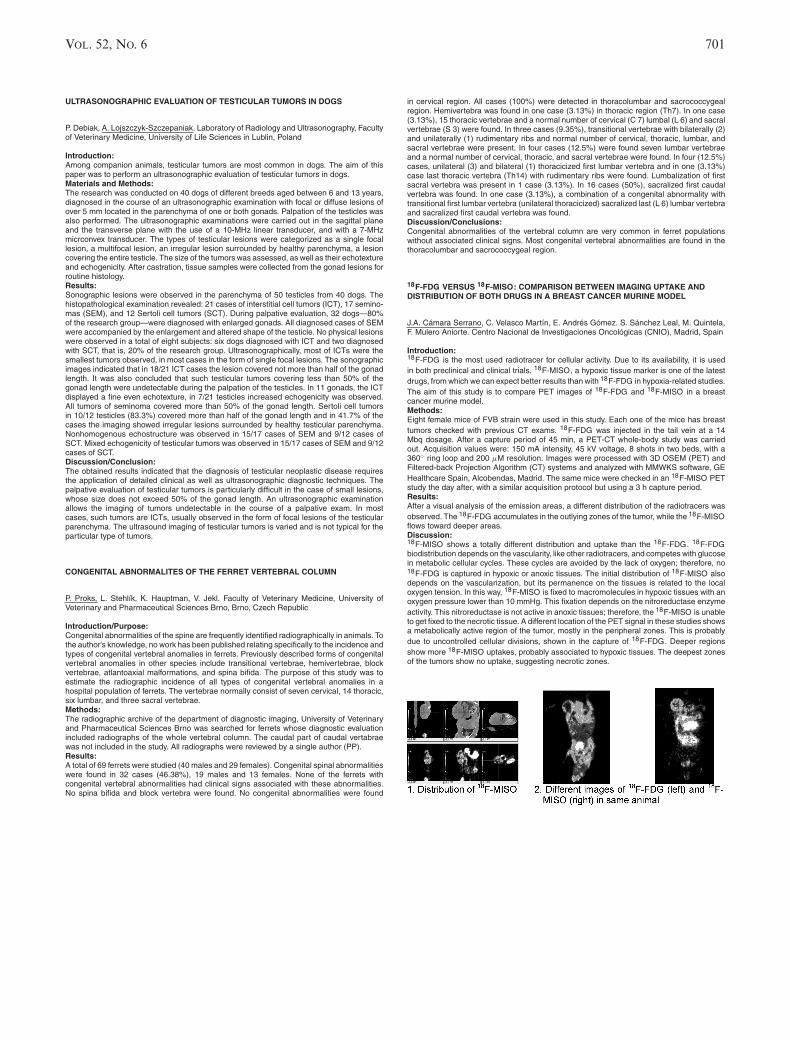
VOL. 52, NO. 6 701
ULTRASONOGRAPHIC EVALUATION OF TESTICULAR TUMORS IN DOGS
P. Debiak, A. Lojszczyk-Szczepaniak. Laboratory of Radiology and Ultrasonography, Facultyof Veterinary Medicine, University of Life Sciences in Lublin, Poland
Introduction:Among companion animals, testicular tumors are most common in dogs. The aim of thispaper was to perform an ultrasonographic evaluation of testicular tumors in dogs.Materials and Methods:The research was conducted on 40 dogs of different breeds aged between 6 and 13 years,diagnosed in the course of an ultrasonographic examination with focal or diffuse lesions ofover 5 mm located in the parenchyma of one or both gonads. Palpation of the testicles wasalso performed. The ultrasonographic examinations were carried out in the sagittal planeand the transverse plane with the use of a 10-MHz linear transducer, and with a 7-MHzmicrconvex transducer. The types of testicular lesions were categorized as a single focallesion, a multifocal lesion, an irregular lesion surrounded by healthy parenchyma, a lesioncovering the entire testicle. The size of the tumors was assessed, as well as their echotextureand echogenicity. After castration, tissue samples were collected from the gonad lesions forroutine histology.Results:Sonographic lesions were observed in the parenchyma of 50 testicles from 40 dogs. Thehistopathological examination revealed: 21 cases of interstitial cell tumors (ICT), 17 semino-mas (SEM), and 12 Sertoli cell tumors (SCT). During palpative evaluation, 32 dogs—80%of the research group—were diagnosed with enlarged gonads. All diagnosed cases of SEMwere accompanied by the enlargement and altered shape of the testicle. No physical lesionswere observed in a total of eight subjects: six dogs diagnosed with ICT and two diagnosedwith SCT, that is, 20% of the research group. Ultrasonographically, most of ICTs were thesmallest tumors observed, in most cases in the form of single focal lesions. The sonographicimages indicated that in 18/21 ICT cases the lesion covered not more than half of the gonadlength. It was also concluded that such testicular tumors covering less than 50% of thegonad length were undetectable during the palpation of the testicles. In 11 gonads, the ICTdisplayed a fine even echotexture, in 7/21 testicles increased echogenicity was observed.All tumors of seminoma covered more than 50% of the gonad length. Sertoli cell tumorsin 10/12 testicles (83.3%) covered more than half of the gonad length and in 41.7% of thecases the imaging showed irregular lesions surrounded by healthy testicular parenchyma.Nonhomogenous echostructure was observed in 15/17 cases of SEM and 9/12 cases ofSCT. Mixed echogenicity of testicular tumors was observed in 15/17 cases of SEM and 9/12cases of SCT.Discussion/Conclusion:The obtained results indicated that the diagnosis of testicular neoplastic disease requiresthe application of detailed clinical as well as ultrasonographic diagnostic techniques. Thepalpative evaluation of testicular tumors is particularly difficult in the case of small lesions,whose size does not exceed 50% of the gonad length. An ultrasonographic examinationallows the imaging of tumors undetectable in the course of a palpative exam. In mostcases, such tumors are ICTs, usually observed in the form of focal lesions of the testicularparenchyma. The ultrasound imaging of testicular tumors is varied and is not typical for theparticular type of tumors.
CONGENITAL ABNORMALITES OF THE FERRET VERTEBRAL COLUMN
P. Proks, L. Stehlık, K. Hauptman, V. Jekl. Faculty of Veterinary Medicine, University ofVeterinary and Pharmaceutical Sciences Brno, Brno, Czech Republic
Introduction/Purpose:Congenital abnormalities of the spine are frequently identified radiographically in animals. Tothe author’s knowledge, no work has been published relating specifically to the incidence andtypes of congenital vertebral anomalies in ferrets. Previously described forms of congenitalvertebral anomalies in other species include transitional vertebrae, hemivertebrae, blockvertebrae, atlantoaxial malformations, and spina bifida. The purpose of this study was toestimate the radiographic incidence of all types of congenital vertebral anomalies in ahospital population of ferrets. The vertebrae normally consist of seven cervical, 14 thoracic,six lumbar, and three sacral vertebrae.Methods:The radiographic archive of the department of diagnostic imaging, University of Veterinaryand Pharmaceutical Sciences Brno was searched for ferrets whose diagnostic evaluationincluded radiographs of the whole vertebral column. The caudal part of caudal vertabraewas not included in the study. All radiographs were reviewed by a single author (PP).Results:A total of 69 ferrets were studied (40 males and 29 females). Congenital spinal abnormalitieswere found in 32 cases (46.38%), 19 males and 13 females. None of the ferrets withcongenital vertebral abnormalities had clinical signs associated with these abnormalities.No spina bifida and block vertebra were found. No congenital abnormalities were found
in cervical region. All cases (100%) were detected in thoracolumbar and sacrococcygealregion. Hemivertebra was found in one case (3.13%) in thoracic region (Th7). In one case(3.13%), 15 thoracic vertebrae and a normal number of cervical (C 7) lumbal (L 6) and sacralvertebrae (S 3) were found. In three cases (9.35%), transitional vertebrae with bilaterally (2)and unilaterally (1) rudimentary ribs and normal number of cervical, thoracic, lumbar, andsacral vertebrae were present. In four cases (12.5%) were found seven lumbar vertebraeand a normal number of cervical, thoracic, and sacral vertebrae were found. In four (12.5%)cases, unilateral (3) and bilateral (1) thoracicized first lumbar vertebra and in one (3.13%)case last thoracic vertebra (Th14) with rudimentary ribs were found. Lumbalization of firstsacral vertebra was present in 1 case (3.13%). In 16 cases (50%), sacralized first caudalvertebra was found. In one case (3.13%), a combination of a congenital abnormality withtransitional first lumbar vertebra (unilateral thoracicized) sacralized last (L 6) lumbar vertebraand sacralized first caudal vertebra was found.Discussion/Conclusions:Congenital abnormalities of the vertebral column are very common in ferret populationswithout associated clinical signs. Most congenital vertebral abnormalities are found in thethoracolumbar and sacrococcygeal region.
18F-FDG VERSUS 18F-MISO: COMPARISON BETWEEN IMAGING UPTAKE ANDDISTRIBUTION OF BOTH DRUGS IN A BREAST CANCER MURINE MODEL
J.A. Camara Serrano, C. Velasco Martın, E. Andres Gomez. S. Sanchez Leal, M. Quintela,F. Mulero Aniorte. Centro Nacional de Investigaciones Oncologicas (CNIO), Madrid, Spain
Introduction:18F-FDG is the most used radiotracer for cellular activity. Due to its availability, it is usedin both preclinical and clinical trials. 18F-MISO, a hypoxic tissue marker is one of the latestdrugs, from which we can expect better results than with 18F-FDG in hypoxia-related studies.The aim of this study is to compare PET images of 18F-FDG and 18F-MISO in a breastcancer murine model.Methods:Eight female mice of FVB strain were used in this study. Each one of the mice has breasttumors checked with previous CT exams. 18F-FDG was injected in the tail vein at a 14Mbq dosage. After a capture period of 45 min, a PET-CT whole-body study was carriedout. Acquisition values were: 150 mA intensity, 45 kV voltage, 8 shots in two beds, with a360◦ ring loop and 200 μM resolution. Images were processed with 3D OSEM (PET) andFiltered-back Projection Algorithm (CT) systems and analyzed with MMWKS software, GEHealthcare Spain, Alcobendas, Madrid. The same mice were checked in an 18F-MISO PETstudy the day after, with a similar acquisition protocol but using a 3 h capture period.Results:After a visual analysis of the emission areas, a different distribution of the radiotracers wasobserved. The 18F-FDG accumulates in the outlying zones of the tumor, while the 18F-MISOflows toward deeper areas.Discussion:18F-MISO shows a totally different distribution and uptake than the 18F-FDG. 18F-FDGbiodistribution depends on the vascularity, like other radiotracers, and competes with glucosein metabolic cellular cycles. These cycles are avoided by the lack of oxygen; therefore, no18F-FDG is captured in hypoxic or anoxic tissues. The initial distribution of 18F-MISO alsodepends on the vascularization, but its permanence on the tissues is related to the localoxygen tension. In this way, 18F-MISO is fixed to macromolecules in hypoxic tissues with anoxygen pressure lower than 10 mmHg. This fixation depends on the nitroreductase enzymeactivity. This nitroreductase is not active in anoxic tissues; therefore, the 18F-MISO is unableto get fixed to the necrotic tissue. A different location of the PET signal in these studies showsa metabolically active region of the tumor, mostly in the peripheral zones. This is probablydue to uncontrolled cellular divisions, shown in the capture of 18F-FDG. Deeper regionsshow more 18F-MISO uptakes, probably associated to hypoxic tissues. The deepest zonesof the tumors show no uptake, suggesting necrotic zones.

702 2011
RADIOGRAPHY VERSUS COMPUTED TOMOGRAPHY TO EVALUATE VERTEBRALMALFORMATIONS IN CAPTIVE SENEGAL SOLE (SOLEA SENEGALENSIS)
A. Barreiro1, A.M. Azevedo1, J.D. Barreiro1, M. Vila1, A. Riaza2, S. Vazquez1, M.I.Quiroga1. 1Department of Veterinary Clinical Sciences, Veterinary Faculty, University ofSantiago de Compostela, Lugo, 2Stolt Sea Farm, Lira, Carnota, A Coruna, Spain
Introduction:The Senegal sole (Solea senegalensis) is a common marine flatfish with remarkable interestin aquaculture. Sole farming has important limitations such as a high larval mortality rateand a high incidence of skeletal malformations. In juvenile Senegal sole reared in captivity,the incidence of vertebral deformities is around 40%, which is a major production constraint.The purpose of the present study is to compare digital radiography and CT images forvertebral malformations detection in different batches of juvenile Senegal sole, to test thesuitability of each technique in diverse stages of development.Materials and Methods:For the present study three batches (A, B, and C) of Senegal sole were sampled from afish farm in Northwest Spain. Each batch was composed of three specimens that wereeuthanized, measured, and frozen. The mean values of age, length, and thickness of eachbatch are shown in the table below.
Age (DAH, days Standard Total
Batches after hatching) length* (cm) length** (cm) Thickness*** (cm)
A 741 23.5 (21.3–25.4) 27.1 (24.7–29.1) 2 (1.8–2.3)
B 481 14.2 (11.7–16.6) 14.8 (12.5–17) 1.1 (0.8–1.3)
C 138 8.8 (7.2–10) 9.4 (7.6–10.7) 0.5 (0.4–0.6)
*Standard length was measured from the tip of the snout to the caudal peduncle.**Total length was measured from the tip of the snout to the end of the caudal fin.***Thickness was measured in the abdominal region.
After 24 h of defrosting, fishes were radiographed in dorsoventral (DV) and laterolateral (LL)projections with a mammography digital system. After that a CT scan was performed.Results and Discussion:With respect to digital radiography, images of the three batches provided significant infor-mation about skeletal malformation in all sampled fish. LL projection greatly complementedthe DV view, offering a three-dimensional (3D) perspective of the skeleton. However, the for-mer presented interpretation difficulties because of overlapping structures, especially whenskeletal abnormalities involved twisted spines. CT scan revealed a much more completevision of skeletal conformation with discrimination of all deformed elements and vertebralalignment. It also had an exceptional 3D display of vertebrae. On the other hand, this methodwas not viable for small-sized fish (batch C), whose vertebral size is under the device defini-tion range. Radiography is an adequate method to visualize vertebral alignment and minordeformities in Senegal sole, however overlapping of some structures in animals with severevertebral malformations render difficult interpretation. X-rays are more appropriate than CTimages for detecting anomalies in fish of batch C (138 DAH). CT scan with 3D display is themost suitable technique for visualizing severe malformations in Senegal soles of batches A(741 DAH) and B (481 DAH).
COMPUTED TOMOGRAPHIC DIAGNOSIS OF BRAIN ABSCESS IN A HORSE
V.M.V. Machado, L.C. Vulcano, V.R. Babicsak, D.R. Santos, K.M. Zardo, A.S. Borges, J.J.Moreira. Universidade Estadual Paulista, Botucatu, Brazil
Case History:A 3-month-old Quarter Horse with seizures since the age of 20 days was admitted to theVeterinary Hospital, School of Veterinary Medicine and Animal Science of UNESP, Botucatu.The animal was examined with a brain computed tomography (CT) scan in transverse sec-tions, with 3-mm thickness and 3-mm interval, before and after intravenously administrationof 2 ml/kg of an ionic-iodinated contrast medium (diatrizoate meglumine, Reliev R©). The CTimages revealed the presence of a well circumscribed slightly hypoattenuating lesion (meanHounsfield units (HU) = 21.3), measuring approximately 4.9 × 5.9 cm2, on dorsoventraland transverse axis (respectively). This lesion had a slightly hyperattenuating capsule (meanHU = 38) with moderate and homogeneous contrast intensification (mean HU = 54). Theabnormal brain tissue encompassed a large area, including the left parietal, temporal, andoccipital brain lobes. A marked mass effect was visible with the falx cerebri deviated toward
the right brain parenchyma and the left lateral ventricle displaced and distorced. In the sur-rounding region, a hypoattenuating amorphous and ill-defined tissue (mean HU = 17) wasvisualized. Moreover, there were irregularity and thinning of the left parietal and temporalbone. Following the CT scan, the animal was euthanized and submitted to necropsy. In thearea corresponding to the CT lesion, an abscess was discovered with caseous material inwhich Streptococcus beta hemolytic was isolated.Discussion:Brain abscess is a rare disease in horses and often occurs secondarily to an infection involv-ing other skull structures (e.g., sinusitis, otitis media, tooth roots abscesses, and traumaticinjuries). In adult horses, the infectious agent most commonly isolated from brain abscessesis Streptococcus spp. The common clinical signs in animals with brain abscess are fever andneurological signs such as circling (ipsilateral), head pressing, seizures, mental changes,and focal neurological and ophthalmic tracts deficits. The diagnosis of brain abscesses isbased on the clinical signs, cerebrospinal fluid analysis, and imaging modalities such ascomputed tomography and magnetic resonance imaging. The CT features of an intracranialabscess include a hypoattenuated area of avascular necrotic tissue. After the injection of theiodinated contrast medium, the hypoattenuated area was surrounded by a contrast enhance-ment region representing a tissue with increased cellularity and vascularity encapsulatedby a fibrous tissue. Edema, a commonly finding secondary to brain lesions, was observedas an ill-defined hypoattenuating surrounding area. Thinning and irregularity of a diffuseportion of the parietal and temporal bone can occur by the secondary changes associatedwith the abscess. The CT scan facilitates the identification and evaluation of brain lesions,which is not possible through other methods of diagnostic imaging such as conventionalradiology and ultrasonography. Furthermore, CT may help to determine prognosis, as wellas the appropriate treatment protocol and surgical planning in cases that surgical excisionis indicated.
MRI OF THE CARTILAGINOUS GROWTH STRUCTURES OF THE DISTAL RADIUS OFHORSES
J.P. Troillet1, K. Flatz2, W. Brehm3, K. Gerlach3. 1Orthogen Veterinary GmbH, Dusseldorf,2Clinic for Small Animal Surgery and Reproduction, Veterinary Faculty, Ludwig-Maximilians-University of Munich, 3Large Animal Clinic for Surgery, Faculty of Veterinary Medicine,University of Leipzig, Germany
Introduction/Purpose:In horses, the epiphyseal plate of the distal radius is the most common site of angularlimb deformity. The epiphyseal plate’s long growth up to 30 months of age is one cause ofthis condition. Radiographic examination is used routinely, but cartilages structures can beimaged only indirectly. Magnetic resonance imaging (MRI) due to its extraordinary contrastof soft tissues is the best available technique for imaging intact and damaged cartilage. Onaccount of the epyphiseal plate of the distal radius playing a decisive role in the skeletalsystem’s degree of maturity and its common incidence of pathological changes, it seems tobe reasonable to conduct an age-related examination of this region. The aim of this studywas the imaging and description of the epiphyseal plate and epiphysis of the distal radiusand of the apophyseal plate between radius and ulna by using MRI. The constitution andpresentability of the distal region of the radius should be able to be related and characterizedby possible age-related structural changes.Materials and Methods:Twenty-eight cadaver limb segments including the distal radius of 28 orthopedically soundhorses of diverse breeds aged between 2 days and 17 years were examined by MRI. Thelimbs were examined in four different sequences (T1w-SE, T2w-TSE, PD-SE, T2 DESS-GE) and in two planes (sagittal, dorsal). Three of these physis were additionally examinedhistologically and compared with MRI. The region of growth with respect to the physis ofthe distal radial bone was documented descriptively. Age-related changes regarding thecartilaginous structures within this region were distinguished. The average thickness of theobtainable cartilage zone of the physis was measured consecutively using two sequences(T1w-SE, T2 DESS-GE). The results were compared with each other, the increasing age ofthe examined samples also being considered.Results:The hyaline cartilage of the examined area could be clearly defined by the sequencesused. Cartilaginous structures of the epiphyses and apophysis were considered to be wellassessed with MRI. In young foals, proximal to the physeal cartilage a zone of provisionalcalcification could be documented, whereas a hypointense epiphyseal subchondral boneplate was visible distally adjacent to this. In adult horses, physeal scars in that region ofthe growth plate were clearly detectable. The sample materials could be classified intofive groups due to age-related varieties. With the help of a correlation analysis, a strongconnection between the age of the evaluated individuals and the decreasing thickness ofthe cartilaginous physis could be demonstrated.Conclusion:MRI is an applicable method to assess the cartilaginous structures of the growth region ofthe distal radial bone of horses.

VOL. 52, NO. 6 703
EFFECT OF DIFFERENT HEAD AND NECK POSITIONS ON RADIOLOGICALCERVICAL INTERVERTEBRAL FORAMINA DIMENSIONS IN STANDING HORSES
D. Berner, W. Brehm, K. Gerlach. Large Animal Clinic for Surgery, Faculty of VeterinaryMedicine, University of Leipzig, Germany
Introduction/Purpose:Pathological reduction of intervertebral foramina size can be caused by ventral enlargementof the cervical facet joints. This aspect is used in plain radiographic evaluation to identifypathologically changed articular process joints. In humans, it is known that movement ofthe neck induces variation of the intervertebral foramina dimension. Specifically, extensionleads to a decrease and flexion to an increase in intervertebral foramina size. A recentcomputer tomographic study confirmed similar results in cadaver cervical spines of adultwarmblood horses. The aim of this study was to determine the changes of intervertebralforamina dimensions in three different head and neck positions in living horses.Materials and Methods:Twenty-five horses without any clinical signs of neck diseases were examined. Lateral com-puted radiographs were taken from the mid- and caudal portion of the neck in the threefollowing positions: the mouth of the horses at the level of the shoulder joint (physiologicalS1), the carpal joint (flexion S2), and the withers (extension S3). Compared to the hori-zontal line the following angles resulted: for S1—45◦ , S2—0◦ , and S3—60◦ . From theseradiographs the length of the vertebral body (D1), the height of the cranial intervertebralforamen (D2) and the height of the caudal intervertebral foramen (D3) were measured. Also,the angles between the lengths of the vertebral bodies (D1) of adjacent vertebrae werecalculated. All measurements were taken three times.Results:Lengths of the vertebral bodies as well as heights of the cranial intervertebral foraminashowed no variations between the three different head and neck positions. Compared toextension and to the physiological position, flexion caused an increase in height of the caudalintervertebral foramina of the third (P < 0.05) to seventh (P < 0.001) cervical vertebrae. Onlythe height of the caudal intervertebral foramina of the seventh cervical vertebrae (P < 0.05)in the physiological position was wider than in extension. The greatest change in angles wasfound between the sixth and seventh cervical vertebrae.Discussion/Conclusions:Flexion increases the height of the caudal intervertebral foramina of the cervical vertebrae inhorses. Our study demonstrates the importance of the head and neck position in radiographicexamination of equine cervical spines. To prevent manipulation, standardizing the head andneck position for radiographic examination of the neck should be considered.
TRANSABDOMINAL ULTRASOUND FINDINGS IN HORSES WITH CHRONIC ORRECURRENT COLIC AND/OR WEIGHTLOSS—A RETROSPECTIVE STUDY
I. Tosi1, G. Bolen2, L. Evrard2, A.-L. Etienne A.-L.2, V. Busoni2. 1Equine Clinic, 2DiagnosticImaging Section, Faculty of Veterinary Medicine, University of Liege, Sart-Tilman, Liege,Belgium
Introduction/Purpose:The aim of this poster is to describe transabdominal ultrasound (US) findings and theirprevalence in horses admitted for chronic or recurrent colic or for weight loss.Methods:Medical records of horses having undergone a complete transabdominal US in a nonemer-gency setting were retrospectively reviewed over a 4-year period. Horses were includedif they were admitted with a history of chronic or recurrent colic or weight loss. Horseswith diarrhea without weight loss were not included. The abdominal US had been real-ized with a 3.5-MHz convex transducer after clipping. Higher frequency transducers werealso used when possible to assess the gastrointestinal wall. The caudo-ventral thorax wasalso assessed in all horses for presence of free fluid. A complete thoracic US was re-alized if free fluid was visible or if clinical symptoms suggested a concurrent respiratorydisease.Results:Sixty-eight horses responded to selection criteria (mean age 11.5 years). Twenty-four horseshad a history of weight loss, 51 of chronic or recurrent colic. In two horses no abnormality wasseen, in two an extra-abdominal lesion was the only US abnormality detected. Thickeningof the gastrointestinal wall was seen in 53% cases, mainly affecting large intestine (23horses). Peritoneal fluid was judged mildly to severely increase in volume in 27 horses.Twenty-two horses had intestinal motility subjectively judged abnormal. Thoracic US showedabnormalities in 21% horses.
Discussion/Conclusions:The high prevalence of abnormal US findings in this population demonstrates the useful-ness of a complete abdominal US in horses with suspected nonacute abdominal disease.Thoracic US often added supplementary information indicating the value of a concurrent USassessment of at least a ventro-caudal thoracic window.
COMPARISON OF VARYING INJECTION RATES OF SALINE CHASERS ONINTRAVASCULAR CONTRAST ENHANCEMENT FOR DYNAMIC CT IN CATTLE
K. Lee, K. Yamada. Department of Clinical Veterinary Science, Obihiro University of Agri-culture and Veterinary Medicine, Japan
Introduction/Purpose:The use of a saline chaser results in greater efficiency of the contrast medium by avoidingaccumulation of the contrast medium at the injection site. Such increased efficiency canbe used to obtain enhancement or to reduce the volume of the contrast medium used. Areduction of the volume of the contrast medium can be economically beneficial, particularly incattle because they require a large volume of contrast medium. However, there was no studyabout effect of different saline chaser techniques on the pattern of contrast enhancementin cattle. The purpose of this study was to investigate the effect of saline chasers on theintravascular contrast enhancement when administered at three different rates.Methods:Dynamic computed tomography (CT) was performed in five normal Holstein calves. In acrossover method, group A was administered only contrast medium (600 mgI/kg, 4 ml/s).Groups B, C, and D were administered contrast medium at 30% reduction followed by salinechasers injected at 2, 4, and 8 ml/s, respectively. Attenuation values were obtained from theright and left maxillary arteries and dorsal sagittal sinus.Results:Maximum enhancement value and mean value of the enhancement plateau obtained frommaxillary arteries were significantly lower in group B than in other groups. The duration ofthe enhancement plateau was longer in group C than in groups B and D (P < 0.05).Discussion/Conclusion:Saline chaser at the rate of 4 ml/s is advocated when considering maximum contrast en-hancement and prolonged duration of the enhancement plateau in dynamic CT of cattle.
CONTRAST-ENHANCED ULTRASOUND ANALYSIS OF RENAL PERFUSION INNORMAL MICROPIGS
K. Yi, S. Ji, J. Kim, J. Yoon, M. Choi. Department of Veterinary Diagnostic Imaging, College ofVeterinary Medicine and Research Institute for Veterinary Science, Seoul National University,Seoul, South Korea.
Introduction:Contrast-enhanced ultrasound is one of the proper methods to analyze renal perfusion. Theaim of this project was to evaluate the renal perfusion pattern and perfusion dynamics in thenormal micropigs with ultrasonographic contrast media.Methods:Eight clinically healthy micropigs (11–13 kg body weight, 3–4 month of age) were studied.All micropigs underwent a physical examination, and basic blood analysis was performed.The micropigs were preanesthetized with a compound of tiletamine, zolazepam, tramadol,and medetomidine and maintained on intravenous propofol. The kidney was evaluated withB-mode harmonic ultrasound. All micropigs received an intravenous bolus of a microbubblecontrast medium (0.06 ml/kg BW, SonoVue
R©). The contrast medium was administrated via a
three-way stopcock and this was followed by a rapid bolus of 5-ml saline. With the beginningof injection, images were captured for approximately 5 min at a rate of one frame per 5 s forthe first 120 s and then one frame per 30s.Results:A significant difference in perfusion was detected between renal cortex and medulla. Therenal parenchyma was enhanced in two phases. First, cortex was enhanced, followed by amore gradual enhancement of the medulla. Mean time to peak intensity for the renal cortexfrom injection was 16.3 s, within renal medulla mean time to peak was 26.3 s.Conclusion:Contrast-enhanced harmonic ultrasound is a noninvasive imaging technique to analyzethe differentiation between cortical and medullary renal perfusion patterns. The findings ofthese data can be used as normal reference values and may prove useful in the evaluationof micropigs with renal disease.

704 2011
HEPATIC ABSCESS ASSOCIATED WITH OMPHALOPHLEBITIS IN A LAMB—CASEREPORT
R.V. Santos, V.M.V. Machado, R.M. Cavalcanti, K.M. Zardo, R.M. Amorin. Faculdade deMedicina Veterinaria e Zootecnia – UNESP, Botucatu, Brazil
Case History:A 15-day-old male mixed breed lamb was admitted to the Veterinary Hospital, School ofVeterinary Medicine and Animal Science of UNESP, Botucatu. The animal presented withhyperthermia, a 2-cm purulent wound of the umbilical remnant, a palpable enlarged intraab-dominal structure, paraparesis, and decreased reflexes in forelimbs. The cranial nervesand spinal reflexes in hindlimbs were preserved. There were no abnormalities on radiog-raphy exam of the thoracic, lumbar, and sacral spine. Therefore, an ultrasound exam wasperformed and revealed a thickened tubular structure with coarse echotexture at 1.4-cmdepth, extending from tissues surrounding the subcutaneous umbilicus into the right hepaticlobe. It was determined to be enlarged filled umbilical vein (Fig. 1). In the termination ofthis vein, a mixed echogenicity region with internal hyperechoic gas echoes was observedindicating an anaerobic infection suggestive of an ascending omphalophlebitis with hepaticabscessation. The size and shape of liver was judged to be normal, but there was anotherhyperechoic focal 1.1-cm structure within the left hepatic lobe without internal vascularizationwhen interrogated by color Doppler exam, suggesting a second hepatic abscess (Fig. 2). Anadditional round 1.5-cm anechoic structure with hyperechoic walls was detected displayingdistal acoustic enhancement within the right hepatic lobe indicating a capsular cystic (Fig.3). The patient was discharged with treatment of florfenicol and prescribed physical therapyon lumbar spine and forelimbs. Follow-up inquiry reported the lamb to be healthy after 2months.
Discussion:Routine sonographic examination of the umbilical region in neonates consists of completeexamination of umbilical remnant, umbilical vein, umbilical arteries, and urachus. The um-bilical remnant infection cases may have a favorable prognosis if there is no associatedinfection of umbilical vein and liver. The majority of umbilical vein infections do not extendcranially into the liver, but ultrasound examinations should be performed carefully throughoutthe liver for thorough examination. In this case, the sonographic evaluation was useful in dis-covering the umbilical vein infection and hepatic abscess due ascending omphalophlebitisand targeting appropriate medical therapy.
COMPUTED TOMOGRAPHIC AND RADIOLOGIC ASPECTS OF MANDIBULAROSTEOMYELITIS IN MARSH DEER (BLASTOCERUS DICHOTOMUS)
Z. Bortolini, V.M.V. Machado, R.V. Santos, C.R. Teixeira, J.L. Linardi, L.C. Vulcano. Schoolof Veterinary Medicine and Animal Science – UNESP, Botucatu, Brazil
Case History:An 8-year-old male marsh deer was admitted to the Wild Animal Medical and ResearchCenter with a history of weight loss and wasting, with a swelling of the left mandibularbone, missing third premolar and first molar teeth and gum erosions. Additionally, the buccalmucosa was accumulating food material and had focal purulent discharge. On radiography,there was proliferative bone reaction and bone lysis of the left mandible associated withan increased volume of soft tissue with internal air collections. Computed tomography (CT)exam revealed expansive bone lysis in left mandible and soft-tissue swelling with presenceof air. Cytological and microbiological material was collected for definitive diagnosis. Thecytologic diagnosis was confirmed as Actinomyces pyogenes and the animal was euthanizedconsidering the poor prognosis and extent of infection.
Fig. 1. Head axial section of first molar area showing expansive bone lysis, tooth loss andsoft-tissue swelling with presence of air. L, left.Fig. 2. Three-dimensional CT volume reconstruction illustrating extensive bone reaction inleft mandible.
Discussion:A. pyogenes is a bacterium commonly associated with osseous proliferation and destructionoften with purulent production, particularly in domestic ruminants, but has not previouslybeen reported in the Marsh Deer species. It is a common opportunistic bacterium of oralmucosa of the domestic animals, causing severe infections and spreading by blood to othertissues. In this case, the use of CT, and particularly the CT three-dimensional reconstruction,provided more detailed information on the degree of bone destructive and extent of thelesion.

VOL. 52, NO. 6 705
RADIOGRAPHIC AND TOMOGRAPHIC DIAGNOSIS OF PNEUMOTHORAX INANTEATER (TAMANDUA TETRADACTYLA)
N.F. Sesoko, V.M.V. Machado, K.M. Zardo, H.S. Oliveira, G.D.P. Soares, C.R. Teixeira. Schoolof Veterinary Medicine and Animal Science– UNESP, Botucatu, Brazil
Case History:An adult female anteater was taken to the Veterinary Hospital after vehicular trauma and itpresented with head injury, epistaxis, and prostation. The animal was restrained physicallyand chemically to perform imaging. Latero-lateral and ventral–dorsal thoracic radiographydemonstrated the cardiac silhouette shifted to right hemithorax, with dorsal deviation ofheart in relation to sternum. There was volume decrease of the left lung lobes with in-crease of lung tissue radiopacity and retraction of pleural margins in relation to the dorsalwall of thoracic cavity. There was increased radiolucency in the pleural space and caudaldisplacement of diaphragm to the left hemithorax. Computed tomography was performedwith helical transverse sections of 5-mm thick with 5-mm increments. Tomographic imagesrevealed mediastinal, trachea, and heart deviation to the right side by air in pleural spaceof left hemithorax, causing decrease in lung volume and consequent increase in density ofleft lung lobes (Figs. 1and 2). There was also complete fracture with bone axis deviation in10th–12th left ribs as illustrated in three-dimensional reconstruction (VOXAR 3D R© version6.3; Fig. ??). With the diagnosis of pneumothorax established, pneumocentesis was per-formed. Repeat thoracic radiography examination revealed increased contact of the heartsilhouette with sternum, adequate expansion of the lung lobes, and appropriate position ofdiaphragmatic crus. After observation period of 5 days, the anteater was reintroduced in itsnatural environment.Discussion:The anteater suffered traumatic rib fractures and associated pneumothorax, but the animalpresented with no clinical sign characteristic as dyspnea, restlessness, or cyanosis as oc-curs in most domestic animals with this degree of lung compromise. As only the left lunglobes were affected, it is assumed that the hemithorax of anteaters are totally divided bypleura, and because of this, the anteater maintained adequate pulmonary function and wasnot exhibiting respiratory symptoms. By eliminating overlapping tissue inherent in conven-tional radiography, computed tomography was more sensitive to confirm the diagnosis andevaluate the extent of injury.
Fig. 1: Scout image of an adult female anteater.Fig. 2: Axial section of 4◦ intercostal space revealing air in left pleural space.Fig. 3: Thoracic 3D reconstruction showing fracture in 10th–12th left ribs
DIAGNOSTIC VALUE OF COMPUTED TOMOGRAPHY AS A NONINVASIVE IMAGINGTECHNIQUE AS DEMONSTRATED IN KNUT, THE POLAR BEAR
G. Galateanu1, C.A. Szentiks1, F. Goritz1, G. Fritsch1, Andre Schule2, T. B. Hildebrandt1.1Leibniz Institute for Zoo and Wildlife Research; 2Zoological Garden, Berlin, Germany.
Introduction/Purpose:Knut, the world’s most famous polar bear, died unexpectedly, at the age of 4, on March19, 2011, following a fit of apparent seizures. Polar bear, the largest bear and the onlymarine species in the otherwise terrestrial Ursidae family, is not a customary candidate fora computed tomographic (CT) examination.Methods:The cause of death was investigated by CT examination, as a noninvasive method, priorto necropsy. A state-of-the-art, 128-slice CT scanner was used to perform the challengingwhole body examination on the 305-kg Knut.Results:The high-resolution CT images revealed cerebral lateral ventricular asymmetry in otherwisestructurally normal-looking brain. Necropsy and histological examination of the formalinfixed brain revealed panmeningoencephalomyelitis, nevertheless it showed no ventricularasymmetry and dilatation.Discussion/Conclusions:The importance and technical advantages of the CT as a noninvasive tool were demon-strated in this study. Once the brain was removed at necropsy, the cerebral fluid pressurewas released and the ventricles collapsed. The neurologic symptoms and the findings in theprior CT examination were instrumental in directing the pathologists to search for possiblerelated brain disorders, and to evaluate if there is any association of this lateral ventricleasymmetry with other structural pathologies, which caused Knut’s apparent seizure thateventually led to his death.
Copyright © 2022 FDOKUMEN