Abstracts Accepted to 2015 CDDW/CASL Winter Meeting
396
Abstracts Accepted to 2015 CDDW/CASL Winter Meeting Oral Presentations: What's New in Celiac Disease and Non Celiac Gluten-related Disorders Abstracts 1 – 1 The Intestinal Epithelium: New paradigms in sensor/effector functions Abstracts 2 – 2 CAG Selected Clinical Presentations Abstracts 3 – 8 Inflammation and Cancer Abstracts 9 – 10 Advances in Neurogastroenterology and Motility: Moving and shaping! Abstracts 11 - 12 CASL Paper Session 1 Abstracts 13 – 18 Beyond the Genome: Mechanisms of gene regulation and expression in IBD Abstracts 19 – 20 CASL Paper Session 2 Abstracts 21 – 26 CAG/CCC Student Prize Paper Presentations Abstracts 27 – 33 Poster Presentations: Poster Session1: Clinical Practice Abstracts 34- 64 Cytokines and Intracellular Signals Abstracts 65 – 69 Epidemiology and the Burden of Illness Abstracts 70 – 81 Esophagus, Gastric and Duodenal Ulcer Disorders Abstracts 82 – 91 Gastro Intestinal Oncology Abstracts 92 – 110 Immunology and Inflammatory Bowel Disease Abstracts 111 – 151 Intestinal Disorders Abstracts 152 – 165 Microbiology and Parasite-Host Interactions Abstracts 166 – 167 Viral Hepatitis Abstracts 168 – 197 Poster Session2: Acute Liver Injury and Hepatotoxicity Abstracts 198 – 200 Chronic Liver Disease Including Alcoholic, Cholestatic, & Metabolic Disease Abstracts 201 – 211 Clinical Practice Abstracts 212 – 231 Cytokines and Intracellular Signals Abstracts 232 – 233 Epidemiology and the Burden of Illness Abstracts 234 – 236 Fibrogenesis, Portal Hypertension, Complications of Cirrhosis Abstracts 237 – 241 Hepatobiliary Neoplasia Abstracts 242 – 247 Hormones, Transmitters, Growth Factors Abstracts 248 – 248 Immunobiology and Liver Transplantation Abstracts 249 – 254 Immunology and Inflammatory Bowel Disease Abstracts 255 – 300 Microbiology and Parasite-Host Interactions Abstracts 301 – 318 Motility and Nerve Gut Interactions Abstracts 319 – 339 Nutrition, Obesity and Aging Abstracts 340 – 348 Pancreatico-Biliary Disease Abstracts 349 – 356 Pediatric Liver Disease Abstracts 357 – 360 CDDW2015_lh150113
-
Upload
khangminh22 -
Category
Documents
-
view
3 -
download
0
Transcript of Abstracts Accepted to 2015 CDDW/CASL Winter Meeting

Abstracts Accepted to 2015 CDDW/CASL Winter Meeting
Oral Presentations: What's New in Celiac Disease and Non Celiac Gluten-related Disorders Abstracts 1 – 1 The Intestinal Epithelium: New paradigms in sensor/effector functions Abstracts 2 – 2 CAG Selected Clinical Presentations Abstracts 3 – 8 Inflammation and Cancer Abstracts 9 – 10 Advances in Neurogastroenterology and Motility: Moving and shaping! Abstracts 11 - 12 CASL Paper Session 1 Abstracts 13 – 18 Beyond the Genome: Mechanisms of gene regulation and expression in IBD Abstracts 19 – 20 CASL Paper Session 2 Abstracts 21 – 26 CAG/CCC Student Prize Paper Presentations Abstracts 27 – 33
Poster Presentations: Poster Session1: Clinical Practice Abstracts 34- 64 Cytokines and Intracellular Signals Abstracts 65 – 69 Epidemiology and the Burden of Illness Abstracts 70 – 81 Esophagus, Gastric and Duodenal Ulcer Disorders Abstracts 82 – 91 Gastro Intestinal Oncology Abstracts 92 – 110 Immunology and Inflammatory Bowel Disease Abstracts 111 – 151 Intestinal Disorders Abstracts 152 – 165 Microbiology and Parasite-Host Interactions Abstracts 166 – 167 Viral Hepatitis Abstracts 168 – 197 Poster Session2: Acute Liver Injury and Hepatotoxicity Abstracts 198 – 200 Chronic Liver Disease Including Alcoholic, Cholestatic, & Metabolic Disease Abstracts 201 – 211 Clinical Practice Abstracts 212 – 231 Cytokines and Intracellular Signals Abstracts 232 – 233 Epidemiology and the Burden of Illness Abstracts 234 – 236 Fibrogenesis, Portal Hypertension, Complications of Cirrhosis Abstracts 237 – 241 Hepatobiliary Neoplasia Abstracts 242 – 247 Hormones, Transmitters, Growth Factors Abstracts 248 – 248 Immunobiology and Liver Transplantation Abstracts 249 – 254 Immunology and Inflammatory Bowel Disease Abstracts 255 – 300 Microbiology and Parasite-Host Interactions Abstracts 301 – 318 Motility and Nerve Gut Interactions Abstracts 319 – 339 Nutrition, Obesity and Aging Abstracts 340 – 348 Pancreatico-Biliary Disease Abstracts 349 – 356 Pediatric Liver Disease Abstracts 357 – 360
CDDW2015_lh150113

CAG Paper Session - What's New in Celiac Disease and Non Celiac (Wheat) Gluten-related Disorders, Friday, February 27, 08h00-09h30
A1
DISRUPTION OF THE COMMENSAL MICROBIOTA WITH INCREASES IN PROTEOBACTERIA EXACERBATES HOST RESPONSES TO GLUTEN J. McCarville1, H. Galipeau1, J. Murray2, Y. Sanz3, M. Surette1, E. Verdu1 1. McMaster University, Hamilton, ON, Canada; 2. The Mayo Clinic, Rochester, MN; 3. Institute of Agrochemistry and Food Technology, National Research Council (IATA-CSIC), Valencia, Spain.
Background: Celiac disease (CeD) in an autoimmune enteropathy, triggered by the ingestion of gluten in genetically susceptible individuals (HLA DQ2/8). Not all genetically susceptible individuals develop CeD, thus, unknown environmental factors have been suggested to precipitate the disease. A microbial dysbiosis has also been described in some CeD patients, however a casual role has not yet been defined. Aims: The aim of this study was to investigate the potential pathogenic role of microbial disruption in gluten-sensitized genetically susceptible hosts. Methods: We used a model of gluten-sensitivity consisting of HLA-DQ8 transgenic mice on a Non-Obese Diabetic background (NOD-DQ8). Mice were reared perinatally on vancomycin in drinking water and subsequently orally sensitized to gliadin plus cholera toxin, followed by 2 mg of oral gluten challenges ("gluten-treated"). Controls consisted of NOD-DQ8 mice reared on sterile water receiving cholera toxin alone and vehicle for sensitizations and challenge ("control"). Separate experiments were performed in NOD-DQ8 mice with ASF microbiota (devoid of Proteobacteria). These mice underwent the same sensitization protocol, however were supplemented with a mucosally adherent strain of E. coli (ENT CA15) isolated from a human CeD patient. The microbiota was sequenced using MiSeq Illumina technology, analyzed using a custom pipeline, R and QIIME. Intraepithelial lymphocytes (IELs) were quantified by immunohistochemistry, isolated and stained with fluorochrome-labeled cell-surface markers, acquired using the LSR II and analyzed in FlowJo software. Results: After weaning and prior to gliadin sensitization, mice receiving vancomycin had shifts in microbial composition, including higher proportions of Proteobacteria (p<0.05), comprising Escherichia (p<0.05), in comparison to control mice. At endpoint, vancomycin-gluten-treated mice had more severe enteropathy in comparison to control mice, with greater reductions in villus-to-crypt (V/C) ratios (p<0.05), greater counts of IELs within villi tips (p<0.05) and increases in the CD3+βTCR+ IEL subset (p<0.05). Supplementation of ASF mice with the E. coli strain ENT CA15 led to significantly lower V/C ratios (p<0.01) and higher IELs counts (p<0.01) after gluten treatment in comparison to ASF controls. Conclusions: These results suggest that specific microbial factors related to an expansion of Proteobacteria, play a facilitatory role in gluten sensitivity in a host with genetic susceptibility. Our findings support the notion that the current increase in CeD prevalence could be prevented by microbiota-directed therapies.
Funding Agencies: CIHR

CAG Paper Session - The Intestinal Epithelium: New paradigms in sensor/effector functions, Friday, February 27, 10h00-11h30
A2
SHP-2/ERK SIGNALLING CONTROLS BARRIER FUNCTION IN THE COLON A. Langlois2, G. Coulombe2, M. Langlois2, S. Cagnol2, F. Boudreau2, G. De Palma1, P. Bercik1, E. Verdu1, N. Rivard2 1. McMaster University, Hamilton, ON, Canada; 2. Université de Sherbrooke, Sherbrooke, QC, Canada.
Background: Polymorphisms in the PTPN11 gene encoding for the tyrosine phosphatase SHP-2 were described in Japanese patients with ulcerative colitis. SHP-2 is well expressed in intestinal epithelial cells (IEC). Recently, we found that mice with an IEC-specific deletion of SHP-2 (SHP-2IEC-KO) develop spontaneous colitis one month after birth (Coulombe et al., MCB 2013). Aims: Our objective in the present study was to understand the molecular mechanisms by which SHP-2 epithelial deletion induces chronic colonic inflammation. Methods: We have analyzed by microarray (Affymetrix) the pattern of gene expression in the colon of SHP-2IEC-KO neonates, therefore well before the onset of inflammation. Variations in gene expression levels were confirmed by qPCR analyses. We crossed SHP-2IEC-KO mice with BRafV600E mice carrying a Cre-activated allele of the murine BRaf gene. Electron microscopy was performed to evaluate morphological cell differentiation. Colon histology was analyzed by hematoxylin-eosin staining, Goblet cells by Alcian blue staining and Paneth cells by immunohistochemistry (IHC) against lysozyme. Results: Intriguingly, innate defense genes including α-Defensins, Ido-1, Leap-2, Lysozyme, Reg3β and Reg3γ, emerge as the highest up-regulated genes induced in SHP-2 deficient colons. In line with this, metaplastic Paneth cells were easily found in the colon of SHP-2IEC-KO mice while Goblet cell number was clearly diminished. These alterations in Goblet/Paneth cell ratio in the colon of SHP-2IEC-KO mice were observed rapidly after birth, before the onset of inflammation and were associated with significant alterations in microflora composition (more enterobacteriaceae, less firmicutes) and profound alteration in ERK and βeta-catenin signallings. Remarkably, concomitant expression of an activated form of BRaf in SHP-2IEC-KO mice rescued ERK activation, promoted goblet cell production, inhibited Paneth cell expansion in the colon and prevented colitis. Conclusions: SHP-2-dependent ERK signalling controls the choice between Goblet and Paneth cell fate in the intestine. Dysregulation of epithelial cell fate or differentiation in the colon have serious consequences for the host as exemplified with SHP-2 deficient mice that rapidly develop severe colitis.
Funding Agencies: CIHR
CAG Paper Session - CAG Selected Clinical Presentations, Friday, February 27, 10h00-11h30
A3
MUCUS ATTENUATION AND GOBLET CELL DEPLETION IN THE TERMINAL ILEUM OF CHILDREN WITH ULCERATIVE COLITIS PROMOTES BACTERIAL INTERACTION WITH THE MUCOSAL SURFACE
M. Alipour, D. Zaidi, A. Fu, C. Sergi, M. Carroll, H. Huynh, E. Wine
University of Alberta, Edmonton, AB, Canada.
Background: The partitioning of bacteria and the intestinal epithelial lining by the mucus layer forms an integral barrier for gut homeostasis. In patients with inflammatory bowel diseases (IBD), a breakdown of the mucus barrier correlates with increased inflammation, and bacteria-mucosal interaction. Aims: Our aim was to investigate goblet cell proportion, mucin secretion, and bacterial abundance in the terminal ileum of children with Crohn disease (CD), ulcerative colitis (UC), and non-IBD controls. Methods: Mucosal biopsies from the terminal ileum were collected during ileoscopy and paraffin-embedded. Formalin-fixed sections were histologically graded, and Methacarn-fixed sections quantitatively assessed for goblet cell and mucus production with Alcian blue/Periodic acid-Schiff staining. Biopsies were also assessed for bacteria (EUB338) by FISH, mucin (MUC2) and immunoglobulin-A and -G by immunofluorescence. Results: In the terminal ileum of children with UC, the proportion of goblet cells were depleted and mucus secretion was significantly lower, compared to CD and non-IBD patients. Co-staining for mucin and bacteria showed infiltration of the mucosal layer, and bacteria were found in close proximity to the epithelial lining in children with CD and UC compared to non-IBD. As well, the production of IgA and IgG in the lamina propria and secretion into the lumen was increased in CD and UC patients. Conclusions: Here we show that the terminal ileum of children with UC confer to mucus and goblet cell depletion, with an increase in bacteria in contact with the mucosal layer and elevated IgA/G response in both CD and UC.
Funding Agencies: CAG, CIHR, AIHS

A4
SMOKING'S INFLUENCE ON THE RISK OF SURGERY FOR THE INFLAMMATORY BOWEL DISEASES IS DEPENDENT ON AGE AT DIAGNOSIS
A. Frolkis, J. deBruyn, N. Jette, M. Lowerison, I. Vallerand, S. Patten, B. Eksteen, C. Barnabe, R. Panaccione, S. Ghosh, S. Wiebe, G. Kaplan
University of Calgary, Calgary, AB, Canada.
Aims: We assessed the effect of smoking status at the time of diagnosis of the inflammatory bowel diseases (IBD) on the need for early IBD-related surgery. We hypothesized that smoking would increase the need for early surgery in Crohn's disease, but not in ulcerative colitis. Methods: The Health Improvement Network was used to identify an inception cohort of Crohn's disease (n=1519) and ulcerative colitis (n=3600) patients from 1999-2009. Poisson regression explored temporal trends for the proportion of newly diagnosed IBD patients who never smoked prior to their diagnosis and the risk of surgery within 3 years of diagnosis. Cox proportional hazard models assessed the association between smoking and intestinal resection after adjusting for covariates. Effect modification was explored for age at diagnosis. Results: From 1999-2009 the rate of patients without a history of smoking increased for newly diagnosed Crohn's disease patients increased by 3% per year (incidence rate ratio [IRR] 1.03; 95% confidence interval [CI]:1.02-1.05), but not for ulcerative colitis (Figure 1). The rate of surgery within three years of diagnosis only decreased amongst Crohn's disease patients aged 17-40 years at the time of diagnosis (IRR 0.96; 95% CI:0.93-0.98). Smoking at diagnosis increased the risk of surgery for Crohn's disease patients diagnosed after the age of 40 (hazard ratio [HR] 2.99; 95% CI:1.52-5.92), but not for those diagnosed before age 40. Ulcerative colitis patients diagnosed between the ages of 17 and 40 years and who quit smoking prior to their diagnosis were significantly more likely to undergo a colectomy within three years of their diagnosis (ex-smoker versus never smoker: HR 1.66; 95% CI: 1.04-2.66). Conclusions: The effect of smoking on surgery is dependent on the age at diagnosis of IBD. These data suggest that longstanding smoking drives the rate of surgery in Crohn's disease, whereas quitting smoking at a younger age increases the risk of colectomy for ulcerative colitis.
Figure 1: Proportion of patients without a prior history of smoking prior to the diagnosis of IBD.
Funding Agencies: AIHS

A5
RISING USE OF ANAESTHESIOLOGY ASSISTANCE FOR OUTPATIENT COLONOSCOPY IN ONTARIO: AN UPDATE
B. Bielawska3, J. Tinmouth3, L. Paszat3, L. Rabeneck1, L. Hookey2
1. Cancer Care Ontario, Toronto, ON, Canada; 2. Queen's University, Kingston, ON, Canada; 3. University of Toronto, Toronto, ON, Canada.
Background: Rates of anaesthesiology assistance (AA) in colonoscopy for the purpose of administering propofol sedation are rising. In Canada, the uptake of AA has been most pronounced in Ontario. The most recent published figures derived from Ontario health databases indicated a rise of AA use in colonoscopy to 19% by 2005, a figure that has continued to increase in the absence of regulation for this practice. Routine use of AA in colonoscopy portends significant additional health care cost. Furthermore, evidence is emerging that deep sedation may negatively impact the safety and quality of colonoscopy, although further studies are needed. Aims: The purpose of this study is to obtain an updated estimate on patterns and rates of AA for outpatient colonoscopy in Ontario. Methods: Retrospective population based analysis of outpatient colonoscopy in Ontario adults aged 18 and older using databases from the Institute for Clinical Evaluative Sciences. Colonoscopy was defined by Ontario Health Insurance Plan (OHIP) fee codes indicating insertion of a colonoscope to or beyond the hepatic flexure. Exposure to AA was defined by anaesthesia-specific OHIP fee codes on the day of colonoscopy. Patients who underwent concurrent upper endoscopy or other procedure that may have required same day anaesthesia were excluded. Patient (age, sex and comorbidity), endoscopist (specialty, sex, mean annual colonoscopy volume and years in practice), institution (type) and procedure (biopsy and polypectomy) characteristics were derived from the administrative data. Descriptive data are reported. Results: A total of 2,861,282 outpatient colonoscopies performed between April 2002 and December 2012 were included. 51% of the cohort were female and 78% were over age 50. The overall rate of AA was 29.1%. 64% of colonoscopies with AA were performed by surgeons and 30% by gastroenterologists, compared to 47% each for non-AA colonoscopies. The median annual colonoscopy volume was higher and the number of years in practice was lower for physicians performing AA than non-AA colonoscopies (544 vs 479, p<0.001 and 23 vs 24, p<0.001, respectively). Community and non-hospital settings accounted for 60.7% and 33.1% of AA use in colonoscopy, respectively. Fewer than 1% of colonoscopies at teaching hospitals used AA. There was a significant increase in AA use over time, from 13.6% in 2002 to 44.1% in 2012. Conclusions: Almost half of outpatient colonoscopies in Ontario are now performed with AA, with the greatest utilization among surgeons and in nonhospital and community settings. This practice has significant implications for healthcare costs. Further investigation into the impact of AA on quality and safety outcomes in colonoscopy is planned.
Funding Agencies: Physician Services Incorporated

A6 ASSESSMENT OF A COLONOSCOPY TRIAGE SHEET FOR USE IN A PROVINCE-WIDE POPULATION-BASED COLORECTAL SCREENING PROGRAM N. Sharara1, S. Nolan1, M. Sewitch2, M. Martel1, M. Dias1, A. Barkun1 1. Division of Gastroenterology, McGill University Health Center, Montreal, QC, Canada; 2. McGill University, Montreal, QC, Canada. Background: Based on guidelines, a colonoscopy triage sheet (CTS) was designed for province-wide use in the Quebec population-based colorectal cancer screening program. The aim of the CTS is to permit equitable and uniform triaging of all patients requiring colonoscopy in the province. The CTS lists a hierarchy of 16 identified indications that are matched to 6 priorities, (symptoms-P1 to P4, average-risk screening-P5, surveillance-P6) with correspondingly increasing target delays for colonoscopy. Aims: To compare the priority and indication selected by the referring physician on the referral sheet with the one assessed by the endoscopist on the day of the colonoscopy and assess the yield of the different indications' priorities of the triage sheet. Methods: A retrospective study was conducted of all patients referred to an adult specialty hospital for colonoscopy. Abstracted data included patient age, gender, priority according to the CTS and the endoscopy report, bowel preparation adequacy, cecal intubation, significant endoscopic findings (cancer, ileocolitis, polyp >10mm). Weighted kappa was calculated to assess priority inter-rater agreement between referring physician and endoscopist. Multivariable models were created to identify independent predictors of cancer and of agreement on priority ratings. Results: Data on 1230 patients were collected (age 60.3+12.1yrs, 52.5% female, 86.7% good or excellent preparations 95.9%, cecal intubation and 45.6% polyp detection rate. Significant findings included cancers (1.7%), polyps >10mm (20%), and ileocolitis (7.2%). Priority ratings are listed in Table 1. The weighted kappa value for all colonoscopies was 0.55 (0.51; 0.59). Predictors of cancer were increasing age (in years, OR=1.13; 95%CI(1.06; 1.20)), and CTS priority of 1 or 2 (9.54 [1.73; 52.4]). Significant predictors of increased priority rating agreement between referring physician and endoscopist were ratings of P4 and P5. Conclusions: Agreement on triaging priorities between referring physician and endoscopist was moderate-good. Predictors of increased agreement were related to the selection of less urgent priority ratings. Predictors of cancer were age and urgent priority ratings. These findings appear to validate the CTS hierarchal priority rating scheme. Physician education may be required to improve CTS priority rating selection.
Referring Physician Priority
(%; 95%CI)
Endoscopist Priority
(%; 95%CI) Immediate <24 hours 0.3 (0.0; 0.6) 0.0
Urgent <14 days 1.9 (1.1; 2.7) 3.9 (2.7; 5.1) Semi-elective <60 days 28.3 (25.6; 31.0) 21.8 (19.3; 24.2)
Elective <6 months 12.7 (10.7; 14.7) 20.3 (17.9; 22.8) Average-risk screening or chronic constipation or
diarrhea 44.5 (41.5; 47.6) 41.3 (38.3; 44.3)
Surveillance 12.3 (10.3; 14.2) 12.7 (10.7; 14.7) Funding Agencies: None

A7
VALIDATION OF AN ELECTRONIC CLINICAL DECISION-MAKING TOOL FOR PATIENTS WITH SUSPECTED COLORECTAL CANCER: A PILOT STUDY N. Forbes, M. Cooray, M. Hackett, N. Shah, T. Corner, D. Chan, M. Mills, D. Armstrong, T. Xenodemetropoulos, McMaster University, Hamilton, ON, Canada. Aims: In patients presenting to primary care with rectal bleeding, accurate identification of those with a high probability of colorectal cancer (CRC) is challenging. The CarePath-CRC electronic clinical decision-making application was designed to assist primary care providers at the point-of-care with assessment and referral of patients with suspected CRC. The physician completes an interactive checklist with relevant evidence-based clinical parameters (Figure 1) and a recommended urgency of referral is then generated based on the post-test probability of CRC. This pilot study aimed at validation of the tool for use in the assessment of patients presenting with rectal bleeding. Methods: The Hamilton CRC Pathway was implemented to expedite assessment and management of patients with suspected CRC following colonoscopy. A clinical database of information for all patients with histologically-confirmed CRC referred to the Pathway was reviewed. The CarePath-CRC tool was applied retrospectively in a mock fashion to all patients in this database who initially presented with rectal bleeding, to determine its sensitivity for detecting CRC. A generated recommendation of ‘immediate referral' (referral <24 hours, expected endoscopy < 2 weeks) or ‘urgent referral' (expected consultation and endoscopy < 4 and < 8 weeks) was considered a positive test result. An a priori sensitivity of 90% was deemed adequate, based on test characteristics of the tool's individual clinical criteria. Results: A total of 557 CRC Pathway records from 2010-2014 were reviewed. Of these, 45 records with insufficient clinical information and 231 records of patients lacking an initial presentation of rectal bleeding were excluded. The CarePath tool was applied to the remaining 281 patients. A total of 69 (24.6%) and 188 (66.9%) patients met criteria for immediate and urgent referral, respectively. The remaining 24 study patients (8.5%) met criteria for ‘possible priority referral', while none met criteria for ‘no specific action recommended'. Given the lack of consistent recording of the presence of perianal symptoms, patients with unclear records were treated as having perianal disease and, thus, assigned to a less urgent referral pathway. This resulted in a calculated sensitivity of 91.5% (95% CI: 87.4%-94.3%). Other data on clinical parameters, investigations, wait times, lesion location, pathology and treatment, were also analyzed. Conclusions: The CarePath-CRC tool is sensitive (>90%) in the prediction of CRC in patients presenting with rectal bleeding. A prospective cohort study is being designed to allow for acquisition of comprehensive test performance characteristics and full validation of the instrument.
Figure 1. The CarePath-CRC interactive electronic interface. Funding Agencies: CAG

A8
INTRODUCING NON-FINANCIAL CONFLICTS OF INTEREST IN RESEARCH AND GUIDELINE DEVELOPMENT
Y. Lu1, N. Sharara2, D. Jones1, T. Kaltenbach3, M. Martel1, A. Barkun1
1. McGill University, Montreal, QC, Canada; 2. Harvard University, Boston, MA; 3. Standford University, Standford, CA.
Background: There has been increased awareness and scrutiny over financial conflicts of interest (COI), now widely recognized and addressed in Medicine. However the concept of non-financial COI (NFCOI) remains obscure and direction on the topic is largely inexistent. NFCOI are COIs that are not financial, such as personal beliefs, personal or institutional relationships and interest in career advancement. In the setting of guideline development, NFCOI may necessitate even greater attention. Aims: To explore the awareness and perception of NFCOI by participants in a consensus guideline meeting, and assess impact on voting. Methods: As part of the international SCENIC meeting on surveillance for colorectal neoplasia surveillance in inflammatory bowel disease, an Ethics ad hoc committee was created to oversee COI issues. The above definition for NFCOI was adopted. All participants completed a validated short-answer questionnaire that included 11 questions on NFCOI perception, and after the meeting, joined an additional online discussion on NFCOI. Qualitative analysis using participants' answers from both exercises was performed with NVIVO software using coding and thematic groupings. A separate analysis was carried out to compare voting results on a 5-point Likert according to each participant's publication history and its pertinence to NFCOI. Inferential testing with descriptive statistics was completed. Results: A total of 26 participants responded; 65% of had authored or publicly provided an opinion related to the medical topic of the meeting. When asked about the influence of NFCOI on voting, 81% reported no impact, 15% some impact (15% refused to respond). However, 54% believed NFCOI had influenced other participant's voting. Qualitative analysis highlighted that 1)NFCOI is a broad area that spans institutional and career pressures, as well as personal beliefs, 2)the issue of whether, and to what extent NFCOI have impact is not well understood. Publication history was relevant for 56% of voting members. A higher numeric percentage for "strongly agree" was seen in this group, but overall voting distribution was similar. This study is limited by the small number of participants and the selective nature of the medical topic. Conclusions: NFCOI may influence voting in consensus guideline development. Such influence is perceived more frequently for other participants' NFCOI than for the participant's own NFCOI. The concept of NFCOI is poorly understood and difficult to define yet requires better characterization and quantification.
Funding Agencies: None

CAG Paper Session - Inflammation and Cancer - Friday, February 27, 12h30 – 14h30
A9
TRANSCRIPTOMIC AND PROTEOMIC ANALYSIS OF HNF4α ISOFORMS FUNCTIONS SUPPORT OPPOSITE ROLES FOR THESE DURING COLON CANCER.
J. Babeu, J. Carrier, F. Boisvert, F. Boudreau,
Université de Sherbrooke, Sherbrooke, QC, Canada.
Background: Carcinogenesis is defined by the interplay of tumor suppressor gene inactivation and oncogene activation leading to the misregulation of gene expression. HNF4alpha, a master regulator of gene expression in intestinal epithelial cells, has recently been associated with colorectal cancer (CRC). Being an attractive molecule for CRC therapy, its development as a potentially new druggable target has been slowed down by the ongoing controversy of whether it acts as a tumor suppressor gene or as an oncogene. Aims: To clarify the functional roles of HNF4alpha P1 and P2 isoforms in CRC by identifying their specific genes networks and interacting partners. Methods: P1 and P2 isoforms expression in CRC samples and CRC cell lines was determined by immunofluorescence and qPCR. Transcriptomes of Caco2/15 cells expressing shRNA against P1 or P2 isoforms were determined by RNAseq. Isoforms specific target genes networks were established by comparing RNAseq data to available HNF4alpha ChIPseq datasets. HNF4alpha isoforms specific partners were identified by SILAC quantitative proteomic approach following immunoprecipitation of P1 or P2 isoforms. Results: In CRC, P1 expression was drastically reduced in most patients (86%) while P2 expression was either maintained or increased (75%), a pattern that was maintained in most CRC cell lines. In Caco2/15 cells, RNAseq datas indicated that P1 and P2 isoforms could regulate different subsets of genes. Inhibition of P1 isoforms in Caco2/15 cells led to changes in gene expression promoting cancer while inhibition of P2 isoforms led to changes in genes signature associated with cancer inhibition. Moreover, SILAC proteomic analysis identified novel HNF4alpha isoforms partners predicted to be involved in the DNA damage response. Conclusions: In CRC cells, P1 and P2 isoforms regulate different sets of genes supporting that P1 isoforms are involved in tumor suppressor function while P2 isoforms can maintain cancer-promoting functions. These identified functions are in line with the observed loss of P1 isoforms and the maintenance of P2 isoforms during CRC. Based on the identification of novel interacting partners, HNF4alpha isoforms could also be implicated in DNA damage response strengthening its potential as a new target for therapy. Further studies are ongoing to clarify these new HNF4alpha P1 and P2 isoforms specific functions during CRC. This work was supported by a grant from CIHR.
Funding Agencies: CIHR

A10
DEFINING THE ROLE OF MULE/HUWE1/ARF-BP1 IN INTESTINAL CANCER
C. Dominguez-Brauer
Princess Margaret Hospital, Toronto, ON, Canada.
NOT PUBLISHED AT AUTHOR’S REQUEST
Funding Agencies: None

CAG Paper Session - Advances in Neurogastroenterology and Motility: Moving and shaping!, Saturday, February 28, 08h00-10h00
A11
FUNCTIONAL IMPACT OF IBS MICROBIOTA ON THE GUT-BRAIN AXIS
G. De Palma1, M. Lynch2, J. Lu1, V. Dang1, Y. Deng1, J. Jury1, G. Umeh1, P. Miranda1, M. Pigrau1, S. Sidani1, P. McLean3, G. Moreno-Hagelsieb4, M. Surette1, G. Bergonzelli3, E. Verdu1, P. Britz-McKibbin1, J. Neufeld2, S. Collins1, P. Bercik1
1. McMaster University, Hamilton, ON, Canada; 2. University of Waterloo, Waterloo, ON, Canada; 3. Nestle Research Center, Lausanne, Switzerland; 4. Wilfrid Laurier University, Waterloo, ON, Canada.
Background: Irritable Bowel Syndrome (IBS) is a disorder of the gut-brain axis, characterized by altered gut function and frequent psychiatric co-morbidity. We have previously shown that mice colonized with microbiota from patients with diarrhea predominant IBS and comorbid anxiety exhibited faster gastrointestinal transit, impaired intestinal permeability, elevated tissue b-defensin-3 levels and anxiety-like behavior compared to mice colonized with healthy microbiota. The underlying mechanisms are, however, not fully understood. Aims: To investigate the effect of IBS microbiota on host metabolism and immune function using gnotobiotic mouse model. Methods: Sera from mice colonized with microbiota from five healthy volunteers and 6 patients with IBS (10 mice per human donor) were analyzed by liquid chromatography-time of flight-mass spectrometry (LC-TOF-MS). The expression of 185 inflammation-related mouse genes was measured with NanoString nCounter® Gene Expression CodeSet on total RNA extracted with RNeasy Mini Kit (Qiagen) from colonic sections of mice with IBS and healthy microbiota. Results: Several innate immunity-related genes were found up-regulated in IBS-colonized mice compared to controls, including lymphotoxin alpha (LT-α), IL-22ra2, CXCR4, C3, IL1a, Ptk2, MknK1, Limk1 and Rapgef2. In addition, the expression of CXCR3 was decreased in IBS-colonized mice compared to controls. The metabolomic profiles of IBS mice were different from those of healthy mice, and clustered in three separate subgroups. When analyzing the individual metabolites, O-acetyl-L-carnitine and several lysophosphatidylcholine (LPC) species were significantly increased, whereas phosphatidylserine (PS) metabolites were decreased in IBS mice. When analyzing differences between the three IBS subgroups, we found altered levels of palmitic, oleic and stearic acids, glycerophosphocholine, LPC and PS species. Conclusions: Our results demonstrate that the gut microbiota from patients with IBS has the capacity to perturb colonic immune homeostasis and affects host metabolism, altering levels of metabolites with immune and neuroactive properties. These data further support the hypothesis that gut microbiota plays a key role in the pathophysiology of IBS.
Funding Agencies: CIHR, Nestle Switzerland

A12
GRANULOCYTE-COLONY STIMULATING FACTOR MEDIATES NOCICEPTIVE SENSITIZATION AND VISCERAL PAIN IN A MURINE MODEL OF COLITIS
T. Lapointe, C. Altier
University of Calgary, Calgary, AB, Canada.
Background: Abdominal pain is the most common symptom of inflammatory bowel disease (IBD). Nevertheless, the pathophysiological mechanisms involved in the sensitization of pain signaling pathways in IBD remain incompletely understood. A variety of pro-inflammatory mediators are known to participate in neuronal sensitization. Although granulocyte-colony stimulating factor (G-CSF) has mostly been studied for its involvement in inflammatory processes, recent findings have suggested that it could also play a role in nociception. Notably, G-CSF receptor expression has been reported in sensory afferent neurons. Furthermore, intraplantar injection of G-CSF has been shown to induce thermal and mechanical hyperalgesia, and the blockage of its receptor as proven effective in attenuating tumor-induced hyperalgesia in a model of bone cancer. While these observations point towards a role of GSCF in nociceptive sensitization, its involvement in colitis-associated pain remains unexplored. Aims: This study aimed at establishing the role of G-CSF in neuronal sensitization and visceral pain in the context of IBD using the dextran sulfate sodium (DSS) murine model of colitis. Methods: Colonic inflammation was induced by administration of 2.5% DSS in drinking water for 7 days, and visceral pain assessed by spontaneous nocifensive behaviours in response to intracolonic administration of mustard oil (MO). The direct effect of G-CSF on central sensitization and pain was evaluated by intrathecal injection of G-CSF (5ng) in healthy mice, one hour prior to pain behaviour testing. In vitro experiments were performed on cultured dorsal root ganglion (DRG) neurons treated with 200ng/mL G-CSF for 24 hours. Results: Mice treated with DSS showed increased nocifensive responses to intracolonic administration of MO up to five weeks post-DSS treatment. While no changes in the levels of the pro-inflammatory cytokines interleukin (IL)-1β, IL-6, and tumor necrosis (TNF)-α could be detected between control and DSS-treated mice, a significant increase in G-CSF was observed in the acute phase of colitis, in both colon-innervating DRGs and the spinal cord. Importantly, healthy mice subjected to intrathecal administration of G-CSF also showed increased sensitivity to MO. Finally, exposure to G-CSF induced a significant increase in the expression of the growth factors NGF (nerve growth factor), BDNF (brain-derived neurotrophic factor) and GDNF (glial cell-derived neurotrophic factor) in cultured DRG neurons. Conclusions: Collectively, our data indicate that, in the context of colitis, G-CSF could participate in nociceptive sensitization by mediating the expression of neurotrophic factors, and thus contributing to pain circuit plasticity and the establishment of chronic abdominal pain.
Funding Agencies: CAG, CIHR, Alberta Innovates - Health Solutions

CASL Paper Session 1 - Saturday, February 28, 08h30-10h00
A13
A NOVEL MELD EXCEPTION POINT SYSTEM FOR HEPATOCELLULAR CARCINOMA PROMOTES EQUITABLE LIVER ALLOCATION
M. Bhat2, P. Ghali2, A. Roy3, P. Chaudhury2, F. Alvarez4, M. Carrier3, M. Bilodeau1
1. CRCHUM, Montréal, QC, Canada; 2. McGill University, Montreal, QC, Canada; 3. Universite de Montreal, Montreal, QC, Canada; 4. CHU-Sainte Justine, Montreal, QC, Canada.
Background: The current Model for End-Stage Liver Disease (MELD) point system for Hepatocellular Carcinoma (HCC) in the United States tends to disproportionately favor these patients as compared to those who undergo liver transplantation (LT) for liver failure (LF) based on biological MELD scores. Given these concerns, Transplant-Québec liver committee decided in July 2009 to implement a novel separate MELD pointing system to allow liver allocation for patients with HCC based on graded tumor diameters over time. Cut-offs were chosen based on median MELD at LT over the preceding year. Aims: The aim of this study was to determine the evolution of patients listed for HCC with this scoring system, and how this compared to those patients transplanted for LF based on their MELD score. Methods: In this retrospective study, we evaluated the evolution of all patients listed for LT in Québec, from time of implementation of the scoring system (detailed in the Table) up to May 2014. Points were reassigned every 3 months or upon repeat imaging, depending on changes in tumor size. Patients listed for fulminant liver failure, for exception point indications and children were excluded. Results: 524 patients were listed for LT from July 2009 to May 2014, of whom 94 (17.9%) were assigned MELD HCC points. The majority were male (70.4%), with mean age of 55.4 years. 83.7% underwent liver transplant. 28% of patients listed for HCC required changes in allocated points over time. The mean upgrade in number of points for all HCC patients was 0.32 points+/-0.53. There was no difference between the 2 indications with respect to transplantation rates (HCC 86.1% versus LF 83.3%, p=0.48), waiting time in days (HCC 258 versus LF 325; p=0.20) or waiting list death rates (HCC 0.6% versus LF 9.2%; p=0.11). At the time of LT, HCC patients had a lower MELD score (HCC 22+/-0.3 versus LF 24+/-0.4; p=0.02): therefore, the allocated HCC-MELD score does not seem to jeopardize LF over HCC patients. Conclusions: Our study demonstrates that a novel MELD point system for HCC, which takes into account changes in tumor size as a reflection of tumor biology over time, allows for a more equitable allocation of organs. This system potentially represents an improvement upon the standard MELD exception point system for HCC employed in the United States, but needs to be validated in a broader context.

Funding Agencies: CIHR

A14
THE IMPACT OF NURSING VOLUME ON HOSPITALIZATION OUTCOMES AMONG PATIENTS WITH CIRRHOSIS: A POPULATION-BASED STUDY
A. Shaheen, G. Kaplan, K. Burak, M. Swain, R. Myers
University of Calgary, Calgary, AB, Canada.
Background: Hospitalized patients with cirrhosis have high morbidity and mortality rates and require complex medical care. Although the intensity of nursing care has been associated with improved outcomes in many conditions, the role of nursing staff volume in patients with cirrhosis has not been examined. Aims: Our objective was to assess the association between nurse staffing and hospitalization outcomes in patients with cirrhosis. Methods: We used the 2008 Nationwide Inpatient Sample (NIS) database to identify cirrhosis-related hospitalizations in the United States. An admission was considered cirrhosis-related if the primary diagnosis was cirrhosis or a liver-related complication (based on ICD-9 codes). Hospital-level nursing staff volume, which included the volume of registered nurses, licensed practical nurses and nurse aids, was categorized into tertiles (low [<4.8], medium [4.8-6.2], and high-volume [>6.2 nurse full-time equivalents [FTEs] per 1,000 adjusted inpatient days]). Weighted regression models assessed the impact of nursing volume on mortality, length of hospital stay (LOS) and hospitalization charges. We adjusted for patient (e.g. demographics, insurance, and Elixhauser comorbidities) and hospital characteristics, including hospital volume for cirrhosis-related admissions Results: There were 41,183 cirrhosis-related hospitalizations in 2008 corresponding to an estimated 201,439 admissions in the United States. Compared with patients admitted to hospitals with low nursing volume, those hospitalized in high nursing volume centres were younger (median age: 55.1 vs. 57.1; P<0.001), more likely to be privately insured (32.6% vs. 24.2%; P<0.001), and more frequently admitted to high-volume (for cirrhosis) hospitals (65.9% vs. 10.2%; P<0.001). The prevalence of ≥2 comorbid conditions was similar between groups (low vs. high nursing volume: 79.0% vs. 73.5%; P=0.06). Although in-hospital mortality was similar across nursing volume groups (high, medium, and low: 7.2%, 7.4%, 7.4% respectively; P=0.92), patients admitted to high nursing volume hospitals had increased LOS (4.1 vs. 3.8 days) and hospitalization charges ($27,541 vs. $20,135) compared to low nursing volume centres (both P<0.001). After adjustment for patient and hospitalization characteristics, nursing volume was not an independent predictor of in-hospital mortality (high vs. low-volume: odds ratio 1.02; 95% CI 0.86-1.22), LOS, or hospitalization charges. Conclusions: Among patients hospitalized for cirrhosis-related conditions, nursing staff volume is not associated with hospitalization outcomes including mortality, LOS, or hospital charges.
Funding Agencies: None

A15
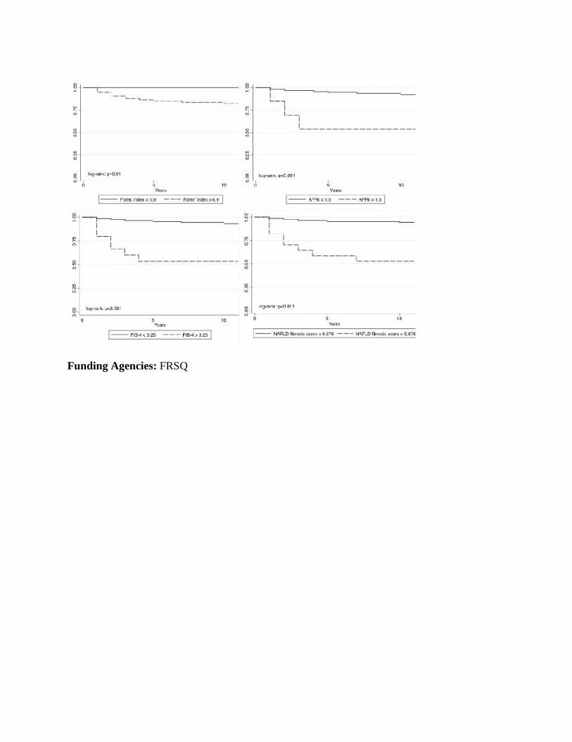
SERUM FIBROSIS BIOMARKERS PREDICT DEATH AND GRAFT LOSS IN LIVER TRANSPLANT RECIPIENTS: A LONGITUDINAL STUDY OF 594 PATIENTS G. Sebastiani, M. Bhat, A. Bhat, K. Rollet, M. Deschenes, P. Wong, P. Ghali McGill University Health Centre, Montreal, QC, Canada.
Background: Serum fibrosis biomarkers predict clinical outcomes in pre-transplant patients with chronic liver disease. However, this has never been addressed in the liver transplant population. Aims: We investigated the role of diagnostic serum biomarkers for liver fibrosis and steatosis to predict death and graft loss after liver transplantation (LT). Methods: We included consecutive patients who underwent LT and met the following criteria: patients with graft survival of >6 months; serum biomarkers to diagnose hepatic fibrosis and steatosis (APRI, FIB-4, NAFLD fibrosis score, hepatic steatosis index) available within 3 months before LT; a minimum follow-up of 1 year. Kaplan-Meier survival analysis and multivariate Cox proportional hazard models were used. Models for death were adjusted for age, sex, BMI, glucose, HCV positivity, history of HCC, post-transplant need for dialysis and type of immunosuppressive therapy. Models for graft loss were adjusted for age, sex, BMI, glucose, HCV positivity, post-transplant need for dialysis, cold ischemia time and type of immunosuppressive therapy. If patients had been retransplanted, the transplant with which they had the longest graft survival was included. Results: 594 consecutive patients (median age 61 years, 69% male) were included in 1991-2011 in a single centre. Over a mean 11.7 (standard deviation 7.9) years follow-up, 37% of patients died and 30% lost the liver graft. After adjustments, the following biomarkers were associated with death on multivariate analysis: APRI (HR 1.01; 95% CI 1.00-1.02, p=0.008), FIB-4 (HR=1.01; 95% CI 1.00-1.02, p=0.008), NAFLD fibrosis score (HR=1.02; 95% CI 1.00-1.04, p=0.04). Other covariates significantly associated with death were HCV positivity (HR=2.2; 95% CI 1.51-3.22, p<0.001), glucose (HR=1.08; 95% CI 1.02-1.15, p=0.007), post-transplant need for dialysis (HR=2.18; 95% CI 1.12-4.25, p=0.02). The following biomarkers were associated with graft loss on multivariate analysis: APRI (HR 1.02; 95% CI 1.01-1.03, p<0.001), FIB-4 (HR=1.01; 95% CI 1.00-1.02, p<0.001). Other covariates significantly associated with graft loss were HCV positivity (HR=2.5; 95% CI 1.58-3.98, p<0.001), cold ischemia time (HR=0.99; 95% CI 0.97-0.99, p=0.02). Survival curves of time to death by category of fibrosis biomarkers and relative log-rank test are shown in Figure 1. Conclusions: Serum biomarkers for liver fibrosis predict death and graft loss in patients after LT. They may help in risk stratification of LT recipients for both death and graft loss, target the need for close monitoring and address negative predictors of survival.
Funding Agencies: FRSQ

A16
CIRRHOSIS AND ACUTE VARICEAL HEMORRHAGE - QUINOLONE THERAPY IS NOT ADEQUATE FOR ANTIBIOTIC PROPHYLAXIS
S. Lee1, M. Ma1, D. Kumar2, J. Abraldes1, A. Keough1, R. Bastiampillai1, S. Jayakumar3, M. Carbonneau1, P. Tandon1
1. Cirrhosis Care Clinic, Univ of Alberta, Edmonton, AB, Canada; 2. Infectious Disease, Univ of Alberta, Edmonton, AB, Canada; 3. University of Calgary, Calgary, AB, Canada.
Background: Bacterial infections are common in the setting of cirrhosis and acute variceal hemorrhage (AVH). A 7-day prophylactic antibiotic course has been associated with improved clinical outcomes. Guidelines recommend Norfloxacin (FQ) for most patients with AVH. Ceftriaxone (Ceph) is recommended in select patients. Aims: Given the recent surge in antibiotic resistant infections, we aimed to describe: i) the types of bacterial infections occurring within the first 14 days post AVH, ii) resistance patterns and iii) in patients receiving antibiotic prophylaxis, factors predicting infection. Methods: We analyzed retrospectively collected data from 572 adult patients with cirrhosis and AVH admitted at two tertiary care centers in Edmonton, Alberta. No patient had bacterial infection on the day of AVH. 70% were male; mean age 55.6 years; mean MELD 16; 225 had received antibiotic prophylaxis and 347 had not. Antibiotic resistance was established if the antibiotic was known to be ineffective for therapy or the organism was resistant on sensitivity testing. Logistic regression was used to determine predictors of infection despite antibiotic prophylaxis. Results: The 225 patients who had been given antibiotic prophylaxis were of most relevance to current practice. In these patients, 30 (13%) developed infections (pneumonia 43%, UTI 20%, SBP 20%, spontaneous bacteremia 17%). Of the 21 culture positive infections, 81% were resistant to FQ and 57% were resistant to Ceph. The Child Pugh score (OR 1.3) and use of chronic outpatient SBP prophylaxis (OR 5.5) were independent predictors of developing a "break-through" infection. Of the patients on chronic outpatient SBP prophylaxis, Enterococcus was the most commonly identified pathogen. Data analysis in the 347 patients who had not received antibiotic prophylaxis revealed that 61 (18%) developed infections, and that of the 51 culture positive infections, 65% were resistant to FQ and 22% resistant to Ceph. Conclusions: Consistent with recent meta-analysis, bacterial infections occur in approximately 13% of patients despite AVH related antibiotic prophylaxis. Pneumonia is the most common infection. Rates of infection with ciprofloxacin resistant organisms at our center have been very high, even in patients not given antibiotic prophylaxis surrounding the AVH episode. Locally therefore, Ceftriaxone should be the agent of choice for AVH prophylaxis, with the possibility of adding Vancomycin to cover the Enterococcus seen in patients who are on chronic outpatient antibiotic prophylaxis.
Funding Agencies: None

A17
PREDICTION OF 10-YEARS CLINICAL OUTCOMES IN NASH BY NON-INVASIVE FIBROSIS AND STEATOSIS TOOLS, HEPATIC VENOUS GRADIENT PRESSURE (HVPG) AND LIVER HISTOLOGY G. Sebastiani, M. Deschenes, M. Rubino, P. Metrakos, P. Wong, P. Ghali McGill University Health Centre, Montreal, QC, Canada.
Background: Non-invasive methods for liver fibrosis diagnosis predict clinical outcomes in viral hepatitis and fatty liver. No study has specifically targeted nonalcoholic steatohepatitis (NASH). Aims: We investigated the ability of histologic liver fibrosis and steatosis, HVPG and non-invasive tools for liver fibrosis and steatosis diagnosis to predict outcomes in patients with NASH. Methods: We included patients who met the following criteria: transjugular liver biopsy with measurement of HVPG; biopsy-proven diagnosis of NASH; absence of severe complications at entry; non-invasive methods for hepatic fibrosis and steatosis (APRI, FIB-4, NAFLD fibrosis score, Forns' index, ultrasound, hepatic steatosis index and Xenon-133 scan) available within 6 months from liver biopsy; a minimum follow-up of 1 year. Outcomes were defined by death, liver transplantation, cirrhosis complications. Kaplan-Meier survival analysis and multivariate Cox proportional hazard models were used. Performance for prediction of outcomes was expressed as area under the curve (AUC). Results: 148 consecutive patients (69% male; mean age 50 years) were included from 2003 to 2013. During a mean follow-up of 5.5 (range 1-11) years, 16% developed cirrhosis complications, 6% died or underwent liver transplantation. After adjustments for age, sex, BMI, cholesterol, fibrosis stage and HVPG, the following biomarkers were associated with outcomes in multivariate analysis: APRI (HR 3.14; 95% CI 1.14-8.62, p=0.03), FIB-4 (HR=1.48; 95% CI 1.07-2.04, p=0.02), NAFLD fibrosis score (1.53; 95% CI 1.05-2.21, p=0.03), Forns' index (HR=2.34; 95% CI 1.41-3.88, p=0.001). Table 1 depicts performance of histologic fibrosis, HVPG and serum fibrosis biomarkers for prediction of outcomes. Neither histologic steatosis nor non-invasive steatosis methods predicted outcomes (AUC<0.50). Survival curves of progression to outcomes by category of fibrosis biomarkers are shown in Figure 1. Conclusions: Non-invasive methods for liver fibrosis demonstrate excellent accuracy to predict 10-years outcomes of patients with NASH. They may help risk stratification and targeted initiation of early interventions.
Performance (AUC) of histology, HVPG and fibrosis biomarkers for outcomes prediction
AUC 95% CI Fibrosis stage 0.85 0.76-0.94
HVPG 0.81 0.68-0.94 Forns' index 0.91 0.84-0.97
APRI 0.89 0.81-0.96 FIB-4 0.89 0.83-0.96
NAFLD fibrosis score 0.80 0.67-0.92
Funding Agencies: FRSQ

A18
COST-EFFECTIVENESS ANALYSIS OF HEPATOCELLULAR CARCINOMA SURVEILLANCE IN PATIENTS WITH HEPATITIS C RELATED CIRRHOSIS AFTER SUSTAINED VIROLOGICAL RESPONSE
H. Farhang Zangneh1, W. Wong3, B. Sander4, C. Bell2, K. Mumtaz5, M. Kowgier1, A. van der Meer6, S. Cleary7, K. Chan2, J. Feld1
1. Toronto Centre for Liver Diseases, University of Toronto, Toronto, ON, Canada; 2. Department of Medicine, University of Toronto, Toronto, ON, Canada; 3. THETA Collaborative, University of Toronto, Toronto, ON, Canada; 4. Institute of Health Policy, Management & Evaluation, University of Toronto, Toronto, ON, Canada; 5. Wexner Medical Center, Ohio State University, Ohio, OH; 6. Erasmus MC University Medical Center, Rotterdam, Netherlands; 7. Department of General Surgery, University of Toronto, Toronto, ON, Canada.
Background: Hepatitis C virus (HCV) causes hepatocellular carcinoma (HCC) in patients with cirrhosis. New therapies eradicating HCV-infection lead to sustained virological response (SVR). Screening for HCC in cirrhosis patients, as recommended by guidelines, is cost-effective. However, since SVR substantially decreases the risk for HCC, cost-effectiveness of screening for HCC in these patients is unknown. Aims: Evaluate cost-effectiveness of biannual ultrasound (US) HCC surveillance in HCV-related cirrhosis patients post-SVR Methods: We designed a Markov state transition model to simulate the natural history of HCV post-SVR. A lifetime time horizon was used in a cohort of 50-year-old cirrhosis patients post-SVR to evaluate biannual US screening and management of HCC through screen-all vs. screen-none strategies. According to AASLD guidelines, tumors detected by US were confirmed by additional dynamic imaging techniques. Parameter values including probabilities, utilities and costs were obtained from the literature and when not available, experts` opinions were sought. Costs were calculated in CAN$, health outcomes were measured as quality adjusted life years (QALYs) and both were discounted at 5%. Sensitivity analyses were conducted to assess parameter uncertainty. Results: With 0.5% HCC annual incidence rate in the model population, biannual US screening offered a gain of 0.096 QALYs vs. no screening through early tumor detection. Liver-related mortality was decreased by about 20%. The calculated costs were $41,475 in screen-all and $27,625 in screen-none strategies, resulting in an incremental cost-effectiveness ratio of 149,590/QALY. Sensitivity analysis on model variables demonstrated that the results are most sensitive to annual discount rate, HCC incidence rate, asymptomatic HCC to symptomatic tumor transition probability and also utility and cost of living without HCC. Conclusions: We found that with 0.5% HCC incidence in HCV-related cirrhosis patients post-SVR and the cost-effectiveness threshold of $50,000/QALY, this strategy would not be cost-effective. Improvements in efficacy of antiviral therapies in curing HCV infection highlight the importance of reconsidering HCC surveillance in cirrhosis patients post-SVR.
Funding Agencies: None

CAG Paper Session - Beyond the Genome: Mechanisms of gene regulation and expression in IBD, Sunday, March 1, 08h30 – 10h30
A19
A NOVEL GENE MUTATION RESULTS IN GRANULOMATOUS COLITIS AND SEVERE PERIANAL DISEASE THROUGH DISRUPTION OF NOD2 SIGNALLING
Q. Li1, C. Lee2, L. Peters3, L. Mastropaolo1, C. Thöni1, A. Elkadri1, T. Schwerd4, R. Murchie1, Z. Al Adham1, C. Guo1, C. Deslandres5, D. Kotlarz6, E. Cutz1, T. Walters1, D. Shouval7, J. Canavan7, M. Wong2, F. Le Deist5, E. Haddad5, C. Roifman1, K. Gaskin2, J. Brumell1, A. Griffiths1, S. Snapper7, C. Klein6, H. Uhlig4, E. Schadt3, A. Muise1
1. The Hospital for Sick Children, Toronto, ON, Canada; 2. The Children's Hospital at Westmead, Westrnead, NSW, Australia; 3. Icahn School of Medicine at Mount Sinai, New York, NY; 4. University of Oxford, Oxford, United Kingdom; 5. Hôpital Sainte-Justine, Montréal, QC, Canada; 6. Dr. von Hauner Children's Hospital, Munich, Germany; 7. Boston Children's Hospital, Boston, MA.
NOT PUBLISHED AT AUTHOR’S REQUEST
Funding Agencies: CAG, CCC, CIHR

A20
METABOLOMIC PROFILING CAN DIFFERENTIATE ULCERATIVE COLITIS PATIENTS FROM CONTROL INDIVIDUALS
A. Keshteli, F. van Den Brand, D. Wishar, R. Mandal, L. Dieleman, K. Madsen University of Alberta, Edmonton, AB, Canada.
Background: The exact mechanisms involved in the pathophysiology of inflammatory bowel disease (IBD) still remain unknown. In addition, most currently available tools for diagnosis and assessment of IBD are invasive, time-consuming and costly. Using a systems-based approach to characterize specific metabolite profiles associated with IBD phenotypes could help both in the discovery of specific biomarkers of disease and in the detection of underlying mechanisms of disease. Aims: The aim of the present study was to use metabolic profiling to identify metabolites that could discriminate between ulcerative colitis (UC) and non-UC controls. Methods: Serum and urine samples were taken from UC patients (n=18-20) in clinical remission (partial Mayo score<2) and non-UC controls (n=14-15). Metabolomic profiling on samples was done using nuclear magnetic resonance (NMR) and direct infusion mass spectrometry (DIMS). Principal component analysis (PCA) and partial least squares discriminant analysis (PLS-DA) were used for classification and statistical analysis. Results: Using NMR and DIMS 138 and 166 metabolites could be identified in serum and urine samples, respectively. In both PCA and PLS-DA analyses of UC patients could be differentiated from non-UC controls (Figure 1). Amino acids (e.g. glutamine, isoleucine, ornithine, tyrosine), gut microbial-related metabolites (e.g. formate, trimethylamine), phosphatidylcholines (e.g. PC aa C30:2, PC aa 38:3), sphingomyelins (e.g. SM C22:3) were found to be primarily responsible for discrimination. Conclusions: Metabolomic profiling can be used to distinguish UC patients from non-UC individuals. In addition to their potential role as diagnostic tools, the identified metabolites provide more insight in the pathophysiological mechanisms of IBD.
Partial least squares discriminant analysis plots showing significant discrimination of ulcerative colitis (UC) patients from non-UC controls. A: direct infusion mass spectrometry (DIMS) on serum samples; B: nuclear magnetic resonance (NMR) on serum samples; C: DIMS on urine samples; D: NMR on urine samples.
Funding Agencies: None
CASL Paper Session 2, Sunday, March 1, 09h00 – 10h30
A21
MOLECULAR PHYLOGENETICS AS A TOOL FOR MONITORING POPULATION LEVEL HEPATITIS C VIRUS TRANSMISSION DYNAMICS
A. Olmstead2, J. Joy1, V. Montoya2, I. luo3, A. Poon1, B. Jacka4, F. Lamoury4, T. Applegate4, J. Montaner1, Y. Khudyakov5, J. Grebely4, D. Cook2, P. Harrigan1, M. Krajden2
1. BC Centre for Excellence in HIV/AIDS, Vancouver, BC, Canada; 2. BC Centre for Disease Control, Vancouver, BC, Canada; 3. University of British Columbia, Vancouver, BC, Canada; 4. The Kirby Institute, UNSW, Sydney, NSW, Australia; 5. Centers for Disease Control and Prevention, Atlanta, GA.
Background: Improved surveillance methods are required to understand and monitor the impact of prevention and treatment interventions on hepatitis C virus (HCV) transmission. Aims: To develop a sequenced-based molecular epidemiology approach for identifying recent population level transmission clusters. Methods: Sanger sequencing and maximum-likelihood phylogenetics (HCV NS5B, Core-HVR1 and HVR1 regions) were applied to individuals diagnosed with HCV in British Columbia, Canada in 2011, which included individuals with two or three sequential specimens collected less than one year apart. Patristic distances between sequential samples from the same individual were used to set cutoffs to identify recent transmission clusters at a population level. Logistic regression was used to identify factors associated with clustering. To further validate and characterize transmission events, deep amplicon sequencing was performed and the HCV intra-host diversity was measured in a subset of individuals. Results: From 618 individuals, 647 sequences were obtained. Within the NS5B, Core-HVR1 and HVR1 phylogenies, depending on the cutoff used, a total of 63 (10%) to 92 (15%) unique individuals were identified within clusters that represent transmission events predicted to have occurred approximately a year or less before the date of sample collection. Compared to those not in clusters, individuals within clusters were more likely to be <40 years old (vs. ≥40 years; Adjusted Odds Ratio (AOR) 1.95, 95% CI 1.18 - 3.24), infected with HCV genotype 1a (vs. other genotypes; AOR 4.86, 95% CI 1.44 - 30.35), and to be seroconverters with an estimated infection duration of <1 year (vs. first time HCV positive; AOR 3.41, 95% CI 1.56 - 7.34) or seroconverters with an estimated infection duration of >1 year (AOR 2.53, 95% CI 1.47 - 4.40). Deep sequencing data provided additional support for 3 putative transmission pairs. The intra-host diversity along with estimated dates of infection were used to further characterize these transmissions. Conclusions: Systematic application of HCV sequencing and molecular phylogenetics can be used to identify epidemiologically relevant population level transmission clusters. This information can be used to monitor the effectiveness of transmission reduction interventions and to target public health resources to populations at risk of onward transmission.
Funding Agencies: CIHR

A22
FEASIBILITY OF HEPATITIS C VIRUS DISEASE ELIMINATION IN CANADA WITHIN TWO DECADES
R. Myers10, M. Krajden4, A. Ramji5, K. Peltekian6, K. Kaita7, P. Marotta8, S. Borgia2, S. Shafran9, M. Bilodeau1, M. Swain10, H. Shah3, J. Feld3, C. Estes11, H. Razavi11, M. Sherman3
1. CRCHUM, Montréal, QC, Canada; 2. McMaster, Hamilton, ON, Canada; 3. University of Toronto, Toronto, ON, Canada; 4. BCCDC, Vancouver, BC, Canada; 5. GIRI, Vancouver, BC, Canada; 6. Dalhousie, Halifax, NS, Canada; 7. University of Manitoba, Winnipeg, MB, Canada; 8. UWO, London, ON, Canada; 9. University of Alberta, Edmonton, AB, Canada; 10. University of Calgary, Calgary, AB, Canada; 11. CDA, Louisville, CO.
Background: The burden of hepatitis C virus (HCV)-related sequelae is increasing in Canada. Aims: To examine the impact of novel antiviral regimens on disease burden and explore the feasibility of HCV disease elimination from Canada within the next two decades. Methods: Using a system dynamic model, we quantified the HCV-infected population in Canada (2014-2035). 36 age/gender-defined cohorts were tracked to define HCV prevalence, complications and mortality. Baseline assumptions, transition probabilities and SVR rates were extracted from the literature, and the availability of novel treatments (Rx) was based on expert opinion (2016: all-oral Rx for G1-3; 2018: pan-genotypic all-oral Rx). In the ‘base case', only patients with ≥F2 fibrosis were treated and no increase in Rx uptake over current levels (3,600 patients/yr) was assumed. Additional strategies modelled a stepwise increase in Rx due to the availability of novel agents (2015: 7,200 patients/yr; 2016-17; 10,800; and 2018-35: 20,000) and different fibrosis restrictions (≥F0, ≥F1, ≥F2 and ≥F3). Finally, a ‘progressive strategy' with a gradual decrease in fibrosis threshold (≥F2 until 2018; ≥F1 until 2022; ≥F0 from 2022-35) was modelled. A sensitivity analysis examined the impact of Rx volume increases (1- to 6-fold increase in Rx rates) on outcomes. The primary endpoint was disease elimination (>95% reduction in HCV infections, decompensated cirrhosis, hepatocellular carcinoma [HCC] and liver-related deaths). Results: In 2014, we estimated 250,859 HCV-infected cases in Canada including 2,171 with decompensated cirrhosis, 802 with HCC and 824 liver-related deaths. In the base case, 174,941 viremic cases will remain in 2035 (30% decline from 2014), but increases in decompensated cirrhosis (32% [n=2,866]), HCC (108% [n=1,667]) and liver-related deaths (84% [n=1,516]) will be observed. The five selected strategies of increased Rx uptake could eliminate HCV-related complications by 2035; to achieve a dramatic decline in HCV prevalence, strategies limiting fibrosis restrictions would be needed. However, the ‘progressive strategy', treating those with more severe liver disease first, was most efficient in achieving elimination of infections and complications. Above 10,800 patients/yr, all Rx scenarios resulted in similar declines in morbidity and mortality by 2035. Conclusions: With the availability of novel antiviral regimens, elimination of HCV infections and hepatic complications from Canada within two decades are achievable.
Funding Agencies: Gilead Sciences Canada

A23
HBSAG LOSS WITH TENOFOVIR DISOPROXIL FUMARATE (TDF) PLUS PEGINTERFERON ALFA-2A (PEG) IN CHRONIC HEPATITIS B (CHB): RESULTS OF A GLOBAL RANDOMIZED CONTROLLED TRIAL
M. Elkashab3, P. Marcellin4, S. Ahn1, M. Khan2, F. Caruntu5, J. Petersen7, E. Bruno Martins6, P. Dinh6, A. Corsa6, P. Charuworn6
1. Division of Gastroenterology, Yonsei University College of Medicine, Seoul, Korea (the Republic of); 2. Gilead Sciences Canada, Inc, Mississauga, ON, Canada; 3. Toronto Liver Centre, Toronto, ON, Canada; 4. Hôpital Beaujon, University Paris-Diderot, Clichy, France; 5. National Institute for Infectious Diseases " Matei Bals", Bucharest, Romania; 6. Gilead Sciences, Inc, Foster City, CA; 7. IFI Institute for Interdisciplinary Medicine at the Asklepios Klinik St. George, University of Hamburg, Hamburg, Germany.
Background: Rates of HBsAg loss in CHB patients treated with nucleos(t)ide analogues (NA) or PEG therapy are relatively low. Studies comparing PEG+NA combination therapy versus PEG alone are inconclusive. Here we present the Week 48 analysis of an ongoing trial evaluating TDF+PEG as combination therapy. Aims: The aims of this study are to compare rates of HBsAg-loss in patients on combination therapy with tenofovir and pegylated interferon versus those on continuous TDF therapy alone or taking 48 weeks of pegylated interferon alone. Methods: 740 patients with non-cirrhotic CHB were randomized 1:1:1:1 to receive TDF+PEG x48 weeks (Arm A); TDF+PEG x16 weeks followed by TDF x32 weeks (Arm B); continuous TDF (Arm C); PEG x48 weeks (Arm D). The primary hypotheses compared the rates of HBsAg loss, estimated by Kaplan-Meier method, at Week 72 for arms A vs C, A vs D, B vs C, and B vs D. The Week 48 analysis was pre-specified. Results: Of the 740 patients randomized and treated, 58.4% were HBeAg(+), mean age 37 years, 74.9% Asians and HBV genotype distribution (A, B, C, D, E-H) was 8.2%, 27.3%, 42.3%, 20.8% and 1.1%, respectively. At week 48, patients receiving PEG+TDF for 48 weeks had significantly higher rates of HBsAg loss than either TDF or PEG alone (figure). Arm A had higher rates of HBs seroconversion (5.9%) than Arms B (0.6%), C (0%) or D (1.8%). Of the subjects with HBsAg loss, 73% were HBeAg(+) at baseline and had the following genotype distribution: 31.8% A, 36.4% B, 18.2% C, and 13.6% D. Rates of HBeAg loss were also higher in arms receiving PEG+TDF(Arm A 24.3%, Arm B 20.2%, Arm C 8.3%, Arm D 12.5%). HBV DNA suppression (HBV DNA < 15 IU/ml) was higher in the TDF-containing arms (Arm A 69.2%, Arm B 71.2%, Arm C 60.5%, Arm D 20.8%). No unexpected AEs were observed in the combination arms. Conclusions: Conclusion: CHB patients treated with TDF and PEG combination therapy for 48 weeks achieved significantly higher rates of HBsAg loss than either therapy given alone.
Funding Agencies: Gilead Sciences, Inc.

A24
INHIBITION OF GSK3β DOWNREGULATES HCV RELEASE BY HUH7.5 CLLS
M. Sarhan, L. Tyrrell, M. Houghton
University of Alberta, Edmonton, AB, Canada.
Background: Hepatitis C virus (HCV) direct antiviral treatment has progressed in the past few years with better response rates. Although there has been considerable work in the field, there are still many unanswered questions and many untreated patients. Besides the detrimental liver cirrhosis and hepatocellular carcinoma occurring late during the disease, HCV infection can be associated with liver steatosis and insulin resistance (Doble and James 2003). This increases the disease morbidity and mortality and has been linked to disturbances in glycogen synthase kinase 3 (GSK3β) signaling. Moreover, there is evidence that HCV hijacks the very low density lipoprotein (VLDL) secretory pathway for its release from hepatic cells (Syed et al., 2010). Aims: To investigate the role of GSK3β inhibitors on HCV assembly and release from Huh7.5 cells and the possible mechanisms implicated. Methods: The expression of WNT/ β-catenin pathway that includes GSK3β molecules in Huh7.5 cells were determined using quantitative RT-PCR before and after infection. Cells treated with GSK3β inhibitors were examined for the level of HCV replication and virion production in comparison to mock treated cells. Proteins isolated from Huh7.5 cells were examined for total and phosphorylated GSK3β using Western blot and ELISA. Further, microarray were conducted on RNA from treated and nontreated cells. Results: Although WNT and AXIN mRNA expression were slightly higher in Huh7.5 cells, no significant changes were detected in FZ-1, LRP5, LRP6 and β-catenin RNA expression after infection. Next, we tested the effect of GSK3β inhibition on cell viability using the MTT assay and apoptosis assay and there was no effect. Interestingly, inhibition of GSK3β did not affect HCV replication in neither JFH-infected Huh7.5 cells nor Huh7 replicon cells carrying full length HCV genome. However, a significant reduction (p=0.0001) in HCV viral particles released into cell supernatants was observed in JFH-infected Huh7.5 cells which may indicate a roll of GSK3β in virus assembly or release. Preliminary data from gene microarray analysis indicated that GSK3β inhibition is associated with downregulation of genes involved in the VLDL assembly. In addition, there was a dose dependant downregulation of LDLr protein in cell treated with GSK3β. Conclusions: We found that inhibition of GSK3β in Huh7.5 cells leads to an intracellular accumulation of viral proteins with a parallel decrease in the amount of infectious virions secreted into the culture media which could be through interfereance with VLDL assembly. Our work will uncover new aspects of virus-host interactions and the role of GSK3β and VLDL in HCV infectivity and pathogenesis.
Funding Agencies: CIHR

CASL Student Prize
A25
25-HYDROXYCHOLESTEROL STIMULATED ANTIVIRAL MICRORNAS REGULATE HEPATIC LIPID METABOLISM
R. Singaravelu1, R. Chen2, D. Jones3, P. Srinivasan4, R. Steenbergen2, R. Russell3, L. Tyrrell2, J. Pezacki4
1. University of Ottawa, Ottawa, ON, Canada; 2. University of Alberta, Edmonton, AB, Canada; 3. Memorial University of Newfoundland, St. John's, NF, Canada; 4. NRC Steacie Institute for the Molecular Sciences, Ottawa, ON, Canada.
Background: A novel role for the macrophage secreted oxysterol, 25-hydroxycholesterol (25HC), in the host antiviral response has been elucidated. The mechanisms of 25HC's antiviral effects aren't completely understood. Aims: We have previously shown that 25HC represses hepatitis C virus (HCV) replication through regulation of hepatic lipid metabolism. MicroRNAs (miRNAS) have recently emerged as critical post-transcriptional regulators of gene expression. We sought to examine 25HC's regulation of microRNAs (miRNAs) in order to further characterize its antiviral properties and regulation of hepatic lipid homeostasis. Methods: Microarray profiling and TaqMan-based qPCR were used to identify the miRNA signatures associated with 25HC's antiviral effect and HCV infection. Predicted targets of miRNA candidates of interest were functionally validated using 3'UTR luciferase assays, and qPCR, and Western blot analyses. miRNA mimics and inhibitors were used to investigate these miRNAs' influence on hepatic lipid metabolism (using coherent anti-Stokes Raman (CARS) spectroscopy, triglyceride and cholesterol assays) and HCV infection. Results: We have demonstrated that 25HC activates the expression of miRNAs, miR-130b and miR-185, in HCV infected hepatoma cells. Overexpression of miR-185 and miR-130b potently inhibits HCV replication. Conversely, miR-130b and miR-185 inhibition increases viral replication. miR-185 and miR-130b directly repress the expression of several host factors with regulatory roles in lipid metabolism, including SREBP, a master transcriptional regulator of cholesterol biosynthesis, SCD, a key enzyme in the synthesis of unsaturated fatty acids, and LDLR, a crucial receptor for cholesterol uptake. CARS microscopy demonstrates that inhibition of miR-185 or miR-130b activity results in lipid accumulation - highlighting HCV induced downregulation of these miRNAs' expression as a novel mechanism of HCV-induced steatosis. Furthermore, several of miR-185's direct targets correspond to host factors with crucial roles in the HCV life cycle. Interestingly, HCV infection downregulates the expression of miR-130b and miR-185 to potentially circumvent 25HC's antiviral effects. Conclusions: With increasing evidence that cholesterol and unsaturated fatty acids play a crucial role in the entry and replication of several classes of virus, our work suggests that 25HC's activation of miRNAs regulating lipid metabolism could play a critical role in the hepatic antiviral response.
Funding Agencies: CIHR, NRC, NSERC

CASL Student Prize
A26
EXAMINING THE ALTERATIONS IN THE HOST ENZYMATIC ACTIVITY DURING HEPATITIS C VIRUS REPLICATION USING ACTIVITY-BASED PROTEIN PROFILING
N. Nasheri, J. Pezacki
Universtiy of Ottawa, Ottawa, ON, Canada.
Background: Host-pathogen interactions are indispensable for the replication of hepatitis C virus (HCV). While numerous studies have demonstrated that HCV modulates the abundance of various host proteins, the systematic study of the virus's effect on the enzymatic activity has been relatively unexplored. Thus, we applied activity-based protein profiling (ABPP) to study the changes in the host enzymatic activity during HCV replication. ABPP is a functional proteomics technique that employs active site-directed probe (ABP) to report on the activity of enzymes within complex proteomes. Herein, we applied broad-spectrum ABPs based on a β-lactam scaffold for global profiling of the alterations in the activity of cellular enzymes during HCV replication. β-lactams are generally known as powerful antibiotics against bacterial infections, however, because the β-lactam moiety is a potent electrophile, it can react with active residues of a variety of enzymes. Therefore β-lactam derived ABPs allows for functional examination of diverse enzyme families. Aims: To identify the essential host enzymes that are differentially active during HCV infection. These enzymes can potentially be recognized as disease-associated biomarkers, with diagnostic and therapeutic significance. Methods: Comparative ABPP was performed by employing novel β-lactam ABPs in situ on naïve Huh7 cells and Huh7 cells harboring HCV full-genomic replicon. Subsequent to labeling, cells were lysed and proteome was extracted. The labeled proteins underwent streptavidin-enrichment, followed by on-bead digestion and analysis by LC-MS/MS. Results: We identified a variety of mechanistically distinct enzymes that demonstrate differential activity during HCV infection. While some of these enzymes have been previously reported to play important roles in HCV replication cycle, such as protein phosphatase 1B, involved in interferon signalling, acetyl-CoA carboxylase, the rate-limiting enzyme in lipogenesis as well as cyclin-dependent kinase 2, which is the key enzyme in cell-cycle regulation; several other host enzymes were identified that their roles in the viral life cycle remain unclear. Moreover, we developed a quantitative ABPP method for relative quantification of the cellular enzymes activity during HCV infection. Conclusions: We successfully applied novel β-lactam derived ABPs for functional screening of enzyme activity in intact cells and were able to identify a variety of mechanistically distinct target enzymes that show differential activity during HCV replication.These results will highlight novel changes in protein activities associated with the pathogenic states of HCV infection, and represent new biomarkers as well as potential targets for therapeutic interventions.
Funding Agencies: CIHR, NRC

CAG Paper Session - CAG/CCC Student Prize Paper Presentations, Sunday, March 1, 13h30-15h00
CAG Student Prize
A27
LOSS OF SONIC HEDGEHOG LEADS TO ALTERATION IN INTESTINAL SECRETORY CELLS MATURATION AND AUTOPHAGY
J. Allaire, J. Gagné Sansfaçon, C. Jones, F. Boudreau, N. Perreault Université de Sherbrooke, Sherbrooke, QC, Canada.
Background: Intestinal epithelial cells (IEC) express Hh ligands such as Sonic Hedgehog (Shh) in crypts and Indian hedgehog (Ihh) in villi. Hh ligands are secreted morphogens that play critical role in the maintenance of intestinal adult homeostasis, tissue repair, cellular survival and proliferation. Although closely related, both Hh ligands display gut phenotypic differences when genetically abrogated in mice. Despite the strong interest in gut Hh signaling, particularly Ihh ligands in GI diseases, no studies have specifically addressed the sole role of IEC Shh signaling. Aims: The aim of this study was to elucidate the specific epithelial role of Shh in adult intestinal homeostasis and functions. Methods: ShhΔIEC conditional knockout mice were generated using the Cre/loxp system. Assessment of histological abnormalities, crypt epithelial cell proliferation and cell fate were observed by H&E, cellular stainings as well as immunofluorescences. Junctional proteins and signaling pathways were analyzed by Western Blots. Ultrastructural analysis of intracellular organelles was also performed. Results: ShhΔIEC mice displayed a decrease in crypt/villus length associated with a diminution in crypt proliferation. Looking at cell fate, mice had a significant reduction in mucin secreting goblet and in antimicrobial peptide secreting Paneth cells numbers. Mutant mice secretory cells also showed disturbance in their secretory products. Decreased integrity of the mucin polymer was revealed by an important defect in fucosylation in goblet cells in ShhΔIEC mice. Ultrastructural microscopy analysis revealed a dilated Endoplasmic Reticulum (ER) lumen in secretory cells, a cellular modification reminiscent of ER stress. Increased expression of IRE1α in ShhΔIEC mice confirmed the induction of ER stress following loss of the Shh ligand. Several alterations observed in ShhΔIEC mice shared many similarities with recent reports on intestinal secretory cells defective for autophagy. Further analyses revealed an important reduction in LC3b-II and an increase in p62 in the mutant mice, supporting the decrease in autophagy process following the loss of Shh. Conclusions: We demonstrated that IEC-specific Shh gene inactivation leads to ER stress alterations and reduction of autophagy. Our results support a specific role for Shh in intestinal secretory cell functions and epithelial autophagy suggesting that Shh signaling protects the IECs from environmental factors disturbing the UPR response.
Funding Agencies: CIHR

CAG Student Prize
A28
INTERPLAY OF IMMUNE SYSTEM AND GUT MICROBIOTA IN BEHAVIOUR: STUDIES IN GERM FREE AND COLONIZED MICE
V. Philip1, J. Lu1, H. Galipeau1, E. Verdu1, K. McCoy3, P. Langella2, M. Surette1, S. Collins1, P. Bercik1 1. McMaster University, Hamilton, ON, Canada; 2. INRA, Jouy en Josas, France; 3. University of Bern, Bern, Switzerland.
Background: Growing evidence suggests that the intestinal microbiota communicates with the brain to influence its development and function. Although the exact pathways of microbiota-gut-brain communication are unknown, neural, metabolic and immune mediated mechanisms have been proposed. It is well established that the immune system can affect neural development and trigger anxiety and/or depression. Several recent studies have shown marked differences in behavior and brain biochemistry between germ-free (GF) and conventional mice. Aims: The object of this study was to determine whether the observed changes in behaviour are due to the presence of bacteria and their metabolic products, or due to the accompanying immune maturation Methods: E.coli HA107 strain is a mutant form of the parental strain E. coli JM83, which lacks the ability to form the cell wall and thus only transiently colonizes the gut. Swiss Webster germ-free mice obtained from AGU McMaster University were treated with saline (germ-free control group), complex microbiota (ASF, SPF microbiota), permanent colonizer (E. coli JM83) and transient colonizer (E. coli HA107). Behaviour was assessed before, 2, 4, and 6 weeks post-treatment using standard behavioural tests. Colonic tissues and blood were collected to analyze immune activation and cecal contents were taken for microbiota analysis. To characterize the immune mechanisms involved in gut-brain communication, behaviour was assessed before, 1 week and 3 weeks post colonization with E.coli JM83 in MyD88-/- TRIF-/- KO, SCID, and C57BL/6 mice. Results: Microbiota analysis showed presence of bacteria in mice gavaged with JM83 but no bacteria were found past 4 weeks in HA107 treated mice. Mono-colonized mice and mice with complex microbiota exhibited anxiety-like behaviour compared to germ-free mice. JM83 and HA107 mono-colonized mice spend less time in the illuminated area and stepped down faster at 2, 4, and 6 weeks post-colonization. Immune assessment using flow cytometry data showed increase in colonic CD4+ and CD8+ T cell in transiently colonized mice, even after the gut reversed to GF status. C57BL/6 and SCID mice spend less time in the illuminated area and spend more time immobile at 1 and 3 weeks post-colonization but MyD88-/- TRIF-/- KO mice showed no difference in behavior compared to GF control group. Conclusions: Bacterial colonization, regardless of its diversity, induces long lasting changes in mouse behaviour, which do not require the continuous presence of gut bacteria. Signaling via the innate but not the adaptive arm of the immune system is crucial for the development of these behavioural changes.
Funding Agencies: CIHR

CCC Student Prize
A29
INCREASED COLITIS SUSCEPTIBILITY INDUCED BY HIGH SUGAR DIETS CAN BE MODULATED THROUGH FECAL MICROBIAL TRANSPLANTATION R. Fedorak, R. Ginter, A. Hassanzadeh Keshteli, N. Hotte, A. Agrawal, K. Madsen University of Alberta, Edmonton, AB, Canada. Background: Increased dietary intake of refined sugar has been linked with altered gut microbiota and may be a contributing factor to inflammatory bowel disease. Manipulation of gut microbiota has the potential to be beneficial in improving diseases associated with diet-induced gut dysbiosis Aims: The aim of this study was to examine the effects of a high sugar diet on colitis using a chemically-induced mouse model and to determine if fecal microbial transplantation (FMT) could reverse diet effects. Methods: At weaning, wild-type 129/SvEv mice were placed on chow (C) or a high sugar diet (HS) (65% sucrose:AIN76A) for 28 days followed by the administration of dextran sodium sulfate (DSS) for 5d and water for 2d. Mice were monitored for weight loss, blood in stool, and stool consistency to determine disease activity index (DAI). At d7, colons were homogenized for cytokine expression by MesoScale discovery. Separate cohorts of mice received fecal microbial transplant (FMT) by gavage using stool from either C- or HS-fed mice prior to DSS treatment (n=4-8 for all groups). Stool samples from each diet group were homogenized and placed on T84 cells ± TNFα, then IL-8 secretion measured. Results: HS-fed mice exhibited earlier onset, increased severity of disease, and delayed epithelial repair relative to C-fed mice (p<0.05). HS-fed mice receiving either C- or HS-FMT still exhibited an earlier onset and increased DAI at d5 compared to C-fed mice. However, at d7, HS-fed mice which had received C-FMT had decreased pro-inflammatory cytokines and reduced DAI (Table) compared with HS-fed mice receiving HS-FMT. In contrast, C-fed mice which received HS-FMT had increased cytokines and DAI compared with C-fed mice which received C-FMT. In T84 cells, there was no difference in IL-8 secretion in response to stool homogenates from C and HS-fed mice; however, in the presence of TNFα, stool from C-fed, but not HS-fed mice had additive effects on TNF-induced IL-8 secretion. Conclusions: High sugar diets increase susceptibility to colitis and delay healing, possibly through a microbial-mediated suppression of epithelial innate immune responses in the presence of tissue damage and subsequent increase in pro-inflammatory cytokines. Manipulating gut microbiota through FMT has potential to modulate and improve epithelial responses to injury. Table 1. DAI and Cytokines at day 7
Diet Transplant DAI IFNγ IL1β IL6 KC TNFα
Chow Chow 5 0.8±0.3 163±102 160±57 125±16 15±4
HS 7 3.3±2.0 793±292 1925±1072 505±290 62±31
HS Chow 9 0.3±0.1 123±38 1030±418 335±54 28±2
HS 10 5.5±4.2 174±50 1175±522 874±420 34±5
Funding Agencies: CIHR, Alberta Innovates Health Solutions
CCC Student Prize
A30
MONOCYTES FROM PATIENTS WITH CROHN'S DISEASE DISPLAY A DEFICIT IN THEIR ABILITY TO GENERATE IL-4 DRIVEN ALTERNATIVELY ACTIVATED MACROPHAGES
G. Leung3, P. Beck2, D. McKay1
1. Uni. Calgary, Calgary, AB, Canada; 2. Univ Calgary, Calgary, AB, Canada; 3. University of Calgary, Calgary, AB, Canada.
Background: Macrophages that are alternatively activated by a variety of stimuli are considered anti-inflammatory and are associated with wound healing responses, fibroblast stimulation, and suppression of T cell activity. We hypothesised that an inability to alternatively activate macrophages by IL-4 (i.e. M2) in chronic inflammation may, in part, contribute to a failure to resolve chronic inflammation. Aims: To determine whether monocyte-derived macrophages from patients with Crohn's disease can be converted into M2 to the same extent as cells from healthy controls (HC). Methods: Monocytes were isolated by Ficoll density gradient from peripheral blood samples from patients with Crohn's disease, healthy volunteers and individuals undergoing colon cancer screening. Monocytes were plated with M-CSF (10 ng/ml; 7 d), then were reseeded and differentiated into MØ (medium only), M1 (10 ng/ml; classical activation) or M2 (10 ng/ml; 48 h). The effect of exposing MØ to an inflammatory cytomix (TNFα, IFNγ, IL-1β; all 10 ng/ml; 48 h) prior to their differentiation, to mimic in vivo inflammation, was tested. Results: As expected, MØ from healthy controls readily differentiated into M2 as assessed by increased expression of the mannose receptor (MRC1, CD206) and chemokine CCL18 as measured by qPCR, flow cytometry and ELISA. However, using these markers of polarization revealed a significantly reduced capacity of MØ from patients with Crohn's disease to generate M2s. The LPS co-receptor, CD14, was reduced by IL-4 in both groups. The defect identified in Crohn's disease patients was not due to reduced expression of the IL-4 receptor. Finally, treatment of control MØ with the cytomix partially reproduced the defect seen in Crohn's disease, since exposure to the cytomix dampened CCL18 mRNA expression in M2 but had no effect on CD206. Conclusions: MØ from patients with Crohn's disease have an impaired ability to alternatively activate in response to IL-4, an effect mimicked somewhat through pre-incubation of MØ from healthy controls with an inflammatory cytomix. A lack of alternatively activated MØ in IBD, as we have described, coupled to an inability to generate pro-resolution M2s could be significant contributing factors in IBD and reduced mucosal healing.
Funding Agencies: CCC, AIHS

CAG Honourable Mention
A31 (poster# A111)
TRIMETHYLAMINE-N-OXIDE AND INFLAMMATORY BOWEL DISEASE: DIFFERENTIAL ROLE OF INTESTINAL MICROBIOTA IN CROHN'S DISEASE VS ULCERATIVE COLITIS
A. Wilson1, W. Teft2, Y. Choi3, R. Tirona2, S. Woolsey2, T. Ponich4, J. Gregor4, N. Chande4, R. Kim2 1. Department of Physiology & Pharmacology; Gastroenterology, University of Western Ontario, London, ON, Canada; 2. Clinical Pharmacology, University of Western Ontario, London, ON, Canada; 3. Department of Statistics, University of Western Ontario, London, ON, Canada; 4. Gastroenterology, University of Western Ontario, London, ON, Canada.
Background: The potential impact of the gastrointestinal (GI) microbiome to inflammatory bowel disease (IBD) is increasingly recognized for potential clinical relevance. Current biomarkers of disease are designed to assess the presence of inflammatory mediators. However, available data also suggest host GI microbiota participate in the generation of the dysregulated immune response and thereby contribute to initiation and progression of IBD. Trimethylamine-N-oxide (TMAO) is a metabolite generated by GI tract anaerobes through the digestion of phosphatidylcholine- and carnitine-containing food products. Recently, elevated levels have been linked to the development of heart disease, yet little is known regarding TMAO levels in the setting of IBD. Aims: To determine if plasma TMAO levels among those with IBD are altered compared to healthy controls and if they correlate with disease activity or phenotype. Methods: Ultra performance liquid chromatography-tandem mass spectrometry was used to measure TMAO as well as choline and carnitine plasma levels from blood samples from 485 subjects (373 healthy controls, 112 IBD). Cases and controls were matched on age-category and sex. Subjects were also genotyped for the common flavin monooxygenase (FMO) 3 variants E158K and E308G. Results: Plasma TMAO levels were significantly decreased in individuals with IBD. Individuals with active ulcerative colitis (UC) had significantly lower plasma TMAO levels than those with inactive disease. No difference was seen in those with active Crohn's disease (CD) versus those with inactive CD. Though statistical significance was not achieved, a trend toward lower plasma TMAO levels was seen in those with colonic disease compared to those with ileal disease. No inter-group variation was seen in plasma TMAO levels based on FMO3 genotype. Choline levels were higher in IBD, while carnitine levels were similar between the two groups suggesting lower TMAO levels in IBD were not due to reduced dietary intake of food containing choline and carnitine such as dairy products, eggs and red meat. Conclusions: Decreased TMAO levels are seen in IBD compared to a non-IBD population. To our knowledge this is the first study that describes reduced TMAO levels in IBD. TMAO may have the potential for use as a biomarker to support IBD diagnosis as well as disease activity in UC.
Funding Agencies: CIHR
CAG Honourable Mention
A32 (poster# A112)
THE MICROBIAL METABOLITE BUTYRATE, PROMOTES BACTERICIDAL ACTIVITY AND THE INDUCTION OF REGULATORY T CELLS BY IL-4 PRIMED MACROPHAGES
M. Fernando2, J. Reyes3, D. McKay1
1. Uni. Calgary, Calgary, AB, Canada; 2. University of Calgary, Calgary, AB, Canada; 3. University of Calgary, Calgary, AB, Canada.
Background: Butyrate is the most abundant short chain fatty acid produced by the intestinal microbiota through the fermentation of dietary fibers. As the primary energy source of colonocytes, butyrate has also been associated with the regulation of innate (i.e. epithelial barrier integrity) and adaptive (i.e. induction of regulatory T cells (Treg) and suppression of pro-inflammatory macrophage responses) immunity. Having shown that adoptive transfer of alternatively activated macrophages (AAMs) can suppress colitis, we hypothesized that butyrate would reinforce an AAM phenotype. Aims: To use canonical markers and functional assays to determine if butyrate exposure modifies the polarization of macrophages by interleukin (IL)-4. Methods: Murine bone-marrow-derived macrophages were differentiated into AAMs by IL-4 (20ng/mL; 48h) ± butyrate, propionate or acetate (0.1-2mM) and changes in markers of AAM polarization (arginase-1, Ym1) were assessed. AAMs were also exposed to LPS (1μg/mL) for a further 24h, and nitric oxide and cytokine levels were measured. The effect of butyrate pre- and post-treatment on cytokine production by AAMs was assessed. Additionally, the ability of AAMs to kill commensal E. coli (strain HB101) and the effect of AAMs on Treg polarization (based on CD25 and Foxp3 expression) with or without butyrate was determined. Results: Exposure of AAMs to butyrate inhibited expression of hallmark AAM markers Arg1 and Ym1, and significantly suppressed LPS-induced nitric oxide, IL-12p40, IL-6 and IL-10 production compared to IL-4-treated macrophages. This regulation of AAM phenotype occurred whether butyrate was applied as a pre-treatment (48h) or 48h after IL-4 exposure. Butyrate-treated AAMs showed no significant increase in apoptosis and butyrate did not affect IL-4-induced phospho-STAT6. Importantly, butyrate-treated AAMs displayed enhanced bacterial killing compared to AAMs only, and CD4+ T cells co-cultured with the butyrate-treated AAMs displayed increased CD25 expression. None of these effects were observed with acetate or propionate. Conclusions: Butyrate is a common constituent of the normal gut, and while it reduced the expression of AAM markers, it had the benefit of suppressing LPS-stimulated pro-inflammatory responses, while enhancing bactericidal activity and induction of putative Tregs. These findings point to the importance of butyrate, a microbial-derived metabolite, in the regulation of mucosal immunity and support reassessment of butyrate as an adjunct or stand-alone anti-inflammatory treatment in defined cohorts of patients with inflammatory bowel disease.
Funding Agencies: CCC, CIHR, CDHF, AI-HS

CAG Honourable Mention
A33 (poster# A113)
EFFECT OF MICROBIOTA ON MATURATION OF INTESTINAL BARRIER STRUCTURE AND FUNCTION.
C. Hayes, J. Jury, C. Sheridan, J. Dong, E. Verdu
McMaster University, Hamilton, ON, Canada.
Background: The gut microbiota influences immune maturation and homeostasis, but its role in the regulation and maturation of the intestinal barrier has not been fully characterized. Aims: In this study, changes in small intestinal barrier structure and function induced by microbial colonization were investigated. Methods: Male and female germ free C57BL/6 mice were colonized with fecal microbiota either rich in Ruminococcaceae derived from a healthy human (HH) adult, or low in this family from an adult ulcerative colitis (UC) patient during an acute flare. At 24 hours and 1 week following colonization, crypt depth, immune cell infiltration, myeloperoxidase (MPO) activity, tight junction mRNA and protein expression by qPCR and immunohistochemistry were evaluated in the small intestine (SI: ileum). Paracellular permeability of the SI was evaluated in vitro using Ussing chambers with the probe 51Cr-EDTA. Results: One week post-colonization with HH microbiota, stool consistency normalized and crypt depth increased compared to germ free mice. These changes were not observed after colonization with UC microbiota. In both HH and UC colonized mice, claudin-3 expression was increased at 24 hours, and ZO-1 expression decreased at 1 week. In HH colonized mice, E-cadherin mRNA expression and protein were increased at 24 hours, but not in UC colonized mice. Furthermore, occludin expression was decreased at 1 week in UC colonized mice, which was not observed in HH colonized mice. SI paracellular permeability was higher at 24 hours in mice colonized with UC compared to HH. Conclusions: These findings indicate that a microbiota low in Ruminococcaceae from an UC patient in flare affects early intestinal barrier maturation and function. Although colonization with either microbiota induced changes in tight junction mRNA and protein expression, a microbiota low in Ruminococcaceae was associated with delayed crypt depth changes, decreased stool consistency, lower E-cadherin and occludin expression at 24 hours and 1 week, respectively, and increased paracellular permeability. Identification of specific bacteria that affect barrier maturation, and perhaps subsequent susceptibility to inflammation, may help develop microbiota-directed strategies for inflammatory bowel diseases, such as UC. Supported by Crohn's and Colitis Canada
Funding Agencies: Crohn's and Colitis Canada

Poster Session I - Saturday, February 28, 18h00-19h30, Alhambra Room Clinical Practice
A34
BIOPSYCHOSOCIAL MODEL OF INFLAMMATORY BOWEL DISEASE: PAIN PHENOTYPING AND QUALITY OF LIFE S. Walker1, M. Ropeleski3, H. Yurgan2, A. Muere2, C. Nickel4, S. Vanner3, M. Beyak3, D. Tripp2 1. Psychology, Queen's, Kingston, ON, Canada; 2. Queen's University, Kingston, ON, Canada; 3. Gastrointestinal Diseases Research Unit, Queen's, Kingston, ON, Canada; 4. Urology, Queen's, Kingston, ON, Canada.
Background: Inflammatory Bowel Disease (IBD) is a chronic gastrointestinal disorder involving visceral pain. No research has mapped pain locations and examined the relationship between pain and psychosocial variables in patients suffering from IBD. Aims: The aim of this study is to qualify the pain experience of IBD with a biopsychosocial model focused on pain phenotyping and psychosocial factors. There were three objectives: analyze differences between Crohn's and ulcerative colitis (UC) patient experience, use a full body map of pain locations to define pain phenotype groups across the IBD population, and examine the phenotype groups in regard to the impact that pain comorbidity has on patient quality of life, catastrophizing and depressive symptoms. Methods: Patients were recruited from a tertiary care clinic at Hotel Dieu hospital in Kingston, Ontario. Patients were consented to the IRB approved study, asked to complete a questionnaire package, and to mail the package to the Pain/GI research lab in a provided pre-stamped envelope. Differences between Crohn's and UC were examined using t-tests. Contingency tables were used to create pain phenotypes and to analyze the locations of presumed intestinal and co-morbid pain. ANOVA was used to compare pain experience and psychosocial variables (i.e., catastrophizing, depressive symptoms, quality of life) between phenotype groups. Results: Of the 296 patients who participated, 200 reported pain and were included in the analysis. Patient's (Mage = 45.38 [SD = 16.39]; 60.4% female) reported no differences in age, education, partner status, ethnicity, pain (sensory or affective) or the body diagram of overall severity across IBD groups. There were greater reports of depression, catastrophizing, and poorer quality of life in the Crohn's group, but because pain was not significantly different, analyses were conducted with the UC and Crohn's patients as one group. Three pain phenotypes emerged: 1) patients only reporting presumed intestinal pain locations (i.e., lower abdomen, pelvis and buttocks), 2) patients reporting presumed intestinal pain and one to two additional co-morbid pain sites, 3) Presumed intestinal pain and three or more co-morbid pain sites. Phenotypes analyses also showed that more severe pain phenotypes were associated with higher levels of catastrophizing, depression and poorer quality of life. Conclusions: IBD pain phenotyping allows for a new conceptualization of the patient experience. This research furthers the biopsychosocial understanding of IBD and supports the management and treatment of psychosocial symptoms alongside the physical symptomology of IBD.
Funding Agencies: Crohn's and Colitis Foundation of Canada (CCFC)
A35
COMPARISON OF ONE VERSUS TWO FECAL IMMUNOCHEMICAL TESTS IN THE DETECTION OF COLORECTAL NEOPLASIA IN A POPULATION-BASED COLORECTAL CANCER SCREENING PROGRAM
S. Moosavi2, L. Gentile3, G. Lovedeep3, M. Colleen3, R. Enns1, J. Telford2
1. Pacific Gastroenterology Associates, Vancouver, BC, Canada; 2. University of British Columbia, Vancouver, BC, Canada; 3. British Columbia Cancer Agency, Vancouver, BC, Canada.
Aims: To determine the predictive value of two versus one abnormal FIT in the detection of colorectal neoplasia in a Canadian population Methods: Three BC communities were enrolled in a colorectal cancer screening pilot program from January 2009 to April 2013 using a 2 sample quantitative biennial FIT (cut-off 100 ng/ ml) with follow-up colonoscopy for abnormal FITs. All data was collected prospectivelly, including patient demographics, FITs, colonoscopy quality indicators, pathology results, and cancer stage. Patient inclusion criteria were: 1) both FIT kits were completed satisfactorily, 2) one or both of the two FITs was abnormal, and 3) colonoscopy was completed. High risk polyps were defined as adenomas > 10mm, adenomas with high grade dysplasia, villous adenomas, traditional or serrated sessile adenomas, and > 3 tubular adenomas. Low risk polyps were defined as < 3 tubular adenomas, < 10 mm in size with low grade dysplasia. For patients with multiple neoplasms, the most significant lesion is reported. PPV of one versus two abnormal FIT(s) was calculated using a weighted generalized score statistic (Kolinski et al). A two-sided 5% significance level was used. This study was approved by the ethics board at the BC Cancer Agency. Results: 17,031 average risk men and women 50-74 years old completed at least one round of screening with FIT during the pilot program. Of the 1576 patients with an abnormal FIT, 837 (53%) had a neoplasm: 37 (3%) had cancer, 426 (27%) had high risk polyps, and 374 (24%) had low risk polyps as the most significant lesions. For the detection of cancer, PPV was 0.7% for one abnormal FIT and 6% for two abnormal FITs (p-value= 0.0002). For the detection of cancer or a high risk polyp, the PPVs were 21% and 46% for one and two abnormal FITs, respectively (p< 0.0001). For the detection of any neoplasia, PPVs were 47% for one abnormal FIT and 65% for two abnormal FITs (p <0.0001). When only the value of the first completed FIT is considered (1st FIT normal, 2nd FIT abnormal): 5 (14%) patients would have an undetected cancer, and 104 (23%) would have an undetectable high risk polyp and/or cancer. Conclusions: In this population-based study, two abnormal FITs had a significantly higher PPV for neoplasia when compared to one abnormal FIT. Whether these results would persist long-term with multiple screening rounds has yet to be determined. Screening programs would need to weigh the additional cost of two tests as well as increased colonoscopy and pathology utilization against potential benefits.
Funding Agencies: None

A36
A RETROPSPECTIVE STUDY OF FLEXIBLE SIGMOIDOSCOPY SCREENING IN AVERAGE RISK PATIENTS PERFORMED BY NURSES VERSUS GASTROENTEROLOGISTS
A. Makhzoum1, J. Louw1, W. Paterson2
1. Queen's University, Kingston, ON, Canada; 2. Queens Univ, Kingston, ON, Canada.
Background: Screening sigmoidoscopy is effective in reducing mortality from colorectal cancer. Cancer Care Ontario has launched a screening program in average risk individuals that includes sigmoidoscopy performed by trained nurses. Data comparing outcomes of screening sigmoidoscopy performed by nurses versus physicians is limited. Nurses have been performing screening sigmoidoscopy on average risk patients ≥ 50 years old at Hotel Dieu Hospital, Kingston, ON since 2009. Prior to this program, there was a pilot sigmoidoscopy screening program by gastroenterologists in a similar average risk cohort. In both programs, patients found to have neoplasia at sigmoidoscopy were referred on for colonoscopy. Aims: To compare adenoma detection rates and costs of screening sigmoidoscopy performed by nurses and gastroenterologists. Methods: A retrospective chart review was conducted on sigmoidoscopy performed as part of 2 average risk screening programs performed by gastroenterologists and nurse endoscopists. Detected polyps were categorized as hyperplastic, low-risk adenomas (LRA: < 1 cm, no villous component or high grade dysplasia) or high-risk adenomas (HRA; ≥ 1 cm, villous or serrated component and/or high grade dysplasia). Average cost per procedure was estimated based on wage and benefits of the nurse endoscopists, physician supervisory fees, physician fee for service charges, and pathology costs per polyp biopsied. Facility, nurse assistant and program organizational costs were comparable in both groups so were not considered. Results: 538 procedures were performed by nurses and 179 by physicians. Adenomas were detected in 18% of nurse-performed procedures vs 9% in physician-performed procedures (p=0.003;Chi-square test). One cancer was found in the physician group. This higher adenoma detection rate was restricted to LRA (14% vs 4.5%) with HRA detection rate being comparable (4.1% nurses; 4.5% physicians). Differences in adenoma detection rate could not be explained by reported depth of scope insertion (60.8 cm physicians; 57.9 cm nurses). A total of 8 physicians performed the 179 sigmoidoscopies, with one physician performing the majority (61%). This physician's adenoma detection rate was 4.5%, whereas detection rate for the remaining physicians combined was 16.5%. Nurses biopsied far more polyps per case than physicians (0.96 vs 0.18), but 65% of these proved to be hyperplastic. Average cost of procedure was ~ $172 for nurses vs $231 for physicians. Conclusions: Well-trained nurse endoscopists can provide a cost-effective service for colorectal cancer screening.
Funding Agencies: Jeanne Mance Foundation, Hotel Dieu Hospital
A37
IMPROVING ACCESS TO CARE IN GASTROENTEROLOGY: A NOVEL TELEPHONE CONSULTATION SERVICE WITH PRIMARY CARE
K. Milne1, K. Rioux1, B. Kathol3, K. Novak2
1. University of Calgary, Calgary, AB, Canada; 2. University of Calgary, Calgary AB, AB, Canada; 3. Foothills Medical Centre, Calgary, AB, Canada.
Background: Access to general gastroenterology (GI) care is an increasing concern across Canada, with wait times for routine referrals in Calgary exceeding 24 months. Effective communication between primary and specialist care is of paramount importance to enhance the quality and appropriateness of referrals for timely access. Telephone consultation may present a novel communication tool to support primary care, thus providing key education to reduce gastroenterology consultation and endoscopic investigation. Aims: To prospectively evaluate the outcome of a novel GI telephone consult service with primary care, which is aimed to improve patient access to timely and appropriate care. Methods: The telephone consultation service was conducted from November 2011 to August 2014. 'Routine' or 'non-urgent' referrals were screened and selected from the GI Central Access and Triage as having potential for resolution with a phone discussion between referring physician and gastroenterologist. Data was prospectively collected, including referral indication, recommendations, and need for further consultation or investigation. Advice provided to referring physicians during telephone consultation was based on local or provincially generated clinical care guidelines and best-practice algorithms for care. Data on re-referrals were also collected. Results: 187 Telephone consults were completed by 2 gastroenterologists with 151 general practitioners in 2.8 years. The most common indication was queries regarding Helicobacter pylori infection (68 consults, 36%) followed by colon cancer screening (25 consults, 13%) and minor gastrointestinal bleeding (16 consults, 9%). 137 consults (73%) required no further follow up with gastroenterology and were thus discharged from the queue. 31 consults (17%) required endoscopic investigation only and 10 consults (5%) required a full clinic consultation with a gastroenterologist. After resolution, 17 of the 187 consults (9%) were re-referred for the same issue. Conclusions: To our knowledge, this is the first evaluation of a GI telephone consultation service. This study reports that through providing advice and algorithmic management plans to general practitioners via telephone consultation, up to 73% of selected 'routine' or 'non-urgent' gastroenterology consults may be resolved without having to proceed to a gastroenterology clinic booking and possible endoscopy, with low rates of repeat referral (9%). Information imparted with consultation is highly likely to prevent similar referrals from that physician in the future, thus acting as an effective means of knowledge translation and optimization of referral appropriateness.
Funding Agencies: None

A38
ACCURACY OF GASTROENTEROLOGIST PERFORMED POINT OF CARE ULTRASOUND IN PATIENTS WITH IRRITABLE BOWEL SYNDROME - DIARRHEA PREDOMINANT
D. Jacob, G. Kaplan, R. Panaccione, S. Ghosh, E. Ehteshami-Afshar, K. Novak
University of Calgary, Calgary, AB, Canada.
Background: Patients presenting with symptoms of diarrhea, bloating, and abdominal pain, likely have diarrhea predominant irritable bowel syndrome (IBS-D); however patients with inflammatory bowel disease (IBD) may present similarly. Inflammatory markers may have limited accuracy. Transabdominal ultrasound (US) is an established, accurate means of detecting the presence of luminal inflammation. Such a non-invasive method of detecting inflammation may expedite diagnosis and conserve limited resources. Aims: To determine the accuracy of gastroenterologist performed, clinic-based point of care ultrasound (POCUS) in detecting luminal inflammation when compared to gold standard ileo-colonoscopy in patients with suspected IBS-D. Methods: A prospective observational study, utilizing a convenience sample of patients presenting to the IBD clinic with diarrhea were recruited. Informed consent was obtained. POCUS was performed by a trained GI using standardized protocol to evaluate all bowel segments (colon, terminal ileum and small bowel). Standard features of inflammation were evaluated including bowel wall thickness (BWT) >3-4mm, presence of mesenteric fat, lymph nodes and blood flow on color Doppler and/ or presence of complications e.g. strictures and fistulae. All patients then went on to have confirmatory, gold standard ileo-colonoscopy. Results: A total of 46 patients were recruited with 40 included in this preliminary analysis(25 females and 15 males). One patient was excluded given poor image quality secondary to BMI>35. All but one of the gold-standard ileo-colonoscopies was performed within 3 months of the POCUS. All exams included the terminal ileum. The accuracy was evaluated for both BWT >3 and >4mm, with abnormal study >3mm having a sensitivity of 71.43%[CI 29.27% to 95.48%] and specificity 81.82%[CI 64.53%-92.98%], and if BWT >4mm was used, sensitivity was unchanged, leading to marked improvement in specificity 96.97% [CI 84.18-99.49]. The positive and negative predictive value (PPV, NPV) was calculated for BWT >4mm as 83.33% [CI 36.10-97.24] and 94.12%[CI 80.29% to 99.11%] respectively. Conclusions: A non-radiologist GI can accurately perform POCUS in clinic, as an objective, safe bedside tool to exclude inflammation in patients with symptoms of diarrhea, suspicious for IBS-D. This may improve triage to endoscopy and obviate the need for endoscopy in select patients. Mild colonic inflammation may be missed and fewer false positives are seen with the use of BWT of 4mm. Future studies will combine stool-based inflammatory markers with POCUS to non-invasively assess for inflammatory activity.
Funding Agencies: None

A39
IMPACT OF A PATIENT EDUCATION WEBSITE ON THE QUALITY OF OUTPATIENT BOWEL PREPARATION FOR COLONOSCOPY
O. Takach1, C. Galorport1, B. Bressler2, R. Enns2
1. St. Paul's Hospital, Vancouver, BC, Canada; 2. University of British Columbia, Burnaby, BC, Canada.
Aims: Patient education and engagement for colonoscopy preparations such as the educational pamphlets have previously been shown to improve bowel cleanliness and colonoscopy outcomes. Recently, St. Paul's gastroenterology clinic implemented a website based instruction method to educate patients. In this study, we aimed to evaluate the effectiveness of these web-based instructions vs. the traditional paper based instructions in patients who have had a colonoscopy. Methods: A prospective consecutive observational trial was initiated at St. Paul's Hospital in Vancouver, BC. Inclusion criteria: Age ≥ 19 years and planned outpatient colonoscopy. Exclusion criteria: severe IBD. The patients were given and explained one of two instructions for colonoscopy: historic paper-based instructions (Group A) or web-based instructions (Group B). Two endoscopists rated the quality of the preparation using the validated Boston Bowel Preparation Scale (BBPS) with a score of ≥ 7 being considered excellent and <5 being considered inadequate. In addition, patient satisfaction to either instruction was determined based on a ten-question survey. Results: 160 subjects were recruited. 80 were assigned to Group A (paper) and 80 to Group B (website). A Fisher's exact test showed a significant difference in the proportion of subjects achieving an excellent BBPS score ≥ 7 between the two groups (Group A = 30/80 Group B = 50/80, p = 0.04). In addition, there was a 50% decrease in inadequate bowel preps (Group A = 11/80 Group B = 5/80, p = 0.17) but additional studies will be needed to find statistical significance. Finally, 95% of the Group B participants found the instructions very helpful, the highest choice on a 4-point likert scale. Conclusions: In this prospective study, a significant increase in excellent colonoscopy preparation was found when using a web-based instruction method versus the traditional paper-based one. Further research is needed to confirm the ability of the web-based instruction to significantly reduce the rate of inadequate colon preparations. With our findings, web-based instructions should be considered when making a decision in strategies to improve colon cleanliness for outpatient colonoscopies.
Funding Agencies: None

A40
A PROGRESSIVE MODEL OF SIMULATION-BASED TRAINING IN COLONOSCOPY ENHANCES TECHNICAL AND NON-TECHNICAL SKILLS: A BLINDED, RANDOMIZED TRIAL
S. Grover, M. Scaffidi, I. Plener, C. Wang, S. Scaffidi, T. Alomani, M. AlAwamy, S. Akbari-Nasab, J. Yu, S. Sharma, C. Walsh
University of Toronto, Toronto, ON, Canada.
Background: For simple procedures, a progressive model of simulation-based training that utilizes low- and then high-fidelity simulators results in superior skill transfer as compared with low- or high-fidelity simulation alone. Aims: To determine whether a curriculum incorporating progressive levels of simulation fidelity and task complexity improves colonoscopy skill acquisition and transfer to the clinical setting as compared to a curriculum utilizing high-fidelity simulation in isolation. Methods: 37 novice endoscopists were randomized to 2 groups. The progressive group received 6 hours of simulation-based training: 1 hour on a bench-top colonoscopic simulator (low-fidelity) followed by 5 hours on a virtual reality (VR) simulator (high-fidelity), during which they practiced tasks of increasing complexity. The high-fidelity group received 6 hours of VR training, with simulation tasks arranged in random order of complexity. Both groups received expert feedback during training and 4 hours of lectures. The primary outcome measure was performance during participants' first 2 clinical colonoscopies (performed 4-6 weeks after training) assessed by a blinded reviewer using the JAG DOPS scale, a task-specific colonoscopy assessment tool. Secondary outcome measures were differences in: (1) procedural knowledge; (2) performance on a VR simulator task immediately and 4-6 weeks after training as measured by a modified JAG DOPS scale; and (3) performance during an integrated scenario (in which a VR colonoscopy is performed while interacting with a standardized patient) 4-6 weeks after training as measured by the JAG DOPS scale and validated communication and global rating scales. Results: There were no significant differences between groups in demographics or VR performance at baseline (p>0.05). The progressive group outperformed the high-fidelity group during the first clinical colonoscopy procedure (p<0.01, d=1.02), but not the second. The progressive group displayed superior technical skills on the VR simulator at the end of practice (p<0.05, d=0.96), and performed significantly better during the integrated scenario in terms of communication (p<0.001, d=0.62), global performance (p<0.001,d=0.81), and colonoscopy-specific performance (p<0.01, d=1.51). There was no difference in knowledge acquisition between groups (p>0.05). Conclusions: A colonoscopy simulation-based curriculum involving progressive-fidelity and increasing task complexity led to improved skill retention and transfer. This finding is commensurate with learning theories on scaffolding.
Funding Agencies: CAG

A41
WHICH IS THE BEST PRODUCT TO USE WHEN PREPARING A COLON?
O. Kherad1, S. Moosavi2, S. Restellini3, M. Martel4, A. Barkun4
1. Internal Medicine, Tour Hospital and University of Geneva, Geneva, Switzerland; 2. Internal Medicine, McGill University Health Centre, Montreal, QC, Canada; 3. Internal Medicine and Rehabilitation and Geriatric, Geneva's University Hospitals, Geneva, Switzerland; 4. Division of Gastroenterology, McGill University Health Center, Montreal, QC, Canada.
Background: Adequacy of the colonoscopy bowel preparation is an important predictor of colonoscopy quality. Aims: To determine a difference in term of effectiveness between different existing colon cleansing products in the setting of a screening program. Methods: The records of consecutive patients who underwent colonoscopy between April 2013 and April 2014 with information on colonoscopy preparations were retrospectively extracted from our digestive endoscopic institutional database. Descriptive, and inferential statistics were completed, including multivariable logistic regression analysis to determine independent predictors of an adequate colonoscopy preparation (good and excellent). Results: Overall, 2867 charts were assessed; among them 1130 colonoscopies were performed in a screening setting. In the overall population, mean age was 60.4+13.5 years, 49.9% female, mean ASA score 1.42+0.52. Bowel preparation products used included sodium picosulfate (PICO) (1124, 39.2%), Poly Ethylene Glycol (PEG) (1720, 60%), and adjuvants (368, 12.8%), with split regimens in 119 (4.2%). The cecal intubation rate was 96%, with a mean withdrawal time of 9.4+4.3 mn, resulting in an overall polyp detection rate of 43.8% (45.6% in screening population). Overall, adequate bowel preparation was noted in 84 (90% in screening population). Bowel preparation was worse in patients receiving PICO in comparison to PEG, in both the combined population and a screening setting OR=0.6; (0.5-0.8) and OR=0.5; (0.3-0.7). Regardless of the preparation product, the odds of achieving satisfactory quality cleansing were overall 6.6 times greater for patients receiving a split regimen (OR=6.6; (2.0-21.0)). In multivariable analyses, significant independent clinical factors associated with inadequate bowel preparation in the screening population were use of PICO. In the overall group, additional significant predictors included in-patient status, use of a non-split regimen and the indication of colonoscopy (lowest adequacy of preparations in patients with bleeding and altered bowel habits). Conclusions: Preparation quality needs to be more consistently included in the colonoscopy report. PEG provides better bowel cleansing efficacy than PICO in a screening setting when considering both day before and split prep regimens. Split dosage regimens increase the quality of colon cleansing across all types of preparations and should be the preferred method of administration.
Funding Agencies: None

A42
PATIENT PERSPECTIVES ON THEIR COLONOSCOPY EXPERIENCE AND IMPACT ON THE GLOBAL RATING SCALE - THE ADULT MCGILL EXPERIENCE
S. El Ouali, N. Sharara, S. Carpentier, M. Martel, A. Barkun
Division of Gastroenterology, McGill University, Montreal, QC, Canada.
Background: The Global Rating Scale (GRS) is a quality self-assessment and improvement tool for gastrointestinal endoscopy units using an adapted, validated questionnaire addressing issues pertaining to the patient experience. Critical to the serial assessment of a unit GRS score is the responsiveness to patient input through the performance of regular satisfaction surveys. Aims: Our aim was to quantify patients' perspectives on their colonoscopy experience in two large Canadian endoscopy units, and to evaluate the impact on GRS scores. Methods: A random sampling of 500 patients undergoing a colonoscopy in 2013 at the adult sites of the McGill University Health Center (MUHC) were mailed a standardized patient satisfaction survey (PSS) consisting of 62 questions on key aspects of pre-, intra-, and post-procedural experiences. Responses were collected by an independent research assistant. The web-based GRS questionnaire consists of 116 yes/no questions (12 items) representing different aspects of endoscopic services, and divided into levels graded D (basic) through A (advanced). It is completed by a team of 3 healthcare professionals (nurse, secretary, physician) biannually. We assessed responses according to the PSS. We also compared GRS scores using as unit of analysis individual questions, and the highest rated item grouping per domain. Chi-square or McNemar's tests were used to compare these scores pre- and post-administration of one round of PSS per site. Results: 275 patients participated in the PSS (42.8% male, 30.4% over age 65). Pre-endoscopy, 83.7% felt they were contacted "early enough" after referral. 74.4% felt risks and benefits were explained adequately and 94.8% had sufficient opportunity to ask further questions before starting the procedure. During the colonoscopy, 98.5% felt that the endoscopist and nurse were attentive to their comfort, 98.9% felt their privacy was respected. 14.6% experienced moderate discomfort and 4.1% severe discomfort. Post-endoscopy, 70.2% received a copy of the procedure note, 85% were told what symptoms to expect, and 3.7% presented to a doctor or hospital for symptoms related to the colonoscopy. Several items of the GRS improved post administration of the PSS at one of the sites, including equality of access (P=0.0253), booking (P=0.0143), privacy and dignity (P=0.0253). The overall number of "yes" answers increased significantly post PSS at both sites: site 1, 76.7% vs 46.7% (P=0.0037), and site 2 75% vs 60.3% (P<0.0001). Conclusions: As we are striving to better assess and improve endoscopic services, evaluating patients' perspective is critical in improving quality of the colonoscopy experience and delivering patient-centered care.
Funding Agencies: None

A43
MEDIATORS OF PAIN AND HEALTH RELATED QUALITY OF LIFE IN PATIENTS WITH INFLAMMATORY BOWEL DISEASE
H. Yurgan1, M. Ropeleski3, S. Walker1, A. Krsmanovic1, C. Nickel4, S. Vanner3, M. Beyak3, D. Tripp2
1. Psychology, Queen's, Kingston, ON, Canada; 2. Queen's University, Kingston, ON, Canada; 3. Gastrointestinal Diseases Research Unit, Queen's University, Kingston, ON, Canada; 4. Urology, Queen's, Kingston, ON, Canada.
Background: Inflammatory Bowel Disease (IBD), made up of Crohn's and Ulcerative Colitis (UC), is a painful chronic gastrointestinal disease characterized by inflammation. The involvement of biopsychosocial factors in the IBD patient experience is poorly understood. More specifically, a better understanding of the relationship between pain and health related quality of life (HR-QoL) is needed. Aims: The study had two objectives: 1) examine differences in pain and HR-QoL between patients with Crohn's and UC. 2) evaluate the associations of pain appraisal and behavioural coping strategies with outcomes of pain and HR-QoL. Methods: 296 patients participated, with 69 patients excluded due to no reported pain and 4 excluded due to missing data. Patients, both male (n = 90) and female (n = 133), were recruited from tertiary care clinics at Hotel Dieu hospital in Kingston, Ontario, and completed a questionnaire package including measures of pain, HR-QoL, depression, coping, pain catastrophizing, and perceived social support. Patients were consented to the IRB approved study and asked to return the questionnaire in a provided pre-stamped envelope. Differences between Crohn's (n = 140) and UC (n = 75) were examined using independent t-tests and contingency tables. Multiple mediation statistical analyses were conducted to examine the relationship between pain and HR-QoL. Mediation models can explain the process by which one variable affects another. Mediating variables are constructs that connect one variable (e.g., pain) to another (e.g., HR-QoL). Mediations can include multiple mediation variables within a single model. Results: No differences in terms of pain or HR-QoL were found between Crohn's and UC. However, Crohn's patients had a greater female to male gender ratio and a longer duration of diagnosis than UC patients. In the multiple mediation for overall HR-QoL, pain catastrophizing and illness-focused behavioural coping (IFBC) emerged as partial mediators of pain and HR-QoL. More specifically, helplessness was a significant partial mediator for emotional HR-QoL. Guarding was a significant partial mediator for emotional and social HR-QoL, while resting was a significant partial mediator for bowel and systemic HR-QoL. Conclusions: Similar psychosocial treatments for both Crohn's and UC may be effective. Pain catastrophizing and IFBC play important roles in the relationship between pain and HR-QoL in IBD patients. Therefore, IFBC and catastrophizing may be recognized as important psychological targets for IBD intervention.
Funding Agencies: Crohn's and Colitis Foundation of Canada (CCFC)

A44
IDENTIFICATION OF FACTORS ASSOCIATED WITH SEDATION TOLERANCE DURING COLONOSCOPY:A RETROSPECTIVE REVIEW OF 5000 PATIENTS AND DEVELOPMENT OF THE PREDICTIVE MODEL.
R. Enns2, S. Svarta5, G. Ou3, E. Lam6, J. Frenette8, O. Takach7, K. Donaldson8, J. Tong4, R. Kwok8, A. Shingina1
1. University of Toronto, Toronto, ON, Canada; 2. St Paul, Vancouver, BC, Canada; 3. St. Paul, Vancouver, BC, Canada; 4. St. Paul's Hospital, Vancouver, BC, Canada; 5. St. Pauls Hospital, Vancouver, BC, Canada; 6. Univ. of California, Vancouver, BC, Canada; 7. University of British Columbia, Burnaby, BC, Canada; 8. Saint Paul Hospital, Vancouver, BC, Canada.
Background: Colonoscopy is the preferred test for colorectal cancer screening. Current recommendations involve the use of benzodiazepines with opiates to achieve sufficient sedation rates. The factors previously reported to be associated with increased sedation requirements include present use of opiates/benzodiazepines, alcohol abuse, pre-procedural anxiety and a previous difficult procedure. Aims: Identify patient specific characteristics that are associated with high sedation requirements during an out patient colonoscopy and develop a prediction model to identify these patients. Methods: A retrospective chart review on 5000 patients who underwent an outpatient colonoscopy at St. Paul's Hospital from 2009 to 2010 in order to develop a model for identifying patients who will require increased doses of sedatives. The outcome of study was the use of high dose of sedation defined as Fentanyl≥100mcg and Midazolam≥3mg. Results: Analysis of 5314 patients (mean age 57±12, 49% female) was performed. Age and gender adjusted univariate analysis yielded the following variables associated with high sedation rates: of IBD as an indication (OR3.17, 95%CI[1.58,6.37];p=0.002); difficult procedure as defined by an endoscopist (OR5.13, 95%CI [2.97, 8.85]; p=0.0001); current use of opioids/benzodiazepines/antidepressants (OR2.88, 95%CI [1.74, 4.77]; p=0.001); history of colonoscopy in the past (OR1.92,95%CI[1.18, 3.11];p=0.01), history of abdominal surgery (OR 1.52,95%CI[0.93, 2.51];p=0.09).(Table 2) Our prediction model using the following pre-procedural variables including age, indication for the procedure, medication/substance use, previous surgeries yielded an AUC of 0.76 for Fentanyl≥100mcg and Midazolam≥3mg. Conclusions: Pre-procedural planning is the key in conducting successful, efficient colonoscopy. Logistic regression analysis of 5000 patients who underwent out-patient colonoscopy revealed the following factors associated with increased sedation requirement: younger age, female gender, difficult endoscopy, specific indications as well as cardiopulmonary complications and current use of opioids/benzodiazepines. Age and gender adjusted analysis yielded similar results. The final predictive model has good predictive ability for Fentanyl≥100mcg and Midazolam≥3mg and fair predictive ability for Fentanyl≥50mcg and Midazolam≥2mg. External validity of this model is planned to be tested in another center.
Funding Agencies: None

A45
QUALITY AND WAIT TIMES FOR DIRECT TO PROCEDURE PATHWAY IN GASTROENTEROLOGY, AN EFFECTIVE WAY TO IMPROVE ACCESS
K. Novak1, K. Rioux1, B. Kathol3, M. Swain2, J. Pang1, E. Boyce1, S. Pendharkar1, R. Myers1, C. Andrews1, P. Belletrutti1
1. University of Calgary, Calgary, AB, Canada; 2. Univ Calgary, Calgary, AB, Canada; 3. Alberta Health Services, Calgary, AB, Canada.
Background: Timely access to gastroenterology care is a concern across Canada. Some centers have attempted to improve access by directing referrals to simultaneous consultation and endoscopy. This eliminates the clinic visit and may improve wait times. To date, there has been no formal evaluation the quality and appropriateness of this pathway. Aims: To retrospectively evaluate the direct to procedure (DTP) pathway compared to standard clinic visit, including indication, wait time, outcome, patient satisfaction and report quality. Methods: This is a retrospective, controlled observational study: all endoscopic examinations completed from June 15th to Sept 15th 2013 DTP or to clinic (controls) were evaluated. Anonymized records were reviewed for: referral date, reason for referral, triage urgency category, and appointment to clinic or DTP. Clinical outcome based on endoscopy and histology records were reviewed. To assess quality, 150 de-identified endoscopy reports were evaluated for management recommendations and follow up, and patients completed a satisfaction survey post-endoscopy. Results: 1176 Patient records were evaluated, 586 (50%) having entered the DTP pathway and 590 (50%) seen in clinic (controls). Wait times for the DTP pathway were consistently shorter for each triage category. The overall median wait for DTP was 53 days while for controls it was 113 days (p<0.001). The three most common DTP indications were colorectal cancer screening 16% (94/586), dysphagia 15% (88/586), and abdominal pain 13% (78/586), compared to controls with overt gastrointestinal bleeding 22% (129/590), abdominal pain 15% (90/590), and diarrhea 13% (75/590). There was no difference in cancer rates between the DTP and clinic streams, at 2.2% and 2.5% respectively (p=0.85); however in the most urgent DTP stream the cancer rate was 9.8% with a median wait time of 27 days. Eosinophilic esophagitis and celiac disease were more commonly diagnosed (p<0.001) in DTP compared to controls. Overall patient satisfaction (50/83) with the DTP experience was very good (86% reported >8/10). Follow-up instructions were identified by patients as in need of improvement. Review of endoscopy reports revealed only 52% (78/150) exhibited complete follow up instructions, while 82% (123/150) had detailed management plans outlined. Conclusions: To our knowledge, this is the first study evaluating the quality and outcomes of a DTP pathway in gastroenterology. If selected appropriately, directing patients straight to endoscopy reduces wait times, results in rapid diagnosis and management, and has high patient satisfaction.
Funding Agencies: None

A46
REASONS FOR OUTPATIENT COLONOSCOPY NON-ATTENDANCE IN KINGSTON, ONTARIO
D. Chopra, L. Hookey
Queen's University, Kingston, ON, Canada.
Background: Colonoscopy cancellations and non-attendance are universal problems in the realm of gastroenterology. The lack of forewarning that accompanies missed appointments leaves insufficient time to book new colonoscopies, resulting in a potential delay in disease diagnosis and a loss of finite hospital resources. Aims: This study aims to determine the reasons for outpatient colonoscopy non-attendance from a Canadian perspective. Knowledge of current contributing causes will enable the implementation of more tailored quality control interventions that can improve resource utilization, maximize colonoscopy completion rates, and enhance patient care. Methods: A questionnaire was developed based on a detailed literature review and understanding of current challenges with colonoscopy adherence. Demographic data, reasons for non-attendance, and patient suggestions for improving compliance were elicited from 50 eligible study participants via telephone survey. The 50 non-attenders were compared to age and sex matched controls for the following factors: first colonoscopy, prior clinic visit, screening vs symptomatic, distance from Hotel Dieu Hospital, and timing of colonoscopy (AM/PM, month). The Cochran-Mantel-Haenszel method was used to estimate p-values and odds ratios for the association between missing an appointment and selected predictor variables. Qualitative data was grouped according to common themes. Results: Non-attendance rates varied significantly by calendar month and were significantly higher in the winter months (Dec, Jan, Feb, Mar); the odds ratio of non-attendance was 5.2 (95% CI, 1.6 to 17.0, p<0.001). There was no significant association between non-attendance and any of the other variables examined. The top 3 reasons for colonoscopy non-attendance were being too unwell to attend the procedure, being unable to complete bowel preparation, or experiencing logistical challenges such as not having transportation post-colonoscopy. Apathy accounted for only 12% of non-attendance. Patient suggestions for improving attendance primarily included better communication about the procedure and bowel preparation, as well as providing reminders. Conclusions: The key reasons for colonoscopy non-attendance were related to patient health status, bowel preparation difficulties, and logistical challenges. Colonoscopy attendance rates appear to vary significantly based on month of the year and it may be beneficial to book more colonoscopies in the summer months. The issues regarding communication are certainly targets for intervention. More tailored teaching sessions or potentially creating a hospital specific colonoscopy video may provide clearer explanations of the procedure and bowel preparation requirements. Reminders in the form of telephone calls, emails, or other means of communication may also increase attendance rates.
Funding Agencies: None
A47
VALIDATED SCALES FOR COLON CLEANSING: A SYSTEMATIC REVIEW.
R. Parmar2, M. Martel1, A. Barkun1
1. Division of Gastroenterology, McGill University Health Center, Montreal, QC, Canada; 2. McGill, Montreal, QC, Canada.
Background: Few scales assessing adequate bowel preparation have been formally validated, even though this information is critical to optimize patient care by determining consequent clinical management. Aims: To assess validity and reliability of existing bowel preparation scales. Methods: A systematic review of literature from January 1980 to March 2014.Main outcomes were validity and reliability assessments for each bowel preparation scale identified. This included face, construct and criterion validities, as well as inter and intra-observer reliability quantification. Results: Six published articles and six abstracts assessed seven distinct scales (table 1). The Aronchick scale (abstract only) was used by five gastroenterologists (GIs) (80 colonoscopies). Substantial inter-observer reliability for the cecum (inter-class coefficient (ICC)=0.76) was demonstrated but with fair-moderate reliability for the ascending (ICC=0.43) and distal colon (ICC=0.31). The Ottawa Bowel Preparation Quality Scale (OBPQS) (2 GIs, 97 colonoscopies) revealed superior inter-observer reliability when compared to the Aronchick scale (complete colon ICC=0.94). Two principal studies evaluated the Boston Bowel Preparation Scale (BBPS). Lai et al. (22 clinicians; 633 colonoscopies) assessed inter and intra-observer reliabilities yielding weighed kappas of 0.74 and 0.77, respectively. Construct validity showed increasing BBPS scores were associated with polyp detection (p<0.02), less repeat colonoscopies (p<0.001) and shorter insertion/withdrawal times (p<0.001). Calderwood et al. (12 GIs; 983 colonoscopies) quantified BBPS inter-observer (ICC=0.91) and intra-observer reliabilities (kappa=0.78) as substantial-excellent. Dichotomized segment scores of 2-3 (versus 0-1) significantly predicted polyp detection in the left (OR= 2.58; 1.34-4.98) and right colon (OR=1.6; 1.01-2.55). Criterion validity of the Harefield Cleansing Scale (HCS) (3 GIs, 337 colonoscopies) yielded only moderate agreement between expert and investigator ratings (ICC=0.46), with even more modest agreement for individual segment scores (kappa=0.28). In construct validity assessment, adenoma detection rates did not significantly differ between HCS grades (p = 0.9). The Chicago Bowel Preparation Scale (CBPS) (3 GIs, 150 colonoscopies) also established excellent inter-observer reliability for total scores (0.84; 0.79-0.88). Conclusions: All published scales display limitations; incomplete assessments, limited reliability and generalizability. The BBPS appears to be the most valid and reliable scale, yet its implications with regards to repeat colonoscopy time interval and ease and pertinence of use for auditing purposes requires further study.
Funding Agencies: None

A48
DIAGNOSTIC ACCURACY OF ENDOSCOPIC ULTRASOUND-GUIDED FINE-NEEDLE ASPIRATION IN THE DIAGNOSIS OF UPPER GASTRO-INTESTINAL SUB-EPITHELIAL LESIONS D. T. Didomenicantonio, P. James, A. Chatterjee University of Ottawa, Ottawa, ON, Canada. Background: Upper GI sub-epithelial tumors (SETs) are a heterogeneous group of neoplastic and non-neoplastic conditions. Diagnostic accuracy of endoscopic ultrasound-guided fine-needle aspiration (EUS-FNA) for upper GI SETs has been shown to be generally sub-optimal, especially for immunohistochemical (IHC) analyses. Case series have reported the yield to be as low as 34% with EUS-FNA (for gastric SETs). Other methods have been proposed, including "Tru-Cut" biopsy, endoscopic partial resection with unroofing technique, and EUS-guided single-incision needle-knife (SINK) with forceps biopsies followed by prophylactic clipping. The latter was found to have higher diagnostic accuracy (cytological diagnosis in 92.8% and IHC diagnosis in 78.6%) with an excellent safety profile. Aims: To determine the safety and diagnostic accuracy of EUS-FNA in upper GI SETs in our tertiary referral centre. Methods: Data was collected retrospectively from our EUS database (which includes EUS-FNA cases for SETs encountered at the Ottawa Hospital from June 2009 to February 2014) and cross-referenced with hospital records to look for endoscopy details, pathological diagnoses and possible complications. Analyses were completed using SPSS version 16. Results: Our analysis showed that 49 patients underwent EUS-FNA for upper GI SETs (mean age 61 ± 14 years, males 51 %). Most were incidental findings on imaging or prior endoscopy (71 %). Mean maximum diameter of the lesions was 34 ± 17 mm (range 9 - 73 mm). Lesions were located in the distal esophagus (4 %), cardia (10 %), fundus (20%), body (31 %), antrum (25 %) and duodenum (10 %), and most had normal overlying mucosa (92 %). Lesions originated from the fourth (45), third (1) and second (3) layers. Fifty-three percent of lesions were homogeneous and 86 % were hypoechoic (compared to 2 % isoechoic and 12 % hyperechoic). Only 12 % had cystic spaces and 6 % had significant adjacent lymph nodes. Pathological analysis details are illustrated in table 1. Univariate logistic regression revealed that out of all variables tested (age, sex, SET location and layer of origin, maximum size < 20 mm, heterogeneity, presence of cystic spaces and number of FNA passes < 2), only SET location had an impact on adequacy of the sample (p = 0.024), but this was not the case on multivariate analyses. Sufficient samples were achieved with esophageal (100 %) and gastric FNA (78 %) more often than duodenal FNA (20 %). Conclusions: Diagnostic yield of EUS-FNA in our centre reflects previously published data on the subject. Newer procedures such as EUS-guided SINK biopsy warrant additional trials to determine whether it is as accurate as reported and to determine safety and cost-effectiveness.
Funding Agencies: None

A49
ADENOMA DETECTION RATE AND LOCATION SHOULD DETERMINE THE OPTIMAL SCREENING STRATEGY
L. Strasser2, J. Gould2, S. Sokol2, A. Gould2, M. Gould1
1. Vaughan Endoscopy Clinic, Toronto, ON, Canada; 2. Vaughan Endoscopy Clinic, Vaughan, ON, Canada.
Background: Adenoma detection rate is an important quality indicator for colonoscopy. With the well-known adenomatous polyp to carcinoma sequence, colonoscopic identification and removal of the adenomas is a recognized strategy to prevent and reduce the incidence of colon cancer. However, other screening tests, such as flexible sigmoidosopy or stool tests are also recommended. Controversy exists about which of these tests is best suited for colon cancer screening. Data regarding the distribution of adenomas in the colon would be informative in this debate. Aims: To demonstrate that the adenoma detection rate can be monitored, and to report the distribution of adenomas throughout the colon in a cohort of patients undergoing colonoscopy for all indications at the clinic. Methods: All records for 3205 patients who received colonoscopies from January 2012 to March 2013 were analyzed. Patients scoped for any indication were included in the analysis. Patients were categorized into those with polyps and those without polyps. For patients with polyps, pathology reports were reviewed and the following variables recorded: number of polyps, location of furthest polyp from the rectum, type of polyp, number, size, and description of adenoma (if applicable). Results: All colonoscopy records were reviewed for the presence of polyps. 52.7 % of patients were males and 47.3% female.. This review revealed that 43.4% of patients had polyps, 63.4% of patients did not have polyps and that 0.3% of the records were indeterminate. The histology of the polyps from the pathology reports revealed, that of the total polyps removed, 59.0% were adenomas, 26.4% were hyperplastic polyps, 2.4% were a mixture of adenomas and hyperplastic polyps, and 12.2% were classified as either other types of polyps or indeterminate. The percentage of adenomas in patients with polyps was 19.9% in females and 30.7% in males. The percentage of polyps located in the right colon was 34.8%, 14.9% in the transverse colon; 8.6% in the descending colon, and 41.3% in the rectosigmoid colon. 78.3% of all polyps; in the in the right colon, 75.36% in the transverse colon, 64.16% in the descending colon, and 42.00% in the rectosigmoid were adenomas. For patients with adenomas, 64.2% had one polyp and 35.8% had two or more polyps. Conclusions: Approximately a quarter of the patients scoped had adenomas. More than half the adenomas were located on the right side of the colon. These findings support the use of colonoscopy as a screening strategy as most of the precancerous lesions identified were beyond the reach of the flexible sigmoidoscope.
Funding Agencies: None

A50
CURRICULUM-BASED SIMULATION TRAINING IN COLONOSCOPY: IMPACT ON PATIENT COMFORT I. Plener, M. Scaffidi, G. Kim, C. Walsh, S. Grover University of Toronto, Toronto, ON, Canada. Background: Quality indicators, such as patient comfort, are increasingly recognized as being essential to the delivery of high-quality endoscopy services. Failure to achieve adequate patient comfort during colonoscopy accounts for nearly 35% of premature procedure termination. While comprehensive simulation-based endoscopy curricula have been shown to improve the acquisition and retention of colonoscopic skill as compared to independent learning, the effect of such curricula on endoscopy quality indicators remains unknown. Aims: The objective of this study was to determine if a structured comprehensive curriculum (SCC) in virtual reality (VR) simulation improves patient comfort during novices' first clinical colonoscopies as compared to self-regulated learning (SRL) on a VR simulator. Methods: Twenty-two novice endoscopists were randomized to structured colonoscopy simulation training (SCC) or self-regulated learning (SRL). The SCC group received a structured curriculum of didactic lectures interlaced with 8 hours of VR simulation-based training with expert feedback. The SRL group received an equivalent amount of VR training with simulator-generated feedback alone. Participants were then observed during their first two clinical colonoscopies to assess skill transfer. Patient comfort was assessed using the Nurse-Assessed Patient Comfort Score (NAPCOMS). Secondary outcomes included differences in nurse-reported sedation, maximum pain score and procedural duration. Results: There were no significant differences in participant demographics or performance at baseline. The SCC group significantly outperformed the SRL group with respect to the NAPCOMS (p < 0.016 partial η2 = 0.179). No differences were observed in procedural duration, mean heart rate or sedation dosage. Both groups demonstrated a significant reduction in the maximum pain score during the second procedure as compared to the first. Conclusions: A structured curriculum in simulated VR colonoscopy improves patient comfort during novices' first two clinical colonoscopies as compared to self-regulated learning. This study suggests that the breadth of technical, cognitive and integrative competencies required to perform a high-quality colonoscopy procedure are best learned through expert instruction and a formal comprehensive VR simulation curricula.
Funding Agencies: None
A51
EVALUTATION OF THE QUALITY OF COLONOSCOPY REPORTING FROM A CANADIAN COLORECTAL CANCER SCREENING PROGRAM
J. Nilsson3, R. Sultanian1, B. Moysey3, C. Wong3, S. van Zanten1
1. Univ of Alberta, Edmonton, AB, Canada; 2. 10240 Kingsway Ave., Edmonton, AB, Canada; 3. University of Alberta, Edmonton, AB, Canada.
Background: The effectiveness and safety of colonoscopy depends on the quality of examination performed. Quality assurance research can help identify ways to improve patient outcomes and the delivery of care. Comprehensive assessment of colonoscopy quality requires complete and accurate reporting, something that is often lacking. To address this, the US-based Multi-Society Task Force on Colorectal Cancer (MSTF-CRC) created a quality assurance task group to develop a standardized colonoscopy reporting and data system (CO-RADS). They identified a number of essential pre-endoscopy and intra-procedural quality indicators (QIs) to help assess the quality of endoscopic reporting. Aims: To determine the utilization of CO-RADS reporting in screening colonoscopies completed through the Edmonton based CRC screening program Stop COlorectal cancer through Prevention and Education (SCOPE). Methods: Dicated endoscopy reports for 748 consecutive patients undergoing screening colonoscopy through the SCOPE program were analyzed for compliance with CO-RADS standardized reporting indicies. Results: The specific pre-endoscopy QI's assessed included discussion of past medical history (74%), medication use (72%), consent (74%), family history (85%), abnormal investigations (66%), prior surveillance (72%) and GI symptoms (81%). Intra-procedure QI's assessed documentatino of sedation type (96%), dose of sedation (84%), succesful cecal intubation (99%), photo-documentation (19%), withdrawl time (2.4%), quality of bowel prep (85%), type of scope used (21%), technical difficulties (47%), patient discomfort (35%), special maneuvers (5.7%) and retroflexion in rectum (90%). Polyps were detected in (53%) of procedures, with documentatin of size (90%), removal methods (97%), and documented retrieval (65%) evaluated. Miscellaneous findings were discussed in 94% of all cases, and the absence/presence of adverse events in (35%). Conclusions: This study provides benchmarks for CO-RADs compliance within the SCOPE program, and identifies areas for possible improvement in endoscopy reporting. Futher compliance to CO-RADs may be enhanced with the adoption of electronic synoptic reporting.
Funding Agencies: None
A52
A META-ANALYSIS OF SPLIT VERSUS NON-SPLIT COLON PREPARATIONS
M. Martel1, A. Barkun2, C. Ménard3, O. Kherad4, S. Restellini4, A. Vanasse3
1. McGill University and University of Sherbrooke, Montreal, QC, Canada; 2. McGill University, Montreal, QC, Canada; 3. University of Sherbrooke, Sherbrooke, QC, Canada; 4. University of Geneva, Geneva, Switzerland.
Background: Different regimens of colon preparation are available for colonoscopy, they primarily include polyethylene glycol (PEG), sodium phosphate (NaP), picosulfate (PICO) and oral sulfate solution (OSS). Aims: To evaluate the efficacy, safety and patient satisfaction of split versus day before or same day regimens (non-split) for the same comparator and across all preparation and amongst a same type; split versus non-split PEG, split versus non-split NaP, split versus non-split PICO, and split versus non-split OSS. Methods: Systematic searches were completed querying MEDLINE, EMBASE, Scopus, CENTRAL and ISI Web of knowledge from January 1980 to August 2013. All fully published randomized controlled trials with colon preparation for colonoscopy were included. Populations including pediatric, sole inpatients or sole IBD patients were excluded. The primary outcome measure was the efficacy of colon cleansing. Secondary outcomes included side effects or complications, procedural outcomes and patient satisfaction. A meta-analysis was conducted with results reported as odd-ratios (OR) with 95% confidence intervals. Heterogeneity and publication bias were assessed and quantified. Results: From an initial 2366 citations, 40 trials fulfilled the inclusion criteria for split dosing (10,373 patients). Split-dosage -regardless of the preparation type or adjuvants- are significantly more effective non-split regimens (OR = 2.34 (1.75, 3.12)). Split-dosage were significantly more effective compared to day before or same day regimens for PEG (OR = 2.21 (1.26; 3.87) as well as for NaP (OR = 2.35 (1.27, 4.34)) or PICO (OR = 3.54 (1.95, 6.45)). No study has been identified for split-dose OSS. Willingness to repeat was significantly higher in the group of patients who received split-dose for all types of preparation (OR = 2.75 (1.74; 4.35)) and incidence of chill was lower in the same comparator group OR=(0.53 (0.32; 0.86)). The rate of nausea was higher in split dose group for NaP, OR = 2.03 (1.15, 3.58) only Conclusions: Split dosage regimens increase the quality of colon cleansing across all types of preparations and are the preferred method of administration.
Funding Agencies: CAG

A53
PATIENT PREFERENCE AND WILLINGNESS TO PAY FOR FIBROSCAN® (TRANSIENT ELASTOGRAPHY) VERSUS LIVER BIOPSY: A BRITISH COLUMBIAN PERSPECTIVE
V. Kan, V. Marquez, J. Ford, P. Kwan, S. Erb, E. Yoshida
Division of Gastroenterology, Vancouver General Hospital, Vancouver, BC, Canada.
Background: FibroScan® (FS) and liver biopsy (LB) are approved methods of fibrosis assessment in Canada. The cost of a LB is covered under provincial health care plans; however, the cost of a FS is not. Consequently, there is regional variation regarding access to a FS and monitoring of liver disease progression. Aims: To evaluate patient preference for FS versus LB and quantify that preference by assessing willingness to self-pay. Methods: A survey was distributed to out-patients receiving a FS at the Vancouver General Hospital. A survey was also mailed to patients who received both a FS from the same clinic and a LB. Results: The response rate in clinic was 99% (n=315) and via mail was 45% (n=107) for an overall response rate of 76% (422/558). The mean age was 54, 50% were male, 55% white, 38% had hepatitis C, 27% had hepatitis B, and 26% had a household income of >$75,000. Of the 422 respondents, 205 were LB-experienced. Overall, 95% preferred FS to LB with the majority reporting no discomfort during the FS (84%), no discomfort after (96%), and no feelings of anxiety after test explanation (78%). The majority also reported short wait times for the FS (97%) and for the FS result (96%). Among LB-experienced respondents, 95% preferred FS to LB, with no statistically significant differences in their perceptions of discomfort, anxiety, and wait time associated with FS when compared to LB-naïve respondents. In contrast, few LB-experienced respondents reported no discomfort during the LB (8%), no discomfort after the LB (15%), no anxiety after LB explanation (13%), and short wait time for LB test and result (48% and 62% respectively). Among all respondents, 75% were willing to pay for a FS if not publicly funded. Similarly, among LB-experienced, 76% were willing to pay. The amount patients were willing to pay varied, with the greatest number of individuals (26%) willing to pay between $25-49, and 19% of individuals willing to pay more than $100. Age, gender, income, and previous LB experience did not have a significant association with patient preference; however, patients with unknown liver disease diagnosis preferred LB (OR for FS preference=0.20; 95% CI: 0.07 - 0.53). Conclusions: FS is the preferred method of assessing liver fibrosis among patients with the majority of patients willing to pay for the test. To ensure consistency in access, provincial funding for FS is needed. However, LB remains the procedure of choice for individuals with an unknown diagnosis.
Funding Agencies: None

A54
CLINICAL CHARACTERISTICS AND COMPLICATIONS OF PEDIATRIC LIVER BIOPSY: A SINGLE CENTRE EXPERIENCE
Q. Mujawar1, J. Liang2, A. Glesby3, R. Schreiber1, O. Guttman1
1. BC Children's Hospital, Vancouver, BC, Canada; 2. University of British Columbia, Vancouver, BC, Canada; 3. Royal College of Surgeons, Dublin, Ireland.
Background: Liver biopsy for histological assessment of tissue is the gold standard for diagnosis and staging of a wide variety of hepatic disorders. Pediatric data suggest that major complications of liver biopsy such as bleeding and visceral perforation are rare, but that minor complications such as pain occur in a minority of children. Post-procedure monitoring practices vary by institution and few pediatric data are available to inform decisions about length of observation after liver biopsy Aims: To assess the clinical characteristics of patients undergoing liver biopsy as well as the nature and timing of complications post liver biopsy Methods: We conducted a retrospective review of all liver biopsies performed at British Columbia Children's Hospital from 2006 to 2012. Patient charts were consulted for demographics, biopsy type and indication as well as type and timing of complications Results: A total of 184 patients (101 M, 78 F) underwent 223 biopsies, of which 163 were percutanous, 16 transjugular and 44 surgical wedge biopsies. Wedge biopsies were excluded from the analysis. Median age at biopsy was 9.2 years (range 0.1-18.3).The most common indication for biopsy was elevated liver enzymes (81 biopsies, 45%) followed by conjugated hyperbilirubinemia in infancy (30 biopsies, 17%). One hundred and two biopsies were performed for outpatients, 75 for inpatients and 2 for patients in intensive care. The median duration of stay for outpatient liver biopsies was 20 hours (range 6.5-33). The most frequently reported minor complication was pain, which required analgesia in 106 cases (60%). Opiate analgesics were required after 38 biopsies (21%). Ten patients had a drop in hemoglobin post-biopsy of more than 20 g/L, but none required transfusion. In 6 biopsies (3%) a small hematoma developed at the biopsy site immediately post-procedure and required no intervention. One patient (0.6%) required thrombin embolization of a capsular hematoma in the immediate post-biopsy period. There were no other major complications and no mortality in this patient series. The median time to recognition of complication was 1.6 hours (range 0.5-23). Eighty-five percent of complications were recognized within 6 hours of biopsy and 89% were recognized within 8 hours. Conclusions: The results of this single centre experience suggest that liver biopsy in children is a safe procedure with a low rate of complications. The majority of complications were minor and were recognized within 8 hours of the biopsy. Guidelines for the monitoring of pediatric patients post liver biopsy should reflect the likely timing of complications.
Funding Agencies: None

A55
LOW UTILITY OF ULTRASOUND IN DIAGNOSING CIRRHOSIS WITHOUT PORTAL HYPERTENSION
M. Parks, V. Feldstein, E. Kelly, M. Sarkar, M. Peters
UCSF, San Francisco, CA.
Background: Transient elastography and liver biopsy are validated tools for the diagnosis of advanced fibrosis, however non-invasive imaging such as ultrasound may also help identify patients with cirrhosis. Ultrasound is an invaluable tool for hepatocellular carcinoma (HCC) surveillance and in diagnosing portal hypertension (PHTN: varices, splenomegaly, ascites). Some providers use ultrasound to rule out cirrhosis, without good evidence. Aims: We evaluated the accuracy of ultrasound compared to transient elastography and liver biopsy in diagnosing cirrhosis among patients without imaging evidence of PHTN. Methods: This was a retrospective study of all ultrasounds requested by UCSF hepatologists at a single academic center between 2008-2013 that reported "cirrhosis" or "nodularity". Of all ultrasounds performed, we excluded patients with evidence of PHTN, post transplant or fulminant failure. Charts were reviewed to assess for evidence of cirrhosis within one year of ultrasound, using liver biopsy (n=92 with adequate tissue), TE (n=33), clinical data and serum markers (n=305). Results: 330 patients had ultrasounds that reported "cirrhosis" (n=257) or "nodularity" (n=73) without evidence of portal hypertension. Median age was 60 (IQR 54-67); 58% were male; 51% had HBV; 32% HCV; 5% NASH/ALD. Median serum albumin was 4 g/L(IQR 3.6-4.2) and median platelets were 161,000 x10^9/L(IQR 122-203). Ultrasound report of cirrhosis was confirmed by biopsy in 50/92 (54%), by FIB-4>3.25 on 38/245 (34%), APRI> 1.5 in 55/246 (22%), and by clinical data in 77/305 (25%). Overall 45% (52/115) of patients diagnosed with cirrhosis on ultrasound had confirmed cirrhosis by transient elastography or liver biopsy. 30 patients had portal vein size >14mm, but portal vein size was similar in those with and without biopsy or transient elastography confirmed cirrhosis (11.8mm vs 11.2mm, p=0.1). Platelets<100,000 (10%: 34/330) in the absence of other evidence of PHTN did not predict cirrhosis by biopsy or fibroscan. Exclusion of ultrasounds reporting "nodularity" without "cirrhosis" did not improve the diagnostic accuracy of ultrasound. Conclusions: In the absence of objective measures of PHTN, ultrasound is inaccurate in predicting cirrhosis and may overestimate fibrosis. Other noninvasive markers (APRI, FIB-4, platelets) also had low sensitivity in this low risk group. Transient elastography or liver biopsy should be used to determine cirrhosis and the subsequent need for HCC or variceal screening.
Funding Agencies: CASL Clinical Hepatology Scholarship

A56
POOR INFLUENZA VACCINE UPTAKE IN LIVER TRANSPLANT RECIPIENTS
M. Aljawad4, P. Marotta3, K. Qumosani1, B. Aljudaibi2
1. LHSC, London, ON, Canada; 2. London Health Science Centre, London, ON, Canada; 3. London heatlh sciences Center, University of Western Ontario, London, ON, Canada; 4. Western University, London, ON, Canada.
Background: Influenza infection is a major source of morbidity and potential mortality particularly in those who are immunosuppressed. Annual influenza vaccine for solid organ transplant recipients has been endorsed by major medical societies. Aims: The goal of this study is to assess the uptake of influenza vaccine in short and long term liver transplant recipients and to explore barriers to vaccination. Methods: A 3 page survey was distributed to all adult liver transplant recipients attending the liver transplant clinic at London Health Science Centre (London, Ontario) over 3 monthd period. Results: 64 consecutive patients completed the survey. The median age was 58.5 years, 65% were male, mean time since liver transplant was 6 years,. The underlying liver disease was autoimmune liver diseases in 36%, hepatitis C in 27%, distant alcohol misuse in 11%, combined hepatitis C and alcohol misuse in 9%, other causes accounted for 18%. Majority of participants (80%) were on at least dual maintenance immunosuppressive therapy. Of all those surveyed, 28 (44%) did not receive the influenza vaccine within the last 12 months. The main reasons for not receiving the vaccine: 1) lack of awareness for the need (40%), 2) Advised against the vaccine by a health care provider (18%) and 3) was not offered (10%). Conclusions: Annual influenza vaccination is recommended to all solid organ transplant recipients. In this single centre snapshot experience, just over half patients at risk received vaccination. The remainder site reasons for omission that all relate to better communication within the health care system. Thus further education to health care providers concerning the need and recommendations for annual influenza vaccination should help improve the uptake in this at risk group
Funding Agencies: None

A57
THE SAFETY AND EFFICACY OF LOCAL REGIONAL THERAPIES IN ELDERLY PATIENTS WITH HEPATOCELLULAR CARCINOMA
Y. Alawadh5, B. Aljudaibi3, C. Dale2, K. Qumosani1, P. Marotta4
1. LHSC, London, ON, Canada; 2. London Health Science Center, London, ON, Canada; 3. London Health Science Centre, London, ON, Canada; 4. London heatlh sciences Center, University of Western Ontario, London, ON, Canada; 5. UWO, London, ON, Canada.
Background: The incidence of Hepatocellular carcinoma (HCC) is increasing. There are currently no specific guidelines for the treatment of HCC in the elderly (≥75). By reviewing the safety and efficacy of local regional therapies in this group of elderly patients with HCC, we hope to determine the utility of these therapies in these high risk patients . Aims: To determine the Safety and efficacy of local regional therapy among elderly (age≥75) patients with HCC. Methods: All HCC patients of age ≥75 who underwent any form of local regional therapy (transarterial chemoembolization, radiofrequency ablation and external beam radiation) from 2010 to 2014 were reviewed. Response to therapy, short and long term complications, length of hospital stay and mortality were analysed. Results: 30 patients fulfilled inclusion criteria; the mean age of this group was 79yrs (range 75 to 85);19 (63.3%) patients were male. The main etiology of liver cirrhosis was non-alcoholic fatty liver disease (36.7%), and the mean total tumor volume was 230.9cm3 (range 15 to 1398). 21 (70%) patients received transarterial chemoembolization (TACE). 10 (47%) of the TACE patients had received more than one type of treatment modality and 8 (38%) had more than one session. 2 patients received radiofrequency ablation, 4 patients received external beam radiation and 1 received combination therapies (radiation and sorafenib). Using modified RESIST criteria, 8 (26%) patients had a complete response (CR) to therapy, 10 (33%) had a partial response (PR) , 5 (16.7%) patients had stable disease (SD) to therapy and 7 (23.3%) patients had progressive disease. With regards to the safety aspect of the procedure, 7 (23.3%) patients developed minor symptoms (nausea and vomiting), and one (3%) patient developed angina. 5 (17%) patients died on follow up and 2 (7%) of these died as a possible complication of the local regional therapy. Conclusions: In a high risk cohort of elderly patients (age≥75), local regional therapy can be considered reasonably effective (>50% had CR or PR). Additionally the safety profile appears acceptable, and is not associated with significant mortality or complications rates. Thus providing local regional therapy to HCC in elderly patients may help improve quality and quantity of life.
Funding Agencies: None

A58
THIOPURINE METHYLTRANSFERASE GENOTYPE AMONG PATIENTS WITH AUTOIMMUNE HEPATITIS TREATED WITH AZATHIOPRINE.
B. Thomas1, P. Marotta2, P. Adams2, U. Schwarz3, K. Qumosani2, M. Levstik2, R. Kim3, B. Al-Judaibi2
1. Department of Medicine, Schulich School of Medicine and Dentistry, Western University, London, ON, Canada; 2. London Health Sciences Centre, Division of Gastroenterology, London, ON, Canada; 3. London Health Sciences Centre, Division of Clinical Pharmacology, London, ON, Canada.
Background: Azathioprine (AZA) is a well established treatment option for patients with autoimmune hepatitis. However, in clinical practice AZA is not always effective, and nearly 20% of patients discontinue AZA due to adverse events. Functional polymorphisms of thiopurine methyltransferase (TPMT),an enzyme involved in thiopurine metabolism, have been previously associated with toxicity. It has been proposed that known TPMT variant carrier status may predict toxicity in autoimmune hepatitis patients treated with AZA. Aims: To determine if TPMT genotype and degree of hepatic fibrosis are predictors of adverse effects (myelotoxicity, pancreatitis, diarrhea, nausea, vomiting and myalgia) among patients with autoimmune hepatitis treated with AZA. Methods: Patients diagnosed with autoimmune hepatitis undergoing AZA therapy or those previously treated with AZA were enrolled. Adverse effects were evaluated and correlated with TPMT genotypes and degree of hepatic fibrosis. Genotyping of previously described polymorphisms including TPMT 238G>C(*2 allele),TPMT 460G>A and 719A>G (*3A allele),was performed by TaqMan Real-time PCR. The TPMT wild-type allele was designated TPMT*1. Liver biopsy, fibroscan, and abdominal ultrasound were used to assess liver fibrosis and an advanced fibrosis was defined as stage 2-4 fibrosis according to the METAVIR score. Results: A total of 29 patients were enrolled in the study, with 23 patients (79.3%) on therapy and 6 patients (20.7 %) currently off treatment. Among those, 7 patients (28.6%) experienced adverse events, and 5 were unable to tolerate AZA therapy. Three patients (10.3%) experienced severe myelosuppression (WBC< 2.0 or neutrophils <1.0). Only 1 out of 29 total patients was a heterozygous carrier for TPMT*3A and did not develop adverse events. A total of 19 patients had advanced fibrosis and among those, 5 had adverse events compared to 2/10 patients with none-to-mild fibrosis (26.3% vs 20%, P = 1). Conclusions: Our results suggest TPMT genotype did not aid in the prediction of adverse events among patients initiated on AZA therapy with autoimmune hepatitis, albeit given the small sample size and rarity of TPMT deficiency this is difficult to correlate. With regards to the patient population with hepatic fibrosis our results did not show a significant difference amongst those with advanced fibrosis compared with those who had mild or no fibrosis. However, future studies with larger sample size may establish a difference among these groups.
Funding Agencies: None

A59
ADDRESSING DIVISIONAL WAIT TIMES FOR DIGESTIVE CARE AND ENDOSCOPY - QUALITY IMPROVEMENT PROJECT
M. Sheridan1, S. Williams2, J. Stewart2, K. Peltekian2
1. Dalhousie University, Bedford, NS, Canada; 2. Queen Elizabeth II Health Sciences Centre, Halifax, NS, Canada.
Background: In spite of centralizing the triage process and introducing efficiencies in patient flow, our division is still facing issues regarding the large number of non-urgent referrals that have accumulated on our wait list over the last 2 years. The current wait list has over than 2000 patients. Aims: The main aim was to validate our non-urgent wait list for those patients waiting more than one year, to characterize patients better and reassign them to a different wait list, including the possibility for direct access endoscopy and remote telephone consultation with referring physicians. Methods: 856 letters were sent to the referring physician of each patient on the waitlist. Responses were requested, asking whether the patient still needed to be seen or not, with room for the reason why not. Phone consultation with a gastroenterologist was also offered, instead of a clinic appointment, with appropriate billing for both parties arranged through Nova Scotia Medical Services Insurance. The responses were then sorted into those still requiring appointments, those not, and those for whom telephone interviews would suffice. The data extracted included information about the referral (indication, patient age, source) and the action taken (either to remain on waitlist, see urgently or send direct to procedure), as well as any outcomes sustained while on the waitlist (relevant hospitalizations or emergency department visits). Results: Response rate was approximately 70%- 46% still wished to be seen, 26% of appointments were no longer required. Reasons for no longer requiring appointments included symptom resolution, they had been seen elsewhere (out of province/in another division), or that the patient had moved. The 3 most common indications for referral were GERD/dyspepsia (29%), Colon Cancer Screening (26%), and Abdominal Pain (20%), with many in the first two categories able to be re-triaged to the direct to procedure list (44% of all those still requiring appointments). There were 103 relevant emergency department visits (95) and hospitalizations (8) among those waiting.48% of patients were re-triaged as semi-urgent or direct access endoscopy. Conclusions: Management of wait list is significant burden. This requires frequent and regular review. Validation process should be part of wait list management process. Centralizing the wait list allows introducing such quality improvement process. Appropriately triaging low-risk individuals as direct to procedure will help to alleviate current wait times. Although few adverse events were incurred during the long wait period, the impact of the wait of quality of life must be significant but the latter has not been documented.
Funding Agencies: None

A60
ESOPHAGOGASTRODUODENOSCOPY FOR PATIENTS WITH DYSPEPSIA: IS THIS AN EFFICIENT USE OF HOSPITAL RESOURCES?
C. Hung2, G. Williams1
1. Dalhousie University, Halifax, NS, Canada; 2. Dalhousie University and QEII Health Sciences Centre, Halifax, NS, Canada.
Background: Dyspepsia is a common symptom which could be caused by a variety of benign and serious conditions including gastric cancer and peptic ulcers. Esophagogastroduodenoscopy (EGD) offers the potential for early diagnosis of structural pathology and other worrisome conditions for patients with dyspepsia. However, the lifetime probability of developing gastric cancer is 1.4% for males and 0.8% for females in Canada. In addition, Helicobacter Pylori can be diagnosed non-invasively with urea breath test or serology and peptic ulcer disease can be effectively treated with proton pump inhibitors. Since most patients requiring gastroscopy have benign etiology, the cost-benefit ratio for EGD to determine the etiology of dyspepsia must be investigated. Endoscopy is a limited resource and appropriate indications must be carefully assessed. Aims: The goal of this study is to assess whether the routine use of EGD for patients with dyspepsia is an efficient use of hospital resources and to investigate the yield of this practice. Methods: We performed a retrospective cohort review of all patients who underwent EGD for dyspepsia from April 2012 to September 2014 at the Queen Elizabeth II Health Sciences Centre and the Cobequid Community Health Centre. Results: Of 7801 EGDs, 428 (5.5%) were performed for Dyspepsia in 418 patients. Of these 428 EGDs, only one tumor was observed in a patient. In addition, 8 (1.9%) EGDs demonstrated ulcers, 5 (1.2%) demonstrated strictures, 37 (8.6%) demonstrated polyps, and one demonstrated a Mallory-Weiss tear in the gastric cardia. Additionally, 105 (24.5%) EGDs demonstrated mucosal abnormality where 28 (6.5%) observed gastritis, 21 (4.9%) observed esophagitis, 2 (0.5%) observed antritis, one observed duodenitis and 10 (2.3%) observed erythema. In total, 167 (39.0%) EGDs produced no significant findings. Pathology results from biopsies will be included. Conclusions: While EGDs may reveal underlying conditions for patients with dyspepsia, only one (0.2%) of our gastroscopies observed a malignancy during the study period. Although a quarter of our cases observed polyps, patients rarely presented with ulcers. Further investigation with a large patient population may demonstrate that dyspepsia is not an effective indication for EGD.
Funding Agencies: None

A61
A SCALABLE, OPEN-SOURCE STOOL BANK MODEL FOR SCREENING, PROCESSING AND CHARACTERIZING DONOR STOOL FOR USE IN FECAL MICROBIOTA TRANSPLANTATION
Z. Kassam1, M. Smith1, J. Burgess2, A. Perrotta3, L. Burns2, G. Mendolia2, N. Dubois2, A. Noh2, E. Alm1
1. MIT & OpenBiome, Cambridge, MA; 2. OpenBiome, Medford, MA; 3. MIT, Cambridge, MA.
Background: Recurrent Clostridium difficile infection (rCDI) is a major public health threat. Although fecal microbiota transplantation (FMT) is a promising therapy for rCDI, logistical barriers have limited clinical adoption. Aims: We aimed to develop a scalable workflow for preparing highly characterized, frozen stool for FMT. These methods represent a model that can facilitate the standardization of FMT, both for the treatment of rCDI and as a platform for investigating other microbiome-associated diseases. Methods: A comprehensive literature review of FMT was conducted and best practices in donor screening and sample processing were vetted in a Delphi-like process with content experts. Donor samples were collected and characterized with high-throughput sequencing and differential fluorescent labeling. Results: The OpenBiome workflow is a result of an iterative review process. Potential stool donors undergo rigorous clinical assessment with a 109-point interview to rule out risk factors for transmissible diseases and potential microbiome-mediated conditions. Subsequently, donors are tested for 27 serological and stool-based assays for detection of communicable infectious agents. Healthy donors passing these screening protocols contribute stool samples over a 60-day collection window. Within 1 hour after collection, samples are suspended in a sterile saline and glycerol solution, homogenized, filtered to 330 microns, and aliquoted into treatment units, which are stored at -80 Celsius for up to 24 months. Processing is performed aerobically using sterile technique in a class II biosafety cabinet. All material is quarantined until donors pass a second, identical round of clinical, stool and serological exams at the end of a 60-day collection window. After release from quarantine, frozen FMT treatments are thawed prior to use and administered to rCDI patients by colonoscopy, enema or nasogastric/duodenal tube. Aliquots from each donor are retained to inform safety investigations and to enable further characterization. We will present preliminary analyses of these banked samples, illustrating the stability and diversity of 175 stool samples collected from enrolled donors. In the US, this OpenBiome model has been adopted by more than 120 clinical institutions in 33 states. Greater than 80% of the US population is within a 4-hour drive of an OpenBiome partner. Conclusions: The non-profit public stool bank, OpenBiome, addresses a pressing clinical need by providing ready-to-use, highly characterized stool from rigorously screened donors. This streamlined, standardized and scaled workflow can serve as a model for public stool banks.
Funding Agencies: Rasmussen Family Foundation

A62
INFLUENZA VACCINATION IN CIRRHOTIC PATIENTS: UPTAKE AND BARRIERS
M. Aljawad4, P. Marotta3, K. Qumosani1, B. Aljudaibi2
1. LHSC, London, ON, Canada; 2. London Health Science Centre, London, ON, Canada; 3. London heatlh sciences Center, University of Western Ontario, London, ON, Canada; 4. Western University, London, ON, Canada.
Background: Annual influenza vaccine is recommended for all patients with chronic medical disease. To optimize the benefit of this recommendation, Public health and health care providers should focus on populations at higher risk of influenza-related complications. Patients with cirrhotic stage liver disease meet criteria for annual influenza vaccine due to not only the chronic medical condistion but also as they are considered immunocompromised. It is unknown if this population is reaching adequate penetration for the vaccination recommendation. Aims: The goal of this study is to assess the rate of vaccination in this population and to explore reasons for the lack of vaccination. Methods: A self completed survey was distributed to all adult patients known to have liver cirrhosis attending the specialityl hepatology clinic at London Health Science Centre (London, Ontario) over 3 month period. Results: 50 patients completed the survey thus far. The median age was 59 years, 74% were male, and mean time since diagnosis was 5 years. The underlying cause of liver disease was hepatitis C in 36 %, autoimmune liver diseases in 24 %, alcohol misuse in 14%, non-alcoholic fatty liver disease (NASH) in 24%, other causes accounted for 2%. Majority of patients (80%) have had one or more symptoms of decompensated liver disease while 20% were always well compensated. Of all participants, 23 patients (46%) did not receive the influenza vaccine within the last 12 months. The main reasons for not receiving the vaccine were: 1) lack of awareness for its requirement (35%), 2) declined by the patients as personal preference (21%), 3) was not offered by any health care provider (17%) and 4) liver disease is too adavnced to tolerate the vaccine (13%) . Conclusions: Influenza infection is potentially a preventable disease. The preliminary results of our study suggest suboptimal utilization of the influenza vaccine in this at risk group since only about half of those surveyed received the vaccine. Efforts by health care providers could potentially improve the uptake of the vaccine
Funding Agencies: None

A63
ARE PATIENTS HOSPITALIZED WITH CIRRHOSIS AND ASCITES RECEIVING APPROPRIATE DIAGNOSTIC PARACENTESIS?
H. Baharith, M. Puglia
McMaster University, Ancaster, ON, Canada.
Background: Ascites is the most common complication of cirrhosis, and is associated with increased mortality. Diagnostic paracentesis is recommended for patients who are admitted to the hospital with ascites. However, it is unknown if diagnostic paracentesis in Canadian teaching hospitals are done according to recommended guidelines. Aims: We analyzed the rate of paracentesis, determined barriers for not performing paracentesis and the association of not performing paracentesis with mortality. Methods: We conducted a retrospective chart review of inpatient records from January 2012 to May 2014 at 2 sites of Hamilton Health Sciences. We used electronic medical records to identify patients with cirrhosis and ascites who were admitted with a primary or secondary diagnosis of ascites, spontaneous bacterial peritonitis or hepatic encephalopathy. All patients have to have a secondary diagnosis of cirrhosis. Primary point of interest was the performance of diagnostic paracentesis. We determined barriers for not performing and delaying paracentesis > 1 day after admission. We used multiple logistic regression to study the association between age, Charlson score (comorbidity score), model of end stage liver disease (MELD) score and weekend admission for patients who received and did not receive paracentesis. Mortality and hospital stay (outcomes) were compared for those who received and did not receive paracentesis. Results: Of 162 eligible admissions, 82 (50.6%, 95% CI 42.7%- 58.6%) received paracentesis. 77 % (63) of paracenteses occurred early (< 1 day after admission). After adjusting for covariates, MELD was the only predictor that was significantly associated with performance of paracentesis (P=0.022). In patients who did not receive paracentesis, 46/80 (57.5%) had no documented reason for not receiving paracentesis. In patients who received delayed paracentesis, 42.1% (8) was related to seeking ultrasound (US) guidance. The mean hospital stay was longer with patients who received paracentesis (8.2) compared to those who did not receive paracentesis (7.1). In-hospital mortality was slightly higher with patients underwent paracentesis 9/82 (11%) compared to those who did not undergo paracentesis 6/80 (7.5%). Neither met statistical significance. Conclusions: In these two Canadian teaching centers, paracentesis was underused for patients admitted with ascites and cirrhosis. There was no clear documented reason for not receiving paracentesis in many patients. We found an increased reliance on US guidance resulted in delayed paracentesis. MELD score is the only predictor identified for receiving paracentesis. Larger studies needed to determine the effect of not performing paracentesis on mortality.
Funding Agencies: None
A64
A CASE OF CIRRHOSIS WITH INTRIGUING ASCITES
X. Xiong2, Y. Tse2, S. Fung1
1. Toronto General Hospital, Toronto, ON, Canada; 2. University of Toronto, Toronto, ON, Canada.
Background: Ascites is a common complication of cirrhosis. The serum-ascites albumin gradient (SAAG) has been very useful for differentiating causes of ascites. Aims: We present a case illustrating a diagnostic dilemma in a cirrhotic patient with low SAAG ascites. Methods: A 79 year old man known with end-stage renal disease on hemodialysis presented with increased abdominal girth and discomfort. He was diagnosed with non-alcoholic steatohepatitis. Ultrasonography with Doppler did not demonstrate portal venous thrombosis. He underwent diagnostic and therapeutic paracentesis. Fluid was clear and analysis revealed a surprisingly low SAAG of 6g/L, confirmed on multiple subsequent paracentesis. Results: Further workup included the following: Ascitic fluid analysis demonstrated normal amylase, elevated ascitic protein of 36g/L, as well as negative cytology, culture, and acid-fast bacilli. Computed tomography of the abdomen did not reveal any malignancy or fibrosis. Urine protein/creatinine ratio was 604mg/mmol, significant for nephrotic range proteinuria. Echocardiography showed normal left and right ventricular function. We believe his ascites was secondary to nephrotic range proteinuria given persistently low SAAG. However, other causes were also considered because of elevated ascitic protein, including nephrogenic ascites and idiopathic dialysis ascites, usually diagnoses of exclusion. He had some improvement of symptoms with increased frequency of hemodialysis and institution of low sodium diet, but continued to require regular paracentesis for symptomatic relief. SAAG is a widely accepted diagnostic workup in patients with ascites, with accuracy of 97% reported in the literature. A low SAAG of <11g/L is associated with non-portal hypertensive etiologies, including malignancies, tuberculous peritonitis, pancreatitis, and nephrotic syndrome. In cases of low SAAG and high ascitic protein, case reports of nephrogenic or uremic ascites, and idiopathic dialysis ascites has been described. Conclusions: Persistently low SAAG in cirrhotic patients requires further investigations for underlying etiology. This is achieved with thorough history, physical examination and investigation including ascites fluid analysis as well as abdominal imaging. Treatment of underlying etiology is an important aspect in these patients.
Funding Agencies: None
Cytokines and Intracellular Signals
Poster of Distinction
A65
SHP-1 PROTEIN TYROSINE PHOSPHATASE NEGATIVELY MODULATES GROWTH AND GOBLET/PANETH CELL FATE DECISIONS THROUGH PI3K/AKT SIGNALING IN THE INTESTINAL EPITHELIUM
C. Leblanc, M. Langlois, G. Coulombe, N. Rivard
Université de Sherbrooke, Sherbrooke, QC, Canada.
Background: SHP-1 (Src homology-2 domain containing protein tyrosine phosphatase-1) is a tyrosine phosphatase expressed at high levels in hematopoietic and epithelial cells. Even if its function is largely characterized in hematopoietic cells, its role in epithelial cells, such as intestinal epithelial cells, is poorly known. We have recently generated mice with an intestinal epithelial cell-specific deletion of SHP-1 (SHP-1IEC-KO). We showed that loss of epithelial SHP-1 leads to an intestinalomegaly associated with an increase in epithelial cell proliferation. Aims: Our objective in this present study was to further understand the molecular mechanisms by which SHP-1 epithelial deletion induces intestinalomegaly. Methods: SHP-1 mutant mice and controls were sacrificed for histology (H&E staining), immunohistochemistry (IHC), Western blot and quantitative polymerase chain reaction analyses on intestinal mucosa enrichments. Electron microscopy was performed to evaluate morphological cell differentiation. Goblet cells were analyzed by Alcian blue staining and Paneth cells by Best's Carmine staining. Results: Macroscopic analysis confirmed an increase in the length and weight of the intestinal tract in SHP-1 mutant mice. Histological analysis demonstrated significant perturbation of the crypt-villus architecture with an apparent increase in the number of Goblet and Paneth cells. However, mucin2 gene expression was increased while lysozyme, and RegIIIγ expression was diminished. Electron microscopy analyses revealed that Paneth cells of SHP-1IEC-KO mice exhibited a different morphology compared to typical small intestinal Paneth cells. Accordingly, Paneth cell granules in SHP-1-deficient intestines were strikingly different, with the granules being irregular in size, less dense with a lattice-like appearance. This phenotype was similar to the phenotype of intermediate cells, which probably represent the common progenitor cells of the Goblet and Paneth cell lineages. Indeed, in the intestine of SHP-1IEC-KO mice, we easily detected the presence of lysozyme-positive/Alcian blue-positive cells. Interestingly, loss of epithelial SHP-1 also resulted in induction of β-catenin protein levels as well as in increased phosphorylation levels of Akt, mTOR and S6K. Finally, we noticed that most of the phenotypic alterations observed in SHP-1IEC-KO mice were reminiscent to those previously observed in PTENIEC-KO mice (Langlois et al., Faseb J 2007). Conclusions: Our results suggest that SHP-1 may regulate growth and Goblet/Paneth cell fate decisions in the intestine through the inhibition of PI3K/AKT signalling pathway.
Funding Agencies: NSERC

Poster of Distinction
A66
PAR2 ACTIVATION PROTECTS AGAINST DSS-INDUCED COLITIS THROUGH COX-2 EXPRESSION, BUT INHIBITS EPITHELIAL WOUND HEALING
E. Trusevych2, V. Iablokov2, M. Dicay2, P. Beck2, W. MacNaughton1
1. Univ Calgary, Calgary, AB, Canada; 2. University of Calgary, Calgary, AB, Canada.
Background: Protease-activated receptors (PARs) and their activating enzymes have been postulated to play a role in IBD pathogenesis. While previous studies have shown that PAR2 is highly expressed on intestinal epithelial cells and its activating enzymes are increased in IBD, the specific roles of PAR2 in disease initiation and progression remain unclear. Aims: Since it was previously shown that PAR2 activation increased the expression of cyclooxygenase (COX)-2 in intestinal epithelial cells, we tested the hypothesis that PAR2-induced COX-2 could regulate intestinal inflammation and modulate epithelial wound healing. Methods: In vitro experiments were performed on colonic epithelial Caco2 cells. PAR2 was activated using the selective activating peptide 2f-LIGRLO (0.5μM-10μM). COX-2 protein levels were assessed by western blot, and PGE2 metabolites measured by ELISA. For wound healing experiments, circular wounds were made in cell monolayers with a pipette tip and monitored with live-cell imaging. Proliferation was inhibited with mitomycin C (20μg/mL) during wound healing to assess migration, while proliferation was measured using an XTT assay. In vivo, epithelial damage was induced by giving WT and PAR2 KO (C57Bl/6) mice 2.5% DSS in their drinking water for 5-7 days. Results: Activation of PAR2 in Caco2 cells significantly increased COX-2 protein levels (peak 4.2 fold increase at 4 hr) and PGE2 metabolites (peak 9.6 fold increase at 6 hr). In vivo, preliminary results showed less COX-2 protein in PAR2 KO mice given DSS compared to WT mice. The PAR2 KO mice also lost significantly more weight and had higher histological damage scores compared to WT mice. Since these data indicated a protective role of PAR2 activation, we next determined the effect of PAR2-induced COX-2 on epithelial wound healing in vitro. Surprisingly, PAR2 activation significantly inhibited the rate of wound closure over 48 hr (79.3±2.5% wound closure) compared to control (94.3±0.5%), which was independent of COX-2 activity. PAR2 activation had no effect on proliferation, but significantly inhibited cell migration. Conclusions: PAR2 activation was shown to be protective in vivo, which correlated with the expression of COX-2. In attempting to determine the mechanism of PAR2 protection by studying epithelial wound healing, we uncovered a novel effect of PAR2 activation that was independent of COX-2. Although PAR2 activation had no effect on Caco2 proliferation, it significantly slowed the rate of wound healing by inhibiting cell migration. Collectively, these results suggest that PAR2 plays differential dynamic roles in regulating epithelial response to injury and inflammation.
Funding Agencies: CCC, Alberta IBD Consortium, Alberta Cancer Foundation
Poster of Distinction
A67
THE ABILITY OF SERINE PROTEASES TO ENHANCE EPITHELIAL BARRIER FUNCTION IS LOST FOLLOWING TIGHT JUNCTION DISRUPTION BY INFLAMMATORY CYTOKINES.
N. Ronaghan2, J. Shang2, V. Iablokov2, W. MacNaughton1
1. Univ Calgary, Calgary, AB, Canada; 2. University of Calgary, Calgary, AB, Canada.
Background: Crohn's disease and ulcerative colitis are characterized by increased permeability of the gastrointestinal tract. Mediators of inflammation such as cytokines disrupt the tight junction (TJ), a major component of barrier function, in intestinal epithelial cells. We have previously found that serine proteases such as trypsin and matriptase induce a rapid and sustained increase in barrier function in a number of epithelial cell lines. However we do not know if they can prevent or reverse barrier disruption induced by inflammation, or how they modulate the tight junction. Aims: To establish a model of epithelial barrier disruption in SCBN cells using IFNγ and TNFα and to determine whether serine proteases reverse this barrier disruption. By determining changes in TJ structure with cytokine treatment with or without protease treatment, we may further examine the mechanism of serine protease-mediated increase in barrier function. Methods: The canine epithelial cell line SCBN was grown on Snapwells and treated basolaterally every 24 hr for 48 hrs with 2.5-25 ng/mL IFNγ and TNFα. Cells were then mounted on Ussing chambers and treated apically with trypsin (45 BAU/mL) or matriptase (0.5 BAU/mL). Change in transepithelial electrical resistance (TER) and 4 kDa FITC-dextran flux were assessed. Parallel experiments examined change in TJ structure via confocal immunofluorescence microscopy for ZO-1, occludin, tricellulin, and claudin-4. Cells were transfected for 48 hours with siRNA to selectively knock down canine occludin or tricellulin and change in permeability in response to serine proteases determined. Results: Treatment of cell monolayers with cytokines dose-dependently reduced TER and increased 4 kDa FITC dextran flux. Interestingly, the higher the doses of cytokines, the less able trypsin or matriptase were able to induce an increase in TER or decrease in FITC flux. Cytokines induced a disruption of the tight junction proteins occludin, tricellulin, and claudin-4 but not ZO-1, as observed by confocal microscopy. Knockdown of occludin using siRNA resulted in a 39.8% reduction in baseline TER but no change in FITC-dextran flux. The removal of occludin from these cells caused a 74.1% and 90.6% reduction in the ability of trypsin and matriptase, respectively, to induce an increase in TER. Knockdown of tricellulin had no effect on baseline TER or response to serine proteases. Conclusions: Occludin is a key TJ protein in the mechanism of serine protease-induced increase in TER. Thus during inflammatory conditions, as cytokines alter tight junctional structure and occludin localization, serine proteases lose their ability to enhance epithelial barrier function.
Funding Agencies: CIHR, NSERC
A68
DIFFERENTIAL ROLES OF PI3-K ISOFORM COMPLEXES IN THE REGULATION OF HUMAN IEC CELL SURVIVAL AND ANOIKIS
M. Beauséjour, D. Noël, D. Jean, M. Demers, P. Vachon
Université de Sherbrooke, Sherbrooke, QC, Canada.
Background: In human IECs, the PI3-K/Akt-1 pathway is crucial for the β1/Fak/Src-mediated promotion of cell survival and suppression of anoikis. PI3-K consists of a complex formed by a catalytic (C) and regulatory (R) subunit. Three R (p85α, β, and p55γ) and four C (p110α, β, δ and γ) isoforms are known. It is established that: i) PI3-K isoform complexes can be selectively expressed and/or activated depending on the cell type studied; and ii) these isoforms can perform distinct roles, even within the same given cell type. Aims: The main goal here was to analyze the expression of PI3-K isoforms and determine their roles in the regulation of human IECs survival and β1/Fak/Src-mediated suppression of anoikis, according to the state of enterocytic differentiation. Methods: Two established human intestinal epithelial cell models (cell lines HIECs and Caco-2/15) were used in order analyze undifferentiated vs. differentiated IECs in vitro. Predominant PI3-K isoform complexes were determined by immunoprecipitation (IP) of R subunits and verification of association (co-IP) of C subunits by Western blot (WB). Fak (Y397 phosphorylation) and Src (Y418 phosphorylation) activation, as well as functional Fak/Src interactions (phosphorylation of Y576/577 of Fak, by Src), were monitored by IP and WB. Additionally, cells were exposed to the following specific inhibitors: PF573228 (Fak), PP2 (Src), LY294002 (all p110s), PIK75 (p110α), or TGX221 (p110β). Expression silencing was carried out either by siRNA transfection (for undifferentiated IECs) or by inducible shRNA lentiviral infection (for differentiatied IECs). Anoikis was induced by maintaining cells in suspension. Apoptosis/anoikis was evaluated by fluorometric CASP-3 activity assays, and Akt-1 activation (S473 phosphorylation) was assessed by WB. Results: 1) distinct profiles of predominant PI3-K R/C isoform complexes are displayed by human IECs, according to the differentiation state; 2) these PI3-K isoform complexes are distinctly involved in human IEC survival as well as in Akt-1 activation, also according to the differentiation state; 3) distinct PI3-K isoform complexes are engaged by β1/Fak/Src signaling according to the state of differentiation; and 4) the distinct PI3-K isoform complexes engaged by the β1/Fak axis promote cell survival and suppress anoikis, whereas the ones engaged by the β1/Fak/Src axis only promote cell survival of human IECs, according to their state of differentiation. Conclusions: These data demonstrate that PI3-K isoform complexes are selectively expressed, as well as distinctively engaged by β1/Fak/Src signaling and, consequently, perform selective roles in the survival and suppression of anoikis in human IECs according to their state of differentiation. (M.B. was supported by the NSERC)
Funding Agencies: NSERC

A69
CHENODEOXYCHOLIC ACID INDUCES THE SYNTHESIS OF INTESTINAL ANTIMICROBIAL PEPTIDES THROUGH ACTIVATION OF THE STAT3 SIGNALING PATHWAY
S. Tremblay3, G. Romain3, M. Roux3, K. Brown1, X. Chen3, S. Ramanathan3, D. Gibson2, A. Menendez3
1. UBC, Kelowna, BC, Canada; 2. UBC Okanagan, Kelwona, BC, Canada; 3. University of Sherbrooke, Sherbrooke, QC, Canada.
Background: Antimicrobial peptides and proteins (AMPP) play a crucial role in the maintenance of intestinal homeostasis. We have shown that the bile acid chenodeoxycholic acid (CDCA) upregulates the synthesis of ileal AMPP in vivo and ex vivo and limits the infection with the enterohepatic pathogen Salmonella typhimurium. However the mechanisms supporting the CDCA stimulation of AMPP production are not known. Aims: The aims of this project is to determine the mecanisms underlying the effect of CDCA on the regulation of AMPP. Methods: In vivo experiments were performed in mice fed with a CDCA-supplemented diet for 16 hours. Total RNA was used to measure gene expression by qPCR and tissue lysates were used for western blots. Changes in immune cell populations of the intestinal mucosa were investigated by FACS and the microbiota composition of CDCA-fed animals was analyzed by PCR. We performed treatments of cultured ileal explants ex vivo and in intestinal epithelial cells in vitro with CDCA and STAT3 agonists, to study the mechanisms of stimulation of AMPP synthesis. Results: In vivo experiments with CDCA-fed mice showed an upregulation of the expression of several ileal AMPP. This upregulation was accompanied by changes in the mucosal immune cell populations; CDCA diet induced a decrease in the percentage of macrophages (CD68+ cells) and neutrophils (Ly6G+ cells) and an increase in the percentage of plasma B cells. Moreover, CDCA-treated explants (ex vivo) replicated this effect. Induction of AMPP synthesis correlated with STAT3 activation. Treatment of the rat intestinal epithelial cells IEC6 with IL-6 (an activator of STAT3) showed an increase in the production of PAP1 (rat Reg3β) after 18h. We also observed that IL-6 treated explants expressed more IL-6 mRNA and produced more Reg3β, detected by western blot. Experiments are currently in progress using IL-6-deficient mice and STAT3 antagonists, to determine the involvement of IL-6 in the CDCA induction of intestinal AMPP synthesis. Conclusions: Our results suggest the existence of a novel mechanism for the antimicrobial nature of bile acids in vivo, apparently dependent on the activation of STAT3 pathway.
Funding Agencies: NSERC, FRQNT and CRCHUSl.

Epidemiology and the Burden of Illness
A70
THE GLOBAL BURDEN OF APPENDICITIS: A POPULATION-BASED NORTH AMERICAN COHORT STUDY AND SYSTEMATIC REVIEW
S. Quan, M. Ferris, D. Tanyingoh, B. Kaplan, N. Molodecky, R. Myers, C. Ball, G. Chernoff, S. Ghosh, E. Dixon, G. Kaplan
University of Calgary, Edmonton, AB, Canada.
Background: Population-based studies have reported widely varying incidence of appendicitis across time and in different countries. However, updated population-based incidence studies are lacking. Aims: We compared the incidence of appendicitis in the US and Canada to regions across the world and evaluated temporal trends of appendicitis incidence. Methods: We used nationwide administrative databases to calculate the annual incidence of appendicitis from 2004 to 2008 for American and Canadian patients with appendicitis. Temporal trend analyses for the annual incidence of appendicitis from 2004 to 2008 were assessed using a generalized linear model that assumed a Poisson distribution and adjusted for age, sex, appendicitis type, region, and season. Next, we assessed the global evolution of the incidence of appendicitis by conducting a systematic review of population-based studies. Worldwide differences in the incidence of appendicitis are presented in geographic maps. Time trends were explored using Poisson regression. Results: From 2004 to 2008 the incidence of appendicitis was 85.8/100,000 and 98.1/100,000 in Canada and US, respectively. Incidence rates increased in Canada (IRR=1.02; 95% CI: 1.01-1.03), but not in the US (IRR=0.99; 95% CI: 0.98-1.00). The systematic review identified 92 population-based studies reporting the incidence of appendicitis. For these studies, the incidence of appendicitis stratified in quartiles were: <76, 76 to 105.5, 106 to 146, and >147 per 100,000. Incidence peaked in Europe (601/100,000) and North America (383/100,000) in the mid-portion of the Twentieth Century; a substantial decline and finally a plateau in incidence was observed in the latter part of the 20th Century. In contrast, the incidence of appendicitis steadily increased in Asia and the Middle East. Conclusions: Temporal trends across the world highlight that industrialization leads to a spike and subsequent decline in the incidence of appendicitis. Asia and the Middle East is currently in the upswing of this process. For unexplained reasons incidence is slightly increasing in Canada.
Funding Agencies: CIHR, Alberta Innovates - Health Solutions and Health Canada

A71
VALIDATING BOWEL PREPARATION SCALES
V. Heron1, R. Parmar1, C. Ménard2, M. Martel1, A. Barkun1
1. McGill University, Montreal, QC, Canada; 2. University of Sherbrooke, Sherbrooke, QC, Canada.
Background: Few of the scales developed to assess the quality of bowel preparation have been formally validated to guide clinical management, and lack face-to-face comparisons. Aims: Compare inter- and intra-observer agreement of bowel preparation scales, and assess predictive abilities for clinical outcomes. Methods: Video recordings of colonoscopies were collected with patient demographics, procedural information; each included five colonic segments after washing. Clips were viewed independently by three physicians evaluating bowel cleanliness using the Boston, Chicago, and Harefield scales, in randomized order. Intra- and inter rater agreement were quantified using kappa or intra class correlations. Associations between scores and adenoma detection and between scores and rater's opinion on adequacy of preparation to exclude lesions 5mm or more were assessed. Ease of use was evaluated on a 1-10 scale. Results: 83 individual patient videos were reviewed (5 for intra-rater variability). The population included 41 women, mean age 64.4±12.4 yrs. Indications included screening or surveillance in 32.5%. Adenomas were removed in 19.3% patients. Mean scores for each rater were 5.2±1.6, 6.5±1.5, 5.1±1.2 for Boston, and 23.7±6.0, 28.4± .5, 24.8±5.8 for Chicago. For Harefield, successful cleansing score (grade A or B) was given in 76%, 89% and 63%. Intra-rater agreements ranged between 0.88 and 1.00, 0.83 and 1.00, and 0.62 and 1.00 for Boston, Chicago, and Harefield scales, respectively. Similarly, inter-rater agreements ranged between 0.50 and 0.79, 0.64 and 0.83, and 0.28 and 0.52. Scores according to the pre-defined clinical outcomes are shown below. Ease of use ranged between 2 and 3 for Boston, 3 and 7 for Chicago and 5 and 7 for Harefield. Conclusions: Intra-rater agreements were greater than inter-rater agreements that were lowest for the Harefield scale. All scales discriminated significantly with regards to the ability to detect 5mm lesions. These data suggest the Boston and Chicago scales are most discriminant and most clinically relevant for routine practice, with the Boston scale considered easiest to use. Further characterization is needed to identify the optimal scale predicting adenoma detection and the appropriate time interval untill the next colonoscopy.
Adenoma No
Adenoma p-
values Adequate to detect
>5mm Inadequate to detect
>5mm p-
values R1 5.4 5.1 0.32 6.3 4.5 <0.01
Boston R2 6.6 6.5 0.31 7.1 5.1 <0.01 R3 5.4 5.0 0.17 6.1 4.5 <0.01
Harefield (%)
R1 75 77 0.87 90 67 0.02 R2 100 88 0.14 98 72 <0.01

R3 75 60 0.27 94 42 <0.01
Chicago R1 25.1 23.3 0.17 28.5 20.9 <0.01 R2 30.2 27.9 0.02 30.8 23.2 <0.01 R3 26.9 24.3 0.06 30.1 21.2 <0.01
Funding Agencies: CAG

A72
DEPRESSION AND INFLAMMATORY BOWEL DISEASE: RESULTS FROM A NATIONALLY RESPRESENTATIVE U.S. SURVEY
D. Lakra2, G. Nguyen1
1. Mount Sinai Hospital, University of Toronto, Toronto, ON, Canada; 2. University of Toronto, Toronto, ON, Canada.
Background: The incidence of IBD continues to increase and its greatest burden is experienced in westernized nations. Among those with IBD, depression is associated with worsened severity of disease, reduced quality of life and increased risk of relapse. In spite of this, there is a paucity of nationally representative, population-based studies that have investigated the frequency with which these two disease states occur concomitantly. Aims: Through the National Health Interview Survey (NHIS) we sought to identify the prevalence of depression in IBD. Our secondary objective was to define the odds of depression in IBD relative to that in the general population. Methods: The NHIS is the largest, nationally representative survey of US health information and the 1999 version specifically surveyed both depression and IBD. A total of 30, 801 non-institutionalized, civilian adults were surveyed. To account for the unequal probability of selection, a weighting scheme was applied to all analyses, allowing for estimates that are generalizable to the adult US population. Depression was measured using the WHO's Composite Diagnostic Interview Short-Form and diagnosis of IBD was ascertained by self-report. The odds of depression in those with IBD to that in the general US population was computed, and multivariate logistic regression allowed us to control for sex, age, race, education, and marital status. Results: The prevalence of IBD among the adult US population was estimated at 1.81 million people, and the prevalence of depression in this subset was 9.4%, significantly greater than that in the general population of 5.4% (p<0.01). A diagnosis of IBD conferred a 1.83 fold increase in the odds of depression (95% CI 1.22-2.77), persisting even after controlling for demographic variables shown to differ between those with and without IBD (OR 1.80, 95% CI 1.17-2.77). Conclusions: To our knowledge, this study is the first of its kind to investigate the association between IBD and depression using a population-based, national US dataset. We provided an estimate of the prevalence of depression in the US IBD population and demonstrated an increased odds of depression in this population subset. Limitations of the study include the inability to identify the directionality of the relationship between IBD and depression and that the diagnosis of IBD was made via self-report. Future population-based studies are required to identify the prevalence of depression in UC and Crohn's disease specifically, and to prospectively investigate the association between these diseases. Additionally, it remains unclear if treatment of IBD influences the natural history of depression. Nevertheless, the results of this study lend impetus for the consideration of screening for depression in patients with IBD.
Funding Agencies: None
A73
INCIDENCE OF APPENDICITIS OVER TIME: A COMPARATIVE ANALYSIS OF AN ADMINISTRATIVE HEALTHCARE DATABASE AND A PATHOLOGY-PROVEN APPENDICITIS REGISTRY
S. Coward, H. Kareemi, F. Clement, J. Hubbard, M. Proulx, E. Dixon, C. Ball, S. Heitman, R. Myers, M. Swain, S. Ghosh, G. Kaplan
University of Calgary, Calgary, AB, Canada.
Aims: We used a study population of patients with appendicitis from administrative healthcare databases and from a pathology-proven appendicitis registry to: 1) evaluate the validity of administrative healthcare database coding of appendicitis; and 2) assess temporal trends in the incidence of perforated and non-perforated appendicitis. Methods: A population-based study was conducted in the Calgary Health Zone to identify individuals admitted to hospital for appendicitis from 2000-2008. We used two data sources to identify appendicitis patients: 1) a pathology proven registry (n= 8,822); and 2) hospital discharge abstract database (DAD) (n= 10,162) using International Classification of Disease, Ninth Revision (ICD-9-CM) or Tenth Revision (ICD-10-CA). Patients identified in the DAD database were coded for non-perforated (ICD-9-CM:540.9; ICD-10-CA:K35.1,K35.9) or perforated appendicitis (ICD-9-CM:540.0,540.1; ICD-10-CA:K35.0) and compared to the pathology proven registry to define the positive predictive value (PPV) of the ICD-9/10 codes. We compared the annual incidence of appendicitis from the DAD database to the pathology-proven registry for all cases and then stratified by perforated versus non-perforated appendicitis. Temporal trends for the average annual incidence of appendicitis was assessed using a generalized linear model assuming a Poisson distribution and reported as an annual percentage change (APC) with 95% confidence intervals (CI). Results: The PPV for all appendicitis was 82.6% (95%CI:81.8%, 83.3%); however, the PPV was lower for perforated appendicitis codes (PPV=44.3%; 95%CI:42.7%,46.0%) (Table 1). The pathology proven average annual incidence of appendicitis was 84.2 per 100,000; in contrast, the administrative database overestimated the incidence of appendicitis (97.3 per 100,000) due to misclassified appendicitis (Table 1). The incidence of non-perforated appendicitis significantly increased by 4.9% per year (95%CI:3.9, 5.9), whereas perforated appendicitis was stable (APC:1.4%; 95%CI:-0.3, 3.2); similar findings were shown in the DAD database (Table 1). Conclusions: Based on a pathology-proven registry the incidence of appendicitis is 84 per 100,000 and the incidence of non-perforated appendicitis is increasing. Epidemiologic studies of appendicitis using administrative healthcare database may overestimate the incidence of perforated appendicitis.
Table 1: Summary of Findings
Positive Predictive Value (95%CI)
Pathology Proven Database
Administrative Database
Annual
Incidence APC
(95%CI) Annual
Incidence APC
(95%CI)

All Appendicitis 94.4% (93.9-94.8)
84.2 per 100,000
4.1 (3.3,4.9)
97.3 per 100,000
1.9 (1.2,2.7)
Perforated Appendicitis
52.7% (50.9-54.5)
19.4 per 100,000
1.4 (-
0.3,3.2)
33.8 per 100,000
-0.2 (-
1.5,1.1) Non-oerforated
Appendicitis 92.5%
(91.8-93%) 64.9 per 100,000
4.9 (3.9,5.9)
63.5 per 100,000
3.1 (2.1,4.0)
Funding Agencies: None
A74
THE IMPACT OF WEEKEND VERSUS WEEKDAY ADMISSION ON OUTCOMES OF THE INFLAMMATORY BOWEL DISEASES: A POPULATION-BASED STUDY
A. Shaheen, R. Myers, G. Kaplan
University of Calgary, Calgary, AB, Canada.
Background: Weekend admissions have been associated with worse outcomes in multiple complex diseases. Patients with Crohn's Disease (CD) and Ulcerative Colitis (UC) require complex care provided in a timely fashion. We hypothesized that reduced hospital staffing on weekends may influence outcomes for patients with CD and UC who are admitted with an acute flare. Aims: To assess the effect of weekend admission on hospitalization outcomes among patients admitted emergently or urgently for CD or UC in the United States. Methods: We used the 2008 Nationwide Inpatient Sample hospitalization database to identify patients admitted urgently or emergently with a primary diagnosis of CD (n=10,463) or UC (n=5,896). Weighted regression models were used to determine the impact of weekend versus weekday admission on in-hospital mortality, surgery, length of stay (LOS) and hospitalization charges after adjustment for patient and hospital characteristics. Risk estimates are presented as adjusted odds ratios (aOR) with 95% confidence intervals (CI). Results: UC and CD patients admitted on the weekend had similar baseline characteristics to those admitted on a weekday. Compared with patients hospitalized on weekdays, those admitted on the weekend had higher rates of surgery in UC (8.4% vs. 5.5%; P=0.02) and CD (9.2% VS. 7.0%; P=0.001), but no difference in unadjusted in-hospital mortality (1.0% vs. 1.2% in UC; P=0.49, and 0.3% for both in CD; P=0.69). While LOS for weekday admissions was longer in UC (4.0 vs. 3.5 days; P=0.004), this difference was not observed for CD (3.3 vs. 3.1; P=0.17). Hospitalization charges did not differ between weekend and weekday admissions in either UC or CD. After adjustment for patient and hospital factors, weekend admissions were not associated with in-hospital mortality in either condition. However, weekend admission was an independent predictor of lower likelihood of having surgery in CD (aOR: 0.76; 95% CI 0.64-0.90), but not UC (aOR: 0.66: 0.41-1.04). Weekend admission was an independent predictor for shorter LOS (-4%' -1 to -8%) in UC (not CD), but was not associated with hospitalization charges in either condition. Conclusions: There is a weekend effect for some hospitalization outcomes among non-electively admitted patients with IBD. Although weekend admissions were not associated with in-hospital mortality, they were associated with a lower likelihood of surgery in CD and shorter LOS among patients with UC.
Funding Agencies: None
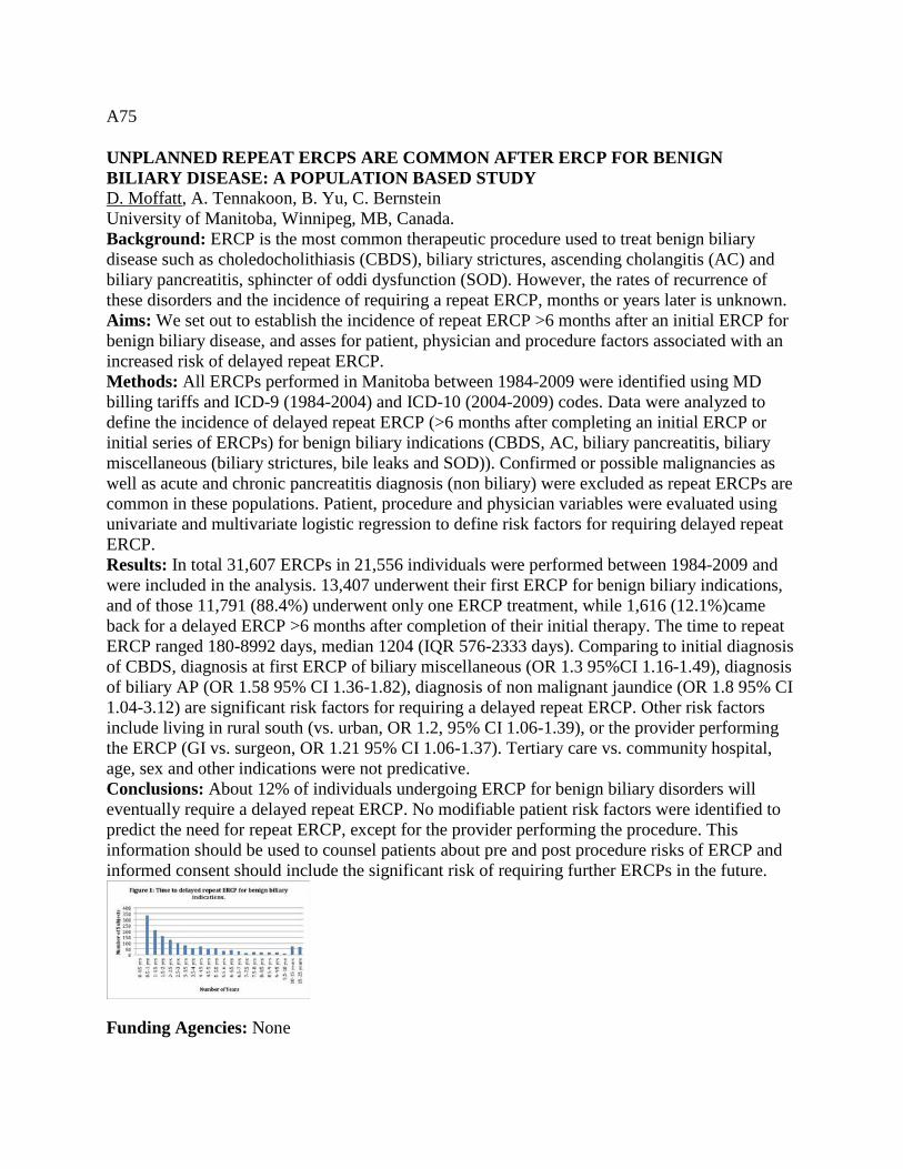
A75
UNPLANNED REPEAT ERCPS ARE COMMON AFTER ERCP FOR BENIGN BILIARY DISEASE: A POPULATION BASED STUDY D. Moffatt, A. Tennakoon, B. Yu, C. Bernstein University of Manitoba, Winnipeg, MB, Canada. Background: ERCP is the most common therapeutic procedure used to treat benign biliary disease such as choledocholithiasis (CBDS), biliary strictures, ascending cholangitis (AC) and biliary pancreatitis, sphincter of oddi dysfunction (SOD). However, the rates of recurrence of these disorders and the incidence of requiring a repeat ERCP, months or years later is unknown. Aims: We set out to establish the incidence of repeat ERCP >6 months after an initial ERCP for benign biliary disease, and asses for patient, physician and procedure factors associated with an increased risk of delayed repeat ERCP. Methods: All ERCPs performed in Manitoba between 1984-2009 were identified using MD billing tariffs and ICD-9 (1984-2004) and ICD-10 (2004-2009) codes. Data were analyzed to define the incidence of delayed repeat ERCP (>6 months after completing an initial ERCP or initial series of ERCPs) for benign biliary indications (CBDS, AC, biliary pancreatitis, biliary miscellaneous (biliary strictures, bile leaks and SOD)). Confirmed or possible malignancies as well as acute and chronic pancreatitis diagnosis (non biliary) were excluded as repeat ERCPs are common in these populations. Patient, procedure and physician variables were evaluated using univariate and multivariate logistic regression to define risk factors for requiring delayed repeat ERCP. Results: In total 31,607 ERCPs in 21,556 individuals were performed between 1984-2009 and were included in the analysis. 13,407 underwent their first ERCP for benign biliary indications, and of those 11,791 (88.4%) underwent only one ERCP treatment, while 1,616 (12.1%)came back for a delayed ERCP >6 months after completion of their initial therapy. The time to repeat ERCP ranged 180-8992 days, median 1204 (IQR 576-2333 days). Comparing to initial diagnosis of CBDS, diagnosis at first ERCP of biliary miscellaneous (OR 1.3 95%CI 1.16-1.49), diagnosis of biliary AP (OR 1.58 95% CI 1.36-1.82), diagnosis of non malignant jaundice (OR 1.8 95% CI 1.04-3.12) are significant risk factors for requiring a delayed repeat ERCP. Other risk factors include living in rural south (vs. urban, OR 1.2, 95% CI 1.06-1.39), or the provider performing the ERCP (GI vs. surgeon, OR 1.21 95% CI 1.06-1.37). Tertiary care vs. community hospital, age, sex and other indications were not predicative. Conclusions: About 12% of individuals undergoing ERCP for benign biliary disorders will eventually require a delayed repeat ERCP. No modifiable patient risk factors were identified to predict the need for repeat ERCP, except for the provider performing the procedure. This information should be used to counsel patients about pre and post procedure risks of ERCP and informed consent should include the significant risk of requiring further ERCPs in the future.
Funding Agencies: None

A76
THE PREVALENCE AND COMORBID STATE OF BOWEL-BLADDER PAIN SYNDROME IN A GENERAL GASTROENTEROLOGY CLINIC
A. Patel2, H. Yurgan2, M. Beyak1, W. Paterson3, D. Tripp2, C. Nickel2, S. Vanner4
1. QUEEN, Kingston, ON, Canada; 2. Queen's University, Kingston, ON, Canada; 3. Queens Univ, Kingston, ON, Canada; 4. queen's, Kignston, ON, Canada.
Background: The irritable bowel syndrome (IBS) and interstitial cystitis (IC) are common conditions presenting to general gastroenterology (GI) clinics but remain poorly understood in their interaction in the bowel-bladder pain syndrome and their effect on comorbid symptoms. Aims: To identify the prevalence, severity of pain, depression scores and association of comorbid states in patients with diagnoses of IBS, IC, or mixed IC/IBS. Methods: Patients attending a general gastroenterology clinic at Hotel Dieu Hospital between June 2013 to August 2014 participated in an ethics-approved survey of pain, depression and related health symptoms through a convenience sample administered at random times. The survey included components involving ROME III criteria, Medical Symptoms Inventory & Patient Health Questionnaire to diagnose conditions of IBS, IC and depression, respectively. Scores of pain, depression and co-morbidity presence were analyzed between groups without IBS or IC (‘other' group), IBS, IC, or mixed IC/IBS. Results: 213 patients participated and had conditional diagnoses as follows: 24.4% IC, 17.8% IBS, and 27.2% mixed IC/IBS, and 30.5% did not have IBS or IC (i.e. ‘other' group). The three groups with a conditional diagnosis had significantly higher pain ratings than the ‘other' group with a trend of pain scores highest for IC/IBS, then IBS and then IC. Depression scores were highest for mixed IC/IBS (mean 10.4 ±6.1) then IC(mean 8.5 ±6.0) then IBS(mean 6.3 ±5.8) and then ‘other'(mean 4.2 ±4.6). Significant differences (p<0.01) were observed between IC/IBS vs. other, IC/IBS vs. IBS and IC vs. other. Overall 87.8% of patients reported symptoms related to comorbid disease. The most prevalent were fibromyalgia (76%), lower back pain (67%) and chronic fatigue syndrome (63%). Symptoms of fibromyalgia, chronic fatigue syndrome, lower back pain, vulvodynia, migraines, tension headaches and temperomandibular joint disorder were highest for mixed IC/IBS (mean symptoms 5.2 ±1.7) followed by IC (mean symptoms 4.4 ±2.2) followed by IBS (mean 2.6 ±2.0) and then ‘other'(mean 1.9 ±1.5). Significant differences (p<0.01) were observed between IC/IBS vs. other, IC/IBS vs. IBS, IC vs.IBS and IC vs. other. Conclusions: This survey has determined that irritable bowel syndrome has a high degree of overlap in patients with interstitial cystitis in a general gastroenterology clinic patient population and that when interstitial cystitis is present; there are higher rates of depression and comorbid symptoms.
Funding Agencies: None

A77
A META-ANALYSIS OF COLON CLEANSING WITH PEG COMPARED TO OTHER BOWEL PREPARATIONS
C. Ménard1, A. Barkun2, M. Martel3, S. Restellini4, O. Kherad4, A. Vanasse1
1. University of Sherbrooke, Sherbrooke, QC, Canada; 2. McGill University, Montreal, QC, Canada; 3. McGill University and University of Sherbrooke, Montreal, QC, Canada; 4. Geneva's University Hospitals, Geneva, Switzerland.
Background: Different regimens of colon preparation are available for colonoscopy, they primarily include polyethylene glycol (PEG), sodium phosphate (NaP), picosulfate (PICO) or oral sulfate solution (OSS). Aims: To evaluate the efficacy, safety and patient satisfaction of PEG versus any comparator, NaP, PICO, and OSS. Methods: Systematic searches were completed querying MEDLINE, EMBASE, Scopus, CENTRAL and ISI Web of knowledge from January 1980 to August 2013. All fully published randomized controlled trials with colon preparation for colonoscopy were included. Populations including pediatric, sole inpatients or sole IBD patients were excluded. The primary outcome measure was the efficacy (excellent/good) of colon cleansing. Secondary outcomes included side effects or complications, procedural outcomes and patient satisfaction. A meta-analysis was conducted with results reported as odd-ratios (OR) with 95% confidence intervals. Heterogeneity and publication bias were assessed and quantified. Results: From an initial 2366 citations, 74 trials fulfilled the inclusion criteria (18,025 patients). When PEG was compared to all types of colon preparations, it did not show a significant difference in efficacy; OR=1.11 (0.93; 1.33). Willingness to repeat was lower in the PEG group OR=0.39 (0.23; 0.66) as well was fainting or dizziness OR=0.75 (0.57; 0.98). Forty-five trials included the comparison PEG versus NaP (11,197 patients); PEG did not show a difference in efficacy; OR=0.98 (0.74; 1.30); willingness to repeat was significantly decreased; OR=0.36 (0.22; 0.67) as well as fainting and dizziness OR=0.65 (0.50; 0.85). Eleven trials included the comparison PEG versus PICO (3,097 patients). PEG did not show a difference in efficacy; OR=1.08 (0.73; 1.59). Willingness to repeat was significantly decreased; OR=0.11 (0.04; 0.30), as well as fainting or dizziness OR=0.46 (0.30; 0.68). Abdominal cramps, insomnia and perianal irritation where increased 1.42 (1.05; 1.92), 2.41 (1.15; 5.03) and, 2.57 (1.27; 5.20) in the PEG group respectively. Two studies compared PEG to OSS. PEG was not different in efficacy; OR=0.90 (0.62; 1.30). Lack of data prevented the willingness to repeat analysis. Conclusions: PEG provides similar bowel cleansing efficacy to different types of colon preparations. Willingness to repeat was significantly lower with PEG when compared to all types of preparation wen the data was available. With PEG, more patients reported abdominal cramps compared to NaP, increased insomnia and perianal irritation compared to PICO headaches but less experienced fainting or dizziness compared to all types of preparations, NaP, and PICO.
Funding Agencies: None

A78
UPPER GASTROINTESTINAL BLEEDING IN THE POST PROCEDURAL PERIOD IN PATIENTS UNDERGOING TRANSCATHETER AORTIC VALVE IMPLANTATION: A RETROSPECTIVE ANALYSIS F. Wong2, S. Alipour2, D. Stanger2, O. Takach2, D. Wood2, B. Bressler1 1. Pacific Gastroenterology Associates, Vancouver, BC, Canada; 2. University of British Columbia, Vancouver, BC, Canada. Background: Transcatheter aortic valve implantation (TAVI) is an increasingly common technique to replace the aortic valve in severe aortic stenosis for patients deemed high risk for traditional surgical sternotomy approach. Given the comorbidities and demographics of the population undergoing TAVI, they are at significant risk of upper gastrointestinal bleeding (UGIB). In addition, the use of transesophageal echocardiography (TEE) and antiplatelet/anticoagulation use may further that risk. As TAVI is a relatively new technique, its extra cardiac complications are being elucidated. No studies to date have been published describing the incidence of UGIB in the post procedure period in patients undergoing TAVI. Aims: The objective of this study is to determine the incidence of significant upper GI bleed requiring endoscopic evaluation patients undergoing TAVI as well as the characterization of the bleeding lesions. Methods: A retrospective analysis of UGIB incidence in admitted patients to St Paul's hospital (SPH) from the specified timeline (Jan 2005 - Aug 2014). Subjects will be patients who were referred to the Gastroenterology service at SPH with clinical evidence of UGIB and/or required an esophagogastroduodenoscopy (EGD) during their admission for TAVI. Patients who underwent a TAVI procedure were identified from a database maintained by the Division of Cardiology at SPH and clinical data from patient records for the TAVI admission were extracted from the electronic medical system at SPH. Results: A total of 845 TAVI procedures were included in the analysis. 703 of those were via transfemoral (TF-TAVI) approach and 142 were via transapical (TA-TAVI) approach. 2.01% (n=17) of all TAVI procedures had UGIB. For TF-TAVI and TA-TAVI, 2.13% (15/703) and 1.41% (2/142) had UGIB, respectively. The average hemoglobin drop in those with UGIB was 27.7 g/L. The average number of units transfused was 2. 12 EGD were performed for evaluation. Of those, 2 were normal, 2 had clean based ulcers in duodenum, 3 had clean based ulcers in stomach. 5 had high risk lesions in the esophagus, all requiring endoscopic therapy. Conclusions: TAVI is a new minimally invasive technique for aortic valve replacement and carries a small risk of upper gastrointesintal bleed. When UGIB does occur in this setting, it is usually significant and requires blood transfusions. The most common lesion found is typically a distal esophageal or GE junction ulceration with active bleeding. Postulated reasons for the bleeding may stem from the use of TEE intraoperatively which may cause local trauma. Bleeding may be more severe in part due to antiplatelet and/or anticoagulation use peri-operatively.
Funding Agencies: None

A79
DIETARY ADHERENCE TO GLUTEN-FREE DIET (GFD) AMONG SAUDI CHILDREN WITH CELIAC DISEASE AND ITS SOCIOECONOMIC IMPACTS; A CROSS SECTIONAL STUDY
A. Sarkhy1, M. Mouzan1, S. Al-Ghamdi2, A. Alanazi3, A. Assiri1
1. King Khalid University Hospital, King Saud University, Riyadh, Saudi Arabia; 2. King Faisal Specialist Hospital and Research Center,, Jeddah, Saudi Arabia; 3. King Abdulaziz Medical City, National Guard Health Affairs, Riyadh, Saudi Arabia.
Background: Though it is very effective, GFD is significantly restrictive, it is not easily available, more expensive compared to the regular gluten containing diet, and is not as palatable compared to the regular gluten containing diet. For all of these reasons, there is a significant negative impact on the adherence rate. Furthermore, GFD may impose several psycho-social and financial stresses on the patients & their families which consequently might reduce their adherence to GFD even more Aims: To determine the adherence rate to gluten free diet (GFD) among Saudi children with Celiac disease (CD), and to examine the socio-economic impacts of GFD on these children and their families Methods: A cross-sectional study was conducted and all the families registered in the Saudi Celiac Patients Support Group (SCPSG) were sent an online survey. Only families with children 18 years of age and younger with biopsy-confirmed celiac disease (CD) were included Results: The mean age of the 113 included children was 10 ± 3.7 years, the mean age at symptom onset was 5.8 ± 3.7 years and the mean age at diagnosis was 7 ± 3.8 years, and 62.8 % children were females. Ninety two percent of the patients were symptomatic at the diagnosis while eight percent were asymptomatic. The commonest presenting symptoms included: chronic abdominal pain (59.3%), poor weight gain (54%), abdominal distention/bloating (46.1%) and chronic diarrhea (41.6%). Among the extra-intestinal manifestations, short stature and joint/bone problems were the commonest presentations (22% and 20.4% respectively). Around one third of the children had mood changes (anxiety/depression) and chronic fatigue at the time of presentation. Sixty eight (60%) of the involved children were reported to be strictly adherent to GFD. Younger age at diagnosis and shorter duration since the diagnosis were associated with a better adherence rate. Significant social difficulties were reported in more than 50% of the participating families and their children. Conclusions: Compliance to GFD is relatively poor among Saudi children with CD. There are significant negative socio-economic impacts of GFD on these children & their families; physicians should be aware of such impacts and should be well trained to handle them.
Funding Agencies: None
A80
RELATIONSHIPS BETWEEN IBD AND IBS SUGGEST THAT WHILE EVOLUTION OF LACTASE MAY HAVE PLAYED A ROLE IN IBD, IT HAS NOT IN IBS A. Szilagyi, X. Xue Jewish General Hospital, Montreal, QC, Canada. Background: Pathogenesis of some irritable bowel syndromes (IBS) requires milder pathogenic features which may overlap with inflammatory bowel disease (IBD). Some authors report IBS-like symptoms in quiescent IBD raising a controversy about relationships between IBS and IBD. Previously we hypothesized that IBD and some other diseases may be related to evolution and migrations of lactase persistent and non persistent populations (LP/LNP), which may explain early observed geographic distributions. Aims: To assess ecological relationships between IBS, IBD and lactase proportions using calculated and published national data that suggest evolutionary interdependence. Methods: Data for 5 variables were obtained from 28 countries: IBS% (Lovell RM Clin Gastroenterol Hepatol, 2012), LNP% (Szilagyi A, Clin Epidemiol, 2014), prevalence of Crohn's disease (CD) and Ulcerative Colitis (UC), and incidence of CD (Molodecky NA, Gastroenterol, 2012). Data for IBD and lactase cover 4.7 decades, IBS, 6.2 decades. IBS diagnostics were based on Rome I or II criteria. Pearson's correlations were used to assess relationship among 5 variables. Negative binomial regressions were also used to identify associations between IBD and IBS while adjusting for the effect of LNP. IBD incidence and prevalence were log-transformed. Results: Available sets of data ranged from 18 to 28 for 5 variables. Negative binomial result about associations among them agreed with Pearson correlation analysis. Results of Pearson correlations are shown in the table. There was no significant correlation between IBS and any other parameter. IBD and lactase correlated inversely and significantly. Within IBD there was significant correlation between prevalence and incidence. Conclusions: The limitations of this type of work include few data, lack of IBS incidence, variability of diagnostics with all variables (especially IBS) and changing patterns of IBD and perhaps IBS, necessitating the use of an arbitrary space/time analysis. The major strength is that reports of data are independent of bias toward expected associations. We found no significant relationships between IBS and other variables, However, we confirmed relationships of IBD with distributions of lactase phenotypes. We conclude that while evolution of lactase persistence may have played a role in modern day development of IBD, it probably had no role in the development of IBS. The lack of predisposing genetic overlap between IBD and IBS to date supports this hypothesis.
LNP% CDprev UCprev CDinc IBS%r -0.17a -0.14a -0.06a -0.1a LNP%r -0.55b -0.59b -0.84c CDprevr 0.79c 0.79c UCprevr 0.73c
a-p>0.4,b-p<0.05,c-<0.005
Funding Agencies: None
A81
IS NURSE STAFFING ASSOCIATED WITH HOSPITALIZATION OUTCOMES IN INFLAMMATORY BOWEL DISEASE?
A. Shaheen, R. Myers, G. Kaplan
University of Calgary, Calgary, AB, Canada.
Background: Higher nurse to patient ratios have been associated with improved hospitalization outcomes in many conditions. Crohn's disease (CD) and ulcerative colitis (UC) are complex conditions that may require more intense nursing care. Although hospitals with high case volume for IBD have improved outcomes (e.g. reduced postoperative mortality), the role of nursing staff volume on hospitalization outcomes has not been studied. Aims: To assess the association between nurse staffing and the outcomes of hospitalization in patients with IBD. Methods: We used the 2008 Nationwide Inpatient Sample database to identify hospitalized patients with a primary diagnosis of CD (ICD-9, 555) or UC (556). Hospital-level nursing staff volume, which included the volume of registered nurses, licensed practical nurses and nurse aids, was categorized into tertiles (low [<4.9], medium [4.9-6.1], and high-volume [>6.1 nurse full-time equivalents [FTEs] per 1,000 adjusted inpatient days]). Weighted regression models were used to assess the impact of nursing volume on in-hospital mortality, length of stay (LOS) and hospitalization charges after adjustment for patient characteristics (e.g. comorbidities), the need for surgery, and hospital case volume for IBD. Results: We identified 18,178 IBD admissions in 2008 (UC= 6,777 and CD=11,401). Compared to hospitals with low nursing volume, hospitals with high volume were more likely to have high IBD case volume (54.2% vs. 12.9%, P<0.001). UC patients admitted to hospitals with high nursing volume were more likely to undergo surgery (28% vs. 8% for low nursing volume), had longer LOS (5.0 vs. 3.6 days) and increased charges ($31,255 vs. 17,613, all P<0.001). Compared to CD patients admitted to hospitals with low nursing volume, those admitted to high nursing volume hospitals had higher rates of surgery (21% vs. 11%), longer LOS (3.7 vs. 3.1), and greater hospitalization charges ($23,062 vs. 15,682, all P<0.001). There were no differences in in-hospital mortality according to hospital nursing volumes (high vs. low: UC, 1% vs. 1.5%, P=0.34; and CD, 0.3% for both, P=0.70). After adjusting for patient and hospitalization characteristics, nursing volume was not independently associated with in-hospital mortality or LOS in both UC and CD cohorts. However, high nursing volume was associated with a 15% (95% CI: 3-28%) adjusted increase in hospitalization charges among CD patients; a similar finding was not observed in those with UC. Conclusions: Nursing volume is not associated with in-hospital mortality or LOS in patients with CD or UC; however, CD patients admitted to hospitals with high nursing volume have greater hospitalization charges.
Funding Agencies: None

Esophagus, Gastric and Duodenal Ulcer Disorders
A82
SUSTAINED LONGITUDINAL SMOOTH MUSCLE CONTRACTIONS MAY NOT BE A CAUSE OF NON-CARDIAC CHEST PAIN
K. McIntosh2, W. Paterson1
1. Queens Univ, Kingston, ON, Canada; 2. Western University, London, ON, Canada.
Background: It has been proposed that non-cardiac chest pain (NCCP) may be caused by sustained esophageal contractions ("SECs") of the esophageal longitudinal smooth muscle. In this study, we set out to use high-resolution esophageal manometry to measure esophageal shortening (a surrogate of SECs) in a group of NCCP patients and healthy controls. We hypothesized that if SECs are responsible for NCCP, this group of patients would have an exaggerated esophageal shortening response to luminal acid (a trigger for SECs) and/or a temporal correlation between pain onset and shortening. Aims: To determine if sustained longitudinal smooth muscle contractions of the esophagus contribute to symptom production in patients with NCCP. Methods: 17 patients and 16 controls underwent a baseline esophageal manometry. A small nasogastric catheter was then placed in the mid esophagus and used to sequentially instill normal saline for 10 minutes followed by 0.1N HCL for 20 minutes, each at a rate of 5 mls/min. Pain intensity was recorded every minute on a 10 point visual analogue scale. A blinded observer determined esophageal shortening by measuring the distal margin of the UES and the proximal margin of the LES during the final two minutes of the saline and acid infusion periods. Results: The NCCP patients demonstrated esophageal shortening of 0.53 ± 0.54 cm (mean ± SD) during acid infusion, which was not significantly different than that seen in controls (0.43 ± 0.41 cm; p=0.56). The average percent change in esophageal length was 2.4 ± 2.4 % for NCCP patients versus 2.0 ± 1.9 % for controls (p=0.51). Similar times to shortening onset were observed for NCCP patients (7.8 ± 4.5 min) and controls (5.5 ± 3.5 min, p=0.12). Only 1/17 NCCP patients and 2/16 controls had esophageal shortening that temporally correlated to the onset of their pain. Conclusions: High-resolution manometry appears to be a simple and effective way to measure acid-induced esophageal shortening. In this study, no significant differences were found in the shortening responses of NCCP patients versus controls and there was poor temporal correlation between esophageal shortening and pain onset, suggesting that SECs did not play a significant role in pain production in this group of patients. (Supported by grants from Physicians Services Incorporated Foundation of Ontario and the Canadian Association of Gastroenterology)
Funding Agencies: CAG, PSI
A83
A PHASE 1 CLINICAL TRIAL OF ATB-346, A GASTROINTESTINAL-SAFE NONSTEROIDAL ANTI-INFLAMMATORY DRUG (NSAID)
J. Wallace, M. Bumby, D. Vaughan
Antibe Therapeutics, Toronto, ON, Canada.
NOT PUBLISHED AT AUTHOR’S REQUEST
Funding Agencies: None

A84
HIGH DEFINITION ISCAN VIRTUAL ELECTRONIC CHROMOENDOSCOPY HAS HIGH SENSITIVITY AND SPECIFICITY FOR THE HISTOLOGICAL DIAGNOSIS OF EOSINOPHILIC ESOPHAGITIS
M. Iacucci2, G. Eustace2, M. Fort Gasia2, X. Gui3, S. Ghosh1
1. University of Calgary, Calgary, AB, Canada; 2. Division of Gastroenterology, University of Calgary, Calgary, AB, Canada; 3. Department of Pathology, University of Calgary, Calgary, AB, Canada.
Background: Eosinophilic esophagitis (EoE) has become an increasing important cause of upper gastrointestinal morbidity with dysphagia and food bolus obstruction. Sometimes the diagnosis is challenging as there are no pathognomonic endoscopic findings for EoE and sometimes it may appear normal at endoscopy. There is evidence that white light endoscopy can often miss the diagnosis of EoE. Aims: We aimed to determine whether high definition iSCAN virtual electronic chromoendoscopy with targeted biopsies improved the diagnostic yield of EoE in patients presenting with dysphagia or foreign body impaction. Methods: We included 108 patients (female=46, median age=57y) who presented with dysphagia or foreign body impaction. All patients were assessed by high-definition iSCAN virtual chromoendoscopy (Pentax EC-3490Fi; Pentax, Tokyo, Japan) and targeted biopsies were taken in the mid and upper esophagus and from the furrows where visible. Results: Out of 108 patients with dysphagia, 27 patients (25%, male= 21, median age= 45y) were diagnosed by histology with a diagnosis of EoE. By iSCAN endoscopy 33 patients were suspected to have EoE, of which 26 patients had EoE diagnosed by histology (>15 eosinophils/HPF). One patient had normal endoscopy but histology was diagnostic of EoE. The sensitivity and specificity of iSCAN endoscopy was 96.4% and 92.5% respectively. The positive predictive value of iSCAN endoscopy was 81.8% and the negative predictive value of iSCAN endoscopy was 98.6%. The iSCAN endoscopic finding characteristics of EoE were linear furrows (92%), tracheal appearance (70%), whitish exudates (25%) and narrowing or strictures (3%). These endoscopic findings became more obvious when iSCAN was used and were easily missed on white light endoscopy alone. Fifteen patients out of 108 presented with acute food bolus obstruction of whom 10 (66%) were diagnosed as EoE. Conclusions: iSCAN high definition endoscopy has high diagnostic accuracy for EoE as confirmed by histology. Linear furrows and tracheal appearance were the commonest findings while whitish exudates and narrowing were present in more severe patients. These findings may be missed when using white light endoscopy alone.
Funding Agencies: None

A85
ESOPHAGEAL PSEUDODIVERTICULOSIS: A CASE SERIES
S. Grover1, M. Scaffidi1, B. Ro1, I. Plener2, S. Akbari-Nasab1, A. Grin3, E. Colak4
1. Division of Gastroenterology, St. Michael's Hospital, Toronto, ON, Canada; 2. University of Toronto, Toronto, ON, Canada; 3. Department of Laboratory Medicine, St. Michael's Hospital, Toronto, ON, Canada; 4. Department of Medical Imaging, St. Michael's Hospital, Toronto, ON, Canada.
Background: Esophageal pseudodiverticulosis (EP) is a rare endoscopic and radiological finding distinguished by numerous flask-like outpouchings of the esophageal wall. The majority of EP occurs with esophageal intramural pseudodiverticulosis (EIPD), an idiopathic chronic disorder leading to recurrent dysphagia. Two case reports exist of pseudodiverticulosis in eosinophilic esophagitis (EoE). Given the rarity of this finding, the literature is scarce on the clinical and endoscopic characteristics of EP, with most reports published prior to the understanding of EoE as a clinical entity. Aims: To characterize the clinical and endoscopic features, and prognosis associated with esophageal pseudodiverticulosis. Methods: A single-centre retrospective chart review was conducted from 2000 to 2014. Patients with EP were identified using standard diagnostic criteria on findings from barium or CT esophagogram and endoscopy. Descriptive statistics were calculated for demographic data, potential risk factors, results from endoscopy and imaging, treatment plan, and prognostic factors. Results: Fourteen patients with pseudodiverticulosis were identified with more females (n=10, 71.4%) than males (n=4, 28.6%). Mean age of diagnosis was 51.9 (SD=16.9) and mean BMI was 25 (SD=4.6). All patients presented with dysphagia (n=14, 100%; specifically to solids n=12, 85.7%; median Mellow-Pinkas dysphagia score = 1). Common disease associations were GERD (n=13, 92.9%), alcohol abuse (n=5, 35.6%) and smoking history (n=8, 57.1%) with a mean history of 66.7 pack-years. 4 patients (28.5%) had a diagnosis of EoE made on clinical and histopathologic grounds. The most prevalent location of pseudodiverticula was in the proximal esophagus (n=7, 50%), but all 4 patients with EoE had distal pseuodiverticulosis, multiple esophageal rings, and linear furrows. PPIs were used as treatment for 11 patients (78.5%). 7 patients (50%) had endoscopic dilatation with balloon (n=1, 7.1%) or bougienage (n=6, 42.8%). All 4 patients with EoE received oral fluticasone, and 2 required bougienage. 12 patients (86.7%) reported reduction in Mellow-Pinkas dysphagia score after treatment, but only 2 of 4 patients with EoE reported improvement. Conclusions: This series illustrates eosinophilic esophagitis as a previously unreported cause of esophageal pseudodiverticulosis, distinguished from EIPD endoscopically by distal esophageal pseudodiverticula and classic endoscopic findings associated with EoE. Prior historical reports of EIPD may have been confounded by EoE.
Funding Agencies: None
A86
BACTERICIDAL ACTIVITIES OF STEARYLAMINE-CONTAINING LIPOSOMES AGAINST HELICOBACTER PYLORI DO NOT AFFECT GASTRIC CELL VIABILITY
H. Yee, M. Mahmoud, M. Keelan, *. CANHelp Working Group
University of Alberta, Edmonton, AB, Canada.
Background: Helicobacter pylori is a causative agent of gastritis, peptic ulcers and gastric cancer. Standard treatment involves a triple-drug therapy including two antibiotics and a proton pump inhibitor. In Northern Canadian Aboriginal communities where H. pylori prevalence is high (~60%), standard treatment failure occurs in ~30-40% of individuals. Possible causes include noncompliance due to side effects of therapy, complex dosing regimen, and the development of antibiotic resistance in H. pylori strains. Recent unpublished observations (Mahmoud and Keelan 2014) suggest a single dose of phosphatidylcholine: cholesterol:stearylamine liposomes (7:3:2) impairs H. pylori growth in vitro. This effect is proposed to be due to the stearylamine content of the liposomes. Aims: The aim of this study is to assess the effect of increasing stearylamine concentration within liposomes on H. pylori growth and gastric epithelial cell viability in vitro. Methods: Three formulations of liposomes (F1, F2, F3) were prepared with the following ratios of phosphatidylcholine:cholesterol:stearylamine 7:3:1, 7:3:2 and 7:3:3, respectively. The growth of H. pylori previously isolated from a Northern Canadian community was evaluated by measuring optical density and performing colony counts at time 0 h, and after 24 h incubation with liposomes ranging in concentration from 0 to 200 µg/mL to determine the minimum inhibitory concentration (MIC) and minimum bactericidal concentration (MBC). NCI 87 gastric carcinoma cells were cultured for 12 days until confluent, exposed to F1, F2 and F3 liposomes at MBC values for 24 hours and cell viability determined by trypan blue exclusion assay. Results: No inhibitory or bactericidal activity was observed when H. pylori were exposed to F1 liposomes at the concentrations studied. In contrast, when H. pylori were exposed to F2 liposomes, the MIC was 100 µg/mL and MBC was 200 µg/mL, versus 50 µg/mL and 100 µg/mL, respectively when H. pylori were exposed to F3 liposomes. Gastric cell viability was unaffected by exposure to F1, F2, F3 liposomes and unchanged from untreated controls. Conclusions: These observations provide evidence that increasing stearylamine content in liposomes will elicit bactericidal activity for H. pylori over 24 h, but are non-toxic for NCI 87 gastric cells at MBC values over the same incubation time. Whether the MBCs established for F2 and F3 liposomes adversely affect commensal organisms remains to be established.
Funding Agencies: CIHR, ArcticNet, Alberta Innovates - Health Solutions

A87
PREVALENCE OF GERD IN PATIENTS WITH CF AT A SINGLE CENTRE.
R. Persad1, J. Turner2, H. Rylance1, S. Hyun Kang1
1. University of Alberta, Edmonton, AB, Canada; 2. Stollery Children's Hospital, Edmonton, AB, Canada.
Background: Cystic fibrosis (CF) is the most common life threatening autosomal recessive disease of the Caucasian population in Canada and worldwide. Patients with CF receive supine chest physiotherapy which increases the risk for gastroesophageal reflux disease (GERD). Questionnares for the diagnosis of GERD has been validated with specific symptoms having an increased association with GERD. Aims: The primary objectives for this study was to determine the prevalence of GERD associated symptoms in a group of paediatric and adult CF patients attending the University of Alberta Hospital Edmonton, Canada Methods: All patients or their caregivers were surveyed for GERD related symptoms. Questionnaires were administered by a research nurse attending the paediatric and adult clinics. Results: 146 patients participated in the study with 30.8% less than age 18 years, There were 73 (50.7%) male and 71(49.3%) female. The prevalence of all GERD symptoms (water brash, heartburn, regurgitation, dyspepsia, nausea, vomiting, chest pains and dysphagia) was 62.3%. Conclusions: GERD is prevalent in the CF population studied. The effect of GERD on pulmonary function and nutrition of patients with CF needs further evaluation.
Funding Agencies: Stollery Foundation

A88
CLINICAL PRESENTATION OF HELICOBACTER PYLORI GASTRITIS IN SAUDI CHILDREN
A. Assiri2, A. Saeed1, Z. Zaidi1, A. Al Sarkhy1
1. King Khalid University Hospital, King saud university, Riyadh, Saudi Arabia; 2. Prince Abdullah Bin Khalid Celaic Disease Research Chair, King Suad Univerity, Riyadh, Saudi Arabia.
Background: Helicobacter Pylori (H. pylori) is a gram negative, spiral gastro duodenal pathogen that infects 50% of the world population. Although infection is acquired during childhood but disease manifestations appear decades after the acquisition of infection and may lead to serious consequences like gastritis, ulcers and gastrointestinal malignancies. Presentation is variable but its association with recurrent abdominal pain, gastroesophageal reflux, iron deficiency anemia and asthma is still controversial. Despite the lack of evidence, trend is to screen children for H Pylori and treat those found to have the infection. This study was conducted to review the clinical profile of Saudi children with H. pylori gastritis. Aims: This study was conducted to review the clinical profile of Saudi children with H. pylori gastritis. Methods: It was a retrospective study conducted at pediatrics division of a private tertiary care hospital, Riyadh. A total of 202 Saudi children both males and females between 1 to 16 years of age who underwent oesophago gastroduodenoscopy (OGD) from Jan 2009 to Jan 2012 were included in the study. Out of these, 29 children were found to have H Pylori gastritis. The diagnosis was confirmed with histopathology. Clinical profile of these patients including age, gender, clinical presentation, association with gastro-esophageal reflux disease (GERD), iron deficiency anemia, food allergy, asthma, endoscopic and histopathological findings were reviewed. Results: Mean age of presentation was 10 years. 52% presented between 11-16 years, 44% between 5-10 years and 3% before 5 years of age. 42% were males and 58% were females with male to female ratio of 1:1.8. 84% patients presented with abdominal pain, 28% with heartburn whereas vomiting and growth failure were the chief complaint in 8% and 16% of the patients respectively. Iron deficiency anemia, asthma, food allergy and family history of PUD and GI malignancies were not found in any of the patients. Reflux esophagitis was the feature in 24% (n=7). Sydney system was used for the classification and grading of gastritis. Atrophic gastritis was a feature in 17% while rest had non atrophic gastritis. All patients had chronic active gastritis. None of the patients showed metaplastic, dysplastic or malignant changes. Villous atrophy was found in 17%. All the patients were put on eradication therapy. 82% responded well to treatment. Conclusions: H. pylori infection is not an uncommon entity and is an important cause of gastritis in Saudi children. Majority of the patients presented with abdominal pain but it has no clear association with GERD, iron deficiency anemia or allergies.
Funding Agencies: None

A89
SHORT-TERM INCREASES IN AIR POLLUTION ARE NOT ASSOCIATED WITH THE INCIDENCE OF UPPER GASTROINTESTINAL BLEEDING SECONDARY TO PEPTIC ULCER DISEASE: A CASE-CROSSOVER STUDY
S. Quan2, H. Yang2, D. Tanyingoh2, R. Hilsden2, K. Madsen1, S. Veldhuyzen van Zanten1, K. Novak2, E. Lang2, S. Ghosh2, G. Kaplan2
1. University of Alberta, Edmonton, AB, Canada; 2. University of Calgary, Calgary, AB, Canada.
Background: Recent studies have demonstrated an association between short-term elevations in air pollution and an increased risk of exacerbating gastrointestinal disease. Aims: The objective of the study was to evaluate if day-to-day increases in air pollution concentrations were positively associated with upper gastro-intestinal bleeding (UGIB) secondary to peptic ulcer disease (PUD). Methods: A time-stratified case-crossover study design was used. Adults presenting to hospitals with their first UGIB secondary to PUD from 2004-2010 were identified using administrative databases from Calgary (n=1374; discovery cohort) and Edmonton (n=1159; replication cohort). Daily concentrations of ozone, nitrogen dioxide, sulfur dioxide, carbon monoxide, and particulate matter (PM10 and PM2.5) were estimated in these two cities. Conditional logistic regression models were employed, adjusting for temperature and humidity. Odds ratios were expressed relative to an interquartile range increase in the concentration of each pollutant. Results: No statistically significant associations were observed for any of the individual pollutants based on same-day, or 1-day lag effects within the Calgary discovery cohort. When the air pollution exposures were assessed as 3-, 5-, and 7-day averages, some pollutants were inversely associated with UGIB in the discovery cohort; for example, 5-day averages of nitrogen dioxide (OR=0.68; 95% CI: 0.53-0.88), and particulate matter <2.5µm (OR=0.75; 95% CI: 0.61-0.90). However, these findings could not be reproduced in the replication cohort. Conclusions: Our findings suggest that UGIB secondary to PUD is not associated with short-term changes in the level of ambient air pollutants.
Funding Agencies: CIHR, Alberta Innovates - Health Solutions

A90
TEMPORARY ESOPHAGEAL STENTING FOR SEVERE ANASTOMOTIC DEHISCENCE
A. Hashim2, A. Patel1, L. Hookey1, D. Mercer1
1. Queen's University, Kingston, ON, Canada; 2. Queens University, Kingston, ON, Canada.
Background: Surgery as well as endoscopic esophageal stenting has recently been used to treat postoperative intrathoracic leak. However, surgery is associated with high morbidity and mortality. Aims: To provide an illustration of how a partially covered stent could conservatively manage a large anastomic dehiscence averting the need for surgery. Methods: PubMed Search was conducted to examine the literature around esophageal stents in the use of anastomic dehiscence. Results: A 69 years old male, presented with 4 weeks history of dysphagia and weight loss. Upper endoscopy showed invasive adenocarcinoma of the distal esophagus. He underwent neoadjuvant chemotherapy followed by esophageogastrectomy and gastric pull up surgery. Upper GI series done one week after surgery showed no evidence of anastomotic leak. 5 days later, patient started to have respiratory distress and hypoxia. CT scan and a repeat GI series showed anastomotic leak and a large mediastinal collection. Upper endoscopy confirmed grade 4 esophageal dehiscence. A covered esophageal stent was placed endoscopically, followed by multiple upper GI series over a period of 4 months showing gradual improvements in the esophageal leak. Upper endoscopy 4 months after stent placement demonstrated good healing with no evidence of leakage during gasrograffin injection. The stent was removed; the visualized esophageal mucosa demonstrated good healing at the gastro esophageal anastomosis, patients symptoms improved. Conclusions: In conclusion, placement of a covered stent was successful, minimally invasive, and an uncomplicated treatment option for a large, clinically apparent intrathoracic esophageal anastomotic leak.
Funding Agencies: None

A91
AN INTERESTING INTERPOSITION: A UNIQUE CASE OF UPPER GI BLEEDING
C. Sheasgreen, K. Khan
McMaster University, Hamilton, ON, Canada.
Background: Upper gastrointestinal bleeding (UGIB) affects thousands of Canadians every year and can manifest with hematemesis, melena, hematochezia, or shock. It is most commonly caused by peptic ulcer disease, esophageal varices, erosions or inflammation, and diagnosis is usually by esophaogastroduodenoscopy (EGD). Rarer causes of UGIB can be difficult to diagnose. Aims: A case of upper GI bleeding from a colonic interposition for esophageal atresia will be reviewed, along with the usual complications of colonic interposition. Methods: A comprehensive chart review of the case was undertaken, including assessment of biochemical, endoscopic, and pathological results. Subsequently, a comprehensive literature review of the topic was conducted. Results: A 46 year old woman was admitted for evaluation of chronic chest pain and anemia. She had a remote history of esophageal atresia repaired with colonic interposition (CI) and a gastrostomy tube for nutrition. Cardiac biomarkers, electrocardiogram, and computed tomography (CT) pulmonary angiography were non-diagnostic. Hemoglobin on admission was 61, INR 1.2, platelets 441. She had no history of melena or hematemesis. EGD revealed no bleeding. Three days after admission, she developed hematemesis and shock requiring transfusion of 8 units of packed red blood cells. Repeat EGD revealed blood coating the lumen of the CI that prevented localization of bleeding. Repeat CT of the chest and abdomen showed mediastinal gas and contrast within the CI, but no bleeding source. Two days later, the patient suddenly lost 1.5L of blood from her gastrostomy tube. Urgent angiography did not identify the source of bleeding. A third CT of the chest and an echocardiogram suggested fistulisation of a left ventricular pseudoaneurysm into the CI. Urgent coronary angiography was normal, but left ventriculogram confirmed the fistula. The patient underwent urgent surgery to repair the interposition and patch the pericardium. She was ultimately transferred to the rehabilitation unit. Esophageal atresia is a relatively common disorder, affecting 1 in 2,400-4,500 births. The degree of atresia can vary, as can the surgical intervention to correct it. Lengthening procedures are preferred over interposition as they have fewer complications. Common complications include anastomotic leak and stricturing. Ulcerations have been described from reflux disease, but only one case of fistulization to the ventricle has been described previously in the literature. Conclusions: UGIB has many possible etiologies. For patients with abnormal anatomy or a complicated surgical history, clinicians should individualize testing in order to prevent diagnostic delay—particularly when EGD is inconclusive.
Funding Agencies: None

Gastro Intestinal Oncology
Poster of Distinction A92 RAS SIGNALLING COOPERATES WITH THE PROLYL-ISOMERASE PIN1 TO PROMOTE THE EXPRESSION OF THE NOTCH1 INTRACELLULAR DOMAIN NIC1 IN PANCREATIC CANCER CELLS É. Paré, D. Arsenault, M. Boucher Université de Sherbrooke, Sherbrooke, QC, Canada. Background: Pancreatic ductal adenocarcinoma (PDAC) is the most lethal malignancy with a five-year survival rate of 8%. The identification of the most frequently mutated genes in PDAC have provided important insights into its pathogenesis but have not led to improvement in diagnosis or treatment. A better understanding of the cellular and molecular mechanism that govern PDAC cells is imperative in the quest to develop new therapeutic strategies. PDAC is characterized by a high prevalence of RAS mutation (90%) that is required for both tumour initiation and maintenance. Aberrant activation of the NOTCH signalling pathway was shown to assist RAS signalling in the promotion of tumour initiation. NOTCH are transmembrane receptors that undergo proteolytic cleavages upon ligand interaction. These cleavages release the NOTCH intracellular domain (NIC) that translocates towards the nucleus to associate with its transcriptional partners CSL and MAML1 and impact on gene expression (ex.HES1). Recently, PIN1, a prolyl-isomerase specifically binding to proteins containing proline residues preceded by a phosphorylated serine or threonine, was shown to interact with NIC1. The PIN1-NIC1 interaction was proposed preventing the FBW7-mediated degradation of NIC1. Aims: Given that we recently shown that activation of the serine/threonine kinases ERK1/2 downstream of RAS promotes NOTCH-dependent expression of HES1 in PDAC cells, we hypothesized that PIN1 recognizes ERK1/2 phosphorylation sites on NIC1 leading to increased expression levels of NIC1. Methods: To address this hypothesis, we used PDAC cells (MIA PaCa-2, BxPC-3) in which synchronized activation of NOTCH1 was achieved by addition of EGTA. PMA and the DUSP6 inhibitor BCI were used to activate the MEK/ERK activity whereas the MEK inhibitor U0126 was used to inhibit this pathway. Alternatively, we expressed tagged version of NIC1, PIN1 and/or KRASG12V in HEK293T cells. Results: Our results demonstrated that 1) an active ERK2 directly phosphorylates NIC1 in vitro. 2) Activation of the MEK/ERK pathway promoted the expression of HES1 3) that correlated with increased association of NIC1 with its transcriptional partners CSL and MAML1. 4) Furthermore, overexpression of PIN1 or KRASG12V independently led to increased expression levels of NIC1 whereas 5) PIN1 and KRASG12V cooperatively promoted NIC1 expression levels. Conclusions: Taken together, our results demonstrate for the first time that NIC1 is a direct substrate of ERK1/2. Our data support a model whereby the phosphorylation of NIC1 by ERK1/2 promotes NIC1 expression by PIN1-dependent mechanisms. Abrogating PIN1 function might represent an interesting avenue to target aberrant signalling in PDAC.
Funding Agencies: CIHR, Faculty de Medecine et des Sciences de la Santé de l'Universite de Sherbrooke

A93
THE P2Y6 RECEPTOR: A COLORECTAL TUMORIGENESIS' ACTOR?
M. Placet, D. Grbic, J. Bourzac, C. Molle, G. Arguin, F. Gendron
Université de Sherbrooke, Sherbrooke, QC, Canada.
Background: P2Y6 receptor (P2Y6R) is a nucleotide receptor activated by extracellular UDP. In physiological conditions, P2Y6R activation stimulated NaCl secretion by intestinal epithelial cells (IECs). In inflammatory bowel diseases, P2Y6R stimulation leads to aggravated inflammatory symptoms characterized by an increase in the number of recruited neutrophils. One of the hallmarks of human colorectal cancer (CRC) is the intrinsic or acquired resistance to apoptosis. Apoptosis evasion contributes to carcinogenesis, tumour progression and to treatment resistance. In this context, it was reported that P2Y6R activation could significantly reduces apoptosis in response to TNFa. Although the understanding of P2Y6R roles in CRC is limited, recent studies from our group and others suggested that P2Y6R expression is increased in human CRC tissues harbouring mutation for APC and TP53 and that its activation was associated to cellular responses to DNA damages in human adenocarcinoma cells. Aims: Based on the idea that P2Y6R contributes positively to the tumour potential of IECs with mutations in APC and TP53. The aims are: (1) to elucidate the impact of APC and TP53 mutations on the expression and activity of P2Y6R. (2) To characterize the anti-apoptotic and pro-proliferative potential of P2Y6R in IECs. Methods: We determined P2Y6R expression levels by qPCR in normal and cancerous IECs as well as from CRC tumour biopsies. P2Y6R activity was quantified by measuring the intracellular Ca2+ levels. The impact of P2Y6R activation on apoptosis was determined using Annexin V expression assays, cell count and by analysing apoptosis markers using western blotting. Cell growth curves and clonogenic soft agar assays were realized to determine the effect of P2Y6R stimulation and cell growth and tumorigenic potential. Results: Receptor expression and activity are increased in cells and tumour harboring mutation for genes APC and TP53. Activation of P2Y6R protects colorectal cancer epithelial cells from TNFα-induced apoptosis by increasing XIAP expression. Activation of P2Y6R with UDP stimulated the growth of cells having a robust receptor expression and activity levels in both classical cell growth assays and in clonogenic soft agar experiments. Conclusions: In this study, as it is the case with other immunomodulatory molecules such as TNFa, the sustained activation of P2Y6R could contribute to intestinal tumorigenesis by blocking the apoptotic process and by stimulating cell growth. These results support the idea that modulation of P2Y6R activity using selective antagonists will not only decrease intestinal inflammation but could also reduce tumour size and/or growth.
Funding Agencies: CIHR
A94
CARACTERISATION OF THE MOLECULAR FUNCTIONS OF NUDCD1 ISOFORMS IN COLORECTAL CANCER CELL LINES.
P. Asselin-Mullen, D. Lévesque, F. Boisvert
Université de Sherbrooke, Sherbrooke, QC, Canada.
Background: NudCD1, also known as CML66 or OVA66, is a 583 amino acids protein initially identified as overexpressed in patients with chronic myelogenous leukemia.The molecular function of this protein is currently unknown. mRNA of the gene is significantly expressed in heart and testis of normal tissues, whereas it is overexpressed in several cancers. Previous studies have shown that the expression level of the protein correlates with tumoral phenotype, such as cell growth, migration and invasion, possibly interacting upstream of the Insulin Growth Factor - 1 Receptor (IGF-1R). The gene encoding the NudCD1 protein consists of 12 exons that can be alternative spliced, leading to the expression of three different isoforms. These isoforms possess a common region of 492 amino acids in their C-terminus region and have an isoform specific N-terminus. Aims: Our hypothesis consists that the different NudCD1 isoforms have different cellular functions and roles. Methods: The expression levels of the protein has been measured in various colorectal cancer cell lines using RT-PCR and Western blot techniques. We have localised the isoforms within the cells using microscopy and biochemical subcellular fractionation on cells expressing each isoforms with either a GFP or Myc tag. Finally, we used a quantitative proteomics approach(SILAC) to identify specific protein interaction partners for each isoforms. Results: We have found that the first isoform is indeed overexpressed in every colorectal cancer cell line tested so far. The second isoform was not found in any of those cell lines while the third was found expressed in DLD1 and HT29 cell lines. Localisation studies showed a different cellular localisation for the different isoforms, with the first isoform being nuclear, the second being cytoplasmic and the third isoform shows localisation in both subcellular compartments. We found that the different NudCD1 isoforms have unique interacting partners, with the first isoform binding to a putative RNA helicase named DHX15. The second isoform is interacting with some proteins of the FACT complex, as well as histones. The third isoform interacts with proteins from the COPI complex, that coats vesicles transporting proteins from the cis end of the Golgi complex back to the rough endoplasmic reticulum. Conclusions: In conclusion, the NudCD1 gene is spliced in three different isoforms which have different localisation, expression and roles within the cell.
Funding Agencies: CIHR

A95
MESENCHYMAL BMP SIGNALING IS A KEY COMPONENT IN THE DEVELOPMENT OF CANCER-ASSOCIATED-FIBROBLASTS LEADING TO COLONIC POLYPOSIS INITIATION
J. Allaire1, S. Roy1, C. Jones1, K. Kaestner2, N. Perreault1, F. Boudreau1
1. Université de Sherbrooke, Sherbrooke, QC, Canada; 2. University of Pennsylvania, Philadelphia, PA.
Background: Myofibroblasts are involved in the production of cytokines, growth factors and extracellular matrix (ECM) proteins that establish a microenvironment required for the maintenance of colonic homeostasis. Alteration in these cells functions can produce a toxic microenvironment (stromagenesis) promoting epithelial reprogramming and then initiation and progression of carcinogenesis. Our previous study showed that loss of Bmp exclusively in the GI epithelium resulted in increased epithelial proliferation without polyposis initiation, suggesting a more essential role for stromal Bmp signaling. Aims: To address the relevance of stromal Bmp signaling on cellular and functional integrity of the colonic mucosa. Methods: Conditional knockout mice with loss of Bmp signaling exclusively in the colonic myofibroblasts were generated. H&E staining was used to evaluate histology. Phenotyping was performed by immunostaining for a-SMA, vimentin, ECM proteins, PNCA. CCD-18Co cells invalidated for Bmpr1a by RNAi were used to assess a large scale gene transcript profiling. Results: Morphological analysis demonstrated sporadic dysplastic region in the colon of 3 months-old mutant mice developing into polyposis by 1-year of age. Mutant colonic mucosa was characterized by an overexpansion of the mesenchyme with cellular specialization toward the myofibroblasts. Increase in cell proliferation was observed in both the epithelial and mesenchymal compartments of the mutant mice. Microenvironment deregulation was confirmed by increased in collagen-I, -IV and fibronectin levels and decreased in MMP-3 levels. Illumina analysis in CCD-18Co cells invalidated for Bmpr1a receptor identified several Bmp potential target genes known to be cancer-associated-fibroblast markers. Interestingly, pathways that impact on myofibroblast reprogramming (CXR4 and FGF signaling) and on stromagenesis (ECM and cytokines signaling) were also identified sanctioning the polyposis-related phenotypes observed in the mouse model. Moreover, a disease and function clustering analysis of the gene transcript signatures predicted a functional biological relevance for cancer cell proliferation and movement in absence of Bmp signaling. Conclusions: Our results are the first to demonstrate that stromal Bmp inactivation alone is sufficient to induce important changes in mesenchymal components impacting on polyposis and susceptibility to cancer initiation.
Funding Agencies: CIHR, FRQS - FCMII
A96
EXPLORING NEW THERAPEUTICS IN PRE-CLINICAL MODELS FOR IBD
A. Said, Y. Fiteih, M. El-Kalla, M. Gordon, M. Salla, N. Volodko, S. Baksh
University of Alberta, Edmonton, AB, Canada.
Background: The tumor-suppressor protein, Ras association domain family 1A, (RASSF1A or 1A) can negatively regulate NFκB and inflammation. 1A is frequently epigenetically silenced in numerous cancers (including colorectal cancer, CRC) and ulcerative colitis to result in the loss of function. We propose that chronic inflammation is a driver of epigenetic methylation changes in 1A (and other genes) such that there is loss of 1A function and unregulated inflammation, absence of tumor-suppressor function, and progression to cancer (such as CRC). Aims: We will use Rassf1a-/- and IL-10-/- mice to explore novel therapeutics to treating IBD. The aims will explore a molecular understanding of (1) inhibition of phosphotyrosine signaling pathways and (2) inhibition of autophagic signaling pathways during acute and chronic inflammation; and (3) validation of biomarkers of phosphotyrosine and autophagic signaling in colon biopsies from human IBD patients. Methods: Acute inflammation will be triggered using the dextran sodium sulfate (DSS) model. In our chronic model, mice are subjected to a single dose injection of azoxymethane (AOM), a trigger of colonic hyperplasia, followed by three 10-day cycles of DSS/water and an extended period of 60 days on water. This will mimic IBD disease initiation, recovery, relapse, remission (during the 60 days) and hyperplasia (CRC, before or during the 60 days). Results: We can induce murine colitis in Rassf1a-/- mice to suggest that 1A is a negative modulator of NFκB and inflammation. Furthermore, genetic loss of the autophagic sensor, Nod2, in the Rassf1a-/- mice results in a failure to initiate DSS-induced inflammation damage and 1A can physically interfere with the ability of NOD2 to associate with the kinase, RIPK2. We speculate that NOD2/RIPK2 signaling drives inflammation induced damage in DSS-treated Rassf1a-/- mice and its inhibiton will promote increased survival. RIPK2 is up-regulated in many CRC patients and we can detect enhanced activity of RIPK2 in DSS-treated colon lysates and in human UC patient biospies. We speculate that the absence of 1A in IBD patients results in a state of hyper-inflammation (driven by RIPK2) and increased malignacy if the inflammation is sustained. We will present data on the use of inhibitors of phosphotryosine signaling and RIPK2 resulting in the increased survival of DSS-treated Rassf1a-/- and IL-10-/- mice. Conclusions: Since abnormal inflammation is a strong pre-disposition factor for malignancy and current therapeutics are not useful to all IBD patients, there is need for improved therapeutics to treat not only the primary cause of the inflammation but the sustained inflammatory insult that ensues. Inhibition of inflammation and RIPK2 may be an new therapeutic option for treating IBD and to reduce the pre-disposition to maligancy.
Funding Agencies: Alberta Innovates-Health Solutions

A97
SHP-2 CAN ACT AS A TUMOR SUPPRESSOR GENE IN COLORECTAL CARCINOGENESIS
J. Gagné Sansfaçon2, G. Coulombe2, A. Langlois2, M. Langlois2, M. Paquet3, N. Rivard1
1. University of Sherbrooke, Sherbrooke, QC, Canada; 2. Université de Sherbrooke, Sherbrooke, QC, Canada; 3. Université de Montréal, Saint-Hyacinthe, QC, Canada.
Background: SHP-2 plays an essential role in the control of homeostasis of many tissues by its ability to regulate major signalling pathways such as RAS/MAPK, PI3K/Akt and JAK2/STAT pathways. While gain-of-function mutations in SHP-2 encoding gene have been found in colorectal cancer (CRC), its role in the development of this pathology is not known. Previously, we generated mice with conditional deletion of SHP-2 in the intestinal epithelial cells (SHP-2IEC-
KO). These mice develop chronic colon inflammation resembling to ulcerative colitis (Coulombe et al., MCB 2013). Furthermore, SHP-2 deletion markedly enhanced proliferation and activation of NFkB and Stat3 in the colon epithelium. Aims: The present study was therefore designed to investigate the possible tumour suppressor function of SHP-2 in the colon. Methods: SHP-2IEC-KO mice were sacrificed after 15 months and their colons proceeded for histological/pathological analysis. SHP-2IEC-KO mice were also crossbred with ApcMin/+ mice (SHP-2IEC-KO; ApcMin/+). After 3 months of age, mice were sacrificed for the analysis of tumour load, H&E staining and Western blot analyses. Results: 1- All 15 month-old SHP-2IEC-KO mice analyzed have developed severe dysplasia and infiltrating adenocarcinomas in their distal colon. Some of these mice also developed invasive carcinomas. 2- Growing evidence reinforces the notion that tumors are promoted by inflammatory signals in the surrounding microenvironment since sustained activation of NFkB and Stat3 transcription factors and elevated b-catenin levels were found in the mucosae of mutant mice in comparison to control littermates. 3- On the other hand, SHP-2IEC-KO; ApcMin/+ mice develop 25 times more adenomas in their distal colon compared to ApcMin/+ mice. 4- The tumours were characterized by strong expression of phosphorylated Stat3 and b-catenin proteins. Interestingly, b-catenin was heavily phosphorylated on tyrosines 86 and 654 in SHP-2IEC-KO
mice. Conclusions: Herein, we show that IEC-specific deletion of SHP-2 promotes inflammatory signalling through the NFkB and Stat3 pathways and colon inflammation, resulting in regenerative hyperplasia and development of tumours. These data suggest a tumour suppressor function of SHP-2 in colitis-associated colorectal cancer.
Funding Agencies: CIHR

A98
TIME TO ENDOSCOPY IN PATIENTS WITH COLORECTAL CANCER: A RETROSPECTIVE ANALYSIS OF THE ST PAUL'S HOSPITAL DIVISION OF GASTROENTEROLOGY WAIT-TIMES
R. Janssen King2, O. Takach1, E. Nap-Hill1, J. Yonge1, C. Galorport1, R. Enns1
1. St. Paul's Hospital, Vancouver, BC, Canada; 2. University of British Columbia, Vancouver, BC, Canada.
Background: In a recent survey of Canadian gastroenterologists, wait-times for endoscopy were considerably longer than the current guidelines recommend. Aims: The purpose of this study was to evaluate wait-times for colonoscopy in patients who were subsequently found to have colorectal cancer (CRC) through the Division of Gastroenterology at St. Paul's Hospital. Methods: This observational study was a retrospective chart review of patients seen by the gastroenterologists of St. Paul's Hospital (SPH) who were ultimately diagnosed with CRC. Subjects were identified through the SPH pathology database for the inclusion period 2010 through 2013. Data collected included wait-times, subject characteristics, cancer characteristics, and outcomes. Subjects were excluded if they had a mass on rectal/physical examination, if they were seen as inpatients at SPH, or if they were otherwise seen urgently. Results: 246 subjects met inclusion criteria for this study. The mean wait-time from referral to first office visit was 63±59 days; the mean wait-time to first endoscopy was 93±79 days. Patients with symptoms waited a mean of 89±75 days to first endoscopy. Fewer than half (41%) of patients were seen within the national recommended guideline of 60 days. There was no apparent effect of length of wait-time on node positivity or presence of distant metastases at the time of diagnosis. Conclusions: Wait-times for consultation and endoscopic procedures at the SPH Division of Gastroenterology exceed current guidelines. With the advent of the new BC Colorectal cancer screening program instituted at the end of 2013, the number of referrals for endoscopy is anticipated to increase. This will likely negatively impact the endoscopy wait-times at SPH and elsewhere in British Columbia in the near future.
Funding Agencies: None

A99
RADIATION PROCTOCOLITIS RESPONSE TO APC AND THE ASSOCIATED RISK OF COLORECTAL NEOPLASIA
J. McGrath1, M. Borgaonkar1, D. Pace1, S. Antle2, E. Randell1
1. Memorial University, St. John's, NF, Canada; 2. Eastern Health, St. John's, NF, Canada.
Background: A mainstay in the treatment of prostate and some gynecological cancers is the use of external beam radiation therapy. Radiation proctitis is a well-recognized complication of pelvic radiation and APC is a very effective means of treatment. However the evidence to support this is small. Also, a more serious long term complication of radiation therapy is colonic neoplasia and this is not well described in this setting. Aims: To study the utilization and efficacy of APC in managing patients with radiation proctitis and investigate the relationship between pelvic radiation, adenoma detection rate (ADR) and malignancy. Methods: The current study is a prospective analysis of 81 patients with radiation proctitis and 55 were treated with APC - the largest reported case series to date in this area. Along with the utilization and efficacy of APC, the current study also investigated the relationship between pelvic radiation, adenoma detection rate (ADR) and malignancy. These results were then compared to data collected during the same time period for colonoscopies conducted on (i) average risk individuals and (ii) fecal immunohistochemical test (FIT) positive patients. Data were recorded on a standardized data sheet and entered into SPSS version 20.0 for analysis. Results: In total, 81 patients were seen, 90.1% men and mean age = 68.4 (range: 48-87 years). The average time between the last dose of radiation and the development of symptoms of proctocolitis was 21.8 months (range: 0-132 months). Complete resolution of symptoms was reported in 75.9% of cases, partial resolution in 22.2% and only one patient (1.85%) showed no improvement (mean sessions = 1.86). Furthermore, 61.5% of those with incomplete response had other potential sources of rectal bleeding identified, usually hemorrhoids. The rate of complications was 3.6% with 2 patients developing a rectal ulcer. The development of neoplasia was also assessed. The adenoma detection rate (ADR) was 60.5% for patients with radiation proctocolitis, 21.5% for individuals at average risk and 55.6% for FIT positive individuals. The colon cancer rate was 6.2% for individuals with radiation proctocolitis, 0% for individuals at average risk and 1.8% for FIT positive individuals. Conclusions: APC is a safe and effective therapeutic modality for the treatment of radiation-induced proctitis. Pelvic radiation exposure was associated with an increased risk of colorectal neoplasia.
Patient Group ADR Colon Cancer Rate Patients with Radiation Proctocolitis (n=81) 60.5% 6.2%
Average Risk Individuals (n=130) 21.5% 0% FIT Positive Individuals (n=109) 55.6% 1.8%
Funding Agencies: None

A100
IDENTIFICATION OF THE CHROMODOMAIN HELICASE DNA BINDING PROTEIN 8 (CHD8) AS A NEW INTERACTING PARTNER OF THE NUCLEAR RECEPTOR COREPRESSOR 1 (NCOR1) IN COLORECTAL CANCER CELLS
S. St-Jean, F. Boisvert, F. Boudreau
Université de Sherbrooke, Sherbrooke, QC, Canada.
Background: NCOR1 was originally described to form a large repression complex (NCOR1/HDAC3/TBL1/TBLR1) that is recruited by thyroid hormone and retinoic acid nuclear receptors. NCOR1 repression complex, via HDAC3, was shown to be essential for the maintenance of chromatin structure and genome stability. Our previous results suggested that NCOR1 is an important regulator for survival and tumorigenic potential of colorectal cancer (CRC) cells. We demonstrated that the loss of NCOR1 in Caco-2/15 and HT-29 cells lines was followed by a rapid G2/M cell cycle phase arrest, an increase in DNA damage and the establishment of a cellular senescence phenotype. Aims: Our aim was to characterize the composition of the NCOR1 complex in CRC cells to better understand its function. Methods: The Caco-2/15 cell line was used to generate SILAC (Stable Isotope Labeling In Cell Culture) cell populations. We performed NCOR1 immunoprecipitations and used mass spectrometry to determine the composition of the NCOR1 repression complex in these cells. Results: We identified known proteins of the NCOR1 complex such as HDAC3 and TBLR1. Interestingly, novel candidate interactors of NCOR1 were identified by this approach including the chromatin remodeling subunit CHD8. CHD8 is a DNA helicase that functions as a transcription repressor by remodeling chromatin structure. Mammalian CHD8 is thought to repress β-catenin and TP53 target genes by recruiting histone H1. CHD8 was also demonstrated to be involved in regulation of the cell cycle. In our study, NCOR1 and CHD8 were found to interact together in a predicted 1:1 stoichiometric ratio. We further validated their physical interaction by co-immunoprecipitations in Caco-2/15 and HT-29 cells. We used specific shRNA to downregulate NCOR1 and CHD8 gene transcripts in Caco-2/15 and HT-29 CRC cell lines and performed highthroughput RNA sequencing. We identified over 300 mutually modulated genes in both Caco-2/15 and HT-29 depleted cells. Using the Ingenuity Analysis Pathway software (IPA), these genes were classified in pathways related to inflammatory response, digestive tract cancer, invasion and cellular migration. To evaluate the tumorigenic potential of CRC cells following the loss of NCOR1 or CHD8, we realized xenografts using CD-1 nude mice and HT-29 depleted cells. We observed a significant decrease in tumor growth in both NCOR1 and CHD8 depleted cells. Conclusions: Our results suggest that the NCOR1/CHD8 complex could be an important regulator of CRC cells tumorigenic potential. Targeting this interaction could lead to novel strategies to control CRC tumor growth and invasion. This work was supported by a CIHR grant.
Funding Agencies: CIHR

A101
PROTEOMIC ANALYSIS OF NOTCH1: ESTABLISHMENT OF A RELIABLE SYSTEM TO IDENTIFY NEW NOTCH1 INTERACTING PARTNERS.
M. Thompson, M. Douziech, D. Arsenault, M. Boucher, F. Boisvert
Université de Sherbrooke, Sherbrooke, QC, Canada.
Background: The NOTCH signaling pathway is aberrantly activated in pancreatic cancer, but the underlying mechanisms remain elusive. Activation of the NOTCH receptors (NOTCH 1-4) relies on his intracellular proteolysis by the gamma-secretase complex. This cleavage liberates the NOTCH intracellular domain (NIC) thereby allowing the translocation of NIC towards the nucleus to collaborate with CSL and mastermind-like 1 (MAML1) in the regulation of gene expression. Little is known regarding co-factors that may be involved in the activity of this transcriptional complex (NIC/CSL/MAML1). Aims: To decipher the regulation of this ternary complex, the aim of this study was to establish a system allowing the identification of new NIC1 transcriptional partners. Methods: Towards this goal, we generated a NIC1 tagged with a GFP epitope (NIC1-GFP) that was expressed in HEK 293T. Results: To ensure that the NIC1-GFP mimics endogenous NIC1, 1) nuclear localisation of NIC1-GFP was confirmed by immunofluorescence and 2) association of NIC1-GFP with its known transcriptional partners CSL and MAML1 was confirmed by co-immunoprecipitation (IP). 3) Supporting an active role of NIC1-GFP in gene regulation, we found NIC1-GFP associated with the largest subunit of the RNA polymerase II namely RPB1. 4) To begin exploring new NIC1-GFP interacting partners potentially involved in gene regulation, we proceeded to NIC1-GFP IP followed by quantitative mass spectrometry analysis. Our mass spectrometry data revealed interactions of NIC1-GFP with Histones (H3, H4, H2B) and members of the Mediator complex. These results suggest that our system is compatible with the identification of NIC1 interacting partners associated with DNA. Conclusions: In conclusion, we have established a reliable model that will allow us to unravel new NIC1 interacting partners modulating gene expression. In addition to improving our knowledge of the NOTCH signaling pathway, these results will help in proposing new mechanisms contributing to the aberrant activation of the NOTCH signalling pathway in pancreatic cancer.
Funding Agencies: CIHR, CRSNG
A102
CAN URINE METABOLOMICS PREDICT A PATIENT'S TOLERANCE TO CHEMOTHERAPY IN COLORECTAL CANCER?
M. Dykstra, N. Switzer, R. Eisner, R. Fedorak, H. Wang
University of Alberta, Edmonton, AB, Canada.
Background: Colorectal cancer is the third leading cause of cancer-related death in the western world. The ability to predict an individual patient's response to particular chemotherapy regimens would be of great value for clinicians and patients when planning cancer treatment. Urine metabolomics may be that predictive tool. It provides a non-invasive snapshot of a patient's state of health by examining the presence and concentration of individual metabolites creating a unique, patient-specific metabolome. Aims: To develop a metabolomics-based predictive tool to identify which patients will respond to adjuvant chemotherapy following resection for stage III to IV colorectal cancer. Methods: This is a retrospective chart review of patients with clinical stage III or IV colon or rectal cancer presenting to four tertiary care hospitals in Edmonton between 2008 and 2012. Included patients have provided urine samples for metabolomic analysis prior to neoadjuvant treatment or surgical resection with curative intent. Exclusion criteria included chemotherapy for palliation of colorectal cancer and patients living outside of Alberta. Included charts were reviewed for type of chemotherapy regimen, complications associated with chemotherapy, and disease progression and recurrence. Chemotherapy complications were subdivided into the following groups: failure to complete the target cycles, delays in treatment, hospitalization during treatment, and chemotherapy dose reductions. One-dimensional nuclear magnetic resonance (NMR) spectra of urine samples were acquired using an Oxford 600Hz NMR spectrometer with a Varian VNMRS two-channel console. The 1H NMR spectrum of each urine sample was analyzed using Chenomx NMRSuite v7.0 (Chenomx Inc, Edmonton). Using machine learning, a predictor was created and evaluated using 10-fold cross-validation. Results: The pathological stages for the 90 patients reviewed were: stage I, n=3; stage II, n=5; stage III, n=55; stage IV, n=27. Urine spectra were obtained for 60 patients who fulfilled the inclusion criteria. With log transformed metabolite concentrations and LASSO, the area under the ROC curve was determined to be 0.750 for treatment delay. If the metabolite concentrations were normalized against urea, the area under the curve (random forest method) was 0.717 for treatment delay. Conclusions: We have developed a predictor to identify which colorectal cancer patients will or will not have a treatment delay following curative resection. We are currently refining this tool to address other aspects of patient response to adjuvant chemotherapy.
Funding Agencies: None

A103
ADJUVANT THERAPY FOR RESECTED STAGE II AND III COLON CANCER; TIMING AND REGIMENS
J. Therrien1, A. Beaudoin2
1. Université de Sherbrooke, Montreal, QC, Canada; 2. Université de Sherbrooke, Sherbrooke, QC, Canada.
Aims: The aim of this study was to review our local experience with diagnosis, staging and treatment of stage II and III colon cancer, particularly regarding the timing and regimens of chemotherapy. Methods: At Centre Hospitalier Universitaire de Sherbrooke, all patients operated for a non-metastatic colon cancer between 2005 and 2012 and identified as pathological stage II or III according to the AJCC classification 7th version were included in the study. Retrospective analyses were performed on the diagnostic methods, pathology results and adjuvant chemotherapy. Disease free survival (DFS) and overall survival (OS) were examined with Kaplan-Meier plots. Results: A total of four hundred and one (n=401) patients were identified with pathological stage II and III cancer. A minority of patients (18,5%) with a stage II tumor received adjuvant chemotherapy compared to 58,2% of patients with stage III disease. An oxaliplatin-based chemotherapy was used for 20 patients (54,1%) with stage II disease and 77 patients (65,8%) with stage III. There were no benefits in the five-year OS (80.5 vs 76.9%, p= 0.31) or DFS (81.1% vs 72.2%, p=0.27) among stage II patients receiving adjuvant therapy compared with surgery alone. Among stage II patients, there was no difference either in the group receiving an oxaliplatin-based chemotherapy or in the group receiving 5FU or capecitabine alone. On the other hand, there was a clear benefit in the five-year OS (69,3% vs 42,7%, p<0,01) and DFS (61,4% vs 35,4%, p<0,01) in stage III patients who received adjuvant chemotherapy. A recurrence of disease was found in 58 patients (28,9%) with stage III disease with a trend in favor of chemotherapy (24,8% vs 34,5%, p=0,13) while it was found in 10% of stage II patients, without a difference between surgery alone or adjuvant therapy (9,8% vs 10,8%, p=0,5). The median delay between surgery and the oncology consultation was 52(8;343) days while the delay between surgery and the first chemotherapy treatment was 56(14;148) days. There was a trend in the five-year OS (80,7% vs 70,2%, p=0,19) and DFS (74,8% vs 64,2%, p=0,13) in the group of patients who received the first adjuvant treatment in less than 6 weeks after surgery. Conclusions: Our local experience shows that minimizing the interval between surgery and initiation of chemotherapy, a practice that we will adopt, could possibly improve DFS and OS in stage II and III patients. The oxaliplatin-based chemotherapy in stage II patients did not modify DFS and OS. Obviously, the applicability of these results is limited by the fact that this is a retrospective study with a limited number of patients.
Funding Agencies: None

A104
EVALUATION OF THE PERFORMANCE CHARACTERISTICS OF A REGIONAL COLON CANCER SCREENING PROGRAM
R. Sultanian, B. Moysey, C. Wong, S. van Zanten
Univ of Alberta, Edmonton, AB, Canada.
Background: In November 2011, the SCOPE (Stop Colorectal Cancer through Prevention and Education) Program was created as the Edmonton medical zone arm of the Alberta Colorectal Cancer Screening Program (ACRCSP). The aim of the program was to provide consistent, high quality colon cancer screening while adhering to provincial, national and international screening and endoscopic quality guidelines. Aims: To evaluate the quality of colonoscopy and colon cancer screening completed during the first year of the SCOPE program and to assess the impact of the endoscopist's area of expertise on outcomes. Methods: All colonoscopies performed in 2012 through the SCOPE program were assessed for polyp detection rate (PDR), adenoma detection rate (ADR), withdrawal time and rectal retroflexion and then stratified based on the specialty of endoscopist performing the procedure. Data was derived retrospectively from the dictated reports and pathology findings. Results: From January 1 to December 31, 2012, a total of 3013 screening colonoscopies were performed through the SCOPE program. Gastroenterologists (GI) performed the majority of the cases (2353), with the remaining cases being performed by General Surgeons (GS)(331), Internists (IM)(221), or a Nurse practitioner (NP)(108). The overall mean PDR and ADR for the program was 53% and 35% respectively. The mean ADR was highest amongst GI (37%) and significantly different compared to GS (27%) (p < 0.001) but not statistically different from IM (31%)(p=0.09) or to nurse practitioner (NP) (32%)(p=0.24). GI had significantly longer withdrawal times compared to GS (6.82 min vs. 5.92 min; p<0.001) and to IM (6.82 min vs. 4.08 min; p < 0.001) but no different compared to the NP (6.82 min vs. 7.17 min; p=0.07). The highest mean retroflexion reporting rates were achieved by IM (93%) and NP (97%), which was significantly superior to GI (81%), and GS (52%) (p < 0.0001). GI had a significantly higher retroflexion reporting rates than GS (p < 0.001). Conclusions: The SCOPE program currently meets endoscopic quality standards for overall ADR and withdrawal time, even though there is some variability in the quality of colonoscopy between specialties. This data represents a baseline standard, allowing individual endoscopists participating in the SCOPE program to reflect on areas to maintain or improve patient care through high quality colorectal cancer screening.
Funding Agencies: None

A105
A CANADIAN COLORECTAL CANCER SURVIVORSHIP PROGRAM: EVIDENCE OF HIGH COMPLIANCE RATE WITH SURVEILLANCE COLONOSCOPY
M. Abunassar2, G. Larocque1, R. Segal1, T. Asmis1
1. The Ottawa Hospital Cancer Centre, Ottawa, ON, Canada; 2. The Ottawa Hospital Department of Medicine, Ottawa, ON, Canada.
Background: Colorectal cancer is a common malignancy, and while its management is well researched, cancer survivorship is a relatively new area of study. Cancer survivorship focuses on the health and life of a person beyond the diagnostic and treatment phases of cancer management. Cancer survivorship addresses issues of follow-up care, surveillance, late effects of treatment and ongoing impacts on quality of life. Aims: To assess the compliance of primary care physicians with a wellness beyond colorectal cancer survivorship program. It is believed that primary care physicians are capable of full compliance with such a program. Methods: In March 2011, The Ottawa Hospital, in Ottawa, Ontario, Canada, created a new survivorship program. Entitled the "Wellness Beyond Cancer Program" (WBCP), it was launched for survivors of colorectal cancer (CRC). As part of this program, many patients were discharged to the follow-up care of their family physicians (primary care providers (PCP)) or a Nurse Practitioner (NP)/Oncologist at the Cancer Centre. Upon discharge to the PCP, care plans are provided outlining the required surveillance requirements, including Carcinogenic Embryonic Antigen (CEA) measurements, colonoscopy, and follow-up imaging (CT or Ultrasound). After 1 year of the program, surveys were mailed to the PCPs following these patients to assess program compliance. In the case of follow-up colonoscopy - PCPs were asked to indicate if one was performed, the date, and if not performed yet, the scheduled test date. A "Yes" entered on the survey, was given a score of 1, a "No" was given a 0. Results: 97 patients were included in the initial survey mail out, 56 male, 41 female. 71 patients were followed by family physicians, 26 patients were followed in our tertiary oncology centre by NPs and Oncologists. Some surveys were not completed entirely, some patients were deceased, or refused further screening. Of the 35 appropriately completed surveys from family physicians, a 98% compliance rate with follow-up colonoscopy was obtained. In the 14 completed Tertiary oncology centre completed surveys, a 100% compliance rate was identified. Conclusions: We present a preliminary look at our Canadian colorectal cancer survivorship program. Our assessment of yearly Colonoscopy surveillance at 1 year is very promising. CEA and imaging compliance rates are forthcoming.
Funding Agencies: The Ottawa Hospital Cancer Centre
A106
THE YIELD OF SCREENING COLONOSCOPY IN RENAL TRANSPLANT CANDIDATES
T. AlAmeel1, B. Bseiso1, M. AlBugami1, S. AlMomen1, L. Roth2
1. King Fahad Specialist Hospital, Dammam, Saudi Arabia; 2. The Scarborough Hospital, Scarborough, ON, Canada.
Background: Renal transplantation offers survival advantage to patients with end stage renal disease (ESRD). It is also associated with a three to five fold increase in the risk of developing a neoplasm. The majority of these malignancies are nonmelanomatous skin cancers and lymphoproliferative disorders. The risk of developing colorectal cancer also increases after solid organ transplantation. Aims: Colonoscopy has been associated with reduced mortality from colorectal cancer. We therefore aimed to determine the yield of screening colonoscopy among patients with chronic kidney disease who were considered for renal transplantation. Methods: Patients were included if they were 50 years of age or older, had chronic kidney disease and were being considered for renal transplantation. They underwent a screening colonoscopy that was performed as part of their pretransplant workup. Data from December 2008 to May 2014 was collected retrospectively on all eligible patients. A review of patients' medical records including procedure notes and pathology reports was performed. Results: During the study period 433 patients with chronic kidney disease were considered for renal transplantation. Of those, 170 underwent colonoscopies as part of their pretransplant workup. One was excluded because of previous history of colon cancer. There were more men in the study than women (71 % vs 39%). Patients were between 50 and 74 years of age. Of the 169 procedures performed, 128 revealed no evidence of polyps. Forty one patients (24 %) had one or more polyps diagnosed at the time of colonoscopy. The most common pathological diagnoses were hyperplastic polyp or normal colonic tissue. Fifteen patients (37 %) had tubular adenomas without high grade dysplasia. One patient had a sessile serrated adenoma. Advanced adenomas, defined as villous, tubulovillous or high grade dysplasia was found in four patients. Adenocarcinoma was diagnosed in one patient. Other notable findings on pathological examination of resected polyps included rectal carcinoid in one patient and amebic infestation in another. Conclusions: Patients with ESRD, age 50 or above who are at average risk for colon cancer, being considered for renal transplant should undergo colon cancer screening. The choice of screening test, however, should be individualized based on the patient's preference and the risks and benefits profile of that particular patient.
Funding Agencies: None

A107
DIAGNOSIS, STAGING AND TREATMENT OF CHOLANGIOCARCINOMA; A RETROSPECTIVE ANALYSIS
M. Heppell Hebert, D. Grbic, A. Beaudoin
CHUS, Sherbrooke, QC, Canada.
Background: Although very rare, cholangiocarcinoma is a highly lethal type of cancer that is often already locally advanced at time of diagnosis. Chemotherapy and surgery are the cornerstone of the treatment. Aims: The purpose of this study is to review our local experience with the staging and treatment of the different stages of cholangiocarcinoma. Methods: We reviewed the charts of patients hospitalized at the Centre Hospitalier Universitaire de Sherbrooke (CHUS) who were diagnosed with cholangiocarcinoma between 2002 and 2013. A retrospective analysis was performed to analyze diagnostic methods, staging at diagnosis, treatment strategies and most importantly survival rates between the studied groups. The survival rates were estimated with Kaplan-Meier plots. Results: Eighty-eight (N=88) patients were included in the study, with a mean age of 73. Only 58% of the population studied had pathology specimens confirming the diagnosis. The mean survival rate for patients was 310 days. We analysed the survival according to the stage at diagnosis; we compared patients with stage I to III cancer (N=35, 40%), to patients with stage IV cancer (N=53, 60%). The difference between median survival was not statistically significant between the two groups (286 days vs 158 days, p=0,115). To analyse the effect of treatment on survival, we divided the patients into four groups; the first group had no treatment (N=64), the second group had chemotherapy alone (N=15), the third group had surgery alone (N=6) and the forth group was composed of patients who had both surgery and chemotherapy (N=3). The survival was significantly different between group one and two; patients receiving no treatment had a median survival rate of 105 days (77 to 132), while patients treated with chemotherapy alone had a median survival of 496 days (345 to 646) (105 vs 496, p<0,001). Surgically treated patients (group 3 and 4) seemed to differ from the other groups in terms of survival (434 and 739 days, respectively) but didn't reach statistical significance, probably because of the small sample size of these groups. Finally, to provide a more comprehensive analysis of our results, we compared the treatment groups according to mean age; on that aspect, patients of group 1 were significantly older than patients of group 2 (75 vs 60 years respectively, p<0,001). Conclusions: The review of our local experience with cholangiocarcinoma confirms its lethality and its tendency to be diagnosed at an advanced stage. Chemotherapy alone demonstrated a significant benefit in terms of survival, but the significantly younger age of patients in this group seems to be an important confounding factor in our study. Also, the small number of patients treated with surgery probably explains why we were unable to demonstrate a benefit in terms of survival in the surgical groups.
Funding Agencies: None

A108
HEMOSPRAY® IS SAFE AND EFFECTIVE FOR PATIENTS WITH MALIGNANT UPPER GASTROINTESTINAL BLEEDING: A SINGLE-CENTRE EXPERIENCE
M. sandhu, P. James
University of Ottawa, Ottawa, ON, Canada.
Background: Endoscopic treatment of bleeding from gastrointestinal (GI) malignancies presents a challenging problem. Conventional methods may fail to achieve hemostasis due to technical challenges posed by the location and distribution of the lesion as well as altered tissue responses secondary to chemoradiation treatment, anticoagulation and the malignancy. The safety and efficacy of Hemospray® (Cook Medical, USA) for malignant GI bleeding is unclear. Aims: To analyze the safety and efficacy of Hemospray® for malignant GI bleeding. Methods: A retrospective chart review was performed on all patients who underwent endoscopic therapy for GI bleeding secondary to a malignant lesion since July 1st, 2014. All patients received Hemospray®, either as monotherapy or salvage therapy. All patients had biopsy-proven malignancy and clinical signs of GI hemorrhage. The primary outcome was re-bleeding at 24 hours as defined by a hemoglobin drop of 20g/L from the pre-endoscopy hemoglobin value. Secondary outcomes were defined as was re-bleeding at 72 hours; re-bleeding at 7 days; need for arterial embolization or surgical management of GI hemorrhage; and mortality. Results: Six patients were identified and 67% (4/6) were female. The median age was 52.5 years (range 20-80 years). The site of bleeding was from the stomach in 50% (3/6) and duodenum in 50% (3/6). Eighty-three percent (5/6) had Hemospray® monotherapy and one patient underwent Hemospray® for salvage treatment. All patients achieved acute hemostasis. None of the patients had re-bleeding at 24 hours. One patient had care withdrawn at 48 hours, and hemoglobin was no longer followed. Of the remaining five patients, none re-bled after 72 hours. At seven days, 33% (2/6) of patients did not have hemoglobin investigated. Of the remaining four patients, none met re-bleeding criteria by seven days. No patients required repeat endoscopy after Hemospray® was applied. No patients required surgical management or arterial embolization. There were no adverse effects identified in any patient. At thirty days, three patients died due to non-bleeding complications of their malignancy Conclusions: Hemospray® is a safe and effective therapy for patients with malignant upper GI hemorrhage and should be available to endoscopy services affiliated with cancer centres.
Funding Agencies: None
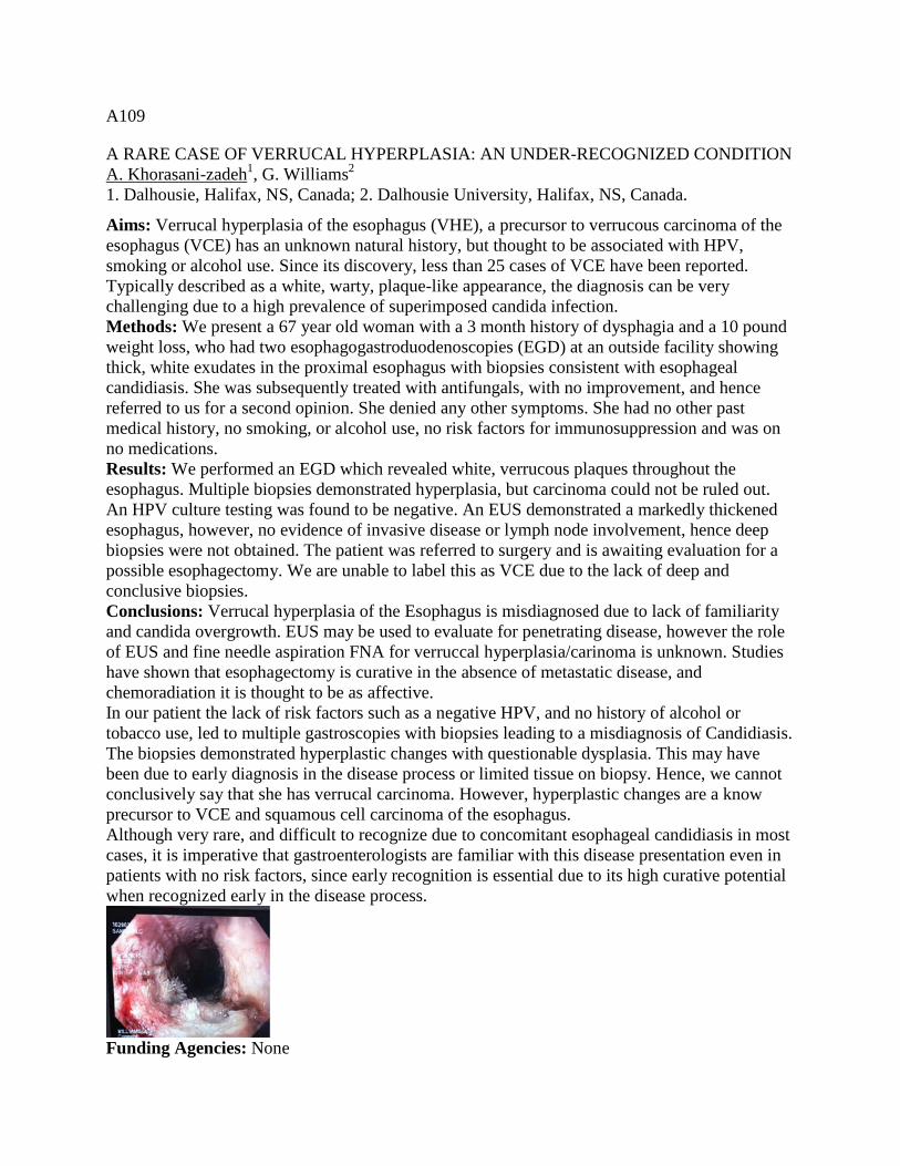
A109
A RARE CASE OF VERRUCAL HYPERPLASIA: AN UNDER-RECOGNIZED CONDITION A. Khorasani-zadeh1, G. Williams2 1. Dalhousie, Halifax, NS, Canada; 2. Dalhousie University, Halifax, NS, Canada.
Aims: Verrucal hyperplasia of the esophagus (VHE), a precursor to verrucous carcinoma of the esophagus (VCE) has an unknown natural history, but thought to be associated with HPV, smoking or alcohol use. Since its discovery, less than 25 cases of VCE have been reported. Typically described as a white, warty, plaque-like appearance, the diagnosis can be very challenging due to a high prevalence of superimposed candida infection. Methods: We present a 67 year old woman with a 3 month history of dysphagia and a 10 pound weight loss, who had two esophagogastroduodenoscopies (EGD) at an outside facility showing thick, white exudates in the proximal esophagus with biopsies consistent with esophageal candidiasis. She was subsequently treated with antifungals, with no improvement, and hence referred to us for a second opinion. She denied any other symptoms. She had no other past medical history, no smoking, or alcohol use, no risk factors for immunosuppression and was on no medications. Results: We performed an EGD which revealed white, verrucous plaques throughout the esophagus. Multiple biopsies demonstrated hyperplasia, but carcinoma could not be ruled out. An HPV culture testing was found to be negative. An EUS demonstrated a markedly thickened esophagus, however, no evidence of invasive disease or lymph node involvement, hence deep biopsies were not obtained. The patient was referred to surgery and is awaiting evaluation for a possible esophagectomy. We are unable to label this as VCE due to the lack of deep and conclusive biopsies. Conclusions: Verrucal hyperplasia of the Esophagus is misdiagnosed due to lack of familiarity and candida overgrowth. EUS may be used to evaluate for penetrating disease, however the role of EUS and fine needle aspiration FNA for verruccal hyperplasia/carinoma is unknown. Studies have shown that esophagectomy is curative in the absence of metastatic disease, and chemoradiation it is thought to be as affective. In our patient the lack of risk factors such as a negative HPV, and no history of alcohol or tobacco use, led to multiple gastroscopies with biopsies leading to a misdiagnosis of Candidiasis. The biopsies demonstrated hyperplastic changes with questionable dysplasia. This may have been due to early diagnosis in the disease process or limited tissue on biopsy. Hence, we cannot conclusively say that she has verrucal carcinoma. However, hyperplastic changes are a know precursor to VCE and squamous cell carcinoma of the esophagus. Although very rare, and difficult to recognize due to concomitant esophageal candidiasis in most cases, it is imperative that gastroenterologists are familiar with this disease presentation even in patients with no risk factors, since early recognition is essential due to its high curative potential when recognized early in the disease process.
Funding Agencies: None

A110
THE ALPHA FETOPROTEIN-SECRETING GASTRIC CANCER IN THE SETTING OF CHRONIC HEPATITIS B: WHEN YOU CAN'T FIND THE HEPATOCELLULAR CACNER, DO ENDOSCOPY
S. Ip, D. Schaeffer, E. Yoshida, P. Kwan
University of BC, Vancouver, BC, Canada.
Background: Alpha-fetoprotein (AFP) is a common tumor marker for hepatocellular carcinoma (HCC). A rising serum AFP, especially >400 mcg/L, in a patient with cirrhosis or hepatitis B (HBV) should raise suspicion of a developing HCC. Aims: We report a unique case of an AFP-secreting gastric cancer (AFPGC) in an elderly man with known chronic HBV and a markedly elevated AFP. Methods: Case Report Results: An 81-year-old Chinese man was followed for chronic HBV and was treated with tenofovir, resulting in undetectable HBV viral load. He did not have cirrhosis or other comorbidities. He complained of early satiety, anorexia, weight loss (>10 pounds), and right-sided chest pain over several months. Laboratory investigations revealed an elevated AFP of 45 mcg/L, which gradually increased to 540 mcg/L over three months. Multiple imaging studies including CT and MRI failed to detect HCC. He then presented with an acute upper gastrointestinal (GI) bleed. On gastroscopy, a large ulcerated mass along the lesser curvature was found. Biopsies taken were consistent with gastric adenocarcinoma; interestingly, the specimen stained for focal positivity to AFP. Staging CT imaging showed no metastatic spread of the disease. The patient then underwent successful neoadjuvant chemotherapy and partial gastrectomy. His AFP levels have since remained normal. AFPGCs are extremely rare, accounting for only 1-6% of all gastric cancers and are associated with poorer prognosis and extensive metastases. A literature review reveals several case series of Asian patients with AFPGCs; however, those with active or chronic hepatitis are excluded from analyses or their hepatitis status is not clearly documented. Conclusions: This case highlights that an elevated AFP may not be associated with HCC even in patients with known risk factors. In these patients, if appropriate imaging does not reveal HCC, clinicians should consider endoscopic investigation of the GI tract.
Funding Agencies: None
Immunology and Inflammatory Bowel Disease
Poster of Distinction
A111 (oral presentation A31)
TRIMETHYLAMINE-N-OXIDE AND INFLAMMATORY BOWEL DISEASE: DIFFERENTIAL ROLE OF INTESTINAL MICROBIOTA IN CROHN'S DISEASE VS ULCERATIVE COLITIS A. Wilson1, W. Teft2, Y. Choi3, R. Tirona2, S. Woolsey2, T. Ponich4, J. Gregor4, N. Chande4, R. Kim2 1. Department of Physiology & Pharmacology; Gastroenterology, University of Western Ontario, London, ON, Canada; 2. Clinical Pharmacology, University of Western Ontario, London, ON, Canada; 3. Department of Statistics, University of Western Ontario, London, ON, Canada; 4. Gastroenterology, University of Western Ontario, London, ON, Canada.
Background: The potential impact of the gastrointestinal (GI) microbiome to inflammatory bowel disease (IBD) is increasingly recognized for potential clinical relevance. Current biomarkers of disease are designed to assess the presence of inflammatory mediators. However, available data also suggest host GI microbiota participate in the generation of the dysregulated immune response and thereby contribute to initiation and progression of IBD. Trimethylamine-N-oxide (TMAO) is a metabolite generated by GI tract anaerobes through the digestion of phosphatidylcholine- and carnitine-containing food products. Recently, elevated levels have been linked to the development of heart disease, yet little is known regarding TMAO levels in the setting of IBD. Aims: To determine if plasma TMAO levels among those with IBD are altered compared to healthy controls and if they correlate with disease activity or phenotype. Methods: Ultra performance liquid chromatography-tandem mass spectrometry was used to measure TMAO as well as choline and carnitine plasma levels from blood samples from 485 subjects (373 healthy controls, 112 IBD). Cases and controls were matched on age-category and sex. Subjects were also genotyped for the common flavin monooxygenase (FMO) 3 variants E158K and E308G. Results: Plasma TMAO levels were significantly decreased in individuals with IBD. Individuals with active ulcerative colitis (UC) had significantly lower plasma TMAO levels than those with inactive disease. No difference was seen in those with active Crohn's disease (CD) versus those with inactive CD. Though statistical significance was not achieved, a trend toward lower plasma TMAO levels was seen in those with colonic disease compared to those with ileal disease. No inter-group variation was seen in plasma TMAO levels based on FMO3 genotype. Choline levels were higher in IBD, while carnitine levels were similar between the two groups suggesting lower TMAO levels in IBD were not due to reduced dietary intake of food containing choline and carnitine such as dairy products, eggs and red meat. Conclusions: Decreased TMAO levels are seen in IBD compared to a non-IBD population. To our knowledge this is the first study that describes reduced TMAO levels in IBD. TMAO may have the potential for use as a biomarker to support IBD diagnosis as well as disease activity in UC.
Funding Agencies: CIHR

Poster of Distinction
A112 (oral presentation A32)
THE MICROBIAL METABOLITE BUTYRATE, PROMOTES BACTERICIDAL ACTIVITY AND THE INDUCTION OF REGULATORY T CELLS BY IL-4 PRIMED MACROPHAGES
M. Fernando2, J. Reyes2, D. McKay1
1. Uni. Calgary, Calgary, AB, Canada; 2. University of Calgary, Calgary, AB, Canada.
Background: Butyrate is the most abundant short chain fatty acid produced by the intestinal microbiota through the fermentation of dietary fibers. As the primary energy source of colonocytes, butyrate has also been associated with the regulation of innate (i.e. epithelial barrier integrity) and adaptive (i.e. induction of regulatory T cells (Treg) and suppression of pro-inflammatory macrophage responses) immunity. Having shown that adoptive transfer of alternatively activated macrophages (AAMs) can suppress colitis, we hypothesized that butyrate would reinforce an AAM phenotype. Aims: To use canonical markers and functional assays to determine if butyrate exposure modifies the polarization of macrophages by interleukin (IL)-4. Methods: Murine bone-marrow-derived macrophages were differentiated into AAMs by IL-4 (20ng/mL; 48h) ± butyrate, propionate or acetate (0.1-2mM) and changes in markers of AAM polarization (arginase-1, Ym1) were assessed. AAMs were also exposed to LPS (1μg/mL) for a further 24h, and nitric oxide and cytokine levels were measured. The effect of butyrate pre- and post-treatment on cytokine production by AAMs was assessed. Additionally, the ability of AAMs to kill commensal E. coli (strain HB101) and the effect of AAMs on Treg polarization (based on CD25 and Foxp3 expression) with or without butyrate was determined. Results: Exposure of AAMs to butyrate inhibited expression of hallmark AAM markers Arg1 and Ym1, and significantly suppressed LPS-induced nitric oxide, IL-12p40, IL-6 and IL-10 production compared to IL-4-treated macrophages. This regulation of AAM phenotype occurred whether butyrate was applied as a pre-treatment (48h) or 48h after IL-4 exposure. Butyrate-treated AAMs showed no significant increase in apoptosis and butyrate did not affect IL-4-induced phospho-STAT6. Importantly, butyrate-treated AAMs displayed enhanced bacterial killing compared to AAMs only, and CD4+ T cells co-cultured with the butyrate-treated AAMs displayed increased CD25 expression. None of these effects were observed with acetate or propionate. Conclusions: Butyrate is a common constituent of the normal gut, and while it reduced the expression of AAM markers, it had the benefit of suppressing LPS-stimulated pro-inflammatory responses, while enhancing bactericidal activity and induction of putative Tregs. These findings point to the importance of butyrate, a microbial-derived metabolite, in the regulation of mucosal immunity and support reassessment of butyrate as an adjunct or stand-alone anti-inflammatory treatment in defined cohorts of patients with inflammatory bowel disease.
Funding Agencies: CCC, CIHR, CDHF, AI-HS

Poster of Distinction
A113 (oral presentation A33)
EFFECT OF MICROBIOTA ON MATURATION OF INTESTINAL BARRIER STRUCTURE AND FUNCTION.
C. Hayes, J. Jury, C. Sheridan, J. Dong, E. Verdu
McMaster University, Hamilton, ON, Canada.
Background: The gut microbiota influences immune maturation and homeostasis, but its role in the regulation and maturation of the intestinal barrier has not been fully characterized. Aims: In this study, changes in small intestinal barrier structure and function induced by microbial colonization were investigated. Methods: Male and female germ free C57BL/6 mice were colonized with fecal microbiota either rich in Ruminococcaceae derived from a healthy human (HH) adult, or low in this family from an adult ulcerative colitis (UC) patient during an acute flare. At 24 hours and 1 week following colonization, crypt depth, immune cell infiltration, myeloperoxidase (MPO) activity, tight junction mRNA and protein expression by qPCR and immunohistochemistry were evaluated in the small intestine (SI: ileum). Paracellular permeability of the SI was evaluated in vitro using Ussing chambers with the probe 51Cr-EDTA. Results: One week post-colonization with HH microbiota, stool consistency normalized and crypt depth increased compared to germ free mice. These changes were not observed after colonization with UC microbiota. In both HH and UC colonized mice, claudin-3 expression was increased at 24 hours, and ZO-1 expression decreased at 1 week. In HH colonized mice, E-cadherin mRNA expression and protein were increased at 24 hours, but not in UC colonized mice. Furthermore, occludin expression was decreased at 1 week in UC colonized mice, which was not observed in HH colonized mice. SI paracellular permeability was higher at 24 hours in mice colonized with UC compared to HH. Conclusions: These findings indicate that a microbiota low in Ruminococcaceae from an UC patient in flare affects early intestinal barrier maturation and function. Although colonization with either microbiota induced changes in tight junction mRNA and protein expression, a microbiota low in Ruminococcaceae was associated with delayed crypt depth changes, decreased stool consistency, lower E-cadherin and occludin expression at 24 hours and 1 week, respectively, and increased paracellular permeability. Identification of specific bacteria that affect barrier maturation, and perhaps subsequent susceptibility to inflammation, may help develop microbiota-directed strategies for inflammatory bowel diseases, such as UC. Supported by Crohn's and Colitis Canada
Funding Agencies: Crohn's and Colitis Canada

Poster of Distinction
A114
LONG-TERM OUTCOMES OF INFLIXIMAB USE FOR PEDIATRIC CROHN'S DISEASE IN CANADA
J. deBruyn2, K. Jacobson4, W. El-Matary3, M. Carroll1, E. Wine1, I. Wrobel2, C. Waterhouse2, M. Van Woudenberg2, H. Huynh1
1. University of Alberta, Edmonton, AB, Canada; 2. University of Calgary, Calgary, AB, Canada; 3. University of Manitoba, Winnipeg, MB, Canada; 4. University of British Columbia, Vancouver, BC, Canada.
Background: Data on long-term outcomes of infliximab use in pediatric Crohn's disease are limited. Aims: To evaluate outcomes of infliximab in Canadian children with Crohn's disease. Methods: The charts of all children <18 years old with Crohn's disease who started infliximab between Jan 2008 and Dec 2012 in 4 Canadian tertiary-care centers were retrospectively reviewed. All patients were followed until transition to adult care or infliximab stop if before transition. Univariable and multivariable factors associated with loss of response requiring discontinuation were evaluated using Cox proportional hazards model. Factors selected a priori included gender, age at diagnosis, age at infliximab start, time to infliximab start, disease behaviour (Paris classification), and concomitant immunomodulator use. Annual loss of response was estimated using Joinpoint regression. Results: One-hundred and eighty children (male 54.4%) received infliximab induction; 99.4% continued to maintenance. Median age at infliximab start was 14.3 years (Q1,Q3: 12.8, 15.9 years) and median time from diagnosis to infliximab start was 1.5 years (Q1,Q3: 0.6, 3.5 years); 91.1% had failed other medical therapies. Indications for starting infliximab were: chronic active disease (47.2%), severe exacerbation (20.1%), steroid dependence (7.8%), severe perianal disease (14.0%), extensive disease (15.2%), internal fistulizing disease (7.3%), and other (5.6%). Concomitant immunomodulator use occurred in 25.8% (azathioprine) and 36.5% (methotrexate). At last follow-up, 88.2% were maintained on IFX; median duration of follow-up was 66.1 weeks (Q1,Q3: 34.0, 115.9 weeks). Infliximab optimization occurred in 52.2% (dose escalation only 12.8%, interval shortening only 3.3%, both 36.1%). The most frequent indication for optimization was loss of response (82.7% dose escalation, 74.1% interval shortening). Of those who underwent dose escalation or interval shortening, 79.3% and 77.5%, respectively, continued infliximab. Loss of response resulting in discontinuation occurred in 3.5% per year (95% CI -1.9 to 8.6); only female gender was a risk factor (hazard ratio 2.6, 95% CI 1.0-6.5, P=0.04). Conclusions: Children with Crohn's disease maintain a durable response to infliximab. Dose optimization is frequently implemented for loss of response, often followed by continued use. Females are at higher risk of loss of response requiring discontinuation.
Funding Agencies: None

Poster of Distinction
A115
BROKEN FENCES; EPITHELIAL GAPS & MICROBES IN PEDIATRIC INFLAMMATORY BOWEL DISEASES D. Zaidi1, M. Bording-Jorgensen1, H. Huynh2, C. matthew1, E. Wine1 1. University of Alberta, Edmonton, AB, Canada; 2. University of alberta, Edmonton, AB, Canada.
Background: Inflammatory Bowel Diseases (IBD), encompassing Crohn Disease and Ulcerative Colitis are highly prevalent in children in Canada with unknown etiology. Multiple factors including alterations in microbial composition, increased intestinal permeability and immune dysregulation contribute to IBD pathogenesis. Increased epithelial cell extrusion, as measured by counting gaps between epithelial cells, has not been assessed in children. Our hypothesis is that epithelial gap density is elevated in pediatric IBD patients and correlates with microbial virulence, barrier disruption and inflammation. Our objective was to study the correlation between epithelial gaps, microbial virulence and gut inflammation. Aims: Evaluate the presence of intestinal epithelial gaps in pediatric IBD and to establish its prognostic value. Define alterations in microbial composition and inflammatory markers and correlate with epithelial gaps. Determine in vitro effects of microbes on epithelial barrier disruption. Methods: In a prospective, blinded, cohort study, epithelial gap density of the duodenum in pediatric IBD patients and non-IBD controls was evaluated using probe-based confocal laser endomicroscopy (pCLE) after injecting florescein. Epithelial gap density was defined as the number of gaps normalized to epithelial cells counted. Epithelial gaps were related to serum inflammatory markers and disease scoring indices. Intestinal aspirates were analyzed for cytokine levels, microbial quantification via qPCR, and bacterial culture. Effect of luminal factors on microbial invasion potential was assessed by Gentamicin protection assays on T84 cells. Florescein levels were quantified to assess permeability. Results: 89 participants have been recruited. Initial analysis revealed differences between IBD and non-IBD patients. Epithelial gap density was significantly higher in IBD patients whereas, anaerobic bacteria and florescein levels were marginally higher in IBD patients than in non-IBD controls. C-reactive protein, ESR levels and high disease activity was observed in a subset of IBD patients with higher epithelial gaps. Aspirates from patients altered invasion of bacteria in vitro. PCR of bacterial isolates showed prevalence of E. coli from various phylotypes and different virulence factors. Conclusions: Results indicate that epithelial gaps, pathogens, and host factors likely play integrated roles in IBD pathogenesis. Altered microbial invasion potential suggests involvement of host and microbial factors. Evaluating epithelial gap density and its relation with clinical parameters might be helpful in defining better treatment options.
Funding Agencies: WCHRI, University of Alberta

Poster of Distinction
A116
IMPACT OF INFLAMMATORY BOWEL DISEASE ACTIVITY ON THE INCIDENCE OF NONALCOHOLIC FATTY LIVER DISEASE: A 7-YEAR LONGITUDINAL STUDY
N. Le, K. Rollet, W. Afif, A. Bitton, T. Bessissow, G. Sebastiani McGill University Health Centre, Montreal, QC, Canada.
Background: Nonalcoholic fatty liver disease (NAFLD) is the most frequent liver disease in Canada. It is classically associated with dysmetabolisms. Conflicting data exist on its frequency and risk factors in patients with inflammatory bowel disease (IBD). Aims: We aimed at investigate prevalence, incidence and predictors of hepatic steatosis development in patients with IBD. Methods: This was a retrospective study of IBD patients without known liver disease during 2006-2013. NAFLD was defined as a Hepatic Steatosis Index (HSI) ≥ 36 or as positive imaging (ultrasound or CT scan). Clinical markers for IBD activity were collected at 6 months interval. Active IBD was defined as Mayo Score ˃ 3 for ulcerative colitis, Harvey Bradshaw Index ˃ 5 or flare within follow-up interval. We estimated incidence rates of NAFLD by dividing the number of participants developing the outcome by the number of person-years (PY) of follow-up. Poisson count models were used to calculate confidence intervals (CI) for incidence rates. Multivariate Cox proportional hazard models were built to identify predictors of development of NAFLD. Results: 232 patients (median age 34 years, 50% males) were included. 68% of patients had Crohn's disease and 59% had active IBD. 28% of patients had at least one metabolic comorbidities. The overall prevalence of NAFLD in our cohort was 41%. 35 patients already had the outcome at baseline and were excluded from the longitudinal analysis. During a median follow-up of 3.6 years (interquartile range 1.4-6), 31% patients developed NAFLD. Over 731.2 PY of follow-up, the rate of progression to NAFLD was 8.2/100 PY (95% CI 6.1-10.3). The results of multivariate analysis are depicted in the Table. Active IBD and age at IBD diagnosis were independent predictors of NAFLD development. Patients with active IBD had a rate of progression of 9/100 PY (95% CI 6-11.9) as compared to 7.3/100 PY (95% CI 4.4-10.2) in those in clinical remission. Metabolic comorbidities did not contribute to NAFLD in our cohort. Conclusions: NAFLD is a frequent comorbidity in patients with IBD. Disease activity is a major predictor of NAFLD development in IBD patients. This should represent one more incentive to achieve and maintain early clinical remission in those patients. Multivariate analysis of predictors of NAFLD development
Predictor Unadjusted HR (95% CI)
Adjusted HR (95% CI) p
Active IBD at each visit (time updated) 1.93 (1.17-3.18) 1.87 (1.13-3.11) 0.02 Age at IBD diagnosis 1.08 (1.01-1.15) 1.1 (1.03-1.17) 0.004
Duration of IBD 1.08 (0.94-1.24) 1.07 (0.92-1.24) 0.38 Prior surgery 1.54 (0.9-2.48) 1.49 (0.79-2.78) 0.21
Funding Agencies: FRSQ
Poster of Distinction
A117
CLINICAL PREDICTORS OF 3-YEAR RISK OF COLECTOMY IN ULCERATIVE COLITIS: A POPULATION-BASED STUDY
A. Al-Darmaki1, J. Hubbard1, C. Seow1, Y. Leung1, K. Novak1, S. Ghosh1, R. Panaccione1, G. Kaplan2
1. University of Calgary, Calgary AB, AB, Canada; 2. University of Calgary, Calgary, AB, Canada.
Background: Despite the improvements in medical therapy of ulcerative colitis, a significant number of patients will require colectomy over time to control medically refractory disease. However, clinical factors that are associated with the risk of early colectomy are unknown. Aims: To determine the clinical predictors of colectomy within 3 years of diagnosis among ulcerative colitis (UC) patients who are hospitalized with an acute flare. Methods: Population-based surveillance was conducted between January 1, 1997 and December 31, 2008 to identify all adults (≥ 18 years) with a discharge abstract code for UC within 3 years of diagnosis. The primary outcome was a colectomy within 3 years of diagnosis. All medical charts were reviewed to confirm the diagnosis and to extract the following variables: age, sex, smoking status, year of admission, length of flare, comorbidities, disease extent, and hemoglobin. Additionally, drug utilization prior to hospitalization was recorded for 5-ASA, azathioprine, prednisone, and infliximab. Infliximab prescription in hospital was also evaluated separately. Logistic regression model was created to evaluate variables that predicted need for colectomy within 3 years of diagnosis. Adjusted odds ratios (OR) with 95% confidence intervals (CI) are reported. Results: Out of 489 patients hospitalized with UC, 282 had colectomy within 3 years of diagnosis. Predictors of increased risk of colectomy included individuals between the ages of 35 and 64 years (OR 2.18, 95% CI: 1.27-3.74), males (OR 2.03, 95% CI: 1.24-3.34), pancolitis (OR 5.38, 95% CI: 3.20-9.06), prior use of prednisone (OR 5.44, 95% CI: 3.03-9.75) and previous use of infliximab (OR 5.12, 95% CI: 1.36-19.30). Patients who received induction therapy with infliximab during hospitalization were less likely to require colectomy (OR 0.28, 95% CI: 0.10-0.77). Past smoking history or current smoking status did not influence the risk of colectomy (OR 0.72, 95% CI: 0.29-1.76; OR 1.14, 95% CI: 0.65-1.98, respectively). There was no increased risk of colectomy among individuals with anemia (OR 0.539, 95% CI: 0.286-1.015). Conclusions: Middle aged men with pancolitis and who failed medical management with prednisone and infliximab were more likely to require colectomy within 3 years of diagnosis. In contrast, patients prescribed infliximab in-hospital during an acute flare were less likely to require a colectomy.
Funding Agencies: None
Poster of Distinction
A118
BIOLOGIC THERAPIES ARE EFFECTIVE IN INDUCING AND MAINTAINING MUCOSAL HEALING IN ULCERATIVE COLITIS: A SYSTEMATIC REVIEW & METANALYSIS
M. Stewart, A. Al-Darmaki, R. Panaccione, G. Kaplan, C. Seow
University of Calgary, Calgary, AB, Canada.
Background: There has been a paradigm shift from the traditional approach of treating ulcerative colitis (UC) to simply achieve symptomatic control (clinical remission). Instead, we now aim to treat to ‘deep remission', that is to achieve mucosal healing (MH). It has been demonstrated that patients who achieve MH have lower rates of relapses, steroid use, hospitalization and surgery. Aims: We performed a systematic review and meta-analysis on the efficacy of biologic therapies in inducing and maintaining mucosal healing in UC. Methods: Randomized controlled trials of a biologic therapy compared to a control arm with participants receiving placebo that met predetermined selection criteria were included. Citations were identified through EMBASE, MEDLINE, the Cochrane Central Register of Controlled Trials, proceedings of gastroenterology meetings, manual search of reference lists of trials and review articles, and ongoing trials identified from the registry http://ClinicalTrials.gov. Data were pooled and we performed a random-effects meta-analysis. As a measure of effect we calculated risk ratio (RR) with corresponding 95% confidence interval (CI) for dichotomous outcomes. Data were analyzed using Review Manager (RevMan version 5.3, Copenhagen:The Nordic Cochrane Centre, The Cochrane Collaboration, 2014). Results: Thirty-five trials were identified and 13 met the inclusion criteria. Of trials using an anti-TNFα, 6 were induction trials totaling over 2000 patients and 3 were maintenance trials totaling over 1500 patients. Anti-TNFα versus placebo increased both induction and maintenance of MH (RR=1.26; 95% CI, 1.07-1.50, and RR=1.68; 95% CI, 1.43-1.97, respectively). The use of an anti-adhesion molecule was evaluated in 3 induction trials, totaling over 1000 patients, and a maintenance trial with over 350 patients. Anti-adhesion molecule versus placebo also increased both induction and maintenance of MH (RR=1.57; 95% CI, 1.19-2.07, and RR=2.71; 95% CI, 1.88-3.93, respectively). Induction of remission was evaluated using an anti-CD20, anti-CD3, and anti-IL2 receptor antibody in 1 trial each. The use of biologic therapy was associated with a decreased risk of study withdrawal due to adverse events for both anti-TNFα (RR=0.65; 95% CI, 0.48-0.86) as well anti-adhesion molecule biologic therapies (RR=0.33; 95% CI, 0.10-1.16). Conclusions: Biologic therapies are more effective in inducing and maintaining MH than placebo in patients with UC. The effect is seen using both anti-TNFα and anti-adhesion molecule biologic therapies. Both classes of therapies were well tolerated with minimal adverse effects.

Funding Agencies: None

A119
HOW WELL DO CLINICAL ACTIVITY INDICES REFLECT ENDOSCOPIC DISEASE ACTIVITY IN INFLAMMATORY BOWEL DISEASE?
E. Zittan2, B. Kabakchiev3, O. Kelly2, J. Stempak3, G. Nguyen2, K. Croitoru2, G. Van Assche1, A. Steinhart2, M. Silverberg2
1. University Hospitals Leuven, Leuven, Belgium; 2. Mount Sinai Hospital, University of Toronto, Toronto, ON, Canada; 3. Lunenfeld-tanenbaum research institute, Toronto, ON, Canada.
Background: There is movement toward utilizing patient-reported outcomes (PROs) and objective measures of disease activity, as compared to clinical activity indices in inflammatory bowel disease (IBD). Aims: This study evaluated the performance between the Harvey-Bradshaw Index (HBI) and Mayo Score and endoscopic scores (SES-CD, Mayo endoscopic score,Rutgeerts score). We also examined which component or combination of subjective clinical scores and patient report outcome best represent the objective endoscopic score in IBD. Methods: A 100 IBD patients undergoing colonoscopy were enrolled in a prospective study (33 Crohn's disease (CD), 24 post-operative ileocolonic resection CD, and 40 with ulcerative colitis (UC)). Each patient had Pre-endoscopist-blinded assessments including: HBI/partial Mayo score, a subjective PRO question asking the perception of disease activity status, physician global assessment (PGA), and C-reactive protein (CRP). Two endoscopists experienced in IBD provided an endoscopic score independently. Active endoscopic disease was defined as SES-CD>3, Rutgeerts≥i1 and Mayo score≥1. Clinical remission was defined as HBI≤4 or Mayo score≤2. We used logistic regression in order to assess the performance of different predictive models against the endoscopic scores. Results: The HBI did not reflect SES-CD well (AUC=0.53). Combining the HBI with PGA and the PRO question and then further adding CRP to the combination above significantly improved the test performance compared to the SES-CD (AUC=0.71 and AUC=0.79 resp.). In post-operative CD, the HBI also performed poorly versus the Rutgeerts score (AUC=0.50). Combining HBI with PGA and the PRO question and then further adding the CRP to the combination above improved the test performance (AUC=0.65 and AUC=0.8056 resp.). In UC, there was poor performance of the 6 point Mayo score (rectal bleeding and stool frequency) versus the Mayo endoscopic score (AUC=0.54). Utilizing the 9 point Mayo score (rectal bleeding, stool frequency and PGA) with the PRO question improved test performance (AUC=0.68). Conclusions: In CD, the HBI poorly mirrors the endoscopic scores (SES-CD and Rutgeerts). Adding a PGA, PRO question and CRP to the HBI, significantly improves its ability to predict endoscopic activity. In UC, clinical evaluation using the Mayo score did not accurately reflect endoscopic disease activity even when adding a PRO question. Current clinical indices in IBD are relatively poor at predicting endoscopic disease activity but can be improved by integrating additional components.
Funding Agencies: None
A120
INFLIXIMAB IS EFFECTIVE IN TREATMENT OF INFLAMMATORY COMPLICATIONS FOLLOWING PELVIC POUCH SURGERY
O. Kelly, M. Rosenberg, G. Greenberg, H. Steinhart, Z. Cohen, A. Tyler, J. Stempak, M. Silverberg
Mount Sinai Hospital, Toronto, ON, Canada.
Background: Pouchitis refractory to first-line therapies remains problematic following colectomy and ileal pouch anal anastomosis (IPAA) for ulcerative colitis (UC). Evidence for infliximab (IFX) in this setting and factors predicting its success remain limited. Aims: To investigate IFX use in refractory pouch problems and variables associated with treatment response. Methods: Data were reviewed from 579 individuals who underwent colectomy and IPAA from 2000-2014. Patients with chronic refractory pouchitis (CP) and Crohn's Disease- like (CDL) outcome treated with IFX were included. Pretreatment parameters were measured within four weeks of induction and IFX response at median of 9 (initial) and 48 weeks (sustained) respectively. Complete response was defined as symptomatic and endoscopic resolution with modified Pouchitis Disease Activity Index (mPDAI) <5. Partial response was clinical improvement with reduction in mPDAI ≥ 2. Mucosal healing, pouch outcomes and CRP were recorded. Serum was analyzed for ASCA, anti-OmpC, anti-CBir1 and pANCA. Fisher's exact testing and Kruskal-Wallis detected differences in clinical and serological variables. Logistic regression estimated odds ratio (OR) of clinical and serological factors with initial and sustained clinical response as dependent variables. Results were deemed significant if p-value ≤0.05. Results: 34 patients were included (33% male; age 32.6 ± 2.6 [mean ± SE; years], 29% CDL). 26% received concomitant immune-modulator. Mean time from IPAA to anti-TNF was 7.3±0.95 years. 74% achieved post-induction response (48% complete). 62.6% sustained response (29.6% complete) at follow-up. Mean mPDAI was significantly reduced from pre-induction score of 8.5± 0.3 to 2± 3.4(p<0.002) and CRP fell from 29.48±6.2 mg/L to 5.76 ± 1.6 mg/L (p<0.001). Age, gender, smoking, pre-pouch disease extent and CRP were not associated with response to IFX. Patients with pretreatment mPDAI ≤10 were more likely to have initial mucosal healing (p = 0.03). Initial mucosal healing was significantly associated with sustained complete response (p<0.05; [OR] =13.2; 95% confidence interval [CI], 1.0-165). A positive ASCA titre was associated with higher initial mPDAI (p = 0.016), but no difference in treatment response. IFX responders had fewer positive antibody titres (2 vs. 3, mean; p<0.05). Conclusions: IFX was effective in short- and longer term in patients with chronic refractory pouchitis and CDL complications in the pouch. Mucosal healing after induction is the strongest clinical association with a complete sustained response to IFX in this group and a pretreatment mPDAI of ≤ 10 may help predict this.
Funding Agencies: Prometheus Laboratories for serology testing

A121
GASTROINTESTINAL OUTPATIENT SURVEY
K. MacIsaac, M. Black, J. Stewart, D. MacIntosh
Queen Elizabeth II Health Science Centre, Halifax, NS, Canada.
Background: The Division of Gastroenterology at the Capital District Health Authority (CDHA) in Halifax has assessed the experience of patients coming in for a clinic visit regarding their gastrointestinal or hepatic disease using satisfaction surveys. These questionnaires were used to ensure that care provided within Capital Health GI Division met the expectations of our patients. Aims: To assess the quality of care provided in the out-patient clinic as reported by patients, and to ensure that deficits in delivery of care were identified and resolved. Methods: June 2014 to August 2014 questionnaires were distributed to all patients receiving care from the outpatient clinic. Patients were given a questionnaire with a pre-paid envelope upon admission for their visit. Completed surveys were to be returned within seven days of the visit and entered into a SPSS database for analysis. Results: Information was collected from 595 patients. 325 patients (55%) were seen by a gastroenterologist, 95 patients (16%) were seen by a nurse practitioner for their liver disease, 46 patients (8%) were seen by a surgeon, 43 patients (7%) were seen by a hepatologist, and 37 (6%) patients were seen by a nurse practitioner for their inflammatory bowel disease. Of the patients assessed, 97 patients (16%) reported that it was their first time being seen by the outpatient clinic. Determining satisfaction of patients regarding the time from referral to their clinic visit was of interest to our site. 493 patients (86%) believed that they were referred within a reasonable time, and 53 patients (8.9%) believe that the time to clinic visit was too long. 347 (58%) patients were seen within 3 months of referral, 282 (14%) patients were seen within 3-6 months of referral, 43 (7%) patients were seen 6-12 months after referral, and 26 (4%) patients were seen over one year after referral. Patients were also asked about taking time away from work for their appointment. 231 patients (39%) had to take time off work for their appointment, while taking time off work was not necessary for 362 patients (61%). Once in the clinic for their appointment, patients were asked if their appointment was on time (within 15 minutes of the scheduled time). 448 (75%) patient appointments were on time and 103 (17%) appointments were over 15 minutes past the scheduled time. Conclusions: No significant gaps in the delivery of care at the outpatient clinic have been identified. The services being provided currently meet patient expectations.
Funding Agencies: None

A122
ANTI-VIRAL THERAPY IS ASSOCIATED WITH DECREASED COLECTOMY RATES IN CORTICOSTEROID-REFRACTORY IBD PATIENTS WITH CYTOMEGALOVIRUS: A SYSTEMATIC REVIEW AND META-ANALYSIS
T. Shukla1, S. Singh2, J. McCurdy1
1. The Ottawa Hospital, Ottawa, ON, Canada; 2. Mayo Clinic, Rochester, MN.
Background: Cytomegalovirus (CMV) is an opportunistic infection associated with corticosteroid (CS)-refractory inflammatory bowel disease (IBD). It remains unknown if CMV plays a pathogenic role and if anti-viral therapy can improve clinical outcomes. Aims: We therefore performed a systematic review and meta-analysis of observational studies to assess the association between antiviral therapy and risk of colectomy in IBD patients with CMV. Methods: A systematic search of multiple electronic databases was performed through July 2014 for studies reporting risk of colectomy in IBD patients with CMV, stratified by treatment with antiviral agents. Studies were included when the diagnosis of CMV was made by serum or tissue-based techniques. Colectomy rates were assessed for the overall cohort and stratified by CS-refractory disease. We estimated summary odds ratios (ORs) and 95% confidence intervals (CI), using random effects model. The methodological quality of the included studies was assessed using the Newcastle-Ottawa scale. Results: Twenty-two studies were identified with data available for 530 IBD patients with CMV (249 treated with and 281 without antiviral therapy). In 12 studies, patients were stratified based on CS-refractory disease (196 IBD patients with CMV, 110 treated with and 86 without anti-viral therapy). In the overall, unselected IBD population there was no difference in colectomy rate between patients treated with anti-viral therapy and those without treatment (OR=0.74; 95% CI: 0.33-1.64), with moderate heterogeneity (I2=56%). In patients with CS-refractory disease, treatment with antiviral therapy resulted in an 89% lower risk of colectomy compared to no antiviral treatment (OR=0.19; 95% CI: 0.09-0.40; I2=0%). The results were stable when restricting the analysis to patients with UC, tissue diagnosis of CMV, and studies that defined CS-refractory disease as a failure to respond to 1 week of intravenous CS. Conclusions: This study provides evidence for a beneficial role of antiviral therapy in CS-refractory IBD patients with CMV disease.
Funding Agencies: None

A123
VITAMIN D LEVELS CORRELATE WITH MILD CROHN'S DISEASE ACTIVITY AND LABORATORY MARKERS IN PATIENTS AND CONTROLS
A. Szilagyi, X. Xue, A. Grunbaum, B. Burstein, E. MacNamara, P. Gordon
Jewish General Hospital, Montreal, QC, Canada.
Background: Serum 25(OH) D (Vitamin D) levels in quiescent inflammatory bowel diseases (IBD:CD and UC ) were similar to healthy controls. But,52% of each group had insufficient or defficient levels (Nutr J, 2013). Aims: To evaluate:relationships between:1) Vitamin-D and IBD scores in patients, 2) Vitamin-D and blood chemistries in all groups. Methods: : The study between 2009-2011 recruited from IBD clinics or hospital sources for controls. CD was scored by Harvey Bradshaw index (HBI) and UC,by Simple Clinical Colitis Activity index (SCCAI). Vitamin D was measured by RIA kit and blood chemistries by hospital laboratory methods, including Hgb, Platelets, MCV, µPlatelet Volume (MPV), PMNLeukocyte, Lymphocyte, Albumin, Calcium and CRP. Potential confounding variables were patient groups, season, intake of supplement vitamin D and Caucasian race. Univariate and Multivariable linear regression analysis were conducted. Log transformations were used when variables were not normally distributed. Statistical significance was set at p < 0.05, 95%(CI) confidence intervals were provided. Results: 100 persons were recruited, 55 IBD patients (34 CD, 21 UC) [3 non white] and 45 controls [10 non white]. Their mean age was 41±13 years with 71% female. 20% of CD patients had mild activity. The entire CD group had normal but statistically higher CRP, platelets and lower MCV, albumin and Calcium levels than controls or UC patients. There were 32 HBI scores (mean = 2.7±2.0) and 24 SCCAI scores (mean = 2.0±1.4) available. Pearson's correlations between Vitamin D and HBI was -0.38 with p=0.042, between Vitamin D and SCCAI was -0.13, p=0.603. Both univarite and multivariable analysis of the three groups showed that Platelets, MCV and Calcium were significantly associated with Vitamin-D after adjusting the effect of season and Caucasian race. For each one unit increase in log Platelets, the Vitamin-D levels decreased by a factor of 0.75 (95%CI: 0.58-0.99), while for each unit increase in MCV and log Calcium, the Vitamin-D levels increased by a factor of 1.02 (95%CI: 1.01-1.03) and 1.30 (95%CI: 1.14-1.48) respectively. Conclusions: There is a weak inverse relationship of HBI with Vitamin D in minimally active CD patients suggesting a sensitive relationship between vitamin D levels and even minimal inflammation. Higher mean Platelets and lower mean MCV and Calcium were found to be significantly associated with serum vitamin D in combining both IBD patients and controls. These biochemical attributes may reflect mild disease activity in IBD patients or nutritional and bone density status in the groups (not evaluated in this study).
Funding Agencies: Funding came from pivate donations to the department of surgery to Dr PH Gordon
A124
CRP DOES NOT CORRELATE WITH DISEASE ACTIVITY IN PREGNANT WOMEN WITH IBD
Y. Leung3, G. Kaplan3, E. Ehteshami-Afshar3, D. Tanyingoh1, H. Barkema2, S. Ghosh3, R. Panaccione3, C. Seow3
1. Department of Community Health Sciences University of Calgary, Calgary, AB, Canada; 2. Faculty of Veterinary Medicine, University of Calgary, Calgary, AB, Canada; 3. Division of Gastroenterology, University of Calgary, Calgary, AB, Canada.
Background: Non-invasive biomarkers of a disease flare in pregnant women with inflammatory bowel diseases (IBD) are essential to optimize outcomes. Given that symptoms of IBD have overlap with pregnancy symptoms and functional GI symptoms a non-invasive biomarker of a disease flare is valuable. However the pattern of CRP change in pregnant IBD patients have never been described. Aims: Describe the pattern of change in CRP and ESR during pregnancy in patients with IBD Describe the changes in CRP and ESR during pregnancy when the disease flares Methods: Patients were prospectively enrolled between September 2012 and May 2014. A disease flare was defined as a Harvey BradShaw Index of >5 or a Simple Clinical Colitis Activity Index score of >3. The inflammatory markers (CRP and ESR) were measured each trimester and patients were included if they had two such measurements during the pregnancy. Median CRP and ESR values with interquartile ranges (IQR) were calculated for patients who flared and for those who did not flare. Median values were compared using Wilcoxon signed rank test. A mixed model analysis was performed to investigate change in CRP and ESR over time. Results: Median CRP and ESR levels were not different between patients who flared versus those who did not flare during pregnancy across the three trimester. The median CRP (IQR) trimester 1 (T1), T2, and T3 were 7.3 (4.5-8.7), 4 (1-7), and 5.7 (1.9-7.7) in the flare group compared to 2 (1-4.9), 5.2 (2.1-10.5) and 3.6 (2-4.8) in the group who did not flare; all p-values >0.28 . The median ESR for T1, T2, and T3 were 23 (13.5-27), 27 (18-36), 34 (28-53) in the flare group compared to 13 (7-17), 24.5 (18-33), and 28 (21-51) in the group who did not flare; all p-values >0.2. ESR levels increased (p<.0001) across the three trimesters of pregnancy however, the rise in ESR was not different between flaring and non-flaring IBD patients. In contrast, CRP levels were stable throughout pregnancy (p=0.96) in both flaring and non-flaring patients . Analyses were consistent when ESR and CRP were studied separately for Crohn's disease (CD) and ulcerative colitis (UC). Conclusions: CRP did not differentiate disease activity between flaring and non-flaring IBD patients across the three trimesters of pregnancy. Future studies should evaluate more sensitive biomarkers of disease activity for pregnant women with IBD.

TABLE 1: CRP EACH TRIMESTER IN PATIENTS WHO FLARE VERSUS NO FLARE
Funding Agencies: This research is supported by The Alberta IBD Consortium funded by AHFMR Interdisciplinary Team Grants. AHFMR is now Alberta Innovates - Health Solutions. The funder had no role in study design,
A125
MEASURING BURDEN OF INFLAMMATION IN CROHN'S DISEASE
D. Cohen-Lyons1, O. Ben-bassat2, J. Stempak2, F. Princen4, G. Van Assche3, M. Silverberg2
1. University of Toronto, Toronto, ON, Canada; 2. Mount Sinai Hospital, Toronto, ON, Canada; 3. University Hospitals Leuven, Leuven, Belgium; 4. Prometheus Laboratories, San Diego, CA.
Background: There is mounting evidence that earlier use of more potent immunosuppressive therapies may be the optimal approach in selected patients with Crohn's disease (CD). Currently, the gold standard in assessing burden of disease is through endoscopy. Given the potential risks, high cost and discomfort associated with repeated endoscopy, there would be significant benefit from the use of less invasive biomarkers as a surrogate for assessing disease severity. Serologic markers may have utility as non-endoscopic tools for assessing mucosal inflammation. Aims: The aim of this study is to determine if a multi-variable, serum diagnostic panel containing inflammatory markers and antibodies is useful to measure burden of inflammation in patients with CD. Methods: 72 patients with CD were evaluated with composite serologic markers (ASCA-IgA, ASCA-IgG, ANCA, pANCA, anti-OmpC, CBir1, A4-Fla2, and FlaX) and inflammatory markers (CRP, SAA, ICAM, VCAM, and VEGF) (Prometheus Laboratories, San Diego, CA). All patients had endoscopic assessment within 3 months of serologic testing. Endoscopic reports were scored according to the severity of inflammation (mild, moderate, or severe) as well as by disease location. Individual markers were compared to disease severity and location using both Wilcoxon test for non-parametric data and ANOVA for parametric data and t-test was applied for location. Quartile sum scores (QSS) were applied to the same endoscopic endpoints, including either all markers or only those that demonstrated potential associations (P<0.1) by ANOVA. The multi-marker panel was compared with CRP alone in assessing disease severity using ROC analysis. Results: Both CRP and SAA had a positive association with worse endoscopic disease (CRP p=0.002, SAA p=0.008). There was a positive association with VEGF titres and segmental colonic distribution (p=0.02). In addition, CBir1, CRP and SAA were associated with left sided colonic disease (p=0.02, 0.03, 0.05 respectively). Using QSS analysis, ANCA, CRP, VEGF, SAA and anti-OmpC antibodies showed a positive association with endoscopic severity (p=0.0002). CRP, SAA, anti-CBir-1, Fla2 and Flax also were significantly associated with left-sided colonic location (p=0.02). CRP alone had a similar association with slightly better sensitivity (65% vs. 60%), specificity (72% vs. 62%), PPV (70% vs. 70%) and NPV (58% vs. 51%) than the QSS of all markers. Conclusions: We demonstrated a significant correlation between a multi-maker serologic panel and endoscopic disease severity and location. These data support the growing body of evidence that serologic testing are a useful adjunct to endoscopic assessment.
Funding Agencies: Prometheus Laboratories

A126
PREVELANCE AND SIGNIFICANCE OF PERIPHREAL EOSINOPHILIA IN CHILDREN WITH INFLAMMATORY BOWEL DISEASE
G. Sadi2, B. Dufault3, J. Stoffman4, W. El-Matary1
1. Section of Pediatric Gastroenterology and Manitoba Institute of Child Health, University of Manitoba, Winnipeg, MB, Canada; 2. Section of Pediatric Gastroenterology, University of Manitoba, Winnipeg, MB, Canada; 3. George and Fay Yee Center for Healthcare Innovation, University of Manitoba, Winnipeg, MB, Canada; 4. Department of Pediatric Hematology, Univesity of Manitoba, Winnipeg, MB, Canada.
Background: Inflammatory bowel disease (IBD) encompasses two related but distinct disorders of unknown etiology: Crohn's disease (CD) and ulcerative colitis (UC). The course of this chronic disease is unpredictable with typical remissions and relapses. Consequently, there has been a continuous search for markers for disease activity. Eosinophils are granulocytic leukocytes that play a key role in mucosal innate host defense and are implicated in the pathogenesis of inflammatory bowel disease. The prevalence and significance of peripheral eosinophilia (PE) is under-investigated Aims: The aim of this study was to examine the prevalence and significance of PE at diagnosis in children with IBD Methods: A comprehensive chart review of all children with IBD in Winnipeg Children's Hospital between January 2005 - June 2014 4who had differential white cell count measured at diagnosis was performed. All children with incomplete records or any associated atopic diseases were excluded. Patients with peripheral eosinophilia at diagnosis were compared to those without in relation to disease course and severity Results: A total of 109 children (mean age 14.6+2.77, range 4.5-17.9 years, 55 boys) with IBD (61 CD and 48 with UC) who were followed for a mean duration of 2.82+1.89 (range 0.1-9.2 years) were identified. 44 (40%) children had PE at diagnosis. PE at diagnosis was more prevalent in patients with UC compared to those with CD (61.3 vs 36.3%, P <0.05) with no gender difference (22 males vs. 22 females.). PE was significantly associated with disease severity as indicated by Pediatric CD Activity Index for children with CD (P<0.05), Pediatric UC activity index for children with UC (P<0.01), high C-reactive protein at diagnosis (P< 0.05) and more frequent relapses during the disease course (P< 0.01) Conclusions: PE is a common finding at diagnosis in children with IBD especially in those with UC. Those with PE at diagnosis are more likely to present with more severe disease likely to get more relapses during the course of their disease.
Funding Agencies: Manitoba Institute of Child Health

A127
THE CORRELATION BETWEEN A FECAL CALPROTECTIN AND A CLINICAL INDEX FOR DISEASE ACTIVITY FOR CROHN'S DISEASE: A PROSPECTIVE COHORT S. Moosavi4, O. Takach3, B. Bressler1, G. Rosenfeld2 1. Pacific Gastroenterology Associates, Vancouver, BC, Canada; 2. UBC, Vancouver, BC, Canada; 3. University of British Columbia, Burnaby, BC, Canada; 4. University of British Columbia, Vancouver, BC, Canada. Aims: The optimal way to follow a patient with Crohn's disease in routine clinical care is unknown as routine endoscopic assessment for mucosal healing is not practical. Approaches that are currently being used include clinical scoring systems for Crohn's Disease activity, such as the Harvey Bradshaw Index (HBI). Alternatively fecal calprotectin (FecalCal) has been shown to correlate closely with endoscopic disease activity. In this study, we aimed to evaluate the correlation between FecalCal, and a clinical disease activity index, the HBI. Methods: Subjects were recruited from those ambulatory patients presenting to St. Paul's Hospital, Vancouver, BC for a CT enterography as part of the investigation of Crohn's disease activity, between September 2012 and September 2014. These patients were enrolled in a larger study evaluating Fecal Cal and disease activity as defined by CT Enterography. For the study reported herein, inclusion criteria were: 1) patients over 19 years of age, 2) a confirmed diagnosis of Crohn's disease, and 3) both HBI and FecalCal data available from the same office visit. The study was approved by the UBC Research Ethics Board and was registered at clinicaltirals.gov (NCT01736046). Results: A total of 102 patients were enrolled in a prospective fashion. 78 patients with known Crohn's disease were eligible for this study. These patients' HBI and FecalCal values were compared visually using a scatter plot as shown (Fig. 1). Using Spearman's correlation coefficient, no significant correlation was found between the HBI and Fecal Cal levels, rs = - 0.070 (p = 0.484, .95 CI, -0.2664, 0.1319). Conclusions: In this prospective study of patients previously diagnosed with Crohn's disease, no significant correlation was found between FecalCal, an objection biomarker of disease activity and a clinical index of disease activity, HBI. Although questionnaires, such as HBI, are frequently used in clinical and research settings to assess Crohn's disease activity, this study provides further evidence that this approach may be unreliable. Further research is needed to confirm the utility of FecalCal as a more appropriate and objective measure of Crohn's disease active than indices such as the HBI.
Funding Agencies: None
A128
CLINICAL EFFICACY OF METABOLITE-BASED THIOPURINE ADJUSTMENT
A. Dorreen, A. Toumi, A. Benmassaoud, A. Bitton, T. Bessissow, W. Afif
McGill University, Montreal, QC, Canada.
Background: The clinical benefit of therapeutic drug monitoring with thiopurines in patients with IBD is unclear. Aims: To examine the clinical efficacy of metabolite-based dose adjustment of thiopurines in routine clinical practice. Methods: We retrospectively identified all IBD patients who had consecutive thiopurine metabolite testing from March 2011 to April 2013, at the McGill University Health Center. The efficacy of metabolite-based dose adjustments was assessed clinically. Results: Forty-five patients had 158 clinical encounters where metabolite levels were measured. The majority of patients had Crohn's disease (37/45, 82.2%). The main indications for monitoring were the follow-up of concentrations after drug initiation and following dosing adjustment (n=73, 46.2%). Patients had clinically active disease during 52.8% (76/144) of the clinical encounters and were biochemically active in 32.7% (CRP ≥ 5, 48/147). Patients had active disease during 76 clinical encounters, of which 59 (77.6%) were associated with subtherapeutic 6TG concentrations (mean: 147 pmol/8 x 108 RBC). Of those encounters with subtherapeutic 6TG levels, 35 dose adjustments (59.3%) were made. Following adjustment, therapeutic 6TG levels (235-450) were achieved in 22.9% of cases (n=8, mean: 294), correlating with complete clinical response in 37.5% (n=3), partial response in 12.5% (n=1), and persistent disease in 50.0% (n=4). There were 24 encounters where 6TG levels remained subtherapeutic after dose adjustment (mean: 146), resulting in complete clinical response in 25.0% (n=6), partial response in 16.7% (n=4), worsening of disease in 16.7% (n=4), and persistent disease in 41.7% (n=10). Regardless of whether therapeutic concentrations were achieved, there was no significant difference in the proportion of patients who achieved clinical remission and/or response (p>0.05). Patients were in clinical remission during 68 encounters, during which 6TG levels were found to be subtherapeutic in 67.6% (n=46, mean: 154). Of those who were subtherapeutic, 19 dose adjustments were made (41.3%), with 21.1% achieving therapeutic 6TG levels (n=4, mean: 319). Following dosing adjustment, all patients remained in remission during follow-up, regardless of whether therapeutic 6-TG concentrations were achieved (mean follow-up 9.2±4.6 months). Conclusions: Neither patients with active disease nor those in clinical remission appeared to have a clinical benefit after achieving therapeutic 6TG concentrations. More data are needed to clarify the clinical utility of metabolite-based dose adjustment of thiopurines.
Funding Agencies: None

A129
DOES FECAL CALPROTECTIN CORRELATE WITH ENDOSCOPIC DISEASE ACTIVITY, C REACTIVE PROTEIN LEVELS, AND DISEASE LOCATION IN PATIENTS WITH INFLAMMATORY BOWEL DISEASE?
L. Kwapisz4, M. Mosli1, N. Chande1, B. Yan2, M. Beaton2, J. Micsko1, W. Barnett6, K. Bax7, T. Ponich6, J. Howard6, A. Tirolese1, R. Lannigan5, J. Gregor3
1. LHSC-UWO, London, ON, Canada; 2. London Health Sciences Centre, London, ON, Canada; 3. Los Alamos National Laboratory, London, ON, Canada; 4. UWO, Whitby, ON, Canada; 5. Western university, London, ON, Canada; 6. Western University, London, ON, Canada; 7. western University, London, ON, Canada.
Background: Fecal calprotectin is a marker of disease activity in inflammatory bowel disease (IBD). With increasing numbers of patients diagnosed with IBD, it is important to identify non-invasive methods of detecting disease activity. Aims: The aim of this study is to correlate fecal calprotectin (FC) levels with endoscopically active IBD, using the Mayo scoring system for ulcerative colitis (UC) and simple endoscopic score for Crohn's disease (SES-CD), C-reactive protein (CRP) levels, and disease location. Methods: Statistical analysis was performed on 126 consecutive patients who presented to outpatient clinics with lower gastrointestinal symptoms who either had established IBD or suspected new IBD and provided high range FC samples within 4 weeks of their scheduled endoscopic assessment. Results: 126 patients, of whom 66 were females, were included with a mean age of 44.4 years (+-16.7). Among these patients, exactly 50% had known IBD prior to their endoscopy, whereas the remaining patients had symptoms suggestive of IBD. FC levels were subsequently measured and showed strong positive correlation with endoscopically active disease using both the Mayo scoring system and SES-CD (P=<0.0001). To better characterize the correlation between FC and CRP, simple linear regression was performed. FC weakly but significantly positively correlated with CRP levels (r=0.017, 95% CI: 0.006-0.03, p=0.003) and strongly positively correlated with disease location (rho=0.5191, 95% CI: 0.006-0.03, p=<0.00001) with colonic disease involvement by itself having the strongest association. Conclusions: FC is a useful test as an initial screening tool for patients with active intestinal inflammation. It has a strong positive correlation with endoscopic disease activity. Furthermore, FC showed a weak but positive correlation with CRP levels and a strong positive correlation with disease location. Given its non-invasive nature, it may yet prove to reduce the need for colonoscopy and be an added tool in the diagnosis and management of IBD.
Funding Agencies: None
A130
ASSESSMENT OF FECAL MARKERS AND CLINICAL OUTCOMES IN PATIENTS WITH MODERATELY TO SEVERELY ACTIVE ULCERATIVE COLITIS: PURSUIT-SC INDUCTION
B. Feagan2, W. Sandborn3, C. Marano1, R. Strauss4, J. Johanns5, H. Zhang4, J. Colombel6, W. Reinisch7, P. Gibson8, J. Collins9, D. Tarabar10, Z. Hebzda11, P. Rutgeerts12
1. Janssen R&D, LLC, Spring House, PA; 2. Robarts Clinical Trials Inc, London, ON, Canada; 3. University of California San Diego, La Jolla, CA; 4. Janssen R&D, LLC., Spring House, PA; 5. Janssen R&D, LLC, Spring House, RI; 6. CHU, Lille, France; 7. McMaster University Hospital, Hamilton, ON, Canada; 8. Alfred Hospital, Melbourne, VIC, Australia; 9. Oregon Health Sciences University, Portland, OR; 10. Military Medical Academy, Belgrade, Serbia; 11. Klinika Chorob Wewnetrznych, Krakow, Poland; 12. U Hospital Gsthuisberg, Leuven, Belgium.
Background: . Aims: To assess changes in fecal inflammatory markers fCal and fLac, relative to clinical outcomes at wk6 in UC. Methods: In PURSUIT-SC,pts with Mayo scores of 6-12 inclusive, including endoscopic subscore>2 were randomized to PBO/PBO,GLM200mg/100mg, or GLM400mg/ 200mg at wks 0 and 2. Clinical response assessed at wk6. Stool samples were collected for fCal and fLac at wk0 through wk6. Assays for fCal and fLac were performed using validated methods. Cut-off bsl values were: fCal< or >250mg/kg and fLac< or >7.5µg/mL for analysis of wk6 efficacy. Absolute median changes in markers were assessed for pts achieving clinical response and mucosal healing vs those who did not. Results: Median(IQ range) bsl concentrations for the PBO and combined active groups, resp, were: fCal 858mg/kg (327,1810) and 843mg/kg (358,1769); fLac 215µg/mL (60,524) and 220µg/mL(70,508). Pts with bsl fCal levels< 250 mg/kg were more likely to be in clinical response and achieve mucosal healing at wk6 with GLM vs those with higher bsl fCal levels≥250 mg/kg. Similar patterns observed for fLac. No apparent effect of bsl fCal on PBO response; pts with fLAc<7.5 µg/mL tended to have higher PBO response at wk6 for clinical response and mucosal healing vs those with >7.5 µg/mL. Regardless of treatment, median decrease in fCal levels at wk6 was greater in pts with clinical response vs those who did not achieve clinical response (PBO -369 mg/kg, combined GLM -298 mg/kg vs PBO 0mg/kg, combined GLM -113. mg/kg); similar results were observed among those who achieve mucosal healing vs those who did not (PBO -309 mg/kg, combined GLM -370 mg/kg vs PBO 0mg/kg , combined GLM -114 mg/kg). Similar trends observed with fLac (clinical response: PBO -72µg/mL, combined GLM -44 µg/mL vs PBO 0µg/mL, combined GLM -20 µg/mL; mucosal healing: PBO -54 µg/mL, combined GLM 19µg/mL vs PBO 0 µg/mL, combined GLM -4µg/mL) Conclusions: Pts with bsl fCal <250mg/kg more likely to achieve clinical response and mucosal healing with GLM. Similar association not observed for baseline fLac. Fecal markers were more likely improved in pts in clinical response and demonstrated mucosal healing.
Funding Agencies: None

A131
USE OF IFX FOR IBD WITHIN A PATIENT SUPPORT PROGRAM: POSITIVE PERCEPTION OF IV INFUSIONS FROM PATIENTS' PERSPECTIVE
J. Jones2, M. Borgaonkar1, J. Siffledeen3, D. Dajnowiec4, M. Williamson4, P. Dyrda4
1. Memorial University, St. John's, NF, Canada; 2. Royal University Hospital, Saskatoon, SK, Canada; 3. University of Alberta, Edmonton, AB, Canada; 4. Janssen Inc., Toronto, ON, Canada.
Background: In Canada, IBD patients treated with infliximab (IFX) are primarily managed through a nationwide patient support program (PSP) where a case coordinator manages treatment between the patient, physician and a network of infusion clinics. Aims: The aim of this study was to assess patients' experience of IFX therapy administered in this PSP. Methods: In this nationwide, cross-sectional survey, patients currently receiving IFX within the PSP were given an information brochure to access a web-based survey (May 5-July 18, 2014) of demographic & disease characteristics, respondents' lifestyle & health ratings, and their perception of IV infusions before and after initiating therapy. The analysis was exploratory and descriptive; data collected was a self-reported ordinal (scale, low-high, 1-10) with median (IQR) and mean (range) reported. The Wilcoxon signed-rank test was used for assessment of statistically significant differences in responses over time. Results: Of 10,000 brochures distributed in 192 clinics, there were 1,762 respondents (18%); >75% were treated for IBD. 49% were males, and median age was 41 (30-53) and 98% treated with IFX. 62% of respondents were employed, 8% unemployed, 11% retired, 9% on disability, 9% were students. 57% of respondents reported receiving therapy for >2 yrs, 18% for 1-2 yrs and 25% <12 mo. 73% of respondents were receiving their 1st biologic therapy. Regarding lifestyle, 57% of respondents stated that they travel for personal/work reasons and 76% of all responders self-categorized as living a busy/active lifestyle. Median health rating was high 8(6-9), with higher values observed for patients enrolled in the program for longer periods of time (p<0.0001). Changes in the initial vs. current patient impressions on specific attributes of the program were notably positive. Experience of having multiple infusions improved patients' perception of the value of the time required to complete an infusion. The overall perception of IV infusions was increased as well; the majority of patients rated it as 5 prior to starting therapy vs. 8 after multiple infusions (p<0.0001). Change in perception of IV infusions varied based on the initial rating with over 90% of patients increasing their rating from 1-6 pts after undergoing infusions. Conclusions: While this study is subject to a strong selection bias, we found that these patients lead busy/active lifestyles, see the time commitment of IV treatment as worthwhile, have a positive experience at the clinics and report significant improvements in their perception of IV infusion. Further studies on this topic are warranted.
Funding Agencies: Janssen Inc.

A132
PATIENT CARE EXPERIENCE: A FOUR-YEAR SUMMARY AT A CANADIAN ACADEMIC CENTER
K. MacIsaac2, J. Stewart2, S. Gruchy1, D. MacIntosh2
1. Dalhousie Univ, Halifax, NS, Canada; 2. Queen Elizabeth II Health Science Centre, Halifax, NS, Canada.
Background: During the past four years, the Division of Gastroenterology at the Capital District Health Authority (CDHA) in Halifax has assessed the experience of patients undergoing GI endoscopy using satisfaction surveys. These questionnaires were used to ensure the care provided within Capital Health GI Division met the expectations of our patients. Aims: To track the quality of endoscopic care provided over the past four years and to ensure that deficits in delivery of care were identified and resolved in a timely manner. Methods: During each of the past four years, standardized questionnaires were distributed throughout four sites within CDHA from June to the end of August. Patients were given a questionnaire with a pre-paid envelope upon discharge from their procedure. Completed surveys were returned within seven days of the procedure and entered into a SPSS database for analysis. Results: Information has been collected from 1464 patients over a four-year period, 614 responses from the primary gastrointestinal care facility at the Victoria General Hospital and 850 from three satellite sites. Of the responses analyzed, 928 (63.6%) were colonoscopies, 323 (22.1%) were gastroscopies, and 61 (4.2%) were combined. Educating patients regarding potential complications of endoscopic procedures is considered to be an important part of informed consent. We surveyed when patients would prefer to be informed of the risks related to their procedures. Most subjects N=984 (67%) preferred before coming to the unit, 168 (11.5%) responded when they come into the unit, and 101 (6.9%) answered in the scope room just prior to their procedure. Tolerability of the procedure and the impact of that experience on future care was also measured. 41.6%(N=609) of patients found their procedure extremely tolerable, 39.3%(N=576) found it tolerable, 9.6% (N=140) found it fairly tolerable, and 6%(N=85) of patients found their procedure fairly intolerable to extremely intolerable. 88.1% (N=1288) said they would undergo the same procedure again if it was recommended and 10.1% (N=148) said they would undergo the procedure again only if it was absolutely necessary. 0.8% (N=11) responded that they would not undergo the procedure again. Conclusions: No significant gaps in the delivery of care to the patients undergoing endoscopy at CDHA have been identified. The services being provided currently meet patient expectations.
Funding Agencies: None

A133
SUERVEY ON THERAPEUTIC DRUG MONITORING IN THE MANAGEMENT OF IBD PATIENTS.
M. Hosseina1, T. Bessissow2, W. Afif2
1. McGill University, Montreal, QC, Canada; 2. McGill University Health Center, Montreal, QC, Canada.
Background: The use of therapeutic drug monitoring (TDM) to modify treatment in patients with IBD is becoming more frequent. Aims: To assess the clinical utilization and the factors influencing the use of TDM amongst Canadian gastroenterologists. Methods: We conducted a cross-sectional study of gastroenterologists nationwide, through distribution of an electronic questionnaire to active members of the CAG. Results: 68 respondents completed the majority of the survey. The participants had a median of 9 years of practice (IQR, 4-19 years). The majority (89%) were adult gastroenterologists, approximately half (52%) were from academic centers and the responses were mainly from Quebec (60%) and Ontario (28%). The majority of participants (76%) considered themselves experts in treatment of IBD, had a clinical practice that was comprised of >25% of IBD patients (65%) and had access to TDM (79%). Gastroenterologists used TDM more than once per month for infliximab (IFX) compared to adalimumab (ADAL) (40% vs 15%, p<0.05). The majority of high frequency TDM users worked in academic centers (67%) and had practices with >50% IBD patients (60%). The main indications for TDM were secondary loss of response (93%), failure to obtain response/remission after an initial dose escalation (72%), partial response on initiation (70%) and lack of mucosal healing (43%). The major prohibitive factors for the use of TDM were availability or cost (42%) and a lack of clarity on their clinical utility (31%). If cost and availability were not an issue, a significantly larger proportion of physicians would systematically use TDM to investigate secondary loss of response with IFX (66% vs 15%, p<0.05). There was a wide variability regarding the definition of therapeutic IFX and ADAL concentrations and high antibody concentrations (ELISA assay). For IFX, therapeutic concentrations were considered concentrations of 3-7 ug/ml (38%), 7-10 ug/ml (19%), any detectable concentration (9%), while the remaining respondents were not sure (34%). High IFX antibody concentrations were considered to be >5 ug/ml (43%), up to 5 ug/ml (17%), while the remaining respondents were not sure (40%). For ADAL, a considerable proportion of respondents were not clear about adequate therapeutic concentrations (70%) or the threshold concentrations for high antibody titers (67%). Conclusions: TDM is being used frequently to guide clinical decision-making, especially in higher volume IBD practices and within academic centers. Lack of easy availability and cost are the main factors limiting more frequent utilization of these tests, but there is a significant lack of clarity with regards to adequate therapeutic concentrations and the definition of high antibody concentrations.
Funding Agencies: None
A134
INFLIXIMAB TREATMENT FOR PAEDIATRIC ULCERATIVE COLITIS: LONG-TERM OUTCOMES AT A SINGLE CENTRE S. Ho, A. Sharma, T. Walters, K. Frost, J. Johnstone, A. Muise, P. Church, A. Griffiths Hospital for Sick Children, Toronto, ON, Canada. Background: We recently reported excellent short-term results and long-term durability of response to infliximab in luminal paediatric CD. Until recently the use of Infliximab (IFX) in the treatment of children with ulcerative colitis (UC) was limited to those hospitalized with steroid-refractory disease. Pediatric data concerning long term clinical outcomes among responders are sparse. Aims: We aim to review the effectiveness of IFX treatment in achieving and maintaining clinical remission a single-centre cohort and compared the response durability of response in steroid-refractory versus steroid-dependent UC. Methods: The records of all children treated with infliximab for active UC between 2000 and 2011 at SickKids Hospital, Toronto were retrospectively reviewed. Response and remission were assessed at week 8, 6 months and annually thereafter based on physician global assessment (PGA) and PUCAI. A decrease in PUCAI of ≥ 20 points defined response and PUCAI ≤10 defined remission. Responders to induction continued scheduled maintenance treatment. Secondary loss of responsiveness was defined as complete loss of benefit from IFX infusions among patients who had responded primarily. Durability of response according to indication for therapy (steroid-refractory vs steroid-dependent) was explored using survival analysis. <!--= 10 defined remission. Responders to induction continued scheduled maintenance treatment. Secondary loss of responsiveness was defined as complete loss of benefit from IFX infusions among patients who had responded primarily. Durability of response according to indication for therapy (steroid-refractory vs steroid-dependent) was explored using survival analysis. Colonoscopic data and levels of IFX and antibodies to IFX (ATI) were recorded when available.--> Results: 96 children (56% male; median age 12.6 years, IQR 5.9 years; median duration of diagnosed UC 10.3 months (range 0.03 - 118 months) received infliximab treatment for active UC (94% extensive). 56 (58%) had steroid refractory acute severe colitis (SR); 40 (42%) were steroid dependent (SD). Six patients aborted induction regimen early because of primary non-response. 50 (52%) completed standard induction (5 mg/kg at weeks 0, 2, 6); 46 (48%) received an intensified regimen (higher dose and/or shortened intervals between induction doses. Overall 20/96 (21%) subjects were primary non-responders (PNR). 47/96 (49%) children were in clinical remission by the end of induction. Majority of PNR were SR 15/20 (75%). 76 patients continued with maintenance IFX therapy. During the first year of follow-up, 46/76 (54%) had per kg dose escalation or interval shortening to optimize symptom control. 7/76 (9%) ceased IFX therapy due to intolerance/complication. 29/76 (38%) children initially responded to IFX maintenance later became non-responsive during follow up. Conclusions: Primary non-response in pediatric ulcerative colitis is common in luminal inflammatory pediatric UC. Remission can be achieved with IFX therapy however loss of responsiveness is not uncommon.
Funding Agencies: None

A135
AUTOPHAGY IS REGULATED BY THE PXR IN INTESTINAL EPITHELIAL CELLS
S. Erickson, K. Babyn, L. Alston, S. Hirota
University of Calgary, Calgary, AB, Canada.
Background: Recent studies suggest that complex interactions between autophagy, apoptosis, and ER stress within the intestinal epithelium contribute to the regulation of intestinal mucosal homeostasis. Gene polymorphisms that reduce autophagy are associated with an increased risk of developing Crohn's disease (CD), suggesting that this process may play an important role in proper gut function. It is well established that innate immune signalling cascades can regulate autophagy in the intestinal mucosa, but little is known about how it can be regulated by other systems. The pregnane X receptor (PXR) is a nuclear receptor that acts as a xenobiotic sensor in the gastrointestinal tract, sensing foreign compounds and microbial metabolites, and activating signalling events that have been shown to contribute to intestinal mucosal homeostasis. Polymorphisms in NR1I2, the gene that encodes the PXR, are associated with an increased risk of developing CD. In mice, deletion of Nr1I2 leads to the development of spontaneous small intestinal inflammation and defects in intestinal epithelial barrier function. Interestingly, in the liver, it has been reported that the PXR can regulate autophagy, enhancing hepatocyte survival during inflammatory stress; however, it is not known whether this occurs in the gut. Aims: Using an in vitro approach, we sought to test the hypothesis that the PXR regulates autophagy in the intestinal epithelium. Methods: To determine whether activation of the PXR induces autophagy, differentiated Caco-2 cells were treated with PXR agonists(rifaximin, rifampicin, SR12813; each at 10μM) for 4 hours. Immunoblotting was then performed to detect the conversion of LC3-I to LC3-II, as a measure of autophagy. In addition, Caco-2 cells transduced with an LC3-GFP lentivirus were stimulated with PXR agonists, and LC3 puncta formation, a marker of autophagy, was visualized with fluorescence microscopy. To assess upstream signalling events, cells were treated with PXR agonists, and AMP kinase (AMPK) activity was assessed by immunoblot, through the detection of phosphorylated active AMPK. Results: Activation of the PXR was associated with an increased conversion of LC3-I to LC3-II, as well as increased puncta formation, suggesting an induction of autophagy. Furthermore, activation of the PXR was associated with the activation of AMPK, an upstream activator of the autophagy pathway. Conclusions: Taken together, our data suggest that activation of the PXR can enhance autophagy within the intestinal epithelium, an event that may be driven by PXR-dependent activation of AMPK. These data, and others, provide insight into the role that the PXR plays in maintaining intestinal mucosal homeostasis and may help elucidate how its dysfunction contributes to the pathogenesis of CD.
Funding Agencies: CCC, The Dr. Lloyd Sutherland Investigatorship in IBD/GI Research; Dr. Keith Sharkey’s CCFC Chair in IBD Research

A136
ACTIVATION OF THE PXR ATTENUATES CYTOKINE-INDUCED EPITHELIAL BARRIER DYSFUNCTION
A. Garg, L. Alston, S. Hirota
University of Calgary, Calgary, AB, Canada.
Background: Changes in the microbial composition are thought to contribute to the pathogenesis of Crohn's disease (CD) and ulcerative colitis (UC). Changes in the diversity of the microbiota have been reported in IBD patients, suggesting a specific composition may confer gut health. However, healthy individuals exhibit great diversity in their microbiota. In contrast, the microbial metabolic profile may be more conserved amongst healthy individuals, suggesting these products may contribute to proper gut function. Little is known about how microbial metabolites interact with the host tissues, nor is it understood what discrete biological processes they regulate. Recently, it was reported that microbial metabolites can interact with host tissues through the pregnane X receptor (PXR). Interestingly, mutations in NR1I2, the gene that encodes the PXR, have been linked to increased risk of developing CD and UC. In animal models we, and others, have reported that PXR activation can attenuate intestinal inflammation and barrier dysfunction. Aims: In the current study, we sought to assess the molecular mechanisms through which the PXR preserves barrier function during inflammatory stress. Methods: To study the effect of PXR activation on epithelial permeability in vitro, Caco-2 cells were grown for 14 days on Transwell plates and FITC-dextran permeability assays performed. Cells were pretreated with PXR agonists (rifaximin, rifampicin, SR12813, each at 10 µM), for one hour, and then challenged with TNFα/IFNɣ (10 ng/mL/40 ng/mL; basolateral compartment), for 24 hours. To probe the mechanism of barrier protection, the localization of tight junction protein ZO-1 was evaluated, and both c-jun N-terminal kinase (JNK) activity and apoptosis were assessed by western blot. Results: As we observed in our previously published in vivo work, PXR activation attenuated TNFα/IFNɣ-induced barrier dysfunction. This was associated with reduced ZO-1 redistribution and a reduction in TNFα/IFNɣ-induced JNK activation. Interestingly, pretreating Caco-2 or HT-29 cells with PXR agonists did not inhibit TNFα/IFNɣ-induced apoptosis. Conclusions: Our data indicate that PXR activation protects against TNFα/IFNɣ-induced epithelial barrier disruption, an effect that was associated with reduced JNK signalling, but no change in apoptosis. Furthermore, PXR activation attenuated TNFα/IFNɣ-induced ZO-1 redistribution, suggesting a potential interaction with cytoskeletal regulatory pathways. Overall, our findings highlight a novel role for the PXR in the regulation of intestinal mucosal homeostasis, and suggest its activation by microbial metabolites may contribute to proper gut health. This may also provide support for the development of future PXR-targetted therapies for the treatment of CD and UC.
Funding Agencies: CCC, Dr. Lloyd Sutherland - Investigator in IBD/GI Research, Dr. Keith Sharkey’s CCFC Chair in IBD Research

A137
VITAMIN B12 SUPPLEMENTATION DOES NOT IMPAIR THE BARRIER FUNCTION OF CACO2-BBE EPITHELIAL CELL MONOLAYERS
E. Lurz2, P. Maattanen4, R. Wu3, K. Henry4, P. Sherman1
1. CIHR, Toronto, ON, Canada; 2. Hospital for Sick Children, Toronto, ON, Canada; 3. University of Toronto, Toronto, ON, Canada; 4. Cell Biology Program, Peter Gilgan Centre for Research and Learning, Hospital for Sickchildren, Toronto, ON, Canada.
Background: Vitamin B12 deficiency is associated with several diseases, such as cardiovascular-, neurologic- and hematologic diseases. However Vitamin B12 deficiency is reported to reduce mucosal inflammation in a dextran sodium sulfate induced murine colitis model. Aims: Reduced levels of Vitamin B12 will prevent gut injury in response to enteric pathogens. Methods: Polarized Caco2-Bbe human colonic adenocarcinoma cells were seeded onto 12-mm Transwells. Transepithelial Electrical Resistance (TER) was measured every 12 hours until TER reached > 600 kΩ/cm2. Apical chambers were then treated with varying concentrations of Vitamin B12 (5, 20 and 50 µM). Control transwells were infected with enterohemorrhagic Escherichia coli (EHEC) strain CL56, serotype O157:H7 (1 x 10⁷ CFU) or supplemented with vehicle alone (Vitamin B12 concentration from of ~0.1 nM). TER was measured at 1, 4, 6 and 8h after intervention. To determine if Vitamin B12 treatment had an effect on infection-induced decreases in TER, monolayers pre-treated for 24 hrs with either Vitamin B12, in given concentrations, or vehicle were subsequently infected with EHEC. Changes in TER were measured as described above. Results: Vitamin B12 supplementation up to 50 µM maintained the gradual increase of TER over time and did not disrupt epithelial integrity. EHEC led to a rapid decrease in TER. Vitamin B12 pretreated monolayers seemed to maintain TER longer after EHEC challenge, compared with untreated polarized epithelia. Conclusions: Reduced levels of vitamin B12 do not alter, and Vitamin B12 supplementation maintain epithelial barrier function in Caco2 Bbe epithelial cell monolayers. In addition, Vitamin B12 supplementation could prove beneficial for maintaining epithelial integrity during the stress of challenge with an enteric bacterial pathogen. Further research is necessary to better understand the precise role of Vitamin B12 in maintaining gut epithelial barrier function.
Funding Agencies: CIHR

A138
ALICAFORSEN FOR INDUCTION OF REMISSION REMISSION IN ULCERATIVE COLITIS, A META-ANALYSIS
D. Segal1, T. Bhanji2, J. MacDonald3, N. Chande2
1. western university, London, ON, Canada; 2. Western University, London, ON, Canada; 3. Robarts, London, ON, Canada.
Background: The migration, adhesion, and homing of T cells to the gut continue to be explored as potential targets for therapy in inflammatory bowel disease. Cytokines implicated in IBD amplify the abnormal immune response by upregulating the expression of endothelial immunoglobulin molecules on the vascular surface like intercellular adhesion molecule-1. Alicaforsen is an antisense protein targeting ICAM-1. Aims: The primary objective of this review was to evaluate the efficacy and safety of Alicaforsen for induction of remission in patients with ulcerative colitis. Methods: A systematic search of MEDLINE, EMBASE, CENTRAL, the Cochrane IBD and Functional Bowel Disorders Review Group Specialized Register, and conference abstracts was performed from inception to August 2014 for randomized controlled trials. Data were analyzed using Review Manager (RevMan 5.2). The primary outcome of interest was the proportion of patients achieving clinical remission as defined by the included studies. Results: Three studies were identified (342 participants). All studies assessed patients with mild to moderate left sided ulcerative colitis. VanDeventer 2004 allocated patients to either an Alicaforsen enema dosed at 6 mg, 30 mg, 120 mg, 240 mg or placebo for four weeks. VanDeventer 2006 allocated patients to an Alicaforsen enema dosed at 120 mg daily for ten days then alternate days, 240 mg on alternate days, 240 mg daily for ten days then alternate days, 240 mg daily or placebo for six weeks of treatment. Miner 2006 allocated patients to an Alicaforsen enema dosed at 120 mg or 240 mg or an identical four gram mesalazine enema for six weeks. One study was rated low risk of bias while two were rated unclear risk of bias. In VanDeventer 2006, Fifty-one percent of Alicaforsen subjects achieved remission compared to 41% of placebo subjects (RR 1.25 CI 0.73 - 2.15). Furthermore, 8% of Alicaforsen treated patients were withdrawn from the study due to an adverse event in comparison to 0% for placebo (RR .82 CI .04 - 18.43). When compared to Mesalamine, induction of remission was achieved in 17% of Alicaforsen patients versus 15% (RR 1.16 CI 0.54 - 2.49). Those treated with Alicaforsen experienced similar rates of clinical response (RR 0.98 CI 0.72 - 1.33) and endoscopic remission (RR 1.2 CI 0.59 - 2.44) in comparison to the Mesalamine group. Data from multiple studies could not be pooled for any of our prespecified outcomes. Conclusions: There is insufficient data to make firm conclusions. One study suggests that Alicaforsen is comparable to Mesalamine for inducing clinical and endoscopic remission, while another suggests that it no better than placebo at inducing clinical remission.
Funding Agencies: CIHR, Olive Stewart Fund

A139
PHENOTYPIC EFFECTS OF CELIAC DISEASE WITH COEXISTENT INFLAMMATORY BOWEL DISEASE
C. Lu3, T. Dang3, K. Kroeker3, B. Halloran2, L. Dieleman3, R. Fedorak1
1. Los Alamos National Laboratory, Edmonton, AB, Canada; 2. University of Alberta, Edmonton, AB, Canada; 3. University of Alberta, Edmonton, AB, Canada.
Background: Inflammatory bowel disease (IBD) and celiac disease (CeD) are the two most common autoimmune gastrointestinal diseases. Through genome wide association studies, the genetic loci, PTPN2, IL18RAP, TAGAP, and PUS10, have been identified for both disorders. It has been shown that patients with ulcerative colitis (UC) and CeD are more likely to have pancolitis and be treated with immunomodulator agents. However, the genetics and phenotype of these patients have not been simultaneously studied even though these patients are known to have a more aggressive IBD phenotype. In patients who have coexisting IBD and CeD, we predict that there is a shared genetic risk predisposing these patients to more aggressive IBD. Aims: This study aims to determine the phenotypic effects of both CedD and IBD and if there are overlapping genetic loci. Methods: This study was a retrospective and cross-sectional study including patients from a database of IBD patients followed by gastroenterologists in Edmonton. Three groups of patients were identified: (1) only celiac disease, (2) only IBD, and (3) both celiac disease and IBD. Chart reviews for patients and controls (IBD only) were performed to assess disease phenotype (extent), and natural history (surgery, hospitalizations, corticosteroid use for flares, medication use). The genetic loci (PTPN2, IL18RAP, TAGAP, and PUS10) will be sequenced and assessed from the three aforementioned groups. Results: Ten IBD patients (6 Crohn's disease (CD), 4 UC) with CeD were identified from our preliminary search of 600 database patients. There were no significant differences in age of diagnosis, gender, ethnicity, phenotype, surgery rates, or type of medication use between patients with IBD-CeD and only IBD (21 patients). Interestingly, all patients (4/4) with UC and CeD were diagnosed with IBD prior to CeD. On the contrary, 5 of the 6 patients with concurrent CD and CeD were diagnosed with CeD prior to CD. Conclusions: There were no observed phenotypic effects of CeD with coexisting IBD. However, CeD appears to be a predisposing condition to the development of CD and not UC. It will be interesting to assess for the presence of genetic loci in concurrent CeD and UC in comparison to CD.
Funding Agencies: None

A140 PEDIATRIC INFLAMATORY BOWEL DISEASE (IBD) ACTIVITY AND FECAL CALPROTECTIN (FCAL)
M. Hussein1, S. Odeh1, M. Shajib3, U. Dutta3, W. Khan1, J. Marshall2, P. Moayyedi1, D. Armstrong1, R. Issenman1 1. McMaster University, Hamilton, ON, Canada; 2. McMaster Universty, Hamilton, ON, Canada; 3. -, Hamilton, ON, Canada. Background: Follow up colonoscopy to assess IBD activity is invasive, requires bowel preparation and is expensive and time consuming. Thus, there is a role for non-invasive, sensitive and specific markers of mucosal inflammation. In a recent meta-analysis, fCal had a sensitivity of 93% (95%Cl: 85% to 97%) and specificity of 96% (95%Cl: 79% to 99%) respectively ; however, there are few data regarding the effect of fCal testing on IBD management in clinical practice. Aims: Can fCal modify decision about IBD management and the need for follow up colonoscopy? Methods: Written informed consent was obtained from 39 patients < 18 years of age with IBD (CD or UC). History, PCDAI , PUCAI, examination, hemoglobin, leukocyte counts, differential count, platelets, CRP, ESR were obtained. Stool samples were assayed, blindly, within 72 hours of collection, using ELISA. Clinical status, ESR and CRP were assessed at base line and, again, when the fCal result was available. Chi-squared test was used for analysis Results: Mean ages were 13.0 yr (females, N=11) and 12.7 yr (males, N=13) for CD patients and 14.6 yr (females, N=8) and 14.3 years (males, N=8) for UC patients. Clinical assessment, PCDAI and PUCAI revealed 8 patients had active disease. Fecal calprotectin levels of >200 μg/g were detected in 22 patients, 14 and 8 of whom had CD and UC, respectively. Based on the elevated fCal levels, management plans were modified for 17 (11 with CD and 6 with UC) of 22 patients, significant at p<0.05. Conclusions: Fecal calprotectin levels correlated well with disease activity which modified the management in the majority of patients with elevated fecal calprotectin levels. Further study is required to determine whether changes improved patients' outcomes. Table 1 and 2
Table1.Patient Population Crohn's Disease Ulcerative colitis
11Female 13 Male 8 Female 8 Male 23 16
Table2.Treatment modifications
Crohn's Disease (CD) Ulcerative colitis (UC)
Female Male Female Male Colonoscopy 5 5 4 4
Additional Diagnostic Imaging 3 2 0 2 Intensification of Medical Treatment 2 1 1 1
Funding Agencies: Abbvie provided the test kits for the study.
A141
A RETROSPECTIVE COHORT STUDY OF RATES OF CLOSTRIDIUM DIFFICILE INFECTION IN MODERATE-TO-SEVERE INFLAMMATORY BOWEL DISEASE PATIENTS TREATED WITH VEDOLIZUMAB VS INFLIXIMAB AT A CANADIAN TERTIARY HOSPITAL K. O'Regan1, K. Kroeker1, B. Halloran1, L. Dieleman1, A. Petersen2, K. Haynes2, R. Fedorak1 1. University of Alberta Hospital, Edmonton, AB, Canada; 2. GILDR, Edmonton, AB, Canada. Background: Clostridium difficile-associated diarrhoea (CDAD) rates have been increasing in North America over the last decade. Patients with inflammatory bowel disease (IBD) are known to be at increased risk of CDAD with a high level of morbidity associated with this infection. The novel humanised monoclonal antibody, vedolizumab, is hypothesised to be gut selective acting on a key component of gut mucosal immunity and inflammation, the α4β7 integrin. Previous studies have demonstrated reduced immune response to oral vaccines in patients treated with vedolizumab. This observation raises the question over whether vedolizumab may predispose to developing enteric infections, including Clostridium difficile. Aims: To examine whether patients with moderate-to-severe IBD treated with vedolizumab develop CDAD at higher rates than patients on standard-of-care infliximab therapy. Methods: The number of documented cases of CDAD in all patients enrolled in phase 3 clinical trials of vedolizumab for treatment of moderate-to-severe Crohn's disease or ulcerative colitis at the University of Alberta hospital between 2010-2014 were compared to CDAD cases in a randomly selected cohort of infliximab patients treated over the same time period. All patients in both groups had received vedolizumab or infliximab treatment for a minimum of 6 months. Results: In the vedolizumab group there was 1 documented case of CDAD over a total of 112.2 patient years. There were 3 CDAD cases in the infliximab group over a total of 116.9 patient years (1 case per 38.9 patient years). Conclusions: Preliminary data from this study suggests that the rate of C. difficile infection in moderate-to-severe IBD patients treated with vedolizumab is not increased compared to patients on anti-TNFα therapy (infliximab). This was a small retrospective study with several possible confounding factors. Larger prospective trials are needed to confirm these findings once vedolizumab is available for clinical use in IBD patients. Patient characteristics
N, Vedolizumab (%) N, Infliximab (%) Total pts 40 40 CDAD 1 3 Crohn's 31 (77.5) 30 (75)
UC 9 (22.5) 10 (25) Age, yrs (mean) 33.7 35.5
Sex: M/F 16/24 16/24 Smoking 14 (35) 7 (17.5)
Duration Rx, wk (mean) 146.4 152.5 Dual Rx (AZA/MTX) 17 (42.5) 26 (65)
Funding Agencies: None

A142
DETECTION OF ACTIVE CATHEPSIN S USING A NOVEL ACTIVITY BASED PROBE IN MOUSE DSS COLITIS AND HUMAN ULCERATIVE COLITIS
Y. Nasser1, N. Sharma3, R. Guerrero-Alba1, N. Barlow2, N. Bunnett2, A. Craig3, S. Vanner1
1. Gastrointestinal Diseases Research Unit, Queen's University, Kingston, ON, Canada; 2. Monash Institute of Pharmaceutical Sciences, Parkville, VIC, Australia; 3. Department of Biomedical and Molecular Sciences, Queen's University, Kingston, ON, Canada.
Background: Cathepsin S is a cysteine protease primarily found in antigen presenting cells that is involved in pain and inflammation in the gastrointestinal tract. Cathepsin S is secreted as an inactive zymogen and regulated by endogenous inhibitors. As such, protease expression does not provide an accurate indication of function. Activity based probes are small molecular synthetic probes which specifically target active proteases. Aims: Our aim was to use the novel fluorescent cathepsin S activity based probe, NB200 to detect cathepsin S activity in mice with dextran sodium sulfate (DSS) colitis and patients with ulcerative colitis (UC). Methods: Colitis was induced in C57BL/6 mice by the administration of 3% DSS for 5 days followed by 2 days of tap water. The severity of colitis was assessed using a disease activity index (DAI) and measuring colonic length. Mucosal biopsies were obtained from the descending colon of UC (n=1) or control patients (n=3) undergoing colonoscopy. Samples were homogenized in TRIS buffer and protein concentration was determined using the BCA method. Mouse colon (2000μg), patient biopsy samples (625μg) and purified cathepsin S, with or without the cathepsin S inhibitor MV026031, were incubated with 10μM of NB200 in a fluorescent plate reader. DAI and colonic length were analyzed using a Mann Whitney test, while probe binding data was analyzed with a two way ANOVA followed by a Bonferroni post test. Results: The minimum detectable amount of purified cathepsin S using NB200 was found to be 0.50μg (p<0.05). Subsequently, 1μg of purified cathepsin S was used for all experiments. MV026031 (3μM) significantly inhibited the increased fluorescence induced by NB200 + 1μg cathepsin S (p<0.05). DSS induced a significant colitis in mice as assessed by an increased DAI (DSS: 5.60 ± 1.67 n=5; 0.2 ± 0.2 for control; p= 0.016, n= 5 per group) and shortening of the colon (DSS: 6.58 ± 0.36, Control: 8.46 ± 0.21, p= 0.0079). Colonic samples from DSS mice induced a significant increase in relative fluorescence when compared to control mice (p<0.05), which was inhibited by the addition of the addition of MV026031 (p<0.05). The peak fluorescence correlated significantly with the DAI (Spearman R = 0.7746, p = 0.0172). Similarly, biopsy samples from a patient with UC induced a significant increase in relative fluorescence when compared to samples from controls (p<0.05). Conclusions: Activity based probes represent a novel tool with which to detect protease activity in intestinal inflammation. This novel diagnostic method may assist with the identification of biomarkers in disease.
Funding Agencies: CAG, CIHR, CCFC

A143
INCIDENCE OF COLONIC PERFORATION IN PATIENTS WITH INFLAMMATORY BOWEL DISEASE UNDERGOING COLONOSCOPY
E. Ha1, R. Sy2
1. University of Ottawa, Ottawa, ON, Canada; 2. University of Ottawa, Ottawa Health Research Institute, Ottawa, ON, Canada.
Aims: The use of colonoscopy in patients with inflammatory bowel disease (IBD) is integral for diagnosis, assessment of disease activity, surveillance for neoplasia as well as for therapeutic applications. The concept of monitoring for mucosal healing will most likely increase the use of endoscopy in this population. There is conflicting evidence regarding the risk of perforation secondary to colonoscopy in patients with IBD. We sought to review the incidence of colonic perforation in inpatients with IBD undergoing colonoscopy at The Ottawa Hospital. Methods: A retrospective review was performed to identify inpatients with a diagnosis of IBD who underwent colonoscopy during their admission to The Ottawa Hospital from December 2009 to July 2014. Patient demographics, medications, extent of examination, disease activity on endoscopy, therapeutic interventions, perforation rates and outcomes of perforation were recorded. Results: A total of 356 inpatient colonoscopies were performed for patients with IBD. Active disease was reported in 86.0% and 35.9% were described as severe inflammation. 52.3% of procedures were incomplete. A total of 5 perforations were found (1.4%). All perforations resulted in surgery. There was one death (0.28%). Conclusions: We observed a higher incidence of perforation in inpatients with a diagnosis of IBD. This was despite the fact that the majority of colonoscopies were incomplete. This may reflect the high proportion of patients with active, severe disease. Caution must be observed when performing colonoscopies in these settings.
Funding Agencies: None
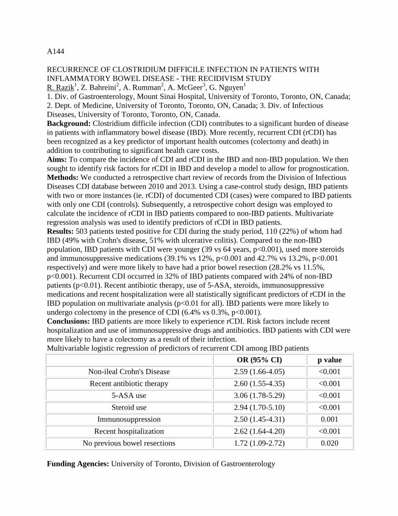
A144
RECURRENCE OF CLOSTRIDIUM DIFFICILE INFECTION IN PATIENTS WITH INFLAMMATORY BOWEL DISEASE - THE RECIDIVISM STUDY R. Razik1, Z. Bahreini2, A. Rumman2, A. McGeer3, G. Nguyen1 1. Div. of Gastroenterology, Mount Sinai Hospital, University of Toronto, Toronto, ON, Canada; 2. Dept. of Medicine, University of Toronto, Toronto, ON, Canada; 3. Div. of Infectious Diseases, University of Toronto, Toronto, ON, Canada. Background: Clostridium difficile infection (CDI) contributes to a significant burden of disease in patients with inflammatory bowel disease (IBD). More recently, recurrent CDI (rCDI) has been recognized as a key predictor of important health outcomes (colectomy and death) in addition to contributing to significant health care costs. Aims: To compare the incidence of CDI and rCDI in the IBD and non-IBD population. We then sought to identify risk factors for rCDI in IBD and develop a model to allow for prognostication. Methods: We conducted a retrospective chart review of records from the Division of Infectious Diseases CDI database between 2010 and 2013. Using a case-control study design, IBD patients with two or more instances (ie. rCDI) of documented CDI (cases) were compared to IBD patients with only one CDI (controls). Subsequently, a retrospective cohort design was employed to calculate the incidence of rCDI in IBD patients compared to non-IBD patients. Multivariate regression analysis was used to identify predictors of rCDI in IBD patients. Results: 503 patients tested positive for CDI during the study period, 110 (22%) of whom had IBD (49% with Crohn's disease, 51% with ulcerative colitis). Compared to the non-IBD population, IBD patients with CDI were younger (39 vs 64 years, p<0.001), used more steroids and immunosuppressive medications (39.1% vs 12%, p<0.001 and 42.7% vs 13.2%, p<0.001 respectively) and were more likely to have had a prior bowel resection (28.2% vs 11.5%, p<0.001). Recurrent CDI occurred in 32% of IBD patients compared with 24% of non-IBD patients (p<0.01). Recent antibiotic therapy, use of 5-ASA, steroids, immunosuppressive medications and recent hospitalization were all statistically significant predictors of rCDI in the IBD population on multivariate analysis (p<0.01 for all). IBD patients were more likely to undergo colectomy in the presence of CDI (6.4% vs 0.3%, p<0.001). Conclusions: IBD patients are more likely to experience rCDI. Risk factors include recent hospitalization and use of immunosuppressive drugs and antibiotics. IBD patients with CDI were more likely to have a colectomy as a result of their infection. Multivariable logistic regression of predictors of recurrent CDI among IBD patients
OR (95% CI) p value Non-ileal Crohn's Disease 2.59 (1.66-4.05) <0.001 Recent antibiotic therapy 2.60 (1.55-4.35) <0.001
5-ASA use 3.06 (1.78-5.29) <0.001 Steroid use 2.94 (1.70-5.10) <0.001
Immunosuppression 2.50 (1.45-4.31) 0.001 Recent hospitalization 2.62 (1.64-4.20) <0.001
No previous bowel resections 1.72 (1.09-2.72) 0.020
Funding Agencies: University of Toronto, Division of Gastroenterology

A145
FEVER, ABDOMINAL PAIN, AND NECROTIZING LYMPHADENOPATHY: A CASE OF ASEPTIC ABSCESSES IN A PATIENT WITH INFLAMMATORY BOWEL DISEASE
J. Tessolini2, M. Silverberg1
1. Mount Sinai Hospital, Toronto, ON, Canada; 2. University of Toronto, Toronto, ON, Canada.
Aims: We will present a case of aseptic abscesses in a 27 year old man with a history of ulcerative colitis and review the literature surrounding this rare disease entity. Methods: A case report was generated from the clinical details available from the patient's admission to Mount Sinai Hospital in Toronto from September 8th - September 29, 2014. A literature review was conducted through a PubMed search using the keywords "aseptic abscesses". Review of the literature was limited to English articles only. Results: A 27 year old male presented to the Emergency Department with a 5-day history of fevers, severe abdominal pain, anorexia, and weight loss. His past medical history was of ulcerative colitis diagnosed at age 18, with a total proctocolectomy and ileal-anal pouch anastomosis in 2008 for medically refractory disease. Since 2012, he had undergone multiple surgeries for de-functioning and reversal of diverting loop ileostomy for intrabdominal abscesses felt to be secondary to a pouch leak, although never definitively proven. At the time of presentation he had an elevated WBC of 21 x 10^9 with neutrophilia. A CT scan of the abdomen and chest revealed multiple cavitating mesenteric lymph nodes, low density lesions in the liver, and cavitating lung lesions. Bronchoscopy with bronchoalveolar lavage, blood cultures, and rheumatologic work-up was negative. All cultures were negative for acid-fast bacilli. Ultrasound-guided intraabdominal lymph node aspirate and CT-guided lung biopsy were non-diagnostic. Treatment with broad-spectrum antibiotics did not produce any clinical improvement. Esophagealgastroduodenoscopy (EGD) and pouchoscopy was non-contributory. Methylprednisolone 25mg IV q12h was initiated and within 2 days his abdominal pain resolved. He was transitioned to oral Prednisone and was discharged home. Based on his clinical presentation, radiographic findings, and response to corticosteroids, a diagnosis of aseptic abscesses was made. Conclusions: We have presented a case of aseptic abscesses in a 27 year old male with a history of ulcerative colitis. A review of the literature has identified several articles from a research group in France, including one that classifies the features of 30 cases of aseptic abscesses. This is a recently described clinicopathological disease entity associated with inflammatory bowel disease. The most common presenting features are fever, abdominal pain, and weight loss. Intraabdominal organs are most commonly involved, specifically spleen, mesenteric lymph nodes, liver, and lung have been described. Diagnosis is made using a combination of clinical features, radiographic findings, and ruling out other, specifically infectious, etiologies. The disease responds to treatment with corticosteroid therapy.
Funding Agencies: None

A146
THE PEDIATRIC CROHN'S DISEASE ACTIVITY INDEX SHOWS NO CORRELATION WITH ENDOSCOPIC SEVERITY AT DIAGNOSIS
N. Carman, P. Church, S. O'Donnell, D. Mack, W. El-Matary, H. Huynh, C. Deslandres, A. Otley, E. Benchimol, M. Carroll, E. Wine, J. deBruyn, A. Griffiths, T. Walters
The Canadian Children IBD Network, Toronto, ON, Canada.
Background: The Pediatric Crohn's Disease Activity Index (PCDAI), incorporating clinical and laboratory variables, is the most commonly endorsed outcome measure in pediatric CD trials. Although less subjective than the CDAI, PCDAI was never weighted nor validated based on endoscopic activity, a key parameter, as treatment targets have shifted toward mucosal healing. Aims: We undertook to compare endoscopic severity with PCDAI and other measures of disease activity in new onset pediatric CD. Methods: Children ≤ 16.5 years presenting with new onset CD at participating sites in the newly established Canadian Children Inflammatory Bowel Disease Network are eligible for enrolment in an inception cohort study. Disease phenotypic data are prospectively recorded. Disease activity at presentation is evaluated by physician global assessment (PGA), PCDAI, and conventional serologic markers of inflammation. Severity of disease at ileocolonoscopy is assessed by Simple endoscopic Score (SES-CD). Data, including SES-CD segmental subscores for ulcer size and affected surface, were compared using Spearman's test of correlation. Results: 50 patients from 7 pediatric IBD centres across Canada were included in the analysis. All patients underwent ileocolonoscopy and had activity indices collected prior to therapy. 42/50 patients had disease involving the colon (69% ileocolonic, 21% colonic). Median PCDAI was 35 (IQR 25-42); 82% had an elevated CRP (Median 23; IQR 6-52). Median SES-CD was 18 (IQR 12-24). 47% of patients had large ulcers present in at least 2/6 anatomical segments, 14% in ≥ 4 segments. Whilst PCDAI and CRP closely correlated with each other, there was no relationship between either of these measures and the full SES-CD score (r= 0.14 & 0.05 respectively). When examining the subscores, although the number of segments with large ulcers demonstrated no relationship to PCDAI, it did correlate modestly with CRP (r=0.4, p=0.01). Of note, PGA correlated with both the PCDAI (r=0.6) and CRP (r=0.5) as well as reasonably with the SES-CD overall (r=0.4, p=0.01) and, more specifically, the presence of large ulcers (r=0.5, p=0.003).In patients with isolated colonic (L2) disease, the presence of large ulcers was strongly correlated with CRP (r = 0.8, p=0.015). Conclusions: PCDAI correlates as poorly as CDAI with endoscopic disease activity. Clinical markers of disease activity must not be used in isolation in clinical practice as treatment paradigms evolve to target mucosal healing.
Funding Agencies: CIHR, C.H.I.L.D. Foundation
A147
PREDICTORS OF 30 DAY READMISSION RATES IN INFLAMMATORY BOWEL DISEASE PATIENTS AFTER HOSPITAL DISCHARGE
A. Israel1, M. Bokhary1, S. Shanmuganathan1, D. Roffey2, R. Sy1
1. University of Ottawa, Ottawa, ON, Canada; 2. Ottawa Hospital Research Institute, Ottawa, ON, Canada.
Background: Inflammatory bowel disease (IBD) is a chronic condition that includes patients with ulcerative colitis (UC) and Crohn's disease (CD). IBD patients are characterized by a remitting and relapsing course of disease activity and are often admitted to hospital for treatment.. A key issue in health care quality and costs include the rates of readmission after discharge. There are many factors that can lead to readmission. Predicting which patients require readmission may help reduce significant expense in health care. Aims: The purpose of this study was to analyze both pre-admission and time-of-admission factors in regards to their ability to predict 30 day readmission rates in an IBD patient population. Methods: Patients admitted to The Ottawa Hospital with IBD related reasons for admission from January 2011- June 2012 were reviewed retrospectively. Patients were identified who required 30 days post discharge readmission. A variety of preadmission and time-of-admission factors were analyzed to determine which were associated with readmission. Results: 263 patients were admitted during an 18-month time frame. 16% (n=43) required readmission within 30 days. Patients who received abdominal imaging with CT or MRI on initial admission were significantly more likely to be readmitted within 30 days (P<0.05). None of the other pre-admission data including baseline demographics, type of IBD or previous IBD management (medical and/or surgical) were significantly associated with readmission. Conclusions: There are a multitude of factors that are thought to impact IBD readmission rates. The issues are often complex and may be difficult to predict. There is a high 30 day readmission rate for IBD patients admitted in hospital for IBD related reasons. Patients that receive diagnostic imaging on initial admission were significantly associated with risk of readmission and may need closer monitoring.
Funding Agencies: None

A148 THE INTRODUCTION OF ANTI-TNF THERAPY IN EDMONTON TO TREAT CROHN'S DISEASE HAS CHANGED THE DEMOGRAPHICS OF PATIENTS UNDERGOING INTESTINAL RESECTION A. Dittrich, R. Fedorak, H. Wang, K. Kroeker University of Alberta, Edmonton, AB, Canada. Background: Crohn's Disease (CD) is a chronic inflammatory bowel disease that can occur anywhere from mouth to anus. The behaviour of CD is characterized into three classes: inflammatory, stricturing, and penetrating, with stricturing and penetrating preceded by inflammation. Anti-TNF agents entered the Alberta therapeutic armamentarium for CD in 1998. Recent evidence has shown that early treatment of CD with biologic agents will reduce the need for intestinal surgery. Aims: To assess if the characteristics of adults with CD undergoing intestinal surgery are different following the introduction of anti-TNF agents in 1998. Methods: Through a population-based retrospective study, the Data Integration, Measurement, and Reporting (DIMR) Database was used to identify patient eligibility: (1) male or female patients aged 18 or above, (2) a known diagnosis of CD, and (3) have undergone intestinal surgery for their disease in one of the four Edmonton area hospitals between January 1, 1996 and December 31, 2013. The patient characteristics of patients undergoing surgery before and after 1998 were compared. Statistical analysis (t-tests and Chi-squared tests) were completed using SPSS. Results: Demographic characteristics are shown in Table 1. To date, the charts of 250 of 1,650 patients undergoing intestinal surgery have been reviewed. There was a trend for more patients to undergo laparoscopic rather than open surgery after 1998 (18.1% v. 9.7%, p=0.10). In regards to medication use prior to surgery, there was an increase in anti-TNF (0% v. 11.2%, p=0.003) and immunosuppressive use (15.3% v. 28.1%, p=0.033) after 1998. While 5-ASA use was lower after 1998 (41.7% v. 19.7%, p<0.001) and steroid use remained similar (56.9% v. 56.2%, p=0.912). No differences in smoking status were found for patients undergoing surgery before or after 1998 (smoker: 50.0% v. 43.4%, former: 8.6% vs. 18.3%, non-smoker: 41.4% v. 38.3%, p=0.162). Conclusions: After the introduction of anti-TNF therapy for Crohn's disease in 1998, we can see a trend toward older age and longer disease duration at the time of surgery. There was both an increase in anti-TNF and immunosuppressive use, while 5-ASA use declined. The proportion of women undergoing intestinal resection after 1998 increased. Data collection is still in progress. Demographic characteristics of patients.
Surgery Before 1998 Surgery After 1998 p-value Characteristics n = 72 n = 178
Age (mean ± SD) 38.4 ± 12.4 41.9 ± 15.0 p = 0.087 Gender %, (n)
F
26.4% (19)
45.4% (81)
p = 0.005 Disease Duration
(mean ± SD)
9.4±7.8
11.8±10.9
p = 0.051 Funding Agencies: Northern Alberta Clinical Trials and Research Centre (NACTRC) Summer Student Award, Centre of Excellence for Gastrointestinal and Immunity Research (CEGIIR)

A149
RESIDUAL MUCOSAL ABNORMALITIES AFTER MAYO ENDOSCOPIC SUBSCORE DEFINED COMPLETE MUCOSAL HEALING DEMONSTRATED BY ISCAN ENDOSCOPIC AND REFINED NEW HISTOLOGIC GRADINGS
M. Iacucci1, M. Fort Gasia1, R. Panaccione1, G. Kaplan1, S. Ghosh1, X. Gui2
1. IBD Clinic, Department of Gastroenterology, Calgary, AB, Canada; 2. Department of Pathology, Calgary, AB, Canada.
Background: High definition (HD) iSCAN endoscopy can characterize in details the mucosa in patients with ulcerative colitis (UC) and may provide detailed information about inflammation and mucosal healing (MH). Aims: We aimed to establish more sensitive endoscopic and histological criteria to better define MH and to identify subtle histologic abnormalities. Methods: 78 patients with UC were assessed by HD-iSCAN colonoscopy (Pentax EC-3490Fi; Pentax,Japan) and white light endoscopy (WLE).Mayo endoscopic subscore and UCEIS score were assigned to patients according to WLE findings.Mucosal pattern on iSCAN was graded as 1=normal,2=mosaic pattern,3=tubular-gyrus,4=nodular rosette.The vascular pattern was graded as 1=normal,2=spiral isolated vessels,3=crowded tortuous vessels,4=Irregular vessels.A histological grading and scoring system was developed for a more comprehensive evaluation. This system (GUI-ECAP system) was designed to reflect all histologic changes in IBD categorized as 1)Extent of inflammation (focal, multifocal, diffuse),2)Chronicity (crypt architectural alteration,Paneth cell metaplasia),3)Activity (surface epithelium changes,neutrophilic cryptitis,crypt abscess,crypt destruction,lamina propria mononuclear cellularity,lamina propria neutrophil infiltration,and basal plasmacytosis), and 4)Plus additional findings, including eosinophilia and lymphoid follicles/aggregates.An established histologic grading system, NYMS score was used to validate the grading of inflammation by histology. Results: Out of 78 patients with UC,23 (29%) patients had Mayo endoscopic subscore of 0.Of these 23 patients,18 patients had abnormal vascular pattern on iSCAN and 7 had abnormal mucosal pattern on iSCAN.By using ECAP histologic scoring all 23 patients showed various histologic abnormalities including crypt architectural alteration 83%, surface epithelium abnormality 70%, crypt destruction 13%, increase in lamina propria mononuclear cells 65%,basal plasmacytosis 48%,lamina propria neutrophilic infiltration 21% and other additional findings 83% Conclusions: The subtle histologic abnormalities underlying the apparently healed mucosa with Mayo endoscopic subscore of 0 can be detected by using a refined histological scoring system (ECAP) in combination with iSCAN endoscopy.iSCAN and histologic scoring such as ECAP can detect residual abnormalities in most patients with apparent MH in UC.The significance of these findings in terms of outcome has to be established.
Funding Agencies: None
A150
CHARACTERIZATION OF MUCOSAL SEROTONIN SIGNALLING COMPONENTS IN PATIENTS WITH INFLAMMATORY BOWEL DISEASE.
M. Shajib1, J. Kim1, Z. Muqtadir1, T. Aziz2, P. Moayyedi1, W. Reinisch1, J. Marshall1, W. Khan1
1. Farncombe Family Digestive Health Research Institute, McMaster University, Hamilton, ON, Canada; 2. Department of Pathology & Molecular Medicine, McMaster University, Hamilton, ON, Canada.
Background: Gastrointestinal (GI) tract contains about 95% of total serotonin (5-hydroxytrptamine; 5-HT) in human body. Enterochromaffin (EC) cells are the main source of 5-HT in the gut and tryptophan hydroxylase (TPH) 1 is the rate-limiting enzyme involved in the biosynthesis of 5-HT in EC cells. 5-HT interacts through multiple receptors present on variety of cells in the gut to regulate various functions in the GI tract. Alterations in intestinal 5-HT signalling have been reported in GI disorders such as irritable bowel syndrome and inflammatory bowel disease (IBD). In IBD, particularly in Crohn's disease (CD), studies report an up-regulation in both EC cell numbers and 5-HT content. There are few studies, however, that have evaluated changes in 5-HT receptor signalling in the context of IBD. Aims: A comprehensive examination of key elements of intestinal 5-HT signalling in patients with CD. Methods: Mucosal biopsies were collected from the colon of consenting patients with CD and from healthy controls (HC), undergoing routine colonoscopy for colorectal cancer. Inactive specimens were collected from CD patients in remission, or from non-inflamed regions of patient with active disease. Non-inflamed regions were defined as those without any endoscopic features of inflammation and at least 10 cm from any area of active inflammation. Biopsies collected (N=10, 11 and 8 from HC, inactive CD and active CD segments, respectively) underwent histological evaluation by a pathologist unaware of the diagnosis, and quantitative polymerase chain reaction to determine gene expression of TPH1, serotonin reuptake transporter (SERT) and 5-HT receptors (5-HTR1, 3, 4, 7). Results: Histological assessment of biopsies confirmed the macroscopic assessments of the endoscopist during sample collection. There was significant increase in TPH1, 5-HTR3A and 5-HTR7 expression in active CD samples as compared to inactive and control samples. 5-HTR4 expression was significantly elevated in both active and inactive samples compared to control and 5-HTR1A expression was minimal in all groups compared. SERT expression was significantly reduced in active samples compared to both inactive and control samples. Conclusions: Our results show that there are changes in various aspects of 5-HT signalling (5-HTR3, 7, TPH1 and SERT) in association with increased inflammation in patients with CD. Better understanding of 5-HT signalling in intestinal inflammation may ultimately lead to effective strategies targeting this pathway in inflammatory conditions such as IBD.
Funding Agencies: CCC, CIHR

A151
PREVALENCE AND SIGNIFICANCE OF ELEVATED SERUM IMMUNOGLOBULIN G AT DIAGNOSIS IN CHILDREN WITH INFLAMMATORY BOWEL DISEASE
G. Sadi1, B. Dufault3, W. El-Matary2
1. Section of Pediatric Gastroenterology, University of Manitoba, Winnipeg, MB, Canada; 2. University of Manitoba, Winnipeg, MB, Canada; 3. George and Fay Yee Center for Healthcare Innovation, University of Manitoba, Winnipeg, MB, Canada.
Background: Inflammatory bowel disease (IBD) is a chronic disease with dys-regulated immunity. Immunoglobulin (IgG), the most abundant antibody isotype found in the circulation, is synthesized and secreted by B lymphocytes. Serum IgG levels can be elevated in patients with immune disorders. The prevalence and significance of high serum IgG levels in children with IBD is yet to be defined Aims: The aim of this study was to examine the prevalence and significance of high serum IgG levels at diagnosis in children with IBD Methods: A comprehensive chart review of all children with IBD in Winnipeg Children's Hospital who had serum IgG levels measured at diagnosis was performed. All children with incomplete records or any associated other immune disorders were excluded. Patients with high serum IgG at diagnosis were compared to those without in relation to other laboratory markers and disease severity Results: Out of 45 children (mean age was 11.67 years, 29 boys, 24 with Crohn's disease (CD) and 21 with ulcerative colitis (UC), mean duration of follow up was 2.23 years) with IBD who had serum IgG measured at diagnosis, 17 (37%) children had abnormally elevated serum IgG. Abnormally high serum IgG levels were more common in patients with CD compared with those with UC (P=0.07), more common in girls (56%) compared boys (27%) P<0.05 and more likely to be associated with anemia (P<0.05), elevated C-reactive protein (CRP) (P<0.01) and low serum albumin (P<0.01) Conclusions: Over one third of children newly diagnosed with IBD have abnormally elevated serum IgG. Elevated serum IgG is more common in children with CD especially in those with anemia, high inflammatory markers and low serum albumin which may signify the presence of severe disease in those patients. Large prospective studies are needed to confirm our conclusions.
Funding Agencies: Manitoba institute of Child Health

Intestinal Disorders
Poster of Distinction
A152
IN VIVO PROTECTIVE EFFECT OF AN H2S-RELEASING AGENT AGAINST NSAID + BILE-INDUCED CYTOTOXICITY AND ULCERATION
A. Manko2, R. Blackler1, A. Buret2, J. Wallace2
1. McMaster University, Hamilton, ON, Canada; 2. University of Calgary, Calgary, AB, Canada.
Aims: Hydrogen sulfide (H2S) is an endogenous gaseous mediator with anti-inflammatory and cytoprotective effects. H2S can also promote ulcer healing in digestive tract. It is known that the toxicity of bile acids is strongly dependent on their degree of hydrophobicity as a result of an increased ability to interact as a detergent on the plasma membrane of cells. Chemical composition of bile is changed following administration of non-steroidal anti-inflammatory drugs (NSAIDs) - the most commonly used class of anti-inflammatory drugs. This is likely in part due to dramatic shifts in the composition of the small intestinal microbiota. We therefore evaluated the ability of an H2S-releasing agent, diallyl disulfide (DADS) to decrease the cytotoxic effects of bile from NSAID-treated rats on intestinal epithelial cells. Methods: Rats (n≥6 per group) were treated orally, twice daily, with naproxen (20 mg/kg) or vehicle for 4.5 days (9 administrations in total). Immediately prior to each administration of naproxen or vehicle, rats were treated with DADS (10, 30, or 60 mmol/kg p.o.) or an equivalent volume of vehicle. One hour after final drug administration, the bile duct was cannulated and bile was collected. The collected bile was added to cultured rat intestinal epithelial cells (IEC-6), at various dilutions, for up to 3 hours. Cell death was measured via release of lactate dehydrogenase. In separate studies, the extent of small intestinal damage induced by naproxen, with or without DADS co-treatment, was determined. Results: Naproxen administration resulted in extensive ulceration and bleeding in the small intestine. DADS dose-dependently reduced naproxen-induced intestinal damage (by 27%, 67% and 72% at doses of 10, 30 and 60 mmol/kg, respectively). Bile from rats treated with naproxen exhibited a dose-dependent cytotoxic effect on intestinal epithelial cells. Bile from rats co-treated with DADS and naproxen was significantly less cytotoxic to intestinal epithelial cells. The protective effect of DADS was dose-dependent. Conclusions: Administration of DADS significantly reduced the cytotoxic effects of bile on intestinal epithelial cells in a dose-dependent manner, and dose-dependently reduced the severity of naproxen-induced small intestinal damage. H2S-induced changes in cytotoxicity of bile may underlie the reduction of intestinal damage by DADS. The reduction of cytotoxicity of bile may occur secondary to changes in the intestinal microbiota, as we have previously demonstrated.
Funding Agencies: CIHR

A153
INSIGHTS INTO MOLECULAR PATHOLOGY OF MICROVILLUS INCLUSION DISEASE (MVID)
C. Thoeni, E. Cutz, A. Muise
Hospital for Sick Children, Toronto, ON, Canada.
NOT PUBLISHED AT AUTHOR’S REQUEST
Funding Agencies:

A154
ROLE OF HIGH MUC2 MUCIN BIOSYNTHESIS IN COLONIC GOBLET CELL STRESS AND APOPTOSIS
A. Tawiah, F. Moreau, K. Chadee
University of Calgary, Calgary, AB, Canada.
Background: MUC2 mucin is a large glycoprotein produced by goblet cells that form the protective mucus blanket overlying the intestinal epithelium. It is the first line of innate host defense. Secretion of MUC2 occurs basally and during pathological conditions such as inflammatory bowel disease where accelerated MUC2 biosynthesis and secretion is followed by depletion. Unfortunately, little information is known on how MUC2 production is regulated basally and whether goblet cells undergo increased stress in response to high MUC2 biosynthesis and secretion during inflammation, infection or disease conditions. Aims: 1. To determine if high MUC2 production in goblet cells can lead to ER stress and apoptosis 2. To elucidate the mechanism of ER stress-induced apoptosis. Methods: We used a high MUC2 producing human colonic goblet cell line, HT29-H, and a clone of HT29-H (HT29-L) in which MUC2 was stably knocked down using lentivirus shRNA. Cells were treated with the ER stress inducer, tunicamycin, and markers for ER stress (GRP78, ATF4, CHOP, sXBP1, AGR2) and apoptosis (caspase 3 and PARP cleavage) quantified by western blotting and RT-qPCR. Mouse colonic epithelial cells isolated by enzymatic digestion and full thickness tissues were from wild type (Wt) and Muc2-/- mice. Results: Compared to HT29-L, HT29-H cells exhibited a significant dose-and time-dependent increase in ER stress in response to tunicamycin. Under prolonged ER stress conditions, HT29-H cells were highly susceptible to apoptosis as compared to HT29-L cells. Specificity for MUC2 in inducing ER stress and apoptosis was confirmed by over expressing DNA constructs of the C-terminus of MUC2 in non-MUC2-producing SKCO15 colonic epithelial cells. Pre-treatment of cells with the reactive oxygen species (ROS) inhibitor, diphenyleneiodonium, completely inhibited basal stress levels as well as tunicamycin-induced ER stress and apoptosis. Predictably, HT29-H cells produced significantly more ROS compared to HT29-L cells demonstrating that high MUC2 production specifically increases ROS production that drives goblet cell ER stress and apoptosis. These findings were corroborated in colonic epithelial cells and whole colonic tissues isolated from Wt and Muc2-/- mice that showed a significant increase in ER stress in Wt as compared to Muc2-/- animals at the mRNA and protein level. In Wt mice the distal colon, which contains more goblet cells, also expressed higher ER stress compared to the proximal colon. Conclusions: These findings indicate that sustained high MUC2 production induces goblet cell ER stress and apoptosis that could lead to diminished mucus barrier function and increased susceptibility to gut inflammation and disease.
Funding Agencies: CIHR

A155
INTESTINAL EPITHELIAL CELLS THROUGH MESOTRYPSIN'S SECRETION IS INVOLVED IN IBS SYMPTOMS
C. Rolland-Fourcade1, A. Denadai-Souza1, J. Motta2, T. Bautzova1, I. Spreadbury3, S. Vanner3, C. Deraison1, N. Vergnolle1
1. Inserm, U1043, Toulouse, France; 2. University of Calgary, Calgary, AB, Canada; 3. queen's, Kignston, ON, Canada.
Background: Trypsin activity is increased in human colonic biopsies from IBS patients in comparison to control patients. Moreover, proteolytic activity released from biopsies of IBS patients, when introduced into the colon of mice, causes hypersensitivity symptoms (hyperalgesia and allodynia). Mesotrypsin or trypsin IV is a form of trypsin whose functions are not clear (Radisky E. et al., 2013). It was shown that mRNA level encoding this particular trypsin is increased in small intestine of IBS patients (Kerckhoffs A. et al., 2008). However, the cellular origin of this protease is unknown. Aims: This study aims to determine if intestinal epithelial cells release mesotrypsin during inflammation and if it could participate to the hypersensitive symptoms of IBS. Methods: Cryosection of biopsies from IBS (n=10) and control patients (n=9) was used to performed immunostaining of mesotrypsin. For in vitro approach, intestinal epithelial cells were cultured on Transwell (Caco2 and HT29) and stimulated with LPS (20µg/mL, 24H) to induce inflammation. Expression of trypsin forms was evaluated by qRT-PCR. Mesotrypsin released in basal and apical medium was detected by western blot and correlated with trypsin activity. Conditioned media from basal and apical sides of epithelial monolayer were used to stimulate neurons from murine Dorsal Ganglia Root (DRG). Results: Mesotrypsin staining was more intense, specifically in epithelial cells in biopsies from IBS patients in comparison to control patients. After LPS stimulation, expression of cationic and anionic trypsins was not modified in epithelial cell lines whereas mesotrypsin expression was increased at transcriptional and protein level. In the basal medium of epithelial monolayer, mesotrypsin secretion was greater in inflammatory condition, along with an increase of proteolytic activity, suggesting that mesotrypsin was released by epithelial cell as an active protease. Basal medium from "inflamed" Caco2 cells induced neuronal activation that was not observed with apical medium or media from epithelial cells in steady state. Conclusions: This study reveals that intestinal epithelial cells release mesotrypsin after inflammatory stimulation specifically at the basal side and can activate DRG neurons. It has already shown that mesotrypsin can activate PAR2 (Bunnett N. et al., 2007). This receptor is involved in maintain of inflammation, in modification of the gastrointestinal motility, in increase of intestinal permeability and in visceral hypersensitivity; all parameters founded in IBS. Therefore colonic epithelial cells secreting mesotrypsin close to neurons could play a key role in this pathology.
Funding Agencies: European Council Research

A156
CONTRAST-ENHANCED SMALL BOWEL ULTRASOUND IN THE ASSESSMENT OF THE SMALL BOWEL IN PATIENTS WITH CROHN'S DISEASE
A. Harris1, C. Healy2, F. Donnellan2, N. Chatur1, B. Salh2
1. VGH, Vancouver, BC, Canada; 2. Vancouver General Hospital, Vancouver, BC, Canada.
Background: Non-invasive radiological assessment of Crohn's disease has traditionally utilised barium studies, computerised tomography or magnetic resonance imaging. Ultrasound is emerging as a reliable, non-invasive method of assessing the small bowel. The additive value of using injectable contrast agents is gaining popularity in assessing and following Crohn's small bowel disease, and distinguishing acute disease from fibrostenotic disease, resulting in a significant change in patient management. Aims: To study findings and outcomes of patients referred to a tertiary referral centre for investigation of small bowel disease, who underwent small bowel ultrasound +/- contrast enhanced small bowel ultrasound. Methods: Patients with an established or a suspected diagnosis of Crohn's Disease referred for a focused small bowel ultrasound were studied. Small bowel ultrasound findings, subsequent need for contrast enhanced ultrasound and outcomes were analysed. Results: 53 patients were referred for a focused small bowel ultrasound. Of the 53 patients who underwent small bowel ultrasound for suspected Crohn's Disease, 31 had normal findings. 19 ultrasounds were perceived as abnormal, the most common patterns of abnormality being thickened and hyperaemic small bowel. 14 of these 19 subsequently underwent contrast enhanced small bowel ultrasound. Of this group, 10 had moderate to avid small bowel wall enhancement, and 4 had poor or minimal small bowel wall enhancement. There was 100% correlation between contrast enhanced ultrasound findings and endoscopic, biopsy, clinical correlation and follow-up. Conclusions: Contrast enhanced ultrasound is a valuable investigative tool in diagnosing active small bowel inflammation in patients with Crohn's Disease. In the correctly chosen population, there is a strong correlation between enhancement patterns and endoscopic and clinical findings. Its lack of ionizing radiation makes it an attractive imaging option in evaluating small bowel disease severity, and in evaluating response to treatment.
Funding Agencies: None,

A157
PREVALENCE OF FUNCTIONAL GASTRO-INTESTINAL DISORDERS IN NEWLY-DIAGNOSED CELIAC DISEASE PATIENTS, AND EFFECTS OF A GLUTEN FREE DIET.
S. Jocelyn, L. Graff, J. Walker, D. Duerksen
University of Manitoba, Winnipeg, MB, Canada.
Background: Celiac disease (CD) affects 1% of Canadians. Functional intestinal disorders are ten to twenty times more common than CD and can present with similar symptoms, suggesting that there may be considerable overlap between these two conditions. Aims: To determine the prevalence of functional intestinal disorders based upon Rome III criteria at diagnosis and 6 months after diagnosis of CD. Methods: Adults with positive celiac serology (TTG and/or EMA antibodies) and findings of villous atrophy (Marsh IIIa-c) on intestinal biopsy within 6 weeks of diagnosis were prospectively enrolled. A survey including questions related to GI symptoms and diet adherence was completed at study entry and 6 months. Standardized measures included Rome III questionnaire items related to irritable bowel syndrome (IBS), epigastric pain syndrome, functional abdominal pain syndrome and functional bloating. The Celiac Symptom Index (CSI) and the Celiac Diet Assessment Tool (CDAT) were used to measure celiac disease symptoms and gluten-free diet adherence, respectively. All statistical tests were two-sided and all t-tests were paired. Results: There were 50 newly-diagnosed CD patients who completed all measures. Mean age was 40.8 years (SD 16.8), with participants ranging in age from 16 to 78; 70% were female. At diagnosis, 8% reported a prior diagnosis of IBS. The overall prevalence of functional bowel disorders was [mean (95% confidence interval)]: IBS 76% (64-88%), Functional Dyspepsia (FD) 32% (19-45%), and Functional Bloating (FB) 6% (0-13%). None met criteria for Epigastric Pain Syndrome. At 6 months, 92% reported gluten exposure less than 1 per month and the mean CDAT score was 12.4 (SD 3.6), suggesting adequate adherence. The prevalence of functional bowel disorders did not decrease significantly: IBS 68% (55-81%), FD 12% (3-21%) and FB 4% (0-10%). IBS symptoms scores were highly correlated with CD symptoms (CSI), and those with IBS had higher scores CSI scores at both 0 and 6 months. Comparing over time, CD symptoms improved significantly from baseline to 6 months for those identified as also having IBS (mean CSI score 39.8 (SD 10.7) at diagnosis, 36.3 (SD 7.2) at 6 months (p<0.005)), with little change in CD symptoms for those who did not have IBS (mean CSI score was 30.5 (SD 7.8) at diagnosis and 29.5 (SD 9.5) at 6 months (p = 0.54)). Conclusions: CONCLUSIONS: IBS (as determined by self completed survey) is common among patients with newly diagnosed celiac disease. Those who have IBS report more celiac disease symptoms at diagnosis and have a greater decrease in CD symptoms in response to a gluten-free diet than those who do not. Larger and longer prospective studies are required to determine whether these differences are sustained and the role of gluten-free diet in mediating these changes.
Funding Agencies: CIHR, Canadian Celiac Association
A158
THE GLUTEN FREE DIET: ASSESSING ADHERENCE IN A PEDIATRIC CELIAC DISEASE POPULATION
J. Dowhaniuk, H. Mileski, J. Saab, J. Cheng, P. Tutelman, H. Brill
McMaster University, Hamilton, ON, Canada.
Background: Celiac Disease (CD) is one of the most common chronic diseases in childhood. A strict, lifelong gluten-free diet (GFD) remains the sole treatment for CD. The assessment of adherence to the GFD in pediatric studies is often based on self-report and visual analogue scales which lack proven validity. Aims: We sought to compare parental self-report of GFD adherence to expert Registered Dietitian (RD) assessments, the accepted best available standard. We hypothesize that there is poor agreement between parental scores based on Likert scales and a comprehensive RD assessment. Methods: Parents of children with biopsy-proven CD followed at McMaster Children's Hospital scored their adherence to the GFD on a 5-point Likert scale similar to that used in previous pediatric CD studies. Each family was then evaluated by a RD expert in CD management who conducted a comprehensive and standardized assessment and scored the family's adherence; a second dietitian also scored the family based on clinic notes. The agreement between parents and the RD was assessed using paired t-test and intraclass correlation coefficient (ICC) based on their scores. Inter-rater reliability was used to compare RD assessments. Results: One hundred and twenty-two children and their families participated in the study with a median of 32 months on a GFD. Excellent adherence (score 5/5) was attributed to 60.5% of the sample by the RD. The parents scored adherence higher than the RD by an average difference of 0.41 scale points (95% CI:0.28,0.54; p<0.001). The agreement between parents and the Registered Dietitian was poor (ICC = 0.21). Conclusions: Reliance on self-report through Likert scales for GFD adherence overestimates adherence and misses opportunities for patient and family education. Future studies will attempt to develop an effective tool for assessing adherence to the GFD. In the interim, regular assessment by a RD in a dedicated CD clinic remains the most reliable way to assess adherence.
Funding Agencies: Regional Medical Association of Hamilton

A159
THE NON USE OF IBD-SPECIFIC MEDICATIONS IN PATIENTS WITH IBD
S. Bhasin1, C. Bernstein1, H. Singh1, L. Targownik1, E. israeli2
1. University of Manitoba, Winnipeg, MB, Canada; 2. Hadassah-Hebrew University Hospital, Jerusalem, Israel
Background: We have previously reported, using a population-based administrative database of persons with IBD, that at any one time approximately 50% of persons with IBD may not be using prescription medications specific for IBD (glucocorticoids, 5-aminosalicylates, thiopurines, methotrexate, anti-TNF agents, or long-term metronidazole and/or ciprofloxacin). Aims: To determine how often persons with IBD presenting to a specialized referral clinic are not using IBD-specific prescription medications and the reasons for nonuse. To compare patient characteristics of med non-users with med users in IBD, including age, sex, phenotype and disease duration. Methods: All persons with IBD presenting to an IBD referral clinic of a single practitioner with expertise in IBD between April 15 2011 and April 15 2013 were tracked. Medications used were recorded and when no IBD-specific medications were used the reasons for non use were recorded. Patient characteristics, phenotypes, as well as reasons for non-drug use were compared between the Crohns Disease (CD) and UC groups. Data were compared at the level of both individual patients as well as total patient visits. Disease remission was considered to be an acceptable reason to be off meds. Results: 552 CD patients and 299 UC patients were identified. This equated to 1218 CD visits and 662 UC visits. 215 CD patients were non-users (39%) vs 62 UC patients (20.7%) (p<0.001). There were 396 CD visits with no med use (32.4%) vs 139 UC vists with no med use (21%, p<0.001). The mean age of CD non med users in years was 45.3 and was 40.5 in med users( p<0.0007). Conversely in UC, Mean age in years of non users was 40.3, and in users was 43.1 (p<0.01). Top reasons for non drug use in CD visits were disease remission (46.1%), recent diagnosis/change in clinical condition -or in need of treatment or planning to start meds (17.8%), and post-operative state (13%). In UC visits, the top reasons for non-drug use were disease remission (49%), non-adherence (22%), and recent diagnosis/change in clinical condition - or in need of treatment and planning to start meds (12%). Patients who had multiple visits were more likely to be on medications at every visit if they had UC (70%) vs CD (9%). Conclusions: About one third of IBD patients presenting to a specialty referral clinic were not using IBD-specific drugs. This was more likely in CD than UC. Close to half of all IBD patients who were non-drug users were in disease remission and deemed suitable to be off IBD-specific medications by the physician. There appears to be more non-adherence in UC patients as compared to CD patients in this study population. Of persons with multiple visits, CD patients had a higher inconsistency of medication use than UC patients.
Funding Agencies: None
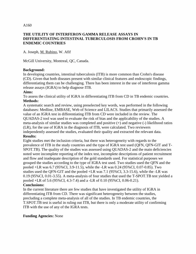
A160
THE UTILITY OF INTERFERON GAMMA RELEASE ASSAYS IN DIFFERENTIATING INTESTINAL TUBERCULOSIS FROM CROHN'S IN TB ENDEMIC COUNTRIES
A. Joseph, M. Rubino, W. Afif
McGill University, Montreal, QC, Canada.
Background: In developing countries, intestinal tuberculosis (ITB) is more common than Crohn's disease (CD). Given that both diseases present with similar clinical features and endoscopic findings, differentiating them can be challenging. There has been interest in the use of interferon gamma release assays (IGRA) to help diagnose ITB. Aims: To assess the clinical utility of IGRA in differentiating ITB from CD in TB endemic countries. Methods: A systematic search and review, using preselected key words, was performed in the following databases: Medline, EMBASE, Web of Science and LILACS. Studies that primarily assessed the value of an IGRA test in differentiating ITB from CD were included in the review. The QUADAS-2 tool was used to evaluate the risk of bias and the applicability of the studies. A meta-analysis of similar studies was completed and positive (+) and negative (-) likelihood ratios (LR), for the use of IGRA in the diagnosis of ITB, were calculated. Two reviewers independently assessed the studies, evaluated their quality and extracted the relevant data. Results: Eight studies met the inclusion criteria, but there was heterogeneity with regards to the prevalence of ITB in the study countries and the type of IGRA test used (QFN, QFN-GIT and T-SPOT.TB). The quality of the studies was assessed using QUADAS-2 and the main deficiencies noted were incomplete reporting of the index test, incomplete descriptions of patient recruitment and flow and inadequate description of the gold standards used. For statistical purposes we grouped the studies according to the type of IGRA test used. Two studies used the QFN and the pooled +LR was 6.7 (95%CI, 3.9-11.5), while the -LR was 0.24 (95%CI, 0.07-0.85). Two studies used the QFN-GIT and the pooled +LR was 7.1 (95%CI, 3.3-15.6), while the -LR was 0.19 (95%CI, 0.01-3.55). A meta-analysis of four studies that used the T-SPOT.TB test yielded a pooled +LR of 5.6 (95%CI, 4.3-7.4) and a -LR of 0.10 (95%CI, 0.06-0.21). Conclusions: In the current literature there are few studies that have investigated the utility of IGRA in differentiating ITB from CD. There was significant heterogeneity between the studies, precluding a complete meta-analysis of all of the studies. In TB endemic countries, the T.SPOT.TB test is useful in ruling out ITB, but there is only a moderate utility of confirming ITB with the use of any of the IGRA tests.
Funding Agencies: None
A161
FAMILIAL PROGRESSIVE MULTIPLE INTESTINAL ATRESIA AND IMMUNODEFICIENCY: FAILURE OF IMMUNOSUPPRESSION AND BONE MARROW TRANSPLANT TO HALT PROGRESSION OF DISEASE
M. Chavannes, U. Halac, H. Decaluwe, F. Le Deist, N. Patey, V. Marchand
Hôpital Sainte Justine, Montreal, QC, Canada.
Background: Human multiple intestinal atresia with combined immune deficiency (HMIA-CID) is a rare cause of intestinal obstruction associated with a profound immune deficiency caused by a deleterious mutations of the TTC7A. This condition presents with multiple intestinal stenosis and atresia leading to intestinal resections early in life. The lesions may be progressive and relapse post-surgically. The immune deficiency characteristically affects T- and B-cell function and presents with lymphopenia, monocytosis and agammaglobulinemia. Aims: To describe the case of a child with MIA and the course of his disease after treatment with immunosuppression and bone marrow transplant. Methods: Case report and review of literature Results: We report a child with hereditary MIA and confirmed mutation in TTC7A gene. The child was born with a prenatal diagnosis of intestinal obstruction. Surgery performed on the first day of life revealed multiple atresia involving the duodenum, ileum, proximal colon and sigmoid and led to multiple intestinal resections and a terminal ileostomy. At 4 weeks of age, a second laparotomy revealed a new atretic segment of ileum. Histology of the surgical specimens revealed severe inflammation, apoptosis and multifocal atresia with sieve-like multiple intestinal lumen caused by multiple mucosal adhesions. After the surgery, the patient presented severe secretory diarrhea and was dependant on parenteral nutrition. Systemic corticosteroids and later cyclosporine were given in an attempt to reduce intestinal inflammation. Despite this treatment massive diarrhea persisted. There was no recurrence of stenosis but endoscopic biopsies revealed persistent severe intestinal inflammation. Immunologic evaluation performed before immunosuppression displayed lymphopenia, monocytosis and hypogammaglobulinemia. At 8 months, he received a hematopoietic stem cell transplant using HLA 6/6 match cord blood and remained on cyclosporin and steroids. Post-transplant course was complicated by mucositis, but no GVHD. While there were no appreciable bowel obstructions at the time of transplant, a small bowel follow-through later demonstrated new areas of stenosis and biopsies revealed persistent inflammation. He remained dependant on parenteral nutrition. He developed respiratory distress and died from toxic pneumonitis at 12 months. Conclusions: Our patient's evolution suggests that immunosuppressive therapy and hematopoietic stem cell transplantation does not prevent recurrence of atresia and does not improve secretory diarrhea. Similar findings were identified by other groups.
Funding Agencies: None

A162
CHEMICAL INDUCED COLITIS FROM HYDROGEN PEROXIDE ENEMAS: A RARE CAUSE OF SEVERE AND POTENTIALLY LIFE THREATENING IATROGENIC COLITIS
J. Zhu1, Y. Reda2, D. Moffatt2
1. University of Saskatchewan, Saskatoon, SK, Canada; 2. University of Manitoba, Winnipeg, MB, Canada. Background: Hydrogen peroxide enema is a rare iatrogenic cause of severe colitis that mimicks other colitides. Aims: We present two cases of colitis from hydrogen peroxide enema with radiographic, endoscopic and pathologic findings. Methods: Case series. Results: Case 1: A 40yo woman presented to the emergency room (ER) with constipation. The patient received a "POW" enema (mixture of hydrogen peroxide, mineral oil and water). She immediately developed lower abdominal pain followed by bloody diarrhea. Stool cultures and C. difficile assay were negative and her WBC was 24.1. CT abdomen showed severe colitis from the proximal transverse colon to rectum with extensive gas within the portal venous system (Fig. 1). Sigmoidoscopy showed severe colitis with ulcerations and pseudomembranes (Fig. 2). Biopsies showed purulent exudate, hyalinization, intramucousal hemorrhage and crypt hyperplasia suggestive of drug induced or early ischemic colitis (Fig. 3). The patient was managed conservatively in hospital with IV fluids and antibiotics. She improved on day 7 and was discharged home. Case 2: A 32yo female presented to the ER with constipation. She was given a "POW" enema and developed bloody diarrhea and severe abdominal pain. She became febrile (T=38.1C), but her vitals were otherwise stable. Her WBC was elevated at 19.6. Stool cultures and C.difficile were negative. CT abdomen showed rectosigmoid colitis, multiple small foci of extraluminal air and extensive portal venous gas (Fig. 4). Endoscopy was not done given the possible perforation and free air. She was managed conservatively with IV antibiotics and gut rest and was discharged home on day 5. In both cases, no further colitis or lower GI complaints have occurred since discharge from hospital. Review: In the medical literature there are 11 reports of chemical colitis from hydrogen peroxide enema, three with air embolism. Pseudomembranes and "snow-white" sign may be observed endoscopically, while biopsies may show ischemic changes or features similar to infectious colitis. Management is supportive although colectomy is occasionally necessary. Despite prior publications on the dangers of hydrogen peroxide enemas, they are still used in clinical practice today, and are recommended in text books and resources in Emergency Medicine and Palliative Care. Conclusions: Hydrogen peroxide containing enemas are a potentially toxic therapy that can cause severe colitis, air embolization and perforation. The identification of chemical colitis requires taking an accurate history, while imaging, endoscopy and pathology are supportive. We recommend against using this therapy for any clinical indication.
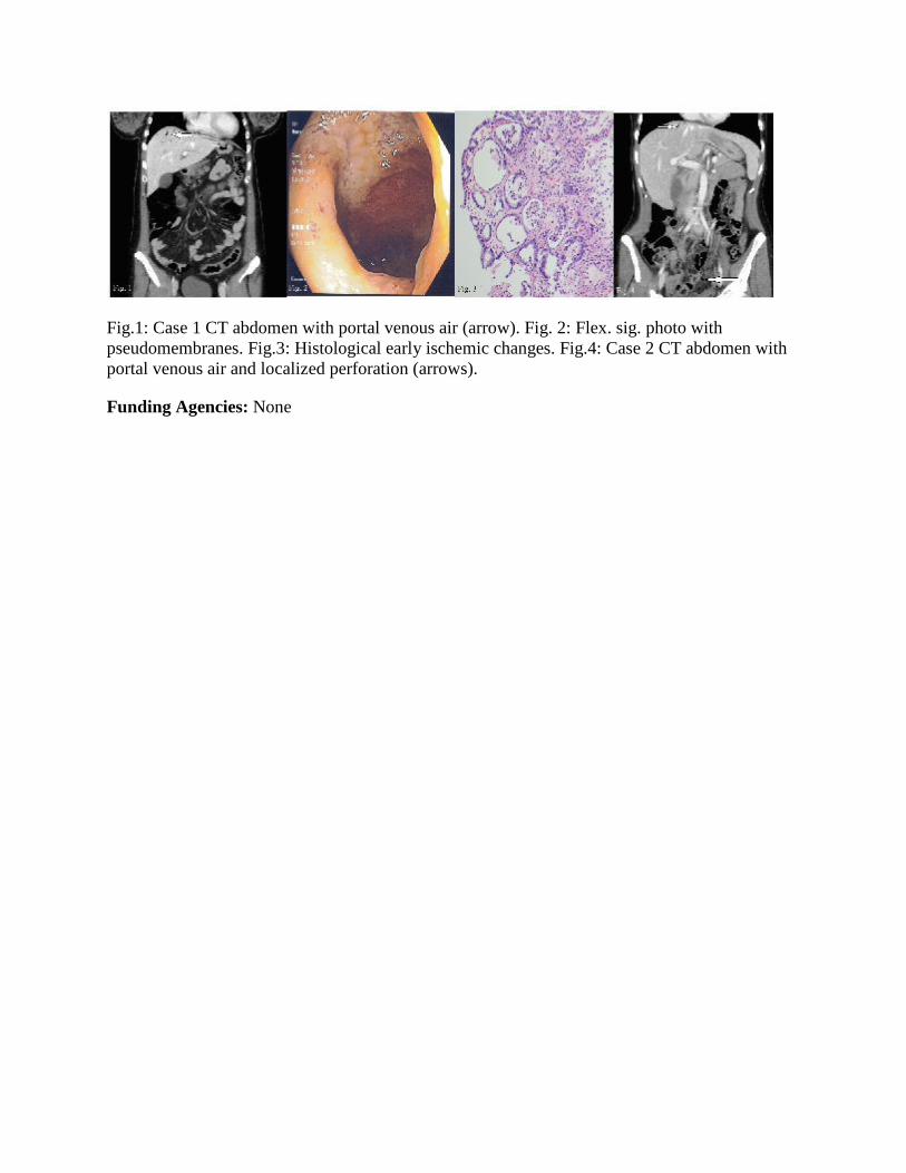
Fig.1: Case 1 CT abdomen with portal venous air (arrow). Fig. 2: Flex. sig. photo with pseudomembranes. Fig.3: Histological early ischemic changes. Fig.4: Case 2 CT abdomen with portal venous air and localized perforation (arrows).
Funding Agencies: None
A163
COLLAGENOUS ENTEROCOLITIS AS DIARRHEA AND ANEMIA
A. Yau, H. Ko
University of British Columbia, Vancouver, BC, Canada.
Aims: N/A Methods: N/A Results: A 56-year-old Caucasian female presented with epigastric pain, watery diarrhea, bloating, and flatulence. She denied nausea, vomiting, anorexia, and weight loss. Her symptoms persisted despite a trial of bismuth subsalicylate and probiotics which included Lactobacillus acidophilus and Bifidobacterium lactis. Her past medical history was unremarkable apart from anxiety and depression for which she took duloxetine and subsequently venlafaxine. Abdominal examination was benign. Blood work revealed a hemoglobin of 96 g/L (115-160 g/L), iron of 6 umol/L (10-33 umol/L), transferrin of 2.96 g/L (1.47-3.38 g/L), transferrin saturation of 0.08 (0.20-0.55), ferritin of 26 ug/L (15-180 ug/L), albumin of 46 g/L (35-50 g/L), pre-albumin of 293 mg/L (170-370 mg/L), normal liver enzymes, and anti-TTG of 5 units (< 20 units). Fecal occult blood tests were 3/3 positive and stool cultures were negative. CT enterography was normal. Colonoscopy revealed collagenous colitis on random colonic biopsies, while an upper endoscopy showed collagenous sprue on duodenal biopsy with blunted to completely flattened villi and markedly thickened subepithelial collagen table entrapping capillaries and lymphocytes. The patient was commenced on a gluten-free diet (GFD) and was prescribed loperamide 2 mg once daily and ferrous gluconate 150 mg once daily. Her symptoms resolved and a fecal immunochemical test performed 6 months later was negative. Conclusions: Collagenous sprue (CS) classically presents in middle-aged or elderly women with persistent diarrhea, progressive weight loss, and severe malabsorption. Malabsorption may be severe enough to warrant total parenteral nutrition and in some cases contribute to mortality. Our patient also presented with watery diarrhea and required iron supplementation for her anemia, but her presentation was atypical given her lack of weight loss. CS is frequently associated with celiac disease (CD); however, their relationship remains controversial. Serologic testing and HLA typing for CD may be positive in some cases of CS. GFD alone is usually ineffective for the treatment of CS which often requires corticosteroids. However, our patient had resolution of clinical symptoms with GFD alone, similar to several previous cases. These observations have led some to speculate that CS may be part of the celiac spectrum where the subepithelial collagen represents a histopathologic marker of poor prognosis. Collagen deposits may also be found in gastric and/or colonic mucosa in patients with CS. Our patient was found to have concomitant CS and collagenous colitis. This underscores the importance of performing both an upper and lower endoscopy to thoroughly assess the entire gastrointestinal tract for collagenous mucosal inflammatory diseases which may result in life-threatening complications such as ulcerative perforation and T or B cell lymphoma.
Funding Agencies: None

A164
GLUCOSE-GALACTOSE MALABSORPTION : HYPERCALCEMIA, NEPHROCALCINOSIS AND FALSE POLYURIA M. Lebel, M. Fiscaletti, G. Benoit, N. Alos, P. Jantchou CHU Sainte-Justine, Montreal, QC, Canada. Background: Glucose-galactosemalabsorption (GGM) is a rare and potentially fatal disorder. The autosomal recessive mutation of SGLT1 gene interferes with the active glucose transport in the gut resulting in osmotic diarrhea and failure to thrive (FTT). Aims: Two caucasian non-related infants with GGM are presented. One consulted for FTT while the other was first seen for macroscopic hematuria. The profuse diarrhea, initially mistaken for polyuria, promptly resolved after the glucose-galactose-free, fructose-based milk introduction. The investigations showed bilateral nephrocalcinosis in both patients with associated hypercalcemia, high levels of 1,25-dihydroxyvitamin D3 (1,25(OH)2D3) and hypercalciuria in one of them. Methods: NA Results: We hypothesize the upregulation of epithelial calcium channels (TRPV6) and 1,25(OH)2D3 as factors involved in the pathophysiology of nephrocalcinosis. In GGM, calcium intake remains adequate, but the state of malabsorption might trigger the upregulation of the TRPV6 transporter by 1,25(OH)2D3 and contributes to the hypercalcemia and hypercalciuria seen in some patients (TRPV6 being expressed in duodenal brush border and kidney tubules). It is also corroborated by the fact that local production of 1,25(OH)2D3 occurs in the enterocyte, which may sustain the upregulation of TRPV6 and the high levels of 1,25(OH)2D3 found in one of our cases. Since hypercalcemia and nephrocalcinosis are not present in every patient with GGM, genotype-phenotype studies are necessary to better understand the association. In this report, a novel intronic SGLT1 mutation (c.207+2dup) is described. Conclusions: In conclusion, osmotic diarrhea should be differentiated from polyuria, particularly in infants. These two cases also demonstrate that in children with nephrocalcinosis and/or hypercalcemia, clinicians must rule out a malabsorption disorder such as GGM.
Funding Agencies: None

A165
ARE TAX DEDUCTIBLE PROVISIONS FOR THE GLUTEN FREE DIET THE ANSWER TO IMPROVING UPTAKE AND COMPLIANCE FOR CELIAC PATIENTS?
M. Pinto-Sanchez2, E. Verdu4, M. Gordillo1, A. Nardelli1, J. Bai3, S. Birch4, P. Moayyedi4, P. Bercik2
1. McMasterUniversity, Hamilton-, ON, Canada; 2. McMaster University, HamiltON, ON, Canada; 3. Hospital de Gastroenterologia Dr C B Udaondo, Buenos Aires, Argentina; 4. McMasterUniversity, Hamilton, ON, Canada.
Background: Celiac disease (CD) affects 1% of the North American population, with an estimate of ~350,000 Canadians affected. CD is triggered by the ingestion of gluten, and a lifelong strict gluten-free diet (GFD) is the only current available treatment. A strict GFD is essential not only for intestinal mucosal recovery and alleviation of symptoms, but also for the prevention of complications such as anemia, osteoporotic fractures and small-bowel lymphoma. However the GFD is difficult to follow, socially inconvenient and expensive. Aims: The purpose of this analysis was to review the different systems available to support gluten- free diet coverage as a treatment for CD patients Methods: A review of the current systems available for gluten free diet coverage was performed. The current systems were compared in terms of premium coverage amount, as well as overall advantages and disadvantages for each system. Results: Different approaches such as tax reduction, cash transfer, food provision , prescription and subsidy ,have been used to reduce the extra costs of the GFD. The review showed that the systems in place exhibit advantages and disadvantages in relation to promoting uptake and compliance with GFD. The tax offset system used in Canada for GFD coverage, takes the form of a reimbursement of a cost previously incurred. Hence the programme does not help celiac patients meet the incremental cost of the GFD, it simply provides some future refund of that cost. Furthermore, the value of the tax offset is directly related to the celiac patient's marginal rate of tax. If the individual does not have sufficient income to pay income tax there is no tax refund to be claimed. In contrast someone paying tax at a marginal rate of 50% will receive a tax refund equal to 50% of the incremental cost of the GFD. An ideal balanced approach would involve subsidizing gluten-free products, through controlled vouchers or direct food provision to those who most need it independently of "ability or willingness to pay". Moreover, if the cost of such a program is inhibitive, the value of the benefits could be made taxable in order to ensure that any patient contribution (in terms of additional taxation) is directly related to ability to pay Conclusions: The limited coverage of GFD in Canada is of concern. There is an unmet need for GFD among celiac patients in Canada. More efforts are required by the Canadian medical community and the Canadian Celiac Society to act as agents in identifying ways of improving resource allocation in celiac disease.
Funding Agencies: CIHR

Microbiology and Parasite-Host Interactions
A166
INTERACTION BETWEEN HCV CORE AND PTEN PLAYS A ROLE IN REGULATING HCV REPLICATION
Q. Liu, Q. Wu
University of Saskatchewan, Saskatoon, SK, Canada.
Background: Hepatitis C virus (HCV) infection causes serious global public health problems. The World Health Organization has established that there are more than 170 million chronic HCV patients worldwide. Hepatocellular carcinoma (HCC) is the most deadly clinical consequence of HCV infection. Phosphatase and tensin homolog deleted on chromosome 10 (PTEN) can suppress PI3K-AKT pathway, one of the most critical cancer-promoting pathways PTEN is frequently mutated or deleted in tumors including HCC. However, the role of PTEN in HCV replication and pathogenesis is not well characterized. PTEN protein contains an N-terminal PIP2 (phosphatidylinositol-4,5-bisphosphate)-binding motif, a phosphatase domain, a C2 domain, a C-terminal tail containing two PEST (proline, glutamic acid, serine, threonine) sequences and a PDZ (PSD-95/DLG/ZO-1)-binding interaction motif at the end. Two naturally occurring mutations on the phosphatase domain disrupt PTEN's phosphatase activity: C124S mutation, which abrogates both lipid and protein phosphatase activity, and G129E mutation, which abrogates lipid phosphatase only. Aims: To determine the effect of PTEN on HCV infection and the underlying molecular mechanisms. Methods: We characterized HCV infection after PTEN overexpression or knocking down. We also determined whether PTEN interacts with HCV viral proteins as a mechanism for its effect on HCV infection. Results: PTEN negatively regulated HCV genotype 1a and 2a viral entry through PI3K-AKT pathway by using HCV pseudo-particles. In HCV-2a J6/JFH-1 genomic replicon cells, knocking down PTEN significantly enhanced HCV NS5A protein expression and viral replication; consistently, PTEN overexpression significantly inhibited HCV replication. We further showed that the phosphatase domain of PTEN was involved in HCV replication inhibition. Interestingly, PTEN with the lipid phosphatase defective mutation (G129E) could no longer inhibit HCV replication. In co-immunoprecipitation and pull-down assays, we showed that HCV core protein interacted with PTEN. HCV core aa. R50 and PTEN aa. 1-185 were required for the interaction. PTEN overexpression could no longer inhibit HCV genomic replication carrying core R50A mutation. Conclusions: PTEN regulates HCV viral entry, protein expression and replication. The lipid phosphatase activity of PTEN is required for inhibiting HCV replication. HCV core interacts with PTEN, which may contribute to PTEN's effect on HCV replication. Our study may help justify further development of PTEN as a new drug target for HCV therapy.
Funding Agencies: CIHR, NCRTP-HepC, SHRF

A167
HCV REPLICATION REQUIRES THE RECRUITMENT OF THE AUTOPHAGY ELONGATION COMPLEX (ATG5-12/16) IN A LC3 INDEPENDENT MANNER
A. Fahmy, C. Duguay, P. Labonté
INRS-Institut Armand Frappier, Laval, QC, Canada.
Background: Hepatitis C virus (HCV) infection is known to induce autophagosome accumulation as observed by the typical punctate cytoplasmic distribution of LC3-II in infected cells. Recently, we showed that viral RNA-dependent RNA polymerase (NS5B) interacts with ATG5, a major component of autophagy initiation. Aims: In this study, we evaluate the involvement of the autophagy elongation complex (ATG5-12/16) in HCV replication Methods: In this study we utilized Indirect immunoflourescence, western blot, co-immunoprecipitation, proximity ligation assay and siRNA techniques to investigate the role of the autophagy elongation complex (ATG5-12/16) in HCV replication Results: We demonstrate that the elongation complex is recruited at the site of viral replication and acts as a proviral factor. Indeed, ATG5-12 as well as ATG16L1 colocalizes with the viral replicase and dsRNA in infected cells. Using in situ proximity ligation assay, we confirms that ATG5 can interact with two replicase component, namely NS5B and NS3, but not with the viral capsid (core). Furthermore, we show the capability of NS4B to induce autophagy. While NS4B-induced autophagy was escorted by colocalization of NS4B with LC3-II in NS4B- transfected cells, no colocalization colocalization has been observed during infection. Interestingly, LC3 is not recruited along with the elongation complex to the site of viral replication as no colocalization of LC3-II with viral proteins was observed. Finally, using dominant negative forms of ATG proteins and siRNA approach, we demonstrate that ATG5-12 conjugate is important for viral replication but not LC3-II formation. Conclusions: Together, these findings indicate that HCV uses the autophagy elongation complex as a proviral factor for its own replication while it impedes the formation of a genuine autophagosome at the site of viral replication.
Funding Agencies: CIHR, NSERC
Viral Hepatitis
Poster of Distinction
A168
SAFETY AND EFFICACY OF OMBITASVIR-ABT-450/R AND DASABUVIR ± RBV IN HCV GENOTYPE 1-INFECTED CANADIAN PATIENTS: RESULTS FROM PHASE 3 TRIALS
J. Feld1, E. Yoshida2, A. Ramji2, E. Tam3, V. Bain4, N. Ackad5, J. Baloukas5, N. Shulman6, C. Cooper7
1. Toronto Centre for Liver Disease, University of Toronto, Toronto, ON, Canada; 2. University of British Columbia, Vancouver, BC, Canada; 3. LAIR Centre, Vancouver, BC, Canada; 4. University of Alberta, Edmonton, AB, Canada; 5. AbbVie Canada, Saint-Laurent, QC, Canada; 6. AbbVie Inc., North Chicago, IL; 7. University of Ottawa, Ottawa, ON, Canada.
Background: The interferon-free 3 direct-acting antiviral (3D) regimen of co-formulated ombitasvir/ABT-450 (identified by AbbVie and Enanta)/ritonavir and dasabuvir ± ribavirin (RBV) has achieved SVR12 rates >95% in HCV genotype 1-infected patients enrolled in 6 phase 3 studies (n=2053). Aims: We report the safety and efficacy of 3D±RBV for the subgroup of patients enrolled at Canadian sites of the phase 3 studies. Methods: Canadian sites participated in 4 of the 6 phase 3 trials. SAPPHIRE-I (GT1 treatment naïve) and SAPPHIRE-II (GT1 HCV treatment-experienced) were both randomized, placebo-controlled studies of 3D+RBV for 12 weeks in patients without cirrhosis. PEARL-IV (GT1a treatment naïve) was a randomized study of 3D+RBV or 3D+placebo for 12 weeks in patients without cirrhosis, and TURQUOISE-II (GT1 HCV treatment naïve or experienced patients with cirrhosis) was a randomized, open-label study of 3D+RBV for 12 or 24 weeks. Treatment-emergent adverse events (AEs) were reported for any patient receiving at least 1 dose of study drug. Results: Of the 117 patients who received 3D±RBV at Canadian sites in the phase 3 trials, 72 (62%) were male, 94 (80%) had GT1a infection, 36 (31%) had cirrhosis, and 34 (27%) had failed prior peginterferon/RBV treatment. A total of 97 patients received 3D+RBV and 20 patients received 3D without RBV. The overall SVR12 rate was 97% (114/117). SVR12 rates in patients treated with or without RBV were 98% and 95% respectively, 97% in patients with cirrhosis, and 100% in patients with GT1b infection. Fatigue, headache, nausea, and insomnia were the most commonly reported AEs. Most events were mild and there were no serious AEs. Clinically significant anemia requiring RBV dose modification occurred in only 1 (1%) patient. Conclusions: Canadian patients enrolled in phase 3 trials of 3D±RBV achieved similar SVR12 rates to the overall study populations.
SVR12 rates in Canadian patients enrolled in phase 3 studies of 3D ± RBV
n/N (%) Overall GT1a GT1b Treatment Treatment Experienced

Naïve
Relapse Partial Response
Null Response
All Patients 114/117 (97.4)
91/94 (96.8)
23/23 (100)
82/85 (96.5)
7/7 (100)
10/10 (100)
15/15 (100)
SAPPHIRE-I 33/34 (97.1)
25/26 (96.2)
8/8 (100)
33/34 (97.1) NA NA NA
SAPPHIRE-II 18/18 (100)
13/13 (100)
5/5 (100) NA 5/5
(100) 4/4
(100) 9/9
(100)
PEARL-IV 30/31 (96.8)
30/31 (96.8) NA 30/31
(96.8) NA NA NA
TURQUOISE-II
33/34 (97.1)
23/34 (95.8)
10/10 (100)
19/20 (95.0)
2/2 (100)
6/6 (100)
6/6 (100)
Funding Agencies: AbbVie
Poster of Distinction
A169
AN INTEGRATED SAFETY AND EFFICACY ANALYSIS OF >500 PATIENTS WITH COMPENSATED CIRRHOSIS TREATED WITH LEDIPASVIR/SOFOSBUVIR WITH OR WITHOUT RIBAVIRIN
J. Feld2, M. Bourliere3, M. Sulkowski5, S. Zeuzem4, E. Lawitz6, P. Marcellin7, M. Omata8, X. Ding9, J. Yang9, S. Knox9, P. Pang9, M. Subramanian9, W. Symonds9, J. McHutchison9, A. Mangia10, E. Gane11, R. Reddy12, M. Mizokami13, S. Pol14, N. Afdhal1
1. Beth Israel Deconess Medical Cnter, Boston, MA; 2. University Health Network University of Toronto, Toronto, ON, Canada; 3. Hopital Saint Joseph, Marseilles, France; 4. Johann Wolfgang Goethe University, Frankfurt am Main, Germany; 5. Johns Hopkins University, Baltimore, MD; 6. Texas Liver Institute, University of Texa Health Science Center, San Antonio, TX; 7. Hopital Beaujon, University of Paris, Paris, France; 8. Yamanashi Prefectural Hospital Organization, Yamanashi, Japan; 9. Gilead Sciences, Foster City, CA; 10. Liver Unit, Casa Sollievo della Sofferenza Hospital, San Giovanni Rotondo, Italy; 11. New Zealand Liver Transplant Unit, Auckland City Hospital, Auckland, New Zealand; 12. University of Pennsylvania, Philadelphia, PA; 13. Research Center for Hepatitis and immunology, National center for Global Health and Medicine, Chiba, Japan; 14. Department of Hepatology Universite Paris-Rene Descartes, Paris, France.
Background: Patients with HCV and Cirrhosis represent a population in most need of treatment; however, with interferon based therapy, such patients are difficult to cure and consequently often underrepresented in clinical trials. Aims: We analyzed the safety and efficacy of ledipasvir/sofosbuvir (LDV/SOF) in >500 patients with compensated cirrhosis enrolled in phase 2 and 3 studies. Methods: Treatment-naïve or experienced patients with chronic HCV genotype 1 infection and compensated cirrhosis receiving LDV/SOF+/-ribavirin (RBV) for 12 or 24 weeks were included in this pooled analysis Results: 514 subjects with compensated cirrhosis were identified. The majority (91%) of patients had cirrhosis diagnosed by biopsy or fibroscan (>12.5 kPa). Of the 293 patients with a fibroscan, 137/293 (47%) had a value >20 kPa. The majority were treatment-experienced (353, 69%), male (343, 67%), GT 1a (307, 60%), and IL28B non-CC (405, 79%). 238 (67% of the treatment-experienced patients) had previously received a protease inhibitor-containing regimen. 91 (18%) initiated therapy with a baseline platelet count of <90,000 cells/μL and 59 (11%) with a baseline albumin <3.5 g/dL. Patients received: 12 weeks of LDV/SOF (118, 23%), or LDV/SOF+RBV (206, 40%), or 24 weeks of LDV/SOF (132, 26%) or LDV/SOF+RBV (58, 11%). Safety in patients with cirrhosis was similar to that previously reported in patients without cirrhosis. Adverse events including anemia were more frequent in patients who received RBV. To date, 284 patients have available post-treatment week 12 data; 269 (95%) have achieved SVR12. Safety and efficacy for all 514 subjects will be presented. Conclusions: Based on results from over 500 patients, LDV/SOF is effective, safe, and well-tolerated for the treatment of compensated cirrhotic patients with HCV genotype 1.
Funding Agencies: Gilead Sciences Inc.

Poster of Distinction
A170
EVIDENCE FOR ONGOING LOW-LEVEL VIREMIA IN PATIENTS WITH CHRONIC HEPATITIS B RECEIVING LONG-TERM NUCLEOS(T)IDE ANALOG THERAPY
P. Marcellin4, E. Gane5, K. Kaita3, M. Khan1, R. Flisiak6, A. Gaggar7, L. Lin7, K. Kitrinos7, J. Flaherty8, H. Janssen2
1. Gilead Sciences Canada, Inc, Mississauga, ON, Canada; 2. Toronto Western Hospital, Toronto, ON, Canada; 3. University of Manitoba, Winnipeg, MB, Canada; 4. Hôpital Beaujon, Clichy, France; 5. Auckland City Hospital, Aukland, New Zealand; 6. Medical University of Bialystok, Bialystok, Poland; 7. Gilead Sciences, Inc., Foster City, CA; 8. Gilead Sciences, Inc, Foster City, CA.
Background: Nucleos(t)ide analog (NA) therapy is a mainstay of treatment for chronic Hepatitis B (CHB) infection. Treatment with a potent NA such as tenofovir disoproxil fumarate (TDF) is associated with a high level of durable viral suppression, improvement in liver fibrosis, and no documented viral resistance in a cohort of patients followed for up to 5 years. The presence of low level viral replication below the lower limit of quantification (LLOQ) in the presence of ongoing NA therapy, however, has not been evaluated in detail. Aims: To investigate low level viremia in patients on long-tetherapy with tenofovir therapy. Methods: HBV DNA levels were assessed from two registrational studies of TDF for CHB in HBeAg negative and HBeAg-positive patients. HBV DNA was quantified using the COBAS TaqMan V 2.0 having a lower limit of quantification (LLOQ) of 29 IU/mL and a lower limit of detection of 10 IU/mL. HBV DNA values less than 10 IU/mL are reported as target not detected (TND), while values between 10 IU/mL and 28 IU/mL are reported as target detected (TD). Results: The percentage of patients with undetectable HBV DNA less than LLOQ (TND) between weeks 96 and 240 is shown in the figure. Among patients who achieved HBV DNA < 29 IU/mL, the percentage with TND increased over time to 22-36% after 5 years (240 weeks) of NA treatment for HBeAg positive and HBeAg negative patients respectively. No patients achieved and remained 50% of visits between Weeks 96 and 240. For individuals who achieved HBV DNA suppression ( Conclusions: Low-level ongoing viral replication may be occurring in the presence of nucleoside analog therapy even after 5 years of therapy. The absence of low-level viremia was associated with HBeAg loss and seroconversion. Further evaluation into the mechanism of viral replication in the absence of overt drug resistance is warranted. These data provide a rationale for possible intensification of viral suppression through an orthogonal mechanism of action.
Funding Agencies: CIHR, Gilead Sciences, Inc.

Poster of Distinction
A171
SAFETY, ANTI-VIRAL EFFICACY AND PHARMACOKINETICS (PK) OF SOFOSBUVIR (SOF) IN PATIENTS WITH SEVERE RENAL IMPAIRMENT
E. Gane2, D. Mumm1, R. Robson3, L. Liu4, K. Sajwani4, L. Stamm4, D. Brainard4, J. McHutchison4, C. Stedman5, E. Lawitz6
1. Gilead Sciences Canada, Mississauga, ON, Canada; 2. University of Auckland, Auckland, New Zealand; 3. Christchurch Clinical Studies Trust, Christchurch, New Zealand; 4. Gilead Sciences, Foster City, CA; 5. Christchurch Hospital, Christchurch, New Zealand; 6. The Texas Liver Institute, San Antonio, TX.
Background: Safe and effective treatment for HCV-infected patients with severe renal impairment is currently unavailable and represents an area of unmet medical need. As compared to those with normal renal function, the AUC0-inf of SOF is 2.7-fold higher in patients with severe renal impairment, and the AUC0-inf of GS-331007, the renally excreted major SOF metabolite, is 5.5-fold higher. Aims: This study investigates the safety, efficacy and PK of SOF+RBV in HCV-infected patients with severe renal impairment. Methods: In an open-label study, 10 patients with chronic HCV GT1 or 3 with creatinine clearance (CrCl) less than 30mL/min as calculated by the Cockcroft-Gault equation, not on dialysis, are being treated with SOF 200mg + RBV 200mg daily for 24 wks. We examined the on-treatment virologic response and safety including echocardiograms at screening and Wk 12 of therapy. Results: Ten patients (7 GT1a, 2 GT1b, 1 GT3) were enrolled and have been treated for 12-24 wks (median 20 wks): 6 male, 5 black, 3 Pacific Islander/Asian, 2 white, mean age 62; none had cirrhosis, 7 treatment-naïve, 8 IL28B genotype non-CC, mean baseline (BL) CrCl 28.1 mL/min, mean BL hemoglobin (Hb) 11.1 g/dL. All patients experienced rapid virologic decline similar to those with normal renal function and full-dose SOF+RBV; 8/10 patients had HCV RNA < LLOQ at Wk 2 and 9/10 patients had HCV RNA (anemia). There were 2 treatment-emergent (TE) SAEs (diabetic ketoacidosis, unstable angina) not related to study drugs and not resulting in a change in treatment. Anemia (n=5) and headache (n=4) were the only TE AEs reported in more than 2 patients. Renal function was stable with a mean CrCl change from BL of -1.29 mL/min at Wk 12. Hemoglobin reductions were observed with a mean decrease from BL at Wk 12 of -1.4 g/dL. Four patients had Hb < 8.5 g/dL; 3 had the RBV dose-reduced or interrupted and one discontinued RBV after 56 days. Three patients were on epoetin at BL, 2 of whom required additional doses during treatment. As compared to BL echocardiograms, there were no significant changes

at Wk 12 (ejection fractions within 5% of BL). Conclusions: SOF 200mg + RBV 200mg in GT1 or 3 HCV-infected patients with severe renal impairment was well-tolerated and resulted in rapid virologic suppression. Final safety, SVR12 and PK will be presented
Funding Agencies: Gilead Sciences Inc.
Poster of Distinction
A172
A RECOMBINANT HCV ENVELOPE GLYCOPROTEIN E1E2 VACCINE ELICITS ANTIBODIES TARGETING MULTIPLE REGIONS ON E1E2 ASSOCIATED WITH BROAD CROSS-NEUTRALIZATION
J. Wong1, R. Bhat1, D. Hockman1, M. Logan1, C. Chen1, A. Levin1, S. Frey2, R. Belshe2, D. Tyrrell1, J. Law1, M. Houghton1
1. Li Ka Shing Institute of Virology, Department of Medical Microbiology and Immunology, University of Alberta, Edmonton, AB, Canada; 2. Department of Internal Medicine, Saint Louis University School of Medicine, Saint Louis, MO.
Background: Although effective Hepatitis C Virus (HCV) antivirals are on the horizon, a global prophylactic vaccine for HCV remains elusive. The diversity of the virus is a major concern for vaccine development; there are 7 major genotypes of HCV found globally. Therefore, a successful vaccine will need to protect against HCV infection of all genotypes. Despite the diversity, many monoclonal antibodies (mAbs) with broadly cross-neutralizing activity have been described suggesting the presence of conserved epitopes that can be targeted to prevent infection. Similarly, a vaccine comprising recombinant envelope glycoproteins (rE1E2) derived from the genotype 1a HCV-1 strain has been shown to be capable of eliciting cross-neutralizing antibodies in guinea pigs, chimpanzees, and healthy human volunteers. Aims: Investigation into the basis of the observed cross-neutralization was conducted. Methods: Epitope mapping of anti-E1E2 antibodies present within antisera from goats and humans immunized with HCV-1 rE1E2 was conducted through peptide mapping and competition studies with a panel of cross-neutralizing mAbs targeting various epitopes within E1E2. Results: The immunized goat antisera was shown to compete with the binding of all mAbs tested (AP33, HC33.4, HC84.26, 1:7, AR3B, AR4A, AR5A, IGH526, A4). Antisera showed the best competition against HC84.26/AR3B and the weakest competition against AR4A. Furthermore, antisera from five immunized human vaccinees were shown to compete with five pre-selected mAbs (AP33, AR3B, AR4A, AR5A, IGH526). Conclusions: These data show that immunization with HCV-1 rE1E2 elicits antibodies targeting multiple cross-neutralizing epitopes. Our results further support the use of such a vaccine antigen to induce cross-genotype neutralization. Characterization of the mechanisms of neutralization by vaccine antisera and at what entry step neutralization is taking place is currently being investigated and new data will be discussed. Note: This data has been recently published online by Journal of Virology.
Funding Agencies: Canadian Excellence in Research Chairs, Canadian Liver Foundation, National CIHR Research Training Program in Hepatitis C

Poster of Distinction
A173
FAVOURABLE IFNL3 GENOTYPES AND LIVER FIBROSIS IN HIV/HEPATITIS C (HCV) CO-INFECTED INDIVIDUALS FROM THE CANADIAN CO-INFECTION COHORT
N. Moqueet1, M. Hull2, C. Cooper3, S. Walmsley4, M. Potter1, M. Klein1
1. McGill University, Montreal, QC, Canada; 2. BC Centre for Excellence in HIV/AIDS, Vancouver, BC, Canada; 3. Ottawa Hospital-General Campus, Ottawa, ON, Canada; 4. University of Toronto and University Health Network, Toronto, ON, Canada.
Background: Liver fibrosis progression is faster in HIV/HCV co-infected individuals due to an elevated inflammatory profile. Interferon Lamda-3 (IFNλ-3), encoded by the human IFNL3 gene (formerly IL28B),has both antiviral and pro-inflammatory properties,though reports of its association with liver fibrosis are inconsistent. Homozygous recessive SNPs (rs12979860CC, rs8099917TT) in this gene are linked to spontaneous HCV clearance and better treatment response, potentially via non-synonymous functional variant rs8103142,which leads to a lysine-arginine substitution at position 70(K70R). Aims: Examining the relationship between specific IFNL3 genotypes and significant liver fibrosis as measured by the AST-to-platelet ratio index (APRI) ≥1.5 in HIV/HCV co-infected Canadians Methods: From the prospective Canadian Co-infection Cohort (n=1176), HCV RNA-positive participants free of fibrosis, end-stage liver disease and chronic Hepatitis B at baseline (n=612) were included. Cases (n=126) developed an APRI≥1.5 over follow-up. Data were analyzed using Cox proportional hazards, adjusting for sex, ethnicity, alcohol use, age and baseline APRI. Multiple imputation was used to account for missing data. Results: Overall 74% were male with median HCV duration=18 years.126 participants developed fibrosis over 1346 person-years of risk (9.40/100 person-years, 95% CI=7.90, 11.20/100 p-y). Univariate analyses suggested that each SNP may be linked to a higher risk of fibrosis. In multivariate analyses, rs8099917 had the strongest effect. Conclusions: Our results suggest that among the IFNL3 SNPs analyzed, rs8099917 is linked to a higher rate of liver fibrosis among HIV/HCV co-infected Canadians. Larger studies are needed to confirm this finding.
Table 1: Results
rs12979860 CC rs8099917 TT rs8103142 TT Univariate 1 (0.69, 1.45) 1.35 (0.93, 1.95) 1.15 (0.80, 1.66)
Multivariate 1.06 (0.72, 1.55) 1.46 (1, 2.14) 1.19 (0.82, 1.71) Female 1.25 (0.82, 1.89) 1.27 (0.85, 1.90) 1.27 (0.84, 1.93)
Alcohol Use 1.27 (0.87, 1.84) 1.24 (0.85, 1.81) 1.26 (0.87, 1.84) Baseline APRI 3.29 (2.10, 5.16) 3.38 (2.15, 5.32) 3.28 (2.09, 5.13)

Age 0.99 (0.97, 1.01) 0.99 (0.97, 1.02) 0.99 (0.97, 1.01) Aboriginal 1.10 (0.65, 1.87) 1.09 (0.64, 1.84) 1.09 (0.64, 1.84)
Funding Agencies: CIHR
A174
LEDIPASVIR/SOFOSBUVIR WITH RIBAVIRIN FOR THE TREATMENT OF HCV IN PATIENTS WITH POST TRANSPLANT RECURRENCE: PRELIMINARY RESULTS OF A PROSPECTIVE, MULTICENTER STUDY
R. Reddy3, J. Brisebois2, G. Everson4, S. Flamm6, J. Denning5, S. Arterburn5, T. Brandt-Sarif5, P. Pang5, J. McHutchison5, N. Afdhal1, M. Charlton7
1. Beth Israel Deconess Medical Cnter, Boston, MA; 2. Gilead Sciences Canada, Mississauga, QC, Canada; 3. University of Pennsylvania School of Medicine, Philadelphia, PA; 4. University of Colorado Denver, Aurora, CO; 5. Gilead Sciences, Foster City, CA; 6. Northwestern Feinberg School of Medicine, Chicago, IL; 7. Intermountain Medical Center, Murray, UT.
Background: In patients who are viremic at the time of liver transplantation HCV recurrence is universal and associated with reduced graft and patient survival. Aims: We evaluated the safety and efficacy of ledipasivr/sofosbuvir (LDV/SOF) with ribavirin in this population. Methods: GT 1 and 4, naïve and treatment-experienced patients with HCV infection, who were post liver transplantation (fibrosis score 0-3, CPT class A, B and C cirrhosis) with an estimated glomerular filtration rate (GFR) > 40 mL/min, received 12 or 24 weeks of LDV/SOF FDC with RBV. The primary efficacy endpoints were SVR (HCV RNA <25 IU/mL) 12 weeks after completion of study treatment, safety and tolerability. Results: To date, 223 patients have been randomized and treated. Most were male (83%), Caucasian (87%), and had prior HCV treatment (83%). The median time since liver transplant was 4.4 years (0.4-23.3). Mean baseline HCV RNA was 6.4 log10 IU/mL [range 2.4-7.8 log10 IU/mL]. Mean GFR was 65.5 [range 20.4-118.9 mL/min]. 112 patients had F0-F3 fibrosis, 52, 50 and 9 patients had CPT class A, B, and C cirrhosis, respectively. Interim Observed SVR4 results are depicted in Table 1. The most common adverse events were fatigue, anemia, headache and nausea. 9 SAEs in 8 patients were considered related to study treatment; anemia (4), hemolytic anemia (2), sick sinus syndrome (1), sinus arrhythmia (1) and portal vein thrombosis (1). 5 patients with cirrhosis died while in the study; internal bleeding, multiorgan failure/intestinal perforation, cardiac, complications of cirrhosis and progressive multifocal leukoencephalitis. Median serum creatinine and INR remained at baseline levels throughout treatment. Consistent with patients who have moderate renal impairment and who are receiving RBV, hemoglobin values decreased 2-3 g/dL on treatment. 32 patients received epoetin or blood transfusions in the study. Conclusions: Administration of LDV/SOF+RBV in patients with HCV recurrence post transplantation was well tolerated. SVR4 rates suggest high efficacy, with early data showing no apparent difference between 12 and 24 weeks of treatment. SVR12 results will be presented.
Table 1. Interim Observed SVR4 Results
Post Transplant Post Transplant F0-F3 CPT A, B, and C Cirrhosis
LDV/SOF+ LDV/SOF+ LDV/SOF+ LDV/SOF+

RBV 12 weeks (N=55)
RBV 24 weeks (N=57)
RBV 12 weeks (N=57)
RBV 24 weeks (N=54)
SVR4 n/N (%) 51/53 (96) 15/16 (94) 43/47 (91) 9/11 (82)
Funding Agencies: Gilead Sciences Inc.

A175
AN OBSERVATIONAL STUDY INVESTIGATING THE MANAGEMENT OF G1 HEPATITIS C ADULT PATIENTS WITH BOCEPREVIR IN COMBINATION WITH PEGIFN/RBV IN CANADA (THE S.I.M.P.L.E. STUDY)
S. Borgia3, J. Ford4, M. Levstik2, B. Conway5, D. Emokpare7, E. Tam8, C. Steingart6, N. Hilzenrat9, A. Ramji1, C. Cooper10, M. Vachon11, J. Halsey-Brandt12, M. Drolet12, J. Pouliot12
1. Gastrointestinal Research Institute, Vancouver, BC, Canada; 2. London Health Sciences Centre, London, ON, Canada; 3. William Osler Health System-Brampton Civic Hospital, Brampton, ON, Canada; 4. Vancouver General Hospital, Vancouver, BC, Canada; 5. Vancouver ID Research & Care Centre, Vancouver, BC, Canada; 6. Sanguen Health Center Clinic, Waterloo, ON, Canada; 7. Outpost Health Center, Regina, SK, Canada; 8. LAIR Centre, Vancouver, BC, Canada; 9. Jewish General Hospital, Montreal, QC, Canada; 10. The Ottawa Hospital Research Institute, Ottawa, ON, Canada; 11. CHUQ, Quebec, QC, Canada; 12. Merck Canada Inc., Kirkland, QC, Canada.
Background: Despite extensive use of PegIFN/RBV in combination with boceprevir for the management of GT1 HCV the efficacy and safety of triple regimen in the real-world Canadian population have not been completely characterized. Aims: To describe the real life use of BOC/PegIFN/RBV for the management of HCV G1 in Canada. Methods: This is an observational, prospective, multicenter, non-interventional study of 157 patients enrolled in 25 academic and community centers in Canada. Results: In this study, 79% are treatment naïve, 74% male and 78% Caucasian; 40% of the patients are infected with genotype 1a. The median age is 54 (21-72) years. Half the patients have advanced liver fibrosis (F3 or F4) and 33% have cirrhosis. In the interim analysis performed on 142 patients, at week 4, 23% of the naïve (18/77) and 12.5% (1/8) of the experienced patients had undetectable viral load. At week 8, 67.5% (55/77) of the naïve non-cirrhotic and 50% (4/8) of the experienced non-cirrhotic patients (excluding nulls) had undetectable viral load and were eligible for shorter therapy. At week 12, the proportion of naïve and experienced patients with undetectable viral load was 68% and 56% respectively. Anemia (Hb<100g/L) occurred in 26% of patients at TW12. Discontinuation due to adverse events occurred in 7% of the patients. The health care resources utilization was similar for week 4, 8 and 12 (28, 30 and 27 health care visits/year) and consisted mostly of nursing visits. Conclusions: In this interim analysis, a significant proportion of patients are eligible for shortened treatment duration and demonstrate early virologic response at TW 8 and 12 in a population including 53% F3/F4 fibrosis level. This preliminary analysis suggests that, in a cost-containment environment, the first generation DAAs might still represent an option for a large proportion of patients. SVR data will be presented.
Funding Agencies: Merck Canada Inc.

A176
SERUM LYSYL OXIDASE LIKE 2 (SLOXL2) LEVELS CORRELATE WITH ISHAK FIBROSIS SCORE AND DECREASE WITH TREATMENT WITH TENOFOVIR DISOPROXIL FUMARATE (TDF) IN PATIENTS WITH CHRONIC HEPATITIS B (CHB)
W. Ray Kim3, R. Loomba4, E. Tam2, M. Khan1, P. Lal5, R. Schall5, A. Johnson5, J. Bornstein5
1. Gilead Sciences Canada, Inc, Mississauga, ON, Canada; 2. LAIR Centre, Vancouver, BC, Canada; 3. Stanford School of Medicine, Palo Alto, CA; 4. University of California San Diego, San Diego, CA; 5. Gilead Sciences, Inc, Foster City, CA.
Background: Lysyl Oxidase Like 2 (LOXL2) is an extracellular copper-dependent amine oxidase that catalyzes the formation of crosslinks in collagen, and is implicated in the development of liver fibrosis. Aims: We assessed the relationship of sLOXL2 levels with liver fibrosis and cirrhosis in patients with CHB treated with TDF. Methods: Subjects in the pivotal TDF registration trials who had stored serum samples available for analysis at baseline and weeks 12, 48 and 240 were included in the analysis. Liver biopsies were performed pre-treatment, at weeks 48 and 240. sLOXL2 was measured using a highly sensitive assay developed using 2 specific monoclonal antibodies on a Singulex® platform. Results: Of the 641 subjects originally randomized, 304 had stored serum available and were included in the analysis. Of these, 225 had liver biopsies at baseline and week 240. Characteristics (77% male, 63% white, 30% Asian and 13% obese) of the subjects included in the study matched well with those not included in the analysis. sLOXL2 correlated with Ishak fibrosis stage at all time points. In cross sectional analyses at baseline, the median sLOXL2 level correlated with necroinflammation by Knodell score (p<0.0001) and with fibrosis by Ishak stage (791 pg/mL for Ishak stage 0-2, 1091 pg/mL for Ishak 3-4 and 1370 for pg/mL for Ishak 5-6; p<0.0001). In longitudinal analysis, sLOXL2 declined with treatment, with the largest decline seen in the first 12 weeks. For all fibrosis stage categories, patients experiencing fibrosis regression consistently had lower sLOXL2 levels than those without regression. Conclusions: sLOXL2 correlates with fibrosis stage in patients with HBV. sLOXL2 declines with HBV treatment, likely indicating a decrease in fibrogenesis. Overall, the data suggest that sLOXL2 is a potential measure of ongoing fibrosing disease and may be useful not only to assess liver fibrosis at a given time but also to detect reversal of fibrosis and cirrhosis.
Funding Agencies: Gilead Sciences, Inc.

A177
HEPATITIS B VIRUS (HBV) COMPARTMENTALIZATION BEFORE AND AFTER NUCLEOS/TIDE ANALOG (NA) TREATMENT
S. Gao1, Z. Duan3, Y. Chen3, J. Zhang3, S. Lee1, F. van der Meer1, G. van Marle1, C. Osiowy2, C. Coffin1
1. University of Calgary, Calgary, AB, Canada; 2. National Microbiology Laboratory, Winnipeg, MB, Canada; 3. Beijing Youan Hospital, Capital Medical University, Beijing, China.
Background: The HBV is a hepatotropic virus but also infects peripheral blood mononuclear cells, PBMC. Potent NA, i.e., entecavir (ETV) or tenofovir (TDF), suppress viral replication but HBV surface antigen (HBsAg) loss rarely occurs. Aims: We aim to compare HBV replication status, viral diversity and HBV polymerase (P)/overlapping preS/surface (S), pre-core/core (C) and basal core promoter (BCP) gene variants in matched plasma, PBMC and liver in CHB patients treated with NA. Methods: Plasma, PBMC and liver biopsy tissue in some were collected pre-treatment and end of follow-up from 14 CHB patients (median age 46 y [range 23-57], 10/14 M, 13/14 Asian, 10/14 Genotype B or C, median follow-up 42 weeks [range 38-53]). At baseline, 6/14 HBeAg+, median ALT 67 U/L (range 20-137), median HBV DNA 5 log IU/ml (range 2.9 - 8 log), median quantitative HBsAg 3 log IU/ml (range 2-5 log), 8/14 >F2 fibrosis by transient elastography or liver biopsy analysis. All received NA (9/14 ETV, 5/14 TDF), 2/6 had HBeAg loss, and 13/14 had 6-mo suppressed plasma HBV DNA by a commercial PCR (sensitivity ~50 copies/ml). HBV-covalently closed circular (ccc)-DNA levels were quantified at baseline and in follow-up PBMC and liver using an in-house PCR normalized to beta-globin housekeeping gene. The HBV P/S gene was PCR-amplified in matched plasma/PBMC/liver tissue, amplicons cloned and ~20 clones/sample sequenced and analyzed for drug-resistant (DR) and immune escape (IE) variants by MEGA v 5.0. In 7/14, HBV full-genome clones were analyzed by deep sequencing (Illumina, MiSeq). Results: In treatment naïve patients, the HBV diversity and minority variants at positions associated with IE (i.e., sG145R and sP120S) or DR (i.e., rtM204I, rtL180I) was comparable in plasma vs. PBMC and in liver vs. plasma. In follow-up cases analyzed on NA therapy compared to baseline (N=6), HBV diversity increased in plasma only, but IE and DR variants increased in plasma and PBMC (P<0.05). In 12/14, the median HBV cccDNA pre-treatment was 3.6-log copies/106 PBMC with no significant decline in 3/12 NA-treated patients with available follow-up PBMC tested (3.5 log copies/106 cells). Conclusions: In treatment naïve patients there was no difference in minor HBV variants between liver, PBMC or plasma site. After initiation of NA and suppression of HBV replication, HBV cccDNA persists in PBMC. Although minor HBV variants increase in matched plasma and PBMC, overall HBV diversity only increased in PBMC site. The results suggest HBV evolves in a compartment-specific fashion after NA therapy.
Funding Agencies: CIHR

A178
MARIJUANA USE DOES NOT ACCELERATE LIVER FIBROSIS IN HCV/HIV COINFECTED WOMEN
E. Kelly1, M. Sarkar1, J. Dodge1, A. French2, P. Tien1, M. Glesby3, E. Golub4, M. Augenbraun5, M. Plankey6, M. Peters1
1. UCSF, San Francisco, CA; 2. CORE Center, Chicago, IL; 3. Cornell Medical College, New York, NY; 4. Johns Hopkins Bloomberg School of Public Health, Baltimore, MD; 5. State University of New York, Brooklyn, NY; 6. Georgetown University, Washington, DC.
Background: The cannabinoid system has been implicated in modulating liver disease. Cross-sectional analyses suggest that cannabis (THC) use predicts advanced fibrosis in patients with chronic hepatitis C (HCV) and may be most detrimental to those with established fibrosis. Aims: We studied long-term effects of THC on fibrosis progression in women co-infected with HCV/HIV enrolled in Women's Interagency HIV Study (WIHS), a prospective, multicenter cohort of women with or at risk for HIV infection. Methods: Fibrosis was categorized by APRI scores as mild (<0.5), moderate (0.5-1.5), or severe (≥1.5): women with severe fibrosis at entry into WIHS were excluded. THC and alcohol use were treated as continuous variables and quantified as average exposure over time in study until last follow-up or development of severe fibrosis. Associations between THC use and progression to severe fibrosis were assessed using Cox proportional hazards regression. Results: Among 670 HIV/HCV co-infected women [median follow-up: 5.1 (1.2-10.5) years], 323 (49%) reported no THC use; 209 (31%) reported ≥weekly use; 134 (20%) <weekly use; and 4 no THC data. Median APRI at entry were similar [0.53 vs 0.49 vs 0.50] in those who reported no THC use, <weekly use and ≥ weekly use, respectively. Compared to women reporting no THC use, weekly users reported more injection drug [28% vs 18% p=0.004] and alcohol use [60% vs 44% p=0.001]. In univariate analysis, log APRI [HR 10.35 (5.69-18.84) p<0.001], log HCV RNA [HR 1.3 (1.10-1.54) p=0.002] and log HIV RNA [HR 1.14 (1.02-1.29) p=0.03] at entry were associated with progression to severe fibrosis; higher CD4+ count [per 50 cells HR 0.96 (0.93-0.98) p<0.0004] and ART use [HR 0.62 (0.38-1.01), p=0.05] were associated with lower fibrosis. Cumulative alcohol use [risk per 1 drink increase per week [HR 1.03 (1.02-1.04) p<0.001] was associated with greater risk of progression. In multivariate analysis, entry APRI, HCV RNA, CD4+ count and cumulative alcohol use remained significant. Cumulative THC use was not independently associated with a greater risk of fibrosis progression [HR 1.00 (95% CI 0.996-1.003)] even in those with moderate fibrosis at entry [HR 1.00 (95% CI 0.995-1.005)]. Conclusions: In this large cohort of HCV/HIV co-infected women with prospectively collected cumulative alcohol and THC use, THC was not associated with liver fibrosis progression. Alcohol use was strongly associated with THC use and independently associated with liver fibrosis, and may better predict fibrosis progression in HCV/HIV co-infected women.
Funding Agencies: CASL Clinical Hepatology Scholarship
A179
CLINICAL PROFILE AND TREATMENT OFFER IN HCV-INFECTED PATIENTS WITH END-STAGE-LIVER DISEASE
F. Trottier-Tellier1, M. Bilodeau2
1. Department of Medicine, CHUM, Montreal, QC, Canada; 2. CRCHUM, Montréal, QC, Canada.
Background: Antiviral treatment for chronic Hepatitis C Virus (HCV) infection has been shown to be moderately effective in RCTs but, in real life setting, only a minority of HCV-infected patients are treated and even less reach sustained virological response (SVR). Aims: Our goal is to evaluate, in a group of chronic HCV-infected patients with end-stage liver disease (ESLD), the proportion who received antiviral therapy before the appearance of ESLD. We hypothesized that the majority of patients would have been offered antiviral therapy, but either refused it or did note achieve SVR. Methods: HCV-infected patients with ESLD followed in our institution were screened for the study. ESLD was defined by the development of hepatocellular carcinoma, hepatic encephalopathy, esophageal variceal bleeding, spontaneous bacterial peritonitis, ascites or a MELD score of 14 or higher. We included 143 patients who filled a survey on sociodemographic data, risk factors, diagnosis and treatment of HCV infection. Medical charts were reviewed to complete data acquisition. Results: Among 143 participants, 116 (81%) were diagnosed with HCV infection before ESLD developed. Overall, 77% of participants (n=111) received a treatment offer and 71% of patients (n=101) underwent antiviral therapy. Among the 101 treated participants, 67 (47% of the study population) were treated before the occurrence of ESLD and 32 only after ESLD had developed. 42 patients (29%) never received treatment. Among these, 33 (23% of study population) were never offered therapy and 9 declined treatment offer. Only 48 patients (34% of all patients) completed the full course of treatment. The most common causes for treatment discontinuation were side effects and treatment failure. Among the treated participants, 60 underwent 1 treatment trial and 40 underwent 2 trials or more for an average of 1.55 treatments per treated patient and a total of 155 treatment trials. Only 25 patients (17%) achieved SVR. When groups of treated vs non-treated individuals were compared, non-treated patients tended to have lower education levels, were more likely to live alone, to drink alcohol and to be active users of illegal drugs (p<0,05). Conclusions: Among a group of HCV-infected patients with ESLD, we found that most patients (81%) were diagnosed before developing ESLD and a majority (71%) received antiviral therapy. However, a minority of patients (47%) received treatment before hepatic complications occurred, even less (34%) completed a full course antiviral therapy and the overall SVR rate was very low (17%). These results suggest that earlier, more effective therapy and fewer obstacles to treatment initiation are required in order to avoid the life-threatening complications of HCV infection.
Funding Agencies: None

A180
ECONOMIC EVALUATION OF LEDIPASVIR/SOFOSBUVIR IN HEPATITIS C: A CANADIAN PERSPECTIVE
A. Rocchi1, J. Totten2, I. Guerra3, S. Cure3
1. Athena Research Inc., Burlington, ON, Canada; 2. Gilead Sciences Canada Inc, Mississauga, ON, Canada; 3. Optum, London, United Kingdom.
Aims: To conduct an economic evaluation of ledipasvir/sofosbuvir (LDV/SOF) versus appropriate comparators, over a lifetime horizon, in patients with genotype 1 (G1) chronic hepatitis C virus (HCV) infection. Methods: A Markov state-transition model described the progression of HCV over a lifetime horizon. Comparators used in the model included: sofosbuvir (SOF) in combination with pegylated-interferon and ribavirin (P/R) (the current standard of care), boceprevir + P/R, telaprevir + P/R (both the historical standards of care), no treatment, SOF + ribavirin (RBV) (both for patients ineligible or intolerant to interferon) and simeprevir + P/R. Rates of sustained virologic response (SVR) from published clinical trials were used to determine if a patient moved into a "cure state" following treatment with each comparator. Without an SVR, patients faced an annual probability of liver disease progression and mortality. Recent Canadian literature sources were used for quality of life, health care costs and productivity costs. Adverse events considered in this analysis were anemia, depression and rash with costs assigned based on a retrospective HCV treatment analysis of the Quebec claims database. Results: From the societal perspective (which considers both health care costs and workplace productivity costs), LDV/SOF was dominant (more effective and less expensive) over comparators for a treatment-naïve population (TN) and highly cost-effective for treatment-experienced (TE) patients, with results under $15,000 per quality-adjusted life year (QALY). From the health care system perspective (considering only health care costs), LDV/SOF dominated for TN patients, and was highly cost-effective (under $30,000/QALY) for TE patients. Due to superior SVRs, use of LDV/SOF was associated with fewer cases of cirrhosis, hepatocellular carcinoma, and liver transplants in all clinical settings and versus all comparators. Conclusions: Conclusion: regimen, LDV/SOF offers clinically meaningful improvements in efficacy, tolerability and simplicity for patients with CHC and provides a much-needed treatment option for the substantial proportion of patients who currently have no other options, including those who are IFN-intolerant, ineligible or unwilling.
Funding Agencies: Gilead Sciences Canada Inc.
A181
BINDING OF THE HCV HELICASE ON THE NUCLEIC ACID SUBSTRATE AT SINGLE NUCLEOTIDE RESOLUTION
C. Ablenas1, H. Liu2, G. Cosa1, M. Gotte2
1. McGill University, Montreal, QC, Canada; 2. University of Alberta, Edmonton, AB, Canada.
Background: The hepatitis C virus (HCV) non-structural protein 3 (NS3) contains a helicase activity essential for viral replication. The helicase binds to single-stranded (ss) regions of nucleic acids and unwinds duplexes in an NTP-dependent manner. The mechanism by which the helicase disrupts RNA secondary structure in the viral genome to make way for the replication machinery remains elusive. Several mechanisms have been proposed, which include an active mechanism whereby the helicase actively engages the ss/double-stranded (ds) junction of the substrate to unwind the duplex, and a passive mechanism where the helicase binds and translocates along a ss nucleic acid overhang, taking advantage of transient melting of the ss/ds junction. Aims: Here, we investigate the dynamic interaction between the helicase and a nucleic acid substrate using a fluorescence-based assay in an attempt to establish where binding to the substrate occurs. Methods: Positioning of the helicase on a nucleic acid substrate with a ss/ds junction was monitored using a fluorescence-based assay. We used this approach to determine the location of the enzyme based on the increased intensity of a fluorescence signal upon binding of a protein in very close proximity to a fluorescent dye attached to the nucleic acid substrate, a process referred to as protein induced fluorescence enhancement (PIFE). Results: The helicase binds to the ss substrate with a footprint of approximately 6 bases. Designing a substrate fluorescently labeled at the ss/ds junction with a ss overhang of 6 bases ensures that a single helicase molecule will bind to the substrate and be positioned directly at the junction. Addition of NS3h to this substrate results in a strong fluorescence enhancement, showing conclusively that the helicase bound at the junction on this short ss overhang. The length of the ss region of the substrate was then increased, providing the opportunity for the helicase to bind the ss substrate at increased distances from the ss/ds junction. At a single base pair resolution, as the length of the ss overhang increased fluorescence enhancement decreased. This implies that the enzyme does not preferentially bind at the ss/ds junction, but rather binds anywhere along the ss overhang. Conclusions: The work presented here validates the use of PIFE to study the positioning of the helicase relative to the ss/ds junction of the nucleic acid substrate. Furthermore, our studies offer insight into the dynamic interactions between the helicase and its substrate. Based on our data, it appears that the helicase may bind the ss substrate and translocate until arriving at the ss/ds junction where it unwinds the duplex. A deeper understanding of the mechanism by which the helicase interacts with its substrate will help elucidate its role in the viral life cycle.
Funding Agencies: CIHR, None, National CIHR Research Trainning Program in Hepatitis C (NCRTP), and FRQS

A182
BIOCHEMICAL CHARACTERIZATION OF NUCLEOTIDE TRIPHOSPHATE METABOLITES OF BETA-D-2'-C-METHYL-4-N-HYDROXYCYTIDINE PRODRUG AS POTENT INHIBITORS OF HEPATITIS C VIRUS POLYMERASE
M. Ehteshami1, S. Tao1, S. Amiralaei1, H. Li1, X. Lu1, T. Ozturk1, F. Amblard1, T. McBrayer2, T. Whikater2, S. Coats2, R. Schinazi1
1. Emory University, Atlanta, GA; 2. RFS Pharma, Tucker, GA.
Background: Nucleoside analog inhibitors (NI) are an important class of anti-HCV agents that are pan-genotypic and have a high genetic barrier to drug resistance. Sofosbuvir, the only NI approved by the FDA to date, has demonstrated an excellent safety profile and treatment with sofosbuvir-containing regimens results in unprecedented cure rates for chronic HCV infections. Herein, we describe the discovery of a b-D-2'-C-methyl-4-N-hydroxycytidine prodrug, that, upon intracellular metabolism, can deliver multiple nucleoside 5'-triphosphate inhibitors (NI-TP). Upon cellular entry, this prodrug was metabolized to generate three distinct NI-TPs: 2'-C-methyl-CTP, 2'-C-methyl-UTP and 2'-C-methyl-4-N-OH-CTP. The two former NI-TPs are well characterized for their anti-HCV activity, whereas 2'-C-methyl-4-N-OH-CTP has not been studied. Aims: The aim of this study is to characterize the biochemical properties of 2'-C-methyl-4-N-OH-CTP as a novel inhibitor of HCV polymerase. We also aim to shed light on the mechanism of action of this prodrug in vivo through characterization of intracellular metabolism of b-D-2'-C-methyl-4-N-hydroxycytidine prodrug and cell-free enzymatic properties of the generated NI-TPs. Methods: Prodrug was incubated in Huh-7 cells and LC-MS/MS was used to measure intracellular levels of metabolites. Inhibition of RNA synthesis by each NI-TP was separately evaluated using purified recombinant HCV polymerase. In vitro enzymatic assays were employed to measure dissociation constants and rates of incorporation for each inhibitor. Results: We observed that 2'-C-methyl-4-N-OH-CTP behaves as both a cytidine and uridine analog (C > U). We also established that 2'-C-methyl-4-N-OH-CTP effectively inhibited RNA polymerization when pre-incubated with purified NS5B enzyme, but was outcompeted when co-incubated with natural CTP and UTP substrates. Kinetic parameters, as well as intracellular NI-TP levels were taken into consideration in order to shed light on its intracellular mechanism and antiviral activity. Conclusions: b-D-2'-C-Methyl-4-N-hydroxycytidine prodrug takes advantage of naturally occurring intracellular metabolism pathways to generate three bioactive NI-TP. These findings could have important implications for the development of a new class of NIs that mediate the intracellular delivery of multiple active nucleoside 5'-triphosphate analogs with distinct incorporation profiles.
Funding Agencies: CIHR, ALF, CFAR, NIH

A183
ROLE OF FGF23 IN TENOFOVIR-INDUCED HYPOPHOSPHATEMIA IN PATIENTS WITH CHRONIC HEPATITIS B
R. Saeedi2, A. Mojebi-Mogharar2, J. Ford2, M. Yousefi2, J. Dubland2, M. Pudek2, D. Holmes2, Z. Erb2, P. Kwan2, E. Yoshida1
1. University of BC, Vancouver, BC, Canada; 2. University of British Columbia, Vancouver, BC, Canada.
Aims: To investigate the relationship between fibrobast growth factor (FGF23) and tenofovir (TEN)-induced hypophosphatemia in chronic hepatitis B (CHB)-infected patients. Methods: A cross-sectional study in CHB-infected patients who were treated for at least one year with either TEN (300 mg/day), lamuvidine (LAM) (100 mg/day), entacavir (ENT) (0.5 mg/day), or were not treated (CON) for at least three months. Enrolled subjects did not have cirrhosis or known bone disease. Routine serum biochemistry was assessed by standard methods. Plasma FGF23 was measured by ELISA. Intact parathyroid hormone (iPTH) levels and serum calcitriol were measured in selected patients by immunoassay and tandem mass spectrometry, respectively. Fisher's exact test was applied to assess association between FGF23, serum PO4, calcitriol, and iPTH. Results: Patients were divided into 4 groups: TEN treated (N=26) for 1-12 (mean 3) years, LAM treated (N=20) for 1.5-10 (mean 4.1) years, ENT treated (N=11) for 3.5-5.5 (mean 3.6) years, and CON (N=19). There were no differences in calcium, liver or renal function tests among groups. Serum PO4 was low in 19% of TEN group, 20% of LAM group, and 5% of CON group. FGF23 was elevated in 4 TEN treated patients while one had hypophosphatemia and high iPTH (Table 1). There was a statistically significant association between elevated FGF23 and treatment with TEN (p<0.01). Conclusions: Treatment with either TEN or LAM caused hypophosphatemia in some CHB-infected patients; however, high levels of FGF23 were only observed in patients treated with TEN while not all of them had hypophosphatemia. Differences in phosphorus metabolism may exist between these groups.
Table 1: Laboratory Results of the four (out of 26) TEN-treated patients with high FGF23
Patient 1 patient 2 patient 3 Patient 4 Reference Interval FGF23 (RU/ml) 530.3 247.4 575.6 342.1 ≤180
iPTH (pg/ml) 7.6 4.7 5.6 2.9 1.3-6.8 Calcitriol (pg/ml) 56.8 49.8 39.1 42.7 18-78
Serum phosphate (mmol/l) 0.68 1.24 1.01 0.92 0.8-1.6 Serum calcium (mmol/l) 2.14 2.3 2.2 2.43 2.18-2.58 eGFR* (ml/min/1.73 m2) 104 74 107 113 >59
Creatinine (umol/l) 56 101 54 69 60-100 Duration of treatment (years) 12 4.5 2.5 2.5 --------

*estimated Glomerular Filtration Rate (eGFR) using the Modification of Diet in Renal Disease study group formula (MDRD).
Funding Agencies: None

A184
A PROSPECTIVE STUDY OF 148 UNTREATED AND TENOFOVIR TREATED CHRONIC HEPATITIS B (CHB) PREGNANT PATIENTS
G. Samadi Kochaksaraei1, E. Castillo1, A. Scott2, J. Oshiomogho1, S. Martin1, S. Lee1, R. Myers1, C. Coffin1
1. University of Calgary, Calgary, AB, Canada; 2. Alberta Health and Welness, Edmonton, AB, Canada.
Background: Despite prophylaxis, mother-to-child transmission (MTCT) of hepatitis B can occur in ~10-15% of infants, linked to high maternal viremia. HBV flares, (i.e., ALT >2X upper limit normal) has been reported during pregnancy/post-partum. In 2011, Alberta prenatal screening program recommended that all mothers with CHB be referred to a specialist for management. Aims: To determine maternal and infant outcomes of CHB patients referred in pregnancy and postpartum. Methods: Pregnant CHB carriers were enrolled from a hepatology or maternal fetal medicine clinic from Jan 2011 to present. Demographic and clinical data were collected in pregnancy up to 6 months post-partum including estimated fibrosis stage by transient elastography (TE), delivery mode and prematurity rate. All mothers with HBV DNA >6-log10 IU/ml were offered Tenofovir (TDF) in the 3rd trimester, and maternal glomerular filtration rate and infant HBV serology were compared in treated vs. untreated patients. Results: 148 women with 156 pregnancies (1 twin) were enrolled (median age 32 y [IQR 29-35]), 18% [28/148] HBeAg(+), median baseline HBV DNA 2.5 log IU/ml [IQR 1.6-3.6; range 0-8.81], and median liver stiffness by TE 4 kPA [IQR 3.2-4.6]. Overall, 14.8% (22/148) received TDF in pregnancy due to high viral load (15/22, median duration 74 days [IQR 59-110]), or treated long-term due to liver disease (i.e., 7/22 with active CHB or cirrhosis). In treated patients, the median baseline vs. follow-up HBV DNA was 7.4 and 2.5 log IU/ml respectively (P=0.007). In treatment naïve mothers with low viral load (N=119, median HBV DNA 2.2 log IU/ml [IQR 1.5-1.8]) the median ALT increased from 17 U/L to 27.5 postpartum (P=2e-6), as well as in untreated highly viremic patients who declined TDF (N=7; HBV DNA >8-log10 IU/ml); median ALT increase >2.8X (P<0.01). 26% (40/156) had caesarean section and there were no differences in obstetric or renal outcomes in treated vs. untreated cohort. In infants with available data 95% (149/156), 76% (114/149) received HBIG, 57% (86/149) to date received 3 doses of HBV vaccine, only 41% (62/149) completed follow-up HBV serology. One infant of an untreated mother tested HBV surface antigen (+) (maternal viral load > 8-log IU/ml) despite immunoprophylaxis. Conclusions: TDF therapy in pregnancy is well tolerated, significantly reduces HBV viral load, and does not impact obstetric outcomes. One case of MTCT occurred despite immunoprophylaxis of an infant born to a highly viremic untreated CHB mother. Most untreated mothers had ALT increase post-partum.
Funding Agencies: CIHR

A185
INVESTIGATING THE ROLE OF MICRORNA-122-ASSOCIATED COMPLEXES IN HEPATITIS C VIRUS INFECTION
A. Bernier, S. Sagan
McGill University, Montreal, QC, Canada.
Background: Approximately 200 million individuals worldwide are infected by hepatitis C virus (HCV), including more than 300 000 Canadians. HCV-infected individuals typically develop a persistent infection that leads to chronic hepatitis, cirrhosis and liver cancer. MicroRNA-122 (miR-122) is a highly abundant liver-specific microRNA shown to interact at two "tandem" microRNA-binding sites in the 5' UTR of the HCV genome. This unusual interaction promotes HCV RNA accumulation in both HCV-infected cells, and the livers of infected patients. Mutation, truncation, or exchange of the 3' terminal ribonucleotides of miR-122 for deoxynucleotides reduces HCV RNA accumulation. However, these nucleotides are not required for canonical miRNA activities (i.e. target cleavage and translational inhibition). This suggests that sequences in the 3' tail of miR-122 may mediate important interactions with viral or cellular factors involved in HCV RNA accumulation. Aims: We hypothesize that miR-122 forms a distinct complex with host and/or viral proteins that together mediate HCV RNA accumulation in infected cells. Hence, we aim at identifying and characterizing host and viral factors associated with non-canonical miR-122 complexes in HCV-infected cells to identify novel antiviral targets that can be targeted with small molecules. Methods: Alkyne-tagged miR-122 molecules are transfected into HCV RNA-harboring Huh-7 or Hep3B cells. Following miR-122 biotinylation by a click reaction, miR-122 ribonucleoprotein complexes from naïve and HCV-infected cells are isolated by streptavidin affinity purification. MiR-122-associated proteins are then analyzed by SDS-PAGE, liquid chromatography tandem mass spectrometry (LC-MS/MS) and multidimensional protein identification technology (MudPIT). Comparison of miR-122 complexes from naïve and cells infected with HCV RNA with mutations in either site 1 or site 2 of the miR-122 binding sites will allow the identification of proteins acting specifically at site 1 or site 2 of the HCV genome. Results: Here, we demonstrate that alkyne-tagged miR-122 molecules are functional in mediating HCV RNA accumulation in Huh-7 cells. We show that the click reaction is stable under physiological conditions and permits efficient labeling and affinity purification of miR-122 molecules in cell lysates. Western blot of affinity purified miR-122 complexes show enrichment in the RNA-induced silencing complex (RISC) protein Argonaute 2. Conclusions: We expect that the results will provide insight into a novel microRNA ‘capping complex' as well as a non-canonical ‘microRNA enhancing complex'. We anticipate that we will identify novel host-virus interactions important for viral replication that will provide new targets for therapeutic intervention.
Funding Agencies: CIHR, Fonds de la recherche en santé du Québec (FRSQ); National CIHR Research Training Program on Hepatitis C Virus (NCRTP-HepC)

A186
CHARACTERIZATION OF PROTEASE INHIBITOR RESISTANT MUTATIONS AND ANALYSIS OF NOVEL INHIBITORS IN AN INFECTIOUS CELL CULTURE SYSTEM
N. Taylor1, M. Ehteshami2, M. Conway1, A. Atoom1, M. Gotte3, R. Schinazi2, R. Russell1
1. Memorial University of Newfoundland and Labrador, St.John's, NF, Canada; 2. Emory University, Atlanta, GA; 3. University of Alberta, Edmonton, AB, Canada.
Aims: In recent years, novel and effective direct-acting antivirals (DAAs) have become available and optimized in the clinic, but drug resistant variants can still emerge in treated patients. The primary goals of this project are to analyze fitness of resistance mutations, develop an EC50 assay for various DAAs in the context of a fully infectious virus, establish a protocol for selection of drug resistance, and assess the impact of genetic barrier on resistant variants. Methods: To validate our system, four common PI drug resistant variants: L36M, T54A, R155K and I170A were generated in the JFH1T backbone through site-directed mutagenesis and viral fitness was assessed based on the levels of virus produced upon transfection of Huh-7.5 cells. Mutant viruses were cultured over 15 - 45 days and were sequenced to assess the impact of genetic barrier on reversion. Resistance of the mutants to Telaprevir was assessed by QRT-PCR. Appearance of mutants in culture was assessed through regional PCR amplification and sequencing. Results: The L36M, T54A and R155K mutations resulted in titres of approximately 1 log lower than wild type JFH1T, I170A showed an approximately 3-4 log decrease. T54A and R155K showed ½ log and log increases of EC50 values relative to wild type JFH1T. After passaging R155K for 45 days the lysine mutation was maintained in cell culture. After passaging T54A for 30 days the alanine mutation was maintained in cell culture. R155K also remained dominant when co-cultured with wild-type at a 10 to 1 ratio for three days. Several novel compounds, representing multiple drug classes were tested in our EC50 assay and the effective concentrations determined reflected the expected results based on chemical class. Conclusions: Our results validate the efficacy and accuracy of our system. We intend to use this system to assess these characteristics in new and promising compounds. Considering the fully infectious nature of our system we believe it can accurately recapitulate the effects of new DAAs targeting multiple stages of the viral life cycle.
Funding Agencies: CIHR

A187
IDENTIFICATION OF CIRCULATING HUMAN MICRORNAS AS POTENTIAL BIOMARKERS OF HEPATITIS C VIRUS-ASSOCIATED LIVER FIBROSIS
A. Hyrina3, M. Krajden1, E. Tam2, F. Jean3
1. BC Centre for Disease Control, Vancouver, BC, Canada; 2. LAIR Centre, Vancouver, BC, Canada; 3. University of British Columbia, Vancouver, BC, Canada.
Background: Globally liver fibrosis is a major cause of liver related morbidity and mortality. To replace expensive and invasive liver biopsy and/or Fibroscan® testing for fibrosis staging, additional serum biomarkers are desperately needed. MicroRNAs (miRNAs) are small non-coding RNAs involved in the gene expression regulation and are highly abundant inside the cells and in blood. Changes in miRNAs levels have been associated with many human pathologies, including liver cancer. Aims: This study is designed to investigate a subset of circulating human miRNAs to serve as biomarkers for liver fibrosis in patients with chronic hepatitis C virus (CHC) infection. Methods: Expression of circulating miRNAs was measured in 100 CHC patients that received interferon-based hepatitis C virus (HCV) treatment. Liver fibrosis was scored by biopsy and/or FibroScan® prior to treatment. Plasma samples were collected before, during, and post-treatment. Levels of HCV RNA and the APRI score of liver inflammation were measured at each time point of sample collection. Circulating miRNAs levels were measured using qRT-PCR assays, which were normalized using synthetic miRNA (cel-miR-39). Healthy controls were used to establish baseline expression levels of circulating miRNAs from 15 people. Results: MiR-122, miR-24 and miR-223 are the most abundant miRNAs in the liver and regulate genes essential for lipid and cholesterol metabolism. Based on preliminary data, these miRNAs are likely to be promising biomarkers for staging liver fibrosis because they show a significant expression upregulation in patients with CHC compared to HCV RNA negative controls. Average fold changes (±SEM) in CHC patients (n=8) compared to HCV RNA negative controls (n=2) of miR-122: 24.25±11.59 to 0.79±0.20, miR-24: 6.19±1.59 to 0.85±0.1, and miR-223: 6.53±2.12 to 0.74±0.25, respectively. Also, these miRNAs showed little variation within individuals whose blood was drawn 2-8 days apart and levels were not affected by multiple freeze-and-thaw cycles. Average changes (±SEM) in normalized Ct values within individuals (n=3) of miR-122: 1.69±0.78, miR-24: 0.87±0.5, and miR-223: 1.39±1.03. Conclusions: By assessing miRNAs profiles in healthy control subjects and patients chronically infected with HCV, we identified a differentially expressed subset of three miRNAs that are associated with CHC. Our data support the potential use of quantifying the levels of serum miRNAs to assess HCV-associated liver fibrosis in CHC patients.
Funding Agencies: National CIHR Research Training Program in Hepatitis C (NCRTP-ÂHepC) and Research Collaboration Agreement with QIAGEN Sciences LLC

A188
SCREENING FOR HEPATITIS C VIRUS: A SYSTEMATIC REVIEW OF ECONOMIC EVALUATIONS
S. Coward, L. Leggett, R. Myers, F. Clement
University of Calgary, Calgary, AB, Canada.
Aims: To synthesize economic evaluations assessing screening programmes for the Hepatitis C virus (HCV). Methods: NHSEED, MEDLINE, the HTA Health Technology Assessment Database, EMBASE, and EconLit were searched for economic evaluations that assessed HCV screening programs. Studies were included if they screened populations for only HCV, and excluded if they co-screened for other diseases, evaluated tests for diagnosing HCV, or evaluated medications for treating HCV. All remaining full texts underwent data extraction (type of study, population, comparators, clinical pathway, results, time horizon, discount rate, inputs, sources of uncertainty, etc) and evaluation using the Consolidated Health Economic Evaluation Reporting Standards (CHEERs) checklist. Results: 1889 abstracts were identified, and 24 studies were included. Seven populations were identified: birth cohort (born 1945-1970), intravenous drug users, general population, high risk (i.e. history of drug use, blood transfusion or surgery before 1992), pregnant women, prisoners, and other (Table 1). Most studies were of high quality, when evaluated with the CHEERs checklist (Table 1). Sources of uncertainty included rates of acceptance for testing and treatment, rates of disease progression, prevalence of HCV in the population, and treatment costs (Table 1). Conclusions: Economic evidence for screening various populations is robust and of good quality. Screening programmes for birth cohorts, drug users, and some high risk groups would be considered good value for money based on accepted thresholds. The key variables driving uncertainty in the models are prevalence, acceptance, and treatments for HCV; values that are likely to remain unknown until after a screening programme is implemented.
Table 1: Summary of Findings Population n* Dates Countries Quality Range of Findings Source of variation
Drug Users 6 1999-2012 UK, US High $4,551 - $51,020 per
QALY gained
Prevalence, acceptance of
treatment
High risk 4 1998-2013 UK, US High/Low
$35 - $92,437 per case detected
-$749 to $2,297 per LY gained
Prevalence, acceptance of
treatment
Pregnant Women 3 2004-
2013 US, NETH Hight $1,170,000 per QALY gained
$68,460- $76,248 Target Population

per LY gained
Prisoners 2 2004-2013 UK High
$11,590 per case found
$99,522 per QALY gained
Acceptance of screening and
treatment
Birth Cohort 8 2008-2013
US, ITA, JPN, CAN High
$5,400 - $65,749 per QALY gained
$848 to 4,825 per LY gained
Prevalence, uptake
General Pop 4 2001-2013 US, NETH High/Mod $7,900 to $91,000
per QALY gained Prevalence
Other 4
STD Clinic history of gastroscopy, contact with an infected person, history of invasive
procedure, history of colonoscopy or history of surgery attending genito-urinary clinic
Individuals who had minor or major surgery *Studies can be included in multiple populations
Funding Agencies: Alberta Health

A189
TREATMENT OUTCOMES WITH TELAPREVIR-BASED THERAPY FOR HIV/HCV-COINFECTED PATIENTS ARE COMPARABLE TO HCV-MONOINFECTED PATEINTS: A CANADIAN EXPERIENCE.
C. O'Neil, J. Pang, J. Kapler, M. Gill, S. Lee, M. Swain, P. Klein, M. Labrie, R. Myers, C. Coffin
UCalgary, Calgary, AB, Canada.
Background: Hepatitis C virus (HCV) infection is an important cause of end-stage liver disease. Triple therapy including peginterferon (Peg-IFN), ribavirin (RBV) and telaprevir (TVR) has improved sustained virologic response (SVR) rates compared to Peg-IFN/RBV dual therapy, albeit with added toxicity. Aims: Our objective was to compare clinical outcomes of HCV-monoinfected and HIV/HCV-coinfected patients with HCV genotype 1 treated with TVR-based triple therapy at a regional referral center in Alberta, Canada. Methods: All patients with compensated liver disease due to HCV genotype 1 treated with Peg-IFN/RBV/TVR from June 2011 to December 2013 were included. Demographic, clinical, and laboratory data was retrospectively collected including age, sex, HCV genotype, fibrosis stage, IL28B genotype, prior antiviral therapy, and where applicable, HIV viral load, CD4+ T cell count, and antiretroviral regimen. Outcomes included end of treatment virologic response (EOT), SVR at 24 weeks (SVR24), and safety. Multivariate logistic regression was used to examine the independent impact of HIV infection on SVR24 after adjustment for potential confounders. Results: In total, 90 HCV-monoinfected and 12 HIV/HCV-coinfected patients initiated Peg-IFN/RBV/TVR therapy. The median age was 56 years (IQR 51-59), 73% was male and 43% had compensated cirrhosis. 72% were GT1a (available in n=47), 72% had IL28B non-CC genotype (n=58), and 9% were prior null responders (n=55). All coinfected patients had undetectable HIV RNA on antiretroviral therapy. Although HIV/HCV-coinfected patients were more likely to be prior null responders (25% vs. 5%; P=0.06), other baseline characteristics did not differ from HCV monoinfected patients. Compared with HIV-negative patients, HIV/HCV-coinfected patients had similar rates of EOT (73% [64/88] vs. 67% [8/12]; P=0.74) and SVR24 (65% [58/89] vs. 60% [6/10]; P=0.74). After adjustment for age and sex, HIV-infection was not associated with SVR24 (odds ratio [OR] 1.17; 95% CI 0.28-4.91); only advanced fibrosis (F3-F4) was a significant negative predictor of treatment response (OR 0.24; 95% CI 0.10-0.64). Treatment discontinuation due to adverse events occurred in 15% (13/89) of HCV-monoinfected patients vs. 8% (1/12) of HIV-coinfected subjects (P=1.00). Hepatic decompensation occurred in five patients (4/5 HIV negative) and two patients died (both HIV negative). Conclusions: In our cohort of patients with compensated HCV genotype 1 infection treated with TVR-based triple therapy, HIV/HCV-coinfected patients had comparable treatment responses and tolerability to HCV monoinfected patients. The SVR24 rate observed in our cohort is similar to historical controls.
Funding Agencies: None

A190
HEPATITIS B VIRUS MUTATIONS AND CHRONIC HEPATITIS B CLINICAL OUTCOMES DURING PREGNANCY AND POST-PARTUM FOLLOW-UP
T. Wang2, B. Virine2, S. Gao2, J. Oshiomogho2, E. Castillo2, G. van Marle2, C. Osiowy1, C. Coffin2
1. National Microbiology Laboratory, Winnipeg, MB, Canada; 2. University of Calgary, Calgary, AB, Canada.
Background: Chronic hepatitis B (CHB) is a dynamic disease, that may be affected by host immunological changes in pregnancy. Certain HBV genotypes (i.e., C vs B) and HBV core (C), basal core promoter (BCP) and surface (S) variants are associated with increased liver disease risk, and rarely linked to vaccine failure due to mother-to-child transmission (MTCT) of vaccine escape mutants (VEM). In highly viremic mothers guidelines recommend consideration of nucleos/tide analogs (NA) therapy targeting the HBV polymerase (P) to reduce MTCT risk. Aims: To follow untreated and NA-treated CHB carriers during pregnancy until ~ 6 months postpartum to assess HBV viral load, liver enzymes and HBV eAg (HBeAg) status, and to evaluate for changes in the HBV genome. Methods: In this prospective study, demographic and clinical data were collected from 21 women recruited in the 2nd trimester (one with 2 pregnancies), and during post-partum follow-up. In plasma collected during pregnancy (20/21), and post-partum (11/21), the HBV pre-S/S overlapping P region in all, and the HBV pre-C/C gene (9/21 to date) were PCR amplified, amplicons cloned and ~15 clones/plasma sample sequenced and analyzed with MEGA V5.0. Maternal clinical outcomes assessed included changes in HBV DNA, quantitative HBV surface antigen (qHBsAg) titres, ALT and HBeAg serconversion. Results: In 21 patients (median age 31 y, 62% Asian with genotype B or C, 41% HBeAg+, 23% (5/21) were treated with TDF to reduce maternal viremia (median baseline HBV DNA 8.5 log IU/ml). In 16 untreated patients, the median baseline vs postpartum ALT increased (19.5 [range 6-43] vs 24.5 [range 7- 64], P<0.01) albeit with no significant change in HBV DNA (2.7 vs 2.4 log10 IU/ml). All had normal post-partum liver stiffness measurement (LSM) by transient elastography (median LSM 4.7 kpa, range 2.8-6.1) and none had HBeAg loss. Clonal sequencing analysis of the HBV preS/S/overlapping P region showed that patients often carried minor variants associated with immune escape (N=12) and liver disease (N=8), as well as NA resistance (N=4). Conclusions: CHB carriers followed during pregnancy and post-partum often experience mild ALT elevations from baseline and carry minor HBV variants at residues associated with vaccine escape, liver disease and NA resistance. Analysis of the HBV pre C/C region for variants associated with HBeAg(-) CHB and liver disease risk is in progress.
Funding Agencies: CIHR, The Cal Wenzel Family Foundation in Hepatology

A191
NON-RESPONSE TO INTERFERON-BASED THERAPY IS THE RESULT OF INTERFERON-STIMULATED GENE ACTIVATION THROUGH A NOVEL INTERACTION BETWEEN IFNL4 AND IFNL1
T. Hansen2, V. Cherepanov2, N. Anand2, S. MacParland3, L. Chen3, I. McGilvray1, J. Feld1
1. University Health Network University of Toronto, Toronto, ON, Canada; 2. University of Toronto, Pickering, ON, Canada; 3. University of Toronto, Toronto, ON, Canada.
Background: Non-response to interferon-based therapy for chronic hepatitis C has been associated with baseline upregulation of intrahepatic interferon stimulated genes (ISGs). The recently described IFN lambda 4 (IFNL4) has been proposed as the functional mechanism by which ISG expression is elevated in individuals with non-TT genotypes. However, whether this occurs in vivo and by what mechanism remains unclear. Aims: To determine whether IFNL4 or another IFN drives intrahepatic ISG expression in IFN non-responders. Methods: RNA was extracted from pre-treatment liver biopsies from patients with known treatment outcomes. Genotyping was performed at the IFNL4 allele by direct sequencing (Favorable: TT vs Unfavorable: non-TT). qPCR was used to quantify mRNA expression of IFNa, IFNb, IFNL1-3, the IL28 receptor (IL28R1) and a number of ISGs (USP18, ISG15, MxA and IP10). To model in vivo events, HUH7 and HepG2 cell lines were transfected with functional IFNL4-p179 or vector alone for 30 hours. Both cells types were seeded into transwells and co-cultured with naïve HUH7 and HepG2 cells, which were then treated with IFNa, IFNb and IFNL1-3. Expression of interferons, IL28R1 and selected ISGs (Mx1, ISG15, USP18) was quantified by qPCR at 24 hrs. Results: ISG expression was increased in patients with the treatment unfavorable non-TT genotype but levels of all IFNs measured were similar in both groups of patients. ISG expression correlated only with IFNL1 expression but not with levels of other IFNs, including IFNL4, in liver biopsy specimens. IFNL1 expression correlated with HCV RNA levels in the liver in all patients but there was no significant difference in IFNL1 expression between TT and non-TT patients. However IL28R1 (the IFN lambda receptor) expression was increased in patients with the non-TT genotype. In vitro experiments yielded similar results. IFNL4 transfection led to up-regulation of ISGs and increased expression of IL28R1in HUH7 and HepG2 cells. IFNL4 transfection also led to production of IFNL1 and IFNL2 by HUH7 and HepG2 cells, which was adequate to stimulate ISG expression in naïve cells, making them refractory to subsequent treatment with IFN-alpha. Conclusions: ISG preactivation is found in patients with the non-TT genotype who produce functional IFNL4. However, IFNL4 does not drive ISG expression directly but rather leads to up-regulation of the IFNL receptor (IL28R1), making cells more responsive to IFNL1, which is produced in response to HCV infection in all patients. ISG up-regulation makes cells unresponsive to subsequent treatment with IFN-alpha, offering an explanation for treatment non-response.
Funding Agencies: None

A192
PREVALENCE OF HEPATITIS B AND C AMONG PARTICIPANTS OF AN ASIAN HEALTH FAIR IN LOWER MAINLAND, BRITISH COLUMBIA
S. Ip1, J. Ford1, K. Lau1, M. Guan1, C. Klassen1, J. Chan2, P. Kwan1, M. Krajden3, E. Yoshida1
1. University of British Columbia, Vancouver, BC, Canada; 2. SUCCESS, Vancouver, BC, Canada; 3. BC Centre for Disease Control, Vancouver, BC, Canada.
Background: In Canada, the prevalence of hepatitis B (HBV) and hepatitis C (HCV) are estimated at 0.4% and 0.7% respectively (Statistics Canada). However, different regions and ethnic groups report varying rates including as high as 5-15% of HBV among Southeast Asian Canadians. Understanding the prevalence of viral hepatitis has significant implications in terms of health education and resource allocation. Aims: To determine the prevalence of HBV and HCV among attendees of an Asian health fair in the Lower Mainland of British Columbia (BC), Canada. Methods: Individuals at an Asian health fair were invited to participate in this study. Demographic information including age, years in Canada, and ethnicity were recorded. Blood draws were then completed for HBV and HCV serology. Only individuals in whom serological data could be correctly linked to survey results were included in the study. HBV surface antigen positive (HbsAg+) was defined as active HBV, and HCV antibody positive (anti-HCV+) was defined as active HCV. Previous exposure to HBV was defined as HBV core antibody positive (anti-HBc+, HbsAg-). Results: 112 of 192 (53%) participants were included in the study. 44% were female with a median age of 65 years (interquartile range: 58-70 years). On average, participants have been in Canada for 22.4 years (95% confidence interval: 20.6-24.2 yrs). 91% of participants were Chinese of which 26% spoke mainly Mandarin while 74% spoke Cantonese; the remaining participants were Korean. Active HBV was found in 3 participants (2.7%), and active HCV was found in 2 participants (1.8%). These cases of chronic hepatitis were previously known prior to testing except for one new case of HCV. 75% of these individuals were followed by their family doctor and/or specialist regarding their hepatitis. Interestingly, 55% of participants had been previously exposed to HBV (anti-HBc+, HbsAg-). Conclusions: The prevalence of HBV and HCV found at an Asian health fair in the Lower Mainland, BC were 2.7% and 1.8% respectively - higher than nationally reported rates. Over 50% of participants had been previously exposed to HBV, which may represent an occult risk of HBV reactivation if these individuals were treated with chemotherapy or immunosuppression, but also indicates that HBV infection remains a significant clinical issue in the Asian community of BC.
Funding Agencies: None

A193
FIRST REPORT OF LYMPHOMA REMISSION BY INTERFERON-FREE HCV ERADICATION WITHOUT CHEMOTHERAPY
L. Lim, D. La, C. Cserti-Gazdewich, H. Shah
University of Toronto, Toronto, ON, Canada.
Background: Epidemiologic studies have suggested an association between hepatitis C virus (HCV) infection and antigen-driven lymphoproliferative disorders, in particular marginal zone lymphomas (MZL). Antiviral therapy (AVT) has been shown to exert an anti-lymphoma effect in these indolent B-cell lymphoproliferations, with survival gains also observed. However these protocols traditionally incorporated interferon (IFN). Aims: Our aim was to describe a patient with chronic hepatitis C, immune thrombocytopenia (ITP), and splenic MZL who after eradication of HCV with sofosbuvir and ribavirin (RBV) exhibited complete remission of both hematologic conditions. Methods: We treated a patient with chronic hepatitis C, ITP and splenic MZL with sofosbuvir and RBV. Results: A 70-year-old female patient, born (and residing until age 9) in Italy, presented to our institution with a platelet count of 4 x 10⁹/L and mucocutaneous bleeding. In the diagnosis of ITP, genotype 2 HCV (viral load 6.6 log IU/mL) was ascertained for the first time. She had chronic, mild microcytic anemia in keeping with beta-thalassemia trait. Peripheral blood and bone marrow testing revealed an aberrant monoclonal B-cell population with an indolent immunophenotype (CD20+ CD19- CD5wk CD10- CD11c- CD38-); clinicoradiologic review was negative for lymphoma and cytometry thus defined a monoclonal B-cell lymphocytosis (MBL) of uncertain significance. Liver and renal biochemistries were unremarkable. Fibroscan result was 6.8 kPA. The ITP proved to be refractory to a variety of corticosteroid regimens, IVIG, H. pylori eradication, azathioprine, and platelet transfusion. The ensuing therapeutic splenectomy was fortuitously diagnostic of marginal zone lymphoma with an immunophenotype matching the MBL, presumed in retrospect to have been driven by the HCV. Due to thrombocytopenic ineligibility for pegylated-IFN and chemotherapy, she was granted compassionate access to sofosbuvir (GS-7977) 400mg daily with RBV 1000mg for 12 weeks. HCV RNA was undetectable at post-therapy week 0, 12, and 24 indicating sustained virological response (SVR). Repeat flow cytometry for residual MBL remained negative 22 weeks after treatment, and the ITP remained in remission 39 weeks after treatment. Combination sofosbuvir and RBV was well tolerated and resulted in complete remission of MZL and ITP >9 months after cessation of AVT. Conclusions: This is the first report of lymphoma remission through HCV SVR with an IFN-free regimen, illustrating a safe and well-tolerated treatment for patients with contraindications to IFN and/or chemotherapy. Larger prospective studies will be needed to confirm this outcome.
Funding Agencies: None

A194
ACUTE HEPATITIS B VIRUS INFECTION IN HIV-INFECTED PATIENTS DESPITE PRIOR HEPATITIS B VACCINATION
S. Rolland3, J. Powis2, T. Murdoch1, D. Wong2, P. James3
1. University of Calgary, Calgary, AB, Canada; 2. University of Toronto, Department of medicine, Toronto, ON, Canada; 3. University of Ottawa, Department of medicine, Ottawa, ON, Canada.
Background: More than 240 million people have chronic hepatitis B virus (HBV) infection. Since its introduction in 1982, the HBV vaccine has been the most effective means of reducing the incidence of HBV infection worldwide. Depending on age, seroconversion rates can vary from 47 to 95%. Aims: We report two cases of acute HBV infection in two HIV-infected patients who were previously vaccinated for HBV. Methods: All cases were retrieved from the Toronto General Hospital Immunodeficiency Unit, where a single hepatologist evaluates all patients with HIV and liver disease. Acute HBV infection was diagnosed by seroconversion for HBsAg, and absence of IgG anti-HBc prior to HBsAg seroconversion. Results: Two previously HBV-vaccinated patients who subsequently developed HBV infection were identified, from January 2006 to March 2009. Case 1: Caucasian man who was diagnosed with HIV at the age of 39 in 2003. In 2004, he remained HBsAg and Anti-HBs negative despite three full courses of HBV vaccine, one of which was before HIV infection. In 2005, HAART was initiated because his CD4 count had fallen to 250 cells/uL. Six weeks later, the patient had GI symptoms and ALT at 1616. Serology in January 2006 revealed acute HBV infection: HBsAg positive, IgM anti-HBc positive, anti-HBe positive. Subsequent testing in March 2006 revealed resolution of acute HBV infection: HBsAg negative, anti-HBs positive and an undetectable HBV DNA (less than 12 IU/mL). Case 2: Canadian Caucasian man who, at diagnosis of HIV in 2006, had documented immunity for HBV: negative anti-HBc, and positive anti-HBs at 52 IU/mL. Routine testing in 2006 revealed ALT levels at 97, as well as acute HBV infection: HBsAg positive, IgM anti-HBc positive, HBeAg positive, and HBV viral load at 9.08xE9UI/mL. HBV therapy with HIV coverage was started for HBV. In 2008, he displayed markers suggestive of HBV viral clearance: HBsAg negative, anti-HBs positive, and an undetectable HBV DNA. Conclusions: In immunosuppressed patients with ongoing risk of HBV infection, HBV infection should remain in the differential when acute or chronic hepatitis is observed. It is unclear whether this also applies to immune-competent persons at risk of HBV.
Funding Agencies: None
A195
THE SAFETY AND EFFICACY OF PROTEASE INHIBITOR-BASED TRIPLE THERAPY IN PATIENTS WITH HEPATITIS C VIRUS GENOTYPE 1
E. Skaff4, M. Mosli3, P. Marotta1, B. Aljudaibi2
1. London Health Sciences Centre, Western University, London, ON, Canada; 2. London Health Sciences Centre, London, ON, Canada; 3. UWO-LHSC, London, ON, Canada; 4. Western University, London, ON, Canada.
Background: Patients with hepatitis C virus (HCV) genotype 1 may undergo treatment with triple therapy (interferon, ribavirin, and either telaprevir or boceprevir). However, these therapies may be prematurely halted due to complications or lack of efficacy. Aims: The purpose of this study is to determine the safety, efficacy and sustained virological response (SVR) predictors among patients with HCV genotype 1 who were treated with telaprevir or boceprevir based therapy. Methods: A retrospective chart review was performed on patients with HCV genotype 1 who were treated with triple therapy between 2012-2013. Demographic, medical and treatment information was collected and analyzed. Results: A total of 107 patients with HCV genotype one received triple therapy between 2012-2013. Males represented 77% of patients the mean age was 55.5 years. 54.5% achieved rapid virological response (RVR), 96.8% achieved early virological response (EVR) and of the patients that completed treatment, 87.7% achieved SVR. Significant liver fibrosis or cirrhosis was present in 62% of the patients who achieved SVR and 86% of the patients who did not achieve SVR. There was no other significant difference in patient characteristics (age, gender, diabetes, hepatitis C viral load at the start of treatment and previous HCV treatment) between patients achieving SVR and patients who did not. Therapy was not completed in 37.3% of patients due to lack of response or side effects. Anemia was present in 54.8% of patients and neutropenia in 52.9%. Dose reductions of interferon and ribavirin were required in 12.7% and 49.5%, respectively. Blood transfusions and erythropoietin were required in 14.4% and 29.5%, respectively. Conclusions: Despite higher rates of anemia, neutropenia and the need to adjust doses of interferon and ribavirin for safety reasons, over a third of patients did not complete therapy due to complications of therapy or lack of efficacy, even with support with erythropoietin and blood transfusions. This study identified only RVR and cirrhosis as factors that may predict SVR in patients with HCV genotype 1 treated with triple therapy. Larger retrospective analyses, a meta-analysis or a multicenter trial could be conducted to confirm these findings.
Funding Agencies: None
A196
ACUTE SERONEGATIVE HCV INFECTION IN AN HIV-INFECTED POST LIVER TRANSPLANT RECIPIENT: A CASE REPORT AND REVIEW OF LITERATURE
F. AlShatti1, S. Moosavi1, Z. Erb4, T. Hussaini1, V. Marquez2, M. Hull1, E. Yoshida3
1. UBC, Vancouver, BC, Canada; 2. University of British Columbia, Vancouver, BC, Canada; 3. University of BC, Vancouver, BC, Canada; 4. UBC, vancouver, BC, Canada.
Background: Liver disease is one of the most common non-AIDS-related causes of death among human immunodeficiency virus (HIV) infected patients. Causes of liver disease in the HIV-infected population in the antiretroviral (ART) era include chronic hepatitis C virus, chronic hepatitis B virus, medication-related hepatotoxicity, alcohol abuse, nonalcoholic fatty liver disease (NAFLD), and AIDS-related liver diseases. Aims: Approximately 30-40% of HIV patients may suffer NAFLD. In HIV-infected patients, NAFLD can result from the HIV itself, ART, and/or lipodystrophy as well as the usual non-HIV causes (ie metabolic syndrome). Nonalcoholic steatohepatitis (NASH) itself, regardless of HIV status, has become an increasing common indication for orthotopic liver transplantation (OLT) in North America and recurrence of steatosis post-OLT is common. Methods: Case Report Results: We report an HIV infected patient who required OLT secondary to NASH. His post liver transplant course had been stable but became elevated over a year post-transplant. Laboratory investigations including HCV antibody were negative. A liver biopsy revealed recurrence of his steatosis but no graft rejection or graft hepatitis. Despite adherence to diet and exercise instructions, his liver biochemistry remained abnormal. Despite negative HCV serology, his serum HCV RNA was found to be positive with the subsequent diagnosis of post-transplant acute HCV. Conclusions: Seronegative HCV infections have been reported in non-transplant HIV infected individuals, hemodialysis patients, and blood and organ donors. It is unknown if the frequency of false-negative tests is sufficiently high to change screening recommendations in these settings. However, a HCV nucleic acid test should be considered in the setting of unexplained elevations in liver biochemistry post-transplant in HIV infected individuals.
Funding Agencies: None

A197
INTERFERON-FREE THERAPY FOR HEPATITIS C RELATED CRYOGLOBULINEMIA IN A DECOMPENSATED CIRRHOSIS: A CASE REPORT
Z. Gu, C. Lowe, J. Flemming
Queens University, Kingston, ON, Canada.
Background: There are no published data on the effectiveness or tolerance of interferon (IFN) free anti-viral therapy in the treatment of hepatitis C (HCV) related cryopglobulinemia (CG). Aims: To describe the successful initial treatment of HCV-related CG in a patient with decompensated cirrhosis with sofosbuvir (SOF) and ribavirin (RBV) Methods: The patient is a 53 year old male with HCV genotype 3 and a history of ascites, spontaneous bacterial peritonitis, non-bleeding esophageal varices, hepatic encephalopathy, and thrombocytopenia (platelets ~50,000) which had precluded IFN-containing anti-viral therapy in the past. He presented on 5/5/2014 to hospital with his second episode of mixed CG manifested by systemic vasculitits, acute interstitial nephritis, a cryocrit of 5%, and an HCV RNA of 4.11 x 10⁵ IU/ml. His model for end-stage liver disease (MELD) score was 26 (creatinine 196 umol/L, bilirubin 55 umol/L, INR 1.9) with a Child-Pugh (CTP) score of 13 (class C). He was started on weekly plasma exchange (PLEX) on 5/10/2014. After referral for liver transplantation, IFN-free anti-viral therapy with SOF 400mg daily and RBV 600 mg daily were started on 5/24/2014. His RBV dose was decreased to 200 mg daily secondary to anemia on 7/4/2014. Results: The patient's week 4 HCV RNA was below the lower limit of detection and he was both HCV RNA negative and CG negative by week 12 at which point PLEX was discontinued. At last clinical assessment (week 19 of SOF/RBV), his vasculitits had dramatically improved, his MELD score was 14 (creatinine 85 umol/L, bilirubin 59 umol/L, INR 1.3) and CTP score 9 (class B). Conclusions: The combination of SOF and RBV appears to be effective and well tolerated in the patient described with significant liver decompensation. SVR 12 results will be available at the time of presentation. This supports the use of IFN-free anti-viral therapy in this difficult to treat population.
Funding Agencies: None
Poster Session 2 - Sunday, March 1, 18h00-19h30, Alhambra Room
Acute Liver Injury and Hepatotoxicity
A198
LOW MOLECULAR WEIGHT HEPARIN (LMWH) INDUCED HEPATOTOXICITY: A CASE REPORT HIGHLIGHTING AN UNCOMMON ADVERSE REACTION TO A COMMONLY USED AGENT.
H. Nguyen2, M. Swain1
1. Univ Calgary, Calgary, AB, Canada; 2. University of Calgary, Calgary, AB, Canada. Background: LMWH is commonly used agent in the acute care setting for a variety of conditions. These include acute coronary event, pulmonary embolism, venous thrombosis and atrial fibrillation. LMWH induced hepatotoxicity has been infrequently reported previously in the literature. Case reports along with manufacturer data suggests that heparin induced hepatotoxicity may occur in between ~4-13% of patients exposed to this agent. As LMWH is a commonly used agent, this is deserving of more discussion. Aims: With relevant literature review, we present a case of heparin induced hepatoxicity. Methods: Patient data extracted from chart acquired from the Foothills Hospital in Calgary AB. Literature review was completed using PubMed. Results: Case: 59 year old male admitted with RLL PE. The patient was admitted and treated with both Tinzaparin and Warfarin. On day 5 of treatment, the patient exhibited a hepatocellular pattern of liver injury. Additional history and serological work up ruled out alternative causes of liver injury. It was thought that the hepatocellular injury was secondary to LMWH based on the time of onset and course of injury. Upon cessation of LMWH the patient exhibited improvement in liver serological tests. Conclusions: LMWH is a commonly used agent in the hospital setting. However, hepatotoxicity is often under-recognized in the clinical setting and may lead to unnecessary/costly investigations. Review of the literature showed that 4-13% of patients exposed to LMWH experience hepatotoxicity (defined as an elevation of greater than 3X the upper limit of normal). The mechanism of LMWH mediated liver injury is not clear. Potential mechanisms includes that of cell membrane disruption, intra-cellular protein binding creating new antigenic immune targets, alteration in drug metabolism pathways, and generation of reactive oxygen species via inhibition of mitochondrial function. Similar to other idiosyncratic drug related liver injuries, predictors of adverse outcomes are thought to be likely related to genetic variability and the subsequent effect on overall drug metabolism. To date, there are no studies looking at predictors of hepatotoxicity in patients exposed to LMWH. Review of the literature showed studies which identified male gender, advanced age, and higher baseline serum liver enzymes as independent predictors of hepatotoxicty for patients exposed to Unfractionated Heparin (UFH). However, these studies looked only at UFH and were done using univariate analysis. Thus, they are of limited use in our case. Future directions may include performing a multivariate analysis to determine risk factors for hepatotoxicity to LMWH. This may allow physicians to consider alternative options of treatment for patients at risk of hepatotoxicity.
Funding Agencies: CIHR, Cal Wenzel Family Foundation Chair in Hepatology

A199
A CASE OF PROBABLE GREEN TEA EXTRACT-INDUCED HEPATOTOXICITY ASSOCIATED WITH H63D HOMOZYGOSITY FOR HEREDITARY HEMOCHROMATOSIS
A. Rodger, T. Kulai, I. Wanless, S. Gruchy
Dalhousie University, Halifax, NS, Canada.
Aims: Herbal and dietary supplement use is common in industrialized countries and not regulated as strictly as food and drug products. Adverse reactions to supplements often involve the liver and genetic predisposition is thought to be the greatest risk factor for hepatotoxicity. Numerous inciting agents for herbal hepatotoxicity have been described including green tea extract. A case of herbal hepatotoxicity with JetFuel Superburn weight loss supplement use and H63D mutation homozygosity is described. Methods: Retrospective chart review and literature review. Results: A 41-year-old male presented to the emergency department with a 10-day history of jaundice, fatigue and anorexia. One month prior, he began taking JetFuel Superburn twice daily and Isoflex protein powder, creatine powder and a multivitamin daily. His past medical history was significant for presumed viral hepatitis in 2006 after a negative workup for causes of liver disease was performed. Physical examination revealed jaundice, but was otherwise unremarkable. Abnormal blood work demonstrated AST1556 U/L, ALT 3188 U/L, total bilirubin 237 umol/L, direct bilirubin 135.4 umol/L, ALP 172 U/L, GGT 111 U/L, INR 1.3 and albumin 41 g/L. Abdominal ultrasound with Doppler was normal. Further work up revealed a ferritin of 6855 ug/L and HFE gene testing demonstrated H63D mutation homozygosity. Percutaneous liver biopsy showed moderately severe diffuse parenchymal injury with many acidophilic bodies and dropout. The histology demonstrated moderate mixed inflammatory infiltrate consistent with a reaction to an exogenous compound as the likely cause for hepatitis. The patient improved clinically and was discharged on day six of admission. Conclusions: There are no reported cases of hepatotoxicity secondary to the ingested ingredients in isolation except for green tea extract. Causality assessment with the Council for International Organizations of Medical Sciences scale was 7 (probable). With a previous history of presumed viral hepatitis, the patient may have had underlying risk factors that predisposed him to liver injury, potentially H63D mutation homozygosity. This homozygosity is of uncertain significance in hemochromatosis, but is associated with elevated ferritin and moderate iron overload. Supplement-induced hepatitis has been previously described in a H63D heterozygote with elevated ferritin (1). The mechanism of liver injury seen here may be the combined effect of green tea extract and increased iron stores or immune-mediated abnormalities related to H63D mutation. Given the high frequency of H63D mutation alleles, further studies are needed to determine if there is a true relationship between these alleles and herbal hepatotoxicity. Reference 1. Fujii H et al. Hepatol Res 2008;38:319.
Funding Agencies: None
A200
MYCOBACTERIUM AVIUM COMPLEX OF THE LIVER: A CASE REPORT.
C. Stallwood, J. Marshall, F. Tse
McMaster University, Hamilton, ON, Canada.
Background: Disseminated Mycobacterium Avium Complex (MAC) is an opportunistic infection that can have prominent hepatobiliary manifestations. Although rare, MAC infection of the liver is seen most often in the setting of an HIV infected host with a CD4 count of less than 100 cells/mm3. Aims: This is a case of a 53 year old HIV-infected male who presented with fevers. Physical exam was notable for diffuse lymphadenopathy and cachexia. Labs revealed acute hepatitis, with a disproportionate elevation in alkaline phosphatase. CT scan revealed extensive mesenteric and retroperitoneal lymphadenopathy, splenomegaly, and hepatomegaly. Finally, biopsy of the liver showed portal inflammation, granulomas, and microorganisms compatible with mycobacterium. The patient was treated with clarithromycin and ethambutol, and was eventually discharged from the hospital with symptomatic improvement and noted stability in liver chemistries. Methods: N/A (case report) Results: N/A (case report) Conclusions: N/A (case report)
Funding Agencies: None

Chronic Liver Disease Including Alcoholic, Cholestatic, and Metabolic Disease
Poster of Distinction
A201
FRAILTY ASSESSMENT IN CIRRHOSIS - VARIABLE PREVALENCE ACROSS SCREENING TOOLS
L. Zenith4, T. Shaikh4, L. Thomas4, N. Mitchell6, M. Ma4, J. Abraldes4, M. Carbonneau4, N. Kneteman5, R. Bailey1, K. Burak7, S. Jayakumar7, A. brisebois2, N. Tangri3, P. Tandon4 1. Royal Alexandra Hospital, Edmonton, AB, Canada; 2. Palliative Care, Univ of Alberta, Edmonton, AB, Canada; 3. University of Manitoba, Winnipeg, MB, Canada; 4. Cirrhosis Care Clinic, Univ of Alberta, Edmonton, AB, Canada; 5. Transplant Surgery, Univ of Alberta, Edmonton, AB, Canada; 6. Psych, Univ of Alberta, Edmonton, AB, Canada; 7. University of Calgary, Calgary, AB, Canada. Background: Frailty is an important predictor of morbidity and mortality in cirrhosis, and can be assessed using multiple screening tools. Some of these tools are time-consuming for use in daily clinical practice, while others ignore important domains of cognitive function and mood. Moreover, a comparison of the prevalence of frailty and its association with outcomes using different assessment tools has not been conducted. Aims: In a prospective cohort of patients with cirrhosis, we aimed to evaluate the i) prevalence of frailty using different frailty tools, ii) the most commonly affected frailty domains and iii) the impact of frailty on clinical outcomes Methods: Data was gathered prospectively on patients with cirrhosis seen in liver clinics in Edmonton & Calgary, the majority recruited during work-up for transplant. We examined frailty using the short physical performance battery (SPPB), the Clinical Frailty scale and the 5 and 7 variable Fried frailty criteria. Results: Of the 296 included patients, 66% were male with a mean age of 57 years (SD 9.3), MELD score of 12 (4.9) and Child Pugh score of 7.5 (2.1). The top three etiologies of liver disease were Hepatitis C (34%), Alcohol (32%) and NAFLD/cryptogenic (17%). Seventeen percent of patients had HCC. The prevalence of frailty was 7% using the SPPB, 19% using a score of ≥5 on the Clinical Frailty scale, 36% by the 5 variable Fried physical frailty criteria and 55% if cognition and depression were also evaluated as part of the 7 variable Fried criteria. The most commonly affected domains were: cognition (62%), low physical activity level (49%), exhaustion (46%), weight loss (45%), weakness (45%), depression (32%), slowness (20%). Using logistic regression analysis, all of the frailty criteria (with the exception of the SPPB) were independent of liver function in predicting admission to hospital within 6 months of assessment (subset of 79 patients). The strongest association was seen using the 7 variable Fried criteria: OR: 8.9 (2.7 to 28.9), p=0.001. Conclusions: The prevalence of frailty varies widely depending upon the choice of screening tool, ranging from 7% to 55%. In our preliminary analysis of impact on clinical outcomes, all frailty tools (apart from the SPPB) were strongly predictive of the need for hospitalization. Both physical and cognitive/mood domains of frailty were affected, and should be considered when screening patients with cirrhosis for frailty or risk of hospitalization. Funding Agencies: None
A202
GENETIC LINKAGE ANALYSIS TO IDENTIFY SUSCEPTIBILITY LOCI FOR PRIMARY BILIARY CIRRHOSIS (PBC) IN BRITISH COLUMBIA'S FIRST NATIONS PEOPLES
S. Asuri2, S. McIntosh2, V. Taylor2, L. Field2, E. Yoshida1, L. Arbour2
1. University of BC, Vancouver, BC, Canada; 2. University of British Columbia, Victoria, BC, Canada.
Background: While rare in most populations, PBC is highly prevalent in the First Nations (FN) population in British Columbia reflecting a referral rate for liver transplantation eight times higher than non-FN. PBC is a chronic autoimmune liver disease progressing from destruction of interlobular bile ducts to cirrhosis, often necessitating liver transplantation. It mainly affects women, in their 40-60s. Other autoimmune (AI) diseases frequently co-exist with PBC. Both genetic and environmental factors are considered to likely influence the pathogenesis. Genetic factors that predispose to PBC are continuously being elucidated and indicate population specificity. Aims: Our goal was to identify susceptibility loci that may contain genes predisposing to PBC in this population using genome wide family based linkage analysis. Methods: To date 130 FN participants from 31 families are enrolled in the study (45 affected; 85 without PBC). Two of three diagnostic criteria (positive AMA, increased liver enzymes, liver biopsy with defining features) were necessary for inclusion. Anyone with inconclusive diagnosis, and lack of DNA sample were excluded. The final whole-genome linkage included 32 "affected" and 35 informative "unaffected family controls" from 26 families and was performed using one array of the Affymetrix 5.0 set. Merlin was used to perform multipoint parametric and nonparametric linkage. Linkage disequilibrium was controlled for to prevent inflation of LOD scores. Results: The maximum LOD score of 2.3 was seen at chromosome 19p13, and LOD scores >2.0 were seen at 1q23, 6q21, 9q21, 17p13. Genes residing in these loci were identified as plausible candidates, including ICAM-1, Netrin-1 (previous association with PBC, SLE, Crohn's, thyroid disease), RUNX-2, CDKAL1, STX-8, DNMT-1, TYK2 (association with AI diseases), TrkB, TJP2, lamininα4, GAS7, Pin-1 (involved in apoptosis regulation), PBX-1, CD2AP, WASF-1, Fyn (regulation of TGF-β, NFκβ, IFN signaling pathways). Our results suggest that functional variants involved in NFkB activation, intrinsic apoptosis, and the IL12 signaling pathways may influence the pathogenesis of PBC in this population. Conclusions: Although the strong familial nature of PBC in the First Nations of BC suggested the possibility of a single gene contributing to the high frequency, our linkage studies do not support that, but rather point to a multifactorial etiology as suggested in other genome wide analyses. We identified multiple chromosomal regions of interest, each enriched in genes important in immune function and previously implicated in the development of PBC and other AI diseases such as SLE, arthritis and psoriasis, conditions commonly seen in First Nations of BC.
Funding Agencies: Canadian Liver Foundation
A203
PLASMA LEVEL OF MATRIX METALLOPROTEINASE-2 IS A RELIABLE DIAGNOSTIC MARKER FOR MONITORING HEPATIC FIBROGENESIS OF DIFFERENT ETIOLOGIES IN MALE AND FEMALE PATIENTS
C. Su1, C. Huang2, K. Tseng1, M. Lin1
1. Buddhist Dalin Tzu Chi Hospital, Chiayi County, Taiwan; 2. China Medical University, Taichung, Taiwan.
Background: Liver fibrosis and cirrhosis may be reversible in some circumstances. Reliable diagnostic tests are necessary for monitoring hepatic fibrogenesis. Matrix metalloproteinase (MMP)-2 and MMP-9 are major MMPs in the circulation, and are most relevant to hepatic fibrosis. The behaviors of MMPs may be significantly different in men and women, and may also differ in cases of cirrhosis of various etiologies. Aims: This study aimed to evaluate the manifestations of MMP-2 and MMP-9 in liver cirrhosis of different etiologies in men and women, and to compare these patterns with those of healthy controls. Methods: MMP-2 and MMP-9 levels in plasma samples from 112 patients with cirrhosis and 112 healthy age- and gender-matched controls were measured with enzyme-linked immunosorbent assay. We then correlated these levels with gender and disease etiology. Results: Plasma MMP-2 and MMP-9 concentrations in patients increased with the severity of cirrhosis. MMP-2 concentrations were markedly increased in patients regardless of gender and etiology compared with healthy controls (P < 0.0001). MMP-2 and MMP-9 concentrations were not significantly different between both genders among controls and patient subgroups, and among patients with different cirrhosis etiologies. Plasma MMP-9 concentrations were significantly lower in female patients with mild cirrhosis than in female controls (P = 0.012). Conclusions: Plasma MMP-2, and particularly MMP-9, increased with the severity of cirrhosis. However, these concentrations were not significantly different between genders or among patients with cirrhosis of varying etiologies.
Funding Agencies: None
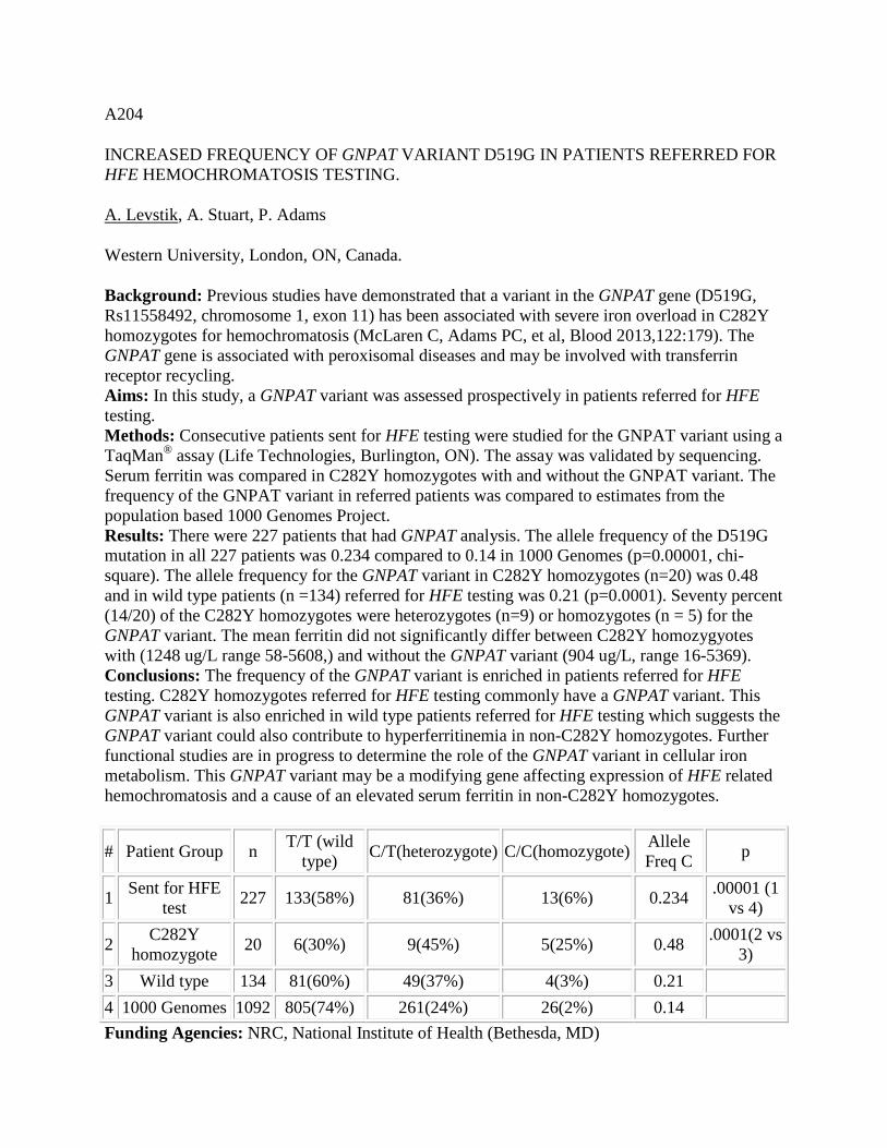
A204
INCREASED FREQUENCY OF GNPAT VARIANT D519G IN PATIENTS REFERRED FOR HFE HEMOCHROMATOSIS TESTING.
A. Levstik, A. Stuart, P. Adams
Western University, London, ON, Canada.
Background: Previous studies have demonstrated that a variant in the GNPAT gene (D519G, Rs11558492, chromosome 1, exon 11) has been associated with severe iron overload in C282Y homozygotes for hemochromatosis (McLaren C, Adams PC, et al, Blood 2013,122:179). The GNPAT gene is associated with peroxisomal diseases and may be involved with transferrin receptor recycling. Aims: In this study, a GNPAT variant was assessed prospectively in patients referred for HFE testing. Methods: Consecutive patients sent for HFE testing were studied for the GNPAT variant using a TaqMan® assay (Life Technologies, Burlington, ON). The assay was validated by sequencing. Serum ferritin was compared in C282Y homozygotes with and without the GNPAT variant. The frequency of the GNPAT variant in referred patients was compared to estimates from the population based 1000 Genomes Project. Results: There were 227 patients that had GNPAT analysis. The allele frequency of the D519G mutation in all 227 patients was 0.234 compared to 0.14 in 1000 Genomes (p=0.00001, chi-square). The allele frequency for the GNPAT variant in C282Y homozygotes (n=20) was 0.48 and in wild type patients (n =134) referred for HFE testing was 0.21 (p=0.0001). Seventy percent (14/20) of the C282Y homozygotes were heterozygotes (n=9) or homozygotes (n = 5) for the GNPAT variant. The mean ferritin did not significantly differ between C282Y homozygyotes with (1248 ug/L range 58-5608,) and without the GNPAT variant (904 ug/L, range 16-5369). Conclusions: The frequency of the GNPAT variant is enriched in patients referred for HFE testing. C282Y homozygotes referred for HFE testing commonly have a GNPAT variant. This GNPAT variant is also enriched in wild type patients referred for HFE testing which suggests the GNPAT variant could also contribute to hyperferritinemia in non-C282Y homozygotes. Further functional studies are in progress to determine the role of the GNPAT variant in cellular iron metabolism. This GNPAT variant may be a modifying gene affecting expression of HFE related hemochromatosis and a cause of an elevated serum ferritin in non-C282Y homozygotes.
# Patient Group n T/T (wild type) C/T(heterozygote) C/C(homozygote) Allele
Freq C p
1 Sent for HFE test 227 133(58%) 81(36%) 13(6%) 0.234 .00001 (1
vs 4)
2 C282Y homozygote 20 6(30%) 9(45%) 5(25%) 0.48 .0001(2 vs
3) 3 Wild type 134 81(60%) 49(37%) 4(3%) 0.21 4 1000 Genomes 1092 805(74%) 261(24%) 26(2%) 0.14 Funding Agencies: NRC, National Institute of Health (Bethesda, MD)

A205
RELATIONSHIP OF BETARETROVIRAL INFECTION WITH DIFFERENTIAL EXPRESSION OF METABOLIC ENZYMES IN PBC
F. Wysokinski, D. Sharon, G. Zhang, A. Mason
University of Alberta, Edmonton, AB, Canada.
Background: Primary Biliary Cirrhosis (PBC) is an autoimmune liver disease that predominantly affects middle-aged women. Immune damage to biliary epithelial cells (BEC) may lead to cirrhosis and the only present therapy is ursodeoxycholic acid that aids in removal of toxic bile from the liver. It has also been shown that PBC patients' BEC show abnormal mitochondrial morphology, increased aerobic glycolysis and increased oxidative phosphorylation in BEC. Proteomic studies done by the Mason lab showed increased expression of metabolic enzymes. Little is known what causes these metabolic derangements. The Mason lab has characterized a Human Betaretrovirus (HBRV), which shares 95% sequence homology with MMTV. We hypothesize that HBRV/MMTV infection is involved in the changes in metabolic enzyme expression seen in proteomic studies which may lead to aberrant cellular respiration. Aims: Our aims are to characterize which metabolic enzymes' expression are affected by HBRV/MMTV infection of BEC and how infection affects cellular respiration. Methods: 1.Western Blot (WB) of HEK293t MMTV, PBC BEC, and Immortalized BEC infected with HBRV/MMTV. Various mitochondrial/metabolic antibodies (abs) will be used looking for changes in expression of key enzymes associated with mitochondrial respiration relative to their respective controls. 2.Immunofluorescence (IF) of PBC BEC with mitochondrial/metabolic enzyme abs, AMA, and capsid antibody will be used looking for differences in localization/expression of enzymes and if these differences can be linked with viral expression. 3.Measure different aspects of cellular respiration in HEK293t MMTV, PBC BEC, and Immortalized BEC relative to their respective controls using commercial kits. Results: Screening of mitochondrial and metabolic enzyme expression was done with WB based on data from previous proteomic studies. Lysates from Hek293t cells infected with MMTV showed an unexpected lower band, approximately 25kDa in size, reactive to anti-β-enolase antibody in Hek293t MMTV lysates but was not seen in Hek293t control lysates. Conclusions: These findings are interesting as enolase has been shown to be cleaved to a 25 kDa band through induction of the caspase-1 pathway in response to infection. Enolase is also known to localize to the plasma membrane and anti-enolase autoimmune antibodies have been found in patient serum from a number of different autoimmune disorders, including PBC. This data suggests that the enolase family could be another potential player in the development of PBC and other autoimmune disorders.
Funding Agencies: CIHR

A206
DEPRESSION IN CIRRHOSIS-A SERIOUS AND UNDER-RECOGNIZED CLINICAL PROBLEM
L. Thomas2, T. Shaikh2, L. Zenith2, N. Mitchell4, M. Ma2, J. Abraldes2, M. Carbonneau2, N. Kneteman1, R. Bailey5, K. Burak3, S. Jayakumar3, P. Tandon2
1. Surgery, Univ of Alberta, Edmonton, AB, Canada; 2. Cirrhosis Care Clinic, Univ of Alberta, Edmonton, AB, Canada; 3. University of Calgary, Calgary, AB, Canada; 4. Psychiatry, Univ of Alberta, Edmonton, AB, Canada; 5. Royal Alexandra Hospital, Edmonton, AB, Canada.
Background: In many chronic disease states, depression can have a negative impact on clinical outcomes. In patients with cirrhosis, depression is not routinely screened for. The prevalence and impact on clinical outcomes in this population therefore remains unclear. Aims: In a prospective cohort of patients with cirrhosis, we aimed to evaluate i)the prevalence of depression by different screening tools, ii)predictors of depression and iii)the impact of depression on clinical outcomes. Methods: Data was gathered prospectively on cirrhotic patients seen in liver clinics in Edmonton & Calgary, the majority recruited during work-up for liver transplant. We examined depression using the EQ-5D, the Hospital Anxiety and Depression Scale (HADS) and the gold-standard MINI neuropsychiatric interview (MINI). Associations between depression (dx by MINI) and hospitalization within the following six months were made using logistic regression. Results: Of the 296 included patients, 66% were male with a mean age of 57 years (SD 9.3), MELD score of 12 (4.9) and Child Pugh score of 7.5 (2.1). The top three etiologies of liver disease were Hepatitis C (34%), Alcohol (32%) and NAFLD/cryptogenic (17%). Seventeen percent of patients had HCC. Using the HADS, 12% of patients scored ≥11 (definite case) and 32% scored ≥8 (borderline case). By the EQ-5D, "39% reported "some problems" with anxiety/depression and 4% reported "extreme problems". Using the MINI neuropsychiatric interview, 26% of patients met diagnostic criteria for a depressive disorder (major depression or dysthymia). Of these patients, only 36% had been recognized and were on antidepressants. A positive score on the MINI was independently predicted by a higher Child Pugh score [odds ratio (OR) of 1.2 (95% CI 1.04 to 1.37), p=0.001] and younger age [OR 0.94 (95% CI 0.91 to 0.97), p=0.001]. Being in a marriage/common-law relationship [OR 0.33 (0.18 to 0.58), p=0.001] was protective. MELD, HCV and active alcohol/smoking history did not reach statistical significance. The MELD [OR 1.13 (1.02 to 1.26), p=0.02] and scoring positive on the MINI [OR 3.3 (1.2 to 9.0), p=0.02] were independent predictors of admission to hospital within the 6 months after assessment (subset of 79 patients). Conclusions: Depression is a common and often under-recognized condition in patients with cirrhosis. The prevalence of depression varies depending upon the tool that is utilized. As depression is independently associated with increased hospital admissions, future studies should evaluate whether treatment may have an impact on hospitalization rates and other relevant clinical outcomes.
Funding Agencies: None

A207
A GLOBAL PERSPECTIVE OF DIFFERING APPROACHES TO PRIORITISATION OF PATIENTS WITH PRIMARY SCLEROSING CHOLANGITIS FOR LIVER TRANSPLANTATION
S. Neong1, K. Burak1, T. Karlsen2, B. Eksteen1
1. Univ of Calgary, Calgary, AB, Canada; 2. On behalf of IPSCSG, Oslo, Norway.
Background: Liver transplantation is an established treatment modality for patients with end-stage liver disease secondary to primary sclerosing cholangitis (PSC). Prioritisation for liver transplantation and organ allocation in most centres are based on predicted mortality scores such as the MELD or UKELD. Unfortunately these scoring systems are not specific to PSC and often underestimate the severity of PSC. As a result, PSC patients are often awarded exception points to fully capture their illness severity. No consensus exists as how this should be done on an equitable and standardised manner. To gauge the current practices globally, we performed a survey amongst the members of the international PSC study group (IPSCSG) across 12 countries. Aims: We aim to identify and explore current practices for prioritising PSC patients for liver transplantation across Europe and North America. Better understanding of this is fundamental in helping us develop a structured approach in managing PSC patients and timing to listing and prioritization for transplantation. Methods: We created a 14-part questionnaire focusing on key issues in managing PSC patients on transplant waiting lists. The questionnaire was disseminated to IPSCSG members via survey monkey for a 2-month period from 30 April to 30 June 2014 inclusive. Responses were analysed through survey monkey software. Results: We received a total of 31 responses from transplant centres across 12 countries. The mean number of liver transplantations performed annually in each centre is 76 (range 0-200). MELD score is the most common scoring system used to prioritise liver allografts (78%) followed by Child-Turcotte-Pugh score (15%). The cohort is divided in their opinion on the need for a minimum score for liver transplantation listing. The minimum score required to be listed in each transplant centre is MELD 10 (range 10-30) and UKELD 49 (range 49-53). Average score at time of transplantation are MELD 25 and UKELD 53. Almost all centres (96%) allow exception points for diseases not accurately captured by conventional scoring systems but this is highly variable and dependent on disease-specific parameters such as hepatocellular carcinoma, recurrent cholangitis, cholangiocarcinoma, decompensation with ascites and hepatic encephalopathy, refractory pruritus, prolonged hospitalisation with hepatic hydrothorax and sarcopenia. The local transplant team determine the exception points allocation in half of these centres. This is repeated on a case-to-case basis in 60% of the centres. Conclusions: Great variability exists globally in managing PSC patients awaiting liver transplantation and highlights the need of a validated PSC specific transplant allocation system.
Funding Agencies: None
A208
VIROLOGICAL FOOTPRINT OF T-CELL RESPONSES IN PATIENTS WITH PRIMARY BILIARY CIRRHOSIS
M. Rahbari, D. Sharon, A. Landi, M. Houghton, A. Mason
University of Alberta, Edmonton, AB, Canada.
Background: Primary biliary cirrhosis (PBC) is known as a cholestatic liver disease of autoimmune origin, characterized by inflammatory destruction of the small intrahepatic bile ducts, and fibrosis which can progress to cirrhosis and subsequent liver failure. PBC is a disease of unknown etiology. Both genetic and environmental factors impact on the development of PBC. Our laboratory has characterized a retrovirus in PBC patients. This agent is referred to as the human betaretrovirus (HBRV) because of the genetic, morphological and antigenic relatedness to the mouse mammary tumor virus (MMTV). Aims: Our objective is to characterize the relationship of HBRV infection with PBC and studying the linkage of the human HBRV with PBC. We hypothesize that patients with PBC make cellular immune responses to HBRV. Methods: Peripheral blood mononuclear cells (PBMCs) were purified from whole blood of 24 patients with PBC, 21 liver disease patients, and 5 normal volunteers. T-cell responses in PBMC were assessed for reactivity to HBRV peptides. PBMCs were stimulated for 6 hours with pools of overlapping 20-mer peptides from HBRV Envelope and Gag as well as human cytomegalovirus Gag peptides and PHA-Ionomycin mitogens, which were used as a positive control. The magnitude of ex vivo responses to stimulation were evaluated by measuring the number of T-cells secreting various cytokines including IL-6, IL-4, IL-10, TNF-α and IFN-γ assessed by flow cytometry analysis to document T-cell memory responses. Results: Out of 24 patients with PBC, 10 patients showed memory CD8+ T-cell responses to HBRV Gag peptides, whereas 1 non-PBC and 1 healthy control samples showed reactivity to HBRV Gag peptides. The magnitude of the memory CD8+ T-cell responses for PBC patients had a range of responses from 0.2% to 4.97% to HBRV Gag. Out of the 10 PBC patients that showed T-cell reactivity against HBRV Gag, 1 patient also showed memory CD8+ T-cell response against HBRV Env. We have also applied the epitope mapping approaches using HBRV-derived Gag peptides to identify the immunodominant peptides. Linear epitopes with high immunoreactivity were mapped at matrix (P10) and capsid (P27) region of HBRV. Conclusions: In our study we found that 41% of patients showed reactivity to HBRV Gag pool of peptides. Epitope mapping approach identified 4 immunodominant peptides mapped at matrix and capsid regions of HBRV. The identified immunoreactive peptides will be used to design tetramers. Using peptide MHC tetramers will increase the sensitivity of the assay to detect the frequency of CD8+ HBRV-specific T cells and compare the extent of immune response in both PBC and control groups.
Funding Agencies: CIHR

A209
EFFICACY OF PROBIOTIC THERAPY IN PREVENTING OVERT HEPATIC ENCEPHALOPATHY: A REVIEW
A. Sandhu3, B. Aljudaibi2, K. Qumosani1, J. Burton5, G. Reid5, A. Rahman4
1. LHSC, London, ON, Canada; 2. London Health Science Centre, London, ON, Canada; 3. London Health Sciences Centre, London, ON, Canada; 4. Western University, London, ON, Canada; 5. Lawson Health Research Institute, London, ON, Canada.
Background: Hepatic Encephalopathy (HE) is a common complication experienced by patients with liver cirrhosis and is characterized by several neuropsychiatric symptoms ranging from mild impairments to coma. Overt HE (OHE) is associated with poor outcomes, including increased mortality. The cost of HE for the healthcare system is exorbitant. While therapies exist to manage OHE, there is an enlarging body of evidence aimed at identifying and treating subclinical or minimal HE (MHE), and thus preventing OHE. There is also a growing interest in the use of probiotics at preventing OHE, with a physiological basis to reduce the absorption of ammonia. Aims: To review the available literature assessing the use of probiotics in treating MHE and preventing OHE. Methods: An electronic literature search of databases including The Cochrane CENTRAL Register for Controlled Trials, EMBASE, Scopus, EBSCO host, PUBMED/MEDLINE and OVID was performed. We included only randomized controlled trials (RCTs), meta-analyses and COCHRANE reviews, between 2004 and 2014, which evaluated the use of probiotics in MHE in adult patients with liver cirrhosis. Results: Nine publications were identified: seven randomized controlled trials, one meta-analysis and one COCHRANE review. Of the RCTs, all evaluated probiotics compared against placebo, while some also included comparisons with lactulose, L-ornithine L-aspartate and rifaximin. Sample sizes ranged from 25 to 160 and patients were followed for one to three months at which point the MHE was reassessed. Among these RCTs, there was considerable heterogeneity in the methods used to diagnose MHE and track the efficacy with treatment. All RCTs demonstrated significant improvement in MHE. The single meta-analysis, which included 9 RCTs, concluded significant improvement in MHE with probiotics. Compliance was excellent and there were no demonstrated side effects. A COCHRANE review in 2011 concluded there was no conclusive clinical efficacy of probiotics, citing that the body of evidence had a high risk of bias and random errors and inconsistent methods of assessing for efficacy. Two large RCTs have been released since, which have demonstrated significant improvement in MHE with probiotics using larger sample sizes and more consistent methodology. Conclusions: Probiotics have consistently demonstrated to be efficacious for MHE amongst RCTs, with excellent compliance and no documented adverse events. Further studies are required to confirm reproducibility, especially with respect to the methodology of diagnosis and assessing improvement in MHE.
Funding Agencies: None

A210
BILE-LIGATED RATS ARE SUSCEPTIBLE TO HYPOTENSION-INDUCED NEURONAL CELL LOSS: IMPLICATIONS FOR PERSISTING NEUROLOGICAL COMPLICATIONS FOLLOWING LIVER TRANSPLANTATION
M. Clément1, C. Bosoi4, M. Tremblay1, C. Bemeur2, C. Rose3
1. CRCHUM, Montréal, QC, Canada; 2. CRCHUM, Montreal, QC, Canada; 3. Hôpital St-Luc, CRCHUM, Université de Montréal, Montreal, QC, Canada; 4. Université de Montréal, Montréal, QC, Canada.
Background: Hepatic encephalopathy (HE) is a major neuropsychiatric complication caused by liver disease characterized by cognitive and motor dysfunction. The only curative treatment to date remains liver transplantation (LT). Historically, HE has always been considered to be a reversible metabolic disorder and has therefore been expected to completely resolve following LT. However, even following the implantation of a new liver, persisting neurological complications remain a common problem affecting as many as 47% (8-47%) of liver transplant recipients. LT is a major surgical procedure accompanied by intraoperative stress and confounding factors, including blood loss (hypovolumia) and hypotension. Aims: We hypothesize, in the setting of MHE, that the compromised brain becomes predisposed to what would normally be an innocuous hypotensive insult, resulting in cell injury and death. Methods: Using 6-week bile-duct ligated rats and respective controls, blood is withdrawn from the femoral artery (inducing hypovolemia) until an arterial pressure of 30 mmHg (hypotension) and maintained for 150 minutes. Upon sacrifice, brains are perfused and extracted for western blotting and immunohistochemistry. Results: Both BDL rats and SHAM-operated controls without hypotension do not display any neuronal loss. However, BDL rats following hypotension demonstrated a significant decrease in neuronal cell count in the frontal cortex using NeuN+DAPI and Cresyl Violet compared to hypotensive SHAM-operated controls. In addition, neuronal loss was associated with an increased in cellular stress protein, hsp32 and caspase-3, suggesting apoptotic cell death. Conclusions: These findings suggest that patients with HE are more susceptible to hypotension-induced neuronal cell loss and this may explain why transplanted patients are experiencing persisting neurological complications. Aside from cirrhotic patients having a stroke, these results also suggest a patient with HE (even MHE) with a "frail brain", fare worse during transplantation leading to poor neurological outcome. This implies MHE should not be ignored and therefore treated pre-LT.
Funding Agencies: CIHR, Canadian Liver Foundation

A211
DOES IMMUNOCHEMICAL STAINING FOR IGG4 IN THE EXPLANTED LIVERS PREDICTS RECURRENCE OF PRIMARY SCLEROSING CHOLANGITIS (PSC)?
M. Alghanem1, B. Al-Judaibi2, P. Marotta3, H. Chaturika3, S. Chakrabarti3
1. university of western ontario, London, ON, Canada; 2. London Health Science Centre, London, ON, Canada; 3. London heatlh sciences Center, University of Western Ontario, London, ON, Canada.
Background: The outcome of Primary Sclerosing Cholangitis (PSC) after liver transplantation can be affected by recurrent PSC (rPSC) and subsequent graft failure. IgG4 related sclerosing disease is a recent entity that has a similar morphological appearance to PSC, making the distinction difficult. However, IgG4 related sclerosing cholangitis has an excellent prognosis since, it is steroid sensitive, but the impact of IgG4 on rPSC after liver transplant is still unknown. Aims: To determine the association between IgG4 immunochemical staining in liver explants and recurrence of primary sclerosing cholangitis post-liver transpantation Methods: All adult patients who underwent liver transplantation for PSC ,from 1990 to 2014, were identified. Clinical inforamtion and immunochemical staining were perforemd among the patietns with PSC recurrence post-liver transplantation. IgG4 immunochemical staining was perforemed on the liver and porta-hepatis region. IgG4 level was scored as: none (<5 cells/HPF), mild (5 to 10 cells/HPF), moderate (11 to 29 cells/HPF), and significant (>30 cells/HPF). Immunochemical staining is considered to be positive if the score was moderate-significant. Results: 116 patients underwent liver transplantation for PSC. 17 patients (14%) developed rPSC post liver transplantation. However, the immunochemical staining results were only available in 11/17. Out of these patients who were stained for IgG4, 9/11 were males (81%) and 10/11 patients (90%) had an IBD (8 patients with Ulcerative colitis and 2 patients with Crohns disease). 6/11 patients (54.5%) had Roux-en-Y anastmosis.The Median time for follow-up was calculated as 172 months. Median time to development of rPSC in this population was calculated as 53 months. 6/11 patients (54.5%) developed graft failure secondary to recurrence of PSC with median time of 84 months .2 patient had positive immunochemical staining (1 patient with significant level in both liver and porta-hepatis and 1 patient with moderate score only in the liver). Conclusions: Our preliminary results showed only two patient had a positive IgG4 level in the explanted liver. However, all the explanted PSC livers will be stained for IgG4 regardless of the graft status post-liver transplantation. We hypothesize that PSC patients with positive IgG4 staining have a higher likelihood of PSC recurrence compared to patients with negative IgG4 staining.
Funding Agencies: None

Clinical Practice
A212
MAGNETIC IMAGING-ASSISTED COLONOSCOPY VS CONVENTIONAL COLONOSCOPY: A RANDOMIZED CONTROLLED TRIAL.
C. Teshima2, S. Zepeda-Gomez2, S. Alshankiti1, G. Sandha3
1. Provincial Health Services Authority, Vancouver, BC, Canada; 2. University of Alberta, Edmonton, AB, Canada; 3. University of Alberta, Edmonton, AB, Canada.
NOT PUBLISHED AT AUTHOR’S REQUEST
Funding Agencies: None

A213
EFFECT OF THE FECAL IMMUNOCHEMICAL TEST ON REFERRAL RATES IN A GASTROENTEROLOGY CLINIC: A RETROSPECTIVE ANALYSIS OF THE REFERRAL PATTERNS OF ST PAUL'S HOSPITAL DIVISION OF GASTROENTEROLOGY
O. Takach1, J. Yonge1, A. Round2, C. Galorport1, A. Sobkowicz2, E. Nap-Hill2, R. Enns1
1. St. Paul's Hospital, Vancouver, BC, Canada; 2. University of British Columbia, Burnaby, BC, Canada.
Background: We have recently shown that wait-times for endoscopy by Canadian gastroenterologists were considerably longer than the current guidelines recommend. A more sensitive and specific fecal blood test, Fecal Immunochemical Test (FIT), was implemented in 2012. A single sample of stool is collected for FIT (abnormal defined as greater than 50 mcg/g). In regular colon cancer surveillance programs this increase in sensitivity and specificity has been postulated to be one of the causes for the increasing burden on Canadian gastroenterologists. Aims: The purpose of this study was to evaluate changes in referral rates when FIT was implemented in 2012 at the Division of Gastroenterology at St. Paul's Hospital. Methods: This observational study was a retrospective chart review of patients seen by the gastroenterologists of St. Paul's Hospital, Vancouver. Patients' referral patterns were analyzed and compared between April to July of 2011 and 2013. Data was extracted from a comprehensive EMR used by all gastroenterologists; data collected included demographics and reason for referral. The patients studied in this group do not include Colon Screening Program, which would be in addition to the above referrals but managed in a different pathway. Results: 11,619 subjects referred to our center were analyzed for this study. The overall referrals to the GI clinic during April - July 2011 to 2013 increased from 5388 to 6231 (15.6%). The overall referral rate of referral due to a positive fecal test also increased from 6.23% in 2011 to 11.5% in 2013. Conclusions: In this study, we show that the more specific and sensitive FIT has increased the referral rate of patients by 84.5% that would require screening colonoscopy. This increased referral rate may be anticipated to be similar in many regions implementing this testing and should be considered when planning resource utilization particularly for endoscopic services.
Funding Agencies: None
A214
EARLY EXPERIENCE WITH THE FECAL IMMUNOCHEMICAL TEST IN A LARGE CENTRAL ACCESS MODEL OF ACUTE GASTROENTEROLOGY CARE
K. Rioux, G. Heather, B. Kathol, M. Swain, S. Heitman, R. Hilsden, K. Novak
Department of Medicine, Division of Gastroenterology, University of Calgary, Calgary, AB, Canada.
Background: Alberta recently implemented the fecal immunochemical test (FIT) for colorectal cancer screening among asymptomatic average risk individuals aged 50-74. Despite criteria for appropriate use, FIT is also being used to investigate GI symptoms and for screening outside the target age range. In Calgary, FIT+ patients who do not qualify for colonoscopy at the Forzani & MacPhail Colon Cancer Screening Centre (CCSC) due to age, symptoms, or high medical comorbidity are referred to GI Central Access and Triage (CAT). Aims: The objective of this study was to assess patient characteristics and endoscopic findings among FIT+ patients referred through GI CAT. Methods: FIT+ referrals received by GI CAT from January 1 to April 30, 2014 were identified in our Cerner Millennium® referral database. Anonymized patient records were abstracted to determine patient demographics and clinical characteristics. Colonoscopy findings were determined from our Pentax endoPRO® and pathology databases. Results: 582 FIT+ patient referrals were received during the four-month study period. The mean age of the cohort was 62.5 years (range 21-90). 197 patients (33.8%) were outside of the recommended screening age range. 131 referrals (22.5%) were redirected to CAT from CCSC due to ineligibility; 78 screen appropriate referrals (13.4%) were redirected from CAT to CCSC. Only 5.4% of FIT+ referrals to CAT were asymptomatic; 25.2% had overt signs of GI bleeding, 15.8% anemia, 15.5% abdominal pain, 12.6% abnormal/altered bowel habit, 5.8% weight loss, and 5.4% medical comorbidity. 59% of patients were deemed appropriate for ‘direct to procedure' colonoscopy, whereas 41% required clinic consultation to first determine appropriateness and medical fitness for colonoscopy. Complete records of colonoscopy and histopathology findings were available for 358/504 (71%) of patients. 59.2% of FIT+ patients had any type of polyp, 34.6% had advanced neoplasia, and 5% had colorectal cancer. 48.6% of patients had other findings (e.g. hemorrhoids, diverticulosis, etc.), while only 13.4% of patients had entirely normal colonoscopies. Conclusions: Unrestricted access to FIT in Alberta has led to significant use outside the target age range and for diagnostic purposes. Triage algorithms for FIT+ positive patients lead to urgent clinic consultation and/or urgent colonoscopy. Although colonoscopy in this cohort of FIT+ patients yields important and relevant pathology, use of this screening test outside of intended purpose has led to increased demands on resources in our central access model of acute GI care.
Funding Agencies: None

A215
2L SPLIT-DOSE PEG3350 ELECTROLYTE SOLUTION PLUS BISACODYL COMBINATION VERSUS 2L SPLIT-DOSE PEG3350 ELECTROLYTE SOLUTION PLUS ASCORBIC ACID FOR OUTPATIENT COLONOSCOPY: PROSPECTIVE RANDOMIZED CONTROLLED STUDY
O. Takach1, C. Galorport1, A. Round2, A. Sobkowicz2, G. Ou2, M. Brahmania2, M. Suzuki1, H. Ko1, J. Telford2, B. Bressler2, R. Enns2
1. St. Paul's Hospital, Vancouver, BC, Canada; 2. University of British Columbia, Burnaby, BC, Canada.
Aims: Adenoma detection rates of colonoscopy depends on the quality of the colonic preparation. Our previous study has demonstrated that 2L split-dose PEG3350 electrolyte solution (PEG3350e) with 15mg bisacodyl is as effective for outpatient colonoscopy preparation as large volume (4L) PEG3350e. In this study, the investigators aimed to evaluate the effectiveness of ascorbic acid versus bisacodyl as the adjunctive laxative for 2L split-dose PEG3350e bowel preparation regimen. Methods: A randomized, prospective, endoscopist-blinded trial was initiated at St. Paul's Hospital in Vancouver, BC. Inclusion criteria: Age ≥ 19 years and planned outpatient colonoscopy. Exclusion criteria: chronic constipation, suspected bowel obstruction, severe IBD, and history of colonic resection. The two bowel preparations of interest include 2L split-dose PEG3350e plus 15 mg bisacodyl (Group A) and 2L split-dose PEG3350e plus ascorbic acid (Group B). Four endoscopists rated the quality of the preparation using the validated Boston Bowel Preparation Scale (BBPS) with a score of ≥ 8 being considered excellent. In addition, patient tolerability and satisfaction to either product was determined based on a ten-question survey. Interim analysis was performed after 6 months of recruitment. Results: As of October 2014, 231 subjects have been recruited. 107 (46.7%) are male and mean age is 57 years (SD = 12.03, range 23 to 82). 108 were assigned to Group A (bisacodyl) and 123 to Group B (ascorbic acid). A Fisher's exact test showed no significant difference in the proportion of subjects achieving an excellent BBPS score ≥ 8 between the two groups (Group A = 53/108, Group B = 71/123, p = 0.234). In addition, no significant difference was found comparing the taste ratings on a scale of 0 to 10 of the two products (Group A mean = 5.0, SD = 2.4; Group B mean = 5.0, SD = 2.7; p = 1.0. Finally, same proportions of participants found the bowel preparation easy or acceptable (93.5% vs. 86.9%, p = 0.12), and were willing to repeat the same preparation in the future (86.9% vs. 86.1%, p =0.84). Conclusions: Interim analysis detects no significant difference between 2L split-dose PEG3350e plus 15 mg bisacodyl and 2L split-dose PEG3350e plus ascorbic acid in patient preference of product taste, tolerability, or colon cleanliness.
Funding Agencies: None

A216
A PROVIDER QUALITY REPORT FOR AN OHP: SETTING THE STANDARD FOR ENDOSCOPY QUALITY REPORTING.
A. Gould, S. Sokol, J. Gould, L. Strasser, M. Gould Vaughan Endoscopy Clinic, Toronto, ON, Canada.
Background: The development of a provider report card for endoscopy has become a high priority in the effort to improve endoscopic outcomes. Measuring endoscopist quality is important to improve patient outcomes by reducing missed cancers and interval cancers. Hopefully this study will demonstrate which variables are possible to measure in an out patient facility that can be used to evaluate the performance of the endoscopists in each facility. Aims: To demonstrate the feasibility of measuring key quality variables in an outpatient endoscopy centre which could be used to develop a provider quality report. Methods: Vaughan endoscopy centre is an urban out of hospital centre with 13 hosptial-based gastroenterologists. The annual volumes at the centre exceed 3000 cases per year. 3205records from all patients who received colonoscopies, regardless of the indication for the endoscopy, from January 2012 to March 2013 were analyzed. The following variables were recorded: the presence, location, size, and polyps number and histology, age, sex, colonoscopist, indication for endoscopy quality of preparation, signs of bowel perforation, and cecal intubation. Results: From the observed data of 3205 patients, who had colonoscopies in the years 2012/13, 52.7% were male and 47.3% female. 77% had a prep quality rating of "Good", 22% had prep quality rating of "Fair", and only 1% had a prep quality rating of "Poor"/"Inadequate". There was an overall polyp detection rate of 43.4%. Of all polyps found, 61.4% were adenomas. There was an overall adenoma detection rate of 26.42%. For all males there was an adenoma detection rate of 30.67% and 19.89% adenoma detection rate for females. The average cecal intubation rate was 98%. The distribution of cecal intubation rates among the endoscopists ranged from a low of 89.4% to a high of 100%. Conclusions: The currently recognized quality data indicators for a quality provider report in endoscopy, cecal intubation rates, adenoma detection rates and prep quality, are easily measured in a outpatient facility and should be part of a standard quality report for each facility. This report should be generalizable to all endoscopy facilities.
Figure 1. Adenoma detection rate by Doctor Funding Agencies: None

A217
THIOPURINE METHYLTRANSFERASE TESTING AND THIOPURINE USE IN INFLAMMATORY BOWEL DISEASE PATIENTS AMONGST CANADIAN GASTROENTEROLOGISTS: A QUALITY ASSURANCE INITIATIVE
S. Singh, R. Panaccione, K. Novak
Division of Gastroenterology, University of Calgary, Calgary, AB, Canada.
Background: Inflammatory bowel disease (IBD) is a chronic disease that often requires immunosuppressive and/or immunomodulatory therapy, in particular thiopurines such as Azathioprine (AZA) and 6-Mercaptopurine (6-MP). Multiple national and international organizations support routine evaluation of thiopurine methyltransferase testing (TPMT) for enzyme levels in order to identify at-risk patients for thiopurine-induced leukopenia. Aims: We administered an electronic survey to Canadian Gastroenterologists to evaluate practice patterns and guideline adherence for TPMT testing in thiopurine use. Methods: We designed a 21-question electronic survey that was provided to the Canadian Association of Gastroenterology (CAG). The survey was distributed to all CAG members on July 14, 2014 via e-mail and subsequently posted on the CAG website. The survey was created using online software from www.surveymonkey.com and results were analyzed using online software. Results: To date, there have been 45 respondents. Majority (72.7%) of the gastroenterologists were in the 30-49 age range, 72.7% were male and 61.4% are practicing in academic institutions. The most commonly prescribed thiopurine is Azathioprine (93.3%) and the most influential prescribing factor is personal familiarity (64.4%). Only 60.0% of gastroenterologists routinely order TPMT testing and 17.8% of gastroenterologists were unaware TPMT testing was available in Canada. Amongst those that are aware, 60.0% are unaware which laboratories offer TPMT testing. Amongst Canadian Gastroenterologists, 24.4% order TPMT testing on all newly diagnosed IBD patients while 31.1% order it on IBD patients just prior to starting therapy with a thiopurine. The most common reason gastroenterologists do not order TPMT testing is because they believe testing is not available locally (22.2%). Additionally, Canadian gastroenterologists believe TPMT testing costs anywhere from $11 to over $200 and believe result take anywhere from 1 day to over 4 weeks to attain. Conclusions: Considerable variability exists in practice patterns with TPMT testing in thiopurine use. Several misconceptions amongst TPMT testing exist which is concerning given that previous research shows increased patient safety and reduced adverse drug reactions in those who have TPMT testing done pre-treatment. We believe that this data can aid in creating an educational initiative aimed at clarifying common misconceptions and provide greater adherence to current guidelines with a goal to increase TPMT testing and subsequently, patient safety.
Funding Agencies: None

A218
CLINICAL RESPONSE TO EVIDENCE SUPPORTING RESTRICTIVE TRANSFUSION STRATEGIES IN ACUTE UPPER GASTROINTESTINAL BLEED
N. Malhotra2, M. Beaton1
1. London Health Sciences Centre, London, ON, Canada; 2. Western University, London, ON, Canada.
Background: Current guidelines for non-variceal upper gastrointestinal bleeding (UGIB) recommend definitive management with endoscopic intervention within twenty-four hours of hospital admission. Supportive care upon presentation is of utmost importance. However, the precise role of blood transfusion in this patient population is not entirely clear. While studies have suggested restrictive transfusion strategies are preferred, most of these excluded patients with acute gastrointestinal bleeding. Recently, the use of blood products in this patient population has been studied; with the findings that a restrictive transfusion strategy showed improved mortality and rate of re-bleeding. Aims: Our goal was to assess whether this transfusion strategy was adopted and resulted in a change in clinical practice in an academic teaching hospital. Methods: A retrospective chart review was conducted looking at transfusion thresholds two months before and four months after the publication of new evidence. Inclusion criteria included adults admitted with both a primary and secondary diagnosis of an UGIB Those transferred from another hospital center along with cases during the month of article publication were excluded. Primary end points included hemoglobin levels triggering transfusion, time to endoscopy and Rockall score. Results: 103 patients were deemed to have an UGIB eligible for review. The majority of patients were treated with a restrictive (n=32) versus liberal (n=3) transfusion strategy, with no significant decrease after new evidence was published (p = 1.0). Those treated with a liberal transfusion strategy had an additional diagnosis requiring use of blood products and included ongoing acute coronary syndrome (ACS) or active gastric malignancy. The mean age of patients in this cohort was 71.7 years with a Rockall score of 3 and average transfusion requirement of 2 units of packed red blood cells. The mean time to endoscopy was 15.8 hours (range: 1-79). Of those with cirrhotic liver disease (n=7), the most common cause of upper GI bleed was portal hypertensive gastropathy (PHG) and transfusion requirement was no different to the remaining cohort (p= 1.0). The mean time to endoscopy was 10.4 hours. Conclusions: The existing practice at our academic teaching hospital was an adherence to a restrictive transfusion strategy, with no significant change transfusion of blood products after new evidence was published. In addition, there was no difference in transfusion requirement for those patients with cirrhosis and a diagnosis of an UGIB. Overall, time to first endoscopy was within the recommended guidelines.
Funding Agencies: None

A219
DIAGNOSIS AND MANAGEMENT OF BARRETT'S ESOPHAGUS; A RETROSPECTIVE STUDY COMPARING THE ENDOSCOPIC ASSESSMENT OF EARLY ESOPHAGEAL LESIONS IN THE COMMUNITY VERSUS A SPECIALIZED CENTER
E. Rayner-Hartley, R. Enns, O. Takach, C. Galorport
University of British Columbia, Vancouver, BC, Canada.
Background: Specialized endoscopic evaluation for patients with Barrett's Esophagus (BE) is well supported, however no studies have shown that centers with expertise provide altered care for BE with high-grade dysplasia or early adenocarcinoma. Many of these patients can be managed in a single endoscopic session with endoscopic mucosal resection. Aims: In this study, the investigators aimed to compare the endoscopic assessment of esophageal lesions in a community practice versus a specialized BE center and how these endoscopic impressions correlate with histology. Methods: A retrospective analysis of referrals from the community to our center for evaluation of BE between 01/07 and 02/14 was performed. Subjects included patients referred for BE and dysplasia who were subsequently re-evaluated by endoscopy. The pathology and endoscopy reports from the community and our center were reviewed. Inclusion criteria: ≥ 19 years old, pathologic diagnosis of BE or dysplasia in the community. Exclusion criteria: incomplete pathology data or incomplete endoscopy reports from referring physicians. Results: 77 total patients were reviewed. This included 14.3% with low-grade dysplasia, 44.2% with high-grade dysplasia and 7.8% with adenocarcinoma. The staging of 28.9% of patients referred from the community was changed from the initial pathological diagnosis with 18.4% of these patients being upstaged. Using the Fischer Exact test, we showed that in our endoscopic impressions correlated significantly with pathology results (p<0.0001). Conclusions: The staging of esophageal pathology (based solely on endoscopic visualization) was changed in a significant proportion of patients referred to a center that manages these cases regularly. Our endoscopic impressions correlated well with pathology results. This study supports the early referral of patients with BE to a center managing them regularly, in order to optimize diagnosis and management.
Funding Agencies: None

A220
FACTORS THAT INFLUENCE THE EFFICIENCY OF AN ENDOSCOPY UNIT: A PATIENT FLOW ANALYSIS
R. Almeida, N. Craig, L. Hookey
Queen's University, Kingston, ON, Canada.
Background: The demand for endoscopic procedures is increasing in the context of limited financial resources. There has been a paradigm shift towards optimizing value in health care delivery that emphasizes efficient use of existing resources. Audits of endoscopy units reveal underutilized resources; yet, there is limited literature on the range of endoscopy unit efficiencies. A sustainable endoscopic practice needs reliable efficiency metrics to improve the quality and value of endoscopic care. Aims: The objective was to assess the efficiency of the endoscopy unit at the Hotel-Dieu Hospital by analyzing patient flow through the unit. The primary aim was to obtain data on process measures such as the durations of the components of endoscopic experience; as well as estimates of the patient-centered measures such as the patient waiting time. Methods: A prospective study from December to March 2014 was conducted at the endoscopy unit at the Hotel Dieu Hospital, Kingston. Time elapsed for all components from patient registration to exit from the endoscopy unit were recorded and mean times were analyzed. Procedures were directly observed in three segments - individual endoscopy room utilization, pre-procedure/recovery room, and overall endoscopy unit room usage. Results: Data were collected for 137 procedures in the endoscopy room, 139 procedures in the pre-procedure room, and 143 procedures for overall room usage. The mean time for patient registration was 39.22 minutes ahead (95% CI: -44.76 to -33.68) of their scheduled starting time. The mean patient waiting time in the pre-procedure room was 71.21 minutes (95% CI: 63.95 - 78.48). The mean time delay from scheduled start time to the actual time of patient transfer into the endoscopy room was 19.07 minutes (95% CI: 13.98 - 24.15). The mean endoscopist start delay from the time of patient entry to that of endoscopist arrival was 7.51 minutes (95% CI: 5.15 - 9.86). The mean duration spent by the patient in the endoscopy room was found to be 31.47 minutes (95% CI: 26.82 - 36.12) for an EGD, 52.93 minutes (95% CI: 48.11 - 57.75) for a colonoscopy, 66.88 minutes (95% CI: 58.64 - 75.12) for a double procedure, and 30.47 minutes (95% CI: 25.51 - 35.43) for a flexible sigmoidoscopy. Conclusions: The patient arrival to the endoscopy unit was ahead of their scheduled registration time. However their entry into the endoscopy suite was much delayed, independent of further delay contributed by endoscopist unavailability. Furthermore, the endoscopy room durations far exceed the allocated times contributing to patient waiting time. This is consistent with the recognition that individual units have unique operational characteristics and that identification of bottlenecks can lead to optimization of resources by targeted quality improvement initiatives.
Funding Agencies: None
A221
EVALUATION OF A NOVEL, ICON-BASED SYMPTOM ASSESSMENT TOOL FOR GASTROENTEROLOGY
L. Lu, D. Armstrong, R. Hunt
McMaster University, Hamilton, ON, Canada.
Background: Existing tools for evaluating gastrointestinal (GI) symptoms use verbal descriptors which are affected by social, cultural and linguistic factors. Furthermore, the perception of GI symptoms varies between individuals. Thus it is often difficult for patients to characterize their symptoms with terms, such as heartburn, nausea, bloating or abdominal cramps, making symptom monitoring imprecise. To address this, we are developing a GI symptom evaluation tool based on pictorial icons, designed from symptom descriptors identified by patient focus groups, which may allow patients and clinicians to better identify, record and monitor GI symptoms over time or in response to treatment. Aims: The aim of this study was to assess the usability and responsiveness of the icon-based symptom evaluation tool. Methods: We evaluated a novel, icon-based symptom evaluation tool (Dhaliwal S et al. Gut 2009;58(SII):A316; Dhaliwal S et al. Gastroenterology 2012;140(5S1):S579) in comparison to a commonly-used, validated tool, the Gastrointestinal Symptom Rating Scale (GSRS), for assessing the severity of short-term GI symptoms triggered by mono- or disaccharides in patients undergoing outpatient breath hydrogen testing (BHT). Participants completed the icon-based tool and the GSRS, hourly, before and during a BHT performed for suspected bacterial overgrowth (glucose) or lactose/fructose intolerance. Results: Of 59 patients enrolled in the study, 8 were excluded for incomplete data. Breath tests were positive in 11 (21.6%) and negative in 40 (78.4%) patients. Increases in GI symptoms during the BHT were detected by the GSRS in 34 (66.7%) patients and by the icon-based tool in 30 (58.8%) patients. For the prediction of a positive BHT, the GSRS had a sensitivity of 81.8% and a specificity of 37.5% whilst the icon-based tool had a sensitivity of 72.7% and a specificity of 45%. The diagnostic odds ratio (dOR) of the icon-based tool was 2.18 (95% CI: 0.43-12.3; p=0.490), the dOR of GSRS was 2.7 (95% CI: 0.44-21.0; p=0.297). Conclusions: The icon-based symptom evaluation tool performs comparably to the validated GSRS with respect to the detection of acute, general GI symptoms triggered by the ingestion of a fermentable monosaccharide or disaccharide and to the prediction of a positive breath hydrogen test. Further development of the icon-based tool will require correlation of specific symptom-related icons with specific dietary and therapeutic interventions, disease mechanisms and sites of symptom generation with the goal of validating its use in clinical and research practice.
Funding Agencies: None
A222
A GASTROENTEROLOGY INNOVATION CHALLENGE: EVALUATION OF TRAINEES' LEARNING OF THE INTRINSIC CANMEDS ROLES
A. Martinez2, M. Raman1, M. Cino2, C. Wong4, C. Mulvale3, C. Walsh2
1. University of Calgary, Calgary, AB, Canada; 2. University of Toronto, Toronto, ON, Canada; 3. Canadian Digestive Health Foundation, Oakville, ON, Canada; 4. University of Alberta, Edmonton, AB, Canada.
Background: The intrinsic CanMEDS roles are often not explicitly taught within postgraduate medical programs, but rather learned opportunistically in the clinical setting. The Canadian Digestive Health Foundation's (CDHF) RISE from the Snake Pit Challenge, a program run at CDDW 2014, aspired to help young professionals discover the impact they can have on digestive health outside their institutions. Participants were tasked with using social media to create a platform to educate and incite change around real-life challenges in the area of gastroenterology (GI). Aims: To assess the impact of the CDHF Challenge in teaching non-medical expert skills. Methods: Medical students, residents, gastroenterology trainees and graduate students from Canadian gastroenterology-focused programs were placed in 6 working groups. Each team was tasked with creating an action plan using social media to heighten public awareness and understanding and incite positive change in public behaviour around a specific GI topic (e.g., preventing colon cancer). Teams presented their social media platforms to an expert panel, who selected a winner whose idea was subsequently professionally produced by the CDHF. Utilizing survey methodology trainees were asked to rate the quality of teaching they receive around each CanMEDS role and their baseline perceptions of preparedness to fulfill each role using a 7-point Likert scale (1 = not at all, 7 = extremely). After the Challenge, trainees were asked open-endedly to report which skills were most enhanced and to rate, using 7-point Likert scales, improvement in their ability to implement each CanMEDS role. Results: 90.1% of trainees (63/68 GRIT, 28/33 Scholars) responded to the pre-survey. At baseline, trainees felt moderately prepared to implement each CanMEDS role (mean ratings 4.8-5.6) and believed all roles were reasonably well taught in their programs (mean ratings 4.9-5.3). The response rate to the post-survey was 60.4% (34/68 GRIT, 27/33 Scholars). Skills most frequently reported to be enhanced by the Challenge included collaboration, communication, advocacy, creativity and management skills, noted by 78.7, 75.4, 41.0, 27.9 and 26.2% of trainees, respectively. Trainees rated improvement in their ability to implement each role highest for the health advocate (5.1+/-1.7), collaborator (4.9+/-1.7) and communicator (4.8+/-1.6) roles. Conclusions: The CDHF Challenge was valued as a useful innovation to enhance trainee learning of the intrinsic CanMEDS roles.
Funding Agencies: None

A223
BASELINE EVALUATION OF THE GLOBAL RATING SCALE (GRS)-CANADA IN SEVEN ENDOSCOPY UNITS IN THE EDMONTON REGION
R. Sultanian2, K. Kostiuk4, D. Sadowski1, A. Bala3, B. Walters2, N. Hoque2, S. Veldhuyzen van Zanten3
1. Royal Alexandra Hospital, Edmonton, AB, Canada; 2. Univ of Alberta, Edmonton, AB, Canada; 3. University of Alberta, Edmonton, AB, Canada; 4. Alberta Health Services, Edmonton, AB, Canada.
Background: The recently developed GRS-Canada is a validated instrument whose implementation leads to improved quality and patient experience of colonoscopy. The GRS-C has two dimensions dealing with clinical quality and quality of the patient experience. Both have ratings for 6 different categories resulting in a total of 12 dimensions for the "total" GRS-C score. The GRS-C has four grading levels, going from D, the lowest level, to level A, the highest. In order to reach a certain level all questions in each domain need to be answered positively. Recently the GRS was introduced in seven of the eight hospital sites where endoscopy is performed in the Edmonton Zone: University of Alberta Hospital, Royal Alexandra, Grey Nuns, Misericordia, Sturgeon, Leduc and Fort Saskatchewan. Here we report the baseline assessment of all sites. Aims: The aim of the project is to get all sites up to an A level over the next four years. Methods: The CAG website, created for online submission of the GRS score and associated improvement process, was used. Results: A D level for all categories was reached by 5 of the 7 hospitals. One hospital missed the D level in two (booking choice, after care) and one hospital in one dimension (privacy). Only for 3 dimensions did any site reach a C level. Conclusions: The baseline GRS-Canada score is a D level for most sites. Much improvement process work needs to be done to improve scores and this is starting. We thank all the Edmonton endoscopy managers for their help with this project
Funding Agencies: None
A224
DEVELOPMENT AND VALIDATION OF THE SAINT PAUL'S ENDOSCOPY COMFORT SCALE (SPECS) FOR COLONOSCOPY
I. Tavakoli2, O. Takach1, R. Kwok2, N. Harris1, J. Yonge1, C. Galorport1, S. Whittaker2, A. Ramji2, J. Amar2, G. Rosenfeld2, H. Ko2, E. Lam2, B. Bressler2, J. Telford2, R. Enns2
1. St. Paul's Hospital, Vancouver, BC, Canada; 2. University of British Columbia, Burnaby, BC, Canada.
Background: Patient comfort during colonoscopy is an important measure of colonoscopy quality and is associated with improved patient satisfaction and compliance with future procedures. The Gloucester Scale (GS) is the most commonly used; however, there are few studies evaluating its validity. Aims: We created the St. Paul's Endoscopy Comfort Score (SPECS) which is a pain assessment tool based on objective behavioral cues tailored to outpatients undergoing colonoscopy and compared it to existing comfort scores in outpatients undergoing colonoscopy. Methods: Patients undergoing outpatient colonoscopy at St. Paul's Hospital, Vancouver, BC, were prospectively enrolled between June and August 2014. Inclusion criteria: Age ≥ 19 years and planned outpatient colonoscopy. Exclusion criteria: Non-English speaking, undergoing upper endoscopy (in addition to colonoscopy), and not completing the questionnaire. The SPECS and GS were completed by three independent staff: the physician, the nurse, and a research assistant. The research assistant also completed the Non-Verbal Pain Assessment Tool (NPAT) and Nurse Assessed Patient Comfort Score (NAPCOMS). Patient demographics, sedation dose, procedure duration, and the time spent in recovery were collected. Enrolled patients completed a patient satisfaction questionnaire and Visual Analogue Scale (VAS) that assessed the patient's overall pain rating. Spearman rank coefficient analysis was used to assess the inter-rater variability amongst the observers and correlation between the different scales and the VAS. This study was approved by the IRB. Results: 350 subjects were recruited. SPECS was found to have the highest Spearman rank correlation when compared to the GS. Doctor vs nurse: rs = 0.692 for GS and rs = 0.773 for SPECS; nurse vs observer: rs = 0.732 for GS and rs = 0.828 for SPECS, and doctor vs observer: rs = 0.794 for GS and rs = 0.783 for SPECS. The Spearman coefficient comparing scales vs patient reported pain from the VAS showed: SPECS vs VAS: rs = 0.515, GS vs VAS: rs = 0.493, NAPCOMS vs VAS: rs = 0.452, and NPAT vs VAS: rs = 0.465. Conclusions: SPECS is a valid measure of patient comfort during colonoscopy. SPECS was more strongly correlated to patient pain recall and had superior inter-rater validity when compared to the GS.
Funding Agencies: None

A225
ENHANCED CHARACTERIZATION OF BARRETT'S ESOPHAGUS ISLANDS THROUGH A REVISION OF THE PRAGUE CRITERIA
R. Hundal1, P. Blakely1, C. Wong2
1. University of Alberta, Edmonton, AB, Canada; 2. 10240 Kingsway Ave., Edmonton, AB, Canada.
Background: Barrett's esophagus (BE) is a premalignant condition that can progress to adenocarcinoma of the esophagus and gastroesophageal junction. The Prague C and M criteria was established to provide guidelines on the endoscopic characterization of BE through grading of continuous metaplastic extension from the top of the gastric folds. However, we have observed that many patients with BE have metaplastic columnar ‘islands' that would not be classified under the current criteria. The possibility of dysplasia within these islands and the potential for missed lesions during surveillance or through ablative therapy highlights the need for a revision of the Prague criteria. Aims: To identify and characterize Barrett's esophagus patients that develop metaplastic columnar islands and to assess the utility of a descriptive tool used in addition to the Prague criteria. Methods: This retrospective review analyzed consecutive patients with BE referred for management of suspected dysplasia. All patients were assessed with endoscopes equipped with high definition white light and narrow band imaging. All cases were classified using the Prague criteria for BE at a baseline, mapping upper endoscopy with 4 quadrant biopsies taken at 1 cm intervals from the gastroesophageal junction. After the "C" and "M" of the Prague scale were recorded, all additional islands of metaplastic columnar mucosa were mapped. An "I" designation was given to the most proximal island measured from the gastroesophageal junction. The Barrett's segment was represented as a CxMxIx where "x" is the number in cm from the gastroesophageal junction. Biopsied tissue was assessed for dysplasia, which was confirmed by an expert GI pathologist. Patients with and without islands were compared. Results: From June 2012 to October 2014, 73 patients (mean age 66.0, 61 male, 12 female) were referred for assessment of potential dysplastic BE. 49 (67%) patients did not have any observed islands (mean age 67.4, range 19-87). 25 (33%) patients (mean age 63.6, range 37-84) had islands of Barrett's tissue; 11 with de novo islands and 14 with islands appearing after endoscopic ablation was administered. In the non-island BE group, the mean and range of C and M were 3.3 (0-11) and 4.8 (1-12) respectively. In the BE group with islands, the mean and range of C, M and I were 2.5 (0-11), 3.5 (0-10) and 5.2 (1-10) respectively. 2 patients in the island BE group had biopsy confirmed intramucosal carcinoma. Conclusions: The Prague criteria is validated for endoscopic description of BE. However, we have observed that many patients assessed for BE have island configurations of Barrett's tissue that fall outside of classic Prague descriptors. The potential risk of missed dysplasIa warrants the need for a revision to the Prague Criteria.
Funding Agencies: None

A226
SEDATION IN COLONOSCOPY: FOR PATIENTS UNDERGOING COLONOSCOPY, DOES THE INTRODUCTION OF A PATIENT COMFORT SCORE AFFECT THE AMOUNT OF SEDATION USED BY THE ENDOSCOPIST? SEARCHING FOR THE HAWTHORNE EFFECT.
B. Chan, A. Hussey, L. Hookey
Queen's University, Kingston, ON, Canada.
Background: Colonoscopy is a widely used endoscopic procedure for the diagnosis of gastroenterologic diseases and in the screening, diagnosis, and follow up of colorectal cancer. There have been many studies evaluating the type, amount, and risks of sedation during colonoscopy, as well as patient comfort during the procedure. During colonoscopy, a complete exam with the least amount of sedation, while maintaining patient comfort, is optimal. Sedation practices vary widely between centres, regions, and endoscopists. Aims: Using a Canadian, validated patient comfort scoring system, this study aimed to determine whether the introduction of a patient comfort score would affect the amount of sedation used by endoscopists. Methods: The Nurse Assisted Patient Comfort Score (NAPCOMS) was used to assess patient comfort during colonoscopy. NAPCOMS was added as part of the routine procedural documentation. Over the course of eight weeks, NAPCOMS surveys were collected without the knowledge of endoscopists. After this period, endoscopists were informed that patient comfort would be assessed during colonoscopy and the endoscopist-aware study was run for eight weeks. Results: Baseline characteristics between the endoscopist-blinded and endoscopist-aware groups were similar with no significant differences. The NAPCOMS consisted of three domains - pain, sedation, and global tolerability. There were no significant differences between groups in these domains. Sedation (p=0.11) and global tolerability (p=0.05) showed a trend towards the endoscopist aware group. Patients had a higher level of consciousness and tolerated the procedure better in this cohort. Position changes were used more frequently in the open study (blinded n=103, open n=163, p=0.003), however, the mean number of position changes did not vary significantly by group (blinded 3.01±1.84, open 3.41±2.25, p=0.19). Conclusions: Overall, there was no significant difference in the primary endpoint, a change in NAPCOMS score. There was a trend towards less sedation and higher global tolerability in the endoscopist aware cohort. Our results suggest assessing patient comfort does not affect the amount of sedation used during colonoscopy. One possible explanation is that the monitoring is unobtrusive and potentially forgotten quickly by the endoscopist. In addition, endoscopists are already cognizant of patient comfort during colonoscopy and may be practicing in an optimal manner - with a complete exam, maintaining patient comfort, with the least amount of sedation. Additional studies are underway to compare results between specialties.
Funding Agencies: None

A227
REFINING FINANCIAL CONFLICTS OF INTEREST DISCLOSURE IN GUIDELINE DEVELOPMENT
Y. Lu, Y. Chen, A. Barkun
McGill University, Montreal, QC, Canada.
Background: Financial conflicts of interest (FCOIs) among authors of clinical practice guidelines and their possible impact on biasing recommendations have been discussed for decades; however, international standards are still lacking, with disagreements on threshold levels in reporting FCOIs. Aims: To assess the perception of FCOIs reported by participants at an international multidisciplinary consensus meeting, and quantify their opinions as to reporting thresholds. Methods: As part of the international SCENIC meeting on surveillance for colorectal neoplasia detection and management in inflammatory bowel disease, an ad hoc ethics committee was created to assess specific FCOI issues. All participants completed the standard Canadian Association of Gastroenterology FCOI disclosure form prior to the meeting. A second identical FCOI form was then completed on site many weeks later after a formal public in depth listing of all possible industry sources of conflict that could pertain to the recommendations. Furthermore, participants completed a more detailed questionnaire targeting specific reporting thresholds with regards to dollar amounts, years accrued, and categories of FCOI. In addition to descriptive statistics, inferential testing was carried out in the responses addressing questions common to both forms. Results: Amongst 26 participants, 8 disclosed FCOIs on the initial standard disclosure form. The most frequently reported FCOIs were speaker's bureau (5), consultant (4), other (4), research support (3), and advisory board (3) with most of the funding in the form of honoraria (6), followed by equipment (3) and research subsidies (2). The majority of FCOIs were reported as current or within the past 12-24 months, while the most frequently declared amount was between 1000-4999$. Six participants changed their disclosures on site. All participants felt that FCOIs should be disclosed during the clinical guideline development process. The majority also felt that all FCOIs, within the past 2 years, should be declared regardless of the amount. In addition, 46% of responders believed FCOI disclosure alone was not enough to allow for full participation in the guideline development, and that further steps such as proportionality, recusal, disclosure, or withdrawal from participation should be undertaken, depending on the disclosed benchmark. Conclusions: Even though the great majority of consensus meeting participants were already sensitized to FCOI issues, a process identifying all possible sources of FCOI enhanced disclosure. Furthermore, there was disparity when choosing reporting thresholds and the resulting actions to take as a result of disclosure.
Funding Agencies: None

A228
GLUTATHIONE-S-TRANSFERASE POLYMORPHISM AS A PREDICTOR OF CLINICAL RESPONSE AMONG INFLAMMATORY BOWEL DISEASE PATIENTS TREATED WITH AZATHIOPRINE: A RETROSPECTIVE STUDY
N. Chande1, J. Gregor1, M. Mosli2, T. Ponich1, U. Schwarz1, R. Kim1, B. Aljudaibi1
1. London Health Science Centre, London, ON, Canada; 2. Western University, London, ON, Canada.
Background: Thiopurines (Azathioprine (AZA) and 6-Mercaptopurine (6-MP) is considered a well-established class of therapy for patients with Inflammatory Bowel Disease (IBD) including ulcerative colitis (UC) and Crohn's Disease (CD). However, nearly 20% of patients discontinue thiopurines due to adverse events. Functional polymorphisms of several enzymes involved in the metabolism of thiopurines have been linked with toxicity. The clinical value of variant carriers such as TPMT, ITPA and GSTs polymorphisms in predicting toxicity for IBD patients treated with thiopurines remains unknown. Aims: To determine if variation in TPMT, ITPA and GST genotypes can predict adverse effects as well as clinical response for patients with IBD treated with thiopurines Methods: Patients known to have IBD and treated with AZA or 6-MP were enrolled. Adverse effects were calculated and their correlation with: TPMT, ITPA and GST genotypes were evaluated. Additionally, correlation between clinical response and TPMT, ITPA and GST genotypes was assessed Results: A total of 53 patients were enrolled.16/53 patients (28.6%) responded to AZA therapy. 17 patients experienced adverse events with 10 having to discontinue treatment. Three patients (5.4%) developed severe myelosuppression (WBC<2.0 or neutrophils <1.0). TPMT deletion was not strongly associated with adverse events (OR 3.64, 95% CI 0.55 - 24.23, p = 0.0313)[mm1] . ITPA and GST polymorphisms were not associated with toxicity. In a multivariate analysis, GSTM1 deletion was associated with poor clinical response to therapy (OR 9.22, 95% CI1.081-78.62, p=0.042)[mm2] , however, neither TPMT*3A nor ITPA polymorphisms were predictive of clinical response [mm1]Why did u say not strongly? The OR is decent however the CI crosses the null of 1, which makes it non-significant but clinically interesting, we could just say associated [mm2]Same issue with the CI Conclusions: Genotyping for GSTM1 can potentially predict clinical response in IBD patients treated with thiopurines.
Funding Agencies: None

A229
THE STRAY PATIENT DEMOGRAPHIC LABEL: IMPLICATIONS FOR PATIENT SAFETY AND QUALITY IN THE ENDOSCOPY UNIT
D. Sadowski, G. Lutzak, L. Gramlich
Royal Alexandra Hospital, Edmonton, AB, Canada.
Background: Over a 6-month period, 3 separate incidents occurred in our unit where the wrong patient labels were affixed to the endoscopy biopsy requisition form (EBRF), the biopsy specimen container (BSC) or both. This type of incident can have a significant impact on patient safety and is an indicator of poor quality in the specimen control process. Aims: The purpose of this study was to identify factors contributing to this medical error and to develop a process to prevent future occurrences. Methods: A Quality Assurance Review (QAR) was conducted to determine contributing systems issues. This review was carried out at the Royal Alexandra Hospital, Edmonton. This endoscopy unit performs about 18,000 procedures per year. Systems Analysis Methodology (SAM) was conducted to identify issues that contributed to the patient-specimen mismatches. SAM identified the following system issues: a) variation in set-up of nursing workspaces, b) variation in process of EBRF completion, and c) the occurrence of stray patient labels in the endoscopy theatre. The QAR provided recommendations to prevent future incidents: a) standardized set up of nursing workspace b) a checklist to ensure proper patient identification prior to procedure initiation, proper labeling of the EBRF and the BSC and c) remove all patient demographic labels from the theatre after the conclusion of the procedure. Results: Since EBRF and BSC mislabeling incidents are rare events; we utilized indicators of information quality on the EBRF itself as surrogate markers for effectiveness of the QAR recommendations. We deemed the following as key quality indicators of EBRF information: a) completion of clinical history by MD, b) correct identification of anatomic site, c) avoidance of ambiguous terminology, and d) correct patient label on the EBRF and BSC. We tracked these indicators daily. We reported the data weekly to physician leaders and other health providers for wider engagement. We used the Reporting and Learning System (RLS) for patient safety to monitor reported incidents. Prior to implementation of the QAR recommendations, the average number of the EBRFs containing deficient information was 16.6/month. Subsequent to the implementation of QAR recommendations, this number decreased to 6.4/month (p = 0.02). However, in the 7 months subsequent to the QAR recommendation implementation, we had 4 further incident of mislabeling with the wrong patient label and 3 episodes of unlabeled specimen containers. Conclusions: Stray patient data labels are a significant contributing factor to the EBRF and BSC mislabeling. QARs can reduce the incidence of this medical error and improve quality of the EBRF completion; however, without health care provider engagement, serious incidents may still occur.
Funding Agencies: None
A230
CAG INDICATORS OF SAFETY COMPROMISE IN USUAL PRACTICE
M. Borgaonkar, D. Pace, M. Lougheed, C. Marcoux, J. McGrath
Memorial University, St. John's, NF, Canada.
Background: In 2012, the CAG published 19 indicators of safety compromise in gastrointestinal endoscopy for endoscopy units to use to identify adverse events (AEs). Aims: To determine the AE rates in patients undergoing colonoscopy. Methods: This retrospective cohort study was performed on adults who underwent colonoscopy in the city of St. John's, NL in the year 2012. Subjects were identified through records from the health authority. Data were extracted from the electronic medical record (EMR), including the endoscopist and nursing procedure report. Follow-up data were obtained from the EMR by tracking subsequent health care utilization. AE definitions were as per the CAG consensus guidelines. Results: Data were collected on 3235 patients. Mean age was 58.4 years (± 12.4) with 1805 (55.8%) females. The most common indications for colonoscopy were a family history of colorectal cancer in 845 patients (26.1%) and a personal history of colonic polyps in 667 patients (20.6%). A Gastroenterologist performed 2048 colonoscopies (63.3%) and a Surgeon performed 1187 (36.7%). AEs occurred in 786 (24.3%) patients. Seven hundred and forty six (23.1%) were mild, 36 (1.1%) moderate and 10 (0.3%) severe. Four patients (0.1%) died within 30 days of the colonoscopy. Seven hundred and nineteen (22.1%) AEs were definitely attributable to colonoscopy, 36 (1.1%) probably, 15 (0.5%) possibly and 25 (0.8%) were felt unlikely to be attributable to colonoscopy. No fatalities were directly attributable to colonoscopy. AEs are summarized in the table. The indicator, ‘Sedation dosages in patients older than 70,' showed lower usage of fentanyl (77.9 μg vs. 97.3 μg; p<0.001) and versed (2.4 mg vs. 3.2 mg; p<0.001) in elderly patients. Conclusions: The most common AEs were mild and sedation-related. Rates of serious AEs were in keeping with published reports.
Indicators of safety compromise
Indicator N (%) Need for CPR 0 (0%)
Use of reversals 4 (0.1%) Hypoxia 319 (9.9%)
Hypotension 498 (15.4%) Hypertension 30 (0.9%)
Allergic reaction 0 (0%) Laryngospasm 0 (0%)
Perforation 6 (0.2%)

Immediate bleeding 9 (0.3%) Need for admission/ER 4 (0.1%) Instrument impaction 0 (0%)
Abdo pain needing visit 12 (0.4%) Death within 30 days 4 (0.1%)
Unplanned hospitalization 19 (0.6%) Unplanned health care visit 59 (1.8%)
Bleeding within 14 days 6 (0.2%) Infection 1 (0.03%)
Metabolic complication 1 (0.03%) Funding Agencies: None

A231
IPEX SYNDROME IN AN INFANT WITH HEMIZYGOUS MISSENSE MUTATION IN FOXP3 GENE BUT NORMAL TREG CELLS AND FOXP3 PROTEIN EXPRESSION
A. AlMaawali, J. Van Limbergen, A. Issekutz, B. Derfalvi, T. Issekutz, M. Rashid
Dalhousie University, IWK Health Centre, Halifax, NS, Canada.
Background: Immune dysregulation-polyendocrinopathy-enteropathy-X-linked (IPEX) syndrome is a rare multisystem disorder that often presents in early childhood and can be fatal. In many cases it is caused by mutations in FOXP3 gene leading to qualitative or functional deficiency of regulatory T cells (Treg), therefore affecting their immune suppressive actions which can in turn lead to autoimmune and inflammatory disorders. Levels of Treg cells and FOXP3 protein expression are used as screening test for IPEX syndrome. Aims: We describe an infant with IPEX syndrome with normal percentage of Treg cells and FOXP3 protein expression in whom further testing revealed hemizygous missense mutation in the FOXP3 gene. Methods: CASE: A 4 month-old, previously well, exclusively breast fed baby boy developed progressively worsening diarrhea over two months. At presentation, he had severe secretory diarrhea, looked ill with severe malnutrition. Results: The patient had anemia and marked hypoalbuminemia. No stool pathogens were identified. Endoscopy and biopsies revealed enteropathy with villous atrophy, acute and chronic gastritis and severe active colitis. Workup showed intact IL-10 receptor function. The percentage of circulating Treg cell and FOXP3 protein expression in Treg cells measured by flow cytometry was normal. Analysis of all coding regions and exon/intron boundaries of FOXP3 gene was carried out that revealed a hemizygous missense mutation (p.R397Q) which would result in non-functioning Treg cells. The child was treated with parenteral nutrition and high dose intravenous corticosteroids. His clinical condition gradually improved and he was able to initiate oral intake with an elemental formula. He was maintained on oral prednisone and tacrolimus and has received an urgent hematopoietic stem cell transplant. Conclusions: In suspected cases of IPEX syndrome, sequencing of FOXP3 gene should be carried out to look for mutations even in the face of normal Treg cell numbers and FOXP3 protein expression. A timely diagnosis is essential to initiate appropriate therapy including hematopoietic stem cell transplant in this potentially fatal disorder.
Funding Agencies: None

Cytokines and Intracellular Signals
A232
IL-22 ENHANCES TGF-BETA PRO-FIBROTIC FUCNTION IN HEPATIC STELLATE CELLS IN A P38/MAPK DEPENDENT MANNER.
T. Fabre1, N. Shoukry2
1. Département de microbiologie, infectiologie et d'immunologie, Université de Montréal, Montréal, QC, Canada; 2. Département de médecine, Université de Montréal,, Montréal, QC, Canada.
Background: Activation of hepatic stellate cells (HSCs) is a key event in liver fibrosis, characterized by enhanced extracellular matrix (ECM) production and altered degradation. The immune system modulates activation of HSCs through production of cytokine. IL-22 is an enigmatic cytokine, from the IL-10 family, with both pro- and anti-inflammatory properties. IL-22 deficient mice have high hepatic inflammation during acute injury compare to their wild type littermates. IL-22 is also elevated in the sera of patients with liver cirrhosis and carcinoma. However, in a mouse model of hepatitis B, IL-22 indirectly induces fibrosis by recruiting pro-fibrotic Th17. Aims: We hypothesized that IL-22 may modulate activation and induction of the fibrogenic process of HSCs. Methods: The human HSC line LX2 and primary human HSCs were stimulated with increase doses of IL-22 and compared to TGF-β- and PBS- treated cells as positive and negative controls, respectively. Results: IL-22 did not induce activation of HSCs. However, IL-22 enhanced the response of HSCs to suboptimal doses of TGF-β as observed by strong induction of alpha-smooth muscle actin (α-SMA), collagen type I (COL1A1) and tissue inhibitor of matrix metalloproteinase (TIMP-I). IL-22 stimulation did not enhance cell surface expression of TGF-β-RII. However, pretreatment of HSCs with IL-22 led to increase phosphorylation of SMAD2/3 in response to suboptimal doses TGF-β. This effect was dependent on the activation of the p38/MAPK pathway. IL-22 also downmodulated the expression of IL-22BP which may further increase the HSC response to IL-22. Conclusions: Our results suggest a novel pro-fibrotic function for IL-22 through enhancement TGF-β signaling in a p38/MAPK dependent manner.
Funding Agencies: CIHR

A233
IDENTIFICATION OF PEKIN DUCK LYSOSOMAL-ASSOCIATED MEMBRANE PROTEIN 1 (LAMP-1, CD107A) AND DEVELOPMENT OF A FLOW CYTOMETRIC DUCK CD8+ T CELL DEGRANULATION ASSAY
Q. Yao, K. Fischer, L. Tyrrell, K. Gutfreund
University of Alberta, Edmonton, AB, Canada.
Background: Ducks infected with Duck hepatitis B virus (DHBV) are a valuable natural hepadnaviral infection model but the study of the immunopathogenesis of acute and chronic DHBV infections is hampered by the lack of assays to assess CTL responses. Lysosome associated membrane protein-1 (LAMP-1, CD107a) is expressed on the cell surface of e.g. CD8+
T lymphocytes and NK cells upon activation-induced degranulation. Aims: The aim of this study was to identify duck CD107a (duCD107a) and to develop a FACS-based assay for assessment of duck CD8+ T cell responses. Methods: The duCD107a cDNA was obtained by RT-PCR and RACE. Cross-reactivity of available monoclonal antibodies (mAbs) was assessed in duCD107a- and mock-transfected 293T cells by immunoblot and FACS. PBMC from uninfected ducks and ducks with resolved DHBV infection were incubated with phorbol myristate acetate (PMA), thapsigargin or overlapping peptide pools of DHBV core and assessed by FACS. Results: The predicted 421 amino acid protein had an amino acid identity of 84% and 48% with chicken and human CD107a proteins, respectively. The duCD107a protein was detected by immunoblot in cell lysates and by FACS in CD107a-transfected 293T cells using mAbs to chicken CD107a. PMA and thapsigargin resulted in a dose and time-dependant surface expression of duCD107a on duck PBMC and CD8+ T cells. Conclusions: Our observations show evolutionary conservation of the duCD107a protein and cross reactivity of mAbs to chicken CD107a with duCD107a allowing development of a FACS-based degranulation assay and further assessment of duck CTL responses in the duck hepatitis B infection model.
Funding Agencies: CIHR, Canadian Liver Foundation (CLF)

Epidemiology and the Burden of Illness
A234
RISK FACTORS FOR MORTALITY IN PATIENTS WITH ALCOHOLIC HEPATITIS AND ASSESSMENT OF PROGNOSTIC MODELS: A POPULATION-BASED STUDY
J. Pang2, E. Ross2, M. Borman2, S. Zimmer3, G. Kaplan2, S. Heitman2, M. Swain1, K. Burak1, H. Quan2, R. Myers2
1. Univ Calgary, Calgary, AB, Canada; 2. University of Calgary, Calgary, AB, Canada; 3. Alberta Health Services, Calgary, AB, Canada.
Background: Severe alcoholic hepatitis (AH) carries a substantial risk of short-term mortality. Individual evaluation of risk can be useful for patient management. Aims: Our objectives were to identify prognostic factors and validate well-known risk prediction models in a Canadian population of patients hospitalized for AH. Methods: In this retrospective study, we included patients hospitalized for AH in Calgary, Alberta, Canada between 01/2008 and 08/2012. Stepwise logistic regression models identified independent risk factors for 90-day mortality, and the discrimination of prognostic models (Model for End-stage Liver Disease [MELD], Maddrey discriminant function [DF], Glasgow Alcoholic Hepatitis Score [GAHS] and Lille model) was examined using areas under receiver operating characteristic curves (AUROCs). Results: 122 patients with AH were hospitalized during the study period; the median age was 49 years (interquartile range [IQR] 42-55) and 60% were male. Median (IQR) MELD and Maddrey DF on admission were 21 (18-24) and 45 (26-62), respectively. Seventy-three percent of patients received corticosteroids and/or pentoxifylline and 90-day mortality was 17%. Independent predictors of mortality included older age, female sex, INR, MELD, and Maddrey DF (all P<0.05). For discrimination of 90-day mortality, the AUROCs of the prognostic models (MELD, 0.64; Maddrey DF, 0.68; GAHS, 0.70; and Lille model, 0.73) were similar (all P>0.05 vs. MELD). At an optimal cut-off of ≥22, MELD was 67% (95% confidence interval [CI] 43-85%) sensitive and 59% (95% CI 49-69%) specific. An optimal Maddrey DF cut-off of ≥37 had sensitivity of 90% (95% CI 70-99%) and specificity of 47% (95% CI 37-57%). At their optimal cut-offs, both models excluded death with high certainty, with negative predictive values of 90% and 96% respectively. Conclusions: In patients hospitalized for AH, well-known prognostic models can be used to predict 90-day mortality, particularly to identify patients with a low risk of death.
Funding Agencies: CIHR, Alberta Innovates-Health Solutions, Canadian Liver Foundation
A235
HEPATITIS C EPIDEMIOLOGY IN BRITISH COLUMBIA TO INFORM SCREENING AND TREATMENT RESPONSE
N. Janjua1, A. Yu2, M. Kuo1, M. Alvarez1, M. Krajden1
1. BC Centre for Disease Control, Vancouver, BC, Canada; 2. BCCDC, Vancouver, BC, Canada.
Background: Hepatitis C (HCV) infection rates in British Columbia (BC) are 50% higher than the national average. Much of the disease burden in BC is among baby boomers born between 1945-1965. Most were infected decades ago and are now increasingly presenting with HCV-related sequelae. Untreated HCV infected British Columbians have a ~5 fold increased all-cause mortality and ~20 fold increased risk of dying from liver-relate disease. Treatment reduces morbidity and mortality. Aims: Here we update the current HCV epidemiology and disease burden in BC to inform screening and treatment response. Methods: The BC Hepatitis Testers Cohort (BC HTC) consists of longitudinally linked data from all those individuals who have been tested for HCV at the BC Public Health Reference and Microbiology Laboratory (PHMRL) between 1992 and 2013. Longitudinal test records enable assessment of sero-conversion from negative to positive and confirmation of active or resolved (cleared) infection at baseline and subsequently. An HCV case is defined as an individual testing either anti-HCV or PCR positive; an active case is PCR positive; and a cleared infection is anti-HCV positive and PCR negative. HCV testing records are also linked with HIV, hepatitis B and TB status. Results: Of 1,136,653 HCV tested British Columbians, 66,466 (5.8%) were anti-HCV positive and an additional 1480 cases were tested elsewhere for a total of 67,946. Of 66,466 cases tested at BC PHMRL, 8,794 seroconverted from negative to positive. Seventy-seven percent (n=37,528) of the 48,643 tested by PCR for active infection were PCR positive and 11,115 (23%) were negative at the baseline (spontaneous clearance). PCR status remained unknown for 19,303/67,946(28%) anti-HCV positive individuals. Among 30,303 individuals who underwent genotyping, 65% are genotype 1, 22% genotype 3 and 11% genotype 2. Among HCV infected individuals, 61% (n=41288) are baby boomers (born between 1945 and 1965), 19%(n=12985) born between 1965-74 and 11%( n=7320) were born after 1975. Median age at diagnosis was 43 years while the projected current age is 55 years, suggesting a rapidly aging HCV infected cohort. Conclusions: In BC, 71% of 1.3 million baby boomers remained unscreened and 28% of HCV infected individuals have not been assessed for active HCV infection yet highlight gaps in screening for anti-HCV and active infection. Our analysis of the already screened population identifies a substantial burden of untreated baby boomers at risk of progressive liver disease.
Funding Agencies: None
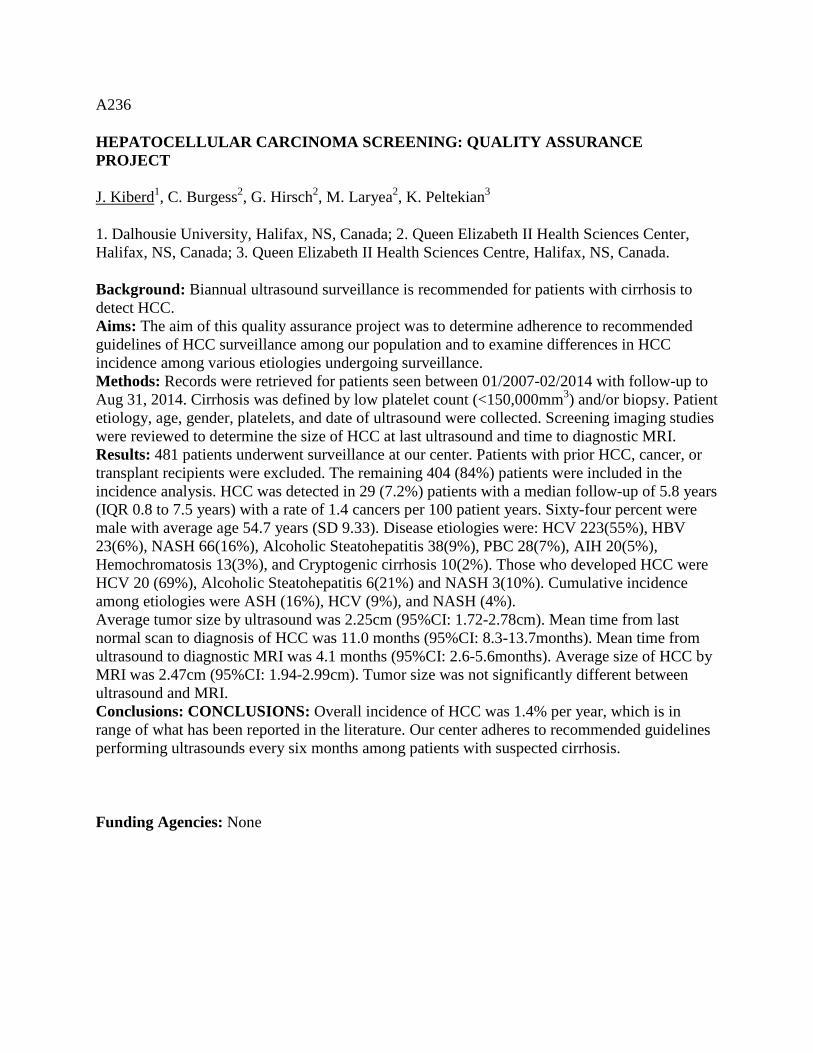
A236
HEPATOCELLULAR CARCINOMA SCREENING: QUALITY ASSURANCE PROJECT
J. Kiberd1, C. Burgess2, G. Hirsch2, M. Laryea2, K. Peltekian3
1. Dalhousie University, Halifax, NS, Canada; 2. Queen Elizabeth II Health Sciences Center, Halifax, NS, Canada; 3. Queen Elizabeth II Health Sciences Centre, Halifax, NS, Canada.
Background: Biannual ultrasound surveillance is recommended for patients with cirrhosis to detect HCC. Aims: The aim of this quality assurance project was to determine adherence to recommended guidelines of HCC surveillance among our population and to examine differences in HCC incidence among various etiologies undergoing surveillance. Methods: Records were retrieved for patients seen between 01/2007-02/2014 with follow-up to Aug 31, 2014. Cirrhosis was defined by low platelet count (<150,000mm3) and/or biopsy. Patient etiology, age, gender, platelets, and date of ultrasound were collected. Screening imaging studies were reviewed to determine the size of HCC at last ultrasound and time to diagnostic MRI. Results: 481 patients underwent surveillance at our center. Patients with prior HCC, cancer, or transplant recipients were excluded. The remaining 404 (84%) patients were included in the incidence analysis. HCC was detected in 29 (7.2%) patients with a median follow-up of 5.8 years (IQR 0.8 to 7.5 years) with a rate of 1.4 cancers per 100 patient years. Sixty-four percent were male with average age 54.7 years (SD 9.33). Disease etiologies were: HCV 223(55%), HBV 23(6%), NASH 66(16%), Alcoholic Steatohepatitis 38(9%), PBC 28(7%), AIH 20(5%), Hemochromatosis 13(3%), and Cryptogenic cirrhosis 10(2%). Those who developed HCC were HCV 20 (69%), Alcoholic Steatohepatitis 6(21%) and NASH 3(10%). Cumulative incidence among etiologies were ASH (16%), HCV (9%), and NASH (4%). Average tumor size by ultrasound was 2.25cm (95%CI: 1.72-2.78cm). Mean time from last normal scan to diagnosis of HCC was 11.0 months (95%CI: 8.3-13.7months). Mean time from ultrasound to diagnostic MRI was 4.1 months (95%CI: 2.6-5.6months). Average size of HCC by MRI was 2.47cm (95%CI: 1.94-2.99cm). Tumor size was not significantly different between ultrasound and MRI. Conclusions: CONCLUSIONS: Overall incidence of HCC was 1.4% per year, which is in range of what has been reported in the literature. Our center adheres to recommended guidelines performing ultrasounds every six months among patients with suspected cirrhosis.
Funding Agencies: None

Fibrogenesis, Portal Hypertension, Complications of Cirrhosis
A237
OCCULT CIRRHOSIS IDENTIFIED BY TRANSIENT ELASTOGRAPHY: A FREQUENT AND UNDER-MONITORED CLINICAL ENTITY WITH PROGNOSTIC SIGNIFICANCE
T. Chen, R. Wong, P. Wong, P. Ghali, M. Deschenes, G. Sebastiani
McGill University Health Centre, Montreal, QC, Canada.
Background: Diagnosis of compensated liver cirrhosis at pre-clinical stage is challenging due to lack of any physical, laboratory and imaging findings. Aims: We evaluated prevalence, predictors and outcomes of preclinical compensated cirrhosis, defined as occult cirrhosis (OC), diagnosed by transient elastography (TE, Fibroscan®). We also explored the pattern of surveillance for cirrhosis complications that OC receives in clinical practice. Methods: 871 consecutive patients with compensated chronic liver disease (CLD) and a valid TE examination were divided into three groups: 1) OC (TE≥13kPa and absence of any clinical sign of cirrhosis, including absence of thrombocytopenia, or signs of advanced liver disease on ultrasound or gastroscopy); 2) clinically evident compensated cirrhosis (TE>13kPa with any of the previous signs); 3) non-cirrhotic CLD (TE<13kPa). We used multivariate logistic regression analysis to identify predictors of OC at baseline. Outcomes included incident hepatocellular carcinoma (HCC), esophageal varices and ascites. Kaplan-Meier curves and log-rank test were used to illustrate and compare the cumulative incidence of outcomes among study groups. A late diagnosis of outcomes was defined as HCC stage > intermediate or bleeding from esophageal varices. Multivariate Cox proportional hazard models adjusted for age, sex, HIV coinfection, diabetes and OC were used. Results: OC represented 12% of the study population and 37% of cirrhotic patients. Logistic regression analysis showed predictors of OC were age (odds ratio [OR] 1.15; 95% confidence interval [CI], 1.04-1.26), HIV co-infection (OR 3.53; 1.85-6.76) and APRI (OR 2.63; 1.87-3.71). During a median follow-up period of 12 months (range 6-36), OC received less surveillance than CE cirrhosis, with fewer ultrasounds (1.8+1 vs 2.4+1.3; p<0.001) and gastroscopies (1.14+0.5 vs 1.7+0.9; p<0.001). Only 36% of the OC group vs 69% of the clinically evident cirrhosis group received the recommended surveillance for cirrhosis (p<0.001). Cumulative incidence of outcomes was 5% in OC, 0% in non-cirrhotic CLD and 14% in clinically evident cirrhosis (see Figure 1). Late diagnosis of outcomes was more frequent in OC as compared to clinically evident cirrhosis (60% vs 15%, p=0.01). Multivariate Cox regression analysis showed OC was associated with incident outcomes as compared to non-cirrhotic CLD (hazard ratio, 3.19; 1.40-7.24). Conclusions: OC is a frequent and under-monitored clinical entity associated with short-term risk of outcomes. Screening of patients with CLD by TE may help prompt initiation of surveillance and specific therapy for an otherwise unrecognized condition.
Funding Agencies: FRSQ
A238
MUSCLE MASS OPTIMIZATION PREVENTS EXPERIMENTAL HEPATIC ENCEPHALOPATHY
S. Ghezzal1, M. Clément2, C. Bosoi2, R. Beauchamp2, M. Tremblay2, C. Rose2, C. Bemeur3
1. Université Pierre et Marie Curie, Paris, France; 2. CRCHUM, Montréal, QC, Canada; 3. Université de Montréal, Montreal, QC, Canada.
Background: Malnutrition is an important prognostic factor potentially influencing clinical outcome of patients suffering from chronic liver disease (cirrhosis; CLD). Malnutrition exacerbates severe muscle loss and hepatic encephalopathy (HE) in cirrhotic patients. New management strategies focussing on improving nutritional status and attenuating CLD-related complications are an unmet clinical need. Aims: We hypothesize supplementation with branched-chain amino acid leucine (LEU) and exercise training (EX) could possibly attenuate muscle mass loss and prevent HE (characterized by brain edema as well as cognitive and psychomotor impairments) in CLD. Methods: CLD was induced in rats following 6-week bile-duct ligation (BDL). Five experimental groups were tested; 1) BDL; 2) BDL + LEU; 3) BDL + EX; 4) BDL + LEU + EX; 5) Sham-operated rats. One week following BDL, rats were submitted to 15 min EX (10 cm/s) every other day and BDL rats receiving LEU, were gavaged daily (1.35 mg/kg) for 5 weeks. Body weight, muscle (gastrocnemius) mass, metabolic state (calculation of energy expenditure independent of food intake and fecal mass), cerebral edema (specific gravity method) and cognitive/psychomotor function (open-field test; anxiety-like behavior assessment and novel object recognition test; memory testing) were measured in all groups. Results: BDL rats gained less body weight compared to sham-operated rats (125.0g ± 24.9 vs 226.0g ± 38.5; p<0.05). LEU-treated BDL rats display an improvement in brain edema (78.50% ± 0.03 vs 80.27% ± 0.14; p<0.05), muscle mass (5.48g/kg ± 0.90 vs 4.83g/kg ± 0.11; p<0.05) and circumference (15.6cm/kg ± 0.8 vs 13.1cm/kg ± 0.7; p<0.05) and metabolic activity (27.48 ± 1.15 vs 32.99 ± 2.35; p<0.05), which was further ameliorated with EX, compared to BDL animals. In addition, BDL rats receiving LEU and EX exhibited less anxiety-like behavior (4.9s ± 1.2 vs 2.2s ± 0.9 passed in the center; p<0.01) as well as better novel object recognition memory (69.6 ± 15.2% vs 25.4 ± 9.6%; p<0.01), in comparison with BDL rats. Conclusions: Our results demonstrate that supplemental LEU along with EX reduces body weight and muscle mass loss, improves metabolic activity, attenuates brain edema and improve cognitive and psychomotor function. These findings suggest that strategies aiming at improving nutritional status will attenuate muscle mass loss, reduce the risk of developing HE and therefore improve quality of life and decrease mortality in CLD. LEU supplementation and EX could rapidly be translated into clinical practice.
Funding Agencies: CIHR

A239
SARCOPENIA AND MYOSTEATOSIS INCREASE THE RISK OF HEPATIC ENCEPHALOPATHY IN CIRRHOTIC PATIENTS
A. Montano-Loza1, A. Duarte-Rojo3, C. Rose2
1. University of Alberta, Edmonton, AB, Canada; 2. Hôpital St-Luc, CRCHUM, Université de Montréal, Montreal, QC, Canada; 3. University of Arkansas for Medical Sciences, Little Rock, AR.
Background: Sarcopenia is one of the most common complications of cirrhosis and it is associated with increased mortality. Muscle depletion is generally characterized by both a reduction in muscle size and increased proportion of inter- and intra-muscular fat denominated "myosteatosis". Skeletal muscle may serve as an alternative site of ammonia detoxification in patients with cirrhosis. Aims: In this study we aimed to investigate if sarcopenia and myosteatosis are associated with overt hepatic encephalopathy in patients with cirrhosis. Methods: A total of 678 cirrhotic patients undergoing assessment for liver transplantation were studied. Sarcopenia and myosteatosis (characterized as low muscle attenuation) were analyzed using computed tomography (CT) scans at the level of the 3rd lumbar vertebral body. The area of paraspinal skeletal muscle (L3 SMI) at this location, and the muscle attenuation index were calculated. Hepatic encephalopathy was assessed clinically by applying the West-Heaven criteria (grade 0-IV). Results: Of the 678 patients, 457 patients were males (67%). Cirrhosis was caused by HCV in 256 patients (38%), alcohol in 152 (22%), NASH/cryptogenic in 171 (25%), autoimmune liver disease in 53 (8%), HBV in 41 (6%), other etiology in 5 patients (1%); and 292 patients had concomitant HCC (43%). Sarcopenia was noted in 291 patients (43%), and 353 patients had myosteatosis (52%). A total of 216 patients (32%) had history of hepatic encephalopathy (162 grade I-II, 54 grade III-IV). The prevalence of hepatic encephalopathy was significantly higher in patients with sarcopenia (40 vs. 26%, P<0.001), and myosteatosis (39 vs. 24%). By multivariate regression analysis (adjusted to age, gender, and MELD score), both sarcopenia (OR 1.68, (95% CI 1.04-2.40, P=0.03), and myosteatosis (OR 1.97, 95% CI 1.32-2.99, P=0.001) were significantly associated with hepatic encephalopathy. Conclusions: Cirrhotic patients with sarcopenia and myosteatosis have a higher risk of overt hepatic encephalopathy. Skeletal muscle seems to play a protective role in the pathogenesis of hepatic encephalopathy in cirrhosis, and therapeutic strategies to improve the muscle mass and quality may improve hepatic encephalopathy in cirrhosis.
Funding Agencies: None, American College of Gastroenterology
A240
THERAPEUTIC OUTCOMES OF CHILD-PUGH A CIRRHOTICS FOUND TO HAVE MEDIUM/LARGE ESOPHAGEAL VARICES ON SCREENING THIN SCOPE ENDOSCOPY
A. Flynn, S. Congly, R. Myers, K. Burak, A. Aspinall, S. Lee
University of Calgary, Calgary, AB, Canada.
Background: Unsedated peroral thin scope endoscopy (TSE) is a well-tolerated and cost-effective alternative to conventional esophagogastroduodenoscopy (EGD) for the screening of esophageal varices in patients with Child-Pugh A cirrhosis. Consensus guidelines recommend either non-selective β-blockers (NSBB) or endoscopic variceal ligation (EVL) for the primary prophylaxis of medium/large varices. EVL cannot be done using TSE; therefore, a follow-up EGD must be performed if this procedure is planned. This introduces an additional therapeutic step that could delay or prevent appropriate therapy. The therapeutic outcomes of this patient population are not currently known. Aims: To determine the therapeutic outcomes of patients with Child-Pugh A cirrhosis who are found to have medium/large esophageal varices on screening TSE. Methods: A retrospective chart review was performed on the 168 patients with Child-Pugh A cirrhosis and no prior variceal screening who underwent TSE at the University of Calgary outpatient liver clinic between September 2011 and July 2013. Patients with medium/large varices were identified, and their subsequent referral for EVL and/or prescription of NSBB was determined. Results: Medium/large esophageal varices were detected by TSE in a total of 17 (10.1%) patients, including 3 (1.8%) with high-risk stigmata. The initial primary prophylaxis strategy of these patients was EVL + NSBB (52.9%), EVL alone (25.4%), or NSBB alone (17.6%). The median number of days between TSE and follow-up EGD in patients sent for EVL was 41 (6-700). No episodes of upper gastrointestinal bleeding were recorded for any patient in the period between TSE and EGD. Conclusions: A significant proportion of Child-Pugh A cirrhotics with medium/large varices on screening TSE are treated with both EVL and NSBB for primary prophylaxis, which is not in line with current consensus guidelines. The length of time between the detection of medium/large esophageal varices by TSE and their ligation varies widely, but has not been associated with the interval development of variceal bleeding.
Funding Agencies: None

A241
OBESITY PREDICTS SIGNIFICANT LIVER FIBROSIS FOLLOWING LIVER TRANSPLANTATION
M. Bhat, K. Rollet, A. Bhat, A. Farag, M. Deschenes, P. Wong, P. Ghali, G. Sebastiani
MUHC, Montreal, QC, Canada.
Background: Hepatic fibrosis may occur as a manifestation of recurrent or de novo chronic liver disease following liver transplantation (LT). Longitudinal studies with long-term follow-up are scarce. Aims: We employed a validated serum biomarker for liver fibrosis, AST-to-Platelet-Ratio-Index (APRI), to determine incidence and predictors of significant liver fibrosis after LT. Methods: We included consecutive patients who underwent LT at a single center from 1991-2011. Included patients had to have a minimum follow-up of 1 year and at least 2 visits. Significant liver fibrosis was diagnosed by APRI >1.5. If patients had been retransplanted, the transplant with which they had the longest graft survival was included. Patients with outcome at baseline (APRI >1.5) or within 6 months from LT, as well as those with a graft survival <6 months were excluded. We estimated incidence rates of significant liver fibrosis by dividing the number of participants developing the outcome by number of person-years (PY) of follow-up. Poisson count models were used to calculate confidence intervals (CI) for incidence rates. Multivariate Cox proportional hazard models, using robust standard errors to allow for repeated measures within individuals, were built to identify predictors of development of significant liver fibrosis. Results: 399 patients (median age 57 years, 66% male) were included. Over 2304 person-years (PY) of follow-up, 23% developed significant liver fibrosis, accounting for an incidence of 3.9/100 PY (95% CI 3.2-4.8). Results of univariate and multivariate analysis and relative hazard ratios (95% CI) are depicted in the Table. After adjustments, obesity, hepatitis C infection and baseline APRI, were significantly associated with development of significant liver fibrosis. Conclusions: Progression to significant liver fibrosis is a frequent event following LT, particularly in those with obesity, hepatitis C infection, and higher baseline APRI. Identification of patients at risk of progression during the pre-transplant period can help early initiation of targeted interventions, such as weight loss and antiviral therapy for hepatitis C. Finally, at risk patients must be followed more closely after LT.
Unadjusted HR Adjusted HR P Time independent baseline covariates
Baseline age (x 5 years) 0.99 (0.92-1.08) 0.95 (0.86-1.04) 0.28
Male gender 1.12 (0.72-1.72) 0.99 (0.62-1.59) 0.99 Obese at baseline 1.49 (1.01-2.3) 1.55 (1.01-2.49) 0.05

Baseline fasting glucose 1.07 (1-1.15) 1.05 (0.97-1.14) 0.21 Baseline APRI 3.37 (2.27-5) 2.69 (1.734-4.16) <0.0001
Cyclosporin therapy 0.94 (0.61-1.44) 0.9 (0.58-1.41) 0.65 Time updated covariates
HCV 4.24 (2.58-6.96) 2.59 (1.44-4.68) 0.002 Hypertension 1.31 (0.83-2.07) 1.29 (0.76-2.16) 0.344
Funding Agencies: None
Hepatobiliary Neoplasia
A242
LIVER FIBROSIS AND HEPATITIS C VIRUS INFECTION BOTH BLOCK THE ANTINEOPLASTIC EFFECT OF CISPLATINUM ON HEPATOCELLULAR CARCINOMA CELLS
B. Dupont, B. Lacoste, G. Merlen, V. Raymond, M. Bilodeau
CRCHUM, Montreal, QC, Canada.
Background: Hepatitis C virus (HCV) infection is a major risk factor for hepatocellular carcinoma (HCC). The strong association between liver fibrosis and the development of HCC is also well established but not well understood. We have demonstrated that type 1 collagen (COL1) could block apoptosis of normal hepatocytes through the ERK1 pathway. Aims: The goal of this work was to evaluate whether COL1 can induce a state of resistance against anti-cancer agents and whether HCV affects this phenomenon. Methods: We used 3 HCC cell lines: Huh7 (devoid of HCV proteins), 9-13 (a Huh7 cell line stably transfected with a genotype-1 replicon expressing HCV non-structural proteins) and JFH-1 (a Huh7 cell line infected with the JFH-1 HCV strain). Cell death was induced by exposure of the cell lines to cisplatinum (CP) [25 μg/mL] for 24hr, in the presence or not of COL1 [13.9 μg/cm2]. Cell viability (MTT assay), apoptotic bodies (apoptotic rate (AR)), caspase 3 activity and cell proliferation were measured. In order to evaluate the ability of cells to form independent colonies, a characteristic of neoplastic cells, individual cells were seeded in a soft agar gel with or without COL1 [695 μg/cm3]. Results: Control Huh7 and 9-13 cells were more resistant to CP-induced apoptosis when plated on COL1 (AR; Huh7: 6.4±0.3% on COL1 vs 20.3±0.2% on plastic; p<0.01, 9-13: 9.5±1.2% on COL1 vs 15.8±1.8% on plastic; p=0.01). The protective effect of COL1 was not observed in JFH-1-infected cells (13.5±0.9% on COL1 vs 15.4±0.2% on plastic; p=0.25) that were spontaneously more resistant to CP-induced apoptosis than the parental Huh7 cell line (AR: 20.3±0.2% in Huh7 vs 15.4±0.2% in JFH-1; p<0.01). The protective effect of COL1 was abolished when cells were co-treated with CP and the MEK inhibitor U0126 [20μM] (AR; Huh7: 16.0±1.6%, 9-13: 15.9±2.7%). When JFH-1-infected cells were cultured in the presence of U0126, there were no change in the level of CP-induced apoptosis (11.3±0.8% vs 13.5±0.9%). Results of caspase 3 activity or cell viability were similar. There was no difference in cell doubling time between HCC cells plated on COL1 or on plastic (Huh7: 53±6h vs 56±4h, 9-13: 63±4h vs 64±10h). The ability of Huh7 cells to form colonies in soft agar was higher when cells were seeded in the presence of COL1 (1.7±0.3 for controls vs 12.7±1.3 for COL1; p=0.001). Conclusions: COL1 partially blocks the efficacy of CP on HCC cells by an ERK1/2-mediated mechanism without any effect on cell proliferation. It also increases their tumorigenic potential. However, this phenomenon is not observed in JFH1 HCV-infected cells that are spontaneously more resistant to apoptosis, this effect being independent from the ERK1/2 pathway.
Funding Agencies: NCRTP-HepC
A243
ISOLATION OF HIGHLY TUMORIGENIC CLONES FROM HEPATOCELLULAR CARCINOMA CELL LINES BY SELECTION IN SOFT AGAR
B. Dupont, B. Lacoste, G. Merlen, V. Raymond, M. Bilodeau
CRCHUM, Montréal, QC, Canada.
Background: Hepatocellular carcinoma (HCC) is the third most deadly cancer worldwide. Despite its recognized heterogeneity, specific cell clones are suspected to be the driving force for tumor formation and/or recurrence. Liver Cancer Stem Cells (CSC) might represent such clones since they are characterized by their ability to become quiescent, their relative chemoresistance and a high capacity to lead to new tumor formation. The isolation of HCC cells with highly tumorigenic potential could help develop novel therapeutic strategies against HCC resistant to therapy or recurrence. Aims: We made the hypothesis that culture in soft agar gel could be an efficient in vitro method to select highly tumorigenic cells. Methods: Hepa 1-6 cells, a widely used mouse HCC cell line, were seeded in a soft agar gel. After 4 weeks, macroscopic colonies were harvested and cultured in order to obtain a clone derived from the Hepa 1-6 parental cell line, called sa-Hepa 1-6. We then characterized the preselected sa-Hepa 1-6 cell line by measuring cell doubling time and metabolic activity by MTT/crystal violet assay. Next, we evaluated response to cisplatinum (CP) [25 μg/mL] by measuring cell viability (MTT assay) and counting apoptotic bodies (apoptotic rate) after 24h exposure. In order to determine the tumorigenicity of the sa-Hepa 1-6 cells, we measured their ability to form independent colonies after a second run of culture in soft agar gel and to form liver tumors in C57/bl6 mice after intrasplenic inoculation (1 million cells). Results: Preselected-sa-Hepa 1-6 cells possessed in vitro properties observed with CSC. Cell proliferation was not different between sa-Hepa 1-6 and Hepa 1-6 (37h vs 42h; p=0.47). Intrinsic metabolic activity was 11% higher in Hepa 1-6 than in sa-Hepa 1-6 (p=0.01). The sa-Hepa 1-6 cells were more resistant to the toxicity of CP in comparison to Hepa 1-6 cells (apoptotic rate: 20.3±2.0% for sa-Hepa 1-6 vs 35.2±5.8% for Hepa 1-6; p<0.05). Results were similar by evaluating cell viability. Furthermore, sa-Hepa 1-6 cells led to 5 times more colonies after a second run of culture in soft agar gel than the unselected Hepa 1-6 cells (p<0.05). Finally, in vivo, preselected sa-Hepa 1-6 cells led to visible liver tumors in C57/bl6 mouse 21 days after intrasplenic inoculation unlike the Hepa 1-6 parental cell line (tumor load: 14.3±5.8 lesions vs 0 lesions; p<0.05). They also invaded the spleen more readily. Conclusions: Culture in soft agar gel is a feasible and efficient method to isolate highly tumorigenic clones from HCC cell lines. The derived cell lines possess liver CSC properties and are able to generate liver tumors in an immuno-proficient mice model after intrasplenic inoculation.
Funding Agencies: NCRTP-HepC
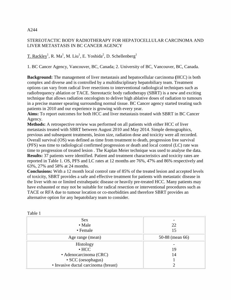
A244
STEREOTACTIC BODY RADIOTHERAPY FOR HEPATOCELLULAR CARCINOMA AND LIVER METASTASIS IN BC CANCER AGENCY
T. Rackley1, R. Ma1, M. Liu1, E. Yoshida2, D. Schellenberg1
1. BC Cancer Agency, Vancouver, BC, Canada; 2. University of BC, Vancouver, BC, Canada.
Background: The management of liver metastasis and hepatocellular carcinoma (HCC) is both complex and diverse and is controlled by a multidisciplinary hepatobiliary team. Treatment options can vary from radical liver resections to interventional radiological techniques such as radiofrequency ablation or TACE. Stereotactic body radiotherapy (SBRT) is a new and exciting technique that allows radiation oncologists to deliver high ablative doses of radiation to tumours in a precise manner spearing surrounding normal tissue. BC Cancer agency started treating such patients in 2010 and our experience is growing with every year. Aims: To report outcomes for both HCC and liver metastasis treated with SBRT in BC Cancer Agency. Methods: A retrospective review was performed on all patients with either HCC of liver metastasis treated with SBRT between August 2010 and May 2014. Simple demographics, previous and subsequent treatments, lesion size, radiation dose and toxicity were all recorded. Overall survival (OS) was defined as time from treatment to death, progression free survival (PFS) was time to radiological confirmed progression or death and local control (LC) rate was time to progression of treated lesion . The Kaplan Meier technique was used to analyse the data. Results: 37 patients were identified. Patient and treatment characteristics and toxicity rates are reported in Table 1. OS, PFS and LC rates at 12 months are 76%, 47% and 86% respectively and 63%, 27% and 58% at 24 months. Conclusions: With a 12 month local control rate of 85% of the treated lesion and accepted levels of toxicity, SBRT provides a safe and effective treatment for patients with metastatic disease in the liver with no or limited extrahepatic disease or heavily pre-treated HCC. Many patients may have exhausted or may not be suitable for radical resection or interventional procedures such as TACE or RFA due to tumour location or co-morbidities and therefore SBRT provides an alternative option for any hepatobilary team to consider.
Table 1
Sex • Male
• Female
- 22 15
Age range (mean) 50-88 (mean 66) Histology
• HCC • Adenocarcinoma (CRC)
• SCC (oesophagus) • Invasive ductal carcinoma (breast)
- 19 14 1 2

• Leimyosarcoma (adrenal) 1 Radiotherapy dose
• 45Gy/3# • 45Gy/5# • 50Gy/5# • 42.5/5# • 36Gy/9#
- 27 7 1 1 1
Size of treated lesion 1.4-10cm (mean 4.1cm) Toxicity • Fatigue • Nausea
• Vomiting • Pain
• Gastritis/duodenitis • Late toxicity
- 8 6 1 3 2 0
Number of previous treatments prior to SBRT • HCC
• Liver mets
- 0-5 (mean 2) 0-4 (mean 2)
HCC previous treatments (n=19) • Surgical resection
• TACE • RFA
• Ethanol ablation • Sorafenib
• Y90
- 7 22 11 3 0 1
Metastasis previous treatments (n=18) • Surgical resection
• RFA • Chemotherapy
• Hormonal therapy
- 7 6 23 1
Typical radiotherapy plan
Funding Agencies: None

A245
OUTCOMES FOLLOWING RADIOFREQUENCY ABLATION IN HEPATOCELLULAR CARCINOMA: IMPACT OF ETIOLOGY
S. Sharma, K. Pratte, M. Kowgier, M. Sherman
University of Toronto, Toronto, ON, Canada.
Background: Among patients with small tumors (<2cm), RFA has been shown to have comparable outcomes to surgical resection. In patients who have undergone surgical resection, etiology of HCC may be an important predictor of transplant-free survival (TFS), with some studies showing worse survival in HCV, compared to HBV and non-viral chronic liver disease. There is a paucity of data regarding the impact of etiology of HCC on outcomes in patients undergoing RFA. Aims: This study aims to address the impact of etiology of HCC on transplant-free survival in patients undergoing RFA. Methods: Chart review was conducted on all patients undergoing RFA at the University Health Network between January 1, 2008 and December 31, 2011. Patients were identified through the Tumour Board database, and followed up until January 1, 2014. HCC was defined using standard AASLD criteria. Data collected included demographics, etiology of liver disease, severity of liver disease and size and location of HCC.Complete ablation of lesions was defined as the absence of residual disease at 3 months post RFA. Recurrence of HCC was defined as tumor foci developing at a location of prior RFA treatment - either early (<6 months) or late (>1 year). Outcome measures included transplant-free survival and tumor recurrence. Results: 193 patients underwent RFA during the study period. 88% of the patients had documented evidence of cirrhosis. 64 patients with HBV (53 on treatment), 81 patients with HCV (46 treated, 9 SVR), and 48 patients with nonviral etiologies were included. Gender distribution varied by etiology with 14% females in the HBV group, 31% among HCV patients and 19% among nonviral patients. The mean age at diagnosis of HCC varied from 62 (HBV and HCV) to 67 years (nonviral). Mean MELD scores varied from 7.3 (HCV) to 9.3 (nonviral). All patients underwent at least one session of RFA (mean 1.8 treatments). The average size of RFA-treated lesions was 2.5cm (±0.96cm). 78% of the patients had complete ablation. Recurrence occurred in 55 patients (28%), with mean recurrence-free interval of 489 days (±350d). 78 (40%) patients died or underwent liver transplant during the follow-up period. The primary outcome (transplant or death) was reached in 23% of HBV patients (median TFS 1.4 years), 51% of HCV patients (median TFS 1.8 years) (p=0.0059 HBV vs. HCV), and 35% of nonviral patients (median TFS 1.7 years). Patients with HCV had a significantly worse TFS, after adjusting for age and MELD score (p=0.01, log-rank test). On multivariable analysis, etiology remained a significant predictor of TFS, after adjusting for age and MELD. Conclusions: Etiology of HCC may be an important determinant of transplant-free survival post RFA. Further studies are needed to determine the underlying reason for this association.
Funding Agencies: None

A246
HEPATOCELLULAR CARCINOM RESPONSE TO LOCAL REGIONAL THERAPY, ; CORRELATION BETWEEN PRE-LIVER TRANSPLANTS IMAGING AND EXPLANT PATHOLOGY.
K. Qumosani1, M. Alghanem4, P. Marotta3, B. Aljudaibi2, D. Driman3, M. Garisa4, O. Bashir3, N. Kakani3
1. LHSC, London, ON, Canada; 2. London Health Science Centre, London, ON, Canada; 3. London heatlh sciences Center, University of Western Ontario, London, ON, Canada; 4. university of western ontario, London, ON, Canada.
Background: Hepatocellular Carcinoma (HCC) is the fifth most common malignancy worldwide. HCC therapy includes Local regional Therapy (LRT) such as Radiofrequency Ablation (RFA) and Trans-Arterial Chemoembolization (TACE). Modified Response Evaluation Criteria in Solid Tumors (mRECIST) were developed to assess the response to treatment in patients with HCC, based on measurements of viable tumor using dynamic imaging (CT/MRI). Aims: To compare the estimate of viable HCC after LRT by CT imaging and before liver transplant, to the histopathological assessment of viable HCC in the hepatic explant. Methods: We prospectively evaluated 29 patients with HCC who undwerent both LRT and liver transplantation at london health science center. Using mRECIST criteria, the response to LRT was assessed by 2 blinded radiologists and the percentage of necrosis was reported separately for the reference CT(rCT) completed done after the last LRT and prior to liver transplantation. The results from the radiologists were compared to the findings of an expert pathologist reporting on viable tumour present and tumour necrosis in the hepatic explants. Both parties were blinded so prevent bias in the results. Results: A total of twenty nine transplant recipients fulfilled the inclusion criteria for the study. 3 patients were excluded due to absence of a rCT prior to LT and after LRT. At time of listing 100% were within Ontario criteria,78% within UCSF, and 67% within Milan.The HCC was single lesion was in 50% (13/26), and only one lobe was affected in 70% (17/26). 6 patients (23%) received RFA while 20 patients (77%) received TACE treatments as bridging therapy. No patients progressed beyond transplant criteria. . The average time frame from the last reference CT scan to liver transplant was 59.7 days; the average time from last LRT to reference CT was 66.3 days. 20 (76%) recipients had accurate assessment for necrosis (mRECIST) within 20% comparing rCT to explant (i.e. concordant). 13/26 (50%) predicted 100% concordance. Only 3/26 (11.5%) had a poor concordance (>50%) between histology and reference CT images. positive correlation was detected with the correlation coefficient is calculated as 0.57 Conclusions: Dynamic CT is an accurate tool to evaluate the tumour response prior to liver transplantation and the likelihood of underestimating the tumour burden is low. With expert radiologists and pathologists, the correlation is acceptable and supports the ongoing use of frequent dynamic imaging to evaluate responses to LRT and determining transplant eligibility
Funding Agencies: None
A247
A NEUROENDOCRINE TUMOUR ARISING FROM A TYPE III CHOLEDOCHAL CYST, PRESENTING WITH ELEVATED LIVER ENZYMES: A COMMON PRESENTATION OF TWO UNUSUAL ETIOLOGIES
S. Kenshil, R. Gheorghe, D. Moffatt
University of Manitoba, Winnipeg, MB, Canada.
Background: Choledochal cysts are cystic dilatations of the intrahepatic or extrahepatic biliary tree and are uncommon in Western adults happening in an estimated less than 1/100,000 individuals in North America. Neuroendocrine tumors are likewise rare tumors occurring in 4-6/100,000 individuals..We report a rare case of a neuroendocrine tumour arising from a choledochocoele (Type III choledochal cyst). Aims: Report a Unique Case. Methods: Case Report. Results: A 27 year old, highly functioning autistic, caucasian female presented to a hepatology clinic for evaluation of elevated cholestatic liver enzymes (AST 143U/L, ALT 302 U/L, ALP 998U/L, GGT 500U/L, bilirubin 20umol/L). She was otherwise well and asymptomatic. Workup was negative except for a transabdominal ultrasound, that revealed mild prominence of the common bile duct at 1.0cm. An MRCP was arranged and revealed worsening intra and extrahepatic biliary dilatation, with a common hepatic duct up to 1.6cm maximum diameter with no evidence of a mass lesion or choledocholithiasis. An ERCP was performed to evaluate the cause of the biliary dilation and revealed a abnormal papilla consistent with a choledochocoele. A sphincterotomy was performed and no filling defects or stones were identified. No masses were seen but routine biopsies were taken from the papilla and sphincterotomy site. The patients liver enzymes rapidly decreased to normal levels. (ALT 33 U/L, ALP 142U/L, GGT 142U/L, bilirubin 20umol/L) One biopsy revealed small bowel mucosa with an area that was consistent with a low grade neuroendocrine tumour. A subsequent endoscopic ultrasound of the papilla revealed no mass, but biopsies from the area were again consistent with a NET. An ocreotide scan was positive at the area of the ampulla and an adjacent lymph node. The patient underwent a Whipple procedure for definitive management. Pathology confirmed the diagnosis of a low grade NET,T2N1 with 2/10 positive lymph nodes, maximum diameter 6 x 9mm, which was completely resected. She has since made a full recovery. Conclusions: In this patient we report a rare case of a NET arising within a choledochocele. There have only been 5 prior case reports of a NET arising from a choledochal cyst and this is the first arising from a choledochocele (Type III CCC). Although recent data has not shown an increased risk of cholangiocarcinoma in caucasian population with Type III cysts, it is possible that there may be an increased risk of NET within these lesions. Further studies are needed.
Funding Agencies: None

Hormones, Transmitters, Growth Factors
A248
THE ADAPTIVE EFFECTS OF GLUCAGON-LIKE PEPTIDE-2 ARE INFLEUNCED BY ANATOMIC AND ENTERAL FACTORS IN NEONATAL INTESTINAL FAILURE.
D. Lim3, P. Wales1, P. Nation3, P. Wizzard3, D. Bigam3, D. Sigalet4, J. Turner2
1. The Hospital for Sick Children and University of Toronto, Toronto, ON, Canada; 2. Stollery Children's Hospital and University of Alberta, Edmonton, AB, Canada; 3. University of Alberta, Edmonton, AB, Canada; 4. University of Calgary, Calgary, AB, Canada.
Aims: To study how the effect of exogenous glucagon-like peptide-2 (GLP-2) therapy in a preclinical model of neonatal intestinal failure (IF) is affected by remnant anatomy and enteral nutrition therapy (ENT). We hypothesize that GLP-2 therapy is synergistic with ENT, with greatest effect in IF-anatomy lacking ileum. Methods: Neonatal piglets were block randomized to either GLP-2 therapy (11 nmol/kg/d) or saline control following either a 75% mid-intestinal (JI) or 100% ileal (JC) resection or no resection (sham control). Piglets were also randomized to receive ENT at 0%, 20% or 40% of nutritional requirement. Piglets underwent terminal laparotomy on post-operative day 7. Gross morphological adaptation was assessed by the change in intestinal length post-resection, bowel weight per length and mucosal weights. Structural adaptation ex vivo was further assessed by histopathologic analysis of jejunum and ileum. Data are analyzed by a three-way ANOVA for a 3x3 factorial design of surgery, treatment and enteral nutrition factors. Results: There were 18 study groups in total, which encompassed three anatomic levels (sham control, JI, JC), two treatment levels (saline control, GLP-2) and three ENT levels (0%, 20%, 40%) with n=4 in the sham groups and n=6 in the JI and JC groups. All gross morphological parameters varied depending on remnant anatomy. There was a significant interaction in anatomy and ENT influencing the change in intestinal length (p<0.01). Intestinal length was augmented with increasing ENT, with the greatest increases in the JI group. ENT minimized bowel shortening in the JC group. Bowel weight per length increased uniformly with GLP-2 treatment (p<0.001), with a trend towards increasing bowel weight per length with increasing ENT. Jejunal and ileal mucosal weights increased with GLP-2 and ENT (p<0.001). Both villus height and crypt depth differed with a significant interaction between all three factors (p<0.001). GLP-2 increased jejunal and ileal villus height with increasing ENT in all three surgical groups. Conclusions: Morphological adaptation in neonatal IF is dependent on anatomic, hormonal and enteral nutrition factors. GLP-2 therapy and increasing enteral nutrition stimulates histologic adaptation, most in mid-intestinal resection IF and to an extent in distal-intestinal resection IF. Enteral nutrition therapy may be necessary for longitudinal bowel lengthening whereas hormonal therapy may be more beneficial for transverse mucosal growth.
Funding Agencies: CIHR, SickKids Foundation
Immunobiology and Liver Transplantation
A249
UNINTENTIONAL CONSEQUENCE OF NA-MELD IMPLEMENTATION IN ONTARIO:EXCESSIVE BENEFIT TO LIVER TRANSPLANT CANDIDATES LISTED WITH ‘EXCEPTION' POINTS
C. Dale, B. Aljudaibi, R. Hernandez, W. Wall, P. Marotta University Hospital, London, ON, Canada. Background: To foster equitable access to liver transplantation (LT) in Ontario, a provincial mandate resulted in alterations to the organ allocation algorithm. The Model for End-Stage Liver Disease (MELD) is a valid objective tool that prioritizes disease severity. In 2012, a MELD based system (Na-MELD) was implemented. As in other regions, candidates with exceptional disease states (eg Hepatocellular carcinoma (HCC), Hepatopulmonary syndrome), are granted MELD ‘exception' points. These points represent a numeric value that presumably allows equal competition for transplantation while balancing equal mortality rates for all candidates awaiting transplantation. Aims: Review transplant candidates that were listed, delisted or died awaiting LT during the first 18 months of implementing the new allocation protocol. Methods: A retrospective review of all candidates ever listed for LT from Nov 13, 2012 until June 30, 2014 at a single center in Ontario. All removals and reason for removal from the wait list were analysed. Results: In the defined period, 155 candidates awaited LT; 115(74%) listed without exception points and 40 (26%) listed using exceptions. Eighty candidates were transplanted, 51 (64%) without exception points and 29 (36%) with exception points. Overall, 23 (15%) were delisted or died awaiting LT; 20 candidates representing 17 % (20/115) of non-exception candidates and 3 representing 7.5% (3/40) exception point candidates. At transplant the mean NaMELD score for non-exception recipients was 27 and 25 for those listed with exceptions, with the calculated NaMELD in the latter group being 14.5. Those without exception points that were delisted (n=20) had a mean NaMELD score at listing of 24 and at delisting 28. Of the candidates listed with exception points, 86% had HCC. Conclusions: MELD based allocation was implemented to optimize access to LT for those in most need based on disease severity. ‘Exceptions' are included by estimating MELD points to equal wait list mortality and access to LT for other disease states. We observed an overall ‘delist' rate of 15%. Our experience, reveals that patients with advanced liver disease have at least a 2 fold increase in death while awaiting LT, whereas exception candidates represent proportionally fewer on the wait list, achieve LT more frequently and have a disproportionately lower rate of delisting, suggesting waitlist mortality is overestimated by exception points. These findings are similar to other regions (UNOS, UK, Australia). Allocating by MELD exceptions overestimates presumed mortality to the disadvantage of those with end stage liver disease. The unintentional consequences of this policy in Ontario must be revisited and adapted to allow liver allocation to be equitable for all wait list candidates.
Funding Agencies: None

A250
OUTPATIENT TACROLIMUS TROUGH LEVELS IMMEDIATELY POST-PEDIATRIC LIVER TRANSPLANT: PRELIMINARY QUALITY ASSURANCE ANALYSIS
L. Verma, R. Heisler, J. Yap
University of Alberta, Edmonton, AB, Canada.
Background: Calcineurin inhibitors (CNI) such as tacrolimus are the backbone immuno-suppresion post pediatric liver transplants. Sub-therapeutic trough CNI levels are associated with increased incidence of allograft rejection, whilst high CNI levels increased the risk of toxicity related adverse events. Aims: The study aims to describe the frequency of CNI levels within target range in the outpatient pediatric liver transplant recipients. The study will compare whether changes in monitoring processes and medical personnel in two different time eras influenced the consistency of trough CNI levels. Methods: Retrospective chart review of consecutive ABO compatible primary pediatric liver transplant recipients from 2004 to 2006 (Era 1) and 2010 to 2013 (Era 2). Outpatient CNI data was collected from eligible patients in the immediate six months post-liver transplant. Adverse outcomes such as biopsy proven acute cellular rejection (ACR) and other events were also noted. The years from 2007 to 2009 were excluded from the analysis because of unstable medical staffing and redevelopment/implementation of new processes. Results: This preliminary analysis presents results from Era 1 only. Twenty-two subjects were screened; three were excluded because of liver re-transplantation and one for insufficient outpatient data. The remaining 18 patients included were on tacrolimus; 67% females. Biliary atresia (44%) was the most common indication for liver transplantation. The majority of subjects (61%) were living related liver donor recipients. Median age and weight at transplant were 1.55 (IQR: 0.8 - 4.3) years; 11.6 (8.2 - 18.5) kg respectively. There were 475 trough tacrolimus levels; 23.7 levels per subject. Only 25.6% of all levels were within target range, while 73.6% out-of-range levels were sub-therapeutic. There were 2 subjects with mild ACR and another 2 requiring anti-hypertensive medications. The frequency of sub-therapeutic tacrolimus levels were no different between subjects with or without rejection (p=1.0). Conclusions: A significant number of patients in Era 1 had CNI levels out of target range. Despite substantial frequency of sub-therapeutic CNI levels, episodes of ACR were uncommon. Further comparative analysis between eras will provide more granular details.
Funding Agencies: None

A251
EARLY RENAL DYSFUNCTION AFTER LIVER TRANSPLANTATION IS ASSOCIATED WITH REDUCED GRAFT AND PATIENT SURVIVAL
R. Bhanji, M. Ma, V. Bain, A. Montano-Loza
University of Alberta, Edmonton, AB, Canada.
Background: Renal dysfunction is a common complication occurring after liver transplantation (LT). Aims: In this study we aimed at investigating the frequencies of acute renal dysfunction (ARD) and chronic kidney disease (CKD) post-liver transplant (LT), the risk factors associated with renal dysfunction post-LT, and to determine the clinical impact of renal dysfunction with regards to both graft, and patient survival after LT. Methods: We analyzed 535 patients with cirrhosis who received a liver transplant over the period of 1989 to 2010. Estimated glomerular filtration rate (eGFR) was determined using the four variable MDRD formula, ARD was defined as eGFR >90 ml/min prior to transplant followed by eGFR <60 ml/min at 3 months post-LT, and CKD was defined as eGFR <60 ml/min after LT, evidence of intrinsic renal disease, or need for renal replacement therapy. Data was obtained from the electronic medical charts and was used to determine the prevalence of ARD and CKD, risk factors for renal dysfunction, and graft/patient survival after LT. Results: 359 patients were males (67%), and the mean age at LT was 52±10 years. Cirrhosis etiology was HCV cirrhosis (35%), alcohol cirrhosis (33%), autoimmune liver disease (25%), NASH (2%), and HBV (6%). Diabetes was present in 180 (34%), and hypertension in 244 patients (46%) prior to LT. Mean serum creatinine level was 111±80 μmol/L, and 186 patients (35%) had a diagnosis of CKD prior to LT. ARD was documented in 34 patients (6%) after LT, and a new diagnosis of CKD post-LT was made in 88 patients (16%). Risk factors associated with development of ARD were age >60 years (38 vs. 19%, P=0.01) and male gender (47 vs. 68%, P=0.01). Risk factors for development of CKD post-LT were diabetes (48 vs. 31%, P=0.003), and hypertension (61 vs. 43%, P=0.001). Graft survival (133±13 vs. 168 ±11 months, P=0.03) and patient survival were significantly reduced when eGFR was <60 ml/min at 3 months (133±13 vs. 185±12 months, P=0.006). Hazard ratio for mortality was 1.6 (95% CI 1.2-2.3, P=0.006) for those patients with eGFR <60 ml/min at 3 months. Conclusions: ARD and CKD are frequent complications after LT. Older age and male gender are associated with a higher risk for ARD, whereas diabetes, hypertension and early reduction in the eGFR are associated with higher risk of CKD after LT. Recognition of early kidney dysfunction after LT is important in an effort to establish strategies to improve graft and patient survival after LT.
Funding Agencies: None

A252
PRESENCE OF ASCITES AND HEPATIC ENCEPHALOPATHY PRE-LIVER TRANSPLANT ARE ASSOCIATED WITH NEGATIVE IMPACT ON LIVER TRANSPLANT SURVIVAL
R. Bhanji, M. Ma, V. Bain, A. Montano-Loza
University of Alberta, Edmonton, AB, Canada.
Background: Significant improvement in one year outcomes post-liver transplant (LT) has changed attention to long-term survival. Understanding factors associated with premature death is essential for improving long-term outcome after LT. Complications secondary to portal hypertension can be life threatening and are often the main indication for LT; however, their impact after LT survival has not been thoroughly investigated. Aims: In this study we aimed to investigate the impact of specific complications of portal hypertension, such as ascites and hepatic encephalopathy on the outcomes after LT. Methods: A total of 450 patients with cirrhosis who received a LT between 2000 and 2010 were evaluated. The presence of ascites and hepatic encephalopathy was recorded as part of the pre-LT assessment. Ascites was categorized as easily controlled (responded to low salt diet and diuretics), or poorly controlled (required intermittent large volume paracentesis or TIPS) and hepatic encephalopathy was categorized as grade 1&2 or 3&4, according to the West-Haven classification. Results: 308 were males (68%), and mean age was 52±10 years. Cirrhosis etiology was HCV (37%), autoimmune liver disease (22%), alcohol (16%), NASH (8%), and others (17%). Ascites was present in 361 patients (81%), of whom 157 patients (35%) had easily controlled and 208 (46%) poorly controlled ascites. Hepatic encephalopathy was present in 273 (61%) patients that was grade 1&2 in 184 (41%), and grade 3&4 in 89 (20%). Ascites was associated with reduced LT survival (100±3 vs. 118±5 months; Log Rank P=0.004) and patients with poorly controlled ascites had even worse survival (97±5 vs. 110±4 months; Log Rank P=0.01). Also, hepatic encephalopathy was associated with reduced LT survival (117±4 vs. 94±4 months; Log Rank P<0.001) and patient with grade 3/4 had worse survival (90±7 vs. 107±3 months; Log Rank P=0.03). Five-year probability of survival was 71 and 87% in patients with and without ascites (P=0.004), and 68 and 84% in patients with and without hepatic encephalopathy (P<0.001). There was as significant association between ascites and hepatic encephalopathy (68% had both, P<0.001). Including each separately on a multivariate Cox regression analysis, with age, HCV, and HCC both were independently associated with mortality after LT (ascites, HR 1.83, 95% CI 1.01-3.34, P=0.05; hepatic encephalopathy, HR 1.81, 95% CI 1.81-1.20-2.74, P=0.005). Conclusions: Ascites and hepatic encephalopathy pre-LT are frequently present in patients with cirrhosis and these complications are associated with a reduced LT survival. Further studies to elucidate the pathogenic mechanism of these associations are warranted.
Funding Agencies: None

A253
ASSOCIATION BETWEEN HYPOMAGNESAEMIA AND CALCINEURIN INHIBITOR INDUCED NEPHROTOXICITY IN LIVER TRANSPLANT RECIPIENT: A CASE-CONTROL STUDY
A. Daulat1, T. Hussaini1, N. Partovi1, Z. Erb2, E. Yoshida2, V. Marquez2
1. University of British Columbia, Vancouver, BC, Canada; 2. The Division of Gastroenterology, University of British Columbia, Vancouver, BC, Canada.
Background: Calcineurin inhibitors (CNIs) are considered the backbone of immunosuppression improving graft and patient survival. However long term CNIs exposure causes toxicities leading to morbidity and mortality in transplant recipients. Nephrotoxicity and hypomagnesaemia are common findings in liver transplant recipients receiving CNIs. This has led to the hypothesis that magnesium (Mg) wasting or deficiency is related to chronic CNI nephrotoxicity. Aims: To determine if there is an association between hypomagnesaemia and the development of CNI induced nephrotoxicity in liver transplant recipients Methods: A case-control study was conducted including liver transplant recipients at Vancouver General Hospital between Jan 2005 and Dec 2013. Patients who survived more than 90 days post-transplantation and had an eGFR of >=60ml/min at the time of discharge were included. Patients with intrinsic renal disease such as diabetic nephropathy or acute tubular necrosis were excluded. Chronic Renal Failure (CRF) was defined as a sustained drop (>3 months) in the eGFR to < 50 ml/min. Hypomagnesaemia was defined as an average level of <0.7 mmol/ml during the first year post transplant. Results: Of 370 patients evaluated, 254 (157 male, 97 female) met the inclusion criteria. Seventy-six (30%) patients developed CRF. The mean eGFR levels at discharge were lower in cases (82 ml/min, sd 21) compared to controls (95 ml/min, sd 20). Patients in both groups were exposed to tacrolimus during the first month post-transplantation and only 4(5.2%) of the cases were on cyclosporine compared to 8(4.5%) of the controls. During the first month post-transplantation, the use of other immunosuppressant medications including azathioprine (p=0.106), mycophenolate mofetil (p=0.192), steroids (p=0.300), basiliximab (p=0.062) and anti-thymocyte globulin (p=0.493) were similar in both groups. The mean Mg levels during follow up were 0.74±0.07 mmol/L in cases and 0.72±0.07mmol/L in controls. Follow up annual average tacrolimus trough levels was lower in cases than in controls (5.0 ng/ml vs 5.6 ng/ml, p<0.0005). Low Mg levels during the first year post-transplantation was not associated with an increased odds of developing CRF in univariate analysis or after adjusting for age, gender, diabetes, eGFR at discharge, hypertension and HCV infection as the primary liver disease (OR=0.60, CI 0.29 to 1.24). Conclusions: The study did not find association between low Mg levels during the first year of transplant and subsequent CNI induced nephrotoxicity in patients post liver transplantation.
Funding Agencies: None

A254
HCV INFECTION INTERACTION WITH GENDER IN CALCINEURIN INDUCED CHRONIC RENAL FAILURE IN LIVER TRANSPLANT RECIPIENTS: A CASE-CONTROL STUDY
A. Daulat2, T. Hussaini2, N. Partovi2, Z. Erb1, E. Yoshida1, V. Marquez1
1. Gastroenterology, University of British Columbia, Vancouver, BC, Canada; 2. University of British Columbia, Vancouver, BC, Canada.
Background: Although CNIs have led to major advances in the field of transplantation, their use is limited by their toxicity. Nephrotoxicity is the most common long term side effect of CNIs leading to significant morbidity and mortality. Liver transplant recipients have the highest 5-year incidence of chronic renal failure (CRF) of any non-renal solid organ transplants. Approximately 70% of CRF in this population is attributed to CNI nephrotoxicity. It has been suggested that HCV is an independent risk factor for post-transplant renal dysfunction Aims: To determine the risk factors for the development of CRF in liver transplant recipients. Methods: A case-control study was conducted including all liver transplant recipients at Vancouver General Hospital between Jan 2005 and Dec 2013. Patients who survived more than 90 days post-transplantation and had an estimated eGFR of equal to or above 60ml/min at the time of discharge were included in the study. Patients with a diagnosis of intrinsic renal disease such as diabetic nephropathy or acute tubular necrosis were excluded. CRF was defined as a sustained drop (> 3 months) in the eGFR to below 50 ml/min. Results: Of 370 patients evaluated, 254 (157 male, 97 female) met the inclusion criteria. Seventy-six (30%) patients developed CRF. Males constituted 43(56.58%) of the cases and 114(64%) of the controls. Mean age at transplantation of cases was higher than controls (54.4 vs 49.4 years old). The overall distribution of the causes of liver disease that necessitated transplantation was not different between groups (p=0.132); however, there were more patients with hepatitis C virus infection among cases (54%) than controls (32%) and a greater proportion of HCV infection among men than in women (45.2% vs 27.8%, respectively). The prevalence of hypertension was higher in cases, but the prevalence of diabetes was similar. After adjusting for age at transplantation (grouped in categories less than 30 years old, 30-39, 40-49,50-59, 60 and above), eGFR at discharge, gender, HCV as primary liver disease, diabetes, and hypertension, an interaction between gender and HCV was found. The odds ratio of developing CRF was 1.4 (0.6-3.3) in males with HCV, 1.6 (0.67-3.85) in females without HCV and 4.42 (1.47-13.2) among females with HCV when compared to men without HCV. Conclusions: This research suggests that HCV infection acts as an effect measure modifier for gender, in that it increases the odds of developing calcineurin-induced CRF and the effect is super-multiplicative.
Funding Agencies: None

Immunology and Inflammatory Bowel Disease
A255
FECAL CALPROTECTIN IS ELEVATED WITH CLINICAL DISEASE ACTIVITY DURING PREGNANCY IN WOMEN WITH INFLAMMATORY BOWEL DISEASE
V. Huang, J. Bal, R. Foshaug, L. Ambrosio, K. Kroeker, L. Dieleman, B. Halloran, R. Fedorak
University of Alberta, Edmonton, AB, Canada.
Background: Women with inflammatory bowel disease (IBD) often have gastrointestinal symptoms during pregnancy. Fecal calprotectin (FCP) is a non-invasive biomarker that detects intestinal inflammation and therefore is being used to determine if gastrointestinal symptoms in patients with IBD are due to active disease. Nevertheless, the validation of FCP as a biomarker in pregnant women with IBD and gastrointestinal symptoms has not been studied. Aims: To determine if an elevated FCP reflects clinical disease activity during pregnancy among women with IBD. Methods: Female IBD patients (18-45yrs) were enrolled pre-conception (PC) or at each trimester of pregnancy (T[n]). At each visit, a FCP measurement was determined and women were grouped by clinical disease activity using the modified Harvey Bradshaw index (HBI) for Crohn's disease and the partial Mayo score for ulcerative colitis. Women with a modified HBI score of > 5 or a partial Mayo score of > 2 were identified as having clinically active disease. FCP on a first morning stool was determined using the Quantum Blue High Range reader. To examine if FCP reflects clinical disease activity during preconception and during pregnancy, we compared, at each visit, the median FCP of the women with clinically active disease to those with inactive disease. Results: Fifteen patients (median age 34.0 (IQR 28.5 - 37.5) years) provided 16 stool samples for analysis. There were 5 Crohn's disease patients and 11 ulcerative colitis patients. The median FCP of the women with clinically active disease was numerically higher than the median FCP of women with inactive disease at each visit: PC (1800mg/kg vs 438mg/kg; P=.333), T1 (1298mg/kg, no inactive cases), T2 (1200mg/kg vs 180mg/kg; P=.100), and T3 (1510mg/kg vs 134mg/kg; P=.486), respectively. Conclusions: Women with IBD who had clinically active disease during preconception and pregnancy had higher fecal calprotectin levels than women who had clinically inactive disease. This confirms that fecal calprotectin has potential to be used as a biomarker for assessing clinical disease activity during pregnancy in women with IBD.

Funding Agencies: Alberta Innovates Health Solutions, Alberta IBD Consortium, Center of Excellence for Gastrointestinal Inflammation and Immunity Research (CEGIIR), Women and Children's Health Research Institute (WCHRI)
Poster of Distinction
A256
EFFECT OF CONCOMITANT IMMUNOSUPPRESSIVE THERAPY ON INFLIXIMAB LEVELS AND THE FORMATION OF ANTIBODIES TO INFLIXIMAB IN PATIENTS WITH INFLAMMATORY BOWEL DISEASE
J. Qiang1, S. O'Donnell2, J. Stempak2, H. Steinhart3, M. Silverberg3 1. Facility of Medicine, University of Toronto, Toronto, ON, Canada; 2. Zane Cohen Centre for Digestive Diseases, Mount Sinai Hospital, Toronto, ON, Canada; 3. Mount Sinai Hospital, Toronto, ON, Canada.
Background: Previous findings suggest that co-treatment with immunomodulators in Inflammatory Bowel Disease (IBD) patients on maintenance infliximab (IFX) therapy may be associated with higher IFX levels and lower rates of antibody formation. However, it remains uncertain as to whether the benefit of combination therapy outweighs potential risks, particularly in the long term. Aims: To determine the effect of combination therapy on infliximab trough levels and the formation of antibodys to infliximab. Methods: IBD patients receiving scheduled maintenance IFX infusions between 2008 and 2014 at Mount Sinai Hospital, Toronto, were recruited for this cross-sectional study. Serum samples were taken after informed consent for the measurement of trough IFX and antibodies to infliximab (ATI) levels. The timing of level ascertainment within the course of therapy was at the discretion of the treating physician. Clinical data and laboratory results were obtained by retrospective chart review. An independent t test or Mann Whitney test were used to compare the means. Results: A total of 128 (53 Ulcerative Colitis and 79 Crohn's) patients with 182 IFX levels were included in this study. 45% (n=57) of patients received combination therapy; 47 patients were on a thiopurine, 9 patients were on methotrexate and 1 was on cyclosporine (renal transplant). Patients received an average of 20 infusions pre-IFX level (range 1-96). IFX levels were significantly higher in those on combination therapy than those on monotherapy (14.3 vs. 10.2 mcg/mL, p=0.04). While there was a trend towards lower ATI levels in those on combination therapy (2.6 vs 7.9 U/mL, p=0.26), this did not reach statistical significance. Higher IFX levels were significantly correlated with lower doses per kg of AZA (p<0.01). Higher C reactive protein (CRP) and lower albumin levels were associated with lower IFX levels (p=0.05 and p=0.03 by univariate linear regression, respectively). There was a trend towards a higher mean CRP at the time of a positive ATI measurement. 21% of cases were positive for ATI at at least one time point. Conclusions: Coadministration of immunosuppressive therapy help to maintain higher IFX levels in IBD patients treated with scheduled maintenance IFX infusions. The association between systemic inflammation and serum albumin level and IFX trough levels may be due to alterations in pharmacokinetics in patients with active disease but further studies are required to confirm this.
Funding Agencies: Prometheus

Poster of Distinction
A257
FISH OIL SUPPLEMENTATION HAS A DIFFERENTIAL EFFECT DURING MURINE COLITIS DEPENDENT ON BACKGROUND FATTY ACIDS.
S. Gill, K. Brown, C. Chan, D. Gibson
UBC Okanagan, Kelowna, BC, Canada.
Background: Inflammatory Bowel Diseases (IBD) are a major health burden in the West and there is no cure. Finding methods of preventing IBD should be a priority for reducing disease burden in Canada. Emerging evidence has identified dietary lipid intake to be an important factor contributing to the etiology of IBD however the effects of specific fatty acids on intestinal inflammation are unknown. Evidence from our own lab suggests that fish oil supplementation, rich in omega-3 polyunsaturated fatty acids (PUFA), have differential effects on inflammation when supplemented on a background diet rich in omega-6 PUFA or saturated fatty acids when examined using an infectious model of murine colitis. These results suggested the effects of fish oil are dependent on what is the predominant type of dietary fat consumed. Aims: The aim of this study was to investigate the effects of fatty acids with or without fish oil supplementation on dextran sodium-sulfate (DSS)-induced colitis. Methods: We fed post-weaned mice (n=12 each group) isocaloric lipid diets (20% wt/wt) rich in either ω-6 PUFA (corn oil), monounsaturated fatty acids (olive oil) or saturated fatty acids (dairy fat) alone or supplemented with ω-3 PUFA (fish oil) for 5 weeks. The mice were then challenged with 3% DSS for 7 days and compared to unchallenged controls fed the same diets. We examined colonic damage, immune cell infiltration and cytokine expression of the DSS treated samples compared to controls. Results: We found that each type of fatty acid had a differential effect on colonic inflammation and damage. The supplementation of fish oil to each background diet decreased immune cell infiltration to all diets but had differential effects to cytokine responses. We examined colonic neutrophil (MPO) and macrophage cell infiltration (F4/80) and found that ω-6 PUFA, monounsaturated and saturated fatty acids all with the supplementation of ω-3 PUFA decreased cell infiltration similar to the low ω-6 PUFA control. Supplementation of ω-3 PUFA was shown to increase pro-inflammatory cytokine responses, however an increase in the anti-inflammatory cytokine IL-10 was also observed. TNF-α, IL-6, MIP2a, Cxcr1, IL-17a, IFN-γ, IL-1B and MCP-1 pro-inflammatory cytokines all showed some type of an increase in expression when supplemented with ω-3 PUFA. This increase was observed to be the most differential on the background monounsaturated fatty acid diet (olive oil). Conclusions: Our study reveals fish oil has a differential effect depending on the background diet that is being consumed. This has significant implications when considering the contradicting evidence for dietary fish oil supplementation in IBD patients previously reported.
Funding Agencies: CAG, CCC

Poster of Distinction
A258
MOUSE MAMMARY TUMOR VIRUS (MMTV) IMPLICATED IN SEVERITY OF COLITIS AND ASSOCIATED PRO-INFLAMMATORY RESPONSE IN INTERLEUKIN-10 DEFICIENT MICE
H. Thaker, A. Thiesen, N. Hotte, M. Rahbari, D. Sharon, K. Madsen, A. Mason
University of Alberta, Edmonton, AB, Canada.
Background: Inflammatory bowel disease (IBD) is thought to occur in genetically-predisposed individuals who are exposed to microbial, dietary, and environmental triggers, but the role of viruses in either the initiation or perpetuation of inflammation in IBD remains to be determined. Our laboratory has previously shown increased levels of Mouse Mammary Tumor Virus (MMTV) in the small bowel, colon and liver of the IL-10-/- mouse with spontaneous colitis as compared to wild type (WT) mice. This observation was interesting because our lab has reported MMTV infection in spontaneous mouse models of colitis. Furthermore, recent reports have shown that MMTV infection induces tolerance when taken up into the gastrointestinal tract by triggering secretion of IL-10 anti-inflammatory cytokine. Aims: The aim of this study was to address the hypothesis that MMTV infection was a contributing factor to the spontaneous inflammation observed in the IL-10-/- mouse and that treatment of MMTV with anti-retroviral therapy would improve colitis. Methods: IL-10-/- and WT mice were treated with the combination HIV reverse transcriptase inhibitors, tenofovir and emtricitabine (Truvada) and the HIV protease inhibitor (Kaletra) versus placebo in drinking water for 10 weeks. Mice were sacrificed at 18 weeks of age. MMTV RNA was measured using quantitative RT-PCR and QuantiGene. Intestinal inflammation was scored by histology on 5 μm colon tissue samples that were stained with H&E. Immune function was measured via ELISA using the MesoScale platform for cytokine analysis for pro-inflammatory cytokines. Splenocytes from both WT and IL-10-/- mice were exposed to betaretrovirus Env peptides for 5 hours and cytokine responses were evaluated. Results: WT mice have a significantly lower amount of virus than IL-10-/- mice (p<0.0001). IL-10-/- mice treated with antiviral therapy had a reduction in viral load which correlates with a decrease in overall histological score (p<0.007) compared to IL-10-/- on placebo. Pro-inflammatory cytokines (TNFα, KC GRO, IL-6) were increased in the small intestine and colon of IL-10-/- mice relative to WT (p<0.005) and the anti-retroviral therapy reversed the trend (p<0.02). TNFα, KC GRO, IL-6 and IL-12 production was markedly increased in the splenocytes from the IL-10 deficient mice in response to betaretrovirus Env, not observed in WT. Conclusions: MMTV levels in IL-10-/- mice are higher than in WT mice due to an inability to clear the infection. Viral load can be reduced by antiviral therapy and this correlates with a reduction in intestinal inflammation. Our data suggest that MMTV may contribute to the IBD phenotype observed in the IL-10-/- mice.
Funding Agencies: CIHR

Poster of Distinction
A259
PREDICTORS OF CAESAREAN SECTION IN PREGNANT WOMEN WITH INFLAMMATORY BOWEL DISEASE (IBD) A. Sharaf2, G. Nguyen1 1. Mount Sinai Hospital, University of Toronto, Toronto, ON, Canada; 2. University of Toronto, Toronto, ON, Canada.
Background: Pregnant women with Crohn's disease (CD) or ulcerative colitis (UC) are at higher risk of adverse peripartum outcomes as compared with the non-IBD pregnant population. Specifically, rates of Cesarean section (C-section) have been found to be higher in women with IBD than those without. There is a paucity of data in the literature regarding predictors of C-section delivery in IBD patients. Aims: In this study, we endeavor to identify patient factors that may portend delivery by C-section in IBD patients. Methods: We conducted a retrospective cohort study of pregnant women with IBD who delivered at Mount Sinai Hospital between 2006-2013. We captured data on history of IBD surgery, disease activity during pregnancy/delivery, and phenotype, including presence of perianal disease. Data analysis, stratified by UC and CD diagnosis, with descriptive analyses and multivariable logistic regression, was conducted to identify clinical predictors of C-section delivery, while adjusting for disease phenotype/activity, maternal health conditions and prior surgery. Results: Women with CD who underwent C-section were more likely than those who underwent vaginal surgery to have a history of perianal disease (43% vs. 6%, P<0.0001); active perianal disease at delivery (18% vs. 1%, P<0.001); history of surgery for perianal disease (18% vs. 3%, P<0.0001); active IBD symptoms at delivery (29% vs. 14%, P=0.006); and be on IBD medications during pregnancy (55% vs. 39%, P=0.02). After multivariable adjustment, significant predictors of C-section included history of perianal disease (adjusted odds ratio [aOR], 6.3; 95% CI: 1.6 - 24.1); presence of maternal conditions (aOR, 3.1; 95% CI: 1.2 - 8.0); and history of prior C-section (aOR, 14.7: 95% CI: 4.5 - 47.7). Among women with UC, those who underwent C-section were more likely to have had prior colectomy (40% vs. 12%, P=0.0001); prior C-section (39% vs. 0%, P<0.0001); and maternal health conditions (19% vs. 8%, P<0.05). After multivariable adjustment, the only significant predictors of C-section were prior colectomy (aOR, 3.7; 95% CI: 1.1 - 12.5) and prior C-section (aOR, 90.0; P=0.001). Conclusions: A history of perianal disease was the only IBD-specific factor that predicted C-section in CD. However, less than a fifth of those who underwent C-section had active perianal disease at the time of delivery, which is the major IBD-specific indication for the procedure. Our findings suggest that health providers need to distinguish between active and inactive perianal disease when recommending C-section. For UC, history of colectomy was a predictor of C-section and may be due to concern for ileal pouch complications arising from vaginal delivery. Consensus statements outlining specific indications for C-section may decrease unnecessary procedures.
Funding Agencies: University of Toronto, Department of Gastroenterology

Poster of Distinction
A260
MEASUREMENT OF INPATIENT QUALITY INDCATORS IN INFLAMMATORY BOWEL DISEASE
A. Alali2, G. Nguyen1
1. Mount Sinai Hospital, University of Toronto, Toronto, ON, Canada; 2. University of Toronto, Toronto, ON, Canada.
Background: Inflammatory Bowel Disease (IBD) patients require frequent hospitalization for disease exacerbation. During hospitalization, they are at increased risk for venous thromboembolism (VTE) and complications from C.difficile infection which are both associated with increased morbidity and mortality. VTE is the leading cause of preventable death in hospital inpatients. Current guidelines strongly recommend VTE prophylaxis for admitted IBD patients. Implementation of VTE prophylaxis may reduce in-hospital mortality but is suboptimal in the general population and unknown in the hospitalized IBD population. Similarly, testing for C.difficile may enable prompt treatment of co-existing infection that may reduce mortality. Additionally, patients who do not respond to first line intravenous steroid therapy may be kept for too long without implementation of rescue therapy (medical or surgical) which possibly can negatively affect their outcome and increase healthcare costs. Aims: 1.To determine rates, safety and effectiveness of VTE prophylaxis among IBD patients hospitalized for acute exacerbation. 2.To determine the rates of C.difficile testing in patients admitted with diarrhea-predominant exacerbation. 3.To assess the timing of initiation of rescue therapy for ulcerative colitis patients not responding to intravenous steroid. Methods: Retrospective chart review of all IBD patients admitted to a single center (Mount Sinai Hospital in Toronto) from April 2011 to March 2013 Results: In total, 241 patients were included in the analysis. Overall, 80.5% of patients received VTE prophylaxis, 80.9% in the first 24 hours. Non-Surgical services (gastroenterology and general internal medicine) had lower VTE prophylaxis compared to surgical service (76% vs 80.8% vs 97.4%, p-value <0.05). There were no major adverse events seen in patients receiving VTE prophylaxis including patients with rectal bleeding at admssion. C.difficle colitis complicated 11% of all diarrhea-predominant IBD admission. The overall response rate to intravenous steroid is 55.5% in patients with severe ulcerative colitis exacerbation. Infliximab is the preferred second line medical therapy with an overall response rate of 87%. Thirty nine percent of patients do not receive rescue therapy within 7 days of admission. The overall colectomy rate for patients admitted with severe ulcerative colitis exacerbation is 20% Conclusions: VTE prophylaxis was more frequent on the surgical services in patients admitted with IBD exacerbation. VTE prophylaxis is very safe even in patients admitted with rectal bleeding. Rescue therapy for ulcerative colitis (medical or surgical) use is usually delayed. Implementation of standardized protocols may help to improve VTE prophylaxis utilization and reduce the time to using rescue therapy for admitted IBD patients.

Funding Agencies: None

Poster of Distinction
A261
UTILITY OF POST-INDUCTION TROUGH LEVEL MEASUREMENT IN PLANNING INFLIXIMAB MAINTENANCE THERAPY FOR PEDIATRIC IBD
C. Popalis, P. Church, K. Frost, J. Johnstone, T. Walters, A. Griffiths
The Hospital for Sick Children, Toronto, ON, Canada.
WITHDRAWN

Poster of Distinction
A262
HIGH FECAL CALPROTECTIN CORRELATES WITH ACTIVE COLONIC DISEASE BUT NOT WITH SMALL INTESTINAL CROHN'S DISEASE ACTIVITY.
E. Zittan1, O. Kelly1, J. Burns3, J. Stempak4, G. Nguyen1, K. Croitoru1, G. Van Assche2, M. Silverberg1, A. Steinhart1
1. Mount Sinai Hospital, University of Toronto, Toronto, ON, Canada; 2. University Hospitals Leuven, Leuven, Belgium; 3. Mount Sinai Hospital Toronto, Toronto, ON, Canada; 4. Lunenfeld-tanenbaum research institute, Mount Sinai Hospital, Toronto, ON, Canada.
Background: Fecal calprotectin is a marker of inflammation in inflammatory bowel disease (IBD). However, the utility of fecal calprotectin (FC) in small intestinal Crohn's disease (CD) remains to be clarified. Aims: We examined how reliably calprotectin levels reflect mucosal disease activity in colonic IBD and small intestinal CD. Methods: 71 IBD patients (36 CD, 35 UC) were recruited to the study. Clinical disease activity was assessed by means of the Harvey-Bradshaw Index (HBI) for CD and Mayo score for UC. Inflammatory disease activity was assessed by means of C-reactive protein (CRP), MRE/CTE and/or ileocolonoscopy. Clinical remission was defined as HBI ≤ 4 or Mayo score ≤ 2. Mucosal healing was defined as absence of ulceration in all ileocolonic segments. Patients undergoing colonoscopy delivered a fecal sample prior to bowel cleansing. FC was determined by Buhlmann Quantum Blue Calprotectin High Range immunoassay. Results: In UC there was a significant correlation between FC levels and the partial Mayo score (r=0.63, p<0.0001). Mayo endoscopic subscore correlated with FC (r=0.96, p<0.0001). Mean FC was significantly higher in severe active disease (p<0.0001). FC was most accurate in identifying severe endoscopic activity at a cut-off value of > 500 μg/g (AUC= 1, p < 0.0002) but was also highly sensitive and specific at >250 μg/g and >100 μg/g in this cohort (AUC = 0.94, p=0.0004, 88% sensitivity, 100% specificity at both values). In contrast, in CD patients, there was no significant correlation between HBI and FC (r=0.496, p=0.11). Disease activity in the CD group as a whole as determined by ileocolonoscopy and MRE/CTE did not correlate well with FC levels (r=0.494, p=NS). However, when separated for disease location, in Crohn's colitis, endoscopic activity was significantly correlated with FC level, (r=0.61, p<0.001).In isolated small bowel CD, FC levels were not sensitive and/or specific for clinical activity as determined by HBI and MRE and/or endoscopy at any cut-off value (40% sensitive and 56% specific at >500 μg/g). Conclusions: FC is a reliable marker for the detection of mucosal inflammation and mucosal healing in IBD patients with colonic disease location but less sensitive in small intestinal CD. Further evaluation is required to confirm this finding and to determine whether there are specific features of small intestinal disease that may either permit or preclude the use of FC as a useful non-invasive tool with which to assess inflammatory disease activity.
Funding Agencies: None

Poster of Distinction
A263
LONG-TERM SAFETY AND EFFICACY OF GOLIMUMAB IN PATIENTS WITH MODERATELY TO SEVERELY ACTIVE ULCERATIVE COLITIS: RESULTS FROM THE PURSUIT-SC MAINTENANCE STUDY EXTENSION
B. Feagan2, P. Gibson4, W. Reinisch5, W. Sandborn3, C. Marano1, R. Strauss6, J. Johanns6, H. Zhang6, L. Padgett1, J. Colombel7, J. Collins6, P. Rutgeerts8
1. Janssen R&D, LLC, Spring House, PA; 2. Robarts Clinical Trials Inc, London, ON, Canada; 3. University of California San Diego, La Jolla, CA; 4. Alfred Hospital, Melbourne, VIC, Australia; 5. McMaster University Hospital, Hamilton, ON, Canada; 6. Janssen R&D, LLC., Spring House, PA; 7. Hopital Claude Huriez, Lille Cedex, France; 8. University Hospital Gasthuisberg, Leuven, Belgium.
Aims: To evaluate safety and efficacy through 2y of SC GLM maintenance in moderately to severely active UC pts. Methods: 1233 pts were enrolled in PURSUIT GLM induction and maintenance. During PURSUIT-Maintenance, GLM induction responders (464pts) were randomized to PBO, SC GLM50mg, or SC GLM100mg at bsl(wk0) and q4wks through wk52. 129 remaining pts who were PBO induction responders continued on PBO; 635 pts who were non-responders to PBO or GLM induction were treated with GLM100mg q4wks. Pts completing treatment through wk52 and evaluation at wk54 were eligible to participate in the study extension of approximately 3y (LTE). Pts entered LTE at the same GLM dose they were receiving at the end of the main study; during LTE, PBO- or GLM50mg-treated pts could cross over to GLM100mg q4wks upon worsening of UC. All efficacy analyses are based on pts randomized to GLM at wk0 of maintenance who continued receiving GLM during LTE. Safety analyses are based on all pts treated with GLM at any time from wk0 of induction through wk104. Results: 200 pts randomized to GLM in maintenance study entered LTE and continued receiving GLM. The rate of d/c prior to wk104 was 8.5%.Using ITT analysis, at wk104, 80.5% (157/195) of pts had a PGA of 0/1(range: 84.6%-91.8% from wk56- wk92) and 56.4% (110/195) of pts had PGA of 0(range: 53.8%-58.5% from wk56- wk92). 88.5% (154/174) of pts who were not receiving corticosteroids at wk54 of the maintenance study remained corticosteroid-free through wk104. At wk104, 62.2% (120/193) had an IBDQ score >170. AEs per 100 pt yrs of follow-up through wk104 will be presented. Rates of AEs of special interest (e.g. infection, including TB and opportunistic infection) remained low and comparable to wk54. Malignancy rate through 2y of GLM-treatment was comparable to that observed through wk54; 3 additional malignancies were observed between wks54 and 104-2 nonmelanoma skin cancers and 1 metastatic colon cancer. There were 2 additional deaths (biventricular heart dysfunction and sepsis) that occurred between wks54 and 104. Conclusions: GLM for up to 2y maintained clinical benefit including a reduction in corticosteroid use. No new safety signals were observed with continued GLM through wk 104; safety profile was similar to wk54.
Funding Agencies: None

Poster of Distinction
A264
PREOPERATIVE ANTI TNF α THERAPY IN ULCERATIVE COLITIS PATIENTS IS NOT ASSOCIATED WITH AN INCREASED RISK OF POST OPERATIVE ILEAL POUCH-ANAL ANASTOMOSIS(IPAA) LEAKS AND INFECTIOUS COMPLICATIONS.
E. Zittan1, H. Huang2, R. Milgrom2, B. O'Connor2, J. Stempak2, H. MacRae1, E. Kennedy1, R. Kirsch1, R. Riddell1, G. Nguyen1, K. Croitoru1, A. Steinhart1, M. Silverberg1, Z. Cohen1
1. Mount Sinai Hospital, University of Toronto, Toronto, ON, Canada; 2. Lunenfeld-tanenbaum research institute, Mount Sinai Hospital., Toronto, ON, Canada.
Background: Previous studies of short-term outcomes after preoperative exposure to anti TNF-α therapy in Ulcerative colitis (UC) patients who have undergone IPAA have been conflicting. Aims: Our primary objective was to determine the rate of postoperative pouch leaks. Our secondary objective was to determine the rate of infectious complications including pelvic abscesses and wound infections in anti TNF-α treated UC patients who underwent IPAA surgery. Methods: A retrospective case-control study between 2003 and 2013 was performed for subjects with UC who underwent IPAA. Those individuals with documented preoperative exposure to Anti-TNF-α from our IBD-Database were selected. Anti-TNF-α exposure was verified by reviewing both the inpatient and outpatient charts of each individual patient. A Control group of patients who were not exposed to anti TNF-α therapy pre-operatively were matched by age, diagnosis, BMI, and gender. For each patient we collected the time from the last infusion date(Inf) to surgery (0-15 days, 15-30 days,31-180 days, and >180 days). In addition we collected the accumulated Inf data for each patient. (1 Inf, 2 Inf,3 Inf, more than 4 Inf pre-operatively).Pouch leaks were documented by clinical and radiologic means. Multivariate analysis was performed using the Chi-square test. Results: 804 UC IPAA patients were reviewed. 94 patients had an IPAA Leak. 19 of those 94 patients were exposed to anti TNF-α therapy preoperatively. There were 145 patients with exposure to anti TNF-α therapy and 653 patients who were not exposed to anti TNF-α therapy. We matched (1:2) 277 patients who were not exposed to anti TNF-α therapy to 143 patients who were exposed to anti TNF-α therapy. There were no significant differences in the postoperative IPAA leak rate in those who had been operated upon within 15 days from last anti TNF-α Inf (n=24) ,within 15-30 (n=23) or 31-180 days (n=63) (0/24, 4/23, 3/63 resp,(p=NS)). There were no significant differences in other secondary outcomes such as pelvic abscesses and wound infections. There were no significant differences in the postoperative IPAA leak rates in those who had been exposed to more than 4 Inf of anti TNF-α preoperatively (n=42) compared to the matched control group(n=78), p=NS. Conclusions: Preoperative treatment with anti TNF-α therapy in patients with UC having undergone IPAA surgery is not associated with early and or late postoperative IPAA leaks or other infectious complications.
Funding Agencies: None

Poster of Distinction
A265
A SIMPLE ULTRASOUND SCORE FOR PEDIATRIC CROHN'S DISEASE - A RETROSPECTIVE ANALYSIS
A. Kellar1, G. Kaplan1, R. Panaccione1, J. deBruyn1, S. Wilson1, D. Tanyingoh1, K. Novak2
1. University of Calgary, Calgary, AB, Canada; 2. University of Calgary, Calgary AB, AB, Canada.
Background: There is increasing demand for non-invasive, accurate means of detecting inflammation in children with inflammatory bowel disease (IBD). Current gold standard, ileocolonoscopy, requires general anesthesia, and is challenging to repeat after medical intervention. Other imaging modalities have limitations. Computed tomography (CT) may impart harm, as it is associated with ionizing radiation. Magnetic resonance imaging (MRI) is accurate and safe, but is expensive with limited availability in some centers. Transabdominal ultrasound (US) is safe, well-tolerated and easily repeated. Aims: Although US is an accurate modality in children with IBD, it is not extensively utilized in Canada, and requires further evaluation. Methods: This is a University of Calgary ethics approved study of children retrospectively included from an established pediatric IBD database, who were cross-referenced with Picture Archiving and Communication (PACs) imaging database to review temporally related US. Patients with endoscopy and sonography within 60 days were included for comparison. Standard sonographic inflammatory parameters included: bowel wall thickness, mesenteric fat, hyperemia and lymphadenopathy. A weighted kappa statistic was calculated to assess agreement between sonographic and endoscopic findings. Using ordinal logistic regression and proportional odds models, a grey-scale ultrasound (US) score was created using parameters that best predict disease activity, compared to gold standard endoscopy. Results: There was moderate agreement in disease severity between sonography and endoscopy (weight kappa=0.55). The most significant prediction parameters for the presence of pediatric Crohn's disease disease activity were bowel wall thickness and hyperemia (p<0.05). These were used to generate an item score with varying weights given to the range of bowel wall thickness. The total score ranges from -16 to 8 - reflecting normal to active disease. With this novel scoring system, 66% of patients were classified correctly according to endoscopic disease severity, 14% were underestimated and 17% were overestimated. This score is highly accurate in the ability to discern normal from active IBD (AUC 90%). Additional analysis will be conducted including 76 patients from the same database to improve statistical power. Conclusions: The parameters bowel wall thickness and hyperemia best predict disease severity in children with IBD. These parameters can be combined into an accurate simple predictive score, effective in the detection of inflammatory activity in children with IBD.
Funding Agencies: None
A266
ROLE OF SOCS1 IN INTESTINAL HOMEOSTASIS AND COLON CARCINOGENESIS
C. Beaurivage, A. Simoneau, S. St-Jean, S. Boudjadi, J. Allaire, S. Roy, F. Boudreau, J. Carrier, N. Perreault, S. Ilangumaran, C. Saucier
Université de Sherbrooke, Sherbrooke, QC, Canada.
Background: The protein SOCS1 is a negative feedback regulator of cytokine signaling typified to act as a tumor suppressor. However, the role of SOCS1 in colorectal cancer (CRC) remains understudied and conflicting results have emerged from clinical and experimental studies. Notably, mice studies indicate that SOCS1 may suppress or promote CRC progression depending on the cellular context. For instance, while mice lacking Socs1 in all tissues except in T and B cells spontaneously developed colitis and CRC, ablation of the Socs1 gene in monocytic cells stimulated anti-tumoral responses in experimental colon carcinogenesis. Aims: The goal of this study is to determine whether or not SOCS1 exerts a cell-intrinsic tumor suppressor function in intestinal epithelial cells (IEC). Methods: We generated mice lacking Socs1 in the intestinal epithelium (Socs1∆IEC) by breeding Socs1fl/fl mice with Villin-Cre mice. Different age groups of Socs1∆IEC mice and age-matched control littermates were evaluated for disease symptoms and histological features. Results: Socs1∆IEC mice were born normally and did not develop any sign of intestinal histological defects or inflammation until 3 months of age. However, these mice developed severe colitis at a median age of 7 months, characterized by a weight loss, diarrhea as well as colon shortening and swelling. Histologically, Socs1∆IEC mice displayed epithelial cryptal architecture disruption, goblet cell loss, segmental ulceration, lamina and muscularis propria immune cell infiltration as well as complete epithelium erosion in the distal colon. The inflammation showed a proximal-to-distal gradient of severity similar to ulcerative colitis. Systematic evaluation of wound-adjacent regions revealed epithelial hyperplasia, increased mitotic figures and high-grade dysplasia. Our first results in 4.5 month-old mice indicate that disruption of the physical epithelial barrier does not seem to be a driving event of inflammation in our model. Integrity of the epithelium at a molecular level, the type of immune response associated with the development of colitis as well as the mechanisms contributing to neoplasia are presently being evaluated. Conclusions: Our results demonstrate, for the first time, that SOCS1 expression in IECs is critical to maintain the homeostasis of the intestinal epithelium, and suggest that loss of SOCS1 in IECs promotes inflammation-mediated colon carcinogenesis.
Funding Agencies: CIHR
A267
INTESTINAL EPITHELIAL CELLS RELEASE AN UNCONTROLLED ELASTASE AND PARTICIPATE TO MUCOSAL INFLAMMATION IN IBD CONDITIONS
A. Florence1, A. Denadai-Souza1, A. Edir1, C. Rolland1, J. Motta3, C. Bonnart1, E. Verdu2, D. McKay3, L. Alric4, N. Vergnolle1, C. Deraison1
1. Inserm, U1043, Toulouse, France; 2. McMaster University, Hamilton, ON, Canada; 3. University of Calgary, Calgary, AB, Canada; 4. CHU Purpan, Toulouse, France.
Background: Proteases have a crucial role in the persistence of chronic inflammatory responses of the gastro-intestinal tract. We have previously shown that colonic biopsies from patients suffering of Inflammatory Bowel Diseases (IBD) released higher elastolytic activity concomitantly with a lower expression of ELAFIN, an endogenous elastase inhibitor, compared to healthy biopsies. We showed that delivery of ELAFIN in the mucosa had a strong protective effect against IBD-associated injuries. The imbalance between proteases and their inhibitors appears to be crucial to the development of IBD. Aims: Identify sources of hyperactive elastase released by IBD biopsy and decipher consequences on colonic barrier function and inflammatory response. Methods: In situ zymography with FITC-elastin was performed on cryosections of colonic biopsies from healthy (n=7) and IBD patients (n=10) taken in inflamed and non-inflamed area. Immunostaining was performed on cryosections from human biopsies, from 5% DSS-treated, germ free and control mice. Caco2 epithelial cell line overexpressing elastase (Tg-ELA) was constructed and the potential of elastase hyperactivity to modulate the release of cytokines and permeability changes was evaluated. Results: Using in situ zymography, we showed a strong elastolytic activity in the enterocytes in healthy human colonic tissue, which was greatly enhanced in inflamed biopsies as well as in non-inflamed biopsies from IBD patients. An elastase's cDNA was cloned from human enterocytes. Immunostaining of colonic tissues showed that elastase was only expressed in epithelial cells. In IBD, elastase expression was enhanced in the epithelium in inflammed area and in non-inflammed biopies. Analysis of elastase expression shows also an up-regulation in colonocytes from DSS-treated mice but absence of protein in colon from germ-free mice. Tg-ELA epithelial cells showed defective barrier function: increase of Dextran-FITC permeability, accompanied by increase of pro-inflammatory cytokines expression. Western blot analysis revealed that ZO-1 and occluding amounts were decreased in Tg-ELA compared to Caco-2 cells. Thus these proteins of tight junction are target by elastase hyperactivty secreted in the medium. Conclusions: The loss of control of elastase activity from colonocytes could trigger the entry of colonic bacteria and maintain the inflammatory response.
Funding Agencies: European commission Marie Curie Actions, Agence Nationale de la Recherche

A268
ABSENCE OF NOD2 IN A METABOLICALLY STRESSED EPITHELIUM RESULTS IN INCREASED BACTERIAL INTERNALIZATION AND IL-8 PRODUCTION
A. Saxena, A. Wang, D. McKay
University of Calgary, Calgary, AB, Canada.
Background: The barrier function of the enteric epithelium is a critical first line of host defense that is compromised in IBD. We have shown that perturbation of epithelial mitochondria reduces barrier function and results in significant internalization of commensal E. coli. Given that loss of function mutations in the NOD2 gene are a susceptibility trait for IBD we hypothesized that absence to NOD2 in gut epithelium would leave the cell vulnerable to metabolic stress. Aims: To determine if the loss of barrier function elicited in model epithelia by dinitrophenol (DNP: uncouples oxidative phosphorylation to reduce mitochondrial activity) treatment is altered in cells lacking the NOD2 protein. Methods: Filter- or plastic-grown monolayers of the human colonic T84 epithelial cell line were treated with DNP (0.1 mM) ± E. coli (HB101, 106 cfu), killed E. coli (3x10⁶ particles) or 1mm latex beads (106) for 16h. Mitochondrial dysfunction was measured by the MTT assay and apoptosis by caspase 3 cleavage. Epithelial permeability was assessed by transepithelial resistance (TER) and internalization (gentamycin assay) and translocation of bacteria, and IL-8 was measured in the supernatant by ELISA. siRNA was used to knock-down NOD2 as confirmed on immunoblot. Results: Epithelia treated with DNP displayed reduced cleavage of the MTT substrate but no obvious increase in apoptosis. Metabolically stressed T84 cells had increased internalization of inert beads, dead E. coli and viable bacteria, and the latter also translocated across the epithelial layer. With the exception of the latex beads, DNP+E. coli resulted in a time-dependent (6-42h) increase in IL-8 production that was greater than that elicited by DNP or E. coli individually. T84 epithelia showed no constitutive expression of NOD2 on immunoblot, but there was a time-dependent increase in NOD2 protein in cells treated with E. coli and more so when DNP was also added to the co-culture. Knock-down of NOD2 in T84 cells results in ~2 fold increase in E. coli internalization and ~20% increase in IL-8 production compared to wild-type cells treated with DNP+E. coli. Conclusions: The interaction between the gut epithelium and commensal bacteria is a determinant of digestive health. Perturbed mitochondrial activity in epithelia results in their internalization of commensal bacteria that can now be perceived as a threat and inflammatory stimulus (i.e. IL-8 output) which is exaggerated in the context of an IBD susceptibility trait, i.e. NOD2 deficiency.
Funding Agencies: CIHR
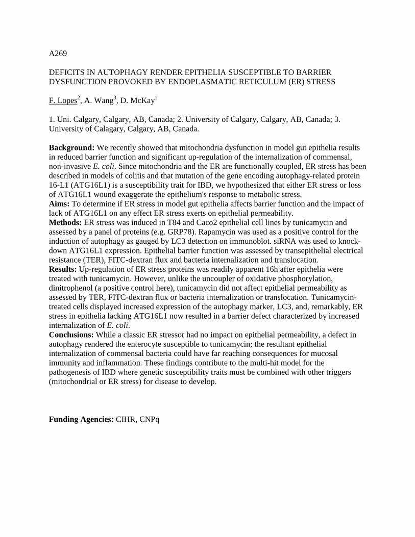
A269
DEFICITS IN AUTOPHAGY RENDER EPITHELIA SUSCEPTIBLE TO BARRIER DYSFUNCTION PROVOKED BY ENDOPLASMATIC RETICULUM (ER) STRESS
F. Lopes2, A. Wang3, D. McKay1
1. Uni. Calgary, Calgary, AB, Canada; 2. University of Calgary, Calgary, AB, Canada; 3. University of Calagary, Calgary, AB, Canada.
Background: We recently showed that mitochondria dysfunction in model gut epithelia results in reduced barrier function and significant up-regulation of the internalization of commensal, non-invasive E. coli. Since mitochondria and the ER are functionally coupled, ER stress has been described in models of colitis and that mutation of the gene encoding autophagy-related protein 16-L1 (ATG16L1) is a susceptibility trait for IBD, we hypothesized that either ER stress or loss of ATG16L1 wound exaggerate the epithelium's response to metabolic stress. Aims: To determine if ER stress in model gut epithelia affects barrier function and the impact of lack of ATG16L1 on any effect ER stress exerts on epithelial permeability. Methods: ER stress was induced in T84 and Caco2 epithelial cell lines by tunicamycin and assessed by a panel of proteins (e.g. GRP78). Rapamycin was used as a positive control for the induction of autophagy as gauged by LC3 detection on immunoblot. siRNA was used to knock-down ATG16L1 expression. Epithelial barrier function was assessed by transepithelial electrical resistance (TER), FITC-dextran flux and bacteria internalization and translocation. Results: Up-regulation of ER stress proteins was readily apparent 16h after epithelia were treated with tunicamycin. However, unlike the uncoupler of oxidative phosphorylation, dinitrophenol (a positive control here), tunicamycin did not affect epithelial permeability as assessed by TER, FITC-dextran flux or bacteria internalization or translocation. Tunicamycin-treated cells displayed increased expression of the autophagy marker, LC3, and, remarkably, ER stress in epithelia lacking ATG16L1 now resulted in a barrier defect characterized by increased internalization of E. coli. Conclusions: While a classic ER stressor had no impact on epithelial permeability, a defect in autophagy rendered the enterocyte susceptible to tunicamycin; the resultant epithelial internalization of commensal bacteria could have far reaching consequences for mucosal immunity and inflammation. These findings contribute to the multi-hit model for the pathogenesis of IBD where genetic susceptibility traits must be combined with other triggers (mitochondrial or ER stress) for disease to develop.
Funding Agencies: CIHR, CNPq
A270
VASOACTIVE INTESTINAL PEPTIDE (VIP) PROMOTES A PROTECTIVE TH17-ASSOCIATED IMMUNE RESPONSE TO INTESTINAL BACTERIAL INFECTION WITH C. RODENTIUM
X. Wu1, H. Yu1, V. Morampudiv1, B. Vallance2, K. Jacobson2
1. CFRI and University of British Columbia, Vancouver, BC, Canada; 2. BC Children's Hospital, Vancouver, BC, Canada.
Background: Intestinal infection with the mouse pathogen Citrobacter rodentium induces a strong local Th17 response in the colon. Although this inflammatory immune response helps clear the pathogen, it also induces inflammation-associated pathology in the gut and thus, has to be tightly controlled. Vasoactive intestinal peptide (VIP) is an enteric neuropeptide shown to modulate inflammation in animal models of IBD. We have previously shown that mucosal VIP immunoreactivity is increased post C. rodentium infection suggesting a potential modulatory role for VIP in bacterial colitis. Aims: Hence the aim of this study was to examine the impact of VIP on the infectious/ inflammatory processes elicited by the bacterial pathogen C. rodentium. Methods: Eight to ten week old female VIP-/- and littermates wild type (VIP+/+) mice were used. Mice received 2.5 × 10⁸ C. rodentium by gavage on day 0. Animal weights were monitored daily until day 10 post infection (p.i.), the day of sacrifice. The distribution of C rodentium on day 3, 6 and 8 p.i. was monitored by imaging with IVIS spectrum CT. On day 10 p.i., mice colons were removed and assessed for histology and bacterial burden. The distribution of colonic C. rodentium at day 10 p.i. was probed by Tir staining. In addition, mucosal IL-17A expression was examined by real-time PCR and the Th17 cell population in the lamina propria was measured by flow-cytometry analysis. Results: VIP-/- mice experienced significant weight loss (14.0 % vs. 3.1 %, n=16, p<0.001) and high mortality between days 7-10 p.i. compared to VIP+/+mice (40% vs. no mortality). Consistent with enhanced susceptibility to infection, the colonic Th17 cell response was impaired in VIP-/- mice, who showed significantly lower IL-17A expression (11.82 ± 2.61 vs. 30.76 ± 5.17, n=5, p<0.001) and a significantly lowered Th17 cell population (7.77 ± 0.73% vs. 12.80 ± 1.45%, n=5,p<0.05) than VIP+/+mice. Impaired bacterial clearance in the colon was also seen in VIP-/- mice (log cfu/g of bacteria: 9.95 ± 0.22 vs. 8.85 ± 0.18, n=8, p<0.05) with deeper penetration of Citrobacter into crypts, and increased systemic dissemination to liver and spleen. Taken together, these data suggest that VIP is crucial for the establishment/ maintenance of a functional Th17 response in intestinal infection with a murine pathogen. Conclusions: These data provide new sight into the function of VIP in regulating colonic mucosal immune response and in maintaining mucosal immune homeostasis under pathological conditions in vivo.
Funding Agencies: CCFC
A271
HIGH SALT DIET UP-REGULATES INTESTINAL TH17 CELLS AND INCREASES SUSCEPTIBILITY TO DSS COLITIS
P. Miranda, G. De Palma, J. Lu, M. Pigrau, S. Collins, P. Bercik
McMaster University, Hamilton, ON, Canada
Background: Specific environmental factors in genetically predisposed individuals can trigger dysfunctional immune responses, leading to chronic inflammatory diseases such as multiple sclerosis (MS), or inflammatory bowel disease (IBD). A recent study has shown that high concentrations of salt (NaCl) affects differentiation of human and mouse Th17 cells, that have been strongly implicated in the pathology of autoimmune diseases, such as MS and IBD. Mice fed with a high-salt diet (HSD) were shown to develop a severe form of experimental autoimmune encephalomyelitis (EAE), the animal model of MS. Also, high salt consumption has been associated with an increase in disease activity in MS patients but its role in colitis remains unknown. Aims: To explore the effect of HSD on intestinal immune homeostasis and susceptibility to colitis using a murine colitis model. Methods: Specific pathogen free NIH Swiss mice were fed a high salt diet (4% NaCl containing diet and 1% NaCl in drinking water) or a control diet for 4 weeks. Some mice were administered 3.5% DSS in drinking water for 5 days. Body weight, food and water consumption were monitored. Mice were then sacrificed and intestinal T-cell populations were quantified by Flow Cytometry. Microbiota composition profiles were analyzed by 16S DNA based DGGE and Illumina techniques. Colitis severity was assessed using clinical, macroscopic and microscopic scores. Results: Mice on HSD did not display any change in food and water consumption, or body weight compared to mice fed the control diet. However, HSD up-regulated Th17 cell population in mesenteric lymph nodes despite inducing a down-regulation of the total CD4+ population. This was associated with distinct microbial profiles in mice fed HSD compared to controls. DSS administration induced greater weight loss, higher colitis severity with increased incidence of blood in stool and cecum, reduced colon length and increased microscopic scores in mice fed the HSD compared to control diet. Conclusions: HSD induces immune activation in the gut characterized by increased Th17 cell populations and intestinal dysbiosis, and leads to increased sensitivity to experimental colitis. Our data suggest that high salt diet may constitute a dietary factor that contributes to the pathogenesis of IBD.
Funding Agencies: CIHR

A272
ENDOTHELIAL CELL-SPECIFIC TOLL-LIKE RECEEPTOR (TLR) 4 PROMOTES NEUTROPHIL RECRUITMENT IN EXPERIMENTAL COLITIS
T. Wang, C. Waterhouse
University of Calgary, Calgary, AB, Canada.
Background: Bacterial recognition by the innate immune system plays a role in the pathogenesis of inflammatory bowel diseases (IBD). TLR4, the receptor for Gram negative bacterial wall component lipopolysaccharide (LPS), is upregulated in active IBD patients, and some genetic polymorphisms of TLR4 are associated with the higher susceptibility to IBD. Because there are a number of cellular sources of TLR4 in the gastrointestinal tract, different TLR4-expressing cells might have unique roles in IBD pathogenesis. Aims: In this study, to test our hypothesis that endothelial TLR4 promotes neutrophil infiltration in experimental colitis, we utilized tamoxifen-inducible VE-Cadherin(VE-Cad)-Cre mice crossed to TLR4-loxp mice to produce endothelial cell-specific deleted TLR4 mice (TLR4ΔEndo) to investigate the role of endothelial TLR4 in chemical-induced colitis. Methods: TLR4ΔEndo mice and VE-Cad-Cre negative littermates mice (WT) were administrated 4% dextran sodium sulfate (DSS) in drinking water for 5 days to induce colitis (starting at Day 0), and received regular water for another 9 days to recovery. Colitis was measured by daily body weight change and stool consistency. At Day 5 and Day 14, entire colons were collected for assessments. Histological damage scoring was done in harvested colon samples. Colon myeloperoxidase (MPO) was assessed as the marker of neutrophil infiltration. Intravital microscopy was performed to measure neutrophil-endothelial interaction during colitis. Results: In acute (Day 5) and recovery (Day 14) colitis models, the daily body weight change, the length of colon and the stool consistency did not show the significant difference. In histological examination, TLR4ΔEndo manifested lower histological damage scores compared to control animals(TLR4ΔEndo v.s. WT: Day 5, 15±0 v.s. 12.33±0.57, p=0.008; Day 14, 16.33±0.71 v.s. 13±0, p=0.031). As measured by MPO, TLR4ΔEndo mice showed less neutrophils infiltration compared to wild type mice (TLR4ΔEndo v.s. WT: Day 5, 125.1±30.56 U/mg protein v.s. 259.9±7.45 U/mg protein, p=0.043; Day 14, 196.4±19.44 U/mg protein v.s. 242.6±10.70 U/mg protein, p=0.048). Intravital microscopy confirmed a lower number of adhered neutrophils (TLR4ΔEndo v.s. WT: Day 14, 0.274±0.098 /100μm blood vessel v.s. 1.056±0.220 /100μm blood vessel, p=0.0014), as well as a higher speed of neutrophil rolling (TLR4ΔEndo v.s. WT: Day 14, 21.54±2.124 μm/s v.s. 10.18±2.036 μm/s, p=0.003) in TLR4ΔEndo colons. Conclusions: Endothelial TLR4 promotes neutrophil recruitment and tissue damage in DSS colitis. Understanding the mechanism underlying this may lead to new IBD therapeutic strategies targeting immune cell recruitment to the intestinal tract.
Funding Agencies: None

A273
INTRAVITAL IMAGING OF THE COLON: BEHAVIOUR AND CELLULAR INTERACTIONS OF NKT CELLS IN HEALTH AND DISEASE
F. Zemp, C. Waterhouse, P. Kubes
University of Calgary, Calgary, AB, Canada.
Background: Inflammatory bowel disease (IBD) has a complex etiology, with diverse genetic, environmental and immunological factors playing roles in pathogenesis. Immunologically, skewing of the inflammatory cytokine milieu can dramatically affect disease outcome. A recently discovered group of cells termed invariant natural killer T-cells (iNKTs) are specialized in the rapid secretion of inflammatory cytokines, and their activation through glycolipid antigen recognition on MHC-related CD1d can result in the rapid polarization of immune responses. Recent literature has shown that iNKTs are important in mouse models of IBD, but have failed to demonstrate the role of resident colonic iNKTs. Further, the cellular interactions and activity of these cells in vivo has never been observed in the gut. We hypothesized that resident colonic iNKTs respond to glycolipid antigens presented on colonic macrophages with the resulting interactions being important for mediating homeostasis and disease. Aims: I: To develop an intravital microscopy technique to observe iNKT behavior in the colonic lamina propria of live rodents during health and disease. II: To determine how iNKTs mediate inflammation in the colon. Methods: To observe iNKTs in vivo using intravital spinning disk confocal microscopy we employed Cxcr6-GFP and Va14Ja18 mice, which overexpress iNKTs. CD1d-mediated antigen presentation was visualized in vivo using fluorescently conjugated glycolipid antigens. Finally, we constructed macrophage-specific (LysM-Cre) and epithelium-specific (Villin-Cre) CD1d knockout animals to examine the role these cells play in glycolipid antigen presentation. Results: We found iNKTs actively survey the colonic lamina propria in the steady state. These cells arrested upon administration of the glycolipid antigen, which was presented by perivascular macrophages and colonic epithelial cells. CD1d-knockout animals had exacerbated disease in the DSS model, which was associated with enhanced epithelial denudation and pronounced inflammatory cell recruitment. We found that CD1d expression on the epithelium, but not macrophages, was largely responsible for the protective response. Conclusions: iNKTs play important, albeit opposing, roles in several animal models of IBD. This is not surprising given their roles supporting different types of immune responses. Here we demonstrate that iNKTs actively survey the colonic lamina propria, and arrest upon recognition of glycolipid antigen. These cells play a protective role in DSS-mediated colitis, which is dependent on CD1d-antigen presentation in the epithelium. Current work is examining how epithelium-specific CD1d mediates colonic homeostasis and inflammatory responses, and suggests an exaggerated recruitment of polymorphonuclear cells to the lamina propria during inflammatory insult in mice lacking epithelial CD1d.

Intravital imaging of colonic iNKT cells (green) found that they survey outside of the subepithelial capillary network (blue) within the lamina propria. These cells survey on or near perivascular macrophages (purple) and the colonic epithelium (not shown).
Funding Agencies: CIHR, AIHS, CCFC
A274
COMPARISON OF MR ENTEROGRAPHY AND VIDEOCAPSULE ENDOSCOPY IN CHILDREN SUSPECTED TO HAVE ACTIVE CROHN'S DISEASE
A. BOBARNAC, C. Deslandres, M. Dirks SAINTE JUSTINE HOSPITAL, Montreal, QC, Canada.
Background: The identification of Crohn's disease in children can at times be delayed when there is predominance of small bowel involvement. MR enterography (MRE) has be come the new standard of care for the its identification. Aims: We have performed videocapsule endoscopy since 2006 to identify small bowel Crohn`s disease in children. MRE became routinely available in our centre in 2012. We compared the results of videocapsule studies to MRE in children to establish a diagnosis or relapse of Crohn's disease. Methods: A retrospective observational study was carried out at Sainte-Justine Hospital, from January 2012 to September 2014.Of 34 patients who underwent a videocapsule study to rule-out active Crohn's disease (based on clinical grounds, despite normal or non-specific upper endoscopy or colonoscopy findings),we excluded 12 patients who lacked an MRE study . A total of 22 patients with an average age of 15.1 years (6 - 18 years) , 12 girls and 10 boys, were evaluated. The capsule studies were all read blinded to the MRE results. There were no adverse events related to the capsule. Results: 45.5%5 (10 exams ) of the MRE were abnormal . 22.7%(5 exams ) of MRE exams revealed non-specific ileal changes , 22.7% (5 exams )showed only left colonic involement. 59.1% (13 cases) of the video capsule had significant findings typical of Crohn's disease of which 8 were sufficient to confirm the diagnosis . For small bowel involement only : Study normal MR enterography abnormal MR enterography Normal Video capsule 11 3 AbnormalVideo capsule 6 2 The mean interval between the two examinations was 5.3 months (1 -24 months) In 9 cases the results were discordant between the 2 tests. In 8 cases with normal small bowel MRE, a videocapsule study showed typical lesions of Crohn's disease with more then 5 deep mucosal ulcers with peripheral erythema and fibrin ( 5), mucosal thickening, cobblestoning, pseudopolyp formation ( 4) and circumferential ulceration with luminal narrowing (3 ) leading to the diagnosis of 4 new cases of Crohn's disease and 4 cases of relapsed Crohn. In 3 additional cases, videocapsule studies clearly demonstrated no Crohn's disease while the - MRE had suspected findings. Conclusions: Videocapsule endoscopy appears to be more sensitive than MR enterography in the detection of small bowel Crohn's disease in an experienced centre. For small bowel involement only :
Study normal MRE abnormal MRE Normal Videocapsule 11 3
Abnormal Videocapsule 6 2
Funding Agencies: None
A275
VACCINATION IN INFLAMMATORY BOWEL DISEASE PATIENTS: ATTITUDES, KNOWLEDGE AND UPTAKE IN A CANADIAN CENTRE
A. Rumman1, G. Malhi1, R. Thanabalan2, G. Nguyen1
1. Division of Gastroenterology, Department of Medicine, University of Toronto, Toronto, ON, Canada; 2. Mount Sinai Hospital, Toronto, ON, Canada.
Background: Immunosuppressive and biologic agents, used to treat inflammatory bowel disease (IBD), are associated with an increased risk of infection, including vaccine-preventable infections. Despite guideline recommendations, deficiencies in vaccine uptake have been documented. Aims: We assessed patient attitudes towards vaccination, knowledge of vaccine recommendations and uptake of recommended vaccines. We also aimed to identify predictors of vaccination completion. Methods: Randomly selected patients completed a self-administered, structured, paper-based questionnaire during a routine visit to our IBD Clinic. We collected information about medical and immunization history, education and employment status, and self-reported uptake of both routine childhood vaccinations and adult vaccinations recommended for IBD patients (influenza, pneumococcus, viral hepatitis, and varicella). We also assessed patient knowledge of vaccine recommendations, expectations for vaccination care and reasons for not taking vaccinations. Results: A total of 300 patients completed the survey. This included 173 (57.7%) patients with Crohn's disease, 120 (40.0%) with ulcerative colitis and 7 (2.3%) with indeterminate colitis. Mean age was 35.4 year, mean age at diagnosis was 23.4 years and 146 (48.7%) of participants were male. Current immunosuppressive therapy was reported by 171 (57.0%) patients: 112 (37.3%) on biologics, 48 (16.0%) on steroids, 48 (16.0%) on thriopurines and 17 (5.7%) on methotrexate. Self-reported vaccine completion was reported by 136 patients (45.3%). Vaccination uptake rates were 61.3% for influenza, 10.3% for pneumococcus, 61.0% for hepatitis B, 52.0% for hepatitis A and 26.0% for varicella. Significant (p<0.05) predictors of vaccine completion were annual vaccination review by family physician (OR = 1.82; 95% CI 1.27-2.62) or gastroenterologist (OR=1.72; 95% CI 1.10-3.10), current steroid use (OR=1.28; 95% CI 1.12-2.09), and current or prior treatment with biologics (OR=1.42; 95% CI 1.04-1.94). The majority of patients reported that the primary responsibility to ensure vaccine completion lies with the patient (41.7%), followed by the family physician and gastroenterologist (32.3% and 3.3%, respectively). Uncertainty about indications, fears of side effects and concerns regarding vaccine safety were the most common patient-reported reasons for non-uptake (22.0%, 20.7% and 5.3%, respectively). Conclusions: Uptake of recommended vaccines among IBD patients is suboptimal. Annual vaccination reviews by both family physicians and gastroenterologists contribute to improving vaccine uptake. Interventions targeted at improving vaccination uptake in IBD patients are needed.
Funding Agencies: Division of Gastroenterology, Department of Medicine, University of Toronto

A276
PREVALENCE OF DERMATOLOGICAL COMPLICATIONS OF ANTI-TNF-A THERAPY IN CROHN'S DISEASE
J. Marshall2, P. Bercik1, W. Reinisch1, D. Armstrong1, P. Moayyedi1, A. Nardelli1
1. McMaster University, Hamilton, ON, Canada; 2. McMaster University Medical Centre, Hamilton, ON, Canada.
Background: Skin lesions in patients with Crohn's disease (CD) may develop as a side effect of anti-tumor necrosis factor-a (anti-TNF-a) therapy or as a consequence of treatment with other immunomodulatory agents. Skin lesions may also represent incidental findings or extra-intestinal manifestations of inflammatory bowel disease (IBD). Aims: To investigate and describe new onset drug-associated dermatological manifestations associated with anti-TNF-a therapy in patients with CD Methods: This is an observational study of patients with CD on anti-TNF-a therapy seen consecutively at McMaster University Medical Centre outpatient clinic between October 2013 and September 2014. Presence of dermatological manifestations was assessed by clinical examination and documented by photographic records after obtaining patients' consent. Skin biopsy was not performed Results: In total, 1368 patients with IBD were reviewed, of whom 916 (67%) had CD. 339 patients with CD were on anti-TNF-a therapy. Sixty three (18.6%) patients with CD on anti-TNF-a therapy, without a previous history of psoriasis or atopic dermatitis (AD), developed dermatological side effects. The most common skin manifestations were: AD (17), inverse psoriasis (9), hand eczema (8), seborrhoeic dermatitis (6), pustular psoriasis (4), skin rash (4), nail dystrophy (3), pytiriasis rosea (3), urticaria (2), alopecia (2), acne (2), pythirosporum foliculitis (2) and pythiriasis liquenoide (1). Fifty five percent of these patients were receiving infliximab monotherapy and 37% were on combination therapy with azathioprine or methotrexate, 11% were receiving adalimumab and 5% were being treated with ustekinumab. Majority of these lesions was successfully managed by local treatment by a dermatologist. Conclusions: New skin lesions develop frequently during anti-TNF-a therapy, with atopic dermatitis and inverse psoriasis being the most common presentations.As the use of anti-TNF-a increases, the diagnosis and management of its cutaneous complications will become an increasingly important issue. Further studies investigating the relationship between IBD, biologics and skin-related diseases are warranted.
Funding Agencies: None
A277
MANNOSE BINDING LECTIN DEFICIENT MICE FAIL TO RECOVER FROM DSS INDUCED COLITIS: RELATIONSHIP WITH COLONIZATION OF THE INFLAMED COLON
U. Jain1, J. Li2, J. Butcher2, A. Stintzi2, A. Stadnyk1
1. Dalhousie University, Halifax, NS, Canada; 2. University of Ottawa, Ottawa, ON, Canada.
Background: The relationship between host complement and microbial colonization of the colon is not known, nor is the complement pathway that is activated during colitis known. Mannose binding lectins 1 and 2 (MBL1,2) are the proximal activators of the complement lectin pathway so we used MBL1,2 gene knockout mice (KO) to determine the role of the lectin pathway in colitis and colonization. Aims: To determine whether the lack of MBL1 and 2 has an impact on colitis and/or the colonization of healthy or inflamed colon. Methods: Groups of wildtype (WT) and KO mice were co-housed prior to adding 3% w/v DSS to their drinking water for 5 days then were euthanized 1 day (acute model) or 10 days (recovery model) after removing the DSS. Both stool and distal mucosal colon scapings were collected to determine the resident microbiota by high-throughput sequencing of the V6 hyper-variable region of the 16S rRNA gene. The results were then analyzed using the quantitative insights into microbial ecology (QIIME) and linear effect size (LEfSe) bioinformatic packages. Results: In the acute model, both strains experienced similar levels of colon inflammation although the MBL1,2-/- mice had considerably higher levels of C3a and C5a, indicating greater local complement activation. Contrasting this outcome, in the recovery model KO mice had significantly greater colon inflammation although anaphylatoxin levels were no long different between the strains. QIIME failed to identify differences in the overall taxonomic compositions between the microbiota of the co-housed WT and KO mice both before and after DSS treatment. However, there were significant differences in the microbial compositions observed in the stool samples as compared to those in the colon in addition to the differences between control/DSS treatments for both WT and KO mice. Moreover, while the overall taxonomic profiles between WT/KO mice are quite similar, LEfSe analysis suggests that the microbiota of the WT and KO mice respond differently to DSS treatment. Conclusions: We conclude that MBL is not required for complement activation and may actually attenuate complement activation in the DSS-inflamed colon. The heightened complement activation may impede the recovery of the inflamed colons of KO mice. We further conclude that MBL may not be important for eliciting colitis but is important in the host response while recovering from colitis, and that MBL may influence the microbiota's response to colitis as well.
Funding Agencies: CIHR, Nova Scotia Health Research Foundation, Crohn's and Colitis Foundation of Canada

A278
DETECTION OF CYTOMEGALOVIRUS IN PATIENTS WITH INFLAMATORY BOWEL DISEASE: WHERE TO BIOPSY, HOW MANY BIOPSIES, AND WHICH TEST?
J. McCurdy2, F. Enders1, A. Jones1, J. Killian1, E. Loftus1, D. Bruining1, T. Smyrk1
1. Mayo Clinic, Rochester, MN; 2. The Ottawa Hospital, Ottawa, ON, Canada.
Background: Cytomegalovirus (CMV) is an opportunistic pathogen that may negatively impact inflammatory bowel disease (IBD). This warrants efforts to improve the yield of current diagnostic techniques. Aims: Therefore, we sought to determine the optimal number and location of biopsies for detecting CMV in IBD patients. Methods: Patients with ulcerative colitis (UC), Crohn's disease (CD) or IBD undifferentiated (IBDU) and a tissue diagnosis of CMV were identified from our institutional database between 2005 and 2011. The number and location of biopsies, along with the endoscopic severity, and colonic distribution of inflammation were determined retrospectively. Biopsies were reviewed for inflammation and the presence of CMV by hematoxolyn and eosin (H&E), immunohistochemistry (IHC), and/or in situ hybridization (ISH). The proportion of positive biopsies from areas of inflammation was determined. Using data from the 25th percentile, we determined the optimal number of biopsies required to achieve an 80% probability of obtaining a single positive biopsy. Results: Sixty-eight patients with IBD (66% UC, 31% CD, and 3% IBDU) had a tissue diagnosis of CMV. Of those with a diagnosis by IHC or ISH who underwent multiple diagnostic techniques, 28 out of 62 (25%; 95% confidence interval [CI], 15%-36%) were positive by histopathology, and 11 out of 37 (31%; 95% CI, 16%-46%) were positive by whole blood polymerase chain reaction. Of the patients with biopsies proximal and distal to the splenic flexure from areas of active inflammation, 1 of 11 patients (9%) with UC and 4 of 8 patients (50%) with CD had a diagnosis of CMV limited to the right colon. Twenty percent of biopsies from inflamed mucosa were positive by IHC or ISH. Eleven biopsies in patients with UC and 16 in patients with CD were required to achieve an 80% chance of obtaining at least one positive biopsy. Conclusions: These findings highlight the importance of a tissue diagnosis, the colonic location, and the number of biopsies to maintain a high probability of detecting CMV.
Funding Agencies: None
A279
SURVEY OF COLONOSCOPY FOR THE PREVENTION AND DETECTION OF NEOPLASIA IN INFLAMMATORY BOWEL DISEASE (SCREEN-IBD)
Z. Gallinger2, G. Nguyen1
1. Mount Sinai Hospital, University of Toronto, Toronto, ON, Canada; 2. University of Toronto, Toronto, ON, Canada.
Background: The uptake of surveillance programs for colorectal cancer (CRC) in patients with inflammatory bowel disease (IBD) is unknown. The presence of multiple, sometimes dissimilar guidelines, may contribute to confusion among gastroenterologists. Studies suggest, despite specific national guidelines, that various factors, including location of practice and personal experience, may influence the use of surveillance guidelines. Aims: To examine the perception and practices of Canadian gastroenterologists towards surveillance of CRC in patients with IBD. Methods: A questionnaire was sent to all members of the Canadian Association of Gastroenterologists. Participants were subspecialists who had had participated in the care of IBD patients in the last 12 months. Questions were focused on gastroenterologists approach to surveillance colonoscopy as well as opinions and utilization of various new endoscopic techniques Results: There were 80 respondents to the survey, and among these, 72 met inclusion criteria and completed the survey (90% response rate). More academic than community gastroenterologist responded to the survey. Sixty four percent of respondents considered themselves IBD subspecialists. Approximately 80% of respondents do not use chromoendoscopy during colonoscopy, and of those that do (21%), only 33% use it greater than fifty percent of the time. The most common reported barrier (50%) to the use of chromondendoscopy is that it takes too much time. The majority of respondents (62%) believed that their institution required more endoscopic techniques to provide better care. There was a significant difference between community and academic physicians that take 2 to 4 random biopsies at 10 cm intervals for surveillance colonoscopies less than 50% of the time compared to those that do so greater than 50% of the time (10% vs. 4%, 22% vs. 64%, p =0.0096). Conclusions: Recent European guidelines suggest that chromoendoscopy is the preferred method for surveillance colonoscopy for CRC in IBD patients. Despite this, less than 20% of Canadian gastroenterologists surveyed use this technique. The most commonly cited obstacle is the lack of availability and training. There is a desire by gastroenterologists to obtain further training and funding for newer endoscopic modalities, including chromoendoscopy.
Funding Agencies: CIHR

A280
HNF4A : A TRANSCRIPTIONNAL REPRESSOR IN INTESTINAL CRYPTS?
C. Jones, F. Boudreau
Université de Sherbrooke, Sherbrooke, QC, Canada.
Background: HNF4α, an epithelial transcription activator, is downregulated in human intestinal bowel diseases (IBDs). In the adult mouse, epithelial deletion of HNF4α leads to spontaneous intestinal inflammation. In both humans and mice, Hnf4α is crucial for the maintenance of intestinal inflammatory homeostasis. Paneth and stem cells also play crucial roles in intestinal homeostasis and defense. In the bottom of the crypts, differentiated Paneth cells secrete anti-microbial compounds like lysozyme and cryptdins, whereas proliferation of stem cells is an essential process for the healing of the mucosa following an injury. Aims: Since the crypt compartment plays an important role in the maintenance of intestinal homeostasis, we aimed to investigate HNF4α regulatory functions in Paneth and stem cells. Methods: In mouse jejunal tissue, the adhesion molecule CD24 is detected at the crypt base. This molecule is a Paneth cell marker, but a weak expression of CD24 is also associated with the stem cell population. A conjugated antibody specific to CD24 was used to sort stem and Paneth cells from a pool of jejunal epithelial cells. These pools of cells were obtained from 3 wild type (WT) and 3 Villin-Cre/HNF4αloxp/loxp (KO) mice. RNA was extracted from the sorted cells and processed for RNA-sequencing. Results: Strong expression of stem and Paneth cell markers was recorded in the CD24+ population when compared to an enterocytes control population. IPA software was used to deepen the characterization of the CD24+ population. Interestingly, Hnf4α was predicted as an important upstream regulator in Paneth and stem cells. Gene expression in the KO mice population was then compared with the expression in the WT population. The majority (1157 out of 1411) of significantly modulated genes in the absence of Hnf4α were upregulated in Paneth and stem cells, suggesting a new potential repressor role for this regulator in these cells. Direct targets were then selected to investigate this potential dual new role. As an example, FgfR4 and Ido1, two relevant genes in the inflammatory context, were confirmed to be respectively downregulated and upregulated in mutant mice crypts. Conclusions: A repressor role for Hnf4α in intestinal crypts could represent an important breakthrough in the understanding of Hnf4α functions in the intestine, but also in IBD and carcinogenesis. ChIP-seq experiments are in progress to determine whether HNF4α can bind to promoter and enhancer regions of genes identified in the RNA-seq, and if the chromatin is active or inactive in these HNF4α bound regions. Immunoprecipitation of Hnf4α and mass spectrometry analysis are ongoing to identify partners that could further elucidate how Hnf4α acts as a repressor in this context. This work was financially supported by a grant from Vertex/CCC.
Funding Agencies: CCC, Vertex-CCC
A281
PREDICTIVE VALUE OF BASAL PLASMACYTOSIS ON CLINICAL RELAPSE IN QUIESCENT ULCERATIVE COLITIS
C. Dargavel, D. Kevans, B. Kabakchiev, R. Riddell, R. Kirsch, M. Silverberg
Mount Sinai Hospital, Toronto, ON, Canada.
Background: Endoscopic mucosal healing is a primary therapeutic goal in ulcerative colitis [UC]. Even when this is achieved evidence of histological activity has been observed. The absence of histological activity has been associated with improved clinical outcomes in UC. While data are few, histological markers such as basal plasmacytosis [BPC] have been associated with clinical relapse in UC. The predictive role of histological markers on clinical outcomes in UC remains incompletely assessed. Aims: To determine the predictive role of BPC and other histological markers on disease relapse and other clinically important UC outcomes. Methods: Adult UC patients with quiescent disease [endoscopic Mayo subscore of 0 or 1] and ≥12 months of follow-up between 2001 and 2014 at an academic tertiary referral centre were retrospectively included. A blinded expert pathologist evaluated all biopsies for the presence of BPC, eosinophils and histological activity [Geboes Score (GS)]. Primary outcomes were rate of disease relapse at 18 months; and time to first relapse; following index colonoscopy. Secondary outcomes were time to first corticosteroid or biological prescription, UC-related hospitalization, and colectomy; following index colonoscopy. Disease relapse was defined as a partial Mayo score ≥3. Results: This is a preliminary analysis of 62 [58% male, mean age 48.5 years] of an anticipated cohort of 86 patients. Average length of follow-up was 72.6 months. On initial colonoscopy, 56.5% patients had a Mayo 0 endoscopic subscore. At study entry, 67.7% were on 5ASA analogues, 27.4% were on immunomodulators, and 14.5% were on biologicals. At 18 months, disease relapse occurred in 34% of patients. Over the length of follow-up, disease relapse occurred in 63% of patients at an average of 26.1 months from study entry. Despite having quiescent disease, histological evaluation demonstrated the presence of BPC in 48%, active histology in 58%, and eosinophils in 68% of subjects. Presence of BPC was associated with a shorter time to disease relapse [median time to relapse 22 versus 46 months, p=0.035] and to corticosteroid prescription [median time to relapse 38 versus 65 months, p=0.048]. Presence of BPC trended towards being associated with disease relapse at 18 months [p=0.071]. Histological evidence of eosinophils and active inflammation [GS ≥3.1] were not significantly associated with the primary outcomes. Conclusions: A significant proportion of UC patients with endoscopically quiescent UC experience disease relapse at 18 months. BPC is an adjunctive histological marker associated with increased likelihood of disease relapse in this UC cohort. While prospective studies are required, the presence of BPC should prompt consideration of therapy optimization in UC patients.
Funding Agencies: University of Toronto, Division of Gastroenterology

A282
IBD PATIENTS ARE FREQUENTLY NON-ADHERENT WITH SCHEDULED INDUCTION AND MAINTENANCE INFLIXIMAB
C. Ma, C. Evaschesen, G. Gracias, V. Huang, D. Fedorak, K. Kroeker, L. Dieleman, B. Halloran, R. Fedorak, University of Alberta, Edmonton, AB, Canada. Background: Although infliximab has efficacy in inducing and maintaining clinical response in IBD patients, adherence to scheduled dosing is required to maintain therapeutic trough drug levels and prevent anti-infliximab antibody formation. Previous administrative database studies have not been powered to evaluate adherence to the infliximab induction or maintenance administration schedule. Aims: To characterize the adherence to regularly scheduled infliximab induction and maintenance in patients with IBD and to assess predictors of non-adherence. Methods: A retrospective cohort study was conducted evaluating adult (>17 years) outpatients with Crohn's disease (CD) or ulcerative colitis (UC) on scheduled infliximab from 2008-2010. Official infliximab infusion records were reviewed and non-adherence was defined by a discrepancy of >72 hours between the scheduled date of infliximab infusion and the date of administration. Patients were deemed non-adherent if they received <80% of their infliximab doses per schedule. Multivariate logistic regression was performed to evaluate predictors of non-adherence. Results: 215 patients (173 CD, 42 UC) met inclusion criteria. Patients received a median of 12.0 (IQR7.0-13.0) infliximab infusions during the study period. 412 induction and 1837 maintenance infliximab doses were administered. 109/140 patients (77.9%) were adherent to infliximab induction; 68/215 patients (31.6%) were adherent to their maintenance regimen. Mean variance from an individual infliximab induction and maintenance infusions was 1.1 days (± 1.6) and 4.0 days (± 4.6), respectively. 92.1% of patients received at least one delayed maintenance infusion and 10.1% of patients received maintenance infusions on average more than one week late. In multivariate logistic regression analysis, only male gender (OR 1.77 [95% CI 1.01-3.11]) was predictive of non-adherence. Conclusions: While three quarters of patients are adherent with infliximab induction therapy, less than one third remain closely adherent to their maintenance infliximab schedule. Table 1: Baseline Patient Demographics and Adherence to Infliximab Therapy
Patients receiving induction IFX 140 Patients receiving maintenance IFX 215
Mean age at IFX induction (years, ±SD) 40.8 ± 13.7 Median lifetime IFX infusions (n, IQR) 12.0 (7.0-19.5) Median study follow-up (weeks, IQR) 80.1 (38.7-100.9)
Patients adherent to IFX induction 109 / 140 (77.9) Patients adherent to IFX maintenance 68 / 215 (31.6)
Mean delayed induction infusions per patient (±SD) 0.5 ± 0.6 Mean delayed maintenance infusions per patient (±SD) 3.5 ± 2.5
Funding Agencies: The Centre of Excellence for Gastrointestinal Inflammation and Immunity Research (CEGIIR)

A283
DEVELOPMENT AND VALIDATION OF A NUTRITION SCREENING TOOL TO DETECT NUTRITION RISK IN INFLAMMATORY BOWEL DISEASE
N. Haskey1, J. Pena-Sanchez2, J. Jones2, S. Fowler2
1. Saskatoon Health Region, Food and Nutrition Services, Saskatoon, SK, Canada; 2. Division of Gastroenterology, Department of Medicine, University of Saskatchewan, Saskatoon, SK, Canada.
Background: Malnutrition is a well-known complication of Inflammatory Bowel Disease (IBD). A nutrition screening tool to detect nutrition risk in patients with IBD does not currently exist. The Malnutrition Universal Screening Tool (MUST) has been validated in the outpatient setting, but not specifically in an IBD patient population. Aims: To develop and validate a screening tool to detect nutrition risk in patients with IBD. Methods: A nutrition screening tool, Saskatchewan IBD - Nutrition Risk (SaskIBD-NR), was developed and administered alongside the MUST to patients attending an outpatient gastroenterology clinic. Nutrition risk was assessed by the IBD clinic dietitian (RD) and treating gastroenterologist (GI), and was taken as the gold standard. Agreement between the SaskIBD-NR tool and RD/GI nutrition risk assessment was evaluated by computing Cohen's kappa; this was also used to test agreement between MUST and RD/GI assessment, and between the two screening tools. Sensitivity and specificity were calculated for the MUST and SaskIBD-NR tools, as well as their positive and negative predictive values. Results: In total, 110 patients with IBD were screened and had RD/GI nutrition risk assessments completed. The mean age of the sample was 38.6 years (SD=14.7), and 47 (42.7%) were male and 63 (57.3%) were female. Seventy-four (67.3%) had CD and 36 (32.7%) had UC. The mean BMI was 26.38 kg/m2 (SD=5.16) and 41 patients (37.3%) stated that they had been restricting foods. The RD/GI assessment established that 23 patients (20.9%) were at nutrition risk. The SaskIBD-NR tool classified 21 (19.1%) patients at medium or high risk, while MUST classified 17 (15.5%). The SaskIBD-NR tool had a significant agreement with the RD/GI assessment (k: 0.83, p<0.001), while MUST showed a lack of agreement (k: 0.15, p=0.12). There was poor agreement between the SaskIBD-NR and MUST screening tools (k: 0.18, p=0.06). When compared with RD/GI assessment, the SaskIBD-NR had a better sensitivity (82.6% vs. 26.1%), specificity (97.7% vs. 87.4%), positive predictive value (90.5% vs. 35.3%), and negative predictive value (95.5% vs. 81.7%) than the MUST. Conclusions: The SaskIBD-NR, which assesses GI symptoms, food restriction, and weight loss, adequately detects nutrition risk in IBD patients. In contrast, the MUST did not adequately detect nutrition risk in the IBD outpatient population.
Funding Agencies: None

A284
TITLE: PATIENT PREFERENCES OF SYMPTOM CONTROL AND INVOLVEMENT IN TREATMENT DECISIONS IN INFLAMMATORY BOWEL DISEASE
T. Mombourquette2, J. Stewart2, M. Black2, D. MacIntosh1
1. Queen Elizabeth II Health Science Centre, Halifax, NS, Canada; 2. Queen Elizabeth II Health Sciences Centre, Halifax, NS, Canada
Background: Inflammatory bowel disease (IBD) patients constantly have to manage symptoms. It is important for the healthcare providers to know their patients and their feelings of what symptoms are most important to them. Aims: The purpose of the study is to investigate how patients perceive what is most important to their healthcare providers compared to what the desires of the patient themselves actually are. Methods: A questionnaire was given to consecutive patients attending the IBD clinic The questionnaire contained items related to demographics, disease-related symptoms and outcomes that participants felt most impacted their quality of life, as well as their perception of what disease-related symptoms and outcomes that their heath care provider(s) felt were most important. Participants ranked the importance of each symptoms and outcome on a Likert scale from one to five (one being the most important, five being the least important). The patient was given a list of ten symptoms including abdominal pain and diarrhea, and a list of nine outcomes including complete resolution of symptoms and staying alive to rank according to the scale of importance. Statistical analysis is descriptive with rank sums of responses to each item expressed as percentages. Results: One hundred and twelve surveys were returned. 100% of the surveys returned had information for both the patient's most important and second most important symptom to control. Responses to questionnaire items that were ranked as "most important" were given 5 points and those ranked as "least important" were given 1 point. There was concordance between what factors participants perceived as being important and those factors participants perceived were important to their health-care provider. Twenty-one percent of patients surveyed felt that the most important disease-related symptom to them was abdominal pain, while 21.9% felt that abdominal pain was most important to their healthcare provider. 19.8% responded that diarrhea was more important to them and 21.3% said diarrhea was more important to their healthcare provider. The outcome ranked as being of greatest importance was avoidance of death (16.5%). Eighteen percent perceived that this outcome was of greatest importance to their healthcare provider as well. Conclusions: The study shows that patients believe both they and their healthcare providers similarly believe certain symptoms and outcomes are most important to control in IBD treatment. Patients feel that their healthcare providers may believe certain symptoms or outcomes are more important to them than they are to the patients.
Funding Agencies: None

A285
PSYCHOLOGICAL PROFILING IN CROHN'S DISEASE: EMOTION PROCESSING DISTRESS AND BODY CONNECTION ISSUES ARE THE DOMINANT COPING STRATEGIES IN SEVERE DISEASE
J. Paulton1, G. Ou1, A. Round1, C. Galorport2, R. Enns2
1. Faculty of Medicine, University of British Columbia, Vancouver, BC, Canada; 2. Pacific Gastroenterology Associates, Vancouver, BC, Canada.
Background: There is debate whether psychological treatments would benefit patients with Crohn's disease (CD). Emotion processing and body connection are central psychological constructs affected by chronic diseases. Emotion processing distress disrupts absorbing disturbing events and is experienced in anxiety attacks, colorectal cancer and chronic back pain ailments. Body connection is awareness of body states and cues. Body vigilance is body awareness placed upon internal sensations and is heightened in panic disorders. Private body consciousness is attendance to internal bodily states and is associated with greater frequency and intensity of chronic pain. Dissociation is neglect of body states and affects managing chronic diseases. Aims: The study aimed to identify, in CD patients, constructs meaningful in psychotherapy, hypothesizing that emotion processing distress and body awareness issues would be present in severe disease. Methods: A prospective cross-sectional study was conducted at St. Paul's Hospital, Vancouver, BC. Fifty outpatients participated; 36% were in remission, 30% had mild activity, and 34% had moderate-to severe activity. Participants completed validated surveys, including: the Emotional Processing Scale, Scale of Body Connection, Private Body Consciousness Scale, and Body Vigilance Scale. Scores were compared to control standards derived from the general population. The Harvey Bradshaw Index was used to assess CD severity. Results: Moderate-to severe disease patients emotion processing distress was significantly greater than healthy controls and remission (p<0.001, p<0.001; Figure 1). CD activity was significantly correlated to emotion processing distress (r=0.458, p=0.001). Moderate-to severe patients body vigilance was significantly greater than healthy controls and remission (p<0.001, p=0.0069; Table 1). Private body consciousness was significantly greater in moderate-to severe CD than chronic pain controls (p<0.001). Body dissociation was significantly correlated to emotion processing distress (r=0.722, p<0.001). Conclusions: This study, using validated psychological surveys indicates emotion processing distress and body awareness issues in moderate-to severe Crohn's, where theoretically, psychological treatments would be most helpful.
Table 1. Body Vigilance of Crohn's Disease Subgroups versus Controls Crohn's Disease Subgroups versus Controls Body Vigilance
Normal 17.9 Panic Disorder 22.6 Crohn's AVG 20.3

Remission 17.0 Mild 21.2
Moderate-severe 22.7
Funding Agencies: Faculty of Medicine, University of British Columbia
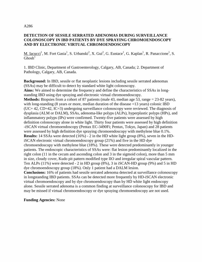
A286
DETECTION OF SESSILE SERRATED ADENOMAS DURING SURVEILLANCE COLONOSCOPY IN IBD PATIENTS BY DYE SPRAYING CHROMOENDOSCOPY AND BY ELECTRONIC VIRTUAL CHROMOENDOSCOPY
M. Iacucci1, M. Fort Gasia1, S. Urbanski2, X. Gui2, G. Eustace1, G. Kaplan1, R. Panaccione1, S. Ghosh1
1. IBD Clinic, Department of Gastroenterology, Calgary, AB, Canada; 2. Department of Pathology, Calgary, AB, Canada.
Background: In IBD, sessile or flat neoplastic lesions including sessile serrated adenomas (SSAs) may be difficult to detect by standard white light colonoscopy. Aims: We aimed to determine the frequency and define the characteristics of SSAs in long-standing IBD using dye spraying and electronic virtual chromoendoscopy. Methods: Biopsies from a cohort of 87 patients (male 43, median age 53, range = 23-82 years), with long-standing (8 years or more, median duration of the disease =13 years) colonic IBD (UC= 42, CD=42, IC=3) undergoing surveillance colonoscopy were reviewed. The diagnosis of dysplasia (ALM or DALM), SSAs, adenoma-like polyps (ALPs), hyperplastic polyps (HPs), and inflammatory polyps (IPs) were confirmed. Twenty-five patients were assessed by high definition colonoscopy alone in white light. Thirty four patients were assessed by high definition -iSCAN virtual chromoendoscopy (Pentax EC-3490Fi; Pentax, Tokyo, Japan) and 28 patients were assessed by high definition dye spraying chromoendoscopy with methylene blue 0.1%. Results: 14 SSAs were detected (16%) - 2 in the HD white light group (8%), seven in the HD-iSCAN electronic virtual chromoendoscopy group (21%) and five in the HD dye chromoendoscopy with methylene blue (18%). These were detected predominantly in younger patients. The endoscopic characteristics of SSAs were: flat lesion predominantly localized in the right colon (11 in the cecum and ascending colon and 3 in the sigmoid colon), more than 5 mm in size, cloudy cover, Kudo pit pattern modified type IIO and irregular spiral vascular pattern. Ten ALPs (11%) were detected - 2 in HD group (8%), 3 in iSCAN-HD group (9%) and 5 in HD dye chromoendoscopy group (18%). Only 1 patient had a DALM lesion. Conclusions: 16% of patients had sessile serrated adenoma detected at surveillance colonoscopy in longstanding IBD patients. SSAs can be detected more frequently by HD-iSCAN electronic virtual chromoendoscopy and by dye chromoendoscopy than by HD white light endoscopy alone. Sessile serrated adenoma is a common finding at surveillance colonoscopy for IBD and may be missed if virtual chromoendoscopy or dye spraying chromoendoscopy are not used.
Funding Agencies: None

A287
CLINICAL OUTCOMES IN CONTINUOUS CLINICAL RESPONDERS WITH MODERATELY TO SEVERELY ACTIVE UC: SUB-ANALYSES FROM THE PURSUIT-SC MAINTENANCE STUDY
B. Feagan1, W. Sandborn3, J. Colombel4, W. Reinisch5, P. Gibson6, C. Marano2, R. Strauss7, J. Johanns7, H. Zhang7, H. Weng8, R. Yao8, D. Tarabar9, Z. Hebzda10, P. Rutgeerts11
1. Robarts Clinical Trials Inc, London, ON, Canada; 2. Janssen R&D, LLC, Spring House, PA; 3. University of California San Diego, La Jolla, CA; 4. Hopital Claude Huriez, Lille Cedex, France; 5. McMaster University Hospital, Hamilton, ON, Canada; 6. Alfred Hospital, Melbourne, VIC, Australia; 7. Janssen R&D, LLC., Spring House, PA; 8. Merck Sharp & Dohme, Kenilworth, NJ; 9. Military Medical Academy, Belgrade, Serbia; 10. Klinika Chorob Wewnetrznych, Krakow, Poland; 11. University Hospital Gathuisberg, Leuven, Belgium.
Background: . Aims: To evaluate long-term clinical outcomes in moderately to severely active UC pts who achieved complete continuous response(CCR) vs pts who did not achieve CCR(non-CCR) through Wk54 of SC GLM maintenance. Methods: During PURSUIT-Maintenance,GLM induction responders(464pts)were randomized to PBO,SC GLM50mg, or SC GLM100mg at bsl(Wk0) and q4wks through Wk52.Primary endpoint was clinical response through Wk54(CCR). Clinical remission, mucosal healing, corticosteroid use, and IBDQ outcomes and fecal markers at Wk54 among CCR vs non-CCR were assessed. All sub-analyses are based on pts randomized at Wk0 of maintenance (n=456). Results: CCR pts had better results vs non-CCR pts. Among pts receiving corticosteroids at baseline, a greater proportion of CCR pts were not receiving corticosteroids at Wk54 vs non-CCR pts. Greater proportions of CCR pts were also in clinical remission vs non-CCR pts. Mean decreases in fecal lactoferrin and fecal calprotectin at Wk54 from Wk0 of maintenance were greater for CCR pts vs non-CCR pts. Data between the GLM groups were similar and were pooled(Table).. Conclusions: These data support that pts induced into clinical response who maintain a clinical response through Wk54 are more likely to have better clinical outcomes.
Outcomes based on continuous clinical response at Wk54 in PURSUIT-SC maintenance*
Clinical endpoints Non-CCR CCR
PBO Combined GLM PBO Combined
GLM Corticosteroid Use
Randomized pts receiving concomitant steroids at Wk0(n) 60 87 27 73
Pts not receiving corticosteroids at Wk54 (%) 1.7 4.6 66.7 75.3

Remission Randomized pts (n) 106 156 48 146
Pts in clinical remission at Wk54(%) 0.9 1.9 68.8 67.1 Mucosal healing
Randomized pts (n) 106 156 48 146 Pts with mucosal healing at Wk54 (%) 1.9 2.6 87.5 90.4
IBDQ score Randomized pts (n) 105 156 48 144
Changes from Wk0 through Wk54 [mean(SD)]
-38.9(32.1) -36.9(37.6) 10.6(18.2) 11.3(28.1)
Pts with IBDQ score>170 at Wk54(%) 18.1 24.4 81.2 75.0 *Pts were in response to GLM induction and were randomized to either PBO or GLM 50 or 100mg (combined)
Funding Agencies: None

A288
C-REACTIVE PROTEIN IS ELEVATED WITH CLINICAL DISEASE ACTIVITY DURING PREGNANCY IN WOMEN WITH INFLAMMATORY BOWEL DISEASE
J. Bal1, V. Huang1, L. Ambrosio1, R. Foshaug1, K. Kroeker1, B. Halloran2, L. Dieleman1, R. Fedorak2
1. University of Alberta, Edmonton, AB, Canada; 2. University of Alberta, Edmonton, AB, Canada.
Background: Inflammatory bowel disease (IBD), classified into either Crohn's disease (CD) or ulcerative colitis (UC), is a chronic inflammatory condition of the gastrointestinal tract. Women with IBD have a risk of flaring their IBD during pregnancy. C-reactive protein (CRP) is an acute-phase serum marker of inflammation that is often elevated with flares of IBD. CRP can also be elevated during healthy pregnancies. In other words, it is unclear whether CRP can be used as a non-invasive biomarker of clinical disease activity in pregnant women with IBD. Aims: We aim to determine if CRP is elevated in women with clinically active IBD compared to non-active IBD during pregnancy. Methods: Female IBD patients (18 to 45 yrs) were enrolled pre-conception (PC) or at each trimester of pregnancy (Tx). At each clinic visit, women were grouped by clinical disease activity, using the Modified Harvey Bradshaw Index (mHBI) for Crohn's disease patients and Partial Mayo Score (pMayo) for ulcerative colitis patients. Active disease was considered an HBI score greater than or equal to 5 and a pMayo score greater than or equal to 2. CRP was also measured at each clinic visit; only patients who had previously documented CRP elevations (levels greater than 8.00 mg/L) with flares of their IBD were included for analysis. Results: Twenty-three patients (14 UC and 9 CD patients of median age 29.0 yrs) seen over 52 clinic visits were included for analysis. To examine the association of CRP with clinical disease activity, we compared CRP in women with clinically active and non-active disease. The median CRP trended higher in women with clinically active disease compared to those with clinically non-active disease at PC (6.95 vs 2.80 mg/L; p=0.559), T1 (24.75 vs 6.00 mg/L; p=1.000) and T2 (8.85 vs 7.10 mg/L; p=1.000), respectively. During T3, women with clinically non-active disease had a higher median CRP than women with clinically active disease (6.95 vs 5.45 mg/L, respectively; p=0592). Conclusions: Women with IBD who had clinically active disease during preconception and the first and second trimesters of pregnancy had higher CRP levels than women who had clinically non-active disease. This suggests that CRP remains a potential tool for assessing clinical IBD activity in the early trimesters of pregnancy.
Funding Agencies: Alberta Innovates Health Solutions (AIHS), Women and Children’s Health Research Institute (WCHRI), Alberta IBD Consortium, and Center of Excellence for Gastrointestinal Inflammation and Immunity Research (CEGIIR).

A289
DOES A COMPLICATED CROHN'S DISEASE BEHAVIOR AT BASELINE PREDICT AN AGGRESSIVE DISEASE COURSE?
S. O'Donnell1, K. Borowski1, J. Stempak1, R. Milgrom1, M. Silverberg2
1. Zane Cohen Centre for Digestive Diseases, Mount Sinai Hospital, Toronto, ON, Canada; 2. Mount Sinai Hospital, Toronto, ON, Canada.
Background: The presence of stricturing or penetrating disease is often reported as a risk factor for an aggressive or disabling Crohn's disease (CD) course. Aims: The aim of our study was to determine whether those who have stricturing or penetrating disease at presentation follow an aggressive course. Methods: 393 CD cases were included in this retrospective observational study. Baseline phenotype at or within 12 months of diagnosis was documented. Categorical variables were compared using χ<+>2<+> tests or logistic regression while Kruskal Wallis or linear regression were used for continuous variables. Time to an event was evaluated using a Kaplan-Meier survival analysis. Results: Clinical characteristics and results are shown in Table 1. 30.8% of cases had B2/B3 disease at baseline. Predictors of B2/B3 behavior at diagnosis were an older age at diagnosis, absence of perianal disease and ileal involvement in a multinominal logistic regression. B2/B3 disease at diagnosis was associated with a higher number of hospital admissions compared to B1. This was paralleled by a higher rate of abdominal surgery in those with B2/B3 behavior at diagnosis. The time to first abdominal surgery was significantly longer for those with B1 behavior at diagnosis [B1- 14.7 yrs (95% CI 11.15-18.2), B2- 3.13 yrs (95% CI 1.58-4.69), B3- 1 yr (95% CI 0.53-1.47), log rank, p <0.000001]. However, the time to a second surgery was actually longer for those with B2/B3 baseline behavior, though this difference was not statistically significant different, (B1 - 8.18 yrs, B2 - 17.92 yrs and B3 - 15.73 yrs, p=0.539). Immunosuppressant therapies, including anti-TNFs, were used more frequently in those with baseline inflammatory disease; however, there was no difference in the median time to initiation of an anti-TNF across the 3 groups. Conclusions: While B2/B3 disease is commonly reported as a predictor of aggressive CD, our data suggests that this may not be the case for those who have complicated disease behavior at presentation. While these cases have surgery at an early time point; they appear to progress slowly after initial surgical intervention.
Table 1
B1 B2 B3 n (%) 272 (69.2) 69 (17.6) 52 (13.2)
Male Gender 140 (51.5) 38 (55.1) 34 (65.4) p=0.179 L1 L2 L3
37 (13.8) 96 (35.8) 135 (50.4)
14 (20.3) 6 (8.7)
49 (71.0)
14 (26.9) 7 (13.5) 31 (59.6)
p=0.00001*

Upper GI involvement 73 (26.8) 18 (26.1) 10 (19.2) p=0.514 Perianal Disease 104 (38.2) 15 (21.7) 10 (19.2) p=0.003*
Abdominal Surgery 112 (41.2) 52 (75.4) 45 (86.5) p<0.00001* Age at Diagnosis 22.81 yrs 27.49 yrs 25.87 yrs p=0.006*
Mean No of Hospitalizations 1.04 1.42 1.67 p=0.00002* Anti-TNF therapy 152 (55.9%) 29 (42.0%) 20 (38.5%) p=0.018* Mean Follow Up 12.4 11.8 12.0 p=0.59
Funding Agencies: CAG, CCC, CIHR
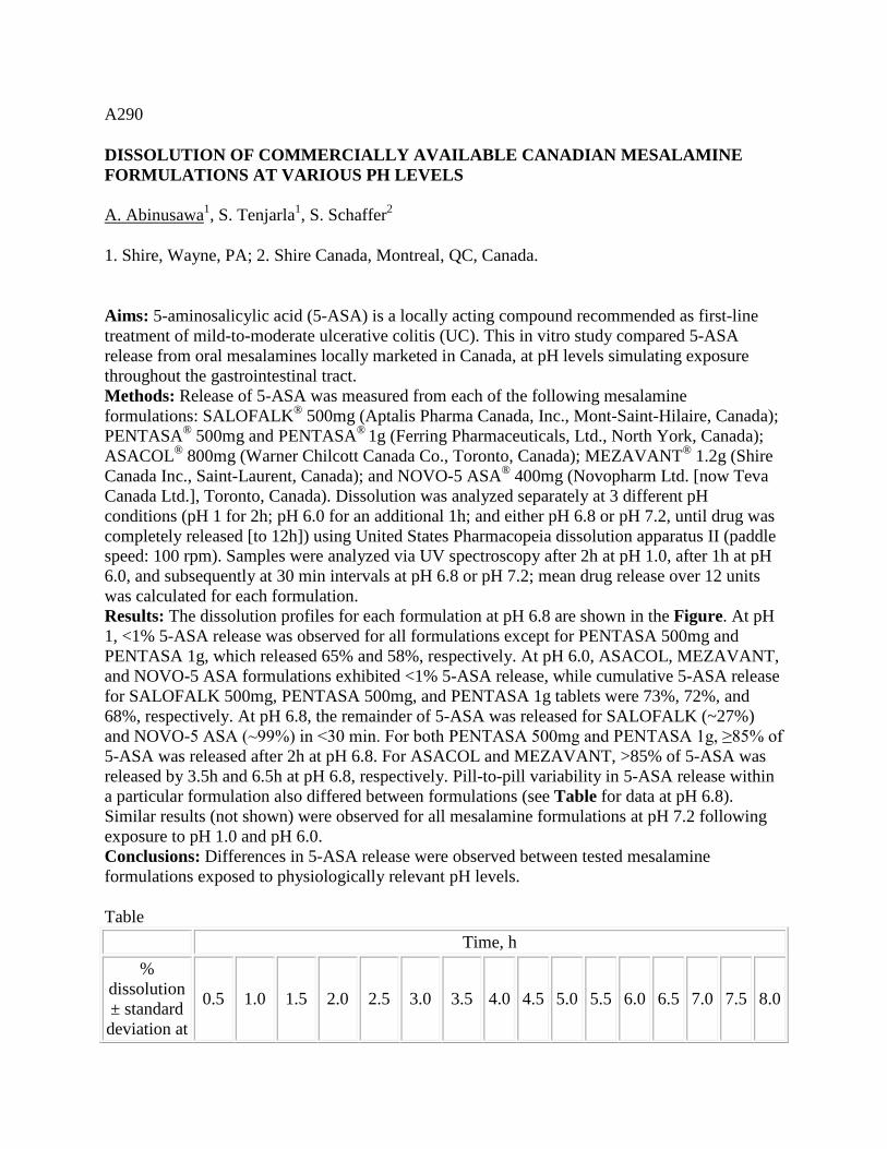
A290
DISSOLUTION OF COMMERCIALLY AVAILABLE CANADIAN MESALAMINE FORMULATIONS AT VARIOUS PH LEVELS
A. Abinusawa1, S. Tenjarla1, S. Schaffer2
1. Shire, Wayne, PA; 2. Shire Canada, Montreal, QC, Canada.
Aims: 5-aminosalicylic acid (5-ASA) is a locally acting compound recommended as first-line treatment of mild-to-moderate ulcerative colitis (UC). This in vitro study compared 5-ASA release from oral mesalamines locally marketed in Canada, at pH levels simulating exposure throughout the gastrointestinal tract. Methods: Release of 5-ASA was measured from each of the following mesalamine formulations: SALOFALK® 500mg (Aptalis Pharma Canada, Inc., Mont-Saint-Hilaire, Canada); PENTASA® 500mg and PENTASA® 1g (Ferring Pharmaceuticals, Ltd., North York, Canada); ASACOL® 800mg (Warner Chilcott Canada Co., Toronto, Canada); MEZAVANT® 1.2g (Shire Canada Inc., Saint-Laurent, Canada); and NOVO-5 ASA® 400mg (Novopharm Ltd. [now Teva Canada Ltd.], Toronto, Canada). Dissolution was analyzed separately at 3 different pH conditions (pH 1 for 2h; pH 6.0 for an additional 1h; and either pH 6.8 or pH 7.2, until drug was completely released [to 12h]) using United States Pharmacopeia dissolution apparatus II (paddle speed: 100 rpm). Samples were analyzed via UV spectroscopy after 2h at pH 1.0, after 1h at pH 6.0, and subsequently at 30 min intervals at pH 6.8 or pH 7.2; mean drug release over 12 units was calculated for each formulation. Results: The dissolution profiles for each formulation at pH 6.8 are shown in the Figure. At pH 1, <1% 5-ASA release was observed for all formulations except for PENTASA 500mg and PENTASA 1g, which released 65% and 58%, respectively. At pH 6.0, ASACOL, MEZAVANT, and NOVO-5 ASA formulations exhibited <1% 5-ASA release, while cumulative 5-ASA release for SALOFALK 500mg, PENTASA 500mg, and PENTASA 1g tablets were 73%, 72%, and 68%, respectively. At pH 6.8, the remainder of 5-ASA was released for SALOFALK (~27%) and NOVO-5 ASA (~99%) in <30 min. For both PENTASA 500mg and PENTASA 1g, ≥85% of 5-ASA was released after 2h at pH 6.8. For ASACOL and MEZAVANT, >85% of 5-ASA was released by 3.5h and 6.5h at pH 6.8, respectively. Pill-to-pill variability in 5-ASA release within a particular formulation also differed between formulations (see Table for data at pH 6.8). Similar results (not shown) were observed for all mesalamine formulations at pH 7.2 following exposure to pH 1.0 and pH 6.0. Conclusions: Differences in 5-ASA release were observed between tested mesalamine formulations exposed to physiologically relevant pH levels.
Table
Time, h %
dissolution ± standard deviation at
0.5 1.0 1.5 2.0 2.5 3.0 3.5 4.0 4.5 5.0 5.5 6.0 6.5 7.0 7.5 8.0

pH 6.8 SALOFALK 500 mg
102 ±2.4
102 ±2.3
102 ±2.2
PENTASA 500 mg
78 ±1.8
83 ±1.6
86 ±1.5
89 ±1.5
91 ±1.5
92 ±1.5
94 ±1.6
95 ±1.6
95 ±1.7
96 ±1.7
96 ±1.7
97 ±1.8
97 ±1.8
PENTASA 1 g
74 ±1.2
78 ±1.1
82 ±1.0
85 ±1.0
87 ±1.0
89 ±1.0
91 ±1.0
92 ±1.0
93 ±1.0
94 ±1.0
95 ±1.0
95 ±1.0
95 ±1.0
ASACOL 800 mg
34 ±31.
4
45 ±40.
4
52 ±40.
7
59 ±39.
6
73 ±31.
2
82 ±25.
0
89 ±10.
9
91 ±7.7
93 ±6.2
94 ±5.0
95 ±4.1
95 ±3.3
96 ±2.6
96 ±2.1
96 ±1.8
97 ±1.5
MEZAVANT 1.2 g
2 ±1.8
8 ±2.5
14 ±1.5
20 ±1.5
26 ±2.1
34 ±2.9
42 ±3.4
50 ±4.3
58 ±5.2
67 ±5.9
76 ±7.0
84 ±7.0
92 ±5.7
95 ±3.9
99 ±2.0
100 ±1.8
NOVO-5 ASA 400
mg
102 ±3.4
101 ±3.0
101 ±3.0
Funding Agencies: Shire Development LLC

A291
PARKING PRICES INFLUENCING PATIENTS ABILITY TO SEEK HEALTH CARE
K. MacIsaac, J. Stewart, D. MacIntosh, S. Smith
Queen Elizabeth II Health Science Centre, Halifax, NS, Canada.
Background: The Health Canada Act set 5 principles that the health care system was to ensure for all patients. One of these principles is accessibility, which ensures "financial or other barriers to the provision of publicly funded health services are discouraged, so that health services are available to all Canadians when they need them". Aims: In the Capital District Health Authority (CDHA), a region responsible for the care of half of all Nova Scotians, the price of parking can vary from no charge up to $14.50/day depending on the site. We wished to determine if patient accessibility was being affected by financial burden on patients attending healthcare facilities in CDHA. Methods: Patients are surveyed annually as part of the Division of Gastroenterology's quality assurance program. During 2013, questions about parking were introduced into these surveys that were distributed to all patients undergoing endoscopy. In the summer of 2014 it was also included into new outpatient and IBD clinic surveys. All responses were optional and anonymous; the questionnaires and pre-paid envelopes were given to patients at the end of their clinic visit or upon discharge from their procedure. Results: We received a total of 1157 responses with regard to parking prices. Of these, 915 (78.9%) of respondents paid for parking. Of the patients that recorded their cost of parking, 234 patients reported paying less than three dollars, 227 patients 3 to 5 dollars, 148 paid 5 to 8 dollars, 73 paid 8 to 11 dollars, and 24 patients paid over 11 dollars for parking. The highest reported parking cost was $17. Patients were subsequently asked if parking charges presented a hardship. One hundred and twenty-five (13.7%) stated that the cost of parking presented a hardship. Subjects were also asked if the cost of parking might influence attendance at future appointments. Seventy-four (8.1%) of patients reported that parking costs might impact their ability to attend future appointments. Conclusions: The rising cost of parking may influence health care provision for our population. Recent increases in parking prices provide a potential barrier for a proportion of patients attending GI services in CDHA.
Funding Agencies: None
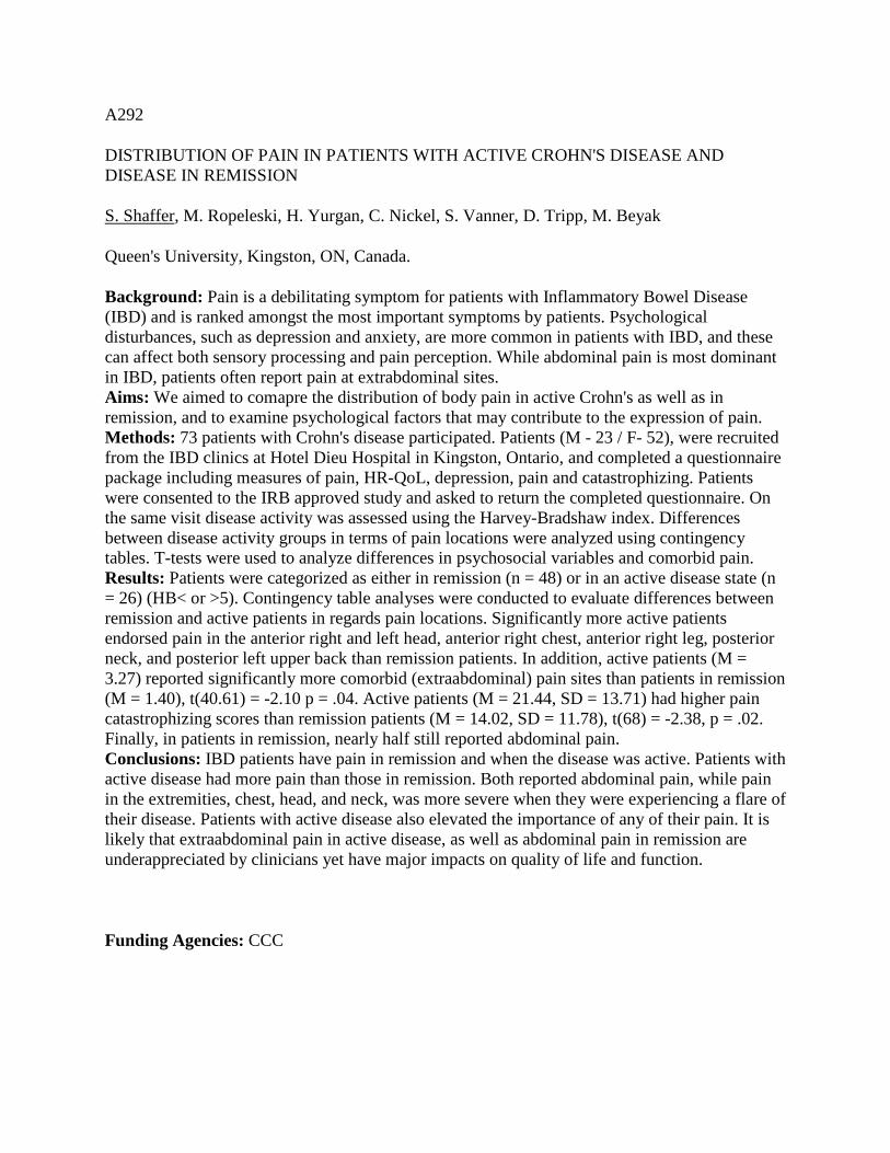
A292
DISTRIBUTION OF PAIN IN PATIENTS WITH ACTIVE CROHN'S DISEASE AND DISEASE IN REMISSION
S. Shaffer, M. Ropeleski, H. Yurgan, C. Nickel, S. Vanner, D. Tripp, M. Beyak
Queen's University, Kingston, ON, Canada.
Background: Pain is a debilitating symptom for patients with Inflammatory Bowel Disease (IBD) and is ranked amongst the most important symptoms by patients. Psychological disturbances, such as depression and anxiety, are more common in patients with IBD, and these can affect both sensory processing and pain perception. While abdominal pain is most dominant in IBD, patients often report pain at extrabdominal sites. Aims: We aimed to comapre the distribution of body pain in active Crohn's as well as in remission, and to examine psychological factors that may contribute to the expression of pain. Methods: 73 patients with Crohn's disease participated. Patients (M - 23 / F- 52), were recruited from the IBD clinics at Hotel Dieu Hospital in Kingston, Ontario, and completed a questionnaire package including measures of pain, HR-QoL, depression, pain and catastrophizing. Patients were consented to the IRB approved study and asked to return the completed questionnaire. On the same visit disease activity was assessed using the Harvey-Bradshaw index. Differences between disease activity groups in terms of pain locations were analyzed using contingency tables. T-tests were used to analyze differences in psychosocial variables and comorbid pain. Results: Patients were categorized as either in remission (n = 48) or in an active disease state (n = 26) (HB< or >5). Contingency table analyses were conducted to evaluate differences between remission and active patients in regards pain locations. Significantly more active patients endorsed pain in the anterior right and left head, anterior right chest, anterior right leg, posterior neck, and posterior left upper back than remission patients. In addition, active patients (M = 3.27) reported significantly more comorbid (extraabdominal) pain sites than patients in remission (M = 1.40), t(40.61) = -2.10 p = .04. Active patients (M = 21.44, SD = 13.71) had higher pain catastrophizing scores than remission patients (M = 14.02, SD = 11.78), t(68) = -2.38, p = .02. Finally, in patients in remission, nearly half still reported abdominal pain. Conclusions: IBD patients have pain in remission and when the disease was active. Patients with active disease had more pain than those in remission. Both reported abdominal pain, while pain in the extremities, chest, head, and neck, was more severe when they were experiencing a flare of their disease. Patients with active disease also elevated the importance of any of their pain. It is likely that extraabdominal pain in active disease, as well as abdominal pain in remission are underappreciated by clinicians yet have major impacts on quality of life and function.
Funding Agencies: CCC

A293
CUTANEOUS PSEUDOLYMPHOMA INDUCED BY INFLIXIMAB IN A PATIENT WITH CROHN'S DISEASE
M. Niazi2, L. Worobetz1
1. Department of Gastroenterology, Saskatoon, SK, Canada; 2. Department of Medicine, Saskatoon, SK, Canada.
Background: Cutaneous pseudolymphoma is a benign inflammatory response that often clinically and histologically mimics cutaneous lymphoma. It is characterized by dense lymphoid infiltrate of the skin, which may be local, diffuse and rarely disseminated. Typical lesions are pruritic, red to violaceous, smooth-surfaced papules, nodules, or plaques. The most common site of involvement is the face, followed by the chest and upper extremities. Drug-induced pseudolymphoma may be associated with systemic manifestations such as fever, arthralgia, lymphadenopathy, and hepatosplenomegaly. Eosinophilia and abnormal liver enzymes may also occur. Aims: We present a case of pseudolymphoma in a patient with Crohn's disease on infliximab. Methods: A detailed review of the case was undertaken, including history, biochemistry, and pathology. A subsequent literature review of the topic was conducted. Results: A 55 year-old Caucasian male with a 35-year history of Crohn's disease, previously well controlled on 5-aminosalicylic acid and prednisone was switched to infliximab for perianal disease. He had mild improvement while on infliximab. However, approximately three months after the initiation of anti-tumor necrosis factor (anti-TNF), the patient developed pruritic red papules and plaques of varying size over the forehead and scalp. The patient had no systemic symptoms or biochemical derangements. Skin biopsy showed perivascular inflammation predominantly lymphohistiocytic, with plasma cells and occasional eosinophils. On immunohistochemistry the infiltrate was compromised of predominantly T-lymphocytes with a few B-lymphocytes admixed. Kappa and lambda light chain staining demonstrated polyclonal pattern of plasma cells. The diagnosis of drug-induced pseudolymphoma was made and therefore infliximab was discontinued. At six months following discontinuation of infliximab, the rash had improved but has not resolved completely. While a vast array of pharmacologic agents have been linked to lymphoid drug reactions, our case represents the first reported incidence in a patient with Crohn's disease. Previously, three cases of pseudolymphoma have been reported in psoriasis patients on adalimumab and infliximab. In these reported cases, withdrawal of the offending agent resulted in complete resolution of the rash and recurrence was noted with an alternative anti-TNF agent. Treatment modalities include topical glucocorticoids, surgical excision, and local radiation. However, close follow up is recommended as recurrence is not uncommon and there are occasional case reports of cutaneous pseudolymphoma evolving into lymphoma. Conclusions: Recognition of cutaneous pseudolymphoma is critical as it may represent drug class effect. Second, it is important in understanding the risk characteristics of the patients developing this type of rash.
Funding Agencies: None
A294
USTEKINUMAB FOR REFRACTORY PEDIATRIC CROHN'S DISEASE: REPORT OF 6 CASES
C. Dupont-Lucas, V. Marchand, U. Halac, M. Dirks, C. Faure, C. Deslandres
CHU Sainte Justine, Montreal, QC, Canada.
Background: The therapeutic options available for managing Crohn's disease have recently been increased by new molecules. Ustekinumab is a monoclonal antibody against the p40 subunit of interleukin-12/23. It has been shown to be superior to placebo in inducing clinical response and maintaining remission in adult patients with moderate- to severe Crohn's disease, but not yet studied in children. Aims: To describe safety and efficacy of subcutaneous ustekinumab in pediatric patients with refractory Crohn's disease. Methods: We report the cases of 6 patients who received open-label ustekinumab in our institution between February and October 2014 for Crohn's disease refractory to anti-TNFs. Data was retrieved retrospectively from medical charts. Results: There were 4 boys and 2 girls. Median age at initiation of ustekinumab was 16.4 yrs (Min 10.7 - Max 18.2) and median duration of Crohn's disease was 3.6 yrs (Min 1.0 - Max 8.4). Location of disease according to Paris classification was L1 in 1 patient and L3 in 4 patients. One patient did not have colonic inflammation. Upper gastrointestinal disease was noted in 4 patients: 2 had L4a and 2 had L4b involvement. Behavior was non penetrating non stricturing in 5 cases and stricturing in 1. No patient had perianal disease. All patients had failed or were intolerant to treatment with an immunomodulator (5 patients to azathioprine, 6 to methotrexate). All patients had failed infliximab (2 discontinued for loss of response, 2 for allergies or adverse events and 2 for both loss of response and adverse events). Five patients had failed adalimumab, 1 had not received it. One patient had had 2 small bowel resections with stricturoplasties. At baseline, 4 patients were on prednisone and 3 on methotrexate. Doses of subcutaneous ustekinumab administered during induction therapy were 45 mg (if weight ≤ 45 kg) per week or 90 mg (if weight > 45 kg) per week, during 3 weeks. After induction therapy, clinical response was noted in 3 patients, clinical remission in 1 and absence of response in 2 patients. Five patients continued on maintenance therapy with 45 mg every 8 wks or 90 mg every 8 wks depending on doses received during induction. Dose escalation was necessary in 2 cases. In all, patients received a median 6.5 doses (3 - 9) of ustekinumab. Response to maintenance therapy was evaluated after a median 5.1 months (3.7 - 7.1): 1 patient was in clinical remission, 2 had clinical response and 2 patients had no response, one of whom required subtotal colectomy with ileostomy. No adverse events were noted. Conclusions: Ustekinumab is well tolerated in pediatric patients. More data is necessary to confirm the efficacy shown in adults.
Funding Agencies: None

A295
DIGITAL TOOLS SUPPORTING IBD PATIENTS AND THEIR DOCTORS: A CANADIAN PERSPECTIVE
T. van Mierlo2, R. Fournier2, R. Fedorak1
1. Los Alamos National Laboratory, Edmonton, AB, Canada; 2. Evolution Health Systems Inc., Toronto, ON, Canada.
Background: Approximately 83% of Canadians are online, and research indicates that the majority of IBD patients use the Internet to gather information. Unlike other chronic conditions, limited research has been conducted on using digital tools to expand the reach of evidence-based IBD education. Moreover, many existing IBD digital resources contain content that is unverified or not worded in patient-friendly terms. There is a risk of overemphasizing worst-case scenarios or promoting inaccurate information on treatment and side effects. Aims: This study gathered insights from Canadian Key Opinion Leaders (KOLs) on the utility of digital resources, and the feasibility of technology as a means to facilitate information sharing between IBD patients and their doctors. Methods: In November 2013, Nominal Group Technique was utilized to capture insights from seven Canadian Gastroenterologists (KOLs) on their experiences with Internet-based tools, and their preference for digital content. KOLs held senior positions at research hospitals or universities, frequently published in peer-reviewed journals, and developed protocols for patient care. Discussion themes included methodological approaches to non-adherence, concordance, patient-centricity, and attributes of digital tools that would be promoted by Gastroenterologists. Following the session, an anonymous questionnaire was used to synthesize results. Results: All KOLs agreed that digital tools could aid in treatment, and 57% (n=4) had experienced patients bringing digital resources to consultations. 71% (n=5) found these resources to contain inaccurate information, and when asked about referring patients to digital tools, all agreed that content should focus on evidence-based facts. 57% (n=4) felt that patient-centric tools, as well as professional or non-profit endorsement, would increase their confidence. When considering program design, 86% (n=6) preferred tools that addressed a mix of compliance and concordance, and only 14% (n=1) supported the development of tools that focused on compliance. A gap identified was lack of peer-to-peer support that has been successfully utilized in other conditions. KOLs agreed that the Canadian IBD community could benefit from a digital resource that was frequently updated, monitored, and unbiased. Conclusions: The variability of IBD presents unique challenges to the development of digital resources. While KOLs would support tools that focus on shared decision-making, a concern is that patients might diverge from treatment. Canadian IBD patients frequently access digital tools, yet there is limited research on their effectiveness. Further research in regards to development, design and efficacy of digital tools that support IBD patients is required.
Funding Agencies: Ferring Pharmaceuticals (Canada)

A296
EFFICACY AND SAFETY OF SERIAL FECAL MICROBIOTA TRANSPLANTATION (FMT) IN ILEOCOLONIC CROHN'S: A CASE REPORT
D. Kao, N. Hotte, K. Madsen
University of Alberta, Edmonton, AB, Canada.
Background: Intestinal dysbiosis has been shown in patients with inflammatory bowel disease (IBD). It remains unclear whether the relationship is associative or causative. Case reports and small case series have shown FMT to be effective in IBD. However, clinical improvement is not universal. Adverse events such as abdominal pain, fevers, and IBD flare have been reported. Aims: To determine efficacy and safety of serial FMT in patients with mild Crohn's disease Methods: A 35 year old man was diagnosed with moderately severe Crohn's colitis in Aug 2007. Following clinical remission induced by steroid, he was maintained on Asacol. Unfortunately he had a mild flare in 2012 and requested FMT from his treating gastroenterologist. At the time of the FMT, he had moderate inflammation in the cecum. He subsequently went into clinical remission and was well on no maintenance therapy until June 2014 when he started to have bloody diarrhea, with 1-2 motions per day (HBI= 5). He was restarted on Mezavant, but only had partial response. As he fulfilled all inclusion and exclusion criteria, he was enrolled in our open-label serial FMT trial and underwent FMT by colonoscopy at weeks 0, 4 and 8 in addition to FMT enema at weeks 2 and 6. The fecal material was provided by one of the universal stool donors registered with our FMT program. Serial HBI scores, C reactive protein (CRP), fecal calprotectin (FC), and Simple Endoscopic Score for Crohn's disease (SES-CD), and colonic biopsies were collected. Results: At the time of his first colonoscopy, he had mild, continuous inflammation in the left colon, patchy moderate inflammation in the transverse colon, and moderate, continuous inflammation in the proximal colon and terminal ileum (TI). He went into clinical remission at week 1. The serial HBI scores, CRP, FC, and SES-CD were summarized in table 1. At week 4, he had endoscopic and histologic healing up to descending colon, but the rest of the colon and TI remained unchanged. At week 8, he had endoscopic and histologic healing from rectum to transverse colon, but his ascending colon still showed moderate inflammation. His TI also showed marked mucosal healing, with only several small aphthous ulcers. There were no adverse events. Conclusions: Serial FMT appears safe and and promising in inducing clinical and possibly endoscopic remission in mild Crohn's.
Table 1 Timing CRP FC SES-CD HBI
At screening (time 0) 22.6 5235 23 5 Day 1 14.9 - - -
Week 1 11.1 1792 - 1 Week 2 18.1 3815 - 0
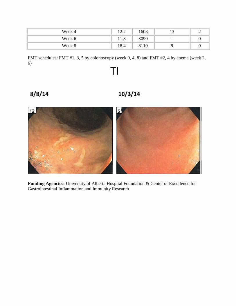
Week 4 12.2 1608 13 2 Week 6 11.8 3090 - 0 Week 8 18.4 8110 9 0
FMT schedules: FMT #1, 3, 5 by colonoscopy (week 0, 4, 8) and FMT #2, 4 by enema (week 2, 6)
Funding Agencies: University of Alberta Hospital Foundation & Center of Excellence for Gastrointestinal Inflammation and Immunity Research

A297
INCIDENCE OF STEROID-INDUCED MOOD CHANGES IN PATIENTS WITH INFLAMMATORY BOWEL DISEASE
G. Ou, B. Bressler, C. Galorport, E. Lam, H. Ko, R. Enns, J. Telford, G. Rosenfeld
UBC, Vancouver, BC, Canada.
Background: Corticosteroids are effective in the treatment of immunological conditions but are associated with systemic side-effects, including diabetes, osteoporosis, and mood changes. Previous studies have assessed the incidence of mood changes among patients taking short-course oral steroids for asthma or ophthalmic conditions, but data specific to the use of steroids for the treatment of inflammatory bowel disease (IBD) is lacking. Aims: To determine the incidence of mood changes in patients being treated with oral prednisone for IBD. Methods: A prospective observational cohort study was initiated at St. Paul's Hospital (Vancouver, BC) in October 2013. Ambulatory patients aged 19 or older with IBD being started on oral prednisone therapy comprised this cohort. Exclusion criteria: hospitalization for management of IBD within 2 weeks of study entry; liver cirrhosis with/without evidence of synthetic dysfunction; medications that interfere with steroid metabolism; change in psychiatric medication within 1 month of study entry; and recreational drug use/alcohol abuse. Mood changes were assessed using 2 previously validated questionnaires: Beck Depression Inventory (BDI-) II for depression and Activation subscale of Internal State Scale v2 (AS-ISSv2) for (hypo)mania at the outset and after 1-2 weeks of steroid therapy. Overlapping symptoms between IBD and depression as per BDI-II were excluded. IBD activity was assessed at each visit using Harvey-Bradshaw Index (HBI) for Crohn's disease (CD) or Simple Clinical Colitis Activity Index (SCCAI) for Ulcerative Colitis (UC). Results: Twenty subjects have been recruited and completed 2 sets of mood evaluation as of September 2014. Eleven (55%) were male and mean age was 41.4±15.4 years. Twelve (60%) subjects had UC and 8 (40%) had CD. Most subjects experienced improvement in IBD symptoms after initiation of steroid treatment (mean HBI change -5.8±6.8; mean SCCAI change -5.4±4.8). One patient with UC underwent subtotal colectomy due to perforation 2 weeks into treatment and another patient with UC required intravenous steroids due to worsening disease. There was no significant change in BDI-II scores (mean 24.2±6.7 vs 25±7.3, p=0.72) or AS-ISSv2 scores (mean 80±80 vs 101.5±87.3, p=0.42) after 16±5.4 days of steroid therapy. Conclusions: No significant mood change was seen after initiation of oral steroids for treatment of IBD in the first 20 patients in this prospective cohort. Recruitment will continue to achieve the target sample size of 50.
Funding Agencies: CAG
A298
EVALUATION OF THE ASSOCIATION BETWEEN FECAL LACTOFERRIN STATUS AND CLINICAL, SEROLOGIC AND ENDOSCOPIC OUTCOMES: A POST-HOC ANALYSIS OF THE SONIC TRIAL
W. Reinisch2, M. Gerassimos1, J. Colombel4, A. Kornbluth4, P. Rutgeerts5, F. Cornillie6, M. Molenda6, L. Tang6, W. Sandborn3
1. Evangelismos Hospital, Athens, Greece; 2. McMaster University Hospital, Hamilton, ON, Canada; 3. University of California San Diego, La Jolla, CA; 4. Mount Sinai Medical Center, New York, NY; 5. University Hospital Gasthuisberg, Leuven, Belgium; 6. Janssen Scientific Affairs, LLC., Spring House, PA.
Aims: This SONIC post-hoc analysis reports on the association of fecal lactoferrin (LF) to clinical, serologic and endoscopic outcomes, and serum IFX levels in CD patients enrolled in centers in the US who had evaluable measurements of LF, CDAI, serum CRP, and ileocolonoscopy both at baseline and wk26, and evaluable serum IFX levels at wk30. Methods: LF was measured by an ELISA assay and expressed as positive (pooled +2, +3, and +4 scores) or negative (pooled negative and +1 scores). MH was defined as absence of any ulceration. Corticosteroid-Free Clinical Remission (CFCR) was defined as clinical remission (CDAI<150) and no treatment with corticosteroids for >6 weeks. Post-hoc analyses were performed using the Fisher's exact test and the Kruscal-Wallis test where appropriate. Results: Among the 109 patients (median age 43 yrs, 54% male), 33.9% were LF+ and 19.3% were LF- at both baseline and wk26; 36.7% patients changed from LF+ to LF- from baseline at wk26 whereas 10.1% changed from LF- to LF+ (p<0.0001). At baseline, LF was significantly associated with CDAI (p=0.024) and serum CRP (p=0.01); in addition, LF showed a trend of association with normal levels of CRP (65.6% vs. 36.4% with CRP <0.8mg/dL for LF - and LF+, respectively, p=0.064). At wk26, there was no association between LF and CDAI (p=0.09), serum CRP (p=0.18), CRP normalization (0.512), CFCR (p=0.245).The association between LF and MH (ulceration) was evaluated in 168 patients at baseline and 110 patients at wk 26 who had both LF and MH data at these measurement points. At baseline, ulcerations were not found in 43.1% (25/58) LF- patients vs. 21.8% (24/110) LF+ patients (sensitivity 0.72, specificity 0.51, p=0.004). In contrast, at wk26, 44.3% (27/61) of LF- patients vs. 30.6% (15/49) LF+ patients had MH (sensitivity 0.50, specificity 0.64, p=0.143). Conclusions: In this post-hoc analysis of the SONIC trial, LF was collected as a categorical variable. The association between LF and other clinical parameters may be better estimated if LF was on a continuous scale. Semi-quantitative LF does not seem to be a good biomarker in CD. Although it was useful to identify active clinical and biological disease at baseline, it could not confirm clinical, biological, or endoscopic remission at wk26.
Funding Agencies: None

A299
GOLIMUMAB IN THE MANAGEMENT OF INFLIXIMAB EXPERIENCED ULCERATIVE COLITIS, A SINGLE-CENTRE EXPERIENCE
M. Lam2, B. Bressler1 1. Pacific Gastroenterology Associates, Vancouver, BC, Canada; 2. University of British Columbia, Vancouver, BC, Canada. Background: Golimumab (GOL) is a subcutaneous fully human monoclonal antibody to tumor necrosis factor α (TNFα) approved for the treatment of ulcerative colitis (UC). PURSUIT-SC and PURSUIT-M are double-blinded phase 2/3 trials on TNFα antagonist-naïve patients with moderate-to-severe UC for induction and maintenance respectively. These trials showed that GOL is effective in induction and maintenance of remission compared to placebo through to week 54. Aims: To determine the effectiveness of GOL in IFX-experienced UC patients who either lose response to IFX or develop intolerance to IFX. Methods: This is a single-centre retrospective case series of IFX-experienced patients with UC on GOL from September 2013 to 2014. Response to therapy was assessed by full or partial Mayo scores obtained from charts (which ever is available) and were separated into early response assessment (≤12 weeks), mid response assessment (12-24 weeks), and late response assessment (24-52 weeks). Response to therapy is defined by a reduction in Mayo score of 3 points, and remission is defined by Mayo score ≤2 with no subscore >1. Durable remission defined by Mayo score ≤2 with no subscore >1 for at least two assessment including the late response assessment. Results: Five patients were found to be on GOL for UC who were IFX-experienced. Forty percent were male with average age of 31.0 years at time of GOL therapy. The average duration of disease was 7 years with a median of 5 years. Four patients (80%) had left-sided colitis and one (20%) had pancolitis. The median time between the start of IFX therapy and start of GOL therapy was 2 years and a median time between the last infusion of IFX and first injection of GOL of 9.4 weeks. GOL was started in two patients due to lupus-like drug reaction to IFX, in two patients due to loss of response (LOR) and IFX antibodies, and one patient due to secondary non-response to IFX with negative IFX antibody status and therapeutic IFX drug levels. Four of the five patients (80%) achieved remission over the course of assessment. Two (40%) of the patients achieved durable remission by partial Mayo score. One achieved remission during mid response assessment but has not had follow up in late response assessment period at the time of data collection. One patient had flare of UC in the late response assessment period after achieving symptomatic remission. One patient had no response to GOL. Conclusions: In IFX-experienced UC patients, 80% of our cohort achieved remission by partial Mayo score with GOL therapy. GOL may be a viable option for patients who develop intolerance or LOR due to IFX antibodies.
Funding Agencies: None
A300
HEPATOCELLULAR CARCINOMA IN A PATIENT WITH CROHN'S DISEASE ON AZATHIOPRINE AND INFLIXIMAB COMBINATION THERAPY.
K. Fortinsky3, A. Alali2, K. Jeejeebhoy4, M. Sherman1, S. Fung1
1. Toronto General Hospital, Toronto, ON, Canada; 2. Univeristy of Toronto, Toronto, ON, Canada; 3. University of Toronto, Toronto, ON, Canada; 4. University of Toronto, Toronto, ON, Canada.
Background: Only ten cases of hepatocellular carcinoma (HCC) in patients with Crohn's disease (CD) have been reported. Eight of these patients were being treated with azathioprine, two of whom were also on infliximab. Intriguingly, the two patients not exposed to azathioprine therapy who developed HCC were found to have underlying liver disease. Aims: To describe a case of HCC in a patient with CD and review the literature surrounding the possible etiologies of this rare diagnosis. Methods: Case report and literature review. Results: We present a 34-year-old man with a 25-year history of CD who developed HCC. Ten years ago, he required a proctocolectomy and end ileostomy for severe colonic and perianal disease refractory to medical treatment with 5-ASA, 6-MP, and methotrexate. Postoperatively, he developed peristomal pyoderma gangrenosum and seronegative polyarthritis, for which azathioprine (AZA) and infiliximab (IFX) were initiated. He was otherwise healthy and denied any smoking, alcohol, or illicit drug use. There was no family history of inflammatory bowel disease, liver disease, or malignancy. He presented with 2 months of epigastric pain, bloating, and weight loss. He had been taking AZA and IFX for the past 6 years. Recent ileoscopy, gastroscopy, and blood work including liver enzymes from 2 months prior were normal. On presentation, an abdominal CT scan revealed a 24 cm left hepatic lobe mass with tumor thrombosis involving the left portal vein and nodular masses in the right lobe. Liver enzymes were mildly elevated, liver function was normal, and alpha-fetoprotein levels were markedly elevated at 3307 ug/L. A targeted biopsy confirmed HCC. Viral hepatitis serology, alpha-1 antitrypsin levels, autoimmune panel, iron studies, and copper studies were uniformly normal. Liver transplantation was not a viable option and treatment was initiated with transarterial chemoembolization and sorafenib. Unfortunately, the patient developed metastatic disease involving his lungs, adrenals, spine, maxilla, and mandible. The patient was provided palliative care and passed away 5 months after his diagnosis. Conclusions: The etiology of HCC in patients with CD is poorly understood and requires further investigation. While large-scale studies have failed to show an association between azathioprine and HCC in CD, several case reports suggest a potential association which is concerning. Interestingly, there are no reported cases of HCC in patients treated with AZA for a condition asides from CD. Clinicians may consider early imaging in CD patients with concerning symptomatology or abnormal liver enzymes, especially those treated with azathioprine.
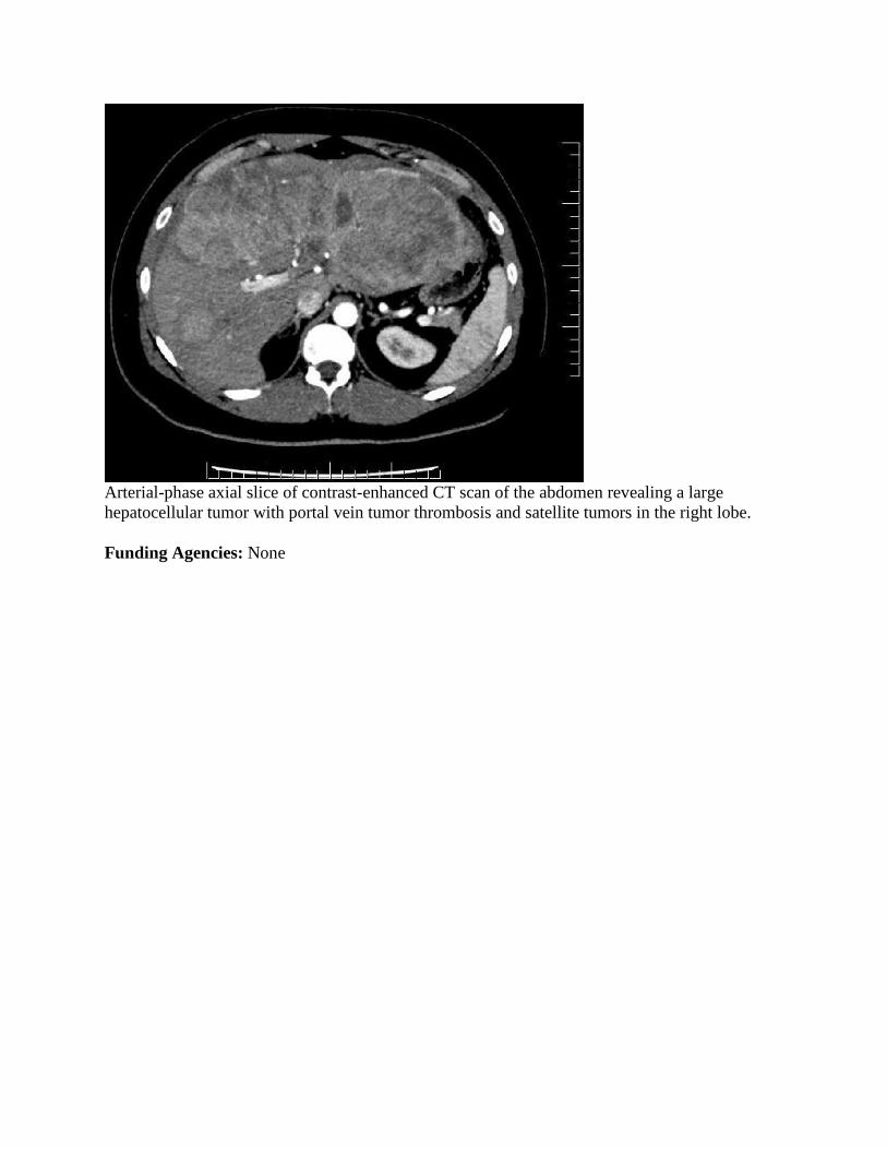
Arterial-phase axial slice of contrast-enhanced CT scan of the abdomen revealing a large hepatocellular tumor with portal vein tumor thrombosis and satellite tumors in the right lobe.
Funding Agencies: None

Microbiology and Parasite-Host Interactions
Poster of Distinction
A301
HYDROGEN SULFIDE KILLS PLANKTONIC BACTERIA AND PROMOTES GUT MICROBIOTA BIOFILMS: A POSSIBLE ROLE IN PROTECTION AGAINST COLITIS
J. Motta2, K. Flannigan1, J. Wallace2, A. Buret2
1. McMaster University, Hamilton, ON, Canada; 2. University of Calgary, Calgary, AB, Canada.
Background: Alterations of the microbiota (dysbiosis) have been associated with inflammatory bowel disease (IBD). Reports on fecal bacterial composition have offered key insights into the microbiota characteristics of IBD, but a causal link between disruptions of mucosal microbiota biofilms and disease has yet to be established. Hydrogen sulfide (H2S) protects against colitis, but the mechanisms remain incompletely understood. Aims: To determine if H2S promotes a beneficial biofilm mode of growth of microbiota and inhibits the release of planktonic (free-swimming bacteria) bacteria. Methods: Colonic tissues of mice (CD1), and descending colon biopsies from healthy and Crohn's Disease (CD) volunteers were collected to generate ex vivo, anaerobically, gut microbiota biofilms and the planktonic bacteria they shed, using the Calgary Biofilm DeviceTM. Anaerobic biofilms and planktonic bacteria were exposed with either the antibiotic metronidazole, or H2S donors (DADS and NaHS). Metabolic activity (survival) and biomass were assessed for biofilms, and for planktonic bacteria in stationary and exponential phases. Colitis was induced in rodents using dinitrobenzene sulfonic acid (DNBS) and animals were orally treated with DADS for 7 days. Colitis severity was assessed macroscopically and histologically and gut microbiota was visualized using fluorescent in situ hybridization. Results: Metronidazole significantly reduced exponential growth of planktonic bacteria from healthy and CD volunteers (1 to 100 µg/ml), but had no effects on their biofilms. Concentrations of 50 to 100 µM of H2S-donors reduced growth of planktonic bacteria in exponential phase (mice and human), but not in stationary phase. Biofilms (mice and human-derived) exposed to concentrations of 1 to 100 µM of H2S donors had dose-dependently higher metabolic activity and biomass. In healthy rodents, intestinal microbiota formed continuous biofilms. During colitis, biofilms were fragmented and adhered to the epithelial surface, allowing planktonic bacteria to translocate into the tissues. Therapeutic delivery of H2S into the colon reduced inflammation, restored the microbiota biofilm and reduced translocation of planktonic bacteria. Conclusions: Our results demonstrate that H2S has antibacterial properties on planktonic bacteria and promotes biofilms, even in CD patient's microbiota. We showed that biofilms are hallmarks of a healthy intestine; and promoting them with an H2S-donor is protective. H2S donors may offer a promising therapeutic avenue to control dysbiosis in patients with IBD.
Funding Agencies: CAG, CIHR, Izaak Walton Killam PDF, University of Calgary Eye's High
Poster of Distinction
A302
DISTINCT ROLES OF THE MUCUS LAYER AND MICROBIOTA IN ALTERING SUSCEPTIBILITY TO DSS-INDUCED COLITIS
A. Leon Coria2, F. Moreau2, M. Surette1, K. Chadee2
1. McMaster University, Hamilton, ON, Canada; 2. University of Calgary, Calgary, AB, Canada.
Background: The human intestine has approximately 100 trillion microorganisms, mainly bacteria that form the microbiota. In the intestinal lumen, microbiota plays a key role in digestive health as well as in the development of a balanced immune system. The intestinal mucosa is covered by MUC2 mucus that acts as a protective barrier and is constantly exposed to commensals and pathogenic organisms. Both dysbiosis (alteration in microbiota composition) and disruption of the intestinal mucus layer have been associated with gastrointestinal (GI) pathological conditions such as irritable bowel disease, colorectal cancer and inflammatory bowel diseases. However, the distinct contribution of the microbiota and the mucus barrier in the pathogenesis of colitis is not well understood. Aims: 1. To determine whether shifts in microbiota altered the onset, progression or recovery of colitis in Muc2+/+ and Muc2-/- mice. 2. To quantify the distinct roles of an intact Muc2 barrier and the microbiota in susceptibility and resolution of colitis. Methods: To minimize variations in microbiota composition, only F2 littermates Wt (Muc2+/+) and homozygous (Muc2-/-) mice were used in this study. To quantify the role of the microbiota in disease pathogenesis, animals were gavaged with an antibiotic cocktail to eliminate indigenous bacteria as revealed by DNA analysis, and then fecal transplanted with their littermate stool and susceptibility to dextran sulphate sodium (DSS) quantified. Fecal samples pre- and post-antibiotic treatment was quantified by MiSeq lll lluminar sequencing and analyzed by Bray Curtis PCoA and weighted Unifrac PCoA. Intestinal permeability was assessed using FITC-dextran. Results: DNA analysis showed clear phyla differences in microbiota composition between the two groups of mice, showing increase in Bacteriodetes and Tenericutes and a decrease in Firmicutes in Muc2-/- animals. Surprisingly, Muc2+/+ mice that received Muc2-/- microbiota were highly susceptible to DSS induced colitis associated with increased mortality as compared to animals not treated with antibiotics or water controls. Histological examination showed increased numbers of ulcerated lesions, loss in crypts architecture and greater inflammatory cellular infiltrate with increased intestinal permeability as compared to Muc2+/+ receiving their own microbiota. Muc2-/- mice while highly susceptible to DSS colitis on their own showed no increase/decrease in disease susceptibility receiving Muc2+/+ microbiota. Conclusions: Our results clearly establish that Muc2-/- microbiota is capable of aggravating DSS-induced colitis in Wt mice with an intact Muc2 layer, suggesting that Muc2-/- microbiota is highly colitogenic.
Funding Agencies: CCC

Poster of Distinction
A303
ACTIVATION OF LATENT ESCHERICHIA COLI VIRULENCE GENES BY CAMPLYOBACTER JEJUNI: A NEW MECHANISM OF POST INFECTIOUS INTESTINAL INFLAMMATION
K. Reti1, L. Tymensen2, M. Amrein1, A. Buret1 1. University of Calgary, Calgary, AB, Canada; 2. Alberta Agriculture and Rural Development, Lethbridge, AB, Canada.
Background: Campylobacter jejuni, among other enteropathogens, has been associated with chronic, post-infectious intestinal inflammation via mechanisms that are unclear. Our research has shown that C. jejuni promotes translocation of commensal microbes, disrupts intestinal enterocyte Toll-like receptor 9 (TLR9) expression, and increases intestinal susceptibility to mild inflammatory stimuli. We hypothesized that C. jejuni may increase the expression of latent virulence genes in commensal bacteria, which may contribute to post-infectious intestinal inflammation. Aims: The aims of this study were to: (1) assess the effects C. jejuni has on the expression of virulence genes in non-invasive Escherichia coli, and (2) determine the pathophysiological consequences the C. jejuni-induced alterations in E. coli have on human enterocytes. Methods: Non-invasive E. coli HB101 was grown (1) in monoculture, or (2) in co-culture with live C. jejuni 81-176, or (3) in C. jejuni-conditioned media. Affymetrix microarrays and RT-PCR assessed changes in E. coli gene expression. E. coli phenotypic changes were visualized via negative staining and transmission electron microscopy. Atomic force microscopy (AFM), which has not yet been successfully applied to bacterial-host cell interactions, was used to measure the adhesive force between E. coli and human T84 enterocytes. Changes in T84 cell receptor and CXCL8 expression were assessed via RT-PCR. Results: Microarray analyses indicated that E. coli virulence genes, including adhesin, flagella, and hemolysin genes, were upregulated when E. coli was grown in C. jejuni-conditioned media. E. coli fimbrial and hemolysin gene expression were upregulated when E. coli was grown in C. jejuni-conditioned media and subsequently exposed to T84 cells. The number of flagellated E. coli was increased when E. coli was exposed to C. jejuni or E. coli was grown in C. jejuni-conditioned media. AFM results indicate that E. coli exposed to C. jejuni-conditioned media were more adherent to T84 cells. T84 cells that had been exposed to C. jejuni-altered E. coli had reduced levels of the protective TLR4 expression and increased levels of CXCL8. Conclusions: C. jejuni and C. jejuni-conditioned media increase the expression of virulence factors in non-invasive E. coli. These include flagellar, hemolysin, and fimbrial genes, which promotes strong adhesion of otherwise non-invasive, commensal E. coli to host enterocytes. Furthermore, this altered E. coli promotes a pro-inflammatory CXCL8 response. These findings describe a novel mechanism by which an enteric pathogen may render a commensal microbe more virulent, which may contribute to chronic intestinal inflammation following the resolution of an acute infection.
Funding Agencies: CCC

A304
ENTAMOEBA HISTOLYTICA CYSTEINE PROTEASE-5 SIMULATE PI3K/ PKCδ-DEPENDANT MUC2 MUCIN SECRETION FROM COLONIC GOBLET CELLS
S. Cornick, F. Moreau, K. Chadee
Department of Microbiology, Immunology and Infectious Diseases, University of Calgary, Calgary, AB, Canada.
Background: The mucus barrier that covers the single layer of epithelial cells in the gastrointestinal tract provides the first line of innate host defense against commensal organisms and disease causing pathogens. MUC2 mucin produced by goblet cells (GC) can directly interact with a variety of pathogens to limit their interaction with the epithelium. The protozoan parasite Entamoeba histolytica (Eh) directly interacts with MUC2 to prevent parasite invasion of the underlying intestinal epithelial cells that would otherwise result in disease. However, Eh can cause amebic colitis when the parasite surface cysteine protease-5 (EhCP5) directly cleave the MUC2 C-terminus and depolymerize the mucus gel. Eh also induces hypersecretion of MUC2 resulting in the depletion of mucin stores within goblet cells via an unknown contact dependent mechanism. Aims: 1. To identify the parasite component responsible for mucin secretion in goblet cells 2. To decipher how this virulence factor triggers intracellular signaling cascades to evoke mucin exocytosis. Methods: Eh-induced mucin secretion was studied in vitro using LS 174T colonic GC that was metabolically labeled with 3H-glucosamine that incorporates into MUC2 Gal/GalNAc oligosaccharide residues. 3H-mucin secretion was quantified at various time points following infection with live Eh, Eh secreted components and in presence of various pharmacological inhibitors. S4B column chromatography and western blotting were used to confirm high Mr mucin secretions. Confocal microscopy was used to interrogate the intimate contact between Eh and GC. Results: Live Eh-induced MUC2 mucin secretion in a dose- and time-dependent manner, similar to the potent mucin secretagogue, PMA. Interestingly, Eh lacking cysteine protease-5 (EhCP5-) or Wt Eh treated with the cysteine protease inhibitor E64 significantly reduced mucin secretion. Mucin secretion was modestly induced by Wt Eh secreted components (SC) and recombinant EhCP5 but not EhCP5- and E64-treated Eh SC. Inhibition of protein kinase C or PI3K abrogated cysteine protease dependent MUC2 secretion by live Eh. Phosphorylation and activation of PKCδ following Eh contact was blocked by PI3K inhibition. Both PI3K and PKCδ were phosphorylated at the site of Wt Eh contact, but not EhCP5- or E64-treated Eh. Previous work has identified EhCP5 as interacting with αvβ3 integrins through an RGD motif and saturating this domain with small peptide inhibitors markedly inhibited mucin secretion. Conclusions: We propose a model in which cysteine proteases present on can Eh modulate goblet cell integrins to activate PI3K/PKCδ that facilitates MUC2 mucin exocytosis in disease pathogenesis.
Funding Agencies: CCC, CIHR, NRC

A305
AUTOPHAGY MODULATES HELICOBACTER PYLORI CYTOTOXIN ASSOCIATED GENE A
M. Abdullah, L. Greenfield, E. Galindo-Mata, N. Jones
The Hospital for Sick Children, Toronto, ON, Canada.
Background: Infection with Helicobacter pylori is the most important risk factor for the development of gastric cancer, one of the leading causes of cancer related deaths worldwide. Cytotoxin associated gene A (CagA) and vacuolating cytotoxin (VacA) are major virulence determinants of H. pylori. CagA is injected into the host cell via a type IV secretion system, where it initiates oncogenic signaling. We have shown that VacA modulates the autophagy pathway, which is a cellular degradation pathway. Short-term exposure to VacA induces autophagy, while prolonged exposure prevents autophagosome maturation. We hypothesize that during chronic H. pylori infection, VacA-disrupted autophagy increases CagA levels leading to enhanced downstream oncogenic CagA signaling. Aims: Here we investigated the mechanisms by which autophagy modulates CagA in host cells. The objectives were to determine if (1) autophagy modulates CagA and (2) VacA-disrupted autophagy increases intracellular CagA levels, which could lead to enhanced oncogenic downstream signaling. Methods: To assess the role of autophagy in the degradation of CagA during acute infection, wild-type and atg5-/- mouse embryonic fibroblasts (MEFs) were infected (4 h) with wild-type 60190 H. pylori. In complementary studies, gastric epithelial (AGS) cells were infected with the 60190 vacA isogenic mutant and co-cultured with VacA+ or VacA- culture supernatants (4 h). We then assessed the effect of prolonged VacA exposure (when autophagy maturation is disrupted) on CagA expression by infecting AGS cells with the 60190 vacA isogenic mutant or the carcinogenic 7.13 (vacA-, cagA+) strain followed by gentamycin treatment to kill extracellular bacteria. Cells were then treated with VacA+ or VacA- culture supernatant for 24 h. Results: In comparison with wild-type MEFs, an increase in CagA was detected in autophagy deficient atg5-/- MEFs infected with H. pylori for 4 h. Short-term incubation with VacA+ CCMS reduced CagA expression in H. pylori-infected AGS cells in comparison with cells incubated with the VacA- CCMS. Next AGS cells were exposed to VacA for prolonged periods to disrupt autophagosome maturation. In cells treated with VacA+ but not VacA- CCMS for 24 h, an increase in CagA was detected. Conclusions: Our data indicate that autophagy regulates CagA levels. Acute VacA exposure (when autophagy is induced) decreases CagA levels. However, chronic VacA exposure (when autophagy maturation is disrupted) results in increased CagA. These findings provide a framework for future studies aimed at understanding the consequences of VacA-regulated CagA. Furthermore, our findings implicate autophagy as a possible therapeutic target to decrease CagA oncogenic effects.
Funding Agencies: CCC, CIHR
A306
ENTAMOEBA HISTOLYTICA INDUCES PRO-INFLAMMATORY MEDIATOR HMGB1 RELEASE IN EARLY CONTACT WITH MACROPHAGE
S. Begum, J. St-Pierre, F. Moreau, K. Chadee
University of Calgary, Calgary, AB, Canada.
Background: Entamoeba histolytica (Eh) is the causative agent of amebiasis, which occurs in 10% of infected individuals when the parasite invades the underlying colonic mucosa. The host and parasite factors that influence disease susceptibility remain unknown. Parasite-induced host pro-inflammatory responses is critical in disease pathogenesis, but the mechanisms underlying this response is poorly characterized. The first interaction between Eh and macrophages triggers a strong pro-inflammatory response following inflammasome activation (IL-1β) and other uncharacterized pathways (TNF-α) that can trigger cell death. High Mobility Group Box1 protein (HMGB1) is a non-histone nuclear protein that is released in the extracellular space during infection by activated or damaged immune cells and act as a pro-inflammatory mediator. Aims: 1. To determine whether activated macrophages are triggered to release the early "alarmin" signal HMGB1 in response to Eh, 2. To identify the putative parasitic virulent factors that engage macrophages to trigger HMGB1. Methods: Macrophages derived from human monocytic cell line (THP-1) and mouse bone marrow (Wt and caspase-1-/- mice) were treated with live virulent Eh, whole soluble Eh proteins and secreted components released from live Eh for different time points. Pharmacological inhibitors were used to identify the requirement of caspase-1 activation and Eh virulent factors in HMGB1 secretion. Western blotting was done to quantify HMGB1 secretion. Results: Only stimulation with live Eh but not Eh components triggered robust HMGB1 secretion (within 5 min) in a time-dependent manner. This event was Eh-macrophage contact-dependent as inhibiting the Eh adhesin, Gal-lectin with exogenous galactose abrogated HMGB1 release. HMGB1 release was independent of the major Eh surface virulent factor cysteine proteinase5 (EhCP5) as EhCP5 deficient parasites showed no difference. Treatment of Wt Eh with the cysteine proteinase inhibitor E64 showed that other CPs are not required for HMGB1 secretion. Surprisingly, this potent alarmin protein secretion was independent of caspase-1 activation, as secretion was not inhibited with the Pan caspase inhibitor, Z-VAD-fmk. As predicted, similar results were obtained using bone marrow macrophages derived from caspase-1-/- mice treated with live Eh. Conclusions: Our results suggest that the earliest innate response upon Eh contact with macrophages is the release of the potent inflammatory mediator HMGB1. It can act as a sensor or danger signal to detect invasive parasites to trigger and/or amplify macrophage innate host defenses. This mediator can attract bystander cells and/or promote extensive inflammation, which is critical in disease pathogenesis.
Funding Agencies: NSERC

A307
CELIAC-ASSOCIATED DUODENAL MICROBIOTA ENHANCES PRODUCTION OF 33-MER DERIVED PEPTIDES
A. CAMINERO FERNANDEZ2, C. Johnston2, H. Galipeau1, J. McCarville1, J. Casqueiro Blanco4, M. Surette1, N. Magarvey3, E. Verdu1 1. McMaster University, Hamilton, ON, Canada; 2. Mcmaster University, Dundas, ON, Canada; 3. Mcmaster University, Hamilton, ON, Canada; 4. Leon University, Leon, Spain.
Background: The pathogenesis of celiac disease (CD) involves poorly understood innate immune mechanisms as well as a specific adaptive immune response to dietary gluten. Environmental factors, such as the gut microbiota, have been suggested to modulate CD development, but the underlying mechanisms are unknown. Aims: Our aim was to test the activity of gut microbiota components on gluten metabolism and its impact on gluten-induced enteropathy. Methods: We performed in vitro incubations of the 33-mer gluten peptide with Pseudomona aeruginosa isolated from duodenal aspirate of a patient with CD. The 33-mer derived-peptides generated were measured by LC/MS. Germ free C57BL/6 mice were then mono-colonized with P. aeruginosa, or di-colonized with Lactobacillus rhamnosus and L. fermentum from duodenal aspirates of non celiac subjects and with Staphylococcus warneri and S. epidermidis isolated from duodenal aspirates of celiac subjects. One week after colonization mice received a gliadin gavage (7mg/mouse). Gluten metabolism was evaluated in the small intestinal lumen for the next 2 hours measuring non-specific gluten proteases and gliadin-peptides content. Finally, Schaedler flora (ASF)-colonized NOD-DQ8 mice were used to test the effect of the studied strains on gluten-induced inflammation in the context of an intact gut microbiota. Results: LC/MS analysis showed that P. aeruginosa hydrolyzed the immunogenic gluten peptide, 33-mer, generating further large potentially toxic peptides. P. aeruginosa-colonized mice showed increased glutenasic activity and mucolytic activity in ileum and caecum after gliadin challenge compared to Lactobacillus or Staphylococcus-colonized mice. Two hours after gliadin gavage, colonized mice had less gliadin content in the small intestine compared to germ free mice. ASF-colonized Conclusions: P. aeruginosa isolated from duodenal aspirate of celiac patients has the capacity to partially digest the 33-mer peptide in vitro, leading to increased production of immunogenic peptides. Colonization of germ free mice with P. aeruginosa is accompanied by increased proteolytic activity in vivo, and when inoculated into ASF NOD-DQ8 mice, enhanced gluten-induced enteropathy was observed. We describe a mechanism through which microbiota from a celiac patient could facilitate CD development through amplification of the immune response to gluten peptides.
Funding Agencies: CAG, CIHR
A308
GIARDIA DUODENALIS CATHEPSIN-LIKE CYSTEINE PROTEASES CLEAVE A CYTOSKELETAL PROTEIN, VILLIN, IN AN MLCK-DEPENDENT MANNER.
A. Bhargava, J. Cotton, R. Yates, A. Buret
University of Calgary, Calgary, AB, Canada
Background: Disruptions of intestinal epithelial barrier, and cytoskeletal injury are implicated in the pathophysiology of a variety of intestinal disorders. Giardia duodenalis, a non-invasive protozoan parasite of the upper small intestine, closely associates with IECs to induce pathophysiological effects, including intestinal barrier dysfunction and cytoskeletal injury; however, the mechanisms or parasitic factors involved remain obscure. The Giardia genome has been shown to contain genes for cathepsin B-, C-, and K/L-like cysteine proteases; however, their roles are unknown. Aims: The aims of this study were to characterize cathepsin activity during Giardia-IEC cocultures, and determine the role of these in initiation of giardiasis. Methods: G. duodenalis trophozoites (Assemblage A isolates NF, S2 or Assemblage B isolate GS/M) were co-incubated in vitro with human colonic monolayers (Caco-2) for 2 or 24 hours. Giardia trophozoite lysates, supernatants, and host cell monolayers lysates were isolated and incubated with cathepsin fluorogenic substrates to measure cathepsin B/L activity. Giardia trophozoites pretreated with a broad spectrum cysteine protease inhibitor (E64d) or an MLCK-inhibitor (ML-9) were co-incubated with Caco-2 monolayers or lysates of Caco-2 cells were co-incubated with Giardia trophozoites lysates in excess of E64d or Ca-074Me. Host cell lysates were processed for Western blotting to assess effects of Giardia cathepsins on tight junctional integrity (ZO-1) or cytoskeletal proteins (Villin). Results: Giardia trophozoites express intra-trophozoite cathepsin activity and cathepsin activity was detected in the supernatant in the presence or absence of Caco-2 monolayers in an assemblage-independent manner. No changes in cathepsin activity in host cells were detected in the presence of Giardia from either assemblage. Pretreatment of Giardia NF trophozoites with E64d blocked Giardia cathepsin activity but failed to block Giardia-induced ZO-1 breakdown. However, an E64d pretreatment of Giardia trophozoites prevented the breakdown of villin, a key cytoskeletal protein responsible for homeostatic maintenance of IECs and microvilli on IECs. Coincubation of Caco-2 and Giardia NF trophozoite lysates with E64d prevented villin breakdown. Co-incubation with ML-9 prevented villin cleavage after 24 hours but not after 2 hours. Conclusions: Giardia trophozoites express and release cathepsin-like cysteine proteases. Giardia-induced villin, but not ZO-1, breakdown is mediated in part by cathepsin-like cysteine proteases, which is later sustained by an MLCK-dependent pathway. This suggests that cysteine proteases from Giardia are capable of selectively degrading host homeostatic proteins.
Funding Agencies: NSERC, NSERC CREATE, AIHS

A309
INTESTINAL MUCUS IN GIARDIASIS, AND EFFECTS OF G. DUODENALIS ON MUC2
C. Amat, J. Motta, A. Bhargava, K. Chadee, A. Buret
University of Calgary, Calgary, AB, Canada.
Background: Giardia duodenalis (syn. G. lamblia, G. intestinalis) is a non-invasive, protozoan parasite of the upper small intestine of animals, including humans. Giardiasis can cause malnutrition and malabsorptive diarrhea, and can result in post-infectious irritable bowel syndrome (PI-IBS) and extra-intestinal complications via mechanisms that remain unclear. The mucus layers of the small and large intestines play a key protective role against enteric infections. We hypothesized that, while playing a protective role in giardiasis as well, Giardia may disrupt this barrier, either by degradation of mucin proteins or by causing mucin depletion, which may contribute to acute and chronic disease in giardiasis. Aims: The aims of this study are to characterize how the enteropathogen G. duodenalis interacts with host mucus and assess its effects on the primary constituent of the mucus layer, mucin-2 (MUC2). Methods: Giardia trophozoites (Assemblage B, strain GS/M) were orally gavaged to C57BL/6 wild-type (WT) mice and Muc2-/- (KO) mice. Mice were weighed daily and sacrificed on day 7, at the peak of infection. The small intestine and colon were collected and processed for trophozoite counts and histological staining. The liver and spleen were collected aseptically, homogenized, and plated on Columbia blood agar plates to assess bacterial translocation. Plates were incubated for 48h (aerobically and anaerobically) and data were expressed as CFU/g of tissue. Secreted products obtained from a 3-hour incubation of G. duodenalis trophozoites (Assemblage A, strain NF) in PBS at 37oC were co-incubated with purified human mucin for 3 hours at 37oC. Products obtained from the co-incubation were processed for western blotting for MUC2. Results: In vivo studies showed that Muc2-/- mice infected with G. duodenalis had a higher parasitic load and failed to gain weight compared to the infected WT mice. Periodic acid-Schiff and Alcian blue staining showed that Giardia induced a change in the goblet cell phenotype characteristic of mucin depletion in the WT mice, whereas it induced an aberrant mucin hypersecretory response in the Muc2-/- mice. Staining tissues with wheat germ agglutinin (WGA) confirmed these results. Infected mice showed increased bacterial translocation of aerobic and anaerobic species into the liver and spleen. In vitro co-incubation of purified human mucin with Giardia's secreted products showed a breakdown of the mucus protein comparable to the Entamoeba histolytica positive control. Conclusions: Mucus protects the host against parasite accumulation and Giardia-induced inhibition of weight gain. Giardia causes goblet cell mucin depletion and degrades MUC2. The disruptions are associated with increased bacterial translocation. These mechanisms may contribute to PI-IBS.
Funding Agencies: HPI NSERC CREATE and Queen Elizabeth II Graduate Scholarship
A310
THE ROLE OF TOLLIP IN CITROBACTER RODENTIUM INFECTION IN VIVO
A. Skinn1, C. Waterhouse2
1. Alberta Children's Hospital, Calgary, AB, Canada; 2. University of Calgary, Calgary, AB, Canada.
Background: Enteropathogenic and enterohemorrhagic Escherichia coli are attaching and effacing (A/E) organisms that cause pediatric diarrheal illness. The murine pathogen, Citrobacter rodentium, is an A/E organism causing disease similar to the human pathogens. Toll-interacting protein (TOLLIP) is an intracellular negative regulator of TLR signaling, limiting NF-kB activation following TLR ligation. TOLLIP is expressed in the epithelium and select tissue macrophages in the intestinal tract and plays a role in preventing exaggerated disease in models of inflammation. However, its role in enteric infection is unknown. Aims: To characterize the role of TOLLIP during C. rodentium infection in vivo. Methods: C57Bl/6 (wild type, or WT) and TOLLIP-/- mice were infected with 1 X 10^8 CFU C. rodentium by gavage and studied up to 15 days. Western blot and immunohistochemistry for TOLLIP expression was performed in the caecum and colon of WT mice. In WT and TOLLIP-/- mice, liver, spleen and mesenteric lymph nodes(MLN) were cultured to assess translocation. Stool was cultured to assess infectious burden. Neutrophil infiltrate was quantitated by myeloperoxidase (MPO) assay. Histological evidence of inflammation was assessed using an established scoring system. Results: Infection with C. rodentium caused increased TOLLIP expression in the caecum at 7 days and distal colon at 14 days. Immunohistochemistry at 7 and 14 days post-infection confirmed expression of TOLLIP in the caecum and colon, localized to the epithelium and lamina propria macrophages. TOLLIP-/- mice have more severe colitis than WT littermates during C. rodentium infection. At day 8 post infection, TOLLIP-/- mice had increased weight loss compared to WT (95.1 vs 100.6%, p < 0.01) with increased MPO in the caecum (2.4 vs 1.1 U/mg tissue, p<0.001) and colon (2.2 vs 1.1 U/mg tissue, p < 0.01). Histological damage was increased at day 8 in the caecum and colon in TOLLIP-/- compared to WT (7.8 vs 5.2 in colon, p<0.05; 5.0 vs 3.0 in caecum, p<0.05). Weight differences persisted at 15 days, with exaggerated weight loss in TOLLIP-/- mice (101.9 vs 90.3%, p < 0.001) and persistent elevated MPO in the colon. TOLLIP-/- mice show increased bacterial translocation to the MLN and spleen, and have increased numbers of mucosal-associated C. rodentium in the proximal (1.4 X 10^12 vs 1.7 X10^11 CFU/mg, p = 0.052) and distal (1.54 X10^12 vs 8.7 X 10^4 CFU/mg, p = 0.08) colon as well as increased shedding of C. rodentium in the stool of TOLLIP-/- mice (2.4X10^11 vs 1.7 X10^9 cfu/mg, p = 0.07). Conclusions: TOLLIP plays a role in modulating C. rodentium infection in mice by preventing exaggerated host responses, and may contribute to host defense. Further study will yield insight into the regulation of TLR signaling and may lead to therapies for infectious diarrheal diseases.
Funding Agencies: CCC, Alberta Children's Hospital Research Institute
A311
ENVIRONMENTAL FACTORS ARE ASSOCIATED WITH THE COMPOSITION OF HUMAN MICROBIOME IN HEALTHY FDR OF CROHN'S PATIENTS
W. Turpin1, K. Shestopaloff1, M. Smith1, C. Bravi1, D. Guttman1, M. Silverberg2, W. Xu1, G. Moreno-Hagelsieb4, A. Paterson3, K. Croitoru2
1. University of Toronto, Toronto, ON, Canada; 2. Mount Sinai Hospital, Toronto, ON, Canada; 3. The Hospital for Sick Children Research Institute, Toronto, ON, Canada; 4. Wilfrid Laurier University, Waterloo, ON, Canada.
Background: The human microbiota is currently the object of extensive research because of its importance in human health and disease. The human intestinal tract is colonized with diverse bacteria and several factors have been shown to be associated with microbiota composition. However, the influence of potentially important demographic factors has yet to be studied Aims: To investigate if demographic data are associated with fecal microbiota composition in a large population of healthy first degree relatives (FDRs) of Crohn's disease subjects Methods: Bacterial DNA extracted from the stool of 773 unrelated healthy FDRs was subjected to sequencing of the V4 hypervariable regions of the 16S rRNA using the MiSeq platform. Sequences were processed using PANDAseq and the QIIME pipeline. Non-chimeric sequences were clustered into operational taxonomic units (OTUs) at 97.0% sequence identity using USEARCH and GreenGenes. Only OTUs with a prevalence >5% were included in the study. Subjects and/or their guardians filled questionnaires recording demographic information and environmental data potentially associated with the risk to develop IBD. Microbiome to metadata associations were assessed using non-parametric, Kruskal-Wallis/Spearman tests. Raw p values are presented. Results: In this cohort, stool microbiota were dominated by Bacteroidaceae (17.8 ± 13.1, mean % ± SD %), Lachnospiraceae (13.4 ± 6.5), and Ruminococcaceae (11.9 ± 6.0 respectively) families. Among 24 environmental factors tested, we found that age is strongly associated with microbiota composition, with adults (19-35) harboring less Bifidobacterium than children (6-18) (p=1.4×10⁻¹⁴). Geographic location was associated with an increase of Veillonellaceae and Enterobacteriaceae on the East of Canada, and with Enterobacteriaceae increased in the North (p=1.9×10⁻⁶-2.2×10⁻¹²). Gender was moderately associated with females showing higher abundance of Oscillospira (p=5.3=10×10-6). Conclusions: Age appears to be one of the most important demographic factors associated with differences in microbiome composition. Geographic area and gender were also associated with the composition of human microbiota. These results indicate the importance of including the demographic influences on the intestinal bacterial composition in large cohort of asymptomatic subjects. Submitted on behalf of GEM Project research team
Funding Agencies: CAG, CIHR, Ferring Pharmaceuticals Inc.
A312
TISSUE AND CULTURE FOR H.PYLORI IS MORE EFFECTIVE THAN RAPID UREASE TESTING ALONE, AND ADDS MINIMAL COST TO TESTING
S. Kenshil, P. Lagacé-Wiens, D. Moffatt
University of Manitoba, Winnipeg, MB, Canada.
Background: There are multiple methods for diagnosing Helicobacter pylori (HP) including urea breath testing, biopsy based rapid urease test (RUT), and histologic examination. HP urease activity can be altered by medications including PPIs and during acute GI bleeds, thus decreasing the sensitivity of RUT. Another method for identifying HP infection is tissue culture, with 100% specificity but variable sensitivity. Aims: We aimed to compare the sensitivity of RUT and HP culture and identify patient and procedure characteristics that might lead to negative RUT but positive HP culture, and assess the additional costs associated with performing tissue culture on all RUT negative samples. Methods: HP samples at our hospital are processed at the microbiology lab with RUT first. If positive this is considered a true positive and testing stops. If negative, the sample is cultured for HP. We analyzed all tests for HP from January 2003 to December 2012. The number of patients who were RUT negative, but culture positive was quantified. A retrospective chart review was performed and patient and procedure data was collected to assess for potential factors that might contribute to a negative RUT, but positive culture. Results: In total 7,315 endoscopic samples were analyzed for HP by RUT in our study period. Of those, 751 (10.3%) were RUT positive and 385 (5.3%) were RUT negative but culture positive (cases). Culturing RUT negative samples increased the overall yield by 33.9%. Of the cases the mean age was 60.8 years and 53.1% were male. Endoscopy was performed in the setting of an acute GIB in 106 patients (44.4%), and 100 patients (41.8%) had received PPI prior to endoscopy. Biopsies were also sent for histology assessment in 93/385 (24.5%) of the cases. Histology was positive for HP in only 36/93, with a sensitivity of 38.7%. The added cost per culture specimen including lab materials and technologist time is $11/test, meaning each additional positive HP diagnosis had a cost of $187.50/case. Conclusions: The addition of culture to RUT resulted in a 38.9% increase in diagnosis of HP infection, at the cost of $188/case. Culture for HP appeared to be much more sensitive than histologic assessment. RUT negative tests may occur more frequently during acute GI bleeds and in those on PPIs, given the high prevalence of these findings in our cases. Further research is ongoing to compare cases to a control group with positive RUT to establish the true sensitivity and specificity of HP culture and establish risk factors for this phenomenon. We suggest that HP culture should be implemented in centers that rely on RUT for the diagnosis HP infection.
Funding Agencies: None
A313
ENHANCED ENTERIC PATHOGEN KILLING BY INFLAMMASOME-ACTIVATED MACROPHAGES
M. Bording-Jorgensen, M. Alipour, E. Wine
University of Alberta, Edmonton, AB, Canada.
Background: The proinflammatory cytokine interleukin (IL)-1β is released from macrophages and monocytes through a class of protein complexes called inflammasomes. Nod-like receptor protein-3 (NLRP3) inflammasomes have been linked to various inflammatory conditions such as inflammatory bowel diseases (IBD). Conditions associated with the inflammasomes are typically characterized by an overabundance of IL-1β with the exception of IBD, where its dysregulation leads to an IL-1β reduction. The mouse pathogen Citrobacter rodentium, a common mouse model pathogen for enteropathogenic Escherichia coli, is used to understand the dynamic relationship between pathogens, the inflammasome and the epithelial barrier. We have previously shown that NLRP3-/- mice given exogenous IL-1β had improved ability to clear C. rodentium infections. Aims: Our hypothesis was that inflammasome activation increases macrophages ability to phagocytose and eliminate C. rodentium. The aims are to determine whether inflammasome activation is required for macrophage engulfment and killing of C. rodentium and to characterize the macrophage, C. rodentium interplay in the presence of epithelial cells. Methods: Gentamicin protection assay with J774A.1 cell line macrophages was used to determine the rate of phagocytosis and bacterial killing. ATP (5mM; NLRP-3 activator) was utilized to stimulate endogenous IL-1β production; YVAD (25µM) was used as a caspase 1 inhibitor. A multiplex ELISA kit was used on cell supernatants with and without the presence of bacteria to study cytokine production. Polarized epithelial (CMT-93) cells, grown in transwells, were employed to study the interplay between macrophages, bacteria, and epithelium. Results: Activation of the inflammasome, using extracellular ATP, significantly increased the ability of J774A.1 macrophages to kill C. rodentium. Inhibition using YVAD resulted in a reduction of microbial death. Furthermore, inflammasome activation did not appear to affect the macrophage ability to phagocytose, nor did it illicit an increase in cell toxicity. Cytokine analysis showed that inflammasome activation by ATP induced a reduction in IL-6 and an increase in IL-12. For the epithelial cell experiment, the addition of IL-1β induces transepithelial migration of macrophages to the apical membrane where bacteria were adherent. Conclusions: Inflammasome activation appears to play a critical role in the clearance of pathogens, whether it is in direct pathogen elimination or localizing the immune response. In relation to IBD, this dysregulation of the inflammasome may contribute to an increase in host susceptibility to pathogens. Studying the role of IL-1β on macrophage activity during an inflammatory state will lead to a better understanding of inflammatory diseases.
Funding Agencies: None
A314
R-SPONDIN EXPRESSION: A LINK BETWEEN INTESTINAL INFLAMMATION AND TISSUE HOMEOSTASIS?
E. Kang, L. Zhu, S. Gruenheid
McGill University, Montreal, QC, Canada.
NOT PUBLISHED AT AUTHOR’S REQUEST
Funding Agencies: CIHR

A315
TRANSCRIPTION FACTOR PU.1 DEFICIENCY IN MICE IMPAIRS HOST RESISTANCE TO AN ENTERIC PARASITIC INFECTION
J. Kim1, H. Wang1, J. Terc1, M. Shajib1, M. Kaplan2, W. Khan1
1. Farncombe Family Digestive Health Research Institute, McMaster University, Hamilton, ON, Canada; 2. Herman B Wells Center for Pediatric Research, Indiana University, Indianapolis, IN.
Background: T-helper 2 (Th2) type cells are important in host protective immunity against many intestinal nematode infections and there is now substantial evidence that Th2 cells play the predominant role in the development of intestinal goblet cell hyperplasia. Th2 cells were initially described as the main source of IL-9. In recent years, however, a subset of IL-9- secreting Th9 cell subset has been described. In previous studies, IL-9 has been shown to play a role in host defense in helminth infection. Recent studies revealed that transcription of Il9 is controlled by PU.1, a transcription factor of the ETS family. Aims: In this study, we investigated the effect of PU.1 in the generation of goblet cell hyperplasia and host protective immunity in mice infected with intestinal nematode infection. Methods: Resistant (C57BL/6) mice and mice with conditional deletion of the PU.1 gene on the C57BL/6 background were infected with nematode, Trichuris muris, ova by oral gavage. Worm expulsion, changes in periodic acid Schiff (PAS) - stained goblet cells, and inflammatory and immune responses were investigated at various time points post-infection (p.i.). Results: Decreasing PU.1 expression by conditional deletion in murine T cells impaired IL-9 production following T. muris infection. Expulsion of the T. muris worms from the intestine was significantly delayed in mice with PU.1- deficient T cells. This was associated with a significant impairment in the development of goblet cell hyperplasia in PU.1 -deficient mice on days 14 and 21 p.i. There was a significant decrease in Th2 cytokine, IL-4, production in PU.1 -deficient mice on day 14 p.i. as compared to controls. In addition, these effects were associated with a decrease of IL-13 production from in vitro-stimulated spleen cells. Mice with PU.1 deficiency had increased colonic levels of pro-inflammatory cytokine IL-1β as compared to wild-type controls. Conclusions: ETS family transcription factor, PU.1, is important for IL-9 production in the gut. Decreasing PU.1 expression by conditional deletion in murine T cells impaired worm expulsion and goblet cell numbers following T. muris infection. These findings suggest that PU.1 plays an important role in contributing to host protective immunity following enteric nematode infection.
Funding Agencies: CCC, CIHR
A316
ENTAMOEBA HISTOLYTICA INDUCES CASPASE-4 AND -11 ACTIVATION IN MACROPHAGE INFLAMMATORY RESPONSES
J. Quach, L. Mortimer, F. Moreau, K. Chadee
University of Calgary, Calgary, AB, Canada.
Background: Entamoeba histolytica (Eh) is a protozoan parasite that colonizes in/on the mucus layer and in 10% of individuals, Eh invades the colonic mucosa causing amebic colitis. A hallmark of amebiasis is an acute intestinal inflammation dominated by secretions of pro-inflammatory cytokines, interleukin (IL)-1β and tumour necrosis factor (TNF)-α. We identified that Eh in contact with macrophages activates caspase-1 by the inflammasome complex resulting in the maturation and release of IL-1β and IL-18. Other inflammatory caspases are cleaved upon Eh stimulation, however, their roles are less defined. Several lines of evidence demonstrate that caspase-11 is an upstream regulator of caspase-1 in response to pathogens. Caspase-4, the human homolog of caspase-11, is hypothesized to play similar roles in innate sensing of pathogens and danger signals. Aims: To identify the requirements for Eh-induced caspase-4/11 activation and the role of caspase-4/11 in inflammasome signalling. Methods: Human and mouse macrophages were treated with live Eh, soluble Eh proteins and secreted components derived from viable Eh. Central to Eh pathogenicity is parasite adherence to host cells via the Eh surface Gal-lectin adhesin, therefore galactose was used to inhibit binding to macrophages. Moreover, Eh deficient in cysteine proteinase 5 (CP5) was used to determine if this virulence factor is critical for caspase activation. To determine if caspase-4 and -11 are involved in inflammasome signalling, siRNA silencing of the gene was performed. Cleavage of pro-forms of caspase-1, -4, -11 into their active forms in secretions and lysates was determined by Western blotting. An ELISA was used to quantify IL-1β secretions. Results: Only live Eh triggered the maturation of caspase-1, -4, and -11, suggesting that Eh-macrophage contact is a strict requirement for caspase activation. Blocking Eh Gal-lectin inhibited caspase-1, -4, and -11 activation by 80%. Interestingly, EhCP5 was found to be required for caspase-1, -4, and -11 activation. Caspase-4 and caspase-11 siRNA silenced macrophages stimulated with Eh did not affect caspase-1 and IL-1β activation. Additionally, multiplex cytokine array revealed that TNF-α was markedly reduced by 40% in caspase-4 and -11 knocked down cells, suggesting a regulatory role for caspase-4/11 in TNF-α secretion. Conclusions: The requirements of caspase-4 and -11 activation exhibit similarities to caspase-1 in that they all occur in an Eh-macrophage contact dependent manner. Remarkably, caspase-4 and -11 activation were independently regulated from the inflammasome complex. Determining how Eh activates these caspases may lead to new therapeutics to treat amebiasis.
Funding Agencies: NSERC

A317
THE RAT TAPEWORM, HYMENOLEPIS DIMINUTA, DIRECTLY ACTIVATES GUT EPITHELIA: OBSERVATIONS FROM IN VITRO STUDIES
F. Lopes2, A. Wang2, D. McKay1
1. Uni. Calgary, Calgary, AB, Canada; 2. University of Calgary, Calgary, AB, Canada.
Background: Remarkably even a single infective cysticercoid of H. diminuta is not tolerated by mice and is rejected within 14 days of a primary infection. Traditionally characterization of anti-helminth responses have focused on adaptive immunity, with more recent studies foraying into the realm of innate immunity. Unless small amounts of H. diminuta antigen are picked up by dendritic cell processes, the gut epithelium will be first point of contact with this non-invasive parasite. Yet nothing is known of the direct, contact-dependent interaction of H. diminuta with the epithelium despite the fact that this is highly likely to shape the resultant host-parasite interaction. Aims: To develop an in vitro co-culture model of epithelial cells and H. diminuta and determine if attachment of the tapeworm results in significant activation of the epithelia. Methods: Adult H. diminuta were collected from IL-4Rα-/- mice 3 weeks post-infection, destrobilated, and the head-neck portion (~2 cm) co-cultured +/- the murine (IEC4), rat (IEC6) or human (T84) epithelial cells in 1% or 10% of fetal bovine serum (FBS) for 21 days. Condition of the helminths was assessed on a 12-point well-being score based on spontaneous movement (0-4), surface integrity (0-4) and colour/necrosis (0-4). Epithelial cytokine production was assessed by qPCR and ELISA. Results: H. diminuta cultured in 10% FBS were noticeably healthier than those in 1% FBS only. Qualitative assessment revealed that H. diminuta incubated with murine epithelium fared worse than those on human epithelial cells, whereas rat (normal permissive host for H. diminuta) epithelia sustained the healthiest appearing worms. Murine IEC4 epithelia exposed to H. diminuta showed significant up-regulation of the TH2-polarizing cytokines, IL25, IL-33 and TSLP at the mRNA and protein levels. Conclusions: Despite the expulsion of H. diminuta from mice being well characterized, and the intuitive suspicion that the epithelium participates in this event, there are no data to support this. Here we have shown that the helminth-epithelium interface is critical and that while the epithelium can sustain the worm, the contact dependent activation of the epithelium and the up-regulation of IL-25, IL-33 and TSLP is likely to be a central event in the demise of the parasite.
Funding Agencies: NSERC, NSERC CREATE HPI, CNPq

A318
GASTRIC SARCINOSIS: FRIEND OR FOE
Y. Reda, S. Ji, I. Gibson, L. Targownik
University of Manitoba, Winnipeg, MB, Canada.
Background: Sarcina are anaerobic, tetrad forming, gram positive cocci found mainly in soil. It is a known pathogen in livestock but has been found in humans with gastric stasis. Whether Sarcina is a bystander or an invasive organism is not well known. Aims: We present a case of gastric outlet obstruction(GUO) complicated by Sarcina associated necrotizing gastritis. Methods: Case Report and Literature Review Results: Case: A 30 year old male with a history of ethanol related chronic pancreatitis(CP) complicated by a pseudocyst requiring cyst gastrostomy in the distal body eight years ago presented to hospital with 1 week of nausea , right upper quadrant pain, hematemesis and presyncope. His hemoglobin was 54 g/L; and lipase and white blood cell count were unremarkable. Imaging revealed thickened gastric mucosa concerning for malignancy, a 1.2 cm gastrohepatic lymph node, and portal vein and splenic vein thrombosis (Figure 1) . On gastroscopy a fungating mass was seen on the lesser curvature, along with severe pyloric stenosis. Intravenous proton pump inhibitor therapy was initiated. Due to continued symptoms of GUO, an endoscopy was repeated two weeks later showing persistent pyloric stenosis and the development of a sinus tract from the previous cystgastrostomy site that was causing extrinsic compression. Histology revealed an acute necrotizing gastritis with gram positive, tetrad forming organisms consistent with Sarcina ; there was no evidence of dysplasia or malignancy. A gastric biopsy performed a year earlier for a bleeding peptic ulcer did not reveal evidence of Sarcina. He underwent a gastrojejunostomy to bypass the pyloric stenosis. Postoperatively, his symptoms resolved and he was able to tolerate oral feeds. Six months later he remains asymptomatic. Discussion: We were able to identify 10 published case reports of gastric pathology in the presence of Sarcina, generally in the presence of a gastric outlet obstruction. In 3 of these cases, Sarcina was associated with severe life threatening disease (a gastric perforation, emphysematous gastritis, and Sarcina bacteremia). These cases resolved with antibiotics, the remainder of the cases it was believed to be an inhabitant and was not treated. It is not clear from the available literature why persons with gastric outlet obstruction are predisposed to being colonized with Sarcina, nor is it known the pathophysiology associated with gastric sarcinosis. It is also not known if colonization with Sarcina results from gastric outlet obstruction , or is involved in the development of obstruction. Conclusions: As Sarcina is sensitive to penicillin and other beta-lactams, we propose it be treated if it is detected in the presence of life threatening disease. Further work is required to better understand the clinical significance of detecting sarcina on histologic analysis of gastric tissue.

Figure 1. Histology of necrotizing gastritis and sarcina organism (Left). Endoscopy image of a fungating gastric mass along the lesser curvature of stomach (Right)
Funding Agencies: CAG

Motility and Nerve �Gut Interactions
A319
INFLUENCE OF THE INTESTINAL MICROBIOTA ON THE EXPRESSION OF GLIAL CELL LINE-DERIVED NEUROTROPHIC FACTOR IN POSTNATAL MOUSE INTESTINE
K. Mungovan2, R. Borojevic1, E. Ratcliffe1
1. McMaster University, Hamilton, ON, Canada; 2. McMaster University, Kitchener, ON, Canada.
Aims: The microbial colonization of the gastrointestinal tract occurs during the neonatal period; this coincides with the postnatal maturation of enteric neurons. Our observations in postnatal germ-free (GF) mice have demonstrated a structurally and functionally abnormal enteric nervous system (ENS), characterized by marked hypoplasia and alterations in chemical coding. If these differences are indeed attributable to the postnatal environment of the GF mice, we hypothesized that abnormalities in the ENS of GF mice would not be present during the prenatal time periods. We further postulated that the postnatal hypoplasia of the ENS could be the result of a deficiency of a critical growth factor, such as glial cell line-derived neurotrophic factor (GDNF). Methods: GF (n=7) and control specific pathogen-free (SPF; n=3) mice were harvested at embryonic day 17 (E17). The ENS was examined in intact intestinal tubes by immunostaining with the pan-neuronal antibody PGP9.5. Neuronal density was quantified using image analysis software. Small intestine was also collected from GF (n=5) and SPF (n=5) mice at postnatal day 1 (P1), P7 and P28 and processed for quantitative reverse transcriptase polymerase chain reaction. Results: At E17, no prominent dissimilarities were observed between the myenteric plexuses of the GF and SPF small intestine and no significant difference in nerve density was found on quantification. At P1, expression of GDNF did not differ significantly between GF and SPF small intestine. However, levels of GDNF mRNA were found to significantly decreased in GF mice at later postnatal ages compared to SPF small intestine (P7 + P28; p < 0.05). Conclusions: Our findings suggest that differences in the ENS between GF and SPF mice arise postnatally rather than during the prenatal period, and are therefore likely attributable to the postnatal microbial status of these animals. Postnatal changes in the ENS of GF mice may be related to a decreased expression of GDNF. Recent work suggests that the NF-κB pathway is involved in the expression of GDNF in the intestinal smooth muscle; further investigations are required to determine whether a similar mechanism is involved in mediating the effects of the intestinal microbiota on the postnatal development of the ENS.
Funding Agencies: Kids Dig Health, McMaster Children's Hospital

A320
THE EFFECT OF PRUCALOPRIDE ON GASTRIC EMPTYING AND SMALL BOWEL TRANSIT TIME AS MEASURED THROUGH VIDEO CAPSULE ENDOSCOPY
J. Skinner, W. Paterson, K. Marchut, L. Hookey
Queen's University, Kingston, ON, Canada.
Background: Prucalopride is a highly selective 5-HT4 receptor agonist. In animal studies, it has been shown to stimulate contractions in the stomach and colon and induce rectal muscle relaxation. Prucalopride has also been shown to enhance gastric emptying and improve colonic transit in patients with chronic constipation. Aims: The goal of our study was to examine the effect of prucalopride on gastric emptying and small bowel transit motility as measured through video capsule endoscopy (VCE). Our hypothesis is that prucalopride will result in faster gastric emptying and decrease small bowel transit time. Methods: The experimental group was prepped for VCE with split dose polyethylene glycol. Participants were then randomized to one of three treatment groups: prucalopride 2 mg daily x 4 days (including the day of VCE), prucalopride 2 mg daily x 3 days and 4 mg on the day of the VCE, or prucalopride 2 mg daily x 4 days (including the day of VCE), with a picosalax "booster" on the day of the VCE. Data for the control group was generated from prior VCE studies in patients prepped with polyethylene glycol, picosalax or clear fluids. Capsule data was studied for gastric emptying and time from duodenum to cecum. Transit duration was compared between groups using the Kruskal-Wallis test and reported as quartiles, due to the positive skew of the time duration variables. Results: The control group and experimental group consisted of 175 and 47 participants, respectively. Our results show that transit time from the duodenum to cecum was significantly reduced in the prucalopride group, as compared with the control group (median time 127.68 minutes vs. 244.48 minutes, p<0.0001). There was no statistically significant difference between groups in gastric emptying time (p=0.99). There was a statistically significant difference in gastric emptying time between the three experimental prucalopride groups (p=0.041). Specifically, the prucalopride booster group had delayed gastric emptying compared with the regular dose prucalopride group (p=0.049) and the picosalax booster group (p=0.023). There was no statistically significant difference between the experimental groups for small bowel transit time from duodenum to cecum (p=0.14). Conclusions: This study is, to our knowledge, the first to examine the effect of prucalopride on gastric emptying and small bowel transit times as measured by VCE. Our results show that the administration of prucalopride before VCE significantly reduced the capsule transit time through the small bowel. We also show that a higher dose of prucalopride administered on the day of VCE resulted in delayed gastric emptying.
Funding Agencies: Janssen Pharmaceuticals

A321
INTERLEUKIN 17A DRIVES NEUROANATOMICAL REMODELING OF THE GASTROINTESTINAL TRACT DURING IBD.
J. Sessenwein1, A. Cervi1, D. Moynes1, L. White1, Y. Nasser2, S. Vanner3, A. Lomax2
1. Queen's University, Kingston, ON, Canada; 2. Queens University, Kingston, ON, Canada; 3. queen's, Kignston, ON, Canada.
WITHDRAWN

A322
NUTRIENT SIGNALING TO AFFERENT NERVES IN THE PROXIMAL COLON
D. Reed, A. Broadhead, D. Bulmer, M. Peiris, L. Blackshaw
Queen Mary University of London, London, United Kingdom.
Background: Our recent studies have demonstrated that fatty acids and amino acids activate enteroendocrine (EE) cells in the proximal colon of both rodents and humans. Activation of EE cells results in release of a variety of mediators including 5-HT, PYY and GLP-1. This may, in part, underlie success of gastric bypass surgery for treatment of obesity. Aims: The aim of this study was to determine if nutrient-induced release of these EE mediators activate afferent nerves within the proximal colon. Methods: A novel in vitro preparation of the C57BL/6 mouse proximal colon was developed to perform extracellular recordings of nerves en route to the superior mesenteric/coeliac complex. Macronutrients were perfused into the proximal colon. Nutrients were also perfused in the presence of antagonists to 5-HT3 (granisetron), Y2 (CYM-9484) and GLP-1 receptors (exendin 9-39), and the response was compared to time controls (measured as % response to first nutrient perfusion). Discriminated units were examined for responses to nutrients. Increased firing >20% of baseline activity was considered significant. Results: Medium chain fatty acid lauric acid (25mM) increased firing in 10/11 preparations (number of spikes above baseline: 145.2 + 74.1%). In discriminated units that responded, 20/25 units responded to multiple applications. Phenylalanine/tryptophan (phe/tryp) (25mM) increased firing of afferent nerves in 4/5 preparations (number spikes above baseline: 78.01 + 21.41%). In discriminated units that responded, 6/12 units responded to multiple applications. In preparations that were given both lauric acid and phe/tryp (N=3), 11/14 units responded to at least one of the nutrients. Of these 11 units, 4/11 only responded lauric acid, 3/11 only responded to phe/tryp, and 4/11 responded to both nutrients. In another series of experiments, lauric acid was perfused into the lumen in the presence of granisetron (1µM), CYM-9484 (1µM) and exendin 9-39 (100nM). Compared to time controls, there was a significant reduction in afferent firing in the presence of all three antagonists (time control = 95.08 + 11.23% vs antags = 18.23 + 6.13; p<0.05). In preliminary experiments, the Y2 antagonist alone appears to result in greater inhibition than the 5-HT3 antagonist alone. Conclusions: Both amino acids and fatty acids in the lumen of the proximal colon activate afferent nerves. This signaling is the result of EE cell activation and subsequent release of mediators PYY, 5-HT3 and possibly GLP-1 which then activate the afferent ending. This peripheral mechanism in the proximal colon may be a target for satiety signaling in treatment of obesity.
Funding Agencies: CAG, CCC, CIHR, Wellcome Trust

A323
THE EFFECT OF A FODMAPS DIET ON SYMPTOM PRODUCTION AND THE GUT MICROBIOME IN PATIENTS WITH IBS
K. McIntosh2, S. Vanner1
1. Queen's University, Kignston, ON, Canada; 2. Western University, London, ON, Canada.
Background: There is increasing evidence that the gut microbiome and dietary factors such as fermentable oligo, di, and monosaccharides and polyols (FODMAPs) contribute to symptoms in patients with irritable bowel syndrome (IBS). The mechanism by which it modulates symptoms has not yet been fully elucidated. It has been suggested that it may alter intestinal gas production by enteric bacteria or that it may actually alter the intestinal flora or gut microbiome over time, acting as a prebiotic. Aims: In this study, we set out to determine if dietary FODMAPs modulates symptoms in patients with IBS by altering the microbiome and/or its metabolome. Methods: This was a double blinded, randomized controlled trial of patients diagnosed with IBS. Patients were randomized to either a low or high FODMAP diet for a total of 3 weeks. Symptoms and the microbiome were assessed at the start and the end of this period. The diet was implemented through a 30 min consultation with a dietitian as well as a booklet with dietary recommendations. Symptoms were assessed using the previously validated IBS symptom severity questionnaire. The gut microbiome was assessed indirectly by measuring the change in the area under the curve (AUC) of H2 ppm for a 5 hour, 10g lactulose breath test. Results: 17 patients were enrolled with 9 randomized to the low FODMAPs and 8 to the high FODMAPs diet. Baseline symptom scores and lactulose breath H2 AUC were similar between groups. Compliance with the diet was excellent (100% for low & 88% for high FODMAPs group). At the end of the study, symptom scores decreased significantly in the low FODMAP versus the high FODMAP group (-31% vs -2%, p=0.028). There was a trend for a reduction in H2 AUC in the low FODMAP group compared to an increase in the high FODMAP group (-9% vs +40%, p=0.09). Conclusions: This interim analysis demonstrates that a low FODMAP diet is easily implemented in a real world setting and leads to a significant reduction in IBS symptom severity after 3 weeks. There is a suggestion that this may be associated with changes in the gut microbiome and/or its metabolome, as there was a trend towards change in AUC for the lactulose H2 breath test. This will be explored further with the completion of this study.
Funding Agencies: None

A324
FROM ION CHANNELS TO COLONIC MOTOR PATTERNS
G. Wright1, Z. Yang2, J. Chen1, J. Huizinga1
1. McMaster University, Hamilton, ON, Canada; 2. Wuhan University, Wuhan, China.
Background: Interstitial cells of Cajal (ICC) play an important role in colonic motility both as pacemaker cells and mediators of innervation. In the colon, ICC-SMP harbour omnipresent pacemaker activity, ICC-MP harbour a stimulus-dependent pacemaker. Serotonin and serotonergic neurons are important for gut motility. We hypothesize that serotonergic signalling stimulates the ICC-MP pacemakers, which results in low-frequency activity. The low-frequency pacemaker activity interacts with the primary pacemaker to produce different motor patterns. Aims: The aim of my project is to identify pacemaker channels in ICC-MP and to investigate which stimuli can evoke pacemaker activity and colonic ICC-MP-dependent motor patterns. Methods: We were the first to use in situ patch clamp techniques on colonic ICC-MP. In situ patch clamping uses a colonic tissue preparation with peeled longitudinal muscle in order to access the ICC-MP associated with the myenteric plexus. Organ bath motility and spatiotemporal mapping were used to assess the motility of rabbit colon in vitro; the rabbit colon has haustra, closely resembling the human colon. Results: Maxi chloride channels discovered in colonic ICC-MP were activated by 20 µM cGMP inside-out (Fig. 1A, B). The currents were 150-250 pS, and often flickered between multiple conductance states. The currents were active between -80 and +80 mV and were outwardly-rectifying. One of the second messengers evoked by 5-HT receptor activation is cGMP, hence they may mediate activation of maxi chloride channels in colonic ICC-MP. The proximal rabbit colon displayed rhythmic myogenic activity in the presence of TTX. Relatively low amplitude contractions occurred at a 6 ± 2 cpm, propagated in variable directions, and were likely associated with the dominant ICC pacemaker activity originating in ICC-SMP (n=8). In the absence of TTX, a variety of motor patterns developed. A second pacemaker organized the myogenic activity into high-amplitude clusters at a frequency of 0.8 ± 0.4 cpm (Fig. 1C). Prucalopride, a 5-HT4 agonist, promoted this activity (0.5-2 µM; n=6), suggesting that it might evoke the slow rhythm by activating ICC-MP pacemakers. Maxi chloride channels are the prime candidates for activation by 5HT4 receptors to induce low-frequency propulsive contractions in the rabbit colon. Conclusions: Serotonergic signalling plays an important role in the activation of stimulus-dependent pacemakers in the colon. The stimulus-dependent pacemaker in colonic ICC-MP is essential for the development of motor patterns that facilitate propulsive motility. Several serotonin receptors are involved in colonic motility, including 5HT1A, 5HT2A and 5HT4, and using single-cell electrical recordings and whole organ motility will provide insights into optimal 5-HT related treatment of colonic motility disorders.
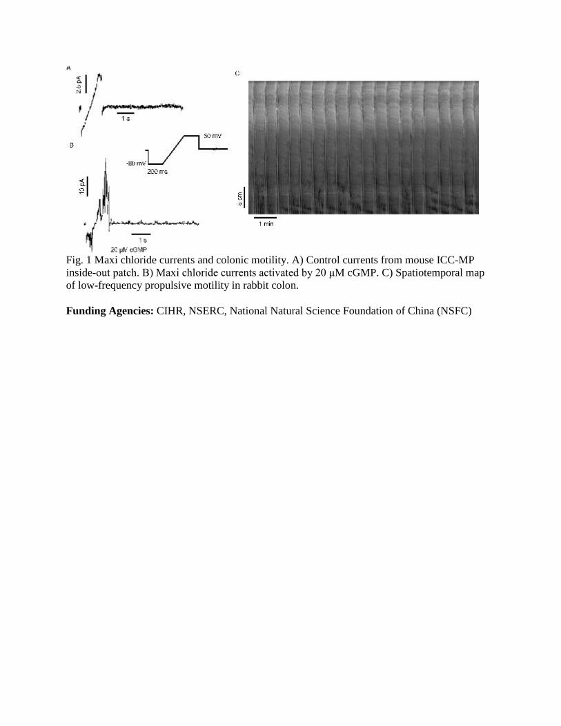
Fig. 1 Maxi chloride currents and colonic motility. A) Control currents from mouse ICC-MP inside-out patch. B) Maxi chloride currents activated by 20 μM cGMP. C) Spatiotemporal map of low-frequency propulsive motility in rabbit colon.
Funding Agencies: CIHR, NSERC, National Natural Science Foundation of China (NSFC)

A325
HISTAMINE IN DIARRHEA-PREDOMINANT IRRITABLE BOWEL SYNDROME (IBS-D) PATIENTS SENSITIZES NOCICEPTIVE SENSORY NEURONS
Y. Nasser1, E. Valdez-Morales1, S. Vanner1, G. Boeckxstaens2
1. Gastrointestinal Diseases Research Group, Queen's University, Kingston, ON, Canada; 2. University Hospital Leuven, Leuven, Belgium.
Background: The pathogenesis of pain signaling in IBS is incompletely understood but studies suggest a key role for mucosal mast cells. Increased mast cells in close proximity to nerve terminals have been observed, while histamine release is increased in biopsy samples of IBS patients. A preliminary clinical trial of a histamine-1 receptor antagonist significantly reduced abdominal pain in IBS patients but the mechanism remains unknown. Aims: We hypothesized that mast cell-derived soluble factors, such as histamine, play a major role in the sensitization of chemosensitive transient receptor potential (TRP) expressing dorsal root ganglia (DRG) neurons, resulting in increased nociceptive signaling in IBS. Methods: Mucosal biopsies obtained from the descending colon of IBS-D patients or healthy controls undergoing colonoscopy were incubated overnight to generate supernatants. Mouse DRG neurons were exposed overnight to supernatants and subsequently loaded with the calcium sensitive dye Fura-2 AM (2μM). Baseline intracellular calcium was monitored for 160s and the chamber was then superfused with the TRPV1 receptor agonist capsaicin (250nM; EC50). Data was analyzed with a Mann-Whitney test. Results: IBS-D supernatants caused a significant increase in the intracellular calcium responses to capsaicin in 4/6 patients (p<0.05). To determine whether mast-cell derived soluble factors could mimic the action of IBS-D, histamine (10μM) was added to the media. Histamine caused a significant increase in intracellular calcium in capsaicin-sensitive neurons compared to control supernatants alone (Control: n=32 cells from 4 patients; F340/380 ratio: 2.45 ± 0.24; Control + Histamine: n=46 cells from 4 patients; F340/380 ratio: 3.98 ± 0.23, 46 cells; p = 0.0001). To test whether the effects of IBS-D were dependent on histamine, pyrilamine (1μM), a histamine-1 receptor antagonist, was added to the media. Pyrilamine significantly inhibited the effects of IBS-D supernatants (IBS-D: n=63 from 5 patients; F340/F380 ratio: 3.90 ± 0.18; IBS-D + pyrilamine: n= 65 cells from 5 patients F340/F380 ratio: 2.73 ± 0.20; p < 0.0001). The effect of pyrilamine was dominant in 3/5 IBS-D patients studied. Conclusions: Soluble factors from patients with IBS-D sensitize TRPV1 calcium signaling. Our studies of TRPV1 sensitization indicate that this is in part histamine dependent and suggests a functional interaction between mast cells and afferent nerves. Furthermore, this data demonstrates heterogeneity within IBS-D patients. A biomarker targeting histamine may determine which patients with IBS-D may derive clinical benefit from the use of a histamine-1 receptor antagonist.
Funding Agencies: CAG, CIHR, CCFC; EVM is supported by a CONACYT: 203341 fellowship

A326
EFFECT OF GLUTEN ON BEHAVIOURAL PROFILE AND GASTROINTESTINAL MOTILITY IN A MURINE MODEL OF GLUTEN SENSITIVITY
M. Pigrau, G. De Palma, S. Sidani, P. Miranda, J. Lu, J. McCarville, E. Verdu, S. Collins, P. Bercik
McMaster University, Hamilton, ON, Canada.
Background: Non-celiac gluten sensitivity is a common but poorly understood condition, which shares many clinical features with irritable bowel syndrome and celiac disease, including gastrointestinal dysmotility and psychiatric comorbidity. Previous studies showed that gluten sensitization in genetically predisposed mice leads to immune activation and gut cholinergic nerve dysfunction, however the in vivo correlates of these findings are unknown. Aims: To investigate the effect of gluten sensitivity on behaviour and motility in NOD-DQ8 and wild type C57Bl/6 mice. Methods: Specific pathogen free NOD-DQ8 and C57Bl/6 mice were maintained on a gluten free diet, and orally sensitized with cholera toxin (CT) plus gliadin weekly for 3 weeks. Controls received CT alone. One week after sensitization, gastrointestinal motility was assessed using the bead videofluoroscopy study (Reed el al, 2014). Behavioural profile was studied by the tail suspension, step-down and light preference tests. Mice were then gavaged 3 times per week with vehicle or gluten (2mg/mouse). Motility and behaviour were assessed during the last week of treatment. Mice were sacrificed thereafter and tissue samples were collected. Statistical analysis was performed using Mann-Whitney or t-tests as appropriate. Results: Sensitization per se did not affect motility or behaviour in C57Bl/6 or in NOD-DQ8 mice. After the gluten challenge, sensitized C57Bl/6 mice displayed no changes in motility or behaviour, and no overt gut inflammation was noted. On the other hand, gluten administration to sensitized NOD-DQ8 mice induced delayed gastrointestinal transit and anxiety-like behaviour compared to controls. Conclusions: Gluten sensitization and challenge induced gastrointestinal dysmotility and anxiety-like behaviour in NOD-DQ8 mice, but not in C57Bl/6 mice. This study suggests that genetic celiac markers play an important role in the development of functional changes associated with gluten sensitivity and supports the concept of a "celiac light" condition underlying non-celiac gluten sensitivity.
Funding Agencies: CIHR

A327
NITRIC OXIDE MEDIATES IMPAIRED EXCITABILITY OF VAGAL AFFERENTS IN OBESITY AND IN AN IN VITRO MODEL OF HYPERLEPTINEMIA
S. Park, M. Beyak
Queen's University, Kingston, ON, Canada.
Background: We have recently demonstrated that both high concentrations of leptin and obesity inhibit the excitability of vagal afferents. This may impair meal - related sateity signaling. It has been suggested that downstream event of leptin is intracellular NO signaling. Aims: These experiments set out to examine the role of NO in the alterations in vagal afferent excitability seen in obesity and with high leptin levels. Methods: Nodose ganglion neurons (NGN) from standard diet fed mice (C57BL/6), or high fat fed mice (HFF, 60% kcal from fat, for 12-16 weeks) were dissociated and/or incubated overnight with leptin or standard culture media (100nM). Excitability was assessed by current clamp recordings performed 18-24 h post-dissociation. Results: In leptin-incubated NGNs, the nitric oxide synthase inhibitor, nitro-L-arginine (L-NNA) (0.1 mM) significantly decreased rheobase (100.8 ± 18.6 pA (n=12, leptin-control) vs. 40.0 ± 3.2 pA (n=13, leptin + L-NNA) **p=0.028) and increased number of APs at twice rheobase (1.6 ± 0.3 (n=12, leptin-control) vs. 5.1 ± 0.9 (n=13, leptin + L-NNA) **p=0.0012). L-NNA (0.1 mM) also significantly decreased rheobase (112.1 ± 14.6 pA (n=14, HFF-control) vs. 57.8 ± 10.9 pA (n=9, HFF + L-NNA) **p=0.041) in HFF neurons. ). One of the targets for NO is soluble guanylate cyclase (sGC), resulting in accumulation of cGMP. The sGC inhibitor, ODQ increased the excitability of leptin incubated NGNs. ODQ (10 mM) significantly decreased rheobase (100.8 ± 18.6 pA (n=12, leptin-control) vs. 60.6 ± 4.2 pA (n=18, leptin + ODQ) *p=0.0172) and increased number of APs at twice rheobase (1.6 ± 0.3 (n=12, leptin-control) vs. 2.7 ± 0.3 (n=18, leptin + ODQ) *p=0.0194) in nodose neuron incubated overnight with 100 nM leptin. cGMP-dependent protein kinase (PKG), a downstream effector of NO was examined on leptin-incubated neurons. Inhibition of cGMP, Rp-8-Br-cGMP (10 mM) significantly decreased rheobase (100.8 ± 18.6 pA (n=12, leptin-control) vs. 57.0 ± 5.2 pA (n=10, leptin + Rp-8-Br-cGMP) *p=0.0499) and increased number of APs at twice rheobase (1.6 ± 0.3 (n=12, leptin-control) vs. 2.7 ± 0.3 (n=10, leptin + Rp-8-Br-cGMP) *p=0.0254). Conclusions: Inhibition of NO reversed the inhibitory effects of leptin and obesity on the excitability of vagal afferents (NGNs). We suggest that the endogenous NO signaling pathway (NO-cGMP-PKG) may be important in pathogenesis of obesity and impaired vagal reflexes such as satiety.
Funding Agencies: CIHR

A328
ENDOGENOUS AND EXOGENOUS NITRIC OXIDE REGULATES EXCITABILITY IN NODOSE GANGLION NEURONS
S. Park, M. Beyak
Queen's University, Kingston, ON, Canada.
Background: Nitric oxide (NO) is a critical mediator of gastrointestinal neuromuscular function and it has been implicated in the modulation of gastrointestinal vagal afferent function. However the mechanisms by which NO affects vagal afferent function are unknown. Aims: The present study aims to elucidate the physiological role of endogenous NO in regulating the excitability of nodose ganglion neurons, the cell bodies of vagal afferents. Methods: Nodose ganglion neurons (NGN) from C57BL/6 mice were dissociated and recordings were performed 18-24 h post-dissociation. Agonists and antagonists of NO, and relevant second messenger pathways were superfused and electrical excitability was assessed with whole cell current clamp recordings. Results: The NO donor, SIN-1 attenuated vagal afferent excitability. SIN-1 (0.5 mM) significantly increased rheobase (52.3 ± 7.8 pA (n=13, control) vs. 83.6 ± 6.9 pA (n=14, SIN-1) **p=0.059) and significantly decreased number of APs at twice rheobase (3.7 ± 0.5 (n=13, control) vs. 1.6 ± 0.3 (n=14, SIN-1) **p=0.0020). On the other hand, the NO synthase inhibitor L-NNA (0.1 mM) decreased rheobase and increased number of APs at twice rheobase. Input resistance was significantly increased by L-NNA (396.3 ± 47.7 MΩ (n=13, control) vs. 803.0 ± 133.7 MΩ (n=14, L-NNA) *p=0.0102). One of the targets for NO is soluble guanylate cyclase (sGC), resulting in accumulation of cGMP. The sGC inhibitor, ODQ (10 mM) did not significantly change the excitability in neurons. However inhibition of cGMP breakdown with sildenafil (1mM) reduced the excitability of nodose neurons increasing rheobase (52.3 ± 7.8 pA (n=13, control) vs. 87.1 ± 9.1 pA (n=14, Sildenafil) **p=0.081) and significantly decreased number of APs at twice rheobase (3.7 ± 0.5 (n=13, control) vs. 1.5 ± 0.2 (n=14, Sildenafil) ***p=0.0005). Activation of cGMP-dependent Protein kinase G (PKG), a downstream effector of NO, increased rheobase (52.3 ± 7.8 pA (n=13, control) vs. 113.3 ± 9.0 pA (n=12, 8-Br-cGMP) ***p<0.0001) and significantly decreased number of APs at twice rheobase (3.7 ± 0.5 (n=13, control) vs. 1.4 ± 0.2 (n=12, 8-Br-cGMP) ***p=0.0008). However, Inhibition of cGMP, Rp-8-Br-cGMP (competition with cGMP) did not significantly change the excitability in neurons. Conclusions: These results indicate endogenous NO inhibits vagal afferents through cGMP and PKG. Given the changes in input resistance, hyperpolarizing ion channels (likely K+ channels) pathway are likely involved. Endogenous NO production by vagal afferents may serve as an autocrine mediator regulating excitability in a variety of physiological states.
Funding Agencies: CIHR
A329
EXPRESSION LEVELS OF CYTOKINES, INCLUDING IL-23, IL-1β, IL-6 AND IFN-γ, IN ESOPHAGEAL MUCOSA ARE STRONGLY PREDICTIVE OF ESOPHAGEAL BODY CONTRACTILITY IN HUMANS
E. Ihara, K. Fukaura, K. Muta, Y. Tanaka, X. Bai, K. Nakamura, R. Takayanagi
Kyushu University, Fukuoka, Japan.
Background: Gastrointestinal (GI) inflammation is accompanied by alterations of GI motility and impaired function of GI smooth muscle (SM). Several cytokines have been found to alter GISM contractility directly, with Th1-related cytokines (TNF-a, IL-1b) associated with hypo-contractility and Th2-related cytokines (IL-4, IL-13) with hyper-contractility of inflamed GISM. It is unclear how Th17-related cytokines affect GISM contractility, or how and which cytokines affect GI motility in clinical settings. High-resolution manometry (HRM) may allow precise evaluation of human esophageal motility. Aims: This study was designed to determine the esophageally expressed cytokines associated with esophageal motility. Methods: Nineteen patients (8 males, 11 females) with suspected esophageal motility disorder were assessed by HRM (Manoscan Z), with esophageal motility function evaluated by measuring distal contractile integral (DCI) and basal LES pressure (BLESP). Biopsy samples were taken from esophageal mucosae covering the esophageal body and LES. Real-time RT-PCR was performed to assess the expression of Th1-related cytokines (TNF-α, IFN-γ, IL-1β), Th2-(IL-4, IL-5, IL-13) and Th17-(IL-17A, IL-23, IL-6) that may be involved in the alteration of GI motility. Cytokines associated with esophageal motility were determined by stepwise regression analysis. Results: Mean patient age was 59.6 ± 3.8 years old. According to the Chicago classification, 7 patients were diagnosed as normal, 7 with achalasia, 2 with esophagogastric junction outflow obstruction, 1 with distal esophageal spasm and 2 with weak peristalsis. Mean ± SD DCI was 2226.3 ± 575 mmHg (n=12), and mean ± SD BLESP was 31.3 ± 3.0 mmHg (n=19). DCI was not measured in 7 patients owing to achalasia. IL-23 and IL-1β were negatively associated with DCI, while IL-6 and IFN-γ were positively associated with DCI. Predicted DCI was calculated using the formula; 48924 − 2070xIL-1β + 1145xIL-6 + 1608xIFN-γ − 2434xIL-23 (adjusted R2=0.85, p<0.0011). In contrast, no cytokines were significantly associated with BLESP. Conclusions: Expression levels of cytokines, including IL-23, IL-1β, IL-6 and IFN-γ, in esophageal mucosa are strongly predictive of contractility of the esophageal body in humans. These cytokines may also be highly associated with GI motility in clinical settings.
Funding Agencies: This work was supported in part by Grants from the Japanese Ministry of Education, Culture, Sports, Science and Technology.

A330
NEUROTRANSMISSION MODULATES NEUROGENESIS WITHIN THE ENTERIC NERVOUS SYSTEM
J. Masood, A. Lomax
Queens University, Kingston, ON, Canada.
WITHDRAWN

A331
EFFICACY OF LINACLOTIDE IN THE TREATMENT OF CHRONIC CONSTIPATION: A SYSTEMATIC REVIEW OF RANDOMIZED CONTROLLED TRIALS
T. Poon2, A. Rezaie1, B. Chang1, E. Cheng2
1. Cedars-Sinai Medical Center, Los Angeles, CA; 2. University of Calgary, Calgary, AB, Canada.
Background: Chronic constipation is a common condition that impairs quality of life and increases morbidity. Linaclotide is an agonist of the guanylate cyclase C receptor on the luminal surface of intestinal enterocytes. Defecation is enhanced by chloride and bicarbonate secretion leading to increased luminal fluid. Aims: We performed a systematic review to determine the efficacy of linaclotide for patients with chronic constipation. Methods: Pubmed, EMBASE and the Cochrane CENTRAL registrar of controlled trials were searched for randomized controlled trials that examined the effect of linaclotide on chronic idiopathic constipation from 1966-Oct 2014. Potential studies were included if they were randomized controlled studies (RCT) evaluating linaclotide compared with placebo or any other drug therapy for constipation. Two reviewers independently assessed trial quality and extracted data. Only studies with intention to treat analyses were considered. Analyses were performed using the Mantel-Haenszel test. When heterogeneity was noted, a random effects model was used. Results: There were 150 papers on linaclotide identified, four of which were RCTs. Three of these RCTs were included based on their use of a primary end point of ≥ 3 spontaneous bowel movements per week and an increase of ≥ 1 spontaneous bowel movements per week. There were 1586 patients randomized to once daily linaclotide 145 mcg, 150 mcg, 290 mcg, or 300 mcg versus placebo. Duration of intervention was 4 to 12 weeks. Low dose linaclotide (145 or 150 mcg) versus placebo was found to have a pooled RR of 3.80 (95% CI 2.20 6.55) for the primary end point and a response rate of 16%-26.8%. High dose linaclotide (290 or 300 mcg) versus placebo pooled RR was 4.26 (95% CI 2.80 6.47) for the primary end point and had a response rate of 19.4%-29.0%. Other end points analyzed also showed improvement with low dose linaclotide. These included improved abdominal discomfort (RR 1.57, 95% CI 1.26 1.97), adequate relief response (RR 2.50, 95% CI 1.87 3.34), decreased bloating which was defined as a score of ≥ 0.5 for 75% of treatment time (RR 1.97, 95% CI 1.44 2.69), and health related quality of life improvement shown by an increase of ≥ 1 point on the Patient Assessment of Constipation Quality of Life instrument (RR 1.83, 95% CI 1.34 2.50). Conclusions: Linaclotide at low and high doses is more effective than placebo in the treatment of chronic constipation. This was found in the primary endpoint of improved bowel function and also in secondary end points such as adequate relief response, bloating, and health related quality of life.
Funding Agencies: None

A332
GASTROINTESTINAL MOTILITY TESTING IN PATIENTS WITH SYSTEMIC SCLEROSIS: A SYSTEMATIC REVIEW
A. Parmar, A. Tisseverasinghe, S. Johnson, L. Liu
University of Toronto, Toronto, ON, Canada.
Background: Gastrointestinal (GI) manifestations occur in up to 90% of patients with systemic sclerosis (SSc). The GI manifestations are believed to be secondary to the progressive, fibrotic changes inherent to SSc, leading to impaired GI motility. However, assessment and treatment of GI manifestations in SSc patients is limited. To further advance this field, the knowledge in the utility of various objective motility testing in this patient population is essential. Aims: This review summarizes the current evidence for the use of various GI motility studies in evaluating patients with SSc. Methods: Three databases were searched (Cochrane Central Register of Controlled Trials, Ovid MEDLINE and Embase) up until November 2013. Search terms included "scleroderma" or "systemic sclerosis", "gastrointestinal", and "motility" or "transit" with related terms. No language restrictions were applied. Abstracts were included if they had ≥ 10 adult (age > 18 yr.) SSc patients and evaluated a GI motility study. Two independent reviewers reviewed all abstracts. A third reviewer resolved all conflicts. All motility studies were evaluated for their description of validity, reliability, responsiveness and feasibility. Results: Our search identified 859 citations. A total of 172 studies were included in this systematic review. To date, esophageal motility studies have been the best described with a total of 167 articles in more than 5000 SSc patients. Esophageal manometry was the most commonly described motility study (n=105). Within the group of esophageal manometry studies, 40 studies evaluated for validity, 19 evaluated responsiveness and 4 described feasibility. Gastric scintography was used in 17 of the 32 studies that assessed gastric motor function. The majority of gastric scintography studies used 99-Technetium radiolabeled meals; however, the testing meal components were significantly heterogeneous. Ano-rectal manometry was used to assess ano-rectal motility in 13 studies, of which 10 used water perfusion catheters. Among these ano-rectal studies, only resting and squeeze pressures had been consistently reported. Currently, there have been only a few studies evaluating the motility of the small intestine (n=8) and colon (n=4). Conclusions: Our study illustrated that the use of standardized motility testing in SSc patients is lacking and largely heterogeneous. Although esophageal manometry has been the mostly commonly employed motility assessment, the reported parameters vary among studies. Standardizations in the methodology and reporting are in need such that the application of these motility studies can be validated in SSc patients with GI motor disturbances.
Funding Agencies: None
A333
EFFECT OF PRUCALOPRIDE IN SMALL BOWEL CAPSULE ENDOSCOPY
R. Scott, D. Prichard, P. Cramer, F. Donnellan
UBC Division of Gastroenterology, Vancouver General Hospital, Vancouver, BC, Canada.
Background: The diagnostic yield of small bowel capsule endoscopy depends on successful completion rates. In up to 30 percent of small bowel capsule endoscopy procedures, the capsule does not reach the cecum within recording time. The use of prokinetics in increasing completion rates is unclear. Aims: To determine the effect of prucalopride on gastrointestinal transit time and diagnostic yield of small bowel capsule examinations Methods: 28 patients were given prucalopride in addition to standard bowel preparation (group A). These patients were compared with a retrospective group of 123 patients who received standard bowel preparation without prucalopride (group B). Gastric and small bowel transit times, complete small bowel examinations determined by identification of the cecum, and diagnostic yields determined by detection of pathology, were compared between the two groups. Data is expressed as the mean (+/-) standard error and was analyzed using the unpaired Students t test and Fishers exact test. Results: Completion rate was 84% in group A and 79% in group B (p=0.605). Mean gastric time was 36(+/-5) minutes in group A and 33(+/-7) minutes in group B (p=0.698). Mean small bowel transit time, calculated following exclusion of incomplete studies caused by small bowel stricturing, was 164 (+/-27) minutes in group A and 229 (+/-10) minutes in group B (p=0.03). There was no difference in the detection of pathology between the two groups (50% versus 46%; p=0.605). Conclusions: In this study, prucalopride appears to significantly reduce the small bowel transit time but there was no significant difference in cecal completion rates or the diagnostic yield.
Funding Agencies: None
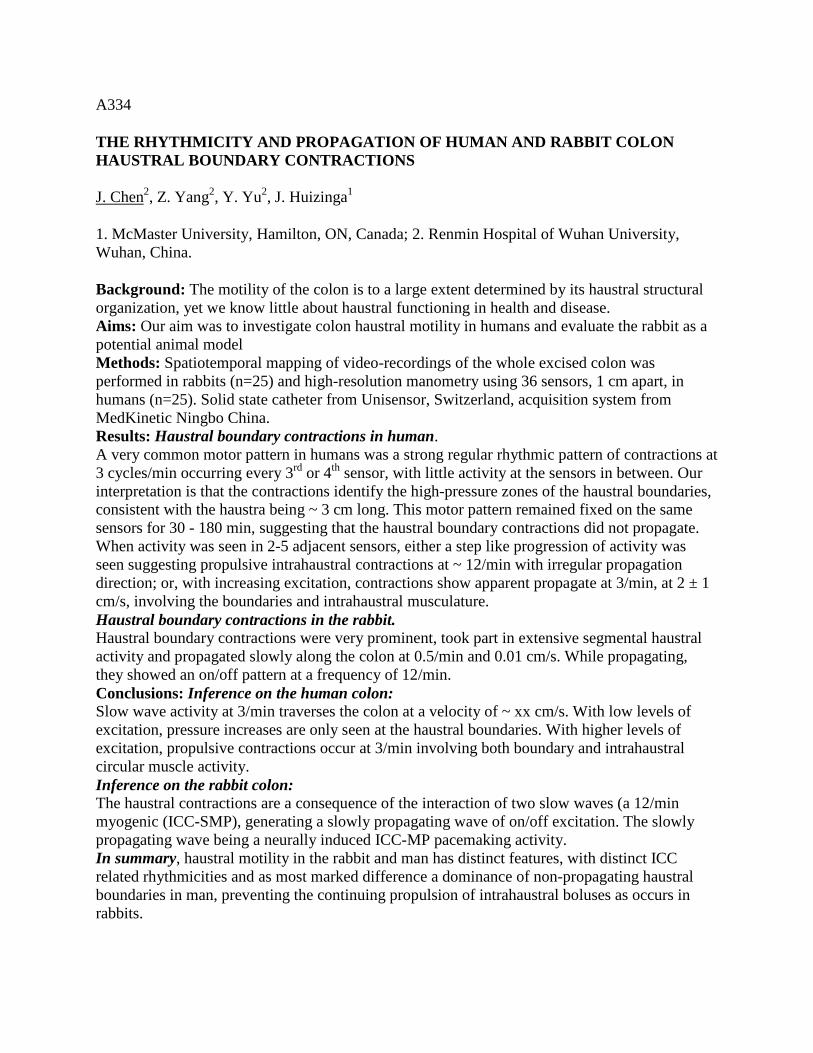
A334
THE RHYTHMICITY AND PROPAGATION OF HUMAN AND RABBIT COLON HAUSTRAL BOUNDARY CONTRACTIONS
J. Chen2, Z. Yang2, Y. Yu2, J. Huizinga1
1. McMaster University, Hamilton, ON, Canada; 2. Renmin Hospital of Wuhan University, Wuhan, China.
Background: The motility of the colon is to a large extent determined by its haustral structural organization, yet we know little about haustral functioning in health and disease. Aims: Our aim was to investigate colon haustral motility in humans and evaluate the rabbit as a potential animal model Methods: Spatiotemporal mapping of video-recordings of the whole excised colon was performed in rabbits (n=25) and high-resolution manometry using 36 sensors, 1 cm apart, in humans (n=25). Solid state catheter from Unisensor, Switzerland, acquisition system from MedKinetic Ningbo China. Results: Haustral boundary contractions in human. A very common motor pattern in humans was a strong regular rhythmic pattern of contractions at 3 cycles/min occurring every 3rd or 4th sensor, with little activity at the sensors in between. Our interpretation is that the contractions identify the high-pressure zones of the haustral boundaries, consistent with the haustra being ~ 3 cm long. This motor pattern remained fixed on the same sensors for 30 - 180 min, suggesting that the haustral boundary contractions did not propagate. When activity was seen in 2-5 adjacent sensors, either a step like progression of activity was seen suggesting propulsive intrahaustral contractions at ~ 12/min with irregular propagation direction; or, with increasing excitation, contractions show apparent propagate at 3/min, at 2 ± 1 cm/s, involving the boundaries and intrahaustral musculature. Haustral boundary contractions in the rabbit. Haustral boundary contractions were very prominent, took part in extensive segmental haustral activity and propagated slowly along the colon at 0.5/min and 0.01 cm/s. While propagating, they showed an on/off pattern at a frequency of 12/min. Conclusions: Inference on the human colon: Slow wave activity at 3/min traverses the colon at a velocity of ~ xx cm/s. With low levels of excitation, pressure increases are only seen at the haustral boundaries. With higher levels of excitation, propulsive contractions occur at 3/min involving both boundary and intrahaustral circular muscle activity. Inference on the rabbit colon: The haustral contractions are a consequence of the interaction of two slow waves (a 12/min myogenic (ICC-SMP), generating a slowly propagating wave of on/off excitation. The slowly propagating wave being a neurally induced ICC-MP pacemaking activity. In summary, haustral motility in the rabbit and man has distinct features, with distinct ICC related rhythmicities and as most marked difference a dominance of non-propagating haustral boundaries in man, preventing the continuing propulsion of intrahaustral boluses as occurs in rabbits.
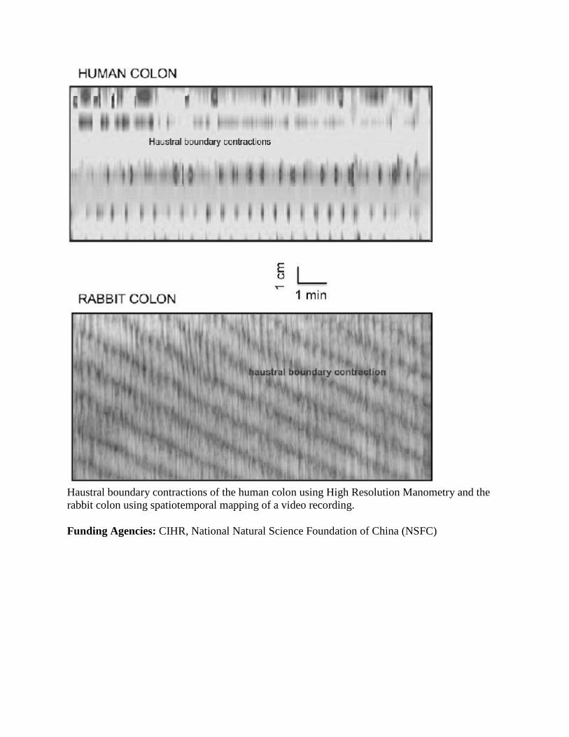
Haustral boundary contractions of the human colon using High Resolution Manometry and the rabbit colon using spatiotemporal mapping of a video recording.
Funding Agencies: CIHR, National Natural Science Foundation of China (NSFC)

A335
HOW DO ICC NETWORK DISTURBANCES AFFECT SMALL INTESTINE MOTOR PATTERNS?
S. Parsons, J. Huizinga
McMaster University, Hamilton, ON, Canada.
Background: The ICC pacemaker networks are critical for normal intestinal motility, as pacemakers and mediators of innervation. In most gut motor disorders, ICC networks have been shown to be structurally affected, in particular retraction of ICC processes. However, we know very little about functional disturbances of ICC networks and the consequences for motor pattern development. Aims: Our objective was to understand network properties of ICC through spatio-temporal mapping of motor patterns as well as through modelling of systems of coupled oscillators Methods: Video recordings of whole organ motility were made. We initiated network disturbances through pharmacological blockade of gap junction conductance. We used a novel multi-camera diameter mapping system to measure contraction along 25-30 cm lengths of the murine small intestine. Results: We observed step-like gradients in the frequency (frequency plateaux) of contraction waves along the length of the small intestine. There were typically 2-3 plateaux per length of intestine. At plateau boundaries there were fork and slip dislocations (wave drops) and a waxing and waning of the contraction amplitude. The gap junction inhibitor carbenoxolone increased the number of plateaux and dislocations and slowed contraction wave velocity. In some cases there was a reversal of the usual frequency gradient, with a plateau at a higher frequency than its proximal neighbour, and thus fork dislocations were inverted and the direction of propagation reversed. Contraction waves were modelled with a chain of van der Pol oscillators. Network characteristics as shown in spatiotemporal maps were evident. Fork dislocations were produced when the chain was given a linear natural frequency gradient. Conclusions: Disturbances in network properties by inhibiting electrical coupling between individual pacemaker cells lead to disruption of long peristaltic waves, increase in frequency plateaus, appearance of retrograde propagation. In general terms a change from dominant propulsion to dominant segmentation. To understand the mechanisms governing intestinal motor pattern generation, the ICC pacemaker network is best evaluated as a system of coupled oscillators. This model can be used to generate hypotheses for the genesis of motor disturbances as a consequence of network disruption.
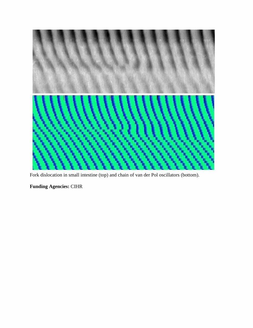
Fork dislocation in small intestine (top) and chain of van der Pol oscillators (bottom).
Funding Agencies: CIHR

A336
EFFECTS OF A DEFINED COMMUNITY OF COMMENSAL HUMAN GUT BACTERIA ON VISCERAL NOCICEPTIVE NEURONS
J. Sessenwein2, E. Petrof1, S. Vanner3, A. Lomax2
1. Queen's University, Kingston, ON, Canada; 2. Queens University, Kingston, ON, Canada; 3. queen's, Kignston, ON, Canada.
WITHDRAWN

A337
ACHALASIA IN EVOLUTION: A CASE SERIES OF THE TEMPORAL EVOLUTION OF ACHALASIA
A. Parmar, L. Liu
University of Toronto, Toronto, ON, Canada.
Background: Achalasia is a primary esophageal motility disorder characterized by absent esophageal peristalsis and failure of lower esophageal sphincter (LES) relaxation. Morphological studies have demonstrated loss of nitrergic innervation in the esophagus of achalasia patients. The presence of atypical achalasia cases, or variants, have been previously documented with only a few case reports outlining a temporal relationship of "variant" achalasia progression to achalasia. Temporal evolution of the manometric changes of patients with achalasia can shed light on a further understanding of the pathophysiology and help in establishing early diagnosis. Aims: To describe adult (age > 18 years) patients who have shown a temporal progression from achalasia in evolution (AIE) to achalasia with respect to their demographics, clinical history and esophageal manometry profiles. Methods: Retrospective chart review of all the high-resolution esophageal manometry studies (EMS) in a tertiary referral center from January 2008 to July 2014. Results: Four confirmed cases of achalasia patients of whom AIE was initially described. All patients (mean age = 75 +/- 14 years, range = 55-86 years; 2 male, 2 female) presented with a varying duration (6 months to 9 years) of liquids and solids dysphagia. Three of the four patients have a history of type 2 diabetes mellitus. AIE was applied when some degree of esophageal body peristaltic activity were preserved. In all AIE patients, the residual lower esophageal sphincter (LES) pressure was consistently elevated (26.7 +/- 10.7 mmHg, normal < 15 mm Hg). The progression of AIE to achalasia was observed over a range of 6 months to 10 years. When achalasia was diagnosed, 2 patients had Type 2 and 2 patients had Type 3 achalasia; all patients demonstrated 100% simultaneous esophageal contractions with an elevated LES residual pressure (31.1 ± 19.9 mm Hg). Conclusions: Our observations that all AIE patients, albeit a small number, progressed to either type 2 or 3 achalasia suggests that type 1 achalasia may represent the later or more advance disease. Thus, the identification of patients with AIE may allow for earlier awareness of possible disease progression, thereby facilitating the initiation of timely investigations and management strategies. Larger and longer-term study will help further characterize the manometric criteria of AIE.
Funding Agencies: None

A338
HIGH RESOLUTION ESOPHAGEAL MANOMETRY PATTERN IN CHILDREN AND ADOLESCENTS WITH RUMINATION SYNDROME
F. Righini-Grunder, A. Aspirot, C. Faure
Ste Justine hospital, Montreal, QC, Canada.
Background: Diagnosis of rumination is mainly clinical and based on the ROME III criteria. The main clinical feature is a painless regurgitation appearing within seconds or minutes from food/liquid ingestion. Pathophysiology of rumination remains unknown, but involves a voluntary contraction of the abdominal wall muscles with a rise in intragastric pressure (R wave) and retrograde movement of gastric contents into the esophagus. High resolution esophageal manometry (HREM) allows identifying R-waves at the moment when episodes of rumination occur. Aims: The aim of this study was to determine the HREM pattern in children suffering from rumination. To our knowledge no study has yet described the HREM pattern in children with rumination. Methods: Retrospective evaluation of pediatric patients with rumination syndrome according to Rome III criteria who underwent an HREM between January 2011 and September 2014. Ten wet swallows followed by 100 mL of water or a test meal were administered during HREM. Rumination was defined as the presence of R-wave during an associated clinical rumination episode. Results: Nine patients (5 F; median age 14.7 yrs, range 9-18) were identified. All fulfilled the Rome III criteria for rumination. Esophagogastroduodenoscopy was performed in 8 patients prior to manometry and was normal in all except one mild microscopic esophagitis and one H pylori gastritis. All had a normal manometry according to Chicago classification. Rumination was confirmed during HREM in 7 out of the 9 patients (77%) who displayed association of R waves with clinical episodes of rumination. LES and UES relaxation were associated with all episodes. Conclusions: HREM is a simple tool that may help to confirm the diagnosis of rumination in children. Whether HREM may influence outcome of these patients remains to be determined.
Funding Agencies: None
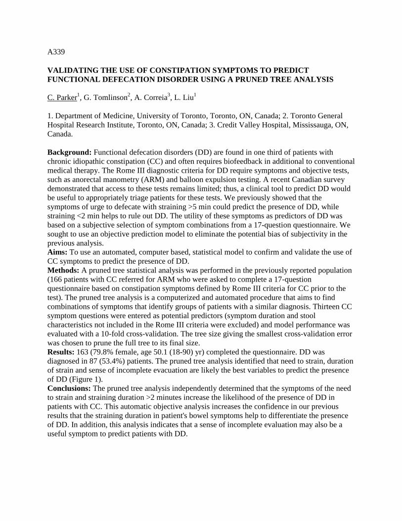
A339
VALIDATING THE USE OF CONSTIPATION SYMPTOMS TO PREDICT FUNCTIONAL DEFECATION DISORDER USING A PRUNED TREE ANALYSIS
C. Parker1, G. Tomlinson2, A. Correia3, L. Liu1
1. Department of Medicine, University of Toronto, Toronto, ON, Canada; 2. Toronto General Hospital Research Institute, Toronto, ON, Canada; 3. Credit Valley Hospital, Mississauga, ON, Canada.
Background: Functional defecation disorders (DD) are found in one third of patients with chronic idiopathic constipation (CC) and often requires biofeedback in additional to conventional medical therapy. The Rome III diagnostic criteria for DD require symptoms and objective tests, such as anorectal manometry (ARM) and balloon expulsion testing. A recent Canadian survey demonstrated that access to these tests remains limited; thus, a clinical tool to predict DD would be useful to appropriately triage patients for these tests. We previously showed that the symptoms of urge to defecate with straining >5 min could predict the presence of DD, while straining <2 min helps to rule out DD. The utility of these symptoms as predictors of DD was based on a subjective selection of symptom combinations from a 17-question questionnaire. We sought to use an objective prediction model to eliminate the potential bias of subjectivity in the previous analysis. Aims: To use an automated, computer based, statistical model to confirm and validate the use of CC symptoms to predict the presence of DD. Methods: A pruned tree statistical analysis was performed in the previously reported population (166 patients with CC referred for ARM who were asked to complete a 17-question questionnaire based on constipation symptoms defined by Rome III criteria for CC prior to the test). The pruned tree analysis is a computerized and automated procedure that aims to find combinations of symptoms that identify groups of patients with a similar diagnosis. Thirteen CC symptom questions were entered as potential predictors (symptom duration and stool characteristics not included in the Rome III criteria were excluded) and model performance was evaluated with a 10-fold cross-validation. The tree size giving the smallest cross-validation error was chosen to prune the full tree to its final size. Results: 163 (79.8% female, age 50.1 (18-90) yr) completed the questionnaire. DD was diagnosed in 87 (53.4%) patients. The pruned tree analysis identified that need to strain, duration of strain and sense of incomplete evacuation are likely the best variables to predict the presence of DD (Figure 1). Conclusions: The pruned tree analysis independently determined that the symptoms of the need to strain and straining duration >2 minutes increase the likelihood of the presence of DD in patients with CC. This automatic objective analysis increases the confidence in our previous results that the straining duration in patient's bowel symptoms help to differentiate the presence of DD. In addition, this analysis indicates that a sense of incomplete evaluation may also be a useful symptom to predict patients with DD.

Funding Agencies: None

Nutrition, Obesity and Aging
A340
INCREASED SUCROSE BUT NOT STARCH INTAKE DETERIORATES COLITIS AND REDUCES THE PROTECTIVE EFFECT OF PREBIOTICS IN A RAT COLITIS MODEL
R. Valcheva1, R. Huo2, A. Hassanzadeh Keshteli1, L. Dieleman1
1. University of Alberta, Edmonton, AB, Canada; 2. McGill University, Montreal, QC, Canada
Background: Inflammatory bowel diseases (IBD) are complex disorders associated with chronic intestinal inflammation. Intestinal bacteria contribute to the initiation and perpetuation of this chronic condition. Modulation of the intestinal microbiota by probiotics or prebiotics has shown benefits in experimental and ulcerative colitis by reducing intestinal inflammation. Recently we showed that the protective effects of inulin- derived fructo-oligosaccharides (FOS) on colitis development depend on the background diet. Prebiotic fibers added to a chemically defined diet versus rat chow failed to reduce colitis in rat colitis model. Aims: The aim of the current study was to examine if the carbohydrate source alone and in combination with FOS can modulate colitis development in HLA-B27 transgenic rats. Methods: At 4 weeks of age, HLA-B27 transgenic rats were randomized to 5 different diets: control chow; sugar-based diet (45% sucrose); starch-based diet (45% corn starch); sugar-based diet + FOS (15%); or starch-based diet + FOS (15%) for 12 weeks. Growth and food intake were measured. Cecal and colonic inflammation was assessed by weight/length ratio, macroscopic scoring and mucosal IL-1β secretion. Bacterial translocation in mesenteric lymph nodes was quantified by qPCR. Cecal contents were collected for microflora analysis. Results: There were no significant differences in animal growth and food intake. Sucrose- based diet was associated with significantly more severe cecal inflammation compared to other diets, as measured by macroscopic tissue scoring, weight/length ratio and IL-1β secretion. Furthermore, 20% of rats fed sucrose-based diet developed arthritis. Addition of FOS in sucrose-based diet did not improve intestinal inflammation. In contrast, adding FOS to the starch-based diet had colitis-reducing effects as evidenced by reductions in inflammatory markers. Conclusions: High amounts of sucrose, but not starch, affect colitis development and prevent the protective effect of prebiotic fibers in experimental colitis. The exact mechanism(s) including the effects on the intestinal microflora remain to be identified and are being analyzed. These results suggest that the carbohydrate composition of diets can affect disease outcome during intervention trials in IBD.
Funding Agencies: Alberta IBD Consortium

A341
PREBIOTICS ENHANCE INTESTINAL EPITHELIAL BARRIER FUNCTION VIA ACTIVATION OF PROTEIN KINASE C
R. Wu2, P. Maattanen1, K. Henry1, P. Sherman1
1. Cell Biology Program, Research Institute, Hospital for Sick Children, Toronto, ON, Canada; 2. Department of Laboratory Medicine and Pathobiology, University of Toronto, Toronto, ON, Canada.
Background: There is growing interest in using selective nutrients as dietary supplements to deliver long-term health benefits to the host. Prebiotics are non-digestible oligosaccharides that promote the growth of select gut microbes, but may have other effects on the mucosal barrier. Aims: The purpose of this study is to determine the effects of prebiotics on intestinal epithelial barrier function and to characterize their underlying mechanisms of action. Methods: Caco2-bbe and T84 cells were seeded onto Transwell filters. Subsequently, polarized monolayers were apically exposed to either inulin or short-chain fructo-oligosaccharide (sc-FOS, 10% w/v, 16 h), and then challenged with enterohemorrhagic Escherichia coli strain CL56, serotype O157:H7 (5 h). Transepithelial electrical resistance (TER), translocation of fluorescein-labelled dextran (10 kDa), and zona-occludens-1 (ZO-1) mRNA, protein expression and immunofluorescence labelling were used to measure epithelial barrier integrity. Activation of isotype-specific protein kinase C (PKC) was assessed by immunoblotting 15 min after prebiotic administration. Results: Pathogen challenge of epithelial monolayers abrogated TER (13.3 ± 1.6 % of baseline, n=5) and increased dextran flux (847.4 ± 108.4 ng), a marker of epithelial permeability. However, pre-treatment with either of the two prebiotics protected against E. coli O157:H7-induced barrier dysfunction (TER: 49.9 ± 3.7 % of baseline for inulin-treated; 48.9 ± 5.6 % for sc-FOS-treated versus untreated cells; ANOVA p<0.01; n=5; dextran flux: 351.5 ± 93.4 ng for inulin-treated; 341.0 ± 65.4 ng for sc-FOS-treated versus untreated cells; ANOVA p<0.01; n=5). ZO-1 redistribution induced by E. coli O157:H7 was prevented by pre-treatment with prebiotics (n=3). Both inulin and sc-FOS increased ZO-1 mRNA expression (1.5 ± 0.2 and 1.4 ± 0.1 fold-increases for inulin and sc-FOS, respectively; ANOVA p<0.05; n=4) and protein levels (2.0 ± 0.6 and 1.9 ± 0.4 fold-increases for inulin and sc-FOS; respectively; n=3). Mechanistically, both prebiotics induced a dose- and time-dependent activation of PKC. Inhibition of PKC activity with pharmacological inhibitors (Gö 6983, Gö 6850 at 10 nM, 60 min) abolished prebiotic-induced effects on intercellular tight junctions. Conclusions: These results indicate that dietary prebiotics can enhance epithelial barrier integrity independent of gut microbes. Such protection is mediated, at least in part, through the activation of PKC in host intestinal epithelial cells. Future aims are to characterize the specific upstream and downstream PKC signaling cascades that mediate the observed effects.
Funding Agencies: RYW is supported by Vanier CGS, CIHR MD/PhD Studentship, and a NASPGHAN Medical Student Scholarship. PMS is the recipient of a CRC in Gastrointestinal Disease. The work was funded by an operating grant from CIHR and a research contract from Lallemand Health Solutions (Montreal, Quebec).

Poster of Distinction
A342
NUTRITION AND PHYSICAL THERAPY TARGETS ARE NOT BEING MET IN ADMITTED PATIENTS WITH CIRRHOSIS
T. Shaikh1, M. Ma3, J. Abraldes3, R. Ameen4, A. Shah4, S. Robbins4, K. Qumosani2, B. Aljudaibi2, A. Rahman2, P. Tandon3
1. University of Alberta, Edmonton, AB, Canada; 2. University of Western Ontario, London, ON, Canada; 3. Cirrhosis Care Clinic, University of Alberta, Edmonton, AB, Canada; 4. Royal Alexandra Hospital, Edmonton, AB, Canada.
Background: Low muscle mass and frailty are major determinants of morbidity and mortality in cirrhosis. Adequate nutritional therapy and early mobilization can impact these parameters. Current cirrhosis guidelines target daily caloric intake at 35-40 kcal/kg and protein intake at 1.2-1.5 g/kg. There are no formal guidelines for mobilization therapy in cirrhosis. Aims: In hospitalized cirrhotic patients, the aims of this study were to evaluate the adherence to nutritional guidelines and determine the proportion of cirrhotic patients who had early (within 72 h of study enrollment) mobilization therapy. Methods: Multicenter prospective trial involving 3 hospitals (2 Alberta, 1 Ontario) with data collection over a 13-month period (June 2013-July 2014). All patients had cirrhosis and were recruited within 72 hours of admission. Patients with non-hepatocellular carcinoma related malignancy, palliative HCC, severe end-organ failure (eg. Dialysis, COPD on home oxygen, CHF) or Hepatic encephalopathy Conn's score >1 were excluded. Patients were followed for 30 days from initial assessment or until discharge from hospital. Detailed nutritional and mobilization data was collected over the first 3 days of assessment and at two centers, also at ~ day 8. Results: 137 patients were recruited. Baseline characteristics - Mean age 55.5 (SD: 10.3), 59% male, 77% Caucasian, mean MELD and CP scores 18.2 (6.6) and 9.5 (2.0) respectively. The top three etiologies of liver disease were alcohol (41%), HCV (33%) and Autoimmune/cholestatic liver disease (12%). Eighty-two percent had at least mild ascites. The mean estimated dry weight BMI was 24.6 (5.3). By day 3, only 7% and 20% respectively met guideline based calorie and protein targets. In the 44 patients with ~ day 8 data still only 11% and 14% of patients met calorie and protein targets respectively. A subset of 77 patients had mobilization data available. Over days 1-3, 22.1%, 38% and 55% of these patients were able to mobilize/be mobilized from their bed to outside of their hospital room. Only three patients had a formal physiotherapy consult. Conclusions: This data presents a significant opportunity to improve on the early nutritional and mobilization therapy in cirrhotic inpatients. Less than 20% of patients met nutritional intake recommendations by day 3 of study enrollment and only 4% received a formal physiotherapy consult. National inpatient cirrhosis care guidelines and standardized orders would allow us to determine whether optimization of these variables can impact clinical outcomes in cirrhosis.
Funding Agencies: None
A343
ANTIMICROBIAL PEPTIDE SYNTHESIS IN THE AGED SMALL INTESTINE: ITS RELATIONSHIP WITH THE COMMENSAL MICROBIOTA AND EPITHELIAL INTEGRITY
S. Tremblay1, G. Grenier1, A. Menendez2
1. Université de Sherbrooke, Sherbrooke, QC, Canada; 2. University of Sherbrooke, Sherbrooke, QC, Canada.
Background: Aging is frequently characterized by alterations in gastrointestinal function, often leading to malnutrition, small intestinal bacterial overgrowth and high prevalence of infections, inflammatory diseases and cancer. Functional changes associate with alterations of the intestinal microenvironment but the contribution of specific factors is undefined. The microbiota, which plays critical roles in the maintenance of intestinal homeostasis, changes with age; however, the mechanisms underlying microbiome changes in the aging gut and its consequences remain unclear. Aims: Antimicrobial peptide and proteins (AMPP) participate in the control of the intestinal microbiome and the maintenance of intestinal immunity. We aimed to determine whether small intestinal AMPP synthesis is modified with age and how it relates to other elements of intestinal homeostasis. Methods: Aged and young mice were studied for the numbers and composition of bacteria in the duodenum, jejunum and ileum, and for the levels of multiple antibacterial peptides and proteins. Electron microscopy and H&E staining were used to document morphological changes of the intestinal epithelium. Results: Aging resulted in an increase of lactose-fermenting enterobacteria along the whole small intestine. Aged mice showed small intestinal bacterial overgrowth evidenced by a significant increase in the number of total bacteria in the duodenum, suggesting a local age-mediated impairment of the immune response. Duodenal AMPP synthesis was enhanced significantly, most likely in response to the bacteria. No gross epithelial alterations were detected in aged animals but the expression of pro-inflammatory markers such as Nos2 and Tnfa was significantly increased. The jejunum also showed an increase in bacteria, with increases only in the Reg3b and Reg3g levels and no apparent epithelial damage. In contrast, dramatic disruptions of the epithelium at the villi tips were observed in the ileum of most old animals. Increased numbers of intermediate cells were found in the ileal crypts and along the villi, suggesting a failure of the aged ileal epithelia to properly differentiate. We also observed goblet cell hyperplasia with increased Muc2 production, and significant upregulation of multiple inflammatory and proliferation markers. EM analyses evidenced atypical secretion granules in the Paneth cells of aged mice. No increased ileal bacterial numbers or generalized changes in AMPP were detected. Conclusions: AMPP synthesis changes with age in a site-specific manner within the small intestine. No correlation with bacterial populations or structural epithelial damage was found. The implications of these findings for intestinal function and homeostasis are currently unclear.
Funding Agencies: NSERC, FQRNT

A344
EFFECT OF TRADITIONAL AND ONLINE EDUCATIONAL INTERVENTIONS ON NUTRITION KNOWLEDGE ACQUISITION AND RETENTION IN PEDIATRIC RESIDENTS
J. Silverman1, S. Coderre1, D. Boctor2, J. Butzner1, K. McLaughlin1, M. Raman1
1. University of Calgary, Calgary, AB, Canada; 2. Alberta Children's Hospital, Calgary, AB, Canada.
Background: Nutrition education for medical trainees is inadequate despite nutrition's importance in health and disease. Online learning may overcome barriers to increasing nutrition education by allowing trainees to access content outside of scheduled academic half-days, and in the absence of local expert faculty. To our knowledge, no studies have directly compared online learning to traditional in-person lessons for advancing nutrition knowledge amongst pediatric trainees. Aims: The aims of this study were: 1. To assess current nutrition-related knowledge amongst Canadian pediatric residents 2. To compare the impact on knowledge acquisition and retention, and acceptability between in-person and online nutrition curricula Methods: Two content-matched nutrition education interventions were developed consisting of either four in-person teaching sessions or four online learning modules. Pediatric residents at three sites participated and were assigned to in-person, online and control groups. Baseline nutrition knowledge and interest were assessed by a survey and a multiple choice pre-test. Knowledge acquisition and participants' opinion of the intervention were assessed with an immediate post-test and survey. Knowledge retention was assessed through a delayed post-test two months after completing the intervention. Paired t-tests were used to compare pre- and post-test scores and differences between groups were assessed by one-way ANOVA. Results: A total of 62 residents enrolled in the study (26 in-person, 19 online, 17 control) and 30 residents completed all study components (20 in-person, 4 online, 6 control). Of 12 participants who started the online modules, 7 completed them. Intervention groups were similar in baseline test scores (64.8% and 65%) and self-reported knowledge (2.9 and 3.1 on 5-point Likert scale). The educational intervention led to improved immediate and delayed post-test scores for both in-person (71.3%, 70.6%) and online (78.4%, 73.4%) groups. The difference in improvement between study groups was not statistically significant. Both study interventions were perceived as useful and of high quality by study participants. Conclusions: Pediatric residents demonstrated sub-optimal nutrition knowledge at baseline both through objective testing and subjective self-report. A focussed nutrition curriculum intervention led to knowledge acquisition and retention demonstrated through improved test scores. Online learning modules were well received and as effective as in-person teaching, suggesting a potentially viable solution to meeting nutrition education requirements in busy postgraduate training programs.
Funding Agencies: CAG

A345
VALIDITY OF SELF-ADMINISTERED NUTRITIONAL RISK SCREENING USING THE MALNUTRITION UNIVERSAL SCREENING TOOL (MUST) IN OUTPATIENTS WITH INFLAMMATORY BOWEL DISEASE: A ONE-YEAR FOLLOW-UP
A. Sandhu1, M. Mosli2, A. Rahman2
1. London Health Sciences Centre, London, ON, Canada; 2. Western University, London, ON, Canada.
Background: Malnutrition among patients with inflammatory bowel disease (IBD) is a well-documented, multifactorial problem that is poorly screened for in the outpatient setting, largely due to the lack of efficient screening modalities. The MUST score, a validated and widely used questionnaire to screen for malnutrition, was previously used by the current authors to stratify these patients prospectively into low, intermediate or high risk malnutrition groups. The tool also demonstrated that patients could accurately screen themselves for malnutrition. Minimal literature exists in terms of whether patients who are identified to be at-risk for malnutrition have worsening disease over time. Aims: Adult IBD outpatients were reassessed one year later to identify if those at risk for malnutrition, as previously identified by the MUST scoring tool, displayed evidence of worse disease outcomes. Methods: A single-centre retrospective analysis was conducted using electronic medical records to obtain blood work results, clinical, surgical and colonoscopy reports, and hospital admission summaries for IBD patients previously identified by the current authors. Data and information used in the analysis was between September 2013 and August 2014. Outcomes that were assessed included elevations in Harvey-Bradshaw Index or partial Mayo scores, weight loss of greater than 10%, elevations of erythrocyte sedimentation rate and C-reactive protein, number of hospital admissions, surgeries or colonoscopies required and dose escalation or other therapeutic modification due to symptoms. Results: 154 outpatient IBD patients were included in the analysis. Patients were stratified based on their MUST score as low risk (MUST = 0), intermediate risk (MUST = 1), or high risk (MUST >1) for malnutrition. Low-Risk was compared to combined-risk (MUST > 0) populations. 64% of patients had Crohn's Disease (CD) and 36% ulcerative colitis (UC). Eighty-seven patients were low-risk and 67 were combined-risk. Out of the outcome measures assessed between these two groups, there was no statistical significance between low- and combined-risk with respect to hospital admissions (P=0.19), surgeries (P=0.30), elevations in Mayo or HBI scores (P=0.50, 0.75), weight loss (P=0.55), increased inflammatory markers (P=0.97) or therapeutic escalation (P=0.23). Conclusions: The MUST scoring index may serve as a reliable tool to self-screen for malnutrition but it does not appear to predict adverse disease outcomes in IBD outpatients one year after they are initially screened. These results may underscore the chronicity of the effect of malnutrition on disease state in that longer follow-up may be required.
Funding Agencies: None
A346
COLOCUTANEOUS FISTULA: A RARE COMPLICATION OF PERCUTANEOUS ENDOSCOPIC GASTROSTOMY TUBE (PEG) REPLACEMENT
D. Duerksen1, E. Abej2
1. St Boniface Hospital, Winnipeg, MB, Canada; 2. University of Manitoba, Winnipeg, MB, Canada.
Background: Since its introduction in 1980s PEG has become the most widely used method of inserting gastrostomy feeding tubes. It is considered a safe and low risk procedure. In patients requiring long-term enteral nutrition support, the feeding tube frequently breaks down and requires replacement. A rare complication of PEG tube insertion, which manifests when the tube is replaced, is a colocutaneous fistula. Aims: to report a rare complication of PEG replacement Methods: We report 2 cases and using a PubMed search review the reported cases of colocutaneous fistulas developing after PEG tube replacement. Results: Case 1: A 66 year old male who had a PEG tube insertion 3 years prior to admission, presented 10 days after tube replacement with diarrhea and fecal material around the tube. Abdominal CT scan showed a colocutaneous fistula. Case 2: A 59 year old male had a PEG tube replaced 3 months after initial insertion., A month after replacement he developed leakage around the tube, and 3 months later he had an abdominal CT scan which showed colocutaneous fistula. The literature describing colocutaneous fistula development after replacement gastrostomy was reviewed. There were 14 cases including the 2 presented in this report. The age ranged from 21 to 84 years (mean 67, 7 females and 7 males). The mean time from first PEG insertion to index replacement was 200 days (range from 11 to 1095 days), the mean time from replacement to onset of symptoms 6.7 days (range from 1 to 30 days) and mean time from symptoms to diagnosis was 11.1 days (range from 1 to 90 days). The major presenting symptoms were diarrhea (n= 8) and fecal material in the tube (n=9). The diagnosis was confirmed by fistulogram (n= 11) or computed tomography (CT) scan of abdomen (n= 4). Treatment was conservative in the majority of cases with 2 patients undergoing surgical repair. Conclusions: Colocutaneous fistula is a rare complication of percutaneous endoscopic gastrostomy placement, and may manifest when a replacement tube is being inserted, months after the initial PEG is placed. There is frequently a delay in the diagnosis and there should be a high index of suspicion if diarrhea develops after PEG tube replacement. Most patients do not require operative intervention to repair the colocutaneous fistula.
Funding Agencies: None

A347
ARE HOUSESTAFF IDENTIFYING MALNOURISHED HOSPITALIZED MEDICINE PATIENTS?
A. RAHMAN2, M. MITCHELL2, D. Duerksen1
1. University of Manitoba, Winnipeg, MB, Canada; 2. WESTERN UNIVERSITY, London, ON, Canada.
Background: Malnutrition is highly prevalent condition in the acute hospital setting. Malnourished patients have higher rates of nosocomial infections, impaired wound healing, reduced functional status and falls. Moreover, they experience prolonged length of hospital stay and higher readmission rates. Despite the high prevalence of malnutrition and substantial cost to both individual and health care system, malnutrition remains under recognized and undertreated by physician. Aims: The purpose of this study is to determine the prevalence of malnutrition in medical wards in a tertiary teaching hospital and to determine whether and how medical housestaff are assessing for malnutrition. Methods: The nutritional status of medicine patients admitted to the internal medicine service at a teaching hospital was assessed using the Subjective Global Assessment screening tool at University Hospital in London, Ontario. Patients' charts were reviewed to determine if housestaff performed nutritional assessments or identified malnutrition-related parameters. Housestaff then completed a survey to determine knowledge in performing nutritional assessments. Results: There were 74 patients were included in the study population. Overall, 57% (n = 42) of the study population was found to be malnourished (SGA-B or SGA-C). Housestaff documented performing a nutritional assessment or reporting nutritional parameters in only 3 of the 74 patients enrolled (4%). Of the 42 patients found to be malnourished, 19% (n=8) had a request for dietician consultation. There were 18 housestaff who completed the study questionnaire. There were 10 third-year medical students and 8 residents. Only 33% (n=6) stated they received any form ofl training in nutritional assessment during medical school while 25% (n=2) received training during their residency. There were no individuals able to name a validated nutritional assessment tool. The majority of housestaff (89%) either agreed or strongly agreed "performing a nutritional assessment on patients admitted to the medicine ward was important". Despite this, 78% (n=14) of the housestaff reported they performed nutritional assessment on 10 percent or less of patients during their CTU rotation and 28% (n=5) reported they did not perform any assessments. Conclusions: Our study demonstrates that malnutrition remains a prevalent and under recognized by medical housestaff in a tertiary care hospital. Medical schools and training programs must place greater emphasis of providing qualified physician nutrition specialists to implement effective nutrition instruction and the need to implement system-wide nutritional risk screening.
Funding Agencies: None

A348
IS IT SAFE TO INSERT A PEG IN PATIENTS WITH CAPD CATHETERS?
Y. Reda, A. Ilnyckyj, D. Deurksen
University of Manitoba, Winnipeg, MB, Canada.
Background: Percutaneous endoscopic gastrostomy (PEG) tubes are essential to support the nutritional status of patients with dysphagia. Although insertional morbidity of PEGs is low, placing a PEG in those with a chronic abdominal peritoneal dialysis(CAPD) catheter may have high morbidity and mortality. Aims: We present a 49 year old male on chronic peritoneal dialysis who had a PEG placed and developed peritonitis and septic shock. Methods: A Case Report and Review of the Literature Results: CASE: A 49 year old male with diabetic nephropathy on CAPD, presented electively for repair of a leaking aneurysmal bioprosthetic aortic valve. A month postoperatively he had a swallow study showing oropharyngeal dysphagia and high aspiration risk. He was converted to hemodialysis and three days later a PEG was placed with cefazolin given preoperatively. Three days post PEG insertion he developed abdominal pain, hypotension and discharge around his PEG site. An abdominal x-ray demonstrated an ileus. A CT scan to re-evaluate the aortic valve, urine studies, CXR and blood cultures were unremarkable. Fluid from his PD was sent for analysis and cultures grew P. Mirabilis. Intravenous ciprofloxacin and metronidazole were initiated and TPN was started as his enteral feedings were held. Clinical improvement occurred with 2 weeks of treatment. Enteral feedings were reintroduced and could not be tolerated due to gastroparesis. After a week of medical management he was converted to a PEG-J. He remains on hemodialysis lifelong. At his three month follow-up he remained well. DISCUSSION: Placing a PEG in patients with a CAPD catheters is a rare occurrence in practice, but current literature suggest infectious risk may be increased. A retrospective analysis in 27 pediatric patients showed bacterial and fungal peritonitis risk to be 37% and 26% respectively. This same study suggested that PD be withheld for 3 days prior to PEG insertion and giving antibiotic and antifungal prophylaxis. In the 11 adult cases of PEG insertion in patients on CAPD, 8/11 patients died with 4 deaths directly related to the PEG. Dahlan hypothesized this high risk is due to residual peritoneal fluid that impairs healing of the PEG site with seeding by peristomal bacteria. While our case survived and is tolerating enteral feeding, he developed significant peritonitis and sepsis despite antibiotics prophylaxis. Conclusions: We suggest that if a PEG must be placed, conversion to hemodialysis for a period of 4-6 weeks could allow time for the peritoneal fluid to resorb and possibly lessen the chance of peritonitis. Also antibiotic prophylaxis should be continued for at least a week from insertion. Larger studies are needed to determine the value of prolonged hemodialysis and antibiotics to prevent PEG associated peritonitis in this high-risk patient group.
Funding Agencies: CAG
Pancreatico-Biliary Disease
A349
EFFECTS OF EARLY BILIARY DRAINAGE ON MORTALITY AND MORBIDITY IN SEVERE ASCENDING CHOLANGITIS
J. Coneys1, D. Staley2, D. Moffatt1
1. University of Manitoba, Winnipeg, MB, Canada; 2. Office of Clinical Research, St. Boniface General Hospital, Winnipeg, MB, Canada.
Aims: Acute ascending cholangitis (AC) is associated with high morbidity and mortality. Early biliary drainage with ERCP is recommended in moderate and severe AC; however, there is limited evidence that this reduces length of stay and mortality. Also, it is unclear how early biliary drainage is required for best outcomes. We set out to investigate if earlier biliary drainage reduces ICU and in hospital mortality as well as ICU and hospital length of stay (LOS) in severe AC. Methods: We performed a retrospective analysis of ICU patients with a diagnosis of AC. This was performed with the University of Manitoba adult ICU database and chart review of all potential cases of AC between 01/2000 and 12/2013 to confirm AC and assess outcome parameters. The time from presentation to first biliary drainage was categorized as very early (<24 hours), early (24-72 hours), late (>72 hours), or no drainage. Statistical analysis was completed using Chi-square to compare incidences of clinical outcomes. Student's t-test and one-way ANOVA were used to compare continuous variables. Logistic regression models were made for mortality and length of stay outcomes. Results: In total, 105 patients met study criteria. Baseline demographics are listed in Table 1. Biliary drainage was very early, early and late in 36, 42 and 13 patients respectively, with a mean of 44.5hr (SD ±42.4). Undergoing ERCP was significantly correlated with reduced in hospital mortality (Pearson correlation .416, p <0.001) with an odds ratio of 0.15 (95% CI 1.9-13.7). Biliary drainage timing did not correlate with decreased ICU or in hospital mortality (p = 0.761; 0.492). Very early biliary drainage showed a trend toward reduced ICU LOS (mean difference -2.1 days vs. early and -1.33 days vs. late) (Figure 1) and hospital LOS (mean difference -10.9 days vs early and -12.3 days vs. late) but did not reach significance (p = 0.169; 0.383). Very early drainage also showed a trend toward less organ failure with a lower incidence of dialysis (11%) versus early (24%) and late (23%) (p = 0.325). Conclusions: Undergoing ERCP appears to reduce mortality in patients with severe AC; however, receiving very early biliary drainage was not associated with reduced mortality. There was a non-significant trend toward shorter ICU LOS, hospital LOS, and less organ failure with very early biliary drainage. This data suggests that early intervention may limit end organ damage and long term morbidity. Further prospective studies are required to further examine these trends.

Table 1. Demographics and Characteristics of the Study Population
Variable Mean Age
Female / Male
Mean APACHE II
ERCP / PTC
Vasopressor (%)
Mechanical Ventilation (%)
CRRT (%)
Value 72.1 46 / 59 22.6 71 / 20 85 (81) 57 (54) 18 (17) PTC, percutaneous transhepatic cholangiogram; CRRT, continuous renal replacement therapy
Funding Agencies: None
A350
WHEN DO TRAINEES REACH COMPETENCY IN PERFORMING ENDOSCOPIC RETROGRADE CHOLANGIOPANCREATOGRAPHY (ERCP): A SYSTEMATIC REVIEW
N. Shahidi, G. Ou, J. Telford, R. Enns
Department of Medicine, Division of Gastroenterology, University of British Columbia, Vancouver, BC, Canada.
Background: Endoscopic retrograde cholangiopancreatography (ERCP) is an advanced endoscopic procedure which is technically more challenging and carries a higher risk of adverse events compared to standard endoscopy. A discrepancy currently exists among guidelines regarding the number of ERCPs that a trainee needs to complete before procedural competency should be assessed. Aims: To assess the learning curve for performing ERCP, specifically, the number of ERCPs required during training to reach procedural competency. Methods: Two authors (N.S. and G.O.) independently searched MEDLINE (1946 to September 23, 2014) along with the grey literature to identify relevant citations. Studies which met inclusion criteria subsequently underwent independent data extraction. To warrant inclusion citations were required, at a minimum, to report successful trainee cannulation rate. Successful cannulation, set at a value of ≥ 80%, was used as our baseline reference for competency. Results: Eight studies, assessing 122 trainees and 15,600 ERCPs, were included in our analysis. Overall, competency was achieved among the included studies between 70 to 400 ERCPs. After stratifying studies: 2 studies provided pancreatic duct (PD) cannulation rate estimates, 5 studies provided selective duct (SD) cannulation rate estimates and 3 studies provided common bile duct (CBD) cannulation rate estimates. Among the studies that used PD cannulation rate, competency was achieved by 70 to 160 ERCPs. Of the studies that used SD cannulation rate, competency was achieved by 79 to 300 ERCPs. Lastly, among the 3 studies that used CBD cannulation rate, only one study reached the reference competency threshold by 350 to 400 ERCPs. This was among study subjects with native papillary anatomy, assessing deep CBD cannulation. Conclusions: Our findings suggest that as ERCP has evolved from a predominantly diagnostic to therapeutic procedure, competency thresholds have risen well above both Canadian and American training guidelines. Therefore, advanced endoscopy training programs need to re-assess their current structure to ensure that procedural competency is being reached.
Funding Agencies: None

A351
HUMAN PANCREATIC ACINAR CELL CARCINOMA XENOGRAFT MODEL.
V. jala1, P. paramananthan1, T. Falls1, C. Ilkow1, J. bell1, A. Chatterjee2
1. Ottawa General hospital, Ottawa, ON, Canada; 2. ottawa general hospital, Ottawa, ON, Canada.
Background: Acinar cell carcinoma (ACC) is a rare pancreatic carcinoma accounting less than 1% of all Pancreatic Cancers with a median survival rate of nineteen months. Treatment protocols have been difficult to establish due to its rarity. Study of human pancreatic ACC is very difficult because these specimens are rarely obtained and have poor quality due to the high levels of degrading pancreatic enzymes. Even if obtained, establishing xenograft models are very difficult. Establishing models of ACC is a valuable resource for research and experimental analysis for biochemical characterization of tumor to establish biomarkers, metabolism, gene expression, translation, post-translation and tumor cell biology. Aims: To generate a Human ACC Xenograft model for future experimental research purposes. Methods: A 54-year old woman presented with pancreatitis and her computed tomography (CT) abdomen showed a locally advanced pancreatic tail mass measuring 6.5 x 4.2cm with enlarged peri-pancreatic lymph nodes and no distant metastasis. She underwent endoscopic ultrasound fine needle aspiration (EUS-FNA) for tissue diagnosis. Following tissue adequacy confirmation by an in-room cytopathologist, two additional EUS-FNA passes were collected using a Cook ECHO 22g FNA needle into vials containing culture media. The tissue sample was implanted subcutaneously into the right flank of a non-obese diabetic severe combined immunodeficiency (NOD SCID) mouse. Approximately three months later, the tissue was harvested and processed for hematoxylin and eosin (H and E) staining, genetic studies and pathology to confirm that the xenograft retained the characteristics observed in the original ACC patient sample. The xenograft was stable and serial transplantation into new mice was performed to generate enough working stock. Results: Based on the results of the H and E staining, genetic studies and pathology, the original characteristics of the tumour were retained in the xenograft confirming success in establishing a stable xenograft model. Conclusions: To our knowledge this is the first report of a human xenograft model for ACC. Going forward, this technique can be used to study the biology, behaviour and response to treatment for this rare cancer and possibly other malignancies.
Funding Agencies: None

A352
ESTABLISHING THE INCIDENCE AND RISK FACTORS FOR UNPLANNED EARLY REPEAT ERCP: A POPULATION BASED STUDY
D. Moffatt, A. Tennakoon, B. Yu, C. Bernstein
University of Manitoba, Winnipeg, MB, Canada.
Background: Endoscopic retrograde cholangiopancreatography (ERCP) is the most common therapeutic procedure used to treat benign biliary disease such as choledocholithiasis (CBDS), biliary strictures, ascending cholangitis (AC) and sphincter of oddi dysfunction (SOD). Unfortunately, patients often require more than one ERCP to these disorders and it is difficult to counsel patients on the likelihood of this possibility, given the lack of available data. Aims: We set out to establish how often repeat ERCPs were required for benign biliary indications in a population based sample, and to look for pre procedure clinical indicators that might pose an increased risk of needing multiple ERCPs. Methods: All ERCPs and hospital admissions were identified using MD billing tariffs and ICD-9 (1984-2004) and ICD-10 (2004-2009) codes. Data were analyzed to define the incidence of repeat within 6 months of an index ERCP done for benign biliary indications (CBDS, AC, biliary pancreatitis, biliary strictures, bile leaks and SOD). Confirmed or possible malignancies as well as acute and chronic pancreatitis diagnosis (non biliary) were excluded as delayed repeat ERCPs are expected in these populations. Patient, procedure and physician variables were evaluated using univariate and multivariate logistic regression to define risk factors for requiring early repeat ERCP. Results: In total 31,607 ERCPs in 21,556 individuals were performed between 1984-2009 and were included in the analysis. 13,407 underwent their first ERCP for benign biliary indications, and of those 2023 (15.1%) required an early repeat ERCP. Patient characteritistics are shown in Table 1. The most common indication that resulted in repeat ERCPs was for CBDS (69%). Multivariate logistic regression analysis for risk factors for early repeat ERCP include: Female sex (OR 1.12 (95%1.03-1.21), Age >60 OR 1.42 (95% CI 1.28-1.57). The diagnosis at ERCP, the type of physician provider, type of facility, and location of residence were not associated with significant risk for repeat ERCP. Conclusions: Early repeat ERCPs for benign indications occur in 15% of cases for benign biliary disease. Patients should be aware pre-procedure that there is a significant chance that more than one ERCP within the next 6 months.
Patient characteristics undergoing early repeat ERCP
Sex Age
Age
group
Diagnosis at ERCP Region of residence Physician Type Facility type
Male
Female
Mean (SD)
0-59 years
CBDS
Biliary
pancreatitis
Jaundice
(non malig
Biliary other
Urban
Rural
north
Rural
Mid
Rural
South
Gastroenterology/ internist
Surgeon
Tertiary Care
other
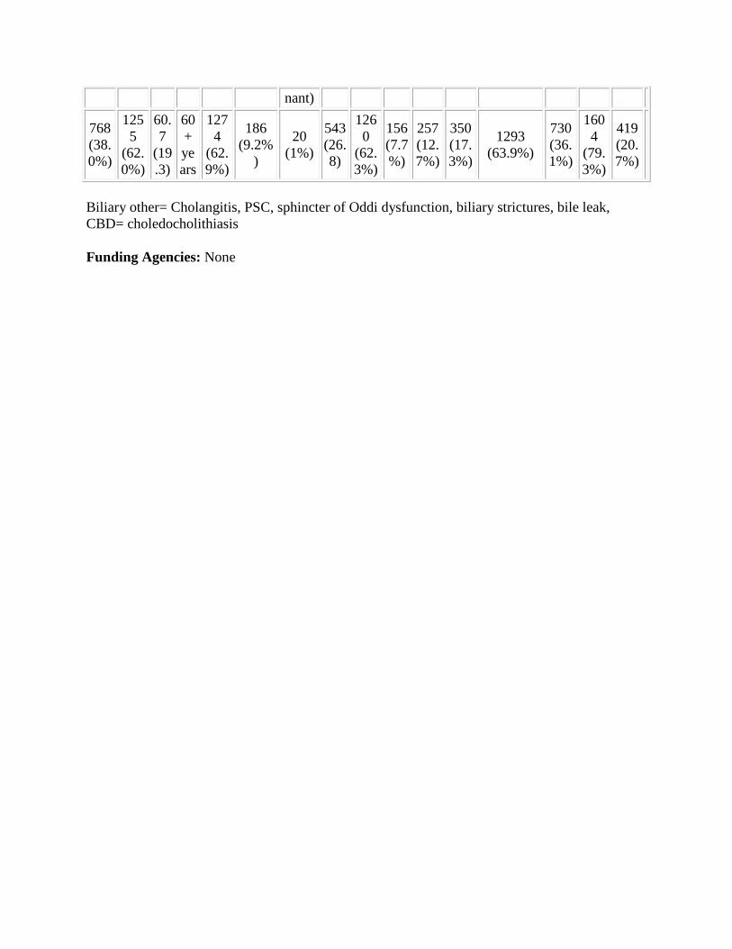
nant)
768 (38.0%)
1255
(62.0%)
60.7
(19.3)
60+ years
1274
(62.9%)
186 (9.2%
)
20 (1%)
543 (26.8)
1260
(62.3%)
156 (7.7%)
257 (12.7%)
350 (17.3%)
1293 (63.9%)
730 (36.1%)
1604
(79.3%)
419 (20.7%)
Biliary other= Cholangitis, PSC, sphincter of Oddi dysfunction, biliary strictures, bile leak, CBD= choledocholithiasis
Funding Agencies: None

A353
TAMM HORSFALL PROTEIN ASSOCIATE WITH ELECTROLYTE BALANCE IN ACUTE PANCREATITIS
T. Tsai1, C. Chang2, H. Liao2, H. Lai1
1. China Medical University hospital division of Gastroenterology, Taichung, Taiwan; 2. China Medical University Hospital, Division of Nephrology, Taichung, Taiwan.
Background: Acute pancreatitis is an acute inflammation process of pancreas and is a leading cause of hospitialization of gastrointestinal disease.Patients with acute pancreatitis often complicate with hypocalcaemia,hypokalemia and acute kidney injury.Tamm Horsfall protein (THP) is an 80-kDa glycoprotein synthesized exclusively in the thick ascending limb cells of Henle's loop(TAL) cells.THP is the most abundant protein in the urine of normal mammals and is related to the homeostasis of electrolytes such as potassium and calcium regulations.Inflammatory diseases can affect the expression of THP and lead to electrolyte disorders. Aims: The aim of the study was to investigate the relationship between urinary Tamm Horsfall protein level and serum hypokalemia, hypocalcaemia in acute pancreatitis patients. Methods: Pancreatitis patients with different etiologies were enrolled excluding history of cancer,diabetes,chronic renal failure and heart failure.Urine and blood samples for potassium,calcium and creatinine were collected daily from admission to discharge day.The THP level was measured by mass spectra and Elisa test.Mass spectra were performed with MALDI-TOF/TOF MS(Ultraflex III TOF/TOF, Bruker Daltonics,Germany) equipped with Smartbeam laser system.Ratio between urine potassium and calcium to urine creatinine were calculated.Analysis of variance (ANOVA) with multiple comparisons was used for multiple groups.A p-value of <0.05 was considered to be statistically significant Results: 37 patients had been enrolled in the study.The average age is 49 year old and mostly man.Etiology include hypertriglyceridemia,cholangitis, alcoholism and unknown.Only 2 patients diagnosed as severe pancreatitis with Ranson's score over 5 and one of then had died due to disease progress.Urine THP and blood potassium,calcium had decreased to the lowest level in the second day after admission and recovered in the discharge day.The urine level of THP is associated with blood potassium and calcium level.The urine potassium level negatively correlated with THP level.On the contrary,the fractional excretion of calcium positively correlated with THP level. Conclusions: THP may be associated with the regulation of hypokalemia in acute pancreatitis patients.The association between THP and hypocalcaemia should be further investigated.
Patient list
Hypertriglyceridemia Cholangitis Alcoholism Unknown Total Number 8 11 15 3 37
Gender M:F 5:3 7:4 14:1 1:2 27:10 Age 42.76±9.3 62.5±12.7 44.1±8.8 45.6±19.2 49.7±15.1

Ranson's score 2.1±1.4 1.8±1.1 2.3±1.8 1±0.8 2±1.5 CT severity index 4.57±1.9 2.8±2.1 3.6±2.5 N/A 3.7±2.4
Amylase 598 1563 557 160 812.8±191.2 Lipase 1090 2045 1966 184 1656±417.9
THP level had related to serum potassium and calcium level. UK/UCr(Urine potassium creatinine ratio) decrease and FeCa(fractional excretion of calcium) increase with the elevated level of THP.
Funding Agencies: None

A354
FIRST CASE OF ISOLATED BILIARY PHYTOBEZOAR
F. Albogami2, A. Barkun1, K. Waschke1
1. McGill University, Montreal, QC, Canada; 2. McGill university, Montreal, QC, Canada.
Background: A phytobezoar (PBZ) is the most common type of gastrointestinal tract bezoar, and is composed of indigestible vegetable material. Biliary PBZ are extremely rare. We describe a case of a patient presenting with an obstructing biliary PBZ causing cholangitis in the absence of a history of abdominal surgery, sphincterotomy or spontaneous biliary enteric fistula. Aims: To review the literature and report a case of isolated PBZ. Methods: A 50 year old man presented with a 5 month history of worsening recurrent biliary abdominal pain and fevers. No significant past medical history or previous biliary surgery. Physical examination upon transfer showed moderate right upper quadrant tenderness. At presentation, the alkaline phosphatase was unavailable, total bilirubin was 125(direct 82) umol/L,GGT 992 U/L,AST 71 U/L, ALT 103 U/L . Abdominal ultrasound showed a common bile duct (CBD) diameter of 6mm, pneumobilia and cholelithiasis. MRCP was said to be normal. Upon transfer, an EUS showed gall bladder sludge and stones, as well as CBD wall thickening with sludge in the mid to distal part of the CBD. At ERCP, a CBD filling defect was found. After performing a sphincterotomy, we extracted what looked like a cast occupying the entire lower CBD, extending into the cystic duct. This was retrieved in one piece and sent to pathology. The patient was discharged without complication. Pathological analysis revealed the cast was made of vegetable material. Results: There are only eight cases of biliary PBZ in the English medical literature. Four were isolated biliary PBZ, and in the others, the PBZ acted as a nidus for CBD stones. In 1972, Ban et al. described a biliary PBZ for the first time, acting as a nidus for symptomatic CBD stones that had developed after choledochojejunostomy. Most reports describe biliary PBZ following a surgical bilio-enteric anastomosis either with associated choledocholithiasis, or alone, and in one case as a result of a choledochoduodenal fistula. There are only 2 reports of patients developing a biliary PBZ in the absence of any bilio-enteric anastomosis or fistula. In both, the bezoar acted as a nidus for CBD stone formation. Although the mechanism for developing a PBZ is not clear, a main contributing factor relates to ablation or bypass of the sphincter of Oddi due to surgical manipulation or fistula formation. The mechanism in the absence of any such altered anatomy remains unknown and some suggest intermittent stone passage may contribute. No sphincter of Oddi manometric information exists in any reported cases. In our patient, although cholelithiasis was noted, no CBD stone was present with the bezoar and cholecystectomy was recommended. Conclusions: We describe the first reported case of an isolated biliary phytobezoar in the absence of previous biliary surgery or a bilio-enteric fistula.
Funding Agencies: None

A355
RITUXIMAB FOR THE TREATMENT OF STEROID-REFRACTORY AUTOIMMUNE PANCREATITIS
M. Boulos, H. Atkins, A. Chatterjee
University of Ottawa, Ottawa, ON, Canada.
Background: Treatment of autoimmune pancreatitis is based on observational studies since there have been no randomized controlled trials. Most patients respond to glucocorticoid therapy, but a significant proportion relapse once glucocorticoids are discontinued. Immunomodulators such as Imuran have been used in patients who fail steroids, relapse or cannot be weaned off steroids; however, recurrence with Imuran is common and side effects of this drug include nausea, hepatotoxicity, pancreatitis, leukopenia, infection and lymphoma. Aims: This case illustrates the efficacy of Rituximab, a monoclonal antibody, in steroid-refractory autoimmune pancreatitis. Methods: Case report Results: A 66-year-old female presented to the emergency department with nausea, vomiting and jaundice. Blood work was significant for elevated liver enzymes (ALT 485, AST 190, ALP 753, GGT 960 and total bilirubin 134) and a CT scan of the abdomen identified a pancreatic head mass. Cholangiogram identified a common bile duct stricture measuring 6 cm in length that was stented. Endoscopic ultra-sound revealed an ill-defined hypoechoic mass in the pancreatic head measuring 3.3 cm x 3.4 cm with peripancreatic lymphadenopathy. A fine needle biopsy of the pancreatic mass was inconclusive for malignancy and the findings were suggestive of chronic pancreatitis. A subsequent CT scan showed a "sausage-shaped" pancreas and further blood work showed elevated IgG4 levels (3.04 g/L, reference range 0.039-0.864 g/L), suggestive of underlying autoimmune pancreatitis. She was first started on a course of steroid; she initially improved clinically and radiologically, but her symptoms recurred as she was weaned from Prednisone. The patient was subsequently started on Rituximab and after the first cycle, a post-infusion MRI showed that the inflammation in the pancreas had resolved. A repeat ERCP demonstrated that a CBD stricture continues to persist although appeared significantly improved (1 cm from 2 cm). She remains asymptomatic with no recurrent pancreatitis 8 months post induction and she is scheduled to receive her second cycle of Rituximab. Conclusions: This case illustrates the efficacy of Rituximab in a patient with steroid-refractory autoimmune pancreatitis. We have since treated an additional two patients with Rituximab with significant response. These findings are promising and further studies are required before Rituximab can be routinely recommended for the treatment of autoimmune pancreatitis.
Funding Agencies: None
A356
CHOLEDOCHODUODENOSTOMY FOR BILIARY DRAINAGE DUE TO MALIGNANT OBSTRUCTION AFTER FAILED ERCP
V. Jovanovic, C. Teshima
University of Alberta, Edmonton, AB, Canada.
Background: PTC has been the traditional salvage option after failed ERCP in malignant distal biliary obstruction. Recently, endoscopic ultrasound-guided biliary drainage (EUS-BD) has been developed as an alternative method. Aims: EUS-BD consists of a EUS-guided rendezvous procedure followed by conversion to ERCP, or direct transmural drainage either in the form of choledochoduodenostomy or hepaticogastrostomy. Advantages over PTC include the ability to perform immediately following failed ERCP and lack of an external drain. There are limited data for this novel procedure, but early series suggest similar efficacy and safety compared to PTC, and the largest study reports equivalent outcomes between rendezvous and direct transluminal techniques. Complication rates are 10-15%, including pneumoperitoneum, bleeding, stent migration and occasionally bile peritonitis. Nevertheless, the overall success rate is 85-95%. Methods: Two cases were reviewed retrospectively and a review of the current literature on choledochoduodenostomy in biliary obstruction was performed. Results: Case 1: A 79 year-old female presenting with epigastric pain and jaundice was found to have a bilirubin of 291 µmol/L. CT abdomen revealed an unresectable mass in the head of the pancreas with resulting biliary obstruction. ERCP was attempted but failed due to tumor ingrowth into the 2nd part of the duodenum that prevented access to the papilla. EUS demonstrated a CBD dilated to 1.5 cm proximal to the tumor. Using fluoroscopy and EUS guidance, the dilated CBD was accessed with a 19G FNA needle, followed by cholangiogram, wire cannulation, balloon dilation, and deployment of a fully-covered 10 mm x 4 cm biliary Wallflex stent from the CBD into the duodenal bulb, resulting in immediate drainage of bile. The serum bilirubin gradually declined to 15 µmol/L and remains normal at 3 months follow-up. No early or late complications occurred. Case 2: A 68 year-old male with known metastatic pancreas adenocarcinoma diagnosed 3 months prior presented with abdominal pain and new onset jaundice (bilirubin 107 µmol/L). CT showed a 4.5 cm mass in the head of the pancreas encasing the CBD. ERCP failed due to inability to advance the wire through the tumor, despite successful cannulation. Attempt at EUS-guided rendezvous was abandoned after unsuccessful wire advancement due to complete obliteration of the CBD within the tumor. Under fluoroscopic and EUS guidance, a fully-covered 10 mm x 4 cm biliary Wallflex stent was then placed from the CBD into the duodenal bulb. Bilirubin declined to 48 µmol/L 4 days post-procedure and to 4 µmol/L at 2 months follow-up. No early or late complications occurred. Conclusions: EUS-guided choledochoduodenostomy is a viable alternative after failed ERCP for malignant biliary obstruction, although should be performed at expert referral centres.
Funding Agencies: None

Pediatric Liver Disease
A357
LIVER STIFFNESS IN PEDIATRIC FONTAN PATIENTS: THE ROLE OF TRANSIENT ELASTOGRAPHY AND AST:PLATELET RATIO INDEX (APRI)
O. Guttman1, B. Chen2, D. Human1, K. Harris1, J. Potts1, R. Schreiber1
1. British Columbia Children's Hospital, Vancouver, BC, Canada; 2. Children's Hospital of Western Ontario, London, ON, Canada.
Background: Liver disease is a well-recognized complication in patients who have undergone Fontan surgery for complex congenital heart disease. Such patients are at risk for significant fibrosis, largely because of hepatic congestion due to elevated venous pressures. Suitable tools for monitoring liver disease in this population have not been well established. Transient elastography (TE) and the aspartate aminotransferase to platelet ratio index (APRI) are non-invasive modalities which have been used for the detection of liver fibrosis in a number of chronic liver diseases. Aims: To evaluate the role of TE and the APRI in pediatric Fontan patients. Methods: Fontan patients and healthy controls were recruited from the cardiology and gastroenterology clinics at British Columbia Children's Hospital. Demographic data and tests of hepatic biochemistry and function were collected at a routine clinic visit. TE was performed using age- and size-appropriate probes. Results: Fifteen Fontan patients were recruited (median age 14.0 years (6.0-18.2) and compared with 15 healthy age- and sex-matched controls. Median time from Fontan to TE was 9.9 years (1.0-14.4). No Fontan patients were in heart failure, 4 had hepatomegaly and 1 had splenomegaly. TE values were significantly higher in the Fontan group (26.3 kPa, range 12.7-42.2) than in the control group (4.9 kPa, range 3.3-6.1), p<0.0001. There were significant differences in liver enzymes between the 2 groups, with Fontan patients having higher values of both ALT (33 U/L (24-44) vs. 18.5 U/L (15-21), p=0.0002) and GGT (60 U/L (48-101) vs. 11 U/L (10-12), p<0.0001). APRI values were significantly higher in the Fontan cohort (0.46, range 0.22-1.0) versus the control group (0.25, range 0.12-0.41), p=0.0008. There was a moderate association between TE value and APRI among Fontan patients (r=0. 5963, p=0.0190) and weak, but statistically non-significant associations between TE and time since Fontan (r=0.3244, p=0.2381) and APRI and time since Fontan (r=0.0895, p=0.7511). Conclusions: Pediatric Fontan patients had markedly elevated TE values compared with healthy controls, indicating increased liver stiffness. The association between APRI and TE values was moderate, suggesting that elevated liver stiffness in Fontan patients with high APRI may indeed reflect advanced fibrosis and portal hypertension rather than hepatic congestion. TE and APRI may have an important role in monitoring liver status in Fontan patients.
Funding Agencies: None

A358
VITAMIN E SUPPLEMENTATION FOR PREVENTION OF PARENTERAL NUTRITION ASSOCIATED LIVER DISEASE STUDIED IN NEONATAL PIGLETS
M. Muto1, A. Soukvilay2, P. Wizzard3, D. Lim4, P. Wales5, J. Turner1
1. Department of Pediatrics, University of Alberta; Department of Agricultural Food and Nutritional Science, University of Alberta, Edmonton, AB, Canada; 2. Department of Pediatrics, University of Alberta; Department of Health Sciences, McMaster University, Hamilton, ON, Canada; 3. Department of Agricultural Food and Nutritional Science, University of Alberta, Edmonton, AB, Canada; 4. Department of Surgery, University of Alberta; Department of Agricultural Food and Nutritional Science, University of Alberta, Edmonton, AB, Canada; 5. Group for Improvement of Intestinal Function and Treatment, Hospital for Sick Children; Department of Surgery, University of Toronto, Toronto, ON, Canada.
Background: Use of plant based lipid emulsions has been associated with increased risk of neonatal parenteral nutrition associated liver disease (PNALD). While fish oil emulsions appear to prevent PNALD, they contain more vitamin E, and this confounds current understanding of the role of lipid emulsions in PNALD. Aims: In this study we compared early onset PNALD in neonatal piglets given total parenteral nutrition (TPN) with and without intravenous vitamin E supplementation. Methods: Neonatal piglets had jugular catheter insertion and were randomized to two treatments: Intralipid® (IL, n=4) or Intralipid® plus vitamin E (Vit E, n=5). Comparisons were made to sow reared piglets (CON, n=4) as a gold standard for normal nutrition and liver function for age. TPN was administered for 17 days, as an all-in-one admixture, bags were light protected to reduce peroxidation. Baseline and termination serum liver chemistry was determined and basal bile flow measured day 17. Statistical analysis used one-way analysis of variance. Results: Vitamin E treated piglets had higher serum vitamin E levels at trial completion (8.0mg/dL vs 2.7mg/dL). Bile flow was not different between groups (IL 6.20mL/g; Vit E 5.96mL/g; CON 10.52mL/g; p=0.136). Bilirubin was elevated in both treatment groups compared to CON (IL 16.55mmol/L; Vit E 14.40mmol/L; CON 4.78mmol/L; p=0.083). Bile acids were elevated in treatment groups and significantly more elevated without added vitamin E (IL 38.5mmol/L; Vit E 20.3mmol/L; CON 8.65mmol/L; p<0.001). Conclusions: Vitamin E treatment show minimal evidence for the prevention of early onset PNALD. This is particularly so when compared to our prior findings using this model and fish oil lipid treatments, where bile flow has been significantly increased above controls. Further data in regards to lipid peroxidation and oxidative stress responses in piglets with and without the vitamin E supplementation is pending.
Funding Agencies: None

A359
AUTOIMMUNE HEPATITIS AND HYPOTHYROIDISM CAUSED BY A GAIN-OF-FUNCTION MUTATION OF STAT1
M. Carneiro de Moura, C. Roifman, B. Kamath
The Hospital for Sick Children, Toronto, ON, Canada.
Aims: Autoimmune hepatitis (AIH) is the most common immune-mediated liver disease of childhood though the etiology is generally unknown. Chronic mucocutaneous candidiasis (CMC) caused by mutations in STAT1, includes a heterogeneous group of conditions with common features including chronic noninvasive Candida infections and autoimmune manifestations. AIH has been reported only twice in adults with CMC. Methods: We describe a child with autoimmune hepatitis that presented with acute liver failure and subsequently developed hypothyroidism two years later. She had a gain-of-function STAT1 mutation, also present in her mother. Results: A 3 year old girl presented with acute liver failure of unknown etiology after a respiratory infection. At the time of diagnosis: ALT 3384 U/L, AST 7488U/L, conjugated bilirubin 221µmol/l, GGT 50U/L, Albumin 35 g/L, INR 1.7, Ferritin 232mcg/L, and IgG 25g/L. Extensive investigations for infectious, autoimmune and metabolic causes were negative. Liver biopsy showed hemophagocytosis, giant cell transformation of hepatocytes and apoptosis with minimal interface inflammation. Bone marrow biopsy with no evidence of hemophagocytosis. She was treated conservatively and liver enzymes improved but then plateaued between 100-150U/L. A repeat liver biopsy revealed mild focal giant cell transformation of hepatocytes with no inflammatory cell infiltrate. She was followed closely and one year later presented with marked weight gain and was found to be hypothyroid (anti-thyroid antibodies negative). After starting levothyroxine, liver enzymes increased tenfold (ALT 2258 U/L, AST 1300 U/L). A third liver biopsy showed lymphocytic and plasma cell infiltrate in portal tracts and mild portal fibrosis consistent with autoimmune hepatitis. She was treated with prednisone followed by azathioprine with biochemical remission. Her mother had a history of candidiasis and bronchiectasis and was identified as having a gain-of-function mutation in STAT1 during the course of her daughter's illness. Our patient had the same c.820 G>A (p.Arg274Gln) mutation in the coiled-coin domain of STAT1. She is now 7 years old with biochemically controlled AIH and hypothyroidism. She has no evidence of candidiasis or serious infections. Conclusions: We present a rare case of a gain-of-function STAT1 mutation manifest by classic CMC in a parent but a modified phenotype of autoimmune hepatitis and hypothyroidism in the child. Dominant gain-of- function STAT1 mutations have been described causing inhibition in Th(IL-17) cell differentiation. AIH has been linked to defective regulatory T cells but its pathogenesis is not fully understood. This case provides a study model of genetic susceptibility and immune regulation leading to autoimmune liver disease.
Funding Agencies: None

A360
OMEGAVAN RESCUE THERAPY IN AN INFANT WITH INTESTINAL FAILURE ASSOCIATED LIVER DISEASE DESPITE SMOF: A CASE REPORT
J. Wu1, J. Yap2
1. Nutrition Services, Stollery Children's Hospital, Edmonton T6G 2B7, AB, Canada; 2. University of Alberta, Edmonton T6G 1C9, AB, Canada.
Background: Children with Short Bowel Syndrome or Intestinal Failure (IF) who require long-term parenteral nutrition are prone to high morbidity and mortality rates secondary to intestinal failure associated liver disease (IFALD). More recently, third-generation lipids have become available and preliminary data suggests that SMOF lipid (Fresenius Kabi, Bad Homburg, Germany), a mixture of soy, medium chain triglycerides, olive oil and fish oil can reduce the risk of IFALD in long-term PN dependent children. Aims: Describe the novel clinical observation of Omegaven rescue treatment in an infant with IFALD despite the previous use of a third generation intravenous lipid emulsion. Methods: Case report Results: The patient was dizygotic female twin born at 28 weeks gestation with birth weight of 1240 grams. On day seventeen of life, she developed necrotizing enterocolitis (NEC) requiring extensive intestinal resection with residual 38 cm of continuous small bowel ending in a jejununostomy. She received Intralipid at 2 g/kg for the first two months of life. Because of worsening cholestasis, Intralipid was converted to SMOF with a dose range of 2 to 2.75 g/kg from month two onwards. Despite the use of SMOF, her total and conjugated bilirubin continued to rise and peaked at 7 months at 113 mmol/L. Omegaven was commenced at 8.5 months chronological age starting at 0.5 g/kg and advanced rapidly to 1 g/kg over 3 days. Following a month of exclusive Omegaven, her bilirubin had improved to 50 mmol/L. She was subsequently converted back to SMOF following normalization of her bilirubin and marked improvement of serum aminotransferases (Figure 1). Her serum bilirubin remained normal at 3 month follow-up. The patient's parenteral nutrition prescription stayed constant throughout her time on Omegaven. Her enteral feeds remained unchanged through the Omegavan treatment duration, providing 30% of her total energy requirements. While, a trial of intestinotrophic hormone glucagon-like peptide-2 (GLP-2) failed to improve her enteral tolerance; fluctuating between 6 to 8 mls/hr continuous via G-tube. Conclusions: Omegaven is recognized as rescue therapy of choice in patients with IFALD exposed to Intralipid. SMOF lipid emulsion is a new intravenous lipid solution recently available in Canada and has held promise as the better alternative to soy based Intralipid. Early evidence hints of potential lower hepatic injury of SMOF in comparison to Intralipid. The novel observation from this case report suggest that pure intravenous omega-3 fatty acid lipid emulsion ie: Omegavan is superior to SMOF as rescue therapy in established IFALD irrespective of previous intravenous lipid solution.
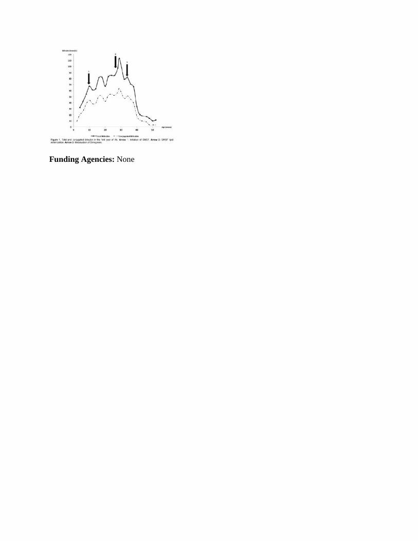
Funding Agencies: None