abstracts book - NALS 2020
206
2020 Madrid · Spain abstracts book Nanomaterials Applied to Life Sciences International Conference on 2 nd 29 th -31 st January Organised by:
-
Upload
khangminh22 -
Category
Documents
-
view
2 -
download
0
Transcript of abstracts book - NALS 2020
2020Madrid · Spain
abstracts book
Nanomaterials Applied to Life
Sciences
International Conference on 2nd
29th-31st
January
Organised by:

Nanomaterials Applied to Life
Sciences
International Conference on 2nd
29th-31st
January
2020Madrid · SpainOrganised by:
abstracts book

NALS
202
0
International Conferenceon Nanomaterials Applied to Life Sciences2nd
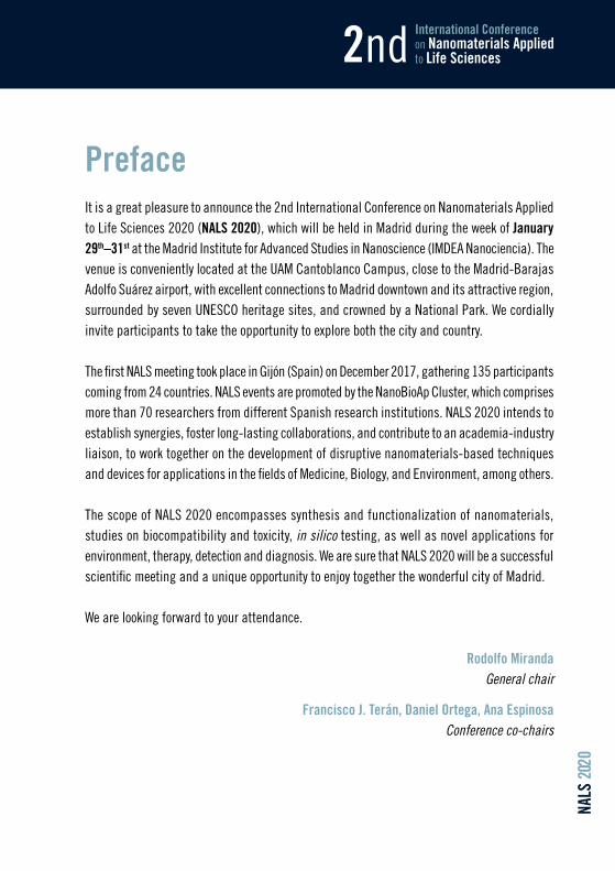
PrefaceIt is a great pleasure to announce the 2nd International Conference on Nanomaterials Applied to Life Sciences 2020 (NALS 2020), which will be held in Madrid during the week of January 29th–31st at the Madrid Institute for Advanced Studies in Nanoscience (IMDEA Nanociencia). The venue is conveniently located at the UAM Cantoblanco Campus, close to the Madrid-Barajas Adolfo Suárez airport, with excellent connections to Madrid downtown and its attractive region, surrounded by seven UNESCO heritage sites, and crowned by a National Park. We cordially invite participants to take the opportunity to explore both the city and country.
The first NALS meeting took place in Gijón (Spain) on December 2017, gathering 135 participants coming from 24 countries. NALS events are promoted by the NanoBioAp Cluster, which comprises more than 70 researchers from different Spanish research institutions. NALS 2020 intends to establish synergies, foster long-lasting collaborations, and contribute to an academia-industry liaison, to work together on the development of disruptive nanomaterials-based techniques and devices for applications in the fields of Medicine, Biology, and Environment, among others.
The scope of NALS 2020 encompasses synthesis and functionalization of nanomaterials, studies on biocompatibility and toxicity, in silico testing, as well as novel applications for environment, therapy, detection and diagnosis. We are sure that NALS 2020 will be a successful scientific meeting and a unique opportunity to enjoy together the wonderful city of Madrid.
We are looking forward to your attendance.
Rodolfo Miranda General chair
Francisco J. Terán, Daniel Ortega, Ana EspinosaConference co-chairs
NALS
202
029th-31st J a n u a r y 2 0 2 02020
Madrid · Spain
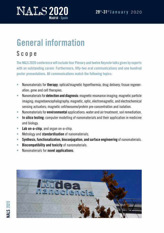
General informationS c o p eThe NALS 2020 conference will include four Plenary and twelve Keynote talks given by experts with an outstanding career. Furthermore, fifty-two oral communications and one hundred poster presentations. All communications match the following topics:
• Nanomaterials for therapy: optical/magnetic hyperthermia; drug delivery; tissue regener-ation; gene and cell therapies.
• Nanomaterials for detection and diagnosis: magnetic resonance imaging; magnetic particle imaging; magnetoencephalography; magnetic, optic, electromagnetic, and electrochemical sensing actuators; magnetic cell/exosome/protein pre-concentration and isolation.
• Nanomaterials for environmental applications: water and air treatment, soil remediation. • In silico testing: computer modelling of nanomaterials and their application in medicine
and biology. • Lab on-a-chip, and organ on-a-chip. • Metrology and standardisation of nanomaterials. • Synthesis, functionalization, bioconjugation, and surface engineering of nanomaterials. • Biocompatibility and toxicity of nanomaterials. • Nanomaterials for novel applications.
NALS
202
0
International Conferenceon Nanomaterials Applied to Life Sciences2nd
Conference OrganizersL o c a l O r g a n i z i n g C o m m i t t e eRodolfo Miranda IMDEA NanocienciaGeneral chair
Francisco J. Terán Daniel Ortega Ana EspinosaIMDEA NanocienciaConference co-chairs
María Jesús VillaIMDEA Nanociencia
Isabel Rodríguez CabezasIMDEA Nanociencia
Bonifacio VegaIMDEA Nanociencia
Diego BaragañoUniversidad de Oviedo
María del Carmen BlancoUniversidad de Oviedo
Julio CamareroUniversidad Autónoma de Madrid & IMDEA Nanociencia
Daniel JaqueUniversidad Autónoma de Madrid
María del Puerto MoralesInstituto de Ciencia de Materiales de Madrid-CSIC
Ana PizarroIMDEA Nanociencia
Montserrat RivasUniversidad de Oviedo
Gorka SalasIMDEA Nanociencia
N A L S E x e c u t i v e C o m m i t t e eMontserrat RivasGeneral chair Universidad de Oviedo
Carmen BlancoCo-chair Universidad de Oviedo
Daniel OrtegaUniversidad de Cádiz & IMDEA Nanociencia
Francisco J. TeránIMDEA Nanociencia
María Luisa Fernández GubiedaEuskal Herriko Unibertsitatea and BCMaterials
NALS
202
029th-31st J a n u a r y 2 0 2 02020
Madrid · Spain
Cristina Gómez PoloUniversidad Pública de Navarra
Xavier BatlleUniversidad de Barcelona
Mónica López FanarragaIDIVAL
Aitziber L. CortajarenaCIC biomaGUNE
José RivasUniversidad de Santiago de Compostela
Arben MerkoçiInstitut Català de Nanociència i Nanotecnologia
Pablo BotellaInstituto de Tecnología Química
Jesús Martínez de la FuenteInstituto de Nanociencia de Aragón
Anna RoigInstituto de Ciencia de Materiales de Barcelona-CSIC
María del Puerto MoralesInstituto de Ciencia de Materiales de Madrid-CSIC
Daniel JaqueUniversidad Autónoma de Madrid
I n t e r n a t i o n a l A d v i s o r y C o m m i t t e eProf. Adarsh SandhuElectro-Communications University, Tokyo (Japan)
Prof. Teresa PellegrinoIstituto Italiano di Tecnologia, Genoa (Italia)
Prof. Ronald B. GoldfarbNational Institute of Standards and Technology, Boulder, CO (USA)
Prof. Florence GazeauUniversité Paris-Diderot, Paris (France)
Prof. Urs O. HafeliThe University of British Columbia,Vancouver (Canada); University ofCopenhagen, Copenhagen (Denmark)
Prof. Adriele Prina-MelloTrinity College Dublin, Dublin (Ireland)
NALS
202
0
International Conferenceon Nanomaterials Applied to Life Sciences2nd
Conference sponsors
www.ieeemagnetics.org
www.iesmat.com
www.nanotherics.com
www.nbnanoscale.com
NALS
202
029th-31st J a n u a r y 2 0 2 02020
Madrid · Spain
www.paralab.es
www.vwr.com
www.rsc.org
www.nature.com/nnano
NALS
202
0
International Conferenceon Nanomaterials Applied to Life Sciences2nd
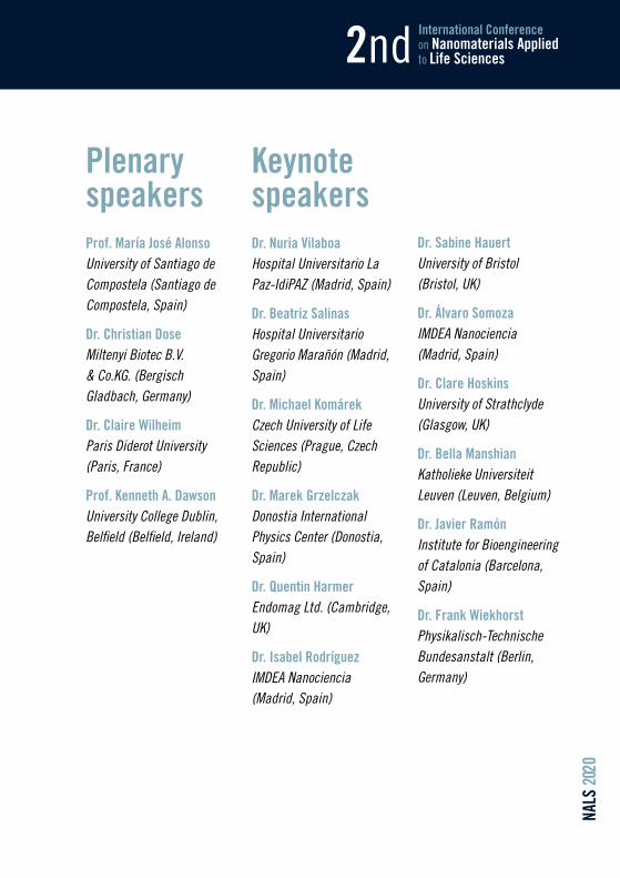
Plenary speakersProf. María José AlonsoUniversity of Santiago de Compostela (Santiago de Compostela, Spain)
Dr. Christian DoseMiltenyi Biotec B.V. & Co.KG. (Bergisch Gladbach, Germany)
Dr. Claire WilheimParis Diderot University (Paris, France)
Prof. Kenneth A. DawsonUniversity College Dublin, Belfield (Belfield, Ireland)
Keynote speakersDr. Nuria VilaboaHospital Universitario La Paz-IdiPAZ (Madrid, Spain)
Dr. Beatriz SalinasHospital Universitario Gregorio Marañón (Madrid, Spain)
Dr. Michael KomárekCzech University of Life Sciences (Prague, Czech Republic)
Dr. Marek GrzelczakDonostia International Physics Center (Donostia, Spain)
Dr. Quentin HarmerEndomag Ltd. (Cambridge, UK)
Dr. Isabel RodríguezIMDEA Nanociencia (Madrid, Spain)
Dr. Sabine HauertUniversity of Bristol (Bristol, UK)
Dr. Álvaro SomozaIMDEA Nanociencia (Madrid, Spain)
Dr. Clare HoskinsUniversity of Strathclyde (Glasgow, UK)
Dr. Bella ManshianKatholieke Universiteit Leuven (Leuven, Belgium)
Dr. Javier RamónInstitute for Bioengineering of Catalonia (Barcelona, Spain)
Dr. Frank WiekhorstPhysikalisch-Technische Bundesanstalt (Berlin, Germany)

NALS
202
029th-31st J a n u a r y 2 0 2 02020
Madrid · Spain
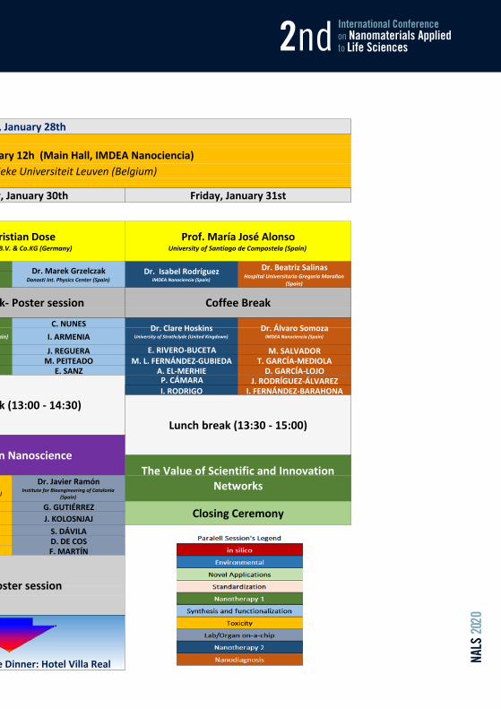
:00:15:30:45:00:15:30 J. WELLS:45 W. GAWEDA:00:15:30:45 R. PÉREZ PELÁEZ C. NUNES:00 I. RUBIA I. ARMENIA:15 D. BARAGAÑO C. ESCALONA J. REGUERA E. RIVERO-BUCETA M. SALVADOR:30 L. ALVES Y. SHEN M. PEITEADO M. L. FERNÁNDEZ-GUBIEDA T. GARCÍA-MEDIOLA:45 O. IGLESIAS J. PALOMO V. MULENS E. SANZ A. EL-MERHIE D. GARCÍA-LOJO:00 S. RUTA F. RIVERA P. CÁMARA J. RODRÍGUEZ-ÁLVAREZ:15 A. MANZIN J. L. R. GALLEGO I. RODRIGO I. FERNÁNDEZ-BARAHONA:30:45:00:15:30:45:00:15:30
:45
:00 M. L. FANARRAGA G. GUTIÉRREZ:15 A. DOMÍNGUEZ J. KOLOSNJAJ:30 V. SALGUEIRIÑO D. JAQUE L.G. DELOGU S. DÁVILA:45 P. MARÍN E. GARAYO G. R. IGLESIAS D. DE COS:00 I. ECHEVARRIA R. BLEUL B. CHISHTI F. MARTÍN:15 M.T. GONZÁLEZ L. ABIVEN:30 J. GARCÍA-MARTÍN A. MOYANO:45 J. SÁNCHEZ-NIEVES P. BENDER:00:15:30:45:00:15:30:45 20:30 h Conference Dinner: Hotel Villa Real
19
Dr. Christian DoseMiltenyi Biotec B.V. & Co.KG (Germany)
Prof. Kenneth A. DawsonCentre For BioNano Interactions (CBNI), School of Chemistry and
Chemical Biology, University College Dublin (Ireland)
Dr. Michael KomárekCzech University of Life Sciences
(Czech Republic)
Dr. Sabine HauertUniversity of Bristol (United Kingdown)
Dr. Claire WilheimParis Diderot University (France)
Dr. Quentin HarmerEndomag Ltd. (United Kingdown)
Dr. Frank WiekhorstPhysikalisch-Technische Bundesanstalt (Germany)
Beer-Poster session
16
17
18
14
15
13
Wednesday, January 29th
Dr. Beatriz SalinasHospital Universitario Gregorio Marañon
(Spain)
Dr. Nuria VilaboaHospital Universitario La Paz-IdiPAZ (Spain)
Prof. María José AlonsoUniversity of Santiago de Compostela (Spain)
12
Coffee Break- Poster session
Dr. Isabel RodríguezIMDEA Nanociencia (Spain)
Dr. Álvaro SomozaIMDEA Nanociencia (Spain)
Beer-Poster session
Coffee Break
Dr. Marek GrzelczakDonosti Int. Physics Center (Spain)
Dr. Javier RamónInstitute for Bioengineering of Catalonia
(Spain)
Dr. Clare HoskinsUniversity of Strathclyde (United Kingdown)
Closing Ceremony
Coffee Break- Poster session
Lunch break (13:00 - 14:30)
Women in Nanoscience
Dr. Bella ManshianKatholieke Universiteit Leuven (Belgium)
The Value of Scientific and Innovation Networks
Lunch break (13:30 - 15:00)Lunch break (13:30 - 15:00)
Satellite Special Seminar 28th January 12h (Main Hall, IMDEA Nanociencia)12:00
Dr. Stefaan Soenen - Katholieke Universiteit Leuven (Belgium)
Tuesday, January 28th
Thursday, January 30th Friday, January 31st
10
11
9
Opening
NALS
202
0
International Conferenceon Nanomaterials Applied to Life Sciences2nd
:00:15:30:45:00:15:30 J. WELLS:45 W. GAWEDA:00:15:30:45 R. PÉREZ PELÁEZ C. NUNES:00 I. RUBIA I. ARMENIA:15 D. BARAGAÑO C. ESCALONA J. REGUERA E. RIVERO-BUCETA M. SALVADOR:30 L. ALVES Y. SHEN M. PEITEADO M. L. FERNÁNDEZ-GUBIEDA T. GARCÍA-MEDIOLA:45 O. IGLESIAS J. PALOMO V. MULENS E. SANZ A. EL-MERHIE D. GARCÍA-LOJO:00 S. RUTA F. RIVERA P. CÁMARA J. RODRÍGUEZ-ÁLVAREZ:15 A. MANZIN J. L. R. GALLEGO I. RODRIGO I. FERNÁNDEZ-BARAHONA:30:45:00:15:30:45:00:15:30
:45
:00 M. L. FANARRAGA G. GUTIÉRREZ:15 A. DOMÍNGUEZ J. KOLOSNJAJ:30 V. SALGUEIRIÑO D. JAQUE L.G. DELOGU S. DÁVILA:45 P. MARÍN E. GARAYO G. R. IGLESIAS D. DE COS:00 I. ECHEVARRIA R. BLEUL B. CHISHTI F. MARTÍN:15 M.T. GONZÁLEZ L. ABIVEN:30 J. GARCÍA-MARTÍN A. MOYANO:45 J. SÁNCHEZ-NIEVES P. BENDER:00:15:30:45:00:15:30:45 20:30 h Conference Dinner: Hotel Villa Real
19
Dr. Christian DoseMiltenyi Biotec B.V. & Co.KG (Germany)
Prof. Kenneth A. DawsonCentre For BioNano Interactions (CBNI), School of Chemistry and
Chemical Biology, University College Dublin (Ireland)
Dr. Michael KomárekCzech University of Life Sciences
(Czech Republic)
Dr. Sabine HauertUniversity of Bristol (United Kingdown)
Dr. Claire WilheimParis Diderot University (France)
Dr. Quentin HarmerEndomag Ltd. (United Kingdown)
Dr. Frank WiekhorstPhysikalisch-Technische Bundesanstalt (Germany)
Beer-Poster session
16
17
18
14
15
13
Wednesday, January 29th
Dr. Beatriz SalinasHospital Universitario Gregorio Marañon
(Spain)
Dr. Nuria VilaboaHospital Universitario La Paz-IdiPAZ (Spain)
Prof. María José AlonsoUniversity of Santiago de Compostela (Spain)
12
Coffee Break- Poster session
Dr. Isabel RodríguezIMDEA Nanociencia (Spain)
Dr. Álvaro SomozaIMDEA Nanociencia (Spain)
Beer-Poster session
Coffee Break
Dr. Marek GrzelczakDonosti Int. Physics Center (Spain)
Dr. Javier RamónInstitute for Bioengineering of Catalonia
(Spain)
Dr. Clare HoskinsUniversity of Strathclyde (United Kingdown)
Closing Ceremony
Coffee Break- Poster session
Lunch break (13:00 - 14:30)
Women in Nanoscience
Dr. Bella ManshianKatholieke Universiteit Leuven (Belgium)
The Value of Scientific and Innovation Networks
Lunch break (13:30 - 15:00)Lunch break (13:30 - 15:00)
Satellite Special Seminar 28th January 12h (Main Hall, IMDEA Nanociencia)12:00
Dr. Stefaan Soenen - Katholieke Universiteit Leuven (Belgium)
Tuesday, January 28th
Thursday, January 30th Friday, January 31st
10
11
9
Opening

plennary speakers
NALS 2020 Nanomaterials Applied to Life Sciences
The Route to Complex Medicines from Science to Regulation Kenneth. A. Dawson1
1Centre For BioNano Interactions (CBNI) School of Chemistry and Chemical Biology. University College Dublin, Belfield, Dublin 4, Ireland
We discuss the microscopic molecular principles of organization at the nanoscale that may be used to control and direct biological processes. Increasing understanding is emerging about the detailed consequences of molecular organizations on the surface of nanostructures and the role this has at cellular and organ (e.g. liver clearance) immune and others. However, now the first detailed information is also becoming available for the role of nanoscale shape, and the potential role of shape in stimulating living systems.
The talk will stress the potential for a structured and rational design approach, based on fundamental understanding to improve on current phenomenological design approaches alone. Some considerations of scaling up, in GLP-like conditions are given, and the potential for these combined approaches to lead to realistic therapies. We stress the potential to build a durable science, with great potential impact, for the long term.
NALS 2020 Nanomaterials Applied to Life Sciences
Thermal therapies with magnetic and plasmonic nanoparticles and longterm fate in the intracellular environment
Claire Wilheim 1
1Paris Diderot University, Paris, France
Nanoparticles-based thermal therapy has emerged to propose alternative treatment and decrease side effects. We recently compared the heating potential of magnetic nanoparticles under magnetic hyperthermia or photothermia [1,2], of plasmonic nanoparticles under photothermia [3], or the combination of both [4-8], towards synergistic solutions to complete cancer cell destruction. The therapeutic use of nanoparticles then still raises the more general issue of intracellular nanoparticle long-term fate. We have developed cell spheroids models and magneto-thermal tools to monitor their intracellular integrity. It evidenced a massive intracellular degradation [9,10], which could be prevented by a polymeric coating [11] or an inert gold shell [12,13]. Remarkably, human cells could also biosynthesize their own magnetic nanoparticles, from the intracellular degradation products of synthetic ones [14].
References
[1] Advanced Functional Materials, 28, 1803660 (2018)
[2] Journal of Controlled Release, 279, 271-281 (2018)
[3] Advanced HealthCare Materials, 5, 1040- 48 (2016)
[4] ACS Nano, 9, 2904-2916 (2015)
[5] ACS nano 2016, 10, 2436-2446 (2016)
[6] Nanoscale, 7, 18872-18877 (2015)
[7] Theranostics, 9, 1288 (2019) [8] Theranostics, 9, 5924 (2019)
[9] ACS nano, 10, 7627-7638 (2016)
[10] Nature Communications, 8, 400 (2017); [11] Nanoscale, 11, 16488 (2019)
[12] Advanced Functional Materials, 27, 1605997 (2017)
[13] ACS nano, 12, 6523-6535 (2018); [14] PNAS 116, 4044-4053 (2019);
NALS 2020 Nanomaterials Applied to Life Sciences
Magnetic Cell Separation (MACS) – From Bench to Bedside Christian Dose 1
1Miltenyi Biotec B.V. & Co.KG Bergisch Gladbach, Germany
The MACS technology enables the magnetic separation of cell populations based on surface
antigens. It is a fast and gentle method for the isolation of viable and functional cells in high purities and recoveries by labeling cell epitopes with specific antibodies that have been conjugated to superparamagnetic nanoparticles. The highly efficient isolation of targeted cells is facilitated by utilizing high-gradient magnetic columns in the cell separation step. This ingenious technological combination supports complete workflows and accesses virtually any cell type of interest. It has been used in thousands of manual and automated high-throughput settings to date, starting from a broad range of cell sources, such as whole blood and blood products, as well as tissues from various species. The talk will illustrate the continuous development of the MACS technology and focuses on some more recent innovative solutions utilized in basic and translational cell research, as well as clinical cell therapy applications that have been made available to the scientific community within the last 30 years.
NALS 2020 Nanomaterials Applied to Life Sciences
“Nanomaterials to help complex drugs overcoming biological barriers”
1María José Alonso 1Dept. Pharmacy and Pharmaceutical Technology,
University of Santiago de Compostela (USC), Spain
Antigen and therapeutic proteins, including monoclonal antibodies, as well as polynucleotides are complex molecules which have great difficulties for overcoming biological barriers and reach their targets. In fact, the adequate formulation of these molecules has been considered as a major constrain for their clinical exploitation.
Fortunately, the continuously improved understanding of the biological barriers and the molecular biology associated to pathological conditions is paving the way for a more comprehensive and rational design of formulations of these complex drugs based on the use of nanotechnology. Our laboratory, with decades of experience in the formulation of macromolecules using polymer nanoparticles, has significantly contributed to this field. As an example, in the 90’s we were the first to report that nanoparticles made of either PLA-PEG or chitosan were efficient vehicles for the transmucosal delivery of proteins antigens and polynucleotides. The result of our subsequent efforts is an array of nanotechnologies that can be used to deliver proteins across mucosal surfaces, and, also, to facilitate their intracellular delivery following parenteral administration.
In my presentation, I will focus on the design of protein and RNA carriers that could be used in different therapeutic areas: (i) oral delivery of peptides intended to treat either local or systemic diseases, (ii) nanovaccines designed to prevent diseases, i.e. HIV as well as to treat diseases, i.e. diabetes and multiple sclerosis, (iii) nose-to-brain delivery of RNA for the treatment of Alzhimer disease, and (iv) delivery of mAb targeted to intracellular onco-proteins, as new oncological treatments.
Overall, our experience in this field has benefited from integrative approaches adopted by specifically designed consortia. Hopefully, the results of these cooperative efforts will help to accelerate the progress of a rational design of protein-based nanomedicines.
More information about these projects can be found at: http://www.usc.es/grupos/mjalonsolab/
Acknowledgements Work related to the oncology field: Ana Cadete, Ana Olivera, Desirée Teijeiro and Dolores
Torres from the USC, Spain and Gema Moreno and Angela Molina from the UCM, Spain.
Work related to oral protein delivery: Matilde Duran, Eleni Samaridou, Carlos Dieguez, Sulay Tovar, Niu Zhigao and Manuel Santander from the USC, Aloise Mabonzo, from the CEA, France and Patrik Lundquist and Per Artursson from UU, Sweden.
Work related to the vaccine field: José Crecente, Tamara Gómez, Ana Olivera, Dolores Torres and Rubén Varela from the USC and Ma Luo and Francis Plumber from the University of Manitoba, Canada.
The research activity has been funded by the European Comision FP7 (grant agreement n° 281035-TRANS-INT), the Horizon 2020 Programme (grant agreement # 646142 – NANOPILOT and Grant Agreement No. 721058- B-SMART), The National Institutes of Health (NIH) (Grant Number: R01AI111805), The Ministry of Economy and competititvity/

FEDER Founds (Ref. BIO2014-53091-C3-2-R, Ref. SAF2017-86634-R and Ref. PCIN-2017129/ AEI, under the frame of EuroNanoMed III") and the Xunta de Galicia- CRG (Ref: ED431C 2017/09).

keynote speakers
NALS 2020 Nanomaterials Applied to Life Sciences
Swarm Engineering Across Scales: From Robots to Nanomedicine Sabine Hauert
Engineering Mathematics, Bristol Robotics Laboratory, University of Bristol, Bristol, UK
Swarm engineering explores how large numbers of simple agents interact to achieve desired collective behaviours (Brambilla et al., Swarm Intelligence, 2013). The collective behaviour of trillions of nanoparticles interacting in complex tumour environments defines their success as treatment and imaging agents (Hauert et al., Trends in Biotechnology, 2014). Optimising these behaviours often requires nanoparticles to overcome transport barriers, and accumulate at effective levels at target tumour sites. This is done by changing the properties of the individual particles, including their size, charge, shape, material, coating, and loading. Small changes in nanoparticle design could lead to entirely different biodistribution. As an example, we showed in simulation how nanoparticles with slow diffusion or strong binding affinity often accumulate in the first cells encountered after extravasation, thereby leading to poor treatment of deep-seeded tumour cells (Hauert et al., Nano Today, 2013). Yet systematically exploring nanoparticle designs experimentally is not tractable due to time and cost constraints. Simulations allow us to thoroughly explore the space of possible nanoparticle designs and observe the resulting collective behaviours in silico. To this end, the EVONANO project (http://evonano.eu/) is designing multiscale models of nanoparticle interactions in the body, including their circulation in the blood stream, extravasation at a tumour site, penetration through tumour tissue, uptake by cells, and delivery to a location of action (Shatil et al., submitted). Importance is given to making the models increasingly realistic, considering the spatiotemporal dynamics of tumours, and nanoparticle interactions, as well as direct mapping of in vivo data to generate in silico models. Combining these more realistic simulations with artificial evolution or other machine learning technologies could allow for automatic design of nanocarriers for specific tumour scenarios. In the future, we imagine this automatic design of collective behaviours of nanoparticles could lead to more interesting emergent properties, similar to those explored in the field of swarm robotics (Schuerle et al., Science Advances, 2019). Example behaviours include decision making, amplification, mapping, or synchronisation. Several of these have been demonstrated in vivo with nanoparticles that communicate the location of a tumour to amplify tumour homing or self-assemble and disassemble to optimize nanoparticle transport (Hauert et al., Trends in Biotechnology, 2014).
Acknowledgements Sabine Hauert receives funding from the European Union's Horizon 2020 FET Open programme under grant agreement. No. 800983.
References [1] M. Brambilla, E. Ferrante, M. Birattari, M. Dorigo, Swarm Intelligence, Swarm intelligence 7.1 (2013): 1-41 [2] S. Hauert, S. N. Bhatia Trends in biotech. 32.9 (2014): 448-455 [3] S. Hauert, S. Berman, R. Nagpal, S. N Bhatia Nano today, 8.6 (2013): 566-576 [4] N. Shatil, M. Kovacevic , I. Balaz, S. Hauert, submitted [5] S. Schuerle, A. P. Soleimany, T. Yeh, G. M. Anand, M. Häberli, H. E. Fleming, N. Mirkhani, F. Qiu, S. Hauert, X.
Wang, B.J. Nelson, S. N. Bhatia, Science adv. 5.4 (2019) eaav4803
NALS 2020 Nanomaterials Applied to Life Sciences
Nano zerovalent iron for soil remediation: What do we know so far? Michael Komárek1, Martina Vítková1, Songlin Wu1,2, Tomáš Cajthaml3
1 Department of Environmental Geosciences, Faculty of Environmental Sciences, Czech University of Life Sciences Prague, Prague – Suchdol, Czech Republic
2 Center for Mined Land Rehabilitation, The University of Queensland, Australia
3 Institute for Environmental Studies, Faculty of Science, Charles University in Prague, Prague 2, Czech Republic
Iron-based amendments in general have been intensively studied for the remediation of contaminated soils as analogues of natural ubiquitous soil Fe phases, which are highly important compounds of the soil sorption complex. The fast development of various nanotechnology fields intensified the production of engineered nanomaterials and their commercial use. Engineered nanoparticles can be also used for environmental management, either through prevention, treatment and remediation of contaminated sites. The combined use of nanotechnologies and biotechnologies for soil remediation (aided phytostabilization) is an emerging and environmentally friendly method with significant scientific and economic potential. Previously, nano zerovalent Fe (nZVI) has been successfully used for the remediation of groundwater contaminated by various pollutants, due to its high reactivity and important reduction/adsorption properties, and its potential for soil remediation is now under extensive investigation. Since long-term studies focusing on nZVI efficiency for the remediation of contaminated soils are still lacking, its full-scale application remains problematic due to the number of questions concerning, e.g., associated phytotoxicity and ecotoxicity issues. This presentation summarizes works performed on soils contaminated with metal(loid)s, investigating the efficiency of nZVI, its composites and its geochemical transformations in the soil environment as mediated by microorganisms, interactions with plants and microbiota and presents perspectives for future research and applications.
Acknowledgements
The work has been supported by the Czech Science Foundation (project 18-24782Y).
NALS 2020 Nanomaterials Applied to Life Sciences
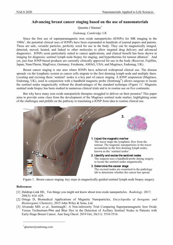
Advancing breast cancer staging based on the use of nanomaterials Quentin J Harmer*
Endomag, Cambridge UK
Since the first use of superparamagnetic iron oxide nanoparticles (IONPs) for MR imaging in the 1980s1, the potential clinical uses of IONPs have been expounded in hundreds of journal papers and patents. These are safe, versatile particles, perfectly sized for use in the body. They can be magnetically imaged, detected, moved, heated, and linked to other molecules to allow targeted drug delivery and advanced diagnostics. IONPs seem particularly suited to cancer applications, and clinical benefit has been shown in imaging for diagnosis; sentinel lymph node biopsy for staging; and hyperthermia for tumour ablation2. And yet, just four IONP-based products are currently clinically approved for use in the body (Resovist, Fujifilm, Japan; NanoTherm, MagForce, Germany; Feraheme, AMAG, USA; and Magtrace, Endomag, UK).
Breast cancer staging is one area where IONPs have achieved widespread clinical use. The disease spreads via the lymphatic system as cancer cells migrate to the first draining lymph node and multiply there. Locating and excising these ‘sentinel’ nodes is a key part of cancer staging. A IONP suspension (Magtrace, Endomag, UK), used in conjunction with a handheld magnetic probe (Sentimag®) allows surgeons to locate the sentinel nodes magnetically without the disadvantages of the standard technique (Figure 1)3. Magnetic sentinel node biopsy has been studied in numerous clinical trials and is in routine use on five continents.
But why have many iron oxide nanoparticle therapies struggled to deliver on their promise? This paper aims to provide some clues from the development of the Magtrace sentinel node marker, highlighting some of the challenges and pitfalls on the pathway to translating a IONP from idea to routine clinical use.
Figure 1. Breast cancer staging: key steps in magnetically-guided sentinel lymph node biopsy surgery.
References
[1] Daldrup-Link HE, Ten things you might not know about iron oxide nanoparticles. Radiology. 2017; 284(3): 616–629.
[2] Ortega D, Biomedical Applications of Magnetic Nanoparticles, Encyclopedia of Inorganic and Bioinorganic Chemistry, 2015 John Wiley & Sons, Ltd.
[3] Alvarado MD. et al., SentimagIC: A Non-inferiority Trial Comparing Superparamagnetic Iron Oxide Versus Technetium-99m and Blue Dye in the Detection of Axillary Sentinel Nodes in Patients with Early-Stage Breast Cancer. Ann Surg Oncol. 2019 Oct; 26(11): 3510-3516.
1. Inject the magnetic markerThe tracer maps the lymphatic flow from the tumour. The magnetic nanoparticles in the tracer accumulate in the first draining lymph nodes, known as the ‘sentinel nodes’.
2. Identify and excise the sentinel nodesThe surgeon uses a handheld probe during surgery to locate the sentinel nodes magnetically.
3. Determine the cancer stageThe excised nodes are examined in the pathology lab to determine whether the cancer has spread.
1.
2. 3.
NALS 2020 Nanomaterials Applied to Life Sciences
A metrology infrastructure for magnetic nanomaterials in bioapplications Frank Wiekhorst and Uwe Steinhoff
Physikalisch-Technische Bundesanstalt, Berlin, Germany
Magnetic nanoparticles (MNP) and other magnetic nanomaterials (MNM) belong to the most important industrial materials in nanotechnology with a high ubiquity and commercial value. The production of nanostructured iron oxide in the European Union amounts to more than 100,000 tons per year [1]. MNP are e.g. widely applied for biomedical separation of cells, bacteria, viruses, DNA, and other biological targets from blood samples. A consistent metrology infrastructure for MNP, MNM and derived products is mandatory for successful applications of MNP and MNM, but also for the development of new technologies and for the implementation of proper safety regulations when dealing with these materials. Here, we sketch the actual state of nanomagnetic metrology and future requirements.
A consensus on the definition of relevant characteristics of MNP suspensions like specific magnetic moment, initial susceptibility, saturation magnetization, specific loss power and other quantities has recently been reached on an international level [2]. A further document for a standardized expression of characteristics of magnetic beads for DNA extraction is currently under development [3]. However, standard operating procedures for the measurement of these characteristics still need to be agreed on and must be verified by ring comparisons, where an identical MNP or MNM sample will be shipped to several proficient laboratories which measure according to an agreed protocol and compare the results and the uncertainty levels. A paramount precondition for such ring comparisons is the existence of reference MNP or MNM materials with defined and stable magnetic properties. So far, such reference materials are not available, they still need to be developed. A candidate parameter is specific loss power in magnetic hyperthermia, which is already performed in humans.
Beside the basic physicochemical characteristics, it is for many applications necessary to quantify the distribution of MNP in biological tissue or organs. These measurements can be verified by measuring physical phantoms with a known and stable MNP distribution. Such phantoms do currently exist for specific applications, but they are still lacking metrological verification.
To set up a qualified metrological infrastructure, it will be necessary to establish reference laboratories where the relevant MNP or MNM properties are traced to the SI units and quantitative measurements of magnetic MNP properties including uncertainties can be performed. These reference laboratories may then certify the proficiency of secondary level test laboratories to perform proper measurements of nanomagnetic characteristics and issue corresponding certificates. This would for the first time allow MNP manufacturers, customers, regulators and other interested parties to obtain a certified and reliable characterization of their MNP or MNM based products from secondary level test laboratories, a service that is currently not available and that will require new high-throughput high-precision MNP measurement devices operating at reasonable costs to be economically viable.
Given the extent of the tasks, international cooperation is essential to establish a metrology infrastructure for magnetic nanomaterials.
Acknowledgements
This work was supported by the EMPIR program grant no. 16NRM04 “MagNaStand”.
References [1] J. Hynes, T. Novotný, M. Nič, P. Danihelka, L. Kocurkova, R. Přichystalová, T. Brzicová, S. Bernatikova: Literature
study on the uses and risks of nanomaterials as pigments in the European Union. European Chemicals Agency, 2018, ISBN 978-92-9020-623-1, p. 23
[2] ISO/TS 19807-1:2019 Nanotechnologies — Magnetic nanomaterials — Part 1: Specification of characteristics and measurements for magnetic nanosuspensions.
[3] ISO/CD TS 19807-2: Nanotechnologies – Magnetic Nanomaterials – Part 2: Nanostructured magnetic beads for nucleic acid extraction - characteristics and measurement methods
NALS 2020 Nanomaterials Applied to Life Sciences
Synthesis, Surface Functionalization and Self-assembly of Metal Nanoparticles
Marek Grzelczak1,2,*, Ana Sanchez-Iglesias3, Luis M. Liz-Marzán2,3 1Donostia International Physics Center (DIPC), Paseo Manuel de Lardizabal 4, Donostia-San
Sebastián 2CICbiomaGUNE, Paseo de Miramón 182, 20014 Donostia-San Sebastián, Spain
3Ikerbasque, Basque Foundation for Science, 48013 Bilbao, Spain
Technological prospects of metal nanoparticles have stimulated intense research into their growth mechanisms,[1] control over surface chemistry [2], and bottom-up fabrication through self-assembly [3].
The first part of the talk will deal with the optimisation protocol for anisotropic nanoparticles. The thermal treatment of conventional gold seeds (~2 nm) leads to the formation of twin planes that increase their morphological stability of the nanocrystal over time. The use of structurally stable seeds in the seeded growth process improves the yield of the final gold nanoparticles of rod-like, bipyramidal, and decahedral shapes [4].
In the second part of the talk, we will discuss the dynamic solvent-induced nanoparticle self-assembly. It is commonly agreed that limiting factor of solvent-induced nanoparticle self-assembly is the need for constant sample dilution in the cyclic assembly that by altering the kinetics of the subsequent assembly process limit the optical signal recovery. It is shown here that upon confinement of polystyrene-stabilized gold nanoparticles in permeable silica nanocapsules allows for keeping constant the number of nanoparticles participating in cyclic aggregation despite bulk changes in the solution. As a result, one can obtain highly reproducible plasmon band shifts at different solvent compositions [5]. The immobilized capsules on solid substrates serve as a colorimetric sensor for detecting solvent vapours.
References
[1] L.M. Liz-Marzán, M. Grzelczak, Science. 356 (2017) 1120–1121.
[2] I. García, A. Sánchez-Iglesias, M. Henriksen-Lacey, M. Grzelczak, S. Penadés, L.M. Liz-Marzán, J. Am. Chem. Soc. 137 (2015) 3686–3692
[3] M. Grzelczak, L.M. Liz-Marzán, R. Klajn, Chem. Soc. Rev. 48 (2019) 1342–1361
[4] A. Sánchez-Iglesias, N. Winckelmans, T. Altantzis, S. Bals, M. Grzelczak, L.M. Liz-Marzán, J. Am. Chem. Soc. 139 (2017) 107–110
[5] A. Sánchez-Iglesias, N. Claes, D.M. Solís, J.M. Taboada, S. Bals, L.M. Liz-Marzán, M. Grzelczak, Angew. Chem. Int. Ed. 57 (2018) 3183 –3186
*e-mail presenting author [email protected]
NALS 2020 Nanomaterials Applied to Life Sciences
Near-infrared responsive scaffolds for biomedical applications Nuria Vilaboa
Hospital Universitario La Paz-IdiPAZ, Paseo de la Castellana 261, Madrid, 28046 Spain CIBER de Bioingenieria, Biomateriales y Nanomedicina, CIBER-BBN, Spain
There is a growing interest in the development of tissue engineering (TE) therapies to repair damaged bone. Among the scaffolds for TE applications, injectable hydrogels have demonstrated great potential in bone TE, owing to their porous structure that allows cell transplantation and proliferation, high water content, similarity to the natural extracellular matrix and ability to match irregular defects. Aiming to drive bone repair, fibrin-based hydrogels capable of transducing near infrared (NIR) energy into heat were prepared and tested. Hollow gold nanoparticles (HGNP) with a plasmon surface band absorption at ~750 nm, a NIR wavelength within the so called “tissue optical window”, were included in injectable fibrin-based hydrogels. These composites were loaded with genetically-modified cells harbouring a heat-activated and dimerizer-dependent gene circuit to regulate the expression of the reporter transgenes firefly luciferase or vascular endothelial growth factor. In combination with dimerizer administration, NIR irradiation of composites subcutaneously injected in immunocompetent mice induced transgene expression with spatial patterns that faithfully matched the NIR-illuminated region, showing that this platform can tightly control the expression of a transgene product in a targeted anatomical region. The magnitude of transgene induction was dependent on the HGNP concentration within the fibrin hydrogel as well as on the intensity of the electromagnetic energy delivered to the plasmonic scaffold and the irradiation time. Next, NIR-responsive cell constructs were injected to fill 4 mm diameter critical-sized defects (CSD) generated in the parietal bone of mice. As observed after subcutaneously implantation, NIR irradiation after dimerizer administration triggered a pattern of fLuc activity that matched the illuminated area of the implant. Same approach could be used to control the secretion of bone morphogenetic protein 2 (BMP-2), a potent osteinductive transgenic growth factor. Induction of NIR-responsive cell constructs conditionally expressing BMP-2 in bone defects resulted in the formation of new mineralized tissue, thus indicating the therapeutic potential of the technological platform. The system could be refined by using CuS nanoparticles (CuSNP) as degradable nanotransducers that can be efficiently cleared from the body. In addition to the specific effects elicited by the transgenic factor, tissue integration of NIR-irradiated fibrin-based implants that contain CuSNP is expected to be greatly enhanced by stimulating neovascularization and matrix degradation. In summary, the combination of spatial control by means of NIR irradiation along with safe and timed transgene induction presents a high application potential for engineering bone tissue in the regenerative medicine scenario.
Acknowledgements
This work is currently supported by grant RTI2018-095159-B-I00 from Ministry of Science, Innovation and Universities (MICINN, Spain).
References
[1] F.M. Martin-Saavedra, V. Cebrian, L. Gomez, D. Lopez, M. Arruebo, C.G. Wilson, R.T. Franceschi, R. Voellmy, J. Santamaria, N. Vilaboa, Biomaterials 35 (2014) 8134.
[2] F. Martín-Saavedra, C. Escudero-Duch, M. Prieto, S. Sanchez-Casanova, D. Lopez, M. Arruebo, R. Voellmy, J. Santamaría, N. Vilaboa, Acta Biomater 78 (2018) 123.
NALS 2020 Nanomaterials Applied to Life Sciences
Nanoparticles for cancer therapy Christy Maksoudian1, Bart Coenaerts1, Carla Rios Luci1, Paula Longas Calvo1, Hendrik Naatz2, Suman
Pokhrel2, Lutz Mädler2, Stefaan J. Soenen1, Bella B. Manshian1,* 1NanoHealth and Optical Imaging Group, Department of Imaging and Pathology, KULeuven,
Herestraat 49, B3000 Leuven, Belgium 2University of Bremen, Faculty of Production Engineering, Badgasteiner Str. 1, 28359 Bremen,
Germany
In the past decade, much attention has been paid to immunotherapy (IT) as an innovative treatment
option for cancer, with remarkable results for metastatic, poorly treatable tumors. Unfortunately, only a small
proportion of the patient population benefits from such treatments and often tumors are refractory. Many
current efforts focus on combinations of conventional treatments with IT to achieve an improved therapeutic
end result. In particular, combinations with therapies that cause immunogenic cell death (ICD) are the center
of attention, due to a synergistic interaction with IT. Nevertheless, conventional therapies are often
associated with all kinds of side effects and myelosuppressive properties can be detrimental for an optimal
outcome. In this context, a combination of IT with treatment with cytotoxic nanoparticles (NPs) seems
promising, due to an optimized bio-distribution at the level of the tumor, a lower sensitivity to resistance
mechanisms and adjuvant characteristics of some NPs. In this study, Fe-doped TiO2 NPs were explored as
possible therapeutic approach. These NPs were used due to their favorable degradation kinetics resulting in
high levels of cancer cell death with limited or no cell death in healthy cell lines. To investigate a potential
synergistic interaction with IT, the immunogenicity of NP-induced cell death was first mapped. For this, in
vitro immune activation was studied after contact with NP-induced tumor cell death. Subsequently, the
immunogenicity was confirmed, in vivo, by vaccination with NP-killed tumor cells. Finally, the efficiency of
a combination with anti-PD-1 antibodies was validated in vivo by monitoring tumor growth in a mouse
xenograft renal cell carcinoma model and immune cell infiltrates in the tumor were studied by
immunohistochemical staining. Image based flow cytometry analysis of immune populations showed an
increase in T-cell activation and a decrease in the CD4 + / CD8 + ratio after stimulation with NP-induced cell
death. Finally, treatment with a combination therapy resulted in shrinking of the tumors, more so then
treatment with the NPs alone. Taken together our results suggest that a combination therapy of NPs with IT
is, potentially, a good alternative treatment for cancer, with a higher therapeutic efficiency compared to a
single treatment with IT.
Acknowledgements
This project has received funding from the European Research Council (ERC) under the European Union’s Horizon 2020 research and innovation programme (ERC StG 737598 NanOnc), and from the Fonds voor Wetenschappelijk Onderzoek – Vlaanderen (FWO G0B2919N) and KULeuven Internal Funding C2 (C24/18/101).
NALS 2020 Nanomaterials Applied to Life Sciences
Metabolic Syndrome Tracking using Advanced Organ on Chip Technology Javier Ramon* 1, Xiomara Fernández-Garibay1, Ferran Velasco-Mallorquí1, Albert Castaño1, Juanma
Fernandez-Costa1, Júlia Rodriguez1, Maria Alejandra Ortega1
1 Institute for Bioengineering of Catalonia, The Barcelona Institute for Science and Technology (BIST), Barcelona, Spain
Objectives: Engineered tissues in three-dimensional (3D) cell culture platforms that resemble the complex native structure and organization can be used as in vitro models to study tissues physiology and metabolism. Our technology allows us to develop a new platform to model metabolic and muscle diseases in vitro in order to study its response to candidate therapeutics and to better understand disease mechanisms of pathogenesis. To this end, we monitor the secretion of disease-associated biomarker proteins and metabolites.
Methods: Here, we present 3D skeletal muscle constructs, fabricated by encapsulating C2C12 cells and pancreatic mouse islets in a photocrosslinkable Gelatin Methacrylate (GelMA) and Carboxymethylcellulose Methacrylate (GelMA:CMCMA) hydrogel and cryogel scaffolds. These scaffolds present a microgrooved topography that promotes cell alignment and differentiation. These 3D tissues are integrated with biosensors for in situ monitorization of cytokines and hormones released under different external stimuli, toxins, drugs or electrical stimulation.
Results: We have obtained a new platform to study the evolution of congenital muscle diseases, specifically myotonic dystrophy 1 and evaluate the functional tissues by metabolic and gene expression analysis. Monitor the secretion of biomarkers proteins, metabolites, and the glycolysis pathway of muscle tissues for different drug candidates [1,2].
Discussion: This platform has been tested with different drugs assays and represent a step toward the goal of producing in vitro drug testing systems for medical and pharmaceutical industry applications. Finally, such “multi tissue-on-a-chip” devices can be fabricated using patient’s own cells as a major step toward personalized medicine.
Acknowledgements
Funding for this project was provided by the Spanish Ministry of Science, Innovation and Universities (TEC2017-83716-C2-2-R) and the Severo Ochoa Program for Centers of Excellence in R&D 2016–2019); an ERC grant (ERC starting grant project – 714317 – DAMOC); the CERCA Programme/Generalitat de Catalunya (2017-SGR-1079); and the Fundación Bancaria "la Caixa"- Obra Social "la Caixa” (projecte IBEC-La Caixa Healthy Ageing).
References
[1] Ortega, María A., Fernández-Garibay, Xiomara, Castaño, Albert G., De Chiara, Francesco, Hernández-Albors, Alejandro, Balaguer-Trias, Jordina, Ramón-Azcón, Javier, (2019). Muscle-on-a-chip with an on-site multiplexed biosensing system for in situ monitoring of secreted IL-6 and TNF-α Lab on a Chip 19, 2568-2580
[2] Hernández-Albors, Alejandro, Castaño, Albert G., Fernández-Garibay, Xiomara, Ortega, María Alejandra, Balaguer, Jordina, Ramón-Azcón, Javier, (2019). Microphysiological sensing platform for an in-situ detection of tissue-secreted cytokines Biosensors and Bioelectronics: X 2, 100025
NALS 2020 Nanomaterials Applied to Life Sciences
Nanostructured Biomaterials as Cell Instructive Bactericidal Surfaces for Regenerative Medicine
M. Teresa Alameda, Felipe Viela, Miguel Esteban-Lucia, Manuel R. Osorio, Alejandra Jacobo-Martín, Jaime J. Hernández, Isabel Rodríguez,*
IMDEA Nanoscience. Faraday 9, Campus de Cantoblanco, Madrid, Spain.
It is now widely accepted that mechanical stimulus exerted onto cells by topographic cues can set off specific physiological processes that ultimately dictate the cell behaviour and fate. Identifying the specific topographical cues that lead to a specific cell behaviour is still an endeavour in biomaterial research for application areas impacting regenerative medicine or tissue engineering.
Towards this aim, there has been numerous approaches to develop materials with fine control of the topographical features using micro and nanofabrication techniques. In our laboratory we use polymer nanoimprinting to produce with nanoscale precision and high reproducibility, cellular instructive micro and nano topographical environments.
We specifically investigate the response of progenitor neural stem cells to dense high aspect ratio polymer pillars on the micro and nano scale. Studies on cell viability, morphology, cell spreading and migration indicate that high aspect ratio topographies impact dramatically the cytoskeleton remodelling and distribution of the cellular tractions which in turn, gave rise to very distinctive cell behaviour [2].
Nano surface features inspired on the moth eye topography have also been investigated as bactericidal biocompatible surfaces for medical implants. This surface has been demonstrated to be an effective bactericidal topography against Gram positive and Gram negative bacteria [3].
With the aim of improving the success rate of the current medial implants, we are now developing surfaces that enhance cellular bio integration while decreasing bacterial infection. For this, we have designed a convergent surface topography comprising of hierarchical nano and micro features capable of interfacing with both mammalian and bacteria cells [4].
Acknowledgements
This work was supported by the Spanish Ministry of Science, Innovation and Universities, through the project BiSURE (Ref no: DPI2017-90058-R), the 'Severo Ochoa' Programme for Centres of Excellence in R&D (Grant SEV-2016-0686) and the Regional Ministry of Education and Research of the Madrid Community and European Social Fund (ESF) (ref. PEJD-2016/IND-2899 & PEJD-2017-PRE/IND-3788).
References
[1] R.J. McMurray et al., Nat Mater. 10 (2011), 637. [2] F.Viela, D.Granados, A. Ayuso‐Sacido, I. Rodríguez I. Adv. Funct. Mat. 26 (2016), 5599. [3] F. Viela I. Navarro-Baena, J. J Hernández, M. R Osorio, I Rodríguez Bioinspir Biomim. 13 (2018): 026011 [4] M. T. Alameda, M. R. Osorio, J.J. Hernández, I. Rodríguez. ACS Appl. Nano Mater. 2 (2019), 4727.
NALS 2020 Nanomaterials Applied to Life Sciences
Evaluation of milk-derivate exosomes as natural nanoplatforms in oncology M. Desco,1,2,3,4 and B. Salinas.1,2,3,4
(1) Inst. de Investigación Sanitaria Gregorio Marañón, Experimental Medicine and Surgery Unit, Madrid, Spain
(2) Centro Nacional de Investigaciones Cardiovasculares Carlos III, Advanced Imaging Unit, Madrid, Spain (3) Universidad Carlos III de Madrid, Bioengineering and Aerospace Engineering Department, Madrid, Spain
(4) Centro de Investigación Biomédica en Red de Salud Mental (CIBERSAM), Spain
While there is extensive literature on the use of liposomes as imaging agents and drug delivery systems (DDS) in the preclinical field, translation to clinical practice is limited, mainly owing to their inability to evade the host immune system, instability, and toxicity [1]. Natural exosomes on the other hand, are emerging as promising new structures based on the fact that they are similar to liposomes in terms of morphology and size and, in addition, they present an inherent biological function due to their active role in cell-cell communication, immune response, and tumor progression [2-4]. These exosomes are small particles, 50-150 nm, of endosomal origin release by cells and defined by a membrane with cup-shaped form. Their biological and physicochemical properties enable exosomes to act as “Trojan horses” for therapeutic agents, thus enhancing their transport to target tissue and increasing their effectiveness. In addition, the biocompatibility and minimal-to-no inherent toxicity of exosomes overcome the limitations observed with most synthetic nanoparticles, thus making them an ideal nanoplatform in the development of new imaging agents and DDS [7]. In the use of this natural nanoparticles in preclinical studies, highlights the use of exosomes of non-human origin (such as milk) as nanocarriers owing to their suitability, scalability, biocompatibility, and low cost [8]. Based on these facts, the objective of this work is the evaluation of milk exosomes as natural nano-platforms in tumour detection.
We have optimized two novel methodologies for the chemical labelling of milk exosomes with SPECT radioisotopes and commercial fluorophores for their in vivo and in vitro evaluation as natural molecular nanoprobes for cancer diagnosis and prognosis by non-invasive imaging based on their nanometric size and native migration to tumour tissues. These novel radiolabelled exosomes shown nanometric size (114.00 ± 8.00nm), confirmed by NTA, TEM and DLS, similar non-labelled exosomes. In vivo evaluation of the novel nanotracers confirmed modifications in their pharmacokinetic profile depending on the administration technique: intravenous injection shown main accumulation of exosomes in liver (36.6 ± 7.5 %ID/g) and short circulation time (t1/2 = 3.84 min). Intraperitoneal injection increases the blood half-life of exosomes (t1/2 = 15.97 min) and suggest their degradation due to the signal registered in trachea area at 3h (22.0 ± 7.2 %ID/g).. On the other hand, in vivo evaluation of fluorescent nanovesicles by optical imaging in xerograph melanoma model confirmed their ability to detect oncological processes, with a progressive uptake of the nanoprobe in the primary tumour region from 5h to 48h. Pharmacokinetic profile confirmed results observed by nuclear imaging, with high and fast accumulation of the exosomes in tumour tissue and liver, according with the standard metabolism of nanoparticles of similar size and shape.
Conclusions: We present for first time the evaluation of goat milk exosomes as an alternative of synthetic liposomes in the development of natural nanoplatforms for theragnostics in oncology. By the development of new radiochemical and optical tools, we have probed its potential used on oncological therapies and diagnosis due to its active accumulation on primary tumour regions.
Acknowledgements: This work was partially supported by Comunidad de Madrid (Y2018/NMT-4949 (NanoLiver – CM and S2017/BMD-3867 RENIM-CM, co-financed by European Structural and Investment Funds), and Ministry of Science, Innovation and Universities (ISCIII-FIS grants PI16/02037, co-financed by ERDF, FEDER, Funds from the European Commission, “A way of making Europe”)
References: [1] Pérez-Herrero, E. and A. Fernández-Medarde. Eur J Pharm and Biopharm, 2015. 93: p. 52-79 [2] Peinado, H., et al.,. Nat Med, 2012. 18: p. 883.

NALS 2020 Nanomaterials Applied to Life Sciences [3] Van Niel, G., G. D'Angelo, and G. Raposo, Nat Rev Mol Cell Biol, 2018. 19: p. 213. [4] Maas, S.L.N., X.O. Breakefield, and A.M. Weaver. Trends Cell Biol, 2017. 27(3): p. 172-188 [5] Yanez-Mo, M., et al.,. J Extracell Ves, 2015. 4: p. 27066 [6] Thery, C., L. Zitvogel, and S. Amigorena, Nat Rev Immunol, 2002. 2(8): p. 569-79. [7] van der Meel, R., et al., J Cont Rel., 2014. 195: p. 72-85 [8] Samuel, M., et al.,. Sci Rep, 2017. 7(1): p. 5933.
NALS 2020 Nanomaterials Applied to Life Sciences
Nano-assasins for pancreatic cancer therapy Clare Hoskins
1Department of Pure and Applied Chemistry, University of Strathclyde, Glasgow, UK
Pancreatic cancer is the 4th most aggressive cancer in the western world with less than 34% of patients surviving past 5 years. Lack of specific symptoms results in a delay in diagnosis. Theranostics are new platforms, which offer simultaneous diagnosis and therapy resulting in a decrease in treatment time. Here treatments are conjugated onto diagnostics by thermally reversible binding allowing for triggered drug release and hence a rapid and localised clinical effect is achieved. Hybrid nanoparticles are composed of an iron oxide core surrounded by a rigid metallic shell. These particles undergo manipulation due to inherent magnetism of the core whilst laser irradiation of their shell results in localised heating due to exploitation of their surface plasmon resonance. Hence, they can be utilised as diagnostics using MRI and laser irradiation can be used as an initiator for drug release. We have developed a series of 'theranostic assassins' based on hybrid nanoparticles which have shown potential for overcoming the challenges relating to pancreatic cancer, providing externally triggered site-specific delivery of therapeutic compounds. In this talk, I will give an overview of our progress to date, discuss the transferrable nature of these technologies and future studies needed before clinical translation can be achieved.
NALS 2020 Nanomaterials Applied to Life Sciences
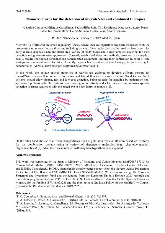
Nanostructures for the detection of microRNAs and combined therapies
Catarina Coutinho, Milagros Castellanos, Paula Milán-Rois, Ciro Rodriguez Diaz, Ana Latorre, Nuria Lafuente-Gómez, David García-Soriano, Gorka Salas, Álvaro Somoza
IMDEA Nanociencia, Faraday 9, 28049, Madrid, Spain
MicroRNAs (miRNAs) are small regulatory RNAs, where their dysregulation has been associated with the progression of several human diseases, including cancer. These molecules can be used as biomarkers for early disease diagnosis and are stable in a variety of body fluids and tissue samples, allowing for their detection using non-invasive approaches. Currently established detection methods, however, are complex, costly, require specialized personnel and sophisticated equipment, limiting their application in point-of-care settings or resource-limited facilities. Recently, approaches based on nanotechnology, in particular gold nanoparticles (AuNPs), have emerged as promising alternatives [1].
In this work, the unique optical properties of AuNPs are explored to develop different sensors for microRNAs, such as fluorescent, colorimetric and lateral flow-based sensors for miRNA detection. Such systems should allow simple, fast and low-cost detection, being suitable for handling by patients or non-specialized professionals. Our systems have shown good sensitivity and selectivity in vitro, allowing specific detection of target sequences with the naked eye in a few hours or minutes [2].
On the other hand, the use of different nanoparticles, such as gold, iron oxide or albumin-based, are explored for the combination therapy using a variety of therapeutic molecules (e.g., chemotherapeutics, oligonucleotides) [3]. Also, their use combined with magnetic hyperthermia is explored.
Acknowledgements
This work was supported by the Spanish Ministry of Economy and Competitiveness (SAF2017-87305-R), Comunidad de Madrid (IND2017/IND-7809; S2017/BMD-3867), Asociación Española Contra el Cáncer, and IMDEA Nanociencia. IMDEA Nanociencia acknowledges support from the 'Severo Ochoa' Programme for Centres of Excellence in R&D (MINECO, Grant SEV-2016-0686). We also acknowledge the European Structural and Investment Fund and the funding from the European Union’s Horizon 2020 research and innovation programme (No 685795. NoCanTher). N. Lafuente-Gómez also thanks the Spanish Education Ministry for the funding (FPU18/02323) and the grant to be a Graduate Fellow of the Madrid City Council (Spain) at the Residencia de Estudiantes (2019–2020).
References [1] C. Coutinho, A. Somoza, Anal. and Bioanal. Chem. 411, (2019),1807. [2] A. Latorre, C. Posch, Y. Garcimartín, S. Ortiz-Urda, A. Somoza, ChemComm 50, (2014), 3018-20 [3] A. Latorre, A.; Latorre, A.; Castellanos, M.; Rodriguez Diaz, C.; Lazaro-Carrillo, A.; Aguado, T.; Lecea, M.; Romero-Pérez, S.; Calero, M.; Sanchez-Puelles, J.M.; Villanueva, Á.; Somoza, Cancers (Basel) 11, (2019), 969.

oral presentations

In Silico Oral Presentations
NALS 2020 Nanomaterials Applied to Life Sciences
Numerical modelling of biomolecular and nanoscopic systems including hydrodynamics
Raúl Pérez Peláez1, Pablo Freire 1, and Rafael Delgado-Buscalioni 1
1Departamento Física Teorica de la Materia Condensada, Universidad Autónoma de Madrid
This talk presents a computational framework to investigate the structure and dynamics arising from the
interaction of bio-molecules (such as lipids, DNA and proteins) and nanoparticles in sub-micron volumes and, possibly, in general unsteady flows. We present different computational schemes to describe the
hydrodynamic interaction between these nanoscopic elements and random displacements satisfying the fluctuation dissipation balance. Over the talk we will present two schemes I) fluctuating hydrodynamics
equipped with the immersed boundary method and ii) Brownian hydrodynamics. We will illustrate these methods with recent research carried out in our group: on the anomalous diffusion of lipids in membranes
[1], on the quartz crystal microbalance response of liposomes attached to DNA strands (in collaboration with Electra Gizelli, FORTH, within the “CATCH-U-DNA” FETOPEN project) [2]; on the estimation of the local
viscosity of the cell using an optically trapped nanoparticle in the cytoplasm (in collaboration with Daniel Jaque, UAM) [3] and the formation of gellified structures and dynamics of proteins in a virus capsid, studied
by AFM (HFSP project, in collaboration with Pedro J. de Pablo, UAM) [4]
Acknowledgements
This research is funded by the Horizon 2020 FETOPEN project CATCH-U-DNA, the spanish
government project MINECO FIS2017-86007-C3-1-P and “María de Maeztu” Programme for Units of Excellence in R&D (MDM-2014-0377), the Human Frontier Science Program (HFSP) and the American
Chemical Society ACS-PRF, ND9 grant.
References
[1] S. Panzuela and R. Delgado-Buscalioni, Phys. Rev. Lett. 121, 048101 (2918)
[2] https://catch-u-dna.com/
[3] P. Rodriguez-Sevilla, F. Sanz-Rodiguez, R. Perez-Pelaez, R. Delgado-Buscalioni, L. Liang, Z. Lui and D. Jaque, Advanced Biosystems. 3 (2019) 1900082.
[4] HFSP project: Protein nanocages as single molecular reactors to understand biocatalysis in crowded environments
NALS 2020 Nanomaterials Applied to Life Sciences
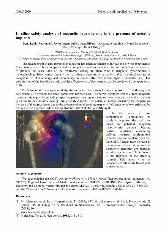
In silico safety analysis of magnetic hyperthermia in the presence of metallic implants
Irene Rubia-Rodríguez1, Javier Ortega-Julia1, Luca Zilberti2, Alessandro Arduino2, Oriano Bottauscio2, Mario Chiampi2, Daniel Ortega1,3
1IMDEA Nanoscience, Faraday 9, 28049 Madrid, Spain 2Istituto Nazionale di Ricerca Metrologica (INRiM), Strada delle Cacce 91, 10135 Turin
3Condensed Matter Physics department, Faculty of Science, University of Cádiz, 11510 Puerto Real (Cádiz), Spain
The advancement of new therapies in medicine has taken advantage of in vivo and in vitro experiments. These two have also been complemented by computer simulations (in silico testing), reducing time and cost to produce the tests. One of the treatments seizing in silico trials is magnetic hyperthermia, a nanotechnology-driven cancer therapy that has already been and is currently trialled in clinical settings as coadjuvant to chemotherapy and radiotherapy to successfully treat several types of tumours [1,2]. The optimization of the benefit/risk ratio and the effectiveness of the treatment can be predicted by computational trials.
Furthermore, the development of algorithms for in silico tests is leading to personalize this therapy and, consequently, to evaluate the safety parameters for each case. The current safety criteria in clinical magnetic hyperthermia explicitly exclude prospective patients bearing any kind of metallic or partly metallic implants [1,3] due to their possible heating through eddy currents. The potential damage caused by the temperature increase of these prostheses due to the presence of an alternating magnetic field tends to be overestimated by this restrictive approach, which has an absolute lack of studies quantifying it.
In this work we use computational simulations to carefully appraise the real risk posed to potential magnetic hyperthermia patients bearing passive implants considering different treatment configurations (tumour location, implant types and materials). Temperature increase in the regions of interest as well as dosimetric quantities are analysed as safety parameters. The influence of the implants on the effective magnetic field intensity at the nanoparticles site in the treated area is also studied.
Acknowledgements
We acknowledge the COST Action MyWave (CA 17115), NoCanTher project (grant agreement No 685795), Regional Government of Madrid under contract PEJD-2017-PRE/IND-3663, Spanish Ministry of Economy and Competitiveness through the grants MAT2017-85617-R, Ramón y Cajal RYC2018-025253-I and the “Severo Ochoa” Program for Centers of Excellence in R&D (SEV-2016-0686).
References
[1] M. Johannsen et al. Int. J. Hyperthermia 21 (2005), 637; M. Johannsen et al. Int. J. Hyperthermia 23 (2007), 315; D. Ortega, Q. A. Pankhurst. in Nanoscience: Vol. 1: Nanostructures through Chemistry (2013), 60.
[2] www.nocanther-project.eu [3] Maier-Hauff et al, J. Neurooncol. 103 (2011), 317
NALS 2020 Nanomaterials Applied to Life Sciences
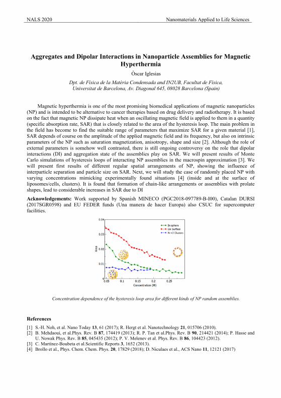
Aggregates and Dipolar Interactions in Nanoparticle Assemblies for Magnetic Hyperthermia
Òscar Iglesias
Dpt. de Física de la Matèria Condensada and IN2UB, Facultat de Física, Universitat de Barcelona, Av. Diagonal 645, 08028 Barcelona (Spain)
Magnetic hyperthermia is one of the most promising biomedical applications of magnetic nanoparticles (NP) and is intended to be alternative to cancer therapies based on drug delivery and radiotherapy. It is based on the fact that magnetic NP dissipate heat when an oscillating magnetic field is applied to them in a quantity (specific absorption rate, SAR) that is closely related to the area of the hysteresis loop. The main problem in the field has become to find the suitable range of parameters that maximize SAR for a given material [1], SAR depends of course on the amplitude of the applied magnetic field and its frequency, but also on intrinsic parameters of the NP such as saturation magnetization, anisotropy, shape and size [2]. Although the role of external parameters is somehow well contrasted, there is still ongoing controversy on the role that dipolar interactions (DI) and aggregation state of the assemblies play on SAR. We will present results of Monte Carlo simulations of hysteresis loops of interacting NP assemblies in the macrospin approximation [3]. We will present first results of different regular spatial arrangements of NP, showing the influence of interparticle separation and particle size on SAR. Next, we will study the case of randomly placed NP with varying concentrations mimicking experimentally found situations [4] (inside and at the surface of liposomes/cells, clusters). It is found that formation of chain-like arrangements or assemblies with prolate shapes, lead to considerable increases in SAR due to DI
Acknowledgements: Work supported by Spanish MINECO (PGC2018-097789-B-I00), Catalan DURSI (2017SGR0598) and EU FEDER funds (Una manera de hacer Europa) also CSUC for supercomputer facilities.
Concentration dependence of the hysteresis loop area for different kinds of NP random assemblies.
References [1] S.-H. Noh, et al. Nano Today 13, 61 (2017); R. Hergt et al. Nanotechnology 21, 015706 (2010). [2] B. Mehdaoui, et al.Phys. Rev. B 87, 174419 (2013); R. P. Tan et al.Phys. Rev. B 90, 214421 (2014); P. Hasse and
U. Nowak Phys. Rev. B 85, 045435 (2012); P. V. Melenev et al. Phys. Rev. B 86, 104423 (2012). [3] C. Martínez-Boubeta et al.Scientific Reports 3, 1652 (2013). [4] Brollo et al., Phys. Chem. Chem. Phys. 20, 17829 (2018); D. Niculaes et al., ACS Nano 11, 12121 (2017)
NALS 2020 Nanomaterials Applied to Life Sciences
Magnetic fluid hyperthermia: Optimisation of the properties for high
heating output
S. Ruta1*, E. Rannala1, D. Serantes1,2, O. Hovorka3, R. Chantrell1
1 Department of Physics, University of York, York YO10 5DD, U.K.2 IIT and Appl. Phys. Dept., Universidade de Santiago de Compostela, 15703, Spain.
3 Faculty of Engineering and the Environment, University of Southampton, U.K.
Cancer is one of the most severe and widespread health problems faced by today’s medicine. The
existing techniques (such as surgery, chemotherapy and radiotherapy) have low survival rate and strong side
effects. An alternative promising methodology for cancer treatment is magnetic hyperthermia. Understanding
the mechanisms of magnetic heating is crucial for synthesizing the optimal particles and to control the
heating inside the human body. The widely used method for prediction of magnetic hyperthermia response is
based on the linear response theory developed by Rosensweig [1] (LRT). Nevertheless, the method is limited
to small field condition, which does not correspond to the broadly used condition in experiments. Also, those
fields are not ideal for high efficiency of the treatment.
For a better investigation, it is important to have a reliable model to support the current experimental
approaches, but also to indicate a new way so that the performance of magnetic nanoparticle is maximized.
These studies are constrained by one or more elements: 1) intrinsic properties and their distribution (particle
size, anisotropy value, easy axis orientation), 2) extrinsic properties (AC magnetic field amplitude, AC field
frequency), 3) the role of dipole interactions, 4) heat transfer from particle to the tumour, 5) mechanical
rotation of the particles, 6) the viscosity of the fluid containing the nanoparticles and 7) the aggregation of
the nanoparticles when placed inside the tumour.
In this work, we will emphasize the applicability limits of LRT to interpret/guide experiments, by
comparing the analytical predictions of LRT with kinetic Monte Carlo simulations [2]. This will provide a
clear definition of what is the meaning of “small field” in terms of both the particles characteristics and AC
field conditions. With the kinetic Monte Carlo approach we are also able to study the optimum balance
between AC field and particles properties in the entire range of interest for hyperthermia application. For the
non-interacting case we can provide a general picture which can be used for the prediction of optimal
conditions for any magnetic particle properties.
References
[1] R. Rosensweig, Journal of Magnetism and magnetic Materials, vol 252, Nov 2002.
[2] S. Ruta, R. Chantrell and O. Hovorka, Sci. Rep. Vol. 5, Jan 2015.
NALS 2020 Nanomaterials Applied to Life Sciences
Design of magnetic nanomaterials for targeted hyperthermia Alessandra Manzin1, Riccardo Ferrero1, Gabriele Barrera1, Federica Celegato1, Marco Coïsson1, Paola Tiberto1
1Istituto Nazionale di Ricerca Metrologica (INRIM), Torino, Italy
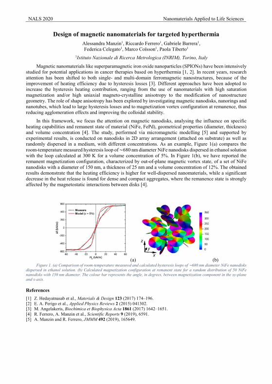
Magnetic nanomaterials like superparamagnetic iron oxide nanoparticles (SPIONs) have been intensively studied for potential applications in cancer therapies based on hyperthermia [1, 2]. In recent years, research attention has been shifted to both single- and multi-domain ferromagnetic nanostructures, because of the improvement of heating efficiency due to hysteresis losses [3]. Different approaches have been adopted to increase the hysteresis heating contribution, ranging from the use of nanomaterials with high saturation magnetization and/or high uniaxial magneto-crystalline anisotropy to the modification of nanostructure geometry. The role of shape anisotropy has been explored by investigating magnetic nanodisks, nanorings and nanotubes, which lead to large hysteresis losses and to magnetization vortex configuration at remanence, thus reducing agglomeration effects and improving the colloidal stability.
In this framework, we focus the attention on magnetic nanodisks, analysing the influence on specific heating capabilities and remanent state of material (NiFe, FePd), geometrical properties (diameter, thickness) and volume concentration [4]. The study, performed via micromagnetic modelling [5] and supported by experimental results, is conducted on nanodisks in 2D array arrangement (attached on substrate) as well as randomly dispersed in a medium, with different concentrations. As an example, Figure 1(a) compares the room-temperature measured hysteresis loop of ∼680 nm diameter NiFe nanodisks dispersed in ethanol solution with the loop calculated at 300 K for a volume concentration of 5%. In Figure 1(b), we have reported the remanent magnetization configuration, characterized by out-of-plane magnetic vortex state, of a set of NiFe nanodisks with a diameter of 150 nm, a thickness of 25 nm and a volume concentration of 12%. The obtained results demonstrate that the heating efficiency is higher for well-dispersed nanomaterials, while a significant decrease in the heat release is found for dense and compact aggregates, where the remanence state is strongly affected by the magnetostatic interactions between disks [4].
(a) (b) Figure 1. (a) Comparison of room-temperature measured and calculated hysteresis loops of ∼680 nm diameter NiFe nanodisks
dispersed in ethanol solution. (b) Calculated magnetization configuration at remanent state for a random distribution of 50 NiFe nanodisks with 150 nm diameter. The colour bar represents the angle, in degrees, between magnetization component in the xy-plane and x-axis.
References [1] Z. Hedayatnasab et al., Materials & Design 123 (2017) 174–196. [2] E. A. Perigo et al., Applied Physics Reviews 2 (2015) 041302. [3] M. Angelakeris, Biochimica et Biophysica Acta 1861 (2017) 1642–1651. [4] R. Ferrero, A. Manzin et al., Scientific Reports 9 (2019), 6591. [5] A. Manzin and R. Ferrero, JMMM 492 (2019), 165649.

Environmental Presentations
NALS 2020 Nanomaterials Applied to Life Sciences
Arsenic immobilization onto magnetite nanoparticles: A novel approach for soil remediation by magnetic separation
D. Baragaño1,*, M. Matos2, R. Forján1, G. Gutiérrez2, C. Sierra3, M.C. Blanco-López4, J.R. Gallego1 1INDUROT and Environmental Biotechnology & Geochemistry Group, University of Oviedo,
C/Gonzalo Gutiérrez Quirós s/n, 33600 Mieres, Asturias, Spain 2Department of Chemical and Environmental Engineering, University of Oviedo, Julián Clavería 8,
33006 Oviedo, Spain 3Escuela Superior de Ingenieros de Minas y Energía, University of León, Campus de Vegazana, León,
Spain 4Department of Physical and Analytical Chemistry, Institute of Biotecnology of Asturias.
Iron-based nanoparticles are a promising material for remediation proposes [1, 2]. The use of nanoparticles for the stabilization of metal(loid)s in soils has been widely studied [3]. However, their use as pollutants-sorbent in soils and later recovery has not yet been sufficiently explored [4].
In this work, several types of magnetite nanoparticles were synthesized via precipitation in a water-in-oil microemulsion system using an aqueous solution containing FeCl2 and FeCl3 using hexadecyltrimethylammonium bromide (CTAB) as a main cationic surfactant, 1-butanol as a costabilizer and 1-hexanol as the continuous oily phase. Nanoparticles were subsequently cleaned with an optimized protocol using different ethanolic solutions. The morphology of the nanoparticles was studied by transmission electron microscopy, the zeta potential was measured and the grain-size distribution determined by dynamic light scattering. Additionally, the magnetic properties of the nanoparticles were measured using a vibrating sample magnetometer. The most suitable type of nanoparticles for the magnetic separation approach was selected. Mean sizes ranged from 4 to 7 nm and all nanoparticles showed to be superparamagnetic.
To assess As sorption onto nanoparticles, soil samples were treated at a 2% dose of nanoparticles. In order to determine the stability of the interaction As-nanoparticle, the samples were subjected to a Toxic Characteristics Leaching Procedure (TCLP). Magnetic separation was carried utilizing a wet high-intensity magnetic separator to recover the Fe nanoparticles with As sorbed onto their surface. Finally, the efficiency of the separation was evaluated according to a metallurgical accounting index. The results obtained indicate the appropriateness of this technique for the treatment of contaminated soils.
Acknowledgements
This work was supported by the Ministerio de Economía y Competitividad (MINECO, Spain), under the Projects MAT2017-84959-C2-1-R and CTM 2016-75894-P. Diego Baragaño obtained a grant from the “Formación del Profesorado Universitario” program, financed by the “Ministerio de Educación, Cultura y Deporte de España”. This study was also financed by the Consejería de Economía y Empleo del Principado de Asturias (Plan de Ciencia, Tecnología e Innovación 2013-2017), under the Grant IDI/2018/000185. Support from the European Regional Development Fund (ERDF) is gratefully acknowledged. This study was also financied by Europa Investigación (ref: EUIN2019-103338).
References
[1] D. Baragaño, J. Alonso, J.R. Gallego, M.C. Lobo, M. Gil-Díaz, Chemosphere. 238 (2020) 124624. [2] M. Gil-Díaz, E. Rodríguez-Valdés, J. Alonso, D. Baragaño, J.R. Gallego, M.C. Lobo, Sci. Total Environ.
675 (2019) 165-175. [3] M. Vítková, M. Puschenreiter, M. Komárek, Chemosphere. 200 (2018) 217-226. [4] C. Boente, C. Sierra, D. Martínez-Blanco, J.M. Menéndez-Aguado, J.R. Gallego, J. Hazard. Mater. 350
(2018) 55-65.
NALS 2020 Nanomaterials Applied to Life Sciences
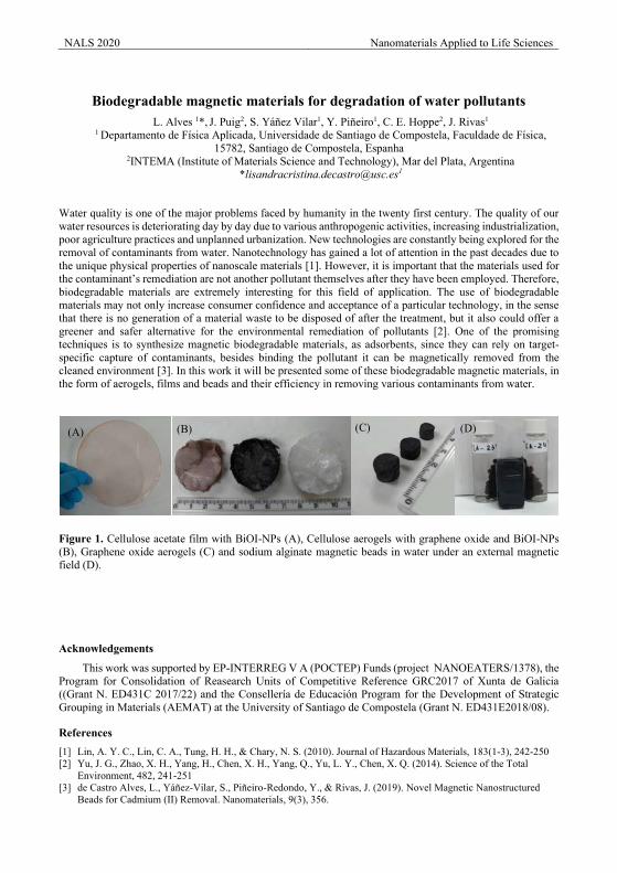
Biodegradable magnetic materials for degradation of water pollutants
L. Alves 1*, J. Puig2, S. Yáñez Vilar1, Y. Piñeiro1, C. E. Hoppe2, J. Rivas1 1 Departamento de Física Aplicada, Universidade de Santiago de Compostela, Faculdade de Física,
15782, Santiago de Compostela, Espanha 2INTEMA (Institute of Materials Science and Technology), Mar del Plata, Argentina
Water quality is one of the major problems faced by humanity in the twenty first century. The quality of our water resources is deteriorating day by day due to various anthropogenic activities, increasing industrialization, poor agriculture practices and unplanned urbanization. New technologies are constantly being explored for the removal of contaminants from water. Nanotechnology has gained a lot of attention in the past decades due to the unique physical properties of nanoscale materials [1]. However, it is important that the materials used for the contaminant’s remediation are not another pollutant themselves after they have been employed. Therefore, biodegradable materials are extremely interesting for this field of application. The use of biodegradable materials may not only increase consumer confidence and acceptance of a particular technology, in the sense that there is no generation of a material waste to be disposed of after the treatment, but it also could offer a greener and safer alternative for the environmental remediation of pollutants [2]. One of the promising techniques is to synthesize magnetic biodegradable materials, as adsorbents, since they can rely on target-specific capture of contaminants, besides binding the pollutant it can be magnetically removed from the cleaned environment [3]. In this work it will be presented some of these biodegradable magnetic materials, in the form of aerogels, films and beads and their efficiency in removing various contaminants from water.
Figure 1. Cellulose acetate film with BiOI-NPs (A), Cellulose aerogels with graphene oxide and BiOI-NPs (B), Graphene oxide aerogels (C) and sodium alginate magnetic beads in water under an external magnetic field (D).
Acknowledgements
This work was supported by EP-INTERREG V A (POCTEP) Funds (project NANOEATERS/1378), the Program for Consolidation of Reasearch Units of Competitive Reference GRC2017 of Xunta de Galicia ((Grant N. ED431C 2017/22) and the Consellería de Educación Program for the Development of Strategic Grouping in Materials (AEMAT) at the University of Santiago de Compostela (Grant N. ED431E2018/08).
References [1] Lin, A. Y. C., Lin, C. A., Tung, H. H., & Chary, N. S. (2010). Journal of Hazardous Materials, 183(1-3), 242-250 [2] Yu, J. G., Zhao, X. H., Yang, H., Chen, X. H., Yang, Q., Yu, L. Y., Chen, X. Q. (2014). Science of the Total
Environment, 482, 241-251 [3] de Castro Alves, L., Yáñez-Vilar, S., Piñeiro-Redondo, Y., & Rivas, J. (2019). Novel Magnetic Nanostructured
Beads for Cadmium (II) Removal. Nanomaterials, 9(3), 356.
(A) (B) (C) (D)
NALS 2020 Nanomaterials Applied to Life Sciences
Novel protein-metal nanoparticles biohybrids for degradation of organic
pollutants in water
Jose M. Palomo*, Noelia Losada-Garcia and Rocio Benavente Department of Biocatalysis. Institute of Catalysis (CSIC). Marie Curie 2. Cantoblanco. Campus UAM,
28049 Madrid, Spain; [email protected]
In today’s world where industries have been modernized and advanced, our environment is filled with various types of pollutants emitted from human activities or industrial processes. In particular, organic pollutants in wastewater, being highly toxic and difficult to degrade, have become one of the most serious global environmental issues today. [1] Therefore, an effective and economical technique needs to be developed to reduce the concentration of organic pollutants before releasing the wastewater into the aquatic environment.
Currently, industrially available wastewater treatment technologies such as adsorption and coagulation merely concentrate or separate these pollutants from water, but does not completely “eliminate” or “destroy” them into biodegradable or less toxic organic compounds.
Here we present a new efficient and green technology based on nanoremediation processes to rapidly degrade different relevant organic pollutants such as Bisphenol-A (BPA), p-aminophenol or chlorinated alkenes in water and room temperature using hydrogen peroxide as green oxidant. For this purpose, we have developed novel nanostructured materials as catalysts based on the in situ (induced by a protein) formation of metal (Fe or Cu) nanoparticles embedded on a protein matrix as a heterogeneous form [2-4].
Acknowledgements
This research was supported by GRO PROGRAM 2017 and SAMSUNG L.S. The authors thank the support by the Spanish National Research Council (CSIC) (intramural project 201980E081). We thank Ramiro Martínez from Novozymes.
References
[1] Lin, D. et al. Chemosphere, 103, (2014) 188-196 [2] Palomo, J. M., Benavente, R. Patent EP1641.1334. [3] Benavente, R., Lopez-Tejedor, D., Palomo,* J.M., ChemCommun 54 (2018) 6256-6259. [4] Palomo, J.M., Losada-Garcia, N., Rodriguez-Otero, A. Patent ES1641.1460.
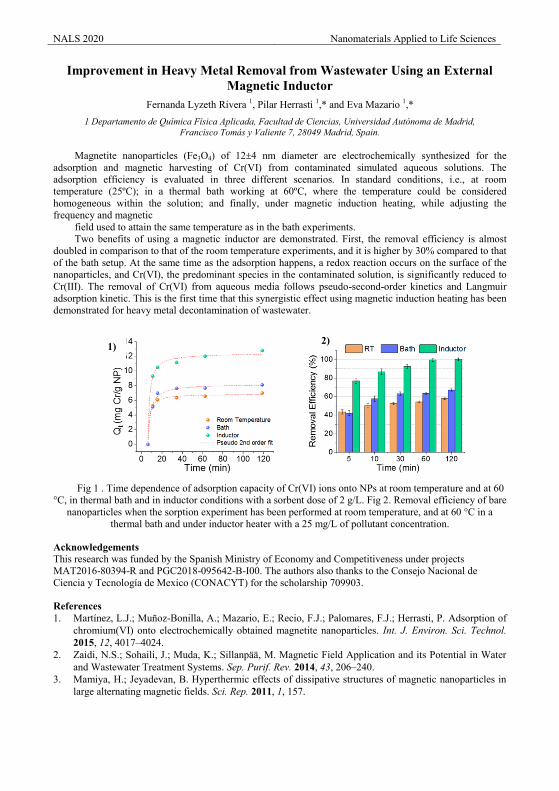
NALS 2020 Nanomaterials Applied to Life Sciences
Improvement in Heavy Metal Removal from Wastewater Using an External Magnetic Inductor
Fernanda Lyzeth Rivera 1, Pilar Herrasti 1,* and Eva Mazario 1,* 1 Departamento de Química Física Aplicada, Facultad de Ciencias, Universidad Autónoma de Madrid,
Francisco Tomás y Valiente 7, 28049 Madrid, Spain.
Magnetite nanoparticles (Fe3O4) of 12±4 nm diameter are electrochemically synthesized for the adsorption and magnetic harvesting of Cr(VI) from contaminated simulated aqueous solutions. The adsorption efficiency is evaluated in three different scenarios. In standard conditions, i.e., at room temperature (25ºC); in a thermal bath working at 60ºC, where the temperature could be considered homogeneous within the solution; and finally, under magnetic induction heating, while adjusting the frequency and magnetic
field used to attain the same temperature as in the bath experiments. Two benefits of using a magnetic inductor are demonstrated. First, the removal efficiency is almost
doubled in comparison to that of the room temperature experiments, and it is higher by 30% compared to that of the bath setup. At the same time as the adsorption happens, a redox reaction occurs on the surface of the nanoparticles, and Cr(VI), the predominant species in the contaminated solution, is significantly reduced to Cr(III). The removal of Cr(VI) from aqueous media follows pseudo-second-order kinetics and Langmuir adsorption kinetic. This is the first time that this synergistic effect using magnetic induction heating has been demonstrated for heavy metal decontamination of wastewater.
Fig 1 . Time dependence of adsorption capacity of Cr(VI) ions onto NPs at room temperature and at 60
°C, in thermal bath and in inductor conditions with a sorbent dose of 2 g/L. Fig 2. Removal efficiency of bare nanoparticles when the sorption experiment has been performed at room temperature, and at 60 °C in a
thermal bath and under inductor heater with a 25 mg/L of pollutant concentration.
Acknowledgements This research was funded by the Spanish Ministry of Economy and Competitiveness under projects MAT2016-80394-R and PGC2018-095642-B-I00. The authors also thanks to the Consejo Nacional de Ciencia y Tecnología de Mexico (CONACYT) for the scholarship 709903.
References 1. Martínez, L.J.; Muñoz-Bonilla, A.; Mazario, E.; Recio, F.J.; Palomares, F.J.; Herrasti, P. Adsorption of
chromium(VI) onto electrochemically obtained magnetite nanoparticles. Int. J. Environ. Sci. Technol. 2015, 12, 4017–4024.
2. Zaidi, N.S.; Sohaili, J.; Muda, K.; Sillanpää, M. Magnetic Field Application and its Potential in Water and Wastewater Treatment Systems. Sep. Purif. Rev. 2014, 43, 206–240.
3. Mamiya, H.; Jeyadevan, B. Hyperthermic effects of dissipative structures of magnetic nanoparticles in large alternating magnetic fields. Sci. Rep. 2011, 1, 157.
1) 2)
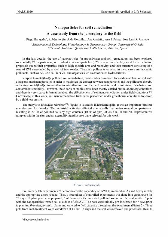
NALS 2020 Nanomaterials Applied to Life Sciences
Nanoparticles for soil remediation:
A case study from the laboratory to the field Diego Baragaño*, Rubén Forján, Aida González, Ana Castaño, Ana I. Peláez, José Luis R. Gallego
1Environmental Technology, Biotechnology & Geochemistry Group, University of Oviedo C/Gonzalo Gutiérrez Quirós s/n, 33600 Mieres, Asturias, Spain
In the last decade, the use of nanoparticles for groundwater and soil remediation has been explored successfully [1]. In particular, zero valent iron nanoparticles (nZVI) have been widely used for remediation proposals due to their properties, such as high specific area and reactivity, and their structure consisting of a core of ZVI surrounded by a shell of iron oxides. The main pollutants targeted in these cases are inorganic pollutants, such as As, Cr, Cu, Pb or Zn, and organics such as chlorinated hydrocarbons.
Respect to metal(loid)s polluted soil remediation, most studies have been focused on a blend of soil with a suspension of nanoparticles in order to maximize the contact between nanoparticles and the pollutants thereby achieving metal(loid)s immobilization/stabilization in the soil matrix and minimizing leachates and contaminants mobility. However, these sorts of studies have been mostly carried out in laboratory conditions and there is very scarce information about the effectiveness of soil nanoremediation under field conditions [2]. Conversely, in this work, soil nanoremediation trials were performed under greenhouse conditions followed by a field test on site.
The study site, known as Nitrastur [3] (Figure 1) is located in northern Spain. It was an important fertilizer manufacturer for decades. The industrial activities affected dramatically the environmental compartments, resulting in 20 Ha of polluted soils by high contents (1000s of ppm) of As, Cu, Pb and Zn. Representative samples within the site, and an exemplifying pilot area were selected for this work.
Figure 1. Nitrastur site.
Preliminary lab experiments [4] demonstrated the capability of nZVI to immobilize As and heavy metals and the appropriate doses needed. Thus, a second set of controlled experiments was done in a greenhouse for 75 days: 12 plant pots were prepared, 6 of them with the untreated polluted soil (controls) and another 6 pots with the nanoparticles-treated soil at a dose of 2% ZVI. The pots were initially pre-incubated for 7 days prior to planting Brassica juncea L. plants and watered to field capacity throughout the experiment (Figure 2). Three pots from each treatment were withdrawn at 15 and 75 days and the soil was removed and processed. Results
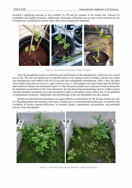
NALS 2020 Nanomaterials Applied to Life Sciences revealed a significant decrease in the available As, Pb and Zn contents in the treated soil, whereas Cu availability was slightly increased. Additionally, the height of the plant was to some extent increased in case of treated soil, revealing not toxicity effects due to the nanoparticles treatment.
Figure 2. Greenhouse experiment: Plants evolution.
Once the greenhouse results revealed the good performance of the nanoparticles, a pilot test was carried out on site. The soil was transferred to controlled plots of one squared metre of surface, and the zero valent iron nanoparticles were added to the soil of one plot and consequently homogenized. After 3 days, the plots were seeded using Brassica juncea L. and Lolium perenne L. Soil samples were taken along time for 60 days and evolution of plants was monitored (Figure 3). After this period, plants were collected in order to determine the pollutants accumulation in the roots and shoots. Several physiological parameters such as oxidative stress and phytochelatins production were also measured in order to determine toxicity effects due to soil pollution or nanoparticles treatment. Additionally, the microbiology of the soil rhizosphere was also studied.
Results revealed that nanoremediation was again effective to immobilize As, Pb, Zn and, in this case, also Cu. Regarding plants and microbes, both types of plants grew in untreated and treated soils, nevertheless the evaluation of results exposed differences in biomass, height, contaminants accumulation, and microbial activity in the rhizospheres.
Figure 3. Untreated (left) and nanoparticles (right) treated plots.

NALS 2020 Nanomaterials Applied to Life Sciences Acknowledgements
This work was supported by the Ministerio de Economía y Competitividad (MINECO, Spain), under the project CTM 2016-75894-P. Diego Baragaño obtained a grant from the “Formación del Profesorado Universitario” program, financed by the “Ministerio de Educación, Cultura y Deporte de España”.
References
[1] J. Rodriges Gonçalves, Procedia Eng. 143 (2016) 1268-1275. [2] M. Gil-Díaz, E. Rodríguez-Valdés, J. Alonso, D. Baragaño, J.R. Gallego, M.C. Lobo, Sci. Total Environ.
675 (2019) 165-175. [3] J.R. Gallego, E. Rodríguez-Valdés, N. Esquinas, A. Fernández-Braña, E. Afif, Sci. Total Environ. 563-
564 (2016) 683-692. [4] D. Baragaño, J. Alonso, J.R. Gallego, M.C. Lobo, M. Gil-Díaz, Chemposhere. 238 (2020) 124624.

Novel Applications Presentations
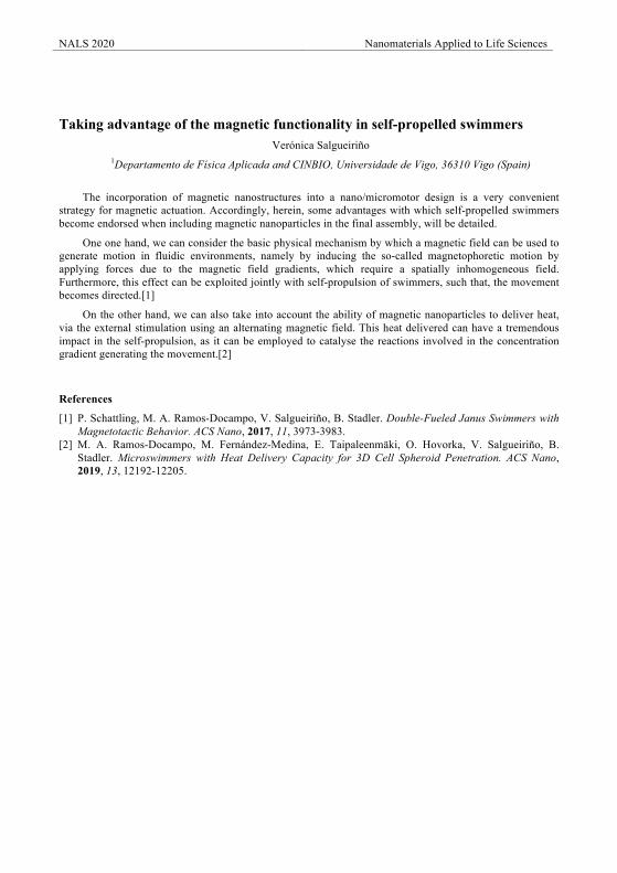
NALS 2020 Nanomaterials Applied to Life Sciences
Taking advantage of the magnetic functionality in self-propelled swimmers
Verónica Salgueiriño 1Departamento de Física Aplicada and CINBIO, Universidade de Vigo, 36310 Vigo (Spain)
The incorporation of magnetic nanostructures into a nano/micromotor design is a very convenient strategy for magnetic actuation. Accordingly, herein, some advantages with which self-propelled swimmers become endorsed when including magnetic nanoparticles in the final assembly, will be detailed.
One one hand, we can consider the basic physical mechanism by which a magnetic field can be used to generate motion in fluidic environments, namely by inducing the so-called magnetophoretic motion by applying forces due to the magnetic field gradients, which require a spatially inhomogeneous field. Furthermore, this effect can be exploited jointly with self-propulsion of swimmers, such that, the movement becomes directed.[1]
On the other hand, we can also take into account the ability of magnetic nanoparticles to deliver heat, via the external stimulation using an alternating magnetic field. This heat delivered can have a tremendous impact in the self-propulsion, as it can be employed to catalyse the reactions involved in the concentration gradient generating the movement.[2]
References
[1] P. Schattling, M. A. Ramos-Docampo, V. Salgueiriño, B. Stadler. Double-Fueled Janus Swimmers with Magnetotactic Behavior. ACS Nano, 2017, 11, 3973-3983.
[2] M. A. Ramos-Docampo, M. Fernández-Medina, E. Taipaleenmäki, O. Hovorka, V. Salgueiriño, B. Stadler. Microswimmers with Heat Delivery Capacity for 3D Cell Spheroid Penetration. ACS Nano, 2019, 13, 12192-12205.
NALS 2020 Nanomaterials Applied to Life Sciences
Wireless stress sensor based on magnetoelastic microwires for biomedical applications: Development of a Telemetric System for Postoperative Follow‐up
of Vascular Surgery Procedures P. Marín1*, A. Hernando-Rydings2,*, A. Hernando1
1Instituto de Magnetismo Aplicado. Departamento de Física de Materiales. Nacional VI Km 22,5 28230 Universidad Complutense de Madrid. Spain
2 Department of Vascular Surgery, Basurto University Hospital, Universidad País Vasco. Barrio Sarriena s/n 48940 Leioa Bizkaia. Spain
Chronic ischemia of the inferior limbs is a range of signs and symptoms that are produced as a consequence of the progressive decrease of blood flow in the inferior limbs. It is estimated that the prevalence of this pathology is between 3% and 18%, which that means more than 27 million people are affected in the world. Failure of surgical revascularization procedures, both open and endovascular, continues to be a challenge in the present‐day clinical practice of the vascular surgeon. Optimizing the follow‐up protocols, as well as offering out‐patient attention, is fundamental to maintaining their quality of life. Magnetic sensors are at the helm of technological development seen in this field over the last decades, offering numerous advantages attributed to their elevated sensitivity, reduced size, systems without the need for an external source of energy, and wireless connections. The use of WSN (wireless sensor network) technologies offers the possibility of developing implantable biomedical sensors allowing for the monitorisation and follow‐up of certain physiological parameters with precise and up until now, unthinkable measurements. Therefore, the aim of this research is to develop a wireless magnetic sensor for postoperative follow‐up procedures of vascular surgery. Because of the unique electromagnetic characteristics of the magnetoelastic microwire, the changes in the pressure of a fluid will provoke a variation of the mechanical pressure on the sensor, which will cause a variation of its magnetization that will be detectable wirelessly. Thus, a wireless system can be developed for following up vascular surgery procedures. The sensor consists of a magnetoelastic microwire ring, which was integrated into an in vitro model with pulsatile flow. Different degrees of stenosis were simulated in different locations both in bovine artery as well as in a polytetrafluoroethylene anastomosis. A Fourier analysis of the registered signals and a statistical analysis using Pearson test and receiver operating characteristic (ROC) curves were made. A Pearson index of 0.945 (P<0.001) was obtained between the invasive pressure of the fluid and the power of the signal transmitted by the sensor in bovine artery. The sensor obtained very good ROC curves upon analyzing the signals registered, both in the case of preanastomotic stenosis (area under the curve [AUC], 0.98; 95% CI, 0.97–1.00), of anastomosis (AUC, 0.93; 95% CI, 0.86–0.99), as well as distal (AUC, 0.88; 95% CI, 0.79–0.98), compared to the control group. The magnetoelastic microwire has shown that it is capable of detecting, locating, and quantifying the degree of stenosis in bovine artery, as well as in a latero‐terminal anastomosis, with a high statistical potency. For the first time, a wireless in vitro sensor has been developed for the postoperative follow‐up of vascular surgery procedures.
Acknowledgements (Times New Roman 11 points)
The authors want to acknowledge to the projects NANOMAGCOST S2018/NMT-4321, Nanofrontmag S2013/MIT-2850 and RTI2018-095856-B-C21
References
[1] C. Herrero-Gómez P. Marín, A. Hernando Appl. Phys.Lett. 103 (2013)142414 [2] C. Herrero-Gómez, A.M. Aragón, M. Hernando-Ryding, P. Marín, A. Hernando Appl. Phys.Lett. 105
(2014) 092405 [3] M. Hernando Rydings, P. Marín, A.M. Aragón-Sánchez, E. Bravo, V. López-Domínguez, I. Martínez, C.
Fernández, A. Bilbao, F.J. Javier Serrano, R. Vega, A. Hernando J of the Am. Heart Ass.5 (2016)1-12.
NALS 2020 Nanomaterials Applied to Life Sciences
Remote Control Healing for OA repair Iria Echevarria1,2, Hareklea Markides2, Alicia El Haj2
1Institute of Science and Technology in Medicine, Keele University, Stoke-on-Trent, UK 2The Healthcare Technology Institute (HTI), University of Birmingham, Birmingham, UK
Magnetic nanoparticles (MNPs) have been widely studied in recent years given their potential in medical applications such as magnetic hyperthermia for cancer therapy or contrast agents for magnetic resonance imaging (MRI). MNPs have been further implemented in the field of tissue engineering for their potential to control stem cell fate and behaviour in vivo via a technique known as MICA (Magnetic Ion Channel activation). This involves the use of a magnetic force bioreactor that applies and alternating magnetic field to the cells prior labelled with MNPs. The MNPs are targeted and bound to specific mechano-transductive ion channels or receptors on the mesenchymal stem cells membrane which then allows remote forces to be delivered directly and selectively to the channels or mechano-receptive parts of the cells membrane. This effect is translated downstream into the activation of specific chondrogenic signalling pathways with the following activation of genes involved in chondrogenesis. Here, we present the application of MICA in developing a novel stem cell based therapy for Osteoarthritis which aims to overcome limitations of current stem cell therapies.
MNPs are functionalized with TRPV4 antibodies, to target the mechanosensitive ion-channel of human bone marrow derived mesenchymal stem cells (MSCs) and Umbilical Cord-derived mesenchymal stem cells (UC-SCs). Cells are firstly expanded, MNP-labelled and cultured in pellet form for 21 days in chondrogenic media. Labelled cells were submitted to 1h daily magnetic stimulation regimes using the MICA magnetic bioreactor. Chondrogenesis was evaluated by histological staining, immunohistochemistry (IHC) and gene expression analysis. Cell growth and proliferation was also analysed together with the ion composition of the samples.
MICA activated pellets presented enhanced growth and proliferation together with a higher expression of chondrogenic markers, such as GAGs and collagen 2. Cell proliferation was observed to be enhanced in MICA groups throughout the duration of the culture period. In addition, MICA activated samples demonstrated enhanced early expression of collagen 2 by day 7. These results were supported by the gene expression analysis of collagen 2. Gene expression analysis also revealed enhanced expression of chondrogenic markers such as SOX9, aggrecan for MICA activated groups and low expression of collagen X, a hypertrophy marker, compared to the unstimulated controls. IHC analysis supported this data showing enhanced chondrogenic marker expression and low expression of hypertrophic markers. Analysis of the proliferation marker Ki67, also revealed enhanced expression for MICA activated groups.
In conclusion, MICA activation improves chondrogenic response in both MSCs and UC-SCs which we believe will be beneficial in enhancing clinical outcomes.
Acknowledgements
We are grateful for EPRSC funding support.
References
[1] Markides H et al. Regen Med. 2018. 3:9
NALS 2020 Nanomaterials Applied to Life Sciences
Nanotechnology-based neural interfaces Beatriz L. Rodilla1,2, Ana Arché-Núñez1, Ivo Calaresu3, Luiz Enger4, Arturo Vera1, Ana Domínguez5,
Ankor González-Mayorga5, Jaime Hernández,1 Claudia Fernández-González1,2, Sandra Ruiz-Gómez2, Isidoro Martínez1, Rubén Guerrero1, Lucas Pérez1,2, Julio Camarero1,6, Stéphane Flament4, Denis Scaini3, Isabel
Rodríguez1, Elisa López-Dolado7, María Concepción Serrano5, Laurence Méchin4, Laura Ballerini3, Rodolfo Miranda1,6, M. Teresa González1*
1IMDEA Nanociencia, 28049, Madrid, España; 2Depto. Física de Materiales. UCM, 28040, Madrid, Spain; 3International School for Advanced Studies (SISSA) 34136 Trieste, Italy; 4Normandie Univ, UNICAEN,
ENSICAEN, CNRS, GREYC, 14000 Caen, France; 5Instituto de Ciencia de Materiales de Madrid (ICMM-CSIC), 28049 Madrid, Spain; 6Depto. Física de la Materia Condensada-IFIMAC, UAM, 28049 Madrid,
Spain; 7Hospital Nacional de Parapléjicos, 45071 Toledo, Spain
The development of neural interfaces for activity recording and electrical stimulation in neural tissue is a challenge that brings together Material Science, Nanotechnology and Neuroscience. The ultimately goal is to provide more effective therapeutic alternatives for spinal cord injuries (SCI), as well as other neurological conditions including Parkinson’s disease, chronic pain, blindness or epilepsy. In spite of important advances, most of nowadays devices still present size, morphology and rigidity issues that unleashes an immunologic response that ultimately inactivates them.
In this work, we will summarize our progress, within our present FET-OPEN project, in a variety of new developed neural interfaces based in Nanotechnology for both sensing and stimulating the activity of the neural tissue. Our final aim is to directly interact the lesion area of a SCI in order to promote neural repair and reconnection. Interfaces incorporating miniaturized magnetic sensors with subnanoTelsa resolution at room temperature have been fabricated and characterized, offering a sensing interface which does not require intimate contact with the neural tissue. On the other hand, different conducting electrodes have been produced for stimulation both in metal and a polymeric composite whose surface has been nanostructured by vertical nanowires. Our biocompatibility tests show that such nanostructured, in addition to offer an enhanced contact area with the neurons, provides a three dimension microenvironment for axons to navigate, allowing the reconstruction of 3D topologies. We will describe our very promising performance results with both the magnetic sensors and the nanostructured stimulators from experiments with primary cultures as well as spinal cord slices in direct contact with the neural interfaces.
Acknowledgements
This work has received funding from the European Union’s Horizon 2020 research and innovation programme under the project ByAxon, grant agreement No 737116. Beatriz L. Rodilla acknowledges the support of UCM Predoctoral contract (CT17/17-CT18/17).
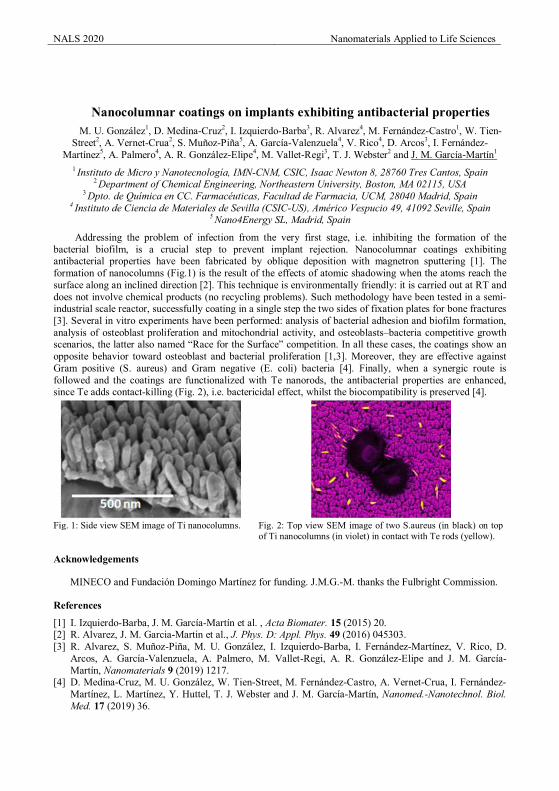
NALS 2020 Nanomaterials Applied to Life Sciences
Nanocolumnar coatings on implants exhibiting antibacterial properties M. U. González1, D. Medina-Cruz2, I. Izquierdo-Barba3, R. Alvarez4, M. Fernández-Castro1, W. Tien-
Street2, A. Vernet-Crua2, S. Muñoz-Piña5, A. García-Valenzuela4, V. Rico4, D. Arcos3, I. Fernández-Martínez5, A. Palmero4, A. R. González-Elipe4, M. Vallet-Regi3, T. J. Webster2 and J. M. García-Martín1
1 Instituto de Micro y Nanotecnología, IMN-CNM, CSIC, Isaac Newton 8, 28760 Tres Cantos, Spain 2 Department of Chemical Engineering, Northeastern University, Boston, MA 02115, USA
3 Dpto. de Química en CC. Farmacéuticas, Facultad de Farmacia, UCM, 28040 Madrid, Spain 4 Instituto de Ciencia de Materiales de Sevilla (CSIC-US), Américo Vespucio 49, 41092 Seville, Spain
5 Nano4Energy SL, Madrid, Spain
Addressing the problem of infection from the very first stage, i.e. inhibiting the formation of the bacterial biofilm, is a crucial step to prevent implant rejection. Nanocolumnar coatings exhibiting antibacterial properties have been fabricated by oblique deposition with magnetron sputtering [1]. The formation of nanocolumns (Fig.1) is the result of the effects of atomic shadowing when the atoms reach the surface along an inclined direction [2]. This technique is environmentally friendly: it is carried out at RT and does not involve chemical products (no recycling problems). Such methodology have been tested in a semi-industrial scale reactor, successfully coating in a single step the two sides of fixation plates for bone fractures [3]. Several in vitro experiments have been performed: analysis of bacterial adhesion and biofilm formation, analysis of osteoblast proliferation and mitochondrial activity, and osteoblasts–bacteria competitive growth scenarios, the latter also named “Race for the Surface” competition. In all these cases, the coatings show an opposite behavior toward osteoblast and bacterial proliferation [1,3]. Moreover, they are effective against Gram positive (S. aureus) and Gram negative (E. coli) bacteria [4]. Finally, when a synergic route is followed and the coatings are functionalized with Te nanorods, the antibacterial properties are enhanced, since Te adds contact-killing (Fig. 2), i.e. bactericidal effect, whilst the biocompatibility is preserved [4].
Fig. 1: Side view SEM image of Ti nanocolumns. Fig. 2: Top view SEM image of two S.aureus (in black) on top
of Ti nanocolumns (in violet) in contact with Te rods (yellow).
Acknowledgements
MINECO and Fundación Domingo Martínez for funding. J.M.G.-M. thanks the Fulbright Commission.
References
[1] I. Izquierdo-Barba, J. M. García-Martín et al. , Acta Biomater. 15 (2015) 20. [2] R. Alvarez, J. M. Garcia-Martin et al., J. Phys. D: Appl. Phys. 49 (2016) 045303. [3] R. Alvarez, S. Muñoz-Piña, M. U. González, I. Izquierdo-Barba, I. Fernández-Martínez, V. Rico, D.
Arcos, A. García-Valenzuela, A. Palmero, M. Vallet-Regi, A. R. González-Elipe and J. M. García-Martín, Nanomaterials 9 (2019) 1217.
[4] D. Medina-Cruz, M. U. González, W. Tien-Street, M. Fernández-Castro, A. Vernet-Crua, I. Fernández-Martínez, L. Martínez, Y. Huttel, T. J. Webster and J. M. García-Martín, Nanomed.-Nanotechnol. Biol. Med. 17 (2019) 36.
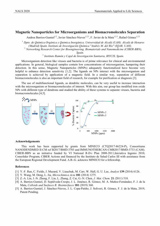
NALS 2020 Nanomaterials Applied to Life Sciences
Magnetic Nanoparticles for Microorganisms and Biomacromolecules Separation
Andrea Barrios-Gumiel1,2, Javier Sánchez-Nieves1,2,3, F. Javier de la Mata1,2,3, Rafael Gómez1,2,3 1 Dpto. de Química Orgánica y Química Inorgánica. Universidad de Alcalá (UAH). Alcalá de Henares
(Madrid) Spain. Instituto de Investigación Química "Andrés M. del Río" (IQAR, UAH). 2 Networking Research Center for Bioengineering. Biomaterials and Nanomedicine (CIBER-BBN),
Spain. 3 Instituto Ramón y Cajal de Investigación Sanitaria, IRYCIS, Spain.
Microorganism detection like viruses and bacteria is of prime relevance for clinical and environmental applications. In general, biological samples contain low concentrations of microorganism, hampering their detection. In this sense, Magnetic Nanoparticles (MNPs) adequately functionalized have become very helpful to enhance detection sensitivity [1,2]. The ligands on NPs interact with the microorganism and separation is achieved by application of a magnetic field. In a similar way, separation of different biomacromolecules is also an important field of research, for example for purification or diagnosis [3].
The use of multifunctional ligands, as dendritic molecules, can be very useful to increase interaction with the microorganism or biomacromolecules of interest. With this aim, our group has modified iron oxide NPs with different type of dendrons and studied the ability of these systems to separate viruses, bacteria and biomacromolecules [4,5].
Acknowledgements
This work has been supported by grants from MINECO (CTQ2017-86224-P), Consortiums NANODENDMED II-CM ref B2017/BMD-3703 and IMMUNOTHERCAN-CMB2017/BMD-3733 (CAM). CIBER-BBN as an initiative funded by VI National R-D-i Plan 2008-2011,Iniciativa Ingenio 2010, Consolider Program, CIBER Actions and financed by the Instituto de Salud Carlos III with assistance from the European Regional Development Fund. A.B.-G. acknows MINECO for a fellowship.
References
[1] Y.-F. Ran, C. Fields, J. Muzard, V. Liauchuk, M. Carr, W. Hall, G. U. Lee, Analyst 139 (2014) 6126. [2] Y. Wang, M. Deng, L. Jia, Microchimica Acta 181 (2014) 1275. [3] Z.-A. Lin, J.-N. Zheng, F. Lin, L. Zhang, Z. Cai, G.-N. Chen, J. Mat. Chem. 21 (2011) 518. [4] A. Barrios-Gumiel, D. Sepúlveda-Crespo, J. L. Jiménez, R. Gómez, M. A. Muñoz-Fernández, F. J. de la
Mata, Colloids and Surfaces B: Biointerfaces 181 (2019) 360. [5] A. Barrios-Gumiel, J. Sánchez-Nieves, J. L. Copa-Patiño, J. Soliveri, R. Gómez, F. J. de la Mata, 2019,
Patent Pending.

Standarization Presentations
NALS 2020 Nanomaterials Applied to Life Sciences
A critical analysis on the in vivo reliability of luminescent nanothermometers
Yingli Shen, José Lifante, Nuria Fernández, Erving Ximendes, Daniel Jaque
Fluorescence Imaging Group, Universidad Autónoma de Madrid, C/Francisco Tomás y Valiente 7, Madrid 28049, Spain
Luminescent nanothermometers (nanoparticles with a temperature dependent luminescence) have demonstrated to be capable of accurate, fast, and contactless thermal sensing in a great variety of scenarios. They have been used, for instance, in areas such as nanofluids, integrated electronics and photonics circuits. It was in biomedicine, however, where their real potential was demonstrated. Though the initial applications of luminescent nanothermometers were restricted to intracellular thermal sensing at the in vitro level, the emergence of infrared emitting nanothermometers has extended their applicability to the preclinical phase. They have made possible the addressing of challenges previously thought to be unattainable with conventional technologies. Some examples would include in vivo intratumoral thermal reading, contactless brain thermometry and early tumor detection.
In vivo luminescence nanothermometry is based on the assumption that all the changes observed in the luminescence of the nanoparticles are correlated with variations in the temperature. In this work, we demonstrate that this is a too optimistic simplification of the problem and that reality is much more complicated. By using the state of the art technology (in vivo infrared hyperspectral imaging), we demonstrate that the non-flat absorption of tissues in the biological windows lead to significant distortions in the spectral features of luminescent nanothermometers. The fluorescence-tissue interaction causes spectral changes not at all correlated with temperature variations. This, in turns, leads to erroneous thermal readouts. The work includes in vivo and ex vivo experiments demonstrating that the spectral distortions due to the wavelength-dependent interaction between light and tissues affect all the infrared luminescent nanothermometers used up to date, independently on their working mode (spectral shift, emitted intensity or ratiometric). It demonstrates, for the first time, that, due to the fluorescence-tissue interaction, infrared luminescent nanothermometers are not as reliable as previously thought. It also provides the scientific community with different hints to improve the reliability of in vivo luminescence thermometry in future studies..
NALS 2020 Nanomaterials Applied to Life Sciences
Specific absorption rate dependence on temperature in magnetic field hyperthermia measured by AC magnetometry
Eneko Garaio1,3, Olivier Sandre2, Juan-Mari Collantes3, Jose Angel Garcia4, Stéphane Mornet5 and Fernando Plazaola3
1Departamento de Ciencias, Universidad Pública de Navarra, 31006 Pamplona, Spain 2Laboratoire de Chimie des Polymères Organiques, UMR 5629 CNRS / Université de Bordeaux, France
3Elektrizitatea eta Elektronika Saila, UPV/EHU, P.K. 644, Bilbao, Spain 4Fisika Aplikatua II Saila, UPV/EHU, P.K. 644, Bilbao, Spain
5Institut de Chimie de la Matière Condensée de Bordeaux, UPR 9048 CNRS / Université de Bordeaux, France
The specific absorption rate (SAR) value is defined as the power transformed into heat per unit of mass of nanoparticles (NPs). The SAR drastically depends on external parameters such as the frequency and intensity of the applied magnetic field, as well as on the internal characteristics of the NPs, i.e. size, shape, material, agglomeration state and even on properties (e.g. viscosity or thermal) of the dispersion medium [1,2]. Moreover, Moreover, a much less known fact is that SAR can also depend on temperature for superparamagnetic (SPM) NPs. However, experimental studies of the temperature-dependency of SAR are rather scarce [3,4].
In the present work, a previously tested lab-made AC magnetometer giving results compatible with the calorimetric method but with a higher sensitivity [5] is used to study the thermal dependence of SAR values of different nanoparticle batches dispersed in water. The 𝑆𝑆𝑆𝑆𝑆𝑆 variations with temperature (𝛥𝛥𝑆𝑆𝑆𝑆𝑆𝑆𝑇𝑇 ) of SPM maghemite NPs of different size distributions have been measured in a wide applied magnetic field frequency (from 75 to 1030 kHz) and intensity (up to 25 kA/m) ranges. The observed thermal behaviour is clearly size dependent: for small particles, absorption rate decreases with temperature whereas for the larger particles, it increases. The thermal behaviour of SPM samples is successfully explained considering Brown and Néel relaxation times.
The obtained results indicate clearly that SAR values are dependent on temperature. The temperature dependence of SAR implies some experimental impact on medical practice for tumour ablation. The heating efficiency of iron oxide NPs can vary by up to 16% between 25 °C and 45 °C, as reported in the present work. Therefore, the SAR values measured at typical hyperthermia temperatures above the physiological temperature (from 41 to 46 °C) have to be considered, rather than SAR values measured at room temperature (25 °C). On the other hand, the thermal dependence of SAR could explain some discrepancies between values reported in the literature by different groups on analogous samples.
Acknowledgements
We thank the Nanotex Company in Madrid for providing us with sample IV. We also want to acknowledge the Basque government for financial support under grant IT-1005-16. Additionally, we thank the European COST action TD1402 RADIOMAG. The authors thank I Orue for helpful comments on this work.
References [1] Lima E Jr., Torres T E, Rossi L M, Rechenberg H R, Berquo T S, Ibarra A, Marquina C, Ibarra M R and Goya G F 2013 Size dependence of the magnetic relaxation and specific power absorption in iron oxide nanoparticles J. Nanoparticle Res. 15 1–11 [2] Rosenweig R E 2002 Heating magnetic fluid with alternating magnetic field J. Magn. Magn. Mater. 252 370 [3] Andreu I, Natividad E, Solozábal L and Roubeau O 2015 Nano-objects for Addressing the Control of Nanoparticle Arrangement and Performance in Magnetic Hyperthermia ACS Nano 9 1408–1419 [4] Garaio E, Sandre O, Collantes J-M, Garcia J A, Mornet S and Plazaola F 2015 Specific absorption rate dependence on temperature in magnetic field hyperthermia measured by dynamic hysteresis losses (AC Magnetometry) Nanotechnology 26 015704 [5] Garaio E, Collantes J M, Garcia J A, Plazaola F, Mornet S, Couillaud F and Sandre O 2014 A wide-frequency range AC magnetometer to measure the specific absorption rate in nanoparticles for magnetic hyperthermia J. Magn. Magn. Mater. 368 432–7
NALS 2020 Nanomaterials Applied to Life Sciences
Continuously manufactured single core iron oxide nanoparticles for cancer theranostics
A. Baki1, O. Kosch2, H. N. Onyema1, A. Remmo2, F. Wiekhorst2, R. Bleul1,* 1Fraunhofer Institut für Mikrotechnik und Mikrosysteme IMM, Mainz, Germany
2 Physikalisch-Technische Bundesanstalt, Berlin, Germany Cancer is still one of the leading causes of death worldwide responsible for an estimated over one million deaths in 2018. Even though many advances in both cancer diagnosis and cancer therapy have been achieved, aiming for early diagnosis and precise treatment at the right time and proper dose, theranostic nanoparticles hold the potential to revolutionize future cancer treatment. They are multifunctional nanosystems, designed for a more specific and personalized disease management combining diagnostic and therapeutic capabilities in one single biocompatible and biodegradable nanoparticle. Present cancer theranostic approaches offering real-time cancer therapy monitoring are not in clinical practice yet. The translation of existing promising research approaches into clinical practice often fails because of the far too high complexity of sophisticated nanosystems raising huge issues already in reproducibly manufacturing of sufficient amounts for comprehensive preclinical testing. Magnetic particle imaging (MPI) is a novel imaging technology with potential for cancer diagnosis using magnetic nanoparticles as tracer material. MPI is in a preclinical state with further demand to improve both the MPI scanner infrastructure and the imaging performance of the tracer materials. Theoretical models suggest single-core iron oxide nanoparticles of about 30 nm core diameter as optimal MPI tracer. These particles are not easily accessible by standard synthesis methods like coprecipitation of iron salts, the technically more demanding thermal decomposition or the biotechnological production with the latter suffering from toxic or immunogenic residues inhibiting an in-vivo application. Here, we report the continuous manufacturing of magnetic single core iron oxide nanoparticles in a microfluidic system, using biocompatible educts and an aqueous synthesis route [1]. We succeeded to produce size controlled single-core iron oxide nanoparticles with a core diameter of about 30 nm, showing high MPI signal amplitudes (2.5 fold of Resovist® the present MPI gold standard tracer) as promising MPI tracer material. Furthermore, we evaluated the therapeutic potential of these particles in magnetic fluid hyperthermia and determined the SAR values (ACHyster 1.4.0, Nanotech Solutions SL, Spain) where we observed values up to 1 kW/(g(Fe) (for fex=300 kHz, Hex=20 kA/m). This is already a quarter of the theoretically achievable SAR, assuming magnetite bulk values for saturation magnetization (MS= 480 kA/m and coercivity Hc=30 kA/m) and exceeds the comparable SAR value of Resovist® by more than factor 3. Our microtechnological approach provides reproducible, scalable single core iron oxide nanoparticles as highly performing tracers for MPI diagnosis as well as efficient heat generators for hyperthermia therapy. These preliminary results contribute to translational research in image guided cancer treatment [2]. Further investigations will focus in MPI imaging of the interaction of our magnetic nanoparticles with a biological environment and with cancer cells.
Acknowledgements
We gratefully acknowledge financial support from European Regional Development Fund (EFRE). R.B. thanks the Fraunhofer-Gesellschaft for the support within the Fraunhofer TALENTA program.
References
[1] A. Baki, N. Löwa, R. Thiermann, C. Bantz, M. Maskos, F. Wiekhorst, R. Bleul. Continuous synthesis of single core iron oxide nanoparticles for MPI tracer development. IJMPI, 3(1) (2017).
[2] R. Bleul, R. Thiermann, G. U. Marten,M. J. House, T. G. St. Pierre,U. O. Häfeli and M. Maskos. Continuously manufactured magnetic polymersomes –a versatile tool (not only) for targeted cancer therapy, Nanoscale 5 (2013) 11385-11393.
NALS 2020 Nanomaterials Applied to Life Sciences
Towards a novel biocompatible probe allowing for real-time temperature measurements at cellular scale
Lise Abiven1,2,3, Corinne Chanéac1, Bruno Viana2, Florence Gazeau3, Thomas Lécuyer3,4, Cyrille Richard4, Victor Castaing2, Estelle Glais1 1. Sorbonne Université, CNRS, Collège de France, Laboratoire de Chimie de la Matière Condensée de Paris, 4 Place
Jussieu, 75005 Paris, France 2. Chimie Paris, PSL Research University, CNRS, 11 Rue Pierre et Marie Curie, 75005 Paris, France
3. Université Paris Diderot, CNRS, Laboratoire Matière et Systèmes Complexes, 10 rue Alice Domon et Léonie Duquet, 75013 Paris, France
4. Université Paris Descartes, Faculté de Pharmacie, CNRS, Unité de Technologies Chimiques et Biologiques pour la Santé, 4 Avenue de l’observatoire, 75006 Paris, France
Biocompatible nanoheaters are of great interest as they have the potential to induce low and localized
temperature gradients within malignant cells. Temperature monitoring at cellular scale may significantly improve tumour response to current cancer-treatment gold standards, while reducing their invasiveness [1]. To investigate this potential, a novel probe allowing for cellular scale temperature gradient measurements is required. Indeed, both thermal and spatial resolutions of currently-available thermometers prevent from doing such measurements.
This novel temperature probe could surpass hyperthermia-therapy application and allow for early-stage diagnosis of diseases. Indeed, abnormal temperature distribution at cellular scale is the first manifestation of health disorders [2].
Silver sulphide quantum dots (Ag2S Qds) were synthetized following two main routes and evaluated as potential nanothermometers to monitor hyperthermia therapy at cellular scale. Quantum dots’ emission spectrum consists in a fingerprint of particles’ surrounding temperature. Indeed, temperature increase strongly quenches emission, allowing for concentration independent temperature sensing based on ratiometric measurements. Nevertheless, emission spectra shape is strongly impacted by interactions between emitters and biological medium (e.g photon reabsorption) [3], preventing to perform one universal temperature probe calibration.
The talk details a method to figure out the more appropriated luminescent parameter for in-vitro and in-vivo Ag2S-Qds-based temperature reading. Luminescent parameter selection is of crucial importance to perform a robust probe calibration and to move toward low incertitude on nanothermometer measurement. Our experimental approach involves different Ag2S-Qds-containing biological media and the study of corresponding emission spectra thermal dependency for in-situ temperature going from 25°C to 50°C.
References [1] D. Ruiz et al, Adv. Funct. Mater., 27 (2017) 1604629. [2] M. Karnebogen et al., Thermochim. Acta, 229 (1993) 147-155. [3] S. Xu et al., Trends in Analytical Chemistry, 80 (2016) 149–155.
NALS 2020 Nanomaterials Applied to Life Sciences
Quantitative magnetic immunochromatographic test to detect histamine in wine
Amanda Moyano1,2*, María Salvador2,3, J. C. Martínez-García2, Vlad Socoliuc4, Ladislau Vékás4, Davide Peddis3, Miguel A. Alvarez5, María Fernández5, Montserrat Rivas2, M. C. Blanco-López1
1 Department of Physical and Analytical Chemistry, University of Oviedo, c/ Julián Clavería 8, 33006 Oviedo, Spain
2 Department of Physics, University of Oviedo, 33204 Gijón, Spain 3 Institute of Structure of Matter (CNR), 00015 Monterotondo Scalo, RM, Italy
4 Laboratory of Magnetic Fluids, Center for Fundamental and Advanced Technical Research, Romanian Academy – Timisoara Branch, 300223 Timisoara, Romania
5 Dairy Research Institute of Asturias, IPLA (CSIC), Paseo Río Linares s/n, 33300 Villaviciosa, Asturias, Spain
Histamine is a biogenic amine produced by bacteria and yeasts due to the microbial decarboxylation of its amino acids in fermented foodstuffs such as wine. A high intake of this compound may produce toxicological effects in humans, especially in individuals with a reduced capacity to catabolise extrinsic histamine. In addition, a high level of histamine can also affect the organoleptic quality of the wine. Different methods have been reported to detect histamine, but they require expensive instrumentation and they are time-consuming.
In this work, we propose a novel approach based on a rapid and simple magnetic lateral flow immunoassay test to detect and quantify histamine in the concentration range of interest for wines [1]. The immunoassay developed follows a competitive configuration at dipstick format. Firstly, we have developed the test using superparamagnetic nanoparticles coated with a double layer of oleic acid as labels. They were functionalized with protein A/G and the conjugation process was confirmed using Dynamic Light Scattering (DLS). Then, the system was calibrated with standards of different concentrations of histamine. For quantification purposes, the immunoassay was coupled to an inductive sensor capable to quantify the magnetic moment of those nanoparticles. In order to validate the magnetic measurements, a commercial reader based on reflectance measurements was used. The lateral flow system has a limit of detection of 1.2 and 1.5 mg/L for the inductive and optical readers, respectively. Finally, the system has been tested with red wine samples at different processing points (at the end of alcoholic fermentation, at the end of malolactic fermentation, in freshly bottled wine, and in reserve wine). The results were statistically equivalent to the concentration obtained by ultra-high-performance liquid chromatography (UHPLC).
Acknowledgements
This work was supported by the Spanish Ministry of Economy and Competitiveness under projects MAT2017-84959-C2-1-R, and the Consejería de Economía y Empleo del Principado de Asturias under the Grant IDI/2018/000185. This study was also financied by Europa Investigación (ref: EUIN2019-103338).
References
[1] Moyano, A.; Salvador, M.; Martínez-García, J.C.; Socoliuc, V.; Vékás, L.; Peddis, D.; Alvarez, M.A.; Fernández, M.; Rivas, M.; Blanco-López, M.C. Magnetic immunochromatographic test for histamine detection in wine. Anal. Bioanal. Chem. 2019, 411, 6615–6624.
NALS 2020 Nanomaterials Applied to Life Sciences
The enhanced magnetic heating of iron oxide nanoflowers explained by small-angle neutron scattering
Philipp Bender1*, Dirk Honecker1, Luis Fernández Barquín2, Andreas Michels1 1University of Luxembourg, Luxembourg
2Universidad de Cantabria, Santander
Iron oxide nanoflowers are densely packed aggregates of ferrimagnetic iron oxide nanocrystallites (Figure 1(a)). They have remarkable magnetic properties and excel at magnetic hyperthermia, especially when using alternating magnetic fields with high frequencies but low amplitudes [1]. By combining spin-resolved small-angle neutron scattering (SANS) experiments with conventional magnetometry of a dilute colloidal dispersion of nanoflowers, we could verify that the particles are at first approximation single-domains (i.e., they are preferentially magnetized along one direction) but with a significant internal spin disorder, which can be attributed to grain boundaries and other structural defects [2]. We assume that this disorder enhances their response to alternating fields in the high-frequency range compared to defect-free particles, which explains their outstanding magnetic heating behaviour.
Figure 1: (a) Transmission electron microscopy image of two small clusters of iron oxide nanoflowers. (b) Purely magnetic SANS pattern of the powder sample of the nanoflowers [4]. (c) P(q) is the particle form factor of the individual nanoflowers which we determine from the purely nuclear SANS signal of the colloidal dispersion [2]. S(q) is the nuclear structure factor of the powder sample. ��𝑀𝑥𝑥 and ��𝑀y are the purely magnetic SANS cross sections of the powder along the x-direction (parallel to neutron beam) and the y-direction (in detector plane). Their increase for q → 0 in comparison to P(q) implies a ferromagnetic-like coupling between the moments of neighboring nanoflowers.
Sakellari et al. [3] could show that in some cases the magnetic heating of nanoflowers can be further enhanced by increasing the particle concentration and thus the interparticle interactions. This is a surprising result and in contrast to other nanoparticle ensembles for which increasing interactions result in a decrease of the hyperthermia performance. To investigate the interparticle correlations in the powder of the nanoflowers, we performed a spin-resolved SANS experiment [4]. Analysis of the nuclear SANS data shows that the nanoflowers are agglomerated to large clusters within the powder. The magnetic scattering contributions then indicate that the moments between neighboring particles within these clusters are preferentially aligned parallel to each other (Figure 1(b) and (c)). We interpret this as evidence for a supraferromagnetic magnetization state within the clusters, which possibly explains the intriguing result in Sakellari et al. that an agglomeration of the nanoflowers can further enhance their magnetic hyperthermia performance.
References
[1] Lartigue et al., ACS Nano 6, 10935-10949 (2012). [2] Bender et al., J. Phys. Chem C 122, 3068-3077 (2018). [3] Sakellari et al., Mater. Sci. Eng., C 58, 187-193 (2016). [4] Bender et al., Appl. Phys. Lett. 115, 132406 (2019).

Nanotherapy 1 Presentations
NALS 2020 Nanomaterials Applied to Life Sciences
A platform for nanoparticle localisation, remote thermometry and hyperthermia therapy application using Lissajous scanning Magnetic Particle Imaging
J. Wells1, S. Twamley2, H. Paysen1, O. Kosch1, A. Ludwig2 and F. Wiekhorst1 1Physikalisch-Technische Bundesanstalt, Abbestraße 2, 10587 Berlin, Germany
2Center for Cardiovascular Research, Charité University Hospital, Berlin, Germany
Localized hyperthermia actuated by magnetic nanoparticles (MNP) accumulated within tumor tissue has shown great promise as a precise and minimally damaging cancer therapy. The technique has received much attention in recent years, with a concerted effort underway to improve aspects including the MNP heating efficiency, administration routes and in-vivo dosimetry. Here, we present experimental studies demonstrating the opportunities offered by the novel biomedical imaging technique “Magnetic particle imaging” (MPI) in supporting the implementation of magnetic field hyperthermia (MFH) therapy.
We present results showing how MPI employs spatially resolved dynamic magnetization measurements to reconstruct a 3D map of a nanoparticle distribution. In addition, we demonstrate that the imaging signal contains information about the local environment of the nanoparticles, including their temperature. Furthermore, we present measurements proving that the complex arrangement of magnetic fields within the MPI scanner can be used to enhance the specific absorption rate of MNP. To investigate the influence of MPI scanner-controlled MFH therapy on the viability of THP-1 leukemia monocytes, in vitro studies were conducted using both MTT assays and flow cytometry to differentiate live and dead cell populations. The results demonstrate effective (60 %) thermoablation of cancer cell populations, using protracted exposure to the magnetic fields present within the MPI scanner to induce MNP heating and cell death.
In conclusion, we present the capability of MPI to provide multi-functional support for MFH therapy by verifying the location of MNP within the tumor, monitoring of the nanoparticle temperature for dosimetry control, and application of the therapeutic excitation field necessary to induce cell death within tumor tissue.
(a) MPI image of MNP-cell mixture. (b) Sample temperature and MPI signal amplitude change over time during MPI field exposure. (c) Cell viability results during 24 hours following 80 minute MPI exposure.
Acknowledgements
This project was supported by the DFG research grants “quantMPI: Establishment of quantitative Magnetic Particle Imaging (MPI) application oriented phantoms for preclinical investigations” (grant TR 408/9-1), and “Matrix in Vision” (SFB 1340/1 2018, no 372486779, projects A02 and B02).
References 1. H. Paysen, J. Wells, O. Kosch, U. Steinhoff, J. Franke, F. Wiekhorst, 2018 Phys. Med. Biol. 63 13NT02 2. J. Wells, H. Paysen O. Kosch, F. Wiekhorst AIP Advances 8, 056703 (2018) *[email protected]
NALS 2020 Nanomaterials Applied to Life Sciences
Barium ferrite nanoparticles labelled with 223
Ra:
a new potential radiobioconjugate for alpha-radioimmunotherapy
W.Gawęda1*,
A.Morgenstern2, F.Bruchertseifer
2, M. Pruszyński
1, M. Rodak
1, A.Bilewicz
1
1Institute of Nuclear Chemistry and Technology, Warsaw, Poland
22European Commission, Joint Research Centre (JRC), Karlsruhe, Germany
Alpha-radioimmunotherapy involves the use of radiolabelled targeting vector – an antibody - for
selective delivery of toxic amount of radiation to tumour cells. Alpha particle emitting radioisotopes are in
considerable interest for radioimmunotherapy because of their high cytotoxicity and short path length.
Unfortunately, many available alpha emitters have limited application due to their low availability and a very
high price.
223Ra, as radium chloride, is the first commercially and widely used α-radiopharmaceutical. Radium has
higher availability and can be easily obtained from the 227
Ac/223
Ra generator. However, Ra2+
cation, as a
member of Alkaline Earth metals, forms very weak complexes. There is a lack of chelator which can
effectively bind 223
Ra to the targeting biomolecule.
In our studies we proposed to use barium ferrite (BaFe12O19) nanoparticles as multifunctional carriers
for 223
Ra radionuclide for alpha-radioimmunotherapy. Barium ferrite nanoparticles labelled with 223
Ra were
synthesized with a modified autoclave method described by Drofenik et al [1]. Yield of labelling was about
70% (for 150kBq 223
Ra). Stability of the obtained radioactive nanoparticles was tested in various biological
solutions: 1 mM PBS, 0.9% NaCl and in human blood serum. It is confirmed that 223
Ra was highly retained
inside nanoparticles in every tested solution.
Obtained magnetic BaFe12O19 nanoparticles were characterized by transmission emission microscopy
and dynamic light scattering. The diameter of synthesized nanoparticles was about 15-30 nm and the
determined magnetization of nanoparticles in room temperature was about 42 emu/g.
In order to synthesize a radiobioconjugate having affinity to HER2 receptors, the monoclonal antibody
trastuzumab was conjugated to the obtained barium ferrite nanoparticles. Firstly, the surface of barium ferrite
nanoparticles was modified with 3-phosphonopropionic acid (CEPA) linker using a method described by
Mohapatra et al [2], and then, the monoclonal antibodies were coupled to the barium ferrite nanoparticles
using the carbodiimide chemistry.
The obtained [223
Ra]BaFe12O19-CEPA-trastuzumab radiobioconjugate almost quantitatively retains 223
Ra and majority of the daughter products. In vitro biological studies indicate that [223
Ra]BaFe12O19-CEPA-
trastuzumab radiobioconjugate exhibits high affinity and cytotoxicity to the to the SKOV-3 ovarian cell line.
Acknowledgements
This work was supported by National Science Centre of Poland (Grant OPUS 2016/21/B/ST4/02133 and
Preludium 2015/17/N/ST4/03943).
This work was supported by COST Action CA 17140 "Cancer Nanomedicine from the Bench to the
Bedside" with ITC CG (COST - European Cooperation in Science and Technology).
References
[1] M.Drofenik et al Mat.Chem.Phys. 2011; 127: 415–419
[2] S.Mohapatra et al Nanotechnology 2007; 18: 385102–11
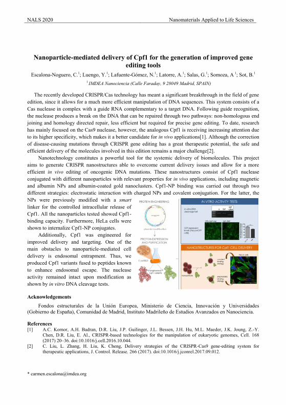
NALS 2020 Nanomaterials Applied to Life Sciences
Nanoparticle-mediated delivery of Cpf1 for the generation of improved gene editing tools
Escalona-Noguero, C.1; Luengo, Y.1; Lafuente-Gómez, N.1; Latorre, A.1; Salas, G.1; Somoza, A 1; Sot, B.1 1 IMDEA Nanociencia (Calle Faraday, 9 28049 Madrid, SPAIN)
The recently developed CRISPR/Cas technology has meant a significant breakthrough in the field of gene edition, since it allows for a much more efficient manipulation of DNA sequences. This system consists of a Cas nuclease in complex with a guide RNA complementary to a target DNA. Following guide recognition, the nuclease produces a break on the DNA that can be repaired through two pathways: non-homologous end joining and homology directed repair, less efficient but required for precise gene editing. To date, research has mainly focused on the Cas9 nuclease, however, the analogous Cpf1 is receiving increasing attention due to its higher specificity, which makes it a better candidate for in vivo applications[1]. Although the correction of disease-causing mutations through CRISPR gene editing has a great therapeutic potential, the safe and efficient delivery of the molecules involved in this edition remains a major challenge[2].
Nanotechnology constitutes a powerful tool for the systemic delivery of biomolecules. This project aims to generate CRISPR nanostructures able to overcome current delivery issues and allow for a more efficient in vivo editing of oncogenic DNA mutations. These nanostructures consist of Cpf1 nuclease conjugated with different nanoparticles with relevant properties for in vivo applications, including magnetic and albumin NPs and albumin-coated gold nanoclusters. Cpf1-NP binding was carried out through two different strategies: electrostatic interaction with charged NPs and covalent conjugation. For the latter, the NPs were previously modified with a smart linker for the controlled intracellular release of Cpf1. All the nanoparticles tested showed Cpf1-binding capacity. Furthermore, HeLa cells were shown to internalize Cpf1-NP conjugates.
Additionally, Cpf1 was engineered for improved delivery and targeting. One of the main obstacles to nanoparticle-mediated cell delivery is endosomal entrapment. Thus, we produced Cpf1 variants fused to peptides known to enhance endosomal escape. The nuclease activity remained intact upon modification as shown by in vitro DNA cleavage tests.
Acknowledgements
Fondos estructurales de la Unión Europea, Ministerio de Ciencia, Innovación y Universidades (Gobierno de España), Comunidad de Madrid, Instituto Madrileño de Estudios Avanzados en Nanociencia.
References [1] A.C. Komor, A.H. Badran, D.R. Liu, J.P. Guilinger, J.L. Bessen, J.H. Hu, M.L. Maeder, J.K. Joung, Z.-Y.
Chen, D.R. Liu, E. Al., CRISPR-based technologies for the manipulation of eukaryotic genomes, Cell. 168 (2017) 20–36. doi:10.1016/j.cell.2016.10.044.
[2] C. Liu, L. Zhang, H. Liu, K. Cheng, Delivery strategies of the CRISPR-Cas9 gene-editing system for therapeutic applications, J. Control. Release. 266 (2017). doi:10.1016/j.jconrel.2017.09.012.
NALS 2020 Nanomaterials Applied to Life Sciences
Real-time Absolute Temperature Feedback for Fully Controlled in vivo
Photothermal Therapy by Ag2S Nanoparticles
Yingli Shen1, Harrison D. A. Santos 3, , Erving Ximendes 1,2, Antonio Benayas4, Daniel Jaque1,2* 1Fluorescence Imaging Group, Departamento de Física de Materiales – Facultad de
Ciencias, Universidad Autónoma de Madrid, C/Francisco Tomás y Valiente 7, Madrid 28049, Spain.
2Nanobiology Group, Instituto Ramón y Cajal de Investigación Sanitaria, IRYCIS, Ctra. Colmenar km. 9.100, Madrid 28034, Spain.
3Grupo de Nano-Fotônica e Imagens, Instituto de Física, Universidade Federal de Alagoas, Maceió-AL 57072-900, Brazil.
4Department of Physics and CICECO—Aveiro Institute of Materials; University of Aveiro, 3810-193 Aveiro, Portugal
Abstract
During the past decade, nanotechnology has developed rapidly and performed excellent application properties in many fields, such as military, industry, chemistry, agriculture, and biomedicine1–3. Among them, the application of biomedicine arose a great attention because it is closely related to human life and health. Based on this, large quantities of nanoparticles (NPs) are designed for imaging (fluorescence, magnetic resonance image (MRI), computed tomography (CT), etc.), drug delivery and release, and the therapy of malignant tumors (photodynamic therapy and photothermal therapy). Photothermal therapy (PTT), which destroys the tumor cells via injecting the light-heat-convert nanoprobe into living tissues, is considered to be a potential anti-tumor technology. To realize the clinic application of PTT, the penetration depth limitation of a piece tissue and real-time accurate temperature feedback should be taken into account mightily, which means it is quite important to fabricate the proper agents for PTT. The frequent PTT agents, which are investigated mostly, including carbon nanotubes (CNTs), rare-earth-doped materials, polymers, and quantum dots (QDs)4–7. While Ag2S NPs, due to its intense luminescence emitting in NIR-II biological windows (1000-1350 nm), high light-heat-convert efficiency, small-size, non-toxicity in vivo, etc., is attracting a huge investigated interest among the researchers8. In this work, Ag2S NPs are incubated with cancer cells to achieve the homogenous commixture which provides the possibility to cure the tumor completely. Moreover, time evolution of laser-induced tumor temperature increment during photothermal treatment as calculated from the analysis of the peak position (T), emitted intensity (TI), and intensity ratio (TR), while the control experiment is also included by displaying the time evolution of laser-induced tumor heating in absence of Ag2S NPs. The results demonstrate the real-time temperature monitoring within the tumor is possible and effective through the multiparameter (λ, I, and R). In turns, the temperature-change comparisons between intratumoral and surface with or without Ag2S NPs, which are excited by different laser power, signify real-time control of the intratumoral temperature plays a key role during the PTT process.
Acknowledgements (Times New Roman 11 points)
This work was supported by the Spanish Ministry of Economy and Competitiveness under projects MAT2016-75362-C3-1-R, MAT2017-83111R, and MAT2017-85617-R, by the Instituto de Salud Carlos III (PI16/00812), by the Comunidad Autónoma de Madrid (B2017/BMD-3867RENIMCM), and co-financed by the European Structural and investment fond. Additional funding was provided by the European Commission, Horizon 2020 project NanoTBTech, the Fundación para la Investigación Biomédica del Hospital Universitario Ramón y Cajal project IMP18_38 (2018/0265) and also by COST action CM1403. Y. L. S. acknowledges a scholarship from the China Scholarship Council (No. 201806870023). A. B. thanks the European Commission as this project has received funding from the European Union’s Horizon 2020
NALS 2020 Nanomaterials Applied to Life Sciences research and innovation program under the Marie Skłodowska Curie grant agreement no. 709270 “TEMPTATION”.
References
[1] Manjunatha, S. B.; Biradar, D. P.; Aladakatti, Y. R. Nanotechnology and Its Applications in Agriculture: A Review. 29 1, 1–13.
[2] Whitesides, G. M. Nanoscience, Nanotechnology, and Chemistry. Small 2005, 1 (2), 172–179.
[3] Coccia, M.; Finardi, U. Emerging Nanotechnological Research for Future Pathways of Biomedicine. Int. J. Biomed. Nanosci. Nanotechnol. 2012, 2 (3/4), 299.
[4] Liang, C.; Diao, S.; Wang, C.; Gong, H.; Liu, T.; Hong, G.; Shi, X.; Dai, H.; Liu, Z. Tumor Metastasis Inhibition by Imaging-Guided Photothermal Therapy with Single-Walled Carbon Nanotubes. Adv. Mater. 2014, 26 (32), 5646–5652.
[5] Carrasco, E.; Rosal, B. del; Sanz‐Rodríguez, F.; Fuente, Á. J. de la; Gonzalez, P. H.; Rocha, U.; Kumar, K. U.; Jacinto, C.; Solé, J. G.; Jaque, D. Intratumoral Thermal Reading During Photo-Thermal Therapy by Multifunctional Fluorescent Nanoparticles. Adv. Funct. Mater. 2015, 25 (4), 615–626.
[6] Au, K. M.; Lu, Z.; Matcher, S. J.; Armes, S. P. Polypyrrole Nanoparticles: A Potential Optical Coherence Tomography Contrast Agent for Cancer Imaging. Adv. Mater. 2011, 23 (48), 5792–5795.
[7] Rosal, B. del; Carrasco, E.; Ren, F.; Benayas, A.; Vetrone, F.; Sanz‐Rodríguez, F.; Ma, D.; Juarranz, Á.; Jaque, D. Infrared-Emitting QDs for Thermal Therapy with Real-Time Subcutaneous Temperature Feedback. Adv. Funct. Mater. 2016, 26 (33), 6060–6068.
[8] Santos, H. D. A.; Ximendes, E. C.; Cruz, M. del C. I. la; Chaves‐Coira, I.; Rosal, B. del; Jacinto, C.; Monge, L.; Rubia‐Rodríguez, I.; Ortega, D.; Mateos, S.; et al. In Vivo Early Tumor Detection and Diagnosis by Infrared Luminescence Transient Nanothermometry. Adv. Funct. Mater. 2018, 28 (43), 1803924 (1-10).
NALS 2020 Nanomaterials Applied to Life Sciences
Triggering plasmon coupling of near infrared (NIR)-invisible gold nanoparticles by three different strategies for cancer theragnosis
Vladimir Mulens-Arias, PhD1,2*; Alice Balfourier, PhD1; Alba Nicolás-Boluda, MsC1; Florent Carn, PhD1; and Florence Gazeau, PhD1
1Laboratoire de Matière et Systèmes Compléxes, Paris Sorbonne, Paris
2National Center for Biotechnology, Madrid, Spain, 28049
Surface plasmon resonance is conferred by the strong extinction of light (absorbance & scattering) upon the incidence of light of a specific wavelength due to the collective oscillation of conduction electrons. Moreover, plasmon coupling occurs when electromagnetic radiation (light)-driven collective oscillation of conduction electrons from two different nanoparticles harmonize when come close enough, leading to the extinction of light of longer wavelength (first life-compatible near-infrared (NIR) window, i.e., within the range 650-950 nm). Such phenomenon allows the use of nanoparticles for therapeutic purposes as biological tissues exhibit a minimal absorption in the NIR range. Although there are a variety of gold nanoparticle with life-compatible localized surface plasmon such as gold nanostars, nanorods, and nanopyramids, their rather complex and laborious synthesis routes impose a barrier for their use in large scale. Spherical gold nanoparticles however are simpler to produce but with a localized surface plasmon within the visible range (520 nm), which makes them not feasible, in principle, for photothermia. We thus investigated the possible and feasible strategies to induce plasmon coupling on spherical gold nanoparticles based on stable clustering. Due to their characteristics we inferred a quasi-fractal state of the gold nanoclusters. Such quasi-fractal re-arrangements were driven by (1) drug-5-Fluorouacil-, (2) endosomal clustering, and (3) polymer-templated one-pot synthesis. We monitored the formation of as-produced nanoclusters by UV-visible absorbance, small angle X ray scattering (SAXS), cryo-TEM, and studied the impact that clustering process has on NIR laser-triggered hyperthermia both in vitro and in vivo. In all cases, clustering led to a 680/808 nm-wavelength laser-induced heat production by gold nanoparticles. We demonstrated the therapeutic potential of both drug- and polymer-templated gold nanoclusters in a murine colon carcinoma in vivo model when injected intratumorally. When internalized by cells, gold nanoparticles clustered within endosomal compartment displaying a pronounced absorbance band at approximately 520-535 nm and a broad absorbance band falling into the near-infrared window with a variable maximum depending on nanoparticle core diameter, cell type and time after incubation. Noteworthy, nanoparticle internalization led to a fractal re-arrangement of nanoparticles that correlated with the appearance of the secondary surface plasmon resonance peak and thermal conversion efficiency upon an 808 nm laser incidence. In addition, AuNP-loaded cell phantoms produce detectable photo-acoustic signal. Altogether, we proved that NIR-invisible gold nanoparticles can be used for photothermia when properly clustered into stable aggregates.
References [1] V. Mulens-Arias et al; Nanoscale, 2019,11, 3344-3359. [2] V. Mulens-Arias et al ; Biomater. Sci., 2019, 7, 389-408
.
*e-mail presenting author: [email protected]

Synthesis and functionalization Presentations
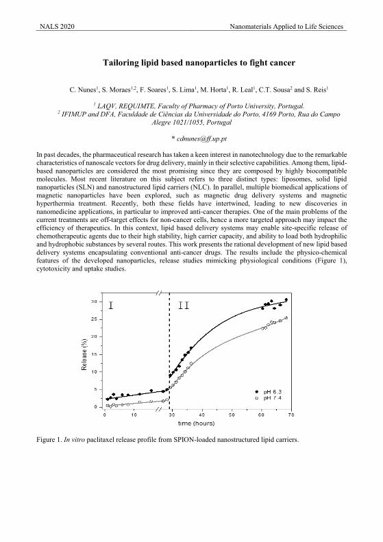
NALS 2020 Nanomaterials Applied to Life Sciences
Tailoring lipid based nanoparticles to fight cancer
C. Nunes1, S. Moraes1,2, F. Soares1, S. Lima1, M. Horta1, R. Leal1, C.T. Sousa2 and S. Reis1
1 LAQV, REQUIMTE, Faculty of Pharmacy of Porto University, Portugal. 2 IFIMUP and DFA, Faculdade de Ciências da Universidade do Porto, 4169 Porto, Rua do Campo
Alegre 1021/1055, Portugal
In past decades, the pharmaceutical research has taken a keen interest in nanotechnology due to the remarkable characteristics of nanoscale vectors for drug delivery, mainly in their selective capabilities. Among them, lipid-based nanoparticles are considered the most promising since they are composed by highly biocompatible molecules. Most recent literature on this subject refers to three distinct types: liposomes, solid lipid nanoparticles (SLN) and nanostructured lipid carriers (NLC). In parallel, multiple biomedical applications of magnetic nanoparticles have been explored, such as magnetic drug delivery systems and magnetic hyperthermia treatment. Recently, both these fields have intertwined, leading to new discoveries in nanomedicine applications, in particular to improved anti-cancer therapies. One of the main problems of the current treatments are off-target effects for non-cancer cells, hence a more targeted approach may impact the efficiency of therapeutics. In this context, lipid based delivery systems may enable site-specific release of chemotherapeutic agents due to their high stability, high carrier capacity, and ability to load both hydrophilic and hydrophobic substances by several routes. This work presents the rational development of new lipid based delivery systems encapsulating conventional anti-cancer drugs. The results include the physico-chemical features of the developed nanoparticles, release studies mimicking physiological conditions (Figure 1), cytotoxicity and uptake studies.
Figure 1. In vitro paclitaxel release profile from SPION-loaded nanostructured lipid carriers.
NALS 2020 Nanomaterials Applied to Life Sciences
Coupling Proteins to Magnetic Nanoparticles by Metal-Chelate Affinity for Tuning their Properties by Magnetic Heating
Ilaria Armenia1,2*, Jesús M. de la Fuente1,2, ,Sabino Veintemillas-Verdaguer3, Fernando López-Gallego4,5, María del Puerto Morales3, Valeria Grazú1,2
1BioNanoSurf Group, Aragon Materials Science Institute (ICMA), CSIC/Universidad de Zaragoza, c/ Edificio I+D, Mariano Esquillor Gomez, 50018, Zaragoza, Spain.
2 Centro de Investigación Biomédica en Red de Bioingeniería, Biomateriales y Nanomedicina (CIBER-BBN), Avenida Monforte de Lemos, 3-5, 28029 Madrid, Spain
3 MamBio Group, Instituto de Ciencia de Materiales de Madrid (ICMM), CSIC, Calle Sor Juana Inés de la Cruz 3, 28049, Madrid, Spain.
4 HetBioCat Group, Universidad de Zaragoza (UNIZAR), Pedro Cerbuna 12, Zaragoza, Spain. 5Current Address: HetBioCat Group, CIC biomaGUNE, Miramon Pasealekua, 182, 20014, Donostia,
Gipuzkoa, Spain.
The biotechnological production of pharmaceuticals and bio-commodities relies on enzymes, but the possibility of an industrial multi-enzymatic process is hampered by several incompatibilities ranging from reaction temperature to unwanted side reactions caused by the high number of intermediates and reaction products that are present [1,2]. To solve this challenge the interdisciplinary FET-OPEN project HOTZYMES proposes to provide the optimal temperature conditions for each reaction in a multi-step-scheme by coupling enzymes to magnetic nanoparticles (MNPs) that can be tune at the nanoscale level by an alternating magnetic field [3,4]. The first step toward this is the production and functionalization of MNPs with optimum properties in term of size and coating. Polyacrylic acid and dimercaptosuccinic acid coated MNPs were selected thanks to their good colloidal stability, functionality to bind enzymes and heating efficiency. Herein, we demonstrate that the further functionalization of these MNPs with NTA-Cu2+ leads to the effective conjugation of an His-tag variant of the superfolded GFP protein, thanks to the high affinity of the copper to the histidine moiety of the protein. Furthermore, using GFP as a molecular thermometer, we demonstrate that the application of an AMF to the different NPs can lead to the generation of different temperature on the MNPs surface; indeed, GFP fluorescence strongly depends on temperature. These first results suggest the feasibility of coupling different His-tagged enzymes to different MNPs capable of generating a temperature gradient if subjected to an alternating magnetic field.
Acknowledgements
The research for this work has received funding from the European Union (EU) project HOTZYMES (grant agreement n° 829162) under EU’s Horizon 2020 Programme Research and Innovation actions H2020-FETOPEN-2018-2019-2020-01. Authors also thank Guangyu MEN for his help on the characterization of MNPs, and Spanish MINECO project BIO2017-84246-C2-1-R and DGA and Fondos Feder (Bionanosurf E15_17R).
References
[1] J.M. Sperl, S. Volker. ACS Catal. 8.3 (2018) 2385. [2] Claassen C, Gerlach T, Rother D. 2019. Stimulus-Responsive Regulation of Enzyme Activity for One-
Step and Multi-Step Syntheses. Adv. Synth. Catal. 361.11 (2019) 2387. [3] A. Riedinger, P. Guardia, A. Curcio, M.A. Garcia, R. Cingolani, L. Manna, T. Pellegrino. Nano Lett.13
(2013) 2399. [4] I. Armenia, V. Grazú, L. de Matteis, P. Ivanchenko, G. Martra, R. Gornati, JM de la Fuente, G.
Bernardini. J. Colloid Interface Sci. 537.1 (2019) 615.
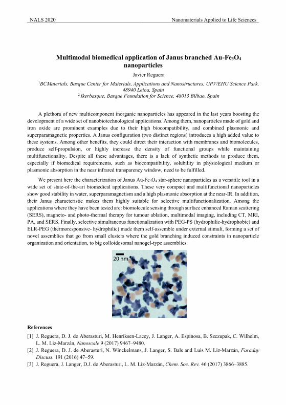
NALS 2020 Nanomaterials Applied to Life Sciences
Multimodal biomedical application of Janus branched Au-Fe3O4 nanoparticles
Javier Reguera 1BCMaterials, Basque Center for Materials, Applications and Nanostructures, UPV/EHU Science Park,
48940 Leioa, Spain 2 Ikerbasque, Basque Foundation for Science, 48013 Bilbao, Spain
A plethora of new multicomponent inorganic nanoparticles has appeared in the last years boosting the development of a wide set of nanobiotechnological applications. Among them, nanoparticles made of gold and iron oxide are prominent examples due to their high biocompatibility, and combined plasmonic and superparamagnetic properties. A Janus configuration (two distinct regions) introduces a high added value to these systems. Among other benefits, they could direct their interaction with membranes and biomolecules, produce self-propulsion, or highly increase the density of functional groups while maintaining multifunctionality. Despite all these advantages, there is a lack of synthetic methods to produce them, especially if biomedical requirements, such as biocompatibility, solubility in physiological medium or plasmonic absorption in the near infrared transparency window, need to be fulfilled.
We present here the characterization of Janus Au-Fe3O4 star-sphere nanoparticles as a versatile tool in a wide set of state-of-the-art biomedical applications. These very compact and multifunctional nanoparticles show good stability in water, superparamagnetism and a high plasmonic absorption at the near-IR. In addition, their Janus characteristic makes them highly suitable for selective multifunctionalization. Among the applications where they have been tested are: biomolecule sensing through surface enhanced Raman scattering (SERS), magneto- and photo-thermal therapy for tumour ablation, multimodal imaging, including CT, MRI, PA, and SERS. Finally, selective simultaneous functionalization with PEG-PS (hydrophilic-hydrophobic) and ELR-PEG (thermoresponsive- hydrophilic) made them self-assemble under external stimuli, forming a set of novel assemblies that go from small clusters where the gold branching induced constraints in nanoparticle organization and orientation, to big colloidosomal nanogel-type assemblies.
References
[1] J. Reguera, D. J. de Aberasturi, M. Henriksen-Lacey, J. Langer, A. Espinosa, B. Szczupak, C. Wilhelm, L. M. Liz-Marzan, Nanoscale 9 (2017) 9467–9480.
[2] J. Reguera, D. J. de Aberasturi, N. Winckelmans, J. Langer, S. Bals and Luis M. Liz-Marzán, Faraday Discuss. 191 (2016) 47–59.
[3] J. Reguera, J. Langer, D.J. de Aberasturi, L. M. Liz-Marzán, Chem. Soc. Rev. 46 (2017) 3866–3885.
NALS 2020 Nanomaterials Applied to Life Sciences
Peptide-driven biomimetic conjugation of oxide nanocomposites Marco Peiteado*, Teresa Jardiel, Ana Castellanos-Aliaga, Amador C. Caballero, David G. Calatayud
Department of Electroceramics, Instituto de Cerámica y Vidrio (CSIC), Kelsen 5, 28049 Madrid, Spain
Nanotechnology is rapidly evolving towards the manufacture of complex materials with very defined structure and properties. The past two decades have seen impressive progress in the way we can shape, position and organize matter at the nanometer scale. For example, with recent advances in deposition techniques, artificial multi-functional devices involving oxide-based arrays and nanostructures are now fabricated worldwide, continuously improving the quality of our daily lives. However, this incessant updating is supported on manufacturing processes entailing a severe consumption of energy, so unavoidably exert a harmful contribution to the global climates on earth. Within this framework, we have been dealing with a completely novel and innovative approach based on biomimetics, combining growth techniques commonly used in the field of bioengineering and different methods of nanomaterials processing in mild conditions. In what could be considered as a molecular approach to nanotechnology, the biomimetic premise lies in the fact that peptides genetically designed to recognise inorganic surfaces, could be used as binding molecules and/or growth units to allow the orderly self-assembly of nanomaterials with specific functions. Different competences are here involved which have all been explored to direct the sustainable assembly of heterostructured nanocomposites of different metal-oxide systems, eventually leading to well-defined multifunctional nanoarchitectures with no need for high-energy resources. Additionally, the proposed technology could open up new waves in the field of water remediation, using the conjugating peptides as a sort of “molecular glue” in the bio-assisted removal of oxide nanoparticles from water.
Acknowledgements
Financial support by Fundación General CSIC (Programa ComFuturo) and MAT2016-80182-R project are acknowledged. Dr T. Jardiel also acknowledges the European Science Foundation (ESF) and the Ramón y Cajal Program of Spanish MICINN for the financial support.
*e-mail presenting author: [email protected]
NALS 2020 Nanomaterials Applied to Life Sciences
Role of the number of recognition ligands per particle on the sensitivity of a
novel magnetic detection method of biomarkers E. Sanz-de Diego*1, C.C Vequi-Suplicy1, A. Aires2, J. Requejo-Isidro3,4
A.L. Cortajarena1,2,4,5 and F.J. Teran1,4
1 iMdea Nanociencia, Campus Universitario de Cantoblanco, 28049 Madrid, Spain
2 CIC biomaGUNE, Paseo Miramón 182, 20014 Donostia-San Sebastian, Spain 3 Centro Nacional de Biotecnología (CNB), CSIC, 28049, Madrid
4 Nanobiotecnología (CNB-CSIC-iMdea-Nanociencia), Unidad Asociada al Centro Nacional de Biotecnología (CSIC), 28049 Madrid, Spain
5 Ikerbasque, Basque Foundation for Science, Maria Diaz de Haro 3, 48013 Bilbao, Spain
The conjugation of magnetic nanoparticles (MNPs) with recognition ligands has been widely explored for diagnosis, therapeutic, magnetic separation and/or sensing purposes. Until now, the quantification of ligands attached onto the MNP surface has been roughly estimated out of calculations considering few experimental parameters related to the bioconjugation processes, such as the ligand and MNP masses, or MNP hydrodynamic diameter among others. However, this quantification requires accuracy for some particular applications such as the magnetic detection procedures, where the number of ligands per MNP is a relevant parameter that strongly influences the sensitivity of the technique, and consequently requires to be precisely defined.
Here, we report on experimental study based on single-molecule fluorescence to determine the number of ligands per MNP. For this proof of concept, we have employed commercial iron oxide MNPs onto we anchor few recognition ligands (GST-MEEVF) and Ap-biotin per MNP. These ligands specifically interact with two different fluorescent analytes (VFPm -TPR2-MMY and VFPd -TPR2-MMY). Single molecule optical spectroscopy assays [1] allow to characterize the analyte fluorescence after specific interaction with the recognition ligands attached onto the MNPs immobilized onto an EPOXY layer with discrete streptavidin molecules. From the analytes fluorescence emission, we simultaneously determined i) the number of bound ligands per MNP; ii) the occurrence of unspecific interactions between analytes and MNPs. In summary, the number of ligands per MNP has been quantified by this experimental method. In addition, we have not observed unspecific interactions resulting in analyte adsorption onto MNP surface when analyte concentration goes up to 0.4nM.
The ratio of recognition ligands per MNP is a key parameter to alter mechanisms of magnetic relaxation after specific interaction with analytes. Consequently, magnetic properties are strongly altered, resulting in significant variation of the AC hysteresis loops of the bioconjugated GST-MEEVF MNPs. [2] Another relevant issue is related to modeling the cluster formation. Cluster formation is again modulated by the number of ligands per MNP, among other parameters. Accurate ligand quantification will allow the modeling of MNP cluster formation, resulting in the quantification of analytes specifically interacting with MNPs. The precise analyte quantification required for MNP clustering modeling or any other application is possible thanks the proposed experimental method.
Acknowledgements
E. Sanz-de Diego acknowledges financial support from CAM (PEJ-2017-AI/ BMD-7515) and BIOMAG project (PCI2019-103600).
References [1] J.Choi, R. Grosely, E.V. Puglisi, J.D.Puglisi, COSB. 58 (2019) 233–240.
[2] PCT/EP2018/080662 "Method for detection of an analyte" Inventors: D.Cabrera, A.Aires, E.J.Artés, J.Camarero, A.L.Cortajarena,and F.J. Teran.Owner. iMdea Nanociencia. Date of filling14th November 2018.

Toxicity Presentations
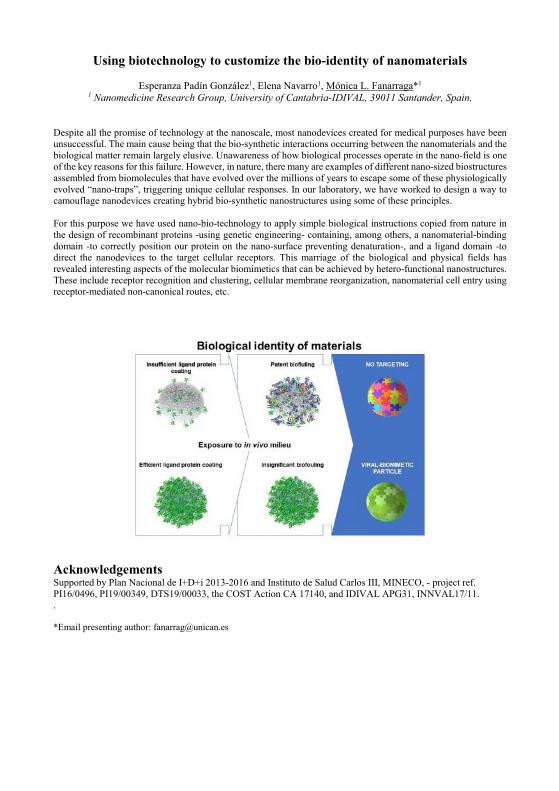
Using biotechnology to customize the bio-identity of nanomaterials
Esperanza Padín González1, Elena Navarro1, Mónica L. Fanarraga*1 1 Nanomedicine Research Group, University of Cantabria-IDIVAL, 39011 Santander, Spain,
Despite all the promise of technology at the nanoscale, most nanodevices created for medical purposes have been unsuccessful. The main cause being that the bio-synthetic interactions occurring between the nanomaterials and the biological matter remain largely elusive. Unawareness of how biological processes operate in the nano-field is one of the key reasons for this failure. However, in nature, there many are examples of different nano-sized biostructures assembled from biomolecules that have evolved over the millions of years to escape some of these physiologically evolved “nano-traps”, triggering unique cellular responses. In our laboratory, we have worked to design a way to camouflage nanodevices creating hybrid bio-synthetic nanostructures using some of these principles. For this purpose we have used nano-bio-technology to apply simple biological instructions copied from nature in the design of recombinant proteins -using genetic engineering- containing, among others, a nanomaterial-binding domain -to correctly position our protein on the nano-surface preventing denaturation-, and a ligand domain -to direct the nanodevices to the target cellular receptors. This marriage of the biological and physical fields has revealed interesting aspects of the molecular biomimetics that can be achieved by hetero-functional nanostructures. These include receptor recognition and clustering, cellular membrane reorganization, nanomaterial cell entry using receptor-mediated non-canonical routes, etc.
Acknowledgements Supported by Plan Nacional de I+D+i 2013‐2016 and Instituto de Salud Carlos III, MINECO, ‐ project ref. PI16/0496, PI19/00349, DTS19/00033, the COST Action CA 17140, and IDIVAL APG31, INNVAL17/11. . *Email presenting author: [email protected]
NALS 2020 Nanomaterials Applied to Life Sciences
Biocompatibility studies of novel nanostructures electrodes for neural repair in spinal cord injury
Ana Domínguez-Bajo1, Ankor González-Mayorga2, Beatriz L. Rodilla3, 4, Ana Arché-Núñez3, Rodolfo Miranda3, 5, Julio Camarero3, 5, Lucas Pérez3, 4, M. Teresa González3, Elisa López-Dolado2, 6, María
Concepción Serrano1,*
*Correspondence author: [email protected] 1 Instituto de Ciencia de Materiales de Madrid, Consejo Superior de Investigaciones Científicas
(ICMM-CSIC), Calle Sor Juana Inés de la Cruz 3, 28049 Madrid, Spain 2Hospital Nacional de Parapléjicos, Servicio de Salud de Castilla-La Mancha, Finca La Peraleda s/n,
45071 Toledo, Spain 3IMDEA Nanociencia, Calle Faraday 9, Campus Cantoblanco, 28049 Madrid, Spain
4Universidad Complutense de Madrid, Plaza de Ciencias, 1, Ciudad Universitaria 28040 Madrid, Spain
5Facultad de Ciencias, Universidad Autónoma de Madrid, C/ Francisco Tomás y Valiente, 7, Campus Cantoblanco,28049 Madrid, Spain
6 Research Unit of “Design and Development of Biomaterials for Neural Regeneration”, Hospital Nacional de Parapléjicos, Joint Research Unit with CSIC, 45071 Toledo, Spain
Unfortunately, the regeneration of the nervous tissue after a spinal cord injury is still a challenge. Consequently, patients suffering from this pathology have their quality of life and life expectancy dramatically affected [1,2]. Thus, researchers from different disciplines such as Materials Science and Neuroscience are working together to provide more effective therapeutic alternatives. Concretely, in the byAxon project, vertically arranged metallic nanowires made of Ni and Au, or both elements in a core-shell structure, are being developed to work as nanoelectrodes for electrical stimulation directly at the lesion site. These substrates are grown by templated-assisted electrodeposition over a flexible gold base. For this purpose, two different kinds of membranes have been used as a template: a commercial one made of polycarbonate and an aluminium oxide membrane produced in the laboratory, which allows a better control of the positioning and shape of the nanowires. The behaviour of neural cells isolated from the cortex of Wistar rat embryos has been studied in culture to test the biocompatibility of these platforms. Cell adhesion, viability, morphology and differentiation have been studied after 14 days in vitro. Moreover, calcium imaging experiments have been also performed. Results indicate that neural cells are able to form networks mainly composed by neurons in close contact with the metallic nanowires. Moreover, cells show a high viability in all types of nanowires substrates fabricated and the presence of spontaneous calcium transient elevations, which demonstrate that cells remain active on the substrates after 14 days in culture. These findings encourage further investigation for the development of an implant able of working as a local bypass at the spinal cord.
Acknowledgements
This project is funded by the European Union’s Horizon 2020 research and innovation programme under grant agreement No 737116.
References
[1] E.J. Bradbury, S. B. McMahon, Nat. Rev. Neurosci. 7 (2006) 644-653. [2] M. V. Sofroniew, Nature, 557 (2018) 343-350.
NALS 2020 Nanomaterials Applied to Life Sciences
Immunomodulatory properties of graphene-based materials: a key tool for the design of new biomedical applications
Laura Fusco 1,2,3, Marco Orecchioni4, Valentina Bordoni4, Alberto Bianco5, Lucia Gemma Delogu2,6,* 1Dept. of Chemical and Pharmaceutical Sciences, University of Trieste, Via L. Giorgieri 1, 34127, Trieste, Italy;
2Istituto di Ricerca Pediatrica, Fondazione Città della Speranza, Corso Stati Uniti 4, 35127, Padua, Italy; 3Sidra Medical and Research Center, Doha, Qatar;
4Dept. of Biology, University of Sassari, Via Muroni 23, 07100, Sassari, Italy; 5CNRS, Institut de Biologie Molèculaire et Cellulaire, Laboratoire d'Immunologie et Chimie Thérapeutiques,
France; 6Dept. of Biomedical Science, University of Padua, Via Ugo Bassi, 58/B, 35131, Padua, Italy.
Thanks to their outstanding properties, graphene-based materials (GBMs) have generated great expectations in nanomedicine. However, their translation into clinics cannot overlook the evaluation of their impact on the immune system. In this view, we have previously demonstrated the key role exerted by the different physicochemical properties of GBMs on the modulation of their impact on human immune cells [1-4]. Therefore, we here explored the effects of graphene oxide (GO) and amino-functionalized GOs with different lateral sizes ex vivo on human immune cells exploiting innovative technologies, including single-cell mass cytometry (unpublished data). Our results revealed that the amino functionalization was able to enhance the immune compatibility of GO. Moreover, the obtained findings showed a specific M1 like activation of monocytes induced by this material, as well as the secretion of interleukin-4 and Granzyme-B from B cells, skewing a cytotoxic-like response. Furthermore, GO was combined with inorganic quantum dots, allowing the detection of GO cell uptake using single cell mass cytometry. The obtained finding revealed the superior ability of monocytes and, surprisingly, B cells, to internalize GO compared to the other immune cell subpopulations. We finally explored the possible biomedical applications of GO for bone regeneration, combining its immunomodulatory proprieties on monocytes with the osteoinductive capacity of calcium phosphates (CaP) in a new nanomaterial called maGO-CaP (monocytes activator GO complexed with CaP) [5]. Intriguingly, maGO-CaP was able to increase bone regeneration in vivo, thanks to its ability to modulate the immune cell functionality, inducing osteoinductive stimuli. Our results pave the way for the application of well designed and functionalized GBMs as biomedical tools exploiting their immunomodulatory properties for the fight of several pathologies.
Acknowledgements
The authors gratefully acknowledge financial support from MIUR JTC Graphene 2015 (G-IMMUNOMICS project), European Union HORIZON 2020 research and innovation programme under MSCA RISE 2016 project Carbo-Immap grant no. 734381, and the Italian MIUR (PRIN call 2015, project: 2015TWP83Z).
References [1] M. Orecchioni, D.A. Jasim, M. Pescatori, R. Manetti, C. Fozza, F. Sgarrella, D. Bedognetti, A. Bianco, K.
Kostarelos, L.G. Delogu. Advanced healthcare materials. 2 (2016) 276-87. [2] M. Orecchioni, D. Bedognetti, L. Newman, C. Fuoco, F. Spada, W. Hendrickx, F.M. Marincola, F. Sgarrella, F.A.
Rodrigues, C. Ménard-Moyon, G. Cesareni, K. Kostarelos, A. Bianco, L.G. Delogu. Nature Communications 8, (2017).
[3] R. Russier, V. Léon, M. Orecchioni, E. Hirata, P. Virdis, C. Fozza, F. Sgarrella, G. Cuniberti, M. Prato, E. Vazquez, A. Bianco, L.G. Delogu. Angew. Chem. Int. Ed. 56 (2017), 1 – 7.
[4] E. Avitabile, D. Bedognetti, G. Ciofani, A. Bianco, L.G. Delogu. How can nanotechnology help the fight against breast cancer? 10 Nanoscale (2018), 11719-11731.
[5] V. Bordoni, G. Reina, M. Orecchioni, G. Furesi, S. Thiele, C. Gardin, B. Zavan, G. Cuniberti, A. Bianco, M. Rauner, L.G. Delogu. Stimulation of bone formation by monocyte-activator functionalized graphene oxide in vivo. Nanoscale 11 (2019), 19408-19421.
NALS 2020 Nanomaterials Applied to Life Sciences
Reactive oxygen species (ROS) production in human hepatoma cell lines
through the application of localized magnetic hyperthermia. G.R. Iglesias1, A. Sola-Leyva2, M. Ángeles Y. Jabalera3, M.A. Chico Lozano2, M. P. Carrasco-
Jiménez2 and C. Jiménez-Lopez3
1Department of Applied Physics. 2Department of Biochemestry and Molecular Biology.
3Department of Microbiology. 1,2,3 Faculty of Sciences. University of Granada, Av. Fuentenueva s/n 18071.
Granada, Spain
This work is based on the study and evaluation of the production and influence of reactive oxygen species (ROS) on cell death versus the application of variable magnetic fields, mediated by the MamC protein of the magnetosome magnetotactic bacterium Magnetococcus marinus MC-1 [1], both naked and functionalized with antitumor drugs, in human hepatoma cell lines.
Recent studies have shown that the application of magnetic hyperthermia can improve the release of the agent transported (drug) by nanoparticles [2] and thus improve the effectiveness in cell death [3]. However, the increase in temperature by means of low intensity alternating magnetic fields on cell lines has not been studied in detail at present, due to the low concentration of magnetic nanoparticles that can be incorporated into it, in order to avoid high toxicity.
The ROS production plays an important role, the increased level of ROS can cause damages to protein, lead to cellular apoptosis, and contribute to many diseases including cancer. Many recent studies proposed a variety of strategies to either suppress toxicity of ROS generation or exploit the elevated ROS levels for cancer therapy. [4,5].
Therefore, the effect of hyperthermia on cell viability and its relationship or synergy with the production of ROS in combination or not with different antitumor drugs will be present in depth.
Acknowledgements
This research work is supported by Ramón y Cajal programme (RYC-2014-16901) MINECO, Spain, FPU2016 grant (Ref. FPU16-04580CGL2016-76723. Unidad Científica de Excelencia UCE-PP2016-05 (UGR), MINECO, Spain and FEDER, EU.
References
[1] G. García Rubia, A. Peigneux, Y. Jabalera, J. Puerma, F. Oltolina, K. Elert, D. Colangelo, J. Gómez Morales, M. Prat, C. Jimenez-Lopez, Langmuir (2018) 34: 13713–13724.
[2] G Iglesias, F. Reyes-Ortega, B C. Fernandez and A. DelgadoA. Polymer (2018) 10,3: 269 [3] Y. Jabalera, B. Garcia-Pinel, R. Ortiz, G. Iglesias, L. Cabeza, J. Prados, C. Jimenez-Lopez, C. Melguizo.
Pharmaceutics (2019) 11,8:395. [4] Mai, T. & Hilt, J.Z. J Nanopart Res (2017) 19: 253 [5] Akhtar MJ, Ahamed M, Kumar S, Khan MM, Ahmad J, Alrokayan SA Int J Nanomed (2012) 7:845–857
NALS 2020 Nanomaterials Applied to Life Sciences
Targeting the plasma membrane as a secondary target in photodynamic therapy for cancer cells
Sebastian Ariel Thompson1 1IMDEA Nanociencia, Ciudad Universitaria de Cantoblanco, 28049 Madrid
Photodynamic therapy (PDT) is a non-invasive anti-cancer treatment already in clinical applications. The goal of PDT is the induction cancer cell death via the photo-generation of Reactive Oxygen Species (ROS). Recent efforts focusing on understanding the mechanisms of cell death activated by PDT find that it depends on the type of photosensitizer (PS), targeted organelles, and nature of the light used. Glycosylation of the PS targets cancer via saccharide receptors on the cell surface, and is generally assumed that these compounds rapidly internalize and accumulate, e.g. in the endoplasmic reticulum. We will present our results showing that part of the glycosylated chlorin compound residing at the PM of cancer cells can also activate necrosis upon illumination by compromising the PM independently of the length of the incubation period.1 The results presented here show that the PM can also be targeted as a secondary target by glycosylated PS designed to accumulate in internal organelles. PS activation to induce necrosis by compromising the plasma membrane has the benefits of fast cell death and shorter irradiation times. The findings described in this presentation expand our understanding of the cellular damage induced by phototherapies, and can modify the protocols used in clinical/animal models. Please, send the abstract in both, pdf and Word formats.
References
[1] Compromising the plasma membrane as a secondary target in photodynamic therapy-induced necrosis. SA Thompson, A Aggarwal, S Singh, AP Adam, JPC Tome, CM Drain. Bioorganic & medicinal chemistry 26 (18), 5224-5228

Lab/Organ on-a-chip Presentations
NALS 2020 Nanomaterials Applied to Life Sciences
Production of controlled size nanovesicles by microfluidic device Gemma Gutiérrez1, Pablo García-Manrique1,2, María Matos1, Mª del Carmen Blanco2
1Department of Chemical and Environmental Engineering, University of Oviedo, Julián Clavería 8, 33006 Oviedo, Spain.
2Department of Physical and Analytical Chemistry, University of Oviedo, Julián Clavería 8, 33006 Oviedo, Spain.
Nanovesicles (organic colloids) are particles formed by self-assembled amphiphilic molecules into closed bilayered structures with an inner aqueous core. Depending on the chemical nature of bilayer constituents, these particles are categorised into liposomes (lipids), niosomes (non-ionic surfactants) or polymersomes (block copolymers).
Several techniques have been used in order to have nanovesicles of the eligible particle size and reasonable monodispersity [1,2].
Microfluidics technology is very promising for precise control over input variables when mixing chemical species. Other advantages, include low consumption of chemicals (relevant in formulation optimization), scale-up possibilities for industrial production, on-line coupling to other processes (such as purification steps), and efficient control over temperature if required. Previous research works have used this method to examine various colloidal formulations. However, the production of niosomes through microfluidic routes remains less explored. In microfluidics a stream of lipids in organic phase is focussed between two aqueous streams in microchannels under laminar flow conditions, allowing the mixing of chemical species by molecular diffusion [3].
In this work, key operation and formulation parameters, involved in a microfluidic reactor with hydrodynamic flow focusing, were investigated in order to quantify their effects on niosomes morphology. Particular attention was given to temperature, which is both a requirement to handle non-ionic surfactants with phase transition temperature above room temperature, and a tailoring variable for size and monodispersity control. A customised device to control temperature and facilitate visualization of the process was developed, which can be easily coupled with commercial inverted microscopes.
Results demonstrated that niosomes with controlled particle size and required monodispersity can be easily produced by microfluidics technique. Results obtained were compared with those obtained by ethanol injection method.
Acknowledgements
This work was supported by the Ministerio de Economía y Competitividad (MINECO, Spain), under the Grant MAT2017-84959-C2-1-R. This study was also financed by the Consejería de Economía y Empleo del Principado de Asturias (Plan de Ciencia, Tecnología e Innovación 2013-2017), under the Grant IDI/2018/000185. Support from the European Regional Development Fund (ERDF) is gratefully acknowledged. This study was also financied by Europa Investigación (ref: EUIN2019-103338) and European H2020-MSCA-ITN-2018 by the Grant Agreement number 813439
References [1] Marianecci, C., Di, L., Rinaldi, F., Celia, C., Paolino, D., Alhaique, F., Esposito, S., Carafa, M., Adv. Colloid Interface Sci. 205 (2014) 187 [2] García-Manrique, P., M. Matos, G. Gutiérrez, O. R. Estupiñán, M. C. Blanco-López and C. Pazos (2016). Ind. & Eng. Chem. Res 55(34): 9164-9175. [3] García-Manrique, P., Gutiérrez, G., Matos, M., Cristaldi, A., Mosayyebi, A., Carugo, D., Zhang, X., Blanco-López, M.C, Colloids Surf B: Biointerfaces, 182, (2019), a 110378
NALS 2020 Nanomaterials Applied to Life Sciences
Magnetic silica coated iron oxide nanochains as nano-scale antennas, locally amplifying the effects of pulsed electric fields
Jelena Kolosnjaj-Tabi1, *, Slavko Kralj2, Sebastjan Nemec2, Muriel Golzio1, Marie-Pierre Rols1 1 Institute of Pharmacology and Structural Biology, 205 Route de Narbonne, 31400 Toulouse, France
2 Department for Materials Synthesis, Jožef Stefan Institute, Jamova cesta 39, 1000 Ljubljana, Slovenia
Silica coated iron oxide nanochains [1], have excellent magnetic and photothermal properties. Moreover, silica-coated iron oxide nanochains can serve as nano-scale antennas, which locally amplify the electric field, and induce electropermeabilization at low electric field strengths.
Electropermeabilization occurs at the level of the cell membrane, when specific (generally micro- or mili-second) electric field pulses are applied. Pulsed electric fields (PEFs) thus transiently disrupt the permeability of cell membranes, which allows cellular internalization of drugs [2]. Cell permeabilization with PEFs is currently used in electrochemotherapy. While this approach successfully treats melanoma, applied PEFs may induce muscle contraction and cause pain.
In order to decrease the electric field strength required to induce electropermeabilization, and thus decrease adverse reactions associated with high PEFs intensities, we herein suggest the use of magnetically responsive nanochains. We show an increased uptake of a fluorescent dye (propidium iodide) (Figure B), when cells are exposed to both, nanochains and low intensity pulsed electric field. This proof of concept is tested on 2D and 3D cell cultures, and our preliminary results show that the electric field strength can be decreased by several orders of magnitude when cells are co-exposed to pulsed electric fields and with low concentrations of nanochains. The increased electropermeabilization was observed in different cell types (HaCaT, HCT-116 GFP, normal dermal fiborblasts isolated from skin biopsies and HeLa cells). When nanochains were added to cells, the permeabilization could occur at electric field strengths as low as 50 V/cm, while in the absence of nanochains the permeabilization required the electric field strength of 600 V/cm and above. This study indicates that nanomaterials could allow decreasing electric field intensities, and thus reduce the adverse reactions associated to high intensity electric fields.
Figure: A) Transmission electron micrographs showing magnetic nanochains (red arrows and magnified view of the red square) at the surface
of a plated cell (N denotes the cell’s nucleus). B) Bright field (left) and fluorescence micrographs (right) (Mag x 20) showing fluorescent dye (propidium iodide) intake when pulsed electric fields are applied (300 V/cm, 8 pulses lasting 100 µs, applied at a frequency of 1 Hz) in the presence of nanochains (top) while in the absence of nanochains (bottom) the cells do not exhibit electropermeabilization.
Acknowledgements
This research was founded by an institutional grant of the ITMO Cancer AVIESAN (National Alliance for life sciences and Health) within the framework of the cancer Plan NUMEP (PC201615) and the Slovenian Research Agency (ARRS) for research core funding No. (P2-0089) No. (J1-7302 and J3-7494).
References
[1] S. Kralj, D. Makovec. ACS Nano. 9 (2015) 9700. [2] J. Kolosnjaj-Tabi, et al. Adv. Drug Deliv. Rev. 138 (2019) 56.
NALS 2020 Nanomaterials Applied to Life Sciences
Tumour Vessel-on-a-chip for Nanomedicine Development
Sergio Dávila1, §, Jean Cacheux1, §, Isabel Rodriguez1 §These authors contributed equally to this work
1IMDEA Nanoscience C/ Faraday, 9, Campus de Cantoblanco, Madrid, Spain. Tel: 912998869
Today, preclinical models primarily relying on conventional 2D cell cultures or in vivo animal models both of which largely differ from the physiological conditions found in humans. As such, the predictive power of the current preclinical models for assessing the toxicology and efficacy of potential new therapies is limited. In addition, there are rising ethical concerns and regulations on animal testing.
Recently, organ-on-a-chip devices have emerged as new preclinical model. This new technology is poised to transform drug development by improving the effectiveness of clinical trials and reducing the drug screening time. Particularly in nanomedicine research, there is a need for suitable in vitro tumour models for optimization of the nanocarriers pharmacodynamics in order to increase their bio-availability and therapeutic level at the tumour site. This tumour-on-a-chip models consist on micro-engineered platforms that integrate microfluidics and cell biology in order to mimic closely particular physiological scenarios found in tumours. For nanodrug delivery studies, the endothelial tumour microvasculature is an essential element to model [1].
Microfluidics is a well suited technology to mimic blood vessels on chips [1]. Typically microfabrication technologies produce square profile channels [2]. To mimic the round geometry of the blood vessels, some approaches have been implemented [3-4]. However, they are poorly reproducible (i.e. poor control of the microchannel cross section geometry) and often present dimensions on the order of several hundred micrometers.
In this work, we present a new fabrication process to produce artificial microvessels (diameter < 100 µm) with semi-circular geometry. The approach is based on the pressure and the temperature control during the microchannel polymer replication process by thermal nanoimprint. This process allows to control very finely the channel geometry with round shape. The imprinted master channels are finally replicated by soft-lithography in PDMS and sealed against a thin cover glass. Endothelial cells are seeded into these channels which act as the scaffold and artificial vessels are formed as the endothelial cells cover the entire channel lumen. The microvessles are connected to a pressure controller to precisely regulate the fluid flow and shear stress while a blood analogue fluid with shear-thinning behaviour is employed to recapitulate closely the characteristic tumour vascular flow [5]. Transport studies of fluorescent nanoparticles at relevant physiological conditions (in terms of viscosity, temperature and applied pressure) on this artificial micro vessels have been performed.
On the whole, we present a fabrication process that allows to make semi-circular artificial microvessels in a practical and reproducible way. The tumour vessel-on-a-chip device allows for nanoparticle transport studies at different conditions of pressure drop and shear rate, viscosity and temperature.
Acknowledgements
This work was performed within the framework of the EVONANO project. This project has received funding from the European Union's Horizon 2020 FET Open programme under grant agreement. No. 800983. The authors are grateful to M.R. Osorio for his valuable assistance in microfabrication.
References [1] D. Caballero, S. M. Blackburn, M. de Pablo, J. Samitier, and L. Albertazzi. Lab Chip 17 (2017) 3760–3771
[2] B.-H. Jo, L. M. Van Lerberghe, K. M. Motsegood, and D. J. Beebe. J. Microelectromech. Syst. 9(2000)76–81
[3] M. Abdelgawad, C. Wu, W.-Y. Chien, W. R. Geddie, M. A. S. Jewett, and Y. Sun. Lab Chip, 11(2011) 545–551
[4] Pauty J, Usuba R, Cheng IG, Hespel L, Takahashi H, Kato K, et al. EBioMedicine, 27(2018), 225–236
[5] van den B. C. N, P. R. A.A, F. Ole, R. M. C.M, den H. W. F, and van de V. F. N. Biorheology, 6(2008), 651–661
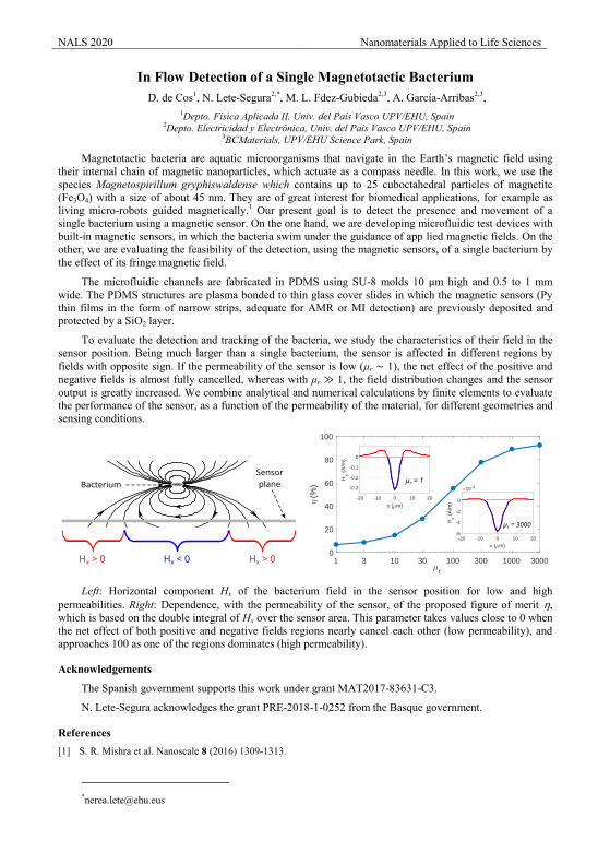
NALS 2020 Nanomaterials Applied to Life Sciences
In Flow Detection of a Single Magnetotactic Bacterium D. de Cos1, N. Lete-Segura2,*, M. L. Fdez-Gubieda2,3, A. García-Arribas2,3,
1Depto. Física Aplicada II, Univ. del País Vasco UPV/EHU, Spain 2Depto. Electricidad y Electrónica, Univ. del País Vasco UPV/EHU, Spain
3BCMaterials, UPV/EHU Science Park, Spain
Magnetotactic bacteria are aquatic microorganisms that navigate in the Earth’s magnetic field using their internal chain of magnetic nanoparticles, which actuate as a compass needle. In this work, we use the species Magnetospirillum gryphiswaldense which contains up to 25 cuboctahedral particles of magnetite (Fe3O4) with a size of about 45 nm. They are of great interest for biomedical applications, for example as living micro-robots guided magnetically.1 Our present goal is to detect the presence and movement of a single bacterium using a magnetic sensor. On the one hand, we are developing microfluidic test devices with built-in magnetic sensors, in which the bacteria swim under the guidance of app lied magnetic fields. On the other, we are evaluating the feasibility of the detection, using the magnetic sensors, of a single bacterium by the effect of its fringe magnetic field.
The microfluidic channels are fabricated in PDMS using SU-8 molds 10 μm high and 0.5 to 1 mm wide. The PDMS structures are plasma bonded to thin glass cover slides in which the magnetic sensors (Py thin films in the form of narrow strips, adequate for AMR or MI detection) are previously deposited and protected by a SiO2 layer.
To evaluate the detection and tracking of the bacteria, we study the characteristics of their field in the sensor position. Being much larger than a single bacterium, the sensor is affected in different regions by fields with opposite sign. If the permeability of the sensor is low (μr ∼ 1), the net effect of the positive and negative fields is almost fully cancelled, whereas with μr ≫ 1, the field distribution changes and the sensor output is greatly increased. We combine analytical and numerical calculations by finite elements to evaluate the performance of the sensor, as a function of the permeability of the material, for different geometries and sensing conditions.
Left: Horizontal component Hx of the bacterium field in the sensor position for low and high
permeabilities. Right: Dependence, with the permeability of the sensor, of the proposed figure of merit , which is based on the double integral of Hx over the sensor area. This parameter takes values close to 0 when the net effect of both positive and negative fields regions nearly cancel each other (low permeability), and approaches 100 as one of the regions dominates (high permeability).
Acknowledgements
The Spanish government supports this work under grant MAT2017-83631-C3.
N. Lete-Segura acknowledges the grant PRE-2018-1-0252 from the Basque government.
References [1] S. R. Mishra et al. Nanoscale 8 (2016) 1309-1313.
NALS 2020 Nanomaterials Applied to Life Sciences
Micropattern-based platform as a physiologically relevant model to study epithelial morphogenesis and nephrotoxicity
Fernando Martín-Belmonte1*, Minerva Bosch-Fortea1, Gonzalo Herranz1, Joanne Young2, Benoit Ladoux3,4
1 Centro de Biología Molecular Severo Ochoa, UAM-CSIC 2 CYTOO SA, Minatec, Grenoble, France 3 Institut Jacques Monod (IJM), Centre National de la Recherche Scientifique (CNRS) Unité Mixte de Recherche (UMR) and Université Paris Diderot, Paris, France 4 Mechanobiology Institute, National University of Singapore, Singapore *Correspondence to [email protected]
Tubulogenesis in epithelial organs often initiates with the acquisition of apicobasal polarity, giving rise to the formation of small lumens that expand and fuse to generate a single opened cavity. In this study, we present a micropattern-based device engineered to generate epithelial tubes through a process that recapitulates in vivo tubule morphogenesis. Interestingly, tubulogenesis in this device is dependent on microenvironmental cues such as cell confinement, extracellular matrix composition, and substrate stiffness, and our set-up specifically allows the control of these extracellular conditions. Additionally, proximal tubule cell lines growing on micropatterns express higher levels of drug transporters and are more sensitive to nephrotoxicity. These tubes display specific morphological defects that can be linked to nephrotoxicity, which would be helpful to predict potential toxicity when developing new compounds. This device, with the ability to recapitulate tube formation in vitro, has emerged as a powerful tool to study the molecular mechanisms involved in organogenesis and, by being more physiologically relevant than existing cellular models, becomes an innovative platform to conduct drug discovery assays.
Acknowledgements
We thank Carmen M. Ruiz-Jarabo and Miguel Bernabé-Rubio for their comments on the manuscript and members of the Martin-Belmonte lab for helpful discussions. We thank the staff in the Confocal and Optical Microscopy Service at the CBMSO, Madrid, Spain, for providing excellent assistance and technical support. We are grateful for the expert technical advice of Isabel Rodriguez and Felipe Viela (Instituto IMDEA de Nanociencias de la Comunidad de Madrid, Spain).We thank the microfabrication core facility from MBI, Singapore (Gianluca Grenci) for providing the silicon wafer. This work was supported by grants to F M-B. from the MINECO (BFU2015-71244-ERC; BFU2014-52125-REDT; BFU2014-57831; BFU2017-83243-P) and Fundación Ramon Areces and NUS-USPC program (BL); M.B.F. was supported by a La-Caixa PhD program fellowship. J.Y. acknowledges support from the ISI program of Bpifrance (ETICS project; Contract 1107018W).
References [1] M. Bosch-Fortea, A. Rodriguez-Fraticelli, G. Herranz, M. Hachimi, B. Ladoux, F. Martin-Belmonte. (2019) Micropattern-Based Platform as a Physiologically Relevant High-Throughput Model to Predict Nephrotoxicity of New Drugs. Biomaterials 18:119339. [2] M. Bosch-Fortea, F. Martin-Belmonte, Mechanosensitive adhesion complexes in epithelial architecture and cancer onset, Curr. Opin. Cell Biol. 50 (2018) 42–49. [3] Rodríguez-Fraticelli AE and Martín-Belmonte F. (2014) Picking up the threads: extracellular signals in tubulogenesis. Current Opinion in Cell Biology, 8,30C:83-90.
[4] A.E. Rodriguez-Fraticelli, M. Auzan, M.A. Alonso, M. Bornens, F. Martin-Belmonte, Cell confinement controls centrosome positioning and lumen initiation during epithelial morphogenesis, J. Cell Biol. 198 (2012) 1011–1023. [5] B. Ladoux, W.J. Nelson, J. Yan, R.M. Mege, The mechanotransduction machinery at work at adherens junctions, Integr Biol (Camb) 7 (2015) 1109–1119.

Nanotherapy 2 Presentations
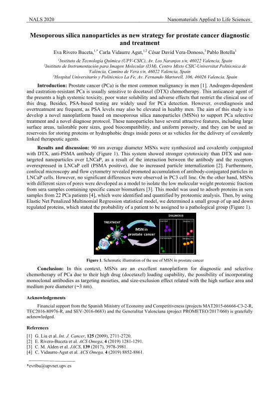
NALS 2020 Nanomaterials Applied to Life Sciences
Mesoporous silica nanoparticles as new strategy for prostate cancer diagnostic and treatment
Eva Rivero Buceta,1,* Carla Vidaurre Agut,1,2 César David Vera-Donoso,3 Pablo Botella1 1Instituto de Tecnología Química (UPV-CSIC), Av. Los Naranjos s/n, 46022 Valencia, Spain
2Instituto de Instrumentación para Imagen Molecular (I3M), Centro Mixto CSIC-Universitat Politècnica de València, Camino de Vera s/n, 46022 Valencia, Spain
3Hospital Universitario y Politécnico La Fe, Av. Fernando Martorell, 106, 46026 Valencia, Spain
Introduction: Prostate cancer (PCa) is the most common malignancy in men [1]. Androgen-dependent and castration-resistant PCa is usually sensitive to docetaxel (DTX) chemotherapy. This anticancer agent of the presents a high systemic toxicity, poor water solubility and adverse effects that restrict the clinical use of this drug. Besides, PSA-based testing are widely used for PCa detection. However, overdiagnosis and overtreatment are frequent, as PSA levels may also be elevated in healthy men. The aim of this study is to develop a novel nanoplatform based on mesoporous silica nanoparticles (MSNs) to support PCa selective treatment and a novel diagnose protocol. These nanoparticles have several attractive features, including large surface areas, tailorable pore sizes, good biocompatibility, and uniform porosity, and they can be used as reservoirs for storing proteins or hydrophobic drugs inside pores or as vehicles for the delivery of covalently linked therapeutic agents.
Results and discussion: 90 nm average diameter MSNs were synthesized and covalently conjugated with DTX, anti-PSMA antibody (Figure 1). This system showed stronger cytotoxicity than DTX and non-targeted nanoparticles over LNCaP, as a result of the interaction between the antibody and the receptors overexpressed in LNCaP cell (PSMA positive), due to increased particle internalization [2]. Furthermore, confocal microscopy and flow cytometry revealed promoted accumulation of antibody-conjugated particles in LNCaP cells. However, no significant differences were observed in PC3 cell line. On the other hand, MSNs with different sizes of pores were developed as a model to isolate the low molecular weight proteomic fraction from sera samples containing specific cancer biomarkers [3]. This model was used to adsorb proteins in sera samples from 22 PCa patients [4], which were identified and quantified by proteomic analysis. Then, by using Elastic Net Penalized Multinomial Regression statistical model, we determined a small group of up and down regulated proteins, which stated the probability of a patient to be assigned to a pathological group (Figure 1).
Figure 1. Schematic illustration of the use of MSN in prostate cancer
Conclusion: In this context, MSNs are an excellent nanoplatform for diagnostic and selective chemotherapy of PCa due to their high drug (docetaxel) loading capability, the possibility of incorporating monoclonal antibodies as targeting moieties, and size-exclusion effect related with the high surface area and medium pore diameter (~3 nm).
Acknowledgements
Financial support from the Spanish Ministry of Economy and Competitiveness (projects MAT2015-66666-C3-2-R, TEC2016-80976-R, and SEV-2016-0683) and the Generalitat Valenciana (project PROMETEO/2017/060) is gratefully acknowledged.
References
[1] G. Liu et al. Int. J. Cancer, 125 (2009), 2711-2720. [2] E. Rivero-Buceta et al. ACS Omega, 4 (2019) 1281-1291. [3] C. M. Alden et al. JACS, 139 (2017), 3978-3981. [4] C. Vidaurre-Agut et al. ACS Omega, 4 (2019) 8852-8861.
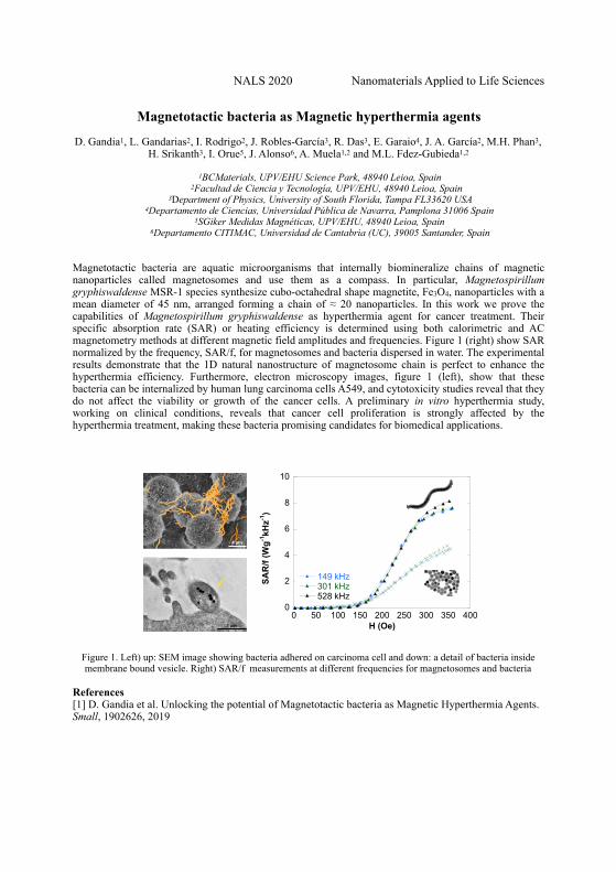
NALS 2020 Nanomaterials Applied to Life Sciences
Magnetotactic bacteria as Magnetic hyperthermia agents
D. Gandia1, L. Gandarias2, I. Rodrigo2, J. Robles-García3, R. Das3, E. Garaio4, J. A. García2, M.H. Phan3, H. Srikanth3, I. Orue5, J. Alonso6, A. Muela1,2 and M.L. Fdez-Gubieda1,2
1BCMaterials, UPV/EHU Science Park, 48940 Leioa, Spain 2Facultad de Ciencia y Tecnología, UPV/EHU, 48940 Leioa, Spain
3Department of Physics, University of South Florida, Tampa FL33620 USA 4Departamento de Ciencias, Universidad Pública de Navarra, Pamplona 31006 Spain
5SGiker Medidas Magnéticas, UPV/EHU, 48940 Leioa, Spain 6Departamento CITIMAC, Universidad de Cantabria (UC), 39005 Santander, Spain
Magnetotactic bacteria are aquatic microorganisms that internally biomineralize chains of magnetic nanoparticles called magnetosomes and use them as a compass. In particular, Magnetospirillum gryphiswaldense MSR-1 species synthesize cubo-octahedral shape magnetite, Fe3O4, nanoparticles with a mean diameter of 45 nm, arranged forming a chain of ≈ 20 nanoparticles. In this work we prove the capabilities of Magnetospirillum gryphiswaldense as hyperthermia agent for cancer treatment. Their specific absorption rate (SAR) or heating efficiency is determined using both calorimetric and AC magnetometry methods at different magnetic field amplitudes and frequencies. Figure 1 (right) show SAR normalized by the frequency, SAR/f, for magnetosomes and bacteria dispersed in water. The experimental results demonstrate that the 1D natural nanostructure of magnetosome chain is perfect to enhance the hyperthermia efficiency. Furthermore, electron microscopy images, figure 1 (left), show that these bacteria can be internalized by human lung carcinoma cells A549, and cytotoxicity studies reveal that they do not affect the viability or growth of the cancer cells. A preliminary in vitro hyperthermia study, working on clinical conditions, reveals that cancer cell proliferation is strongly affected by the hyperthermia treatment, making these bacteria promising candidates for biomedical applications.
Figure 1. Left) up: SEM image showing bacteria adhered on carcinoma cell and down: a detail of bacteria inside membrane bound vesicle. Right) SAR/f measurements at different frequencies for magnetosomes and bacteria
References [1] D. Gandia et al. Unlocking the potential of Magnetotactic bacteria as Magnetic Hyperthermia Agents. Small, 1902626, 2019
0
2
4
6
8
10
0 50 100 150 200 250 300 350 400
149 kHz301 kHz528 kHz
SAR
/f (W
g-1kH
z-1)
H (Oe)
4 µm 4 µm 2 µm
1 µm 1 µm 1 µm
1 µm1 µm 0,5 µm
A B C
D E F
G H I
4 µm 4 µm 2 µm
1 µm 1 µm 1 µm
1 µm1 µm 0,5 µm
A B C
D E F
G H I
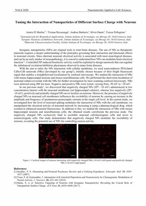
NALS 2020 Nanomaterials Applied to Life Sciences
Tuning the Interaction of Nanoparticles of Different Surface Charge with Neurons
Amira El Merhie1*, Tiziana Ravasenga2, Andrea Barberis2, Silvia Dante3, Teresa Pellegrino1 1Nanomaterials for Biomedical Applications, Istituto Italiano di Tecnologia, via Morego 30, 16163 Genova, Italy
2Synaptic Plasticity of Inhibitory Networks, Istituto Italiano di Tecnologia, via Morego 30, 16163 Genova, Italy 3Materials Characterization Facility, Istituto Italiano di Tecnologia, via Morego 30, 16163 Genova, Italy
Inorganic nanoparticles (NPs) are original tools to treat brain diseases. The use of NPs as therapeutic
materials requires a deeper understanding of the principles governing their interaction and functional effects in neuronal circuits. Since aberrant neuronal electrical activity is associated with most neurological diseases, and can be an early marker of neuropathology, it is crucial to understand how NPs can modulate brain electrical function1,2. Controlled NP-induced bioelectric activity could be exploited to design nanotools that can regulate the imbalanced excitation/inhibition phenomena observed in many brain diseases.
With the aim to tailor the NPs interaction with cellular membrane, we used semiconductor PEGylated CdSe/CdS nanorods (NRs) (developed by our group3), initially chosen because of their bright fluorescent signal that enables a straightforward localization by confocal microscopy. We studied the interaction of NRs with mouse hippocampal neurons and mouse neuroblastoma cells. We performed the short-term incubation of neuronal culture/coverslip with the NRs for further investigation by laser scanning confocal microscopy. NRs were detected using 488 nm laser. Negative and positive NRs were tested, varying from -50 mV to +20 mV.
In our previous study3, we discovered that negatively charged NPs (ZP< -18 mV) administered at low concentration interact with the neuronal membrane (rat hippocampal cultures), whereas less negatively (ZP> -18 mV), positively and neutrally charged NPs never localize on neurons. Moreover, the presence of negatively charged NPs on neuronal cell membranes influences the excitability of neurons by causing an increase in the amplitude and frequency of spontaneous postsynaptic currents at the single cell level. In the current study, we investigated how the level of neuronal spiking modulates the interaction of NRs with the cell membrane; we manipulated the electrical activity of neuronal network by increasing it using a pharmacological drug, which resulted in enhanced neuronal fluorescence. In addition to this, we studied the interaction of NRs with mouse hippocampal neurons and neuroblastoma cells; the obtained results corroborate the previous study. The negatively charged NPs exclusively bind to excitable neuronal cell/electrogenic cells and never to nonelectrogenic cells. Our study demonstrates that negatively charged NPs modulate the excitability of neurons, revealing the potential use of NPs for controlling neuron activity.
Figure 1. Confocal images of neurons interacting with negatively charged NRs (-50 mV) (left panel) and positively charged
NRs (+20 mV) (right panel)
References [1] Sachdev, P. S. Alternating and Postictal Psychoses: Review and a Unifying Hypothesis. Schizophr. Bull. 33, 1029–
1037 (2007). [2] Yuan, P. & Grutzendler, J. Attenuation of β-Amyloid Deposition and Neurotoxicity by Chemogenetic Modulation of
Neural Activity. J. Neurosci. 36, 632–641 (2016). [3] Dante, S. et al. Selective Targeting of Neurons with Inorganic Nanoparticles: Revealing the Crucial Role of
Nanoparticle Surface Charge. ACS Nano 11, 6630–6640 (2017).
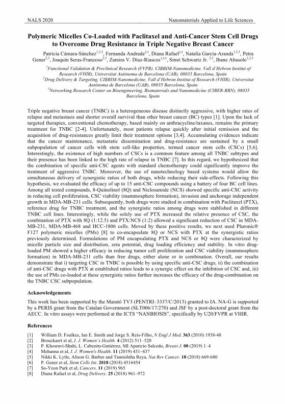
NALS 2020 Nanomaterials Applied to Life Sciences
Polymeric Micelles Co-Loaded with Paclitaxel and Anti-Cancer Stem Cell Drugs to Overcome Drug Resistance in Triple Negative Breast Cancer
Patricia Cámara-Sánchez1,2,3, Fernanda Andrade2,3, Diana Rafael2,3, Natalia García-Aranda1,2,3, Petra Gener2,3, Joaquín Seras-Franzoso2,3, Zamira V. Díaz-Riascos1,2,3, Simó Schwartz Jr. 2,3, Ibane Abasolo1,2,3
1Functional Validation & Preclinical Research (FVPR), CIBBIM-Nanomedicine, Vall d’Hebron Institut of Research (VHIR), Universitat Autònoma de Barcelona (UAB), 08035 Barcelona, Spain
2Drug Delivery & Targeting, CIBBIM-Nanomedicine, Vall d’Hebron Institut of Research (VHIR), Universitat Autònoma de Barcelona (UAB), 08035 Barcelona, Spain
.3Networking Research Center on Bioengineering, Biomaterials and Nanomedicine (CIBER-BBN), 08035 Barcelona, Spain
Triple negative breast cancer (TNBC) is a heterogeneous disease distinctly aggressive, with higher rates of relapse and metastasis and shorter overall survival than other breast cancer (BC) types [1]. Upon the lack of targeted therapies, conventional chemotherapy, based mainly on anthracycline/taxanes, remains the primary treatment for TNBC [2-4]. Unfortunately, most patients relapse quickly after initial remission and the acquisition of drug-resistances greatly limit their treatment options [3,4]. Accumulating evidences indicate that the cancer maintenance, metastatic dissemination and drug-resistance are sustained by a small subpopulation of cancer cells with stem cell-like properties, termed cancer stem cells (CSCs) [5,6]. Interestingly, the existence of high numbers of CSCs is a common feature among all TNBC subtypes and their presence has been linked to the high rate of relapse in TNBC [7]. In this regard, we hypothesized that the combination of specific anti-CSC agents with standard chemotherapy could significantly improve the treatment of aggressive TNBC. Moreover, the use of nanotechnology based systems would allow the simultaneous delivery of synergistic ratios of both drugs, while reducing their side-effects. Following this hypothesis, we evaluated the efficacy of up to 15 anti-CSC compounds using a battery of four BC cell lines. Among all tested compounds, 8-Quinolinol (8Q) and Niclosamide (NCS) showed specific anti-CSC activity in reducing cell proliferation, CSC viability (mammosphere formation), invasion and anchorage independent growth in MDA-MB-231 cells. Subsequently, both drugs were studied in combination with Paclitaxel (PTX), reference drug for TNBC treatment, and the synergistic ratios among drugs were stablished in different TNBC cell lines. Interestingly, while the solely use of PTX increased the relative presence of CSC, the combination of PTX with 8Q (1:12.5) and PTX:NCS (1:2) allowed a significant reduction of CSC in MDA-MB-231, MDA-MB-468 and HCC-1806 cells. Moved by these positive results, we next used Pluronic® F127 polymeric micelles (PMs) [8] to co-encapsulate 8Q or NCS with PTX at the synergistic ratios previously determined. Formulations of PM encapsulating PTX and NCS or 8Q were characterized by micelle particle size and distribution, zeta potential, drug loading efficiency and stability. In vitro drug-loaded PM showed a higher efficacy in reducing tumor cell proliferation and CSC viability (mammosphere formation) in MDA-MB-231 cells than free drugs, either alone or in combination. Overall, our results demonstrate that i) targeting CSC in TNBC is possible by using specific anti-CSC drugs, ii) the combination of anti-CSC drugs with PTX at established ratios leads to a synergic effect on the inhibition of CSC and, iii) the use of PMs co-loaded at these synergistic ratios further increases the efficacy of the drug-combination on the TNBC CSC subpopulation.
Acknowledgements
This work has been supported by the Marató TV3 (PENTRI–3337/C/2013) granted to IA. NA-G is supported by a PERIS grant from the Catalan Government (SLT006/17/270) and JSF by a post-doctoral grant from the AECC. In vitro assays were performed at the ICTS “NANBIOSIS”, specifically by U20/FVPR at VHIR.
References
[1] William D. Foulkes, Ian E. Smith and Jorge S. Reis-Filho, N Engl J Med. 363 (2010) 1938-48 [2] Brouckaert et al, I. J. Women’s Health. 4 (2012) 511–520 [3] P. Khosravi-Shahi, L. Cabezón-Gutiérrez, MI Aparicio Salcedo, Breast J. 00 (2019) 1–4 [4] Mehanna et al, I. J. Women's Health. 11 (2019) 431–437 [5] Nikki K. Lytle, Alison G. Barber and Tannishtha Reya, Nat Rev Cancer. 18 (2018) 669-680 [6] P. Gener et al, Stem Cells Int. 2018 (2018) 4516454 [7] So-Yeon Park et al, Cancers. 11 (2019) 965 [8] Diana Rafael et al, Drug Delivery. 25 (2018) 961–972
NALS 2020 Nanomaterials Applied to Life Sciences
Magnetic Combination Therapy: a new, drug-free approach to cancer treatmentIrati Rodrigo1,2, Michael Stanton3, Daniela Valdés4, Emma Welbourne3, Borja Herrero de la Parte5, José
Ángel García6, Roberto Zysler4,7,8, Fernando Plazaola2 and Russell Cowburn3.
1BCMaterials, UPV/EHU Science Park, Leioa, Spain2 Department of Electricity and Electronics, UPV/EHU, Leioa, Spain
3 Cavendish Laboratory, University of Cambridge, Cambridge, UK4 Instituto Balseiro, San Carlos de Bariloche, Río Negro, Argentina
5 Department of Surgery and Radiology and Physical Medicine, UPV/EHU, Leioa, Spain6Department of Applied Physics II, UPV/EHU, Leioa, Spain
7 Laboratorio de Resonancias Magnéticas, Centro Atómico Bariloche, CNEA, San Carlos de Bariloche, Argentina.
8 Consejo Nacional de Investigaciones Científicas y Técnicas (CONICET), Ministerio de Ciencia, Tecnología e Innovación Productiva, Argentina
Magnetic hyperthermia and magneto-mechanical destruction are two emerging cancer therapies1, 2. We have studied the combination of these two treatments to develop a new, more efficient therapy. Magnetic iron-oxide nanoparticles (MIONs) were synthesized by thermal decomposition3 with the characteristics typical for magnetic hyperthermia. They were coated with DEXTRAN and DSPE-mPEG(2000) (mPEG). The MIONs were magnetically characterized and their size distribution was obtained by analyzing TEM images.Experimental specific absorption rate measurements were made in a custom-built AC magnetometer to define the characteristics of the field to be used for the magnetic hyperthermia experiments4. On the other hand, synthetic antiferromagnetic (SAF) microdiscs (MDs) with perpendicular anisotropy were fabricated by acombination of sputtering and lithography techniques5 with the characteristics typical for magneto-mechanical destruction experiments. The discs were magnetically characterized using MOKE and VSM and imaged by SEM. The spatial arrangements’ form of MDs and MIONS was studied under the influence of DC and AC magnetic fields using an optical microscope with an integrated dipole electromagnet. Strong chaining behaviorbetween MDs and MIONs with DEXTRAN coating was observed, but not with MIONs with a mPEG coating. The internalization of the particles in colorectal cells of cancer cell line (CC531) was also verified using SEM and TEM. Cytotoxicity experiments find MDs and dextran coated MIONs to have a minimal effect on the viability of CC531 cells, making them suitable for in vitro testing. A preliminary in vitro treatment experiment shows that hyperthermia and magneto-mechanical destruction can induce cell death, and that in addition, theircombination generates a greater effect. In conclusion, the results presented in this work indicate that different magnetic particle based cancer treatments have a positive synergistic effects when implemented together to destroy tumor cells.
Acknowledgements
The authors want to acknowledge IEEE Magnetics Society for the founding of this project and to the Basque Government under Grant No. IT-1005-16.
References
[1] A. Jordan, R. Scholz, P. Wust, Journal of Magnetism and Magnetic Materials, Vol. 201, p.413 (1999)
[2] R. Mansell, T. Vemulkar, D. C. M. C. Petit, Scientific Reports, Vol. 7, p.4257 (2017)
[3] M.L. Mojica Pisciotti, E. Lima Jr., M. Vasquez Mansilla, Journal of Biomedical Materials Research Part B: Applied Biomaterials, Vol. 102, p.860 (2014)
[4] T. Vemulkar, R. Mansell, D. C. M. C. Petit, R. P. Cowburn and M. S. Lesniak, Applied Physics Letters, Vol. 107, p.012403 (2015)

Nanodiagnostics Presentations
NALS 2020 Nanomaterials Applied to Life Sciences
Size Nanoparticle and Agglomeration Effects on Magnetic Lateral Flow
Immunoassays M. Salvador1,2,*, A. Gallo-Córdova3, A. Moyano1,4, J.C. Martínez-García1, M.C. Blanco-López4,
M.P. Morales3, M. Rivas1 1Department of Physics & IUTA, University of Oviedo, 33204 Gijón, Spain
2Istituto di Struttura della Materia-CNR, 00015 Monterotondo Scalo (RM), Italy 3Instituto de Ciencia de Materiales de Madrid (ICMM-CSIC), Cantoblanco, 28049 Madrid, Spain
4Department of Physical and Analytical Chemistry, University of Oviedo, 33006 Oviedo, Spain
Lateral Flow Immunoassay (LFIA) is a paper-based architecture whose most popular use is the pregnancy test. They are being increasingly used for determination of biomarkers, allergenic pathogens, drugs and metabolites, biomedical, food safety and environmental settings [1]. Their sensitivity, selectivity, quickness and ease of use make them ideal for Point-of-Use (PoU) testing. One of the key points of the LFIAs is the labelling of the biomarker, traditionally by latex or gold nanoparticles that provide a visible signal. These are essentially qualitative (presence/absence) or semi-quantitative analyses. To add quantification capacities to LFIAs, the use of Magnetic Nanoparticles (NPs) has been lately proposed [2].
The magnetic LFIAs must be associated to a magnetic reader that should be itself fast and portable. A radio-frequency inductive sensor has been developed for this purpose which takes advantage of the superparamagnetic character of the NPs [3,4]. The clue parameter of this technique is the initial magnetic permeability of the particles at the frequency of detection. The particle size has a crucial influence on the magnetic permeability, while the agglomeration degree determines the number of particles per molecule of interest. Thus, both size and agglomeration have a great importance in this type of test. The purpose of this work has been to elucidate the effects of the magnetic core size and particle agglomeration before and after the biofunctionalization process in the magnetic LFIA. To do so, three different iron oxide particles with core sizes of 8, 12 and 23 nm and different initial agglomeration have been evaluated as labels in LFIAs functionalized with neutravidin and immobilized by a biotin test line.
Acknowledgements
This work was supported by the Spanish Ministry of Economy and Competitiveness under projects MAT2017-84959-C2-1-R and EUIN2019-103338, the Council of Gijón-IUTA under grant SV-18-GIJON-1-27, and the Principality of Asturias under project IDI/2018/000185.
References
[1] X. Huang et al. Biosens. Bioelectron. 75 (2016) [2] Lago-Cachón, D. et al., IEEE Magn. Lett. 8 (2017) [3] Lago-Cachón, D. et al., Nanotechnol. 24 (2013) [4] Rivas, M. et al., Sensor Actuar A-Phys. 216 (2014)
NALS 2020 Nanomaterials Applied to Life Sciences
C-NanoDots based label-free sensing of single gene mutations
Tania García-Mendiola1, Iria Bravo2, Félix Pariente3, Alejandra Jacobo-Martin4, C. Navio5, Isabel Rodriguez6,
Reinhold Wannemacher7 and Encarnación Lorenzo8 1,2,3,8Departamento Química Analítica y Análisis Instrumental, Institute for Advanced Research in Chemical Sciences
(IAdChem) Universidad Autónoma de Madrid, 28049 Madrid, Spain. 4,5,6,7Instituto Madrileño de Estudios Avanzados (IMDEA) Nanociencia, Faraday, 9, Campus UAM, 28049 Madrid, Spain.
Here we report on the development of an assay for DNA sensing based on fluorescent carbon nanodots (CNDs). The developed assay allows the rapid detection of gene mutations associated with human diseases, in particular, mutations in BRCA1, CFRT and MRP3 genes associated with breast cancer, cystic fibrosis and autoimmune hepatitis diseases, respectively. Blue fluorescent carbon nanodots, synthesized from citric acid and ethylene diamine, were extensively characterized by various techniques (DLS, TEM, FTIR, XPS, UV-VIS, AFM and fluorescence spectroscopy) and their interactions with DNA were studied. The formation of a nanobioconjugate between C-nanodots and DNA was confirmed by DLS and fluorescence microscopy. Changes in the fluorescence intensity following interaction with single and double-stranded DNA (ssDNA and dsDNA) were used for a specific DNA sequence detection. The biosensor response was linear with respect to target DNA concentrations up to 200 nM with a detection limit of 270 pM. Moreover, the high selectivity allows not only to discriminate between wild type and mutated DNA samples, but also rapidly detect single gene mutations, without the need of labels.
Acknowledgements
The authors acknowledge Spanish government (CTQ2017-84309-C2-1-R), (RED2018-102412-T) and Comunidad de Madrid (S2018/NMT-4349) for financial support.
NALS 2020 Nanomaterials Applied to Life Sciences
Microfluidic Plasmonic Supercrystal as SERS Sensor Daniel García-Lojo1,*, Sergio Gómez-Graña1, Isabel Pastoriza-Santos1 and Jorge Pérez-Juste1
1 Department of Physical Chemistry and Biomedical Research Center (CINBIO), University of Vigo. As Lagoas-Marcosende, 36310, Vigo (Spain)
Surface-enhanced Raman spectroscopy, SERS, is an advanced analytical technique that can be used for the ultrasensible detection of analytes. It is based in the enhancement of the Raman signal of a molecule at the surface of a plasmonic nanostructure mainly due to the presence of strong electromagnetic fields generated after the plasmon excitation. Moreover, this effect could be more intense in the case of plasmonic supercrystals due to antenna effects as demonstrated by recent simulations.1
As we previously reported, microfluidic platforms allow generating a highly-ordered assembly of uniform gold nanoparticles inside their microchannels through the slow pervaporation of the solvent (Figure 1A-B).2 While plasmonic supercrystals made by drop-casting show poor uniformity that limits their potential plasmonic applications.3 Microfluidic approach enables the fabrication of supercrystals of any dimension or morphology. Furthermore, the integration of a plasmonic supercrystal inside microfluidic platform guarantees the infiltration of the desired analyte, even without affinity for gold surface, within the plasmonic supercrystals and therefore its ultrasensitive detection.
Herein, we show the fabrication and characterization of plasmonic supercrystals using gold octahedra nanoparticles synthesized through a wet chemical method. Besides, the study of the sensing capabilities of these platforms showed a highly uniform and intense SERS activity (Figure 1C), being both key parameters to achieve quantitative and ultrasensitive analysis (LOD) (Figure 1E). In fact, LOD as low as 100 zM was achieved for the detection of Crystal Violet, which is several orders of magnitude lower than those found in the literature. Also, we investigate the possibility of combining the capabilities of our SERS sensor with the chromatographic properties of silica nanoparticles to develop a sensor device with the ability of separate analytes by charge (Figure 1D).
Figure 1. (A) Schematic illustration of self-assembly of gold octahedra nanoparticles inside microfluidic platform
induced by pervaporation. (B) Scanning Electron Microscopy (SEM) image of the supercrystal. Inset corresponds with the result of applied Fourier Transform (C) SERS hot-map obtained at 1617 cm-1 in the presence of 100 nM of Crystal Violet. (D) Schematic representation of the final SERS Sensor modified with a silica supercrystal for chromatographic separation. (E) SERS intensity obtained for different concentrations of Crystal Violet (10-7-10-14 M).
Acknowledgements
This work was supported by the Ministerio de Economía y Competitividad (MAT2016-77809-R). DGL acknowledges financial support through the “FPI" fellowship under grant number FPI-MAT2016-77809-R.
References
[1] D.M. Solís et al.; ACS Nano 8 (2014) 7559. [2] S. Gómez-Graña et al., Chem. Mater., 27 (2015) 8315. [3] D. García-Lojo et al., Acc. Chem. Res. 52 (2019), 7, 1855
NALS 2020 Nanomaterials Applied to Life Sciences
SERS in arrays of geometrically frustrated plasmonic nanolements. A. Conde-Rubio1, J. Rodriguez-Alvarez1,2,*, M. Escoda-Torroella1,2, I. Caritg1, P. Molet3, A. Mihi3, F.
Pérez-Murano4, A. Fraile Rodríguez 1,2, A. Labarta 1,2, and X. Batlle 1,2. 1 Departament de Física de la Matèria Condensada, Universitat de Barcelona, 08028 Barcelona, Spain
2 Institut de Nanociència i Nanotecnologia (IN2UB), Barcelona, 08028, Spain 3 Institut de Ciència de Materials de Barcelona (ICMAB, CSIC) Bellaterra, 08193, Spain 4 Institut de Microelectrónica de Barcelona (IMB-CNM, CSIC), Bellaterra, 08193, Spain
Surface Enhanced Raman Spectroscopy (SERS) is a technique widely used in the detection and identification of biomolecules due to its high sensitivity and ability of detecting unique molecular fingerprints. The low efficiency of RAMAN scattering processes can be overcome by the excitation of the corresponding vibrational modes of the molecules taking advantage of the enhanced electric field associated with plasmonic nanostructured surfaces, giving rise to an increase of the signal of several orders of magnitude.
In this work, SERS measurements were carried out on substrates with arrays of geometrically frustrated plasmonic Au nanolements. These arrays may show collective modes due to the lattice long-range order apart from the plasmon response of the individual elements. On the other hand, when nanoelements are very close, near field interactions between them appear as well. The interplay between all these excitations may yield much more intense resonances than those of the single element. The manufactured structures present the pattern of Au nanoelements combined with the inverse Au structure on the top, consisting in a continuous Au layer with element-shaped gaps. In addition, a continuous Au layer is set under the structure to serve as a mirror. Fourier Transform Infrared (FTIR) spectroscopy revealed high absorption peaks around the visible or/and near-infrared regions due to the excitation of these two substructures. Proof of concept functionalization of these samples with 4-mercaptopyridine and crystal violet demonstrate their suitability as SERS substrates, enabling the detection of concentrations down to 10-9 M.
Acknowledgements
The work was supported by Spanish MCIU and AEI (MAT2015-68772-P; PGC2018-097789-B-I00) and European Union FEDER funds. We also acknowledge Andrés Cantarero, Departamento de Física Aplicada y Electromagnetismo, Universitat de Valencia, for preliminary RAMAN measurements.
References [1] Conde-Rubio, A et al. Scientific Reports 9, 3529 (2019)
[2] Conde-Rubio, A et al. Opt. Express 26, 20211 (2018).
Fig.1 (a) SEM image of an array of nanoelements and an inset showing a schematic representation of the structure, (b) RAMAN spectrum of a 10-9 M solution of 4-mercaptopyridine.
500 750 1000 1250 1500 17501,0
1,5
2,0
2,5
3,0
Inte
nsity
(arb
. u.)
Raman shift (cm-1)
a) b)
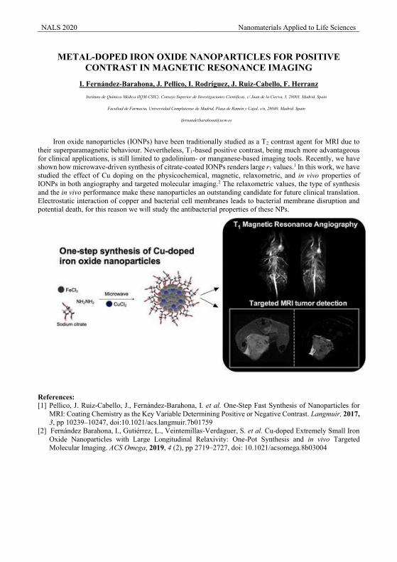
NALS 2020 Nanomaterials Applied to Life Sciences
METAL-DOPED IRON OXIDE NANOPARTICLES FOR POSITIVE
CONTRAST IN MAGNETIC RESONANCE IMAGING
I. Fernández-Barahona, J. Pellico, I. Rodríguez, J. Ruiz-Cabello, F. Herranz Instituto de Química Médica (IQM-CSIC), Consejo Superior de Investigaciones Científicas, c/ Juan de la Cierva, 3, 28003, Madrid, Spain
Facultad de Farmacia, Universidad Complutense de Madrid, Plaza de Ramón y Cajal, s/n, 28040, Madrid, Spain
Iron oxide nanoparticles (IONPs) have been traditionally studied as a T2 contrast agent for MRI due to their superparamagnetic behaviour. Nevertheless, T1-based positive contrast, being much more advantageous for clinical applications, is still limited to gadolinium- or manganese-based imaging tools. Recently, we have shown how microwave-driven synthesis of citrate-coated IONPs renders large r1 values.1 In this work, we have studied the effect of Cu doping on the physicochemical, magnetic, relaxometric, and in vivo properties of IONPs in both angiography and targeted molecular imaging.2 The relaxometric values, the type of synthesis and the in vivo performance make these nanoparticles an outstanding candidate for future clinical translation. Electrostatic interaction of copper and bacterial cell membranes leads to bacterial membrane disruption and potential death, for this reason we will study the antibacterial properties of these NPs.
References: [1] Pellico, J. Ruiz-Cabello, J., Fernández-Barahona, I. et al. One-Step Fast Synthesis of Nanoparticles for
MRI: Coating Chemistry as the Key Variable Determining Positive or Negative Contrast. Langmuir, 2017, 3, pp 10239–10247, doi:10.1021/acs.langmuir.7b01759
[2] Fernández Barahona, I., Gutiérrez, L., Veintemillas-Verdaguer, S. et al. Cu-doped Extremely Small Iron Oxide Nanoparticles with Large Longitudinal Relaxivity: One-Pot Synthesis and in vivo Targeted Molecular Imaging. ACS Omega, 2019, 4 (2), pp 2719–2727, doi: 10.1021/acsomega.8b03004

poster presentations

Wednesday Poster Session
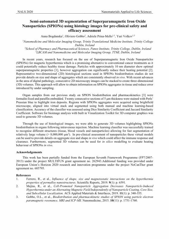
NALS 2020 Nanomaterials Applied to Life Sciences
Semi-automated 3D segmentation of Superparamagnetic Iron Oxide Nanoparticles (SPIONs) using histology images for pre-clinical safety and
efficacy assessment Anna Bogdanska1, Oliviero Gobbo2, Adriele Prina-Mello1,3, Yuri Volkov1,3
1Nanomedicine and Molecular Imaging Group, Trinity Translational Medicine Institute, Trinity College Dublin, Ireland
2School of Pharmacy and Pharmaceutical Sciences, Panoz Institute, Trinity College, Dublin, Ireland 3LBCAM and Nanomedicine and Molecular Imaging Group, TTMI, Dublin, Ireland
In recent years, research has focused on the use of Superparamagnetic Iron Oxide Nanoparticles (SPIONs) for magnetic hyperthermia which is a promising alternative to conventional cancer treatments as it could potentially reduce healthy tissue damage. Particles with approximately 10 nm diameter show optimal superparamagnetic properties [1], however aggregation can significantly reduce their heating potential [2]. Representative two-dimensional (2D) histological sections used in SPIONs biodistribution studies do not provide details on size and shape of aggregates which are consistently observed in vivo. With recent advances in the area of digital pathology, consecutive 2D microscopy images can be stacked to create three-dimensional (3D) volumes. This approach will allow to obtain information on SPIONs aggregates in tissue and reduce error introduced by under sampling.
Organ samples from our previous study on SPION biodistribution and pharmacokinetics [3] were formalin fixed and paraffin embedded. Twenty consecutive sections of 5 μm thickness were stained with Perls’ Prussian blue to highlight iron deposits. Regions with SPIONs aggregates were acquired using brightfield microscope, aligned into virtual stack and segmented using both manual and machine learning-based classification. Accuracy of the classifier was assessed using Dice Similarity Coefficient and Jaccard Similarity Coefficient. Software for bioimage analysis with built in Visualization Toolkit for 3D computer graphics was used to generate 3D volumes.
Through the use of histological images, we were able to generate 3D volumes highlighting SPIONs biodistribution in organs following intravenous injection. Machine learning classifier was successfully trained to recognise different structures (tissue, blood vessels and nanoparticles) allowing for fast segmentation of relatively large volume (> 8,000,000 μm3). In pre-clinical assessment of nanoparticles these virtual models can be used to provide details on aggregate size and shape in vivo which could affect the immune response and clearance. Furthermore, segmented 3D volumes can be used for in silico modelling to evaluate heating behaviour of SPIONs in vivo.
Acknowledgements
This work has been partially funded from the European Seventh Framework Programme (FP7/2007-2013) under the project MULTIFUN grant agreement no. 262943.Additional funding was provided under European Union’s Horizon 2020 research and innovation programme under the project NoCanTher grant agreement no. 685795.
References 1. Ferrero, R., et al., Influence of shape, size and magnetostatic interactions on the hyperthermia
properties of permalloy nanostructures. Scientific Reports, 2019. 9(1): p. 6591. 2. Mejías, R., et al., Cell-Promoted Nanoparticle Aggregation Decreases Nanoparticle-Induced
Hyperthermia under an Alternating Magnetic Field Independently of Nanoparticle Coating, Core Size, and Subcellular Localization. ACS Applied Materials & Interfaces, 2019. 11(1): p. 340-355.
3. Gobbo, O.L., et al., Biodistribution and pharmacokinetic studies of SPION using particle electron paramagnetic resonance, MRI and ICP-MS. Nanomedicine, 2015. 10(11): p. 1751-1760.
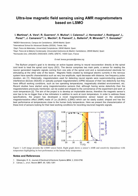
Ultra-low magnetic field sensing using AMR magnetometers based on LSMO
I. Martineza, A. Veraa, R. Guerreroa, V. Muñoza, I. Calaresub, J. Hernandeza, I. Rodrigueza, L. Pereza,c, J. Camareroa,d, L. Mechine, S. Flamente, L. Ballerinib, R. Mirandaa,d, T. Gonzaleza,*
aIMDEA Nanociencia, Campus de Cantoblanco, 28049 Madrid, Spain bInternational School for Advanced Studies (SISSA), Trieste, Italy cDept. Fisica de Materiales, Universidad Complutense, 28040 Madrid, Spain dDept. Fisica de la Materia Condensada, Universidad Autónoma de Madrid, Cantoblanco, 28049 Madrid, Spain eNormandie Univ, UNICAEN, ENSICAEN, CNRS, GREYC, 14000 Caen, France
E-mail: [email protected] The ByAxon project’s goal is to develop an active bypass aiming to neural recconection directly at the spinal cord level to treat the spinal cord injury (SCI). The device comprises two main parts, a sensor for reading the neuron generated magnetic signals coming from one side of the spinal cord and a nanostructured electrode for stimulating at the other side of the lesion. Magnetic fields created by biological electric currents in the nervous system have specific characteristics such as very low amplitude, rapid decrease with distance, low frequency pulse duration, etc [1]. Historically, magnetometers used for detecting neural signals were superconducting quantum interference devices (SQUID) or optically pumped magnetometers (OPM) because of their low detectivity but they have difficult working conditions, such as low operating temperatures, magnetically shielded environement, etc. Recent studies have started using magnetorresistive sensors that, although having worse detectivity than the magnetometers previously mentioned, can be scaled and shaped to the convenience of the experiment and work at room temperature [2]. The aim of the project is to develop an implantable device, therefore the magnetic sensor’s size has to be no bigger than a few milimeters in addition to work at room temperature. In order to address these specifications, the project has developed a novel magnetorresistive sensor based on the anisotropic magnetorresistive effect (AMR) made of La1-xSrxMnO3 (LSMO) which can be easily scaled, shaped and has the best performance at temperatures close to the human body temperature. Here we present the characterization of these kind of sensors looking for their best working conditions for recording neuronal magnetic signals.
Figure 1: Left image presents the LSMO sensor batch. Right graph shows a sensor’s AMR and sensitivity dependence with temperature highlighting its best performance at the human body temperature.
Notes and References 1 Wijesinghe, R. S.; Journal of Electrical & Electronic Systems 2014, 3, 2332-0796 2 Caruso, L., et al.; Neuron 2017, 95, 1283–1291
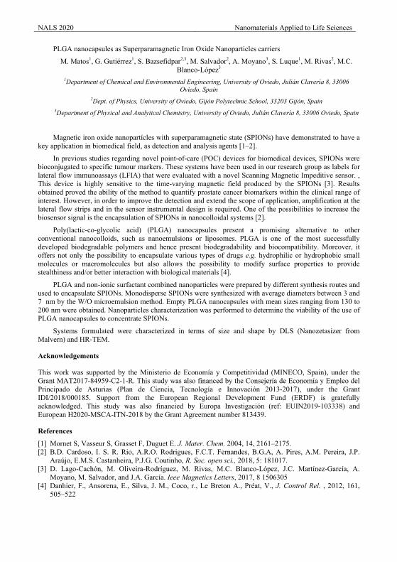
NALS 2020 Nanomaterials Applied to Life Sciences
PLGA nanocapsules as Superparamagnetic Iron Oxide Nanoparticles carriers
M. Matos1, G. Gutiérrez1, S. Bazsefidpar2,3, M. Salvador2, A. Moyano3, S. Luque1, M. Rivas2, M.C. Blanco-López3
1Department of Chemical and Environmental Engineering, University of Oviedo, Julián Clavería 8, 33006 Oviedo, Spain
2Dept. of Physics, University of Oviedo, Gijón Polytechnic School, 33203 Gijón, Spain 3Department of Physical and Analytical Chemistry, University of Oviedo, Julián Clavería 8, 33006 Oviedo, Spain
Magnetic iron oxide nanopartícles with superparamagnetic state (SPIONs) have demonstrated to have a key application in biomedical field, as detection and analysis agents [1–2].
In previous studies regarding novel point-of-care (POC) devices for biomedical devices, SPIONs were bioconjugated to specific tumour markers. These systems have been used in our research group as labels for lateral flow immunoassays (LFIA) that were evaluated with a novel Scanning Magnetic Impeditive sensor. , This device is highly sensitive to the time-varying magnetic field produced by the SPIONs [3]. Results obtained proved the ability of the method to quantify prostate cancer biomarkers within the clinical range of interest. However, in order to improve the detection and extend the scope of application, amplification at the lateral flow strips and in the sensor instrumental design is required. One of the possibilities to increase the biosensor signal is the encapsulation of SPIONs in nanocolloidal systems [2].
Poly(lactic-co-glycolic acid) (PLGA) nanocapsules present a promising alternative to other conventional nanocolloids, such as nanoemulsions or liposomes. PLGA is one of the most successfully developed biodegradable polymers and hence present biodegradability and biocompatibility. Moreover, it offers not only the possibility to encapsulate various types of drugs e.g. hydrophilic or hydrophobic small molecules or macromolecules but also allows the possibility to modify surface properties to provide stealthiness and/or better interaction with biological materials [4].
PLGA and non-ionic surfactant combined nanoparticles were prepared by different synthesis routes and used to encapsulate SPIONs. Monodisperse SPIONs were synthesized with average diameters between 3 and 7 nm by the W/O microemulsion method. Empty PLGA nanocapsules with mean sizes ranging from 130 to 200 nm were obtained. Nanoparticles characterization was performed to determine the viability of the use of PLGA nanocapsules to concentrate SPIONs.
Systems formulated were characterized in terms of size and shape by DLS (Nanozetasizer from Malvern) and HR-TEM.
Acknowledgements
This work was supported by the Ministerio de Economía y Competitividad (MINECO, Spain), under the Grant MAT2017-84959-C2-1-R. This study was also financed by the Consejería de Economía y Empleo del Principado de Asturias (Plan de Ciencia, Tecnología e Innovación 2013-2017), under the Grant IDI/2018/000185. Support from the European Regional Development Fund (ERDF) is gratefully acknowledged. This study was also financied by Europa Investigación (ref: EUIN2019-103338) and European H2020-MSCA-ITN-2018 by the Grant Agreement number 813439.
References [1] Mornet S, Vasseur S, Grasset F, Duguet E. J. Mater. Chem. 2004, 14, 2161–2175. [2] B.D. Cardoso, I. S. R. Rio, A.R.O. Rodrigues, F.C.T. Fernandes, B.G.A, A. Pires, A.M. Pereira, J.P.
Araújo, E.M.S. Castanheira, P.J.G. Coutinho, R. Soc. open sci., 2018, 5: 181017. [3] D. Lago-Cachón, M. Oliveira-Rodríguez, M. Rivas, M.C. Blanco-López, J.C. Martínez-García, A.
Moyano, M. Salvador, and J.A. García. Ieee Magnetics Letters, 2017, 8 1506305 [4] Danhier, F., Ansorena, E., Silva, J. M., Coco, r., Le Breton A., Préat, V., J. Control Rel. , 2012, 161,
505–522
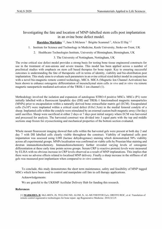
NALS 2020 Nanomaterials Applied to Life Sciences
Investigating the fate and location of MNP-labelled stem cells post implantation in an ovine bone defect model.
Hareklea Markides 1,2, Jane S.Mclaren 3, Brigitte Scammel 3, Alicia El Haj 1,2
1. Institute for Science and Technology in Medicine, Keele University, Stoke-on-Trent, UK
2. Healthcare Technologies Institute, University of Birmingham, Birmingham, UK
3. The University of Nottingham, Nottingham, UK
The ovine critical size defect model provides a strong basis for testing bone tissue engineered constructs for use in the treatment of non-unions and severe trauma. This model has been applied across a number of preclinical studies with emphasis on stem cell based therapies for bone repair. Key to ensuring successful outcomes is understanding the fate of therapeutic cell in terms of identity, viability and bio-distribution post implantation. This study aims to evaluate such parameters in an ovine critical sized defect model in conjunction with a novel bio-magnetic remote control technology, MICA. MICA (Magnetic Ion Channel Activation) has been shown to enhance osteogenic differentiation of mesenchymal stem cells in vivo and in vitro via remote magnetic nanoparticle mediated activation of the TREK-1 ion channel (1).
Methodology involved the isolation and expansion of autologous STRO-4 positive MSCs. MSCs (P1) were initially labelled with a fluorescent lipophilic dye (DII) and TREK-1 functionalised magnetic nanoparticles (MNPs) prior to encapsulation within a naturally derived bone extracellular matrix gel (ECM). Encapsulated cells (5x10⁶) were implanted within a critical sized defect (0.8x1.5cm) in the medial femoral condyle of a sheep. Implanted cells within the condyle were stimulated by an external custom built magnetic array (1hr/day) until sacrifice. Sheep were sacrificed at either 2 days or 7 days post initial surgery where ECM was harvested and processed for analysis. The harvested construct was divided into 3 equal parts with the top and middle sections snap frozen for cryosectioning and mechanical properties of the bottom section evaluated.
Whole mount flourescent imaging showed that cells within the harvested gels were present at both day 2 and day 7 with DII labelled cells clearly visible throughout the construct. Viability of implanted cells post impantation was assessed using LDH (lactase dehydrogenase) staining which demonstrated 50% viability across all experimental groups. MNPs localisation was confirmed on viable cells by Prussian blue staining and dextran immunohistochemisty. Immunohistochemistry further revealed varying levels of osteogenic differentiation at these early time points across groups. Serum CRP (c-reactive protein) levels were measured by ELISA with no obvious increase in CRP levels observed as a result of MNP implanations. This implies that there were no adverse effects related to localised MNP delivery. Finally a sharp increase in the stiffness of all gels was measured post implantiaon when compared to in vitro controls.
To conclude, this study demonstrates the short term maintenance, safety and feasibility of MNP tagged MSCs which have been used to control and manipulate cell fate in cell therapy applications.
Acknowledgements:
We are grateful to the UKRMP Acellular Delivery Hub for funding this research.
References [1] MARKIDES, H, McLAREN, JS, TELLING ND, ALOM ,N, AL-MUTHEFFER EaA, OREFFO ROC, et al. Translation of remote control regenerative technologies for bone repair. npj Regenerative Medicine. 2018;3(1):9.
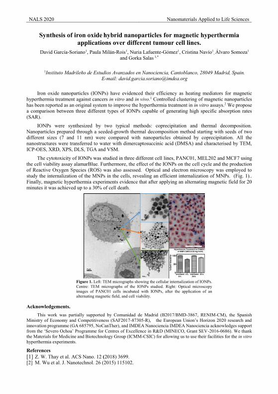
NALS 2020 Nanomaterials Applied to Life Sciences
Synthesis of iron oxide hybrid nanoparticles for magnetic hyperthermia applications over different tumour cell lines.
David García-Soriano1, Paula Milán-Rois1, Nuria Lafuente-Gómez1, Cristina Navío1, Álvaro Somoza1
and Gorka Salas 1,*
1Instituto Madrileño de Estudios Avanzados en Nanociencia, Cantoblanco, 28049 Madrid, Spain.
E-mail: [email protected]
Iron oxide nanoparticles (IONPs) have evidenced their efficiency as heating mediators for magnetic hyperthermia treatment against cancers in vitro and in vivo.1 Controlled clustering of magnetic nanoparticles has been reported as an original system to improve the hyperthermia treatment in in vitro assays.2 We propose a comparison between three different types of IONPs capable of generating high specific absorption rates (SAR).
IONPs were synthesized by two typical methods: coprecipitation and thermal decomposition. Nanoparticles prepared through a seeded-growth thermal decomposition method starting with seeds of two different sizes (7 and 11 nm) were compared with nanoparticles obtained by coprecipitation. All the nanostructures were transferred to water with dimercaptosuccinic acid (DMSA) and characterised by TEM, ICP-OES, XRD, XPS, DLS, TGA and VSM.
The cytotoxicity of IONPs was studied in three different cell lines, PANC01, MEL202 and MCF7 using the cell viability assay alamarBlue. Furthermore, the effect of the IONPs on the cell cycle and the production of Reactive Oxygen Species (ROS) was also assessed. Optical and electron microscopy was employed to study the internalization of the MNPs in the cells, revealing an efficient internalization of MNPs. (Fig. 1).. Finally, magnetic hyperthermia experiments evidence that after applying an alternating magnetic field for 20 minutes it was achieved up to a 30% of cell death.
Figure 1. Left: TEM micrographs showing the cellular internalization of IONPs. Centre: TEM micrographs of the IONPs studied. Right: Optical microscopy images of PANC01 cells incubated with IONPs, after the application of an alternating magnetic field, and cell viability.
Acknowledgements.
This work was partially supported by Comunidad de Madrid (B2017/BMD-3867, RENIM-CM), the Spanish Ministry of Economy and Competitiveness (SAF2017-87305-R), the European Union’s Horizon 2020 research and innovation programme (GA 685795, NoCanTher), and IMDEA Nanociencia IMDEA Nanociencia acknowledges support from the ‘Severo Ochoa’ Programme for Centres of Excellence in R&D (MINECO, Grant SEV-2016-0686). We thank the Materials for Medicine and Biotechnology Group (ICMM-CSIC) for allowing us to use their facilities for the in vitro hyperthermia experiments.
References [1] Z. W. Thay et al. ACS Nano. 12 (2018) 3699. [2] M. Wu et al. J. Nanotechnol. 26 (2015) 115102.
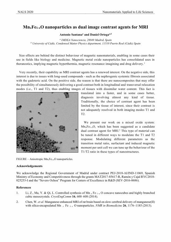
NALS 2020 Nanomaterials Applied to Life Sciences
MnxFe1-xO nanoparticles as dual image contrast agents for MRI
Antonio Santanaa and Daniel Ortegaa,b
a IMDEA Nanociencia, 28049 Madrid, Spain b University of Cádiz, Condensed Matter Physics department, 11510 Puerto Real (Cádiz) Spain
Size effects are behind the distinct behaviour of magnetic nanomaterials, enabling in some cases their use in fields like biology and medicine. Magnetic metal oxide nanoparticles has consolidated uses in theranostics, implying magnetic hyperthermia, magnetic resonance imagining and drug delivery.1 Very recently, their capability as MRI contrast agents has a renewed interest. On the negative side, this interest is due to issues with long-used compounds - such as the nephrogenic systemic fibrosis associated with the gadoteric acid. On the positive side, the reason is that there are nanocomposites that may offer the possibility of simultaneously delivering a good contrast both in longitudinal and transversal relaxation modes (i.e., T1 and T2), thus enabling images of tissues with dissimilar water content. This fact is
translated into a faster, and in some cases better, diagnosis involving almost any kind of tissue. Traditionally, the choice of contrast agent has been limited by the tissue of interest, since their contrast is not adequately resolved in both imaging modes T1 and T2.
We present our work on a mixed oxide system: MnxFe1-xO, which has been suggested as a candidate dual contrast agent for MRI.2 This type of material can be tuned in different ways to modulate the T1 and T2 response. Modulating different parameters as the transition metal ratio, surfactant and induced magnetic moment per unit cell we can tune up the behaviour of the T1/T2 ratio in these types of nanostructures.
FIGURE – Anisotropic MnxFe1-xO nanoparticles.
Acknowledgements
We acknowledge the Regional Government of Madrid under contract PEJ-2018-AI/IND-11069, Spanish Ministry of Economy and Competitiveness through the grants MAT2017-85617-R, Ramón y Cajal RYC2018-025253-I and the “Severo Ochoa” Program for Centers of Excellence in R&D (SEV-2016-0686).
References
1. Li, Z., Ma, Y. & Qi, L. Controlled synthesis of Mn x Fe 1−x O concave nanocubes and highly branched cubic mesocrystals. CrystEngComm 16, 600–608 (2014).
2. Chen, W. et al. Manganese-enhanced MRI of rat brain based on slow cerebral delivery of manganese(II) with silica-encapsulated Mn x Fe 1- x O nanoparticles. NMR in Biomedicine 26, 1176–1185 (2013).
NALS 2020 Nanomaterials Applied to Life Sciences
Fe@C nanoparticles for biomedical applications A. Mateu-Roldán1,2*, R. Fernandez-Pacheco2,3,4, A. Ibarra2,3,4, C. Marquina1,2 and M.R. Ibarra 1,2,3,4
1Instituto de Ciencia de Materiales de Aragón (ICMA), CSIC-Universidad de Zaragoza, Zaragoza, Spain 2 Dpto. de Física de la Materia Condensada, Universidad de Zaragoza, Zaragoza, Spain
3Instituto de Nanociencia de Aragón (INA), University of Zaragoza, Zaragoza, Spain 4Laboratorio de Microscopias Avanzadas (LMA), Universidad de Zaragoza, Zaragoza, Spain
Magnetic nanoparticles are suitable for a broad range of applications, like those involving synthesis and use of magnetic-responsive fluids for bio-applications in general [1, 2]. In medicine the aim is to use them in diagnosis as well as in therapy. Due to their magnetic character, magnetic nanoparticles can act as contrast agents for magnetic resonance imaging (MRI). They can also be used for the targeted delivery of anti-tumour agents, acting as carriers that can be directed to and kept at a desired tissue or organ by applying magnetic field gradient. The idea is to reduce the undesired side effects and to improve the efficiency of the chemotherapy. For this particular application, the core-shell magnetic nanoparticles are really adequate because the chemotherapeutic drug could, for example, be adsorbed on the coating. Here we present the synthesis (by a gas-phase condensation method, in an arc-discharge furnace) and characterization of carbon-coated iron nanoparticles (Fe@C). This characterization includes the proof of concept to assess their capability to adsorb and release a chemotherapy agent. We present also the study of the time evolution of their distribution within the body, once they have been injected in an animal model. These results show also the potential of the Fe@C core shell nanoparticles as MRI contrast agents.
Acknowledgements
Authors acknowledge financial support from Consejo Superior de Investigaciones Científicas-CSIC, through projects PIE201860E018 and from the Gobierno Autónomo de Aragón (regional Government of Aragón) through grant E28_17R, "Construyendo Europa desde Aragón". Drs. J. López-Arrue, L. Asín-Pardo, and M. Marín are also acknowledged for their help and advice in some of the experiments.
References
[1] R. Fernández-Pacheco, C. Marquina, J.C. Valdivia, M. Gutiérrez, M.S. Romero, R. Cornudella, A. Laborda, A.Viloria, T. Higuera, A. García, J. Antonio García de Jalón and M. R. Ibarra, J. Magn.Magn.Mat 311 (2007) 318.
[2] P. González-Melendi, R. Fernández-Pacheco, M.J. Coronado, E. Corredor, P.S. Testillano, M.C. Risueño, C. Marquina, M.R. Ibarra, D. Rubiales and A. Pérez de Luque, Annals of Botany 101 (2008) 187.
[3] C. Marquina and M.R. Ibarra, “Carbon Encapsulated Functional Magnetic Nanoparticles for Life Sciences” in Pure and Functionalized Carbon Based Nanomaterials: Analytical, Biomedical, Civil and Environmental Engineering Applications, Ed: Paweł K. Zarzycki, CRC Press – Taylor & Francis Group ISBN: 978-1-138-49169-4
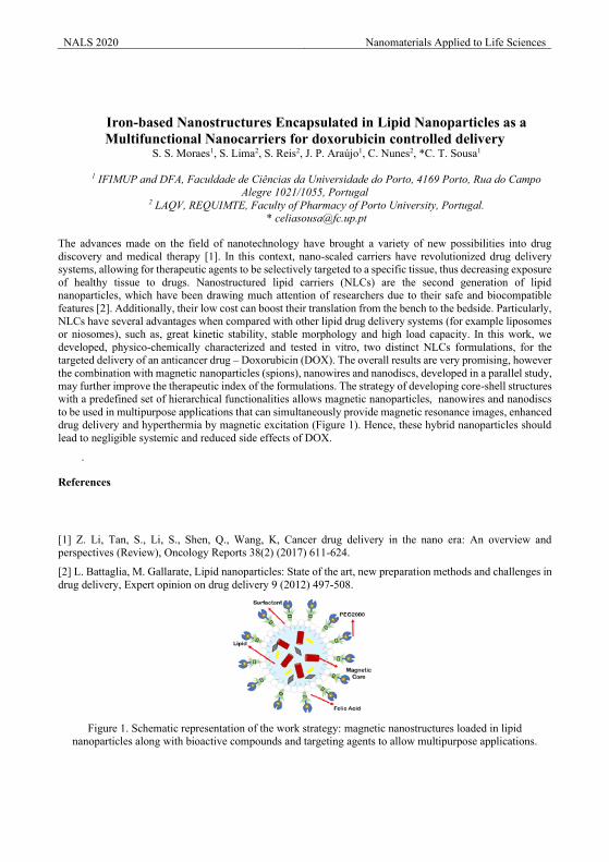
NALS 2020 Nanomaterials Applied to Life Sciences
Iron-based Nanostructures Encapsulated in Lipid Nanoparticles as a Multifunctional Nanocarriers for doxorubicin controlled delivery
S. S. Moraes1, S. Lima2, S. Reis2, J. P. Araújo1, C. Nunes2, *C. T. Sousa1
1 IFIMUP and DFA, Faculdade de Ciências da Universidade do Porto, 4169 Porto, Rua do Campo Alegre 1021/1055, Portugal
2 LAQV, REQUIMTE, Faculty of Pharmacy of Porto University, Portugal. * [email protected]
The advances made on the field of nanotechnology have brought a variety of new possibilities into drug discovery and medical therapy [1]. In this context, nano-scaled carriers have revolutionized drug delivery systems, allowing for therapeutic agents to be selectively targeted to a specific tissue, thus decreasing exposure of healthy tissue to drugs. Nanostructured lipid carriers (NLCs) are the second generation of lipid nanoparticles, which have been drawing much attention of researchers due to their safe and biocompatible features [2]. Additionally, their low cost can boost their translation from the bench to the bedside. Particularly, NLCs have several advantages when compared with other lipid drug delivery systems (for example liposomes or niosomes), such as, great kinetic stability, stable morphology and high load capacity. In this work, we developed, physico-chemically characterized and tested in vitro, two distinct NLCs formulations, for the targeted delivery of an anticancer drug – Doxorubicin (DOX). The overall results are very promising, however the combination with magnetic nanoparticles (spions), nanowires and nanodiscs, developed in a parallel study, may further improve the therapeutic index of the formulations. The strategy of developing core-shell structures with a predefined set of hierarchical functionalities allows magnetic nanoparticles, nanowires and nanodiscs to be used in multipurpose applications that can simultaneously provide magnetic resonance images, enhanced drug delivery and hyperthermia by magnetic excitation (Figure 1). Hence, these hybrid nanoparticles should lead to negligible systemic and reduced side effects of DOX.
.
References
[1] Z. Li, Tan, S., Li, S., Shen, Q., Wang, K, Cancer drug delivery in the nano era: An overview and perspectives (Review), Oncology Reports 38(2) (2017) 611-624.
[2] L. Battaglia, M. Gallarate, Lipid nanoparticles: State of the art, new preparation methods and challenges in drug delivery, Expert opinion on drug delivery 9 (2012) 497-508.
Figure 1. Schematic representation of the work strategy: magnetic nanostructures loaded in lipid
nanoparticles along with bioactive compounds and targeting agents to allow multipurpose applications.
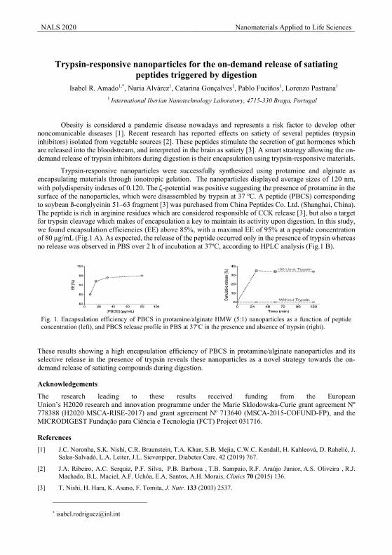
NALS 2020 Nanomaterials Applied to Life Sciences
Trypsin-responsive nanoparticles for the on-demand release of satiating
peptides triggered by digestion Isabel R. Amado1,*, Nuria Alvárez1, Catarina Gonçalves1, Pablo Fuciños1, Lorenzo Pastrana1
1 International Iberian Nanotechnology Laboratory, 4715-330 Braga, Portugal
Obesity is considered a pandemic disease nowadays and represents a risk factor to develop other noncomunicable diseases [1]. Recent research has reported effects on satiety of several peptides (trypsin inhibitors) isolated from vegetable sources [2]. These peptides stimulate the secretion of gut hormones which are released into the bloodstream, and interpreted in the brain as satiety [3]. A smart strategy allowing the on-demand release of trypsin inhibitors during digestion is their encapsulation using trypsin-responsive materials.
Trypsin-responsive nanoparticles were successfully synthesized using protamine and alginate as encapsulating materials through ionotropic gelation. The nanoparticles displayed average sizes of 120 nm, with polydispersity indexes of 0.120. The z-potential was positive suggesting the presence of protamine in the surface of the nanoparticles, which were disassembled by trypsin at 37 ºC. A peptide (PBCS) corresponding to soybean ß-conglycinin 51–63 fragment [3] was purchased from China Peptides Co. Ltd. (Shanghai, China). The peptide is rich in arginine residues which are considered responsible of CCK release [3], but also a target for trypsin cleavage which makes of encapsulation a key to maintain its activity upon digestion. In this study, we found encapsulation efficiencies (EE) above 85%, with a maximal EE of 95% at a peptide concentration of 80 µg/mL (Fig.1 A). As expected, the release of the peptide occurred only in the presence of trypsin whereas no release was observed in PBS over 2 h of incubation at 37ºC, according to HPLC analysis (Fig.1 B).
These results showing a high encapsulation efficiency of PBCS in protamine/alginate nanoparticles and its selective release in the presence of trypsin reveals these nanoparticles as a novel strategy towards the on-demand release of satiating compounds during digestion.
Acknowledgements
The research leading to these results received funding from the European Union’s H2020 research and innovation programme under the Marie Sklodowska-Curie grant agreement Nº 778388 (H2020 MSCA-RISE-2017) and grant agreement Nº 713640 (MSCA-2015-COFUND-FP), and the MICRODIGEST Fundação para Ciência e Tecnologia (FCT) Project 031716.
References [1] J.C. Noronha, S.K. Nishi, C.R. Braunstein, T.A. Khan, S.B. Mejia, C.W.C. Kendall, H. Kahleová, D. Rahelić, J.
Salas-Salvadó, L.A. Leiter, J.L. Sievenpiper, Diabetes Care. 42 (2019) 767.
[2] J.A. Ribeiro, A.C. Serquiz, P.F. Silva, P.B. Barbosa , T.B. Sampaio, R.F. Araújo Junior, A.S. Oliveira , R.J. Machado, B.L. Maciel, A.F. Uchôa, E.A. Santos, A.H. Morais, Clinics 70 (2015) 136.
[3] T. Nishi, H. Hara, K. Asano, F. Tomita, J. Nutr. 133 (2003) 2537.
Fig. 1. Encapsulation efficiency of PBCS in protamine/alginate HMW (5:1) nanoparticles as a function of peptide concentration (left), and PBCS release profile in PBS at 37ºC in the presence and absence of trypsin (right).
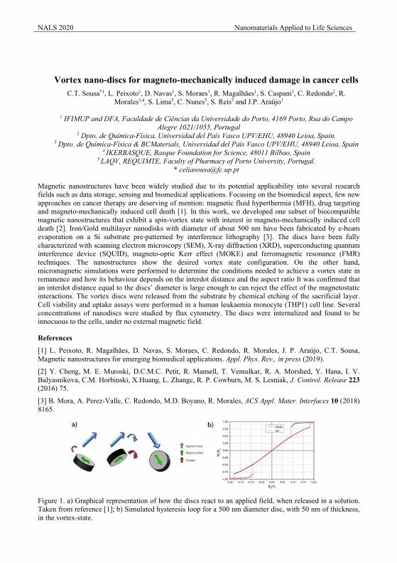
NALS 2020 Nanomaterials Applied to Life Sciences
Vortex nano-discs for magneto-mechanically induced damage in cancer cells C.T. Sousa*1, L. Peixoto1, D. Navas1, S. Moraes1, R. Magalhães1, S. Caspani1, C. Redondo2, R.
Morales3,4, S. Lima5, C. Nunes5, S. Reis5 and J.P. Araújo1
1 IFIMUP and DFA, Faculdade de Ciências da Universidade do Porto, 4169 Porto, Rua do Campo Alegre 1021/1055, Portugal
2 Dpto. de Química-Física, Universidad del País Vasco UPV/EHU, 48940 Leioa, Spain. 3 Dpto. de Química-Física & BCMaterials, Universidad del País Vasco UPV/EHU, 48940 Leioa, Spain
4 IKERBASQUE, Basque Foundation for Science, 48011 Bilbao, Spain 5 LAQV, REQUIMTE, Faculty of Pharmacy of Porto University, Portugal.
Magnetic nanostructures have been widely studied due to its potential applicability into several research fields such as data storage, sensing and biomedical applications. Focusing on the biomedical aspect, few new approaches on cancer therapy are deserving of mention: magnetic fluid hyperthermia (MFH), drug targeting and magneto-mechanically induced cell death [1]. In this work, we developed one subset of biocompatible magnetic nanostructures that exhibit a spin-vortex state with interest in magneto-mechanically induced cell death [2]. Iron/Gold multilayer nanodisks with diameter of about 500 nm have been fabricated by e-beam evaporation on a Si substrate pre-patterned by interference lithography [3]. The discs have been fully characterized with scanning electron microscopy (SEM), X-ray diffraction (XRD), superconducting quantum interference device (SQUID), magneto-optic Kerr effect (MOKE) and ferromagnetic resonance (FMR) techniques. The nanostructures show the desired vortex state configuration. On the other hand, micromagnetic simulations were performed to determine the conditions needed to achieve a vortex state in remanence and how its behaviour depends on the interdot distance and the aspect ratio It was confirmed that an interdot distance equal to the discs’ diameter is large enough to can reject the effect of the magnetostatic interactions. The vortex discs were released from the substrate by chemical etching of the sacrificial layer. Cell viability and uptake assays were performed in a human leukaemia monocyte (THP1) cell line. Several concentrations of nanodiscs were studied by flux cytometry. The discs were internalized and found to be innocuous to the cells, under no external magnetic field.
References
[1] L. Peixoto, R. Magalhães, D. Navas, S. Moraes, C. Redondo, R. Morales, J. P. Araújo, C.T. Sousa, Magnetic nanostructures for emerging biomedical applications. Appl. Phys. Rev., in press (2019).
[2] Y. Cheng, M. E. Muroski, D.C.M.C. Petit, R. Mansell, T. Vemulkar, R. A. Morshed, Y. Hana, I. V. Balyasnikova, C.M. Horbinski, X.Huang, L. Zhangc, R. P. Cowburn, M. S. Lesniak, J. Control. Release 223 (2016) 75.
[3] B. Mora, A. Perez-Valle, C. Redondo, M.D. Boyano, R. Morales, ACS Appl. Mater. Interfaces 10 (2018) 8165.
Figure 1. a) Graphical representation of how the discs react to an applied field, when released in a solution. Taken from reference [1]; b) Simulated hysteresis loop for a 500 nm diameter disc, with 50 nm of thickness, in the vortex-state.
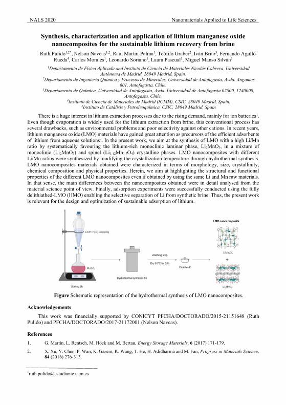
NALS 2020 Nanomaterials Applied to Life Sciences
Synthesis, characterization and application of lithium manganese oxide nanocomposites for the sustainable lithium recovery from brine
Ruth Pulido1,2*, Nelson Naveas1,2, Raúl Martin-Palma1, Teófilo Graber2, Iván Brito3, Fernando Agulló-Rueda4, Carlos Morales1, Leonardo Soriano1, Laura Pascual5, Miguel Manso Silván1
1Departamento de Física Aplicada and Instituto de Ciencia de Materiales Nicolás Cabrera, Universidad Autónoma de Madrid, 28049 Madrid, Spain.
2Departamento de Ingeniería Química y Procesos de Minerales, Universidad de Antofagasta, Avda. Angamos 601, Antofagasta, Chile.
3Departamento de Química, Universidad de Antofagasta, Avda. Universidad de Antofagasta 02800, 1240000, Antofagasta, Chile.
4Instituto de Ciencia de Materiales de Madrid (ICMM), CSIC, 28049 Madrid, Spain. 5Instituto de Catálisis y Petroleoquímica, CSIC, 28049 Madrid, Spain
There is a huge interest in lithium extraction processes due to the rising demand, mainly for ion batteries1. Even though evaporation is widely used for the lithium extraction from brine, this conventional process has several drawbacks, such as environmental problems and poor selectivity against other cations. In recent years, lithium manganese oxide (LMO) materials have gained great attention as precursors of the efficient adsorbents of lithium from aqueous solutions2. In the present work, we aim at the synthesis of LMO with a high Li/Mn ratio by systematically favouring the lithium-rich monoclinic laminar phase, Li2MnO3, in a mixture of monoclinic (Li2MnO3) and spinel (Li1.12Mn1.7O4) crystalline phases. LMO nanocomposites with different Li/Mn ratios were synthesized by modifying the crystallization temperature through hydrothermal synthesis. LMO nanocomposites materials obtained were characterized in terms of morphology, size, crystallinity, chemical composition and physical properties. Herein, we aim at highlighting the structural and functional properties of the different LMO nanocomposites even if obtained by using the same Li and Mn raw materials. In that sense, the main differences between the nanocomposites obtained were in detail analysed from the material science point of view. Finally, adsorption experiments were successfully conducted using the fully delithiathed-LMO (HMO) enabling the selective separation of Li from synthetic brine. Thus, the present work is relevant for the design and optimization of sustainable adsorption of lithium.
Figure Schematic representation of the hydrothermal synthesis of LMO nanocomposites.
Acknowledgements
This work was financially supported by CONICYT PFCHA/DOCTORADO/2015-21151648 (Ruth Pulido) and PFCHA/DOCTORADO/2017-21172001 (Nelson Naveas).
References 1. G. Martin, L. Rentsch, M. Höck and M. Bertau, Energy Storage Materials. 6 (2017) 171-179.
2. X. Xu, Y. Chen, P. Wan, K. Gasem, K. Wang, T. He, H. Adidharma and M. Fan, Progress in Materials Science. 84 (2016) 276-313.
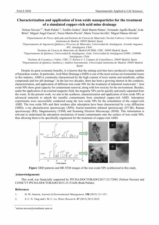
NALS 2020 Nanomaterials Applied to Life Sciences
Characterization and application of iron oxide nanoparticles for the treatment of a simulated copper-rich acid mine drainage
Nelson Naveas1,2*, Ruth Pulido1,2, Teófilo Graber2, Raúl Martin-Palma1, Fernando Agulló-Rueda3, Iván Brito4, Miguel Ángel García5, Nerea Martín-Pavón6, María Teresa Sevilla6, Miguel Manso-Silván1
1Departamento de Física Aplicada and Instituto de Ciencia de Materiales Nicolás Cabrera, Universidad Autónoma de Madrid, 28049 Madrid, Spain.
2Departamento de Ingeniería Química y Procesos de Minerales, Universidad de Antofagasta, Avenida Angamos 601, Antofagasta, Chile.
3Instituto de Ciencia de Materiales de Madrid (ICMM), CSIC, 28049 Madrid, Spain. 4Departamento de Química, Universidad de Antofagasta, Avda. Universidad de Antofagasta 02800, 1240000,
Antofagasta, Chile. 5Instituto de Ceramica y Vidrio, CSIC, C/ Kelsen n◦ 5, Campus de Cantoblanco, 28049 Madrid, Spain.
6Departamento de Química Analítica y Análisis Instrumental, Universidad Autónoma de Madrid, 28049 Madrid, Spain
Despite its great economic benefits, it is known that the mining activities have produced a large number of hazardous wastes. In particular, Acid Mine Drainage (AMD) is one of the most serious environmental issues in this industry. AMD is commonly characterized by the high content of toxic metals and metalloids, sulfate compounds and low pH drainage1. In the last two decades, there has been a growing interest in the synthesis, characterization, and application of different iron oxide NPs for the treatment of industrial wastewater 2. Iron oxide NPs show great capacity for contaminant removal, along with low toxicity for the environment. Besides, under the application of an external magnetic field, the magnetic NPs can be quickly and easily separated from the waste. In the present work, we aim at the synthesis, characterization and application of iron oxide NPs as advanced materials to adsorb the metallic contaminants from simulated copper-rich AMD. Adsorption experiments were successfully conducted using the iron oxide NPs for the remediation of the copper-rich AMD. The iron oxide NPs and their residues after adsorption have been characterized by x-ray diffraction (XRD), x-ray photoelectron spectroscopy (XPS), fourier-transform infrared spectroscopy (FT-IR), Raman spectroscopy (RS), Magnetometry (VSM) and Scanning Electron Microscopy (SEM). This information is relevant to understand the adsorption mechanism of metal contaminants onto the surface of iron oxide NPs, thus allowing them to be specifically engineered for the treatment of copper-rich AMD.
Figure XRD patterns and HR-TEM images of the iron oxide NPs synthesized in this study.
Acknowledgements
This work was financially supported by PFCHA/DOCTORADO/2017-21172001 (Nelson Naveas) and CONICYT PFCHA/DOCTORADO/2015-21151648 (Ruth Pulido).
References 1. H. M. Anawar, Journal of Environmental Management. 158 (2015) 111-121.
2. S. C. N. Tang and I. M. C. Lo, Water Research. 47 (2013) 2613-2632.
NALS 2020 Nanomaterials Applied to Life Sciences
Hybrid technologies for mine reclamation by means of nanomaterials, fertilization and phytoremediation D. Baragaño*, N. Álvarez, J.R. Gallego, A. González
1Environmental Biotechnology & Geochemistry Group, University of Oviedo, C/Gonzalo Gutiérrez Quirós s/n, 33600 Mieres, Asturias, Spain
The abandonment of Hg-As mining and metallurgy sites can severely affect the environment [1]. Arsenic is a highly toxic and carcinogenic metalloid and as such it compromises ecosystem quality and human health. This pollutant is usually presented in the paragenesis of Hg minerals, such as cinnabar, thus the extractive and processing activities generated the mobilization of the As affecting the surrounding soils.
In order to remediate As-polluted soils, several techniques have been applied, such as soil washing, stabilization and phytoremediation [2]. However, in the last decade, a novel stabilization technology based on the application of nanomaterials for immobilizing the pollutants in the soils matrix has been developed [3]. The use of zero valent iron nanoparticles (nZVI) for As stabilization in soils has been explored successfully, although the application of this inorganic amendment can negatively affect to other soil components and plants. On the other hand, organic amendments, such as ecological fertilizers, improve the growth and development of the plants while its effect on the mobilization of arsenic in the soil is not clear.
Thus, in this work, a greenhouse experiment was performed using both materials, alone and in combination, with a highly As polluted soil from a Hg mine, La Soterraña, located in northern Spain. The experiment consisted on 30 pots of each treatment: nZVI application (2%), the addition of an ecological fertilizer of animal and plant origin (30 g Kg-1 of soil) and the combination of both treatments at the same doses. Additionally, 30 pots with the polluted soil without any treatment were also prepared as controls. The pots were initially pre-incubated for 3 days prior planting Medicago sativa L. Ten pots from each treatment were collected at 1, 3 and 10 days, and samples of the soil and plants were analyzed in each time.
The available As concentration in treated soil with nZVI was significantly decreased, although fertilizer addition increased this concentration. However, the combination of both amendments did not affect to As availability. In plants, an increase of As concentration was detected in the aboveground part and the roots after fertilizer addition, but this increase was lower in the soil treated with compost and/or nZVI than in soil without amendments. The application of nZVI only decreased the As concentration in the roots.
Several physiological parameters related to oxidative stress of Medicago sativa L. were also determined. Respect to the photosynthetic pigments, the fertilizer addition to Soterraña soil caused a decrease of the chlorophyll content that may be related to higher As accumulation in the aboveground part. nZVI nanoparticles did not modify chlorophyll content, and carotenoids concentration was significant increased. The combination of amendments did not affect the photosynthetic pigments.
Both the addition of nZVI and fertilizer to the soil decreased the oxidative stress of the plants by reducing the hydrogen peroxide (H2O2) and malondialdehyde (MDA) content and increasing the proline levels when compared with the results obtained in the soil without amendments. According to our results, the low concentrations of MDA obtained suggest that Medicago sativa L. antioxidant system was able to cope with the production of H2O2 induced by As present in the soil.
Finally, thiol compounds synthesis were analysed in the plant. The results showed that the application of nZVI to the soil induce the synthesis of new thiol compounds in the roots but no in the aboveground part. The root is the organ where more As accumulation was observed. Fertilization induced a higher de novo synthesis of thiols both in the aerial part and in the root.
NALS 2020 Nanomaterials Applied to Life Sciences Acknowledgements
This work was supported by the Ministerio de Economía y Competitividad (MINECO, Spain), under the Project CTM 2016-75894-P. Diego Baragaño obtained a grant from the “Formación del Profesorado Universitario” program, financed by the “Ministerio de Educación, Cultura y Deporte de España”. This study was also financed by LIFE16ENV/ES/000481 (SUBproducts4LIFE).
References
[1] J.R. Gallego, N. Esquinas, E. Rodríguez-Valdés, J.M. Ménendez Aguado, C. Sierra, J. Hazard. Mater. 300 (2015) 561-571.
[2] D. Baragaño, E. Rodríguez-Valdés, A.I. Peláez, C. Boente, N. Matanzas, N. García, J.R. Gallego, Proceedings. 2 (2018) 1503,
[3] D. Baragaño, J. Alonso, J.R. Gallego, M.C. Lobo, M. Gil-Díaz, Chemosphere. 238 (2020) 124624.
NALS 2020 Nanomaterials Applied to Life Sciences
Study of synthetic nanovesicles as labels in lateral flow immunoassay A. Moyano1, E. Serrano-Pertierra1, M. Matos2, G. Gutierrez2, M. Rivas3, M.C. Blanco-López1
1 Department of Physical and Analytical Chemistry, University of Oviedo, Julián Clavería 8, 33006 Oviedo, Spain. 2Department of Chemical and Environmental Engineering, University of Oviedo, Julián Clavería 8, 33006
Oviedo, Spain. 3Department of Physics, University of Oviedo, 33204 Gijón, Spain
Lateral flow immunoassay (LFIA) is a rapid, simple and cost-effective paper-based platform, used in fields such as diagnostics in medicine [1], food science or environmental control. For a dipstick format, the paper substrate consists of three parts: conjugate pad, membrane and absorbent pad. Traditionally, LFIAs have been used only as qualitative tests, given that they rely on a visual signal (positive/negative test). Gold nanoparticles (GNP) are the most extensively employed labels because they display an intense red colour due to their surface plasmon resonance.
In this work, we propose the use of synthetic nanovesicles encapsulating dyes (sudan red) as alternative to traditional labels employed in these tests. Vesicles were prepared by Ethanol Injection Method (EI), consisting of a three step- method: injection, evaporation and sonication [2]. Sudan red is a lipophilic molecule and, due to this chemical character, is encapsulated at the vesicles membrane with significantly high efficiency. The surface nanovesicles with dyes were modified with carboxyl groups to get their functionality as labels. To test the performance of these labels, the biotin-neutravidin model system has been used. Firstly, the nanovesicles were functionalized with neutravidin using the carbodiimide crosslinker chemistry and a biotin test line was used on the nitrocellulose membrane. Once the nanovesicles were bound to the neutravidin, they were mixed with a running buffer in solution and the conjugate pad of the LFI was introduced in the solution, so that it flew along the nitrocellulose membrane by capillarity. Only the neutravidin-coated nanovesicles were retained at the biotin test line forming the line. The density of this test line was evaluated with an optical reader and compared to that of other conventional nanoparticles.
Acknowledgements
This work was supported by the Spanish Ministry of Economy and Competitiveness under projects MAT2017-84959-C2-1-R, and the Consejería de Economía y Empleo del Principado de Asturias under the Grant IDI/2018/000185. This study was also financied by Europa Investigación (ref: EUIN2019-103338).
References [1] Oliveira-Rodríguez, M.; Serrano-Pertierra, E.; García, A. C.; López-Martín, S.; Yañez-Mo, M.; Cernuda-Morollón,
E.; Blanco-López M.C., Point-of-care detection of extracellular vesicles: Sensitivity optimization and multiple-target detection. Biosensors and Bioelectronics 2017, 87, 38 - 45.
[2] García-Manrique, P.; Matos, M.; Gutiérrez, G.; Estupiñán O.R.; Blanco-López M.C; Pazos, C., Using factorial experimental design to prepare size-tuned nanovesicles. Amer Chemical SOC Washington, DC, USA. 2016, 87, 38-45.
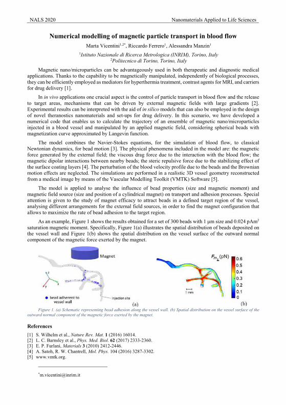
NALS 2020 Nanomaterials Applied to Life Sciences
Numerical modelling of magnetic particle transport in blood flow Marta Vicentini1,2*, Riccardo Ferrero1, Alessandra Manzin1
1Istituto Nazionale di Ricerca Metrologica (INRIM), Torino, Italy 2Politecnico di Torino, Torino, Italy
Magnetic nano/microparticles can be advantageously used in both therapeutic and diagnostic medical applications. Thanks to the capability to be magnetically manipulated, independently of biological processes, they can be efficiently employed as mediators for hyperthermia treatment, contrast agents for MRI, and carriers for drug delivery [1].
In in vivo applications one crucial aspect is the control of particle transport in blood flow and the release to target areas, mechanisms that can be driven by external magnetic fields with large gradients [2]. Experimental results can be interpreted with the aid of in silico models that can also be employed in the design of novel theranostics nanomaterials and set-ups for drug delivery. In this scenario, we have developed a numerical code that enables us to calculate the trajectory of an ensemble of magnetic nano/microparticles injected in a blood vessel and manipulated by an applied magnetic field, considering spherical beads with magnetization curve approximated by Langevin function.
The model combines the Navier-Stokes equations, for the simulation of blood flow, to classical Newtonian dynamics, for bead motion [3]. The physical phenomena included in the model are: the magnetic force generated by the external field; the viscous drag force due to the interaction with the blood flow; the magnetic dipolar interactions between nearby beads; the steric repulsive force due to the stabilizing effect of the surface coating layers [4]. The perturbation of the blood velocity profile due to the beads and the Brownian motion effects are neglected. The simulations are performed in a realistic 3D vessel geometry reconstructed from a medical image by means of the Vascular Modelling Toolkit (VMTK) Software [5].
The model is applied to analyse the influence of bead properties (size and magnetic moment) and magnetic field source (size and position of a cylindrical magnet) on transport and adhesion processes. Special attention is given to the study of magnet efficacy to attract beads in a defined target region of the vessel, analysing different arrangements for the external field sources, in order to find the magnet configuration that allows to maximize the rate of bead adhesion to the target region.
As an example, Figure 1 shows the results obtained for a set of 300 beads with 1 µm size and 0.024 pAm2 saturation magnetic moment. Specifically, Figure 1(a) illustrates the spatial distribution of beads deposited on the vessel wall and Figure 1(b) shows the spatial distribution on the vessel surface of the outward normal component of the magnetic force exerted by the magnet.
Figure 1. (a) Schematic representing bead adhesion along the vessel wall. (b) Spatial distribution on the vessel surface of the
outward normal component of the magnetic force exerted by the magnet.
References [1] S. Wilhelm et al., Nature Rev. Mat. 1 (2016) 16014. [2] L. C. Barnsley et al., Phys. Med. Biol. 62 (2017) 2333-2360. [3] E. P. Furlani, Materials 3 (2010) 2412-2446. [4] A. Satoh, R. W. Chantrell, Mol. Phys. 104 (2016) 3287-3302. [5] www.vmtk.org.
NALS 2020 Nanomaterials Applied to Life Sciences
Barley and Wheat plants affected by the presence of nanoscale zero valent iron in a calcareous soil
M.C. Lobo*, M. Gil-Díaz, J. Alonso, P. García-Gonzalo
IMIDRA, Finca El Encín A-2, km 38.200 Alcalá de Henares 28800 (Madrid) Spain
Soil pollution by metal(loid)s is a worldwide problem due to the adverse effects produced on the ecosystem and human health. Anthropogenic activities, such as mining, military activities, manufacturing, and the land application of industrial or domestic sludge are the main sources of metal(loid) pollution. Uptake of metal(loid)s by plants is mainly influenced by their bioavailable fraction rather than by the total amount in soil. In the last 15 years, the use of nanoscale zero-valent iron (nZVI) for metal(loid) immobilization has gained increasing amounts of attention due to its potential for broader application, higher reactivity and cost-effectiveness compared to other in situ remediation technologies. Consequently, the application of ZVI nanoparticles on polluted soils will lead them to be released to the environment. Studies performed with plants grown in polluted soils treated with nZVI showed an improvement of plant development due to the immobilization of pollutants. However limited data are available regarding the effect of nZVI on plants cultivated in unpolluted soils. In fact, most of the studies have been performed at lab-scale, and long-term data is still scant. The present study describes a greenhouse experiment which evaluates the impact of nZVI on the development of barley and wheat plants through all their vegetative cycle.
A stabilized water dispersion of nZVI NANOFER 25S was purchased from NanoIron (Rajhrad, Czech Republic). Ceramic pots (4 L) were filled with calcareous soil samples treated with nZVI at a dose of 5% and untreated samples were used as control. Barley (Hordeum vulgare, cultivar Pedrezuela) and wheat (Triticum aestivium, cultivar Albares) seeds were initially sown in a seedbed. After 72 h of interaction soil-nZVI, two plants per pot were transplanted. Four pots per treatment were included. The pots were periodically watered with tap water during the experiment. Plants were harvested after four months growth, when they had completed their growing period. Different physiological and chemical parameters were analysed in barley and wheat plants as well as the impact of nZVI on plant ultrastructure using transmission electron microscopy (TEM). Iron availability and soil physico-chemical soil properties were also evaluated.
Both species, barley and wheat, completed their growing cycle and did not show visual symptoms of toxicity. However, the plants grown in soils treated with nZVI presented a lower biomass and photosynthetic activity (Figure 1). According to the malondialdehyde content (MDA), a significant increase of oxidative stress was not observed. The TEM studies revealed slight morphological alterations in the root structure, altered membranes, swollen mitochondria, and unstructured crests (Figure 2). The addition of nZVI did not strongly favour the iron uptake by barley and wheat plants at the experimental conditions. A slight increase of iron availability in soils was observed. Taking into account the obtained results we can concluded that the treatment with nZVI at the experimental conditions did not induce a strong toxicity in barley and wheat plants although they showed a decrease in their development. Monitoring studies with other types of soil and other doses of nZVI are necessary to evaluate its effect on the growth of the plants and at cellular scale.
Acknowledgements
Projects REHABILITA CTM2016-78222-C2-1-R (AEI/FEDER, UE), PDR-18 NANOFERTLIZA (IMIDRA, Comunidad de Madrid, Spain).
NALS 2020 Nanomaterials Applied to Life Sciences
Figure 1. Chlorophyll mean values according to SPAD measure for wheat (A) and barley (B) plants at
three sampling times. Biomass of the plants at the end of the growing cycle (C). Bars followed by different letters significantly differ (p<0.05).
Figure 2. TEM images of wheat (A, B) and barley root plants (C, D). Plant grown in control soil and (A,
C), plant grown in soil treated with nZVI at 5% (B, D).
A B
C
A B
C D
NALS 2020 Nanomaterials Applied to Life Sciences
Assessing Nanoremediation Efficiency: lab-scale to field application M. Gil-Díaz*, J. Alonso, P. García-Gonzalo, M.C. Lobo
IMIDRA, Finca El Encín A-2, km 38.200 Alcalá de Henares 28800 (Madrid) Spain
Since the onset of industrialization two centuries ago, the release of metal(loid)s from anthropogenic activities such as industrial processes, manufacturing, mining, road transport, military activities and agriculture have led to large extensions of contaminated land and water. Although soil contamination is commonly determined on the basis of the total concentration of metal(loid)s in the matrix, this parameter gives limited information about the potential harmful effect of these pollutants on soil biota. In this regard, metal bioavailability, which refers to the accessible fraction that can be assimilated by an organism, is considered a better indicator of the risk to biota. Immobilization technologies, which reduce the metal(loid) bioavailability, can be successfully used for the remediation of contaminated soil. In the last 15 years, the use of nanoscale zero-valent iron (nZVI) for soil and groundwater remediation has been investigated for its potential to immobilize metal(loid)s. Spectroscopy characterizations have confirmed that nZVI has a core-shell structure, which leads to exceptional properties for concurrent sorption/complexation and reductive precipitations of metal(loid) ions. Here, we describe a nanoremediation strategy based on the nZVI application for the remediation of metal(loid) polluted soils.
Firstly, the effectiveness of nZVI for metal(loid) immobilization was tested in water samples at different experimental conditions. Then, the ZVI nanoparticles were applied to different types of polluted soils including single and multi-metal(loid) contamination. In order to evaluate the impact of the nanoremediation on soil, the iron leachability was studied as well as the physico-chemical and biological soil properties. In addition, experiments in growth chamber and greenhouse were performed to evaluate how the nanoremediation treatment affects barley plant growth, including the iron and metal(loid) uptake. After the effectiveness of nZVI at lab-scale was demonstrated, its capacity under field conditions at long-term was evaluated.
The global results showed better immobilization results in water than in soil samples. In soil samples the effectiveness depended on the dose and type of nZVI, soil properties (pH, organic matter content, texture, electrical conductivity), metal(loid) characteristics, the presence of several metal(loid)s and environmental conditions (Figure 1). According to the leaching experiments with acidic polluted soils, the leachates from nZVI-treated soils showed lower toxicity than leachates from untreated ones. In general, the soil phytotoxicity of polluted soils decreased after the nanoremediation treatment due to the decrease of the metal(loid) availability. Barley plants showed a better development in polluted soils treated with nZVI (Figure 2). The iron uptake strongly depended on soil pH; thus, barley plants cultivated during all the plant cycle in polluted calcareous soils treated with nZVI, did not show a significant increase of iron in the different parts of the plants (root, shoot and grain). The application of nZVI at field conditions, in a brownfield polluted with As and Hg and the monitoring of the stability of the nanoremediation treatment for 32 months showed a significant decrease of As and Hg leaching (up 70% and 80%, respectively) and was stable over time (Figure 3). Effect on soil microbial activity and biodiversity were also evaluated. PCR-DGGE analysis revealed that nZVI application leads to a shift in the microbial communities structure and an enhanced arylsulfatase enzyme activity was found along the sampling period (Figure 4). These results suggest that nZVI treatment causes significant changes in the available pool of sulfur in these polluted soils. On the basis of these findings, nZVI emerges as a potential remediation strategy for soil and groundwater polluted with metal(loid)s.
Acknowledgements
Projects REHABILITA CTM2016-78222-C2-1-R (AEI/FEDER, UE).
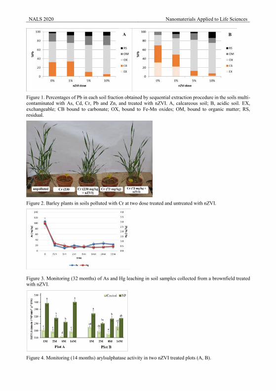
NALS 2020 Nanomaterials Applied to Life Sciences
Figure 1. Percentages of Pb in each soil fraction obtained by sequential extraction procedure in the soils multi-contaminated with As, Cd, Cr, Pb and Zn, and treated with nZVI. A, calcareous soil; B, acidic soil. EX, exchangeable; CB bound to carbonate; OX, bound to Fe-Mn oxides; OM, bound to organic matter; RS, residual.
Figure 2. Barley plants in soils polluted with Cr at two dose treated and untreated with nZVI.
Figure 3. Monitoring (32 months) of As and Hg leaching in soil samples collected from a brownfield treated with nZVI.
Figure 4. Monitoring (14 months) arylsulphatase activity in two nZVI treated plots (A, B).
A B
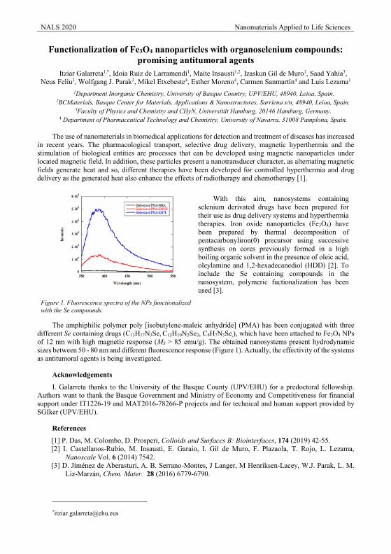
NALS 2020 Nanomaterials Applied to Life Sciences
Functionalization of Fe3O4 nanoparticles with organoselenium compounds: promising antitumoral agents
Itziar Galarreta1,*, Idoia Ruiz de Larramendi1, Maite Insausti1,2, Izaskun Gil de Muro1, Saad Yahia3, Neus Feliu3, Wolfgang J. Parak3, Mikel Etxebeste4, Esther Moreno4, Carmen Sanmartín4 and Luis Lezama1
1Department Inorganic Chemistry, University of Basque Country, UPV/EHU, 48940, Leioa, Spain. 2BCMaterials, Basque Center for Materials, Applications & Nanostructures, Sarriena s/n, 48940, Leioa, Spain.
3Faculty of Physics and Chemistry and CHyN, Universitat Hamburg, 20146 Hamburg, Germany. 4 Department of Pharmaceutical Technology and Chemistry, University of Navarra, 31008 Pamplona, Spain.
The use of nanomaterials in biomedical applications for detection and treatment of diseases has increased
in recent years. The pharmacological transport, selective drug delivery, magnetic hyperthermia and the stimulation of biological entities are processes that can be developed using magnetic nanoparticles under located magnetic field. In addition, these particles present a nanotransducer character, as alternating magnetic fields generate heat and so, different therapies have been developed for controlled hyperthermia and drug delivery as the generated heat also enhance the effects of radiotherapy and chemotherapy [1].
Figure 1. Fluorescence spectra of the NPs functionalized with the Se compounds.
With this aim, nanosystems containing selenium derivated drugs have been prepared for their use as drug delivery systems and hyperthermia therapies. Iron oxide nanoparticles (Fe3O4) have been prepared by thermal decomposition of pentacarbonyliron(0) precursor using successive synthesis on cores previously formed in a high boiling organic solvent in the presence of oleic acid, oleylamine and 1,2-hexadecanediol (HDD) [2]. To include the Se containing compounds in the nanosystem, polymeric fuctionalization has been used [3].
The amphiphilic polymer poly [isobutylene-maleic anhydride] (PMA) has been conjugated with three different Se containing drugs (C13H17N3Se, C12H10N2Se2, C8H7N3Se,), which have been attached to Fe3O4 NPs of 12 nm with high magnetic response (MS > 85 emu/g). The obtained nanosystems present hydrodynamic sizes between 50 - 80 nm and different fluorescence response (Figure 1). Actually, the effectivity of the systems as antitumoral agents is being investigated.
Acknowledgements
I. Galarreta thanks to the University of the Basque County (UPV/EHU) for a predoctoral fellowship. Authors want to thank the Basque Government and Ministry of Economy and Competitiveness for financial support under IT1226-19 and MAT2016-78266-P projects and for technical and human support provided by SGIker (UPV/EHU).
References
[1] P. Das, M. Colombo, D. Prosperi, Colloids and Surfaces B: Biointerfaces, 174 (2019) 42-55. [2] I. Castellanos-Rubio, M. Insausti, E. Garaio, I. Gil de Muro, F. Plazaola, T. Rojo, L. Lezama,
Nanoscale Vol. 6 (2014) 7542. [3] D. Jiménez de Aberasturi, A. B. Serrano-Montes, J Langer, M Henriksen-Lacey, W.J. Parak, L. M.
Liz-Marzán, Chem. Mater. 28 (2016) 6779-6790.
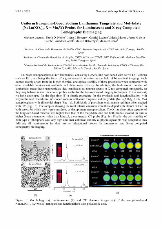
NALS 2020 Nanomaterials Applied to Life Sciences
Uniform Europium-Doped Sodium Lanthanum Tungstate and Molybdate (NaLa(XO4)2, X = Mo,W) Probes for Luminescent and X-ray Computed
Tomography Bioimaging Mariano Laguna1, Nuria O. Nuñez1,*, Ana I. Becerro1, Gabriel Lozano1, Maria Moros2, Jesús M de la
Fuente2, Ariadna Corral3, Marcin Balcerzyk3, Manuel Ocaña1
1 Instituto de Ciencia de Materiales de Sevilla, CSIC, Américo Vespucio 49, 41092, Isla de la Cartuja, Sevilla, Spain
2 Instituto de Ciencia de Materiales de Aragón, CSIC/UniZar and CIBER-BBN, Edificio I+D, Mariano Esquillor s/n, 50018 Zaragoza, Spain
3 Centro Nacional de Aceleradores (CNA) (Universidad de Sevilla, Junta de Andalucía, CSIC), c/Thomas Alva Edison 7, 41092, Isla de la Cartuja, Sevilla, Spain
Ln-based nanophosphors (Ln = lanthanide), consisting a crystalline host doped with active Ln3+ cations
such as Eu3+, are being the focus of a great research attention in the field of biomedical imaging. Such interest mainly arises from the higher chemical and optical stability of these phosphors when compared with other available luminescent materials and their lower toxicity. In addition, the high atomic number of lanthanides make these nanoparticles ideal candidates as contrast agents in X-ray computed tomography so they may behave as multifunctional probes useful for the two mentioned imaging techniques. In this context, we have developed for the first time [1] a simple procedure for the synthesis and functionalization with polyacrilic acid of uniform Eu3+-doped sodium lanthanum tungstate and molybdate (NaLa(XO4)2, X=W, Mo) nanophosphors with ellipsoidal shape (Fig. 1a). Both kinds of phosphors emit intense red light when excited with UV (Fig. 1b). The samples showing the most intense emission were those doped with 20 mol % Eu3+ in both cases, for which they were considered as the optimum nanophosphors. The X-ray absorption capacity of the tungstate-based material was higher than that of the molybdate one and both probes showed, in turn, a higher X-ray attenuation value than Iohexol, a commercial CT probe (Fig. 1c). Finally, the cell viability of both type of phosphors was very high and their colloidal stability at physiological pH was acceptable thus fulfilling all requirements for their use as bifunctional probes for luminescent and X-ray computed tomography bioimaging.
Figure 1. Morphology (a), luminescence (b) and CT phantom images (c) of the europium-doped NaLa(XO4)2, (X=Mo,W) nanoparticles functionalized with polyacrylic acid.
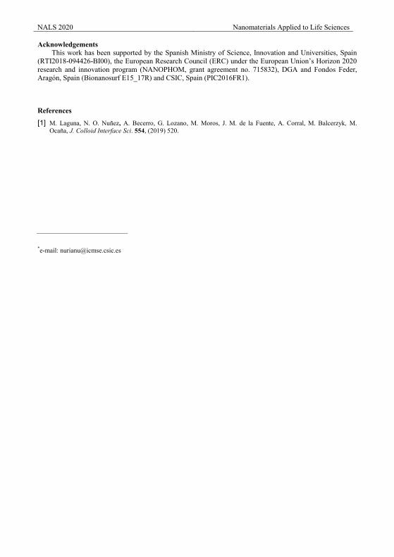
NALS 2020 Nanomaterials Applied to Life Sciences Acknowledgements
This work has been supported by the Spanish Ministry of Science, Innovation and Universities, Spain (RTI2018-094426-BI00), the European Research Council (ERC) under the European Union’s Horizon 2020 research and innovation program (NANOPHOM, grant agreement no. 715832), DGA and Fondos Feder, Aragón, Spain (Bionanosurf E15_17R) and CSIC, Spain (PIC2016FR1).
References
[1] M. Laguna, N. O. Nuñez, A. Becerro, G. Lozano, M. Moros, J. M. de la Fuente, A. Corral, M. Balcerzyk, M. Ocaña, J. Colloid Interface Sci. 554, (2019) 520.
*e-mail: [email protected]
NALS 2020 Nanomaterials Applied to Life Sciences
Synthesis of controlled size starch nanoparticles G. Gutiérrez1, D. Morán1, A. Marefati2, M. Rayner2, M. Matos1
1Department of Chemical and Environmental Engineering, University of Oviedo, Julián Clavería 8, 33006 Oviedo, Spain
2Department of Food Technology, Engineering, and Nutrition, Lund University, P.O. Box 124, SE 221 00 Lund, Sweden
Nanoparticles (NPs) are the right choice for strategic development of new drug delivery systems with novel applications in food, cosmetics and healthcare [1]. Starch is a non-allergenic abundant polysaccharide in nature, renewable and biodegradable making it an ideal candidate as reagent for green bioformulations. Starch models are described as concentric semi-crystalline multistate structures that are involved in the production of new nano-elements. The disruption of amorphous domains of semi-crystalline granular by acid hydrolysis will produce starch nanocrystal, while gelatinized starch will form starch in the form of starch nanoparticles (SNPs) [2].
Several methods have been known to produce SNPs such as high-pressure nanoemulsification, cross-linking, microemulsion/antisolvent nanoprecipitation [1-6]. The final properties of the SNPs are strongly influenced by the synthesis route and conditions so it will determine its final applications.
The aim of this work is to synthesize controlled size SNPs by modifying main parameters involved through nanoprecipitation and microemulsion methods regarding also the effect of amylose and amylopectin content of maize starch on the final starch particle size.
Comparing both methods of preparation, smaller sizes were obtained by the nanoprecipitation method than microemulsion method, sizes from 24.4 to 25.2 nm are obtained depending on the operating conditions used. The composition of the maize used also was found to have influence on the final particle size since high amylopectin maize starch allowed the formation of smaller SNPs leading to values of 16 nm.
However, higher yields were obtained when the microemulsion method was used. Different types of oil and surfactants as well as oil and surfactant concentration were studied. Mean sizes in the range 31-112 nm were obtained when normal maize starch was used, while sizes from 22.9 to 81.5 nm were obtained with high amylopectin content and from 54.2 to 90.8 nm for high amylose starch.
Acknowledgements
This work was supported by the Ministerio de Economía y Competitividad (MINECO, Spain), under the Grant MAT2017-84959-C2-1-R. This study was also financed by the Consejería de Economía y Empleo del Principado de Asturias (Plan de Ciencia, Tecnología e Innovación 2013-2017), under the Grant IDI/2018/000185. Support from the European Regional Development Fund (ERDF) is gratefully acknowledged. This study was also financied by Europa Investigación (ref: EUIN2019-103338) and European H2020-MSCA-ITN-2018 by the Grant Agreement number 813439.
References [1] H.Y. Kim, S.S. Park, S.T. Lim. Colloids and Surf. B: Biointerfaces 126 (2015) 607–620. [2] D. Le Corre, J. Bras, and A. Dufresne. Biomacromolecules 2010, 11, 1139–1153. [3] H. Saari, C. Fuentes, M. Sjöö, M. Rayner, M. Wahlgren. Carbohydrate Polymers (2017) 157, 10 2017, 558-566. [4] S.F. Chin, S.C. Pang, S.H. Tay. Carbohydrate Polymers 86 (2011) 1817– 1819. [5] S.F. Chin, A. Azman, and S.C. Pang.. Carbohydrate Polymers 86 (2011) 1817– 1819. [6] S.H. Mahmoudi Najafi, M. Baghaie, A. Ashori. International Journal of Biological Macromolecules 87 (2016) 48–54. [7] S.V. Kumar, AP. Bafana, P. Pawar, A. Rahman, S.A. Dahoumane & C.S. Jeffryes. Scientific Reports, 8 (2018) 5106.
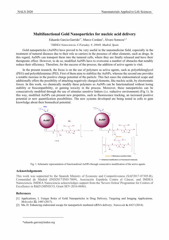
NALS 2020 Nanomaterials Applied to Life Sciences
Multifunctional Gold Nanoparticles for nucleic acid delivery Eduardo Garcia-Garrido1*, Marco Cordani1, Álvaro Somoza1,2
1IMDEA Nanociencia, C/Faraday, 9, 28049, Madrid, Spain
Gold nanoparticles (AuNPs) have proved to be very useful in the nanomedicine field, especially in the treatment of tumoral diseases due to their role as carriers in the presence of other elements, such as drugs. In this regard, AuNPs can transport them into the tumoral cells, where they are finally released and have their therapeutic effect. However, to do so, modified AuNPs have to overcome a number of obstacles that notably reduce their efficiency. Therefore, for the success of the process, the addition of active agents is vital.
In the present research, the focus is on the use of polymers as active agents, such as polyethilenglycol (PEG) and polyethilenimine (PEI). First of them aims to stabilize the AuNPs, whereas the second one provides a notable increase in the positive charge potential of the particle. This fact eases the endosomatical scape and additionally offers the possibility of attaching negatively charged elements, like nucleic acids, by electrostatic forces. In this work, we chemically modify these polymers so AuNPs can be functionalized without losing stability or biocompatibility, or gaining toxicity in the process. Moreover, these nanoparticles can be consecutively modified through the use of stimulus sensitive linkers (i.e. reductive environment) (Fig 1). In this way, modified AuNPs can present new properties, such as fluorescence tracking, an increased positive potential or new quantification possibilities. The new systems developed are being tested in cells to gain knowledge about their biomedical potential.
Fig. 1. Schematic representation of functionalized AuNPs through consecutive modification of the active agents
Acknowledgements This work was supported by the Spanish Ministry of Economy and Competitiveness (SAF2017-87305-R), Comunidad de Madrid (IND2017/IND-7809), Asociación Española Contra el Cáncer, and IMDEA Nanociencia. IMDEA Nanociencia acknowledges support from the 'Severo Ochoa' Programme for Centres of Excellence in R&D (MINECO, Grant SEV-2016-0686).
References [1] Applications, I. Unique Roles of Gold Nanoparticles in Drug Delivery, Targeting and Imaging Applications.
Molecules 22, 1445 (2017). [2] Ma, D. Enhancing endosomal escape for nanoparticle mediated siRNA delivery. Nanoscale 6, 6415 (2014).
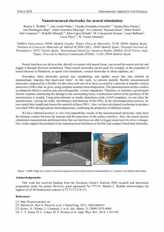
NALS 2020 Nanomaterials Applied to Life Sciences
Nanostructured electrodes for neural stimulation Beatriz L. Rodilla1,2*, Ana Arché-Núñez1, Claudia Fernández-González1,2, Sandra Ruiz-Gómez2,
Ana Domínguez-Bajo3, Ankor González-Mayorga4, Ivo Calaresu5, Rossana Rauti5, Denis Scaini5, Julio Camarero1,6, Rodolfo Miranda1,6, Elisa López Dolado4, M. Concepción Serrano3, Laura Ballerini5,
Lucas Pérez1,2, M. Teresa González1
1IMDEA Nanociencia, 28049, Madrid, España; 2Depto. Física de Materiales. UCM, 28040, Madrid, Spain; 3Instituto de Ciencia de Materiales de Madrid (ICMM-CSIC), 28049 Madrid, Spain; 4Hospital Nacional de
Parapléjicos, 45071 Toledo, Spain; 5International School for Advanced Studies (SISSA) 34136 Trieste, Italy; 6Depto. Física de la Materia Condensada-IFIMAC, UAM, 28049 Madrid, Spain;
Neural interfaces are devices that, directly in contact with neural tissue, can record the neural activity and trigger it through electrical stimulation. These neural electrodes can be used, for example, in the treatment of neural diseases as Parkinson, as spinal cord stimulators, cortical electrodes or retina implants, etc1.
Nowadays, these electrodes present size, morphology and rigidity issues that may unleash an immunologic response that inactivates them2. In this work, we present metallic flexible nanostructured electrodes composed by a flexible Au thin sheet with one of its faces covered by a network of metallic vertical nanowires (NWs) that we grow using template-assisted electrodeposition. The nanostructured surface confers an enhanced effective surface area and subsequently, a lower impedance. Therefore, to warranty a good neural electric response minimizing the damage to the surrounding tissue, a meticulous control of the geometry of the nanostructure is needed. Using polycarbonate or anodic aluminium oxide (AAO) templates, we can tailor the nanostructure, varying the order, interdistance and diameter of the NWs. In the electrodeposition process, we can control their length and choose the material of these NWs3. Also, we have developed a technique to produce core-shell NWs through pulsed electrodeposition, combining the properties of different metals.
We have obtained positive in vitro biocompatibility results of the nanostructured electrodes what show the intimate contact between the neurons and the nanowires of the surface interface. Also, the neural electric stimulation measurements performed show that our interfaces are able to trigger neural activity at low voltages. Our results support the potential of our nanostructured interfaces as flexible and compact functional electrodes.
Figure 1. SEM Image of a vertical nanowire nanostructured surface and its implementation in compact a nd flexible electrodes.
Acknowledgements
This work has received funding from the European Union’s Horizon 2020 research and innovation programme under the project ByAxon, grant agreement No 737116. Beatriz L. Rodilla acknowledges the support of UCM Predoctoral contract (CT17/17-CT18/17).
References
[1] http://byaxon-project.eu/ [2] Barrese JC, Rao N, Paroo K, et al. J Neural Eng. 2013; 10(6):066014 [3] Kotov, N.; Winter, J.; Clements, I. et al. Adv. Mater. 21 (2009) 3970-4004 [4] C. T. Sousa, D. C. Leitao, M. P. Proenca et al. Appl. Phys. Rev. 2014, 1, 031102
NALS 2020 Nanomaterials Applied to Life Sciences
Nanoencapsulation of size tuned Superparamagnetic Iron Oxide Nanoparticles (SPIONs) for biomedical applications
M. Matos1,*, G. Gutiérrez1, M. Salvador2, A. Moyano3, J. R. Álvarez1, J. C. Martínez-García2, M. Rivas2, M.C. Blanco-López3
1Department of Chemical and Environmental Engineering, University of Oviedo, Julián Clavería 8, 33006 Oviedo, Spain
2Dept. of Physics, University of Oviedo, Gijón Polytechnic School, 33203 Gijón, Spain 3Department of Physical and Analytical Chemistry, University of Oviedo, Julián Clavería 8, 33006 Oviedo, Spain
* Corresponding author, e-mail: [email protected]
There has been an increased use of magnetic iron oxide nanoparticles with superparamagnetic behaviour (SPIONs) for various biomedical applications, as promising agents in detection and analysis, or in different therapies such as targeted drug delivery [1–2]. The final properties of the SPIONs are strongly influenced by the synthesis route and conditions due to its unique size-dependent behaviour. A soft chemistry technique with a growing interest is the microemulsion reaction method due to the efficient control of size, shape, monodispersity and composition of the particles. Microemulsions work as nanoreactors where the synthesis of the desired NPs takes place, involving a chemical reaction of co-precipitation.
In previous studies regarding novel point-of-care (POC) devices for cancer diagnosis, SPIONs were functionalized to specifically bind to specific antigens and exosomes in order to perform magnetic lateral flow immunoassays that were evaluated with a novel Scanning Magnetic Impeditive sensor, which is highly sensitive to the time-varying magnetic field produced by the SPIONs [3]. Results obtained probed the ability of the method to quantify within the clinical range of interest using the SPIONs as labels. However, future improvements in NPs amplification at the lateral flow strips and in sensor instrumental design will further improve detection limits. An alternative could be SPIONs encapsulation using different type of nanocolloidal systems, such as nanovesicles. Nanovesicles (e.g., liposomes, niosomes or transfersomes) are an important family of organic NPs, produced by bottom-up nanotechnology. Moreover, nanovesicles entrapping SPIONs are promising therapeutic systems which can be guided to act in a specific site of action by an external magnetic field and used as alternative to chemotherapy through heat-triggered drug delivery or hyperthermia [4].
The feasability of the synthesis of SPIONs with average diameters between 2.8 and 16.6 nm through precipitation in W/O microemulsion has been demonstrated. The microemulsion stability region was determined in a ternary diagram as a function of the aqueous phase using the titration method. These monodisperse SPIONs were further encapsulated using size-tuned nanovesicles obtained by using a modified ethanol injection method controlling the operating conditions based in a previous study in which factorial experimental design) was applied [5]. Systems formulated were characterized in terms of size and shape by DLS (Nanozetasizer) and HR-TEM. Magnetization was also determined after nanovesicles purification by scaning magneto inductive sensor, impedance analyser.
Acknowledgements This work was supported by the Ministerio de Economía y Competitividad (MINECO, Spain), under the
Grant MAT2017-84959-C2-1-R. This study was also financed by the Consejería de Economía y Empleo del Principado de Asturias (Plan de Ciencia, Tecnología e Innovación 2013-2017), under the Grant IDI/2018/000185. Support from the European Regional Development Fund (ERDF) is gratefully acknowledged. This study was also financied by Europa Investigación (ref: EUIN2019-103338) and European H2020-MSCA-ITN-2018 by the Grant Agreement number 813439.
References
[1] Mornet S, Vasseur S, Grasset F, Duguet E. J. Mater. Chem. 2004, 14, 2161–2175. [2] B.D. Cardoso, I. S. R. Rio, A.R.O. Rodrigues, F.C.T. Fernandes, B.G.A, A. Pires, A.M. Pereira, J.P.
Araújo, E.M.S. Castanheira, P.J.G. Coutinho, R. Soc. open sci., 2018, 5: 181017. [3] D. Lago-Cachón, M. Oliveira-Rodríguez, M. Rivas, M.C. Blanco-López, J.C. Martínez-García, A.
Moyano, M. Salvador, and J.A. García. Ieee Magnetics Letters, 2017, 8 1506305. [4] S. Likhitkar, A.K. Bajpai, Carbohydr Polym, 2012, 87, 300-308. [5] García-Manrique, P., Matos, M., Gutiérrez, G., Estupiñán,O., Blanco-López, M.C., Pazos, C., Ind Eng
Chem Res, 2016, 55, 9164-9175.
NALS 2020 Nanomaterials Applied to Life Sciences
Vesicles as antibiotic carriers
V. Marchiano1,2, M. Matos1,E Serrano2, G. Gutiérrez1, Blanco-López2 1Department of Chemical and Environmental Engineering, University of Oviedo, Julián Clavería 8, 33006
Oviedo, Spain 2Department of Physical and Analytical Chemistry, University of Oviedo, Julián Clavería 8, 33006 Oviedo, Spain
Biofilm is a complex aggregation of bacterial microorganisms enclosed in a protective and adhesive matrix. Biofilm is able to grow on any surface, to survive to environmental stresses and to allow to bacteria to infect the host in a more aggressive and resistant manner [1]. This kind of community have developed several mechanisms of resistance to antibiotics: (i) production of inactivating enzymes such as beta-lactamases; (ii) alteration of permeability of the membrane or the cell wall; (iii) alteration of the receptor or enzyme, the antibiotic target; (iv) overproduction of the enzyme or target receptor in this way the concentration of antibiotic is high and gives a toxic effect; (v) efflux pumps: membrane proteins that extrude the antibiotic so fast as to never reach a cytotoxic concentration [2]. An advantageous solution is the drug delivery technology which modify drug release profile, absorption, distribution and elimination for the benefit of improving product efficacy and safety, as well as patient convenience and compliance [3]. In order to achieve efficient targeted delivery, the designed system must avoid the host defence mechanisms, reach its target site of action and come with less side effects. Vesicular systems represent an appropriate option because taking the vesicles organism as an example since they transport biological molecules, they possess several potential advantages: they are stable in blood, can possess native targeting ligands, can confer immune tolerance and they are site specifics with a directly effect on the target [4]. Vesicles are suitable carriers for hydrophilic and lipophilic drugs, since molecules can be encapsulated either in the inner aqueous core or in the lipid layer [5]. In this work liposomes and niosomes are used for their composition similar to the cells membranes. The methods used are thin film hydration and ethanol injection. All systems formulated are characterized in terms of size, zeta potential, stability, encapsulation efficiency and encapsulation stability.
Acknowledgements (Times New Roman 11 points)
This work was supported by the Ministerio de Economía y Competitividad (MINECO, Spain), under the Grant MAT2017-84959-C2-1-R. This study was also financed by the Consejería de Economía y Empleo del Principado de Asturias (Plan de Ciencia, Tecnología e Innovación 2013-2017), under the Grant IDI/2018/000185. Support from the European Regional Development Fund (ERDF) is gratefully acknowledged. This study was also financied by Europa Investigación (ref: EUIN2019-103338) and European H2020-MSCA-ITN-2018 by the Grant Agreement number 813439
La referencia EUIN2019‐103338References
[1] Penesyan, A., Gillings, M., & Paulsen, I. T.,Molecules, 20 (2015), 5286. [2] Tacconelli, E., Carrara, E., Savoldi, A., Harbarth, S., Mendelson, M., Monnet, D. L.,Pulcini, C.,
Kahlmeter, G.,Kluytmans, J., Carmeli, Y., Ouellette, M., Outterson, K., Patel, J., Cavaleri, M., M Cox, E., Houchens, C., Grayson, L., Hansen, P., Zorzet, A. The Lancet Infectious Diseases, 18(2018), 318.
[3] Tibbitt, Mark W., James E. Dahlman, and Robert Langer.Journal of the American Chemical Society 138(2016) 704.
[4] Pattni, Bhushan S., Vladimir V. Chupin, and Vladimir P. Torchilin. Chemical reviews 115(2015), 10938. [5] Sakai, Kumiko, and Yuki Takechi.Analytical Sciences 34(2018).
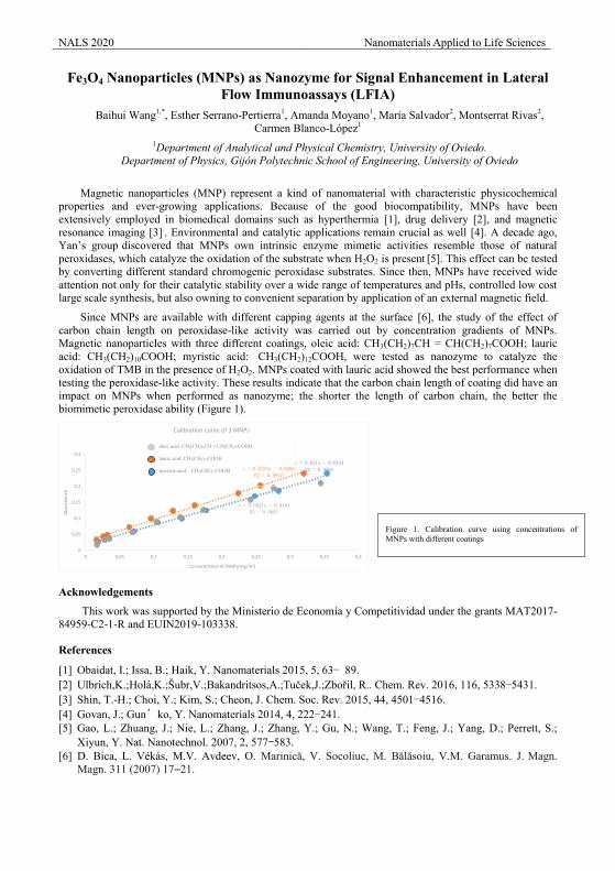
NALS 2020 Nanomaterials Applied to Life Sciences
Fe3O4 Nanoparticles (MNPs) as Nanozyme for Signal Enhancement in Lateral Flow Immunoassays (LFIA)
Baihui Wang1,*, Esther Serrano-Pertierra1, Amanda Moyano1, María Salvador2, Montserrat Rivas2, Carmen Blanco-López1
1Department of Analytical and Physical Chemistry, University of Oviedo. Department of Physics, Gijón Polytechnic School of Engineering, University of Oviedo
Magnetic nanoparticles (MNP) represent a kind of nanomaterial with characteristic physicochemical properties and ever-growing applications. Because of the good biocompatibility, MNPs have been extensively employed in biomedical domains such as hyperthermia [1], drug delivery [2], and magnetic resonance imaging [3].. Environmental and catalytic applications remain crucial as well [4]. A decade ago, Yan’s group discovered that MNPs own intrinsic enzyme mimetic activities resemble those of natural peroxidases, which catalyze the oxidation of the substrate when H2O2 is present [5]. This effect can be tested by converting different standard chromogenic peroxidase substrates. Since then, MNPs have received wide attention not only for their catalytic stability over a wide range of temperatures and pHs, controlled low cost large scale synthesis, but also owning to convenient separation by application of an external magnetic field.
Since MNPs are available with different capping agents at the surface [6], the study of the effect of carbon chain length on peroxidase-like activity was carried out by concentration gradients of MNPs. Magnetic nanoparticles with three different coatings, oleic acid: CH3(CH2)7CH = CH(CH2)7COOH; lauric acid: CH3(CH2)10COOH; myristic acid: CH3(CH2)12COOH, were tested as nanozyme to catalyze the oxidation of TMB in the presence of H2O2. MNPs coated with lauric acid showed the best performance when testing the peroxidase-like activity. These results indicate that the carbon chain length of coating did have an impact on MNPs when performed as nanozyme; the shorter the length of carbon chain, the better the biomimetic peroxidase ability (Figure 1).
Acknowledgements
This work was supported by the Ministerio de Economía y Competitividad under the grants MAT2017-84959-C2-1-R and EUIN2019-103338.
References
[1] Obaidat, I.; Issa, B.; Haik, Y. Nanomaterials 2015, 5, 63− 89. [2] , R.. Chem. Rev. 2016, 116, 5338−5431. [3] Shin, T.-H.; Choi, Y.; Kim, S.; Cheon, J. Chem. Soc. Rev. 2015, 44, 4501−4516. [4] Govan, J.; Gun’ko, Y. Nanomaterials 2014, 4, 222−241. [5] Gao, L.; Zhuang, J.; Nie, L.; Zhang, J.; Zhang, Y.; Gu, N.; Wang, T.; Feng, J.; Yang, D.; Perrett, S.;
Xiyun, Y. Nat. Nanotechnol. 2007, 2, 577–583. [6] D. Bica, L. Vékás, M.V. Avdeev, O. M ă S M ă ă M G m J. Magn.
Magn. 311 (2007) 17–21.
y = 0.621x + 0.0154
R2 = 0.9981 y = 0.6783x + 0.0282
R2 = 0.9957
y = 0.6021x + 0.0161
R2 = 0.9855
0
0,05
0,1
0,15
0,2
0,25
0,3
0 0,05 0,1 0,15 0,2 0,25 0,3 0,35 0,4
Abso
rban
ce
Concentration of MNPs(mg/ml)
Calibration curve of 3 MNPs
Figure 1. Calibration curve using concentrations of MNPs with different coatings
oleic acid: CH3(CH2)7CH = CH(CH2)7COOH
lauric acid: CH3(CH2)10COOH
myristic acid: CH3(CH2)12COOH
lan
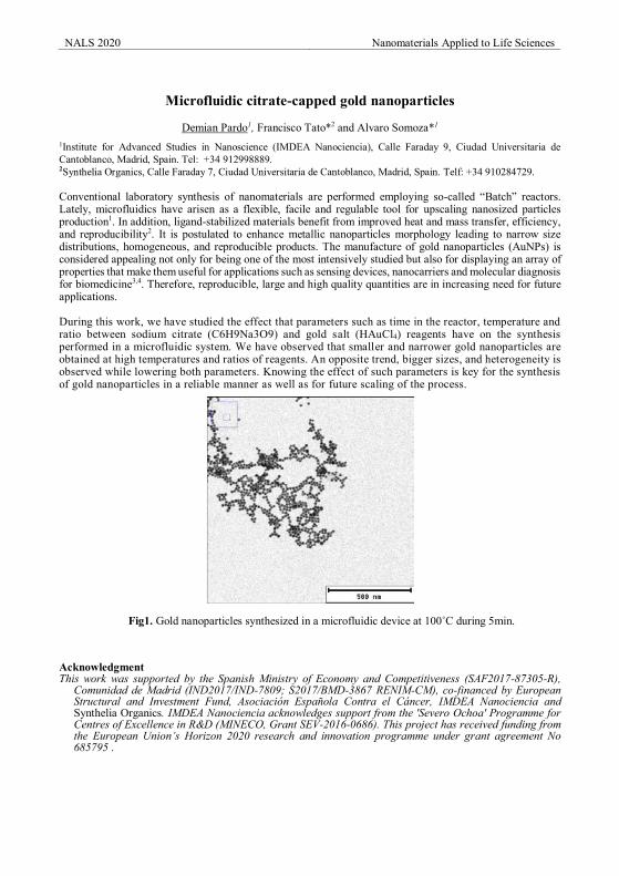
NALS 2020 Nanomaterials Applied to Life Sciences
Microfluidic citrate-capped gold nanoparticles
Demian Pardo1, Francisco Tato*2 and Alvaro Somoza*1
1Institute for Advanced Studies in Nanoscience (IMDEA Nanociencia), Calle Faraday 9, Ciudad Universitaria de Cantoblanco, Madrid, Spain. Tel: +34 912998889. 2Synthelia Organics, Calle Faraday 7, Ciudad Universitaria de Cantoblanco, Madrid, Spain. Telf: +34 910284729. Conventional laboratory synthesis of nanomaterials are performed employing so-called “Batch” reactors. Lately, microfluidics have arisen as a flexible, facile and regulable tool for upscaling nanosized particles production1. In addition, ligand-stabilized materials benefit from improved heat and mass transfer, efficiency, and reproducibility2. It is postulated to enhance metallic nanoparticles morphology leading to narrow size distributions, homogeneous, and reproducible products. The manufacture of gold nanoparticles (AuNPs) is considered appealing not only for being one of the most intensively studied but also for displaying an array of properties that make them useful for applications such as sensing devices, nanocarriers and molecular diagnosis for biomedicine3,4. Therefore, reproducible, large and high quality quantities are in increasing need for future applications. During this work, we have studied the effect that parameters such as time in the reactor, temperature and ratio between sodium citrate (C6H9Na3O9) and gold salt (HAuCl4) reagents have on the synthesis performed in a microfluidic system. We have observed that smaller and narrower gold nanoparticles are obtained at high temperatures and ratios of reagents. An opposite trend, bigger sizes, and heterogeneity is observed while lowering both parameters. Knowing the effect of such parameters is key for the synthesis of gold nanoparticles in a reliable manner as well as for future scaling of the process.
Fig1. Gold nanoparticles synthesized in a microfluidic device at 100˚C during 5min. Acknowledgment This work was supported by the Spanish Ministry of Economy and Competitiveness (SAF2017-87305-R),
Comunidad de Madrid (IND2017/IND-7809; S2017/BMD-3867 RENIM-CM), co-financed by European Structural and Investment Fund, Asociación Española Contra el Cáncer, IMDEA Nanociencia and Synthelia Organics. IMDEA Nanociencia acknowledges support from the 'Severo Ochoa' Programme for Centres of Excellence in R&D (MINECO, Grant SEV-2016-0686). This project has received funding from the European Union’s Horizon 2020 research and innovation programme under grant agreement No 685795 .
NALS 2020 Nanomaterials Applied to Life Sciences References
[1] Victor Sebastian, Christopher D. Smith and Klavs F. Jensen. Nanoscale Issue 14, 2016. Shape-controlled continuous synthesis of metal nanostructures.A. Gapito, I. Saias, Phys. Rev. X 7 (2017) 031037.
[2] Matthew B. Plutschack, Bartholomäus Pieber, Kerry Gilmore , and Peter H. Seeberger. Chem. Rev., 2017, 117 (18), pp 11796–11893. The Hitchhiker’s Guide to Flow Chemistry.
[3] Karen Weintraub.Biomedicine: The new gold standard. Nature 2013 volume 495, pages S14–S16. [4] David A. Giljohann, Dwight S. Seferos, Weston L. Daniel, Matthew D. Massich, Pinal C. Patel, and Chad
A. Mirkin. Gold Nanoparticles for Biology and Medicine. Angew. Chem. Int. Ed. 2010, 49, 3280–3294. [email protected]
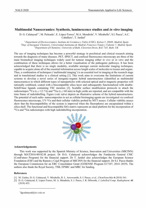
NALS 2020 Nanomaterials Applied to Life Sciences
Multimodal Nanoceramics: Synthesis, luminescence studies and in vitro imaging
D. G. Calatayud1,*, M. Peiteado1, E. López-Torres2, M.A. Mendiola2, V. Mirabello3, S.I. Pascu3, A.C. Caballero1, T. Jardiel1
1 Department of Electroceramics, Instituto de Cerámica y Vidrio (CSIC), Kelsen 5, 28049, Madrid, Spain 2Dep. of Inorganic Chemistry, Universidad Autónoma de Madrid, Francisco Tomás y Valiente 7, Madrid, Spain
3Department of Chemistry, University of Bath, Claverton Down, BA2 7AY, Bath, UK
The use of imaging techniques has become a powerful strategy in preclinical and clinical research aiming towards the diagnosis of many diseases. PET, SPECT, and confocal fluorescence microscopy are three of the main biomedical imaging techniques widely used for tumour imaging either in vivo or in vitro and the combination of these techniques allows for a better visualisation of the pathogenic pathways. It has been acknowledged that there is no single modality, available amongst current molecular imaging techniques, capable to acquire alone all of the essential information across length scales of molecules to tissues and organs. Thus, a combination of techniques (‘multimodal imaging’) is an essential tool in imaging at the research stage and in translational studies to a clinical setting [1]. This work aims to overcome the limitations of current systems to develop a novel series of inorganic-organic hybrid nanostructures (identified as multimodal nanoceramics) in which different types of nanoparticles with selected optical and/or magnetic properties will rationally combined, coated with a biocompatible silica layer and subsequently functionalized with double‐Schiff‐base ligands containing TSC moieties [2]. Scalable surface modification protocols to attach the radioisotopes 64Cu (t1/2=12.7 h) and 68Ga (t1/2=68 min) in high yields are reported, and are compatible with the time frame of radiolabelling. Figure (vide infra) depicts an illustrative scheme of the hybrid nanostructures. The potential of such radio- nanocomposites to act as cellular bioimaging agents are investigated via confocal fluorescence microscopy, UV-Vis and their cellular viability probed by MTT assays. Cellular viability assays show that the biocompatibility of the system is improved when the fluorophores are encapsulated within a silica shell. The functional and biocompatible SiO2 matrix represents an ideal platform for the incorporation of 64Cu and 68Ga radioisotopes with high radiolabelling incorporation.
Acknowledgements
This work was supported by the Spanish Ministry of Science, Innovation and Universities (MICINN) through MAT2016-80182-R project. Dr D.G. Calatayud acknowledges the Fundación General CSIC (ComFuturo Program) for the financial support. Dr T. Jardiel also acknowledges the European Science Foundation (ESF) and the Ramon y Cajal Program of MICINN for the financial support. Dr S.I. Pascu thanks the European Commission for an ERC Consolidator Grant (O2SENSE Program 617107, 2014–2019). The authors also thank the Royal Society, TSB, EPSRC and MRC for funding.
References [1] M. Lledos, D. G. Calatayud, V. Mirabello, R. L. Arrowsmith, S. I. Pascu, et al., ChemNanoMat 4 (2018) 361. [2] D. G. Calatayud, E. López‐Torres, M. A. Mendiola, S. I. Pascu, J. R. Dilworth, J. Labelled Comp. Radiopharm. 61
(2018) 455.
NALS 2020 Nanomaterials Applied to Life Sciences
Efficient Production of Biofunctionalized Multi-Layer Graphene from
Graphite Flakes in Water
Noelia Losada-Garcia 1, Angel Berenguer-Murcia 2, Diego Cazorla-Amorós 2 and Jose M. Palomo 1,* 1Department of Biocatalysis. Institute of Catalysis (CSIC). Marie Curie 2. Cantoblanco. Campus UAM,
28049 Madrid, Spain; [email protected], [email protected] 2Instituto Universitario de Materiales y Departamento de Química Inorgánica, Universidad de Alicante,
Apartado 99, San Vicente del Raspeig, E-03080 Alicante, Spain
Biofunctionalized graphene was successfully produced in water from graphite flakes by a simple, rapid, and efficient methodology based on a bioexfoliation technology [1]. The methodology consisted in the application of a lipase, with a unique mechanism of interaction with hydrophobic surfaces, combined with a previous mechanical sonication, to selectively generate lipase-graphene sheets conjugates in water at room temperature. The adsorption of the lipase on the graphene sheets permits to keep the sheets separated in comparison with other methods. It was possible to obtain more than 80% of graphene (in the form of multi-layer graphene) from low-cost graphite and with less damage compared to commercial graphene oxide (GO) or reduced GO. Experimental analysis demonstrated the formation of multi-layer graphene (MLG) mainly using lipase from Thermomyces lanuginosus (TLL). This new functionalized graphene could represent a cheap and excellent nanomaterial for conjugation of biomolecules (DNA, enzymes, carbohydrates) for multiple applications, diagnosis, therapy or environmental remediation [2-4].
Acknowledgements
This research was supported by GRO PROGRAM 2017 and SAMSUNG L.S. The authors thank the support by the Spanish National Research Council (CSIC) (intramural project 201980E081). We thank Ramiro Martínez from Novozymes.
References
[1] Losada-Garcia, N. et al. Nanomaterials. 9 (2019) 1344. [2] Shajaripour Jaberi, SY. et al. Anal Chim Acta. 1078 (2019) 42-52 [3] Akhavan, O. et al. J Mater Chem. 22-27 (2012) 13773-13781 [4] Lu, K-Q, et al. J Mater Chem A. 6 (2018) 4590
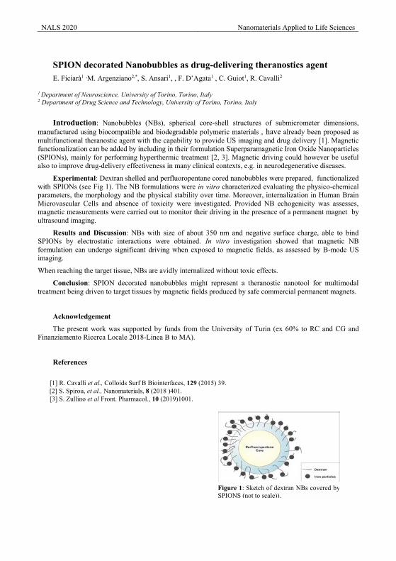
NALS 2020 Nanomaterials Applied to Life Sciences
SPION decorated Nanobubbles as drug-delivering theranostics agent E. Ficiarà1 ,M. Argenziano2,*, S. Ansari1, , F. D’Agata1 , C. Guiot1, R. Cavalli2
1 Department of Neuroscience, University of Torino, Torino, Italy 2 Department of Drug Science and Technology, University of Torino, Torino, Italy
Introduction: Nanobubbles (NBs), spherical core-shell structures of submicrometer dimensions,
manufactured using biocompatible and biodegradable polymeric materials , have already been proposed as multifunctional theranostic agent with the capability to provide US imaging and drug delivery [1]. Magnetic functionalization can be added by including in their formulation Superparamagnetic Iron Oxide Nanoparticles (SPIONs), mainly for performing hyperthermic treatment [2, 3]. Magnetic driving could however be useful also to improve drug-delivery effectiveness in many clinical contexts, e.g. in neurodegenerative diseases.
Experimental: Dextran shelled and perfluoropentane cored nanobubbles were prepared, functionalized with SPIONs (see Fig 1). The NB formulations were in vitro characterized evaluating the physico-chemical parameters, the morphology and the physical stability over time. Moreover, internalization in Human Brain Microvascular Cells and absence of toxicity were investigated. Provided NB echogenicity was assesses, magnetic measurements were carried out to monitor their driving in the presence of a permanent magnet by ultrasound imaging.
Results and Discussion: NBs with size of about 350 nm and negative surface charge, able to bind SPIONs by electrostatic interactions were obtained. In vitro investigation showed that magnetic NB formulation can undergo significant driving when exposed to magnetic fields, as assessed by B-mode US imaging.
When reaching the target tissue, NBs are avidly internalized without toxic effects.
Conclusion: SPION decorated nanobubbles might represent a theranostic nanotool for multimodal treatment being driven to target tissues by magnetic fields produced by safe commercial permanent magnets.
Acknowledgement
The present work was supported by funds from the University of Turin (ex 60% to RC and CG and Finanziamento Ricerca Locale 2018-Linea B to MA).
References
[1] R. Cavalli et al., Colloids Surf B Biointerfaces, 129 (2015) 39.
[2] S. Spirou, et al., Nanomaterials, 8 (2018 )401. [3] S. Zullino et al Front. Pharmacol., 10 (2019)1001.
Figure 1: Sketch of dextran NBs covered by SPIONS (not to scale)).
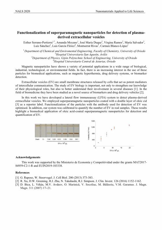
NALS 2020 Nanomaterials Applied to Life Sciences
Functionalization of superparamagnetic nanoparticles for detection of plasma-
derived extracellular vesicles Esther Serrano-Pertierra1,*, Amanda Moyano1, José María Duque2, Virgina Ramos2, María Salvador3,
Luis Sánchez2, Luis García Flórez4, Montserrat Rivas3, Carmen Blanco-López1 1 Department of Chemical and Enviromental Engineering, Faculty of Chemistry, University of Oviedo
2Hospital Universitario San Agustín, Avilés 3Department of Physics, Gijón Polytechnic School of Engineering, University of Oviedo
4Hospital Universitario Central de Asturias, Oviedo
Magnetic nanoparticles have shown a variety of potential applications in a wide range of biological, industrial, technological, or environmental fields. In fact, there is an increasing interest in the use of these particles for biomedical applications, such as magnetic hyperthermia, drug delivery systems, or biomarker detection.
Extracellular vesicles (EV) are small membrane structures released by cells that act as potent mediators of intercellular communication. The study of EV biology is important, not only to strengthen our knowledge of their physiological roles, but also to better understand their involvement in several diseases [1]. In the field of biomedicine they have been studied as a novel source of biomarkers and drug delivery vehicles [2].
In this work we have developed a lateral flow immunoassay (LFIA) system to detect plasma-derived extracellular vesicles. We employed superparamagnetic nanoparticles coated with a double layer of oleic cid [3] as a reporter label. Functionalization of the particles with the antibody used for detection of EV was optimised. In addition, our system was calibrated to quantify the number of EV in real samples. These results highlight a biomedical application of oleic acid-coated superparamagnetic nanoparticles for detection and quantification of EV.
Acknowledgements
This work was supported by the Ministerio de Economía y Competitividad under the grants MAT2017-84959-C2-1-R and EUIN2019-103338.
References
[1] G. Raposo, W. Stoorvogel. J. Cell Biol. 200 (2013) 373-383. [2] R. Xu, D.W. Greening, H.J. Zhu, N. Takahashi, R.J. Simpson. J. Clin. Invest. 126 (2016) 1152-1162. [3] D. Bica, L. Vékás, M.V. Avdeev, O. Marinică, V. Socoliuc, M. Bălăsoiu, V.M. Garamus. J. Magn.
Magn. 311 (2007) 17–21.
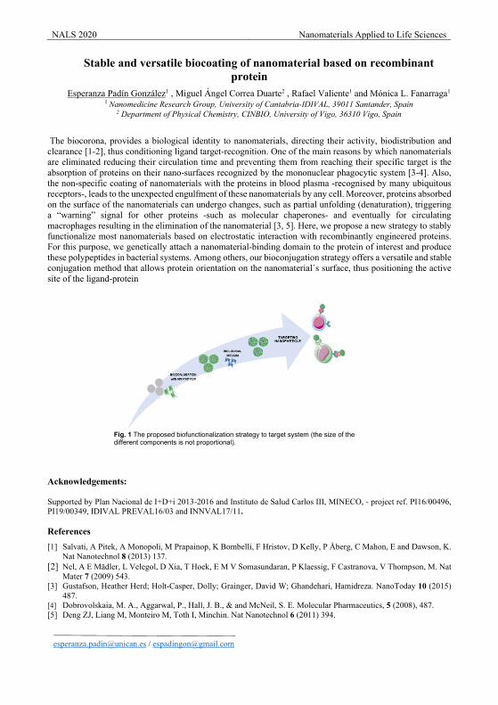
NALS 2020 Nanomaterials Applied to Life Sciences
Stable and versatile biocoating of nanomaterial based on recombinant protein
Esperanza Padín González1 , Miguel Ángel Correa Duarte2 , Rafael Valiente1 and Mónica L. Fanarraga1
1 Nanomedicine Research Group, University of Cantabria-IDIVAL, 39011 Santander, Spain 2 Department of Physical Chemistry, CINBIO, University of Vigo, 36310 Vigo, Spain
The biocorona, provides a biological identity to nanomaterials, directing their activity, biodistribution and clearance [1-2], thus conditioning ligand target-recognition. One of the main reasons by which nanomaterials are eliminated reducing their circulation time and preventing them from reaching their specific target is the absorption of proteins on their nano-surfaces recognized by the mononuclear phagocytic system [3-4]. Also, the non-specific coating of nanomaterials with the proteins in blood plasma -recognised by many ubiquitous receptors-, leads to the unexpected engulfment of these nanomaterials by any cell. Moreover, proteins absorbed on the surface of the nanomaterials can undergo changes, such as partial unfolding (denaturation), triggering a “warning” signal for other proteins -such as molecular chaperones- and eventually for circulating macrophages resulting in the elimination of the nanomaterial [3, 5]. Here, we propose a new strategy to stably functionalize most nanomaterials based on electrostatic interaction with recombinantly engineered proteins. For this purpose, we genetically attach a nanomaterial-binding domain to the protein of interest and produce these polypeptides in bacterial systems. Among others, our bioconjugation strategy offers a versatile and stable conjugation method that allows protein orientation on the nanomaterial´s surface, thus positioning the active site of the ligand-protein
Acknowledgements:
Supported by Plan Nacional de I+D+i 2013‐2016 and Instituto de Salud Carlos III, MINECO, ‐ project ref. PI16/00496, PI19/00349, IDIVAL PREVAL16/03 and INNVAL17/11.
References
[1] Salvati, A Pitek, A Monopoli, M Prapainop, K Bombelli, F Hristov, D Kelly, P Åberg, C Mahon, E and Dawson, K. Nat Nanotechnol 8 (2013) 137.
[2] Nel, A E Mädler, L Velegol, D Xia, T Hoek, E M V Somasundaran, P Klaessig, F Castranova, V Thompson, M. Nat Mater 7 (2009) 543.
[3] Gustafson, Heather Herd; Holt-Casper, Dolly; Grainger, David W; Ghandehari, Hamidreza. NanoToday 10 (2015) 487.
[4] Dobrovolskaia, M. A., Aggarwal, P., Hall, J. B., & and McNeil, S. E. Molecular Pharmaceutics, 5 (2008), 487. [5] Deng ZJ, Liang M, Monteiro M, Toth I, Minchin. Nat Nanotechnol 6 (2011) 394.
[email protected] / [email protected]
Fig. 1 The proposed biofunctionalization strategy to target system (the size of the different components is not proportional).
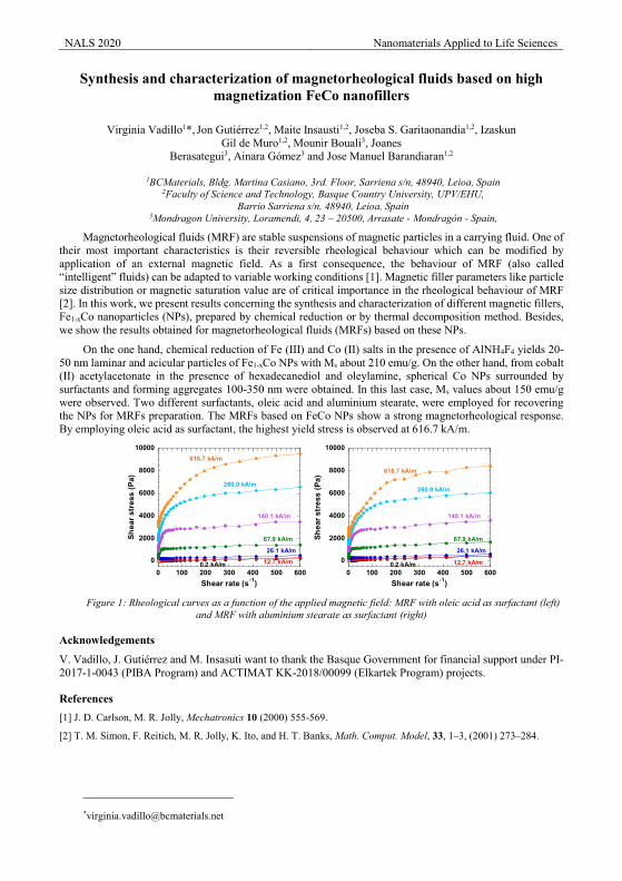
NALS 2020 Nanomaterials Applied to Life Sciences
Synthesis and characterization of magnetorheological fluids based on high magnetization FeCo nanofillers
Virginia Vadillo1*, Jon Gutiérrez1,2, Maite Insausti1,2, Joseba S. Garitaonandia1,2, Izaskun
Gil de Muro1,2, Mounir Bouali3, Joanes Berasategui3, Ainara Gómez3 and Jose Manuel Barandiaran1,2
1BCMaterials, Bldg. Martina Casiano, 3rd. Floor, Sarriena s/n, 48940, Leioa, Spain
2Faculty of Science and Technology, Basque Country University, UPV/EHU, Barrio Sarriena s/n, 48940, Leioa, Spain
3Mondragon University, Loramendi, 4, 23 – 20500, Arrasate - Mondragón - Spain,
Magnetorheological fluids (MRF) are stable suspensions of magnetic particles in a carrying fluid. One of their most important characteristics is their reversible rheological behaviour which can be modified by application of an external magnetic field. As a first consequence, the behaviour of MRF (also called “intelligent” fluids) can be adapted to variable working conditions [1]. Magnetic filler parameters like particle size distribution or magnetic saturation value are of critical importance in the rheological behaviour of MRF [2]. In this work, we present results concerning the synthesis and characterization of different magnetic fillers, Fe1-xCo nanoparticles (NPs), prepared by chemical reduction or by thermal decomposition method. Besides, we show the results obtained for magnetorheological fluids (MRFs) based on these NPs.
On the one hand, chemical reduction of Fe (III) and Co (II) salts in the presence of AlNH4F4 yields 20-50 nm laminar and acicular particles of Fe1-xCo NPs with Ms about 210 emu/g. On the other hand, from cobalt (II) acetylacetonate in the presence of hexadecanediol and oleylamine, spherical Co NPs surrounded by surfactants and forming aggregates 100-350 nm were obtained. In this last case, Ms values about 150 emu/g were observed. Two different surfactants, oleic acid and aluminium stearate, were employed for recovering the NPs for MRFs preparation. The MRFs based on FeCo NPs show a strong magnetorheological response. By employing oleic acid as surfactant, the highest yield stress is observed at 616.7 kA/m.
0
2000
4000
6000
8000
10000
0 100 200 300 400 500 600
Shea
r str
ess
(Pa)
Shear rate (s-1)
12.7 kA/m
616.7 kA/m
26.1 kA/m
67.9 kA/m
140.1 kA/m
280.9 kA/m
0.2 kA/m
0
2000
4000
6000
8000
10000
0 100 200 300 400 500 600
Shea
r str
ess
(Pa)
Shear rate (s-1)
0.2 kA/m 12.7 kA/m
26.1 kA/m
67.9 kA/m
140.1 kA/m
280.9 kA/m
616.7 kA/m
Figure 1: Rheological curves as a function of the applied magnetic field: MRF with oleic acid as surfactant (left)
and MRF with aluminium stearate as surfactant (right)
Acknowledgements
V. Vadillo, J. Gutiérrez and M. Insasuti want to thank the Basque Government for financial support under PI-2017-1-0043 (PIBA Program) and ACTIMAT KK-2018/00099 (Elkartek Program) projects.
References [1] J. D. Carlson, M. R. Jolly, Mechatronics 10 (2000) 555-569.
[2] T. M. Simon, F. Reitich, M. R. Jolly, K. Ito, and H. T. Banks, Math. Comput. Model, 33, 1–3, (2001) 273–284.
NALS 2020 Nanomaterials Applied to Life Sciences
Lifetime in vivo thermal sensing in NIR biological window Meiling Tan1, Feng Li1, Daniel Jaque2, Guanying Chen1*
1 MIIT Key Laboratory of Critical Materials Technology for New Energy Conversion and Storage, School of Chemistry and Chemical Engineering & Key Laboratory of Micro-systems and Micro-structures,
Ministry of Education, Harbin Institute of Technology, 150001 Harbin, People’s Republic of China. 2 Departamento de Física de Materiales, Universidad Autónoma de Madrid, Madrid 28049, Spain.
Luminescence nanothermometry has attracted much attention in the past decades because it allows for non-contact thermal reading that can describe in vivo thermal mapping[1,2]. Lifetime thermometry with independent of intensity, concentration and penetration depth provides outstanding opportunities for in vivo thermal sensing. Herein, we devise a NaYF4@NaYF4: Yb3+, Nd3+ @CaF2 core/shell/shell NPs, with the luminescence lifetime of Yb3+ ions is temperature-dependent. The dopants concentration plays an important role on the temperature sensitivity due to which relates with the way of energy transfer between Nd3+ and Yb3+ ions. Our lifetime nanothermometer has been used to provide accurate temperature mapping in vivo in an inflammatory mode.
Acknowledgements (Times New Roman 11 points)
This work was supported by the grants from the National Natural Science Foundation of China
(51972084 and 51672061).
References [1] Vetrone, F.; Naccache, R.; Zamarrón, A.; Juarranz de la Fuente, A.; Sanz-Rodríguez, F.; Martinez Maestro, L.;
Martin Rodriguez, E.; Jaque, D.; Garcia Sole, J.; Capobianco, J. A., Temperature sensing using fluorescent nanothermometers. Acs Nano 2010, 4 (6), 3254-3258.A. Gapito, I. Saias, Phys. Rev. X 7 (2017) 031037.
[2] Ximendes, E. C.; Santos, W. Q.; Rocha, U. s.; Kagola, U. K.; Sanz-Rodríguez, F.; Fernandez, N.; Gouveia-Neto, A. d. S.; Bravo, D.; Domingo, A. M.; del Rosal, B., Unveiling in vivo subcutaneous thermal dynamics by infrared luminescent nanothermometers. Nano Letters 2016, 16 (3), 1695-1703.
NALS 2020 Nanomaterials Applied to Life Sciences
Engineering the heating power of iron oxide nanoparticles for thermal
regulation of multi-enzymatic processes Jesus G. Ovejero1, Sabino Veintemillas1, Valeria Grazu2,3, Puerto Morales1
1Institute of Material Science of Madrid (ICMM-CSIC), Sor Juana Inés de la Cruz, 3, E-28049 Madrid, Spain.
2BioNanoSurf Group, Aragon Materials Science Institute (ICMA), CSIC/Universidad de Zaragoza, c/ Edificio I+D, Mariano Esquillor Gomez, 50018, Zaragoza, Spain.
3Centro de Investigación Biomédica en Red de Bioingeniería, Biomateriales y Nanomedicina (CIBER-BBN), Avenida Monforte de Lemos, 3-5, 28029 Madrid, Spain
Multi-enzyme processes for in vitro synthetic biology is currently considered a promising biomanufacturing platform. Complex biotransformations like sucrose synthesis [1] or chiral drug formation [2] can be achieved using enzymatic cascades, taking advantage of the combined effect of specific enzymes. However, due to the different optimal temperature of the enzymes involved (mesophiles, thermophyles, etc.), multi-enzyme processes require either the compartmentalization of the different steps or operating in a compromised temperature. The use of magnetic nanoparticles as local nanoheaters is proposed by the FET-OPEN project HOTZYMES as a novel strategy to overcome these limitations [3].
Superparamagnetic Iron Oxide Nanoparticles (SPIONs) present suitable features for this task. On one hand, there is a well-developed surface chemistry for the coating of SPIONs with multiple organic molecules. Selecting the appropriate functional group exposed by such molecules it is possible to decorate the SPIONs with enzymes in a conformation that optimizes their enzymatic activity. On the other hand, when SPIONs are exposed to an alternating magnetic field, they absorb the magnetic energy and dissipate heat in their local environment. Thus, the heat dissipated by the SPIONs can be used to activate the enzymes attached on their surfaces, maintaining the media at low temperature.
For this purpose, is crucial the control of parameters such as SPIONs core size, coating size and composition, or colloidal and magnetic properties of the particles in suspension. In this work we have explored the heating power of SPIONs prepared with different core sizes (12 nm, 20 nm and 30 nm) and different functional coatings (DMSA, APTES, Dextran, Diaminated Dextran) seeking for field and frequency conditions that maximize the dissipated heat. The results offer a clear landscape of the potential of the SPIONs as nanothermal regulators of multienzymatic processes.
Acknowledgements
The research for this work has received funding from the European Union (EU) project HOTZYMES (grant agreement n° 829162) under EU’s Horizon 2020 Programme Research and Innovation actions H2020-FETOPEN-2018-2019-2020-01.
References
[1] C. Zhong, C. You, P. Wei, and Y.-H. Percival Zhang ACS Catal. 7:9 (2017) 5992-5999 [2] W. Jiang and B. Fang Scientific Reports 6 (2016) 30462 [3] I. Armenia, V. Grazú, L. de Matteis, P. Ivanchenko, G. Martra, R. Gornati, JM de la Fuente, G. Bernardini.
J. Colloid Interface Sci. 537.1 (2019) 615
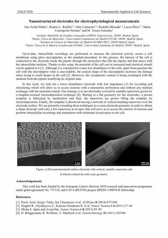
NALS 2020 Nanomaterials Applied to Life Sciences
Nanostructured electrodes for electrophysiological measurements Ana Arché-Núñez1, Beatriz L. Rodilla1,2, Julio Camarero1,4, Rodolfo Miranda1,4, Lucas Pérez1,2, María
Concepción Serrano3 and M. Teresa González1 1Instituto Madrileño de Estudios Avanzados (IMDEA Nanociencia), 28049, Madrid, Spain
2Depto. Física de Materiales. Universidad Complutense de Madrid (UCM), 28040, Madrid, Spain 3Instituto de Ciencia de Materiales de Madrid (ICMM-CSIC), 28049 Madrid, Spain
4 Depto. Física de la Materia Condensada-IFIMAC. Universidad Autónoma de Madrid, 28049, Madrid, Spain
Up-to-date, intracellular recordings are performed to measure the electrical activity across a cell membrane using glass micropipettes as the standard procedure. In this process, the interior of the cell is connected to the electrode inside the pipette through the electrolyte that fills the pipette and that mixes with the intracellular medium. Thanks to this setup, the potential of the cell can be measured and electrical stimuli can be applied to it [1]. Although it is considered to cause low disturbance to the cells, apart from puncture the cell with the micropipette what is unavoidable, the conical shape of the micropipette increases the damage when trying to reach deeper in the cell [2]. Moreover, the cytoplasmic content is being exchanged with the medium from the pipette modifying its original state.
In this work, we look for a lower disturbance electrode with low impedance [3] for recording and stimulating which will allow us to access neurons with a nanometric perforation and without any medium exchange with the neuronal content. Our strategy is to use electrodes covered by metallic nanowires grown by a template-assisted electrodeposition technique [4]. Starting in a flat geometry for the electrodes, a porous template is fabricated by anodization and then, the nanowires are grown filling the template by electrodeposition. Finally, the template is dissolved leaving a network of vertical-standing nanowires over the electrode surface. We are presently extending these techniques to a conic electrode geometry in order to obtain a sharp electrode with only a few nanowires at its apex that will serve us to access the interior of neurons and perform intracellular recordings and stimulation with minimum invasiveness to the cell.
Figure: a) Flat nanostructured surface electrode with vertical, metallic nanowires and
b) Electro-etched tip with conic geometry.
Acknowledgements
This work has been funded by the European Union's Horizon 2020 research and innovation programme under grant agreement No. 737116, and I+D+I (RETOS) project DPI2017-90058-R fellowship.
References
[1] Paolo Actis, Sergiy Tokar, Jan Clausmeyer et al. ACSNano 8 (2014) 875-884 [2] Singhal R., Orynbayeva Z., Kalyana Sundaram R. et al. Nature Nanotech 6 (2011) 57–64 [3] Micha E. Spira and Aviad Hai. Nature Nanotech 8 (2013) 83–94 [4] D. Brüggemann, B. Wolfrum, V. Maybeck et al. Nanotechnology 22 (2011) 265104
2 µm
a)
100 µm
b)
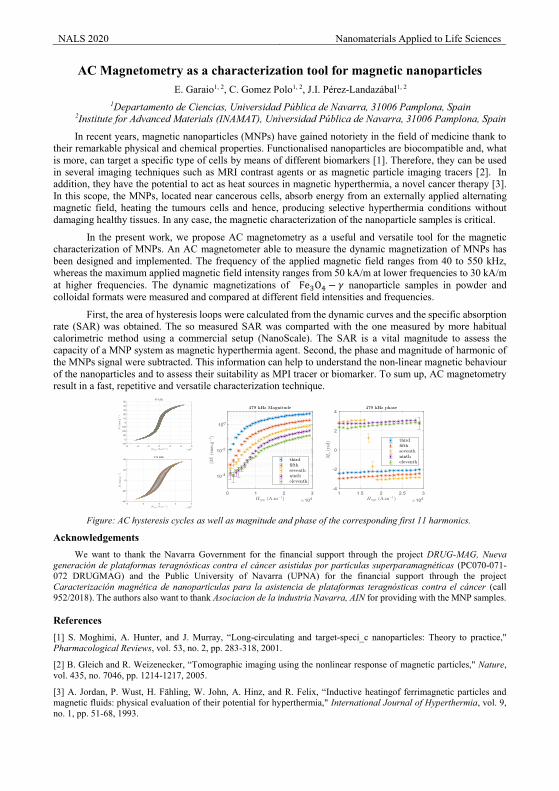
NALS 2020 Nanomaterials Applied to Life Sciences
AC Magnetometry as a characterization tool for magnetic nanoparticles E. Garaio1, 2, C. Gomez Polo1, 2, J.I. Pérez-Landazábal1, 2
1Departamento de Ciencias, Universidad Pública de Navarra, 31006 Pamplona, Spain 2Institute for Advanced Materials (INAMAT), Universidad Pública de Navarra, 31006 Pamplona, Spain
In recent years, magnetic nanoparticles (MNPs) have gained notoriety in the field of medicine thank to their remarkable physical and chemical properties. Functionalised nanoparticles are biocompatible and, what is more, can target a specific type of cells by means of different biomarkers [1]. Therefore, they can be used in several imaging techniques such as MRI contrast agents or as magnetic particle imaging tracers [2]. In addition, they have the potential to act as heat sources in magnetic hyperthermia, a novel cancer therapy [3]. In this scope, the MNPs, located near cancerous cells, absorb energy from an externally applied alternating magnetic field, heating the tumours cells and hence, producing selective hyperthermia conditions without damaging healthy tissues. In any case, the magnetic characterization of the nanoparticle samples is critical.
In the present work, we propose AC magnetometry as a useful and versatile tool for the magnetic characterization of MNPs. An AC magnetometer able to measure the dynamic magnetization of MNPs has been designed and implemented. The frequency of the applied magnetic field ranges from 40 to 550 kHz, whereas the maximum applied magnetic field intensity ranges from 50 kA/m at lower frequencies to 30 kA/m at higher frequencies. The dynamic magnetizations of Fe3O4 − 𝛾𝛾 nanoparticle samples in powder and colloidal formats were measured and compared at different field intensities and frequencies.
First, the area of hysteresis loops were calculated from the dynamic curves and the specific absorption rate (SAR) was obtained. The so measured SAR was comparted with the one measured by more habitual calorimetric method using a commercial setup (NanoScale). The SAR is a vital magnitude to assess the capacity of a MNP system as magnetic hyperthermia agent. Second, the phase and magnitude of harmonic of the MNPs signal were subtracted. This information can help to understand the non-linear magnetic behaviour of the nanoparticles and to assess their suitability as MPI tracer or biomarker. To sum up, AC magnetometry result in a fast, repetitive and versatile characterization technique.
Figure: AC hysteresis cycles as well as magnitude and phase of the corresponding first 11 harmonics.
Acknowledgements We want to thank the Navarra Government for the financial support through the project DRUG-MAG, Nueva
generación de plataformas teragnósticas contra el cáncer asistidas por partículas superparamagnéticas (PC070-071-072 DRUGMAG) and the Public University of Navarra (UPNA) for the financial support through the project Caracterización magnética de nanopartículas para la asistencia de plataformas teragnósticas contra el cáncer (call 952/2018). The authors also want to thank Asociacion de la industria Navarra, AIN for providing with the MNP samples.
References [1] S. Moghimi, A. Hunter, and J. Murray, “Long-circulating and target-speci_c nanoparticles: Theory to practice," Pharmacological Reviews, vol. 53, no. 2, pp. 283-318, 2001.
[2] B. Gleich and R. Weizenecker, “Tomographic imaging using the nonlinear response of magnetic particles," Nature, vol. 435, no. 7046, pp. 1214-1217, 2005.
[3] A. Jordan, P. Wust, H. Fähling, W. John, A. Hinz, and R. Felix, “Inductive heatingof ferrimagnetic particles and magnetic fluids: physical evaluation of their potential for hyperthermia," International Journal of Hyperthermia, vol. 9, no. 1, pp. 51-68, 1993.
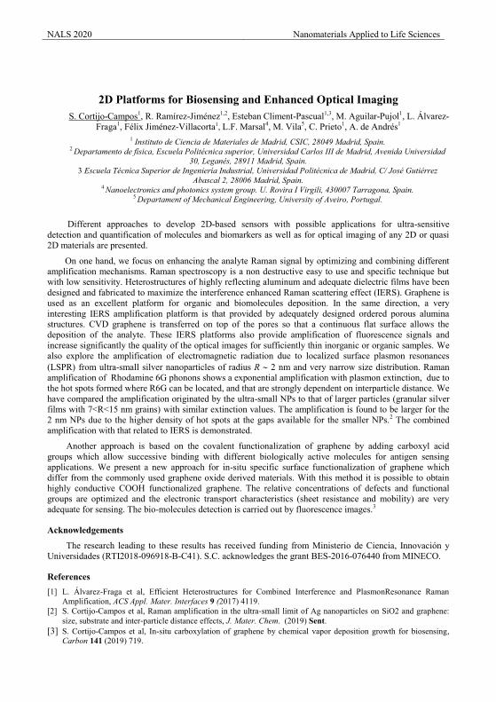
NALS 2020 Nanomaterials Applied to Life Sciences
2D Platforms for Biosensing and Enhanced Optical Imaging S. Cortijo-Campos1, R. Ramírez-Jiménez1,2, Esteban Climent-Pascual1,3, M. Aguilar-Pujol1, L. Álvarez-
Fraga1, Félix Jiménez-Villacorta1, L.F. Marsal4, M. Vila5, C. Prieto1, A. de Andrés1 1 Instituto de Ciencia de Materiales de Madrid, CSIC, 28049 Madrid, Spain.
2 Departamento de física, Escuela Politécnica superior, Universidad Carlos III de Madrid, Avenida Universidad 30, Leganés, 28911 Madrid, Spain.
3 Escuela Técnica Superior de Ingeniería Industrial, Universidad Politécnica de Madrid, C/ José Gutiérrez Abascal 2, 28006 Madrid, Spain.
4 Nanoelectronics and photonics system group. U. Rovira I Virgili, 430007 Tarragona, Spain. 5 Departament of Mechanical Engineering, University of Aveiro, Portugal.
Different approaches to develop 2D-based sensors with possible applications for ultra-sensitive detection and quantification of molecules and biomarkers as well as for optical imaging of any 2D or quasi 2D materials are presented.
On one hand, we focus on enhancing the analyte Raman signal by optimizing and combining different amplification mechanisms. Raman spectroscopy is a non destructive easy to use and specific technique but with low sensitivity. Heterostructures of highly reflecting aluminum and adequate dielectric films have been designed and fabricated to maximize the interference enhanced Raman scattering effect (IERS). Graphene is used as an excellent platform for organic and biomolecules deposition. In the same direction, a very interesting IERS amplification platform is that provided by adequately designed ordered porous alumina structures. CVD graphene is transferred on top of the pores so that a continuous flat surface allows the deposition of the analyte. These IERS platforms also provide amplification of fluorescence signals and increase significantly the quality of the optical images for sufficiently thin inorganic or organic samples. We also explore the amplification of electromagnetic radiation due to localized surface plasmon resonances (LSPR) from ultra-small silver nanoparticles of radius R 2 nm and very narrow size distribution. Raman amplification of Rhodamine 6G phonons shows a exponential amplification with plasmon extinction, due to the hot spots formed where R6G can be located, and that are strongly dependent on interparticle distance. We have compared the amplification originated by the ultra-small NPs to that of larger particles (granular silver films with 7<R<15 nm grains) with similar extinction values. The amplification is found to be larger for the 2 nm NPs due to the higher density of hot spots at the gaps available for the smaller NPs.2 The combined amplification with that related to IERS is demonstrated.
Another approach is based on the covalent functionalization of graphene by adding carboxyl acid groups which allow successive binding with different biologically active molecules for antigen sensing applications. We present a new approach for in-situ specific surface functionalization of graphene which differ from the commonly used graphene oxide derived materials. With this method it is possible to obtain highly conductive COOH functionalized graphene. The relative concentrations of defects and functional groups are optimized and the electronic transport characteristics (sheet resistance and mobility) are very adequate for sensing. The bio-molecules detection is carried out by fluorescence images.3
Acknowledgements
The research leading to these results has received funding from Ministerio de Ciencia, Innovación y Universidades (RTI2018-096918-B-C41). S.C. acknowledges the grant BES-2016-076440 from MINECO.
References [1] L. Álvarez-Fraga et al, Efficient Heterostructures for Combined Interference and PlasmonResonance Raman
Amplification, ACS Appl. Mater. Interfaces 9 (2017) 4119. [2] S. Cortijo-Campos et al, Raman amplification in the ultra‐small limit of Ag nanoparticles on SiO2 and graphene:
size, substrate and inter‐particle distance effects, J. Mater. Chem. (2019) Sent. [3] S. Cortijo-Campos et al, In-situ carboxylation of graphene by chemical vapor deposition growth for biosensing,
Carbon 141 (2019) 719.
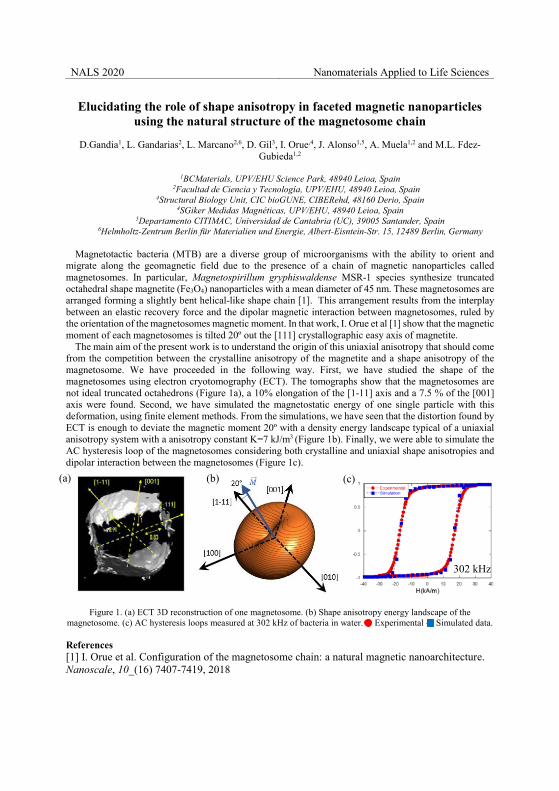
NALS 2020 Nanomaterials Applied to Life Sciences
Elucidating the role of shape anisotropy in faceted magnetic nanoparticles using the natural structure of the magnetosome chain
D.Gandia1, L. Gandarias2, L. Marcano2,6, D. Gil3, I. Orue,4, J. Alonso1,5, A. Muela1,2 and M.L. Fdez-
Gubieda1,2
1BCMaterials, UPV/EHU Science Park, 48940 Leioa, Spain
2Facultad de Ciencia y Tecnología, UPV/EHU, 48940 Leioa, Spain 4Structural Biology Unit, CIC bioGUNE, CIBERehd, 48160 Derio, Spain
4SGiker Medidas Magnéticas, UPV/EHU, 48940 Leioa, Spain 5Departamento CITIMAC, Universidad de Cantabria (UC), 39005 Santander, Spain
6Helmholtz-Zentrum Berlin für Materialien und Energie, Albert-Eisntein-Str. 15, 12489 Berlin, Germany
Magnetotactic bacteria (MTB) are a diverse group of microorganisms with the ability to orient and migrate along the geomagnetic field due to the presence of a chain of magnetic nanoparticles called magnetosomes. In particular, Magnetospirillum gryphiswaldense MSR-1 species synthesize truncated octahedral shape magnetite (Fe3O4) nanoparticles with a mean diameter of 45 nm. These magnetosomes are arranged forming a slightly bent helical-like shape chain [1]. This arrangement results from the interplay between an elastic recovery force and the dipolar magnetic interaction between magnetosomes, ruled by the orientation of the magnetosomes magnetic moment. In that work, I. Orue et al [1] show that the magnetic moment of each magnetosomes is tilted 20º out the [111] crystallographic easy axis of magnetite.
The main aim of the present work is to understand the origin of this uniaxial anisotropy that should come from the competition between the crystalline anisotropy of the magnetite and a shape anisotropy of the magnetosome. We have proceeded in the following way. First, we have studied the shape of the magnetosomes using electron cryotomography (ECT). The tomographs show that the magnetosomes are not ideal truncated octahedrons (Figure 1a), a 10% elongation of the [1-11] axis and a 7.5 % of the [001] axis were found. Second, we have simulated the magnetostatic energy of one single particle with this deformation, using finite element methods. From the simulations, we have seen that the distortion found by ECT is enough to deviate the magnetic moment 20º with a density energy landscape typical of a uniaxial anisotropy system with a anisotropy constant K=7 kJ/m3 (Figure 1b). Finally, we were able to simulate the AC hysteresis loop of the magnetosomes considering both crystalline and uniaxial shape anisotropies and dipolar interaction between the magnetosomes (Figure 1c).
Figure 1. (a) ECT 3D reconstruction of one magnetosome. (b) Shape anisotropy energy landscape of the
magnetosome. (c) AC hysteresis loops measured at 302 kHz of bacteria in water. Experimental - Simulated data.
References [1] I. Orue et al. Configuration of the magnetosome chain: a natural magnetic nanoarchitecture. Nanoscale, 10_(16) 7407-7419, 2018
(a) (b) (c)
302 kHz
NALS 2020 Nanomaterials Applied to Life Sciences
Effect of Co doping on the magnetic and photocatalytic properties of ZnFe2O4
nanoparticlesL. Cervera1,2*, S. Larumbe3, A. Zielińska-Jurek4, C. Gómez-Polo1,2,*
1 Dpto. de Ciencias. Universidad Pública de Navarra. 31006 Pamplona. Spain2 Institute for Advanced Materials (INAMAT), Universidad Pública de Navarra, 31006 Pamplona, Spain
3Surface Engineering, Asociación de la Industria Navarra, Cordovilla, Navarra, Spain4Department of Process Engineering and Chemical Technology, Faculty of Chemistry, Gdańsk University of
Technology, G. Narutowicza 11/12, Gdańsk, 80-233, Poland
Cobalt zinc ferrites have been widely investigated due to their specific electrical, optical and magnetic properties. In this work the effect of Co2+ doping on the photocatalytic properties of ZnFe2O4 nanoparticles is analysed in the design of efficient photocatalysts for the removal of organic pollutants in aqueous media. In these spinel ferrites, the activation of the redox reactions under visible light is combined with the ferrimagnetic behaviour, allowing the separation and recycling of the catalyst under the action of an external magnetic field [1,2].
CoxZn1-xFe2O4 (0 ≤ x ≤ 1) nanoparticles were prepared by co-precipitation method. Aqueous solutions of metal (iron, cobalt and zinc) salts were mixed stoichiometrically and NaOH solution (1 M) was added until the solution reaches pH = 13 favouring the precipitation and complete hydrolyzation of the ions. Then, the precipitates were collected by centrifugation, washed several times with distilled water and dried at 500C in an oven. Finally, the samples were annealed at 4000C during 6 hours [3].
Firstly, the structural and magnetic properties of samples were characterized by X-ray Diffraction (Siemens Diffractometer D5000) and SQUID magnetometry (Quantum Design MPMS XL7), respectively. The samples display the characteristic spinel structure with mean grain sizes in the range 10 – 20 nm. This ultrafine structure leads to superparamagnetic behaviour at room temperature for x ≤ 0.5. Moreover, maximum values of the magnetic moment are achieved for 0.5 ≤ x ≤ 0.7 as a consequence of the Co-Zn cation distribution in the spinel structure. Regarding the optical response, a UV-diffuse reflectance spectra (Perkin Elmer UV-Vis-NIR lambda 950) was employed to characterize the band gap energy, Eg, through the Kubelka-Munk function and the corresponding Tauc plots. Values of Eg around 1.7 eV are found, confirming the potential photocatalytic activation of these spinel ferrites under visible light. In particular, the photo degradation activity was evaluated by measuring the degradation of phenol in aqueous solution under visible light with a Xe arc lamp with a 400 nm cutoff filter. Maximum phenol degradation ratios under visible radiation are found for x = 0.5. Thus, Co0.5Zn0.5Fe2O4 nanoferrite displays optimum response in terms of high magnetic moment, superparamagnetic behaviour at room temperature and photocatalytic response under visible light.
Acknowledgements
L. Cervera would like to acknowledge the financial support of the Navarra Government within the framework of the program “Ayudas Predoctorales de Movilidad Internacional”.
References[1] Sidra Nazim et al. Ceramics International 42 (2016) 7647-7654[2] Guoli Fan et al. Ind. Eng. Chem. Res. 51 (2012) 13639-13647[3] C. Gómez-Polo et al. Journal of Magnetism and Magnetic Materials 465 (2018) 211-219.
NALS 2020 Nanomaterials Applied to Life Sciences
Novel SERS tags for multiplex detection of biomarkers S. Rodal-Cedeira1,*, A. Vázquez-Arias2, G. Bodelón1, S. Núñez-Sánchez1, A. Laporta2, L. Polavarapu3,
S. Bals2, L. M. Liz-Marzán4,5, J. Pérez-Juste1 and I. Pastoriza-Santos1 1Departamento de Química Física and CINBIO, Universidade de Vigo, Vigo, Spain
2EMAT, Universiteit Antwerpen, Antwerpen,Belgium 3Ludwig-Maximilians University of Munich, Munich, Germany
4CIC biomaGUNE and CIBER-BBN, Donostia-San Sebastian, Spain 5Ikerbasque, Basque Foundation for Science, Bilbao, Spain
Control over the morphology of metal nanoparticles (NPs) in combination with mechanic understanding is a crucial step for the development of modern nanoscience and nanotechnology. Their optical properties are related with their localized surface plasmon resonances (LSPRs). LSPRs are strongly dependent of NPs shape, size or composition. A wide range of methods based on wet chemistry have been developed to synthesize NPs with tailored optical properties. Recently our group has developed a strategy involving galvanic replacement reaction coupled seeded growth to fabricate Au/Ag nanorattles [1]. This work opens new avenues towards the shape control of multimetallic hollows nanostructures beyond the morphology of sacrificial templates.
Surface-enhanced Raman scattering (SERS) is an ultrasensitive technique which relies on the enhancement of the Raman scattering signals of a certain molecule when is close to a plasmonic nanostructure. This technique also allows the indirect identification of target molecules through the use of SERS tags, Raman encoded NPs, which comprises a specific organic Raman reporter attached to metallic NPs and often surrounded by a protected shell [2].
Herein we propose a new synthetic route to fabricate a SERS tag based on a room-temperature galvanic reaction coupled seeded growth method using Ag nanospheres (NSs) as sacrificial templates (Fig 1.A). During the process different Raman reporters could be trapped inside the resulting hollow Ag@Au NSs. The SERS tags NPs have been characterized by TEM and EDX revealing that the morphology of the sacrificial template remained after the reaction (Fig 1.B and C). Besides, we have analysed the SERS performance of the tags (Fig 1.D). This approach allowed us to codify the plasmonic particles with a library of Raman active molecules leading to the formation ultrasensitive SERS-encoded nanoparticles.
SERS-encoded NPs have been shown to present an excellent multiplexing ability particularly in immunoassays platforms which could be applied in different fields such as disease diagnosis, food safety, and environmental protection [2]. For instance, cell surface biomarkers already serve as valuable diagnostic and prognostic indicators for a variety of human diseases such as cancer. We demonstrate the reliability and multiplexing capabilities of our method towards the simultaneous SERS detection of three cell surface receptors, expressed in human epithelial carcinoma A431 and nontumoral murine fibroblast 3T3 2.2 cells.
Figure 1. (A) Scheme of the synthesis of SERS-encoded nanoparticles. (B) 3D visualization of the tomographic reconstruction of Au nanorattle and EDX mapping showing the Ag and Au distribution. (C) Representative TEM image of the closed Au nanorattles. Scale bar is 100 nm. (C) SERS spectra of different Au nanorattles encoded with several Raman reporters as indicated.
References [1] L. Polavarapu et al., J. Am. Chem. Soc. 138 (2016) 11453. [2] L. M. Liz-Marzán et al., ACS Nano (2019).
400 800 1200 1600
Inten
sity
Raman shift / cm-1
B
A
B
DAuCl4- / Ascorbic Acid
Galvanic replacementcoupled to overgrowth
A C
Malachite Green
Crystal Violet
Nile Blue
Astra Blue
Methylene Blue
NALS 2020 Nanomaterials Applied to Life Sciences
Steering the synthesis of Fe3O4 nanoparticles by using a factorial designJ. C. Echeverría1,2*, P. Moriones1,2, J.J. Garrido1,2, M.D. Ugarte2,3, E. Garaio1,2,
C. Gómez Polo1,2, J.I. Pérez-Landazábal1,2
1Departamento de Ciencias, Universidad Pública de Navarra, 31006 Pamplona, Spain2Institute for Advanced Materials (INAMAT), Universidad Pública de Navarra, 31006 Pamplona, Spain
3Departamento de Estadística, Informática y Matemáticas, Universidad Pública de Navarra, 31006 Pamplona, Spain
The synthesis of Fe3O4 nanoparticles has captivated the interest of many researchers due to its primary scientific interest and the applications in areas such as magnetic hyperthermia, drug delivery and magnetic resonance imaging. The coprecipitation technique is perhaps the simplest and most efficient chemical way to obtain magnetic nanoparticles [1]. However, the control of the composition, morphology and size of the nanoparticles is hindered by the large number of the experimental variables and the colloidal nature of the precipitate. Although the precipitation of iron oxides has been widely studied since the seminal work by Massart [2], most of research has been performed by studying one variable at a time, keeping the rest constant. This research strategy requires a lot of experiments and does not take into account the possible interactions among variables. The goals of this research was to assess the critical variables that rule the composition and structure of the nanoparticles.
We have applied the Plackett-Burman fractional factorial design at two levels [3]. The experimental factors were the initial concentration of Fe(III) and Fe(II), reaction temperature, ammonia concentration, ethanol content in the aqueous solvent, sonication time, and drying temperature. The molar ratio Fe(III)/Fe(II) was kept equal to two. The solutions were purged with N2(g) to prevent the oxidation of Fe(II) ions. To improve the robustness of the design and to estimate the standard error of the system, we included five dummy factors. The experimental design had 12 runs from the Plackett – Burman method. Characterization techniques include total iron content by AAS, FTIR, XRD, HRTEM, magnetization curves ZFC – FC, and hyperthermia response.
The total iron content, the structural and magnetic properties of the samples significantly depended on the initial Fe(III) – Fe(II) concentration and sonication time. Synthesis from 73 mM Fe(III) and 36.5 mM Fe(II), with sonication for 60 min, yielded samples with the inverse spinel structure characteristic of magnetite. On the other hand, synthesis from 146 mM Fe(III) and 73 mM Fe(II), sonicated for 10 min, resulted in samples of mixed content of Fe oxyhydroxides and magnetite, which had poor magnetic properties. Under the lower Fe concentration, thermodynamics rule over kinetics aspects of the reaction and magnetite nanoparticles precipitate whatever the sonication time; but under the higher initial Fe concentration, kinetics favours the precipitation of Fe oxyhydroxides. The largest content of magnetite, which led to the highest magnetization and hyperthermia efficiency, was obtained for 76 mM Fe(III), 36.5 mM Fe(II), 0.90 M NH3(aq), sonicating for 60 min, and drying samples under vacuum at 105 ºC.
Acknowledgements
Thanks to the Navarra Government for the financial support for P. Moriones through the call “Ayudas para la contratación de personal investigador y tecnológico” in 2016 and 2017. We also want to thank the Public University of Navarra (UPNA) for the financial support through the project “Caracterización magnética de nanopartículas para la asistencia de plataformas teragnósticas contra el cancer” (call 952/2018).
References
[1] S. Laurent, D. Forge, M. Port, A. Roch, C. Robic, L.V. Elst, R.N. Muller, Chem. Rev. 108 (2008) 2064.[2] R. Massart, Ieee Trans. Magn. 17 (1981) 1247.[3] K. Jones, Internat. Lab. 16 (1986) 32.
NALS 2020 Nanomaterials Applied to Life Sciences
Synthesis of Bi2S3 nanostructures for Computed Tomography M. Escoda-Torroella1,*, C. Moya1, D. Doblas1, A. Fraile Rodríguez1, A. Labarta1, X. Batlle1,
1Departament de Física de la Matèria Condensada, Institut de Nanociència i Nanotecnologia (IN2UB), Universitat de Barcelona, 08028 Barcelona, Spain
Computed tomography (CT) is an X-ray based whole body imaging technique widely used to enhance the contrast among human body tissues. Currently clinically approved CT contrast agents are iodinated molecules or barium suspensions, but they have short circulation time and large doses are needed to provide good contrast [1]. Nanoparticles (NPs) show potentiality for cell-tracking and higher residence times compared to these small molecules due to their increased functionalizable surface [2],[3]. Particularly, Bi2S3 NPs are interesting because bismuth shows a large X-ray attenuation coefficient that enhances the contrast for small variations of the X-ray voltage. In addition, it is cheaper and exhibits lower toxicity than other metals with similar X-ray attenuation coefficient.
We have synthesized Bi2S3 NPs by the two-step thermal decomposition of bismuth (III) neodecanoate using different high boiling-point solvents, 1-octadecene and benzyl ether [4]. The NPs have been stabilized in aqueous media by a ligand exchange process. We have monitored the particle size and shape by tuning the duration of the last step of the reaction from 1 to 120 minutes. Crystalline 5 nm spherical NPs are obtained while increased times lead to rod-like-shaped NP with lengths of up to 50 nm. Although elongated particles show poorer crystallinity likely due to a faster growth process, parasitic phases of bismuth oxide are discarded by EELS analysis. These NPs may pave the way to enable the combination with other materials to achieve multifunctional systems for diagnosis or theranostics.
Figure 1. Transmission electron microscope images of the NPs obtained changing the duration of the
last step of the reaction from 1 min to 120 min.
Acknowledgements
The work was supported by Spanish MCIU and AEI (MAT2015-68772-P; PGC2018-097789-B-I00) and European Union FEDER funds. M.ET. acknowledge Spanish MCIU for a Ph.D. contract (BES-2016-077527).
References
[1] G. D. Rubin, Radiology, 273 (2014) 2S, S45-S74. [2] O. Rabin, J. Manuel Perez, J. Grimm, G. Wojtkiewicz, and R. Weissleder, Nat. Mat., 5 (2006) 118-122. [3] J. M. Kinsella et al., Angew. Chemie - Int. Ed., 50 (2011) 12308–12311. [4] C. Moya, X. Batlle, and A. Labarta, Phys. Chem. Chem. Phys, 17, (2015) 41, 27373-27379.
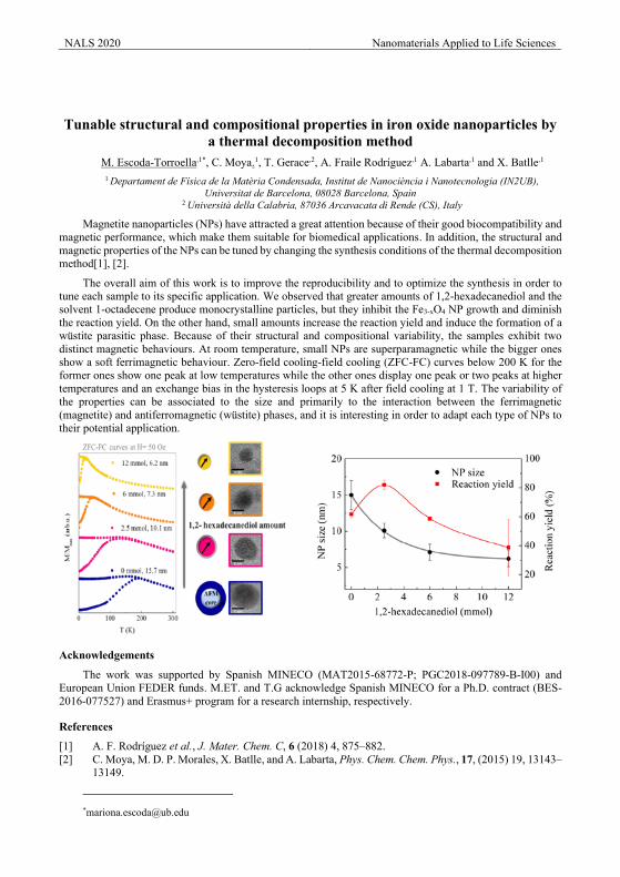
NALS 2020 Nanomaterials Applied to Life Sciences
Tunable structural and compositional properties in iron oxide nanoparticles by
a thermal decomposition method M. Escoda-Torroella,1*, C. Moya.1, T. Gerace,2, A. Fraile Rodríguez,1 A. Labarta,1 and X. Batlle,1
1 Departament de Física de la Matèria Condensada, Institut de Nanociència i Nanotecnologia (IN2UB), Universitat de Barcelona, 08028 Barcelona, Spain
2 Università della Calabria, 87036 Arcavacata di Rende (CS), Italy
Magnetite nanoparticles (NPs) have attracted a great attention because of their good biocompatibility and magnetic performance, which make them suitable for biomedical applications. In addition, the structural and magnetic properties of the NPs can be tuned by changing the synthesis conditions of the thermal decomposition method[1], [2].
The overall aim of this work is to improve the reproducibility and to optimize the synthesis in order to tune each sample to its specific application. We observed that greater amounts of 1,2-hexadecanediol and the solvent 1-octadecene produce monocrystalline particles, but they inhibit the Fe3-xO4 NP growth and diminish the reaction yield. On the other hand, small amounts increase the reaction yield and induce the formation of a wüstite parasitic phase. Because of their structural and compositional variability, the samples exhibit two distinct magnetic behaviours. At room temperature, small NPs are superparamagnetic while the bigger ones show a soft ferrimagnetic behaviour. Zero-field cooling-field cooling (ZFC-FC) curves below 200 K for the former ones show one peak at low temperatures while the other ones display one peak or two peaks at higher temperatures and an exchange bias in the hysteresis loops at 5 K after field cooling at 1 T. The variability of the properties can be associated to the size and primarily to the interaction between the ferrimagnetic (magnetite) and antiferromagnetic (wüstite) phases, and it is interesting in order to adapt each type of NPs to their potential application.
Acknowledgements
The work was supported by Spanish MINECO (MAT2015-68772-P; PGC2018-097789-B-I00) and European Union FEDER funds. M.ET. and T.G acknowledge Spanish MINECO for a Ph.D. contract (BES-2016-077527) and Erasmus+ program for a research internship, respectively.
References
[1] A. F. Rodríguez et al., J. Mater. Chem. C, 6 (2018) 4, 875–882. [2] C. Moya, M. D. P. Morales, X. Batlle, and A. Labarta, Phys. Chem. Chem. Phys., 17, (2015) 19, 13143–
13149.
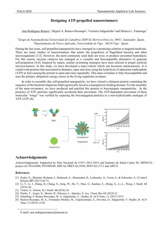
NALS 2020 Nanomaterials Applied to Life Sciences
Designing ATP-propelled nanoswimmers
Ana Rodríguez-Ramos1, Miguel A. Ramos-Docampo2, Verónica Salgueiriño2 and Mónica L. Fanarraga1
1Grupo de Nanomedicina-Universidad de Cantabria–IDIVAL Herrera Oria s/n, 39011, Santander, Spain.
2Departamento de Física Aplicada, Universidade de Vigo , 36310 Vigo , Spain.
During the last years, self-propelled nanoparticles have emerged as a promising solution to targeted medicine. There are many studies of nanoswimmers that mimic the propulsion of flagellated bacteria and other microorganisms [1-3]. However, the most commonly used fuels are toxic or produce unwanted byproducts; For this reason, enzyme catalysis has emerged as a versatile and biocompatible alternative to generate self-propulsion [4-6]. Inspired by nature, similar swimming strategies have been utilized to propel artificial microswimmers. In this study, we have developed a nano-vehicle which can locomote autonomously. It is coated with proteins that associated in dimmers, open and close using the hydrolysis of adenosine triphosphate (ATP) as fuel causing the protein to open and close repeatedly. This nano-swimmer is fully biocompatible and uses the primary ubiquitous energy source in the living organisms in nature.
In order to assemble this self-propelled nanoparticle, we have cloned a chimaera protein containing the sequence of the bacterial chaperone HtpG genetically fused to a nanoparticle-binding domain. For the assembly of the nano-swimmers, we have produced and purified this protein to bioconjugate nanoparticles . In the presence of ATP, particles significantly accelerate their movement. The ATP-dependent movement of these molecular “wings” was verified by exposing the bioconjugated particles to a non-hydrolysable analogue of ATP (ATP-γS).
Acknowledgements Acknowledgements: Supported by Plan Nacional de I+D+i 2013‐2016 and Instituto de Salud Carlos III, MINECO, ‐ project ref. PI16/0496, PI19/00349, IDIVAL PREVAL19/04, INNVAL17/11 and APG31
References [1] Zoaby, N., Shainsky-Roitman, J., Badarneh, S., Abumanhal, H., Leshansky, A., Yaron, S., & Schroeder, A. J.Control
Release 257 (2017) 68-75. [2] Li, T., Li, J., Zhang, H., Chang, X., Song, W., Hu, Y., Shao, G., Sandraz, E., Zhang, G., Li, L., Wang, J. Small. 12
(2016) 44. [3] Tottori, S., Nelson, B.J. Small. 14 (2018) 24. [4] Patiño, T., Arqué, X., Mestre, R., Palacios, L., Sánchez, S. Acc. Chem. Res 11 (2018) 51. [5] Schattling, P. Ramos-Docampo, M. A. Salgueiriño, V., Stadler, B. ACS Nano.,11 (2017) 3973. [6] Ramos-Docampo, M. A., Fernández-Medina, M., Taipaleenmaki, E., Hovorka, O., Salgueiriño, V. Stadler, B. ACS
Nano 13 (2019) 12192 E mail; [email protected]
Biofunctionalization of carbon nanotubes with fluorescent recombinant proteins to double-targeting tumours
Mahsa Saramiforoshani*, Mónica L. Fanarraga
Grupo de Nanomedicina-IDIVAL, Instituto Valdecilla, Universidad de Cantabria, Herrera Oria s/n, 39011 Santander
HER2 receptor (family of EGFR) overexpression or mutation through gene amplification,[1] is a feature of many different cancers and, together with the VEGFR,[2] on endothelial cells that prompt intratumoral vasculogenesis, are the key players in expanding tumours favoring metastasis. Therefore, it would be of great interest to consider targeting HER2 and VEGF with cytotoxic agents to inhibit tumour expansion.
Previous would from our group show how multi-walled carbon nanotubes (MWCNTs) have the intrinsic properties to trigger antitumoral effects,[3] interfering with cell proliferation,[4,5] migration[6] and survival[7] of highly proliferating cells. Here we have produced biodegradable[8] oxidised MWCNTs (o-MWCNT) coated with recombinant proteins genetically engineered to bind the HER2 and VEGF receptors. Our aim is to trigger a double anti-tumoral attack, not only to cancer cells but also the stromal vascular cells that support the tumoral organ. Based on our findings, it appears that o-MWCNT can be effectively and stability biofunctionalized with recombinant proteins to be used as a targeted cytotoxic
Acknowledgements
This work has been supported by the Spanish MINECO, Instituto de Salud Carlos III, European Union (European Regional Development Fund-ERDF) under Projects ref. PI16/000496 and PI19/000349, DTS19/00033.
References [1] K. L. Redmond, A. Papafili, M. Lawler, S. Van Schaeybroeck, Semin. Oncol. 2015, 42, 896. [2] K. a Gold, H.-Y. Lee, E. S. Kim, Cancer 2009, 115, 922. [3] L. García-Hevia, J. C. Villegas, F. Fernández, Í. Casafont, J. González, R. Valiente, M. L. Fanarraga, Adv. Healthc. Mater. 2016, 5, 1. [4] L. García-Hevia, F. Fernández, C. Grávalos, A. García, J. C. Villegas, M. L. Fanarraga, Nanomedicine 2014, 9, 1581. [5] L. Rodríguez-Fernández, R. Valiente, J. González, J. C. Villegas, M. L. Fanarraga, ACS Nano 2012, 6, 6614. [6] L. Garcia-Hevia, R. Valiente, J. L. Fernandez-Luna, E. Flahaut, L. Rodriguez-Fernandez, J. C. Villegas, J. Gonzalez, M. L. Fanarraga, Adv. Healthc. Mater. 2015, 4, 1640. [7] L. García-Hevia, R. Valiente, J. González, J. L. Fernández-Luna, J. C. Villegas, M. L. Fanarraga, Curr. Pharm. Des. 2015, 21, 1920. [8] E. Gonzalez-Lavado, N. Iturrioz-Rodriguez, E. Padin-González, J. A. Gonzalez, L. Garcia-Hervia, J. Heuts, C. Pesquera, F. Gonzalez, J. C. Villegas, R. Valiente, et al., Nanoscale 2018, 10, 11013.
e-mail: *[email protected]
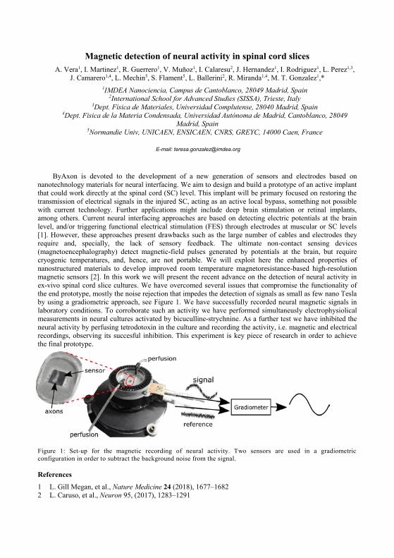
Magnetic detection of neural activity in spinal cord slices A. Vera1, I. Martinez1, R. Guerrero1, V. Muñoz1, I. Calaresu2, J. Hernandez1, I. Rodriguez1, L. Perez1,3,
J. Camarero1,4, L. Mechin5, S. Flament5, L. Ballerini2, R. Miranda1,4, M. T. Gonzalez1,* 1IMDEA Nanociencia, Campus de Cantoblanco, 28049 Madrid, Spain
2International School for Advanced Studies (SISSA), Trieste, Italy 3Dept. Fisica de Materiales, Universidad Complutense, 28040 Madrid, Spain
4Dept. Fisica de la Materia Condensada, Universidad Autónoma de Madrid, Cantoblanco, 28049 Madrid, Spain
5Normandie Univ, UNICAEN, ENSICAEN, CNRS, GREYC, 14000 Caen, France
E-mail: [email protected]
ByAxon is devoted to the development of a new generation of sensors and electrodes based on nanotechnology materials for neural interfacing. We aim to design and build a prototype of an active implant that could work directly at the spinal cord (SC) level. This implant will be primary focused on restoring the transmission of electrical signals in the injured SC, acting as an active local bypass, something not possible with current technology. Further applications might include deep brain stimulation or retinal implants, among others. Current neural interfacing approaches are based on detecting electric potentials at the brain level, and/or triggering functional electrical stimulation (FES) through electrodes at muscular or SC levels [1]. However, these approaches present drawbacks such as the large number of cables and electrodes they require and, specially, the lack of sensory feedback. The ultimate non-contact sensing devices (magnetoencephalography) detect magnetic-field pulses generated by potentials at the brain, but require cryogenic temperatures, and, hence, are not portable. We will exploit here the enhanced properties of nanostructured materials to develop improved room temperature magnetoresistance-based high-resolution magnetic sensors [2]. In this work we will present the recent advance on the detection of neural activity in ex-vivo spinal cord slice cultures. We have overcomed several issues that compromise the functionality of the end prototype, mostly the noise rejection that impedes the detection of signals as small as few nano Tesla by using a gradiometric approach, see Figure 1. We have successfully recorded neural magnetic signals in laboratory conditions. To corroborate such an activity we have performed simultaneusly electrophysiolical measurements in neural cultures activated by bicuculline-strychnine. As a further test we have inhibited the neural activity by perfusing tetrodotoxin in the culture and recording the activity, i.e. magnetic and electrical recordings, observing its succesful inhibition. This experiment is key piece of research in order to achieve the final prototype.
Figure 1: Set-up for the magnetic recording of neural activity. Two sensors are used in a gradiometric configuration in order to subtract the background noise from the signal.
References
1 L. Gill Megan, et al., Nature Medicine 24 (2018), 1677–1682 2 L. Caruso, et al., Neuron 95, (2017), 1283–1291
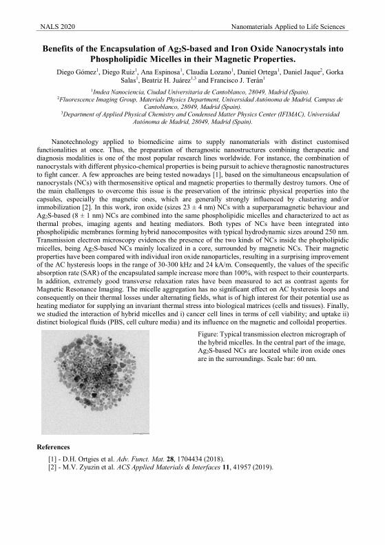
NALS 2020 Nanomaterials Applied to Life Sciences
Benefits of the Encapsulation of Ag2S-based and Iron Oxide Nanocrystals into Phospholipidic Micelles in their Magnetic Properties.
Diego Gómez1, Diego Ruiz1, Ana Espinosa1, Claudia Lozano1, Daniel Ortega1, Daniel Jaque2, Gorka Salas1, Beatriz H. Juárez1,3 and Francisco J. Terán1
1Imdea Nanociencia, Ciudad Universitaria de Cantoblanco, 28049, Madrid (Spain). 2Fluorescence Imaging Group, Materials Physics Department, Universidad Autónoma de Madrid, Campus de
Cantoblanco, 28049, Madrid (Spain). 3Department of Applied Physical Chemistry and Condensed Matter Physics Center (IFIMAC), Universidad
Autónoma de Madrid, 28049, Madrid (Spain).
Nanotechnology applied to biomedicine aims to supply nanomaterials with distinct customised functionalities at once. Thus, the preparation of theragnostic nanostructures combining therapeutic and diagnosis modalities is one of the most popular research lines worldwide. For instance, the combination of nanocrystals with different physico-chemical properties is being pursuit to achieve theragnostic nanostructures to fight cancer. A few approaches are being tested nowadays [1], based on the simultaneous encapsulation of nanocrystals (NCs) with thermosensitive optical and magnetic properties to thermally destroy tumors. One of the main challenges to overcome this issue is the preservation of the intrinsic physical properties into the capsules, especially the magnetic ones, which are generally strongly influenced by clustering and/or immobilization [2]. In this work, iron oxide (sizes 23 ± 4 nm) NCs with a superparamagnetic behaviour and Ag2S-based (8 ± 1 nm) NCs are combined into the same phospholipidic micelles and characterized to act as thermal probes, imaging agents and heating mediators. Both types of NCs have been integrated into phospholipidic membranes forming hybrid nanocomposites with typical hydrodynamic sizes around 250 nm. Transmission electron microscopy evidences the presence of the two kinds of NCs inside the phopholipidic micelles, being Ag2S-based NCs mainly localized in a core, surrounded by magnetic NCs. Their magnetic properties have been compared with individual iron oxide nanoparticles, resulting in a surprising improvement of the AC hysteresis loops in the range of 30-300 kHz and 24 kA/m. Consequently, the values of the specific absorption rate (SAR) of the encapsulated sample increase more than 100%, with respect to their counterparts. In addition, extremely good transverse relaxation rates have been measured to act as contrast agents for Magnetic Resonance Imaging. The micelle aggregation has no significant effect on AC hysteresis loops and consequently on their thermal losses under alternating fields, what is of high interest for their potential use as heating mediator for supplying an invariant thermal stress into biological matrices (cells and tissues). Finally, we studied the interaction of hybrid micelles and i) cancer cell lines in terms of cell viability; and uptake ii) distinct biological fluids (PBS, cell culture media) and its influence on the magnetic and colloidal properties.
Figure: Typical transmission electron micrograph of the hybrid micelles. In the central part of the image, Ag2S-based NCs are located while iron oxide ones are in the surroundings. Scale bar: 60 nm.
References
[1] - D.H. Ortgies et al. Adv. Funct. Mat. 28, 1704434 (2018). [2] - M.V. Zyuzin et al. ACS Applied Materials & Interfaces 11, 41957 (2019).
NALS 2020 Nanomaterials Applied to Life Sciences
Hybrid nanoparticles consisting on magnetic iron oxide and gold nanoparticles modified with Arabic gum
Dulce Guzmán Rocha1,2*, Álvaro Gallo Córdova1, Jesús G. Ovejero1, Sabino Veintemillas-Verdaguer1, María del Puerto Morales1, Teodoro Córdova Fraga2, Jesús Bernal Alvarado2
1 MamBio Group, Instituto de Ciencia de Materiales de Madrid (ICMM), CSIC, Calle Sor Juana Inés de la Cruz 3, 28049, Madrid, Spain.
2Physical Engineering Department, División de Ciencias e Ingenierías Campus León Universidad de Guanajuato, Loma del Bosque 103, Lomas del Campestre CP37150 León, Gto., México
The nanomaterials have been very important in the last years for multiple applications, ranging from catalysis, biomedicine and technology. Hybrid nanoparticles, which combine organic and inorganic materials, present suitable properties for such applications. Recently, the use of Gum Arabic (GA) has been extended to the nanotechnology and nanomedicine fields due to its biocompatibility for in vivo applications. Besides, GA can stabilize nanostructures like metal or magnetic nanoparticles (MNP) and provides support for further functionalization. For example, Wu and Chen [1] has proposed the synthesis of Au nanoparticles by using GA as both reducing and stabilizing agent in the absence of other additives and through green synthesis.
In this work we propose the synthesis of iron oxide nanoparticles coated with GA in a single step by means of co-precipitation of iron salts. This synthesis was also performed in the presence of gold salts to generate Au nanoparticles in situ using GA as reducer. MNP particle size was observed to increase due to the presence of GA in the reaction media, being different if the GA is incorporated in the base and show a crystal size of 16.2 nm or the Fe salts solution with 12.5 nm. The presence of GA at the surface of the MNPs was confirmed by FTIR and TEM images.
When gold salts are incorporated during the synthesis, MNP size is modified. However, the hydrodynamic diameter of MNPs modified with GA and gold after synthesis resulted in the 254 nm. The zeta potentials indicate the existence of negatively charged surfaces before and after GA coating and gold.
Acknowledgements
This work was funded by the Spanish MINECO project MAT2017-88148R (AEI/FEDER, UE) and CONACYT and University of Guanajuato.
References
[1] C.C. Wu and D.H. Chen Gold Bulletin. 43 (2010) 4:234-240

NALS 2020 Nanomaterials Applied to Life Sciences
Tunable structural and compositional properties in iron oxide nanoparticles by
a thermal decomposition method M. Escoda-Torroella,1*, C. Moya.1, T. Gerace,2, A. Fraile Rodríguez,1 A. Labarta,1 and X. Batlle,1
1 Departament de Física de la Matèria Condensada, Institut de Nanociència i Nanotecnologia (IN2UB), Universitat de Barcelona, 08028 Barcelona, Spain
2 Università della Calabria, 87036 Arcavacata di Rende (CS), Italy
Magnetite nanoparticles (NPs) have attracted a great attention because of their good biocompatibility and magnetic performance, which make them suitable for biomedical applications. In addition, the structural and magnetic properties of the NPs can be tuned by changing the synthesis conditions of the thermal decomposition method[1], [2].
The overall aim of this work is to improve the reproducibility and to optimize the synthesis in order to tune each sample to its specific application. We observed that greater amounts of 1,2-hexadecanediol and the solvent 1-octadecene produce monocrystalline particles, but they inhibit the Fe3-xO4 NP growth and diminish the reaction yield. On the other hand, small amounts increase the reaction yield and induce the formation of a wüstite parasitic phase. Because of their structural and compositional variability, the samples exhibit two distinct magnetic behaviours. At room temperature, small NPs are superparamagnetic while the bigger ones show a soft ferrimagnetic behaviour. Zero-field cooling-field cooling (ZFC-FC) curves below 200 K for the former ones show one peak at low temperatures while the other ones display one peak or two peaks at higher temperatures and an exchange bias in the hysteresis loops at 5 K after field cooling at 1 T. The variability of the properties can be associated to the size and primarily to the interaction between the ferrimagnetic (magnetite) and antiferromagnetic (wüstite) phases, and it is interesting in order to adapt each type of NPs to their potential application.
Acknowledgements
The work was supported by Spanish MINECO (MAT2015-68772-P; PGC2018-097789-B-I00) and European Union FEDER funds. M.ET. and T.G acknowledge Spanish MINECO for a Ph.D. contract (BES-2016-077527) and Erasmus+ program for a research internship, respectively.
References
[1] A. F. Rodríguez et al., J. Mater. Chem. C, 6 (2018) 4, 875–882. [2] C. Moya, M. D. P. Morales, X. Batlle, and A. Labarta, Phys. Chem. Chem. Phys., 17, (2015) 19, 13143–
13149.

Thursday Poster Session
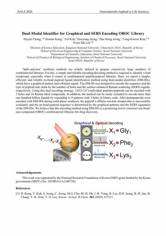
NALS 2020 Nanomaterials Applied to Life Sciences
Dual-Modal Identifier for Graphical and SERS Encoding OBOC LibraryHyejin Chang,1,* Homan Kang,2 Yul Koh,2 Sinyoung Jeong,3 Dae Hong Jeong,3 Yong-Kweon Kim,2,*
Yoon-Sik Lee3,*1Division of Science Education, Kangwon National University, Chuncheon 24341, Republic of Korea
2School of Electrical Engineering & Computer Science, Seoul National University3Department of Chemistry Education, Seoul National University
4School of Chemical & Biological Engineering, Institute of Chemical Processes, Seoul National University,Seoul 08826, Republic of Korea
‘Split-and-mix’ synthetic methods are widely utilized to prepare extensively large numbers of combinatorial libraries. For this, a simple and reliable encoding/decoding method is required to identify a lead compound, especially when it comes to combinatorial peptide/peptoid libraries. Here, we report a simple, efficient, and reliable on-bead peptoid ligand identification method using dual-modal identifiers (DM-IDs),which have a graphical pattern and a Raman signal. The DM-ID was designed to encode the sequence and the type of peptoid side chain by the number of holes and the surface-enhanced Raman scattering (SERS) signals, respectively. Using this dual encoding strategy, 5,832 (183)-individual peptides/peptoids can be encoded with 3 holes and 18 Raman label compounds. In addition, the method can be easily extended to encode more than one hundred billion ligands by expanding to 9 patterns with 3 holes in binary code. After pentapeptoids were encoded with DM-IDs during solid phase synthesis, the peptoid’s affinity towards streptavidin is successfully evaluated, and the on-bead peptoid sequence is determined by the graphical patterns and the SERS signatures of the DM-IDs. We believe that this encoding method using DM-IDs is a promising tool to construct one-bead-one-compound (OBOC) combinatorial libraries for drug discovery.
Acknowledgements
This work was supported by the National Research Foundation of Korea (NRF) grant funded by the Korea government (MSIT) (No. 2019R1G1A1100734).
References
[1] H. Kang, Y. Koh, S. Jeong, C. Jeong, M.G. Cha, M.-H. Oh, J.-K. Yang, H. Lee, D.H. Jeong, B.-H. Jun, H. Chang, Y.-K. Kim, Y.-S. Lee, Sensor. Actuat. B Chem. 303 (2020) 127211.
Raman shift
Raman shift
Raman shift X1
X2
X3
Gly1
Phe2
Lys3
Graphical & Optical decoding
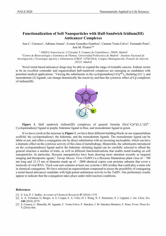
NALS 2020 Nanomaterials Applied to Life Sciences
Functionalization of Soft Nanoparticles with Half-Sandwich Iridium(III) Anticancer Complexes
Ana C. Carrasco1, Adriana Arnaiz1, Ivonne González-Gamboa2, Carmen Yuste-Calvo2, Fernando Ponz2, Ana M. Pizarro1*
1 IMDEA Nanociencia, C/Faraday 9, Campus de Cantoblanco, 28049, Madrid 2Centro de Biotecnología y Genómica de Plantas, Universidad Politécnica de Madrid – Instituto Nacional de
Investigación y Tecnología Agraria y Alimentaria (CBGP, UPM-INIA), Campus Montegancedo, Pozuelo de Alarcón, 28223, Madrid
Novel metal-based anticancer drugs may be able to expand the range of treatable cancers. Iridium seems to be an excellent contender and organoiridium half-sandwich complexes are emerging as candidates with potential medical applications.1 Varying the substituents in the cyclopentadienyl (Cp*R), chelating (LL’), and monodentate (Z) ligands, can change dramatically the reactivity and thus the cytotoxic effect of Cp complexes of iridium(III).
Figure 1. Half sandwich iridium(III) complexes of general formula [Ir(5-Cp*)(L,L’)Z]0/+.
Cyclopentadienyl ligand in purple, bidentate ligand in blue, and monodentate ligand in green.
If we have a look at the structure in Figure 1, we have three different building blocks in our organoiridium scaffold: the cyclopentadienyl, the bidentate, and the monodentate ligands. The monodentate ligand can be labile or not, and offers a conjugation site by direct substitution with an incoming nucleophile, which can have a dramatic effect on the cytotoxic activity of this class of metallodrugs. Meanwhile, the substituents introduced on the cyclopentadienyl ligand and/or the bidentate chelating ligand can be carefully selected to afford the general structure a number of traits, as well as different functionalizations that enable metal-loading on soft nanoparticles. In particular, flexuous nanoparticles have been drawing more attention recently as targeted imaging and therapeutic agents.2 Turnip Mosaic Virus (TuMV) is a flexuous filamentous plant virus of ~ 700 nm long and 12-15 nm of diameter made up of ~ 2000 identical copies coat proteins subunits that cover a molecule of viral RNA.3 Each coat unit contains at least one cysteine (-SH) residue that could play a main role in chemical conjugation. We have selected an organoiridium compound to assess the possibility of conjugating a metal-based anticancer candidate with high potent antitumour activity to the TuMV. Our preliminary results appear to indicate that the conjugation takes place under mild reaction conditions.
References [1] Z. Liu, P. J. Sadler, Accounts of Chemical Research 47 (2014) 1174. [2] A. A. Vernekar, G. Berger, A. E. Czapar, F. A. Veliz, D. I. Wang, N. F. Steinmetz, S. J. Lippard, J. Am. Chem. Soc.
140 (2018) 4279. [3] S. Cuenca, C. Mansilla, M. Aguado, C. Yuste-Calvo, F. Sánchez, J. M. Sánchez-Montero, F. Ponz, Front. Plant Sci.
7 (2016) 464.
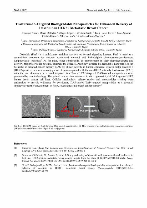
NALS 2020 Nanomaterials Applied to Life Sciences
Trastuzumab-Targeted Biodegradable Nanoparticles for Enhanced Delivery of
Dasatinib in HER2+ Metastasic Breast Cancer Enrique Niza 1, Maria Del Mar Noblejas-López 2, Cristina Nieto 2, Ivan Bravo Pérez 3, Jose Antonio
Castro-Osma 1, Alberto Ocaña 2, Carlos Alonso-Moreno 1
1 Dpto. Inorgánica, Orgánica y Bioquímica, Facultad de Farmacia de Albacete, UCLM, 02071 Albacete, Spain 2 Oncología Traslacional, Unidad de Investigación del Complejo Hospitalario Universitario de Albacete,
02071 Albacete, Spain 3 Dpto. Química Física, Facultad de Farmacia de Albacete, UCLM, 02071 Albacete, Spain
Dasatinib (DAS) is a multikinase inhibitor that acts on several signaling kinases. DAS is used as a second-line treatment for chronic accelerated myeloid and Philadelphia chromosome-positiveacute lymphoblastic leukemia.1 As for many other compounds, an improvement in their pharmacokinetic and delivery properties would potential augment the efficacy. Antibody-targeted biodegradable nanoparticles can be useful in targeted cancer therapy. DAS has shown activity in human epidermal growth factor receptor 2 (HER2) positive tumours, so conjugation of this compound with the anti-HER2 antibody trastuzumab (TAB) with the use of nanocarriers could improve its efficacy.2 TAB-targeted DAS-loaded nanoparticles were generated by nanotechnology. The guided nanocarriers enhanced in vitro cytotoxicity of DAS against HER2 human breast cancer cell lines. Cellular mechanistic, release studies and nanoparticles stability were undertaken to provide evidences for positioning DAS-loaded TAB-targeted nanoparticles as a potential strategy for further development in HER2-overexpressing breast cancer therapy.3
Fig 1. a) FE-SEM image of TAB-targeted Das loaded nanoparticles; b) TEM images of polyethyleneimine-coated nanoparticles (PEI)NPs before (left) and after (right) TAB conjugation
References [1] Butowski NA, Chang SM. General and Neurological Complications of Targeted Therapy. Vol 105. 1st ed.
Elsevier B.V.; 2012. doi:10.1016/B978-0-444-53502-3.00033-1
[2] Ocana A, Gil-Martin M, Antolín S, et al. Efficacy and safety of dasatinib with trastuzumab and paclitaxel in first line HER2-positive metastatic breast cancer: results from the phase II GEICAM/2010-04 study. Breast Cancer Res Treat. 2019;174(3):693-701. doi:10.1007/s10549-018-05100-z
[3] Niza E, Noblejas-lópez MDM, Bravo I, et al. Trastuzumab-targeted biodegradable nanoparticles for enhanced delivery of dasatinib in HER2+ metastasic breast cancer. Nanomaterials. 2019;9(12):1-14. doi:10.3390/nano9121793
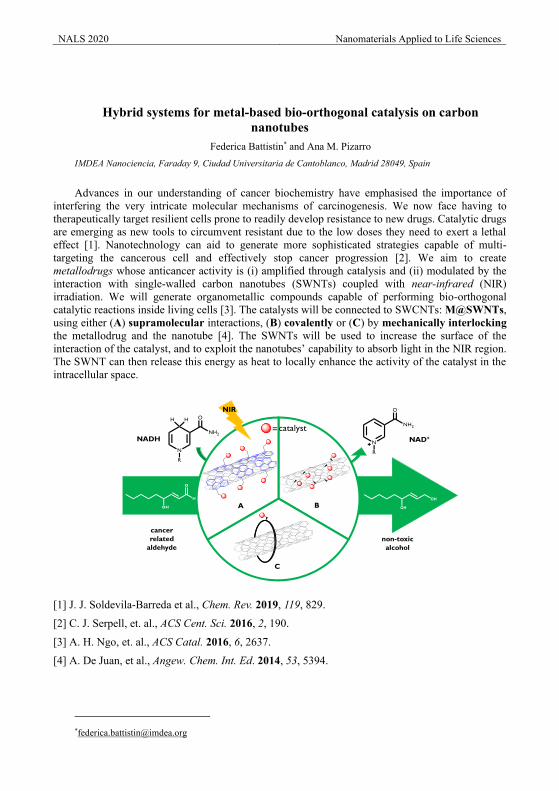
NALS 2020 Nanomaterials Applied to Life Sciences
Hybrid systems for metal-based bio-orthogonal catalysis on carbon nanotubes
Federica Battistin* and Ana M. Pizarro IMDEA Nanociencia, Faraday 9, Ciudad Universitaria de Cantoblanco, Madrid 28049, Spain
Advances in our understanding of cancer biochemistry have emphasised the importance of interfering the very intricate molecular mechanisms of carcinogenesis. We now face having to therapeutically target resilient cells prone to readily develop resistance to new drugs. Catalytic drugs are emerging as new tools to circumvent resistant due to the low doses they need to exert a lethal effect [1]. Nanotechnology can aid to generate more sophisticated strategies capable of multi-targeting the cancerous cell and effectively stop cancer progression [2]. We aim to create metallodrugs whose anticancer activity is (i) amplified through catalysis and (ii) modulated by the interaction with single-walled carbon nanotubes (SWNTs) coupled with near-infrared (NIR) irradiation. We will generate organometallic compounds capable of performing bio-orthogonal catalytic reactions inside living cells [3]. The catalysts will be connected to SWCNTs: M@SWNTs, using either (A) supramolecular interactions, (B) covalently or (C) by mechanically interlocking the metallodrug and the nanotube [4]. The SWNTs will be used to increase the surface of the interaction of the catalyst, and to exploit the nanotubes’ capability to absorb light in the NIR region. The SWNT can then release this energy as heat to locally enhance the activity of the catalyst in the intracellular space.
[1] J. J. Soldevila-Barreda et al., Chem. Rev. 2019, 119, 829. [2] C. J. Serpell, et. al., ACS Cent. Sci. 2016, 2, 190.
[3] A. H. Ngo, et. al., ACS Catal. 2016, 6, 2637.
[4] A. De Juan, et al., Angew. Chem. Int. Ed. 2014, 53, 5394.
NADH NAD+
A B
C
NIR
cancer related
aldehydenon-toxic alcohol
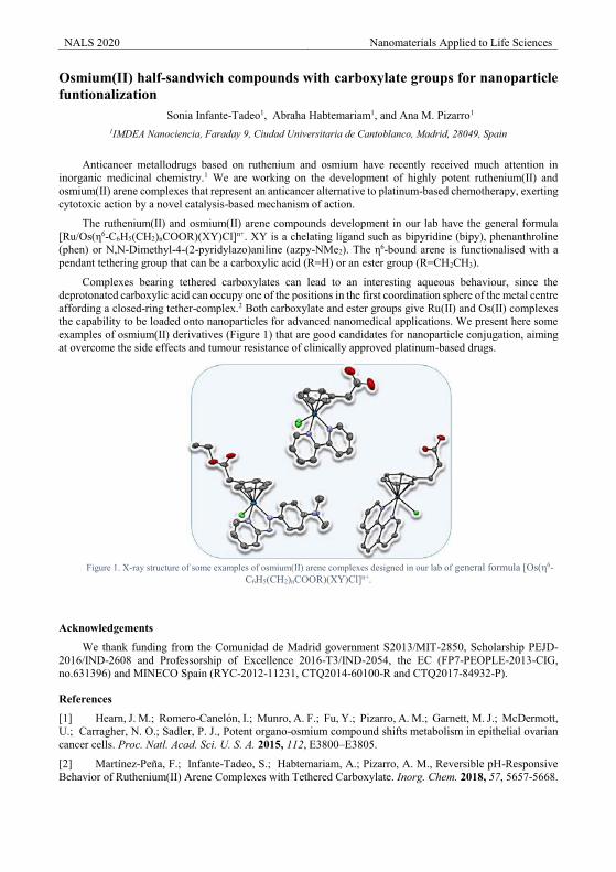
NALS 2020 Nanomaterials Applied to Life Sciences
Osmium(II) half-sandwich compounds with carboxylate groups for nanoparticle funtionalization
Sonia Infante-Tadeo1, Abraha Habtemariam1, and Ana M. Pizarro1
1IMDEA Nanociencia, Faraday 9, Ciudad Universitaria de Cantoblanco, Madrid, 28049, Spain
Anticancer metallodrugs based on ruthenium and osmium have recently received much attention in inorganic medicinal chemistry.1 We are working on the development of highly potent ruthenium(II) and osmium(II) arene complexes that represent an anticancer alternative to platinum-based chemotherapy, exerting cytotoxic action by a novel catalysis-based mechanism of action.
The ruthenium(II) and osmium(II) arene compounds development in our lab have the general formula [Ru/Os(η6-C6H5(CH2)nCOOR)(XY)Cl]n+. XY is a chelating ligand such as bipyridine (bipy), phenanthroline (phen) or N,N-Dimethyl-4-(2-pyridylazo)aniline (azpy-NMe2). The η6-bound arene is functionalised with a pendant tethering group that can be a carboxylic acid (R=H) or an ester group (R=CH2CH3).
Complexes bearing tethered carboxylates can lead to an interesting aqueous behaviour, since the deprotonated carboxylic acid can occupy one of the positions in the first coordination sphere of the metal centre affording a closed-ring tether-complex.2 Both carboxylate and ester groups give Ru(II) and Os(II) complexes the capability to be loaded onto nanoparticles for advanced nanomedical applications. We present here some examples of osmium(II) derivatives (Figure 1) that are good candidates for nanoparticle conjugation, aiming at overcome the side effects and tumour resistance of clinically approved platinum-based drugs.
Figure 1. X-ray structure of some examples of osmium(II) arene complexes designed in our lab of general formula [Os(η6-
C6H5(CH2)nCOOR)(XY)Cl]n+.
Acknowledgements
We thank funding from the Comunidad de Madrid government S2013/MIT-2850, Scholarship PEJD-2016/IND-2608 and Professorship of Excellence 2016-T3/IND-2054, the EC (FP7-PEOPLE-2013-CIG, no.631396) and MINECO Spain (RYC-2012-11231, CTQ2014-60100-R and CTQ2017-84932-P).
References
[1] Hearn, J. M.; Romero-Canelón, I.; Munro, A. F.; Fu, Y.; Pizarro, A. M.; Garnett, M. J.; McDermott, U.; Carragher, N. O.; Sadler, P. J., Potent organo-osmium compound shifts metabolism in epithelial ovarian cancer cells. Proc. Natl. Acad. Sci. U. S. A. 2015, 112, E3800–E3805.
[2] Martínez-Pena, F.; Infante-Tadeo, S.; Habtemariam, A.; Pizarro, A. M., Reversible pH-Responsive Behavior of Ruthenium(II) Arene Complexes with Tethered Carboxylate. Inorg. Chem. 2018, 57, 5657-5668.
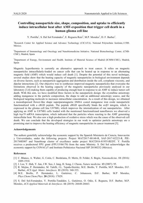
NALS 2020 Nanomaterials Applied to Life Sciences
Controlling nanoparticle size, shape, composition, and uptake to efficiently induce intracellular heat after AMF-exposition that trigger cell death in a
human glioma cell line
Y. Portilla1, S. Del Sol Fernández2, E. Reguera Ruíz2, M.P. Morales3, D. F. Barber1.
1Research Center for Applied Science and Advance Technology (CICATA). National Polytechnic Institute,11500. México.
2Department of Immunology and Oncology and Nanobiomedicine Initiative. National Biotechnology Center, (CNB-CSIC), Madrid, Spain
3Department of Energy, Environment and Health. Institute of Material Science of Madrid (ICMM-CSIC), Madrid, Spain
Magnetic hyperthermia is currently an alternative approach to treat cancer. It relies on magnetic nanoparticles intracellularly-loaded on cancer cells that can be heated up in response to an alternating magnetic field (AMF) which would induce cell death [1]. Despite the potential of this novel technique, several studies show that the heating capacity of magnetic nanoparticles in biological environment depends on diverse factors, such as nanoparticle aggregation and distribution inside the cell, cytoplasm viscosity, and dipolar interactions [2]. Our objective was to synthesize improved magnetic nanoparticles that overcame the limitations observed in the heating capacity of the magnetic nanoparticles previously analysed in our laboratory [3,4] making them capable of producing enough heat in response to an AMF to induce tumor cell death. With this aim, we have modified three factors in the nanoparticle design: the crystalline anisotropy adding manganese to the particle composition, the shape to add an additional anisotropy source, and the biological targeting molecule to increases intracellular concentration. As a result of this design, we obtained a monodispersed flower-like shape superparamagnetic DMSA coated manganese iron oxide nanoparticle functionalized with a cRGD peptide. The peptide cRGD specifically binds the αvβ3 integrin, which is expressed in the glioma cell line U87MG, which improves the internalization of our nanoparticles. After applying an AMF to U87MG cells loaded with the mentioned functionalizaed nanoflawers we observed a high hsp70 mRNA transcription, which indicated that the particles where reacting to the AMF generating intracellular heat. We also saw a high production of oxidative stress which was the cause of the observed cell death. We can conclude that the developed strategies in our work to optimize particle anisotropy are a promising start to improve the heating efficiency of magnetic nanoparticles in cancer treatment [5].
Acknowledgements
The authors gratefully acknowledge the economic support by the Spanish Ministerio de Ciencia, Innovación y Universidades, under the following projects: Project MAT2017-88148-R, SAF-2017-82223-R, PIE-201760E007 and Nanobioap cluster of excellence under project MAT2016-81955-REDT. Y. Portilla receives a predoctoral FPU grant (FPU15/06170) from the same Ministry. S. Del Sol acknowledges the economic support by CONACyT and Instituto Politécnico Nacional SIP 20180252 (México).
References
[1] C. Blanco, A. Walter, G. Cotin, C. Bordeianu, D. Mertz, D. Felder, S. Begin, Nanomedicine. 11 (2016): 1889-1910.
[2] J. Lee, Y. Huh, Y. Jun, J.W. Seo, J. Jang, H. Song, J. Cheon, Nature medicine. 13 (2007): 95. [3] R. Mejías, P. Hernández, M. Talelli, J.L. Tajada-Herráiz, M.E. Brollo, Y. Portilla, M.P. Morales, D.F.
Barber, ACS Appl. Mater. Interfaces. 11 (2019): 340-355. [4] M.E. Brollo, P. Hernández, L. Gutiérrez, C. Johansson, D.F. Barber, M.P Morales,
Phys.Chem.Chem.Phys. 20 (2018): 17829.
[5] S. Del Sol-Fernández, Y. Portilla-Tundidor, L. Gutiérrez, O. Odio, E. Reguera, D.F. Barber, M.P. Morales, ACS Applied Materials & Interfaces. 11 (2019): 26648-26663.
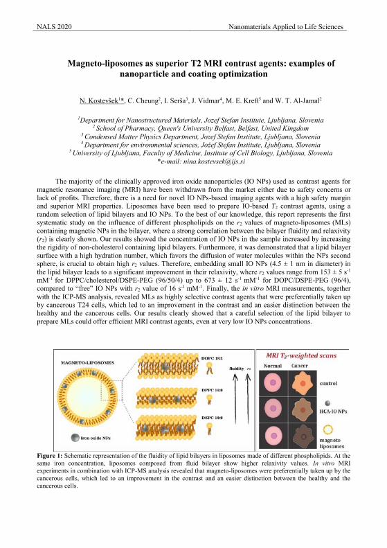
NALS 2020 Nanomaterials Applied to Life Sciences
Magneto-liposomes as superior T2 MRI contrast agents: examples of
nanoparticle and coating optimization
N. Kostevšek1*, C. Cheung2, I. Serša3, J. Vidmar4, M. E. Kreft5 and W. T. Al-Jamal2
1Department for Nanostructured Materials, Jozef Stefan Institute, Ljubljana, Slovenia 2 School of Pharmacy, Queen's University Belfast, Belfast, United Kingdom
3 Condensed Matter Physics Department, Jozef Stefan Institute, Ljubljana, Slovenia
4 Department for environmental sciences, Jožef Stefan Institute, Ljubljana, Slovenia 5 University of Ljubljana, Faculty of Medicine, Institute of Cell Biology, Ljubljana, Slovenia
*e-mail: [email protected]
The majority of the clinically approved iron oxide nanoparticles (IO NPs) used as contrast agents for magnetic resonance imaging (MRI) have been withdrawn from the market either due to safety concerns or lack of profits. Therefore, there is a need for novel IO NPs-based imaging agents with a high safety margin and superior MRI properties. Liposomes have been used to prepare IO-based T2 contrast agents, using a random selection of lipid bilayers and IO NPs. To the best of our knowledge, this report represents the first systematic study on the influence of different phospholipids on the r2 values of magneto-liposomes (MLs) containing magnetic NPs in the bilayer, where a strong correlation between the bilayer fluidity and relaxivity (r2) is clearly shown. Our results showed the concentration of IO NPs in the sample increased by increasing the rigidity of non-cholesterol containing lipid bilayers. Furthermore, it was demonstrated that a lipid bilayer surface with a high hydration number, which favors the diffusion of water molecules within the NPs second sphere, is crucial to obtain high r2 values. Therefore, embedding small IO NPs (4.5 ± 1 nm in diameter) in the lipid bilayer leads to a significant improvement in their relaxivity, where r2 values range from 153 ± 5 s-1
mM-1 for DPPC/cholesterol/DSPE-PEG (96/50/4) up to 673 ± 12 s-1 mM-1 for DOPC/DSPE-PEG (96/4), compared to “free” IO NPs with r2 value of 16 s-1 mM-1. Finally, the in vitro MRI measurements, together with the ICP-MS analysis, revealed MLs as highly selective contrast agents that were preferentially taken up by cancerous T24 cells, which led to an improvement in the contrast and an easier distinction between the healthy and the cancerous cells. Our results clearly showed that a careful selection of the lipid bilayer to prepare MLs could offer efficient MRI contrast agents, even at very low IO NPs concentrations.
Figure 1: Schematic representation of the fluidity of lipid bilayers in liposomes made of different phospholipids. At the same iron concentration, liposomes composed from fluid bilayer show higher relaxivity values. In vitro MRI experiments in combination with ICP-MS analysis revealed that magneto-liposomes were preferentially taken up by the cancerous cells, which led to an improvement in the contrast and an easier distinction between the healthy and the cancerous cells.
NALS 2020 Nanomaterials Applied to Life Sciences
DEVELOPMENT OF IRON OXIDE NANOPARTICLES AS A SMART CANCER-TARGETED THERANOSTIC PROBE FOR CELLULAR
DELIVERY AND MONITORING
Dorra Abdelmalek IDriss*1, Mouna Messaoud2, Sami Aifa3
1Laboratory of Molecular and Cellular Screening Processes, Centre of Biotechnology of Sfax, Bioinformatics
Group, P.O. Box: 1177, Sfax. 3018 Tunisia, University od Sfax. 2Laboratory of Industrial Chemistry, National School of Engineers, University of Sfax, BP1173, Sfax, Tunisia
3Laboratory of Molecular and Cellular Screening Processes, Centre of Biotechnology of Sfax, Bioinformatics Group, P.O. Box: 1177, Sfax. 3018 Tunisia, University od Sfax.
Abstract Non-small cell lung cancer (NSCLC), the predominant subtype of Lung Cancer (LC), is responsible for
the most cancer related deaths worldwide. As the early clinical symptoms of NSCLC are insidious and not
apparent, patients are usually diagnosed with advanced disease. However, as with chemotherapy, targeted
treatment suffers from poor accumulation in the tumor and induce dose limiting toxicities due to systemic
distribution around the body, limiting clinical efficacy. Therefore, NSCLC treatment is still a significant
clinical challenge, highlighting an unmet need for more efficacious therapeutic options. Interestingly,
Nanomedicine involves the application of nanotechnology to address medical questions and has the potential
to greatly enhance cancer diagnosis and therapy. Of particular interest is the use of nanoparticles (NPs) for
drug delivery applications. We designed and synthesized theranostic nanoparticles that showed the
considerable potential for clinical use in targeted therapy, and non-invasive real-time monitoring of tumors by
MRI. Our nanoparticles were ultra-small with superparamagnetic iron oxide cores, conjugated to new tyrosine
kinase inhibitor. Hematite nanoparticles were successfully synthesized using the sol–gel method under
supercritical drying conditions of ethyl alcohol influencing both crystallite is size and particles is size. The
different synthesized samples were characterized by different techniques (XRD spectroscopy, scanning
electron microscopy SEM, Vibrating Sample Magnetometer VSM, dynamic light scattering, UV–vis and FT-
IR spectroscopy.
Keywords
Epidermal Growth Factor Receptor, Tyrosine Kinase inhibitor, Cancer, Superparamagnetic Iron Oxide Nano-particle, Magnetic Resonance Imaging, Theranostic Probe.
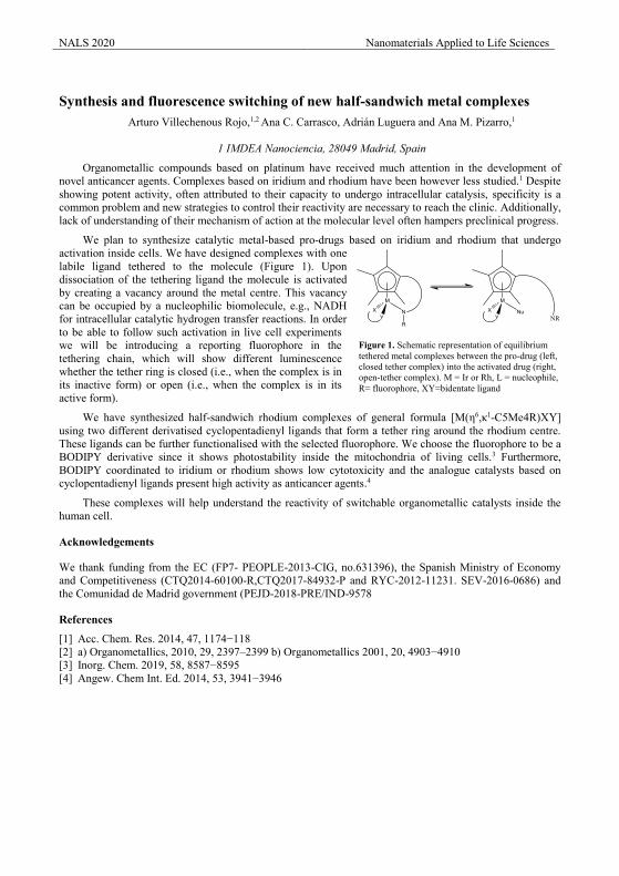
NALS 2020 Nanomaterials Applied to Life Sciences
Synthesis and fluorescence switching of new half-sandwich metal complexes
Arturo Villechenous Rojo,1,2 Ana C. Carrasco, Adrián Luguera and Ana M. Pizarro,1
1 IMDEA Nanociencia, 28049 Madrid, Spain
Organometallic compounds based on platinum have received much attention in the development of novel anticancer agents. Complexes based on iridium and rhodium have been however less studied.1 Despite showing potent activity, often attributed to their capacity to undergo intracellular catalysis, specificity is a common problem and new strategies to control their reactivity are necessary to reach the clinic. Additionally, lack of understanding of their mechanism of action at the molecular level often hampers preclinical progress.
We plan to synthesize catalytic metal-based pro-drugs based on iridium and rhodium that undergo activation inside cells. We have designed complexes with one labile ligand tethered to the molecule (Figure 1). Upon dissociation of the tethering ligand the molecule is activated by creating a vacancy around the metal centre. This vacancy can be occupied by a nucleophilic biomolecule, e.g., NADH for intracellular catalytic hydrogen transfer reactions. In order to be able to follow such activation in live cell experiments we will be introducing a reporting fluorophore in the tethering chain, which will show different luminescence whether the tether ring is closed (i.e., when the complex is in its inactive form) or open (i.e., when the complex is in its active form).
We have synthesized half-sandwich rhodium complexes of general formula [M(η6,κ1-C5Me4R)XY] using two different derivatised cyclopentadienyl ligands that form a tether ring around the rhodium centre. These ligands can be further functionalised with the selected fluorophore. We choose the fluorophore to be a BODIPY derivative since it shows photostability inside the mitochondria of living cells.3 Furthermore, BODIPY coordinated to iridium or rhodium shows low cytotoxicity and the analogue catalysts based on cyclopentadienyl ligands present high activity as anticancer agents.4
These complexes will help understand the reactivity of switchable organometallic catalysts inside the human cell.
Acknowledgements
We thank funding from the EC (FP7- PEOPLE-2013-CIG, no.631396), the Spanish Ministry of Economy and Competitiveness (CTQ2014-60100-R,CTQ2017-84932-P and RYC-2012-11231. SEV-2016-0686) and the Comunidad de Madrid government (PEJD-2018-PRE/IND-9578
References
[1] Acc. Chem. Res. 2014, 47, 1174−118 [2] a) Organometallics, 2010, 29, 2397–2399 b) Organometallics 2001, 20, 4903−4910 [3] Inorg. Chem. 2019, 58, 8587−8595 [4] Angew. Chem Int. Ed. 2014, 53, 3941−3946
Figure 1. Schematic representation of equilibrium tethered metal complexes between the pro-drug (left, closed tether complex) into the activated drug (right, open-tether complex). M = Ir or Rh, L = nucleophile, R= fluorophore, XY=bidentate ligand
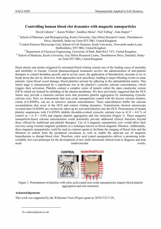
NALS 2020 Nanomaterials Applied to Life Sciences
Controlling human blood clot dynamics with magnetic nanoparticles
David Cabrera1, *, Karen Walker2, Sandhya Moise3, Neil Telling1, Alan Harper1,4 1 School of Pharmacy and Bioengineering, Keele University, Guy Hilton Research Centre, Thornburrow
Drive, Hartshill, Stoke-on-Trent ST5 5BG, United Kingdom; 2 Central Electron Microscope Unit, School of Life Sciences, Keele University, Newcastle-under-Lyme,
Staffordshire, ST5 5BG, United Kingdom 3 Department of Chemical Engineering, University of Bath, Bath BA2 7AY, United Kingdom.
4 School of Medicine, Keele University, Guy Hilton Research Centre, Thornburrow Drive, Hartshill, Stoke-on-Trent ST5 5BG, United Kingdom.
Heart attacks and strokes triggered by unwanted blood clotting remain one of the leading cause of mortality and morbidity in Europe. Current pharmacological treatments involve the administration of anti-platelet therapies to control thrombus growth, and in severe cases, the application of thrombolytic enzymes to try to break down the clot in. However, both approaches lack specificity, leading to major bleeding events in some patients. Upon blood vessel damage, blood platelets activate by adhering to the subendothelial matrix. This initial stage is characterised by a significant rise in the platelet’s cytosolic calcium concentration, which triggers their activation. Platelets contain a complex series of tunnels called the open canalicular system (OCS) which are formed by infolding of the plasma membrane. We have previously suggested that the OCS lumen may provide a transient calcium store that promotes platelet aggregation by maintaining cytosolic calcium rises. Here we demonstrate that iron oxide nanoparticles coated with the known calcium chelator, citrate (Cit-IONPs), can act as selective calcium nanochelators. These nanochelators buffer the calcium accumulations that occur in the OCS and control clotting dynamics. Transmission electron microscopy revealed that Cit-IONPs are exclusively taken up by activated platelets into the OCS. Pretreatment of human platelet suspensions with Cit-IONPs inhibits thrombin-evoked cytosolic calcium rises to 41.8 ± 4.0 % of control (n = 6, P < 0.05) and impairs platelet aggregation and clot retraction (Figure 1). These magnetic nanoparticle-based calcium nanochelators could potentially provide additional clinical functions beyond those offered by traditional anti-platelet therapies. Use of a magnetic nanoparticle core would allow their recovery using external magnetic gradients in a technique known as blood magnetic filtration. Additionally, these magnetic nanoparticles could be used as contrast agents to facilitate the imaging of blood clots and the filtration of emboli from the peripheral circulation as well as enable the adjuvant use of magnetic hyperthermia to disrupt blood clots. Therefore, citric acid coated nanoparticles deliver a promising wide-available, low-cost prototype for the development of new multi-functional clinical tools to diagnose and treat acute cardiovascular events.
Figure 1. Pretreatment of platelets with citric acid-coated iron oxide nanoparticles impairs blood platelet
aggregation and clot retraction.
Acknowledgements
This work was supported by the Wellcome Trust (Project grant no 207617/Z/17/Z).
NALS 2020 Nanomaterials Applied to Life Sciences
Magnetic properties of spinel ferrite nanoparticles engineered by the synthesis process and chemical composition
A. Omelyanchik1,2, G. Muscas3, S. Villa1, F. Canepa1, G. Singh4, V. Rodionova2 and D. Peddis1,5 1 Department of Chemistry and Industrial Chemistry (DCIC), University of Genova, Genova, Italia
2 Immanuel Kant Baltic Federal University, Kaliningrad, Russia
3 Department of Physics and Astronomy, Uppsala University, Uppsala, Sweden
4 Department of Materials Engineering, Norwegian University of Science and Technology (NTNU), Trondheim, Norway
5 Istituto di Struttura della Materia, CNR, 00015 Monterotondo Scalo, (RM) Italy
The magnetic properties of particles are strongly affected by their dimensions. In particular, when the particle size decreases under 100 nm, the particles dramatically differ from the bulk and organize themselves in a single domain state, when, under a critical radius, a multi-domain state is not energetically convenient [1]. Nowadays, magnetic single-domain nanoparticles’ ensembles have found a place in several important technological applications thanks to their peculiar magnetic properties [2], e.g., MRI [3], hyperthermia [4] and drug delivery [5]. In this work, we investigate the flexibility of the high-temperature thermal decomposition (HTD) synthesis to design magnetic nanoparticles with engineered properties for specific applications. We demonstrate the possibility to prepare spinel iron oxide particles with the desired composition (Fe3O4, NiFe2O4 and CoFe2O4) including core/shell nanostructures and well-controlled average size by tuning the synthesis procedure.
The design of nanoparticle-based magnetic materials with specific properties hinges on the control of their physical-chemical structure. We show that the substitution of iron ferrite by Co produces a dramatic increment of the magnetocrystalline anisotropy, well behind the effect of the increment of particle volume. The magnetocrystalline anisotropy has shown to be the leading player in the magnetic behavior of high crystalline particles of such small size, except for the smallest iron oxide sample, where the surface anisotropy plays an extraordinary role. Finally, we observed that the presence of organic surfactant (oleic acid) on the nanoparticles prevents direct exchange coupling among them. Nevertheless, the particles are close enough to be influenced by dipolar interactions, which are proportionally stronger for particles with less intrinsic magnetocrystalline anisotropy.
Core/shell nanoparticles consisted of magnetically hard (CoFe2O4) and soft (NiFe2O4) materials demonstrate enhanced magnetic properties with respect to single-phase systems. However, a prediction of the magnetic properties of such sophisticated structures is the complicated task since many structural properties may affect the magnetic one. In the frame of this work, the magnetic characterisation of two nanoparticles systems with core/shell structures are presented where one is hard/soft nanoparticles with a core consisting of hard cobalt ferrite covered by a soft nickel ferrite, while the second system has an inverted configuration - soft/hard structure with almost the same size and shape. It has been demonstrated that a hard magnetic core can increase the anisotropy of a softer material of shell and this is true for the inverted system. Finally, we applied advanced protocols of magnetic measurement and their elaboration such as Langevin function, activation volume and RAM-model to deeply investigate processes of magnetization and proof that all volume in our core/shell nanostructures involved in magnetization processes.
References
[1] D. Fiorani, J. Magn. Magn. Mater. 196–197 (1999) 143–147. [2] L. Suber, in: Nanomater. Life Sci., Wiley, (2009). [3] C. Sun, Adv. Drug Deliv. Rev. 60 (2008) 1252–65. [4] M. Andrés Vergés, J. Phys. D. Appl. Phys. 41 (2008) 134003. [5] P. Guardia, Langmuir. 26 (2010) 5843–7.
NALS 2020 Nanomaterials Applied to Life Sciences
Quantification of heat dose released by iron oxide nanoparticles inside breast cancer cells under near infrared irradiation
Claudia Lozano Pedraza1, Ana Espinosa1, Francisco Sanz-Rodríguez2,3 and Francisco J. Teran 1,4 1iMdea Nanociencia, Campus Universitaria de Cantoblanco, 28049 Madrid. Spain.
2Fluorescence Imaging Group, Dpto Biología, Universidad Autónoma de Madrid 28049 Madrid, Spain 3Nanobiology Group, Instituto Ramón y Cajal de Investigación Sanitaria, Hospital Universitario Ramón y Cajal,
28034 Madrid, Spain 4Nanobiotecnología (iMdea-Nanociencia), Unidad Asociada al Centro Nacional de Biotecnología (CSIC),
28049 Madrid, Spain.
Photothermal therapy (PTT) is a novel, and minimally invasive therapeutic modality for cancer treatment mediated by nanoparticles (NP). This approach is based on the heat released by NPs due to two physical phenomena depending on chemical composition of NPs upon exposure to light irradiation: 1) plasmonic effect for metallic NPs; 2) photothermal conversion of optical energy (light absorption) into heat for non-metallic NPs. Although gold nanoparticles have been widely studied and exploited as photothermal agents, iron oxide nanoparticles (IONPs) have recently been postulated as a promising tool for this therapy due to their ability to release heat under near infrared region (NIR) irradiation and/or alternating magnetic fields, and their biodegradability and biocompatibility issues [1] [2] [3]. When inside the cells, IONPs heat losses locally generate thermal stress into malignant tumours leading to protein denaturalization and, consequently, triggering the activation of cell death mechanisms. Nevertheless, the relationship between the heat released at the nanoscale inside the cell, the macroscopic temperature reached, and mechanism of cell death is still lacking [4][5][6].
Here, we establish an experimental calorimetric methodology to quantify the intracellular heat released by IONPs under near infrared irradiation: the average heat dose per cell. For this purpose, we have employed commercial citric acid coated magnetite/maghemite nanoparticles as photothermal agent. In vitro heat losses of IONPs were determined into MCF-7 and MDA-MB-231 breast cancer cell lines. Cancer cells were incubated with IONPs at different concentrations (ranging from 50 to 200 µgFe/mL) for 5-24 hours. The average iron content into live cells was determined using ICP-OES. Cells were irradiated for 30-60 minutes at 37ºC using an 808 nm-laser coupled and optical fibre in which spot diameter was adjusted to cover the well of the cell culture plate. Temperature reading of cell media was performed by using a fiber optic thermal probe located at the bottom of the cell culture well. The average heat dose was calculated using the iron content per cell, the exposure time and specific loss power (SLP [W/g]), a physical parameter that is calculated from the initial slope of the temperature increment curve. Finally, the effect of heat dose per cell and macroscopic temperature on MCF-7 cell line viability was determined by MTT assay. Altogether, our results demonstrate the suitability of our system for the correlating cell viability and thermal stress after PTT. Setting the right methodology for establishing this correlation is a key issue for understanding the cell response after hyperthermia mediated by nanoparticles. This is a mandatory requirement for achieving clinical use.
Acknowledgements
Claudia Lozano-Pedraza acknowledges financial support from CAM (PEJ 2017-AI/ IND-6283).
References [1] D. Jaque, L. Martínez Maestro, B. del Rosal, P. Haro-González, A. Benayas, J. L. Plaza, E. Martín Rodríguez, J.
García Solé, Nanoscale. 6 (2014) 9494. [2] A. Espinosa, R. Di Corato, J. Kolosnjaj-Tabit, P. Flaud, T. Pellegrino, C.Wilhem, ACS Nano 10, 2 (2016) 2436-244. [3] A. Espinosa, J. Kolosnjaj-Tabi, A. Abou-Hassan, A. Plan Sangnier, A. Curcio, A. K. A. Silva, R. Di Corato, S. Neveu,
T. Pellegrino, L. M. Liz-Marzán, C. Wilhel, Adv. Funct. Mater. 28 (2018) 1803660. [4] M. Moros, A. Ambrosone, G. Stepien, F. Fabozzi, V. Marchesano, A. Castaldi, A. Tino, J. M. de la Fuente, C.
Tortiglione, Nanomedicine (Lond.). 10, 14 (2015), 2167-2183. [5] R. Ludwig, F. J. Terán, U. Teichgraeber, I. Hilger, Int J Nanomedicine. 12 (2017) 1009–1018. [6] U. M. Engelmann, A. A. Roeth, D. Eberbeck, E. M Buhl, U. P. Neumann, T. Schmitz-Rode, I. Slabu, Scientific
Reports, 8 (2018) 13210.
NALS 2020 Nanomaterials Applied to Life Sciences
The Dissociation Rate of Acetylacetonate Ligands Governs the Size of Ferrimagnetic Zinc Ferrite Nanocubes
Aidin Lak1,*, Tamara Kahmann2, Simon Jakob Schaper3, Jaroslava Obel4, Frank Ludwig2, Peter Müller-Buschbaum3, Jan Lipfert1
1 Department of Physics and Center for NanoScience, LMU Munich, Amalienstr. 54, 80799 Munich, Germany
2Institute for Electrical Measurement Science and Fundamental Electrical Engineering, Technische Universität Braunschweig, Hans-Sommer-Str. 66, 38106 Braunschweig, Germany
3 Lehrstuhl für Funktionelle Materialien, Physik Department, Technische Universität München, James-Franck-Str. 1, 85748 Garching, Germany
4 Department of Chemistry and Pharmacy, Analytical Division, LMU Munich, Butenandtstr. 5-13, 81377 Munich, Germany
Magnetic nanoparticles are critical to a broad range of applications, from medical diagnostics and therapeutics to biotechnological processes and single molecule manipulation. [1–3] The colloidal thermal decomposition synthesis has significantly advanced nanoparticle-based technologies, as it provides an excellent control over particle size, morphology, and composition. While particles with different sizes and magnetic properties are required depending on the desired application, synthetic routes enabling facile particle size control over a broad range are scarce.
Here we show that the reaction degassing temperature has a major impact on the nucleation and growth pathways and on the final size of Zn0.37Fe2.63O4 nanocubes synthesized via thermal decomposition of metal acetylacetonate precursors. [4] We demonstrate that the particle size can be tuned from 25 to 100 nm simply by reducing the reaction degassing temperature from 90 to 25°C, while keeping other synthesis parameters constant. We found that degassing at 90°C results in nearly entire removal of acetylacetone ligands from the reaction, which leads to an early formation of monomers and a reaction-controlled growth towards 25 nm nanocubes. By degassing the reaction mixture at 25°C, instead, the acetylacetone ligands remain largely associated to metal ions up to high temperatures, which results in a late decomposition of the precursors and a delayed formation of monomers. The 100 nm nanocubes show an extraordinary high room temperature saturation magnetization Ms = 188 emu/gFe, ~ 1.5 times higher than the bulk magnetization of magnetite. The 25 nm nanocubes offer a unique combination of high Ms = 130 emu/gFe and low magnetic anisotropy constant K = 6.8 kJ/m3, making them promising candidates for intracellular magnetic hyperthermia-assisted cancer therapy, for which particles with large K that mainly relax via Brownian relaxation mechanism are not suitable due to highly viscous cellular environment. [5]
Using the interfacial instability polymer coating technique, we have successfully coated the magnetic nanocubes with poly(ethylene glycol)-block-poly(ɛ-caprolactone) and transferred them into aqueous media, while keeping them individually encapsulated within the polymeric shell. We will present and discuss the potential application of zinc ferrite nanocubes as magnetic torques probes for the manipulation of macromolecules in single-molecule magnetic tweezers assays.
References
[1] Q. a Pankhurst, N. T. K. Thanh, S. K. Jones, and J. Dobson, J. Phys. D. Appl. Phys. 42, 224001 (2009).
[2] B. T. Mai, S. Fernandes, P. B. Balakrishnan, and T. Pellegrino, Acc. Chem. Res. 51, 999 (2018).
[3] J. Lipfert, M. M. Van Oene, M. Lee, F. Pedaci, and N. H. Dekker, Chem. Rev. 115, 1449 (2015).
[4] A. Lak, T. Kahmann, S. J. Schaper, J. Obel, F. Ludwig, P. Müller-Buschbaum, and J. Lipfert, ChemRxiv, Prepr. (2019).
[5] A. Lak, M. Cassani, B. T. Mai, N. Winckelmans, D. Cabrera, E. Sadrollahi, S. Marras, H. Remmer, S. Fiorito, L. Cremades-Jimeno, F. J. Litterst, F. Ludwig, L. Manna, F. J. Teran, S. Bals, and T. Pellegrino, Nano Lett. 18, 6856 (2018).
The combination of nanomaterials and radioisotopes offers new possibilities in the
molecular imaging field. A particularly convenient combination is iron oxide and 68Ga,
with the radioisotope integrated in the core of the nanoparticle rather than on the surface,
68Ga-Fe2O3 .[1] This type of nano-radiotracer is synthesised by fast microwave
procedure rendering T1-MRI nanoparticles with the core doped with 68Ga. These 68Ga-
Fe2O3 nanoparticles are synthesised in 10 minutes in a microwave from a mixture of
precursors and 68GaCl3 including sodium citrate as surfactant. The surface is then
covalently modified, by traditional EDC/sulfoNHS chemistry or using bioorthogonal
chemistry (tetrazine ligation) to attach different vectors, from antibodies to small
molecules or peptides. Because of the methodology we have developed, these nano-
radiomaterials can be tuned showing very large r1 values for positive contrast in MRI,[2]
this, together with the PET signal and proper bioconjugation make of these probes a
unique tool for diagnosis. In the last years we have applied this new methodology for the
early diagnosis of a number of diseases. We will show our results applying these nano-
radiomaterials for the multimodal diagnosis of tumours,[1,3] atherosclerosis[4,5] and
lung diseases.[6]
[1]- Pellico, J., et al. Fast synthesis and bioconjugation of 68Ga core‐doped extremely
small iron oxide nanoparticles for PET/MR imaging. Contrast Media Mol. Imaging 11,
203–210 (2016).
[2]- Pellico, J., et al. One-step fast synthesis of nanoparticles for MRI: Coating chemistry
as the key variable determining positive or negative contrast. Langmuir 33, 10239–10247
(2017).
[3]- Fernández-Barahona, I., et al. Cu-doped extremely small iron oxide nanoparticles
with large longitudinal relaxivity: one-pot synthesis and in vivo targeted molecular
imaging. ACS Omega 4, 2719–2727 (2019).
[4]- Lechuga-Vieco, A. V. et al. Protein corona and phospholipase activity drive selective
accumulation of nanomicelles in atherosclerotic plaques. Nanomedicine
Nanotechnology, Biol. Med. 14, 643–650 (2018).
[5]- Pellico, J. et al. Unambiguous detection of atherosclerosis using bioorthogonal
nanomaterials. Nanomedicine Nanotechnology, Biol. Med. 17, 26–35 (2019).
[6]- Pellico, J. et al. In vivo imaging of lung inflammation with neutrophil-specific 68Ga
nano-radiotracer. Sci. Rep. 7, 13242 (2017).
We thank economic support from Spanish (SAF2016-79593-P, DTS16/00059) and
european (ERA-CVD SCAN) funding agencies
NALS 2020 Nanomaterials Applied to Life Sciences
Magnetic hyperthermia-induced ionic release for anti-biofilm action V. Marchianò1, M. Salvador2, A. Moyano1,2, S. Yáñez3, Y. Piñeiro3, J. Rivas3, J.C. Martínez-García2,
M.C. Blanco-López1, M. Rivas2, N. Cioffi4, N. Ditarantto4 1Dep. of Physical and Analytical Chemistry, University of Oviedo, 33006 Oviedo, Spain
2Dep. of Physics & IUTA, University of Oviedo, 33203 Gijón, Spain 3Dep. of Applied Physics, University of Santiago de Compostela, 15782 Santiago de Compostela, Spain
4Dep. of Chemistry, Università degli Studi di Bari “Aldo Moro”, 70125 Bari, Italy
Magnetic particle hyperthermia has been intensively studied to enable localised therapy by heat cancer cell destruction and enhanced drug release. In this work, we prove that magnetic hyperthermia can also be used for anti-bacterial applications. We report on preliminary results of the enhancement of bioactive copper ions release caused by magnetic particle heating.
Nanophases of transition metals (e.g. Cu, Ag, Zn) and their compounds have been frequently employed as novel and efficient bioactive agents that are claimed to be safe and well tolerated by human beings. Antibacterial ionic species released by those nanoantimicrobials (NAMs) provide an ideal alternative route to fight bacterial resistance towards conventional disinfecting agents. In light of this, we propose the synthesis of innovative hybrid NAMs nanoparticles, including metal oxide nanoparticles, with synergistic antimicrobial activity. Iron oxide nanoparticles electrodecorated with copper combine the magnetic heating capacity of the former and the bioactivity of the latter. Hyperthermia is induced by an alternating magnetic field that excites the magnetic cores, and the heat generated stimulates the release of ions in the copper particles. In this way, the ionic production rate is multiplied by a factor 3 compared to the experiments performed with no magnetic field.
The magnetic phase was synthesised by co-precipitation. No coating was added to the iron oxide at this stage. Then, it was added in an electrochemical cell where a sacrificial anode electrosynthesis was performed able to support core-shell copper nanoparticles onto the magnetic ones [2,3].
For the magnetic hyperthermia an inductive heating prototype device equipped with a fibre optic thermometer was used. The sample was heated between 40 and 50 ºC for 30 minutes. Finally, the supernatant was immediately collected, and the amount of copper determined by Inductively Coupled Plasma-Mass Spectrometry.
The results showed a release of 426±3 mg/L for the reference sample (not subjected to magnetic field) and 1500±14 mg/L of copper after the heating cycle. This enhanced ionic release bespeaks for a promising new application of particle-mediated magnetic hyperthermia.
Acknowledgements
This work was supported by the European Commission under grant UE-19-BREAKBIOFILMS-813439-MNG, and the Spanish Ministry of Economy and Competitiveness under projects MAT2017-84959-C2-1-R and EUIN2019-103338 the Council of Gijón-IUTA under grant SV-18-GIJON-1-27, and the Principality of Asturias under project IDI/2018/000185.
References
[1] Ortega D, Pankhurst QA. Magnetic hyperthermia. In: O’Brien P, ed. Nanoscience. Cambridge: Royal Society of Chemistry; 2013. pp 60–88
[2] N. Cioffi, L. Torsi, N. Ditaranto, G. Tantillo, L. Ghibelli, L. Sabbatini, T. Bleve-Zacheo, M. D'Alessio, P. G. Zambonin, E. Traversa, Chemistry of Materials, 17 (2005) 5255.
[3] A. Afzal, C. Di Franco, E. Mesto, N. Ditaranto, N. Cioffi, F. Scordari, G. Scamarcio, L. Torsi, Materials Express, 5 (2015) 171.
NALS 2020 Nanomaterials Applied to Life Sciences
Tuning the initial magnetic susceptibility by means of chemical composition in
spinel ferrites nanoparticles for paper-supported biosensing M. Salvador1,2,*, O. Arriortua3, A. Moyano1,4, J.C. Martínez-García1, M. Insausti3, M.C. Blanco-López4,
M. Rivas1 1Department of Physics & IUTA, University of Oviedo, 33204 Gijón, Spain
2Istituto di Struttura della Materia-CNR, 00015 Monterotondo Scalo (RM), Italy 3Zientzia eta Teknologia Fakultatea, Euskal Herriko Unibertsitatea, 644 pk, E-48080 Bilbao, Spain
4Department of Physical and Analytical Chemistry, University of Oviedo, 33006 Oviedo, Spain
Lateral Flow Immunoassay (LFI) is a type of bioanalysis in which the molecule of interest is immobilised in a line across a nitrocellulose membrane strip. Its best-known example is the home pregnancy test. This paper-supported analysis is easy to use, fast and inexpensive. Thanks to the ongoing developments of immunology and biochemistry, we can now widen its application to other biomarkers, toxins, drugs and metabolites, for biomedical, food safety and environmental settings [1]. One of the crucial parts of the LFIs is the labelling of the biomarker to make it visible. Traditionally this has been done with latex or gold nanoparticles that provide a coloured line across the nitrocellulose membrane that is the support of the test. These are essentially qualitative (presence/absence) or semi-quantitative analyses. To add quantifying capacities to LFIs, the use of magnetic nanoparticles (NPs) has been proposed [2].
The magnetic LFIAs must be associated to a magnetic reader that is fast and portable. A radio-frequency inductive sensor has been developed for this purpose which takes advantage of the large initial permeability of the superparamagnetic NPs in this frequency range [3,4]. It must be pointed out that measuring at frequencies above this range involves technical undesired complexity to the sensor. For this reason, it is interesting to produce nanolabels with the optimal properties in this range.
With this idea in mind, we have analysed the performance of different spinel ferrites (SFs), with a general chemical formula M2+Fe2
3+O42- where M is a divalent metal [5-7]. Three different composition of SFs (Fe3O4,
Ni0.31Fe2.69O4 and Mn0.13Fe2.87O4) have been characterised to find out the optimum properties for LFIs and its magnetic RF reader. Particle size, crystallinity and magnetic properties such as initial permeability and saturation magnetisation were studied and compared. The results indicate that magnetite NPs yield the best signal in the inductive sensor. This is correlated mainly to its large magnetic initial permeability at MHz.
Acknowledgements
This work was supported by the Spanish Ministry of Economy and Competitiveness under projects MAT2017-84959-C2-1-R and EUIN2019-103338, the Council of Gijón-IUTA under grant SV-18-GIJON-1- 27, and the Principality of Asturias under project IDI/2018/000185.
References
[1] X. Huang et al. Biosens. Bioelectron. 75 (2016) [2] Lago-Cachón, D. et al., IEEE Magn. Lett. 8 (2017) [3] Lago-Cachón, D. et al., Nanotechnol. 24 (2013) [4] Rivas, M. et al., Sensor Actuar A-Phys. 216 (2014) [5] Lasheras, X. et al., Dalton Tans. 48 (2019) [6] Arriortua, O. K., et al. Colloid Surface B. 165 (2018) [7] Lasheras, X. et al., J. Phys. Chem. C. 120 (2016)
NALS 2020 Nanomaterials Applied to Life Sciences
Janus gold-iron oxide nanohybrids for magnetically-guided photothermal therapy
Ana Espinosa1,2, Javier Reguera3,4, Alberto Curcio2, Álvaro Muñoz-Noval5, Javier Castillo1, Aurore Van de Walle5, Luis M. Liz-Marzán3 and Claire Wilhelm2
1IMDEA Nanociencia, c/ Faraday, 9, 28049 Madrid, Spain 2Laboratoire Matière et Systèmes Complexes (MSC), UMR 7057, CNRS and Université Paris Diderot,
75205 Paris cedex 13, France 3CIC biomaGUNE and Ciber-BBN, Paseo de Miramón 182, 20014 Donostia-San Sebastián, Spain and
Ikerbasque, Basque Foundation for Science, 48013 Bilbao, Spain 4BCMaterials, Basque Center for Materials, Applications and Nanostructures, UPV/EHU Science Park,
48940 Leioa, Spain 5Dpto. Física Materiales, Facultad CC. Físicas, Universidad Complutense de Madrid, 28040 Madrid,
Spain
Hybrid systems based on gold and iron oxide have been extensively used as bifunctional materials for multimodal biomedical purposes. Among the broad range of possible architectures, Janus nanoparticles (anisotropic particles composed of two distinct sides) have attracted scientific interest in the technological and biomedical fields due to their chemical versatility and great potential as cancer theragnostic agents for multimodal imaging and therapy[1-3]. In this study Janus magneto-plasmonic nanoparticles made of gold nanostars and iron oxide nanospheres have been proposed as efficient therapeutic nanoheaters whose on-site delivery can be improved by magnetic targeting. Single and combined magneto- and photo-thermal heating properties render them as compelling heating elements, depending on the nanoparticle dose, magnetic lobe size and milieu conditions. In cancer cells, a much more effective effect was observed for photothermia compared to magnetic hyperthermia, while combination of the two modalities resulted in a synergistic cytotoxic effect in vitro. Cellular internalization of the nanoparticles was largely enhanced by the use of a magnet during incubation, thus revealing a more effective photothermal therapy of cancer cells in vitro. Magnetic-guided photothermal therapy was also tested in vivo. Under the application of a magnet, increased nanoparticle concentration was achieved at the tumor site after systemic intravenous injection, which in turn elicited an improved therapeutic action, which led to total tumor inhibition growth.
Acknowledgements
This work was supported by the European Union (Marie Curie IEF Project FP7-PEOPLE-2013-740 IEF-62647), Comunidad de Madrid (Talento project 2018-T1/IND-1005, Talento project 2018-T1/IND-10360, NANOMAGCOST 2018/NMT-4321), MINECO (SEV-2016-0686, MAT2017-86659-R, MAT2017-86659-R and MDM-2017-0720) and AECC Ideas Semilla 2019. We acknowledge the ESRF for beamtime and the CRG beamline BM25-SpLine and SGIker (UPV/EHU/ ERDF, EU) personnel for technical support.
References
[1] J. Reguera, D. Jiménez De Aberasturi, M. Henriksen-Lacey, J. Langer, A. Espinosa, B. Szczupak, C. Wilhelm, and L. M. Liz-Marzán, Nanoscale 9, 9467 (2017).
[2] J. Zhang, B. A. Grzybowski, and S. Granick, Langmuir 33, 6964 (2017).
[3] X. Chen, G. Li, Q. Han, X. Li, L. Li, T. Wang, and C. Wang, Chemistry–A European Journal 23, 17204 (2017).
NALS 2020 Nanomaterials Applied to Life Sciences
Synthetic micro-nano environments for controlling infection and cell implantation in prosthetics
M. T. Alameda1, M. R. Osorio1, J. J. Hernández1, C. Baumlin2, J. Estengre-Pérez2, S. García-López2, M.P. Pereira2, A. Martínez-Serrano2, I. Rodríguez1
1IMDEA Nanoscience. Faraday, 9, Campus de Cantoblanco, Madrid, Spain. 2 Department of Molecular Biology, Univ. Autónoma Madrid. Ctr. Molecular Biology 'Severo Ochoa'
UAM-CSIC
Currently, there are two main complications concerning prosthetic implants i.e. cell implantation and bacterial infections, and there is a need to simultaneously address both these issues for the long term success of an implant device. Both mammalian cells [1, 2] and bacteria [3, 4] have previously shown to be mechanoresponsive towards the surface topography. These mechanosensing processes may play an important role by directing the biological cell response through topographical mechanical cues at the implant surface.
In this work, the design of effective topographical features that not only prevent bacteria colonization but also modulate the cell behaviour for a successful implant integration is attempted. This is accomplished on the basis of their differences in response towards micro and nanoscale topographies. With this aim, we used a novel fabrication process, which combines sequential nanoimprinting and optical lithography steps [5]. This process allows for a well-defined hierarchical topography where the micro scale is entirely covered by nanofeatures. Moth-eye nanocones were chosen due to their well-known bactericidal properties mediated mechanically by physical contact [4].
In order to assess the usefulness of this hierarchical topography for medical implants, the bactericidal effect and the biological response to the surfaces was studied. Mesenchymal Stem Cells (MSCs) have been chosen as a biological source for testing cell response due to their plasticity and capability to generate a great range of cellular identities, which is very important in tissue regenerative processes.
The results showed that bacteria were sensitive to the nanoscale features within the hierarchical microstructures. Bacteria were found to be sensitive only to the nanocone topography and remained unaffected on flat or micropatterned substrates. In addition, it was found that polymeric substrates with different stiffness provided different bactericidal efficacies.
The hierarchical topographies produced on polydimethylsiloxane (PDMS) were biocompatible towards MSCs supported cell adhesion and showed a high ratio of cell viability. The ability of the hierarchical topography to control the MSCs spreading and morphology was also remarkable which would be a critical parameter to promote MSC differentiation.
Acknowledgements
This work was supported by the Spanish Ministry of Science, Innovation and Universities, through the project BiSURE (Ref no: DPI2017-90058-R), the 'Severo Ochoa' Programme for Centres of Excellence in R&D (Grant SEV-2016-0686) and the Ministry of Education and Research of the Madrid Community and European Social Fund (ESF) (ref. PEJD-2016/IND-2899). Work at the CBMSO was supported by grants (to AMS) SAF-2017-83241-R, RETICS TerCel RD16/0011/0032.
References [1] Rawlinson, Simon C.F., ed. Mechanobiology: Exploitation for Medical Benefit. John Wiley & Sons, (2017). [2] Jansen, K. A., Donato, D. M., Balcioglu, H. E., Schmidt, T., Danen, E. H., & Koenderink, G. H. Biochimica et
Biophysica Acta (BBA)-Molecular Cell Research, 1853 (11) (2015) 3043-3052. [3] Ganjian, M., Modaresifar, K., Ligeon, M. R. O., Kunkels, L. B., Tümer, N., Angeloni, L., Hagen, C. W., Otten, L.
G., Hagedoorn, P.‐L., Apachitei, I., Fratila‐Apachitei, L. E., Zadpoor, A. A., Nature Helps: Toward Bioinspired Bactericidal Nanopatterns. Adv. Mater. Interfaces, 6 (2019) 1900640.
[4] Viela, F., Navarro-Baena, I., Hernández, J. J., Osorio, M. R., & Rodríguez, I. Bioinspiration & Biomimetics, 2018, 13, 026011.
[5] Alameda, M. T., Osorio, M. R., Hernandez, J. J., & Rodríguez, I.ACS Applied Nano Materials, 2(8) (2019), 4727-4733.
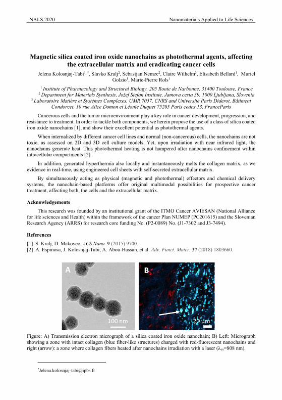
NALS 2020 Nanomaterials Applied to Life Sciences
Magnetic silica coated iron oxide nanochains as photothermal agents, affecting
the extracellular matrix and eradicating cancer cells Jelena Kolosnjaj-Tabi1, *, Slavko Kralj2, Sebastjan Nemec2, Claire Wilhelm3, Elisabeth Bellard1, Muriel
Golzio1, Marie-Pierre Rols1 1 Institute of Pharmacology and Structural Biology, 205 Route de Narbonne, 31400 Toulouse, France
2 Department for Materials Synthesis, Jožef Stefan Institute, Jamova cesta 39, 1000 Ljubljana, Slovenia 3 Laboratoire Matière et Systèmes Complexes, UMR 7057, CNRS and Université Paris Diderot, Bâtiment
Condorcet, 10 rue Alice Domon et Léonie Duquet 75205 Paris cedex 13, FranceParis
Cancerous cells and the tumor microenvironment play a key role in cancer development, progression, and resistance to treatment. In order to tackle both components, we herein propose the use of a class of silica coated iron oxide nanochains [1], and show their excellent potential as photothermal agents.
When internalized by different cancer cell lines and normal (non-cancerous) cells, the nanochains are not toxic, as assessed on 2D and 3D cell culture models. Yet, upon irradiation with near infrared light, the nanochains generate heat. This photothermal heating is not hampered after nanochains confinement within intracellular compartments [2].
In addition, generated hyperthermia also locally and instantaneously melts the collagen matrix, as we evidence in real-time, using engineered cell sheets with self-secreted extracellular matrix.
By simultaneously acting as physical (magnetic and photothermal) effectors and chemical delivery systems, the nanochain-based platforms offer original multimodal possibilities for prospective cancer treatment, affecting both, the cells and the extracellular matrix.
Acknowledgements
This research was founded by an institutional grant of the ITMO Cancer AVIESAN (National Alliance for life sciences and Health) within the framework of the cancer Plan NUMEP (PC201615) and the Slovenian Research Agency (ARRS) for research core funding No. (P2-0089) No. (J1-7302 and J3-7494).
References
[1] S. Kralj, D. Makovec. ACS Nano. 9 (2015) 9700. [2] A. Espinosa, J. Kolosnjaj-Tabi, A. Abou-Hassan, et al. Adv. Funct. Mater. 37 (2018) 1803660.
Figure: A) Transmission electron micrograph of a silica coated iron oxide nanochain; B) Left: Micrograph showing a zone with intact collagen (blue fiber-like structures) charged with red-fluorescent nanochains and right (arrow): a zone where collagen fibers heated after nanochains irradiation with a laser (λex=808 nm).
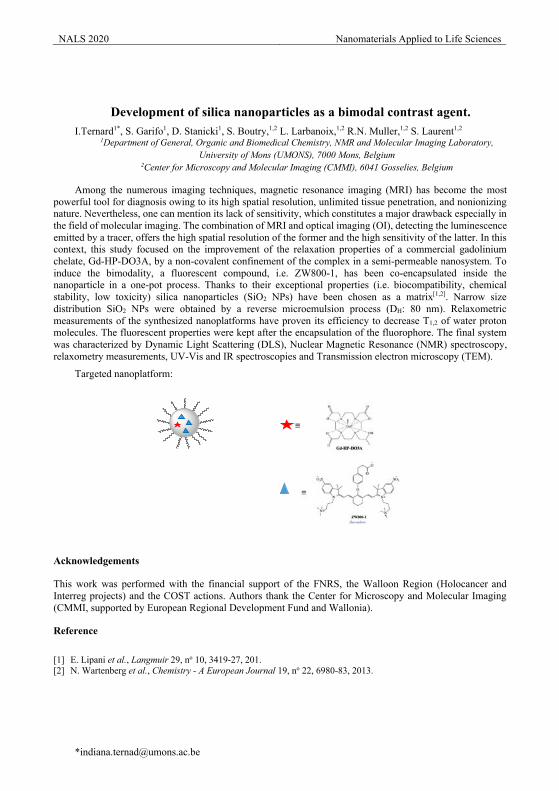
NALS 2020 Nanomaterials Applied to Life Sciences
Development of silica nanoparticles as a bimodal contrast agent. I.Ternard1*, S. Garifo1, D. Stanicki1, S. Boutry,1,2 L. Larbanoix,1,2 R.N. Muller,1,2 S. Laurent1,2
1Department of General, Organic and Biomedical Chemistry, NMR and Molecular Imaging Laboratory, University of Mons (UMONS), 7000 Mons, Belgium
2Center for Microscopy and Molecular Imaging (CMMI), 6041 Gosselies, Belgium
Among the numerous imaging techniques, magnetic resonance imaging (MRI) has become the most powerful tool for diagnosis owing to its high spatial resolution, unlimited tissue penetration, and nonionizing nature. Nevertheless, one can mention its lack of sensitivity, which constitutes a major drawback especially in the field of molecular imaging. The combination of MRI and optical imaging (OI), detecting the luminescence emitted by a tracer, offers the high spatial resolution of the former and the high sensitivity of the latter. In this context, this study focused on the improvement of the relaxation properties of a commercial gadolinium chelate, Gd-HP-DO3A, by a non-covalent confinement of the complex in a semi-permeable nanosystem. To induce the bimodality, a fluorescent compound, i.e. ZW800-1, has been co-encapsulated inside the nanoparticle in a one-pot process. Thanks to their exceptional properties (i.e. biocompatibility, chemical stability, low toxicity) silica nanoparticles (SiO2 NPs) have been chosen as a matrix[1,2]. Narrow size distribution SiO2 NPs were obtained by a reverse microemulsion process (DH: 80 nm). Relaxometric measurements of the synthesized nanoplatforms have proven its efficiency to decrease T1,2 of water proton molecules. The fluorescent properties were kept after the encapsulation of the fluorophore. The final system was characterized by Dynamic Light Scattering (DLS), Nuclear Magnetic Resonance (NMR) spectroscopy, relaxometry measurements, UV-Vis and IR spectroscopies and Transmission electron microscopy (TEM).
Targeted nanoplatform:
Acknowledgements
This work was performed with the financial support of the FNRS, the Walloon Region (Holocancer and Interreg projects) and the COST actions. Authors thank the Center for Microscopy and Molecular Imaging (CMMI, supported by European Regional Development Fund and Wallonia).
Reference
[1] E. Lipani et al., Langmuir 29, no 10, 3419-27, 201. [2] N. Wartenberg et al., Chemistry - A European Journal 19, no 22, 6980-83, 2013.
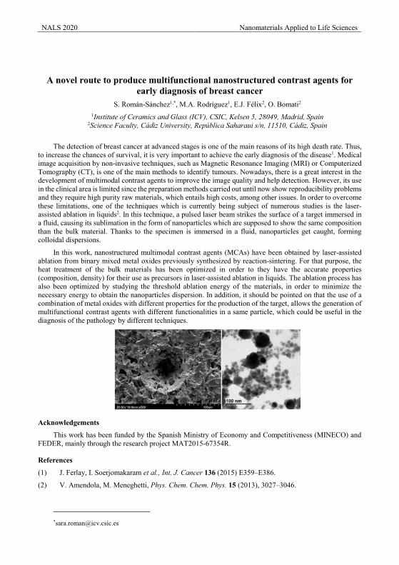
NALS 2020 Nanomaterials Applied to Life Sciences
A novel route to produce multifunctional nanostructured contrast agents for early diagnosis of breast cancer
S. Román-Sánchez1,*, M.A. Rodríguez1, E.J. Félix2, O. Bomati2 1Institute of Ceramics and Glass (ICV), CSIC, Kelsen 5, 28049, Madrid, Spain
2Science Faculty, Cádiz University, República Saharaui s/n, 11510, Cádiz, Spain
The detection of breast cancer at advanced stages is one of the main reasons of its high death rate. Thus, to increase the chances of survival, it is very important to achieve the early diagnosis of the disease1. Medical image acquisition by non-invasive techniques, such as Magnetic Resonance Imaging (MRI) or Computerized Tomography (CT), is one of the main methods to identify tumours. Nowadays, there is a great interest in the development of multimodal contrast agents to improve the image quality and help detection. However, its use in the clinical area is limited since the preparation methods carried out until now show reproducibility problems and they require high purity raw materials, which entails high costs, among other issues. In order to overcome these limitations, one of the techniques which is currently being subject of numerous studies is the laser-assisted ablation in liquids2. In this technique, a pulsed laser beam strikes the surface of a target immersed in a fluid, causing its sublimation in the form of nanoparticles which are supposed to show the same composition than the bulk material. Thanks to the specimen is immersed in a fluid, nanoparticles get caught, forming colloidal dispersions.
In this work, nanostructured multimodal contrast agents (MCAs) have been obtained by laser-assisted ablation from binary mixed metal oxides previously synthesized by reaction-sintering. For that purpose, the heat treatment of the bulk materials has been optimized in order to they have the accurate properties (composition, density) for their use as precursors in laser-assisted ablation in liquids. The ablation process has also been optimized by studying the threshold ablation energy of the materials, in order to minimize the necessary energy to obtain the nanoparticles dispersion. In addition, it should be pointed on that the use of a combination of metal oxides with different properties for the production of the target, allows the generation of multifunctional contrast agents with different functionalities in a same particle, which could be useful in the diagnosis of the pathology by different techniques.
Acknowledgements
This work has been funded by the Spanish Ministry of Economy and Competitiveness (MINECO) and FEDER, mainly through the research project MAT2015-67354R.
References
(1) J. Ferlay, I. Soerjomakaram et al., Int. J. Cancer 136 (2015) E359–E386.
(2) V. Amendola, M. Meneghetti, Phys. Chem. Chem. Phys. 15 (2013), 3027–3046.
NALS 2020 Nanomaterials Applied to Life Sciences
Smart Magnetic Hydrogels for Regenerative Medicine M.T. Lopez-Lopez1,*, A.B. Bonhome-Espinosa1, R. Contreras-Montoya2, L. Alvarez de Cienfuegos2,
J.D.G. Duran1 1Department of Applied Physics, University of Granada, Granada, Spain
2Department of Organic Chemistry, University of Granada, Spain
Hydrogels are 3D, hydrophilic networks of flexible chains (polymers, peptides, etc.) swollen by water or biological fluids. Due to their resemblance to the extracellular matrix of living tissues, hydrogels have been successfully used in regenerative medicine as scaffolds for the generation of artificial tissues. Hybrid hydrogels can be prepared by combination of polymers and peptides with inorganic nanomaterials, such as metallic nanoparticles, which can endow them with new, intriguing properties. An example is the embedment of ferromagnetic nanoparticles within the hydrogel network, which results in magnetic hydrogels that combine the flexibility typical of soft matter with ferromagnetic character, a unique characteristic that is not met by any natural material. This talk will focus on two different cases of magnetic hydrogels, based respectively on a chemically crosslinked polymer network and a physically assembled peptide network.
The chemically crosslinked polymer hydrogel consisted of functionalized magnetite nanoparticles (MNP) embedded in a network of fibrin polymers. We found that fibrin polymers anchored on the surface of the MNP, resulting in a perfect integration of the latter in the polymer network. Furthermore, this anchoring allowed acting by magnetic forces during polymer assembly, which could be used for example to induce alignment of the polymer fibres along the desired direction, giving rise to anisotropy at the microscopic scale. From the macroscopic viewpoint, the embedment of the MNP resulted in a large enhancement of the mechanical strength, even larger for anisotropic hydrogels, with respect to nonmagnetic hydrogels. Even more, the presence of MNP made possible the tunability of the mechanical properties of the final hydrogels by the action of magnetic fields. We finally used these magnetic hydrogels as scaffolds for the growth or artificial tissues and we found no significant differences between the proliferation of cells in magnetic hydrogels with respect to positive controls (nonmagnetic hydrogels). Interestingly, artificial tissues based on magnetic hydrogels demonstrated to be superior in terms of mechanical properties, than those based on nonmagnetic hydrogels, for the total duration of the experiments of cell growth (30 days).
The peptide hydrogel consisted of iron nanoparticles and Fmoc-diphenylalanine (Fmoc-FF) peptides that self-assembled by physical interactions in response to pH changes. Even though we demonstrated that Fmoc-FF peptides adsorbed on the surface of the MNP, magnetic field control of the assembly of the peptides was not achieved in this case. Nevertheless, optical and electron microscopy confirmed that iron nanoparticles aggregated into columnar structures aligned along the magnetic field direction, which ended up perfectly integrated within the hydrogel network, resulting in an anisotropic microstructure. This anisotropy at the microscopic level was also reflected in the macromechanical properties, with values of the viscoelastic moduli significantly larger for deformations perpendicular than parallel to the chain direction. What is more, we found a significant enhancement of the strength of these anisotropic magnetic hydrogels with respect to nonmagnetic hydrogels.
To conclude, we found that embedment of MNP within hydrogel networks offers a suitable way to modify and act by noncontact magnetic fields on the microstructure of the hydrogels. These changes in microstructure could result in enhanced macromechanical properties with respect to nonmagnetic hydrogels. As a consequence of all this, provided an adequate biocompatibility, magnetic hydrogels may be preferable than nonmagnetic hydrogels for applications in regenerative medicine.
Acknowledgements
Project FIS2017-85954-R (Ministerio de Economía, Industria y Competitividad, and Agencia Estatal de Investigación, Spain, cofounded by Fondo Europeo de Desarrollo Regional, European Union).
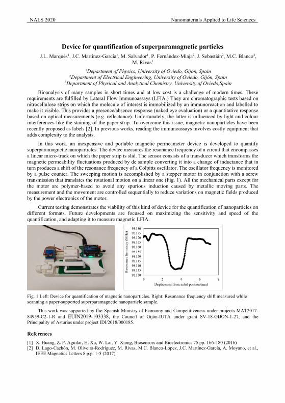
NALS 2020 Nanomaterials Applied to Life Sciences
Device for quantification of superparamagnetic particles
J.L. Marqués1, J.C. Martínez-García1, M. Salvador1, P. Fernández-Miaja2, J. Sebastián2, M.C. Blanco3, M. Rivas1
1Department of Physics, University of Oviedo, Gijón, Spain 2Department of Electrical Engineering, University of Oviedo, Gijón, Spain
3Department of Physical and Analytical Chemistry, University of Oviedo,Spain
Bioanalysis of many samples in short times and at low cost is a challenge of modern times. These requirements are fulfilled by Lateral Flow Immunoassays (LFIA.) They are chromatographic tests based on nitrocellulose strips on which the molecule of interest is immobilized by an immunoreaction and labelled to make it visible. This provides a presence/absence response (naked eye evaluation) or a quantitative response based on optical measurements (e.g. reflectance). Unfortunately, the latter is influenced by light and colour interferences like the staining of the paper strip. To overcome this issue, magnetic nanoparticles have been recently proposed as labels [2]. In previous works, reading the immunoassays involves costly equipment that adds complexity to the analysis.
In this work, an inexpensive and portable magnetic permeameter device is developed to quantify superparamagnetic nanoparticles. The device measures the resonance frequency of a circuit that encompasses a linear micro-track on which the paper strip is slid. The sensor consists of a transducer which transforms the magnetic permeability fluctuations produced by de sample converting it into a change of inductance that in turn produces a shift of the resonance frequency of a Colpitts oscillator. The oscillator frequency is monitored by a pulse counter. The sweeping motion is accomplished by a stepper motor in conjunction with a screw transmission that translates the rotational motion on a linear one (Fig. 1). All the mechanical parts except for the motor are polymer-based to avoid any spurious induction caused by metallic moving parts. The measurement and the movement are controlled sequentially to reduce variations on magnetic fields produced by the power electronics of the motor.
Current testing demonstrates the viability of this kind of device for the quantification of nanoparticles on different formats. Future developments are focused on maximizing the sensitivity and speed of the quantification, and adapting it to measure magnetic LFIA.
Fig. 1 Left: Device for quantification of magnetic nanoparticles. Right: Resonance frequency shift measured while scanning a paper-supported superparamagnetic nanoparticle sample.
This work was supported by the Spanish Ministry of Economy and Competitiveness under projects MAT2017-84959-C2-1-R and EUIN2019-103338, the Council of Gijón-IUTA under grant SV-18-GIJON-1-27, and the Principality of Asturias under project IDI/2018/000185.
References [1] X. Huang, Z. P. Aguilar, H. Xu, W. Lai, Y. Xiong, Biosensors and Bioelectronics 75 pp. 166-180 (2016) [2] D. Lago-Cachón, M. Oliveira-Rodríguez, M. Rivas, M.C. Blanco-López, J.C. Martínez-García, A. Moyano, et al.,
IEEE Magnetics Letters 8 p.p. 1-5 (2017).
98130000
98140000
98150000
98160000
98170000
98180000
0 5 10
Freq
uenc
y [H
z]
Position [mm]
NALS 2020 Nanomaterials Applied to Life Sciences
Mechanobiological Control of Human Neural Stem Cells Miguel Esteban-Lucia1.*, Judith Estengre Pérez2, Jaime J Hernandez1, Silvia García-López2, Marta P
Pereira2, Alberto Martinez-Serrano2, Isabel Rodriguez1 1Madrid Institute for Advanced Studies in Nanoscience (IMDEA Nanoscience), C/Faraday 9, Campus de
Cantoblanco, Madrid 28049, Spain 2Departamento de Biología Molecular (UAM), y Centro de Biología Molecular Severo Ochoa (CBMSO.
Autonomous University of Madrid-CSIC).C/ Nicolás Cabrera 1. Campus de Cantoblanco, Madrid 28049, Spain
The aim of regenerative medicine is to repair or replace tissues/structures in the body that do not function correctly due to disease or injury. Stem cells have a great therapeutic potential for this purpose however, before stem cells can be used in therapeutics, much work still needs to be done to understand their biology, to direct their differentiation and to control their behaviour and response to different stimulus [1].
There have been numerous evidences about the relevance of mechanical stimulus and signals present in extracellular matrix exerted on stem cells, and during development. As a consequence of those studies there is a considerable interest in studying mechanosensing effects to direct, for a tailored purpose, the response of stem cells in vitro [2]. It is widely accepted that topographical cues of micro/nano patterns can set off specific physiological processes that ultimately dictate the cell behaviour and fate, such as morphology, orientations and directionality on the substrate or proliferation [3]. However, much is still unknown about the physical cues and mechano-transductive pathways that determine the cell response.
In this context, the present work aims at investigating the response of immortalized human neural stem cells, hNSC.1000 [4], to dense high aspect ratio polystyrene nanopillars disposed on a square anisotropic pattern [5] with the final goal of guiding the neural stem cell fate commitment.
Studies on cell viability, proliferation, morphology and differentiation have been performed comparatively to a flat control substrate. Metabolic activity measurements have been also carried out to substantiate the response observed.
It was found that high aspect ratio nanopillars had an influence over the neural stem cells proliferation, increasing population doubling time in comparison with flat. Cells on the nanotopography showed a less round morphology due to the cytoplasmic insertions into the topography and cellular deformation. We carried out differentiation experiments to determine the correlation between high aspect ratio pillars disposal on neuronal and glial genesis.
Our observations suggest that high aspect ratio surface topographies could be considered as effective platforms for in vitro neural stem cell research to modulate cell responses.
References [1] J.W. Shin, D. J. Mooney, Cell Stem Cell 18 (2016) 16-19 [2] K. A. Jansen, D. M. Donato, H. E. Balcioglu, T. Schmidt, E. H. J. Danen and G. H. Koenderink, BBA - Molecular Cell Research 1853 (2015) 3043-3052. [3] F. Guilak, D. Cohen, B. Estes, J.M. Gimble, W. Liedtke, C.S. Chen, Cell Stem Cell, 5 (2009) 17-26 [4] A. Villa, E. Y. Snyder, A. Vescovi, and A. Martínez-Serrano, Exp. Neurol. 161(2000) 67-84 [5] F. Viela, D. Granados, A. Ayuso-Sacido and I. Rodríguez, Adv. Funct. Mater. 26(2016) 5599-5609
NALS 2020 Nanomaterials Applied to Life Sciences
Nanoencapsulation of X-7171 for the treatment of soft tissue sarcomas O. Estupiñán1,3, P. Suarez1, C. Rendueles1, M. Matos1,*, G. Gutiérrez1, M.C. Blanco-López3, M.
Matos1,*, R. Rodríguez-González3,*, 1Department of Chemical and Environmental Engineering, University of Oviedo, Julián Clavería 8, 33006
Oviedo, Spain 2Dept. of Physics, University of Oviedo, Gijón Polytechnic School, 33203 Gijón, Spain
3 Instituto de Investigación Sanitaria del principado de Asturias (Oviedo)
* Corresponding authors, e-mail: [email protected]; [email protected]
Sarcomas are historically understudied aggressive tumors affecting mesodermal tissues which usually show a poor response to current treatments, usually based in standard chemotherapy. Therefore, the development of new treatments able to prevent drug resistance would undoubtedly improve current sarcoma treatments. In the search of new therapies, we have focused in the naturally occurring antibiotic X-7171 which has proved a great efficiency to eliminate tumor cells in vitro. X-7171 Nonetheless, due to the high toxicity of this compound in the in vivo setting, the use of targeted delivery systems is required.
Nanovesicles (e.g., liposomes, niosomes or transfersomes) are an important family of organic NPs, produced by bottom-up nanotechnology. They are considered soft NPs because interactions among their molecular components are similar to those arising from biological systems. Another type of soft NPs that has also increased research interest are polymeric micelles [1] where PLGA is commonly used.
The aim of this study was to synthesize controlled size nanocolloids for X-7171 encapsulation within the range of 100-200 nm. For this purpose, different types of nanovesicles were prepared by the Thin Film Hydration controlling the operating conditions based in a previous study in which factorial experimental design and analysis of variance (ANOVA) was applied [2] and the Ethanol Injection Method while polymeric micelles were synthesized by the emulsion/solvent evaporation method. Nanocollids were characterized in terms of size using Dynamic Light Scattering (DLS) technique using Nanozetasizer (Malvern Instruments), TEM and HR-TEM while encapsulation efficiency (EE) was determined by RP-HPLC (Agilent Technologies) using UV-VIS detector. Furthermore, nanovesicle toxicity and loaded-X-7171 nanovesicles antitumoral activity were investigated in the myxoid liposarcoma cell line MSC-T-5H-FC.
Nanovesicles size ranged from 100 to 130 nm with EE values in the range 50-80% while polymeric micelles led to a mean size about 180 nm and EE values up to 95%. Cell tests evidenced lower nanovesicle citotoxicity and similar antitumoral activity of entrapped and free X-7171.
Acknowledgements This work was supported by Ministerio de Economía y Empresa (MINECO, Spain) under Grants
MAT2017-84959-C2-1-R and SAF2016-7528-R. This study was also financed by the Consejería de Economía y Empleo del Principado de Asturias (Plan de Ciencia, Tecnología e Innovación 2013-2017), under the Grant IDI/2018/000185. Support from the European Regional Development Fund (ERDF) is gratefully acknowledged. This study was also financed by Europa Investigación (ref: EUIN2019-103338) and European H2020-MSCA-ITN-2018 by the Grant Agreement number 813439.
References
1. Wang, S., et al., Facile construction of dual-targeting delivery system by using lipid capped polymer nanoparticles for anti-glioma therapy. RSC Advances, 2018. 8(1): p. 444-453.
2. García-Manrique, P., et al., Using Factorial Experimental Design To Prepare Size-Tuned Nanovesicles. Industrial & Engineering Chemistry Research, 2016. 55(34): p. 9164-9175.
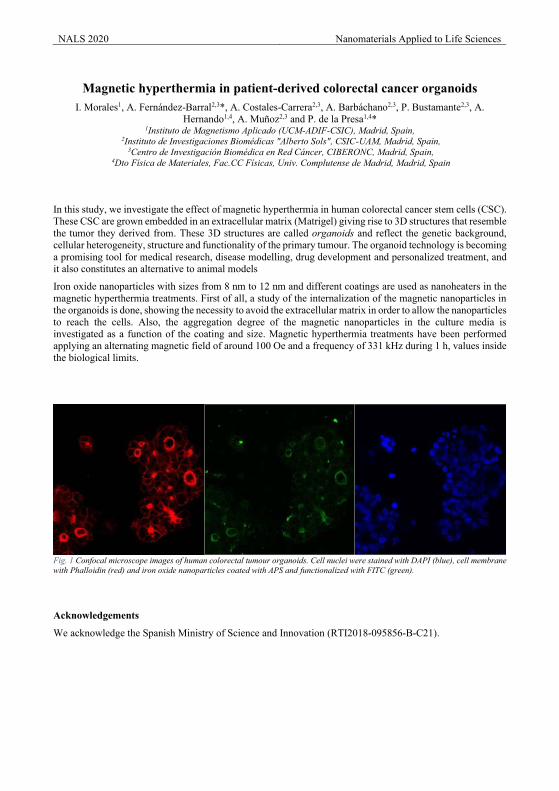
NALS 2020 Nanomaterials Applied to Life Sciences
Magnetic hyperthermia in patient-derived colorectal cancer organoids I. Morales1, A. Fernández-Barral2,3*, A. Costales-Carrera2,3, A. Barbáchano2,3, P. Bustamante2,3, A.
Hernando1,4, A. Muñoz2,3 and P. de la Presa1,4* 1Instituto de Magnetismo Aplicado (UCM-ADIF-CSIC), Madrid, Spain,
2Instituto de Investigaciones Biomédicas "Alberto Sols", CSIC-UAM, Madrid, Spain, 3Centro de Investigación Biomédica en Red Cáncer, CIBERONC, Madrid, Spain,
4Dto Física de Materiales, Fac.CC Físicas, Univ. Complutense de Madrid, Madrid, Spain
In this study, we investigate the effect of magnetic hyperthermia in human colorectal cancer stem cells (CSC). These CSC are grown embedded in an extracellular matrix (Matrigel) giving rise to 3D structures that resemble the tumor they derived from. These 3D structures are called organoids and reflect the genetic background, cellular heterogeneity, structure and functionality of the primary tumour. The organoid technology is becoming a promising tool for medical research, disease modelling, drug development and personalized treatment, and it also constitutes an alternative to animal models
Iron oxide nanoparticles with sizes from 8 nm to 12 nm and different coatings are used as nanoheaters in the magnetic hyperthermia treatments. First of all, a study of the internalization of the magnetic nanoparticles in the organoids is done, showing the necessity to avoid the extracellular matrix in order to allow the nanoparticles to reach the cells. Also, the aggregation degree of the magnetic nanoparticles in the culture media is investigated as a function of the coating and size. Magnetic hyperthermia treatments have been performed applying an alternating magnetic field of around 100 Oe and a frequency of 331 kHz during 1 h, values inside the biological limits.
Fig. 1 Confocal microscope images of human colorectal tumour organoids. Cell nuclei were stained with DAPI (blue), cell membrane with Phalloidin (red) and iron oxide nanoparticles coated with APS and functionalized with FITC (green).
Acknowledgements
We acknowledge the Spanish Ministry of Science and Innovation (RTI2018-095856-B-C21).
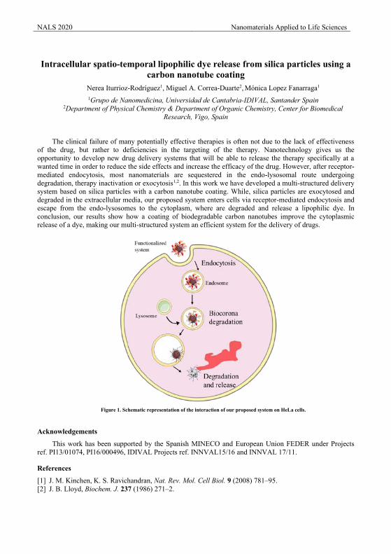
NALS 2020 Nanomaterials Applied to Life Sciences
Intracellular spatio-temporal lipophilic dye release from silica particles using a
carbon nanotube coating Nerea Iturrioz-Rodríguez1, Miguel A. Correa-Duarte2, Mónica Lopez Fanarraga1 1Grupo de Nanomedicina, Universidad de Cantabria-IDIVAL, Santander Spain
2Department of Physical Chemistry & Department of Organic Chemistry, Center for Biomedical Research, Vigo, Spain
The clinical failure of many potentially effective therapies is often not due to the lack of effectiveness of the drug, but rather to deficiencies in the targeting of the therapy. Nanotechnology gives us the opportunity to develop new drug delivery systems that will be able to release the therapy specifically at a wanted time in order to reduce the side effects and increase the efficacy of the drug. However, after receptor-mediated endocytosis, most nanomaterials are sequestered in the endo-lysosomal route undergoing degradation, therapy inactivation or exocytosis1,2. In this work we have developed a multi-structured delivery system based on silica particles with a carbon nanotube coating. While, silica particles are exocytosed and degraded in the extracellular media, our proposed system enters cells via receptor-mediated endocytosis and escape from the endo-lysosomes to the cytoplasm, where are degraded and release a lipophilic dye. In conclusion, our results show how a coating of biodegradable carbon nanotubes improve the cytoplasmic release of a dye, making our multi-structured system an efficient system for the delivery of drugs.
Acknowledgements
This work has been supported by the Spanish MINECO and European Union FEDER under Projects ref. PI13/01074, PI16/000496, IDIVAL Projects ref. INNVAL15/16 and INNVAL 17/11.
References
[1] J. M. Kinchen, K. S. Ravichandran, Nat. Rev. Mol. Cell Biol. 9 (2008) 781–95. [2] J. B. Lloyd, Biochem. J. 237 (1986) 271–2.
Figure 1. Schematic representation of the interaction of our proposed system on HeLa cells.
NALS 2020 Nanomaterials Applied to Life Sciences
Liposomes to improve skin absorption of antioxidants Lucía Martínez Arias1, Alejandro Llamedo1, Rebeca Alonso1 and Daniel Pando1,*
1 Nanovex Biotechnologies, Research and Development Department, Llanera, Asturias, Spain
Skin is the largest organ in human body and his function is to act as a barrier to protect us from the environment. The exposure of skin to UV radiation causes skin damage, acute UV irradiation promote sunburn, altered pigmentation, inflammation and immune suppression, whereas the recurrent exposure to UV radiation leads to photoaging, immunosuppression and photocarcinogenesis.
Skin oxidative stress generated by the presence of reactive oxygen species (ROS) is the main responsible for photoaging. Skin has a network of enzymatic and nonenzymatic antioxidant systems to counteract Oxidative stress effects. The presence of antioxidants in the skin decreases with age, and for this reason the topical treatment with antioxidants can improved the skin protection to UV radiation. Encapsulation of antioxidants in liposomes improve their effectiveness because these nanocarriers improve the stability and skin penetration of these compounds.
In order to study the response of the cells to the UVA-Induced oxidative stress and test the potential of the different encapsulated and non-encapsulated products in counteracting ROS induction, EpiDermTM Reconstructed Human Epidermis 3D skin model was submitted to topical and systematic treatment with free or encapsulated Vitamin A, C and E (ACE or Lipo-ACE) and exposed to UVA Irradiation
Acknowledgements
This work has been supported by grants for Industrial Doctorate from Ayuntamiento de Oviedo (Oviedo Siembra Talento)
References
[1] I. P. Kaur, M. Kapila, R. Agrawal, Ageing Res. Rev. 6, (2007) 271–288. [2] L. Montenegro J. Pharm. Pharmacogn. Res. 2, (2014) 73–92. [3] A. Godic, B. Poljšak, M. Adamic, and R. Dahmane, Oxid. Med. Cell. Longev. 2014, (2014).

NALS 2020 Nanomaterials Applied to Life Sciences
Amine covalent organic frameworks for safe delivery of camptothecin A. De Santana Oliveira,1 E. Rivero-Buceta,2,* C. Vidaurre-Agut,2,3 V. Moreno,4 S. Berenice Castellã
Pergher1 and P. Botella2
1Universidade Federal do Rio Grande do Norte, Laboratório de Peneiras Moleculares, Instituto de Química, 59078-970 Natal, RN, Brazil
2Instituto de Tecnología Química (UPV-CSIC), Av. Los Naranjos s/n, 46022 Valencia, Spain 3Instituto de Instrumentación para Imagen Molecular (I3M), Centro Mixto CSIC-Universitat Politècnica de
València, Camino de Vera s/n, 46022 Valencia, Spain dCentro Investigación Príncipe Felipe, C/ Eduardo Primo Yúfera 3, Valencia 46012, Spain
Introduction: Covalent organic frameworks (COFs) are a new target for research in porous materials, due to their several attractive features, such as, large external surface area, defined pore size, tunable pores [1]. All these properties make these materials promising for multiple applications such as molecular separation, energy storage, catalysis, small molecules adsorption, and drug delivery. 20-(S)-camptothecin (CPT) is a natural alkaloid with strong antitumor activity against a broad spectrum of cancers [2]. Unfortunately, this molecule has found no clinical application, due to its presents some major limitations. Here, we present novel amine functionalized COF’s prepared by direct incorporation of nucleophilic groups (e.g., primary amines) on pore wall during the synthesis of a 2D-COF (COF-5) due to the substitution of original monomers for its later use as drug delivery system for covalently bonded camptothecin (CPT).
Methods: All COFs derivatives (CF-x (substituted with primary amine groups) were synthesized by an optimized microwave-assisted method. The CPT prodrug (CPT-succinate) was conjugated with primary amine groups by amide bond. Moreover, stability assays in water were carried out in order to investigate the effect of the CPT on the structure (Figure 1). In addition, internal trafficking was investigated by flow cytometry (FC) due to the incorporation of Alexa Fluor 647 in the structure of COFs (Figure 1).
Results: COF-25-CPT and COF-25 conjugates show efficient particle cell internalization. It is noted that COF-25-CPT show strong cytotoxic activity over HeLa and MCF-7 cell lines, even at low dose. On the other hand, the introduction of functional groups during the synthesis of two dimensional COFs (2D COFs) is highly discouraged, as they can interfere with the π-π stacking forces, compromising framework integrity [3]. In this case, the presence of the CPT improve the stability of the structure due to the drug protect the B-O bonds and hydrolytically susceptible backbones through kinetic blocking.
Figure 1. Schematic illustration of COF-25-CPT
Conclusion: COF-25-CPT is a novel type of drug delivery system based on covalent linking of a therapeutic molecule in COF structure, by coupling CPT molecules over primary amine functions. These new COF nanoparticles constitute a new stable vehicle for the safe delivery to cancer cells of the CPT or other chemotherapeutic molecules.
Acknowledgements
Financial support of the Spanish Ministry of Economy and Competitiveness (projects TEC2016-80976-R and SEV-2016-0683) is gratefully acknowledged.
References
[1] F. Zhao, H. Liu, S. D. R. Mathe, A. Dong, J. D. Zhang. Nanomaterials. 8(1), (2017), 15. [2] E.L. Ezell, L.L. Smith. J. Nat. Prod. 54, (1991), 1645-1650. [3] S. D. Brucks, D.N. Bunck, W.R. Dichtel, Polym. 55, (2014) 330–334.
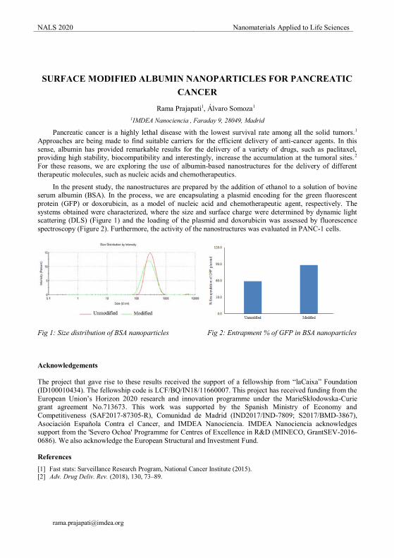
NALS 2020 Nanomaterials Applied to Life Sciences
SURFACE MODIFIED ALBUMIN NANOPARTICLES FOR PANCREATIC
CANCER
Rama Prajapati1, Álvaro Somoza1 1IMDEA Nanociencia , Faraday 9, 28049, Madrid
Pancreatic cancer is a highly lethal disease with the lowest survival rate among all the solid tumors.1 Approaches are being made to find suitable carriers for the efficient delivery of anti-cancer agents. In this sense, albumin has provided remarkable results for the delivery of a variety of drugs, such as paclitaxel, providing high stability, biocompatibility and interestingly, increase the accumulation at the tumoral sites.2 For these reasons, we are exploring the use of albumin-based nanostructures for the delivery of different therapeutic molecules, such as nucleic acids and chemotherapeutics.
In the present study, the nanostructures are prepared by the addition of ethanol to a solution of bovine serum albumin (BSA). In the process, we are encapsulating a plasmid encoding for the green fluorescent protein (GFP) or doxorubicin, as a model of nucleic acid and chemotherapeutic agent, respectively. The systems obtained were characterized, where the size and surface charge were determined by dynamic light scattering (DLS) (Figure 1) and the loading of the plasmid and doxorubicin was assessed by fluorescence spectroscopy (Figure 2). Furthermore, the activity of the nanostructures was evaluated in PANC-1 cells.
Fig 1: Size distribution of BSA nanoparticles Fig 2: Entrapment % of GFP in BSA nanoparticles
Acknowledgements
The project that gave rise to these results received the support of a fellowship from “laCaixa” Foundation (ID100010434). The fellowship code is LCF/BQ/IN18/11660007. This project has received funding from the European Union’s Horizon 2020 research and innovation programme under the MarieSkłodowska-Curie grant agreement No.713673. This work was supported by the Spanish Ministry of Economy and Competitiveness (SAF2017-87305-R), Comunidad de Madrid (IND2017/IND-7809; S2017/BMD-3867), Asociación Española Contra el Cancer, and IMDEA Nanociencia. IMDEA Nanociencia acknowledges support from the 'Severo Ochoa' Programme for Centres of Excellence in R&D (MINECO, GrantSEV-2016-0686). We also acknowledge the European Structural and Investment Fund.
References [1] Fast stats: Surveillance Research Program, National Cancer Institute (2015). [2] Adv. Drug Deliv. Rev. (2018), 130, 73–89.
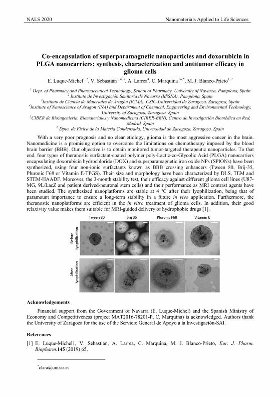
NALS 2020 Nanomaterials Applied to Life Sciences
Co-encapsulation of superparamagnetic nanoparticles and doxorubicin in PLGA nanocarriers: synthesis, characterization and antitumor efficacy in
glioma cellsE. Luque-Michel1, 2, V. Sebastián3, 4, 5, A. Larrea4, C. Marquina3,6,*, M. J. Blanco-Prieto1, 2
1 Dept. of Pharmacy and Pharmaceutical Technology, School of Pharmacy, University of Navarra, Pamplona, Spain2 Instituto de Investigación Sanitaria de Navarra (IdiSNA), Pamplona, Spain
3Instituto de Ciencia de Materiales de Aragón (ICMA), CSIC-Universidad de Zaragoza, Zaragoza, Spain4Institute of Nanoscience of Aragon (INA) and Department of Chemical, Engineering and Environmental Technology,
University of Zaragoza, Zaragoza, Spain5CIBER de Bioingeniería, Biomateriales y Nanomedicina (CIBER-BBN), Centro de Investigación Biomédica en Red,
Madrid, Spain6 Dpto. de Física de la Materia Condensada, Universidad de Zaragoza, Zaragoza, Spain
With a very poor prognosis and no clear etiology, glioma is the most aggressive cancer in the brain. Nanomedicine is a promising option to overcome the limitations on chemotherapy imposed by the blood brain barrier (BBB). Our objective is to obtain monitored tumor-targeted therapeutic nanoparticles. To that end, four types of theranostic surfactant-coated polymer poly-Lactic-co-Glycolic Acid (PLGA) nanocarriersencapsulating doxorubicin hydrochloride (DOX) and superparamagnetic iron oxide NPs (SPIONs) have beensynthesized, using four non-ionic surfactants known as BBB crossing enhancers (Tween 80, Brij-35, Pluronic F68 or Vitamin E-TPGS). Their size and morphology have been characterized by DLS, TEM and STEM-HAADF. Moreover, the 3-month stability test, their efficacy against different glioma cell lines (U87-MG, 9L/LacZ and patient derived-neuronal stem cells) and their performance as MRI contrast agents have been studied. The synthesized nanoplatforms are stable at 4 ºC after their lyophilization, being that of paramount importance to ensure a long-term stability in a future in vivo application. Furthermore, the theranostic nanoplatforms are efficient in the in vitro treatment of glioma cells. In addition, their good relaxivity value makes them suitable for MRI-guided delivery of hydrophobic drugs [1].
Acknowledgements
Financial support from the Government of Navarra (E. Luque-Michel) and the Spanish Ministry of Economy and Competitiveness (project MAT2016-78201-P, C. Marquina) is acknowledged. Authors thankthe University of Zaragoza for the use of the Servicio General de Apoyo a la Investigación-SAI.
References
[1] E. Luque-Michel1, V. Sebastián, A. Larrea, C. Marquina, M. J. Blanco-Prieto, Eur. J. Pharm. Biopharm.145 (2019) 65.
NALS 2020 Nanomaterials Applied to Life Sciences
Nanoparticles for improving therapeutic effects of antibiotics in cornea
Alejandro Llamedo1, Rosalía Sanchez1, Manuel Chacón2, Rebeca Alonso1 1Nanovex Biotechnologies, Research and Development Department, Llanera Asturias, Spain.
2Instituto Universitario Fernández-Vega, Fundación de Investigación Oftalmológica, Universidad de Oviedo, Oviedo, Spain.
Nowadays there is a need in the development of new treatments in ophthalmology. One of the best approaches is the topical ocular drug delivery which presents a challenge in penetration due to all anatomical eye barriers. Lipid-based nanoparticles have being used in several studies as possible candidates for improving drug delivery in this field as they display a penetration enhancing effect in the cornea. In this study, Liposomes and Niosomes were analysed as possible delivery systems for drugs in 3D corneal tissue model QobuR, reducing the use of animal models in investigation.
Naked nanoparticles and chitosan-coated nanoparticles encapsulating fluorescein were synthesized with an average size of 200 nm and the toxicity, bioadhesion and permeability were studied in in-vitro conditions. Results showed that nanoparticles were no irritant and presented no toxicity. Moreover, they were able to stay attached in the epithelial tissue after washing and chitosan-coated liposomes showed penetration into deeper layers of the cornea. Considering these results, chitosan-coated Liposomes were used for the encapsulation of Vancomycin, a widely used antibiotic. Its stability, encapsulation efficiency, surface charge and average size were studied.
In conclusion, chitosan-coated liposomes enhance penetration more than other lipid-based nanoparticles tested which makes them suitable as topical drug delivery vehicles in ophthalmology.
Acknowledgements
This work has been supported by grants from Economic Development Agency of the Principality of Asturias (IDEPA), IDE/000486 and Grants for Industrial Doctorate from Ayuntamiento de Oviedo (Oviedo Siembra Talento)
References
[1] Chacón, M. et al. Eur. J. Pharm. Biopharm. 136, 164–173 (2019) [2] Gonzalez Gomez, A. et al. ACS Omega 4, 10866–10876 (2019). [3] Li, J. et al. Drug Deliv. 26, 188–198 (2019).

NALS 2020 Nanomaterials Applied to Life Sciences
Fe3O4 Nanoparticles (MNPs) as Nanozyme for Signal Enhancement in Lateral Flow Immunoassays (LFIA)
Baihui Wang1,*, Esther Serrano-Pertierra1, Amanda Moyano1, María Salvador2, Montserrat Rivas2, Carmen Blanco-López1
1Department of Analytical and Physical Chemistry, University of Oviedo. Department of Physics, Gijón Polytechnic School of Engineering, University of Oviedo
Magnetic nanoparticles (MNP) represent a kind of nanomaterial with characteristic physicochemical properties and ever-growing applications. Because of the good biocompatibility, MNPs have been extensively employed in biomedical domains such as hyperthermia [1], drug delivery [2], and magnetic resonance imaging [3].. Environmental and catalytic applications remain crucial as well [4]. A decade ago, Yan’s group discovered that MNPs own intrinsic enzyme mimetic activities resemble those of natural peroxidases, which catalyze the oxidation of the substrate when H2O2 is present [5]. This effect can be tested by converting different standard chromogenic peroxidase substrates. Since then, MNPs have received wide attention not only for their catalytic stability over a wide range of temperatures and pHs, controlled low cost large scale synthesis, but also owning to convenient separation by application of an external magnetic field.
Since MNPs are available with different capping agents at the surface [6], the study of the effect of carbon chain length on peroxidase-like activity was carried out by concentration gradients of MNPs. Magnetic nanoparticles with three different coatings, oleic acid: CH3(CH2)7CH = CH(CH2)7COOH; lauric acid: CH3(CH2)10COOH; myristic acid: CH3(CH2)12COOH, were tested as nanozyme to catalyze the oxidation of TMB in the presence of H2O2. MNPs coated with lauric acid showed the best performance when testing the peroxidase-like activity. These results indicate that the carbon chain length of coating did have an impact on MNPs when performed as nanozyme; the shorter the length of carbon chain, the better the biomimetic peroxidase ability (Figure 1).
Acknowledgements
This work was supported by the Ministerio de Economía y Competitividad under the grants MAT2017-84959-C2-1-R and EUIN2019-103338.
References
[1] Obaidat, I.; Issa, B.; Haik, Y. Nanomaterials 2015, 5, 63− 89. [2] , R.. Chem. Rev. 2016, 116, 5338−5431. [3] Shin, T.-H.; Choi, Y.; Kim, S.; Cheon, J. Chem. Soc. Rev. 2015, 44, 4501−4516. [4] Govan, J.; Gun’ko, Y. Nanomaterials 2014, 4, 222−241. [5] Gao, L.; Zhuang, J.; Nie, L.; Zhang, J.; Zhang, Y.; Gu, N.; Wang, T.; Feng, J.; Yang, D.; Perrett, S.;
Xiyun, Y. Nat. Nanotechnol. 2007, 2, 577–583. [6] D. Bica, L. Vékás, M.V. Avdeev, O. M ă S M ă ă M G m J. Magn.
Magn. 311 (2007) 17–21.
y = 0.621x + 0.0154
R2 = 0.9981 y = 0.6783x + 0.0282
R2 = 0.9957
y = 0.6021x + 0.0161
R2 = 0.9855
0
0,05
0,1
0,15
0,2
0,25
0,3
0 0,05 0,1 0,15 0,2 0,25 0,3 0,35 0,4
Abso
rban
ce
Concentration of MNPs(mg/ml)
Calibration curve of 3 MNPs
Figure 1. Calibration curve using concentrations of MNPs with different coatings
oleic acid: CH3(CH2)7CH = CH(CH2)7COOH
lauric acid: CH3(CH2)10COOH
myristic acid: CH3(CH2)12COOH
lan
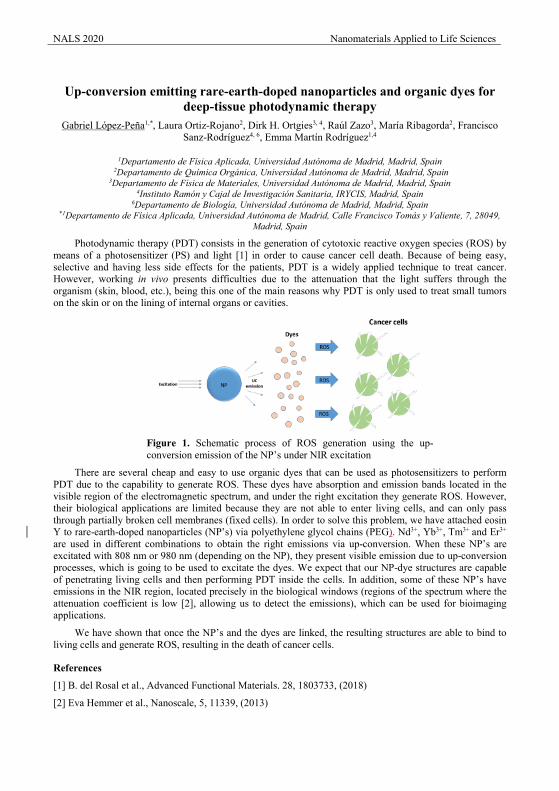
NALS 2020 Nanomaterials Applied to Life Sciences
Up-conversion emitting rare-earth-doped nanoparticles and organic dyes for
deep-tissue photodynamic therapy Gabriel López-Peña1,*, Laura Ortiz-Rojano2, Dirk H. Ortgies3, 4, Raúl Zazo3, María Ribagorda2, Francisco
Sanz-Rodríguez4, 6, Emma Martín Rodríguez1,4
1Departamento de Física Aplicada, Universidad Autónoma de Madrid, Madrid, Spain 2Departamento de Química Orgánica, Universidad Autónoma de Madrid, Madrid, Spain
3Departamento de Física de Materiales, Universidad Autónoma de Madrid, Madrid, Spain 4Instituto Ramón y Cajal de Investigación Sanitaria, IRYCIS, Madrid, Spain
6Departamento de Biología, Universidad Autónoma de Madrid, Madrid, Spain *1Departamento de Física Aplicada, Universidad Autónoma de Madrid, Calle Francisco Tomás y Valiente, 7, 28049,
Madrid, Spain
Photodynamic therapy (PDT) consists in the generation of cytotoxic reactive oxygen species (ROS) by means of a photosensitizer (PS) and light [1] in order to cause cancer cell death. Because of being easy, selective and having less side effects for the patients, PDT is a widely applied technique to treat cancer. However, working in vivo presents difficulties due to the attenuation that the light suffers through the organism (skin, blood, etc.), being this one of the main reasons why PDT is only used to treat small tumors on the skin or on the lining of internal organs or cavities.
Figure 1. Schematic process of ROS generation using the up-conversion emission of the NP’s under NIR excitation
There are several cheap and easy to use organic dyes that can be used as photosensitizers to perform PDT due to the capability to generate ROS. These dyes have absorption and emission bands located in the visible region of the electromagnetic spectrum, and under the right excitation they generate ROS. However, their biological applications are limited because they are not able to enter living cells, and can only pass through partially broken cell membranes (fixed cells). In order to solve this problem, we have attached eosin Y to rare-earth-doped nanoparticles (NP’s) via polyethylene glycol chains (PEG). Nd3+, Yb3+, Tm3+ and Er3+ are used in different combinations to obtain the right emissions via up-conversion. When these NP’s are excitated with 808 nm or 980 nm (depending on the NP), they present visible emission due to up-conversion processes, which is going to be used to excitate the dyes. We expect that our NP-dye structures are capable of penetrating living cells and then performing PDT inside the cells. In addition, some of these NP’s have emissions in the NIR region, located precisely in the biological windows (regions of the spectrum where the attenuation coefficient is low [2], allowing us to detect the emissions), which can be used for bioimaging applications.
We have shown that once the NP’s and the dyes are linked, the resulting structures are able to bind to living cells and generate ROS, resulting in the death of cancer cells.
References
[1] B. del Rosal et al., Advanced Functional Materials. 28, 1803733, (2018)
[2] Eva Hemmer et al., Nanoscale, 5, 11339, (2013)
NALS 2020 Nanomaterials Applied to Life Sciences
Using the Shiga-toxin interaction domain as a natural ligand for nanoparticle precision diagnostics and targeted therapeutics
Elena Navarro-Palomares*, Esperanza Padín-González, Rafael Valiente, Mónica L. Fanarraga Grupo de Nanomedicina-IDIVAL, Instituto Valdecilla, Universidad de Cantabria, Herrera Oria s/n, 39011
Santander
Specific targeting for diagnostic and therapeutic purposes remains one of the major challenges in cancer. In the search of new receptor-mediated cell entry routes, the mechanisms used by virus and toxins to recognize specific cell receptors and hijack the cell entry mechanisms for their own benefit, result of special interest. Protein engineering of pathogen-derived toxins allows the design of nanoparticle biocoatings that mimic some of these unique mechanisms. We have developed a system to target the glycosphingolipid Gb3, overexpressed in several tumours such as oropharyngeal and gastrointestinal carcinomas, with its natural ligand, the Shiga toxin. This toxin, that is produced by Shigella dysenteriae and some Escherichia coli strains, has the particularity of using a non-canonical gateway to invade cells, vía a unique retrograde trafficking pathway, through the Golgi apparatus and endoplasmic reticulum to the cytosol. We have cloned the innocuous receptor-binding domain of the toxin (STxB) fused to a polypeptidic tag specifically designed to functionalize nanomaterials with a negative surface charge. We show how nanoparticles functionalized this way specifically recognize and penetrate in cancer cells expressing the Gb3 receptor using a non-canonical retrograde cell entry route, avoiding the endo-lysosomal vesicular trafficking. This non-canonical receptor-mediated entry route could be used to target, straight into the cytosol, nano-delivery agents carrying sensible therapeutic compounds, preserving nanomaterials of the hostile environment of the canonical endo-lysosomal vesicular pathway. Moreover, this mechanism of cell entry is applicable to a wide array of nano-designs, that can be used to produce unique tools in precision therapy, diagnosis or theragnostic.
Acknowledgements
This work has been supported by the Spanish MINECO, Instituto de Salud Carlos III, European Union (European Regional Development Fund-ERDF) under Projects ref. PI16/000496 and PI19/000349, DTS19/00033, CTM2017-84050-R, MCIU/AEI/FEDER, EU under project PGC2018-101464-B-100, NanoBioApp Network (MINECO-17-MAT2016-81955-REDT), and IDIVAL for the PREVAL16/02, INNVAL 17/11 and INNVAL 18/28 projects.
References
[1] L. Johannes, V. Popoff, Cell. 135 (2008) 1175-1187. [2] V. Luginbuehl, N. Meier, K. Kovar and J. Rohrer, Biotechnol. Adv. 36 (2018) 613-623. [3] L. Johannes and W. Römer, Nat. Rev. Microbiol. 8 (2010) 105-116 [4] D. Crossed D Signevenica, V. Čikeš Čulić, A. Vuica and A. Markotić, Med. Oncol. 28 (2011) S675–S684
e-mail: *[email protected]
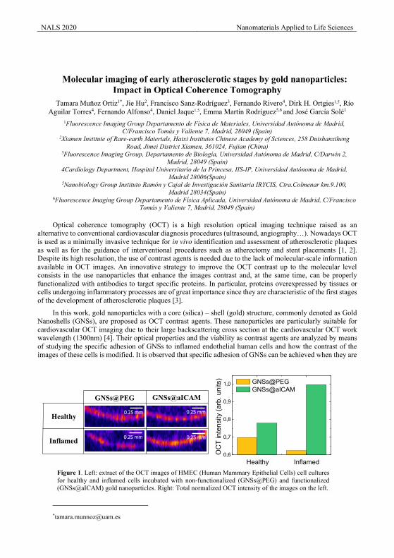
NALS 2020 Nanomaterials Applied to Life Sciences
Molecular imaging of early atherosclerotic stages by gold nanoparticles: Impact in Optical Coherence Tomography
Tamara Muñoz Ortiz1*, Jie Hu2, Francisco Sanz-Rodríguez3, Fernando Rivero4, Dirk H. Ortgies1,5, Río Aguilar Torres4, Fernando Alfonso4, Daniel Jaque1,5, Emma Martín Rodríguez5,6 and José García Solé1
1Fluorescence Imaging Group Departamento de Física de Materiales, Universidad Autónoma de Madrid, C/Francisco Tomás y Valiente 7, Madrid, 28049 (Spain)
2Xiamen Institute of Rare-earth Materials, Haixi Institutes Chinese Academy of Sciences, 258 Duishanxiheng Road, Jimei District Xiamen, 361024, Fujian (China)
3Fluorescence Imaging Group, Departamento de Biología, Universidad Autónoma de Madrid, C/Darwin 2, Madrid, 28049 (Spain)
4Cardiology Department, Hospital Universitario de la Princesa, IIS-IP, Universidad Autónoma de Madrid, Madrid 28006(Spain)
5Nanobiology Group Instituto Ramón y Cajal de Investigación Sanitaria IRYCIS, Ctra.Colmenar km.9.100, Madrid 28034(Spain)
6Fluorescence Imaging Group Departamento de Física Aplicada, Universidad Autónoma de Madrid, C/Francisco Tomás y Valiente 7, Madrid, 28049 (Spain)
Optical coherence tomography (OCT) is a high resolution optical imaging technique raised as an alternative to conventional cardiovascular diagnosis procedures (ultrasound, angiography…). Nowadays OCT is used as a minimally invasive technique for in vivo identification and assessment of atherosclerotic plaques as well as for the guidance of interventional procedures such as atherectomy and stent placements [1, 2]. Despite its high resolution, the use of contrast agents is needed due to the lack of molecular-scale information available in OCT images. An innovative strategy to improve the OCT contrast up to the molecular level consists in the use nanoparticles that enhance the images contrast and, at the same time, can be properly functionalized with antibodies to target specific proteins. In particular, proteins overexpressed by tissues or cells undergoing inflammatory processes are of great importance since they are characteristic of the first stages of the development of atherosclerotic plaques [3].
In this work, gold nanoparticles with a core (silica) – shell (gold) structure, commonly denoted as Gold Nanoshells (GNSs), are proposed as OCT contrast agents. These nanoparticles are particularly suitable for cardiovascular OCT imaging due to their large backscattering cross section at the cardiovascular OCT work wavelength (1300nm) [4]. Their optical properties and the viability as contrast agents are analyzed by means of studying the specific adhesion of GNSs to inflamed endothelial human cells and how the contrast of the images of these cells is modified. It is observed that specific adhesion of GNSs can be achieved when they are
0,6
0,7
0,8
0,9
1,0
OC
T in
tens
ity (a
rb. u
nits
) GNSs@PEG GNSs@aICAM
0.25 mm
0.25 mm 0.25 mm
0.25 mm
GNSs@PEG GNSs@aICAM
Healthy
Inflamed
Figure 1. Left: extract of the OCT images of HMEC (Human Mammary Epithelial Cells) cell cultures for healthy and inflamed cells incubated with non-functionalized (GNSs@PEG) and functionalized (GNSs@aICAM) gold nanoparticles. Right: Total normalized OCT intensity of the images on the left.
Healthy Inflamed
NALS 2020 Nanomaterials Applied to Life Sciences functionalized with proper antibodies (anti-ICAM-1). This specific adhesion is clearly manifested as an increase in the intensity signal of the OCT and optical microscopic images (figure 1). Therefore, the present study represents an important step towards the use of contrast agents to target inflammation in the first stages of atherosclerosis by OCT.
References
[1] H. Yabushita, B. E. Bouma, S. L. Houser, H. T. Aretz, I. K. Jang, K. H. Schlendorf, C. R. Kauffman, M. Shishkov, D. H. Kang, E. F. Halpern and G. J. Tearney Circulation 106 (2002) 1640.
[2] B. E. Bouma, G. J. Tearney, H. Yabushita, M. Shishkov, C. R. Kauffman, D. DeJoseph Gauthier, B. D. MacNeill, S. L. Houser, H. T. Aretz, E. F. Halpern and I.-K. Jang Heart 89 (2003) 317.
[3] P. Libby, P. M. Ridker and A. Maseri Circulation 105 (2002) 1135.
[4] J. Hu, F. Rivero, R. A. Torres, H. Loro Ramírez, E. M. Rodríguez, F. Alfonso, J. García Solé and D. Jaque Journal of Biophotonics 10 (2017) 674.
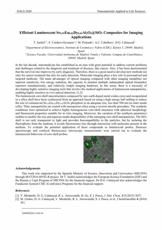
NALS 2020 Nanomaterials Applied to Life Sciences
Efficient Luminescent Sr0.95Eu0.02Dy0.03Al2O4@SiO2 Composites for Imaging Applications
T. Jardiel1,*, E. Cordero-Oyonarte1,2, M. Peiteado1, A.C. Caballero1, D.G. Calatayud1 1 Department of Electroceramics, Instituto de Cerámica y Vidrio (CSIC), Kelsen 5, 28049, Madrid,
Spain 2 Science Faculty, Universidad Autónoma de Madrid, Tomás y Valiente, Campus de Cantoblanco,
28049, Madrid, Spain
In the last decade, nanomedicine has established as an area with great potential to address current problems and challenges related to the diagnosis and treatment of diseases, like cancer. Also, it has been demonstrated that the survival rate improves by early diagnosis. Therefore, there is a great need to develop new methods not only for cancer treatment but also for early detection. Molecular imaging plays a key role in personalised and targeted medicine. The main advantages of optical imaging compared with other imaging modalities are superior sensitivity, low energy radiation, the capacity to monitor multiple independent optical biomarker reporters simultaneously, and relatively simple imaging hardware. In this sense, there is a necessity of developing highly sensitive imaging tools that involve the medical applications of luminescent nanoparticles, enabling highly sensitive in vivo optical detection. [1,2] The luminescent core-shell nanostructures composed by rare-earth doped metal oxides cores and encapsulated in a silica shell have been synthesised from an approach based on using a high energy ball milling to reduce the size of commercial Sr0.95Eu0.02Dy0.03Al2O4 phosphors to an adequate size, less than 500 nm (to enter inside cells). Then, nanoparticles are coated with mesoporous silica using a reverse micelle procedure. The synthetic conditions were optimized to achieve highly homogeneous core-shell structures with spherical morphology and fluorescent properties suitable for in vitro imaging. Moreover, the variation of the synthesis parameters enables to modify the size and aqueous media dispersibility of the emerging core-shell nanoparticles. The SiO2 shell is not only transparent to light and provides biocompatibility to the particles, but by isolating the fluorophores from the medium, it avoids fluorescence loss through interaction with molecules present in the medium. To evaluate the potential application of these compounds as luminescent probes, fluoresce spectroscopy and confocal fluorescence microscopy measurements were carried out to evaluate the luminescent behaviour of core-shell probes.
Acknowledgements
This work was supported by the Spanish Ministry of Science, Innovation and Universities (MICINN) through MAT2016-80182-R project. Dr T. Jardiel acknowledges the European Science Foundation (ESF) and the Ramon y Cajal Program of MICINN for the financial support. Dr D.G. Calatayud also acknowledges the Fundación General CSIC (ComFuturo Program) for the financial support.
References
[1] V. Mirabello, D. G. Calatayud, R. L. Arrowsmith, H. Ge, S. I. Pascu, J. Mat. Chem. B 3 (2015) 5657. [2] M. Lledos, D. G. Calatayud, V. Mirabello, R. L. Arrowsmith, S. I. Pascu, et al., ChemNanoMat 4 (2018)
361.
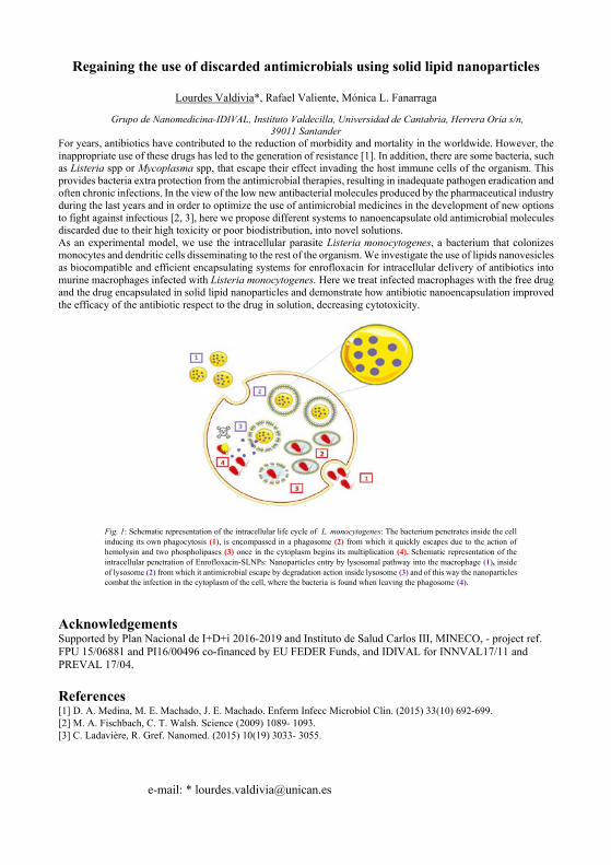
Regaining the use of discarded antimicrobials using solid lipid nanoparticles
Lourdes Valdivia*, Rafael Valiente, Mónica L. Fanarraga
Grupo de Nanomedicina-IDIVAL, Instituto Valdecilla, Universidad de Cantabria, Herrera Oria s/n, 39011 Santander
For years, antibiotics have contributed to the reduction of morbidity and mortality in the worldwide. However, the inappropriate use of these drugs has led to the generation of resistance [1]. In addition, there are some bacteria, such as Listeria spp or Mycoplasma spp, that escape their effect invading the host immune cells of the organism. This provides bacteria extra protection from the antimicrobial therapies, resulting in inadequate pathogen eradication and often chronic infections. In the view of the low new antibacterial molecules produced by the pharmaceutical industry during the last years and in order to optimize the use of antimicrobial medicines in the development of new options to fight against infectious [2, 3], here we propose different systems to nanoencapsulate old antimicrobial molecules discarded due to their high toxicity or poor biodistribution, into novel solutions. As an experimental model, we use the intracellular parasite Listeria monocytogenes, a bacterium that colonizes monocytes and dendritic cells disseminating to the rest of the organism. We investigate the use of lipids nanovesicles as biocompatible and efficient encapsulating systems for enrofloxacin for intracellular delivery of antibiotics into murine macrophages infected with Listeria monocytogenes. Here we treat infected macrophages with the free drug and the drug encapsulated in solid lipid nanoparticles and demonstrate how antibiotic nanoencapsulation improved the efficacy of the antibiotic respect to the drug in solution, decreasing cytotoxicity.
Acknowledgements Supported by Plan Nacional de I+D+i 2016‐2019 and Instituto de Salud Carlos III, MINECO, ‐ project ref. FPU 15/06881 and PI16/00496 co-financed by EU FEDER Funds, and IDIVAL for INNVAL17/11 and PREVAL 17/04. References [1] D. A. Medina, M. E. Machado, J. E. Machado. Enferm Infecc Microbiol Clin. (2015) 33(10) 692-699. [2] M. A. Fischbach, C. T. Walsh. Science (2009) 1089- 1093. [3] C. Ladavière, R. Gref. Nanomed. (2015) 10(19) 3033- 3055.
e-mail: * [email protected]
Fig. 1: Schematic representation of the intracellular life cycle of L. monocytogenes: The bacterium penetrates inside the cell inducing its own phagocytosis (1), is encompassed in a phagosome (2) from which it quickly escapes due to the action of hemolysin and two phospholipases (3) once in the cytoplasm begins its multiplication (4). Schematic representation of the intracellular penetration of Enrofloxacin-SLNPs: Nanoparticles entry by lysosomal pathway into the macrophage (1), inside of lysosome (2) from which it antimicrobial escape by degradation action inside lysosome (3) and of this way the nanoparticles combat the infection in the cytoplasm of the cell, where the bacteria is found when leaving the phagosome (4).
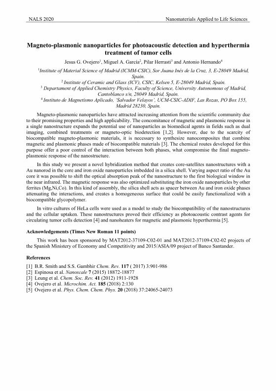
NALS 2020 Nanomaterials Applied to Life Sciences
Magneto-plasmonic nanoparticles for photoacoustic detection and hyperthermia
treatment of tumor cells Jesus G. Ovejero1, Miguel A. García2, Pilar Herrasti3 and Antonio Hernando4
1Institute of Material Science of Madrid (ICMM-CSIC), Sor Juana Inés de la Cruz, 3, E-28049 Madrid, Spain.
2 Institute of Ceramic and Glass (ICV), CSIC, Kelsen 5, E-28049 Madrid, Spain. 3 Departament of Applied Chemistry Physics, Faculty of Science, University Autonomous of Madrid,
Cantoblanco s/n, 28049 Madrid, Spain. 4 Instituto de Magnetismo Aplicado, ‘Salvador Velayos’, UCM-CSIC-ADIF, Las Rozas, PO Box 155,
Madrid 28230, Spain.
Magneto-plasmonic nanoparticles have attracted increasing attention from the scientific community due to their promising properties and high applicability. The concomitance of magnetic and plasmonic response in a single nanostructure expands the potential use of nanoparticles as biomedical agents in fields such as dual imaging, combined treatments or magneto-optic biodetection [1,2]. However, due to the scarcity of biocompatible magneto-plasmonic materials, it is necessary to synthesize nanocomposites that combine magnetic and plasmonic phases made of biocompatible materials [3]. The chemical routes developed for this purpose offer a poor control of the interaction between both phases, what compromise the final magneto-plasmonic response of the nanostructure.
In this study we present a novel hybridization method that creates core-satellites nanostructures with a Au nanorod in the core and iron oxide nanoparticles imbedded in a silica shell. Varying aspect ratio of the Au core it was possible to shift the optical absorption peak of the nanostructure to the first biological window in the near infrared. The magnetic response was also optimized substituting the iron oxide nanoparticles by other ferrites (Mg,Ni,Co). In this kind of assembly, the silica shell acts as spacer between Au and iron oxide phases attenuating the interactions, and creates a homogeneous surface that could be easily functionalized with a biocompatible glycopolymer.
In vitro cultures of HeLa cells were used as a model to study the biocompatibility of the nanostructures and the cellular uptaken. These nanostructures proved their efficiency as photoacoustic contrast agents for circulating tumor cells detection [4] and nanoheaters for magnetic and plasmonic hyperthermia [5].
Acknowledgements (Times New Roman 11 points)
This work has been sponsored by MAT2012-37109-C02-01 and MAT2012-37109-C02-02 projects of the Spanish Ministery of Economy and Competitivity and 2015/ASIA/09 project of Banco Santander.
References
[1] B.R. Smith and S.S. Gambhir Chem. Rev. 117 ( 2017) 3:901-986 [2] Espinosa et al. Nanoscale 7 (2015) 18872-18877 [3] Leung et al. Chem. Soc. Rev. 41 (2012) 1911-1928 [4] Ovejero et al. Microchim. Act. 185 (2018) 2:130 [5] Ovejero et al. Phys. Chem. Chem. Phys. 20 (2018) 37:24065-24073
NALS 2020 Nanomaterials Applied to Life Sciences
Magnetic properties of iron oxide nanoparticles: comparison between promising candidates for magnetic hyperthermia
E. M. Jefremovas1*, L. Gandarias2, D. Gandía3, L. Marcano3,4, A. García Prieto3, A. Muela3, M. L. Fdez-Gubieda4, C. Grüttner6, J. Alonso1, L. Fernández Barquín1
1 Depto. CITIMAC, Facultad de Ciencias, Universidad de Cantabria, 39005 Santander, Spain
2 Depto. de Inmunología, Microbiología y Parasitología, UPV/EHU 48080 Leioa, Spain 3 Depto. Electricidad y Electrónica and BCMaterials, UPV/EHU 48080 Leioa, Spain
4 Helmholtz-Zentrum Berlin für Materialien und Energie, Albert-Einstein-Strasse 15, D-12489 Berlin, Germany 5 Department of Applied Physics, School of Engineering of Bilbao, 48013 Bilbao, Spain
6Micromod Partikeltechnologie GmbH, 18119 Rostock, Germany
Application of iron oxide nanoparticles for magnetic hyperthermia therapy (MHT) has been intensively studied in recent years [1-3]. One of the main research aims is to optimise the heating efficiency of these nanoparticles (NPs) by tuning their magnetic response. Although commercial iron oxide NPs have been frequently employed in MHT studies, in recent years, it has been shown that highly crystalline iron oxide NPs, such as those synthesised by magnetotactic bacteria, can give rise to enhanced MHT results. Therefore, in this work we have studied the properties of two systems: Commercial Synomag nanoflowers (NF) [2], which are about 70 nm in size, and consist of densely packed iron oxide cores, and magnetosomes (MG) [3], synthesised by magnetotactic bacteria of the strain Magnetospirillum gryphiswaldense, that consist of 40 nm NPs with truncated-octahedral shape. Both systems have been described as fine candidates for MHT.
In this study we have characterised the magnetic behaviour of both systems by means of static magnetisation MDC(H, T) and dynamic χAC(T,f) measurements. Static zero field cooled-field cooled (ZFC-FC) MDC(H, T) measurements reveal that both samples exhibit high temperature irreversibility but, while MG present a clear peak around 110 K corresponding to the Verwey transition, no peak was observed for the Synomag NF. This suggests that the latter are formed by a mixture of magnetite and maghemite, as supported by the lower saturation magnetization value exhibited by these samples [Ms = 62.3(2) emu/g)]. In addition, both the coercive field (HC) and remanence (Mr/MS) at room temperature for Synomag NF are nearly zero, while for the MG we obtain an Mr/MS = 0.50 and HC = 166 Oe. This suggests therefore that Synomag NF behave more like ideal superparamagnetic NPs (HC = 0, Mr/MS = 0) at room temperature than MG, which are magnetically blocked. This is supported by characterising the area between ZFC-FC branches measured at several H. With increasing H, the area first increases until reaching a maximum and then decreases until becoming null. This is related to the coercive field (HC) of the NPs: Irreversibility increases as long as the applied magnetic field stays below HC. When this HC is overcome, the irreversibility disappears. Indeed, as HC for MG is higher than for iron-oxide NF, MG display irreversibility at higher fields than the NF. In order to better understand these differences, we have also carried out χAC(T,f ) measurements that are in perfect agreement with the MDC(H, T) results. No trace of Verwey transition is found for NF. Moreover, an additional shoulder is found at 30 K, pointing to a super spin-glass transition.
All these results indicate that, while magnetosomes behave like single crystalline magnetically blocked NPs, commercial Synomag nanoflowers, due to their multicore internal structure, behave more like interacting superparamagnetic NPs at room temperature. This can be related to why Synomag NF present a good heating efficiency at low fields while magnetosomes, with greater coercive field and irreversibility, maximise their heating efficiency at higher fields.
Acknowledgements This work has been supported by the MAT2017-83631-C3-R grant. EMJ thanks the “Beca Concepción Arenal”
BOC-133-09-07-2018 granted by the Gobierno de Cantabria.
References [1] Q A Pankhurst et al, J. Phys. D: Appl. Phys. 36 R167 (2003). [2] P. Bender et al., J. Phys. Chem. C 122, 3068 (2018). [3] D. Gandia, et al. Small 1902626 (2019); L. Marcano et al., J. Phys. Chem. C, 122, 7541 (2018). [4] F. Q. Walz. Journal of Physics: Condensed Matter 14, 12 285 (2002).
NALS 2020 Nanomaterials Applied to Life Sciences
Acid amino-functionalized carboxymetilcellulose as a nanocarrier for gene delivery: Characterization and efficacy in silencing a target gene in a cell culture
Hernández Magali 1, Lima Enrique 2
1 Facultad de Química, Universidad Nacional Autónoma de México. Circuito exterior s/n, Cd. Universitaria, Del.
Coyoacán, CP 04510, México D. F., Mexico.Mail:[email protected] 2 Laboratorio de Fisicoquímica y Reactividad de Superficies (LaFReS), Instituto de Investigaciones en Materiales,
Universidad Nacional Autónoma de México, Circuito exterior s/n, Cd. Universitaria, Del. Coyoacán, CP 04510, Ciudad de México, Mexico. Mail:[email protected]
Nanoparticles are already being developed as effective carriers of drugs to target regions of the body that were previously hard to access using traditional drug formulation methods. Through manipulation of their elemental composition, size, shape, charge and surface modification or chemical functionalization it may be possible to target particles to specific organs where they may elicit their therapeutic effect.
Spinocerebellar ataxia 7 (SCA7) is a neurodegenerative disorder characterized by degeneration of the cerebellum, brainstem and retina. Recently, genetic therapy has been proposed as alternative with the use of molecular tools such as siRNA (small interfering Ribonucleic Acid) that serve as effective therapeutics for disorders in the central nervous system, however the release of siRNA suffers from nuclease degradation and endosomal entrapment, thus avoid siRNA to target the cells of interest, another limitation is the difficulty to cross blood-brain barrier (BBB). Therefore, in this work we intend to develop release systems based on functionalized polymeric nanoparticles that facilitate their passage through the BBB. These polymers are cationic in nature, thus allowing electrostatic interactions with SiRNA, thus conferring stability to biological media.
We synthesized conjugates between a biodegradable polymer, carboxymethytilcellulose (PEG) and polyetilenglicol (PEG-NH2), by means of an amide linkage. And the polymeric complex obtained was functionalized with two amino acids; the multifunctional hybrid was characterized using 13C MAS NMR and FTIR spectroscopy, TEM and DLS, the latter among which shows 12 nm hydrodynamic radii of polymeric complex dispersed in deionized water.
Acknowledgements (Times New Roman 11 points)
We are grateful to G.Cedillo and A.Tejeda for their technical, assistance.
References [1] L. Gouw, M. Castañeda, C. McKenna, K. Digre, S. Pulst, S. Perlman, M. Lee, K. Hum. Mol. Genet. 7(1998) 525–
532. [2] Y. Grace, K. Caldecott, HCN. 115 (2018) 105. [3] J. Wang, Z. Lu, G. Wientjes, J. Au. AAPS J. 12 (2010), 492. [4] M. Yan, M. Liang, J. Wen, Y. Liu, Y. Lu. J. Am. Chem. Soc. 134( 2012) 13542. [5] W. Li, F. Szoka. Pharm. Res. 24 (2007) 438.
NALS 2020 Nanomaterials Applied to Life Sciences
Internalization and cytotoxicity of magnetosomes and magnetotactic bacteria in lung carcinoma cells
Lucía Gandarias1, David Gandia2, Lide Arana3, Itziar Alkorta3, Alfredo García-Arribas2,4, Ana García Prieto2,5, Alicia Muela1,2, MªLuisa Fernández-Gubieda2,4.
1Dpto. de Inmunología, Microbiología y Parasitología, Universidad del País Vasco (UPV/EHU), 48940 Leioa, Spain
2Basque Center for Materials, Applications & Nanostructures, 48940 Leioa, Spain3Dpto. de Bioquímica y Biología Molecular Universidad del País Vasco (UPV/EHU), 48940 Leioa,
Spain4Dpto. de Electricidad y Electrónica, Universidad del País Vasco (UPV/EHU), 48940 Leioa, Spain
5Dpto. de Física Aplicada I, Universidad del País Vasco (UPV/EHU), 48013 Bilbao, Spain
Magnetotactic bacteria (MTB) are a diverse group of microorganisms that have the ability to synthesize membrane-enclosed magnetic nanoparticles called magnetosomes that present fascinating magnetic properties. Therefore, both MTB and, especially magnetosomes, have been proposed for diverse biomedical applications.
In this work we study the efficiency of magnetosomes and, in an innovative manner, of the whole MTB as heating agents in magnetic hyperthermia for cancer treatment. First, we verify if magnetosomes and MTB certainly enter cancer cells by using different microscopic techniques. Once this has been assured, we use different endocytosis inhibitors to identify the route by which cancer cells internalize these agents using flow cytometry. Then, we study the cytotoxic effect that magnetosomes and MTB could cause on cancer cells by incubating them together and checking cell viability at different times using proper Live/Dead stains and flow cytometry. Finally, we assess the effectivity of magnetic hyperthermia on lung carcinoma cells (A549 cell line) by applying an alternate magnetic field (150 kHz, 45 minutes) to cells that have previously internalized magnetosomes (with an amplitude of 370 Oe) and MTB (435 Oe), and checking cell viability afterwards.
Our first results are very promissng as they show that human lung carcinoma cells internalize both magnetosomes and magnetotactic bacteria without suffering any decrease in their viability and that themagnetic hyperthermia treatment is effective as it reduces significantly the number of cells present in the culture [1].
References
[1] Gandia D, Gandarias L, Rodrigo I, Robles‐García J, Das R, Garaio E, et al. Unlocking the Potential of Magnetotactic Bacteria as Magnetic Hyperthermia Agents. Small 15 (2019) 1902626.
NALS 2020 Nanomaterials Applied to Life Sciences
XPS/HAXPES characterization of titanate assisted organosilanized surfaces
Rodrigo Calvo a*, Sergio Pinilla a, Javier Mateo a, Juan Rubio Zuazo b, Miguel Manso Silvána a Departamento de Física Aplicada, Universidad Autónoma de Madrid, 28049 Madrid, Spain; b Spline, European
Synchrotron Radiation Facility, 38000 Grenoble, France.
Introduction
The surface biofunctionalization of inorganic materials implies the formation of an organic layer that allows the conjugation with the biomolecules. Titanate assisted organosilanization (TAO) is a technique for surface functionalization that is gaining relevance as it demonstrates its generic value on both, the different types of substrates it can functionalize and the alternatives in functional organosilanes that can be integrated on the surface. In this work, we have carried out an X-Ray photoelectron spectroscopy (XPS) and hard X-ray photoelectron spectroscopy (HAXPES) characterization [2] of TAO films to make an in-depth evaluation of the distribution and orientation of a variety of organosilanes within the condensing titanate film.
Results and Discussion
XPS has been used for the inspection of films of the alkane octyl-silane (OCTS) and thiol mercaptopropyl-silane (MPS) with respect to a titanate control film (TIPT). It has been observed that the organosilane migrates to the surface reverting the molar concentration for OCTS and MPS. Relevantly, the surface concentration of the atoms related to the organosilane queues also increase notably their molar concentration on the surface. These results highlight that relevant molecular dynamics phenomena take place during condensation, leading to a surface segregation.
An additional organosilane (perfluorodecyl silane, PFDS) was probed by HAXPES at increasing energies. The composition of the surface could be explored at different depths. The results show that the concentration of organic species increases with respect to the Ti signal as the excitation energy of the X-ray beam is reduced. Not only the relative stoichiometry of the elements suggests an organization of the organosilane on the film, but also the relative components of core level peaks of specific elements: in the Si 1s core level, the one related to the silane (Si-O) with respect to the one related to the substrate (Si-Si).
Figure 1: a) XPS survey spectrum of MPS condensed by TAO. b) HAXPES Si 1s core level spectrum of a PFDS layer, showing substrate (Si-
Si) and silane (Si-O) related contributions.
This data agree with the idea that, not only the organosilanes migrate to the surface during the condensation of the titanate, but also they orient preferentially exhibiting their non-hydrolysable functional groups to the surface. This behavior can be understood if the condensation kinetics of the organosilanes is slower than that of the titanate.
References
[1] D. Moreno-Cerrada, C. Rodríguez, F. Moreno-Madrid, E. Selivanovitch, T. Douglas, P.J. de Pablo, and M. Manso Silván, Loading the dice: The orientation of virus-like particles adsorbed on titanate assisted organosilanized surfaces, Biointerphases, 14 (2019) 011001.
[2] J. Rubio-Zuazo, A. Chainani, M. Taguchi, D. Malterre, A. Serrano, and G.R. Castro. Electronic structure of FeO, γ−Fe2O3, and Fe3O4 epitaxial films using high-energy spectroscopies. Phys. Rev. B 97, 235148.
NameC 1sCl 2pO 1sS 2pSi 2pTi 2p
At%50.9190.931
33.1512.1935.1517.655
C 1s
Cl 2
p
O 1
s
S 2p
Si 2
p
Ti 2
p
x 103
10
20
30
40
50
60
CPS
500 400 300 200 100 0Binding Energy (eV)
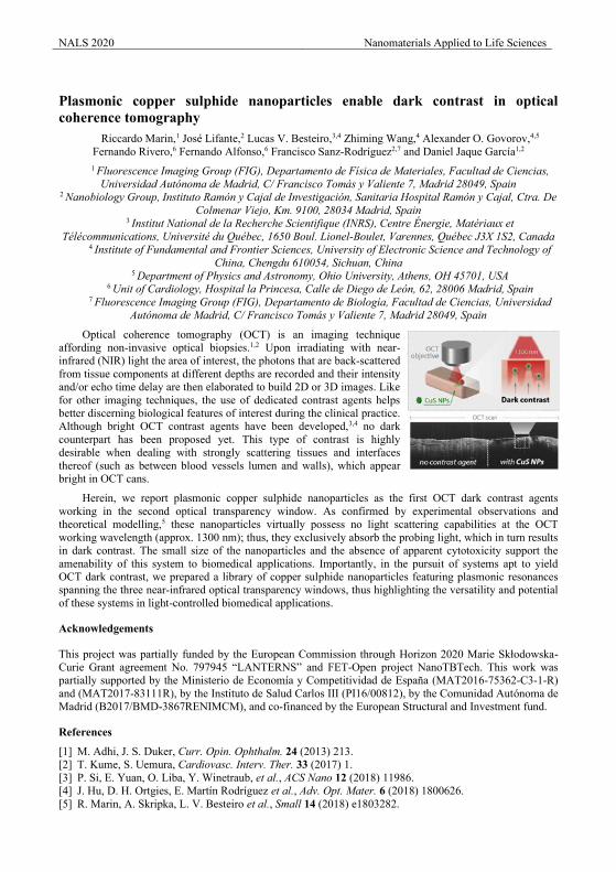
NALS 2020 Nanomaterials Applied to Life Sciences
Plasmonic copper sulphide nanoparticles enable dark contrast in optical coherence tomography
Riccardo Marin,1 José Lifante,2 Lucas V. Besteiro,3,4 Zhiming Wang,4 Alexander O. Govorov,4,5 Fernando Rivero,6 Fernando Alfonso,6 Francisco Sanz-Rodríguez2,7 and Daniel Jaque García1,2 1 Fluorescence Imaging Group (FIG), Departamento de Física de Materiales, Facultad de Ciencias,
Universidad Autónoma de Madrid, C/ Francisco Tomás y Valiente 7, Madrid 28049, Spain 2 Nanobiology Group, Instituto Ramón y Cajal de Investigación, Sanitaria Hospital Ramón y Cajal, Ctra. De
Colmenar Viejo, Km. 9100, 28034 Madrid, Spain 3 Institut National de la Recherche Scientifique (INRS), Centre Énergie, Matériaux et
Télécommunications, Université du Québec, 1650 Boul. Lionel-Boulet, Varennes, Québec J3X 1S2, Canada
4 Institute of Fundamental and Frontier Sciences, University of Electronic Science and Technology of China, Chengdu 610054, Sichuan, China
5 Department of Physics and Astronomy, Ohio University, Athens, OH 45701, USA 6 Unit of Cardiology, Hospital la Princesa, Calle de Diego de León, 62, 28006 Madrid, Spain
7 Fluorescence Imaging Group (FIG), Departamento de Biología, Facultad de Ciencias, Universidad Autónoma de Madrid, C/ Francisco Tomás y Valiente 7, Madrid 28049, Spain
Optical coherence tomography (OCT) is an imaging technique affording non-invasive optical biopsies.1,2 Upon irradiating with near-infrared (NIR) light the area of interest, the photons that are back-scattered from tissue components at different depths are recorded and their intensity and/or echo time delay are then elaborated to build 2D or 3D images. Like for other imaging techniques, the use of dedicated contrast agents helps better discerning biological features of interest during the clinical practice. Although bright OCT contrast agents have been developed,3,4 no dark counterpart has been proposed yet. This type of contrast is highly desirable when dealing with strongly scattering tissues and interfaces thereof (such as between blood vessels lumen and walls), which appear bright in OCT cans.
Herein, we report plasmonic copper sulphide nanoparticles as the first OCT dark contrast agents working in the second optical transparency window. As confirmed by experimental observations and theoretical modelling,5 these nanoparticles virtually possess no light scattering capabilities at the OCT working wavelength (approx. 1300 nm); thus, they exclusively absorb the probing light, which in turn results in dark contrast. The small size of the nanoparticles and the absence of apparent cytotoxicity support the amenability of this system to biomedical applications. Importantly, in the pursuit of systems apt to yield OCT dark contrast, we prepared a library of copper sulphide nanoparticles featuring plasmonic resonances spanning the three near-infrared optical transparency windows, thus highlighting the versatility and potential of these systems in light-controlled biomedical applications.
Acknowledgements
This project was partially funded by the European Commission through Horizon 2020 Marie Skłodowska-Curie Grant agreement No. 797945 “LANTERNS” and FET-Open project NanoTBTech. This work was partially supported by the Ministerio de Economía y Competitividad de España (MAT2016-75362-C3-1-R) and (MAT2017-83111R), by the Instituto de Salud Carlos III (PI16/00812), by the Comunidad Autonoma de Madrid (B2017/BMD-3867RENIMCM), and co-financed by the European Structural and Investment fund.
References
[1] M. Adhi, J. S. Duker, Curr. Opin. Ophthalm. 24 (2013) 213. [2] T. Kume, S. Uemura, Cardiovasc. Interv. Ther. 33 (2017) 1. [3] P. Si, E. Yuan, O. Liba, Y. Winetraub, et al., ACS Nano 12 (2018) 11986. [4] J. Hu, D. H. Ortgies, E. Martín Rodríguez et al., Adv. Opt. Mater. 6 (2018) 1800626. [5] R. Marin, A. Skripka, L. V. Besteiro et al., Small 14 (2018) e1803282.
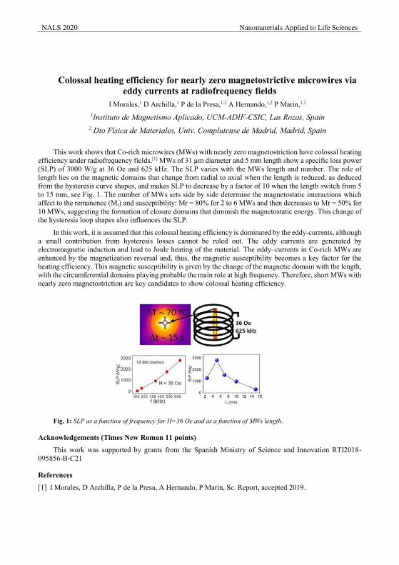
NALS 2020 Nanomaterials Applied to Life Sciences
Colossal heating efficiency for nearly zero magnetostrictive microwires via eddy currents at radiofrequency fields
I Morales,1 D Archilla,1 P de la Presa,1,2 A Hernando,1,2 P Marin,1,2
1Instituto de Magnetismo Aplicado, UCM-ADIF-CSIC, Las Rozas, Spain 2 Dto Física de Materiales, Univ. Complutense de Madrid, Madrid, Spain
This work shows that Co-rich microwires (MWs) with nearly zero magnetostriction have colossal heating efficiency under radiofrequency fields.[1] MWs of 31 µm diameter and 5 mm length show a specific loss power (SLP) of 3000 W/g at 36 Oe and 625 kHz. The SLP varies with the MWs length and number. The role of length lies on the magnetic domains that change from radial to axial when the length is reduced, as deduced from the hysteresis curve shapes, and makes SLP to decrease by a factor of 10 when the length switch from 5 to 15 mm, see Fig. 1. The number of MWs sets side by side determine the magnetostatic interactions which affect to the remanence (Mr) and susceptibility: Mr = 80% for 2 to 6 MWs and then decreases to Mr = 50% for 10 MWs, suggesting the formation of closure domains that diminish the magnetostatic energy. This change of the hysteresis loop shapes also influences the SLP.
In this work, it is assumed that this colossal heating efficiency is dominated by the eddy-currents, although a small contribution from hysteresis losses cannot be ruled out. The eddy currents are generated by electromagnetic induction and lead to Joule heating of the material. The eddy–currents in Co-rich MWs are enhanced by the magnetization reversal and, thus, the magnetic susceptibility becomes a key factor for the heating efficiency. This magnetic susceptibility is given by the change of the magnetic domain with the length, with the circumferential domains playing probable the main role at high frequency. Therefore, short MWs with nearly zero magnetostriction are key candidates to show colossal heating efficiency.
Fig. 1: SLP as a function of frequency for H=36 Oe and as a function of MWs length.
Acknowledgements (Times New Roman 11 points)
This work was supported by grants from the Spanish Ministry of Science and Innovation RTI2018-095856-B-C21
References
[1] I Morales, D Archilla, P de la Presa, A Hernando, P Marin, Sc. Report, accepted 2019.
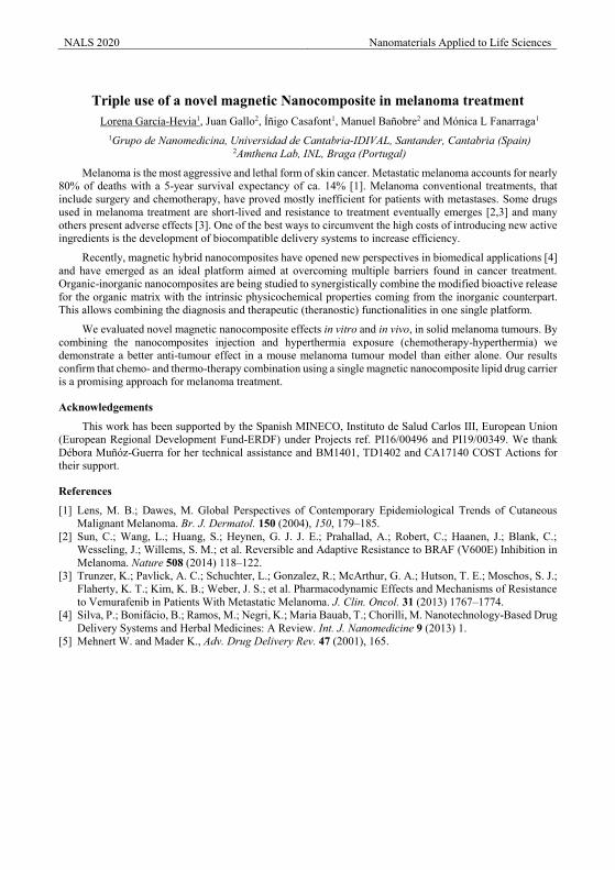
NALS 2020 Nanomaterials Applied to Life Sciences
Triple use of a novel magnetic Nanocomposite in melanoma treatment
Lorena García-Hevia1, Juan Gallo2, Íñigo Casafont1, Manuel Bañobre2 and Mónica L Fanarraga1 1Grupo de Nanomedicina, Universidad de Cantabria-IDIVAL, Santander, Cantabria (Spain)
2Amthena Lab, INL, Braga (Portugal)
Melanoma is the most aggressive and lethal form of skin cancer. Metastatic melanoma accounts for nearly 80% of deaths with a 5-year survival expectancy of ca. 14% [1]. Melanoma conventional treatments, that include surgery and chemotherapy, have proved mostly inefficient for patients with metastases. Some drugs used in melanoma treatment are short-lived and resistance to treatment eventually emerges [2,3] and many others present adverse effects [3]. One of the best ways to circumvent the high costs of introducing new active ingredients is the development of biocompatible delivery systems to increase efficiency.
Recently, magnetic hybrid nanocomposites have opened new perspectives in biomedical applications [4] and have emerged as an ideal platform aimed at overcoming multiple barriers found in cancer treatment. Organic-inorganic nanocomposites are being studied to synergistically combine the modified bioactive release for the organic matrix with the intrinsic physicochemical properties coming from the inorganic counterpart. This allows combining the diagnosis and therapeutic (theranostic) functionalities in one single platform.
We evaluated novel magnetic nanocomposite effects in vitro and in vivo, in solid melanoma tumours. By combining the nanocomposites injection and hyperthermia exposure (chemotherapy-hyperthermia) we demonstrate a better anti-tumour effect in a mouse melanoma tumour model than either alone. Our results confirm that chemo- and thermo-therapy combination using a single magnetic nanocomposite lipid drug carrier is a promising approach for melanoma treatment.
Acknowledgements
This work has been supported by the Spanish MINECO, Instituto de Salud Carlos III, European Union (European Regional Development Fund-ERDF) under Projects ref. PI16/00496 and PI19/00349. We thank Débora Muñóz-Guerra for her technical assistance and BM1401, TD1402 and CA17140 COST Actions for their support.
References
[1] Lens, M. B.; Dawes, M. Global Perspectives of Contemporary Epidemiological Trends of Cutaneous Malignant Melanoma. Br. J. Dermatol. 150 (2004), 150, 179–185.
[2] Sun, C.; Wang, L.; Huang, S.; Heynen, G. J. J. E.; Prahallad, A.; Robert, C.; Haanen, J.; Blank, C.; Wesseling, J.; Willems, S. M.; et al. Reversible and Adaptive Resistance to BRAF (V600E) Inhibition in Melanoma. Nature 508 (2014) 118–122.
[3] Trunzer, K.; Pavlick, A. C.; Schuchter, L.; Gonzalez, R.; McArthur, G. A.; Hutson, T. E.; Moschos, S. J.; Flaherty, K. T.; Kim, K. B.; Weber, J. S.; et al. Pharmacodynamic Effects and Mechanisms of Resistance to Vemurafenib in Patients With Metastatic Melanoma. J. Clin. Oncol. 31 (2013) 1767–1774.
[4] Silva, P.; Bonifácio, B.; Ramos, M.; Negri, K.; Maria Bauab, T.; Chorilli, M. Nanotechnology-Based Drug Delivery Systems and Herbal Medicines: A Review. Int. J. Nanomedicine 9 (2013) 1.
[5] Mehnert W. and Mader K., Adv. Drug Delivery Rev. 47 (2001), 165.
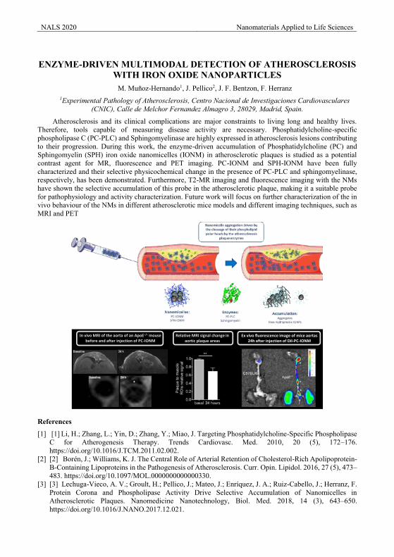
NALS 2020 Nanomaterials Applied to Life Sciences
ENZYME-DRIVEN MULTIMODAL DETECTION OF ATHEROSCLEROSIS
WITH IRON OXIDE NANOPARTICLES M. Muñoz-Hernando1, J. Pellico2, J. F. Bentzon, F. Herranz
1Experimental Pathology of Atherosclerosis, Centro Nacional de Investigaciones Cardiovasculares (CNIC), Calle de Melchor Fernandez Almagro 3, 28029, Madrid, Spain.
Atherosclerosis and its clinical complications are major constraints to living long and healthy lives. Therefore, tools capable of measuring disease activity are necessary. Phosphatidylcholine-specific phospholipase C (PC-PLC) and Sphingomyelinase are highly expressed in atherosclerosis lesions contributing to their progression. During this work, the enzyme-driven accumulation of Phosphatidylcholine (PC) and Sphingomyelin (SPH) iron oxide nanomicelles (IONM) in atherosclerotic plaques is studied as a potential contrast agent for MR, fluorescence and PET imaging. PC-IONM and SPH-IONM have been fully characterized and their selective physicochemical change in the presence of PC-PLC and sphingomyelinase, respectively, has been demonstrated. Furthermore, T2-MR imaging and fluorescence imaging with the NMs have shown the selective accumulation of this probe in the atherosclerotic plaque, making it a suitable probe for pathophysiology and activity characterization. Future work will focus on further characterization of the in vivo behaviour of the NMs in different atherosclerotic mice models and different imaging techniques, such as MRI and PET
References
[1] [1] Li, H.; Zhang, L.; Yin, D.; Zhang, Y.; Miao, J. Targeting Phosphatidylcholine-Specific Phospholipase C for Atherogenesis Therapy. Trends Cardiovasc. Med. 2010, 20 (5), 172–176. https://doi.org/10.1016/J.TCM.2011.02.002.
[2] [2] Borén, J.; Williams, K. J. The Central Role of Arterial Retention of Cholesterol-Rich Apolipoprotein-B-Containing Lipoproteins in the Pathogenesis of Atherosclerosis. Curr. Opin. Lipidol. 2016, 27 (5), 473–483. https://doi.org/10.1097/MOL.0000000000000330.
[3] [3] Lechuga-Vieco, A. V.; Groult, H.; Pellico, J.; Mateo, J.; Enríquez, J. A.; Ruiz-Cabello, J.; Herranz, F. Protein Corona and Phospholipase Activity Drive Selective Accumulation of Nanomicelles in Atherosclerotic Plaques. Nanomedicine Nanotechnology, Biol. Med. 2018, 14 (3), 643–650. https://doi.org/10.1016/J.NANO.2017.12.021.
Ex vivo fluorescence image of mice aortas 24h after injection of Dil-PC-IONM
Relative MRI signal change in aortic plaque areas
In vivo MRI of the aorta of an ApoE−/− mouse before and after injection of PC-IONM

NALS 2020 Nanomaterials Applied to Life Sciences
METAL-DOPED IRON OXIDE NANOPARTICLES FOR POSITIVE
CONTRAST IN MAGNETIC RESONANCE IMAGING
I. Fernández-Barahona, J. Pellico, I. Rodríguez, J. Ruiz-Cabello, F. Herranz Instituto de Química Médica (IQM-CSIC), Consejo Superior de Investigaciones Científicas, c/ Juan de la Cierva, 3, 28003, Madrid, Spain
Facultad de Farmacia, Universidad Complutense de Madrid, Plaza de Ramón y Cajal, s/n, 28040, Madrid, Spain
Iron oxide nanoparticles (IONPs) have been traditionally studied as a T2 contrast agent for MRI due to their superparamagnetic behaviour. Nevertheless, T1-based positive contrast, being much more advantageous for clinical applications, is still limited to gadolinium- or manganese-based imaging tools. Recently, we have shown how microwave-driven synthesis of citrate-coated IONPs renders large r1 values.1 In this work, we have studied the effect of Cu doping on the physicochemical, magnetic, relaxometric, and in vivo properties of IONPs in both angiography and targeted molecular imaging.2 The relaxometric values, the type of synthesis and the in vivo performance make these nanoparticles an outstanding candidate for future clinical translation. Electrostatic interaction of copper and bacterial cell membranes leads to bacterial membrane disruption and potential death, for this reason we will study the antibacterial properties of these NPs.
References: [1] Pellico, J. Ruiz-Cabello, J., Fernández-Barahona, I. et al. One-Step Fast Synthesis of Nanoparticles for
MRI: Coating Chemistry as the Key Variable Determining Positive or Negative Contrast. Langmuir, 2017, 3, pp 10239–10247, doi:10.1021/acs.langmuir.7b01759
[2] Fernández Barahona, I., Gutiérrez, L., Veintemillas-Verdaguer, S. et al. Cu-doped Extremely Small Iron Oxide Nanoparticles with Large Longitudinal Relaxivity: One-Pot Synthesis and in vivo Targeted Molecular Imaging. ACS Omega, 2019, 4 (2), pp 2719–2727, doi: 10.1021/acsomega.8b03004
2020Madrid · Spain
Nanomaterials Applied to Life
Sciences
International Conference on 2nd
h t t p s : / / n a l s 2 0 2 0 . c o m
N A L S 2 0 2 0 V E N U EIFT-ICMAT · C/ Nicolás Cabrera, 13-15
Ciudad Universitaria de Cantoblanco
28049 Madrid
@nals2020#nals2020
abstracts book