PROGRAM BOOK & ABSTRACTS - Society of Urologic ...
310
Extraordinary Opportunities for Discovery December 2 – 4, 2015 Renaissance Washington DC Downtown Hotel Washington, DC 16th Annual Meeting of the Society of Urologic Oncology PROGRAM BOOK & ABSTRACTS
-
Upload
khangminh22 -
Category
Documents
-
view
0 -
download
0
Transcript of PROGRAM BOOK & ABSTRACTS - Society of Urologic ...

Extraordinary Opportunitiesfor Discovery
December 2 – 4, 2015Renaissance Washington DC Downtown Hotel
Washington, DC
16th Annual Meeting of theSociety of Urologic Oncology
PROGRAM BOOK& ABSTRACTS
1
Table of ConTenTs
For your convenience, the ToC has been linked to each corresponding section.Click on the title or page number to go directly to the desired page.
Board of Directors 2015 – 2016 .................................................................................................................2
Committees .................................................................................................................................................2
2015 Faculty Listing ...................................................................................................................................4
Promotional Partners and Contributors ...................................................................................................6
Exhibitors ....................................................................................................................................................7
General Meeting Information .....................................................................................................................8
Educational Needs and Objectives ...........................................................................................................9
Accreditation Information ........................................................................................................................ 11
Faculty Disclosures ..................................................................................................................................12
Industry Satellite Symposia .....................................................................................................................20
SUO General Scientific Program .............................................................................................................21
Young Urologic Oncologists Podium Session – Full Abstracts ..........................................................31
Oral Abstract Session – Full Abstracts ..................................................................................................34
Poster Session I – Summary ...................................................................................................................40
Poster Session I – Full Abstracts ............................................................................................................60
Poster Session II – Summary ................................................................................................................166
Poster Session II – Full Abstracts .........................................................................................................185
Alphabetical Index of Presenting Authors ...........................................................................................296
SUO Fellowship Programs ....................................................................................................................301
2016 Urologic Oncology Fellowship Matching Program Schedule ...................................................306
Mark Your Calendars ..............................................................................................................................307
2Back to Table of Contents h
boD, CommiTTees & faCulTy
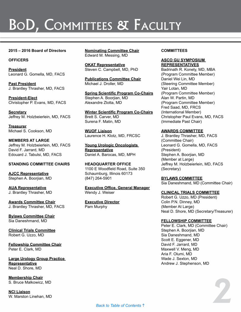
2015 – 2016 Board of Directors
OFFICERS
PresidentLeonard G. Gomella, MD, FACS
Past PresidentJ. Brantley Thrasher, MD, FACS
President-ElectChristopher P. Evans, MD, FACS
SecretaryJeffrey M. Holzbeierlein, MD, FACS
TreasurerMichael S. Cookson, MD
MEMBERS AT LARGEJeffrey M. Holzbeierlein, MD, FACSDavid F. Jarrard, MDEdouard J. Tabulsi, MD, FACS
STANDING COMMITTEE CHAIRS
AJCC RepresentativeStephen A. Boorjian, MD
AUA RepresentativeJ. Brantley Thrasher, MD
Awards Committee ChairJ. Brantley Thrasher, MD, FACS
Bylaws Committee ChairSia Daneshmand, MD
Clinical Trials CommitteeRobert G. Uzzo, MD
Fellowship Committee ChairPeter E. Clark, MD
Large Urology Group Practice RepresentativeNeal D. Shore, MD
Membership ChairS. Bruce Malkowicz, MD
NCI LiaisonW. Marston Linehan, MD
Nominating Committee ChairEdward M. Messing, MD
OKAT RepresentativeSteven C. Campbell, MD, PhD
Publications Committee ChairMichael J. Droller, MD
Spring Scientific Program Co-ChairsStephen A. Boorjian, MDAlexandre Zlotta, MD
Winter Scientific Program Co-ChairsBrett S. Carver, MDSurena F. Matin, MD
WUOF LiaisonLaurence H. Klotz, MD, FRCSC
Young Urologic Oncologists RepresentativeDaniel A. Barocas, MD, MPH
HEADQUARTER OFFICE1100 E Woodfield Road, Suite 350Schaumburg, Illinois 60173(847) 264-5901
Executive Office, General ManagerWendy J. Weiser
Executive DirectorPam Murphy
COMMITTEES
ASCO GU SYMPOSIUM REPRESENTATIVESBadrinath R. Konety, MD, MBA(Program Committee Member)Daniel Wei Lin, MD(Steering Committee Member)Yair Lotan, MD(Program Committee Member)Alan W. Partin, MD(Program Committee Member)Fred Saad, MD, FRCS(International Member)Christopher Paul Evans, MD, FACS (Immediate Past Chair)
AWARDS COMMITTEEJ. Brantley Thrasher, MD, FACS (Committee Chair)Leonard G. Gomella, MD, FACS (President)Stephen A. Boorjian, MD(Member at Large)Jeffrey M. Holzbeierlein, MD, FACS (Secretary)
BYLAWS COMMITTEESia Daneshmand, MD (Committee Chair)
CLINICAL TRIALS COMMITTEERobert G. Uzzo, MD (President)Colin P.N. Dinney, MD(Member At Large)Neal D. Shore, MD (Secretary/Treasurer)
FELLOWSHIP COMMITTEEPeter E. Clark, MD (Committee Chair)Stephen A. Boorjian, MDSia Daneshmand, MDScott E. Eggener, MDDavid F. Jarrard, MDMaxwell V. Meng, MDAria F. Olumi, MDWade J. Sexton, MDAndrew J. Stephenson, MD
3Back to Table of Contents h
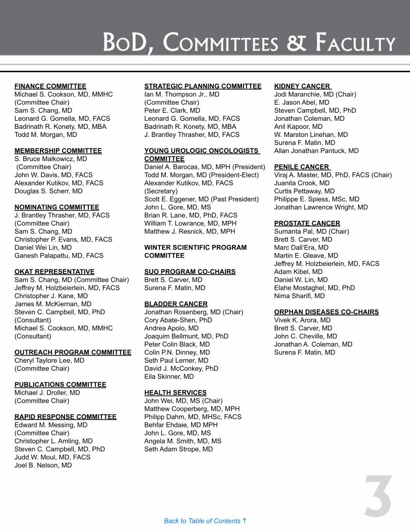
FINANCE COMMITTEEMichael S. Cookson, MD, MMHC (Committee Chair)Sam S. Chang, MDLeonard G. Gomella, MD, FACSBadrinath R. Konety, MD, MBATodd M. Morgan, MD
MEMBERSHIP COMMITTEES. Bruce Malkowicz, MD (Committee Chair)John W. Davis, MD, FACSAlexander Kutikov, MD, FACSDouglas S. Scherr, MD
NOMINATING COMMITTEEJ. Brantley Thrasher, MD, FACS (Committee Chair)Sam S. Chang, MDChristopher P. Evans, MD, FACSDaniel Wei Lin, MDGanesh Palapattu, MD, FACS
OKAT REPRESENTATIVESam S. Chang, MD (Committee Chair)Jeffrey M. Holzbeierlein, MD, FACSChristopher J. Kane, MDJames M. McKiernan, MDSteven C. Campbell, MD, PhD (Consultant)Michael S. Cookson, MD, MMHC (Consultant)
OUTREACH PROGRAM COMMITTEECheryl Taylore Lee, MD(Committee Chair)
PUBLICATIONS COMMITTEEMichael J. Droller, MD(Committee Chair)
RAPID RESPONSE COMMITTEEEdward M. Messing, MD(Committee Chair)Christopher L. Amling, MDSteven C. Campbell, MD, PhDJudd W. Moul, MD, FACSJoel B. Nelson, MD
STRATEGIC PLANNING COMMITTEEIan M. Thompson Jr., MD(Committee Chair)Peter E. Clark, MDLeonard G. Gomella, MD, FACSBadrinath R. Konety, MD, MBAJ. Brantley Thrasher, MD, FACS
YOUNG UROLOGIC ONCOLOGISTS COMMITTEEDaniel A. Barocas, MD, MPH (President)Todd M. Morgan, MD (President-Elect)Alexander Kutikov, MD, FACS (Secretary)Scott E. Eggener, MD (Past President)John L. Gore, MD, MSBrian R. Lane, MD, PhD, FACSWilliam T. Lowrance, MD, MPHMatthew J. Resnick, MD, MPH
WINTER SCIENTIFIC PROGRAM COMMITTEE
SUO PROGRAM CO-CHAIRSBrett S. Carver, MDSurena F. Matin, MD
BLADDER CANCERJonathan Rosenberg, MD (Chair)Cory Abate-Shen, PhDAndrea Apolo, MDJoaquim Bellmunt, MD, PhDPeter Colin Black, MDColin P.N. Dinney, MD Seth Paul Lerner, MDDavid J. McConkey, PhDEila Skinner, MD
HEALTH SERVICESJohn Wei, MD, MS (Chair)Matthew Cooperberg, MD, MPHPhilipp Dahm, MD, MHSc, FACSBehfar Ehdaie, MD MPHJohn L. Gore, MD, MSAngela M. Smith, MD, MSSeth Adam Strope, MD
boD, CommiTTees & faCulTy
KIDNEY CANCER Jodi Maranchie, MD (Chair)E. Jason Abel, MDSteven Campbell, MD, PhDJonathan Coleman, MDAnil Kapoor, MDW. Marston Linehan, MDSurena F. Matin, MDAllan Jonathan Pantuck, MD
PENILE CANCER Viraj A. Master, MD, PhD, FACS (Chair)Juanita Crook, MDCurtis Pettaway, MDPhilippe E. Spiess, MSc, MDJonathan Lawrence Wright, MD
PROSTATE CANCERSumanta Pal, MD (Chair)Brett S. Carver, MDMarc Dall’Era, MDMartin E. Gleave, MDJeffrey M. Holzbeierlein, MD, FACSAdam Kibel, MDDaniel W. Lin, MDElahe Mostaghel, MD, PhDNima Sharifi, MD
ORPHAN DISEASES CO-CHAIRSVivek K. Arora, MDBrett S. Carver, MDJohn C. Cheville, MDJonathan A. Coleman, MDSurena F. Matin, MD
4Back to Table of Contents h
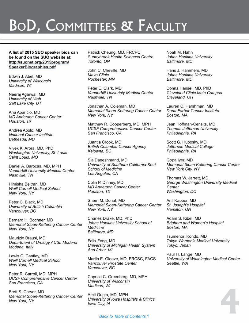
A list of 2015 SUO speaker bios can be found on the SUO website at:http://suonet.org/2015program/SpeakerBiographies.pdf
Edwin J. Abel, MDUniversity of WisconsinMadison, WI
Neeraj Agarwal, MDUniversity of UtahSalt Lake City, UT
Ana Aparicio, MDMD Anderson Cancer CenterHouston, TX
Andrea Apolo, MDNational Cancer InstituteBethesda, MD
Vivek K. Arora, MD, PhDWashington University, St. LouisSaint Louis, MO
Daniel A. Barocas, MD, MPHVanderbilt University Medical CenterNashville, TN
Himisha Beltran, MDWeill Cornell Medical SchoolNew York, NY
Peter C. Black, MDUniversity of British ColumbiaVancouver, BC
Bernard H. Bochner, MDMemorial Sloan-Kettering Cancer CenterNew York, NY
Maurizio Brausi, MDDepartment of Urology AUSL ModenaModena, Italy
Lewis C. Cantley, MDWeill Cornell Medical SchoolNew York, NY
Peter R. Carroll, MD, MPHUCSF Comprehensive Cancer CenterSan Francisco, CA
Brett S. Carver, MDMemorial Sloan-Kettering Cancer CenterNew York, NY
boD, CommiTTees & faCulTy
Patrick Cheung, MD, FRCPCSunnybrook Health Sciences CentreToronto, ON
John C. Cheville, MDMayo ClinicRochester, MN
Peter E. Clark, MDVanderbilt Univeristy Medical CenterNashville, TN
Jonathan A. Coleman, MDMemorial Sloan-Kettering Cancer CenterNew York, NY
Matthew R. Cooperberg, MD, MPHUCSF Comprehensive Cancer CenterSan Francisco, CA
Juantia Crook, MDBritish Columbia Cancer AgencyKelowna, BC
Sia Daneshmand, MDUniversity of Southern California-Keck School of MedicineLos Angeles, CA
Colin P. Dinney, MDMD Anderson Cancer CenterHouston, TX
Sherri M. Donat, MDMemorial Sloan-Kettering Cancer CenterNew York, NY
Charles Drake, MD, PhDJohns Hopkins University School of MedicineBaltimore, MD
Felix Feng, MDUniversity of Michigan Health SystemAnn Arbor, MI
Martin E. Gleave, MD, FRCSC, FACSVancouver Prostate CenterVancouver, BC
Caprice C. Greenberg, MD, MPHUniversity of WisconsinMadison, WI
Amit Gupta, MD, MPHUniversity of Iowa Hospitals & ClinicsIowa City, IA
Noah M. HahnJohns Hopkins UniversityBaltimore, MD
Hans J. Hammers, MDJohns Hopkins UniversityBaltimore, MD
Donna Hansel, MD, PhDCleveland Clinic Main CampusCleveland, OH
Lauren C. Harshman, MDDana Farber Cancer InstituteBoston, MA
Jean Hoffman-Censits, MDThomas Jefferson UniversityPhiladelphia, PA
Scott G. Hubosky, MDJefferson Medical CollegePhiladelphia, PA
Gopa Iyer, MDMemorial Sloan Kettering Cancer CenterNew York City, NY
Thomas W. Jarrett, MDGeorge Washington University Medical CenterWashington, DC
Anil Kapoor, MDSt. Joseph’s HospitalHamilton, ON
Adam S. Kibel, MDBrigham and Women’s HospitalBoston, MA
Tsumenori Kondo, MDTokyo Women’s Medical UniversityTokyo, Japan
Paul H. Lange, MDUniversity of Washington Medical CenterSeattle, WA
5Back to Table of Contents h
boD, CommiTTees & faCulTy
Jonathan Rosenberg, MDMemorial Sloan-Kettering Cancer CenterNew York, NY
Morgan Roupret, MD, PhDPitié-Salpétrière HospitalParis, France
Martin G. Sanda, MDEmory UniversityAtlanta, GA
Paul F. Schellhammer, MDUrology of Virginia PLLCVirginia Beach, VA
Nikolaus Schultz, MSMemorial Sloan-Kettering Cancer CenterJersey City, NJ
Shahrokh F. Shariat, MDWeill Cornell Medical SchoolNew York, NY
Nima Sharifi, MDCleveland ClinicCleveland, OH
Arlene D. Siefker-Radtke, MDMD Anderson Cancer CenterHouston, TX
Eila C. Skinner, MDStanford University Medical CenterStanford, CA
Ted A. Skolarus, MD, MPHUniversity of Michigan Ann Arbor, MI
Eric J. Small, MDUCSF Comprehensive Cancer CenterSan Francisco, CA
Angela M. Smith, MD, MSUniversity of North CarolinaChapel Hill, NC
Joseph Smith, MDVanderbilt University Medical CenterNashville, TN
Norm D. Smith, MDUniversity of Chicago Medical CenterChicago, IL
David B. Solit, MDMemorial Sloan-Kettering Cancer CenterNew York, NY
Philippe E. Spiess, MSc, MDMoffitt Cancer CenterTampa, FL
Scott Tomlins, MD, PhDUniversity of MichiganAnn Arbor, MI
Robert G. Uzzo, MDFox Chase Cancer CenterPhiladelphia, PA
Erik Vegt, MDNetherlands Cancer InstituteAmsterdam, Netherlands
John T. Wei, MD, MSTaubman Health Care CtrAnn Arbor, MI
Alon Z. Weizer, MDUniversity of MichiganAnn Arbor, MI
J. Stuart Wolf, MDUniversity of MichiganAnn Arbor, MI
Alexander Wyatt, BSc, D.PhilVancouver Prostate CentreVancouver, BC
Evan Y. Yu, MDUniversity of Washington Medical CenterSeattle, WA
Seth P. Lerner, MDBaylor College of MedicineHouston, TX
Daniel W. Lin, MDUniversity of Washington Medical CenterSeattle, WA
Jodi K. Maranchie, MDUniversity of PittsburghPittsburgh, PA
Vitaly Margulis, MDUT Southwestern Medical CenterDallas, TX
Viraj A. Master, MD, PhD, FACSEmory UniversityAtlanta, GA
Surena F. Matin, MDMD Anderson Cancer CenterHouston, TX
James M. McKiernan, MDColumbia UniversityNew York, NY
Sumanta K. Pal, MDCity of Hope Comprehensive Cancer CenterDuarte, CA
Dipen J. Parekh, MDUniversity of MiamiMiami, FL
Curits A. Pettaway, MDMD Anderson Cancer CenterHouston, TX
Elizabeth R. Plimack, MD, MSFox Chase Cancer CenterPhiladelphia, PA
Edwin Posadas, MDCedars-Sinai Medical CenterLos Angeles, CA
Raj S. Pruthi, MDUniversity of North Carolina Chapel Hill, NC
Jay D. Raman, MDPenn State Milton S. Hershey Medical CenterHershey, PA

6Back to Table of Contents h
Thank you
Thank You to Our 2015 Promotional Partners(As of 11/18/15)
Gold LevelGenentech
Janssen Biotech, Inc.
Silver LevelDendreon Corporation
Genomic HealthMedivation/Astellas
Myriad Genetic Laboratories, Inc.
Thank You to Our 2015 Contributors(As of 11/18/15)
Boston ScientificGenentech
Genomic HealthJanssen Biotech, Inc.Medivation/AstellasTARIS Biomedical

7Back to Table of Contents h
Thank you
Thank You to Our 2015 Exhibitors(As of 11/18/15)
Argos TherapeuticsBayer Healthcare
Bladder Cancer Advocacy NetworkBoston Scientific Corporation
Collect RxDendreon Corporation
Ferring PharmaceuticalsGenentech
GenomeDx Biosciences Inc.Genomic Health
Janssen Biotech, Inc.MDxHealth
Medivation/AstellasMemorial Healthcare System
Merck & Co., Inc.Myriad Genetic Laboratories, Inc.
OPKO Lab, LLCPacific Edge Diagnostics USA Ltd.
PhotocurePromethus Laboratories Inc.
sanofi OncologyTOLMAR Pharmaceuticals

8Back to Table of Contents h
General informaTion
The 16th Annual Scientific Meeting in Urologic Oncology will be held December 2 – 4, 2015, at the Renaissance Washington DC Downtown Hotel. The Society of Urologic Oncology will sponsor this highly interactive meeting where all attendees participate in the discussions led by internationally renowned urologic oncologists, medical oncologists and scientists. State-of-the-art translational topics on prostate, kidney and bladder cancer, as well as strategies in urologic oncology will be discussed. This year’s meeting will also feature a special program on Wednesday to start the meeting that will be a Focus on Orphan Diseases in Urologic Oncology. More information on the course and registration can be found below.
Who Should Attend?• Urological Surgeons• Medical Oncologists• Radiation Oncologists• Research Scientists• Residents/Fellows-in-Training
Attendee ParticipationThis meeting is designed to be a discussion of issues among members of the urologic oncology community. All attendees participate in the discussions and are encouraged to interact with program faculty.
Registration/Information DeskLocation: Grand Registration DeskWednesday, December 2, 2015 10:00 a.m. – 6:00 p.m.Thursday, December 3, 2015 6:30 a.m. – 6:00 p.m.Friday, December 4, 2015 7:00 a.m. – 3:15 p.m.
Exhibit Hall Location: Renaissance BallroomWednesday, December 2, 2015 2:00 p.m. – 6:00 p.m.Thursday, December 3, 2015 7:45 a.m. – 7:30 p.m. SUO Welcome Reception 6:00 p.m. – 7:30 p.m.Friday, December 4, 2015 7:00 a.m. – 10:30 a.m.
Young Urologic Oncologists (Y.U.O.) Dinner*Date: Wednesday, December 2, 2015Time: 6:00 p.m. – 9:00 p.m.Location: Congressional BallroomCost: One ticket is included in the registration fee. Please let us know if you will be attending on the registration form.Attire: Business casual*Y.U.O. Members OnlyMembership in the Y.U.O. Section of the Society of Urologic Oncology consists of fellows, scientists and board certified or eligible physicians who are members of the SUO and have some post-residency training in urologic oncology. Membership is limited to the first seven years after completion of fellowship.
SUO ReceptionDate: Thursday, December 3, 2015Time: 6:00 p.m. – 7:30 p.m.Location: Renaissance BallroomCost: One ticket is included in the registration fee. Attire: Business casual The Society of Urologic Oncology welcomes its members to the 16th Annual Meeting. Members can visit with exhibitors and connect with fellow members, all while enjoying delicious drinks and hors d’oeuvres.
SUO Board of Directors MeetingDate: Wednesday, December 2, 2015Time: 6:00 p.m. – 9:00 p.m.Location: Meeting Room 12-14
SUO-CTC Board MeetingDate: Wednesday, December 2, 2015Time: 4:00 p.m. – 5:00 p.m.Location: Mount Vernon Square A Room
SUO Fellowship Committee MeetingDate: Thursday, December 3, 2015Time: 6:30 a.m. – 7:30 a.m.Location: Meeting Room 2
SUO Fellowship Program Directors MeetingDate: Thursday, December 3, 2015Time: 11:45 a.m. – 12:45 p.m.Location: Meeting Room 12-14
SUO Annual Business MeetingDate: Friday, December 4, 2015Time: 7:30 a.m. – 8:15 a.m.Location: Grand Ballroom
9Back to Table of Contents h
eDuCaTional neeDs & objeCTives
EDUCATIONAL NEEDS
Upper Tract Urothelial CancerUpper tract urothelial carcinoma (UTUC) is an orphan disease, frequently overlooked during kidney cancer and bladder cancer conferences. There have been few venues providing a multidisciplinary approach to this disease which, despite its relative rarity, is frequently encountered by urologists and medical oncologists. It represents a watershed disease without a subspecialty champion. In the urologic discipline it is incidentally managed by those who treat kidney cancer by nature of its anatomy, endoscopically managed by those with the technical means, and incidentally managed by those who treat bladder cancer by nature of its biology. Medical oncologists look for a high level of evidence to guide them for systemic therapy strategies yet little such evidence exists for this disease.
This Second Summit on UTUC offers a unique educational venue for the dissemination of research, diagnostic, evaluation, and treatment advances, and to identify high impact areas of need to improve our understanding and treatment of this challenging disease.
Bladder CancerThis year’s bladder cancer sessions will address major knowledge gaps in bladder cancer including an understanding of bladder cancer biology, integration of new therapies into clinical practice, and a forum to discuss the impact different surgical approaches on outcomes for bladder cancer patients. The session will also provide multidisciplinary perspectives on developing new treatments and applying existing treatments and new ways.
Kidney CancerThere has been a plethora of promising new agents for renal cancer in the neoadjuvant, adjuvant and metastatic setting. Practicing urologists and medical oncologists need to be familiar with the novel pathways, mechanisms, safety profile and efficacy of these agents for the management of their patients. Further, this understanding will support rational trial design and execution for the advancement of our patient care mission. Urologists and medical oncologists should understand the role of checkpoint inhibition in promoting tumor killing by the innate immune system and be familiar with results of promising combination trials in renal cancer. The frequency and duration of radiologic follow up for resected, localized renal cancer remains controversial and attendees need to be familiar with the rationale for risk-stratified guidelines. Despite general acceptance of the safety and accuracy of percutaneous renal mass biopsy, attendees should be aware of the limitations of biopsy and its overall impact clinical decision making. Attendees should be familiar with the risks and benefits of stereotactic radio-ablative therapy as an adjunct to systemic therapy in oligo-metastatic renal cancer.
Health Services ResearchThe term Health Services Research (HSR) is increasingly referenced not only in the research context but also in clinical practice. Most clinicians have limited familiarity with HSR in urology and may be primarily focused on how it may affect their practice in the years to come. The focus of this session is to raise awareness for the increasing importance and promises of HSR to the practice of urology. Taking a cue from molecular science, the NextGen HSR session will cover how HSR is increasingly allows us to measure and evaluate the delivery of healthcare in urology and provides the tools for improving the care of urological patients.
Penile Cancer Penile cancer is a rare disease in the United States. Most urologic oncologists evaluate patients with this condition infrequently. Knowledge of the appropriate use of non-surgical modalities, including radiotherapy, delivered either as primary therapy to the inguinal region, or as adjunctive therapy after surgery, is often poorly understood. Emerging data on the use of PET scans and MRI scans for staging is significant and important for the urologic oncology community. A third area of significant interest is to determine the current role of pelvic nodal dissection and of the prognostic impact of pelvic lymph nodes in locally advanced penile cancer. Finally, advancement of care will likely take place only in the context of well designed clinical trials. For this rare disease, the upcoming international INPACT trial will be of significant interest to the US urologic oncology community.
Prostate CancerProstate cancer is the most commonly diagnosed cancer in men and the second leading cause of male cancer death. There has been renewed interest in conventional approaches for advanced disease, such as the application of chemotherapy in metastatic castration sensitive prostate cancer (mCSPC), but there is still a lack of consensus as to which patients should receive this modality. In the setting of metastatic castrional resistant disease (mCPRC), there are a number of exciting approaches at varying stages of development, including poly-ADP ribose polymerase (PARP) inhibitors, common T-lymphocyte associated protein 4 (CTLA4) inhibitors, and programmed death-1 (PD-1) inhibitors. Beyond these drugs which antagonize novel pathways in prostate cancer, there is interest in agents which abrogate signaling through the classical target in prostate cancer,
10Back to Table of Contents h
eDuCaTional neeDs & objeCTives
androgen receptor (AR), albeit through novel approaches, as inhibition of the DNA binding domain. Given the multitude of therapies that are emerging, focus has also shifted towards developing novel genomic tools that may facilitate optimal selection and sequencing of agents.
Novel genomic tools are also being applied towards risk stratification of localized prostate cancer. Current guidelines have yet to incorporate these tools formally, and there is still a question as to whether these tests can discern the most appropriate treatment modality (e.g., active surveillance versus definitive intervention with surgery/radiotherapy). In defining treatment for localized prostate cancer, there is also great interest in incorporating not only biologic data but quality of life data in determining treatment allocation.
EDUCATIONAL OBJECTIVES
At the conclusion of the 2015 SUO Annual Meeting, attendees should be able to:
Upper Tract Urothelial Cancer• Identify putative genetic pathways associated with UTUC. • Recognize the strong association of UTUC with Lynch
Syndrome and the opportunity for urologic-driven diagnosis of the syndrome.
• Identify methods for screening patients with Lynch Syndrome.
• Evaluate the current modalities and techniques for endoscopic management of UTUC.
• Describe the benefits and rationale for neoadjuvant chemotherapy.
• Describe the options for chemotherapy in the renally impaired patient.
• Discuss the potential role of checkpoint blockade therapies for UTUC.
• Explain the various options for surgical management of UTUC, and the role of MIS, lymphadenectomy, and methods for bladder cuff excision.
• Identify current and upcoming clinical trials focused on UTUC.
Bladder Cancer• Integrate new approaches for the use of immunotherapy
in the management of patients with metastatic disease.• Describe the development of clinical trials testing novel
therapeutics and non-muscle invasive bladder cancer.• Identify a potential role for novel immunotherapy using
immune checkpoint inhibitors including those targeting PD-1 and PD-L1.
• Evaluate clinical trial data and controversies regarding different surgical approaches and bladder cancer.
Kidney Cancer• Describe the impact of adjuvant systemic therapy on
disease progression and survival following resection of localized renal cell carcinoma.
• Explain the rationale for neoadjuvant vs. adjuvant therapy using novel target and immune modulating agents.
• Identify the obstacles to trial accrual for localized renal cell carcinoma.
• Explain the importance of PD-1 in renal cancer and the clinical impact of combined checkpoint inhibition.
• Explain risk-stratified strategies for surveillance following resection of localized renal cell carcinoma.
• Describe the risks and benefits of percutaneous biopsy of the small renal mass in the clinical management of renal cancer.
• Describe the role of focal radiation for management of oligo-metastatic renal cell carcinoma.
Health Services Research• Describe the why Urologic HSR is increasingly relevant in
the current age of evidence based medicine.• Explain why Urologic HSR needs to span the population
level down to the patient level.• Describe the systematic approach HSR seeks to adapt
technologies to improve delivery at the organizational and patient-centered levels.
• Explain the rationale of extending HSR to include surgeons’ technical quality.
Penile Cancer• Identify the current use of radiotherapy for locally
advanced penile cancer.• Describe the international INPACT trial, and how this trial
will be of pivotal importance in advancing penile cancer management, and also to highlight the role US urologic oncologists may play.
• Describe the latest data on the use of PET and MRI scans on patients with penile cancer.
• Describe current data on the management of the pelvic lymph node basin in patients with penile cancer.
Prostate Cancer• Describe the role of docetaxel in mHSPC based on recent
data from the CHAARTED, GETUG-15 and STAMPEDE trials.
• Identify novel approaches to targeting the androgen receptor (AR), such as inhibitors of the AR DNA binding domain.
11Back to Table of Contents h
Prostate Cancer Objectives continued:• Explain the emerging data associated with novel immunotherapies such as CTLA4 inhibitors in mCPRC.• Describe the emerging data associated with PARP inhibitors in mCPRC.• Identify novel genomic platforms (e.g., cell free DNA) that can be used to obtain real-time genomic data from patients with
advanced prostate cancer.• Explain the role of currently available genomic tools in the risk stratification of localized prostate cancer.• Recognize how to harness publicly available resources such as The Cancer Genome Atlas (TCGA) to delineate prostate
cancer genomics.• Identify strategies for improving clinical outcomes from radical prostatectomy.• Recognize the differences in quality of life between conservative approaches (e.g., active surveillance) and definitive
approaches (e.g., surgery/radiation) for localized prostate cancer.
CONTINUING MEDICAL EDUCATION ACCREDITATION INFORMATION
AccreditationThis activity has been planned and implemented in accordance with the Essential Areas and Policies of the Accreditation Council for Continuing Medical Education (ACCME) through the joint providership of the American College of Surgeons and the Society of Urologic Oncology. The American College of Surgeons is accredited by the ACCME to provide continuing medical education for physicians.
AMA PRA Category 1 Credits™ The American College of Surgeons designates this live activity for a maximum of 15.25 AMA PRA Category 1 Credits™. Physicians should claim only the credit commensurate with the extent of their participation in the activity.
Nurses and other healthcare professionals will receive a Certificate of Attendance. For information on the applicability and acceptance of Certificates of Attendance for educational activities certified for AMA PRA Category 1 Credit™ from organizations accredited by the ACCME, please consult your professional licensing board.
General Disclaimer The statements and opinions contained in this program are solely those of the individual authors and contributors and not of the Society of Urologic Oncology. The appearance of the advertisements is not a warranty, endorsement, or approval of the products or services advertised or of their effectiveness, quality, or safety. The content of this publication may contain discussion of off-label uses of some of the agents mentioned. Please consult the prescribing information for full disclosure of approved uses. The Society of Urologic Oncology disclaims responsibility for any injury to persons or property resulting from any ideas or products referred to in the abstracts or advertisements.
Special Assistance We encourage participation by all individuals. If you have a disability, advance notification of any special needs will help us better serve you. Call (847) 264-5901 if you require special assistance to fully participate in the meeting.
aCCreDiTaTion
12Back to Table of Contents h
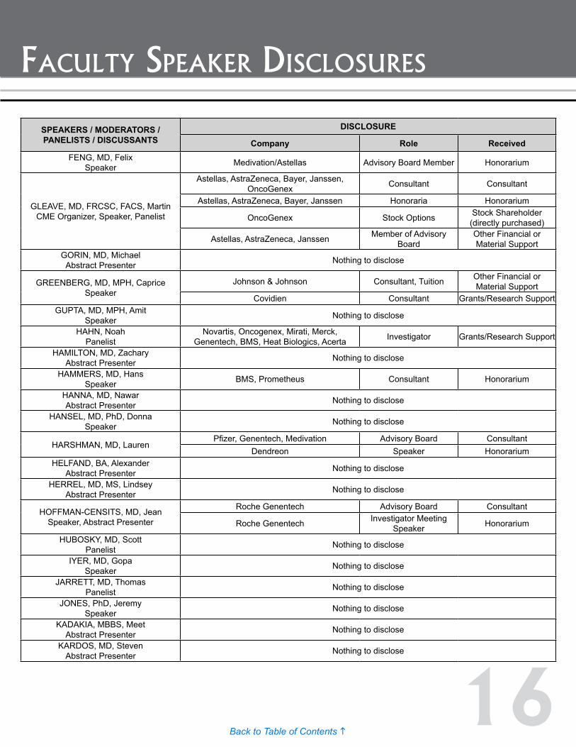
faCulTy speaker DisClosures
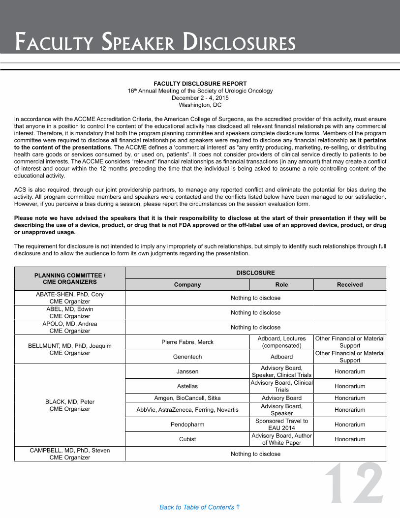
FACULTY DISCLOSURE REPORT16th Annual Meeting of the Society of Urologic Oncology
December 2 - 4, 2015 Washington, DC
In accordance with the ACCME Accreditation Criteria, the American College of Surgeons, as the accredited provider of this activity, must ensure that anyone in a position to control the content of the educational activity has disclosed all relevant financial relationships with any commercial interest. Therefore, it is mandatory that both the program planning committee and speakers complete disclosure forms. Members of the program committee were required to disclose all financial relationships and speakers were required to disclose any financial relationship as it pertains to the content of the presentations. The ACCME defines a ‘commercial interest’ as “any entity producing, marketing, re-selling, or distributing health care goods or services consumed by, or used on, patients”. It does not consider providers of clinical service directly to patients to be commercial interests. The ACCME considers “relevant” financial relationships as financial transactions (in any amount) that may create a conflict of interest and occur within the 12 months preceding the time that the individual is being asked to assume a role controlling content of the educational activity.
ACS is also required, through our joint providership partners, to manage any reported conflict and eliminate the potential for bias during the activity. All program committee members and speakers were contacted and the conflicts listed below have been managed to our satisfaction. However, if you perceive a bias during a session, please report the circumstances on the session evaluation form.
Please note we have advised the speakers that it is their responsibility to disclose at the start of their presentation if they will be describing the use of a device, product, or drug that is not FDA approved or the off-label use of an approved device, product, or drug or unapproved usage.
The requirement for disclosure is not intended to imply any impropriety of such relationships, but simply to identify such relationships through full disclosure and to allow the audience to form its own judgments regarding the presentation.
PLANNING COMMITTEE / CME ORGANIZERS
DISCLOSURE
Company Role ReceivedABATE-SHEN, PhD, Cory
CME Organizer Nothing to disclose
ABEL, MD, Edwin CME Organizer Nothing to disclose
APOLO, MD, Andrea CME Organizer Nothing to disclose
BELLMUNT, MD, PhD, Joaquim CME Organizer
Pierre Fabre, Merck Adboard, Lectures (compensated)
Other Financial or Material Support
Genentech Adboard Other Financial or Material Support
BLACK, MD, Peter CME Organizer
Janssen Advisory Board, Speaker, Clinical Trials Honorarium
Astellas Advisory Board, Clinical Trials Honorarium
Amgen, BioCancell, Sitka Advisory Board Honorarium
AbbVie, AstraZeneca, Ferring, Novartis Advisory Board, Speaker Honorarium
Pendopharm Sponsored Travel to EAU 2014 Honorarium
Cubist Advisory Board, Author of White Paper Honorarium
CAMPBELL, MD, PhD, Steven CME Organizer Nothing to disclose
13Back to Table of Contents h
faCulTy speaker DisClosures
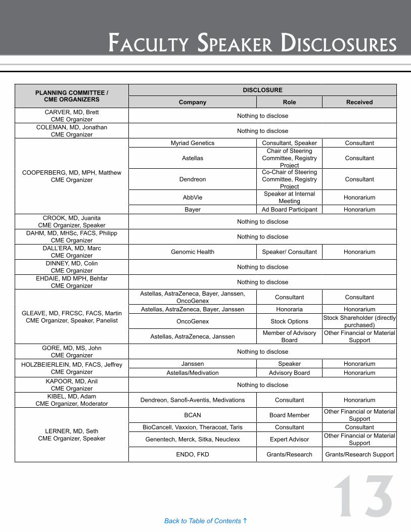
PLANNING COMMITTEE / CME ORGANIZERS
DISCLOSURE
Company Role ReceivedCARVER, MD, Brett
CME Organizer Nothing to disclose
COLEMAN, MD, Jonathan CME Organizer Nothing to disclose
COOPERBERG, MD, MPH, Matthew CME Organizer
Myriad Genetics Consultant, Speaker Consultant
AstellasChair of Steering
Committee, Registry Project
Consultant
DendreonCo-Chair of Steering Committee, Registry
ProjectConsultant
AbbVie Speaker at Internal Meeting Honorarium
Bayer Ad Board Participant HonorariumCROOK, MD, Juanita
CME Organizer, Speaker Nothing to disclose
DAHM, MD, MHSc, FACS, Philipp CME Organizer Nothing to disclose
DALL’ERA, MD, Marc CME Organizer Genomic Health Speaker/ Consultant Honorarium
DINNEY, MD, Colin CME Organizer Nothing to disclose
EHDAIE, MD MPH, Behfar CME Organizer Nothing to disclose
GLEAVE, MD, FRCSC, FACS, Martin CME Organizer, Speaker, Panelist
Astellas, AstraZeneca, Bayer, Janssen,OncoGenex Consultant Consultant
Astellas, AstraZeneca, Bayer, Janssen Honoraria Honorarium
OncoGenex Stock Options Stock Shareholder (directly purchased)
Astellas, AstraZeneca, Janssen Member of Advisory Board
Other Financial or Material Support
GORE, MD, MS, John CME Organizer Nothing to disclose
HOLZBEIERLEIN, MD, FACS, Jeffrey CME Organizer
Janssen Speaker HonorariumAstellas/Medivation Advisory Board Honorarium
KAPOOR, MD, Anil CME Organizer Nothing to disclose
KIBEL, MD, Adam CME Organizer, Moderator Dendreon, Sanofi-Aventis, Medivations Consultant Honorarium
LERNER, MD, Seth CME Organizer, Speaker
BCAN Board Member Other Financial or Material Support
BioCancell, Vaxxion, Theracoat, Taris Consultant Consultant
Genentech, Merck, Sitka, Neuclexx Expert Advisor Other Financial or Material Support
ENDO, FKD Grants/Research Grants/Research Support
14Back to Table of Contents h
faCulTy speaker DisClosures
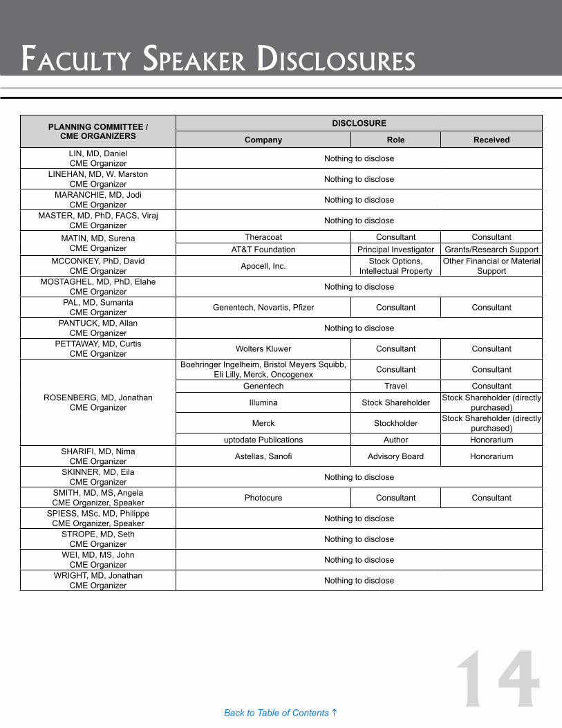
PLANNING COMMITTEE / CME ORGANIZERS
DISCLOSURE
Company Role ReceivedLIN, MD, Daniel CME Organizer Nothing to disclose
LINEHAN, MD, W. Marston CME Organizer Nothing to disclose
MARANCHIE, MD, Jodi CME Organizer Nothing to disclose
MASTER, MD, PhD, FACS, Viraj CME Organizer Nothing to disclose
MATIN, MD, Surena CME Organizer
Theracoat Consultant ConsultantAT&T Foundation Principal Investigator Grants/Research Support
MCCONKEY, PhD, David CME Organizer Apocell, Inc. Stock Options,
Intellectual PropertyOther Financial or Material
SupportMOSTAGHEL, MD, PhD, Elahe
CME Organizer Nothing to disclose
PAL, MD, Sumanta CME Organizer Genentech, Novartis, Pfizer Consultant Consultant
PANTUCK, MD, Allan CME Organizer Nothing to disclose
PETTAWAY, MD, Curtis CME Organizer Wolters Kluwer Consultant Consultant
ROSENBERG, MD, Jonathan CME Organizer
Boehringer Ingelheim, Bristol Meyers Squibb, Eli Lilly, Merck, Oncogenex Consultant Consultant
Genentech Travel Consultant
Illumina Stock Shareholder Stock Shareholder (directly purchased)
Merck Stockholder Stock Shareholder (directly purchased)
uptodate Publications Author HonorariumSHARIFI, MD, Nima
CME Organizer Astellas, Sanofi Advisory Board Honorarium
SKINNER, MD, Eila CME Organizer Nothing to disclose
SMITH, MD, MS, Angela CME Organizer, Speaker Photocure Consultant Consultant
SPIESS, MSc, MD, Philippe CME Organizer, Speaker Nothing to disclose
STROPE, MD, Seth CME Organizer Nothing to disclose
WEI, MD, MS, John CME Organizer Nothing to disclose
WRIGHT, MD, Jonathan CME Organizer Nothing to disclose
15Back to Table of Contents h
faCulTy speaker DisClosures
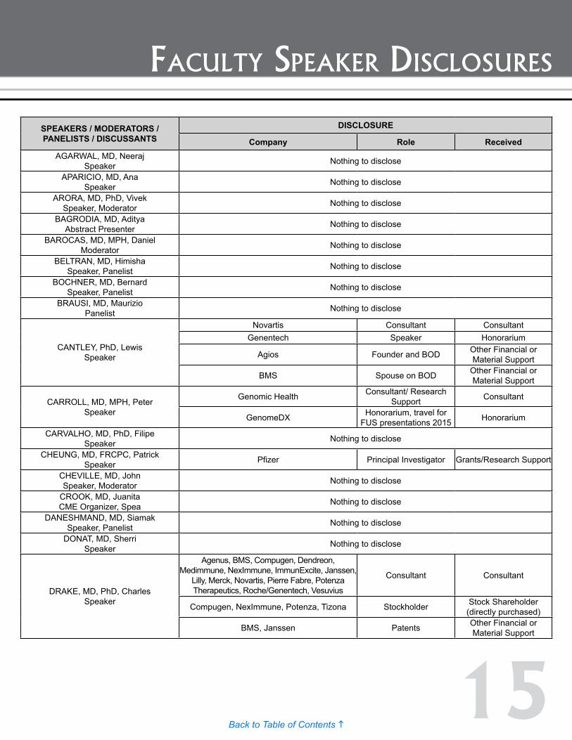
SPEAKERS / MODERATORS / PANELISTS / DISCUSSANTS
DISCLOSURE
Company Role ReceivedAGARWAL, MD, Neeraj
Speaker Nothing to disclose
APARICIO, MD, Ana Speaker Nothing to disclose
ARORA, MD, PhD, Vivek Speaker, Moderator Nothing to disclose
BAGRODIA, MD, AdityaAbstract Presenter Nothing to disclose
BAROCAS, MD, MPH, Daniel Moderator Nothing to disclose
BELTRAN, MD, Himisha Speaker, Panelist Nothing to disclose
BOCHNER, MD, Bernard Speaker, Panelist Nothing to disclose
BRAUSI, MD, Maurizio Panelist Nothing to disclose
CANTLEY, PhD, Lewis Speaker
Novartis Consultant ConsultantGenentech Speaker Honorarium
Agios Founder and BOD Other Financial or Material Support
BMS Spouse on BOD Other Financial or Material Support
CARROLL, MD, MPH, Peter Speaker
Genomic Health Consultant/ Research Support Consultant
GenomeDX Honorarium, travel for FUS presentations 2015 Honorarium
CARVALHO, MD, PhD, Filipe Speaker Nothing to disclose
CHEUNG, MD, FRCPC, Patrick Speaker Pfizer Principal Investigator Grants/Research Support
CHEVILLE, MD, John Speaker, Moderator Nothing to disclose
CROOK, MD, Juanita CME Organizer, Spea Nothing to disclose
DANESHMAND, MD, Siamak Speaker, Panelist Nothing to disclose
DONAT, MD, Sherri Speaker Nothing to disclose
DRAKE, MD, PhD, Charles Speaker
Agenus, BMS, Compugen, Dendreon, Medimmune, NexImmune, ImmunExcite, Janssen,
Lilly, Merck, Novartis, Pierre Fabre, Potenza Therapeutics, Roche/Genentech, Vesuvius
Consultant Consultant
Compugen, NexImmune, Potenza, Tizona Stockholder Stock Shareholder (directly purchased)
BMS, Janssen Patents Other Financial or Material Support
16Back to Table of Contents h
faCulTy speaker DisClosures
SPEAKERS / MODERATORS / PANELISTS / DISCUSSANTS
DISCLOSURE
Company Role ReceivedFENG, MD, Felix
Speaker Medivation/Astellas Advisory Board Member Honorarium
GLEAVE, MD, FRCSC, FACS, Martin CME Organizer, Speaker, Panelist
Astellas, AstraZeneca, Bayer, Janssen, OncoGenex Consultant Consultant
Astellas, AstraZeneca, Bayer, Janssen Honoraria Honorarium
OncoGenex Stock Options Stock Shareholder (directly purchased)
Astellas, AstraZeneca, Janssen Member of Advisory Board
Other Financial or Material Support
GORIN, MD, Michael Abstract Presenter Nothing to disclose
GREENBERG, MD, MPH, Caprice Speaker
Johnson & Johnson Consultant, Tuition Other Financial or Material Support
Covidien Consultant Grants/Research SupportGUPTA, MD, MPH, Amit
Speaker Nothing to disclose
HAHN, Noah Panelist
Novartis, Oncogenex, Mirati, Merck, Genentech, BMS, Heat Biologics, Acerta Investigator Grants/Research Support
HAMILTON, MD, Zachary Abstract Presenter Nothing to disclose
HAMMERS, MD, Hans Speaker BMS, Prometheus Consultant Honorarium
HANNA, MD, Nawar Abstract Presenter Nothing to disclose
HANSEL, MD, PhD, Donna Speaker Nothing to disclose
HARSHMAN, MD, LaurenPfizer, Genentech, Medivation Advisory Board Consultant
Dendreon Speaker HonorariumHELFAND, BA, Alexander
Abstract Presenter Nothing to disclose
HERREL, MD, MS, Lindsey Abstract Presenter Nothing to disclose
HOFFMAN-CENSITS, MD, Jean Speaker, Abstract Presenter
Roche Genentech Advisory Board Consultant
Roche Genentech Investigator Meeting Speaker Honorarium
HUBOSKY, MD, Scott Panelist Nothing to disclose
IYER, MD, Gopa Speaker Nothing to disclose
JARRETT, MD, Thomas Panelist Nothing to disclose
JONES, PhD, Jeremy Speaker Nothing to disclose
KADAKIA, MBBS, Meet Abstract Presenter Nothing to disclose
KARDOS, MD, Steven Abstract Presenter Nothing to disclose
17Back to Table of Contents h
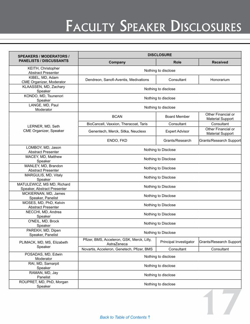
faCulTy speaker DisClosures
SPEAKERS / MODERATORS / PANELISTS / DISCUSSANTS
DISCLOSURE
Company Role ReceivedKEITH, Christopher Abstract Presenter Nothing to disclose
KIBEL, MD, Adam CME Organizer, Moderator Dendreon, Sanofi-Aventis, Medivations Consultant Honorarium
KLAASSEN, MD, Zachary Speaker Nothing to disclose
KONDO, MD, Tsunenori Speaker Nothing to disclose
LANGE, MD, Paul Moderator Nothing to disclose
LERNER, MD, Seth CME Organizer, Speaker
BCAN Board Member Other Financial or Material Support
BioCancell, Vaxxion, Theracoat, Taris Consultant Consultant
Genentech, Merck, Sitka, Neuclexx Expert Advisor Other Financial or Material Support
ENDO, FKD Grants/Research Grants/Research Support
LOMBOY, MD, JasonAbstract Presenter Nothing to Disclose
MACEY, MD, Matthew Speaker Nothing to Disclose
MANLEY, MD, Brandon Abstract Presenter Nothing to Disclose
MARGULIS, MD, Vitaly Speaker Nothing to Disclose
MATULEWICZ, MS MD, Richard Speaker, Abstract Presenter Nothing to Disclose
MCKIERNAN, MD, James Speaker, Panelist Nothing to Disclose
MOSES, MD, PhD, Kelvin Abstract Presenter Nothing to Disclose
NECCHI, MD, Andrea Speaker Nothing to Disclose
O'NEIL, MD, Brock Speaker Nothing to Disclose
PAREKH, MD, Dipen Speaker, Panelist Nothing to Disclose
PLIMACK, MD, MS, Elizabeth Speaker
Pfizer, BMS, Acceleron, GSK, Merck, Lilly, AstraZeneca Principal Investigator Grants/Research Support
Novartis, Acceleron, Genetech, Pfizer, BMS Consultant ConsultantPOSADAS, MD, Edwin
Moderator Nothing to disclose
RAI, MD, Samarpit Speaker Nothing to disclose
RAMAN, MD, Jay Panelist Nothing to disclose
ROUPRET, MD, PhD, Morgan Speaker Nothing to disclose
18Back to Table of Contents h
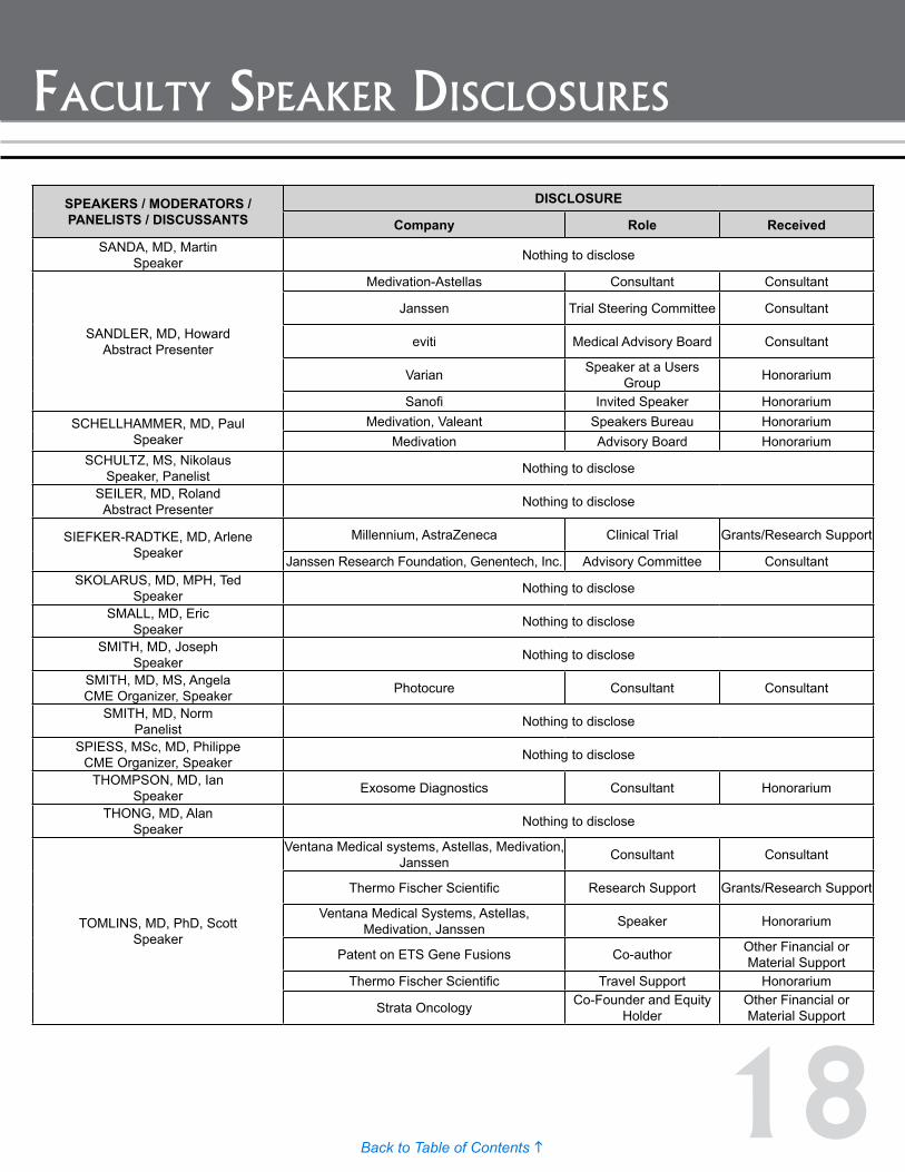
faCulTy speaker DisClosures
SPEAKERS / MODERATORS / PANELISTS / DISCUSSANTS
DISCLOSURE
Company Role ReceivedSANDA, MD, Martin
Speaker Nothing to disclose
SANDLER, MD, Howard Abstract Presenter
Medivation-Astellas Consultant Consultant
Janssen Trial Steering Committee Consultant
eviti Medical Advisory Board Consultant
Varian Speaker at a Users Group Honorarium
Sanofi Invited Speaker HonorariumSCHELLHAMMER, MD, Paul
SpeakerMedivation, Valeant Speakers Bureau Honorarium
Medivation Advisory Board HonorariumSCHULTZ, MS, Nikolaus
Speaker, Panelist Nothing to disclose
SEILER, MD, Roland Abstract Presenter Nothing to disclose
SIEFKER-RADTKE, MD, Arlene Speaker
Millennium, AstraZeneca Clinical Trial Grants/Research Support
Janssen Research Foundation, Genentech, Inc. Advisory Committee ConsultantSKOLARUS, MD, MPH, Ted
Speaker Nothing to disclose
SMALL, MD, Eric Speaker Nothing to disclose
SMITH, MD, Joseph Speaker Nothing to disclose
SMITH, MD, MS, Angela CME Organizer, Speaker Photocure Consultant Consultant
SMITH, MD, Norm Panelist Nothing to disclose
SPIESS, MSc, MD, Philippe CME Organizer, Speaker Nothing to disclose
THOMPSON, MD, Ian Speaker Exosome Diagnostics Consultant Honorarium
THONG, MD, Alan Speaker Nothing to disclose
TOMLINS, MD, PhD, Scott Speaker
Ventana Medical systems, Astellas, Medivation, Janssen Consultant Consultant
Thermo Fischer Scientific Research Support Grants/Research Support
Ventana Medical Systems, Astellas, Medivation, Janssen Speaker Honorarium
Patent on ETS Gene Fusions Co-author Other Financial or Material Support
Thermo Fischer Scientific Travel Support Honorarium
Strata Oncology Co-Founder and Equity Holder
Other Financial or Material Support
19Back to Table of Contents h
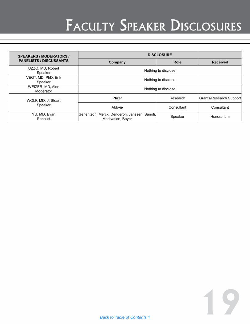
faCulTy speaker DisClosures
SPEAKERS / MODERATORS / PANELISTS / DISCUSSANTS
DISCLOSURE
Company Role ReceivedUZZO, MD, Robert
Speaker Nothing to disclose
VEGT, MD, PhD, Erik Speaker Nothing to disclose
WEIZER, MD, Alon Moderator Nothing to disclose
WOLF, MD, J. Stuart Speaker
Pfizer Research Grants/Research Support
Abbvie Consultant Consultant
YU, MD, Evan Panelist
Genentech, Merck, Denderon, Janssen, Sanofi, Medivation, Bayer Speaker Honorarium
20Back to Table of Contents h
inDusTry saTelliTe symposia
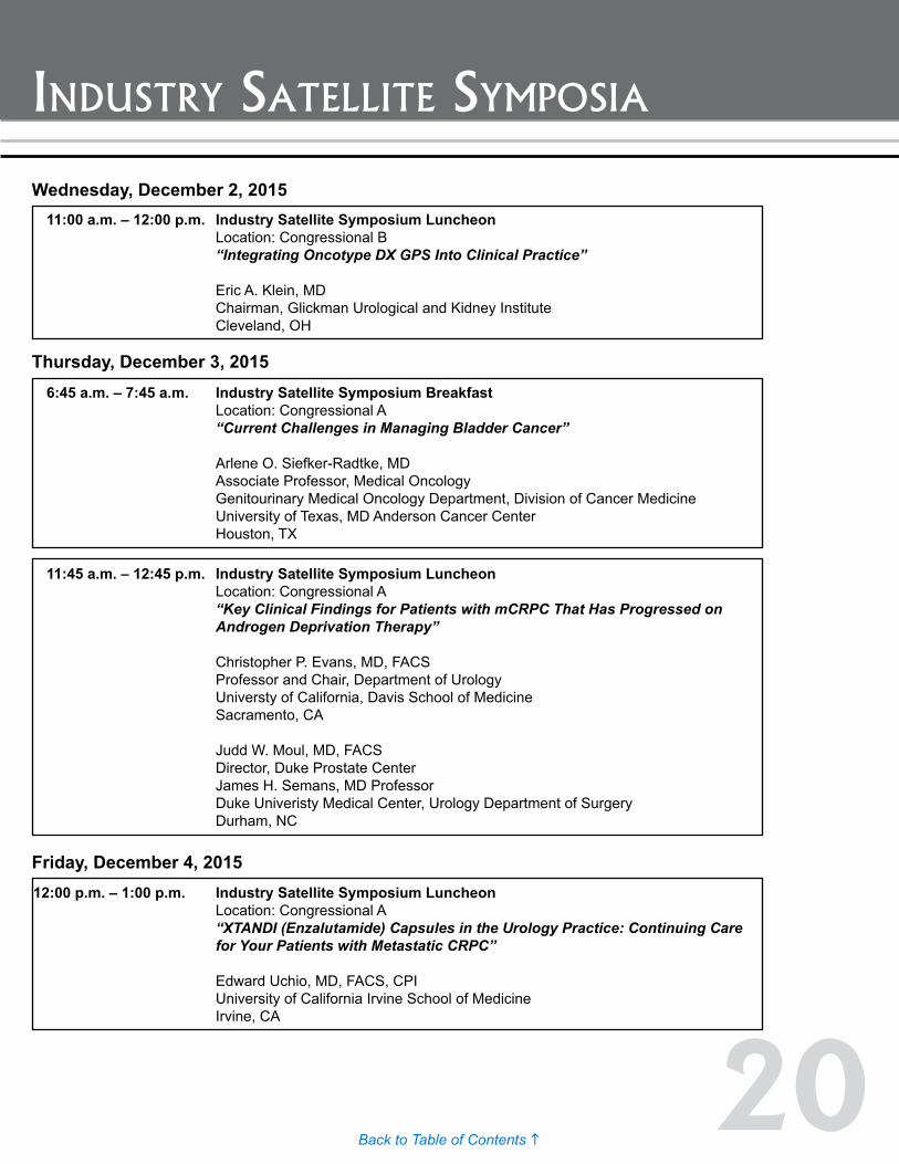
11:00 a.m. – 12:00 p.m. Industry Satellite Symposium Luncheon Location: Congressional B “Integrating Oncotype DX GPS Into Clinical Practice”
Eric A. Klein, MD Chairman, Glickman Urological and Kidney Institute Cleveland, OH
6:45 a.m. – 7:45 a.m. Industry Satellite Symposium Breakfast Location: Congressional A “Current Challenges in Managing Bladder Cancer”
Arlene O. Siefker-Radtke, MD Associate Professor, Medical Oncology Genitourinary Medical Oncology Department, Division of Cancer Medicine University of Texas, MD Anderson Cancer Center Houston, TX
Wednesday, December 2, 2015
Thursday, December 3, 2015
11:45 a.m. – 12:45 p.m. Industry Satellite Symposium Luncheon Location: Congressional A “Key Clinical Findings for Patients with mCRPC That Has Progressed on Androgen Deprivation Therapy”
Christopher P. Evans, MD, FACS Professor and Chair, Department of Urology Universty of California, Davis School of Medicine Sacramento, CA
Judd W. Moul, MD, FACS Director, Duke Prostate Center James H. Semans, MD Professor Duke Univeristy Medical Center, Urology Department of Surgery Durham, NC
Friday, December 4, 201512:00 p.m. – 1:00 p.m. Industry Satellite Symposium Luncheon Location: Congressional A “XTANDI (Enzalutamide) Capsules in the Urology Practice: Continuing Care for Your Patients with Metastatic CRPC”
Edward Uchio, MD, FACS, CPI University of California Irvine School of Medicine Irvine, CA
21Back to Table of Contents h
General sCienTifiC proGram
16th Annual Meeting of the Society of Urologic Oncology Extraordinary Opportunities for Discovery
December 2 – 4, 2015Renaissance Washington DC Downtown Hotel
Washington, DC
General Scientific Program
Speakers and times are subject to changeAll sessions located in Grand Ballroom unless otherwise noted
22Back to Table of Contents h
General sCienTifiC proGram
Speakers and times are subject to changeAll sessions located in Grand Ballroom unless otherwise noted
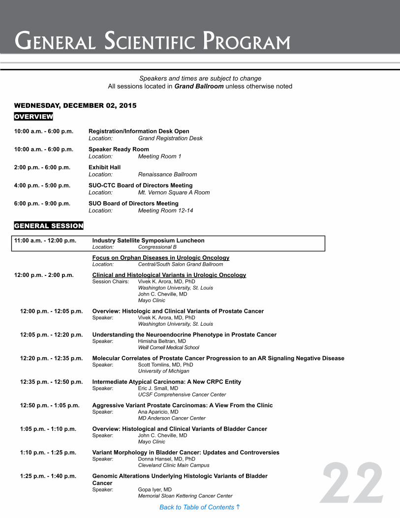
WEDNESDAY, DECEMBER 02, 2015OVERVIEW
10:00 a.m. - 6:00 p.m. Registration/Information Desk Open Location: Grand Registration Desk
10:00 a.m. - 6:00 p.m. Speaker Ready Room Location: Meeting Room 1
2:00 p.m. - 6:00 p.m. Exhibit Hall Location: Renaissance Ballroom
4:00 p.m. - 5:00 p.m. SUO-CTC Board of Directors Meeting Location: Mt. Vernon Square A Room
6:00 p.m. - 9:00 p.m. SUO Board of Directors Meeting Location: Meeting Room 12-14
GENERAL SESSION
11:00 a.m. - 12:00 p.m. Industry Satellite Symposium Luncheon Location: Congressional B
Focus on Orphan Diseases in Urologic Oncology Location: Central/South Salon Grand Ballroom
12:00 p.m. - 2:00 p.m. Clinical and Histological Variants in Urologic Oncology Session Chairs: Vivek K. Arora, MD, PhD Washington University, St. Louis John C. Cheville, MD Mayo Clinic
12:00 p.m. - 12:05 p.m. Overview: Histologic and Clinical Variants of Prostate Cancer Speaker: Vivek K. Arora, MD, PhD Washington University, St. Louis
12:05 p.m. - 12:20 p.m. Understanding the Neuroendocrine Phenotype in Prostate Cancer Speaker: Himisha Beltran, MD Weill Cornell Medical School
12:20 p.m. - 12:35 p.m. Molecular Correlates of Prostate Cancer Progression to an AR Signaling Negative Disease Speaker: Scott Tomlins, MD, PhD University of Michigan
12:35 p.m. - 12:50 p.m. Intermediate Atypical Carcinoma: A New CRPC Entity Speaker: Eric J. Small, MD UCSF Comprehensive Cancer Center
12:50 p.m. - 1:05 p.m. Aggressive Variant Prostate Carcinomas: A View From the Clinic Speaker: Ana Aparicio, MD MD Anderson Cancer Center
1:05 p.m. - 1:10 p.m. Overview: Histological and Clinical Variants of Bladder Cancer Speaker: John C. Cheville, MD Mayo Clinic
1:10 p.m. - 1:25 p.m. Variant Morphology in Bladder Cancer: Updates and Controversies Speaker: Donna Hansel, MD, PhD Cleveland Clinic Main Campus
1:25 p.m. - 1:40 p.m. Genomic Alterations Underlying Histologic Variants of Bladder Cancer
Speaker: Gopa Iyer, MD Memorial Sloan Kettering Cancer Center
23Back to Table of Contents h
General sCienTifiC proGram
Speakers and times are subject to changeAll sessions located in Grand Ballroom unless otherwise noted
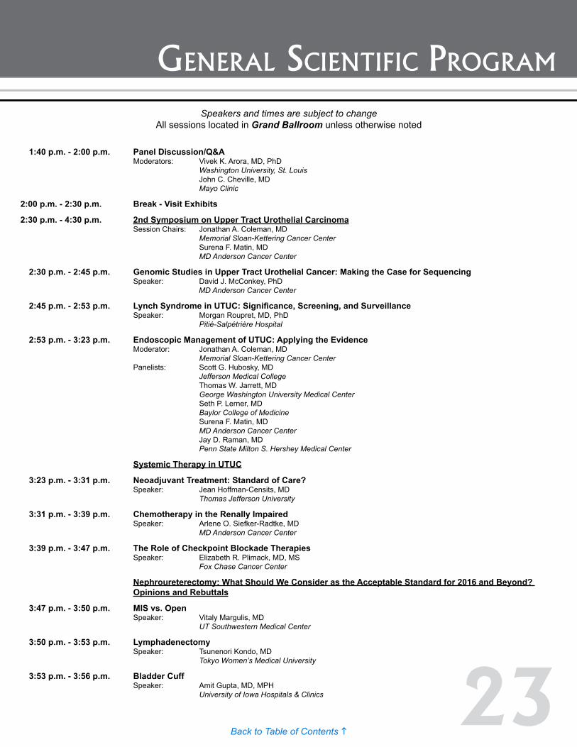
1:40 p.m. - 2:00 p.m. Panel Discussion/Q&A Moderators: Vivek K. Arora, MD, PhD Washington University, St. Louis John C. Cheville, MD Mayo Clinic
2:00 p.m. - 2:30 p.m. Break - Visit Exhibits
2:30 p.m. - 4:30 p.m. 2nd Symposium on Upper Tract Urothelial Carcinoma Session Chairs: Jonathan A. Coleman, MD Memorial Sloan-Kettering Cancer Center Surena F. Matin, MD MD Anderson Cancer Center
2:30 p.m. - 2:45 p.m. Genomic Studies in Upper Tract Urothelial Cancer: Making the Case for Sequencing Speaker: David J. McConkey, PhD
MD Anderson Cancer Center
2:45 p.m. - 2:53 p.m. Lynch Syndrome in UTUC: Significance, Screening, and Surveillance Speaker: Morgan Roupret, MD, PhD Pitié-Salpétrière Hospital
2:53 p.m. - 3:23 p.m. Endoscopic Management of UTUC: Applying the Evidence Moderator: Jonathan A. Coleman, MD Memorial Sloan-Kettering Cancer Center Panelists: Scott G. Hubosky, MD Jefferson Medical College Thomas W. Jarrett, MD George Washington University Medical Center Seth P. Lerner, MD Baylor College of Medicine Surena F. Matin, MD MD Anderson Cancer Center Jay D. Raman, MD Penn State Milton S. Hershey Medical Center
Systemic Therapy in UTUC
3:23 p.m. - 3:31 p.m. Neoadjuvant Treatment: Standard of Care? Speaker: Jean Hoffman-Censits, MD Thomas Jefferson University
3:31 p.m. - 3:39 p.m. Chemotherapy in the Renally Impaired Speaker: Arlene O. Siefker-Radtke, MD MD Anderson Cancer Center
3:39 p.m. - 3:47 p.m. The Role of Checkpoint Blockade Therapies Speaker: Elizabeth R. Plimack, MD, MS Fox Chase Cancer Center
Nephroureterectomy: What Should We Consider as the Acceptable Standard for 2016 and Beyond? Opinions and Rebuttals
3:47 p.m. - 3:50 p.m. MIS vs. Open Speaker: Vitaly Margulis, MD UT Southwestern Medical Center
3:50 p.m. - 3:53 p.m. Lymphadenectomy Speaker: Tsunenori Kondo, MD Tokyo Women’s Medical University
3:53 p.m. - 3:56 p.m. Bladder Cuff Speaker: Amit Gupta, MD, MPH University of Iowa Hospitals & Clinics
24Back to Table of Contents h
General sCienTifiC proGram
Speakers and times are subject to changeAll sessions located in Grand Ballroom unless otherwise noted
3:56 p.m. - 4:07 p.m. Rebuttal Panel Discussion Panelists: Maurizio Brausi, MD Department of Urology Ausl Modena Thomas W. Jarrett, MD George Washington University Medical Center Eila C. Skinner, MD Stanford University Medical Center
Update on Prospective Trials
4:07 p.m. - 4:09 p.m. EORTC/ACRIN/SWOG Neoadjuvant Chemotherapy and European NAC Study (Palou) Speaker: Vitaly Margulis, MD UT Southwestern Medical Center
4:09 p.m. - 4:11 p.m. Theracoat Speaker: Seth P. Lerner, MD Baylor College of Medicine
4:11 p.m. - 4:13 p.m. Lymphadenectomy Speaker: Jonathan A. Coleman, MD Memorial Sloan-Kettering Cancer Center
4:13 p.m. - 4:15 p.m. POUT Trial (Adjuvant) Speaker: Morgan Roupret, MD, PhD Pitié-Salpétrière Hospital
4:15 p.m. - 4:30 p.m. Questions & Answers
4:30 p.m. - 6:00 p.m. *Poster Session I and Reception Location: Renaissance Ballroom *Not CME Accredited
6:00 p.m. - 9:00 p.m. *Young Urologic Oncologists (Y.U.O.) Dinner Location: Congressional Ballroom *Not CME Accredited
6:00 p.m. - 7:00 p.m. Cocktail Hour
7:00 p.m. - 7:10 p.m. Welcome and Introduction Session Chair: Daniel A. Barocas, MD, MPH Vanderbilt University Medical Center
7:10 p.m. - 7:20 p.m. Annual Business Meeting
7:20 p.m. - 7:30 p.m. Paper of the Year Presentation
7:30 p.m. - 8:10 p.m. Compensation and Job Satisfaction in Urology and Urologic Oncology Speaker: Raj S. Pruthi, MD The University of North Carolina
8:10 p.m. - 8:30 p.m. SUO Urologic Oncology Fellowship and the Urology Workforce Speaker: Peter E. Clark, MD Vanderbilt University Medical Center
8:45 p.m. - 9:00 p.m. Panel Discussion/Q&A
25Back to Table of Contents h
General sCienTifiC proGram
Speakers and times are subject to changeAll sessions located in Grand Ballroom unless otherwise noted
THURSDAY, DECEMBER 03, 2015OVERVIEW
6:00 a.m. - 6:00 p.m. Speaker Ready Room Location: Meeting Room 1
6:30 a.m. - 6:00 p.m. Registration/Information Desk Open Location: Grand Registration Desk
6:45 a.m. - 7:45 a. m. Industry Satellite Symposium Breakfast Location: Congressional A
7:45 a.m. - 7:30 p.m. Exhibit Hall Location: Renaissance Ballroom
GENERAL SESSION
8:00 a.m. - 8:05 a.m. Welcome and Introduction Program Chairs: Brett S. Carver, MD Memorial Sloan Kettering Cancer Center Surena F. Matin, MD MD Anderson Cancer Center
8:05 a.m. - 9:35 a.m. Bladder Cancer Session I Session Chair: Jonathan Rosenberg, MD Memorial Sloan-Kettering Cancer Center
8:05 a.m. - 8:20 a.m. New Approaches to Adjuvant Therapy for Muscle Invasive Bladder Cancer Speaker: Andrea Apolo, MD National Cancer Institute
8:20 a.m. - 8:35 a.m. Q&A
Debate: Robotic vs. Open Radical Cystectomy: Have We Moved Too Fast, or Not Fast Enough? Session Chair: Eila C. Skinner, MD Stanford University Medical Center
8:35 a.m. - 8:45 a.m. Pro Robotic Cystectomy Speaker: Dipen J. Parekh, MD University of Miami
8:45 a.m. - 8:55 a.m. Pro Open Cystectomy Speaker: Bernard H. Bochner, MD Memorial Sloan-Kettering Cancer Center
8:55 a.m. - 9:02 a.m. Does ERAS Level the Playing Field? Speaker: Sia Daneshmand, MD University of Southern California-Keck School of Medicine
9:02 a.m. - 9:35 a.m. Panel Discussion Panelists: Bernard H. Bochner, MD Memorial Sloan-Kettering Cancer Center Sia Daneshmand, MD University of Southern California-Keck School of Medicine Dipen J. Parekh, MD University of Miami Norm D. Smith, MD University of Chicago Medical Center
9:35 a.m. - 10:05 a.m. *SUO-CTC Scientific Session: Emerging Therapies for Patients With Bladder Cancer Speaker: Colin P. Dinney, MD MD Anderson Cancer Center *Not CME Accredited
26Back to Table of Contents h
General sCienTifiC proGram
Speakers and times are subject to changeAll sessions located in Grand Ballroom unless otherwise noted
10:05 a.m. - 10:40 a.m. Break
10:40 a.m. - 11:45 a.m. Health Services Session Session Chair: John T. Wei, MD, MS University of Michigan
10:40 a.m. - 10:45 a.m. Introduction Speaker: John T. Wei, MD, MS University of Michigan
10:45 a.m. - 10:55 a.m. Population Level - AQUA Registry Update Speaker: Matthew R. Cooperberg, MD, MPH UCSF
10:55 a.m. - 11:05 a.m. Using Implementation Science to Improve Urologic Cancer Care Speaker: Ted A. Skolarus, MD, MPH University of Michigan
11:05 a.m. - 11:20 a.m. Surgical Coaching for Performance Improvement Speaker: Caprice C. Greenberg, MD, MPH University of Wisconsin
11:20 a.m. - 11:30 a.m. Putting the “Patient” Into Patient-Centered Outcomes Research Prioritization Speaker: Angela M. Smith, MD, MS University of North Carolina
11:30 a.m. - 11:45 a.m. Discussion and Questions
11:45 a.m. - 12:45 p.m. Industry Satellite Symposium Luncheon Location: Congressional A
12:45 p.m. - 1:45 p.m. Prostate Cancer Session I Session Chair: Sumanta K. Pal, MD City of Hope Comprehensive Cancer Center Moderator: Adam S. Kibel, MD Brigham and Women’s Hospital
Understanding the Biology of Localized Prostate Cancer
12:45 p.m. - 12:55 p.m. Heterogeneity in Prostate Cancer Speaker: Himisha Beltran, MD Weill Cornell Medical School
12:55 p.m. - 1:05 p.m. Accessing Genomic Data in Prostate Cancer and Overview of TCGA Subtypes Speaker: Nikolaus Schultz, MS Memorial Sloan-Kettering Cancer Center
1:05 p.m. - 1:15 p.m. Currently Available Genomic Tools for Risk Stratification of Localized Prostate Cancer Speaker: Daniel W. Lin, MD University of Washington Medical Center
1:15 p.m. - 1:35 p.m. Clinical Implications of Genomics in Treating Prostate Cancer Panelists: Himisha Beltran, MD Weill Cornell Medical School Brett S. Carver, MD Memorial Sloan Kettering Cancer Center Martin E. Gleave, MD, FRCSC, FACS Vancouver Prostate Center Daniel W. Lin, MD University of Washington Medical Center Nikolaus Schultz, MS Memorial Sloan-Kettering Cancer Center Evan Y. Yu, MD University of Washington Medical Center
1:35 p.m. - 1:45 p.m. Q&A

27Back to Table of Contents h
General sCienTifiC proGram
Speakers and times are subject to changeAll sessions located in Grand Ballroom unless otherwise noted
1:45 p.m. - 1:55 p.m. *SUO Huggins Medal Presentation President: Leonard G. Gomella, MD, FACS Thomas Jefferson University Kimmel Cancer Center *Not CME Accredited
1:55 p.m. - 2:15 p.m. Huggins Medal Lecture Speaker: Joseph A. Smith, Jr., MD Vanderbilt University Medical Center
2:15 p.m. - 3:00 p.m. Penile Cancer Session Session Chair: Viraj A. Master, MD, PhD, FACS Emory University
2:15 p.m. - 2:25 p.m. Radiotherapy Approaches for Locally Advanced Penile Cancer - Neoadjuvant and Adjuvant Speaker: Juanita Crook, MD British Columbia Cancer Agency
2:25 p.m. - 2:35 p.m. INPACT Trial Update Speaker: Curtis A. Pettaway, MD MD Anderson Cancer Center
2:35 p.m. - 2:45 p.m. Current Use of Imaging for Penile Cancer Management Speaker: Erik Vegt, MD, PhD Netherlands Cancer Institute
2:45 p.m. - 2:55 p.m. Pelvic Nodes - Update Speaker: Philippe E. Spiess, MSc, MD Moffitt Cancer Center
2:55 p.m. - 3:00 p.m. Q&A/Discussion
3:00 p.m. - 3:30 p.m. Break - Visit Exhibits
3:30 p.m. - 4:30 p.m. Kidney Cancer Session I Session Chair: Jodi K. Maranchie, MD University of Pittsburgh Moderator: Edwin J. Abel, MD University of Wisconsin
Adjuvant Therapy for Localized Renal Cancer
3:30 p.m. - 3:45 p.m. The Current Status of Adjuvant Systemic Therapy for RCC Speaker: Robert G. Uzzo, MD Fox Chase Cancer Center
3:45 p.m. - 4:00 p.m. Designing the Next Generation of Clinical Trials Speaker: Lauren C. Harshman, MD Dana Farber Cancer Institute
4:00 p.m. - 4:15 p.m. Combination Checkpoint Inhibition Speaker: Hans J. Hammers, MD Johns Hopkins University
4:15 p.m. - 4:30 p.m. Q&A/Discussion
4:30 p.m. - 6:00 p.m. *Poster Session II Location: Renaissance Ballroom *Not CME Accredited
6:00 p.m. - 7:30 p.m. SUO Reception Location: Renaissance Ballroom
28Back to Table of Contents h
General sCienTifiC proGram
Speakers and times are subject to changeAll sessions located in Grand Ballroom unless otherwise noted
FRIDAY, DECEMBER 04, 2015OVERVIEW
7:00 a.m. - 3:15 p.m. Registration/Information Desk Open Location: Grand Registration Desk
7:00 a.m. - 3:00 p.m. Speaker Ready Room Location: Meeting Room 1
7:00 a.m. - 10:30 a.m. Exhibit Hall Location: Renaissance Ballroom
7:30 a.m. - 8:15 a.m. SUO Annual Business Meeting Location: Grand Ballroom
GENERAL SESSION
8:15 a.m. - 8:45 a.m. Young Urologic Oncologists (Y.U.O.) Program Moderator: Daniel A. Barocas, MD, MPH Vanderbilt University Medical Center Abstracts selected by the Y.U.O
8:15 a.m. - 8:20 a.m. Introduction and Announcements
8:20 a.m. Podium #1 SUCCESSFUL PREDICTION OF LYMPH NODE METASTASES IN BLADDER CANCER USING GENE EXPRESSION SIGNATURES OF PRIMARY TUMORS
(Presented By: Roland Seiler)
8:28 a.m. Podium #2 PATTERNS OF PSA SCREENING AMONG LOW INCOME AFRICAN-AMERICAN AND CAUCASIAN MEN: DATA FROM THE SOUTHERN COMMUNITY COHORT STUDY
(Presented By: Kelvin Moses)
8:36 a.m. Podium #3 IS POOR ADHERENCE TO GUIDELINES FURTHER EVIDENCE OF THE NEED FOR CENTRALIZATION OF PENILE CANCER CARE?
(Presented By: Richard Matulewicz)
8:45 a.m. - 9:45 a.m. Kidney Cancer Session II Session Chair: Jodi K. Maranchie, MD University of Pittsburgh Moderator: Anil Kapoor, MD St. Joseph’s Hospital
8:45 a.m. - 9:00 a.m. Development of the Renal Cancer Follow-Up Guidelines Speaker: Sherri M. Donat, MD Memorial Sloan-Kettering Cancer Center
9:00 a.m. - 9:15 a.m. Impact of Renal Mass Biopsy on RCC Management Speaker: J. Stuart Wolf, Jr., MD University of Michigan
9:15 a.m. - 9:30 a.m. Focal Radiotherapy for Oligo Metastatic Disease Speaker: Patrick Cheung, MD, FRCPC Sunnybrook Health Sciences Centre
9:30 a.m. - 9:45 a.m. Q&A/Discussion
9:45 a.m. - 10:10 a.m. Break - Visit Exhibits
10:10 a.m. - 10:55 a.m. Prostate Cancer Session II Session Chair: Sumanta K. Pal, MD City of Hope Comprehensive Cancer Center Moderator: Paul H. Lange, MD University of Washington Medical Center
29Back to Table of Contents h
General sCienTifiC proGram
Speakers and times are subject to changeAll sessions located in Grand Ballroom unless otherwise noted
Survivorship After Primary Prostate Cancer
10:10 a.m. - 10:22 a.m. Urologists With Prostate Cancer Speaker: Paul F. Schellhammer, MD Eastern Virginia Medical School Norfolk Virginia; Urology of Virginia
10:22 a.m. - 10:34 a.m. Improving Outcome After Radical Prostatectomy Speaker: Martin G. Sanda, MD Emory University
10:34 a.m. - 10:46 a.m. Comparing QOL With Surveillance and Radical Prostatectomy Speaker: Peter R. Carroll, MD, MPH UCSF Comprehensive Cancer Center
10:46 a.m. - 10:55 a.m. Q&A
10:55 a.m. - 11:40 a.m. Oral Abstract Session Moderator: Alon Z. Weizer, MD University of Michigan
10:55 a.m. Podium #4 EFFICACY AND SAFETY RESULTS FROM A PIVOTAL MULTICENTER PHASE II STUDY (IMVIGOR 210) OF ATEZOLIZUMAB IN PATIENTS (PTS) WITH LOCALLY ADVANCED OR METASTATIC UROTHELIAL CARCINOMA (MUC)
(Presented By: Jean Hoffman-Censits)
11:02 a.m. Podium #5 DEVELOPMENT OF A NOVEL ANTIBODY-FREE METHOD FOR DETECTING CIRCULATING TUMOR CELLS IN PATIENTS WITH METASTATIC RENAL CELL CARCINOMA
(Presented By: Michael Gorin)
11:09 a.m. Podium #6 ANALYSIS OF MUTATION FREQUENCY IN A LARGE COHORT OF RENAL CLEAR CELL CARCINOMA PATIENTS AND CORRELATION WITH CLINICAL FEATURES
(Presented By: Brandon Manley)
11:16 a.m. Podium #7 A PHASE III PROTOCOL OF ANDROGEN SUPPRESSION AND 3DCRT/IMRT VS AS AND 3DCRT/IMRT FOLLOWED BY CHEMOTHERAPY WITH DOCETAXEL FOR LOCALIZED, HIGH-RISK PROSTATE CANCER (NRG ONCOLOGY/RTOG 0521)
(Presented By: Leonard Gomella)
11:23 a.m. Podium #8 ACTIONABLE TARGETS IN PATIENTS WITH CISPLATIN-RESISTANT ADVANCED GERM CELL TUMORS (Presented By: Aditya Bagrodia)
11:30 a.m. Podium #9 THE EFFECT OF DISTANCE TO A HIGH-VOLUME CENTER ON RECEIPT OF TREATMENT FOR INVASIVE BLADDER CANCER
(Presented By: Jason Lomboy)
11:37 a.m. - 11:40 a.m. Q&A
11:40 a.m. - 12:00 p.m. State-of-the-Art Lecture I: Making a Difference for Our Patients: Next Generation Clinical Trials in GU Oncology Speaker: Ian M. Thompson, Jr., MD The University of Texas Health Science Center
12:00 p.m. - 1:00 p.m. Industry Satellite Symposium Luncheon Location: Congressional A
1:00 p.m. - 1:20 p.m. State-of-the-Art Lecture II Speaker: Lewis C. Cantley, PhD Weill Cornell Medical College
1:20 p.m. - 2:20 p.m. Bladder Cancer Session II
1:20 p.m. - 1:35 p.m. Application of Immune Checkpoint Inhibitors to NMIBC: Current Science, Proposed Trials, and Future Directions
Speaker: Peter C. Black, MD University of British Columbia
30Back to Table of Contents h
General sCienTifiC proGram
Speakers and times are subject to changeAll sessions located in Grand Ballroom unless otherwise noted
1:35 p.m. - 1:50 p.m. Novel Approaches to Cytotoxic and Targeted Drugs in NMIBC Speaker: James M. McKiernan, MD Columbia University
1:50 p.m. - 2:05 p.m. Optimal Trial Design in NMIBC Speaker: Seth P. Lerner, MD Baylor College of Medicine
2:05 p.m. - 2:20 p.m. Panel Discussion Moderator: Colin P. Dinney, MD MD Anderson Cancer Center Panelists: Peter C. Black, MD University of British Columbia Noah M. Hahn Johns Hopkins Seth P. Lerner, MD Baylor College of Medicine James M. McKiernan, MD Columbia University
2:20 p.m. - 3:15 p.m. Prostate Cancer Session III Session Chair: Sumanta K. Pal, MD City of Hope Comprehensive Cancer Center Moderator: Edwin Posadas, MD Cedars-Sinai Medical Center
2:20 p.m. - 2:28 p.m. The Evolving Role of Chemotherapy in Advanced Prostate Cancer Speaker: Neeraj Agarwal, MD University of Utah
2:28 p.m. - 2:36 p.m. Steroid Metabolism and Prostate Cancer Speaker: Nima Sharifi, MD Cleveland Clinic
2:36 p.m. - 2:44 p.m. Novel Genomic Assays for Advanced Disease (Cell Free DNA and Others) Speaker: Martin E. Gleave, MD, FRCSC, FACS Vancouver Prostate Center
2:44 p.m. - 2:52 p.m. Immunotherapy in Prostate Cancer: Clinical Advances Speaker: Charles Drake, MD, PhD Johns Hopkins University School of Medicine
2:52 p.m. - 3:00 p.m. PARP Inhibitors and Prostate Cancer Speaker: Felix Feng, MD University of Michigan Health System
3:00 p.m. - 3:08 p.m. Targeting AR Outside of the LBD Speaker: Jeremy Jones, PhD City of Hope
3:08 p.m. - 3:15 p.m. Q&A
3:15 p.m. - Wrap Up/Adjourn
Disclaimer StatementStatements, opinions and results of studies contained in the program are those of the presenters/authors and do not reflect the policy or position of the SUO nor does the SUO provide any warranty as to their accuracy or reliability.
Every effort has been made to faithfully reproduce the abstracts as submitted. However, no responsibility is assumed by the SUO for any injury and/or damage to persons or property from any cause including negligence or otherwise, or from any use or operation of any methods, products, instruments or ideas contained in the material herein.
31Back to Table of Contents h
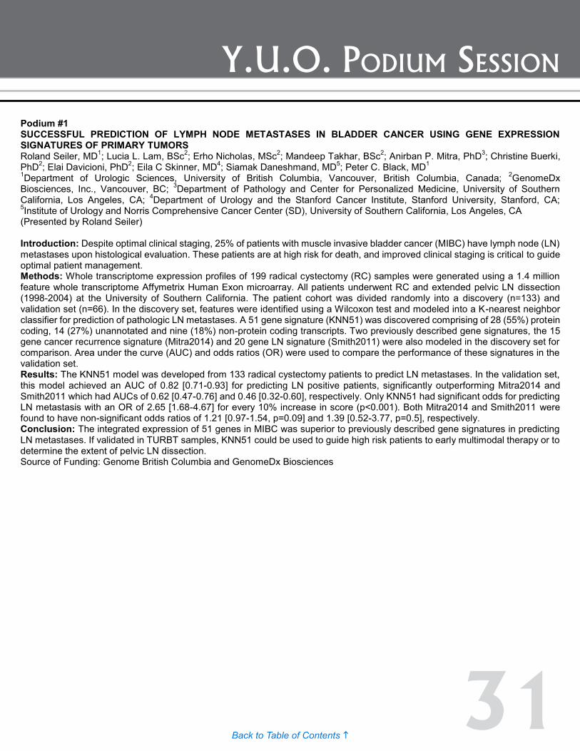
Podium #1 SUCCESSFUL PREDICTION OF LYMPH NODE METASTASES IN BLADDER CANCER USING GENE EXPRESSION SIGNATURES OF PRIMARY TUMORS Roland Seiler, MD1; Lucia L. Lam, BSc2; Erho Nicholas, MSc2; Mandeep Takhar, BSc2; Anirban P. Mitra, PhD3; Christine Buerki, PhD2; Elai Davicioni, PhD2; Eila C Skinner, MD4; Siamak Daneshmand, MD5; Peter C. Black, MD1 1Department of Urologic Sciences, University of British Columbia, Vancouver, British Columbia, Canada; 2GenomeDx Biosciences, Inc., Vancouver, BC; 3Department of Pathology and Center for Personalized Medicine, University of Southern California, Los Angeles, CA; 4Department of Urology and the Stanford Cancer Institute, Stanford University, Stanford, CA; 5Institute of Urology and Norris Comprehensive Cancer Center (SD), University of Southern California, Los Angeles, CA (Presented by Roland Seiler) Introduction: Despite optimal clinical staging, 25% of patients with muscle invasive bladder cancer (MIBC) have lymph node (LN) metastases upon histological evaluation. These patients are at high risk for death, and improved clinical staging is critical to guide optimal patient management. Methods: Whole transcriptome expression profiles of 199 radical cystectomy (RC) samples were generated using a 1.4 million feature whole transcriptome Affymetrix Human Exon microarray. All patients underwent RC and extended pelvic LN dissection (1998-2004) at the University of Southern California. The patient cohort was divided randomly into a discovery (n=133) and validation set (n=66). In the discovery set, features were identified using a Wilcoxon test and modeled into a K-nearest neighbor classifier for prediction of pathologic LN metastases. A 51 gene signature (KNN51) was discovered comprising of 28 (55%) protein coding, 14 (27%) unannotated and nine (18%) non-protein coding transcripts. Two previously described gene signatures, the 15 gene cancer recurrence signature (Mitra2014) and 20 gene LN signature (Smith2011) were also modeled in the discovery set for comparison. Area under the curve (AUC) and odds ratios (OR) were used to compare the performance of these signatures in the validation set. Results: The KNN51 model was developed from 133 radical cystectomy patients to predict LN metastases. In the validation set, this model achieved an AUC of 0.82 [0.71-0.93] for predicting LN positive patients, significantly outperforming Mitra2014 and Smith2011 which had AUCs of 0.62 [0.47-0.76] and 0.46 [0.32-0.60], respectively. Only KNN51 had significant odds for predicting LN metastasis with an OR of 2.65 [1.68-4.67] for every 10% increase in score (p<0.001). Both Mitra2014 and Smith2011 were found to have non-significant odds ratios of 1.21 [0.97-1.54, p=0.09] and 1.39 [0.52-3.77, p=0.5], respectively. Conclusion: The integrated expression of 51 genes in MIBC was superior to previously described gene signatures in predicting LN metastases. If validated in TURBT samples, KNN51 could be used to guide high risk patients to early multimodal therapy or to determine the extent of pelvic LN dissection. Source of Funding: Genome British Columbia and GenomeDx Biosciences
y.u.o. poDium session
32Back to Table of Contents h
y.u.o. poDium session
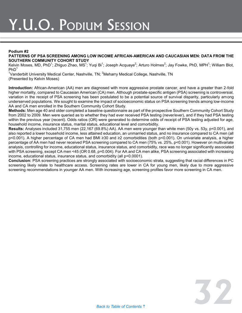
Podium #2 PATTERNS OF PSA SCREENING AMONG LOW INCOME AFRICAN-AMERICAN AND CAUCASIAN MEN: DATA FROM THE SOUTHERN COMMUNITY COHORT STUDY Kelvin Moses, MD, PhD1; Zhiguo Zhao, MS1; Yuqi Bi1; Joseph Acquaye2; Arturo Holmes2; Jay Fowke, PhD, MPH1; William Blot, PhD1 1Vanderbilt University Medical Center, Nashville, TN; 2Meharry Medical College, Nashville, TN (Presented by Kelvin Moses) Introduction: African-American (AA) men are diagnosed with more aggressive prostate cancer, and have a greater than 2-fold higher mortality, compared to Caucasian American (CA) men. Although prostate-specific antigen (PSA) screening is controversial, variation in the receipt of PSA screening has been postulated to be a potential source of survival disparity, particularly among underserved populations. We sought to examine the impact of socioeconomic status on PSA screening trends among low-income AA and CA men enrolled in the Southern Community Cohort Study. Methods: Men age 40 and older completed a baseline questionnaire as part of the prospective Southern Community Cohort Study from 2002 to 2009. Men were queried as to whether they had ever received PSA testing (never/ever), and if they had PSA testing within the previous year (recent). Odds ratios (OR) were generated to determine odds of receipt of PSA testing adjusted for age, household income, insurance status, marital status, educational level and comorbidity. Results: Analyses included 31,755 men (22,167 (69.8%) AA). AA men were younger than white men (50y vs. 53y, p<0.001), and also reported a lower household income, less attained education, an unmarried status, and no insurance compared to CA men (all p<0.001). A higher percentage of CA men had BMI ≥30 and ≥2 comorbidities (both p<0.001). On univariate analysis, a higher percentage of AA men had never received PSA screening compared to CA men (75% vs. 25%, p<0.001). However on multivariate analysis, controlling for income, educational status, insurance status, and comorbidity, race was no longer significantly associated with PSA screening, except CA men <45 (OR 0.68, p=0.004). For AA and CA men alike, PSA screening associated with increasing income, educational status, insurance status, and comorbidity (all p<0.0001). Conclusion: PSA screening practices are strongly associated with socioeconomic strata, suggesting that racial differences in PC screening likely relate to healthcare access. Screening rates are lower in CA for young men, likely due to more aggressive screening recommendations in younger AA men. With increasing age, screening profiles favor more screening in CA men.
33Back to Table of Contents h
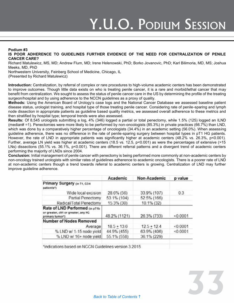
y.u.o. poDium session
Podium #3 IS POOR ADHERENCE TO GUIDELINES FURTHER EVIDENCE OF THE NEED FOR CENTRALIZATION OF PENILE CANCER CARE? Richard Matulewicz, MS, MD; Andrew Flum, MD; Irene Helenowski, PhD; Borko Jovanovic, PhD; Karl Bilimoria, MD, MS; Joshua Meeks, MD, PhD Northwestern University, Feinberg School of Medicine, Chicago, IL (Presented by Richard Matulewicz) Introduction: Centralization, by referral of complex or rare procedures to high-volume academic centers has been demonstrated to improve outcomes. Though little data exists on who is treating penile cancer, it is a rare and morbid/lethal cancer that may benefit from centralization. We sought to assess the status of penile cancer care in the US by determining the profile of the treating surgeon/hospital and by using adherence to the NCCN guidelines as a proxy of quality. Methods: Using the American Board of Urology’s case logs and the National Cancer Database we assessed baseline patient disease status, urologist training, and hospital type of those treating penile cancer. Considering rate of penile-sparing and lymph node dissection in appropriate patients as guideline based quality metrics, we assessed overall adherence to these metrics and then stratified by hospital type; temporal trends were also assessed. Results: Of 8,545 urologists submitting a log, 4% (346) logged a partial or total penectomy, while 1.5% (125) logged an ILND (median# =1). Penectomies were more likely to be performed by non-oncologists (85.3%) in private practices (66.7%) than LND, which was done by a comparatively higher percentage of oncologists (34.4%) in an academic setting (56.0%). When assessing guideline adherence, there was no difference in the rate of penile-sparing surgery between hospital types in pT1 HG patients. However, the rate of LND in appropriate patients was significantly higher at academic centers (48.2% vs. 26.3%, p<0.001). Further, average LN yield was higher at academic centers (18.5 vs. 12.5, p<0.001) as were the percentages of extensive (>15 LNs) dissections (55.1% vs. 36.1%, p<0.001). There are different referral patterns and a divergent trend of academic centers performing the majority of LNDs since 2004. Conclusion: Initial management of penile cancer with penectomy is being performed more commonly at non-academic centers by non-oncology trained urologists with similar rates of guidelines adherence to academic oncologists. There is a poorer rate of LND at non-academic centers though a trend towards referral to academic centers is growing. Centralization of LND may further improve guideline adherence.
Podium #2 PATTERNS OF PSA SCREENING AMONG LOW INCOME AFRICAN-AMERICAN AND CAUCASIAN MEN: DATA FROM THE SOUTHERN COMMUNITY COHORT STUDY Kelvin Moses, MD, PhD1; Zhiguo Zhao, MS1; Yuqi Bi1; Joseph Acquaye2; Arturo Holmes2; Jay Fowke, PhD, MPH1; William Blot, PhD1 1Vanderbilt University Medical Center, Nashville, TN; 2Meharry Medical College, Nashville, TN (Presented by Kelvin Moses) Introduction: African-American (AA) men are diagnosed with more aggressive prostate cancer, and have a greater than 2-fold higher mortality, compared to Caucasian American (CA) men. Although prostate-specific antigen (PSA) screening is controversial, variation in the receipt of PSA screening has been postulated to be a potential source of survival disparity, particularly among underserved populations. We sought to examine the impact of socioeconomic status on PSA screening trends among low-income AA and CA men enrolled in the Southern Community Cohort Study. Methods: Men age 40 and older completed a baseline questionnaire as part of the prospective Southern Community Cohort Study from 2002 to 2009. Men were queried as to whether they had ever received PSA testing (never/ever), and if they had PSA testing within the previous year (recent). Odds ratios (OR) were generated to determine odds of receipt of PSA testing adjusted for age, household income, insurance status, marital status, educational level and comorbidity. Results: Analyses included 31,755 men (22,167 (69.8%) AA). AA men were younger than white men (50y vs. 53y, p<0.001), and also reported a lower household income, less attained education, an unmarried status, and no insurance compared to CA men (all p<0.001). A higher percentage of CA men had BMI ≥30 and ≥2 comorbidities (both p<0.001). On univariate analysis, a higher percentage of AA men had never received PSA screening compared to CA men (75% vs. 25%, p<0.001). However on multivariate analysis, controlling for income, educational status, insurance status, and comorbidity, race was no longer significantly associated with PSA screening, except CA men <45 (OR 0.68, p=0.004). For AA and CA men alike, PSA screening associated with increasing income, educational status, insurance status, and comorbidity (all p<0.0001). Conclusion: PSA screening practices are strongly associated with socioeconomic strata, suggesting that racial differences in PC screening likely relate to healthcare access. Screening rates are lower in CA for young men, likely due to more aggressive screening recommendations in younger AA men. With increasing age, screening profiles favor more screening in CA men.
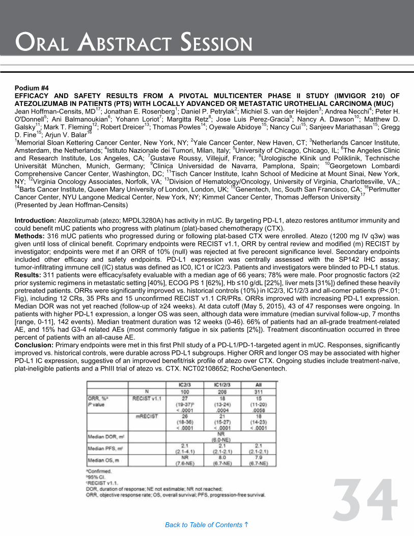
34Back to Table of Contents h
oral absTraCT session
Podium #4 EFFICACY AND SAFETY RESULTS FROM A PIVOTAL MULTICENTER PHASE II STUDY (IMVIGOR 210) OF ATEZOLIZUMAB IN PATIENTS (PTS) WITH LOCALLY ADVANCED OR METASTATIC UROTHELIAL CARCINOMA (MUC) Jean Hoffman-Censits, MD17; Jonathan E. Rosenberg1; Daniel P. Petrylak2; Michiel S. van der Heijden3; Andrea Necchi4; Peter H. O'Donnell5; Ani Balmanoukian6; Yohann Loriot7; Margitta Retz8; Jose Luis Perez-Gracia9; Nancy A. Dawson10; Matthew D. Galsky11; Mark T. Fleming12; Robert Dreicer13; Thomas Powles14; Oyewale Abidoye15; Nancy Cui15; Sanjeev Mariathasan15; Gregg D. Fine15; Arjun V. Balar16 1Memorial Sloan Kettering Cancer Center, New York, NY; 2Yale Cancer Center, New Haven, CT; 3Netherlands Cancer Institute, Amsterdam, the Netherlands; 4Istituto Nazionale dei Tumori, Milan, Italy; 5University of Chicago, Chicago, IL; 6The Angeles Clinic and Research Institute, Los Angeles, CA; 7Gustave Roussy, Villejuif, France; 8Urologische Klinik und Poliklinik, Technische Universität München, Munich, Germany; 9Clinica Universidad de Navarra, Pamplona, Spain; 10Georgetown Lombardi Comprehensive Cancer Center, Washington, DC; 11Tisch Cancer Institute, Icahn School of Medicine at Mount Sinai, New York, NY; 12Virginia Oncology Associates, Norfolk, VA; 13Division of Hematology/Oncology, University of Virginia, Charlottesville, VA,; 14Barts Cancer Institute, Queen Mary University of London, London, UK; 15Genentech, Inc, South San Francisco, CA; 16Perlmutter Cancer Center, NYU Langone Medical Center, New York, NY; Kimmel Cancer Center, Thomas Jefferson University17 (Presented by Jean Hoffman-Censits) Introduction: Atezolizumab (atezo; MPDL3280A) has activity in mUC. By targeting PD-L1, atezo restores antitumor immunity and could benefit mUC patients who progress with platinum (plat)-based chemotherapy (CTX). Methods: 316 mUC patients who progressed during or following plat-based CTX were enrolled. Atezo (1200 mg IV q3w) was given until loss of clinical benefit. Coprimary endpoints were RECIST v1.1, ORR by central review and modified (m) RECIST by investigator; endpoints were met if an ORR of 10% (null) was rejected at five perecent significance level. Secondary endpoints included other efficacy and safety endpoints. PD-L1 expression was centrally assessed with the SP142 IHC assay; tumor-infiltrating immune cell (IC) status was defined as IC0, IC1 or IC2/3. Patients and investigators were blinded to PD-L1 status. Results: 311 patients were efficacy/safety evaluable with a median age of 66 years; 78% were male. Poor prognostic factors (≥2 prior systemic regimens in metastatic setting [40%], ECOG PS 1 [62%], Hb ≤10 g/dL [22%], liver mets [31%]) defined these heavily pretreated patients. ORRs were significantly improved vs. historical controls (10%) in IC2/3, IC1/2/3 and all-comer patients (P<.01; Fig), including 12 CRs, 35 PRs and 15 unconfirmed RECIST v1.1 CR/PRs. ORRs improved with increasing PD-L1 expression. Median DOR was not yet reached (follow-up of ≥24 weeks). At data cutoff (May 5, 2015), 43 of 47 responses were ongoing. In patients with higher PD-L1 expression, a longer OS was seen, although data were immature (median survival follow-up, 7 months [range, 0-11], 142 events). Median treatment duration was 12 weeks (0-46). 66% of patients had an all-grade treatment-related AE, and 15% had G3-4 related AEs (most commonly fatigue in six patients [2%]). Treatment discontinuation occurred in three percent of patients with an all-cause AE. Conclusion: Primary endpoints were met in this first PhII study of a PD-L1/PD-1-targeted agent in mUC. Responses, significantly improved vs. historical controls, were durable across PD-L1 subgroups. Higher ORR and longer OS may be associated with higher PD-L1 IC expression, suggestive of an improved benefit/risk profile of atezo over CTX. Ongoing studies include treatment-naïve, plat-ineligible patients and a PhIII trial of atezo vs. CTX. NCT02108652; Roche/Genentech.
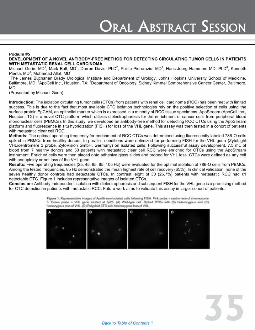
35Back to Table of Contents h
oral absTraCT session
Podium #5 DEVELOPMENT OF A NOVEL ANTIBODY-FREE METHOD FOR DETECTING CIRCULATING TUMOR CELLS IN PATIENTS WITH METASTATIC RENAL CELL CARCINOMA Michael Gorin, MD1; Mark Ball, MD1; Darren Davis, PhD2; Phillip Pierorazio, MD1; Hans-Joerg Hammers MD, PhD3; Kenneth Pienta, MD1; Mohamad Allaf, MD1 1The James Buchanan Brady Urological Institute and Department of Urology, Johns Hopkins University School of Medicine, Baltimore, MD; 2ApoCell Inc., Houston, TX; 3Department of Oncology, Sidney Kimmel Comprehensive Cancer Center, Baltimore, MD (Presented by Michael Gorin) Introduction: The isolation circulating tumor cells (CTCs) from patients with renal cell carcinoma (RCC) has been met with limited success. This is due to the fact that most available CTC isolation technologies rely on the positive selection of cells using the surface protein EpCAM, an epithelial marker which is expressed in a minority of RCC tissue specimens. ApoStream (ApoCell Inc., Houston, TX) is a novel CTC platform which utilizes dielectrophoresis for the enrichment of cancer cells from peripheral blood mononuclear cells (PBMCs). In this study, we developed an antibody-free method for detecting RCC CTCs using the ApoStream platform and fluorescence in situ hybridization (FISH) for loss of the VHL gene. This assay was then tested in a cohort of patients with metastatic clear cell RCC. Methods: The optimal operating frequency for enrichment of RCC CTCs was determined using fluorescently labeled 786-O cells spiked in PBMCs from healthy donors. In parallel, conditions were optimized for performing FISH for the VHL gene (ZytoLight VHL/centromere 3 probe, ZytoVision GmbH, Germany) on isolated cells. Following successful assay development, 7.5 mL of blood from 7 healthy donors and 30 patients with metastatic clear cell RCC were enriched for CTCs using the ApoStream instrument. Enriched cells were then placed onto adhesive glass slides and probed for VHL loss. CTCs were defined as any cell with aneuploidy or net loss of the VHL gene. Results: Five operating frequencies (25, 45, 65, 85, 105 Hz) were evaluated for the optimal isolation of 786-O cells from PBMCs. Among the tested frequencies, 85 Hz demonstrated the mean highest rate of cell recovery (65%). In clinical validation, none of the seven healthy donor controls had detectable CTCs. In contrast, eight of 30 (26.7%) patients with metastatic RCC had ≥1 detectable CTC. Figure 1 includes representative images of isolated CTCs. Conclusion: Antibody-independent isolation with dielectrophoresis and subsequent FISH for the VHL gene is a promising method for CTC detection in patients with metastatic RCC. Future work aims to validate this assay in larger cohort of patients.
36Back to Table of Contents h
oral absTraCT session
Podium #6 ANALYSIS OF MUTATION FREQUENCY IN A LARGE COHORT OF RENAL CLEAR CELL CARCINOMA PATIENTS AND CORRELATION WITH CLINICAL FEATURES Brandon J. Manley, MD1; Hakimi Abraham A, MD1; Jozefina Casuscelli, MD2; Hsieh James J., MD, PhD1 1Memorial Sloan Kettering Cancer Center, New York, NY; 2Sloan Kettering Institute, New York, NY (Presented by Brandon J. Manley) Introduction: Clear cell renal cell carcinoma (ccRCC) is the most common and deadly forum of renal cancer. There have been several large scale efforts by cooperative groups like The Cancer Genome Atlas (TCGA) and The International Cancer Genome Consortium (ICGC) to better characterize the genetic and mutational defects of ccRCC. Sequencing efforts have reveled recurrent somatic mutations in a number of genes including VHL, PBRM1, SETD2, BAP1, KDM5C, TP53, MTOR and PTEN. To better assess the impact of these recurrently mutated genes we pooled patients from available public cohorts and our own institution for analysis. Methods: We assessed 810 ccRCC patients with available genomic and clinical data. 559 patients came from published cohorts including TCGA and ICGC. 251 patients were from our institutional database. We analyzed the average size of each patient’s primary tumor with respect to mutations in each of the previously described genes and compared this to patients without the respective gene mutation using a two-tailed student t-test. We also analyzed the frequency of each gene mutation between patients with American Joint Committee on Cancer (AJCC) stage I-III disease and compared it to those with stage IV disease using the z-test for proportions. Results: The overall average maximum primary tumor dimension in our cohort was 6.33 cm. Mutations in the genes SETD2, KDM5C, BAP1 and PTEN were found to arise in significantly larger primary tumors (7.06cm, p-value<0.001; 7.08cm, p-value=0.035, 7.5cm, p-value <0.001, 8.22cm, p-value= 0.005 respectively). Among patients with stage IV disease, mutations in SETD2 (p-value<0.001), BAP1 (p-value=0.038) and TP53 (p-value=0.017) were found to significantly more common compared to patients with stage I-III disease. Conclusion: In a large cohort of 810 ccRCC patients we found SETD2 and BAP1 mutations to be significantly more common in patients with larger primary tumors and those with Stage IV disease. Mutations in PTEN and KDM5C were found to be more common in patients with larger primary tumors but not in patients with stage IV disease. In patients with stage IV disease, TP53 mutations were statistically more frequent compared to those with stage I-III disease but were not found to be more frequent in those with larger tumors.
37Back to Table of Contents h
oral absTraCT session
Podium #7 A PHASE III PROTOCOL OF ANDROGEN SUPPRESSION AND 3DCRT/IMRT vs. AS AND 3DCRT/IMRT FOLLOWED BY CHEMOTHERAPY WITH DOCETAXEL FOR LOCALIZED, HIGH-RISK PROSTATE CANCER (NRG ONCOLOGY/RTOG 0521) Howard Sandler; Chen Hu, PhD1; Seth Rosenthal, MD2; Oliver Sartor, MD3; Leonard Gomella, MD4; Mahul Amin, MD5; James Purdy, PhD6; Jeff Michalski, MD7; Mark Garzotto, MD8; Nadeem Pervez, MD9; Alexander Balogh, MD10; George Rodrigues, MD, PhD11; Luis Souhami, MD12; M. Neil Reaume, MD13; Scott Williams, MD14; Raquibul Hannan, MD15; Eric Horwitz, MD16; Adam Raben, MD17; Christopher Peters, MD18; Rebecca Paulus, BS1; William Shipley, MD19 1NRG Oncology Statistics and Data Management Center, Philadelphia, PA; 2Sutter Cancer Center, Sacramento, CA; 3Tulane University Health Services Center, New Orleans, LA; 4Thomas Jefferson University Hospital, Philadelphia, PA; 5Cedars-Sinai Medical Center, Los Angeles, CA; 6UC-Davis, Sacramento, CA; 7Washington University School of Medicine, St. Louis, MO; 8Oregon Health & Science University, OR; 9Cross Cancer Institute, Edmonton, AB; 10Tom Baker Cancer Centre, Calgary, AB; 11London Health Sciences Centre, London, ON; 12McGill University, Montreal, QC; 13The Ottawa Hospital Cancer Centre, Ottawa, ON; 14Peter MacCallum Cancer Centre, Melbourne, VIC, Australia; 15University of Texas Southwestern Medical School, Dallas, TX; 16Fox Chase Cancer Center, Philadelphia, PA; 17Christiana Care Health Services, Inc. CCOP, Wilmington, DE; 18Northeast Radiation Oncology Center, Scranton, PA; 19Massachusetts General Hospital, Boston, MA (Presented by Leonard Gomella) Introduction: High-risk, localized prostate cancer (PCa) patients have a relatively poor prognosis. We hypothesized that addition of adjuvant docetaxel and prednisone to long-term (24 months) androgen suppression (AS) and RT would improve overall survival (OS). Methods: RTOG 0521 opened 12/05 and closed 8/09 with targeted accrual of 600 cases. It was designed to detect improvement in four-year OS from 86% to 93% with a 51% hazard reduction (HR=0.49). Under a 0.05 1-sided type I error and 90% power, at least 78 deaths were required to analyze the OS endpoint. Patients had 1) Gleason (Gl) 7-8, any T-stage, and PSA>20, or 2) Gl 8, ≥T2, any PSA, or 3) Gl 9-10, any T-stage, any PSA. All had PSA≤150. RT dose was 75.6 Gy. CT consisted of 6, 21-day cycles of docetaxel + prednisone starting 28 days after RT. Results: Of 612 enrolled, 50 were excluded for eligibility issues, leaving 562 evaluable. Median age = 66, median PSA = 15.1, 53% had Gl 9-10, 27% had cT3-4. Median follow-up = 5.5 yrs. 4-yr OS rates were 89% (95% CI: 84-92%) for the AS+RT arm and 93% (95% CI: 90-96%) for the AS+RT+CT arm (1-sided p= 0.03, HR=0.68 [95% CI: 0.44, 1.03]). There were 52 centrally-reviewed deaths in the AS+RT arm and 36 in the AS+RT+CT arm, with fewer deaths both due to PCa/treatment (20 vs. 16) and due to other causes/unknown (32 vs. 20) in the AS+RT+CT arm. 5-yr disease-free survival rates were 66% for AS+RT and 73% for AS+RT+CT (2-sided p=0.05, HR=0.76 [95% CI: 0.57, 1.00]). There was 1, Gr 5 unlikely-related adverse event (AE) in the AS+RT arm and 2, Gr 5 possibly/probably-related AEs with AS+RT+CT. Conclusion: For high-risk, localized PCa, adjuvant CT improved the OS from 89% to 93% at 4 yrs. Toxicity was acceptable. This trial was designed with a short OS assessment period and additional follow-up is warranted to determine the long-term benefit of CT to the current standard of care of long-term AS+RT. This project was supported by grants U10CA21661, U10CA180868, U10CA180822, from the National Cancer Institute and Sanofi with additional support from AstraZeneca for Australian site participation.
38Back to Table of Contents h
oral absTraCT session
Podium #8 ACTIONABLE TARGETS IN PATIENTS WITH CISPLATIN-RESISTANT ADVANCED GERM CELL TUMORS Aditya Bagrodia, MD1; Samuel Kaffenberger, MD1; Byron Lee, MD1; William Lee, MD1; Eugene Cha, MD1; John Sfakianos, MD2; Gopa Iyer, MD1; Aijazuddin Syed, MD1; Ritika Kundra, MD1; Jana Eng, MD1; Michael Berger, MD1; Dean Bajorin, MD1; Joel Sheinfeld, MD1; George Bosl, MD1; Hikmat Al-Ahmadie, MD1; David Solit, MD1; Darren Feldman, MD1 1, Memorial Sloan Kettering Cancer Center, New York, NY; 2, Icahn School of Medicine at Mount Sinai, New York, NY (Presented by Aditya Bagrodia) Introduction: Approximately 30% of patients with advanced germ cell tumor (aGCT) will progress after first-line chemotherapy. Nearly half of these patients will die from progressive GCT. We describe potentially actionable mutations in a cohort of 76 patients with cisplatin-resistant (CR) aGCT through targeted next generation sequencing. Methods: Seventy-six patients with CR disease were sequenced with targeted sequencing of 341 cancer-related genes using the Memorial Sloan Kettering-Integrated Mutation Profiling of Actionable Cancer Targets (MSK-IMPACT) assay. Patients were categorized as CR if they met any of the following criteria: 1) incomplete response to first-line cisplatin-based chemotherapy; 2) nonteratomatous tumor progression after standard chemotherapy; 3) viable nonteratomatous GCT identified at post-chemo surgery. We grouped all somatic mutations observed into core signal transduction pathways or canonical cell functions to identify potential precise targets for therapy. Results: The majority of patients had testis vs. mediastinal primaries (n=64, 84% vs. n=12, 16%) and non-seminoma histology (n=64, 84%). International Germ Cell Cancer Collaborative Group risk group was good, intermediate, and poor in 34%, 13% and 53% of patients, respectively. 17 patients ultimately died of their disease. In total, 51 potentially actionable alterations were identified in 36/76 (47%) patients. Within the TP53 pathway, seven MDM2 amplifications and four MYCN amplifications that may sensitive to nutlin-3 inhibitors were identified. Within the receptor tyrosine kinase pathway, 3 KIT mutations, 1 KDR amplification, and 1 MET amplification were seen that may sensitive to tyrosine kinase inhibitors. Eleven KRAS mutations, 3 NRAS, mutations, 3 BRAF mutations, and 2 RAC1 mutations were see among the RAS pathway with preclinical data suggesting efficacy towards MEK or RAC1 inhibitors. Actionable targets were also among the PI3-kinase, WNT, and cell cycle pathways. Genes with potential targets with chromatin modifying or tumor suppressor functions were also seen. Conclusion: We describe actionable alterations that have the potential to guide treatment selection in a significant proportion of patients with platinum-resistant aGCT. Targeted sequencing of patients with CR aGCT may allow us to enrich future clinical trials with patients whose tumors harbor alterations in the drug target of interest.
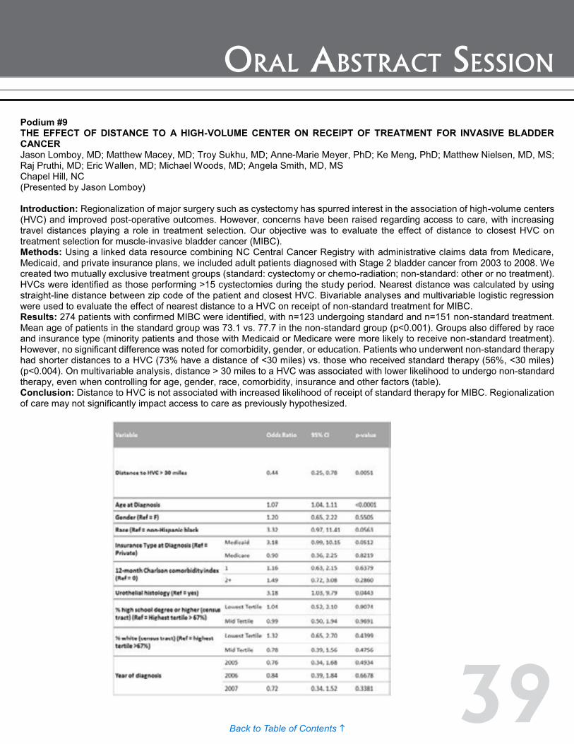
39Back to Table of Contents h
oral absTraCT session
Podium #9 THE EFFECT OF DISTANCE TO A HIGH-VOLUME CENTER ON RECEIPT OF TREATMENT FOR INVASIVE BLADDER CANCER Jason Lomboy, MD; Matthew Macey, MD; Troy Sukhu, MD; Anne-Marie Meyer, PhD; Ke Meng, PhD; Matthew Nielsen, MD, MS; Raj Pruthi, MD; Eric Wallen, MD; Michael Woods, MD; Angela Smith, MD, MS Chapel Hill, NC (Presented by Jason Lomboy) Introduction: Regionalization of major surgery such as cystectomy has spurred interest in the association of high-volume centers (HVC) and improved post-operative outcomes. However, concerns have been raised regarding access to care, with increasing travel distances playing a role in treatment selection. Our objective was to evaluate the effect of distance to closest HVC on treatment selection for muscle-invasive bladder cancer (MIBC). Methods: Using a linked data resource combining NC Central Cancer Registry with administrative claims data from Medicare, Medicaid, and private insurance plans, we included adult patients diagnosed with Stage 2 bladder cancer from 2003 to 2008. We created two mutually exclusive treatment groups (standard: cystectomy or chemo-radiation; non-standard: other or no treatment). HVCs were identified as those performing >15 cystectomies during the study period. Nearest distance was calculated by using straight-line distance between zip code of the patient and closest HVC. Bivariable analyses and multivariable logistic regression were used to evaluate the effect of nearest distance to a HVC on receipt of non-standard treatment for MIBC. Results: 274 patients with confirmed MIBC were identified, with n=123 undergoing standard and n=151 non-standard treatment. Mean age of patients in the standard group was 73.1 vs. 77.7 in the non-standard group (p<0.001). Groups also differed by race and insurance type (minority patients and those with Medicaid or Medicare were more likely to receive non-standard treatment). However, no significant difference was noted for comorbidity, gender, or education. Patients who underwent non-standard therapy had shorter distances to a HVC (73% have a distance of <30 miles) vs. those who received standard therapy (56%, <30 miles) (p<0.004). On multivariable analysis, distance > 30 miles to a HVC was associated with lower likelihood to undergo non-standard therapy, even when controlling for age, gender, race, comorbidity, insurance and other factors (table). Conclusion: Distance to HVC is not associated with increased likelihood of receipt of standard therapy for MIBC. Regionalization of care may not significantly impact access to care as previously hypothesized.
Podium #8 ACTIONABLE TARGETS IN PATIENTS WITH CISPLATIN-RESISTANT ADVANCED GERM CELL TUMORS Aditya Bagrodia, MD1; Samuel Kaffenberger, MD1; Byron Lee, MD1; William Lee, MD1; Eugene Cha, MD1; John Sfakianos, MD2; Gopa Iyer, MD1; Aijazuddin Syed, MD1; Ritika Kundra, MD1; Jana Eng, MD1; Michael Berger, MD1; Dean Bajorin, MD1; Joel Sheinfeld, MD1; George Bosl, MD1; Hikmat Al-Ahmadie, MD1; David Solit, MD1; Darren Feldman, MD1 1, Memorial Sloan Kettering Cancer Center, New York, NY; 2, Icahn School of Medicine at Mount Sinai, New York, NY (Presented by Aditya Bagrodia) Introduction: Approximately 30% of patients with advanced germ cell tumor (aGCT) will progress after first-line chemotherapy. Nearly half of these patients will die from progressive GCT. We describe potentially actionable mutations in a cohort of 76 patients with cisplatin-resistant (CR) aGCT through targeted next generation sequencing. Methods: Seventy-six patients with CR disease were sequenced with targeted sequencing of 341 cancer-related genes using the Memorial Sloan Kettering-Integrated Mutation Profiling of Actionable Cancer Targets (MSK-IMPACT) assay. Patients were categorized as CR if they met any of the following criteria: 1) incomplete response to first-line cisplatin-based chemotherapy; 2) nonteratomatous tumor progression after standard chemotherapy; 3) viable nonteratomatous GCT identified at post-chemo surgery. We grouped all somatic mutations observed into core signal transduction pathways or canonical cell functions to identify potential precise targets for therapy. Results: The majority of patients had testis vs. mediastinal primaries (n=64, 84% vs. n=12, 16%) and non-seminoma histology (n=64, 84%). International Germ Cell Cancer Collaborative Group risk group was good, intermediate, and poor in 34%, 13% and 53% of patients, respectively. 17 patients ultimately died of their disease. In total, 51 potentially actionable alterations were identified in 36/76 (47%) patients. Within the TP53 pathway, seven MDM2 amplifications and four MYCN amplifications that may sensitive to nutlin-3 inhibitors were identified. Within the receptor tyrosine kinase pathway, 3 KIT mutations, 1 KDR amplification, and 1 MET amplification were seen that may sensitive to tyrosine kinase inhibitors. Eleven KRAS mutations, 3 NRAS, mutations, 3 BRAF mutations, and 2 RAC1 mutations were see among the RAS pathway with preclinical data suggesting efficacy towards MEK or RAC1 inhibitors. Actionable targets were also among the PI3-kinase, WNT, and cell cycle pathways. Genes with potential targets with chromatin modifying or tumor suppressor functions were also seen. Conclusion: We describe actionable alterations that have the potential to guide treatment selection in a significant proportion of patients with platinum-resistant aGCT. Targeted sequencing of patients with CR aGCT may allow us to enrich future clinical trials with patients whose tumors harbor alterations in the drug target of interest.
40Back to Table of Contents h
posTer session i – summary
Poster Session I & ReceptionWednesday, December 2, 2015
4:30 p.m. – 6:00 p.m.Poster Walks
See page 60 for full abstracts
Poster #1 ROLE OF INTRAVESICAL GEMCITABINE AND MITOMYCIN C WITH MITOMYCIN C ALONE FOR HIGH RISK NON-MUSCLE INVASIVE BLADDER CARCINOMA-A RANDOMIZED PILOT STUDY Santosh Kumar MS, MCh, FRCS1; Kumar Jayant MD, MS, MRCS12; Shrawan Kumar Singh MS, Mch3; Mayank Mohan Agarwal MS, Mch, MRCS1; Swati Agrawal MD, MS, MRCOG14 1Asst Professor, Dept of Urology, Postgraduate institute of Medical Education & Research, Chandigarh India; 2Senior Resident, Sudha Hospital & Medical Research Center; 3Professor, Dept of Urology, Postgraduate institute of Medical Education & Research, Chandigarh India; 4Resident, Sudha Hospital & Medical Research Center, Kota, Raj, India (Presented by Kumar Jayant)
Poster #2 ROBOTIC AND OPEN RADICAL CYSTECTOMY: LESSONS FROM THE NATIONAL CANCER DATABASE Richard Matulewicz, MS, MD1; Vidit Sharma, MD2; Adarsh Manjunath, MD3; Jennifer Tse, BS3; Joshua Meeks, MD, PhD3; Shilajit Kundu, MD3 1Northwestern University, Feinberg School of Medicine; 2Mayo Clinic; 3Northwestern University Feinberg School of Medicine (Presented by Richard Matulewicz)
Poster #3 PERIOPERATIVE BLOOD TRANSFUSION IN RADICAL CYSTECTOMY: ANALYSIS OF THE NATIONAL SURGICAL QUALITY IMPROVEMENT PROGRAM DATABASE Wilson Sui, BA; Ifeanyi Onyeji, BA; Justin Matulay, MD; Maxwell James, BA; Marissa Velez, MD; Sven Wenske MD; G. Joel DeCastro, MD Columbia University Medical Center, Department of Urology, New York City, NY (Presented by Wilson Sui)
Poster #4 VASCULAR TARGETED PHOTODYNAMIC THERAPY (VTP) WITH WST-11 USING A URETEROSCOPIC APPROACH TO A PORCINE RENAL PELVIS IS SAFE AND UROTHELIAL CELL CARCINOMAS ARE SENSITIVE TO WST-11 VTP IN A MURINE MODEL Katie Murray, Renato Beluco Corradi Fonseca, Stephen LaRosa, Sylvia Jebiwott, Alex Somma, Govindarajan Srimathveeravalli, Kwanghee Kim, Sebastien Monette, Avigdor Scherz, Jonathan Coleman (Presented by Katie Murray)
Poster #5 CLINICAL UTILIZATION OF NEOADJUVANT CHEMOTHERAPY IN ELDERLY PATIENTS WITH BLADDER CANCER: OUTCOMES FROM A SINGLE INSTITUTION EXPERIENCE Andrew Leone, MD; Kamran Zargar-Shoshtari, MD, Gregory J Diorio, MD; Pranav Sharma, MD; Scott M. Gilbert, MD; Julio M. Powsang, MD; Wade J. Sexton, MD; Michael A. Poch, MD; Philippe E. Spiess, MD Moffitt Cancer Center, Tampa FL (Presented by Andrew Leone)
Poster #6 PATIENT-RELATED FACTORS SIGNIFICANTLY AFFECT THE PERFORMANCE OF URINE TESTS FOR BLADDER CANCER: AN EXAMPLE OF SPECTRUM EFFECTS. Thomas Longo, MD1; Ajay Gopalakrishna, BS1; Joseph Fantony, MD1; Richmond Owusu, MD2; Rajesh Dash, MD1; Brant Inman, MD1 1Duke University, Durham, NC; 2UCSD, San Diego, CA(Presented by Thomas Longo)
41Back to Table of Contents h
posTer session i – summary
Poster #7 ASSOCIATIONS BETWEEN HEALTH-RELATED QUALITY OF LIFE AND PHYSICAL ACTIVITY IN BLADDER CANCER SURVIVORS: A CROSS-SECTIONAL STUDY Ajay Gopalakrishna BS, BA; Joseph Fantony, MD, Thomas Longo, MD; Brant Inman, MD Duke University Medical Center, Durham, NC (Presented by Ajay Gopalakrishna)
Poster #8 EVALUATION OF AN EPIGENETIC PROFILE FOR THE DETECTION OF BLADDER CANCER IN HEMATURIA PATIENTS Kim van Kessel, MD1; Leander Van Neste, PhD2; Irene Lurkin1; Ellen Zwarthoff, PhD1; Wim Van Criekingel PhD2 1Department of Pathology, Erasmus Medical Center, Rotterdam, The Netherlands; 2MDXHealth, Inc., Irvine, CA (Presented by Kim van Kessel)
Poster #9 SOX-2 EXPRESSION IN PATIENTS WHO UNDERWENT RADICAL CYSTECTOMY FOR UROTHELIAL CARCINOMA OF THE BLADDER Charles Nottingham MD, MS1; Sanjay Patel, MD2; Peter Clark, MD3; David DeGraff, PhD4; Justin Gregg MD3; Blake Anderson, MD5; Gladell Paner, MD5; Donald Vander Griend, PhD5 1University of Chicago Medical Center, Chicago, IL; 2University of Oklahoma Health Sciences Center, Oklahoma City, OK; 3Vanderbilt University Medical Center, Nashville, TN; 4Pennsylvania State University College of Medicine, Hershey, PA; 5University of Chicago Medical Center, Chicago, IL (Presented by Charles Nottingham)
Poster #10 PREOPERATIVE ASYMPTOMATIC LEUKOCYTOSIS IN RADICAL CYSTECTOMY: ANALYSIS OF THE NATIONAL SURGICAL QUALITY IMPROVEMENT PROGRAM DATABASE Ifeanyi Onyeji, BA; Wilson Sui, BA; Justin Matulay, MD; Marissa Velez, MD Maxwell James, BS; Guarionex DeCastro, MD, MPH Columbia University Medical Center, Department of Urology, New York, NY (Presented by Ifeany Onyeji)
Poster #11 EFFECTS OF THE COMBINATION OF VASCULAR TARGETED PHOTODYNAMIC THERAPY AND ACTLA-4 IN A PRE CLINICAL UROTHELIAL CARCINOMA MODEL Renato Beluco Corradi, MD1; Stephen La Rosa2; Sylvia Jebiwott2; Katie Murray2; Alex Somma2; Avigdor Scherz3; Kwanghee Kim2; Jonathan Coleman2 1Memorial Sloan Kettering Cancer Center, New York, New YorkCancer Center, New York, New York; 2Memorial Sloan Kettering Cancer Center, New York, NY; 3Department of Plant Sciences, Weizmann Institute of Science, Rehovot, Israel Institution (Presented by Renato Beluco Corradi)
Poster #12 ADDITIONAL ADJUVANT CONVENTIONAL CHEMOTHERAPY IN PATIENTS PREVIOUSLY TREATED WITH NEOADJUVANT CHEMOTHERAPY AND RADICAL CYSTECTOMY: RETROSPECTIVE DESCRIPTION Kamran Zargar-Shoshtari, MD1; Michael Kongnyuy, MD2; Pranav Sharma, MD2; Mayer N. Fishman, MD2; Scott M. Gilbert, MD2; Michael A. Poch, MD2; Julio M. Powsang, MD2; Philippe E. Spiess, MD2; Jingsong Zhang, MD2; Wade J. Sexton, MD2 1Department of Genitourinary Oncology, H. Lee Moffitt Cancer Center and Research Institute, Tampa, FL; 2Department of Genitourinary Oncology, H. Lee Moffitt Cancer Center and Research Institute, Tampa, FL (Presented by Kamran Zargar-Shoshtari)
42Back to Table of Contents h
posTer session i – summary
Poster #13 THE RELATIONSHIP BETWEEN TRAVEL DISTANCE TO CYSTECTOMY AND LIKELIHOOD OF READMISSION Troy Sukhu, MD; Jason Lomboy, MD; Matthew Macey, MD; Anne-Marie Meyer, PhD; Ke Meng, PhD; Matthew Nielsen MD, MS; Raj Pruthi, MD; Eric Wallen, MD; Michael Woods, MD; Angela Smith, MD, MS Chapel Hill, NC (Presented by Troy Sukhu)
Poster #14 REPORTING BIAS LEADING TO DISCORDANT VENOUS THROMBOEMBOLISM RATES IN US VS. NON-US COUNTRIES FOLLOWING RADICAL CYSTECTOMY: A SYSTEMATIC REVIEW AND META-ANALYSIS Joseph Fantony, MD1; Ajay Gopalakrishna, BS2; Megan Van Noord Van Noord BS, MS2; Brant Inman MD, MSc2 1Duke University Durham, NC; 2Duke University Durham,NC (Presented by Joseph Fantony)
Poster #15 MALIGNANT URETEROINTESTINAL ANASTOMOTIC STRICTURE FOLLOWING RADICAL CYSTECTOMY: PATTERNS, RISK FACTORS, AND OUTCOMES Mary E. Westerman, MD1; Boyd R. Viers, MD1; R. Jeffrey Karnes, MD1; Robert Tarrell2; Prabin Thapa2; R. Houston Thompson, MD1; Matthew K. Tollefson, MD1; Stephen A. Boorjian, MD1 1Mayo Clinic Dept of Urology, Rochester, MN; 2Department of Health Sciences Research, Mayo Clinic, Rochester, MN(Presented by Mary E. Westerman)
Poster #16 VALIDATING THE BLADDER UTILITY SYMPTOM SCALE (BUSS): A MULTIATTRIBUTE HEALTH STATE CLASSIFICATION SYSTEM FOR BLADDER CANCER Nathan Perlis, MD, MSc1; Kirstin Boehme, MSc2; Munir Jamal, MD3; Karen Bremner2; Shabbir Alibhai, MD, MSc4; Antonio Finelli, MD, MSc1; Paul Ritvo, PhD5; Murray Krahn, MD, MSc6; Girish Kulkarni, MD, PhD1 1Division of Urology, University Health Network, University of Toronto, Toronto, ON; 2University Health Network, Toronto, ON; 33Trillium Health Partners, Mississauga, ON; 4Division of Medicine, University Health Network, University of Toronto, Toronto, ON; 5York University and Cancer Care Ontario, Toronto, ON; 6Toronto Health Economics and Technology Assessment (THETA) Collaborative, University of Toronto, and University Health Network, Toronto, ON(Presented by Nathan Perlis)
Poster #17 ONCOLOGIC SURVEILLANCE FOLLOWING RADICAL CYSTECTOMY: AN INDIVIDUALIZED RISK-BASED APPROACH. Suzanne Stewart-Merrill, MD1; Stephen Boorjian, MD2; R. Houston Thompson, MD2; Sarah Psutka, MD3; John Cheville, MD2; Prabin Thapa2; Matthew Tollefson, MD2; Igor Frank, MD2 1Hershey, PA; 2Rochester, MN; 3Chicago, IL (Presented by Suzanne Stewart-Merrill)
Poster #18 IMPACT OF HEALTH LITERACY ON SURGICAL OUTCOMES FOLLOWING RADICAL CYSTECTOMY Kristen Scarpato, MD; Stephen Kappa, MD; Kathyrn Goggins, MPH; Sam Chang, MD, MBA; Daniel Barocas, MD, MPH; Joseph Smith, MD; Peter Clark, MD; David Penson, MD, MPH; Matthew Resnick, MD, MPH; Sunil Kripalani, MD; Kelvin Moses, MD, PHD Vanderbilt University (Presented by Kristen Scarpato)
43Back to Table of Contents h
posTer session i – summary
Poster #19 FIRST-LINE RANDOMIZED PHASE 2 STUDY OF GEMCITABINE/CISPLATIN PLUS APATORSEN OR PLACEBO IN PATIENTS WITH ADVANCED BLADDER CANCER: THE INTERNATIONAL BOREALIS-1™ TRIAL Daniel Petrylak, MD1; Bernhard Eigl, MD2; Elzbieta Senkus, MD3; Yohann Loriot, MD4; Przemyslaw Twardowski, MD5; Daniel Castellano, MD6; Normand Blais, MD7; Srikala Sridhar, MD8; Cora Sternberg, MD9; Margitta Retz, MD10; Brent Blumenstein, PhD11; Cindy Jacobs, PhD, MD12; Patricia Stewart, MD12; Joaquim Bellmunt, MD13 1Yale School of Medicine, New Haven, CT; 2British Columbia Cancer Agency, Vancouver, BC; 3Medical University, Gdansk, Poland; 4Institut Gustave Roussy, Villejuif, France; 5City of Hope National Medical Center, Duarte, CA; 6Hospital Universitario 12 de Octubre, Madrid, Spain; 7Université de Montréal-Hospital Notre-Dame, Montréal, QC; 8Princess Margaret Hospital, Toronto, ON; 9San Camillo Forlanini Hospital, Rome, Italy; 10Technischen Universität München, München, Germany; 11Trial Architecture Consulting, Washington, DC; 12OncoGenex Pharmaceuticals, Inc., Bothell, WA; 13Dana-Farber Cancer Institute, Boston, MA (Presented by Daneil Petrylak)
Poster #20 ONCOLOGIC OUTCOMES AFTER ANTERIOR EXENTERATION FOR MUSCLE INVASIVE BLADDER CANCER IN WOMEN Justin Gregg, MD1; Curran Emeruwa, BS2; Johnson Wong, BS2; Matthew Resnick, MD, MPH1; Daniel Barocas, MD, MPH1; Michael Cookson, MD, MMHC3; Sam Chang, MD1; David Penson, MD, MPH1; Joseph Smith, MD1; Kristen Scarpato, MD, MPH1; Kelvin Moses, MD, PhD1 1Vanderbilt University Medical Center, Nashville, TN; 2Meharry Medical College, Nashville, TN; 3University of Oklahoma College of Medicine, Oklahoma City, OK (Presented by Justin Gregg)
Poster #21 QUANTITATIVE PROTEOMIC ANALYSIS OF UROLOGIC BLADDER CANCER CELLS FOLLOWING TREATMENT WITH HISTONE DEACETYLASE INHIBITORS Quentin Li, MD, PhD1; Jian-Jiang Hao, PhD2; Zheng Zhang, MD2; Reema Railkar, PhD1; Iawen Hsu, PhD1; Adam Metwalli, MD1; Piyush Agarwal, MD1 1UOB, CCR, NCI, NIH, Bethesda, MD 20892; 2Poochon Scientific, Frederick, MD 21704(Presented by Quentin Li)
Poster #22 TARGETING THE GLYCOME IN CISPLATIN-RESISTANT BLADDER CANCER WITH A NATURALLY OCCURRING PARASITE-HOST ANCHOR PROTEIN Roland Seiler, MD1; Htoo Z. Oo, PhD2; Sherry S. Lee, BSc2; Mette O. Agerbaek, PhD3; Jamie R. Rich, PhD4; Thomas M. Clausen, PhD3; John Babcook, PhD4; Peter C. Black, MD2; Ali Salanti, PhD3; Mads Daugaard, PhD2 1Department of Urologic Sciences, University of British Columbia, Vancouver, BC; 2Department of Urologic Sciences Vancouver Prostate Centre; 3Faculty of Health Sciences, University of Copenhagen; 4The Centre for Drug Research and Development, Vancouver, BC (Presented by Roland Seiler)
Poster #23 THE IMPACT OF THE CURRENT WORLDWIDE SHORTAGE OF BACILLE CALMETTE-GUERIN FOR THE TREATMENT OF NON-MUSCLE INVASIVE BLADDER CANCER AT A TERTIARY CARE CENTER: THE COLUMBIA EXPERIENCE Jamie Pak, BA; Wilson Sui, BA; Sven Wenske, MD; G. Joel DeCastro, MD; David Weiner, MD; Nicholas Romas, MD; Mitchell Benson, MD; James McKiernan, MD Columbia University Medical Center (Presented by Jamie Pak)
44Back to Table of Contents h
posTer session i – summary
Poster #24 PROGNOSTIC VALUE OF NUMBER OF LYMPH NODES REMOVED DURING NEPHROURETERECTOMY FOR UPPER TRACT UROTHELIAL CARCINOMA Andrew Winer, MD1; Emily Vertosick, MS1; Renato Beluco, MD1; Sigrid Carlsson, MD, PhD1; Samuel Kaffenberger, MD1; Aditya Bagrodia, MD1; Katie Murray, MD1; Daniel Sjoberg, MS1; Alexander Sankin, MD1; John Sfakianos, MD2; Eugene Cha, MD1; Guido Dalbagni, MD1; Jonathan Coleman, MD1 1MSKCC, New York, NY; 2Mount Sinai, New York, NY (Presented by Andrew Winer)
Poster #25CARCINOGEN INDUCED UROTHELIAL CARCINOMA IN RATS DISPLAY AN IMMUNOGENIC PHENOTYPE COMPARABLE TO HUMANS Max Kates, MD; Nikolai Sopko, MD, PhD; Hotaka Matsui, MD; Allison Reinhardt; Djahida Bedja; Xiaopu Liu, BA; Leonardo Reis MD, PhD; Noah Hahn, MD; Alex Baras, MD, PhD; Brian Simons, DVM, PhD; Christina Kochel, PhD; Charles Drake, MD, PhD; Trinity Bivalacqua, MD, PhD James Buchanan Brady Urological Institute The Johns Hopkins Medical Institutions (Presented by Max Kates)
Poster #26 PREDICTING RECURRENCE AFTER RADICAL CYSTECTOMY AMONG PATIENTS WHO EXPERIENCE COMPLETE PATHOLOGIC RESPONSE TO NEOADJUVANT CHEMOTHERAPY William P. Parker, MD1; Phil L. Ho, MD2; Stephen A. Boorjian, MD1; Jonathan J. Melquist, MD2; Prabin Thapa, PhD1; Jeffrey M. Holzbeierlein, MD3; Igor Frank, MD1; Ashish M. Kamat, MD2; Eugene K. Lee, MD3 1Mayo Clinic; 2MD Anderson Cancer Center; 3University of Kansas Medical Center (Presented by William P. Parker)
Poster #27 NUTRITIONAL STATUS AND MAJOR ABDOMINAL SURGERIES: 30-DAY POSTOPERATIVE COMPLICATIONS Nicola Pavan, MD1; Carmen C. Mir, MD, PhD2; Chad R. Ritch, MD, MBA2; Samarpit Rai, MD2; Nachiketh Soodana-Prakash MD, MS2; Raymond R. Balise, PhD3; Carlo Trombetta, MD4; Dipen J. Parekh, MD2; Mark L. Gonzalgo MD, PhD2 1Department of Urology, University of Miami Leonard M. Miller School of Medicine, Miami, FL and Urology Clinic, Department of Medical, Surgical and Health Science, University of Trieste, Italy; 2Department of Urology, University of Miami Leonard M. Miller School of Medicine, Miami, FL; 3Division of Biostatistics, Department of Public Health Sciences, University of Miami Leonard M. Miller School of Medicine, Miami, FL; 4Urology Clinic, Department of Medical, Surgical and Health Science, University of Trieste, Italy (Presented by Samarpit Rai)
Poster #28 FREQUENCY AND CLINICAL IMPLICATIONS OF PATHOLOGIC COMPLETE RESPONSES AT CYSTECTOMY FOR MUSCLE-INVASIVE BLADDER CANCER WITHOUT PRIOR NEOADJUVANT CHEMOTHERAPY Kristian Stensland, MD1; Russell McBride, PhD, MPH2; John Sfakianos, MD2; Reza Mehrazin, MD2; William Oh, MD2; Matthew Galsky, MD2 1Lahey Clinic, Burlington, MA; 2Icahn School of Medicine at Mount Sinai, New York, NY (Presented by Kristian Stensland)
Poster #29 EMPIRIC TREATMENT OF CLOSTRIDIUM DIFFICILE CARRIERS AT TIME OF CYSTECTOMY: PRELIMINARY OUTCOMES Joseph M. Jacob, MD; Hristos Z. Kaimakliotis, MD; Nick W. Liu, MD; Jane S. Cho, MD; M. Francesca Monn, MD; Benjamin R. Judge; Clint K. Cary, MD; Timothy A. Masterson, MD; Thomas A. Gardner, MD; Richard S. Foster, MD; Richard Bihrle, MD; Michael O. Koch, MD Indiana University Medical Center (Presented by Joseph M. Jacob)
45Back to Table of Contents h
posTer session i – summary
Poster #30 FACTORS ASSOCIATED WITH SUICIDE IN PATIENTS WITH GENITOURINARY MALIGNANCIES Zachary Klaassen, MD1; Rita P. Jen, MD, MPH1; John M. DiBianco, MD1; Lael Reinstatler, MD, MPH1; Qiang Li MD, PhD1; Rabii Madi, MD1; Ronald W. Lewis, MD1; Arthur M. Smith, MD1; Durwood E. Neal, Jr., MD, FACS1; Kelvin A. Moses, MD, PhD2; Martha K. Terris, MD1 1Medical College of Georgia - Georgia Regents University, Augusta, GA; 2Department of Urological Surgery, Vanderbilt University Medical Center, Nashville, TN (Presented by Zachary Klaassen)
Poster #31 DETERMINING PROSTATE CANCER RISK STRATA USING ONLY GLEASON SCORE AND PSA Justin Gregg, MD1; Max Lang, MD1; Sam Chang, MD1; Peter Clark, MD1; Michael Cookson, MD, MMHC2; S. Duke Herrell, MD1; David Penson, MD, MPH1; Matthew Resnick, MD, MPH1; Joseph Smith, MD1; Daniel Barocas, MD, MPH1 1Vanderbilt University Medical Center, Nashville, TN; 2University of Oklahoma College of Medicine, Oklahoma City, OK (Presented by Justin Gregg)
Poster #32 THE USE OF NATURAL LANGUAGE PROCESSING TO DETERMINE PROSTATE CANCER CLINICAL RISK STRATA Justin Gregg, MD; Max Lang, BS; Lucy Wang, BS; David Penson, MD, MPH; Matthew Resnick, MD, MPH; Jeremy Warner, MD, MS; Daniel Barocas, MD, MPH Vanderbilt University Medical Center, Nashville, TN (Presented by Justin Gregg)
Poster #33 COMPETITION IN CANCER CARE: ADAPTING TO CHANGES IN PAYMENT Brock O’Neil, MD1; Amy Graves, SM, MPH2; Daniel Barocas, MD, MPH2; Sam Chang, MD2; David Penson, MD, MPH2; Matthew Resnick, MD, MPH2 1Vanderbilt University; 2Vanderbilt University, Nashville, TN (Presented by Brock O’Neil)
Poster #34 DIFFERENCES IN PROSTATE SPECIFIC ANTIGEN TESTING AMONG UROLOGISTS AND PRIMARY CARE PROVIDERS IN THE UNITED STATES FOLLOWING THE 2011 USPSTF RECOMMENDATIONS Nawar Hanna, MD1; Micheal E. Zavaski, MD1; Christian Meyer, MD1; Jesse D. Sammon, MD2; Soham Gupta, MD2; Maxine Sun, PhD1; Quoc-Dien Trinh, MD1 1Brigham and Women’s Hospital, Boston, MA; 2Vattikuti Urology Institute, Detroit, MI (Presented by Nawar Hanna)
Poster #35 THE IMPACT OF MEDICAL COMORBIDITIES ON RENAL FUNCTION FOLLOWING RADICAL OR PARTIAL NEPHRECTOMY Michael J. Vacchio, MD; Andrew G. Winer, MD; Emily C. Zabor, PhD; A. Ari Hakimi, MD; Paul Russo, MD; Jonathan A. Coleman, MD; Edgar A. Jaimes, MD Memorial Sloan-Kettering Cancer Center New York, NY (Presented by Michael J. Vacchio)
Poster #36 RANDOMIZED DOUBLE BLINDED PLACEBO CONTROLLED TRIAL OF SILDENAFIL FOR RENOPROTECTION PRIOR TO HILAR CLAMPING IN PATIENTS UNDERGOING ROBOTIC ASSISTED LAPAROSCOPIC PARTIAL NEPHRECTOMY Louis Spencer Krane, MD1; Charles C. Peyton, MD2; Ashok K. Hemal, MD, MCh2 1National Cancer Institute, Bethesda MD; 2Wake Forest Baptist Health, Winston Salem NC (Presented by Louis Spencer Krane)
46Back to Table of Contents h
posTer session i – summary
Poster #37 TRICHLOROETHYLENE IS ASSOCIATED WITH KIDNEY CANCER MORTALITY: A POPULATION-BASED ANALYSIS Shaheen Alanee, MD, MPH, MBA; Joseph Clemons; Whitney Zahnd; Dan Sadowski; Danuta Dynda, MD Southern Illinois University School of Medicine, Springfield, IL (Presented by Shaheen Alanee)
Poster #38 UPPER TRACT UROTHELIAL CARCINOMA LOCATED IN THE RENAL PELVIS HAS WORSE CLINICAL OUTCOMES COMPARED TO THE URETER: A POPULATION-BASED ANALYSIS Zachary Klaassen, MD; Benjamin T. Harper; Rita P. Jen, MD, MPH; Grace Yaguchi; Ross Everett, MPH; John M. DiBianco, MD; Martha K. Terris, MD; Rabii Madi, MD Medical College of Georgia - Georgia Regents University, Augusta, GA (Presented by Zachary Klaassen)
Poster #39 RENAL CELL TUMOR-MEDIATED CONVERSION OF NATURAL KILLER CELLS TO A PROANGIOGENIC PHENOTYPE BY TRANSFORMING GROWTH FACTOR-Β AND HYPOXIA Yue Guan1; Christopher Chambers1; Britnie James2; Purba Singh1; Donald Reed1; Kathy Robinson3; Shaheen Alanee MD, MPH, MBA4; Thomas Griffith2; Donald Torry1; Andrew Wilber1 1Department of Medical Microbiology, Immunology and Cell Biology, Southern Illinois University School of Medicine, Springfield, IL; 2Department of Microbiology, Immunology and Cancer Biology Graduate Program and Department of Urology, University of Minnesota, Minneapolis, MN; 3Simmons Cancer Institute, Springfield, IL; 4Southern Illinois University School of Medicine, Springfield, IL (Presented by Shaheen Alanee)
Poster #40 CHARACTERISTICS OF ISOLATED LOW GRADE UPPER TRACT UROTHELIAL CARCINOMA: SESSILE TUMOR ARCHITECTURE IS ASSOCIATED WITH ADVERSE ONCOLOGIC OUTCOMES Ryan Hutchinson, MD; Nirmish Singla, MD; Ahmed Haddad, MD, PhD; Vitaly Margulis, MD UTSW, Dallas, TX (Presented by Ryan Hutchinson)
Poster #41 INTEGRIN SIGNALING POTENTIATES TRANSFORMING GROWTH FACTOR BETA 1 DEPENDENT DOWN REGULATION OF E-CADHERIN EXPRESSION- IMPLICATIONS FOR EPITHELIAL TO MESENCHYMAL TRANSITION IN RENAL CELL CARCINOMA Ryan Hutchinson, MD; Boris Feldkoren, PhD; Yury Rapaport, MD; Vitaly Margulis, MD UTSW, Dallas, TX (Presented by Ryan Hutchinson)
Poster #42 RADIOGRAPHIC SIZE OF RETROPERITONEAL LYMPH NODES PREDICTS PATHOLOGIC NODAL INVOLVEMENT FOR PATIENTS UNDERGOING RADICAL NEPHRECTOMY FOR RENAL CELL CARCINOMA: DEVELOPMENT OF A RISK PREDICTION MODEL Boris Gershman, MD; Naoki Takahashi, MD; Daniel Moreira, MD; R. Houston Thompson, MD; Stephen Boorjian, MD; Christine Lohse, MS; Brian Costello, MD; John Cheville, MD; Bradley Leibovich, MD Mayo Clinic, Rochester, MN (Presented by Boris Gershman)
47Back to Table of Contents h
posTer session i – summary
Poster #43 KIDNEY CANCER IN RURAL ILLINOIS: LOWER INCIDENCE YET HIGHER MORTALITY RATES Daniel Sadowski, MD, MPhil1; Scott Geiger, BS1; Georgia Mueller, MS2; Whitney Zahnd, MS2; Shaheen Alanee, MD, MPH1; Kevin McVary, MD1 1Southern Illinois University School of Medicine, Division of Urology, Springfield, Illinois; 2Southern Illinois University School of Medicine, Center for Clinical Research, Springfield, IL(Presented by Daniel Sadowski)
Poster #44 THE PROGNOSTIC IMPACT OF A POSITIVE VASCULAR MARGIN IN PT3 CLEAR CELL RENAL CARCINOMAS Nick Liu, MD1; James Wren, MD2; Emily Vertosick, MS2; Justin Lee, MD2; Nick Power, MD2; Simon Kimm, MD2; Dan Sjoberg, MD2; Paul Russo, MD2; Coleman Jonathan, MD2 1New York; 2Memorial Sloan-Kettering Cancer Center (Presented by Nick Liu)
Poster #45 INFLAMMATORY MARKERS PREDICT ADVERSE PATHOLOGICAL FEATURES AND SURVIVAL IN PATIENTS WITH LOCALIZED RENAL CELL CARCINOMA Matthew Meissner, MD; Ahmed Haddad, MD, PhD; Kunj Sheth, MD; Vitaly Margulis, MD UT Southwestern Medical Center Dallas, TX (Presented by Matthew Meissner)
Poster #46 PROSPECTIVE EVALUATION OF 99MTC-SESTAMIBI SPECT/CT FOR THE DIAGNOSIS OF RENAL ONCOCYTOMAS AND HYBRID ONCOCYTIC/CHROMOPHOBE TUMORS Michael Gorin, MD1; Steven Rowe, MD, PhD2; Alex Baras, MD, PhD3; Lilja Solnes, MD2; Mark Ball, MD1; Phillip Pierorazio, MD1; Christian Pavlovich, MD1; Jonathan Epstein, MD3; Mehrbod Javadi, MD2; Mohamad Allaf, MD1 1The James Buchanan Brady Urological Institute and Department of Urology, Johns Hopkins University School of Medicine, Baltimore, MD; 2The Russell H. Morgan Department of Radiology and Radiological Science, Johns Hopkins University School of Medicine, Baltimore, MD; 3Department of Pathology, Johns Hopkins University School of Medicine, Baltimore, MD(Presented by Michael Gorin)
Poster #47 DEVELOPMENT OF A CLEAR CELL RENAL CELL CARCINOMA XENOGRAFT MODEL: SURGICAL TISSUE VS. BIOPSY TISSUE Yiyu Dong, PhD1; Brandon J. Manley, MD2; Abraham A. Hakimi, MD2; Paul Russo, MD2; Jonathan Coleman, MD2; James J. Hsieh, MD, PhD2 1Sloan Kettering Institute, New York, NY; 2Memorial Sloan Kettering Cancer Center, New York, NY. (Presented by Brandon J. Manley)
Poster #48 FACTORS PREDICTING BLOOD LOSS AND POSITIVE MARGINS AT TIME OF PARTIAL NEPHRECTOMY: ANALYSIS OF A CONTEMPORARY COHORT Harras Zaid, MD; Stephen Boorjian, MD; William Parker, MD; Christine Lohse, MS; John Cheville, MD; Bradley Leibovich, MD; R. Houston Thompson, MD Mayo Clinic, Rochester MN (Presented by Harras Zaid)
48Back to Table of Contents h
posTer session i – summary
Poster #49 EVALUATION OF SURGICAL COMPLICATIONS FROM A CONTEMPORARY SERIES OF PARTIAL NEPHRECTOMY PATIENTS Harras Zaid, MD; R. Houston Thompson, MD; William Parker, MD; Christin Lohse, MS; John Cheville, MD; Stephen Boorjian, MD; Bradley Leibovich, MD Mayo Clinic, Rochester MN (Presented by Harras Zaid)
Poster #50 SURGICAL MANAGEMENT OF RECURRENT VENOUS TUMOR THROMBUS AFTER PRIOR NEPHRECTOMY William P. Parker, MD; Stephen A. Boorjian, MD; Harras B. Zaid, MD; Bradley C. Leibovich, MD; R. Houston Thompson, MD Mayo Clinic (Presented by William P. Parker)
Poster #51 PREOPERATIVE PROTEINURIA IS AN INDEPENDENT PREDICTOR OF SURVIVAL FOLLOWING RENAL CANCER SURGERY Joseph Zabell, MD1; Zhiling Zhang, MD1,2; Juping Zhao, MD1,3; Jianbo Li, MS4; Diego Aguilar Palacios, MD1; Sevag Demirjian, MD5; Steven C. Campbell, MD, PhD1 1Department of Urology, Center for Urologic Oncology, Glickman Urological & Kidney Institute, Cleveland Clinic, Cleveland, OH; 2Department of Urology, Sun Yat-Sen University Cancer Center, Guangzhou, China; 3Shanghai JiaoTong University School of Medicine, Ruijin Hospital, Department of Urology, Shanghai, China; 4Department of Quantitative Health Sciences, Cleveland Clinic, Cleveland, OH; 5Department of Nephrology and Hypertension, Glickman Urological & Kidney Institute, Cleveland Clinic, Cleveland, OH (Presented by Joseph Zabell, MD)
Poster #52 SELECT CONCURRENT CHROMOSOME 3P MUTATIONS PREDICT WORSE OVERALL SURVIVAL IN CLEAR CELL RENAL CARCINOMA IN THE CANCER GENOME ATLAS Christopher Keith; Michael Rossi, PhD; Rebecca Arnold, PhD; John Petros, MD (Presented by Christopher Keith)
Poster #53 RESECTION TECHNIQUES FOR NEPHRON SPARING SURGERY VARY: ANALYSIS OF A PROSPECTIVELY COLLECTED MULTI-INSTITUTIONAL INTERNATIONAL COHORT HARNESSING THE SURFACE–INTERMEDIATE–BASE (S.I.B.) MARGIN SCORE Alexander Kutikov; Riccardo Campi, MD1; Miki Haifler, MD2; Robert G. Uzzo, MD2; Marco Carini, MD1; Andrea Minervini, MD1 1University of Florence, Italy; 2Fox Chase Cancer Center (Presented by Alexander Kutikov)
Poster #54 ACUTE KIDNEY INJURY AFTER PARTIAL NEPHRECTOMY: ROLE OF PARENCHYMAL MASS REDUCTION AND ISCHEMIA AND IMPACT ON SUBSEQUENT FUNCTIONAL RECOVERY Zhiling Zhang, MD1,2; Juping Zhao, MD1,3; Erick Remer, MD4; Jianbo Li, MS5; Sevag Demirjian, MD1; Joseph Zabell, MD1; Steven C. Campbell, MD, PhD1 1Glickman Urological and Kidney Institute, Cleveland Clinic, Cleveland, OH; 2Sun Yat-Sen University Cancer Center, Department of Urology, Guangzhou, China; 3Shanghai JiaoTong University School of Medicine, Ruijin Hospital, Department of Urology, Shanghai, China; 4Imaging Institute, Cleveland Clinic, Cleveland, OH; 5Department of Quantitative Health Sciences, Cleveland Clinic, Cleveland, OH (Presented by Zhiling Zhang)
49Back to Table of Contents h
posTer session i – summary
Poster #55 VENOUS THROMBOEMBOLISM FOLLOWING NEPHRECTOMY: THIRTY DAY INCIDENCE AND RISK FACTORS FROM NATIONAL MULTI-INSTITUTIONAL DATA Richard S. Matulewicz, MD1; Yousef Al-shraideh, MD2; Brian Trihn, MD3; John Oliver Delancey, MD3; Irene Helenowski, PhD4; Borko Jovanovic, PhD4; Shilajit Kundu, MD3 1Northwestern Feinberg/Urology; 2Chicago; 3Northwestern Feinberg/Ur; 4Northwestern Feinberg/Biostatistics (Presented by Yousef Al-shraideh)
Poster #56 PILOT STUDY EVALUATING PSMA-TARGETED 18F-DCFPYL PET/CT IMAGING OF METASTATIC CLEAR CELL RENAL CELL CARCINOMA Michael Gorin, MD1; Steven Rowe, MD2; Hans-Joerg Hammers, MD, PhD3; M. Som Javadi, MD2; Hazem Hawasli, MD2; Zsolt Szabo, MD, PhD2; Steve Cho, MD4; Martin Pomper, MD, PhD2; Mohamad Allaf, MD1 1The James Buchanan Brady Urological Institute and Department of Urology, Johns Hopkins University School of Medicine, Baltimore, MD; 2The Russell H. Morgan Department of Radiology and Radiological Science, Johns Hopkins University School of Medicine, Baltimore, MD; 3Department of Medical Oncology, Sidney Kimmel Comprehensive Cancer Center, Johns Hopkins University School of Medicine, Baltimore, MD; 4Department of Radiology, University of Wisconsin School of Medicine and Public Health, Madison, WI (Presented by Michael Gorin)
Poster #57 CLINICAL, PATHOLOGIC AND GENOMIC PROFILES OF EXCEPTIONAL RESPONDERS TO ANTI-PD1 THERAPY IN RENAL CELL CARCINOMA Mark Ball, MD1; Michael Johnson, MD2; Michael Gorin, MD2; Maria Rodriguez, MD2; Luis Diaz, MD2; Bert Vogelstein, MD2; Kenneth Kinzler, PhD2; Michael Haffner, MD2; Luigi Marchionni, MD2; George Netto, MD2; Charles Drake, MD, PhD2; Mohamad Allaf, MD2 1Johns Hopkins, Baltimore, MD; 2Johns Hopkins, Baltimore, MD (Presented by Mark Ball)
Poster #58 CIRCULATING TUMOR DNA AS A BIOMARKER IN ADVANCED RENAL CELL CARCINOMA Mark Ball, MD1; Michael Gorin, MD2; Gunes Gunter, MD2; Phillip Pierorazio, MD2; George Netto, MD2; Channing Paller, MD2; Hans Hammers, MD2; Luis Diaz, MD2; Mohamad Allaf, MD2 1Johns Hopkins, Baltimore, MD; 2Johns Hopkins, Baltimore, MD (Presented by Mark Ball)
Poster #59 DISCRIMINATING IPSI AND CONTRA-LATERAL COMPENSATORY RENAL GROWTH (CRG) AFTER PARTIAL NEPHRECTOMY- ARE BOTH KIDNEYS ALIKE? A RAT MODEL Barak Rosenzweig, MD1; Raya Eilam, PhD2; Alon Harmelin, BVSc, MRCVS, Dip ECLAM2; Jacob Ramon, MD1 1Department of Urology, The Chaim Sheba Medical Center, Tel Hashomer, Ramat-Gan, Affiliated to Sackler School of Medicine, Tel-Aviv University, Israel; 2Dept. of Veterinary Resources, The Weizmann Institute of Science, Rechovot, Israel (Presented by Barak Rosenzweig)
Poster #60 PD-1 EXPRESSION ON CLASSICAL MONOCYTES (CM) IS AN INDEPENDENT PREDICTOR OF CANCER SPECIFIC SURVIVAL IN CLEAR CELL RENAL CARCINOMA (CCRCC) Mohammed Haseebuddin, Alexander MacFarlane IV, Karen Ruth, Robert Uzzo, Elizabeth Plimack, Mowafaq Jillab, Essel Al-Saleem, Tahseen Al-Saleem, Kerry Campbell (Presented by Mohammed Haseebuddin)

50Back to Table of Contents h
posTer session i – summary
Poster #61 MANAGEMENT OF A MULTIDISCIPLINARY PHASE 3 CLINICAL TRIAL IN PATIENTS WITH SYNCHRONOUS MRCC (ADAPT) USING A REGIONAL CHAMPION MODEL IN THE SOCIETY OF UROLOGIC ONCOLOGY-CLINICAL TRIALS CONSORTIUM Brian Lane, MD, PhD1; Joan Chiaviello, RN2; Gennady Bratslavsky, MD3,4; Christopher Wood, MD5; Thomas Gardner, MD3,6; Jason Gee, MD3,7; Jeff Holzbeierlein, MD3,8; William Huang, MD3,9; Larry Karsh, MD3,10; Alexander Kutikov, MD3,11; Will Lowrance, MD3,12; Viraj Master, MD3,13; Marc Smaldone, MD3,11; Robert Uzzo, MD3,11; The Adapt Study Group3 1Spectrum Health Hospital, Grand Rapids, MI; 2Society of Urologic Oncology-Clinical Trials Consortium (SUO-CTC), Chicago, IL; 3Society of Urologic Oncology-Clinical Trials Consortium (SUO-CTC); 4SUNY Upstate Medical University, Syracuse, NY; 5Society of Urologic Oncology - Clinical Trials Consortium (SUO-CTC), MD Anderson Cancer Center, Houston, TX; 6Indiana University Medical Center; 7Lahey Clinic, Burlington, MA; 8University of Kansas Medical Center, KS; 9New York University School of Medicine, NY; 10The Urology Center of Colorado, Denver, CO; 11Fox Chase Cancer Center, Philadelphia, PA; 12Huntsman Cancer Hospital, Salt Lake City, Utah; 13Emory University, Atlanta GA (Presented by Brian Lane)
Poster #62 MULTIMODALITY APPROACH FOR METASTATIC RENAL CELL CARCINOMA IN A MODERN COHORT Scott Pate, MD1; Margaret Higgins, BA2; William Parker, MD1; Jeffrey Holzbeierlein, MD1; Moben Mirza, MD1; Peter Van Veldhuizen, MD1; Eugene Lee, MD1 1University of Kansas Medical Center; 2University of Kansas School of Medicine (Presented by Scott Pate)
Poster #63 MET INHIBITION IN CLEAR CELL RENAL CELL CARCINOMA Zuoquan Xie, PhD1; Young Lee, PhD2; Marta Boeke, PhD1; Lucia Jilaveanu, MD, PhD3; Donald Bottaro, PhD2; Harriet Kluger, MD3; Brian Shuch, MD4 1Yale Urology; 2NCI Urologic Oncology Branch; 3Yale Medical Oncology; 4Yale (Presented by Brian Shuch)
Poster #64 SINGLE-INSTITUTIONAL ANALYSIS OF PATIENTS WITH CLEAR-CELL PAPILLARY RENAL CELL CARCINOMA Jozefina Casuscelli, MD; Andrew Winer, MD; Eduard Reznik, PhD; Jianing Xu, PhD; Brandon Manley, MD; Jyoti Chouhan, MD; Paul Russo, MD; Jonathan Coleman, MD; Victor Reuter, MD; Satish Tickoo, MD; James Hsieh, MD; Abraham Hakimi, MD Memorial Sloan Kettering Cancer Center, New York, NY (Presented by Jozefina Casuscelli)
Poster #65 PREVALENCE AND RACE-SPECIFIC CHARACTERISTICS OF PATIENTS MEETING REFERRAL CRITERIA FOR GENETIC COUNSELING FOR HEREDITARY KIDNEY CANCER SYNDROMES BASED ON TUMOR PATHOLOGY Hong Truong, MS, MD1; Leonard Gomella, MD1; Edouard Trabulsi, MD1; Costas Lallas, MD1; William Kelly, MD2; Veda Giri, MD2 1Department of Urology, Sidney Kimmel Cancer Center, Thomas Jefferson University, Philadelphia, PA; 2Department of Medical Oncology, Sidney Kimmel Cancer Center, Thomas Jefferson University, Philadelphia, PA (Presented by Hong Truong)
Poster #66 CLINICAL SIGNIFICANCE OF P53 AND P16INK4A STATUS IN A CONTEMPORARY NORTH AMERICAN PENILE CARCINOMA COHORT Kamran Zargar-Shoshtari, MD1; Philippe E. Spiess, MD1; Anders E. Berglund, PhD2; Pranav Sharma, MD1; Julio M. Powsang, MD1; Anna Giuliano, PhD3; Anthony M. Magliocco, MD4; Jasreman Dhillon, MD4 1Department of Genitourinary Oncology, H. Lee Moffitt Cancer Center and Research Institute, Tampa, FL; 2Department of Biomedical Informatics, H. Lee Moffitt Cancer Center and Research Institute, Tampa, FL; 3Center for Infection Research in Cancer, Lee Moffitt Cancer Center and Research Institute, Tampa, FL; 4Department of Pathology, H. Lee Moffitt Cancer Center and Research Institute, Tampa, FL (Presented by Kamran Zargar-Shoshtari)
51Back to Table of Contents h
posTer session i – summary
Poster #67 RACIAL AND ECONOMIC DISPARITIES IN THE TREATMENT OF PENILE SQUAMOUS CELL CARCINOMA: Results: FROM THE NATIONAL CANCER DATABASE Pranav Sharma, MD; Kenan Ashouri, MS; Kamran Zargar-Shoshtari, MD; Adam Luchey, MD; Philippe Spiess, MD Moffitt Cancer Center, Tampa, FL (Presented by Pranav Sharma)
Poster #68 ADJUVANT RADIATION THERAPY IS ASSOCIATED WITH DECREASED DISEASE RECURRENCE AFTER PELVIC LYMPH NODE DISSECTION IN PENILE CANCER PATIENTS WITH POSITIVE PELVIC LYMPH NODES: A MULTI-INSTITUTIONAL STUDY Gregory Diorio, DO1; Pranav Sharma, MD1; Andrew Leone, MD1; Kamran Zargar-Shostari, MD1; Rosa Djajadiningrat, MD2; Mario Catanzaro, MD3; Yao Zhu, MD4; Nicola Nicolia, MD3; Simon Horenblas, MD2; Philippe Spiess, MD1 1Moffitt Cancer Center, Tampa, Florida; 2Netherlands Cancer Institute, Amsterdam, Netherlands; 3Fondazione IRCCS Istituto Nazionale Tumori, Milano, Italy; 4Fudan University Shanghai Cancer Center, Shanghai, China (Presented Gregory Diorio)
Poster #69 ASSOCIATION OF OBESITY-RELATED HEMODILUTION OF PSA, DIHYDROTESTOSTERONE AND TESTOSTERONE: Results: FROM REDUCE Zachary Klaassen, MD1; Lauren E. Howard, MSc2; Daniel M. Moreira, MD3; Gerald L. Andriole, MD4; Martha K. Terris, MD1; Stephen J. Freedland, MD5 1Medical College of Georgia, Georgia Regents University, Augusta, GA; 2Duke University Medical Center, Durham, NC; 3Mayo Clinic, Rochester, MN; 4Washington University School of Medicine, St. Louis, MO; 5Cedars Sinai Medical Center, Los Angeles, CA (Presented by Zachary Klaassen)
Poster #70 DOES ENDORECTAL COIL MRI INCREASE THE ACCURACY OF PREOPERATIVE PROSTATE CANCER STAGING? Aydin Pooli, MD; Gates Cook, MS; Chad LaGrange, MD UNMC (Presented by Aydin Pooli)
Poster #71 METFORMIN USE AND RISK OF PROSTATE CANCER: Results: FROM THE REDUCE STUDY Tom Feng, MD1; Xizi Sun, MS2; Lauren Howard, MS2; Adriana Vidal, PhD1; Alexis Gaines, MSc1; Daniel Moreira, MD3; Ramiro Castro-Santamaria, MD4; Gerald Andriole, MD5; Stephen Freedland, MD1 1Division of Urology, Department of Surgery, Cedars Sinai Medical Center, Los Angeles, CA; 2Surgery Section, Durham VA Medical Center, Durham, NC; 3Mayo Clinic, Department of Urology, Rochester, MN; 4GlaxoSmithKline Inc., R&D, King of Prussia, PA; 5Washington University School of Medicine in St. Louis, St. Louis, Missouri(Presented by Tom Feng)
Poster #72 CONTEMPORARY INCIDENCE AND MORTALITY RATES OF NEUROENDOCRINE PROSTATE CANCER. Shaheen Alanee, MD, MPH, MBA; Aaron Moore; Max Nutt; Bradley Holland; Danuta Dynda, MD; Ahmed El-Zawahry, MD, Kevin McVary, MD Southern Illinois University School of Medicine, Springfield, IL (Presented by Shaheen Alanee)
Poster #73NATIONAL ECONOMIC CONDITIONS AND PATIENT INSURANCE STATUS PREDICT PROSTATE CANCER DIAGNOSIS RATES AND MANAGEMENT DECISIONS Adam Weiner, BS; Rena Conti, PhD; Scott Eggener, MD University of Chicago, Chicago, IL (Presented by Adam Weiner)
52Back to Table of Contents h
posTer session i – summary
Poster #74 VALIDATION OF A GENOMIC CLASSIFIER FOR PREDICTION OF METASTASIS FOLLOWING POSTOPERATIVE SALVAGE RADIATION THERAPY Stephen Freedland, MD1; Voleak Choeurng, MSc2; Lauren Howard, MSc3; Amanda De Hoedt, MSc4; Marguerite du Plessis, BSc2; Kasra Yousefi, MSc2; Lucia Lam, BSc2; Christine Buerki, PhD2; Edouard Trabulsi, MD5; Adam Dicker, PhD5; Elai Davicioni, PhD2; Jeffrey Karnes, MD6; Robert Den, MD5 1Cedars Sinai Medical Center; 2GenomeDx Biosciences Inc., Vancouver, BC; 3Department of Urology, Duke University, Durham, NC; 4Urology Research, Veteran Affairs Medical Centre, Durham, NC; 5Sidney Kimmel Medical College of Thomas Jefferson University, Philadelphia, PA; 6Department of Urology, Mayo Clinic, Rochester, MN (Presented by Stephen Freedland)
Poster #75 BIOPSY PATTERNS OF PATIENTS WITH HGPIN OR ASAP IN THE ERA OF ACTIVE SURVEILLANCE Zachary Panfili, MD; Zachary Hamilton, MD; Timothy Walmann, BS; Eugene Lee, MD; David Duchene, MD, FACS; Hadley Wyre, MD; Brantley Thrasher, MD, FACS; Jeffery Holzbeierlein, MD, FACS; Moben Mirza, MD, FACS University of Kansas Medical Center, Department of Urology. Kansas City, KS(Presented by Zachary Panfili)
Poster #76 RISK RECLASSIFICATION BY PROSTATE TISSUE GENE ANALYSIS: DOES IT CHANGE MANAGEMENT? David Earl, PharmD; Robin Willard-Niendorff, BS; Bevan Choate, MD; Frances Alba, MD; Satyan Shah, MD University of New Mexico School of Medicine, Albuquerque, NM(Presented by Bevan Choate)
Poster #77 CHRONIC BASELINE PROSTATE INFLAMMATION IS ASSOCIATED WITH LOWER TUMOR GRADE IN MEN WITH PROSTATE CANCER ON REPEAT BIOPSY: Results: FROM THE REDUCE STUDY Daniel Moreira, MD, MHS1; J. Curtis Nickel, MD2; Gerald Andriole, MD3; Ramiro Castro-Santamaria, MD4; Stephen Freedland, MD5 1Rochester; 2Department of Urology, Queen’s University, Kingston, ON; 3Division of Urologic Surgery, Department of Surgery, Washington University School of Medicine, St. Louis, MO; 4GlaxoSmithKline Inc., Global R&D Unit, King of Prussia, PA; 5Division of Urology, Department of Surgery, Cedars-Sinai Medical Center, Los Angeles, CA (Presented by Daniel Moreira)
Poster #78 VALIDATION OF A GENOMIC CLASSIFIER FOR PREDICTING POST-PROSTATECTOMY RECURRENCE IN A COMMUNITY-BASED HEALTH CARE SETTING Andrew G. Glass, MD1; Michael C. Leo, PhD1; Zaid Haddad, BSc2; Kasra Yousefi, MSc2; Marguerite du Plessis, BSc2; Chuhe Chen, PhD1; Voleak Choeurng, MSc2; Firas Abdollah, MD3; Bruce Robbins, MD4; Seong Ra, MD4; Kathryn E. Richert-Boe, MD1; Christine Buerki, PhD2; Kathy Pearson, MPH1; Elai Davicioni, PhD5; Sheila Weinmann, PhD1 1Center for Health Research, Kaiser Permanente Northwest, Portland, OR; 2GenomeDx Biosciences Inc., Vancouver, BC; 3Vattikuti Urology Institute, Henry Ford Hospital, Detroit, MI; 4San Diego Pathologists Medical Group, San Diego, CA; 5GenomeDx Biosciences Inc., San Diego, CA(Presented by Elai Davicioni)
Poster #79 INCREASING USE OF SURGERY FOR HIGH-RISK LOCALIZED PROSTATE CANCER Adam Weiner, BS1; Scott Eggener, MD2 1University of Chicago, Chicago, IL; 2University of Chicago (Presented by Adam Weiner)
53Back to Table of Contents h
posTer session i – summary
Poster #80 COMPARATIVE EFFECTIVENESS OF CANCER CONTROL AND SURVIVAL AFTER ROBOTIC ASSISTED VERSUS OPEN RADICAL PROSTATECTOMY Jim C. Hu, MD, MPH1; Bilal Chughtai, MD1; Padraic O’Malley, MSc, MD, FRCSC1; Abby Isaacs, MS2; Jason D. Wright, MD3; Dawn Hershman, MD, MS4; Art Sedrakyan, MD, PhD2 1Department of Urology, Weill Cornell Medical College, New York, NY; 2Department of Healthcare Policy and Research, Weill Cornell Medical College, New York, NY; 3Department of Obstetrics and Gynecology, Herbert Irving Comprehensive Cancer Center, Columbia University College of Physicians and Surgeons, New York, NY; 4Department of Medicine, Department of Epidemiology, Mailman School of Public Health, Herbert Irving Comprehensive Cancer Center, Columbia University College of Physicians and Surgeons, New York, NY (Presented by Padraic O’Malley)
Poster #81 VASECTOMY AND RISK OF PROSTATE CANCER IN A SCREENING TRIAL Jonathan Shoag, MD1; Sameer Mittal, MD1; Joshua A. Halpern, MD1; Daniel J. Lee, MD1; Padraic O’Malley, MSc, MD, FRCSC1; Bobby Najari, MD1; Brian Eisner, MD2; Peter N. Schlegel, MD1; Douglas S. Scherr, MD1; Christopher E. Barbieri, MD, PhD1 1Department of Urology, Weill Cornell Medical College, New York, NY; 2Department of Urology, Massachusetts General Hospital, Boston, MA (Presented by Padraic O’Malley)
Poster #82 PERIOPERATIVE BLOOD TRANSFUSION AND RADICAL PROSTATECTOMY: ANALYSIS OF THE NATIONAL SURGICAL QUALITY IMPROVEMENT PROGRAM DATABASE Justin Matulay, Wilson Sui, Ifeanyi Onyeji, Maxwell James, Marissa Velez, G. Joel DeCastro Department of Urology, Columbia University Medical Center, New York, NY (Presented by Justin Matulay)
Poster #83 RECENT DECLINE IN PROSTATE CANCER INCIDENCE IN THE UNITED STATES, BY AGE, STAGE, AND GLEASON SCORE Kim Herget MStat1; Erik Mayer2,3; Darshan Patel MD2,3; Heidi Hanson PhD3,4; Carol Sweeney, PhD1,3,5; William Lowrance, MD2,3 1Utah Cancer Registry, University of Utah, Salt Lake City, UT; 2Division of Urology, Department of Surgery, University of Utah, Salt Lake City, UT; 3Huntsman Cancer Institute, Salt Lake City, UT; 4Division of Public Health, Department of Family and Preventive Medicine, University of Utah, Salt Lake City, UT; 5Division of Epidemiology, Department of Internal Medicine, University of Utah, Salt Lake City, UT (Presented by Erik Mayer)
Poster #84 CAN PSA DENSITY AND FREE-TO-TOTAL PSA RATIO IMPROVE OUR ABILITY TO PREDICT PROSTATE CANCER ON BIOPSY? Results: FROM A PROSPECTIVE, MULTI-INSTITUTIONAL, AND CONTEMPORARY COHORT Samarpit Rai, MD1, Nachiketh Soodana-Prakash, MD, MS1; Nicola Pavan, MD2; Bruno Nahar, MD1; Amil Patel1; Yan Dong PhD3; Ramgopal Satyanarayana, MD1; Dipen J. Parekh, MD1; Sanoj Punnen, MD1 1Department of Urology, University of Miami Leonard M. Miller School of Medicine, Miami, FL; 2Department of Urology, University of Miami Leonard M. Miller School of Medicine, Miami, FL and Urology Clinic, Department of Medical, Surgical and Health Science, University of Trieste, Italy; 3OPKO Diagnostics, LLC, Nashville, TN (Presented by Samarpit Rai)

54Back to Table of Contents h
posTer session i – summary
Poster #85 DEFINING THE OPTIMAL PSA RANGE FOR THE MAXIMAL PREDICTIVE EFFICACY OF PSA DENSITY TO DETECT PROSTATE CANCER ON BIOPSY: Results: FROM A MULTI-INSTITUTIONAL, PROSPECTIVE, AND CONTEMPORARY COHORT Samarpit Rai, MD1; Nicola Pavan, MD2; Nachiketh Soodana-Prakash, MD, MS1; Bruno Nahar, MD1; Yan Dong, PhD3; Ramgopal Satyanarayana, MD1; Dipen J. Parekh, MD1; Sanoj Punnen, MD1 1Department of Urology, University of Miami Leonard M. Miller School of Medicine, Miami, FL; 2Department of Urology, University of Miami Leonard M. Miller School of Medicine, Miami, FL and Urology Clinic, Department of Medical, Surgical and Health Science, University of Trieste, Italy; 3OPKO Diagnostics, LLC, Nashville, TN (Presented by Samarpit Rai)
Poster #86 PREDICT, A STUDY EVALUATING BASELINE DISEASE CHARACTERISTICS PREDICTIVE OF A POSITIVE IMAGING STUDY FOR DISTANT METASTASES IN PATIENTS WITH CASTRATION-RESISTANT PROSTATE CANCER: PRELIMINARY DATA Stephen J. Freedland, MD1; Raoul S. Concepcion, MD2; Matthew R. Smith, MD3; Christopher Pieczonka, MD4; Son Nguyen, MD5; Laurence H. Belkoff, MD6; Robert S. Firlit, MD7; Tim Van Mouwerik, MD8; Neal D. Shore, MD9 1Cedars Sinai Medical Center, Los Angeles, CA; 2Urology Associates P.C., Nashville, TN; 3Massachusetts General Hospital, MA; 4Associated Medical Professionals, Syracuse, NY; 5Kansas City Urology Care, PA; 6Urologic Consultants of Southeastern Pennsylvania, PA; 7TBD, Palos Heights, IL; 8Dendreon Pharmaceuticals, Inc, Seattle, WA; 9Carolina Urologic Research Center, Urology, Myrtle Beach, SC (Presented by Neal D. Shore)
Poster #87 THE ASSOCIATION OF ANDROGEN METABOLISM GENE POLYMORPHISMS WITH PROSTATE CANCER RISK AND STEROID HORMONE CONCENTRATIONS FROM THE PROSTATE CANCER PREVENTION TRIAL Douglas Price, PhD1; Cindy Chau, PharmD, PhD2; Cathee Till, MS3; Phylllis Goodman, MS3; Robin Leach, PhD4; Teresa Johnson-Pais, PhD4; Ann Hsing, PhD5; Ashraful Hoque, PhD6; Howard Parnes, MD2; Jeannette Schenk, PhD3; Catherine Tangen, DrPH3; Ian Thompson, MD4; Juergen Reichardt, PhD7; William Figg, PharmD2 1NCI; 2NCI, Bethesda, MD; 3FHCRC, Seattle WA; 4UTHSCSA, San Antonio, TX; 5CPIP, Fremont, CA; 6UTMDACC, Houston, TX; 7JCU, Townsville, QLD, AU (Presented by Douglas Price)
Poster #88 IMPACT OF A FAMILIAL HISTORY OF PROSTATE CANCER ON CLINICOPATHOLOGIC OUTCOMES AND SURVIVAL FOLLOWING RADICAL PROSTATECTOMY Mary E. Westerman, MD1; Boris Gershman, MD1; R. Jeffrey Karnes, MD1; R. Houston Thompson, MD1; Laureano Rangel2; Stephen Boorjian, MD1 1Mayo Clinic Dept of Urology, Rochester MN; 2Department of Health Sciences Research, Mayo Clinic, Rochester, Minnesota (Presented by Mary E. Westerman)
Poster #89 A PROSPECTIVE STUDY OF HEALTH-RELATED QUALITY OF LIFE OUTCOMES FOR LOW-RISK PROSTATE CANCER PATIENTS MANAGED BY ACTIVE SURVEILLANCE OR RADIATION THERAPY John Banerji, MD, MCh (Urolog)1; Lauren Hurwitz, MHS2; Jennifer Cullen, PhD, MPH2; Erika Wolff, PhD1; Katherine Levie, CCRP2; Khani Pham, MD1; Katherin Odem-Davis, PhD3;Christopher Porter, MD, FACS1 1Dept. of Urology, Virginia Mason, Seattle,Washington; 2Center for Prostate Diesase Research, Department of Defense, Rockville, MD; 3Center for BioMedical Statistics, University of Washington, Seattle, Washington (Presented by John Banerji)
Poster #90 UTILIZATION OF RADIOTHERAPY FOR PROSTATE CANCER ACCORDING TO UROLOGISTS’ PRACTICE PATTERNS Stephen Williams, MD; Jinhai Huo, PhD; Benjamin Smith, MD; Karen Hoffman, MD MD Anderson Houston, TX (Presented by Stephen Williams)
55Back to Table of Contents h
posTer session i – summary
Poster #91 AMONG MEN WITH LOW-GRADE PROSTATE CANCER ON PROSTATE BIOPSY, THE 4KSCORE PREDICTS MORE AGGRESSIVE PROSTATE CANCER AT PROSTATECTOMY Sanoj Punnen, MD1; Bruno Nahar, MD1; Daniel Sjoberg2; Stephen Zappala, MD, FACS3; Dipen Parekh, MD1 1University of Miami Miller School of Medicine Department of Urology, Miami, FL; 2Memorial Sloan Kettering, New York City, NY; 3Andover Urology, Andover, MA (Presented by Bruno Nahar)
Poster #92 THE 4KSCORE PREDICTS THE GRADE AND STAGE OF PROSTATE CANCER IN THE RADICAL PROSTATECTOMY SPECIMEN; Results: FROM A MULTI-INSTITUTIONAL PROSPECTIVE TRIAL Sanoj Punnen, MD, MAS1; Bruno Nahar, MD1; Daniel Sjoberg2; Stephen Zappala, MD, FRACS3; Dipen Parekh, MD1 1University of Miami Miller School of Medicine Department of Urology, Miami, FL; 2Memorial Sloan Kettering Cancer Center, New York City, NY; 3Andover Urology, Andover, MA (Presented by Bruno Nahar)
Poster #93 NATURAL HISTORY OF LESIONS SUSPICIOUS FOR PROSTATE CANCER ON MULTIPARAMETRIC MRI BUT BENIGN ON IMAGE-GUIDED BIOPSY: RECOMMENDATIONS FOR REPEATING A BIOPSY. Raju Chelluri, MS; Amichai Kilchevsky, MD; Arvin George, MD; Abhinav Sidana, MD; Daniel Su, MD; Thomas Frye, MD; Michele Fascelli; Richard Ho, Stephen Abboud; Baris Turkbey, MD; Peter Choyke, MD; Bradford Wood, MD; Peter Pinto, MD National Cancer Institute (Presented by Raju Chelluri)
Poster #94 SIGNIFICANT REDUCTION IN THERAPEUTIC BURDEN FROM USE OF CCP TEST IN TREATMENT DECISIONS AMONG NEWLY DIAGNOSED PROSTATE CANCER PATIENTS IN A LARGE PROSPECTIVE REGISTRY Neal Shore, MD1; Judd Boczko, MD2; Naveen Kella, MD3; Brian J. Moran, MD4; Fernando J. Bianco, MD5; E. David Crawford, MD6; Stephanie A. Hamilton, BA, MBA7; Rajesh Kaldate, MS7; Kirstin M. Roundy, MS7; Michael K. Brawer, MD7 ; Mark L. Gonzalgo, MD, PhD8 1Carolina Urologic Research Center, Myrtle Beach, SC; 2WESTMED Medical Group, White Plains, NY; 3The Urology and Prostate Institute, San Antonio, TX; 4Prostate Cancer Foundation of Chicago, Westomont, IL; 5Urological Research Network, Miami Lakes, FL; 6University of Colorado at Denver, Aurora, CO; 7Myriad Genetic Laboratories, Inc., Salt Lake City, UT; 8University of Miami Miller School of Medicine, Miami, FL (Presented by Neal D. Shore)
Poster #95 TAK-385, AN ORAL GONADOTROPIN-RELEASING HORMONE (GNRH) ANTAGONIST: EFFICACY AND SAFETY Results: FROM A RANDOMIZED PHASE 2 TRIAL IN PROSTATE CANCER PATIENTS (PTS) Neal D. Shore, MD, FACS, CPI1; James L. Bailen, MD, FACS, CPI2; Christopher Pieczonka, MD3; David B. MacLean, MD4; Hongliang Shi, MA4; Hélène M. Faessel, PharmD, PhD4; Fred Saad, MD, FRCS5 1Carolina Urologic Research Center, Myrtle Beach, SC; 2First Urology Research, Jeffersonville, IN; 3Associated Medical Professionals of NY, PLLC, Syracuse, NY; 4Millennium Pharmaceuticals, Inc., Cambridge, MA, a wholly owned subsidiary of Takeda Pharmaceutical Company Limited; 5University of Montreal Hospital Center, Montreal, QC (Presented by Neal D. Shore)
56Back to Table of Contents h
posTer session i – summary
Poster #96 IMMUNE RESPONSES AND CLINICAL DATA FROM STRIDE, A RANDOMIZED, PHASE 2, OPEN LABEL STUDY OF SIPULEUCEL-T WITH CONCURRENT vs. SEQUENTIAL ENZALUTAMIDE IN METASTATIC CASTRATION-RESISTANT PROSTATE CANCER Christopher Pieczonka, MD1; David Quinn, MD2; Charles Drake, MD3; Neal Shore, MD4; John Corman, MD5; Raoul Concepcion, MD6; Robert Dreicer, MD7; Emmanuel Antonarakis, MD3; Todd DeVries, PhD8; Nancy Chang, PhD9; Nadeem Sheikh, PhD9; Daniel Petrylak, MD10 1Associated Medical Professionals of New York, Syracuse, NY; 2Norris Comprehensive Cancer Center, University of Southern California, Los Angeles, CA; 3The Johns Hopkins University School of Medicine, Baltimore, MD; 4Carolina Urologic Research Center, Myrtle Beach, SC; 5Virginia Mason Cancer Institute, Seattle, WA; 6Urology Associates, Nashville, TN; 7University of Virginia Cancer Center, Charlottesville, VA; 8CTI BioPharma Corporation, Seattle, WA; 9Dendreon Pharmaceuticals, Inc., Seattle, WA; 10Yale Cancer Center, New Haven, CT (Presented by Christopher Pieczonka)
Poster #97 THE PROSTATE CANCER PREVENTION TRIAL RISK CALCULATOR 2.0 UNDERESTIMATES PROSTATE CANCER INCIDENCE IN MEN UNDERGOING MRI/US FUSION BIOPSY John Michael DiBianco, MD1; Michele Fascelli2; Christopher E. Bayne, MD3; Arvin K. George, MD2; Thomas P. Frye, MD2; Amichai Kilchevsky, MD2; Alice Semerjian, MD3; Bradford J. Wood, MD2; Peter A. Pinto, MD2 1The George Washington University School of Medicine and Health Sciences, Washington, DC; 2Urologic Oncology Branch, National Cancer Institute, National Institutes of Health, Bethesda, MD.; 3The George Washington University School of Medicine and Health Sciences, Washington, DC. (Presented by John Michael DiBianco)
Poster #98 PATIENT-SPECIFIC META-ANALYSIS OF MULTIPLE STUDIES TO PREDICT PATHOLOGIC OUTCOMES IN CLINICALLY LOCALIZED PROSTATE CANCER (PCA) USING A 17-GENE GENOMIC PROSTATE SCORE (GPS) Timothy Brand, MD; Matthew Cooperberg, MD, MPH1; Isabell Sesterhenn, MD2; Jeff Simko, MD1; Nan Zhang, PhD3; Michael Crager, PhD3; Tara Maddala, PhD3; Jeffrey Lawrence, MD3; Phillip Febbo, MD3; June Chan, ScD1; Peter Carroll, MD1; Shiv Srivastava, MD4; Jennifer Cullen, PhD4 1University of California, San Francisco; 2Joint Pathology Center, Silver Spring, MD; 3Genomic Health, Redwood City, CA; 4Center for Prostate Disease Research, Rockville, MD (Presented by Timothy Brand)
Poster #99 NEGATIVE PREDICTIVE VALUE OF MULTIPARAMETRIC PROSTATE MRI FOR THE DIAGNOSIS OF PROSTATE CANCER ON MRI/TRUS FUSION BIOPSY Evan Kovac, MD,cm, FRCSC; Andrei Purysko, MD; Andrew Stephenson, MD, FRCSC, FACS, MBA Cleveland Clinic, Cleveland OH (Presented by Evan Kovac)
Poster #100 ACTIVE SURVEILLANCE FOR LOW RISK PROSTATE CANCER IN MEN UNDER 60 YEARS OF AGE Adam Feldman, MD, MPH1; David Kuppermann, MD2; Mark Preston, MD, MPH3; Jonathan Paly1; Douglas Dahl, MD1; Richard Lee, MD, PhD1; Jason Efstathiou, MD, DPhil1; Michael Blute, MD1; Anthony Zietman, MD1 1Massachusetts General Hospital; 2Harvard Medical School; 3Brigham and Women’s Hospital (Presented by Adam Feldman)
57Back to Table of Contents h
posTer session i – summary
Poster #101 DOES INITIAL PRESENTATION AFFECT SURVIVAL FOR METASTATIC PROSTATE CANCER? Philip Fontenot, MD1; Avinash Nehra, MD1; William Parker, MD1; David Duchene, MD1; Hadley Wyre, MD1; Jeffrey Holzbeierlein, MD1; J. Brantley Thrasher, MD1; Moben Mirza, MD1; Peter Van Veldhuizen, MD2; Eugene Lee, MD1 1Department of Urology, University of Kansas Medical Center, Kansas City, KS; 2Department of Hematology/Oncology, University of Kansas Medical Center, Kansas City, KS (Presented by Philip Fontenot)
Poster #102 PROSTATE CANCER CELL LINE MODELING TO STUDY HEALTH DISPARITY IN AFRICAN AMERICAN MEN Bita Nick Kholgh, MD, PhD1; Michael B. Rothberg, MD2; Xiaolan Fang, PhD3; Shira M. Winters4; Anvi Raina4 Komal S. Pandya1; Kenneth Gyabaah1; Nora Fino, MS5; K.C. Balaji, MD, PhD2 1Wake Forest Institute for Regenerative Medicine, Winston-Salem, NC; 2Department of Urology, Wake Forest School of Medicine, Winston-Salem, NC; 3Department of Cancer Biology, Wake Forest Comprehensive Cancer Center, Winston-Salem, NC; 4Wake Forest School of Medicine, Winston-Salem, NC; 5Department of Biostatistical Sciences, Wake Forest School of Medicine, Winston-Salem, NC (Presented by Michael B. Rothberg)
Poster #103 IMPACT OF PELVIC LYMPH NODE DISSECTION DURING RADICAL PROSTATECTOMY ON 30-DAY POST OPERATIVE COMPLICATIONS: Results: FROM A LARGE NATIONAL DATABASE Nicola Pavan, MD1; Samarpit Rai, MD2; Nachiketh Soodana-Prakash, MD, MS2; Raymond R. Balise, PhD3; Carmen M. Mir, MD, PhD2; Bruno Nahar, MD2; Fernando Marsicano, MD2; Carlo Trombetta, MD4; Chad R. Ritch, MD, MBA2; Dipen J. Parekh, MD2; Mark L. Gonzalgo, MD, PhD2 1Department of Urology, University of Miami Leonard M. Miller School of Medicine, Miami, FL and Urology Clinic, Department of Medical, Surgical and Health Science, University of Trieste, Italy; 2Department of Urology, University of Miami Leonard M. Miller School of Medicine, Miami, FL; 3Division of Biostatistics, Department of Public Health Sciences, University of Miami Leonard M. Miller School of Medicine, Miami, FL; 4Urology Clinic, Department of Medical, Surgical and Health Science, University of Trieste, Italy (Presented by Samarpit Rai)
Poster #104 EFFECT OF LOCAL THERAPY ON THE SYSTEMIC ANTI-TUMOR RESPONSE IN PROSTATE CANCER Benjamin Benzon, MD; Stephanie Glavaris, BS; Brian Simons, DVM, PhD; Robert Hughes, BS; Patrick Mullane, BS; Rebecca Miller, BS; Katriana Nugent, BS; Brian Shinder, BS; Richard Blosser; Phuoc Tran, MD, PhD; Paula Hurley, PhD; Milena Vuica-Ross, MD, MS; Edward Schaeffer, MD, PhD; Charles Drake, MD, PhD; Ashley Ross, MD, PhDBaltimore, MD (Presented by Ashley Ross)
Poster #105 B7H3 EXPRESSION IS ANDROGEN RELATED AND PREDICTIVE OF PROSTATE CANCER OUTCOMES IN A LARGE NATURAL HISTORY COHORT OF MEN UNDERGOING PROSTATECTOMY Benjamin Benzon1; Shuang Zhao2; Mandeep Takhar3; Michael Haffner1; Nicholas Erho3; Paula Hurley1; Jeffrey Tosoian1; Mohammed Alshalalfa3; Stephanie Glavaris1; Brian Simons1; Phuoc Tran1; Elai Davicioni3; R. Jeffrey Karnes4; Edward Schaeffer1; Charles Drake1; Felix Feng2; Ashley Ross, MD, PhD5 1Johns Hopkins, Baltimore, MD; 2University of Michigan, Anne Arbor, MI; 3GenomeDx Biosciences, Vancouver, BC; 4Mayo Clinic, Rochester, MN; 5Johns Hopkins Hosptial, Baltimore, MD (Presented by Ashley Ross)
58Back to Table of Contents h
Poster #106 EFFICACY OF EARLY AND DELAYED RADIATION IN A PROSTATECTOMY COHORT ADJUSTED FOR GENOMIC AND CLINICAL RISK Ashley Ross, MD, PhD3; Robert Den MD1; Kasra Yousefi, MSc2; Bruce Trock, PhD3; Elai Davicioni, PhD2; Jeffrey Tosoian, MD, MPH3; Darby Thompson, PhD4; Voleak Choeurng, MSc2; Zaid Haddad, MSc2; Phuoc Tran, MD, PhD5; Edouard Trabulsi, MD1; Leonard Gomella, MD1; Costas Lallas, MD1; Firas Abdollah, MD6; Felix Feng, MD7; Adam Dicker, MD, PhD1; Stephen Freedland, MD8; Jeffrey Karnes, MD9; Edward Schaeffer, MD, PhD3 1Sidney Kimmel Medical College of Thomas Jefferson University, Philadelphia, PA; 2GenomeDx Biosciences Inc., Vancouver, BC, Canada; 3James Buchanan Brady Urological Institute, Johns Hopkins Hospital, Baltimore, MD; 4Emmes Canada, Burnaby, BC, Canada; 5Department of Radiation Oncology, Johns Hopkins Hospital, Baltimore, MD; 6Vattikuti Urology Institute, Henry Ford Hospital, Detroit, MI; 7University of Michigan, Ann Arbor, MI; 8Department of Surgery, Division of Urology, Center for Integrated Research on Cancer and Lifestyle, Samuel Oschin Comprehensive Cancer Center, Cedars-Sinai Medical Center, Los Angeles, CA and Surgery section, Durham Veteran Affairs Medical Center, Durham, NC; 9Department of Urology, Mayo Clinic, Rochester, MN (Presented by Ashley Ross)
Poster #107 IMAAGEN TRIAL UPDATE: EFFECT OF ABIRATERONE ACETATE AND LOW DOSE PREDNISONE ON PSA AND RADIOGRAPHIC DISEASE PROGRESSION IN PATIENTS WITH NON-METASTATIC CASTRATION-RESISTANT PROSTATE CANCER Charles J. Ryan, MD1; E. David Crawford, MD2; Neal D. Shore, MD3; Willie Underwood, MD4; Anil Londhe, PhD5; Shawn Black, PhD5; Tracy McGowan, MD5; Philip W. Kantoff, MD6 1UCSF Helen Diller Family Comprehensive Cancer Center, San Francisco, CA; 2University of Colorado, Denver, CO; 3Carolina Urologic Research Center,Myrtle Beach, SC; 4Roswell Park Cancer Institute, Buffalo, NY; 5Janssen Pharmaceuticals, Horsham, PA; 6Dana-Farber Cancer Institute, Boston, MA (Presented by Charles J. Ryan)
Poster #108 REAL-TIME MRI-GUIDED FOCUSED ULTRASOUND FOR FOCAL THERAPY OF ORGAN CONFINED LOW-INTERMEDIATE RISK PROSTATE CANCER: Results: OF PHASE 1 STUDY Sangeet Ghai, MD1; Nathan Perlis, MD, MSc2; Eugen Hlasny1; Theodorus Van der Kwast, MD, PhD3; Stuart McCluskey, MD, PhD4; Walter Kucharczyk, MD1; John Trachtenberg, MD2 1Department of Radiology, University Health Network, University of Toronto, Toronto, ON; 2Division of Urology, University Health Network, University of Toronto, Toronto, ON; 3Department of Pathology, Univeristy Health Network, University of Toronto, Toronto, ON; 4Department of Anaesthesiology, University Health Network, University of Toronto, Toronto, ON (Presented by Nathan Perlis)
Poster #109 FOCAL LASER ABLATION OF PROSTATE CANCER: CAN IT BE DONE SAFELY WITHOUT MR THERMOMETRY? Amirali Salmasi, MD1; Steven S. Raman, MD2; Daniel J. A. Margolis, MD2; Patricia W. Lieu1; Jason Wu1; Shyam Natarajan, PhD3; Alan Priester3; Leonard S. Marks, MD1 1Department of Urology, University of California, Los Angeles, CA; 2Department of Radiology, University of California, Los Angeles, CA; 3Departments of Urology and Biomedical Engineering, University of California, Los Angeles, CA (Presented by Amirali Salmasi)
Poster #110 GENETIC BASIS FOR CISPLATIN RESISTANCE IN PATIENTS WITH ADVANCED GERM CELL TUMORS (GCT) Aditya Bagrodia, MD1; Byron Lee, MD, PhD1; William Lee, PhD1; Eugene Cha, MD1; John Sfakianos, MD2; Paul Gao, PhD1; Gopa Iyer, MD1; Emily Zabor, PhD1; Irina Ostrovnaya, PhD1; Samuel Kaffenberger, MD1; Ritika Kundra, BS1; Jana Eng, BS1; Michael Berger, PhD1; Dean Bajorin, MD1; Nikolaus Schultz, PhD1; Victor Reuter, MD1; Joel Sheinfeld, MD1; George Bosl, MD1; Hikmat Al-Ahmadie, MD1; David Solit, MD1; Darren Feldman, MD1 1Memorial Sloan Kettering Cancer Center, New York, NY; 2Icahn School of Medicine at Mount Sinai, New York, NY (Presented by Aditya Bagrodia)
posTer session i – summary
59Back to Table of Contents h
posTer session i – summary
Poster #111 TREATMENT AND CLINICAL OUTCOMES OF PATIENTS WITH TERATOMA WITH SOMATIC-TYPE MALIGNANT TRANSFORMATION: AN INTERNATIONAL COLLABORATION. Andrea Necchi, MD1; Gregory Pond, PhD2; Patrizia Giannatempo, MD1; Guru Sonpavde, MD3; Costantine Albany, MD4; Yohann Loriot, MD5; Christopher Sweeney, MD6; Roberto Salvioni, MD1; Maurizio Colecchia, MD1; Nicola Nicolai, MD1; Daniele Raggi, MD1; Kevin Rice, MD4; Chandra Flack, MD4; Nemer El Mouallem, MD3; Hope Feldman, MD6; Karim Fizazi, MD5; Lawrence Einhorn, MD4; Richard Foster, MD4; Clint Cary, MD4 1Fondazione IRCCS Istituto Nazionale dei Tumori, Milan, Italy; 2McMaster University, Hamilton, Ontario, Canada; 3UAB Comprehensive Cancer Center, Birmingham, AL; 4Indiana University Melvin & Bren Simon Cancer Center, Indianapolis, IN; 5Institut Gustave Roussy, Villejuif, France; 6Dana-Farber Cancer Institute, Boston, MA(Presented by Andrea Necchi)
Poster #112 PATTERNS OF CARE AND SURVIVAL OUTCOMES FOR MALIGNANT SEX CORD STROMAL TESTICULAR CANCER: Results: FROM THE NATIONAL CANCER DATA BASE John Banerji, MD, MCh (Urology)1; Katherine Odem-Davis, PhD2; Erika Wolff, PhD3; Craig Nichols, MD4; Christopher Porter, MD, FACS3 1Dept. of Urology, Virginia Mason, Seattle, WA; 2Center for Biomedical Statistics, Univ of Washington, Seattle, WA; 3Dept. of Urology, Virginia Mason, Seattle, WA; 4Dept of Haematology/Oncology, Seattle, WA (Presented by John Banerji)
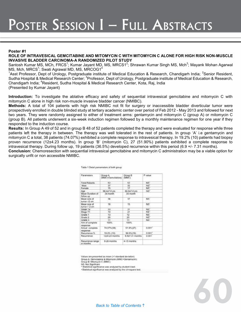
60Back to Table of Contents h
Poster session i – Full AbstrActs
Poster #1 ROLE OF INTRAVESICAL GEMCITABINE AND MITOMYCIN C WITH MITOMYCIN C ALONE FOR HIGH RISK NON-MUSCLE INVASIVE BLADDER CARCINOMA-A RANDOMIZED PILOT STUDY Santosh Kumar MS, MCh, FRCS1; Kumar Jayant MD, MS, MRCS12; Shrawan Kumar Singh MS, Mch3; Mayank Mohan Agarwal MS, Mch, MRCS1; Swati Agrawal MD, MS, MRCOG14 1Asst Professor, Dept of Urology, Postgraduate institute of Medical Education & Research, Chandigarh India; 2Senior Resident, Sudha Hospital & Medical Research Center; 3Professor, Dept of Urology, Postgraduate institute of Medical Education & Research, Chandigarh India; 4Resident, Sudha Hospital & Medical Research Center, Kota, Raj, India (Presented by Kumar Jayant) Introduction: To investigate the ablative efficacy and safety of sequential intravesical gemcitabine and mitomycin C with mitomycin C alone in high risk non-muscle invasive bladder cancer (NMIBC). Methods: A total of 104 patients with high risk NMIBC not fit for surgery or inaccessible bladder diverticular tumor were prospectively enrolled in double blinded study at tertiary academic center over period of Feb 2012 - May 2013 and followed for next two years. They were randomly assigned to either of treatment arms: gentamycin and mitomycin C (group A) or mitomycin C (group B). All patients underwent a six-week induction regimen followed by a monthly maintenance regimen for one year if they responded to the induction course. Results: In Group A 49 of 52 and in group B 48 of 52 patients completed the therapy and were evaluated for response while three patients left the therapy in between. The therapy was well tolerated in the rest of patients. In group ‘A’ i.e gentamycin and mitomycin C a total, 38 patients (74.07%) exhibited a complete response to intravesical therapy. In 19.2% (10) patients had biopsy proven recurrence (12±4.23 months). In group ‘B’ (mitomycin C), 27 (51.90%) patients exhibited a complete response to intravesical therapy. During follow up, 19 patients (36.5%) developed recurrence within this period (6.9 +/- 7.31 months). Conclusion: Chemoresection with sequential intravesical gemcitabine and mitomycin C administration may be a viable option for surgically unfit or non accessible NMIBC.
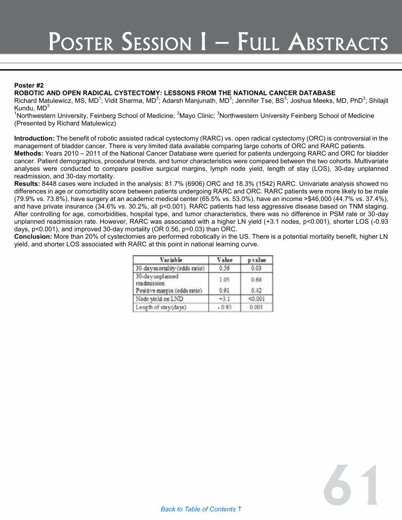
61Back to Table of Contents h
Poster session i – Full AbstrActs
Poster #2 ROBOTIC AND OPEN RADICAL CYSTECTOMY: LESSONS FROM THE NATIONAL CANCER DATABASE Richard Matulewicz, MS, MD1; Vidit Sharma, MD2; Adarsh Manjunath, MD3; Jennifer Tse, BS3; Joshua Meeks, MD, PhD3; Shilajit Kundu, MD3 1Northwestern University, Feinberg School of Medicine; 2Mayo Clinic; 3Northwestern University Feinberg School of Medicine (Presented by Richard Matulewicz) Introduction: The benefit of robotic assisted radical cystectomy (RARC) vs. open radical cystectomy (ORC) is controversial in the management of bladder cancer. There is very limited data available comparing large cohorts of ORC and RARC patients. Methods: Years 2010 – 2011 of the National Cancer Database were queried for patients undergoing RARC and ORC for bladder cancer. Patient demographics, procedural trends, and tumor characteristics were compared between the two cohorts. Multivariate analyses were conducted to compare positive surgical margins, lymph node yield, length of stay (LOS), 30-day unplanned readmission, and 30-day mortality. Results: 8448 cases were included in the analysis: 81.7% (6906) ORC and 18.3% (1542) RARC. Univariate analysis showed no differences in age or comorbidity score between patients undergoing RARC and ORC. RARC patients were more likely to be male (79.9% vs. 73.8%), have surgery at an academic medical center (65.5% vs. 53.0%), have an income >$46,000 (44.7% vs. 37.4%), and have private insurance (34.6% vs. 30.2%, all p<0.001). RARC patients had less aggressive disease based on TNM staging. After controlling for age, comorbidities, hospital type, and tumor characteristics, there was no difference in PSM rate or 30-day unplanned readmission rate. However, RARC was associated with a higher LN yield (+3.1 nodes, p<0.001), shorter LOS (-0.93 days, p<0.001), and improved 30-day mortality (OR 0.56, p=0.03) than ORC. Conclusion: More than 20% of cystectomies are performed robotically in the US. There is a potential mortality benefit, higher LN yield, and shorter LOS associated with RARC at this point in national learning curve.
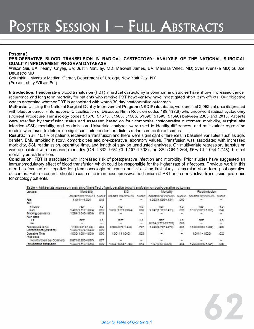
62Back to Table of Contents h
Poster session i – Full AbstrActs
Poster #4 VASCULAR TARGETED PHOTODYNAMIC THERAPY (VTP) WITH WST-11 USING A URETEROSCOPIC APPROACH TO A PORCINE RENAL PELVIS IS SAFE AND UROTHELIAL CELL CARCINOMAS ARE SENSITIVE TO WST-11 VTP IN A MURINE MODEL Katie Murray, Renato Beluco Corradi Fonseca, Stephen LaRosa, Sylvia Jebiwott, Alex Somma, Govindarajan Srimathveeravalli, Kwanghee Kim, Sebastien Monette, Avigdor Scherz, Jonathan Coleman (Presented by Katie Murray) Introduction: WST-11 is a novel intravascular photosensitizing agent for vascular targeted photodynamic therapy (VTP) that allows titratable degrees of tissue necrosis making it uniquely suited for endoluminal organs such as the upper urinary tract. The objective of this study was to examine the feasibility and safety of ablation of the renal pelvis using retrograde ureteroscopic approach in a porcine and report the efficacy of WST-11 VTP in human xenograft murine models. Methods: Unilateral retrograde ureteroscopy was performed on healthy female swine. A laser fiber (2cm diffusing tip) was placed through the ureteroscope into the renal pelvis. WST-11 was administered intravenously and immediate illumination was provided at a light fluence of 200 mW/cm for 10 minutes using a 753 nm laser. Endpoints of the study included CT imaging, laboratory values, and gross and histopathological evaluation of the treated renal pelvis and surrounding renal parenchyma at 24 hours (three animals) and four weeks (three animals). A hindlimb subcutaneous murine model was created with urothelial cell carcinoma line 5637 for treatment with WST-11 VTP and animals were monitored for regrowth of tumor up to 19 days post ablation. Results: In the porcine, weekly CT imaging was normal in all animals after WST-11 VTP. Mild elevation in serum creatinine was seen at 24 hours and persisted for four weeks after ablation. Histologic findings at 24 hours showed completely sloughed surface urothelium with necrosis and hyperemia through the lamina propria on H&E and confirmed with TUNEL staining. At four weeks, the urothelium was regenerated and there was evidence of minimal surrounding parenchymal changes both on gross and histologic examination with increased parenchymal fibrosis on Masson Trichrome staining. In the murine model, 80% of animals were complete responders without evidence of tumor recurrence at 19 days after WST-11 VTP. Conclusion: VTP with WST-11 applied to the porcine renal pelvis is feasible via a retrograde ureteroscopic approach for direct visual fiber placement. The localized ablation effects are reproducible with no safety implications to the renal structure or surrounding tissues. Urothelial cell carcinoma line 5637 in a murine model appears to be sensitive to WST-11 VTP therapy. This data provides the foundation for advancement of clinical trials using WST-11 VTP in upper tract urothelial carcinomas in humans. Funding: The Wade Thompson Family Foundation
Poster #3 PERIOPERATIVE BLOOD TRANSFUSION IN RADICAL CYSTECTOMY: ANALYSIS OF THE NATIONAL SURGICAL QUALITY IMPROVEMENT PROGRAM DATABASE Wilson Sui, BA; Ifeanyi Onyeji, BA; Justin Matulay, MD; Maxwell James, BA; Marissa Velez, MD; Sven Wenske MD; G. Joel DeCastro,MD Columbia University Medical Center, Department of Urology, New York City, NY (Presented by Wilson Sui) Introduction: Perioperative blood transfusion (PBT) in radical cystectomy is common and studies have shown increased cancer recurrence and long term mortality for patients who receive PBT however few have investigated short term effects. Our objective was to determine whether PBT is associated with worse 30 day postoperative outcomes. Methods: Utilizing the National Surgical Quality Improvement Program (NSQIP) database, we identified 2,952 patients diagnosed with bladder cancer (International Classification of Diseases Ninth Revision codes 188-188.9) who underwent radical cystectomy (Current Procedure Terminology codes 51570, 51575, 51580, 51585, 51590, 51595, 51596) between 2005 and 2013. Patients were stratified by transfusion status and assessed based on four composite postoperative outcomes: morbidity, surgical site infection (SSI), mortality, and readmission. Univariate analyses were used to identify differences, and multivariate regression models were used to determine significant independent predictors of the composite outcomes. Results: In all, 40.1% of patients received a transfusion and there were significant differences in baseline variables such as age, gender, BMI, smoking history, comorbidities and pre-operative laboratory values. Transfusion was associated with increased morbidity, SSI, readmission, operative time, and length of stay on unadjusted analyses. On multivariate regression, transfusion was associated with increased morbidity (OR 1.332, 95% CI 1.107-1.603) and SSI (OR 1.364, 95% CI 1.064-1.748), but not mortality or readmission. Conclusion: PBT is associated with increased risk of postoperative infection and morbidity. Prior studies have suggested an immunomodulatory effect of blood transfusion which could be responsible for the higher rate of infections. Previous work in this area has focused on negative long-term oncologic outcomes but this is the first study to examine short-term post-operative outcomes. Future research should focus on the immunosuppressive mechanism of PBT and on restrictive transfusion guidelines for oncology patients.
63Back to Table of Contents h
Poster session i – Full AbstrActs
Poster #4 VASCULAR TARGETED PHOTODYNAMIC THERAPY (VTP) WITH WST-11 USING A URETEROSCOPIC APPROACH TO A PORCINE RENAL PELVIS IS SAFE AND UROTHELIAL CELL CARCINOMAS ARE SENSITIVE TO WST-11 VTP IN A MURINE MODEL Katie Murray, Renato Beluco Corradi Fonseca, Stephen LaRosa, Sylvia Jebiwott, Alex Somma, Govindarajan Srimathveeravalli, Kwanghee Kim, Sebastien Monette, Avigdor Scherz, Jonathan Coleman (Presented by Katie Murray) Introduction: WST-11 is a novel intravascular photosensitizing agent for vascular targeted photodynamic therapy (VTP) that allows titratable degrees of tissue necrosis making it uniquely suited for endoluminal organs such as the upper urinary tract. The objective of this study was to examine the feasibility and safety of ablation of the renal pelvis using retrograde ureteroscopic approach in a porcine and report the efficacy of WST-11 VTP in human xenograft murine models. Methods: Unilateral retrograde ureteroscopy was performed on healthy female swine. A laser fiber (2cm diffusing tip) was placed through the ureteroscope into the renal pelvis. WST-11 was administered intravenously and immediate illumination was provided at a light fluence of 200 mW/cm for 10 minutes using a 753 nm laser. Endpoints of the study included CT imaging, laboratory values, and gross and histopathological evaluation of the treated renal pelvis and surrounding renal parenchyma at 24 hours (three animals) and four weeks (three animals). A hindlimb subcutaneous murine model was created with urothelial cell carcinoma line 5637 for treatment with WST-11 VTP and animals were monitored for regrowth of tumor up to 19 days post ablation. Results: In the porcine, weekly CT imaging was normal in all animals after WST-11 VTP. Mild elevation in serum creatinine was seen at 24 hours and persisted for four weeks after ablation. Histologic findings at 24 hours showed completely sloughed surface urothelium with necrosis and hyperemia through the lamina propria on H&E and confirmed with TUNEL staining. At four weeks, the urothelium was regenerated and there was evidence of minimal surrounding parenchymal changes both on gross and histologic examination with increased parenchymal fibrosis on Masson Trichrome staining. In the murine model, 80% of animals were complete responders without evidence of tumor recurrence at 19 days after WST-11 VTP. Conclusion: VTP with WST-11 applied to the porcine renal pelvis is feasible via a retrograde ureteroscopic approach for direct visual fiber placement. The localized ablation effects are reproducible with no safety implications to the renal structure or surrounding tissues. Urothelial cell carcinoma line 5637 in a murine model appears to be sensitive to WST-11 VTP therapy. This data provides the foundation for advancement of clinical trials using WST-11 VTP in upper tract urothelial carcinomas in humans. Funding: The Wade Thompson Family Foundation
64Back to Table of Contents h
Poster session i – Full AbstrActs
Poster #5 CLINICAL UTILIZATION OF NEOADJUVANT CHEMOTHERAPY IN ELDERLY PATIENTS WITH BLADDER CANCER: OUTCOMES FROM A SINGLE INSTITUTION EXPERIENCE Andrew Leone, MD; Kamran Zargar-Shoshtari, MD, Gregory J Diorio, MD; Pranav Sharma, MD; Scott M. Gilbert, MD; Julio M. Powsang, MD; Wade J. Sexton, MD; Michael A. Poch, MD; Philippe E. Spiess, MD Moffitt Cancer Center, Tampa FL (Presented by Andrew Leone) Introduction: Treatment of urothelial carcinoma of the bladder (UCB) in elderly patients is challenging. With adoption of neoadjuvant chemotherapy (NAC) as stand of care for patients with MIBC, we sought to review patients treated with NAC followed by radical cystectomy (RC) and compare elderly patients ≥70 years to remaining patients in functional and oncological outcomes. Methods: A retrospective chart review was performed on all patients who received NAC for UCB from 2005 to 2013. 161 patients were identified with predominant UC that underwent NAC for ≥ cT2 bladder cancer. All patients underwent RC as planned. Primary end point was pathological response and secondary was survival. Data was compared using Fisher exact test or Mann-Whitney. Kaplan-Meir analysis curves were used for survival and recurrence. Results: 63 elderly patients (≥70) (62% cisplatin-based regimen) were identified and compared with 98 (80% cisplatin-based regimen) younger patients. Charlson Comorbidity index (CCI) and renal function were significantly worse in elderly patients (p<0.01). Eight elderly patients and six patients in the younger cohort were changed to a carboplatin regimen from cisplatin regimen due to toxicity (p=0.18). Dose reduction occurred in 11 (17%) of the elderly vs. 15 (15%) of the other cohort (p=0.83). Subjective clinical response based on endoscopic or radiographic data was not statistically different between the two cohorts. Pathological downstaging to non-muscle invasive disease and complete pathological response were not statistically different in 39 (39%) vs. 23 (36%) patients (p=0.74), 24 (24%) vs. 12 patients (19%) p=0.45. There was no significant difference in follow up, recurrence or in median overall survival between patient groups. In a multivariate model controlling for clinical confounders, age was not an independent predictor of pathological downstaging or complete response. Conclusion: Neoadjuvant chemotherapy (NAC) in appropriately selected elderly patients (≥70 years old) demonstrate equivalent morbidity and oncological outcomes in our single institution cohort. Although, the elderly patients had significantly poorer performance status and CCI there were no differences in survival or response to NAC.
65Back to Table of Contents h
Poster session i – Full AbstrActs
Poster #6 PATIENT-RELATED FACTORS SIGNIFICANTLY AFFECT THE PERFORMANCE OF URINE TESTS FOR BLADDER CANCER: AN EXAMPLE OF SPECTRUM EFFECTS. Thomas Longo, MD1; Ajay Gopalakrishna, BS1; Joseph Fantony, MD1; Richmond Owusu, MD2; Rajesh Dash, MD1; Brant Inman, MD1 1Duke University, Durham, NC; 2UCSD, San Diego, California (Presented by Thomas Longo) Introduction: Physicians rely upon diagnostic tests for clinical decision-making and understand a test’s performance to be summarized by its sensitivity and specificity. Most clinicians do not realize that these values can vary dramatically from one patient subset to another, a phenomenon known as spectrum effects. Urine cytology and UroVysion FISH are two common tests for bladder cancer (BC). Our objective was to evaluate test performance across clinically meaningful patient subgroups to determine if spectrum effects were present. Methods: We assessed all subjects who underwent cystoscopy, cytology, and FISH at our institution from 2003 to 2012. White light cystoscopy was held as the diagnostic gold standard for BC. The standard diagnostic test performance metrics were calculated using generalized linear mixed models (GLMM) and generalized estimating equations (GEE) to account for repeated measures within subjects. We calculated test performance for the overall cohort as well as in key patient subsets defined by age, gender, race, and smoking status. Results: A total of 4027 pairs of cystoscopies and cytologies were obtained from 871 unique subjects for the cytology analysis, and 1697 pairs of UroVysion tests and cystoscopies from 828 unique subjects for analysis. Increasing age, male gender, and history of smoking were associated with increased sensitivity but decreased specificity. In cytology, estimates of sensitivity were higher in men than women (70% vs. 56%) and specificity was correspondingly lower (36% vs. 50%). In FISH, increasing age had the greatest impact on performance; sensitivity nearly tripled from 17% in subjects ≤ 40 years old to 49% in those ≥ 80 years old. Specificity decreased over this same range from 93% in those ≤ 40 years old to 74% in those ≥ 80 years old. Race had no significant impact. Conclusion: The diagnostic performance of two widely used urine tests for bladder cancer, urine cytology and FISH, varied significantly according to the patient demographic in whom they were used, implying that there is no single sensitivity or specificity value that can summarize the performance of these tests. Rather, patient-related factors must be used to contextualize the clinicians’ interpretation of test results and their decision-making. This publication was supported by the National Center for Advancing Translational Sciences, National Institutes of Health, through Duke-CTSA Grant Number 5TL1TR001116-02
Poster #5 CLINICAL UTILIZATION OF NEOADJUVANT CHEMOTHERAPY IN ELDERLY PATIENTS WITH BLADDER CANCER: OUTCOMES FROM A SINGLE INSTITUTION EXPERIENCE Andrew Leone, MD; Kamran Zargar-Shoshtari, MD, Gregory J Diorio, MD; Pranav Sharma, MD; Scott M. Gilbert, MD; Julio M. Powsang, MD; Wade J. Sexton, MD; Michael A. Poch, MD; Philippe E. Spiess, MD Moffitt Cancer Center, Tampa FL (Presented by Andrew Leone) Introduction: Treatment of urothelial carcinoma of the bladder (UCB) in elderly patients is challenging. With adoption of neoadjuvant chemotherapy (NAC) as stand of care for patients with MIBC, we sought to review patients treated with NAC followed by radical cystectomy (RC) and compare elderly patients ≥70 years to remaining patients in functional and oncological outcomes. Methods: A retrospective chart review was performed on all patients who received NAC for UCB from 2005 to 2013. 161 patients were identified with predominant UC that underwent NAC for ≥ cT2 bladder cancer. All patients underwent RC as planned. Primary end point was pathological response and secondary was survival. Data was compared using Fisher exact test or Mann-Whitney. Kaplan-Meir analysis curves were used for survival and recurrence. Results: 63 elderly patients (≥70) (62% cisplatin-based regimen) were identified and compared with 98 (80% cisplatin-based regimen) younger patients. Charlson Comorbidity index (CCI) and renal function were significantly worse in elderly patients (p<0.01). Eight elderly patients and six patients in the younger cohort were changed to a carboplatin regimen from cisplatin regimen due to toxicity (p=0.18). Dose reduction occurred in 11 (17%) of the elderly vs. 15 (15%) of the other cohort (p=0.83). Subjective clinical response based on endoscopic or radiographic data was not statistically different between the two cohorts. Pathological downstaging to non-muscle invasive disease and complete pathological response were not statistically different in 39 (39%) vs. 23 (36%) patients (p=0.74), 24 (24%) vs. 12 patients (19%) p=0.45. There was no significant difference in follow up, recurrence or in median overall survival between patient groups. In a multivariate model controlling for clinical confounders, age was not an independent predictor of pathological downstaging or complete response. Conclusion: Neoadjuvant chemotherapy (NAC) in appropriately selected elderly patients (≥70 years old) demonstrate equivalent morbidity and oncological outcomes in our single institution cohort. Although, the elderly patients had significantly poorer performance status and CCI there were no differences in survival or response to NAC.
66Back to Table of Contents h
Poster session i – Full AbstrActs
Poster #7 ASSOCIATIONS BETWEEN HEALTH-RELATED QUALITY OF LIFE AND PHYSICAL ACTIVITY IN BLADDER CANCER SURVIVORS: A CROSS-SECTIONAL STUDY Ajay Gopalakrishna BS, BA; Joseph Fantony, MD, Thomas Longo, MD; Brant Inman, MD Duke University Medical Center, Durham, NC (Presented by Ajay Gopalakrishna) Introduction: Physical activity has been shown to significantly improve health-related quality of life (HRQOL) and survivorship in a variety of cancer patients. However, little is known about the physical activity patterns of bladder cancer survivors and how these are related to HRQOL in the United States. Our objectives were to describe HRQOL and self-reported physical activity patterns, and examine the association between these measures in a large cohort of bladder cancer survivors. Methods: Bladder cancer survivors identified through an institutional database were mailed a survey that included the Functional Assessment of Cancer Therapy Bladder Cancer (FACT-Bl) and the International Physical Activity Questionnaire (IPAQ-L). Results: A total of 466 subjects (49% response rate) completed the survey. The mean age was 73 years, 80% were male, and 88% were white. Linear regression indicated a positive correlation between physical activity and physical well being (PWB, P < 0.001), emotional well being (EWB, P < 0.001), and functional well being (FWB, P < 0.001) sub-scales, as well as the FACT-Bl (P < 0.001), FACT general (P < 0.001), and trial outcome index (TOI, P < 0.001) composite scores. Conversely, total daily sitting time was negatively correlated with all of the aforementioned indices. Adjusting for demographic factors did not alter the findings. Conclusion: Physical activity is positively associated with HRQOL in bladder cancer survivors. Further studies investigating the causal relationship between physical activity and HRQOL in the post-treatment setting in bladder cancer survivors are warranted. Acknowledgements: This publication was supported by the National Center for Advancing Translational Sciences, National Institutes of Health, through Duke-CTSA Grant Number 5TL1TR001116-02. Its contents are solely the responsibility of the authors and do not necessarily represent the official views of the NIH. Poster #8 EVALUATION OF AN EPIGENETIC PROFILE FOR THE DETECTION OF BLADDER CANCER IN HEMATURIA PATIENTS Kim van Kessel, MD1; Leander Van Neste, PhD2; Irene Lurkin1; Ellen Zwarthoff, PhD1; Wim Van Criekingel PhD2 1Department of Pathology, Erasmus Medical Center, Rotterdam, The Netherlands; 2MDXHealth, Inc., Irvine, CA (Presented by Kim van Kessel) Introduction: Detection of bladder cancer represents a diagnostic challenge. Many patients enter the care cycle with gross or microscopic hematuria and undergo a cystoscopy to rule out bladder cancer. Sensitivity of this invasive examination is limited, leaving many patients at risk of having undetected cancer. To improve upon current clinical practice more sensitive and non-invasive screening methods should be applied, which will benefit patient management. Methods: A total of 154 urine samples were collected from hematuria patients without (n=80) and with (n=74) bladder cancer. DNA from cells in the urine was epigenetically profiled using two independent assays. Methylation-specific PCR (MSP) was performed on TWIST1. SNaPshot methylation analysis was performed for different loci of OTX1 and ONECUT2. Additionally, all samples were analyzed for mutation status of TERT, PIK3CA, FGFR3, HRAS, KRAS and NRAS. Six patients were removed from the analysis due to a lack of sufficient detectable DNA. Results: The combination of TWIST1, ONECUT2 (2 loci) and OTX1 resulted in the best overall performing panel. Logistic regression analysis on these methylation markers, mutation status of FGFR3, TERT and HRAS and age resulted in an accurate model with a sensitivity of 97% (83% specificity).The area under the curve (AUC) was 0.93 (95% CI 0.88 - 0.98). Internal validation led to an optimism-corrected AUC of 0.92. With an estimated bladder cancer prevalence of 5-10% in a hematuria cohort, the assay results in a negative predictive value (NPV) of 99.6% to 99.9%. Conclusion: Epigenetic profiling using TWIST1, ONECUT2 and OTX1 results in a high sensitivity and specificity. Accurate risk prediction might result in less extensive and invasive examination of low-risk patients, hereby reducing unnecessary patient burden and healthcare costs.
67Back to Table of Contents h
Poster session i – Full AbstrActs
Poster #9 SOX-2 EXPRESSION IN PATIENTS WHO UNDERWENT RADICAL CYSTECTOMY FOR UROTHELIAL CARCINOMA OF THE BLADDER. Charles Nottingham MD, MS1; Sanjay Patel, MD2; Peter Clark, MD3; David DeGraff, PhD4; Justin Gregg MD3; Blake Anderson, MD5; Gladell Paner, MD5; Donald Vander Griend, PhD5 1University of Chicago Medical Center, Chicago, IL; 2University of Oklahoma Health Sciences Center, Oklahoma City, OK; 3Vanderbilt University Medical Center, Nashville, TN; 4Pennsylvania State University College of Medicine, Hershey, PA; 5University of Chicago Medical Center, Chicago, ILlinois (Presented by Charles Nottingham) Introduction: Small populations of cells within tumors may have stem cell like qualities. Sox-2 is a transcription factor essential for maintaining the survival and pluripotency of undifferentiated embryonic stem cells. One study suggested that Sox-2 expression in transurethral resection specimens of non-muscle invasive bladder cancer correlated with poor recurrence free survival, increased tumor size and number, and higher grade. No studies, however, have evaluated Sox-2 expression in patients with muscle invasive bladder cancer. Methods: We obtained a series of annotated TMAs from patients who underwent radical cystectomy (RC) for urothelial carcinoma (UC) at Vanderbilt University Medical Center from (years). Tumor specimens were stained for Sox-2 (Cell Signaling Tech, clone D969) and reviewed by a single genitourinary pathologist. The specimens were scored on a 0-3 scale for percentage of cells with positive nuclear staining, and then scored on a 0-3 scale for staining intensity. The two scores were multiplied together to give the Sox-2 score. If multiple samples were available, the scores were averaged. Univariate analysis was used to identify risk factors for positive Sox-2 score. Multivariate analysis was performed to determine patient and pathologic characteristics correlating with Sox-2 score. A univariate survival analysis was performed using the Kaplan-Meier method and multivariate analysis with the Cox proportional hazards model. Results: A total of 271 total patients who underwent RC for UC, of which 114 (42.1%) had Sox-2 scores >0. On univariate analysis Sox-2 score >0 correlated with the presence of urothelial carcinoma in situ (CIS) at RC (p<0.002) and older patient age (p<0.009). On multivariate analysis while controlling for sex, race, comorbidity, pT stage, pN stage, and grade, older age and presence of CIS (HR: 2.4 [95% CI: 1.39-4.22]; p=0.002) predicted for Sox-2 score >0. There was no difference in overall survival (OS) between patients with Sox-2 score of 0 vs. >0 (median OS 44 vs. 46 months, respectively; p=0.99). Conclusion: The presence of Sox-2 in bladder cancer specimens correlated with the presence of CIS at RC, but did not correlate with OS. Further studies to confirm correlation of CIS and Sox-2 are needed, particularly in a non-muscle invasive cohort where a such a correlation may better risk stratify patients for recurrence or progression.
68Back to Table of Contents h
Poster session i – Full AbstrActs
Poster #10 PREOPERATIVE ASYMPTOMATIC LEUKOCYTOSIS IN RADICAL CYSTECTOMY: ANALYSIS OF THE NATIONAL SURGICAL QUALITY IMPROVEMENT PROGRAM DATABASE Ifeanyi Onyeji, BA; Wilson Sui, BA; Justin Matulay, MD; Marissa Velez, MD Maxwell James, BS; Guarionex DeCastro, MD, MPH Columbia University Medical Center, Department of Urology, New York, NY (Presented by Ifeany Onyeji) Introduction: Asymptomatic leukocytosis (AL) has been associated with worse recurrence free and cancer specific survival in lung and gynecological malignancies. The prognostic significance of preoperative AL in bladder cancer has yet to be explored, so we examined the impact its presence may have on perioperative outcomes after radical cystectomy (RC). Methods: Using the National Surgical Quality Improvement (NSQIP) database, we retrospectively analyzed 30-day post-operative outcomes among patients undergoing RC for bladder cancer from 2005 to 2013. We queried the NSQIP Participant User Files with ICD-9 codes for bladder cancer (188, 188.1, 188.2, 188.3, 188.4, 188.5, 188.6, 188.7, 188.8, 188.9) and CPT codes for RC (51570, 51575, 51580, 51585, 51590, 51595, 51596) selecting only those without pre-operative pneumonia, wound infection, disseminated cancer, chronic steroid use, and acute renal failure. AL was defined as WBC>11ng/dL within 30-days preoperatively. The primary outcome was 30-day morbidity. Secondary outcomes included 30-day mortality, readmission, and perioperative complications. Results: Of the 2654 RC performed, 253 patients (9.5%) had preoperative AL. Patient age, race, sex, and diversion type did not differ between groups. The AL group was more likely to smoke (38.7% vs. 23.3%, p<0.001), have pulmonary disease (14.6% vs. 7.5%, p<0.001), and anemia (68.3% vs. 7.8%, p=0.002). On multivariate analysis, 30-day morbidity (OR 1.503 95% CI: 1.104-2.048; p=0.010), infectious complications (OR 1.569 95% CI: 1.142-2.157; p=0.006), pneumonia (OR 1.957 95% CI: 1.068-3.587; p=0.030), ventilator dependency (OR 2.140 95% CI: 1.026-4.463, p=????), acute renal failure (OR 2.942 95% CI: 1.210-7.154; p=0.017) and all surgical complications (OR 1.362 95% CI: 1.012-1.833; p=0.042) were higher among patients with AL. Conclusion: Preoperative asymptomatic leukocytosis is associated with increased 30-day post-operative morbidity, specifically acute renal failure, and infectious, respiratory and surgical complications, after RC. The mechanism underlying these findings most likely involves cytokine-related immunomodulation and impaired neutrophil function.
69Back to Table of Contents h
Poster session i – Full AbstrActs
Poster #11 EFFECTS OF THE COMBINATION OF VASCULAR TARGETED PHOTODYNAMIC THERAPY AND ACTLA-4 IN A PRE CLINICAL UROTHELIAL CARCINOMA MODEL Renato Beluco Corradi, MD1; Stephen La Rosa2; Sylvia Jebiwott2; Katie Murray2; Alex Somma2; Avigdor Scherz3; Kwanghee Kim2; Jonathan Coleman2 1Memorial Sloan Kettering Cancer Center, New York, New YorkCancer Center, New York, New York; 2Memorial Sloan Kettering Cancer Center, New York, NY; 3Department of Plant Sciences, Weizmann Institute of Science, Rehovot, Israel Institution (Presented by Renato Beluco Corradi) Introduction: Urothelial carcinoma (UC) is the most common urinary tract malignancy and the majority of the patients present with non-muscle invasive disease at diagnosis. In this disease stage, the gold standard therapy is resection of the primary tumor with intravesical BCG. However, when this treatment fail or is not tolerated, these patients are left with no other conservative efficient options and need to undergo radical cystectomy or nephroureterectomy in cases of bladder and upper tract tumors, respectively. Alternative conservative treatment options are lacking. Following the historical importance of immunotherapy in the management of UCs and the promising effects of vascular targeted photodynamic therapy (VTP) in tumorigenic tissues, we tested the effect of the combination of these two therapies in a mouse UC model. The focal therapy with VTP relies mediates a cascade of events that leads to tumor necrosis and increases long-lasting systemic antitumor immunity. The combination agent used with VTP was aCTLA-4, a monoclonal antibody against CTLA-4. Our aim was to confirm our hypothesis that VTP combined with CTLA4 blockage can be an efficient treatment for UCs. Methods: The murine bladder 49 (MB-49) cell line is a carcinogen-induced transitional cell carcinoma derived from C57BL/6 male mice. On day 11 after flank MB49 injection, VTP treatment took place. Antibodies injections started on the day after VTP treatment. Intraperitoneal injections were administrated on days one, four, seven and 10 after VTP treatment for all antibodies and controls. Mice were split in four groups: control, VTP tretatment alone, anti-CTLA4 injections alone and combination of VTP plus anti-CTLA4. We monitored the tumor growth and the development of lung metastasis with bioluminescent imaging at key time points, starting at the treatment day. We also evaluated the different groups’ survival using Kaplan-Meier curves. Results: The analysis of the mean tumor signal significantly favored the combination group in comparison with all the other 3 groups (p<0.0001). We also showed a decreased lung signal uptake in this group compared to control (p<0.0001), aCTLA-4 (p=0.0023) and VTP + Mouse IgG (p<0.0001). Furthermore, the combination therapy provided prolonged survival (p < 0.0001). Conclusion: Our results support the use of VTP plus anti CTLA4 combination therapy in future clinical trials as an alternative treatment for UCs in selected cases.
70Back to Table of Contents h
Poster session i – Full AbstrActs
Poster #12 ADDITIONAL ADJUVANT CONVENTIONAL CHEMOTHERAPY IN PATIENTS PREVIOUSLY TREATED WITH NEOADJUVANT CHEMOTHERAPY AND RADICAL CYSTECTOMY: RETROSPECTIVE DESCRIPTION Kamran Zargar-Shoshtari, MD1; Michael Kongnyuy, MD2; Pranav Sharma, MD2; Mayer N. Fishman, MD2; Scott M. Gilbert, MD2; Michael A. Poch, MD2; Julio M. Powsang, MD2; Philippe E. Spiess, MD2; Jingsong Zhang, MD2; Wade J. Sexton, MD2 1Department of Genitourinary Oncology, H. Lee Moffitt Cancer Center and Research Institute, Tampa, FL, United States of America.; 2epartment of Genitourinary Oncology, H. Lee Moffitt Cancer Center and Research Institute, Tampa, FL, United States of America. (Presented by Kamran Zargar-Shoshtari) Introduction: In bladder cancer, neoadjuvant chemotherapy (NAC) can downstage the primary tumor prior to radical cystectomy (RC), and may impact eventual overall survival. However, the optimal management in patients with persistent non-organ confined disease (pT3-4 and/or pN+) following RC is unknown. The aim of this study was to describe use and outcomes of adjuvant chemotherapy (ACT) in patients with residual non-organ confined cancer following NAC and RC. Methods: Using single institution data, pT3-4 and/or pN+ patients who received NAC and RC were identified. ACT was defined as systemic therapy administered post operatively to patients who were clinically disease free. Recurrence-free (RFS) and cancer specific-free survival (CSS) were assessed with Kaplan-Meier analysis. Cox regression was used in multivariate models for survival. Results: From 2001 to 2013, 161 patients received NAC and RC. 80 pT3-4 and/or pN+ patients were identified. NAC was cisplatin-based in 76% and carboplatin-based in 24%. 29 (36%) received ACT; the median number of cycles of ACT was four (IQR: 3-6), with median time to ACT from discharge of 50 days (IQR: 40-67). ACT in the majority of patients was carboplatin-based (16), followed by cisplatin (eight) and other, mainly taxane containing regimens (five). The median RFS was 17.5 months in the ACT and 13.7 months in the non-ACT group (p=0.78). ACT remained an insignificant predictor for RFS after adjusting for pT, pN and margin status (HR: 0.89, 95%CI: 0.48-1.68]). CSS was 23 and 22 months in the respective groups (p=0.65) and remained insignificant after adjusting for pathologic confounders (HR: 0.67, [95%CI: 0.34-1.28]). The eight patients who received cisplatin-based chemotherapy in both neoadjuvant and adjuvant settings had median RFS of 23.2 months. Conclusion: ACT in this retrospective cohort of pT3-4 and/or pN+ did identify a subset with better median RFS. However, the choice of ACT regimens, and incorporation of newer drugs in both adjuvant and neoadjuvant contexts requires further study.

71Back to Table of Contents h
Poster session i – Full AbstrActs
Poster #13 THE RELATIONSHIP BETWEEN TRAVEL DISTANCE TO CYSTECTOMY AND LIKELIHOOD OF READMISSION Troy Sukhu, MD; Jason Lomboy, MD; Matthew Macey, MD; Anne-Marie Meyer, PhD; Ke Meng, PhD; Matthew Nielsen MD, MS; Raj Pruthi, MD; Eric Wallen, MD; Michael Woods, MD; Angela Smith, MD, MS Chapel Hill, NC (Presented by Troy Sukhu) Introduction: Population-based estimates of readmissions following cystectomy range from 25 to 43%. Recent studies have investigated the relationship between complications and other patient- and hospital-level factors; however, the relationship between distance traveled for surgery and risk of readmission remains unclear. We hypothesized that larger distances would increase the risk of readmission following surgery. Methods: Using a linked data resource combining NC Central Cancer Registry with administrative claims data from Medicare, Medicaid, and private insurance plans, we included adult patients undergoing radical cystectomy for bladder cancer from 2003 to 2008. Complications were carefully coded and grouped based on previously published standards: genitourinary, gastrointestinal, wound, infection, venous thromboembolism, and others. Travel distances were calculated by using straight-line distances between zip codes of the patient and cystectomy provider. Bivariable analyses were performed, and multivariable logistic regression was used to evaluate the association of travel distance to cystectomy with likelihood of readmission within 30 and 31-90 days. Results: Of 735 patients who underwent cystectomy, 171 (23%) were readmitted within 30 days, and n=156 (21%) were readmitted between 31-90 days. Mean age was higher among those readmitted, but was statistically non-significant. No significant differences were noted based on race, stage, comorbidity status, or complication type. However, on bivariable analysis, distance to the cystectomy provider > 30 miles was associated with a higher likelihood of readmission (p=0.0009). On multivariable analysis, the only predictor of 30-day readmission was a longer travel distance to the cystectomy provider (table 1). Results were also analyzed for 31-90 day readmissions, but no significant predictors were identified. Conclusion: Longer travel distance to a cystectomy provider is associated with higher 30-day readmission rates, suggesting that complications occurring during this time period may benefit from closer follow-up.
Poster #12 ADDITIONAL ADJUVANT CONVENTIONAL CHEMOTHERAPY IN PATIENTS PREVIOUSLY TREATED WITH NEOADJUVANT CHEMOTHERAPY AND RADICAL CYSTECTOMY: RETROSPECTIVE DESCRIPTION Kamran Zargar-Shoshtari, MD1; Michael Kongnyuy, MD2; Pranav Sharma, MD2; Mayer N. Fishman, MD2; Scott M. Gilbert, MD2; Michael A. Poch, MD2; Julio M. Powsang, MD2; Philippe E. Spiess, MD2; Jingsong Zhang, MD2; Wade J. Sexton, MD2 1Department of Genitourinary Oncology, H. Lee Moffitt Cancer Center and Research Institute, Tampa, FL, United States of America.; 2epartment of Genitourinary Oncology, H. Lee Moffitt Cancer Center and Research Institute, Tampa, FL, United States of America. (Presented by Kamran Zargar-Shoshtari) Introduction: In bladder cancer, neoadjuvant chemotherapy (NAC) can downstage the primary tumor prior to radical cystectomy (RC), and may impact eventual overall survival. However, the optimal management in patients with persistent non-organ confined disease (pT3-4 and/or pN+) following RC is unknown. The aim of this study was to describe use and outcomes of adjuvant chemotherapy (ACT) in patients with residual non-organ confined cancer following NAC and RC. Methods: Using single institution data, pT3-4 and/or pN+ patients who received NAC and RC were identified. ACT was defined as systemic therapy administered post operatively to patients who were clinically disease free. Recurrence-free (RFS) and cancer specific-free survival (CSS) were assessed with Kaplan-Meier analysis. Cox regression was used in multivariate models for survival. Results: From 2001 to 2013, 161 patients received NAC and RC. 80 pT3-4 and/or pN+ patients were identified. NAC was cisplatin-based in 76% and carboplatin-based in 24%. 29 (36%) received ACT; the median number of cycles of ACT was four (IQR: 3-6), with median time to ACT from discharge of 50 days (IQR: 40-67). ACT in the majority of patients was carboplatin-based (16), followed by cisplatin (eight) and other, mainly taxane containing regimens (five). The median RFS was 17.5 months in the ACT and 13.7 months in the non-ACT group (p=0.78). ACT remained an insignificant predictor for RFS after adjusting for pT, pN and margin status (HR: 0.89, 95%CI: 0.48-1.68]). CSS was 23 and 22 months in the respective groups (p=0.65) and remained insignificant after adjusting for pathologic confounders (HR: 0.67, [95%CI: 0.34-1.28]). The eight patients who received cisplatin-based chemotherapy in both neoadjuvant and adjuvant settings had median RFS of 23.2 months. Conclusion: ACT in this retrospective cohort of pT3-4 and/or pN+ did identify a subset with better median RFS. However, the choice of ACT regimens, and incorporation of newer drugs in both adjuvant and neoadjuvant contexts requires further study.
72Back to Table of Contents h
Poster session i – Full AbstrActs
Poster #14 REPORTING BIAS LEADING TO DISCORDANT VENOUS THROMBOEMBOLISM RATES IN US VS. NON-US COUNTRIES FOLLOWING RADICAL CYSTECTOMY: A SYSTEMATIC REVIEW AND META-ANALYSIS Joseph Fantony, MD1; Ajay Gopalakrishna, BS2; Megan Van Noord Van Noord BS, MS2; Brant Inman MD, MSc2 1Duke University Durham, NC; 2Duke University Durham,NC (Presented by Joseph Fantony) Introduction: Post-cystectomy bladder cancer (BCa) patients are at high-risk for developing venous thromboembolism (VTE). The published literature varies widely in the reporting of VTEs in this population. To determine the VTE rate in subjects undergoing radical cystectomy (RC) and highlight specific factors affecting this rate. Methods: This meta-analysis was registered with the PROSPERO database, registration number: CRD42015016776. We queried MEDLINE, The Cochrane Library, EMBASE, Scopus, CINAHL, and Web of Science using all time frames. Search terms captured BCa, RC, and VTE. Per the PRISMA guidelines abstracts were reviewed for inclusion/exclusion criteria by two independent reviewers, and disagreements resolved by a third reviewer. A search of the grey literature and references of pertinent articles was also performed. The date of our last search was December 15, 2014. For unreported data, study authors were contacted. Data were abstracted in duplicate and pooled a random effects (RE) model. Subgroup analyses and meta-regression were performed to determine risk factors for VTE. Results: We identified 2,927 publications of which 223 met inclusion criteria for this review. A total of 1,115,634 surgeries were performed on an 80% male population, with 51,908 VTE events. The overall VTE rate estimated by the RE model was 3.7% (95% CI 3.31-4.16). Due to significant heterogeneity, subgroup and meta-regression analyses were undertaken. This revealed a higher rate of VTE in US based studies at 4.49% (95% CI 3.89-5.13) compared to “westernized”non-US based studies at 3.43 (95% CI 3.02-3.86) and “non-westernized”non-US based studies at 2.50 (95% CI 1.89-3.21). Other important modifiers included minimally invasive at 5.54 (95% CI 3.97-7.36) vs. open surgery at 3.55 (95% CI 3.12-4.01) and age. The case-fatality rate of pulmonary emboli (PE) was 44%. Conclusion: VTE is common in patients undergoing radical cystectomy. Reporting of VTE is heterogeneous and the rate varies according to study-level factors including: surgery type, and country of origin. This may be due to significant reporting bias in non-westernized countries versus westernized countries, rather than a true difference in rate. Limitations of this study include the preponderance of observational studies in the final analysis and lack of complete reporting of all variables of interest within each study.
73Back to Table of Contents h
Poster session i – Full AbstrActs
Poster #15 MALIGNANT URETEROINTESTINAL ANASTOMOTIC STRICTURE FOLLOWING RADICAL CYSTECTOMY: PATTERNS, RISK FACTORS, AND OUTCOMES Mary E. Westerman, MD1; Boyd R. Viers, MD1; R. Jeffrey Karnes, MD1; Robert Tarrell2; Prabin Thapa2; R. Houston Thompson, MD1; Matthew K. Tollefson, MD1; Stephen A. Boorjian, MD1 1Mayo Clinic Dept of Urology, Rochester MN; 2Department of Health Sciences Research, Mayo Clinic, Rochester, Minnesota (Presented by Mary E. Westerman) Introduction: The development of a ureteral-intestinal anastomotic (UIA) stricture has been reported in up to 15% of patients undergoing radical cystectomy (RC) with urinary diversion. The vast majority of these strictures result from ischemia, and as such are benign. Herein, meanwhile, we seek to characterize the incidence, risk factors, and outcomes of malignant UIA strictures. Methods: We identified 2,524 patients who underwent RC at our institution between 1980 and 2009, among whom 235 (9.3%) were subsequently diagnosed with a UIA stricture. Of these, 17.4% (41/235) were malignant strictures, confirmed pathologically in all cases by cytology and/or excision. Logistic regression models were used to evaluate clinical variables associated with the development of a malignant stricture. Oncologic outcomes were estimated using the Kaplan-Meier method. Results: Obtained: Median postoperative follow up was 9.3 years (IQR 6.2, 20.2). Five-year malignant-stricture free survival was 98.2%. The median time to diagnosis for malignant versus benign strictures was 30 months versus 7.2 months, respectively (p<0.0001). Seventy-eight percent of patients with malignant strictures were symptomatic at presentation; specifically, these patients were significantly more likely to report gross hematuria compared to those with a benign stricture (26.8% vs. 5.7%; p<0.0001). Patients who developed a malignant stricture were significantly more likely to have a pathologic stage of Tis/Cis at RC (46.3% vs. 18.4%, p<0.0001) and non-muscle-invasive disease (68.3% vs. 45.9%, p=0.0041) compared to those who did not develop a malignant stricture following RC. More than half (22/41, 53.7%) of patients with a malignant stricture were managed with open excision. A total of 35 patients with a malignant UIA stricture died during follow-up, including 29 who died of urothelial carcinoma. Following malignant stricture diagnosis, five- and 10- year cancer specific survival was 28% and 19%, respectively, while overall survival was 22% and 15%. Conclusion: Malignant strictures are uncommon although frequently symptomatic. Carcinoma in situ at RC is associated with a significantly increased risk of developing a malignant stricture. Importantly, malignant strictures present significantly later than benign strictures, underscoring the importance of continued patient follow up.
Poster #14 REPORTING BIAS LEADING TO DISCORDANT VENOUS THROMBOEMBOLISM RATES IN US VS. NON-US COUNTRIES FOLLOWING RADICAL CYSTECTOMY: A SYSTEMATIC REVIEW AND META-ANALYSIS Joseph Fantony, MD1; Ajay Gopalakrishna, BS2; Megan Van Noord Van Noord BS, MS2; Brant Inman MD, MSc2 1Duke University Durham, NC; 2Duke University Durham,NC (Presented by Joseph Fantony) Introduction: Post-cystectomy bladder cancer (BCa) patients are at high-risk for developing venous thromboembolism (VTE). The published literature varies widely in the reporting of VTEs in this population. To determine the VTE rate in subjects undergoing radical cystectomy (RC) and highlight specific factors affecting this rate. Methods: This meta-analysis was registered with the PROSPERO database, registration number: CRD42015016776. We queried MEDLINE, The Cochrane Library, EMBASE, Scopus, CINAHL, and Web of Science using all time frames. Search terms captured BCa, RC, and VTE. Per the PRISMA guidelines abstracts were reviewed for inclusion/exclusion criteria by two independent reviewers, and disagreements resolved by a third reviewer. A search of the grey literature and references of pertinent articles was also performed. The date of our last search was December 15, 2014. For unreported data, study authors were contacted. Data were abstracted in duplicate and pooled a random effects (RE) model. Subgroup analyses and meta-regression were performed to determine risk factors for VTE. Results: We identified 2,927 publications of which 223 met inclusion criteria for this review. A total of 1,115,634 surgeries were performed on an 80% male population, with 51,908 VTE events. The overall VTE rate estimated by the RE model was 3.7% (95% CI 3.31-4.16). Due to significant heterogeneity, subgroup and meta-regression analyses were undertaken. This revealed a higher rate of VTE in US based studies at 4.49% (95% CI 3.89-5.13) compared to “westernized”non-US based studies at 3.43 (95% CI 3.02-3.86) and “non-westernized”non-US based studies at 2.50 (95% CI 1.89-3.21). Other important modifiers included minimally invasive at 5.54 (95% CI 3.97-7.36) vs. open surgery at 3.55 (95% CI 3.12-4.01) and age. The case-fatality rate of pulmonary emboli (PE) was 44%. Conclusion: VTE is common in patients undergoing radical cystectomy. Reporting of VTE is heterogeneous and the rate varies according to study-level factors including: surgery type, and country of origin. This may be due to significant reporting bias in non-westernized countries versus westernized countries, rather than a true difference in rate. Limitations of this study include the preponderance of observational studies in the final analysis and lack of complete reporting of all variables of interest within each study.
74Back to Table of Contents h
Poster session i – Full AbstrActs
Poster #16 VALIDATING THE BLADDER UTILITY SYMPTOM SCALE (BUSS): A MULTIATTRIBUTE HEALTH STATE CLASSIFICATION SYSTEM FOR BLADDER CANCER Nathan Perlis, MD, MSc1; Kirstin Boehme, MSc2; Munir Jamal, MD3; Karen Bremner2; Shabbir Alibhai, MD, MSc4; Antonio Finelli, MD, MSc1; Paul Ritvo, PhD5; Murray Krahn, MD, MSc6; Girish Kulkarni, MD, PhD1 1Division of Urology, University Health Network, University of Toronto, Toronto, Ontario; 2University Health Network, Toronto, Ontario; 33Trillium Health Partners, Mississauga, Ontario; 4Division of Medicine, University Health Network, University of Toronto, Toronto, Ontario; 5York University and Cancer Care Ontario, Toronto, Ontario; 6Toronto Health Economics and Technology Assessment (THETA) Collaborative, University of Toronto, and University Health Network, Toronto, Ontario (Presented by Nathan Perlis) Introduction: Given the current healthcare funding environment, cost-effective decisions for resource allocation in bladder cancer (BC) are necessary. A numerical measure of health-related quality of life (HRQOL) based on patients’ preferences, called “utility”, is necessary to measure the incremental cost-effectiveness ratio, the key metric of cost-effectiveness. An instrument that can measure BC patient HRQOL and generate utilities currently does not exist. Our goal was to create a valid and reliable instrument to measure utility and HRQOL among BC patients. We have previously described the creation and pilot testing of the Bladder Utility Symptom Scale - Psychometric (BUSS-P) questionnaire. Herein, we assessed the psychometric properties of our newly designed instrument. Methods: To determine BUSS-P validation and reliability, field testing was performed at both an academic and community hospital, the University Health Network (Toronto, ON) and Trillium Health Partners (Mississauga, ON), respectively. We used purposive sampling to accrue 105 BC patients. All stages of BC were included. Patients completed the BUSS-P and five other HRQOL and utility instruments (FACT-Bl, BCI, EQ-5D, SF-36, TTO). Construct validity was tested with spearman’s rank correlations (rs), and comparisons of BUSS-P scores across known-groups. Reliability was assessed at two time-points, four weeks apart. Results: The BUSS-P had high whole scale correlation with both the FACT-Bl (rs=0.82, p<0.0001) and the EQ-5D (rs=0.67, p<0.0001). Similarly, the BUSS-P demonstrated substantial-to-high subscale correlations with the EQ-5D (emotional wellbeing: rs=0.71, p<0.0001), the FACT-Bl (physical wellbeing: rs=-0.70, p<0.0001), and the BCI (urinary issues: rs=-0.62, p<0.0001). Median BUSS-P scores (M) were also significantly different (p=0.0016) across patients with differing disease severity: non-muscle invasive BC (M=85.0), cystectomy (M=76.2), and metastatic BC patients (M=66.7). There was excellent test-retest reliability (ICC 0.78). Greater than 95% of respondents answered all questions, suggesting a comprehensible and well-designed questionnaire. Conclusion: These data suggest that the BUSS-P is a valid instrument to measure HRQOL among BC patients. Future work collecting patient- and community member-generated utility weights to convert the questionnaire into a disease-specific utility instrument is underway. Funding by Canadian Institutes of Health Research (CIHR) (#MOP123366)
75Back to Table of Contents h
Poster session i – Full AbstrActs
Poster #17 ONCOLOGIC SURVEILLANCE FOLLOWING RADICAL CYSTECTOMY: AN INDIVIDUALIZED RISK-BASED APPROACH. Suzanne Stewart-Merrill, MD1; Stephen Boorjian, MD2; R. Houston Thompson, MD2; Sarah Psutka, MD3; John Cheville, MD2; Prabin Thapa2; Matthew Tollefson, MD2; Igor Frank, MD2 1Hershey, PA; 2Rochester, MN; 3Chicago, IL (Presented by Suzanne Stewart-Merrill) Introduction: The appropriate duration of surveillance for bladder cancer (BC) following radical cystectomy (RC) remains unknown. Uniform adherence to current guidelines has the potential for over utilization of resources in some patients and deficiency of testing in others. Herein, we provide an approach to surveillance which balances the risk of recurrence versus the risk of non-BC death. Methods: We identified 2438 patients who underwent RC for M0 BC between 1980 and 2007. Patients were stratified for analysis by pathologic stage (pT0Nx-0, pTa/CIS/1Nx-0, pT2Nx-0, pT3/4Nx-0, and pTanyN+), relapse location (urethra, upper urinary tract, abdomen, thorax, and other), age (<= 60, 61-70, 71-80, >80yrs) and Charlson Co-morbidity Index (CCI <= 2 and CCI >= 3). Risks of disease recurrence and non-BC death were estimated using parametric models for time-to-failure using Weibull distributions. Surveillance duration was estimated at the time point when the risk of non-BC death exceeded the risk of recurrence. Results: At a median follow-up of 6.0 yrs (IQR 2.0,11.1), a total of 713 patients developed recurrence. As shown in the table, vastly different surveillance durations were appreciated for various stage, age, and CCI groups before the risk of non-BC death exceeded the risk of recurrence. Specifically, among patients age <= 60yrs with pT2Nx-0 disease, the risk of non-BC death exceeded the risk of recurrence to the abdomen at 7.5yrs if the patient’s CCI was >= 3, but failed to do so until 10 years if the patient’s CCI was <= 2. On the other hand, for patients’ age > 80yrs with pT2Nx-0 disease, the risk of non-BC death exceeded the risk of abdominal recurrence at 1yr following surgery regardless of the patient’s CCI. Conclusion: We present an individualized approach to post-RC surveillance that bases duration of follow-up on the interplay between competing risk factors of recurrence and non-BC death. This strategy may improve the balance between the derived benefit from surveillance and medical resource allocation.
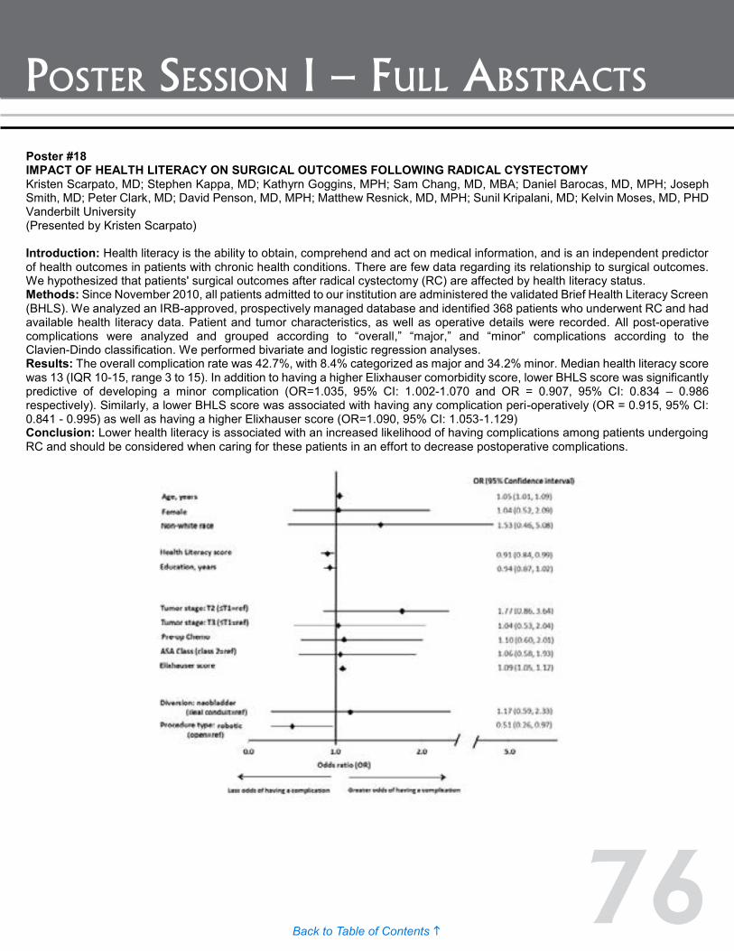
76Back to Table of Contents h
Poster session i – Full AbstrActs
Poster #18 IMPACT OF HEALTH LITERACY ON SURGICAL OUTCOMES FOLLOWING RADICAL CYSTECTOMY Kristen Scarpato, MD; Stephen Kappa, MD; Kathyrn Goggins, MPH; Sam Chang, MD, MBA; Daniel Barocas, MD, MPH; Joseph Smith, MD; Peter Clark, MD; David Penson, MD, MPH; Matthew Resnick, MD, MPH; Sunil Kripalani, MD; Kelvin Moses, MD, PHD Vanderbilt University (Presented by Kristen Scarpato) Introduction: Health literacy is the ability to obtain, comprehend and act on medical information, and is an independent predictor of health outcomes in patients with chronic health conditions. There are few data regarding its relationship to surgical outcomes. We hypothesized that patients' surgical outcomes after radical cystectomy (RC) are affected by health literacy status. Methods: Since November 2010, all patients admitted to our institution are administered the validated Brief Health Literacy Screen (BHLS). We analyzed an IRB-approved, prospectively managed database and identified 368 patients who underwent RC and had available health literacy data. Patient and tumor characteristics, as well as operative details were recorded. All post-operative complications were analyzed and grouped according to “overall,” “major,” and “minor” complications according to the Clavien-Dindo classification. We performed bivariate and logistic regression analyses. Results: The overall complication rate was 42.7%, with 8.4% categorized as major and 34.2% minor. Median health literacy score was 13 (IQR 10-15, range 3 to 15). In addition to having a higher Elixhauser comorbidity score, lower BHLS score was significantly predictive of developing a minor complication (OR=1.035, 95% CI: 1.002-1.070 and OR = 0.907, 95% CI: 0.834 – 0.986 respectively). Similarly, a lower BHLS score was associated with having any complication peri-operatively (OR = 0.915, 95% CI: 0.841 - 0.995) as well as having a higher Elixhauser score (OR=1.090, 95% CI: 1.053-1.129) Conclusion: Lower health literacy is associated with an increased likelihood of having complications among patients undergoing RC and should be considered when caring for these patients in an effort to decrease postoperative complications.
77Back to Table of Contents h
Poster session i – Full AbstrActs
Poster #19 FIRST-LINE RANDOMIZED PHASE 2 STUDY OF GEMCITABINE/CISPLATIN PLUS APATORSEN OR PLACEBO IN PATIENTS WITH ADVANCED BLADDER CANCER: THE INTERNATIONAL BOREALIS-1™ TRIAL Daniel Petrylak, MD1; Bernhard Eigl, MD2; Elzbieta Senkus, MD3; Yohann Loriot, MD4; Przemyslaw Twardowski, MD5; Daniel Castellano, MD6; Normand Blais, MD7; Srikala Sridhar, MD8; Cora Sternberg, MD9; Margitta Retz, MD10; Brent Blumenstein, PhD11; Cindy Jacobs, PhD, MD12; Patricia Stewart, MD12; Joaquim Bellmunt, MD13 1Yale School of Medicine, New Haven, CT; 2British Columbia Cancer Agency, Vancouver, BC; 3Medical University, Gdansk, Poland; 4Institut Gustave Roussy, Villejuif, France; 5City of Hope National Medical Center, Duarte, CA; 6Hospital Universitario 12 de Octubre, Madrid, Spain; 7Université de Montréal-Hospital Notre-Dame, Montréal, QC; 8Princess Margaret Hospital, Toronto, ON; 9San Camillo Forlanini Hospital, Rome, Italy; 10Technischen Universität München, München, Germany; 11Trial Architecture Consulting, Washington, DC; 12OncoGenex Pharmaceuticals, Inc., Bothell, WA; 13Dana-Farber Cancer Institute, Boston, MA (Presented by Daneil Petrylak) Introduction: Heat shock protein 27 (Hsp27) is over-expressed in bladder cancer (BC) and postulated to increase tumor growth, metastasis, and chemotherapy resistance. Apatorsen (A; OGX-427), a novel antisense oligonucleotide, inhibits Hsp27 production and can potentially enhance the efficacy of chemotherapy. Objectives of this trial were to evaluate efficacy and safety of A in combination with gemcitabine and cisplatin (GC) in patients (pts) with advanced BC. Methods: Chemotherapy naïve patients with advanced BC were randomized to GC+A 600 mg, GC+A 1000 mg, or GC + placebo. Patientswere stratified by Karnofsky performance status (KPS) and visceral disease. The primary endpoint was overall survival (OS). Prognostic sub-groups were retrospectively evaluated using multiple variable modeling and hierarchical step down. A post hoc analysis was performed to explore the hypothesis that Hsp27 inhibition might be relevant to OS in poor prognosis disease. Results: A total of 179 patients were randomized and treated. Median OS was 15.2 months (m). When compared to GC + placebo, GC+A 600 demonstrated improved OS and PFS (OS HR = 0.856 and PFS HR = 0.830) versus GC+A 1000 (OS HR = 0.898; PFS HR = 0.927). Results from the post hoc model revealed that KPS, liver mets, alk phos, and hemoglobin were prognostic. A median prognostic score dichotomized patients into poor and good prognosis groups (50% each group). Patientswith poor prognosis treated with GC+A 600 had a greater reduction in risk of death (HR = 0.717) than patients with good prognosis (HR = 1.44). The most significant prognostic factor was KPS ≤80% (35% patients in GC+A 600 vs. GC) resulting in HR = 0.50 in favor of GC+A 600. Overall treatment was well tolerated. Most common Grade ≥3 adverse events (AEs) were neutropenia, anemia, thrombocytopenia and hypertension. Frequency of ≥3 Grade toxicities were: 89% (GC), 93% (GC+A 600) and 95% (GC+A 1000). GC+A 1000 had a higher treatment discontinuation rate due to AEs. Conclusion: Advanced BC patients with poor prognosis benefited from apatorsen 600mg combined with first line GC. Apatorsen may be impacting the intrinsic biology of patients with poor risk factors. Further evaluation is warranted in this pt population. This study was supported by OncoGenex Pharmaceuticals, Inc. (Bothell, WA).
78Back to Table of Contents h
Poster session i – Full AbstrActs
Poster #20 ONCOLOGIC OUTCOMES AFTER ANTERIOR EXENTERATION FOR MUSCLE INVASIVE BLADDER CANCER IN WOMEN Justin Gregg, MD1; Curran Emeruwa, BS2; Johnson Wong, BS2; Matthew Resnick, MD, MPH1; Daniel Barocas, MD, MPH1; Michael Cookson, MD, MMHC3; Sam Chang, MD1; David Penson, MD, MPH1; Joseph Smith, MD1; Kristen Scarpato, MD, MPH1; Kelvin Moses, MD, PhD1 1Vanderbilt University Medical Center, Nashville, TN; 2Meharry Medical College, Nashville, TN; 3University of Oklahoma College of Medicine, Oklahoma City, OK (Presented by Justin Gregg) Introduction: Female patients with muscle invasive bladder cancer (MIBC) traditionally undergo anterior pelvic exenteration. However, female genitourinary (GU) organ involvement is rare and removal can potentially affect urinary and sexual function. Our study aimed to identify tumor characteristics associated with female GU organ involvement. We hypothesized that a lack of trigonal or bladder floor tumor, intraoperative palpable posterior mass, and clinical lymphadenopathy is associated with a lack of GU organ involvement. Methods: We retrospectively reviewed charts of female patients who underwent radical cystectomy at our institution from 1999 to 2014. Patient and operative characteristics were extracted from the electronic medical record. Women who had a prior hysterectomy were excluded. We examined patient and operative characteristics as well as presence of hysterectomy for association with disease recurrence. Statistical analysis utilized chi squared and student’s t-test. We evaluated if the three characteristics in our hypothesis were associated with lack of GU organ involvement in patients who underwent hysterectomy. Results: Out of 322 eligible patients, 164 (51.0%) did not have a hysterectomy prior to cystectomy. Of these, 143 (87.2%) underwent intraoperative hysterectomy. Mean follow-up time was 2.0 years (SD 2.7). Twenty patients (12.2%) recurred during follow-up. No patient or surgical factor other than use of adjuvant chemotherapy or radiation (p<0.01) was associated with recurrence. Thirty-four out of 143 patients who underwent exenteration (23.8%) had female GU organ involvement. Thirty out of 102 patients (29.4%) who had a trigone or bladder floor tumor, palpable posterior mass or suspicious LN on preoperative scan had female GU organ involvement. Four out of 41 (9.8%) patients who did not meet this criteria had GU organ involvement (p=0.01). Two of the women who did not undergo anterior extenteration had a documented recurrence, neither of which involved the female GU organs. Conclusion: Rates of female GU organ involvement found during anterior exenteration for MIBC are higher than previously reported. Lack of trigonal/bladder floor tumor, palpable posterior mass and adenopathy on imaging may be associated with absence of GU organ involvement. Individualized risk assessment, patient preferences, and intraoperative findings should be used to guide surgical planning.
79Back to Table of Contents h
Poster session i – Full AbstrActs
Poster #21 QUANTITATIVE PROTEOMIC ANALYSIS OF UROLOGIC BLADDER CANCER CELLS FOLLOWING TREATMENT WITH HISTONE DEACETYLASE INHIBITORS Quentin Li, MD, PhD1; Jian-Jiang Hao, PhD2; Zheng Zhang, MD2; Reema Railkar, PhD1; Iawen Hsu, PhD1; Adam Metwalli, MD1; Piyush Agarwal, MD1 1UOB, CCR, NCI, NIH, Bethesda, MD 20892; 2Poochon Scientific, Frederick, MD 21704 (Presented by Quentin Li) Introduction: Recently, the TCGA (The Cancer Genome Atlas) project identified the importance of mutations in chromatin remodeling genes in human carcinomas. These findings imply that epigenetic modulators might have a therapeutic role in urothelial cancers. Therefore, in order to exploit histone deacetylases (HDACs) as targets for cancer therapy, we investigated HDAC inhibitors (HDACIs) as potential chemotherapeutic agents for bladder cancer. Methods: We assessed the antiproliferative effect of the HDACIs romidepsin and trichostatin A (TSA) in 5637 bladder cancer cell line using an MTS (Promega) assay. We then performed a quantitative proteomic analysis of cells before and after treatment with these HDACIs and assessed for changes. Results: We showed that both HDACIs suppressed cell growth and induced cell death in bladder cancer 5637 cells. Our proteome studies identified a total of 6003 unique proteins. Of these, 2472 proteins were up-regulated and 2049 proteins were down-regulated in response to HDACI exposure, as compared to the untreated controls (P < 0.05). Bioinformatic analysis further revealed that those differentially expressed proteins were mainly involved in multiple biological functions and enzyme-regulated pathways, including cell cycle progression, apoptosis, autophagy, free radical generation, and DNA damage repair. HDACIs also altered the acetylation status of histones H2A, H2B, H3 and H4, as well as the levels of chromatin modification proteins, suggesting that HDACIs may exert multiple cytotoxic actions in bladder cancer cells by inhibiting HDAC activity or altering the structure of chromatin. Conclusion: The HDACIs romidepsin and TSA are effective in the inhibition of cell growth and the induction of apoptosis in 5637 bladder cancer cells. Furthermore, proteomics reveals multiple pathway alterations that identify some potential strategies for combination therapy with HDACIs. These observations support the notion that HDACIs both as single agents and in combination might provide new therapeutic options for bladder cancer treatment and thus warrant further preclinical exploration. This study was supported by the Intramural Research Program of the National Cancer Institute, the National Institutes of Health.
80Back to Table of Contents h
Poster session i – Full AbstrActs
Poster #22 TARGETING THE GLYCOME IN CISPLATIN-RESISTANT BLADDER CANCER WITH A NATURALLY OCCURRING PARASITE-HOST ANCHOR PROTEIN Roland Seiler, MD1; Htoo Z. Oo, PhD2; Sherry S. Lee, BSc2; Mette O. Agerbaek, PhD3; Jamie R. Rich, PhD4; Thomas M. Clausen, PhD3; John Babcook, PhD4; Peter C. Black, MD2; Ali Salanti, PhD3; Mads Daugaard, PhD2 1Department of Urologic Sciences, University of British Columbia, Vancouver, British Columbia, Canada; 2Department of Urologic Sciences Vancouver Prostate Centre; 3Faculty of Health Sciences, University of Copenhagen; 4The Centre for Drug Research and Development, Vancouver, BC (Presented by Roland Seiler) Introduction: Cisplatin-based chemotherapy is an integral component of therapy for invasive urothelial carcinoma of the bladder (UCB). Although responses are common, they are rarely durable, and second line options are limited. Here we investigated the potential of targeting cancer-specific oncofetal chondroitin sulfate A (ofCSA) chains in cisplatin-resistant UCB with a high affinity ofCSA glycan-binding protein VAR2CSA found in the malaria parasite Plasmodium falciparum. Methods: A recombinant VAR2CSA (rVAR2) was generated, purified and used for binding assays in flow cytometry and immunohistochemistry, as well as for conjugation with the hemiasterlin compound KT886 to create a Var2-Drug-Conjugate (VDC). Expression of ofCSA was determined in a tissue microarray of 52 chemotherapy-naïve UCB samples as well as 36 matched post-chemotherapy cystectomy specimens from patients receiving neoadjuvant gemcitabine/cisplatin. In a chemoresistant orthotopic UCB xenograft model, VDC efficacy was tested against rVAR2 alone, KT886 and PBS. This model was created by inoculating UM-UC13 cells in the mouse bladder and treating the mice with 3mg/KG intraperitoneal cisplatin once weekly. The tumors were recycled for 6 successive generations in the presence of cisplatin therapy and the resultant highly resistant tumors were used for treatment studies. Results: In-vitro, rVAR2 bound eight out of eight human bladder cancer cell lines in a concentration- and CSA-dependent manner. The binding increased in ex-vivo UM-UC13 cells generated from the chemoresistant orthotopic xenograft model. Similarly, ofCSA was overexpressed in residual patient tumors after neoadjuvant chemotherapy (p=0.001). Moreover, in these chemoresistant tumors ofCSA expression was associated with advanced tumor stage (ypT3/4, p=0.005) and shorter overall survival (p=0.04). In-vivo, VDC effectively inhibited growth of the chemoresistant orthotopic UCB xenografts. Conclusion: ofCSA is overexpressed in UCB after chemotherapy and is associated with unfavorable tumor characteristics. VDC demonstrated antineoplastic effects in chemoresistant xenografts. This treatment paradigm warrants further study as a second line treatment in UBC not responding to cisplatin.
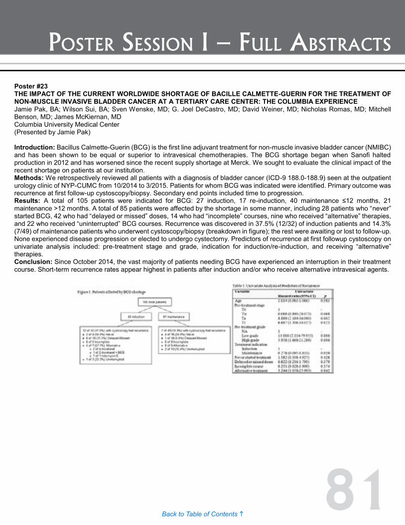
81Back to Table of Contents h
Poster session i – Full AbstrActs
Poster #23 THE IMPACT OF THE CURRENT WORLDWIDE SHORTAGE OF BACILLE CALMETTE-GUERIN FOR THE TREATMENT OF NON-MUSCLE INVASIVE BLADDER CANCER AT A TERTIARY CARE CENTER: THE COLUMBIA EXPERIENCE Jamie Pak, BA; Wilson Sui, BA; Sven Wenske, MD; G. Joel DeCastro, MD; David Weiner, MD; Nicholas Romas, MD; Mitchell Benson, MD; James McKiernan, MD Columbia University Medical Center (Presented by Jamie Pak) Introduction: Bacillus Calmette-Guerin (BCG) is the first line adjuvant treatment for non-muscle invasive bladder cancer (NMIBC) and has been shown to be equal or superior to intravesical chemotherapies. The BCG shortage began when Sanofi halted production in 2012 and has worsened since the recent supply shortage at Merck. We sought to evaluate the clinical impact of the recent shortage on patients at our institution. Methods: We retrospectively reviewed all patients with a diagnosis of bladder cancer (ICD-9 188.0-188.9) seen at the outpatient urology clinic of NYP-CUMC from 10/2014 to 3/2015. Patients for whom BCG was indicated were identified. Primary outcome was recurrence at first follow-up cystoscopy/biopsy. Secondary end points included time to progression. Results: A total of 105 patients were indicated for BCG: 27 induction, 17 re-induction, 40 maintenance ≤12 months, 21 maintenance >12 months. A total of 85 patients were affected by the shortage in some manner, including 28 patients who “never” started BCG, 42 who had “delayed or missed” doses, 14 who had “incomplete” courses, nine who received “alternative” therapies, and 22 who received “uninterrupted” BCG courses. Recurrence was discovered in 37.5% (12/32) of induction patients and 14.3% (7/49) of maintenance patients who underwent cystoscopy/biopsy (breakdown in figure); the rest were awaiting or lost to follow-up. None experienced disease progression or elected to undergo cystectomy. Predictors of recurrence at first followup cystoscopy on univariate analysis included: pre-treatment stage and grade, indication for induction/re-induction, and receiving “alternative” therapies. Conclusion: Since October 2014, the vast majority of patients needing BCG have experienced an interruption in their treatment course. Short-term recurrence rates appear highest in patients after induction and/or who receive alternative intravesical agents.
Poster #22 TARGETING THE GLYCOME IN CISPLATIN-RESISTANT BLADDER CANCER WITH A NATURALLY OCCURRING PARASITE-HOST ANCHOR PROTEIN Roland Seiler, MD1; Htoo Z. Oo, PhD2; Sherry S. Lee, BSc2; Mette O. Agerbaek, PhD3; Jamie R. Rich, PhD4; Thomas M. Clausen, PhD3; John Babcook, PhD4; Peter C. Black, MD2; Ali Salanti, PhD3; Mads Daugaard, PhD2 1Department of Urologic Sciences, University of British Columbia, Vancouver, British Columbia, Canada; 2Department of Urologic Sciences Vancouver Prostate Centre; 3Faculty of Health Sciences, University of Copenhagen; 4The Centre for Drug Research and Development, Vancouver, BC (Presented by Roland Seiler) Introduction: Cisplatin-based chemotherapy is an integral component of therapy for invasive urothelial carcinoma of the bladder (UCB). Although responses are common, they are rarely durable, and second line options are limited. Here we investigated the potential of targeting cancer-specific oncofetal chondroitin sulfate A (ofCSA) chains in cisplatin-resistant UCB with a high affinity ofCSA glycan-binding protein VAR2CSA found in the malaria parasite Plasmodium falciparum. Methods: A recombinant VAR2CSA (rVAR2) was generated, purified and used for binding assays in flow cytometry and immunohistochemistry, as well as for conjugation with the hemiasterlin compound KT886 to create a Var2-Drug-Conjugate (VDC). Expression of ofCSA was determined in a tissue microarray of 52 chemotherapy-naïve UCB samples as well as 36 matched post-chemotherapy cystectomy specimens from patients receiving neoadjuvant gemcitabine/cisplatin. In a chemoresistant orthotopic UCB xenograft model, VDC efficacy was tested against rVAR2 alone, KT886 and PBS. This model was created by inoculating UM-UC13 cells in the mouse bladder and treating the mice with 3mg/KG intraperitoneal cisplatin once weekly. The tumors were recycled for 6 successive generations in the presence of cisplatin therapy and the resultant highly resistant tumors were used for treatment studies. Results: In-vitro, rVAR2 bound eight out of eight human bladder cancer cell lines in a concentration- and CSA-dependent manner. The binding increased in ex-vivo UM-UC13 cells generated from the chemoresistant orthotopic xenograft model. Similarly, ofCSA was overexpressed in residual patient tumors after neoadjuvant chemotherapy (p=0.001). Moreover, in these chemoresistant tumors ofCSA expression was associated with advanced tumor stage (ypT3/4, p=0.005) and shorter overall survival (p=0.04). In-vivo, VDC effectively inhibited growth of the chemoresistant orthotopic UCB xenografts. Conclusion: ofCSA is overexpressed in UCB after chemotherapy and is associated with unfavorable tumor characteristics. VDC demonstrated antineoplastic effects in chemoresistant xenografts. This treatment paradigm warrants further study as a second line treatment in UBC not responding to cisplatin.
82Back to Table of Contents h
Poster session i – Full AbstrActs
Poster #24 PROGNOSTIC VALUE OF NUMBER OF LYMPH NODES REMOVED DURING NEPHROURETERECTOMY FOR UPPER TRACT UROTHELIAL CARCINOMA Andrew Winer, MD1; Emily Vertosick, MS1; Renato Beluco, MD1; Sigrid Carlsson, MD, PhD1; Samuel Kaffenberger, MD1; Aditya Bagrodia, MD1; Katie Murray, MD1; Daniel Sjoberg, MS1; Alexander Sankin, MD1; John Sfakianos, MD2; Eugene Cha, MD1; Guido Dalbagni, MD1; Jonathan Coleman, MD1 1MSKCC, New York, NY; 2Mount Sinai, New York, NY (Presented by Andrew Winer) Introduction: The association between the number of lymph nodes removed during radical nephroureterectomy (RNU) with lymph node dissection (LND) for upper tract urothelial carcinoma (UTUC) and cancer specific outcomes has been studied in previous reports with varying results. Therefore, the aim of this study was to investigate whether removal of more lymph nodes during RNU with LND for UTUC has a beneficial impact on recurrence-free (RFS) and cancer-specific survival (CSS). Methods: We performed a retrospective review of patients who underwent RNU with concomitant LND for UTUC at our institution between 1976 and 2014. We created Cox regression models including an interaction term between nodal status (positive or negative) and the total number of nodes removed to assess whether the number of nodes removed affected RFS or CSS differently by nodal status. We used Kruskal-Wallis tests to assess whether any additional clinicopathologic characteristics were associated with the extent of lymph node dissection. Results: Of the 442 patients, there were 222 recurrences and 94 patients died from disease, with a median follow-up of 6.2 years (IQR 2.6, 10.2) for survivors. The median number of lymph nodes removed, was nine (IQR 4, 16). In this cohort, 78 patients (18%) had positive nodes, with a median of two positive nodes removed (IQR 1, 5). Among patients with negative nodes (pN0), there was no evidence of an association between nodal yield and RFS (HR 1.02 per 5 nodes removed, 95% CI 0.95, 1.10, p=0.6). However, in patients with node positive disease (pN1) we observed improved RFS with an increase in number of nodes removed (HR 0.81 per 5 nodes removed, 95% CI 0.67, 0.98, p=0.026). Additionally, analysis among node positive men showed a non-significant improvement in CSS associated with removing an increased number of nodes (HR 0.90 per 5 nodes removed, 95% CI 0.75, 1.08, p=0.2). Conclusion: We found that increased number of nodes excised at the time of RNU with LND for UTUC was associated with improved RFS in node positive patients. These findings provide rationale for further definition of the role for an extended template LND in selected high-risk cohorts.
83Back to Table of Contents h
Poster session i – Full AbstrActs
Poster #25 CARCINOGEN INDUCED UROTHELIAL CARCINOMA IN RATS DISPLAY AN IMMUNOGENIC PHENOTYPE COMPARABLE TO HUMANS Max Kates, MD; Nikolai Sopko, MD, PhD; Hotaka Matsui, MD; Allison Reinhardt; Djahida Bedja; Xiaopu Liu, BA; Leonardo Reis MD, PhD; Noah Hahn, MD; Alex Baras, MD, PhD; Brian Simons, DVM, PhD; Christina Kochel, PhD; Charles Drake, MD, PhD; Trinity Bivalacqua, MD, PhD James Buchanan Brady Urological Institute The Johns Hopkins Medical Institutions (Presented by Max Kates) Introduction: The human immune system balances pro-tumorigenic inflammatory markers, including T regulatory cells and immune checkpoint molecules (PD-1, PD-L1, Lag-3, Tim-3) with anticancer co-stimulatory molecules (ie effector T cells, CD40, Ox40). Major advances in tumor immunology have lead to therapeutic breakthroughs in immune checkpoint inhibitors in bladder cancer. However, current immune competent animal models of bladder cancer are scarce, limiting the understanding of bladder cancer immunology and therapy in a pre-clinical setting. We investigated the histologic and gene expression profile of an immune competent carcinogen model of bladder cancer. Methods: Fischer 344 rats aged seven weeks received 1.5mg/kg N-Nitroso-N-methylurea (MNU) every other week for six weeks (4 doses). After eight, 16, and 20 weeks, the animals underwent bladder ultrasounds before being sacrificed; bladders were analyzed for histopathology (presence or absence of tumor with grade and stage). MNU rats 20 weeks postop (n=5) were compared with age matched controls (n=5), and bladders were processed for qPCR to evaluate gene expression of T lymphocytes and associated co-stimulatory and co-inhibitory markers. Results: By 16 weeks MNU treated bladders had evidence of increased thickening and papillary tumors on ultrasound imaging. Histologic examination demonstrated dysplasia by week 8 and a combination of papillary Ta, CIS, and T1 cancer by week 16. Compared with controls, MNU bladders demonstrated a three fold increase in Foxp3+ Treg gene expression, and more than two fold increase in gene expression of the immune checkpoints Lag-3 and Tim-3 (P<0.05). Additionally, significant increases in relative gene expression of co-stimulatory MHC Class II molecules Ox40 and Cd40 were identified (p<0.05). Conclusion: This study demonstrates that the MNU carcinogen based animal model reflects human localized bladder cancer consistent with previously published human studies. Significant differences in gene expression of regulatory T lymphocytes and immune checkpoint markers demonstrate the immunogenicity of the MNU model, and suggest that it is appropriate for translational studies in bladder cancer, and bladder tumor immunology in particular.
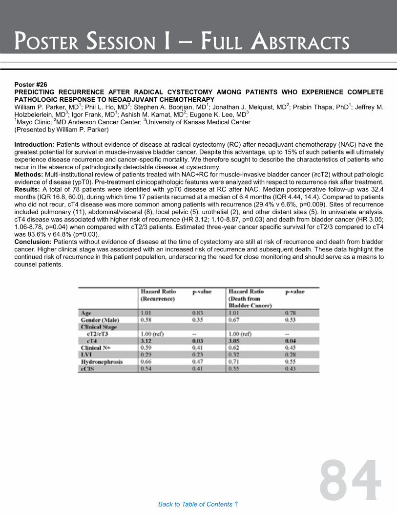
84Back to Table of Contents h
Poster session i – Full AbstrActs
Poster #26 PREDICTING RECURRENCE AFTER RADICAL CYSTECTOMY AMONG PATIENTS WHO EXPERIENCE COMPLETE PATHOLOGIC RESPONSE TO NEOADJUVANT CHEMOTHERAPY William P. Parker, MD1; Phil L. Ho, MD2; Stephen A. Boorjian, MD1; Jonathan J. Melquist, MD2; Prabin Thapa, PhD1; Jeffrey M. Holzbeierlein, MD3; Igor Frank, MD1; Ashish M. Kamat, MD2; Eugene K. Lee, MD3 1Mayo Clinic; 2MD Anderson Cancer Center; 3University of Kansas Medical Center (Presented by William P. Parker) Introduction: Patients without evidence of disease at radical cystectomy (RC) after neoadjuvant chemotherapy (NAC) have the greatest potential for survival in muscle-invasive bladder cancer. Despite this advantage, up to 15% of such patients will ultimately experience disease recurrence and cancer-specific mortality. We therefore sought to describe the characteristics of patients who recur in the absence of pathologically detectable disease at cystectomy. Methods: Multi-institutional review of patients treated with NAC+RC for muscle-invasive bladder cancer (≥cT2) without pathologic evidence of disease (ypT0). Pre-treatment clinicopathologic features were analyzed with respect to recurrence risk after treatment. Results: A total of 78 patients were identified with ypT0 disease at RC after NAC. Median postoperative follow-up was 32.4 months (IQR 16.8, 60.0), during which time 17 patients recurred at a median of 6.4 months (IQR 4.44, 14.4). Compared to patients who did not recur, cT4 disease was more common among patients with recurrence (29.4% v 6.6%, p=0.009). Sites of recurrence included pulmonary (11), abdominal/visceral (8), local pelvic (5), urothelial (2), and other distant sites (5). In univariate analysis, cT4 disease was associated with higher risk of recurrence (HR 3.12; 1.10-8.87, p=0.03) and death from bladder cancer (HR 3.05; 1.06-8.78, p=0.04) when compared with cT2/3 patients. Estimated three-year cancer specific survival for cT2/3 compared to cT4 was 83.6% v 64.8% (p=0.03). Conclusion: Patients without evidence of disease at the time of cystectomy are still at risk of recurrence and death from bladder cancer. Higher clinical stage was associated with an increased risk of recurrence and subsequent death. These data highlight the continued risk of recurrence in this patient population, underscoring the need for close monitoring and should serve as a means to counsel patients.
85Back to Table of Contents h
Poster session i – Full AbstrActs
Poster #27 NUTRITIONAL STATUS AND MAJOR ABDOMINAL SURGERIES: 30-DAY POSTOPERATIVE COMPLICATIONS Nicola Pavan, MD1; Carmen C. Mir, MD, PhD2; Chad R. Ritch, MD, MBA2; Samarpit Rai, MD2; Nachiketh Soodana-Prakash MD, MS2; Raymond R. Balise, PhD3; Carlo Trombetta, MD4; Dipen J. Parekh, MD2; Mark L. Gonzalgo MD, PhD2 1Department of Urology, University of Miami Leonard M. Miller School of Medicine, Miami, FL and Urology Clinic, Department of Medical, Surgical and Health Science, University of Trieste, Italy; 2Department of Urology, University of Miami Leonard M. Miller School of Medicine, Miami, FL; 3Division of Biostatistics, Department of Public Health Sciences, University of Miami Leonard M. Miller School of Medicine, Miami, FL; 4Urology Clinic, Department of Medical, Surgical and Health Science, University of Trieste, Italy (Presented by Samarpit Rai) Introduction: Patient nutritional status in the preoperative setting has been associated with the rate of postoperative complications. Patients with better nutritional status prior to surgery have been shown to have fewer perioperative complications compared to patients with poor nutritional status. We utilized the National Surgery Quality Improvement Program (NSQIP) database to determine whether preoperative factors such as nutritional status could predict the incidence of complications following major abdominal surgery. Methods: The NSQIP Database was queried using CPT codes to build a cohort of patients (N = 162,364) who underwent colectomy, cystectomy, and hysterectomy from 2009 to 2013. Exclusion criteria included ascites, disseminated cancer, chronic use of steroids, blood transfusion before serum albumin test, ventilator dependence, ASA 5, emergency procedures, and sepsis before surgery. Postoperative complications were classified according to Clavien-Dindo criteria. Multivariate analysis was performed to predict factors associated with major 30-day postoperative complications (Clavien-Dindo grade ≥ 3). Results: The distribution of abdominal surgeries was: open colectomy (28.7%), MIS colectomy (33.4%), open cystectomy (2.3%), MIS cystectomy (0.10%), open hysterectomy (15.7%) and MIS hysterectomy (19.9%). After adjusting for age, sex, smoking, and surgical approach, patients with >10% weight loss 6 months prior to surgery had a higher chance of developing major postoperative complications (OR 1.14, p < 0.053). Patients with below normal BMI also had a higher chance of developing major postoperative complications compared to patients with normal BMI (OR 1.36, p < 0.0003). Increased risk of major complications was observed in patients with moderate (OR 1.18, p < 0.01) or severe (OR 1.93, p < 0.0001) hypoalbuminemia, and for patients who were smokers (OR 1.38, p < 0.0001). Conclusion: Lower nutritional status significantly increases the risk of 30-day postoperative morbidity following abdominal surgery (colectomy, cystectomy, hysterectomy). Using a standardized predictive algorithm to identify, treat, and optimize nutritional status preoperatively may help to reduce the incidence of postoperative complications in patients undergoing abdominal surgery.
Poster #26 PREDICTING RECURRENCE AFTER RADICAL CYSTECTOMY AMONG PATIENTS WHO EXPERIENCE COMPLETE PATHOLOGIC RESPONSE TO NEOADJUVANT CHEMOTHERAPY William P. Parker, MD1; Phil L. Ho, MD2; Stephen A. Boorjian, MD1; Jonathan J. Melquist, MD2; Prabin Thapa, PhD1; Jeffrey M. Holzbeierlein, MD3; Igor Frank, MD1; Ashish M. Kamat, MD2; Eugene K. Lee, MD3 1Mayo Clinic; 2MD Anderson Cancer Center; 3University of Kansas Medical Center (Presented by William P. Parker) Introduction: Patients without evidence of disease at radical cystectomy (RC) after neoadjuvant chemotherapy (NAC) have the greatest potential for survival in muscle-invasive bladder cancer. Despite this advantage, up to 15% of such patients will ultimately experience disease recurrence and cancer-specific mortality. We therefore sought to describe the characteristics of patients who recur in the absence of pathologically detectable disease at cystectomy. Methods: Multi-institutional review of patients treated with NAC+RC for muscle-invasive bladder cancer (≥cT2) without pathologic evidence of disease (ypT0). Pre-treatment clinicopathologic features were analyzed with respect to recurrence risk after treatment. Results: A total of 78 patients were identified with ypT0 disease at RC after NAC. Median postoperative follow-up was 32.4 months (IQR 16.8, 60.0), during which time 17 patients recurred at a median of 6.4 months (IQR 4.44, 14.4). Compared to patients who did not recur, cT4 disease was more common among patients with recurrence (29.4% v 6.6%, p=0.009). Sites of recurrence included pulmonary (11), abdominal/visceral (8), local pelvic (5), urothelial (2), and other distant sites (5). In univariate analysis, cT4 disease was associated with higher risk of recurrence (HR 3.12; 1.10-8.87, p=0.03) and death from bladder cancer (HR 3.05; 1.06-8.78, p=0.04) when compared with cT2/3 patients. Estimated three-year cancer specific survival for cT2/3 compared to cT4 was 83.6% v 64.8% (p=0.03). Conclusion: Patients without evidence of disease at the time of cystectomy are still at risk of recurrence and death from bladder cancer. Higher clinical stage was associated with an increased risk of recurrence and subsequent death. These data highlight the continued risk of recurrence in this patient population, underscoring the need for close monitoring and should serve as a means to counsel patients.
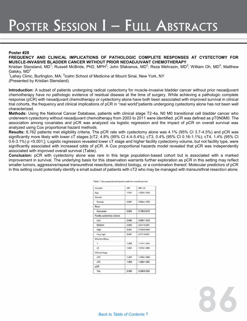
86Back to Table of Contents h
Poster session i – Full AbstrActs
Poster #28 FREQUENCY AND CLINICAL IMPLICATIONS OF PATHOLOGIC COMPLETE RESPONSES AT CYSTECTOMY FOR MUSCLE-INVASIVE BLADDER CANCER WITHOUT PRIOR NEOADJUVANT CHEMOTHERAPY Kristian Stensland, MD1; Russell McBride, PhD, MPH2; John Sfakianos, MD2; Reza Mehrazin, MD2; William Oh, MD2; Matthew Galsky, MD2 1Lahey Clinic, Burlington, MA; 2Icahn School of Medicine at Mount Sinai, New York, NY (Presented by Kristian Stensland) Introduction: A subset of patients undergoing radical cystectomy for muscle-invasive bladder cancer without prior neoadjuvant chemotherapy have no pathologic evidence of residual disease at the time of surgery. While achieving a pathologic complete response (pCR) with neoadjuvant chemotherapy or cystectomy alone have both been associated with improved survival in clinical trial cohorts, the frequency and clinical implications of pCR in “real world”patients undergoing cystectomy alone has not been well characterized. Methods: Using the National Cancer Database, patients with clinical stage T2-4a, N0 M0 transitional cell bladder cancer who underwent cystectomy without neoadjuvant chemotherapy from 2003 to 2011 were identified. pCR was defined as pT0N0M0. The association among covariates and pCR was analyzed via logistic regression and the impact of pCR on overall survival was analyzed using Cox proportional hazard methods. Results: 8,762 patients met eligibility criteria. The pCR rate with cystectomy alone was 4.1% (95% CI 3.7-4.5%) and pCR was significantly more likely with lower cT stages [cT2, 4.8% (95% CI 4.4-5.4%); cT3, 0.4% (95% CI 0.16-1.1%); cT4, 1.4% (95% CI 0.6-3.1%) p <0.001;]. Logistic regression revealed lower cT stage and higher facility cystectomy volume, but not facility type, were significantly associated with increased odds of pCR. A Cox proportional hazards model revealed that pCR was independently associated with improved overall survival (Table). Conclusion: pCR with cystectomy alone was rare in this large population-based cohort but is associated with a marked improvement in survival. The underlying basis for this observation warrants further exploration as pCR in this setting may reflect smaller tumors, aggressive/repeat transurethral resections, distinct biology, or a combination thereof. Molecular predictors of pCR in this setting could potentially identify a small subset of patients with cT2 who may be managed with transurethral resection alone.
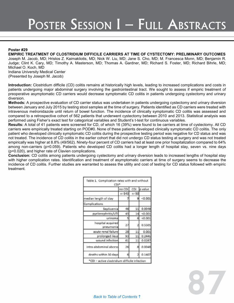
87Back to Table of Contents h
Poster session i – Full AbstrActs
Poster #29 EMPIRIC TREATMENT OF CLOSTRIDIUM DIFFICILE CARRIERS AT TIME OF CYSTECTOMY: PRELIMINARY OUTCOMES Joseph M. Jacob, MD; Hristos Z. Kaimakliotis, MD; Nick W. Liu, MD; Jane S. Cho, MD; M. Francesca Monn, MD; Benjamin R. Judge; Clint K. Cary, MD; Timothy A. Masterson, MD; Thomas A. Gardner, MD; Richard S. Foster, MD; Richard Bihrle, MD; Michael O. Koch, MD Indiana University Medical Center (Presented by Joseph M. Jacob) Introduction: Clostridium difficile (CD) colitis remains at historically high levels, leading to increased complications and costs in patients undergoing major abdominal surgery involving the gastrointestinal tract. We sought to assess if empiric treatment of preoperative asymptomatic CD carriers would decrease symptomatic CD colitis in patients undergoing cystectomy and urinary diversion. Methods: A prospective evaluation of CD carrier status was undertaken in patients undergoing cystectomy and urinary diversion between January and July 2015 by testing stool samples at the time of surgery. Patients identified as CD carriers were treated with intravenous metronidazole until return of bowel function. The incidence of clinically symptomatic CD colitis was assessed and compared to a retrospective cohort of 562 patients that underwent cystectomy between 2010 and 2013. Statistical analysis was performed using Fisher’s exact test for categorical variables and Student’s t-test for continuous variables. Results: A total of 41 patients were screened for CD, of which 16 (39%) were found to be carriers at time of cystectomy. All CD carriers were empirically treated starting on POD#0. None of these patients developed clinically symptomatic CD colitis. The only patient who developed clinically symptomatic CD colitis during the prospective testing period was negative for CD status and was not treated. The incidence of CD colitis in the earlier cohort that did not undergo CD status testing at surgery and was not treated empirically was higher at 8.8% (49/562). Ninety-four percent of CD carriers had at least one prior hospitalization compared to 64% among non-carriers (p=0.059). Patients who developed CD colitis had a longer length of hospital stay, seven vs. nine days (p<0.020), and higher rate of Clavien complications. Conclusion: CD colitis among patients undergoing cystectomy and urinary diversion leads to increased lengths of hospital stay with higher complication rates. Identification and treatment of asymptomatic carriers at time of surgery seems to decrease the incidence of CD colitis. Further studies are warranted to assess the utility and cost of testing for CD status followed with empiric treatment.
88Back to Table of Contents h
Poster session i – Full AbstrActs
Poster #30 FACTORS ASSOCIATED WITH SUICIDE IN PATIENTS WITH GENITOURINARY MALIGNANCIES Zachary Klaassen, MD1; Rita P. Jen, MD, MPH1; John M. DiBianco, MD1; Lael Reinstatler, MD, MPH1; Qiang Li MD, PhD1; Rabii Madi, MD1; Ronald W. Lewis, MD1; Arthur M. Smith, MD1; Durwood E. Neal, Jr., MD, FACS1; Kelvin A. Moses, MD, PhD2; Martha K. Terris, MD1 1Medical College of Georgia - Georgia Regents University, Augusta, GA; 2Department of Urological Surgery, Vanderbilt University Medical Center, Nashville, TN (Presented by Zachary Klaassen) Introduction: Approximately 70% of all suicides in patients aged >60 years are attributed to physical illness, with higher rates noted in patients with cancer. The purpose of the current study was to characterize suicide rates among patients with genitourinary cancers and identify factors associated with suicide in this specific cohort. Methods: Patients with prostate, bladder, kidney, testis, and penile cancer were identified in the Surveillance, Epidemiology, and End Results database (1988-2010). Standardized mortality ratios (SMRs) and 95% confidence intervals (95% CIs) were calculated for each anatomic site. Multivariable logistic regression models generated odds ratios (ORs) for the identification of factors associated with suicide for each malignancy. Results: There were 2268 suicides identified among 1,239,522 individuals with genitourinary malignancies observed for 7,307,377 person-years. The SMRs for patients with cancer were 1.37 for prostate cancer (95% CI, 0.99-1.86), 2.71 for bladder cancer (95% CI, 2.02-3.62), 1.86 for kidney cancer (95% CI, 1.32-2.62), 1.23 for testis cancer (95% CI, 0.88-1.73), and 0.95 for penile cancer (95% CI, 0.65-1.35). Patients with prostate cancer had an increase in the incidence of suicide over time, with the highest incidence observed at >15 years after diagnosis (SMR, 1.84; 95% CI, 1.39-2.41). Patients with bladder cancer had an increased incidence of suicide over all time periods, with the highest incidence noted within the first five years after diagnosis (SMR, 3.05; 95% CI, 2.26-3.96). On multivariable analysis, male sex was found to be associated with odds of suicide among patients with bladder cancer (OR, 6.63) and kidney cancer (OR, 4.98). Increasing age was associated with suicide for patients with prostate, bladder, and testis cancer (OR range, 1.03-1.06). Distant disease was associated with suicide in patients with prostate, bladder, and kidney cancer (OR range, 2.82-5.43). Among patients with prostate, bladder, and kidney cancer, African American patients were less likely to commit suicide compared with white individuals (OR range, 0.26-0.46). Conclusion: Suicide in patients with genitourinary malignancies poses a public health challenge, especially among men, the elderly, and those with aggressive disease. Clinicians should be aware of risk factors for suicide in these patients.

89Back to Table of Contents h
Poster session i – Full AbstrActs
Poster #31 DETERMINING PROSTATE CANCER RISK STRATA USING ONLY GLEASON SCORE AND PSA Justin Gregg, MD1; Max Lang, MD1; Sam Chang, MD1; Peter Clark, MD1; Michael Cookson, MD, MMHC2; S. Duke Herrell, MD1; David Penson, MD, MPH1; Matthew Resnick, MD, MPH1; Joseph Smith, MD1; Daniel Barocas, MD, MPH1 1Vanderbilt University Medical Center, Nashville, TN; 2University of Oklahoma College of Medicine, Oklahoma City, OK (Presented by Justin Gregg) Introduction: Risk stratification forms the basis of multiple prostate cancer quality indicators. Risk stratification typically incorporates digital rectal exam (DRE)-based tumor stage, Gleason score, and PSA. While automated extraction of these strata would enable more efficient calculation of patient risk groups and facilitate quality measurement, DRE findings are difficult to obtain from electronic notes. We therefore aimed to investigate the predictive accuracy of Gleason score and PSA, alone, at stratifying prostate cancer risk when compared to complete clinical data populated manually in a prospective clinical database. Methods: Patients who underwent radical prostatectomy between 2010 and 2015 were eligible for inclusion in the current study. Preoperative clinical T stage, Gleason score, and PSA were prospectively gathered in an electronic database and served as the reference for risk stratification. Patients were then placed into low, intermediate and high risk groups based on D’Amico risk criteria and modified criteria, which assumed that all tumor staging was cT1c. Accuracy of modified risk criteria was compared to the prospective reference using the weighted Kappa statistic (κ) Results: Of the 2,353 eligible patients, 2,344 (99.6%) had complete data available for analysis. 925 (39.5%), 1,016 (43.3) and 403 (17.2) patients had low, intermediate and high risk disease based on D’Amico risk criteria. Modified risk criteria had a 98.0% raw agreement with risk stratification (Κ=0.95, SE = 0.02) (Table). Modified risk criteria had 100%, 95.7% and 90.6% agreement with true risk stratification of low, intermediate and high risk disease, respectively. Conclusion: The combination of Gleason score and PSA value provides an accurate risk assessment of patients with prostate cancer. Validation of these findings would enable providers and organizations to easily extract risk stratification data from raw medical records, enabling streamlined measurement of quality indicators and performance feedback.
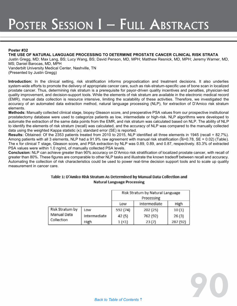
90Back to Table of Contents h
Poster session i – Full AbstrActs
Poster #32 THE USE OF NATURAL LANGUAGE PROCESSING TO DETERMINE PROSTATE CANCER CLINICAL RISK STRATA Justin Gregg, MD; Max Lang, BS; Lucy Wang, BS; David Penson, MD, MPH; Matthew Resnick, MD, MPH; Jeremy Warner, MD, MS; Daniel Barocas, MD, MPH Vanderbilt University Medical Center, Nashville, TN (Presented by Justin Gregg) Introduction: In the clinical setting, risk stratification informs prognostication and treatment decisions. It also underlies system-wide efforts to promote the delivery of appropriate cancer care, such as risk-stratum-specific use of bone scan in localized prostate cancer. Thus, determining risk stratum is a prerequisite for payor-driven quality incentives and penalties, physician-led quality improvement, and decision-support tools. While the elements of risk stratum are available in the electronic medical record (EMR), manual data collection is resource intensive, limiting the scalability of these activities. Therefore, we investigated the accuracy of an automated data extraction method, natural language processing (NLP), for extraction of D’Amico risk stratum elements. Methods: Manually collected clinical stage, biopsy Gleason score, and preoperative PSA values from our prospective institutional prostatectomy database were used to categorize patients as low, intermediate or high-risk. NLP algorithms were developed to automate the extraction of the same data points from the EMR, and risk stratum was calculated based on NLP. The ability of NLP to identify the elements of risk stratum (recall) was calculated, and the accuracy of NLP was compared to the manually collected data using the weighted Kappa statistic (κ); standard error (SE) is reported. Results: Obtained: Of the 2353 patients treated from 2010 to 2015, NLP identified all three elements in 1945 (recall = 82.7%). Among patients with all 3 elements, NLP had a 91.9% raw agreement with manual risk stratification (Κ=0.78, SE = 0.02) (Table). The κ for clinical T stage, Gleason score, and PSA extraction by NLP was 0.89, 0.89, and 0.87, respectively. 83.3% of extracted PSA values were within 1.0 ng/mL of manually collected PSA levels. Conclusion: NLP can achieve greater than 90% accuracy on D’Amico risk stratification of localized prostate cancer, with recall of greater than 80%. These figures are comparable to other NLP tasks and illustrate the known tradeoff between recall and accuracy. Automating the collection of risk characteristics could be used to power real-time decision support tools and to scale up quality measurement in cancer care.
91Back to Table of Contents h
Poster session i – Full AbstrActs
Poster #33 COMPETITION IN CANCER CARE: ADAPTING TO CHANGES IN PAYMENT Brock O'Neil, MD1; Amy Graves, SM, MPH2; Daniel Barocas, MD, MPH2; Sam Chang, MD2; David Penson, MD, MPH2; Matthew Resnick, MD, MPH2 1Vanderbilt University; 2Vanderbilt University, Nashville, TN (Presented by Brock O’Neil) Introduction: Regionalizing cancer care has the potential to reduce spending, increase care coordination, and improve outcomes but may result in the unintended consequence of decreasing competition. We previously examined changes in patterns of bladder cancer care as a result of fee-for-service incentives in Medicare physician payment and demonstrated inefficient alterations in practice with urologists performing more office-based minor cystoscopic procedures without reciprocal declines in the likelihood of treatment in facility-based settings. We sought to characterize the association of physician and market level factors with adoption of this financially incentivized practice to predict possible downstream changes associated with regionalization of cancer care. Methods: Using a five percent Medicare sample, we identified beneficiaries that underwent a minor cystoscopic procedure related to bladder cancer and for which there was a large financial incentive to perform in an office-based location. Urologists performing these procedures were then identified and linked to publically available datasets with information on provider and market characteristics. The outcomes of interest were the relationship of market competition and provider billing efficiency to the response in utilization of office-based procedures by individual urologists. Billing efficiency was measured by the number of total unique billing codes used and the number of codes used per beneficiary. Competition was assessed by urologist density (providers per 10,000 beneficiaries) and Herfindahl-Hirschman Index (HHI). Logistic regression models were used to create adjusted odds ratios, expressed as inter-quartile values. Results: Providers that increased use of office-based procedures had an increased odds (OR 1.19; 95% CI 1.04 to 1.35) of practicing in markets with the highest quartile of urologist density while increasing HHI was not associated with any demonstrable change (0.96; CI 0.87 to 1.05). Urologists that increased use of office-based procedures were also more likely to be in the highest quartile for use of unique billing codes (1.49; 1.32 to 1.69) and billing codes per patient (1.18; 1.11 to 1.25). Conclusion: Provider density, not HHI, is associated with revenue-maximizing behavior. Additionally, urologists responding to financial incentives are more likely to be facile in billing practices. Whether these practices reflect over-, under-, or appropriate billing remains to be determined.
Poster #32 THE USE OF NATURAL LANGUAGE PROCESSING TO DETERMINE PROSTATE CANCER CLINICAL RISK STRATA Justin Gregg, MD; Max Lang, BS; Lucy Wang, BS; David Penson, MD, MPH; Matthew Resnick, MD, MPH; Jeremy Warner, MD, MS; Daniel Barocas, MD, MPH Vanderbilt University Medical Center, Nashville, TN (Presented by Justin Gregg) Introduction: In the clinical setting, risk stratification informs prognostication and treatment decisions. It also underlies system-wide efforts to promote the delivery of appropriate cancer care, such as risk-stratum-specific use of bone scan in localized prostate cancer. Thus, determining risk stratum is a prerequisite for payor-driven quality incentives and penalties, physician-led quality improvement, and decision-support tools. While the elements of risk stratum are available in the electronic medical record (EMR), manual data collection is resource intensive, limiting the scalability of these activities. Therefore, we investigated the accuracy of an automated data extraction method, natural language processing (NLP), for extraction of D’Amico risk stratum elements. Methods: Manually collected clinical stage, biopsy Gleason score, and preoperative PSA values from our prospective institutional prostatectomy database were used to categorize patients as low, intermediate or high-risk. NLP algorithms were developed to automate the extraction of the same data points from the EMR, and risk stratum was calculated based on NLP. The ability of NLP to identify the elements of risk stratum (recall) was calculated, and the accuracy of NLP was compared to the manually collected data using the weighted Kappa statistic (κ); standard error (SE) is reported. Results: Obtained: Of the 2353 patients treated from 2010 to 2015, NLP identified all three elements in 1945 (recall = 82.7%). Among patients with all 3 elements, NLP had a 91.9% raw agreement with manual risk stratification (Κ=0.78, SE = 0.02) (Table). The κ for clinical T stage, Gleason score, and PSA extraction by NLP was 0.89, 0.89, and 0.87, respectively. 83.3% of extracted PSA values were within 1.0 ng/mL of manually collected PSA levels. Conclusion: NLP can achieve greater than 90% accuracy on D’Amico risk stratification of localized prostate cancer, with recall of greater than 80%. These figures are comparable to other NLP tasks and illustrate the known tradeoff between recall and accuracy. Automating the collection of risk characteristics could be used to power real-time decision support tools and to scale up quality measurement in cancer care.
92Back to Table of Contents h
Poster session i – Full AbstrActs
Poster #34 DIFFERENCES IN PROSTATE SPECIFIC ANTIGEN TESTING AMONG UROLOGISTS AND PRIMARY CARE PROVIDERS IN THE UNITED STATES FOLLOWING THE 2011 USPSTF RECOMMENDATIONS Nawar Hanna, MD1; Micheal E. Zavaski, MD1; Christian Meyer, MD1; Jesse D. Sammon, MD2; Soham Gupta, MD2; Maxine Sun, PhD1; Quoc-Dien Trinh, MD1 1Brigham and Women's Hospital, Boston, MA; 2Vattikuti Urology Institute, Detroit, MI (Presented by Nawar Hanna) Introduction: The use of prostate specific antigen (PSA) testing for early detection of prostate cancer remains controversial and the subject of intense scrutiny. In 2011, the USPSTF issued a grade ‘D’ recommendation against screening for all men. To further understand the heterogeneity in screening practices, we examine the use of PSA testing among urologists vs. primary care providers (PCPs) before and after the latest USPSTF recommendations. Methods: We utilized the National Ambulatory Medical Care Survey (NAMCS) to examine the use of PSA testing in 2010 and 2012. We included all outpatient visits for men aged 50 and older who presented to a urologist or a PCP for a ‘preventive care’ visit. Men with a diagnosis of prostate cancer (n=31) and elevated PSA (n=1) were excluded. This resulted in a weighted sample of 34.4 (n=1545) million eligible visits in 2010 and 2012. We examined the frequency of PSA testing according to provider specialty using a difference-in-differences analytic approach to evaluate whether the 2011 USPSTF recommendations were associated with a decrease in PSA testing in men <75. Results were weighted to reflect the US population based on the complex survey design. Results: In men aged 50-74, the use of PSA testing decreased from 35.6 to 16.3% among primary care visits, whereas it decreased from 43.9 to 37.5% among urology visits. When a difference-in-differences approach was used, the decrease in PSA testing was significant among primary care physicians (p<0.001), but not among urologists (p=0.084). Conclusion: Our findings suggest a differential effect of the 2011 USPSTF recommendations on PSA screening in PCPs vs. urologists. Such findings likely reflect opposing provider perceptions on the benefit of PSA screening, conflicting guidelines and perhaps differences in patient demographics or expectations. Moving forward, this emphasizes the need to continue the dialogue between PCPs and urologists to achieve a consensus on PSA screening.
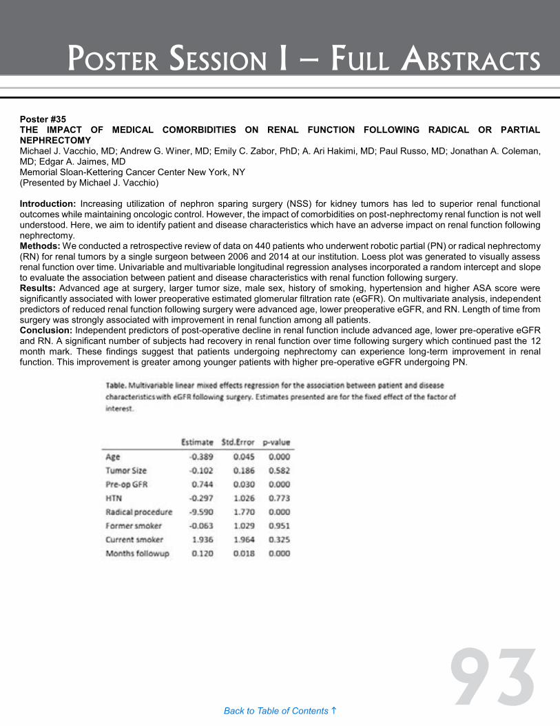
93Back to Table of Contents h
Poster session i – Full AbstrActs
Poster #35 THE IMPACT OF MEDICAL COMORBIDITIES ON RENAL FUNCTION FOLLOWING RADICAL OR PARTIAL NEPHRECTOMY Michael J. Vacchio, MD; Andrew G. Winer, MD; Emily C. Zabor, PhD; A. Ari Hakimi, MD; Paul Russo, MD; Jonathan A. Coleman, MD; Edgar A. Jaimes, MD Memorial Sloan-Kettering Cancer Center New York, NY (Presented by Michael J. Vacchio) Introduction: Increasing utilization of nephron sparing surgery (NSS) for kidney tumors has led to superior renal functional outcomes while maintaining oncologic control. However, the impact of comorbidities on post-nephrectomy renal function is not well understood. Here, we aim to identify patient and disease characteristics which have an adverse impact on renal function following nephrectomy. Methods: We conducted a retrospective review of data on 440 patients who underwent robotic partial (PN) or radical nephrectomy (RN) for renal tumors by a single surgeon between 2006 and 2014 at our institution. Loess plot was generated to visually assess renal function over time. Univariable and multivariable longitudinal regression analyses incorporated a random intercept and slope to evaluate the association between patient and disease characteristics with renal function following surgery. Results: Advanced age at surgery, larger tumor size, male sex, history of smoking, hypertension and higher ASA score were significantly associated with lower preoperative estimated glomerular filtration rate (eGFR). On multivariate analysis, independent predictors of reduced renal function following surgery were advanced age, lower preoperative eGFR, and RN. Length of time from surgery was strongly associated with improvement in renal function among all patients. Conclusion: Independent predictors of post-operative decline in renal function include advanced age, lower pre-operative eGFR and RN. A significant number of subjects had recovery in renal function over time following surgery which continued past the 12 month mark. These findings suggest that patients undergoing nephrectomy can experience long-term improvement in renal function. This improvement is greater among younger patients with higher pre-operative eGFR undergoing PN.
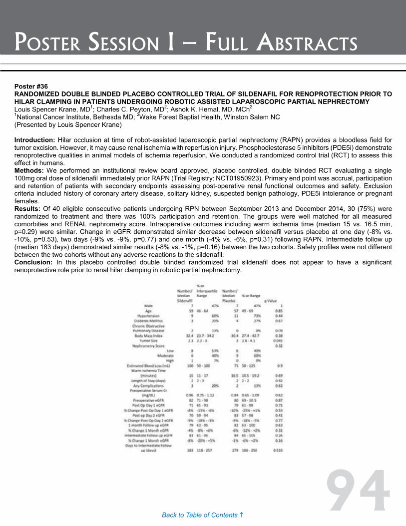
94Back to Table of Contents h
Poster session i – Full AbstrActs
Poster #36 RANDOMIZED DOUBLE BLINDED PLACEBO CONTROLLED TRIAL OF SILDENAFIL FOR RENOPROTECTION PRIOR TO HILAR CLAMPING IN PATIENTS UNDERGOING ROBOTIC ASSISTED LAPAROSCOPIC PARTIAL NEPHRECTOMY Louis Spencer Krane, MD1; Charles C. Peyton, MD2; Ashok K. Hemal, MD, MCh2 1National Cancer Institute, Bethesda MD; 2Wake Forest Baptist Health, Winston Salem NC (Presented by Louis Spencer Krane) Introduction: Hilar occlusion at time of robot-assisted laparoscopic partial nephrectomy (RAPN) provides a bloodless field for tumor excision. However, it may cause renal ischemia with reperfusion injury. Phosphodiesterase 5 inhibitors (PDE5i) demonstrate renoprotective qualities in animal models of ischemia reperfusion. We conducted a randomized control trial (RCT) to assess this effect in humans. Methods: We performed an institutional review board approved, placebo controlled, double blinded RCT evaluating a single 100mg oral dose of sildenafil immediately prior RAPN (Trial Registry: NCT01950923). Primary end point was accrual, participation and retention of patients with secondary endpoints assessing post-operative renal functional outcomes and safety. Exclusion criteria included history of coronary artery disease, solitary kidney, suspected benign pathology, PDE5i intolerance or pregnant females. Results: Of 40 eligible consecutive patients undergoing RPN between September 2013 and December 2014, 30 (75%) were randomized to treatment and there was 100% participation and retention. The groups were well matched for all measured comorbities and RENAL nephrometry score. Intraoperative outcomes including warm ischemia time (median 15 vs. 16.5 min, p=0.29) were similar. Change in eGFR demonstrated similar decrease between sildenafil versus placebo at one day (-8% vs. -10%, p=0.53), two days (-9% vs. -9%, p=0.77) and one month (-4% vs. -6%, p=0.31) following RAPN. Intermediate follow up (median 183 days) demonstrated similar results (-8% vs. -1%, p=0.16) between the two cohorts. Safety profiles were not different between the two cohorts without any adverse reactions to the sildenafil. Conclusion: In this placebo controlled double blinded randomized trial sildenafil does not appear to have a significant renoprotective role prior to renal hilar clamping in robotic partial nephrectomy.
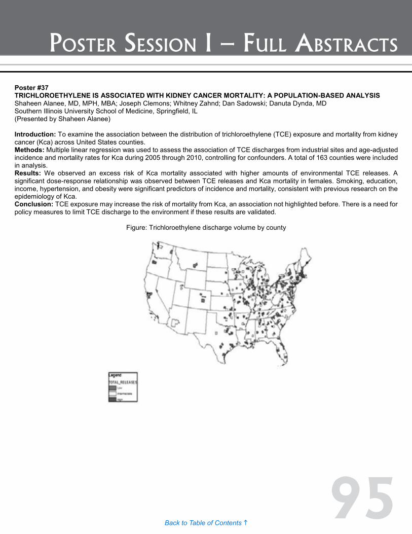
95Back to Table of Contents h
Poster session i – Full AbstrActs
Poster #37 TRICHLOROETHYLENE IS ASSOCIATED WITH KIDNEY CANCER MORTALITY: A POPULATION-BASED ANALYSIS Shaheen Alanee, MD, MPH, MBA; Joseph Clemons; Whitney Zahnd; Dan Sadowski; Danuta Dynda, MD Southern Illinois University School of Medicine, Springfield, IL (Presented by Shaheen Alanee) Introduction: To examine the association between the distribution of trichloroethylene (TCE) exposure and mortality from kidney cancer (Kca) across United States counties. Methods: Multiple linear regression was used to assess the association of TCE discharges from industrial sites and age-adjusted incidence and mortality rates for Kca during 2005 through 2010, controlling for confounders. A total of 163 counties were included in analysis. Results: We observed an excess risk of Kca mortality associated with higher amounts of environmental TCE releases. A significant dose-response relationship was observed between TCE releases and Kca mortality in females. Smoking, education, income, hypertension, and obesity were significant predictors of incidence and mortality, consistent with previous research on the epidemiology of Kca. Conclusion: TCE exposure may increase the risk of mortality from Kca, an association not highlighted before. There is a need for policy measures to limit TCE discharge to the environment if these results are validated.
Figure: Trichloroethylene discharge volume by county
Poster #36 RANDOMIZED DOUBLE BLINDED PLACEBO CONTROLLED TRIAL OF SILDENAFIL FOR RENOPROTECTION PRIOR TO HILAR CLAMPING IN PATIENTS UNDERGOING ROBOTIC ASSISTED LAPAROSCOPIC PARTIAL NEPHRECTOMY Louis Spencer Krane, MD1; Charles C. Peyton, MD2; Ashok K. Hemal, MD, MCh2 1National Cancer Institute, Bethesda MD; 2Wake Forest Baptist Health, Winston Salem NC (Presented by Louis Spencer Krane) Introduction: Hilar occlusion at time of robot-assisted laparoscopic partial nephrectomy (RAPN) provides a bloodless field for tumor excision. However, it may cause renal ischemia with reperfusion injury. Phosphodiesterase 5 inhibitors (PDE5i) demonstrate renoprotective qualities in animal models of ischemia reperfusion. We conducted a randomized control trial (RCT) to assess this effect in humans. Methods: We performed an institutional review board approved, placebo controlled, double blinded RCT evaluating a single 100mg oral dose of sildenafil immediately prior RAPN (Trial Registry: NCT01950923). Primary end point was accrual, participation and retention of patients with secondary endpoints assessing post-operative renal functional outcomes and safety. Exclusion criteria included history of coronary artery disease, solitary kidney, suspected benign pathology, PDE5i intolerance or pregnant females. Results: Of 40 eligible consecutive patients undergoing RPN between September 2013 and December 2014, 30 (75%) were randomized to treatment and there was 100% participation and retention. The groups were well matched for all measured comorbities and RENAL nephrometry score. Intraoperative outcomes including warm ischemia time (median 15 vs. 16.5 min, p=0.29) were similar. Change in eGFR demonstrated similar decrease between sildenafil versus placebo at one day (-8% vs. -10%, p=0.53), two days (-9% vs. -9%, p=0.77) and one month (-4% vs. -6%, p=0.31) following RAPN. Intermediate follow up (median 183 days) demonstrated similar results (-8% vs. -1%, p=0.16) between the two cohorts. Safety profiles were not different between the two cohorts without any adverse reactions to the sildenafil. Conclusion: In this placebo controlled double blinded randomized trial sildenafil does not appear to have a significant renoprotective role prior to renal hilar clamping in robotic partial nephrectomy.
96Back to Table of Contents h
Poster session i – Full AbstrActs
Poster #38 UPPER TRACT UROTHELIAL CARCINOMA LOCATED IN THE RENAL PELVIS HAS WORSE CLINICAL OUTCOMES COMPARED TO THE URETER: A POPULATION-BASED ANALYSIS Zachary Klaassen, MD; Benjamin T. Harper; Rita P. Jen, MD, MPH; Grace Yaguchi; Ross Everett, MPH; John M. DiBianco, MD; Martha K. Terris, MD; Rabii Madi, MD Medical College of Georgia - Georgia Regents University, Augusta, GA (Presented by Zachary Klaassen) Introduction: The prognostic impact of upper tract urothelial carcinoma (UTUC) tumor location is not well defined, however, most studies suggest ureteral tumors have inferior outcomes to renal pelvis tumors. The purpose of this study was to compare long-term outcomes of UTUC patients by tumor location and identify factors associated with distant metastasis at diagnosis. Methods: Patients diagnosed with UTUC were identified in the SEER database from 2004 to 2011 (n=8,805). Descriptive statistics were used to compare demographic and clinicopathologic variables between patients with non-metastatic renal pelvis and ureteral UTUC. Overall survival (OS) and disease-specific survival (DSS) were compared for patients with non-metastatic disease at diagnosis using log-rank test and Kaplan Meier analysis. Multivariable logistic regression models with backward selection were performed to generate odds ratios (OR) for factors associated with metastasis at diagnosis. Results: There were 5,444 (61.8%) patients with renal pelvis and 3,361 (38.2%) with ureteral UTUC. Among these patients, there were 4,745 (87.2%) with renal pelvis and 3,124 (92.9%) with ureteral UTUC that had non-metastatic disease at diagnosis. Patients with non-metastatic ureteral UTUC at diagnosis were more likely to be older (median 75 (IQR 14) vs. 74 (IQR 16) years, p=0.001), male (61.3 vs. 58.6%, p=0.02), and non-black or white race (7.7 vs. 6.2%, p=0.03) compared to those with renal pelvis UTUC. Patients with ureteral UTUC were more likely to have AJCC Stage I disease and less likely to have Stage III disease (Stage I - 36.1 vs. 33.1%; Stage III – 23.0 vs. 32.4%, p<0.001). There was no difference in OS (p=0.72), however patients with renal pelvis UTUC had worse DSS (p=0.01). Factors associated with distant metastasis at diagnosis for renal pelvis UTUC included black race (OR 1.74, 95%CI 1.14-2.65; p=0.01) and high-grade disease (OR 1.42, 95%CI 1.25-1.62; p<0.001). High-grade disease (OR 1.29, 95%CI 1.07-1.57; p=0.009) was the only factor associated with distant metastasis at diagnosis for patients with ureteral UTUC. Conclusion: Patients with non-metastatic UTUC at diagnosis have inferior DSS if the tumor is in the renal pelvis compared to the ureter, suggesting the renal pelvis may not be protective from aggressive tumor behavior. Furthermore, grade of disease is an independent factor associated with metastatic disease at diagnosis for both patients with renal pelvis and ureteral UTUC.
97Back to Table of Contents h
Poster session i – Full AbstrActs
Poster #39 RENAL CELL TUMOR-MEDIATED CONVERSION OF NATURAL KILLER CELLS TO A PROANGIOGENIC PHENOTYPE BY TRANSFORMING GROWTH FACTOR-Β AND HYPOXIA Yue Guan1; Christopher Chambers1; Britnie James2; Purba Singh1; Donald Reed1; Kathy Robinson3; Shaheen Alanee MD, MPH, MBA4; Thomas Griffith2; Donald Torry1; Andrew Wilber1 1Department of Medical Microbiology, Immunology and Cell Biology, Southern Illinois University School of Medicine, Springfield, IL; 2Department of Microbiology, Immunology and Cancer Biology Graduate Program and Department of Urology, University of Minnesota, Minneapolis, MN; 3Simmons Cancer Institute, Springfield, IL; 4Southern Illinois University School of Medicine, Springfield, IL (Presented by Shaheen Alanee) Introduction: Natural killer (NK) cells are classically associated with immune surveillance and destruction of tumor cells via cytotoxicity. Inconsistent with this function, influxes of NK cells are found in advanced renal cell carcinoma (RCC) tumors. NK cells with non-classical phenotypes (CD56+CD16dim/neg; termed decidua NK or dNK cells) accumulate at the maternal-fetal interface during implantation. These dNK cells are poorly cytotoxic, proangiogenic, and facilitate placentation; effects mediated, in part, by transforming growth factor beta (TGFβ). We investigated whether an analogous shift in NK cell phenotype/function occurs in RCC tumors potentiated by tumor-derived TGFβ and hypoxia. Methods: NK cells from peripheral blood (pNK) and resected tumor tissue (TiNK) of RCC patients were compared to pNK from healthy, tumor-free donors. pNK cells were cultured in the absence or presence of TGFβ and exposed to 21% or 1% oxygen to assess conversion by monitoring expression of surface markers and angiogenic genes as well as ability to directly kill target cells. An orthotopic mouse model of RCC was also used, where the murine renal adenocarcinoma cell line, Renca, was implanted directly into the kidneys of Balb/c mice. Results: pNK cells of healthy donors were CD56+CD16+ (94+1%) and cytotoxic, but acquired characteristics of dNK cells (reduced cytotoxicity and augmented vascular endothelial growth factor (VEGF) expression) when exposed to TGFβ or 1% O2. Addition of TGFβ to NK cells maintained in 1% O2 further suppressed cytotoxic function and induced expression of urokinase plasminogen activator (uPA) and its inhibitor PAI-1, two factors with known roles in tumor progression. pNK cells of RCC patients were phenotypical similar to healthy donors (89+2%, CD56+CD16+), but lacked full cytotoxic ability which we attributed to 3-fold higher levels of TGFβ in patient serum. RCC TiNK cells were significantly enriched for dNK-like CD56+CD16dim/neg cells (47+12%) had limited cytotoxic capacity and increased VEGF and uPA expression. Conclusion: These studies support a role for TGFβ and hypoxia in conversion of pNK cells to a dNK-like phenotype in RCC tumors. While these characteristics are conceivably beneficial for placentation, they may be exploited to support RCC growth and metastasis. Thus, efforts designed to neutralize the effects of TGFβ may be an important goal of future immunotherapies.
98Back to Table of Contents h
Poster session i – Full AbstrActs
Poster #42 RADIOGRAPHIC SIZE OF RETROPERITONEAL LYMPH NODES PREDICTS PATHOLOGIC NODAL INVOLVEMENT FOR PATIENTS UNDERGOING RADICAL NEPHRECTOMY FOR RENAL CELL CARCINOMA: DEVELOPMENT OF A RISK PREDICTION MODEL Boris Gershman, MD; Naoki Takahashi, MD; Daniel Moreira, MD; R. Houston Thompson, MD; Stephen Boorjian, MD; Christine Lohse, MS; Brian Costello, MD; John Cheville, MD; Bradley Leibovich, MD Mayo Clinic, Rochester, MN (Presented by Boris Gershman) Introduction: Preoperative risk-stratification to predict lymph node metastases from renal cell carcinoma (RCC) is essential to guide patient selection for lymph node dissection at the time of nephrectomy. We therefore evaluated the ability of radiographic features to predict nodal (pN1) disease, and developed a preoperative risk prediction model using both clinical and radiographic features. Methods: A total of 220 patients who underwent radical nephrectomy with lymph node dissection from 2000-2010 with preoperative computed tomography (CT) scans available for review were identified. Preoperative radiographic features were assessed by one genitourinary radiologist blinded to pN status. Associations of clinical and radiographic features with pN1 disease were evaluated using logistic regression. Univariable and multivariable predictive models were developed, and model performance was assessed using the AUC and decision curve analysis. Results: Median lymph node yield was 10 (IQR 5-18). Fifty-five (25%) patients had pN1 disease at nephrectomy. On univariable analysis, maximum lymph node (LN) short axis diameter (OR 1.17; p<0.001) predicted pN1 disease with an AUC of 0.84 (Figure). Although a number of clinical and radiographic features were associated with pN1 disease, only two were retained in the multivariable model: maximum LN short axis diameter (OR 1.19; p<0.001) and radiographic perinephric/renal sinus fat invasion (OR 44.64; p=0.01), with an AUC of 0.85. On decision curve analysis, the single variable and multivariable models demonstrated similar net benefit. Conclusion: Two radiographic features, maximum LN short axis diameter and perinephric/renal sinus fat invasion, outperformed traditional clinical variables in predicting pN1 risk. Maximum LN short axis diameter alone demonstrated excellent predictive performance, and, if validated externally, would provide for a simple model to guide patient selection for lymph node dissection.
Poster #40 CHARACTERISTICS OF ISOLATED LOW GRADE UPPER TRACT UROTHELIAL CARCINOMA: SESSILE TUMOR ARCHITECTURE IS ASSOCIATED WITH ADVERSE ONCOLOGIC OUTCOMES Ryan Hutchinson, MD; Nirmish Singla, MD; Ahmed Haddad, MD, PhD; Vitaly Margulis, MD UTSW, Dallas, Texas (Presented by Ryan Hutchinson) Introduction: Low grade upper tract urothelial carcinoma can be managed by a variety of means, with increasing recent emphasis on endoscopic management in amenable tumors. We sought to examine a large, multi-institutional database of low grade upper tract urothelial carcinomas treated surgically and evaluate for factors associated with adverse clinical outcomes. Methods: Patients with isolated low-grade upper tract urothelial carcinoma without previous lower tract disease or CIS undergoing radical nephroureterectomy or partial ureterectomy were identified from a multi-institutional database spanning 13 academic institutions on three continents. Descriptive statistics on patient characteristics were obtained and Cox regression analyses were performed on disease-specific variables like tumor architecture and prior endoscopic treatment of tumor. Results: 350 patients were identified. Mean follow up was 66.8 months. During the follow up period 88 patients (25.1%) died including 24 (6.9%) of disease. Tumor location and prior endoscopic management of disease were not associated with decreased disease specific or overall survival. Sessile tumor architecture was identified in 16 patients (4.6%) and was associated with decreased cancer-specific survival on Cox regression analysis (p=0.003, HR = 5.23). Conclusion: We report clinical and pathologic outcomes of patients with isolated low-grade upper tract urothelial carcinoma treated by partial or radical ureterectomy. Previous endoscopic management before ureterectomy does not appear associated with adverse clinical outcomes. Sessile tumor architecture is associated with decreased disease specific survival. Poster #41 INTEGRIN SIGNALING POTENTIATES TRANSFORMING GROWTH FACTOR BETA 1 DEPENDENT DOWN REGULATION OF E-CADHERIN EXPRESSION- IMPLICATIONS FOR EPITHELIAL TO MESENCHYMAL TRANSITION IN RENAL CELL CARCINOMA Ryan Hutchinson, MD; Boris Feldkoren, PhD; Yury Rapaport, MD; Vitaly Margulis, MD UTSW, Dallas, Texas (Presented by Ryan Hutchinson) Introduction: Signal transduction through the transforming growth factor-beta 1 (TGF-β1) pathway affects epithelial to mesenchymal transition (EMT) in human tumors partly by modulation of E-Cadherin expression. The concurrent impact of extracellular matrix driven regulation of integrin signaling on EMT has not been well characterized. We assessed the cumulative effect and molecular mechanisms of TGF-β1 and integrin signal transduction on E-Cadherin and other EMT markers’ expression in a renal cell cancer (RCC) model. Methods: TGF-β1 driven alteration of EMT specific markers was confirmed in an established in-vitro model of RCC. A ligand of integrin αv-β3, cyclo pentapeptide containing arginyl-glycyl-aspartic acid (RGD), in addition to TGF-β1, was utilized to mimic integrin signaling and evaluate the cumulative effect of simultaneous TGF-β1 and integrin αv stimulation on markers of EMT. Subsequent silencing of potential downstream mediators of the additive action of RGD and TGF-β1 was carried out by small interfering RNA transfection against FAK and PINCH1 and was confirmed by Western blotting or RT-PCR. Results: Stimulation of RCC cells with TGF-β1 demonstrated a three-fold increased expression of integrin αv. Treatment of cells with RGD and TGF-β1 demonstrated significantly higher effect on markers of EMT (E-cadherin depletion and Snail1 augmentation) than either ligand alone (p < 0.05). SiRNA mediated silencing of FAK and PINCH1 independently abrogated the cumulative effect of RGD and TGF-β1 on E-Cadherin expression (p < 0.01). Conclusion: We have identified a novel mechanism through which extracellular matrix event transduction by integrins further augments TGF-β1 related effects on well-established markers of EMT. Molecular machinery involved in the integrin αv-TGF-β1 interplay may represent a therapeutic target in RCC.
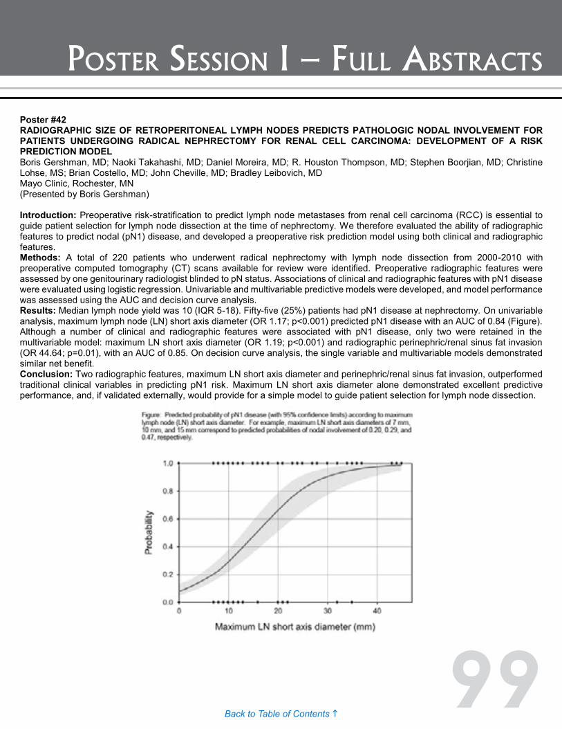
99Back to Table of Contents h
Poster session i – Full AbstrActs
Poster #42 RADIOGRAPHIC SIZE OF RETROPERITONEAL LYMPH NODES PREDICTS PATHOLOGIC NODAL INVOLVEMENT FOR PATIENTS UNDERGOING RADICAL NEPHRECTOMY FOR RENAL CELL CARCINOMA: DEVELOPMENT OF A RISK PREDICTION MODEL Boris Gershman, MD; Naoki Takahashi, MD; Daniel Moreira, MD; R. Houston Thompson, MD; Stephen Boorjian, MD; Christine Lohse, MS; Brian Costello, MD; John Cheville, MD; Bradley Leibovich, MD Mayo Clinic, Rochester, MN (Presented by Boris Gershman) Introduction: Preoperative risk-stratification to predict lymph node metastases from renal cell carcinoma (RCC) is essential to guide patient selection for lymph node dissection at the time of nephrectomy. We therefore evaluated the ability of radiographic features to predict nodal (pN1) disease, and developed a preoperative risk prediction model using both clinical and radiographic features. Methods: A total of 220 patients who underwent radical nephrectomy with lymph node dissection from 2000-2010 with preoperative computed tomography (CT) scans available for review were identified. Preoperative radiographic features were assessed by one genitourinary radiologist blinded to pN status. Associations of clinical and radiographic features with pN1 disease were evaluated using logistic regression. Univariable and multivariable predictive models were developed, and model performance was assessed using the AUC and decision curve analysis. Results: Median lymph node yield was 10 (IQR 5-18). Fifty-five (25%) patients had pN1 disease at nephrectomy. On univariable analysis, maximum lymph node (LN) short axis diameter (OR 1.17; p<0.001) predicted pN1 disease with an AUC of 0.84 (Figure). Although a number of clinical and radiographic features were associated with pN1 disease, only two were retained in the multivariable model: maximum LN short axis diameter (OR 1.19; p<0.001) and radiographic perinephric/renal sinus fat invasion (OR 44.64; p=0.01), with an AUC of 0.85. On decision curve analysis, the single variable and multivariable models demonstrated similar net benefit. Conclusion: Two radiographic features, maximum LN short axis diameter and perinephric/renal sinus fat invasion, outperformed traditional clinical variables in predicting pN1 risk. Maximum LN short axis diameter alone demonstrated excellent predictive performance, and, if validated externally, would provide for a simple model to guide patient selection for lymph node dissection.
100Back to Table of Contents h
Poster session i – Full AbstrActs
Poster #43 KIDNEY CANCER IN RURAL ILLINOIS: LOWER INCIDENCE YET HIGHER MORTALITY RATES Daniel Sadowski, MD, MPhil1; Scott Geiger, BS1; Georgia Mueller, MS2; Whitney Zahnd, MS2; Shaheen Alanee, MD, MPH1; Kevin McVary, MD1 1Southern Illinois University School of Medicine, Division of Urology, Springfield, Illinois; 2Southern Illinois University School of Medicine, Center for Clinical Research, Springfield, Illinois (Presented by Daniel Sadowski) Introduction: We aimed to determine if rural status was associated with kidney cancer (KCa) incidence and mortality in Illinois while controlling for known KCa risk factors and access to care variables. Methods: Age-adjusted KCa incidence rates from 1991 to 2010 were calculated from Illinois State Cancer Registry data. Age-adjusted KCa mortality rates were obtained from health statistics embedded within SEER*Stat. Rural Urban Continuum Codes designated Illinois’ 102 counties as urban, rural adjacent to a metropolitan area, and rural non-adjacent to a metropolitan area. County-level demographics and physician density were obtained from the Area Health Resource File. Behavioral Risk Factor Surveillance System data were used for smoking, obesity, and hypertension prevalence. Analysis of Variance (ANOVA), correlation, and regression analyses were used. Results: The incidence of KCa was found to be higher among urban compared to rural counties after controlling for known risk factors (p<0.01). A larger proportion of cases were diagnosed at a localized stage in urban counties (<0.01). Mortality rates were significantly higher in rural counties (p=0.02). The final regression model found rural status, higher incidence rate, lower percentage of localized stage at diagnosis, and lower urologist density to be variables significantly associated with higher KCa mortality rate. Conclusion: KCa incidence was higher in urban counties while mortality was higher in rural areas. Higher rates of localized KCa were found in the urban sample, which may be explained by greater radiologic utilization resulting in earlier detection. The incidence of localized KCa in the urban cohort coupled with lower urologist density in rural counties highlights a disparity in access to care that may be responsible for the increased KCa mortality in rural Illinois. Telemedicine may be an opportunity to improve this disparity.
101Back to Table of Contents h
Poster session i – Full AbstrActs
Poster #44 THE PROGNOSTIC IMPACT OF A POSITIVE VASCULAR MARGIN IN PT3 CLEAR CELL RENAL CARCINOMAS Nick Liu, MD1; James Wren, MD2; Emily Vertosick, MS2; Justin Lee, MD2; Nick Power, MD2; Simon Kimm, MD2; Dan Sjoberg, MD2; Paul Russo, MD2; Coleman Jonathan, MD2 1New York; 2Memorial Sloan-Kettering Cancer Center (Presented by Nick Liu) Introduction: There is a wide spectrum of vascular involvement of tumor thrombus in patients with pT3 clear cell carcinoma which includes muscular branch invasion within the kidney to large volume tumor thrombus into the inferior vena cava (IVC). The finding of positive vascular margin (PVM) from surgical specimens in this setting is often reported but poorly defined and of uncertain clinical significance. We examined the impact of PVM in pT3 patients managed at our institution. Methods: We identified 224 patients with venous tumor invasion through our institutional kidney cancer database from January 1999 to June 2013. We included those with metastasis, other positive surgical margin, lymph node involvement, neoadjuvant therapy, or non-clear cell histology. Kaplan-Meier analysis and log rank tests were used to evaluate whether PVM was associated with PFS or CSS. Results: PVMs were present in 41 patients (18%) including 12 main renal vein (MRV) and 29 inferior vena cava (IVC) invasions and were directly related to extent of vascular thrombus invasion (p<0.0001). The median follow up was 3.2 years (IQR: 1.6–5.6) for surviving patients. Compared to the NVM group, PVMs were adversely associated with PFS (p=0.01) but not significantly associated with CSS (p=0.3). Higher level of vascular thrombus invasion was associated with a worse PFS (p=0.02) but not significantly associated with CSS (p=0.4). The 3-year PFS was least favorable for IVC invasion and most favorable for MVB invasion (54% [95% CI: 34–70%] vs. 76% [95% CI: 64–85%]). In patients with MRV thrombus only, PVM was not associated with an inferior PFS (p=0.5) or CSS (p=0.2). Conclusion: In the setting of pT3N0/XM0 clear cell RCC, the presence of a PVM is associated with risk for disease progression but no impact on survival was detected. However, the risk of relapse associated with PVM is driven by extent of vascular thrombus invasion. These findings suggest that the clinical significance of reporting vascular margin status in pT3 clear cell RCC using the current definition is minimal.
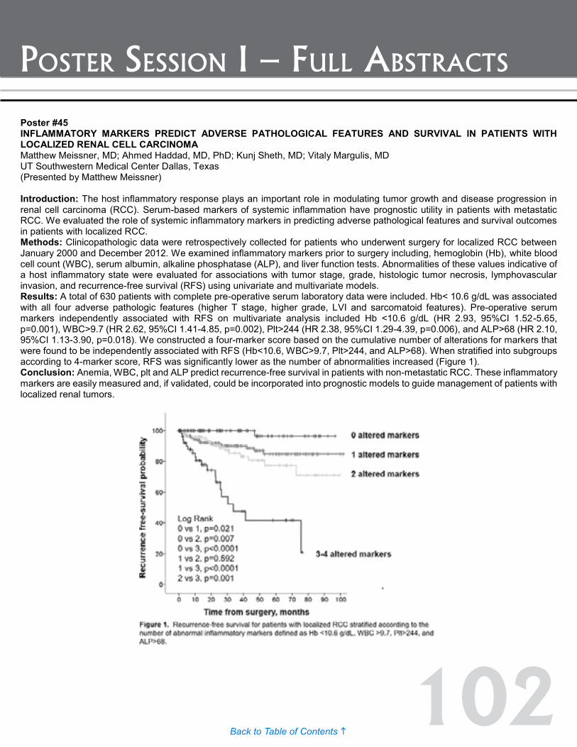
102Back to Table of Contents h
Poster session i – Full AbstrActs
Poster #45 INFLAMMATORY MARKERS PREDICT ADVERSE PATHOLOGICAL FEATURES AND SURVIVAL IN PATIENTS WITH LOCALIZED RENAL CELL CARCINOMA Matthew Meissner, MD; Ahmed Haddad, MD, PhD; Kunj Sheth, MD; Vitaly Margulis, MD UT Southwestern Medical Center Dallas, Texas (Presented by Matthew Meissner) Introduction: The host inflammatory response plays an important role in modulating tumor growth and disease progression in renal cell carcinoma (RCC). Serum-based markers of systemic inflammation have prognostic utility in patients with metastatic RCC. We evaluated the role of systemic inflammatory markers in predicting adverse pathological features and survival outcomes in patients with localized RCC. Methods: Clinicopathologic data were retrospectively collected for patients who underwent surgery for localized RCC between January 2000 and December 2012. We examined inflammatory markers prior to surgery including, hemoglobin (Hb), white blood cell count (WBC), serum albumin, alkaline phosphatase (ALP), and liver function tests. Abnormalities of these values indicative of a host inflammatory state were evaluated for associations with tumor stage, grade, histologic tumor necrosis, lymphovascular invasion, and recurrence-free survival (RFS) using univariate and multivariate models. Results: A total of 630 patients with complete pre-operative serum laboratory data were included. Hb< 10.6 g/dL was associated with all four adverse pathologic features (higher T stage, higher grade, LVI and sarcomatoid features). Pre-operative serum markers independently associated with RFS on multivariate analysis included Hb <10.6 g/dL (HR 2.93, 95%CI 1.52-5.65, p=0.001), WBC>9.7 (HR 2.62, 95%CI 1.41-4.85, p=0.002), Plt>244 (HR 2.38, 95%CI 1.29-4.39, p=0.006), and ALP>68 (HR 2.10, 95%CI 1.13-3.90, p=0.018). We constructed a four-marker score based on the cumulative number of alterations for markers that were found to be independently associated with RFS (Hb<10.6, WBC>9.7, Plt>244, and ALP>68). When stratified into subgroups according to 4-marker score, RFS was significantly lower as the number of abnormalities increased (Figure 1). Conclusion: Anemia, WBC, plt and ALP predict recurrence-free survival in patients with non-metastatic RCC. These inflammatory markers are easily measured and, if validated, could be incorporated into prognostic models to guide management of patients with localized renal tumors.
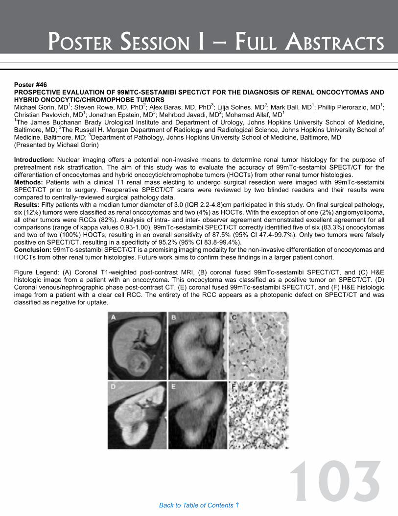
103Back to Table of Contents h
Poster session i – Full AbstrActs
Poster #46 PROSPECTIVE EVALUATION OF 99MTC-SESTAMIBI SPECT/CT FOR THE DIAGNOSIS OF RENAL ONCOCYTOMAS AND HYBRID ONCOCYTIC/CHROMOPHOBE TUMORS Michael Gorin, MD1; Steven Rowe, MD, PhD2; Alex Baras, MD, PhD3; Lilja Solnes, MD2; Mark Ball, MD1; Phillip Pierorazio, MD1; Christian Pavlovich, MD1; Jonathan Epstein, MD3; Mehrbod Javadi, MD2; Mohamad Allaf, MD1 1The James Buchanan Brady Urological Institute and Department of Urology, Johns Hopkins University School of Medicine, Baltimore, MD; 2The Russell H. Morgan Department of Radiology and Radiological Science, Johns Hopkins University School of Medicine, Baltimore, MD; 3Department of Pathology, Johns Hopkins University School of Medicine, Baltimore, MD (Presented by Michael Gorin) Introduction: Nuclear imaging offers a potential non-invasive means to determine renal tumor histology for the purpose of pretreatment risk stratification. The aim of this study was to evaluate the accuracy of 99mTc-sestamibi SPECT/CT for the differentiation of oncocytomas and hybrid oncocytic/chromophobe tumors (HOCTs) from other renal tumor histologies. Methods: Patients with a clinical T1 renal mass electing to undergo surgical resection were imaged with 99mTc-sestamibi SPECT/CT prior to surgery. Preoperative SPECT/CT scans were reviewed by two blinded readers and their results were compared to centrally-reviewed surgical pathology data. Results: Fifty patients with a median tumor diameter of 3.0 (IQR 2.2-4.8)cm participated in this study. On final surgical pathology, six (12%) tumors were classified as renal oncocytomas and two (4%) as HOCTs. With the exception of one (2%) angiomyolipoma, all other tumors were RCCs (82%). Analysis of intra- and inter- observer agreement demonstrated excellent agreement for all comparisons (range of kappa values 0.93-1.00). 99mTc-sestamibi SPECT/CT correctly identified five of six (83.3%) oncocytomas and two of two (100%) HOCTs, resulting in an overall sensitivity of 87.5% (95% CI 47.4-99.7%). Only two tumors were falsely positive on SPECT/CT, resulting in a specificity of 95.2% (95% CI 83.8-99.4%). Conclusion: 99mTc-sestamibi SPECT/CT is a promising imaging modality for the non-invasive differentiation of oncocytomas and HOCTs from other renal tumor histologies. Future work aims to confirm these findings in a larger patient cohort. Figure Legend: (A) Coronal T1-weighted post-contrast MRI, (B) coronal fused 99mTc-sestamibi SPECT/CT, and (C) H&E histologic image from a patient with an oncocytoma. This oncocytoma was classified as a positive tumor on SPECT/CT. (D) Coronal venous/nephrographic phase post-contrast CT, (E) coronal fused 99mTc-sestamibi SPECT/CT, and (F) H&E histologic image from a patient with a clear cell RCC. The entirety of the RCC appears as a photopenic defect on SPECT/CT and was classified as negative for uptake.
104Back to Table of Contents h
Poster session i – Full AbstrActs
Poster #47 DEVELOPMENT OF A CLEAR CELL RENAL CELL CARCINOMA XENOGRAFT MODEL: SURGICAL TISSUE VS. BIOPSY TISSUE. Yiyu Dong, PhD1; Brandon J. Manley, MD2; Abraham A. Hakimi, MD2; Paul Russo, MD2; Jonathan Coleman, MD2; James J. Hsieh, MD, PhD2 1Sloan Kettering Institute, New York, NY; 2Memorial Sloan Kettering Cancer Center, New York, NY. (Presented by Brandon J. Manley) Introduction: The use of xenograft tumor models in immunodeficient mouse strains is considered the ideal platform to investigate the effects and toxicities of novel drugs in primary human tumors. These models in clear cell renal cell carcinoma (ccRCC) are deemed to be the most clinically reliable in assessing the response of tumors to systemic therapy and also allow for mutational profiling and correlating tumor response in vivo. The successful establishment of a personalized xenograft model using preoperative or pretherapy biopsy for patients with metastatic or high risk disease could improve selection of targeted therapy. We report the success of our xenograft model in using various tissue sources including transplanted biopsies in correlation with patient’s clinical history and tumor characteristics. Methods: Fifty-six specimens from primary and metastatic ccRCC from 48 consenting patients were collected. After surgery (n=35) or biopsy (n=21) the specimen was transplanted either subcutaneously or after cell culture to immunodeficient mice. Tumor engraftment was followed for an initial period of up to four months. Successfully engrafted patient-derived tumors were subsequently passaged to further mice. Conformation of xenograft tumors with formalin-fixed, paraffin-embedded (FFPE) and Hematoxylin and eosin (HE) stained tumor sections was done to assure morphological concordance between the patients tumor. We used a two-tailed two proportion z-test to compare the number of successful xenografts harvested from surgical tissue or biopsy tissue. Results: Overall 25 of the 56 specimens were successful in growing tumor in our immunodeficient mice. The frequency of success based on the type and site of tissue harvest may be seen in Table 1. Analyzing the number of successful xenografts by method of tissue harvest, we found biopsy tissue to be significantly more successful compared to surgical tissue, 61.9% compared to 34.2% (p-value=0.044). Conclusion: We believe our xenograft model, using biopsy tissue, demonstrates the feasibility of a real time personalized in vivo model to aide in the selection of targeted treatments for systemic therapy in ccRCC patients.
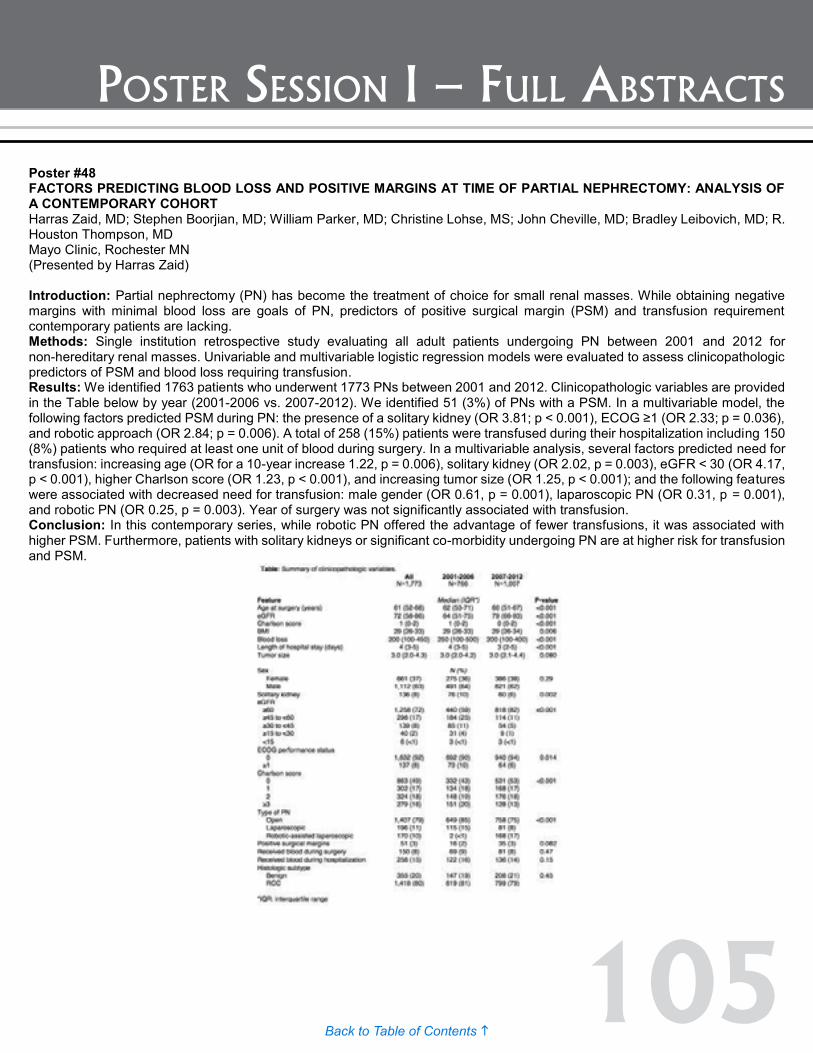
105Back to Table of Contents h
Poster session i – Full AbstrActs
Poster #48 FACTORS PREDICTING BLOOD LOSS AND POSITIVE MARGINS AT TIME OF PARTIAL NEPHRECTOMY: ANALYSIS OF A CONTEMPORARY COHORT Harras Zaid, MD; Stephen Boorjian, MD; William Parker, MD; Christine Lohse, MS; John Cheville, MD; Bradley Leibovich, MD; R. Houston Thompson, MD Mayo Clinic, Rochester MN (Presented by Harras Zaid) Introduction: Partial nephrectomy (PN) has become the treatment of choice for small renal masses. While obtaining negative margins with minimal blood loss are goals of PN, predictors of positive surgical margin (PSM) and transfusion requirement contemporary patients are lacking. Methods: Single institution retrospective study evaluating all adult patients undergoing PN between 2001 and 2012 for non-hereditary renal masses. Univariable and multivariable logistic regression models were evaluated to assess clinicopathologic predictors of PSM and blood loss requiring transfusion. Results: We identified 1763 patients who underwent 1773 PNs between 2001 and 2012. Clinicopathologic variables are provided in the Table below by year (2001-2006 vs. 2007-2012). We identified 51 (3%) of PNs with a PSM. In a multivariable model, the following factors predicted PSM during PN: the presence of a solitary kidney (OR 3.81; p < 0.001), ECOG ≥1 (OR 2.33; p = 0.036), and robotic approach (OR 2.84; p = 0.006). A total of 258 (15%) patients were transfused during their hospitalization including 150 (8%) patients who required at least one unit of blood during surgery. In a multivariable analysis, several factors predicted need for transfusion: increasing age (OR for a 10-year increase 1.22, p = 0.006), solitary kidney (OR 2.02, p = 0.003), eGFR < 30 (OR 4.17, p < 0.001), higher Charlson score (OR 1.23, p < 0.001), and increasing tumor size (OR 1.25, p < 0.001); and the following features were associated with decreased need for transfusion: male gender (OR 0.61, p = 0.001), laparoscopic PN (OR 0.31, p = 0.001), and robotic PN (OR 0.25, p = 0.003). Year of surgery was not significantly associated with transfusion. Conclusion: In this contemporary series, while robotic PN offered the advantage of fewer transfusions, it was associated with higher PSM. Furthermore, patients with solitary kidneys or significant co-morbidity undergoing PN are at higher risk for transfusion and PSM.
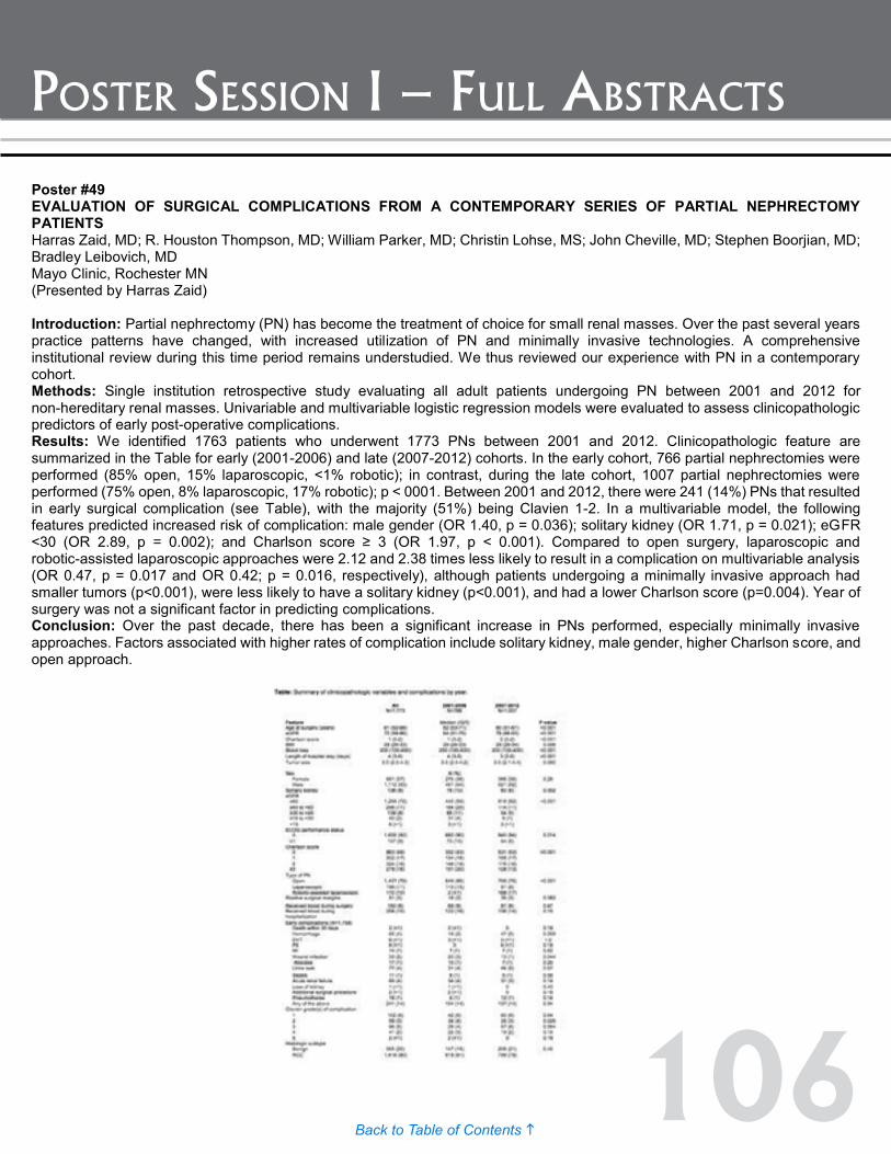
106Back to Table of Contents h
Poster session i – Full AbstrActs
Poster #49 EVALUATION OF SURGICAL COMPLICATIONS FROM A CONTEMPORARY SERIES OF PARTIAL NEPHRECTOMY PATIENTS Harras Zaid, MD; R. Houston Thompson, MD; William Parker, MD; Christin Lohse, MS; John Cheville, MD; Stephen Boorjian, MD; Bradley Leibovich, MD Mayo Clinic, Rochester MN (Presented by Harras Zaid) Introduction: Partial nephrectomy (PN) has become the treatment of choice for small renal masses. Over the past several years practice patterns have changed, with increased utilization of PN and minimally invasive technologies. A comprehensive institutional review during this time period remains understudied. We thus reviewed our experience with PN in a contemporary cohort. Methods: Single institution retrospective study evaluating all adult patients undergoing PN between 2001 and 2012 for non-hereditary renal masses. Univariable and multivariable logistic regression models were evaluated to assess clinicopathologic predictors of early post-operative complications. Results: We identified 1763 patients who underwent 1773 PNs between 2001 and 2012. Clinicopathologic feature are summarized in the Table for early (2001-2006) and late (2007-2012) cohorts. In the early cohort, 766 partial nephrectomies were performed (85% open, 15% laparoscopic, <1% robotic); in contrast, during the late cohort, 1007 partial nephrectomies were performed (75% open, 8% laparoscopic, 17% robotic); p < 0001. Between 2001 and 2012, there were 241 (14%) PNs that resulted in early surgical complication (see Table), with the majority (51%) being Clavien 1-2. In a multivariable model, the following features predicted increased risk of complication: male gender (OR 1.40, p = 0.036); solitary kidney (OR 1.71, p = 0.021); eGFR <30 (OR 2.89, p = 0.002); and Charlson score ≥ 3 (OR 1.97, p < 0.001). Compared to open surgery, laparoscopic and robotic-assisted laparoscopic approaches were 2.12 and 2.38 times less likely to result in a complication on multivariable analysis (OR 0.47, p = 0.017 and OR 0.42; p = 0.016, respectively), although patients undergoing a minimally invasive approach had smaller tumors (p<0.001), were less likely to have a solitary kidney (p<0.001), and had a lower Charlson score (p=0.004). Year of surgery was not a significant factor in predicting complications. Conclusion: Over the past decade, there has been a significant increase in PNs performed, especially minimally invasive approaches. Factors associated with higher rates of complication include solitary kidney, male gender, higher Charlson score, and open approach.
107Back to Table of Contents h
Poster session i – Full AbstrActs
Poster #50 SURGICAL MANAGEMENT OF RECURRENT VENOUS TUMOR THROMBUS AFTER PRIOR NEPHRECTOMY William P. Parker, MD; Stephen A. Boorjian, MD; Harras B. Zaid, MD; Bradley C. Leibovich, MD; R. Houston Thompson, MD Mayo Clinic (Presented by William P. Parker) Introduction: The management of venous tumor thrombus at the time of radical nephrectomy has been well described with excellent outcomes in the non-metastatic setting. However, on rare occasions, patients recur with tumor thrombus after primary treatment, and the management in this setting has not been well described. Herein, we evaluate our experience with patients presenting with tumor thrombus after prior radical nephrectomy. Methods: A retrospective chart review of patients with a history of renal cell carcinoma treated with radical nephrectomy with or without tumor thrombectomy who subsequently recurred with new or residual tumor thrombus that was not related to a new contralateral renal tumor. Patient demographics, surgical outcomes, and survival were assessed. Results: Between 1970 and 2013, a total of 17 patients were identified, including 14 patients who underwent a secondary tumor thrombectomy. Three patients were diagnosed with synchronous widely metastatic disease, did not undergo repeat thrombectomy, and died within five months. Among the repeat surgical patients, median age was 67 (range 48 – 76) with a median time from prior nephrectomy to diagnosis of 5.7months (0 – 57.5 months). Ten patients had known tumor thrombus at nephrectomy, nine of whom had a pathologically complete prior tumor thrombectomy prior to presentation. At exploration for secondary thrombus, surgical resection was completed in 12, with a median blood loss of 2750 ml (200-7000ml) and with a median transfusion requirement of six units (0-18 units). Two patients were found to have extensive invasion at the time of exploration, preventing excision of the thrombus. At a median follow-up of 16.6 months all patients had recurred, with all but one patient dying of disease. Median time to recurrence and death was 4.4 months and 11.9 months following repeat exploration, respectively. Conclusion: Secondary tumor thrombectomy is a technically feasible yet challenging operation. Survival is poor in this population with metastatic progression appreciated in the large majority during follow-up. Further evaluation of this challenging patient population in the targeted therapy era is warranted.
Poster #49 EVALUATION OF SURGICAL COMPLICATIONS FROM A CONTEMPORARY SERIES OF PARTIAL NEPHRECTOMY PATIENTS Harras Zaid, MD; R. Houston Thompson, MD; William Parker, MD; Christin Lohse, MS; John Cheville, MD; Stephen Boorjian, MD; Bradley Leibovich, MD Mayo Clinic, Rochester MN (Presented by Harras Zaid) Introduction: Partial nephrectomy (PN) has become the treatment of choice for small renal masses. Over the past several years practice patterns have changed, with increased utilization of PN and minimally invasive technologies. A comprehensive institutional review during this time period remains understudied. We thus reviewed our experience with PN in a contemporary cohort. Methods: Single institution retrospective study evaluating all adult patients undergoing PN between 2001 and 2012 for non-hereditary renal masses. Univariable and multivariable logistic regression models were evaluated to assess clinicopathologic predictors of early post-operative complications. Results: We identified 1763 patients who underwent 1773 PNs between 2001 and 2012. Clinicopathologic feature are summarized in the Table for early (2001-2006) and late (2007-2012) cohorts. In the early cohort, 766 partial nephrectomies were performed (85% open, 15% laparoscopic, <1% robotic); in contrast, during the late cohort, 1007 partial nephrectomies were performed (75% open, 8% laparoscopic, 17% robotic); p < 0001. Between 2001 and 2012, there were 241 (14%) PNs that resulted in early surgical complication (see Table), with the majority (51%) being Clavien 1-2. In a multivariable model, the following features predicted increased risk of complication: male gender (OR 1.40, p = 0.036); solitary kidney (OR 1.71, p = 0.021); eGFR <30 (OR 2.89, p = 0.002); and Charlson score ≥ 3 (OR 1.97, p < 0.001). Compared to open surgery, laparoscopic and robotic-assisted laparoscopic approaches were 2.12 and 2.38 times less likely to result in a complication on multivariable analysis (OR 0.47, p = 0.017 and OR 0.42; p = 0.016, respectively), although patients undergoing a minimally invasive approach had smaller tumors (p<0.001), were less likely to have a solitary kidney (p<0.001), and had a lower Charlson score (p=0.004). Year of surgery was not a significant factor in predicting complications. Conclusion: Over the past decade, there has been a significant increase in PNs performed, especially minimally invasive approaches. Factors associated with higher rates of complication include solitary kidney, male gender, higher Charlson score, and open approach.
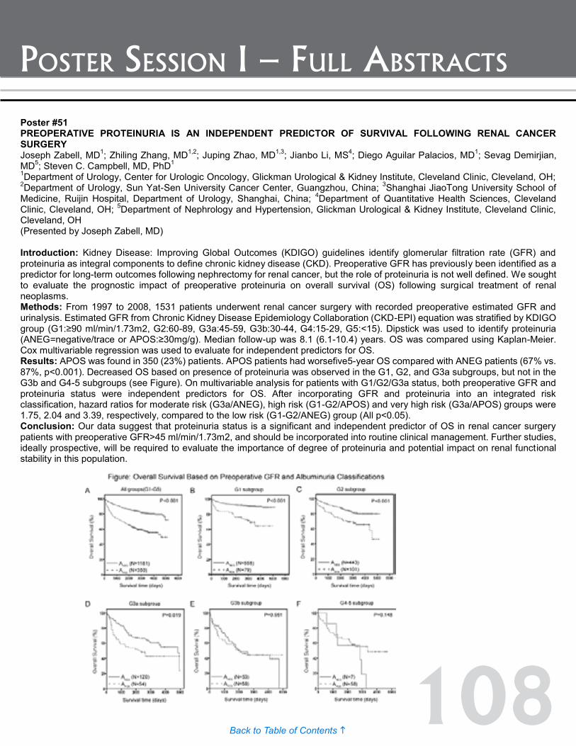
108Back to Table of Contents h
Poster session i – Full AbstrActs
Poster #52 SELECT CONCURRENT CHROMOSOME 3P MUTATIONS PREDICT WORSE OVERALL SURVIVAL IN CLEAR CELL RENAL CARCINOMA IN THE CANCER GENOME ATLAS Christopher Keith; Michael Rossi, PhD; Rebecca Arnold, PhD; John Petros, MD (Presented by Christopher Keith) Introduction: Genes most associated with oncogenesis and/or poor prognosis in clear cell renal cell carcinoma (ccRCC), including VHL, BAP1, SETD2, and PBRM1, are present on the short arm of chromosome three. Mutations in BAP1 and, in some cohorts, PBRM1 are associated with worse survival, while VHL and SETD2 mutations do not independently predict poor prognosis. While studies have analyzed these genes independently, the prognostic significance of co-existing mutations in these chr3p genes is unknown. Methods: The ccRCC database in The Cancer Genome Atlas (TCGA) was probed for mutations in VHL, BAP1, SETD2, and PBRM1 and patient outcomes. Iterative analysis of clustered mutations was performed using Kaplan-Meier survival log-rank analysis. Multivariate survival analysis was performed using Cox regression. Results: Of 418 patients in TCGA, at least one of the four chromosome 3p genes was mutated in 74% of cases. Clustered mutational analysis revealed co-existing somatic mutations in VHL and BAP1 (n=16), VHL and SETD2 (n=9), or PBRM1 and SETD2 and/or BAP1 (n=13) predicted worst overall survival. Patients with such mutational profiles typically had worse outcome than those with higher chromosome 3p mutation burden. For example, cases with VHL and SETD2 mutations had decreased overall survival compared to those with alterations in VHL, SETD2, and PBRM1 (p=0.009) or all four genes (p=0.015). The high-risk chromosome 3p group (n=38) had a significantly reduced median overall survival of 2.42 years compared to 7.13 years for all other patients (n=380) (p<0.0001). There was no difference in age (p=0.48). The survival difference remained significant after controlling for total mutational burden (p<0.0001), BAP1 mutations (p=0.014), and PBRM1 mutations (p<0.0001). On multivariate analysis, high-risk chromosome 3p status was associated with significantly decreased recurrence-free survival and overall survival. Conclusion: Concurrent mutations in VHL and BAP1, VHL and SETD2, or PBRM with SETD2 and/or BAP1 in ccRCC are associated with significantly decreased overall survival in the TCGA. Such information may be useful in risk stratification of patients with ccRCC.
Poster #51 PREOPERATIVE PROTEINURIA IS AN INDEPENDENT PREDICTOR OF SURVIVAL FOLLOWING RENAL CANCER SURGERY Joseph Zabell, MD1; Zhiling Zhang, MD1,2; Juping Zhao, MD1,3; Jianbo Li, MS4; Diego Aguilar Palacios, MD1; Sevag Demirjian, MD5; Steven C. Campbell, MD, PhD1 1Department of Urology, Center for Urologic Oncology, Glickman Urological & Kidney Institute, Cleveland Clinic, Cleveland, OH; 2Department of Urology, Sun Yat-Sen University Cancer Center, Guangzhou, China; 3Shanghai JiaoTong University School of Medicine, Ruijin Hospital, Department of Urology, Shanghai, China; 4Department of Quantitative Health Sciences, Cleveland Clinic, Cleveland, OH; 5Department of Nephrology and Hypertension, Glickman Urological & Kidney Institute, Cleveland Clinic, Cleveland, OH (Presented by Joseph Zabell, MD) Introduction: Kidney Disease: Improving Global Outcomes (KDIGO) guidelines identify glomerular filtration rate (GFR) and proteinuria as integral components to define chronic kidney disease (CKD). Preoperative GFR has previously been identified as a predictor for long-term outcomes following nephrectomy for renal cancer, but the role of proteinuria is not well defined. We sought to evaluate the prognostic impact of preoperative proteinuria on overall survival (OS) following surgical treatment of renal neoplasms. Methods: From 1997 to 2008, 1531 patients underwent renal cancer surgery with recorded preoperative estimated GFR and urinalysis. Estimated GFR from Chronic Kidney Disease Epidemiology Collaboration (CKD-EPI) equation was stratified by KDIGO group (G1:≥90 ml/min/1.73m2, G2:60-89, G3a:45-59, G3b:30-44, G4:15-29, G5:<15). Dipstick was used to identify proteinuria (ANEG=negative/trace or APOS:≥30mg/g). Median follow-up was 8.1 (6.1-10.4) years. OS was compared using Kaplan-Meier. Cox multivariable regression was used to evaluate for independent predictors for OS. Results: APOS was found in 350 (23%) patients. APOS patients had worsefive5-year OS compared with ANEG patients (67% vs. 87%, p<0.001). Decreased OS based on presence of proteinuria was observed in the G1, G2, and G3a subgroups, but not in the G3b and G4-5 subgroups (see Figure). On multivariable analysis for patients with G1/G2/G3a status, both preoperative GFR and proteinuria status were independent predictors for OS. After incorporating GFR and proteinuria into an integrated risk classification, hazard ratios for moderate risk (G3a/ANEG), high risk (G1-G2/APOS) and very high risk (G3a/APOS) groups were 1.75, 2.04 and 3.39, respectively, compared to the low risk (G1-G2/ANEG) group (All p<0.05). Conclusion: Our data suggest that proteinuria status is a significant and independent predictor of OS in renal cancer surgery patients with preoperative GFR>45 ml/min/1.73m2, and should be incorporated into routine clinical management. Further studies, ideally prospective, will be required to evaluate the importance of degree of proteinuria and potential impact on renal functional stability in this population.
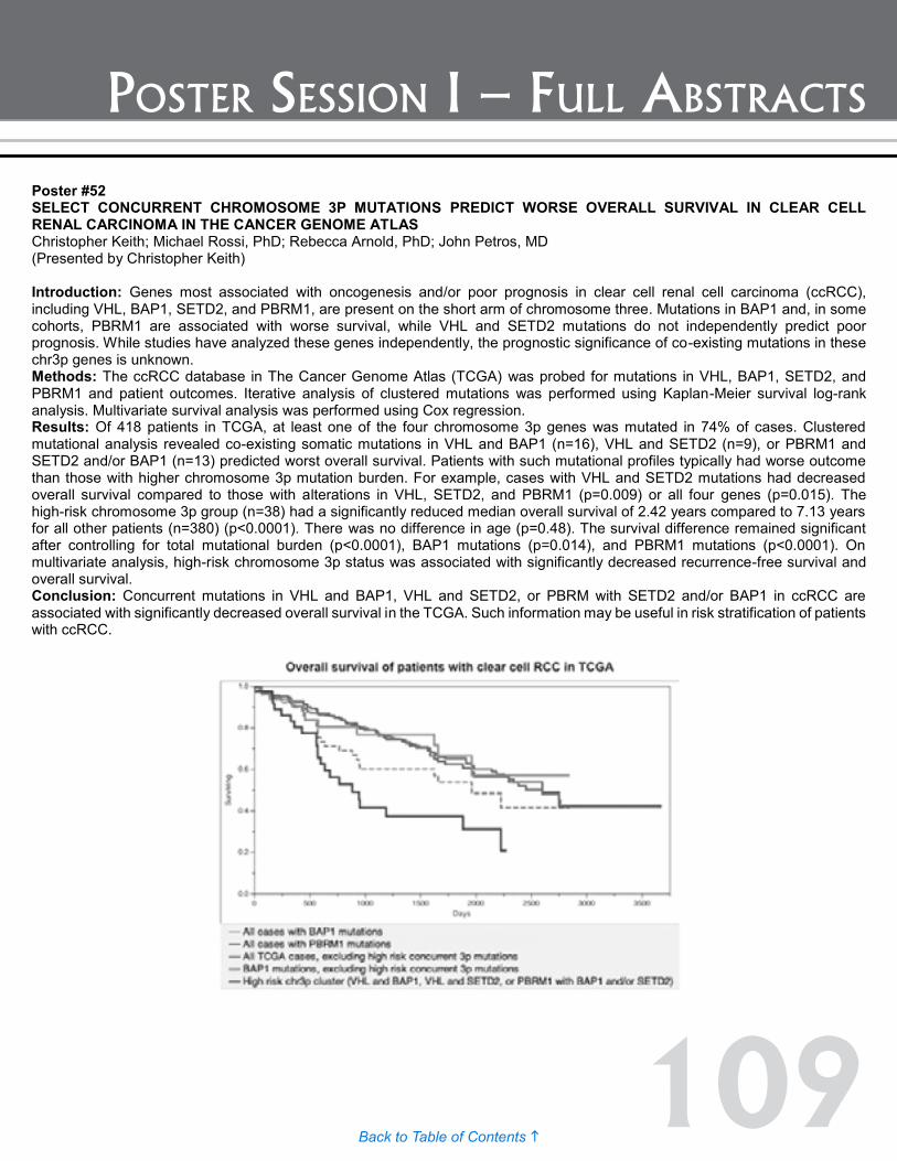
109Back to Table of Contents h
Poster session i – Full AbstrActs
Poster #52 SELECT CONCURRENT CHROMOSOME 3P MUTATIONS PREDICT WORSE OVERALL SURVIVAL IN CLEAR CELL RENAL CARCINOMA IN THE CANCER GENOME ATLAS Christopher Keith; Michael Rossi, PhD; Rebecca Arnold, PhD; John Petros, MD (Presented by Christopher Keith) Introduction: Genes most associated with oncogenesis and/or poor prognosis in clear cell renal cell carcinoma (ccRCC), including VHL, BAP1, SETD2, and PBRM1, are present on the short arm of chromosome three. Mutations in BAP1 and, in some cohorts, PBRM1 are associated with worse survival, while VHL and SETD2 mutations do not independently predict poor prognosis. While studies have analyzed these genes independently, the prognostic significance of co-existing mutations in these chr3p genes is unknown. Methods: The ccRCC database in The Cancer Genome Atlas (TCGA) was probed for mutations in VHL, BAP1, SETD2, and PBRM1 and patient outcomes. Iterative analysis of clustered mutations was performed using Kaplan-Meier survival log-rank analysis. Multivariate survival analysis was performed using Cox regression. Results: Of 418 patients in TCGA, at least one of the four chromosome 3p genes was mutated in 74% of cases. Clustered mutational analysis revealed co-existing somatic mutations in VHL and BAP1 (n=16), VHL and SETD2 (n=9), or PBRM1 and SETD2 and/or BAP1 (n=13) predicted worst overall survival. Patients with such mutational profiles typically had worse outcome than those with higher chromosome 3p mutation burden. For example, cases with VHL and SETD2 mutations had decreased overall survival compared to those with alterations in VHL, SETD2, and PBRM1 (p=0.009) or all four genes (p=0.015). The high-risk chromosome 3p group (n=38) had a significantly reduced median overall survival of 2.42 years compared to 7.13 years for all other patients (n=380) (p<0.0001). There was no difference in age (p=0.48). The survival difference remained significant after controlling for total mutational burden (p<0.0001), BAP1 mutations (p=0.014), and PBRM1 mutations (p<0.0001). On multivariate analysis, high-risk chromosome 3p status was associated with significantly decreased recurrence-free survival and overall survival. Conclusion: Concurrent mutations in VHL and BAP1, VHL and SETD2, or PBRM with SETD2 and/or BAP1 in ccRCC are associated with significantly decreased overall survival in the TCGA. Such information may be useful in risk stratification of patients with ccRCC.
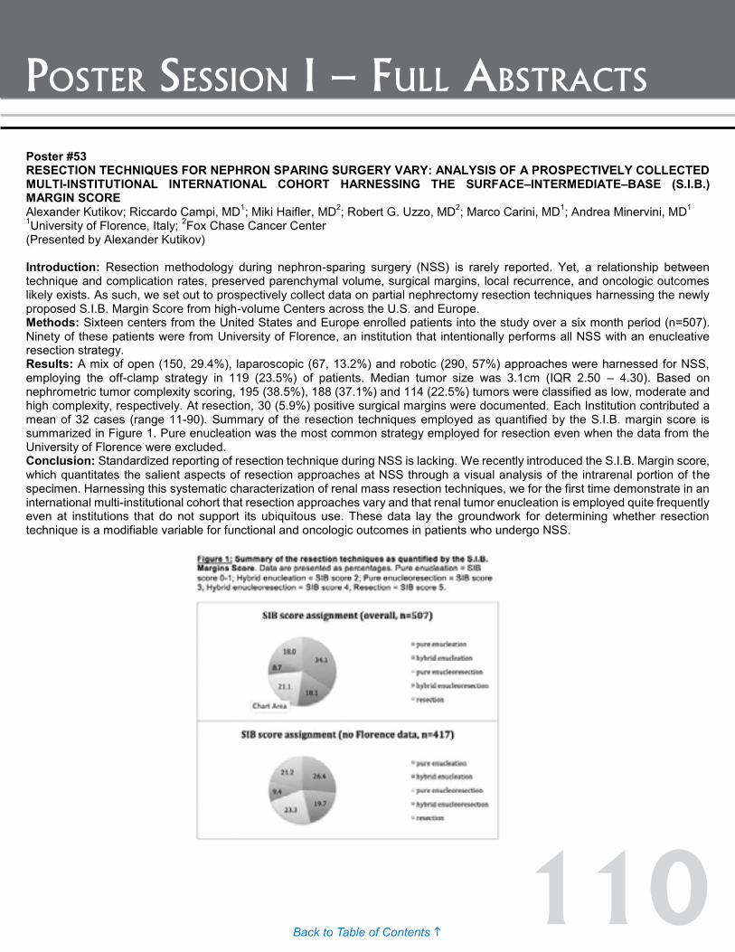
110Back to Table of Contents h
Poster session i – Full AbstrActs
Poster #54 ACUTE KIDNEY INJURY AFTER PARTIAL NEPHRECTOMY: ROLE OF PARENCHYMAL MASS REDUCTION AND ISCHEMIA AND IMPACT ON SUBSEQUENT FUNCTIONAL RECOVERY Zhiling Zhang, MD1,2; Juping Zhao, MD1,3; Erick Remer, MD4; Jianbo Li, MS5; Sevag Demirjian, MD1; Joseph Zabell, MD1; Steven C. Campbell, MD, PhD1 1Glickman Urological and Kidney Institute, Cleveland Clinic, Cleveland, OH; 2Sun Yat-Sen University Cancer Center, Department of Urology, Guangzhou, China; 3Shanghai JiaoTong University School of Medicine, Ruijin Hospital, Department of Urology, Shanghai, China; 4Imaging Institute, Cleveland Clinic, Cleveland, OH; 5Department of Quantitative Health Sciences, Cleveland Clinic, Cleveland, OH (Presented by Zhiling Zhang) Introduction: Acute increase of serum creatinine (SCr) after partial nephrectomy (PN) is primarily due to parenchymal mass reduction or ischemia, however only ischemia can impact subsequent functional recovery. We evaluate etiologies of acute kidney injury (AKI) after PN and their prognostic significance. Methods: From 2007 to 2014, 83 solitary kidneys managed with PN had necessary studies for detailed analysis of function and parenchymal mass before/after surgery. AKI was classified by RIFLE (Risk/Injury/Failure/Loss/End-stage) defined by either standard criteria (comparison to preoperative SCr) or proposed criteria (comparison to projected postoperative SCr based on parenchymal mass reduction). Subsequent recovery was defined as percent function preserved normalized by percent mass saved. Predictive factors for AKI were evaluated by logistic regression. Relationship between AKI grade and subsequent functional recovery was assessed by linear regression. Results: Median duration of warm ischemia (n=39) was 20 minutes and hypothermia (n=44) 29 minutes. Median parenchymal mass reduction was 11%. AKI occurred in 45 patients based on standard criteria and 38 based on proposed criteria, and reflected injury/failure (grade=2/3) in 23 and 16 patients, respectively. On multivariable analysis, only ischemia time correlated with AKI occurrence (p=0.007). Based on the proposed criteria, median recovery from ischemia was 99% in patients without AKI and 95%, 90%, and 88% for patients with grades 1/2/3 AKI, respectively. AKI grade based on proposed criteria correlated with subsequent recovery (p=0.025), while AKI grade based on standard criteria failed to correlate (p=0.56). Main limitation is retrospective design. Conclusion: Parenchymal mass reduction and ischemia both contribute to acute changes in SCr after PN, however only classification of AKI by proposed criteria, which more accurately reflects the impact of ischemia, correlates with subsequent functional recovery. While AKI is associated with suboptimal recovery, even patients with grade 2/3 AKI reached 88-90% of recovery expected.
Poster #53 RESECTION TECHNIQUES FOR NEPHRON SPARING SURGERY VARY: ANALYSIS OF A PROSPECTIVELY COLLECTED MULTI-INSTITUTIONAL INTERNATIONAL COHORT HARNESSING THE SURFACE–INTERMEDIATE–BASE (S.I.B.) MARGIN SCORE Alexander Kutikov; Riccardo Campi, MD1; Miki Haifler, MD2; Robert G. Uzzo, MD2; Marco Carini, MD1; Andrea Minervini, MD1 1University of Florence, Italy; 2Fox Chase Cancer Center (Presented by Alexander Kutikov) Introduction: Resection methodology during nephron-sparing surgery (NSS) is rarely reported. Yet, a relationship between technique and complication rates, preserved parenchymal volume, surgical margins, local recurrence, and oncologic outcomes likely exists. As such, we set out to prospectively collect data on partial nephrectomy resection techniques harnessing the newly proposed S.I.B. Margin Score from high-volume Centers across the U.S. and Europe. Methods: Sixteen centers from the United States and Europe enrolled patients into the study over a six month period (n=507). Ninety of these patients were from University of Florence, an institution that intentionally performs all NSS with an enucleative resection strategy. Results: A mix of open (150, 29.4%), laparoscopic (67, 13.2%) and robotic (290, 57%) approaches were harnessed for NSS, employing the off-clamp strategy in 119 (23.5%) of patients. Median tumor size was 3.1cm (IQR 2.50 – 4.30). Based on nephrometric tumor complexity scoring, 195 (38.5%), 188 (37.1%) and 114 (22.5%) tumors were classified as low, moderate and high complexity, respectively. At resection, 30 (5.9%) positive surgical margins were documented. Each Institution contributed a mean of 32 cases (range 11-90). Summary of the resection techniques employed as quantified by the S.I.B. margin score is summarized in Figure 1. Pure enucleation was the most common strategy employed for resection even when the data from the University of Florence were excluded. Conclusion: Standardized reporting of resection technique during NSS is lacking. We recently introduced the S.I.B. Margin score, which quantitates the salient aspects of resection approaches at NSS through a visual analysis of the intrarenal portion of the specimen. Harnessing this systematic characterization of renal mass resection techniques, we for the first time demonstrate in an international multi-institutional cohort that resection approaches vary and that renal tumor enucleation is employed quite frequently even at institutions that do not support its ubiquitous use. These data lay the groundwork for determining whether resection technique is a modifiable variable for functional and oncologic outcomes in patients who undergo NSS.
111Back to Table of Contents h
Poster session i – Full AbstrActs
Poster #54 ACUTE KIDNEY INJURY AFTER PARTIAL NEPHRECTOMY: ROLE OF PARENCHYMAL MASS REDUCTION AND ISCHEMIA AND IMPACT ON SUBSEQUENT FUNCTIONAL RECOVERY Zhiling Zhang, MD1,2; Juping Zhao, MD1,3; Erick Remer, MD4; Jianbo Li, MS5; Sevag Demirjian, MD1; Joseph Zabell, MD1; Steven C. Campbell, MD, PhD1 1Glickman Urological and Kidney Institute, Cleveland Clinic, Cleveland, OH; 2Sun Yat-Sen University Cancer Center, Department of Urology, Guangzhou, China; 3Shanghai JiaoTong University School of Medicine, Ruijin Hospital, Department of Urology, Shanghai, China; 4Imaging Institute, Cleveland Clinic, Cleveland, OH; 5Department of Quantitative Health Sciences, Cleveland Clinic, Cleveland, OH (Presented by Zhiling Zhang) Introduction: Acute increase of serum creatinine (SCr) after partial nephrectomy (PN) is primarily due to parenchymal mass reduction or ischemia, however only ischemia can impact subsequent functional recovery. We evaluate etiologies of acute kidney injury (AKI) after PN and their prognostic significance. Methods: From 2007 to 2014, 83 solitary kidneys managed with PN had necessary studies for detailed analysis of function and parenchymal mass before/after surgery. AKI was classified by RIFLE (Risk/Injury/Failure/Loss/End-stage) defined by either standard criteria (comparison to preoperative SCr) or proposed criteria (comparison to projected postoperative SCr based on parenchymal mass reduction). Subsequent recovery was defined as percent function preserved normalized by percent mass saved. Predictive factors for AKI were evaluated by logistic regression. Relationship between AKI grade and subsequent functional recovery was assessed by linear regression. Results: Median duration of warm ischemia (n=39) was 20 minutes and hypothermia (n=44) 29 minutes. Median parenchymal mass reduction was 11%. AKI occurred in 45 patients based on standard criteria and 38 based on proposed criteria, and reflected injury/failure (grade=2/3) in 23 and 16 patients, respectively. On multivariable analysis, only ischemia time correlated with AKI occurrence (p=0.007). Based on the proposed criteria, median recovery from ischemia was 99% in patients without AKI and 95%, 90%, and 88% for patients with grades 1/2/3 AKI, respectively. AKI grade based on proposed criteria correlated with subsequent recovery (p=0.025), while AKI grade based on standard criteria failed to correlate (p=0.56). Main limitation is retrospective design. Conclusion: Parenchymal mass reduction and ischemia both contribute to acute changes in SCr after PN, however only classification of AKI by proposed criteria, which more accurately reflects the impact of ischemia, correlates with subsequent functional recovery. While AKI is associated with suboptimal recovery, even patients with grade 2/3 AKI reached 88-90% of recovery expected.
112Back to Table of Contents h
Poster session i – Full AbstrActs
Poster #56 PILOT STUDY EVALUATING PSMA-TARGETED 18F-DCFPYL PET/CT IMAGING OF METASTATIC CLEAR CELL RENAL CELL CARCINOMA Michael Gorin, MD1; Steven Rowe, MD2; Hans-Joerg Hammers, MD, PhD3; M. Som Javadi, MD2; Hazem Hawasli, MD2; Zsolt Szabo, MD, PhD2; Steve Cho, MD4; Martin Pomper, MD, PhD2; Mohamad Allaf, MD1 1The James Buchanan Brady Urological Institute and Department of Urology, Johns Hopkins University School of Medicine, Baltimore, MD; 2The Russell H. Morgan Department of Radiology and Radiological Science, Johns Hopkins University School of Medicine, Baltimore, MD; 3Department of Medical Oncology, Sidney Kimmel Comprehensive Cancer Center, Johns Hopkins University School of Medicine, Baltimore, MD; 4Department of Radiology, University of Wisconsin School of Medicine and Public Health, Madison, WI (Presented by Michael Gorin) Introduction: Positron emission tomography (PET) provides a highly sensitive means of imaging cancer. Unfortunately, the most commonly used PET radiotracer, 18F-fluorodeoxyglucose, has demonstrated a limited role in the evaluation of patients with renal cell carcinoma (RCC). Owing to frequent loss of the VHL gene, clear cell RCC is characterized by a high degree of neovascularization. In this study, we tested the feasibility of imaging metastatic clear cell RCC (ccRCC) using 18F-DCFPyL, a novel PET radiotracer which targets prostate membrane specific antigen (PSMA) overexpression in the neovasculature of solid tumors. Methods: Six patients with untreated metastatic ccRCC were imaged with 18F-DCFPyL PET/CT. PET/CT scans were centrally reviewed by a single reader blinded to conventional imaging. Similarly, conventional imaging studies (contrast-enhanced CT or MRI) were reviewed by a second reader blinded to PET/CT data. Following blinded review, scan results were compared for concordance. Results: Conventional imaging identified 19 sites of metastatic ccRCC (range 1-9 per patient). In contrast, 18F-DCFPyL PET/CT identified 29 foci of abnormal radiotracer uptake (range 1-14 per patient). Sites of abnormal radiotracer uptake had maximum standardized uptake values ranging from 1.6 to 19.3 and were found within the bone, brain, lymph nodes, soft tissue and abdominal viscera. Of the 19 sites of disease identified on conventional imaging, 18 (94.7%) were also identified on PET/CT. Two of these lesions were resected and found to be metastatic ccRCC. The 11 additional sites of radiotracer avidity without corresponding findings on conventional imaging are hypothesized to represent subclinical/occult sites of metastatic ccRCC. Conclusion: PET/CT with 18F-DCFPyL allowed for the sensitive detection of sites of metastatic ccRCC. Future work aims to validate these findings in a larger patient cohort as well as to determine if areas of radiotracer uptake not seen on conventional imaging truly represent sites of metastatic disease. Figure Legend: Representative (A) contrast-enhanced MRI and (B) 18F-DCFPyL PET/CT images of a patient with a metastatic lesion in the left frontal lobe of the brain.
Poster #55 VENOUS THROMBOEMBOLISM FOLLOWING NEPHRECTOMY: THIRTY DAY INCIDENCE AND RISK FACTORS FROM NATIONAL MULTI-INSTITUTIONAL DATA Richard S. Matulewicz, MD1; Yousef Al-shraideh, MD2; Brian Trihn, MD3; John Oliver Delancey, MD3; Irene Helenowski, PhD4; Borko Jovanovic, PhD4; Shilajit Kundu, MD3 1Northwestern Feinberg/Urology; 2Chicago; 3Northwestern Feinberg/Ur; 4Northwestern Feinberg/Biostatistics (Presented by Yousef Al-shraideh) Introduction: Venous thromboembolism (VTE) has significant consequences on surgical patients’ morbidity and mortality. In this study, we determine potential modifiable risk factors associated with VTE in patients undergoing nephrectomy. Methods: We queried the NSQIP database from 2006 to 2012 for all patients undergoing nephrectomy. Patient characteristics were compared between VTE and non-VTE patients. Cohorts for DVT and PE and open and lap/robotic (MIS) surgery were then compared using a bivariate screen method followed by multivariate regression analysis to determine association between preoperative risk factors, surgical variables, and VTE. Results: A total of 13,208 patients were identified. The overall rate of VTE was 1.3% (PE=0.5% and DVT=0.8%). Mean age of patients who developed DVT and PE were 62.2 and 64.8 years, respectively. On bivariate analysis, both postoperative DVT and PE were associated with dyspnea and longer operative time (OT) (all p<0.001). While DVT was significantly associated with diabetes, prior stroke, an ASA of 3 or 4, and poor functional status (PFS), PE was associated with COPD and disseminated cancer. Specific to open and lap/MIS surgery, the rate of DVT and PE was greater in open surgery (2% vs. 0.8%, all p<0.001). In both open and LAP/MIS cohorts, postoperative DVT was significantly associated with PFS and increased OT. Post open surgery DVT was associated with diabetes and prior stroke while post LAP/MIS, it was associated with dyspnea, and ASA 3 or 4. On multivariate analysis, no factors were found associated with an increased risk of DVT, whereas dyspnea (OR 2.7, 95% CI 1.5-4.8, p=0.001), disseminated cancer (OR 2.5, 95% CI 1.1-5.5, p=0.03), increasing age (OR 1.1, 95% CI 1.0-1.2, p=0.04), and longer OT carried an increased risk for PE (OR 1.1, 95% CI 1.0-1.1, p<0.0001) Conclusion: There are potentially modifiable risk factors associated with VTE in kidney surgeries. A better understanding of at risk patients will allow surgeons to better stratify and direct preventative interventions such as screening and VTE prophylaxis.
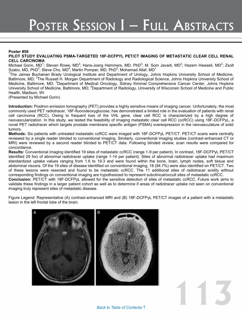
113Back to Table of Contents h
Poster session i – Full AbstrActs
Poster #56 PILOT STUDY EVALUATING PSMA-TARGETED 18F-DCFPYL PET/CT IMAGING OF METASTATIC CLEAR CELL RENAL CELL CARCINOMA Michael Gorin, MD1; Steven Rowe, MD2; Hans-Joerg Hammers, MD, PhD3; M. Som Javadi, MD2; Hazem Hawasli, MD2; Zsolt Szabo, MD, PhD2; Steve Cho, MD4; Martin Pomper, MD, PhD2; Mohamad Allaf, MD1 1The James Buchanan Brady Urological Institute and Department of Urology, Johns Hopkins University School of Medicine, Baltimore, MD; 2The Russell H. Morgan Department of Radiology and Radiological Science, Johns Hopkins University School of Medicine, Baltimore, MD; 3Department of Medical Oncology, Sidney Kimmel Comprehensive Cancer Center, Johns Hopkins University School of Medicine, Baltimore, MD; 4Department of Radiology, University of Wisconsin School of Medicine and Public Health, Madison, WI (Presented by Michael Gorin) Introduction: Positron emission tomography (PET) provides a highly sensitive means of imaging cancer. Unfortunately, the most commonly used PET radiotracer, 18F-fluorodeoxyglucose, has demonstrated a limited role in the evaluation of patients with renal cell carcinoma (RCC). Owing to frequent loss of the VHL gene, clear cell RCC is characterized by a high degree of neovascularization. In this study, we tested the feasibility of imaging metastatic clear cell RCC (ccRCC) using 18F-DCFPyL, a novel PET radiotracer which targets prostate membrane specific antigen (PSMA) overexpression in the neovasculature of solid tumors. Methods: Six patients with untreated metastatic ccRCC were imaged with 18F-DCFPyL PET/CT. PET/CT scans were centrally reviewed by a single reader blinded to conventional imaging. Similarly, conventional imaging studies (contrast-enhanced CT or MRI) were reviewed by a second reader blinded to PET/CT data. Following blinded review, scan results were compared for concordance. Results: Conventional imaging identified 19 sites of metastatic ccRCC (range 1-9 per patient). In contrast, 18F-DCFPyL PET/CT identified 29 foci of abnormal radiotracer uptake (range 1-14 per patient). Sites of abnormal radiotracer uptake had maximum standardized uptake values ranging from 1.6 to 19.3 and were found within the bone, brain, lymph nodes, soft tissue and abdominal viscera. Of the 19 sites of disease identified on conventional imaging, 18 (94.7%) were also identified on PET/CT. Two of these lesions were resected and found to be metastatic ccRCC. The 11 additional sites of radiotracer avidity without corresponding findings on conventional imaging are hypothesized to represent subclinical/occult sites of metastatic ccRCC. Conclusion: PET/CT with 18F-DCFPyL allowed for the sensitive detection of sites of metastatic ccRCC. Future work aims to validate these findings in a larger patient cohort as well as to determine if areas of radiotracer uptake not seen on conventional imaging truly represent sites of metastatic disease. Figure Legend: Representative (A) contrast-enhanced MRI and (B) 18F-DCFPyL PET/CT images of a patient with a metastatic lesion in the left frontal lobe of the brain.
114Back to Table of Contents h
Poster session i – Full AbstrActs
Poster #57 CLINICAL, PATHOLOGIC AND GENOMIC PROFILES OF EXCEPTIONAL RESPONDERS TO ANTI-PD1 THERAPY IN RENAL CELL CARCINOMA Mark Ball, MD1; Michael Johnson, MD2; Michael Gorin, MD2; Maria Rodriguez, MD2; Luis Diaz, MD2; Bert Vogelstein, MD2; Kenneth Kinzler, PhD2; Michael Haffner, MD2; Luigi Marchionni, MD2; George Netto, MD2; Charles Drake, MD, PhD2; Mohamad Allaf, MD2 1Johns Hopkins, Baltimore, MD; 2Johns Hopkins, Baltimore, MD (Presented by Mark Ball) Introduction: Previous studies have correlated response to PD-1 blockade with PD-L1 over-expression, as well as cell-mediated immune transcripts). However, factors associated with long-term, durable response to nivolumab in patients with RCC have not yet been elucidated. To better understand these factors, we conducted a study to characterize the two extremes of response to therapy: exceptional responders who had durable, complete response and patients who had primary refractory disease. Methods: Patients with durable, complete response (“exceptional responders”) (n=4) and primary refractory disease (n=3) were analyzed. Immunohistochemical staining for PD-L1, CD8 and FOXP3, whole-exome sequencing and quantitative RNA expression profiling was performed and correlated with clinical outcomes. Results: Exceptional responders had trends toward greater CD8+ lymphocyte infiltrate (126.7 vs. 28.8 lymphocytes/hpf), higher number of somatic mutations (67 vs. 35 somatic mutations/genome), and higher number of predicted mutation associated neoantigens than primary refractory patients (44 vs. eight). Expression analysis demonstrated acute phase and immune tolerance signatures in primary refractory patients, while exceptional responders had higher expression of T cell and innate immune signatures. Conclusion: Exceptional responders and primary refectory patients have distinct pathologic, genomic and RNA expression profiles. Larger studies are needed to fully elucidate the basis of response to PD-1 blockade in patients with renal cell carcinoma.
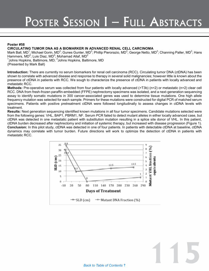
115Back to Table of Contents h
Poster session i – Full AbstrActs
Poster #58 CIRCULATING TUMOR DNA AS A BIOMARKER IN ADVANCED RENAL CELL CARCINOMA Mark Ball, MD1; Michael Gorin, MD2; Gunes Gunter, MD2; Phillip Pierorazio, MD2; George Netto, MD2; Channing Paller, MD2; Hans Hammers, MD2; Luis Diaz, MD2; Mohamad Allaf, MD2 1Johns Hopkins, Baltimore, MD; 2Johns Hopkins, Baltimore, MD (Presented by Mark Ball) Introduction: There are currently no serum biomarkers for renal cell carcinoma (RCC). Circulating tumor DNA (ctDNA) has been shown to correlate with advanced disease and response to therapy in several solid malignancies; however little is known about the presence of ctDNA in patients with RCC. We sough to characterize the presence of ctDNA in patients with locally advanced and metastatic RCC. Methods: Pre-operative serum was collected from four patients with locally advanced (>T3b) (n=2) or metastatic (n=2) clear cell RCC. DNA from fresh-frozen paraffin-embedded (FFPE) nephrectomy specimens was isolated, and a next-generation sequencing assay to identify somatic mutations in 300 cancer-associated genes was used to determine tissue mutations. One high allele frequency mutation was selected for each sample. Primers for these mutations were constructed for digital PCR of matched serum specimens. Patients with positive pretreatment ctDNA were followed longitudinally to assess changes in ctDNA levels with treatment. Results: Next generation sequencing identified known mutations in all four tumor specimens. Candidate mutations selected were from the following genes: VHL, BAP1, PBRM1, NF. Serum PCR failed to detect mutant alleles in either locally advanced case, but ctDNA was detected in one metastatic patient with substitution mutation resulting in a splice site donor of VHL. In this patient, ctDNA burden decreased after nephrectomy and initiation of systemic therapy, but increased with disease progression (Figure 1). Conclusion: In this pilot study, ctDNA was detected in one of four patients. In patients with detectable ctDNA at baseline, ctDNA dynamics may correlate with tumor burden. Future directions will work to optimize the detection of ctDNA in patients with metastatic RCC.
116Back to Table of Contents h
Poster session i – Full AbstrActs
Poster #59 DISCRIMINATING IPSI AND CONTRA-LATERAL COMPENSATORY RENAL GROWTH (CRG) AFTER PARTIAL NEPHRECTOMY- ARE BOTH KIDNEYS ALIKE? A RAT MODEL Barak Rosenzweig, MD1; Raya Eilam, PhD2; Alon Harmelin, BVSc, MRCVS, Dip ECLAM2; Jacob Ramon, MD1 1Department of Urology, The Chaim Sheba Medical Center, Tel Hashomer, Ramat-Gan, Affiliated to Sackler School of Medicine, Tel-Aviv University, Israel; 2Dept. of Veterinary Resources, The Weizmann Institute of Science, Rechovot, Israel (Presented by Barak Rosenzweig) Introduction: Compensatory renal growth (CRG) is a characteristic adaptation to reduced renal mass. Studies in rats have shown CRG to correlate closely with the amount of renal tissue surgically removed. Localized renal cell carcinoma (T1a-b) is best treated surgically with partial nephrectomy (PN), yet information regarding CRG after PN is scarce. Our objective was to study CRG after PN discriminating ipsi from contra-lateral kidney units in that regard. Methods: Twenty-four rats were allocated into four groups. An index group (n=7) underwent partial nephrectomy while temporarily occluding the renal vasculature (i.e. ischemia). The control groups underwent radical nephrectomy (RN) (n=7), ischemia only (no renal mass resection) (n=7) and sham operation (n=3). 5-bromo-2'-deoxyuridine (BrdU) was injected intra-peritoneally on post-operative days one and two. Animals were euthanized six weeks after operation, blood samples for plasma creatinine were withdrawn and kidneys’ weight*, volume, histopathology and BrdU florescence were analyzed. *Kidneys weight was compared to resected kidneys of RN group initially and control group kidneys on sacrifice. Results: Contra-lateral kidney weight increase was shown in the RN and PN groups (67% and 68%), with no statistically significant difference between these two groups. PN group showed total renal mass increase from 87% to 94% with relative decrease in ipsi-lateral (operated) kidney weight from 37% to 26%. Serum creatinine was highest in the RN group (0.2 mg/dL), followed by PN (0.18 mg/dL, NS), ischemia and control (0.15 mg/dL, 0.14 mg/dL, respectively, p<0.05). Histology analysis identified regenerative changes with high nucleus/cytoplasm relation and large nuclei with prominent nucleoli, indicating cellular synthetic activity and mitosis. These changes were predominantly seen in the resection crater. Mitotic index, exemplified by BrdU, was highest in the PN ipsi-lateral kidney resection crater (898/0.5mm^2+/-370), followed by RN (334/0.5mm^2+/-38), PN ipsi-lateral out of resection crater (218/0.5mm^2+/-37), PN contra-lateral (116/0.5mm^2+/-25), ischemia ipsi-lateral (128/0.5mm^2+/-19), ischemia contra-lateral (15/0.5mm^2+/-4) and control (19/0.5mm^2+/-27). Conclusion: Weight, histology and mitotic indexes indicated different CRG comparing contra vs. ipsi-lateral PN kidneys. Contra-lateral mass increase was similar in the RN and PN groups. Regenerative reaction was highest in the PN resection crater.
117Back to Table of Contents h
Poster session i – Full AbstrActs
Poster #60 PD-1 EXPRESSION ON CLASSICAL MONOCYTES (CM) IS AN INDEPENDENT PREDICTOR OF CANCER SPECIFIC SURVIVAL IN CLEAR CELL RENAL CARCINOMA (CCRCC) Mohammed Haseebuddin, Alexander MacFarlane IV, Karen Ruth, Robert Uzzo, Elizabeth Plimack, Mowafaq Jillab, Essel Al-Saleem, Tahseen Al-Saleem, Kerry Campbell (Presented by Mohammed Haseebuddin) Introduction: We have previously shown that PD-1 expression in peripheral blood lymphocytes is elevated in RCC patients at diagnosis and decreases subsequent to surgery. We theorize thatcm inhibition through PD-1 interferes with antigen presentation necessary to stimulate adaptive immune responses, and thus, preopcm PD-1 expression may be prognostic of poor outcomes. Methods: Blood samples were obtained from RCC patients (n = 90) preop and age-matched healthy donors (n = 25). Flow cytometric (FC) data were quantified as mean fluorescence intensity (MFI) or percentage of cells that express a cell surface receptor. Patients were excluded if they had comorbid CLL, had prior surgeries for RCC, had non-ccRCC histology, or if the FC data was missing. We have defined elevatedcm PD-1 expression at the third quartile level ofcm PD-1 expression (784 MFI) in the healthy donors.cm PD-1 level was correlated with patient clinico-pathologic data using non-parametric tests. Cancer specific survival (CSS) curves were estimated with Kaplan-Meier methods and a multivariate analysis was performed. Results: The average age of the ccRCC cohort (n = 68) was 60 (range 25-88) years. 48 and 35 patients had family history of RCC or high-grade disease, respectively. Median follow-up was 41 months; during follow-up, 10 patients died with ccRCC. No patients with low-grade RCC died. The percentage ofcm within total monocytes was significantly less among patients who died of disease compared to those alive (48.8% vs. 63.2%, p = 0.01). Among high-grade patients, increasedcm PD-1 expression was associated with RCC family history (p = 0.008). High-grade disease patients withcm PD-1 expression >784 MFI had lower survival compared to patients withcm PD-1 <784 MFI (p = 0.02); CSS at 24 months forcm PD-1 MFI >784 was 62.1% (95% CI 34.2-81.0) vs. 93.3% (95% CI 61.3-99.0) forcm PD-1 MFI <784. On multivariable analysis, controlling for stage related covariates,cm PD-1 MFI >784 was associated with inferior CSS (hazard ratio = 8.3 [95% CI: 1.1-65.3], p = 0.04) among high-grade disease patients. Conclusion: In patients with ccRCC, preoperativecm PD-1 >784 MFI level was associated with lower survival.cm PD-1 expression level is significantly related to family history, suggesting that tumor genetics may also alter immune biology. Preoperativecm PD-1 MFI may be an important biomarker to stratify patients at risk for poor outcome who may benefit from immunotherapy.
118Back to Table of Contents h
Poster session i – Full AbstrActs
Poster #61 MANAGEMENT OF A MULTIDISCIPLINARY PHASE 3 CLINICAL TRIAL IN PATIENTS WITH SYNCHRONOUS MRCC (ADAPT) USING A REGIONAL CHAMPION MODEL IN THE SOCIETY OF UROLOGIC ONCOLOGY-CLINICAL TRIALS CONSORTIUM Brian Lane, MD, PhD1; Joan Chiaviello, RN2; Gennady Bratslavsky, MD3,4; Christopher Wood, MD5; Thomas Gardner, MD3,6; Jason Gee, MD3,7; Jeff Holzbeierlein, MD3,8; William Huang, MD3,9; Larry Karsh, MD3,10; Alexander Kutikov, MD3,11; Will Lowrance, MD3,12; Viraj Master, MD3,13; Marc Smaldone, MD3,11; Robert Uzzo, MD3,11; The Adapt Study Group3 1Spectrum Health Hospital, Grand Rapids, MI; 2Society of Urologic Oncology-Clinical Trials Consortium (SUO-CTC), Chicago, IL; 3Society of Urologic Oncology-Clinical Trials Consortium (SUO-CTC); 4SUNY Upstate Medical University, Syracuse, NY; 5Society of Urologic Oncology - Clinical Trials Consortium (SUO-CTC), MD Anderson Cancer Center, Houston, TX; 6Indiana University Medical Center; 7Lahey Clinic, Burlington, MA; 8University of Kansas Medical Center, KS; 9New York University School of Medicine, NY; 10The Urology Center of Colorado, Denver, CO; 11Fox Chase Cancer Center, Philadelphia, PA; 12Huntsman Cancer Hospital, Salt Lake City, Utah; 13Emory University, Atlanta GA (Presented by Brian Lane) Introduction: Surgical based multidisciplinary trials pose a unique challenge, particularly for relatively low incidence tumors. We developed a strategy within the SUO-CTC employing regional champions (RCs) to overcome potential barriers to timely site initiation and patient recruitment amongst 52 sites participating in the ADAPT trial (Argos Therapeutics). ADAPT is a phase three randomized (2:1) study comparing TKI therapy plus dendritic cell immunotherapy (AGS-003) to standard TKI therapy alone in adult patients post CN with clear cell mRCC. This trial required intense coordination involving surgeons, OR staff, pathologists, medical oncologists, leukapheresis and immunotherapy manufacturer. Here, we report novel governance leading to the successful accrual of 712 of 1133 patients enrolled and contributed 273 of 450+ randomized patients over 26 months. Methods: SUO-CTC developed a RC strategy and identified RCs within eight U.S. regions paralleling AUA sections. A communication plan was enacted where RC engaged in ongoing site accountability to identify barriers, guide best practices, and update patient accrual. Bi-annual meetings and quarterly conference calls with all investigators were held amongst all study personnel to review trial updates, reassess progress, develop and implement amendments, etc. Results: Utilizing a prospective RC plan, we demonstrated high rates of trial success at nearly all sites. Of 56 selected sites, 52 (92%) opened the trial (median time 3-9 mo) with 51/52 (98%) sites contributing at least two patients to the trial (range: 1-65). The SUO-CTC represented 44% (n=52) of the total number of global sites accruing patients and was able to contribute more than 62% of tumors collected (n=712) and patients (n=273) randomized to study. We identified relative accrual balance across the regions with each contributing an average of 13 patients per site (range 7-18). Despite an amendment, the study completed accrual in 26 months averaging 27.3 patients per month. Conclusion: Understanding the inherent challenges in a multidisciplinary surgical clinical trial combined with prospective planning enhances overall outcomes in trial conduct and completion. The ongoing engagement of investigators, led by peer to peer communication and RC was a critical success factor to the pace and completion of patient enrollment in this clinical trial, the largest ever, of patients undergoing CN. Clinical Trials registry number: NCT 1582672
119Back to Table of Contents h
Poster session i – Full AbstrActs
Poster #62 MULTIMODALITY APPROACH FOR METASTATIC RENAL CELL CARCINOMA IN A MODERN COHORT Scott Pate, MD1; Margaret Higgins, BA2; William Parker, MD1; Jeffrey Holzbeierlein, MD1; Moben Mirza, MD1; Peter Van Veldhuizen, MD1; Eugene Lee, MD1 1University of Kansas Medical Center; 2University of Kansas School of Medicine (Presented by Scott Pate) Introduction: Metastatic renal cell carcinoma (mRCC) accounts for roughly a quarter of new RCC diagnoses. While novel therapies targeting angiogenesis are currently expanding mRCC treatment, high dose Interleukin-2 (IL-2) has been able to produce durable complete responses in a fraction of mRCC patients. Contemporary management often includes both modalities at separate times and most efficacious strategies remains poorly characterized. Methods: After IRB approval, a retrospective review of patients undergoing treatment with IL-2 from July 2007 to September 2014 for mRCC was performed. Responses to IL-2 were classified as complete response, partial response, stable disease, or progression. We used SPSS 22.0 using Kaplan-Meier methodology to record time to progression and cancer specific survival. We compared IL-2 response in a group that received chemotherapy prior to IL-2 and a chemotherapy naïve group using chi-square analysis. Results: A total of 92 patients underwent treatment with IL-2 for mRCC between July 2007 to September 2014. The mean number of IL-2 therapeutic hospitalizations was 3.4 with a mean of 25.4 doses per patient. Seven patients demonstrated a complete response IL-2 with an additional 3 patients demonstrating cure after surgical resection of oligometastasis. Of the seven complete responders, just one patient received targeted therapy prior to IL-2. Thirty-one patients received targeted mRCC chemotherapy prior to receiving IL-2. Sixty-six patients received post-IL-2 targeted therapy: 29 patients received one agent, 13 patients received two agents, eight patients received three agents, seven patients received four agents, six patients received five agents, three patients received six or more agents at a median follow up of four years. Median time to progression after IL-2 was 3.17 months and median cancer specific survival was 36.23 months. Progression free survival in the group that did not receive pre-IL-2 targeted therapy demonstrated a trend toward better IL-2 response although this did not prove to meet statistical significance (p=0.090). Conclusion: Our series characterizes the contemporary mRCC patient and response to available treatment agents. Thirty-four percent of patients received pre-IL-2 targeted therapy. Seventy-two percent received post-IL-2 targeted therapy with some receiving as many as 8 agents. We saw a trend toward stronger IL-2 response in patients that did not receive prior chemotherapy.
Poster #61 MANAGEMENT OF A MULTIDISCIPLINARY PHASE 3 CLINICAL TRIAL IN PATIENTS WITH SYNCHRONOUS MRCC (ADAPT) USING A REGIONAL CHAMPION MODEL IN THE SOCIETY OF UROLOGIC ONCOLOGY-CLINICAL TRIALS CONSORTIUM Brian Lane, MD, PhD1; Joan Chiaviello, RN2; Gennady Bratslavsky, MD3,4; Christopher Wood, MD5; Thomas Gardner, MD3,6; Jason Gee, MD3,7; Jeff Holzbeierlein, MD3,8; William Huang, MD3,9; Larry Karsh, MD3,10; Alexander Kutikov, MD3,11; Will Lowrance, MD3,12; Viraj Master, MD3,13; Marc Smaldone, MD3,11; Robert Uzzo, MD3,11; The Adapt Study Group3 1Spectrum Health Hospital, Grand Rapids, MI; 2Society of Urologic Oncology-Clinical Trials Consortium (SUO-CTC), Chicago, IL; 3Society of Urologic Oncology-Clinical Trials Consortium (SUO-CTC); 4SUNY Upstate Medical University, Syracuse, NY; 5Society of Urologic Oncology - Clinical Trials Consortium (SUO-CTC), MD Anderson Cancer Center, Houston, TX; 6Indiana University Medical Center; 7Lahey Clinic, Burlington, MA; 8University of Kansas Medical Center, KS; 9New York University School of Medicine, NY; 10The Urology Center of Colorado, Denver, CO; 11Fox Chase Cancer Center, Philadelphia, PA; 12Huntsman Cancer Hospital, Salt Lake City, Utah; 13Emory University, Atlanta GA (Presented by Brian Lane) Introduction: Surgical based multidisciplinary trials pose a unique challenge, particularly for relatively low incidence tumors. We developed a strategy within the SUO-CTC employing regional champions (RCs) to overcome potential barriers to timely site initiation and patient recruitment amongst 52 sites participating in the ADAPT trial (Argos Therapeutics). ADAPT is a phase three randomized (2:1) study comparing TKI therapy plus dendritic cell immunotherapy (AGS-003) to standard TKI therapy alone in adult patients post CN with clear cell mRCC. This trial required intense coordination involving surgeons, OR staff, pathologists, medical oncologists, leukapheresis and immunotherapy manufacturer. Here, we report novel governance leading to the successful accrual of 712 of 1133 patients enrolled and contributed 273 of 450+ randomized patients over 26 months. Methods: SUO-CTC developed a RC strategy and identified RCs within eight U.S. regions paralleling AUA sections. A communication plan was enacted where RC engaged in ongoing site accountability to identify barriers, guide best practices, and update patient accrual. Bi-annual meetings and quarterly conference calls with all investigators were held amongst all study personnel to review trial updates, reassess progress, develop and implement amendments, etc. Results: Utilizing a prospective RC plan, we demonstrated high rates of trial success at nearly all sites. Of 56 selected sites, 52 (92%) opened the trial (median time 3-9 mo) with 51/52 (98%) sites contributing at least two patients to the trial (range: 1-65). The SUO-CTC represented 44% (n=52) of the total number of global sites accruing patients and was able to contribute more than 62% of tumors collected (n=712) and patients (n=273) randomized to study. We identified relative accrual balance across the regions with each contributing an average of 13 patients per site (range 7-18). Despite an amendment, the study completed accrual in 26 months averaging 27.3 patients per month. Conclusion: Understanding the inherent challenges in a multidisciplinary surgical clinical trial combined with prospective planning enhances overall outcomes in trial conduct and completion. The ongoing engagement of investigators, led by peer to peer communication and RC was a critical success factor to the pace and completion of patient enrollment in this clinical trial, the largest ever, of patients undergoing CN. Clinical Trials registry number: NCT 1582672
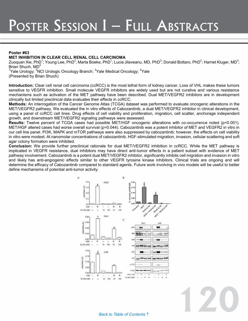
120Back to Table of Contents h
Poster session i – Full AbstrActs
Poster #63 MET INHIBITION IN CLEAR CELL RENAL CELL CARCINOMA Zuoquan Xie, PhD1; Young Lee, PhD2; Marta Boeke, PhD1; Lucia Jilaveanu, MD, PhD3; Donald Bottaro, PhD2; Harriet Kluger, MD3; Brian Shuch, MD4 1Yale Urology; 2NCI Urologic Oncology Branch; 3Yale Medical Oncology; 4Yale (Presented by Brian Shuch) Introduction: Clear cell renal cell carcinoma (ccRCC) is the most lethal form of kidney cancer. Loss of VHL makes these tumors sensitive to VEGFR inhibition. Small molecule VEGFR inhibitors are widely used but are not curative and various resistance mechanisms such as activation of the MET pathway have been described. Dual MET/VEGFR2 inhibitors are in development clinically but limited preclinical data evaluates their effects in ccRCC. Methods: An interrogation of the Cancer Genome Atlas (TCGA) dataset was performed to evaluate oncogenic alterations in the MET/VEGFR2 pathway. We evaluated the in vitro effects of Cabozantinib, a dual MET/VEGFR2 inhibitor in clinical development, using a panel of ccRCC cell lines. Drug effects of cell viability and proliferation, migration, cell scatter, anchorage independent growth, and downstream MET/VEGFR2 signaling pathways were assessed. Results: Twelve percent of TCGA cases had possible MET/HGF oncogenic alterations with co-occurrence noted (p<0.001). MET/HGF altered cases had worse overall survival (p=0.044). Cabozantinib was a potent inhibitor of MET and VEGFR2 in vitro in our cell line panel. PI3K, MAPK and mTOR pathways were also suppressed by cabozantinib; however, the effects on cell viability in vitro were modest. At nanomolar concentrations of cabozantinib, HGF-stimulated migration, invasion, cellular scattering and soft agar colony formation were inhibited. Conclusion: We provide further preclinical rationale for dual MET/VEGFR2 inhibition in ccRCC. While the MET pathway is implicated in VEGFR resistance, dual inhibitors may have direct anti-tumor effects in a patient subset with evidence of MET pathway involvement. Cabozantinib is a potent dual MET/VEGFR2 inhibitor, significantly inhibits cell migration and invasion in vitro and likely has anti-angiogenic effects similar to other VEGFR tyrosine kinase inhibitors. Clinical trials are ongoing and will determine the efficacy of Cabozantinib compared to standard agents. Future work involving in vivo models will be useful to better define mechanisms of potential anti-tumor activity.
121Back to Table of Contents h
Poster session i – Full AbstrActs
Poster #64 SINGLE-INSTITUTIONAL ANALYSIS OF PATIENTS WITH CLEAR-CELL PAPILLARY RENAL CELL CARCINOMA Jozefina Casuscelli, MD; Andrew Winer, MD; Eduard Reznik, PhD; Jianing Xu, PhD; Brandon Manley, MD; Jyoti Chouhan, MD; Paul Russo, MD; Jonathan Coleman, MD; Victor Reuter, MD; Satish Tickoo, MD; James Hsieh, MD; Abraham Hakimi, MD Memorial Sloan Kettering Cancer Center, New York, New York (Presented by Jozefina Casuscelli) Introduction: Recently clear cell papillary renal cell carcinoma (cpRCC) has been recognized as new histologic subtype with immunohistochemical profiles that differentiate it from clear cell (ccRCC) and papillary (pRCC) RCC. Several previous studies highlighted the indolent behaviour of this entity in the reported cases. Our primary objective was to further elucidate the genomic and clinical characteristics of cpRCC. Methods: Forty-four patients with cpRCC were selected from the MSKCC database with surgery performed between 2007 and 2014. Only tumors with histologically appropriate configuration and immunohistochemically confirmed CAIX and CK7 positivity and CD10 negativity were included. Whole exome sequencing (WES) was performed on 5 cpRCC tumor samples and one sample was analyzed by next-generation target-sequencing (MSK-IMPACT). A further comparison was made to 825 ccRCC and 219 pRCC tumors with initial pT1 diagnosis from our institutional database. Differences in the variables across groups were analyzed with the Chi-Square and the Kruskal–Wallis tests. To visualize and test the survival distribution differences, we used Kaplan-Meier plots and Log-rank tests. Results: Sequencing (WES and MSK-IMPACT) did not reveal VHL mutations or other known driver mutations commonly seen in ccRCC or pRCC and no recurrent mutations were identified. The median follow up period for cpRCC was 27 months, for ccRCC 59 months and for pRCC 63 months. cpRCC frequently co-occured with ccRCC or other RCC subtypes (17/44 cases). Female patients developed cpRCC significantly more frequently than ccRCC or pRCC (47.7% P<0.001) and Kruskal-Wallis test revealed differences in tumor size between the three groups (cpRCC median size 2.5cm, P<0.001). Recurrence, metastatic development and death from kidney cancer was observed in ccRCC (3.7%, 2.3% and 0.8%) and in pRCC (4.5%, 1.8% and 2%), but not in the cpRCC cohort. Conclusion: cpRCC is genomically distinct from ccRCC and pRCC and lacks driver mutations commonly associated with aggressive disease. The tumors tend to present smaller than the other RCC subtypes, commonly co-occur with other RCCs and disproportionately affect women. Extended follow-up of larger cohorts is necessary to confirm the true indolent nature of cpRCC.
122Back to Table of Contents h
Poster session i – Full AbstrActs
Poster #65 PREVALENCE AND RACE-SPECIFIC CHARACTERISTICS OF PATIENTS MEETING REFERRAL CRITERIA FOR GENETIC COUNSELING FOR HEREDITARY KIDNEY CANCER SYNDROMES BASED ON TUMOR PATHOLOGY Hong Truong, MS, MD1; Leonard Gomella, MD1; Edouard Trabulsi, MD1; Costas Lallas, MD1; William Kelly, MD2; Veda Giri, MD2 1Department of Urology, Sidney Kimmel Cancer Center, Thomas Jefferson University, Philadelphia, PA; 2Department of Medical Oncology, Sidney Kimmel Cancer Center, Thomas Jefferson University, Philadelphia, PA (Presented by Hong Truong) Introduction: To gain insight into the scope of patients who may warrant genetic counseling referral for inherited renal cell cancer (RCC), we evaluated the overall prevalence of suspected inherited RCC syndrome based on recent consensus criteria overall and by race. Methods: We analyzed the prevalence of kidney cancer and specific RCC histology subtypes in the NCI SEER Program and in our institutional cancer registry data from 2004 to 2013. Consensus criteria from the American College of Medical Genetics and Genomics and National Society of Genetic Counselors (ACMG/NSGC) were used to identify patients with suspected inherited RCC based on patient demographics and RCC histologies. Results: Overall, 17% of the NCI SEER cohort and 22% of our institutional cohort met ACMG/NSGC referral criteria for genetic counseling based on histology. While white patients more commonly met early onset ccRCC criteria, black patients met papillary RCC criteria at twice the rate of whites in both NCI SEER and institutional cohorts (p<0.0001). Conclusion: This is the first application of emerging genetic referral guidelines to a large population database as well as high-volume institutional database of kidney cancer patients with attention to race. As many as one in five individuals with diagnosis of kidney cancer would meet referral criteria for genetic counseling based on newly emerging guidelines, with differences by race. Refinement of emerging guidelines based on future prospective clinical trials of kidney cancer genetic screening is expected to refine prevalence estimates and clinical implications of hereditary kidney cancer syndromes. Poster #66 CLINICAL SIGNIFICANCE OF P53 AND P16INK4A STATUS IN A CONTEMPORARY NORTH AMERICAN PENILE CARCINOMA COHORT Kamran Zargar-Shoshtari, MD1; Philippe E. Spiess, MD1; Anders E. Berglund, PhD2; Pranav Sharma, MD1; Julio M. Powsang, MD1; Anna Giuliano, PhD3; Anthony M. Magliocco, MD4; Jasreman Dhillon, MD4 1Department of Genitourinary Oncology, H. Lee Moffitt Cancer Center and Research Institute, Tampa, FL; 2Department of Biomedical Informatics, H. Lee Moffitt Cancer Center and Research Institute, Tampa, FL; 3Center for Infection Research in Cancer, Lee Moffitt Cancer Center and Research Institute, Tampa, FL; 4Department of Pathology, H. Lee Moffitt Cancer Center and Research Institute, Tampa, FL (Presented by Kamran Zargar-Shoshtari) Introduction: Due to the low incidence of penile carcinoma (PC), the value of p16ink4a, p53 and HPV infection status in clinical practice remains unclear. Herein, we report our experience with potential clinical utility of these markers in men with PC treated at our institution. Methods: Tissue microarrays (TMA) of 57 cases of invasive penile squamous cell carcinomas were immunohistochemically stained for p16 and p53. HPV In-Situ-Hybridization (ISH) for high-risk subtypes was also performed. Association between marker status, nodal disease, cancer specific (CSS) and overall (OS) were evaluated. Survival was assessed with Kaplan-Meier and Cox regression analyses and predictors of nodal disease were analysed in a logistic regression model. Results: p16 and HPV ISH were positive in 23 (40%) and 24 (42%) of the cohort, respectively. The proportion of warty, basaloid or mixed warty-basaloid tumor subtypes were significantly higher in the p16 positive patients (48% vs. 3%, p<0.01). p53 expression was negative in 31 (54%) cases. In p16 negative patients, positive p53 status was associated with pN+ disease (OR 4.4 [95%CI 1.04-18.6]). On Kaplan-Meier analysis, the unadjusted estimated OS was insignificantly longer in p16 positive patients (median OS 75 vs. 27, p=0.27) and median CSS was not reached (p=0.16). In a multivariable Cox proportional hazard model, when controlling for pathological nodal status and adjuvant chemotherapy, p16 status was a significant predictor for improved CSS (HR: 0.36, [95%CI 0.13-0.99]). The worst CSS was seen in pN+ patients with double negative p16 and p53 expression (8 vs. 34 months, p=0.01). Conclusion: In this current cohort, p53 and p16 status demonstrated clinical utility in predicting nodal disease as well as survival. The markers may have potential utility in selecting high risk patients who may benefit from adjuvant therapies or more stringent follow up.
123Back to Table of Contents h
Poster session i – Full AbstrActs
Poster #67 RACIAL AND ECONOMIC DISPARITIES IN THE TREATMENT OF PENILE SQUAMOUS CELL CARCINOMA: Results: FROM THE NATIONAL CANCER DATABASE Pranav Sharma, MD; Kenan Ashouri, MS; Kamran Zargar-Shoshtari, MD; Adam Luchey, MD; Philippe Spiess, MD Moffitt Cancer Center, Tampa, FL (Presented by Pranav Sharma) Introduction: We evaluated sociodemographic and economic differences in overall survival (OS) of penile SCC patients using the National Cancer Data Base (NCDB). Methods: We identified 5,720 patients with a diagnosed of penile SCC from 1998 to 2012 with clinically non-metastatic disease and available pathologic tumor and nodal staging. OS was estimated using the Kaplan-Meier method, and differences were determined using the log-rank test. Cox proportional hazard regression was performed to identify independent predictors of OS. Results: Median age was 66 years (interquartile range [IQR]: 55 – 76). Estimated median OS was 91.9 months (IQR: 25.8 – not reached) at median follow-up of 44.7 months (IQR: 17.2 – 81.0). Black patients presented with a higher stage of disease (pT3/T4: 17.4 vs. 13.7%, p=0.039) and had a worse median OS (68.6 vs. 93.7 months, p<0.01). Patients with private insurance and higher median income >$63,000 presented with a lower stage of disease (pT3/T4: 11.8 vs. 15.4%, p<0.01 and 12.5 vs. 14.5%, p=0.02, respectively) and had a better median OS (163.2 vs. 70.8 months, p<0.01 and 105.3 vs. 86.4 months, p=0.001, respectively). On multivariate analysis, black race (hazard ratio [HR]: 1.39, 95% confidence interval [CI]: 1.21 – 1.58; p<0.01) was independently associated with worse OS, while private insurance (HR: 0.78, 95% CI: 0.63 – 0.97; p=0.022) and median income >$63,000 (HR: 0.84, 95% CI: 0.74 – 0.95; p=0.006) were independently associated with better OS. Conclusion: Racial and economic disparities in the survival of penile cancer patients exist. An understanding of these differences will help minimize treatment gaps in cancer care.
Poster #68 ADJUVANT RADIATION THERAPY IS ASSOCIATED WITH DECREASED DISEASE RECURRENCE AFTER PELVIC LYMPH NODE DISSECTION IN PENILE CANCER PATIENTS WITH POSITIVE PELVIC LYMPH NODES: A MULTI-INSTITUTIONAL STUDY Gregory Diorio, DO1; Pranav Sharma, MD1; Andrew Leone, MD1; Kamran Zargar-Shostari, MD1; Rosa Djajadiningrat, MD2; Mario Catanzaro, MD3; Yao Zhu, MD4; Nicola Nicolia, MD3; Simon Horenblas, MD2; Philippe Spiess, MD1 1Moffitt Cancer Center, Tampa, Florida; 2Netherlands Cancer Institute, Amsterdam, Netherlands; 3Fondazione IRCCS Istituto Nazionale Tumori, Milano, Italy; 4Fudan University Shanghai Cancer Center, Shanghai, China (Presented Gregory Diorio) Introduction: Squamous cell carcinoma (SCC) of the penis poses many treatment challenges in its advanced stages. Extent of nodal involvement is also an established prognostic factor for long-term oncological outcomes. The purpose of this study is to examine the treatment benefit of adjuvant radiation therapy (A-XRT) in patients with positive pelvic lymph nodes (PPLN) after pelvic lymph node dissection (PLND). Methods: We retrospectively analyzed patients across four international tertiary referral centers with penile SCC and pathologically confirmed PPLNs after PLND. Clinical and demographic characteristics were compared between those who received A-XRT versus those who did not using the Mann-Whitney U test to compare medians and the chi-squared test for proportions. Median overall survival (OS) was estimated using the Kaplan-Meier method and differences were determined with the log-rank test. Multivariate logistic and Cox-regression analysis was used to test the association of A-XRT with disease recurrence and OS. Results: Ninety-two patients were identified who had PPLNs after PLND. Forty patients received A-XRT and 52 did not. No difference was found in terms of age, BMI, pT stage, number of positive ILNS, number of positive PLNS, Pelvic ENE, and chemotherapy use. Inguinal ENE was more common in patients who did not receive radiation (p=0.04). Median follow up was 9.4 months (IQR: 5.3-19.4). Data for survival analysis was available for 83 subjects (46 no pelvic XRT 37 pelvic XRT). On univariate analysis Median OS was 12.1 months (95% CI: 7.4-16.9) for those who received A-XRT vs. 8.0 months (95% CI: 4.9-11.1) in those who did not (p=0.04). On multivariate analysis A-XRT was an independent predictor of recurrence (odds ratio: 0.2 95% CI: 0.04-0.8; p=0.02) as was post-operative chemotherapy (odds ratio 26.5 95% CI: 2.2-307.4; p=0.009). A-XRT was not associated with improved OS on multivariate analysis (p=0.07). Conclusion: A-XRT to the pelvis is associated with decreased recurrence in penile cancer patients with PPLN but has no effect on overall survival. Further prospective studies are needed to develop multimodal treatments for patients with advanced disease.
Poster #65 PREVALENCE AND RACE-SPECIFIC CHARACTERISTICS OF PATIENTS MEETING REFERRAL CRITERIA FOR GENETIC COUNSELING FOR HEREDITARY KIDNEY CANCER SYNDROMES BASED ON TUMOR PATHOLOGY Hong Truong, MS, MD1; Leonard Gomella, MD1; Edouard Trabulsi, MD1; Costas Lallas, MD1; William Kelly, MD2; Veda Giri, MD2 1Department of Urology, Sidney Kimmel Cancer Center, Thomas Jefferson University, Philadelphia, PA; 2Department of Medical Oncology, Sidney Kimmel Cancer Center, Thomas Jefferson University, Philadelphia, PA (Presented by Hong Truong) Introduction: To gain insight into the scope of patients who may warrant genetic counseling referral for inherited renal cell cancer (RCC), we evaluated the overall prevalence of suspected inherited RCC syndrome based on recent consensus criteria overall and by race. Methods: We analyzed the prevalence of kidney cancer and specific RCC histology subtypes in the NCI SEER Program and in our institutional cancer registry data from 2004 to 2013. Consensus criteria from the American College of Medical Genetics and Genomics and National Society of Genetic Counselors (ACMG/NSGC) were used to identify patients with suspected inherited RCC based on patient demographics and RCC histologies. Results: Overall, 17% of the NCI SEER cohort and 22% of our institutional cohort met ACMG/NSGC referral criteria for genetic counseling based on histology. While white patients more commonly met early onset ccRCC criteria, black patients met papillary RCC criteria at twice the rate of whites in both NCI SEER and institutional cohorts (p<0.0001). Conclusion: This is the first application of emerging genetic referral guidelines to a large population database as well as high-volume institutional database of kidney cancer patients with attention to race. As many as one in five individuals with diagnosis of kidney cancer would meet referral criteria for genetic counseling based on newly emerging guidelines, with differences by race. Refinement of emerging guidelines based on future prospective clinical trials of kidney cancer genetic screening is expected to refine prevalence estimates and clinical implications of hereditary kidney cancer syndromes. Poster #66 CLINICAL SIGNIFICANCE OF P53 AND P16INK4A STATUS IN A CONTEMPORARY NORTH AMERICAN PENILE CARCINOMA COHORT Kamran Zargar-Shoshtari, MD1; Philippe E. Spiess, MD1; Anders E. Berglund, PhD2; Pranav Sharma, MD1; Julio M. Powsang, MD1; Anna Giuliano, PhD3; Anthony M. Magliocco, MD4; Jasreman Dhillon, MD4 1Department of Genitourinary Oncology, H. Lee Moffitt Cancer Center and Research Institute, Tampa, FL; 2Department of Biomedical Informatics, H. Lee Moffitt Cancer Center and Research Institute, Tampa, FL; 3Center for Infection Research in Cancer, Lee Moffitt Cancer Center and Research Institute, Tampa, FL; 4Department of Pathology, H. Lee Moffitt Cancer Center and Research Institute, Tampa, FL (Presented by Kamran Zargar-Shoshtari) Introduction: Due to the low incidence of penile carcinoma (PC), the value of p16ink4a, p53 and HPV infection status in clinical practice remains unclear. Herein, we report our experience with potential clinical utility of these markers in men with PC treated at our institution. Methods: Tissue microarrays (TMA) of 57 cases of invasive penile squamous cell carcinomas were immunohistochemically stained for p16 and p53. HPV In-Situ-Hybridization (ISH) for high-risk subtypes was also performed. Association between marker status, nodal disease, cancer specific (CSS) and overall (OS) were evaluated. Survival was assessed with Kaplan-Meier and Cox regression analyses and predictors of nodal disease were analysed in a logistic regression model. Results: p16 and HPV ISH were positive in 23 (40%) and 24 (42%) of the cohort, respectively. The proportion of warty, basaloid or mixed warty-basaloid tumor subtypes were significantly higher in the p16 positive patients (48% vs. 3%, p<0.01). p53 expression was negative in 31 (54%) cases. In p16 negative patients, positive p53 status was associated with pN+ disease (OR 4.4 [95%CI 1.04-18.6]). On Kaplan-Meier analysis, the unadjusted estimated OS was insignificantly longer in p16 positive patients (median OS 75 vs. 27, p=0.27) and median CSS was not reached (p=0.16). In a multivariable Cox proportional hazard model, when controlling for pathological nodal status and adjuvant chemotherapy, p16 status was a significant predictor for improved CSS (HR: 0.36, [95%CI 0.13-0.99]). The worst CSS was seen in pN+ patients with double negative p16 and p53 expression (8 vs. 34 months, p=0.01). Conclusion: In this current cohort, p53 and p16 status demonstrated clinical utility in predicting nodal disease as well as survival. The markers may have potential utility in selecting high risk patients who may benefit from adjuvant therapies or more stringent follow up.
124Back to Table of Contents h
Poster session i – Full AbstrActs
Poster #69 ASSOCIATION OF OBESITY-RELATED HEMODILUTION OF PSA, DIHYDROTESTOSTERONE AND TESTOSTERONE: Results: FROM REDUCE Zachary Klaassen, MD1; Lauren E. Howard, MSc2; Daniel M. Moreira, MD3; Gerald L. Andriole, MD4; Martha K. Terris, MD1; Stephen J. Freedland, MD5 1Medical College of Georgia - Georgia Regents University, Augusta, GA; 2Duke University Medical Center, Durham, NC; 3Mayo Clinic, Rochester, MN; 4Washington University School of Medicine, St. Louis, MO; 5Cedars Sinai Medical Center, Los Angeles, CA (Presented by Zachary Klaassen) Introduction: Since men with higher body mass index (BMI) have greater plasma volumes, PSA hemodilution is accepted theory for lower PSA values in obese men. Testosterone (T) and dihydrotestosterone (DHT) are responsible for PSA production and are reduced in obese men. The degree to which these hormones affect PSA levels in obese individuals has not been evaluated. Thus, the objective of this study was to assess the relationship of BMI and PSA, taking into consideration the effect of T and DHT. Methods: Among the 8,122 participants in REDUCE, complete data for this analysis was available for 7,275 men. Race, PSA (ng/mL), T (ng/mL) and DHT (ng/mL) were obtained at the time of enrollment. BMI was categorized as normal weight (<25 kg/m2), overweight (≥25 to <30 kg/m2), obese (≥30 to <35 kg/m2) and moderate + severely obese (≥35 kg/m2). The associations between PSA and the outcome variables of BMI, T and DHT were examined using linear regression. DHT was examined as a continuous variable after logarithmic transformation. In the first model, we adjusted for age, race and TRUS prostate volume to assess the association between BMI and PSA. Subsequent models serially adjusted for the above characteristics, as well as T, logarithmic DHT, and both hormones. Results: There were 1,964 (27.0%) normal weight, 3,826 (52.6%) overweight, 1,200 (16.5%) obese, and 285 (3.9%) moderately + severely obese patients in the study cohort. With increasing BMI, there was a progressive decrease in PSA (p=0.02), increase in prostate volume (p<0.001), and decrease in both T (p<0.001) and DHT (p<0.001). Using linear regression, increasing BMI lead to decreased PSA values (Beta-coeff -0.01825, 95%CI -0.02951, -0.00698, p=0.002). When individually adjusting for T (Beta-coeff -0.01624, 95%CI -0.02789, -0.00458, p=0.006) and DHT (Beta-coeff -0.01504, 95%CI -0.02669, -0.00339, p=0.01), BMI continued to remain inversely associated with PSA. This was also true for T and DHT together in the same model (Beta-coeff -0.01475, 95%CI -0.02653, -0.00297, p=0.01). T and DHT were responsible for 19% of the PSA reduction associated with increasing BMI. Conclusion: In this analysis of men in the REDUCE study, only a fraction of PSA reduction in obese men could be attributed to T and DHT levels. The remaining factors explaining lower PSA values are unaccounted for and are presumably secondary to hemodilution associated with increased plasma volume in obese men.
125Back to Table of Contents h
Poster session i – Full AbstrActs
Poster #70 DOES ENDORECTAL COIL MRI INCREASE THE ACCURACY OF PREOPERATIVE PROSTATE CANCER STAGING? Aydin Pooli, MD; Gates Cook, MS; Chad LaGrange, MD UNMC (Presented by Aydin Pooli) Introduction: Prostate MRI is frequently used in evaluating prostate cancer patients, which can be performed with or without endorectal coil. Men report significant discomfort with rectal coil. Therefore, we sought to investigate if endorectal coil increased the consistency of MRI findings with surgical pathology. In addition, we explored the accuracy of pre-prostatectomy MRI and surgical pathology in these patients. Methods: Patients who underwent prostatectomy from 2002 to 2015 and had preoperative prostate MRI were included in this study. Age, PSA at diagnosis, Gleason score at biopsy, MRI technique, prostate cancer related radiologic findings, capsular and seminal vesicle involvement, lymphadenopathy and final surgical pathology were retrospectively reviewed. The sensitivity, specificity, positive and negative predictive values of MRI findings for predicting T3 disease were assessed. The consistency of MRI findings with pathology report was compared between MRIs with or without endorectal coil. Results: From 2002 to February 2015, 571 patients with prostate cancer had prostate MRIs. A cohort of 83 patients with preoperative prostate MRI was identified. Eighty-seven percent of the patients with biopsy proven prostate cancer had MRI findings suggestive of cancer. Pelvic lymphadenopathy was reported on MRI in 19 patients, 14 of which had negative lymph nodes on final pathology, while five did not undergo lymph node dissection with surgery. Only two cases had positive surgical lymph nodes, despite negative MRIs. Endorectal coil was used in 21 patients (25.3%) compared to 62 patients (74.3%) without coil. Eighty-five percent of patients with rectal coil and 88.7% of those without rectal coil had MRI findings suggestive of cancer (p= 0.659). MRI correlated with surgical stage T3 disease in 53 patients (64%). MRI findings were consistent with final pathology report in 70% of the rectal coil group and 61.3% of the non-coil group, p=0.482. Overall, in terms of capsular or seminal vesicle involvement, MRI had specificity, sensitivity, positive and negative predictive values of 84.44%, 37.84%, 66.67% and 62.3% respectively. Conclusion: In our study, the use of an endorectal coil did not increase the accuracy of prostate MRI findings in this small cohort. MRI was specific but not sensitive for capsular or seminal vesicle invasion. However, MRI did not appear accurate for lymph node involvement.
126Back to Table of Contents h
Poster session i – Full AbstrActs
Poster #71 METFORMIN USE AND RISK OF PROSTATE CANCER: Results: FROM THE REDUCE STUDY Tom Feng, MD1; Xizi Sun, MS2; Lauren Howard, MS2; Adriana Vidal, PhD1; Alexis Gaines, MSc1; Daniel Moreira, MD3; Ramiro Castro-Santamaria, MD4; Gerald Andriole, MD5; Stephen Freedland, MD1 1Division of Urology, Department of Surgery, Cedars Sinai Medical Center, Los Angeles, CA; 2Surgery Section, Durham VA Medical Center, Durham, NC; 3Mayo Clinic, Department of Urology, Rochester, MN; 4GlaxoSmithKline Inc., R&D, King of Prussia, PA; 5Washington University School of Medicine in St. Louis, St. Louis, Missouri (Presented by Tom Feng) Introduction: The role of metformin in prostate cancer chemoprevention remains unclear. Our aim was to evaluate the link between metformin use and prostate cancer diagnosis using the REDUCE study. Methods: REDUCE was a four-year, multicenter, randomized, double-blind, placebo-controlled study that followed biopsy-negative men with protocol-dictated PSA-independent biopsies at two and four years. In diabetic men from REDUCE, we tested the association between metformin use, use of other anti-diabetic medications, vs. no anti-diabetic medication use and prostate cancer diagnosis as well as prostate cancer grade (low-grade Gleason 4-6, high-grade Gleason 7-10) using logistic regression. Results: Of the 540 diabetic men with complete data, 205 (38%) did not report use of any anti-diabetic medications, 141 (26%) reported use of at least one anti-diabetic medication other than metformin, and 194 (36%) reported use of metformin. After adjusting for various clinical and demographic characteristics, we found that metformin use was not significantly associated with total (OR=1.19, p=0.50), low- (OR=1.01, p=0.97), or high-grade (OR=1.80, p=0.19) prostate cancer diagnosis. Likewise, there was no significant association between the use of non-metformin anti-diabetic medications and prostate cancer risk in both crude (OR=1.02, p=0.95) and multivariable analysis (OR=0.85, p=0.58). Furthermore, the interactions between anti-diabetic medication use and BMI, geographic location, coronary artery disease, smoking, and treatment group were not significant (all p>0.05). Conclusion: Among diabetic men with a negative pre-study biopsy who all underwent biopsies largely independent of PSA, metformin use was not associated with reduced risk of prostate cancer diagnosis. These findings do not support the use of metformin as prostate cancer chemoprevention in men with a negative biopsy. Funding: This study was supported by GlaxoSmithKline (GSK).
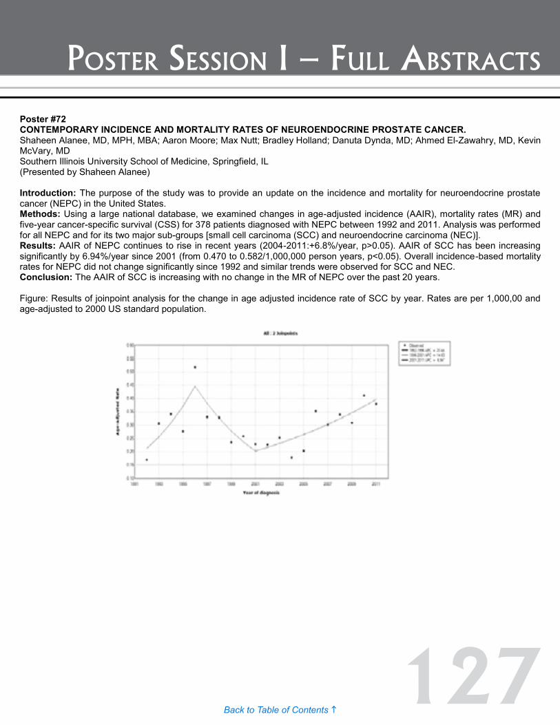
127Back to Table of Contents h
Poster session i – Full AbstrActs
Poster #72 CONTEMPORARY INCIDENCE AND MORTALITY RATES OF NEUROENDOCRINE PROSTATE CANCER. Shaheen Alanee, MD, MPH, MBA; Aaron Moore; Max Nutt; Bradley Holland; Danuta Dynda, MD; Ahmed El-Zawahry, MD, Kevin McVary, MD Southern Illinois University School of Medicine, Springfield, IL (Presented by Shaheen Alanee) Introduction: The purpose of the study was to provide an update on the incidence and mortality for neuroendocrine prostate cancer (NEPC) in the United States. Methods: Using a large national database, we examined changes in age-adjusted incidence (AAIR), mortality rates (MR) and five-year cancer-specific survival (CSS) for 378 patients diagnosed with NEPC between 1992 and 2011. Analysis was performed for all NEPC and for its two major sub-groups [small cell carcinoma (SCC) and neuroendocrine carcinoma (NEC)]. Results: AAIR of NEPC continues to rise in recent years (2004-2011:+6.8%/year, p>0.05). AAIR of SCC has been increasing significantly by 6.94%/year since 2001 (from 0.470 to 0.582/1,000,000 person years, p<0.05). Overall incidence-based mortality rates for NEPC did not change significantly since 1992 and similar trends were observed for SCC and NEC. Conclusion: The AAIR of SCC is increasing with no change in the MR of NEPC over the past 20 years. Figure: Results of joinpoint analysis for the change in age adjusted incidence rate of SCC by year. Rates are per 1,000,00 and age-adjusted to 2000 US standard population.
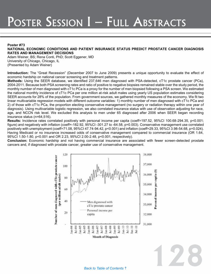
128Back to Table of Contents h
Poster session i – Full AbstrActs
Poster #74 VALIDATION OF A GENOMIC CLASSIFIER FOR PREDICTION OF METASTASIS FOLLOWING POSTOPERATIVE SALVAGE RADIATION THERAPY Stephen Freedland, MD1; Voleak Choeurng, MSc2; Lauren Howard, MSc3; Amanda De Hoedt, MSc4; Marguerite du Plessis, BSc2; Kasra Yousefi, MSc2; Lucia Lam, BSc2; Christine Buerki, PhD2; Edouard Trabulsi, MD5; Adam Dicker, PhD5; Elai Davicioni, PhD2; Jeffrey Karnes, MD6; Robert Den, MD5 1Cedars Sinai Medical Center; 2GenomeDx Biosciences Inc., Vancouver, BC, Canada; 3Department of Urology, Duke University, Durham, NC; 4Urology Research, Veteran Affairs Medical Centre, Durham, NC; 5Sidney Kimmel Medical College of Thomas Jefferson University, Philadelphia, PA; 6Department of Urology, Mayo Clinic, Rochester, MN (Presented by Stephen Freedland) Introduction: Management of patients with a postoperative rising prostate-specific antigen (PSA) level is complex. Additional local treatment such as salvage radiation therapy (SRT) may be sufficient for many patients but some may require concurrent systemic therapy in order to delay or prevent metastatic disease. As PSA recurrence on its own is a poor surrogate for metastatic disease we hypothesized that the Decipher genomic classifier (GC), a validated predictor of metastasis may be able to better distinguish those patients where additional therapy is beneficial from those where SRT on its own is likely sufficient. Methods: Genomic classifier (GC) scores were calculated from 170 prostate cancer patients, who received SRT at the Veteran Affairs Medical Center Durham, Thomas Jefferson University and Mayo Clinic, between 1990 and 2010. SRT was defined as administration of RT with Pre-RT PSA levels >0.2 ng/ml. GC and CAPRA-S scores were compared using survival c-index, competing-risks and Cox regression analysis for the prediction of metastasis. Results: Survival c-index for predicting metastasis five years post SRT was 0.85 (95% CI: 0.73-0.88) for GC and 0.63 (95% CI: 0.49-0.77) for CAPRA-S. The cumulative incidence of metastasis at 5 years post-SRT was 2.7%, 8.4%, and 33.1% for low, average, and high GC scores (p<0.001) and 16.9%, 2.3% and 17.2% for low, average and high CAPRA-S scores (p=0.113). In univariable analysis only GC, extraprostatic extension, path GS and Pre-RT PSA were significant predictors of metastasis. In multivariable analyses with clinical risk factors or the CAPRA-S risk model, GC was the only independent predictor of metastasis with a HR of 1.63 (1.22-2.18, p<0.001) for a 10% unit increase in risk score. Conclusion: In patients treated with postoperative SRT for PSA recurrence, GC is a powerful predictor of metastasis. Patients with low Decipher have excellent prognosis with SRT and may avoid concurrent hormonal therapy. Patients with high Decipher risk are at highest risk for metastatic disease and SRT failure and may benefit from intensified systemic therapy.
Poster #73 NATIONAL ECONOMIC CONDITIONS AND PATIENT INSURANCE STATUS PREDICT PROSTATE CANCER DIAGNOSIS RATES AND MANAGEMENT DECISIONS Adam Weiner, BS; Rena Conti, PhD; Scott Eggener, MD University of Chicago, Chicago, IL (Presented by Adam Weiner) Introduction: The “Great Recession” (December 2007 to June 2009) presents a unique opportunity to evaluate the effect of economic hardship on national cancer screening and treatment patterns. Methods: Using the SEER database, we identified 237,646 men diagnosed with PSA-detected, cT1c prostate cancer (PCa), 2004-2011. Because both PSA screening rates and ratio of positive to negative biopsies remained stable over the study period, the monthly number of men diagnosed with cT1c PCa is a proxy for the number of men biopsied following a PSA screen. We estimated the national monthly incidence of cT1c PCa per one million at-risk adult males using yearly US population estimates considering SEER accounts for 28% of the population. From government sources, we gathered monthly measures of the economy. We fit two linear multivariable regression models with different outcome variables: 1) monthly number of men diagnosed with cT1c PCa and 2) of those with cT1c PCa, the proportion electing conservative management (no surgery or radiation therapy within one year of diagnosis). Using multivariable logistic regression, we also correlated insurance status with use of observation adjusting for race, age, and NCCN risk level. We excluded this analysis to men under 65 diagnosed after 2006 when SEER began recording insurance status (n=64,516). Results: Incidence rates correlated positively with personal income per capita (coeff=197.52, 95%CI 100.68-294.35, p<0.001; figure) and negatively with inflation (coeff=-182.92, 95%CI -301.27 to -64.58, p=0.003). Conservative management use correlated positively with unemployment (coeff=71.08, 95%CI 47.74-94.42, p<0.001) and inflation (coeff=29.33, 95%CI 3.98-54.68, p=0.024). Having Medicaid or no insurance increased odds of conservative management compared to commercial insurance (OR 1.64, 95%CI 1.50-1.80, p<0.001 and OR 2.23, 95%CI 2.00-2.48, p<0.001, respectively). Conclusion: Economic hardship and not having commercial insurance are associated with fewer screen-detected prostate cancers and, if diagnosed with prostate cancer, greater use of conservative management.
129Back to Table of Contents h
Poster session i – Full AbstrActs
Poster #74 VALIDATION OF A GENOMIC CLASSIFIER FOR PREDICTION OF METASTASIS FOLLOWING POSTOPERATIVE SALVAGE RADIATION THERAPY Stephen Freedland, MD1; Voleak Choeurng, MSc2; Lauren Howard, MSc3; Amanda De Hoedt, MSc4; Marguerite du Plessis, BSc2; Kasra Yousefi, MSc2; Lucia Lam, BSc2; Christine Buerki, PhD2; Edouard Trabulsi, MD5; Adam Dicker, PhD5; Elai Davicioni, PhD2; Jeffrey Karnes, MD6; Robert Den, MD5 1Cedars Sinai Medical Center; 2GenomeDx Biosciences Inc., Vancouver, BC, Canada; 3Department of Urology, Duke University, Durham, NC; 4Urology Research, Veteran Affairs Medical Centre, Durham, NC; 5Sidney Kimmel Medical College of Thomas Jefferson University, Philadelphia, PA; 6Department of Urology, Mayo Clinic, Rochester, MN (Presented by Stephen Freedland) Introduction: Management of patients with a postoperative rising prostate-specific antigen (PSA) level is complex. Additional local treatment such as salvage radiation therapy (SRT) may be sufficient for many patients but some may require concurrent systemic therapy in order to delay or prevent metastatic disease. As PSA recurrence on its own is a poor surrogate for metastatic disease we hypothesized that the Decipher genomic classifier (GC), a validated predictor of metastasis may be able to better distinguish those patients where additional therapy is beneficial from those where SRT on its own is likely sufficient. Methods: Genomic classifier (GC) scores were calculated from 170 prostate cancer patients, who received SRT at the Veteran Affairs Medical Center Durham, Thomas Jefferson University and Mayo Clinic, between 1990 and 2010. SRT was defined as administration of RT with Pre-RT PSA levels >0.2 ng/ml. GC and CAPRA-S scores were compared using survival c-index, competing-risks and Cox regression analysis for the prediction of metastasis. Results: Survival c-index for predicting metastasis five years post SRT was 0.85 (95% CI: 0.73-0.88) for GC and 0.63 (95% CI: 0.49-0.77) for CAPRA-S. The cumulative incidence of metastasis at 5 years post-SRT was 2.7%, 8.4%, and 33.1% for low, average, and high GC scores (p<0.001) and 16.9%, 2.3% and 17.2% for low, average and high CAPRA-S scores (p=0.113). In univariable analysis only GC, extraprostatic extension, path GS and Pre-RT PSA were significant predictors of metastasis. In multivariable analyses with clinical risk factors or the CAPRA-S risk model, GC was the only independent predictor of metastasis with a HR of 1.63 (1.22-2.18, p<0.001) for a 10% unit increase in risk score. Conclusion: In patients treated with postoperative SRT for PSA recurrence, GC is a powerful predictor of metastasis. Patients with low Decipher have excellent prognosis with SRT and may avoid concurrent hormonal therapy. Patients with high Decipher risk are at highest risk for metastatic disease and SRT failure and may benefit from intensified systemic therapy.
130Back to Table of Contents h
Poster session i – Full AbstrActs
Poster #75 BIOPSY PATTERNS OF PATIENTS WITH HGPIN OR ASAP IN THE ERA OF ACTIVE SURVEILLANCE Zachary Panfili, MD; Zachary Hamilton, MD; Timothy Walmann, BS; Eugene Lee, MD; David Duchene, MD, FACS; Hadley Wyre, MD; Brantley Thrasher, MD, FACS; Jeffery Holzbeierlein, MD, FACS; Moben Mirza, MD, FACS University of Kansas Medical Center, Department of Urology. Kansas City, KS (Presented by Zachary Panfili) Introduction: To investigate the practice patterns of prostate biopsy in patients with high grade prostatic intraepithelial neoplasia (HGPIN) and atypical small acinar proliferation (ASAP). Methods: After collecting practice and demographic data, we surveyed urologists of the South Central Section on their re-biospy practices of patients with HGPIN or ASAP. Clinical scenarios included patients age 55 or 69 years old with a PSA of seven found to have three cores positive for HGPIN or ASAP on 12 core trans-rectal biopsy. Differences in selecting repeat PSA vs. re-biopsy based on academic vs. private practice, years of experience, and beliefs regarding the malignant potential of these lesions were examined. Results: 130 urologists completed the survey. Young patients with HGPIN were re-biopsied at three to six months and six to 12 months, 32.0% and 49.2% respectively. The same young patient with ASAP was re-biopsied at three to six months 70.9% of the time, (p=0.04). 62.2% of older patients with HGPIN had repeat PSA in six to 12 months and 17.7% were re-biopsied at three to six months. The same old patient with ASAP was re-biopsied 52.9% of the time, (p=0.01). There was no difference in management of HGPIN based on academic or private practice and years of experience regarding repeat PSA or re-biopsy. However, both young and old patients with ASAP were more likely to be re-biopsied in the academic setting (Young: 91.2% vs. 62.7%, p=0.01) and (Old: 70.6% vs. 45.9%, p=0.02); and by less experienced urologists (Young: 82.0% vs. 62.7%, p= 0.02) and (Old: 66.0% vs. 43.5%, p=0.02). HGPIN and ASAP are considered associated with prostate cancer 50.8% and 54.6%, (p=0.075) and premalignant by 33.1% and 41.5%, (p=0.481) of respondents respectively. Providers believing HGPIN is associated with prostate cancer re-biopsied young patients 68.3% and older patients 27.0% of the time (p=0.01). Those believing HGPIN is premalignant re-biopsied young patient 34.9% and older patients 17.7% of the time (p=0.623). Providers not believing HGPIN is associated with prostate cancer re-biopsied young patients 4.9% and older patient 0.0% of the time (p=0.01). Conclusion: In an era of active surveillance where low risk prostate cancer is re-biopsied at one year, our practice of performing re-biopsy of HGPIN and ASAP three to six months after diagnosis is incongruent with our current understanding of prostate cancer. HGPIN and ASAP are biologically different entities and our education and practice patterns need to be updated.

131Back to Table of Contents h
Poster session i – Full AbstrActs
Poster #76 RISK RECLASSIFICATION BY PROSTATE TISSUE GENE ANALYSIS: DOES IT CHANGE MANAGEMENT? David Earl, PharmD; Robin Willard-Niendorff, BS; Bevan Choate, MD; Frances Alba, MD; Satyan Shah, MD University of New Mexico School of Medicine Albuquerque, New Mexico (Presented by Bevan Choate) Introduction: Tissue-based gene analysis (TBGA) may improve risk stratification in prostate cancer. However, it is unclear if this new information changes management. We studied how reclassification of disease risk affects treatment decisions. Methods: All patients with adenocarcinoma of the prostate that underwent either Oncotype Dx or Prolaris testing between 2013 and 2015 were included. For each patient, we determined if clinical AUA risk category was reclassified by the test. For Oncotype Dx, this was obtained by converting reported NCCN risk into AUA risk category. On Prolaris reports, “more”or “less”aggressive disease was deemed a one category higher or lower reclassification in risk. “Considerably more”and “considerably less”was deemed a two category higher or lower reclassification in risk. Reclassifications outside the bounds of the 3-category system were not allowed. Risk was deemed unchanged in cases where insufficient disease precluded testing. Pre and post-testing treatment recommendations were compared. Results: TBGA was ordered in 75 patients (Oncotype Dx in 50 and Prolaris in 25). However, insufficient disease precluded testing in 18 patients (24%). Pre-testing risk was low in 48, intermediate in 24, and high in three patients. Reclassification rates were 2%, 25%, and 67% for low, intermediate and high risk respectively (p<0.001). This included eight reclassifications by Prolaris, and one by Oncotype Dx. Pre-testing treatment plan was surveillance/observation in 65 patients (87%) and definitive therapy in 10 patients (13%). TBGA changed this plan in three patients with Oncotype Dx and none for Prolaris. In all three patients, AUA risk category was not reclassified. However, the GPS score was felt to be too high (48, 54, 62) for surveillance. In the eight patients with risk reclassification by Prolaris, treatment plan was not changed because of elderly age driving surveillance in three, reduction in risk further supporting surveillance in four, and a plan for radical prostatectomy in one in whom risk was already intermediate but reclassified to high. Conclusion: TBGA rarely changed management in this largely surveillance cohort. TBGA was limited by the frequent inability to perform the test due to insufficient tissue. Intermediate/high risk disease was more likely to be reclassified than low risk disease.
132Back to Table of Contents h
Poster session i – Full AbstrActs
Poster #78 VALIDATION OF A GENOMIC CLASSIFIER FOR PREDICTING POST-PROSTATECTOMY RECURRENCE IN A COMMUNITY-BASED HEALTH CARE SETTING Andrew G. Glass, MD1; Michael C. Leo, PhD1; Zaid Haddad, BSc2; Kasra Yousefi, MSc2; Marguerite du Plessis, BSc2; Chuhe Chen, PhD1; Voleak Choeurng, MSc2; Firas Abdollah, MD3; Bruce Robbins, MD4; Seong Ra, MD4; Kathryn E. Richert-Boe, MD1; Christine Buerki, PhD2; Kathy Pearson, MPH1; Elai Davicioni, PhD5; Sheila Weinmann, PhD1 1Center for Health Research, Kaiser Permanente Northwest, Portland, OR; 2GenomeDx Biosciences Inc., Vancouver, BC, Canada; 3Vattikuti Urology Institute, Henry Ford Hospital, Detroit, MI; 4San Diego Pathologists Medical Group, San Diego, CA; 5GenomeDx Biosciences Inc., San Diego, California (Presented by Elai Davicioni) Introduction: Radical prostatectomy (RP) is a primary treatment option for men with intermediate and high-risk prostate cancer. Though many will be effectively cured with local therapy alone, these men are by definition at higher risk of adverse pathologic findings and disease recurrence. In this study, we aimed to evaluate the value of Decipher, a genomic classifier, to predict prostate cancer outcomes among patients following RP in a community health care setting. Methods: We examined the experience of 224 men treated with radical prostatectomy (RP) between 1997 and 2009 at Kaiser Permanente Northwest, a large prepaid health plan in Portland, Oregon. Study subjects had aggressive prostate cancer with at least one of the following: pre-operative prostate specific antigen (PSA) ≥ 20 ng/ml, Gleason score ≥ 8, stage pT3, positive surgical margins at prostatectomy. The primary endpoint was clinical recurrence or metastasis after surgery evaluated using a time-dependent c-index using ROC curves. Secondary endpoints were biochemical recurrence and salvage treatment failure. We compared the performance of the genomic classifier, Decipher, alone to the widely used Cancer of the Prostate Risk Assessment Postsurgical (CAPRA-S) score and assessed the independent contributions of Decipher, CAPRA-S and their combination for the prediction of recurrence and treatment failure. Results: Of the 224 cases, 12 experienced clinical recurrence, 68 had biochemical recurrence and 34 failed salvage treatment. At 10 years post-RP, the recurrence rate was 2.6% among patients with low Decipher scores but 13.6% among those with high Decipher scores (p=0.02). The discrimination accuracy of the ROC curve for CAPRA-S was 0.73, 0.80 for Decipher and 0.84 for the combined model for predicting clinical recurrence 10 years post RP. In multivariable analysis only age at RP and Decipher remained significant predictors of outcome. Similar results were observed for biochemical recurrence and salvage treatment failure endpoints. Conclusion: Decipher improves our ability to predict clinical recurrence in prostate cancer patients treated in a community-based setting and adds precision to conventional pathological predictive measures.
Poster #77 CHRONIC BASELINE PROSTATE INFLAMMATION IS ASSOCIATED WITH LOWER TUMOR GRADE IN MEN WITH PROSTATE CANCER ON REPEAT BIOPSY: Results: FROM THE REDUCE STUDY Daniel Moreira, MD, MHS1; J. Curtis Nickel, MD2; Gerald Andriole, MD3; Ramiro Castro-Santamaria, MD4; Stephen Freedland, MD5 1Rochester; 2Department of Urology, Queen's University, Kingston, ON, Canada; 3Division of Urologic Surgery, Department of Surgery, Washington University School of Medicine, St. Louis, MO; 4GlaxoSmithKline Inc., Global R&D Unit, King of Prussia, PA; 5Division of Urology, Department of Surgery, Cedars-Sinai Medical Center, Los Angeles, CA (Presented by Daniel Moreira) Introduction: We have previously shown that chronic baseline prostate inflammation in an otherwise benign biopsy was associated with lower risk of prostate cancer in repeat prostate biopsies and lower tumor volumes for those who are diagnosed with cancer. In the present study, we evaluated whether baseline acute or chronic prostate inflammation among men with initial negative biopsies for prostate cancer was associated with cancer grade at the two-year repeat prostate biopsy in a clinical trial with systematic biopsies regardless of PSA. Methods: Retrospective analysis of 889 men 50-75 years old with negative baseline prostate biopsy and positive two-year repeat biopsy for prostate cancer in the Reduction by Dutasteride of Prostate Cancer Events (REDUCE) study. Acute and chronic prostate inflammation (coded as present or absent) and cancer grade were determined by central pathology at the time of the REDUCE study. The association of inflammation in baseline biopsies with two-year repeat biopsy cancer grade (low-grade: Gleason scores 2-6 vs. high-grade: Gleason scores 7-10) was evaluated with Student t test, chi-squared test and logistic regression controlling for age, race, body-mass index (BMI), digital rectal exam (DRE), prostate volume, baseline pre-study PSA and treatment arm (dutasteride or placebo). Results: Chronic, acute inflammation and both were detected in 533 (60%), 12 (1%) and 85 (10%) baseline biopsies, respectively. Presence of acute and chronic inflammations were significantly associated with each other (P<0.001). Patients with chronic inflammation had significantly larger prostates (P<0.001). Both types of inflammation were unrelated to race, BMI, PSA or DRE. At two-year biopsy, a total of 621 (70%) tumors were low-grade and 268 (30%) tumors were high-grade. In both uni- and multivariable analyses, men with baseline chronic inflammation had significantly less high-grade tumors (univariable odds ratio [OR] = 0.64, 95% confidence interval [CI] = 0.47-0.87, P = 0.004; multivariable OR = 0.68, 95% CI = 0.50-0.93, P = 0.016) than those without baseline chronic inflammation. Baseline acute inflammation was not associated with tumor grade. Conclusion: In a cohort of men undergoing repeat prostate biopsy two years after a negative baseline biopsy who all had cancer on the follow-up biopsy, the presence baseline chronic inflammation was associated with lower prostate cancer grade.
133Back to Table of Contents h
Poster session i – Full AbstrActs
Poster #78 VALIDATION OF A GENOMIC CLASSIFIER FOR PREDICTING POST-PROSTATECTOMY RECURRENCE IN A COMMUNITY-BASED HEALTH CARE SETTING Andrew G. Glass, MD1; Michael C. Leo, PhD1; Zaid Haddad, BSc2; Kasra Yousefi, MSc2; Marguerite du Plessis, BSc2; Chuhe Chen, PhD1; Voleak Choeurng, MSc2; Firas Abdollah, MD3; Bruce Robbins, MD4; Seong Ra, MD4; Kathryn E. Richert-Boe, MD1; Christine Buerki, PhD2; Kathy Pearson, MPH1; Elai Davicioni, PhD5; Sheila Weinmann, PhD1 1Center for Health Research, Kaiser Permanente Northwest, Portland, OR; 2GenomeDx Biosciences Inc., Vancouver, BC, Canada; 3Vattikuti Urology Institute, Henry Ford Hospital, Detroit, MI; 4San Diego Pathologists Medical Group, San Diego, CA; 5GenomeDx Biosciences Inc., San Diego, California (Presented by Elai Davicioni) Introduction: Radical prostatectomy (RP) is a primary treatment option for men with intermediate and high-risk prostate cancer. Though many will be effectively cured with local therapy alone, these men are by definition at higher risk of adverse pathologic findings and disease recurrence. In this study, we aimed to evaluate the value of Decipher, a genomic classifier, to predict prostate cancer outcomes among patients following RP in a community health care setting. Methods: We examined the experience of 224 men treated with radical prostatectomy (RP) between 1997 and 2009 at Kaiser Permanente Northwest, a large prepaid health plan in Portland, Oregon. Study subjects had aggressive prostate cancer with at least one of the following: pre-operative prostate specific antigen (PSA) ≥ 20 ng/ml, Gleason score ≥ 8, stage pT3, positive surgical margins at prostatectomy. The primary endpoint was clinical recurrence or metastasis after surgery evaluated using a time-dependent c-index using ROC curves. Secondary endpoints were biochemical recurrence and salvage treatment failure. We compared the performance of the genomic classifier, Decipher, alone to the widely used Cancer of the Prostate Risk Assessment Postsurgical (CAPRA-S) score and assessed the independent contributions of Decipher, CAPRA-S and their combination for the prediction of recurrence and treatment failure. Results: Of the 224 cases, 12 experienced clinical recurrence, 68 had biochemical recurrence and 34 failed salvage treatment. At 10 years post-RP, the recurrence rate was 2.6% among patients with low Decipher scores but 13.6% among those with high Decipher scores (p=0.02). The discrimination accuracy of the ROC curve for CAPRA-S was 0.73, 0.80 for Decipher and 0.84 for the combined model for predicting clinical recurrence 10 years post RP. In multivariable analysis only age at RP and Decipher remained significant predictors of outcome. Similar results were observed for biochemical recurrence and salvage treatment failure endpoints. Conclusion: Decipher improves our ability to predict clinical recurrence in prostate cancer patients treated in a community-based setting and adds precision to conventional pathological predictive measures.
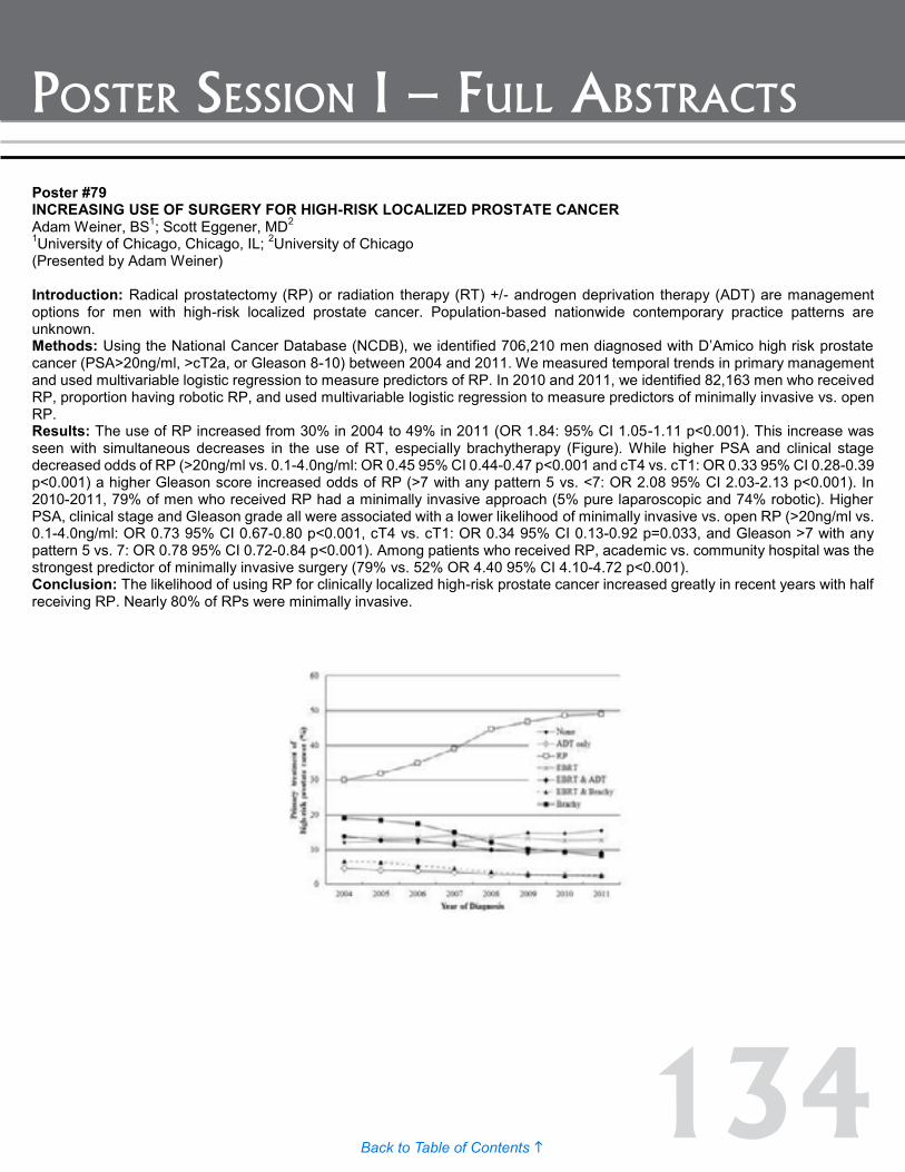
134Back to Table of Contents h
Poster session i – Full AbstrActs
Poster #79 INCREASING USE OF SURGERY FOR HIGH-RISK LOCALIZED PROSTATE CANCER Adam Weiner, BS1; Scott Eggener, MD2 1University of Chicago, Chicago, IL; 2University of Chicago (Presented by Adam Weiner) Introduction: Radical prostatectomy (RP) or radiation therapy (RT) +/- androgen deprivation therapy (ADT) are management options for men with high-risk localized prostate cancer. Population-based nationwide contemporary practice patterns are unknown. Methods: Using the National Cancer Database (NCDB), we identified 706,210 men diagnosed with D’Amico high risk prostate cancer (PSA>20ng/ml, >cT2a, or Gleason 8-10) between 2004 and 2011. We measured temporal trends in primary management and used multivariable logistic regression to measure predictors of RP. In 2010 and 2011, we identified 82,163 men who received RP, proportion having robotic RP, and used multivariable logistic regression to measure predictors of minimally invasive vs. open RP. Results: The use of RP increased from 30% in 2004 to 49% in 2011 (OR 1.84: 95% CI 1.05-1.11 p<0.001). This increase was seen with simultaneous decreases in the use of RT, especially brachytherapy (Figure). While higher PSA and clinical stage decreased odds of RP (>20ng/ml vs. 0.1-4.0ng/ml: OR 0.45 95% CI 0.44-0.47 p<0.001 and cT4 vs. cT1: OR 0.33 95% CI 0.28-0.39 p<0.001) a higher Gleason score increased odds of RP (>7 with any pattern 5 vs. <7: OR 2.08 95% CI 2.03-2.13 p<0.001). In 2010-2011, 79% of men who received RP had a minimally invasive approach (5% pure laparoscopic and 74% robotic). Higher PSA, clinical stage and Gleason grade all were associated with a lower likelihood of minimally invasive vs. open RP (>20ng/ml vs. 0.1-4.0ng/ml: OR 0.73 95% CI 0.67-0.80 p<0.001, cT4 vs. cT1: OR 0.34 95% CI 0.13-0.92 p=0.033, and Gleason >7 with any pattern 5 vs. 7: OR 0.78 95% CI 0.72-0.84 p<0.001). Among patients who received RP, academic vs. community hospital was the strongest predictor of minimally invasive surgery (79% vs. 52% OR 4.40 95% CI 4.10-4.72 p<0.001). Conclusion: The likelihood of using RP for clinically localized high-risk prostate cancer increased greatly in recent years with half receiving RP. Nearly 80% of RPs were minimally invasive.
135Back to Table of Contents h
Poster session i – Full AbstrActs
Poster #80 COMPARATIVE EFFECTIVENESS OF CANCER CONTROL AND SURVIVAL AFTER ROBOTIC ASSISTED VERSUS OPEN RADICAL PROSTATECTOMY Jim C. Hu, MD, MPH1; Bilal Chughtai, MD1; Padraic O'Malley, MSc, MD, FRCSC1; Abby Isaacs, MS2; Jason D. Wright, MD3; Dawn Hershman, MD, MS4; Art Sedrakyan, MD, PhD2 1Department of Urology, Weill Cornell Medical College, New York, NY; 2Department of Healthcare Policy and Research, Weill Cornell Medical College, New York, NY; 3Department of Obstetrics and Gynecology, Herbert Irving Comprehensive Cancer Center, Columbia University College of Physicians and Surgeons, New York, NY; 4Department of Medicine, Department of Epidemiology, Mailman School of Public Health, Herbert Irving Comprehensive Cancer Center, Columbia University College of Physicians and Surgeons, New York, NY (Presented by Padraic O’Malley) Introduction: Robotic-assisted surgery has been rapidly adopted in the United States for treatment of prostate cancer. The purpose of the study is to examine comparative effectiveness of robotic assisted (RARP) versus open radical prostatectomy (ORP) in terms of cancer control and survival in a nationally representative population. Methods: Retrospective, observational study of 6,430 RARP and 9,161 ORP performed during 2003 to 2012 using Surveillance, Epidemiology, and End results (SEER)-Medicare linked data. Propensity-based analyses were performed to adjust for confounders. Cox-proportional hazard models and Kaplan-Meier analysis were used to compare cancer control (as defined by use of post-radical prostatectomy radiation and/or androgen deprivation therapy) and overall and prostate cancer specific survival. Results: In the propensity-matched analysis, RARP was associated with less use of post-operative androgen deprivation (ADT) and radiation therapy (Hazard Ratio [HR] 0.74 95% CI 0.67-0.81). After median follow-up of 6.5 years (IQR 5.2-7.9), RARP was associated with a lower risk of all-cause mortality (HR 0.79 [0.67-0.93]) but similar cancer specific mortality (HR 0.73, [0.46-1.15]) compared to ORP. Conclusion: RARP is associated with better intermediate cancer control, as evidenced by less use of additional postoperative cancer therapies and better overall survival. Longer-term follow-up is needed to assess for differences in prostate cancer specific survival, which was similar during our intermediate followup. Our findings have significant quality and cost implications and provide reassurance regarding the adoption of a more expensive technology in the absence of randomized controlled trials. Poster #81 VASECTOMY AND RISK OF PROSTATE CANCER IN A SCREENING TRIAL Jonathan Shoag, MD1; Sameer Mittal, MD1; Joshua A. Halpern, MD1; Daniel J. Lee, MD1; Padraic O'Malley, MSc, MD, FRCSC1; Bobby Najari, MD1; Brian Eisner, MD2; Peter N. Schlegel, MD1; Douglas S. Scherr, MD1; Christopher E. Barbieri, MD, PhD1 1Department of Urology, Weill Cornell Medical College, New York, NY; 2Department of Urology, Massachusetts General Hospital, Boston, MA (Presented by Padraic O’Malley) Introduction: Vasectomy has been implicated as a risk factor for prostate cancer in multiple epidemiologic studies over the past 25 years. Whether this relationship is causal remains unclear. Methods: We analyzed data from the Prostate, Lung, Colorectal, and Ovarian (PLCO) cancer screening trial to assess the relationship between vasectomy and prostate cancer. Analysis was stratified by study arm in order to assess, and control for, detection bias. Results: Consistent with prior studies we identified an increased risk of prostate cancer in men who had undergone vasectomy and were randomized to the unscreened arm of the study (adjusted HR 1.11 95% CI 1.03-1.20, p=0.008). There was no association between vasectomy and diagnosis of prostate cancer in men randomized to the screening arm (adjusted HR 1.03 95% CI 0.95-1.11, p=0.469). Only men who had vasectomies at an older age in the control arm of the study, but not the screening arm, were at increased risk of being diagnosed with prostate cancer. Conclusion: These data suggest that in a modern cohort of patients the association of vasectomy with prostate cancer is related to increased detection of prostate cancer based on patterns of care rather than a biological effect of the vasectomy procedure on prostate cancer development
136Back to Table of Contents h
Poster session i – Full AbstrActs
Poster #82 PERIOPERATIVE BLOOD TRANSFUSION AND RADICAL PROSTATECTOMY: ANALYSIS OF THE NATIONAL SURGICAL QUALITY IMPROVEMENT PROGRAM DATABASE Justin Matulay, Wilson Sui, Ifeanyi Onyeji, Maxwell James, Marissa Velez, G. Joel DeCastro Department of Urology, Columbia University Medical Center, New York, NY (Presented by Justin Matulay) Introduction: Radical prostatectomy remains a mainstay in the treatment of clinically significant localized prostate cancer. The literature suggests that perioperative blood transfusion (PBT) in oncologic surgeries may lead to poorer long-term outcomes of recurrence, progression, and survival, for colon, pancreatic, esophageal and gynecologic malignancies. Previous studies have not shown this to be true for prostatectomy but there has yet to be a study examing the impact of PBT on 30-day post-operative outcomes. Methods: We used the National Surgical Quality Improvement (NSQIP) database to retrospectively analyze 30-day post-operative outcomes in men undergoing radical prostatectomy from 2005-2013. NSQIP Participant Use Files were queried using ICD-9 codes for prostate cancer (185) and matched with CPT codes for laparoscopic (55866) and open (55801, 55810, 55812, 55815, 55821, 55831, 55840, 55842, 55845) prostatectomy. Our primary outcome was 30-day post-operative morbidity. Secondary outcomes included 30-day mortality, readmission, length of stay, infectious complications, and pulmonary complications. Results: A total of 21293 radical prostatectomies were performed of which 810 (3.9%) received PBT. Baseline patient age, race, and smoking history did not differ between groups. The transfusion group was more likely to be ASA class ≥3 (38.7% v. 32.7%; p=0.001) and have preoperative anemia (31.8% v. 19.3%; p<0.001) but less likely to be obese (29.3% v. 35.5%; p<0.002). Transfusion was less common in robotic versus any open approach (29.6% v. 67.0%; p<0.001). On multivariate analysis, 30-day morbidity (OR 2.36 95% 95% CI: 1.75-3.17; p<0.001), readmission (OR 2.00 95% CI: 1.39-2.88; p<0.001), pulmonary complications (OR 3.80 95% CI: 2.11-6.86; p<0.001) and surgical site infections (OR 2.58 95% CI: 1.69-3.93; p<0.001) were all increased in patients receiving PBT. Conclusion: Perioperative blood transfusion is associated with increased 30-day post-operative morbidity, hospital readmission, pulmonary complications, and infectious complications. This phenomenon may be due to immunomodulation induced by elevated pro-inflammatory cytokine levels in banked blood in addition to a recipient response to allogeneic tissue. We have again demonstrated that blood transfusion is more common in open approaches. Our findings support the push in recent years towards more restrictive transfusion thresholds.
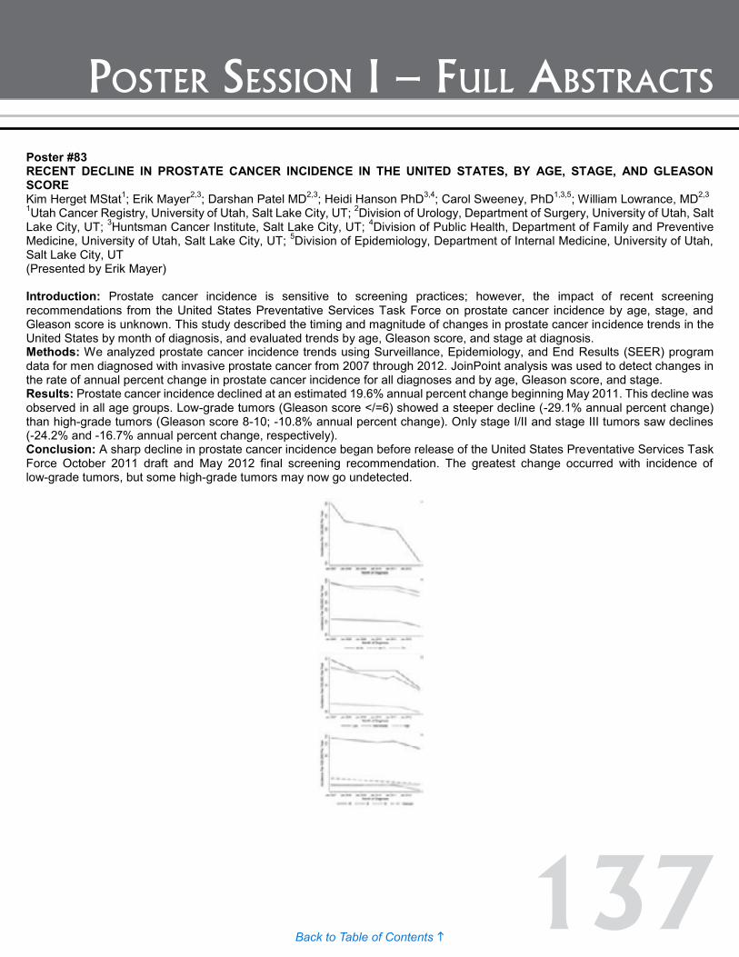
137Back to Table of Contents h
Poster session i – Full AbstrActs
Poster #83 RECENT DECLINE IN PROSTATE CANCER INCIDENCE IN THE UNITED STATES, BY AGE, STAGE, AND GLEASON SCORE Kim Herget MStat1; Erik Mayer2,3; Darshan Patel MD2,3; Heidi Hanson PhD3,4; Carol Sweeney, PhD1,3,5; William Lowrance, MD2,3 1Utah Cancer Registry, University of Utah, Salt Lake City, UT; 2Division of Urology, Department of Surgery, University of Utah, Salt Lake City, UT; 3Huntsman Cancer Institute, Salt Lake City, UT; 4Division of Public Health, Department of Family and Preventive Medicine, University of Utah, Salt Lake City, UT; 5Division of Epidemiology, Department of Internal Medicine, University of Utah, Salt Lake City, UT (Presented by Erik Mayer) Introduction: Prostate cancer incidence is sensitive to screening practices; however, the impact of recent screening recommendations from the United States Preventative Services Task Force on prostate cancer incidence by age, stage, and Gleason score is unknown. This study described the timing and magnitude of changes in prostate cancer incidence trends in the United States by month of diagnosis, and evaluated trends by age, Gleason score, and stage at diagnosis. Methods: We analyzed prostate cancer incidence trends using Surveillance, Epidemiology, and End Results (SEER) program data for men diagnosed with invasive prostate cancer from 2007 through 2012. JoinPoint analysis was used to detect changes in the rate of annual percent change in prostate cancer incidence for all diagnoses and by age, Gleason score, and stage. Results: Prostate cancer incidence declined at an estimated 19.6% annual percent change beginning May 2011. This decline was observed in all age groups. Low-grade tumors (Gleason score </=6) showed a steeper decline (-29.1% annual percent change) than high-grade tumors (Gleason score 8-10; -10.8% annual percent change). Only stage I/II and stage III tumors saw declines (-24.2% and -16.7% annual percent change, respectively). Conclusion: A sharp decline in prostate cancer incidence began before release of the United States Preventative Services Task Force October 2011 draft and May 2012 final screening recommendation. The greatest change occurred with incidence of low-grade tumors, but some high-grade tumors may now go undetected.
Poster #82 PERIOPERATIVE BLOOD TRANSFUSION AND RADICAL PROSTATECTOMY: ANALYSIS OF THE NATIONAL SURGICAL QUALITY IMPROVEMENT PROGRAM DATABASE Justin Matulay, Wilson Sui, Ifeanyi Onyeji, Maxwell James, Marissa Velez, G. Joel DeCastro Department of Urology, Columbia University Medical Center, New York, NY (Presented by Justin Matulay) Introduction: Radical prostatectomy remains a mainstay in the treatment of clinically significant localized prostate cancer. The literature suggests that perioperative blood transfusion (PBT) in oncologic surgeries may lead to poorer long-term outcomes of recurrence, progression, and survival, for colon, pancreatic, esophageal and gynecologic malignancies. Previous studies have not shown this to be true for prostatectomy but there has yet to be a study examing the impact of PBT on 30-day post-operative outcomes. Methods: We used the National Surgical Quality Improvement (NSQIP) database to retrospectively analyze 30-day post-operative outcomes in men undergoing radical prostatectomy from 2005-2013. NSQIP Participant Use Files were queried using ICD-9 codes for prostate cancer (185) and matched with CPT codes for laparoscopic (55866) and open (55801, 55810, 55812, 55815, 55821, 55831, 55840, 55842, 55845) prostatectomy. Our primary outcome was 30-day post-operative morbidity. Secondary outcomes included 30-day mortality, readmission, length of stay, infectious complications, and pulmonary complications. Results: A total of 21293 radical prostatectomies were performed of which 810 (3.9%) received PBT. Baseline patient age, race, and smoking history did not differ between groups. The transfusion group was more likely to be ASA class ≥3 (38.7% v. 32.7%; p=0.001) and have preoperative anemia (31.8% v. 19.3%; p<0.001) but less likely to be obese (29.3% v. 35.5%; p<0.002). Transfusion was less common in robotic versus any open approach (29.6% v. 67.0%; p<0.001). On multivariate analysis, 30-day morbidity (OR 2.36 95% 95% CI: 1.75-3.17; p<0.001), readmission (OR 2.00 95% CI: 1.39-2.88; p<0.001), pulmonary complications (OR 3.80 95% CI: 2.11-6.86; p<0.001) and surgical site infections (OR 2.58 95% CI: 1.69-3.93; p<0.001) were all increased in patients receiving PBT. Conclusion: Perioperative blood transfusion is associated with increased 30-day post-operative morbidity, hospital readmission, pulmonary complications, and infectious complications. This phenomenon may be due to immunomodulation induced by elevated pro-inflammatory cytokine levels in banked blood in addition to a recipient response to allogeneic tissue. We have again demonstrated that blood transfusion is more common in open approaches. Our findings support the push in recent years towards more restrictive transfusion thresholds.
138Back to Table of Contents h
Poster session i – Full AbstrActs
Poster #84 CAN PSA DENSITY AND FREE-TO-TOTAL PSA RATIO IMPROVE OUR ABILITY TO PREDICT PROSTATE CANCER ON BIOPSY? Results: FROM A PROSPECTIVE, MULTI-INSTITUTIONAL, AND CONTEMPORARY COHORT Samarpit Rai, MD1, Nachiketh Soodana-Prakash, MD, MS1; Nicola Pavan, MD2; Bruno Nahar, MD1; Amil Patel1; Yan Dong PhD3; Ramgopal Satyanarayana, MD1; Dipen J. Parekh, MD1; Sanoj Punnen, MD1 1Department of Urology, University of Miami Leonard M. Miller School of Medicine, Miami, FL; 2Department of Urology, University of Miami Leonard M. Miller School of Medicine, Miami, FL and Urology Clinic, Department of Medical, Surgical and Health Science, University of Trieste, Italy; 3OPKO Diagnostics, LLC, Nashville, TN (Presented by Samarpit Rai) Introduction: Several studies have reported an increased value of PSA density (PSAD) and free-to-total PSA ratio (f/t PSA) over PSA alone in predicting prostate cancer (PCa). However, both these derivatives appear to be underutilized in current clinical practice. This study analyzed a contemporary cohort of men referred for prostate biopsy (PB) to determine if PSAD and f/t PSA enhanced the prediction of any PCa and/or significant PCa (Gleason score ≥ 7) compared to PSA alone. Methods: 1,370 prospectively enrolled patients were referred for a PB for suspicion of PCa across 26 urological centers within the nation. A phlebotomy was performed immediately prior to PB for PSA and f/t PSA measurement. PSAD was calculated using the prostate volume obtained during the trans-rectal ultrasound (TRUS) guided PB. Histopathologic examination was performed according to the established practice at each study site. The area under the receiver operating characteristic curve (AUC) was used to assess the added discriminative value of PSAD and f/t PSA when added to a base model consisting of PSA, age, prior biopsy status, and DRE for the prediction of any PCa and significant PCa. Results: Of the 1,290 men in the final cohort, 301 (23%) and 284 (22%) men were diagnosed with low-grade PCa (Gleason score = 6) and significant PCa respectively. The median PSAD values in men with no PCa, low-grade PCa, and significant PCa were 0.09, 0.11, and 0.17 ng/mL/cc, respectively (P < 0.0001). The median f/t PSA in men with no PCa, low-grade PCa, and significant PCa was 0.21, 0.17, and 0.12 respectively (P < 0.0001). The AUC for a model incorporating PSAD showed superior predictive value compared to the base model for diagnosing any PCa (AUC 0.76 versus 0.70, P < 0.0001) and significant PCa (AUC 0.82 versus 0.77, P < 0.0001). Similarly, a model with f/t PSA showed superior predictive value compared to the base model for diagnosing any PCa (AUC 0.73 vs. 0.70, P < 0.0001) and significant PCa (AUC 0.82 versus 0.77, P < 0.0001). While PSAD showed superior predictive value over f/t PSA for predicting any PCa (AUC 0.76 versus 0.73, P = 0.0062), there was no difference in their discrimination of significant PCa. Conclusion: PSAD and f/t PSA add substantial predictive power to the diagnostic armamentarium for any PCa and significant PCa. Their calculation may aid in the early detection of aggressive PCa, while reducing the number of unnecessary biopsies being performed.
139Back to Table of Contents h
Poster session i – Full AbstrActs
Poster #85 DEFINING THE OPTIMAL PSA RANGE FOR THE MAXIMAL PREDICTIVE EFFICACY OF PSA DENSITY TO DETECT PROSTATE CANCER ON BIOPSY: Results: FROM A MULTI-INSTITUTIONAL, PROSPECTIVE, AND CONTEMPORARY COHORT Samarpit Rai, MD1; Nicola Pavan, MD2; Nachiketh Soodana-Prakash, MD, MS1; Bruno Nahar, MD1; Yan Dong, PhD3; Ramgopal Satyanarayana, MD1; Dipen J. Parekh, MD1; Sanoj Punnen, MD1 1Department of Urology, University of Miami Leonard M. Miller School of Medicine, Miami, FL; 2Department of Urology, University of Miami Leonard M. Miller School of Medicine, Miami, FL and Urology Clinic, Department of Medical, Surgical and Health Science, University of Trieste, Italy; 3OPKO Diagnostics, LLC, Nashville, TN (Presented by Samarpit Rai) Introduction: PSA density (PSAD) is an important predictor of prostate cancer (PCa). We assessed whether the predictive accuracy of PSAD varied based on the range of PSA or whether the patient had a previous negative biopsy. Methods: We assessed a contemporary and prospective cohort of men who were referred for biopsy of the prostate for suspicion of PCa at 26 different sites across the United States. The area under the receiver operating characteristic curve (AUC) was used to assess the added predictive accuracy of PSAD versus PSA across 3 different PSA ranges (<4, 4 – 10, >10 ng/mL) and in men with or without a prior negative biopsy for the detection of any and significant (Gleason > 7) PCa. Results: Of the 1,290 patients in the final cohort, 585 (45%) men were diagnosed with PCa and 284 (22%) men were diagnosed with significant PCa. PSAD was significantly more predictive than PSA for detecting any PCa in the PSA ranges of 4 – 10 (AUC 0.70 vs. 0.53, P<0.00001) and >10 (AUC 0.84 vs. 0.65, P<0.00001) ng/mL. Similarly, for significant PCa, PSAD was more predictive than PSA in the PSA ranges of 4 – 10 (AUC 0.72 vs. 0.57, P<0.00001) and >10 (AUC 0.82 vs. 0.68, P = 0.0001) ng/mL. Furthermore, PSAD was significantly more predictive than PSA in detecting PCa in men that had a prior negative prostate biopsy (AUC 0.69 vs. 0.56, P = 0.0001 for any PCa and AUC 0.81 vs. 0.70, P = 0.0042 for significant PCa), and those that didn’t (AUC 0.72 vs. 0.67, P = 0.0001 for any PCa and AUC 0.77 vs. 0.73, P = 0.0026 for significant PCa). However, the difference between the AUC of PSAD and PSA (ΔAUC) was a lot more pronounced in men that had a prior negative prostate biopsy (ΔAUC = 0.13 for any PCa and ΔAUC = 0.11 for significant PCa) as opposed to those that didn’t (ΔAUC = 0.05 for any PCa and ΔAUC = 0.04 for significant PCa), suggesting that PSAD is a much better predictor than PSA alone in men who have undergone a previous biopsy. Conclusion: As PSA increases, the predictive accuracy of PSAD over PSA appears to improve for the detection of any PCa and significant PCa. Additionally, PSAD has a more pronounced predictive value over PSA in detecting any and significant PCa in men who have undergone a prior negative biopsy. We support the use of PSAD testing to avoid unnecessary biopsies in men who have elevated PSA secondary to an enlarged prostate.
Poster #84 CAN PSA DENSITY AND FREE-TO-TOTAL PSA RATIO IMPROVE OUR ABILITY TO PREDICT PROSTATE CANCER ON BIOPSY? Results: FROM A PROSPECTIVE, MULTI-INSTITUTIONAL, AND CONTEMPORARY COHORT Samarpit Rai, MD1, Nachiketh Soodana-Prakash, MD, MS1; Nicola Pavan, MD2; Bruno Nahar, MD1; Amil Patel1; Yan Dong PhD3; Ramgopal Satyanarayana, MD1; Dipen J. Parekh, MD1; Sanoj Punnen, MD1 1Department of Urology, University of Miami Leonard M. Miller School of Medicine, Miami, FL; 2Department of Urology, University of Miami Leonard M. Miller School of Medicine, Miami, FL and Urology Clinic, Department of Medical, Surgical and Health Science, University of Trieste, Italy; 3OPKO Diagnostics, LLC, Nashville, TN (Presented by Samarpit Rai) Introduction: Several studies have reported an increased value of PSA density (PSAD) and free-to-total PSA ratio (f/t PSA) over PSA alone in predicting prostate cancer (PCa). However, both these derivatives appear to be underutilized in current clinical practice. This study analyzed a contemporary cohort of men referred for prostate biopsy (PB) to determine if PSAD and f/t PSA enhanced the prediction of any PCa and/or significant PCa (Gleason score ≥ 7) compared to PSA alone. Methods: 1,370 prospectively enrolled patients were referred for a PB for suspicion of PCa across 26 urological centers within the nation. A phlebotomy was performed immediately prior to PB for PSA and f/t PSA measurement. PSAD was calculated using the prostate volume obtained during the trans-rectal ultrasound (TRUS) guided PB. Histopathologic examination was performed according to the established practice at each study site. The area under the receiver operating characteristic curve (AUC) was used to assess the added discriminative value of PSAD and f/t PSA when added to a base model consisting of PSA, age, prior biopsy status, and DRE for the prediction of any PCa and significant PCa. Results: Of the 1,290 men in the final cohort, 301 (23%) and 284 (22%) men were diagnosed with low-grade PCa (Gleason score = 6) and significant PCa respectively. The median PSAD values in men with no PCa, low-grade PCa, and significant PCa were 0.09, 0.11, and 0.17 ng/mL/cc, respectively (P < 0.0001). The median f/t PSA in men with no PCa, low-grade PCa, and significant PCa was 0.21, 0.17, and 0.12 respectively (P < 0.0001). The AUC for a model incorporating PSAD showed superior predictive value compared to the base model for diagnosing any PCa (AUC 0.76 versus 0.70, P < 0.0001) and significant PCa (AUC 0.82 versus 0.77, P < 0.0001). Similarly, a model with f/t PSA showed superior predictive value compared to the base model for diagnosing any PCa (AUC 0.73 vs. 0.70, P < 0.0001) and significant PCa (AUC 0.82 versus 0.77, P < 0.0001). While PSAD showed superior predictive value over f/t PSA for predicting any PCa (AUC 0.76 versus 0.73, P = 0.0062), there was no difference in their discrimination of significant PCa. Conclusion: PSAD and f/t PSA add substantial predictive power to the diagnostic armamentarium for any PCa and significant PCa. Their calculation may aid in the early detection of aggressive PCa, while reducing the number of unnecessary biopsies being performed.
140Back to Table of Contents h
Poster session i – Full AbstrActs
Poster #86 PREDICT, A STUDY EVALUATING BASELINE DISEASE CHARACTERISTICS PREDICTIVE OF A POSITIVE IMAGING STUDY FOR DISTANT METASTASES IN PATIENTS WITH CASTRATION-RESISTANT PROSTATE CANCER: PRELIMINARY DATA Stephen J. Freedland, MD1; Raoul S. Concepcion, MD2; Matthew R. Smith, MD3; Christopher Pieczonka, MD4; Son Nguyen, MD5; Laurence H. Belkoff, MD6; Robert S. Firlit, MD7; Tim Van Mouwerik, MD8; Neal D. Shore, MD9 1Cedars Sinai Medical Center, Los Angeles, CA; 2Urology Associates P.C., Nashville, TN; 3Massachusetts General Hospital, MA; 4Associated Medical Professionals, Syracuse, NY; 5Kansas City Urology Care, PA; 6Urologic Consultants of Southeastern Pennsylvania, PA; 7TBD, Palos Heights, IL; 8Dendreon Pharmaceuticals, Inc, Seattle, WA; 9Carolina Urologic Research Center, Urology, Myrtle Beach, SC (Presented by Neal D. Shore) Introduction: At diagnosis, most men with PC have localized disease with no distant metastases (M0) or regional lymphadenopathy (N0). After therapy, 20–40% of men have biochemical recurrence, often treated with androgen deprivation therapy (ADT). Other patients may receive primary ADT for N0/M0 disease. Both groups are at risk of castration-resistant prostate cancer (CRPC), ie rising prostate-specific antigen (PSA) in absence of radiographic proven metastatic disease (M0). Metrics predicting a positive imaging test for men presumed M0 CRPC are not universally accepted/implemented. PREDICT’s primary objective is to evaluate characteristics predictive of a baseline (BL) imaging study positive for distant metastases (M1) in men thought to have M0 CRPC. Methods: Eligible men have M0 CRPC, no prior M1, no imaging in over three months and are classified as M0 or M1 based on BL bone and soft tissue imaging. Patients will be followed for 3 (M0) to 5 (M1) years after last patient registration. Results: Sixty-five men have BL scans and interim descriptive BL data. At BL, 18/65 (28%) men were M1; 37 men had Tc99 bone scintigraphy and 28 had NaF PET/CT scans to assess bone metastases (scan type at Investigator discretion). Scans were positive for bone metastases in 4/37 (10.8%) Tc99 scans vs. 9/28 (32.1%) NaF PET/CT (p=0.06). Five men had only soft tissue lesions. The M1 group, vs. M0 group, were older (84 vs. 79 yrs), more likely to have Eastern Cooperative Oncology Group (ECOG) performance status >0 (44 vs. 32%), a Gleason score >=8 (44 vs. 34%), and had a shorter time from diagnosis to study entry (6.6 vs. 12.6 years). BL median laboratory parameters were higher in the M1 vs. M0 group for serum PSA (10.1 vs. 4.8 ng/mL), alkaline phosphatase (88 vs. 73 U/L), lactate dehydrogenase (200 vs. 186 U/L), and serum prostatic acid phosphatase (2.6 vs. 1.2 ng/mL). Conclusion: These real-world, preliminary data from a small group of men with presumed M0 CRPC suggest that undetected M1 is common, as is M1 occurrence in soft tissues only. M1 detection appeared higher in the small number of men having a NaF PET/CT scan. This study will contribute to clinical guidance on optimal timing, frequency, and imaging study modality for detecting M1 CRPC. Funding: Dendreon Pharmaceuticals, Inc
141Back to Table of Contents h
Poster session i – Full AbstrActs
Poster #87 THE ASSOCIATION OF ANDROGEN METABOLISM GENE POLYMORPHISMS WITH PROSTATE CANCER RISK AND STEROID HORMONE CONCENTRATIONS FROM THE PROSTATE CANCER PREVENTION TRIAL Douglas Price, PhD1; Cindy Chau, PharmD, PhD2; Cathee Till, MS3; Phylllis Goodman, MS3; Robin Leach, PhD4; Teresa Johnson-Pais, PhD4; Ann Hsing, PhD5; Ashraful Hoque, PhD6; Howard Parnes, MD2; Jeannette Schenk, PhD3; Catherine Tangen, DrPH3; Ian Thompson, MD4; Juergen Reichardt, PhD7; William Figg, PharmD2 1NCI; 2NCI, Bethesda, MD; 3FHCRC, Seattle WA; 4UTHSCSA, San Antonio, TX; 5CPIP, Fremont, CA; 6UTMDACC, Houston, TX; 7JCU, Townsville, QLD, AU (Presented by Douglas Price) Introduction: We investigated the association of genetic polymorphisms in genes of the androgen biosynthesis and metabolism pathway with prostate cancer risk using a nested case-control study of the Prostate Cancer Prevention Trial (PCPT) population. We also examined whether these polymorphisms are responsible for altering serum androgen concentrations. Methods: Data for this nested case-control study are from the PCPT. Cases were drawn from men with biopsy-proven prostate cancer and matched controls. We genotyped a total of 51 SNPs in the following genes in the androgen metabolism pathway: SRD5A1, SRD5A2, SRD5A2L/SRD5A3, HSD3B2, HSD17B2, HSD17B3, CYP1B1, CYP3A4, CYP3A5, CYP3A43, CYP17, and CYP19. Results: There were statistically significant associations of SNPs in SRD5A1 (rs3736316, rs3822430, rs1560149, rs248797, rs472402) and SRD5A2 (rs2300700) with risk of high-grade prostate cancer in the placebo arm of the PCPT. Eleven SNPs in SRD5A1, SRD5A2, CYP1B1, and CYP3A4 were found to be associated with modifying mean baseline serum androgen and SHBG concentrations of all participants; two SNPs (SRD5A1 rs824811 and CYP1B1 rs10012) consistently and significantly altered all serum androgen and SHBG concentrations. Several SNPs were significantly associated with both circulating hormone levels and prostate cancer risk. Conclusion: Germline genetic variations of androgen-related pathway genes may contribute to the risk of prostate cancer and modify steroid hormone serum concentrations. Additional studies to validate these results and examine the functional consequence of causal variants are warranted. Poster #88 IMPACT OF A FAMILIAL HISTORY OF PROSTATE CANCER ON CLINICOPATHOLOGIC OUTCOMES AND SURVIVAL FOLLOWING RADICAL PROSTATECTOMY Mary E. Westerman, MD1; Boris Gershman, MD1; R. Jeffrey Karnes, MD1; R. Houston Thompson, MD1; Laureano Rangel2; Stephen Boorjian, MD1 1Mayo Clinic Dept of Urology, Rochester MN; 2Department of Health Sciences Research, Mayo Clinic, Rochester, Minnesota (Presented by Mary E. Westerman) Introduction: While a family history (FH) of prostate cancer (PCa) represents an established risk factor for subsequent diagnosis in men, conflicting data exist regarding its impact on oncologic outcomes. Herein, we evaluated the association of FH with clinicopathologic outcomes among men undergoing radical prostatectomy (RP). Methods: We identified 16,472 men who underwent RP between 1987 and 2010. Patients were considered to have a positive FH if at least one first degree relative had been diagnosed with PCa. Survival was estimated using the Kaplan-Meier method. Associations of FH with clinicopathologic features and survival were evaluated using logistic and Cox regression, respectively. Results: Overall, 5,323 (32.3%) men reported a FH of prostate cancer. Median follow-up was 9.9 years (IQR 5.9, 15.5), during which time 4,430 (26.9%) men died, including 558 (3.4%) who died of PCa. Patients with a FH were significantly more likely to have low-risk disease at diagnosis (47.7% vs. 43.0%, p<0.0001) and organ confined disease at RP (79.2% vs. 74.4%, p<0.0001). Men with FH had a significantly higher 10-year cancer-specific (99% vs. 97%, p<0.001) and overall survival (92% vs. 85%, p<0.001) than men without FH. Moreover, on multivariable analysis, FH of PCa remained independently associated with reduced cancer-specific (HR 0.65, 95% CI: 0.500-0.832, p=0.0007) and all-cause mortality (HR 0.68, 95% CI: 0.622-0.745, p<0.0001). Conclusion: In this surgical cohort, FH of prostate cancer was associated with lower risk disease at diagnosis, more favorable pathology at RP, and significantly better CSS and OS. These results may utilized for patient risk-stratification and counseling.
Poster #86 PREDICT, A STUDY EVALUATING BASELINE DISEASE CHARACTERISTICS PREDICTIVE OF A POSITIVE IMAGING STUDY FOR DISTANT METASTASES IN PATIENTS WITH CASTRATION-RESISTANT PROSTATE CANCER: PRELIMINARY DATA Stephen J. Freedland, MD1; Raoul S. Concepcion, MD2; Matthew R. Smith, MD3; Christopher Pieczonka, MD4; Son Nguyen, MD5; Laurence H. Belkoff, MD6; Robert S. Firlit, MD7; Tim Van Mouwerik, MD8; Neal D. Shore, MD9 1Cedars Sinai Medical Center, Los Angeles, CA; 2Urology Associates P.C., Nashville, TN; 3Massachusetts General Hospital, MA; 4Associated Medical Professionals, Syracuse, NY; 5Kansas City Urology Care, PA; 6Urologic Consultants of Southeastern Pennsylvania, PA; 7TBD, Palos Heights, IL; 8Dendreon Pharmaceuticals, Inc, Seattle, WA; 9Carolina Urologic Research Center, Urology, Myrtle Beach, SC (Presented by Neal D. Shore) Introduction: At diagnosis, most men with PC have localized disease with no distant metastases (M0) or regional lymphadenopathy (N0). After therapy, 20–40% of men have biochemical recurrence, often treated with androgen deprivation therapy (ADT). Other patients may receive primary ADT for N0/M0 disease. Both groups are at risk of castration-resistant prostate cancer (CRPC), ie rising prostate-specific antigen (PSA) in absence of radiographic proven metastatic disease (M0). Metrics predicting a positive imaging test for men presumed M0 CRPC are not universally accepted/implemented. PREDICT’s primary objective is to evaluate characteristics predictive of a baseline (BL) imaging study positive for distant metastases (M1) in men thought to have M0 CRPC. Methods: Eligible men have M0 CRPC, no prior M1, no imaging in over three months and are classified as M0 or M1 based on BL bone and soft tissue imaging. Patients will be followed for 3 (M0) to 5 (M1) years after last patient registration. Results: Sixty-five men have BL scans and interim descriptive BL data. At BL, 18/65 (28%) men were M1; 37 men had Tc99 bone scintigraphy and 28 had NaF PET/CT scans to assess bone metastases (scan type at Investigator discretion). Scans were positive for bone metastases in 4/37 (10.8%) Tc99 scans vs. 9/28 (32.1%) NaF PET/CT (p=0.06). Five men had only soft tissue lesions. The M1 group, vs. M0 group, were older (84 vs. 79 yrs), more likely to have Eastern Cooperative Oncology Group (ECOG) performance status >0 (44 vs. 32%), a Gleason score >=8 (44 vs. 34%), and had a shorter time from diagnosis to study entry (6.6 vs. 12.6 years). BL median laboratory parameters were higher in the M1 vs. M0 group for serum PSA (10.1 vs. 4.8 ng/mL), alkaline phosphatase (88 vs. 73 U/L), lactate dehydrogenase (200 vs. 186 U/L), and serum prostatic acid phosphatase (2.6 vs. 1.2 ng/mL). Conclusion: These real-world, preliminary data from a small group of men with presumed M0 CRPC suggest that undetected M1 is common, as is M1 occurrence in soft tissues only. M1 detection appeared higher in the small number of men having a NaF PET/CT scan. This study will contribute to clinical guidance on optimal timing, frequency, and imaging study modality for detecting M1 CRPC. Funding: Dendreon Pharmaceuticals, Inc
142Back to Table of Contents h
Poster session i – Full AbstrActs
Poster #89 A PROSPECTIVE STUDY OF HEALTH-RELATED QUALITY OF LIFE OUTCOMES FOR LOW-RISK PROSTATE CANCER PATIENTS MANAGED BY ACTIVE SURVEILLANCE OR RADIATION THERAPY John Banerji, MD, MCh (Urolog)1; Lauren Hurwitz, MHS2; Jennifer Cullen, PhD, MPH2; Erika Wolff, PhD1; Katherine Levie, CCRP2; Khani Pham, MD1; Katherin Odem-Davis, PhD3;Christopher Porter, MD, FACS1 1Dept. of Urology, Virginia Mason, Seattle,Washington; 2Center for Prostate Diesase Research, Department of Defense, Rockville, MD; 3Center for BioMedical Statistics, University of Washington, Seattle, Washington (Presented by John Banerji) Introduction: Patients with low-risk prostate cancer (PCa) generally have excellent oncologic outcomes. However, treatment with curative intent, such as external beam radiotherapy (EBRT), may have greater physical and psychological consequences compared to active surveillance (AS). Since health related quality of life (HRQoL) concerns are paramount when selecting among treatment options, this study sought to elucidate whether, when compared to EBRT, AS offers better HRQoL. Methods: A prospective cohort study of HRQoL in PCa patients enrolled in the Center for Prostate Disease Research Multicenter National Database was initiated in 2007. HRQoL was assessed with the Expanded Prostate Cancer Index Composite (EPIC) and the Medical Outcomes Study Short Form (SF36). Temporal changes in HRQoL were compared for low-risk PCa patients managed on AS versus EBRT at baseline, one, two, and three years post diagnosis. Longitudinal patterns were modeled using generalized estimating equations (GEE) for repeated measures, adjusting for baseline HRQoL, demographic and clinical characteristics. Bonferroni corrections were performed to adjust for multiple comparisons. Results: Of the 499 eligible low-risk PCa patients, 103 (21%) selected AS and 60 (12%) were treated with EBRT. Demographic characteristics of the treatment groups were essentially similar. Of note though, 43% of the EBRT cohort were African Americans (AA), while only 16% of of the AS cohort were AA (p=0.0003). Of the patients receiving EBRT, 61% received a dose of 78 Gy or more. At baseline, both groups reported comparable HRQoL. EBRT patients experienced significantly worse bowel function and bother at one year (adjusted mean score: 87 vs. 95, p=0.001 and 89 vs. 95, p=0.008, respectively) and two years (87 vs. 93, p=0.007 and 87 vs. 96, p=0.002, respectively). There were no statistically significant differences in adjusted mean scores at three years. Finally, at one year, patients who received EBRT were significantly more likely to experience a decrease in more than one EPIC domain when compared to those on AS (60% vs. 28%, p=0.004 for function and 54% vs. 33%, p=0.06 for bother, respectively). Conclusion: Patients receiving EBRT for low-risk prostate cancer suffer declines in bowel function and bother. These declines are not experienced by patients on AS, suggesting that management of low-risk prostate cancer with AS may offer a means for preserving HRQoL following prostate cancer diagnosis.
143Back to Table of Contents h
Poster session i – Full AbstrActs
Poster #90 UTILIZATION OF RADIOTHERAPY FOR PROSTATE CANCER ACCORDING TO UROLOGISTS’ PRACTICE PATTERNS Stephen Williams, MD; Jinhai Huo, PhD; Benjamin Smith, MD; Karen Hoffman, MD MD Anderson Houston, TX (Presented by Stephen Williams) Introduction: Physician practices that offer ancillary medical services may refer their patients for such services, a process known as self-referral. Self-referral for radiation therapy is prevalent in the urologic community, yet its impact on utilization and cost of prostate cancer care is not known. We evaluated how utilization and cost of care differ for men diagnosed with prostate cancer in a self-referring urologic practice (SRP) compared to a traditional urologic practice. Methods: 17,982 men 66 years and older diagnosed with localized prostate cancer from 2006 to 2009 were identified from the Texas Cancer Registry. Diagnosing urologist, cancer-directed therapy, comorbid medical conditions and healthcare costs were determined from linked Medicare claims. Disease was classified as favorable if low-grade and clinical T1 or T2. The diagnosing urologist was classified as being affiliated with a SRP if their practice owned a linear accelerator. Multilevel logistic regression models evaluated the odds of receiving a specific type of treatment adjusted for diagnosis year, age, comorbidities, race/ethnicity, income, education, clinical tumor stage, and tumor grade. Cost of care was calculated from Medicare expenditures within 12 months of diagnosis Results: Diagnosis in a SRP increased from 2.2% of prostate cancers in 2004 to 24.5% in 2009 (p< 0.001). Men diagnosed in SRPs were more likely to receive upfront treatment (vs. watchful waiting/active surveillance) than men diagnosed by traditional practices (92.7% vs. 89%; AOR 1.61, 95%CI 1.30-2.00; p<0.001) and were more likely to be treated with external beam radiation (47.4% vs. 34.1%; AOR 1.59, 95%CI 1.37-1.84; P<0.001). This persisted for both favorable and unfavorable risk cancer. Men diagnosed in SRPs were more likely to receive upfront treatment (favorable: 92.9% vs. 87.1%; AOR 1.89, 95%CI 1.33-2.69; p<0.001; unfavorable: 97.9% vs. 95.0%; AOR 2.07, 95%CI 1.32-3.25; p=0.002) and more likely to be treated with EBRT (favorable: 41.2% vs. 31.1%; AOR 1.45, 95%CI 1.15-1.84; p=0.002; unfavorable: 49.0% vs. 38.1%; AOR 1.53, 95%CI 1.24-1.88; p<0.001). Median annual prostate cancer care cost was $2460 (95% CI $1663-$3368) higher for men diagnosed by SRPs. Conclusion: Older men diagnosed with prostate cancer in SRPs are more likely to undergo upfront treatment and to receive radiation treatment. This may increase appropriate treatment of unfavorable disease but contribute to overtreatment of favorable disease.
Poster #89 A PROSPECTIVE STUDY OF HEALTH-RELATED QUALITY OF LIFE OUTCOMES FOR LOW-RISK PROSTATE CANCER PATIENTS MANAGED BY ACTIVE SURVEILLANCE OR RADIATION THERAPY John Banerji, MD, MCh (Urolog)1; Lauren Hurwitz, MHS2; Jennifer Cullen, PhD, MPH2; Erika Wolff, PhD1; Katherine Levie, CCRP2; Khani Pham, MD1; Katherin Odem-Davis, PhD3;Christopher Porter, MD, FACS1 1Dept. of Urology, Virginia Mason, Seattle,Washington; 2Center for Prostate Diesase Research, Department of Defense, Rockville, MD; 3Center for BioMedical Statistics, University of Washington, Seattle, Washington (Presented by John Banerji) Introduction: Patients with low-risk prostate cancer (PCa) generally have excellent oncologic outcomes. However, treatment with curative intent, such as external beam radiotherapy (EBRT), may have greater physical and psychological consequences compared to active surveillance (AS). Since health related quality of life (HRQoL) concerns are paramount when selecting among treatment options, this study sought to elucidate whether, when compared to EBRT, AS offers better HRQoL. Methods: A prospective cohort study of HRQoL in PCa patients enrolled in the Center for Prostate Disease Research Multicenter National Database was initiated in 2007. HRQoL was assessed with the Expanded Prostate Cancer Index Composite (EPIC) and the Medical Outcomes Study Short Form (SF36). Temporal changes in HRQoL were compared for low-risk PCa patients managed on AS versus EBRT at baseline, one, two, and three years post diagnosis. Longitudinal patterns were modeled using generalized estimating equations (GEE) for repeated measures, adjusting for baseline HRQoL, demographic and clinical characteristics. Bonferroni corrections were performed to adjust for multiple comparisons. Results: Of the 499 eligible low-risk PCa patients, 103 (21%) selected AS and 60 (12%) were treated with EBRT. Demographic characteristics of the treatment groups were essentially similar. Of note though, 43% of the EBRT cohort were African Americans (AA), while only 16% of of the AS cohort were AA (p=0.0003). Of the patients receiving EBRT, 61% received a dose of 78 Gy or more. At baseline, both groups reported comparable HRQoL. EBRT patients experienced significantly worse bowel function and bother at one year (adjusted mean score: 87 vs. 95, p=0.001 and 89 vs. 95, p=0.008, respectively) and two years (87 vs. 93, p=0.007 and 87 vs. 96, p=0.002, respectively). There were no statistically significant differences in adjusted mean scores at three years. Finally, at one year, patients who received EBRT were significantly more likely to experience a decrease in more than one EPIC domain when compared to those on AS (60% vs. 28%, p=0.004 for function and 54% vs. 33%, p=0.06 for bother, respectively). Conclusion: Patients receiving EBRT for low-risk prostate cancer suffer declines in bowel function and bother. These declines are not experienced by patients on AS, suggesting that management of low-risk prostate cancer with AS may offer a means for preserving HRQoL following prostate cancer diagnosis.
144Back to Table of Contents h
Poster session i – Full AbstrActs
Poster #91 AMONG MEN WITH LOW-GRADE PROSTATE CANCER ON PROSTATE BIOPSY, THE 4KSCORE PREDICTS MORE AGGRESSIVE PROSTATE CANCER AT PROSTATECTOMY Sanoj Punnen, MD1; Bruno Nahar, MD1; Daniel Sjoberg2; Stephen Zappala, MD, FACS3; Dipen Parekh, MD1 1University of Miami Miller School of Medicine Department of Urology, Miami FL; 2Memorial Sloan Kettering, New York City NY; 3Andover Urology, Andover MA (Presented by Bruno Nahar) Introduction: Most men diagnosed with prostate cancer in the United States are found to have low-grade tumors. While many of these men are good candidates for active surveillance, a proportion will have a bad outcome due to the presence of a more aggressive prostate cancer that was missed on initial biopsy. A recent prospective study confirmed the 4Kscore accurately predicts the likelihood of aggressive cancer on prostate biopsy. We wanted to see if the 4Kscore could predict the presence of more significant cancer in a population of men with low-grade tumors on the diagnostic biopsy. Methods: A recent multi-institutional prospective validation of the 4Kscore was conducted at 26 sites throughout the United States. We selected men who were found to have low-grade (Gleason 6) cancer on biopsy for this analysis. The 4Kscore calculates the risk of aggressive prostate cancer on prostate biopsy by a blood test that measures levels of four kallikrein biomarkers (total PSA, free PSA, intact PSA, and human kallikrein-2) plus age, DRE findings, and prior biopsy status. We investigated whether the 4Kscore was associated with more significant cancer among men found to have Gleason 6 cancer on prostate biopsy. We also looked at a subset of these men who underwent radical prostatectomy to see if the 4Kscore was associated with prostate cancer being upgraded in the surgical specimen. Results: Among the 1312 men enrolled in this trial, 306 men were found to have Gleason 6 cancer on prostate biopsy. The 4Kscore was significantly associated with the number of positive cores (p=0.001) and the millimeters of cancer seen (p=0.0002), with higher 4Kscores relating to more extensive cancer present on biopsy. In the subpopulation of 51 men who underwent radical prostatectomy, the median 4Kscore was significantly higher among men who had an upgrade to Gleason 7 or higher [15% (8,25)] compared to men who did not experience an upgrade [7% 4,14)] (p=0.032) in their final pathology. Conclusion: Among men with Gleason 6 prostate cancer on biopsy, we found the 4Kscore was associated with the prostate cancer being upgraded in the surgical specimen at radical prostatectomy. The 4Kscore test may facilitate the selection of men who can be observed versus those who should undergo immediate treatment.
145Back to Table of Contents h
Poster session i – Full AbstrActs
Poster #92 THE 4KSCORE PREDICTS THE GRADE AND STAGE OF PROSTATE CANCER IN THE RADICAL PROSTATECTOMY SPECIMEN; Results: FROM A MULTI-INSTITUTIONAL PROSPECTIVE TRIAL Sanoj Punnen, MD, MAS1; Bruno Nahar, MD1; Daniel Sjoberg2; Stephen Zappala, MD, FRACS3; Dipen Parekh, MD1 1University of Miami Miller School of Medicine Department of Urology, Miami FL; 2Memorial Sloan Kettering Cancer Center, New York City, NY; 3Andover Urology, Andover MA (Presented by Bruno Nahar) Introduction: A recent prospective validation study confirmed the 4Kscore accurately predicted aggressive prostate cancer on prostate biopsy. We investigated the association between the 4Kscore and pathologic grade and stage at radical prostatectomy, where the entire prostate gland is sampled. Methods: Prospective enrollment of 1312 men who were referred for prostate biopsy for clinical suspicion of prostate cancer occurred at 26 sites throughout the United States from October 2013 to April 2014. We selected men who were found to have positive prostate cancer biopsies and elected to undergo radical prostatectomy. The 4Kscore is an algorithm that incorporates a panel of 4 Kallikreins (total PSA, free PSA, intact PSA and human kallikrein-2) in addition to age, digital rectal examination, and prior biopsy status. We assessed the concordance between the 4Kscore prior to biopsy and grade of prostate cancer at radical prostatectomy. 4Kscore test results were compared for those with and without non organ-confined tumors at surgery using the Wilcoxon rank-sum. Results: Among the 1312 men who enrolled in this validation study, 144 were found to have prostate cancer and underwent radical prostatectomy. We saw a significant association between the 4Kscore and grade at surgery with higher scores relating to worse grade. For men with Gleason 6, 7, and 8 or higher cancers in the surgical specimen the median (IQR) 4Kscore was 7% (4, 12), 25% (12, 38), and 47% (24, 66) (p<0.0001). The median 4Kscore among men with non organ-confined cancer was significantly higher then men with cancers confined to the prostate (36% [IQR 19,58] vs. 19% [IQR 9, 35], p=0.002). Conclusion: In a subset of men who underwent radical prostatectomy, the 4Kscore was significantly associated with pathological grade and extracapsular extension in the surgical specimen, with higher scores being linked to higher grade and more aggressive histology. The test can be beneficial to aid in treatment decision making for men who are contemplating observation of their cancer versus immediate treatment.
Poster #91 AMONG MEN WITH LOW-GRADE PROSTATE CANCER ON PROSTATE BIOPSY, THE 4KSCORE PREDICTS MORE AGGRESSIVE PROSTATE CANCER AT PROSTATECTOMY Sanoj Punnen, MD1; Bruno Nahar, MD1; Daniel Sjoberg2; Stephen Zappala, MD, FACS3; Dipen Parekh, MD1 1University of Miami Miller School of Medicine Department of Urology, Miami FL; 2Memorial Sloan Kettering, New York City NY; 3Andover Urology, Andover MA (Presented by Bruno Nahar) Introduction: Most men diagnosed with prostate cancer in the United States are found to have low-grade tumors. While many of these men are good candidates for active surveillance, a proportion will have a bad outcome due to the presence of a more aggressive prostate cancer that was missed on initial biopsy. A recent prospective study confirmed the 4Kscore accurately predicts the likelihood of aggressive cancer on prostate biopsy. We wanted to see if the 4Kscore could predict the presence of more significant cancer in a population of men with low-grade tumors on the diagnostic biopsy. Methods: A recent multi-institutional prospective validation of the 4Kscore was conducted at 26 sites throughout the United States. We selected men who were found to have low-grade (Gleason 6) cancer on biopsy for this analysis. The 4Kscore calculates the risk of aggressive prostate cancer on prostate biopsy by a blood test that measures levels of four kallikrein biomarkers (total PSA, free PSA, intact PSA, and human kallikrein-2) plus age, DRE findings, and prior biopsy status. We investigated whether the 4Kscore was associated with more significant cancer among men found to have Gleason 6 cancer on prostate biopsy. We also looked at a subset of these men who underwent radical prostatectomy to see if the 4Kscore was associated with prostate cancer being upgraded in the surgical specimen. Results: Among the 1312 men enrolled in this trial, 306 men were found to have Gleason 6 cancer on prostate biopsy. The 4Kscore was significantly associated with the number of positive cores (p=0.001) and the millimeters of cancer seen (p=0.0002), with higher 4Kscores relating to more extensive cancer present on biopsy. In the subpopulation of 51 men who underwent radical prostatectomy, the median 4Kscore was significantly higher among men who had an upgrade to Gleason 7 or higher [15% (8,25)] compared to men who did not experience an upgrade [7% 4,14)] (p=0.032) in their final pathology. Conclusion: Among men with Gleason 6 prostate cancer on biopsy, we found the 4Kscore was associated with the prostate cancer being upgraded in the surgical specimen at radical prostatectomy. The 4Kscore test may facilitate the selection of men who can be observed versus those who should undergo immediate treatment.
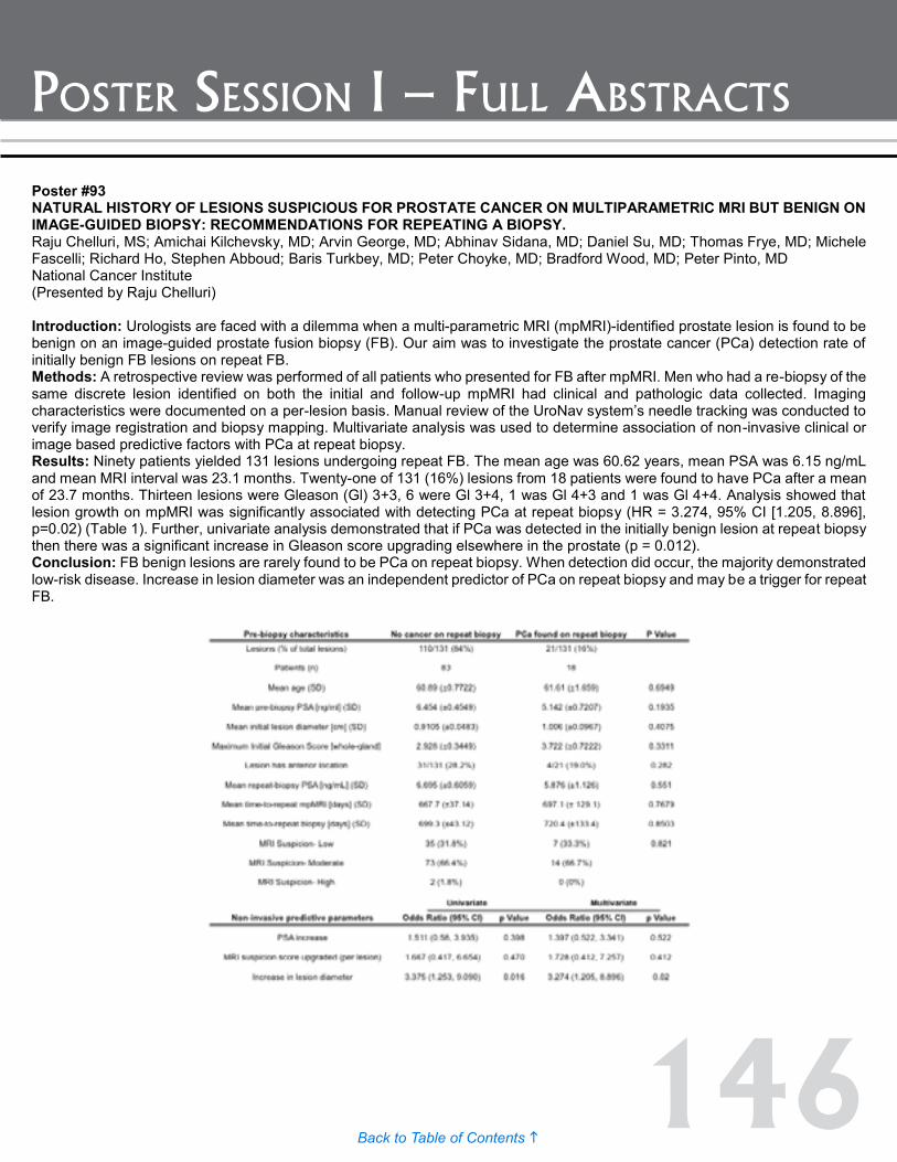
146Back to Table of Contents h
Poster session i – Full AbstrActs
Poster #93 NATURAL HISTORY OF LESIONS SUSPICIOUS FOR PROSTATE CANCER ON MULTIPARAMETRIC MRI BUT BENIGN ON IMAGE-GUIDED BIOPSY: RECOMMENDATIONS FOR REPEATING A BIOPSY. Raju Chelluri, MS; Amichai Kilchevsky, MD; Arvin George, MD; Abhinav Sidana, MD; Daniel Su, MD; Thomas Frye, MD; Michele Fascelli; Richard Ho, Stephen Abboud; Baris Turkbey, MD; Peter Choyke, MD; Bradford Wood, MD; Peter Pinto, MD National Cancer Institute (Presented by Raju Chelluri) Introduction: Urologists are faced with a dilemma when a multi-parametric MRI (mpMRI)-identified prostate lesion is found to be benign on an image-guided prostate fusion biopsy (FB). Our aim was to investigate the prostate cancer (PCa) detection rate of initially benign FB lesions on repeat FB. Methods: A retrospective review was performed of all patients who presented for FB after mpMRI. Men who had a re-biopsy of the same discrete lesion identified on both the initial and follow-up mpMRI had clinical and pathologic data collected. Imaging characteristics were documented on a per-lesion basis. Manual review of the UroNav system’s needle tracking was conducted to verify image registration and biopsy mapping. Multivariate analysis was used to determine association of non-invasive clinical or image based predictive factors with PCa at repeat biopsy. Results: Ninety patients yielded 131 lesions undergoing repeat FB. The mean age was 60.62 years, mean PSA was 6.15 ng/mL and mean MRI interval was 23.1 months. Twenty-one of 131 (16%) lesions from 18 patients were found to have PCa after a mean of 23.7 months. Thirteen lesions were Gleason (Gl) 3+3, 6 were Gl 3+4, 1 was Gl 4+3 and 1 was Gl 4+4. Analysis showed that lesion growth on mpMRI was significantly associated with detecting PCa at repeat biopsy (HR = 3.274, 95% CI [1.205, 8.896], p=0.02) (Table 1). Further, univariate analysis demonstrated that if PCa was detected in the initially benign lesion at repeat biopsy then there was a significant increase in Gleason score upgrading elsewhere in the prostate (p = 0.012). Conclusion: FB benign lesions are rarely found to be PCa on repeat biopsy. When detection did occur, the majority demonstrated low-risk disease. Increase in lesion diameter was an independent predictor of PCa on repeat biopsy and may be a trigger for repeat FB.
147Back to Table of Contents h
Poster session i – Full AbstrActs
Poster #94 SIGNIFICANT REDUCTION IN THERAPEUTIC BURDEN FROM USE OF CCP TEST IN TREATMENT DECISIONS AMONG NEWLY DIAGNOSED PROSTATE CANCER PATIENTS IN A LARGE PROSPECTIVE REGISTRY Neal Shore, MD1; Judd Boczko, MD2; Naveen Kella, MD3; Brian J. Moran, MD4; Fernando J. Bianco, MD5; E. David Crawford, MD6; Stephanie A. Hamilton, BA, MBA7; Rajesh Kaldate, MS7; Kirstin M. Roundy, MS7; Michael K. Brawer, MD7 ; Mark L. Gonzalgo, MD, PhD8 1Carolina Urologic Research Center, Myrtle Beach, SC; 2WESTMED Medical Group, White Plains, NY; 3The Urology and Prostate Institute, San Antonio, TX; 4Prostate Cancer Foundation of Chicago, Westomont, IL; 5Urological Research Network, Miami Lakes, FL; 6University of Colorado at Denver, Aurora, CO; 7Myriad Genetic Laboratories, Inc., Salt Lake City, UT; 8University of Miami Miller School of Medicine, Miami, FL (Presented by Neal D. Shore) Introduction: The cell cycle progression (CCP) test is a validated molecular assay that assesses risk of prostate cancer-specific disease progression and mortality when combined with standard clinicopathologic parameters. PROCEDE-1000 is the largest prospective registry to evaluate CCP test impact on personalizing prostate cancer treatment. results of the final analysis of 1206 patients are presented. Methods: Untreated patients with newly diagnosed (≤6 months), clinically localized prostate adenocarcinoma were enrolled. The physician’s initial therapy recommendation (pre-CCP) was recorded on the first questionnaire. The CCP test was then conducted on prostate biopsy tissue. Three post-CCP questionnaires recorded the physician’s revised treatment recommendation, physician/patient treatment decision, and actual treatment administered. Changes in treatments between the pre-CCP and post-CCP questionnaires demonstrated the impact of CCP testing on treatment decisions at each stage. Results: There was a significant reduction in the treatment burden recorded at each successive evaluation (P<0.0001), with mean number of treatments per patient decreasing from 1.72 pre-CCP test to 1.16 in actual follow-up. From pre-CCP therapy recommendation, the CCP risk score caused a change in actual treatment administered in 48% of patients; of these changes, 72% were reductions in treatment. These reductions occurred in radical prostatectomy (34%), radiation therapy (39% primary; 55% adjuvant), brachytherapy (46% interstitial; 63% HDR) and hormonal therapy (30% neoadjuvant; 50% concurrent) treatments. Although a considerably high percentage of patients (34.6%; 417/1206) were recommended for conservative management pre-CCP testing, a further 2.6% increase overall was recorded for non-interventional treatments during actual follow-up. Conclusion: The CCP risk assessment score has a significant impact in helping physicians and patients reach consensus on an appropriate personalized treatment decision, often with major reductions in interventional treatment burden. Financial Disclosure: Study funded by Myriad Genetic Laboratories, Inc.
148Back to Table of Contents h
Poster session i – Full AbstrActs
Poster #95 TAK-385, AN ORAL GONADOTROPIN-RELEASING HORMONE (GNRH) ANTAGONIST: EFFICACY AND SAFETY Results: FROM A RANDOMIZED PHASE 2 TRIAL IN PROSTATE CANCER PATIENTS (PTS) Neal D. Shore, MD, FACS, CPI1; James L. Bailen, MD, FACS, CPI2; Christopher Pieczonka, MD3; David B. MacLean, MD4; Hongliang Shi, MA4; Hélène M. Faessel, PharmD, PhD4; Fred Saad, MD, FRCS5 1Carolina Urologic Research Center, Myrtle Beach, SC; 2First Urology Research, Jeffersonville, IN; 3Associated Medical Professionals of NY, PLLC, Syracuse, NY; 4Millennium Pharmaceuticals, Inc., Cambridge, MA, a wholly owned subsidiary of Takeda Pharmaceutical Company Limited; 5University of Montreal Hospital Center, Montreal, QC (Presented by Neal D. Shore) Introduction: TAK-385 is an investigational, oral, non-peptide GnRH antagonist highly selective for the human GnRH receptor (IC50 0.12 nM). This phase 2, randomized, open label, parallel group study (NCT02083185; funded by Millennium Pharmaceuticals, Inc., a wholly owned subsidiary of Takeda Pharmaceutical Company Limited) was designed to evaluate the testosterone (T)-lowering efficacy, safety and pharmacokinetics (PK) of TAK-385 vs. leuprorelin (LEUP, injectable GnRH agonist). Here we report results from the first interim analysis conducted after 50 patients received ≥12 weeks of treatment. Methods: Men aged ≥18 years with histologically confirmed prostate cancer, baseline T >150 ng/dL and prostate-specific antigen (PSA) >2 ng/mL, who were candidates for first-line androgen deprivation therapy, were randomized to receive oral TAK-385, 80 or 120 mg, once daily (QD) or LEUP 22.5 mg subcutaneously every 12 weeks (Q12W), for 48 weeks. The primary endpoint was effective castration rate of TAK-385 (T <50 ng/dL) from wk 5–24. Secondary endpoints included: safety, PK and PSA. PK, T, and PSA measurements were collected on days one, four, eight, 15 and 29, and then every four to12 weeks. Results: At data cut-off, 50 patients had received TAK-385 (26 at 80 mg, 24 at 120 mg QD; median age 73.5 y [51–87]); 13 patients received LEUP (median age 69 y [60–82]). Median treatment duration was 24.7 weeks (4.6–36.7) with TAK-385 and 27.1 weeks (14.7–35.6) with LEUP. By the end of week one, T had decreased by a median of 95% from baseline with TAK-385 to 21 ng/dL (6.9–474.8) vs. 29% with LEUP to 282.9 ng/dL (114.1–744.7). After 12 weeks, T was lowered by 98% to 8.9 ng/dL in both arms. T <50 ng/dL was sustained over five to 24 weeks in 88% vs. 92% of TAK-385 vs. LEUP patientts, respectively. After 12 weeks, PSA was reduced by a median of 97% from baseline to 0.1 ng/mL (0.1–21.4) with TAK-385 and 92% to 0.3 ng/mL (0.1–1.8) with LEUP. Most common AEs were (TAK-385/LEUP): hot flush (50/54%), fatigue (14/23%), and elevated alanine transferase (10/8%). Initial results from pooled phase 1/2 data showed similar PK in prostate cancer patients and healthy men, with large variability and dose-proportional increase in plasma trough levels over three months. Conclusion: The safety and efficacy profile of TAK-385 was acceptable and consistent with the mechanism of action. TAK-385 rapidly reduced T and maintained levels at <50 ng/dL over 24 weeks. These data support further investigation of oral TAK-385 as an option to injectable GnRH therapies.
149Back to Table of Contents h
Poster session i – Full AbstrActs
Poster #96 IMMUNE RESPONSES AND CLINICAL DATA FROM STRIDE, A RANDOMIZED, PHASE 2, OPEN LABEL STUDY OF SIPULEUCEL-T WITH CONCURRENT vs. SEQUENTIAL ENZALUTAMIDE IN METASTATIC CASTRATION-RESISTANT PROSTATE CANCER Christopher Pieczonka, MD1; David Quinn, MD2; Charles Drake, MD3; Neal Shore, MD4; John Corman, MD5; Raoul Concepcion, MD6; Robert Dreicer, MD7; Emmanuel Antonarakis, MD3; Todd DeVries, PhD8; Nancy Chang, PhD9; Nadeem Sheikh, PhD9; Daniel Petrylak, MD10 1Associated Medical Professionals of New York, Syracuse, NY; 2Norris Comprehensive Cancer Center, University of Southern California, Los Angeles, CA; 3The Johns Hopkins University School of Medicine, Baltimore, MD; 4Carolina Urologic Research Center, Myrtle Beach, SC; 5Virginia Mason Cancer Institute, Seattle, WA; 6Urology Associates, Nashville, TN; 7University of Virginia Cancer Center, Charlottesville, VA; 8CTI BioPharma Corporation, Seattle, WA; 9Dendreon Pharmaceuticals, Inc., Seattle, WA; 10Yale Cancer Center, New Haven, CT (Presented by Christopher Pieczonka) Introduction: In metastatic castration-resistant prostate cancer (mCRPC), data are limited regarding optimal concomitant (CON) or sequential (SEQ) use of available treatments. This randomized, open-label, phase 2 study (STRIDE; NCT01981122) compares the effects of CON vs. SEQ administration of the androgen receptor inhibitor enzalutamide (enz) with the autologous cellular immunotherapy sipuleucel T (sip-T) on cellular immune responses through week 26 humoral responses through week 52, and prostate-specific antigen (PSA) responses. Methods: 52 patients with asymptomatic or minimally symptomatic mCRPC were randomized to receive three sip-T infusions with enz beginning two weeks before (CON: n=25) or 10 weeks after (SEQ: n=27) sip-T initiation. The primary endpoint was T cell proliferative response to PA2024, the immunogen used to manufacture sip-T and induce immune responses against prostatic acid phosphatase (PAP). Secondary endpoints included interferon (IFN)-γ ELISPOT (to evaluate T cell memory responses) and humoral responses to PA2024 and PAP. Changes in PSA levels, elevations in eosinophil counts (previously correlated with immune responses and prostate cancer-specific survival), and adverse events (AEs) were also assessed. Results: PA2024-specific T cell proliferative and IFN-γ ELISPOT responses were significantly elevated at all post-baseline time points (p<0.001), sustained through week 26, and observed in the majority of patients (CON: 95.8%, SEQ: 88.9%) with no difference in magnitude between the two arms. Humoral responses to PA2024 and PAP were significantly increased in both arms through week 52. The percentage of patients with ≥50% decrease in PSA was CON: 80%, SEQ: 81%, and rates of eosinophilia (CON: 24%, SEQ: 22%) were similar to percentages previously reported in sip-T-treated patients. There were no new safety signals, with similar rates of AEs (CON: 88%, SEQ: 100%) and grade ≥3 AEs (CON: 28.0%, SE: 29.6%) between arms. Conclusions: mCRPC pts receiving enz concurrently with or subsequent to sip-T demonstrated significant and sustained in vivo cellular responses to PA2024 and humoral immune responses to PA2024 and PAP. These interim data suggest enz does not impair in vivo immune response to Sip-T, and the combined or sequential use of these agents resulted in no new safety concerns. Funded by Dendreon Pharmaceuticals, Inc.
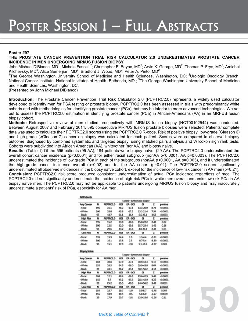
150Back to Table of Contents h
Poster session i – Full AbstrActs
Poster #97 THE PROSTATE CANCER PREVENTION TRIAL RISK CALCULATOR 2.0 UNDERESTIMATES PROSTATE CANCER INCIDENCE IN MEN UNDERGOING MRI/US FUSION BIOPSY John Michael DiBianco, MD1; Michele Fascelli2; Christopher E. Bayne, MD3; Arvin K. George, MD2; Thomas P. Frye, MD2; Amichai Kilchevsky, MD2; Alice Semerjian, MD3; Bradford J. Wood, MD2; Peter A. Pinto, MD2 1The George Washington University School of Medicine and Health Sciences, Washington, DC; 2Urologic Oncology Branch, National Cancer Institute, National Institutes of Health, Bethesda, MD.; 3The George Washington University School of Medicine and Health Sciences, Washington, DC. (Presented by John Michael DiBianco) Introduction: The Prostate Cancer Prevention Trial Risk Calculator 2.0 (PCPTRC2.0) represents a widely used calculator developed to identify men for PSA testing or prostate biopsy. PCPTRC2.0 has been assessed in trials with predominantly white cohorts and with methodologies for identifying prostate cancer (PCa) that may be inferior to more advanced technologies. We set out to assess the PCPTRC2.0 estimation in identifying prostate cancer (PCa) in African-Americans (AA) in an MRI-US fusion biopsy cohort. Methods: Retrospective review of men studied prospectively with MRI/US fusion biopsy (NCT00102544) was conducted. Between August 2007 and February 2014, 595 consecutive MRI/US fusion prostate biopsies were selected. Patients’ complete data was used to calculate their PCPTRC2.0 scores using the PCPTRC2.0 R-code. Risk of positive biopsy, low-grade (Gleason 6) and high-grade (≥Gleason 7) cancer on biopsy was calculated for each patient. Scores were compared to observed biopsy outcome, diagnosed by combined systematic and targeted biopsy, using matched pairs analysis and Wilcoxon sign rank tests. Cohorts were subdivided into African American (AA), white/other (nonAA) and biopsy naive. Results: (Table 1) Of the 595 patients (95 AA), 184 patients were biopsy naïve, (29 AA). The PCPTRC2.0 underestimated the overall cohort cancer incidence (p<0.0001) and for either racial subgroup (nonAA p<0.0001, AA p=0.0003). The PCPTRC2.0 underestimated the incidence of low grade PCa in each of the subgroups (nonAA p<0.0001, AA p=0.003), and it underestimated the high-grade cancer incidence overall (p=0.02) and for the AA cohort (p=0.01). The PCPTRC2.0 scores significantly underestimated all observed incidences in the biopsy naïve cohort, except for the incidence of low-risk cancer in AA men (p=0.21). Conclusion: PCPTRC2.0 risk score produced consistent underestimation of actual PCa incidence regardless of race. The PCPTRC2.0 did not significantly underestimate the incidence of high-risk PCa in white men overall and amid low-risk PCa in AA biopsy naïve men. The PCPTRC2.0 may not be applicable to patients undergoing MRI/US fusion biopsy and may inaccurately underestimate a patients’ risk of PCa, especially for AA men.
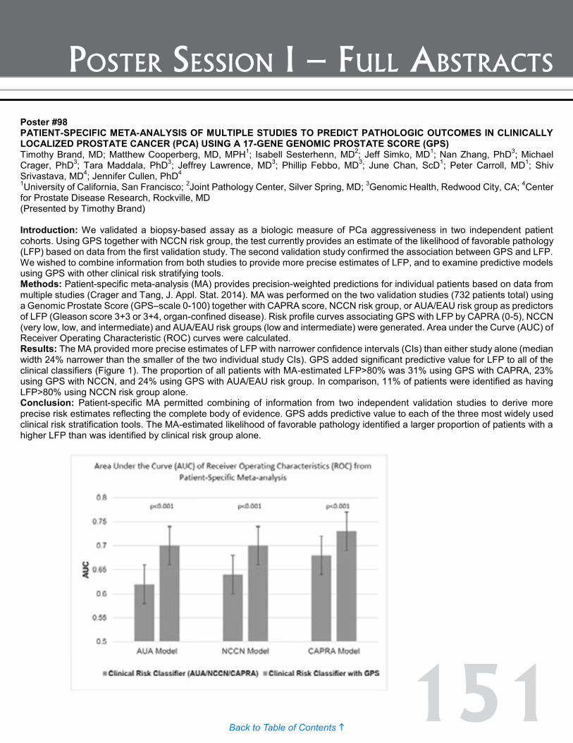
151Back to Table of Contents h
Poster session i – Full AbstrActs
Poster #98 PATIENT-SPECIFIC META-ANALYSIS OF MULTIPLE STUDIES TO PREDICT PATHOLOGIC OUTCOMES IN CLINICALLY LOCALIZED PROSTATE CANCER (PCA) USING A 17-GENE GENOMIC PROSTATE SCORE (GPS) Timothy Brand, MD; Matthew Cooperberg, MD, MPH1; Isabell Sesterhenn, MD2; Jeff Simko, MD1; Nan Zhang, PhD3; Michael Crager, PhD3; Tara Maddala, PhD3; Jeffrey Lawrence, MD3; Phillip Febbo, MD3; June Chan, ScD1; Peter Carroll, MD1; Shiv Srivastava, MD4; Jennifer Cullen, PhD4 1University of California, San Francisco; 2Joint Pathology Center, Silver Spring, MD; 3Genomic Health, Redwood City, CA; 4Center for Prostate Disease Research, Rockville, MD (Presented by Timothy Brand) Introduction: We validated a biopsy-based assay as a biologic measure of PCa aggressiveness in two independent patient cohorts. Using GPS together with NCCN risk group, the test currently provides an estimate of the likelihood of favorable pathology (LFP) based on data from the first validation study. The second validation study confirmed the association between GPS and LFP. We wished to combine information from both studies to provide more precise estimates of LFP, and to examine predictive models using GPS with other clinical risk stratifying tools. Methods: Patient-specific meta-analysis (MA) provides precision-weighted predictions for individual patients based on data from multiple studies (Crager and Tang, J. Appl. Stat. 2014). MA was performed on the two validation studies (732 patients total) using a Genomic Prostate Score (GPS–scale 0-100) together with CAPRA score, NCCN risk group, or AUA/EAU risk group as predictors of LFP (Gleason score 3+3 or 3+4, organ-confined disease). Risk profile curves associating GPS with LFP by CAPRA (0-5), NCCN (very low, low, and intermediate) and AUA/EAU risk groups (low and intermediate) were generated. Area under the Curve (AUC) of Receiver Operating Characteristic (ROC) curves were calculated. Results: The MA provided more precise estimates of LFP with narrower confidence intervals (CIs) than either study alone (median width 24% narrower than the smaller of the two individual study CIs). GPS added significant predictive value for LFP to all of the clinical classifiers (Figure 1). The proportion of all patients with MA-estimated LFP>80% was 31% using GPS with CAPRA, 23% using GPS with NCCN, and 24% using GPS with AUA/EAU risk group. In comparison, 11% of patients were identified as having LFP>80% using NCCN risk group alone. Conclusion: Patient-specific MA permitted combining of information from two independent validation studies to derive more precise risk estimates reflecting the complete body of evidence. GPS adds predictive value to each of the three most widely used clinical risk stratification tools. The MA-estimated likelihood of favorable pathology identified a larger proportion of patients with a higher LFP than was identified by clinical risk group alone.
Poster #97 THE PROSTATE CANCER PREVENTION TRIAL RISK CALCULATOR 2.0 UNDERESTIMATES PROSTATE CANCER INCIDENCE IN MEN UNDERGOING MRI/US FUSION BIOPSY John Michael DiBianco, MD1; Michele Fascelli2; Christopher E. Bayne, MD3; Arvin K. George, MD2; Thomas P. Frye, MD2; Amichai Kilchevsky, MD2; Alice Semerjian, MD3; Bradford J. Wood, MD2; Peter A. Pinto, MD2 1The George Washington University School of Medicine and Health Sciences, Washington, DC; 2Urologic Oncology Branch, National Cancer Institute, National Institutes of Health, Bethesda, MD.; 3The George Washington University School of Medicine and Health Sciences, Washington, DC. (Presented by John Michael DiBianco) Introduction: The Prostate Cancer Prevention Trial Risk Calculator 2.0 (PCPTRC2.0) represents a widely used calculator developed to identify men for PSA testing or prostate biopsy. PCPTRC2.0 has been assessed in trials with predominantly white cohorts and with methodologies for identifying prostate cancer (PCa) that may be inferior to more advanced technologies. We set out to assess the PCPTRC2.0 estimation in identifying prostate cancer (PCa) in African-Americans (AA) in an MRI-US fusion biopsy cohort. Methods: Retrospective review of men studied prospectively with MRI/US fusion biopsy (NCT00102544) was conducted. Between August 2007 and February 2014, 595 consecutive MRI/US fusion prostate biopsies were selected. Patients’ complete data was used to calculate their PCPTRC2.0 scores using the PCPTRC2.0 R-code. Risk of positive biopsy, low-grade (Gleason 6) and high-grade (≥Gleason 7) cancer on biopsy was calculated for each patient. Scores were compared to observed biopsy outcome, diagnosed by combined systematic and targeted biopsy, using matched pairs analysis and Wilcoxon sign rank tests. Cohorts were subdivided into African American (AA), white/other (nonAA) and biopsy naive. Results: (Table 1) Of the 595 patients (95 AA), 184 patients were biopsy naïve, (29 AA). The PCPTRC2.0 underestimated the overall cohort cancer incidence (p<0.0001) and for either racial subgroup (nonAA p<0.0001, AA p=0.0003). The PCPTRC2.0 underestimated the incidence of low grade PCa in each of the subgroups (nonAA p<0.0001, AA p=0.003), and it underestimated the high-grade cancer incidence overall (p=0.02) and for the AA cohort (p=0.01). The PCPTRC2.0 scores significantly underestimated all observed incidences in the biopsy naïve cohort, except for the incidence of low-risk cancer in AA men (p=0.21). Conclusion: PCPTRC2.0 risk score produced consistent underestimation of actual PCa incidence regardless of race. The PCPTRC2.0 did not significantly underestimate the incidence of high-risk PCa in white men overall and amid low-risk PCa in AA biopsy naïve men. The PCPTRC2.0 may not be applicable to patients undergoing MRI/US fusion biopsy and may inaccurately underestimate a patients’ risk of PCa, especially for AA men.
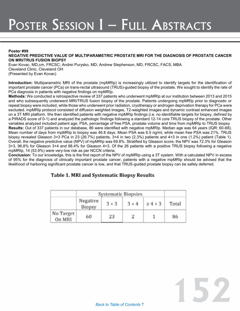
152Back to Table of Contents h
Poster session i – Full AbstrActs
Poster #100 ACTIVE SURVEILLANCE FOR LOW RISK PROSTATE CANCER IN MEN UNDER 60 YEARS OF AGE Adam Feldman, MD, MPH1; David Kuppermann, MD2; Mark Preston, MD, MPH3; Jonathan Paly1; Douglas Dahl, MD1; Richard Lee, MD, PhD1; Jason Efstathiou, MD, DPhil1; Michael Blute, MD1; Anthony Zietman, MD1 1Massachusetts General Hospital; 2Harvard Medical School; 3Brigham and Women's Hospital (Presented by Adam Feldman) Introduction: Active surveillance (AS) has become an accepted management strategy in men with low-risk prostate cancer. Data on outcomes of AS in younger men are limited. We present characteristics and outcomes of our AS cohort of men under age 60. Methods: We reviewed our single-institutional AS database of 990 men diagnosed between 1997 and 2014 to identify 177 men under age 60. Guidelines for inclusion in AS at our institution include Gleason ≤ 6 (Gleason 3+4 in select patients with low volume), ≤3/12 cores positive with ≤20% in each core, PSA <10. Results: At diagnosis, median age was 56 years (IQR 53.1-57.7) and median PSA was 4.47 ng/mL (IQR 3.00-5.60), with 173 of 177 below 10 ng/mL. 176 of 177 had Gleason 6 and 1 of 177 had Gleason 3+2=5. 92.7% (164/177) were T1c, 6.7% (12/177) T2a, and 0.6% (1/177) T1a. With a median follow-up of 4.4 years (range: 0.5-17.0; IQR: 2.8-6.1), 85.9% (152/177) had a repeat biopsy with 61.8% (94/152) showing prostate cancer, 25.7% (39/152) benign, 7.2% (11/152) with PIN, and 5.3% (8/152) with atypia. Kaplan Meier actuarial freedom-from-treatment was 69.6% at five years. Of all the patients, 28.3%(51) progressed to treatment for the following reasons: 68.6%(35/51) pathologic progression, 17.6% (9/51) PSA progression, 11.8% (6/51) patient preference, 2.0% (1/51) other reasons. Among treated patients, 72.5% (37/51) had surgery, 19.6% (10/51) had external beam radiation, and 7.8% (4/51) had brachytherapy. On pathologic review after surgery, 83.8% (31/37) were pT2, and 16.2% (6/37) pT3. Conclusion: Active surveillance is a reasonable option for carefully selected men under 60 with low risk prostate cancer. Patients must be surveyed closely and understand the risk of ultimately needing treatment.
Poster #99 NEGATIVE PREDICTIVE VALUE OF MULTIPARAMETRIC PROSTATE MRI FOR THE DIAGNOSIS OF PROSTATE CANCER ON MRI/TRUS FUSION BIOPSY Evan Kovac, MD,cm, FRCSC; Andrei Purysko, MD; Andrew Stephenson, MD, FRCSC, FACS, MBA Cleveland Clinic, Cleveland OH (Presented by Evan Kovac) Introduction: Multiparametric MRI of the prostate (mpMRIp) is increasingly utilized to identify targets for the identification of important prostate cancer (PCa) on trans-rectal ultrasound (TRUS)-guided biopsy of the prostate. We sought to identify the rate of PCa diagnosis in patients with negative findings on mpMRIp. Methods: We conducted a retrospective review of 337 patients who underwent mpMRIp at our institution between 2013 and 2015 and who subsequently underwent MRI/TRUS fusion biopsy of the prostate. Patients undergoing mpMRIp prior to diagnostic or repeat biopsy were included, while those who underwent prior radiation, cryotherapy or androgen deprivation therapy for PCa were excluded. mpMRIp protocol consisted of diffusion weighted images, T2-weighted images and dynamic contrast enhanced images on a 3T MRI platform. We then identified patients with negative mpMRIp findings (i.e. no identifiable targets for biopsy, defined by a PIRADS score of 0-1) and analyzed the pathologic findings following a standard 12-14 core TRUS biopsy of the prostate. Other variables analyzed included patient age, PSA, percentage of free PSA, prostate volume and time from mpMRIp to TRUS biopsy Results: Out of 337 patients in our database, 86 were identified with negative mpMRIp. Median age was 64 years (IQR: 60-68). Mean number of days from mpMRIp to biopsy was 46.8 days. Mean PSA was 6.5 ng/ml, while mean free PSA was 21%. TRUS biopsy revealed Gleason 3+3 PCa in 23 (26.7%) patients, 3+4 in two (2.3%) patients and 4+3 in one (1.2%) patient (Table 1). Overall, the negative predictive value (NPV) of mpMRIp was 69.8%. Stratified by Gleason score, the NPV was 72.3% for Gleason 3+3, 96.8% for Gleason 3+4 and 98.4% for Gleason 4+3. Of the 26 patients with a positive TRUS biopsy following a negative mpMRIp, 14 (53.9%) were very-low risk as per NCCN criteria. Conclusion: To our knowledge, this is the first report of the NPV of mpMRIp using a 3T system. With a calculated NPV in excess of 95% for the diagnosis of clinically important prostate cancer, patients with a negative mpMRIp should be advised that the likelihood of harboring significant prostate cancer is low, and that TRUS-guided prostate biopsy can be safely deferred.
153Back to Table of Contents h
Poster session i – Full AbstrActs
Poster #100 ACTIVE SURVEILLANCE FOR LOW RISK PROSTATE CANCER IN MEN UNDER 60 YEARS OF AGE Adam Feldman, MD, MPH1; David Kuppermann, MD2; Mark Preston, MD, MPH3; Jonathan Paly1; Douglas Dahl, MD1; Richard Lee, MD, PhD1; Jason Efstathiou, MD, DPhil1; Michael Blute, MD1; Anthony Zietman, MD1 1Massachusetts General Hospital; 2Harvard Medical School; 3Brigham and Women's Hospital (Presented by Adam Feldman) Introduction: Active surveillance (AS) has become an accepted management strategy in men with low-risk prostate cancer. Data on outcomes of AS in younger men are limited. We present characteristics and outcomes of our AS cohort of men under age 60. Methods: We reviewed our single-institutional AS database of 990 men diagnosed between 1997 and 2014 to identify 177 men under age 60. Guidelines for inclusion in AS at our institution include Gleason ≤ 6 (Gleason 3+4 in select patients with low volume), ≤3/12 cores positive with ≤20% in each core, PSA <10. Results: At diagnosis, median age was 56 years (IQR 53.1-57.7) and median PSA was 4.47 ng/mL (IQR 3.00-5.60), with 173 of 177 below 10 ng/mL. 176 of 177 had Gleason 6 and 1 of 177 had Gleason 3+2=5. 92.7% (164/177) were T1c, 6.7% (12/177) T2a, and 0.6% (1/177) T1a. With a median follow-up of 4.4 years (range: 0.5-17.0; IQR: 2.8-6.1), 85.9% (152/177) had a repeat biopsy with 61.8% (94/152) showing prostate cancer, 25.7% (39/152) benign, 7.2% (11/152) with PIN, and 5.3% (8/152) with atypia. Kaplan Meier actuarial freedom-from-treatment was 69.6% at five years. Of all the patients, 28.3%(51) progressed to treatment for the following reasons: 68.6%(35/51) pathologic progression, 17.6% (9/51) PSA progression, 11.8% (6/51) patient preference, 2.0% (1/51) other reasons. Among treated patients, 72.5% (37/51) had surgery, 19.6% (10/51) had external beam radiation, and 7.8% (4/51) had brachytherapy. On pathologic review after surgery, 83.8% (31/37) were pT2, and 16.2% (6/37) pT3. Conclusion: Active surveillance is a reasonable option for carefully selected men under 60 with low risk prostate cancer. Patients must be surveyed closely and understand the risk of ultimately needing treatment.
Poster #99 NEGATIVE PREDICTIVE VALUE OF MULTIPARAMETRIC PROSTATE MRI FOR THE DIAGNOSIS OF PROSTATE CANCER ON MRI/TRUS FUSION BIOPSY Evan Kovac, MD,cm, FRCSC; Andrei Purysko, MD; Andrew Stephenson, MD, FRCSC, FACS, MBA Cleveland Clinic, Cleveland OH (Presented by Evan Kovac) Introduction: Multiparametric MRI of the prostate (mpMRIp) is increasingly utilized to identify targets for the identification of important prostate cancer (PCa) on trans-rectal ultrasound (TRUS)-guided biopsy of the prostate. We sought to identify the rate of PCa diagnosis in patients with negative findings on mpMRIp. Methods: We conducted a retrospective review of 337 patients who underwent mpMRIp at our institution between 2013 and 2015 and who subsequently underwent MRI/TRUS fusion biopsy of the prostate. Patients undergoing mpMRIp prior to diagnostic or repeat biopsy were included, while those who underwent prior radiation, cryotherapy or androgen deprivation therapy for PCa were excluded. mpMRIp protocol consisted of diffusion weighted images, T2-weighted images and dynamic contrast enhanced images on a 3T MRI platform. We then identified patients with negative mpMRIp findings (i.e. no identifiable targets for biopsy, defined by a PIRADS score of 0-1) and analyzed the pathologic findings following a standard 12-14 core TRUS biopsy of the prostate. Other variables analyzed included patient age, PSA, percentage of free PSA, prostate volume and time from mpMRIp to TRUS biopsy Results: Out of 337 patients in our database, 86 were identified with negative mpMRIp. Median age was 64 years (IQR: 60-68). Mean number of days from mpMRIp to biopsy was 46.8 days. Mean PSA was 6.5 ng/ml, while mean free PSA was 21%. TRUS biopsy revealed Gleason 3+3 PCa in 23 (26.7%) patients, 3+4 in two (2.3%) patients and 4+3 in one (1.2%) patient (Table 1). Overall, the negative predictive value (NPV) of mpMRIp was 69.8%. Stratified by Gleason score, the NPV was 72.3% for Gleason 3+3, 96.8% for Gleason 3+4 and 98.4% for Gleason 4+3. Of the 26 patients with a positive TRUS biopsy following a negative mpMRIp, 14 (53.9%) were very-low risk as per NCCN criteria. Conclusion: To our knowledge, this is the first report of the NPV of mpMRIp using a 3T system. With a calculated NPV in excess of 95% for the diagnosis of clinically important prostate cancer, patients with a negative mpMRIp should be advised that the likelihood of harboring significant prostate cancer is low, and that TRUS-guided prostate biopsy can be safely deferred.
154Back to Table of Contents h
Poster session i – Full AbstrActs
Poster #101 DOES INITIAL PRESENTATION AFFECT SURVIVAL FOR METASTATIC PROSTATE CANCER? Philip Fontenot, MD1; Avinash Nehra, MD1; William Parker, MD1; David Duchene, MD1; Hadley Wyre, MD1; Jeffrey Holzbeierlein, MD1; J. Brantley Thrasher, MD1; Moben Mirza, MD1; Peter Van Veldhuizen, MD2; Eugene Lee, MD1 1Department of Urology, University of Kansas Medical Center, Kansas City, KS; 2Department of Hematology/Oncology, University of Kansas Medical Center, Kansas City, KS (Presented by Philip Fontenot) Introduction: Prostate cancer (CAP) is the second leading cause of cancer death in American men. Men with metastatic disease are at the highest risk of prostate cancer specific mortality. The objective of this study was to characterize a contemporary cohort of patients in the era of PSA screening, who were diagnosed with metastatic CAP to determine if initial patient presentation was associated with survival differences. Methods: After IRB approval, we evaluated the medical records of 157 men diagnosed with metastatic CAP from 2001 to 2014. Men were placed into two groups depending on their method of initial presentation. Group 1 included patients who presented with presumed localized disease and were treated with surgery or radiation, but later developed metastases (n=34, 22%). Group 2 consisted of patients who, symptomatic or asymptomatic, presented with metastatic disease (n=123, 78%). Groups were compared for overall survival from the time of initial diagnosis and survival after the development of metastatic disease using Kaplan Meier Curves. Results: The majority of patients were Caucasian (83%), while only 12% were African American. Overall, the average age at the identification of metastatic disease was 67 and the median PSA was 42.5 ng/mL. Group 1 had a median PSA at initial diagnosis of 37.5 ng/mL with a two-, three- and five-year estimated survival from the time of initial diagnosis of 87.4, 77.3 and 62%, respectively. Group 2 had a median PSA of 538.1 ng/mL and two-, three- and five-year estimated survival from the time of initial diagnosis of 82.5, 70.3 and 59.6% respectively. No significant difference in overall survival was noted between the two groups (p=0.855). However, patients who presented with localized disease had a significantly reduced life expectancy after the diagnosis of metastatic disease when compared to group two. Estimated two- and three-year overall survival after the development of metastatic disease for Group 1 was 50.7% and 30.0% respectively, while Group 2 was 78.9% and 65.9%, (HR 3.67, 95% CI 2.23-6.01, p=0.001). Conclusion: Despite improvements in CAP mortality rates over the last decade, overall survival after the development of metastatic disease remains poor. Notably, in this cohort, patients who initially presented with localized disease, received treatment and later developed metastases had an increased risk of dying after the development of metastatic disease compared to those presenting initially with metastases.
155Back to Table of Contents h
Poster session i – Full AbstrActs
Poster #102 PROSTATE CANCER CELL LINE MODELING TO STUDY HEALTH DISPARITY IN AFRICAN AMERICAN MEN Bita Nick Kholgh, MD, PhD1; Michael B. Rothberg, MD2; Xiaolan Fang, PhD3; Shira M. Winters4; Anvi Raina4 Komal S. Pandya1; Kenneth Gyabaah1; Nora Fino, MS5; K.C. Balaji, MD, PhD2 1Wake Forest Institute for Regenerative Medicine, Winston-Salem, NC; 2Department of Urology, Wake Forest School of Medicine, Winston-Salem, NC; 3Department of Cancer Biology, Wake Forest Comprehensive Cancer Center, Winston-Salem, NC; 4Wake Forest School of Medicine, Winston-Salem, NC; 5Department of Biostatistical Sciences, Wake Forest School of Medicine, Winston-Salem, NC (Presented by Michael B. Rothberg) Introduction: In comparison to Caucasian (CA) men, African-American (AA) males are disproportionately affected by prostate cancer (PCa) for incidence of overall disease, high-risk disease, and the most lethal variants of PCa. While this disparity is likely multifactorial, a biological basis is often strongly suspected. Currently, there are few well-characterized experimental model systems to study the biological basis of racial disparity in PCa. We report a validated in vitro cell line model system that could be used for this purpose. Methods: We assembled a cell line model comprising both AA (E006AA and MDAPCa2b) and CA (LNCaP and C4-2) PCa cell lines. Specifically, the LNCaP and C4-2 cell lines served as models of androgen-dependent and castration-resistant metastatic CA PCa, respectively. The utility of these cell lines in studying the biological variance of PCa was explored using a multiplex biomarker panel consisting of genes and proteins previously reported to influence PCa progression. Biomarker expression was studied by RT-PCR and Western blot in the four cell lines and validated in both AA and CA human PCa tissues by RT-PCR. Cell viability assays and molecular studies were performed as proof of principle to demonstrate functionality of the cell line model. Results: Dysregulation in the multiplex biomarker panel in AA cell lines for primary cancers was similar to that of CA cell lines for metastatic cancers, suggesting the cell line model could be used to study inherently aggressive phenotypes in AA men with PCa. We also demonstrated this model’s use for functional studies via over-expression of Protein Kinase D1 (PKD1), a novel tumor suppressor gene that decreases cell proliferation and prevents epithelial mesenchymal transition, previously shown to be down regulated in advanced PCa. Moreover, we established the feasibility of studying the expression of these biomarkers by comparing the differential gene expression levels in archived human PCa tissue from AA and CA men as a prelude to future translational studies. Conclusion: We have characterized a novel in vitro cell line model that could be used in future investigations to study the biological basis of PCa disparity among AA and CA men. This work is supported by the Wake Forest Institute for Regenerative Medicine, VA-ORD Award Number 5 I01 BX001536-03 to B. Nick Kholgh, and NIH Grant Number: CA079448 to K.C. Balaji.
156Back to Table of Contents h
Poster session i – Full AbstrActs
Poster #103 IMPACT OF PELVIC LYMPH NODE DISSECTION DURING RADICAL PROSTATECTOMY ON 30-DAY POST OPERATIVE COMPLICATIONS: Results: FROM A LARGE NATIONAL DATABASE Nicola Pavan, MD1; Samarpit Rai, MD2; Nachiketh Soodana-Prakash, MD, MS2; Raymond R. Balise, PhD3; Carmen M. Mir, MD, PhD2; Bruno Nahar, MD2; Fernando Marsicano, MD2; Carlo Trombetta, MD4; Chad R. Ritch, MD, MBA2; Dipen J. Parekh, MD2; Mark L. Gonzalgo, MD, PhD2 1Department of Urology, University of Miami Leonard M. Miller School of Medicine, Miami, FL and Urology Clinic, Department of Medical, Surgical and Health Science, University of Trieste, Italy; 2Department of Urology, University of Miami Leonard M. Miller School of Medicine, Miami, FL; 3Division of Biostatistics, Department of Public Health Sciences, University of Miami Leonard M. Miller School of Medicine, Miami, FL; 4Urology Clinic, Department of Medical, Surgical and Health Science, University of Trieste, Italy (Presented by Samarpit Rai) Introduction: Pelvic lymph node dissection (PLND) during radical prostatectomy (RP) is the most effective method for detecting lymph node metastases in patients with prostate cancer. The association between PLND during RP and morbidity, especially thromboembolic adverse events (AEs), remains unclear. We assessed the effect of PLND on 30-day postoperative AEs in patients undergoing RP using the American College of Surgeons’ National Surgical Quality Improvement Program database (NSQIP). Methods: A total of 21,895 men undergoing RP between 2006 and 2013 were classified into two groups according to surgical approach (MIS-RP vs. ORP) and whether PLND was performed. Multivariate logistic regression adjusting for approach and demographic features was performed to assess the impact of PLND for predicting two primary endpoints (overall complications and major complications defined as Clavien-Dindo ≥ 3) and for 17 types of complications. P-values were adjusted to maintain an experiment-wise p < 0.05. Results: MIS-RP and ORP was performed in 17,354 (79.3%) and 4,541 (20.7%) patients, respectively. PLND was performed in 7,579 (43.7%) and 3,597 (79.2%) patients in the MIS-RP and ORP groups, respectively. The overall postoperative complication rate was 8.7% (5.5% for MIS-RP and 21.0% for ORP). PLND was not associated with a higher risk of DVT (OR 0.99; p= 0.98) or PE (OR 1.02; p= 0.91). However, PLND was associated with a higher risk of superficial surgical site infection (OR 1.68; p = 0.013), organ space surgical site infection (OR 1.77; p = 0.02), and perioperative transfusion (OR 1.32; p = 0.002) regardless of surgical approach. PLND was not associated with overall or major AEs on multivariable analysis. ORP was associated with a significantly higher risk of overall (OR 4.64, p < 0.0001) and major (OR 1.6, p = 0.0004) AEs compared to MIS-RP. Conclusion: PLND during RP is associated with a significantly increased risk of certain types of AEs within the 30-day post-operative period. However, there appears to be no significant association between PLND and thromboembolic AEs.
157Back to Table of Contents h
Poster session i – Full AbstrActs
Poster #104 EFFECT OF LOCAL THERAPY ON THE SYSTEMIC ANTI-TUMOR RESPONSE IN PROSTATE CANCER Benjamin Benzon, MD; Stephanie Glavaris, BS; Brian Simons, DVM, PhD; Robert Hughes, BS; Patrick Mullane, BS; Rebecca Miller, BS; Katriana Nugent, BS; Brian Shinder, BS; Richard Blosser; Phuoc Tran, MD, PhD; Paula Hurley, PhD; Milena Vuica-Ross, MD, MS; Edward Schaeffer, MD, PhD; Charles Drake, MD, PhD; Ashley Ross, MD, PhD Baltimore, MD (Presented by Ashley Ross) Introduction: The genomic complexities and adaptability of aggressive cancer implies that it may only be eradicated by equally adaptable systems, such as immune based therapies. Immune checkpoint blockade (such as that targeting PD-1 or CTLA-4) has shown dramatic and durable efficacy in immunogenic malignancies, but little or no benefit in less immunogenic cancers such as prostate cancer. Here we use a mouse model of prostate cancer to investigate whether local therapy can mount or augment an abscopal response to distant tumors. Methods: Immuno-competent FVB mice were bilaterally implanted with Myc-CAP cells to form isogenic grafts. Two weeks after tumor introduction, ablative therapies including radiation (stereotactically as a single 10 Gy fraction), cryoablation (two freeze-thaw cycles of less than -40 degree Celsius), whole tumor cauterization, or excision were applied to the larger graft in the presence of checkpoint blockade (intra-peritoneal anti-CTLA-4 or anti-PD1) or control injection (hamster anti-mouse IgG). Tumor sizes and mouse survival was recorded as was lymphocytic infiltrates which were characterized histologically and by flow cytometry. Results: The ablative therapies of cautery or cryoablation, but not excision or radiation caused a statistically significant delay in the growth of distant untreated tumors (P<0.05 for both). Flow cytometric analysis demonstrated an associated increase in the CD8+IFNgamma+ T cell population in the lymph nodes draining the untreated distant tumor. Flow also demonstrated an increase in CD4+FoxP3+ cells (P<0.05 for each). Low dose anti-CTLA-4 therapy (1mg/kg) synergized with all local therapies to greatly increase survival and delay distant untreated tumor growth. This corresponded to an increase in activated T cells within distant tumors. Anti-PD-1 therapy did not synergize with local therapies. Conclusion: Some ablative local therapies (cautery, cryoablation) alone may incite an immune response in distant tumors. Checkpoint inhibition with low dose CTLA-4 blockade synergizes with all local therapies to incite a systemic anti-tumor response. PD-1 blockade has minimal effectiveness when combined with local therapies and may require combinatorial drug use for effectiveness in prostate cancer.
158Back to Table of Contents h
Poster session i – Full AbstrActs
Poster #105 B7H3 EXPRESSION IS ANDROGEN RELATED AND PREDICTIVE OF PROSTATE CANCER OUTCOMES IN A LARGE NATURAL HISTORY COHORT OF MEN UNDERGOING PROSTATECTOMY Benjamin Benzon1; Shuang Zhao2; Mandeep Takhar3; Michael Haffner1; Nicholas Erho3; Paula Hurley1; Jeffrey Tosoian1; Mohammed Alshalalfa3; Stephanie Glavaris1; Brian Simons1; Phuoc Tran1; Elai Davicioni3; R. Jeffrey Karnes4; Edward Schaeffer1; Charles Drake1; Felix Feng2; Ashley Ross, MD, PhD5 1Johns Hopkins, Baltimore, MD; 2University of Michigan, Anne Arbor, MI; 3GenomeDx Biosciences, Vancouver, BC; 4Mayo Clinic, Rochester, MN; 5Johns Hopkins Hosptial, Baltimore, MD (Presented by Ashley Ross) Introduction: B7-H3 (CD276), part of the B7 superfamily, has been shown to play an immunomodulatory role, however its regulation, receptor and mechanism of action remain unclear. Protein levels of B7-H3 have been previously shown to relate to prostate cancer outcomes and currently, humanized monoclonal antibodies are being developed for clinical use (MGA271, Macrogenics). Here we use genomic expression data to examine the relationship of B7-H3 to prostate cancer outcomes and molecular subtypes. Methods: Prostatectomy tissue from 905 patients was profiled using the Affymetrix HuEx 1.0 ST microarray. Kruskal-Wallis tests were used to identify significant associations of B7-H3 expression with clinico-pathologic variables, and survival analysis were used to evaluate the prognostic value of B7-H3. Pearson’s correlation analyses were also performed to assess the relationship of B7-H3 expression with molecular subtypes and individual transcripts. Androgen receptor (AR) occupancy of promoter regions was derived in silico from chromosomal immune-precipitation (ChIP) data. Results: B7-H3 expression was positively associated with Gleason score (p<0.01) and tumor stage (p <0.01). High B7-H3 expression also correlated with the development of metastasis and prostate cancer specific mortality (HR of 3.4 and 2.4 respectively, p<0.05 for both), but this was not significant on multi-variable analysis. B7-H3 was positively associated with ERG+ disease (n =670, r = 0.85, p < 0.05) and AR expression (n =670, r = 0.46, p < 0.001). B7-H3 was found to be one of the most correlated genes with AR (95th percentile) and ChIP analysis revealed AR binding upstream of B7-H3, suggesting potential androgen dependent regulation. Conclusion: B7-H3 expression correlates with high Gleason grade and advanced prostate cancer stage with higher quartiles of expression portending poor oncologic outcomes in two independent prostatectomy cohorts. B7-H3 expression appears to relate to the androgen receptor.
159Back to Table of Contents h
Poster session i – Full AbstrActs
Poster #106 EFFICACY OF EARLY AND DELAYED RADIATION IN A PROSTATECTOMY COHORT ADJUSTED FOR GENOMIC AND CLINICAL RISK Ashley Ross, MD, PhD3; Robert Den MD1; Kasra Yousefi, MSc2; Bruce Trock, PhD3; Elai Davicioni, PhD2; Jeffrey Tosoian, MD, MPH3; Darby Thompson, PhD4; Voleak Choeurng, MSc2; Zaid Haddad, MSc2; Phuoc Tran, MD, PhD5; Edouard Trabulsi, MD1; Leonard Gomella, MD1; Costas Lallas, MD1; Firas Abdollah, MD6; Felix Feng, MD7; Adam Dicker, MD, PhD1; Stephen Freedland, MD8; Jeffrey Karnes, MD9; Edward Schaeffer, MD, PhD3 1Sidney Kimmel Medical College of Thomas Jefferson University, Philadelphia, PA; 2GenomeDx Biosciences Inc., Vancouver, BC, Canada; 3James Buchanan Brady Urological Institute, Johns Hopkins Hospital, Baltimore, MD; 4Emmes Canada, Burnaby, BC, Canada; 5Department of Radiation Oncology, Johns Hopkins Hospital, Baltimore, MD; 6Vattikuti Urology Institute, Henry Ford Hospital, Detroit, MI; 7University of Michigan, Ann Arbor, MI; 8Department of Surgery, Division of Urology, Center for Integrated Research on Cancer and Lifestyle, Samuel Oschin Comprehensive Cancer Center, Cedars-Sinai Medical Center, Los Angeles, CA and Surgery section, Durham Veteran Affairs Medical Center, Durham, NC; 9Department of Urology, Mayo Clinic, Rochester, MN (Presented by Ashley Ross) Introduction: In three published randomized clinical trials, adjuvant radiation therapy (ART) for prostate cancer (PCa) resulted in improved progression free survival. However, the impact on metastases and overall survival is unclear. To date, there have been no published prospective trials examining the impact of salvage radiation therapy (SRT) in this disease state. Hence, we conducted a retrospective, nonrandomized comparative study of adjuvant, salvage, or no radiation following radical prostatectomy (RP) for men with pT3 disease or positive margins (adverse pathologic features, APF). Methods: Four hundred twenty-two PCa patients treated at four institutions with RP and having APF were analyzed with a primary end point of clinical metastasis. Men undergoing ART (n=111), early SRT (n=70) and delayed SRT (n=83) were defined by having PSA levels of <0.2, 0.2 to 0.5, and ≥0.5 ng/mL, respectively, prior to initiation of RT. Remaining 157 patients who did not receive additional therapy (RT or hormonal) prior to metastatic onset formed the no RT group. Clinical-genomic risk was assessed by CAPRA-S and Decipher. Cox univariable (UVA) and multivariable (MVA) proportional hazards models were used to evaluate the impact of treatment on outcome. Results: During study follow-up, 37 patients developed metastasis with a median follow-up of eight years. Both CAPRA-S and Decipher had independent predictive value on MVA for metastatic outcome (both p<0.05). On MVA adjusting for clinical and genomic risk, delayed SRT and no RT had an HR of 4.31 (95% confidence interval [CI], 1.20-15.47) and 5.42 (95% CI, 1.59-18.44) for metastasis compared to ART as the reference group. No significance difference was observed between early SRT and ART groups (p=0.28). Men with low to intermediate CAPRA-S scores and low Decipher risk have a low rate of metastatic events regardless of treatment selection. In contrast, men with high CAPRA-S and Decipher scores benefit from ART, however the cumulative incidence of metastasis remains high. Conclusion: The decision as to the timing and need for additional local therapy following RP is nuanced and requires providers and patients to balance risks of morbidity with improved oncologic outcomes. This analysis provides the most robust and accurate quantification of risk for these patients. Post-RP treatment can be safely avoided for men who are low risk by clinical-genomic risk, whereas those at high risk should strongly favor enrollment in clinical trials.
160Back to Table of Contents h
Poster session i – Full AbstrActs
Poster #108 REAL-TIME MRI-GUIDED FOCUSED ULTRASOUND FOR FOCAL THERAPY OF ORGAN CONFINED LOW-INTERMEDIATE RISK PROSTATE CANCER: Results: OF PHASE 1 STUDY Sangeet Ghai, MD1; Nathan Perlis, MD, MSc2; Eugen Hlasny1; Theodorus Van der Kwast, MD, PhD3; Stuart McCluskey, MD, PhD4; Walter Kucharczyk, MD1; John Trachtenberg, MD2 1Department of Radiology, University Health Network, University of Toronto, Toronto, Ontario; 2Division of Urology, University Health Network, University of Toronto, Toronto, Ontario; 3Department of Pathology, Univeristy Health Network, University of Toronto, Toronto, Ontario; 4Department of Anaesthesiology, University Health Network, University of Toronto, Toronto, Ontario (Presented by Nathan Perlis) Introduction: Localizing strength of mp-MRI allows targeted focal therapy, the ExAblate MRgFUS system (InSightec Ltd, Haifa, Israel) combines 1.5/3T MRI scanner with HIFU energy that is transmitted from a phased array endorectal transducer. We report the six month functional and oncologic outcomes of the ongoing Phase 1 trial of patients treated with focal transrectal in-bore MRgFUS. Methods: Eight patients with PSA ≤10ng/mL, ≤ cT2a and Gleason score ≤7 (4+3) were prospectively enrolled. These patients all had confirmed MRI visible lesions corresponding to biopsy positive sites. Under MRI guidance and real-time MR thermography feedback, HIFU energy was delivered to the tumor. Adverse events and serial quality of life questionnaires were recorded. Oncologic outcomes were evaluated at six months after treatment. Results: Six month follow-up data is available for five patients with a total of seven targeted lesions, each of which were treated were treated with ≤ Clavien I complications. Quality of life parameters were similar between baselines and Six months. In all five patients, MRI at six months was negative in their treated regions (100%) (figure 1), three of five patients were clear of disease in their treated regions on biopsy (67%), representing complete ablation of five of seven target lesions (71%). Results for the final three patients will be reported. Conclusion: MRgFUS is a feasible technique for focal treatment of prostate cancer. Further investigation and follow-up is warranted in a larger patient series.
Poster #107 IMAAGEN TRIAL UPDATE: EFFECT OF ABIRATERONE ACETATE AND LOW DOSE PREDNISONE ON PSA AND RADIOGRAPHIC DISEASE PROGRESSION IN PATIENTS WITH NON-METASTATIC CASTRATION-RESISTANT PROSTATE CANCER Charles J. Ryan, MD1; E. David Crawford, MD2; Neal D. Shore, MD3; Willie Underwood, MD4; Anil Londhe, PhD5; Shawn Black, PhD5; Tracy McGowan, MD5; Philip W. Kantoff, MD6 1UCSF Helen Diller Family Comprehensive Cancer Center, San Francisco, CA; 2University of Colorado, Denver, CO; 3Carolina Urologic Research Center,Myrtle Beach, SC; 4Roswell Park Cancer Institute, Buffalo, NY; 5Janssen Pharmaceuticals, Horsham, PA; 6Dana-Farber Cancer Institute, Boston, MA (Presented by Charles J. Ryan) Introduction: Abiraterone acetate (AA) 1000mg, in combination with prednisone (P), 10mg daily is indicated for the treatment of patients with metastatic castration-resistant prostate cancer (mCRPC). IMAAGEN is a phase II, multi-center study with a primary endpoint that evaluated the ability of AA plus 5mg of prednisone (AA+5) to decrease PSA levels in patients with nmCRPC and a rising PSA; primary results have been reported earlier. Herein we report updated results on secondary endpoints and safety from the IMAAGEN study (Nov 3, 2014 database cut-off date). Methods: All enrolled patients had high risk non-metastatic castration resistant prostate cancer (nmCRPC): PSA value ≥ 10ng/mL or PSA doubling time ≤ 10 months at screening. Patientts received AA+5 daily; each treatment cycle=28 days. Reported endpoints include time to PSA progression, time to radiographic progressive disease, and safety. Results: At the time of the data cutoff, 62 (47.3%) of the 131 patients enrolled in IMAAGEN remained on treatment in the study. The median duration of exposure was 17.9 months (range 0.1 – 40.7 months). Median time to PSA progression was 28.7 months (95% CI 21.2, NE). There were 21 confirmed radiographic progression events. The median time to radiographic progressive disease was not reached. 95.4% of patients had an adverse event (AE) (54.9% had a Grade 3 or higher) and 38.2% had a serious AE (SAE) with 35.9% having an SAE of Grade 3 or higher. Thirteen percent of patients had AEs resulting in discontinuation of study treatment. Four patients had AEs resulting in death (coronary artery disease, myocardial infarction, acute respiratory failure, and pneumonia). Conclusion: Treatment of high risk nmCRPC patients with AA+5 resulted in a median time to PSA progression of 28.7 months. The median time to radiographic disease progression was not reached. The safety profile of AA+5 reported in this IMAAGEN trial update is consistent with the safety profile from previously reported studies of abiraterone acetate 1000mg in combination with either 5mg or 10mg prednisone. Source of funding: This study (NCT 01314118) was sponsored by Janssen Scientific Affair, LLC. 1ASCO 2014 IMAAGEN primary endpoint poster presentation
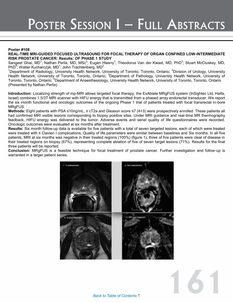
161Back to Table of Contents h
Poster session i – Full AbstrActs
Poster #108 REAL-TIME MRI-GUIDED FOCUSED ULTRASOUND FOR FOCAL THERAPY OF ORGAN CONFINED LOW-INTERMEDIATE RISK PROSTATE CANCER: Results: OF PHASE 1 STUDY Sangeet Ghai, MD1; Nathan Perlis, MD, MSc2; Eugen Hlasny1; Theodorus Van der Kwast, MD, PhD3; Stuart McCluskey, MD, PhD4; Walter Kucharczyk, MD1; John Trachtenberg, MD2 1Department of Radiology, University Health Network, University of Toronto, Toronto, Ontario; 2Division of Urology, University Health Network, University of Toronto, Toronto, Ontario; 3Department of Pathology, Univeristy Health Network, University of Toronto, Toronto, Ontario; 4Department of Anaesthesiology, University Health Network, University of Toronto, Toronto, Ontario (Presented by Nathan Perlis) Introduction: Localizing strength of mp-MRI allows targeted focal therapy, the ExAblate MRgFUS system (InSightec Ltd, Haifa, Israel) combines 1.5/3T MRI scanner with HIFU energy that is transmitted from a phased array endorectal transducer. We report the six month functional and oncologic outcomes of the ongoing Phase 1 trial of patients treated with focal transrectal in-bore MRgFUS. Methods: Eight patients with PSA ≤10ng/mL, ≤ cT2a and Gleason score ≤7 (4+3) were prospectively enrolled. These patients all had confirmed MRI visible lesions corresponding to biopsy positive sites. Under MRI guidance and real-time MR thermography feedback, HIFU energy was delivered to the tumor. Adverse events and serial quality of life questionnaires were recorded. Oncologic outcomes were evaluated at six months after treatment. Results: Six month follow-up data is available for five patients with a total of seven targeted lesions, each of which were treated were treated with ≤ Clavien I complications. Quality of life parameters were similar between baselines and Six months. In all five patients, MRI at six months was negative in their treated regions (100%) (figure 1), three of five patients were clear of disease in their treated regions on biopsy (67%), representing complete ablation of five of seven target lesions (71%). Results for the final three patients will be reported. Conclusion: MRgFUS is a feasible technique for focal treatment of prostate cancer. Further investigation and follow-up is warranted in a larger patient series.
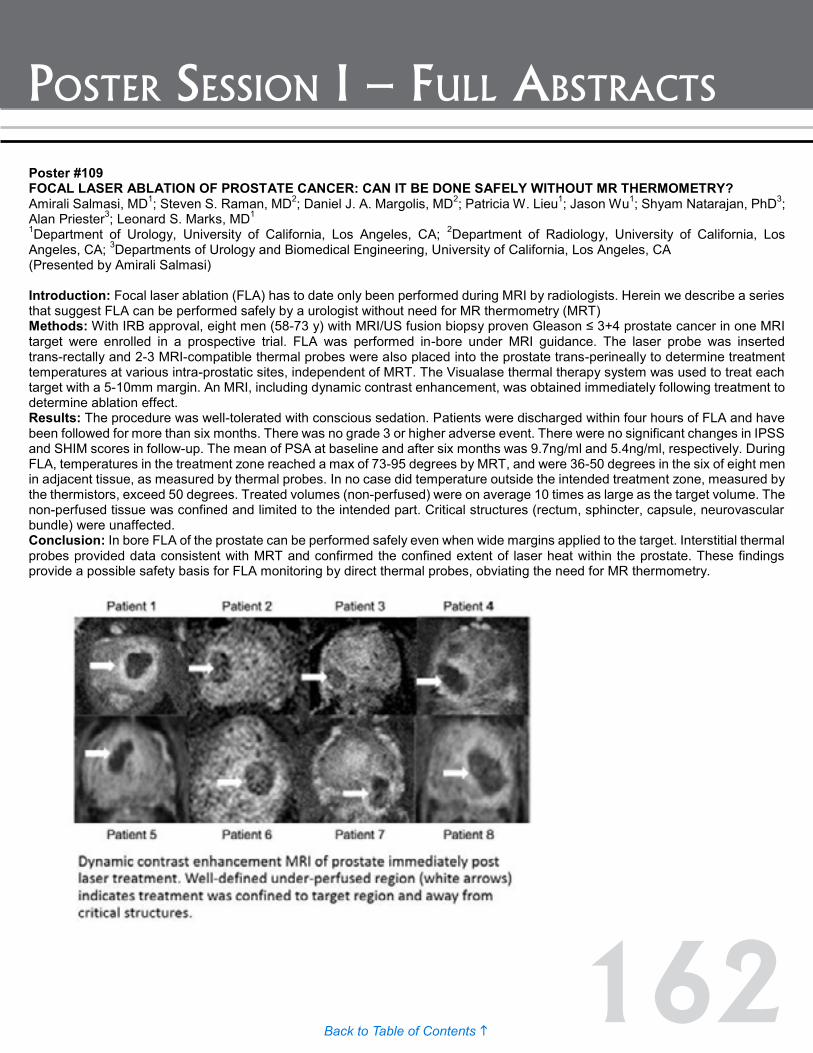
162Back to Table of Contents h
Poster session i – Full AbstrActs
Poster #109 FOCAL LASER ABLATION OF PROSTATE CANCER: CAN IT BE DONE SAFELY WITHOUT MR THERMOMETRY? Amirali Salmasi, MD1; Steven S. Raman, MD2; Daniel J. A. Margolis, MD2; Patricia W. Lieu1; Jason Wu1; Shyam Natarajan, PhD3; Alan Priester3; Leonard S. Marks, MD1 1Department of Urology, University of California, Los Angeles, CA; 2Department of Radiology, University of California, Los Angeles, CA; 3Departments of Urology and Biomedical Engineering, University of California, Los Angeles, CA (Presented by Amirali Salmasi) Introduction: Focal laser ablation (FLA) has to date only been performed during MRI by radiologists. Herein we describe a series that suggest FLA can be performed safely by a urologist without need for MR thermometry (MRT) Methods: With IRB approval, eight men (58-73 y) with MRI/US fusion biopsy proven Gleason ≤ 3+4 prostate cancer in one MRI target were enrolled in a prospective trial. FLA was performed in-bore under MRI guidance. The laser probe was inserted trans-rectally and 2-3 MRI-compatible thermal probes were also placed into the prostate trans-perineally to determine treatment temperatures at various intra-prostatic sites, independent of MRT. The Visualase thermal therapy system was used to treat each target with a 5-10mm margin. An MRI, including dynamic contrast enhancement, was obtained immediately following treatment to determine ablation effect. Results: The procedure was well-tolerated with conscious sedation. Patients were discharged within four hours of FLA and have been followed for more than six months. There was no grade 3 or higher adverse event. There were no significant changes in IPSS and SHIM scores in follow-up. The mean of PSA at baseline and after six months was 9.7ng/ml and 5.4ng/ml, respectively. During FLA, temperatures in the treatment zone reached a max of 73-95 degrees by MRT, and were 36-50 degrees in the six of eight men in adjacent tissue, as measured by thermal probes. In no case did temperature outside the intended treatment zone, measured by the thermistors, exceed 50 degrees. Treated volumes (non-perfused) were on average 10 times as large as the target volume. The non-perfused tissue was confined and limited to the intended part. Critical structures (rectum, sphincter, capsule, neurovascular bundle) were unaffected. Conclusion: In bore FLA of the prostate can be performed safely even when wide margins applied to the target. Interstitial thermal probes provided data consistent with MRT and confirmed the confined extent of laser heat within the prostate. These findings provide a possible safety basis for FLA monitoring by direct thermal probes, obviating the need for MR thermometry.
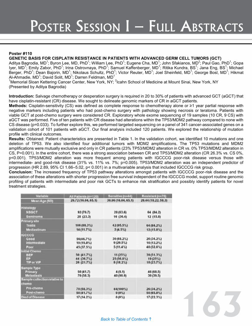
163Back to Table of Contents h
Poster session i – Full AbstrActs
Poster #110 GENETIC BASIS FOR CISPLATIN RESISTANCE IN PATIENTS WITH ADVANCED GERM CELL TUMORS (GCT) Aditya Bagrodia, MD1; Byron Lee, MD, PhD1; William Lee, PhD1; Eugene Cha, MD1; John Sfakianos, MD2; Paul Gao, PhD1; Gopa Iyer, MD1; Emily Zabor, PhD1; Irina Ostrovnaya, PhD1; Samuel Kaffenberger, MD1; Ritika Kundra, BS1; Jana Eng, BS1; Michael Berger, PhD1; Dean Bajorin, MD1; Nikolaus Schultz, PhD1; Victor Reuter, MD1; Joel Sheinfeld, MD1; George Bosl, MD1; Hikmat Al-Ahmadie, MD1; David Solit, MD1; Darren Feldman, MD1 1Memorial Sloan Kettering Cancer Center, New York, NY; 2Icahn School of Medicine at Mount Sinai, New York, NY (Presented by Aditya Bagrodia) Introduction: Salvage chemotherapy or desperation surgery is required in 20 to 30% of patients with advanced GCT (aGCT) that have cisplatin-resistant (CR) disease. We sought to delineate genomic markers of CR in aGCT patients. Methods: Cisplatin-sensitivity (CS) was defined as complete response to chemotherapy alone or ≥1 year partial response with negative markers including patients who had post-chemo surgery with pathology showing necrosis or teratoma. Patients with viable GCT at post-chemo surgery were considered CR. Exploratory whole exome sequencing of 19 samples (10 CR, 9 CS) with aGCT was performed. Five of ten patients with CR disease had alterations within the TP53/MDM2 pathway compared to none with CS disease (p=0.033). To further explore this, we performed targeted sequencing on a panel of 341 cancer-associated genes on a validation cohort of 101 patients with aGCT. Our final analysis included 120 patients. We explored the relationship of mutation profile with clinical outcomes. Results: Obtained: Patient characteristics are presented in Table 1. In the validation cohort, we identified 10 mutations and one deletion of TP53. We also identified four additional tumors with MDM2 amplifications. The TP53 mutations and MDM2 amplifications were mutually exclusive and only in CR patients (23% TP53/MDM2 alteration in CR vs. 0% TP53/MDM2 alteration in CS, P=0.001). In the entire cohort, there was a strong association between CR and TP53/MDM2 alteration (CR 26.3% vs. CS 0%, p<0.001). TP53/MDM2 alteration was more frequent among patients with IGCCCG poor-risk disease versus those with intermediate- and good-risk disease (31% vs. 11% vs. 7%; p=0.005). TP53/MDM2 alteration was an independent predictor of progression (HR 2.89, 95% CI 1.66–5.02, p< 0.001) in a multivariable analysis that included IGCCCG risk group. Conclusion: The increased frequency of TP53 pathway alterations amongst patients with IGCCCG poor-risk disease and the association of these alterations with shorter progression free survival independent of the IGCCCG model, support routine genomic profiling of patients with intermediate and poor risk GCTs to enhance risk stratification and possibly identify patients for novel treatment strategies.
Poster #109 FOCAL LASER ABLATION OF PROSTATE CANCER: CAN IT BE DONE SAFELY WITHOUT MR THERMOMETRY? Amirali Salmasi, MD1; Steven S. Raman, MD2; Daniel J. A. Margolis, MD2; Patricia W. Lieu1; Jason Wu1; Shyam Natarajan, PhD3; Alan Priester3; Leonard S. Marks, MD1 1Department of Urology, University of California, Los Angeles, CA; 2Department of Radiology, University of California, Los Angeles, CA; 3Departments of Urology and Biomedical Engineering, University of California, Los Angeles, CA (Presented by Amirali Salmasi) Introduction: Focal laser ablation (FLA) has to date only been performed during MRI by radiologists. Herein we describe a series that suggest FLA can be performed safely by a urologist without need for MR thermometry (MRT) Methods: With IRB approval, eight men (58-73 y) with MRI/US fusion biopsy proven Gleason ≤ 3+4 prostate cancer in one MRI target were enrolled in a prospective trial. FLA was performed in-bore under MRI guidance. The laser probe was inserted trans-rectally and 2-3 MRI-compatible thermal probes were also placed into the prostate trans-perineally to determine treatment temperatures at various intra-prostatic sites, independent of MRT. The Visualase thermal therapy system was used to treat each target with a 5-10mm margin. An MRI, including dynamic contrast enhancement, was obtained immediately following treatment to determine ablation effect. Results: The procedure was well-tolerated with conscious sedation. Patients were discharged within four hours of FLA and have been followed for more than six months. There was no grade 3 or higher adverse event. There were no significant changes in IPSS and SHIM scores in follow-up. The mean of PSA at baseline and after six months was 9.7ng/ml and 5.4ng/ml, respectively. During FLA, temperatures in the treatment zone reached a max of 73-95 degrees by MRT, and were 36-50 degrees in the six of eight men in adjacent tissue, as measured by thermal probes. In no case did temperature outside the intended treatment zone, measured by the thermistors, exceed 50 degrees. Treated volumes (non-perfused) were on average 10 times as large as the target volume. The non-perfused tissue was confined and limited to the intended part. Critical structures (rectum, sphincter, capsule, neurovascular bundle) were unaffected. Conclusion: In bore FLA of the prostate can be performed safely even when wide margins applied to the target. Interstitial thermal probes provided data consistent with MRT and confirmed the confined extent of laser heat within the prostate. These findings provide a possible safety basis for FLA monitoring by direct thermal probes, obviating the need for MR thermometry.
164Back to Table of Contents h
Poster session i – Full AbstrActs
Poster #111 TREATMENT AND CLINICAL OUTCOMES OF PATIENTS WITH TERATOMA WITH SOMATIC-TYPE MALIGNANT TRANSFORMATION: AN INTERNATIONAL COLLABORATION. Andrea Necchi, MD1; Gregory Pond, PhD2; Patrizia Giannatempo, MD1; Guru Sonpavde, MD3; Costantine Albany, MD4; Yohann Loriot, MD5; Christopher Sweeney, MD6; Roberto Salvioni, MD1; Maurizio Colecchia, MD1; Nicola Nicolai, MD1; Daniele Raggi, MD1; Kevin Rice, MD4; Chandra Flack, MD4; Nemer El Mouallem, MD3; Hope Feldman, MD6; Karim Fizazi, MD5; Lawrence Einhorn, MD4; Richard Foster, MD4; Clint Cary, MD4 1Fondazione IRCCS Istituto Nazionale dei Tumori, Milan, Italy; 2McMaster University, Hamilton, Ontario, Canada; 3UAB Comprehensive Cancer Center, Birmingham, AL; 4Indiana University Melvin & Bren Simon Cancer Center, Indianapolis, IN; 5Institut Gustave Roussy, Villejuif, France; 6Dana-Farber Cancer Institute, Boston, MA (Presented by Andrea Necchi) Introduction: The development of teratoma with malignant transformation (TMT) is a rare occurrence among germ cell tumors (GCT). We aimed to assess the clinical prognostic factors, treatments given, and outcome of patients with TMT from an international multicentric database. Methods: Data on patients diagnosed with TMT between June 1981 and August 2014 were collected across five referral centers. Chemotherapy was dichotomized as GCT or TMT-based according to investigator judgement. Cox analyses evaluated prognostic factors for overall survival (OS, primary endpoint). Each factor was evaluated in a univariable model. Forward stepwise selection was used to construct an optimal model. Results: Overall, 320 patients were identified. Median age was 28 years (interquartile range [IQR]: 24-35), tumor primary site was: gonadal (287, 89.7%), retroperitoneal (17, 5.3%), and mediastinal (16, 5%). TMT and GCT were diagnosed concurrently in 130 patients (40.6%). Forty-nine patients (16.8%) initially presented with clinical stage I (CSI): 14 were treated with orchiectomy alone, 28 with primary retroperitoneal lymph-node dissection (RPLND) which resulted in viable nodal TMT in 10 cases (35.7%). The remainder had metastatic good (123, 42.3%), intermediate (42, 14.4%), and poor risk according to the IGCCCG (77, 26.5%). 159 (49.7%) had received initial first-line GCT-chemotherapy, 14 (4.4%) TMT-chemotherapy, and 147 (45.9%) surgery only. After a median follow up of 25.1 months (IQR: 5.4-63.8), 167 patients relapsed (52.2%). Five-year OS was 83.4% (95%CI: 61.3 to 93.5%) for patients with CSI and it was worse than expected for metastatic patients too. On multivariable analyses, non-primitive neuroectodermal tumor (PNET) histology (overall p=0.004), gonadal primary tumor (p=0.005), and fewer prior chemotherapy regimens before TMT (p<0.001) were independent predictors of better OS. The c-index for the multivariable model was 0.646 (95% CI: 0.605 to 0.686). Chemotherapy was not independently prognostic. Conclusion: Less heavily pre-treated TMT with gonadal primary tumor and non-PNET histology appeared to be associated with longer OS. Generally, TMT had a worse prognosis than GCT. While uncertainties persist regarding the optimal chemotherapy, surgery is the key in every stage and may be the new standard (primary RPLND) for all CSI patients.
165Back to Table of Contents h
Poster session i – Full AbstrActs
Poster #112 PATTERNS OF CARE AND SURVIVAL OUTCOMES FOR MALIGNANT SEX CORD STROMAL TESTICULAR CANCER: Results: FROM THE NATIONAL CANCER DATA BASE John Banerji, MD, MCh (Urology)1; Katherine Odem-Davis, PhD2; Erika Wolff, PhD3; Craig Nichols, MD4; Christopher Porter, MD, FACS3 1Dept. of Urology, Virginia Mason, Seattle, WA; 2Center for Biomedical Statistics, Univ of Washington, Seattle, WA; 3Dept. of Urology, Virginia Mason, Seattle, WA; 4Dept of Haematology/Oncology, Seattle, WA (Presented by John Banerji) Introduction: Sex-cord stromal tumors (SCSTs) of the testis comprise less than five percent of testicular neoplasms. Consequently, data regarding patterns of care and survival is sparse. Therefore, we sought to provide a more definitive analysis of the outcomes and management of these rare malignancies. Methods: Data were obtained from the National Cancer Data Base, which captures over 70% of all newly diagnosed malignancies in the US. Patients diagnosed from 1998 to 2011 utilizing ICD-9 codes for the two most frequent SCSTs of the testis, Leydig cell and Sertoli cell, were selected. Overall survival was assessed by Kaplan-Meier using data through 2006. Results: Of the 79,120 cases of testicular cancer in the NCDB diagnosed between 1998 and 2011, 315 (0.39%) were primary malignant Leydig or Sertoli cell tumors. The median age at diagnosis was 43 years for both Leydig and Sertoli cell tumors. The majority of men were Non-Hispanic white (63% Leydig, 58% Sertoli) with a Charlson comorbidity index score of 0 (94% Leydig, 84% Sertoli). Most men were treated at a facility classified as either a Comprehensive Community Cancer or Academic Research program (87% Leydig; 83% Sertoli). Of the 315 patients, 250 (79%) had malignant Leydig cell tumors and 65 (21%) had Sertoli cell tumors; most were diagnosed with stage I disease (94% and 78%, respectively). Overall survival estimates at one and five years for stage I Leydig cell tumors were 98% (95% CI: 96-100) and 91% (95% CI: 85- 96), respectively, and for stage I Sertoli cell tumors were 93% (95% CI: 83-100) and 77% (95% CI: 62-95, p=0.015), respectively. The vast majority of patients with stage I tumors received no further treatment following orchiectomy (94% Leydig, 84% Sertoli). The most common treatment modality for patients with stage II or III Leydig or Sertoli cell tumors following orchiectomy was active surveillance, followed by retroperitoneal lymph node dissection alone. Conclusion: Stage I malignant Leydig cell tumors were the most common SCSTs in the NCDB. Five-year survival estimates of stage I Leydig and Sertoli cell tumors are reduced when compared to stage I germ cell tumors, and patients with Sertoli cell tumors have significantly worse survival than those with Leydig cell tumors. These differences in survival of SCSTs are only able to be identified through the utilization of a large national dataset and may prompt important discussions with respect to appropriate treatment options for these rare tumors.
166Back to Table of Contents h
posTer session ii – summary
Poster Session IIThursday, December 3, 2015
4:30 p.m. – 6:00 p.m.Poster Walks
See page 185 for full abstracts
Poster #113 OUTCOMES OF PATIENTS WITH KNOWN NODE-POSITIVE DISEASE UNDERGOING NEO-ADJUVANT CHEMOTHERAPY WITH CYSTECTOMY AND NODE DISSECTION Sarah Ha, BA1; Tamara Lhungay, BS1; Colin O’Donnell, BS2; Paul Maroni, MD2; Tom Flaig, MD2; Ashish Kamat, MD3; Shandra Wilson, MD2 1University of Colorado School of Medicine, Aurora, CO; 2University of Colorado Health, Aurora, CO; 3University of Texas MD Anderson Cancer Center, Houston, TX (Presented by Sarah Ha)
Poster #114 PROGNOSTIC IMPLICATION OF PAPILLARY RECURRENCE OF BLADDER CANCER AT FIRST EVALUATION AFTER INDUCTION BACILLUS CALMETTE-GUÉRIN THERAPY Chinedu Mmeje, MD; Charles Guo, MD; Jay Shah, MD; Neema Navai, MD; H. Barton Grossman, MD; Colin Dinney, MD; Ashish Kamat, MD MD Anderson Cancer Center, Houston, TX (Presented by Chinedu Mmeje)
Poster #115 INTEGRATIVE ANALYSIS OF THE METHYLOME IDENTIFIES UNIQUE PATHWAYS ACTIVE IN LYMPH NODE POSITIVE VERSUS LYMPH NODE NEGATIVE UROTHELIAL CARCINOMA Thomas Sanford, MD; Maxwell Meng, MD; Sima Porten, MD UCSF, San Francisco, CA (Presented by Thomas Sanford)
Poster #116 MODERATE CHRONIC KIDNEY DISEASE (EGFR <60 ML/MIN) PREDICTS RECURRENCE AND PROGRESSION IN BLADDER CANCER PATIENTS TREATED WITH TRANSURETHRAL RESECTION Michael L. Blute, Jr., MD; Victor Kucherov; Timothy J. Rushmer; Fangfang Shi; Benjamin Fuller; E. Jason Abel, MD; Kyle Richards, MD; David F. Jarrard, MD; Edward M. Messing, MD; Tracy M. Downs, MD (Presented by Michael L. Blute, Jr.)
Poster #117 MUTATIONAL LANDSCAPE OF PRIMARY BLADDER AND URACHAL ADENOCARCINOMA Byron Lee, MD, PhD1; Emmet Jordan, MD2; Helen Won2; Aditya Bagrodia, MD2; Neil Desai, MD2; Dean Bajorin, MD2; Jonathan Rosenberg, MD2; Bernard Bochner, MD2; Wonkyu Kim, MD2; Michael Berger, MD2; David Solit, MD2; Hikmat Al-Ahmadie, MD2; Gopa Iyer, MD2 1Memorial Sloan Kettering Cancer Center; 2New York, NY (Presented by Byron Lee)
Poster #118 IMPROVED OUTCOMES IN PATIENTS UNDERGOING RADICAL CYSTECTOMY OVER TIME: 1993-2013 Jane S. Cho, MD; Hristos Z. Kaimakliotis, MD; M. Francesca Monn, MD; Joseph M. Jacob, MD; Kevin A. Parikh; Lee-Wei Kao; Paul Gellhaus, MD; Clint K. Cary, MD; Timothy A. Masterson, MD; Richard S. Foster, MD; Michael O. Koch, MD; Richard Bihrle, MD; Indiana University Medical Center (Presented by Joseph M. Jacob)
167Back to Table of Contents h
posTer session ii – summary
Poster #119 TYPE 1 COLLAGEN IN THE TUMOR MICROENVIRONEMENT AND ITS ASSOCIATION WITH INVASIVE PROGRESSION OF NON-MUSCLE INVASIVE BLADDER CANCER Michael Brooks, MD; Qianxing Mo, PhD; Ross Krasnow, MD; Philip Ho, MD; Jing Xiao, PhD; Antonina Kurtova, PhD; Seth P. Lerner, MD; Weiguo Jian, MD; Fengju Chen, PhD; Patricia Castro, PhD; David Rowley, PhD; Michael Ittmann, MD; Keith Chan, PhD Baylor College of Medicine, Houston, TX (Presented by Michael Brooks)
Poster #120 UPPER TRACT UROTHELIAL CARCINOMA IN PATIENTS WITH CLINICAL SUSPICION OF LYNCH SYNDROME Hong Truong, MS, MD1; Sarah Hegarty, MPhil2; Scott Hubosky, MD1; Kelly Healy, MD1; Jean Hoffman-Censits, MD3; Veda Giri, MD4 1Department of Urology, Sidney Kimmel Cancer Center, Thomas Jefferson University, Philadelphia, PA; 2Department of Biostatistics, Sidney Kimmel Cancer Center, Thomas Jefferson University, Philadelphia, PA; 3Department of Medical Oncology, Sidney Kimmel Cancer Center, Thomas Jefferson University, MD; 4Divison of Population Science, Department of Medical Oncology, Sidney Kimmel Cancer Center, Thomas Jefferson University, Philadelphia, PA (Presented by Hong Truong)
Poster #121 THE IMPACT OF HISTOLOGICAL VARIANTS ON BLADDER CANCER SURVIVAL: A POPULATION-BASED ANALYSIS Francisco Gelpi-Hammerschmidt, MD, MPH1; Dayron Rodriguez, MD, MPH2; Ilker Tinay, MD1; Christopher Allard, MD2; Steven Chang, MD, MS1; Michael Blute, MD2; Adam Kibel, MD1; Quoc Trinh, MD1; Mark Preston, MD, MPH1 1Brigham and Women’s Hospital, Boston, MA; 2Massachusetts General Hospital, Boston, MA (Presented by Dayron Rodriguez)
Poster #122 IMPACT OF AGE ON POST-OPERATIVE COMPLICATIONS FOLLOWING RADICAL CYSTECTOMY IN PATIENTS WITH BLADDER CANCER USING DATA FROM THE NATIONAL SURGICAL QUALITY IMPROVEMENT PROGRAM M. Francesca Monn, MD, MPH; K. Clint Cary, MD, MPH; Hristos Z. Kaimakliotis, MD; Richard Bihrle, MD; Michael O. Koch, MD Indiana University School of Medicine, Department of Urology, Indianapolis, IN (Presented by M. Francesa Monn)
Poster #123 PATHOLOGIC RESPONSE IN PATIENTS RECEIVING NEOADJUVANT CHEMOTHERAPY FOR MUSCLE-INASIVE BLADDER CANCER: IS THE CURATIVE EFFECT DUE TO CHEMOTHERAPY OR TURBT? Aaron Brant, BS; Max Kates, MD; Meera Chappidi, BS; Hiten D. Patel, MD, MPH; Nikolai A. Sopko, MD, PhD; Trinity J. Bivalacqua, MD, PhD Johns Hopkins School of Medicine, Baltimore, MD (Presented by Aaron Brant)
Poster #124 POST-CYSTECTOMY OUTCOMES IN PARTIAL RESPONDERS TO NEOADJUVANT CHEMOTHERAPY FOR MUSCLE-INVASIVE BLADDER CANCER Aaron Brant, BS; Max Kates, MD; Meera Chappidi, BS; Hiten D. Patel, MD, MPH; Nikolai A. Sopko, MD, PhD; Trinity J. Bivalacqua, MD, PhD Johns Hopkins School of Medicine, Baltimore, MD (Presented by Aaron Brant)
168Back to Table of Contents h
posTer session ii – summary
Poster #125 THE CANCER GENOME ATLAS (TCGA) PROJECT ANALYSIS OF MICRO-RNA AND GENE EXPRESSION SUBTYPES OF HIGH-GRADE, MUSCLE-INVASIVE UROTHELIAL CARCINOMA Gordon Robertson, PhD1; Preethi Gunaratne, PhD2; Seth Lerner, MD3; Andrew Mungall, PhD1; Denise Brooks1; Reanne Bowlby1; Payal Sipahimalani1; Steven Jones1; Marco Marra, PhD1; Katherine Hoadley, PhD4; David Kwiatkowski, MD, PhD5; John Weinstein, MD, PhD6 1BC Cancer Agency; 2University of Houston; 3Baylor College Of Medicine, Houston, TX; 4University of North Carolina; 5Harvard, Broad Institute; 6MD Anderson Cancer Center (Presented by Seth Lerner)
Poster #126 PROPHYLACTIC ANTIBIOTICS IN THE FIRST 30 DAYS FOLLOWING RADICAL CYSTECTOMY WITH URINARY DIVERSION LEADS TO FEWER URINARY TRACT INFECTIONS Ryan Werntz, MD; Brian Junio, BS; Jeffrey La Rochelle, MD; Christopher Amling, MD; Theresa Koppie, MD OHSU, Portland, OR(Presented by Ryan Werntz)
Poster #127 DYSREGULATION OF ERΒ PATHWAY AS A MECHANISM OF BCG RESISTANCE IN UROTHELIAL BLADDER CANCER Mehrsa Jalalizadeh, MD1; Leonardo O. Reis, MD, PhD2; Hiroki Ide, MD1; Hiroshi Miyamoto, MD, PhD1; Armine K. Smith, MD1 1The Brady Urological Institute, Johns Hopkins University, Baltimore, MD; 2Pontifical Catholic University of Campinas, PUC-Campinas, Campinas, São Paulo, Brazil (Presented by Mehrsa Jalalizadeh)
Poster #128 RADICAL CYSTECTOMY IS ASSOCIATED WITH AN INCREASED RISK OF DEPRESSION IN THE EARLY POST-OPERATIVE PERIOD. Alfredo Harb-De la Rosa, MD1; Ahmed Saeed Goolam, MD2; Matthew R. Acker, MD2; Nachiketh Soodana-Prakash, MD3; Raymond R. Balise, PhD4; Dipen Parekh, MD5; Murugesan Manoharan, MD6 1Research Associate,Department of Urology, University of Miami, Miami, FL.; 2Urology Fellow, Department of Urology, University of Miami, Miami, FL; 3Research Associate, Department of Urology, University of Miami, Miami, FL; 4Assistant Professor Departments of Biostatistics and Urology University of Miami, Miami, FL; 5Professor and Chair, Department of Urology, Director of Robotic Surgery,University of Miami, Miami, FL; 6Professor of Urology, Department of Urology, University of Miami, Miami, FL (Presented by Ahmed Saeed Goolam)
Poster #129 ASSESSMENT OF CELL-CYCLE MARKERS IN IMPROVING DISCRIMINATION OF EORTC AND CUETO RISK MODELS IN PREDICTING RECURRENCE AND PROGRESSION OF NON-MUSCLE INVASIVE HIGH-RISK BLADDER CANCER Niccolo Maria Passoni, MD1; Bishoy Gayed, MD2; Payal Kapur, MD1; Arthur Sagalowsky, MD1; Shahrokh Shariat, MD3; Yair Lotan, MD1 1UT Southwestern Medical Center, Dallas, TX; 2Chesapeake Urology, Baltimore, MD; 3Medical University of Vienna, Vienna, Austria (Presented by Niccolo Maria Passoni)
Poster #130 RADIOTHERAPY FOR PROSTATE CANCER: HOW DOES PATIENT AGE IMPACT THE RISK FOR DEVELOPING A SECOND PRIMARY MALIGNANCY? Ross Krasnow, MD1; Nawar Hanna, MD1; Brandon Bernard, MD2; Steven Chang, MD, MS3 1Massachusetts General Hospital, Boston, MA; 2Dana Farber Cancer Institute, Boston, MA; 3Brigham and Woman’s Hospital, Boston, MA (Presented by Ross Krasnow)
169Back to Table of Contents h
posTer session ii – summary
Poster #131 SEARCHING FOR OUTLIERS: CORRELATING MUTATIONAL PROFILE WITH RESPONSE IN A PHASE II TRIAL OF THE PAN-ISOFORM PI3K INHIBITOR BKM120 IN METASTATIC UROTHELIAL CARCINOMA PATIENTS Samuel Kaffenberger, MD; Gopa Iyer, MD; Sasinya Scott, MPH; Mariel Boyd, CCRP; Asia Mccoy, BSN, RN; Michael Berger, PhD; Hikmat Al-Ahmadie, MD; Bernard Bochner, MD; David Solit, MD; Jonathan Rosenberg, MD; Dean Bajorin, MD Memorial Sloan Kettering Cancer Center, New York, NY(Presented by Samuel Kaffenberger)
Poster #132 PRECYSTECTOMY EPITHELIAL TUMOR MARKER RESPONSE TO NEOADJUVANT CHEMOTHERAPY AND ITS EFFECT ON ONCOLOGICAL OUTCOMES IN UROTHELIAL BLADDER CANCER Soroush T. Bazargani, MD; Thomas Clifford; Hooman Djaladat, MD, MS; Anne K. Schuckman, MD; David Quinn, MD; Tanya Dorff, MD; Sarmad Sadeghi, MD; Siamak Daneshmand, MD USC Institute of Urology, Los Angeles, CA (Presented by Soroush T. Bazargani)
Poster #133 BLUE LIGHT CYSTOSCOPY FOR DIAGNOSIS OF UROTHELIAL BLADDER CANCER: Results: FROM A PROSPECTIVE REGISTRY. Soroush T. Bazargani, MD; Swar H. Shah, MD; Hooman Djaladat, MD, MS; Anne K. Schuckman, MD; Siamak Daneshmand, MD USC Institute of Urology, Los Angeles, CA (Presented by Soroush T. Bazargani)
Poster #134 PROSPECTIVE IDENTIFICATION OF GENOMIC ALTERATIONS IN MUSCLE-INVASIVE BLADDER CANCER (MIBC) AND METASTATIC UROTHELIAL CARCINOMA (UC) USING A NEXT-GENERATION SEQUENCING (NGS) ASSAY Eugene Pietzak, MD; Qiang Li; Aditya Bagrodia; Ahmet Zehir; Donavan Cheng; David Hyman; Maria Arcila; Marc Ladanyi; Agnes Viale; Hikmat Al-Ahmadie; Michael Berger; Jonathan Rosenberg; Dean Bajorin; Jonathan Coleman; Bernard Bochner; David Solit; Gopa Iyer; Eugene Cha Memorial Sloan Kettering Cancer Center, New York, NY
Poster #135 COMPARING SURVIVAL TRENDS AFTER RADICAL CYSTECTOMY AND BLADDER PRESERVATION THERAPY IN PATIENTS WITH MUSCLE INVASIVE BLADDER CANCER David Cahn, DO, MBS1; Elizabeth Handorf, PhD2; Michael Nordsiek, DO1; Thomas Churilla, MD3; Eric Horwitz, MD3; Benjamin Ristau, MD4; David Chen, MD, FACS4; Rosalia Viterbo, MD, FACS4; Richard Greenberg, MD, FACS4; Alexander Kutikov, MD, FACS4; Robert Uzzo, MD, FACS5; Marc Smaldone, MD, MSHP4 1Einstein Healthcare Network, Department of Urology, Philadelphia, PA; 2Fox Chase Cancer Center, Department of Biostatistics and Bioinformatics, Philadelphia, PA; 3Fox Chase Cancer Center, Department of Radiation Oncology, Philadelphia, PA; 4Fox Chase Cancer Center, Department of Urologic Oncology, Philadelphia, PA; 5Fox Chase Cancer Center and Einstein Healthcare Network, Departments of Urologic Oncology and Urology, Philadelphia, PA (Presented by David Cahn)
Poster #136 BLUE-LIGHT CYSTOSCOPY’S EFFECTS ON MANAGEMENT OF BLADDER CANCER WHEN COMPARED TO TRADITIONAL WHITE-LIGHT CYSTOSCOPY Andrew Mount, BS1; Stephen Williams, MD2; Colin Dinney, MD2; H. Barton Grossman, MD2; Curtis Pettaway, MD2; Brian Chapin, MD2; Neema Navai, MD2; Surena Matin, MD2; William Graber, MD2; Ashish Kamat, MD2 1UT-Houston Medical School Houston, TX; 2MD Anderson Cancer Center Houston, TX (Presented by Andrew Mount)
170Back to Table of Contents h
posTer session ii – summary
Poster #137 EXAMINING THE EFFECT THAT PRIOR BLADDER MANIPULATION AND BCG TREATMENT HAVE ON FALSE POSITIVE RATES OF BLUE-LIGHT CYSTOSCOPY BIOPSIES Andrew Mount, BS1; Stephen Williams, MD2; Colin Dinney, MD2; H. Barton Grossman, MD2; Curtis Pettaway, MD2; Brian Chapin, MD2; Neema Navai, MD2; Surena Matin, MD2; William Graber, MD2; Ashish Kamat, MD2 1UT-Houston Medical School Houston, TX; 2MD Anderson Cancer Center Houston, TX (Presented by Andrew Mount)
Poster #138 USING SERUM ANGIOGENESIS MARKERS TO ASSESS TUMOR RESPONSE TO INTRAVESICAL BACILLUS CALMETTE-GUERIN (BCG) FOLLOWED BY SUNITINIB FOR HIGH-RISK NON-MUSCLE INVASIVE BLADDER CANCER Alexander M. Helfand, BA1; Cheryl T. Lee, MD1; Khaled S. Hafez, MD1; Maha H. Hussain, MD2; Monica Liebert, PhD1; Stephanie Daignault, MS3; Jeffrey S. Montgomery, MD, MHSA1; David C. Miller, MD, MPH1; Linda Drnek, BS1; Brent K. Hollenbeck, MD, MS1; Alon Z. Weizer, MD, MS1 1Department of Urology, University of Michigan, Ann Arbor, MI; 2Department of Medicine, University of Michigan, Ann Arbor, MI; 3Department of Biostatistics, University of Michigan, Ann Arbor, MI (Presented by Alexander M. Helfand)
Poster #139 THE UTILITY OF NEUTROPHIL-TO-LYMPHOCYTE RATIO IN DETERMINING SURVIVAL OUTCOMES IN PATIENTS TREATED WITH NEOADJUVANT CHEMOTHERAPY AND RADICAL CYSTECTOMY FOR HIGH-RISK BLADDER CANCER Chinedu Mmeje, MD; Austen Slade, BS; Rebecca Slack, MS; Neema Navai, MD; Jianjun Gao, MD, PhD; Arlene Siefker-Radtke, MD; Ashish Kamat, MD; Colin Dinney, MD; Jay Shah, MD MD Anderson Cancer Center, Houston, TX (Presented by Chinedu Mmeje)
Poster #140 GEOGRAPHIC AND TEMPORAL TRENDS IN GLOBAL BLADDER CANCER MORBIDITY AND MORTALITY 1990-2010 Catherine Harris MD, MPH; Jonathan Brajtbord, MD; Matthew Cooperberg, MD, MPH; Maxwell Meng, MD; Anobel Odisho, MD, MPH University of California, San Francisco, CA (Presented by Jonathan Barjtboard, MD)
Poster #141 THE USE OF CYTOLOGY DURING THE WORKUP OF PATIENTS WITH PRIMARY MICROSCOPIC HEMATURIA: GUIDELINE COMPLIANCE PATTERNS AMONG A LARGE COHORT OF UROLOGISTS Andrew Ng, BS; Karlyn Stoltman, BS; Paras Shah, MD; Derek Friedman, BS; Vinay Patel, BS; Simpa Salami, MD; Patrick Sampson, MD; Manaf Alom, MD; Jessica Kreshover, MD; Justin Han, MD; Michael Schwartz, MD; Lee Richstone, MD; Manish Vira, MD; Louis Kavoussi, MD The Arthur Smith Institute for Urology, Hofstra North Shore-LIJ School of Medicine, New Hyde Park, NY (Presented by Karlyn Stoltman)
Poster #142 ENHANCED RECOVERY AFTER SURGERY AND CARE COORDINATION PATHWAY AT CITY OF HOPE: DECREASED LENGTH OF STAY, READMISSIONS, AND COMPLICATIONS Steven V. Kardos, MD; Kevin G. Chan, MD; Bertram Yuh, MD; Jonathan Yamzon, MD; Nora H. Ruel; Finly Zachariah, MD; Clayton S. Lau, MD; Laura Crocitto, MD Duarte, CA (Presented by Steven V. Kardos)
171Back to Table of Contents h
posTer session ii – summary
Poster #143 SURVIVAL AMONG PATIENTS WITH UROLOGIC MALIGNANCIES TREATED AT SAFETY NET CANCER CENTERS Lindsey Herrel, MD, MS; Sandra Wong, MD, MS; David Miller, MD, MPH University of Michigan, Ann Arbor, MI (Presented by Lindsey Herrel)
Poster #144 PRIMARY CARE PHYSICIAN DENSITY AND INSURANCE STATUS ON STAGE OF DIAGNOSIS FOR UROLOGIC MALIGNANCIES Kristy Nguyen, BS; Marshall Shaw, MD; Sanjay Patel, MD; Kelly Stratton, MD Department of Urology, University of Oklahoma HSC, Oklahoma City, OK (Presented by Kristy Nguyen)
Poster #145 PATIENT DISABILITY AND TREATMENT VARIATION AMONG OLDER ADULTS WITH KIDNEY CANCER Hung-Jui Tan, MD, MSHPM1; Karim Chamie, MD, MS1; Mark Litwin, MD, MPH1; Jim Hu, MD, MPH2 1UCLA, Los Angeles, CA; 2Cornell, New York, NY (Presented by Hung-Jui Tan)
Poster #146 COST DASHBOARDS FOR RADICAL CYSTECTOMY: ACCOUNTING FOR SURGEON COST VARIATION Alan Thong, MD1; Wazim Narain2; Donna Boccamazzo2; Peter Sidi2; Guido Dalbagni, MD1; Bernard Bochner, MD1 1Urology Service, Department of Surgery, Memorial Sloan-Kettering Cancer Center, New York, NY; 2Health Informatics, Memorial Sloan-Kettering Cancer Center, New York, NY (Presented by Alan Thong)
Poster #147 ENDOSCOPIC VERSUS SURGICAL MANAGEMENT FOR PATIENTS WITH UPPER TRACT UROTHELIAL CANCER AS THEIR FIRST CANCER DIAGNOSIS: A MATCHED PROPENSITY SCORE ANALYSIS USING SEER-MEDICARE DATA Eric Kim, MD; Goutham Vemana, MD; Sam Bhayani, MD, MS; Joel Vetter; Seth Strope, MD, MPH Washington University School of Medicine, St. Louis, MO (Presented by Eric Kim)
Poster #148 PATHOLOGICAL DETERMINANTS OF ONCOLOGIC OUTCOMES IN STAGE II RENAL CELL CARCINOMA Zachary Hamilton1; Aditya Bagrodia2; Sean Berquist1; Conrad Tobert3; Abd-elrahma Hassan1; Samuel Kaffenberger2; Catherine Dufour1; Fang Wan1; James Proudfoot1; Reza Mehrazin4; Anthony Patterson4; Brian Lane3; Ithaar Derweesh1 1University of California, San Diego, CA; 2Memorial Sloan Kettering Cancer Center, New York City, NY; 3Spectrum Health, Grand Rapids, MI; 4University of Tennessee Health Science Center, Memphis, TN (Presented by Zachary Hamilton)
Poster #149 DETERMINANTS OF RENAL FUNCTION RECOVERY FROM EXTENDED RENAL ISCHEMIA Zachary Hamilton, Michael Liss, Nishant Patel, Robert Deconde, Sean Berquist, Song Wong, Lishi Zang, Hak Lee, Giovanna Casola, Ithaar Derweesh University of California, San Diego, CA (Presented by Zachary Hamilton)
172Back to Table of Contents h
posTer session ii – summary
Poster #150 MALIGNANT ASCITES AS A MANIFESTATION OF METASTATIC PAPILLARY RENAL CELL CANCER Abhinav Sidana, MD; Julia Friend; Meet Kadakia, MD; Daniel Su, MD; Louis Krane, MD; Geri Hawks; Martha Ninos; James Peterson; W. Marston Linehan, MD; Ramaprasad Srinivasan, MD, PhD Urologic Oncology Branch, NIH (Presented by Abhinav Sidana)
Poster #151 RACIAL DISPARITIES IN RENAL CELL CARCINOMA: A SINGLE PAYER HEALTHCARE EXPERIENCE. Abiodun Mafolasire, MS1; Cayce Nawaf, MD1; Xiaopan Yao, PhD2; Douglas Corley, MD, PhD3; Jonathan Hofmann, PhD4; Mark Purdue, PhD4; Adebowale Adeniran, MD5; Brian Shuch, MD6 1Department of Urology, Yale School of Medicine; 2Yale Center for Analystical Sciences, Yale School of Medicine; 3Kaiser Permanente Division of Research, Kaiser Permanente San Francisco Medical Center; 4Division of Cancer Epidemiology and Genetics, National Cancer Institute &Department of Pathology, Yale School of Medicine; 5Department of Pathology, Yale School of Medicine; 6Yale School of Medicine (Presented by Brian Shuch)
Poster #152 CONTEMPORARY Results: OF RENAL MASS BIOPSY David Cahn, DO, MBS1; Caitlin Lim, DO1; Rosaleen Parsons, MD, FACR2; Benjamin Ristau, MD3; Alexander Kutikov, MD, FACS3; David Chen, MD, FACS3; Richard Greenberg, MD, FACS3; Rosalia Viterbo, MD, FACS3; Marc Smaldone, MD, MSHP3; Robert Uzzo, MD, FACS4 1Einstein Healthcare Network, Department of Urology, Philadelphia, PA; 2Fox Chase Cancer Center, Department of Radiology, Philadelphia, PA; 3Fox Chase Cancer Center, Department of Urologic Oncology, Philadelphia, PA; 4Fox Chase Cancer Center and Einstein Healthcare Network, Departments of Urologic Oncology and Urology, Philadelphia, PA (Presented by David Cahn)
Poster #153 BIOPSY PROVEN ONCOCYTOMA: IN SITU NATURAL HISTORY AND CLINICAL OUTCOMES OF 109 LESIONS Dayron Rodriguez, MD, MPH; Manish Dhyani, MD; Sameer M. Deshmukh, MD; Glen W. Barrisford, MD, MS, MPH; David Kuppermann, BS; Francisco Gelpi-Hammeschmidt, MD, MPH; Anthony E. Samir, MD; Ronald S. Arellano, MD; Francis J. McGovern, MD; Michael L. Blute, MD; Adam S. Feldman, MD, MPH Massachussetts General Hospital, Boston, MA (Presented by Dayron Rodriguez)
Poster #154 AGGRESSIVE CHROMOPHOBE RENAL CELL CARCINOMA: UNDERSTANDING THE METASTATIC DEVELOPMENT Jozefina Casuscelli, MD1; Patricia I. Wang, MSc1; Almedina Redzematovic, MSc1; William Lee, PhD1; Venkatraman Seshan PhD1; Ronglai Shen, PhD1; Allan Pantuck, MD2; Nicholas Donin, MD2; R. Houston Thompson, MD3; John C. Cheville, MD3; Victor Reuter, MD1; Satish Tickoo, MD1; Paul Russo, MD1; Jonathan A. Coleman, MD1; A. Ari Hakimi, MD1; James J. Hsieh, MD1 1Memorial Sloan Kettering Cancer Center, New York, NY; 2UCLA, Los Angeles, CA; 3Mayo Clinic, Rochester, MN (Presented by Jozefina Casuscelli)
Poster #155 DEVELOPMENT OF A NOMOGRAM TO PREDICT RECURRENCE IN NON-METASTATIC RCC WITH THROMBUS USING A MULTICENTER CONTEMPORARY SERIES Michael L. Blute, Jr., MD; Timothy A. Masterson, MD; Viraj A. Master, MD; Vitaly Margulis, MD; C. Adam Lorentz, MD; Tyler Bauman; Jose A. Karam, MD; Christopher G. Wood, MD; E. Jason Abel, MD (Presented by Michael L. Blute, Jr.)
173Back to Table of Contents h
posTer session ii – summary
Poster #156 A NOVEL LIVE CELL MICROFLUIDIC DIAGNOSTIC USING PHENOTYPIC BIOMARKERS WITH OBJECTIVE ALGORITHMIC ANALYSIS FOR KIDNEY AND BLADDER CANCER RISK STRATIFICATION. David Albala, MD1; Vladimir Mouraviev, MD2; Kimberly Rieger-Christ, PhD3; Travis Sullivan, MS3; Naveen Kella, MD4; Kevin Knopf, MD5; Hani Rashid, MD6; Michael Manak, PhD7; Brad Hogan, PhD7; Gauri Dixit, PhD7; Delaney Berger, BA7; Wendell Su, MS7; Matthew Whitfield, PhD7; Jonathan Varsanik, PhD7; Mani Foroohar, MD7; Ashok Chander, PhD7; Grannum Sant, MD8 1Associated Medical Professionals of New York; 2Florida Hospital, Orlando, FL; 3Lahey Hospital and Medical Center, Burlington, MA; 4The Urology Place, San Antonio, TX; 5California Pacific Medical Center, San Francisco, CA; 6University of Rochester Medical Center School of Medicine and Dentistry, Rochester, NY; 7Cellanyx Diagnostics, Beverly, MA; 8Tufts University School of Medicine, Boston, MA (Presented by David Albala)
Poster #157 FIBROBLAST GROWTH FACTOR-23 A MARKER OF CKD-M IS ASSOCIATED WITH PATIENTS WHO HAVE SEVERE DE NOVO CKD-S AND IN CKD-M/S AFTER NEPHRECTOMY Danny Lascano, BA; Jennifer Ahn, MD; Solomon Woldu, MD; Rashed Ghandour, MD; Mathew Danzig, MD; Jared Levinson, BA; Serge Cremers, PhD; Thomas Nickolas, MD, MPH; Jonathan Barasch, MD, PhD; G. Joel DeCastro, MD, MPH; James McKiernan, MD New York-Presbyterian Hospital/ Columbia University Medical Center and Columbia University College of Physicians and Surgeons (Presented by Danny Lascano)
Poster #158 PERI-OPERATIVE ASPIRIN HAS NO SIGNIFICANT IMPACT ON BLEEDING COMPLICATIONS FOR ROBOTIC PARTIAL NEPHRECTOMY Vignesh Packiam, MD; Andrew Cohen, MD; Charles Nottingham, MD; Shane Pearce, MD; Arieh Shalhav, MD; Scott Eggener, MD University of Chicago Medical Center, Chicago, IL (Presented by Vignesh Packiam)
Poster #159 BAP1 IS OVER-EXPRESSED IN AFRICAN AMERICAN COMPARED TO WHITE PATIENTS WITH MX-M1 CCRCC David Paulucci, BA; John Sfakianos, MD; Shalini Singh Yadav, PhD; Ketan Badani, MD Department of Urology, Icahn School of Medicine at Mount Sinai, New York, NY (Presented by David Paulucci)
Poster #160 PERIOPERATIVE OUTCOMES OF OPEN AND MINIMALLY INVASIVE NEPHROURETERECTOMY AND PRE-OPERATIVE PREDICTORS OF COMPLICATIONS: AN ANALYSIS USING THE NATIONAL SURGICAL QUALITY IMPROVEMENT PROGRAM DATABASE Nachiketh Soodana-Prakash, MD, MS1; Nicola Pavan, MD2; Raymond R. Balise, PhD3; Bruno Nahar, MD1; Samarpit Rai, MD1; Chad R. Ritch, MD, MBA1; Sanoj Punnen, MD1; Ramgopal Satyanarayana, MD1; Dipen J. Parekh, MD1; Mark L. Gonzalgo, MD, PhD1 1Department of Urology, University of Miami Leonard M. Miller School of Medicine, Miami, FL; 2Department of Urology, University of Miami Leonard M. Miller School of Medicine, Miami, FL and Urology Clinic, Department of Medical, Surgical and Health Science, University of Trieste, Italy; 3Division of Biostatistics, Department of Public Health Sciences, University of Miami Leonard M. Miller School of Medicine, Miami, FL (Presented by Nachiketh Soodana-Prakash)
174Back to Table of Contents h
posTer session ii – summary
Poster #161 R.E.N.A.L. NEPHROMETRY SCORE SERVES AS AN ACCURATE PREDICTOR OF POSTOPERATIVE NON-NEOPLASTIC PARENCHYMAL VOLUME REMOVED AND DECLINE IN RENAL FUNCTION Fatima Husain, MD1; Daniel Rosen, BA2; David Paulucci, BA1; John Sfakianos, MD1; Michael Whalen, MD3; Ronney Abaza, MD4; Ketan Badani, MD1 1Department of Urology, Icahn School of Medicine at Mount Sinai, New York, NY; 2Harvard Medical School, Boston, MA; 3Department of Urology, Yale New Haven Hospital, New Haven, CT; 4Robotic Urologic Surgery, OhioHealth Dublin Methodist Hospital, Dublin, OH (Presented by David Paulucci)
Poster #162 SELECTIVE ARTERIAL CLAMPING PROVIDES NO IMMEDIATE, INTERMEDIATE OR LONG TERM RENAL FUNCTION BENEFIT UNDER LOW ISCHEMIA TIME David Paulucci, BA1; Daniel Rosen, BA2; Balaji Reddy, MD1; Michael Whalen, MD3; Ronney Abaza, MD4; Ketan Badani, MD1 1Department of Urology, Icahn School of Medicine at Mount Sinai, New York, NY; 2Harvard Medical School, Boston, MA; 3Department of Urology, Yale New Haven Hospital, New Haven, CT; 4Robotic Urologic Surgery, OhioHealth Dublin Methodist Hospital (Presented by David Paulucci)
Poster #163 COMPLICATIONS ASSOCIATED WITH POST-NEPHRECTOMY TYROSINE KINASE INHIBITOR USE: Results: FROM SEER-MEDICARE Filipe L. F. Carvalho, MD, PhD; Chaoyi Zheng, MS; Saekwon Jeng, MD; John O’Neill; John H. Lynch, MD; Keith J. Kowalczyk, MD MedStar Georgetown University Hospital, Washington, DC (Presented by Filip L. F. Carvalho)
Poster #164 FUNCTIONAL LIFESPAN OF SUTURES STOPPED WITH HEM-O-LOK® CLIPS BACKED WITH EITHER LAPRA-TY® SUTURE CLIPS OR SURGICAL KNOTS IN PARTIAL NEPHRECTOMY Ruchir Gupta1; Nadia Sunny, ME2; Mouafak Tourojman, MD3; Bikal Paka, MSE2; Sabrina Noyes, BS3; Donald Endres2; Robert Bossemeyer, PhD2; Brian Lane, MD, PhD4 1Forest Hills Central High School, Ada, MI; 2Grand Valley State University, Grand Rapids, MI; 3Spectrum Health Hospital, Grand Rapids, MI; 4Spectrum Health Hospital, Grand Rapids MI (Presented by Richir Gupta)
Poster #165 ELEVATED PREOPERATIVE ERYTHROCYTE SEDIMENTATION RATE (ESR) IS ASSOCIATED WITH PROGRESSIVE RENAL FUNCTIONAL DECLINE FOLLOWING SURGERY FOR RENAL CORTICAL NEOPLASM Unwanaobong Nseyo, MD1; Viraj Master, MD, PhD2; Zachary Hamilton, MD1; Hak Lee, MD1; Kyle Gillis, MD1; Omer Raheem, MD1; Song Wang, MPH1; Jason Woo, MD1; Sean Berquist, BS1; Abdel-rahman Hassen, BS1; Michael Liss, MD3; Ithaar Derweesh, MD1 1UC San Diego Health System; 2Emory Health; 3UT Health Science Center (Presented by Unwanaobong Nseyo)
Poster #166 DEVELOPMENT OF A CONTEMPORARY PROGNOSTIC NOMOGRAM FOR PREDICTING RECURRENCE-FREE SURVIVAL IN PATIENTS TREATED WITH RADICAL NEPHRECTOMY FOR RENAL CELL CARCINOMA Vinay Patel, BS; Paras Shah, MD; Daniel Moreira, MD; Arvin George, MD; Manaf Alom, MD; Michael Siev, BA; Lee Richstone, MD; Manish Vira, MD; Louis Kavoussi, MD The Arthur Smith Institute for Urology, Hofstra North Shore-LIJ School of Medicine, New Hyde Park, NY(Presented by Paras Shah)
175Back to Table of Contents h
posTer session ii – summary
Poster #167 TUMOR SIZE INCREASES THE RISK OF ADVERSE PATHOLOGIC CHARACTERISTICS IN THE SMALL RENAL MASS: IMPLICATIONS ON PATIENT SELECTION FOR ACTIVE SURVEILLANCE Cayce Nawaf, MD1; James Rosoff, MD1; Adebowale Adeniran, MD2; Peter Humphrey, MD, PhD2; Brian Shuch, MD1 1Yale Department of Urology. New Haven, CT; 2Yale Department of Pathology. New Haven, CT (Presented by Cayce Nawaf)
Poster #168 PROGNOSTIC STRATIFICATION OF PATHOLOGIC STAGE T3A RENAL CELL CARCINOMA AFTER RADICAL NEPHRECTOMY Paras Shah, MD; Daniel Moreira, MD; Vinay Patel, BS; Arvin George, MD; Manaf Alom, MD; Manish Vira, MD; Lee Richstone, MD; Louis Kavoussi, MD The Arthur Smith Institute for Urology, Hofstra North Shore-LIJ School of Medicine, New Hyde Park, NY(Presented by Vinay Patel)
Poster #169UTILITY OF C-REACTIVE PROTEIN AS A PROGNOSTIC INDICATOR OF RESPONSE TO PRIMARY TYROSINE KINASE INHIBOR THERAPY IN METASTATIC RENAL CELL CARCINOMA Zachary Hamilton, Hak Lee, Nishant Patel, Sean Berquist, Abdel-rahman Hassan, Catherine Dufour, Song Wang, Jason Woo, James Michael Randall, Frederick Millard, Ithaar Derweesh University of California, San Diego, CA (Presented by Zachary Hamilton)
Poster #170 SYSTEMATIC EVALUATION OF LABORATORY VALUES ASSOCIATED WITH SURVIVAL IN METASTATIC RENAL CELL CARCINOMA Abhinav Golla, BA1; I-Chun Thomas, MS1; Remy Lamberts, MD1; Benjamin Chung, MD1; Geoff Sonn, MD1; Sandy Srinivas, MD2; Alice Fan, MD1; Todd Wagner, PhD2; Viraj Master, MD, PhD3; James Brooks, MD1; Glenn Chertow, MD, MPH1; Chirag Patel, PhD4; John Leppert, MD, MS1 1Stanford University; 2Stanford University & VA Palo Alto; 3Emory University School of Medicine; 4Harvard Medical School (Presented by John Leppert)
Poster #171 EVALUATION OF A NEW INFLAMMATORY-BASED MARKER PROGNOSTIC SCORE IN A LARGE COHORT OF PATIENTS WITH LOCALIZED AND METASTATIC CLEAR CELL RENAL CELL CARCINOMA Rishi Sekar, BA; Dattatraya Patil, MBBS, MPH; Jeffrey Pearl, MD; Yoram Baum, MD; Kenneth Ogan, MD; Viraj Master, MD, PhD Emory University School of Medicine, Department of Urology, Atlanta, GA (Presented by Rishi Sekar)
Poster #172 HYPERTENSION AND DIABETES ARE NOT INDEPENDENT PREDICTORS OF WORSE RENAL FUNCTION OUTCOMES FOLLOWING PARTIAL NEPHRECTOMY Balaji Reddy, MD1; David Paulucci, BA1; Ronney Abaza, MD2; Daniel Eun, MD3; John Sfakianos, MD1; Ketan Badani, MD1 1Icahn School of Medicine at Mount Sinai, New York, NY; 2Dublin Methodist Hospital, Dublin, OH; 3Temple University, Philadelphia, PA (Presented by Balaji Reddy)
Poster #173 MITOMYCIN-C INDUCTION AND MAINTENANCE TOPICAL THERAPY FOR UPPER TRACT UROTHELIAL CARCINOMA Gavin Wagenheim, MD1,2; John Papadopoulos, MD2; Neema Navai, MD2; John Davis, MD2; Jose Karam, MD2; Ashish Kamat, MD2; Christopher Wood, MD2; Colin Dinney, MD2; Surena Matin, MD2 1The University of Texas - Houston, Medical School, Houston, TX; 2The University of Texas, MD Anderson Cancer Center, Houston, TX (Presented by Gavin Wagenheim)
176Back to Table of Contents h
posTer session ii – summary
Poster #174 PATHOLOGICAL AND CLINICAL FACTORS ASSOCIATED WITH DEVELOPMENT AND OUTCOMES OF BONE METASTASES IN METASTATIC RENAL CELL CARCINOMA Zachary Hamilton, Omer Raheem, Hak Lee, Song Wang, Jason Woo, Sean Berquist, Frederick Millard, Ithaar Derweesh University of California, San Diego, CA (Presented by Zachary Hamilton)
Poster #175 EVALUATION OF THE NON-NEOPLASTIC KIDNEY AS A PREDICTOR OF RENAL INSUFFICIENCY IN RADICAL NEPHRECTOMY SPECIMENS Deepak Pruthi; Vivian Liu, MD; Ruchi Chhibba, BSc; Evan Weins, BSc; Ian Gibson, MD; Thomas McGregor, MD; FRCS(C) University of Manitoba (Presented by Deepak Pruthi)
Poster #176 A DECADE’S EXPERIENCE WITH MANAGEMENT OF BOSNIAK CYSTS: THE NATURAL HISTORY OF A CHANGE IN DIAGNOSIS Deepak Pruthi; Darrel Drachenbergm MD, FRCS(C) University of Manitoba (Presented by Deepak Pruthi)
Poster #177 PRE-OPERATIVE RENAL PARENCHYMAL VOLUMETRICS AND PREDICTION OF RENAL INSUFFICIENCY FOLLOWING RADICAL NEPHRECTOMY Deepak Pruthi; Sacha Oomah, MD; Ruchi Chhibba, BSc; Iain Kirkpatrick, MD; Thomas McGregor, MD, FRCS(C) University of Manitoba (Presented by Deepak Pruthi)
Poster #178 TYPE OF PENILE SPARING SURGERY HAS NO EFFECT ON TIME TO RECURRENCE IN PATIENTS WITH PENILE CANCER Gregory Diorio, DO1; Andrew Leone, MD1; Keenan Ashouri, BS2; Pranav Sharma, MD1; Kamran Zargar-Shoshtari, MD1; Philippe Spiess, MD1 1Moffitt Cancer Center, Tampa, FL; 2Florida Atlantic University School of Medicine, Boca Raton, FL (Presented by Gregory Diorio)
Poster #179 CONTEMPORARY SURVIVAL TRENDS IN PENILE CANCER Jed Ferguson, MD, PhD; Allison Deal, MS1; Angela Smith, MD, MS2; Matthew Nielsen, MD, MS2; Michael Woods, MD2 1UNC Lineberger Cancer Center, Chapel Hill, NC; 2UNC Urology Department, Chapel Hill NC (Presented by Jed Ferguson)
Poster #180 PRIMARY PREVENTION OF PENILE CANCER: IS HPV VACCINATION A GOOD IDEA? Divya Ajay, MD, MPH1; Kathleen McGinley, DO, MPH2; Cary Robertson, MD1 1Duke University Medical Center, Durham, NC; 2Lourdes Hospital, Binghamton, NY (Presented by Divy Ajay)
177Back to Table of Contents h
posTer session ii – summary
Poster #181 COMPARISON OF SURVIVAL OUTCOMES FOR AFRICAN-AMERICAN AND CAUCASIAN MEN WITH ADVANCED PENILE CANCER IN FLORIDA Chad R. Ritch, MD, MBA1; Nicola Pavan, MD2; Samarpit Rai, MD1; Nachiketh Soodana-Prakash, MD, MS1; Raymond R. Balise, PhD3; Dipen J. Parekh, MD1; Mark L. Gonzalgo, MD, PhD1 1Department of Urology, University of Miami Leonard M. Miller School of Medicine, Miami, FL; 2Department of Urology, University of Miami Leonard M. Miller School of Medicine, Miami, FL and Urology Clinic, Department of Medical, Surgical and Health Science, University of Trieste, Italy; 3Division of Biostatistics, Department of Public Health Sciences, University of Miami Leonard M. Miller School of Medicine, Miami, FL (Presented by Chad R. Ritch)
Poster #182 PROSPECTIVE PHASE II CLINICAL TRIAL OF SALVAGE HIGH INTENSITY FOCUSED ULTRASOUND FOR RADIO-RECURRENT PROSTATE: INTERMEDIATE TERM Results: Khurram Siddiqui, FRCS; Andrew Arifin, BMSc; Kim-Chi Trans, FRCS(C); Jonathan Izawa, FRCS(C); Joseph Chin, FRCS(C) Division of Urology, Schulich School of Medicine & Dentistry (Presented by Khurran Siddiqui)
Poster #183 INDUCTION OF SENESCENCE AFTER NEOADJUVANT ANDROGEN DEPRIVATION THERAPY IN PROSTATE CANCER OCCURS IN INTERMEDIATE GRADE CANCER Michael L. Blute, Jr., MD; Jennifer Wagner; Nathan Damaschke; Bing Yang, PhD; Martin Gleave, MD; Ladan Fazli, MD; Wei Huang, MD; David F. Jarrard, MD (Presented by Michael L. Blute, Jr.)
Poster #184 THE ADVERSE EFFECTS OF ANDROGEN-DEPRIVATION THERAPY: COMPARISON BETWEEN GONADOTROPIN-RELEASING HORMONE AGONISTS AND ORCHIECTOMY IN AN ELDERLY POPULATION Nawar Hanna, MD1; Maxine Sun, PhD1; Toni K. Choueiri, MD2; Ole-Peter Hamnvik, MD2; Mark Preston, MD1; Guillermo De Valasco, MD2; Wei Jang, PhD1; Stacey Loeb, MD3; Paul L. Nguyen, MD2; Quoc-Dien Trinh, MD1 1Brigham and Women’s Hospital, Boston, MA; 2Dana-Farber Cancer Institute, Boston, MA; 3New York University and Manhattan Veterans Affairs Medical Center, New York, NY (Presented by Nawar Hanna)
Poster #185 MORBIDITY, MORTALITY AND COST IN LOCALLY ADVANCED PROSTATE CANCER: A POPULATION-BASED ANALYSIS COMPARING RADICAL PROSTATECTOMY AND EXTERNAL BEAM RADIATION Nawar Hanna, MD1; Adam Feldman, MD2; Christian Meyer, MD1; Alejandro Sanchez, MD2; Gally Reznor, MS1; Julian Hanske, MD1; Paul L. Nguyen, MD3; Toni K. Choueiri, MD3; Stuart Lipsitz, PhD1; Maxine Sun, PhD1; Quoc-Dien Trinh, MD1 1Brigham and Women’s Hospital, Boston, MA; 2Massachusetts General Hospital, Boston, MA; 3Dana-Farber Cancer Institute, Boston, MA (Presented by Nawar Hanna)
Poster #186 IMPACT OF THE INDIVIDUAL PATHOLOGIST ON POSITIVE SURGICAL MARGINS FOLLOWING RADICAL PROSTATECTOMY FOR PROSTATE CANCER Jacob Tallman, BA1,2; Vignesh Packiam, MD3,2; Gladell Paner, MD4,2; Scott Eggener, MD3,2 1University of Chicago, Pritzker School of Medicine; 2Chicago, IL; 3Department of Surgery, Section of Urology, University of Chicago Medicine; 4Department of Pathology, University of Chicago Medicine (Presented by Jacob Tallman)
178Back to Table of Contents h
posTer session ii – summary
Poster #187 DURING INITIAL ROLLOUT OF A MAGNETIC RESONANCE (MR)/ULTRASOUND (US) FUSION PROSTATE BIOPSY PROGRAM STANDARD TEMPLATE BIOPSIES SHOULD NOT BE ABANDONED Aseem Malhotra; David Chen, MD; Barton Milestone, MD; Marc Smaldone, MD; Rosalie Viterbo, MD; Richard Greenberg, MD; Marion Brody, MD; Rosaleen Parsons, MD; Robert Uzzo, MD; Alexander Kutikov, MD Fox Chase Center and Temple University School of Medicine, Philadelphia, PA (Presented by Assem Malhotra)
Poster #188 NERVE SPARING DURING RADICAL PROSTATECTOMY DOES NOT ADVERSELY IMPACT MARGIN STATUS, COMPLICATIONS, OR LONG-TERM ONCOLOGIC OUTCOMES Vidit Sharma; Boyd R. Viers, MD; Matthew K. Tollefson, MD; R. Houston Thompson, MD; Stephen A. Boorjian, MD; Igor Frank, MD; Matthew T. Gettman, MD; R. Jeffrey Karnes, MD Mayo Clinic, Rochester, MN (Presented by Vidit Sharma)
Poster #189 MULTI-INSTITUTIONAL ANALYSES OF INFECTIOUS COMPLICATIONS AFTER PROSTATE BIOPSY – PREDICTIVE FACTORS OF BACTEREMIA, SEPSIS, AND SHOCK Simpa Salami, MD, MPH1; Vinaya Vasudevan, MD2; Neeti Bagadiya, MD3; Louis Kavoussi, MD, MBA2; Carl Olsson, MD4; Manish Vira, MD2 1University of Michigan, Ann Arbor, MI; 2Hofstra North Shore-Long Island Jewish School of Medicine, New Hyde Park, NY; 3New York University, New York, NY; 4Integrated Medical Professionals, Melville, NY (Presented by Simpa Salami)
Poster #190 COMBINING MAGNETIC RESONANCE IMAGING FINDINGS WITH THE PROSTATE CANCER PREVENTION TRIAL RISK CALCULATOR TO IMPROVE PREDICTION OF GLEASON 7 OR GREATER PROSTATE CANCER Eric Kim, MD; Joel Vetter; John Weaver, MD; Seth Strope, MD, MPH; Gerald Andriole, MD Washington University School of Medicine, St. Louis, MO (Presented by Eric Kim)
Poster #191 DETERMINING THE ADDED VALUE OF MAGNETIC RESONANCE IMAGING (MRI) FINDINGS TO THE PROSTATE CANCER PREVENTION TRIAL RISK CALCULATOR: WHEN DOES MRI IMPROVE CLINICAL RISK STRATIFICATION? Eric Kim, MD; John Weaver, MD; Joel Vetter; Seth Strope, MD, MPH; Gerald Andriole, MD Washington University School of Medicine, St. Louis, MO(Presented by Eric Kim)
Poster #192 PROSTATE MRI BEFORE RADICAL PROSTATECTOMY DOES NOT AFFECT MARGINS AND FUNCTIONAL OUTCOMES Vidit Sharma; Boyd R. Viers, MD; Alessandro Morlacco, MD; Adam T. Froemming, MD; Matthew K. Tollefson, MD; R. Houston Thompson, MD; Stephen A. Boorjian, MD; Frank Igor, MD; Matthew T. Gettman, MD; R. Jeffrey Karnes, MD Mayo Clinic, Rochester, MN (Presented by Vidit Sharma)
179Back to Table of Contents h
posTer session ii – summary
Poster #193 CANCER DETECTION BETWEEN PERIPHERAL ZONE AND TRANSITIONAL ZONE TARGETED BIOPSIES: PRELIMINARY Results: FROM A PROSPECTIVE COHORT OF MEN UNDERGOING MRI-US FUSION BIOPSY Bruno Nahar, MD1; Nachiketh Soodana Prakash, MD1; Tara Abboud, PA1; Nicola Pavan, MD1; Samarpit Rai, MD1; Felipe Munera, MD2; Rosa Castillo, MD2; Raymond Balise, PhD3; Murugesan Manoharan, MD1; Bruce Kava, MD1; Ramgopal Satyanarayana, MD1; Mark Gonzalgo, MD1; Chad Ritch, MD1; Dipen Parekh, MD1; Sanoj Punnen, MD1 1University of Miami Miller School of Medicine Department of Urology, Miami FL; 2University of Miami Miller School of Medicine Department of Radiology, Miami FL; 3University of Miami Miller School of Medicine Department of Biostatistics, Miami FL (Presented by Bruno Nahar)
Poster #194 INCIDENCE, RISK FACTORS AND OUTCOMES FOR RECTAL INJURY DURING RADICAL PROSTATECTOMY Shane Pearce, Andrew Cohen, Vignesh Packiam, Charles Nottingham, Joseph Pariser, Scott Eggener University of Chicago, Chicago, IL (Presented by Shane Pearce)
Poster #195 THE EFFECT OF SMOKING ON 30-DAY MORBIDITY FOLLOWING MALIGNANCY-RELATED PROSTATECTOMY. David Byun, BS1; Matthew Cohn, BS1; Samir Patel, BS1; Marc Bjurlin, DO2 1Weill Cornell Medical College, New York, NY; 2St. Barnabas Hospital, Bronx, NY (Presented by David Byun)
Poster #196 ADJUVANT RADIATION THERAPY IS SUPERIOR TO SALVAGE RADIATION THERAPY IN PATIENTS WITH PN1 PROSTATE CANCER TREATED WITH RADICAL PROSTATECTOMY Derya Tilki, Pierre Tennstedt, Markus Graefen, Rudolf Schwarz, Sascha Ahyai Martini-Clinic Prostate Cancer Center (Presented by Derya Tilki)
Poster #197 CONTEMPORARY TREATMENT PATTERNS AND SHORT-TERM OUTCOMES IN MEN WITH VERY HIGH RISK PROSTATE CANCER Jeffrey Tosoian, MD, MPH1; Debasish Sundi, MD2; Brian Chapin, MD2; R. Jeffrey Karnes, MD3; Emmanuel Antonarakis, MD4; Meera Chappidi, BS5; Ridwan Alam, BS5; Stephanie Glavaris, BS5; Kenneth Pienta, MD5; Phuoc Tran, MD5; Edward Schaeffer, MD, PhD5; Ashley Ross, MD, PhD5 1Johns Hopkins Medical Institutions, Baltimore, MD; 2Department of Urology, The University of Texas MD Anderson Cancer Center, Houston, TX; 3Department of Urology Mayo Clinic; 4Sidney Kimmel Comprehensive Cancer Center, Johns Hopkins Medical Institutions, Baltimore, MD; 5Brady Urological Institute, Johns Hopkins Medical Institutions, Baltimore, MD (Presented by Jeffrey Tosoian)
Poster #198 NOVEL URINE MARKERS FOR DIAGNOSING AND MONITORING NON-INDOLENT PROSTATE CANCER Daniela Bianchi-Frias, PhD1; Ilsa Coleman, PhD1; John Banerji, MD, MCh (Urology)2; Khanh Pham, MD2; Claudio Jeldres, MD2; Roman Gulati, PhD1; Jing Xia, PhD1; Scott Tomlin, PhD3; Christopher Porter, MD, FACS2; Peter Nelson, MD, PhD1 1Divisions of Human Biology, Fred Hutchinson cancer Research Center, Seattle, WA; 2Virginia Mason Medical Center, Seattle, WA; 3Departments of Pathology5 and Urology6, University of Michigan Medical School, Ann Arbor, MI. (Presented by Daniela Bianchi-Frias)
180Back to Table of Contents h
posTer session ii – summary
Poster #199 PREDICTION OF OVERALL AND CLINICALLY SIGNIFICANT CANCER RISK ON MRI-TARGETED AND SYSTEMATIC PROSTATE BIOPSY USING PREBIOPSY NOMOGRAMS Marc A. Bjurlin, DO, MSc1; James S. Wysock, MD, MSc2; Saradwata Sakar, PhD3; Rajesh Venkataraman, PhD3; Neil Mendhiratta, BS4; Xiaosong Meng, MD, PhD5; Michael Fenstermaker, BS4; Andrew B. Rosenkrantz, MD6; Samir S. Taneja, MD1 1Division of Urologic Oncology, Department of Urology, New York University Langone Medical Center, New York, NY; 2Department of Urology, New York Hospital Queens, Flushing, NY; 3Eigen, Grass Valley, CA; 4School of Medicine, New York University Langone Medical Center, New York, NY; 5Department of Urology, New York University Langone Medical Center, New York, NY; 6Department of Radiology, New York University Langone Medical Center, New York, NY (Presented by Marc A. Bjurlin)
Poster #200 DOES FLUCTUATION IN AGE-SPECIFIC MEDIAN PSA INFLUENCE PROSTATE CANCER RISK? Charles Nottingham, MD, MS1; Katherine Sentell2; Kimberly Delli-Zotti, PhD3; Vignesh Packiam, MD1; Andrew Cohen, MD1; Rena Malik, MD1; William Catalona, MD4; Brian Helfand, MD, PhD5 1University of Chicago Medical Center, Chicago, IL; 2Vanderbilt University, Nashville, TN; 3Washington University in St. Louis School of Medicine, St. Louis, MO; 4Northwestern University Feinberg School of Medicine, Chicago, IL; 5NorthShore University Health System, Evanston, IL (Presented by Charles Nottingham)
Poster #201 STUDY OF PSMA-TARGETED 18F-DCFPYL PET/CT IN THE EVALUATION OF MEN WITH AN ELEVATED PSA FOLLOWING RADICAL PROSTATECTOMY Michael Gorin, MD1; Steven Rowe, MD, PhD2; Margarita Mana-ay, BSN1; Zsolt Szabo, MD, PhD2; Edward Schaeffer, MD, PhD1; Phuoc Tran, MD, PhD3; Mohamad Allaf, MD1; Curtiland Deville, MD3; Trinity Bivalacqua1; Steve Cho, MD4; Kenneth Pienta, MD1; Martin Pomper, MD, PhD2; Ashley Ross, MD, PhD1 1The James Buchanan Brady Urological Institute and Department of Urology, Johns Hopkins University School of Medicine, Baltimore, MD; 2The Russell H. Morgan Department of Radiology and Radiological Science, Johns Hopkins University School of Medicine, Baltimore, MD; 3Department of Radiation Oncology and Molecular Radiation Sciences, Johns Hopkins University School of Medicine, Baltimore, MD; 4Department of Radiology, University of Wisconsin School of Medicine and Public Health, Madison, WI (Presented by Michael Gorin)
Poster #202 GENETIC RISK SCORE DIFFERENTIATES INHERITED RISK AMONG RELATIVES OF HEREDITARY PROSTATE CANCER PATIENTS Vignesh Packiam, MD1; Brian Helfand, MD, PhD2; Haitao Chen, MS3; Carly Conran, BS2; Charles Brendler, MD2; Lilly Zheng, MD2; William Isaacs, PhD4; Jianfeng Xu, MD, PhD2 1University of Chicago Medical Center, Chicago, IL; 2NorthShore University Health System, Division of Urology, John and Carol Walter for Urologic Health, Evanston, IL; 3Center for Genomic Translational Medicine and Prevention, School of Public Health, Fudan University, Shanghai, China, PR; 4Department of Urology, and James Buchanan Brady Urological Institute, Johns Hopkins, Baltimore, MD (Presented by Vignesh Packiam)
Poster #203 INSURABILITY OF PATIENTS WITH PROSTATE CANCER BASED ON INITIAL MANAGEMENT Mark Biebel, MS1; Jeffrey Stock, MD1; Hank George, FALU2; Christopher Wright, MD3 1Rutgers New Jersey Medical School, Newark, NJ; 2President - Hank George, Inc., Greendale, WI; 3New Jersey Center for Prostate Cancer and Urology / Hackensack University Medical Center, Maywood, NJ (Presented by Mark Biebel)
181Back to Table of Contents h
Poster #204 IMPACT OF PROSTATE SIZE ON OUTCOMES OF RADICAL PROSTATECTOMY: A COMPREHENSIVE ANALYSIS FROM A LARGE INSTITUTIONAL SERIES Vidit Sharma; Boyd R. Viers, MD; Matthew K. Tollefson, MD; R. Houston Thompson, MD; Stephen A. Boorjian, MD; Igor Frank, MD; Matthew T. Gettman, MD; R. Jeffrey Karnes, MD Mayo Clinic, Rochester, MN (Presented by Vidit Sharma)
Poster #205 AGREEMENT BETWEEN PATIENT AND PHYSICIAN REPORTED SEXUAL FUNCTION AFTER RADICAL PROSTATECTOMY Justin Lee, MD1; Alan Thong, MD1; Bing Ying Poon2; Daniel Sjoberg2; Andrew Vickers, PhD2; Behfar Ehdaie, MD1 1Urology Service, Department of Surgery, Memorial Sloan Kettering Cancer Center, New York, NY; 2Department of Epidemiology and Biostatistics, Memorial Sloan Kettering Cancer Center, New York, NY (Presented by Justin Lee)
Poster #206 MOLECULAR PROFILING OF TISSUE OBTAINED BY SERIAL MRI TARGETED PROSTATE BIOPSY IN MEN ON ACTIVE SURVEILLANCE FOR LOW GRADE PROSTATE CANCER Ganesh Palapattu, MD1; Andi Cani, MSc1; Daniel Hovelson1; Rohit Mehra, MD1; Jeffery Montgomery, MD1; Todd Morgan, MD1; Simpa Salami, MD, MPH1; Scott Tomlins, MD, PhD1; Leonard Marks, MD2 1University of Michigan, Ann Arbor, MI; 2University of California Los Angeles, Los Angeles, CA (Presented by Ganesh Palapattu)
Poster #207 PROSPECTIVE QUALITY OF LIFE IMPACT ANALYSIS FOLLOWING LOCALIZED PROSTATE CANCER TREATMENTS: BRACHYTHERAPY, CRYOTHERAPY, AND RADICAL PROSTATECTOMY LONG-TERM FOLLOW-UP. Matthew Ingham, MD; Arjun Poddar, BS; Mark Shaves, MD; Michael Fabrizio, MD; Raymond Lance, MD; Robert Given, MD; Kurt McCammon, MD; Paul Schellhammer, MD; Michael Williams, MD Eastern Virginia Medical School, Norfolk, VA (Presented by Matthew Ingham)
Poster #208 POTENCY PRESERVATION AFTER RADICAL PROSTATECTOMY IN MEN WITH HIGH-RISK FEATURES. Pedro Recabal, MD1,2; Melissa Assel3; John Musser, MD1; Ronald Caras, MD1; Daniel Sjoberg, PhD3; Jonathan Coleman, MD1; John Mulhall, MD1; Raul Parra MD1; Peter Scardino, MD1; Karim Touijer, MD1; Behfar Ehdaie, MD, MPH1,3; James Eastham, MD1; Vincent Laudone, MD1 1Urology Service, Sidney Kimmel Center for Prostate and Urologic Cancers, Memorial Sloan Kettering Cancer Center, New York, NY; 2Urology Service, Fundacion Arturo Lopez Perez, Santiago, Chile; 3Department of Epidemiology and Biostatistics, Memorial Sloan Kettering Cancer Center, New York, NY (Presented by Pedro Recabal)
Poster #209 VALIDATION OF AUA BEST PRACTICE GUIDELINES FOR PROSTATE BIOPSY INFECTIOUS COMPLICATIONS AND REDUCING VARIABILITY AND DURATION OF ANTIMICROBIAL UTILIZATION: QUALITY IMPROVEMENT INITIATIVE Behfar Ehdaie, MD, MPH; Emily Vertosick, MS; Pedro Recabal, MD; Michael Manasia, RN; Mary Schoen, RN, NP; James Eastham, MD; Karim Touijer, MD, MPH; Massimiliano Spaliviero, MD Memorial Sloan Kettering Cancer Center, New York, NY (Presented by Behfar Ehdaie)
posTer session ii – summary
182Back to Table of Contents h
Poster #210 IS MRI OF THE PROSTATE AN ADEQUATE BIOMARKER TO PREDICT PRESENCE OF CLINICALLY SIGNIFICANT PROSTATE CANCER? Cayce Nawaf, MD1; James Rosoff, MD1; Steffen Huber, MD2; Jeffrey Weinreb, MD2; Amanda Lu, BS3; Angelique Levi, MD4; Peter Humphrey, MD, PHD4; Preston Sprenkle, MD1 1Yale Department of Urology, New Haven, CT; 2Yale Department of Radiology, New Haven, CT; 3Yale School of Medicine, New Haven, CT; 4Yale Department of Pathology, New Haven, CT (Presented by Cayce Nawaf)
Poster #211 MISSING THE MARK? PROSTATE CANCER UPGRADING BY SYSTEMATIC BIOPSY OVER FUSION BIOPSY Akhil Muthigi, BS1; Arvin George, MD1; Amogh Iyer1; Michael Kongnyuy, MS1; Meet Kadakia, MD1; Raju Chelluri, MS1; Francesca Mertan, BS2; Thomas Frye, MD1; Amichai Kilchevsky, MD1; Abhinav Sidana, MD1; Spencer Krane, MD1; Daniel Su, MD1; Maria Merino, MD3; Baris Turkbey, MD2; Peter Choyke, MD2; Bradford Wood, MD4; Peter Pinto, MD1 1Urologic Oncology Branch, National Cancer Institute, National Institutes of Health, Bethesda, MD; 2Molecular Imaging Program, National Cancer Institute, National Institutes of Health, Bethesda, MD; 3Laboratory of Pathology, National Cancer Institute, National Institutes of Health, Bethesda, MD; 4Center for Interventional Oncology, National Cancer Institute & Clinical Center, National Institutes of Health, Bethesda, MD (Presented by Akhil Muthigi)
Poster #212 CLINICAL TRENDS OF AMERICAN UROLOGISTS PERFORMING OPEN AND ROBOTIC PROSTATECTOMIES IN THE UNITED STATES Daniel Oberlin, MD1; Andrew Flum, MD2; Joshua Meeks, MD, PhD2 1Northwestern University Feinberg School of Medicine, Chicago, IL; 2Northwestern University Feinberg School of Medicine, Chicago, IL (Presented by Daniel Oberlin)
Poster #213 PROSTATE CANCER PATHOLOGY HAS WORSENED SINCE USPSTF DECISION Deepak A. Kapoor, MD1,2; Ann Anderson, MD3; Carl A. Olsson, MD, FACS4,5 1Chairman and CEO, Integrated Medical Professionals , Melville NY; 2Clinical Associate Professor, Icahn School of Medicine at Mount Sinai; 3Pathology Director, Integrated Medical Professionals, Garden City, NY; 4Chief Medical Officer, Integrated Medical Professionals, North Hills NY; 5Director of Quality Initiatives and Strategy, Urology Dept, Icahn School of Medicine at Mount Sinai (Presented by Carl A. Olsson)
Poster #214 PROTEIN SYNTHESIS DEPENDENT ACTIVATION OF THE UNFOLDED PROTEIN RESPONSE ENABLES PROSTATE CANCER DEVELOPMENT AND A DRUGGABLE TARGET FOR CANCER THERAPY Hao Nguyen MD, PhD; Crystal Conn, PhD; Tom Cunningham, PhD; Davide Ruggero, PhD UC San Francisco Medical Center (Presented by Hao Nguyen)
posTer session ii – summary
183Back to Table of Contents h
Poster #215 TUMOR CONTACT LENGTH: A NOVEL MULTIPARAMETRIC MRI PREDICTOR OF PROSTATE CANCER OUTCOMES Michael Kongnyuy, MS1; Arvin George, MD1; Amogh Iyer1; Thomas Frye, MD1; Amichai Kilchevsky, MD1; Abhinav Sidana, MD1; Spencer Krane, MD1; Meet Kadakia, MD1; Akhil Muthigi, BS1; Francesca Mertan, BS2; Raju Chelluri, MS1; Richard Ho, BS1; Daniel Su, MD1; Maria Merino, MD3; Baris Turkbey, MD2; Peter Choyke, MD2; Bradford Wood, MD4; Peter Pinto MD1 1Urologic Oncology Branch, National Cancer Institute, National Institutes of Health, Bethesda, MD; 2Molecular Imaging Program, National Cancer Institute, National Institutes of Health, Bethesda, MD; 3Laboratory of Pathology, National Cancer Institute, National Institutes of Health, Bethesda, MD; 4Center for Interventional Oncology, National Cancer Institute & Clinical Center, National Institutes of Health, Bethesda, MD (Presented by Michael Kongnyuy)
Poster #216 DO AFRICAN AMERICANS HAVE HIGHER INCIDENCE OF ANTERIOR PROSTATE LESIONS?: A MULTIPARAMETRIC MAGNETIC RESONANCE IMAGING PERSPECTIVE. Michael Kongnyuy, MS1; Abhinav Sidana, MD1; Amogh Iyer1; Arvin George, MD1; Michele Fascelli, BS1; Meet Kadakia, MD1; Akhil Muthigi, BS1; Thomas Frye, MD1; Amichai Kilchevsky, MD1; Spencer Krane, MD1; Francesca Mertan, BS2; Raju Chelluri, MS1; Richard Ho, BS1; Daniel Su, MD1; Maria Merino, MD3; Baris Turkbey, MD2; Peter Choyke, MD2; Bradford Wood, MD4; Peter Pinto, MD1 1Urologic Oncology Branch, National Cancer Institute, National Institutes of Health, Bethesda, MD; 2Molecular Imaging Program, National Cancer Institute, National Institutes of Health, Bethesda, MD; 3Laboratory of Pathology, National Cancer Institute, National Institutes of Health, Bethesda, MD; 4Center for Interventional Oncology, National Cancer Institute & Clinical Center, National Institutes of Health, Bethesda, MD (Presented by Michael Kongnyuy)
Poster #217 SUCCESSFUL IMPLEMENTATION OF A DISEASE SPECIFIC SURVIVORSHIP PROGRAM FOR MEN WITH PROSTATE CANCER (PC) AND THEIR PARTNERS Celestia Higano, MD, FACP1; Phil Pollock, MRes2; Richard Wassersug, PhD1; Christine Zarowski, RN2; Marcy Dayan, BSR; MHA2; Sarah Weller, BAppSci2; Cheri Van Patten, RD3; Stacy Elliott, MD1; Monita Sundar, MA2; Sarah Mahovlich, BSC4; Erik Wibowo, PhD4; Martin Gleave, MD1; Peter Black, MD1; Alan So, MD1; Larry Goldenberg, MD1 1University of British Columbia, Vancouver, BC; 2Vancouver Prostate Center, Vancouver, BC; 3British Columbia Cancer Agency, Vancouver, BC; 4Vancouver Prostate Centre, Vancouver, BC (Presented by Celesita Higano)
Poster #218PROSTATE GENETIC SCORE IN MEN WITH METASTATIC PROSTATE CANCER Michael Liss1, Jianfeng Xu2, Zachary Hamilton3, S. Lilly Zheng2, Haitao Chen2, Jae Choi3, Frederick Millard3, James Michael Randall3, Sij Hemal4, A. Karim Kader3 1University of Texas Health Science Center, San Antonio; TX 2NorthShore University HealthSystem, Evanston, IL; 3University of California, San Diego, CA; 4Wake Forest School of Medicine, Winston-Salem, NC (Presented by Zachary Hamilton)
Poster #219 MULTIPLEX TESTING COMBINING THE GENETIC SCORE INDEX (GSI) WITH THE PROSTATE HEALTH INDEX (PHI) FOR THE DETECTION OF CLINICALLY SIGNIFICANT PROSTATE CANCER Jen-Jane Liu, MD1; Zhaoyong Feng2; Lori Sokoll2; Bruce Trock2; Jianfeng Xu3; William Isaacs2; Leslie Mangold2; Alan Partin2; Christian Pavlovich2 1Portland, OR; 2Johns Hopkins Hospital, Baltimore, MD; 3Northshore University Health System (Presented by Jen-Jane Liu)
posTer session ii – summary
184Back to Table of Contents h
Poster #220 OPTIMIZATION OF MAGNETIC RESONANCE IMAGING ULTRASOUND FUSION TARGETED PROSTATE BIOPSY FOR THE ACCURATE DETECTION AND CHARACTERIZATION OF PROSTATE CANCER Chinonyerem Okoro, MD; Neil Mendhiratta; Samir Taneja, MD Department of Urology, New York University Langone Medical Center, New York, NY (Presented by Chinonyerem Okoro)
Poster #221 MULTI-INSTITUTIONAL EVALUATION OF MULTIPARAMETRIC MRI AND FUSION-GUIDED PROSTATE BIOPSY IN A BIOPSY NAIVE POPULATION Meet Kadakia, MBBS1; Arvin George, MD2; M. Minhaj Siddiqui, MD3; Soroush Rais-Bahrami, MD4,5; Ardeshir Rastinehad, DO6; Srinivas Vourganti, MD7; Michele Fascelli, MS2; Michael Kongnyuy, MS2; Akhil Muthigi, MS2; Abhinav Sidana, MD2; Thomas Frye, DO2; Amichai Kilchevsky, MD2; Jeffrey Nix, MD4; Jennifer Gordetsky, MD8,4; John Thomas, MD5; Vidhush Yarlagadda, MD4; Daniel Su, MD2; Maria Merino, MD9; Bradford Wood, MD10; Peter Choyke, MD10; Baris Turkbey, MD11; Peter Pinto, MD2 1Urologic Oncology Branch, National Cancer Institute, National Institutes of Health; 2Urologic Oncology Branch, National Cancer Institute, National Institutes of Health, Bethesda, MD; 3Dept. of Surgery, Div. of Urology, Baltimore, MD, University of Maryland Medical Center University of Maryland Medical Center; 4Department of Urology, University of Alabama at Birmingham, Birmingham, AL; 5Department of Radiology, University of Alabama at Birmingham, Birmingham, AL; 6Dept of Urology, LIJ Hospital, Mount Sinai Hospital Network, New York City, NY; 7Dept of Urology, State University of New York Upstate, Syracuse, NY; 8Department of Pathology, University of Alabama at Birmingham, Birmingham, AL; 9Laboratory of Pathology, National Cancer Institute, National Institutes of Health, Bethesda, MD; 10Center for Interventional Radiology, National Cancer Institute and Clinical Center, National Institutes of Health, Bethesda, MD; 11Molecular Imaging Program, National Cancer Institute, National Institutes of Health, Bethesda, MD (Presented by Meet Kadakia)
Poster #222 ADJUVANT SURGICAL PROCEDURES IN PATIENTS UNDERGOING POST-CHEMOTHERAPY RETROPERITONEAL LYMPH NODE DISSECTION (RPLND) FOR TESTICULAR CANCER: BLOOD, SWEAT AND A FEW TEARS! John Banerji, MD, MCh (Urology)1; Edmond Raker, MD2; Jay Voit, MD2; Craig Nichols, MD3; Christopher Porter, MD, FACS1 1Dept. of Urology, Virginia Mason, Seattle, WA; 2Dept. of Vascular Surgery, Virginia Mason, Seattle, WA; 3Dept of Haematology and Oncology, Virginia Mason, Seattle, WA (Presented by John Banerji)
Poster #223 MULTICENTER EVALUATION OF PRIMARY ROBOT-ASSISTED LAPAROSCOPIC RPLND IN LOW-STAGE NON-SEMINOMATOUS TESTICULAR CANCER Shane Pearce1, Michael Gorin2, Amy Luckenbaugh3, Stephen Williams4, John Ward4, Jeffrey Montgomery3, Khaled Hafez3, Alon Weizer3, Phillip Pierorazo2, Mohamad Allaf2, James Porter5, Scott Eggener1 1University of Chicago, Chicago, IL; 2The Johns Hopkins School of Medicine, Baltimore, MD; 3University of Michigan Health System, Ann Arbor, MI; 4The University of Texas MD Anderson Cancer Center, Houston, TX; 5Swedish Urology Group, Seattle, WA (Presented by Shane Pearce)
Poster #224 EXTRAPERITONEAL MIDLINE RETROPERITONEAL LYMPH NODE DISSECTION Sumeet Syan-Bhanvadia, MD1; Soroush Bazargani, MD2; Thomas Clifford2; Hooman Djaladat, MD, MS2; Anne Schuckman, MD2; Siamak Daneshmand, MD2 1USC Institute of Urology; 2USC Institute of Urology, Los Angeles, CA (Presented by Sumeet Syan-Bhanvadia)
posTer session ii – summary
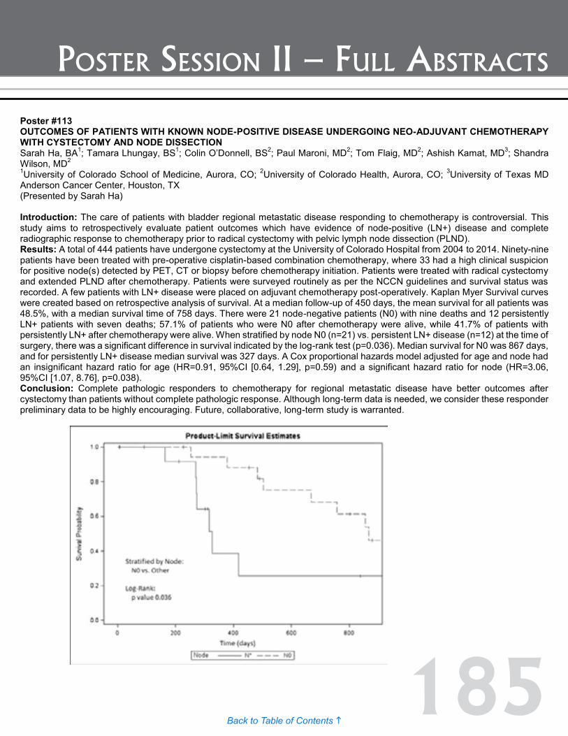
185Back to Table of Contents h
Poster session ii – Full AbstrActs
Poster #113 OUTCOMES OF PATIENTS WITH KNOWN NODE-POSITIVE DISEASE UNDERGOING NEO-ADJUVANT CHEMOTHERAPY WITH CYSTECTOMY AND NODE DISSECTION Sarah Ha, BA1; Tamara Lhungay, BS1; Colin O’Donnell, BS2; Paul Maroni, MD2; Tom Flaig, MD2; Ashish Kamat, MD3; Shandra Wilson, MD2 1University of Colorado School of Medicine, Aurora, CO; 2University of Colorado Health, Aurora, CO; 3University of Texas MD Anderson Cancer Center, Houston, TX (Presented by Sarah Ha) Introduction: The care of patients with bladder regional metastatic disease responding to chemotherapy is controversial. This study aims to retrospectively evaluate patient outcomes which have evidence of node-positive (LN+) disease and complete radiographic response to chemotherapy prior to radical cystectomy with pelvic lymph node dissection (PLND). Results: A total of 444 patients have undergone cystectomy at the University of Colorado Hospital from 2004 to 2014. Ninety-nine patients have been treated with pre-operative cisplatin-based combination chemotherapy, where 33 had a high clinical suspicion for positive node(s) detected by PET, CT or biopsy before chemotherapy initiation. Patients were treated with radical cystectomy and extended PLND after chemotherapy. Patients were surveyed routinely as per the NCCN guidelines and survival status was recorded. A few patients with LN+ disease were placed on adjuvant chemotherapy post-operatively. Kaplan Myer Survival curves were created based on retrospective analysis of survival. At a median follow-up of 450 days, the mean survival for all patients was 48.5%, with a median survival time of 758 days. There were 21 node-negative patients (N0) with nine deaths and 12 persistently LN+ patients with seven deaths; 57.1% of patients who were N0 after chemotherapy were alive, while 41.7% of patients with persistently LN+ after chemotherapy were alive. When stratified by node N0 (n=21) vs. persistent LN+ disease (n=12) at the time of surgery, there was a significant difference in survival indicated by the log-rank test (p=0.036). Median survival for N0 was 867 days, and for persistently LN+ disease median survival was 327 days. A Cox proportional hazards model adjusted for age and node had an insignificant hazard ratio for age (HR=0.91, 95%CI [0.64, 1.29], p=0.59) and a significant hazard ratio for node (HR=3.06, 95%CI [1.07, 8.76], p=0.038). Conclusion: Complete pathologic responders to chemotherapy for regional metastatic disease have better outcomes after cystectomy than patients without complete pathologic response. Although long-term data is needed, we consider these responder preliminary data to be highly encouraging. Future, collaborative, long-term study is warranted.
186Back to Table of Contents h
Poster session ii – Full AbstrActs
Poster #114 PROGNOSTIC IMPLICATION OF PAPILLARY RECURRENCE OF BLADDER CANCER AT FIRST EVALUATION AFTER INDUCTION BACILLUS CALMETTE-GUÉRIN THERAPY Chinedu Mmeje, MD; Charles Guo, MD; Jay Shah, MD; Neema Navai, MD; H. Barton Grossman, MD; Colin Dinney, MD; Ashish Kamat, MD MD Anderson Cancer Center, Houston, TX (Presented by Chinedu Mmeje) Introduction: To evaluate the prognostic implications of cTa papillary recurrence found three months after induction Bacillus Calmette-Guérin (BCG) therapy. Methods: We reviewed our database of 917 patients who underwent transurethral resection and induction BCG from 1995 to 2012. We excluded patients whose initial pathology slides were unavailable, those with only carcinoma in situ at the third month after BCG therapy, and those without at least two years of follow-up. Clinical characteristics were compared between three month recurrence stages using chi-square analysis and Student t-tests. The Kaplan-Meier method was used to determine bladder-preservation time, progression-free survival (PFS), and disease-specific survival (DSS). Results: We identified 84 patients who met the study criteria (66 patients with cTa, and 18 patients with cT1). The median follow-up for the entire cohort was 74 months. Of the patients with cTa recurrence, 60 continued with bladder-sparing therapy. Patients with a high-grade cTa recurrence treated who continued bladder-sparing therapy had a 17% incidence of disease progression and a 62% incidence of recurrence within one year. No patients with low-grade cTa recurrence (n=13) developed disease progression or underwent radical cystectomy. Patients with an initial cTa at diagnosis had a higher five-year bladder preservation rate than those with an initial cT1 diagnosis (84% vs. 61%; p =.041), but five-year DSS rates were similar between those two groups. Patients with high-grade cTa recurrence and those with cT1 recurrence had similar outcomes with respect to death rates over the entire follow-up period (10% and 15%, respectively) as well as five-year PFS (77% vs. 83%). Conclusion: Patients with low-grade cTa papillary recurrence three months after induction BCG can safely continue with bladder-sparing therapy. Patients with high-grade cTa papillary recurrence at that time have risks of recurrence and progression similar to those of patients with cT1 recurrence.
187Back to Table of Contents h
Poster session ii – Full AbstrActs
Poster #115 INTEGRATIVE ANALYSIS OF THE METHYLOME IDENTIFIES UNIQUE PATHWAYS ACTIVE IN LYMPH NODE POSITIVE VERSUS LYMPH NODE NEGATIVE UROTHELIAL CARCINOMA Thomas Sanford, MD; Maxwell Meng, MD; Sima Porten, MD UCSF, San Francisco, CA (Presented by Thomas Sanford) Introduction: The Cancer Genome Atlas Project (TCGA) provides a wealth of molecular data for urothelial carcinoma allowing for integrative analysis utilizing multiple sources of genomic information to identify potential driver processes. The purpose of this study was to use an integrative analysis of the methylome to evaluate for epigenetic drivers of urothelial carcinoma in lymph node positive versus lymph node negative urothelial carcinoma. Methods: Level 3 TCGA methylation data were downloaded from the Broad Institute’s Firehose website (http://gdac.broadinstitute.org/). All data were derived from muscle invasive cystectomy specimens. Methylation status for each gene was summarized using beta values. The R package MethylMix was then used to perform integrative analysis to identify methylation changes that were both correlated with gene expression and differentially methylated compared with a pool of normal bladder tissue. Gene enrichment analysis was performed on genes identified in patients with nodal metastasis as well as those without nodal metastasis (defined as N0 with at least 10 lymph nodes removed). Results: There were at total of 117 patients with lymph node metastasis, 138 patients without lymph node metastasis, and 21 normal bladder samples. MethylMix identified a total of 195 candidate driver genes for node positive patients and 246 genes for node negative patients. There was overlap in 133 genes. Node positive patients were found to have enrichment in estrogen signaling pathways as well as in the NRF2 pathways. Lymph node negative patients were found to have enrichment in the EGFR1 pathway. Both groups had enrichment in chemical carcinogenesis and glutathione pathways. Conclusion: This is an integrative analysis of the human methylome that identifies a list of potential driver mutations shared a substantial proportion of genes and were enriched in common pathways. However, lymph node positive tumors had estrogen signaling whereas lymph node negative tumors had EGFR1 signaling, indicating lymph node positive tumors may rely on epigenetic mechanisms for lymph node spread.
188Back to Table of Contents h
Poster session ii – Full AbstrActs
Poster #116 MODERATE CHRONIC KIDNEY DISEASE (EGFR <60 ML/MIN) PREDICTS RECURRENCE AND PROGRESSION IN BLADDER CANCER PATIENTS TREATED WITH TRANSURETHRAL RESECTION Michael L. Blute, Jr., MD; Victor Kucherov; Timothy J. Rushmer; Fangfang Shi; Benjamin Fuller; E. Jason Abel, MD; Kyle Richards, MD; David F. Jarrard, MD; Edward M. Messing, MD; Tracy M. Downs, MD (Presented by Michael L. Blute, Jr.) Introduction: Chronic kidney disease (CKD) has been suggested to be associated with a higher risk of cancer development and higher cancer mortality in bladder cancer. The purpose of this study was to evaluate if moderate CKD (eGFR <60 ml/min) is associated with high rates of tumor recurrence or progression in bladder cancer patients treated with TURBT. Methods: A multi-institutional database (University of Wisconsin, University of Rochester, NY) identified patients with serum creatinine values available prior to first TURBT for NMIBC. CKD-epidemiology collaboration formula was used to calculate estimated glomerular filtration rate (eGFR) for each patient. Cox proportional hazards models were used to evaluate associations with recurrence-free (RFS) and progression-free survival (PFS). Results: A total of 727 patients were identified with a median patient age of 69.8 (IQR 60.1-77.6). During a median follow-up of 3.7 (IQR 1.5-6.5) years, 400 (55%) patients had recurrence and 145 (19.9%) patients had progression of tumor stage or grade. A total of 41 (5.6%) patients had progression to muscle invasive disease (pT2). Moderate or severe CKD was identified in 183 patients according to eGFR. Multivariate analysis identified tumor size > 3cm (HR 1.4, 95% CI 1.1-1.8; p=0.01) and eGFR < 60 (HR 1.5, 95% CI 1.2-1.9; p=0.002) as predictors of tumor recurrence. The five-year RFS rate was 46% for patients with an eGFR >=60 ml/min and 27% for patients with an eGFR <60 ml/min (log rank p-value=0.0004). Multivariate analysis also demonstrated that eGFR <60 ml/min (HR 3.7, 95% CI 1.75-7.94; p=0.001) was associated with progression to muscle-invasive disease. The five-year PFS rate was 83% for patients with an eGFR >=60 ml/min and 71% for patients with an eGFR <60 ml/min (log rank p-value=0.01). Subgroup analysis of patients who received BCG therapy stratified by eGFR showed that those who had CKD were more likely to experience tumor progression to muscle invasive disease (HR 7.2, 95% CI 1.93-26.50; p=0.003). Conclusion: Moderate CKD (eGFR<60 ml/min) at first TUR is associated with reduced RFS and PFS. Patients with reduced renal function should be considered for increased surveillance after bladder cancer diagnosis.
189Back to Table of Contents h
Poster session ii – Full AbstrActs
Poster #117 MUTATIONAL LANDSCAPE OF PRIMARY BLADDER AND URACHAL ADENOCARCINOMA Byron Lee, MD, PhD1; Emmet Jordan, MD2; Helen Won2; Aditya Bagrodia, MD2; Neil Desai, MD2; Dean Bajorin, MD2; Jonathan Rosenberg, MD2; Bernard Bochner, MD2; Wonkyu Kim, MD2; Michael Berger, MD2; David Solit, MD2; Hikmat Al-Ahmadie, MD2; Gopa Iyer, MD2 1Memorial Sloan Kettering Cancer Center; 2New York, NY (Presented by Byron Lee) Introduction: Adenocarcinomas of the bladder and urachus represent a rare subtype of urinary tract cancer and account for two percent of all tumors originating from the bladder. Localized disease can be managed with surgical extirpation alone; however, less than 40% of patients with advanced disease will achieve an objective response with platinum-based chemotherapy. We hypothesized that bladder and urachal adenocarcinomas harbor a distinct mutational profile that may make them susceptible to targeted agents. Methods: After Institutional Review Board approval, 16 patients with primary bladder adenocarcinoma (PBA) and 10 patients with urachal adenocarcinoma (UA) were identified. Histopathologic review was performed, and tumor DNA isolated from representative paraffin-embedded sections as well as matched normal DNA were subjected to Integrated Mutation Profiling of Actionable Cancer Targets (MSK-IMPACT) sequencing, an exon-capture assay that interrogates the mutation and copy number status of 379 oncogenes and tumor suppressor genes known to be commonly altered in cancer. Somatic alterations including point mutations, insertions/deletions, truncations, and copy number changes were detected. Results: The study population consisted of 17 (65%) males and nine (35%) females with local or locally advanced PBA or UA. Seventeen (65%) were ever smokers and nine (35%) were never smokers. Four (15%) patients received neoadjuvant chemotherapy, 14 (54%) underwent partial cystectomy, and 12 (46%) underwent radical cystectomy. Median overall survival was 41.2 months (95% CI: 12-129) in patients with PBA and 80.5 months (95% CI: 11-263) in patients with UA. Mutational analysis revealed that mutations resulting in upregulation of the mitogen-activated protein kinase (MAPK) pathway are common in PBA and UA (Table 1). Conclusion: The mutational landscape of PBA and UA is more similar to that of colorectal adenocarcinoma than urothelial carcinoma of the bladder. Alterations predicted to upregulate the MAPK pathway such as activating KRAS and GNAS mutations and are common in PBA and UA, and agents targeting this pathway may provide a novel therapeutic avenue for the treatment of PBA and UA.
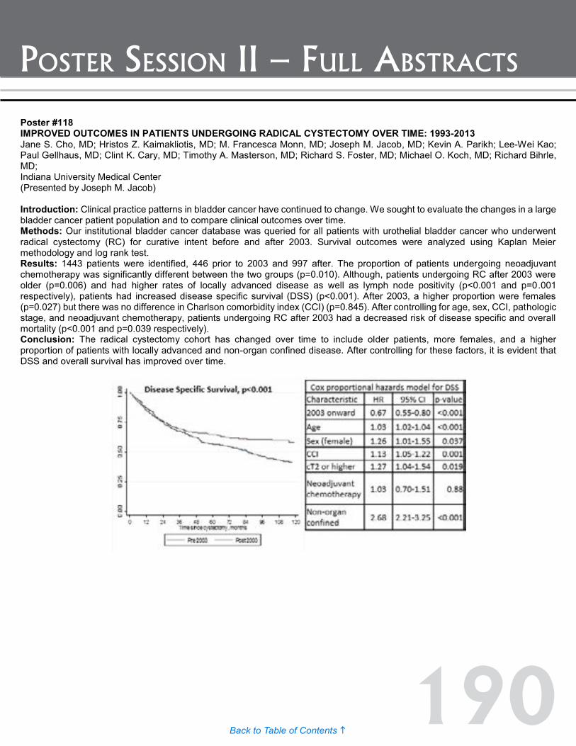
190Back to Table of Contents h
Poster session ii – Full AbstrActs
Poster #118 IMPROVED OUTCOMES IN PATIENTS UNDERGOING RADICAL CYSTECTOMY OVER TIME: 1993-2013 Jane S. Cho, MD; Hristos Z. Kaimakliotis, MD; M. Francesca Monn, MD; Joseph M. Jacob, MD; Kevin A. Parikh; Lee-Wei Kao; Paul Gellhaus, MD; Clint K. Cary, MD; Timothy A. Masterson, MD; Richard S. Foster, MD; Michael O. Koch, MD; Richard Bihrle, MD; Indiana University Medical Center (Presented by Joseph M. Jacob) Introduction: Clinical practice patterns in bladder cancer have continued to change. We sought to evaluate the changes in a large bladder cancer patient population and to compare clinical outcomes over time. Methods: Our institutional bladder cancer database was queried for all patients with urothelial bladder cancer who underwent radical cystectomy (RC) for curative intent before and after 2003. Survival outcomes were analyzed using Kaplan Meier methodology and log rank test. Results: 1443 patients were identified, 446 prior to 2003 and 997 after. The proportion of patients undergoing neoadjuvant chemotherapy was significantly different between the two groups (p=0.010). Although, patients undergoing RC after 2003 were older (p=0.006) and had higher rates of locally advanced disease as well as lymph node positivity (p<0.001 and p=0.001 respectively), patients had increased disease specific survival (DSS) (p<0.001). After 2003, a higher proportion were females (p=0.027) but there was no difference in Charlson comorbidity index (CCI) (p=0.845). After controlling for age, sex, CCI, pathologic stage, and neoadjuvant chemotherapy, patients undergoing RC after 2003 had a decreased risk of disease specific and overall mortality (p<0.001 and p=0.039 respectively). Conclusion: The radical cystectomy cohort has changed over time to include older patients, more females, and a higher proportion of patients with locally advanced and non-organ confined disease. After controlling for these factors, it is evident that DSS and overall survival has improved over time.
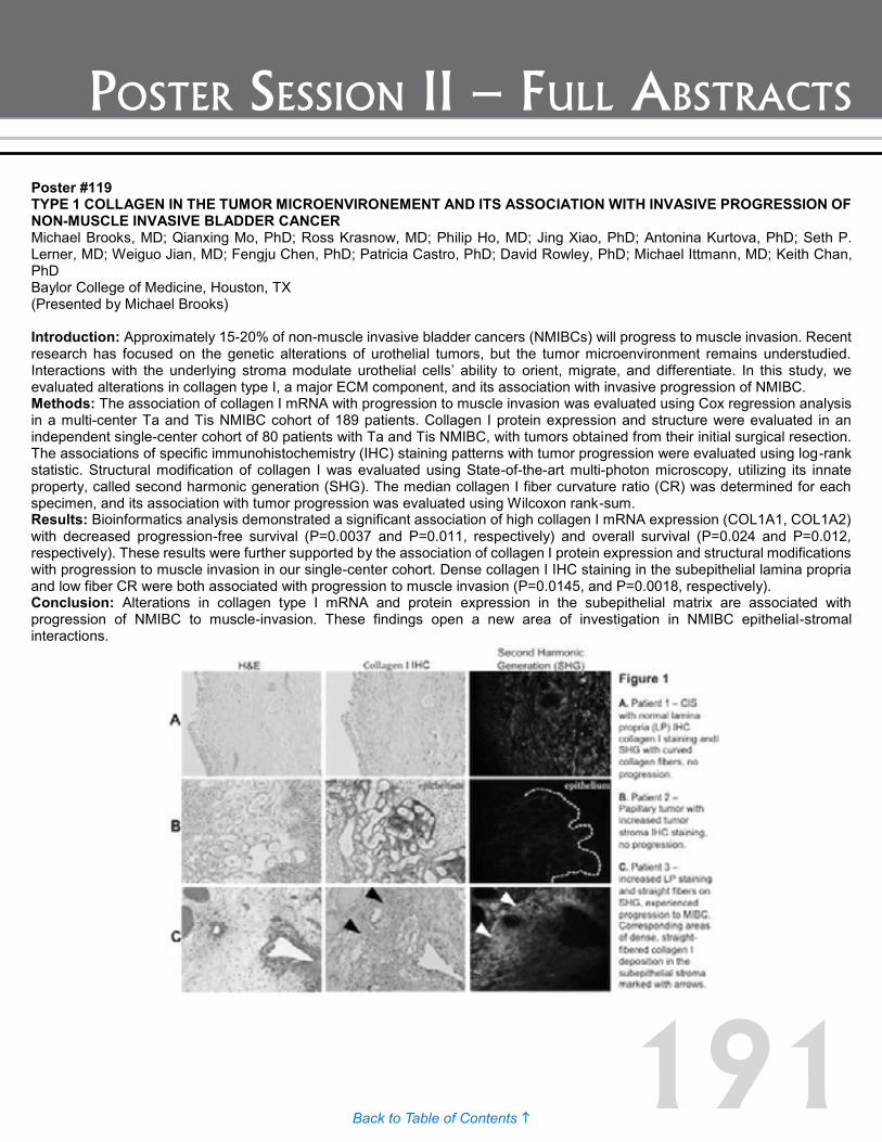
191Back to Table of Contents h
Poster session ii – Full AbstrActs
Poster #119 TYPE 1 COLLAGEN IN THE TUMOR MICROENVIRONEMENT AND ITS ASSOCIATION WITH INVASIVE PROGRESSION OF NON-MUSCLE INVASIVE BLADDER CANCER Michael Brooks, MD; Qianxing Mo, PhD; Ross Krasnow, MD; Philip Ho, MD; Jing Xiao, PhD; Antonina Kurtova, PhD; Seth P. Lerner, MD; Weiguo Jian, MD; Fengju Chen, PhD; Patricia Castro, PhD; David Rowley, PhD; Michael Ittmann, MD; Keith Chan, PhD Baylor College of Medicine, Houston, TX (Presented by Michael Brooks) Introduction: Approximately 15-20% of non-muscle invasive bladder cancers (NMIBCs) will progress to muscle invasion. Recent research has focused on the genetic alterations of urothelial tumors, but the tumor microenvironment remains understudied. Interactions with the underlying stroma modulate urothelial cells’ ability to orient, migrate, and differentiate. In this study, we evaluated alterations in collagen type I, a major ECM component, and its association with invasive progression of NMIBC. Methods: The association of collagen I mRNA with progression to muscle invasion was evaluated using Cox regression analysis in a multi-center Ta and Tis NMIBC cohort of 189 patients. Collagen I protein expression and structure were evaluated in an independent single-center cohort of 80 patients with Ta and Tis NMIBC, with tumors obtained from their initial surgical resection. The associations of specific immunohistochemistry (IHC) staining patterns with tumor progression were evaluated using log-rank statistic. Structural modification of collagen I was evaluated using State-of-the-art multi-photon microscopy, utilizing its innate property, called second harmonic generation (SHG). The median collagen I fiber curvature ratio (CR) was determined for each specimen, and its association with tumor progression was evaluated using Wilcoxon rank-sum. Results: Bioinformatics analysis demonstrated a significant association of high collagen I mRNA expression (COL1A1, COL1A2) with decreased progression-free survival (P=0.0037 and P=0.011, respectively) and overall survival (P=0.024 and P=0.012, respectively). These results were further supported by the association of collagen I protein expression and structural modifications with progression to muscle invasion in our single-center cohort. Dense collagen I IHC staining in the subepithelial lamina propria and low fiber CR were both associated with progression to muscle invasion (P=0.0145, and P=0.0018, respectively). Conclusion: Alterations in collagen type I mRNA and protein expression in the subepithelial matrix are associated with progression of NMIBC to muscle-invasion. These findings open a new area of investigation in NMIBC epithelial-stromal interactions.
Poster #118 IMPROVED OUTCOMES IN PATIENTS UNDERGOING RADICAL CYSTECTOMY OVER TIME: 1993-2013 Jane S. Cho, MD; Hristos Z. Kaimakliotis, MD; M. Francesca Monn, MD; Joseph M. Jacob, MD; Kevin A. Parikh; Lee-Wei Kao; Paul Gellhaus, MD; Clint K. Cary, MD; Timothy A. Masterson, MD; Richard S. Foster, MD; Michael O. Koch, MD; Richard Bihrle, MD; Indiana University Medical Center (Presented by Joseph M. Jacob) Introduction: Clinical practice patterns in bladder cancer have continued to change. We sought to evaluate the changes in a large bladder cancer patient population and to compare clinical outcomes over time. Methods: Our institutional bladder cancer database was queried for all patients with urothelial bladder cancer who underwent radical cystectomy (RC) for curative intent before and after 2003. Survival outcomes were analyzed using Kaplan Meier methodology and log rank test. Results: 1443 patients were identified, 446 prior to 2003 and 997 after. The proportion of patients undergoing neoadjuvant chemotherapy was significantly different between the two groups (p=0.010). Although, patients undergoing RC after 2003 were older (p=0.006) and had higher rates of locally advanced disease as well as lymph node positivity (p<0.001 and p=0.001 respectively), patients had increased disease specific survival (DSS) (p<0.001). After 2003, a higher proportion were females (p=0.027) but there was no difference in Charlson comorbidity index (CCI) (p=0.845). After controlling for age, sex, CCI, pathologic stage, and neoadjuvant chemotherapy, patients undergoing RC after 2003 had a decreased risk of disease specific and overall mortality (p<0.001 and p=0.039 respectively). Conclusion: The radical cystectomy cohort has changed over time to include older patients, more females, and a higher proportion of patients with locally advanced and non-organ confined disease. After controlling for these factors, it is evident that DSS and overall survival has improved over time.
192Back to Table of Contents h
Poster session ii – Full AbstrActs
Poster #120 UPPER TRACT UROTHELIAL CARCINOMA IN PATIENTS WITH CLINICAL SUSPICION OF LYNCH SYNDROME Hong Truong, MS, MD1; Sarah Hegarty, MPhil2; Scott Hubosky, MD1; Kelly Healy, MD1; Jean Hoffman-Censits, MD3; Veda Giri, MD4 1Department of Urology, Sidney Kimmel Cancer Center, Thomas Jefferson University, Philadelphia, PA; 2Department of Biostatistics, Sidney Kimmel Cancer Center, Thomas Jefferson University, Philadelphia, PA; 3Department of Medical Oncology, Sidney Kimmel Cancer Center, Thomas Jefferson University, MD; 4Divison of Population Science, Department of Medical Oncology, Sidney Kimmel Cancer Center, Thomas Jefferson University, Philadelphia, PA (Presented by Hong Truong) Introduction: Lynch syndrome (LS) is the only known hereditary cancer syndrome associated with upper tract urothelial carcinoma (UTUC). Patients with LS have a reported 14-fold increase in relative risk of developing UTUC compared to the general population. The prevalence and clinical implications of potential hereditary UTUC are not well understood due to the rarity of this clinical entity and the lack of awareness among urologists. The goal of this study was to evaluate the prevalence and outcome of UTUC in patients with LS-associated cancers. Methods: We analyzed UTUC cases (primary site codes C65.9 and 66.9) in the Surveillance, Epidemiology, and End Results (SEER-17) database. LS-associated cancers were identified in patients with UTUC. Based on the revised Bethesda Guidelines for Lynch Syndrome, LS-associated cancers include colorectal, endometrial/uterine, gastric, ovarian, small intestinal, glioblastoma, sebaceous adenocarcinoma, biliary tract, and pancreatic cancers. UTUC-specific survival was compared between patients with UTUC alone versus those with LS-associated cancers. Results: Between 1973 and 2011, 30,349 cases of UTUC were reported in SEER. A total of 1970 (6.49%) of cases of UTUC also had at least one LS-associated cancer. Colorectal, uterine, and ovarian cancers were the most prevalent LS-associated cancers, accounting for 4.2%, 2.1%, and 0.65% of UTUC cases respectively. Compared to UTUC-only cases, those with LS-associated cancers have higher UTUC-specific survival when controlled for disease stage and age of diagnosis (p<0.0001). Conclusion: The risk of developing UTUC in LS may be underappreciated. Patients with potential hereditary UTUC may have a different clinical course and survival outcomes compared to those with UTUC without LS-associated cancers. Further study and genetic evaluation is warranted.
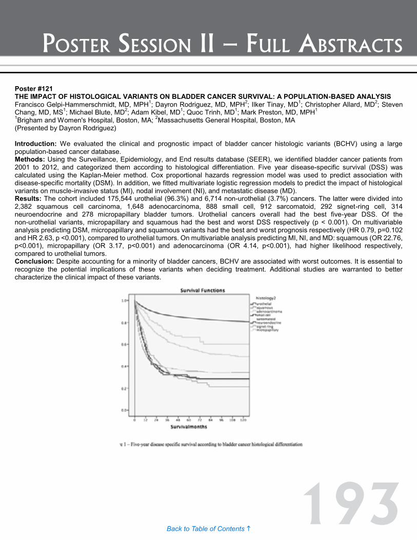
193Back to Table of Contents h
Poster session ii – Full AbstrActs
Poster #121 THE IMPACT OF HISTOLOGICAL VARIANTS ON BLADDER CANCER SURVIVAL: A POPULATION-BASED ANALYSIS Francisco Gelpi-Hammerschmidt, MD, MPH1; Dayron Rodriguez, MD, MPH2; Ilker Tinay, MD1; Christopher Allard, MD2; Steven Chang, MD, MS1; Michael Blute, MD2; Adam Kibel, MD1; Quoc Trinh, MD1; Mark Preston, MD, MPH1 1Brigham and Women's Hospital, Boston, MA; 2Massachusetts General Hospital, Boston, MA (Presented by Dayron Rodriguez) Introduction: We evaluated the clinical and prognostic impact of bladder cancer histologic variants (BCHV) using a large population-based cancer database. Methods: Using the Surveillance, Epidemiology, and End results database (SEER), we identified bladder cancer patients from 2001 to 2012, and categorized them according to histological differentiation. Five year disease-specific survival (DSS) was calculated using the Kaplan-Meier method. Cox proportional hazards regression model was used to predict association with disease-specific mortality (DSM). In addition, we fitted multivariate logistic regression models to predict the impact of histological variants on muscle-invasive status (MI), nodal involvement (NI), and metastatic disease (MD). Results: The cohort included 175,544 urothelial (96.3%) and 6,714 non-urothelial (3.7%) cancers. The latter were divided into 2,382 squamous cell carcinoma, 1,648 adenocarcinoma, 888 small cell, 912 sarcomatoid, 292 signet-ring cell, 314 neuroendocrine and 278 micropapillary bladder tumors. Urothelial cancers overall had the best five-year DSS. Of the non-urothelial variants, micropapillary and squamous had the best and worst DSS respectively (p < 0.001). On multivariable analysis predicting DSM, micropapillary and squamous variants had the best and worst prognosis respectively (HR 0.79, p=0.102 and HR 2.63, p <0.001), compared to urothelial tumors. On multivariable analysis predicting MI, NI, and MD: squamous (OR 22.76, p<0.001), micropapillary (OR 3.17, p<0.001) and adenocarcinoma (OR 4.14, p<0.001), had higher likelihood respectively, compared to urothelial tumors. Conclusion: Despite accounting for a minority of bladder cancers, BCHV are associated with worst outcomes. It is essential to recognize the potential implications of these variants when deciding treatment. Additional studies are warranted to better characterize the clinical impact of these variants.
Poster #120 UPPER TRACT UROTHELIAL CARCINOMA IN PATIENTS WITH CLINICAL SUSPICION OF LYNCH SYNDROME Hong Truong, MS, MD1; Sarah Hegarty, MPhil2; Scott Hubosky, MD1; Kelly Healy, MD1; Jean Hoffman-Censits, MD3; Veda Giri, MD4 1Department of Urology, Sidney Kimmel Cancer Center, Thomas Jefferson University, Philadelphia, PA; 2Department of Biostatistics, Sidney Kimmel Cancer Center, Thomas Jefferson University, Philadelphia, PA; 3Department of Medical Oncology, Sidney Kimmel Cancer Center, Thomas Jefferson University, MD; 4Divison of Population Science, Department of Medical Oncology, Sidney Kimmel Cancer Center, Thomas Jefferson University, Philadelphia, PA (Presented by Hong Truong) Introduction: Lynch syndrome (LS) is the only known hereditary cancer syndrome associated with upper tract urothelial carcinoma (UTUC). Patients with LS have a reported 14-fold increase in relative risk of developing UTUC compared to the general population. The prevalence and clinical implications of potential hereditary UTUC are not well understood due to the rarity of this clinical entity and the lack of awareness among urologists. The goal of this study was to evaluate the prevalence and outcome of UTUC in patients with LS-associated cancers. Methods: We analyzed UTUC cases (primary site codes C65.9 and 66.9) in the Surveillance, Epidemiology, and End Results (SEER-17) database. LS-associated cancers were identified in patients with UTUC. Based on the revised Bethesda Guidelines for Lynch Syndrome, LS-associated cancers include colorectal, endometrial/uterine, gastric, ovarian, small intestinal, glioblastoma, sebaceous adenocarcinoma, biliary tract, and pancreatic cancers. UTUC-specific survival was compared between patients with UTUC alone versus those with LS-associated cancers. Results: Between 1973 and 2011, 30,349 cases of UTUC were reported in SEER. A total of 1970 (6.49%) of cases of UTUC also had at least one LS-associated cancer. Colorectal, uterine, and ovarian cancers were the most prevalent LS-associated cancers, accounting for 4.2%, 2.1%, and 0.65% of UTUC cases respectively. Compared to UTUC-only cases, those with LS-associated cancers have higher UTUC-specific survival when controlled for disease stage and age of diagnosis (p<0.0001). Conclusion: The risk of developing UTUC in LS may be underappreciated. Patients with potential hereditary UTUC may have a different clinical course and survival outcomes compared to those with UTUC without LS-associated cancers. Further study and genetic evaluation is warranted.
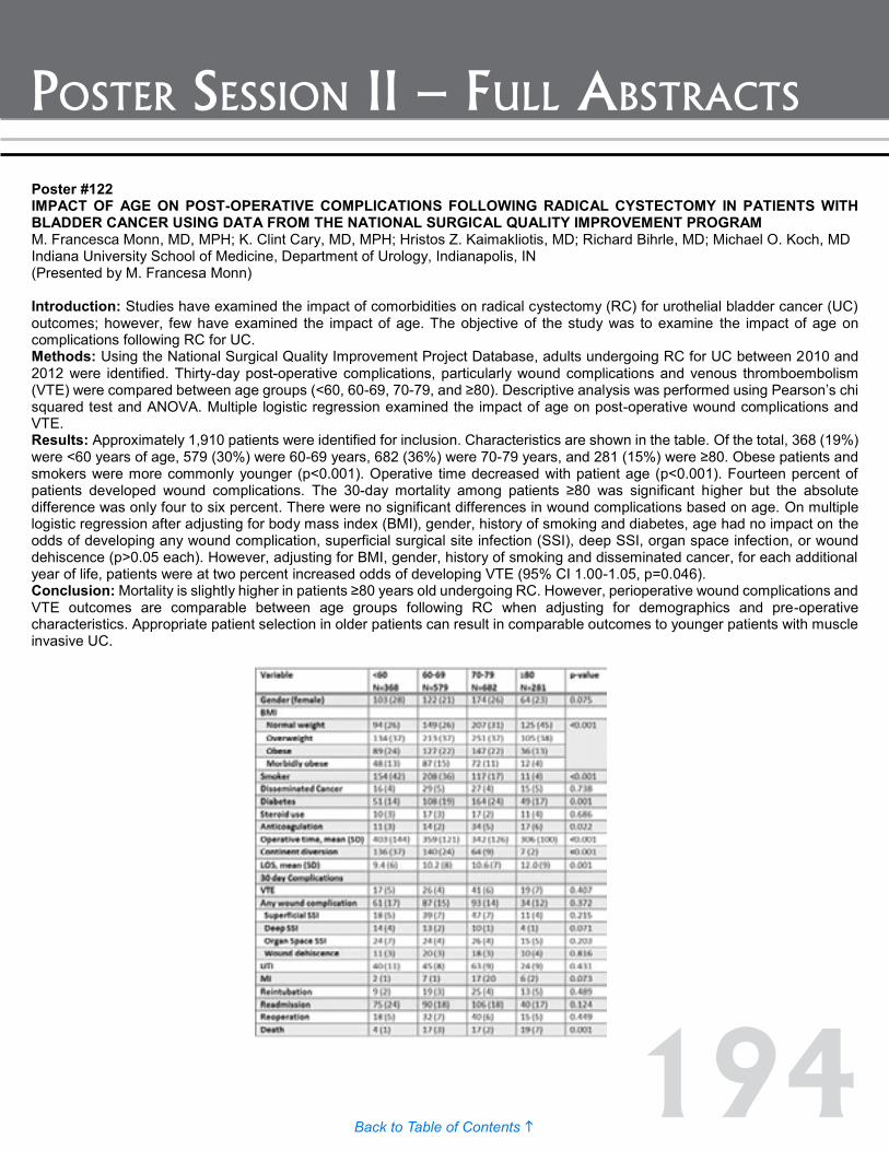
194Back to Table of Contents h
Poster session ii – Full AbstrActs
Poster #123 PATHOLOGIC RESPONSE IN PATIENTS RECEIVING NEOADJUVANT CHEMOTHERAPY FOR MUSCLE-INASIVE BLADDER CANCER: IS THE CURATIVE EFFECT DUE TO CHEMOTHERAPY OR TURBT? Aaron Brant, BS; Max Kates, MD; Meera Chappidi, BS; Hiten D. Patel, MD, MPH; Nikolai A. Sopko, MD, PhD; Trinity J. Bivalacqua, MD, PhD Johns Hopkins School of Medicine, Baltimore, MD (Presented by Aaron Brant) Introduction: Neoadjuvant chemotherapy (NAC) for muscle-invasive bladder cancer (MIBC) has been associated with a survival benefit in patients who achieve pathologic response. However, a smaller but significant group of patients who receive transurethral resection of bladder tumor (TURBT) without NAC will also be down-staged. We identified predictors of pathologic response in a cohort of NAC and non-NAC patients in order to estimate the prevalence of pathologic response in NAC patients that could be attributed to TURBT. Methods: Of 737 serial patients who received radical cystectomy (RC) at Johns Hopkins Medical Center from 2005 to 2014, 328 patients with cT2 urothelial carcinoma were identified. 172 patients received TURBT and NAC prior to RC, while 156 went directly to RC after TURBT. Demographic, clinical, and pathologic information was compared between groups using Wilcoxon-Mann-Whitney for continuous and chi-squared for categorical variables. Pathologic response was defined as <T2 (T0, Ta, Tis, or T1) at RC. A Poisson regression model with robust error variance was used to determine relative risk (RR) of pathologic response in NAC vs. non-NAC patients, adjusting for age, body mass index (BMI), race, gender, Charlson score, smoking status, days from MIBC diagnosis to surgery, and history of prior non-MIBC (NMIBC). Results: Pathologic response was higher in NAC patients compared to non-NAC patients (62% vs. 21%, RR=3.00). Younger age at RC was a predictor of response in NAC patients (p=0.01), while history of prior NMIBC was a predictor of response in non-NAC patients (p<0.05). NAC patients were significantly younger than non-NAC patients (64.8 vs. 71.2 years, p<0.01), with higher BMI (28.1 vs. 26.7 kg/m2, p<0.01), lower frequency of Charlson score ≥3 (13.4% vs. 26.7%, p<0.01), and lower frequency of prior NMIBC (9.4% vs. 22.4%, p<0.01). Adjustment resulted in a RR of pathologic response in NAC vs. non-NAC of 2.50 (95% CI: 1.69-3.69, p<0.01). Assuming no interaction between NAC and TURBT, this adjusted model suggests that in a cohort of patients who receive NAC, 40% (95% CI: 27–59%) of pathologic response can be attributed to TURBT. Conclusion: An adjusted model suggests that in a cohort of patients who receive NAC and TURBT prior to RC, 40% of pathologic response can be attributed to TURBT. Understanding which patients are true responders to chemotherapy and which received a therapeutic TURBT is necessary in order to select optimal candidates for NAC.
Poster #122 IMPACT OF AGE ON POST-OPERATIVE COMPLICATIONS FOLLOWING RADICAL CYSTECTOMY IN PATIENTS WITH BLADDER CANCER USING DATA FROM THE NATIONAL SURGICAL QUALITY IMPROVEMENT PROGRAM M. Francesca Monn, MD, MPH; K. Clint Cary, MD, MPH; Hristos Z. Kaimakliotis, MD; Richard Bihrle, MD; Michael O. Koch, MD Indiana University School of Medicine, Department of Urology, Indianapolis, IN (Presented by M. Francesa Monn) Introduction: Studies have examined the impact of comorbidities on radical cystectomy (RC) for urothelial bladder cancer (UC) outcomes; however, few have examined the impact of age. The objective of the study was to examine the impact of age on complications following RC for UC. Methods: Using the National Surgical Quality Improvement Project Database, adults undergoing RC for UC between 2010 and 2012 were identified. Thirty-day post-operative complications, particularly wound complications and venous thromboembolism (VTE) were compared between age groups (<60, 60-69, 70-79, and ≥80). Descriptive analysis was performed using Pearson’s chi squared test and ANOVA. Multiple logistic regression examined the impact of age on post-operative wound complications and VTE. Results: Approximately 1,910 patients were identified for inclusion. Characteristics are shown in the table. Of the total, 368 (19%) were <60 years of age, 579 (30%) were 60-69 years, 682 (36%) were 70-79 years, and 281 (15%) were ≥80. Obese patients and smokers were more commonly younger (p<0.001). Operative time decreased with patient age (p<0.001). Fourteen percent of patients developed wound complications. The 30-day mortality among patients ≥80 was significant higher but the absolute difference was only four to six percent. There were no significant differences in wound complications based on age. On multiple logistic regression after adjusting for body mass index (BMI), gender, history of smoking and diabetes, age had no impact on the odds of developing any wound complication, superficial surgical site infection (SSI), deep SSI, organ space infection, or wound dehiscence (p>0.05 each). However, adjusting for BMI, gender, history of smoking and disseminated cancer, for each additional year of life, patients were at two percent increased odds of developing VTE (95% CI 1.00-1.05, p=0.046). Conclusion: Mortality is slightly higher in patients ≥80 years old undergoing RC. However, perioperative wound complications and VTE outcomes are comparable between age groups following RC when adjusting for demographics and pre-operative characteristics. Appropriate patient selection in older patients can result in comparable outcomes to younger patients with muscle invasive UC.
195Back to Table of Contents h
Poster session ii – Full AbstrActs
Poster #123 PATHOLOGIC RESPONSE IN PATIENTS RECEIVING NEOADJUVANT CHEMOTHERAPY FOR MUSCLE-INASIVE BLADDER CANCER: IS THE CURATIVE EFFECT DUE TO CHEMOTHERAPY OR TURBT? Aaron Brant, BS; Max Kates, MD; Meera Chappidi, BS; Hiten D. Patel, MD, MPH; Nikolai A. Sopko, MD, PhD; Trinity J. Bivalacqua, MD, PhD Johns Hopkins School of Medicine, Baltimore, MD (Presented by Aaron Brant) Introduction: Neoadjuvant chemotherapy (NAC) for muscle-invasive bladder cancer (MIBC) has been associated with a survival benefit in patients who achieve pathologic response. However, a smaller but significant group of patients who receive transurethral resection of bladder tumor (TURBT) without NAC will also be down-staged. We identified predictors of pathologic response in a cohort of NAC and non-NAC patients in order to estimate the prevalence of pathologic response in NAC patients that could be attributed to TURBT. Methods: Of 737 serial patients who received radical cystectomy (RC) at Johns Hopkins Medical Center from 2005 to 2014, 328 patients with cT2 urothelial carcinoma were identified. 172 patients received TURBT and NAC prior to RC, while 156 went directly to RC after TURBT. Demographic, clinical, and pathologic information was compared between groups using Wilcoxon-Mann-Whitney for continuous and chi-squared for categorical variables. Pathologic response was defined as <T2 (T0, Ta, Tis, or T1) at RC. A Poisson regression model with robust error variance was used to determine relative risk (RR) of pathologic response in NAC vs. non-NAC patients, adjusting for age, body mass index (BMI), race, gender, Charlson score, smoking status, days from MIBC diagnosis to surgery, and history of prior non-MIBC (NMIBC). Results: Pathologic response was higher in NAC patients compared to non-NAC patients (62% vs. 21%, RR=3.00). Younger age at RC was a predictor of response in NAC patients (p=0.01), while history of prior NMIBC was a predictor of response in non-NAC patients (p<0.05). NAC patients were significantly younger than non-NAC patients (64.8 vs. 71.2 years, p<0.01), with higher BMI (28.1 vs. 26.7 kg/m2, p<0.01), lower frequency of Charlson score ≥3 (13.4% vs. 26.7%, p<0.01), and lower frequency of prior NMIBC (9.4% vs. 22.4%, p<0.01). Adjustment resulted in a RR of pathologic response in NAC vs. non-NAC of 2.50 (95% CI: 1.69-3.69, p<0.01). Assuming no interaction between NAC and TURBT, this adjusted model suggests that in a cohort of patients who receive NAC, 40% (95% CI: 27–59%) of pathologic response can be attributed to TURBT. Conclusion: An adjusted model suggests that in a cohort of patients who receive NAC and TURBT prior to RC, 40% of pathologic response can be attributed to TURBT. Understanding which patients are true responders to chemotherapy and which received a therapeutic TURBT is necessary in order to select optimal candidates for NAC.
Poster #122 IMPACT OF AGE ON POST-OPERATIVE COMPLICATIONS FOLLOWING RADICAL CYSTECTOMY IN PATIENTS WITH BLADDER CANCER USING DATA FROM THE NATIONAL SURGICAL QUALITY IMPROVEMENT PROGRAM M. Francesca Monn, MD, MPH; K. Clint Cary, MD, MPH; Hristos Z. Kaimakliotis, MD; Richard Bihrle, MD; Michael O. Koch, MD Indiana University School of Medicine, Department of Urology, Indianapolis, IN (Presented by M. Francesa Monn) Introduction: Studies have examined the impact of comorbidities on radical cystectomy (RC) for urothelial bladder cancer (UC) outcomes; however, few have examined the impact of age. The objective of the study was to examine the impact of age on complications following RC for UC. Methods: Using the National Surgical Quality Improvement Project Database, adults undergoing RC for UC between 2010 and 2012 were identified. Thirty-day post-operative complications, particularly wound complications and venous thromboembolism (VTE) were compared between age groups (<60, 60-69, 70-79, and ≥80). Descriptive analysis was performed using Pearson’s chi squared test and ANOVA. Multiple logistic regression examined the impact of age on post-operative wound complications and VTE. Results: Approximately 1,910 patients were identified for inclusion. Characteristics are shown in the table. Of the total, 368 (19%) were <60 years of age, 579 (30%) were 60-69 years, 682 (36%) were 70-79 years, and 281 (15%) were ≥80. Obese patients and smokers were more commonly younger (p<0.001). Operative time decreased with patient age (p<0.001). Fourteen percent of patients developed wound complications. The 30-day mortality among patients ≥80 was significant higher but the absolute difference was only four to six percent. There were no significant differences in wound complications based on age. On multiple logistic regression after adjusting for body mass index (BMI), gender, history of smoking and diabetes, age had no impact on the odds of developing any wound complication, superficial surgical site infection (SSI), deep SSI, organ space infection, or wound dehiscence (p>0.05 each). However, adjusting for BMI, gender, history of smoking and disseminated cancer, for each additional year of life, patients were at two percent increased odds of developing VTE (95% CI 1.00-1.05, p=0.046). Conclusion: Mortality is slightly higher in patients ≥80 years old undergoing RC. However, perioperative wound complications and VTE outcomes are comparable between age groups following RC when adjusting for demographics and pre-operative characteristics. Appropriate patient selection in older patients can result in comparable outcomes to younger patients with muscle invasive UC.
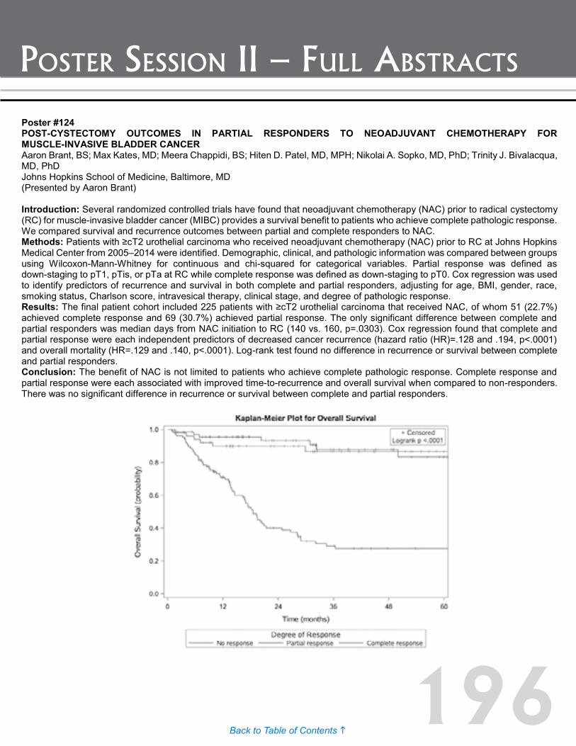
196Back to Table of Contents h
Poster session ii – Full AbstrActs
Poster #124 POST-CYSTECTOMY OUTCOMES IN PARTIAL RESPONDERS TO NEOADJUVANT CHEMOTHERAPY FOR MUSCLE-INVASIVE BLADDER CANCER Aaron Brant, BS; Max Kates, MD; Meera Chappidi, BS; Hiten D. Patel, MD, MPH; Nikolai A. Sopko, MD, PhD; Trinity J. Bivalacqua, MD, PhD Johns Hopkins School of Medicine, Baltimore, MD (Presented by Aaron Brant) Introduction: Several randomized controlled trials have found that neoadjuvant chemotherapy (NAC) prior to radical cystectomy (RC) for muscle-invasive bladder cancer (MIBC) provides a survival benefit to patients who achieve complete pathologic response. We compared survival and recurrence outcomes between partial and complete responders to NAC. Methods: Patients with ≥cT2 urothelial carcinoma who received neoadjuvant chemotherapy (NAC) prior to RC at Johns Hopkins Medical Center from 2005–2014 were identified. Demographic, clinical, and pathologic information was compared between groups using Wilcoxon-Mann-Whitney for continuous and chi-squared for categorical variables. Partial response was defined as down-staging to pT1, pTis, or pTa at RC while complete response was defined as down-staging to pT0. Cox regression was used to identify predictors of recurrence and survival in both complete and partial responders, adjusting for age, BMI, gender, race, smoking status, Charlson score, intravesical therapy, clinical stage, and degree of pathologic response. Results: The final patient cohort included 225 patients with ≥cT2 urothelial carcinoma that received NAC, of whom 51 (22.7%) achieved complete response and 69 (30.7%) achieved partial response. The only significant difference between complete and partial responders was median days from NAC initiation to RC (140 vs. 160, p=.0303). Cox regression found that complete and partial response were each independent predictors of decreased cancer recurrence (hazard ratio (HR)=.128 and .194, p<.0001) and overall mortality (HR=.129 and .140, p<.0001). Log-rank test found no difference in recurrence or survival between complete and partial responders. Conclusion: The benefit of NAC is not limited to patients who achieve complete pathologic response. Complete response and partial response were each associated with improved time-to-recurrence and overall survival when compared to non-responders. There was no significant difference in recurrence or survival between complete and partial responders.
197Back to Table of Contents h
Poster session ii – Full AbstrActs
Poster #125 THE CANCER GENOME ATLAS (TCGA) PROJECT ANALYSIS OF MICRO-RNA AND GENE EXPRESSION SUBTYPES OF HIGH-GRADE, MUSCLE-INVASIVE UROTHELIAL CARCINOMA Gordon Robertson, PhD1; Preethi Gunaratne, PhD2; Seth Lerner, MD3; Andrew Mungall, PhD1; Denise Brooks1; Reanne Bowlby1; Payal Sipahimalani1; Steven Jones1; Marco Marra, PhD1; Katherine Hoadley, PhD4; David Kwiatkowski, MD, PhD5; John Weinstein, MD, PhD6 1BC Cancer Agency; 2University of Houston; 3Baylor College Of Medicine, Houston, TX; 4University of North Carolina; 5Harvard, Broad Institute; 6MD Anderson Cancer Center (Presented by Seth Lerner) Introduction: Our objective was to characterize microRNA (miR) expression patterns and miR-gene/RNA/protein interactions and determine associations of specific miRs and clusters with survival in the TCGA muscle invasive bladder cancer final cohort. We functionally validated two tumor suppressor miRs targeting p63 and EGFR, important contributors to genomic instability and the basal phenotype, respectively. Methods: miRNA-seq data for 5p and 3p mature strands were assessed for intrinsic subtypes by most-variable genes using unsupervised non-negative matrix factorization (NMF). We compared NMF clusters to previously defined mRNA-seq clusters. miR-gene targeting was assessed between normalized miR and mRNA data (n-405), and miR and RPPA protein data (n=338). Univariate associations with overall survival were characterized for selected miRs and genes. We transiently transfected T24 cells with miR-130b and miR-509-3p, which directly targets and suppresses ΔNp63α and EGFR, respectively, and normal controls and used a 96-well scratch-wound assay to assess its impact on apoptosis and cellular migration. Results: We identified five miR expression subtypes which were strongly associated with four mRNA-based subtypes (P=3.8E-37) and associated with overall survival (P=0.05). Differentially abundant miRs included miR-10a-5p which was highly abundant in miR cluster 4, which was enriched in papillary samples and mRNA group 1; miR-21-5p, miR-143-5p and miR-145-5p, which were relatively abundant in miR cluster 2, which had low purity and was enriched in mRNA group 2; and miR-203a, which was abundant in miR cluster 5. Members of the miR-200 family were at low abundance in miR cluster 2 that had relatively poor survival, and these miRs were inversely correlated to mRNA for the EMT regulators ZEB1 and ZEB2. In vitro, both miR-130b and miR-509-3p strongly attenuated the ability of cells to migrate and invade Matrigel in T24 cells. miR-130b, most abundant in miR cluster 3, strongly induced apoptosis in T24 cells. We have established that EGFR is a direct downstream target of miR-509-3p, and high expression of EGFR was negatively correlated with overall survival (P=0.015). Conclusion: We identify and corroborate distinct intrinsic miRNA and mRNA subtypes for muscle-invasive bladder cancer and describe a high level of correlation between three miRNA unsupervised groups and two mRNA groups. Previously reported miRNA-mRNA target relationships were confirmed and functionally validated in vitro.
Poster #124 POST-CYSTECTOMY OUTCOMES IN PARTIAL RESPONDERS TO NEOADJUVANT CHEMOTHERAPY FOR MUSCLE-INVASIVE BLADDER CANCER Aaron Brant, BS; Max Kates, MD; Meera Chappidi, BS; Hiten D. Patel, MD, MPH; Nikolai A. Sopko, MD, PhD; Trinity J. Bivalacqua, MD, PhD Johns Hopkins School of Medicine, Baltimore, MD (Presented by Aaron Brant) Introduction: Several randomized controlled trials have found that neoadjuvant chemotherapy (NAC) prior to radical cystectomy (RC) for muscle-invasive bladder cancer (MIBC) provides a survival benefit to patients who achieve complete pathologic response. We compared survival and recurrence outcomes between partial and complete responders to NAC. Methods: Patients with ≥cT2 urothelial carcinoma who received neoadjuvant chemotherapy (NAC) prior to RC at Johns Hopkins Medical Center from 2005–2014 were identified. Demographic, clinical, and pathologic information was compared between groups using Wilcoxon-Mann-Whitney for continuous and chi-squared for categorical variables. Partial response was defined as down-staging to pT1, pTis, or pTa at RC while complete response was defined as down-staging to pT0. Cox regression was used to identify predictors of recurrence and survival in both complete and partial responders, adjusting for age, BMI, gender, race, smoking status, Charlson score, intravesical therapy, clinical stage, and degree of pathologic response. Results: The final patient cohort included 225 patients with ≥cT2 urothelial carcinoma that received NAC, of whom 51 (22.7%) achieved complete response and 69 (30.7%) achieved partial response. The only significant difference between complete and partial responders was median days from NAC initiation to RC (140 vs. 160, p=.0303). Cox regression found that complete and partial response were each independent predictors of decreased cancer recurrence (hazard ratio (HR)=.128 and .194, p<.0001) and overall mortality (HR=.129 and .140, p<.0001). Log-rank test found no difference in recurrence or survival between complete and partial responders. Conclusion: The benefit of NAC is not limited to patients who achieve complete pathologic response. Complete response and partial response were each associated with improved time-to-recurrence and overall survival when compared to non-responders. There was no significant difference in recurrence or survival between complete and partial responders.
198Back to Table of Contents h
Poster session ii – Full AbstrActs
Poster #126 PROPHYLACTIC ANTIBIOTICS IN THE FIRST 30 DAYS FOLLOWING RADICAL CYSTECTOMY WITH URINARY DIVERSION LEADS TO FEWER URINARY TRACT INFECTIONS Ryan Werntz, MD; Brian Junio, BS; Jeffrey La Rochelle, MD; Christopher Amling, MD; Theresa Koppie, MD OHSU, Portland, OR (Presented by Ryan Werntz) Introduction: Urinary tract infections (UTI) in the first 30 days contribute significantly to the morbidity associated with cystectomy and urinary diversion. Currently, there is little data and no AUA guidelines that address the use of prophylactic antibiotics in the first 30 days following radical cystectomy with urinary diversion. The purpose of this study was to determine if prophylactic antibiotics decreases UTI's in the first 30 days following radical cystectomy. Methods: Subjects were identified utilizing using our IRB approved electronic database. From 2014 to 2015, 84 consecutive patients who underwent a radical cystectomy with urinary diversion for bladder cancer were included in the study. The indwelling ureteral stents were left in place for three weeks in both groups. The first 42 patients did not receive daily prophylactic antibiotics. The postoperative protocol was altered to include a urine culture on discharge followed by four weeks of daily oral antibiotics (Septra DS). Patients with a prolonged hospital course, those who died of other causes, or who had been on antibiotics for a known uropathogen preoperatively were excluded from the study. We evaluated for UTI's in the first 30 days following surgery. A UTI was defined as clinical symptoms or signs (i.e. sepsis, pyelonephritis, malaise, elevated wbc count) and a documented culture positive organism. Simple T tests were used to determine the association between four weeks of postoperative antibiotics and 30 day risk of UTI. Results: A total of 84 patients were enrolled in the study between January 2014 and May 2015. There was no significant difference in age, BMI, diversion type, or stage between the two groups. A total of 10% in the prophylactic antibiotic group had a documented UTI, whereas 31% in the no antibiotic group had a UTI. On univariable analyisis, this was significant with P=0.01. There was no association noted between urine culture at discharge and the development of UTI in the 30 day post-discharge period. Not receiving antibiotics was associated with admission from urosepsis (P=0.02). Conclusion: Prophylactic antibiotics in the four weeks following radical cystectomy are associated with a significant decrease in UTI's in the 30 days after surgery. Discharge positive urine culture was not associated with the development of a UTI in the first 30 days following cystectomy. Patients not receiving prophylactic antibiotics were associated with a higher readmission rate for urosepsis.
199Back to Table of Contents h
Poster session ii – Full AbstrActs
Poster #127 DYSREGULATION OF ERΒ PATHWAY AS A MECHANISM OF BCG RESISTANCE IN UROTHELIAL BLADDER CANCER Mehrsa Jalalizadeh, MD1; Leonardo O. Reis, MD, PhD2; Hiroki Ide, MD1; Hiroshi Miyamoto, MD, PhD1; Armine K. Smith, MD1 1The Brady Urological Institute, Johns Hopkins University, Baltimore, MD; 2Pontifical Catholic University of Campinas, PUC-Campinas, Campinas, São Paulo, Brazil (Presented by Mehrsa Jalalizadeh) Introduction: Bacille Calmette-Guérin (BCG) is the mainstay of non-muscle invasive urothelial carcinoma of the bladder. Unfortunately the disease recurs in up to 50% of patients and the alternative options have not shown much benefit. We aimed to study the pathways involved in development of BCG resistance, as they are widely unknown. Methods: We created bladder cancer cell lines resistant to direct toxicity of BCG by exposing MB49 (mouse bladder cancer) and UMUC3 (human bladder cancer) cell lines to BCG bacterium three times per week for 15 weeks. Development of BCG resistance was confirmed by comparing cell death following BCG exposure to these cells as opposed to BCG naive cells. This method produced cell lines that are resistant only to direct toxicity of BCG therefore we also created an in vivo resistant cell line. We generated orthotopic bladder cancer in mice through intravesical injection of MB49 cells, and by weekly intravesical BCG treatment we attempted to render the cancer cells resistant to immunogenic effects of BCG. The tumors that survived were considered in vivo BCG resistant and were harvested to form a new cell line. To compare the new cell line’s response to treatment we injected another set of mice with MB49 naive and in vivo BCG-resistant cells and treated them with weekly intravesical BCG. We then used proteome profiler antibody arrays (R&D Systems, Minneapolis, MN) to assess the differences in the expression of proteins involved in apoptotic and steroid-receptor mediated pathways. The resultant differences in proteins of interest were validated by series of immunoblots. Both treatment groups (naive and in vivo BCG resistant) were also compared to the MB49 naive control tumors that did not receive treatment. Results: In general, BCG treatment in all the groups caused increase in the levels of cleaved caspases three, eight and nine. However, in the BCG resistant line there was more cleaved caspase nine and less cleaved caspase eight production. There were notable differences in CREB, AKT 1/2/3, cJUN, Src and ERK1/2 proteins, all of which are known to be involved in the ERβ signaling pathway. Conclusion: Our investigation points to dysregulation of estrogen receptor pathways as a contributor to BCG resistance. More studies are needed to ascertain this hypothesis and pursue targeted therapies to overcome urothelial tumor treatment resistance.
Poster #126 PROPHYLACTIC ANTIBIOTICS IN THE FIRST 30 DAYS FOLLOWING RADICAL CYSTECTOMY WITH URINARY DIVERSION LEADS TO FEWER URINARY TRACT INFECTIONS Ryan Werntz, MD; Brian Junio, BS; Jeffrey La Rochelle, MD; Christopher Amling, MD; Theresa Koppie, MD OHSU, Portland, OR (Presented by Ryan Werntz) Introduction: Urinary tract infections (UTI) in the first 30 days contribute significantly to the morbidity associated with cystectomy and urinary diversion. Currently, there is little data and no AUA guidelines that address the use of prophylactic antibiotics in the first 30 days following radical cystectomy with urinary diversion. The purpose of this study was to determine if prophylactic antibiotics decreases UTI's in the first 30 days following radical cystectomy. Methods: Subjects were identified utilizing using our IRB approved electronic database. From 2014 to 2015, 84 consecutive patients who underwent a radical cystectomy with urinary diversion for bladder cancer were included in the study. The indwelling ureteral stents were left in place for three weeks in both groups. The first 42 patients did not receive daily prophylactic antibiotics. The postoperative protocol was altered to include a urine culture on discharge followed by four weeks of daily oral antibiotics (Septra DS). Patients with a prolonged hospital course, those who died of other causes, or who had been on antibiotics for a known uropathogen preoperatively were excluded from the study. We evaluated for UTI's in the first 30 days following surgery. A UTI was defined as clinical symptoms or signs (i.e. sepsis, pyelonephritis, malaise, elevated wbc count) and a documented culture positive organism. Simple T tests were used to determine the association between four weeks of postoperative antibiotics and 30 day risk of UTI. Results: A total of 84 patients were enrolled in the study between January 2014 and May 2015. There was no significant difference in age, BMI, diversion type, or stage between the two groups. A total of 10% in the prophylactic antibiotic group had a documented UTI, whereas 31% in the no antibiotic group had a UTI. On univariable analyisis, this was significant with P=0.01. There was no association noted between urine culture at discharge and the development of UTI in the 30 day post-discharge period. Not receiving antibiotics was associated with admission from urosepsis (P=0.02). Conclusion: Prophylactic antibiotics in the four weeks following radical cystectomy are associated with a significant decrease in UTI's in the 30 days after surgery. Discharge positive urine culture was not associated with the development of a UTI in the first 30 days following cystectomy. Patients not receiving prophylactic antibiotics were associated with a higher readmission rate for urosepsis.
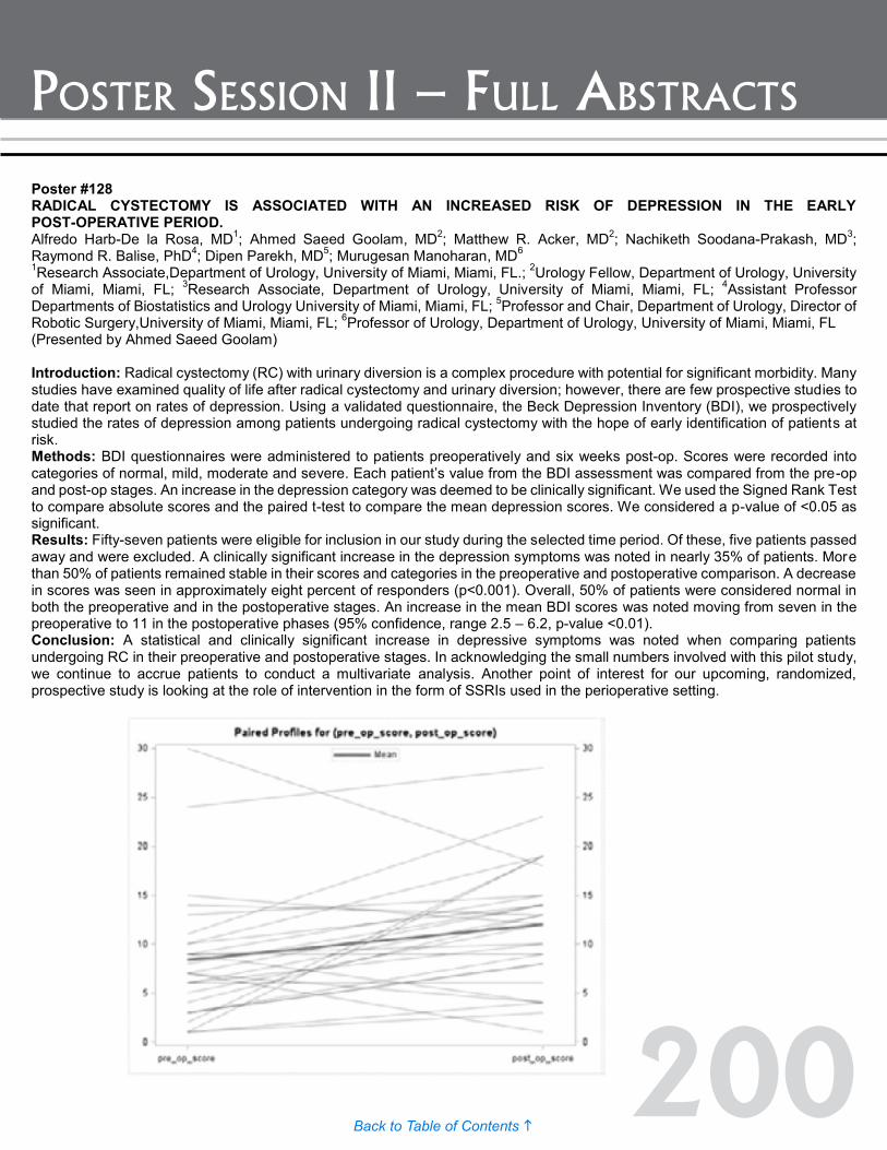
200Back to Table of Contents h
Poster session ii – Full AbstrActs
Poster #128 RADICAL CYSTECTOMY IS ASSOCIATED WITH AN INCREASED RISK OF DEPRESSION IN THE EARLY POST-OPERATIVE PERIOD. Alfredo Harb-De la Rosa, MD1; Ahmed Saeed Goolam, MD2; Matthew R. Acker, MD2; Nachiketh Soodana-Prakash, MD3; Raymond R. Balise, PhD4; Dipen Parekh, MD5; Murugesan Manoharan, MD6 1Research Associate,Department of Urology, University of Miami, Miami, FL.; 2Urology Fellow, Department of Urology, University of Miami, Miami, FL; 3Research Associate, Department of Urology, University of Miami, Miami, FL; 4Assistant Professor Departments of Biostatistics and Urology University of Miami, Miami, FL; 5Professor and Chair, Department of Urology, Director of Robotic Surgery,University of Miami, Miami, FL; 6Professor of Urology, Department of Urology, University of Miami, Miami, FL (Presented by Ahmed Saeed Goolam) Introduction: Radical cystectomy (RC) with urinary diversion is a complex procedure with potential for significant morbidity. Many studies have examined quality of life after radical cystectomy and urinary diversion; however, there are few prospective studies to date that report on rates of depression. Using a validated questionnaire, the Beck Depression Inventory (BDI), we prospectively studied the rates of depression among patients undergoing radical cystectomy with the hope of early identification of patients at risk. Methods: BDI questionnaires were administered to patients preoperatively and six weeks post-op. Scores were recorded into categories of normal, mild, moderate and severe. Each patient’s value from the BDI assessment was compared from the pre-op and post-op stages. An increase in the depression category was deemed to be clinically significant. We used the Signed Rank Test to compare absolute scores and the paired t-test to compare the mean depression scores. We considered a p-value of <0.05 as significant. Results: Fifty-seven patients were eligible for inclusion in our study during the selected time period. Of these, five patients passed away and were excluded. A clinically significant increase in the depression symptoms was noted in nearly 35% of patients. More than 50% of patients remained stable in their scores and categories in the preoperative and postoperative comparison. A decrease in scores was seen in approximately eight percent of responders (p<0.001). Overall, 50% of patients were considered normal in both the preoperative and in the postoperative stages. An increase in the mean BDI scores was noted moving from seven in the preoperative to 11 in the postoperative phases (95% confidence, range 2.5 – 6.2, p-value <0.01). Conclusion: A statistical and clinically significant increase in depressive symptoms was noted when comparing patients undergoing RC in their preoperative and postoperative stages. In acknowledging the small numbers involved with this pilot study, we continue to accrue patients to conduct a multivariate analysis. Another point of interest for our upcoming, randomized, prospective study is looking at the role of intervention in the form of SSRIs used in the perioperative setting.
201Back to Table of Contents h
Poster session ii – Full AbstrActs
Poster #129 ASSESSMENT OF CELL-CYCLE MARKERS IN IMPROVING DISCRIMINATION OF EORTC AND CUETO RISK MODELS IN PREDICTING RECURRENCE AND PROGRESSION OF NON-MUSCLE INVASIVE HIGH-RISK BLADDER CANCER Niccolo Maria Passoni, MD1; Bishoy Gayed, MD2; Payal Kapur, MD1; Arthur Sagalowsky, MD1; Shahrokh Shariat, MD3; Yair Lotan, MD1 1UT Southwestern Medical Center, Dallas, TX; 2Chesapeake Urology, Baltimore, MD; 3Medical University of Vienna, Vienna, Austria (Presented by Niccolo Maria Passoni) Introduction: To assess if a panel of cell-cycle markers could improve the discrimination of EORTC and CUETO models in predicting recurrence and progression of high grade non muscle invasive bladder cancer (NMIBC). Methods: Between January 2007 and January 2012, every patient with high-grade NMIBC treated with trans-urethral resection of bladder (TURB) underwent immunohistochemical staining for tumor protein p53 (Tp53); cyclin dependent kinase inhibitor 1A (p21, Cip1) (CDKN1A); cyclin-dependent kinase inhibitor 1B (p27, Kip1); antigen identified by monoclonal antibody Ki-67 (MKI67); and cyclin E1. We excluded patients who underwent immediate cystectomy and those who were lost to f/u (n=21). Kaplan-Meier curves assessed recurrence and progression-free survival. Univariate and multivariate Cox regression analysis assessed the predictive ability of markers after correcting for EORTC or CUETO risk scores. Harrel’s concordance index assessed for discrimination. No funding was obtained for this study. Results: There were 138 patients with a median follow up 26.2 months. The stage breakdown was Ta (50%), T1 (41%) and CIS (9%). For 103 patients this was the primary tumor with 24 with <1 recurrence/year and 10 with >1 recurrence per year. Intravesical therapy was used in 71% of cases of which 45% had maintenance. The EORTC recurrence score was 1-4, 5-9 and 10-17 in 22%, 67% and nine percent, respectively. Recurrence-free survival rates at six, 12 and 24 months were 94.4%, 90.2% and 87.2%, respectively, while progression-free survival rate at six, 12 and 24 months were 94.4%, 90.2% and 87.2%, respectively. No differences in survival based on number of altered markers were noted. Biomarker status was not a significant predictor of recurrence or progression at Cox regression analysis. Marker alterations marginally improved discrimination of EORTC and CUETO models, which was confirmed to be mediocre. Conclusion: Biomarkers were not significant predictors of recurrence nor progression in patients with high-grade non-muscle invasive bladder cancer and their addition to prediction models is of little benefit.
Poster #128 RADICAL CYSTECTOMY IS ASSOCIATED WITH AN INCREASED RISK OF DEPRESSION IN THE EARLY POST-OPERATIVE PERIOD. Alfredo Harb-De la Rosa, MD1; Ahmed Saeed Goolam, MD2; Matthew R. Acker, MD2; Nachiketh Soodana-Prakash, MD3; Raymond R. Balise, PhD4; Dipen Parekh, MD5; Murugesan Manoharan, MD6 1Research Associate,Department of Urology, University of Miami, Miami, FL.; 2Urology Fellow, Department of Urology, University of Miami, Miami, FL; 3Research Associate, Department of Urology, University of Miami, Miami, FL; 4Assistant Professor Departments of Biostatistics and Urology University of Miami, Miami, FL; 5Professor and Chair, Department of Urology, Director of Robotic Surgery,University of Miami, Miami, FL; 6Professor of Urology, Department of Urology, University of Miami, Miami, FL (Presented by Ahmed Saeed Goolam) Introduction: Radical cystectomy (RC) with urinary diversion is a complex procedure with potential for significant morbidity. Many studies have examined quality of life after radical cystectomy and urinary diversion; however, there are few prospective studies to date that report on rates of depression. Using a validated questionnaire, the Beck Depression Inventory (BDI), we prospectively studied the rates of depression among patients undergoing radical cystectomy with the hope of early identification of patients at risk. Methods: BDI questionnaires were administered to patients preoperatively and six weeks post-op. Scores were recorded into categories of normal, mild, moderate and severe. Each patient’s value from the BDI assessment was compared from the pre-op and post-op stages. An increase in the depression category was deemed to be clinically significant. We used the Signed Rank Test to compare absolute scores and the paired t-test to compare the mean depression scores. We considered a p-value of <0.05 as significant. Results: Fifty-seven patients were eligible for inclusion in our study during the selected time period. Of these, five patients passed away and were excluded. A clinically significant increase in the depression symptoms was noted in nearly 35% of patients. More than 50% of patients remained stable in their scores and categories in the preoperative and postoperative comparison. A decrease in scores was seen in approximately eight percent of responders (p<0.001). Overall, 50% of patients were considered normal in both the preoperative and in the postoperative stages. An increase in the mean BDI scores was noted moving from seven in the preoperative to 11 in the postoperative phases (95% confidence, range 2.5 – 6.2, p-value <0.01). Conclusion: A statistical and clinically significant increase in depressive symptoms was noted when comparing patients undergoing RC in their preoperative and postoperative stages. In acknowledging the small numbers involved with this pilot study, we continue to accrue patients to conduct a multivariate analysis. Another point of interest for our upcoming, randomized, prospective study is looking at the role of intervention in the form of SSRIs used in the perioperative setting.
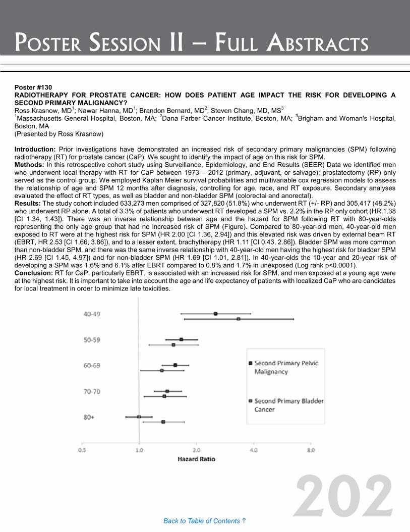
202Back to Table of Contents h
Poster session ii – Full AbstrActs
Poster #130 RADIOTHERAPY FOR PROSTATE CANCER: HOW DOES PATIENT AGE IMPACT THE RISK FOR DEVELOPING A SECOND PRIMARY MALIGNANCY? Ross Krasnow, MD1; Nawar Hanna, MD1; Brandon Bernard, MD2; Steven Chang, MD, MS3 1Massachusetts General Hospital, Boston, MA; 2Dana Farber Cancer Institute, Boston, MA; 3Brigham and Woman's Hospital, Boston, MA (Presented by Ross Krasnow) Introduction: Prior investigations have demonstrated an increased risk of secondary primary malignancies (SPM) following radiotherapy (RT) for prostate cancer (CaP). We sought to identify the impact of age on this risk for SPM. Methods: In this retrospective cohort study using Surveillance, Epidemiology, and End Results (SEER) Data we identified men who underwent local therapy with RT for CaP between 1973 – 2012 (primary, adjuvant, or salvage); prostatectomy (RP) only served as the control group. We employed Kaplan Meier survival probabilities and multivariable cox regression models to assess the relationship of age and SPM 12 months after diagnosis, controlling for age, race, and RT exposure. Secondary analyses evaluated the effect of RT types, as well as bladder and non-bladder SPM (colorectal and anorectal). Results: The study cohort included 633,273 men comprised of 327,820 (51.8%) who underwent RT (+/- RP) and 305,417 (48.2%) who underwent RP alone. A total of 3.3% of patients who underwent RT developed a SPM vs. 2.2% in the RP only cohort (HR 1.38 [CI 1.34, 1.43]). There was an inverse relationship between age and the hazard for SPM following RT with 80-year-olds representing the only age group that had no increased risk of SPM (Figure). Compared to 80-year-old men, 40-year-old men exposed to RT were at the highest risk for SPM (HR 2.00 [CI 1.36, 2.94]) and this elevated risk was driven by external beam RT (EBRT, HR 2.53 [CI 1.66, 3.86]), and to a lesser extent, brachytherapy (HR 1.11 [CI 0.43, 2.86]). Bladder SPM was more common than non-bladder SPM, and there was the same inverse relationship with 40-year-old men having the highest risk for bladder SPM (HR 2.69 [CI 1.45, 4.97]) and for non-bladder SPM (HR 1.69 [CI 1.01, 2.81]). In 40-year-olds the 10-year and 20-year risk of developing a SPM was 1.6% and 6.1% after EBRT compared to 0.8% and 1.7% in unexposed (Log rank p<0.0001). Conclusion: RT for CaP, particularly EBRT, is associated with an increased risk for SPM, and men exposed at a young age were at the highest risk. It is important to take into account the age and life expectancy of patients with localized CaP who are candidates for local treatment in order to minimize late toxicities.
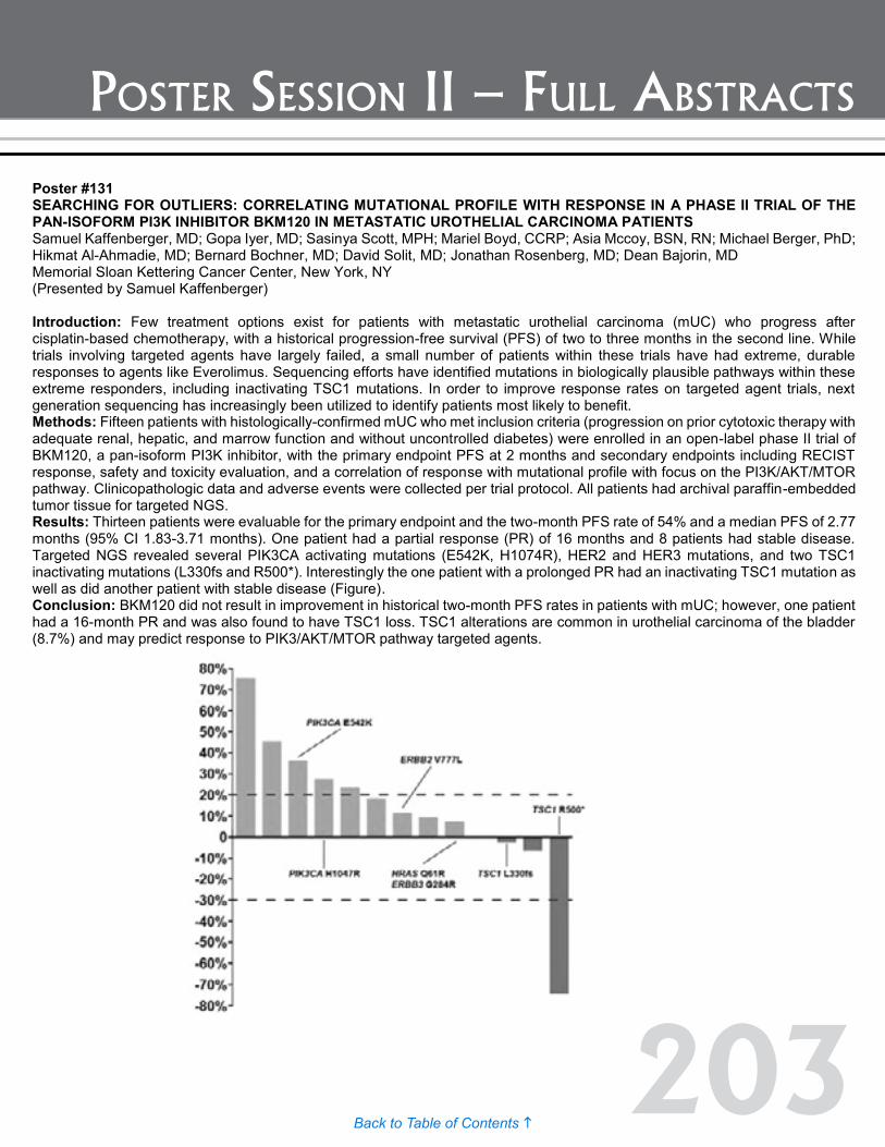
203Back to Table of Contents h
Poster session ii – Full AbstrActs
Poster #131 SEARCHING FOR OUTLIERS: CORRELATING MUTATIONAL PROFILE WITH RESPONSE IN A PHASE II TRIAL OF THE PAN-ISOFORM PI3K INHIBITOR BKM120 IN METASTATIC UROTHELIAL CARCINOMA PATIENTS Samuel Kaffenberger, MD; Gopa Iyer, MD; Sasinya Scott, MPH; Mariel Boyd, CCRP; Asia Mccoy, BSN, RN; Michael Berger, PhD; Hikmat Al-Ahmadie, MD; Bernard Bochner, MD; David Solit, MD; Jonathan Rosenberg, MD; Dean Bajorin, MD Memorial Sloan Kettering Cancer Center, New York, NY (Presented by Samuel Kaffenberger) Introduction: Few treatment options exist for patients with metastatic urothelial carcinoma (mUC) who progress after cisplatin-based chemotherapy, with a historical progression-free survival (PFS) of two to three months in the second line. While trials involving targeted agents have largely failed, a small number of patients within these trials have had extreme, durable responses to agents like Everolimus. Sequencing efforts have identified mutations in biologically plausible pathways within these extreme responders, including inactivating TSC1 mutations. In order to improve response rates on targeted agent trials, next generation sequencing has increasingly been utilized to identify patients most likely to benefit. Methods: Fifteen patients with histologically-confirmed mUC who met inclusion criteria (progression on prior cytotoxic therapy with adequate renal, hepatic, and marrow function and without uncontrolled diabetes) were enrolled in an open-label phase II trial of BKM120, a pan-isoform PI3K inhibitor, with the primary endpoint PFS at 2 months and secondary endpoints including RECIST response, safety and toxicity evaluation, and a correlation of response with mutational profile with focus on the PI3K/AKT/MTOR pathway. Clinicopathologic data and adverse events were collected per trial protocol. All patients had archival paraffin-embedded tumor tissue for targeted NGS. Results: Thirteen patients were evaluable for the primary endpoint and the two-month PFS rate of 54% and a median PFS of 2.77 months (95% CI 1.83-3.71 months). One patient had a partial response (PR) of 16 months and 8 patients had stable disease. Targeted NGS revealed several PIK3CA activating mutations (E542K, H1074R), HER2 and HER3 mutations, and two TSC1 inactivating mutations (L330fs and R500*). Interestingly the one patient with a prolonged PR had an inactivating TSC1 mutation as well as did another patient with stable disease (Figure). Conclusion: BKM120 did not result in improvement in historical two-month PFS rates in patients with mUC; however, one patient had a 16-month PR and was also found to have TSC1 loss. TSC1 alterations are common in urothelial carcinoma of the bladder (8.7%) and may predict response to PIK3/AKT/MTOR pathway targeted agents.
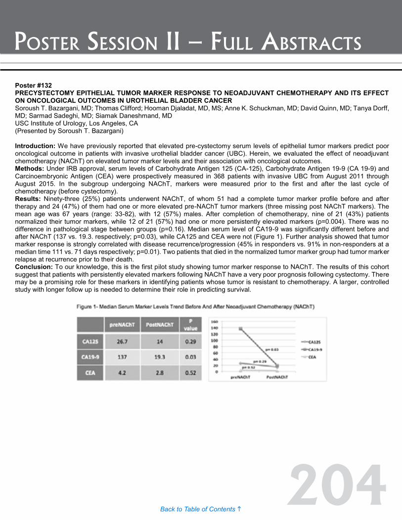
204Back to Table of Contents h
Poster session ii – Full AbstrActs
Poster #133 BLUE LIGHT CYSTOSCOPY FOR DIAGNOSIS OF UROTHELIAL BLADDER CANCER: Results: FROM A PROSPECTIVE REGISTRY. Soroush T. Bazargani, MD; Swar H. Shah, MD; Hooman Djaladat, MD, MS; Anne K. Schuckman, MD; Siamak Daneshmand, MD USC Institute of Urology, Los Angeles, CA (Presented by Soroush T. Bazargani) Introduction: Studies have shown Blue Light Cystoscopy (BLC) using hexaminolevulinate (Cysview) can improve the detection of non-muscle invasive bladder cancer (NMIBC) compared to white light cystoscopy (WLC) alone. We report on our experience from the prospective Blue Light Cystoscopy with Cysview® Registry. Methods: Under IRB approval, we prospectively enrolled consecutive patients undergoing transurethral resection of bladder lesions into the registry. Patients received Cysview® one hour prior to surgery, where intraoperative findings with white light (WL) and blue light (BL), lesion characteristics (flat or papillary), location and size were recorded. Patients who refused catheter insertion (three), had pure upper tract or prostatic urethral lesions (four), or were lost to follow up (seven) were excluded from the study. Results: A total of 320 separate lesions were identified from 112 patients between April 2014 and May 2015. Mean age was 70 with 81% being male. There were 193 (60%) WL positive, and 253 (79%) BL positive. Using final pathology as the reference standard, the sensitivity of WL, BL and the combination for any malignant lesion was 74%, 90% and 99% respectively. The addition of BL to standard WL cystoscopy increased our detection rate in papillary lesions from 89% to 99% (Table 1). This detection improvement was nine percent for low-grade lesions, 16% for high-grade ones and 42% for CIS (from 57% to 99%). Within the WL negative group, an additional 46 lesions were detected solely with the addition of BL (sensitivity 48%). Within the WL negative and BL positive group (94), 48 (51%) were benign (false positive). Forty-one (36%) patients received BCG at least six weeks prior to BLC, with comparable sensitivity (90%) specificity (50%) for malignant lesions. There were no complications attributable to Cysview instillation. Twenty-two (seven percent) patients eventually had a cystectomy, all BL positive tumors. Conclusion: Our experience with a prospective registry confirms the advantages of BLC using Cysview. BLC significantly increases detection rates of CIS and high grade lesions as well as low grade papillary lesions compared to WL cystoscopy alone. Prior BCG therapy appears to have no effect on BLC accuracy. Funding: Photocure Inc.
Poster #132 PRECYSTECTOMY EPITHELIAL TUMOR MARKER RESPONSE TO NEOADJUVANT CHEMOTHERAPY AND ITS EFFECT ON ONCOLOGICAL OUTCOMES IN UROTHELIAL BLADDER CANCER Soroush T. Bazargani, MD; Thomas Clifford; Hooman Djaladat, MD, MS; Anne K. Schuckman, MD; David Quinn, MD; Tanya Dorff, MD; Sarmad Sadeghi, MD; Siamak Daneshmand, MD USC Institute of Urology, Los Angeles, CA (Presented by Soroush T. Bazargani) Introduction: We have previously reported that elevated pre-cystectomy serum levels of epithelial tumor markers predict poor oncological outcome in patients with invasive urothelial bladder cancer (UBC). Herein, we evaluated the effect of neoadjuvant chemotherapy (NAChT) on elevated tumor marker levels and their association with oncological outcomes. Methods: Under IRB approval, serum levels of Carbohydrate Antigen 125 (CA-125), Carbohydrate Antigen 19-9 (CA 19-9) and Carcinoembryonic Antigen (CEA) were prospectively measured in 368 patients with invasive UBC from August 2011 through August 2015. In the subgroup undergoing NAChT, markers were measured prior to the first and after the last cycle of chemotherapy (before cystectomy). Results: Ninety-three (25%) patients underwent NAChT, of whom 51 had a complete tumor marker profile before and after therapy and 24 (47%) of them had one or more elevated pre-NAChT tumor markers (three missing post NAChT markers). The mean age was 67 years (range: 33-82), with 12 (57%) males. After completion of chemotherapy, nine of 21 (43%) patients normalized their tumor markers, while 12 of 21 (57%) had one or more persistently elevated markers (p=0.004). There was no difference in pathological stage between groups (p=0.16). Median serum level of CA19-9 was significantly different before and after NAChT (137 vs. 19.3. respectively; p=0.03), while CA125 and CEA were not (Figure 1). Further analysis showed that tumor marker response is strongly correlated with disease recurrence/progression (45% in responders vs. 91% in non-responders at a median time 111 vs. 71 days respectively; p=0.01). Two patients that died in the normalized tumor marker group had tumor marker relapse at recurrence prior to their death. Conclusion: To our knowledge, this is the first pilot study showing tumor marker response to NAChT. The results of this cohort suggest that patients with persistently elevated markers following NAChT have a very poor prognosis following cystectomy. There may be a promising role for these markers in identifying patients whose tumor is resistant to chemotherapy. A larger, controlled study with longer follow up is needed to determine their role in predicting survival.
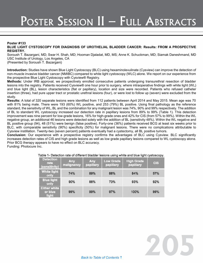
205Back to Table of Contents h
Poster session ii – Full AbstrActs
Poster #133 BLUE LIGHT CYSTOSCOPY FOR DIAGNOSIS OF UROTHELIAL BLADDER CANCER: Results: FROM A PROSPECTIVE REGISTRY. Soroush T. Bazargani, MD; Swar H. Shah, MD; Hooman Djaladat, MD, MS; Anne K. Schuckman, MD; Siamak Daneshmand, MD USC Institute of Urology, Los Angeles, CA (Presented by Soroush T. Bazargani) Introduction: Studies have shown Blue Light Cystoscopy (BLC) using hexaminolevulinate (Cysview) can improve the detection of non-muscle invasive bladder cancer (NMIBC) compared to white light cystoscopy (WLC) alone. We report on our experience from the prospective Blue Light Cystoscopy with Cysview® Registry. Methods: Under IRB approval, we prospectively enrolled consecutive patients undergoing transurethral resection of bladder lesions into the registry. Patients received Cysview® one hour prior to surgery, where intraoperative findings with white light (WL) and blue light (BL), lesion characteristics (flat or papillary), location and size were recorded. Patients who refused catheter insertion (three), had pure upper tract or prostatic urethral lesions (four), or were lost to follow up (seven) were excluded from the study. Results: A total of 320 separate lesions were identified from 112 patients between April 2014 and May 2015. Mean age was 70 with 81% being male. There were 193 (60%) WL positive, and 253 (79%) BL positive. Using final pathology as the reference standard, the sensitivity of WL, BL and the combination for any malignant lesion was 74%, 90% and 99% respectively. The addition of BL to standard WL cystoscopy increased our detection rate in papillary lesions from 89% to 99% (Table 1). This detection improvement was nine percent for low-grade lesions, 16% for high-grade ones and 42% for CIS (from 57% to 99%). Within the WL negative group, an additional 46 lesions were detected solely with the addition of BL (sensitivity 48%). Within the WL negative and BL positive group (94), 48 (51%) were benign (false positive). Forty-one (36%) patients received BCG at least six weeks prior to BLC, with comparable sensitivity (90%) specificity (50%) for malignant lesions. There were no complications attributable to Cysview instillation. Twenty-two (seven percent) patients eventually had a cystectomy, all BL positive tumors. Conclusion: Our experience with a prospective registry confirms the advantages of BLC using Cysview. BLC significantly increases detection rates of CIS and high grade lesions as well as low grade papillary lesions compared to WL cystoscopy alone. Prior BCG therapy appears to have no effect on BLC accuracy. Funding: Photocure Inc.
Poster #132 PRECYSTECTOMY EPITHELIAL TUMOR MARKER RESPONSE TO NEOADJUVANT CHEMOTHERAPY AND ITS EFFECT ON ONCOLOGICAL OUTCOMES IN UROTHELIAL BLADDER CANCER Soroush T. Bazargani, MD; Thomas Clifford; Hooman Djaladat, MD, MS; Anne K. Schuckman, MD; David Quinn, MD; Tanya Dorff, MD; Sarmad Sadeghi, MD; Siamak Daneshmand, MD USC Institute of Urology, Los Angeles, CA (Presented by Soroush T. Bazargani) Introduction: We have previously reported that elevated pre-cystectomy serum levels of epithelial tumor markers predict poor oncological outcome in patients with invasive urothelial bladder cancer (UBC). Herein, we evaluated the effect of neoadjuvant chemotherapy (NAChT) on elevated tumor marker levels and their association with oncological outcomes. Methods: Under IRB approval, serum levels of Carbohydrate Antigen 125 (CA-125), Carbohydrate Antigen 19-9 (CA 19-9) and Carcinoembryonic Antigen (CEA) were prospectively measured in 368 patients with invasive UBC from August 2011 through August 2015. In the subgroup undergoing NAChT, markers were measured prior to the first and after the last cycle of chemotherapy (before cystectomy). Results: Ninety-three (25%) patients underwent NAChT, of whom 51 had a complete tumor marker profile before and after therapy and 24 (47%) of them had one or more elevated pre-NAChT tumor markers (three missing post NAChT markers). The mean age was 67 years (range: 33-82), with 12 (57%) males. After completion of chemotherapy, nine of 21 (43%) patients normalized their tumor markers, while 12 of 21 (57%) had one or more persistently elevated markers (p=0.004). There was no difference in pathological stage between groups (p=0.16). Median serum level of CA19-9 was significantly different before and after NAChT (137 vs. 19.3. respectively; p=0.03), while CA125 and CEA were not (Figure 1). Further analysis showed that tumor marker response is strongly correlated with disease recurrence/progression (45% in responders vs. 91% in non-responders at a median time 111 vs. 71 days respectively; p=0.01). Two patients that died in the normalized tumor marker group had tumor marker relapse at recurrence prior to their death. Conclusion: To our knowledge, this is the first pilot study showing tumor marker response to NAChT. The results of this cohort suggest that patients with persistently elevated markers following NAChT have a very poor prognosis following cystectomy. There may be a promising role for these markers in identifying patients whose tumor is resistant to chemotherapy. A larger, controlled study with longer follow up is needed to determine their role in predicting survival.
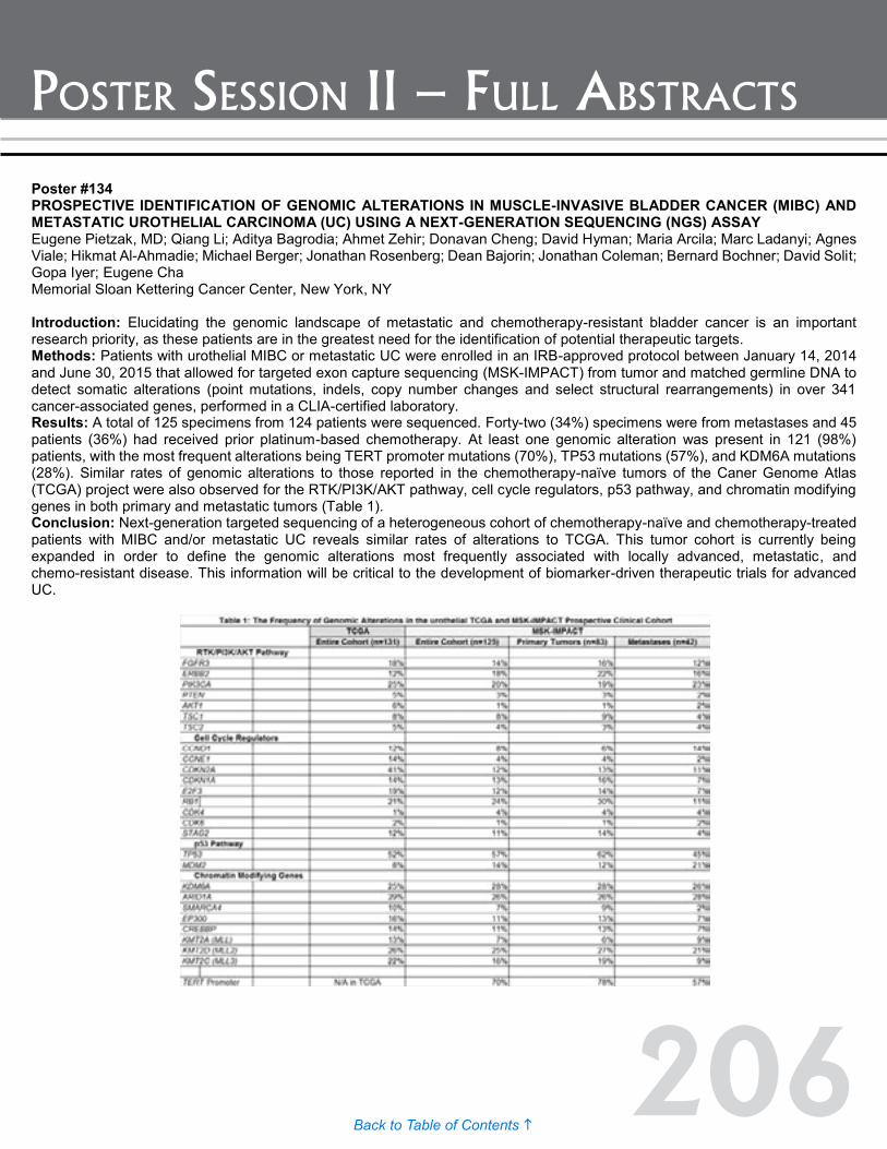
206Back to Table of Contents h
Poster session ii – Full AbstrActs
Poster #134 PROSPECTIVE IDENTIFICATION OF GENOMIC ALTERATIONS IN MUSCLE-INVASIVE BLADDER CANCER (MIBC) AND METASTATIC UROTHELIAL CARCINOMA (UC) USING A NEXT-GENERATION SEQUENCING (NGS) ASSAY Eugene Pietzak, MD; Qiang Li; Aditya Bagrodia; Ahmet Zehir; Donavan Cheng; David Hyman; Maria Arcila; Marc Ladanyi; Agnes Viale; Hikmat Al-Ahmadie; Michael Berger; Jonathan Rosenberg; Dean Bajorin; Jonathan Coleman; Bernard Bochner; David Solit; Gopa Iyer; Eugene Cha Memorial Sloan Kettering Cancer Center, New York, NY Introduction: Elucidating the genomic landscape of metastatic and chemotherapy-resistant bladder cancer is an important research priority, as these patients are in the greatest need for the identification of potential therapeutic targets. Methods: Patients with urothelial MIBC or metastatic UC were enrolled in an IRB-approved protocol between January 14, 2014 and June 30, 2015 that allowed for targeted exon capture sequencing (MSK-IMPACT) from tumor and matched germline DNA to detect somatic alterations (point mutations, indels, copy number changes and select structural rearrangements) in over 341 cancer-associated genes, performed in a CLIA-certified laboratory. Results: A total of 125 specimens from 124 patients were sequenced. Forty-two (34%) specimens were from metastases and 45 patients (36%) had received prior platinum-based chemotherapy. At least one genomic alteration was present in 121 (98%) patients, with the most frequent alterations being TERT promoter mutations (70%), TP53 mutations (57%), and KDM6A mutations (28%). Similar rates of genomic alterations to those reported in the chemotherapy-naïve tumors of the Caner Genome Atlas (TCGA) project were also observed for the RTK/PI3K/AKT pathway, cell cycle regulators, p53 pathway, and chromatin modifying genes in both primary and metastatic tumors (Table 1). Conclusion: Next-generation targeted sequencing of a heterogeneous cohort of chemotherapy-naïve and chemotherapy-treated patients with MIBC and/or metastatic UC reveals similar rates of alterations to TCGA. This tumor cohort is currently being expanded in order to define the genomic alterations most frequently associated with locally advanced, metastatic, and chemo-resistant disease. This information will be critical to the development of biomarker-driven therapeutic trials for advanced UC.
207Back to Table of Contents h
Poster session ii – Full AbstrActs
Poster #135 COMPARING SURVIVAL TRENDS AFTER RADICAL CYSTECTOMY AND BLADDER PRESERVATION THERAPY IN PATIENTS WITH MUSCLE INVASIVE BLADDER CANCER David Cahn, DO, MBS1; Elizabeth Handorf, PhD2; Michael Nordsiek, DO1; Thomas Churilla, MD3; Eric Horwitz, MD3; Benjamin Ristau, MD4; David Chen, MD, FACS4; Rosalia Viterbo, MD, FACS4; Richard Greenberg, MD, FACS4; Alexander Kutikov, MD, FACS4; Robert Uzzo, MD, FACS5; Marc Smaldone, MD, MSHP4 1Einstein Healthcare Network, Department of Urology, Philadelphia, PA; 2Fox Chase Cancer Center, Department of Biostatistics and Bioinformatics, Philadelphia, PA; 3Fox Chase Cancer Center, Department of Radiation Oncology, Philadelphia, PA; 4Fox Chase Cancer Center, Department of Urologic Oncology, Philadelphia, PA; 5Fox Chase Cancer Center and Einstein Healthcare Network, Departments of Urologic Oncology and Urology, Philadelphia, PA (Presented by David Cahn) Introduction: To compare overall survival (OS) in patients undergoing radical cystectomy (RC) and bladder preservation therapy (BPT) for muscle invasive urothelial carcinoma of the bladder. Methods: We conducted a retrospective, observational cohort study in which we reviewed the National Cancer Database (NCDB) to identify patients with analytic stage II-III (N0M0) urothelial carcinoma of the bladder from 2003 to 2011. BPT patients were stratified as any external beam radiotherapy (EBRT), definitive radiotherapy (RT) [50-80Gy], and definitive RT + chemotherapy. Treatment trends were evaluated using Pearson Chi-square tests. OS was compared between RC and BPT using unadjusted Kaplan Meier curves and Cox regression models adjusted for year of treatment, hospital volume, and patient/tumor characteristics using increasingly stringent selection criteria to identify those undergoing BPT. Results: Of the 603,298 patients with bladder cancer captured in the NCDB from 2003 to 2011, 9% (n=54,518) had analytic stage II-III with urothelial histology. Of these patients, 51.1% (n=27,843) were treated with RC (70.9%, n=19,745) or BPT (29.1%, n= 8,098). Of the patients undergoing BPT, stratified by selection criteria, 26.9% (n=2,176) and 15.0% (n=1,215) were treated with definitive RT and definitive RT + chemotherapy, respectively. Following adjustment, while improved survival in patients undergoing RC was noted regardless of BPT definition employed, we noted attenuated differences in OS using increasingly stringent definitions for BPT (EBRT: HR 2.2 [CI 2.15-2.29]; definitive RT: HR 1.94 [CI 1.74-2.14]; definitive RT + chemotherapy: HR 1.56 [CI 1.45-1.68]). Conclusion: In the NCDB, receipt of BPT was associated with worse OS compared to RC in all patients with stage II-III urothelial carcinoma, in part due to selection biases. However, the use of increasingly stringent definitions of BPT attenuated the observed survival differences. Further randomized prospective controlled trials are needed to compare trimodal BPT to RC to identify optimal candidates for bladder preservation.
208Back to Table of Contents h
Poster session ii – Full AbstrActs
Poster #136 BLUE-LIGHT CYSTOSCOPY'S EFFECTS ON MANAGEMENT OF BLADDER CANCER WHEN COMPARED TO TRADITIONAL WHITE-LIGHT CYSTOSCOPY Andrew Mount, BS1; Stephen Williams, MD2; Colin Dinney, MD2; H. Barton Grossman, MD2; Curtis Pettaway, MD2; Brian Chapin, MD2; Neema Navai, MD2; Surena Matin, MD2; William Graber, MD2; Ashish Kamat, MD2 1UT-Houston Medical School Houston, TX; 2MD Anderson Cancer Center Houston, TX (Presented by Andrew Mount) Introduction: Fluorescence cystoscopy (also known as blue-light cystoscopy [BL]) is an adjunct to white light cystoscopy (WL) and aids in visualization of tumors that may be missed with WL as well as allow more complete resection of tumors. Herein we present the impact on subsequent patient management in real world setting. Our objective is to determine if BL affected subsequent management of patients diagnosed with bladder cancer. Methods: A total of 116 consecutive patients who underwent simultaneous BL and WL from January 2013 through December 2014 were included in the study. Pathology and operative reports were reviewed to determine the grade and stage of the tumors and whether they were viewed under BL or WL. Results: Of the 116 patients, a total of 161 biopsies and/or transurethral resected specimens were analyzed. Of these, 46 (28.6%) lesions were seen only with BL, none were seen only with white light, 109 (67.7%) were seen with both, and seven (4.4%) were identified via random biopsies. Of the 46 lesions seen only on BL, 17 (37%) were positive for cancer, while, of the 109 lesions seen with WL and BL, 84 (77%) were positive. Of the 17 (37%) true positive tumors seen only on BL, the stage and grades were: five (29.4%) low-grade Ta, two (11.8%) high-grade Ta, one (5.9%) high-grade T1, and nine (52.9%) CIS. There were no instances of a tumor being found on WL that was not also visualized with BL. In this patient cohort, the false positive rates were 23% and 63% for WL and BL, respectively. The false negative rates were 0.9% and 2.1% for WL and BL, respectively. For the 46 (28.6%) lesions that were visible by BL, WL cystoscopy was not able to visualize 10 (21.7%) tumors visualized by BL. In addition, there was one patient who had multiple lesions sent for pathology by BL and WL, and the lesions identified by BL were of higher stage than the lesions visualized by WL. Conclusion: BL identified additional tumors that would have been missed with WL. Moreover, in patients who had tumors visualized only with BL, 11 (26.8%) were high-grade, including one (2.4%) patient with T1 tumor and eight (19.5%) with CIS. Thus, BL identified a number of high-risk tumors, which had a significant impact on the subsequent management of patients with bladder cancer.
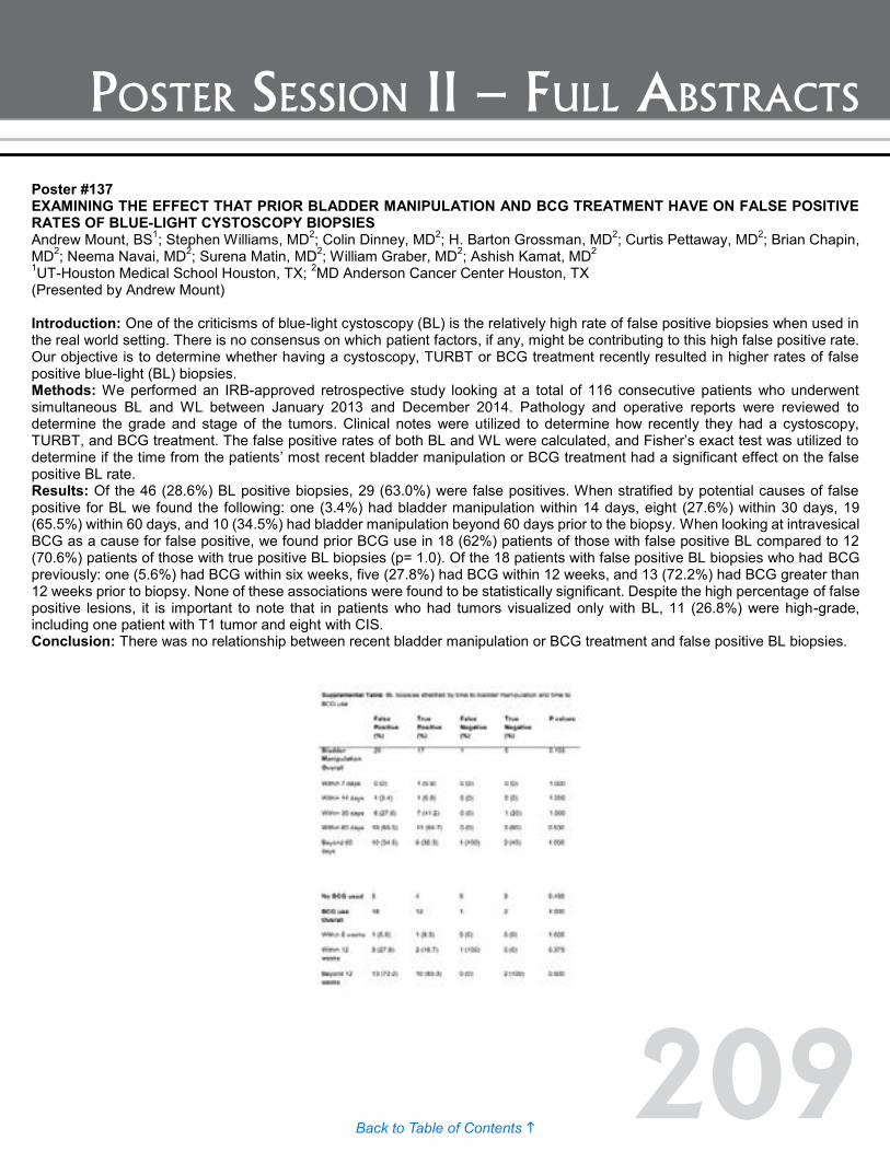
209Back to Table of Contents h
Poster session ii – Full AbstrActs
Poster #137 EXAMINING THE EFFECT THAT PRIOR BLADDER MANIPULATION AND BCG TREATMENT HAVE ON FALSE POSITIVE RATES OF BLUE-LIGHT CYSTOSCOPY BIOPSIES Andrew Mount, BS1; Stephen Williams, MD2; Colin Dinney, MD2; H. Barton Grossman, MD2; Curtis Pettaway, MD2; Brian Chapin, MD2; Neema Navai, MD2; Surena Matin, MD2; William Graber, MD2; Ashish Kamat, MD2 1UT-Houston Medical School Houston, TX; 2MD Anderson Cancer Center Houston, TX (Presented by Andrew Mount) Introduction: One of the criticisms of blue-light cystoscopy (BL) is the relatively high rate of false positive biopsies when used in the real world setting. There is no consensus on which patient factors, if any, might be contributing to this high false positive rate. Our objective is to determine whether having a cystoscopy, TURBT or BCG treatment recently resulted in higher rates of false positive blue-light (BL) biopsies. Methods: We performed an IRB-approved retrospective study looking at a total of 116 consecutive patients who underwent simultaneous BL and WL between January 2013 and December 2014. Pathology and operative reports were reviewed to determine the grade and stage of the tumors. Clinical notes were utilized to determine how recently they had a cystoscopy, TURBT, and BCG treatment. The false positive rates of both BL and WL were calculated, and Fisher’s exact test was utilized to determine if the time from the patients’ most recent bladder manipulation or BCG treatment had a significant effect on the false positive BL rate. Results: Of the 46 (28.6%) BL positive biopsies, 29 (63.0%) were false positives. When stratified by potential causes of false positive for BL we found the following: one (3.4%) had bladder manipulation within 14 days, eight (27.6%) within 30 days, 19 (65.5%) within 60 days, and 10 (34.5%) had bladder manipulation beyond 60 days prior to the biopsy. When looking at intravesical BCG as a cause for false positive, we found prior BCG use in 18 (62%) patients of those with false positive BL compared to 12 (70.6%) patients of those with true positive BL biopsies (p= 1.0). Of the 18 patients with false positive BL biopsies who had BCG previously: one (5.6%) had BCG within six weeks, five (27.8%) had BCG within 12 weeks, and 13 (72.2%) had BCG greater than 12 weeks prior to biopsy. None of these associations were found to be statistically significant. Despite the high percentage of false positive lesions, it is important to note that in patients who had tumors visualized only with BL, 11 (26.8%) were high-grade, including one patient with T1 tumor and eight with CIS. Conclusion: There was no relationship between recent bladder manipulation or BCG treatment and false positive BL biopsies.
210Back to Table of Contents h
Poster session ii – Full AbstrActs
Poster #138 USING SERUM ANGIOGENESIS MARKERS TO ASSESS TUMOR RESPONSE TO INTRAVESICAL BACILLUS CALMETTE-GUERIN (BCG) FOLLOWED BY SUNITINIB FOR HIGH-RISK NON-MUSCLE INVASIVE BLADDER CANCER Alexander M. Helfand, BA1; Cheryl T. Lee, MD1; Khaled S. Hafez, MD1; Maha H. Hussain, MD2; Monica Liebert, PhD1; Stephanie Daignault, MS3; Jeffrey S. Montgomery, MD, MHSA1; David C. Miller, MD, MPH1; Linda Drnek, BS1; Brent K. Hollenbeck, MD, MS1; Alon Z. Weizer, MD, MS1 1Department of Urology, University of Michigan, Ann Arbor, MI; 2Department of Medicine, University of Michigan, Ann Arbor, MI; 3Department of Biostatistics, University of Michigan, Ann Arbor, MI (Presented by Alexander M. Helfand) Introduction: We measured serum angiogenesis markers in patients participating in a phase II trial (NCT00794950) which demonstrated significant improvement in three-month complete response (3m CR) and two-year recurrence and progression-free survival in patients receiving combination therapy with intravesical BCG and sunitinib for non-muscle invasive bladder cancer. Studies have shown that the concentration of circulating angiogenesis markers such as soluble VEGF receptors and VEGF ligands may correlate with response to sunitinib treatment. Methods: In the trial, 36 patients with high-grade ≤cT1N0M0 urothelial carcinoma were given induction BCG (six weeks) followed two weeks later by 28 days of sunitinib (50mg). Patients had serum drawn before treatment, and at six weeks (after BCG), eight weeks (before sunitinib), and 12 weeks (after sunitinib). We used the Milliplex MAP Human Angiogenesis/Growth Factor and Human Soluble Cytokine Receptor Magnetic Bead Panels (EMD Millipore, Germany) to perform multiplex Luminex XMAP detection assays of Angiopoietin-2, VEGF-A, VEGF-C, and VEGF-D, IL-8, FGF-2, sVEGF-R1, sVEGF-R2, and sVEGF-R3. Mean analyte values of the 26 patients with a 3m CR to BCG and sunitinib were compared to values from the 10 patients with recurrent disease at three months. The Wilcoxon rank test was used to test for differences between the two groups and p<0.05 was considered significant. Results: Sunitinib treatment induced a mean increase in VEGF-D (+151.8 pg/mL) in the CR group (between weeks eight and 12), whereas the non-CR group experienced a decrease (-6 pg/mL) in VEGF-D (p=0.009). Between weeks one and eight, sVEGF-R2 decreased in the CR group (-129.7 pg/mL) but increased (+1135 pg/mL) in the non-CR group (p=0.019). BCG treatment (week one to week six) also led to a decrease in sVEGF-R3 (-138.7 pg/mL) in the CR group and an increase (+53.7 pg/mL) in the non-CR group (p=0.045). Conclusion: These results suggest that an increase in circulating VEGF-D after sunitinib treatment may signal successful VEGF receptor inhibition in responders. The decreased levels of sVEGF-R2 and sVEGF-R3 in responders after BCG treatment may be evidence that VEGF receptor downregulation is a key component of BCG’s mechanism of action. Understanding the molecular evidence for synergy between BCG and sunitinib in reducing recurrence and identifying causes of resistance to these anti-angiogenic effects in non-responders will require further study. (Study supported by Pfizer and NIDDK grant P30DK020572)
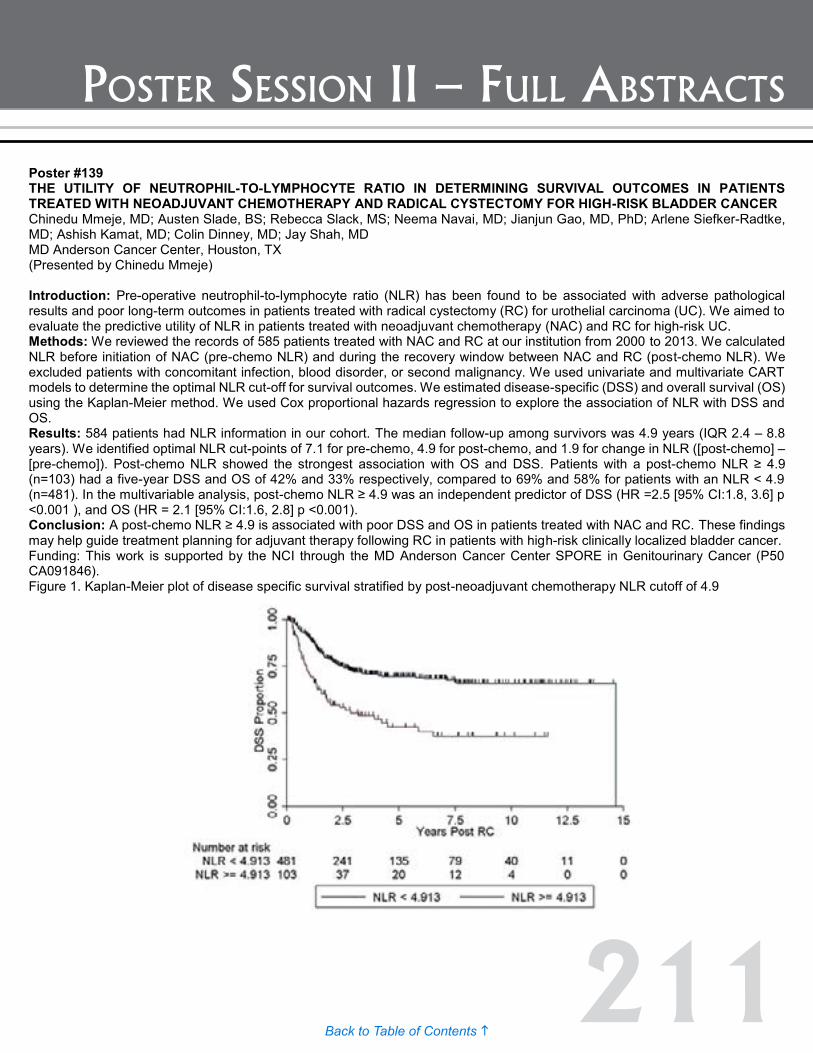
211Back to Table of Contents h
Poster session ii – Full AbstrActs
Poster #139 THE UTILITY OF NEUTROPHIL-TO-LYMPHOCYTE RATIO IN DETERMINING SURVIVAL OUTCOMES IN PATIENTS TREATED WITH NEOADJUVANT CHEMOTHERAPY AND RADICAL CYSTECTOMY FOR HIGH-RISK BLADDER CANCER Chinedu Mmeje, MD; Austen Slade, BS; Rebecca Slack, MS; Neema Navai, MD; Jianjun Gao, MD, PhD; Arlene Siefker-Radtke, MD; Ashish Kamat, MD; Colin Dinney, MD; Jay Shah, MD MD Anderson Cancer Center, Houston, TX (Presented by Chinedu Mmeje) Introduction: Pre-operative neutrophil-to-lymphocyte ratio (NLR) has been found to be associated with adverse pathological results and poor long-term outcomes in patients treated with radical cystectomy (RC) for urothelial carcinoma (UC). We aimed to evaluate the predictive utility of NLR in patients treated with neoadjuvant chemotherapy (NAC) and RC for high-risk UC. Methods: We reviewed the records of 585 patients treated with NAC and RC at our institution from 2000 to 2013. We calculated NLR before initiation of NAC (pre-chemo NLR) and during the recovery window between NAC and RC (post-chemo NLR). We excluded patients with concomitant infection, blood disorder, or second malignancy. We used univariate and multivariate CART models to determine the optimal NLR cut-off for survival outcomes. We estimated disease-specific (DSS) and overall survival (OS) using the Kaplan-Meier method. We used Cox proportional hazards regression to explore the association of NLR with DSS and OS. Results: 584 patients had NLR information in our cohort. The median follow-up among survivors was 4.9 years (IQR 2.4 – 8.8 years). We identified optimal NLR cut-points of 7.1 for pre-chemo, 4.9 for post-chemo, and 1.9 for change in NLR ([post-chemo] – [pre-chemo]). Post-chemo NLR showed the strongest association with OS and DSS. Patients with a post-chemo NLR ≥ 4.9 (n=103) had a five-year DSS and OS of 42% and 33% respectively, compared to 69% and 58% for patients with an NLR < 4.9 (n=481). In the multivariable analysis, post-chemo NLR ≥ 4.9 was an independent predictor of DSS (HR =2.5 [95% CI:1.8, 3.6] p <0.001 ), and OS (HR = 2.1 [95% CI:1.6, 2.8] p <0.001). Conclusion: A post-chemo NLR ≥ 4.9 is associated with poor DSS and OS in patients treated with NAC and RC. These findings may help guide treatment planning for adjuvant therapy following RC in patients with high-risk clinically localized bladder cancer. Funding: This work is supported by the NCI through the MD Anderson Cancer Center SPORE in Genitourinary Cancer (P50 CA091846). Figure 1. Kaplan-Meier plot of disease specific survival stratified by post-neoadjuvant chemotherapy NLR cutoff of 4.9
Poster #138 USING SERUM ANGIOGENESIS MARKERS TO ASSESS TUMOR RESPONSE TO INTRAVESICAL BACILLUS CALMETTE-GUERIN (BCG) FOLLOWED BY SUNITINIB FOR HIGH-RISK NON-MUSCLE INVASIVE BLADDER CANCER Alexander M. Helfand, BA1; Cheryl T. Lee, MD1; Khaled S. Hafez, MD1; Maha H. Hussain, MD2; Monica Liebert, PhD1; Stephanie Daignault, MS3; Jeffrey S. Montgomery, MD, MHSA1; David C. Miller, MD, MPH1; Linda Drnek, BS1; Brent K. Hollenbeck, MD, MS1; Alon Z. Weizer, MD, MS1 1Department of Urology, University of Michigan, Ann Arbor, MI; 2Department of Medicine, University of Michigan, Ann Arbor, MI; 3Department of Biostatistics, University of Michigan, Ann Arbor, MI (Presented by Alexander M. Helfand) Introduction: We measured serum angiogenesis markers in patients participating in a phase II trial (NCT00794950) which demonstrated significant improvement in three-month complete response (3m CR) and two-year recurrence and progression-free survival in patients receiving combination therapy with intravesical BCG and sunitinib for non-muscle invasive bladder cancer. Studies have shown that the concentration of circulating angiogenesis markers such as soluble VEGF receptors and VEGF ligands may correlate with response to sunitinib treatment. Methods: In the trial, 36 patients with high-grade ≤cT1N0M0 urothelial carcinoma were given induction BCG (six weeks) followed two weeks later by 28 days of sunitinib (50mg). Patients had serum drawn before treatment, and at six weeks (after BCG), eight weeks (before sunitinib), and 12 weeks (after sunitinib). We used the Milliplex MAP Human Angiogenesis/Growth Factor and Human Soluble Cytokine Receptor Magnetic Bead Panels (EMD Millipore, Germany) to perform multiplex Luminex XMAP detection assays of Angiopoietin-2, VEGF-A, VEGF-C, and VEGF-D, IL-8, FGF-2, sVEGF-R1, sVEGF-R2, and sVEGF-R3. Mean analyte values of the 26 patients with a 3m CR to BCG and sunitinib were compared to values from the 10 patients with recurrent disease at three months. The Wilcoxon rank test was used to test for differences between the two groups and p<0.05 was considered significant. Results: Sunitinib treatment induced a mean increase in VEGF-D (+151.8 pg/mL) in the CR group (between weeks eight and 12), whereas the non-CR group experienced a decrease (-6 pg/mL) in VEGF-D (p=0.009). Between weeks one and eight, sVEGF-R2 decreased in the CR group (-129.7 pg/mL) but increased (+1135 pg/mL) in the non-CR group (p=0.019). BCG treatment (week one to week six) also led to a decrease in sVEGF-R3 (-138.7 pg/mL) in the CR group and an increase (+53.7 pg/mL) in the non-CR group (p=0.045). Conclusion: These results suggest that an increase in circulating VEGF-D after sunitinib treatment may signal successful VEGF receptor inhibition in responders. The decreased levels of sVEGF-R2 and sVEGF-R3 in responders after BCG treatment may be evidence that VEGF receptor downregulation is a key component of BCG’s mechanism of action. Understanding the molecular evidence for synergy between BCG and sunitinib in reducing recurrence and identifying causes of resistance to these anti-angiogenic effects in non-responders will require further study. (Study supported by Pfizer and NIDDK grant P30DK020572)
212Back to Table of Contents h
Poster session ii – Full AbstrActs
Poster #140 GEOGRAPHIC AND TEMPORAL TRENDS IN GLOBAL BLADDER CANCER MORBIDITY AND MORTALITY 1990-2010 Catherine Harris MD, MPH; Jonathan Brajtbord, MD; Matthew Cooperberg, MD, MPH; Maxwell Meng, MD; Anobel Odisho, MD, MPH University of California, San Francisco, CA (Presented by Jonathan Barjtboard, MD) Introduction: Advanced bladder cancer is associated with significant worldwide morbidity and mortality. However, accurate assessment of temporal trends across countries is limited. Understanding the drivers of bladder cancer morbidity and mortality can help increase global awareness and guide policy. Our objective was to determine geographic and temporal trends in morbidity and mortality in bladder cancer worldwide and examine potential associated factors. Methods: Age-adjusted sex-specific mortality rates (per 100,000 people) and disease burden (years of life lost to disability) for 1990, 2005 and 2010 were obtained from the Global Burden of Disease Project. We calculated population-weighted country specific trends over the 20-year period. We examined the association with GDP per capita, Human Development Index (HDI), smoking rates, and Gini coefficient. Results: From 1990 to 2010 there was a 14.2% decline in bladder cancer mortality worldwide; 13.9% in men and 19.1% in women (all p<.001). Mortality declined in all regions, from 3.9% to 25% in East Asia and Sub-Saharan Africa, respectively. Countries with higher GDP per capita, HDI were associated with a higher decline in mortality on univariate analysis. Morbidity decreased 3.7% in men and 6.4% in women worldwide (p<0.01). There was a significant regional variability in morbidity from a 23.3% decline in Sub-Saharan Africa to 43.48% increase in East Asia and Pacific. Multivariate regression revealed only GDP per capita was associated with a change in mortality and morbidity, with each $1000 increase in GDP per capita associated with a 0.002 (95% CI -0.003,-0.001) annual decline in death rate and 0.001 (95% CI -0.001,-0.0004) annual decline in years lost to morbidity. Smoking was not found to have a significant association with morbidity or mortality. Conclusion: Bladder cancer mortality is decreasing globally. In the past 20 years countries with higher GDP per capita have experienced a decline in bladder cancer deaths and morbidity. Global policy and initiatives to improve worldwide bladder cancer outcomes should account for the effects that a country’s economic status has on bladder cancer morbidity and mortality. Improvements in medical recordkeeping and data collection on known risk factors such as tobacco and environmental carcinogens are needed to better understand global trends in bladder cancer, especially in less developed areas of the world.
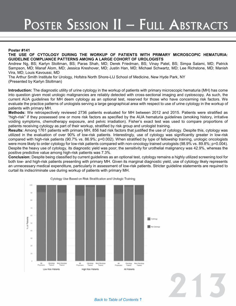
213Back to Table of Contents h
Poster session ii – Full AbstrActs
Poster #141 THE USE OF CYTOLOGY DURING THE WORKUP OF PATIENTS WITH PRIMARY MICROSCOPIC HEMATURIA: GUIDELINE COMPLIANCE PATTERNS AMONG A LARGE COHORT OF UROLOGISTS Andrew Ng, BS; Karlyn Stoltman, BS; Paras Shah, MD; Derek Friedman, BS; Vinay Patel, BS; Simpa Salami, MD; Patrick Sampson, MD; Manaf Alom, MD; Jessica Kreshover, MD; Justin Han, MD; Michael Schwartz, MD; Lee Richstone, MD; Manish Vira, MD; Louis Kavoussi, MD The Arthur Smith Institute for Urology, Hofstra North Shore-LIJ School of Medicine, New Hyde Park, NY (Presented by Karlyn Stoltman) Introduction: The diagnostic utility of urine cytology in the workup of patients with primary microscopic hematuria (MH) has come into question given most urologic malignancies are reliably detected with cross-sectional imaging and cystoscopy. As such, the current AUA guidelines for MH deem cytology as an optional test, reserved for those who have concerning risk factors. We evaluate the practice patterns of urologists serving a large geographical area with respect to use of urine cytology in the workup of patients with primary MH. Methods: We retrospectively reviewed 2736 patients evaluated for MH between 2012 and 2015. Patients were stratified as “high-risk” if they possessed one or more risk factors as specified by the AUA hematuria guidelines (smoking history, irritative voiding symptoms, chemotherapy exposure, and pelvic irradiation). Fisher’s exact test was used to compare proportions of patients receiving cytology as part of their workup, stratified by risk group and urologist training. Results: Among 1761 patients with primary MH, 856 had risk factors that justified the use of cytology. Despite this, cytology was utilized in the evaluation of over 90% of low-risk patients. Interestingly, use of cytology was significantly greater in low-risk compared with high-risk patients (90.7% vs. 86.9%; p=0.002). When stratified by type of fellowship training, urologic oncologists were more likely to order cytology for low-risk patients compared with non-oncology trained urologists (98.9% vs. 89.8%; p=0.004). Despite the heavy use of cytology, its diagnostic yield was poor; the sensitivity for urothelial malignancy was 42.9%, whereas the positive predictive value among high-risk patients was 7.3%. Conclusion: Despite being classified by current guidelines as an optional test, cytology remains a highly utilized screening tool for both low- and high-risk patients presenting with primary MH. Given its marginal diagnostic yield, use of cytology likely represents an unnecessary medical expenditure, particularly in assessment of low-risk patients. Stricter guideline statements are required to curtail its indiscriminate use during workup of patients with primary MH.
Poster #140 GEOGRAPHIC AND TEMPORAL TRENDS IN GLOBAL BLADDER CANCER MORBIDITY AND MORTALITY 1990-2010 Catherine Harris MD, MPH; Jonathan Brajtbord, MD; Matthew Cooperberg, MD, MPH; Maxwell Meng, MD; Anobel Odisho, MD, MPH University of California, San Francisco, CA (Presented by Jonathan Barjtboard, MD) Introduction: Advanced bladder cancer is associated with significant worldwide morbidity and mortality. However, accurate assessment of temporal trends across countries is limited. Understanding the drivers of bladder cancer morbidity and mortality can help increase global awareness and guide policy. Our objective was to determine geographic and temporal trends in morbidity and mortality in bladder cancer worldwide and examine potential associated factors. Methods: Age-adjusted sex-specific mortality rates (per 100,000 people) and disease burden (years of life lost to disability) for 1990, 2005 and 2010 were obtained from the Global Burden of Disease Project. We calculated population-weighted country specific trends over the 20-year period. We examined the association with GDP per capita, Human Development Index (HDI), smoking rates, and Gini coefficient. Results: From 1990 to 2010 there was a 14.2% decline in bladder cancer mortality worldwide; 13.9% in men and 19.1% in women (all p<.001). Mortality declined in all regions, from 3.9% to 25% in East Asia and Sub-Saharan Africa, respectively. Countries with higher GDP per capita, HDI were associated with a higher decline in mortality on univariate analysis. Morbidity decreased 3.7% in men and 6.4% in women worldwide (p<0.01). There was a significant regional variability in morbidity from a 23.3% decline in Sub-Saharan Africa to 43.48% increase in East Asia and Pacific. Multivariate regression revealed only GDP per capita was associated with a change in mortality and morbidity, with each $1000 increase in GDP per capita associated with a 0.002 (95% CI -0.003,-0.001) annual decline in death rate and 0.001 (95% CI -0.001,-0.0004) annual decline in years lost to morbidity. Smoking was not found to have a significant association with morbidity or mortality. Conclusion: Bladder cancer mortality is decreasing globally. In the past 20 years countries with higher GDP per capita have experienced a decline in bladder cancer deaths and morbidity. Global policy and initiatives to improve worldwide bladder cancer outcomes should account for the effects that a country’s economic status has on bladder cancer morbidity and mortality. Improvements in medical recordkeeping and data collection on known risk factors such as tobacco and environmental carcinogens are needed to better understand global trends in bladder cancer, especially in less developed areas of the world.
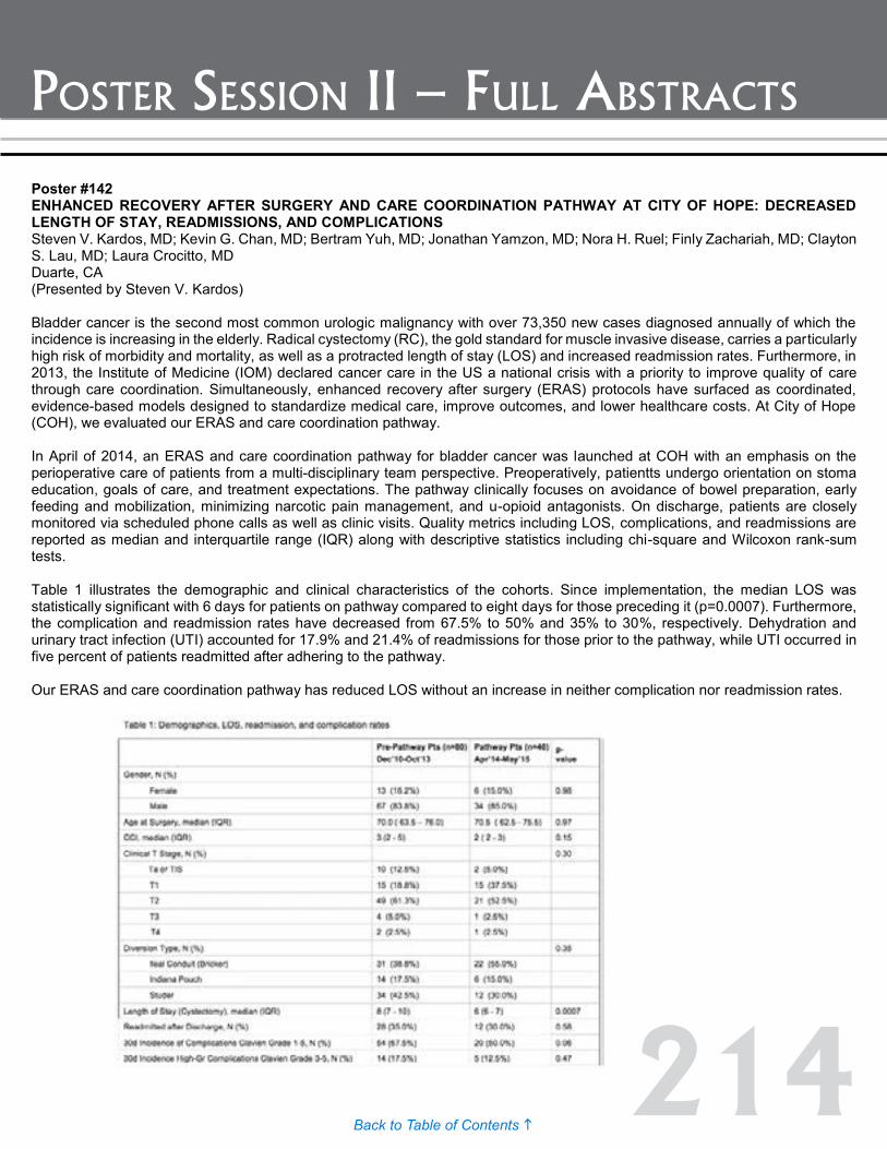
214Back to Table of Contents h
Poster session ii – Full AbstrActs
Poster #142 ENHANCED RECOVERY AFTER SURGERY AND CARE COORDINATION PATHWAY AT CITY OF HOPE: DECREASED LENGTH OF STAY, READMISSIONS, AND COMPLICATIONS Steven V. Kardos, MD; Kevin G. Chan, MD; Bertram Yuh, MD; Jonathan Yamzon, MD; Nora H. Ruel; Finly Zachariah, MD; Clayton S. Lau, MD; Laura Crocitto, MD Duarte, CA (Presented by Steven V. Kardos) Bladder cancer is the second most common urologic malignancy with over 73,350 new cases diagnosed annually of which the incidence is increasing in the elderly. Radical cystectomy (RC), the gold standard for muscle invasive disease, carries a particularly high risk of morbidity and mortality, as well as a protracted length of stay (LOS) and increased readmission rates. Furthermore, in 2013, the Institute of Medicine (IOM) declared cancer care in the US a national crisis with a priority to improve quality of care through care coordination. Simultaneously, enhanced recovery after surgery (ERAS) protocols have surfaced as coordinated, evidence-based models designed to standardize medical care, improve outcomes, and lower healthcare costs. At City of Hope (COH), we evaluated our ERAS and care coordination pathway. In April of 2014, an ERAS and care coordination pathway for bladder cancer was launched at COH with an emphasis on the perioperative care of patients from a multi-disciplinary team perspective. Preoperatively, patientts undergo orientation on stoma education, goals of care, and treatment expectations. The pathway clinically focuses on avoidance of bowel preparation, early feeding and mobilization, minimizing narcotic pain management, and u-opioid antagonists. On discharge, patients are closely monitored via scheduled phone calls as well as clinic visits. Quality metrics including LOS, complications, and readmissions are reported as median and interquartile range (IQR) along with descriptive statistics including chi-square and Wilcoxon rank-sum tests. Table 1 illustrates the demographic and clinical characteristics of the cohorts. Since implementation, the median LOS was statistically significant with 6 days for patients on pathway compared to eight days for those preceding it (p=0.0007). Furthermore, the complication and readmission rates have decreased from 67.5% to 50% and 35% to 30%, respectively. Dehydration and urinary tract infection (UTI) accounted for 17.9% and 21.4% of readmissions for those prior to the pathway, while UTI occurred in five percent of patients readmitted after adhering to the pathway. Our ERAS and care coordination pathway has reduced LOS without an increase in neither complication nor readmission rates.
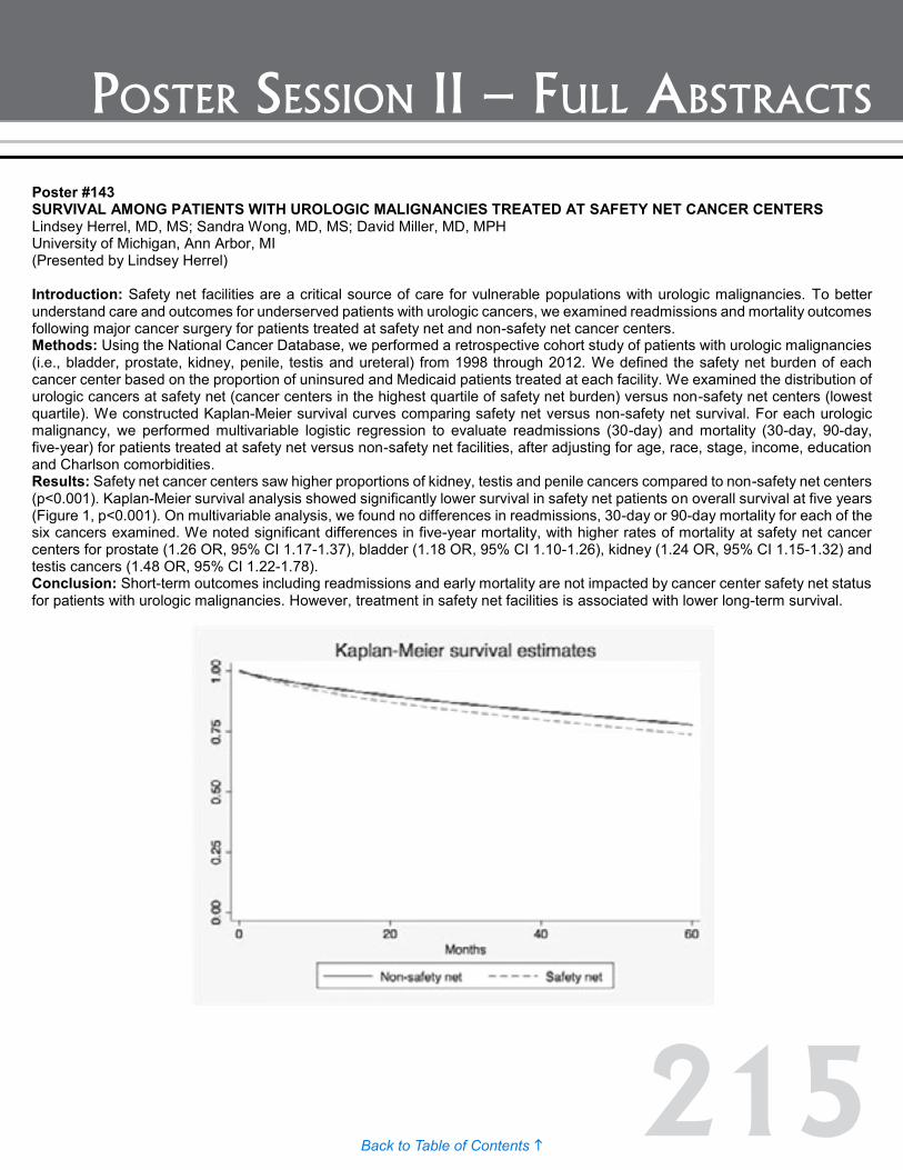
215Back to Table of Contents h
Poster session ii – Full AbstrActs
Poster #143 SURVIVAL AMONG PATIENTS WITH UROLOGIC MALIGNANCIES TREATED AT SAFETY NET CANCER CENTERS Lindsey Herrel, MD, MS; Sandra Wong, MD, MS; David Miller, MD, MPH University of Michigan, Ann Arbor, MI (Presented by Lindsey Herrel) Introduction: Safety net facilities are a critical source of care for vulnerable populations with urologic malignancies. To better understand care and outcomes for underserved patients with urologic cancers, we examined readmissions and mortality outcomes following major cancer surgery for patients treated at safety net and non-safety net cancer centers. Methods: Using the National Cancer Database, we performed a retrospective cohort study of patients with urologic malignancies (i.e., bladder, prostate, kidney, penile, testis and ureteral) from 1998 through 2012. We defined the safety net burden of each cancer center based on the proportion of uninsured and Medicaid patients treated at each facility. We examined the distribution of urologic cancers at safety net (cancer centers in the highest quartile of safety net burden) versus non-safety net centers (lowest quartile). We constructed Kaplan-Meier survival curves comparing safety net versus non-safety net survival. For each urologic malignancy, we performed multivariable logistic regression to evaluate readmissions (30-day) and mortality (30-day, 90-day, five-year) for patients treated at safety net versus non-safety net facilities, after adjusting for age, race, stage, income, education and Charlson comorbidities. Results: Safety net cancer centers saw higher proportions of kidney, testis and penile cancers compared to non-safety net centers (p<0.001). Kaplan-Meier survival analysis showed significantly lower survival in safety net patients on overall survival at five years (Figure 1, p<0.001). On multivariable analysis, we found no differences in readmissions, 30-day or 90-day mortality for each of the six cancers examined. We noted significant differences in five-year mortality, with higher rates of mortality at safety net cancer centers for prostate (1.26 OR, 95% CI 1.17-1.37), bladder (1.18 OR, 95% CI 1.10-1.26), kidney (1.24 OR, 95% CI 1.15-1.32) and testis cancers (1.48 OR, 95% CI 1.22-1.78). Conclusion: Short-term outcomes including readmissions and early mortality are not impacted by cancer center safety net status for patients with urologic malignancies. However, treatment in safety net facilities is associated with lower long-term survival.
Poster #142 ENHANCED RECOVERY AFTER SURGERY AND CARE COORDINATION PATHWAY AT CITY OF HOPE: DECREASED LENGTH OF STAY, READMISSIONS, AND COMPLICATIONS Steven V. Kardos, MD; Kevin G. Chan, MD; Bertram Yuh, MD; Jonathan Yamzon, MD; Nora H. Ruel; Finly Zachariah, MD; Clayton S. Lau, MD; Laura Crocitto, MD Duarte, CA (Presented by Steven V. Kardos) Bladder cancer is the second most common urologic malignancy with over 73,350 new cases diagnosed annually of which the incidence is increasing in the elderly. Radical cystectomy (RC), the gold standard for muscle invasive disease, carries a particularly high risk of morbidity and mortality, as well as a protracted length of stay (LOS) and increased readmission rates. Furthermore, in 2013, the Institute of Medicine (IOM) declared cancer care in the US a national crisis with a priority to improve quality of care through care coordination. Simultaneously, enhanced recovery after surgery (ERAS) protocols have surfaced as coordinated, evidence-based models designed to standardize medical care, improve outcomes, and lower healthcare costs. At City of Hope (COH), we evaluated our ERAS and care coordination pathway. In April of 2014, an ERAS and care coordination pathway for bladder cancer was launched at COH with an emphasis on the perioperative care of patients from a multi-disciplinary team perspective. Preoperatively, patientts undergo orientation on stoma education, goals of care, and treatment expectations. The pathway clinically focuses on avoidance of bowel preparation, early feeding and mobilization, minimizing narcotic pain management, and u-opioid antagonists. On discharge, patients are closely monitored via scheduled phone calls as well as clinic visits. Quality metrics including LOS, complications, and readmissions are reported as median and interquartile range (IQR) along with descriptive statistics including chi-square and Wilcoxon rank-sum tests. Table 1 illustrates the demographic and clinical characteristics of the cohorts. Since implementation, the median LOS was statistically significant with 6 days for patients on pathway compared to eight days for those preceding it (p=0.0007). Furthermore, the complication and readmission rates have decreased from 67.5% to 50% and 35% to 30%, respectively. Dehydration and urinary tract infection (UTI) accounted for 17.9% and 21.4% of readmissions for those prior to the pathway, while UTI occurred in five percent of patients readmitted after adhering to the pathway. Our ERAS and care coordination pathway has reduced LOS without an increase in neither complication nor readmission rates.
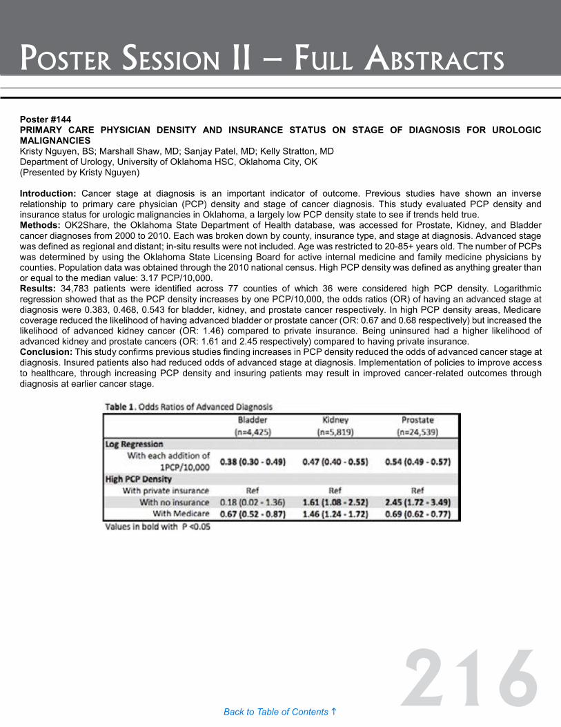
216Back to Table of Contents h
Poster session ii – Full AbstrActs
Poster #144 PRIMARY CARE PHYSICIAN DENSITY AND INSURANCE STATUS ON STAGE OF DIAGNOSIS FOR UROLOGIC MALIGNANCIES Kristy Nguyen, BS; Marshall Shaw, MD; Sanjay Patel, MD; Kelly Stratton, MD Department of Urology, University of Oklahoma HSC, Oklahoma City, OK (Presented by Kristy Nguyen) Introduction: Cancer stage at diagnosis is an important indicator of outcome. Previous studies have shown an inverse relationship to primary care physician (PCP) density and stage of cancer diagnosis. This study evaluated PCP density and insurance status for urologic malignancies in Oklahoma, a largely low PCP density state to see if trends held true. Methods: OK2Share, the Oklahoma State Department of Health database, was accessed for Prostate, Kidney, and Bladder cancer diagnoses from 2000 to 2010. Each was broken down by county, insurance type, and stage at diagnosis. Advanced stage was defined as regional and distant; in-situ results were not included. Age was restricted to 20-85+ years old. The number of PCPs was determined by using the Oklahoma State Licensing Board for active internal medicine and family medicine physicians by counties. Population data was obtained through the 2010 national census. High PCP density was defined as anything greater than or equal to the median value: 3.17 PCP/10,000. Results: 34,783 patients were identified across 77 counties of which 36 were considered high PCP density. Logarithmic regression showed that as the PCP density increases by one PCP/10,000, the odds ratios (OR) of having an advanced stage at diagnosis were 0.383, 0.468, 0.543 for bladder, kidney, and prostate cancer respectively. In high PCP density areas, Medicare coverage reduced the likelihood of having advanced bladder or prostate cancer (OR: 0.67 and 0.68 respectively) but increased the likelihood of advanced kidney cancer (OR: 1.46) compared to private insurance. Being uninsured had a higher likelihood of advanced kidney and prostate cancers (OR: 1.61 and 2.45 respectively) compared to having private insurance. Conclusion: This study confirms previous studies finding increases in PCP density reduced the odds of advanced cancer stage at diagnosis. Insured patients also had reduced odds of advanced stage at diagnosis. Implementation of policies to improve access to healthcare, through increasing PCP density and insuring patients may result in improved cancer-related outcomes through diagnosis at earlier cancer stage.
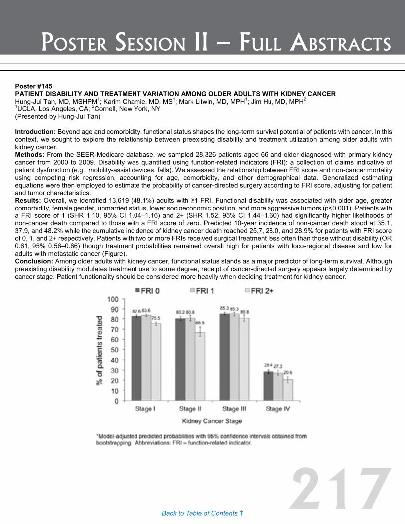
217Back to Table of Contents h
Poster session ii – Full AbstrActs
Poster #145 PATIENT DISABILITY AND TREATMENT VARIATION AMONG OLDER ADULTS WITH KIDNEY CANCER Hung-Jui Tan, MD, MSHPM1; Karim Chamie, MD, MS1; Mark Litwin, MD, MPH1; Jim Hu, MD, MPH2 1UCLA, Los Angeles, CA; 2Cornell, New York, NY (Presented by Hung-Jui Tan) Introduction: Beyond age and comorbidity, functional status shapes the long-term survival potential of patients with cancer. In this context, we sought to explore the relationship between preexisting disability and treatment utilization among older adults with kidney cancer. Methods: From the SEER-Medicare database, we sampled 28,326 patients aged 66 and older diagnosed with primary kidney cancer from 2000 to 2009. Disability was quantified using function-related indicators (FRI): a collection of claims indicative of patient dysfunction (e.g., mobility-assist devices, falls). We assessed the relationship between FRI score and non-cancer mortality using competing risk regression, accounting for age, comorbidity, and other demographical data. Generalized estimating equations were then employed to estimate the probability of cancer-directed surgery according to FRI score, adjusting for patient and tumor characteristics. Results: Overall, we identified 13,619 (48.1%) adults with ≥1 FRI. Functional disability was associated with older age, greater comorbidity, female gender, unmarried status, lower socioeconomic position, and more aggressive tumors (p<0.001). Patients with a FRI score of 1 (SHR 1.10, 95% CI 1.04–1.16) and 2+ (SHR 1.52, 95% CI 1.44–1.60) had significantly higher likelihoods of non-cancer death compared to those with a FRI score of zero. Predicted 10-year incidence of non-cancer death stood at 35.1, 37.9, and 48.2% while the cumulative incidence of kidney cancer death reached 25.7, 28.0, and 28.9% for patients with FRI score of 0, 1, and 2+ respectively. Patients with two or more FRIs received surgical treatment less often than those without disability (OR 0.61, 95% 0.56–0.66) though treatment probabilities remained overall high for patients with loco-regional disease and low for adults with metastatic cancer (Figure). Conclusion: Among older adults with kidney cancer, functional status stands as a major predictor of long-term survival. Although preexisting disability modulates treatment use to some degree, receipt of cancer-directed surgery appears largely determined by cancer stage. Patient functionality should be considered more heavily when deciding treatment for kidney cancer.
218Back to Table of Contents h
Poster session ii – Full AbstrActs
Poster #147 ENDOSCOPIC VERSUS SURGICAL MANAGEMENT FOR PATIENTS WITH UPPER TRACT UROTHELIAL CANCER AS THEIR FIRST CANCER DIAGNOSIS: A MATCHED PROPENSITY SCORE ANALYSIS USING SEER-MEDICARE DATA Eric Kim, MD; Goutham Vemana, MD; Sam Bhayani, MD, MS; Joel Vetter; Seth Strope, MD, MPH Washington University School of Medicine, St. Louis, MO (Presented by Eric Kim) Introduction: While nephroureterectomy remains the gold standard for the management of upper tract urothelial carcinoma (UTUC), endoscopic management has become an alternative treatment for select patients. However, differences in survival among patients receiving these two treatment options are largely unknown. Methods: Using SEER-Medicare data, patients diagnosed with localized, low-grade UTUC between 2004 and 2009 were identified (n=1093). Patients with other prior cancer diagnoses were excluded (n=318). Receipt of endoscopic and surgical interventions was assessed and patients were separated into cohorts based on treatment: surgical management (n=523) and endoscopic management (n=151). Matching based on age, race, gender, marital status, year of diagnosis, and Charlson comorbidity index, we used nearest neighbor (2:1) propensity score analysis to match a subset of surgical management patients (n=302) to patients receiving endoscopic management (n=151). Cox proportional hazards and Kaplan-Meier analyses were performed for overall (OS) and cancer-specific survival (CSS), using the matched data. Results: Endoscopic management was an independent and significant predictor of all-cause (HR 1.6) and cancer-specific mortality (HR 2.1). Figure 1 illustrates the Kaplan-Meier estimated survival analysis. OS and CSS were significantly lower for endoscopic management, with both OS and CSS diverging at approximately 24 to 36 months. A subset of patients initially receiving endoscopic management went on to receive surgical intervention (80/151 = 53%) at a median of 8.8 months from diagnosis. For these patients, CSS was not significantly different from those who remained on endoscopic management (p=0.62) and remained significantly lower than patients who received upfront surgical intervention (p=0.02). Conclusion: Although initial survival outcomes (first 24 months) are similar for endoscopic and surgical management of localized, low grade UTUC, both OS and CSS are significantly inferior in the endoscopic management group in the longer term. Furthermore, transition from initial endoscopic management to surgical intervention appears to have limited impact on CSS.
Poster #146 COST DASHBOARDS FOR RADICAL CYSTECTOMY: ACCOUNTING FOR SURGEON COST VARIATION Alan Thong, MD1; Wazim Narain2; Donna Boccamazzo2; Peter Sidi2; Guido Dalbagni, MD1; Bernard Bochner, MD1 1Urology Service, Department of Surgery, Memorial Sloan-Kettering Cancer Center, New York, NY; 2Health Informatics, Memorial Sloan-Kettering Cancer Center, New York, NY (Presented by Alan Thong) Introduction: In addition to promoting quality, new payment models are emerging with a mandate to control healthcare costs. On a national level, there is significant variation in the cost of major index cancer operations. Accurate cost accounting, clinician-defined metrics, risk-adjustment, and feedback of data are essential steps in using cost data for quality improvement. We sought to account for institutional direct costs incurred during radical cystectomy, identify the main cost drivers in the index hospitalization, and examine surgeon variability in the main cost drivers within the context of patient characteristics. Methods: Direct itemized institutional cost data for 321 consecutive radical cystectomies performed by five de-identified surgeons was obtained. Costs were adjusted using a multiplier equal to the ratio of normalized Medicare reimbursement over institutional reported cost. Interactive web-based dashboards were created to feedback patient characteristics, outcomes, cost components, and anonymized surgeon cost comparisons. Analysis of variance was used to compare patient characteristics and cost components across surgeons. Multivariable logistic regression was used to model above average costs on surgeon and patient specific factors. Results: The median adjusted total cost per case was $13,009.57 (interquartile range $11,254.82, $15,730.53). Inpatient, anesthesia, disposable device, operating room, and physician costs accounted for the majority of the total costs incurred. Inpatient costs were not significantly different across surgeons, however maximum differences between cost means for anesthesia ($93.62, p=0.0039), disposable device (energy devices, $279.48, p<0.0001; staplers, $494.73, p<0.0001), operating room ($1168.55, p<0.0001), and physician costs ($1362.20, p<0.0001) did differ significantly across surgeons. Adjusting for patient factors including gender, American Society of Anesthesiologists status, diversion type, and total lymph node count, these significant differences in cost across surgeons persisted. Conclusion: Cost accounting using dashboards identified significant cost differences across surgeons performing radical cystectomy at our institution independent of differences in patient characteristics. Additional longitudinal cost data is needed to determine if such anonymized, interactive cost feedback through dashboards can improve the value of surgical care by reducing cost without sacrificing quality.
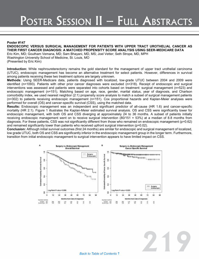
219Back to Table of Contents h
Poster session ii – Full AbstrActs
Poster #147 ENDOSCOPIC VERSUS SURGICAL MANAGEMENT FOR PATIENTS WITH UPPER TRACT UROTHELIAL CANCER AS THEIR FIRST CANCER DIAGNOSIS: A MATCHED PROPENSITY SCORE ANALYSIS USING SEER-MEDICARE DATA Eric Kim, MD; Goutham Vemana, MD; Sam Bhayani, MD, MS; Joel Vetter; Seth Strope, MD, MPH Washington University School of Medicine, St. Louis, MO (Presented by Eric Kim) Introduction: While nephroureterectomy remains the gold standard for the management of upper tract urothelial carcinoma (UTUC), endoscopic management has become an alternative treatment for select patients. However, differences in survival among patients receiving these two treatment options are largely unknown. Methods: Using SEER-Medicare data, patients diagnosed with localized, low-grade UTUC between 2004 and 2009 were identified (n=1093). Patients with other prior cancer diagnoses were excluded (n=318). Receipt of endoscopic and surgical interventions was assessed and patients were separated into cohorts based on treatment: surgical management (n=523) and endoscopic management (n=151). Matching based on age, race, gender, marital status, year of diagnosis, and Charlson comorbidity index, we used nearest neighbor (2:1) propensity score analysis to match a subset of surgical management patients (n=302) to patients receiving endoscopic management (n=151). Cox proportional hazards and Kaplan-Meier analyses were performed for overall (OS) and cancer-specific survival (CSS), using the matched data. Results: Endoscopic management was an independent and significant predictor of all-cause (HR 1.6) and cancer-specific mortality (HR 2.1). Figure 1 illustrates the Kaplan-Meier estimated survival analysis. OS and CSS were significantly lower for endoscopic management, with both OS and CSS diverging at approximately 24 to 36 months. A subset of patients initially receiving endoscopic management went on to receive surgical intervention (80/151 = 53%) at a median of 8.8 months from diagnosis. For these patients, CSS was not significantly different from those who remained on endoscopic management (p=0.62) and remained significantly lower than patients who received upfront surgical intervention (p=0.02). Conclusion: Although initial survival outcomes (first 24 months) are similar for endoscopic and surgical management of localized, low grade UTUC, both OS and CSS are significantly inferior in the endoscopic management group in the longer term. Furthermore, transition from initial endoscopic management to surgical intervention appears to have limited impact on CSS.
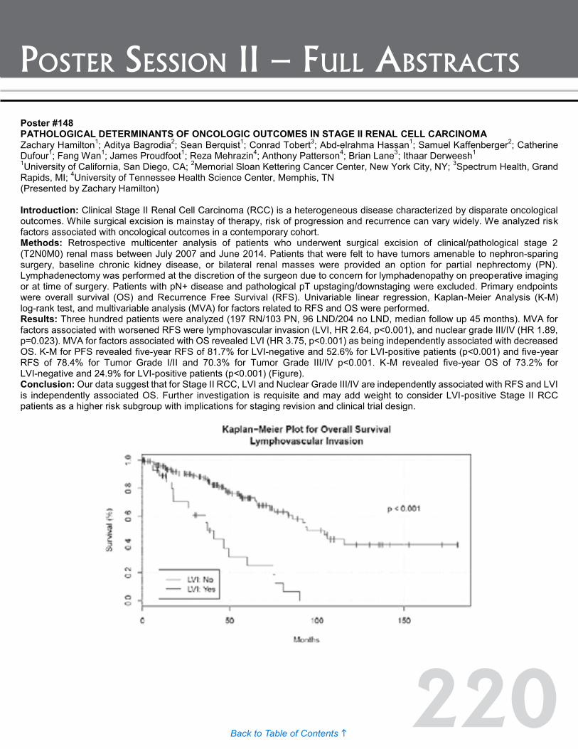
220Back to Table of Contents h
Poster session ii – Full AbstrActs
Poster #148 PATHOLOGICAL DETERMINANTS OF ONCOLOGIC OUTCOMES IN STAGE II RENAL CELL CARCINOMA Zachary Hamilton1; Aditya Bagrodia2; Sean Berquist1; Conrad Tobert3; Abd-elrahma Hassan1; Samuel Kaffenberger2; Catherine Dufour1; Fang Wan1; James Proudfoot1; Reza Mehrazin4; Anthony Patterson4; Brian Lane3; Ithaar Derweesh1 1University of California, San Diego, CA; 2Memorial Sloan Kettering Cancer Center, New York City, NY; 3Spectrum Health, Grand Rapids, MI; 4University of Tennessee Health Science Center, Memphis, TN (Presented by Zachary Hamilton) Introduction: Clinical Stage II Renal Cell Carcinoma (RCC) is a heterogeneous disease characterized by disparate oncological outcomes. While surgical excision is mainstay of therapy, risk of progression and recurrence can vary widely. We analyzed risk factors associated with oncological outcomes in a contemporary cohort. Methods: Retrospective multicenter analysis of patients who underwent surgical excision of clinical/pathological stage 2 (T2N0M0) renal mass between July 2007 and June 2014. Patients that were felt to have tumors amenable to nephron-sparing surgery, baseline chronic kidney disease, or bilateral renal masses were provided an option for partial nephrectomy (PN). Lymphadenectomy was performed at the discretion of the surgeon due to concern for lymphadenopathy on preoperative imaging or at time of surgery. Patients with pN+ disease and pathological pT upstaging/downstaging were excluded. Primary endpoints were overall survival (OS) and Recurrence Free Survival (RFS). Univariable linear regression, Kaplan-Meier Analysis (K-M) log-rank test, and multivariable analysis (MVA) for factors related to RFS and OS were performed. Results: Three hundred patients were analyzed (197 RN/103 PN, 96 LND/204 no LND, median follow up 45 months). MVA for factors associated with worsened RFS were lymphovascular invasion (LVI, HR 2.64, p<0.001), and nuclear grade III/IV (HR 1.89, p=0.023). MVA for factors associated with OS revealed LVI (HR 3.75, p<0.001) as being independently associated with decreased OS. K-M for PFS revealed five-year RFS of 81.7% for LVI-negative and 52.6% for LVI-positive patients (p<0.001) and five-year RFS of 78.4% for Tumor Grade I/II and 70.3% for Tumor Grade III/IV p<0.001. K-M revealed five-year OS of 73.2% for LVI-negative and 24.9% for LVI-positive patients (p<0.001) (Figure). Conclusion: Our data suggest that for Stage II RCC, LVI and Nuclear Grade III/IV are independently associated with RFS and LVI is independently associated OS. Further investigation is requisite and may add weight to consider LVI-positive Stage II RCC patients as a higher risk subgroup with implications for staging revision and clinical trial design.
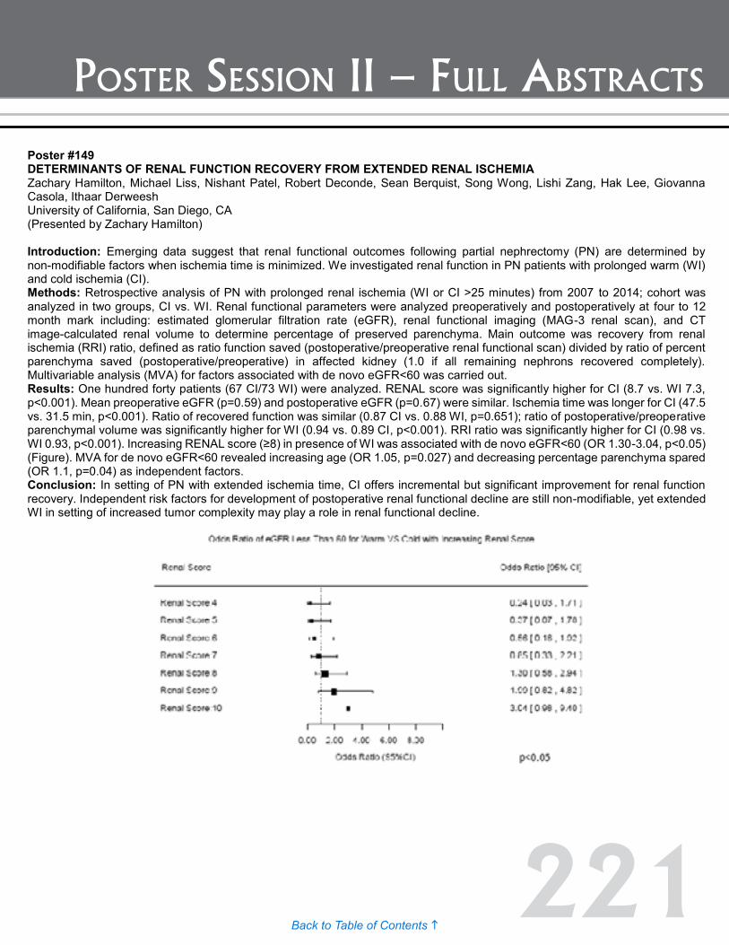
221Back to Table of Contents h
Poster session ii – Full AbstrActs
Poster #149 DETERMINANTS OF RENAL FUNCTION RECOVERY FROM EXTENDED RENAL ISCHEMIA Zachary Hamilton, Michael Liss, Nishant Patel, Robert Deconde, Sean Berquist, Song Wong, Lishi Zang, Hak Lee, Giovanna Casola, Ithaar Derweesh University of California, San Diego, CA (Presented by Zachary Hamilton) Introduction: Emerging data suggest that renal functional outcomes following partial nephrectomy (PN) are determined by non-modifiable factors when ischemia time is minimized. We investigated renal function in PN patients with prolonged warm (WI) and cold ischemia (CI). Methods: Retrospective analysis of PN with prolonged renal ischemia (WI or CI >25 minutes) from 2007 to 2014; cohort was analyzed in two groups, CI vs. WI. Renal functional parameters were analyzed preoperatively and postoperatively at four to 12 month mark including: estimated glomerular filtration rate (eGFR), renal functional imaging (MAG-3 renal scan), and CT image-calculated renal volume to determine percentage of preserved parenchyma. Main outcome was recovery from renal ischemia (RRI) ratio, defined as ratio function saved (postoperative/preoperative renal functional scan) divided by ratio of percent parenchyma saved (postoperative/preoperative) in affected kidney (1.0 if all remaining nephrons recovered completely). Multivariable analysis (MVA) for factors associated with de novo eGFR<60 was carried out. Results: One hundred forty patients (67 CI/73 WI) were analyzed. RENAL score was significantly higher for CI (8.7 vs. WI 7.3, p<0.001). Mean preoperative eGFR (p=0.59) and postoperative eGFR (p=0.67) were similar. Ischemia time was longer for CI (47.5 vs. 31.5 min, p<0.001). Ratio of recovered function was similar (0.87 CI vs. 0.88 WI, p=0.651); ratio of postoperative/preoperative parenchymal volume was significantly higher for WI (0.94 vs. 0.89 CI, p<0.001). RRI ratio was significantly higher for CI (0.98 vs. WI 0.93, p<0.001). Increasing RENAL score (≥8) in presence of WI was associated with de novo eGFR<60 (OR 1.30-3.04, p<0.05) (Figure). MVA for de novo eGFR<60 revealed increasing age (OR 1.05, p=0.027) and decreasing percentage parenchyma spared (OR 1.1, p=0.04) as independent factors. Conclusion: In setting of PN with extended ischemia time, CI offers incremental but significant improvement for renal function recovery. Independent risk factors for development of postoperative renal functional decline are still non-modifiable, yet extended WI in setting of increased tumor complexity may play a role in renal functional decline.
Poster #148 PATHOLOGICAL DETERMINANTS OF ONCOLOGIC OUTCOMES IN STAGE II RENAL CELL CARCINOMA Zachary Hamilton1; Aditya Bagrodia2; Sean Berquist1; Conrad Tobert3; Abd-elrahma Hassan1; Samuel Kaffenberger2; Catherine Dufour1; Fang Wan1; James Proudfoot1; Reza Mehrazin4; Anthony Patterson4; Brian Lane3; Ithaar Derweesh1 1University of California, San Diego, CA; 2Memorial Sloan Kettering Cancer Center, New York City, NY; 3Spectrum Health, Grand Rapids, MI; 4University of Tennessee Health Science Center, Memphis, TN (Presented by Zachary Hamilton) Introduction: Clinical Stage II Renal Cell Carcinoma (RCC) is a heterogeneous disease characterized by disparate oncological outcomes. While surgical excision is mainstay of therapy, risk of progression and recurrence can vary widely. We analyzed risk factors associated with oncological outcomes in a contemporary cohort. Methods: Retrospective multicenter analysis of patients who underwent surgical excision of clinical/pathological stage 2 (T2N0M0) renal mass between July 2007 and June 2014. Patients that were felt to have tumors amenable to nephron-sparing surgery, baseline chronic kidney disease, or bilateral renal masses were provided an option for partial nephrectomy (PN). Lymphadenectomy was performed at the discretion of the surgeon due to concern for lymphadenopathy on preoperative imaging or at time of surgery. Patients with pN+ disease and pathological pT upstaging/downstaging were excluded. Primary endpoints were overall survival (OS) and Recurrence Free Survival (RFS). Univariable linear regression, Kaplan-Meier Analysis (K-M) log-rank test, and multivariable analysis (MVA) for factors related to RFS and OS were performed. Results: Three hundred patients were analyzed (197 RN/103 PN, 96 LND/204 no LND, median follow up 45 months). MVA for factors associated with worsened RFS were lymphovascular invasion (LVI, HR 2.64, p<0.001), and nuclear grade III/IV (HR 1.89, p=0.023). MVA for factors associated with OS revealed LVI (HR 3.75, p<0.001) as being independently associated with decreased OS. K-M for PFS revealed five-year RFS of 81.7% for LVI-negative and 52.6% for LVI-positive patients (p<0.001) and five-year RFS of 78.4% for Tumor Grade I/II and 70.3% for Tumor Grade III/IV p<0.001. K-M revealed five-year OS of 73.2% for LVI-negative and 24.9% for LVI-positive patients (p<0.001) (Figure). Conclusion: Our data suggest that for Stage II RCC, LVI and Nuclear Grade III/IV are independently associated with RFS and LVI is independently associated OS. Further investigation is requisite and may add weight to consider LVI-positive Stage II RCC patients as a higher risk subgroup with implications for staging revision and clinical trial design.
222Back to Table of Contents h
Poster session ii – Full AbstrActs
Poster #150 MALIGNANT ASCITES AS A MANIFESTATION OF METASTATIC PAPILLARY RENAL CELL CANCER Abhinav Sidana, MD; Julia Friend; Meet Kadakia, MD; Daniel Su, MD; Louis Krane, MD; Geri Hawks; Martha Ninos; James Peterson; W. Marston Linehan, MD; Ramaprasad Srinivasan, MD,PhD Urologic Oncology Branch, NIH (Presented by Abhinav Sidana) Introduction: Malignant ascites is a rare but ominous complication of metastatic cancer. While it has been reported to complicate less than one percent of metastatic clear cell cancer its incidence in metastatic papillary renal cell cancer (pRCC) has not been defined. Here, we describe the incidence of ascites in metastatic pRCC, identify the predictors of its development and estimate its prognostic effect on the survival of these patients. Methods: A retrospective evaluation of the medical records of patients with metastatic pRCC seen at Urologic Oncology Branch of National Cancer Institute (2000-2014) was undertaken. Patient demographics, tumor characteristics and patient outcome were studied. Logistic regression to identify predictors and Kaplan Meier analysis to determine survival were done. Results: A total of 106 patients with metastatic pRCC were identified, including 100 in whom sufficient data was available to enable assessment of ascites. Of these, 20% (20/100) had evidence of malignant ascites. Median patient age at diagnosis of ascites was 48.5 years (26.1-76.6 years) and median time to development of ascites from initial diagnosis of metastatic disease was 15.6 (0-72.3) months. There was no significant difference in the incidence of ascites between patients with hereditary and sporadic pRCC (p=0.804) or between patients with different histological subtypes of pRCC (p=0.675). Age, race, histology, hereditary origin and presence of metastasis at presentation were not found to be significant predictors for development of ascites. Median estimated survival from diagnosis of metastatic disease was significantly shorter for patients who developed ascites (26.0 [14.1-37.9] months) compared to patients who did not develop this complication (41.8 [29.3-54.2] months, p=0.033). Conclusion: To our knowledge, this is the largest series evaluating the incidence, predictors and prognostic impact of ascites in metastatic pRCC. Malignant ascites is a fairly common manifestation of metastatic pRCC. In our cohort, no predictor was identified for the development of ascites. Ascites was associated with worse outcome, with a shorter overall survival observed in patients developing this complication.
223Back to Table of Contents h
Poster session ii – Full AbstrActs
Poster #151 RACIAL DISPARITIES IN RENAL CELL CARCINOMA: A SINGLE PAYER HEALTHCARE EXPERIENCE. Abiodun Mafolasire, MS1; Cayce Nawaf, MD1; Xiaopan Yao, PhD2; Douglas Corley, MD, PhD3; Jonathan Hofmann, PhD4; Mark Purdue, PhD4; Adebowale Adeniran, MD5; Brian Shuch, MD6 1Department of Urology, Yale School of Medicine; 2Yale Center for Analystical Sciences, Yale School of Medicine; 3Kaiser Permanente Division of Research, Kaiser Permanente San Francisco Medical Center; 4Division of Cancer Epidemiology and Genetics, National Cancer Institute &Department of Pathology, Yale School of Medicine; 5Department of Pathology, Yale School of Medicine; 6Yale School of Medicine (Presented by Brian Shuch) Introduction: Significant racial disparities exist between blacks and whites with renal cell carcinoma (RCC) in regards to age of onset, histologic type, and survival. Differences in access to care and comorbidities are possible contributors. In this study, we analyzed data from a single payer healthcare system with presumably similar access and quality of care, to investigate if racial differences in outcomes persist. Methods: As part of a case-control study of patients receiving care within the Kaiser Permanente Northern California system, pathologic and epidemiological records were obtained for RCC cases from 1998 to 2008. Patient race was identified as either black or white. Patient characteristics, comorbidities, tumor characteristics and treatment status were compared. Overall survival (OS) and disease specific survival (DSS) were calculated by the Kaplan-Meier method and a multivariate, Cox proportion hazard model was used to estimate the association of race, comorbidity, and clinico-pathologic variables with survival. Results: A total of 2445 patients (2152 whites, 293 blacks) were included in the study. Compared to whites, blacks were diagnosed at a younger age (median age of 62 years vs. 66 years, p <0.001), had higher percentage of papillary RCC subtype (15% vs. 5.2%, p<0.001) and had a similar rate of surgical treatment (78.8% vs. 77.9%, p=0.764). Blacks had better DSS (11.9 years vs. 11.2 years, p=0.016). On univariate analysis, race, histological subtype, tumor size, AJCC stage, tumor grade, surgical treatment were significant predictors of survival. On multivariate analysis race was no longer significant. Advanced AJCC stage, lack of surgical treatment, larger tumor size, and higher tumor grade were independent predictors of worse DSS. Conclusion: Even within a single healthcare system providing similar access to care, black and white patients with RCC had significant differences in the age of diagnosis, histologic type, tumor size, stage, and comorbidities. Black patients had decreased tumor stage and improved DSS, although race was not an independent predictor of outcome. This suggests that survival differences using large national registries may result from barriers to healthcare access and/or comorbidity rather than different disease biology.
Poster #150 MALIGNANT ASCITES AS A MANIFESTATION OF METASTATIC PAPILLARY RENAL CELL CANCER Abhinav Sidana, MD; Julia Friend; Meet Kadakia, MD; Daniel Su, MD; Louis Krane, MD; Geri Hawks; Martha Ninos; James Peterson; W. Marston Linehan, MD; Ramaprasad Srinivasan, MD,PhD Urologic Oncology Branch, NIH (Presented by Abhinav Sidana) Introduction: Malignant ascites is a rare but ominous complication of metastatic cancer. While it has been reported to complicate less than one percent of metastatic clear cell cancer its incidence in metastatic papillary renal cell cancer (pRCC) has not been defined. Here, we describe the incidence of ascites in metastatic pRCC, identify the predictors of its development and estimate its prognostic effect on the survival of these patients. Methods: A retrospective evaluation of the medical records of patients with metastatic pRCC seen at Urologic Oncology Branch of National Cancer Institute (2000-2014) was undertaken. Patient demographics, tumor characteristics and patient outcome were studied. Logistic regression to identify predictors and Kaplan Meier analysis to determine survival were done. Results: A total of 106 patients with metastatic pRCC were identified, including 100 in whom sufficient data was available to enable assessment of ascites. Of these, 20% (20/100) had evidence of malignant ascites. Median patient age at diagnosis of ascites was 48.5 years (26.1-76.6 years) and median time to development of ascites from initial diagnosis of metastatic disease was 15.6 (0-72.3) months. There was no significant difference in the incidence of ascites between patients with hereditary and sporadic pRCC (p=0.804) or between patients with different histological subtypes of pRCC (p=0.675). Age, race, histology, hereditary origin and presence of metastasis at presentation were not found to be significant predictors for development of ascites. Median estimated survival from diagnosis of metastatic disease was significantly shorter for patients who developed ascites (26.0 [14.1-37.9] months) compared to patients who did not develop this complication (41.8 [29.3-54.2] months, p=0.033). Conclusion: To our knowledge, this is the largest series evaluating the incidence, predictors and prognostic impact of ascites in metastatic pRCC. Malignant ascites is a fairly common manifestation of metastatic pRCC. In our cohort, no predictor was identified for the development of ascites. Ascites was associated with worse outcome, with a shorter overall survival observed in patients developing this complication.
224Back to Table of Contents h
Poster session ii – Full AbstrActs
Poster #152 CONTEMPORARY Results: OF RENAL MASS BIOPSY David Cahn, DO, MBS1; Caitlin Lim, DO1; Rosaleen Parsons, MD, FACR2; Benjamin Ristau, MD3; Alexander Kutikov, MD, FACS3; David Chen, MD, FACS3; Richard Greenberg, MD, FACS3; Rosalia Viterbo, MD, FACS3; Marc Smaldone, MD, MSHP3; Robert Uzzo, MD, FACS4 1Einstein Healthcare Network, Department of Urology, Philadelphia, PA; 2Fox Chase Cancer Center, Department of Radiology, Philadelphia, PA; 3Fox Chase Cancer Center, Department of Urologic Oncology, Philadelphia, PA; 4Fox Chase Cancer Center and Einstein Healthcare Network, Departments of Urologic Oncology and Urology, Philadelphia, PA (Presented by David Cahn) Introduction: The role of renal mass biopsy (RMB) in the management of clinically localized renal masses has recently undergone re-evaluation in an effort to improve risk stratification and minimize morbidity from over-treatment. We review our institutional experience with RMB over a 15-year period to identify performance characteristics and highlight opportunities to improve clinical management. Methods: Using our prospectively maintained renal mass database, we identified patients who underwent renal mass core and fine needle aspiration biopsies at our institution between 1999 and 2015. We describe demographic and performance characteristics and assess pathologic concordance of those who underwent surgical resection. Using the University of Michigan algorithm, we reviewed the likelihood that RMB could influence therapeutic decision-making. Results: Of the 371 biopsies performed from 1999 to 2015, 65.5% were within the last five years. 32.3% of patients were placed on active surveillance (AS) protocols, of which average tumor size was 3.3cm. 48.4% of biopsied lesions underwent surgical resection. Using core biopsy only, RMB accurately predicted renal cell pathologic histology and grade concordance in 86.3% and 64.2% of cases, respectively. 11% of RMB were benign and no surgical intervention occurred. 18.8% of patients treated surgically had tumors classified as favorable or intermediate <2cm using the UM algorithm. Using this algorithm, AS would have potentially been the preferred treatment plan for all of these patients. However, 42.4% of surgically treated patients at our institution with UM favorable characteristics had tumors larger than 4cm. 44.4% of surgically treated cases were performed on unfavorable lesions or intermediate >2cm using the UM algorithm. Conclusion: RMB remains a safe and effective clinical tool to pathologically evaluate renal masses with minimal morbidity. Predetermined pathways for risk stratification with RMB may assist physicians in objectifying data to avoid over-treatment and morbidity from unnecessary surgery. At our institution, histologic and grade concordance rates are consistent with previous literature. Using the UM algorithm, management could have been changed in 18.8% of patients with AS becoming the preferred treatment plan. RMB should be considered in patients where results would influence clinical decision-making.
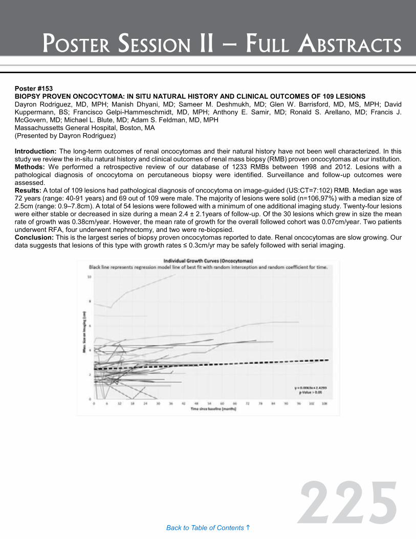
225Back to Table of Contents h
Poster session ii – Full AbstrActs
Poster #153 BIOPSY PROVEN ONCOCYTOMA: IN SITU NATURAL HISTORY AND CLINICAL OUTCOMES OF 109 LESIONS Dayron Rodriguez, MD, MPH; Manish Dhyani, MD; Sameer M. Deshmukh, MD; Glen W. Barrisford, MD, MS, MPH; David Kuppermann, BS; Francisco Gelpi-Hammeschmidt, MD, MPH; Anthony E. Samir, MD; Ronald S. Arellano, MD; Francis J. McGovern, MD; Michael L. Blute, MD; Adam S. Feldman, MD, MPH Massachussetts General Hospital, Boston, MA (Presented by Dayron Rodriguez) Introduction: The long-term outcomes of renal oncocytomas and their natural history have not been well characterized. In this study we review the in-situ natural history and clinical outcomes of renal mass biopsy (RMB) proven oncocytomas at our institution. Methods: We performed a retrospective review of our database of 1233 RMBs between 1998 and 2012. Lesions with a pathological diagnosis of oncocytoma on percutaneous biopsy were identified. Surveillance and follow-up outcomes were assessed. Results: A total of 109 lesions had pathological diagnosis of oncocytoma on image-guided (US:CT=7:102) RMB. Median age was 72 years (range: 40-91 years) and 69 out of 109 were male. The majority of lesions were solid (n=106,97%) with a median size of 2.5cm (range: 0.9–7.8cm). A total of 54 lesions were followed with a minimum of one additional imaging study. Twenty-four lesions were either stable or decreased in size during a mean 2.4 ± 2.1years of follow-up. Of the 30 lesions which grew in size the mean rate of growth was 0.38cm/year. However, the mean rate of growth for the overall followed cohort was 0.07cm/year. Two patients underwent RFA, four underwent nephrectomy, and two were re-biopsied. Conclusion: This is the largest series of biopsy proven oncocytomas reported to date. Renal oncocytomas are slow growing. Our data suggests that lesions of this type with growth rates ≤ 0.3cm/yr may be safely followed with serial imaging.
Poster #152 CONTEMPORARY Results: OF RENAL MASS BIOPSY David Cahn, DO, MBS1; Caitlin Lim, DO1; Rosaleen Parsons, MD, FACR2; Benjamin Ristau, MD3; Alexander Kutikov, MD, FACS3; David Chen, MD, FACS3; Richard Greenberg, MD, FACS3; Rosalia Viterbo, MD, FACS3; Marc Smaldone, MD, MSHP3; Robert Uzzo, MD, FACS4 1Einstein Healthcare Network, Department of Urology, Philadelphia, PA; 2Fox Chase Cancer Center, Department of Radiology, Philadelphia, PA; 3Fox Chase Cancer Center, Department of Urologic Oncology, Philadelphia, PA; 4Fox Chase Cancer Center and Einstein Healthcare Network, Departments of Urologic Oncology and Urology, Philadelphia, PA (Presented by David Cahn) Introduction: The role of renal mass biopsy (RMB) in the management of clinically localized renal masses has recently undergone re-evaluation in an effort to improve risk stratification and minimize morbidity from over-treatment. We review our institutional experience with RMB over a 15-year period to identify performance characteristics and highlight opportunities to improve clinical management. Methods: Using our prospectively maintained renal mass database, we identified patients who underwent renal mass core and fine needle aspiration biopsies at our institution between 1999 and 2015. We describe demographic and performance characteristics and assess pathologic concordance of those who underwent surgical resection. Using the University of Michigan algorithm, we reviewed the likelihood that RMB could influence therapeutic decision-making. Results: Of the 371 biopsies performed from 1999 to 2015, 65.5% were within the last five years. 32.3% of patients were placed on active surveillance (AS) protocols, of which average tumor size was 3.3cm. 48.4% of biopsied lesions underwent surgical resection. Using core biopsy only, RMB accurately predicted renal cell pathologic histology and grade concordance in 86.3% and 64.2% of cases, respectively. 11% of RMB were benign and no surgical intervention occurred. 18.8% of patients treated surgically had tumors classified as favorable or intermediate <2cm using the UM algorithm. Using this algorithm, AS would have potentially been the preferred treatment plan for all of these patients. However, 42.4% of surgically treated patients at our institution with UM favorable characteristics had tumors larger than 4cm. 44.4% of surgically treated cases were performed on unfavorable lesions or intermediate >2cm using the UM algorithm. Conclusion: RMB remains a safe and effective clinical tool to pathologically evaluate renal masses with minimal morbidity. Predetermined pathways for risk stratification with RMB may assist physicians in objectifying data to avoid over-treatment and morbidity from unnecessary surgery. At our institution, histologic and grade concordance rates are consistent with previous literature. Using the UM algorithm, management could have been changed in 18.8% of patients with AS becoming the preferred treatment plan. RMB should be considered in patients where results would influence clinical decision-making.
226Back to Table of Contents h
Poster session ii – Full AbstrActs
Poster #154 AGGRESSIVE CHROMOPHOBE RENAL CELL CARCINOMA: UNDERSTANDING THE METASTATIC DEVELOPMENT Jozefina Casuscelli, MD1; Patricia I. Wang, MSc1; Almedina Redzematovic, MSc1; William Lee, PhD1; Venkatraman Seshan PhD1; Ronglai Shen, PhD1; Allan Pantuck, MD2; Nicholas Donin, MD2; R. Houston Thompson, MD3; John C. Cheville, MD3; Victor Reuter, MD1; Satish Tickoo, MD1; Paul Russo, MD1; Jonathan A. Coleman, MD1; A. Ari Hakimi, MD1; James J. Hsieh, MD1 1Memorial Sloan Kettering Cancer Center, New York, NY; 2UCLA, Los Angeles, CA; 3Mayo Clinic, Rochester, MN (Presented by Jozefina Casuscelli) Introduction: Chromophobe renal cell carcinoma (chRCC) is the third most common histologic subtype of kidney cancer. While most of these tumors have an indolent behavior, six to seven percent of patients with chRCC develop metastases, with no currently available standard of care. The Cancer Genome Atlas characterized chRCC, highlighting the pathognomonic concurrent single copy chromosomal losses of one, two, six, 10, 13 and 17, as well as a minimal mutation burden distinguishing it from all other cancer types. However, only 15% of the analyzed patients had advanced disease. Therefore the focus of our study was set on metastatic chRCC in order to characterize these tumors and recognize the mechanisms leading to aggressive chRCC using different genomic tools. Methods: Our cohort of metastatic chRCC consisted of 39 patients with available clinical data and tumor samples. Whole genome sequencing (WGS) was performed on six patients (four primary tumors and two metastases), while 42 additional samples from 33 patients were analyzed using targeted deep next-generation sequencing from a 341 genes platform (MSK-IMPACT). Notably we were able to collect and analyze matched primary and metastatic tumors from six patients. As control cohort we analyzed primary tumors of 27 indolent thus non-metastatic chRCC with MSK-IMPACT. Copy number patterns were computed with OncoSNP seq and FACETS. Results: The most commonly mutated genes in the aggressive chRCC tumors were TP53 and PTEN (WGS: TP53 67 %, PTEN 33%; MSK-IMPACT: TP53 61%, PTEN 27%). No other genes were mutated frequently. Primary tumor samples of chRCC did show the typical pattern of chromosomal losses in one, two, six, 10, 13 and 17. The canonical losses could not be detected in the metastases, suggesting whole genome or whole chromosome events in these samples. Conclusion: Mutations in TP53 and PTEN are highly enriched in both primary and metastatic tumors of aggressive chRCC compared to the non-aggressive tumors. These genes could therefore play a crucial role in the progression to aggressive behavior. More intriguingly the observation of differential copy numbers in matched primary and metastatic tumors suggest whole genome or whole chromosome events in these samples. We are currently employing different bioinformatic and cytogenetic platforms to validate our novel hypothesis of chromosomal events as driver for metastatic development in chRCC.
227Back to Table of Contents h
Poster session ii – Full AbstrActs
Poster #155 DEVELOPMENT OF A NOMOGRAM TO PREDICT RECURRENCE IN NON-METASTATIC RCC WITH THROMBUS USING A MULTICENTER CONTEMPORARY SERIES Michael L. Blute, Jr., MD; Timothy A. Masterson, MD; Viraj A. Master, MD; Vitaly Margulis, MD; C. Adam Lorentz, MD; Tyler Bauman; Jose A. Karam, MD; Christopher G. Wood, MD; E. Jason Abel, MD (Presented by Michael L. Blute, Jr.) Introduction: Risk factors for recurrence after surgery for non-metastatic RCC patients with tumor thrombus are poorly defined. The purpose of this study is to develop a clinically relevant nomogram in non-metastatic RCC patients with tumor thrombus using contemporary data from five centers to predict disease recurrence following radical nephrectomy and tumor thrombectomy. Methods: Data was collected for consecutive non-metastatic RCC patients with tumor thrombus treated surgically from 2000 to 2012 at the University of Wisconsin, UTSW, MD Anderson, Emory University, and Indiana University. Multivariate proportional hazard models and competing risk analyses evaluated associations with recurrence and variables including: age, BMI, gender, smoking, local or systemic symptoms, surgery year, laterality, preoperative labs, EBL, transfusion, thrombus level, T-stage, tumor diameter, perinephric fat invasion, histologic subtype, and sarcomatoid features. A nomogram was developed to identify patients at greatest risk of RCC recurrence. Results: Of 637 patients, 239 (37.5%) progressed to metastatic disease within a median follow-up of 24.9 Months (IQR 12.2-54.9). Tumor thrombus extended into the renal vein in 300 (47.1%), IVC below the diaphragm 280 (44%), and IVC above the diaphragm 57 (8.9%). Nomogram was developed from variables that were significant after multivariate analysis and included BMI and tumor size as continuous variables. Categorical variables include: hemoglobin <LLN, peri-nephric fat invasion, non-clear cell histology and systemic symptoms. The predictive accuracy of the nomogram was calculated (AUC 0.72). Conclusion: Using a multicenter contemporary dataset, a nomogram was developed that predicts tumor recurrence in non-metastatic RCC patients with thrombus. This model can be used for individualize follow-up regimens and to identify patients for enrollment in adjuvant clinical trials.
228Back to Table of Contents h
Poster session ii – Full AbstrActs
Poster #156 A NOVEL LIVE CELL MICROFLUIDIC DIAGNOSTIC USING PHENOTYPIC BIOMARKERS WITH OBJECTIVE ALGORITHMIC ANALYSIS FOR KIDNEY AND BLADDER CANCER RISK STRATIFICATION. David Albala, MD1; Vladimir Mouraviev, MD2; Kimberly Rieger-Christ, PhD3; Travis Sullivan, MS3; Naveen Kella, MD4; Kevin Knopf, MD5; Hani Rashid, MD6; Michael Manak, PhD7; Brad Hogan, PhD7; Gauri Dixit, PhD7; Delaney Berger, BA7; Wendell Su, MS7; Matthew Whitfield, PhD7; Jonathan Varsanik, PhD7; Mani Foroohar, MD7; Ashok Chander, PhD7; Grannum Sant, MD8 1Associated Medical Professionals of New York; 2Florida Hospital, Orlando, FL; 3Lahey Hospital and Medical Center, Burlington, MA; 4The Urology Place, San Antonio, TX; 5California Pacific Medical Center, San Francisco, CA; 6University of Rochester Medical Center School of Medicine and Dentistry, Rochester, NY; 7Cellanyx Diagnostics, Beverly, MA; 8Tufts University School of Medicine, Boston, MA (Presented by David Albala) Introduction: Kidney and bladder cancer diagnostics currently lack sufficient biomarkers to effectively assess disease aggressiveness and metastatic potential. Utilizing a new biomarker diagnostic platform incorporating functional molecular and cellular information may allow a better understanding of local growth and metastatic potential of the cancer. A novel and proprietary diagnostic test was developed based on advancements in four areas: matrix biology and cell culturing, molecular and cellular biomarkers, microfluidics, and machine vision. We evaluate our ability to culture patient cell samples, assess and automate biomarker measurement from live and fixed cells, and objectively analyze the cancer’s disease stage and potential for progression through a proprietary machine vision-based algorithmic analysis. Methods: Conditions were optimized for reliably culturing primary cancer cells in vitro by simulating in vivo conditions on a specialized and proprietary extra-cellular matrix (ECM) formulation. We developed a novel and proprietary microfluidic device that was used to culture live tumor biopsy samples ex vivo, thus enabling automated imaging of label-free and label-based, molecular and biophysical biomarkers. Results: Following the results of a study in prostate cancer (n=200) that showed AUCs > 0.85 in predicting adverse pathologies, an exploratory study in kidney and bladder cancer demonstrated a novel set of phenotypic (molecular and biophysical) biomarkers produce secondary biomarker metrics termed Metastatic Potential (MP) and Oncogenic Potential (OP) using unbiased machine learning algorithms. Concordance analysis supports that OP and MP, derived from primary biomarkers, are useful for distinguishing between normal and malignant cells, predicting stage, and predicting adverse pathology states such as lympho-vascular invasion in kidney (n=15) and bladder (n=15) cancer samples. Importantly the results of the exploratory study yield specificities and sensitivities greater than 90% when predicting stage and lympho-vascular invasion. Conclusion: These results constitute the first step towards clinical validation of a novel diagnostic test and demonstrate the capability of a live-cell in vitro tumor biopsy diagnostic test to be useful in predicting adverse pathologies for kidney and bladder cancer samples. Ultimately this diagnostic platform will be used to better stage and risk-stratify cancer patients towards optimized treatment.
229Back to Table of Contents h
Poster session ii – Full AbstrActs
Poster #157 FIBROBLAST GROWTH FACTOR-23 A MARKER OF CKD-M IS ASSOCIATED WITH PATIENTS WHO HAVE SEVERE DE NOVO CKD-S AND IN CKD-M/S AFTER NEPHRECTOMY Danny Lascano, BA; Jennifer Ahn, MD; Solomon Woldu, MD; Rashed Ghandour, MD; Mathew Danzig, MD; Jared Levinson, BA; Serge Cremers, PhD; Thomas Nickolas, MD, MPH; Jonathan Barasch, MD, PhD; G. Joel DeCastro, MD, MPH; James McKiernan, MD New York-Presbyterian Hospital/ Columbia University Medical Center and Columbia University College of Physicians and Surgeons (Presented by Danny Lascano) Introduction: Urologists have favored partial nephrectomy (PN) over radical nephrectomy (RN) to avoid the risk of chronic kidney disease (CKD, <60 GFR) caused by hyperfiltration injury. CKD via medically induced nephron damage (CKD-M) is different in outcomes from de novo CKD after renal extirpative surgery (CKD-S) with the latter associated with worse outcomes in terms of adverse events and increased rates of GFR decline. Fibroblast growth factor 23 (FGF23) is a known predictor of mortality in CKD-M patients. We hypothesize that if FGF23 is a biomarker of adverse outcomes in CKD-M, then FGF23 levels following extirpative renal surgery may differ in patients in patients with de novo CKD-S after surgery. Methods: Between 2013 and 2014, 32 patients underwent PN and RN and enrolled in our IRB approved biomarker study. Patient serum was analyzed for FGF23, Cystatin C, and creatinine levels before renal surgery and on follow-up visits; glomerular filtration rate (GFR) was calculated via the Chronic Kidney Disease Epidemiology Collaboration (CKD-EPI) equation. Results: Median age was 62 years with median follow-up of 163 days; 17 of 32 (53%) and 15 of 32 (47%) patients had undergone RN and PN, respectively; seven patients had CKD-M pre-operatively. FGF23 levels did not differ pre-operatively between patients with CKD-M and normal renal function (p=0.973); pre-operative GFR differed (45.6 vs. 83.8 GFR, p= 0.001). At last visit, mean GFR declined from 78.3 to 64.2 GFR (p<0.005), but FGF23 level changes did not differ (p=0.502). For non-CKD patients (n=25) pre and post-operative FGF23 levels did not differ (p=0.802) despite GFR decline (84.5 vs. 68.7, p<0.0005). For CKD-M patients undergoing surgery (n=7), FGF23 levels increased from 101.6 to 169.4 RUs/mL (p=0.033) but no change in GFR (p=0.194). No differences existed in FGF23 levels between post-op GFR above 60 (n=16) and de novo CKD-S (n=9, p=0.410). Patients with severe de-novo CKD-S with GFR < 45 (n=3) had increased FGF23 levels in comparison to those with GFR above > 45 (n=22) (324.2 RU/mL vs. 158.1 RU/mL, p=0.012). Conclusion: Post-nephrectomy GFR decline was not mirrored by statistically significant increases in FGF23 levels. Severe de novo CKD-S with GFR <45 was associated with significantly increased FGF23 levels. This provides further evidence-supporting PN for removing renal masses while preserving renal function and those with de novo CKD-S with GFR <45 may actually be more similar to CKD-M warranting close follow-up by a nephrologist.
Poster #156 A NOVEL LIVE CELL MICROFLUIDIC DIAGNOSTIC USING PHENOTYPIC BIOMARKERS WITH OBJECTIVE ALGORITHMIC ANALYSIS FOR KIDNEY AND BLADDER CANCER RISK STRATIFICATION. David Albala, MD1; Vladimir Mouraviev, MD2; Kimberly Rieger-Christ, PhD3; Travis Sullivan, MS3; Naveen Kella, MD4; Kevin Knopf, MD5; Hani Rashid, MD6; Michael Manak, PhD7; Brad Hogan, PhD7; Gauri Dixit, PhD7; Delaney Berger, BA7; Wendell Su, MS7; Matthew Whitfield, PhD7; Jonathan Varsanik, PhD7; Mani Foroohar, MD7; Ashok Chander, PhD7; Grannum Sant, MD8 1Associated Medical Professionals of New York; 2Florida Hospital, Orlando, FL; 3Lahey Hospital and Medical Center, Burlington, MA; 4The Urology Place, San Antonio, TX; 5California Pacific Medical Center, San Francisco, CA; 6University of Rochester Medical Center School of Medicine and Dentistry, Rochester, NY; 7Cellanyx Diagnostics, Beverly, MA; 8Tufts University School of Medicine, Boston, MA (Presented by David Albala) Introduction: Kidney and bladder cancer diagnostics currently lack sufficient biomarkers to effectively assess disease aggressiveness and metastatic potential. Utilizing a new biomarker diagnostic platform incorporating functional molecular and cellular information may allow a better understanding of local growth and metastatic potential of the cancer. A novel and proprietary diagnostic test was developed based on advancements in four areas: matrix biology and cell culturing, molecular and cellular biomarkers, microfluidics, and machine vision. We evaluate our ability to culture patient cell samples, assess and automate biomarker measurement from live and fixed cells, and objectively analyze the cancer’s disease stage and potential for progression through a proprietary machine vision-based algorithmic analysis. Methods: Conditions were optimized for reliably culturing primary cancer cells in vitro by simulating in vivo conditions on a specialized and proprietary extra-cellular matrix (ECM) formulation. We developed a novel and proprietary microfluidic device that was used to culture live tumor biopsy samples ex vivo, thus enabling automated imaging of label-free and label-based, molecular and biophysical biomarkers. Results: Following the results of a study in prostate cancer (n=200) that showed AUCs > 0.85 in predicting adverse pathologies, an exploratory study in kidney and bladder cancer demonstrated a novel set of phenotypic (molecular and biophysical) biomarkers produce secondary biomarker metrics termed Metastatic Potential (MP) and Oncogenic Potential (OP) using unbiased machine learning algorithms. Concordance analysis supports that OP and MP, derived from primary biomarkers, are useful for distinguishing between normal and malignant cells, predicting stage, and predicting adverse pathology states such as lympho-vascular invasion in kidney (n=15) and bladder (n=15) cancer samples. Importantly the results of the exploratory study yield specificities and sensitivities greater than 90% when predicting stage and lympho-vascular invasion. Conclusion: These results constitute the first step towards clinical validation of a novel diagnostic test and demonstrate the capability of a live-cell in vitro tumor biopsy diagnostic test to be useful in predicting adverse pathologies for kidney and bladder cancer samples. Ultimately this diagnostic platform will be used to better stage and risk-stratify cancer patients towards optimized treatment.
230Back to Table of Contents h
Poster session ii – Full AbstrActs
Poster #159 BAP1 IS OVER-EXPRESSED IN AFRICAN AMERICAN COMPARED TO WHITE PATIENTS WITH MX-M1 CCRCC David Paulucci, BA; John Sfakianos, MD; Shalini Singh Yadav, PhD; Ketan Badani, MD Department of Urology, Icahn School of Medicine at Mount Sinai, New York, NY (Presented by David Paulucci) Introduction: Incidence of renal cell carcinoma (RCC) is higher in African Americans (AA) with disease specific and overall survival (OS) being lower compared to Caucasians even though renal tumors among AA are more likely to be localized. These disparities have been attributed to higher rates of obesity and hypertension, lower rates of nephrectomy and a lack of access to quality health care. With scant genomic evidence to account for these disparities, the present study sought to compare BAP1 expression between AA and whites with ccRCC; a gene that inhibits tumor progression when overexpressed and results in poor clinical outcomes when silenced. Methods: The Cancer Genome Atlas (TCGA) data set was used to identify 58 (9.9%) AA and 529 (90.9%) white patients with clear cell RCC (ccRCC) patients with an initial pathologic diagnosis from 1998 to 2013. BAP1 expression was compared using a Mann-Whitney U test. The association between BAP1 expression and pathologic stage, AJCC stage and Fuhrman grade was assessed for the overall cohort and stratified by race. The association between BAP1 expression and OS was assessed using Cox proportion hazards models adjusting for Fuhrman grade, pathologic stage and the presence of pathologic metastases for the overall cohort and stratified by race. Results: The level of BAP1 expression was significantly higher in AA vs. white ccRCC patients (10.5 vs. 10.3; p<.001). For the overall cohort, increasing BAP1 expression was associated with decreasing pathologic stage (β=-0.25, p=.004) and decreasing AJCC Stage (β=-.029, p=.006). For AA, increasing BAP1 expression was associated with decreasing AJCC stage (β=-0.79, p=.016), decreasing Fuhrman grade (β=-0.55, p=.011) and a decreased risk of pathologic metastases (OR=0.83, p=.038). For white patients, increasing BAP1 expression was associated with decreasing pathologic stage (β=-0.20, p=.026). Conclusion: BAP1 overexpression in AA compared to white patients and its association with favorable stage may explain why tumors among AA are more likely to be localized. BAP1 overexpression portends distinct clinical outcomes in AA and white patients demonstrating the need for racial stratification and adequate AA sampling in BAP1 biomarker studies.
Poster #158 PERI-OPERATIVE ASPIRIN HAS NO SIGNIFICANT IMPACT ON BLEEDING COMPLICATIONS FOR ROBOTIC PARTIAL NEPHRECTOMY Vignesh Packiam, MD; Andrew Cohen, MD; Charles Nottingham, MD; Shane Pearce, MD; Arieh Shalhav, MD; Scott Eggener, MD University of Chicago Medical Center, Chicago, IL (Presented by Vignesh Packiam) Introduction: There is controversy regarding perioperative administration of aspirin for urologic procedures with increased bleeding risk. We aim to determine whether the continuation of perioperative aspirin alters bleeding and other complications for patients who undergo robotic partial nephrectomy. Methods: Retrospective review identified 201 patients with complete data who underwent robotic partial nephrectomy at our institution between May 2012 and March 2015. Patients were stratified by perioperative aspirin administration status; 47 patients continued aspirin, 33 patients held aspirin for at least seven days prior to surgery, and 121 patients had never taken aspirin. The primary outcomes were bleeding complications, which included hemoglobin drop of > 3g/dL during admission, post-operative blood transfusion, or necessity for urgent selective angiographic embolization. Results: Patients continuing aspirin were older (median age 68 vs. 68 vs. 55 years, p<0.001) and had significantly higher ASA score (p<0.001) compared to patients who held or never took aspirin, respectively. The overall mean body mass index was 31.0 +/- 7.6 kg/m2 and mean tumor size was 3.4 +/- 1.8cm, which were not significantly different between groups. There was no significant difference in estimated blood loss (p=0.40), operating room time (p=0.45), and length of stay (p=0.20) between groups. Compared to those who held or never took aspirin, those who continued aspirin did not have significantly higher rates of bleeding complications, including hemoglobin drop >3g/dL (23% vs. 15% vs. 13%, p=0.27), post-operative blood transfusion (2% vs. 3% vs. 2%, p=0.88) or necessity for selective embolization (4% vs. 3% vs. 1%, p=0.32), respectively. The two cases of selective embolization for those who continued aspirin occurred more than three days after discharge. No patients underwent reoperation for bleeding. Continuation of aspirin was associated with a trend towards higher overall 30-day complication rate compared to the two other groups (23% vs. 12% vs. 15%, p=0.06). There were no perioperative thrombotic complications. Conclusion: Patients who continued perioperative aspirin did not have significantly increased rate of bleeding complications compared to those who held or were not on aspirin. The benefits of minimizing thrombotic risk by continuing perioperative aspirin may outweigh the risk of postoperative bleeding.
231Back to Table of Contents h
Poster session ii – Full AbstrActs
Poster #159 BAP1 IS OVER-EXPRESSED IN AFRICAN AMERICAN COMPARED TO WHITE PATIENTS WITH MX-M1 CCRCC David Paulucci, BA; John Sfakianos, MD; Shalini Singh Yadav, PhD; Ketan Badani, MD Department of Urology, Icahn School of Medicine at Mount Sinai, New York, NY (Presented by David Paulucci) Introduction: Incidence of renal cell carcinoma (RCC) is higher in African Americans (AA) with disease specific and overall survival (OS) being lower compared to Caucasians even though renal tumors among AA are more likely to be localized. These disparities have been attributed to higher rates of obesity and hypertension, lower rates of nephrectomy and a lack of access to quality health care. With scant genomic evidence to account for these disparities, the present study sought to compare BAP1 expression between AA and whites with ccRCC; a gene that inhibits tumor progression when overexpressed and results in poor clinical outcomes when silenced. Methods: The Cancer Genome Atlas (TCGA) data set was used to identify 58 (9.9%) AA and 529 (90.9%) white patients with clear cell RCC (ccRCC) patients with an initial pathologic diagnosis from 1998 to 2013. BAP1 expression was compared using a Mann-Whitney U test. The association between BAP1 expression and pathologic stage, AJCC stage and Fuhrman grade was assessed for the overall cohort and stratified by race. The association between BAP1 expression and OS was assessed using Cox proportion hazards models adjusting for Fuhrman grade, pathologic stage and the presence of pathologic metastases for the overall cohort and stratified by race. Results: The level of BAP1 expression was significantly higher in AA vs. white ccRCC patients (10.5 vs. 10.3; p<.001). For the overall cohort, increasing BAP1 expression was associated with decreasing pathologic stage (β=-0.25, p=.004) and decreasing AJCC Stage (β=-.029, p=.006). For AA, increasing BAP1 expression was associated with decreasing AJCC stage (β=-0.79, p=.016), decreasing Fuhrman grade (β=-0.55, p=.011) and a decreased risk of pathologic metastases (OR=0.83, p=.038). For white patients, increasing BAP1 expression was associated with decreasing pathologic stage (β=-0.20, p=.026). Conclusion: BAP1 overexpression in AA compared to white patients and its association with favorable stage may explain why tumors among AA are more likely to be localized. BAP1 overexpression portends distinct clinical outcomes in AA and white patients demonstrating the need for racial stratification and adequate AA sampling in BAP1 biomarker studies.
Poster #158 PERI-OPERATIVE ASPIRIN HAS NO SIGNIFICANT IMPACT ON BLEEDING COMPLICATIONS FOR ROBOTIC PARTIAL NEPHRECTOMY Vignesh Packiam, MD; Andrew Cohen, MD; Charles Nottingham, MD; Shane Pearce, MD; Arieh Shalhav, MD; Scott Eggener, MD University of Chicago Medical Center, Chicago, IL (Presented by Vignesh Packiam) Introduction: There is controversy regarding perioperative administration of aspirin for urologic procedures with increased bleeding risk. We aim to determine whether the continuation of perioperative aspirin alters bleeding and other complications for patients who undergo robotic partial nephrectomy. Methods: Retrospective review identified 201 patients with complete data who underwent robotic partial nephrectomy at our institution between May 2012 and March 2015. Patients were stratified by perioperative aspirin administration status; 47 patients continued aspirin, 33 patients held aspirin for at least seven days prior to surgery, and 121 patients had never taken aspirin. The primary outcomes were bleeding complications, which included hemoglobin drop of > 3g/dL during admission, post-operative blood transfusion, or necessity for urgent selective angiographic embolization. Results: Patients continuing aspirin were older (median age 68 vs. 68 vs. 55 years, p<0.001) and had significantly higher ASA score (p<0.001) compared to patients who held or never took aspirin, respectively. The overall mean body mass index was 31.0 +/- 7.6 kg/m2 and mean tumor size was 3.4 +/- 1.8cm, which were not significantly different between groups. There was no significant difference in estimated blood loss (p=0.40), operating room time (p=0.45), and length of stay (p=0.20) between groups. Compared to those who held or never took aspirin, those who continued aspirin did not have significantly higher rates of bleeding complications, including hemoglobin drop >3g/dL (23% vs. 15% vs. 13%, p=0.27), post-operative blood transfusion (2% vs. 3% vs. 2%, p=0.88) or necessity for selective embolization (4% vs. 3% vs. 1%, p=0.32), respectively. The two cases of selective embolization for those who continued aspirin occurred more than three days after discharge. No patients underwent reoperation for bleeding. Continuation of aspirin was associated with a trend towards higher overall 30-day complication rate compared to the two other groups (23% vs. 12% vs. 15%, p=0.06). There were no perioperative thrombotic complications. Conclusion: Patients who continued perioperative aspirin did not have significantly increased rate of bleeding complications compared to those who held or were not on aspirin. The benefits of minimizing thrombotic risk by continuing perioperative aspirin may outweigh the risk of postoperative bleeding.
232Back to Table of Contents h
Poster session ii – Full AbstrActs
Poster #160 PERIOPERATIVE OUTCOMES OF OPEN AND MINIMALLY INVASIVE NEPHROURETERECTOMY AND PRE-OPERATIVE PREDICTORS OF COMPLICATIONS: AN ANALYSIS USING THE NATIONAL SURGICAL QUALITY IMPROVEMENT PROGRAM DATABASE Nachiketh Soodana-Prakash, MD, MS1; Nicola Pavan, MD2; Raymond R. Balise, PhD3; Bruno Nahar, MD1; Samarpit Rai, MD1; Chad R. Ritch, MD, MBA1; Sanoj Punnen, MD1; Ramgopal Satyanarayana, MD1; Dipen J. Parekh, MD1; Mark L. Gonzalgo, MD, PhD1 1Department of Urology, University of Miami Leonard M. Miller School of Medicine, Miami, FL; 2Department of Urology, University of Miami Leonard M. Miller School of Medicine, Miami, FL and Urology Clinic, Department of Medical, Surgical and Health Science, University of Trieste, Italy; 3Division of Biostatistics, Department of Public Health Sciences, University of Miami Leonard M. Miller School of Medicine, Miami, FL (Presented by Nachiketh Soodana-Prakash) Introduction: Minimally invasive nephroureterectomy (MINU) is an alternative approach to open nephroureterectomy for management of upper tract urothelial carcinoma (UTUC). Oncological outcomes between the two methods have been shown to be similar. We analyzed the NSQIP database to determine if there was a significant difference in perioperative complications between MINU and open nephroureterectomy. Methods: Between 2005 and 2013, a total of 1,027 patients were identified in the National Surgical Quality Improvement Program Database (NSQIP) that underwent nephrouretectomy for UTUC. Pre-operative covariates were analyzed to predict the rates of severe (Clavien-Dindo grade ≥ 3) perioperative complications. Univariate and multivariate logistic regression models (controlling for demographic and comorbid conditions) were built to predict severe complications and exploratory analyses were done to predict 18 common complications. Further, comparisons of means were performed using unpaired t-test or Wilcoxon rank-sum tests where appropriate. P-values were adjusted to maintain an experiment-wise p < .05. Results: A total of 669 (65%) and 359 (35%) patients underwent MINU and open nephroureterectomy, respectively. Open nephroureterectomy was associated with a higher rate of severe complications (OR 1.87, CI 1.02-3.4, p = 0.04). Post-operative occurrence of pneumonia (OR 4.5, CI 1.7-3.4, p < 0.001) and transfusions (OR 2.5, CI 1.7-3.6, p < 0.0001) were lower for MINU compared to open nephroureterectomy. There were no significant differences between the two surgical methods with respect to incidence of other complications. MINU took longer on average than open nephroureterectomy (median 219 mins vs. 200 mins, p < 0.001). Time to discharge was longer for open nephroureterectomy compared to MINU (median 6.25 days vs. 5 days, p < 0.0001). Conclusion: Post-operative pneumonia and occurrence of severe complications (Clavien-Dindo grade ≥ 3) were higher for the open nephroureterectomy group compared to MINU. These data suggest that MINU is an acceptable surgical approach for management of UTUC that is associated with lower morbidity compared to open nephroureterectomy.
233Back to Table of Contents h
Poster session ii – Full AbstrActs
Poster #161 R.E.N.A.L. NEPHROMETRY SCORE SERVES AS AN ACCURATE PREDICTOR OF POSTOPERATIVE NON-NEOPLASTIC PARENCHYMAL VOLUME REMOVED AND DECLINE IN RENAL FUNCTION Fatima Husain, MD1; Daniel Rosen, BA2; David Paulucci, BA1; John Sfakianos, MD1; Michael Whalen, MD3; Ronney Abaza, MD4; Ketan Badani, MD1 1Department of Urology, Icahn School of Medicine at Mount Sinai, New York, NY; 2Harvard Medical School, Boston, MA; 3Department of Urology, Yale New Haven Hospital, New Haven, CT; 4Robotic Urologic Surgery, OhioHealth Dublin Methodist Hospital, Dublin, OH (Presented by David Paulucci) Introduction: Warm ischemia time (WIT) and non-neoplastic parenchymal volume (NNPV) loss are considered the primary determinants of renal function following robotic partial nephrectomy (RPN). TheR.E.N.A.L. Nephrometry score serves as an accurate preoperative predictor of anatomical and surgical complexity and has been shown to correlate with surgical outcomes. The present study sought to assess the relationship of R.E.N.A.L. score with WIT, NNPV removed, and the change in renal function post-operatively in a predominantly low ischemia cohort (WIT <25 min). Methods: The Multi-Institutional Mount Sinai Kidney Cancer Database was used to identify 203 patients (97% with WIT<25 min) with full and complete data who underwent robotic RPN from February 2008 to April 2015 with follow-up. Mann-Whitney U tests and Pearson product-moment correlations were used to assess the relationship between R.E.N.A.L. score, WIT, NNPV, and the percentage change in eGFR. Univariable and multivariable linear and logistic regression models were used to assess the independent relationships of NNPV, R.E.N.A.L. score, and WIT with the percent change in eGFR, as well as CKD upstaging at a median follow-up of 6 months. Results: R.E.N.A.L. score (8 vs. 7, p<.001) and the percent reduction in eGFR (-15.9% vs. -10.2%, p=.046) were found to be significantly higher when NNPV was higher. R.E.N.A.L. score was associated with higher WIT (r=0.16, p=.024) and greater percent reduction in eGFR (r= -0.21, p=.002). A weak trend was found for increasing WIT and increasing percent reduction in eGFR (r= -0.13, p=.068). In multivariable analysis, only R.E.N.A.L. score was associated with increasing percent reduction in eGFR (β= -2.16, 95% CI=-3.77 - -0.55, p=.009). No variables were associated with CKD upstaging. Conclusion: Although WIT was low in the majority of patients, increasing R.E.N.A.L. score was associated with increasing WIT, NNPV removed, and higher percent reductions in eGFR at six months. The R.E.N.A.L. score provides increasing amounts of preoperative prognostics to better inform clinician and patient decisions.
234Back to Table of Contents h
Poster session ii – Full AbstrActs
Poster #162 SELECTIVE ARTERIAL CLAMPING PROVIDES NO IMMEDIATE, INTERMEDIATE OR LONG TERM RENAL FUNCTION BENEFIT UNDER LOW ISCHEMIA TIME David Paulucci, BA1; Daniel Rosen, BA2; Balaji Reddy, MD1; Michael Whalen, MD3; Ronney Abaza, MD4; Ketan Badani, MD1 1Department of Urology, Icahn School of Medicine at Mount Sinai, New York, NY; 2Harvard Medical School, Boston, MA; 3Department of Urology, Yale New Haven Hospital, New Haven, CT; 4Robotic Urologic Surgery, OhioHealth Dublin Methodist Hospital (Presented by David Paulucci) Introduction: To limit irreversible ischemic injury and subsequent losses of renal function, selective arterial clamping (SAC) may be used as a safe alternative to main renal artery clamping (MAC) during partial nephrectomy (PN). Warm ischemia time (WIT) < 25 minutes during PN is associated with significantly decreased ischemic injury with a lower risk of acute renal failure and onset stage IV chronic kidney disease. The present study therefore sought to assess the renal function benefit of selective segmental vs. main artery clamping during robotic partial nephrectomy (RPN) when WIT is low (<25 minutes) and the expected effect of irreversible ischemic trauma to the kidney subsequently minimized. Methods: We identified 54 (20.9%) patients undergoing SAC RPN and 204 (79.1%) patients undergoing MAC RPN from February, 2008 to May, 2015 using the multi-institutional IRB-approved Mount Sinai Kidney database. The percentage change in eGFR and the rate of CKD upstaging at discharge, one week to six months and greater than six months between patients undergoing selective arterial vs. main artery clamping RPN were respectively compared using multivariable linear and logistic regression models adjusting for WIT, R.E.N.A.L. Nephrometry Score, tumor size, the use of indocyanine green (ICG), operative time, length of stay (LOS) and body mass index (BMI). Results: Tumor size (3.0 vs. 2.5cm, p<.001) and BMI (30.1 vs. 26.7, p=.013) were higher and more non-neoplastic parenchymal volume (NNPV) (11.4 vs. 8.3cm3, p=.039) was removed in patients undergoing main vs. selective segmental clamping RPN. WIT (15.0 vs. 13.6 minutes, p=.097) was similar between groups. The percentage reduction in eGFR at discharge (β=1.75; p=.546), one week to six months (β=2.43; p=.591) and greater than months (β=-1.67; p=.738) and the rate of CKD upstaging at discharge (OR=0.91; p=.257), one week to six months (OR=0.93; p=.515) and greater than months (β=0.99; p=.976) did not differ between patients undergoing selective arterial vs. main artery clamping RPN. Conclusion: SAC in RPN does not appear to significantly affect immediate, intermediate, or long term renal function compared to MAC RPN when ischemia time is kept below 25 minutes. While this study does not preclude the utility of SAC in more complex tumors with expected WIT >25 min, SAC usage in simpler partial nephrectomies (WIT <25 min) do not appear to have a renal function benefit.
235Back to Table of Contents h
Poster session ii – Full AbstrActs
Poster #163 COMPLICATIONS ASSOCIATED WITH POST-NEPHRECTOMY TYROSINE KINASE INHIBITOR USE: Results: FROM SEER-MEDICARE Filipe L. F. Carvalho, MD, PhD; Chaoyi Zheng, MS; Saekwon Jeng, MD; John O’Neill; John H. Lynch, MD; Keith J. Kowalczyk, MD MedStar Georgetown University Hospital, Washington, DC (Presented by Filip L. F. Carvalho) Introduction: Tyrosine kinase inhibitors (TKIs) transformed the management of advanced renal cell carcinoma (RCC). However, the perioperative safety and potential complications of TKI use remains unknown. Small single institution series have discrepant results regarding post-operative complications associated with TKIs following nephrectomy. Our objective is to describe postoperative outcomes of patients treated with TKIs vs. no TKIs for RCC using a large population based database. Methods: We identified 557 patients diagnosed with Stage IV RCC who underwent nephrectomy between 2000 and 2009 from the SEER-Medicare database. Seventy-two patients received TKI within 90 days of surgery while 485 patients had no TKI use. We calculated 90-day incidence rates of postoperative complication using a 1:3 propensity-matched sample (N = 288), and compared the adjusted risk for post-operative complications between cases and controls using the Cox proportional hazard model with TKI use as a time-dependent variable. Results: On unadjusted analysis, there were no significant differences in overall complication rates within 90 days of surgery between the TKI and no-TKI groups. However, patients taking TKIs had higher rates of blood transfusion (p=0.04) and overall complications (p=0.02) within 365 days of surgery when compared to similarly staged patients not taking TKIs. On multivariate analysis, perioperative TKI use was independently associated with higher risk for post-operative complications (HR=1.83, 95% CI: 1.01-3.30) within 90 days of surgery. Older age (HR=1.58, 95% CI: 1.01-2.47) and higher Charlson comorbidity index (HR=2.61, 95% CI: 1.86-3.68) were also independent risk factors for post-operative complications. Conclusion: Perioperative TKI treatment is independently associated with increased risk of 90-day complication rates following nephrectomy, in addition to higher rates of blood transfusion and overall complications within one year of surgery. Our results suggest that TKIs should be used sparingly in the postoperative setting, especially in older and sicker patients. Prospective studies are needed to corroborate these results and evaluate if oncologic benefit of TKI use outweighs the postoperative risks.
236Back to Table of Contents h
Poster session ii – Full AbstrActs
Poster #164 FUNCTIONAL LIFESPAN OF SUTURES STOPPED WITH HEM-O-LOK® CLIPS BACKED WITH EITHER LAPRA-TY® SUTURE CLIPS OR SURGICAL KNOTS IN PARTIAL NEPHRECTOMY Ruchir Gupta1; Nadia Sunny, ME2; Mouafak Tourojman, MD3; Bikal Paka, MSE2; Sabrina Noyes, BS3; Donald Endres2; Robert Bossemeyer, PhD2; Brian Lane, MD, PhD4 1Forest Hills Central High School, Ada, MI; 2Grand Valley State University, Grand Rapids, MI; 3Spectrum Health Hospital, Grand Rapids, MI; 4Spectrum Health Hospital, Grand Rapids MI (Presented by Richir Gupta) Introduction: Complications after partial nephrectomy (PN) including bleeding and urine leak generally occur one to three weeks after surgery. The causes of these delayed events are not entirely clear, but may be due to failure of the system to close the PN site. The functional life of various configurations of PN closure systems was evaluated at ≥3 weeks in simulated use conditions. Methods: Sutures were prepared using standard techniques for sutured renorrhaphy during PN. Some sutures were tested with a knot/Hem-o-lok arrangement and some were tested with a Lapra-ty/Hem-o-lok combination. The saline-filled tank was maintained at 37ºC and sutures were suspended at 4N tension. Suture material breakage or suture slippage through the closure system was considered a suture failure. The following sutures were tested (four to nine samples per): Vicryl 0, 2-0, 3-0, 4-0, 5-0; Stratafix® (PGA-PCL) 2-0, 3-0; V-LocTM 2-0. Results: Suture material breakage was observed with PGA-PCL 2-0, 3-0 and Vicryl 3-0 in knot/Hem-o-lok trials (see Table 1). Vicryl 5-0 failed immediately in both combinations. Usage of Lapra-tys resulted in failure rates ≥ the rates observed when using knots. Failures with Lapra-tys were generally due to the suture slipping through the Lapra-ty and then displacing the Hem-o-lok. Slippage through the Lapra-ty was observed in Vicryl 2-0, 3-0, 4-0; PGA-PCL 2-0, 3-0. Slippage of knots was only observed in Vicryl 2-0, 4-0, and 5-0. The only suture closure systems without failure were Vicryl 0/Lapra-ty, Vicryl 0/knots, V-Loc 2-0/Lapra-ty, and V-Loc 2-0/knots. Conclusion: Lapra-tys result in more frequent failure at ≤3 weeks than knots. For all sutures tested, knots were superior to Lapra-ty clips to backstop Hem-o-lok clips at 4N tension. Preferably, Vicryl 0 or V-Loc 2-0 sutures should be used in combination with either knots or Lapra-tys. Lapra-tys in combination with the other sutures tested do not have the capabilities to reliably hold the tested sutures under 4N tension for three weeks. If Lapra-tys are to be used, we recommend closure at lesser tension and/or use of Vicryl 0 or V-Loc 2-0 suture. Support: Supported by funding from the Betz Family Endowment for Cancer Research and the Spectrum Health Foundation.
237Back to Table of Contents h
Poster session ii – Full AbstrActs
Poster #165 ELEVATED PREOPERATIVE ERYTHROCYTE SEDIMENTATION RATE (ESR) IS ASSOCIATED WITH PROGRESSIVE RENAL FUNCTIONAL DECLINE FOLLOWING SURGERY FOR RENAL CORTICAL NEOPLASM Unwanaobong Nseyo, MD1; Viraj Master, MD, PhD2; Zachary Hamilton, MD1; Hak Lee, MD1; Kyle Gillis, MD1; Omer Raheem, MD1; Song Wang, MPH1; Jason Woo, MD1; Sean Berquist, BS1; Abdel-rahman Hassen, BS1; Michael Liss, MD3; Ithaar Derweesh, MD1 1UC San Diego Health System; 2Emory Health; 3UT Health Science Center (Presented by Unwanaobong Nseyo) Introduction: Chronic kidney disease (CKD) is associated with a pro-inflammatory state. Erythrocyte sedimentation rate (ESR) is a marker of chronic inflammatory response. We investigated relationship between preoperative ESR and renal function in patients with cortical renal masses undergoing radical (RN) and partial nephrectomy (PN). Methods: Multi-institutional retrospective study of patients who underwent surgery for renal mass from 2008 to 2014 and had preoperative ESR (elevated defined as ≥15 mg/dL in males, ≥20 ng/dL in females). Data was analyzed between two groups: patients with preoperative elevated ESR vs. normal ESR. Primary outcome was development of de novo post-operative estimated glomerular filtration rate, (eGFR, by MDRD)<45 mL/min/1.73 m2, with secondary outcomes being benchmarks associated with renal functional decline (eGFR<60, eGFR<30, proteinuria). Multivariable analysis (MVA) was performed to identify factors associated with de novo eGFR<45. Kaplan-Meier (KM) analysis estimated 5-year freedom from developing eGFR<45. Results: 474 patients were identified (111 elevated ESR/363 normal; median follow up 33.3 months). Elevated ESR patients were more likely to have hypertension (p=0.014), larger tumor size (5 vs. 3.4cm, p<0.001), higher RENAL scores (p<0.001) and RN (57.7% vs. 11%, p <0.001). Elevated ESR had higher rates of progressive renal functional decline compared to normal ESR: de novo eGFR<60, eGFR<45, and eGFR<30 rates were 51.4% vs. 19% (p=0.004), 35.4% vs. 7.2% (p<0.001), and 4.5% vs. 1.1% (p=0.02). Proteinuria was increased in elevated ESR (20.7% vs. 6.1%, p<0.001). MVA demonstrated elevated ESR (OR=2.74, p=0.041), age (OR=1.89, p<0.001), proteinuria (OR 2.11, p=0.03) and RN (OR=3.76; p<0.001) as factors associated with de novo eGFR<45. KM analysis revealed that normal and elevated ESR had 91% and 44% five-year freedom from eGFR<45 (p<0.001, Figure). Preoperative ESR elevation had sensitivity 44.9%, specificity 84.5%, PPV 51.4%, and NPV 80.8% for eGFR<45. Conclusion: Preoperative elevated ESR is an independent risk factor associated with postoperative renal functional decline with high specificity. Further studies are requisite to clarify etiology of this association.
Poster #164 FUNCTIONAL LIFESPAN OF SUTURES STOPPED WITH HEM-O-LOK® CLIPS BACKED WITH EITHER LAPRA-TY® SUTURE CLIPS OR SURGICAL KNOTS IN PARTIAL NEPHRECTOMY Ruchir Gupta1; Nadia Sunny, ME2; Mouafak Tourojman, MD3; Bikal Paka, MSE2; Sabrina Noyes, BS3; Donald Endres2; Robert Bossemeyer, PhD2; Brian Lane, MD, PhD4 1Forest Hills Central High School, Ada, MI; 2Grand Valley State University, Grand Rapids, MI; 3Spectrum Health Hospital, Grand Rapids, MI; 4Spectrum Health Hospital, Grand Rapids MI (Presented by Richir Gupta) Introduction: Complications after partial nephrectomy (PN) including bleeding and urine leak generally occur one to three weeks after surgery. The causes of these delayed events are not entirely clear, but may be due to failure of the system to close the PN site. The functional life of various configurations of PN closure systems was evaluated at ≥3 weeks in simulated use conditions. Methods: Sutures were prepared using standard techniques for sutured renorrhaphy during PN. Some sutures were tested with a knot/Hem-o-lok arrangement and some were tested with a Lapra-ty/Hem-o-lok combination. The saline-filled tank was maintained at 37ºC and sutures were suspended at 4N tension. Suture material breakage or suture slippage through the closure system was considered a suture failure. The following sutures were tested (four to nine samples per): Vicryl 0, 2-0, 3-0, 4-0, 5-0; Stratafix® (PGA-PCL) 2-0, 3-0; V-LocTM 2-0. Results: Suture material breakage was observed with PGA-PCL 2-0, 3-0 and Vicryl 3-0 in knot/Hem-o-lok trials (see Table 1). Vicryl 5-0 failed immediately in both combinations. Usage of Lapra-tys resulted in failure rates ≥ the rates observed when using knots. Failures with Lapra-tys were generally due to the suture slipping through the Lapra-ty and then displacing the Hem-o-lok. Slippage through the Lapra-ty was observed in Vicryl 2-0, 3-0, 4-0; PGA-PCL 2-0, 3-0. Slippage of knots was only observed in Vicryl 2-0, 4-0, and 5-0. The only suture closure systems without failure were Vicryl 0/Lapra-ty, Vicryl 0/knots, V-Loc 2-0/Lapra-ty, and V-Loc 2-0/knots. Conclusion: Lapra-tys result in more frequent failure at ≤3 weeks than knots. For all sutures tested, knots were superior to Lapra-ty clips to backstop Hem-o-lok clips at 4N tension. Preferably, Vicryl 0 or V-Loc 2-0 sutures should be used in combination with either knots or Lapra-tys. Lapra-tys in combination with the other sutures tested do not have the capabilities to reliably hold the tested sutures under 4N tension for three weeks. If Lapra-tys are to be used, we recommend closure at lesser tension and/or use of Vicryl 0 or V-Loc 2-0 suture. Support: Supported by funding from the Betz Family Endowment for Cancer Research and the Spectrum Health Foundation.
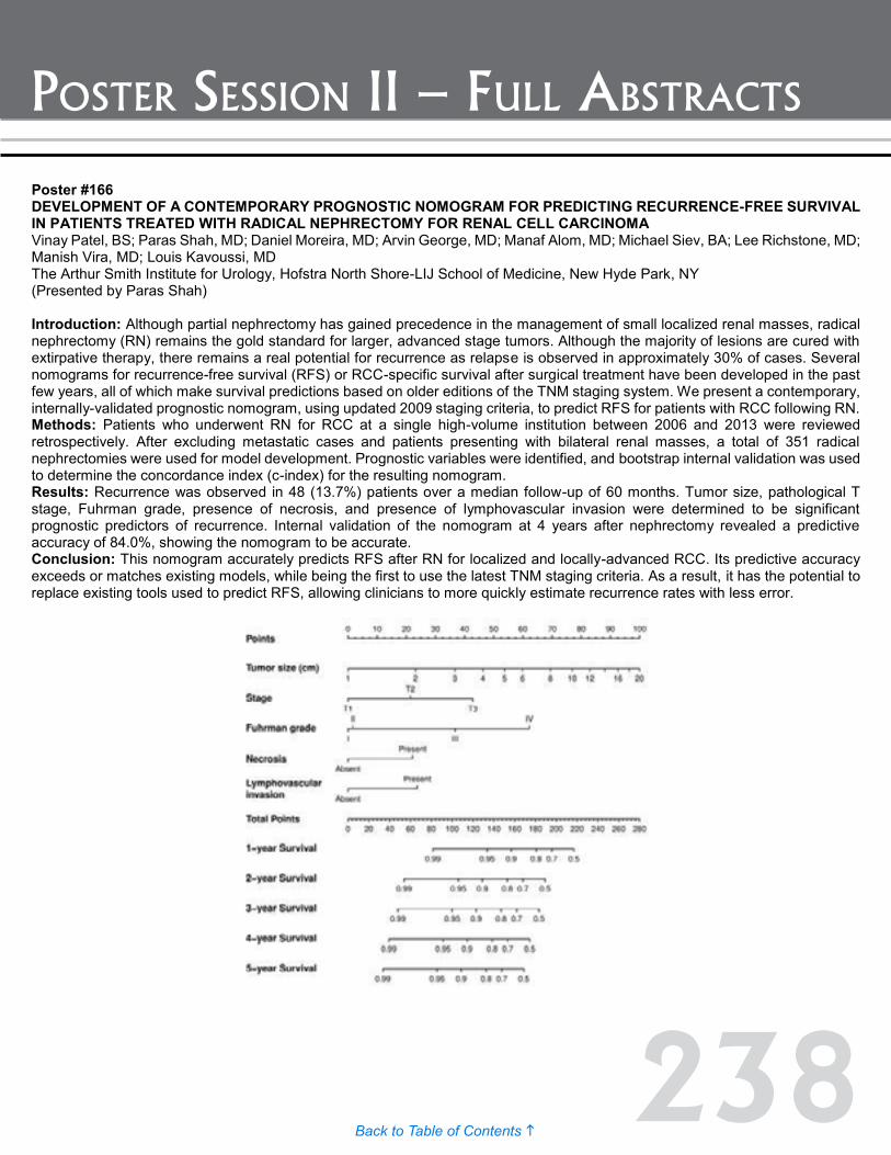
238Back to Table of Contents h
Poster session ii – Full AbstrActs
Poster #166 DEVELOPMENT OF A CONTEMPORARY PROGNOSTIC NOMOGRAM FOR PREDICTING RECURRENCE-FREE SURVIVAL IN PATIENTS TREATED WITH RADICAL NEPHRECTOMY FOR RENAL CELL CARCINOMA Vinay Patel, BS; Paras Shah, MD; Daniel Moreira, MD; Arvin George, MD; Manaf Alom, MD; Michael Siev, BA; Lee Richstone, MD; Manish Vira, MD; Louis Kavoussi, MD The Arthur Smith Institute for Urology, Hofstra North Shore-LIJ School of Medicine, New Hyde Park, NY (Presented by Paras Shah) Introduction: Although partial nephrectomy has gained precedence in the management of small localized renal masses, radical nephrectomy (RN) remains the gold standard for larger, advanced stage tumors. Although the majority of lesions are cured with extirpative therapy, there remains a real potential for recurrence as relapse is observed in approximately 30% of cases. Several nomograms for recurrence-free survival (RFS) or RCC-specific survival after surgical treatment have been developed in the past few years, all of which make survival predictions based on older editions of the TNM staging system. We present a contemporary, internally-validated prognostic nomogram, using updated 2009 staging criteria, to predict RFS for patients with RCC following RN. Methods: Patients who underwent RN for RCC at a single high-volume institution between 2006 and 2013 were reviewed retrospectively. After excluding metastatic cases and patients presenting with bilateral renal masses, a total of 351 radical nephrectomies were used for model development. Prognostic variables were identified, and bootstrap internal validation was used to determine the concordance index (c-index) for the resulting nomogram. Results: Recurrence was observed in 48 (13.7%) patients over a median follow-up of 60 months. Tumor size, pathological T stage, Fuhrman grade, presence of necrosis, and presence of lymphovascular invasion were determined to be significant prognostic predictors of recurrence. Internal validation of the nomogram at 4 years after nephrectomy revealed a predictive accuracy of 84.0%, showing the nomogram to be accurate. Conclusion: This nomogram accurately predicts RFS after RN for localized and locally-advanced RCC. Its predictive accuracy exceeds or matches existing models, while being the first to use the latest TNM staging criteria. As a result, it has the potential to replace existing tools used to predict RFS, allowing clinicians to more quickly estimate recurrence rates with less error.
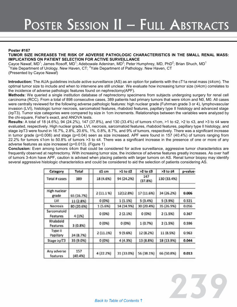
239Back to Table of Contents h
Poster session ii – Full AbstrActs
Poster #167 TUMOR SIZE INCREASES THE RISK OF ADVERSE PATHOLOGIC CHARACTERISTICS IN THE SMALL RENAL MASS: IMPLICATIONS ON PATIENT SELECTION FOR ACTIVE SURVEILLANCE Cayce Nawaf, MD1; James Rosoff, MD1; Adebowale Adeniran, MD2; Peter Humphrey, MD, PhD2; Brian Shuch, MD1 1Yale Department of Urology. New Haven, CT; 2Yale Department of Pathology. New Haven, CT (Presented by Cayce Nawaf) Introduction: The AUA guidelines include active surveillance (AS) as an option for patients with the cT1a renal mass (≤4cm). The optimal tumor size to include and when to intervene are still unclear. We evaluate how increasing tumor size (≤4cm) correlates to the incidence of adverse pathologic features found on nephrectomy(APF). Methods: We queried a single institution database of nephrectomy specimens from subjects undergoing surgery for renal cell carcinoma (RCC). From a total of 898 consecutive cases, 389 patients had primary tumors that were ≤4cm and N0, M0. All cases were centrally reviewed for the following adverse pathologic features: high nuclear grade (Fuhrman grade 3 or 4), lymphovascular invasion (LVI), histologic tumor necrosis, sarcomatoid features, rhabdoid features, papillary type II histology and advanced stage (≥pT3). Tumor size categories were compared by size in 1cm increments. Relationships between the variables were analyzed by the chi-square, Fisher’s exact, and ANOVA tests. Results: A total of 18 (4.6%), 94 (24.2%), 147 (37.8%), and 130 (33.4%) of tumors ≤1cm, >1 to ≤2, >2 to ≤3, and >3 to ≤4 were evaluated, respectively. High nuclear grade, LVI, necrosis, sarcomatoid features, rhabdoid features, papillary type II histology, and stage ≥pT3 were found in 16.7%, 2.8%, 20.6%, 1%, 0.8%, 8.7%, and 9% of tumors, respectively. There was a significant increase in tumor grade (p=0.006) and stage (p=0.04) seen as size increased. APF were found in 157 (40.4%) of tumors ranging from 22.2% for tumors ≤1cm to 50.8% of tumors >3 to ≤4. There was a significant increase in the presence of one or more of any adverse features as size increased (p=0.013). (Figure 1) Conclusion: Even among tumors ≤4cm that could be considered for active surveillance, aggressive tumor characteristics are frequently observed at nephrectomy. With increasing tumor size, the incidence of adverse features greatly increases. As over half of tumors 3-4cm have APF, caution is advised when placing patients with larger tumors on AS. Renal tumor biopsy may identify several aggressive histologic characteristics and could be considered to aid the selection of patients considering AS.
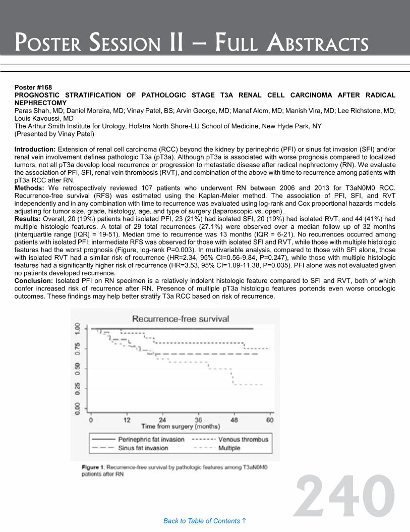
240Back to Table of Contents h
Poster session ii – Full AbstrActs
Poster #168 PROGNOSTIC STRATIFICATION OF PATHOLOGIC STAGE T3A RENAL CELL CARCINOMA AFTER RADICAL NEPHRECTOMY Paras Shah, MD; Daniel Moreira, MD; Vinay Patel, BS; Arvin George, MD; Manaf Alom, MD; Manish Vira, MD; Lee Richstone, MD; Louis Kavoussi, MD The Arthur Smith Institute for Urology, Hofstra North Shore-LIJ School of Medicine, New Hyde Park, NY (Presented by Vinay Patel) Introduction: Extension of renal cell carcinoma (RCC) beyond the kidney by perinephric (PFI) or sinus fat invasion (SFI) and/or renal vein involvement defines pathologic T3a (pT3a). Although pT3a is associated with worse prognosis compared to localized tumors, not all pT3a develop local recurrence or progression to metastatic disease after radical nephrectomy (RN). We evaluate the association of PFI, SFI, renal vein thrombosis (RVT), and combination of the above with time to recurrence among patients with pT3a RCC after RN. Methods: We retrospectively reviewed 107 patients who underwent RN between 2006 and 2013 for T3aN0M0 RCC. Recurrence-free survival (RFS) was estimated using the Kaplan-Meier method. The association of PFI, SFI, and RVT independently and in any combination with time to recurrence was evaluated using log-rank and Cox proportional hazards models adjusting for tumor size, grade, histology, age, and type of surgery (laparoscopic vs. open). Results: Overall, 20 (19%) patients had isolated PFI, 23 (21%) had isolated SFI, 20 (19%) had isolated RVT, and 44 (41%) had multiple histologic features. A total of 29 total recurrences (27.1%) were observed over a median follow up of 32 months (interquartile range [IQR] = 19-51). Median time to recurrence was 13 months (IQR = 6-21). No recurrences occurred among patients with isolated PFI; intermediate RFS was observed for those with isolated SFI and RVT, while those with multiple histologic features had the worst prognosis (Figure, log-rank P=0.003). In multivariable analysis, compared to those with SFI alone, those with isolated RVT had a similar risk of recurrence (HR=2.34, 95% CI=0.56-9.84, P=0.247), while those with multiple histologic features had a significantly higher risk of recurrence (HR=3.53, 95% CI=1.09-11.38, P=0.035). PFI alone was not evaluated given no patients developed recurrence. Conclusion: Isolated PFI on RN specimen is a relatively indolent histologic feature compared to SFI and RVT, both of which confer increased risk of recurrence after RN. Presence of multiple pT3a histologic features portends even worse oncologic outcomes. These findings may help better stratify T3a RCC based on risk of recurrence.
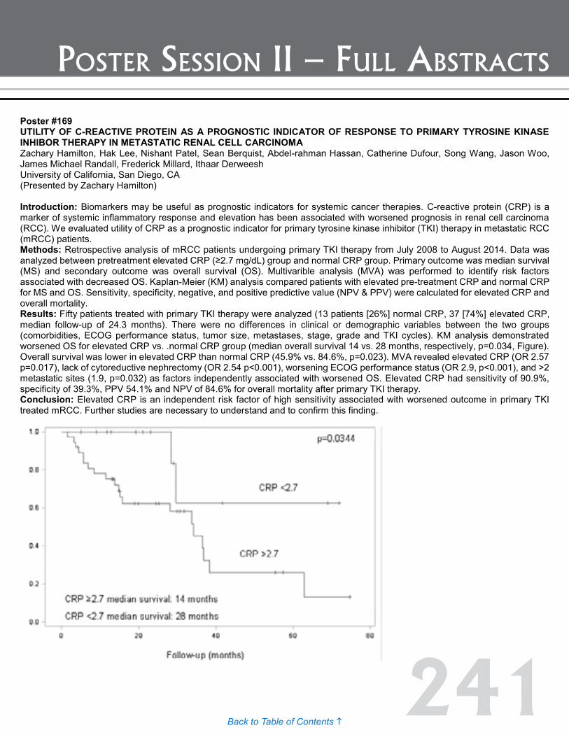
241Back to Table of Contents h
Poster session ii – Full AbstrActs
Poster #169 UTILITY OF C-REACTIVE PROTEIN AS A PROGNOSTIC INDICATOR OF RESPONSE TO PRIMARY TYROSINE KINASE INHIBOR THERAPY IN METASTATIC RENAL CELL CARCINOMA Zachary Hamilton, Hak Lee, Nishant Patel, Sean Berquist, Abdel-rahman Hassan, Catherine Dufour, Song Wang, Jason Woo, James Michael Randall, Frederick Millard, Ithaar Derweesh University of California, San Diego, CA (Presented by Zachary Hamilton) Introduction: Biomarkers may be useful as prognostic indicators for systemic cancer therapies. C-reactive protein (CRP) is a marker of systemic inflammatory response and elevation has been associated with worsened prognosis in renal cell carcinoma (RCC). We evaluated utility of CRP as a prognostic indicator for primary tyrosine kinase inhibitor (TKI) therapy in metastatic RCC (mRCC) patients. Methods: Retrospective analysis of mRCC patients undergoing primary TKI therapy from July 2008 to August 2014. Data was analyzed between pretreatment elevated CRP (≥2.7 mg/dL) group and normal CRP group. Primary outcome was median survival (MS) and secondary outcome was overall survival (OS). Multivarible analysis (MVA) was performed to identify risk factors associated with decreased OS. Kaplan-Meier (KM) analysis compared patients with elevated pre-treatment CRP and normal CRP for MS and OS. Sensitivity, specificity, negative, and positive predictive value (NPV & PPV) were calculated for elevated CRP and overall mortality. Results: Fifty patients treated with primary TKI therapy were analyzed (13 patients [26%] normal CRP, 37 [74%] elevated CRP, median follow-up of 24.3 months). There were no differences in clinical or demographic variables between the two groups (comorbidities, ECOG performance status, tumor size, metastases, stage, grade and TKI cycles). KM analysis demonstrated worsened OS for elevated CRP vs. .normal CRP group (median overall survival 14 vs. 28 months, respectively, p=0.034, Figure). Overall survival was lower in elevated CRP than normal CRP (45.9% vs. 84.6%, p=0.023). MVA revealed elevated CRP (OR 2.57 p=0.017), lack of cytoreductive nephrectomy (OR 2.54 p<0.001), worsening ECOG performance status (OR 2.9, p<0.001), and >2 metastatic sites (1.9, p=0.032) as factors independently associated with worsened OS. Elevated CRP had sensitivity of 90.9%, specificity of 39.3%, PPV 54.1% and NPV of 84.6% for overall mortality after primary TKI therapy. Conclusion: Elevated CRP is an independent risk factor of high sensitivity associated with worsened outcome in primary TKI treated mRCC. Further studies are necessary to understand and to confirm this finding.
Poster #168 PROGNOSTIC STRATIFICATION OF PATHOLOGIC STAGE T3A RENAL CELL CARCINOMA AFTER RADICAL NEPHRECTOMY Paras Shah, MD; Daniel Moreira, MD; Vinay Patel, BS; Arvin George, MD; Manaf Alom, MD; Manish Vira, MD; Lee Richstone, MD; Louis Kavoussi, MD The Arthur Smith Institute for Urology, Hofstra North Shore-LIJ School of Medicine, New Hyde Park, NY (Presented by Vinay Patel) Introduction: Extension of renal cell carcinoma (RCC) beyond the kidney by perinephric (PFI) or sinus fat invasion (SFI) and/or renal vein involvement defines pathologic T3a (pT3a). Although pT3a is associated with worse prognosis compared to localized tumors, not all pT3a develop local recurrence or progression to metastatic disease after radical nephrectomy (RN). We evaluate the association of PFI, SFI, renal vein thrombosis (RVT), and combination of the above with time to recurrence among patients with pT3a RCC after RN. Methods: We retrospectively reviewed 107 patients who underwent RN between 2006 and 2013 for T3aN0M0 RCC. Recurrence-free survival (RFS) was estimated using the Kaplan-Meier method. The association of PFI, SFI, and RVT independently and in any combination with time to recurrence was evaluated using log-rank and Cox proportional hazards models adjusting for tumor size, grade, histology, age, and type of surgery (laparoscopic vs. open). Results: Overall, 20 (19%) patients had isolated PFI, 23 (21%) had isolated SFI, 20 (19%) had isolated RVT, and 44 (41%) had multiple histologic features. A total of 29 total recurrences (27.1%) were observed over a median follow up of 32 months (interquartile range [IQR] = 19-51). Median time to recurrence was 13 months (IQR = 6-21). No recurrences occurred among patients with isolated PFI; intermediate RFS was observed for those with isolated SFI and RVT, while those with multiple histologic features had the worst prognosis (Figure, log-rank P=0.003). In multivariable analysis, compared to those with SFI alone, those with isolated RVT had a similar risk of recurrence (HR=2.34, 95% CI=0.56-9.84, P=0.247), while those with multiple histologic features had a significantly higher risk of recurrence (HR=3.53, 95% CI=1.09-11.38, P=0.035). PFI alone was not evaluated given no patients developed recurrence. Conclusion: Isolated PFI on RN specimen is a relatively indolent histologic feature compared to SFI and RVT, both of which confer increased risk of recurrence after RN. Presence of multiple pT3a histologic features portends even worse oncologic outcomes. These findings may help better stratify T3a RCC based on risk of recurrence.
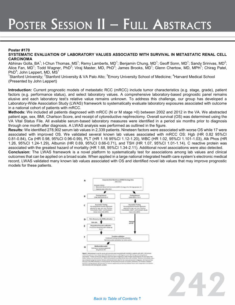
242Back to Table of Contents h
Poster session ii – Full AbstrActs
Poster #170 SYSTEMATIC EVALUATION OF LABORATORY VALUES ASSOCIATED WITH SURVIVAL IN METASTATIC RENAL CELL CARCINOMA Abhinav Golla, BA1; I-Chun Thomas, MS1; Remy Lamberts, MD1; Benjamin Chung, MD1; Geoff Sonn, MD1; Sandy Srinivas, MD2; Alice Fan, MD1; Todd Wagner, PhD2; Viraj Master, MD, PhD3; James Brooks, MD1; Glenn Chertow, MD, MPH1; Chirag Patel, PhD4; John Leppert, MD, MS1 1Stanford University; 2Stanford University & VA Palo Alto; 3Emory University School of Medicine; 4Harvard Medical School (Presented by John Leppert) Introduction: Current prognostic models of metastatic RCC (mRCC) include tumor characteristics (e.g. stage, grade), patient factors (e.g. performance status), and select laboratory values. A comprehensive laboratory-based prognostic panel remains elusive and each laboratory test’s relative value remains unknown. To address this challenge, our group has developed a Laboratory-Wide Association Study (LWAS) framework to systematically evaluate laboratory exposures associated with outcome in a national cohort of patients with mRCC. Methods: We included all patients diagnosed with mRCC (N or M stage >0) between 2002 and 2012 in the VA. We abstracted patient age, sex, BMI, Charlson Score, and receipt of cytoreductive nephrectomy. Overall survival (OS) was determined using the VA Vital Status File. All available serum-based laboratory measures were identified in a period six months prior to diagnosis through one month after diagnosis. A LWAS analysis was performed as outlined in the figure. Results: We identified 278,902 serum lab values in 2,339 patients. Nineteen factors were associated with worse OS while 17 were associated with improved OS. We validated several known lab values associated with mRCC OS: Hgb (HR 0.82 95%CI 0.81-0.84), Ca (HR 0.98, 95%CI 0.96-0.99), PLT (HR 1.16 95%CI 1.12-1.20), WBC (HR 1.02, 95%CI 1.101-1.03), Alk Phos (HR 1.26, 95%CI 1.24-1.29), Albumin (HR 0.69, 95%CI 0.66-0.71), and TSH (HR 1.07, 95%CI 1.01-1.14). C reactive protein was associated with the greatest hazard of mortality (HR 1.68, 95%CI 1.34-2.11). Additional novel associations were also detected. Conclusion: The LWAS framework is a novel platform to systematically test for associations among lab values and clinical outcomes that can be applied on a broad scale. When applied in a large national integrated health care system’s electronic medical record, LWAS validated many known lab values associated with OS and identified novel lab values that may improve prognostic models for these patients.
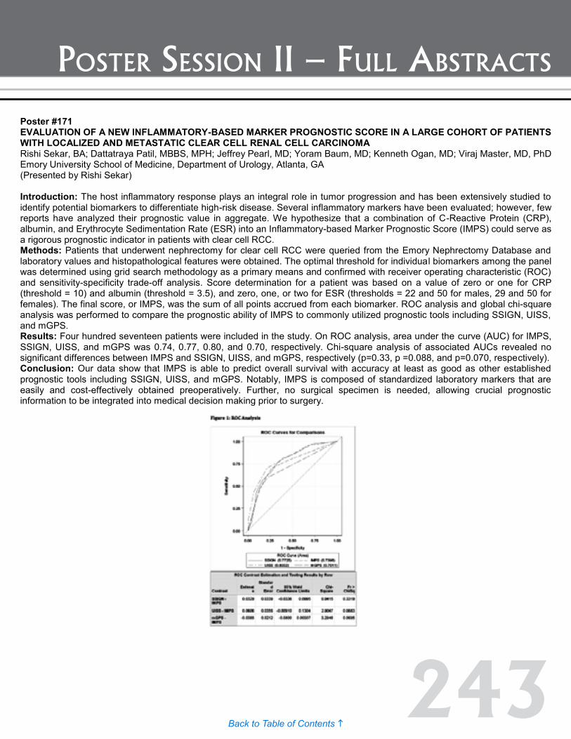
243Back to Table of Contents h
Poster session ii – Full AbstrActs
Poster #171 EVALUATION OF A NEW INFLAMMATORY-BASED MARKER PROGNOSTIC SCORE IN A LARGE COHORT OF PATIENTS WITH LOCALIZED AND METASTATIC CLEAR CELL RENAL CELL CARCINOMA Rishi Sekar, BA; Dattatraya Patil, MBBS, MPH; Jeffrey Pearl, MD; Yoram Baum, MD; Kenneth Ogan, MD; Viraj Master, MD, PhD Emory University School of Medicine, Department of Urology, Atlanta, GA (Presented by Rishi Sekar) Introduction: The host inflammatory response plays an integral role in tumor progression and has been extensively studied to identify potential biomarkers to differentiate high-risk disease. Several inflammatory markers have been evaluated; however, few reports have analyzed their prognostic value in aggregate. We hypothesize that a combination of C-Reactive Protein (CRP), albumin, and Erythrocyte Sedimentation Rate (ESR) into an Inflammatory-based Marker Prognostic Score (IMPS) could serve as a rigorous prognostic indicator in patients with clear cell RCC. Methods: Patients that underwent nephrectomy for clear cell RCC were queried from the Emory Nephrectomy Database and laboratory values and histopathological features were obtained. The optimal threshold for individual biomarkers among the panel was determined using grid search methodology as a primary means and confirmed with receiver operating characteristic (ROC) and sensitivity-specificity trade-off analysis. Score determination for a patient was based on a value of zero or one for CRP (threshold = 10) and albumin (threshold = 3.5), and zero, one, or two for ESR (thresholds = 22 and 50 for males, 29 and 50 for females). The final score, or IMPS, was the sum of all points accrued from each biomarker. ROC analysis and global chi-square analysis was performed to compare the prognostic ability of IMPS to commonly utilized prognostic tools including SSIGN, UISS, and mGPS. Results: Four hundred seventeen patients were included in the study. On ROC analysis, area under the curve (AUC) for IMPS, SSIGN, UISS, and mGPS was 0.74, 0.77, 0.80, and 0.70, respectively. Chi-square analysis of associated AUCs revealed no significant differences between IMPS and SSIGN, UISS, and mGPS, respectively (p=0.33, p =0.088, and p=0.070, respectively). Conclusion: Our data show that IMPS is able to predict overall survival with accuracy at least as good as other established prognostic tools including SSIGN, UISS, and mGPS. Notably, IMPS is composed of standardized laboratory markers that are easily and cost-effectively obtained preoperatively. Further, no surgical specimen is needed, allowing crucial prognostic information to be integrated into medical decision making prior to surgery.
Poster #170 SYSTEMATIC EVALUATION OF LABORATORY VALUES ASSOCIATED WITH SURVIVAL IN METASTATIC RENAL CELL CARCINOMA Abhinav Golla, BA1; I-Chun Thomas, MS1; Remy Lamberts, MD1; Benjamin Chung, MD1; Geoff Sonn, MD1; Sandy Srinivas, MD2; Alice Fan, MD1; Todd Wagner, PhD2; Viraj Master, MD, PhD3; James Brooks, MD1; Glenn Chertow, MD, MPH1; Chirag Patel, PhD4; John Leppert, MD, MS1 1Stanford University; 2Stanford University & VA Palo Alto; 3Emory University School of Medicine; 4Harvard Medical School (Presented by John Leppert) Introduction: Current prognostic models of metastatic RCC (mRCC) include tumor characteristics (e.g. stage, grade), patient factors (e.g. performance status), and select laboratory values. A comprehensive laboratory-based prognostic panel remains elusive and each laboratory test’s relative value remains unknown. To address this challenge, our group has developed a Laboratory-Wide Association Study (LWAS) framework to systematically evaluate laboratory exposures associated with outcome in a national cohort of patients with mRCC. Methods: We included all patients diagnosed with mRCC (N or M stage >0) between 2002 and 2012 in the VA. We abstracted patient age, sex, BMI, Charlson Score, and receipt of cytoreductive nephrectomy. Overall survival (OS) was determined using the VA Vital Status File. All available serum-based laboratory measures were identified in a period six months prior to diagnosis through one month after diagnosis. A LWAS analysis was performed as outlined in the figure. Results: We identified 278,902 serum lab values in 2,339 patients. Nineteen factors were associated with worse OS while 17 were associated with improved OS. We validated several known lab values associated with mRCC OS: Hgb (HR 0.82 95%CI 0.81-0.84), Ca (HR 0.98, 95%CI 0.96-0.99), PLT (HR 1.16 95%CI 1.12-1.20), WBC (HR 1.02, 95%CI 1.101-1.03), Alk Phos (HR 1.26, 95%CI 1.24-1.29), Albumin (HR 0.69, 95%CI 0.66-0.71), and TSH (HR 1.07, 95%CI 1.01-1.14). C reactive protein was associated with the greatest hazard of mortality (HR 1.68, 95%CI 1.34-2.11). Additional novel associations were also detected. Conclusion: The LWAS framework is a novel platform to systematically test for associations among lab values and clinical outcomes that can be applied on a broad scale. When applied in a large national integrated health care system’s electronic medical record, LWAS validated many known lab values associated with OS and identified novel lab values that may improve prognostic models for these patients.
244Back to Table of Contents h
Poster session ii – Full AbstrActs
Poster #172 HYPERTENSION AND DIABETES ARE NOT INDEPENDENT PREDICTORS OF WORSE RENAL FUNCTION OUTCOMES FOLLOWING PARTIAL NEPHRECTOMY Balaji Reddy, MD1; David Paulucci, BA1; Ronney Abaza, MD2; Daniel Eun, MD3; John Sfakianos, MD1; Ketan Badani, MD1 1Icahn School of Medicine at Mount Sinai, New York, NY; 2Dublin Methodist Hospital, Dublin, OH; 3Temple University, Philadelphia, PA (Presented by Balaji Reddy) Introduction: Partial nephrectomy (PN) is offered to patients with renal masses deemed amenable in attempting to preserve renal function while not compromising cancer specific outcomes. Renal function preservation is also shown to improve overall survival as a consequence of decreased cardiac morbidity. The present study therefore sought to identify the factors affecting estimated glomerular filtration rate (eGFR) decline in patients undergoing PN, and further investigate the role of hypertension (HTN) and diabetes mellitus (DM) in developing chronic kidney disease (CKD). Methods: Subjects who underwent robotic PN across three institutions with <T1b (clinical) renal masses and follow-up >2 weeks were included in the analysis. Patients were classified according to preoperative kidney function (PKF) as follows: PKF Group 1 eGFR ≥90, PKF Group 2 eGFR 60-90, PKF Group 3 eGFR <60 mL/min/1.73m2. Multivariable linear regression and Cox Proportion Hazards models were respectively used to compare the percentage change in eGFR and risk of CKD upstaging between PKF groups and between patients with vs. without HTN or DM in PKF group 1 at two to 24 weeks and more than 24 weeks adjusting for demographic and perioperative variables (age, BMI, ASA, tumor size, RENAL score, warm ischemia time). Results: We identified 383 patients with 163 (42.6%) in PKF group 1, 165 (43.1%) in PKF group 2 and 55 (14.4%) in PKF group 3 with a median follow up of 13.8, 17 and 15.9 months respectively (p>0.05). The percent reduction in eGFR was significantly lower in PKF group 3 vs. 1 (b=25.74, p<.001) at two to 24 weeks and more than 24 weeks (b=12.41, p=.03). The risk of CKD upstaging was significantly lower for PKF group 3 vs. 1 (HR=0.09, p=.021) at more than 24 weeks. 133 patients in PKF group 1 had comorbidity data available, 78 patients with either DM/HTN and 55 with neither. There was no difference in baseline eGFR, percent change in eGFR or risk of CKD upstaging in eGFR at two to 24 weeks or greater than 24 weeks (p>.05) between the two groups. Only baseline eGFR significantly predicted eGFR decline at greater than six months (b=-0.41, p=0.012) in PKF group 1. Conclusion: The percentage reduction in eGFR and risk of CKD upstaging were significantly higher in PKF group 1 vs. 3. The presence of DM and HTN are not independent risk factors of GFR decline in patients with baseline eGFR ≥ 90.
245Back to Table of Contents h
Poster session ii – Full AbstrActs
Poster #173 MITOMYCIN-C INDUCTION AND MAINTENANCE TOPICAL THERAPY FOR UPPER TRACT UROTHELIAL CARCINOMA Gavin Wagenheim, MD1,2; John Papadopoulos, MD2; Neema Navai, MD2; John Davis, MD2; Jose Karam, MD2; Ashish Kamat, MD2; Christopher Wood, MD2; Colin Dinney, MD2; Surena Matin, MD2 1The University of Texas - Houston, Medical School, Houston, TX; 2The University of Texas, MD Anderson Cancer Center, Houston, TX (Presented by Gavin Wagenheim) Introduction: Reported recurrence rates following endoscopic treatment of upper tract urothelial carcinoma (UTUC) are high, from 30% to 70%. Adjuvant BCG topical therapy has not shown the same success in reducing recurrences as for treatment of upper tract CIS. Similarly, results are inferior to what is seen in adjuvant treatment of bladder cancer. This is likely due to difficulty in definitive delivery to the upper tract and the absence of a reservoir for long dwell times. There is limited reported use of adjuvant mitomycin-c (MMC) for UTUC, and to our knowledge no reported experience of topical delivery using the SWOG maintenance regimen. We hypothesized that a chemotherapeutic agent could be effective as adjvuant topical therapy of the upper tract, particularly when given using SWOG recommendations. We report the efficacy, safety, and tolerability of this approach. Methods: We reviewed charts of all patients undergoing primary endoscopic biopsy/resection and ablation of an UTUC, recording relevant clinical, pathologic, laboratory, and follow up information. Patients were offered induction and maintenance topical therapy after endoscopic control. MMC was given as the initial adjuvant topical agent for 6 weeks induction and 3 weeks maintenance for up to 2 years per SWOG protocol. Delivery was either via percutaneous nephrostomy or cystoscopically placed ureteral catheter, per patient preference. Results: Twenty-seven patients were identified, 20 (74%) were low grade and seven (26%) were high grade. Delivery of MMC was via percutaneous nephrostomy in 30% and ureteral catheter in 70%. 44% were treated on an imperative basis, 48% elective, and seven percent palliative. No patients discontinued therapy due to intolerance, and 63% received maintenance therapy. Only 11% of patients undergoing induction and seven percent receiving maintenance therapy incurred complications. With a mean follow up of 23 months (range 1-79), recurrence-free, progression-free, and nephroureterectomy-free survival in all patients, low-grade patients, and high-grade patients was 70%, 70%, and 71%; 89%, 90%, and 85%; and 89%, 90%, and 85%, respectively. Conclusion: In patients with complete endoscopic control, upper tract topical instillation of MMC as induction and maintenance via either percutaneous nephrostomy or ureteral catheter is a well-tolerated, feasible, and perhaps beneficial treatment option for low-grade and possibly high-grade tumors.
246Back to Table of Contents h
Poster session ii – Full AbstrActs
Poster #174 PATHOLOGICAL AND CLINICAL FACTORS ASSOCIATED WITH DEVELOPMENT AND OUTCOMES OF BONE METASTASES IN METASTATIC RENAL CELL CARCINOMA Zachary Hamilton, Omer Raheem, Hak Lee, Song Wang, Jason Woo, Sean Berquist, Frederick Millard, Ithaar Derweesh University of California, San Diego, CA (Presented by Zachary Hamilton) Introduction: Despite stage migration, metastatic renal cell carcinoma (mRCC) continues to constitute a significant clinical problem. We examined impact of risk factors associated with development and clinical outcomes of bone metastasis (BM) in a contemporary cohort. Methods: Retrospective analysis of mRCC patients diagnosed between 2005 and 2014. Data were analyzed within subgroups based on presence or absence of BM. Primary endpoint was overall survival (OS) and secondary outcomes included cancer-specific survival (CSS), and mortality and recurrence rates. Multivariable analysis (MVA) was conducted to assess variables correlated with OS and CSS of mRCC patients. Results: 171 patients with mRCC were analyzed (median follow-up 24.5 months); 29 (17%) patients had BM and 142 (83%) did not. There were no statistical differences with respect to demographic variables or median primary tumor size (BM 8cm vs. non BM 8.9cm, p=0.186). Significantly greater proportion of patients without BM underwent cytoreductive nephrectomy (82.8% vs. 59.2%, p=0.016). Overall rates of recurrence were higher in the BM group (93% vs. 51%, p=0.0001), as were all-cause mortality (59% vs. 37%, p=0.03), and cancer-specific mortality (59 vs. 34%, p=0.01). MVA showed that BM compared to other metastatic sites (OR 4.55, p=0.012), lack of CN (OR 3.11, p<0.001), and poor performance status (OR 4.24, p=0.012) were independently associated with reduced OS. BM compared to other metastatic sites (OR 4.86, p=0.004) and no CN (OR 2.5, p=0.037) were independently associated with reduced CSS, as well. MVA for pathological factors associated with BM revealed high Furman grade (III/IV, OR 2.76, p<0.001), lymphovascular invasion (OR 1.8, p=0.021), and tumor necrosis (OR 2.13, p=0.014) as factors independently associated with development of bone metastases. Presence of any two (OR 4.46, p<0.001) or three (OR 7.39, p=0.005) risk factors in combination further increased risk of development of BM. Conclusion: Even in the targeted therapy era, BM in mRCC has a negative impact on survival. Presence of high Fuhrman grade, lymphovascular invasion and necrosis and combinations of these factors should increase clinical suspicion and vigilance for development of BM. BM mRCC or patients at risk for BM in mRCC should be considered for early inclusion in clinical trials with combination therapies or emerging agents.
247Back to Table of Contents h
Poster session ii – Full AbstrActs
Poster #175 EVALUATION OF THE NON-NEOPLASTIC KIDNEY AS A PREDICTOR OF RENAL INSUFFICIENCY IN RADICAL NEPHRECTOMY SPECIMENS Deepak Pruthi; Vivian Liu, MD; Ruchi Chhibba, BSc; Evan Weins, BSc; Ian Gibson, MD; Thomas McGregor, MD; FRCS(C) University of Manitoba (Presented by Deepak Pruthi) Introduction: To identify predictors of post-operative renal insufficiency by analyzing clinical co-morbidities and pathologic changes in the non-neoplastic kidney (NNK) parenchyma of patients undergoing radical nephrectomy (RN) for suspected renal cell carcinoma (RCC). Methods: A retrospective review of all patients undergoing RN for suspected RCC measuring <10cm between January 1, 2011 and May 31, 2014. All slides were independently reviewed for histopathologic changes using the Banff II criteria by two blinded dedicated nephropathogists for any glomerular, tubulointerstitial, arterial/arteriolar changes (GTA). Standard statistical analysis was employed. Estimated glomerular rate (eGFR) was calculated using chronic kidney disease epidemiology collaboration formula. Results: In total 147 patients met the inclusion criteria. The group included a mean Charlson comorbidity score of 2.3; 21% were diabetics, 59% hypertensives, and 36% smokers. Mean age was 60, mean body mass index was 30, and mean pathologic tumor size was 7.3. Mean pre-op eGFR was 76.5 (median 21.9) with a mean six and 12 month post-op eGFR of 53 (median 16.5) and 50.8 (median 18.6), respectively. Sixty-four (56%) patients developed new onset CKDIII. Four patients went on to dialysis in one year and nine patients died during follow-up. Increasing age trended toward greater six month percentage declines in eGFR (p=0.06) but this was attenuated at 12 months. When compared to patients with mild-moderate changes, patients with baseline NNK severe tubulointerstitial changes (eGFR -55 vs. -30; p=0.024) and severe glomerular changes (eGFR -44 vs. -28; p=0.033) were more likely to have significant declines in the 12 month eGFRs. Arterial/arteriolar changes (p=0.66), diabetes (p=0.13), hypertension (p=0.85), nor positive smoking status (p=0.48) predicted post-operative declines in eGFR. Conclusion: Pre-operative comorbidities did not predict renal insufficiency. Investigation of architectural changes in the non-neoplastic kidney may aid in the prediction of post-operative renal insufficiency and have significant clinical and therapeutic implications.
Poster #174 PATHOLOGICAL AND CLINICAL FACTORS ASSOCIATED WITH DEVELOPMENT AND OUTCOMES OF BONE METASTASES IN METASTATIC RENAL CELL CARCINOMA Zachary Hamilton, Omer Raheem, Hak Lee, Song Wang, Jason Woo, Sean Berquist, Frederick Millard, Ithaar Derweesh University of California, San Diego, CA (Presented by Zachary Hamilton) Introduction: Despite stage migration, metastatic renal cell carcinoma (mRCC) continues to constitute a significant clinical problem. We examined impact of risk factors associated with development and clinical outcomes of bone metastasis (BM) in a contemporary cohort. Methods: Retrospective analysis of mRCC patients diagnosed between 2005 and 2014. Data were analyzed within subgroups based on presence or absence of BM. Primary endpoint was overall survival (OS) and secondary outcomes included cancer-specific survival (CSS), and mortality and recurrence rates. Multivariable analysis (MVA) was conducted to assess variables correlated with OS and CSS of mRCC patients. Results: 171 patients with mRCC were analyzed (median follow-up 24.5 months); 29 (17%) patients had BM and 142 (83%) did not. There were no statistical differences with respect to demographic variables or median primary tumor size (BM 8cm vs. non BM 8.9cm, p=0.186). Significantly greater proportion of patients without BM underwent cytoreductive nephrectomy (82.8% vs. 59.2%, p=0.016). Overall rates of recurrence were higher in the BM group (93% vs. 51%, p=0.0001), as were all-cause mortality (59% vs. 37%, p=0.03), and cancer-specific mortality (59 vs. 34%, p=0.01). MVA showed that BM compared to other metastatic sites (OR 4.55, p=0.012), lack of CN (OR 3.11, p<0.001), and poor performance status (OR 4.24, p=0.012) were independently associated with reduced OS. BM compared to other metastatic sites (OR 4.86, p=0.004) and no CN (OR 2.5, p=0.037) were independently associated with reduced CSS, as well. MVA for pathological factors associated with BM revealed high Furman grade (III/IV, OR 2.76, p<0.001), lymphovascular invasion (OR 1.8, p=0.021), and tumor necrosis (OR 2.13, p=0.014) as factors independently associated with development of bone metastases. Presence of any two (OR 4.46, p<0.001) or three (OR 7.39, p=0.005) risk factors in combination further increased risk of development of BM. Conclusion: Even in the targeted therapy era, BM in mRCC has a negative impact on survival. Presence of high Fuhrman grade, lymphovascular invasion and necrosis and combinations of these factors should increase clinical suspicion and vigilance for development of BM. BM mRCC or patients at risk for BM in mRCC should be considered for early inclusion in clinical trials with combination therapies or emerging agents.
248Back to Table of Contents h
Poster session ii – Full AbstrActs
Poster #176 A DECADE’S EXPERIENCE WITH MANAGEMENT OF BOSNIAK CYSTS: THE NATURAL HISTORY OF A CHANGE IN DIAGNOSIS Deepak Pruthi; Darrel Drachenbergm MD, FRCS(C) University of Manitoba (Presented by Deepak Pruthi) Introduction: To explore the management including surgical and active surveillance of Bosniak renal cysts and to identify predictors of change in classification. Methods: We retrospectively reviewed all consecutive referrals of patients with complex renal cysts between January 1, 2003 and August 31, 2014. Every radiologic image was reviewed as were the charts and pathology reports. Conflicting cases were independently reviewed with a dedicated Uro-radiologist. Multivariate logistical regression analysis was performed to identify predictors of malignancy and predictors of Bosniak classification change. Results: A total 1104 images were reviewed in the 129 patients were included in the study. This included 807 scans (CT, MRI, ultrasound) of 163 lesions. A total of 538 CT scans were performed on 124 patients (4.2 CT scans per patient). Upon the conclusion of the study one patient died on surveillance while 56 (43%) patients were maintained on surveillance and avoided nephrectomy. A total of 31 patients underwent surgery. Male gender (p<0.005), presence of mural nodule (p=0.035), endophytic lesions (p=0.003), smoking status (p=0.001), cyst size (p<0.001) all predicted malignancy. Bosniak 3 lesions were 50% likely to harbor malignancy. Bosniak stage changes were common 10% of bosniak four lesions were ultimately downstaged. Bosniak 3 lesions had significant variability, 32% of were ultimately downstaged and 13% upstaged. In multivariate analysis bosniak 3 cysts were more likely to be upstaged if there was an enhancing mural nodule and more likely to be downstaged if there was an enhancing septation. Over the study interval Bosniak 3 cysts grew at 1.1% while Bosniak 2F cysts grew at 5.5%. Conclusion: Stage changes in Bosniak lesions are common. Active surveillance of small lesions can help accurately characterize lesions. Perceived cyst complexity can be a function of lesion size and location relative to surrounding parenchyma or cysts. Many Bosniak 3 and 4 cysts ultimately underwent stage changes. Presence of mural nodularity was more predictive of upstaging compared to enhancing septations.
249Back to Table of Contents h
Poster session ii – Full AbstrActs
Poster #177 PRE-OPERATIVE RENAL PARENCHYMAL VOLUMETRICS AND PREDICTION OF RENAL INSUFFICIENCY FOLLOWING RADICAL NEPHRECTOMY Deepak Pruthi; Sacha Oomah, MD; Ruchi Chhibba, BSc; Iain Kirkpatrick, MD; Thomas McGregor, MD, FRCS(C) University of Manitoba (Presented by Deepak Pruthi) Introduction: To identify predictors of post-operative renal insufficiency by analyzing pre-operative imaging volumetrics among patients undergoing radical nephrectomy (RN) for renal cell carcinoma (RCC). Methods: A retrospective review of all patients undergoing RN for RCC between January 2011 and August 2013 was performed. Estimated glomerular filtration rate (eGFR) was calculated employing the modified diet in renal disease (MDRD) formula using the pre-operative and one-year serum creatinine values. Pre-operative and one-year post-operative CT/MRI scans were reviewed. AW Volume Share 5 workstations were utilized to calculate volumes. Statistical analysis using Chi-square and Fisher exact test were employed. Results: In total 65 patients met the inclusion criteria. Patients with smaller tumor volumes (<375cm3) were more likely to have a greater decline in their post-operative eGFRs (>35%) when compared to patients with larger tumors (58 vs. 21% p=0.0165). Patients with a pre-operative eGFR >60 ml/min/1.73m2 had a greater (>25%) drop in their one-year eGFR (81 vs. 47%, p=0.0084). A smaller volume (<150cm3) of parenchyma of the ipsilateral kidney was associated less compensatory (<25%) hypertrophy (100 vs. 69, p=0.0348). Smaller tumors (<300cm3) were more likely to have a greater degree (>10%) of compensatory hypertrophy (78 vs. 43% p=0.0234) and less blood loss (<300cc, 82 vs. 44%, p=0.0082). Older patients and patients with a combined history of diabetes (DM) and hypertension (HTN) were more likely to have compensatory (>25%) hypertrophy (Age>65 100% vs. 31%, p<0.001; DM+HTN 100% vs. 75%, p=0.0084). The degree of compensatory hypertrophy (>25%) however, did not predict one- year eGFR (35%) change (p=0.2203). New kidney disease (eGFR<60 ml/min/1.73m2) occurred in 71% patients. Conclusion: Volumetric imaging shows promising potential and may become a valuable tool in predicting post-operative renal insufficiency for patients undergoing nephrectomy and should be investigated further both in RN and partial nephrectomy cohorts.
250Back to Table of Contents h
Poster session ii – Full AbstrActs
Poster #178 TYPE OF PENILE SPARING SURGERY HAS NO EFFECT ON TIME TO RECURRENCE IN PATIENTS WITH PENILE CANCER Gregory Diorio, DO1; Andrew Leone, MD1; Keenan Ashouri, BS2; Pranav Sharma, MD1; Kamran Zargar-Shoshtari, MD1; Philippe Spiess, MD1 1Moffitt Cancer Center, Tampa, FL; 2Florida Atlantic University School of Medicine, Boca Raton, FL (Presented by Gregory Diorio) Introduction: Squamous Cell Carcinoma of the Penis (PSCC) is a rare but has significant effect on urinary, sexual and psychological health. Traditionally, management of PSCC has included wide excision with a 2cm margin and partial or total amputation. Recent treatment paradigms have demonstrated that penile sparing surgery (PSS) can have excellent oncologic control while preserving function and cosmesis. The purpose of this study is to evaluate the effect of laser ablation (LA) and wide local excision (WLE) on recurrence rates in patients with penile cancer. Methods: We retrospectively reviewed or penile cancer database from 1994 to 2013 for penile cancer patients treated with LA or WLE. Clinical and demographic characteristics were compared using the Mann-Whitney U test to compare medians and the chi-squared test for proportions. Recurrence-free survival (RFS) was examined using the Kaplan-Meir method and comparisons carried out with the log rank test. Multivariate Cox-regression analysis was used to test the association of PSS with disease recurrence. Results: Thirty-seven patients were identified who underwent PSS at our institution. There was no difference between the two groups in terms of age, pTstage, margin status, HPV status, recurrence, or follow up. Median follow up was 62.6 months for LA and 28.7 months for LWE (p=0.81). On Kaplan-Meir analysis median time to recurrence was 3.9 months (SE 1.24 95% CI: 1.45-6.34) for LA vs. 12.1 months (SE 5.1 95% CI: 2.1-22.1) for LWE (p=0.25). Of those who recurred, four patients (36.4%) in the LWE group were managed with partial penectomy, the remainder in both groups were managed with further PSS. On multivariate analysis RFS was not significantly associated with the type of PSS (odds ratio 0.43 95% CI: 0.113-1.67 p=0.22). Histologic grade was an independent predictor of RFS (odds ratio 8.03 95% CI: 1.66-38.85 p=0.01). Conclusion: Type of PSS has no association with RFS in patients with penile cancer though histologic grade may be an adverse predictor of time to recurrence. Further prospective studies are needed to examine the oncologic treatment paradigm in patients with penile cancer while optimizing function and cosmesis. Poster #179 CONTEMPORARY SURVIVAL TRENDS IN PENILE CANCER Jed Ferguson, MD, PhD; Allison Deal, MS1; Angela Smith, MD, MS2; Matthew Nielsen, MD, MS2; Michael Woods, MD2 1UNC Lineberger Cancer Center, Chapel Hill, NC; 2UNC Urology Department, Chapel Hill NC (Presented by Jed Ferguson) Introduction: Penile cancer is a rare neoplasm in the United States, and scarce data exist examining the trends in survival over time. Methods: The National Cancer Database was used to evaluate trends in overall five-year survival in cases of squamous cell carcinoma of the penis between the years of 1998 to 2009. Age, stage, and race were used to construct a multivariate Cox regression model to evaluate hazard ratios for risk of mortality over time. Time periods 1998-2001, 2002-2005, and 2006-2009 were compared. Results: Overall 10,407 adult patients were diagnosed with squamous cell carcinoma of the penis over this time and 9,268 had complete stage information. Stage distribution remained similar over the years evaluated. American Joint Committee on Cancer definitions did not change over this time. Five year overall survival remained similar between time periods: 1998-200, 62.8%; 2002-2005, 62.6%; 2006-2009, 60.8%. On multivariate analysis for all stages combined, age, stage, and race were independent predictors of mortality, but year of diagnosis group was not. Interestingly, when we evaluated each stage individually, stage 2 patients diagnosed between 2006 and 2009 had higher five-year survival rates than earlier years (58.1% vs. 51.8% (2002-2005) and 50.7% (1998-2001). These differences remained statistically significant in multivariate analysis with a p-value of 0.0099. Year of diagnosis group was not statistically significantly different for other stages. Conclusion: Overall survival of penile cancer patients as a whole has remained stable between 1998 and 2009. However, stage II patients showed improved survival between 2006 and 2009. This may reflect an improvement in the surgical management of these patients.
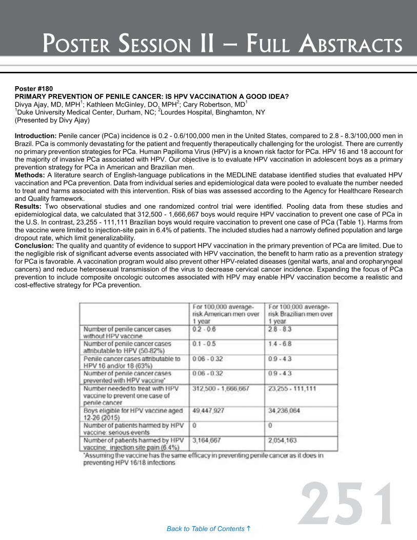
251Back to Table of Contents h
Poster session ii – Full AbstrActs
Poster #180 PRIMARY PREVENTION OF PENILE CANCER: IS HPV VACCINATION A GOOD IDEA? Divya Ajay, MD, MPH1; Kathleen McGinley, DO, MPH2; Cary Robertson, MD1 1Duke University Medical Center, Durham, NC; 2Lourdes Hospital, Binghamton, NY (Presented by Divy Ajay) Introduction: Penile cancer (PCa) incidence is 0.2 - 0.6/100,000 men in the United States, compared to 2.8 - 8.3/100,000 men in Brazil. PCa is commonly devastating for the patient and frequently therapeutically challenging for the urologist. There are currently no primary prevention strategies for PCa. Human Papilloma Virus (HPV) is a known risk factor for PCa. HPV 16 and 18 account for the majority of invasive PCa associated with HPV. Our objective is to evaluate HPV vaccination in adolescent boys as a primary prevention strategy for PCa in American and Brazilian men. Methods: A literature search of English-language publications in the MEDLINE database identified studies that evaluated HPV vaccination and PCa prevention. Data from individual series and epidemiological data were pooled to evaluate the number needed to treat and harms associated with this intervention. Risk of bias was assessed according to the Agency for Healthcare Research and Quality framework. Results: Two observational studies and one randomized control trial were identified. Pooling data from these studies and epidemiological data, we calculated that 312,500 - 1,666,667 boys would require HPV vaccination to prevent one case of PCa in the U.S. In contrast, 23,255 - 111,111 Brazilian boys would require vaccination to prevent one case of PCa (Table 1). Harms from the vaccine were limited to injection-site pain in 6.4% of patients. The included studies had a narrowly defined population and large dropout rate, which limit generalizability. Conclusion: The quality and quantity of evidence to support HPV vaccination in the primary prevention of PCa are limited. Due to the negligible risk of significant adverse events associated with HPV vaccination, the benefit to harm ratio as a prevention strategy for PCa is favorable. A vaccination program would also prevent other HPV-related diseases (genital warts, anal and oropharyngeal cancers) and reduce heterosexual transmission of the virus to decrease cervical cancer incidence. Expanding the focus of PCa prevention to include composite oncologic outcomes associated with HPV may enable HPV vaccination become a realistic and cost-effective strategy for PCa prevention.

252Back to Table of Contents h
Poster session ii – Full AbstrActs
Poster #181 COMPARISON OF SURVIVAL OUTCOMES FOR AFRICAN-AMERICAN AND CAUCASIAN MEN WITH ADVANCED PENILE CANCER IN FLORIDA Chad R. Ritch, MD, MBA1; Nicola Pavan, MD2; Samarpit Rai, MD1; Nachiketh Soodana-Prakash, MD, MS1; Raymond R. Balise, PhD3; Dipen J. Parekh, MD1; Mark L. Gonzalgo, MD, PhD1 1Department of Urology, University of Miami Leonard M. Miller School of Medicine, Miami, FL; 2Department of Urology, University of Miami Leonard M. Miller School of Medicine, Miami, FL and Urology Clinic, Department of Medical, Surgical and Health Science, University of Trieste, Italy; 3Division of Biostatistics, Department of Public Health Sciences, University of Miami Leonard M. Miller School of Medicine, Miami, FL (Presented by Chad R. Ritch) Introduction: Studies suggest that there may be disparity in clinical outcomes for African-American men (AAM) compared to Caucasian men (CM) with penile squamous cell carcinoma (SCC). We sought to determine whether there was a survival difference for African American versus Caucasian men, particularly in locally advanced and metastatic cases of penile SCC where disease mortality is highest. Methods: Using the Florida Cancer Data System, we identified men diagnosed with penile SCC from 2004 to 2014. We excluded men who were diagnosed on autopsy or at the time of death and with more than six months of follow up. Demographic variables including: age, follow-up, stage, race and treatment type were compared between AAM andcm. Treatment type was categorized as surgery alone or surgery plus additional therapy (chemotherapy and/or radiation). For locally advanced and metastatic disease, we compared treatment type and overall survival (OS) between AAM andcm. A multivariable model was developed to determine significant predictors of OS. Results: Of the 653 men with penile SCC, 198 (38 [19%] AAM and 160 [81%] CM) had locally advanced and/or metastatic disease. Median follow up for the entire cohort was 12.5 mos. For all stages, AAM demonstrated a significantly decreased median OS compared tocm (26 vs. 37 mos, p=0.03). For locally advanced and metastatic disease, there was a persistent, but non-significant, trend toward disparity in median OS between AAM and CM (17 vs. 23 mos, p=0.06). Fewer AAM compared tocm received surgery plus additional therapy for locally advanced and/or metastatic disease (8 [21%] vs. 42 [26%], but this difference was not statistically significant. After adjusting for age, stage, and treatment type, AAM had increased likelihood of death from penile SCC (HR 1.63, p=0.015). Conclusion: Mortality rates from penile SCC remain high in contemporary series. For AAM in Florida, advanced stage at presentation, along with treatment disparity, may partially explain decreased survival rates. Further studies are needed to determine the additional socioeconomic, as well as potential biologic, factors that may predict the relatively poor outcome observed in AA men with penile SCC.
253Back to Table of Contents h
Poster session ii – Full AbstrActs
Poster #182 PROSPECTIVE PHASE II CLINICAL TRIAL OF SALVAGE HIGH INTENSITY FOCUSED ULTRASOUND FOR RADIO-RECURRENT PROSTATE: INTERMEDIATE TERM Results: Khurram Siddiqui, FRCS; Andrew Arifin, BMSc; Kim-Chi Trans, FRCS(C); Jonathan Izawa, FRCS(C); Joseph Chin, FRCS(C) Division of Urology, Schulich School of Medicine & Dentistry (Presented by Khurran Siddiqui) Introduction: Salvage high intensity focused ultrasound (s-HIFU) is a potentially curative minimally invasive treatment for radio-recurrent prostate cancer (rr-PCa). Aim of this phase II trial was to prospectively assess oncologic and functional outcomes along with the morbidity of s-HIFU. Methods: Men with biopsy proven non metastatic rr-PCa were prospectively recruited and evaluated at the scheduled study visits at 45, 90 and 180 days post HIFU. Further follow ups were scheduled as per standard of care. TRUS biopsy(Bx) was performed at six months. Treatment failure was identified by biopsy positive for PCa and/or biochemical failure, as per Phoenix criterion. Primary endpoint was persistence of disease at six months Bx. Secondary endpoints included QoL, biochemical recurrence-free (BRFS), overall (OS) survivals and progression to ADT. Survival analysis was carried out according to Kaplan-Meier’s-student and χ2 tests were used for continuous and grouped data, respectively (GraphPad Prism v6, p<0.05). Results: Eighty-one men underwent 88 procedures. The mean pre HIFU PSA was 4.06 ± 2.88 and the mean prostate size was 25.5±8.8 ml. Nineteen men (23.5%) received pretreatment ADT. At six months 63 men who underwent Bx, 22 (35%) had residual disease. The median PSA at three, six, 12, 36, 60 and 84 months were 0.47, 0.52, 0.65, 1.29, 1.75 and 1.47. ADT following s-HIFU was initiated in 21 cases (26.9%) at a median of 11.63 months. With a mean follow-up of 53.5 ±31.6 months, the median biochemical disease free survival was 63 months. The five-year overall survival and cancer specific survival was 88% and 94.4%. IPSS score significantly increased from 7.85 to 14.37, 11.76 and 11.21 at 45, 90 and 180 days respectively (p<0.001) while IIEF-5 score decreased from 8.6 to 3.85, 5.3 and 5.38. The SF-36 score did not change significantly. 162 complications were recorded during the 90-days after HIFU. Clavien-Dindo grades of the complications were; 143 grade I, 15 grade II, three grade III and one grade VI(a). Conclusion: S-HIFU is a viable treatment option for rr-PCa providing relatively good intermediate-term local disease control and hormone-free rates with acceptable morbidity.
254Back to Table of Contents h
Poster session ii – Full AbstrActs
Poster #184 THE ADVERSE EFFECTS OF ANDROGEN-DEPRIVATION THERAPY: COMPARISON BETWEEN GONADOTROPIN-RELEASING HORMONE AGONISTS AND ORCHIECTOMY IN AN ELDERLY POPULATION Nawar Hanna, MD1; Maxine Sun, PhD1; Toni K. Choueiri, MD2; Ole-Peter Hamnvik, MD2; Mark Preston, MD1; Guillermo De Valasco, MD2; Wei Jang, PhD1; Stacey Loeb, MD3; Paul L. Nguyen, MD2; Quoc-Dien Trinh, MD1 1Brigham and Women's Hospital, Boston, MA; 2Dana-Farber Cancer Institute, Boston, MA; 3New York University and Manhattan Veterans Affairs Medical Center, New York, NY (Presented by Nawar Hanna) Introduction: Androgen-deprivation therapy (ADT) through surgical castration is equally effective as medical castration in controlling prostate cancer (PCa). However, the adverse effect profiles of both ADT groups have never been compared. Our objective is to provide a comparative effectiveness analysis of the adverse effects of gonadotropin-releasing hormone agonists (GnRHa) vs. bilateral orchiectomy in a homogeneous population. Methods: A total of 4687 men with metastatic PCa aged 66 years or older treated with GnRHa, bilateral orchiectomy, or no ADT between 1995 and 2009 were identified. Our main outcome measures are: 1) any fractures, 2) peripheral arterial disease, 3) venous thromboembolism, 4) cardiac-related complications, 5) diabetes mellitus, and 6) cognitive disorders. Multivariable competing-risks regression models were performed, with the adjustment of all-cause mortality and treatment propensity score. Secondary analyses examined the effect of increasing duration of GnRHa treatment, and expenditures. Results: In adjusted analyses, patients who received a bilateral orchiectomy had significantly lower risks of experiencing any fractures (hazard ratio [HR]: 0.79, 95% confidence interval [CI]: 0.65–0.96, P=0.02), peripheral arterial disease (HR: 0.69, 95% CI: 0.53–0.91, P=0.008), and cardiac-related complications (HR: 0.76, 0.61–0.96, P=0.02) compared to those treated with GnRHa. No statistically significant difference was noted between orchiectomy and GnRHa for diabetes and cognitive disorders. When compared to individuals treated for ≥35 months with GnRHa, the increased risk for GnRHa compared to orchiectomy was noted for fractures, peripheral arterial disease, venous thromboembolism, cardiac-related complications, and diabetes mellitus (HR: 1.69, 2.19, 1.60, 1.62, and 1.36, respectively, all P≤0.04). At 12 months after PCa diagnosis, the median total expenditures were highest for GnRHa at $12,523 vs. orchiectomy ($10,498) and no ADT ($8,810, P<0.001). Conclusion: GnRHa is associated with higher risks of several clinically relevant adverse effects, and was significantly more expensive compared to orchiectomy at one year following diagnosis.
Poster #183 INDUCTION OF SENESCENCE AFTER NEOADJUVANT ANDROGEN DEPRIVATION THERAPY IN PROSTATE CANCER OCCURS IN INTERMEDIATE GRADE CANCER Michael L. Blute, Jr., MD; Jennifer Wagner; Nathan Damaschke; Bing Yang, PhD; Martin Gleave, MD; Ladan Fazli, MD; Wei Huang, MD; David F. Jarrard, MD (Presented by Michael L. Blute, Jr.) Introduction: Senescence is cellular terminal growth arrest. Lysosomal-β-galactosidase (GLB1) hydrolyzes the terminal β-galactose from glycoconjugates representing senescence associated-β-gal activity (SA-β-gal). We have developed an antibody and quantitative imaging approach to evaluate senescence in paraffin blocks and clinical impact of senescence in primary tumors. We previously found senescence occurred after androgen deprivation therapy (ADT) in prostate cancer cells in vitro (Ewald Prostate 2012) and test the hypothesis that senescence is induced during ADT in a neoadjuvant trial. Methods: In vitro characterization of the GLB1 antibody was performed in PCa lines using low-dose doxorubicin (Dox, 25nm) or Diazequone (AZQ; 250nM). Terminally arrested cells express SA-β-gal activity and other senescence markers. Prostate tissue microarrays (376 cores) were subjected to immunofluorescent staining for GLB1, Ki67 and HP1, and automated quantitative imaging was performed using AQUA in exploratory samples and VectraTM. This imaging approach was applied to prostate cancer specimens (n=194) from a multicenter neoadjuvant ADT trial. Results: GLB1 expression accumulates in replicative and induced senescence and correlates with senescent morphology and P16(CDKN2). In tissue arrays, quantitative imaging detected increased GLB1 expression in high-grade prostatic intraepithelial neoplasia (HGPIN) specimens compared to benign and cancer (p<0.0001) and correlated with low Ki67 and elevated HP1g expression. Increased GLB1 was noted in primary tumors and associated with lower Gleason Score (p=0.001), T stage (p=0.01), localized versus metastatic disease (p=0.001) and improved PSA-free survival (p=0.02). GLB1 stratified PSA-free survival in intermediate grade samples (p=0.01). In ADT treated tissue, GLB1 expression increased in intermediate, but not high grade cancers. Significantly higher levels of cytoplasmic GLB1 (p=0.002) were seen in tissues treated longer than five months neoadjuvantly compared to untreated tisses. Apoptosis measured via Caspase 3 levels also increased (p<0.001) in ADT treated samples compared to untreated. Conclusion: Increased GLB1 identifies senescence. Increased GLB1 identifies improved cancer outcomes in primary tumors. Tumor response to ADT involves apoptosis. These cells constitute a small fraction of the total tumor. ADT induces senescence in androgen-sensitive prostate cancer cells in vivo which may potentially impact tumor response.
255Back to Table of Contents h
Poster session ii – Full AbstrActs
Poster #184 THE ADVERSE EFFECTS OF ANDROGEN-DEPRIVATION THERAPY: COMPARISON BETWEEN GONADOTROPIN-RELEASING HORMONE AGONISTS AND ORCHIECTOMY IN AN ELDERLY POPULATION Nawar Hanna, MD1; Maxine Sun, PhD1; Toni K. Choueiri, MD2; Ole-Peter Hamnvik, MD2; Mark Preston, MD1; Guillermo De Valasco, MD2; Wei Jang, PhD1; Stacey Loeb, MD3; Paul L. Nguyen, MD2; Quoc-Dien Trinh, MD1 1Brigham and Women's Hospital, Boston, MA; 2Dana-Farber Cancer Institute, Boston, MA; 3New York University and Manhattan Veterans Affairs Medical Center, New York, NY (Presented by Nawar Hanna) Introduction: Androgen-deprivation therapy (ADT) through surgical castration is equally effective as medical castration in controlling prostate cancer (PCa). However, the adverse effect profiles of both ADT groups have never been compared. Our objective is to provide a comparative effectiveness analysis of the adverse effects of gonadotropin-releasing hormone agonists (GnRHa) vs. bilateral orchiectomy in a homogeneous population. Methods: A total of 4687 men with metastatic PCa aged 66 years or older treated with GnRHa, bilateral orchiectomy, or no ADT between 1995 and 2009 were identified. Our main outcome measures are: 1) any fractures, 2) peripheral arterial disease, 3) venous thromboembolism, 4) cardiac-related complications, 5) diabetes mellitus, and 6) cognitive disorders. Multivariable competing-risks regression models were performed, with the adjustment of all-cause mortality and treatment propensity score. Secondary analyses examined the effect of increasing duration of GnRHa treatment, and expenditures. Results: In adjusted analyses, patients who received a bilateral orchiectomy had significantly lower risks of experiencing any fractures (hazard ratio [HR]: 0.79, 95% confidence interval [CI]: 0.65–0.96, P=0.02), peripheral arterial disease (HR: 0.69, 95% CI: 0.53–0.91, P=0.008), and cardiac-related complications (HR: 0.76, 0.61–0.96, P=0.02) compared to those treated with GnRHa. No statistically significant difference was noted between orchiectomy and GnRHa for diabetes and cognitive disorders. When compared to individuals treated for ≥35 months with GnRHa, the increased risk for GnRHa compared to orchiectomy was noted for fractures, peripheral arterial disease, venous thromboembolism, cardiac-related complications, and diabetes mellitus (HR: 1.69, 2.19, 1.60, 1.62, and 1.36, respectively, all P≤0.04). At 12 months after PCa diagnosis, the median total expenditures were highest for GnRHa at $12,523 vs. orchiectomy ($10,498) and no ADT ($8,810, P<0.001). Conclusion: GnRHa is associated with higher risks of several clinically relevant adverse effects, and was significantly more expensive compared to orchiectomy at one year following diagnosis.
256Back to Table of Contents h
Poster session ii – Full AbstrActs
Poster #185 MORBIDITY, MORTALITY AND COST IN LOCALLY ADVANCED PROSTATE CANCER: A POPULATION-BASED ANALYSIS COMPARING RADICAL PROSTATECTOMY AND EXTERNAL BEAM RADIATION Nawar Hanna, MD1; Adam Feldman, MD2; Christian Meyer, MD1; Alejandro Sanchez, MD2; Gally Reznor, MS1; Julian Hanske, MD1; Paul L. Nguyen, MD3; Toni K. Choueiri, MD3; Stuart Lipsitz, PhD1; Maxine Sun, PhD1; Quoc-Dien Trinh, MD1 1Brigham and Women's Hospital, Boston, MA; 2Massachusetts General Hospital, Boston, MA; 3Dana-Farber Cancer Institute, Boston, MA (Presented by Nawar Hanna) Introduction: To compare the implications of primary external beam radiation therapy (EBRT) or radical prostatectomy (RP) on urinary and gastrointestinal (GI) toxicities, use of androgen deprivation therapy (ADT), morbidity, mortality and costs in patients with locally advanced (cT3) prostate cancer (PCa). Methods: Relying on the Surveillance, Epidemiology, and End Results (SEER)-Medicare insurance program linked database, 3,387 patients were found to be eligible. Cox proportional hazard and Cox regression models were fitted to predict toxicities, ADT use, and all-cause and cancer-specific mortality. Quantile regression analyses were employed to assess the tota and cancer specific incremental therapy costs after one, two, and five years. Results: In our cohort, 1,613 (47.6%) men received RP with a mean follow-up of 11.3 (SD: 6.1) years and 1,774 (52.4%) received EBRT with a mean follow-up of 7.3 (SD: 4.1) years (p<0.001). Patients who received RP had significantly higher odds of urinary toxicities (HR 2.74, 95% CI 2.1-3.57) and sexual toxicities (HR 6.43, 95% CI 3.47-11.9), whereas they were less susceptible to GI toxicities (HR: 0.68, 95% CI: 0.51-091) compared to EBRT. EBRT was associated with a higher risk of ADT use at five years (HR 1.5, 95% CI 1.2-1.9) (p<0.001). The EBRT group had significantly higher overall (HR: 1.65, 95% CI: 1.47-1.84) and PCa-specific (HR: 2.29, 95% CI: 1.8-2.9) mortality. RP treatment was associated with lower PCa-specific expenditures after five years (-$2275 [95% CI -$1542 to -$3008]), but no difference with regard to total incremental costs (-$1700 [95% CI +$627 to -$4028]). Conclusion: We demonstrate significant differences in toxicity and mortality profiles among men undergoing RP vs. EBRT in the primary treatment setting for locally advanced PCa. Moreover, EBRT patients were more likely to receive ADT and incur higher treatment costs. These results are significant in the context of the ongoing controversy about treatment strategies for locally advanced PCa and should be considered in tailoring individual therapies.
257Back to Table of Contents h
Poster session ii – Full AbstrActs
Poster #186 IMPACT OF THE INDIVIDUAL PATHOLOGIST ON POSITIVE SURGICAL MARGINS FOLLOWING RADICAL PROSTATECTOMY FOR PROSTATE CANCER Jacob Tallman, BA1,2; Vignesh Packiam, MD3,2; Gladell Paner, MD4,2; Scott Eggener, MD3,2 1University of Chicago, Pritzker School of Medicine; 2Chicago, IL; 3Department of Surgery, Section of Urology, University of Chicago Medicine; 4Department of Pathology, University of Chicago Medicine (Presented by Jacob Tallman) Introduction: A positive surgical margin (PSM) following radical prostatectomy (RP) for prostate cancer (PCa) is associated with an increased risk of biochemical recurrence. Studies have found inter-observer concordance for margins to be approximately 87-89%. We sought to examine whether the individual pathologist is an independent predictor of PSMs. Methods: We performed a retrospective cohort study on 3,719 men who were treated with RP for localized PCa at the University of Chicago Medical Center from 2003 to 2015. Univariate logistic regression was used to test variables previously shown to influence PSM rates. We then performed a multivariable logistic regression using the significant (p < 0.1) predictors. Results: On univariate logistic regression, increased body mass index (BMI), less surgeon experience, greater pathologist experience, higher pathologic Gleason score, higher pathologic stage, and higher prostate specific antigen (PSA) all had a significant association with PSMs. There was a single surgeon (odds ratio [OR] 1.85, 95% confidence interval [CI] [1.33-2.58], p < 0.001) and single pathologist (OR 0.35, 95% CI [0.12-1.00], p = 0.050) who were significantly associated with PSMs. Multivariable regression analysis showed decreased surgeon experience, increased pathologist experience, higher pathologic Gleason score, higher pathologic stage, and higher PSA were significant predictors for PSMs. Multivariable analysis showed one pathologist was significantly associated with PSMs (OR 1.44, 95% CI [1.03-2.01], p = 0.033) after controlling for surgeon experience, pathologist experience, pathologic stage, Gleason score, PSA, and patient BMI. Surgeon experience was associated with decreased PSM rates in a volume-dependent manner (101-500 cases: OR 0.66, 95% CI [0.43-1.00], p = 0.049; 501-1000 cases: OR 0.40, 95% CI [0.23-0.69], p=0.001; >1000 cases: OR 0.32, 95% CI [0.17-0.60], p < 0.001), while pathologist experience exhibited the inverse relationship (21-75 cases: OR 1.39, 95% CI [0.95-2.02], p = 0.087; 76-150 cases: OR 1.61, 95% CI [1.03-2.53], p = 0.037; >150 cases: OR 1.73, 95% CI [1.05-2.84], p = 0.030). Conclusion: We show that the individual pathologist is an independent factor for PSMs, even after controlling for case mix, surgeon, and pathologist experience.
Poster #185 MORBIDITY, MORTALITY AND COST IN LOCALLY ADVANCED PROSTATE CANCER: A POPULATION-BASED ANALYSIS COMPARING RADICAL PROSTATECTOMY AND EXTERNAL BEAM RADIATION Nawar Hanna, MD1; Adam Feldman, MD2; Christian Meyer, MD1; Alejandro Sanchez, MD2; Gally Reznor, MS1; Julian Hanske, MD1; Paul L. Nguyen, MD3; Toni K. Choueiri, MD3; Stuart Lipsitz, PhD1; Maxine Sun, PhD1; Quoc-Dien Trinh, MD1 1Brigham and Women's Hospital, Boston, MA; 2Massachusetts General Hospital, Boston, MA; 3Dana-Farber Cancer Institute, Boston, MA (Presented by Nawar Hanna) Introduction: To compare the implications of primary external beam radiation therapy (EBRT) or radical prostatectomy (RP) on urinary and gastrointestinal (GI) toxicities, use of androgen deprivation therapy (ADT), morbidity, mortality and costs in patients with locally advanced (cT3) prostate cancer (PCa). Methods: Relying on the Surveillance, Epidemiology, and End Results (SEER)-Medicare insurance program linked database, 3,387 patients were found to be eligible. Cox proportional hazard and Cox regression models were fitted to predict toxicities, ADT use, and all-cause and cancer-specific mortality. Quantile regression analyses were employed to assess the tota and cancer specific incremental therapy costs after one, two, and five years. Results: In our cohort, 1,613 (47.6%) men received RP with a mean follow-up of 11.3 (SD: 6.1) years and 1,774 (52.4%) received EBRT with a mean follow-up of 7.3 (SD: 4.1) years (p<0.001). Patients who received RP had significantly higher odds of urinary toxicities (HR 2.74, 95% CI 2.1-3.57) and sexual toxicities (HR 6.43, 95% CI 3.47-11.9), whereas they were less susceptible to GI toxicities (HR: 0.68, 95% CI: 0.51-091) compared to EBRT. EBRT was associated with a higher risk of ADT use at five years (HR 1.5, 95% CI 1.2-1.9) (p<0.001). The EBRT group had significantly higher overall (HR: 1.65, 95% CI: 1.47-1.84) and PCa-specific (HR: 2.29, 95% CI: 1.8-2.9) mortality. RP treatment was associated with lower PCa-specific expenditures after five years (-$2275 [95% CI -$1542 to -$3008]), but no difference with regard to total incremental costs (-$1700 [95% CI +$627 to -$4028]). Conclusion: We demonstrate significant differences in toxicity and mortality profiles among men undergoing RP vs. EBRT in the primary treatment setting for locally advanced PCa. Moreover, EBRT patients were more likely to receive ADT and incur higher treatment costs. These results are significant in the context of the ongoing controversy about treatment strategies for locally advanced PCa and should be considered in tailoring individual therapies.
258Back to Table of Contents h
Poster session ii – Full AbstrActs
Poster #187 DURING INITIAL ROLLOUT OF A MAGNETIC RESONANCE (MR)/ULTRASOUND (US) FUSION PROSTATE BIOPSY PROGRAM STANDARD TEMPLATE BIOPSIES SHOULD NOT BE ABANDONED Aseem Malhotra; David Chen, MD; Barton Milestone, MD; Marc Smaldone, MD; Rosalie Viterbo, MD; Richard Greenberg, MD; Marion Brody, MD; Rosaleen Parsons, MD; Robert Uzzo, MD; Alexander Kutikov, MD Fox Chase Center and Temple University School of Medicine, Philadelphia, PA (Presented by Assem Malhotra) Introduction: Targeted MR/US fusion prostate biopsy holds promise for improving detection of high-risk prostate cancer (PCa), while minimizing over-diagnosis of low-risk disease. Reports from institutions where the technology was developed are promising; however, data from real-world implementation of commercialized biopsy systems remain sparse. Here we assess results of the initial rollout of the MR/US fusion biopsy system at a tertiary referral center. Methods: Patients with suspicious lesions on multiparametric MRI (mp-MRI) of the prostate who underwent MR/US fusion targeted prostate biopsy using UroNav System (Invivo, Inc.) at our institution were included. In addition to one to four biopsy cores taken through each target lesion, all patients underwent a 12-core template biopsy. Results: 149 patients (mean age 64 yrs. (IQR 59.0-69.0), PSA 9.1 ng/mL (IQR 4.6-9.6), prostate volume 53.6 cc (IQR 31.5-64.5) comprised the cohort. 127 (85.2%) underwent a repeat biopsy. Of these, 74 (58.3%) patients had prior negative biopsies, while 47 (37.0%) patients had low-risk Gleason 6 cancer and underwent rebiopsy on Active Surveillance protocol. Overall, 90 (56.6%) patients harbored carcinoma on either targeted or 12-core biopsy. Amongst these, 62 (41.6%) patients demonstrated evidence of clinically significant Gleason 7 or higher cancer. If performed independently, targeted biopsy would have identified 62.9% (n=39 of 62) with Gleason 7 or higher cancer while 12-core biopsy would have identified 80.6% (n=50 of 62) of such patients. Addition of targeted biopsy to the 12-core template affected management in 10.7% of patients (n=16 of 149). Eight (5.4%) patients were diagnosed with cancer not seen on the 12-core template, while in another eight (5.4%) patients cancer was upgraded. Although, targeted biopsies would have avoided diagnosis of Gleason 6 cancer in 12.8% (n=19), fusion failed to diagnose Gleason 7 or higher cancer in 15.4% (n=23) patients. Conclusion: MR/ultrasound fusion biopsy technology holds promise in advancing prostate cancer care, 12-core biopsy templates remain necessary to assure care quality, especially during initial rollout of Fusion Biopsy Programs.
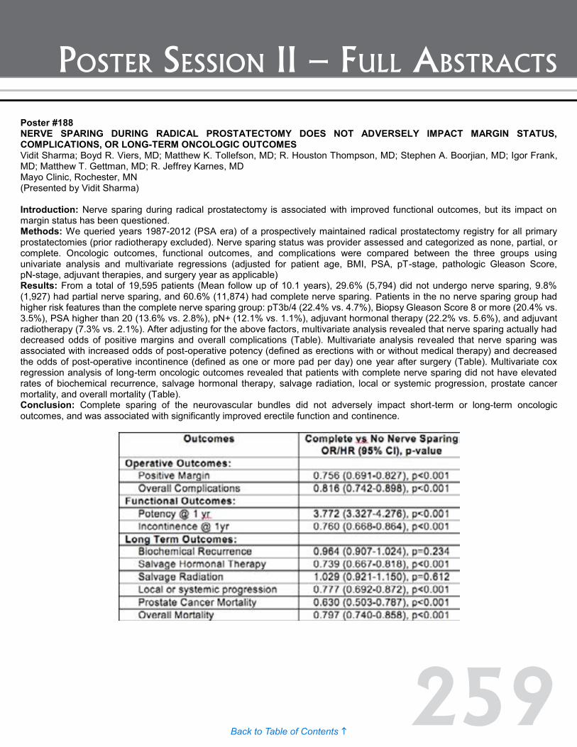
259Back to Table of Contents h
Poster session ii – Full AbstrActs
Poster #188 NERVE SPARING DURING RADICAL PROSTATECTOMY DOES NOT ADVERSELY IMPACT MARGIN STATUS, COMPLICATIONS, OR LONG-TERM ONCOLOGIC OUTCOMES Vidit Sharma; Boyd R. Viers, MD; Matthew K. Tollefson, MD; R. Houston Thompson, MD; Stephen A. Boorjian, MD; Igor Frank, MD; Matthew T. Gettman, MD; R. Jeffrey Karnes, MD Mayo Clinic, Rochester, MN (Presented by Vidit Sharma) Introduction: Nerve sparing during radical prostatectomy is associated with improved functional outcomes, but its impact on margin status has been questioned. Methods: We queried years 1987-2012 (PSA era) of a prospectively maintained radical prostatectomy registry for all primary prostatectomies (prior radiotherapy excluded). Nerve sparing status was provider assessed and categorized as none, partial, or complete. Oncologic outcomes, functional outcomes, and complications were compared between the three groups using univariate analysis and multivariate regressions (adjusted for patient age, BMI, PSA, pT-stage, pathologic Gleason Score, pN-stage, adjuvant therapies, and surgery year as applicable) Results: From a total of 19,595 patients (Mean follow up of 10.1 years), 29.6% (5,794) did not undergo nerve sparing, 9.8% (1,927) had partial nerve sparing, and 60.6% (11,874) had complete nerve sparing. Patients in the no nerve sparing group had higher risk features than the complete nerve sparing group: pT3b/4 (22.4% vs. 4.7%), Biopsy Gleason Score 8 or more (20.4% vs. 3.5%), PSA higher than 20 (13.6% vs. 2.8%), pN+ (12.1% vs. 1.1%), adjuvant hormonal therapy (22.2% vs. 5.6%), and adjuvant radiotherapy (7.3% vs. 2.1%). After adjusting for the above factors, multivariate analysis revealed that nerve sparing actually had decreased odds of positive margins and overall complications (Table). Multivariate analysis revealed that nerve sparing was associated with increased odds of post-operative potency (defined as erections with or without medical therapy) and decreased the odds of post-operative incontinence (defined as one or more pad per day) one year after surgery (Table). Multivariate cox regression analysis of long-term oncologic outcomes revealed that patients with complete nerve sparing did not have elevated rates of biochemical recurrence, salvage hormonal therapy, salvage radiation, local or systemic progression, prostate cancer mortality, and overall mortality (Table). Conclusion: Complete sparing of the neurovascular bundles did not adversely impact short-term or long-term oncologic outcomes, and was associated with significantly improved erectile function and continence.
260Back to Table of Contents h
Poster session ii – Full AbstrActs
Poster #189 MULTI-INSTITUTIONAL ANALYSES OF INFECTIOUS COMPLICATIONS AFTER PROSTATE BIOPSY – PREDICTIVE FACTORS OF BACTEREMIA, SEPSIS, AND SHOCK Simpa Salami, MD, MPH1; Vinaya Vasudevan, MD2; Neeti Bagadiya, MD3; Louis Kavoussi, MD, MBA2; Carl Olsson, MD4; Manish Vira, MD2 1University of Michigan, Ann Arbor, MI; 2Hofstra North Shore-Long Island Jewish School of Medicine, New Hyde Park, NY; 3New York University, New York, NY; 4Integrated Medical Professionals, Melville, NY (Presented by Simpa Salami) Introduction: Transrectal ultrasound guided biopsy of the prostate (TRUSBX) remains the gold standard for the diagnosis of prostate cancer (PCa). Recent studies have reported that three to five percent of men will suffer an infectious complication requiring hospitalization following TRUSBX, despite prophylaxis with oral fluoroquinolone prophylaxis. Our objective was to evaluate a large cohort of men hospitalized for post biopsy infection and determine which factors predict septic complications. Methods: Over a 3-year period, a total of 19,143 patients underwent TRUSBX at either a large community urology practice or an academic urology department. Two hundred and thirty patients (1.2%) were admitted within 30 days post biopsy, of which 96 were admitted to our health system hospitals for which hospital records were available. Demographic and clinical information obtained from patients’ charts were recorded in an IRB-approved database. Logistic regression was used to evaluate factors predicting bacteremia and sepsis in a multivariate model. Results: Mean presentation was 3.1 days following TRUSBX. The vast majority presented with fever (94%); some patients complained of gastrointestinal (GI) symptoms (42%), hematuria (34%), and dysuria (22%). Review of clinical outcomes revealed that 44% had positive blood cultures, 43% progressed to sepsis, and 17% progressed to septic shock. Analysis of risk factors demonstrated that these patients presented significantly sooner after TRUSBX (mean 1.9 vs. 3.9 days, p=0.0018). On multivariate analysis, shorter time to presentation, fever, GI symptoms, elevated WBC, pyuria, urine nitrite were independent predictors of the risk of bacteremia, sepsis or septic shock (all p < 0.05). In fact, men who presented with fever and GI symptoms were 3.4 times more likely to progress to sepsis as compared to men without these symptoms (p=0.0062). Interestingly, maximum temperature and history of diabetes were not statistically associated with risk of bacteremia or sepsis. There was no mortality during the study period. Conclusion: Infection following TRUSBX is a highly morbid complication with a high risk of sepsis or septic shock. Men who present with GI symptoms along with fever and men with significant comorbidities are at highest risk of progression to sepsis or septic shock.
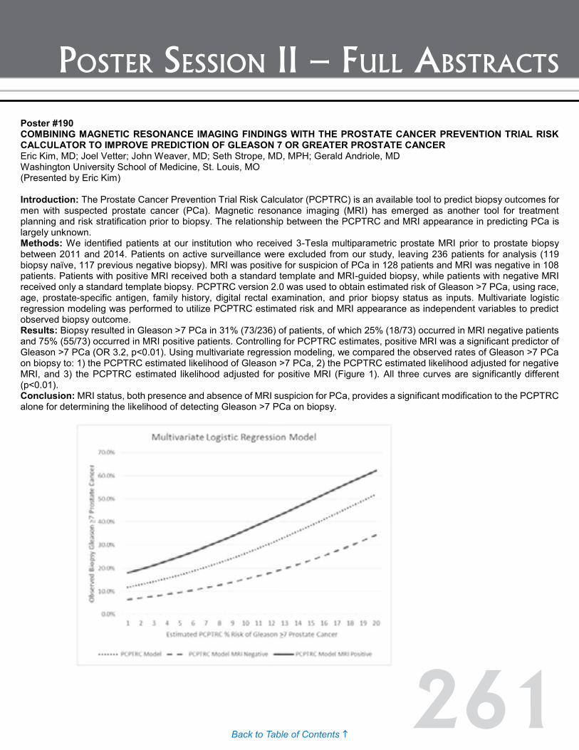
261Back to Table of Contents h
Poster session ii – Full AbstrActs
Poster #190 COMBINING MAGNETIC RESONANCE IMAGING FINDINGS WITH THE PROSTATE CANCER PREVENTION TRIAL RISK CALCULATOR TO IMPROVE PREDICTION OF GLEASON 7 OR GREATER PROSTATE CANCER Eric Kim, MD; Joel Vetter; John Weaver, MD; Seth Strope, MD, MPH; Gerald Andriole, MD Washington University School of Medicine, St. Louis, MO (Presented by Eric Kim) Introduction: The Prostate Cancer Prevention Trial Risk Calculator (PCPTRC) is an available tool to predict biopsy outcomes for men with suspected prostate cancer (PCa). Magnetic resonance imaging (MRI) has emerged as another tool for treatment planning and risk stratification prior to biopsy. The relationship between the PCPTRC and MRI appearance in predicting PCa is largely unknown. Methods: We identified patients at our institution who received 3-Tesla multiparametric prostate MRI prior to prostate biopsy between 2011 and 2014. Patients on active surveillance were excluded from our study, leaving 236 patients for analysis (119 biopsy naïve, 117 previous negative biopsy). MRI was positive for suspicion of PCa in 128 patients and MRI was negative in 108 patients. Patients with positive MRI received both a standard template and MRI-guided biopsy, while patients with negative MRI received only a standard template biopsy. PCPTRC version 2.0 was used to obtain estimated risk of Gleason >7 PCa, using race, age, prostate-specific antigen, family history, digital rectal examination, and prior biopsy status as inputs. Multivariate logistic regression modeling was performed to utilize PCPTRC estimated risk and MRI appearance as independent variables to predict observed biopsy outcome. Results: Biopsy resulted in Gleason >7 PCa in 31% (73/236) of patients, of which 25% (18/73) occurred in MRI negative patients and 75% (55/73) occurred in MRI positive patients. Controlling for PCPTRC estimates, positive MRI was a significant predictor of Gleason >7 PCa (OR 3.2, p<0.01). Using multivariate regression modeling, we compared the observed rates of Gleason >7 PCa on biopsy to: 1) the PCPTRC estimated likelihood of Gleason >7 PCa, 2) the PCPTRC estimated likelihood adjusted for negative MRI, and 3) the PCPTRC estimated likelihood adjusted for positive MRI (Figure 1). All three curves are significantly different (p<0.01). Conclusion: MRI status, both presence and absence of MRI suspicion for PCa, provides a significant modification to the PCPTRC alone for determining the likelihood of detecting Gleason >7 PCa on biopsy.
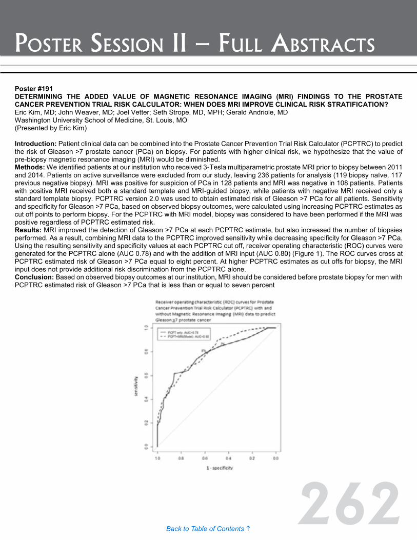
262Back to Table of Contents h
Poster session ii – Full AbstrActs
Poster #191 DETERMINING THE ADDED VALUE OF MAGNETIC RESONANCE IMAGING (MRI) FINDINGS TO THE PROSTATE CANCER PREVENTION TRIAL RISK CALCULATOR: WHEN DOES MRI IMPROVE CLINICAL RISK STRATIFICATION? Eric Kim, MD; John Weaver, MD; Joel Vetter; Seth Strope, MD, MPH; Gerald Andriole, MD Washington University School of Medicine, St. Louis, MO (Presented by Eric Kim) Introduction: Patient clinical data can be combined into the Prostate Cancer Prevention Trial Risk Calculator (PCPTRC) to predict the risk of Gleason >7 prostate cancer (PCa) on biopsy. For patients with higher clinical risk, we hypothesize that the value of pre-biopsy magnetic resonance imaging (MRI) would be diminished. Methods: We identified patients at our institution who received 3-Tesla multiparametric prostate MRI prior to biopsy between 2011 and 2014. Patients on active surveillance were excluded from our study, leaving 236 patients for analysis (119 biopsy naïve, 117 previous negative biopsy). MRI was positive for suspicion of PCa in 128 patients and MRI was negative in 108 patients. Patients with positive MRI received both a standard template and MRI-guided biopsy, while patients with negative MRI received only a standard template biopsy. PCPTRC version 2.0 was used to obtain estimated risk of Gleason >7 PCa for all patients. Sensitivity and specificity for Gleason >7 PCa, based on observed biopsy outcomes, were calculated using increasing PCPTRC estimates as cut off points to perform biopsy. For the PCPTRC with MRI model, biopsy was considered to have been performed if the MRI was positive regardless of PCPTRC estimated risk. Results: MRI improved the detection of Gleason >7 PCa at each PCPTRC estimate, but also increased the number of biopsies performed. As a result, combining MRI data to the PCPTRC improved sensitivity while decreasing specificity for Gleason >7 PCa. Using the resulting sensitivity and specificity values at each PCPTRC cut off, receiver operating characteristic (ROC) curves were generated for the PCPTRC alone (AUC 0.78) and with the addition of MRI input (AUC 0.80) (Figure 1). The ROC curves cross at PCPTRC estimated risk of Gleason >7 PCa equal to eight percent. At higher PCPTRC estimates as cut offs for biopsy, the MRI input does not provide additional risk discrimination from the PCPTRC alone. Conclusion: Based on observed biopsy outcomes at our institution, MRI should be considered before prostate biopsy for men with PCPTRC estimated risk of Gleason >7 PCa that is less than or equal to seven percent
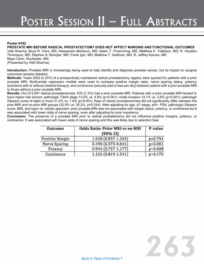
263Back to Table of Contents h
Poster session ii – Full AbstrActs
Poster #192 PROSTATE MRI BEFORE RADICAL PROSTATECTOMY DOES NOT AFFECT MARGINS AND FUNCTIONAL OUTCOMES Vidit Sharma; Boyd R. Viers, MD; Alessandro Morlacco, MD; Adam T. Froemming, MD; Matthew K. Tollefson, MD; R. Houston Thompson, MD; Stephen A. Boorjian, MD; Frank Igor, MD; Matthew T. Gettman, MD; R. Jeffrey Karnes, MD Mayo Clinic, Rochester, MN (Presented by Vidit Sharma) Introduction: Prostate MRI is increasingly being used to help identify and diagnose prostate cancer, but its impact on surgical outcomes remains debated. Methods: Years 2002 to 2012 of a prospectively maintained radical prostatectomy registry were queried for patients with a prior prostate MRI. Multivariate regression models were used to compare positive margin rates, nerve sparing status, potency (erections with or without medical therapy), and continence (security pad or less per day) between patient with a prior prostate MRI to those without a prior prostate MRI. Results: Out of 9,291 radical prostatectomies, 676 (7.3%) had a prior prostate MRI. Patients with a prior prostate MRI tended to have higher risk tumors: pathologic T3b/4 stage 13.0% vs. 4.8% (p<0.001); nodal invasion 14.1% vs. 2.6% (p<0.001); pathologic Gleason score of eight or more 31.2% vs. 7.6% (p<0.001). Rate of robotic prostatectomies did not significantly differ between the prior-MRI and no-prior MRI groups (32.9% vs. 35.2%, p=0.244). After adjusting for age, pT-stage, pN+, PSA, pathologic Gleason score, BMI, and open vs. robotic approach, prior prostate MRI was not associated with margin status, potency, or continence but it was associated with lower odds of nerve sparing, even after adjusting for prior impotence. Conclusion: The presence of a prostate MRI prior to radical prostatectomy did not influence positive margins, potency, or continence. It was associated with lower odds of nerve sparing and this was likely due to selection bias.
264Back to Table of Contents h
Poster session ii – Full AbstrActs
Poster #194 INCIDENCE, RISK FACTORS AND OUTCOMES FOR RECTAL INJURY DURING RADICAL PROSTATECTOMY Shane Pearce, Andrew Cohen, Vignesh Packiam, Charles Nottingham, Joseph Pariser, Scott Eggener University of Chicago, Chicago, IL (Presented by Shane Pearce) Introduction: Rectal injury (RI) is a rare, but serious complication of radical prostatectomy (RP). We performed a population-based analysis of RI during RP. Methods: The National Inpatient Sample (2003-2012) was used to identify patients with a diagnosis of prostate cancer who underwent RP. Survey-weighted cohorts were created based on the diagnosis and repair of a RI. Data included demographics, hospital characteristics, hospital volume for RP, surgical details, complications and perioperative outcomes. Multivariable logistic regression was used to identify risk factors for RI. Results: Of 614,294 men who underwent RP, there were 2900 (0.5%) RIs, with decreased incidence over time (0.6% in 2003 vs. 0.4% in 2012, p=0.02). Colostomy was performed in 281 patients (10%). Patients with RI were slightly older (mean age 62.0 vs. 61.2, p<0.01) and RI was more frequent among African-Americans (0.8% vs. 0.4% for Caucasians, p<0.01). There was no difference in Elixhauser comorbidity score between groups (p=0.2). However, the incidence of RI was higher in patients with clinically significant benign prostatic hyperplasia (BPH) and metastatic disease and lower in obese men (all p<0.05). The overall incidence of RI was highest for open RP (0.6%) compared to laparoscopic (0.4%) and robotic approaches (0.2%), p<0.01. RI was also more common at urban (0.8% vs. 0.5% for rural), non-teaching (0.6% vs. 0.4% for teaching), and low-volume hospitals (0.6% vs. 0.3% for high-volume), (all p<0.01). Overall complication rate (28% vs. 11%, p<0.01), length of stay (4.8 vs. 2.3 days, p<0.01), and total charges ($45,298 vs. $32,855, p<0.01) were greater for patients with RI compared to those without. Multivariable logistic regression (see Table) identified African-American race, BPH and metastatic cancer as predictors for RI, while private insurance, robotic approach, high-volume hospital and obesity reduced the risk of RI (all p<0.05). Conclusion: RI during RP is a rare complication, more common among African-Americans, patients with low BMI, BPH or with metastatic disease. RI was also more common for open surgical approaches and low-volume hospitals.
Poster #193 CANCER DETECTION BETWEEN PERIPHERAL ZONE AND TRANSITIONAL ZONE TARGETED BIOPSIES: PRELIMINARY Results: FROM A PROSPECTIVE COHORT OF MEN UNDERGOING MRI-US FUSION BIOPSY Bruno Nahar, MD1; Nachiketh Soodana Prakash, MD1; Tara Abboud, PA1; Nicola Pavan, MD1; Samarpit Rai, MD1; Felipe Munera, MD2; Rosa Castillo, MD2; Raymond Balise, PhD3; Murugesan Manoharan, MD1; Bruce Kava, MD1; Ramgopal Satyanarayana, MD1; Mark Gonzalgo, MD1; Chad Ritch, MD1; Dipen Parekh, MD1; Sanoj Punnen, MD1 1University of Miami Miller School of Medicine Department of Urology, Miami FL; 2University of Miami Miller School of Medicine Department of Radiology, Miami FL; 3University of Miami Miller School of Medicine Department of Biostatistics, Miami FL (Presented by Bruno Nahar) Introduction: Multiparametric MRI has emerged as a popular imaging modality to localize prostate cancer. Nevertheless, interpretation of MRI is subjective, with concerns for false positives, particularly in the transitional zone (TZ), where hyperplastic changes that are common to this region may be confused for suspicion of cancer. We analyzed a prospective cohort of men undergoing MRI-US fusion biopsy and compared cancer detection rate between lesions seen in the peripheral zone (PZ) and the TZ. Methods: 133 men with elevated PSA or positive digital rectal exam underwent MRI-US fusion biopsy with average of two cores taken per target for the detection of prostate cancer between October 2014 and July 2015. Each targeted lesion in the PZ and TZ was previously classified by radiologists according to the MRI PI-RADS score and grouped according to their level of suspicion as probably benign (1-2), indeterminate (3) or probably malignant (4-5). Histopathology from targeted cores was categorized as no cancer, non-significant cancer (Gleason 6) and significant cancer (Gleason ≥ 7). We compared the cancer detection rates between lesions in PZ and TZ lesions, based on PI-RADS score. Results: We identified 143 lesions in the PZ and 82 lesions in the TZ. Among lesions found in the TZ, 57.3% were reported as probably malignant, compared to 44.7% of lesions seen in the PZ. Cancer was diagnosed in 23% of the lesions in the PZ, compared to only in 9.7% in the TZ (p<0.01). With respect to significant cancer, although we did not see a statistically significant difference, there was a trend towards higher detection of significant cancer in the PZ (13.29% vs. 6.10% p=0.12) compared to TZ. Furthermore, lesions in the PZ with a PI-RADS score >4 were associated with a three-fold increase in the odds of detecting cancer compared to lesions with PI-RADS < 4 (OR 3.08; CI 95% 1.29 – 7.31, p<0.011), whereas in the TZ there was no increased risk of cancer with higher PI-RADS scores (OR 1.0 CI 95% 0.20 – 4.84, p<1.0). Conclusion: To our knowledge this is the first study to address a concern regarding an increased likelihood of false positives when reporting the presence and aggressiveness of cancer in the TZ versus the PZ. This may lead to unnecessary biopsies in men undergoing MRI of the prostate.
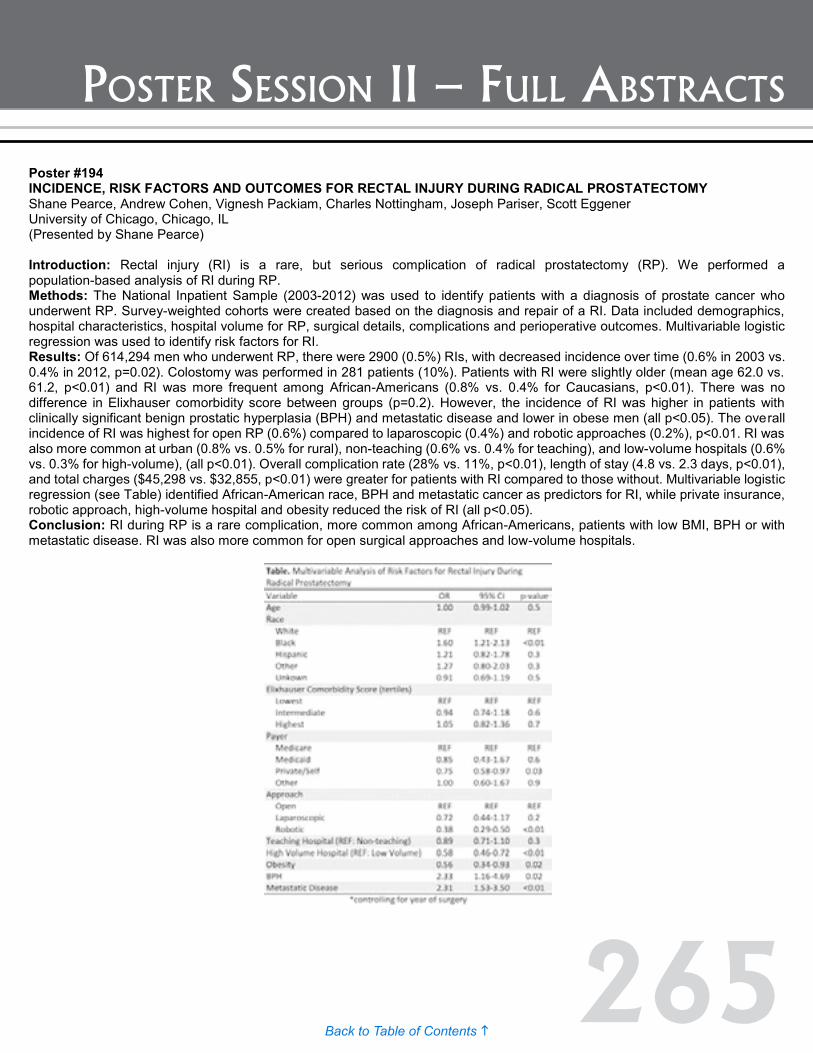
265Back to Table of Contents h
Poster session ii – Full AbstrActs
Poster #194 INCIDENCE, RISK FACTORS AND OUTCOMES FOR RECTAL INJURY DURING RADICAL PROSTATECTOMY Shane Pearce, Andrew Cohen, Vignesh Packiam, Charles Nottingham, Joseph Pariser, Scott Eggener University of Chicago, Chicago, IL (Presented by Shane Pearce) Introduction: Rectal injury (RI) is a rare, but serious complication of radical prostatectomy (RP). We performed a population-based analysis of RI during RP. Methods: The National Inpatient Sample (2003-2012) was used to identify patients with a diagnosis of prostate cancer who underwent RP. Survey-weighted cohorts were created based on the diagnosis and repair of a RI. Data included demographics, hospital characteristics, hospital volume for RP, surgical details, complications and perioperative outcomes. Multivariable logistic regression was used to identify risk factors for RI. Results: Of 614,294 men who underwent RP, there were 2900 (0.5%) RIs, with decreased incidence over time (0.6% in 2003 vs. 0.4% in 2012, p=0.02). Colostomy was performed in 281 patients (10%). Patients with RI were slightly older (mean age 62.0 vs. 61.2, p<0.01) and RI was more frequent among African-Americans (0.8% vs. 0.4% for Caucasians, p<0.01). There was no difference in Elixhauser comorbidity score between groups (p=0.2). However, the incidence of RI was higher in patients with clinically significant benign prostatic hyperplasia (BPH) and metastatic disease and lower in obese men (all p<0.05). The overall incidence of RI was highest for open RP (0.6%) compared to laparoscopic (0.4%) and robotic approaches (0.2%), p<0.01. RI was also more common at urban (0.8% vs. 0.5% for rural), non-teaching (0.6% vs. 0.4% for teaching), and low-volume hospitals (0.6% vs. 0.3% for high-volume), (all p<0.01). Overall complication rate (28% vs. 11%, p<0.01), length of stay (4.8 vs. 2.3 days, p<0.01), and total charges ($45,298 vs. $32,855, p<0.01) were greater for patients with RI compared to those without. Multivariable logistic regression (see Table) identified African-American race, BPH and metastatic cancer as predictors for RI, while private insurance, robotic approach, high-volume hospital and obesity reduced the risk of RI (all p<0.05). Conclusion: RI during RP is a rare complication, more common among African-Americans, patients with low BMI, BPH or with metastatic disease. RI was also more common for open surgical approaches and low-volume hospitals.
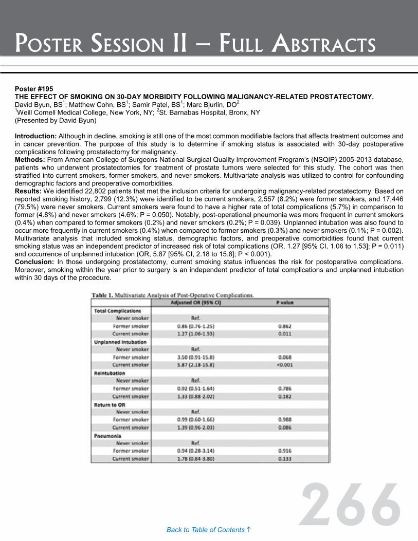
266Back to Table of Contents h
Poster session ii – Full AbstrActs
Poster #196 ADJUVANT RADIATION THERAPY IS SUPERIOR TO SALVAGE RADIATION THERAPY IN PATIENTS WITH PN1 PROSTATE CANCER TREATED WITH RADICAL PROSTATECTOMY Derya Tilki, Pierre Tennstedt, Markus Graefen, Rudolf Schwarz, Sascha Ahyai Martini-Clinic Prostate Cancer Center (Presented by Derya Tilki) Introduction: Patients with positive lymph nodes (LN) at radical prostatectomy (RP) represent a heterogeneous group and their optimal treatment is a matter of debate. To analyze the impact of no treatment or salvage radiation therapy (NT/sRT group) in case of biochemical recurrence (BCR), adjuvant (aRT group) and adjuvant hormonal treatment only (aHT group) after RP on oncological outcomes of LN positive patients. Methods: A total of 773 patients with LN positive prostate cancer (PCa) at RP with or without additional radiation treatment from 2005 to 2013 were retrospectively analyzed. Cox regressions addressed factors influencing BCR and metastasis-free survival (MFS). Propensity score-matched analyses were performed. Results: During the study period, 294 (38.0%) patients experienced BCR and 90 (11.6%) patients developed distant metastasis. In multivariate analysis, aHT (n=55) and NT/sRT (n=505) were independent risk factors for BCR and metastasis compared to patients with aRT (n=234). The superiority of aRT was confirmed after propensity score-matching. Three-year metastasis-free survival in the matched cohort was 85.0% versus 94.9% for the NT/sRT and aRT groups, respectively (p=0.03). Overall, 60.0% of the sRT patients responded to sRT. These patients had lower preradiation PSA (<0.3 vs. ≥0.3) when compared to patients who did not respond. Conclusion: LN positive patients who received aRT had a significantly better oncological outcome compared to patients with NT/sRT or aHT independent of tumor characteristics. Patients with early sRT showed higher rates of response and better metastasis-free survival than patients with pre-RT PSA ≥0.3 ng/ml.
Poster #195 THE EFFECT OF SMOKING ON 30-DAY MORBIDITY FOLLOWING MALIGNANCY-RELATED PROSTATECTOMY. David Byun, BS1; Matthew Cohn, BS1; Samir Patel, BS1; Marc Bjurlin, DO2 1Weill Cornell Medical College, New York, NY; 2St. Barnabas Hospital, Bronx, NY (Presented by David Byun) Introduction: Although in decline, smoking is still one of the most common modifiable factors that affects treatment outcomes and in cancer prevention. The purpose of this study is to determine if smoking status is associated with 30-day postoperative complications following prostatectomy for malignancy. Methods: From American College of Surgeons National Surgical Quality Improvement Program’s (NSQIP) 2005-2013 database, patients who underwent prostatectomies for treatment of prostate tumors were selected for this study. The cohort was then stratified into current smokers, former smokers, and never smokers. Multivariate analysis was utilized to control for confounding demographic factors and preoperative comorbidities. Results: We identified 22,802 patients that met the inclusion criteria for undergoing malignancy-related prostatectomy. Based on reported smoking history, 2,799 (12.3%) were identified to be current smokers, 2,557 (8.2%) were former smokers, and 17,446 (79.5%) were never smokers. Current smokers were found to have a higher rate of total complications (5.7%) in comparison to former (4.8%) and never smokers (4.6%; P = 0.050). Notably, post-operational pneumonia was more frequent in current smokers (0.4%) when compared to former smokers (0.2%) and never smokers (0.2%; P = 0.039). Unplanned intubation was also found to occur more frequently in current smokers (0.4%) when compared to former smokers (0.3%) and never smokers (0.1%; P = 0.002). Multivariate analysis that included smoking status, demographic factors, and preoperative comorbidities found that current smoking status was an independent predictor of increased risk of total complications (OR, 1.27 [95% CI, 1.06 to 1.53]; P = 0.011) and occurrence of unplanned intubation (OR, 5.87 [95% CI, 2.18 to 15.8]; P < 0.001). Conclusion: In those undergoing prostatectomy, current smoking status influences the risk for postoperative complications. Moreover, smoking within the year prior to surgery is an independent predictor of total complications and unplanned intubation within 30 days of the procedure.
267Back to Table of Contents h
Poster session ii – Full AbstrActs
Poster #196 ADJUVANT RADIATION THERAPY IS SUPERIOR TO SALVAGE RADIATION THERAPY IN PATIENTS WITH PN1 PROSTATE CANCER TREATED WITH RADICAL PROSTATECTOMY Derya Tilki, Pierre Tennstedt, Markus Graefen, Rudolf Schwarz, Sascha Ahyai Martini-Clinic Prostate Cancer Center (Presented by Derya Tilki) Introduction: Patients with positive lymph nodes (LN) at radical prostatectomy (RP) represent a heterogeneous group and their optimal treatment is a matter of debate. To analyze the impact of no treatment or salvage radiation therapy (NT/sRT group) in case of biochemical recurrence (BCR), adjuvant (aRT group) and adjuvant hormonal treatment only (aHT group) after RP on oncological outcomes of LN positive patients. Methods: A total of 773 patients with LN positive prostate cancer (PCa) at RP with or without additional radiation treatment from 2005 to 2013 were retrospectively analyzed. Cox regressions addressed factors influencing BCR and metastasis-free survival (MFS). Propensity score-matched analyses were performed. Results: During the study period, 294 (38.0%) patients experienced BCR and 90 (11.6%) patients developed distant metastasis. In multivariate analysis, aHT (n=55) and NT/sRT (n=505) were independent risk factors for BCR and metastasis compared to patients with aRT (n=234). The superiority of aRT was confirmed after propensity score-matching. Three-year metastasis-free survival in the matched cohort was 85.0% versus 94.9% for the NT/sRT and aRT groups, respectively (p=0.03). Overall, 60.0% of the sRT patients responded to sRT. These patients had lower preradiation PSA (<0.3 vs. ≥0.3) when compared to patients who did not respond. Conclusion: LN positive patients who received aRT had a significantly better oncological outcome compared to patients with NT/sRT or aHT independent of tumor characteristics. Patients with early sRT showed higher rates of response and better metastasis-free survival than patients with pre-RT PSA ≥0.3 ng/ml.
268Back to Table of Contents h
Poster session ii – Full AbstrActs
Poster #197 CONTEMPORARY TREATMENT PATTERNS AND SHORT-TERM OUTCOMES IN MEN WITH VERY HIGH RISK PROSTATE CANCER Jeffrey Tosoian, MD, MPH1; Debasish Sundi, MD2; Brian Chapin, MD2; R. Jeffrey Karnes, MD3; Emmanuel Antonarakis, MD4; Meera Chappidi, BS5; Ridwan Alam, BS5; Stephanie Glavaris, BS5; Kenneth Pienta, MD5; Phuoc Tran, MD5; Edward Schaeffer, MD, PhD5; Ashley Ross, MD, PhD5 1Johns Hopkins Medical Institutions, Baltimore, MD; 2Department of Urology, The University of Texas MD Anderson Cancer Center, Houston, TX; 3Department of Urology Mayo Clinic; 4Sidney Kimmel Comprehensive Cancer Center, Johns Hopkins Medical Institutions, Baltimore, MD; 5Brady Urological Institute, Johns Hopkins Medical Institutions, Baltimore, MD (Presented by Jeffrey Tosoian) Introduction: Beginning in 2014, the National Comprehensive Cancer Network (NCCN) recognized very high-risk (VHR)/locally advanced prostate cancer (cT3b-T4, or primary Gleason pattern 5, or more than 4 biopsy cores with Gleason score 8-10, or multiple high-risk features) as a classification distinct from high-risk (HR) disease. Here, using prospectively collected institutional data, we describe contemporary treatment patterns and short-term outcomes in the VHR population. Methods: The Johns Hopkins radical prostatectomy (RP) database was queried to identify men who underwent RP from January 2010 through June 2015, and trends in management were compared across the study period. Men with VHR cancer were identified for assessment of pathological and short-term clinical outcomes. Non organ-confined disease (NOCD) was defined as ≥ pT3 disease or lymph node positivity, persistent postoperative PSA as ≥ 0.2 ng/mL, and biochemical recurrence (BCR) as a PSA ≥ 0.2 ng/mL following an initial undetectable postoperative PSA. Results: During the study period, 4,954 men underwent RP, of which 161 (3.2%) men had VHR cancer at diagnosis. The annual proportion of men who underwent RP with VHR cancer increased over the five-year study period (chronologically 1.8%, 1.0%, 3.3%, 4.1%, 5.6%, and 5.2%; p<0.001). Sixteen percent of men with VHR disease were enrolled in pre-surgical clinical trials, with an increase from zero percent of men in 2010 to 19.1% in 2015 (p=0.11). At prostatectomy, 39% of the VHR cohort had seminal vesicle invasion, 26% had lymph node involvement, and a total of 74% had NOCD. Following surgery, 33% of men had PSA persistence, and a total of 40% experienced either PSA persistence or BCR during follow-up (median 13.4 months). Of 136 men with at least one follow-up assessment, 15 (11.0%) developed clinical metastasis (median follow-up 13.4 months). During the follow-up period, 33% of the cohort was treated with radiation therapy, 42% with androgen deprivation, and 15% with docetaxel. Conclusion: VHR men represent a population with the greatest risk of clinical progression following local treatment. Over the past five years, we have seen an increase in the surgical treatment of VHR men and more frequent enrollment in pre-surgical clinical trials. Contemporary assessment of post-operative interventions and outcomes will help to facilitate counseling of men presenting with VHR disease and establish point estimates from which to power clinical trials.
269Back to Table of Contents h
Poster session ii – Full AbstrActs
Poster #198 NOVEL URINE MARKERS FOR DIAGNOSING AND MONITORING NON-INDOLENT PROSTATE CANCER Daniela Bianchi-Frias, PhD1; Ilsa Coleman, PhD1; John Banerji, MD, MCh (Urology)2; Khanh Pham, MD2; Claudio Jeldres, MD2; Roman Gulati, PhD1; Jing Xia, PhD1; Scott Tomlin, PhD3; Christopher Porter, MD, FACS2; Peter Nelson, MD, PhD1 1Divisions of Human Biology, Fred Hutchinson cancer Research Center, Seattle, WA; 2Virginia Mason Medical Center, Seattle, WA; 3Departments of Pathology5 and Urology6, University of Michigan Medical School, Ann Arbor, MI. (Presented by Daniela Bianchi-Frias) Introduction: Prostate cancer (PCa) is the most common solid tumor in men. Active surveillance (AS) is an acceptable alternative to immediate treatment for low-risk PCa. However, AS often entails significant risk of under-grading the tumor and repeated invasive biopsies. We hypothesized that transcripts associated with high Gleason grade cancers are quantifiable in urine samples and reflect the presence of higher-grade non-indolent tumors. Our objective was to develop a qPCR-urine-based assay for the detection of occult high-grade cancer in urine samples from PCa patients. Methods: By comparing transcriptional profiles of Gleason pattern 3 (GP3) and Gleason pattern 4 (GP4) cancers, we identified transcripts differentially expressed between these histologies. Four consistently up-regulated GP4 transcripts (RELN, GRIN3A, RGS5 and LRNN1) were evaluated in RNA extracted from urine sediments from 53 PCa patients. Urine specimens were obtained following a digital rectal exam and immediately prior to radical prostatectomy. qPCR was performed for the GP4 markers and normalized to KLK3 and RPL13A. To evaluate the ability of the GP4 markers to discriminate between low- and high-grade tumors, univariate and multivariate logistic regression analyses were performed and the overall significance determined using likelihood ratio tests. Results: Each gene was an independent predictor of high-grade (Gleason score ≥4+3 vs. ≤3+4) prostate cancer (p<0.05 for each gene) with area under receiver operating characteristic curves (AUC) of 0.786, 0.770, 0.794, and 0.784. When tested as a panel, the four genes were significantly associated with high-grade cancer based on the overall likelihood ratio test (p=0.002). Furthermore, the four-gene panel outperformed each gene alone, with an AUC of 0.841. Conclusion: A novel four-gene panel assayed by qPCR appears to be capable of discriminating clinically relevant high-grade (Gleason≥4+3) tumors from low-grade (Gleason≤3+4) tumors using urine sediments. Further studies are needed to confirm these preliminary findings.
270Back to Table of Contents h
Poster session ii – Full AbstrActs
Poster #199 PREDICTION OF OVERALL AND CLINICALLY SIGNIFICANT CANCER RISK ON MRI-TARGETED AND SYSTEMATIC PROSTATE BIOPSY USING PREBIOPSY NOMOGRAMS Marc A. Bjurlin, DO, MSc1; James S. Wysock, MD, MSc2; Saradwata Sakar, PhD3; Rajesh Venkataraman, PhD3; Neil Mendhiratta, BS4; Xiaosong Meng, MD, PhD5; Michael Fenstermaker, BS4; Andrew B. Rosenkrantz, MD6; Samir S. Taneja, MD1 1Division of Urologic Oncology, Department of Urology, New York University Langone Medical Center, New York, NY; 2Department of Urology, New York Hospital Queens, Flushing, NY; 3Eigen, Grass Valley, CA; 4School of Medicine, New York University Langone Medical Center, New York, NY; 5Department of Urology, New York University Langone Medical Center, New York, NY; 6Department of Radiology, New York University Langone Medical Center, New York, NY (Presented by Marc A. Bjurlin) Introduction: MRI-targeted prostate biopsy (PB) is increasingly being utilized to aid cancer diagnosis in clinical practice. Our objective was to develop nomograms to predict the probability of overall prostate cancer (CaP) and clinically significant CaP on MRI targeted, and combined targeted and systematic prostate biopsy. Methods: From June 2012 to June 2015, MR-US fusion targeted prostate biopsy was performed on approximately 1,140 men with suspicious regions identified on pre-biopsy 3T multiparametric-MRI along with systematic 12 core biopsy, utilizing the ProFuse|Artemis™ system. Logistic regression modeling was used to evaluate predictors of overall and clinically significant CaP, and corresponding nomograms were generated. Models were created with a randomly selected sample, tested, and then validated with the remaining sample, bias-corrected using bootstrap resampling, and Akaike information criterion was used to select best-fit models. Results: A total of 693 patients with complete records were included for analysis (median age 66 years, PSA 5.5 ng/ml, prostate volume 51 cc). Statistically significant independent positive predictors of CaP on targeted and systematic PB were found to be PSA density, age, and MRI suspicion score (MRIss). CaP probability nomograms were generated using the predictors and model performance characteristics bias-corrected areas under the receiver-operating characteristic curves (AUC) are shown in Figure 1. Conclusion: PSA density, age, and MRI suspicion score predict CaP on MRI-targeted and systematic biopsy. Our CaP probability nomograms may allow further individualization of the decision to perform PB in men with clinical suspicion of prostate cancer or those with known prostate cancer considering surveillance.
271Back to Table of Contents h
Poster session ii – Full AbstrActs
Poster #200 DOES FLUCTUATION IN AGE-SPECIFIC MEDIAN PSA INFLUENCE PROSTATE CANCER RISK? Charles Nottingham, MD, MS1; Katherine Sentell2; Kimberly Delli-Zotti, PhD3; Vignesh Packiam, MD1; Andrew Cohen, MD1; Rena Malik, MD1; William Catalona, MD4; Brian Helfand, MD, PhD5 1University of Chicago Medical Center, Chicago, IL; 2Vanderbilt University, Nashville, TN; 3Washington University in St. Louis School of Medicine, St. Louis, MO; 4Northwestern University Feinberg School of Medicine, Chicago, IL; 5NorthShore University Health System, Evanston, IL (Presented by Charles Nottingham) Introduction: Prior studies have demonstrated that men who present with PSA values above the age-specific median (ASM) have a significantly increased risk of developing prostate cancer (PCa). However, the change in risk of PCa with fluctuation in PSA over subsequent decades remains unknown. We therefore aimed to determine the risk of PCa diagnosis based on PSA trends over time relative to the ASM. Methods: Using a longitudinal database of 26,111 male patients aged 40-89 years who were enrolled between 1989 and 2001 at Washington University Medical Center in St. Louis, we examined PSA values over time. Patients were classified as being below or above the ASM based on their PSA at enrollment relative to previously-reported ASM PSA values. Patients were then followed for the development of PCa, and had at least one PSA measurement per decade of life. The risk of PCa was then calculated for men who remained above or below the ASM, and for men who fluctuated between ASM categories Results: 9,686 men had a PSA data available for two decades of life. 10.6% of men with PSA values initially below the ASM fluctuated above the PSA in the following decade, whereas 45.4% of men initially above the ASM fluctuated below in the following decade. Compared with men whose PSA remained below the ASM in both decades, men who fluctuated above the ASM had a significantly higher risk of PCa (4.1% versus 0.37%; RR 11.2; 95% CI 5.80-21.6; p<0.001). Men with PSA initially above the ASM who fluctuated below still had a higher risk of PCa compared to men who were below the ASM in both decades (1.9% versus 0.37%; RR 5.13; 95% CI 2.79-9.41; p<0.001). Men with a PSA that remained above the ASM in both decades also had a significantly higher risk of PCa than men who fluctuated below the ASM (10% versus 1.9%; RR 5.29; 95% CI 3.64-7.70; p<0.001). Conclusion: Fluctuation in PSA relative to the ASM significantly alters a patient’s risk of developing PCa. While men with PSA values below the ASM have the lowest PCa risk, some of these men may fluctuate above the ASM values in the next decade and vice versa. While fluctuation in PSA values occur over time, ASM PSA values are predictive of PCa diagnosis.
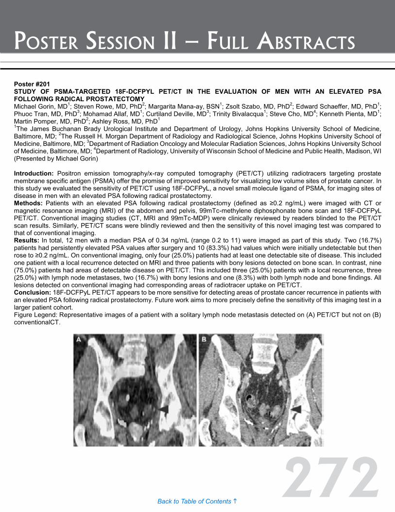
272Back to Table of Contents h
Poster session ii – Full AbstrActs
Poster #201 STUDY OF PSMA-TARGETED 18F-DCFPYL PET/CT IN THE EVALUATION OF MEN WITH AN ELEVATED PSA FOLLOWING RADICAL PROSTATECTOMY Michael Gorin, MD1; Steven Rowe, MD, PhD2; Margarita Mana-ay, BSN1; Zsolt Szabo, MD, PhD2; Edward Schaeffer, MD, PhD1; Phuoc Tran, MD, PhD3; Mohamad Allaf, MD1; Curtiland Deville, MD3; Trinity Bivalacqua1; Steve Cho, MD4; Kenneth Pienta, MD1; Martin Pomper, MD, PhD2; Ashley Ross, MD, PhD1 1The James Buchanan Brady Urological Institute and Department of Urology, Johns Hopkins University School of Medicine, Baltimore, MD; 2The Russell H. Morgan Department of Radiology and Radiological Science, Johns Hopkins University School of Medicine, Baltimore, MD; 3Department of Radiation Oncology and Molecular Radiation Sciences, Johns Hopkins University School of Medicine, Baltimore, MD; 4Department of Radiology, University of Wisconsin School of Medicine and Public Health, Madison, WI (Presented by Michael Gorin) Introduction: Positron emission tomography/x-ray computed tomography (PET/CT) utilizing radiotracers targeting prostate membrane specific antigen (PSMA) offer the promise of improved sensitivity for visualizing low volume sites of prostate cancer. In this study we evaluated the sensitivity of PET/CT using 18F-DCFPyL, a novel small molecule ligand of PSMA, for imaging sites of disease in men with an elevated PSA following radical prostatectomy. Methods: Patients with an elevated PSA following radical prostatectomy (defined as ≥0.2 ng/mL) were imaged with CT or magnetic resonance imaging (MRI) of the abdomen and pelvis, 99mTc-methylene diphosphonate bone scan and 18F-DCFPyL PET/CT. Conventional imaging studies (CT, MRI and 99mTc-MDP) were clinically reviewed by readers blinded to the PET/CT scan results. Similarly, PET/CT scans were blindly reviewed and then the sensitivity of this novel imaging test was compared to that of conventional imaging. Results: In total, 12 men with a median PSA of 0.34 ng/mL (range 0.2 to 11) were imaged as part of this study. Two (16.7%) patients had persistently elevated PSA values after surgery and 10 (83.3%) had values which were initially undetectable but then rose to ≥0.2 ng/mL. On conventional imaging, only four (25.0%) patients had at least one detectable site of disease. This included one patient with a local recurrence detected on MRI and three patients with bony lesions detected on bone scan. In contrast, nine (75.0%) patients had areas of detectable disease on PET/CT. This included three (25.0%) patients with a local recurrence, three (25.0%) with lymph node metastases, two (16.7%) with bony lesions and one (8.3%) with both lymph node and bone findings. All lesions detected on conventional imaging had corresponding areas of radiotracer uptake on PET/CT. Conclusion: 18F-DCFPyL PET/CT appears to be more sensitive for detecting areas of prostate cancer recurrence in patients with an elevated PSA following radical prostatectomy. Future work aims to more precisely define the sensitivity of this imaging test in a larger patient cohort. Figure Legend: Representative images of a patient with a solitary lymph node metastasis detected on (A) PET/CT but not on (B) conventionalCT.
273Back to Table of Contents h
Poster session ii – Full AbstrActs
Poster #202 GENETIC RISK SCORE DIFFERENTIATES INHERITED RISK AMONG RELATIVES OF HEREDITARY PROSTATE CANCER PATIENTS Vignesh Packiam, MD1; Brian Helfand, MD, PhD2; Haitao Chen, MS3; Carly Conran, BS2; Charles Brendler, MD2; Lilly Zheng, MD2; William Isaacs, PhD4; Jianfeng Xu, MD, PhD2 1University of Chicago Medical Center, Chicago, IL; 2NorthShore University Health System, Division of Urology, John and Carol Walter for Urologic Health, Evanston, IL; 3Center for Genomic Translational Medicine and Prevention, School of Public Health, Fudan University, Shanghai, China, PR; 4Department of Urology, and James Buchanan Brady Urological Institute, Johns Hopkins, Baltimore, MD (Presented by Vignesh Packiam) Introduction: Clinical guidelines recommend targeted screening for men with a family history of prostate cancer (PCa). However, family history assigns equivalent risk to all relatives based on their degree of relationship. Recent genetic studies have identified single nucleotide polymorphisms (SNPs) that can be used to calculate a genetic risk score (GRS) to determine PCa risk. We sought to determine whether GRS can stratify PCa risk among hereditary PCa families, considered to be at highest disease risk. Methods: Family members with hereditary PCa were recruited and genotyped for 17 SNPs associated with PCa. GRS was calculated with weighted odds ratios (ORs) from previously published meta-analyses for each SNP. A GRS of 1.0 indicates an average risk in the general population, while GRS greater or less than 1.0 is associated with increased and decreased disease risks, respectively. A GRS was calculated for all family members. The generalized estimating equation (GEE) model was used to account for the relatedness of men within each family. Univariate and multivariate analyses were performed to compare the distribution of GRS amongst affected and unaffected family members of similar and different degree of relations. Results: Data was available for 789 family members of probands including 552 with PCa and 237 unaffected relatives. There was a wide range of GRS among family members with the same degree of relationship. Interquartile range (IQR) for GRS among first-degree relatives was from 0.76 to 1.84. The median GRS was higher among first-degree relatives compared to second- and third-degree relatives (GRS 1.20 vs. 1.09 vs. 1.00, p<0.001). GRS among affected first- and second-degree relatives were significantly higher than unaffected relatives (p=0.042 and p=0.016, respectively). On univariate analysis, degree of family relationship (OR 1.85, p<0.001) and GRS (OR 1.52, p<0.001) were both independent predictors of PCa. Multivariate analysis controlling for degree of family relationship demonstrated that GRS was a significant and independent predictor of PCa (OR 1.52, 95% CI: 1.15-2.01). Conclusion: Compared to family history, GRS is a simple genetic test that can be used to assess differences in PCa risk among family members of men with the disease. While prospective validations studies are required, this information can help provide guidance for these relatives in regards to the initiation and frequency of PCa testing.
Poster #201 STUDY OF PSMA-TARGETED 18F-DCFPYL PET/CT IN THE EVALUATION OF MEN WITH AN ELEVATED PSA FOLLOWING RADICAL PROSTATECTOMY Michael Gorin, MD1; Steven Rowe, MD, PhD2; Margarita Mana-ay, BSN1; Zsolt Szabo, MD, PhD2; Edward Schaeffer, MD, PhD1; Phuoc Tran, MD, PhD3; Mohamad Allaf, MD1; Curtiland Deville, MD3; Trinity Bivalacqua1; Steve Cho, MD4; Kenneth Pienta, MD1; Martin Pomper, MD, PhD2; Ashley Ross, MD, PhD1 1The James Buchanan Brady Urological Institute and Department of Urology, Johns Hopkins University School of Medicine, Baltimore, MD; 2The Russell H. Morgan Department of Radiology and Radiological Science, Johns Hopkins University School of Medicine, Baltimore, MD; 3Department of Radiation Oncology and Molecular Radiation Sciences, Johns Hopkins University School of Medicine, Baltimore, MD; 4Department of Radiology, University of Wisconsin School of Medicine and Public Health, Madison, WI (Presented by Michael Gorin) Introduction: Positron emission tomography/x-ray computed tomography (PET/CT) utilizing radiotracers targeting prostate membrane specific antigen (PSMA) offer the promise of improved sensitivity for visualizing low volume sites of prostate cancer. In this study we evaluated the sensitivity of PET/CT using 18F-DCFPyL, a novel small molecule ligand of PSMA, for imaging sites of disease in men with an elevated PSA following radical prostatectomy. Methods: Patients with an elevated PSA following radical prostatectomy (defined as ≥0.2 ng/mL) were imaged with CT or magnetic resonance imaging (MRI) of the abdomen and pelvis, 99mTc-methylene diphosphonate bone scan and 18F-DCFPyL PET/CT. Conventional imaging studies (CT, MRI and 99mTc-MDP) were clinically reviewed by readers blinded to the PET/CT scan results. Similarly, PET/CT scans were blindly reviewed and then the sensitivity of this novel imaging test was compared to that of conventional imaging. Results: In total, 12 men with a median PSA of 0.34 ng/mL (range 0.2 to 11) were imaged as part of this study. Two (16.7%) patients had persistently elevated PSA values after surgery and 10 (83.3%) had values which were initially undetectable but then rose to ≥0.2 ng/mL. On conventional imaging, only four (25.0%) patients had at least one detectable site of disease. This included one patient with a local recurrence detected on MRI and three patients with bony lesions detected on bone scan. In contrast, nine (75.0%) patients had areas of detectable disease on PET/CT. This included three (25.0%) patients with a local recurrence, three (25.0%) with lymph node metastases, two (16.7%) with bony lesions and one (8.3%) with both lymph node and bone findings. All lesions detected on conventional imaging had corresponding areas of radiotracer uptake on PET/CT. Conclusion: 18F-DCFPyL PET/CT appears to be more sensitive for detecting areas of prostate cancer recurrence in patients with an elevated PSA following radical prostatectomy. Future work aims to more precisely define the sensitivity of this imaging test in a larger patient cohort. Figure Legend: Representative images of a patient with a solitary lymph node metastasis detected on (A) PET/CT but not on (B) conventionalCT.
274Back to Table of Contents h
Poster session ii – Full AbstrActs
Poster #203 INSURABILITY OF PATIENTS WITH PROSTATE CANCER BASED ON INITIAL MANAGEMENT Mark Biebel, MS1; Jeffrey Stock, MD1; Hank George, FALU2; Christopher Wright, MD3 1Rutgers New Jersey Medical School, Newark, NJ; 2President - Hank George, Inc., Greendale, WI; 3New Jersey Center for Prostate Cancer and Urology / Hackensack University Medical Center, Maywood, NJ (Presented by Mark Biebel) Introduction: Different management options of low-risk prostate cancer have been investigated extensively with respect to patient outcome, but there is minimal data on how life insurance companies view each management option. The primary study objective was to determine life insurance underwriting practices for patients diagnosed with early stage, low-risk prostate cancer based on age at diagnosis and initial management (active surveillance versus radiation therapy versus radical prostatectomy). Methods: A questionnaire was sent to 20 life insurance companies that included nine sample patient cases. The sample patients were diagnosed with low risk prostate cancer and received one of three treatment options: active surveillance, external beam radiation, or radical prostatectomy. These three treatment options were applied to three groups of patients based on age of diagnosis at 55, 65, and 75 years old. Of the nine sample patients, the life insurance underwriters were asked to answer what their underwriting decision would be (decline, substandard, or standard) if the particular patient submitted a 500 thousand dollar term life application at one, three, and five years post-treatment initiation. Results: Of the 20 life insurance companies that were sent the questionnaire, 14 (70%) responded. In all three age groups, patients were granted standard life insurance premiums at the highest percentage after radical prostatectomy (52.7%), followed by radiation therapy (36.0%), and at the lowest percentage with active surveillance (5.6%). Patients diagnosed at an older age were also more likely to receive standard premiums with all treatment options. Lastly, in all patients, underwriters were more likely to select standard premiums as more years passed from treatment initiation (54.6% after five years versus 31.0% after one year). Conclusion: Life insurance companies are more likely to grant standard life insurance premiums in patients diagnosed with low-risk prostate cancer if they are initially treated with either radical prostatectomy or radiation therapy, as opposed to active surveillance.
275Back to Table of Contents h
Poster session ii – Full AbstrActs
Poster #204 IMPACT OF PROSTATE SIZE ON OUTCOMES OF RADICAL PROSTATECTOMY: A COMPREHENSIVE ANALYSIS FROM A LARGE INSTITUTIONAL SERIES Vidit Sharma; Boyd R. Viers, MD; Matthew K. Tollefson, MD; R. Houston Thompson, MD; Stephen A. Boorjian, MD; Igor Frank, MD; Matthew T. Gettman, MD; R. Jeffrey Karnes, MD Mayo Clinic, Rochester, MN (Presented by Vidit Sharma) Introduction: Reports examining the effect of prostate size on radical prostatectomy outcomes have provided conflicting results and have been underpowered. Methods: Years 1987 to 2012 of a prospectively maintained institutional radical prostatectomy registry were queried for patients undergoing a radical prostatectomy without prior radiotherapy or TURP. Patients without ultrasound prostate volume data were excluded. Prostate volume was then analyzed using both univariate analysis and multivariate regressions to determine if larger prostates had poorer clinicopathologic, functional, and long-term oncologic outcomes. Results: The 16,796 patients had a mean estimated prostate volume of 38.1cc (std. dev 20.6). Overall, 3.4% (N=568) had prostate volumes of 75-100cc’s and 1.9% (N=314) had prostate volumes greater than 100cc’s. Patients in with >100cc prostates were older (67.0 vs. 60.2, p<0.001), had greater BMIs (29.2 vs. 28.0, p<0.001), higher PSAs (11.8 vs. 6.2, p<0.001), and were more likely to have a pathologic Gleason Score of 6 or less (67.0% vs. 55.1%, p<0.001). After adjusting for age, BMI, surgery year, PSA, pathologic Gleason Score, pN+, and pT-stage, patients with prostates >100cc had lower odds of a positive margin (OR 0.441, 95%CI 0.318-0.613, p<0.001), and increased odds of intra-operative (OR 3.092, 95%CI 1.131-8.455, p=0.028) and overall complications (OR 1.521, 95%CI 1.150-2.011, p=0.003) compared to patients with prostate volumes of 25ccs or less. Similar findings were observed for every 25cc incremental increase in prostate volume. On multivariate analysis, patients with > 100cc prostates had lower odds of partial/complete nerve sparing (OR 0.387 95%CI 0.342-0.438, p<0.001) and higher odds of incontinence, defined as 1 or more pad per day (OR 1.568 95%CI 1.158-2.124). With a mean follow up of 10.0 years, increased prostate size was not associated with a greater risk of biochemical recurrence, metastasis, or prostate cancer mortality. Conclusion: While increased prostate size may not compromise long-term oncologic control, it is associated with increased complications, lower rates of nerve sparing, and higher rates of incontinence. This may be useful in counseling patients particularly with prostates larger than 100cc’s.
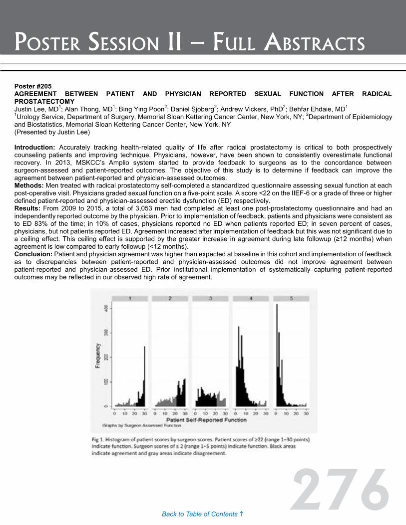
276Back to Table of Contents h
Poster session ii – Full AbstrActs
Poster #205 AGREEMENT BETWEEN PATIENT AND PHYSICIAN REPORTED SEXUAL FUNCTION AFTER RADICAL PROSTATECTOMY Justin Lee, MD1; Alan Thong, MD1; Bing Ying Poon2; Daniel Sjoberg2; Andrew Vickers, PhD2; Behfar Ehdaie, MD1 1Urology Service, Department of Surgery, Memorial Sloan Kettering Cancer Center, New York, NY; 2Department of Epidemiology and Biostatistics, Memorial Sloan Kettering Cancer Center, New York, NY (Presented by Justin Lee) Introduction: Accurately tracking health-related quality of life after radical prostatectomy is critical to both prospectively counseling patients and improving technique. Physicians, however, have been shown to consistently overestimate functional recovery. In 2013, MSKCC’s Amplio system started to provide feedback to surgeons as to the concordance between surgeon-assessed and patient-reported outcomes. The objective of this study is to determine if feedback can improve the agreement between patient-reported and physician-assessed outcomes. Methods: Men treated with radical prostatectomy self-completed a standardized questionnaire assessing sexual function at each post-operative visit. Physicians graded sexual function on a five-point scale. A score <22 on the IIEF-6 or a grade of three or higher defined patient-reported and physician-assessed erectile dysfunction (ED) respectively. Results: From 2009 to 2015, a total of 3,053 men had completed at least one post-prostatectomy questionnaire and had an independently reported outcome by the physician. Prior to implementation of feedback, patients and physicians were consistent as to ED 83% of the time; in 10% of cases, physicians reported no ED when patients reported ED; in seven percent of cases, physicians, but not patients reported ED. Agreement increased after implementation of feedback but this was not significant due to a ceiling effect. This ceiling effect is supported by the greater increase in agreement during late followup (≥12 months) when agreement is low compared to early followup (<12 months). Conclusion: Patient and physician agreement was higher than expected at baseline in this cohort and implementation of feedback as to discrepancies between patient-reported and physician-assessed outcomes did not improve agreement between patient-reported and physician-assessed ED. Prior institutional implementation of systematically capturing patient-reported outcomes may be reflected in our observed high rate of agreement.
277Back to Table of Contents h
Poster session ii – Full AbstrActs
Poster #206 MOLECULAR PROFILING OF TISSUE OBTAINED BY SERIAL MRI TARGETED PROSTATE BIOPSY IN MEN ON ACTIVE SURVEILLANCE FOR LOW GRADE PROSTATE CANCER Ganesh Palapattu, MD1; Andi Cani, MSc1; Daniel Hovelson1; Rohit Mehra, MD1; Jeffery Montgomery, MD1; Todd Morgan, MD1; Simpa Salami, MD, MPH1; Scott Tomlins, MD, PhD1; Leonard Marks, MD2 1University of Michigan, Ann Arbor, MI; 2University of California Los Angeles, Los Angeles, CA (Presented by Ganesh Palapattu) Introduction: While the recent introduction of MRI/US fusion targeted prostate biopsy has begun to change the active surveillance (AS) paradigm by enabling more precise disease assessment, the ability of this technique to longitudinally sample the same clonal cancer focus is unknown. The primary objective of our study was to determine if serial MRI/US fusion targeted prostate biopsy facilitates assessment of the same clonal focus of cancer over time. Methods: We performed a combination of immunohistochemistry (IHC) and targeted RNA/DNA next generation sequencing (NGS) on routine formalin fixed paraffin embedded (FFPE) prostate biopsy specimens acquired one year apart, obtained with an MR fusion targeted technique, from a prospective series of 31 men on AS for low grade prostate cancer. ERG protein and ETS gene fusion mRNA expression, markers of prostate cancer clonality, and oncogenic DNA alterations in common cancer associated genes. Results: We observed 96% (25 of 26 evaluable specimens) ERG expression concordance between same sites targeted biopsy samples assessed over one year. RNA NGS was consistent in 75% (12 of 16) of cases with sufficient RNA. Of the 11 men who progressed to higher grade cancer on targeted surveillance biopsy at one year, 100% (10/10; 1 not evaluable) displayed ERG concordance between initial and subsequent biopsy. Among these men, a driving mutation in IDH1 and SPOP was detected, in one patient each, in both the early (low grade) and late (higher grade) sample. In one case with progression to Gleason 8 disease, a TP53 mutation was detected in the late but not the early sample. In two out of 20 cases that did not progress, driving mutations in SPOP and BRCA2 were detected in the late sample only. Conclusion: Serial MRI/US targeted prostate biopsy allows assessment of the same clonal focus over time. These findings provide molecular rationale for employing this modality during AS for men with low risk prostate cancer. Additionally, while molecular progression of low risk prostate cancer appears to be a rare event over the course of one year, our data suggest that a proportion of high grade prostate cancers may originate from low grade cancers during this time and that de novo mutations in potential driver genes can occur. Further work is needed to delineate molecular predictors of disease progression, particularly given current interest in prostate cancer biomarkers.
Poster #205 AGREEMENT BETWEEN PATIENT AND PHYSICIAN REPORTED SEXUAL FUNCTION AFTER RADICAL PROSTATECTOMY Justin Lee, MD1; Alan Thong, MD1; Bing Ying Poon2; Daniel Sjoberg2; Andrew Vickers, PhD2; Behfar Ehdaie, MD1 1Urology Service, Department of Surgery, Memorial Sloan Kettering Cancer Center, New York, NY; 2Department of Epidemiology and Biostatistics, Memorial Sloan Kettering Cancer Center, New York, NY (Presented by Justin Lee) Introduction: Accurately tracking health-related quality of life after radical prostatectomy is critical to both prospectively counseling patients and improving technique. Physicians, however, have been shown to consistently overestimate functional recovery. In 2013, MSKCC’s Amplio system started to provide feedback to surgeons as to the concordance between surgeon-assessed and patient-reported outcomes. The objective of this study is to determine if feedback can improve the agreement between patient-reported and physician-assessed outcomes. Methods: Men treated with radical prostatectomy self-completed a standardized questionnaire assessing sexual function at each post-operative visit. Physicians graded sexual function on a five-point scale. A score <22 on the IIEF-6 or a grade of three or higher defined patient-reported and physician-assessed erectile dysfunction (ED) respectively. Results: From 2009 to 2015, a total of 3,053 men had completed at least one post-prostatectomy questionnaire and had an independently reported outcome by the physician. Prior to implementation of feedback, patients and physicians were consistent as to ED 83% of the time; in 10% of cases, physicians reported no ED when patients reported ED; in seven percent of cases, physicians, but not patients reported ED. Agreement increased after implementation of feedback but this was not significant due to a ceiling effect. This ceiling effect is supported by the greater increase in agreement during late followup (≥12 months) when agreement is low compared to early followup (<12 months). Conclusion: Patient and physician agreement was higher than expected at baseline in this cohort and implementation of feedback as to discrepancies between patient-reported and physician-assessed outcomes did not improve agreement between patient-reported and physician-assessed ED. Prior institutional implementation of systematically capturing patient-reported outcomes may be reflected in our observed high rate of agreement.
278Back to Table of Contents h
Poster session ii – Full AbstrActs
Poster #207 PROSPECTIVE QUALITY OF LIFE IMPACT ANALYSIS FOLLOWING LOCALIZED PROSTATE CANCER TREATMENTS: BRACHYTHERAPY, CRYOTHERAPY, AND RADICAL PROSTATECTOMY LONG-TERM FOLLOW-UP. Matthew Ingham, MD; Arjun Poddar, BS; Mark Shaves, MD; Michael Fabrizio, MD; Raymond Lance, MD; Robert Given, MD; Kurt McCammon, MD; Paul Schellhammer, MD; Michael Williams, MD Eastern Virginia Medical School, Norfolk, VA (Presented by Matthew Ingham) Introduction: A number of treatment modalities for localized prostate cancer (CaP) exist, often with similar oncologic outcomes. As such, health related quality of life (HRQOL) plays a significant role in treatment decisions. We sought to evaluate the long-term HRQOL impact of four such treatments. Methods: Patients undergoing open/robotic-assisted radical prostatectomy (ORP/RAP), brachytherapy (BT), or cryotherapy (CRYO) for localized CaP between March 2002 and October 2009 were asked to complete the UCLA-PCI pre-op and at one, three, six, 12, 18, 24, 30, 36, 48, and 60 months post-op. 586 of 1,094 patients returned surveys out to the 60-month endpoint. Outcomes were compared across treatment modalities. Baseline scores were obtained along with a percent of baseline score (PBS) for all subsequent surveys. Results: For urinary function (UF) and bother (UB) domains, BT and CRYO showed a significant improvement in HRQOL vs. ORP or RAP, which persisted to the 60-month endpoint. BT and CRYO also showed a faster return of HRQOL, plateauing by 6-12 months compared with ORP and RAP which plateaued at 18 to 24 months. Sexual function (SF) and bother (SB) domains showed a significantly improved HRQOL for BT over ORP, RAP, and CRYO. By 12 months, BT patients had roughly double the improvement of the others. BT, however, demonstrated a decline in SF after 36 months vs. ORP, RAP, and CYRO which had stable SF over the same period. Conclusion: In patients followed for five years, BT and CRYO offer durable HRQOL benefits in both UF and UB over ORP and RAP. BT alone offers improved HRQOL outcomes for SF as compared to ORP, RAP, or CRYO. Certainly, these findings can be employed for appropriate counseling prior to treatment decisions.
279Back to Table of Contents h
Poster session ii – Full AbstrActs
Poster #208 POTENCY PRESERVATION AFTER RADICAL PROSTATECTOMY IN MEN WITH HIGH-RISK FEATURES. Pedro Recabal, MD1,2; Melissa Assel3; John Musser, MD1; Ronald Caras, MD1; Daniel Sjoberg, PhD3; Jonathan Coleman, MD1; John Mulhall, MD1; Raul Parra MD1; Peter Scardino, MD1; Karim Touijer, MD1; Behfar Ehdaie, MD, MPH1,3; James Eastham, MD1; Vincent Laudone, MD1 1Urology Service, Sidney Kimmel Center for Prostate and Urologic Cancers, Memorial Sloan Kettering Cancer Center, New York, NY; 2Urology Service, Fundacion Arturo Lopez Perez, Santiago, Chile; 3Department of Epidemiology and Biostatistics, Memorial Sloan Kettering Cancer Center, New York, NY (Presented by Pedro Recabal) Introduction: Complete excision of all the tumor is the primary goal of Radical Prostatectomy (RP). However, a wider resection can compromise the neurovascular bundles (NVB), and hinder sexual function recovery. We aimed to describe the efficacy of RP to achieve total excision of the primary tumor and preserve sexual function, in a cohort of patients at increased risk for extraprostatic extension in whom the surgical resection margins were tailored based on clinical staging, Gleason score and location of positive biopsies, preoperative MRI, and intraoperative findings. Methods: In a retrospective review, we identified 584 patients who underwent RP between 2006 and 2012 for prostate cancer with one or more NCCN-defined high risk features (PSA ≥ 20ng/mL; clinical stage ≥ T3; preoperative Gleason grade 8-10). Positive surgical margin (PSM) rate and erectile function recovery (defined as IIEF score > 21) were determined in patients that received some degree of NVB sparing. The probability of bilateral NVB resection was estimated based on preoperative characteristics. Results: Bilateral NVB resection was performed in 12%, while 16% underwent unilateral NVB resection. The remainder had at least some degree of bilateral NVB preservation. Among patients that underwent some degree of NVB preservation, 24% had PSMs and 47% of men with preoperatively functional erections were potent within two years. Preoperative features associated with a higher probability of bilateral NVB resection were biopsy primary Gleason grade 5, and clinical stage T3. Conclusion: High risk features should not be considered per se as an indication for complete NVB resection. Some degree of bilateral NVB sparing can be safely performed in the majority of these patients with an acceptable rate of positive surgical margins with nearly half of preoperatively potent patients recovering erectile function after this procedure.
Poster #207 PROSPECTIVE QUALITY OF LIFE IMPACT ANALYSIS FOLLOWING LOCALIZED PROSTATE CANCER TREATMENTS: BRACHYTHERAPY, CRYOTHERAPY, AND RADICAL PROSTATECTOMY LONG-TERM FOLLOW-UP. Matthew Ingham, MD; Arjun Poddar, BS; Mark Shaves, MD; Michael Fabrizio, MD; Raymond Lance, MD; Robert Given, MD; Kurt McCammon, MD; Paul Schellhammer, MD; Michael Williams, MD Eastern Virginia Medical School, Norfolk, VA (Presented by Matthew Ingham) Introduction: A number of treatment modalities for localized prostate cancer (CaP) exist, often with similar oncologic outcomes. As such, health related quality of life (HRQOL) plays a significant role in treatment decisions. We sought to evaluate the long-term HRQOL impact of four such treatments. Methods: Patients undergoing open/robotic-assisted radical prostatectomy (ORP/RAP), brachytherapy (BT), or cryotherapy (CRYO) for localized CaP between March 2002 and October 2009 were asked to complete the UCLA-PCI pre-op and at one, three, six, 12, 18, 24, 30, 36, 48, and 60 months post-op. 586 of 1,094 patients returned surveys out to the 60-month endpoint. Outcomes were compared across treatment modalities. Baseline scores were obtained along with a percent of baseline score (PBS) for all subsequent surveys. Results: For urinary function (UF) and bother (UB) domains, BT and CRYO showed a significant improvement in HRQOL vs. ORP or RAP, which persisted to the 60-month endpoint. BT and CRYO also showed a faster return of HRQOL, plateauing by 6-12 months compared with ORP and RAP which plateaued at 18 to 24 months. Sexual function (SF) and bother (SB) domains showed a significantly improved HRQOL for BT over ORP, RAP, and CRYO. By 12 months, BT patients had roughly double the improvement of the others. BT, however, demonstrated a decline in SF after 36 months vs. ORP, RAP, and CYRO which had stable SF over the same period. Conclusion: In patients followed for five years, BT and CRYO offer durable HRQOL benefits in both UF and UB over ORP and RAP. BT alone offers improved HRQOL outcomes for SF as compared to ORP, RAP, or CRYO. Certainly, these findings can be employed for appropriate counseling prior to treatment decisions.
280Back to Table of Contents h
Poster session ii – Full AbstrActs
Poster #210 IS MRI OF THE PROSTATE AN ADEQUATE BIOMARKER TO PREDICT PRESENCE OF CLINICALLY SIGNIFICANT PROSTATE CANCER? Cayce Nawaf, MD1; James Rosoff, MD1; Steffen Huber, MD2; Jeffrey Weinreb, MD2; Amanda Lu, BS3; Angelique Levi, MD4; Peter Humphrey, MD, PHD4; Preston Sprenkle, MD1 1Yale Department of Urology, New Haven, CT; 2Yale Department of Radiology, New Haven, CT; 3Yale School of Medicine, New Haven, CT; 4Yale Department of Pathology, New Haven, CT (Presented by Cayce Nawaf) Introduction: Pre-biopsy multi-parametric MRI lesion suspicion score can help predict the likelihood of prostate cancer (PCa) on prostate biopsy. This study evaluated the correlation between pre-biopsy MRI lesion suspicion scores and detection of clinically significant prostate cancer on MRI-US Fusion Biopsy. Methods: Between December 2012 and June 2015, 374 men presented to our institution for prostate biopsy and underwent pre-biopsy mpMRI followed by prostate biopsy. Outcomes were recorded in an IRB-approved database. Lesion suspicion score was calculated using a four-point Likert scale and were classified as negative, low, moderate, or high suspicion. Only patients who received full MRI sequences were included in the analysis. The highest MRI lesion suspicion score per patient, 12-core standard mapping biopsy (Mbx), and Targeted biopsy (Tbx) results were recorded. Patients were further classified as biopsy naïve, history of prior negative biopsy, or history of biopsy showing prior cancer. Results: 317 men met inclusion criteria (mean age = 64 years; median PSA = 7.3 (IQR 4.8-11.3)). High suspicion lesions were significantly associated with having intermediate or high-risk disease on MRI-Fbx (p < 0.001) (Figure 1). On univariate analysis, older men (p = 0.007), biopsy-naïve men, those with higher PSA (p=0.001), those with larger lesion volume (p = 0.014), and those with smaller prostate volume (p< 0.001) were more likely to have high-risk disease detected on biopsy. On univariate analysis, older men (p = 0.007), those with higher PSA (p=0.018), biopsy-naïve men, those with larger lesion volume (p =0.022), and those with smaller prostate volume (p< 0.001) were more likely to have intermediate or high risk disease on biopsy. On multivariable analysis, MR level of suspicion, higher PSA, smaller prostate volume and biopsy-naïve status remained predictive of biopsy results (all p < 0.001) Conclusion: Increasing lesion suspicion score on pre-biopsy mp-MRI is an independent predictor of detecting intermediate or high-risk prostate cancer. Pre-biopsy MRI is a promising biomarker that may in the future help inform patient decisions about proceeding to biopsy.
Poster #209 VALIDATION OF AUA BEST PRACTICE GUIDELINES FOR PROSTATE BIOPSY INFECTIOUS COMPLICATIONS AND REDUCING VARIABILITY AND DURATION OF ANTIMICROBIAL UTILIZATION: QUALITY IMPROVEMENT INITIATIVE Behfar Ehdaie, MD, MPH; Emily Vertosick, MS; Pedro Recabal, MD; Michael Manasia, RN; Mary Schoen, RN, NP; James Eastham, MD; Karim Touijer, MD, MPH; Massimiliano Spaliviero, MD Memorial Sloan Kettering Cancer Center, New York, NY (Presented by Behfar Ehdaie) Introduction: The American Urological Association (AUA) recently updated recommendations for antibiotic prophylaxis prior to prostate biopsy. We sought to implement these guidelines to standardize antibiotic prophylaxis in a tertiary health system and assess the impact on infectious complications after biopsy. Methods: Prior to 2013, the choice of prophylactic antibiotics was at the discretion of the surgeon and typically consisted of a three-day duration. In 2013, we implemented the AUA guidelines to standardize prophylactic antibiotic use. The guidelines describe standardized regimens using a single antibiotic for up to 24 hours duration. The recommendations include consulting the local antibiogram and identifying high-risk men, including recent antibiotic use, and previous biopsy related infection. We enrolled 584 men who underwent biopsy from January 2011 to January 2012 and 654 men from January 2014 to January 2015. Men were contacted within 14 days of biopsy, and information was collected prospectively on complications, antibiotics received and bacterial culture results. We calculated the change in overall antibiotic use and rate of infectious complications between men biopsied in 2011 and 2014. Results: In 2011, the use of 19 different antibiotic regimens was reported. Antibiotics were usually started the evening before the biopsy, and continued for 72 hours. In 2014, 96% of men received one of three standardized antibiotic regimens, which were started on the same day of biopsy and continued for 24-hours. In both 2011 and 2014, fluoroquinolone prophylaxis was the most common regimen. In 2014, 98% of men received a one drug regimen, compared to only 73% of men in 2011. Infection rates were low in both cohorts, with 19 men (3.3%) in 2011 and 18 men (2.8%) in 2014. Men treated after implementing AUA guidelines had a lower rate of infection (difference -0.5%; 1-sided 95% CI 1.1%). We were able to exclude a clinically relevant increase in infectious complications using AUA guidelines. Conclusion: Implementing the AUA guidelines for antibiotic prophylaxis prior to prostate biopsy was non-inferior to a non-standardized approach for reducing infectious complications. The duration of antibiotics were reduced from 72 hours to less than 24 hours and the total number of antibiotic regimens decreased from 19 to three. We validated the recent AUA guidelines for antibiotic prophylaxis and recommend wide adoption of these guidelines for physicians that perform prostate biopsies.
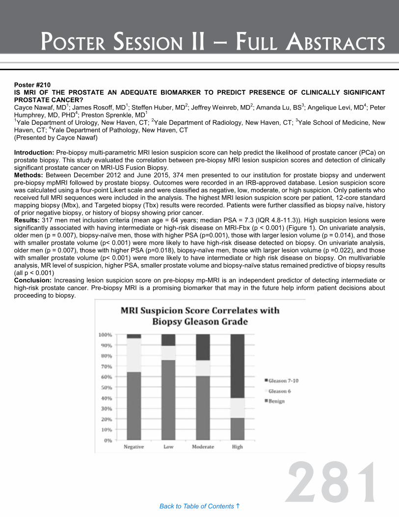
281Back to Table of Contents h
Poster session ii – Full AbstrActs
Poster #210 IS MRI OF THE PROSTATE AN ADEQUATE BIOMARKER TO PREDICT PRESENCE OF CLINICALLY SIGNIFICANT PROSTATE CANCER? Cayce Nawaf, MD1; James Rosoff, MD1; Steffen Huber, MD2; Jeffrey Weinreb, MD2; Amanda Lu, BS3; Angelique Levi, MD4; Peter Humphrey, MD, PHD4; Preston Sprenkle, MD1 1Yale Department of Urology, New Haven, CT; 2Yale Department of Radiology, New Haven, CT; 3Yale School of Medicine, New Haven, CT; 4Yale Department of Pathology, New Haven, CT (Presented by Cayce Nawaf) Introduction: Pre-biopsy multi-parametric MRI lesion suspicion score can help predict the likelihood of prostate cancer (PCa) on prostate biopsy. This study evaluated the correlation between pre-biopsy MRI lesion suspicion scores and detection of clinically significant prostate cancer on MRI-US Fusion Biopsy. Methods: Between December 2012 and June 2015, 374 men presented to our institution for prostate biopsy and underwent pre-biopsy mpMRI followed by prostate biopsy. Outcomes were recorded in an IRB-approved database. Lesion suspicion score was calculated using a four-point Likert scale and were classified as negative, low, moderate, or high suspicion. Only patients who received full MRI sequences were included in the analysis. The highest MRI lesion suspicion score per patient, 12-core standard mapping biopsy (Mbx), and Targeted biopsy (Tbx) results were recorded. Patients were further classified as biopsy naïve, history of prior negative biopsy, or history of biopsy showing prior cancer. Results: 317 men met inclusion criteria (mean age = 64 years; median PSA = 7.3 (IQR 4.8-11.3)). High suspicion lesions were significantly associated with having intermediate or high-risk disease on MRI-Fbx (p < 0.001) (Figure 1). On univariate analysis, older men (p = 0.007), biopsy-naïve men, those with higher PSA (p=0.001), those with larger lesion volume (p = 0.014), and those with smaller prostate volume (p< 0.001) were more likely to have high-risk disease detected on biopsy. On univariate analysis, older men (p = 0.007), those with higher PSA (p=0.018), biopsy-naïve men, those with larger lesion volume (p =0.022), and those with smaller prostate volume (p< 0.001) were more likely to have intermediate or high risk disease on biopsy. On multivariable analysis, MR level of suspicion, higher PSA, smaller prostate volume and biopsy-naïve status remained predictive of biopsy results (all p < 0.001) Conclusion: Increasing lesion suspicion score on pre-biopsy mp-MRI is an independent predictor of detecting intermediate or high-risk prostate cancer. Pre-biopsy MRI is a promising biomarker that may in the future help inform patient decisions about proceeding to biopsy.
Poster #209 VALIDATION OF AUA BEST PRACTICE GUIDELINES FOR PROSTATE BIOPSY INFECTIOUS COMPLICATIONS AND REDUCING VARIABILITY AND DURATION OF ANTIMICROBIAL UTILIZATION: QUALITY IMPROVEMENT INITIATIVE Behfar Ehdaie, MD, MPH; Emily Vertosick, MS; Pedro Recabal, MD; Michael Manasia, RN; Mary Schoen, RN, NP; James Eastham, MD; Karim Touijer, MD, MPH; Massimiliano Spaliviero, MD Memorial Sloan Kettering Cancer Center, New York, NY (Presented by Behfar Ehdaie) Introduction: The American Urological Association (AUA) recently updated recommendations for antibiotic prophylaxis prior to prostate biopsy. We sought to implement these guidelines to standardize antibiotic prophylaxis in a tertiary health system and assess the impact on infectious complications after biopsy. Methods: Prior to 2013, the choice of prophylactic antibiotics was at the discretion of the surgeon and typically consisted of a three-day duration. In 2013, we implemented the AUA guidelines to standardize prophylactic antibiotic use. The guidelines describe standardized regimens using a single antibiotic for up to 24 hours duration. The recommendations include consulting the local antibiogram and identifying high-risk men, including recent antibiotic use, and previous biopsy related infection. We enrolled 584 men who underwent biopsy from January 2011 to January 2012 and 654 men from January 2014 to January 2015. Men were contacted within 14 days of biopsy, and information was collected prospectively on complications, antibiotics received and bacterial culture results. We calculated the change in overall antibiotic use and rate of infectious complications between men biopsied in 2011 and 2014. Results: In 2011, the use of 19 different antibiotic regimens was reported. Antibiotics were usually started the evening before the biopsy, and continued for 72 hours. In 2014, 96% of men received one of three standardized antibiotic regimens, which were started on the same day of biopsy and continued for 24-hours. In both 2011 and 2014, fluoroquinolone prophylaxis was the most common regimen. In 2014, 98% of men received a one drug regimen, compared to only 73% of men in 2011. Infection rates were low in both cohorts, with 19 men (3.3%) in 2011 and 18 men (2.8%) in 2014. Men treated after implementing AUA guidelines had a lower rate of infection (difference -0.5%; 1-sided 95% CI 1.1%). We were able to exclude a clinically relevant increase in infectious complications using AUA guidelines. Conclusion: Implementing the AUA guidelines for antibiotic prophylaxis prior to prostate biopsy was non-inferior to a non-standardized approach for reducing infectious complications. The duration of antibiotics were reduced from 72 hours to less than 24 hours and the total number of antibiotic regimens decreased from 19 to three. We validated the recent AUA guidelines for antibiotic prophylaxis and recommend wide adoption of these guidelines for physicians that perform prostate biopsies.
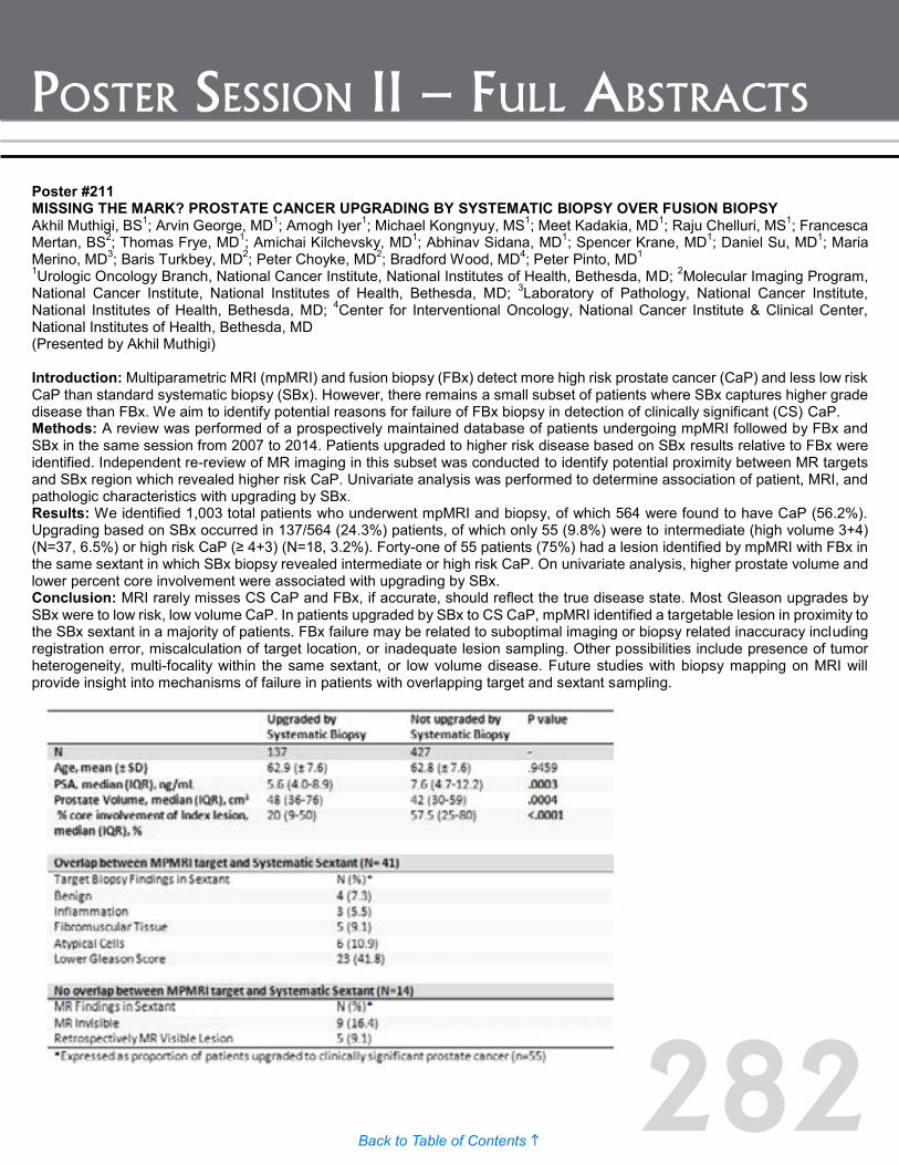
282Back to Table of Contents h
Poster session ii – Full AbstrActs
Poster #211 MISSING THE MARK? PROSTATE CANCER UPGRADING BY SYSTEMATIC BIOPSY OVER FUSION BIOPSY Akhil Muthigi, BS1; Arvin George, MD1; Amogh Iyer1; Michael Kongnyuy, MS1; Meet Kadakia, MD1; Raju Chelluri, MS1; Francesca Mertan, BS2; Thomas Frye, MD1; Amichai Kilchevsky, MD1; Abhinav Sidana, MD1; Spencer Krane, MD1; Daniel Su, MD1; Maria Merino, MD3; Baris Turkbey, MD2; Peter Choyke, MD2; Bradford Wood, MD4; Peter Pinto, MD1 1Urologic Oncology Branch, National Cancer Institute, National Institutes of Health, Bethesda, MD; 2Molecular Imaging Program, National Cancer Institute, National Institutes of Health, Bethesda, MD; 3Laboratory of Pathology, National Cancer Institute, National Institutes of Health, Bethesda, MD; 4Center for Interventional Oncology, National Cancer Institute & Clinical Center, National Institutes of Health, Bethesda, MD (Presented by Akhil Muthigi) Introduction: Multiparametric MRI (mpMRI) and fusion biopsy (FBx) detect more high risk prostate cancer (CaP) and less low risk CaP than standard systematic biopsy (SBx). However, there remains a small subset of patients where SBx captures higher grade disease than FBx. We aim to identify potential reasons for failure of FBx biopsy in detection of clinically significant (CS) CaP. Methods: A review was performed of a prospectively maintained database of patients undergoing mpMRI followed by FBx and SBx in the same session from 2007 to 2014. Patients upgraded to higher risk disease based on SBx results relative to FBx were identified. Independent re-review of MR imaging in this subset was conducted to identify potential proximity between MR targets and SBx region which revealed higher risk CaP. Univariate analysis was performed to determine association of patient, MRI, and pathologic characteristics with upgrading by SBx. Results: We identified 1,003 total patients who underwent mpMRI and biopsy, of which 564 were found to have CaP (56.2%). Upgrading based on SBx occurred in 137/564 (24.3%) patients, of which only 55 (9.8%) were to intermediate (high volume 3+4) (N=37, 6.5%) or high risk CaP (≥ 4+3) (N=18, 3.2%). Forty-one of 55 patients (75%) had a lesion identified by mpMRI with FBx in the same sextant in which SBx biopsy revealed intermediate or high risk CaP. On univariate analysis, higher prostate volume and lower percent core involvement were associated with upgrading by SBx. Conclusion: MRI rarely misses CS CaP and FBx, if accurate, should reflect the true disease state. Most Gleason upgrades by SBx were to low risk, low volume CaP. In patients upgraded by SBx to CS CaP, mpMRI identified a targetable lesion in proximity to the SBx sextant in a majority of patients. FBx failure may be related to suboptimal imaging or biopsy related inaccuracy including registration error, miscalculation of target location, or inadequate lesion sampling. Other possibilities include presence of tumor heterogeneity, multi-focality within the same sextant, or low volume disease. Future studies with biopsy mapping on MRI will provide insight into mechanisms of failure in patients with overlapping target and sextant sampling.
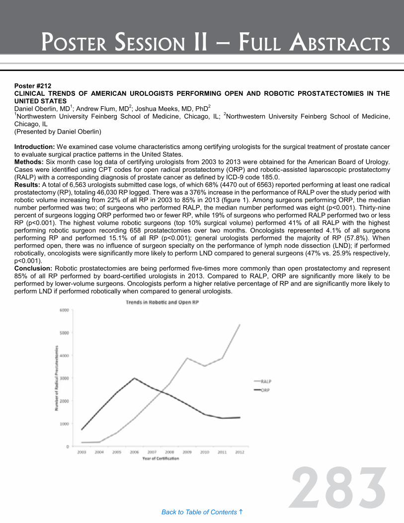
283Back to Table of Contents h
Poster session ii – Full AbstrActs
Poster #212 CLINICAL TRENDS OF AMERICAN UROLOGISTS PERFORMING OPEN AND ROBOTIC PROSTATECTOMIES IN THE UNITED STATES Daniel Oberlin, MD1; Andrew Flum, MD2; Joshua Meeks, MD, PhD2 1Northwestern University Feinberg School of Medicine, Chicago, IL; 2Northwestern University Feinberg School of Medicine, Chicago, IL (Presented by Daniel Oberlin) Introduction: We examined case volume characteristics among certifying urologists for the surgical treatment of prostate cancer to evaluate surgical practice patterns in the United States. Methods: Six month case log data of certifying urologists from 2003 to 2013 were obtained for the American Board of Urology. Cases were identified using CPT codes for open radical prostatectomy (ORP) and robotic-assisted laparoscopic prostatectomy (RALP) with a corresponding diagnosis of prostate cancer as defined by ICD-9 code 185.0. Results: A total of 6,563 urologists submitted case logs, of which 68% (4470 out of 6563) reported performing at least one radical prostatectomy (RP), totaling 46,030 RP logged. There was a 376% increase in the performance of RALP over the study period with robotic volume increasing from 22% of all RP in 2003 to 85% in 2013 (figure 1). Among surgeons performing ORP, the median number performed was two; of surgeons who performed RALP, the median number performed was eight (p<0.001). Thirty-nine percent of surgeons logging ORP performed two or fewer RP, while 19% of surgeons who performed RALP performed two or less RP (p<0.001). The highest volume robotic surgeons (top 10% surgical volume) performed 41% of all RALP with the highest performing robotic surgeon recording 658 prostatectomies over two months. Oncologists represented 4.1% of all surgeons performing RP and performed 15.1% of all RP (p<0.001); general urologists performed the majority of RP (57.8%). When performed open, there was no influence of surgeon specialty on the performance of lymph node dissection (LND); if performed robotically, oncologists were significantly more likely to perform LND compared to general surgeons (47% vs. 25.9% respectively, p<0.001). Conclusion: Robotic prostatectomies are being performed five-times more commonly than open prostatectomy and represent 85% of all RP performed by board-certified urologists in 2013. Compared to RALP, ORP are significantly more likely to be performed by lower-volume surgeons. Oncologists perform a higher relative percentage of RP and are significantly more likely to perform LND if performed robotically when compared to general urologists.
284Back to Table of Contents h
Poster session ii – Full AbstrActs
Poster #213 PROSTATE CANCER PATHOLOGY HAS WORSENED SINCE USPSTF DECISION Deepak A. Kapoor, MD1,2; Ann Anderson, MD3; Carl A. Olsson, MD, FACS4,5 1Chairman and CEO, Integrated Medical Professionals , Melville NY; 2Clinical Associate Professor, Icahn School of Medicine at Mount Sinai; 3Pathology Director, Integrated Medical Professionals, Garden City, NY; 4Chief Medical Officer, Integrated Medical Professionals, North Hills NY; 5Director of Quality Initiatives and Strategy, Urology Dept, Icahn School of Medicine at Mount Sinai (Presented by Carl A. Olsson) Introduction: Literature on impact of the Grade D recommendation against screening published by the United States Preventive Services Task Force (USPSTF) is conflicting. In our large urology practice, prostate biopsy (PBx) results had seemed to us more aggressive since that publication, prompting analysis of our results over time. Methods: We reviewed two years experience (2010 and 2011) prior to the USPSTF publication in 2012, creating a baseline against which similar reviews of 2013 and 2014 could be compared. All men (3,429) undergoing PBx in years 2010 and 2011 were combined and compared with all men having PBx in 2013 (2,773) and 2014 (2,577). Analysis of PBx results was the same throughout all years and focused only on patients with a cancer diagnosis. Each PBx core was studied for both primary Gleason pattern and score and each patient was assigned a Weighted Gleason Index wherein each positive core was multiplied by its own Gleason score, resulting in a single number representing both # cores positive and Gleason score. Results: The percentage of positive PBx increased over time, from the combined rate of 39% (1,338) in 2010/11 to 41.4% (1,147) in 2013 and 42.6% (1,097) in 2014. (p<001) Of 16,271 cores in 2010/11, 5047 or 31.3% were positive. Gleason patterns were 3 in 67%, 4 in 31.5% and 5 in 1.5%. Gleason scores were 8 in 9.5%, 9 in 5.0% and 10 in 0.3%. Of 13,960 cores in 2013, 4415 or 31.6% were positive. Gleason patterns were 3 in 69.8%, 4 in 27.1% and 5 in 3.1%, while Gleason scores were 8 in 8.7%, 9 in 10.7% and 10 in 0.3%. Of 13577 cores in 2014, 4694 or 34.6% were positive.(p<001) Here, Gleason patterns were 3 in 61.4%, 4 in 33.5% and 5 in 5.2% and Gleason scores were 8 in 10.7%, 9 in 14.5% and 10 in 0.9%. (both p<001). Weighted Gleason Index was 25.53 in 2010/11, compared to 25.99 in 2013 (NS) and 29.77 in 2014 (p<001). Conclusion: The USPSTF grade D decision against prostate screening is temporally related to worsening of many PBx parameters compared to baseline. These changes continue to worsen as time passes, raising concern for delayed diagnosis of important cancers and portending potential reversal of favorable trends in prostate cancer mortality.
285Back to Table of Contents h
Poster session ii – Full AbstrActs
Poster #214 PROTEIN SYNTHESIS DEPENDENT ACTIVATION OF THE UNFOLDED PROTEIN RESPONSE ENABLES PROSTATE CANCER DEVELOPMENT AND A DRUGGABLE TARGET FOR CANCER THERAPY Hao Nguyen MD, PhD; Crystal Conn, PhD; Tom Cunningham, PhD; Davide Ruggero, PhD UC San Francisco Medical Center (Presented by Hao Nguyen) The acquisition of oncogenic lesions stimulates biosynthetically and bioenergetically demanding cellular processes such as protein synthesis to drive cancer cell growth and proliferation. The hijacking of these key processes by oncogenic pathways triggers cellular stress that requires an adaptive or evasive response in order for cancer cells to survive and continue proliferating. We have previously demonstrated that deregulated protein synthesis activates one of the key cytoprotective stress response pathways, known as the unfolded protein response (UPR). The UPR is a cellular homeostatic program engaged when an excess of unfolded/misfolded proteins accumulate within the lumen of the endoplasmic reticulum. It is carried out by three major signaling arms: PERK, IRE1, and ATF6. However, whether and how each of these distinct signaling arms of the UPR is specifically activated by deregulated protein synthesis upon oncogenic insult is poorly understood. Here, we investigate prostate cancer initiation and maintenance, following combined loss of the PTEN tumor suppressor and overexpression of the Myc oncogene on protein synthesis-dependent activation of the UPR to facilitate tumor cell survival using a novel genetic mouse model. We observe that overexpression of Myc in the prostate synergizes with PTEN loss to dramatically stimulate the PERK and IRE1 signaling arms of the UPR pathway, which correlates with enhanced PIN formation and invasive carcinoma. To dissect the mechanism by which these oncogenic lesions promote UPR signaling, we have developed a cell culture model employing human prostate epithelial cells overexpress MYC, harbor an shRNA targeting PTEN, or the combined overexpression of MYC and shRNA of PTEN. Using this cell culture model, we demonstrate the activation of UPR arms PERK and IRE1 upon Myc overexpression and loss of PTEN. Interestingly, blocking the cytoprotective UPR using PERK or IRE1 inhibitors resulted in a significant increase in cell death and decreased clonogenic potential in cells harboring both oncogenic lesions (MYC/PTEN), but not in normal cells. Experiments are currently underway to test UPR inhibition in a preclinical trial. Taken together, our results suggest a critical role of the UPR in ensuring prostate cancer cell progression and serve as a promising opportunity for therapeutically targeting this cancer-specific vulnerability to stress adaptation in order to elicit synthetic lethality.
286Back to Table of Contents h
Poster session ii – Full AbstrActs
Poster #215 TUMOR CONTACT LENGTH: A NOVEL MULTIPARAMETRIC MRI PREDICTOR OF PROSTATE CANCER OUTCOMES Michael Kongnyuy, MS1; Arvin George, MD1; Amogh Iyer1; Thomas Frye, MD1; Amichai Kilchevsky, MD1; Abhinav Sidana, MD1; Spencer Krane, MD1; Meet Kadakia, MD1; Akhil Muthigi, BS1; Francesca Mertan, BS2; Raju Chelluri, MS1; Richard Ho, BS1; Daniel Su, MD1; Maria Merino, MD3; Baris Turkbey, MD2; Peter Choyke, MD2; Bradford Wood, MD4; Peter Pinto MD1 1Urologic Oncology Branch, National Cancer Institute, National Institutes of Health, Bethesda, MD; 2Molecular Imaging Program, National Cancer Institute, National Institutes of Health, Bethesda, MD; 3Laboratory of Pathology, National Cancer Institute, National Institutes of Health, Bethesda, MD; 4Center for Interventional Oncology, National Cancer Institute & Clinical Center, National Institutes of Health, Bethesda, MD (Presented by Michael Kongnyuy) Introduction: Multiparametric Magnetic Resonance Imaging (MP-MRI) can visualize prostate tumors. MP-MRI characteristics (extraprostatic extension [ECE], tumor volume, seminal vesicle invasion, etc.) can be predictive of final pathological findings such as lymph node (LN) involvement, true pathological ECE (pECE), margin status, and biochemical recurrence (BCR). These are pivotal in the decision-making process regarding treatment. Tumor contact length (TCL) is defined as the length of cancer in contact with the prostatic capsule. We evaluated the ability of MP-MRI determined TCL in predicting pECE, BCR and LN in patients undergoing radical prostatectomy. Methods: All patients who underwent a 3T MP-MRI at the NCI from 2007 to 2015 were retrospectively classed into no ECE, suspicious ECE (sECE) and frank ECE (fECE) based on MP-MRI findings. sECE was defined as tumor with capsular bulge on MRI while fECE was clear capsular obliteration and tumor extension beyond the prostatic capsule. Demographic data was obtained on patients with fECE and sECE on MP-MRI with the presence of pECE, LN, and BCR status following radical prostatectomy from a single surgeon (PP) experience. All MP-MRIs were performed and read by two expert GU radiologists (BT, PLC) and all pathology read by a single GU pathologist (MJM). Chi-Square analysis was done to compare proportions and Wilcoxon rank sum test was used to compare continuous variables. Logistic regression was used to determine the predictive ability of TCL. Statistical significance was defined as p-value ≤0.05. Results: Of all 1260 patients who underwent MP-MRI, we focused on 146 who had sECE (68) or fECE (78) on MP-MRI. Mean age was 60 years and median prostate specific antigen was 11.7 ng/ml. Logistic regression analysis showed that MP-MRI determined TCL was predictive of ECE (p=0.01), LN status (p=0.0001) on final pathology and BCR (p=0.05) during follow up. Patients with pECE had a longer median MP-MRI TCL (2.8cm) compared to those without pECE (2.4cm), p=0.04. When analyzed individually, fECE correlated with pECE (p=0.05) while s ECE did not correlate with pECE (p=0.11). Although, not statistically significant, the median MP-MRI TCL for sECE with pECE was still longer than in sECE with no pECE in the sub-group analysis. Conclusion: Longer TCL on MP-MRI can indicate presence of ECE, LN involvement at final pathology as well as predict BCR on follow up.
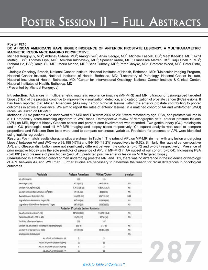
287Back to Table of Contents h
Poster session ii – Full AbstrActs
Poster #216 DO AFRICAN AMERICANS HAVE HIGHER INCIDENCE OF ANTERIOR PROSTATE LESIONS?: A MULTIPARAMETRIC MAGNETIC RESONANCE IMAGING PERSPECTIVE. Michael Kongnyuy, MS1; Abhinav Sidana, MD1; Amogh Iyer1; Arvin George, MD1; Michele Fascelli, BS1; Meet Kadakia, MD1; Akhil Muthigi, BS1; Thomas Frye, MD1; Amichai Kilchevsky, MD1; Spencer Krane, MD1; Francesca Mertan, BS2; Raju Chelluri, MS1; Richard Ho, BS1; Daniel Su, MD1; Maria Merino, MD3; Baris Turkbey, MD2; Peter Choyke, MD2; Bradford Wood, MD4; Peter Pinto, MD1 1Urologic Oncology Branch, National Cancer Institute, National Institutes of Health, Bethesda, MD; 2Molecular Imaging Program, National Cancer Institute, National Institutes of Health, Bethesda, MD; 3Laboratory of Pathology, National Cancer Institute, National Institutes of Health, Bethesda, MD; 4Center for Interventional Oncology, National Cancer Institute & Clinical Center, National Institutes of Health, Bethesda, MD (Presented by Michael Kongnyuy) Introduction: Advances in multiparametric magnetic resonance imaging (MP-MRI) and MRI ultrasound fusion-guided targeted biopsy (TBx) of the prostate continue to improve the visualization, detection, and categorization of prostate cancer (PCa) lesions. It has been reported that African Americans (AA) may harbor high-risk lesions within the anterior prostate contributing to poorer outcomes in active surveillance. We aim to report the rates of anterior lesions, in a matched cohort of AA and white/other (W/O) races, visualized on MP-MRI. Methods: All AA patients who underwent MP-MRI and TBx from 2007 to 2015 were matched by age, PSA, and prostate volume in a 1:1 propensity score-matching algorithm to W/O races. Retrospective review of demographic data, anterior prostate lesions (APL) on MRI, highest biopsy Gleason scores and percent core involvement was recorded. Two genitourinary (GU) radiologists and a GU pathologist read all MP-MRI imaging and biopsy slides respectively. Chi-square analysis was used to compare proportions and Wilcoxon Sum tests were used to compare continuous variables. Predictors for presence of APL were identified using logistic regression. Results: Post-match results characteristics are shown in Table 1. The rates of APL on MP-MRI (in men with any lesion undergoing biopsy) between AA and W/O were 93/195 (47%) and 94/195 (48.2%) respectively (p=0.62). Similarly, the rates of cancer-positive APL and Gleason distribution were not significantly different between the cohorts (p=0.72 and p=0.87 respectively). Presence of prior negative biopsy was the sole predictor of presence of APL in MP-MRI in AA subset of our cohort (p=0.04). Increasing PSA (p=0.001) and presence of prior biopsy (p=0.045) predicted positive anterior lesion on MRI targeted biopsy. Conclusion: In a matched cohort of men undergoing prostate MRI and TBx, there was no difference in the incidence or histology of APL between AA and W/O men. Further studies are necessary to determine the reason for racial differences in oncological outcomes.
288Back to Table of Contents h
Poster session ii – Full AbstrActs
Poster #217 SUCCESSFUL IMPLEMENTATION OF A DISEASE SPECIFIC SURVIVORSHIP PROGRAM FOR MEN WITH PROSTATE CANCER (PC) AND THEIR PARTNERS Celestia Higano, MD, FACP1; Phil Pollock, MRes2; Richard Wassersug, PhD1; Christine Zarowski, RN2; Marcy Dayan, BSR; MHA2; Sarah Weller, BAppSci2; Cheri Van Patten, RD3; Stacy Elliott, MD1; Monita Sundar, MA2; Sarah Mahovlich, BSC4; Erik Wibowo, PhD4; Martin Gleave, MD1; Peter Black, MD1; Alan So, MD1; Larry Goldenberg, MD1 1University of British Columbia, Vancouver, BC; 2Vancouver Prostate Center, Vancouver, BC; 3British Columbia Cancer Agency, Vancouver, BC; 4Vancouver Prostate Centre, Vancouver, BC (Presented by Celesita Higano) Introduction: Treatment for localized PC can adversely impact the quality of life for the patient (pt) and his partner. Addition of androgen deprivation therapy (ADT) to treat biochemical relapse or metastatic disease can result in further symptoms. We hypothesized that PC patients and partners would benefit from a clinical, educational, research-based approach to care that would focus on their specific needs. Methods: Funding from government agencies and philanthropic sources were used to establish a supportive care program at the Vancouver Prostate Centre. A multi-disciplinary management team was formed to oversee the program. The Prostate Cancer Supportive Care (PCSC) Program is organized around five thematic modules: 1) information about PC and primary treatment options (TX), 2) sexual health and intimacy (SH), 3) lifestyle changes in diet and exercise (DE), 4) managing the side effects of ADT, 5) incontinence and pelvic floor physiotherapy (PT). Group educational sessions (ED) are held one to two times monthly for each module. Individual clinic appointments (CLIN) with SH and PT clinicians were also available. A program manager, clinic coordinator, and research assistant run PCSC on a day-to-day basis. Results: PCSC, located in the urology clinic at Vancouver General Hospital (VGH), was initiated in January 2013. A total of 783 patients were referred by urologists at VGH, nurses, pharmacists, and radiation oncologists at the British Columbia Cancer Agency, or were self-referred: 167 in 2013, 356 in 2014, and 260 as of August 2015. Not all patients chose to participate but those who did are summarized in the table below. Feedback from couples, participating clinicians, and allied health personnel has been overwhelmingly positive. Conclusion: The results demonstrate that implementation of a disease specific survivorship program is feasible and well received. Why some declined PCSC services that were free of charge is being explored. Outcomes research and intervention protocols are in progress to address our hypothesis.
289Back to Table of Contents h
Poster session ii – Full AbstrActs
Poster #218 PROSTATE GENETIC SCORE IN MEN WITH METASTATIC PROSTATE CANCER Michael Liss1, Jianfeng Xu2, Zachary Hamilton3, S. Lilly Zheng2, Haitao Chen2, Jae Choi3, Frederick Millard3, James Michael Randall3, Sij Hemal4, A. Karim Kader3 1University of Texas Health Science Center, San Antonio; TX 2NorthShore University HealthSystem, Evanston, IL; 3University of California, San Diego, CA; 4Wake Forest School of Medicine, Winston-Salem, NC (Presented by Zachary Hamilton) Introduction: Prostate specific antigen (PSA) based prostate cancer (PCa) screening may be refined by focusing screening efforts on men at higher-risk. Herein we investigate family history together with the prostate genetic risk score (PGS), a germline biomarker of PCa risk, in a cohort of men with metastatic PCa to determine the proportion of men the test would have indicated screening or avoided screening. Methods: After University of California San Diego IRB approval, we prospectively enrolled men presenting to the oncology clinic with metastatic castrate resistant prostate cancer. Baseline clinical characteristics and saliva were collected on all patients. Germline DNA was harvested from saliva and genotyped at 33 PCa associated single nucleotide polymorphisms (SNPs). The PGS was calculated based on their genotype and weighted by odds ratio (OR) and allele frequency of SNPs. Based on previous studies, a PGS of 0.6 without a family history indicated the lowest risk of prostate cancer. A PGS score >1.3 or a family history of prostate cancer indicates the highest risk. Results: One hundred patients were enrolled, 77% Caucasian, 21% with a family history, 73% with castrate resistant metastatic prostate cancer. The median PGS was 1.2 (IQR 0.86-2.03) with four patients having a PGS <0.6 and 44 men having a PGS >1.3. Only four patients (4%) qualified as low risk for prostate cancer by genetic testing (PGS <0.6) and a negative family history (FH-). However, 55 patients (55%) would have been identified as a high genetic risk or a family history of PCa. Twenty men (20%) presented with metastasis and did not have previous PSA screening on record, of which none (0%) had PGS <0.6 and 9 of the 20 (45%) had PGS >1.3. Conclusion: If screening were recommended for higher risk men (average and high risk individuals using the PGS >0.6 or a positive family history for PCa), 96% of this cohort would have undergone PSA screening, while four percent may have been missed. PGS at an early age to indicate PCa screening may have reduced the metastatic presentation of lethal PCa by 45%.
Poster #217 SUCCESSFUL IMPLEMENTATION OF A DISEASE SPECIFIC SURVIVORSHIP PROGRAM FOR MEN WITH PROSTATE CANCER (PC) AND THEIR PARTNERS Celestia Higano, MD, FACP1; Phil Pollock, MRes2; Richard Wassersug, PhD1; Christine Zarowski, RN2; Marcy Dayan, BSR; MHA2; Sarah Weller, BAppSci2; Cheri Van Patten, RD3; Stacy Elliott, MD1; Monita Sundar, MA2; Sarah Mahovlich, BSC4; Erik Wibowo, PhD4; Martin Gleave, MD1; Peter Black, MD1; Alan So, MD1; Larry Goldenberg, MD1 1University of British Columbia, Vancouver, BC; 2Vancouver Prostate Center, Vancouver, BC; 3British Columbia Cancer Agency, Vancouver, BC; 4Vancouver Prostate Centre, Vancouver, BC (Presented by Celesita Higano) Introduction: Treatment for localized PC can adversely impact the quality of life for the patient (pt) and his partner. Addition of androgen deprivation therapy (ADT) to treat biochemical relapse or metastatic disease can result in further symptoms. We hypothesized that PC patients and partners would benefit from a clinical, educational, research-based approach to care that would focus on their specific needs. Methods: Funding from government agencies and philanthropic sources were used to establish a supportive care program at the Vancouver Prostate Centre. A multi-disciplinary management team was formed to oversee the program. The Prostate Cancer Supportive Care (PCSC) Program is organized around five thematic modules: 1) information about PC and primary treatment options (TX), 2) sexual health and intimacy (SH), 3) lifestyle changes in diet and exercise (DE), 4) managing the side effects of ADT, 5) incontinence and pelvic floor physiotherapy (PT). Group educational sessions (ED) are held one to two times monthly for each module. Individual clinic appointments (CLIN) with SH and PT clinicians were also available. A program manager, clinic coordinator, and research assistant run PCSC on a day-to-day basis. Results: PCSC, located in the urology clinic at Vancouver General Hospital (VGH), was initiated in January 2013. A total of 783 patients were referred by urologists at VGH, nurses, pharmacists, and radiation oncologists at the British Columbia Cancer Agency, or were self-referred: 167 in 2013, 356 in 2014, and 260 as of August 2015. Not all patients chose to participate but those who did are summarized in the table below. Feedback from couples, participating clinicians, and allied health personnel has been overwhelmingly positive. Conclusion: The results demonstrate that implementation of a disease specific survivorship program is feasible and well received. Why some declined PCSC services that were free of charge is being explored. Outcomes research and intervention protocols are in progress to address our hypothesis.
290Back to Table of Contents h
Poster session ii – Full AbstrActs
Poster #219 MULTIPLEX TESTING COMBINING THE GENETIC SCORE INDEX (GSI) WITH THE PROSTATE HEALTH INDEX (PHI) FOR THE DETECTION OF CLINICALLY SIGNIFICANT PROSTATE CANCER Jen-Jane Liu, MD1; Zhaoyong Feng2; Lori Sokoll2; Bruce Trock2; Jianfeng Xu3; William Isaacs2; Leslie Mangold2; Alan Partin2; Christian Pavlovich2 1Portland, OR; 2Johns Hopkins Hospital, Baltimore, MD; 3Northshore University Health System (Presented by Jen-Jane Liu) Introduction: There is a need for biomarkers that differentiate between clinically significant prostate cancers requiring intervention and non-clinically significant cancers that can be observed. Phi uses PSA isoforms to predict the risk of prostate cancer while the GSI is based on SNPs that predict patient risk for developing prostate cancer. Unlike serum biochemical indices (phi), which vary over a patient’s lifetime, genomic tests (GSI) are static biomarkers that reflect the inherent potential for malignancy and also avoid selection biases associated with PSA screening. Our goal was to determine whether multiplex biochemical and germline genetic testing can improve the detection of clinically significant prostate cancer. Methods: We identified patients from two biorepositories who had serum and buffy coat samples taken prior to prostate biopsy. [-2]proPSA was measured to calculate phi and the GSI was calculated based on a 70 SNP panel. Performance characteristics of PSA, phi, and GSI for identifying clinically significant prostate cancer (based on D’Amico and Epstein criteria) were calculated. Results: A total of 424 samples were analyzed, of which 112 had benign or clinically insignificant cancer, and 311 had clinically significant cancer. The median PSA of the two groups was 6.7 (0.6–285) and 6.2 (0.5-130) ng/mL, respectively (p=0.79). The median phi was 38 and 45, respectively (p=0.0004). Median GSI for the clinically insignificant cancer group was 0.52, compared to 0.59 for the clinically significant cancer group (p=0.19). AUC for PSA, age, race was 0.56 (see figure). When GSI and phi were added to age and race, the AUC for the model increased to 0.63 (p=0.02). Phi+age+race (AUC=0.62) was also superior to PSA+age+race (AUC=0.56, p=0.03). Conclusion: The addition of GSI and phi to standard models including age and race improve the ability to accurately diagnose clinically significant prostate cancer compared to PSA. The improved performance is driven by phi, suggesting that phi may be more useful than PSA in identifying clinically significant prostate cancer. As more SNPs associated with prostate cancer are identified, there may be an improvement in the performance of GSI in the prediction of clinically significant prostate cancer.
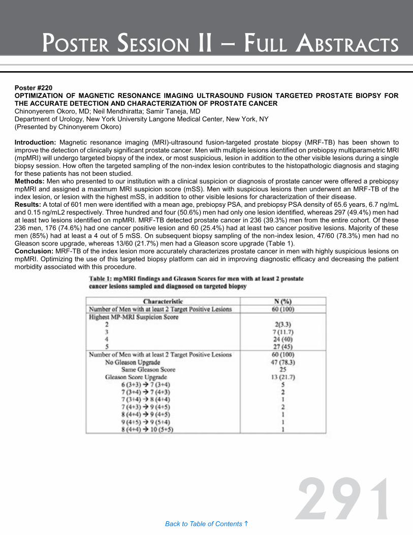
291Back to Table of Contents h
Poster session ii – Full AbstrActs
Poster #220 OPTIMIZATION OF MAGNETIC RESONANCE IMAGING ULTRASOUND FUSION TARGETED PROSTATE BIOPSY FOR THE ACCURATE DETECTION AND CHARACTERIZATION OF PROSTATE CANCER Chinonyerem Okoro, MD; Neil Mendhiratta; Samir Taneja, MD Department of Urology, New York University Langone Medical Center, New York, NY (Presented by Chinonyerem Okoro) Introduction: Magnetic resonance imaging (MRI)-ultrasound fusion-targeted prostate biopsy (MRF-TB) has been shown to improve the detection of clinically significant prostate cancer. Men with multiple lesions identified on prebiopsy multiparametric MRI (mpMRI) will undergo targeted biopsy of the index, or most suspicious, lesion in addition to the other visible lesions during a single biopsy session. How often the targeted sampling of the non-index lesion contributes to the histopathologic diagnosis and staging for these patients has not been studied. Methods: Men who presented to our institution with a clinical suspicion or diagnosis of prostate cancer were offered a prebiopsy mpMRI and assigned a maximum MRI suspicion score (mSS). Men with suspicious lesions then underwent an MRF-TB of the index lesion, or lesion with the highest mSS, in addition to other visible lesions for characterization of their disease. Results: A total of 601 men were identified with a mean age, prebiopsy PSA, and prebiopsy PSA density of 65.6 years, 6.7 ng/mL and 0.15 ng/mL2 respectively. Three hundred and four (50.6%) men had only one lesion identified, whereas 297 (49.4%) men had at least two lesions identified on mpMRI. MRF-TB detected prostate cancer in 236 (39.3%) men from the entire cohort. Of these 236 men, 176 (74.6%) had one cancer positive lesion and 60 (25.4%) had at least two cancer positive lesions. Majority of these men (85%) had at least a 4 out of 5 mSS. On subsequent biopsy sampling of the non-index lesion, 47/60 (78.3%) men had no Gleason score upgrade, whereas 13/60 (21.7%) men had a Gleason score upgrade (Table 1). Conclusion: MRF-TB of the index lesion more accurately characterizes prostate cancer in men with highly suspicious lesions on mpMRI. Optimizing the use of this targeted biopsy platform can aid in improving diagnostic efficacy and decreasing the patient morbidity associated with this procedure.
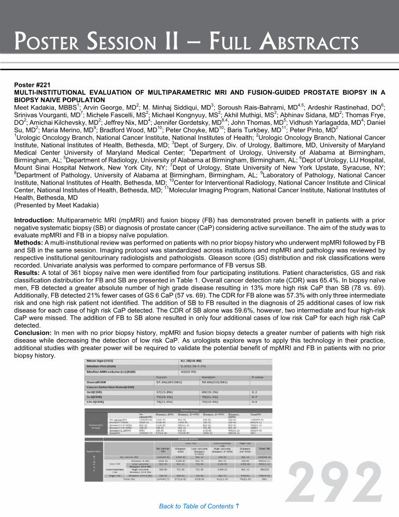
292Back to Table of Contents h
Poster session ii – Full AbstrActs
Poster #221 MULTI-INSTITUTIONAL EVALUATION OF MULTIPARAMETRIC MRI AND FUSION-GUIDED PROSTATE BIOPSY IN A BIOPSY NAIVE POPULATION Meet Kadakia, MBBS1; Arvin George, MD2; M. Minhaj Siddiqui, MD3; Soroush Rais-Bahrami, MD4,5; Ardeshir Rastinehad, DO6; Srinivas Vourganti, MD7; Michele Fascelli, MS2; Michael Kongnyuy, MS2; Akhil Muthigi, MS2; Abhinav Sidana, MD2; Thomas Frye, DO2; Amichai Kilchevsky, MD2; Jeffrey Nix, MD4; Jennifer Gordetsky, MD8,4; John Thomas, MD5; Vidhush Yarlagadda, MD4; Daniel Su, MD2; Maria Merino, MD9; Bradford Wood, MD10; Peter Choyke, MD10; Baris Turkbey, MD11; Peter Pinto, MD2 1Urologic Oncology Branch, National Cancer Institute, National Institutes of Health; 2Urologic Oncology Branch, National Cancer Institute, National Institutes of Health, Bethesda, MD; 3Dept. of Surgery, Div. of Urology, Baltimore, MD, University of Maryland Medical Center University of Maryland Medical Center; 4Department of Urology, University of Alabama at Birmingham, Birmingham, AL; 5Department of Radiology, University of Alabama at Birmingham, Birmingham, AL; 6Dept of Urology, LIJ Hospital, Mount Sinai Hospital Network, New York City, NY; 7Dept of Urology, State University of New York Upstate, Syracuse, NY; 8Department of Pathology, University of Alabama at Birmingham, Birmingham, AL; 9Laboratory of Pathology, National Cancer Institute, National Institutes of Health, Bethesda, MD; 10Center for Interventional Radiology, National Cancer Institute and Clinical Center, National Institutes of Health, Bethesda, MD; 11Molecular Imaging Program, National Cancer Institute, National Institutes of Health, Bethesda, MD (Presented by Meet Kadakia) Introduction: Multiparametric MRI (mpMRI) and fusion biopsy (FB) has demonstrated proven benefit in patients with a prior negative systematic biopsy (SB) or diagnosis of prostate cancer (CaP) considering active surveillance. The aim of the study was to evaluate mpMRI and FB in a biopsy naïve population. Methods: A multi-institutional review was performed on patients with no prior biopsy history who underwent mpMRI followed by FB and SB in the same session. Imaging protocol was standardized across institutions and mpMRI and pathology was reviewed by respective institutional genitourinary radiologists and pathologists. Gleason score (GS) distribution and risk classifications were recorded. Univariate analysis was performed to compare performance of FB versus SB. Results: A total of 361 biopsy naïve men were identified from four participating institutions. Patient characteristics, GS and risk classification distribution for FB and SB are presented in Table 1. Overall cancer detection rate (CDR) was 65.4%. In biopsy naïve men, FB detected a greater absolute number of high grade disease resulting in 13% more high risk CaP than SB (78 vs. 69). Additionally, FB detected 21% fewer cases of GS 6 CaP (57 vs. 69). The CDR for FB alone was 57.3% with only three intermediate risk and one high risk patient not identified. The addition of SB to FB resulted in the diagnosis of 25 additional cases of low risk disease for each case of high risk CaP detected. The CDR of SB alone was 59.6%, however, two intermediate and four high-risk CaP were missed. The addition of FB to SB alone resulted in only four additional cases of low risk CaP for each high risk CaP detected. Conclusion: In men with no prior biopsy history, mpMRI and fusion biopsy detects a greater number of patients with high risk disease while decreasing the detection of low risk CaP. As urologists explore ways to apply this technology in their practice, additional studies with greater power will be required to validate the potential benefit of mpMRI and FB in patients with no prior biopsy history.
293Back to Table of Contents h
Poster session ii – Full AbstrActs
Poster #222 ADJUVANT SURGICAL PROCEDURES IN PATIENTS UNDERGOING POST-CHEMOTHERAPY RETROPERITONEAL LYMPH NODE DISSECTION (RPLND) FOR TESTICULAR CANCER: BLOOD, SWEAT AND A FEW TEARS! John Banerji, MD, MCh (Urology)1; Edmond Raker, MD2; Jay Voit, MD2; Craig Nichols, MD3; Christopher Porter, MD, FACS1 1Dept. of Urology, Virginia Mason, Seattle, WA; 2Dept. of Vascular Surgery, Virginia Mason, Seattle, WA; 3Dept of Haematology and Oncology, Virginia Mason, Seattle, WA (Presented by John Banerji) Introduction: Post-chemotherapy RPLND is a key armamentarium for treatment of germ cell tumors of the testis. There are occasions when successful removal of all retroperitoneal disease necessitates en bloc removal of adjacent organs and/or major vascular resection. We queried our database of patients undergoing RPLND and identified those in whom such additional procedures were performed. Methods: We reviewed our IRB-approved retrospective testis cancer database and identified patients who underwent RPLND and at least one adjuvant procedure between 2004 and 2014. Adjuvant procedures were defined as removal of adjacent organs or major vascular structures. Results: Of the 100 patients who underwent RPLND, 17 (17%) underwent 31 adjuvant procedures. Median age was 29 years (range 20-49). Median estimated blood loss was 900 ml (range 100 ml to 25L) and median intra operative time was 628 min (range 420-1320). The majority of patients who underwent adjuvant procedures had nephrectomies (12/17, 70%). Patients also underwent colonic resection (2, 12%) and, mid jejunum resection (1, 6%), resection, resection of lung nodules (2, 12%), and resection of a paraesophageal nodal mass (1, 6%), the latter two performed with thoracic surgeons. Vascular procedures included aortic resection with reconstruction using a Dacron graft (7, 41%), anterior aortic wall resection repaired with a venous patch (1, 6%), and caval resection (4, 24%), two (12%) of which included reconstruction, and autotransplantation to revascularize the remaining kidney (2, 12%). Early complications included prolonged ileus (12, 70%), acute kidney injury (4, 23%), pneumonia (2, 12%), Clostridium difficile colitis (1, 6%), and abdominal compartment syndrome due to ascites (1, 6%). Retroperitoneal pathology revealed teratoma in a majority (11, 65%), with Wilm’s tumor-like differentiation in one (6%). Four patients (24%) had necrosis and fibrosis, one (6%) had a residual primitive neuroectodermal tumor (PNET), and one (6%) had a choriocarcinoma. There was one death in this series in the patient with PNET, who relapsed within three months and underwent three RPLND’s within four years. Conclusion: Adjuvant nephrectomy and vascular resection are safe, feasible adjunctive procedures when dealing with complex retroperitoneal tumor masses. These are best performed with a multi-disciplinary approach. Teratoma was the most common retroperitoneal pathology in our series, and being as they are chemoresistant, major resection is justified.
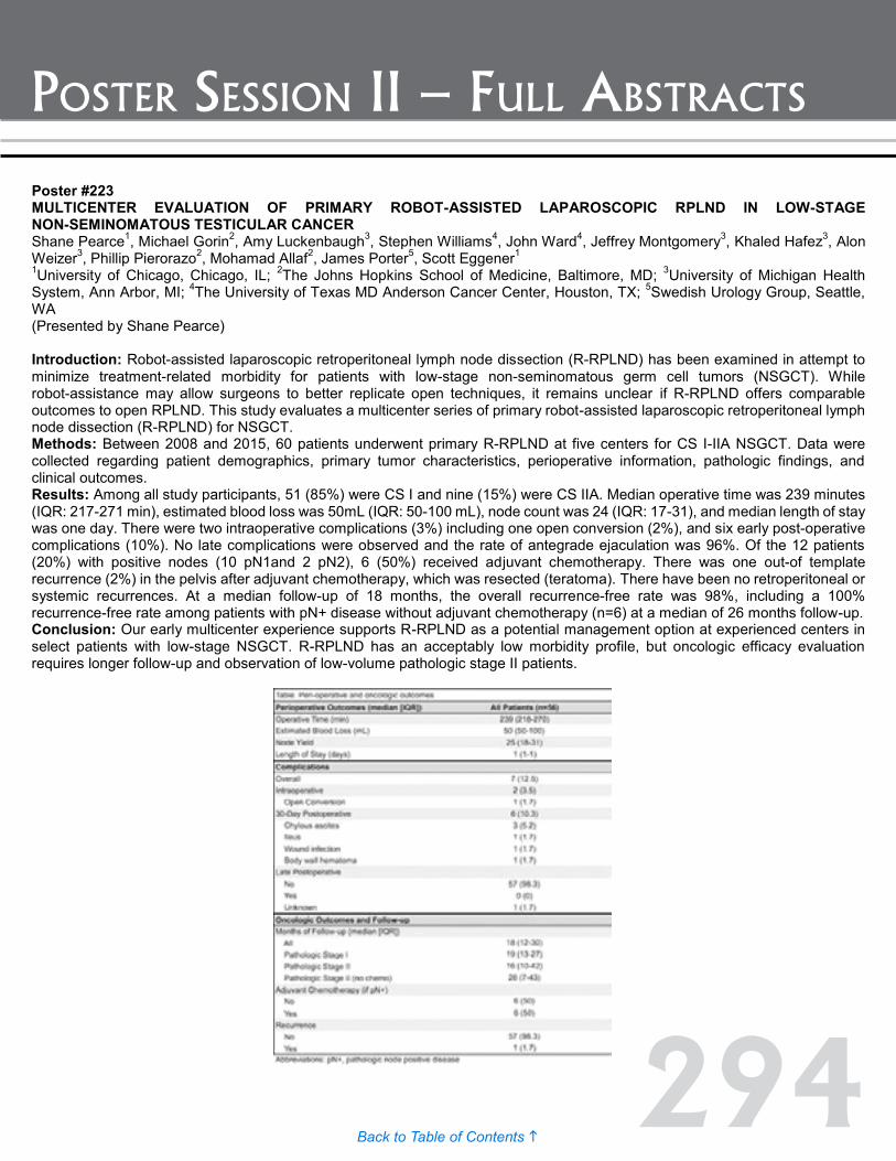
294Back to Table of Contents h
Poster session ii – Full AbstrActs
Poster #223 MULTICENTER EVALUATION OF PRIMARY ROBOT-ASSISTED LAPAROSCOPIC RPLND IN LOW-STAGE NON-SEMINOMATOUS TESTICULAR CANCER Shane Pearce1, Michael Gorin2, Amy Luckenbaugh3, Stephen Williams4, John Ward4, Jeffrey Montgomery3, Khaled Hafez3, Alon Weizer3, Phillip Pierorazo2, Mohamad Allaf2, James Porter5, Scott Eggener1 1University of Chicago, Chicago, IL; 2The Johns Hopkins School of Medicine, Baltimore, MD; 3University of Michigan Health System, Ann Arbor, MI; 4The University of Texas MD Anderson Cancer Center, Houston, TX; 5Swedish Urology Group, Seattle, WA (Presented by Shane Pearce) Introduction: Robot-assisted laparoscopic retroperitoneal lymph node dissection (R-RPLND) has been examined in attempt to minimize treatment-related morbidity for patients with low-stage non-seminomatous germ cell tumors (NSGCT). While robot-assistance may allow surgeons to better replicate open techniques, it remains unclear if R-RPLND offers comparable outcomes to open RPLND. This study evaluates a multicenter series of primary robot-assisted laparoscopic retroperitoneal lymph node dissection (R-RPLND) for NSGCT. Methods: Between 2008 and 2015, 60 patients underwent primary R-RPLND at five centers for CS I-IIA NSGCT. Data were collected regarding patient demographics, primary tumor characteristics, perioperative information, pathologic findings, and clinical outcomes. Results: Among all study participants, 51 (85%) were CS I and nine (15%) were CS IIA. Median operative time was 239 minutes (IQR: 217-271 min), estimated blood loss was 50mL (IQR: 50-100 mL), node count was 24 (IQR: 17-31), and median length of stay was one day. There were two intraoperative complications (3%) including one open conversion (2%), and six early post-operative complications (10%). No late complications were observed and the rate of antegrade ejaculation was 96%. Of the 12 patients (20%) with positive nodes (10 pN1and 2 pN2), 6 (50%) received adjuvant chemotherapy. There was one out-of template recurrence (2%) in the pelvis after adjuvant chemotherapy, which was resected (teratoma). There have been no retroperitoneal or systemic recurrences. At a median follow-up of 18 months, the overall recurrence-free rate was 98%, including a 100% recurrence-free rate among patients with pN+ disease without adjuvant chemotherapy (n=6) at a median of 26 months follow-up. Conclusion: Our early multicenter experience supports R-RPLND as a potential management option at experienced centers in select patients with low-stage NSGCT. R-RPLND has an acceptably low morbidity profile, but oncologic efficacy evaluation requires longer follow-up and observation of low-volume pathologic stage II patients.
295Back to Table of Contents h
Poster session ii – Full AbstrActs
Poster #224 EXTRAPERITONEAL MIDLINE RETROPERITONEAL LYMPH NODE DISSECTION Sumeet Syan-Bhanvadia, MD1; Soroush Bazargani, MD2; Thomas Clifford2; Hooman Djaladat, MD, MS2; Anne Schuckman, MD2; Siamak Daneshmand, MD2 1USC Institute of Urology; 2USC Institute of Urology, Los Angeles, CA (Presented by Sumeet Syan-Bhanvadia) Introduction: Retroperitoneal lymph node dissection (RPLND) for testicular germ cell tumors (GCT) can be technically challenging and associated with significant morbidity, particularly in the post-chemotherapy setting. We have previously described a midline extraperitoneal approach which decreases perioperative morbidity and length of stay. We present further experience performing a midline extraperitoneal (EP) RPLND in patients with NSGCT. Methods: 154 patients have undergone RPLND for germ cell tumors by a single surgeon between 2010 and 2014 (130 post-chemotherapy, 24 primary). Since 2010 all patients have undergone an EP RPLND unless resection of major vessels or extirpative procedures involving intraperitoneal structures was required, leaving 55 of 78 patients who underwent EP-RPLND (2010-2014). These were matched to a cohort undergoing midline transperitoneal RPLND (TP) from 2004-2010 by the same surgeon. All patients underwent bilateral template dissection in the post-chemotherapy setting and extended right and left templates for primary RPLND. Perioperative outcomes were retrospectively compared between groups. Results: There were no significant differences between the EP and TP groups with regard to patient age (28 vs. 29 years, p=0.6). Radiographic size of mass in the EP group was <2cm, 2-5cm and >5cm in 18, 13 and 19 patients compared to three, nine and 15 in the TP group (p=0.06) indicating a trend towards larger masses in the EP group. Mean number of nodes resected and number of positive nodes were higher in the EP group at 38 and five respectively, versus 28 and one in the TP group (p=0.03) which may reflect institutional differences in lymph node reporting. Median estimated blood loss (EBL) was 300cc in the EP group versus 750cc in the TP group (p<0.001), with a median length of stay (LOS) of three days (range 2-7) versus seven days for TP RPLND (p<0.001). EP patients received less intraoperative transfusions than TP (0.5 vs. 1.5, p= 0.05). There were 4 complications in the EP group the highest of which was one grade III complication (lymphocele requiring percutaneous drainage). There was no difference in complication rates between groups (p=0.9). Conclusion: Extraperitoneal RPLND can be performed safely without compromising operative expediency, exposure, or ability to perform complete node dissection, even in the setting of large tumors post-chemotherapy. This approach is associated with shorter hospital stay and potentially less EBL and transfusions.

296Back to Table of Contents h
AJAY, DIVYA 12/3/2015 4:30 pm Poster #180
ALBALA, DAVID 12/3/2015 4:30 pm Poster #156
AL-SHRAIDEH, YOUSEF 12/2/2015 4:30 pm Poster #55
BAGRODIA, ADITYA 12/2/2015 4:30 pm Poster #110 12/4/2015 11:23 am AB #8
BALL, MARK 12/2/2015 4:30 pm Poster #57 12/2/2015 4:30 pm Poster #58
BAZARGANI, SOROUSH 12/3/2015 4:30 pm Poster #132 12/3/2015 4:30 pm Poster #133
BIANCHI-FRIAS, DANIELA 12/3/2015 4:30 pm Poster #198
BIEBEL, MARK 12/3/2015 4:30 pm Poster #203
BJURLIN, MARC 12/3/2015 4:30 pm Poster #199
BLUTE, MICHAEL 12/3/2015 4:30 pm Poster #183 12/3/2015 4:30 pm Poster #116 12/3/2015 4:30 pm Poster #155
BRAJTBORD, JONATHAN 12/3/2015 4:30 pm Poster #140
BRAND, TIMOTHY 12/2/2015 4:30 pm Poster #98
BRANT, AARON 12/3/2015 4:30 pm Poster #123 12/3/2015 4:30 pm Poster #124
BROOKS, MICHAEL 12/3/2015 4:30 pm Poster #119
BYUN, DAVID 12/3/2015 4:30 pm Poster #195
CAHN, DAVID 12/3/2015 4:30 pm Poster #152 12/3/2015 4:30 pm Poster #135
CARVALHO, FILIPE 12/3/2015 4:30 pm Poster #163
alphabeTiCal inDex of auThors
CASUSCELLI, JOZEFINA 12/2/2015 4:30 pm Poster #64 12/3/2015 4:30 pm Poster #154
CHELLURI, RAJU 12/2/2015 4:30 pm Poster #93
CORRADI, RENATO 12/2/2015 4:30 pm Poster #11
DAVICIONI, ELAI 12/2/2015 4:30 pm Poster #78
DIBIANCO, JOHN MICHAEL 12/2/2015 4:30 pm Poster #97
DIORIO, GREGORY 12/2/2015 4:30 pm Poster #68 12/3/2015 4:30 pm Poster #178
EHDAIE, BEHFAR 12/3/2015 4:30 pm Poster #209
FANTONY, JOSEPH 12/2/2015 4:30 pm Poster #14
FELDMAN, ADAM 12/2/2015 4:30 pm Poster #100
FENG, TOM 12/2/2015 4:30 pm Poster #71
FERGUSON, JED 12/3/2015 4:30 pm Poster #179
FONTENOT, PHILIP 12/2/2015 4:30 pm Poster #101
GERSHMAN, BORIS 12/2/2015 4:30 pm Poster #42
GOOLAM, AHMED SAEED 12/3/2015 4:30 pm Poster #128
GOPALAKRISHNA, AJAY 12/2/2015 4:30 pm Poster #7
GORIN, MICHAEL 12/2/2015 4:30 pm Poster #46 12/2/2015 4:30 pm Poster #56 12/3/2015 4:30 pm Poster #201 12/4/2015 11:02 am AB #5

297Back to Table of Contents h
alphabeTiCal inDex of auThors
GREGG, JUSTIN 12/2/2015 4:30 pm Poster #20 12/2/2015 4:30 pm Poster #31 12/2/2015 4:30 pm Poster #32
GUPTA, RUCHIR 12/3/2015 4:30 pm Poster #164
HA, SARAH 12/3/2015 4:30 pm Poster #113
HAMILTON, ZACHARY 12/3/2015 4:30 pm Poster #148 12/3/2015 4:30 pm Poster #149 12/3/2015 4:30 pm Poster #169 12/3/2015 4:30 pm Poster #218 12/3/2015 4:30 pm Poster #174
HANNA, NAWAR 12/2/2015 4:30 pm Poster #34 12/3/2015 4:30 pm Poster #184 12/3/2015 4:30 pm Poster #185
HASEEBUDDIN, MOHAMMED 12/2/2015 4:30 pm Poster #60
HELFAND, ALEXANDER 12/3/2015 4:30 pm Poster #138
HERREL, LINDSEY 12/3/2015 4:30 pm Poster #143
HIGANO, CELESTIA 12/3/2015 4:30 pm Poster #217
HUTCHINSON, RYAN 12/2/2015 4:30 pm Poster #41
INGHAM, MATTHEW 12/3/2015 4:30 pm Poster #207
JACOB, JOSEPH 12/2/2015 4:30 pm Poster #29 12/3/2015 4:30 pm Poster #118
JALALIZADEH, MEHRSA 12/3/2015 4:30 pm Poster #127
JAYANT, KUMAR 12/2/2015 4:30 pm Poster #1
KADAKIA, MEET 12/3/2015 4:30 pm Poster #221
KAFFENBERGER, SAMUEL 12/3/2015 4:30 pm Poster #131
KARDOS, STEVEN V. 12/3/2015 4:30 pm Poster #142
KATES, MAX 12/2/2015 4:30 pm Poster #25
KEITH, CHRISTOPHER 12/2/2015 4:30 pm Poster #52
KIM, ERIC 12/3/2015 4:30 pm Poster #147 12/3/2015 4:30 pm Poster #190 12/3/2015 4:30 pm Poster #191
KLAASSEN, ZACHARY 12/2/2015 4:30 pm Poster #69 12/2/2015 4:30 pm Poster #30 12/2/2015 4:30 pm Poster #38
KONGNYUY, MICHAEL 12/3/2015 4:30 pm Poster #215 12/3/2015 4:30 pm Poster #216
KOVAC, EVAN 12/2/2015 4:30 pm Poster #99
KRANE, LOUIS 12/2/2015 4:30 pm Poster #36
KRASNOW, ROSS 12/3/2015 4:30 pm Poster #130
KUTIKOV, ALEXANDER 12/2/2015 4:30 pm Poster #53
LANE, BRIAN 12/2/2015 4:30 pm Poster #61
LASCANO, DANNY 12/3/2015 4:30 pm Poster #157
LEE, JUSTIN 12/3/2015 4:30 pm Poster #205
LEE, BYRON 12/3/2015 4:30 pm Poster #117
LEONE, ANDREW 12/2/2015 4:30 pm Poster #5
LEPPERT, JOHN 12/3/2015 4:30 pm Poster #170

298Back to Table of Contents h
alphabeTiCal inDex of auThors
LI, QUENTIN 12/2/2015 4:30 pm Poster #21
LIU, JEN-JANE 12/3/2015 4:30 pm Poster #219
LIU, NICK 12/2/2015 4:30 pm Poster #44
LONGO, THOMAS 12/2/2015 4:30 pm Poster #6
MALHOTRA, ASEEM 12/3/2015 4:30 pm Poster #187
MANLEY, BRANDON 12/2/2015 4:30 pm Poster #47 12/4/2015 11:09 am AB #6
MATULAY, JUSTIN 12/2/2015 4:30 pm Poster #82
MATULEWICZ, RICHARD 12/2/2015 4:30 pm Poster #2 12/4/2015 8:36 am AB #3
MAYER, ERIK 12/2/2015 4:30 pm Poster #83
MEISSNER, MATTHEW 12/2/2015 4:30 pm Poster #45
MMEJE, CHINEDU 12/3/2015 4:30 pm Poster #114 12/3/2015 4:30 pm Poster #139
MONN, M. FRANCESCA 12/3/2015 4:30 pm Poster #122
MOREIRA, DANIEL 12/2/2015 4:30 pm Poster #77
MOSES, KELVIN 12/4/2015 8:28 am AB #2
MOUNT, ANDREW 12/3/2015 4:30 pm Poster #136 12/3/2015 4:30 pm Poster #137
MURRAY, KATIE 12/2/2015 4:30 pm Poster #4
MUTHIGI, AKHIL 12/3/2015 4:30 pm Poster #211
NAHAR, BRUNO 12/2/2015 4:30 pm Poster #91 12/2/2015 4:30 pm Poster #92 12/3/2015 4:30 pm Poster #193
NAWAF, CAYCE 12/3/2015 4:30 pm Poster #167 12/3/2015 4:30 pm Poster #210
NECCHI, ANDREA 12/2/2015 4:30 pm Poster #111
NGUYEN, HAO 12/3/2015 4:30 pm Poster #214
NGUYEN, KRISTY 12/3/2015 4:30 pm Poster #144
NOTTINGHAM, CHARLES 12/2/2015 4:30 pm Poster #9 12/3/2015 4:30 pm Poster #200
NSEYO, UNWANA 12/3/2015 4:30 pm Poster #165
OBERLIN, DANIEL 12/3/2015 4:30 pm Poster #212
OKORO, CHINONYEREM 12/3/2015 4:30 pm Poster #220
OLSSON, CARL 12/3/2015 4:30 pm Poster #213
O’MALLEY, PADRAIC 12/2/2015 4:30 pm Poster #80 12/2/2015 4:30 pm Poster #81
O’NEIL, BROCK 12/2/2015 4:30 pm Poster #33
ONYEJI, IFEANYI 12/2/2015 4:30 pm Poster #10
PACKIAM, VIGNESH 12/3/2015 4:30 pm Poster #158 12/3/2015 4:30 pm Poster #202
PAK, JAMIE 12/2/2015 4:30 pm Poster #23

299Back to Table of Contents h
alphabeTiCal inDex of auThors
PALAPATTU, GANESH 12/3/2015 4:30 pm Poster #206
PANFILI, ZACHARY 12/2/2015 4:30 pm Poster #75
PARKER, WILLIAM 12/2/2015 4:30 pm Poster #50 12/2/2015 4:30 pm Poster #26
PASSONI, NICCOLO 12/3/2015 4:30 pm Poster #129
PATE, SCOTT 12/2/2015 4:30 pm Poster #62
PATEL, VINAY 12/3/2015 4:30 pm Poster #168
PAULUCCI, DAVID 12/3/2015 4:30 pm Poster #159 12/3/2015 4:30 pm Poster #161 12/3/2015 4:30 pm Poster #162
PEARCE, SHANE 12/3/2015 4:30 pm Poster #223 12/3/2015 4:30 pm Poster #194
PETRYLAK, DANIEL 12/2/2015 4:30 pm Poster #19
PIECZONKA, CHRISTOPHER 12/2/2015 4:30 pm Poster #96
PIETZAK, EUGENE 12/3/2015 4:30 pm Poster #134
POOLI, AYDIN 12/2/2015 4:30 pm Poster #70
PRICE, DOUGLAS 12/2/2015 4:30 pm Poster #87
PRUTHI, DEEPAK 12/3/2015 4:30 pm Poster #175 12/3/2015 4:30 pm Poster #176 12/3/2015 4:30 pm Poster #177
RAI, SAMARPIT 12/2/2015 4:30 pm Poster #84 12/2/2015 4:30 pm Poster #85 12/2/2015 4:30 pm Poster #103 12/2/2015 4:30 pm Poster #27
RECABAL GUIRALDES, PEDRO 12/3/2015 4:30 pm Poster #208
REDDY, BALAJI 12/3/2015 4:30 pm Poster #172
RITCH, CHAD 12/3/2015 4:30 pm Poster #181
RODRIGUEZ, DAYRON 12/3/2015 4:30 pm Poster #121 12/3/2015 4:30 pm Poster #153
ROSENZWEIG, BARAK 12/2/2015 4:30 pm Poster #59
ROSS, ASHLEY 12/2/2015 4:30 pm Poster #104 12/2/2015 4:30 pm Poster #105 12/2/2015 4:30 pm Poster #106
ROTHBERG, MICHAEL 12/2/2015 4:30 pm Poster #102
RYAN, CHARLES J 12/2/2015 4:30 pm Poster #107
SALAMI, SIMPA 12/3/2015 4:30 pm Poster #189
SALMASI, AMIRALI 12/2/2015 4:30 pm Poster #109
SANFORD, THOMAS 12/3/2015 4:30 pm Poster #115
SCARPATO, KRISTEN 12/2/2015 4:30 pm Poster #18
SEILER, ROLAND 12/2/2015 4:30 pm Poster #22 12/4/2015 8:20 am AB #1
SEKAR, RISHI 12/3/2015 4:30 pm Poster #171
SHAH, PARAS 12/3/2015 4:30 pm Poster #166
SHARMA, PRANAV 12/2/2015 4:30 pm Poster #67

300Back to Table of Contents h
alphabeTiCal inDex of auThors
SHARMA, VIDIT 12/3/2015 4:30 pm Poster #188 12/3/2015 4:30 pm Poster #192 12/3/2015 4:30 pm Poster #204
SHORE, NEAL 12/2/2015 4:30 pm Poster #86 12/2/2015 4:30 pm Poster #94 12/2/2015 4:30 pm Poster #95
SHUCH, BRIAN 12/2/2015 4:30 pm Poster #63 12/3/2015 4:30 pm Poster #151
SIDANA, ABHINAV 12/3/2015 4:30 pm Poster #150
SIDDIQUI, KHURRAM 12/3/2015 4:30 pm Poster #182
SOODANA-PRAKASH, NACHIKETH 12/3/2015 4:30 pm Poster #160
STENSLAND, KRISTIAN 12/2/2015 4:30 pm Poster #28
STEWART-MERRILL, SUZANNE 12/2/2015 4:30 pm Poster #17
STOLTMAN, KARLYN 12/3/2015 4:30 pm Poster #141
SUI, WILSON 12/2/2015 4:30 pm Poster #3
SYAN-BHANVADIA, SUMEET 12/3/2015 4:30 pm Poster #224
TALLMAN, JACOB 12/3/2015 4:30 pm Poster #186
TAN, HUNG-JUI 12/3/2015 4:30 pm Poster #145
THONG, ALAN 12/3/2015 4:30 pm Poster #146
TILKI, DERYA 12/3/2015 4:30 pm Poster #196
TOSOIAN, JEFFREY 12/3/2015 4:30 pm Poster #197
TRUONG, HONG 12/2/2015 4:30 pm Poster #65 12/3/2015 4:30 pm Poster #120
VACCHIO, MICHAEL 12/2/2015 4:30 pm Poster #35
VAN KESSEL, KIM 12/2/2015 4:30 pm Poster #8
WAGENHEIM, GAVIN 12/3/2015 4:30 pm Poster #173
WEINER, ADAM 12/2/2015 4:30 pm Poster #73 12/2/2015 4:30 pm Poster #79
WERNTZ, RYAN 12/3/2015 4:30 pm Poster #126
WESTERMAN, MARY 12/2/2015 4:30 pm Poster #88 12/2/2015 4:30 pm Poster #15
WILLIAMS, STEPHEN 12/2/2015 4:30 pm Poster #90
WINER, ANDREW 12/2/2015 4:30 pm Poster #24
ZABELL, JOSEPH 12/2/2015 4:30 pm Poster #51
ZARGAR-SHOSHTARI, KAMRAN 12/2/2015 4:30 pm Poster #66 12/2/2015 4:30 pm Poster #12
ZHANG, ZHILING 12/2/2015 4:30 pm Poster #54

301Back to Table of Contents h
suo fellowship proGrams
The following urologic oncology fellowship programs have earned the credentials of the Society of Urologic Oncology:
Combined Harvard Urologic Oncology Fellowship at Massachusetts General Hospital and Brigham & Women’s HospitalProgram Director: Adam S. Feldman, MD, MPHAssistant in Urology, Massachusetts General HospitalAssistant Professor of Surgery, Harvard Medical [email protected]
Associate Program Director: Steven L. Chang, MD, MSAssistant Professor, Division of Urologic SurgeryBrigham & Women’s [email protected]
Fellowship Coordinator: Kimberly A. [email protected] Fruit St., Yawkey Building 7EBoston, MA 02114Phone: (617) 726-8078Fax: (617) 726-6131
www.massgeneral.org/urology
http://suonet.org/fellowships/Combined%20Harvard%20Urologic%20Oncology%20Fellowship%20Overview.pdf
Duke University Medical Center Program Director: Thomas J. Polascik, MD Professor, Division of Urologic Surgery PO Box 2804, Room 1080 Yellow Zone Duke South Durham, NC 27710 Phone: (919) 684-4946 Email: [email protected]
http://urology.surgery.duke.edu/education-and-training/fellowship-programs/urologic-oncology
Fox Chase Cancer Center, Division of Urologic Oncology Program Director: David Y.T. Chen, MD Department of Surgical Oncology 333 Cottman Avenue Philadelphia, PA 19111 Phone: (215) 728-2548 Email: [email protected]
http://www.fccc.edu/healthProfessionals/fellowships/urologic.html
Glickman Urological and Kidney Institute, Cleveland Clinic Program Director: Andrew J. Stephenson, MD 9500 Euclid Avenue – Desk Q10-1 Cleveland, OH 44195-0001 Phone: (216) 445-1062 Fax: (216) 636-4492 Email: [email protected]
http://my.clevelandclinic.org/services/urology-kidney/for-medical-professionals/educational-opportunities/urology-fellowships
Indiana University, Urology DepartmentProgram Director: Timothy A. Masterson MD Indiana University Health, Department of Urology 535 N. Barnhill, Suite 420 Indianapolis, IN 46202 Phone: (317) 948-7560 Fax: (317) 944-0174 Email: [email protected] [email protected]
Fellowship Contact: Tina Hedges Email: [email protected]
urology.iupui.edu/education/fellowships/uro_onc_program.php
Johns Hopkins Brady Urological InstituteProgram Director: Christian Pavlovich, MD Associate ProfessorJohns Hopkins Bayview Medical Center 4940 Eastern Avenue, 301 Building, Suite 3100 Baltimore, MD 21224 Phone: (410) 550-0013 Fax: (410) 550-3341 Email: [email protected]
urology.jhu.edu/professionals/oncology_fellowship.php
302Back to Table of Contents h
suo fellowship proGrams
Mayo Clinic Cancer Center, Mayo School of Graduate Medical Education Program Director: Stephen A. Boorjian, MD Professor of Urology Mayo Clinic 200 First Street, SW Rochester, MN 55905-2981 Phone: (507) 284-4015 Email: [email protected]
Education Coordinator: Joan E. Simon Phone: (507) 284-1330 Email: [email protected]
MD Anderson Cancer Center, Urology DepartmentProgram Director: Ashish M. Kamat, MD University of Texas MD Anderson Cancer Center 1515 Holcombe Blvd. Unit 1373 Houston, TX 77030 Phone: (713) 792-3250 Email: [email protected]
Fellowship Coordinator: Christina Medina Phone: (713) 794-1466 Fax: (713) 792-4824 Email: [email protected]
www.mdanderson.org/education-and-research/education-and-training/schools-and-programs/graduate-medical-education/residency-and-fellowship-programs/urologic-oncology-fellowship.html
Memorial Sloan Kettering Cancer Center, Urology DepartmentProgram Director: Joel Sheinfeld, MD 1275 York Ave. New York, NY 10021 Phone: (212) 639-2593 Email: [email protected]
Moffitt Cancer Center Program Director: Wade Sexton, MD Jackie Campbell, Fellowship Coordinator12092 Magnolia Drive, Suite 4035 Tampa, FL 33612 Phone: (813) 745-3131Fax: (813) 745-4064 Email: [email protected] or [email protected]
National Cancer Institute, Urologic Oncology ProgramProgram Director: Peter Pinto MD National Institutes of Health, Bldg. 10, CRC, Room 2-5940 10 Center Drive Bethesda, MD 20892 Phone: (301) 496-6353 Fax: (301) 402-0922 Email: [email protected]
ccr.cancer.gov/labs/lab.asp?labid=92
New York Presbyterian Hospital - Weill Cornell Medical Center Program Director: Douglas Scherr, MD 525 East 68th Street, Starr 900 New York, NY 10065 Phone: (212) 746-5788 Fax: (212) 746-0975 [email protected]
Roswell Park Cancer Institute Program Director: James L. Mohler, MD Elm and Carlton Streets Buffalo, NY 14263 Phone: (716) 845-3389 Fax: (716) 845-3300 Email: [email protected]
http://www.roswellpark.edu/education/clinical-fellowships/urology
UCLA Medical Center, Urology DepartmentProgram Director: Allan J. Pantuck, MD UCLA School of Medicine 300 Stein Plaza, 3rd Floor Los Angeles, CA 90095 Phone: (310) 206-2436 or (310) 794-7224 Email: [email protected]
Fellowship Coordinator: Diana Corral Phone: (310) 794-8492 Fax: (310) 794-3514 Email: [email protected]
303Back to Table of Contents h
suo fellowship proGrams
University of California, San DiegoComprehensive Cancer Center Urologic Oncology Fellowship 200 West Arbor Drive #8897 San Diego, CA 92103-8897 Program Director: Ithaar Derweesh, MD Phone: (619) 543-2659 Fax: (619) 543-6573 Email: [email protected]
Fellowship Coordinator: Adela Lopez Email: [email protected]
University of California, San Francisco, Urologic Oncology ProgramProgram Director: Maxwell V. Meng, MD University of California, San Francisco Department of Urology 1600 Divisadero St. Room A609 San Francisco, CA 94143-1695 Email: [email protected] [email protected] Telephone: (415) 885-7748
Sima Porten, MD, MPH Assistant Professor of Urology Associate Urology/Oncology Fellowship Director University of California, San Francisco Email: [email protected] Telephone: (415) 885-3690
Joannie O’Leary and Katherine Jung Residency and Fellowship Program Coordinator Office Dept of Urology, MZ Campus 1600 Divisadero Street, UCSF Box 1695 San Francisco, CA 94143-1695 Fax: 415 885-7443 Email: [email protected] Telephone: (415) 885-7748 Email: [email protected] Telephone: (415) 885-3690
urology.ucsf.edu/education/fellowships/oncology
University of Chicago Medical Center, Section of UrologyProgram Director: Gary D. Steinberg, MD 5841 South Maryland Avenue, MC 6038 Chicago, IL 60637 Phone: (773) 702-3080 Fax: (773) 702-1001 Email: [email protected]
Fellowship Coordinator: Joanne Swanson Phone: (773) 702-9757 Email: [email protected]
www.ucurology.org/fellowship
University of IowaProgram Director: Ken Nepple, MDUniversity of Iowa Hospitals and Clinics3228 RCP200 Hawkins Dr.Iowa City, IA 52242Phone: 319-467-5074Fax: [email protected]
Fellowship Coordinator: Sandy MoenkPhone: [email protected]
www.medicine.uiowa.edu/urology
University of Kansas Medical Center Program Director: Jeffrey M. Holzbeierlein, MD 3901 Rainbow Blvd, Mail Stop 3016 Kansas City, KS 66160 Phone: (913) 588-7571 Fax: (913) 588-0603 Email: [email protected]

304Back to Table of Contents h
suo fellowship proGrams
University of Michigan, Urology DepartmentProgram Director: Cheryl T. Lee, MD Associate Professor of Urology University of Michigan 7303 CCGC 1500 E. Medical Center Dr. Ann Arbor, MI 48109-5946 Phone: (734) 615-6662 Fax: (734) 647-9480 Email: [email protected]
Fellowship Coordinator: Michelle Vigo Phone: (734) 615-6662 Email: [email protected]
https://medicine.umich.edu/dept/urology/education/fellowships/society-urologic-oncology-fellowship
University of Pittsburgh Medical Center Program Director: Benjamin Davies, MD 5200 Center Avenue, Suite 209 Pittsburgh, PA 15232 Phone: (412) 605-3020 Fax: (412) 605-3030 [email protected]
University of Southern California, Keck School of MedicineProgram Director: Sia Daneshmand, MDDirector of Urologic Oncology1441 Eastlake Avenue, MS 74, Suite 7416Los Angeles, CA 90089Phone: 323-865-3700Fax: [email protected]
Shannon N. Piazza1441 Eastlake Avenue, Suite 7416Los Angeles, CA 90089Phone (323) 865-3716Fax (323)[email protected]
University of Texas Health Science Center, Department of UrologyProgram Director: Dharam Kaushik, MBBS, MDPhone: 210-567-5676Fax: [email protected]
Fellowship Coordinator: Crystal Montez Academic Program Coordinator, Urology Phone: (210) 567-5644 Fax: (210) 567-5977 Email: [email protected]
University of Toronto, Uro-Oncology Fellowship Program, Division of UrologyProgram Director: Alex Zlotta, MD 60 Murray Street, 6th Floor Toronto, Ontario M5T 3L9 Canada Phone: (416) 586-4800 ext 3910 or 3933 Fax: (416) 586-8354 Email: [email protected]
Fellowship Coordinator: Stephanie Wong Email: [email protected] Phone: (416) 586-4800 x3910
www.surgery.utoronto.ca
University of Washington Medical Center, Urology DepartmentProgram Director: Daniel W. Lin, MD Department of Urology Box 356510, BB-1115 University of Washington Seattle, WA 98195 Phone: (206) 543-4740 Email: [email protected]
Assistant Director: John L. Gore, MD 1959 NE Pacific St. Box 356510 Seattle, WA Phone: (206) 221-6430 Email: [email protected]
Fellowship Coordinator: Denise Gerdon Phone: (206) 543-4740 Email: [email protected]
305Back to Table of Contents h
suo fellowship proGrams
University of Western Ontario, Uro-Oncology Fellowship Program Program Director: Jonathan I. Izawa, MD, FRCSC Associate Professor, Departments of Surgery & Oncology Divisions of Surgical Oncology & Urology Schulich School of Medicine & DentistryThe University of Western OntarioLondon Health Sciences Centre-Victoria Hospital 800 Commissioners Road East, Room E2-649London, Ontario, Canada N6A 4G5 Phone: (519) 685-8550 Fax: (519) 685-8455 Email: [email protected]
http://www.schulich.uwo.ca/urology/education/fellowships/urologic_oncology_fellowship_program.htmlworking on getting a new link
University of Wisconsin, Department of UrologyProgram Director: David Jarrard, MD 1685 Highland Avenue, 3rd Floor Madison, WI 53705-2281 Phone: (608) 263-9534 Fax: (608) 262-6453 Email: [email protected]
www.urology.wisc.edu/education-training/fellowship-in-urologic-oncology
UT Southwestern Medical Center at DallasProgram Director: Vitaly Margulis, MD Assistant Professor: Dept. of Urology 5323 Harry Hines Blvd. Dallas, TX 75390 Phone: (214) 648-0567 Fax: (214) 648-8786 Email: [email protected]
Fellowship Coordinator: Jacqueline Cochran Email: [email protected]
Vanderbilt University Program, Department of Urologic SurgeryProgram Director: Sam S. Chang, MD Vanderbilt University A1302 MCN, Dept. of Urologic Surgery 1161 21st Avenue Nashville, TN 37232 Phone: (615) 322-2101 Fax: (615) 322-8990 Email: [email protected]
Fellowship Coordinator: Tiffanie Winkler Phone: (615) 322-2101 Email: [email protected]
www.mc.vanderbilt.edu/root/vumc.php?site=urologicsurgery&doc=24704
The Society of Urologic Oncology (SUO) was created in 1984 to include members interested in the care of patients with malignant genitourinary disease. The SUO develops educational and research initiatives, studies in urologic oncology, and provides physician statements representing state-of-the-art assessments of these issues to other organizations.
For more information, visit www.suonet.org.
The National Cancer Institute (NCI) is the government’s primary agency for conducting and supporting research in cancer causes, diagnosis, prevention, and treatment. In support of the entire community of cancer researchers, NCI employs its funding mechanisms, organizations, and networks to support basic, translational, and clinical research, and to invest in extraordinary opportunities to further progress made possible by previous discoveries.
For more information, visit www.cancer.gov.
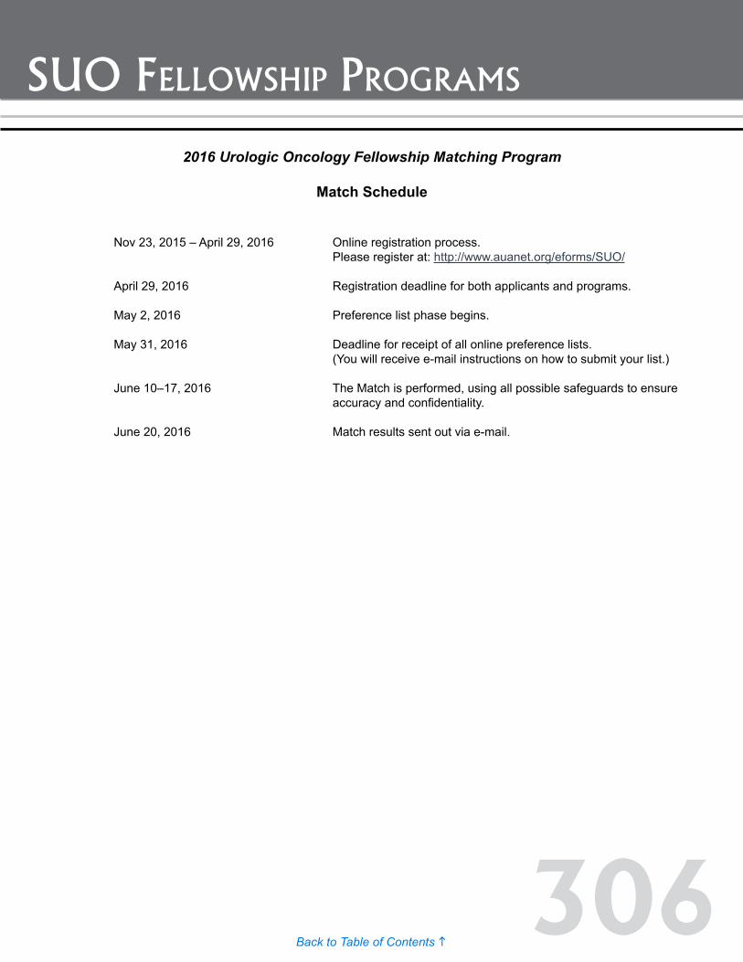
306Back to Table of Contents h
suo fellowship proGrams
2016 Urologic Oncology Fellowship Matching Program
Match Schedule
Nov 23, 2015 – April 29, 2016 Online registration process. Please register at: http://www.auanet.org/eforms/SUO/
April 29, 2016 Registration deadline for both applicants and programs.
May 2, 2016 Preference list phase begins.
May 31, 2016 Deadline for receipt of all online preference lists. (You will receive e-mail instructions on how to submit your list.) June 10–17, 2016 The Match is performed, using all possible safeguards to ensure accuracy and confidentiality.
June 20, 2016 Match results sent out via e-mail.

307Back to Table of Contents h
SUO-SBUR Joint Meeting at the 2016 AUA Annual MeetingMay 7, 2016
San Diego, California
SUO at the 2016 AUA Annual MeetingMay 7, 2016
San Diego, California
17th Annual Meeting of the SUONovember 30 - December 2, 2016
Grand Hyatt San AntonioSan Antonio, Texas
mark your CalenDars

308Back to Table of Contents h
noTes

Two Wood�eld Lake1100 E Wood�eld Road, Suite 350
Schaumburg, IL 60173-5116Phone: (847) 264-5901 | Fax: (847) 517-7229
Email: [email protected] | Website: www.suonet.org