ABSTRACT PER SESSIONS - SMIT 2017
257
ABSTRACT PER SESSIONS
-
Upload
khangminh22 -
Category
Documents
-
view
0 -
download
0
Transcript of ABSTRACT PER SESSIONS - SMIT 2017

ABSTRACT PER SESSIONS

SESSION 03 – ACTUATORS AND
MECHANISMS FOR NOVEL
MEDICAL TOOLS
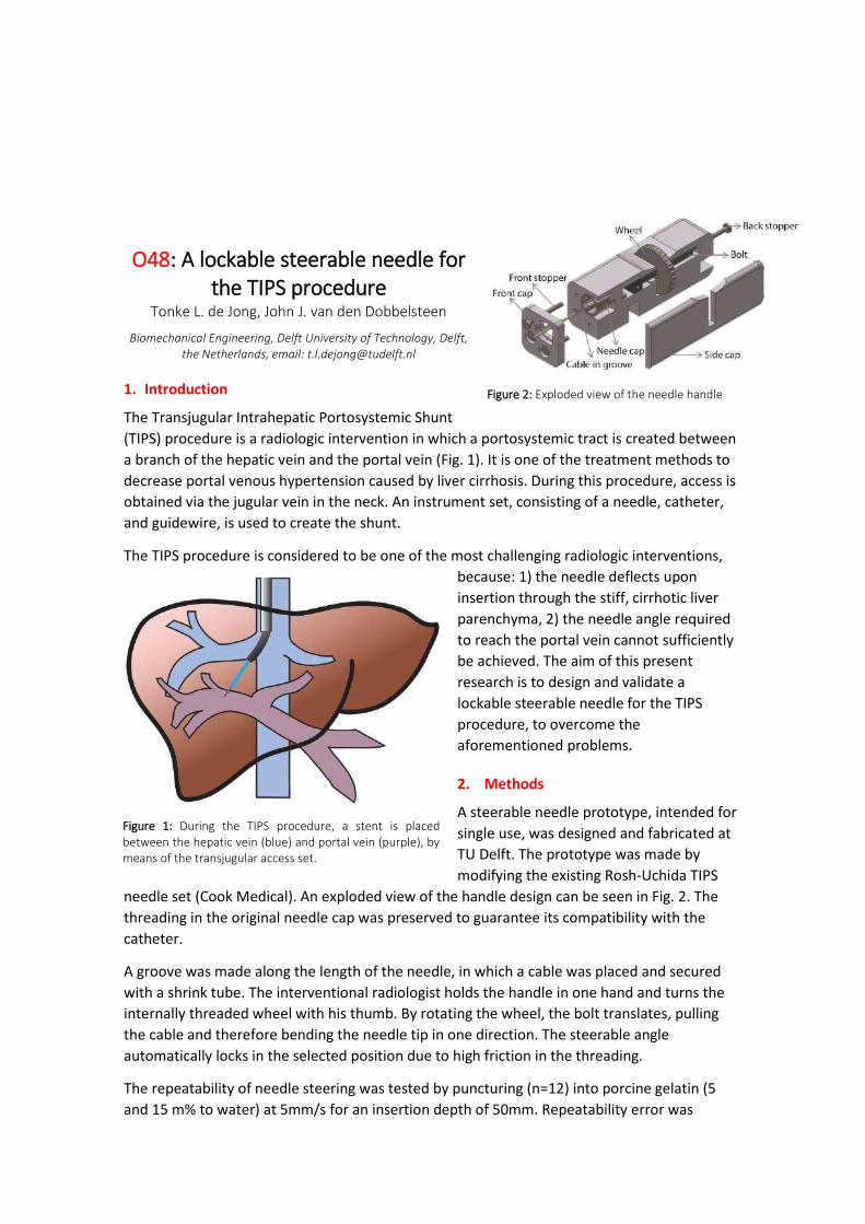
O48: A lockable steerable needle for the TIPS procedure
Tonke L. de Jong, John J. van den Dobbelsteen
Biomechanical Engineering, Delft University of Technology, Delft, the Netherlands, email: [email protected]
1. Introduction
The Transjugular Intrahepatic Portosystemic Shunt
(TIPS) procedure is a radiologic intervention in which a portosystemic tract is created between
a branch of the hepatic vein and the portal vein (Fig. 1). It is one of the treatment methods to
decrease portal venous hypertension caused by liver cirrhosis. During this procedure, access is
obtained via the jugular vein in the neck. An instrument set, consisting of a needle, catheter,
and guidewire, is used to create the shunt.
The TIPS procedure is considered to be one of the most challenging radiologic interventions,
because: 1) the needle deflects upon
insertion through the stiff, cirrhotic liver
parenchyma, 2) the needle angle required
to reach the portal vein cannot sufficiently
be achieved. The aim of this present
research is to design and validate a
lockable steerable needle for the TIPS
procedure, to overcome the
aforementioned problems.
2. Methods
A steerable needle prototype, intended for
single use, was designed and fabricated at
TU Delft. The prototype was made by
modifying the existing Rosh-Uchida TIPS
needle set (Cook Medical). An exploded view of the handle design can be seen in Fig. 2. The
threading in the original needle cap was preserved to guarantee its compatibility with the
catheter.
A groove was made along the length of the needle, in which a cable was placed and secured
with a shrink tube. The interventional radiologist holds the handle in one hand and turns the
internally threaded wheel with his thumb. By rotating the wheel, the bolt translates, pulling
the cable and therefore bending the needle tip in one direction. The steerable angle
automatically locks in the selected position due to high friction in the threading.
The repeatability of needle steering was tested by puncturing (n=12) into porcine gelatin (5
and 15 m% to water) at 5mm/s for an insertion depth of 50mm. Repeatability error was
Figure 2: Exploded view of the needle handle
Figure 1: During the TIPS procedure, a stent is placed between the hepatic vein (blue) and portal vein (purple), by means of the transjugular access set.
defined as the distance in millimeters between the mean of all insertions and an individual
insertion.
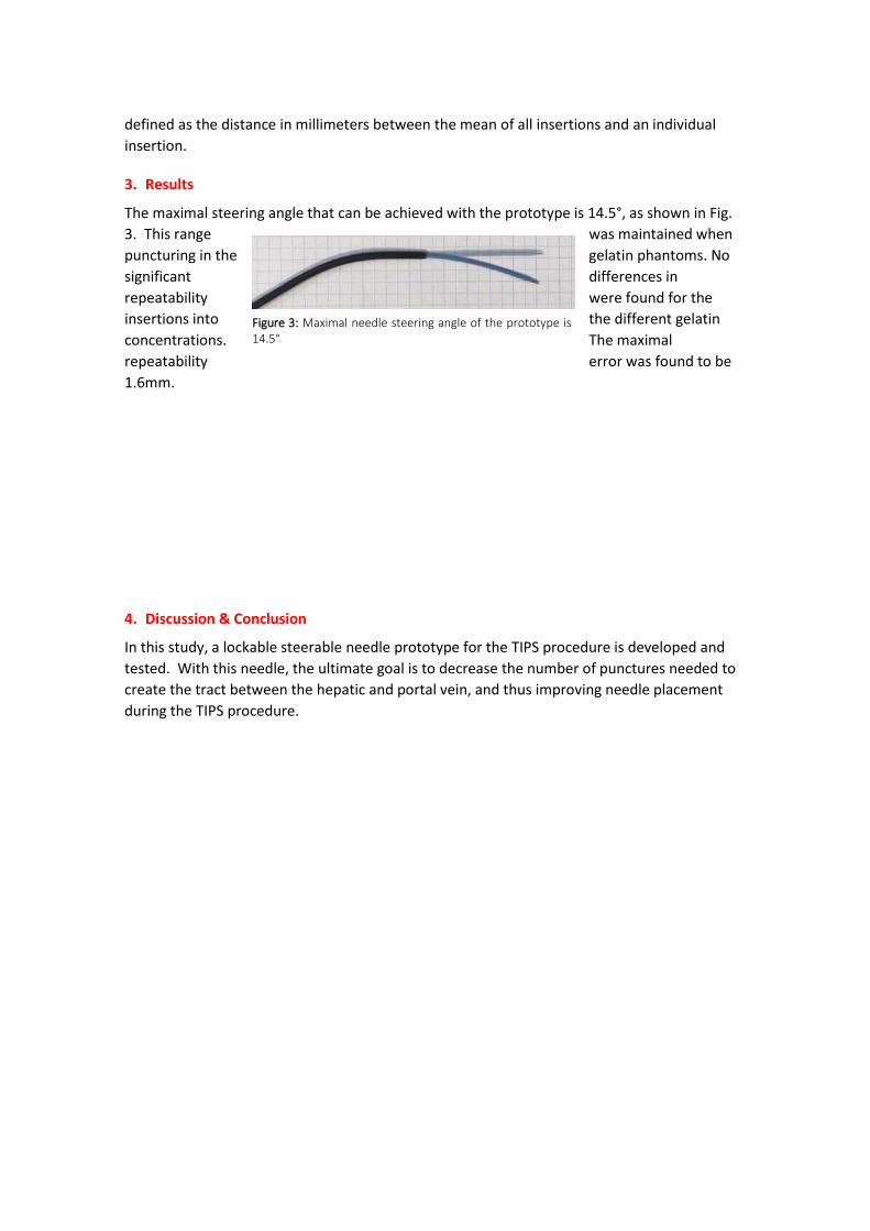
3. Results
The maximal steering angle that can be achieved with the prototype is 14.5°, as shown in Fig.
3. This range was maintained when
puncturing in the gelatin phantoms. No
significant differences in
repeatability were found for the
insertions into the different gelatin
concentrations. The maximal
repeatability error was found to be
1.6mm.
4. Discussion & Conclusion
In this study, a lockable steerable needle prototype for the TIPS procedure is developed and
tested. With this needle, the ultimate goal is to decrease the number of punctures needed to
create the tract between the hepatic and portal vein, and thus improving needle placement
during the TIPS procedure.
Figure 3: Maximal needle steering angle of the prototype is 14.5°

O117:
O46: Minimally invasive prenatal closure of spina bifida L. Tas1, J.J. van den Dobbelsteen1, D. Oepkes2, A.J. Eggink3
1) Biomechanical Engineering, TU Delft, Delft, The Netherlands, [email protected]. 3) Erasmus MC, Rotterdam, The Netherlands. 4) LUMC, Leiden, The Netherlands.
Introduction
In spina bifida there is a failure of closure of the neural tube causing a defect through which
the spinal cord, nerves and meninges can protrude. The current prenatal treatment method is
an invasive method and therefore research is performed to operate spina bifida in a minimally
invasive manner during the pregnancy. Instead of suturing the defect at the foetus, the
ultimate goal is to 3D print a protective cap over the defect.
The progression of the first prototype, which is able to close the defect via one port, was
presented at the previous SMIT congress. Here, the follow-up prototype and results of the user
evaluation tests are presented.
Methods
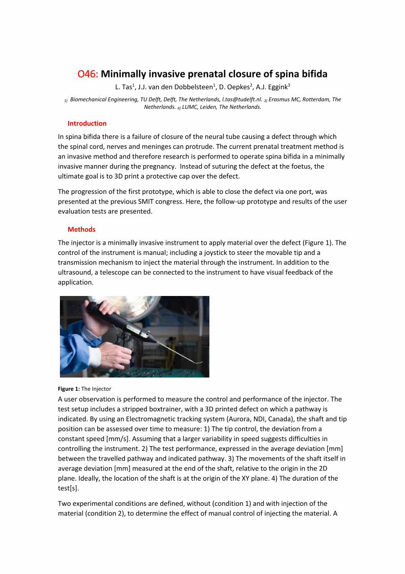
The injector is a minimally invasive instrument to apply material over the defect (Figure 1). The
control of the instrument is manual; including a joystick to steer the movable tip and a
transmission mechanism to inject the material through the instrument. In addition to the
ultrasound, a telescope can be connected to the instrument to have visual feedback of the
application.
Figure 1: The Injector
A user observation is performed to measure the control and performance of the injector. The
test setup includes a stripped boxtrainer, with a 3D printed defect on which a pathway is
indicated. By using an Electromagnetic tracking system (Aurora, NDI, Canada), the shaft and tip
position can be assessed over time to measure: 1) The tip control, the deviation from a
constant speed [mm/s]. Assuming that a larger variability in speed suggests difficulties in
controlling the instrument. 2) The test performance, expressed in the average deviation [mm]
between the travelled pathway and indicated pathway. 3) The movements of the shaft itself in
average deviation [mm] measured at the end of the shaft, relative to the origin in the 2D
plane. Ideally, the location of the shaft is at the origin of the XY plane. 4) The duration of the
test[s].
Two experimental conditions are defined, without (condition 1) and with injection of the
material (condition 2), to determine the effect of manual control of injecting the material. A
paired t-test is performed on each of the above performance measures (SPSS, IBM, United
States).
Results
On average, the standard deviation of the speed [mm/s] with which the tip of the instrument is
moved, is significantly larger in condition 1 (M=4.7, SE=.2) than in condition 2 (M=3.9, SE=.3),
t(19)=2.4, p<.05, r=.5. When looking at the mean deviation [mm] between the ideal and
travelled pathway, it is on average significantly larger in condition 2 (M=6.2, SE=.3) compared
to condition 1 (M=5.1, SE=.3), t(19)=-3.1, p<.05, r=.6. The average deviation of the shaft
relative to the origin in the XY plane has no significant difference between condition 1 (M=3.6,
SE=0.1) and condition 2 (M=3.8, SE=0.2). The duration of the task [s] is on average significantly
longer in condition 2 (M=23.9, SE=.8) than in condition 1 (M=17.6, SE=.4), t(19)=-9.0, p<.05,
r=.9.
Discussion & Conclusion
The user test shows that the test person is able to hold the instrument in place, limiting the
amount of movements of the shaft. In both conditions the test can be performed in a short
period of time.
However, the results show that the instrument tip positioning is affected by the manual
injection of the material. In both conditions the tip positioning is not sufficient. Whether the
tip positioning is the right parameter to determine the performance of the instrument is not
clear.
It is striking that in condition 2 the speed of movement is more constant, showing more
control. This can be a result of the direct feedback of the application of the material to the
user.
It can be concluded, that minimally invasive coverage of fetal deficits by using a single
instrument is feasible, within limited time and with limited shaft movements. Considering the
complexity of concurrently controlling the injection of the material and the positioning the tip,
it would be interesting to look into alternative methods of controlling the injection of the
material.

Haider Abidi1, Alice Tonazzini2, M. Cianchetti1, D. Floreano2, A. Menciassi1
1) BioRobotics Institute, Scuola Superiore Sant’Anna, Pisa, 56025, ITALY, [email protected]. 2) Laboratory of
Intelligent Systems École Polytechnique Fédérale de Lausanne, Lausanne 1015, Switzerland
INTRODUCTION
Although soft robots promise to provide unsurpassed dexterity, their usage is limited due to their lack of stiffness. This statement may seem paradoxical, however as discussed by [1], softness comes at a price of load bearing capacity. Soft materials cannot transfer or sustain forces effectively. Thus manipulation and interaction with the environment becomes challenging. This has made means of varying stiffness an active field of research [2]. This work presents the integration of a soft surgical robot (Stiff-Flop [3]) with stiffening fibers made by Low Melting Point Alloy (LMPA). A preliminary analysis shows a marked increase in the stiffness of the module.
METHODS
The Stiff-Flop modules are made from a soft elastomeric material. The robot operates though pneumatic pressure. The intrinsic softness of the robot grants it unmatched dexterity in the constrained environment of minimally invasive surgery. However its functionalities may be increased if a means of stiffening can be implemented. Respecting the dimensional constraints, a (LMPA) based approach has been considered [4].
The LMPA fibers are made by pumping molten Field’s metal alloy into pre-stretched silicone tubes. On solidifying, a conductive wire is wound around the fibers. When current is run through this wire, the resulting heat causes the LMPA to melt, making the fibers soft. Upon cooling the LMPA solidifies, regaining its rigidity. The pre-stretching applied to the silicone tubes ensures that no discontinuities are created during solidification.
The fibers are bonded to the Stiff-Flop module using silicone glue. Once integrated the stiff and soft state of the module may be alternated by heating and cooling the fibers. The fibers can be softened within 20 seconds, at a temperature of about 60 degrees. During operation, higher temperatures can be reached, hence a controller needs to be implemented to avoid excessive heating. They regain rigidity within a minute at room temperature.
RESULTS
To measure the effect of the LMPA fibers, a force deflection curve of the integrated module was made. The tip of the robot was loaded while the LMPA was in molten and solidified state. The corresponding defections shows a significant increase (more than three times) in the load carrying ability of the robot (Figure 1). In the soft state the fibers do not give much resistance hence the intrinsic softness is unaffected.
O150: Low Melting Point Alloy Based Stiffening of a Soft Robot
Figure 1: a) Stiff flop with the LMPA fibers. b) A comparison of the force deflection curve in the soft and rigid state

DISCUSSION AND CONCLUSION
The integration shows a proof of concept of using LMPA fibers for stiffening soft robots. The module cannot be moved while the fibers are in solid state. Hence the fibers may be used in applications where shape locking is needed or when the robot is expected to bear large loads while in static condition. Overall the prototype shows a novel means of achieving rigidity of soft robots. Such a technology may also be used on base structures of soft deployable robots for structural stability.
REFERENCES
[1] A. Loeve, P. Breedveld and J. Dankelman, "Scopes Too Flexible...and Too Stiff," IEEE Pulse, vol. 1,
no. 3, pp. 26-41, Dec. 2010.
[2] M. Manti, V. Cacucciolo and M. Cianchetti, "Stiffening in Soft Robotics: A Review of the State of
the Art," IEEE Robotics & Automation Magazine, vol. 23, no. 3, pp. 93-106, sept 2016.
[3] T. Ranzani, M. Cianchetti, G. Gerboni, I. D. Falco and A. Menciassi, "A Soft Modular Manipulator
for Minimally Invasive Surgery: Design and Characterization of a Single Module," IEEE
Transactions on Robotics, vol. 32, no. 1, pp. 187-200, Feb 2016.
[4] Alice Tonazzini, Stefano Mintchev, Bryan Schubert, Barbara Mazzolai,Jun Shintake and Dario
Floreano, "Variable Stiffness Fiber with Self-Healing Capability," Advanced Materials, p. 10142–
10148, 2016.

Session 05 – image guided surgery

07: 3D TONGUE MODEL CONSTRUCTION AND THE MOTION REGENERATION
N. Mukai1, T. Ishizu1, K. Mori2, Y. Takei2, and H. Yamada2 1) Graduate School of Engineering, Tokyo City University, Setagaya, Tokyo, 158-8557 Japan, [email protected]. 2) Department of Oral Rehabilitation, Showa University Dental Hospital, Oota, Tokyo, 145-8515 Japan.
1. Introduction
Computer generated tongue model is necessary to analyze the movement especially for lateral articulation (LA). Fasel et al. extracted tongue contours from ultrasound (US) images based on Deep Belief Networks (DBFs) [1], and Xu et al. visualized tongue motion by using modal warping [2]. However, US images are so noisy that some researchers use Magnetic Resonance Imaging (MRI) instead of US [3,4]. US is yet easily treated and safe for humans. Then, we propose a method how to construct 3D tongue model from US images and regenerate the motion.
2. Methods
The target is a set of images, which is constructed of 19 frontal US images. Fig.1 shows the process that extracts control points, which approximates the tongue surface. First, from the US image (Fig.1(a)), the region of interest (ROI) is extracted (Fig.1(b)). Then, the image is binarized (Fig.1(c)), and thinning processes is performed (Fig.1(d)). For the remained points, N spline curve is applied (Fig.1(e)), which represents the tongue surface of a frontal image in US images. Then, 19 control points are specified for the N spline (Fig.1(f)), and 19 x 19 points construct the tongue surface mesh model for 19 US images. In addition, the thickness is added to the surface model, a tongue image is mapped onto the model, and a mandible model is attached. Finally, the tongue model is constructed as shown in Fig.2. By changing the positions of the control points according to an articulation, the tongue motion is regenerated.
Figure 1: Frontal view image.
(a) US image (b) ROI
(c) Binary image (d) Thinned image
(e) N spline (f) Control points
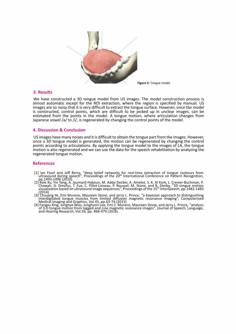
Figure 2: Tongue model.
3. Results
We have constructed a 3D tongue model from US images. The model construction process is almost automatic except for the ROI extraction, where the region is specified by manual. US images are so noisy that it is very difficult to extract the tongue surface. However, once the model is constructed, control points, which are difficult to be picked up in unclear images, can be estimated from the points in the model. A tongue motion, where articulation changes from Japanese vowel /a/ to /i/, is regenerated by changing the control points of the model.
4. Discussion & Conclusion
US images have many noises and it is difficult to obtain the tongue part from the images. However, once a 3D tongue model is generated, the motion can be regenerated by changing the control points according to articulations. By applying the tongue model to the images of LA, the tongue motion is also regenerated and we can use the data for the speech rehabilitation by analyzing the regenerated tongue motion.
References
[1] Ian Fasel and Jeff Berry, “deep belief networks for real-time extraction of tongue contours from
ultrasound during speech”, Proceedings of the 20th International Conference on Pattern Recognition, pp.1493-1496 (2010)
[2] Kele Xu, Yin Yang, A. Jaumard-Hakoun, M. Adda-Decker, A. Amelot, S. K. Al Kork, L. Crevier-Buchman, P. Chawah, G. Dreyfus, T. Fux, C. Pillot-Loiseau, P. Roussel, M. Stone, and B. Denby, “3D tongue motion visualization based on ultrasound image sequences”, Proceedings of the 15th InterSpeech, pp.1482-1483 (2014)
[3] Chuyang Ye, Emi Murano, Maureen Stone, and Jerry L. Prince, “a bayesian approach to distinguishing interdigitated tongue muscles from limited diffusion magnetic resonance imaging”, Computerized Medical Imaging and Graphics, Vol.45, pp.63-74 (2015)
[4] Fangxu Xing, Jonghye Woo, Junghoon Lee, Emi Z. Murano, Maureen Stone, and Jerry L. Prince, “analysis of 3-D tongue motion from tagged and cine magnetic resonance images”, Journal of Speech, Language, and Hearing Research, Vol.59, pp. 468-479 (2016)

035: ADDITIVE MANUFACTURING FOR GYNAECOLOGICAL BRACHYTHERAPHY: CUSTOM-FIT APPLICATORS BASED ON MRI DATA
van de Berg Nick J., Laan Rianne C., Nout Remi A.- Delft University of Technology, BioMechanical
Engineering, Delft, Netherlands
Introduction
The image guided treatment of tumours by means of locally placed, radioactive sources, makes gynaecological brachytherapy a contemporary example of personalized care. The treatment quality largely depends on the ability to place sources at a series of dwell positions. For most patients, an optimal dose distribution is achieved using an intracavitary applicator, occasionally supplemented by interstitial catheters. However, in case of distal parametrial tumour extension or lower (para)vaginal involvement, current instruments may not suffice. The one-size-fits-all designs pose limitations in freedom for individualized treatment planning in these cases.
This study aims to employ additive manufacturing for the optimization of the applicator design. The approach includes a custom-fit device shape, potentially increasing the applicator position stability. This custom approach also allows for (nearly) limitless combinations of catheter channels. Practical path constraints, imposed by the catheter and obturator stiffness, are expected and have therefore been studied.
Methods
The constructed applicator designs were based on MRI-scans of two patients with vaginal cancer. Ultrasound gel was used to augment the visibility of the vaginal cavity. Conventional treatment planning software (Oncentra, Elekta, SE) was used to segment the vaginal cavity and the tumour, and to manually indicate the paths of the desired internal channels. These channels were made compatible with 6F catheters (Proguide, Nucletron BV, NL). The contours were saved in DICOM RT-struct files, and converted (MiVisLab, Fraunhofer MEVIS, DE) to coordinate sets, which allowed for processing in a CAD program (SolidWorks, Dassault Systèmes, US). The personalized applicators were printed from PLA (Ultimaker 2 Extended+, Ultimaker, NL).
Channel curvature constraints were studied with a separate print. This print consisted of an array of channels with path radii ranging from 20-75 mm, bridging an instrument wall thickness of 5 mm. The print was suspended in a 10 m% gelatin phantom. Catheters (with obturators) were inserted at 5 mm/s, using a linear stage (PRO-115, Aerotech, US), and insertion forces were collected (LSB200, Futek, US). The data were compared to subjective evaluations of manual insertions performed by a clinician.
Results
Two personalized applicator designs are shown in Fig. 1. Interstitial catheters enter through the lower cylindrical section, and exit through the holes visible at the top of the product, as shown by the red lines in the left column images.
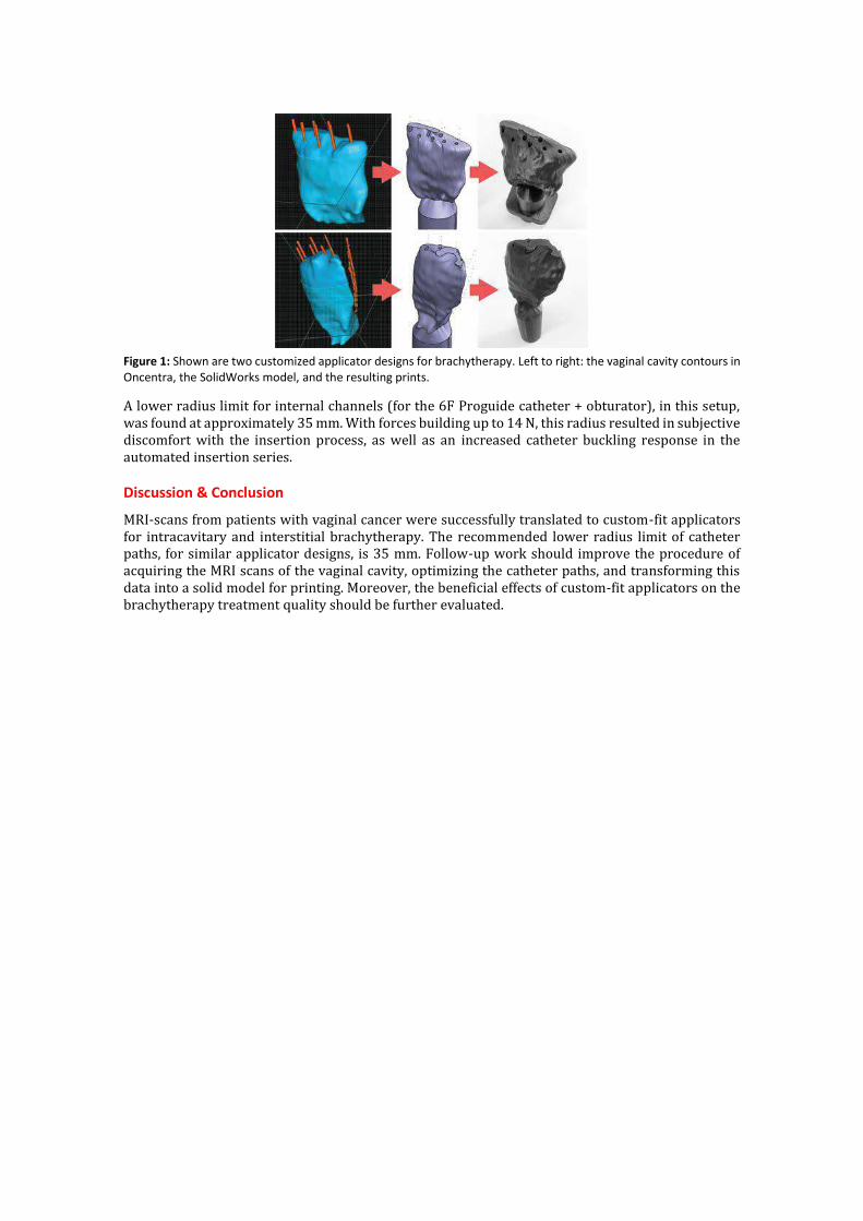
Figure 1: Shown are two customized applicator designs for brachytherapy. Left to right: the vaginal cavity contours in Oncentra, the SolidWorks model, and the resulting prints.
A lower radius limit for internal channels (for the 6F Proguide catheter + obturator), in this setup, was found at approximately 35 mm. With forces building up to 14 N, this radius resulted in subjective discomfort with the insertion process, as well as an increased catheter buckling response in the automated insertion series.
Discussion & Conclusion
MRI-scans from patients with vaginal cancer were successfully translated to custom-fit applicators for intracavitary and interstitial brachytherapy. The recommended lower radius limit of catheter paths, for similar applicator designs, is 35 mm. Follow-up work should improve the procedure of acquiring the MRI scans of the vaginal cavity, optimizing the catheter paths, and transforming this data into a solid model for printing. Moreover, the beneficial effects of custom-fit applicators on the brachytherapy treatment quality should be further evaluated.

0103: Mechanical catheter navigation with electromagnetic tracking to peripheral airway targets
F. Trauzettel1,2, H. A. Jaeger1,2, E.F. Hofstad3, M.P. Kennedy4, H. Leira5, T. Lango3, P. Cantillon-Murphy1,2
1) School of Engineering, University College Cork, Ireland, [email protected], 2) IHU Strasbourg, Strasbourg, France, 3) SINTEF, Trondheim, Norway, 4) Cork University Hospital, Cork, Ireland, 5) St.Olav’s Hospital, Trondheim,
Norway
Introduction
Lung cancer remains the single most deadly cancer in men and women due to low rates of early detection and treatment. Since non-small cell lung cancer usually starts in the outer airways, targeted minimally invasive biopsy which limits radiation exposure and avoids surgery is highly desirable. Current commercial solutions such as the superDimension (Medtronic Inc., Dublin, Ireland), and SpIN (Veran Medical, St. Louis, USA) systems rely on electromagnetic tracking for virtual navigation. However, clinical outcomes have been unconvincing due to poor accuracy, limitations in instrumentation and the lack of tracked catheters. This work proposes a novel mechanical catheter design with embedded electromagnetic tracking to facilitate tip-tracked navigation without the need for proprietary instruments or probe exchange. The catheter was used to reach peripheral airway targets by multiple users in pre-clinical studies.
Methods
The catheter used for this work (Figure 1) consisted in seven lumens within a 3mm diameter package; four lumens for stainless steel tendons used to deflect the distal tip, two lumens each containing one 5-degree-of-freedom EM tracking sensor (Northern Digital, Inc); and the final lumen is a 1.5mm working channel for instruments such as biopsy forceps or therapy probe.
Figure 1: Proximal end (left) and distal articulating section (right) of the catheter
A testing apparatus was constructed with stepper motors and leadscrews for catheter characterisation. Pre-clinical testing was achieved in two live porcine model with independent expert users (MPK, HL) navigating to a total of 8 peripheral airway targets using virtual bronchoscopic navigation. Following virtual navigation to each tumour model, a Tornado® embolization coil (Cook Medical Inc., USA) was deployed at each for post procedure targeting verification using 3D CT.
Results
System characterisation of tendon deflection versus force and tendon extension identified significant non-linearities and hysteresis in the mechanical catheter characteristics. Rudimentary steering was achieved by rotation of the bronchoscopy with the catheter in the working channel. Notwithstanding catheter steering limitations, expert users successfully navigated to 8 peripheral targets (Figure 2) and deployed 8 marker coils using virtual navigation (see Table 1).

Figure 2: DynaCT image.
Table 1: Closest distance from target to marker coil from CT
Lung Position Study 1 Study 2
Upper right 2.48 mm
5.54 mm
Centre left 6.95 mm
12.04 mm
Centre right 3.79 mm
1.97 mm
Lower right 8.07 mm
1.39 mm
Discussion & Conclusion
Tracked catheter navigation is feasible for targeting within 10mm of peripheral airway targets for endoscopic diagnosis and therapy. The results outlined here may serve as a platform for endoscopic catheter navigation in gastroenterology and urology.

O125: Rectal Cancer Segmentation with Fully Convolutional
Neural Network Joohyung Lee1, Ji Eun Oh1, Min Ju Kim2, Bo Yun Hur2, Dae Kyung Sohn1,2
1) Innovative Medical Engineering & Technology, Division of Convergence Technology, Research Institute & Hospital, National Cancer Center, Goyang, Republic of Korea. [email protected]. 2) Center for Colorectal Cancer, Research
Institute & Hospital, National Cancer Center, Goyang, Republic of Korea.
1. Introduction
The predictive accuracy of the pathologic T-categorization of rectal cancer using MRI is reported to be approximately 71–91% [1]. Since the location of rectal cancer with respect to that of rectum is crucial in deciding the T-staging of rectal cancer, automated segmentation of both rectum and tumor from magnetic resonance (MR) images can greatly improve or at least ease the diagnostic procedure.
2. Methods
We retrospectively analyzed T2-weighted MR images of 133 patients comprising 70 cases of T2 stage and 63 cases of T3 stage patients. T2 stage and T3 stage subjects were chosen since determining tumors of those stages are especially crucial in deciding treatment method [2]. The images have been obtained from National Cancer Center (NCC2017-0031) in Republic of Korea, from September 2004 to June 2016.
Fully convolutional neural network structure called ‘U-NET’ with batch normalization was exploited and modified [3]. In addition, transposed convolution filters were initialized as bilinear filters, and updated with all the other parameters using Adam optimizer algorithm. In addition, due to the limited dataset, 10-fold cross-validation was implemented to better assess the performance of the network.
Figure 2: Structure of segmentation network called ‘U-NET’
Segmentation for rectum and tumor has not only been carried out with separate networks, but also with single network with additional last softmax layer grafted on the last ReLU layer. Furthermore, a dice loss function was devised inspired from Dice Coefficient. The definitions of this function is described below.
−2× ∑ 𝑃(𝑥 ∈ 𝑝𝑜𝑠𝑖𝑡𝑖𝑣𝑒)𝑥∈𝑇𝑟𝑢𝑒 𝑃𝑜𝑠𝑖𝑡𝑖𝑣𝑒
∑ 𝑃(𝑥 ∈ 𝑝𝑜𝑠𝑖𝑡𝑖𝑣𝑒) + |𝐴𝑐𝑡𝑢𝑎𝑙 𝑝𝑜𝑠𝑖𝑡𝑖𝑣𝑒|𝑥∈𝑃𝑟𝑒𝑑𝑖𝑐𝑡𝑒𝑑 𝑃𝑜𝑠𝑖𝑡𝑖𝑣𝑒

3. Results
Integrated network seems to outperform single network in tumor segmentation whereas the single network surpasses in rectum segmentation. All the metrics were weighed by averaging the outcome from 10-fold cross validation followed by standard deviation.
Separate Network
Dice Score Sensitivity
Tumor 0.660.10 0.690.10
Rectum 0.890.03 0.880.05
Table 1: Segmentation performances using separate segmentation network
Integrated Network
Dice Score
Sensitivity
Tumor 0.690.12 0.720.11
Rectum 0.880.03 0.890.05
Table 2: Segmentation performances using integrated segmentation network for tumor and rectum
Figure 3: Learning progression for integrated network
4. Discussion & Conclusion
Tumor segmentation turned out to be more challenging than rectum segmentation, owing to the possible confusion between tumor, excrement, and air. Several advantages can be addressed for integrated network such as time consumption, and better tumor segmentation performance.
References
[1] S. H. Heo, “Multimodal imaging evaluation in staging of rectal cancer,” World Journal of Gastroenterology, vol. 20, no. 15, p. 4244, 2014.
[2] R. Glynne-Jones, L. Wyrwicz, E. Tiret, G. Brown, C. Rödel, A. Cervantes, and D. Arnold, “Rectal cancer: ESMO Clinical Practice Guidelines for diagnosis, treatment and follow-up†,” Annals of Oncology, vol. 28, no. suppl_4, pp. iv22–iv40, 2017
[3] O. Ronneberger, P. Fischer, and T. Brox, “U-Net: Convolutional Networks for Biomedical Image Segmentation,” Lecture Notes in Computer Science Medical Image Computing and Computer-Assisted Intervention – MICCAI 2015, pp. 234–241, 2015.
Acknowledement
This work was supported by grant from the National Cancer Center (NCC-1710070)
SESSION 14 – CAPSULE
ENDOSCOPY
O116: Wireless Endoscopic Capsule for Early Screening of Colorectal Cancer Contributions by the WiBEC project S. Mahmood 1, M. Zimmermann 2, S. Schostek 2, M. O. Schurr 2, E. Fosse 3, I. Balasingham 4 1) Ovesco Endoscopy AG, Tuebingen, Germany, [email protected]. 2) Ovesco Endoscopy AG, Tuebingen, Germany. 3) University of Oslo, Norway. 4) Norwegian University of Sciences and Technology (NTNU), Trondheim, Norway 1. Introduction Colorectal Cancer (CRC) accounts for a large number of deaths every year. Wireless Capsule Endoscopy (WCE) has emerged as a promising technique for painless, non-invasive and time efficient colonoscopy. Conventional colonoscopy, although still considered the gold standard for diagnosis of colonic diseases comes with inherent issues that pose obstacles in making conventional colonoscopy a mass screening diagnostic technique [1]. The WiBEC (Wireless in Body Environment) project is part of the Horizon 2020 Innovative Training Network initiative funded by the European Union under Marie Skłodowska Curie Action. The project aims to develop wireless technologies for novel implantable devices that will contribute to the improvement in quality and efficacy of healthcare. Two devices are being focused as a use case: leadless pacemaker and wireless endoscopic capsule for CRC screening. 2. Challenges addressed Magnetic Assisted Capsule Endoscopy (MACE) is a promising technique for controlling endoscopic capsules. The technique involves embedding a permanent magnet inside the capsule and uses external magnetic fields to steer the capsule inside the patient’s body. Techniques such as hand held magnetic manipulator, MRI style magnetic control and external robot mounted magnetic manipulators have been investigated by researchers. These solutions are either very expensive, very slow, low performance or are complicated to operate in which case the success of the procedure depends on the skill of the operator [2] [3] [4]. Currently, the gold standard for CRC screening is flexible endoscope that provides a high resolution video. For MACE to compete with flexible endoscopy, a high speed datalink for seamless video transmission is crucial [1]. This is in contrast to the small bowel capsule endoscopy where the preferred method is WCE. In this niche application, it does not have to compete with the flexible endoscope and therefore a comparatively low image quality and low frame rate is acceptable. Intuitive control of the capsule is also dependent on a high speed datalink. Delays in video result in misalignment of the capsule and the magnetic actuation field, increasing the chance of skipping visualization of polyps and lesions, leading to misdiagnosis and difficulties in real-time control of the capsule. WiBEC focuses on developing a MACE system using a robot mounted magnetic manipulator and employing a high speed datalink for capsule communication. The project is divided into multiple parts. Key focus areas are antenna design, low power signal processing, image processing, system integration, testing, encapsulation and channel modeling. 3. Conclusion All existing WCE solutions use low speed communication channels. WiBEC aims to develop a low cost, easy to use WCE system with high communication data rate to solve the problems faced by existing MACE Systems targeting the lower GI tract for early screening of CRC. References

[1] W. C. Leung, D. C. Foo, T. Chan, M. Chiang, A. H. Lam, H. H. Chan and C. C. Cheung, “Alternatives to colonoscopy for population-wide colorectal cancer screening,” Hong Kong medical journal = Xianggang yi xue za zhi / Hong Kong Academy of Medicine, pp. 70-77, 2016.
[2] Z. Liao, X. Hou, E.-Q. Lin-Hu, et al., “Accuracy of Magnetically Controlled Capsule Endoscopy, Compared With Conventional Gastroscopy, in Detection of Gastric Diseases,” Clinical Gastroenterology and Hepatology, vol. 14, p. 1266–1273, 2016.
[3] H. Gu, H. Zheng, X. Cui, Y. Huang, et al., “Maneuverability and safety of a magnetic-controlled capsule endoscopy system to examine the human colon under real-time monitoring by colonoscopy: a pilot study,” Gastrointestinal Endoscopy, vol. 85, pp. 438-443, 2017.
[4] J. F. Rey, H. Ogata, N. Hosoe, et al. , “Blinded nonrandomized comparative study of gastric examination with a magnetically guided capsule endoscope and standard videoendoscope,” Gastrointestinal Endoscopy, vol. 75, pp. 373-381, 2012

O106: Detection of acute upper gastrointestinal bleeding with the HemoPill acute - a prospective study Melanie Zimmermann1,2, Sebastian Schostek1, Mario Fode1, Jan Keller1, Michael Melbert1, Romy Hartmann1, Arthur Schmidt3, Karel Caca4, Marc O. Schurr1, Thomas Gottwald1,2 1)Division Diagnostic Systems, Ovesco Endoscopy AG, Tuebingen, Germany, [email protected]. 2)Eberhard Karls Universitaet Tuebingen, Tuebingen, Germany. 3)Department of Gastroenterology, Hepatology, Endocrinology, and Infectious Diseases, University Hospital Freiburg, Freiburg, Germany. 4)Department of Gastroenterology and Oncology, Klinikum Ludwigsburg, Ludwigsburg, Germany. 1. Introduction The ESGE guideline [1] recommends risk stratification into high and low risk groups on patients with suspected acute upper gastrointestinal bleeding (UGIB) to aid clinical decision making regarding timing of esophagogastroduodenoscopy (EGD). There is a need to improve risk stratification tools/methods on patients with suspected acute UGIB. Novel tools which are able to determine the presence or absence of a severe bleeding in a timely and atraumatic manner may provide a clear indication or non-indication for timely endoscopy. Prospective studies showed the potential of video capsule endoscopy in real-time detection of UGIB in emergency departments [2]. The HemoPill acute is a non-imaging capsule equipped with a sensor for direct detection of liquid blood and hematin in the lumen of the upper gastrointestinal tract [3]. The capsule is battery-operated and is administered by swallowing. The sensor signals from the HemoPill acute are transmitted wirelessly to the HemoPill Receiver, which allows real-time detection of blood in the upper gastrointestinal tract [4]. The HemoPill acute is 7.0x26.3mm in size and considerably smaller than a video capsule. The HemoPill acute capsule is intended to support the physician in risk stratification of patients with suspected acute UGIB. The HemoPill acute is a novel diagnostic device with no predicate device in clinical use. The results of an explorative clinical trial on n=30 patients are presented here. The aim of the study was to evaluate the safety and feasibility of the HemoPill acute as well as the performance of the capsule regarding the detection of blood in the upper gastrointestinal tract. 2. Material and methods A prospective, non-randomized, monocentric clinical trial without predicate device was performed. Patients suspected of having acute UGIB based on clinical symptoms such as hematemesis, tarry stools/melena swallowed a HemoPill acute capsule after signing the informed consent. The capsule sent sensor data to the receiver. Each patient received EGD within 12 hours after capsule ingestion. The endoscopists categorized the bleedings according to the amount of blood: <5ml, 5ml-20ml, and >20ml. The sensor data from the HemoPill acute was compared to the results of EGD. The values measured and transmitted by the HemoPill acute were used to analyze the performance of the HemoPill acute regarding the detection of blood. The thresholds of the sensor signal were defined in order to determine whether the results were positive or negative. Capsule excretion was monitored for up to 4 days. If excretion was not recorded during that time, a follow-up examination of the patient was conducted after 10 days. 3. Results In the period of 04/2015 to 02/2016, n=30 patients with suspected acute UGIB were included in the trial. Data analysis was performed on n=28 patients, due to the fact that a patient dropped out of the study (EGD was performed >12 hours after capsule ingestion, protocol violation) and data was lost in another case. Capsule ingestion was tolerated well by every patient. According to EGD reports after capsule ingestion, n=10 patients had stigmata of bleeding (n=4 with <5ml; n=4 with 5-20ml; n=2 with >20ml), and n=18 patients had no
bleeding. The HemoPill acute capsule correctly detected 2 of 4 patients (50%) with <5ml of blood, 2 of 4 patients (50%) with 5-20ml of blood, and 2 of 2 patients (100%) with >20ml of blood. The HemoPill acute capsule correctly identified 18 of 18 patients (100%) without bleeding as non-bleeders as per the endoscopy report. No complications related to the use of the HemoPill acute occurred during the trial. 4. Conclusions Safety and feasibility of the HemoPill acute were positively evaluated in this clinical trial. With regards to clinical relevance and risk stratification, minor bleedings with less than 20ml of blood may not require urgent intervention (low risk group), while severe bleeding does (high risk group). Here, the presence of 20 ml or more liquid blood or hematin in the upper GI tract would indicate severe bleeding with the urgent need for endoscopic evaluation. HemoPill acute capsule detected all severe bleedings (n=2) in the defined patient population (n=28). If an active bleeding (indicated by the presence of liquid blood or hematin) of a certain amount (indicated by the presence of more than 20ml of blood in the upper GI tract) identifies a patient who would benefit from a timely endoscopy, the sensitivity and specificity assessment of the HemoPill acute in detecting such a patient reveals the following results: sensitivity=100% (95%-CI=54-100%); specificity=83% (95%-CI=60-95%). However, a relatively wide confidence interval (95%) must be considered, due to the low number of patients included. Based on the present analysis, the device and procedure proved to be feasible and safe and showed the potential to be a valuable diagnostic tool for risk stratification of patients with acute suspected UGIB. References [1] I. Gralnek et al., “Diagnosis and management of nonvariceal upper gastrointestinal hemorrhage: European Society of Gastrointestinal Endoscopy (ESGE) Guideline,” Endoscopy, vol. 47, no. 10, pp. a1–a46, 2015. [2] A. C. Meltzer et al., “Video capsule endoscopy in the emergency department: A prospective study of acute upper gastrointestinal hemorrhage,” Ann. Emerg. Med., vol. 61, no. 4, p. 438–443.e1, 2013. [3] S. Schostek and M. O. Schurr, “The HemoCop telemetric sensor system: Technology and results of in-vivo assessment,” Stud. Health Technol. Inform., vol. 177, pp. 97–100, 2012.
[4] S. Schostek et al., “Telemetric real-time sensor for the detection of acute upper
gastrointestinal bleeding,” Biosens. Bioelectron., vol. 78, pp. 524–529, 2016.
O118: Wireless HD Imaging for Minimal Invasive Surgery
Mintz Y, Kunicher N, Livneh N, Brodie R, Bracha VB
Department of Surgery, Hadassah Hebrew University Medical Center, Jerusalem, Israel
Introduction: In laparoscopic surgery any entry to the abdomen creates a pivot point on the abdominal
wall which limits the movements of the instrument or laparoscope. As such, the visual field is confined
to the angles allowed by the pivot point, limiting the visual field and forcing the surgeon to compromise
on view of the surgical field. In addition current "state of the art" laparoscopes are encumbered by
cabling for power, video and a light source inside a semi flexible or rigid mechanical rod. While these
laparoscopes provide good image quality, they are cumbersome and require a point of access into the
patient through a dedicated separate incision.
Wirelessly transmitting uncompressed high definition video streaming through the body is unrealistic.
As the body absorbs most of the transmitted radio waves, the huge data load from the camera leaves
very small amount of energy per bit. Therefore, a reliable communication requires high power
transmission which is not feasible with a battery and most regulators will not permit this.
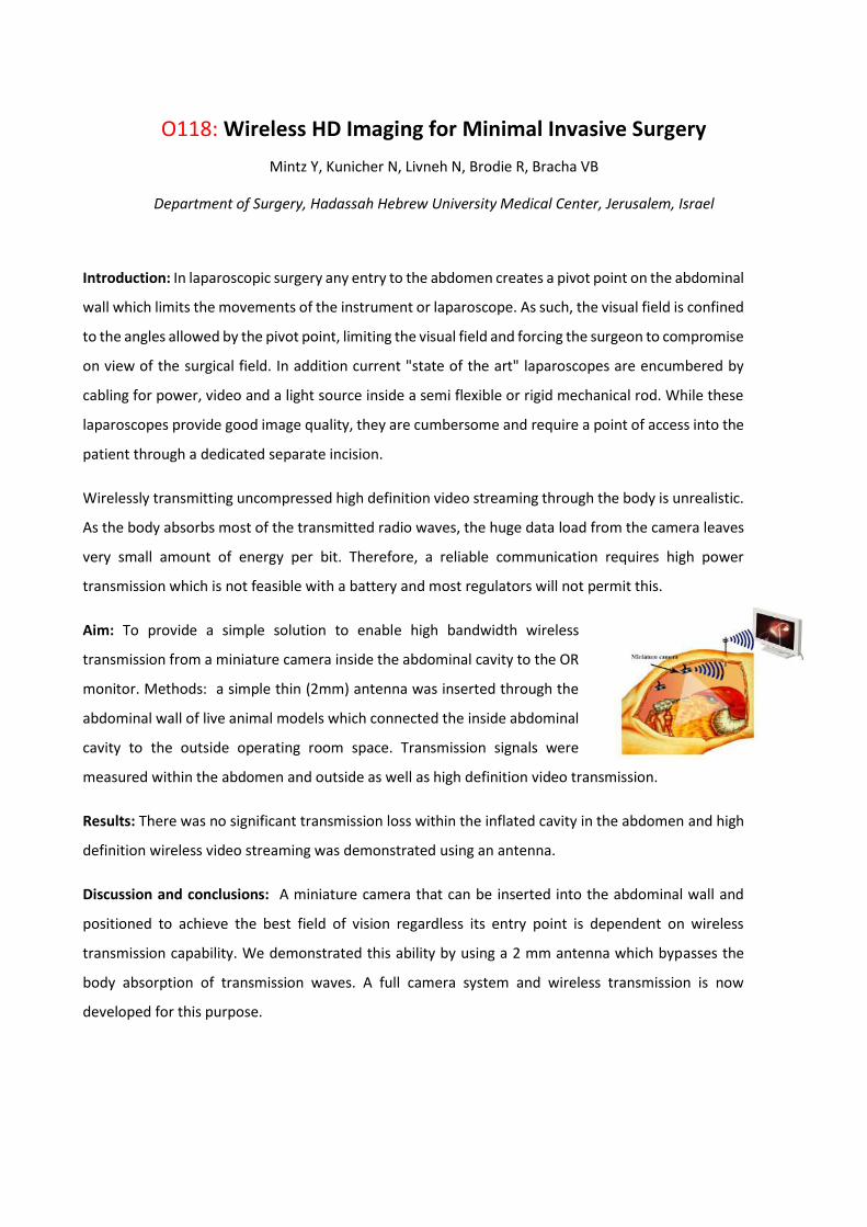
Aim: To provide a simple solution to enable high bandwidth wireless
transmission from a miniature camera inside the abdominal cavity to the OR
monitor. Methods: a simple thin (2mm) antenna was inserted through the
abdominal wall of live animal models which connected the inside abdominal
cavity to the outside operating room space. Transmission signals were
measured within the abdomen and outside as well as high definition video transmission.
Results: There was no significant transmission loss within the inflated cavity in the abdomen and high
definition wireless video streaming was demonstrated using an antenna.
Discussion and conclusions: A miniature camera that can be inserted into the abdominal wall and
positioned to achieve the best field of vision regardless its entry point is dependent on wireless
transmission capability. We demonstrated this ability by using a 2 mm antenna which bypasses the
body absorption of transmission waves. A full camera system and wireless transmission is now
developed for this purpose.

O61: Post-lymphangiographic multidetector CT for preclinical
lymphatic interventions in rabbits
T. Matsumoto1,2, K. Tomita1, S. Maegawa1, 2, T. Nakamura3, T. Suzuki2, T. Hasebe1,2
1) Department of Radiology, Tokai University Hachioji Hospital, Tokai University School of Medicine, Tokyo, Japan, 2) Graduate School of Science and Technology, Keio University,
Kanagawa, Japan, 3) National Institute of Advanced Industrial Science and Technology, Ibaraki, Japan, [email protected].; [email protected]
1. Introduction
Loss of chyle into thoracic/peritoneal cavity can lead to serious life-threatening consequences because of the significant loss of fluid, plasma protein, fats and immunoregulatory lymphocytes, which exhibits clinical features of severe malnutrition, hyponatraemia, acidosis, hypocalcaemia and susceptibility to infection. Reports on lymphatic interventions including lymphangiography for detecting or treating lymphatic leaks (1) and thoracic duct embolization (TDE) for postoperative chylothorax has been increasing. However, postoperative lymphatic leakage remains a challenging clinical problem with high mortality in post esophageal surgery. Despite this clinical problem, an animal model for lymphatic interventions has not been developed so far. For lymphatic interventions and future imaging, detailed lymphatic anatomical features in rabbits should be elucidated. The objective of our present study is to describe the visibility of the lymphatic system on post-lymphangiographic multidetector CT (MDCT) for preclinical lymphatic interventions in rabbits.
2. Methods
Lymphangiography via the popliteal lymph node or vessel was performed, using six healthy female Japanese White rabbits. Post-lymphangiographic MDCT examinations were performed in all rabbits. The dataset images underwent image processing analysis by three-dimensional maximum intensity projection (3-D MIP) technique. Three reviewers evaluated degree of depiction of the lymphatic system using a four-point scale (1 = poor, 4 = excellent). The distance between the body surface and cisterna chyli was measured.
3. Results
The popliteal lymph node was detectable in 90%. The visualization of lymphatic system via the popliteal node was found in 89%. More than 3.0 mean visual scores were the right femoral lymph vessel, left femoral lymph vessel, left iliac lymph vessel, left lumbar lymphatic trunks and cisterna chyli; whereas, less than 3.0 mean visual scores were the right iliac lymph vessel, right lumbar lymphatic trunks and cisterna chyli. The distance between the body surface and cisterna chyli was 4.33 ± 0.14 cm.

Figure 1: Three-dimensional maximum intensity projection (3-D MIP) image on post-lymphangiographic multidetector CT (MDCT) in anteroposterior view shows the lymphatic system in a rabbit.
4. Discussion & Conclusion
For TDE, the opacified cisterna chyli or retroperitoneal lymphatic duct is usually accessed through a transabdominal approach under fluoroscopic guidance. Rabbits may be suitable for TDE following lymphangiography. The visibility of the lymphatic system on post-lymphangiographic MDCT in rabbits provides enough information for interventional radiologists to perform preclinical lymphatic interventions and future imaging techniques.
References
[1] Matsumoto T, Yamagami T, Kato T, Hirota T, Yoshimatsu R, Masunami T, et al. The effectiveness of lymphangiography as a treatment method for various chyle leakages. Br J Radiol , vol. 82, p.286, 2009
Femoral lymph vessel
Iliac lymph vessel
Lumbar lymphatic trunk
Cisterna chyli
Thoracic duct
O27: AN INTUITIVE DISPOSABLE ENDOSCOPE WITH INTRINSIC
PNEUMATIC ACTUATION
Garbin Nicolo, Wang Long, Sohn Dennis, Obstein Keith L., Simaan Nabil, Valdastri Pietro-
Vanderbilt University, Mechanical Engineering, Nashville, United States
1. Introduction
Upper Endoscopy is the preferred method for gastric cancer (GC) screening. However, it requires
trained operators and comes with high costs [1]. Novel, intuitive, and low-cost endoscopes can
hence enable screening programs in low middle-income countries where GC is prevalent. Our
team has developed a Disposable Endoscope with Intrinsic Pneumatic Actuation (DEIPA). DEIPA
successfully visualized key landmarks in ex-vivo and in-vivo trials [2]. In this abstract, the
mechanical design of DEIPA is presented.
2. Methods
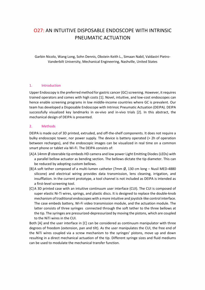
DEIPA is made out of 3D printed, extruded, and off-the-shelf components. It does not require a
bulky endoscopic tower, nor power supply. The device is battery operated (> 2h of operation
between recharges), and the endoscopic images can be visualized in real time on a common
smart phone or tablet via Wi-Fi. The DEIPA consists of:
[A] A 14mm Ø steerable tip embeds HD camera and low power Light Emitting Diodes (LEDs) with
a parallel bellow actuator as bending section. The bellows dictate the tip diameter. This can
be reduced by adopting custom bellows.
[B] A soft tether composed of a multi-lumen catheter (7mm Ø, 130 cm long – Nusil MED-4880
silicone) and electrical wiring provides data transmission, lens cleaning, irrigation, and
insufflation. In the current prototype, a tool channel is not included as DEIPA is intended as
a first-level screening tool.
[C] A 3D printed case with an intuitive continuum user interface (CUI). The CUI is composed of
super elastic Ni-Ti wires, springs, and plastic discs. It is designed to replace the double-knob
mechanism of traditional endoscopes with a more intuitive and joystick-like control interface.
The case embeds battery, Wi-Fi video transmission module, and the actuation module. The
latter consists of three syringes connected through the soft tether to the three bellows at
the tip. The syringes are pressurized-depressurized by moving the pistons, which are coupled
to the NiTi wires in the CUI.
Both [A] and the user interface in [C] can be considered as continuum manipulator with three
degrees of freedom (extension, pan and tilt). As the user manipulates the CUI, the free end of
the NiTi wires coupled via a screw mechanism to the syringes’ pistons, move up and down
resulting in a direct mechanical actuation of the tip. Different syringe sizes and fluid mediums
can be used to modulate the mechanical transfer function.
3. Results
DEIPA was assembled with associated prototyping costs <100 USD. The most expensive
components are the camera and the Wi-Fi transmission module (15 USD and 30 USD,
respectively).
Figure 1: Left: DEIPA’s main components. Right: assembled version performing underwater
visualization.
4. Discussion & Conclusion
DEIPA is designed to be intuitive to use thanks to direct mechanical coupling between the
proximal and the distal end. It can be either fully disposed or redesigned to reuse the camera
module. Moving forward DEIPA usability in cadaver will be tested and custom bellow will be
adopted to reduce the tip size.
References
[1] A. Tashiro, et al. “Comparing mass screening techniques for gastric cancer in Japan” World
journal of gastroenterology, 12(30), 4873, 2006.
[2] N. Garbin, et al. “Su1180 Evaluation of a Novel Disposable Upper Endoscope for
Unsedated Bedside (Non-Endoscopy Unit Based) Assessment of the Upper
Gastrointestinal (UGI) Tract”, Gastrointestinal Endoscopy, 85(5), AB304, 2017

O131
SESSION 24 – NEW TECH FOR
ADVANCED THERAPIES

O97: Pre-clinical validation of open-source airway navigation using Anser EMT and CUSTUSx
H. A. Jaeger1,2, F. Trauzettel1,2, E.F. Hofstad3, M.P. Kennedy4, H. Leira5, Thomas Lango3, P. Cantillon-Murphy1,2
1) School of Engineering, University College Cork, Ireland, [email protected], 2) IHU Strasbourg, Strasbourg, France, 3) SINTEF, Trondheim, Norway, 4) Cork University Hospital,
Cork, Ireland, 5) St.Olav’s Hospital, Trondheim, Norway
Introduction
Electromagnetic tracking (EMT) is a common navigation technology used in image guided applications. EMT is particularly useful in procedures where line-of-sight of the operating field is not feasible. We present a major update of the open source electromagnetic tracking platform Anser EMT [1] and present its results when performing bronchoscopy in a pre-clinical setting using the open-source CustusX navigation suite [2]. The updated system design is open source and free to use and modify under the Berkeley Standard Distribution (BSD) license.
Methods
The original design of the Anser system [3] was consolidated onto single printed circuit board (PCB). EM-guided virtual bronchoscopy navigation was performed using the CustusX navigation suite. In two pre-clinical validation experiments, 4 calcium-alginate tumour models were percutaneously injected into the right (3) and left (1) lungs. Following CT imaging (Siemens syngo 64-slice), airway segmentation was performed on the post-placement CT scan using an OpenCL accelerated algorithm. Image to patient registration was performed using the iterative closest point (ICP) method in CloudCompare. A custom tip-tracked bronchial catheter was used for tracked airway navigation through a 3.2mm working channel of therapeutic bronchoscope (EB-1970TK, Pentax Europe GmbH). Following virtual navigation to each tumour model, a Tornado® embolization coil (Cook Medical Inc., USA) was deployed at each for post procedure targeting verification.
Results
The system was calibrated using 81 pre-defined test points in a single plane at a height of 85mm above the field generator with an RMS registration error of 1.52mm. Airway segmentation was performed using CustusX in 18 seconds. ICP image-to-patient registration yielded a single rigid registration matrix. Successful navigation and to each tumour target was achieved using EM guided catheter navigation. Targeting errors were measured as the Euclidean distance between the centres of each tumour site and their respective embolization coils, using the post procedure Zeego DynaCT scan of the pig model. Absolute targeting errors in the range of 1.39-12.04mm were recorded in 8 tumour model sites across multiple users in two independent live cases.
Discussion & Conclusion
We have shown successful airway navigation and tumour targeting using a combination of open source hardware and software navigation technologies. Overall tumour targeting accuracy is comparable with previously reported procedures for electromagnetic navigation bronchoscopy.

Figure 4. Virtual bronchoscopy using the Anser EMT system (top) and CUSTUSx imaging platform (bottom).
References
[1] H. A. Jaeger et al., "Anser EMT: the first open-source electromagnetic tracking platform for image-guided interventions." Int J Comput Assist Radiol Surg. 2017 Jun; 12(6): 1059-1067
[2] Askeland C et al., “CustusX: an open-source research platform for image-guided therapy.” Int J Comput Assist Radiol Surg. 2016 Apr; 11(4): 505-1
[3] Anser EMT - http://anser.io. (Accessed on 29/07/2017).

O87: EXPERIMENTAL CHITOSAN MEMBRANES APPLIED ON THE
PERI-PROSTATIC NEUROVASCULAR BUNDLES AFTER NERVE-
SPARING
ROBOT-ASSISTED RADICAL PROSTATECTOMY: PRELIMINARY
RESULTS OF A PHASE II STUDY
Porpiglia Francesco, Bertolo Riccardo, Fiori Cristian, Muratori L, Fregnan Federica, Geuna
Stefano - Department of Oncology, University of Turin, Orbassano (To), 10043, Italy., San Luigi
Gonzaga Hospital, University of Turin, Orbassano, Italy
Introduction The primary aim of the study was the evaluation of the feasibility and the safety of the application of chitosan membranes (ChiMe) on the neurovascular bundles (NVBs) after nerve-sparing robot-assisted radical prostatectomy (NS-RARP). The secondary aim of the study was to report preliminary data and in particular potency recovery data of patients who underwent NS-RARP and received ChiMe. Methods This was a single-centre, single-arm prospective study, enrolling all patients with localised prostate cancer scheduled for RARP with five-item version of the International Index of Erectile Function scores of >17, from July 2015 to September 2016. All patients underwent NS-RARP with application of ChiMe on the NVBs. The demographics, perioperative, postoperative and complications data were evaluated. Potency recovery data were evaluated in particular and any sign/symptom of local allergy/intolerance to the ChiMe was recorded and evaluated. Results In all, a hundred-forty patients underwent NS-RARP with application of ChiMe on the NVBs. Applying the ChiMe was easy in almost all the cases, and did not compromise the safety of the procedure. None of the patients reported signs of intolerance/allergy attributable to the ChiMe and potency recovery data were encouraging. Discussion & Conclusions In our experience, ChiMe applied on the NVBs after NSRARP was feasible and safe, without compromising the duration, difficulty or complication rate of the ‘standard’ procedure. No patients had signs of intolerance/allergy attributable to the ChiMe and potency recovery data were encouraging. A comparative cohort would have added value to the study. The present paper was performed before Conformit_e Europ_eene (CE)-mark achievement.

O65: VIDEO-ASSISTED ARTERIOVENOUS FISTULA IN DYALISIS PATIENTS: OUR PRELIMINARY EXPERIENCE WITH VITOM® HD SYSTEM
Palumbo Vincenzo Davide- University of Palermo, Department of Surgical, Oncological and
Stomatological Disciplines, Palermo, Italy
1. Introduction
Arteriovenous fistulae (AVF) constructed using native vessels, vascular grafts and central venous catheters are the best permanent access, owing to a lower incidence of stenosis, thrombosis and infection. [1] The radiocephalic AVF of Cimino-Brescia remains the first choice for vascular access. [2,3] Mininvasivally access with a packaging of anastomoses is difficult in a restricted surgical field and traditional micro-dissection requires using the oculars of a stereo or surgical microscope for visualization. [4] Loupes with 2.5–4.5 magnification are most frequently used, but also the operating microscope may be used. [5] Although these magnifying instruments are essential to the optimal care of patients, they often come at a detriment to the operating surgeon in the form of neck or back pain and fatigue. VITOM® HD System can be a valid alternative to the others magnifying instruments (Fig. 1).
Figure 1: First experience of using VITOM® HD 3D System in an experimental model.
2. Methods
We performed a video-assisted radio-cephalic arteriovenous fistula in latero-lateral using prolene 7-0 without loupes (Fig. 2). The patient was a 72 years old man with history of Diabetes Mellitus type II from 15 years, ischaemic cardiomyopathy from 5 years and renal failure from 2 months. He needed a vascular access to start the substitutive haemodialysis treatment 30 days later. The VITOM® HD 3D System used is a KARL STORZ optical instrument coupled with a 1080p full high definition camera system.
3. Results
The average time for packing anastomoses was 46 ± 15 minutes; No complications were noted after decay, anastomoses showed a perfect holding. The consensus opinion of the entire group was that image quality was excellent and the system is ergonomic. The surgeons agreed that neck strain
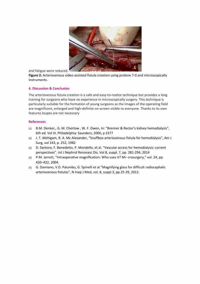
and fatigue were reduced. Figure 2: Arteriovenous video-assisted fistula creation using prolene 7-0 and microscopically instruments.
4. Discussion & Conclusion
The arteriovenous fistula creation is a safe and easy-to-realize technique but provides a long training for surgeons who have no experience in microscopically surgery. This technique is particularly suitable for the formation of young surgeons as the images of the operating field are magnificent, enlarged and high-definite on-screen visible to everyone. Thanks to its own features loupes are not necessary
References
[1] B.M. Denker,, G. M. Chertow , W. F. Owen, In: “Brenner & Rector’s kidney hemodialysis”,
6th ed. Vol III. Philadelphia: Saunders; 2000, p 2377
[2] J. T. Mehigam, R. A. Mc Alexander, “Snuffbox arteriovenous fistula for hemodialysis”, Am J
Surg, vol 143, p. 252, 1982
[3] D. Santoro, F. Benedetto, P. Mondello, et al. “Vascular access for hemodialysis: current
perspectives”. Int J Nephrol Renovasc Dis. Vol 8, suppl. 7, pp. 281-294, 2014
[4] P.M. Jarrett, “Intraoperative magnification: Who uses it? Mi- crosurgery,” vol. 24, pp.
420–422, 2004.
[5] G. Damiano, V.D. Palumbo, G. Spinelli et al.“Magnifying glass for difficult radiocephalic
arteriovenous fistulas”, N Iraqi J Med, vol. 8, suppl.3, pp.25-29, 2012.
O84: Design of an Innovative Implant for Rib Fracture Stabilization
Keith Mallia, Aaron Casha, Philip Farrugia
University of Malta (Dept. of Industrial & Manufacturing Engineering, Faculty of Engineering). Mater Dei Hospital, Malta (Dept. of Cardiothoracic surgery).
Introduction
Rib fractures are the most common injury that is sustained in the chest area. These occur in the elderly, adults and youths due to blunt chest trauma. Ribs are responsible for healthy respiration, and protect vital organs such as the heart, lungs and arteries. Treating rib fractures comes down to an adequate stabilization of the fracture site until the bones heal [1].
In this paper, an innovative approach to rib fracture stabilization is proposed. The designed implant focuses on an effective and efficient surgery by adopting the approach of Video-Assisted Thoracic Surgery (VATS). Moreover, the implant’s design ensures that the surgery is simple, safe and results in less pain to the patients.
Methods
The basic design cycle [2] was employed to systematically develop the implant. An analysis of the anatomy of the thoracic (chest) area was carried out. This went hand in hand with a study of the current state-of-the-art products related to rib fracture stabilization. This was followed by understanding the customer requirements which was used to develop a Quality Function Deployment (QFD) and a detailed Product Design Specification (PDS) that outlined the targets that must be met. Such targets included the surgery time, ease of installation and ease of customization for different patients. In the synthesis stage, several concepts were generated based on these criteria. Once a concept was chosen and refined, it was developed into a real-world implant, building upon a previous design developed by Calleja [3]. Calculations were compiled to ensure that the new design would withstand the forces exerted by the human body. A detailed Computer-Aided Design (CAD) model was generated. A physical prototype was then fabricated on a Ti-6Al-4V sheet using water-jet cutting. The final stage consisted of evaluating the implant in order to identify its strengths and weaknesses.
Results
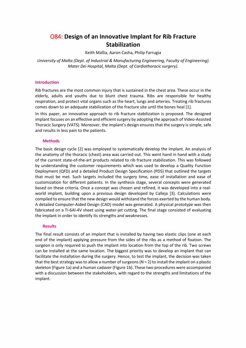
The final result consists of an implant that is installed by having two elastic clips (one at each end of the implant) applying pressure from the sides of the ribs as a method of fixation. The surgeon is only required to push the implant into location from the top of the rib. Two screws can be installed at the same location. The biggest priority was to develop an implant that can facilitate the installation during the surgery. Hence, to test the implant, the decision was taken that the best strategy was to allow a number of surgeons (N = 2) to install the implant on a plastic skeleton (Figure 1a) and a human cadaver (Figure 1b). These two procedures were accompanied with a discussion between the stakeholders, with regard to the strengths and limitations of the implant.
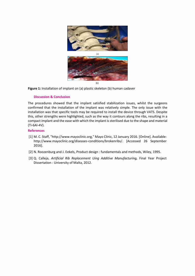
Figure 1: Installation of implant on (a) plastic skeleton (b) human cadaver
Discussion & Conclusion
The procedures showed that the implant satisfied stabilization issues, whilst the surgeons confirmed that the installation of the implant was relatively simple. The only issue with the installation was that specific tools may be required to install the device through VATS. Despite this, other strengths were highlighted, such as the way it contours along the ribs, resulting in a compact implant and the ease with which the implant is sterilised due to the shape and material (Ti-6Al-4V).
References
[1] M. C. Staff, "http://www.mayoclinic.org," Mayo Clinic, 12 January 2016. [Online]. Available: http://www.mayoclinic.org/diseases-conditions/brokenribs/. [Accessed 26 September 2016].
[2] N. Roozenburg and J. Eekels, Product design : fundamentals and methods, Wiley, 1995.
[3] Q. Calleja, Artificial Rib Replacement Uing Additive Manufacturing, Final Year Project Dissertation : University of Malta, 2012.
O5: Fibrina Arricchita di Leucociti e Piastrine (L-PRF) nel trattamento delle lesioni
cutanee
RESCIGNO ENRICO, ROSA GIORGIO, SAN ROME' GIULIA- STRUTTURA SEMPLICE CHIRURGIA
VASCOLARE, OSPEDALE DI LAVAGNA ASL 4 CHIAVARESE, LAVAGNA (GE), Italy
RIASSUNTO. Introduzione. Nell’ultima decade si è affermata in campo odontoiatrico la Fibrina Arricchita di Leucociti e Piastrine (L-PRF) in grado di rigenerare tessuto osseo e gengivale. Le piastrine e i leucociti racchiusi in questo coagulo riescono a liberare fattori di crescita in maggior quantità e durata rispetto ai tradizionali concentrati piastrinici. Intendiamo evidenziare i possibili benefici derivanti dall’utilizzo di L-PRF anche nel trattamento delle lesioni cutanee. Metodi. 18 pazienti , 6 maschi e 12 femmine , età media 78.8 aa ( range 32-99 ), con 23 lesioni di diversa natura (10 traumatiche, 6 diabetiche, 2 flebostatiche, 1 mista, 2 da pressione, 1 peristomale, 1 reumatoide), con una superficie complessiva di cute ferita di 405.1 cmq (range 1- 98) , alcuni già trattati senza successo con medicazioni avanzate, sono stati sottoposti ad applicazione di L-PRF. Tramite prelievo ematico con provette certificate da 10 ml (Intra-Lock®) immediatamente centrifugate ( Intraspin Medical Device Intra-Lock® System Europa SPA) per 12 minuti a 2700 giri/min, sono stati separati i globuli rossi dal materiale plasmatico coagulato. Quest’ultimo è stato applicato con cadenza settimanale su lesioni già granuleggianti. Sono stati ricercati la risoluzione delle lesioni , la comparsa di neovascolarizzazione, gli eventi avversi, la riduzione della sintomatologia algica, il tempo di guarigione. Risultati. Dei 18 pazienti studiati : 14 hanno raggiunto la guarigione completa con sviluppo di tessuto neoformato ipervascolarizzato; 1 solo paziente con allergia cutanea e polimorbilità ha interrotto il trattamento per recrudescenza dell’ulcera nonostante un iniziale miglioramento, rappresentando questo l’unico evento avverso ; 2 sono deceduti per polimorbilità; 1 ha sospeso la terapia per trasferimento di domicilio . In un caso di piede diabetico con esposizione ossea dell’alluce si è ottenuta la guarigione evitandone l’amputazione . Le ferite traumatiche sono guarite con precoce rivestimento epidermico. In tutti i 18 pazienti (100%) si è verificata la risoluzione del dolore alla prima applicazione. Il tempo medio di guarigione su 19 lesioni è risultato di 8.89 settimane (range 1 – 21). Conclusioni. L-PRF presidio autologo fresco , facile da ottenere, usato topicamente nelle lesioni cutanee ha dimostrato , nella nostra pur limitata esperienza, ottima tollerabilità, efficacia antalgica, capacità di risolvere rapidamente lesioni traumatiche, riduzione dei tempi di guarigione anche in lesioni inveterate non responders alle medicazioni avanzate, nel piede diabetico potrebbe risevare notevoli ripercussioni sulla qualità di vita.
SESSION 06 – NOVEL DESIGN OF MEDICAL DEVICES: FROM IP TO MARKET ANALYSIS

O100: TISSUEGRAFT: FROM THE IDEA UP TO THE MARKET Calvo Catoira Marta, Fusaro Luca, Ramella Martina, Boccafoschi Francesca- Deaprtment of
Health Sciences/Tissuegraft srl, University of Piemonte Orientale, Novara, Italy
1. Introduction and methods In 2012, the Commission published two regulation proposals to revise existing legislation on medical devices and in vitro diagnostics. The revision affects all medical devices, from home-use items like pregnancy tests and contact lenses, to X-ray machines, pacemakers and breast implants. After nearly four years of negotiation between the EU institutions, industry and Member States, the end of the legislative procedure is finish on 2017. Manufacturers of medical devices will be confronted with major changes in the European Union’s regulatory framework which governs market access to the EU for medical devices. That’s where TissueGraft srl comes in. We operate in Europe, coordinating among multinational owners interested to create new medical devices, financial office and R&D department. TissueGraft can collaborate with industries in order to develop medical devices following the partners in all the project
phases bringing to market. Our goal is developing new products in collaboration with companies interested in regenerative medicine market. We offer our lab for characterize the products, sharing also innovative ideas, where required. The skills and the instrumentations of TissueGraft company are available for both research institutions and companies. Our expertise are focalized on biocompatibility and the mechanical characterization. – Assessing biocompatibility, haemocompatibility and mechanical properties of materials for tissue engineering and regenerative medicine. – Improving an existing product of a company involved in regenerative medicine with sueGraft process.
Finally, Tissuegraft can project the industrialization process, optimizing the transfer technology solutions. 2. Results In Italy, we’ve been helping several companies get new products approved on the market on the last three years. In brief, here it is what we offer:

3. Discussion and conclusion The strength of our team is the multidisciplinary approach. We can collaborate with the industries partners from the certification process to the industrialization of their own products. From the first idea and approval to debut and product maturity, we provide guidance that considers the complex considerations where business and compliance meet. Many of our collaborators have worked for multinational companies and in the most advanced research centers, and have backgrounds in regenerative medicine, biomedical and chemical engineering and material science, among other disciplines.

O63: An active device for high fidelity simulation in newborn intubation
Selene Tognarelli1, Alessia Longo1, Rosa Teresa Scaramuzzo2, Massimiliano Ciantelli2, Armando Cuttano2, Arianna Menciassi1
1The BioRobotics Institute, Scuola Superiore Sant’Anna, Pisa, Italy, 2NINA Center, Azienda Ospedaliero Universitaria Pisana, Pisa, Italy
1. Introduction
Endotracheal Intubation (EI) procedure is highly complex and it requires wide-ranging competences and experience for avoiding unexpected complications: the force generated by the laryngoscope on the human tissues can easily cause traumas [1]. Airway management and EI are even more delicate for newborns and infants because of limited availability of instrumentation and challenging anatomical and physiological features [2]. Given the complexity of the neonatal domain, a specific education program is required [3] and specific intubation skill trainers are mandatory.
2. Methods
Active intubation skill trainers should not only be high-fidelity simulators of the analogous anatomical structures, but also active instruments able to provide feedback about the trainee’s performances. There are several commercial neonatal intubation manikin models. However, to the best of our knowledge, no sensorized skill trainer devices for neonatal intubation have been previously described. Based on these, and starting from our previous experiences [6], we worked on the development of an active, robust and reliable neonatal skill trainer able to provide clinicians with real-time information about the intubation procedure in terms of force peak value, force distribution and timing. In particular, different force sensing elements were integrated into a commercial neonatal intubation mannequin (Figure 1), in well defined points that were recognized as the anatomical areas mainly subjected to injuries during EI.
3. Results
An active prototype was realized by including force and pressure sensors into tongue, epiglottis, superior and inferior gingival arches, neck and trachea. Punctual force sensors (i.e. FSR®400 short - Interlink Electronics, CA, US) were recognised as the most promising sensorization strategy of gingival arches and epiglottis. On the other hand, a custom-made multilayer pressure sensor was used for tongue sensorization in order to guarantee a pressure distribution map (Figure 5). A custom Graphic User Interface (GUI) was implemented for feeding back the data from all the integrated sensors. Validation tests were carried out with 9 users that took part in two intubation sessions held 24 hours apart.
Figure 5: Overall view of the sensorized skill trainer with details about the sensorization strategies.
FSR®400 force sensor Active neonatal skill trainer
Custom Graphic User Interface (GUI)
Silicone prototype
Pressure sensor

Each session consisted of 5 intubation attempts: time of procedure and pressure on critical points were anonymously collected. The first test was accomplished with a mean time of 40.34±26.28s, which rapidly decreases in the second session until to reach 16.77±13.59s. Regarding the applied forces, epiglottis forces decreased from 3.01±1.98N down to 1.12±0.62N from the first to the second training session; on the contrary, forces on superior and inferior gingival arches were quite constant (from 0.49±0.68N to 0.34±0.06N for the superior arch and from 0.38±0.15N to 0.27±0.05N for the inferior arch). Finally, with respect to the pressures applied on the tongue, a maximum force of 11.96N was measured and a maximum involved area of 400 mm2 was revealed. A promising decrease of the maximum applied force between the first and the last attempt was observed.
4. Discussion and Conclusion
We proposed here a skill trainer equipped with force sensors for the training of medical professionals in neonatal EI procedures.
References
M. E. Warner, et al., “Perianesthetic dental injuries: frequency, outcomes, and risk factors,” Anesthesiology vol. 90(5), pp. 1302-1305, 1999.
G. M. Angelos, et al. “Oral complications associated with neonatal oral tracheal intubation: a critical review,” Pediatric Dentistry, vol. 11(2), pp. 133-40, 1989.
G. Fletcher, et al. “Rating non technical skills: developing a behavioural marker system for use in anaesthesia,” Cognition, Technology & Work, 2004.
S. Tognarelli, et al., “Development and validation of a sensorized neonatal intubation skill trainer for simulation-based education enhancement,” IJMRHS, vol. 3(4), pp. 833-9, 2014.
SESSION 15 – CARDIO@SMIT2017: WHAT’S NEW IN CARDIAC SURGERY

O6: The Development of the Hydraulic Pressure Wave Catheter
Aimée Sakes1, Paul Breedveld1, and Jo Spronck2
1) BioMechanical Engineering, Delft University of Technology, Delft, the Netherlands,
[email protected]. 2) Precision and Microsystems Engineering, Delft University of Technology, Delft, the Netherlands
1. Introduction
Crossing Chronic Total Occlusions (CTOs) is challenging, resulting in undesirably low success rates of 50–90% depending on the operator’s experience and CTO characteristics. The most common failure mode in the treatment is the inability to apply sufficient force to cross the CTO due to buckling of the guidewire. The inability to cross the CTO often leads to procedural failure and the subsequent referral to the invasive open-heart bypass surgery. Additionally, buckling can cause damage to the blood vessel wall due to bifurcations of the guidewire’s shaft and tip motions.
In order to improve the buckling resistance of the guidewire (or other crossing tools alike) and improve the procedural success rate, we propose to apply a mechanical impulse onto the CTO during the crossing procedure. Using an impulse to dynamically load the crossing tool and CTO can prevent buckling in two main ways: 1) the critical force the crossing tool can withstand increases with a decrease in force duration and 2) using an impulse to cross the CTO can lower the penetration load.
In order to transfer the impulse from the incision point towards the CTO with high efficiency, we propose to use a hydraulic pressure wave; a fast-moving longitudinal pressure surge in a fluidic medium (Fig. 1). The proposed tool consists of a saline-filled cardiac catheter-shaft with two free-moving plungers at the proximal and distal end, respectively (Fig. 1). To generate the pressure wave, an input impulse is exerted on the proximal plunger using a spring-loaded hammer (Fig. 1). This pressure wave is, subsequently converted into an impulse during the collision of the output plunger with the CTO. 2. Methods In order to valorize the proposed hydraulic pressure wave catheter, a proof-of-principle experiment has been performed in which the effectiveness and efficiency of this concept have been tested. In this feasibility experiment, a standard cardiac catheter (Ø2 mm) was used. The input impulse was delivered by a preloaded mass-spring system. Both the input and output impulse were measured using load cells, from which the impulse efficiency and impact peak forces were derived. To test the influence of the shape and flexibility of the catheter on the efficiency, different braided cardiac catheters were guided through several curves and loops.
Figure 1: The proposed hydraulic wave catheter concept. 3. Results
From the experiment, a high impulse efficiency of over 80% was found. Furthermore, impact peak forces of up to 43 N were measured, which is approximately 25x higher than what is required [1]. No significant difference was found between the impulse efficiency for the looped catheter (3 loops) compared to the straight catheter. Furthermore, only a minor difference in efficiency of ~13% was found between the different braided catheter types. 4. Discussion & Conclusion The hydraulic pressure wave catheter is a simple and versatile tool that allows for the delivering high-force impulses through a very tortuous path and under any angle. The catheter can deliver a large variety of output characteristics on the target tissue, from a single pulse to a pulsating motion, making it applicable to several medical fields. Based on the output characteristic and tip shape, it is also able to target a specific tissue type. We, therefore, feel that this tool can be the solution to overcoming current challenges in the treatment of CTOs in future. References [1] A. Thind, B. Strauss, A. Teitelbaum, R. Karshafian, M. Ladouceur, et al., “A novel method for the measurement of proximal fibrous cap puncture force in chronic total occlusions: the effect of increasing age,” Eurointervention, vol. 6, p. 997-1002.
O16: POLYLYSINE-ENRICHED DECELLULARIZED MATRICES: A PROMISING APPROACH FOR VASCULAR SUBSTITUTIONS Boccafoschi Francesca, Fusaro Luca, Calvo Catoira Marta, Ramella Martina- Deaprtment of Health
Sciences/Tissuegraft srl, University of Piemonte Orientale, Novara, Italy
1. Introduction Cardiovascular diseases are a leading cause of death worldwide. Current clinical approaches show poor efficiency in the replacement of small-caliber arteries (<6 mm). The use of autologous saphenous vein or mammary artery is currently the best surgical option; however, since most of the vessels are affected by widespread atherosclerotic abnormalities, this approach is not always feasible.
The use of synthetic grafts, used with good results in large caliber vessels substitution, due to thrombosis or intimal hyperplasia, leads to implant failure for small-caliber systems. In fact, the different compliance between the native vessel and the synthetic graft influences the mechanical behavior of the vessel wall causing lumen occlusion. In addition, the difference in mechanical properties could generate turbulent blood flow which enhances the formation of thrombi or aneurysms.
The use of decellularized scaffolds is a technique that has shown good perspectives in various applications for regenerative medicine both in preclinical and clinical. Through the decellularization process, it is possible to completely remove the cellular elements, retaining the native extracellular matrix (ECM). The scaffold obtained is an excellent substrate for cell adhesion, growth and proliferation. However, the decellularization process may weak the structure of the vessel. The purpose of this work is to obtain a scaffold chemically enriched with polylysine. Showing improved mechanical properties, without altering biocompatibility and hemocompatibility.
2. Methods The matrices were obtained by decellularization and enrichment with polylysine (sueGraft®) of porcine arteries (femoral and carotid). In order to verify the effectiveness of the decellularization process, DAPI and hematoxylin/eosin staining were performed, together with the quantification of residual DNA. In order to verify the efficiency of the enrichment procedure, XPS analyses were performed. Endothelial cells were used to test the biocompatibility. In order to measure elasticity and burst pressure, as well as degradation test in working condition, mechanical tests were performed. Finally, several parameters related to hemocompatibility were measured.
3. Results DAPI and hematoxylin/eosin staining confirmed the effectiveness of the decellularization method. The quantification of the DNA test showed that the amount of residual DNA was significantly and adequately reduced compared to untreated control. Values obtained were lower than the threshold values reported in literature.
Polylysine-enriched matrices showed excellent biocompatibility.
The analysis of the Young moduli showed that stiffness value of the enriched matrix is not significantly different to native vessel. Burst pressure test showed strengthening of the polylysine-enriched matrix, which can withstand higher pressures compared to native vessel. Matrix degradation test showed that the polylysine-enriched vessel has almost no weight loss, which indicates an absent degradation.
Concerning hemocompatibility, the evaluated parameters suggest that polylysine-enriched matrices increase clotting time.

Figure 1. Polylysine-enriched vascular substitute (prototype).
4. Discussion and Conclusion The matrices obtained by decellularization of blood vessels and enriched with polylysine show a very good biocompatibility, excellent mechanical properties, thanks to the cross-link action of polylysine, and improved hemocompatibility properties for the intended use as vascular substitutes. Based on these results, matrices enriched with polylysine indicates a promising approach for vascular applications.
References [1] Mozaffarian D, et al, Circulation. 131(4): e29-322, 2015. [2] Rathore A, et al., Nanomed Nanobiotechnol. 4(3):257-272, 2012. [3] Pashneh-Tala S et al. Tissue Eng Part B Rev. ePub, 2015. [4] Chiu JJ et al. EPhysiol Rev. 91(1):327-387, 2011
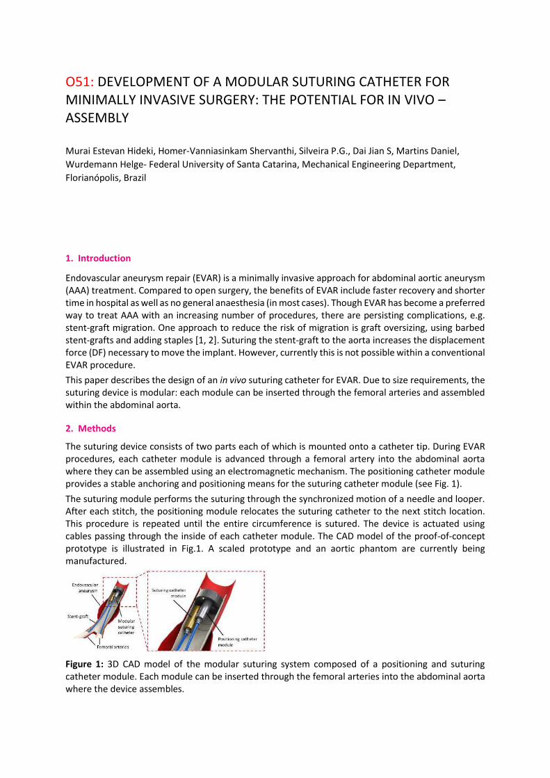
O51: DEVELOPMENT OF A MODULAR SUTURING CATHETER FOR MINIMALLY INVASIVE SURGERY: THE POTENTIAL FOR IN VIVO – ASSEMBLY Murai Estevan Hideki, Homer-Vanniasinkam Shervanthi, Silveira P.G., Dai Jian S, Martins Daniel,
Wurdemann Helge- Federal University of Santa Catarina, Mechanical Engineering Department,
Florianópolis, Brazil
1. Introduction
Endovascular aneurysm repair (EVAR) is a minimally invasive approach for abdominal aortic aneurysm (AAA) treatment. Compared to open surgery, the benefits of EVAR include faster recovery and shorter time in hospital as well as no general anaesthesia (in most cases). Though EVAR has become a preferred way to treat AAA with an increasing number of procedures, there are persisting complications, e.g. stent-graft migration. One approach to reduce the risk of migration is graft oversizing, using barbed stent-grafts and adding staples [1, 2]. Suturing the stent-graft to the aorta increases the displacement force (DF) necessary to move the implant. However, currently this is not possible within a conventional EVAR procedure.
This paper describes the design of an in vivo suturing catheter for EVAR. Due to size requirements, the suturing device is modular: each module can be inserted through the femoral arteries and assembled within the abdominal aorta.
2. Methods
The suturing device consists of two parts each of which is mounted onto a catheter tip. During EVAR procedures, each catheter module is advanced through a femoral artery into the abdominal aorta where they can be assembled using an electromagnetic mechanism. The positioning catheter module provides a stable anchoring and positioning means for the suturing catheter module (see Fig. 1).
The suturing module performs the suturing through the synchronized motion of a needle and looper. After each stitch, the positioning module relocates the suturing catheter to the next stitch location. This procedure is repeated until the entire circumference is sutured. The device is actuated using cables passing through the inside of each catheter module. The CAD model of the proof-of-concept prototype is illustrated in Fig.1. A scaled prototype and an aortic phantom are currently being manufactured.
Figure 1: 3D CAD model of the modular suturing system composed of a positioning and suturing catheter module. Each module can be inserted through the femoral arteries into the abdominal aorta where the device assembles.

3. Future work
Experimental testing of the suturing and graft pull-out force is ‘work in progress’. An initial larger-scale prototype of the suturing catheter will be tested in a phantom environment. The prototype will be used to suture a graft to a phantom aorta. Then, DF will be quantified using a pull-out force experiment [1-3].
4. Discussion & Conclusion
An estimation of the DF for the sutured graft can be made using the scaled phantom model. The DF can be then compared with other graft-to-phantom fixation approaches found in the literature [2-3]. We strongly believe that our preliminary study will demonstrate the feasibility for suturing in EVAR using in vivo assembled catheters.
References
[1] J. A. Kratzberg, J. Golzarian, M. L. Raghavan, "Role of graft oversizing in the fixation strength of
barbed endovascular grafts", Journal of Vascular Surgery, vol. 49(6), p. 1543-1553, 2009.
[2] N. Melas, T. Perdikides, A. Saratzis, N. Saratzis, D. Kiskinis, D. H. Deaton, "Helical EndoStaples
enhance endograft fixation in an experimental model using human cadaveric aortas", Journal of
Vascular Surgery, vol. 55(6), p. 1726-1733, 2012.
[3] D. H. Deaton, "Improving proximal fixation and seal with the HeliFx Aortic EndoAnchor", Seminars
in Vascular Surgery, vol. 25(4), p.187-192, 2012.
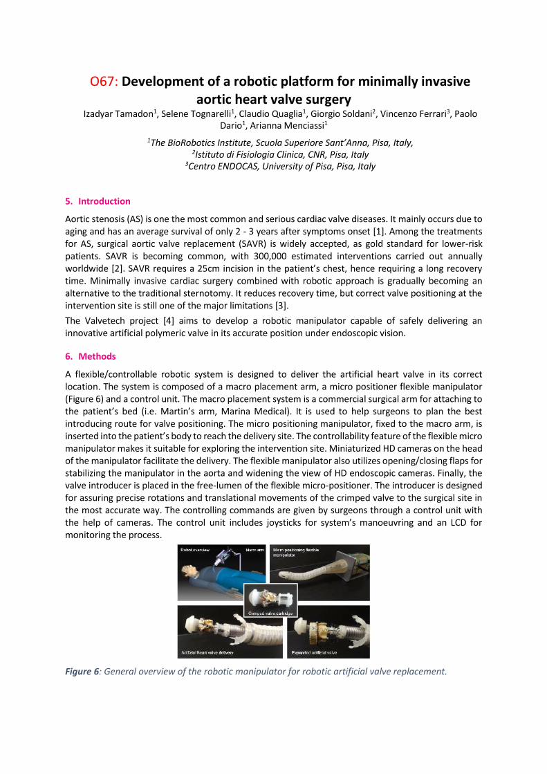
O67: Development of a robotic platform for minimally invasive aortic heart valve surgery
Izadyar Tamadon1, Selene Tognarelli1, Claudio Quaglia1, Giorgio Soldani2, Vincenzo Ferrari3, Paolo Dario1, Arianna Menciassi1
1The BioRobotics Institute, Scuola Superiore Sant’Anna, Pisa, Italy, 2Istituto di Fisiologia Clinica, CNR, Pisa, Italy
3Centro ENDOCAS, University of Pisa, Pisa, Italy
5. Introduction
Aortic stenosis (AS) is one the most common and serious cardiac valve diseases. It mainly occurs due to aging and has an average survival of only 2 - 3 years after symptoms onset [1]. Among the treatments for AS, surgical aortic valve replacement (SAVR) is widely accepted, as gold standard for lower-risk patients. SAVR is becoming common, with 300,000 estimated interventions carried out annually worldwide [2]. SAVR requires a 25cm incision in the patient’s chest, hence requiring a long recovery time. Minimally invasive cardiac surgery combined with robotic approach is gradually becoming an alternative to the traditional sternotomy. It reduces recovery time, but correct valve positioning at the intervention site is still one of the major limitations [3].
The Valvetech project [4] aims to develop a robotic manipulator capable of safely delivering an innovative artificial polymeric valve in its accurate position under endoscopic vision.
6. Methods
A flexible/controllable robotic system is designed to deliver the artificial heart valve in its correct location. The system is composed of a macro placement arm, a micro positioner flexible manipulator (Figure 6) and a control unit. The macro placement system is a commercial surgical arm for attaching to the patient’s bed (i.e. Martin’s arm, Marina Medical). It is used to help surgeons to plan the best introducing route for valve positioning. The micro positioning manipulator, fixed to the macro arm, is inserted into the patient’s body to reach the delivery site. The controllability feature of the flexible micro manipulator makes it suitable for exploring the intervention site. Miniaturized HD cameras on the head of the manipulator facilitate the delivery. The flexible manipulator also utilizes opening/closing flaps for stabilizing the manipulator in the aorta and widening the view of HD endoscopic cameras. Finally, the valve introducer is placed in the free-lumen of the flexible micro-positioner. The introducer is designed for assuring precise rotations and translational movements of the crimped valve to the surgical site in the most accurate way. The controlling commands are given by surgeons through a control unit with the help of cameras. The control unit includes joysticks for system’s manoeuvring and an LCD for monitoring the process.
Figure 6: General overview of the robotic manipulator for robotic artificial valve replacement.
7. Results
Prototypes of each robotic element were designed and assembled in the first Valvetech robotic platform. Preliminary assessments of the individual components and the complete integrated platform were carried out. A preliminary control architecture was also implemented, even if further tuning of the parameters is still required.
8. Discussion and Conclusion
A robotic system for robotic minimally invasive replacement of the aortic valve is proposed. Future activities will be devoted to extensively test the manipulator both in ex-vivo and in-vivo conditions.
References
[1] R. L. J. Osnabrugge et al., “Aortic stenosis in the elderly: Disease prevalence and number of candidates for transcatheter aortic valve replacement: A meta-analysis and modeling study,” J. Am. Coll. Cardiol., vol. 62, no. 11, pp. 1002–1012, 2013.
[2] R. O. Bonow, M. B. Leon, D. Doshi, and N. Moat, “Management strategies and future challenges for aortic valve disease,” Lancet, vol. 387, no. 10025, pp. 1312–1323, 2016.
[3] J. Joseph, S. Y. Naqvi, J. Giri, and S. Goldberg, “Aortic Stenosis: Pathophysiology, Diagnosis, and Therapy,” Am. J. Med., vol. 130, no. 3, pp. 253–263, 2017.
[4] https://www.cnr.it/Valvetechproject
O104: EVALUATION OF A MULTI-STEERABLE CATHETER FOR CARDIAC INTERVENTIONS
Ali Awaz, De Winter Joost, Dodou Dimitra, Breedveld Paul- Delft University of Technology,
BioMechanical Engineering, Delft, Netherlands
1. Introduction
To overcome the challenges presented by complex 3-dimensional shapes and tortuous vasculature in the cardiovascular environment, we previously developed a prototype of a multi-steerable catheter having 4 DOFs controlled by two joysticks [1]. However, it remains unknown whether the added steering possibilities are beneficial in the cardiovascular anatomy. With this research we aimed to investigate the effect of 0-DOF, 2-DOF, and 4-DOF catheter systems on surgical performance in a cardiovascular model.
2. Methods
Catheter Prototype – The catheter prototype was modified to compare 0-DOF (no steering), 2-DOF (multi-directional steering with a single segment), and 4-DOF steering (multi-directional steering with two segments).
Heart Model & Pathways – A transparent, rigid 3D-printed heart model was fabricated by Materialise (Leuven, Belgium) based on patient CT-data. Three pathways were defined for the experiment, differing in level of complexity:
1. Endo-myocardial biopsy route: starting in the jugular vein to reach a specified location on the intraventricular wall of the right ventricle.
2. Aortic valve implantation route: starting from the femoral artery to reach a specified location past the aortic valve.
3. Trans-septal mitral valve route: starting from the inferior vena cava to reach a specified location past the mitral valve.
Test Setup – The three identified pathways were visibly marked on the heart model and an electrode was placed at the start and end
points of each pathway. The electrodes were connected to a National Instruments LabVIEW box to record the elapsed time between the start and end of each test.
Test Procedure – 18 novices (all students and employees at Delft University of Technology) between 18 and 30 years old, participated in the experiment. Each participant conducted three experimental sessions. In each session, one catheter type was used to manoeuvre along each of the three pathways. This resulted in nine tests per participant. Catheter order and pathway order were randomized.
Performance measures – Performance was assessed using the following objective measures: 1) completion time of each of the nine tests, and 2) number and locations of errors (wrong branch or chamber, retraction of the catheter, catheter blocking). Moreover, the following self-reported measured were used: 1) task performance, 2) usability, 3) workload, and 4) catheter preference.
3. Results
General Observations – General observations gave insight in the methods the users applied to overcome difficulties in steering. These methods included rotation of the shaft, fast push-pull movements of the catheter as a whole, shaking movements to direct the catheter in a specific region, and making use of the cardiac wall.
Preliminary Results – Four participants have been tested so far, yielding a total of 36 tests (4 x 9). For both the 0-DOF and the 2-DOF catheters, 4 out of 12 tests succeeded within 5 minutes. With the 4-DOF catheter, the task was completed within 5 minutes in 8 out of 12 tests. From the tasks that were completed within 5
minutes, the average completion time was measured as 102 s for 0-DOF, 126 s for 2-DOF, and 118 s for 4-DOF. Additionally, all four users reported that they preferred the 4-DOF catheter over the other two.
4. Discussion & Conclusion
The preliminary results indicated that the 4-DOF catheter is able to overcome the challenges of the cardiac pathways better than the 0-DOF and 2-DOF catheters. The remaining 14 participants must be tested before any clear conclusions can be drawn from the results.
References
[1] A. Ali, A. Sakes, E.A. Arkenbout, P.
Breedveld, “Design of a Multi-Steerable
Catheter for Complex Cardiac
Interventions,” 28th International
Conference of Society for Medical
Innovation and Technology (SMIT), Oct. 5-
8, Delft, The Netherlands.

O114:

SESSION 25 – IMAGE GUIDED SURGERY 2
O8: ELECTROMAGNETIC TRACKING CAN BE USED TO ACCURATELY MESURE LEFT VENTRICULAR DIAMETER IN REAL TIME: A VALIDATION STUDY Krogh Magnus Reinsfelt, Andersen Øyvind Senstad, Aalen John, Elle Ole Jacob, Odland Hans Henrik,
Remme Espen Wattenberg - The Intervention Center, Oslo University Hospital, Oslo, Norway
1. Introduction
Left bundle branch block (LBBB) results in a delayed electrical activation of the left ventricular (LV) lateral wall. This causes an inefficient, uncoordinated contraction, manifested as an abnormal shortening and lengthening pattern of the septum-to-lateral wall diameter during the heart cycle (Fig. 1) [1]. LBBB is treated using cardiac resynchronization therapy (CRT), where 2 pacemaker leads are placed on the septum and lateral wall. The LV is paced on both sides to restore synchronous electrical activation and contraction, restoring the normal LV diameter trace. However, successful resynchronization depends on correct placement of the pacing leads. Monitoring the LV diameter trace during implantation could therefore be beneficial.
Electromagnetic tracking (EMT) can be used to measure the movement of instruments in space, and could theoretically be incorporated into a pace lead, and hence measure the position and distance between the pace leads. We therefore validated how accurately EMT could measure LV diameter through the heart cycle as a first feasibility test of a combined EMT and CRT system.
2. Methods
In 6 anesthetized canines we placed EMT sensors at the pacing sites in the septum and LV lateral wall. As reference, sonomicrometry (SM) sensors were placed beside the EMT sensors. LBBB was induced and data was collected during baseline, LBBB, and CRT.
The length between the EMT sensors was calculated as the difference in position over time, and compared to the length measured by SM. The accuracy of the EMT method was calculated as root mean square deviation (RMSD) of the absolute difference between the SM and EMT length traces.
3. Results
The average peak to peak amplitude of measured LV diameter using SM was 4.3±1.5 mm. The LV diameter measured by EMT showed very good agreement with the reference, with an average RMSD of 0.4±0.1 mm. Representative traces are shown in Fig. 1.
4. Discussion & Conclusion
The EMT- and SM sensors could not be placed in the exact same location in our setup, which may in part explain the small error between the methods. Despite this limitation, there was very good agreement between EMT and SM in measuring LV diameter.
The results show that EMT can measure LV diameter continuously with high accuracy, indicating that EMT can be used to aid CRT implantation.
References
[1] O. Gjesdal, E. W. Remme, A. Opdahl, H. Skulstad, K. Russell, E. Kongsgaard, T. Edvardsen, and O. A. Smiseth, “Mechanisms of abnormal systolic motion of the interventricular septum during left bundle-branch block,” Circ. Cardiovasc. Imaging, vol. 4, no. 3, pp. 264–273, 2011.
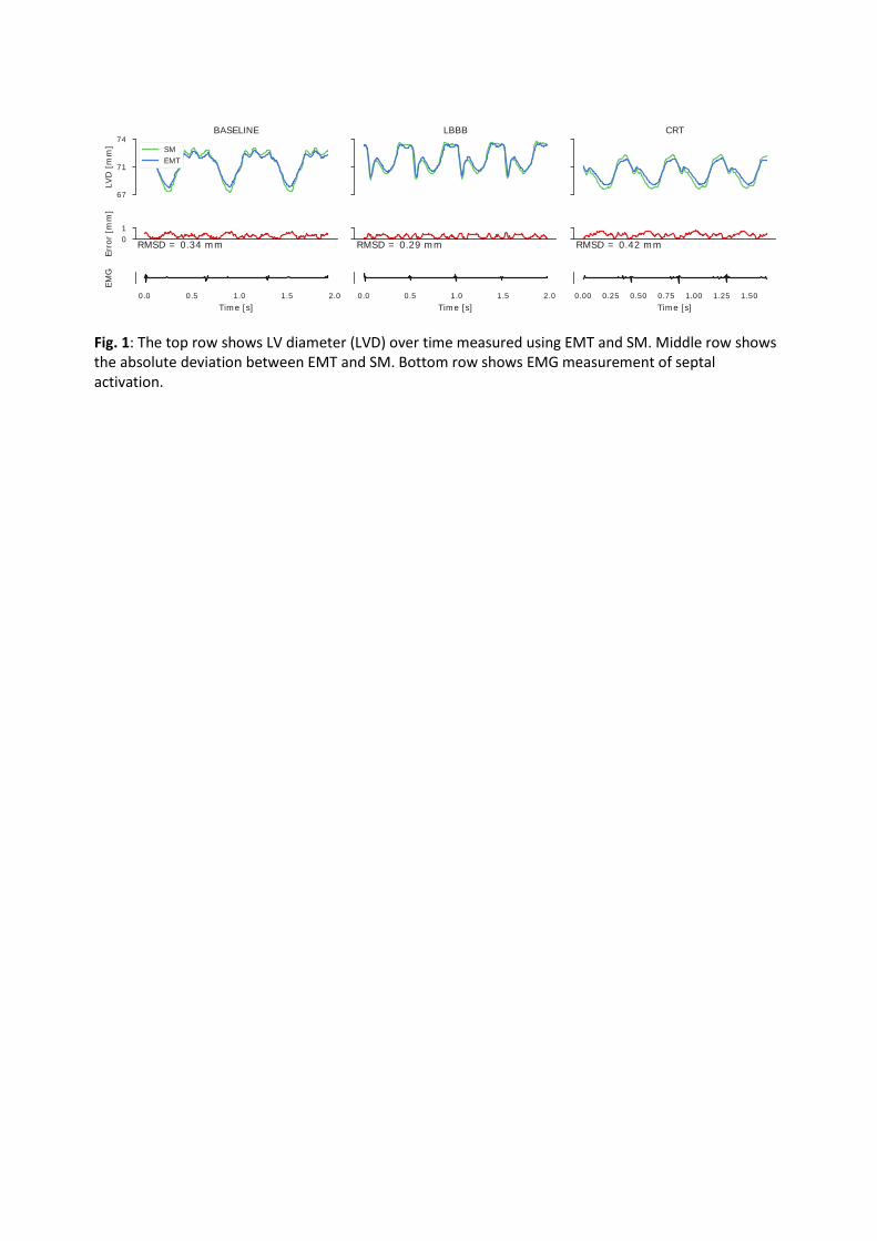
Fig. 1: The top row shows LV diameter (LVD) over time measured using EMT and SM. Middle row shows the absolute deviation between EMT and SM. Bottom row shows EMG measurement of septal activation.

O18: PREDICTION OF GUIDEWIRE TRAJECTORY INSIDE THE VASCULAR
SYSTEM: 3D COMPUTER BASED MODEL
Sharei Hoda, van den Dobbelsteen John J, Dankelman Jenny- Delft university of technology,
Biomechanical Engineering, Delft, Netherlands
5. Introduction
Guidewires are the basic tools used during interventions to access the place of interest [1]. Since the range of vascular geometries are extensive from one organ to another and from one patient to another, there is a variety of guidewires available. Therefore, the interventionalist needs to select an appropriate guidewire in each procedure. Despite the importance of this selection in the success rate of the procedure, it is mainly subjective and based on experience.
Knowledge of the guidewire’s trajectory and applied forces prior to the procedure can support the interventionalist in selecting a guidewire with suitable mechanical properties for successful navigation. Therefore, we developed a 3D model, based on rigid multibody dynamics, to simulate the motion of the guidewire in vascular system. The force transmission between the instrument and the vascular wall was also determined.
Methods
In our model, the guidewire is considered as a discrete body: a number of interconnected rigid segments, each of which may translate and rotate. To account for the bending stiffness of the guidewire, joints with torsional springs and dampers are located at each interconnection [2]. A 3-point bending test is performed to obtain real data regarding the stiffness of guidewire. The left anterior descending (LAD) coronary artery is chosen as vascular geometry. Therefore, based on consultation with specialists, Pilot50 and Pilot200 are used as guidewires.
6. The developed model is based on the forward dynamic method, i.e. given initial conditions and applied forces and/or applied moments, over a given time interval to predict the motion. By applying a defined force to the proximal side of the guidewire, the guidewire moves by a constant speed of 2 mm/s. The model is developed in MATLAB/Simulink (The Math Works, Inc.) environment. To validate the accuracy of our method, a series of experiments on a phantom model were performed.
Results & Discussion
In this study, we have developed a 3D guidewire model and have endeavored to investigate its behavior in a specific vascular geometry and the importance of guidewire’s stiffness on the motion. Although we used LAD as the vascular geometry and Pilot50 and Pilot200 as guidewires, the model is generic and it is possible to adopt to any other geometry or guidewire.

Figure 7: Tip trajectory of two guidewires inside LAD
The results show that the flexibility of a guidewire impacts its behavior during advancement: a higher flexibility, more fluctuation (see Figure 1). This can be explained by the fact that under the applied loads, the flexible tip deflects easier than the stiff one. Moreover, the stiff one (here, Pilot200) causes more applied forces to the vascular wall (see Figure 2).
Figure 8: Applied force at the tip of guidewires
Use of such simulations enables the user to assess the possible motions, and even to predict the success rate of the procedure. Moreover, this information might help instrument designers to predict the performance of a new guidewire before manufacturing.
ACKNOWLEDGMENT This work is part of the research program CONNECT project ( Grant Nr. 12705) within the research program interactive Multi-Interventional Tools (iMIT), supported by the Dutch Technology Foundation STW, which is part of the Netherlands Organization for Scientific Research (NWO).
References [1] Schneider, Peter A. Endovascular skills: guidewire and catheter skills for endovascular surgery. New
York, NY, USA: Informa Healthcare, 2009.
[2] Takashima, K., Tsuzuki, S., Ooike, A., Yoshinaka, K., Yu, K., Ohta, M. and Mori, K., 2014. ‘Numerical analysis and experimental observation of guidewire motion in a blood vessel model’, Medical engineering and physics, 36(12), pp.1672-1683.
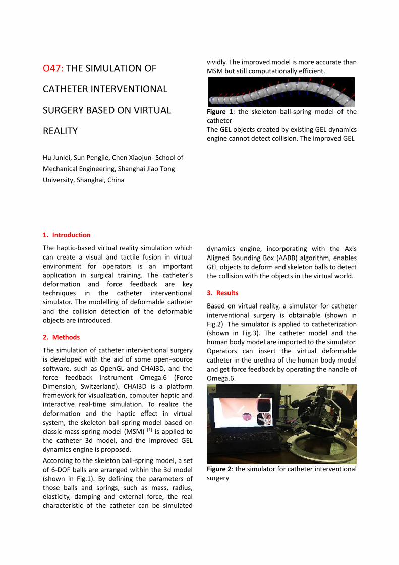
O47: THE SIMULATION OF
CATHETER INTERVENTIONAL
SURGERY BASED ON VIRTUAL
REALITY
Hu Junlei, Sun Pengjie, Chen Xiaojun- School of
Mechanical Engineering, Shanghai Jiao Tong
University, Shanghai, China
1. Introduction
The haptic-based virtual reality simulation which can create a visual and tactile fusion in virtual environment for operators is an important application in surgical training. The catheter’s deformation and force feedback are key techniques in the catheter interventional simulator. The modelling of deformable catheter and the collision detection of the deformable objects are introduced.
2. Methods
The simulation of catheter interventional surgery is developed with the aid of some open–source software, such as OpenGL and CHAI3D, and the force feedback instrument Omega.6 (Force Dimension, Switzerland). CHAI3D is a platform framework for visualization, computer haptic and interactive real-time simulation. To realize the deformation and the haptic effect in virtual system, the skeleton ball-spring model based on classic mass-spring model (MSM) [1] is applied to the catheter 3d model, and the improved GEL dynamics engine is proposed.
According to the skeleton ball-spring model, a set of 6-DOF balls are arranged within the 3d model (shown in Fig.1). By defining the parameters of those balls and springs, such as mass, radius, elasticity, damping and external force, the real characteristic of the catheter can be simulated
vividly. The improved model is more accurate than MSM but still computationally efficient.
Figure 1: the skeleton ball-spring model of the catheter The GEL objects created by existing GEL dynamics engine cannot detect collision. The improved GEL
dynamics engine, incorporating with the Axis Aligned Bounding Box (AABB) algorithm, enables GEL objects to deform and skeleton balls to detect the collision with the objects in the virtual world.
3. Results
Based on virtual reality, a simulator for catheter interventional surgery is obtainable (shown in Fig.2). The simulator is applied to catheterization (shown in Fig.3). The catheter model and the human body model are imported to the simulator. Operators can insert the virtual deformable catheter in the urethra of the human body model and get force feedback by operating the handle of Omega.6.
Figure 2: the simulator for catheter interventional surgery

Figure 3: the simulation of catheterization
4. Discussion & Conclusion
The simulator provides an effective surgical training method to help operators practice the catheter interventional surgery and get more skillful in a safe and easy way rather than traditional surgeries. Meanwhile, the deformable and collision-detective catheter model in the simulator provide operators with a more realistic feeling in surgical trainings. The methods can also be applied to other surgical fields.
Acknowledgement:
This work was supported by Natural Science Foundation of China (Grant No.: 81511130089), Foundation of Science and Technology Commission of Shanghai Municipality (Grant No.: 14441901002, 15510722200, and 16441908400).
References
[1] H. Delingette. "Toward realistic soft-tissue modeling in medical simulation." Proceedings of the IEEE 86.3 (1998): 512-523.

O19: GENERATIVELY MANUFACTURED FLEXIBLE INSTRUMENT GUIDANCE DEVICE AND HOLDING ARM FOR MRI-GUIDED INTERVENTIONS Odenbach Robert, Sánchez López Juan Sebastián, Friebe Michael- Chair Catheter Technologies and Image
Guided Therapies, Department of Medical Engineering, Otto-von-Guericke University Magdeburg,
Magdeburg, Germany
Introduction
MRI-interventions, e.g. biopsy- or RFA-needle placements, usually are executed by the clinician inside the scanner and under real-time guidance with an in-room monitor next to it. For access to the interventional site the clinician must lean with his hand deeply into the center of the scanner-tunnel (60 cm - 80 cm), while observing the monitor, to adjust the right needle-trajectory by hand. During the complete intervention, he must remain multiple times in that uncomfortable and exhausting position.
MRI-compatible assistance devices, which facilitate such procedures are topic of many robotic-oriented research projects. But still there are no affordable and certified products commercially available. We propose a fully MRI-compatible and easy to handle assistance platform that is based on an universally applicable and flexible instrument guidance device (flexible guide) and a holding arm.
Methods
Our design of the flexible guide provides a 6-DOF (DOF = degree of freedom) flexibility in a range of max. 20 mm and features a universal interface (hub diameter = 9 mm) for holding different types of instrument-adapters. The instrument is aligned manually with the movable hub. Once it matches with the desired trajectory all DOF´s can be fixed simultaneously with one locking element. Mounted on the MRI-compatible holding arm the flexible guide can be positioned in every desired location inside the MRI-tunnel.
We evaluated the setup in our MRI-system (3T, Skyra, Siemens, Germany), using a liver biopsy phantom (Abdominal Phantom 057A, CIRS, USA). The holding arm was attached to the table rail and the flexible guide was mounted on its distal end. For the simulation of the biopsy procedure a needle (21G) was placed with an adapter into the hub of the flexible guide, see Figure 1. The coarse positioning of the flexible guide was made with the holding arm around the estimated interventional location. For the registration of that initial needle position a localizer MR-dataset was acquired with the scanner. Specifically designed and printed fiducial markers [1], which are integrated inside of the flexible guide, provided reliable device visibility and conclusion about the instrument alignment. Following the needle orientation was adapted manually with the flexible guide while an in room monitor provided control of that procedure during real-time MRI-guidance. After the right trajectory was determined, the biopsy needle was locked in its position and pushed through the needle guide into the phantom until the target structure was reached.
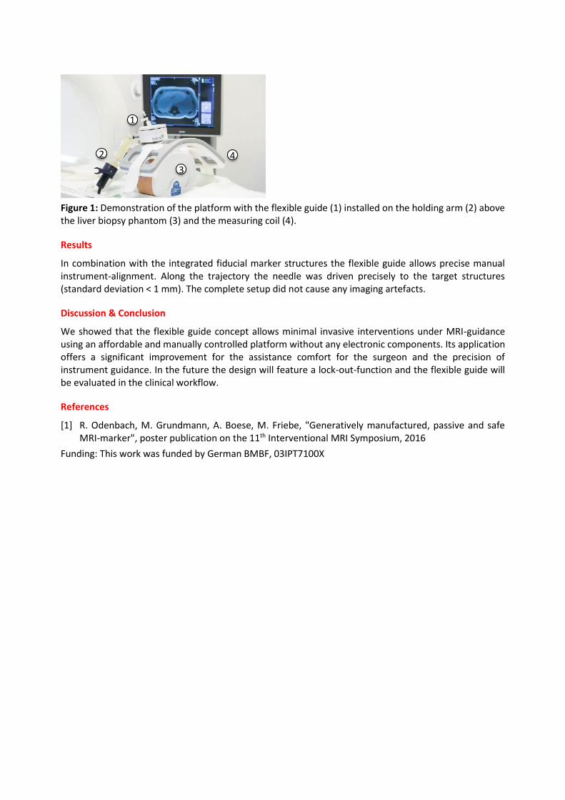
Figure 1: Demonstration of the platform with the flexible guide (1) installed on the holding arm (2) above the liver biopsy phantom (3) and the measuring coil (4).
Results
In combination with the integrated fiducial marker structures the flexible guide allows precise manual instrument-alignment. Along the trajectory the needle was driven precisely to the target structures (standard deviation < 1 mm). The complete setup did not cause any imaging artefacts.
Discussion & Conclusion
We showed that the flexible guide concept allows minimal invasive interventions under MRI-guidance using an affordable and manually controlled platform without any electronic components. Its application offers a significant improvement for the assistance comfort for the surgeon and the precision of instrument guidance. In the future the design will feature a lock-out-function and the flexible guide will be evaluated in the clinical workflow.
References
[1] R. Odenbach, M. Grundmann, A. Boese, M. Friebe, "Generatively manufactured, passive and safe MRI-marker", poster publication on the 11th Interventional MRI Symposium, 2016
Funding: This work was funded by German BMBF, 03IPT7100X
1
2
3
4

O62:A robotic approach for FUS in moving organs A. Cafarelli1, A. Diodato1, A. Schiappacasse2, S. Tognarelli1, G. Ciuti1, A. Melzer3, A. Menciassi1
1) The BioRobotics Institute, Scuola Superiore Sant'Anna, Pisa, Italy, [email protected], 2) CAMELOT Biomedical Systems, Genova, Italy, 3) IMSAT, University of Dundee, Dundee, UK
1. Introduction
Focused Ultrasound Surgery (FUS) is a non-invasive therapeutic technology that was demonstrated to be effective for the treatment of various pathologies, even if a wider clinical diffusion would require to compensate physiological movements of abdominal moving organs [1]. We present here an innovative robotic platform intended for FUS (http://www.futuraproject.eu/). A new motion compensation strategy suitable for clinical FUS procedures for moving organs has been introduced and evaluated through ex vivo tests. A robotic-assisted approach improves accuracy and flexibility of the overall procedure and enables a fast compensation of target organs’ motion, such as for kidney, liver and pancreas.
2. Methods
The FUTURA (Focused Ultrasound Therapy Using Robotic Approaches,) platform is composed by two 6 DoFs manipulators (ABB IRB 120): one holding a custom 16 channels annular array FUS transducer, provided with a confocal 2D ultrasound (US) probe (PA7-4/12, Analogic Ultrasound), and the other one holding a 3D imaging US probe (4DC7-3/40, Analogic Ultrasound). Both US probes are connected with proper US acquisition machines (SonixTablet, Analogic Ultrasound). A dedicated Human Machine Interface developed in ROS (Robot Operating System) controls all system components and manages the entire procedure. Figure 1. Experimental setup of the FUTURA platform.
Based on the design architecture, the FUTURA platform can treat moving targets by using machine learning and computer vision techniques. The pipeline is mainly composed by: i) a tracking module that detects and tracks a region of interest (ROI) on one of the two B-mode US images, ii) a learning module trained on target trajectory provided by the tracking module. By exploiting the axial steering capabilities
of the annular focused transducer, we implemented an innovative method for compensating the target point motion based on an angular movement of the transducer combined with a fast adjusting of the focal depth. This strategy minimizes the modification of the acoustic window during the breathing phases, which could hamper the correct identification and treatment of the target (e.g. presence of reflective ribs in in vivo applications). The feasibility of the compensation strategy was demonstrated in a simulated respiratory-induced organ motion environment, consisting in an ex-vivo porcine kidney placed on a 1 DoF motorized slide moving with an amplitude of 20 mm and a frequency of 0.2 Hz (Figure 1). The procedure was repeated 10 times in different regions of the ex vivo target in order to evaluate the tracking error between the target and transducer focus positions. In addition, a sonication was performed in a chicken breast in static conditions in order to qualitatively demonstrate the accuracy of
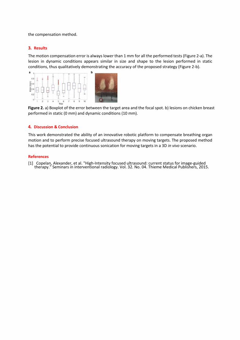
the compensation method.
3. Results
The motion compensation error is always lower than 1 mm for all the performed tests (Figure 2-a). The lesion in dynamic conditions appears similar in size and shape to the lesion performed in static conditions, thus qualitatively demonstrating the accuracy of the proposed strategy (Figure 2-b).
Figure 2. a) Boxplot of the error between the target area and the focal spot. b) lesions on chicken breast performed in static (0 mm) and dynamic conditions (10 mm).
4. Discussion & Conclusion
This work demonstrated the ability of an innovative robotic platform to compensate breathing organ motion and to perform precise focused ultrasound therapy on moving targets. The proposed method has the potential to provide continuous sonication for moving targets in a 3D in vivo scenario.
References
[1] Copelan, Alexander, et al. "High-Intensity focused ultrasound: current status for image-guided therapy." Seminars in interventional radiology. Vol. 32. No. 04. Thieme Medical Publishers, 2015.
O99: CYBERSECURITY: IT RISK PERCEPTION IN THE HEALTHCARE. MARCHE REGION CASE STUDY Kulaga Barbara, Cristino Berardino- Università Politecnica delle Marche, Management, Ancona, Italy
1. Introduction
The use of healthcare technologies, from medical devices to the Internet of Things, has increased the exposure of healthcare companies to hacking [1]. The value of health data transmitted through cyberattacks is even greater than that one of payment card data [2]. Therefore, computer health networks are particularly appealing to computer hackers. The last damage is caused, however, not to the healthcare company or NHS, but to the patient; the subjects of the infringement are the patient’ sensitive data [3]. The aim of this study is to understand whether the health care staff is aware of the problem.
2. Methods
The study was conducted through the analysis of survey data provided to the staff of some public and
private healthcare facilities in the Marche Region. After the examination of the emerging data in the most
current research on the topic, find out through keyword research on PubMed (Medline), Kinahl and
Proquest; there was the creation of questions to be included in the survey; the distribution of the survey
in nine structures in the Marche Region (Ars – Agenzia Regionale Sanitaria-; Azienda Ospedaliero
Universitaria Hospitals of Ancona, at Umberto I Hospital and Pediatric Hospital G. Salesi; Clinitest, in Ascoli
and San Benedetto del Tronto; INRCA; Institute of Rehabilitation Santo Stefano , in the headquarters of
Ascoli, Porto Potenza Picena and San Benedetto del Tronto; Provincial Hospital of Macerata), finally there
was a statistical analysis of the data divided into classes. On the statistic sample, which was considered
significant, was considered a distortion factor (e = 1.17) during the processing of the results.
3. Results
On a sample basis n=251, considered an average quadratic error of 1,177; it is noted that despite the almost unanimous belief in the impact of any computer attack, there are still a few initiatives taken to raise awareness about IT security. Staff largely do not know about the ICT security plan, even if this is defined by the structure, a similar scenario can be identified about the formalization of the procedure to be followed in case of computer attack. From this it emerges that there is no synergy between computer and clinical engineers and healthcare staff. Considering the imminent obligation to implement the Minimum ICT Security Measures, it would also be desirable to have a learning pathway for those who daily handle sensitive data. 4. Discussion & Conclusion
Although the health sector is most exposed to computer attacks, the theme is still far from the common interest. In the medical field, the alarm was launched in the last months of 2016 when some research by Deloitte and Trapax estimated that 94% of the 5600 US hospitals were victims of hacking. The focus increased in May 2017 when Wannacry ransomware hit 99 countries and forced the British National Cyber Security Center to work with the National Health Service [4] to contain the consequences of the attack that paralyzed some health facilities for a whole weekend in Britain. It emerges that the digitization of healthcare companies should correspond to an adequate investment in computer security. This study demonstrates the lack of awareness of individual users of risk exposure and highlights that educational campaigns should be directed to the staff involved. Only in this way will be a cooperation between clinical

and computer engineers and the healthcare staff, with the aim to find and implement appropriate solutions. Through this study it is possible to have an indication of the categories on which it is necessary to work most, making them aware and sensitive to the problem. Correct information would help in addressing the problem in healthcare facilities, would allow for a better collaborative spirit with professional and technical figures that work specifically on cybersecurity, and would make the operators, not only in their role of healthcare staff, but as citizens, more careful, allowing the spread of the rules to be followed to limit the risks also outside the healthcare field, in everyday life. References [1] Eastwood, B. Premera says data breach may affect 11 million consumers. FierceMobileHealthcare
(Mar. 18, 2015); http://www.fiercehealthit.com/story/premera-says-data-breach-may-affect-11-million-consumers/2015-03-18.
[2] Gagliord, N. Healthcare cybersecurity worse than retail: BitSight. (May 28, 2014); http://www.zdnet.com/article/healthcare-cybersecurity-worse-than-retail-bitsight/.
[3] Naveed, M. et al. Privacy in the genomic era. ACM Comput. Surv. 48, 1, Article 6 (July 2015); DOI: http://dx.doi.org/10.1145.2767007.
[4] Tobias A. Mattei in World Neurosurgery: Privacy, Confidentiality, and Security of Health Care Information: Lessons from the Recent WannaCry Cyberattack, Volume 104, August 2017, Pages 972-974

SESSION 7 – I HAVE AN IDEA! FROM THE CONCEPT TO THE CLINICAL VALIDATION
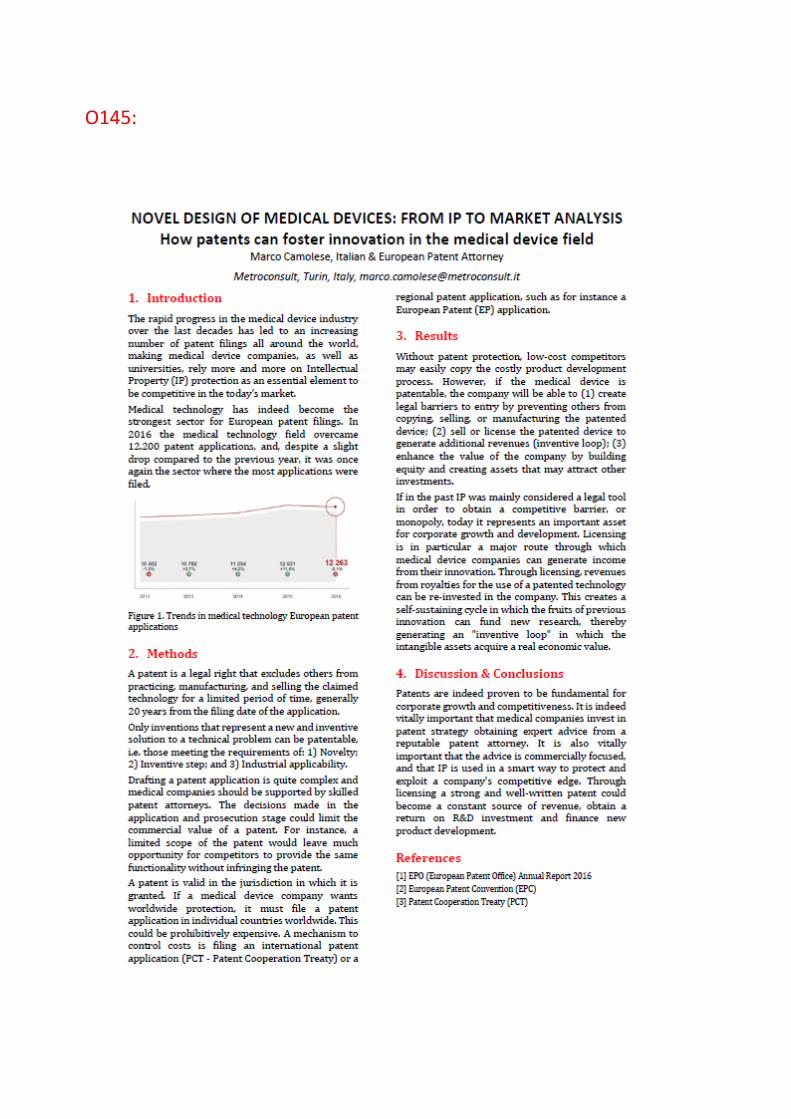
O145:

O140: Asymmetric – a novel vessel sealing technology, using low energy, and a flexible laser Fiber Szold Amir - Assia Medical Group, Assuta Medical Center, Tel Aviv, Israel
Objective: Vessel sealing during minimally invasive surgery is still a challenge, especially when
applying through articulating or flexible devices. Additional disadvantages of current system
include low precision and collateral tissue heating.
Technology: The core technology is an asymmetric laser fiber that enables controlled lateral
energy emission. This allowed the design and production of several vessel sealing and tissue
cutting devices including an articulating grasper, a hook and a loop snare for endoscopy. The
devices can be articulated or flexible, fit into a flexible endoscopes' working channels and can be
made in diameters even lower than 3 mm. This superior adaptability means the technology is a
perfect fit with a variety of surgical robotic platforms.
Preliminary results: The devices were tested in an animal model with successful sealing of
arteries up to 5 mm diameter, using low energy and with a durable seal.
Future perspectives: We are currently finalizing the first product design for minimally invasive
surgery.

O36: Design requirements for surgical equipment for low-and-middle income countries Oosting Roos Marieke, Wauben Linda, Groen Reinou, Dankelman Jenny - Delft University of Technology, BioMechanical Engineering, Delft, Netherlands
7. Introduction There is a significant disparity between surgical procedures performed in high-income countries (HICs) and low-and-middle income countries (LMICs), only 3.5 % of the surgeries performed in the world are received by the poorest one third of the world’s population(1). Multiple surgical capacity studies have shown that hospitals in LMICs, such as Sierra Leone, Malawi and Nigeria, do not have a 100% coverage of basic surgical equipment such as, theatre lights, anesthesia machines and electro surgical units (ESUs). Equipment that is implemented successfully in HICs (e.g. oxygen concentrators) had a very short life span when implemented in hospitals in Gambia(2). Perry & Malkin et al. (2011) estimated that 40% of the equipment available in hospitals in LMICs is not working(3). Within the department of Biomechanical Engineering of the Delft University of Technology we aim to develop surgical equipment that can be successfully implemented in operating theatres in LMICs. The aim of this study is to develop the design requirements for context appropriate surgical devices by identifying the surgical equipment journey in Kenya and the barriers perceived by important stakeholders.
8. Methods 1)To determine the surgical equipment journey in Kenya, semi-structured interviews and participatory mapping exercises were conducted in Kenya in December 2016 with 8 biomedical engineering technicians (BMETs), 1 local distributor of medical equipment, 2 NGO’s and 2 surgeons all working in Kenya. 2)Based on the surgical equipment journey and perceived barriers design requirements were developed.
9. Results The surgical equipment journey in Kenya is displayed in Figure 1. Lack of spare parts, lack of BMET training, lack of service contracts and environmental factors such as electricity, temperature, and humidity were identified as barriers (Fig. 1).
The design requirements should, therefore, take into account both the local context and the different maintenance and repair structure in Kenya:
1. Design should account for repair instructions, spare parts and easy accessible replacement parts 2.Design should withstand a harsh dusty, humid climate with high temperatures 3. Design should be reusable with limited running costs
4.Discussion & Conclusion
We believe that these design requirements will eventually lead to a different set of surgical equipment than used in HIC, without essentially compromising the quality, safety and functionality. The aim is to increase the life span and thereby the longer use of surgical equipment in LMICs. A cryo device, ESU, video laryngoscope and laparoscope for LMICs are currently being designed at Delft University based on these requirements. Further research will focus on determining the order of reparability that is required for successful and safe implementation in LMICs.
Figure 9: Surgical equipment journey and perceived barriers by stakeholders in Kenya.

References
1. Chao TE, Sharma K, Mandigo M, Hagander L, Resch SC, Weiser TG, et al. Cost-effectiveness of surgery and its policy implications for global health: a systematic review and analysis. The Lancet Global Health. 2014;2(6):e334-e45. 2. Howie SR, Hill SE, Peel D, Sanneh M, Njie M, Hill PC, et al. Beyond good intentions: lessons on equipment donation from an African hospital. Bulletin of the World Health Organization. 2008;86(1):52-6. 3. Perry L, Malkin R. Effectiveness of medical equipment donations to improve health systems: how much medical equipment is broken in the developing world? Medical and Biological Engineering and Computing. 2011;49(7):719-22.

O142: Extending Human Xtensions from device to system: remote controlled articulating tool for MIS surgery Szold Amir, Sholev Mordechai - Assia Medical Group, Assuta Medical Center, Tel Aviv, Israel
Objective: Human Extensions has developed a hand-held, software driven mechatronic device
for MIS. To extend the scope of usability of these platform technologies and broaden the
possible configurations for their use a novel table mounted system was developed.
Technology: Using the same core technologies (novel articulation, user interface and control
system) a table-mounted software driven electro-mechanical system that can support several end
effectors was designed and built The system is composed of a sophisticated user interface that
enables unrestricted hand movement at or away from the table, and a novel, motor driven
articulating tool that is controlled by the interface and enables all degrees of freedom. The
system is lightweight and with a small footprint, requires minimal set up time, can be easily
moved between laparoscopic trocars and perform complex motions in the surgical field.
Preliminary results: Following an extensive validation in the lab we have performed an animal
trial using a pig model. We have demonstrated excellent manipulation with the device and
seamless integration with our hand-held device.
Future perspectives: We are currently testing the system in order to personalize a technology
solution to the different needs of surgeons and institutions,

O53:
Design of a fully implantable closed loop artificial pancreas V. Iacovacci1, L. Ricotti1, I. Tamaddon1, G. Tortora1, C. Perri1, P. Dario1, and A. Menciassi1
1) The BioRobotics Institute, Scuola Superiore Sant'Anna, Pisa, Italy (*[email protected]).
10. Introduction Conventional type 1 diabetes (T1D) therapy implies daily fingerpricks and multiple insulin injections.
This approach fails in satisfactorily controlling blood glucose levels and strongly affects patients’ life
quality. Artificial Pancreas (APs) represent technological solutions combining a sensor for continuous
glucose monitoring and an insulin pump. These two elements are connected by a closed-loop control
algorithm. Despite recent advancements, current APs (mainly portable ones) still have some limitations
since they significantly affect lifestyle and imply delays in the insulin adsorption kinetics. The
development of fully implantable AP is hampered, at present, by the need of periodic surgical
operations to refill the insulin reservoir and to replace the implanted battery.
In this framework the authors proposed a fully implantable long-term autonomous AP based on a
non-invasive insulin refilling [1] and non-invasive battery recharging strategy. The device exploits
the intraperitoneal supply route to enable a more physiological insulin profile [2].
11. Methods
The proposed device is conceived to be implanted in close proximity of an intestine loop. This enables to
implement a non-invasive refilling procedure based on sensorized ingestible insulin capsules.
Figure 10 System overview with details showing the implant site, the wireless energy transfer and the communication systems.
Such capsules are expected to go through the gastrointestinal tract, until they are revealed and
reversibly captured by the implanted AP. The insulin can be thus transferred to the internal device
reservoir. A commercial glucose sensor and a wireless communication module enable to close the
control loop and to provide the patient with warning and monitoring information (Figure 1). The AP
prototype developed was based on mechatronic components enabling capsule docking, insulin transfer
to the reservoir and intraperitoneal insulin microinjection. A Nylon reservoir featured by a variable
volume was fabricated to guarantee insulin stability. Long-term system powering can be guaranteed by
embedded batteries, rechargeable in a wireless fashion, through non-radiative energy transfer.

12. Results A docking mechanism based on a magnetic switchable device (MSD) was designed, developed and
tested. Two under-actuated mechanisms, each including a stepper motor and a train of gears, were
embedded in the system for the activation of the MSD/capsule punching and the suction/injection
system, respectively (Figure 11). The Nylon-based reservoir limited insulin aggregation for at least two
weeks. A 4 coils-based wireless recharging system was designed, fabricated and preliminarily tested.
Figure 11 Fully implantable AP mechatronic system.
13. Discussion & Conclusion The authors demonstrated the overall feasibility of a fully implantable autonomous AP,
guaranteeing an intraperitoneal insulin supply. Results are encouraging in view of an in vivo
translation of this technology.
References
[1] Iacovacci, Veronica, et al. "Design and development of a mechatronic system for noninvasive refilling of implantable artificial pancreas." IEEE/ASME Transactions on Mechatronics 20.3 (2015): 1160-1169.
[2] Cobelli, Claudio, Eric Renard, and Boris Kovatchev. "Artificial pancreas: past, present, future." Diabetes 60.11 (2011): 2672-2682.

O56: A novel microwaves tool for robotic resection of the liver Frosini Francesco, Staderini Fabio, Brancadoro Margherita, Dimitri Mattia, Regolini Jacopo, Miniati
Roberto, Boushaki Mohamed Nassim, Paternò Linda, Guerra Francesco, Coratti Andrea, Cianchi Fabio,
Corvi Andrea, Menciassi Arianna, Biffi Gentili Guido - University of Florence, Department of Information
Engineering, Florence, Italy
1. Introduction
Hepatectomy using coagulative necrosis (HCN) has been recently developed in order to avoid bleeding
related problems, one of the major concern for the hepatic surgeon [1]. The basic principle of liver pre-
coagulation consists of creating a non-perfused zone using very high temperature hyperthermia along
the resection line, in order to prevent bleeding during resection maneuvers. Microwave (MW) energy,
despite its several advantages when compared to bipolar or monopolar radiofrequency [2], has still
never been used as an effective coagulative energy.
2. Methods
The Integrated Medical-Robotics Solution Research Group, a research group funded by Tuscany Region, developed
a new tool able to decrease the bleeding during hepatectomy. This tool uses 2.45 GHz MW energy with the aim to
create a coagulative necrosis during the robotic resection of the liver. A MW needle [2], consisting on a radiating
monopole has been integrated into a robotic arm of the da Vinci Research Kit (dVRK, Intuitive Surgical Inc.) surgical
platform. The needle is able to transfer high impulsive energy to the liver parenchyma allowing to establish a
transection line and enabling a very precise dissection and safe parenchyma division.
The needle was integrated into the dVRK designing a novel mechanical joint (Figure 1) able to
encapsulate the coaxial cable, the rods and the pulleys to control the needle. The tool exploits the
motion capability of the dVRK arm and allows one degrees of freedom (i.e., pitch). This joint was
developed following 4 important requirements: flexibility, integrability, stability and reliability. Before
the liver resection, different applications of sequential ablation were performed along the resection line:
particularly a needle of 14 G applied sequential energy pulses of 30 seconds at the power level of 89 W
and 100 W.
3. Results
Computer simulations and experimental measures using ex-vivo and in-vivo tissues were performed:
both confirmed that the target region reaches 100 °C in 7-10 s, arriving at 150 °C at the end of the 30 s
pulse. This allows the instantaneous coagulation of big vessel in contact with the needle. In-vivo
procedures demonstrates that this application is capable of coagulating vessels with diameters up to
4-5mm. The coagulation effect seems guaranteed by an internal MW-induced vessels’ thrombosis.

Figure 1: Mechanical joint built for the integration of the MW needle into the dVRK robotic system.
4. Discussion & Conclusion
One of the main reason of the slow spreading of Robotic hepatic surgery probably is the consequence
of the absence of effective hepatic coagulative devices. Although the improvement of surgical
techniques and instruments, bleeding from parenchyma transection remains the most critical point
which can affect post-operative recovery. This novel device joins the advantages of MW energy as a
coagulative energy source with the advantages of robotic platform and minimally invasive surgery. Our
preliminary tests suggest that this combination could be useful to surgeons that perform robotic
hepatic resection. The robotic device seems highly effective in coagulating vessels of small and medium
diameter. The only main disadvantage is that the procedure it could be slower because the pre-
coagulation phase is time consuming. Eventually, by decreasing the pulse duration and optimizing the
MW energy transfer to the liver parenchyma the surgery could be faster; the evaluation of the
efficiency is still in progress. In conclusion, MW integrated into the dVRK robotic platform can be useful
to decrease the bleeding related problem during a robotic liver resection thus expanding the indications
of minimally invasive hepatic surgery.
References
[1] F. Romano, M. Garancini, F. Uggeri, et al., “Bleeding in Hepatic Surgery: Sorting through Methods to Prevent It,” HPB Surgery, vol. 2012, Article ID 169351, 12 pages, 2012. doi:10.1155/2012/169351.
[2] G. Biffi Gentili, C. Ignesti, and V. Tesi, “Development of a Novel Switched-Mode 2.45 GHz Microwave Multiapplicator Ablation System,” International Journal of Microwave Science and Technology, vol. 2014, Article ID 973736, 12 pages, 2014. doi:10.1155/2014/973736

O74: From concept to design: A new flexible robotic uterine elevator Chakravarthini M. Saaj1, Seri Mustaza1, Kavitha Madhuri2
1) Department of Electrical and Electronic Engineering, University of Surrey, Guildford, UK, e-mail [email protected]. 2) Department of Gynecology, Royal Surrey County Hospital, Guildford, UK
1.Introduction
Robot-assisted gynaecology and urology surgery is the fastest growing field of robotic surgery. Surgical
robots such as the da Vinci robot is now the most popular robot for minimally invasive pelvic surgery [1],
[2], [3]. However, some disadvantages of the da Vinci system and other surgical robotic systems are
their large size, the high initial and running costs, rigid laparoscopic instruments, limited degrees of
freedom and restricted haptic feedback with the consequence that the surgeon has a limited ‘feeling’ of
the surgery performed.
Uterine elevators are widely used for laparoscopic and robotic hysterectomy and other trans-
vaginal procedures. In the trans-vaginal manipulation of the uterus, there is no need for an additional
laparoscopic port which might be dedicated to manipulating the uterus from above. Hence,
manipulation through the vaginal route is highly advantageous as the vagina serves as a single “natural
orifice” port. The trans-vaginal uterine elevators are widely used by gynecologists for both laparoscopic
and robotic hysterectomy, oophrectomy, myomectomy, lymph node biopsy, and treatment of fibroids,
abnormal periods, endometriosis, ovarian cancer and pelvic prolapse [1], [4].
An ideal uterine manipulator must allow manipulation in all directions i.e. pushing, anteversion,
retroversion, lateralization, and flexing of the uterus whilst holding on to the cervix. Further, the uterine
elevator should be able to maintain the pneumoperitoneum following colpotomy and assist the
identification of vaginal cuff, the adnexae and the posterior cul-de-sac. However, the current hand-held
and voice controlled trans-vaginal uterine elevators are rigid tools that are often manually
positioned by an assistant in response to the surgeon’s command. Inefficient response to these
commands, lack of experience, lack of coordination between the surgeon and his assistant, poor
commands by the surgeon or fatigue and tiredness are some of the issues arising with the current
manipulation technique. Furthermore, the manipulation of a rigid and stiff uterine elevator could
potentially damage the uterine wall due to slippage.
Literature survey reveals that the commercially available uterine elevators do not offer flexibility
for pose control, force sensing, intelligence, autonomy or ease of adaptability [5]-[12].
A flexible uterine manipulator which can be fixed in place relative to the patient and controlled
remotely whilst the surgeon is sitting at the operating console would be a big step forward in
advancing the state-of-the-art of robotic gynaecological surgery. These issues motivate the research
on the development of an innovative flexible robotic uterine elevator.
This paper presents the proof-of-concept of a novel pneumatically controlled, octopus-inspired
robotic flexible uterine manipulator - GENTLER (Gynaecological ENdoscopic uTerine eLEvatoR),
based on a stiffness controllable and soft continuum mechanism with integrated force and pose
sensors.

2. Methods
The key features of the new flexible robotic uterine elevator – GENTLER is listed in Table 1. The first concept of GENTLER is shown in Figure 1 and the current design of the functional prototype is shown in Figure 2.
The flexible manipulator has an outer diameter of 20mm. A ceramic/plastic hard cup with an embedded metallic probe of variable sizes is mounted to the front end of the flexible module. The soft module can be positioned in the vagina, so that the cup is placed over the cervix holding it. The tip of the probe will then enter into the narrow uterine cavity and a balloon (not shown in Figure 2) will be inflated to hold the uterus for manipulation.
The core actuation mechanism of GENTLER is the soft silicone manipulator itself. This silicone-based manipulator is soft and is capable of elongating and bending in the sagittal and lateral planes. Furthermore, it can gently squeeze into narrow openings, such as the uterine cavity, and offers controllable stiffness.
Selective and controllable stiffness is expected to offer increased maneuverability and disturbance rejection capabilities for accurate positioning of the uterus. This would allow tuning its compliance in response to the force exerted onto its surroundings, hence avoiding damage to the tissue wall.
In addition to supporting its own weight, including the weight of the rod and cups that are mounted at the distal end of the manipulator, this lightweight manipulator must be capable of manipulating the uterus effectively. The inbuilt controllable stiffness, using the granular jamming mechanism, also ensures adaptability and better load handling of various sizes and weights of the uterus.
Table 1: Design features of GENTLER
Figure 1: GENTLER Concept
Design Feature Function
Back cup -
available in
multiple sizes
Maintaining pneumoperitoneum
Pneumatically
driven flexible
tube with
controlled
stiffening
1) Soft structure does not damage
tissue walls during manipulation
2) Maximum movement range (170
degrees in anterior/posterior
sagittal plane; 170 degrees
lateral movement in transverse
plane)
3) Stiffening control and soft organ
interface ensures no tissue
damage
Front cup -
comes in
varying sizes
Provide delineation of vaginal
fornices
Remote
controlled
1) Ease of manipulation
2) Able to lock device at certain
position to stabilize uterus
position
3) Avoid fatigue and tiredness
Angled Shaft Prevent slip during twisting motion
Partially
sterilization and
partially
reusable
Multiple usage and reducing running
costs
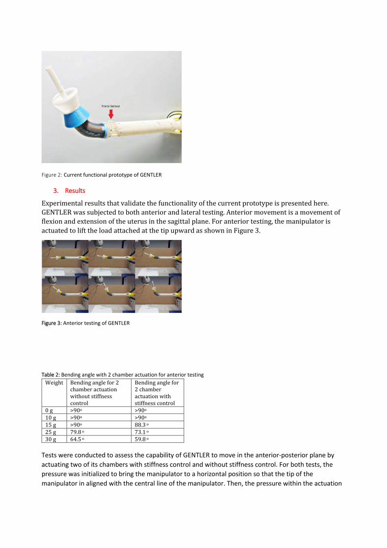
Figure 2: Current functional prototype of GENTLER
3. Results
Experimental results that validate the functionality of the current prototype is presented here.
GENTLER was subjected to both anterior and lateral testing. Anterior movement is a movement of
flexion and extension of the uterus in the sagittal plane. For anterior testing, the manipulator is
actuated to lift the load attached at the tip upward as shown in Figure 3.
Figure 3: Anterior testing of GENTLER
Table 2: Bending angle with 2 chamber actuation for anterior testing
Weight Bending angle for 2 chamber actuation without stiffness control
Bending angle for 2 chamber actuation with stiffness control
0 g >90o >90o 10 g >90o >90o 15 g >90o 88.3 o 25 g 79.8 o 73.1 o 30 g 64.5 o 59.8 o
Tests were conducted to assess the capability of GENTLER to move in the anterior-posterior plane by
actuating two of its chambers with stiffness control and without stiffness control. For both tests, the
pressure was initialized to bring the manipulator to a horizontal position so that the tip of the
manipulator in aligned with the central line of the manipulator. Then, the pressure within the actuation

chamber was increased incrementally until 90o or until the actuation pressure has reached a maximum
of 1.8 bar, whichever occurs first.
A comparison of the bending angle achieved for weight up to 30 g, with and without stiffness control, is
shown in Table 2. The manipulator is capable of lifting the 10 g load to reach 90o bending angle before
the maximum pressure limit is reached and is able to lift 15 g load to bend 90o at approximately 1.8 bar,
which is the maximum limit. However, GENTLER is still unable to bring the 25 g and 30 g load to reach
90o.
Figure 4: Lateral testing of GENTLER
GENTLER was further subjected to lateral testing in which it is expected to move the uterus to left and
right side. This was conducted by actuating the manipulator to left and right of the centerline of the
manipulator as shown in Figure 4. Similar to the anterior movement test, the pressure within the
chambers was first initialized to bring the manipulator to a horizontal position. The value of chamber
pressure is then incrementally increased to move the manipulator that is carrying the load. Here again,
the lateral movements were tested with and without activation of the stiffness control.
Table 3: Bending angle with 2 chamber actuation for lateral testing
Weight Bending angle for 2 chamber actuation without stiffness control
Bending angle for 2 chamber actuation with stiffness control
0 g >90o 90o 10 g 90o 83.4o 15 g 84.5o 78.1 o 25 g 77.9 o 72.5 o 30 g 59.1 o 51.3 o
The results for lateral movement test are analogous to the anterior movement tests. The achievable
bending angle reduced as the weight at the tip of the manipulator is increased. The highest bending
angle the manipulator achieved was 90o (to the left and right of the centerline of the manipulator),
when the manipulator is not carrying any load and when 10g is placed at the tip. The maximum bending
angle the manipulator can achieve reduced to 85.4o and 78.1o for a system without and with the
stiffness mechanism, respectively, when 15 g load is placed at the tip and reduced further with an
increase in load. These results are shown in Table 3.
4. Discussion & Conclusion
Commercially available uterine manipulators either require human assistance to hold the manipulator in
position or rely on limited capabilities of the motorised, voice controlled holder like the ViKY ® UP, which
was originally designed for an endoscopy aid. This paper presents the proof-of-concept of a novel
flexible robotic uterine elevator for trans-vaginal procedures. This pneumatically actuated uterine
elevator is integrated with a precise tip position controller and force feedback controller. It is capable of
achieving 180o range of motion in any direction in the absence of load at its tip. However, this range
becomes narrower as more load is placed at the tip. Future research is needed to improve the load
carrying capacity of the uterine elevator without losing its flexibility and to incorporate haptic feedback.
Acknowledgement
The authors wish to acknowledge the members of the EU FP7 STIFF-FLOP consortium, in particular, King’s College London (UK) for providing their force sensor and Przemysłowy Instytut Automatyki i Pomiarów (Poland) for providing the design of the soft manipulator module.
6. References
[1] K. Sakhel, “Robotic Assisted Laparoscopic Hysterectomy,” Robot Surgery InTech, January 01, 2010, ISBN 978-953-7619-77-0
[2] Lori et al., “Robotic Surgery in Gynecology…,” Obstetrics and Gynecology International, vol. 2011, Article ID 852061, 29 pages.
[3] Madhuri et al., “Robot assisted surgery in gynaecologic oncology …,” Int J Med Robot. 2012 Dec;8(4):496-503. doi: 10.1002/rcs.1461.
[4] Sabella et.al. “A Technique for Laparoscopic…,” Obstet Gynecol 1996;87:465-7
[5] ConMedEndosurgery,VCare®, (Accessed: July 2017)
[6] Cooper Surgical, ZUMI™ - Zinnanti Uter. Manip. Inj, (Accessed: July 2017)
[7] Cooper Surgical Inc, Intro. to the UPS and RUMI - Clinical setting (video), 15 February 2011, (Accessed: July 2017)
[8] Swan et.al. “Advanced uterine manipulation technologies”, Surg Tech. Int. 2010 Oct, 20:215-20.
[9] Sarah Kane and Kevin J. Stepp, 2010, “Laparo-endoscopic single-site…”, J Robotic Surg vol. 3, pp.253–255.
[10] Akrivos, Nikolaos and Peter Barton-Smith, “A Pilot Study of Robotic Uterine…,” J. Robotic Surgery, 21 April 2013.
[11] Puntambekar et.al. “A Novel Technique of Uterine Manip…”, Minimally Invasive Surgery, (2010), Article ID 836027, doi:10.1155/2010/836027.
[12] Yip et.al., “Towards developing a robot assistant for uterus positioning during hysterectomy: system design and experiments”, Robotics and Biomimetics, vol.1, No.9, 2014.

O98:
Autonomous Balloon Management for Endovascular Occlusion C.McCarthy1,2, I.Kanterman2,3, F. Trauzettel1,2, A.A. Goetz2, H.A. Jaeger1,2, B. Colvard2,4, P. Cantillon-
Murphy1,2
1) University College Cork, Ireland 2) IHU Strasbourg, Strasbourg, France 3) University of Rhode Island, USA 4) Vascular and Endovascular Surgery, University of Stanford School of Medicine, USA.
Introduction Non-compressible torso hemorrhage is a major cause of preventable death from trauma in civilian and military populations [1]. Resuscitative endovascular balloon occlusion of the aorta (REBOA) is a minimally invasive procedure to increase blood pressure to the brain in such cases. A balloon catheter is inserted through the femoral artery, often percutaneously, and inflated within a patient’s aorta. However, prolonged aortic occlusion during transport and in-hospital resuscitation may result in deleterious effects. One possible solution is partial REBOA, where the balloon catheter is inflated to a diameter slightly less than that of the aorta, allowing some distal flow to perfuse the visceral organs. The Autonomus Balloon Management System presented in this work
allows automatic pREBOA to be performed while preventing over inflation which could damage the aorta.
Figure 12- Intelligent balloon management for endovascular occlusion enables a partially occluded aorta allowing distal flow.
Methods Proximal (above the balloon) and distal (below the balloon) blood pressures, as well as internal
balloon pressure (saline) are monitored continuously by the balloon management system. The user
may control desired minimum proximal mean arterial pressure (pMAP) via a simple user interface.
This user-selected proximal mean arterial pressure is maintained by dynamic inflation and
deflation of the balloon catheter within the aorta, which is achieved by controlling a stepper-motor-
powered syringe driver to inflate the balloon to the required set point. An Arduino microcontroller
is employed to generate a faux PID controller, which uses the three pressure readings in a feedback
loop to achieve the required setpoint.
Results Ex vivo testing was performed on a vascular simulator allowing device characterisation and
controller tuning to be performed (Figure 2).
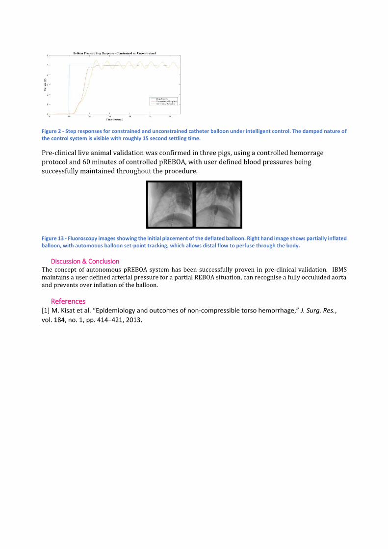
Figure 2 - Step responses for constrained and unconstrained catheter balloon under intelligent control. The damped nature of the control system is visible with roughly 15 second settling time.
Pre-clinical live animal validation was confirmed in three pigs, using a controlled hemorrage
protocol and 60 minutes of controlled pREBOA, with user defined blood pressures being
successfully maintained throughout the procedure.
Figure 13 - Fluoroscopy images showing the initial placement of the deflated balloon. Right hand image shows partially inflated balloon, with automoous balloon set-point tracking, which allows distal flow to perfuse through the body.
Discussion & Conclusion The concept of autonomous pREBOA system has been successfully proven in pre-clinical validation. IBMS maintains a user defined arterial pressure for a partial REBOA situation, can recognise a fully occuluded aorta and prevents over inflation of the balloon.
References [1] M. Kisat et al. “Epidemiology and outcomes of non-compressible torso hemorrhage,” J. Surg. Res.,
vol. 184, no. 1, pp. 414–421, 2013.

O33:
Touchless control of a prototype light system Oram Louise, Alcoriza Jonathan Brian, Palomar Rafael, Elle Ole Jakob - The Intervention Centre, Oslo
University Hospital, Oslo, Norway
1. Introduction
This abstract presents a prototype light system, currently in clinical use in an angiography operation room (OR). The OR is primarily used for minimally invasive procedures such as TEVAR (thoracic endovascular aortic repair). There was a need for an alternative lighting solution because of the likelihood of collisions with specialized equipment (e.g. a robotic CT arm) and the laminar airflow ceiling. An idea mentioned by Hartmann et al. [1] is: “to replace the arm-mounted light [with] remote-controlled lights embedded in the ceiling.”
This system comprises 7 controllable (pan, tilt, rotate) ceiling mounted lights. The prototype system is controlled in 2 manners: touchless with midair gestures detected by a Kinect (Microsoft depth camera motion sensor), or using a simple touchscreen interface. The hand tracking is activated by the Kinect through a horizontal wave gesture (with an LED tracking status light), and the tracking and lights are turned on/off using a foot pedal.
2. Methods
In order to find out how best to improve the
system and assess its usability (including
limitations and ideas for improvement), a user
survey was administered. The 15 participants
(surgeons, radiographers, and nurses) in the
survey currently perform or assist in operations
in the angiography OR, or have done so
recently.
Additionally, the position and usage of the lights
was logged. The logging data was compared to
the list of performed procedures, so as to
understand when and in what procedures the
lights are used.
3. Results
Generally, the survey showed the light system meets the basic clinical requirements. The users expressed that the main limitations of the system are not being able to place the lights at specific angles (that non-ceiling mounted lights can achieve) and that the focus of the lights needs to be adjustable. Survey participants with higher self-rated light system skill also rated themselves as more comfortable positioning the lights.
Out of 6 recorded FEVARs the Kinect was used to move the lights on average 2.33 times per procedure (range: 0-5), and the tablet was also used to move the lights on average 1.83 times per procedure (range: 0-5). The lights were turned on average 4 times (range: 2-6) times during each FEVAR. The lights are turned on for periods of a few minutes up to around 30 minutes. One example of the Kinect being used to position the lights, from the data logging, is seen in [fig 1].
Figure 1: Movement trace of the Kinect hand tracking
during a TEVAR procedure, with the depth image shown.
The color of the trace indicates where the movement is
over time (as it changes from turquoise to pink).
4. Discussion & Conclusion
The gesture recognition reliability needs to be improved (in a realistic setting with many other people / hands in view), and it would be useful to have more gestures to be able to map to more functionality. For instance, a gesture could be used to start and stop the hand tracking instead of using the pedal (which some users expressed can be awkward to access). Feedback is essential to be able to understand if the system is successfully tracking the hand or still searching for a hand. Due to varying
reliability of gesture detection and if more gestures are introduced, it is important for users to understand if an action has resulted in a successful state change.
This prototype allows the clinicians to control the ceiling mounted lights while remaining sterile. This is important because the surgeons are most often those repositioning the lights [2], because they are the ones that know their lighting needs. It is a successful first version of the system, and should be further developed to increase usability and better meet the clinical needs.
References
[1] Hartmann, F., & Schlaefer, A. (2013). Feasibility of touch-less control of operating room lights. International journal of computer assisted radiology and surgery, 8(2), 259-268.
{2} Knulst, A. J., Mooijweer, R., Jansen, F. W., Stassen, L. P., & Dankelman, J. (2011). Indicating shortcomings in surgical lighting systems. Minimally Invasive Therapy & Allied Technologies, 20(5), 267-275.
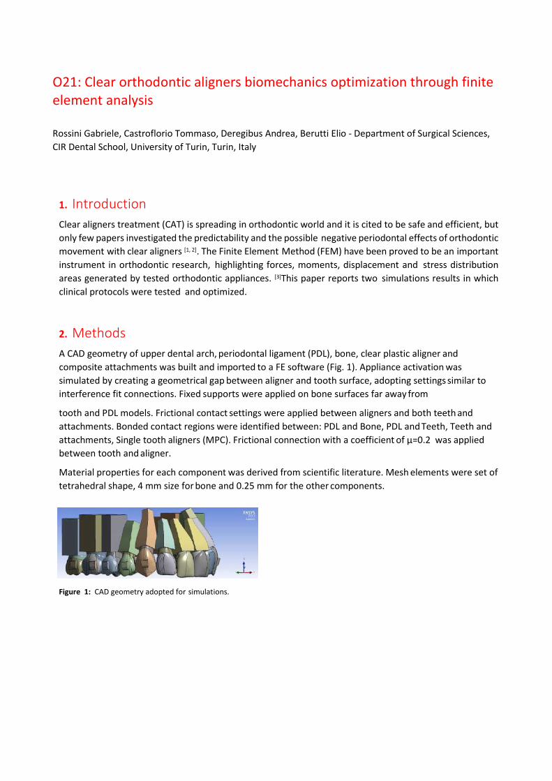
O21: Clear orthodontic aligners biomechanics optimization through finite element analysis Rossini Gabriele, Castroflorio Tommaso, Deregibus Andrea, Berutti Elio - Department of Surgical Sciences,
CIR Dental School, University of Turin, Turin, Italy
1. Introduction
Clear aligners treatment (CAT) is spreading in orthodontic world and it is cited to be safe and efficient, but
only few papers investigated the predictability and the possible negative periodontal effects of orthodontic
movement with clear aligners [1, 2]. The Finite Element Method (FEM) have been proved to be an important
instrument in orthodontic research, highlighting forces, moments, displacement and stress distribution
areas generated by tested orthodontic appliances. [3]This paper reports two simulations results in which
clinical protocols were tested and optimized.
2. Methods
A CAD geometry of upper dental arch, periodontal ligament (PDL), bone, clear plastic aligner and
composite attachments was built and imported to a FE software (Fig. 1). Appliance activation was
simulated by creating a geometrical gap between aligner and tooth surface, adopting settings similar to
interference fit connections. Fixed supports were applied on bone surfaces far away from
tooth and PDL models. Frictional contact settings were applied between aligners and both teeth and
attachments. Bonded contact regions were identified between: PDL and Bone, PDL and Teeth, Teeth and
attachments, Single tooth aligners (MPC). Frictional connection with a coefficient of μ=0.2 was applied
between tooth and aligner.
Material properties for each component was derived from scientific literature. Mesh elements were set of
tetrahedral shape, 4 mm size for bone and 0.25 mm for the other components.
Figure 1: CAD geometry adopted for simulations.

3. Results
From two simulated environments, incisor's root torque movement and first molars rotation optimized
biomechanics were obtained (Fig. 2).
Attachments from canines to molars, together with a buccal pressure area on incisors' crown and a crown-
rotation activation pattern of 1° released
0.23 N on root PDL with a 0.08 mm root displacement.
Rotation with rectangular attachment only on upper first molars produced a moment of 20 N*mm with a
6 N pressure on mesial surface of the attachment. No collateral forces on molars were reported with this
setting.
Figure 2: a. Efficient root control with attachments and single pressure area. b. Displacement patterns for the optimized
rotational configuration.
4. Discussion & Conclusion
FEM is a reliable technique to study and improve CAT biomechanics, minimizing undesired side effects.
References [1] G. Rossini, S. Parrini, T. Castroflorio, A. Deregibus, C. L. Debernardi. "Efficacy of clear aligners in controlling orthodontic tooth
movement: a systematic review," Angle Orthod. vol 85(5), p. 881-9, 2015.
[2] G. Rossini, S. Parrini, T. Castroflorio, A. Deregibus, C. L. Debernardi. "Periodontal health during clear aligners treatment: a systematic
review." Eur J Orthod. vol. 37(5), p. 539-43, 2015.
[3] J. R. Singh, P. Kambalyal, M. Jain, P. Khandelwal. "Revolution in Orthodontics: Finite element analysis." Journal of International
Society of Preventive & Community Dentistry. vol. 6(2), p. 110–114, 2016.
a b

SESSION 26 – NEW TOOLS FOR LAPAROSCOPY & INTERVENTIONAL RADIOLOGY

O139: A Randomized Controlled Trial of the Fundamentals of Robotic Surgery: Validation of a proficiency-based progression robotic skills course in a multi-specialty, multi-institutional trial. Satava Richard - University of Washington Medical Center
Objective: To validate the Fundamentals of Robotic Surgery (FRS) skills course in a randomized control trial
using multiple simulation platforms.
Methods and Procedures: A randomized clinical trial with a control group and 3 experimental groups:
physical “dome” using the robot, virtual reality hybrid simulation using the computer ‘backpack’ on the
surgical console, and stand-alone virtual reality simulator. Training was to proficiency and were set by 80
experts. The multi-institutional trial was conducted 12 international simulation centers and included the
multiple specialties that perform robotic surgery.
Results: Face and content validity was verified by 80 experts; concurrent validity was demonstrated by
experimental groups performing in less time and with less errors than the control group. The construct
validity was demonstrated because the mean time and mean number of errors to perform the various tasks
by experts were statistically less the novices. The inter-rater reliability for task specific checklists was >
0.80, however the Global Evaluative Assessment of Robotic Skills (GEARS) inter-rater reliability was <0.80
Conclusion: The Fundamentals of Robotic Surgery is a validated course to teach novices the simplest robotic
surgery skills across the many specialties that are currently performing robotic surgery. All the four
validities were confirmed on a physical dome model, or any of the computer/virtual reality simulators. The
FRS is a robust and validated course (curriculum) to train the basic skills in robotic surgery.
O136:

O94: Improving fundamental laparoscopic skills by pre-course tactile exploration Hardon Sem F., Postema Roelf R., Horeman Tim, Smeets Jeroen B.J., Bonjer H. Jaap - VU University Medical
Center, Departmetn of Surgery, Amsterdam, Netherlands
Introduction
Minimally invasive techniques in operation rooms have reduced the tactile and visual feedback
compared to open surgery. There is increasing evidence in the neuropsychological literature that
visual and haptic information combine or converge to form a mental representation of an object.
Because of the combination of these inputs, this mental representation is better, more refined and
robust than the image formed by only one of these sensory inputs. We investigated whether haptic
exploration of a 3D object before executing a laparoscopic action improves performance.
Methods&Materials
A randomized prospective cohort study was conducted using a case-crossover design. Third year medical
students (N=18) with no prior laparoscopic experience were included.
Based on well-known FLS tasks, three more complex task platforms were designed and produced for this
course using a 3D-printing system. In a box trainer, the participants executed a peg transfer task as a baseline
test, followed by three repetitions of each 3D task.
The box trainer was equipped with state-of-the-art Forcesense system (Medishield, Delft, The Netherlands)
for adequate assessment of skill based on force, motion and time parameters. P Penalty points for errors (e.g.
dropping a peg, rope or ring) were scored through analysis and evaluation of captured videos of each
performance.
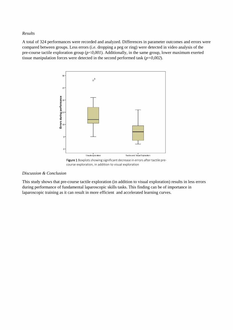
Results
A total of 324 performances were recorded and analyzed. Differences in parameter outcomes and errors were
compared between groups. Less errors (i.e. dropping a peg or ring) were detected in video analysis of the
pre-course tactile exploration group (p<0,001). Additionally, in the same group, lower maximum exerted
tissue manipulation forces were detected in the second performed task (p=0,002).
Discussion & Conclusion
This study shows that pre-course tactile exploration (in addition to visual exploration) results in less errors
during performance of fundamental laparoscopic skills tasks. This finding can be of importance in
laparoscopic training as it can result in more efficient and accelerated learning curves.
Figure 1 Boxplots showing significant decrease in errors after tactile pre-course exploration, in addition to visual exploration
O81: Sentinel node navigation surgery and ICG fluorescence-guided surgery for gastric cancer Inaki Noriyuki, Tsuji Toshikatsu, Minami Teruya - Ishikawa Prefectural Central Hospital, Gastroenterological
Surgery, Kanazawa, Japan
Introduction: We have performed sentinel node navigation surgery (SNNS) for treatment of early
gastric cancer through a prospective multicenter trial in Japan using dual tracer endoscopic injection
technique. On the other hand, ICG fluorescence-guided surgery has recently used for various
digestive surgery in which blood supply or lymphatic basin can be confirmed. We applied the ICG
fluorescence-guided technique to complete radical lymphadenectomy for gastric cancer surgery.
Both concepts of ICG-using procedure are different each other. In this paper, we demonstrate each
procedure and evaluate the feasibility and validity.
Methods: A dual-tracer method using radioactive tin-colloids and ICG is considered reliable
method for the detection in patients of early gastric cancer. For early gastric cancer, the
establishment of individualized, minimally invasive treatments based on SN concept can retain the
patients’ quality of life. The laparoscopic and endoscopic cooperative surgery (LECS) has also
realized minimally partial resection for gastric tumor, and the closed-LECS, which doesn't require
direct opening of the stomach wall and can avoid tumor spreading. Indication of closed-LECS is
early gastric cancer, whose tumor size is within 4 cm in diameter. The stomach are laparoscopically
conducted and a local injection of 0.5ml ICG 0.5% (5mg/ml) is endoscopically performed. SN is
detected and the lymphatic basin (LB) including SN is dissected. Following removal of the whole
SN, the LB is checked by intraoperative pathology diagnosis. The closed-LECS is started when all
examinations confirm negative SN (Video 1). ICG fluorescence-guided surgery is routinely used
laparoscopic gastric cancer surgery. Single tracer of ICG is used for radical
lymphadenectomy.(Video 2).
Results: Two cases for early gastric cancer underwent closed-LECS using double tracer. Eighteen
cases for gastric cancer underwent ICG fluorescence-guided gastrectomy with radical
lymphadenectomy. In both procedures, there was no intra- and post-operative complication.
Conclusion: Sentinel node navigation surgery and ICG fluorescence-guided surgery for gastric
cancer is expected to be established as the individualized effective surgical procedure for gastric
cancer treatment.
O85: Indocyanine green guided sentinel lymph node identification – a prospective study evaluating the distribution of lymph nodes respectively lymph node metastasis in rectal cancer patients
Shah S., Salber J., Stricker I., Tannapfel A., Viebahn R. - Department of General-, Abdominal- and Transplant
Surgery, Universityhospital Knappschaftskrankenhaus, Bochum, Germany
1. Introduction
Lymphatic metastasis in rectal cancer is a prognostic parameter and has a strong influence on survival of the patient [1]. It is therefore important to ensure complete lymphadenectomy during the operation in rectal cancer patients. To enable the surgeon to identify the lymph nodes (LN) intraoperatively, we conducted a prospective study. We used Indocyanine green (ICG) to visualize the lymph nodes (sentinel lymph node (SLN)). The aims of our study were to localize the SLN, respectively the metastatic lymph nodes in rectal cancer patients.
2. Methods
We conducted a prospective study on rectal carcinoma patients, regardless of the prior treatment [2]. The statistical calculation determined a group size of thirty patients to identify at least one sentinel lymph node. Two ml of ICG was injected endoscopically in the submucosa near the tumour lesion in the rectum. After anterior or abdominoperineal resection of the rectum including total mesorectal excision, the specimen was illuminated with a near infrared light (NIRL) camera unit to visualize the lymph nodes. They were identified and marked. This was followed by illuminating the operation site with the NIRL camera unit, to detect any missed lymphatic tissue. The examination of the specimen in the pathology department included search for lymph node metastasis (LNM) by immunohistochemistry. Depending on the presence of metastatic disease all lymph nodes were classified as node positive or node negative. LNM was mapped, according to the tumour site, to above or below the tumour.
3. Results
Thirty patients with rectal cancer were included in the study. Eleven patients had a rectal cancer in the lower third, nineteen in the middle rectum. Fourteen patients received prior to surgery chemoradiotherapy (CRT). In fifteen patients the tumour was classified T3 (Table 1). We could identify at least one SLN in all operated
patients (n=30). In total a 122 SLN were identified in thirty patients. In three patients the SLN were false negative. Two of these patients received chemoradiotherapy (CRT) before operation. The lymph node yield was in minimum 16 and in average 33.5 lymph nodes. In total we excised 1004 lymph nodes. Considering preoperative treatment, the CRT group (n=14) of patients had a mean lymph node yield of 27.7 LN. In the NoCRT group (n=16) the LN yield was 38.5 (in mean). Lymph node metastasis (LNM) was present in 11 patients. In only one patient the LNM was exclusively found above the tumour. And in three patients the LNM was found below the tumour. Seven patients had above and below the tumour LNM. Interestingly the patients with false negative SLN had only above or below the tumour LNM.
4. Discussion and Conclusion
ICG was successful in detecting SLN in rectal cancer patients. The sensitivity to detect SLN was high, especially in the group of patients without CRT. Our findings suggest that CRT can impair SLN detection, although in this study the number of retrieved lymph nodes of patients with neoadjuvant CRT met the recommendations. The location of lymph node metastasis was found to be not only above, but also below the tumour site. For comprehensive lymphadenectomy the ICG application can be helpful. Through intraoperative visualization of the sentinel lymph nodes the surgeon is enabled to visualize lymph nodes respectively lymphatic vessels and therefore can adjust resection margins to improve the quality. The application of ICG was simple and feasible, without affecting the operating time. However, further studies are required to adopt this concept for routine use in the operation of rectal cancer patients.

Picture 1: SLN visualisation in situ mesorectum
Picture 2: SLN visualisation ex situ specimen
References
[1] H. Kobayashi et al., “Lymph node ratio is a powerful prognostic index in patients with stage III distal rectal cancer: a Japanese multicenter study,” (eng), International journal of colorectal disease, vol. 26, no. 7, pp. 891–896, 2011.
[2] M. E. Kreis et al., “Use of Preoperative Magnetic Resonance Imaging to Select Patients with Rectal Cancer for Neoadjuvant Chemoradiation--Interim Analysis of the German OCUM Trial (NCT01325649),” (eng), Journal of gastrointestinal surgery : official journal of the Society for Surgery of the Alimentary Tract, vol. 20, no. 1, 25-32; discussion 32-3, 2016.

O59: Instruments and situation awareness: main risk-factors for surgery Wauben Linda, Dekker-van Doorn Connie, Lange Johan, Klein Jan - Delft University of Technology,
Department of Biomechanical Engineering, Delft, Netherlands
Introduction
Although in (Dutch) hospitals, rates of healthcare related avoidable harm decreased during the last
decennium [1], still errors occur in the operating room (OR) due to the complex non-standardized OR
environment, inadequate situation awareness, bad teamwork and communication and the large variety
of (non-ergonomic) instruments and equipment [2, 3]. To prevent errors from occurring, we designed
and implemented a Time Out Procedure plus debriefing (TOPplus)[4]. The aim of this study was to
establish risk-factors for avoidable patient’s harm and adapt TOPplus accordingly to prevent future
errors.
Methods
OR team members were asked to self-report ‘details’ (remarks and incidents) about the surgery before
introduction of TOPplus (T0 baseline measurement) and six months after introduction (T1). Hospitals
were not pre-selected, but joined the TOPplus study voluntarily over time. Details were registered via
paper registration forms. Data were digitalised and were categorized into remarks and incidents
(defined as [5]). Both remarks and incidents where subdivided in subcategories relating to: anaesthesia,
surgery, communication, instruments and equipment, leadership, situation awareness, teamwork,
perfusion, and TOPplus.
Results
In total 8 out of 15 hospitals completed T0 and T1: 3 academic, 2 teaching and 3 general hospitals.
Figure 1: Number of self-reported remarks and incidents.
The number of registration forms received included: T0, n=2,724; T1, n=22,764 (see Figure 1). In T0,
18.5% of registrations included an incident and in T1, 4.6% of registrations included an incident. Figure 2
shows that most self-reported incidents in both T0 and T1 were related to situation awareness (T0: 44.4%
- T1: 26.8%) and to the instruments and equipment (T0: 29.6% - T1: 41.8%). The main reasons for the
instrument-incidents were linked to incomplete and defect instruments or equipment.

Figure 2: Situation awareness and instruments; main self-reported incidents.
Discussion & Conclusion
Defect and incomplete instruments and equipment and inadequate situation awareness are the main
causes of incidents in the OR and therefore the main risk-factors for patient harm. The number of
incidents will probably be higher due to (self) underreporting. Although incidents relating to a lack of
situation awareness can be addressed and prevented by TOPplus, the ‘defect’ instrument and
equipment need redesign to further improve patient safety.
References
[1] M. Langelaan et al., "Monitor Zorggerelateerde Schade 2011/2012: dossieronderzoek in Nederlandse ziekenhuizen," Utrecht/Amsterdam2013.
[2] S. R. C. Driessen, E. M. Sandberg, S. P. Rodrigues, E. W. van Zwet, and F. W. Jansen, "Identification of risk factors in minimally invasive surgery: a prospective multicenter study," Surg Endosc, vol. 31, no. 6, pp. 2467-2473, Jun 2017.
[3] A. C. Guedon et al., "Where are my instruments? Hazards in delivery of surgical instruments," Surg Endosc, vol. 30, no. 7, pp. 2728-35, Jul 2016.
[4] L. S. G. L. Wauben, C. M. Dekker-van Doorn, J. Klein, J. F. Lange, and R. H. Goossens, "Participatory design: implementation of time out and debriefing in the operating theatre " (in Eng), Journal of Design Research, vol. 9, no. 3, pp. 220-240, 2011.
[5] W. H. Organisation, "International Classification for Patient Safety," 2007, vol. Version 1.0 for Use in Field Testing 2007 - 2008.

O147:
Morphological and hemodynamic characterization of post
endovascular AAA repair: comparison between two different
commercial devices
Paola Tasso1, Anastasios Raptis2, Michalis Xenos2,3, Maurizio Lodi Rizzini1,
Diego Gallo1, Miltiadis Matsagkas2,4, Umberto Morbiducci1
1)Department of Mechanical and Aerospace Engineer Politecnico di Torino, Italy; 2) Institute of Vascular Diseases
Laboratory for Vascular Simulations Ioannina, Greece; 3) Dept. of Mathematics University of Ioannina, Greece; 4) Dept. of
Vascular Surgery, Faculty of Medicine, University of Thessaly, Greece
1. Introduction
Abdominal aortic aneurysm (AAA) is a vascular disease characterized by a localized expansion of the
abdominal aorta. In the last years the minimally invasive endovascular aortic repair (EVAR) approach for
AAA treatment has been widely applied as alternative to classical open-chest surgery. EVAR results in (1)
redirection of blood through the deployed endograft, and in (2) iliac bifurcation reshaping, thus altering
local hemodynamics [1]. In this study the impact that two different commercial endovascular graft (EG)
devices, have on vascular geometry and hemodynamics is investigated. In detail, geometric features are
pre- and post-operatively evaluated. Computational Fluid Dynamics (CFD) is applied to explore if the
vascular territory post interventional reshaping supports the re-establishment of physiological
hemodynamics.
2. Methods
Subjects suffering from AAA were treated with two EG systems, N=5 with the Endurant® (Medtronic, CA,
USA), N=5 with the Excluder® (Gore Medical, AZ, USA) (fig. 1). Also N=5 healthy subjects underwent CT
angiography. CT scans were obtained before and one month after EVAR [1]. 3D models were
reconstructed from CT images[1].
Figure 1: (a) Endurant device, (b) Excluder device.
Centerline-based geometry analysis was carried out on reconstructed models, in terms of curvature,
torsion, normalized cross-sectional area and cross-sectional area variation rate [2]. The reconstructed 3D
geometries were meshed with tetrahedral elements [1]. The governing equations of blood flow were
solved by using the finite volume method. Boundary conditions at inflow and outflow sections were
based on flow and pressure measurements [1]. Near-wall hemodynamics and intravascular flow
structures were described in terms of time-average wall shear stress (TAWSS), and in terms of helical
flow, blood recirculation volume (RV), respectively.

3. Results
Geometric features are similar between treated groups but present higher and more scattered values
than healthy subjects, as an average. In terms of hemodynamics, subjects treated with the Excluder
present lower percentages of surface area exposed to low WSS and lower RV (fig. 2).
Figure 2: Explanary RV and TAWSS maps: (a) Endurant; (b) Excluder. In TAWSS maps low values are colored with red.
Overall, the Excluder group presents the highest TAWSS and helicity intensity values.
4. Discussion
From the geometrical point of view there are no-marked differences between the two devices. From the
hemodynamic point of Excluder models present a higher mean value of TAWSS and helicity that could confirm that
the helical flow plays a beneficial role in suppressing low velocity/stagnation regions that could lead to thrombus
formation.
References 1. A. Raptis, M. Xenos, E. Georgakarakos, G. Kouvelos, A. Giannoukas, N. Labropoulos et al., ‘’Comparison of physioological and post-
endovascular aneurysm repair infrarenal blood flow’’, Computer Methods in Biomechanics and Biomedical Engineering, vol.20, p. 242-249,
2016.
2. D. Gallo, A. Lefieux, S. Morganti, A. Veneziani, A. Reali, F. Auricchio, M. Conti, U. Morbiducci,’’A patient-specific follow up study of the
impact of thoracic endovascular repair (TEVAR) on aortic anatomy and on post-operative hemodynamics’’, Computer and Fluids, vol.141,
p.54-61, 2016.

O105: Assessing non-technical skills in the delivery room through multisensory-based simulation Bracco Fabrizio, Masini Michele, Passarelli Marcello, Glowinski Donald, Domaneschi Filippo, Chiorri Carlo
- University of Genova, Department of Education, Genova, Italy
1. Introduction
High-fidelity simulation (HFS) has proven to be an effective methodology for medical training and for reducing adverse events in the delivery room [1]. The debriefing is the crucial learning moment of the whole method, since it boosts proper metacognition on the actions taken during the simulation [2]. However, the focus is often directed to technical aspects (e.g., drug administration, specific procedures, etc.) while the so-called non-technical skills (NTS, i.e., situation awareness, decision making, communication, leadership, teamwork, workload management) are overlooked or only superficially tackled [3]. Unfortunately, literature on risk management demonstrated that the main contribution to adverse events is due to non-technical issues. HFS has the potential to be one of the most effective methods to train NTS, but it should be based on a sound method to observe and assess them [4].
2. Methods
We are currently developing a method to assess NTS during delivery room emergency simulation, which is based on the integration of multiple sources of information. We developed a comprehensive set of checklists of NTS behavioral markers for each professional and for team performance. These tools will be associated to other behavioral cues representative of NTS, which will be recorded by means of both remote sensors (e.g. 360-degree cameras) and wearable sensors. The data are based on: (i) physiological recordings (e.g., heartbeat, skin conductance, etc.); (ii) body movements (e.g., posture, kinesics, proxemics, gaze direction, etc.); (iii) linguistic and paralinguistic features (e.g., presuppositions, voice pitch, hesitations, emotional contents, etc.); (iv) facial expressions (e.g., muscular patterns related to emotion expressions).
3. Results
The NTS behavioral markers checklists have already been validated and participants to simulation sessions positively rated them for usefulness and usability [5]. We are currently
performing pilot tests in simulated scenarios with the addition of wearable and remote sensors and the participants’ feedback is positive.
Figure 1: Top-left: the team during the simulation. Top-right: physiological parameters recorded during the simulation. Bottom: a remote-capture of bodily postures.
4. Discussion & Conclusion
Putting together all these cues, we will be able to enrich the debriefing session and the discussion about NTS, since abstract constructs such as leadership or communication will be operationalized in concrete data and observable behavioral patterns. Learning will be improved because it will be based on a multifaceted analysis of aspects that, until now, have only been generically defined as "the contrary of technical skills".
References
[1.] Dadiz R, Weinschreider J, Schriefer J, et al. “Interdisciplinary simulation-based training to improve delivery room communication”, Simul Healthc., vol. 8(5), p. 279-91, 2013.
[2.] Rudolph JW, Simon R, Dufresne RL, Raemer DB. “There’s no such thing as ‘nonjudgmental’ debriefing: a theory and method for debriefing with good judgment”, Simul Healthc; vol. 1(1), p. 49-55, 2006.
[3.] Gordon M, Darbyshire D, Baker P. “Non-technical skills training to enhance patient safety: A systematic review”, Med Educ, vol. 46(11), p. 1042-54, 2012.
[4.] Jackson KS, Hayes K, Hinshaw K. “The relevance of non-technical skills in obstetrics and gynaecology”, Obstet Gynaecol. Vol. 15(4), p. 269-74, 2013.
[5.] Bracco, F. et al. “Adaptation of Non-Technical Skills behavioural markers for delivery room simulation”, BMC Pregnancy and Childbirth, vol. 17, p. 89, 2017.

O128:

SESSION 18 – INNOVATION IN COLORECTAL ENDOSCOPY & SURGERY
0111: LESS IS MORE: NUOVO APPROCCIO
ALL’ADDOME APERTO
AUTORI E ISTITUZIONE:
L. Petruzzelli, F. Lora, M. Benedetto, E. Franchi,
L. Rapetti, Chirurgia Generale e d'Urgenza 3,
Città della Salute e della Scienza, Torino
INTRODUZIONE:
L’UTILIZZO DELLA TECNICA DELL’ADDOME
APERTO NELLA GESTIONE DEL PAZIENTE
CRITICO PRESENTA DIFFUSIONE SEMPRE
MAGGIORE. LE PRINCIPALI INDICAZIONI SONO:
SINDROME COMPARTIMENTALE, GESTIONE
DELLO SHOCK EMORRAGICO DA CAUSA
INTRAADDOMINALE NON IMMEDIATAMENTE
CONTROLLABILE, TRATTAMENTO DELLE SEPSI
DA PERITONITE SEVERA. MENTRE SONO
SOSTANZIALMENTE ABBANDONATI L’UTILIZZO
DELLA “BOGOTA-BAG” E DELLE PROTESI
SINTETICHE “A PONTE”, L’UTILIZZO DEI SISTEMI
A PRESSIONE NEGATIVA (BARKER’S VACUUM
PACK TECHNIQUE , ABTHERA OA NPT )
TROVA SEMPRE MAGGIORE CONSENSO. ESSO È,
TUTTAVIA, GRAVATO DA IMPORTANTI
CONTROINDICAZIONI, IN PARTICOLARE
L’IMPOSSIBILITÀ DI UTILIZZARLO IN CASO DI
SANGUINAMENTO ATTIVO E L’ELEVATO
RISCHIO DI FISTOLA ENTEROATMOSFERICA; LA
STRUMENTAZIONE NECESSARIA NON È INOLTRE
SEMPRE DISPONIBILE IN URGENZA.
METODI:
Abbiamo brevettato un divaricatore/protettore
di parete anulare modificato al fine di renderlo
adatto alla creazione di una laparostomia
temporanea sicura e agevolmente frubile. Esso
è configurato come un normale retrattore di
parete a doppio anello ma include un cappuccio
trasparente di materiale polimerico ed è dotato
di apposite asole che consentono l’ancoraggio
alla cute mediante sutura (FIG.)
RISULTATI:
IL NOSTRO DISPOSITIVO SI PUÒ UTILIZZARE NEL
PAZIENTE EMORRAGICO SENZA CHE VENGA
APPLICATA PRESSIONE NEGATIVA,
NELL'ADDOME SETTICO IN ASSENZA DI ALTRI
PRESIDI, IN PRESENZA DI ESTREMA FRAGILITÀ
TISSUTALE CHE CONTROINDICHI L'ASPIRAZIONE
E IN PREVISIONE DI REVISIONI IN TEMPI BREVI.
LA POSSIBILITÀ DI POSIZIONARE DRENAGGI
CONSENTE L'EVACUAZIONE DEI FLUIDI
QUANDO È NECESSARIO.
DISCUSSIONE:
Il re-accesso al cavo addominale è immediato e
il cappuccio trasparente consente un controllo
delle condizioni dello stesso in tempo reale; non
viene inoltre a crearsi alcun danno alla parete
che rimane anzi continuativamente protetta. Il
dispositivo è poco costoso e facilmente
utilizzabile anche da centri che non abbiano a
disposizione attrezzature per sistemi a
pressione negativa.

O14:
RED – Robot for Endoscopic Dissection: preliminary study M. Brancadoro1*, C. Quaglia1, H. Abidi1, M. A. Bonino2, A. Menciassi1, A. Arezzo2
1) The BioRobotics Institute, Scuola Superiore Sant'Anna, Pisa, Italy (*[email protected]). 2) Department of Surgical Sciences, University of Torino, Turin, Italy.
Introduction Endoscopic Submucosal Dissection (ESD) is a flexible endoscopic technique that allows for an en bloc removal of lesions of the gastrointestinal (GI) tract. However, it is associated with high risks of complications and the procedures are long, often requiring around 95min for lesions of even only 3-4cm in diameter. The risk of haemorrhage ranges from 3.5% to 15.5% and the possibility of intestinal perforation is also high (up to 18%) [1]. A flexible robotic endoscope may offer a solution to overcome these limitations, by improving the degrees of freedom (DoF) and operational efficiency [2].
Methods RED is a novel miniature robotic platform for ESD, designed to effectively remove tumours of the entire GI tract. It has been designed to be coupled to the tip of traditional flexible endoscopes of about 14.5 mm in diameter. The robot exploits the flexibility of the endoscope for navigation through the intestine and integrates 2-actived robotic arms (i.e., cautery and gripper) extending the degrees of freedom (DoF), and thus enhancing the efficiency during complex tasks such as manipulation and surgical tissue dissection. The RED system is supported by an external platform, a personal computer and two Geomagic Touch phantoms (3D System, Inc) (Fig. 1). During the operation, the surgeon (referred to as surgeon A, Fig.1) stands close to the patient to manoeuvre the endoscope for exploring the GI tract and reaching the target area. Another surgeon (referred to as surgeon B, Fig.1) operates the RED arms through the computer console, carrying out the surgical procedure.
Fig. 1: An overview of the surgery scenario
Results RED consists of a cap body with two robotic arms (Fig.2). The right arm is equipped with a surgical gripper to grasp and lift the tissue; it is featured by 3 DoFs (i.e., slide, pitch and open/close of the gripper). The left arm is a mono-polar cautery with 3 DoFs (i.e., slide, roll and pitch) employed to cut the lesion. While navigating through the intestine, the RED arms are enclosed in the cap body; when the target area is reached, the arms are deployed and they can start to operate. The external shape of the cap body is approx 27mm in diameter (including the endoscope tip) and has a total length of 50mm; these sizes have been defined considering anatomical constraints (i.e., dimension and shape of the intestine).

Fig. 2: RED design.
Discussion & Conclusion RED has been designed thanks to a deep collaboration between engineers and medical doctors. It will help the surgeon to accurately excise target tissue through precise dissection. The gripper arm is expected to generate around 3N of lifting force and 10-14N of gripping force. Around 1.5N are availble at the cauthery tip. There are various advantages of using this robotic device. A traditional endoscopic room is enough to carry out a complete surgical operation and the patient does not need to undergo surgery under general anaesthesia. The hospitalization times, and hence the general hospital costs, could be significantly reduced. For the future, we will validate the functionality of the RED, by conducting in-vivo experiments to prove the clinical advantages of the proposed system.
References
[1] Arezzo, Alberto, et al. "Systematic review and meta-analysis of endoscopic submucosal dissection versus transanal endoscopic microsurgery for large noninvasive rectal lesions." Surgical endoscopy 28.2 (2014): 427-438.
[2] Yeung, Baldwin Po Man, and Philip Wai Yan Chiu. "Application of robotics in gastrointestinal endoscopy: A review." World journal of gastroenterology 22.5 (2016): 1811.

O112: ENDOSCOPIC FULL THICKNESS RESECTION: PRELIMINARY INDICATIONS AND RESULTS
Coppola Franco - ASLTO4, Gastroenterology, Chivasso, Italy
Introduction
Endoscopic full thickness resection ﴾ EFTR﴿ is a new procedure to remove difficult lesions, such as
epithelial or sub-epithelial tumours unsuitable for conventional resection even with endoscopic mucosal
resection (EMR) nor with endoscopic sub-mucosal dissection (ESD). Recently, a new over the scope clip
(OTSC) device named full- thickness resection device (FTRD) has been used to perform a full thickness
resection in one-step procedure. It consists of a modified OVESCO clip with a longer cap and an
integrated snare.
Aims & Methods
Our aim was to evaluate the efficacy and the safety of OTSC assisted EFTR for recurrent adenomas and
for evaluation of the muscular layer in neuromuscular suspected diseases. Patients who underwent
OTSC assisted EFTR between December 2015 and December 2016 at San Giovanni Bosco Hospital,
Torino, were included. The procedure consisted of a) lesion or wall suction into the cap of the OTSC, b)
clip deployment and c) en-bloc resection of the lesion above the clip using an electrosurgical snare.
Procedure was performed under monitored anaesthesia assistance. Demographic, clinical data,
histopathologic diagnosis and adverse events were registered.
Results
Six patients (1 male, median age of 62 years) were included. Four patients underwent EFTR for recurrent
adenomas, 2 in the rectum (10 and 12 mm) and 2 in the cecum (9 and 24 mm). In case of proximal
adenomas a demo OTSC was used before the procedure to ascertain the possibility to reach the lesion.
Moreover 2 patients with chronic severe constipation underwent EFTR to obtain a full thickness sample
of the rectum for muscular layer examination. Histopathology confirmed a full-thickness R0 resection
(negative margins) in all cases with adenomatous lesions and diagnosis of intestinal neuronal dysplasia
in the 2 cases of intestinal chronic constipation. The mean procedure time was 60 minutes (range: 40-75
min). No adverse events occurred. No immediate or delayed perforations were observed, all patients
were discharged after 24 hours.
Conclusion
These preliminary data show that OTSC assisted EFTR was effective and safe to remove difficult and
recurrent adenomas and to obtain full thickness rectum sample for the diagnosis of neuronal dysplasia.
The technique was quite easy in the rectum but it may be challenging in more proximal tracts and a
demo OTSC device was used to confirm the possibility to reach lesions located at the cecum.
References
- Probst A, Schaller T, Messmann H. Gastrointestinal stromal tumor of the colon-endoscopic treatment by
full- thickness resection. Endoscopy 2015; 47: E460–E461
- Schmidt A, Bauerfeind P, Gubler C et al. Endoscopic full-thickness resection in the colorectum with a novel
over-the-scope device: first experience. Endoscopy 2015; 47: 719– 725
- Friedmacher F, Puri P. Classification and diagnostic criteria of variants of Hirschsprung’s disease. Pediatr
Surg
O90: QUALITY OF MESORECTAL EXCISION AFTER OPEN, LAPAROSCOPIC, ROBOTIC AND TRANSANAL TME
Simone Maria Tierno, Gabriele Campana, Gianfrancesco Intini, Carlo Eugenio Vitelli, Marco Maria Lirici;
San Giovanni Hospital, Department of Surgery,
1. Introduction
Colorectal cancer is the third most common cancer in the World. Neoadjuvant chemoradiotherapy
followed by radical surgery is considered the standard treatment for locally advanced middle and low
rectal cancer. Removing all of the mesorectum containing the lymph nodes and tumour is paramount
for a good outcome and minimal recurrence within the pelvis. Involvement of the circumferential
resection margin (CRM) and the quality of total mesorectal excision (TME) are related to local
recurrence and long-term survival 1-3.
The aim of the present prospective study is to evaluate the safety and efficacy of laparoscopic resection
vs open vs robotic versus transanal TME. A successful resection was defined when meeting all the
following criteria: complete total resection margin, a clear CRM (>1mm) and a clear distal resection
margin (>1mm).
2. Methods
Specimen from a series of 101 consecutive patients undergoing TME for histological proven rectal
cancer at the Dpt of Oncologic Surgery of San Giovanni Hospital, Rome from July 2015 to July 2017, were
collected and pathology exam data recorded prospectically. Evidence of distant metastases was not an
exclusion criterion. Patients were divided according to surgical technique in four groups: open (OP),
laparoscopic (VLS), robotic (ROB) and TaTME (Ta). Patients who were converted from mini-invasive to
open procedure were considered in a different group (VLSconv). Localization of the tumour was
categorized as: upper rectum (10-15 cm from the anal verge), middle rectum (5 to 10 cm from the anal
verge), low rectum (<5 cm from the anl verge). Demographic data and histopathologic characteristics of
the tumour such as lesion size, distance from the anal verge, regression grade after neoadjuvant
reatment according to Mandard scale and number of lymph nodes retrieved were analysed. CRM was
defined involved when tumor cells were present within 1 mm from the lateral surface of the
mesorectum. Completeness of mesorectal excision was defined according to Quirke classification as
complete, nearly complete or incomplete.
3. Results
Twenty-eight (27.7%) patients underwent open rectal resection, 35 (34.7%) laparoscopic rectal
resection, 18 (17.8%) robotic rectal resection and 13 (12.9%) were treated by trans-anal TME. Seven
patients (6.9%) were converted from mini-invasive to open rectal resection. 39 patients (38.6%) with
cancer in the upper part of the rectum underwent partial mesorectal excision (PME); 37 patients (36.6%)
with cancer in the middle rectum and 39 patients (38.6%) with cancer in the lower rectum underwent
total mesorectal excision (61.4%). There were no statistically significant differences between group with
respect to sex, age, body mass index, ASA grading, tumour stage and tumour location. CRM resulted
positive in 5 patients (4.9%), 2 in the VSL group, 1 in the ROB group and 1 in the VSLconv. Overall,
mesorectal excision resulted complete in 68 patients (67.3%), nearly complete in 13 patients (12.9%)
and incomplete in 20 patients (19.8%). There were no statistically significant differences between
groups (p=0.3). In the sub-group analysis, considering patients with middle and low rectal cancer who
underwent TME, there were no positive margins in the OP g and TaTME g. versus 1 in the VSL g, 1 in the
ROB g. and 2 in the VSLconv (p=0.001). According to Quirke classification, complete TME was found in
78% patients in OP g., 66.7% in VSL g., 84.6% in TaTME g. and 50% in ROB group. Nearly complete TME
was found in 7.1% in the OP g., 5.6% in the VSL g., 16.4% in the TaTME g. and 21.4% in the ROB group.
Incomplete TME was found in 14.3% in the OP g, 27.8% in the VSL g. and 28.6% in the ROB g. None of
the patients who underwent TaTME resulted with incomplete TME (p=0.02). Multivariate regression
analysis showed that surgical technique was associated with the completeness of mesorectal excision
(p=0.02). No differences were found in term of lymph-nodes retrieval (p=0.8) and distal resection
margin (p=0.2).
4. Discussion and Conclusions
This prospective single center study investigates hysto-pathologic assessment of the resected
protectomy specimen focusing on clear distal and radial margins and completeness of total mesorectal
excision, the combination of which defines optimal surgery, and has been shown in other trials to be
associated with better long term oncologic outcomes4. We demonstrates that TaTME is not only a
feasible and safe technique but improves oncological resection principles in middle and low rectal
cancer resulting in better mesorectal excision and circumferential resection margin. Further prospective
comparative study are needed to clarify which is the gold standard technique for middle and low rectal
cancer .
References
[1] Quirke P, Steele R, Monson J et al. MRC CR07/NCIC-CTG C016 Tiral Investigators; NCRI Colorectal
Cancer Study Group. “Effect of the plane of surgery achieved on local recurrence in patientis with
operable rectal cancer: a prospective study using data from the MRC CR07/NCIC-CTG C016 randomised
clinical trial. Lancet 2009; 373 (9666): 821-828
[2] Parfitt JR, Driman DK. “The Total Mesorectal Excision specimen for rectal cancer: a review of its
pathological assessment”. J Clin Pathol. 2007; 60: 849-855
[3] Nagtegaal ID, Quirke P. “What is the role for the circumferential margin in the modern treatment of
rectal cancer?” J clin Oncol 2008;26: 303-312
[4] Nagtegaal ID, van de Velde CJ, van der Worp E et al. “Cooperative clinical investigators of the Dutch
Colorectal Cancer Group. Macroscopic evaluation of rectal cancer resection specimen”. J Cllin Oncol.
2002; 20:1729-1734
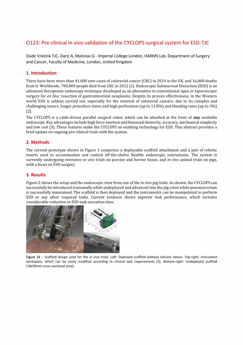
O123: Pre-clinical in vivo validation of the CYCLOPS surgical system for ESD-TJC Oude Vrielink TJC, Darzi A, Mylonas G - Imperial College London, HARMS Lab, Department of Surgery
and Cancer, Faculty of Medicine, London, United Kingdom
1. Introduction
There have been more than 41,000 new cases of colorectal cancer (CRC) in 2014 in the UK, and 16,000 deaths from it. Worldwide, 700,000 people died from CRC in 2012 [1]. Endoscopic Submucosal Dissection (ESD) is an advanced therapeutic endoscopy technique developed as an alternative to conventional open or laparoscopic surgery for en bloc resection of gastrointestinal neoplasms. Despite its proven effectiveness, in the Western world ESD is seldom carried out, especially for the removal of colorectal cancers, due to its complex and challenging nature, longer procedure times and high perforation (up to 11.8%) and bleeding rates (up to 3%) [2].
The CYCLOPS is a cable-driven parallel surgical robot, which can be attached at the front of any available endoscope. Key advantages include high force exertion and bimanual dexterity, accuracy, mechanical simplicity and low cost [3]. These features make the CYCLOPS an enabling technology for ESD. This abstract provides a brief update on ongoing pre-clinical trials with the system.
2. Methods
The current prototype shown in Figure 1 comprises a deployable scaffold attachment and a pair of robotic inserts used to accommodate and control off-the-shelve flexible endoscopic instruments. The system is currently undergoing extensive ex vivo trials on porcine and bovine tissue, and in vivo animal trials on pigs, with a focus on ESD surgery.
3. Results
Figure 2 shows the setup and the endoscopic view from one of the in vivo pig trials. As shown, the CYCLOPS can successfully be introduced transanally while undeployed and advanced into the pig colon while pneumorectum is successfully maintained. The scaffold is then deployed and the instruments can be manipulated to perform ESD or any other required tasks. Current evidence shows superior task performance, which includes considerable reduction in ESD task execution time.
Figure 14 – Scaffold design used for the in vivo trials. Left: Deployed scaffold without silicone sleeve. Top-right: instrument workspace, which can be easily modified according to clinical task requirements [3]. Bottom-right: Undeployed scaffold (18x30mm cross-sectional area).

Figure 15 – Setup and endoscopic view inside the pig colon, with two end-effectors visible.
4. Discussion and Conclusion
Based on the initial pre-clinical evidence, a second generation of the system has been developed and currently undergoing further in vivo animal validation. Extensive updates will be made public in the near future.
References
[1] CRUK, http://www.cancerresearchuk.org/health-professional/cancer-statistics/statistics-by-cancer-type/bowel-cancer
[2] Y. Saito, et al, "A prospective, multicenter study of 1111 colorectal endoscopic submucosal dissections (with video)," Gastrointestinal Endoscopy, vol. 72, pp. 1217-1225.
[3] G. P. Mylonas, et al, "CYCLOPS: A versatile robotic tool for bimanual single-access and natural-orifice endoscopic surgery," in Robotics and Automation (ICRA), 2014 IEEE International Conference on, 2014, pp. 2436-2442.

O119:

O10: A novel method for intraoperative tactile examination in colorectal surgery Solodova Rozaliia, Galatenko Vladimir, Nakashidze Eldari, Staroverov Vladimir, Sokolov Mikhail- Lomonosov Moscow State University, Faculty of Mechanics and Mathematics, Moscow, Russian Federation
1. Background
Intraoperative localization of small lesions is often challenging, especially in laparoscopies. Localization rate can be increased by preoperative tattooing, clipping or by intraoperative colonoscopy or ultrasound examination [1], but 100% localization rate still remains unachieved. We tested whether utilization of Medical Tactile Endosurgical Complex (MTEC) – a novel system for instrumental palpation – provides further increase of localization rate.
2. Methods
MTEC tactile mechanoreceptor (Fig. 1A) performs measurements by an array of pressure sensors and wirelessly transmits results to a computer. Computer performs processing and visualization with simultaneous reproduction on a specialized tactile display (Fig. 1C) which allows data perception simply via a finger. Processing includes automated artifact correction [2] and detection of heterogeneities in tactile images [3] thus simplifying localization of tumor sites.
Utilization of MTEC in colorectal surgery was tested in 2013-2016 in Moscow Clinical Hospital 31 (Russia) in the course of 29 randomly selected surgeries. Patients’ ages were from 42 to 84. A strategy of margin detection and localization was similar to the one used in the study [4].
3. Results
MTEC was first tested on visually identifiable tumors during 9 open surgeries with 20 mm tactile mechanoreceptor which contains 19 pressure sensors (Fig. 1B right). In all cases results of instrumental palpation were consistent with lesion margins. Then MTEC was utilized in 20 laparoscopies to precisely detect and localize tumor site using 10 mm mechanoreceptor with 7 sensors (Fig. 1B left). Successful detection and margin localization was achieved in all cases even for small adenomas and adenocarcinomas of colorectal zone, including small tumors that could not be detected visually in spite of tattooing.
Figure 1: Medical Tactile Endosurgical Complex: A. A tactile mechanoreceptor (general view). B. Operating heads of a tactile mechanoreceptor with 7 (left) and 19 (right) pressure sensors. C. A tactile display.
4. Conclusion
Utilization of MTEC provides a tactile feedback in laparoscopies of colorectal cancer thus increasing its capabilities of correct identification of tumor site in case of visually undetectable tumors.
Acknowledgments
The research was supported by the Russian Science Foundation (project 16-11-00058 “The development of methods and algorithms for automated analysis of medical tactile information and classification of tactile images”).
References
[1] S. Acuna, M. Elmi, P.S. Shah, N. G. Coburn, F. A. Quereshy, “Preoperative localization of colorectal cancer: a systematic review and meta-analysis”, Surg Endosc, vol. 31, p. 2366-2379, 2016.

O95: Hands-on colorectal laparoscopic skills courses: lessons learned after 10
editions
Sanchez-Margallo Francisco Miguel, Sánchez Hurtado Miguel Ángel, Fatás José Antonio, Díaz-Güemes
Idoia, Alba Mesa Francisco, Fernando Trebolle José, Ferrer Sotelo Rocío - Minimally Invasive Surgery
Centre Jesús Usón, Scientific Direction, Cáceres
1. Introduction
The main objective of this study was to report the experience after ten editions of the Advanced
Laparoscopic Colorectal Course, organised by our centre and endorsed by the European Association of
Endoscopic Surgeons (EAES). We present our experience. We also introduce future models for the
practice of Tamis/Tatme procedures and contribute to its standardization.
2. Methods
We organized a 2.5 days theoretical and practical courses. A total number of 146 participants completed
these courses (Fig. 1). A previous Elearning web platform is used to prepare the hands-on box trainer
tasks. The learning objectives include the acquisition of operative techniques and appreciation of
surgical strategies to allow the candidate to progress porcine sigmoidectomy and ileocecal resection.
Objective Assessment of laparoscopic intracorporeal suturing (LIS) is emphasized by means of initial
level test and acquired learning curve (time, errors). Also, at the end of the course, trainees subjectively
rank various educational and organizational topics of the training program by means of a questionnaire.
Fig. 1. Attendant´s mentorship by expert faculty.
3. Results
Average participants´ opinions regarding the "theoretical and audiovisual contents", "hands-on animal
model" and overall course rating exceeded 9 points in a 0-10 range. 85% of participants considered the
Elearning web platform as useful. With regard to the learning curve, there was a significant
improvement of the (LIS) average completion time (minutes): 1st test (inorganic model) = 4.02±1.84 vs
2nd test (organic model) =3.02±1.33 vs 3rd test (porcine model) = 1.82±0.90 (Fig. 2).
4. Discussion & Conclusion
The proposed training program for advanced colorectal laparoscopy showed a very high evaluation and
demonstrated participant´s LIS skills evolution. Laparoscopic surgical skills acquisition is imperative for
patient safety. Finally, check and evaluation on colorectal surgeons should be placed with great
emphasis, and regarded as a prerequisite of engaging colorectal surgery.
Fig. 2. Evolution in the average quality of the learning curve between the first and last training day
References
[1] Usón J, Sánchez FM, Pascual S. Climent S. (2016) Formación en Cirugía laparoscópica Paso a Paso. 6º edición. Cáceres. Centro de Cirugía de Mínima Invasión. 151-155.
[2] Celentano V. (2015) Need for simulation in laparoscopic colorectal surgery training. World J Gastrointest Surg. 7(9): 185-189.
[3] Bauer F, Rommel N, Kreutzer K, et al. A novel approach to teaching surgical skills to medical students using an ex vivo animal training model. J Surg Educ. 2014;71(4):459-465.

O92: Trans-anal Total Mesorectal Excision: Pneumodissection of Retroperitoneal
Structures Eases Laparosocpic Rectal Resection
Simone Maria Tierno, Gabriele Campana, Gianfrancesco Intini, Carlo Eugenio Vitelli, Marco Maria
Lirici; San Giovanni Hospital, Department of Surgery,
1. Introduction
Laparoscopic total mesorectal excision is effective and safe but often technically challenging because of inadequate
exposure. Transanal total mesorectal excision was introduced to mitigate this limitation and improve the quality of
mesorectal dissection in even the most challenging cases. Currently, the technique for transanal total mesorectal
excision dissection is not standardized.
2. Technique
There are 3 possible sequences for the operative steps: 1) transanal approach followed by laparoscopy,
2) abdominal approach followed by transanal dissection, or 3) a concurrent, dual-team abdominal and
transanal approach (the Cecil approach by Lacy et al5). In the transanal-first approach, a down-to-up TME
is carried out until the peritoneal reflection is opened. Carbon dioxide (CO2) insufflation in the pelvic
space opens the dissection planes by creating a retro-pneumoperitoneum, facilitating the TME. However,
if CO2 insufflation time is prolonged, it may produce both pneumomediastinum and subcutaneous
emphysema. The risk of peritoneal contamination is present throughout the surgery, performed using this
approach. The abdominal-first approach may decrease the risk of peritoneal contamination and avoid
severe retroperitoneal pneumatosis, but it does not have the advantage of the opening of fusion planes
between embryonic fascias provided by pneumodissection. The main advantage of the simultaneous Cecil
approach is that, while carrying out the dissection, traction and countertraction maneuvers performed at
the same time from above and below speed the entire procedure and can facilitate mesorectal excision
along the proper plane. The main pitfalls of the simultaneous approach are significantly increate
personnel costs associated with doubling the surgical team and loss of staff energy.
3. Sequential approach
We performed and evaluated the surgical techniques mentioned above. After considering their pros and
cons, we developed and adopted a modified approach to TaTME, the sequential approach, whichThe
sequential approach to transanal total mesorectal excision mirrors the principles of the transanal
abdominal transanal procedure. It begins with the transanal step, followed by the laparoscopic step, and
then the transanal total mesorectal excision. The perirectal space is entered via a full-thickness dissection
of the anterior rectal wall. Carbon dioxide is left flowing, widening the embryonic planes between the
mesorectal and pelvic fascias, then moving upward through the retroperitoneal space. The surgeon
switches to the abdominal field and begins laparoscopic dissection, consisting of inferior mesenteric
artery dissection and division, inferior mesenteric vein dissection and division, and possible splenic
flexure dissection. Pneumodissection facilitates this procedure by distancing the inferior mesenteric
artery from the hypogastric nerves and opening the embryonic fusion plane between the Toldt and Gerota
fascias to allow faster division of the left colon lateral attachments. The operation continues with a switch
to the perineal field and mesorectal excision.
3. Discussion and Conclusions
Transanal total mesorectal excision may benefit from pneumodissection, expedites the laparoscopic step, and the
sequential approach facilitates the visualization of the correct dissection planes. The safety and cost-effectiveness
of the procedure still warrant consideration
References
[1] Heald RJ, Husband EM, Ryall RD. “The mesorectum in rectaln cancer surgery: the clue to pelvic recurrence?” Br J Surg.
1982;69:613–616.
[2]Lirici MM, Huscher CHS. Techniques and technology evolution of rectal cancer surgery: a history of more than a tundre
years. Minim Invasive Ther Allied Technol. 2016;25:226–233.
[3]. Heald RJ, Moran BJ, Ryall RD, Sexton R, MacFarlane JK. Rectal cancer: the Basingstoke experience of total mesorectal
excision, 1978–1997. Arch Surg. 1998;133:894–899.
[4]. Bosch SL, Nagtegaal ID. The importance of the pathologist’s role in assessment of the quality of the mesorectum. Curr
Colorectal Cancer Rep. 2012;8:90–98.
[5]. Lacy AM, Rattner DW, Adelsdorfer C, Tasende MM, et al. Transanal natural orifice transluminal endoscopic surgery
(NOTES) rectal resection: “down-to-up” total mesorectal excision (TME)-short-term outcomes in the first 20 cases. Surg
Endosc. 2013;27:3165–3172.
[6]. Arunachalam L, O’Grady H, Hunter I, Killeen S. A systematic review of outcomes after transanal mesorectal resection
for rectal cancer. Dis Colon & Rectum. 2016;59:340–350.
[7]. Velthuis S, Veltcamp Helbach M, Tuynman JB, Le TN, Bonjer HJ, Sietses C. Intra-abdominal bacterial contamination in
TAMIS total mesorectal excision for rectal carcinoma: a prospective study. Surg Endosc. 2015;29:3319–3323.
[8]. Huscher CGS, Tierno SM, Romeo V, Lirici MM. Technologies, technical steps, and early postoperative results of transanal
TME. Minim Invasive Ther Allied Technol. 2016;25:247–256.
[9]. Knol JJ, D’Hondt M, Souverijns G, Heald B, Vangertruyden G. Transanal endoscopic total mesorectal excision: technical

aspects of approaching the mesorectal plane from below–a preliminary report. Tech Coloproctol. 2015;19:221–229.
[10]. Lacy AM, Tasende MM, Delgado S, et al. Transanal total mesorectal excision for rectal cancer: outcomes after 140
patients. J Am Coll Surg. 2015;221:415–423.

SESSION 27 – WHAT’S COOL IN NAVIGATION
O93: Implementation of natural user interface for medical image
navigation and remote control during surgery : Efficiency and face
validity
Sánchez-Margallo Francisco Miguel, Sánchez Margallo Juan Alberto, Moyano Cuevas José Luis
- Minimally Invasive Surgery Centre Jesús Usón, Scientific Direction, Cáceres, Spain
Introduction
Surgical environments require special aseptic conditions for direct interaction with the preoperative
images and surgical equipment, which hampers the use of traditional input devices. In a previous study,
we presented the feasibility of using a set of gesture control sensors combined with voice control to
directly interact in a more intuitive and sterile manner with the preoperative images and the integrated
operating room (OR) functionalities during laparoscopic surgery [1]. Results showed that the
combination between the MYO armband (Thalmic Labs Inc.) and voice commands provided the most
intuitive and accurate natural user interface (NUI). The main objective of this work is to assess the
efficiency and face validity of this NUI in an OR for medical image navigation and remote control during
the performance of a set of basic tasks.
Methods
Figure 1: Screens displaying the different source of information. TEDCUBE system with the different
gesture control sensors (bottom right).
Twenty experienced laparoscopic surgeons participated in this study. They performed 25 basic tasks
focused on the interaction with a medical image viewer (Osirix; Pixmeo) and with the functionalities of
an integrated OR (OR1; Karl Storz). The tasks were carried out by traditional manual interaction, using a
computer keyboard and mouse and a touching screen, and using a gesture control sensor (MYO
armband) in combination with voice commands. This NUI is controlled by the TEDCUBE system (TEDCAS
Medical Systems) (Fig. 1). Time required to complete the tasks using each interaction method was
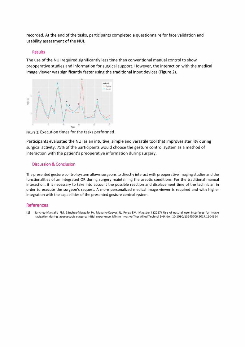
recorded. At the end of the tasks, participants completed a questionnaire for face validation and
usability assessment of the NUI.
Results
The use of the NUI required significantly less time than conventional manual control to show
preoperative studies and information for surgical support. However, the interaction with the medical
image viewer was significantly faster using the traditional input devices (Figure 2).
Figure 2: Execution times for the tasks performed.
Participants evaluated the NUI as an intuitive, simple and versatile tool that improves sterility during
surgical activity. 75% of the participants would choose the gesture control system as a method of
interaction with the patient’s preoperative information during surgery.
Discussion & Conclusion
The presented gesture control system allows surgeons to directly interact with preoperative imaging studies and the functionalities of an integrated OR during surgery maintaining the aseptic conditions. For the traditional manual interaction, it is necessary to take into account the possible reaction and displacement time of the technician in order to execute the surgeon’s request. A more personalized medical image viewer is required and with higher integration with the capabilities of the presented gesture control system.
References
[1] Sánchez-Margallo FM, Sánchez-Margallo JA, Moyano-Cuevas JL, Pérez EM, Maestre J (2017) Use of natural user interfaces for image navigation during laparoscopic surgery: initial experience. Minim Invasive Ther Allied Technol 1–9. doi: 10.1080/13645706.2017.1304964
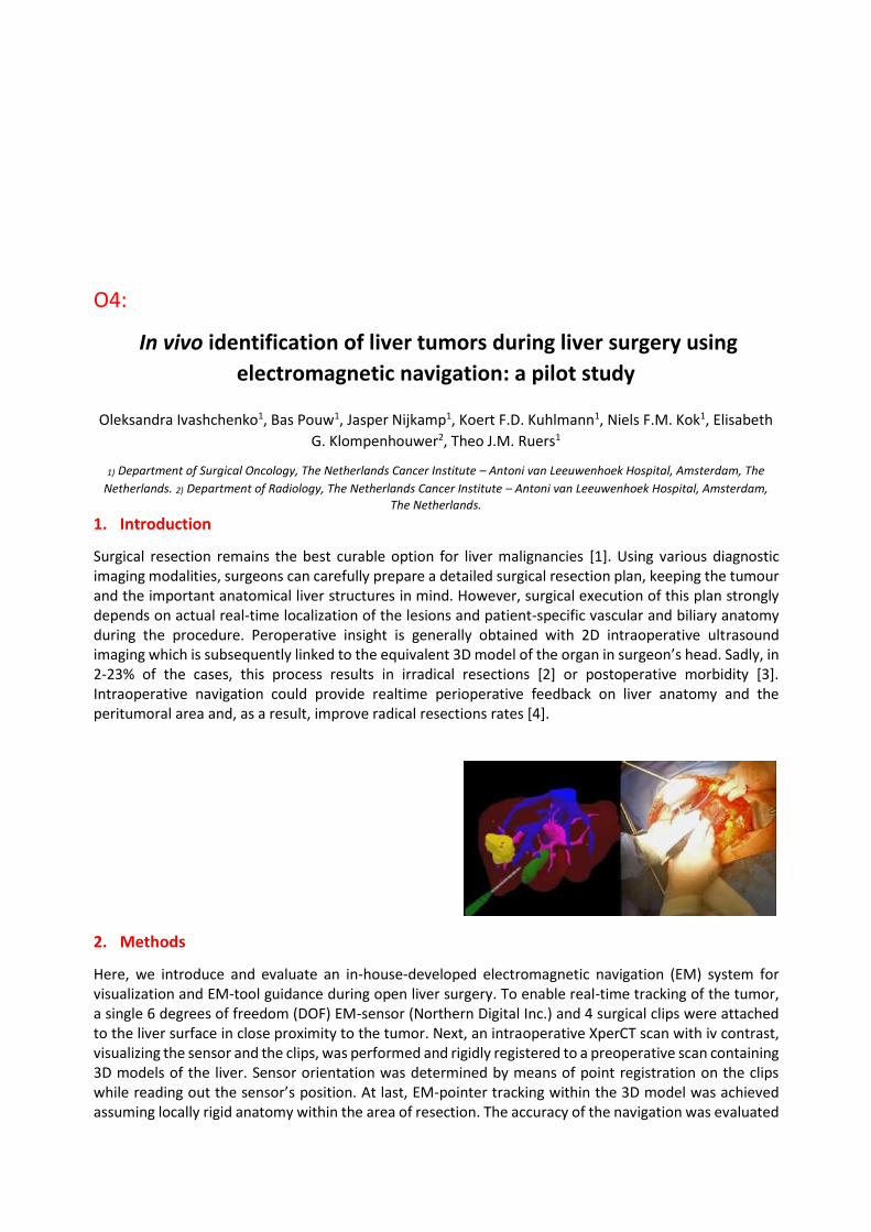
O4:
In vivo identification of liver tumors during liver surgery using
electromagnetic navigation: a pilot study
Oleksandra Ivashchenko1, Bas Pouw1, Jasper Nijkamp1, Koert F.D. Kuhlmann1, Niels F.M. Kok1, Elisabeth
G. Klompenhouwer2, Theo J.M. Ruers1
1) Department of Surgical Oncology, The Netherlands Cancer Institute – Antoni van Leeuwenhoek Hospital, Amsterdam, The
Netherlands. 2) Department of Radiology, The Netherlands Cancer Institute – Antoni van Leeuwenhoek Hospital, Amsterdam,
The Netherlands.
1. Introduction
Surgical resection remains the best curable option for liver malignancies [1]. Using various diagnostic imaging modalities, surgeons can carefully prepare a detailed surgical resection plan, keeping the tumour and the important anatomical liver structures in mind. However, surgical execution of this plan strongly depends on actual real-time localization of the lesions and patient-specific vascular and biliary anatomy during the procedure. Peroperative insight is generally obtained with 2D intraoperative ultrasound imaging which is subsequently linked to the equivalent 3D model of the organ in surgeon’s head. Sadly, in 2-23% of the cases, this process results in irradical resections [2] or postoperative morbidity [3]. Intraoperative navigation could provide realtime perioperative feedback on liver anatomy and the peritumoral area and, as a result, improve radical resections rates [4].
2. Methods
Here, we introduce and evaluate an in-house-developed electromagnetic navigation (EM) system for visualization and EM-tool guidance during open liver surgery. To enable real-time tracking of the tumor, a single 6 degrees of freedom (DOF) EM-sensor (Northern Digital Inc.) and 4 surgical clips were attached to the liver surface in close proximity to the tumor. Next, an intraoperative XperCT scan with iv contrast, visualizing the sensor and the clips, was performed and rigidly registered to a preoperative scan containing 3D models of the liver. Sensor orientation was determined by means of point registration on the clips while reading out the sensor’s position. At last, EM-pointer tracking within the 3D model was achieved assuming locally rigid anatomy within the area of resection. The accuracy of the navigation was evaluated

by comparing navigation-based to pathology results for the shortest distance between measurement points in the resection plane and the tumor. In this pilot study with the METC approval, a total of 17 navigated surgical resections were performed.
3. Results
Navigated surgical resections were performed with a median surgical overhead time of 43 minutes. This included placement of EM-sensor on the liver surface (9 min), sterile intraoperative contrast-enhanced XperCT scan (13 min), registration of the surgical plan with a real-time situation and all navigation-related measuements (21 min). The navigation technology resulted in an acuurate realtime tracking of liver tumors and had an average accuracy of 7.3 mm, compared to the pathology, and this value is expected to improve in the future.
Figure 1: Sample screen shot from the navigation software during open liver surgery and the corresponding surgical view.
4. Discussion &Conclusions
We successfully developed and implemented EM navigation for open liver surgery. This was done by combining a 3D liver model, intraoperative XperCT and EM tracking of the liver and a sterile EM-pointer. Achieved accuracy shows that the assumption of locally rigid organ registration allows for accurate detection of critical anatomical structures within the resection area.
References
[1] Y. Mise, et. al., Liver Cancer, vol 2(1), p. 55–66, 2013. [2] E.K. Abdalla, et al., Ann Surg, vol 239(6), p. 818-827, 2004. [3] S. Virani, et al., J Am Coll Surg., vol 204(6), p. 1284-92. [4] M.E. Kelly, et. al., J Surg Oncol, vol 111(3), 341–351.

O79:Mixed Reality 3D Visualization and Interaction for Surgery using
HoloLens
Kumar Rahul Prasanna, Brun Henrik, Edwin Bjørn, Elle Ole Jakob- The Intervention Centre, Oslo University Hospital, Oslo, Norway
Introduction
Visualization of patient specific 3D anatomy is gaining importance in healthcare, especially for surgery
applications. Visualizing 3D anatomical structures on a flat screen may only partially improve spatial
understanding. Thus mixed reality 3D visualization, which is the merging of real and virtual worlds where
physical and digital objects co-exist and interact in real time, could provide better 3D information to the
surgeons. Microsoft HoloLens can map the users’ real environment to place virtual 3D models in relation
to reality. This provides the user with a near-real experience, which is essential for it’s use in medical
applications. The laparoscopic surgeons and the cardiologists at the Oslo University Hospital (OUS), who
viewed their corresponding models in HoloLens, were very enthusiastic about its future applications in
their respective fields of medicine.
Methods
Planning is an integral part of laparoscopic resection workflow, where the surgeons decide beforehand
on the resection plan for the surgery. Also, in complex congenital cardiac anomalies like Pulmonary
Atresia With Ventricular Septal Defect and Major Aortopulmonary Collateral Arteries (PA/VSD/MAPCAS)
and complex Double-outlet right ventricle (DORV) in paediatric heart, it is essential to understand the
exact 3D anatomy before surgery.
With the development of our novel platform for planning liver resections on a computer using
segmented and then 3D generated models [1]–[3], the surgeons can plan better and safer resections.
This is now taken further with the use of mixed reality by visualizing the 3D segmented models of liver
and heart in the HoloLens, thereby giving the surgeons a better spatial and depth information. We have
also integrated our resection planning software [1] into the HoloLens application, along with
implementing a cutting tool for heart.
Results
We have developed an optimal mixed reality experience for the surgeons to better plan their surgical
procedure, with possibility of interaction with the planned models during surgery.

Figure 1: The above pictures show the visualization of liver and heart in mixed reality through HoloLens.
Discussion & Conclusion
Earlier, surgeons had to mentally plan the surgery by looking at the CT/MR images, thus making it
difficult to discuss their plans with other surgeons. With our HoloLens application, the surgeons get a
better spatial and depth information, along with the possibility to share and discuss with other surgeons
with multiple HoloLens devices. Also, having a cutting tool for heart helps the surgeons to cut as many
times and places as they require, compared to the 3D printed models that have to be reprinted if cut
wrong.
Acknowledgements
The research leading to these results has received funding from the Helse Sør-Øst innovation funding.
The authors thank SopraSteria developers for helping us develop the application.
References
[1] R. Palomar, F. A. Cheikh, B. Edwin, A. Beghdadhi, and O. J. Elle, ‘Surface reconstruction for planning and navigation of liver
resections’, Comput. Med. Imaging Graph., vol. 53, pp. 30–42, Oct. 2016.
[2] R. P. Kumar, F. Albregtsen, M. Reimers, B. Edwin, T. Langø, and O. J. Elle, ‘Three-Dimensional Blood Vessel Segmentation and
Centerline Extraction based on Two-Dimensional Cross-Section Analysis’, Ann. Biomed. Eng., pp. 1–12, Nov. 2014.
[3] R. P. Kumar, L. Barkhatov, B. Edwin, F. Albregtsen, and O. J. Elle, ‘Portal and Hepatic Vein Segmentation with Leak Restriction: A
Pilot Study’, in EMBEC & NBC 2017, Springer, 2017, pp. 823–826.

O86: Intra-operative estimation of surgical progress by tracking
instruments use
Meeuwsen Frédérique, van Luyn Fabian, van den Dobbelsteen John - Delft University of Technology,
Biomechanical Engineering, Delft, Netherlands
Introduction
Driven by rising health care costs due to factors including advancing technology and an aging
population, cost-effectiveness has become an increasingly important aspect of care delivery, with the
operating room (OR) being a specific area of interest. By means of a surgical process model (SPM),
OR systems can gain an understanding of clinical context and surgical workflow, hereby generating
ample opportunities to improve OR logistics and surgical care. Applications of SPM’s include
intra-operative end-time predictions, improved surgical training and assessment, computer-aided
surgery and increased autonomy in robotic surgery. The aim of this study is twofold; to predict
surgical phases intra-operatively based on data of instrument use and to evaluate the surgical phase
recognition model on simulated clinical tasks.
Methods
This study evaluates the use of SPM’s for intra-operative recognition of surgical phases in 40
laparoscopic hysterectomy cases. Based on manually annotated instrument usage data, the
procedures were split up in 10 phases. Random Forest and Hidden Markov Model were compared
and evaluated on two tasks related to clinical practice in the OR: the automatic prediction of
surgical end-times and the automatic generation of training material by clipping endoscopic video
based on the predictions of phase.
Results
With an out-of-sample accuracy of 77%, phases were best recognized by a Random Forest model.
The computation time of the RF model is sufficiently low for intra-operative clinical applications
and the model poses several other advantages, including noise resistance and suitability for parallel
processing. Simulating surgical end-time predictions based on the RF model was shown to be

promising, with a mean absolute error of 16 minutes. The model is also found suitable for phase
extraction for the generation of training material.
Discussion
We conclude that the performance of the Random Forest surgical phase recognition model based on
intra-operative data of instrument use, has promising performance. Our further research is aimed
at replicating the simulated findings using sensor-based data in an in-vivo clinical setting.
O78 : Magnetic resonance imaging compatible liver phantom with 3D
printed vessels for pre-clinical assessment
Pelanis Egidijus, Oram Louise, Kumar Rahul P., Elle Ole Jakob, Edwin Bjørn - The Intervention Centre,
Oslo University Hospital, Oslo, Norway
1. Introduction
Metastatic colorectal cancer is most common in
the liver and, despite advances in treatment,
resection of metastasis remains the only
curative option. Parenchyma-sparing
laparoscopic liver resection is associated with
significantly less postoperative complications
for patients compared to open [1]. In some
cases, this approach can be challenging, due to
limited field of view and curved resection
planes.
All patients undergo pre-operative MR imaging
for staging and surgery planning. These
volumetric images can already be segmented
and reconstructed in 3D to be viewed by
surgeons. A 3D view helps the surgeon better
understand patient specific anatomy, determine
if the patient can undergo surgery, plan the
surgery and visualize liver anatomy during the
surgery itself.
High Performance Soft-tissue Navigation
(HiPerNav) is a Marie Sklodowska-Curie Actions
– Innovative training network project. The
consortium will address specific bottlenecks in
soft tissue navigation for improved treatment of
cancer. The technological advancements in soft-
tissue navigation would support easier
orientation and potentially improve surgical
outcomes.
For development and pre-clinical testing of soft-
tissue navigation systems, phantoms are
needed and commercially available ones can be
very expensive [2]. The biggest limitation of
phantoms is their degradability and the need to
be replaced if cut into. A simple liver phantom
has been done before although not
anatomically correct [3]. The development of an
anatomically correct and inexpensively
replaceable liver phantom is required.
2. Methods
Fully anonymized MR images are used for semi-
automatic liver segmentation using tools within
“3DSlicer” and ones developed through the
NorMit project [3] and the HiPerNav project.
Segmentations are then verified by a medical
doctor for medical accuracy and optimized for
3D printing. A unidirectional object is added to
both the liver and liver vessel model to ensure
correct vessel position in the phantom.
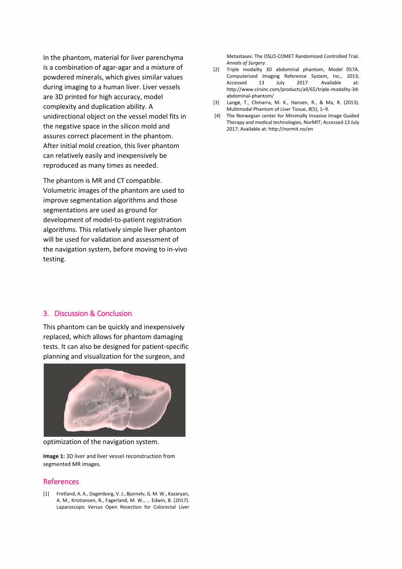
In the phantom, material for liver parenchyma
is a combination of agar-agar and a mixture of
powdered minerals, which gives similar values
during imaging to a human liver. Liver vessels
are 3D printed for high accuracy, model
complexity and duplication ability. A
unidirectional object on the vessel model fits in
the negative space in the silicon mold and
assures correct placement in the phantom.
After initial mold creation, this liver phantom
can relatively easily and inexpensively be
reproduced as many times as needed.
The phantom is MR and CT compatible.
Volumetric images of the phantom are used to
improve segmentation algorithms and those
segmentations are used as ground for
development of model-to-patient registration
algorithms. This relatively simple liver phantom
will be used for validation and assessment of
the navigation system, before moving to in-vivo
testing.
3. Discussion & Conclusion
This phantom can be quickly and inexpensively
replaced, which allows for phantom damaging
tests. It can also be designed for patient-specific
planning and visualization for the surgeon, and
optimization of the navigation system.
Image 1: 3D liver and liver vessel reconstruction from
segmented MR images.
References
[1] Fretland, A. A., Dagenborg, V. J., Bjornelv, G. M. W., Kazaryan, A. M., Kristiansen, R., Fagerland, M. W., … Edwin, B. (2017). Laparoscopic Versus Open Resection for Colorectal Liver
Metastases: The OSLO-COMET Randomized Controlled Trial. Annals of Surgery.
[2] Triple modality 3D abdominal phantom, Model 057A. Computerized Imaging Reference System, Inc., 2013; Accessed 13 July 2017. Available at: http://www.cirsinc.com/products/all/65/triple-modality-3d-abdominal-phantom/
[3] Langø, T., Chmarra, M. K., Hansen, R., & Ma, R. (2013). Multimodal Phantom of Liver Tissue, 8(5), 1–9.
[4] The Norwegian center for Minimally Invasive Image Guided Therapy and medical technologies, NorMIT; Accessed 13 July 2017; Available at: http://normit.no/en

O66 : Evaluation of a 3D planning tool for irreversible electroporation
treatment in pancreatic cancer
Eigl Benjamin, Peterhans Matthias, Weber Stefan, Gloor Beat, Worni Mathias - CAScination AG, Surgical
Navigation, Bern, Switzerland
1. Introduction
Local ablation is a treatment alternative for patients with unresectable cancer. Irreversible-electroporation (IRE) is a non-thermal ablation technique that spares vessels and thus is predisposed for appropriate treatment of locally advanced pancreatic cancer [1]. Correct needle positioning and parallel placement is a prerequisite for an efficient and complication-free IRE treatment. Therefore, we propose a 2D/3D planning and visualization solution to support the surgeon during this crucial procedure.
2. Methods
Based on conventional CT image data, and 3D reconstructions of essential anatomical structures (duodenum, pancreas with tumour, aorta with local branches, vena cava, superior/inferior mesenteric-, splenic-, and portal vein), our software provides the surgeon with a structured planning interface. The surgeon can define needle trajectories under consideration of different constraints such as number of needles used, spacing, and parallelism of the IRE needles. As a result, the software displays anatomical structures together with planned IRE trajectories (Figure 1). To explore the current potential of the planning software, the solution was presented to potential users which were subsequently asked to complete a questionnaire. Furthermore, it was investigated if additional navigation support during open, as well as minimally invasive surgeries would be beneficial.
Figure 16: Planning display containing bones structures in white and coloured 3D reconstructions of vessels, pancreas, duodenum, and tumour. The planned IRE needles (n=4) are displayed as coloured lines.
3. Results
The study population consists of 8 medical doctors (MD’s) with speciality in HPB surgery (7) and surgical oncology (1) active in USA (1), India(1) Turkey(1), Germany(1), Austria(1), and Switzerland(3). The MD’s perform between 30 and 120 pancreatic surgeries annually (average value of 70 surgeries per year). The yearly number of IRE’s was situated between 0 and 20 treatments with an average number of 4.5. Figure 2 shows the results of the questionnaire. All the MD’s agreed that there is a need for a dedicated planning software and a majority is in favour of 3D planning (against conventional 2D planning). Seven (87.5%) of the MD’s would use

a planning software in clinical routine. The majority of the pancreas specialists see a need for minimally invasive pancreas IRE (87.5%) as well as for intra-operative needle navigation (100%).
Figure 17: Evaluation of the questionnaire about our dedicated planning software.
4. Discussion & Conclusion
The results in Figure 2 indicate that the pancreas IRE planning solution strongly requires 3D visualization support. The results motivate further developments in the direction of dedicated planning and navigation tools as most surgeons would evaluate it for their daily clinical routine.
We envisage to combine the present planning solution for IRE treatments with a navigation platform providing real time needle guidance to facilitate intraoperative positioning of IRE needles.
References
[1] R. C. G. Martin, “Use of irreversible electroporation in unresectable pancreatic cancer,” Hepatobiliary Surg. Nutr., vol. 4, no. 3, pp. 211–215, Jun. 2015.

O28 :
Assessment of target registration error for hybrid instrument tracking for image guided surgery applications
Andrea Teatini1, Bjørn Edwin1,2, Ole J. Elle1,3
1) The Intervention Centre, Oslo University Hospital, [email protected]. 2) Institute of Clinical Medicine, Faculty of Medicine, University of Oslo, Norway. 3) Department of Informatics, University of Oslo, Norway.
1. Introduction
Instrument tracking for medical applications has
mainly been restricted to optical tracking.
However, optical instrument tracking presents
the disadvantage of losing tracking if line of
sight to the cameras is occluded. This limits its
use for surgical applications. Electromagnetic
tracking (EMT) does not need line of sight track
and is therefore starting to attract interest.
However, EMT is still limited by its small
measurement volume and difficulty in handling
ferromagnetic interferences.
Similarly to EMT, inertial sensing is also not
interrupted by occlusions. Nevertheless, it is
currently not in use for surgical instrument
tracking because of its drift issues [1]. A
combination of these systems could
compensate for the disadvantages of each
tracking technology and even improve overall
tracking accuracy. This abstract describes some
advantages of hybrid tracking techniques and
proposes a method to test their accuracy and
reliability to track instrument tips for image
guided surgery applications.
2. Methods
The first hybrid tracking system makes use of a
NDI Polaris Spectra optical tracking cameras and
a NDI Aurora electromagnetic tracking system.
This combination of tracking systems will be
achieved tracking optical passive markers on
the instrument’s handle and placing an EMT
sensor on the tip of the tracked instrument. This
will allow handle loss of tracking in case of
occlusions. Furthermore, it will reduce the
impact of error magnification inherent to
optical tracking when tracking the tip of an
instrument. The EMT sensor can also be used to
localize flexible instrument tips, advantageous
for applications such as laparoscopic
ultrasonography.
The second hybrid tracking system proposed is
a combination of a NDI Polaris Spectra’s optical
tracking with inertial tracking using a
INVENSENSE MTU9250 inertial sensor. This
sensor is significantly larger that the EMT sensor
(~2cm vs 1,3mm diameter), which makes it
impossible to place the inertial sensor at the tip
of the tracked instrument. Thus, a solution
where the sensor is rigidly connected to the
optical tracking markers is proposed. This
combination will maintain tracking temporarily
in case of occlusions, but also increase the
tracking’s speed based on the higher update
frequency of the inertial sensor.
The two hybrid tracking systems will be
assessed and compared based on: accuracy and
reliability. To evaluate tracking accuracy, two
measures are commonly used [2]: fiducial
localization error (FLE) and target registration
error (TRE). FLE is the distance between the
position of a fiducial and its measured position.
TRE is the error that the tracking system
commits in localizing a specific position on the
tracked instrument. Tracking the tip of the tool
is more relevant to image guided surgery, so the
latter is chosen. TRE is calculated either
mathematically based on a reported FLE, or via
direct measurement [2]. This study proposes a
method of direct measurement TRE using a
custom made optical verification phantom
according to the ASTM F2554-10 standard [3].
The verification phantom is also used to
perform a reliability test. The phantom
evaluates tracking within a measurement
volume similar to that of abdominal surgery
(approximately 20x30x15cm). The surgeon will
be required to target, with different tracked
instruments, 28 positions on the phantom at 9
different orientation planes. The orientation
planes reflect the directions tools are held
during abdominal surgery. Based on a count of
occurrence of loss of tracking data throughout
the experiments, reliability of the tracking
systems will be assessed.
3. Discussion and Conclusion
The combination between tracking systems can
allow for improvement of both accuracy and
stability for tracking surgical instruments. The
efficacy of the two types of hybrid systems will
be compared with a standardized approach in a
setup, which will assess the accuracy for an
application of surgical instrument tip tracking
for image guided surgery applications.
References
[1] G. Welch and E. Foxlin, "Motion tracking: no silver bullet, but a respectable arsenal," in IEEE Computer Graphics and Applications, vol. 22, no. 6, pp. 24-38, Nov.-Dec. 2002. doi:10.1109/MCG.2002.1046626
[2] Banivaheb, Niloofar. "Comparing Measured and Theoretical Target Registration Error of an Optical Tracking System." (2015).
[3] ASTM F2554-10, Standard Practice for Measurement of Positional Accuracy of Computer Assisted Surgical Systems, ASTM International, 2010.

O96 : An interactive approach for the semi-authomatic segmentation of
liver an lesions
Caccamo Sergio Salvatore, Ray Pravda Jith, Oram Louise, Elle Ole Jakob, Kumar Rahul Prasanna-The
Intervention Centre, Oslo University Hospital, Department Of Informatics, University of Oslo, Oslo,
Norway

O144: Using video-tracking algorithms of the MST robotic camera manipulator for teaching and testing surgical skills Amir Szold; Assia Medical Group, Assuta Medical Center, Tel Aviv, Israel Background: MST had commercialized a camera-positioning robotic arm (Autolap), with motion control based on artificial vision and video analysis of generic surgical tools. Objective: To test the use of the instrument-tracking software for performance analysis and training in minimally invasive surgery Technology: We have designed a training and assessment mode that can asses surgeon’s efficiency by motion analysis of surgical tools, and give feedback on motion efficiency and performance both in the lab and during surgery. Preliminary results: The prototype was able to track instruments and analyze motion efficiency in different settings. Future perspectives: We are currently testing the system to differentiate between novice and experienced surgeons and aid in teaching complex maneuvers in a minimally invasive surgery setting.

SESSION 19 – MATERIALS FOR MEDICAL DEVICES : BIOCOMPATIBILITY, FUNCIONAL SURFACES

O41: Low Melting Point Materials as controllable stiffness mechanism
for endoscopic and catheter applications
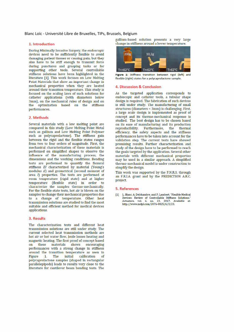
Blanc Loïc - Université Libre de Bruxelles, TIPs, Brussels, Belgium

O45: Towards a new patient-specific, modular aortic vascular phantom
with clinically relevant mechanical properties
Gallarello Antonio, Palombi Andrea, De Momi Elena, Burriesci Gaetano, Homer-Vanniasinkam
Shervanthi, Wurdemann Helge - University College London, Department of Mechanical Engineering,
London, United Kingdom
1. Introduction
According to [1], the cardiovascular devices market was valued at over US$33 billion worldwide in 2015
with a projected growth of a CAGR (Compound Annual Growth Rate) of 6.6% until 2024.
For early-stage validation of emerging tools, in vitro tests are usually preferred to in vivo tests as animal
experiments and clinical trials are costly, time-consuming and can pose ethical issues [2].
Simulators able to mimic physiological pulsatile flow and pressure are commercially available (e.g.,
Endovascular simulator by Vivitro Labs). Researchers have further created both rigid and flexible
phantoms based on simplified geometries [3] or patient-specific 3D reconstructions [4]. However, the
challenge remains to develop hard-wearing vascular testbeds with clinically relevant mechanical
properties reproducing the physiological environment.
This paper presents a step-by-step development process of a vascular phantom which will be able to
mimic human-like distensibility and thus allow haemodynamic analysis and device testing.
2. Methods
The presented phantom environment should
• comprise the aortic arch, the descending and abdominal aorta as well as the common iliac arteries including the branches
• be modular, using patient-specific data
• be able to mimic human-like distensibility
• be cost-effective
• be MR-compatible
3. Results
A threshold-based image segmentation has been carried out on selected regions of interest of a CT
angiogram, using the open-source (OS) software 3D Slicer (Fig.1a). The 3D reconstruction has been
manually refined and edited with OS CAD software (Fig.1b). A number of soft materials suitable for
moulding (silicone material) and 3D printing (TangoPlus Full Cure 930®) have been characterised by
uniaxial tensile testing. The results of the tensile tests followed by a finite element analysis permitted
the fabrication of a modular phantom environment made of Eco-Flex 00-30 silicone with a thickness of

Figure 18. Example of the manufacturing process for a cadiovascular module (here: the aortic arch): (a) Segmentation of angio-CT images; (b) refining and 3D reconstruction of the vessels; (c) post-processing for mould design; (d) final module design with Windkessel chamber.
2.2mm. The external mould is designed as shown in Fig.1c. Furthermore, a Windkessel chamber
connected to the water-filled bucket containing the phantom allows variation in the distensibility
(Fig.1d).
4. Discussion & Future work
To the best of our knowledge, this is the first description of the development of a patient-specific,
modular aortic vascular phantom with clinically relevant mechanical properties. We envisage this
phantom environment to be of paramount importance for early-stage validation procedures of medical
devices for cardiovascular applications include diagnostic and monitoring devices as well as surgical
tools. Ongoing efforts will be dedicated to the fabrication of each module as well as the assembly of the
entire phantom environment. Validation results will be carried out and presented at the conference.
References
[1] Cardiovascular Devices Market Size, Global Industry Report, 2024, Grand View Research,
2016. [2] A. Knight, “Weighing the Costs and Benefits of Animal Experiments,” Eighth World Congr. Altern. Anim. Use life Sci., pp. 289–294, 2011.
[3] T. L. Poepping, H. N. Nikolov, M. L. Thorne, and D. W. Holdsworth, “A thin-walled carotid vessel phantom for Doppler ultrasound flow
studies,” Ultrasound Med. Biol., vol. 30, no. 8, pp. 1067–1078, 2004.
[4] E. Vander Poorten et al., “Cognitive AutonomouS CAtheters operating in Dynamic Environments,” J. Med. Robot. Res., 2016.
O32: Improving functional recovery after radical prostatectomy: in vitro
evidences on a chitosan-based medical device
Geuna Stefano, Muratori Luisa Muratori, Fregnan Federica, Bertolo Riccardo, Porpiglia Francesco-
University of Turin, Department of Clinical and Biological Sciences, Orbassano, Italy
1. Introduction
Chitosan (CS) based nerve grafts are employed to
promote neural repair after injury raising more
and more interest among basic and clinical
research. In vitro and in vivo studies have shown
that this biomaterial has biocompatible and
biomimetic properties to improve the
regeneration process of the peripheral nervous
system [1].
It is well known that Prostate cancer (PCa) is the
most common cancer among men. The surgical
treatment for PCa is represented by the radical
prostatectomy, which is the gold standard in the
treatment of localized disease. Unfortunately, in
patients who underwent a radical prostatectomy,
frequently iatrogenic damage to the peri-
prostatic nerve bundles occurs, leading to erectile
dysfunction (ED).
The aim of this in vitro study is to assess the
simultaneous anti-proliferative and pro-
regenerative properties of a CS film, which has
already achieved a clinical use for the peri-
prostatic nerve plexus protection and a patent
(Application reference: 102016000070911).
2. Methods CS-anti-proliferative properties were tested on
different human prostate cancer cell lines (PC-3,
DU145, LNCap) seeded on two different
experimental condition: dissolution products of
CS and CS coating. Since the prostatic plexus is
innervated by sympathetic, parasympathetic and
somatic fibers, the regenerative potential of CS
films was assessed through primary neuronal
cultures and ex vivo explants derived from
autonomic and DRG ganglia.
3. Results The dissolution products of CS on proliferation
assay performed after 1, 3, 6 days determined a
significant lower proliferation of cancer cells,
accordingly the same cells in direct contact with
CS coating showed a substantial change in
morphology, but also a significant decrease in
proliferation.
Regarding the regenerative potential, CS film
were tested and they demonstrated to represent
a permissive substrate for neurite regeneration
and axonal elongation.
Figure 1: A) PC3 cancer cell line on CS coating, effect on cell
proliferation and on Bcl2/Bax expression (B). (C) Pro-
regenerative effect of CS on Autonomic ganglia explants and
on dissociated autonomic neuron cultures (D)
4. Discussion & Conclusion. An increasing number of young men have an early
prostate cancer diagnosis, and ED caused by
radical prostatectomy is associated with distress
and impaired quality of life. The clinical
application of new techniques and new materials
in the field of peripheral nerve regeneration
would result in minor inconvenience for patients
and allow to extend the treatment also for
applications in oncology.
0
500000
1000000
1500000
2000000
2 div 5 div 7 div
PC3 cells
ctr
acetic acid
**
****
**
A
B
C
D

References [1] K, Haastert-Talini S.. Geuna, LB Dahlin, et al." Chitosan tubes of
varying degrees of acetylation for bridging peripheral nerve
defects."Biomaterials,vol.34,p.9886-904,2013.
[2] V. Ficarra, G .Novara, TE. Ahlering et al. "Systematic review and
meta-analysis of studies reporting potency rates after robot-assisted
radical prostatectomy". Eur Urol, vol. 62, p.418-30,2012

O70:
A novel bactericidal surface to reduce nosocomial infections improving an Italian Patent
Alessandra Piccitto1, Ivan P. Parkin2, Mario Morino3
1) C. Ingold Building Dept. of Chemistry, University College London, London, UK, [email protected]
2) C. Ingold Building Dept. of Chemistry, University College London, London, UK
3) School of Medicine Dept. of Surgery Molinette Hospital, University of Torino, Torino, Italy.
1. Introduction
This work would like to improve a 3D Ultra Sound full console machine able by using the SynDiag patented software to output a quantitative triangular mesh in PLY format, a mathematical object that can be printed and studied while the fetus is at its 5th month of pregnancy. As antibiotic resistance continues to threaten the treatment of various infections, the aim of this work concerns the design of a new coating film for inanimate surfaces containing light activated antimicrobial chemicals and nanoparticles to avoid microbes spread across hospitals. It is well known that methicillin-resistant Staphylococcus aureus (MRSA) is a substantial public health problem not restricted to any geographic area, but worldwide. The same is true for the opportunistic pathogen Pseudomonas aeruginosa.
Antimicrobial activity of control and treated polyurethane samples on 4742 MRSA and PAO1 after 5 hours of incubation at 500 lux. There is no MRSA growth with crystal violet (CV) solution and CV + Silver NPs solution in 5 hours. The situation changes in the case of PAO1 where the growth is reduced only by the solution containing silver NPs in 5 hours. However, after an incubation time of 24 hours in the dark a significant antimicrobial activity was observed.
3. Results
Figure 1: Silver nanoparticle – TEM Image
This work involves developing research areas that did not exist previously, such as the light activated polymers which have been studied at Chemistry Department UCL and in some cases commercialized [1]. Micro-organisms selected for this study were Epidemic MRSA 4742 representative of a Gram-positive epidemic bacteria isolated at UCL Hospital and Pseudomonas aeruginosa PAO1 as well as the clinical strand isolated at UCL representative of Gram-negative bacteria resistant to the majority of currently used antibiotics. Concerning this one a complementary study of its biofilm-mediated resistance to antibiotics has been followed. To satisfy the target the dye selected was crystal violet or gentian violet because it has been demonstrated that in vitro it can disrupt Pseudomonas aeruginosa biofilms and it can kill MRSA. The nanoparticles (NPs) used for this study were silver ions because the antimicrobial activity of Ag+ ions is well documented and reviewed. The antimicrobial polyurethane coating films produced were tested against these micro-organisms to demonstrate their efficacy [2]. It has been also studied the critical importance of the presence of silver nanoparticles (Image 1) to improve the functional property of the inanimate surface.
References
[1] Sacha Noimark et al. Chem. Sci., 2014, 5, 2216-2223 [2] Gi Byoung Hwang et al. J. Mater. Chem. B, 2016, 4, 2199-2207
Silver NPs used for this work remain inside of the polyurethane matrix – they stay into the bulk and not on the surface and exhibit potent antimicrobial activity. In addiction to this, the combination of silver NPs and a light activated agent (CV) can provide a dual kill mechanism also under dark conditions. These surfaces are promising candidates for use in healthcare environments to reduce the incidence of hospital-acquired infections.
4. Discussion & Conclusion
2. Methods
SESSION 28 – NEW TOOLS FOR LAPAROSCOPY 2

O141: Artack: a mesh fixation tacking device with potential robotic
interface
Szold Amir - Assia Medical Group, Assuta Medical Center, Tel Aviv, Israel
Objective: Vessel sealing during minimally invasive surgery is still a challenge, especially
when applying through articulating or flexible devices. Additional disadvantages of current
system include low precision and collateral tissue heating.
Technology: The core technology is an asymmetric laser fiber that enables controlled lateral
energy emission. This allowed the design and production of several vessel sealing and tissue
cutting devices including an articulating grasper, a hook and a loop snare for endoscopy. The
devices can be articulated or flexible, fit into a flexible endoscopes' working channels and can
be made in diameters even lower than 3 mm. This superior adaptability means the technology
is a perfect fit with a variety of surgical robotic platforms.
Preliminary results: The devices were tested in an animal model with successful sealing of
arteries up to 5 mm diameter, using low energy and with a durable seal.
Future perspectives: We are currently finalizing the first product design for minimally
invasive surgery.
O143: Performance assessment and training using MST core
software : Instrument-tracking using video analysis software
Szold Amir - Assia Medical Group, Assuta Medical Center, Tel Aviv, Israel
Background: MST had commercialized a camera-positioning robotic arm (Autolap), with motion control based on artificial vision and video analysis of generic surgical tools. Objective: To test the use of the instrument-tracking software for performance analysis and training in minimally invasive surgery Technology: We have designed a training and assessment mode that can asses surgeon’s efficiency by motion analysis of surgical tools, and give feedback on motion efficiency and performance both in the lab and during surgery. Preliminary results: The prototype was able to track instruments and analyze motion efficiency in different settings. Future perspectives: We are currently testing the system to differentiate between novice and experienced surgeons and aid in teaching complex maneuvers in a minimally invasive surgery setting.

O2:
Is Reduced port laparoscopic cholecystectomy any better than the gold standard?
To assess surgeon stress and workload: a randomized controlled trial
Yasuhiro Ishiyama, Yoshiaki Hara, Yuta Enami, Shoji Shimada, Kenta Nakahara, Chiyo Maeda,
Naruhiko Sawada, Eiji Hidaka, Fumio Ishida, Shin-ei Kudo
1) Digestive disease center Showa University Northern Yokohama Hospital 35-1, Chigasaki-Chuo Tsuzuki, Yokohama 224-8503, Japan E-mail:[email protected]
Introduction
Single incision laparoscopic surgery (SILS) has been attracting attention in the field of minimally
invasive surgery. However, it is technical difficulty has delayed its adoption for every surgeons.
Reduced port laparoscopic surgery (RPS) may lead to be easier than SILS for surgeon. The aim of this
prospective study was to compare surgeon stress and workload during a randomized controlled study
for reduced port laparoscopic cholecystectomy (RPLC) and conventional laparoscopic
cholecystectomy (CLC) in the operating room.
Methods 16 patients were prospectively randomly assigned to either RPLC or CLC from July 2016 to
September 2016. To evaluate differences in surgeon workload and stress between RPLC and CLC
procedures, objective and subjective workload data were collected randomized controlled trial
(RCT) comparing patient outcomes between RPLC and CLC.
Results Patient factors and operative outcomes were no significantly difference between RPLC and CLC. In the
surgeon reported Surg-TLX, subscale of Task demand was significantly more demanding for RPLC
than CLC (p<0.05). The other factors were no significantly difference between RPLC and CLC.
Salivary amylase level was no significantly difference during RPLC than CLC.
Figure : Salivary amylase levels at the three time points

Discussion & Conclusion
In conclusion, it can be thought that RPLC is as similar as CLC for the surgeons stress. In workload,
task demanding was higher in CLC than in RPLC. In other words, we assumed that RPLC might be an
acceptable alternatives to CLC in benign gallbladder diseases. References 1. Chang SK. Lee KY therapeutic advances: single incision laparoscopic hepatopancreatobiliary surgery. World J Gastroenterol
2014;20(39):14329–14337. 2. Bhandarkar D, Katara A, Deshmane V, Mittal G, Udwadia TE. Single-incision bilateral laparoscopic oophorectomy. J Minim
Access Surg. 2011 Jan;7(1):87-9.
O52:
Changes in abdominal wall thickness during laparoscopy:
Implications for the use of magnetic assisted surgery
N Shussman, G Marom, L Luques, R Elazary, M Abu Gazala, AJ Pikarsky, L Appelbaum, R Brodie, Y
Mintz
Department of General Surgery, Hadassah Hebrew-University Medical Center, Jerusalem, Israel
Center for Ultrasound, Hadassah Hebrew-University Medical Center, Jerusalem, Israel
ABSTRACT
INTRODUCTION: In the last two decades efforts are done to minimize surgical invasiveness. Recently, magnetic
solutions were proposed such as the Magnetic Anchoring and Guidance System (MAGS). This system and similar
technologies includes deployable instruments that are inserted into the abdominal cavity through a single access
point. The internal components contain magnets and are manipulated by external magnets, which are held on
the abdominal wall's surface. This technology relies on the magnetic force between magnets which is inversely
related to the abdominal wall thickness (AWT). The change of AWT before and during laparoscopy was never
studied.
OBJECTIVE: To establish the expected change in AWT in obese and non-obese patients after initiation of
pneumoperitoneum, to determine preselection of patients appropriate for magnetic guided surgery.
METHODS: AWT prior to and during surgery were measured at the supra-umbilical midline and at the left upper
and left lower quadrants, and the change of abdominal wall thickness during laparoscopy was calculated.
RESULTS: Thirty-two patients undergoing various laparoscopic procedures were included. Twenty patients were
male (62.5%) and 10 were morbid obese (31%). Mean age was 51 years (range 18-76) and average BMI was 28.1
kg/m2 (range 19.0-41.0). Mean preoperative AWT was 3.72 centimeters for obese patients and 2.38 centimeters

for non-obese patients (p<0.001). AWT decreased on average by 15.6% once pneumoperitoneum was initiated
in both obese and non-obese patients (p=0.01).
CONCLUSION: Abdominal wall thickness decreases on average by 15 per cent with initiation of
pneumoperitoneum as compared to pre-laparoscopy measurements, regardless of age, gender or BMI. Our data
suggest that following preoperative assessment of AWT with abdominal wall ultrasound, more patients than
expected might be candidates for the use of trans-abdominal magnetic devices.
O83-91:
ANALYSIS OF LOCALIZED MUSCLE FATIGUE IN SINGLE INCISION ENDOSCOPIC SURGERY
Francisco M. Sánchez-Margallo1, Juan A. Sánchez-Margallo2, A. Skiadopoulos3, K. Gianikellis4
1Laparoscopy Unit, Jesús Usón Minimally Invasive Surgery Centre, Cáceres, Spain, [email protected]. 2Bioengineering and Health Technologies Unit, Jesús Usón Minimally Invasive Surgery Centre, Cáceres, Spain. 3Center for
Research in Human Movement Variability, University of Nebraska at Omaha, USA. 4Biomechanics of Human Movement and Ergonomics Lab., University of Extremadura, Caceres, Spain.
Introduction
The main objective of this study was to evaluate and to compare the effects of single incision endoscopic surgery (SIES) and conventional laparoscopic surgery in the onset of localized trapezius muscle fatigue (high, medium and low portions) and in the paraspinal muscles of the middle cervical portion.
Methods
Eight experienced surgeons in laparoscopic and SIES (> 100 laparoscopic procedures; >20 single
incision procedures) participated in this study, all of them with the right hand as the dominant one.
The participants performed a dissection of the serosa layer of a porcine stomach, attempting to
separate the serosa layer from the muscular layers. This task was carried out for 10 min in a physical
laparoscopic training simulator using laparoscopic and single port approaches. For both approaches
a laparoscopic camera of 5 mm and 30 degrees was used (Karl Storz, Tuttlingen, Germany). During
laparoscopic dissection, participants used a laparoscopic scissors in the right hand (EndoShears®,
Covidien, Mansfield, USA) and a dissector (EndoDissect®, Covidien) in the left hand. In the case of
SIES dissection, participants used an articulating tip instrument (SILS Shears®, Covidien) in the right
hand and a dissector of the same characteristics (SILS Dissector®, Covidien) in the left hand.
For the single port approach a SILS port (Covidien) was used. The muscular activity of the trapezius
muscle (high, medium and low portions) and the paraspinal muscles of the middle cervical portion
were recorded using the Trigno™ wireless electromyography (EMG) system (Delsys Inc., Natick, MA,
USA) (Fig 1). After placing the electrodes on the skin, the area was cleaned with alcohol. Each EMG
signal was amplified and filtered to eliminate potential interference. As an indicator of localized
muscle fatigue, the temporal change of the mean frequency (fMEDIAN) of the EMG signal, calculated
at intervals of 10 seconds, was used. This temporal change was analyzed by linear regression. The
RMS value of the EMG signal, normalized to the maximum value of each muscle and subject, was
used as an indicator of muscular activity. The muscular activity between both approaches was
compared by a repeated measures ANOVA (α <0.05).

Figure 1: Experiment setup. Wireless electromyography system.
Results
The single port approach led to significantly greater muscle activity in the paraespinal muscles of the
right middle cervical portion and the upper right trapezius than the conventional laparoscopic
approach. In both approaches, surgeons showed muscle fatigue in at least one of the muscles
analyzed. During dissection using a conventional laparoscopic approach, seven of the surgeons
reported muscle fatigue in the upper left trapezius. In the case of the single port approach, seven of
the surgeons showed muscle fatigue in the right middle trapezius.
Discussion & Conclusion
Surgeons showed increased muscle activity in the paraespinal muscles of the right middle cervical
portion and the right upper trapezius during the single port approach. All the surgeons showed
muscle fatigue in at least one of the muscles analyzed during both approaches.
References
[1] Sánchez-Margallo FM, Matos-Azevedo AM, Pérez-Duarte FJ, Enciso S, Martín-Portugués ID. (2014) Performance analysis on physical simulator of four different instrument setups in laparo-endoscopic single-site (LESS) surgery.task performance and ergonomics. Surg Endosc 28(5):1479-88.
[2] Sánchez-Margallo FM, Pérez FJ, Azevedo AM, Sánchez-Margallo JA, Sánchez MA, Díaz-Güemes I. (2014) An analysis of skills acquisition during a training program for experienced laparoscopists in laparoendoscopic single-site surgery. Surg Innov 21(3):320-6.
[3] Sánchez-Margallo JA, Sánchez-Margallo FM, Oropesa I, Enciso S, Gómez EJ. (2017). Objective assessment based on motion-related metrics and technical performance in laparoscopic suturing, Int J Comput Assist Radiol Surg.12(2):307-314.

O135: Single-incision laparoscopic cholecystectomy is responsible
for increased adverse events: results of a meta-analysisof
randomized controlled trials
Arezzo Alberto, Forcignanò Edoardo, Passera Roberto, Rapetti Lisa, Morino Mario - Department of
Surgical Sciences, University of Torino, Italy, Department of Surgical Sciences, University of Torino,
Italy, Turin, Italy
Introduction
Over the last decade SLC has gained popularity, although it is not evident if benefits of this procedure
overcome the potential increased risk. Aim of this study is to compare the outcome of single-Incision
laparoscopic cholecystectomy (SLC) with conventional multi-incision laparoscopic cholecystectomy
(MLC) in a meta-analysis of randomised controlled trials only.
Method
A systematic Medline, Embase, and Cochrane Central Register of Controlled Trials literature search
of articles on SLC and MLC for any indication was performed. The main outcomes measured were
overall adverse events, visual analogue scale for pain score (VAS), Cosmetic results, quality of life
and incisions hernias. Linear regression was used to model the effect of each procedure on the
different outcomes.
Results
Forty-six trials were included and data from 5141 participants were analysed; 2444 underwent SLC
and 2697 MLC respectively. Mortality reported was nil in both treatment groups. Overall adverse
events were higher in the SLC group (RR 1.41; p<0.01) compared to MLC group, as well severe
adverse events (RR 2.06; p<0.01) and even mild adverse events (RR 1.23; p=0.04). A sensitivity
analysis showed that the raw incidence of global adverse events was in favour of MPC also when only
trials including 4 ports techniques (RR 1.37, p=0.004) or 3 ports techniques were considered (RR
1.89, p=0.020). The visual analogue scale pain score by time point showed a standardised mean
difference (SMD) of -0.36 (p<0.001) in favour of SLC. Cosmetic outcome by time point scored a
SMD of 1.49 (p<0.001) in favour of SLC. Incisional hernias occurred more frequently (RR 2.97,
p=0.005) in the SLC group.
Conclusion
Despite SLC offers a better cosmetic outcome and reduction of pain, the consistent higher rate of
adverse events, both sever and mild, together with the higher rate of incisional hernias, should suggest
to reconsider the application of single incision techniques when performing cholecystectomy with the
existing technology.
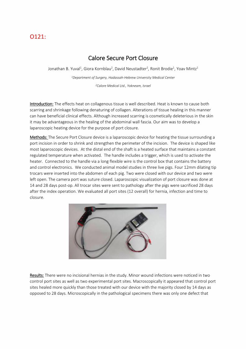
O121:
Calore Secure Port Closure
Jonathan B. Yuval1, Giora Kornblau2, David Neustadter2, Ronit Brodie1, Yoav Mintz1
1Department of Surgery, Hadassah-Hebrew University Medical Center
2Calore Medical Ltd., Yokneam, Israel
Introduction: The effects heat on collagenous tissue is well described. Heat is known to cause both
scarring and shrinkage following denaturing of collagen. Alterations of tissue healing in this manner
can have beneficial clinical effects. Although increased scarring is cosmetically deleterious in the skin
it may be advantageous in the healing of the abdominal wall fascia. Our aim was to develop a
laparoscopic heating device for the purpose of port closure.
Methods: The Secure Port Closure device is a laparoscopic device for heating the tissue surrounding a
port incision in order to shrink and strengthen the perimeter of the incision. The device is shaped like
most laparoscopic devices. At the distal end of the shaft is a heated surface that maintains a constant
regulated temperature when activated. The handle includes a trigger, which is used to activate the
heater. Connected to the handle via a long flexible wire is the control box that contains the battery
and control electronics. We conducted animal model studies in three live pigs. Four 12mm dilating tip
trocars were inserted into the abdomen of each pig. Two were closed with our device and two were
left open. The camera port was suture closed. Laparoscopic visualization of port closure was done at
14 and 28 days post-op. All trocar sites were sent to pathology after the pigs were sacrificed 28 days
after the index operation. We evaluated all port sites (12 overall) for hernia, infection and time to
closure.
Results: There were no incisional hernias in the study. Minor wound infections were noticed in two
control port sites as well as two experimental port sites. Macroscopically it appeared that control port
sites healed more quickly than those treated with our device with the majority closed by 14 days as
opposed to 28 days. Microscopically in the pathological specimens there was only one defect that

crossed the entire abdominal wall in a control port site. Experimental port sites showed a more robust
scarring pattern.
Conclusions: The Secure Port Closure device is safe for use and did not show any increase in adverse
effects. The strength of the closure is still not known. The ports in this study were purposely placed at
locations that were less likely to present hernias, to allow for initial evaluation of the device. A
possible follow-up study may be to place ports lower in the abdomen– at a location that is known to
be more susceptible to hernias. The results indicate that further study and development may lead to a
device that could be beneficial for both patients and doctors.
O89: Laparoscopic cutting using Radius r2 DRIVE Instruments :
Surgeon’s performance and muscular intervention
Sanchez-Margallo Francisco M., Sánchez Margallo Juan Alberto, Skiadopoulos Andreas, Gianikellis
Kostantinos- Minimally Invasive Surgery Centre Jesús Usón, Scientific Direction, Cáceres, Spain
Introduction
Laparoscopic surgery entails certain technical limitations for surgeons such as the restriction of
movements mainly due to the fixed position of the surgical ports. Several solutions have been
developed to address these limitations and ergonomic issues in laparoscopic surgery, mainly new
designs for instrument handles and support systems for surgeons such as surgical chairs and
armrests [1]. The aim of this study is to analyze the surgeon’s performance and muscular
intervention using Radius r2 DRIVE laparoscopic instruments (Tubingen Scientific Medical, Germany)
during a cutting task.
Methods
Three experienced laparoscopic surgeons performed three intracorporeal cutting tasks on a box
trainer using a conventional laparoscopic Maryland dissector and a pair of scissors and their
equivalent r2 DRIVE instruments. Execution time and the percentage of the area of deviation from
the cutting pattern (circle) were assessed. Surgeon’s ergonomics was evaluated through analysis of
the surface electromyography (trapezius, deltoid and paravertebral muscles) and the NASA-TLX
index.
Results
Execution time was significantly higher using the novel laparoscopic instruments (Conv: 119±31 s vs.
r2 DRIVE: 133±35 s; p<.05). The use of both instruments had a similar percentage of deviation from
the exterior part of the cutting pattern. However, the deviation from the inner part was significantly
higher using the r2 DRIVE instruments (Conv: 7.946±1.269% vs r2 DRIVE: 10.783±2.090%; p<.05).
Surgeons’ muscle activity was significantly higher for the left deltoid muscle and the trapezius
muscle bilaterally using the novel instruments (Fig. 1). Surgeons considered the use of these
instruments leads to a higher mental and physical workload when compared to traditional
laparoscopic instruments during laparoscopic cutting.

Figure 1: Muscle activity (RMS) of the paravertebral, deltoid and trapezius muscles during the use of the conventional and r2 DRIVE laparoscopic instruments.
Discussion & Conclusion
Despite the novel and ergonomic design of the r2 DRIVE laparoscopic instruments, the results
suggest that an improvement in surgical performance and physical workload is required prior their
use in a real surgical setting. Further studies should be done to analyze the use of these instruments
during other laparoscopic tasks and procedures. We believe that surgeons need a longer training
period with these laparoscopic instruments to reach their full potential in laparoscopic surgery.
References
[1] Sánchez-Margallo JA, Sánchez-Margallo FM, Gianikellis K, Skiadopoulos A, Hermoso J, Langø T (2016) Surgical performance and ergonomics of the surgeon’s hand using a robotic handheld needle holder. 28th Conference of the International Society for Medical Innovation and Technology. Delft, The Netherlands.

O39:
Finite element analysis in colorectal surgery: preliminary results on the interaction between tissue and surgical tool
R. Guachi1, M. Bici1, F. Bini1, L. Lorenzon2, F. Campana1, F. Marinozzi1
1)Department of Mechanical and Aerospace Engineering DIMA, “Sapienza” University of Rome, 00184 Rome, Italy, [email protected]. 2)Surgical and Medical Department of Translational Medicine, Sant’Andrea Hospital, “Sapienza”
University of Rome, 00189 Rome, Italy
Introduction
The organs and soft tissues analysis through finite element (FEA), is a useful technique to understand
some phenomena present in surgeries, to help in the preoperative plans of minimalising invasive
surgeries. To achieve an effective evaluation with FEA, it is important to determine the mechanical
behaviour of soft tissue such as the colon (non-linear visco-elastic behaviour). Some mechanical
behaviour approximations from elastic-linear to hyperelastic material models have been done [1].
Hyperelastic models are necessary when major accuracy is required in the stress-strain evaluation
like in preoperative planning, to reproduce and understand phenomena. Although several works
have been introduced considering small displacements, there are not many FEA, yet, focused on
understanding phenomena presented while working with large deformations. In [2], we have
introduced the effects of the material model in complex geometries considering large displacements
and deformations. Therefore, the aim of this work is to perform a sensitivity study on two geometric
models that approximate the real colon geometry, considering different ways of interaction with a
laparoscopic surgical instrument.
Methods
The case study investigated by the FEA is the interaction between a surgical instrument and part of
the colorectal tissue. Two geometric models are analysed: a) a simplified geometry (MRIS) where
colon rings are not considered, and b) one considering the influence of the rings (CTS), providing a
better representation of a real colorectal tissue (Fig 1a and 1b). Simulations have been performed
using a Hyper-Elastic mechanical model with a Mooney-Rivlin material model (HE-MR), the pressure
is set at roughly 0.08 N/mm2
(8gf/mm2) [3]. Several analyses have been applied, using a simplified model of surgical grasper,
varying the amount of tissue fixed by the clamp, and the angle at which the grasper acts on the
tissue, using each geometric model. Our target is to find forces acting on the clamp during
interaction. The kinetic chain of the instrument has been analysed, to obtain the force that must be
exercised by the surgeon on the grasper to achieve adequate tissue fixation.
a) b)
Figure 1: Geometrical model. a) MRIS; b) CTS.

Results
The FEAs have been carried out through Hyperworks software, using Radioss as FEA solver, which
has been chosen to follow kinematic evolution. Fig. 2 show some results on MRIS with HE-MR. The
FEA output has been analysed in terms of: a) Stress-strain distribution around the tissue near the
surgical tool; b) Global displacement along the organ; c) Force at the interface between soft tissue
and clamp.
Figure 2: Stress contour-map in the MRIS geometric model.
Discussion & Conclusion
FEA of colorectal surgery can be useful for many medical purposes. FEA accuracy and set-up with
specific focus on the tissue-instrument interaction has still to be defined. We want to obtain a
sensitivity analysis on different positions of the surgical instrument on the organ during clampling.
We are going to correlate the FEA output on soft tissue with the input force applied on the surgical
tool. The FEA model used is a non-linear model suitable to capture large displacement.
References
[1] Mayeur, O., Witz, J., Lecomte, P., Brieu, M., Cosson, M. and Miller, K. (2015). Influence of Geometry and Mechanical Properties on the Accuracy of Patient-Specific Simulation of Women Pelvic Floor. Annals of Biomedical Engineering, 44(1), pp.202-212
[2] R. Guachi, F. Bini, M. Bici, F. Campana, F. Marinozzi, “Finite Element model set-up of colorectal tissue for analyzing surgical scenarios”, Accepted for VIPIMAGE2017, 18-20 October 2017, Porto, Portugal.
[3] Baker, R., Foote, J., Kemmeter, P., Brady, R., Vroegop, T. and Serveld, M. (2004). The Science of Stapling and Leaks. Obesity Surgery, 14(10), pp.1290-1298.

SESSION 20 – NANOMEDICINE
TOOLS SUPPORTING/REPLACING
SURGERY

O20: Ion-doped mesoporous bioactive glass nanoparticles as antibacterial
agents in tissue regeneration
Bari Alessandra, Pontremoli Carlotta, Fiorilli Sonia, Shepherd Joanna, MacNeil Sheila, Vitale Brovarone Chiara
Politecnico di Torino, Department of Applied Science and Technology, Torino, Italy, School of Clinical Dentistry, The University of Sheffield, SHEFFIELD, United Kingdom, University of Sheffield, Department
of Engineering Materials, SHEFFIELD, United Kingdom
1. Introduction
Bioactive glasses (BGs) have been extensively studied for bone repair and, only recently, for wound
healing applications. Among BGs, mesoporous bioactive glasses (MBGs), in which the texture
parameters of ordered mesoporous matrices are combined with the properties of bioactive glasses,
have received increasing attention as tissue-regeneration systems. Their high surface area and
tunable pore dimensions give the possibility of incorporating and releasing therapeutic ions to
impart antibacterial functionalities (copper [1], cerium [2], silver [3]).
2. Methods
MBGs doped with three different ions (Ce3+, Cu2+, Ag+) were synthesized following two different
routes: a base-catalyzed sol-gel method [4] and an aerosol-assisted spray-drying procedure [5]. Ion-
containing MBGs were characterized by FESEM coupled to EDS, XRD and N2 adsorption-desorption
analysis. To study the release profiles of the therapeutic ions, nanoparticles were immersed in Tris-
HCl and maintained at 37°C in an orbital shaker up to 14 days and then ion contents in the
supernatants were measured by ICP-AES.
The antimicrobial properties of the salts used for the nanoparticles synthesis were investigated by
assessing the MIC and through viable count analysis. The antibacterial potential of the nanoparticles
and their extracts were studied by viable count method using three different bacterial strains.
3. Results
The prepared samples are characterized by different size-between 0.5 and 5 m for spray-dryer and
around 100 nm for sol-gel particles. Their high surface area and homogeneous pore size distribution
allowed for the incorporation of specific amount of ions, which were released in a sustained way
throughout 14 days. Ions-doped BGs showed good antibacterial properties ascribable to the
presence of ions, with a bacteria viability reduction of 100% for what concern the silver-containing
nanoparticles.
4. Discussion & Conclusion
In this study, novel antibacterial ion-doped mesoporous glasses were successfully obtained by two
different routes.

References
[1] A. Bari et al., Copper-containing mesoporous bioactive glass nanoparticles as multifunctional
agent for bone regeneration, Acta Biomater. 55 (2017) 493-504
[2] A. Chen et al., Effect of Ce3+ on membrane permeability of Escherichia coli cell, Journal of
Rare Earths (2012), 947-951
[3] M. Kawashita et al., Antibacterial silver-containing silica glass prepared by sol-gel method,
Biomaterials (2000), 393-398
[4] M. Shi et al., Copper-doped mesoporous silica nanospheres, a promising immunomodulatory
agent for inducing osteogenesis, Acta Biomater. 30 (2016) 334–344
[5] C.Vitale-Brovarone, L. Pontiroli, G. Novajra, I. Tcacencu, J. Reis, A. Manca, Spine-Ghost: a
new bioactive Cement for Vertebroplasty, Key Eng Mat 631 (2015) 43-47

O23: Magneto-Plasmonic nanoparticles for photodynamic therapy
Multari Cristina, Miola Marta, Laviano Francesco, Gerbaldo Roberto, Pezzotti Giuseppe, Debellis
Doriana, Vernè Enrica
Italian Institute of Technology, Electron Microscopy Facility, Genova, Italy, Kyoto Institute of Technology, Material Science and Engineering, Kyoto, Japan, Politecnico di Torino, Applied Science and
Technology Department, Torino, Italy
1. Introduction Nanoparticles (NPs) have been widely studied for their ability to be used in cancer treatment [1]. Particularly, magneto-plasmonic nanoplatforms (MPNPs), composed by superparamagnetic iron oxide nanoparticles (SPIONs) and gold nanoparticles (GNPs), are of great interest because they possess both magnetic and plasmonic properties. MPNPs represent an innovative approach in magneto-photothermal therapy of cancer [2].
2. Methods A reproducible synthesis method [3] is used to obtain MPNPs made of a magnetic core and an external gold decoration (figure 1). Superparamagnetic iron-oxide nanoparticles (SPIONs) are prepared by co-precipitation and capped with citric acid (CA) to enhance their stabilization in aqueous solution; CA capped SPIONs are grafted with APTES to covalently attach the gold metals. GNPs are synthetized from soluble gold salts by reduction and added to the suspension of APTES-grafted SPIONs, in order to promote the growth of GNPs on the SPIONs surface. The correct formations of MPNPs is then detected by different physical characterization methods that gives information about size and morphology, while the magneto-plasmonic properties are noticed with hysteresis loop and UV-VIS analysis. A cytotoxicity study is also performed, comparing the effect of healthy and cancer cells exposed to MPNPs at different concentrations (figure 2). To detect the efficacy of GNPs decoration, a green laser source is used in order to evaluate the ability of GNPs to convert absorbed light into thermal energy.
3. Results The formation of MPNPs and their composition are proved by the physical characterization, in particular the size range of NPs, detected by FESEM and TEM, results to be around 20-30nm. The superparamagnetic behavior of SPIONs and the plasmonic properties of GNPs are also verified thanks to magnetic and optical characterization respectively. Cell tests confirm that a concentration of 50µg/ml, causes an important damage of cancer cells, if exposed to 530nm laser light; while is not resulting dangerous in normal cells. This indicate that the MPNPs allows to convert the light received into heating which can destroy cancer cells, due to their high heat sensitivity.
4. Discussion & Conclusion The purpose of the study is to combine magnetic and plasmonic properties of SPIONs and GNPs. Thanks to the described synthesis method, we are able to create new nanoplatforms that represents a promising tool to overcome most of the limitations of NPs, enhancing tumor selectivity and bioavailability. Using these MPNPs is possible to have a new approach to cancer therapy that consents to drive MPNPs directly in tumor site, to use them as contrast agent for magnetic resonance, as drug delivery system and bioimaging platform. Contemporaneously is possible to use MPNPs as photosensitizer for photodynamic therapy [4].

Figure1: MPNPs composition
Figure2: Cancer cells exposed to MPNPs
References [1] F. Zeinali Sehrig, et al. “Magnetic nanoparticles as potential candidates for biomedical and biological applications”, Artificial Cells, Nanomedicine, and Biotechnology, 1–10. [2] K.C. Leung, et al. “Noble Metal-Iron Oxide Hybrid Nanomaterials: Emerging Applications” Chem Rec 2016; 16: 458-472. (PMID:26805951) [3] M. Miola, et al. “Reductant-free synthesis of magnetoplasmonic iron oxide-gold nanoparticles”, Ceramic International, under review [4] J.U. Menon, et al. “Nanomaterials for Photo-Based Diagnostic and Therapeutic Applications” Theranostics. Theranostics 2013;3(3):152-166. (doi:10.7150/thno.5327)
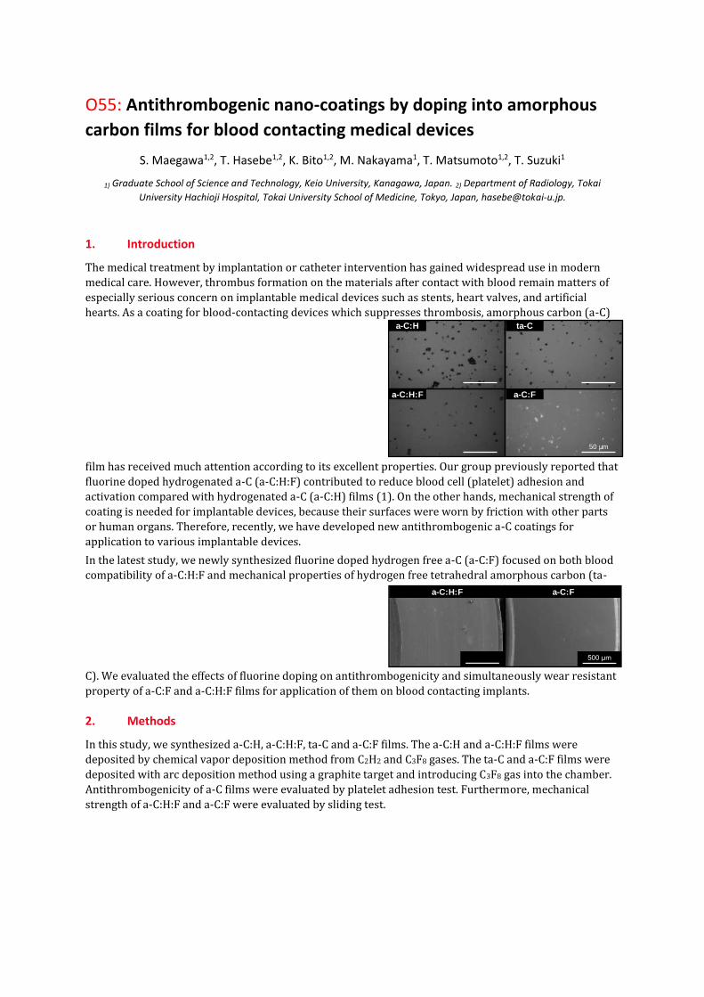
O55: Antithrombogenic nano-coatings by doping into amorphous
carbon films for blood contacting medical devices
S. Maegawa1,2, T. Hasebe1,2, K. Bito1,2, M. Nakayama1, T. Matsumoto1,2, T. Suzuki1
1) Graduate School of Science and Technology, Keio University, Kanagawa, Japan. 2) Department of Radiology, Tokai
University Hachioji Hospital, Tokai University School of Medicine, Tokyo, Japan, [email protected].
1. Introduction
The medical treatment by implantation or catheter intervention has gained widespread use in modern
medical care. However, thrombus formation on the materials after contact with blood remain matters of
especially serious concern on implantable medical devices such as stents, heart valves, and artificial
hearts. As a coating for blood-contacting devices which suppresses thrombosis, amorphous carbon (a-C)
film has received much attention according to its excellent properties. Our group previously reported that
fluorine doped hydrogenated a-C (a-C:H:F) contributed to reduce blood cell (platelet) adhesion and
activation compared with hydrogenated a-C (a-C:H) films (1). On the other hands, mechanical strength of
coating is needed for implantable devices, because their surfaces were worn by friction with other parts
or human organs. Therefore, recently, we have developed new antithrombogenic a-C coatings for
application to various implantable devices.
In the latest study, we newly synthesized fluorine doped hydrogen free a-C (a-C:F) focused on both blood
compatibility of a-C:H:F and mechanical properties of hydrogen free tetrahedral amorphous carbon (ta-
C). We evaluated the effects of fluorine doping on antithrombogenicity and simultaneously wear resistant
property of a-C:F and a-C:H:F films for application of them on blood contacting implants.
2. Methods
In this study, we synthesized a-C:H, a-C:H:F, ta-C and a-C:F films. The a-C:H and a-C:H:F films were
deposited by chemical vapor deposition method from C2H2 and C3F8 gases. The ta-C and a-C:F films were
deposited with arc deposition method using a graphite target and introducing C3F8 gas into the chamber.
Antithrombogenicity of a-C films were evaluated by platelet adhesion test. Furthermore, mechanical
strength of a-C:H:F and a-C:F were evaluated by sliding test.
a-C:H:F
a-C:H
a-C:F
ta-C
50 µm
a-C:H:F a-C:F

3. Results
As shown in Fig. 1, platelet adhesion was apparently suppressed on fluorine doped a-C surface compared
with a-C:H and ta-C surface. Furthermore, newly developed a-C:F films showed as good
antityrombogenicity as a-C:H:F films. These results showed that doping fluorine to a-C films affected the
enhancement of their antithrombogenic property without regard of their structure.
Figure 1: Images of platelet adhesion on a-C films.
The a-C:F films showed excellent mechanical characteristics compared with a-C:H:F films (Fig. 2). This
result suggested that a-C:F films is more suitable for mechanical parts of blood contacting devices than a-
C:H:F films which have good blood compatibility and proper flexibility for expandable stents.
Figure 2: Images of wear damages on fluorine doped a-C films after sliding test.
4. Discussion & Conclusion
We investigated the blood compatibility and mechanical property of a-C films and showed that fluorine
appears to be effective dopant in suppressing blood cell adhesion on a-C films. We can use the films
properly as antithrombogenic coatings for blood contacting implants in accordance to the goal of each
device.
References
[1] T. Saito, T. Hasebe, S. Yohena, Y. Matsuoka, A. Kamijo, K. Takahashi, et al., “Antithrombogenicity of fluorinated diamond-like
carbon films,” Diamond and Related Materials, vol. 14, p. 1116, 2005.

SESSION 29 – DIAGNOSTIC SYSTEMS

O107

O130
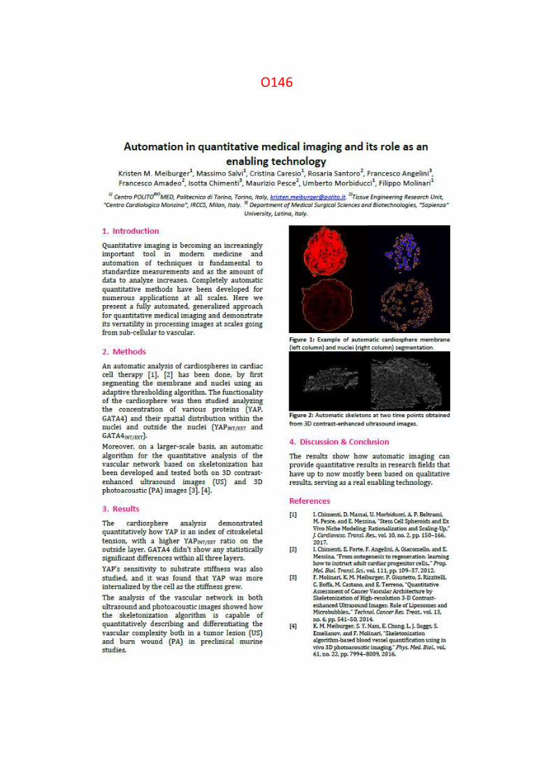
O146

SESSION 11 – ADVANCES IN RECTAL
CANCER SURGERY

O138: Retroperitoneoscopic left adrenalectomy
Giuseppe Giraudo, Simone Arolfo, Paolo Chiaro, Alberto Bullano, Mario Morino
University of Torino, Department of Surgical Sciences
Retroperineoscopic access represents a minimally invasive technique that radically modified
adrenalectomy. Retroperitoneoscopic adrenalectomy is currently adopted in about 20% of referral
centers. It provides more direct access to the adrenals, thus avoiding post-operative adhesions and
the need for patient repositioning in bilateral adrenalectomy. We would like to present a short video
showing the technique, which focuses on surgical landmarks when left adrenal gland is approached
during a posterior retroperitoneoscopic adrenalectomy The patient is a 45 years old woman with a
drug resistant arterial hypertension due to a left adrenal mass producing aldosterone (Conn disease).

SESSION 21 – ORGAN REPAIR &
TISSUE ENGINEERING

O3: Human Liver Stem Cells-derived Extracellular Vesicles reduce
hepatic injury in a murine model of ex vivo hypoxic normothermic
liver perfusion
Rigo Federica, Dorotea Roggio
Università degli studi di Torino, Chirurgia Generale 2 U, Centro Trapianto di Fegato, Torino, Italy
1. Introduction
The gold standard for organ storage during liver transplantation is hypothermia induced by icy
preservation solutions [1]. However, static cold storage is unable to protect completely the
graft from ischemia - reperfusion injury [2]. An emerging alternative is
normothermic machine perfusion (NMP) that provides basis for pharmaceutical interventions
during the preservation phase [3]. HLSC, a population of liver-resident pluripotent stem cells-
like and its derived extracellular vesicles (EV) have demonstrated to carry regenerative and
hepatoprotective properties in injured tissue [4]. In this study, we aimed to explore the
feasibility and efficacy of an organ-reconditioning strategy by using a
combination of NMP and HLSC-derived extracellular vesicles (HLSC-EV)
2. Methods
A murine model of NMP capable to maintain liver function of hypoxic-injured rat livers was
established. Livers were perfused ex vivo with a low haematocrit (<10%) during 4 hours in
the NMP system without (control group, n=10) or with HLSC-EV (treated group, n=9).
The uptake of HLSC-EV was analysed by immunofluorescence, while tissue injury was
analysed by haematoxylin-eosin staining. Then, apoptosis was assessed by TUNEL assay.
Total bile production was quantified and perfusate samples were collected hourly to assess
metabolic (pH, pO2, pCO2) and cytolysis (AST, ALT, LDH) parameters.
3. Results
During the hypoxic NMP perfusion, livers were able to maintain homeostasis and produce bile.
The HLSC-EV were randomly distributed into the hepatic parenchyma (Fig.1, A-D). EV-
treatment significantly reduced necrosis (Fig. 1, E) (Suzuki score 3.9±0.4, p=0.030), apoptosis
(Fig. 1, G) (apoptosis index 0.06±0.01, p=0.049), AST (47±7 U/L/g, p=0.018) and LDH
(340±47 U/L/g, p=0.032) at 3 hours of perfusion. The lower levels of AST in the treated group
was maintained at 4 hours (80±14 U/L/g, p=0.003).
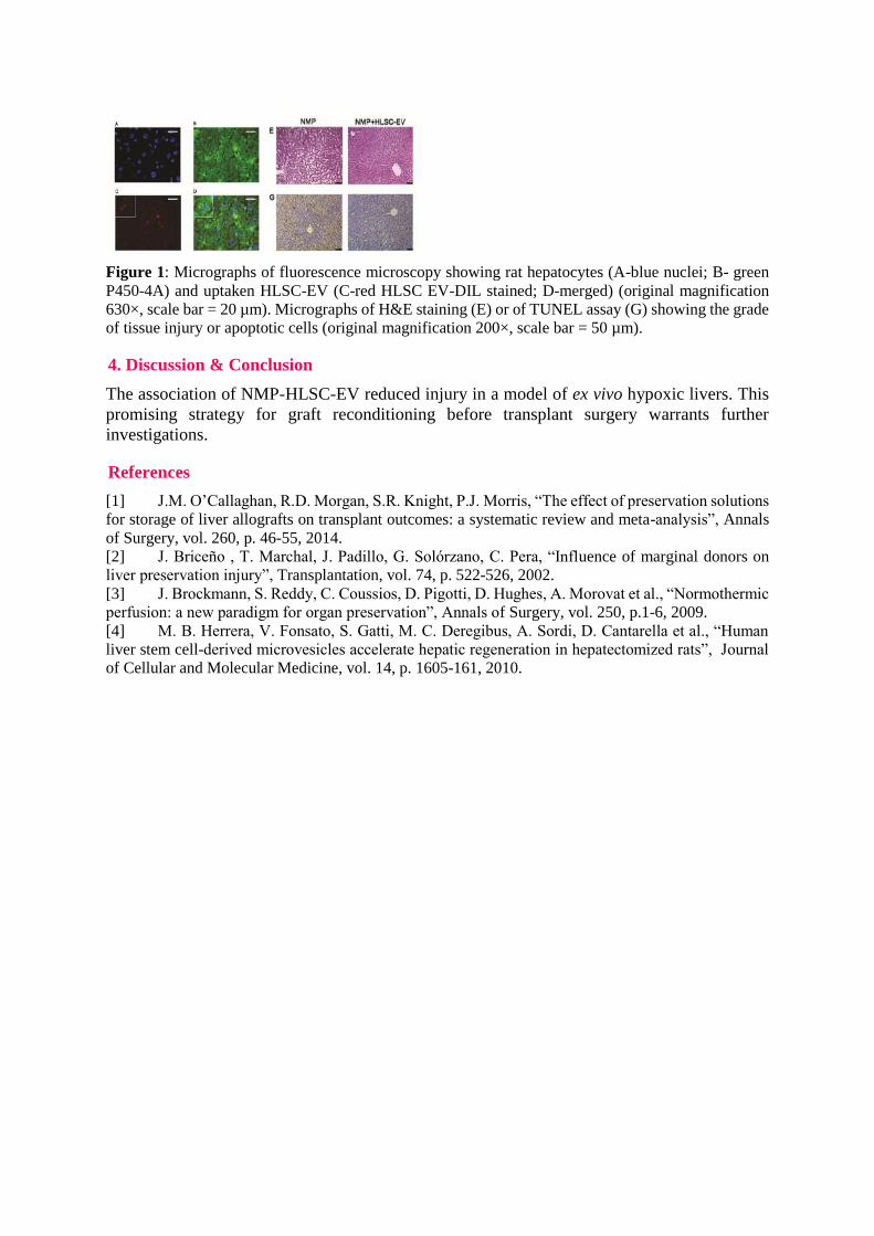
Figure 1: Micrographs of fluorescence microscopy showing rat hepatocytes (A-blue nuclei; B- green
P450-4A) and uptaken HLSC-EV (C-red HLSC EV-DIL stained; D-merged) (original magnification
630×, scale bar = 20 µm). Micrographs of H&E staining (E) or of TUNEL assay (G) showing the grade
of tissue injury or apoptotic cells (original magnification 200×, scale bar = 50 µm).
4. Discussion & Conclusion
The association of NMP-HLSC-EV reduced injury in a model of ex vivo hypoxic livers. This
promising strategy for graft reconditioning before transplant surgery warrants further
investigations.
References
[1] J.M. OʼCallaghan, R.D. Morgan, S.R. Knight, P.J. Morris, “The effect of preservation solutions
for storage of liver allografts on transplant outcomes: a systematic review and meta-analysis”, Annals
of Surgery, vol. 260, p. 46-55, 2014.
[2] J. Briceño , T. Marchal, J. Padillo, G. Solórzano, C. Pera, “Influence of marginal donors on
liver preservation injury”, Transplantation, vol. 74, p. 522-526, 2002.
[3] J. Brockmann, S. Reddy, C. Coussios, D. Pigotti, D. Hughes, A. Morovat et al., “Normothermic
perfusion: a new paradigm for organ preservation”, Annals of Surgery, vol. 250, p.1-6, 2009.
[4] M. B. Herrera, V. Fonsato, S. Gatti, M. C. Deregibus, A. Sordi, D. Cantarella et al., “Human
liver stem cell-derived microvesicles accelerate hepatic regeneration in hepatectomized rats”, Journal
of Cellular and Molecular Medicine, vol. 14, p. 1605-161, 2010.
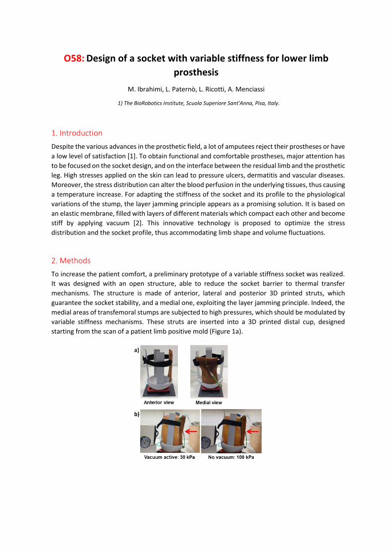
O58: Design of a socket with variable stiffness for lower limb
prosthesis
M. Ibrahimi, L. Paternò, L. Ricotti, A. Menciassi
1) The BioRobotics Institute, Scuola Superiore Sant'Anna, Pisa, Italy.
1. Introduction
Despite the various advances in the prosthetic field, a lot of amputees reject their prostheses or have
a low level of satisfaction [1]. To obtain functional and comfortable prostheses, major attention has
to be focused on the socket design, and on the interface between the residual limb and the prosthetic
leg. High stresses applied on the skin can lead to pressure ulcers, dermatitis and vascular diseases.
Moreover, the stress distribution can alter the blood perfusion in the underlying tissues, thus causing
a temperature increase. For adapting the stiffness of the socket and its profile to the physiological
variations of the stump, the layer jamming principle appears as a promising solution. It is based on
an elastic membrane, filled with layers of different materials which compact each other and become
stiff by applying vacuum [2]. This innovative technology is proposed to optimize the stress
distribution and the socket profile, thus accommodating limb shape and volume fluctuations.
2. Methods
To increase the patient comfort, a preliminary prototype of a variable stiffness socket was realized.
It was designed with an open structure, able to reduce the socket barrier to thermal transfer
mechanisms. The structure is made of anterior, lateral and posterior 3D printed struts, which
guarantee the socket stability, and a medial one, exploiting the layer jamming principle. Indeed, the
medial areas of transfemoral stumps are subjected to high pressures, which should be modulated by
variable stiffness mechanisms. These struts are inserted into a 3D printed distal cup, designed
starting from the scan of a patient limb positive mold (Figure 1a).

Figure 1: (a) Anterior and medial socket view. (b) Experimental set-up for pressure distribution measures. The arrows
indicate a change of the layer jamming profile and the area of contact pressure measurement.
3. Results
A transfemoral amputee was recruited to measure the pressures at the stump/ socket interface
and define the location of high pressures. The piezoresistive pressure sensor 5101 (Tekscan, Inc.,
USA) was placed in the internal surface of the socket, measuring pressure during sitting, standing
and single step tasks. Results showed critical areas at the medial site (~45 kPa, ~60 kPa, ~100 kPa
during sitting, standing and single step respectively). For this reason, the layer jamming strut was
designed for the medial area. Three different types of layers, made of sandpaper P1000, P180 and
woven P180, were tested with samples of 45 mm width, 90 mm height and 3 mm thickness, each
one including five layers. The bending torque was measured to be 13 N*mm for P1000, 17 N*mm
for P180 and 40 N*mm for woven P180, decreasing the pressure of 70 kPa compared to the
atmospheric pressure. Since the highest bending stiffness was reached with the woven P180, it was
used to realize the medial strut (90 mm width, 190 mm height and 5 mm thickness). Bending tests
were repeated showing a maximum value of 140 N*mm at 30 kPa. To validate the socket prototype,
the stiffness of the layer jamming strut was changed measuring the related pressure distribution at
the medial interface (Figure 1b). A latex bag was realized using the patient limb positive mold and
located inside the socket to simulate the stump. The tests showed a mean pressure reduction from
~2 kPa to 1.2 kPa, with a simultaneous change of the socket profile.
4. Discussion & Conclusion
The main objective of this work is to present a new design of a prosthetic socket based on the layer
jamming technology. Contrarily to multi-material socket, this design allows for a dynamic variable
stiffness (e.g. according to patient tasks), able to change the pressure distribution and to
accommodate shape and volume variations of the residual limb.
References
[1] H. E. Meulenbelt, et al., “Determinants of skin problems of the stump in lower-limb amputees,” Arch. Phys. Med. Rehabil., vol. 90, no. 1, pp. 74–81, 2009.
[2] J. Ou, et al., “JamSheets: Thin Interfaces with tunable stiffness enabled by layer jamming,” Proc. 8th Int. Conf. Tang., Embed. Embod. Int. ACM, pp. 65–72, 2014.

O73: Nanotechnology-Based Strategies To Treat Chronic Wounds
C. Tonda-Turo 1, M. Boffito 1, M.Gisbert Garzarán2, M.Manzano García2, F. Ruini 1, A. Torchio 1, R.
Laurano 1, C. Ceresa3, L. Fracchia3, P. Gentile4, A.M. Ferreira-Duarte4, M. Vallet Regí2, G. Ciardelli 1.
1 Department of Mechanical and Aerospace Engineering, Politecnico di Torino, Torino, Italy
2 Dept. Química Inorgánica y Bioinorgánica. Facultad de Farmacia, Universidad Complutense De Madrid, Spain
3 Department of Pharmaceutical Sciences, Università del Piemonte Orientale “A. Avogadro”, Novara, Italy
4 School of Engineering, Newcastle University, Stephenson Building, Claremont Road, Newcastle upon Tyne, NE1 7RU, UK
1. Introduction
Skin lesions are a global healthcare problem since they can compromise the integrity and the functionality of significant skin areas. More than 40 million people suffer from chronic wounds worldwide (8 million in Europe and 6.5 million in US in 2009), most commonly caused by poor blood supply to the lower limbs. Chronic wound is a frequent and very severe problem in patients with diabetes mellitus, a pathological condition involving around 285 million people globally. In this work, different approaches to treat chronic wounds are presented based on antibacterial gelatin nanofibres or innovative polyurethane-based hydrogels loaded with multifunctional mesoporous nanoparticles.
2. Materials and Methods
Silver nanoparticles (AgNPs) and gentamicin sulphate (GS) loaded gelatin nanofibres were obtained by electrospinning starting from aqueous solution to avoid protein denaturation [1].
Thermosensitive amphiphilic polyurethanes (APU) were synthesized according to a patented procedure [2]. Moreover, novel injectable hydrogels for the targeted release of anti-inflammatory drugs and/or ions with antibacterial properties was designed and characterized.
3. Results
The antibacterial properties of AgNPs and GS loaded nanofibres were tested against four pathogenic bacteria isolated from infected wounds (Staphylococcus aureus, Escherichia coli, Staphylococcus Epidermidis, Pseudomonas aeruginosa) and high efficiency against tested strains was observed. Furthermore, the biocompatibility of the developed nanofibers was confirmed using Neonatal Normal Human Dermal Fibroblasts (NHDF-Neo).
APU were successfully synthetized showing: (1) biocompatibility; (2) long-term stability in biological environment and degradation rate compatible with tissue regeneration rate; (3) solubility in aqueous media; (4) low viscosity at 20°C; (5) gelation at 37°C; (8) ability to encapsulate biomolecules and drugs to achieve a therapeutic effect [3]. Moreover, hydrogel chemistry was tuned to exploit the variation in pH value that characterizes chronic wounds as a smart stimulus to tune the release kinetics of previously encapsulated biomolecules. These pH-sensitive innovative hydrogels were characterized and proposed to treat infected wounds within an innovative approach based on the pH-driven release of
antibacterial ions and drugs loaded into mesoporous nanoparticles coated using a pH-sensitive and self-immolative polymer [4].
4. Discussion & Conclusion
The treatment of chronic wounds requires a multifunctional approach based on nanotechnology to combine long-term antibacterial properties with tissue regeneration strategies. In this work, recent findings using gelatin based nanofibres are reported and novel concepts and future targets are presented exploiting the properties of smart polyurethane-based hydrogels combined with mesoporous and multifunctional nanoparticles.
Acknowledgements
This work was financially supported by the Horizon 2020 European Union funding for Research & Innovation (H2020-NMP6-2015) project “Mozart” (MesopOrous matrices for localiZed pH-triggered releAse of theRapeuTic ions and drugs).
References
[1] Tonda-Turo et al., MSEC 2015
[2] Ciardelli et al., 2012 WO 2014016857
[3] Boffito et al., Inter Polym 2016
[4] Gisbert-Garzaran et al., RSC Adv.2017

1. Introduction
Chronic skin wounds affect more than 40 million patients worldwide. Their treatment represents a growing burden for healthcare systems due to the increasing health care costs, the population aging and the rise in diabetes and obesity incidence. The ideal wound dressing should (i) provide a moist wound environment, (ii) protect from external agents, (iii) remove exudate from the wound, and (iv) enhance tissue regeneration. However, no existing treatment fulfils all these demands [1]. Hence, the design of innovative, smart and effective wound dressings represents a current and important challenge. In this work, we developed a thermo-responsive polyurethane-based hydrogel able to transfer pH changes from the biological milieu (pH in the range 7.4-8.5) to its core, triggering the release of anti-inflammatory drugs.
2. Methods
An amphiphilic polyurethane (PU) (CHP407) was synthesized starting from Poloxamer P407, 1,6-hexamethylene diisocyanate and 1,4-cycloexane dimethanol [2]. CHP407 powder was plasma treated with Acrylic Acid vapours to introduce carboxylic groups along the polymer chains (PCHP407), thus enhancing hydrogel sensitivity to basic pH. PUs were characterized by Size Exclusion Chromatography, Attenuated Total Reflectance Fourier transform Infrared spectroscopy (ATR-FTIR), Proton Nuclear Magnetic Resonance (1H-NMR) and Toluidine Blue O assay (TBO). CHP407 and PCHP407 hydrogels were characterized in terms of sensitivity to temperature (tube inverting and gelation time tests, rheology) and external pH (swelling tests in the presence of basic or acid buffers). pH-triggered release of hydrophilic and hydrophobic anti-inflammatory drugs was studied. Cytotoxicity was assessed according to ISO10993.
3. Results
Chemical characterization assessed the successful synthesis of CHP407 with a final number average molecular weight of 50 kDa and a polydispersity index of 1.3. TBO assay, 1H-NMR and ATR-FTIR demonstrated the introduction of carboxylic groups along the polymer chains and evidenced a high intra- and inter-synthesis repeatability of plasma treatment. Tube inverting, gelation time and rheological tests showed the ability of 15%w/v concentrated PU solutions to undergo a sol-to-gel transition at about 27°C within few minutes. No differences in gelation kinetics of CHP407- and PCHP407-based hydrogels were reported and both systems were in a complete gel state at 37°C,
demonstrating that plasma treatment does not affect hydrogel thermo-sensitivity. Concerning sensitivity to basic pH, PCHP407-based hydrogels transferred basic pH of the surrounding environment to the gel core with a significantly faster kinetics and a significantly higher swelling if compared to CHP407 gels. On the other hand, CHP407- and PCHP407-based hydrogels showed a similar behaviour in the presence of acid buffers. Hence, the introduction of -COOH groups along CHP407 chains increased hydrogel sensitivity to basic pH. These data were also confirmed by studying the pH-triggered release of hydrophobic and hydrophilic drugs. Cytotoxicity tests performed on hydrogel extracts with NIH-3T3 murine fibroblasts showed no cytotoxicity.
4. Discussion and Conclusion
PU-based hydrogels are promising candidates in chronic skin wound treatment as they are characterized by (i) easy injectability, (ii) a sol-to-gel transition within few minutes at 37°C, (iii) a moist environment and (iv) capability to fill cavities without causing pressure. Furthermore, COOH-modified PU-based (i.e. PCHP407) hydrogels showed faster inner pH variations and higher swelling with respect to native ones (i.e. based on CHP407), resulting in pH-triggered and accelerated release kinetics of encapsulated drugs.
5. Acknowledgements
This work was supported by the Horizon 2020 European Union funding for Research & Innovation project “MOZART” (MesopOrous matrices for localiZed pH-triggered releAse of theRapeuTic ions and drugs) (H2020-NMP6-2015).
References
[1] L.I.F. Moura, A.M.A. Dias, E. Carvalho, H.C. de Sousa, “Recent advances on the development of wound dressings for diabetic foot ulcer treatment - a review”, Acta Biomateriala, vol. 9, p. 7093-7114, 2013 [2] M. Boffito, E. Gioffredi, V. Chiono, S. Calzone, E. Ranzato, S. Martinotti, G. Ciardelli, “Novel polyurethane-based thermo-sensitive hydrogels as drug release and tissue engineering platforms: design and in vitro characterization”, Polymer International, vol. 65, p. 756 – 769, 2016
O88: Injectable polyurethane-based hydrogels for smart drug
release in the treatment of chronic skin wounds
Laurano Rossella, Torchio Alessandro, Boffito Monica, Ciardelli Gianluca
Politecnico di Torino, Department of Mechanical and Aerospace Engineering, Turin, Italy

O149: Collagen-based biomimetic smart scaffold for bone tissue
engineering
Giulia Montalbano, Giulia Molino, S. Fiorilli, Chiara Vitale- Brovarone
Politecnico Di Torino, Dipartimento Di Scienza Applicata E Tecnologia, Torino, Italy

O24: Plant derived natural molecules: a promising way for surface
functionalization of biomaterials in bone contact applications
Cazzola Martina, Ferraris Sara, Cempura Grzegorz, Prenesti Enrico, Allizond Valeria, Banche Giuliana,
Cuffini Anna Maria, Bertea Cinzia Margherita, Cochis Andrea, Rimondini Lia, Pezzotti Giuseppe,
Spriano Silvia, Vernè Enrica
AGH University of Science and Technology, International Centre of Electron Microscopy for Materials Science &
Faculty of Metals Engineering and Industrial Computer Science, Krakow, Poland, Kyoto Institute of Technology,
Ceramic Physics Laboratory, Kyoto, Japan, Politecnico di torino, Department of Applied Science and Technology,
Torino, Italy, Università degli studi di Torino, Department of Chemistry, Torino, Italy, Università degli studi di
torino, Department of Public Health and Pediatrics, Torino, Italy, Università degli studi di Torino, Plant Physiology
Unit, Department Life Sciences and Systems Biology, Torino, Italy, Università del Piemonte Orientale UPO,
Department of HealthSciences, Novara, Italy

1. Introduction Mixtures of molecules derived from plant natural extracts (egg. polyphenols, essential oils) have interesting properties as antibacterial, antitumoral, antinflammatory and osteoinductive activity[1,2]. It is possible to combine the properties of these molecules with those of bioactive materials used in bone contact application by mean of surface functionalization. As an alternative polyphenols are also reductive agents useful for the in situ reduction of silver nanoparticles in order to confer to the materials inorganic antibacterial activities.
2. Methods The substrates used for the surface functionalization were a silica based bioactive glass [3] and a Ti6Al4V alloy made bioactive by means of a patented chemical treatment[4]. The surfaces were functionalized with different molecules reported in figure 1 (gallic acid as model molecules, polyphenols from red grape skins, green tea leaves and Mentha piperita essential oil of Pancalieri). The functionalization protocols were tailored using different solvents and different time of functionalization suitable for each kind of biomolecules and for the two different substrates. The presence and the activity of the molecules and ions on the surfaces were analyzed by means of XPS, FTIR, gas chromatograpyc and spectroscopic analyses. The effect of these molecules on cells and bacteria was also investigated with biological tests. The silver ions release was investigated in different media.
3. Results The presence and the activity of the polyphenols on the surfaces were proved by means of XPS, FTIR and spectroscopic measurements with the Folin&Ciocalteu methods. Cell cultures also prove the ability of polyphenols to promote osteointegration with healthy osteoblast and induce differentiation in mesenchymal stem cells and promote apoptosis of cancer cells. The presence of the molecules of Mentha oil and silver nanoparticles was checked with GC, FTIR and XPS; they have effect against bacteria, but they show also toxicity against cells. For the samples with silver nanoparticles a well dispersion of the nanoparticles on the substrate and a release at 14 days were observed.
4. Discussion & Conclusion Regarding polyphenols, the surface functionalization seems to be a promising way for in loco use of these molecules rich of beneficial effects on human health. For those concern the samples with the Mentha oil and the silver nanoparticles, the protocol of functionalization needs to be improved in order to tailor the amount of Mentha oil molecules and silver nanoparticles to avoid cytotoxic effects maintaining the great antibacterial activity. The biomaterial surface functionalization is a promising interest field in order to combine the properties of biomolecules with those of biomaterials. It’s possible to confer them new properties using natural molecules with high value but low price that could be also used as additives during different process like nanoparticles reduction.
Figure1: Material and chemicals used for the
work.
References [1] Quideau, S. et al., Angewandte Chemie International Edition 50.3, 586-621, (2011) [2] Swamy, M. K., et al., Evidence-Based Complementary and Alternative Medicine, (2016). [3] Vernè, E. et al., Journal of biomedical materials research Part A, 90(4), 981-992. (2009)
[4] S.Spriano et al., EuropeanPatent 2214732

O12: In-vitro detection of small isolated Cartilage Defects: Intra-Vascular Ultrasound vs. Optical Coherence Tomography
T.Horeman1 ,E.C. Buiter1, B. Pouran2, M. Stijntjes1, J. Dankelman1, G.J.M. Tuijthof 3
1) BioMechanical engineering department, Delft, NL. Delft University of Technology. 2) Department of Orthopaedics, UMC
Utrecht, NL. 3) Faculty Beta Sciences & Technology, Zuyd University of Applied Sciences, Maastricht, NL
1. Introduction Ankle sprains and cartilage fractures are commonly seen in orthopaedics. In up to 50% of all ankle sprains and fractures, isolated cartilage defects occur [1]. These defects affect the articular cartilage and subchondral bone, which can result in deep ankle pain, stiffness of the joint, impaired movement and if left untreated posttraumatic osteoarthritis [2]. This experimental work focuses on a critical aspect in the development of a minimally invasive needle based intervention to treat small cartilage defects in joints: the sensor that identifies the location of the defect in the joint (Figure 1).
Figure 1: an artist impression of a multifunctional steerable needle device currently in development for detection and treatment of articular cartilage defects in the ankle.
The goal of this study is to identify the most accurate imaging method (Intra-Vascular ultrasound imaging (IVUS) vs. optical coherent tomography (OCT)) that is suitable for integration in a needle compared to a golden standard (µCT).
2. Methods An in-vitro study was conducted on human Talar bone specimens that were dissected and placed submerged. To simulate natural appearance of cartilage defects 4 types of defects were created according to a standardized protocol: chondral defects, osteochondral defects, bruises and cartilage surface fibrillation, all sized between 0.1-3 mm diameter. A test setup was built to fixate the talus inside a tank and a catheter
holding system was developed to scan the bone with different catheters (Figure 2).
Figure 2: , A picture of the part of the experimental setup that holds the catheters in a constant position and orientation above measurement location on the bone (Talus) surface.
3. Results The bruises could not be visualised with any of the imaging techniques. The detection rate of the osteochondral defects was 100% by both observers for µCT, 80% for IVUS and 92% for OCT. The detection rate of chondral defects was 97% by both observers for µCT, 60% for IVUS and 83% for OCT. The detection rate of cartilage surface fibrillation was 24% by both observers for µCT, 0% for IVUS and 29% for OCT. The diameter accuracy for OCD and CD showed a significant difference between the golden standard µCT and OCT (p=0.003 osteochondral, p<0.001 chondral) and the depth accuracy (p=0.01 osteochondral, p=0.03 chondral). This was as well the case for OCD between µCT and IVUS (p<0.001).
4. Discussion & Conclusion Both imaging methods can detect the presence of OCD and CD accurately if sized larger than 2 mm, and OCT can detect fibrillated cartilage surface as well if at least larger than 3 mm in size. Thus, a slight preference exists to continue with OCT as is more sensitive to various types and sizes of defects.
References [1] P. F. O’Loughlin, B. E. Heyworth, and J. G. Kennedy, “Current concepts in the diagnosis and treatment of osteochondral lesions of the ankle.,” Am. J. Sports Med., vol. 38, pp. 392–404, 2010.

SESSION 31 – NEURO@SMIT2017:
NEW TECHNIQUES AND DEVICES IN
NEUROSEURGERY II
0151: Anatomic model for artery injury simulation in management
of vascular complications in endoscopic endonasal surgery
Pacca Paolo
A.O.U. città della scienza e della salute, division of neurosurgery, department of neuroscience,
university of Torino, Italy
Introduction: Intraoperative injury of the internal carotid artery (ICA) is the most dreaded
complication in endoscopic endonasal surgery (EES) of skull base (1). Training for ICA injury is
practically impossible in live operative settings (2)In this study, we evaluate a pulsatile perfusion-based
live cadaveric model for ICA injury simulation in a laboratory setting. The major emphasis of the study
was to evaluate various means of controlling acute bleeding and evaluating the practical utility of this
model for training purposes
Methods: Five embalmed uninjected cadaveric heads were prepared for study by connecting to a
pulsatile perfusion pump system filled with artificial blood solution. EES approaches were used to
evaluate different types of ICA injuries similar to operative scenarios (3). Various methods of managing
ICA injuries such as packing, clipping, trapping, were evaluated. The educational advantages of the live
cadaver model were assessed using questionnaires given to participants in a hands-on dissection
course
Results: The trainee was faced with several scenarios similar to those encountered during an actual
intraoperative ICA injury. Packing, clipping and trapping of the ICA injury was successfully achieved in
all segments of the ICA (4). Clipbased reconstruction techniques were successfully developed. All
trainees reported gaining new knowledge, learning new techniques (5). The responses to the
questionnaire confirmed the significant educational value of this model.
Conclusions: The live cadaver model presented here provides real life experience with major vessel
injury during EES in a laboratory setting. This model could significantly improve current training for
management of intraoperative vascular injuries during EES
Reference:
1. Gardner PA, Tormenti MJ, Pant H, Fernandez-Miranda JC, Snyderman CH, Horowitz MB. Carotid artery injury during
endoscopic endonasal skull base surgery: incidence and outcomes. Neurosurgery. 2013 Dec;73(2 Suppl Operative):ons261-9;
discussion ons269-70.
2. Fernandez-Miranda JC, Barges-Coll J, Prevedello DM, et al. Animal model for endoscopic neurosurgical training: technical
note. Minim Invasive Neurosurg. 2010 Oct;53(5-6):286–9.
3. Aboud E, Aboud G, Al-Mefty O, et al. "Live cadavers" for training in the management of intraoperative aneurysmal rupture.
JNeurosurgy. 2015 Nov;123(5):1339-46.
4. Gardner PA, Vaz-Guimaraes F, Fernandez-Miranda JC, Wang E, Snyderman C. Endoscopic endonasal clipping of intracranial
aneurysms: surgical technique. World Neurosurgery. 2015 Nov; 84(5):1380-93.
5. Snyderman C, Kassam A, Carrau R, Mintz A, Gardner P, Prevedello DM. Acquisition of surgical skills for endonasal skull base
surgery: a training program. Laryngoscope. 2007 Apr;117(4):699–705.

O102

O115
O122:


POSTERS PER SESSIONS

DENTAL SURGERY

44: Focus on soft tissue cell response elicited by
anodized titanium Serra Francesca Giulia, Genova Tullio, Zicola Elisa, Mussano Federico, Carossa Stefano-CIR Dental School,
Department of Surgical Sciences, University of Turin, Turin, Italy
Introduction
The aim of this work was to investigate how anodization and silicon-based amorphous coatings may
enhance the adhesion of soft tissue cells to the Ti surfaces. Therefore, the purpose of this study was to
determine in vitro the early cell response of human epithelial cells and fibroblasts on the
aforementioned surface modifications.
1. Methods
Ti-Al-V titanium samples were prepared and shaped as 12 × 12 × 4 mm cylinders (l × l × h). Four types of
specimens were attained: pristine titanium (Ti, unmodified control), anodized titanium (AnoTi) and two
different thin film coatings based on amorphous silicon (a-Si_90 and a-Si_350). Microstructure was
studied by means of a Scanning Electron Microscope (Zeiss EVO 50, Carl Zeiss AG, Oberkochen,
Germany). The wetting properties were investigated by optical contact angle (OCA) measurements with
the sessile drop technique, using an OCAH 200 (DataPhysic Instruments GmbH). To characterize the
biological response in vitro, the epithelial cell line HACAT and the fibroblast cell line NHDF (ECACC,
Salisbury, UK) were used. Cells were maintained in DMEM supplemented with 10% fetal bovine serum
(Life Technologies, Milan, Italy), 100 U/ml penicillin, 100 U/ml streptomycin, were passaged at
subconfluency to prevent contact inhibition and were kept under a humidified atmosphere of 5% CO2
in air, at 37°C. Cell adhesion was evaluated by counting cell nuclei. The proliferation rate was assessed
by Cell Titer GLO (Promega, Milan, Italy) according to the manufacturer’s protocol at 2 days. Data were
analysed by GraphPad Prism6 (GraphPad Software, Inc., La Jolla, CA, USA). Each experiment was
repeated at least three times. Statistical analysis was performed by using the Student t-test. A p value
of <0.05 was considered significant.
2. Results
The non-treated Ti cylinders show a quite hydrophilic behaviour, with an average contact angle (CA)
value for water and diiodomethane (CH2I2) of 35° and 40° respectively. After the anodization process,
a transition toward the hydrophobic regime was found. The a-Si coating is able to impart two opposite
behaviours to the surface, according to the difference in the growth temperature of the film. The
surface of the cylinders coated with a-Si grown at low temperature (sample a-Si_90) showed nearly
the same hydrophobic behaviour with respect to the anodized sample (Ano-Ti), with an average CA of
80° and 49° for H2O and CH2I2, respectively. On the other hand, the wetting behaviour of the high
temperature grown a-Si coating (sample a-Si_350) is comparable with the one observed for the
untreated Ti samples. HaCaT cells display an increased proliferation level on AnoTi samples whereas
NHDF shown a similar proliferation level on AnoTi and non-treated Ti samples, showing a reduced

viability on a-Si_90 and a-Si_350. HaCaT shown a high level of adhesion on AnoTi and as-Si_90 samples
at both 2 and 24 hours. Furthermore, NHDF cells display a significantly higher adhesion level only on
AnoTi sample at both 2 and 24 h (Fig.1).
Figure 1: Representative pictures of cell morphology. Fluorescence photomicrographs of HaCaT (A-D) and NHDF (E-H) seeded
on different samples for 24 h. The cells were stained for the nucleus (DAPI, blue), the actin (rhodamine-phalloidin, green) and
the focal adhesions (paxillin, red).
3. Discussion & Conclusion
In conclusion, this work supports the use of titanium anodization as a strategy to enhance the
adhesion of soft tissue cells to the Ti surfaces.
References
[1] I.Abrahamsson, N.U. Zitzmann, T. Berglundh, E. Linder, A. Wennerberg, J. Lindhe, “The mucosal attachment to titanium implants with different surface characteristics: an experimental study in dogs.” J Clin Periodontol vol.29, p. 448, 2002.
[2] R.S. Williamson, I.P. Blakely, L.M. Didier, “Tuning anatase and rutile phase ratios and nanoscale surface features by anodization processing onto titanium substrate surfaces.” Mater. Sci. Eng. vol.58, p.213, 2015.
[3] P. Mandracci, F. Mussano, P. Ceruti, C.F. Pirri, S. Carossa “Reduction of bacterial adhesion on dental composite resins by silicon–oxygen thin film coatings.” Biomedical Materials vol.1(10):015017, 2015.
49:
OCT for oral measurement: a preliminary study. Gambino
1,2, P.G. Arduino
2, S. Schäfer 3, A. Cafaro
2, S. Carossa
2
1) DIMEAS, Politecnico di Torino, Turin, Italy, [email protected] 2) CIR Dental School, Department of Surgical Sciences, University of Turin, Turin, Italy. 3) Thorlabs GmbH, Hamburg, Germany
1. Introduction We use optical coherence tomography (OCT) to perform a comprehensive program of in vivo and in vitro structural imaging of hard and soft tissues within the oral cavity. We have imaged the different types of healthy
oral mucosa. The aim of this work was to determine whether OCT is a good technique to: image lip, inner cheek,
gum, and the top and bottom of the tongue.
2. Methods The handheld probe was used for all measurements. An air spacer was used to keep the probe at a fixed distance to the tissue (1300 nm). The 900nm handheld probe was only available with a prototype immersion spacer, hence the surface in the images is always in the shape of the spacer. The TEL220 system with OCTH-LK20 objective gets 1300 nm center wavelength, a scan rate up to 76 kHz, an image depth by 3,5 mm in air, an axial resolution 3 µm in air and a lateral resolution of 9 µm. Measurements were performed with the lightweight handheld OCT scanner (OCTH). The OCTH should be used with a spacer that keeps the sample at a fixed distance (in focus).
3. Results The comparison between the scans of the two probes shows similar images for both groups examined in oral structures: cheek, gum, lip, tongue top, tongue bottom. Both 900 nm and 1300 nm can be used for oral OCT imaging. The probe by 900 nm show a higher axial and lateral resolution, the images show more details. The probe by 1300 nm show a higher penetration depth, data can be acquired from deeper lying tissue (Fig.1). Since ,the choice of the ideal wavelength depends on the exact application. The small OCTH design allows to image in the mouth. Both spacers work, since the immersion spacer is just a prototype we would recommend the air spacer.

Figure 1: Representative pictures of tissue morphology by cheeck. The difference between image A where sharper surface
images and image B are displayed where we have more depth data.
4. Discussion & Conclusion OCT imaging was rapid, unproblematic and well received by all patients, with the imaging protocol adding
only a few minutes to visit duration. Thus, the introduction of OCT imaging techniques to routine patient visits
should be well received by patients and clinicians alike. In fact, OCT accurately depicts dental tissue structure
and is able to detect small anomalies in that structure. These results suggest that OCT is a potentially useful
modality for dental clinical and research applications.
References [1] P. Wilder-Smith, K. Lee, S. Guo, , J. Zhang, K. Osann, Z. Chen , and D. Messadi. “In Vivo Diagnosis of Oral Dysplasia and Malignancy Using Optical Coherence Tomography: Preliminary Studies in 50 Patient.” Lasers Surg Med. 2009 July ; 41(5): 353–357. doi:10.1002/lsm.20773. [2] Matheny ES, Hanna N, Mina-Araghi R, Jung WG, Chen Z, Wilder-Smith P, Brenner M. “Optical coherence tomography of malignant Hamster Cheek Pouche “s. J Invest Med 2003;51(1):S78. [3] Fujimoto, JG.; Hee, MR.” Optical coherence tomography: Introduction and theory”. In: Bouma, BE.; Tearney, GJ., editors. Handbook of Optical Coherence Tomography. NY: Marcel Dekker, Inc; 2002.p. 1-143
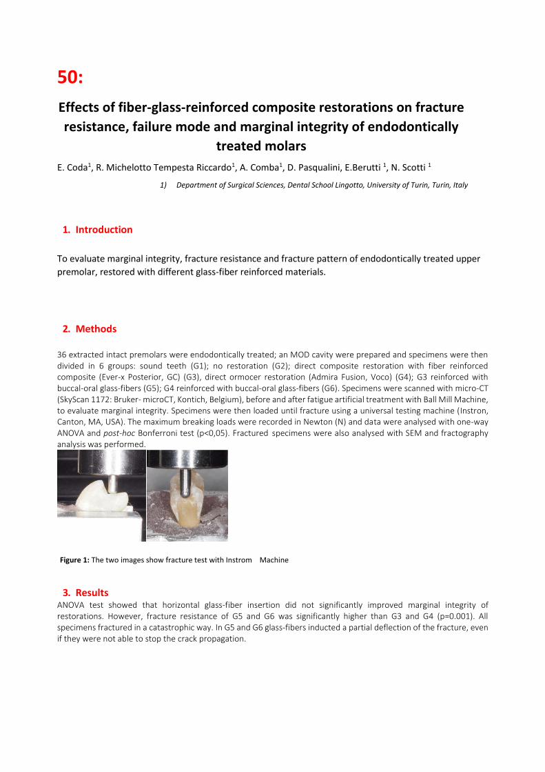
50:
Effects of fiber-glass-reinforced composite restorations on fracture
resistance, failure mode and marginal integrity of endodontically
treated molars
E. Coda1, R. Michelotto Tempesta Riccardo1, A. Comba1, D. Pasqualini, E.Berutti 1, N. Scotti 1
1) Department of Surgical Sciences, Dental School Lingotto, University of Turin, Turin, Italy
1. Introduction
To evaluate marginal integrity, fracture resistance and fracture pattern of endodontically treated upper
premolar, restored with different glass-fiber reinforced materials.
2. Methods
36 extracted intact premolars were endodontically treated; an MOD cavity were prepared and specimens were then divided in 6 groups: sound teeth (G1); no restoration (G2); direct composite restoration with fiber reinforced composite (Ever-x Posterior, GC) (G3), direct ormocer restoration (Admira Fusion, Voco) (G4); G3 reinforced with buccal-oral glass-fibers (G5); G4 reinforced with buccal-oral glass-fibers (G6). Specimens were scanned with micro-CT (SkyScan 1172: Bruker- microCT, Kontich, Belgium), before and after fatigue artificial treatment with Ball Mill Machine, to evaluate marginal integrity. Specimens were then loaded until fracture using a universal testing machine (Instron, Canton, MA, USA). The maximum breaking loads were recorded in Newton (N) and data were analysed with one-way ANOVA and post-hoc Bonferroni test (p<0,05). Fractured specimens were also analysed with SEM and fractography analysis was performed.
Figure 1: The two images show fracture test with Instrom Machine
3. Results ANOVA test showed that horizontal glass-fiber insertion did not significantly improved marginal integrity of restorations. However, fracture resistance of G5 and G6 was significantly higher than G3 and G4 (p=0.001). All specimens fractured in a catastrophic way. In G5 and G6 glass-fibers inducted a partial deflection of the fracture, even if they were not able to stop the crack propagation.
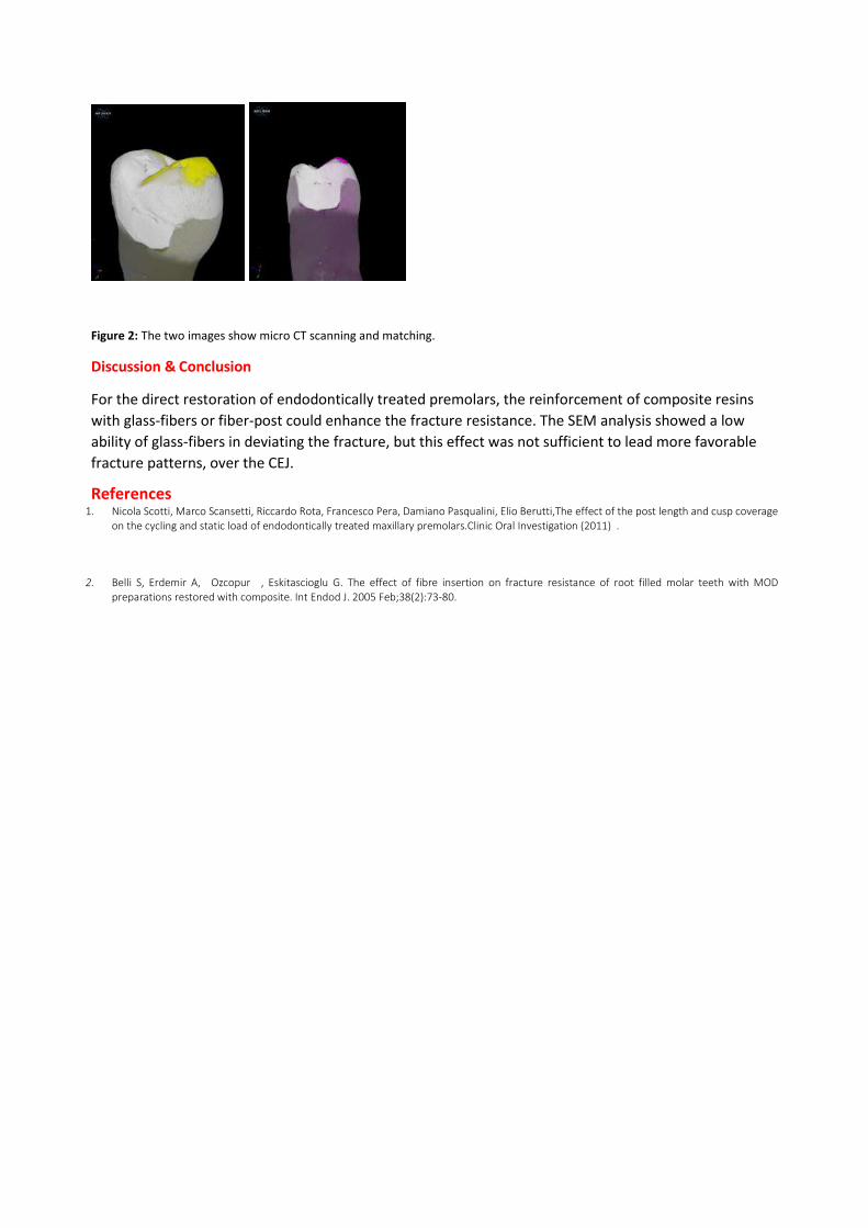
Figure 2: The two images show micro CT scanning and matching.
Discussion & Conclusion
For the direct restoration of endodontically treated premolars, the reinforcement of composite resins
with glass-fibers or fiber-post could enhance the fracture resistance. The SEM analysis showed a low
ability of glass-fibers in deviating the fracture, but this effect was not sufficient to lead more favorable
fracture patterns, over the CEJ.
References 1. Nicola Scotti, Marco Scansetti, Riccardo Rota, Francesco Pera, Damiano Pasqualini, Elio Berutti,The effect of the post length and cusp coverage
on the cycling and static load of endodontically treated maxillary premolars.Clinic Oral Investigation (2011) .
2. Belli S, Erdemir A, Ozcopur , Eskitascioglu G. The effect of fibre insertion on fracture resistance of root filled molar teeth with MOD preparations restored with composite. Int Endod J. 2005 Feb;38(2):73-80.

76:
Argon-based treatments for titanium dental implants to
introduce organic functionalities
T. Genova1,2
, F.G. Serra1,3
, A. Patelli4, E. Zicola
5, F. Mussano
1, S. Carossa
1
1) CIR Dental School, Department of Surgical Sciences, University of Turin, Turin, Italy. 2) DIBIOS,
University of Turin, Turin, 3) DIMEAS, Politecnico di Torino, Turin, Italy, 4) Department of Physics and
Astronomy, UNIPD, Padova, Italy 5) Department of Clinical and Biological Sciences, University of Turin,
Turin, Italy.
1. Introduction
Plasma polymers deposition and plasma surface activation are technologies capable to enhance
biologically relevant surface features of biomaterials. The aim of this study was to evaluate the biological
effects of two different surface modifications, i.e. amine (NH2-Ti) and carboxylic/esteric (COOH/R-Ti)
functionalities.These modifications were obtained from aminopropyltriethoxysilane (APTES) and
methylmethacrylate (MMA) precursors, respectively, through an atmospheric plasma jet RF-APPJ
portable equipment.
2. Methods
Three types of specimens were used: pristine titanium (Ti, unmodified control), titanium with carboxylic/
carboxylate functionalization (COOH/R-Ti) and titanium with amine functionalization (NH2-Ti).
Methylmethacrylate (MMA, Sigma Aldrich, ≥ 99%) has been used for carboxylic/carboxylate groups while
amine functional groups have been obtained using aminopropyltriethoxysilane (APTES, Sigma Aldrich, ≥
98%) as precursor. All sample coatings were characterized by Scanning Electron Microscopy, XPS, FT-IR
spectroscopy and surface energy calculations. Stability after UV sterilisation and in water was also
verified.
Total protein amount was evaluated using SERVA
BCA Protein Assay Micro Kit (SERVA
Electrophoresis GmbH, Heidelberg, Germany). To perform the in vitro tests, the pre-osteoblastic murine
cell line MC3T3-E1 was used. Cell adhesion was evaluated on titanium samples by counting cell nuclei.
Cell proliferation was evaluated using Cell Titer GLO (Promega, Milan, Italy) according to the
manufacturer’s protocol at 1 and 2 days. To assess the osteogenic differentiation Osteocalcin (OCN) was
quantified in cell conditioned media by the use of Mouse Osteocalcin ELISA Kit (MyBioSource, Inc, San
Diego, USA) following manufacturer’s instructions.
3. Results
Both treated samples showed a higher quantity of adsorbed proteins and they also improved osteoblast
cells adhesion on the surfaces compared to the pristine titanium. In particular the COOH/R-Ti led to a
nearly two-fold improvement of cell adhesion. Cell proliferation at 24 h on coated samples was initially
lower than on titanium control, while, at 48 h, COOH/R-Ti reached the same proliferation rate as
pristine titanium. Cells grown on NH2-Ti were more elongated and tapered in shape with smaller areas
than on COOH/R-Ti enriched surfaces. Furthermore, NH2-Ti significantly enhanced osteocalcin
production, starting from 14 days, whereas COOH/R-Ti had this effect only from 21 days. Notably, NH2-
Ti was more efficient than COOH/R-Ti at 21 days. The NH2-Ti surface elicited the most relevant

osteogenic effect in terms of osteocalcin expression: this establishes an interesting correlation between
early cell morphology and later differentiation stages.
Figure 1: Representative figure of wettability, protein adsorption, cell adhesion, cell proliferation, cell area, focal adhesion density and osteoblast differentiation in untreated sample, in titanium with amine functionalization (NH2-Ti) sample and in titanium with carboxylic/carboxylate functionalization (COOH/R-Ti) sample.
4.Discussion & Conclusion
The flexibility of the presented surface functionalization process, by virtue of a wide range of potential
monomers in aerosol or vapour phase, offers the researchers a new tool that can be used to investigate
how to regulate cell fate modulating surface chemistry. It also allows the functionalization of complex 3D
shaped materials and devices, such as dental implants, and it can be done in less than 1 minute.
References
[1] P. Mandracci, F. Mussano, P. Rivolo, S. Carossa, “Surface treatments andfunctional coatings for biocompatibility improvement and bacterial adhesionreduction in dental implantology.” Coatings vol. 6, p.7, 2016
[2] B. Finke, F. Hempel, H. Testrich, et.al. “Plasma processes for cell-adhesive titanium surfaces based on nitrogen-containing coatings.” Surf. Coatings Technol. vol. 205, 2011. doi:10.1016/ j.surfcoat.2010.12.044.
[3] K. Schröder, B. Finke, A. Ohl, F. Lüthen, C. Bergemann, B., et al., “Capability of differently charged plasma polymer coatings for controlof tissue interactions with titanium surfaces.” J. Adhes. Sci. Technol. vol. 24, p.1191, 2010

ENGINEERING
9:
TJBOT: a Robot to prevent the cognitive decline Cristino Berardino, Kulaga Barbara- Università Politecnica delle Marche, Management, Ancona, Italy
1. Introduction
The idea of “TjBot: a robot to prevent the cognitive decline” was born during an hackathon aimed to conceive a technological solution for the aged people. The event was organized by IBM, FONDAZIONE MARCHE and ISSNAF. With the proposed solution, the objective is to counteract cognitive decline approaching the people with the first signs of dementia or Alzheimer's dementia in the last few years of the work career with that Robot. In the workplace, Tjbot, through its Visual Recognition function, is able to recognize eye fatigue, recommending breaks when needed, offering the possibility of ordering a healthy and balanced meal from the Eat&Out-BIOMEDFOOD (spin-off of the Marche Polytechnic University) and remembering, through the resolution of training-brain games, username and password. For older people, Tjbot, connected to home Wi-Fi, allows to challenge the users nearby with the brain training games, communicate with them and meet them in order to fight not only the cognitive decline but also isolation. It can be connected to home automation sensors to allow elderly people monitoring.
2. Methods
The current research being carried out in the field of engineering and medicine, so using a medical approach mainly, but with an economic analysis. In particular, medical theories have been combined with the Internet Of Things (IOT) solution, integrated in the 'TjBot' Robot that uses the IBM Bluemix platform [1] and is managed by its internal Raspberry PI3 CPU.
3. Results
Identifying two target users (pre-retired workers, elderly people with already manifested pathologies), the expected
results are distinguished for the companies that will provide the Robots to its employees and for the elderly users.
Expected results for the companies: contrast the decline in productivity of people in last years of work, due to aging;
decreasing the rate of absenteeism due to the onset of poor metabolic syndrome [2]; encourage compliance with the
Laws concerning the safety at work. Expected results for people with disease like cognitive decline or Alzheimer’s
dementia: monitoring cognitive decline for patients with recognized dementia; stimulation of cognitive and motor
functions; contrast to social isolation; increase the awareness to a nutritionally balanced diet[3].
4. Discussion & Conclusion
The International Monetary Fund published a study in 2016 that highlights: the increase of the share of workers in the age range of 55 to 64 years; an increase of about one third, from 15 to 20% of the total, a phenomenon that would result in a loss of between 2 and 4% in terms of productivity. The study underscores how the peak of productivity is between 40 and 50 years, then goes down quickly as it approaches retirement. Furthermore, according to Alzhaimer's Disease International, dementia patients will reach 75,62 million in 2030, 135,45 million in 2050. The remarkable amount of epidemiological data presented by Professor Martin Prince, Institute of Psychiatry, King's College, London, highlights some critical data. Prince cites, among other sources, the World Health Organization: 1 ill
every 4 seconds, 7.7 million new cases every year in the world. Critics are highlighted in Asia, Sub-Saharan Africa, Colombia, where obesity and diabetes are widespread and promote cognitive decline and dementia [4]. The main objective is help to shift the ex-ante National Health System optics (prevention) rather than ex-post (disease cure) in order to reduce national health spending. The project's specific objectives consist of: contrasting the natural cognitive decline (physical and mental); keeping the socially active user, in touch with other users by creating an online community; increasing awareness of the individual to respect a balanced and personalized diet, with the ability to order a ready, healthy and balanced meal. Currently there is not any integrated solution (Software Program + TJBOT + App + Sensors + Monitoring Devices) in response to the problem being analysed.
References [1] S. Sreelatha , Learning IBM Bluemix, Packt, 2016. [2] Vance DE, McNees P, Meneses K., (2009), Technology, cognitive remediation, and nursing: directions for successful cognitive aging., J
Gerontol Nurs. [3] Huhn S, et al., Components of a Mediterranean diet and their impact on cognitive functions in aging, Front Aging Neurosci, 2015. [4] The global prevalence of dementia: A systematic review and metaanalysis Alzheimer's & Dementia Volume 9, Issue 1, Pages 63-75.e2, 2013.

17:
Automatic determination of the two largest axes from the largest
slice of thyroid in a 2D Ultrasound Dataset
Poudel Prabal, Bhise Aashay, Illanes Alfredo, Sheet Debdoot, Friebe Michael - Otto-von-Guericke-
Universität, Intelligent Catheter and Image Guided Therapies, Magdeburg, Germania
1. Introduction
In this work, we propose a method to segment the thyroid images in a set of 2D Ultrasound (US) images and then extract the two largest axes from the largest thyroid in the dataset. In the first step, segmentation is carried out in each of the image in the dataset. A largest thyroid image is computed from the stack of the segmented images which is used to compute the largest axes (i.e. length and breadth). These information can be used to compute the volume of thyroid in order to distinguish whether the thyroid is healthy or pathogenic as most of the thyroid diseases [1] involve change in the shape and size of the thyroid.
2. Methods
A total of 5 Electromagnetically (EM) tracked datasets with more than 1000 2D US images were acquired. For the segmentation process, an active contour algorithm developed by Chan Vese [2] along with an algorithm as mentioned in [3] were used to segment all the 2D images in the dataset. All the segmented images are computed as binary images. After segmentation; a largest thyroid is computed by counting the number of white pixels inside the contour. This thyroid image is then used to compute the largest axes. This is achieved by computing the largest vertical and horizontal line that pass through the thyroid in each image. For that, the image is rotated by 1 degree in each iteration until a full rotation is completed. Out of all these, the image with the largest vertical and horizontal lines will be considered as the largest axes of the thyroid which can be used to compute the thyroid volume by applying an ellipsoidal formula or by volumetric ultrasonography [4].
3. Results
The results of the estimation of largest axes in the four datasets are shown in figure 1 and similarly the
quantitive results are shown in table 1.
Figure 1: Largest thyroids with largest axis in 4 datasets.
Datasets Axis length 1 (pixels) Axis length 2 (pixels)
1 513 210
2 534 233
3 532 212
4 423 243

5 504 244
Table 1: 2 maximum axis lengths in 5 US image datasets
4. Discussion & Conclusion
The presented approach computes the two largest axes in a thyroid image after segmenting it. The obtained
results in all the datasets match to each other except for 4th dataset because the acquired images in this dataset
did not contain the full thyroid. As a next step we will try to compute the thyroid volume by modelling the
thyroid as an ellipsoid and using the largest axes computed here.
References
[1] Image collection: Human anatomy, “http://www.webmd.com/women/picture-of-the-thyroid”.
[2] Tony F. Chan, Luminita A. Vese, “Active Contours Without Edges” IEEE Transactions on Image Processing, Vol. 10, No. 2, February 2001,
p. 2.
[3] P. Poudel, C. Hansen, J. Sprung, M. Friebe “3D Segmentation of thyroid ultrasound images using active contours”, Current Directions in
Biomedical Engineering 2016, 2(1): p. 467-470.
[4] R. Malago, M. D’Onofrio, M. Ferdeghini, W. Mantovani, C. Colato, P. Brazzarola, M. Motton, R. Mucelli, “Thyroid volumetric quantification:
comparative evaluation between conventional and volumetric ultrasonography”, J Ultrasound Med 2008 Dec, 27(12) 1727-33.

32:
Improving functional recovery after radical prostatectomy: in vitro
evidences on a chitosan-based medical device
Geuna Stefano, Muratori Luisa Muratori, Fregnan Federica, Bertolo Riccardo, Porpiglia Francesco-
University of Turin, Department of Clinical and Biological Sciences, Orbassano, Italy
1. Introduction
Chitosan (CS) based nerve grafts are employed to
promote neural repair after injury raising more
and more interest among basic and clinical
research. In vitro and in vivo studies have shown
that this biomaterial has biocompatible and
biomimetic properties to improve the
regeneration process of the peripheral nervous
system [1].
It is well known that Prostate cancer (PCa) is the
most common cancer among men. The surgical
treatment for PCa is represented by the radical
prostatectomy, which is the gold standard in the
treatment of localized disease. Unfortunately, in
patients who underwent a radical prostatectomy,
frequently iatrogenic damage to the peri-
prostatic nerve bundles occurs, leading to erectile
dysfunction (ED).
The aim of this in vitro study is to assess the
simultaneous anti-proliferative and pro-
regenerative properties of a CS film, which has
already achieved a clinical use for the peri-
prostatic nerve plexus protection and a patent
(Application reference: 102016000070911).
2. Methods CS-anti-proliferative properties were tested on
different human prostate cancer cell lines (PC-3,
DU145, LNCap) seeded on two different
experimental condition: dissolution products of
CS and CS coating. Since the prostatic plexus is
innervated by sympathetic, parasympathetic and
somatic fibers, the regenerative potential of CS
films was assessed through primary neuronal
cultures and ex vivo explants derived from
autonomic and DRG ganglia.
3. Results The dissolution products of CS on proliferation
assay performed after 1, 3, 6 days determined a
significant lower proliferation of cancer cells,
accordingly the same cells in direct contact with
CS coating showed a substantial change in
morphology, but also a significant decrease in
proliferation.
Regarding the regenerative potential, CS film
were tested and they demonstrated to represent
a permissive substrate for neurite regeneration
and axonal elongation.
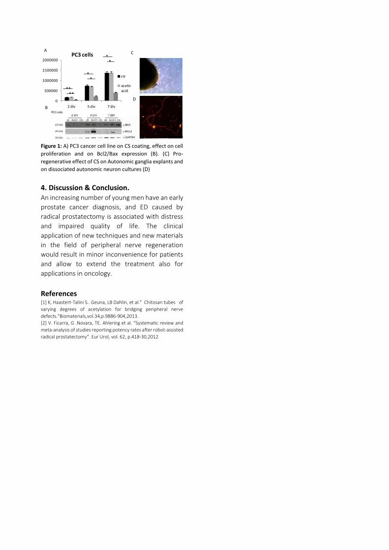
Figure 1: A) PC3 cancer cell line on CS coating, effect on cell
proliferation and on Bcl2/Bax expression (B). (C) Pro-
regenerative effect of CS on Autonomic ganglia explants and
on dissociated autonomic neuron cultures (D)
4. Discussion & Conclusion. An increasing number of young men have an early
prostate cancer diagnosis, and ED caused by
radical prostatectomy is associated with distress
and impaired quality of life. The clinical
application of new techniques and new materials
in the field of peripheral nerve regeneration
would result in minor inconvenience for patients
and allow to extend the treatment also for
applications in oncology.
References [1] K, Haastert-Talini S.. Geuna, LB Dahlin, et al." Chitosan tubes of
varying degrees of acetylation for bridging peripheral nerve
defects."Biomaterials,vol.34,p.9886-904,2013.
[2] V. Ficarra, G .Novara, TE. Ahlering et al. "Systematic review and
meta-analysis of studies reporting potency rates after robot-assisted
radical prostatectomy". Eur Urol, vol. 62, p.418-30,2012
0
500000
1000000
1500000
2000000
2 div 5 div 7 div
PC3 cells
ctr
acetic acid
**
****
**
A
B
C
D
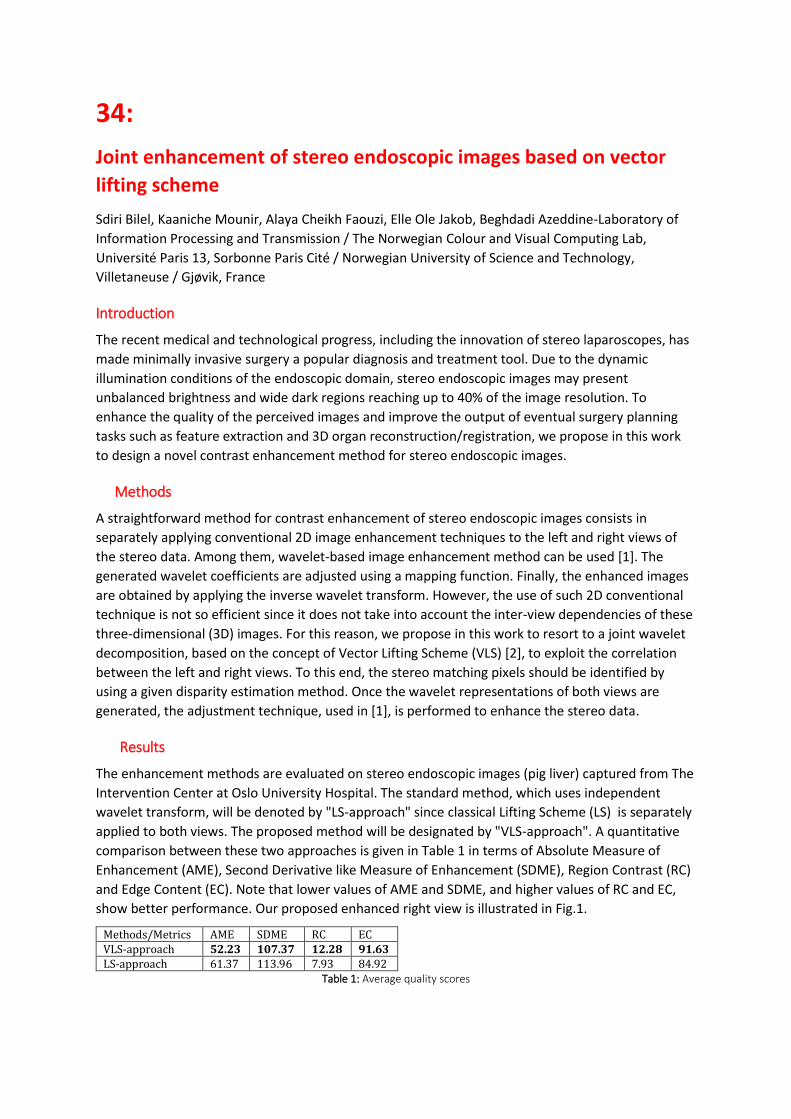
34:
Joint enhancement of stereo endoscopic images based on vector
lifting scheme
Sdiri Bilel, Kaaniche Mounir, Alaya Cheikh Faouzi, Elle Ole Jakob, Beghdadi Azeddine-Laboratory of
Information Processing and Transmission / The Norwegian Colour and Visual Computing Lab,
Université Paris 13, Sorbonne Paris Cité / Norwegian University of Science and Technology,
Villetaneuse / Gjøvik, France
Introduction
The recent medical and technological progress, including the innovation of stereo laparoscopes, has
made minimally invasive surgery a popular diagnosis and treatment tool. Due to the dynamic
illumination conditions of the endoscopic domain, stereo endoscopic images may present
unbalanced brightness and wide dark regions reaching up to 40% of the image resolution. To
enhance the quality of the perceived images and improve the output of eventual surgery planning
tasks such as feature extraction and 3D organ reconstruction/registration, we propose in this work
to design a novel contrast enhancement method for stereo endoscopic images.
Methods
A straightforward method for contrast enhancement of stereo endoscopic images consists in
separately applying conventional 2D image enhancement techniques to the left and right views of
the stereo data. Among them, wavelet-based image enhancement method can be used [1]. The
generated wavelet coefficients are adjusted using a mapping function. Finally, the enhanced images
are obtained by applying the inverse wavelet transform. However, the use of such 2D conventional
technique is not so efficient since it does not take into account the inter-view dependencies of these
three-dimensional (3D) images. For this reason, we propose in this work to resort to a joint wavelet
decomposition, based on the concept of Vector Lifting Scheme (VLS) [2], to exploit the correlation
between the left and right views. To this end, the stereo matching pixels should be identified by
using a given disparity estimation method. Once the wavelet representations of both views are
generated, the adjustment technique, used in [1], is performed to enhance the stereo data.
Results
The enhancement methods are evaluated on stereo endoscopic images (pig liver) captured from The
Intervention Center at Oslo University Hospital. The standard method, which uses independent
wavelet transform, will be denoted by "LS-approach" since classical Lifting Scheme (LS) is separately
applied to both views. The proposed method will be designated by "VLS-approach". A quantitative
comparison between these two approaches is given in Table 1 in terms of Absolute Measure of
Enhancement (AME), Second Derivative like Measure of Enhancement (SDME), Region Contrast (RC)
and Edge Content (EC). Note that lower values of AME and SDME, and higher values of RC and EC,
show better performance. Our proposed enhanced right view is illustrated in Fig.1.
Methods/Metrics AME SDME RC EC VLS-approach 52.23 107.37 12.28 91.63 LS-approach 61.37 113.96 7.93 84.92
Table 1: Average quality scores

Figure 1: Original right image (upper side) and the enhanced one with the proposed method (bottom side).
Discussion & Conclusion
In this work, we have proposed a new method for contrast enhancement of stereo endoscopic
images. While an intra processing of wavelet coefficients has been used as developed in [1], the
future work aims at developing joint processing techniques to adjust the wavelet coefficients of both
views.
References
[1] D. Cho, T. D. Bui, "Fast image enhancement in compressed wavelet domain", Elsevier Signal Processing, vol. 98, 2014.
[2] M. Kaaniche, A. Benazza-Benyahia, B. Pesquet-Popescu, J.-C. Pesquet, "Vector lifting schemes for stereo image coding", IEEE Trans. on Image Processing, vol. 59, 2009.
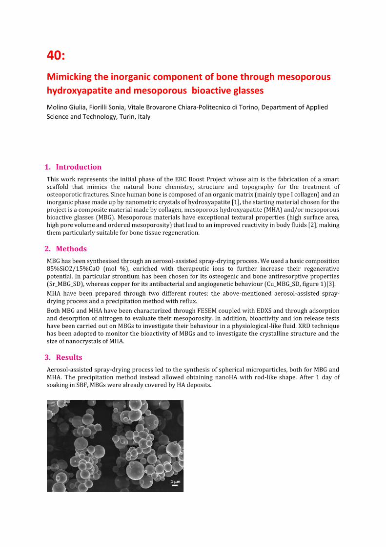
40:
Mimicking the inorganic component of bone through mesoporous
hydroxyapatite and mesoporous bioactive glasses
Molino Giulia, Fiorilli Sonia, Vitale Brovarone Chiara-Politecnico di Torino, Department of Applied
Science and Technology, Turin, Italy
1. Introduction
This work represents the initial phase of the ERC Boost Project whose aim is the fabrication of a smart scaffold that mimics the natural bone chemistry, structure and topography for the treatment of osteoporotic fractures. Since human bone is composed of an organic matrix (mainly type I collagen) and an inorganic phase made up by nanometric crystals of hydroxyapatite [1], the starting material chosen for the project is a composite material made by collagen, mesoporous hydroxyapatite (MHA) and/or mesoporous bioactive glasses (MBG). Mesoporous materials have exceptional textural properties (high surface area, high pore volume and ordered mesoporosity) that lead to an improved reactivity in body fluids [2], making them particularly suitable for bone tissue regeneration.
2. Methods
MBG has been synthesised through an aerosol-assisted spray-drying process. We used a basic composition 85%SiO2/15%CaO (mol %), enriched with therapeutic ions to further increase their regenerative potential. In particular strontium has been chosen for its osteogenic and bone antiresorptive properties (Sr_MBG_SD), whereas copper for its antibacterial and angiogenetic behaviour (Cu_MBG_SD, figure 1)[3].
MHA have been prepared through two different routes: the above-mentioned aerosol-assisted spray-drying process and a precipitation method with reflux.
Both MBG and MHA have been characterized through FESEM coupled with EDXS and through adsorption and desorption of nitrogen to evaluate their mesoporosity. In addition, bioactivity and ion release tests have been carried out on MBGs to investigate their behaviour in a physiological-like fluid. XRD technique has been adopted to monitor the bioactivity of MBGs and to investigate the crystalline structure and the size of nanocrystals of MHA.
3. Results
Aerosol-assisted spray-drying process led to the synthesis of spherical microparticles, both for MBG and MHA. The precipitation method instead allowed obtaining nanoHA with rod-like shape. After 1 day of soaking in SBF, MBGs were already covered by HA deposits.

Figure 1: FESEM image of Cu_MBG_SD.
4. Discussion & Conclusion
MBGs and MHA possess high surface area and consequently high reactivity. Furthermore, MBGs show a very fast bioactive behaviour. MHA obtained through precipitation method showed shape and dimensions similar to bone hydroxyapatite nanocrystals.
5. Acknowledgment
The authors acknowledge the UE financial funding (ERC-CoG-2015, Grant number 681798, BOOST, (www.ercprojectboost.eu)).
References
[1] J. Y. Rho, L. Khun-Spearing, P. Zioupos, “Mechanical properties and the hierarchical structure of bone”, Medical Engineering & Physics, vol. 20, p. 92–102, 1998.
[2] M. Vallet-Regi, “Nanostructured mesoporous silica matrices in nanomedicine”, Journal of Internal Medicine, vol. 267, p. 22–43, 2009.
[3] J. Jones, “Review of bioactive glass: From Hench to hybrids” Acta Biomaterialia, vol. 9, p.4457–4486, 2013.
42:
PROPOSAL OF AN INSTRUMENT FOR THE TRACEABILITY AND THE
MANAGEMENT OF THE CENTRAL VENOUS ACCESSES
Robasto Giulia, Restivo Angela, Goria Mario, Mussa Baudolino, Sandrucci Sergio- Biomed, Specialist
Biomed, Torino, Italy
ABSTRACT
Background and aim: Nowadays the central venous accesses (CVC) are widely used
devices and they allow a safe delivery of diagnostic and therapeutic services. An
inconsistency in CVC management and a lack of data about implanted devices history
emerged by the observation in hospital realities.
The aim is to create a valid instrument in order to offer a better healthcare to patients
and to provide a guide for nurses for their profession.
Materials and method: The instrument was realized following scientific evidences
obtained by consultations of guidelines, articles on PubMed and publications on web
sites (GAVaCeLT). A validation procedure is carried out: the instrument was shown to
nurses, modified after focus group and applied to test the efficacy.
Results: Completed trial booklets were withdrawn by checking compilation mode and
constancy. Approval and validity levels were enquired by analysis of compiled
questionnaires after the final focus group.
Analysis and discussion: During the project an increase of positive feedbacks was
noticed. The instrument promotes: management standardization, implanted device
history knowledge and data availability for possible studies. The CVC consciousness
does not increase in all patients because of time limitation that interferes on their
education negatively.
Conclusion: The instrument results valid, useful to improve patients’ healthcare and
efficacy to standardize CVC management delivered by nurses. It is hoped booklets
will be use unofficially. Studies and future development are suggested.
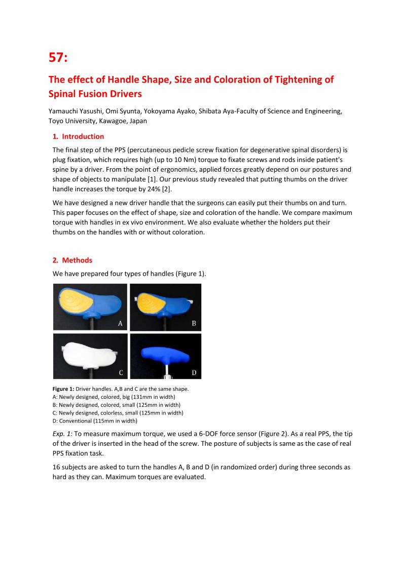
57:
The effect of Handle Shape, Size and Coloration of Tightening of
Spinal Fusion Drivers
Yamauchi Yasushi, Omi Syunta, Yokoyama Ayako, Shibata Aya-Faculty of Science and Engineering,
Toyo University, Kawagoe, Japan
1. Introduction
The final step of the PPS (percutaneous pedicle screw fixation for degenerative spinal disorders) is
plug fixation, which requires high (up to 10 Nm) torque to fixate screws and rods inside patient's
spine by a driver. From the point of ergonomics, applied forces greatly depend on our postures and
shape of objects to manipulate [1]. Our previous study revealed that putting thumbs on the driver
handle increases the torque by 24% [2].
We have designed a new driver handle that the surgeons can easily put their thumbs on and turn.
This paper focuses on the effect of shape, size and coloration of the handle. We compare maximum
torque with handles in ex vivo environment. We also evaluate whether the holders put their
thumbs on the handles with or without coloration.
2. Methods
We have prepared four types of handles (Figure 1).
Figure 1: Driver handles. A,B and C are the same shape.
A: Newly designed, colored, big (131mm in width)
B: Newly designed, colored, small (125mm in width)
C: Newly designed, colorless, small (125mm in width)
D: Conventional (115mm in width)
Exp. 1: To measure maximum torque, we used a 6-DOF force sensor (Figure 2). As a real PPS, the tip
of the driver is inserted in the head of the screw. The posture of subjects is same as the case of real
PPS fixation task.
16 subjects are asked to turn the handles A, B and D (in randomized order) during three seconds as
hard as they can. Maximum torques are evaluated.

Figure 2: Ex-vivo evaluation of fixation torque.
Exp. 2: 19 subjects without prior knowledge of grasping PPS handle are asked to ‘naturally’ grasp
the handles A, B, C and D. Then their grasping postures are categorized as i)putting thumbs on the
handle, ii)not putting thumbs and iii)undefined.
3. Results
The maximum torques of each handles showed statistically significant difference (one-way
ANOVA, p<0.05). Compared with conventional handle (D), newly designed small handle (B)
increased torque by 8%, and newly designed big handle (A) increased torque by 14%.
Whereas ten of 19 subjects put thumbs on the colorless handle (C), 15 subjects put thumbs on the
colored handle (B).
4. Discussion & Conclusion
We reconfirmed that the shape affects fixation torque. The conventional handle we compared
was relatively small and was inferior as compared with big ones. Our result also suggested that
the coloration of the handle could affect grasping postures. Thus the yellow areas of our colored
handles are intended to ‘attract’ thumbs.
References
[1] H. Dong, P. Loomer, et al., “The Effect of Tool Handle Shape on Hand Muscle Load and Pinch Force in a Simulated Dental Scaling Task,” Appl Ergon, vol. 38, pp.525-531, 2007.
[2] Y. Yamauchi, et al., “Influence of Shape of Handles and Gripping Method on Tightening Torque of Spinal Fusion Drivers”, Proc. of The 11th Asian Conf. on Computer Aided Surgery, 2015.

60:
Design to Care through Co-design
Wauben Linda, Baas Elbert, van Gils Ronald, Helder Onno- Rotterdam University of Applied Sciences,
Research Centre Innovations in Care, Rotterdam, Netherlands
Introduction
Next to fundamental research, applied research is important for the scientific process [1].
Furthermore, there is a demand for new user-centred, context specific design solutions by frontline
users, i.e., healthcare professionals and patients. By combining applied research with user-centred
design (i.e. design-based research) we can study user’s everyday problems, needs and wishes and
together co-design solutions. The aim was to study users’ problems/challenges and to co-design
solutions to improve users’ safety and satisfaction using the CaTe approach (Figure 1).
Figure 1: CaTe: CAre TEchnology approach to study and co-design solutions to improve safety and satisfaction.
Methods
Healthcare, Design, Engineering and Communication, Media & Information Technology Bachelor
students worked closely together in interprofessional teams with healthcare professionals and/or
patients in the Minor course ‘Healthcare Technology’ (6 months) and during their graduation
projects (6 months) and to co-design solutions to improve safety and satisfaction.

Results
1. ID.alistic: Patient identification method (via fingerprints) for the Dialysis Center of the Albert Schweitzer Hospital (=concept).
For the Erasmus MC: Sophia Children's Hospital:
2. ANI: Application to calculate the Neonatal Intake (=concept). 3. Happyfeed: Redesign for holding Feeding Tubes besides the Incubator (=produced &
implemented). 4. Baby's length: length-measuring device via light beams (=ready to produce).
Figure 2: Design solution
Discussion & Conclusion
The CaTe approach provided a good structure for students to design user-centred solutions
healthcare professionals and patients want to use. The concepts will be developed further and
produced.
Acknowledgement
We would like to thanks the students: Maarten Zijlmans, Stijn Kievit, Robin van Everdingen, Sjoerd
Keuzenkamp, Mark Rood, Vaashna Beni and Koen van der Beek.
References
[1] K. E. Stanovich, C. Campanella, Ed. How to Think Straight About Psychology, 10th edition ed. New Jersey: Pearson Education, Inc., 2013.
68:
Technology Innovations in Therapy and Imaging (T²I²):
International Graduate School for Biodesign and Entrepreneurship Holger Fritzsche, Elmer Jeto Gomes Ataide, Axel Boese, Michael Friebe
INKA- Chair for Catheter Technologies and Image Guided Therapies, Otto-von-Guericke-University, Magdeburg, Germany, [email protected] .
1. Introduction The potential for innovation generation and subsequent translation in start-up companies at German universities has not been a focus yet and the process is generally not part of the scientific education. The engineering courses are designed to impart knowledge from the natural sciences in the technical context. Innovation, creation and implementation are rarely part of the engineering curriculum, nor are interdisciplinary approaches. Modern Medicine is evolving at rapid rates. Technology has become a vital part of the diagnostic and therapeutic processes and requires constant attention. There is a growing need for professionals who specialize and are able to bridge the gap between medicine and technology combined with the ability to manage tasks given to them effectively and efficiently witin an economic context. The International Graduate School of Technology Innovations in Therapy and Imaging (T2I2) aims to do exactly that. The Ph.D. students are expected to work in a doctoral depth on topics and simultaneously taught to create a successful medical device or service by identifying the "unmet clinical needs" and develop their projects based on the Stanford BIODESIGN [1, - 3].
2. Methods The T2I2 intends to foster the coming generation of health engineers to efficiently develop medical
technology and ensure that this technology is a marketable resource. This is not only concentrated
on Germany or the USA, but should also be employed in a more global approach including specific
needs of healthcare or medical technology needs in developing and emerging countries.
A structured education program with an interdisciplinary approach in the disciplines of Medicine,
Technology and Economics is provided. The graduate school participants have to successfully attend
at least 300 hours of lecture or lecture equivalent study over a 36-month period.
Students from various disciplines work on topics and projects in the main area of medical imaging,
minimally invasive therapy, image guided surgeries, catheter technologies, innovation generation,
technical translation. Soft skill development is a core challenge and objective. Close contact and
direct involvement to industry- and application oriented research projects, an international
exchange and regular further training are core components. The graduate school, while engineering
oriented, is located and placed within the medical faculty of the Otto-von-Guericke-University. The
students are supervised jointly by a clinical and a technical professor.
3. Results The central contact point for the Ph.D. program of the Graduate School T²I² is a new Innovation
Laboratory with Simulation-OR and Prototyping- Lab located at the University Hospital in
Magdeburg. Currently 13 international Ph.D. students (from Egypt, Mexico, Nepal, India, Iran,
Taiwan and Germany) are in that structured doctoral program for innovation generation, technology
transfer and business implementation of medical technologies.
A strong education focus is on the 21-century skills- to approach complex challenges teaching
competencies like critical thinking, creativity, communication and collaboration. Furthermore, for
individual and subject-specific education, an external stay of at least 6 weeks is required at a partner
university with a similar research focus.
4. Discussion & Conclusion The aim of our graduate school is to bridge the gap between medicine and technology for products
and services that have a future and have a clearly identified need. Students need to work more
intimately with the medical personnel to gain a better understanding of their needs. This would
hopefully result in identifying much more useful equipment for the healthcare professionals. The
school will also focus on the innovative and entrepreneurial aspect of healthcare and teach and urge
students to develop more innovative ideas and means to make those innovations marketable.
References
[1] Yock, P.; Zenios, S. Makower, J.: Biodesign: The Process of Innovating Medical Technologies. Cambrige: Cambridge University Press, first ed., 2015.
[2] Michael Friebe, Jörg Traub. Image guided surgery innovation with graduate students - a new lecture format. Current Directions in Biomedical Engineering. Volume 1, Issue 1, Pages 475–479, ISSN (Online) 2364-5504, DOI: 10.1515/cdbme-2015-0114, May 2016
[3] A. Boese, Lösungsfindung mit dem Endnutzer, ein neuer Ansatz in der methodischen Produktentwicklung am Beispiel der Medizintechnik, 1st ed., vol. 6. Shaker, 2016.

75:
Initial results of optical and inertial sensor fusion as a tracking
alternative for interventional procedures
Sühn Thomas, Ziegle Jens, Friebe Michael-Otto-von-Guericke University, University / IMT / INKA,
Magdeburg, Germany
Department of Medical Engineering, Chair for Catheter Technologies and Image Guided Therapies, Otto-von-Guericke University, Magdeburg, Germany, [email protected].
1. Introduction
A significant initial step for an effective image guided interventional treatment is to find the right position and angle to align the used instrument to the target. Additionally, a high navigation accuracy is required to
reach the therapy target precisely. Currently used tracking systems based on optical or electromagnetic (EM) principles come with drawbacks. Optical tracking requires a direct line of sight between the camera and the tracked object and EM systems are very sensitive to environmental changes.
We propose a fusion of optical and inertial sensors that has a potential to achieve a comparable accuracy than EM tracking, which is in the order of 1mm [1], and at the same time avoid the disadvantages of optical tracking systems.
2. Methods
An inertial navigation system (INS) is typically used to track position, orientation and velocity of an object relative to a known reference point by processing accelerometer and gyroscope signals. With recent advances in micromachined electro-mechanical systems (MEMS) the development of small, low cost and lightweight INS for medical applications such as orientation tracking of surgical tools became possible [2].
The gyroscope error propagation caused by the calculation of orientation and position in the strapdown navigation algorithm requires an error compensation for the emerging and critical drift. The integration of information from additional sensors such as magnetometer [3], as well as domain specific constraints and assumptions are common methods to compensate these errors.
We combined a simple optical sensor unit, consisting of a LED and a photodetector with an INS of ≈ 25×20×10 mm size for accurate angle and depth calculation of an interventional instrument. For tracking the orientation and position of a needle tip, the INS was attached to the opposite end. The static optical system was located on the body surface at the position of needle insertion and used to acquire drift free movement data, by recognizing the distance the needle moves alongside the optical sensor during insertion into the body. This information is used as a boundary condition to correct the drift-prone estimation of position. Figure 1 shows the integration of the sensor information into the general strapdown navigation algorithm, as well as the position of the sensors at the needle.
Figure 1: Position of optical and inertial sensor for example on a needle and the integration in the navigation algorithm.
3. Results
Preliminary experiments showed the ability of the optical signal to help reduce the inaccuracy in the position calculation of INS that is caused by the propagation of gyroscope errors. With the knowledge of
Optical Sensor
North
globalAccel.
initial velocity / position
ORIENTATION
POSITION
global movementin certain direction
Projection on global
axes

the extent of needle (and therefor INS) movement in one particular direction, it is now possible to considerably limit the drift of the navigation system.
4. Discussion & Conclusion
Since the calculated orientation is used to project the optically acquired information to the global axes, the propagation of the gyroscope error through the correction path has to be evaluated as well as the accuracy of the optical measurement setup. In addition, an incorporation of the proposed method into a viable calibration process for further improvement of accuracy has to be considered. Assuming an adequate precision, the navigation system could be used for example in connection with ultrasound as small, capable low-cost solution for guidance of interventional instruments.
References
[1] Mandal KK, et al. Assessment of the accuracy of optical shape sensing for needle tracking interventions, ASME. J. Med. Devices. 2017; 11(3): 034504-7.
[2] Rebello KJ, Application of MEMS in Surgery, Proceedings of the IEEE 2004; 92(1): 43-55.
[3] Hongliang R, et al. Investigation of Attitude Tracking Using an Integrated Inertial and Magnetic Navigation System for Hand-Held Surgical Instruments, IEEE Transactions on Mechatronics 2012; 17(2): 210-217.

77:
Objective Measurement of Hip Implant Parameters by Automatic
Feature Extraction from Planar Femoral X-Rays
Ataide Elmer Jeto Gomes, Sühn Thomas, Friebe Michael-Otto-von-Guericke University, University /
IMT / INKA, Magdeburg, Germany
Department of Medical Engineering, Chair for Catheter Technologies and Image Guided Therapies, Otto-von-Guericke University, Magdeburg, Germany, [email protected].
1. Introduction
The total number of arthroplasties, the replacement of a damaged or diseased musculoskeletal joint by implants, is predicted to increase by up to 50% within the next 10-15 years in the UK [1]. Therefore, it is important to provide advanced and easy to use diagnostic support systems for the surgeons to deal with the growing number of patients and to provide a possibility for personalized solutions in the future.
Templating, the process of anticipating the size and position of the implant, is crucial for the success of the surgery by minimizing perioperative complications, reduce future pain and total procedure cost. While several currently available commercial systems deal with the procedure planning, the decision of which implant type will be used is mainly done exclusively by the physician based on more or less objective parameters calculated by the used software. The development of a system that would provide additional information based on the preoperative planar x-ray images and lead to a more objective decision for the critical implant selection process would be very helpful.
Based on image processing of appropriate anteroposterior femoral radiographs, the proposed system could provide important additional and validated parameters that are typically used by the surgeon such as dimensions of certain segments of femur or femoral-acetabular rotation center and offset, but are not provided by the current software solutions. Such a decision system would be image based, complemented with patient specific data, and use an implant database with relevant parameters.
2. Methods
For preliminary testing, we extracted the dimensions (diameter) of significant segments automatically out of 5 planar x-ray images of healthy femur, by processing the images using MATLAB. Initially the x-ray images were obtained and converted into binary images using the gray scale to black and white code and thresholding method. A dataset of the binary images was then created. Segments were obtained in the binary image (anteroposterior view of the femur) using the developed code. The surgeon is provided with fact-based information containing length, angle, width and curvature for the final decision of which possible implants can be placed.
Figure 1: System workflow for objective measurement of hip implant parameters using femoral X-rays.
3. Results
The results obtained from our preliminary developments manifest in the form of segments drawn above and below the Lesser Trochanter in the anteroposterior view of the femoral X-ray. There is also a segment drawn lower than the inferior Lesser Trochanter that is adjustable depending on the curvature of the implant and the angle at which it is required to be implanted. These segments are used as basis on which the surgeon can decide how and where to fit the implant.
4. Discussion & Conclusion
The pre-operative planning procedure for hip implantation is crucial and needs attention. Mere assumptions and being subjective is a risk and could relatively easily be avoided. The proposed system provides standard objective information automatically extracted from planar X-ray images. This has the
X-ray image Create binary image dataset Binary
Image
MATLAB
Run code for obtaining
image segments
Provide surgeon
with data
Decision making
based on facts

potential to reduce surgery risk and making to make the entire process more objective and factual in nature personalised to the patient.
This system could be the basis for a Clinical Decision Support system that would aid the surgeon in the entire planning process, particularly the implant selection and fixation through Machine Learning Algorithms.
References
[1] Culliford D, et al. Future projections of total hip and knee arthroplasty in the UK: results from the UK Clinical Practice Research Datalink. Osteoarthritis and Cartilage 2015; 23(4): 594-600.

80:
A VR-based surgical simulation system using patient – specific
physical computing model
Chen Yining, Liao Denghong, Chen Xiaojun- School of Mechanical Engineering, Shanghai Jiao Tong
University, Shanghai, China
1. Introduction
Apart from the applications in surgical navigations, virtual surgery has become a new feasible method for
training young surgeons. Since 2006 virtual simulation has been performed in selected patient cases affected
by complex craniomaxillofacial disorders (n = 8) in addition to standard surgical planning based on patient
specific 3d-models. [1] Although for training, standard models are enough for young surgeons to practice, we
still need to build an accurate and personalized model to simulate a surgery better.
Patients' CT images are considered appropriate references for this procedure. CT images are taken along
the axis of the body, and each of them represents the structure of a plane that is perpendicular to the body
axis. Information like the gray scale levels of each pixel, which can be used in building a volume model, are also
contained. During an operation, the model should be able to give some haptic feedback as well as to update
the changes on the models in real time.
Figure 1: Construction of the surgical simulation program
In this project, we realize these functions with CHAI3D library. Omega 6 is the device we use for applying
forces and receiving feedbacks.
2. Hardware and Software Implement
Designed as a platform agnostic framework for computer haptics, visualization, and interactive real-time
simulation, CHAI3D is an open source framework that supports a variety of commercially-available three-, six-
and seven-degree-of-freedom haptic devices, and makes it simple to support new custom force feedback
devices. [2]

The omega.6 is the most advanced pen-shaped force-feedback device available. The combination of full gravity
compensation and driftless calibration contributes to greater accuracy. [3]
Figure 2: Program interface and Omega 6
3. Methods
In particular, we use the voxel data to determine the shape of the model and then create it in the CHAI3D
world. CHAI3D allows users to load a stack of CT images into a multi-image pointer and analyze them. After
creating a CHAI3D world and allocating the voxel data for them, we set an isosurface level to the object, and
those voxels which gray level values exceed the isosurface level will represent unit points and form the model.
Collision detections and haptic feedback are also added to the model. CHAI3D uses a virtual "finger-proxy"
algorithm to compute forces. When a tool object hit the surface of a model, the tool we can see will stop
moving directly into its goal. However, the proxy of the tool is actually able to stick into the object, which is
shown in the figure below. In this case, forces are computed between the actual tool and its proxy, assuming a
string in the middle trying to drag them back together. Users can define the stiffness of the model according to
the materials.
Figure 3: Motion of the virtual proxy in Finger-proxy algorithm
When users click on the switch on Omega 6 while the virtual tool hit the object, the drilling operation
is started. CHAI3D will read the collision event, and the determine the point which is contact with the tool.
Then, the property of the point is changed, and it is no longer visible and able to give any haptic feedback.
Graphic rendering will occur at the same time, and users can know about the result of their operation
immediately.

4. Results and Conclusion
At present, some core functions such as loading image files, creating volume objects, and modify objects
are completed. Right now, users can drill on models at random directions, and models can update its data and
graphics in real time. Simple textures and haptic feedback are also applied on the models. Furthermore, for
better simulations, we will calibrate the haptic feedback, restrict the drilling directions, and improve rendering
through using colormap in the model. We expect this virtual surgery program can simulate cutting, grinding
and even more complicated operations. Therefore, surgical training can be feasible and thus save time and
cost in surgery practicing.
Acknowledgement
This work was supported by Natural Science Foundation of China (Grant No.: 81511130089), Foundation
of Science and Technology Commission of Shanghai Municipality (Grant No.: 14441901002, 15510722200, and
16441908400).
Reference
[1] Adolphs, Nicolai, et al. "Virtual planning for craniomaxillofacial surgery–7 Years of experience." Journal of Cranio-Maxillofacial
Surgery 42.5 (2014): e289-e295.
[2] http://www.chai3d.org/concept/about
[3] http://www.forcedimension.com/products/omega-6/overview
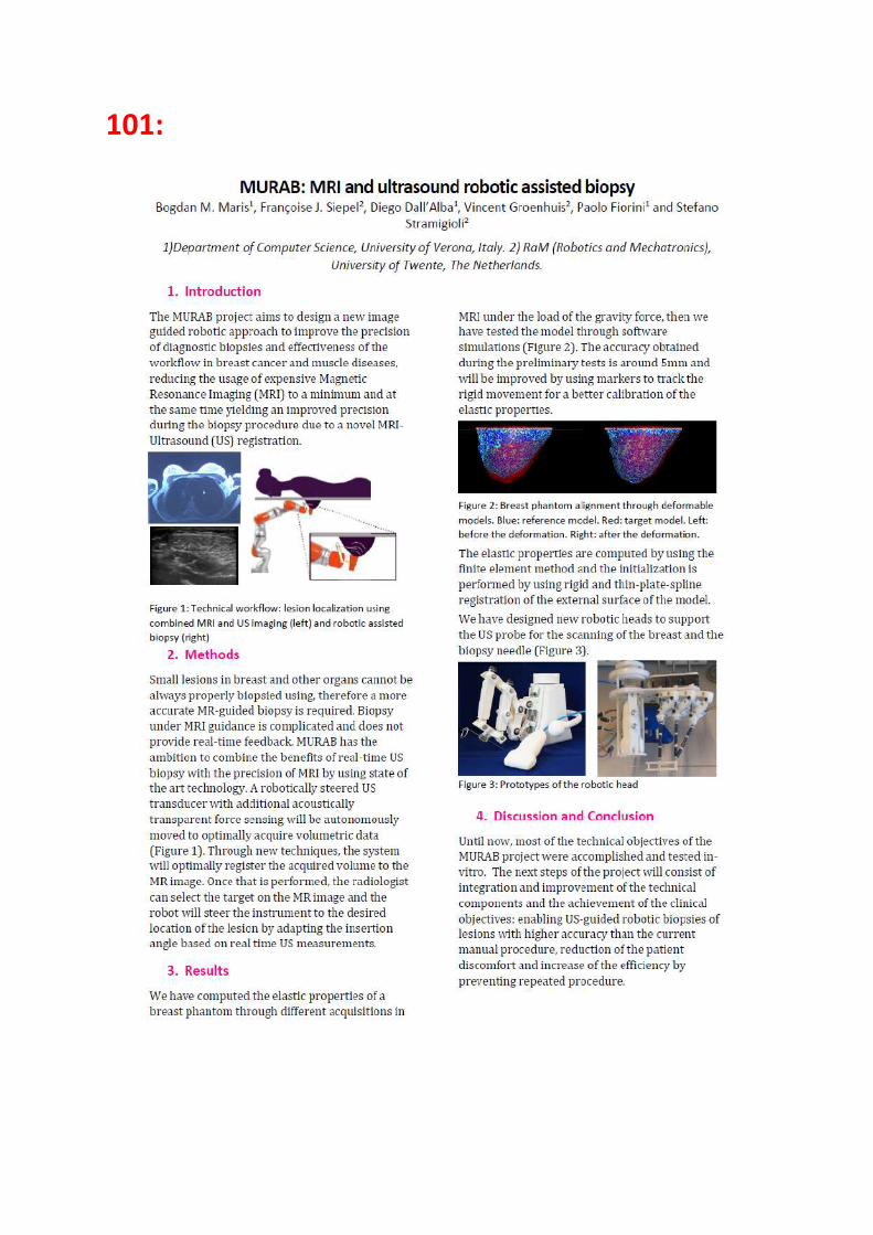
101:

110:
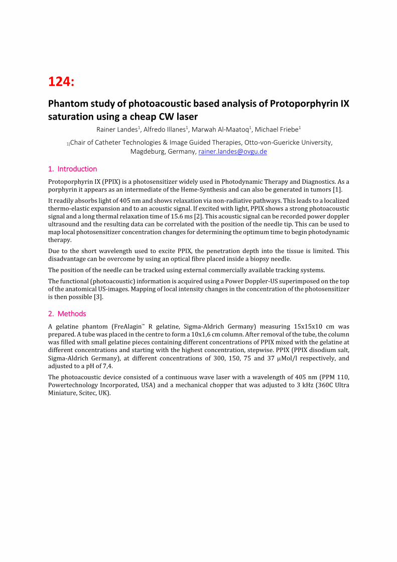
124:
Phantom study of photoacoustic based analysis of Protoporphyrin IX saturation using a cheap CW laser
Rainer Landes1, Alfredo Illanes1, Marwah Al-Maatoq1, Michael Friebe1
1)Chair of Catheter Technologies & Image Guided Therapies, Otto-von-Guericke University, Magdeburg, Germany, [email protected]
1. Introduction
Protoporphyrin IX (PPIX) is a photosensitizer widely used in Photodynamic Therapy and Diagnostics. As a porphyrin it appears as an intermediate of the Heme-Synthesis and can also be generated in tumors [1].
It readily absorbs light of 405 nm and shows relaxation via non-radiative pathways. This leads to a localized thermo-elastic expansion and to an acoustic signal. If excited with light, PPIX shows a strong photoacoustic signal and a long thermal relaxation time of 15.6 ms [2]. This acoustic signal can be recorded power doppler ultrasound and the resulting data can be correlated with the position of the needle tip. This can be used to map local photosensitizer concentration changes for determining the optimum time to begin photodynamic therapy.
Due to the short wavelength used to excite PPIX, the penetration depth into the tissue is limited. This disadvantage can be overcome by using an optical fibre placed inside a biopsy needle.
The position of the needle can be tracked using external commercially available tracking systems.
The functional (photoacoustic) information is acquired using a Power Doppler-US superimposed on the top of the anatomical US-images. Mapping of local intensity changes in the concentration of the photosensitizer is then possible [3].
2. Methods
A gelatine phantom (FreAlagin™ R gelatine, Sigma-Aldrich Germany) measuring 15x15x10 cm was prepared. A tube was placed in the centre to form a 10x1,6 cm column. After removal of the tube, the column was filled with small gelatine pieces containing different concentrations of PPIX mixed with the gelatine at different concentrations and starting with the highest concentration, stepwise. PPIX (PPIX disodium salt, Sigma-Aldrich Germany), at different concentrations of 300, 150, 75 and 37 Mol/l respectively, and adjusted to a pH of 7,4.
The photoacoustic device consisted of a continuous wave laser with a wavelength of 405 nm (PPM 110, Powertechnology Incorporated, USA) and a mechanical chopper that was adjusted to 3 kHz (360C Ultra Miniature, Scitec, UK).
The beam of the laser was coupled directly into a multimode optical fibre with an inner diameter of 273 m and an outer diameter of 420 m. The fibre was placed inside a biopsy needle to probe inside the phantom (see Fig. 1).
Figure 1 A: Biopsy Needle with fibre, B: Transducer, C1-4: volumes with different concentrations of PPIX - US-Signal: thick continuous line, Echo: thin dotted line, added PA-Signal: thin dotted red line
Tracking of the biopsy needle was achieved using the Logiq E medical Ultrasound system (Logiq E,
GE, USA).
3. Results & Discussion
When the fibre probe gets closer to the PPIX samples an increase in amplitude of the frequency spectrum is detectable. Testing the columns with different concentrations of PPIX showed a linear correlation between amplitude and concentration of the PPIX solutions.
References
[1] Eva. Ramón-Gallegos, et al., "Photoacoustic Spectroscopy Applied to the study of Protoporphyrin IX Induced in Mice," Analytical Sciences, vol.17, 2001.
[2] J.L. Jiménez Pérez, et al. "Photoacoustic Spectroscopy to determine in vitro the non-radiative relaxation time of protoporphyrin IX solution containing gold metallic nanoparticles," Eur. Phys. J. Special Topics, vol. 153, p. 353–356, 2008
[3] Matthew P. Fronheiser, Salim F. Idriss, Patrick D. Wolf, Stephen W. Smith, "Vibrating Interventional Device Detection Using Real-Time 3-DColor Doppler," IEEE Trans Ultrason Ferroelectr Freq Control. 55(6): 1355–1362, 2008

129:
Tumor Identification in Colorectal
Histology Image Using Convolutional
Neural Network
Hongjun Yoon1,2,Joohyung Lee1,Ji Eun Oh1,Hong Rae Kim2,Seonhye Lee1, Dae Kyung Sohn1
1) Innovative Medical Engineering & Technology Branch, National Cancer Center, Goyang, Korea. 2) College of Engineering and Computer Science, Syracuse University, Syracuse, NY, U.S.A.
1. Introduction
Colorectal cancer (CRC) has been a major health concern throughout the globe. Although the
recent mortality rate of CRC has been stabilized or declined, it is still the third most common
type of cancer in Korea [1]. Early diagnosis is extremely important as it determines treatment
options and has a strong influence on the length of survival. This is typically done by analyzing
colon biopsy images. This study proposes to use Deep Neural Network (DNN) to predict
whether colon histology images contain tumor as there have been many other proposals that
suggest using Convolutional Neural Network (CNN), i.e. DNN specifically adapted to image data,
is effectively in classifying or locating tumor for many types of cancer. ImageNet Large Scale
Visual Recognition Competition (ILSVRC) is a competition for research teams to compete on
several visual recognition tasks to achieve higher accuracy [2]. Winner of 2014 ILSVRC was
Visual Geometry Group (VGG). They proposed more layer of the CNN structure it gets, higher
the accuracy it will be for visual classification and localization [3].
2. Methods
Data
The colorectal histology image was obtained from National Cancer Center Research Institute,
Innovative Medical Engineering & Technology Branch (IRB number: NCC2016-0048). Total of
30 patients who were diagnosed with CRC were participated with the age range from 20 to 80.
Among those patients, 30 normal and tumor images were obtained and because some of the
images were damaged and were no longer able to use, they were eliminated from this research.
Hence, 28 normal and 29 tumor images were obtained. Most of these images exceeded the size
of the maximum capability average of 10000 pixel by 10000 pixel that the GPU can process, the
raw images were cropped into a fixed size of 256 pixel by 256 pixel. When the images were
cropped, 6806 normal images and 3474 tumor images were obtained.
Model
VGG presented 6 different models with different number of layers. Each of them consists of
Rectified Linear Unit (ReLU) activation function, convolution, max-pooling, fully-connected
layers [3]. Because some of these parts are outdated and the second model has no difference
with the first model when applying the adjustment, we omitted the second structure,
consequently five model structures.
Methodology

Each model were train and validated using a 10-fold validation data set. In this process, only
well distinguished 100 normal and 100 tumor data set were used. After best performing model
was selected based on the training and validation, the whole data set was used for the training
and validation.
3. Results
Using the 5 different modified VGG models and the 200 selected histology image data set,
accuracies for VGG A, B, C, D, and E models were obtained as follows: 82.5%, 87.5%, 87.5%, 91.4%,
and 94.3%. As we use the most accurate model out of these 5 models, which is VGG E model,
accuracy, loss, sensitivity, specificity was marked as follows: 93.5%, 0.439, 95.1%, 92.8%.
4. Discussion & Conclusion
As Visual Geometry Group stated, the deepest model out of 5 modified model, E model was the
most accurate model. Also, by training and validating the colorectal histology image, it shows
that we have implemented a practical tumor classification method.
References
[1] J u n g KW , Wo n g YJ , O h CM , K o n g HJ , L ee DH , L ee KH , "Cancer Res Treat," 2017.
[2] De n g J , Do n g W , Soc he r R , L i LJ , L i K , Fe i - Fei L ., "ImageNet: A Large-Scale Hierarchical Image Database," 2014.
[3] S im o ny a n K , Zi ss erm a n A , “ Ve ry D ee p Co n vo l u t io n al Ne t wor k s f or La r ge Sc a le I ma g e Re co g ni t io n ,”
2 0 1 5 .
Acknowledgement
This work was supported by grant from the National Cancer Center (NCC-1710070).

132:

NEUROSURGERY

120:

SURGERY & ENDOSCOPY
1:
EFFECTS OF WARMED, HUMIDIFIED CO2 INSUFFLATION ON BODY
CORE TEMPERATURE AND CYTOKINE RESPONSE: RANDOMIZED
COMPARISON VERSUS STANDARD INSUFFLATION DURING ROBOT-
ASSISTED RADICAL PROSTATECTOMY
Oderda Marco, Cerutti Elisabetta, Gontero Paolo, Munegato Stefania, Meyer Nicolas, Diemunsch
Pierre-University of Turin, Città della Salute e della Scienza, Dept of Urology, Torino, Italy
1. Introduction Cool and dry gas insufflation in laparoscopy induces hypothermia and cytokine increase, with significant
perioperative morbidity [1]. Few studies have suggested that warmed and humidified insufflation leads to
an improved body core temperature (BCT) maintenance, a reduction of the inflammatory response and an
improved quality of postoperative course, compared with standard insufflation [2-3]. Aim of this study was
to assess if warmed and humidified CO2 insufflation with HumiGard™ device can achieve significant
benefits over standard insufflation in terms of risk of hypothermia and cytokine response, in the setting of
robot-assisted radical prostatectomy (RARP).
2. Methods Design, setting, and participants: patients undergoing RARP were randomized into group H+WB (32
patients receiving warmed, humidified CO2 insufflation with HumiGard™ device, plus hot air warming
blanket) and group WB (32 patients receiving standard CO2 insufflation, plus hot air warming blanket.
Outcome measurements and statistical analysis: BCT, plasma levels of cytokines IL-6 and TNF-α, pain
scores, and intraoperative parameters. The data were analyzed according to the Bayesian paradigm.
3. Results Intraoperative BCT increased in both groups during surgery, with a statistically significant difference favoring group H+WB, ending at 0.2°C higher on average than group WB. The overall BCT increase was 0.088 degree per hour in the WB group, with an additionnal 0.064 degree per hour in the H+WB group. No difference across groups, at none of the time points, could be shown as far as the mean serum cytokine and the TNF-α levels, were concerned. No statistical differences were noted for pain scores and the other intraoperative parameters. Limitations of study were the low baseline BCT of our patients (mean 35.7°C) and the heating power of our HumiGard™ device, which was less than expected.
4. Discussion & Conclusion During RARP, warm and humidified CO2 insufflation with the HumiGard™ device is more effective than the standard CO2 insufflation in maintaining the patient’s heat homeostasis.
References [1] Sessler DI. New surgical thermal management guidelines. Lancet 2009; 374: 1049-1050.
[2] Schlotterbeck H, Schaeffer R, Dow WA, Diemunsch P. Cold nebulization used to prevent heat loss during
laparoscopic surgery: an experimental study in pigs. Surg Endosc 2008; 22: 2616-2620.
[3] Noll E, Schaeffer R, Joshi G, Diemunsch S, Koessler S, Diemunsch P. Heat loss during carbon dioxide
insufflation: comparison of a nebulization based humidification device with a humidification and heating
system. Surg Endosc 2012; 26: 3622: 3625.
13:
Ultra Structural Changes of Collagen in RadioFrequency (RF)-
induced Colorectal anastomoses
Mao Lin, Chen Li, Tu Liangyong, Zhou Yu, Song Chengli- University of Shanghai for Science and
Technology, School of Medical Instrument and Food Engineering, Shanghai, -
1. Introduction
The technology exploits the RadioFrequency (RF) energy induced fusion of native colorectum in order to
achieve high-quality rapid tissue sealing without introducing any foreign materials, and thus, is expected
to greatly reduce morbidity, mortality and cost [1]. Changes in collagen bonds within the fused tissue are
thought to be pivotal to the strength of the resulting fusion. Herein, we investigate the ultra structural
changes of thermal denaturing collagen in RF-induced colorectal anastomoses (CRA) by Raman
spectroscopy, which provides biomolecular insights into the restructuring which occurs during tissue
fusion.
2. Methods
The custom-made compression prototype used for creating end-to-end serosa-to-serosa CRA is showed in
Figure 1. The electrodes were mounted on insulated and heat resistant ring carriers and then were
pressed by a pressure device (ALX-S, Yiding Instrument Corporation) to provide the designated initially
force for anastomoses. Each experimental condition was repeated at least five times.
Figure 1: Compression prototype and experimental process. A pair of electrodes is fixed on two ring carriers. Bowel
segments were mounted on ring carriers compressed by pressure device.
The collagen-rich regions of fused areas and normal colon were made 15 μm paraffin slices by the cross
section and scanned by Raman micro-spectroscope (LabRam-1B, HORIBA Scientific., France) equipped
with a microscope and a CCD camera. Raman spectra in the fingerprint regions that 800 ~ 1800 cm-1 were
acquired. The whole procedure including samples scanning, data acquiring and peak analyzing, were
carried out by a senior technician.
3. Results
Raman spectra of a healthy colon and fused colon subjected distinct duration and electric power is
showed in Figure 2. Despite electric power, the signals at 875 cm-1 (C-C stretching of hydroxyproline
ring), 1002 cm-1 (phenylalanine C=C symmetric stretching), 1061 cm-1 (C-N vibration of proline), 1127
cm-1 (C-N stretching group), 1296 cm-1 (deformation vibration of CH2) and 1669 cm-1 (Amide I band)
shifted consistently to lower wavenumber when compression pressure and duration were 277 kPa and 5
seconds, respectively. Generally, the intramolecular bonding strength was weakened by RF heating. The

signal at 1655 cm-1 representing α helix of collagen disappeared after fusion, which implied the
disruption of α helix.
Figure 2: Raman spectra of the healthy colon (control) and fused colon subjected four combinations of electric power and
duration (group IV-1 to IV-4).
4. Discussion & Conclusions
Despite the combinations of fusion parameters, collagen denaturation featuring by unfolding triple α helix
is confirmed subjected RF by identifying weak C=O bonds and N-H bonds. Tissue fusion may mostly
attribute to deformation vibration of CH2, which subsequently leads to collagen crosslinking and peptides
random coil.
References
[1] L. Zhao, C. Zhuo, C. Song, X. Li, Y. Zhou, and D. Shi, "Histological characteristics of collagen denaturation and injuries in bipolar radiofrequency-induced colonic anastomoses," Pathology Research & Practice, vol. 211, pp. 214-218, 2015.

15:
New surgical tools for minimally invasive middle ear surgery
Christoph Kment, Herbert Weissenboeck
Austrian Center for Medical Innovation and Technology, ACMIT Gmbh, Wiener Neustadt, Austria 1. Introduction
A series of new surgical instruments for implantation of latest cochlear electrodes for implantable hearing aids has been developed, in order to support insertion of these highly flexible structures into the human cochlear by minimally invasive surgical interventions.
The extreme flexibility of the electrode structure paired with the limited space for surgical tools and the necessity to orientate the electrode tip directly towards a tiny hole in the fenestra cochleae impede the usage of traditional instruments like tweezers and forceps.
We have developed different types of designs and working samples that aim to overcome these limitations. [1], [2]
2. Methods
To offer the required functionality to the surgeon, we thoroughly analyzed the variation of geometries of the human ear, used this information for the design of tools based on different mechanical mechanisms, manufactured these designs by both conventional machining and metal rapid prototyping technologies such as titanium laser sintering [3], and finally tested them with real human petrous bone samples. 3. Results
We recognized two main functions required for such instruments: Firstly, the electrode tip needs to be stabilized in a way that permits the introduction into the cochlea. Secondly, to advance the electrode further, mechanisms like direction sensitive stick/slip structures or elastic deformation effects had to be implemented. In a series of prototype tests, we verified our different tool designs. It showed that the performance of some designs was superior to others in terms of functionality and ease of use. We realized that fluids present inside the ear act as a lubricant and can change efficiency of friction based approaches significantly.
Figure 1: 3D rendered tool tip designs based on clamping mechanisms combined with friction-based insertion functionality.

Figure 2: Instrument testing with a human petrous bone sample.
4. Discussion & Conclusion
A series of novel insertion tools for the implantation of flexible electrodes has been developed and evaluated. Future activities will investigate different aspects of functionality in more details in order to further optimize the tool and prepare for a first clinical evaluation. References
[1] H. Weissenboeck, C. Kment, "Microsurgical Instrument for Implantation of Ultra-flexible Cochlea Electrodes", SMIT Abstract
book, Sept. 2015
[2] G. Sprinzl, M. Pok, “Documentation of the experimental assessment of the instruments performed at the petrous bone lab of
Universitätsklinikum St. Pölten” (internal report)
[3] EOS GmbH - Electro Optical Systems, Selektives Laser-sintern für Metallpulver,
http://www.eos.info/systeme_loesungen/metall, July 2017
This work was supported by NÖ Forschungs- und Bildungsges.m.b.H. (NFB), St. Pölten, Austria
ACMIT is an Austrian COMET center, funded by the Austrian Research Promotion Agency (FFG), Vienna, Austria

43:
Use of biological mesh in trans-anal treatment for
recurrent recto-urethral fistula
Marco Inama, Gaya Spolverato, Harmony Impellizeri,
Andrea Casaril, Matilde Bacchion e Gianluigi Moretto
Unità di Chirurgia Generale, Ospedale Pederzoli, Peschiera del Garda VR
PURPOSE:
To report the author's experience on a mini-invasive technique using bioprosthetic plug and a rectal wall flap advancement in the treatment of recurrent recto-urethral fistula.
MATERIALS AND METHODS:
Between 2013 and 2015, seven patients with recurrent recto-urethral fistula were referred to the Pederzoli Hospital, Peschiera del Garda, Verona, Italy. Intraoperatively all patients were found to have a rectal wall lesion and were treated with urinary and fecal diversion. For the persistence of the fistula, all the patients underwent a mini-invasive treatment consisting on placement of a bioprosthetic plug in the fistula covered by an endorectal advancement flap through a trans-anal and trans-urethral combined technique.
RESULTS:
Median operative time was 48 min with a median blood loss of 30 ml. Median hospital stay was 3 days (IQR 1-3). No case of fistula recurrence or plug migration was described. None of the patients experienced fecal or urinary incontinence. All patients obtained complete fistula healing.
CONCLUSIONS:
Recurrent recto-urethral fistula is a challenging postsurgical complication for surgeons and urologists, and its best treatment is still unknown. Our method seems to be feasible and effective for the treatment of complex recto-urethral fistula.

4: In vivo identification of liver tumors during liver surgery using
electromagnetic navigation: a pilot study
Oleksandra Ivashchenko1, Bas Pouw1, Jasper Nijkamp1, Koert F.D. Kuhlmann1, Niels F.M. Kok1,
Elisabeth G. Klompenhouwer2, Theo J.M. Ruers1
1) Department of Surgical Oncology, The Netherlands Cancer Institute – Antoni van Leeuwenhoek Hospital, Amsterdam,
The Netherlands. 2) Department of Radiology, The Netherlands Cancer Institute – Antoni van Leeuwenhoek Hospital, Amsterdam, The Netherlands.
1. Introduction
Surgical resection remains the best curable option for liver malignancies [1]. Using various diagnostic imaging modalities, surgeons can carefully prepare a detailed surgical resection plan, keeping the tumour and the important anatomical liver structures in mind. However, surgical execution of this plan strongly depends on actual real-time localization of the lesions and patient-specific vascular and biliary anatomy during the procedure. Peroperative insight is generally obtained with 2D intraoperative ultrasound imaging which is subsequently linked to the equivalent 3D model of the organ in surgeon’s head. Sadly, in 2-23% of the cases, this process results in irradical resections [2] or postoperative morbidity [3]. Intraoperative navigation could provide realtime perioperative feedback on liver anatomy and the peritumoral area and, as a result, improve radical resections rates [4].
2. Methods
Here, we introduce and evaluate an in-house-developed electromagnetic navigation (EM) system for visualization and EM-tool guidance during open liver surgery. To enable real-time tracking of the tumor, a single 6 degrees of freedom (DOF) EM-sensor (Northern Digital Inc.) and 4 surgical clips were attached to the liver surface in close proximity to the tumor. Next, an intraoperative XperCT scan with iv contrast, visualizing the sensor and the clips, was performed and rigidly registered to a preoperative scan containing 3D models of the liver. Sensor orientation was determined by means of point registration on the clips while reading out the sensor’s position. At last, EM-pointer tracking within the 3D model was achieved assuming locally rigid anatomy within the area of resection. The accuracy of the navigation was evaluated by comparing navigation-based to pathology results for the shortest distance between measurement points in the resection plane and the tumor. In this pilot study with the METC approval, a total of 17 navigated surgical resections were performed.
3. Results
Navigated surgical resections were performed with a median surgical overhead time of 43 minutes. This included placement of EM-sensor on the liver surface (9 min), sterile intraoperative contrast-enhanced XperCT scan (13 min), registration of the surgical plan with a real-time situation and all navigation-related measuements (21 min). The navigation technology resulted in an acuurate realtime tracking of liver tumors and had an average accuracy of 7.3 mm, compared to the pathology, and this value is expected to improve in the future.

Figure 1: Sample screen shot from the navigation software during open liver surgery and the corresponding surgical view.
4. Discussion &Conclusions
We successfully developed and implemented EM navigation for open liver surgery. This was done by combining a 3D liver model, intraoperative XperCT and EM tracking of the liver and a sterile EM-pointer. Achieved accuracy shows that the assumption of locally rigid organ registration allows for accurate detection of critical anatomical structures within the resection area.
References
[1] Y. Mise, et. al., Liver Cancer, vol 2(1), p. 55–66, 2013. [2] E.K. Abdalla, et al., Ann Surg, vol 239(6), p. 818-827, 2004. [3] S. Virani, et al., J Am Coll Surg., vol 204(6), p. 1284-92. [4] M.E. Kelly, et. al., J Surg Oncol, vol 111(3), 341–351.

69:
MIXED TRAINING IN SURGICAL ROBOTIC COMBINING SIMULATION AND TEST
ON REAL ROBOT : PRELIMINARY RESULTS
Dall'Alba Diego, Zerbato Davide, Bovo Francesco, Fiorini Paolo-University of Verona, Department of
Computer Science, Verona, Italy
1. Introduction
The adoption of surgical robotic systems constantly increased in the last two decades, extending
the possibility of using the robot in new surgical specialties. This translates into many benefits for
the patient but also requires the trainees learning new surgical techniques in addition to other
surgical procedures already established in the clinical practice [1]. A lot of interest from the
research community and corporations has been put in the development of virtual training
simulator able to support the acquisition of basic surgical skills in surgical robotics. Even if many
simulation systems are already on the market, no clear indication on how to use such systems
neither a standardized curriculum have been defined yet. It is not clear how to mix exercises based
on virtual simulator and exercises on the real robot to obtain the most effective training outcome
[2]. Therefore, we present preliminary results obtained from a short course to medical students
who performed the same pick-and-place tasks on the real robot and on a virtual simulator.
2. Methods
We involved a group of 24 medical students taking an elective course in robotic surgery and after
theoretical classes describing fundamentals technologies used in this surgical technique, we set up
an experiment where each student repeated the same pick-and-place exercise on a virtual
simulator and on a real robot. The simulator used is Acteon training simulator developed by BBZ srl
and the real robot used in the experiment was a dismissed first generation da Vinci Research Kit
(DVRK) available in authors’ laboratory [3]. We divided students into two groups: group A started
with the simulator and then used the DVRK while group B did the inverse. The same test consisted
of positioning a set of letters in their correct template holes, during two one-hour sessions.
3. Results
The mean number of task repetitions for each student was 5.8 ± 2.2. The tests with the virtual
and real surgical simulators showed the correlation between the experience with the two setups.
Figure 1: Comparison of TLX Workload on the robot and simulator for the two students’ groups.

In Figure 1, we show the comparison of the workload for the two setups evaluated using the
Nasa-TLX protocol [4]. We obtain comparable results even though the perceived workload in the
simulated setup is higher than with the real robot. Group A, which performed the first test with
the simulator and then with the DVRK, shows a lower workload as compared to Group B when
using the real robot. This fact suggests that an initial training in simulation before moving to the
real robot would ease the learning process, as confirmed also by other data collected (i.e. exercise
completion time, range of motions).
4. Discussion & Conclusion
We have described preliminary results addressing the problem of mixed training in virtual and
realistic environments. We are currently comparing our results with other works in literature
related to standard laparoscopic training. This work could enable designing personalized curricula
with remedial exercises to improve specific surgical skills.
References
[1] Lanfranco, Anthony R., et al. "Robotic surgery: a current perspective." Annals of surgery 239.1 (2004): 14. [2] Zerbato, Davide, and Diego Dall’Alba. "Role of virtual simulation in surgical training." The Journal of Visualized Surgery 3.3 (2017). [3] Kazanzides, Peter, et al. "An open-source research kit for the da Vinci® Surgical System." Robotics and Automation (ICRA), 2014 IEEE
International Conference on. IEEE, 2014. [4] Hart, Sandra G., and Lowell E. Staveland. "Development of NASA-TLX (Task Load Index): Results of empirical and theoretical research."
Advances in psychology 52 (1988): 139-183.

71:
Evaluation of a small and low cost camera system and a light source
for endoscopy Boese Axel, Sivankutty Akhil Karthasseril, Friebe Michael- Otto-von-Guericke University Magdeburg,
University / IMT / INKA, Magdeburg, Germany
1. Introduction
Commercial endoscopic systems for medical applications include advanced camera systems and light
sources. These systems are mostly integrated into a bulky cart with a large footprint. Transporting the
entire cart is tedious. For some applications a small and portable system that provides similar performance
would be beneficiary. Therefore a small and portable SMARTSCOPE was designed [1]. A Sony Z5 smart
phone (23 Mpx, 4K resolution) was equipped with a snap mount for endoscopic eyepieces and combined
with a Fenix LD02 led lamp (100 Lumen max.) for illumination. We evaluated the imaging performance of
the SMARTSCOPE compared to a commercial endoscopic imaging system.
2. Methods and Materials
For evaluation a rigid endoscope (70° x 4 mm, Olympus, Germany) was used as standard optic. Two
phantoms were developed as described in [2]. Phantom 1 includes a line pattern at the end and an enlarging
line pattern on the side wall [3]. The second phantom includes a colour pattern on the side wall and a colour
field at the end wall. The patterns were rolled into the lumen of two syringes that mimic a tubular structure
like throat or colon.
Figure 1. a) Olympus camera (1), cart (3) phantoms (2); b)
SMARTSCOPE; c), d) Images of line patterns
As a standard endoscopic system the Evis Exera III, Olympus, Germany with a CH-S190-XZ camera module
and CLV-190 light unit was used. The test setup is shown in Figure 1 a.
Images were acquired with standard system, with standard camera and Fenix light source and with
SMARTSCOPE and Fenix light source. The comparison of the resulting images was based on resolution,
brightness and color reproducibility using ImageJ software.
3. Results
The obtained results are shown in Table 1.
Table 1: Comparison of Pixel Number, Brightness and Colour reproducibility
Camera
System
Pixel
Number
Brightness
mean
Colour repro.
RGB [%]
CH-S190-XZ
+CLV-190
646.858 108 83 / 68 / 98

Camera
System
Pixel
Number
Brightness
mean
Colour repro.
RGB [%]
CH-S190-XZ
+Fenix
653.824 108 70 / 65 / 93
Sony Z5+Fenix 9.315.739 165 81 / 70 / 92
4. Discussion+ Conclusion
Compared to the HD standard endoscopic imaging, the SMARTSCOPE even offers a higher number of pixels.
The brightness of the resulting image is also higher and covers a wider spectrum of grey tones even with
the Fenix light source. Colour reproducibility is comparable to the Olympus system.
The SMARTSCOPE can deliver comparable image quality in a small, handy and portable system. This is
beneficiary for applications like in office examinations or intra surgical imaging [4].
References
[1] A. Boese und M. Friebe, „SMARTSCOPE - portable, easy to use and cheap smartphone endoscopic system (accepted)“, in
Book of abstracts 51st annual conference of the German Society for Biomedical Engineering, Dresden, Germany, 2017.
[2] A. Boese, A. K. Sivankutty, und M. Friebe, „Evaluation and image quality comparison of ultra-thin fibre endoscopes
(accepted)“, Current Directions in Biomedical Engineering, 09 2017.
[3] N. Koren, „Koren lens test chart“, 2003. [Online]. Verfügbar unter: http://www.normankoren.com/Tutorials/MTF5.html.
[Zugegriffen: 08-Mai-2017].
[4] A. Boese, A. Karthasseril Sivankutty, A. Illanes, und M. Friebe, „Intravascular Endoscopy Improvement through Narrow
Band Imaging“, International Journal of Computer Assisted Radiology and Surgery, under review 2017.

72:
Improved patient safety during TURBT through catheter based air bubble removal
J.John1, H.Fritzche2, A.Boese3, M.Friebe4 Department of Electrical Engineering and Information Technology, Chair of Intelligent Catheter (INKA), Otto-von Guericke
University, Magdeburg, Germany, [email protected]
1. Introduction TURBT (transurethral resection of bladder tumor) is a standard treatment for bladder cancer. Air bubble formation caused by the heating of the resectoscope blade causes visual impairments and can also lead to explosive gas formation. The pyrolysis (thermolytic decomposition) of human tissue and electrolysis of inter cellular water can release volatile gases mainly comprised of hydrogen and other hydrocarbons. The bubbles and gases can lead to explosion when combined with oxygen at high temperature (increases carbonization) during cutting and coagulation of tissues. The oxygen that is present inside the bladder is normally insufficient to start igniting the gases, but atmospheric oxygen that enters due to the frequent removal of working element increases the danger. Removing the formed bubbles and gases could also reduce the frequent removal of resectoscope from the bladder currently done to improve visualization. We propose a bubble trap catheter with an integrated micro air filter which can be inserted into the bladder through the inner shaft of the resectoscope. The mechanism helps to remove suspended bubbles from the suprapubic area of the bladder. A continuous controlled irrigation and simultaneous suction unit are also incorporated with the entire system that could resolve the issues related to opening the irrigation channel.
2. Materials and Methods The bubble trapping device is a flexible catheter tip with a hydrophobic PTFE filter. The vacuum mechanism pulls the bubbles that are formed inside the bladder to the catheter whenever the gas sensor that is also placed along with the filter at the catheter tip gives an alarm indicating the presence of toxic gas formation. This system can be attached to the resectoscope working element by making an extra channel along with the electrode loop and optic channel. No significant structural changes to the existing system are required but one extra functionality is added that can be operated manually. The secondary part of the system is a continuous controlled flow and simultaneous suction mechanism that is implemented via a feedback loop to balance the inflow rate and outflow rate. The feedback loop consist of a flow meter, controller and a water pump. A suction apparatus is connected to the outflow channel of the resectoscope for removing the blood stained irrigation fluids and resected tissue samples
Figure 1. Working mechanism of the entire system
3. Results The bubble trap device and continuous irrigation system has successfully implemented. The resected tissues are removed by suction at a flow rate of 500 ml/min with an average suction pressure of 30mmHg. The flow rate is monitored by a flow sensor and triggers the pump when the value crosses the threshold preset rate (we used 200ml/min) via a controller. The irrigation liquid is pumped at a rate of 200 ml/min that fills the bladder whenever it is emptied by the suction pressure thereby keeps the bladder filled throughout the procedure. Around 90% of bubbles are trapped using the catheter manually by the surgeon when necessary.

4. Conclusion The air bubble removal using catheter could resolve the issues such as interrupted vision and toxic gas formation by removing the bubbles that are formed during electro-resection. On the other side continuous controlled irrigation and simultaneous suction helps the surgeons keep away from frequent removal of resectoscope during the surgery. It additionally saves time, effort and reduces the chances of explosion by preventing atmospheric oxygen into the bladder during a transurethral resection.
References 1) M. Seitz, Explosive Gas Formation during Transurethral Resection Of The Prostate (Turp) Eur J Med Res (2008) 13: 399-400 2) Saini, R., Complications of transurethral surgery (Book Chapter), ISBN 978-0-387-89998-5, Springer New York 3) Saul Boyarsky, M.D.Prevention Of Cautery Burns During Transurethral Resection Of Prostate* Dept. of Urology, Washington University
108:
Robotic device for insertion of a biopsy gun for targeted biopsies
Iosif Birlescu1 , Florin Graur2, Calin Vaida1, Doina Pisla1, Nicolae Plitea1
1 Technical University Cluj-Napoca
2 University of Medicine and Pharmacy “Iuliu Hatieganu” Cluj-Napoca
1. Introduction
Biopsies require great precision regarding needle placement in order to achieve a reliable, targeted biopsy. This kind of procedures can be performed by robotic systems in the field of robotic assisted needle placement. Biopsy guns are reliable medical tools which can be successfully used in tissue sampling.
2. Methods
These guns are one of the ways to obtain an accurate biopsy, by guiding a specialized instrument, with the help of a robotic system, towards the targeted area and then inserting the needle and firing the biopsy gun. The instrument, with a modular mounting frame, can be mounted on and guided by various robotic systems such as PARA-BRAHIROB for liver, breast, thyroid biopsies or BIO-PROS 1 for prostate biopsies.
3. Results
This procedure offers increased accuracy and minimizes the risk as it has a linear, redundant motion that is independent of the robot’s motions, for insertion and retraction of the needle, since the insertion is not influenced by robot control anomalies because the singularities are avoided.
4. Discussion & Conclusion
Such automated instrument’s development shows reliable, feasible, practical applications towards medical placement tasks.

109:
Robotic system for RFA needle insertion
Florin Graur1, Iosif Birlescu2, Calin Vaida2, Doina Pisla2, Nicolae Plitea2
1 University of Medicine and Pharmacy “Iuliu Hatieganu” Cluj-Napoca
2Technical University Cluj-Napoca
1. Introduction
Liver tumors are one of the most challenging conditions nowadays and a great deal of them cannot be surgically removed. Therefore, radio frequency ablation (RFA) is used to treat them by delivering a certain dose of electromagnetic radiation until necrosis is achieved. During this procedure, which can be performed either intraoperatively or percutaneously, special needles are used. These needles contain a set of electrodes inside a cannula and can create a variable necrosis area, depending on their retraction mark.
2. Methods
The needle insertion requires increased accuracy therefore robotic assisted needle placement may offer better precision, since a robot has better dexterity. These tasks can be performed by PARA-BRACHYROB, a medical robotic system that can insert both the needle and the electrode within the tumor with great precision. This robotic system has the RFA needle mounted on a device which guides it towards the point of insertion, then inserts it and the electrode on a linear path.
3. Results
This device has two degrees of freedom, redundant with the output motion of the robotic system. 4. Discussion & Conclusion
This offers greater precision in the needle placement procedure as well as reducing the risks, since a sequential approach, that requires validation for each step, is used, making the robot control failure impossible.

113:

126:
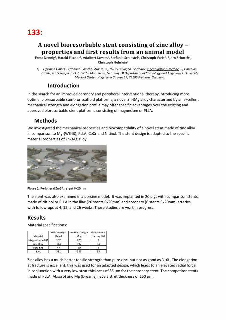
133:
A novel bioresorbable stent consisting of zinc alloy – properties and first results from an animal model
Ernst Nennig1, Harald Fischer1, Adalbert Kovacs2, Stefanie Schiestel2, Christoph Weis3, Björn Schorch3, Christoph Hehrlein3
1) Optimed GmbH, Ferdinand-Porsche-Strasse 11, 76275 Ettlingen, Germany, [email protected]. 2) Limedion GmbH, Am Schaeferstock 2, 68163 Mannheim, Germany. 3) Department of Cardiology and Angiology I, University
Medical Center, Hugstetter Strasse 55, 79106 Freiburg, Germany.
Introduction
In the search for an improved coronary and peripheral interventional therapy introducing more
optimal bioresorbable stent- or scaffold platforms, a novel Zn-3Ag alloy characterized by an excellent
mechanical strength and elongation profile may offer specific advantages over the existing and
approved bioresorbable stent platforms consisting of magnesium or PLLA.
Methods We investigated the mechanical properties and biocompatibility of a novel stent made of zinc alloy
in comparison to Mg-(WE43), PLLA, CoCr and Nitinol. The stent design is adapted to the specific
material properties of Zn-3Ag alloy.
Figure 1: Peripheral Zn-3Ag stent 6x20mm
The stent was also examined in a porcine model. It was implanted in 20 pigs with comparison stents
made of Nitinol or PLLA in the iliac (20 stents 6x20mm) and coronary (6 stents 3x20mm) arteries,
with follow-ups at 4, 12, and 26 weeks. These studies are work in progress.
Results Material specifications:
Zinc alloy has a much better tensile strength than pure zinc, but not as good as 316L. The elongation
at fracture is excellent, this was used for an adapted design, which leads to an elevated radial force
in conjunction with a very low strut thickness of 85 µm for the coronary stent. The competitor stents
made of PLLA (Absorb) and Mg (Dreams) have a strut thickness of 150 µm.
Material
Yield strength
[Mpa]
Tensile strength
[Mpa]
Elongation at
fracture [%]
Magnesium WE43 162 220 2
Zinc alloy 114 192 94
Pure zinc 47 80 8
316L 331 586 35

Figure 2: Radial force of coronary stents
Degeneration behavior in the porcine model: After 4 weeks the stents were completely covered with
neointima. Decrease of the strut thickness was about 50% after 6 months. Radiopacity of the Zn-3Ag
stent is very good; therefore special radiopaque markers are not necessary.
Figure 3: Peripheral Zn-3Ag stent 6x20mm (26 weeks)
Discussion & Conclusion Zinc alloy has a better tensile strength than pure zinc, but not as good as stainless steel or CoCr. It
has an extraordinary elongation at fracture, with an adapted design the radial force of the stent is on
the same level with comparable stents, while the strut thickness is very low. First results from the
animal study are very promising.
Acknowledgements The authors would like to thank the BMBF Research Foundation, Germany, for financial support
(13GW0035).
0,00
0,50
1,00
1,50
2,00
2,50
rad
ial f
orc
e [
N/m
m]
Radial forceat 50% diameter reduction

138: Retroperitoneoscopic left adrenalectomy
Giuseppe Giraudo, Simone Arolfo, Paolo Chiaro, Alberto Bullano, Mario Morino
University of Torino, Department of Surgical Sciences
Retroperineoscopic access represents a minimally invasive technique that radically modified
adrenalectomy. Retroperitoneoscopic adrenalectomy is currently adopted in about 20% of referral
centers. It provides more direct access to the adrenals, thus avoiding post-operative adhesions and
the need for patient repositioning in bilateral adrenalectomy. We would like to present a short video
showing the technique, which focuses on surgical landmarks when left adrenal gland is approached
during a posterior retroperitoneoscopic adrenalectomy The patient is a 45 years old woman with a
drug resistant arterial hypertension due to a left adrenal mass producing aldosterone (Conn
disease).
139:
A Randomized Controlled Trial of the Fundamentals of Robotic
Surgery: Validation of a proficiency-based progression robotic skills
course in a multi-specialty, multi-institutional trial.
Satava Richard- University of Washington Medical Center
ABSTRACT
A Randomized Controlled Trial of the Fundamentals of Robotic Surgery: Validation of a proficiency-
based progression robotic skills course in a multi-specialty, multi-institutional trial.
Objective: To validate the Fundamentals of Robotic Surgery (FRS) skills course in a randomized
control trial using multiple simulation platforms.
Methods and Procedures: A randomized clinical trial with a control group and 3 experimental
groups: physical “dome” using the robot, virtual reality hybrid simulation using the computer
‘backpack’ on the surgical console, and stand-alone virtual reality simulator. Training was to
proficiency and were set by 80 experts. The multi-institutional trial was conducted 12 international
simulation centers and included the multiple specialties that perform robotic surgery.
Results: Face and content validity was verified by 80 experts; concurrent validity was demonstrated
by experimental groups performing in less time and with less errors than the control group. The
construct validity was demonstrated because the mean time and mean number of errors to perform
the various tasks by experts were statistically less the novices. The inter-rater reliability for task
specific checklists was > 0.80, however the Global Evaluative Assessment of Robotic Skills (GEARS)
inter-rater reliability was <0.80
Conclusion: The Fundamentals of Robotic Surgery is a validated course to teach novices the simplest
robotic surgery skills across the many specialties that are currently performing robotic surgery. All
the four validities were confirmed on a physical dome model, or any of the computer/virtual reality
simulators. The FRS is a robust and validated course (curriculum) to train the basic skills in robotic
surgery.