Chemical proteomics identifies Nampt as the target of CB30865, an orphan cytotoxic compound
Upload
independentCategory
view
3download
0
ARTICLE
Absence of an Orphan Mitochondrial Protein,C19orf12, Causes a Distinct Clinical Subtypeof Neurodegeneration with Brain Iron Accumulation
Monika B. Hartig,1,2,14 Arcangela Iuso,2,14 Tobias Haack,1,2,14 Tomasz Kmiec,3 Elzbieta Jurkiewicz,4
Katharina Heim,2 Sigrun Roeber,5 Victoria Tarabin,6 Sabrina Dusi,7 Malgorzata Krajewska-Walasek,8
Sergiusz Jozwiak,3 Maja Hempel,1,2 Juliane Winkelmann,1,2,9 Matthias Elstner,2,10 Konrad Oexle,1
Thomas Klopstock,10 Wolfgang Mueller-Felber,11 Thomas Gasser,12 Claudia Trenkwalder,13
Valeria Tiranti,7 Hans Kretzschmar,5 Gerd Schmitz,6 Tim M. Strom,1,2 Thomas Meitinger,1,2,*and Holger Prokisch1,2
The disease classification neurodegeneration with brain iron accumulation (NBIA) comprises a clinically and genetically heterogeneous
group of progressive neurodegenerative disorders characterized by brain iron deposits in the basal ganglia. For about half of the cases, the
molecular basis is currently unknown. We used homozygosity mapping followed by candidate gene sequencing to identify a homozy-
gous 11 bp deletion in the orphan gene C19orf12. Mutation screening of 23 ideopathic NBIA index cases revealed twomutated alleles in
18 of them, and one loss-of-function mutation is the most prevalent. We also identified compound heterozygous missense mutations in
a case initially diagnosed with Parkinson disease at age 49. Psychiatric signs, optic atrophy, andmotor axonal neuropathy were common
findings. Compared to the most prevalent NBIA subtype, pantothenate kinase associated neurodegeneration (PKAN), individuals with
two C19orf12 mutations were older at age of onset and the disease progressed more slowly. A polyclonal antibody against the predicted
membrane spanning protein showed a mitochondrial localization. A histopathological examination in a single autopsy case detected
Lewy bodies, tangles, spheroids, and tau pathology. The mitochondrial localization together with the immunohistopathological find-
ings suggests a pathomechanistic overlap with common forms of neurodegenerative disorders.
Introduction
Brain iron accumulation is a feature of many neurode-
generative disorders, including Friedreich ataxia (MIM
229300),Huntingtondisease (MIM143100), andParkinson
disease (PD [MIM 168600]). Pronounced iron deposits in
the basal ganglia in the first decades of life are pathogno-
monic for the diagnosis of neurodegeneration with brain
iron accumulation (NBIA), comprising a clinically and
genetically heterogeneous group of debilitating neurode-
generative disorders.1 Genetic defects in PANK2 (MIM
606157) are the most common cause of NBIA, followed by
mutations in PLA2G6; both genes encode mitochondrial
proteins.2–4 PLA2G6 (MIM603604) provides striking exam-
ples of allelic heterogeneity as demonstrated by mutation
reports in cases with infantile neuroaxonal dystrophy and
adult-onset levodopa-responsive dystonia-parkinsonism.5
Mutations in CP (MIM 117700) and FTL (MIM 134790)
have been reported in a small proportion of the remaining
NBIA cases with the clearly distinguishable phenotypes
1Institute of Human Genetics, Technische Universitat Munchen, 81675 Munic
85764 Neuherberg, Germany; 3Department of Child Neurology, Memorial C
Imaging Unit, Memorial Children’s Health Institute, 04-730 Warsaw, Poland
University, 81377 Munich, Germany; 6Institute for Clinical Chemistry and Lab7Unit of Molecular Neurogenetics, Neurological Institute ‘‘Carlo Besta’’ – Istitut8Department of Medical Genetics, Memorial Children’s Health Institute, 04
Munchen, 81675 Munich, Germany; 10Department of Neurology, Ludwig Ma
Dr. von Hauner Children’s Hospital, Ludwig-Maximilians-University, 81337 M
of Neurodegenerative Diseases, and German Center for Neurodegenerative Dis
Elena Klinik, 34128 Kassel, Germany14These authors contributed equally to this work
*Correspondence: [email protected]
DOI 10.1016/j.ajhg.2011.09.007. �2011 by The American Society of Human
The Americ
aceruloplasminaemia and neuroferritinopathy, respec-
tively. Few reportedNBIA families are known to carrymuta-
tions in FA2H (MIM 611026),6 a gene previously associated
with familial leukodystrophy and spastic paraparesis (MIM
612319).7 However, in about half of NBIA cases the under-
lying genetic defect remains unknown to date. The identifi-
cation of additional genetic variation associated with NBIA
has the potential to advance our understanding of both rare
and common neurodegenerative disease entities.
Subjects and Methods
Cases and ControlsThis study was initially conducted in a cohort of 52 NBIA index
cases from Warsaw, Poland. Informed consent was obtained
from all participants or their guardians, and the study was ap-
proved by the institutional review board of the Memorial Chil-
dren’s Health Institute in Warsaw. The diagnosis of NBIA was
based on hypointensity in the globus pallidus documented by
T2-weighted cranial magnetic resonance imaging and a clinical
presentation concordant with NBIA. Detailed clinical information
h, Germany; 2Institute of Human Genetics, Helmholtz Zentrum Munchen,
hildren’s Health Institute, 04-730 Warsaw, Poland; 4Magnetic Resonance
; 5Center for Neuropathology and Prion Research, Ludwigs Maximilians
oratory Medicine, University of Regensburg, 93053 Regensburg, Germany;
o Di Ricovero e Cura a Carattere Scientifico Foundation, 20100 Milan, Italy;
-730 Warsaw, Poland; 9Department of Neurology, Technische Universitat
ximilian University, 81377 Munich, Germany; 11Department of Pediatrics,
unich, Germany; 12Hertie-Institute for Clinical Brain Research, Department
eases, 72076 Tubingen, Germany; 13University of Gottingen and Paracelsus-
Genetics. All rights reserved.
an Journal of Human Genetics 89, 543–550, October 7, 2011 543

was collected for all individuals. The phenotypic characterization
was performed in one clinical center and included a physical
examination with an emphasis on neurological and ophthalmo-
logic examination; neuroimaging (a cranial MRI); and electro-
physiological (EEG, EMG), neurophysiological, and laboratory
testing. The clinical phenotype was documented with a standard-
ized questionnaire. All affected individuals were sequentially
screened for mutations in PANK2, PLA2G6, FTL, and CP. We also
screened a single case with a NBIA diagnosis where histological
samples were available in the German Brain Bank. The clinical
details of this case had been collected at the University Hospital,
Ludwig Maximilians Universitat Munchen.
A group of 676 simplex PD cases from Germany was used for
mutation screening of C19orf12. Diagnosis of PD in this group
was performed according to the UK PD Brain Bank criteria.8
Mutation screening in controls was performed in samples from
150 healthy individuals from Poland and 600 healthy individuals
from Germany.
Homozygosity Mapping and Genetic ScreeningDNA was extracted from peripheral leucocytes with standard
protocols. We performed a genome-wide linkage analysis in a
single family with three affected members by using a genome-
wide SNP array (HumanCNV370-Quadv3, Illumina, San Diego,
CA). Assuming that the disease allele was identical by descent,
we analyzed the data by homozygosity mapping. Frequencies of
marker alleles were derived from 1,200 control individuals, and
approximately 50,000 evenly distributed polymorphic markers
with minimized intermarker linkage disequilibrium (LD) were
selected. Homozygous regions were identified with custom scripts.
Parametric LOD score calculations were performed under the
conservative assumption of a second-cousin marriage. The fre-
quency of the deleterious allele was set to 0.001 and the pene-
trance to 99% (q ¼ 0.001; f1 ¼ 0.0001; f2 ¼ 0.0001; f3 ¼ 0.99).
Data were prepared for linkage analysis with a modified version
of Alohomora.9 Multipoint linkage analysis and haplotyping
were performed with Merlin (version 1.1.2).10
The ExonPrimer primer design software was used to design
primers for PCR amplification of all exons of the 8 transcription
units in the linkage interval. PCR products were purified with
DNA purification kits (Macherey-Nagel, Duren, Germany). Se-
quencing of the sense and antisense strands was performed with
the same primers as for PCR amplification, ABI BigDye Termina-
tors Version 3, and an ABI 3100 Genetic Analyzer. Primers are
listed in Table S1,available online.
Cloning of C19orf12 TranscriptsTotal RNA was isolated from human fibroblasts (NDHF-neo,
Lonza, Basel, Switzerland) with the Trizol method (Invitrogen,
Carlsbad, CA) or from human blood with PAXgene Blood RNA
tubes (PreAnalytiX,Hombrechtikon, Switzerland)and thePAXgene
Blood RNA kit (QIAGEN). Onemicrogram of RNAwas reverse tran-
scribed with M-MLV reverse transcriptase (Promega, Madison, WI)
and oligo dT. C19orf12 coding variants 1 and 2 (NM_001031726.2
andNM_031448.3, respectively) were amplified with Thermo-Start
Taq DNA Polymerase (ABgene, Epsom, UK).
Cell Culture, Transient Transfection, Stable
Transduction, and ImmunocytochemistryNDHF-neo (Lonza) and fibroblast cells weremaintained in Dulbec-
co’s Modified Eagle’s Medium (PAA, Pasching, Austria) supple-
544 The American Journal of Human Genetics 89, 543–550, October
mented with 10% fetal bovine serum (PAA), 1% glutamine
(GIBCO, Carlsbad, CA), and 1% penicillin plus streptomycin
(GIBCO). Cells were transfected with an expression vector con-
taining a C-terminal green fluorescent protein (GFP)-tagged
version of C19orf12 variant 1 (Origene, Rockville, MD) with Effec-
tene (QIAGEN, Hamburg, Germany) according to the manufac-
turer’s instructions. After 12-15 hr of culturing, cells were fixed
with 4% (wt/vol) paraformaldehyde in PBS for 10 min. Following
fixation, cells were permeabilized in PBS, 0.1% NP-40 (Sigma,
St. Louis, MO) and blocked with PBS, 2% BSA and 0.1% NP-40 at
37�C. Primary antibody anti-mitochondrial single strand binding
protein (mtSSBP) was diluted (1:100) in blocking solution and
incubated for 45 min at 37�C. Slides were washed in PBS, 0.1%
NP-40 for 30 min. The same incubation and washing procedures
were used for the secondary antibody anti-rabbit-Alexa Fluor
568 (Invitrogen). Cells were stably transduced with a lentiviral
vector expressing an untagged version of C19orf12 variant 1
(pLenti6.3/V5-TOPO, Invitrogen). Immunocytochemistry was
performed with porin (1:1000, MitoSciences) for the detection of
mitochondria and C19orf12 (1:500). Anti-mouse Alexa Fluor
488 and anti-rabbit Alexa Fluor 568 were used for the secondary
detection. Slides were mounted with coverslips with ProLong
Gold antifade reagent with 40,6-diamidin-20-phenylindol-dihydro-chlorid (DAPI, Invitrogen). Images were collected with a Leica TCS
SP5 confocal microscope with an 633 oil immersion objective.
Mitochondrial morphology was assessed in cell cultures of fibro-
blasts stained with MitoTracker red (Invitrogen).
Subcellular Localization of C19orf12For preparation of C19orf12 antibodies, the long isoform of
C19orf12 was cloned in an Escherichia coli expression vector with
an N-terminal histidine tag (pTrcHis2 TOPO-Invitrogen). After
induction of C19orf12 expression with 1 mM isopropyl-b-D-thio-
galactopyranosid, the native recombinant protein was purified
with Ni-NTA agarose beads according to the manufacturer’s
protocol (QIAGEN). The purified protein was used for immuniza-
tion of two rabbits (Pineda).
For preparation of whole cellular lysates, fibroblast cells from
cases carrying the Gly69ArgffsX10 in C19orf12 and controls were
washed, collected, and resuspended in lysis buffer (0.5 ml/106
cells, 150 mM sodium chloride, 50 mM Tris-HCl [pH 7.4], 0.5%
NP-40, 0.25 mM PMSF, 4�C). After 5 s centrifugation at 1000 g
to pellet unbroken cells, the supernatant was recovered, and
different amounts (5–15 mg each) were separated on a SDS poly-
acrylamide gel.
To isolate different subcellular fractions, we harvested fibro-
blasts at 500 g for 3 min; washed them with phosphate-buffered
saline; and resuspended them in a solution of 300 mM sucrose,
1 mM EGTA, and 20 mM MOPS (pH 7.4). The cellular suspension
was homogenized with a teflon-glass homogenizer with 25 strokes
and centrifuged at 600 g for 10 s at 4�C. The pellet representing
the unbroken cells and the nuclear fraction was recovered. The
mitochondrial-enriched fraction was isolated by centrifugation
at 10,000 g for 25 min. The supernatant was centrifuged at
100,000 g for 1 hr to get endoplasmic reticulum (ER) (pellet) and
the cytosolic fraction (supernatant). For immune decoration, anti-
sera specific for C19orf12 (1:1000), porin (1:200, Mito Sciences,
Eugene, OR), and calnexin (1:2000, Sigma) were used.
Neuropathological InvestigationsHistological examination of one case fromGermany (Table 1, indi-
vidual 2) was performed with 4 mm sections from 20 regions of
7, 2011

Table 1. Clinical Findings
Familial Cases (NBIA) Simplex Cases (NBIA) PD
Family (ID) 28 4 21 37 2a,b 3 5 6b 12 20 22 25 27b 32 33b 34 38 39 41b 40b
II.1 II.4 II.5 II.1 II.2 II.1 II.2 II.1 II.2
Sex m m m f f m f m f m m m m f m m m m m m m f m w M
Age of onset 4 12 4 10 9 12 5 10 20 6 8 10 10 10 6 12 12 11 8 12 10 12c 8 21 25
Age at presentation 12 12 20 25 10 17 15 30 31 21 14 17 18 16 14 15 14 13 13 19 11 14 13 26 59
Pyramidal signsd þ � þ � þ � þþ þ þþ þþþ þ þ þþ þþ þ � þþþ � þþþ þþþ þþ � þþ þþ þ
Extrapyramidal signsd
Oromandibulardystonia
� � þ � � � � þþ þ � � þ þ þ � � þ � � þ þ � þ � �
Generalizeddystonia
þ � þ � � þ � þþ þ � þþ þþ þþ þþ � þ þ � þþ þ � � þ þ n.a.
Parkinsonism þ � þ � � � � þ � � � � þ � � � � � � þ � � � þþþ þþþ
Dysarthria � � þ þ � þ � þþ þ þþþ þ þ þ þ � þ þ � þ þ þ � þ þþþ þ
Wheelchaire n n 18 21 n n n 20 31 19 n 18 18 n n n n n n n n n n 25 n
Optic atrophyf yg yg yg y y yg yg y y y y yg y y yg y yg n yg y yg n y n n.a.
Motor axonalneuropathy
n n y y n n n y y y y y n.a. n n y n y y n n n n n.a n.a.
Psychiatric signsh n n n n n y y y n n n n n n n y y n n n n n n y Y
Cognitive score 82i 90i P7j 60k 73k P25j P25j 80i 80i n.a. 117k 57k 79i 85k 50i 65i P29j 77i P25j 73k 87i 90i 96i n.a. 22l
a The case is from the German BrainNet database.b Cases (n ¼ 7) carrying C19orf12 mutations other than the homozygous founder mutation that was identified in 18 patients: ID2 c.[205G>A]þ[ ¼ ],p.[Gly69Arg]þ[ ¼ ]; ID6 c.[157G>A]þ[204_214del11], p.[Gly53Arg]þ[Gly69ArgfsX10]; ID27 c.[32C>T]þ[204_214del11], p.[Thr11Met]þ[Gly69ArgfsX10];ID33 c.[194G>A]þ[204_214del11], p.[Gly65Glu]þ[Gly69ArgfsX10]; ID38, 40 c.[205G>A]þ[424A>G], p.[Gly65Glu]þ[Lys142Glu]; ID41 c.[32C>T]þ[ ¼ ],p.[Thr11Met]þ[ ¼ ].c Age of onset was defined by MRI.d The following symbols are used: �, not present; þ, mild; þþ, moderate; þþþ, severe as defined in Table S4; n.a. is an abbreviation for not available.e The age at which a wheelchair became necessary is indicated; n indicates no wheelchair was used.f y indicates present; n indicates not present.g Partial optic atrophy (decoloration of optic nerve).h y indicates ADHD-like and/or impulsive-compulsive behavior; n indicates not present.i Wechsler Intelligence Scale for Children/full scale score (WISC-R).j Raven Standard Progressive Matrices (p ¼ percentile).k Wechsler Adult Intelligence Scale/verbal IQ (WAIS-R).l Mini-mental-status Test score (MMST); n.a. not available.
formalin-fixed and paraffin-embedded cerebrum, cerebellum,
brainstem and spinal cord. Hematoxylin and eosin and Prussian
blue as well as different silver stainings were performed with stan-
dard techniques. Immunohistochemistry was performed with
antibodies directed against phosphorylated tau (AT8, 1:200, Inno-
genetics, Ghent, Belgium), a-synuclein (MC42, 1:10, BD Biosci-
ence, Heidelberg, Germany), and amyloid precursor protein
(APP) (22C11, 1:100, Chemicon, Temecula, USA).
Results
Genetics
We screened a cohort of 52 NBIA index cases from Poland
for sequence variants in PANK2, PLA2G6, FTL, and CP and
identified 28 individuals carrying mutations in PANK2.
Twenty-two of these individuals with PANK2 mutations
had been described previously.11 High-density genome-
wide SNP genotyping in one of the 24 families that
lacked PANK2 mutations and had three affected members
The Americ
(Figure 1) revealed a disease-segregating 1.2 Mb region at
19q12 containing eight transcriptional units (Table S2).
Sequence analysis of all exons of these transcripts identi-
fied a single homozygous mutation, c.204_214del11
(Gly69ArgfsX10), in the orphan gene C19orf12 (Figure 2).
This 11 bp deletion predicts a frameshift and a premature
stop codon leading to a predicted truncation of more
than 50% of the protein sequence. Sanger sequencing
of the gene in the remaining 23 index cases revealed a
homozygous 11 bp deletion in 12 of them. Haplotype
analysis suggests that the 11 bp deletion derives from a
common founder at least 50 to 100 generations ago (Fig-
ure S1). Three index cases carried the deletion in combi-
nation with different missense mutations (p.Gly65Glu,
p.Gly53Arg, and p.Thr11Met) in the compound heterozy-
gous state, one case harbored two missense mutations
(p.Gly69Arg and p.Lys142Glu), and two cases carried
homozygous missense mutations (p.Tyr11Met and
p.Gly69Arg). All investigated healthy parents (n ¼ 30) of
an Journal of Human Genetics 89, 543–550, October 7, 2011 545

1 2
+/p.Gly69ArgfsX10 +/p.Gly69ArgfsX10
I
II1 2 3 4 5
p.Gly69ArgfsX10/p.Gly69ArgfsX10 +/p.Gly69ArgfsX10 +/p.Gly69ArgfsX10
p.Gly69ArgfsX10/p.Gly69ArgfsX10
p.Gly69ArgfsX10/p.Gly69ArgfsX10
Figure 1. Pedigree of the NBIA FamilyUsed for Homozygosity MappingMutated and nonmutated alleles are indi-cated.
individuals with C19orf12 mutations were found to be
heterozygous carriers. Five (5/52) index cases remained
without a molecular diagnosis. The protein encoded by
C19orf12 is highly conserved in evolution with 115 out
of 141 amino acids sequences being identical between
the human and mouse (Figure 3). In humans it codes for
two protein isoforms originating from two alternative first
exons. In silico analysis revealed a transmembrane region
in all investigated C19orf12 orthologs. Three missense
mutations (p.Gly53Arg, p.Gly65Glu, and p.Gly69Arg)
change highly conserved glycines in the predicted trans-
membrane region to a charged amino acid. The mutation
p.Thr11Met exclusively affects the longer predicted ver-
sion of the protein (Figure 2). The p.Lys142Glu missense
mutation changes a conserved positive lysine residue to
a negative charged glutamate. Although C19orf12 RNA
levels in blood remained unaffected by the c.204_
214del11 deletion, the protein is absent in fibroblasts
according to immunoblot analysis (Figure S2). The discov-
splice variant 1 (459 nt)(NM_001031726.2)
splice variant 2 (426 nt)(NM_031448.3)
TMD
Met12Met1
c.32C>T,
p.Thr11M
et
c.194G>A, p.G
ly65Glu
c.157G>A, p
.Gly53Arg
c.424A>G, p
.Lys1
42Glu
c.204_214del11, p.G
ly69ArgfsX
10
c.205G>A, p.G
ly69Arg
aa
aa
8 8 65 65 152
54 54 141
1
1
Figure 2. Gene Structure and Identified Disease AllelesGene structure of the two isoforms of C19orf12with the identifiedmutations. The predicted transmembrane domain is marked inyellow. Mutation nomenclature of the C19orf12 gene is based onsplice variant 1 (NM_001031726.2). RefSeq accession number ofsplice variant 2 is NM_031448.3.
546 The American Journal of Human Genetics 89, 543–550, October 7, 2011
ered mutations were absent from 750
control chromosomes.
Clinical Findings
Detailed clinical information was
available for all 24 Polish NBIA cases
with two C19orf12 mutations (Table
1). Eighteen of these are homozygous for the 11 bp dele-
tion in C19orf12 and have a mean age at onset of 9.2 5
3.7 years (a range of 4 to 20 years), 13 presented with
clinical signs of neurodegeneration before the age of 11.
The most common presenting symptoms were speech
and gait difficulties, which occurred in 10 out of 18 and
12out of 18 of cases, respectively. The predominant
neurologic features were extrapyramidal and included
oromandibular and generalized dystonia, and parkin-
sonism (13/18). Involvement of the corticospinal tract
leading to spasticity, hyperreflexia, and Babinski sign was
frequent (14/18) and progressive. Most of the affected
individuals were still able to walk as they entered adult-
hood, but in some the course of the disease progressed
to loss of independent ambulation by the mean (5SD)
age of 21.15 4.5 years (n ¼ 5; range of 18 to 31). An unex-
pected finding was that electrophysiological examina-
tion revealed a motor axonal neuropathy in 44% of cases
(8/18). Although there was no clinical or electroretino-
graphic evidence for a retinopathy, a feature always present
was optic atrophy, which was observed in all 18 cases with
two loss-of-function alleles. The severity of the optic
atrophy varied but did not result in blindness in any of
the cases of our cohort.
Six (6/24) cases of the Polish cohort carried at least one
missense mutation. A trend toward a later onset of the
disease (13.2 5 4.4 years) did not reach significance
when compared to patients carrying two C19orf12 loss-
of-function alleles. Optic atrophy was present in three
out of six cases, and motor axonal neuropathy in two out
of four cases investigated. The course of the disease was
remarkably mild in individuals 27 and 38, which did not
display any pyramidal or extrapyramidal signs. Individual
27 suffered from a motor axonal neuropathy causing
mild gait impairment at the age of 13 years, and individual
38 presented with a mild affection of fine motor skills
only at the age of 14 years.
Psychiatric signs were common in cases with C19orf12
mutations (6/24), including impulsive or compulsive
behavior, depression, and emotional lability. No seizures
were reported.
T2-weighted MRI revealed hypointensities in the globus
pallidus and substantia nigra in all 24 C19orf12 cases,
whereas PKAN-typical hyperintensity in the central

T MM
T MMM
K MMMMM
T
T
K
chimpanzee isoform 1 MERLKSHKPA TIMVEDIMKLLCSLSGERKMKAAVKHSGKGALVTGAVAFV G GLVGGPPGLAV G GAV G GLLGAWMTSGQFKPVPQILM-ELPPAEQQRLFNEAAAIIRHLEWTDAVQLTALVMGSEALQQQLLAMLVNYVT ELRAEIQYDDchimpanzee isoform 2 ---------- TIMVEDIMKLLCSLSGERKMKAAVKHSGKGALVTGAVAFV G GLVGGPPGLAV G GAV G GLLGAWMTSGQFKPVPQILM-ELPPAEQQRLFNEAAAIIRHLEWTDAVQLTALVMGSEALQQQLLAMLVNYVT ELRAEIQYDDhuman isoform 1 MERLKSHKPA TIMVEDIMKLLCSLSGERKMKAAVKHSGKGALVTGAMAFV G GLVGGPPGLAV G GAV G GLLGAWMTSGQFKPVPQILM-ELPPAEQQRLFNEAAAIIRHLEWTDAVQLTALVMGSEALQQQLLAMLVNYVT ELRAEIQYDDhuman isoform 2 ---------- TIMVEDIMKLLCSLSGERKMKAAVKHSGKGALVTGAMAFV G GLVGGPPGLAV G GAV G GLLGAWMTSGQFKPVPQILM-ELPPAEQQRLFNEAAAIIRHLEWTDAVQLTALVMGSEALQQQLLAMLVNYVT ELRAEIQYDDmouse ---------- PIMVDDIMRLLCSISQERKMKAAVKHSGKGAMVAGAMAFV G GLVGGPPGIAV G GTV G GLLGAWMTSGQFKPVPQILM-ELPPAEQRKLVNEAMAIIGNLDWTDAVQLTALVMSNQAMQQRLLAMLTTYVT ELQAEIRYEDchicken MKGIKGPLPA PVRVDQMMQLLCHVSQEKGMTAAVKHSGRGALLAGATAFV G GLVGGPPGIAV G GAL G GLLGAWMTSGQFKPVPQILM-ELPPAEQQKLFDEAIAIVRNLDWTDIAQLTALVMGSGHLQQQLAGVVINYLT ELSAEIKYGEfrog ---------- PVQVDDIIKLLCHVSDHQKMKATIKHSARGALVAAAGAFL G GLVGGPPGIAV G GAV G GAMGAWMTSGQFKPIPQIIM-ELPPVQQQRLCDDIYTIVRTLDWTDATQLIMLVMGNDSLKQKVVAALINYMT ELQAEIQYGDzebrafish ---------- PPHVDDVMKLCCELSANQQVKTAVKQSGKGAAAAGGLAFA G GLIGGPLGIAV G GAV G GLLGCWMTSGQFKPLPQVIM-ELTPDQQARLYEDIVAILSSITWTDVAQLTALVMGNASLQQQVTAALLSYIH ELQAEVHYIDdrosophila1 ---------- PIDTRELMEAIAIVADERNVRVAVKQSGKGAAICAACSFA G GMLLGPVGLAV G GAA G GIAAYKMTSGTFRPLGEVILNDLTDAQREQLVQHVTMAVADIHPTDVVMLLPLIVQNASIQQAVLNTVMSFVT ELR--MQIVDdrosophila2 ---------- DSAISEIINALAILADDKNIQLTIKEAGKGAAICAGAALI G GLLLGPRGLAL G GAI G GLTAYGLTEGNFKSLSEVILNDLTESQRRELEQHVIRAISEVRNVRVRDVARLILNNRHVQEVALEAVKSYIT RMG--MTIVD
GGGGGGGGGGGGGGGGGGG
KKKKKRKKND
KKKKKRKKND
GGGGGGGGGGGGGGGGGGGG
GGGGGGGGGGGGGGGGGG
-
--
----
Figure 3. Evolutionary Conservation of C19orf12Multiple sequences of C19orf12 orthologs were aligned with CLUSTALW2. Amino acids are indicated as identical (red), highly similar(green), and similar (blue). The predicted transmembrane domain is marked in yellow.
Figure 4. Magnetic Resonance Images of Cases with the Homo-zygous C19orf12 Deletion c.204_214del11, p.Gly69ArgfsX10Axial T2-weighted magnetic resonance imaging (1.5 T) showsa bilateral hypointensity of the globus pallidus (A) and substantianigra (B).
anteromedial region of the globus pallidus, also referred to
as ‘‘eye of the tiger’’ sign, was only seen in one (Figure 4).
Neuropathological Findings
Brain pathology was available from aGerman case (Table 1,
individual 2) previously diagnosed with idiopathic NBIA.
Sequence analysis of C19orf12 identified a homozygous
p.Gly69Arg mutation. This person had presented at school
enrollment (at the age of 6) with clumsiness and fatigue.
Optic atrophy had been diagnosed at the age of 10. Progres-
sive gait spasticity had become evident from the age of
14 on, leading to a loss of gait and wheelchair dependence
by the age of 18. Examination at age 21 had shown spastic
tetraparesis, ataxia, marked dysarthria, axonal motor neu-
ropathy, and cognitive decline. A T2-weighted MRI had re-
vealed hypointensities in the globus pallidus. The person
had died at the age 23 from respiratory insufficiency. Histo-
pathological examination showed iron-containing depos-
its, axonal spheroids, numerous a-synuclein-positive Lewy
bodies, andLewybody-like inclusions, sparse Lewyneurites
and hyperphosphorylated tau-containing neuronal inclu-
sions in various regions of the brain (Figure 5). Iron-con-
taining deposits were concentrated in the globus pallidus
and the substantia nigra. The hippocampus showed only
a small number of a-synuclein-containing deposits but
numerous tau-positive pyramidal cells. Loss of myelin was
seen in the pyramidal tracts of the spinal cord and optic
nerve and was most pronounced in the optic tract.
Mutation Screen in Cases with Parkinson Disease
Intrigued by the abundant presence of Lewy bodies,
the neuropathological hallmark of ideopathic Parkinson
disease,12 we performed a mutation screen of C19orf12
in 676 simplex PD cases. This analysis led to the identi-
fication of one sample (Table 1, individual 40) with two
compound heterozygous mutations (p.Gly69Arg and
p.Lys142Glu). Both compound heterozygous mutations
have been also identified in a patient of the Polish cohort
(Table 1, individual 38) presenting with a notably mild
course of the disease that consisted of only an impairment
of fine motor skills at the age of 14. In fact, the MRI per-
formed at the age of 12 was conducted because of a pitui-
tary adenome, and the brain iron accumulation was an
accidental finding.
Evaluation of medical reports revealed that individual
40 presented with paranoid hallucinations at the age of
25. By the age 49, he was diagnosed PD, and a treatment
The Americ
with L-Dopa was started. The latter was effective in pre-
venting akinesia, but a strong fluctuation of the symptoms
was described 11 years later when he was included in a
Parkinson study. Typical PD signs such as rigidity, akinesia,
and a mild tremor at rest were documented. Further, axial
signs, off-dystonia of the legs with muscle cramps, hypo-
phonia, hypomimia, vivid dreams, sleep disturbance, and
optic hallucinations were described. The Unified Parkinson
Disease Rating Score (UPDRS, version III) at that time was
11 and 37, with and without medication, respectively.
The mini-mental status test (MMST) was 22, indicating a
cognitive decline. A cranial computed tomography con-
ducted at the age of 58 showed a marked generalized cere-
bral atrophy. An MRI has not been performed, and the
person was not available for follow-up to resolve the ques-
tion of brain iron deposits.
Functional Investigations
We expressed a C19orf12-GFP fusion protein in fibroblast
cell lines where the C19orf12 fusion protein colocalized
with mitochondria (Figure 6). This finding was confirmed
by subcellular fractionation experiments (Figure 7) and
in vitro import of the radiolabelled gene product into
mouse mitochondria (Figure S3).
On the basis of these findings, we propose the acronym
MPAN (mitochondrial membrane protein associated neu-
rodegeneration) to name the clinical subtype of NBIA
caused by C19orf12 mutations.
an Journal of Human Genetics 89, 543–550, October 7, 2011 547
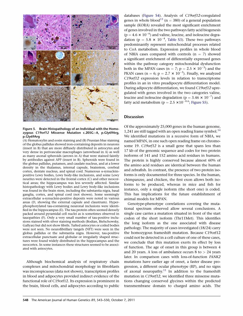
Figure 5. Brain Histopathology of an Individual with the Homo-zygous C19orf12 Missense Mutation c.205G>A, p.Gly69Arg,p.Gly69Arg(A) Hematoxylin and eosin staining and (B) Prussian blue stainingof the globus pallidus showed iron-containing deposits in neurons(insert in B) that are more diffusely distributed in astrocytes andvery dense in perivascular macrophages (arrowhead in A) as wellas many axonal spheroids (arrows in A) that were stained faintlyby antibodies against APP (insert in B). Spheroids were found inthe globus pallidus, putamen, and caudate nucleus, and at a lowerdensity in the thalamus, internal capsule, brainstem, cerebralcortex, dentate nucleus, and spinal cord. Numerous a-synuclein-positive Lewy bodies, Lewy body-like inclusions, and some Lewyneurites were detected in the frontal cortex (C) and other neocor-tical areas; the hippocampus was less severely affected. Similarhistopathology with Lewy bodies and Lewy body-like inclusionswas found in the brain stem, including the substantia nigra, basalganglia, cortex, and spinal cord (not shown). Some seeminglyextracellular a-synuclein-positive deposits were noted in variousareas (D, showing the external capsule and claustrum). Hyper-phosphorylated tau-containing neuronal inclusions were identi-fied in the hippocampus (E). The tau protein often seemed denselypacked around pyramidal cell nuclei as is sometimes observed intauopathies (F). Only a very small number of tau-positive inclu-sions stained with silver staining methods (Bodian, Bielschowsky,Gallyas) but did not show fibrils. Tufted astrocytes or coiled bodieswere not seen. No neurofibrillary tangels (NFT) were seen in theglobus pallidus or the substantia nigra. However, tau-positiveextracellular punctuate and globular or irregularly shaped struc-tures were found widely distributed in the hippocampus and theneocortex. In some instances these structures seemed to be associ-ated with astrocytes.
Although biochemical analysis of respiratory chain
complexes and mitochondrial morphology in fibroblasts
was inconspicuous (data not shown), transcription profiles
in blood and adipocytes provided indirect evidence of the
functional role of C19orf12. Its expression is prominent in
the brain, blood cells, and adipocytes according to public
548 The American Journal of Human Genetics 89, 543–550, October
databases (Figure S4). Analysis of C19orf12-coregulated
genes in whole blood13 (n ¼ 380) of a general population
sample (KORA) revealed the most significant enrichment
of genes involved in the two pathways fatty acid biogenesis
(p ¼ 4.6 3 10�6) and valine, leucine, and isoleucine degra-
dation (p ¼ 5.8 3 10�4, Table S3). These two pathways
predominantly represent mitochondrial processes related
to CoA metabolism. Expression profiles in whole blood
of NBIA cases compared with controls (n ¼ 7) showed
a significant enrichment of differentially expressed genes
within the pathway category mitochondrial dysfunction
both in the MPAN cases (n ¼ 7; p ¼ 2.5 3 10�3) and the
PKAN cases (n ¼ 6; p ¼ 2.7 3 10�3). Finally, we analyzed
C19orf12 expression levels in relation to transcriptome
profiles in an in vitro preadipocyte differentiation model.
During adipocyte differentiation, we found C19orf12 upre-
gulated with genes involved in the two categories valine,
leucine and isoleucine degradation (p ¼ 3.46 3 10�7) and
fatty acid metabolism (p ¼ 2.5 310�0.6; Figure S5).
Discussion
Of the approximately 25,000 genes in the human genome,
1,241 are still tagged with an open reading frame symbol.14
We identified mutations in a recessive form of NBIA, we
namedMPAN, in one such open reading frame on chromo-
some 19. C19orf12 is a small gene that spans less than
17 kb of the genomic sequence and codes for two protein
isoforms of 141 and 152 amino acid residues in humans.
The protein is highly conserved because almost 60% of
the amino acid residues are identical between the human
and zebrafish. In contrast, the presence of two protein iso-
forms is only documented for three species. In the human,
chimpanzee, and chicken, the first exon allows both iso-
forms to be produced, whereas in mice and fish for
instance, only a single isoform (the short one) is coded.
This has implications for the future establishment of
animal models for MPAN.
Genotype-phenotype correlations covering the muta-
tional spectrum observed allow several conclusions. A
single case carries a mutation situated in front of the start
codon of the short isoform (Thr11Met). This identifies
the long isoform as the one associated with disease
pathology. The majority of cases investigated (18/24) carry
the homozygous frameshift mutation. Because C19orf12
could not be detected in a cell culture of one of these cases,
we conclude that this mutation exerts its effect by loss
of function. The age of onset in this group is between 4
and 20 years. A loss of ambulance occurs 8 to > 24 years
later. In comparison cases with loss-of-function PANK2
mutations have earlier age of onset, a faster disease pro-
gression, a different ocular phenotype (RP), and no signs
of axonal neuropathy.11 In addition to the frameshift
mutation in C19orf12, we identified three missense muta-
tions changing conserved glycines within the predicted
transmembrane domain to charged amino acids. The
7, 2011

Figure 6. Subcellular Localization ofC19orf12A C19orf12-GFP fusion protein (uppera-synuclein-positive deposits were notedin various areas [E]) and C19orf12untagged protein (lower panel) exhibita mitochondrial localization in transientlytransfected and in stably transduced fibro-blasts, respectively. Mitochondria werestained with mitochondrial single strandbinding protein (anti-mtSSBP; red) oranti-porin (green). Nuclear DNA wasstained with DAPI (blue).
phenotype associated with these mutations is within
the clinical spectrum caused by the frameshift mutation.
The allelic series observed in MPAN cases is completed
by the c.424A>G (p.Lys142Glu)missensemutation, which
is predicted to exert a milder effect (PolyPhen2).15 Two
cases carry this mutation together with the p.Gly65Glu
mutation. One of them is now 19 years old and suffers
only from impaired fine motor skills, the other one was
diagnosed at the age of 49 with Parkinson disease. No other
diagnosis of a neurodegenerative disorder was made previ-
ously in these persons. The molecular diagnosis of the
C19orf12 mutations is facilitated by the small size of
the gene. Because of the founder mutation observed in
the Polish NBIA cohort, the proportion of NBIA cases
withC19orf12mutationmight be overestimated compared
with other populations. However, the number of different
disease alleles discovered in our study argues that a consid-
erable proportion of NBIA cases worldwide are due to
mutations in this gene.
Using both GFP-tagged protein and a C19orf12-specific
antibody, we showed that the protein localizes predomi-
nantly tomitochondria. So far at least five genes are known
porin
calnexin
C19orf12
Cell ERMitoCyt
Figure 7. Subcellular Localization of C19orf12 in MitochondriaNDHF-neo cells were harvested to obtain mitochondria, ER, andcytosol fractions. Equal amount of proteins (20 mg) from each frac-tion were resolved by SDS-PAGE and immunoblotted for C19orf12with an antibody against the whole protein. Porin and calnexinwere used as a loading control of inner mitochondrial membraneproteins and endoplasmatic reticulum, respectively.
The American Journal of Human Ge
in which mutations cause a NBIA
phenotype with similarities to MPAN
cases. At least two of these genes
(PANK2 and PLA2G6) encode proteins
that share with C19orf12 amitochondrial localization, and
they are suggested to play a role in lipid homeostasis.
Preliminary evidence for a role for C19orf12 in lipid
homeostasis is provided by two experimental observa-
tions. First, there are high C19orf12 expression levels
observed in adipose tissue, which is upregulated during
adipocyte differentiation. Second, C19orf12-coregulated
genes are predominately those involved in fatty acid
metabolism.
C19orf12 is ubitquitously expressed. Nevertheless, both
cells and entire organisms with two loss-of-function
C19orf12 alleles are viable. However, over an extended
period of time, degeneration occurs preferentially in
neurons. The final stage of neurodegeneration—as demon-
strated by the autopsy case described—is characterized by
a variegated histopathological picture in basal ganglia
and cortex regions of the brain including Lewy bodies
and tau inclusions as well as spheroids and iron deposits.
This observation has two main implications. First, it
shows—as has been shown before for familial cases of Par-
kinson disease—that distinct histopathological entities
associated with neurodegeneration can occur as a conse-
quence of a single gene defect. Second, it confirms the
view that the histological end stage of the disease is far
detached from the initial pathophysiological disturbances.
A detailed functional investigation of both normal and
mutated C19orf12 forms has to tell us the details.
Supplemental Data
Supplemental Data include five figures and four tables and can be
found with this article online at http://www.cell.com/AJHG/.
Acknowledgments
We are indebted to all individuals and their families donating
samples and clinical datasets. We thank I. Gromek, A. Kostera-
Pruszczyk, B. Lojszczyk, and B. Chipczynska for their efforts
in clinical phenotyping. We gratefully acknowledge the support
of R. Hellinger and A. Loschner in genotyping; B. Schmick,
netics 89, 543–550, October 7, 2011 549

K. Junghans, and B. Siegel in expression analysis; and S. Heinisch
in bioinformatics analysis. T.M. and H.P. were supported by the
Impulse and Networking Fund of the Helmholtz Association in
the framework of the Helmholtz Alliance for Mental Health in
an Ageing Society (HA-215), the German National Research
Network (NGFNplus #01GS08134), the German Network for
Mitochondrial Disorders (mitoNET 01GM0867), and Systems
Biology of Metabotypes (SysMBo #0315494A). T.K. is supported
by the German Network for Mitochondrial Disorders (mitoNET
#01GM0862). J.W. and K.O. are supported by a grant from the
Deutsche Forschungsgemeinschaft (WI 1820/4-1). The Brain-Net
is supported by the Federal Ministry of Education and Research
(BMBF). Work on primary human adipocytes was supported by
the seventh framework program of the European-Union-funded
LipidomicNet (proposal 202272). S.D. is supported by Italian
Foundation AISNAF (Associazione Italiana Sindromi Neuro-
degenerative Accumulo di Ferro). The financial support of
Mariani Foundation of Milan (grant R-10-84 to V.T.) is gratefully
acknowledged.
Received: August 1, 2011
Revised: September 2, 2011
Accepted: September 15, 2011
Published online: October 6, 2011
Web Resources
The URLs for data presented herein are as follows:
BioGPS database, http://biogps.gnf.org
Brain-Net, http://www.brain-net.net
CLUSTALW2, http://www.ebi.ac.uk/clustalw/
ExonPrimer, http://ihg2.helmholtz-muenchen.de/ihg/ExonPrimer.
html
Ingenuity pathway analysis, http://www.ingenuity.com
OMIM, http://www.omim.org
TMHMM, http://www.cbs.dtu.dk/services/TMHMM/
UCSC Genome Browser, http://genome.ucsc.edu
References
1. Gregory, A., and Hayflick, S.J. (2011). Genetics of neurodegen-
eration with brain iron accumulation. Curr. Neurol. Neurosci.
Rep. 11, 254–261.
2. Zhou, B., Westaway, S.K., Levinson, B., Johnson, M.A., Gitsch-
ier, J., and Hayflick, S.J. (2001). A novel pantothenate kinase
gene (PANK2) is defective in Hallervorden-Spatz syndrome.
Nat. Genet. 28, 345–349.
3. Hortnagel, K., Prokisch, H., and Meitinger, T. (2003). An iso-
form of hPANK2, deficient in pantothenate kinase-associated
550 The American Journal of Human Genetics 89, 543–550, October
neurodegeneration, localizes to mitochondria. Hum. Mol.
Genet. 12, 321–327.
4. Morgan, N.V., Westaway, S.K., Morton, J.E., Gregory, A.,
Gissen, P., Sonek, S., Cangul, H., Coryell, J., Canham, N., Nar-
docci, N., et al. (2006). PLA2G6, encoding a phospholipase A2,
is mutated in neurodegenerative disorders with high brain
iron. Nat. Genet. 38, 752–754.
5. Paisan-Ruiz, C., Bhatia, K.P., Li, A., Hernandez, D., Davis, M.,
Wood, N.W., Hardy, J., Houlden, H., Singleton, A., and
Schneider, S.A. (2009). Characterization of PLA2G6 as a locus
for dystonia-parkinsonism. Ann. Neurol. 65, 19–23.
6. Kruer, M.C., Paisan-Ruiz, C., Boddaert, N., Yoon, M.Y., Hama,
H., Gregory, A., Malandrini, A., Woltjer, R.L., Munnich, A.,
Gobin, S., et al. (2010). Defective FA2H leads to a novel form
of neurodegeneration with brain iron accumulation (NBIA).
Ann. Neurol. 68, 611–618.
7. Edvardson, S., Hama, H., Shaag, A., Gomori, J.M., Berger, I.,
Soffer, D., Korman, S.H., Taustein, I., Saada, A., and Elpeleg,
O. (2008). Mutations in the fatty acid 2-hydroxylase gene
are associated with leukodystrophy with spastic paraparesis
and dystonia. Am. J. Hum. Genet. 83, 643–648.
8. Hughes, A.J., Daniel, S.E., Kilford, L., and Lees, A.J. (1992).
Accuracy of clinical diagnosis of idiopathic Parkinson’s
disease: a clinico-pathological study of 100 cases. J. Neurol.
Neurosurg. Psychiatry 55, 181–184.
9. Lindstrom, M.J., and Bates, D.M. (1988). Newton-Raphson
and EM Algorithms for Linear Mixed-Effects Models for
Repeated-Measures Data. J. Am. Stat. Assoc. 83, 1014–1022.
10. Abecasis, G.R., Cherny, S.S., Cookson, W.O., and Cardon, L.R.
(2002). Merlin—rapid analysis of dense genetic maps using
sparse gene flow trees. Nat. Genet. 30, 97–101.
11. Hartig, M.B., Hortnagel, K., Garavaglia, B., Zorzi, G., Kmiec, T.,
Klopstock, T., Rostasy, K., Svetel, M., Kostic, V.S., Schuelke, M.,
et al. (2006). Genotypic and phenotypic spectrum of PANK2
mutations in patients with neurodegeneration with brain
iron accumulation. Ann. Neurol. 59, 248–256.
12. Hardy, J. (2010). Genetic analysis of pathways to Parkinson
disease. Neuron 68, 201–206.
13. Meisinger, C., Prokisch, H., Gieger, C., Soranzo, N., Mehta, D.,
Rosskopf, D., Lichtner, P., Klopp, N., Stephens, J., Watkins,
N.A., et al. (2009). A genome-wide association study identifies
three loci associated with mean platelet volume. Am. J. Hum.
Genet. 84, 66–71.
14. Pruitt, K.D., Tatusova, T., Klimke, W., and Maglott, D.R.
(2009). NCBI Reference Sequences: current status, policy
and new initiatives. Nucleic Acids Res. 37 (Database issue),
D32–D36.
15. Ramensky, V., Bork, P., and Sunyaev, S. (2002). Human non-
synonymous SNPs: server and survey. Nucleic Acids Res. 30,
3894–3900.
7, 2011
Copyright © 2022 FDOKUMEN