A West Nile Virus Recombinant Protein Vaccine That Coactivates Innate and Adaptive Immunity
11
West Nile Virus Recombinant Protein Vaccine • JID 2007:195 (1 June) • 1607 MAJOR ARTICLE A West Nile Virus Recombinant Protein Vaccine That Coactivates Innate and Adaptive Immunity William F. McDonald, 1,a James W. Huleatt, 1,a Harald G. Foellmer, 2,a Duane Hewitt, 1 Jie Tang, 1 Priyanka Desai, 1 Albert Price, 1 Andrea Jacobs, 1 Virginia N. Takahashi, 1 Yan Huang, 1 Valerian Nakaar, 1 Lena Alexopoulou, 3 Erol Fikrig, 2 and T. J. Powell 1 1 VaxInnate Corporation and 2 Department of Internal Medicine, Yale School of Medicine, New Haven, Connecticut; 3 Centre d’Immunologie de Marseille-Luminy, Centre National de la Recherche Scientifique, INSERM, Universite ´ de la Me ´ diterrane ´e, Marseille, France A chimeric protein West Nile virus (WNV) vaccine capable of delivering both innate and adaptive immune signals was designed by fusing a modified version of bacterial flagellin (STF2D) to the EIII domain of the WNV envelope protein. This fusion protein stimulated interleukin-8 production in a Toll-like receptor (TLR)– 5–dependent fashion, confirming appropriate in vitro TLR5 bioactivity, and also retained critical WNV-E– specific conformation-dependent neutralizing epitopes as measured by enzyme-linked immunosorbent assay. When administered without adjuvant to C3H/HeN mice, the fusion protein elicited a strong WNV-E–specific immunoglobulin G antibody response that neutralized viral infectivity and conferred protection against a lethal WNV challenge. This potent EIII-specific immune response requires a direct linkage of EIII to STF2D, given that a simple mixture of the 2 components failed to induce an antibody response or to provide protection against virus challenge. The presence of a functional TLR5 gene in vivo is also required—TLR5-deficient mice elicited only a minimal antigen-specific response. These results confirm that vaccines designed to coordinately regulate the innate and adaptive immune responses can induce protective immune responses without the need for potentially toxic adjuvants. They also support the further development of an effective WNV vaccine and novel monovalent and multivalent vaccines for related flaviviruses. West Nile virus (WNV) is one of several flaviviruses known to cause human disease. It was first identified in the West Nile district of Uganda in 1937 and has since spread to more temperate regions that include parts of Europe and North America [1]. Currently, there is no effective treatment for the disease, and the best means of prevention will likely come from an ef- fective vaccine. Members of the flavivirus family are single-stranded positive-sense RNA enveloped viruses Received 14 November 2006; accepted 4 January 2007; electronically published 17 April 2007. Potential conflicts of interest: W.F.M., J.W.H., D.H., J.T., P.D., A.P., A.J., V.N.T., Y.H., V.N., and T.J.P. are employees of VaxInnate Corporation. E.F. serves as a scientific advisor for VaxInnate. L.A. and H.G.F. report no conflicts. Financial support: VaxInnate Corporation. a W.F.M., J.W.H., and H.G.F. contributed equally to the work. Reprints or correspondence: William F. McDonald, VaxInnate Corp., Ste. 311, 300 George St., New Haven, CT 06511 ([email protected]). The Journal of Infectious Diseases 2007; 195:1607–17 2007 by the Infectious Diseases Society of America. All rights reserved. 0022-1899/2007/19511-0009$15.00 DOI: 10.1086/517613 whose virions consist of 3 structural proteins [2] that include an envelope (E) protein, a nucleocapsid protein, and a lipid membrane protein. The E protein is asso- ciated with the outer viral membrane and plays a major role in virion assembly, receptor binding, and host membrane fusion. This protein is also the major fla- vivirus immunogen and is the primary target for neu- tralizing antibodies [3, 4]. As with other flaviviruses, the E protein of WNV has been shown to play a critical role in protective immunity. Immunization of experi- mental animals with various forms of the E antigen has been shown to induce protective immune response to viral challenge [5–11]. This protection is associated with a virus-neutralizing antibody response that can be pas- sively transferred to naive recipients [11, 12]. T cell helper and effector mechanisms are thought to also play a role in the natural immune response to WNV infec- tion and may play a role in protection (reviewed in [12]). Thus, because of the critical nature of the E protein during virus infection and the host immune response, a vaccine incorporating E protein deter- by guest on September 27, 2016 http://jid.oxfordjournals.org/ Downloaded from
-
Upload
independent -
Category
Documents
-
view
0 -
download
0
Transcript of A West Nile Virus Recombinant Protein Vaccine That Coactivates Innate and Adaptive Immunity

West Nile Virus Recombinant Protein Vaccine • JID 2007:195 (1 June) • 1607
M A J O R A R T I C L E
A West Nile Virus Recombinant Protein VaccineThat Coactivates Innate and Adaptive Immunity
William F. McDonald,1,a James W. Huleatt,1,a Harald G. Foellmer,2,a Duane Hewitt,1 Jie Tang,1 Priyanka Desai,1
Albert Price,1 Andrea Jacobs,1 Virginia N. Takahashi,1 Yan Huang,1 Valerian Nakaar,1 Lena Alexopoulou,3
Erol Fikrig,2 and T. J. Powell1
1VaxInnate Corporation and 2Department of Internal Medicine, Yale School of Medicine, New Haven, Connecticut; 3Centre d’Immunologiede Marseille-Luminy, Centre National de la Recherche Scientifique, INSERM, Universite de la Mediterranee, Marseille, France
A chimeric protein West Nile virus (WNV) vaccine capable of delivering both innate and adaptive immunesignals was designed by fusing a modified version of bacterial flagellin (STF2D) to the EIII domain of theWNV envelope protein. This fusion protein stimulated interleukin-8 production in a Toll-like receptor (TLR)–5–dependent fashion, confirming appropriate in vitro TLR5 bioactivity, and also retained critical WNV-E–specific conformation-dependent neutralizing epitopes as measured by enzyme-linked immunosorbent assay.When administered without adjuvant to C3H/HeN mice, the fusion protein elicited a strong WNV-E–specificimmunoglobulin G antibody response that neutralized viral infectivity and conferred protection against alethal WNV challenge. This potent EIII-specific immune response requires a direct linkage of EIII to STF2D,given that a simple mixture of the 2 components failed to induce an antibody response or to provide protectionagainst virus challenge. The presence of a functional TLR5 gene in vivo is also required—TLR5-deficient miceelicited only a minimal antigen-specific response. These results confirm that vaccines designed to coordinatelyregulate the innate and adaptive immune responses can induce protective immune responses without the needfor potentially toxic adjuvants. They also support the further development of an effective WNV vaccine andnovel monovalent and multivalent vaccines for related flaviviruses.
West Nile virus (WNV) is one of several flaviviruses
known to cause human disease. It was first identified
in the West Nile district of Uganda in 1937 and has
since spread to more temperate regions that include
parts of Europe and North America [1]. Currently,
there is no effective treatment for the disease, and the
best means of prevention will likely come from an ef-
fective vaccine. Members of the flavivirus family are
single-stranded positive-sense RNA enveloped viruses
Received 14 November 2006; accepted 4 January 2007; electronically published17 April 2007.
Potential conflicts of interest: W.F.M., J.W.H., D.H., J.T., P.D., A.P., A.J., V.N.T.,Y.H., V.N., and T.J.P. are employees of VaxInnate Corporation. E.F. serves as ascientific advisor for VaxInnate. L.A. and H.G.F. report no conflicts.
Financial support: VaxInnate Corporation.a W.F.M., J.W.H., and H.G.F. contributed equally to the work.Reprints or correspondence: William F. McDonald, VaxInnate Corp., Ste. 311,
300 George St., New Haven, CT 06511 ([email protected]).
The Journal of Infectious Diseases 2007; 195:1607–17� 2007 by the Infectious Diseases Society of America. All rights reserved.0022-1899/2007/19511-0009$15.00DOI: 10.1086/517613
whose virions consist of 3 structural proteins [2] that
include an envelope (E) protein, a nucleocapsid protein,
and a lipid membrane protein. The E protein is asso-
ciated with the outer viral membrane and plays a major
role in virion assembly, receptor binding, and host
membrane fusion. This protein is also the major fla-
vivirus immunogen and is the primary target for neu-
tralizing antibodies [3, 4]. As with other flaviviruses,
the E protein of WNV has been shown to play a critical
role in protective immunity. Immunization of experi-
mental animals with various forms of the E antigen has
been shown to induce protective immune response to
viral challenge [5–11]. This protection is associated with
a virus-neutralizing antibody response that can be pas-
sively transferred to naive recipients [11, 12]. T cell
helper and effector mechanisms are thought to also play
a role in the natural immune response to WNV infec-
tion and may play a role in protection (reviewed in
[12]). Thus, because of the critical nature of the E
protein during virus infection and the host immune
response, a vaccine incorporating E protein deter-
by guest on September 27, 2016
http://jid.oxfordjournals.org/D
ownloaded from

1608 • JID 2007:195 (1 June) • McDonald et al.
Figure 1. Flagellin (STF2D) and West Nile virus (WNV)–EIII proteins. A, Schematic representation of STF2D.EIII fusion (1), STF2D (2), and WNV-EIII (3) expression plasmids. All 3 constructs were designed without affinity tags and were expressed in Escherichia coli using the pET/T7 promoter-driven expression system. B, SDS-PAGE of purified STF2D.EIII fusion, STF2D, and EIII proteins. Lane M, molecular-weight markers; lane 1, 5 mg ofSTF2D.EIII; lane 2, 5 mg of STF2D; lane 3, 5 mg of WNV-EIII. Proteins were visualized by Coomassie staining. rbs, ribosome binding site.
minants has the potential to induce long-lasting protective
immunity.
Because E proteins from all flavivirus members are similar,
comparative studies using structural data from select flavivirus
E proteins have provided much-needed information for ratio-
nal vaccine design. The E protein exists as a dimer on the surface
of mature virus particles and consists of a hydrophobic anchor
responsible for the initiation of membrane fusion and 3 distinct
domains (EI, EII, and EIII) essential for dimerization and re-
ceptor binding [2, 13]. These 3 domains have been defined
antigenically using serological means [13] and 3-dimensionally
for dengue, tickborne encephalitis, and WNV proteins using
x-ray crystallography [3, 14–16]. The amino acid sequences of
flavivirus E proteins are also highly homologous and include
12 highly conserved cysteine residues that form 6 disulfide
bridges [17]. EI and EII are formed from the folding of non-
contiguous amino acid segments. EI is positioned within the
center of the protein separating EII and EIII, whereas EII is
the principal region of interaction between E protein monomers
and is thought to promote dimerization. By contrast, EIII is
formed from a single stretch of ∼100 aa and contains a fold
that is reminiscent of a typical immunoglobulin-like domain.
This particular domain is an ideal vaccine target because it is
the receptor-binding domain of E and it encodes the majority
of the flavivirus type-specific epitopes [13].
To develop a potent WNV vaccine that will ensure a specific
and protective immune response, we have designed a recom-
binant protein that will deliver the EIII domain of the E protein
to antigen-presenting cells (APCs) by linking it to the ligand
of Toll-like receptor (TLR)–5. TLRs are initiators of the innate
immune response and gatekeepers of the adaptive immune
response [18–22]. They are the best characterized type of pat-
tern recognition receptor (PRR) expressed on APCs. Engage-
ment of PRRs by their cognate ligands, pathogen-associated
molecular patterns (PAMPs), triggers important cellular mech-
anisms that lead to the expression of costimulatory molecules,
the secretion of critical cytokines and chemokines, and efficient
processing and presentation of antigens to T cells. To date, a
total of 13 TLRs (TLR1–13) have been discovered and the
corresponding PAMPs for most of these receptors have been
identified. Some well-characterized PAMPs include bacterial
cell-wall components (e.g., lipoproteins and lipopolysaccha-
rides), bacterial or viral DNA sequences that contain unmeth-
ylated CpG residues, and bacterial flagellin. In the present ar-
ticle, we demonstrate that the fusion of a TLR agonist with a
protective domain of the WNV-E protein can elicit protective
immunity.
MATERIALS AND METHODS
DNA cloning. Full-length flagellin from Salmonella typhi-
murium fljb (flagellin phase 2), also called “STF2,” is encod-
ed by a 1.5-kb gene. A modified version of STF2, designated
STF2D, was generated by deleting the hypervariable region that
spans aa 170–415. The deleted region was replaced with a short
flexible linker (GAPVDPASPW) designed to facilitate interac-
tions of the NH2 and COOH terminal regions necessary for
TLR5 signaling. The resulting construct, pMT/STF2D, was used
to generate pET/STF2D.EIII and pET/STF2D. Proteins were
purified using conventional chromatography and required re-
folding steps because of low solubility in Escherichia coli. Under
standard growth and induction conditions, STF2D.EIII, STF2D,
and EIII proteins were expressed as insoluble proteins and
formed inclusion bodies (IBs). Before the initiation of column
chromatography, IBs were washed and processed as described
below. STF2D.EIII IBs were solubilized with 8 mol/L urea in
100 mmol/L Tris-HCl (pH 8.0) and refolded by rapid dilution
into 0.1 mol/L Tris-HCl (pH 8.0), 100 mmol/L NaCl, and 1%
by guest on September 27, 2016
http://jid.oxfordjournals.org/D
ownloaded from

West Nile Virus Recombinant Protein Vaccine • JID 2007:195 (1 June) • 1609
Figure 2. Toll-like receptor (TLR)–5–specific bioactivity of recombinantproteins. Serial dilutions of recombinant flagellin fusion proteins wereexamined for the ability to activate TLR5-negative RAW264.7 and TLR5-positive RAW/TLR5 cell lines. Supernatants were harvested 16 h afterstimulation and were examined for expression of the proinflammatorycytokine tumor necrosis factor (TNF)–a by ELISA by RAW264.7/TLR5 (A)and the TLR5-negative parental RAW264.7 (B) cell lines. Stimulation withthe TLR4 agonist lipopolysaccharide (LPS) is shown as a specificity control.
(wt/vol) glycerol to a final protein concentration of 0.1 mg/
mL. Solid ammonium sulfate (AS) was added to the refolded
protein to a final concentration of 1 mol/L, and the sample
was applied to a butyl sepharose column (GE/Amersham)
equilibrated in 25 mmol/L Tris (pH 8.0) and 1 mol/L AS. The
bound protein was eluted from the column in 25 mmol/L Tris
(pH 8.0). The eluate fractions were extracted 4 times with 1%
(wt/vol) Triton X-114 to remove residual endotoxin. The pro-
tein was subsequently fractionated on a Superdex 200 gel fil-
tration column (SD200; GE/Amersham Biosciences) to remove
detergent and buffer exchange the product into Tris-buffered
saline (TBS). STF2D IBs were solubilized in 8 mol/L urea in
50 mmol/L Na acetate (pH 4.0). The solubilized protein was
captured on SP fast-flow sepharose (GE/Amersham Biosci-
ences) under denaturing conditions and was selectively eluted
with 8 mol/L urea and 50 mmol/L Na acetate (pH 4.0) buffer
that contained 0.2 mol/L NaCl. The eluted material was pooled
and dialyzed against 50 mmol/L Tris-HCl (pH 8.0) and refolded
by rapid dilution to 1:10 into 50 mmol/L Tris-HCl (pH 8.0)
to a final protein concentration of ∼0.1 mg/mL. The refolded
SP pool was loaded directly onto Q high-performance sepha-
rose (GE/Amersham), and bound protein was eluted with 20
column volumes of a linear gradient from 0–0.5 mol/L NaCl
in 50 mmol/L Tris-HCl (pH 8.0). EIII IBs were solubilized with
8 mol/L urea in 50 mmol/L Na acetate (pH 6.3). The protein
was applied to SP fast-flow sepharose (GE/Amersham Biosci-
ences). Bound protein was eluted with 50 mmol/L Na acetate
(pH 6.3) and 8 mol/L urea that contained 0.2 mol/L NaCl. SP
peak fractions were pooled and dialyzed against 50 mol/L Tris-
HCl (pH 8.5). To refold the protein, the dialyzed sample was
diluted 1:10 (final protein concentration, ∼0.1 mg/mL) in 50
mmol/L Tris-HCl (pH 8.5). The refolded SP pool was loaded
directly on Q high-performance sepharose (GE/Amersham).
Under these conditions, the majority of EIII did not bind Q
and eluted with the flow-through fraction. The Q high-per-
formance flow-through was concentrated to 2 mg/mL (Amicon
Ultra-15, 5000 molecular-weight cutoff; Millipore) and applied
to size-exclusion chromatography (SD200; GE/Amersham) pre-
equilibrated in TBS.
For the isolation of WNV-EIII sequences, a DNA fragment
encoding the WNV-E protein (aa 1–406) was subcloned into
pMT/BiP/V5-His (Invitrogen) to generate pMT/E. This con-
struct was designed to produce 80% WNV-E protein (without
the COOH terminal hydrophobic tail) in Drosophila for use as
an antigen in serum antibody ELISAs. It also served as a tem-
plate to isolate the WNV-EIII domain (aa 291–406). The EIII
fragment was subcloned separately into pET24a to create pET/
EIII and in frame with STF2D (fused to the C terminus) to
generate pET/STF2D.EIII. The STF2D sequence of pET/STF2D
was derived from pMT/STF2D. Stable cell pools were expanded
as adherent cultures and were adapted to suspension growth
in selection medium (Drosophila SFM; 18 mmol/L l-glutamine,
1� penicillin/streptomycin, and 25 mg/mL blasticidin). Protein
expression was induced with 0.5 mmol/L CuSO4, and E protein
was purified by affinity chromatography using nickel NTA
(Sigma).
TLR5 bioassay and antibody epitope analysis. To test fla-
gellin fusion proteins for TLR5 bioactivity, a cell-based assay
using a modified RAW264.7 cell line that expresses functional
TLR5 was used as reported elsewhere [23]. For antibody epitope
analysis, the following ELISA procedure was used. Ninety-six–
well ELISA plates were coated with serial dilutions (100 mL/
by guest on September 27, 2016
http://jid.oxfordjournals.org/D
ownloaded from

1610 • JID 2007:195 (1 June) • McDonald et al.
Figure 3. Immunogen antigenicity. A, ELISA plates coated with purified West Nile virus (WNV)–E (white bars) or STF2D.EIII (black bars) at 2 mg/mL and incubated with the indicated antibodies. Wells were washed and incubated with horseradish peroxidase–labeled rabbit anti–mouse IgG anddeveloped with 3,3′,5,5′-tetramentylbenzidine substrate. The epitopes for WNV-specific monoclonal antibodies (MAbs) 5C5, 5H10, 3A3, and 7H2 weremapped to the EIII domain of the envelope protein. The epitope for 3D9 resides outside of the EIII domain. A rabbit polyclonal antiserum (Poly) raisedagainst WNV-E protein was used as a control for reactivity with both proteins and the MAb 6H11 (anti-flagellin) was used to assay for flagellinsequences. B, Comparison of MAb 5C5 reactivity with WNV-E (E), STF2D.EIII, EIII, and STF2 proteins. ELISA plates were coated with serial dilutionsof the indicated proteins. C, Comparison of flagellin MAb 6H11 reactivity against the same immunogens. WNV-E was produced and purified fromstable Dmel-2 cells that carried the pMT/E construct.
well) of purified STF2D-fusion proteins in PBS (2 mg/mL).
Plates were blocked with 200 mL/well of assay diluent buffer
(ADB; BD Pharmingen) for 1 h at room temperature. Plates
were washed 3 times in PBS with 0.01% Tween 20 (PBS-T)
and were then incubated with antibodies reactive with flagellin
or WNV-E. The expression of flagellin was detected using the
monoclonal antibody (MAb) 6H11 (Intotek Pharmaceuticals),
whereas the antigenicity of WNV-E proteins was monitored
using a panel of MAbs (5C5, 7H2, 5H10, 3A3, and 3D9) pur-
chased from Bioreliance. Antibodies diluted in ADB (100 mL/
well) were incubated overnight at 4�C. Plates were washed 3
times with PBS-T. Horseradish peroxidase (HRP)–labeled goat
anti–mouse IgG antibodies (Jackson Immunochemical) diluted
in ADB were added (100 mL/well), and the plates were incu-
bated for 1 h at room temperature. Plates were washed 3 times
with PBS-T. After the addition of 3,3′,5,5′-tetramentylbenzidine
Ultra substrate (Pierce Biotechnology) and the monitoring of
color development, the A450 was measured on a Tecan Farcyte
microplate spectrophotometer.
Immunization and viral challenge of mice. C3H/HeN
mice, 6–8 weeks old, were purchased form the Jackson Labo-
ratory. Mice were housed and maintained by the Yale University
animal facility, and all studies were performed in accordance
with the guidelines of the Yale University Institutional Animal
Care and Use Committee. TLR5-deficient mice were generated
as described elsewhere [24]. Mice were immunized subcuta-
neously (sc) or intraperitoneally (ip) at 14-day intervals (2 or
3 immunizations, as described in Results). Immunized mice
were bled by retro-orbital puncture, and serum was harvested
by clotting and centrifugation of the heparin-free blood sam-
by guest on September 27, 2016
http://jid.oxfordjournals.org/D
ownloaded from

West Nile Virus Recombinant Protein Vaccine • JID 2007:195 (1 June) • 1611
Figure 4. Immunized mice challenged on day 37 with LD90 of WestNile virus strain 2741. Survival was monitored for 21 days after challenge.This monitoring period was chosen because survival outcomes beyond21 days remained constant, making longer evaluation times unnecessary[6, 9, 28]. A graphical representation of the percentage of survivors 21days after challenge for each immunized group is shown.
ples. For efficacy studies, mice were challenged with a lethal
dose (LD90) of WNV strain 2741, and survival was monitored
for 21 days after challenge.
Serum antibody determination and reduction in viral
plaque number (PRNT) analysis. Serum IgG titers specific
for flagellin and for WNV-E were determined by ELISA. Ninety-
six–well ELISA plates were coated with 2 mg/mL of purified
WNV-E or STF2 protein. Mouse antiserum was serially diluted
in PBS and incubated for 1 h at room temperature. Plates were
blocked with 200 mL/well of ADB (BD Pharmingen) for 1 h at
room temperature and were washed 3 times in PBS-T. Dilutions
of the serum in ADB were added (100 mL/well), and the plates
were incubated at 4�C overnight. Plates were washed again,
HRP-labeled goat anti–mouse IgG antibodies (Jackson Im-
munochemical) diluted in ADB were added (100 mL/well), and
the plates were incubated for 1 h at room temperature. Plates
were developed as described above. For PRNT analysis, samples
were serially diluted in PBS with 5% gelatin from 1:10 to 1:
2560 and processed as described elsewhere [25].
RESULTS
Bioactivity and antigenicity of purified immunogens. To
confirm the TLR5 signaling activity of purified immunogens
(figure 1), proteins were examined for the ability to activate
the TLR5-negative cell line RAW264.7 or RAW264.7 cells trans-
fected with human TLR5 [23]. As a means of comparison, the
bioactivity of full-length flagellin protein (STF2) was also de-
termined. This protein was purified under native conditions
and was not denatured during purification. As shown in figure
2A, nanomolar amounts of STF2 induced significant levels of
tumor necrosis factor (TNF)–a (6 ng/mL) with a calculated
effective concentration for half-maximal response (EC50) val-
ue of 18 nmol/L. Similar TLR5 activity was observed for
STF2D.EIII and STF2D proteins (EC50 values of 3 and 10 nmol/
L, respectively), which confirmed that removal of the hinge
region from flagellin does not affect TLR5 signaling. These data
also confirmed that the TLR5 signaling activity of STF2D and
STF2D.EIII is restored after denaturation and refolding of these
proteins. As expected, purified EIII, which is not a TLR5 ag-
onist, did not induce TNF-a production in this assay. Finally,
none of these proteins induced TNF-a when tested on the
parental RAW264.7 cell line, which lacks TLR5 (figure 2B).
Thus, purified STF2D.EIII and STF2D have fully functional
TLR5 bioactivity.
Because critical domains within EIII are conformation sen-
sitive, it was also necessary to confirm proper folding and epi-
tope display in purified STF2D.EIII and EIII by ELISA using a
panel of WNV-E polyclonal antibodies and MAbs. For this
purpose, we tested several well-characterized E-specific MAbs
for reactivity against these purified proteins [26] (BioReliance).
As seen in figure 3A, all 5 WNV-E MAbs reacted with full-
length WNV-E, as expected. This protein contains the majority
of the E gene (80%), including the EIII domain, and was pro-
duced in Drosophila cells as a soluble and folded protein. A
comparison of MAb reactivity with STF2D.EIII revealed that
MAbs 5C5, 5H10, 3A3, and 7H2, but not 3D9, recognized the
fusion protein. This pattern of reactivity is consistent with the
proposed location of the antibody epitopes within EIII. Poly-
clonal WNV-E antiserum also reacted with WNV-E and
STF2D.EIII; however, reactivity with STF2D.EIII was somewhat
reduced, perhaps because of the reduced number of potential
epitopes present in the smaller domain. A flagellin-specific MAb
(6H11) was also tested and was shown to specifically react with
STF2D.EIII but not with WNV-E. Using MAb 5C5, a direct
antigenic comparison was made between STF2D.EIII and EIII
(figure 3B and 3C), which confirmed that both STF2D.EIII and
EIII contain critical E-specific conformation-dependent neu-
tralizing epitopes.
Immunogenicity and efficacy of STF2D.EIII. The immu-
nogenicity and efficacy of STF2D.EIII were tested in an estab-
lished mouse model for WNV infection [27]. As shown in
figure 4, significant levels of WNV-E–specific IgG were de-
tected by day 35 (end-point titers 1104) in mice immunized
with STF2D.EIII either ip or sc. These levels were also detected
after the first boost (day 21; data not shown), which suggests
that 2 immunizations might be sufficient to elicit a maximal
response. The fusion protein also elicited flagellin-specific IgG
with similar kinetics (data not shown) (see figure 5).
Because protection from WNV infection is largely antibody
mediated, we tested these same serum samples for their ability
by guest on September 27, 2016
http://jid.oxfordjournals.org/D
ownloaded from

1612 • JID 2007:195 (1 June) • McDonald et al.
Figure 5. Fusion of the West Nile virus (WNV)–EIII domain to flagellin for a potent and protective EIII-specific immune response in mice. Five groupsof C3H/HeN mice (10 mice/group) were immunized on days 0, 14, and 28 subcutaneously with STF2D.EIII (25 mg), STF2D (18 mg), WNV-EIII (7 mg),or a mixture of STF2D (18 mg) and WNV-EIII (7 mg). Doses were chosen to ensure that molar equivalents of each antigen were administered in PBS.On day 35, serum was harvested and tested by ELISA for flagellin (A) and WNV-E–specific (B) IgG responses. Purified flagellin (STF2) and 80% WNV-E protein were used as antigens for antibody detection. Results are the mean � SE optical density values at 450 nm obtained from 10 individualmice/group. C, Immunized mouse challenge on day 37 with an LD90 of WNV strain 2741. Mice monitored for survival for 21 days. Survival curves foreach treatment group are shown and expressed as a percentage.
to neutralize virus in vitro. Serum titers that led to PRNT80
were recorded. The PRNT80 data from 2 independent mouse
efficacy studies are presented in table 1. In all cases, pooled
serum from mice immunized with STF2D.EIII had neutrali-
zation titers of �1:40. It has been previously established that
neutralization titers of �1:40 typically correlate with protection
in vivo [25]. To compare neutralization titers with protection
from WNV in vivo, mice were challenged with an LD90 (100
pfu/mouse) of WNV. As shown in figure 4 and table 1, only
20% of mice that received PBS alone survived infection. By
contrast, �90% survival was observed in groups that were im-
munized with STF2D.EIII. These data are consistent with the
neutralization titers measured with serum from these groups.
Thus, STF2D.EIII administered without adjuvant is capable of
eliciting a potent antibody response sufficient to protect im-
munized mice from a lethal viral challenge.
Requirement for fusion of STF2D to EIII. As reported else-
where [23], the physical linkage of PAMP and antigen, rather
by guest on September 27, 2016
http://jid.oxfordjournals.org/D
ownloaded from
West Nile Virus Recombinant Protein Vaccine • JID 2007:195 (1 June) • 1613
Table 1. West Nile virus (WNV)–E–specific IgG titers andin vitro neutralization activity of antiserum from STF2D.EIII-immunized mice.
Study,immunogen Route IgG titer (end point)
PRNT80,dilution
1
PBS ip ND !1:10
STF2D.EIII ip 2.35 � 104 1:40
STF2D.EIII sc 2.83 � 104 1:160
2
STF2D.EIII ip 2.71 � 104 1:80
STF2D.EIII sc 2.93 � 104 1:40
NOTE. Results from 2 independent efficacy studies are shown (1 and2). C3H/HeN mice (10 mice/group) were immunized 3 times (on days 0,14, and 28) intraperitoneally (ip) or subcutaneously (sc) with 25 mg ofSTF2D.EIII formulated in PBS. On day 35, serum samples from each groupwere harvested, assayed for the presence of WNV-E–specific IgG byELISA, and pooled. Pooled samples were serially diluted and tested forneutralization activity as described in Materials and Methods. WNV-E–specific IgG titers, presented as end-point titers, and virus neutralizationpotency of pooled serum samples (80% reduction in viral plaque number[PRNT80]) are shown. ND, not done.
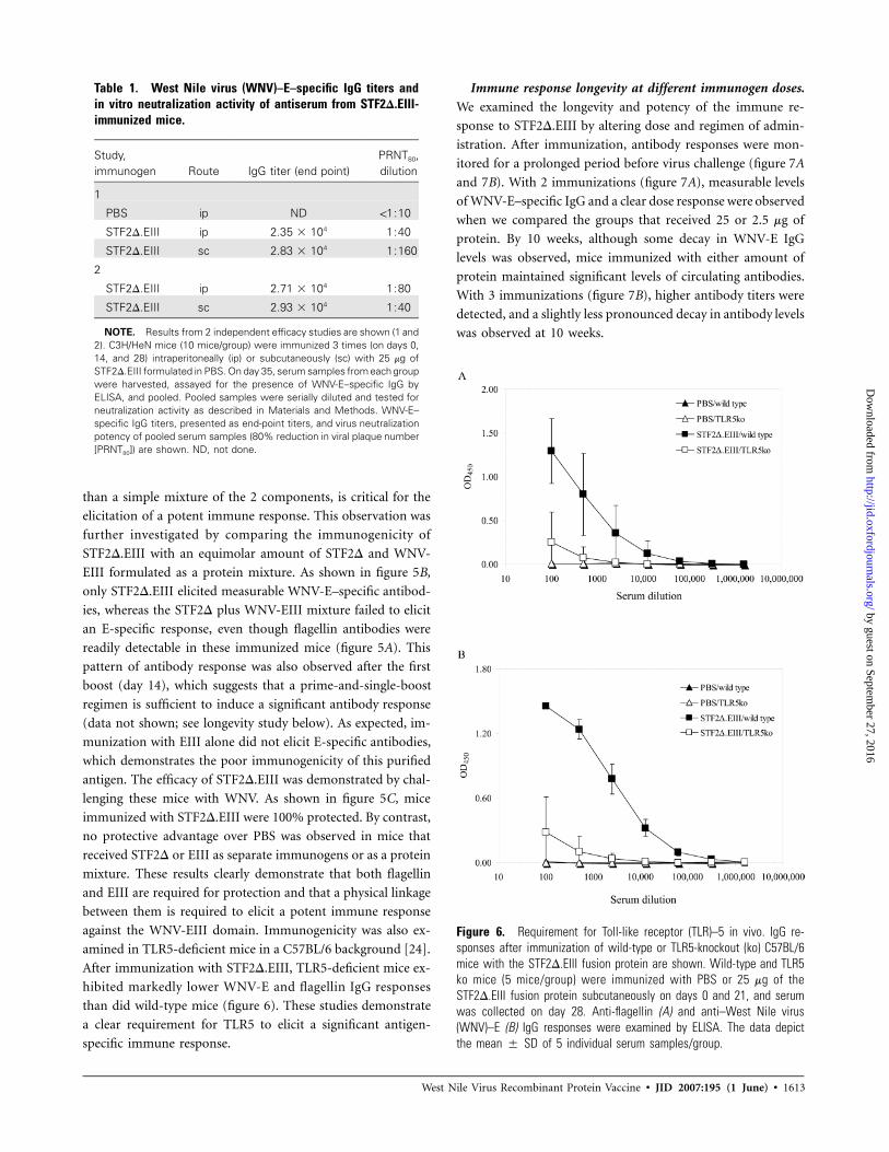
Figure 6. Requirement for Toll-like receptor (TLR)–5 in vivo. IgG re-sponses after immunization of wild-type or TLR5-knockout (ko) C57BL/6mice with the STF2D.EIII fusion protein are shown. Wild-type and TLR5ko mice (5 mice/group) were immunized with PBS or 25 mg of theSTF2D.EIII fusion protein subcutaneously on days 0 and 21, and serumwas collected on day 28. Anti-flagellin (A) and anti–West Nile virus(WNV)–E (B) IgG responses were examined by ELISA. The data depictthe mean � SD of 5 individual serum samples/group.
than a simple mixture of the 2 components, is critical for the
elicitation of a potent immune response. This observation was
further investigated by comparing the immunogenicity of
STF2D.EIII with an equimolar amount of STF2D and WNV-
EIII formulated as a protein mixture. As shown in figure 5B,
only STF2D.EIII elicited measurable WNV-E–specific antibod-
ies, whereas the STF2D plus WNV-EIII mixture failed to elicit
an E-specific response, even though flagellin antibodies were
readily detectable in these immunized mice (figure 5A). This
pattern of antibody response was also observed after the first
boost (day 14), which suggests that a prime-and-single-boost
regimen is sufficient to induce a significant antibody response
(data not shown; see longevity study below). As expected, im-
munization with EIII alone did not elicit E-specific antibodies,
which demonstrates the poor immunogenicity of this purified
antigen. The efficacy of STF2D.EIII was demonstrated by chal-
lenging these mice with WNV. As shown in figure 5C, mice
immunized with STF2D.EIII were 100% protected. By contrast,
no protective advantage over PBS was observed in mice that
received STF2D or EIII as separate immunogens or as a protein
mixture. These results clearly demonstrate that both flagellin
and EIII are required for protection and that a physical linkage
between them is required to elicit a potent immune response
against the WNV-EIII domain. Immunogenicity was also ex-
amined in TLR5-deficient mice in a C57BL/6 background [24].
After immunization with STF2D.EIII, TLR5-deficient mice ex-
hibited markedly lower WNV-E and flagellin IgG responses
than did wild-type mice (figure 6). These studies demonstrate
a clear requirement for TLR5 to elicit a significant antigen-
specific immune response.
Immune response longevity at different immunogen doses.
We examined the longevity and potency of the immune re-
sponse to STF2D.EIII by altering dose and regimen of admin-
istration. After immunization, antibody responses were mon-
itored for a prolonged period before virus challenge (figure 7A
and 7B). With 2 immunizations (figure 7A), measurable levels
of WNV-E–specific IgG and a clear dose response were observed
when we compared the groups that received 25 or 2.5 mg of
protein. By 10 weeks, although some decay in WNV-E IgG
levels was observed, mice immunized with either amount of
protein maintained significant levels of circulating antibodies.
With 3 immunizations (figure 7B), higher antibody titers were
detected, and a slightly less pronounced decay in antibody levels
was observed at 10 weeks.
by guest on September 27, 2016
http://jid.oxfordjournals.org/D
ownloaded from

1614 • JID 2007:195 (1 June) • McDonald et al.
Figure 7. Immune response longevity and potency. Five groups of C3H/HeN mice ( for PBS group and for remaining groups) weren p 5 n p 10immunized subcutaneously with either 25 or 2.5 mg of STF2D.EIII protein 3 times (days 0, 14, and 28) or 2 times (days 14 and 28). Serum wascollected 2 and 10 weeks after immunization, and West Nile virus (WNV)–E–specific antibody responses were measured by ELISA. A, WNV-E–specificIgG response after 2 immunizations at different immunogen doses. B, WNV-E–specific IgG response after 3 immunizations at different immunogendoses. ELISA data are the mean � SD optical density at 450 nm of individual responses. C, Survival of immunized mice after a lethal challenge(LD90) with WNV. Mice shown in panels A and B were challenged 10 weeks after administration of the vaccine.
Ten weeks after the last immunization, all mice in the study
were challenged with WNV, and survival was monitored. As
shown in figure 7C, 100% survival was observed in groups that
were immunized 3 times with 25 or 2.5 mg of protein and those
that were immunized twice with 25 mg of protein. A survival
rate of 80% was observed in mice immunized twice with 2.5
mg of protein. This group of mice had the lowest WNV-E IgG
titer before challenge, which might indicate that these mice
were at a threshold of circulating protective antibodies. These
results indicate that a 2-immunization regimen with �2.5 mg
of recombinant protein is sufficient to induce a sustained an-
tibody response and afford protection from WNV infection in
mice 10 weeks after immunization.
DISCUSSION
There are several WNV vaccines at various stages of develop-
ment, but none are currently available for human use. These
by guest on September 27, 2016
http://jid.oxfordjournals.org/D
ownloaded from

West Nile Virus Recombinant Protein Vaccine • JID 2007:195 (1 June) • 1615
include live attenuated chimeric viruses such as a dengue 4–
WNV and yellow fever–WNV (Chimerivax-WN02) [29, 30].
These modified flaviviruses have their endogenous preM and
E proteins replaced with those from WNV and have been shown
to elicit strong immune responses, including neutralizing WNV
antibodies in nonhuman primates and humans. Safety and ef-
ficacy in human clinical trials are currently being evaluated [30].
Nonreplicating naked-plasmid DNA vaccines are also being
developed and have shown promise in animal studies; how-
ever, their immunogenicity and efficacy in humans have yet
to be demonstrated [31]. Finally, several protein-based sub-
unit vaccines that contain formulations of WNV-NS1 and
-E proteins are also in development. The recombinant pro-
teins used in these vaccines are manufactured in heterologous
protein expression systems that include Drosophila melano-
gastor cells [25, 32]. In addition, an E. coli–expressed recom-
binant E fusion protein has also shown promise as a potential
subunit vaccine [11]. For optimal immunogenicity, these pu-
rified protein preparations require the use of alum- and sap-
onin-based adjuvants [25, 32].
In the present article, we describe an E. coli–based WNV
monovalent subunit protein vaccine that is both immunogenic
and efficacious against WNV infection in the absence of an
adjuvant. This uniquely designed dual-mode chimeric protein
codelivers innate and adaptive immune signals in a single poly-
peptide. A key feature of this fusion protein is its ability to
engage the TLR5 receptor by virtue of bacterial flagellin se-
quences. Flagellin protein is the major component of the pro-
peller-like flagella that are used by bacteria for motility and
are important for host interactions and invasion [33, 34]. The
role of flagellin monomers in the induction of proinflam-
matory responses and their interactions with TLR5 have been
well documented [35, 36]. Flagellin-induced activation of the
innate response through TLR5 signaling is known to influence
the presentation of antigens and the activation of a cellular
immune response during the establishment of adaptive im-
munity [36, 37].
One distinct difference in the design of the fusion protein
that we describe (STF2D.EIII) is that a modified flagellin pro-
tein was used instead of the full-length protein. Bacterial fla-
gellins have highly conserved N- and C-terminal domains that
are essential for TLR5 binding and activation [38]. These do-
mains are separated by a hypervariable domain that contains
the major antigenic epitopes of the molecule. STFD.EIII was
designed with a flagellin that lacks this hypervariable region.
In its place, a flexible linker was used to join both N- and C-
terminal domains. Consistent with previous reports, we have
shown that removal of this domain does not affect TLR5 ac-
tivation in vitro [38]. When fused to the EIII domain of the
WNV-E protein, we observed the induction of a potent anti-
body response to both flagellin and EIII. The kinetics of this
antibody response differed from what was described for oval-
bumin fusion, in which a full-length flagellin sequence, in-
cluding the hypervariable region, was used [23]. For STFD.EIII,
a single immunization was not sufficient to elicit a measurable
IgG response. It is not clear whether this difference is associated
with the modified flagellin or is intrinsic to the different an-
tigens used in each study. Nevertheless, a potent and protective
response was achieved.
It is also clear from these studies that the presence of a
functional TLR5 and the physical association of WNV-EIII do-
main to flagellin (STF2D) are critical for generating a significant
immune response in the absence of adjuvant. When admin-
istered to TLR5 knockout mice as a fusion protein or to wild-
type mice as separate protein components, no antigen-specific
antibody responses were evident. It is important to note that
flagellin antibodies elicited by immunization with flagellin alone
or with flagellin fusion proteins do not neutralize the in vitro
TLR5 activity of flagellin [23]. Therefore, the presence of these
antibodies was not expected to interfere with PAMP activity in
vivo. Consistent with this hypothesis, we have shown that the
presence of flagellin-specific antibodies does not inhibit pri-
mary or boost responses when immunizing with flagellin fusion
proteins [23].
Finally, an important attribute of this recombinant WNV
vaccine is the use of a subdomain of the WNV-E protein. As
mentioned previously, the EIII domain of all WNV is the major
target of neutralizing antibodies, and, when it is produced in
prokaryotic expression systems, it can fold independently to
form a native-like structure that retains its contiguous critical/
dominant neutralizing epitopes [26, 39]. In addition, as an
independent domain, it is capable of preventing WNV infection
in vitro, presumably by blocking interactions between virus and
receptor [40]. Moreover, it has been demonstrated that im-
munizing animals with this domain will elicit neutralizing type-
specific antibodies that do not cross-react with E proteins from
other flaviviruses [41].
In summary, by linking the WNV-EIII viral domain to fla-
gellin, we have generated a novel fusion protein capable of
eliciting a potent and enduring WNV-EIII–specific immune
response that is efficacious against WNV infection. On the basis
of the immunogenicity and efficacy studies presented here, we
believe that this recombinant protein holds great promise as a
WNV vaccine and that this flagellin/EIII approach should pro-
vide new opportunities for the development of simple, cost-
effective vaccines for related flaviviruses and other emerging/
reemerging infectious diseases.
While our article was in press, Chu et al. [42] published
similar findings regarding the use of the WNV EIII domain
as a potential vaccine. Those authors reported that EIII is
immunogenic when administered in CpG adjuvant 3 times
at high protein doses (100 mg/injection). In that article, pro-
by guest on September 27, 2016
http://jid.oxfordjournals.org/D
ownloaded from

1616 • JID 2007:195 (1 June) • McDonald et al.
tection from WNV was measured in suckling mice by admin-
istering virus mixed with immune serum from immunized
mice. This approach tests for the presence of neutralizing an-
tibodies and complements in vitro neutralization assay meth-
odologies. In contrast to those findings, we have reported here
that STF2D.EIII fusion protein is immunogenic and provides
sufficient protection from a direct virus challenge without the
use of adjuvant and at significantly lower protein doses (2.5 mg
of protein delivered twice). Mice were actively immunized with
protein and then challenged with an LD90 of virus. The re-
markable potency of STF2D.EIII as an immunogen in these
studies is attributed to the fusion of the EIII domain, which
by itself is poorly immunogenic, to the TLR5 agonist flagellin.
We found that targeting antigens to APCs through TLRs in this
way eliminates the need for adjuvants and minimizes the im-
munogen dose such that effective subunit protein vaccines can
be developed.
References
1. Nash D, Mostashari F, Fine A, et al. The outbreak of West Nile virusinfection in the New York City area in 1999. N Engl J Med 2001; 344:1807–14.
2. Chambers TJ, Hahn CS, Galler R, Rice CM. Flavivirus genome orga-nization, expression, and replication. Annu Rev Microbiol 1990; 44:649–88.
3. Modis Y, Ogata S, Clements D, Harrison SC. Structure of the denguevirus envelope protein after membrane fusion. Nature 2004; 427:313–9.
4. Modis Y, Ogata S, Clements D, Harrison SC. Variable surface epitopesin the crystal structure of dengue virus type 3 envelope glycoprotein.J Virol 2005; 79:1223–31.
5. Arroyo J, Miller CA, Catalan J, Monath TP. Yellow fever vector live-virus vaccines: West Nile virus vaccine development. Trends Mol Med2001; 7:350–4.
6. Arroyo J, Miller CA, Catalan J, et al. ChimeriVax-West Nile virus live-attenuated vaccine: preclinical evaluation of safety, immunogenicity,and efficacy. J Virol 2004; 78:12497–507.
7. Monath TP, Arroyo J, Miller C, Guirakhoo F. West Nile virus vaccine.Curr Drug Targets Infect Disord 2001; 1:37–50.
8. Monath TP, McCarthy K, Bedford P, et al. Clinical proof of principlefor ChimeriVax: recombinant live, attenuated vaccines against flavivirusinfections. Vaccine 2002; 20:1004–18.
9. Tesh RB, Arroyo J, Travassos Da Rosa AP, Guzman H, Xiao SY, MonathTP. Efficacy of killed virus vaccine, live attenuated chimeric virus vac-cine, and passive immunization for prevention of West Nile virus en-cephalitis in hamster model. Emerg Infect Dis 2002; 8:1392–7.
10. Davis BS, Chang FH, Cropp B, et al. West Nile virus recombinant DNAvaccine protects mouse and horse from virus challenge and expressesin vitro a noninfectious recombinant antigen that can be used in en-zyme-linked immunosorbent assays. J Virol 2001; 75:4040–7.
11. Wang T, Anderson JF, Magnarelli LA, Wong SJ, Koski RA, Fikrig E.Immunization of mice against West Nile virus with recombinant en-velope protein. J Immunol 2001; 167:5273–7.
12. Wang T, Anderson JF, Magnarelli LA, et al. West Nile virus envelopeprotein: role in diagnosis and immunity. Ann NY Acad Sci 2001; 951:325–7.
13. Roehrig JT. Antigenic structure of flavivirus proteins. Adv Virus Res2003; 59:141–75.
14. Rey FA, Heinz FX, Mandl C, Kunz C, Harrison SC. The envelopeglycoprotein from tick-borne encephalitis virus at 2 A resolution. Na-ture 1995; 375:291–8.
15. Kanai R, Kar K, Anthony K, et al. Crystal structure of West Nile virusenvelope glycoprotein reveals viral surface epitopes. J Virol 2006; 80:11000–8.
16. Nybakken GE, Nelson CA, Chen BR, Diamond MS, Fremont DH.Crystal structure of the West Nile virus envelope glycoprotein. J Virol2006; 80:11467–74.
17. Nowak T, Wengler G. Analysis of disulfides present in the membraneproteins of the West Nile flavivirus. Virology 1987; 156:127–37.
18. Medzhitov R, Preston-Hurlburt P, Janeway CA Jr. A human homologueof the Drosophila Toll protein signals activation of adaptive immunity.Nature 1997; 388:394–7.
19. Medzhitov R, Janeway CA Jr. Innate immune induction of the adaptiveimmune response. Cold Spring Harb Symp Quant Biol 1999; 64:429–35.
20. Pasare C, Medzhitov R. Toll-like receptors and acquired immunity.Semin Immunol 2004; 16:23–6.
21. Barton GM, Medzhitov R. Control of adaptive immune responses byToll-like receptors. Curr Opin Immunol 2002; 14:380–3.
22. Bendelac A, Medzhitov R. Adjuvants of immunity: harnessing innateimmunity to promote adaptive immunity. J Exp Med 2002; 195:F19–23.
23. Huleatt JW, Jacobs AR, Tang T, et al. Vaccination with recombinantfusion proteins incorporating Toll-like receptor ligands induces rapidcellular and humoral immunity. Vaccine 2007; 25:763–75.
24. Feuillet V, Medjane S, Mondor I, et al. Involvement of Toll-like receptor5 in the recognition of flagellated bacteria. Proc Natl Acad Sci USA2006; 103:12487–92.
25. Ledizet M, Kar K, Foellmer HG, et al. A recombinant envelope proteinvaccine against West Nile virus. Vaccine 2005; 23:3915–24.
26. Beasley DW, Barrett AD. Identification of neutralizing epitopes withinstructural domain III of the West Nile virus envelope protein. J Virol2002; 76:13097–100.
27. Lustig S, Danenberg HD, Kafri Y, Kobiler D, Ben-Nathan D. Viralneuroinvasion and encephalitis induced by lipopolysaccharide and itsmediators. J Exp Med 1992; 176:707–12.
28. Gould LH, Sui J, Foellmer H, et al. Protective and therapeutic capacityof human single-chain Fv-Fc fusion proteins against West Nile virus.J Virol 2005; 79:14606–13.
29. Pletnev AG, Swayne DE, Speicher J, Rumyantsev AA, Murphy BR.Chimeric West Nile/dengue virus vaccine candidate: preclinical eval-uation in mice, geese and monkeys for safety and immunogenicity.Vaccine 2006; 24:6392–404.
30. Monath TP, Liu J, Kanesa-Thasan N, et al. A live, attenuated recom-binant West Nile virus vaccine. Proc Natl Acad Sci USA 2006; 103:6694–9.
31. Chang GJ, Davis BS, Hunt AR, Holmes DA, Kuno G. Flavivirus DNAvaccines: current status and potential. Ann NY Acad Sci 2001; 951:272–85.
32. Watts DM, Tesh RB, Siirin M, et al. Efficacy and durability of a re-combinant subunit West Nile vaccine candidate in protecting hamstersfrom West Nile encephalitis. Vaccine 2007; 25:2913–8.
33. Mobley HL, Belas R, Lockatell V, et al. Construction of a flagellum-negative mutant of Proteus mirabilis: effect on internalization by humanrenal epithelial cells and virulence in a mouse model of ascendingurinary tract infection. Infect Immun 1996; 64:5332–40.
34. Ohta-Tada U, Takagi A, Koga Y, Kamiya S, Miwa T. Flagellin genediversity among Helicobacter pylori strains and IL-8 secretion fromgastric epithelial cells. Scand J Gastroenterol 1997; 32:455–9.
35. Smith KD, Andersen-Nissen E, Hayashi F, et al. Toll-like receptor 5recognizes a conserved site on flagellin required for protofilament for-mation and bacterial motility. Nat Immunol 2003; 4:1247–53.
36. Cuadros C, Loopez-Hernandez FJ, Dominguez AL, McClelland M,Lustgarten J. Flagellin fusion proteins as adjuvants or vaccines inducespecific immune responses. Infect Immun 2004; 72:2810–6.
37. McSorley SJ, Ehst BD, Yu Y, Gewirtz AT. Bacterial flagellin is an effectiveadjuvant for CD4+ T cells in vivo. J Immunol 2002; 169:3914–9.
38. Eaves-Pyles TD, Wong HR, Odoms K, Pyles RB. Salmonella flagellin-dependent proinflammatory responses are localized to the conserved
by guest on September 27, 2016
http://jid.oxfordjournals.org/D
ownloaded from

West Nile Virus Recombinant Protein Vaccine • JID 2007:195 (1 June) • 1617
amino and carboxyl regions of the protein. J Immunol 2001; 167:7009–16.
39. Beasley DW, Davis CT, Estrada-Franco J, et al. Genome sequence andattenuating mutations in West Nile virus isolate from Mexico. EmergInfect Dis 2004; 10:2221–4.
40. Chu JJ, Rajamanonmani R, Li J, Bhuvanakantham R, Lescar J, Ng ML.Inhibition of West Nile virus entry by using a recombinant domainIII from the envelope glycoprotein. J Gen Virol 2005; 86:405–12.
41. Simmons M, Nelson WM, Wu SI, Hayes CG. Evaluation of the pro-tective efficacy of a recombinant dengue envelope B domain fusionprotein against dengue 2 virus infection in mice. Am J Trop Med Hyg1998; 58:655–62.
42. Chu JHJ, Chiang CCS, Ng ML. Immunization of flavivirus West Nicerecombinant envelope domain III protein induced specific immuneresponse and protection against West Nile virus infection. J Immunol2007; 178:2699–705.
by guest on September 27, 2016
http://jid.oxfordjournals.org/D
ownloaded from