Preclinical and clinical aspects of biomodulation of 5-fluorouracil
Upload
independentCategory
view
1download
0
A Tissue Microarray Study of Osteosarcoma:Histopathologic and Immunohistochemical Validationof Xenotransplanted Tumors as Preclinical Models
Empar Mayordomo, MD,* Isidro Machado, MD,* Francisco Giner, MD,*Stine H. Kresse, PhD,w Ola Myklebost, PhD,w Carmen Carda, MD,*
Samuel Navarro, MD,* and Antonio Llombart-Bosch, MD*
Background: Osteosarcomas (OS) are aggressive neoplasms witha wide range of morphologic patterns.
Materials and Methods: OS cases (primary and xenotransplan-ted) with para!n blocks available were collected and included intissue microarrays (TMAs). A morphologic evaluation includingthe di"erent passages in mice was carried out according to thenew WHO criteria. In addition, TMAs were analyzed with a widepanel of immunohistochemical (IHC) markers (osteonectin,osteocalcin,cytokeratin, S100, Sox-9, Ki-67, Bcl-2, p53, p16, sur-vivin, CD99, and caveolin-1).
Results: A total of 61 cases were collected. The distribution ofthe cases according to the histopathologic pattern was: 38 osteo-genic OS, 8 primary chondrogenic OS, 2 primary telangiectaticOS, 6 parosteal OS, 2 primary small cell OS, 2 primary poorlydi"erentiated OS, 1 primary dedi"erentiated OS, and 3 primarypleomorphic MFH-like OS. The tumor morphology in xeno-transplants was similar to the primary or metastatic tumor oforigin and was generally maintained over the passages. The IHCresults were heterogeneous and osteonectin and osteocalcin werethe most expressed in original tumor and xenografts. S100 andSox-9 were expressed in chondrogenic areas. Caveolin and sur-vivin showed significant IHC variation between the subsequentpassages. p16 displayed heterogenic expression. p53 expressionincreased over the passages, and Ki-67 expression was notassociated with a more undi"erentiated pattern, but increasedover the passages.
Conclusions: An accurate morphologic evaluation using TMAsin original tumor is essential for the OS diagnosis; hence thereis no IHC marker that alone distinguishes the OS subtypes.
Xenografts in OS allow the study of tumor progression in thistype of aggressive neoplasm.
Key Words: osteosarcomas, immunohistochemical, TMAs, xeno-grafts
(Appl Immunohistochem Mol Morphol 2010;00:000–000)
Osteosarcoma (OS) is a malignant primary bonetumor that commonly a"ects adolescents and young
adults.1 The evolution and prognosis are poor and thetreatment currently includes neoadjuvant chemotherapyand conservative surgery.1–3 With regard to location, intra-medullary OS is the most frequent, although parosteal,multifocal, intracortical, and surface OS have also been des-cribed.1,4 OSs are aggressive neoplasms with a wide range ofmorphologic patterns. Their accurate diagnosis impro-ves treatment and patient survival.5 The most frequenthistologic subtypes include osteoblastic, chondroblastic, andfibroblastic OS, although infrequent subtypes such as telan-giectatic and small cell/microcellular OS have also beenreported.6,7 Immunohistochemical investigation is not gene-rally useful for diagnosis, but can be very helpful for accuratediagnosis in undi"erentiated tumors and small round celltumors arising in the bone.1,7 Osteonectin and osteocalcinhave been used for diagnostic purposes, being specific forosteogenic tumors and highly expressed in OS.8–10 Cyto-genetic studies have revealed heterogeneity related tokaryotypic complexity, with polyploid karyotype with severalnumeric chromosomal alterations.11–13 Abnormalities in p16and p53 have been reported.14–18 An alteration of p53 maypredict a poor outcome in OS patients, and the loss of p16expression correlates with decreased survival in pediatric OS.Caveolin-1 expression has been related to a better outcomewith no metastatic potential of OS.19
Usually, bone tumor biopsies are very small, espe-cially with the use of needle biopsies, with scant materialbeing left over for extra studies after diagnostic purposes.Xenograft models of bone tumors are of great value,20
not only because xenotransplanted tumors o"er an easysource of fresh material, but they are also a resource for invivo experiments. Furthermore, this model allows thecharacterization of a large number of tumors using histo-pathology, immunohistochemistry (IHC), ultrastructuralmicroscopy, cytogenetics (conventional and fluorescenceCopyright r 2010 by Lippincott Williams & Wilkins
Received for publication November 16, 2009; accepted February 24,2010.
From the *Department of Pathology, University of Valencia, Valencia,Spain; and wDepartment of Tumor Biology, The Norwegian RadiumHospital, Oslo University Hospital, Oslo, Norway.
This study was funded by Contract no.: 018814 (EuroBoNet) from the6th FP of the EC.
The authors declare that they have no conflict of interest.Reprints: Antonio Llombart-Bosch, MD, Department of Pathology,
University of Valencia. Avenida Blasco Ibanez, 17, 46010, Valencia,Spain (e-mail: [email protected]).
RESEARCH ARTICLE
Appl Immunohistochem Mol Morphol ! Volume 00, Number 00, ’’ 2010 www.appliedimmunohist.com | 1
Copyright © Lippincott Williams & Wilkins. Unauthorized reproduction of this article is prohibited.

in situ hybridization/FISH), and molecular biology, andinvestigations of tumor biology in nude mice. OS xeno-grafts can also be generated from OS cell lines and can bevery important in the search for tumor progression,metastatic potential, angiogenic capacity, and response totherapy.21,22
Tissue microarray (TMA) technology allows theassessment of histopathology, IHC, and molecular alter-ations in di"erent tumors from a large cohort of patientson a single slide.23–25 In addition, TMAs allow the exami-nation of serial sections obtained from the same tumorspecimen and xenotransplanted tumor.26 Currently, theemerging use of TMAs leads to savings in cost, time, andfundamentally, tissue. Therefore, several laboratories applythe use of TMAs to archival samples to save room for thestorage of para!n blocks. Few TMA studies other thanmainly therapeutic proposals have been reported in OS.27
Xenograft models of OS, including successive gene-rations of passages in mice, in combination with TMAtechnology have not been reported so far. Furthermore,TMAs of a large cohort of OS cases with respect tohistopathology and IHC have not yet been reported.Therefore, this study aims to make a morphologic andimmunohistochemical analysis of a large and hetero-geneous group of OS by means of TMAs, with theirsuccessive passages after xenotransplantation into nudemice. In a separate study, we investigate the representa-tivity of xenotransplanted OS tumors by the use ofgenomic profiling (Kresse et al, unpublished).
MATERIAL AND METHODS
Case Selection/Sample SourcesWe collected cases with diagnosis of OS (primary
tumors, recurrences and/or metastasis, and xenotrans-planted OS) with para!n block available from the files ofthe Department of Pathology, University of Valencia, Spain;and from the Departments of Pathology and TumorBiology, The Norwegian Radium Hospital, Oslo UniversityHospital, Norway. Considering the fact that some of thecases were archival cases, where necessary these cases werereviewed and reclassified according to the new WHO cri-teria1 for OS diagnosis in hematoxylin-eosin (H/E) stainedslices.
Assembly of TMAsTissue microarraying was carried out using a
manual tissue arrayer (Beecher Instruments, Sun Prairie,WI). Two cores (1mm in thickness) of each sample wereincluded, except for the cases with a heterogenic morpho-logic pattern, from which more than 2 cores were included.The cases were distributed into 3 groups: (A) primary/recurrence and/or metastatic tumors; (B) primary/recurr-ence and/or metastatic tumors and their corresponding nudemice xenografted material; (C) xenografted OS with nooriginal tumor block available. Owing to the wide variabilityin number of passages among the xenografted cases(between 1 and 68), we selected di"erent passages of eachcase depending on the quality of the original block and the
total number of passages. For presentation of the resultsand the statistical analysis of the data, the cores from thepassages were grouped into: initial passages,1–4 middlepassages,5–10 and late passages (Z11). In every TMA,2 cores of normal liver tissue were included as a control.
Thirteen TMAs were constructed, distributed as:(a) 2 TMAs from 22 original cases with 22 samples in 44cores, (b) 8 TMAs from 31 original cases with xeno-grafted material with 184 samples in 368 cores, and (c) 3TMAs from 8 xenografted cases with no original tumoravailable with 57 samples in 114 cores.
After TMA construction, an H/E stained section ofeach TMA was carried out; first to confirm the presenceof an intact and representative neoplasm and second toanalyze the morphology, not only in the original cases,but also throughout the passages in nude mice. In addi-tion, 5-mm sections were cut to carry out IHC staining.
Immunohistochemical AnalysisIHC analysis was done using antiosteonectin anti-
body (Novocastra) at 1:50 dilution, antiosteocalcin anti-body (Biogenesis) at 1:250 dilution, anti-CD99 antibody(clone 12E7, DakoCytomation) at 1:50 dilution, anti-Sox-9 polyclonal antibody (Santa Cruz Biotechnology,Santa Cruz, CA) at 1:100 dilution, anti-S100 polyclonalantibody (DakoCytomation) at 1:200 dilution, antisurvi-vin polyclonal antibody (Santa Cruz Biotechnology,Santa Cruz, CA) at 1:50 dilution, anti-p16 antibody(clone F12, Santa Cruz Biotechnology, Santa Cruz, CA)at 1:100 dilution, anti-p53 antibody (clone DO7, Novo-castra) at 1:50 dilution, pan-cytokeratin (CK) (AE1/AE3)antibody (DakoCytomation) at 1:50 dilution, anti-Ki-67antibody (MIB-1, DakoCytomation) at 1:50 dilution,anticaveolin (CAV) polyclonal antibody (Santa CruzBiotechnology, Santa Cruz, CA) at 1:200 dilution andanti-Bcl-2 antibody (clone 124, Novocastra) at 1:50 dilu-tion. Antigen retrieval was carried out by pressure cookerboiling for 3 minutes in 10mmol/L of citrate bu"er (pH6.0). The LSAB method (DakoCytomation) was used,followed by revelation with 3,30-diaminobenzidine. Cyto-plasmic and/or membrane staining was consideredpositive for osteonectin, osteocalcin, CD99, CK, caveolin,S100, and Bcl-2 antibodies, and nuclear staining wasconsidered positive for Sox-9, survivin, p53, p16, andKi-67 antibodies. Sections were examined and immuno-reactivity was defined considering staining intensity andpercentage of positive cells as: negative: fewer than 5% oftumor cells stained; low positivity (+): 5% to 10% oftumor cells stained; moderate positivity (++): 10% to50% of tumor cells stained and strong positivity (+++):more than 50% of the tumor cells were stained. Allsections were evaluated blindly by 3 pathologists (EM,IM, and ALLB). Staining intensity agreement wasrecorded, and in cases of disagreement, the score wasdetermined by consensus.
Xenograft ModelMale nude mice were purchased from IFFA-CREDO
(Lyon, France) and kept under specific pathogen-free
Mayordomo et al Appl Immunohistochem Mol Morphol ! Volume 00, Number 00, ’’ 2010
2 | www.appliedimmunohist.com r 2010 Lippincott Williams & Wilkins
Copyright © Lippincott Williams & Wilkins. Unauthorized reproduction of this article is prohibited.
conditions throughout the experiment (with vinyl isolatesplus sterilized food, water, cage, and bedding). The speci-mens for xenografting were obtained from the surgery oforiginal tumors and placed in culture medium (RPMI 1640)with antibiotic at 371C until transplantation (usually lessthan 2h after surgery). Various fragments of nonnecrotictumor, about 3 to 5mm in size, were xenografted into thesubcutaneous tissue of the backs of 2 nude mice. Afterallowing growth, the subsequent tumor transfers werecarried out after the same procedures as in the initial xeno-transplant and always under highly sterile conditions. Ineach passage, su!cient material was obtained for histo-pathology analysis (FFPE blocks from which TMAs wereconstructed), touch preparations, electron microscopy, tissueculture, and frozen tissue. All experimentation involv-ing laboratory animals was approved by the InstitutionalAnimal Care of Valencia University and Local Governmentand carried out in accordance with national legislation.
Statistical AnalysisThe statistical analysis was done with SPSS software
version 15.0. Fisher test was used to compare proportionsas appropriate in each case.
RESULTSSixty-one OS with para!n blocks available, 45 cases
from Valencia and 16 from Oslo, were collected. The distri-bution of the cases according to their histopathologic patternand origin were: 38 osteogenic OS (27 primary, 5 metastaticand 6 primary and metastatic), 8 chondrogenic OS (7 pri-mary and 1 metastatic), 2 telangiectatic OS (1 primary and 1metastatic), 6 parosteal OS (5 primary and 1 metastatic), 2primary small cell/microcellular OS, 2 primary poorly di"er-entiated OS, 1 primary dedi"erentiated OS, and 3 primarypleomorphic MFH-like OS.
The osteoblastic cases consisted of atypical cellula-rity admixed with an intercellular pink, dense, and irregu-lar osteoid. The tumor cells were, depending on the case,fusiform, ovoid, large cells, epithelioid, or a mixture ofthese. The most frequent growth patterns were solid,di"use, angiocentric and somewhat cord-like, and secon-dary, hemangiopericytoid, and storiform. The originalmorphologic pattern was maintained in their metastases;with an increasing number of tumor cells, mitotic, andapoptotic figures. Four osteoblastic cases also showed anassociated secondary pattern, consisting of fibroblasticareas (EOS 68), telangiectatic areas (EOS 19 and 69), andpleomorphic areas (EOS 72), but without constitutingsu!cient criteria for a di"erent diagnosis.
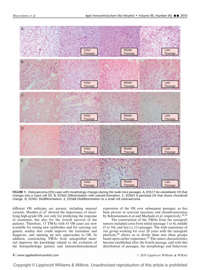
In the majority of the xenografted cases, 21 of 31from group B, the histopathology pattern was similar tothe primary or the metastatic tumor, also with some fea-tures of a higher aggressivity, such as increased cellularity,mitotic and apoptotic figures, and necrotic areas. How-ever, in 10 xenografted cases, the morphology changedover the passages and was quite di"erent from theoriginal tumor implanted into nude mice. Phenomenasuch as dedi"erentiation (EOS 61, 67, 71, and 72), di"er-entiation (with osteoid formation) (EOS 65), and acquisi-
tion of new patterns were observed. These new patternsconsist of giant cell (EOS 17 and 64), microcellular (EOS68 and 69), and chondroid (EOS 63) (Fig. 1).
IHC results were heterogenous (Tables 1, 2), althoughsome associations were found in the statistical analysis(Tables 3, 4). Osteonectin and osteocalcin were the most-expressed markers, being retained, despite the changein the morphology during the subsequent passages andeven when an undi"erentiated morphology was present(Fig. 2). S100 and Sox-9 were expressed mostly in chon-drogenic cases; however, no statistical di"erences relatedto Sox-9 were found in nonchondrogenic OS. Six of the 8chondrogenic cases showed nuclear, moderate, or highSox-9 positivity, whereas 10 osteogenic, 1 telangiectatic, 1dedi"erentiated, and 1 pleomorphic OS also showed posi-tivity. However, this was mainly low, and in 1 case, mode-rate with abundant associated cytoplasmic expression.CK was positive in only 4 original osteoblastic OS, inwhich its positivity increased over the passages, especiallyin the later ones. Bcl-2 was positive only in 2 originalosteoblastic OS, and revealed generally low and hetero-geneous expression throughout the mice passages witha statistically significant higher expression in the latepassages. CD99 was positive in 1 of the 2 microcellularcases with a marked cytoplasmic expression. Never-theless, this expression was not exclusive to this OSvariant; other cases also showed positivity. Caveolin andsurvivin showed immunoreactivity in the majority of thetumors, with no significant variation among the subtypes,except with expression of the former in 12 out of the 14metastatic cases. Survivin was significantly higher overthe subsequent passages. p16 displayed heterogenic expre-ssion with no significant di"erences among the OS sub-types or the subsequent passages. However, 19% of allthe OS lost their expression. p53 expression increasedover the passages, although the statistical analysis wasnot significant. The proliferation index (Ki-67) was notassociated with a more undi"erentiated histopathologypattern of the tumor, but increased significantly over thepassages.
DISCUSSIONNew ancillary technologies such as TMAs are very
helpful for the diagnosis and systematic study of a largecohort of tumors. Packeisen et al23 reviewed in detail theuse of this high-throughput source in daily diagnosisand research, drawing attention to the central position ofpathology in the evolution of cancer investigation. Nilbertet al28 applied this technology to sarcomas and comparedits use with the conventional para!n blocks, findingthe most limiting feature of TMAs to be the lack ofrepresentation of the whole tumor. In our opinion, anaccurate study of the original case to choose a represen-tative area, together with the inclusion of more than 1core and care in assembling the TMA, is a requirementfor the reliability of the TMA.
Evaluating a large cohort of OS in 1 slide is of greatvalue, especially in series such as ours, in which many
Appl Immunohistochem Mol Morphol ! Volume 00, Number 00, ’’ 2010 A Tissue Microarray Study of Osteosarcoma
r 2010 Lippincott Williams & Wilkins www.appliedimmunohist.com | 3
Copyright © Lippincott Williams & Wilkins. Unauthorized reproduction of this article is prohibited.
di"erent OS subtypes are present, including unusualvariants. Hauben et al5 showed the importance of classi-fying high-grade OS, not only for predicting the responseto treatment, but also for the overall survival of thepatients. Therefore, 13 TMAs with 61 OS cases are nowavailable for testing new antibodies and for carrying outgenetic studies that could improve the treatment anddiagnosis, and opening up new approaches to OS. Inaddition, constructing TMAs from xenografted mate-rial improves the knowledge related to the evolution ofthe histopathologic pattern and immunohistochemical
expression of the OS over subsequent passages; as hasbeen proven in synovial sarcomas and chondrosarcomasby Subramaniam et al and Machado et al, respectively.24,26
The construction of the TMAs from the xenografttumors included cores from initial (passage 1 to 4), middle(5 to 10), and late (Z11) passages. The wide experience ofour group working for over 20 years with the xenograftplatform,20 allows us to divide them into these groupsbased upon earlier experience.26 The tumor characteristicsbecome established after the fourth passage, and with thisdistribution of passages, the morphology and behaviour
Initial
A
B
PassageMiddlePassage
LatePassage
InitialPassage
MiddlePassage
LatePassage
InitialPassage
MiddlePassage
LatePassage
InitialPassage
MiddlePassage
LatePassage
InitialPassage
MiddlePassage
LatePassage
C
D
E
FIGURE 1. Osteosarcoma (OS) cases with morphology changes during the nude mice passages. A, EOS17 An osteoblastic OS thatchanges into a Giant cell OS. B, EOS65 Differentiation with osteoid formation. C, EOS63 A parosteal OS that shows chondroidchange. D, EOS61 Dedifferentiation. E, EOS68 Dedifferentiation to a small cell osteosarcoma.
Mayordomo et al Appl Immunohistochem Mol Morphol ! Volume 00, Number 00, ’’ 2010
4 | www.appliedimmunohist.com r 2010 Lippincott Williams & Wilkins
Copyright © Lippincott Williams & Wilkins. Unauthorized reproduction of this article is prohibited.

TABLE 1. Immunoprofile in Each Original Osteosarcoma Subtype
Positive cases in green, negative cases in red, and noninformative cases in white.*Metastatic cases.
Appl Immunohistochem Mol Morphol ! Volume 00, Number 00, ’’ 2010 A Tissue Microarray Study of Osteosarcoma
r 2010 Lippincott Williams & Wilkins www.appliedimmunohist.com | 5
Copyright © Lippincott Williams & Wilkins. Unauthorized reproduction of this article is prohibited.

of the tumors are comparable; not only among passagesof the same group, but also with other xenograftedtumors.
In our series, conventional OS were the mostfrequent, with the osteogenic as the predominant variant
followed by the chondrogenic, as described in the litera-ture.1 No fibroblastic OS (the third in frequency) wasincluded in this study; however 1 of the cases (EOS 68)showed an admixed pattern with osteoid and fibroblasticareas. A di"erent type of matrix was also observed afterthe nude mice passages in a parosteal OS (EOS 63), whichchanged into a chondrogenic tumor, and a pleomorphicMFH–like OS (EOS 65), which changed into an osteo-genic. Such findings have not earlier been reported andneed to be contrasted with new cases to provide a better
TABLE 2. OS Cases in Which the Morphology Changes Over the Nude Mice Passages
Marker expression is indicated with di"erent green tonalities for positive cases from light (low expression) to dark (high expression) and red for negative cases. In whitethe noninformative cases.
TABLE 3. Comparison of the High Expression of the MarkersBetween Initial and Middle-late Passages in the Nude Mice
Immunomarker Initial Passages (%) Late Passages (%) P
Ki-67 n=3 (9.4) n=8 (32) P<0.05p53 n=3 (9.4) n=6 (24) P>0.05p16 n=5 (16.7) n=3 (13) P>0.05Survivin n=16 (51.6) n=21(87.5) P<0.05Caveolin n=25 (80.6) n=19 (79.2) P>0.05Bcl-2 n=3 (9.4) n=5 (20) P>0.05
p16 loss of expression (negativity) is compared between initial and middle-latepassages.
TABLE 4. Comparison of Marker Expression BetweenChondrogenic and Nonchondrogenic OS in Original Tumors
ImmunomarkerChondrogenic
OS (%)Nonchondrogenic
OS (%) P
Sox-9 n=6 (75) n=13 (24.5) P>0.05S100 n=8 (100) n=28 (52.8) P<0.05
Mayordomo et al Appl Immunohistochem Mol Morphol ! Volume 00, Number 00, ’’ 2010
6 | www.appliedimmunohist.com r 2010 Lippincott Williams & Wilkins
Copyright © Lippincott Williams & Wilkins. Unauthorized reproduction of this article is prohibited.

understanding. However, the changes in the matrix afterxenografting, especially regarding bone formation, needto be confirmed as changes in the tumor cells and not beingowing to the mouse stroma. Tokunaga et al29 and Haraet al30 reported that the mouse cells were induced to change
into osteocytes by the inoculation of human OS cells, andwere consequently responsible for bone formation. Goodcharacterization of the origin of the cells, both morphologicand cytogenetically, using for instance cell cultures andFISH, will give clues to excluding this possibility.
ddl
SOX9
Original Tumor Initial Passage
Middle Passage Late Passage
S100
Original Tumor Initial Passage Middle Passage Late Passage
Osteonectina0
Original Tumor Initial Passage Middle Passage Late Passage
Ki67
Original Tumor Initial Passage Middle Passage Late Passage
CD99
Original Tumor Initial Passage Middle Passage Late Passage
Survivin
Original Tumor Initial Passage
Late PassageMiddle Passage
FIGURE 2. Immunohistochemistry in Osteosarcomas (OS) tumors and during the subsequent passages. SOX 9 and S100expression in a Parosteal OS (EOS 63) in which the morphology changed into a chondrogenic tumor. Maintenance of theOsteonectin expression in an Osteoblastic OS (EOS 61), despite the change into an undifferentiated tumor. Ki 67 in a pleomorphicOS (EOS 65) which changed into an osteogenic tumor. CD99 and survivin expression in an Osteoblastic OS (EOS 68) in which themorphology changed into a microcellular tumor.
Appl Immunohistochem Mol Morphol ! Volume 00, Number 00, ’’ 2010 A Tissue Microarray Study of Osteosarcoma
r 2010 Lippincott Williams & Wilkins www.appliedimmunohist.com | 7
Copyright © Lippincott Williams & Wilkins. Unauthorized reproduction of this article is prohibited.

Our study includes rare OS variants, 2 telangiectaticand 2 small cell OS, which account for 3% of all the OS,whereas the estimated incidence is 4% for telangiectaticOS and 1.5% for small cell OS.1 We lack immunopheno-typic markers for these 2 OS variants, although beingaware that this type of OS exists and is especially impor-tant to exclude other bone tumors. Our series also inclu-ded 2 extra cases (EOS 19 and 69) that showed large, butsporadic, blood lacunae surrounded by giant multi-nucleated cells; however, the complete case could unfortu-nately not be evaluated. Hence, these secondary patternsneed to be better evaluated and understood to determinewhether they are new potential subtypes or simplymorphologic changes with no clinical meaning.
The 2 microcellular OS were compounds of a small,admixed cell population with scant and immature osteoid,as described by Nakajima et al.31 According to the litera-ture,7,32 their immunohistochemical profile alone can be apitfall when excluding other small cell tumors, especiallywhen osteonectin or osteocalcin negative. In our cases, 1was positive for osteonectin (EOS 7) and 1 for osteocalcin(EOS 24).
Regarding the IHC analysis, osteonectin and osteo-calcin were generally expressed in all cases and subtypes,even in undi"erentiated OS; these markers, especiallyosteonectin, are useful not only for the diagnosis of OS,but also for its di"erential diagnosis from other malignantbone tumors and to asses the origin of any metastasis.Positivity for these markers is not necessary to diagnose atumor as OS, but if an undi"erentiated tumor or a meta-stasis is positive for any of these 2 markers, it is highlysuspicious of an OS.8,10 However, Chano et al9 observed adecrease in the osteocalcin expression in the metastasiswhen looking for the OS histogenesis, whereas in thisstudy all the metastatic cases were positive.
OS are usually negative for Sox-9, except for thechondrogenic OS, in which some expression in chondro-genic areas is observed. However, in our series, althoughother OS variants showed positivity, the apparent expres-sion was both nuclear and cytoplasmic, which leads us todoubt the reliability of the expression.
OS may express S100, being observed by Devaneyet al in small cell OS7 and Hasegawa et al33 in chondro-blastic, osteoblastic, low-grade central, giant cell-rich,and epithelioid OS subtypes. S100 expression in our studywas significantly higher in the chondrogenic OS than innonchondrogenic; although some expression was found in50% of the osteoblastic, 80% of the parosteal, and in allthe dedi"erentiated and poorly di"erentiated OS.
OS may show positivity for CK,34 although in ourcases only 4 original osteoblastic OS expressed this marker.However, its positivity increased over the mice passages,especially in the later ones. This finding needs to be betterunderstood, investigating for example other types of cyto-keratins alone or in combination with the epithelial mem-brane antigen; as in already published studies.35 Bcl-2revealed generally low and heterogeneous expression with astatistically significant higher expression in the late passagesin nude mice. CD99 was positive in every OS variant,
including 50% of the microcellular cases. Despite being un-specific, this result must be taken into consideration, espe-cially in the di"erential diagnosis of this entity. However,the expression was more cytoplasmic than membranous, astypically occurs in the Ewing family tumors.
Caveolin showed immunoreactivity in the majorityof the tumors with no significant variation among thesubtypes or subsequent passages; even in the majority ofthe metastatic cases. Nevertheless, with only 14 metastaticcases and without clinical data we cannot conclude thatcaveolin is a marker either for good or for bad prognosis,as already published by Cantiani et al.19
Survivin showed immunoreactivity in the majorityof the tumors with no significant variation among thesubtypes. However, surprisingly few of the tumors givingrise to xenografts were positive (20%), whereas the xeno-grafts all became positive, which highlights the impor-tance of this antiapoptotic protein in OS progression.Trieb et al36 also studied survivin expression in OS andrelated its nuclear expression to a prolonged survival;however Osaka et al37 detected survivin mRNA expres-sion by RT-PCR in 22 OS with a significantly higherexpression in metastatic cases and related to a poorprognosis, more in line with these observations. Despitebeing contradictory findings, both focus on the impor-tance of this antiapoptotic pathway of progression andopen up the possibility of its use as a pretherapy responsepredictor.
p16 and p53 displayed heterogenic expression withno significant di"erences between passages or histologicsubtypes. However, 20% of our OS lost their p16 expres-sion, similar to 17% of the cases of Maitra et al14;whereas for Benassi et al,15 loss of p16 expression wasfound in 38% of the cases. Considering that 16% of ourcases were inconclusive, with nuclear and extensive cyto-plasmic overexpression, the percentage could probably behigher. p53 expression was generally low, except in themore undi"erentiated cases and the late passages; a resultthat needs to be clarified because p53 mutation may playa role in neoplastic transformation33 and probably in theevolution of OS.17
The proliferation index (Ki-67) was not necessarilyassociated with a more undi"erentiated histology of thetumor, but increased significantly over the passages.These results agree with the results published by Jonget al,38 in which the proliferative index did not seem topredict either disease-free or overall survival.
Extensive tissue sampling and an accurate morpho-logic evaluation of the tumors are essential for OS diag-nosis; hence there is no single immunohistochemicalmarker that distinguishes the subtypes. However, osteo-calcin and osteonectin are reliable markers for the diag-nosis of OS, and the chondrogenic variant shows asignificantly higher S100 expression than the others. Sur-vivin and p53 expression increases with tumor evolutionover the passages, and Ki-67 expression is related totumor progression over passages.
On the basis of the detailed immunohistochemicalanalysis of the di"erent xenograft passages, we must
Mayordomo et al Appl Immunohistochem Mol Morphol ! Volume 00, Number 00, ’’ 2010
8 | www.appliedimmunohist.com r 2010 Lippincott Williams & Wilkins
Copyright © Lippincott Williams & Wilkins. Unauthorized reproduction of this article is prohibited.

conclude that, although not perfect, the xenograft modelspretty well represent their tumor of origin. Obviously,phenotypes relying on stroma markers, or markers indu-ced in cancer cells as a response to stroma signals, will notbe represented well in such models. However, our parallelstudy of genomic characteristics of such xenografts(Kresse et al, unpublished) supports the overall goodrepresentation of human tumors by OS xenografts.
ACKNOWLEDGMENTThe authors thank Elisa Alonso and Laura Martinez
from the University of Valencia for their collaboration andtechnical assistance, and Drs Jahn M. Nesland and BodilBjerkehagen for providing the blocks from Oslo.
REFERENCES1. Fletcher CDM, Krishnan UK, Mertens F. Pathology and Genetics of
Tumours of Soft Tissue and Bones. World Health OrganizationClassification of Tumours. 2002.
2. Picci P. Osteosarcoma (osteogenic sarcoma). Orphanet J Rare Dis.2007;2:6.
3. Wittig JC, Bickels J, Priebat D, et al. Osteosarcoma: a multi-disciplinary approach to diagnosis and treatment. Am Fam Physi-cian. 2002;65:1123–1132.
4. Sailor J, McCarthy EF, Weber KL, et al. Low-grade intramedullaryosteosarcoma of the tibia presenting as an intracortical lesion.Orthopedics. 2008;31:714.
5. Hauben EI, Weeden S, Pringle J, et al. Does the histological subtypeof high-grade central osteosarcoma influence the response totreatment with chemotherapy and does it a"ect overall survival? Astudy on 570 patients of two consecutive trials of the EuropeanOsteosarcoma Intergroup. Eur J Cancer. 2002;38:1218–1225.
6. Shehadeh AM, Haiba MA, Henshaw RM, et al. Telangiectaticosteosarcoma of the patella. Orthopedics. 2008;31:808.
7. Devaney K, Vinh TN, Sweet DE. Small cell osteosarcoma of bone:an immunohistochemical study with di"erential diagnostic consi-derations. Hum Pathol. 1993;24:1211–1225.
8. Fanburg JC, Rosenberg AE, Weaver DL, et al. Osteocalcin andosteonectin immunoreactivity in the diagnosis of osteosarcoma. AmJ Clin Pathol. 1997;108:464–473.
9. Chano T, Matsumoto K, Ishizawa M, et al. Analysis of the presenceof osteocalcin, S-100 protein, and proliferating cell nuclear antigenin cells of various types of osteosarcomas. Eur J Histochem. 1996;40:189–198.
10. Fanburg-Smith JC, Bratthauer GL, Miettinen M. Osteocalcin andosteonectin immunoreactivity in extraskeletal osteosarcoma: a studyof 28 cases. Hum Pathol. 1999;30:32–38.
11. Osuna D, de Alava E. Molecular pathology of sarcomas. Rev RecentClin Trials. 2009;4:12–26.
12. Wang LL. Biology of osteogenic sarcoma. Cancer J. 2005;11:294–305.13. Tang N, Song WX, Luo J, et al. Osteosarcoma development and
stem cell di"erentiation. Clin Orthop Relat Res. 2008;466:2114–2130.14. Maitra A, Roberts H, Weinberg AG, et al. Loss of p16(INK4a)
expression correlates with decreased survival in pediatric osteosar-comas. Int J Cancer. 2001;95:34–38.
15. Benassi MS, Molendini L, Gamberi G, et al. Alteration of pRb/p16/cdk4 regulation in human osteosarcoma. Int J Cancer. 1999;84:489–493.
16. Benassi MS, Molendini L, Gamberi G, et al. Altered G1 phaseregulation in osteosarcoma. Int J Cancer. 1997;74:518–522.
17. Pellin A, Boix-Ferrero J, Carpio D, et al. Molecular alterations ofthe RB1, TP53, and MDM2 genes in primary and xenograftedhuman osteosarcomas. Diagn Mol Pathol. 1997;6:333–341.
18. Walkley CR, Qudsi R, Sankaran VG, et al. Conditional mouseosteosarcoma, dependent on p53 loss and potentiated by loss of Rb,mimics the human disease. Genes Dev. 2008;22:1662–1676.
19. Cantiani L, Manara MC, Zucchini C, et al. Caveolin-1 reducesosteosarcoma metastases by inhibiting c-Src activity and met signal-ing. Cancer Res. 2007;67:7675–7685.
20. Llombart-Bosch A, Carda C, Boix J, et al. Value of nude micexenografts in the expression of cell heterogeneity of human sarcomasof bone and soft tissue. Pathol Res Pract. 1988;183:683–692.
21. Bruheim S, Bruland OS, Breistol K, et al. Human osteosarcomaxenografts and their sensitivity to chemotherapy. Pathol Oncol Res.2004;10:133–141.
22. Dass CR, Ek ET, Choong PF. Human xenograft osteosarcomamodels with spontaneous metastasis in mice: clinical relevance andapplicability for drug testing. J Cancer Res Clin Oncol. 2007;133:193–198.
23. Packeisen J, Korsching E, Herbst H, et al. Demystifiedytissuemicroarray technology. Mol Pathol. 2003;56:198–204.
24. Machado I, Giner F, Mayordomo E, et al. Tissue microarraysanalysis in chondrosarcomas: light microscopy, immunohistochem-istry and xenograft study. Diagn Pathol. 2008;3(suppl 1):S25.
25. Petrov SV, Machado I, Boulytcheva IV, et al. New approaches inthe diagnosis of small-cell round tumors of bone and soft tissue.Arkh Patol. 2009;71:34–40.
26. Subramaniam MM, Navarro S, Pellin A, et al. Tissue microarrayprofiling of primary and xenotransplanted synovial sarcomasdemonstrates the immunophenotypic similarities existing betweenSYT-SSX fusion gene confirmed, biphasic, and monophasic fibrousvariants. Virchows Arch. 2006;449:435–447.
27. Somers GR, Ho M, Zielenska M, et al. HER2 amplification andoverexpression is not present in pediatric osteosarcoma: a tissuemicroarray study. Pediatr Dev Pathol. 2005;8:525–532.
28. Nilbert M, Engellau J. Experiences from tissue microarray in softtissue sarcomas. Acta Orthop Scand Suppl. 2004;75:29–34.
29. Tokunaga K, Ogose A, Endo N, et al. Human osteosarcoma (OST)induces mouse reactive bone formation in xenograft system. Bone.1996;19:447–454.
30. Hara A, Ikeda T, Nomura S, et al. In vivo implantation of humanosteosarcoma cells in nude mice induces bones with human-derivedosteoblasts and mouse-derived osteocytes. Lab Invest. 1996;75:707–717.
31. Nakajima H, Sim FH, Bond JR, et al. Small cell osteosarcoma ofbone. Review of 72 cases. Cancer. 1997;79:2095–2106.
32. Devoe K, Weidner N. Immunohistochemistry of small round-celltumors. Semin Diagn Pathol. 2000;17:216–224.
33. Hasegawa T, Hirose T, Seki K, et al. Histological and immuno-histochemical diversities, and proliferative activity and grading inosteosarcomas. Cancer Detect Prev. 1997;21:280–287.
34. Okada K, Hasegawa T, Yokoyama R, et al. Osteosarcoma withcytokeratin expression: a clinicopathological study of six cases withan emphasis on di"erential diagnosis from metastatic cancer. J ClinPathol. 2003;56:742–746.
35. Hasegawa T, Hirose T, Kudo E, et al. Immunophenotypicheterogeneity in osteosarcomas. Hum Pathol. 1991;22:583–590.
36. Trieb K, Lehner R, Stulnig T, et al. Survivin expression in humanosteosarcoma is a marker for survival. Eur J Surg Oncol. 2003;29:379–382.
37. Osaka E, Suzuki T, Osaka S, et al. Survivin as a prognostic factorfor osteosarcoma patients. Acta Histochem Cytochem. 2006;39:95–100.
38. Jong R, Davis AM, Mendes MG, et al. Proliferative activity (ki-67expression) and outcome in high grade osteosarcoma: a study of 27cases. Sarcoma. 2000;4:47–55.
Appl Immunohistochem Mol Morphol ! Volume 00, Number 00, ’’ 2010 A Tissue Microarray Study of Osteosarcoma
r 2010 Lippincott Williams & Wilkins www.appliedimmunohist.com | 9
Copyright © Lippincott Williams & Wilkins. Unauthorized reproduction of this article is prohibited.
Copyright © 2022 FDOKUMEN