A STUDY ON VALIAZHAL KEELVAYU - Electronic Theses ...
174
A STUDY ON VALIAZHAL KEELVAYU Dissertation Submitted to THE TAMIL NADU DR.M.G.R MEDICAL UNIVERSITY CHENNAI – 32 For the Partial fulfillment for the requirements to the degree of DOCTOR OF MEDICINE (SIDDHA) (Branch – III, SIRAPPU MARUTHUVAM) DEPARTMENT OF SIRAPPU MARUTHUVAM GOVERNMENT SIDDHA MEDICAL COLLEGE PALAYAMKOTTAI – 627 002 MARCH - 2009
-
Upload
khangminh22 -
Category
Documents
-
view
0 -
download
0
Transcript of A STUDY ON VALIAZHAL KEELVAYU - Electronic Theses ...
A STUDY ON
VALIAZHAL KEELVAYU
Dissertation Submitted to
THE TAMIL NADU
DR.M.G.R MEDICAL UNIVERSITY CHENNAI – 32
For the Partial fulfillment for the requirements to the degree of
DOCTOR OF MEDICINE (SIDDHA) (Branch – III, SIRAPPU MARUTHUVAM)
DEPARTMENT OF SIRAPPU MARUTHUVAM GOVERNMENT SIDDHA MEDICAL COLLEGE
PALAYAMKOTTAI – 627 002
MARCH - 2009
1
INTRODUCTION
Medicine, as everyone knows, it is not merely, a science, but an art
as well. There are different systems of medicine in the world, according
to the way of life of the people and their geographical conditions.
The Siddha System of Medicine is an ancient and traditional one
with prestigious background of Tamil culture. It is one of the pillars of
the Indian System of Medicine. Siddha System of Medicine is dedicated
bequest of Siddhars, cheerfully given to the human society to live long
and free from diseases.
Siddhars were men of highly cultured, intellectual and spiritual
faculties, combined with super natural powers. Siddha medicine is the
one, which prevents the body and mind from a diseased condition by
curing the disease and by working also as a rejuvenating process for
further prevention of the same by treating the whole body as whole.
Nature’s wealth is being destroyed by the change in the life style
with advanced technologies. This has an impact over the physical, social,
cultural, moral, mental and spiritual values, which in turn affect the
normal health. This health deterioration is meant as disease. According to
the manuscripts as well as the evidence found out, disease may occur due
to the derangement of either the external or internal factors.
The internal factors include illness occurring due to the disturbance
of three Humours, the seven Thathus, the three Gunas and the Malas. The
external factors include seasonal changes or climatic variations or
unlikable things like drug abusing or external stress ending in

2
psychosomatic disorders. Siddha medicine has basic principles to remove
the distress and diseases.
" ÁÈôÀмø §¿¡ö ÁÚó¦¾É Ä¡Ìõ ÁÚôÀÐÇ §¿¡ö ÁÚó¦¾Éî º¡Öõ ÁÚôÀ¾¢É¢ §¿¡ö Åá ¾¢Õì¸ ÁÚôÀÐ º¡¨ÅÔ ÁÕó¦¾É Ä¡§Á "
-¾¢ÕãÄ÷. [ §¿¡ö ¿¡¼ø §¿¡ö Ó¾ø ¿¡¼ø ¾¢ÃðÎ _ Àì¸õ 113 ]
According to Siddha aspect, diseases are classified in to Vadha
diseases, Pitha Diseases and Kapha diseases. Valiazhal Keelvayu is one
among the vadha diseases mentioned in the Siddha literature. The clinical
features of Valiazhal Keelvayu are similar to that of Rheumatoid
Arthritis, mentioned in Modern Literatures. According to the Modern
Medical Science, it is an autoimmune disease. Likewise, in Siddha
system the Valiazhal Keelvayu is caused by deranged natural stamina
(Eyarkai Vanmai). This deranged stamina reflects in the disciplines of
three Thosas and seven Thathus.
This disease is a challenge to the world of Medicine, because it
affects the patients mentally, physically, economically and also his
domestic life. The number of affected patients is also getting increased.
This leads to many patients in despair. Till now there is no definite cure
for this disease in other system of medicine.
In order to fortify the hopes of Valiazhal Keelvayu patients, the
author has selected this disease for the dissertation work on the basis of
the Siddha concept. And also due to the unshakable belief among the
people that the Vadha diseases can effectively managed by Siddha
Medicine, the author choose it for dissertation. This study is an initial step
and only a preliminary attempt for further research.

3
AIM AND OBJECTIVES
The disease, Valiazhal Keelvayu produces tremendous pain and
discomfort to the patients. So far a perfect and complete remedy for this
disease has not yet been arrived at all. The purpose of the author’s work
is to elucidate a good medicine from the ancient Siddha Literature and to
create hope and faith in the treatment. This being a preliminary endeavour
by the author, as it would be a helping hand to the sufferers. With this
view this dissertation subject was undertaken.
The other aims of this study are
To collect authentic measures and review the ideas mentioned in
ancient Siddha literature about the disease.
To study the clinical features of the disease Valiazhal Keelvayu.
To review the altered Tridosha or Mukkutram and changes in the
physiology as per Siddha aspect.
To study the disease Valiazhal keelvayu on the basis of Udal
thathu, Paruva kaalam, food, taste, age, sex, socio-economic status,
Ennvagai thervu, Neerkuri and Neikuri.
To expose the unique diagnostic procedure mentioned in Siddha
literature for the disease Valiazhal keelvayu.
To know the extent of correlation of aetiology, signs and symptoms
of Valiazhal keelvayu, in Siddha aspect with Rheumatoid
Arthritis in Modern Medicine.
4
To diagnose the disease on the basis of modern parameters
To have a clinical trial on Valiazhal Keelvayu with Kumari
Mathirai as internal Medicine and Nochi Thylam as external
medicine on 28 inpatients and 30 outpatients.
To have a detailed analysis to prove the clinical efficacy of the
drugs through the pharmacological and biochemical analysis.
To insist Thokanam, Asanaas, exercise, diet and life style
modification along with medicine to achieve good results.
5
REVIEW OF LITERATURE
SIDDHA ASPECTS
Valiazhal Keelvayu is one of the Vatha diseases and Vatham is
main deranged factor. It is then accompanied by Azhal (Pitham).
According to Siddha medicine, diseases are due to derangement of
Uyirthathukkal (Vatham, Pitham, and Kapham). To better understand
about derange factors in Valiazhal keelvayu and their effects in body, a
brief view of “Thiridosha Theory” is essential. Thiruvalluvar has quoted
this as
" Á¢¸¢Ûõ ̨È¢Ûõ §¿¡ö¦ºöÔõ á§Ä¡÷
ÅǢӾġ ±ñ½¢Â ãýÚ" - ¾¢ÕìÌÈû
The Siddha system of medicine is based on the Thirdoshic theory.
The three Cordinal Humours are Vatham, Pitham, and Kapham. They
made up of Pancha Boothams.
The Siddha System considers the body as a whole make up of five
basic elements namely,
1. Prithivi
2. Appu
3. Theyu
4. Vayu
5. Aagayam
The five elements are considered as the fundamental principle of
all creations of God. This can be stated in the Sadhaga Naadi as
6
" À¡ÃôÀ¡ ⾨ÁóÐ Áñ¿£÷ §¾Ô
ÀÃ¢Å¡Ô Å¡¸¡Â ¨Áó¾¢É¡§Ä
§ºÃôÀ¡ º¼Á¡îÍ "
- º¾¸ ¿¡Ê Example:
Prithivi + Prithivi - Bones Prithivi + Vayu - Nerves
Prithivi + Appu - Muscles Prithivi + Agayam - Hairs
Prithivi + Theyu - Skin
Vatham is formed by air and space.
Pitham is formed by fire.
Kabam is formed by earth and water.
Å¡¾õ = þ¼¸¨Ä + «À¡Éý À¢ò¾õ = À¢í¸¨Ä + À¢Ã¡½ý
¸Àõ = ÍØ + ºÁ¡Éý
They are called, as Uyir Thathus or Thiri Thathus in their normal
proportion. These three humours are held in the ratio of 1: ½: 1/4
normally. This is stated in Kannusamiyam as
" ¦ÁöÂÇ× Å¡¾¦ÁýÚ
§ÁøÀ¢ò¾ §Á¡Ã¨Ã¡õ
³Âí¸¡ ¦Äý§È «È¢ " - ¸ñϺ¡Á¢Âõ
" ÅÆí¸¢Â Å¡¾õ Á¡ò¾¢¨Ã ¦Â¡ýÈ¡¸¢ø
¾Æí¸¢Â À¢ò¾ó ¾ýɢĨà šº¢
«ÆíÌí ¸Àó¾¡É¼í¸¢§Â ¸¡§Ä¡Êø
À¢Èí¸¢Â º£Å÷ìÌ À¢º ¦¸¡ýÚÁ¢ø¨Ä§Â. " - ̽š¸¼õ.
7
Three Thathus are accompanied by the Aru Suvaigal and this also
made up of Pancha Boothams.
þÉ¢ôÒ - Áñ + ¿£÷
ÒÇ¢ôÒ - Áñ + ¾£
¯ôÒ - ¿£÷ + ¾£
¨¸ôÒ - Å¢ñ + ÅÇ¢ Vadham - Å¢ñ + ÅÇ¢
¸¡÷ôÒ - ÅÇ¢ + ¾£ Pitham - ¾£
ÐÅ÷ôÒ - Áñ + ÅÇ¢ Kabam - Áñ + ¿£÷
" Áñϼ§É ÒÉø¾£ì¸¡ø Өȡ¸î §º÷ó¾¢ð¼¡ø ÅÕ§ÁþÉ¢ôÒ ¾¢ñ½Á¢Äõ ÐÅ÷ôÀ¢Ãºõ º¾¡¸¾¢§Â¡ ¼¡÷ò¾£ö¢ý ¾¢¼Á¡
Ó¨ÈôÒõ ±ñ½È¢Â ¸ºôÒÓñ¼¡ó ¾ñ½£Ã¢ø ¸½Ä¢¨ÉôÀ¡ ¦ÄØÁ¡ÓÅ÷ôÒ ¯ñ½È¢Â «ÚͨÅ¢ý À¢ÈôÀ¢¦¾Ûõ ÌÕº¢ò¾Õ¨Èò¾ Á¨Ã§Â"
- ÁÕòÐÅ ¾É¢ô À¡¼ø¸û
ͨÅ-Å¡¾õ ¦¾¡¼÷Ò:
" ÒÇ¢ÐÅ÷ Å¢ïÍí¸È¢Â¡ø ââìÌõ Å¡¾õ "
Å¡¾ò¨¾ —¾¢¸ôÀÎòÐõ ͨŸû : ÒÇ¢ôÒ, ÐÅ÷ôÒ
" Å¡¾§ÁÄ£ð¼¡ø ÁÐÃõ ÒÇ¢ ¯ôÒ
§º¾ÓÈî ¦ºöÔï º¢¨ÈÂõ_μ¾ì§¸û
¸¡Ãó ÐÅ÷ ¸ºôÒì ¸¡ðÎïͨŠ¦ÂøÄ¡õ
º¡ÃôÀ⸡Ãï º¡üÚ "
Å¡¾ò¨¾ ºÁÉõ ¦ºöÔõ ͨŸû : þÉ¢ôÒ, ÒÇ¢ôÒ, ¯ôÒ
So any change in the Arusuvaigal and Panchaboothams lead to
disharmony of the three Thathus and produce illness.
The Uyir Thathus in their normal condition regulate all
physiological activities of the human beings and keep the body healthy.
8
When the mutual harmony of these Uyir Thathus is disturbed they are
called three Thosas and they bring diseases. In Siddha system, based on
the Thirdoshic theory, the diseases are classified into
Vatha diseases
Pithaa Diseases
Kapha diseases
VATHA DISEASE
Vadham is a clinical condition characterized by pain swelling, pricking and loss of function due to vitiated Vadha which is the principal humour of the body.
Aetiology And The Classification Of Vadha Diseases
" Å¡¾§Á À¢ÈôÀ¾üÌ ÅÃðº¢Â¡ÕÄÁ¡Ì Á¡¾Ä¡ø ÌÇ¢÷îº¢Â¡Ö Á¾¢ÖÚõ Å¡¾§Ã¡¸õ ¿£¾¢ §ºÃ¾É¢ÖûÇ ¿¢Èí̽¦Á¾üÌ ÓñÎ §Å¾¢ÂÃÈ¢ Å¡Å¡¾ô À¢ÈŢ£ ¦¾ýÉÄ¡§Á "
Yugi Munivar Vaithia Cinthamani Perunool 800,
Jeevaratchavmirtham, Dhanvanthiri Vaithyam -80 types, Anuboga Vathia
Deva Ragasiyam – 84 types, Theriaiyar Vagadam – 81 types
" ±ýɧŠš¾ó ¾¡¦ÉñÀ¾¡Ìõ þ¸ò¾¢§Ä ÁÉ¢¾÷¸Ùì ¦¸öÐÁ¡Ú À¢ýɦŠ¦À¡ý ¾¨É§Â §º¡Ã了öÐ
¦Àâ§Â¡÷¸û À¢Ã¡Á½¨Ãò à„½¢òÐõ ÅýÉ §ÅÅî ¦º¡ò¾¢ü §º¡Ã了öÐ Á¡¾¡ À¢¾¡ ÌըŠÁÈó¾ §À¡ìÌõ
¸ýɧŠ§Å¾ò¨¾ ¿¢ó¨¾ ¦ºö¾ §À÷ìÌõ ¸¡Âò¾¢ü ¸Äó¾¢Î§Á Å¡¾ó¾¡§É "
- (243)
9
“ ¾¡¦ÉýÈ ¸ºô§À¡Î ÐÅ÷ôÒ¨ÃôÒ
º¡¾¸Á¡ö Á¢ï͸ÆÛï º¨Áò¾ ÅýÉõ
¬¦ÉýÈ Å¡È¢ÉÐ ¦À¡º¢ò¾¡Öõ
¬¸¡Âò §¾ÈÄÐ ÌÊò¾¡Öõ
Á¡¦ÉýÈ À¸ÖÈì¸ Á¢Ã¡Å¢Æ¢ôÒ
ÀðÊÉ¢§ÂÁ¢¸ ×È¡¾ø À¡Ã¦Áö¾ø
§¾¦ÉýÈ ¦Á¡Æ¢Â¡÷ §Áüº¢ó¨¾Â¡¸¢ø
º£ì¸¢ÃÁ¡ö Å¡¾ÁÐ ¦ºÉ¢ìÌó¾¡§É ”
- 丢 ¨Åò¾¢Â º¢ó¾¡Á½¢ ¦ÀÕáø 800 (24
4)
"¾¡É¡¸ ¸£øÅ¡¾ §Ã¡¸õ §À¨Ã Ñð¼ÓûÇ Å¡¾ §Ã¡¸ ¦ÁñÀÐó ¾¡ý
¬öó¦¾ÎòÐ −þ¾üÌû§Ç «¼ì¸õ À¡Õ "
- «¸ò¾¢Â÷ ̽š¸¼õ.
Characteristic Features of Vadham
" Å¡¾§Á ¸¾¢ò¾ §À¡Ð Å¡ÔצÁØõÒí ¸ñË÷
Å¡¾§Á ¸¾¢ò¾ §À¡Ð Å¡ÔÁó¾¢Îï ºýÉ¢ §¾¡„õ
Å¡¾§Á ¸¾¢ò¾ §À¡Ð ÅøÖ¼ý ¦ÁÄ¢óÐ ¦¸¡øÖõ "
- «¸ò¾¢Â÷ º¢¸¢îº¡ÃòÉ ¾£Àõ
" Å¡¾Å£Ú «ýÉÁ¢Èí¸¡Ð ¸ÎôÒñ¼¡õ Åñ½Óñ¼¡õ
§Á¡Ð¸ðÌ ¦Ã¡¸õ ÍÃÓñ¼¡ Á¢ÕÁÖÁ¡ ÓÈí¸¡¦¾ýÚõ
´Ð¾Ã¢Â Å¡¾ÁÉÄ¡Ì ¿Îì¸Óñ¼¡õ ¦À¡Õû¸ÇÂ÷ó¾
¾£¦¾É§Å ¿ÃõÀ¢òÐ ºóиû §¾¡Úí¸ÎìÌó ¾¢ÉÓ󾡧É"
- §¾¨ÃÂ÷ Å¡¸¼õ
Loss of appetite, pain and redness, fever and cough, insomnia,
shivering, pain in joints

10
" ¾ì¸ Å¡Ô §¸¡À¢ò¾¡ø ºóШÇóРݨħ¿¡Å¡
Á¢ì¸ ¦¸¡ð¼¡Å¢ Å¢ð¼í ¦¸Ã¢Ô ÁÄí¦¸ðÎõ
´ì¸ ¿ÃõÒ ¾¡ý Ó¼íÌÁÄ÷óÐ Å¡ö¿£ÕÈ¢ÅÕõ
Á¢ì¸ ÌÇ¢Õõ ¿Îì¸Á¡ö §ÁÉ¢ ÌÇÈ¢ ÅÕí¸¡§½ "
- §¾¨ÃÂ÷ Å¡¸¼õ; Àì¸õ - 32
Pain in joints, constipation, nervous weakness, shivering
" ºó¾¢ÃÅ¡¾ Ó¼õÒ ÌÇ¢÷ò¦Øó§¾ ¿ÎìÌí º£¾Å¡öÅ¡õ
Óó¾¢Â Ìòò¢º¢Å¡ï ºóиû§¾¡Úí ̨¼óÐ ¦Á¡Ç¢¸û Å£
íÌõ
Åó¾¢Â ¦¾¡ó¾Å¡¾õ ¿ÃõÒ¸¦ÇøÄ¡ Á¢º¢òÐ ÅÄõÅ¢¼¡Ð
—óÐ «ùÅ¡Ì Å¡¾õ Å£ì¸Óñ¼¡ Ó¼üÈ¢Á¢Õñ¼¡§Á " §¾Ãý Å¡¸¼õ; Àì¸õ - 7
2 Chillness of the body, rigor, pain and tenderness of joints, swelling
of joints
KEEL VAYU
According to Agasthiar Gunavagadam, Keelvayu comes under the
80 types of Vadha disease. In Keel Vayu the most deranged factor is
Vadham. So the Keel Vayu comes under the Vadha diseases according to
Thiridoshic theory. Keelvayu is the general terms that include all kind of
joint diseases. According to T.V.Sambasivan Pillai Dictionary, Keel
means joints and Vayu means Vadham. According to Sabapathy
Manuscript, Keel Vayu is further divided into 10 types, in the text of
Siddha Maruthuvam. The Valiazhal Keelvayu falls in this 10 sub
divisions.
11
Synonyms
According to the Siddha Maruthuvam text,
1. Sandhu Vali
2. Muttu Vali
3. Mega Soolai
4. Muddakku Vayu
5. Amavatham
According to Agasthiar Gunavagadam,
" ¾¡É¡¸ ¸£øÅ¡¾ §Ã¡¸õ §À¨Ã º¡üÚ¸¢§Èý ¿£ÂȢ ŢÅÃÁ¡¸
Á¡É¡É Å¡öקá¸õ Å¡¾§Ã¡¸õ Á¸ò¾¡É Ó¼ìÌ Å¡ö× Ó¼ìÌÅ¡¾õ
§¾É¡É ºó¾£¸ º¢§ÄðÎÁ§Ã¸õ ¦¾Ç¢Å¡É ¨¸¸¡Ä¢ø À¢ÊôÒ §Ã¡¸õ °É¡É úš¾õ ݨÄì¸ðÎ
¯ò¾Á§É ºýɢš¾õ Å¡¾Ý¨Ä¡§Á ¬Ì¦ÁýÈ −ò¾¨ÉÔõ «¾üÌô §Àáõ "
-«¸ò¾¢Â÷ ̽š¸¼õ Synonyms are
1. Vayu Rogam
2. Vatha Rogam
3. Mudakku Vayu
4. Mudakku Vatham
5. Sandeega Siletuma Rogam
6. Kaikal Pidippu Rogam
7. Rasa Vatham
8. Soolai Kattu
9. Santhi Vatham
10. Vatha Soolai

12
Iyal
" ÅÇ¢Ô ¨ÁÔõ ¾ýÉ¢¨Ä ¦¸ðÎ ÅÄ¢Ô¼ý Å£ì¸î ÍÃÓõ ¸¡öóÐ ãðθ §¼¡Úõ ÓÎ츢§Â ¦¿¡óÐ ãðθ ¼ýÉ¢ý ¿£Õõ ÍÃóÐ ¾¡í¦¸¡½¡ ÅÄ¢Ô¼ý ¦¿¡ó¾¢Î Áõ§Á "
- ºÀ¡À¾¢ ¨¸§ÂÎ
In Keel Vayu, Vadham and Kabam are deranged and produce pain
and swelling in the joints associated with fever, restriction of joint
movements, immobility of the joints, collection of fluids in the joints and
unbearable pain in the joints.
Noi Varrum Vazhi (Aetiology)
" Å¡¾ÁÄ¡Ð §ÁÉ¢ ¦¸¼¡Ð "
" ¦¿ÎÅ¡¾ º¡÷¦ÀÐ×Á¢ýÈ¢ Ý¨Ä ÅáР”
- §¿¡ö¿¡¼ø §¿¡ö Ó¾ø ¿¡¼ø ¾¢ÃðÎ
According to Siddha system, causes of disease are due to the
disturbance of Thrithathus. In Keelvayu, the chief deranged factor, among
the Thrithathu is the Vatham. The derangement of Vadham occurs under
various conditions. They are
Environmental factors
Physical factors
Factors of Kanmam
Environmental Factors " ¬Ê¡¾¢Â¡ö ³ôÀº¢ ®È¡ö
—É¢ÄÁ¾ü §¸¡Ãú¢Âø ¸¡Äõ "
" ¸¼¸Ó¾ø ÐÄ¡õ Ũâø Å¡¾Á¡Ìí ¸½ñ½¡¨Âô Àº¢ÔÁЧŠ¡Ìõ "
- º¾¸¿¡Ê
13
According to Sathaganaadi, the Vatha diseases are predominant in
months from Aadi to Iypasi.
" Å¡¾Å÷¾É ¸¡Ä§Á§¾¡ ¦ÅýÉ¢ø ÁÕ׸¢ýÈ ¬É¢ ¸ü¸¼ Á¡¾õ
¬¾¨Éô Àº¢§Â¡Î ¸¡÷ò¾¢¨¸ ¾ýÉ¢ø
«¼Õ§Á ÁüÈ Á¡¾í¸û ¾ýÉ¢ø
§À¡¸§Å ºÁ¢ì¸¢ýÈ ¸¡ÄÁ¡Ìõ "
- 丢 º¢ó¾¡Á½¢
The Vadha Dosha is provoked in its own location in the Ani and
Aadi (Thanilai Vazharchi), but it is provoked and spread beyond its
location in the month Iypasi and Karthigai. (Vetrunilai Vazharchi). But it
resumes its normalcy, in the rest of the year (Thanilai Adaithal).
" ÀÐÁò¨¾ â츨ÅìÌõ À¡ÛÁ¢¸ì ¸¡Ôõ
ÓЧÅɢĢüÒ Å¢ü¿£ü÷ ÓüÚõ - ¸Ð¦ÁÉ
ÅüÚõ ¸Àõ·Ìõ Å¡ÔÁ¢Ìõ Å¡úÁ¡óò÷ì
ÌüÈ ¿Ä¢ì§¸¾¢¦¾ý §È¡Ð. "
- º¢ò¾ ÁÕòÐÅ¡í¸î ÍÕì¸õ.
Physical Factors
According to the text Siddha Maruthuvam,
"ÅÇ¢ ¾Õ ¸¡ö¸¢ÆíÌ Å¨ÃŢġ ¾Â¢Äø §¸¡¨Æ
ÓÇ¢ ¾Â¢÷ §À¡ýÁ¢ÌìÌ Ó¨È¢ġ ×ñÊ §¸¡¼ø
ÌÇ¢÷¾ÕÅǢ¢ü §È¸í ÌÉ¢ôÒÈ ×ÄÅø ¦ÀñÊ÷
ÌÇ¢¾Õ ÓÂì¸õ ¦Àü§È¡÷ ¸Ê ¦ºÂø ¸ÕŢ¡Á¡ø"
- ºÀ¡À¾¢ ¨¸§ÂÎ

14
Excessive intake of certain fruits and vegetables produces Vadha
diseases, improper food in take, exposure to cold air, stay in hilly areas,
over indulgence in sexual activity and hereditary factors produce Keel
Vayu.
According to Agasthiar Gunavadagam,
" «ôÀ§É þЧ¿Õõ Å⨺§¸Ù
§À¡§Á ¾¡ýú à„¢Âò ¾¢É¡§Ä ¦À¡øÄ¡¾ þó¾ §¿¡ö ¸¡ÏõÀ¡Õ... "
-«¸ò¾¢Â÷ ̽š¸¼õ
KeelVayu occurs due to dietary substances, which are degrading
the chyle (Annarasam).
According to Yugi Chinthamani,
" À¸Ã§Å Å¡¾ÁÐ §¸¡À¢ò¾ô§À¡
ÀñÀ¡¸ ¦Àñ§À¡¸õ —о¡ý ¦ºö¢ø
¿¸Ã§Å ¦ÅÌàà ÅÆ¢¿¼ì¸¢ø
¿Ç¢Ã¡É ¸¡üÚ§Á ÀÉ¢§Áø Àð¼¡ø
Á¢¸Ã§Å ¸¡ö¸û ¸É¢¸¢ÆíÌ ¾ý¨É
Á¢¸ÅÕó¾¢ Á£È¢§Â ¾Â¢÷¾¡ý ¦¸¡ñ¼¡ø
ӸçŠÓЦÖõ¨À ÓÚ츢 ¦¿¡óÐ
ÓÆí¸¡Öõ ¸Ï측Öõ ¸ÎôÒñ¼¡§Á "
-丢 º¢ó¾¡
Á½¢
Indulging in the sexual act during vitiation of Vadha, walking for a
long distance, exposing to dampness and cold, harmful combination like
excessive curd after eating fruit, vegetable and tubers causes toxic factors
which affect bones and muscles and provide Vadha diseases.
15
According to Theraiyar Vagadam,
" ¦Åö¢Ģø ¿¼ì¨¸Â¡Öõ Á¢¸ò¾ñ½£÷ ÌÊ쨸¡Öõ
¦ºö¢¨Æ Á¸Ç¢¨Ãî §º÷ó¾ÛÀ Ţ쨸¡Öõ
¨À夃 ñ¨Á¡Öõ À¡¸ü¸¡ö ¾¢ý¨¸Â¡Öõ
¨¾Â§Ä Å¡¾§Ã¡¸õ ºÉ¢ìÌõ¦Áý ÈÈ¢óÐ ¦¸¡û§Ç"
- §¾¨ÃÂ÷ Å¡¸¼õ.
Excessive walking in hot sun, excessive in take of water, over
sexual indulgence, intake of bitter guard, etc.
According to Pararasa Segaram
“ ¸¡Äí¸ñ Á¡È¢ÔñÏí ¸¡Ã¢Âò ¾¡Öó¾ñ½£÷ º¡Ä§Å ÂÕó¾¢É¡Öó ºó¾¢Â¢Öð¸¡÷ó¾¡Öõ
§¸¡ÄÁ¡õ ÒÇ¢ôÒ ¦¿ö¨Âì ̨ÈÅÈ ÅÕó¾¢É¡Öõ Å¡ÄÅ¡÷ ӨĿøÄ¡§Ç Å¡¾ÓüÀÅ¢ìÌí ¸¡§½ ”
(234)
“ ¦¾¡Æ¢ø ¦ÀÚ¨¸ôÒì ¸¡÷ò¾ø ÐÅ÷ò¾ø Å¢ï͸¢Û狀¡Úõ À¨Æ¾¡õ ÅÃÌ Áü¨Èô ¨À󾢨ÉÂÕó¾¢É¡Öõ ±Æ¢ø ¦ÀÈô À¸ÖÈí¸¢ þÃŢɢÖÈí¸¡¾¾¡Öõ Á¨Æ¿¢¸÷ ÌÆĢɡ§Ä Å¡¾í§¸¡À¢ìÌó¾¡§É ”
(231)
Excessive in take of bitter, astringent, salt taste food items, intake
of old cooked rice, in take of rahi like cereals; sleeping during the day
and awakening during night induce Vadha diseases.
Factors of Kanmam
In Siddha system, many diseases are said to be precipitated by
Kanmam, which means that deeds of good or bad committed by an
individual in his previous and present births. According to Agasthiar
Kanmagandam 300, Vadha diseases may also precipated by Kanmam.
16
" á¦ÄýÈ Å¡¾õ Åó¾ Ũ¸¾¡ÉÐ Ðñ¨Á¡öì ¸ýÁò¾¢ý Ũ¸¨Âì §¸Ù
¸¡Ä¢§Ä §¾¡ýÈ¢ÂÐ ¸ÎôÀ§¾Ð ¨¸¸¡Ä¢ø Ó¼í¸¢ÂРţ츧ÁÐ
§¸¡Ä¢§Ä Àθ¢ýÈ Å¢ÕðºÁ¡É
ÌÆó¨¾ ÁÃó¾¨É ¦Åð¼ø §Áø§¾¡øº£Åø "
-«¸ò¾¢Â÷ ¸ýÁ¸¡ñ¼õ
If attribute the following psychological factors, such as removing
the bark of living trees, breaking the legs of the animals, cutting the
branches of the living trees and removing leaves of the living trees
produce Vadha diseases.
“«ó¾½÷ ¸üÒÁ¡¾÷ «ÕǢ º¡Àò¾¡Öõ
Óó¾¢Â Å¢¨É¡Öõ Ó¾¢÷ ¸÷ôÀ §Á¸ò¾¡Öõ
º¢ó¨¾Â¢ü ¦¸¡Î¨Á¡Öõ º¢ÅÌÕ ¿¢ó¨¾Â¡Öõ
¦¾¡ó¾Á¡õ Ţ¡¾¢Â¡Öõ §¾¡ýÈ¢Îõ ݨľ¡§É”
- «¸ò¾¢Â÷
Classification of Keelvayu
According to Sabapathy manuscript the Keelvayu is classified in to
10 types. They are,
1. Vali Keelvayu 2. Azhal Keelvayu 3. Iya Keelvayu 4. Valiazhal Keelvayu 5. Valiaya Keelvayu 6. Azhalvali Keelvayu 7. Azhalaya Keelvayu 8. Iyavali Keelvayu 9. Iyaazhal Keelvayu 10. Mukkutra Keelvayu
17
VALIAZHAL KEELVAYU
Iyal
Valiazhal keelvayu is one of the ten types of Keelvayu. It is a condition dealing with the inflammation of minor and major joints, especially phalangeal joints, Wrist joints, Ankle joints.
In this disease, first eructation (Aeppam) occurs due indigestion. Then gas is formed in abdomen, which results in constipation. Wrist, ankle and phalangeal Joints are mainly affected and there will be redness, pain and burning sensation in the affected joints. It is difficult to relieve the disease, even though, it gets relieved, it repeatedly occurs in the same joints and produce, Ankylosis. All the movements of the joints become restricted. It may be associated with insomnia, restlessness and mild fever.
" Å¡¾À¢ò¾ ¸£øÅ¡ÔÅ¢ý ÅÕíÌÈ¢ º¡üÈì §¸Ç¡ö
²¾Á¡÷ Áó¾§ÁôÀõ þ¨ÃîºÖõ Å¢üÈ¢ü ¸¡Ïõ
´¾Õí Ìò¾ø Å£ì¸õ ´ö¾Ä¢ø ±Ã¢îºÖñ¼¡õ
¸¡¾Ú ÓÈì¸ Á¢ý¨Á ¸¡öîºÖõ ¸¡Ïí ¸ñ¼¡ö "
(Siddha Maruthuvam text)
Valiazhal Keelvayu is mentioned in various names in Siddha literatures as
Vathapitha Soolai – Thirumoolar – Karukkadai vaidhya kaviyam 600, Agathiyar Vadha kaviyam, Vaidhya Sara Sangiragam
Uthiravatha Suronitham – Yugi Vaidhya Sindhamani
Vatha Suronitham - Dhanvanthiri Vaidhyam
18
Prodromal symptoms
Nasal Block Running Nose Hoarseness of voice Mild fever Pain in the extremities Stabbing and excruciating pain in the affected joints Loss of appetite General Malaise
Noi kurigunangal (Signs And Symptoms)
According To Agathiar:
" ¬¸¡¾ Å¡¾Óõ À¢ò¾Óõ ݨÄ¡ø Å¡¸¡É ¨¸¸¡ø ÅÇÁ¡öì ¸Ã§¼Úõ ¾¡¸¡É §ÁÉ¢¾É¢ø ¦ÅÊôÒñ½¡Ìõ §À¡¸¡ÁÉ¢ýÚ Ò½÷¡ø ¦¸¡øÖ§Á "
-«¸ò¾¢Â÷ ̽š¸¼õ
Restricted joint movements, Ankylosis in Upper and lower
extremities; severe body pain
" ¨¸ ¸¡ø ¦À¡ÕòиǢø ¸ÃÎ ¸ðÊ §ÁÉ¢¦ÂøÄ¡õ
¾ÊòÐ Òñ½¡õ Å¡¾À¢ò¾ Ý¨Ä ±ÉôÀÎõ "
- Å¢ò¾¢Â º¡Ãºí¸¢Ã¸õ
" ¨Å¸¢¾Á¡ö ¸¨½ì¸¡Ö ÓÆí¸¡ø¾¡Û Áü¸¼ï ºóÐ ÒÈÅÊÔõ Å£í¸¢î ¦ºö¸¢¾Á¡ï º¢ÚÅ¢Ãø¸û Á¢¸×õ ¦¿¡óÐ º¢ó¨¾ ¾ÎÁ¡È¢§Â ºÄ¢ôÒñ¼¡Ìõ ¾¢ÃÅ¡¾ ͧá½¢¾ò¾¢ Û½÷¡§Á "
-丢 ¨Åò¾¢Â º¢ó¾¡Á½¢; Àì¸õ -122
19
Swelling of the ankle joints, knee joints, meta torso phalangeal joints, Severe pain in the fingers, Psychological distress, Vitiation in Vali & Azhal kutram, Loss of appetite.
" ¨¸Â¢É¢ü ¸Èý¨¼ ¾ñ½£÷¸ðÎï º¨¾ò§¾¡ø " ¾ýÅó¾¢Ã¢ ¨Åò¾¢Âõ; Àì¸õ 45
Swelling of wrist joints and phalenges, Black and red discoloration
in the swelling of joints, Severe body pain.
" Å£ú¦ÀÕ Í§Ã¡½¢¾ó¾¡ý Á¢¸×¼ý ¦ÁÄ¢×Á¡¸¢ò ¾¡úÅ¢û ºóи§Ç Å£í¸£ò ¾¨¸¦ÀÈ ¿¨¼¦¸¡¼¡Áø
"
ÀÃầº¸Ãõ; Àì¸õ 67
Debility in Ratha Thathu (Aneamia), Swelling of joints, Restricted
joint movements, Severe pain in upper and lower extremities.
SIDDHA MODE OF PATHOLOGY
(Noi Nadal Noimuthanaadal)
When the seven Udarkattugal and Mukkutram are in equilibrium a normal structural and physiological state of the body is ensured. As the Udarkattugal are affected by the extrinsic and intrinsic causative factors there will be deterioration in the structural and functional status of the body. When the causative factors take hold of Udarkattukal separately or in a combined form, it results, in coordination of functions, there by the disease manifest and expose its clinical factors.In Valiazhal Keelvayu due to factors related to diet, habit, environment etc adversely influence vali and Azhal mainly in Mukkutram.
Vali is said to be the phenomena responsible for the movements of
the parts involved in locomotor system, hence it is responsible for the
20
articulation of the joints.The involvement of Vyanavayu and Abanavayu
plays a prime role in the manifeststion of signs & symptoms. Vyanan is
responsible for all the moter and sensory functions of the body and the
nutrition of tissues. Abanan is responsible for the assimilation of the
nutritional factors from the gastro intestinal tract distribution between
various thathus and expulsion waste product through faeces, urination etc.
The Azhal is responsible for the healthy maintanence of every
tissue of the body and its variation results in inflammatory changes in the
bone and other accessory structures like tendons, cartilage and synovial
membrane which helps in perfect articulation of the joints.
The deterioration of the two main kutram may also accompany
Iyakutram. The deterioration of Iyakutram leads to structural changes in
the bones and the fluids in the joints which are mainly controlled by the
factors of Santhigam. Disturbances in Mukkuram produce different
clinical manifestations. There include swelling of joints, pain, stiffness,
and restriction of movements due to disturbed Vali. Inflammatory
changes of the joints like redness hyperemia and warthness due to
disturbed Azhal and erosions of bone margin, increased synovial fluid
due to disturbed Iyam.
Santhaga Iyam is said to be the phenomena responsible for the
normal maintenance of the synovial fluid. Synovial fluid provides
nutrition for articular cartilages disc and menisci there by avoids friction
and erosion of the bone.
21
DIFERENTIAL DIAGNOSIS
1. Vali Keel Vayu
" ÅÄ¢ìÌò¾ø Å£ì¸õ ¸¡Ïõ Å¡öò¦¾¡ñ¨¼ ÅÈ𺢠¸¡öîºø
¾¨ÄÅÄ¢ Á¡÷ ÐÊôÒò ¾¡í¦¸¡½¡ ÅÄ¢Å£ì ¸ó¾¡ý
¿¢Ä׸¡ü ¸ÏìÌ ÈíÌ ¿£Î§¾¡û ÓÆí¨ì ¸¡ü¸¡õ
ÁÄì̼ü ¸ðΧÅ÷¨Å Å¡¾ì¸£ø Å¡öÅ¢¾¡§Á " -ºÀ¡À¾¢ ¨¸§ÂÎ
It is characterized by excruciating pain and swelling in toes, knee
joints, elbow joints, shoulder joints and associated with systematic
disturbances like dryness of mouth, pyrexia, head ache, palpitation,
constipation and sweating.
2. Azal Keel Vayu
" À¢ò¾ ¸£øÅ¡ö× ¾ýÉ¡ü À¢Èí̸£ý ÓðÎ Å£í¸¢
º¢ò¾÷ ÁÕóÐ ÅóÐï º£÷À¼¡ò ¾ý¨Áò ¾¡¸¢ò
¾ò¾Ú ¸¡öîºø ¸ñÎ º¡Ä§Å ¾¨Éò¾¡ý ¾ó§¾
¦Áò¾Ú º¢¸¢î¨º ¾ýÉ¡ø Áý¦ÁÄ ¿£íÌ ¦ÁôÀ¡ " -ºÀ¡À¾¢ ¨¸§ÂÎ
It is characterized by swelling of joints associated with severe pain
and fever. Since, it is not quickly responding to the medicine, the
prolonged proper medical care is said to be essential.
3. Iya Keel Vayu
" ¸Õ¾Õí ¸Ä츣ø Å¡ÔÔ¸ñÊý ¯¼Ä¢¨ÄìÌõ
¯Õ¦ÁÄ¢ Å¡ìÌí ¦¸¡ûÙõ ¯ñʨÂî ÍÕìÌ Á¢ýÀó
¾ÕТø ¿£íÌ ÓðËü È¡í¦¸¡½¡ ÅبÅ¡ìÌõ
−ÕÁ§Ä Å¢ì¸ø Å¡ó¾¢ §º¡¨ÀÀ¡ñ ¦¼ØôÒõ À¡§Ã "
-ºÀ¡À¾¢ ¨¸§ÂÎ

22
It is characterized by unbearable pain in the joints, associated with
emaciation of the body, anorexia, insomnia, cough, hiccough, vomiting,
Anemia and dropsy.
4. Azal Vali Keelvayu
" ¦Å¢Ģ¨¼ò ¾¢Ã¢¾ø À¢ò¾ Á¢ÌÓ½ ÅÕó¾ ÖûÇõ
À¢ÖÚ ¸Å¨Ä ¡¾¢ô ÀñÀ¢É¡ø À¢ò¾ Å¡¾õ ¸Â¢ÖÚ Å¡ö× §¾¡ýÈ¢ì ¨¸ôÒ¼ý ÁÂì¸õ Å¡ó¾¢
þÂÖÚ ÀøÄ¢ü ¦ºó¿£÷ þÈí¸¿¡ø §¿¡ìÌí ¦¸¡ûÙõ " -ºÀ¡À¾¢ ¨¸§ÂÎ
It is characterized by pain and swelling of the joints associated with
bitter taste, vomiting, fainting and bleeding from gums. It occurs due to
walking in hot sun, intake of food that aggravate pitham and sadness.
In Pillai Pini Edu
The symptoms resembling to Juvenile Rheumatoid Arthrities are
explained as follows
" Å¢ÃøÅ¡¾ ̽§Á¦¾ýÈ¡ø ÌÆÅ¢ ¨¸¸¡ø Å¢Ãø¸ ¦ÇøÄ¡õ Å£í¸¢ô §À¡Ìõ ¬¸¡¾ÍÃõ ¸¡öóÐ ÁÄÓõ ¸ðÎõ "
" À¡÷쨸¢§Ä þÕÀÐ ¿¡û ¸Æ¢ó¾ À¢ý§É À¡Ä¸÷¾ý ÓðÎÓ¾ø ÒÂõ Å£íÌõ "
- À¢û¨Çô À¢½¢§ÂÎ Å¡¾§Ã¡¸ ¿¢Å¡Ã½õ
In this disease, which affected the children, the fingers become
swollen with fever, constipation and intolerable pricking pain. Twenty
days later larger joints also will be affected.
23
PINIYARI MURAIMAI (DIAGNOSIS)
Poriyalarithal (inspection) Pulanarithal (Palpation) Vinatal (Interrogation) Envagai thervugal
Poriyalarithal
Poriyalarithal means the art of perception of five organs viz:
1. Nose 2. Tongue 3. Eyes 4. Ears 5. Skin
Pulanarithal
It is an art of knowing objective senses viz:
1. Smell 2. Taste 3. Vision 4. Hearing 5. Touch
In both the above said methods, physicians’ Pori and Pulan are
used as took for examine the Pori and Pulan of the patients.
Vinathal
It is a method of history taking. About 50% of diagnosis is made
upon history taking. The history of entire illness can be obtained from the
patients and his relatives.
24
Envagai Thervugal
" ¦ÁöìÌÈ¢ ¿¢Èõ ¦¾¡É¢ ŢƢ¿¡ þÕÁÄõ ¨¸ìÌÈ¢ "
-«¸ò¾¢Â÷
It is the unique method in Siddha system for diagnosis. It is most
useful even in unconscious patients also.
According to Theraiyar, the envagai thervugal are
" ¿¡Ê Àâºõ ¿¡¿¢Èõ ¦Á¡Æ¢ ŢƢ
ÁÄõ ãò¾¢Ã Á¢¨Å ÁÕòÐÅáԾõ "
- §¾Ãý
1. Naadi 2. Sparisam 3. Naa 4. Niram 5. Mozhi 6. Vizhi 7. Malam 8. Moothiram
Naadi (Pulse)
Among the Envagai Thervugal Naadi is the important one. Naadi is
felt as Vatham, Pitham, and Kapham respectively with the tip of the
index, middle and ring fingers over the lower end of the radius.
Normally Vatham, Pitham and Kabam are held in the ratio of
1: ½:1/4 derangement in this ratio will reflect as disease.
25
Naadi Nadai in Keelvayu
" ¾¢Õò¾Á¡õ Å¡¾ò§¾¡§¼ ¾£í¦¸¡Î À¢ò¾ï§ºÃ¢ø
¦À¡Õòиû §¾¡Úõ ¦¿¡óÐ §À¡¾§Å À¢ÊìÌõ Ý¨Ä "
-̽š¸¼õ
When Vatha and Pitha are vitiated, pain occurs in the joints.
"¸¡½ôÀ Å¡¾Á£Ã¢ø
¸¡ø¨¸¸û ¦À¡ÕòÐ §¿¡Ìõ "
-¸¡Å¢Â ¿¡Ê
When kaba vitiated with vatha, it causes pain and swelling in the joints.
" Å¡¾ò¾¢ø §ºòÐÁÁ¡¸¢ø ÅÄ¢§Â¡Î Å£ì¸Óñ¼¡Ìõ "
--«¸ò¾¢Â÷ ¿¡Ê
In Valiazhal keelvayu the following naadinadai are seen commonly
Vathapitham
Vathakapham
Kaphapitham
Sparisam (Palpation)
It is the art of touch. In valiazhal Keelvayu, warmthness, pain,
swelling, tenderness, subcutaneous nodules can be noted.
Naa (Tongue)
The colour, coating, pallor, ulceration, excessive salivation,
dryness, glossitis, fissures etc. of the tongue can be noted.
In Vali Azhal Keelvayu, the tongue may pallor, coating.
26
Niram (Colour)
By examining the niram, cyanosis, redness, pallor and yellow
discolouration can be noted. Colour of the skin all over the body and local
region of affection should be observed.
There is local region of affection due to inflammation is seen in
Valiazhal Keelvayu.
Mozhi (Speech)
It constitutes high or low-pitched voice, slurring and incoherent
speech and hoarseness of voice.
Vizhi (Eye)
Both motor and sensory disturbances of eye are noted. Pallor and
colour of the conjunctiva are also noted.
Malam (Stools)
Quantity, colour, odour of the stools, constipation, diarrhoea, presence of blood, undigested matter etc. can be found out.
Constipation is noted in some cases of Valiazhal Keelvayu.
Moothiram (Urine)
In Moothiram, Neerkuri and Neikuri can be found out.
Neerkuri " Å󾿣÷ì ¸Ã¢¦Â¨¼ Á½õ Ѩà ±ïº¦Äý ¨Èó¾¢Â ÖÇÅ¡¨Å ¨ÈÌРӨȧ "
27
-§¾¨ÃÂ÷ ¿£÷ìÌÈ¢ ¦¿öìÌÈ¢ áø
From the above quoting Neerkuri consists of the following five
characters.
1. Niram – It indicates the colour of the urine.
2. Manam - It indicates the smell of the urine.
3. Edai - It indicates the specific gravity of the urine.
4. Nurai -It indicates the frothy of the urine.
5. Enjal -It indicates the quantity of the urine.
Neikuri
"«ÕóÐÁ¡ ȢþÓõ «Å¢§Ã¡ ¾Á¾¡ö «·¸ø «Ä÷¾ø «¸¡Äçñ ¾Å¢÷ó¾Æ÷
ÌüÈÇ ÅÕó¾¢ ¯Èí¸¢ ¨Å¸¨È ¬Êì ¸Äºò ¾¡Å¢§Â ¸¡Ð¦Àö
¦¾¡Õ ÓÜ÷ò¾ì ¸¨Äì ÌðÀÎ ¿£Ã¢ý
¿¢ÈìÌÈ¢ ¦¿öìÌÈ¢ ¿¢ÕÁ¢ò¾ø ¸¼§É "
-§¾¨ÃÂ÷ ¿£÷ìÌÈ¢ ¦¿öìÌÈ¢
áø
The patient is advised to take a balanced diet and should have a
good sleep prior to the day of urine examination. After waking up from
the bed; the first urine voided by the patient is collected in a container.
The analysis should be performed with in one and a half hours. A drop of
gingili oil is dropped into the glass container without shaking. The
spreading nature of the oil is examined in direct sunlight.
" «ÕôÀ ÓüÈ¡÷ì ¸ùÅ¢¾¢ Å¢Ä째 "
Though the urine should be examined only in the morning, during
emergency it may be done in any time.
1. Vatha Neer
28
"—æÅÉ ¿£ñÊø «·§¾ Å¡¾õ"
If the oil spreads like snake, it indicates the Vatha Neer. 2. Pitha Neer
" ¬Æ¢ô§À¡ü ÀÃÅ¢ý «·§¾ À¢ò¾õ "
It the oil spreads like a ring, it indicates Pitha Neer.
3. Kapha Neer
" Óò¦¾¡òÐ ¿¢ü¸¢ý ¦Á¡Æ¢Å¾ý ¸À§Á "
If the oil does not spread and gives an appearance of a pearl, it
indicates the Kaba Neer.
Thontha neer
" «ÃÅ¢ Ä¡Æ¢Ôõ ¬Æ¢Â¢ø «Ã×õ «ÃÅ¢ý ÓòÐõ ¬Æ¢Â¢ø ÓòÐõ
§¾¡üÈ¢ø ¦¾¡ó¾ §¾¡¼í¸Ç¡§Á"
When the drop oil shows two shapes enclosed with in one another it indicates Thonthaneer.
In Valiazhal Keelvayu the Vadha Neer And Kapha Neer are
noted.
Paruvakalam (Seasonal Effects)
Siddhars have classified a year into six seasons each, constituting two month. They are some of the diseases are more prevalent during a particular Parvakalm and study of it will be of much use for diagnosis.
Here is a table of the principal seasonal effects:
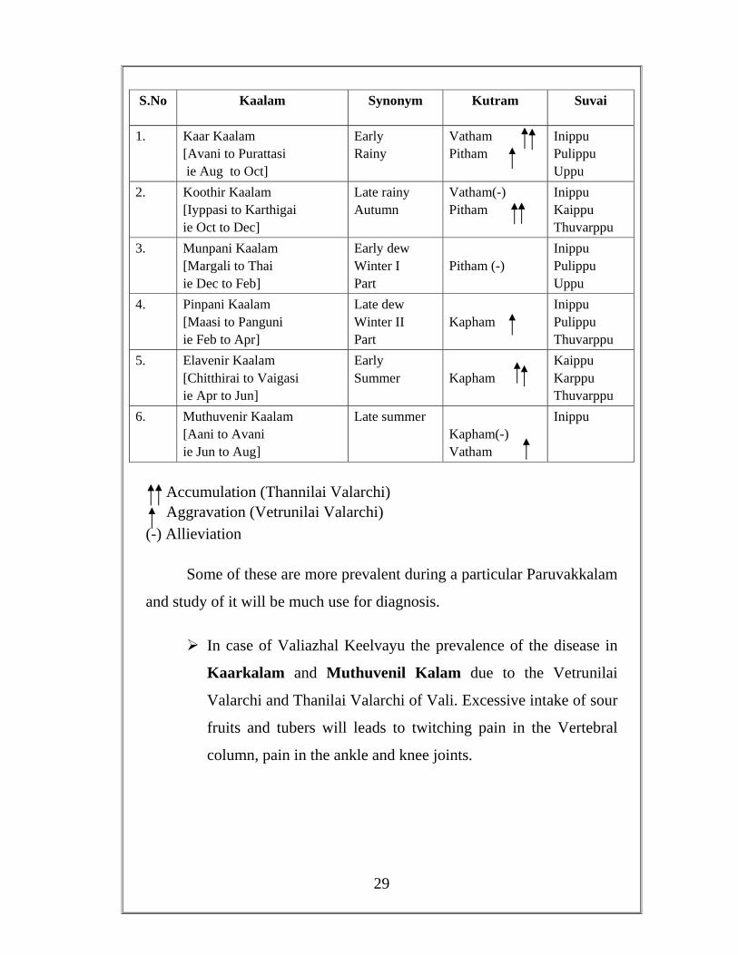
29
S.No Kaalam Synonym Kutram Suvai
1. Kaar Kaalam [Avani to Purattasi ie Aug to Oct]
Early Rainy
Vatham Pitham
Inippu Pulippu Uppu
2. Koothir Kaalam [Iyppasi to Karthigai ie Oct to Dec]
Late rainy Autumn
Vatham(-) Pitham
Inippu Kaippu Thuvarppu
3. Munpani Kaalam [Margali to Thai ie Dec to Feb]
Early dew Winter I Part
Pitham (-)
Inippu Pulippu Uppu
4. Pinpani Kaalam [Maasi to Panguni ie Feb to Apr]
Late dew Winter II Part
Kapham
Inippu Pulippu Thuvarppu
5. Elavenir Kaalam [Chitthirai to Vaigasi ie Apr to Jun]
Early Summer
Kapham
Kaippu Karppu Thuvarppu
6. Muthuvenir Kaalam [Aani to Avani ie Jun to Aug]
Late summer Kapham(-) Vatham
Inippu
Accumulation (Thannilai Valarchi) Aggravation (Vetrunilai Valarchi)
(-) Allieviation Some of these are more prevalent during a particular Paruvakkalam
and study of it will be much use for diagnosis.
In case of Valiazhal Keelvayu the prevalence of the disease in
Kaarkalam and Muthuvenil Kalam due to the Vetrunilai
Valarchi and Thanilai Valarchi of Vali. Excessive intake of sour
fruits and tubers will leads to twitching pain in the Vertebral
column, pain in the ankle and knee joints.
30
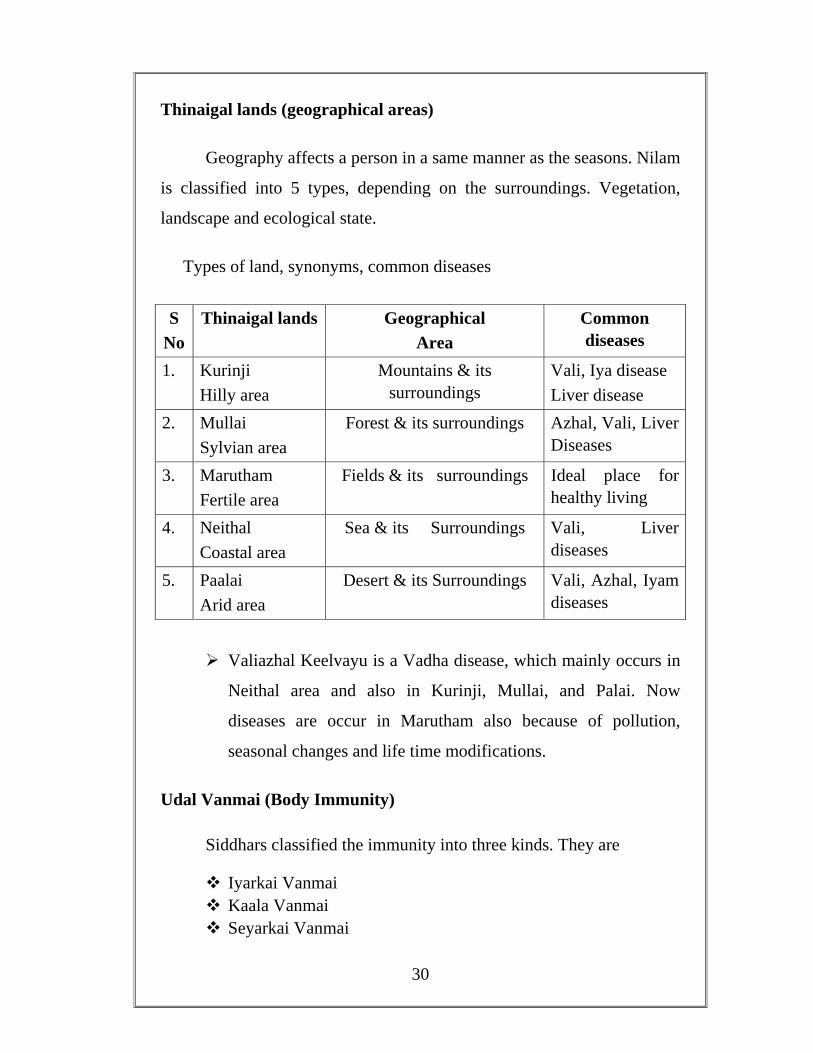
Thinaigal lands (geographical areas)
Geography affects a person in a same manner as the seasons. Nilam
is classified into 5 types, depending on the surroundings. Vegetation,
landscape and ecological state.
Types of land, synonyms, common diseases
S No
Thinaigal lands Geographical Area
Common diseases
1. Kurinji Hilly area
Mountains & its surroundings
Vali, Iya disease Liver disease
2. Mullai Sylvian area
Forest & its surroundings Azhal, Vali, Liver Diseases
3. Marutham Fertile area
Fields & its surroundings Ideal place for healthy living
4. Neithal Coastal area
Sea & its Surroundings Vali, Liver diseases
5. Paalai Arid area
Desert & its Surroundings Vali, Azhal, Iyam diseases
Valiazhal Keelvayu is a Vadha disease, which mainly occurs in
Neithal area and also in Kurinji, Mullai, and Palai. Now
diseases are occur in Marutham also because of pollution,
seasonal changes and life time modifications.
Udal Vanmai (Body Immunity)
Siddhars classified the immunity into three kinds. They are
Iyarkai Vanmai Kaala Vanmai Seyarkai Vanmai
31
Iyarkai Vanmai
One can inherit his immunity by birth naturally.
Seyarkai Vanmai
One can acquire his immunity through various food, activities and
medicines.
Kaala Vanmai
One can inherit his immunity at his different age and different
seasons. (Paruvakaalam).
Valiazhal Keelvayu is due to altered Iyarkai Vanmai and
established due to altered Kala Vanmai and Seyarkai Vanmai.
Uyir Thathukal/Mukkutram/Thirdosha
The theory mukkuthram forms the foundation of Siddha. The
primary position relegated to the equilibrated state of mukkutram in this
definition of a healthy man indicates their importance in the maintenance
of health. It can also be surmised that any disturbance in that equilibrated
state leads to the development of disease in the body. They are
Vazli Azhal Iyam
I. Vali or vayu
Vali is not merely wind, but also that which causes motion, energy
and sensation of every cell in the body. Vayu relates to the nerve force. It
32
is responsible for all movements in the mind and the body. In Western
terms, it is the electricity setting the organism into motion, maintaining
the equilibrium between Azhal and Iyam.
In the human body the loco motor activity functions through
voluntary muscles and its activities controlled by nerves system called
Kanmendriyam, Likewise the sensation and its activities are known as
Gnanendriyam. These types of activities are governed by Vali Kutram
among the Mukkutram.
The nerve cells are also governed by Vali (Vatha) Kutram. During
stimulation the nerve cells become repolarized and depolarized into
positive and negative charged waves by the help of Dhasavays. This
conducts the signals and information from one part to another.
Seats of Vali
Below the Navel region (umbilicus)
Urinary bladder, skin, nerves, bone, joints, muscles, hair follicles, motion, spermatic cord, umbilical cord, pelvis, ear and thigh.
" ñÊ º¨Áòм÷ ÜðÎí ̼üÀ̾ ¾¢ñʦÄýÒ ¦ºÅ¢ÌÈíÌ Å¢ñ¼ ¦¾¡Î ½÷× §¾¡üÚÅ¢ìÌõ §¾¡Ä¢ÕôÀ¢ùÅ¡Úõ ÅÎŢļÁ¡õ ÅÇ¢ìÌ "
Natural Properties of Vali
" ´Øí̼ý ¾¡§¾ú ã¡í¸¢ þÂí¸ ±Ø¦ÀÈ ±ôÀ½¢Ô Á¡üÈ ±Øó¾Ã¢Â §Å¸õ ÒÄý¸ÙìÌ §ÁÅî ÍÚÍÚôÒ Å¡¸Ç¢ìÌõ Á¡ó¾÷ìÌ Å¡Ô "
-º¢ò¾ ÁÕòÐÅ¡í¸ ÍÕì¸õ; Àì¸õ 140
33
Giving briskness, Expiration & Inspiration, Functioning of mind through out body, Regulation of 14 physiological reflexes (Natural urges), Make the uniform functioning of 7 Udal Kattugal, Protection and strengthening of 5 Sensory organs
Qualities of Vatham
Own Attributes:
" Å¡¾í ¸Î¨Á ÅÈðº¢Ô¼ý ¦¿¡ö¨Á º£¾Óï ºÄÉõ º¢¾ÈÏ×-²¾Ó¼
É¢ì ̽ò§¾¡ Îü§È þ−Âì¸ó ¾ÕÁÇÅ¢ü
¾ì¸ À⸡Ãó ¾¡ " -ñϺ¡Á¢Âõ; Àì¸õ 21
Roughness, Dryness, Lightness, Coolness, Mobility, Subtlety
Opposite Attributes
"Å¡¾Ì½ Á¡ÚìÌ Á¡Ú̽ §Á§É¡ìì¢ý μ¾Á¢Õ ¾£Ãõ ¯Â¢÷À¡Ãõ-§À¡¾ÃÅ¡ ÔûǾ£ §Â¡ÎÚ¾¢ ¢üÚò ¾¢ÃÇ¡¸ "
-ñϺ¡Á¢Âõ; Àì¸õ 22
Softness, Unctuousness, Heaviness, Hot, Stable, Solid
Varities of Vatham
The Vali is divided into 10 types according to their location and
functions.
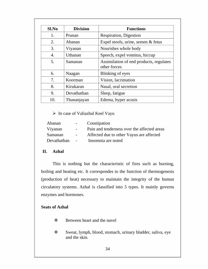
34
Sl.No Division Functions 1. Pranan Respiration, Digestion 2. Abanan Expel stools, urine, semen & fetus 3. Viyanan Nourishes whole body 4. Uthanan Speech, expel vomitus, hiccup 5. Samanan Assimilation of end products, regulates
other forces 6. Naagan Blinking of eyes 7. Koorman Vision, lacrimation 8. Kirukaran Nasal, oral secretion 9. Devathathan Sleep, fatigue 10. Thananjayan Edema, hyper acusis
In case of Valiazhal Keel Vayu
Abanan - Constipation Viyanan - Pain and tenderness over the affected areas Samanan - Affected due to other Vayus are affected Devathathan - Insomnia are noted
II. Azhal
This is nothing but the characteristic of fires such as burning,
boiling and heating etc. It correspondes to the function of thermogenesis
(production of heat) necessary to maintain the integrity of the human
circulatory systems. Azhal is classified into 5 types. It mainly governs
enzymes and hormones.
Seats of Azhal
Between heart and the navel
Sweat, lymph, blood, stomach, urinary bladder, saliva, eye and the skin.

35
Varietis of Azhal
S.N Name Location Function
1. Analam Stomach, Small
intestine
Dissolvent and digestive
2. Ranjagam Liver, spleen, stomach Colouring, pleasing,
gratifying
3. Sathagam Heart Effective, efficient
4. Alosagam Eyes Seeing, consideration
5. Prasagam Skin Complexion of skin
In case of Valiazhal Keelvayu, it is noted that
Analapitham - Loss of appetite
Ranjagam - Aneamia
Sathagam - Unable to carryout regular works properly
Prasagam - Redness and hyper pigmentation
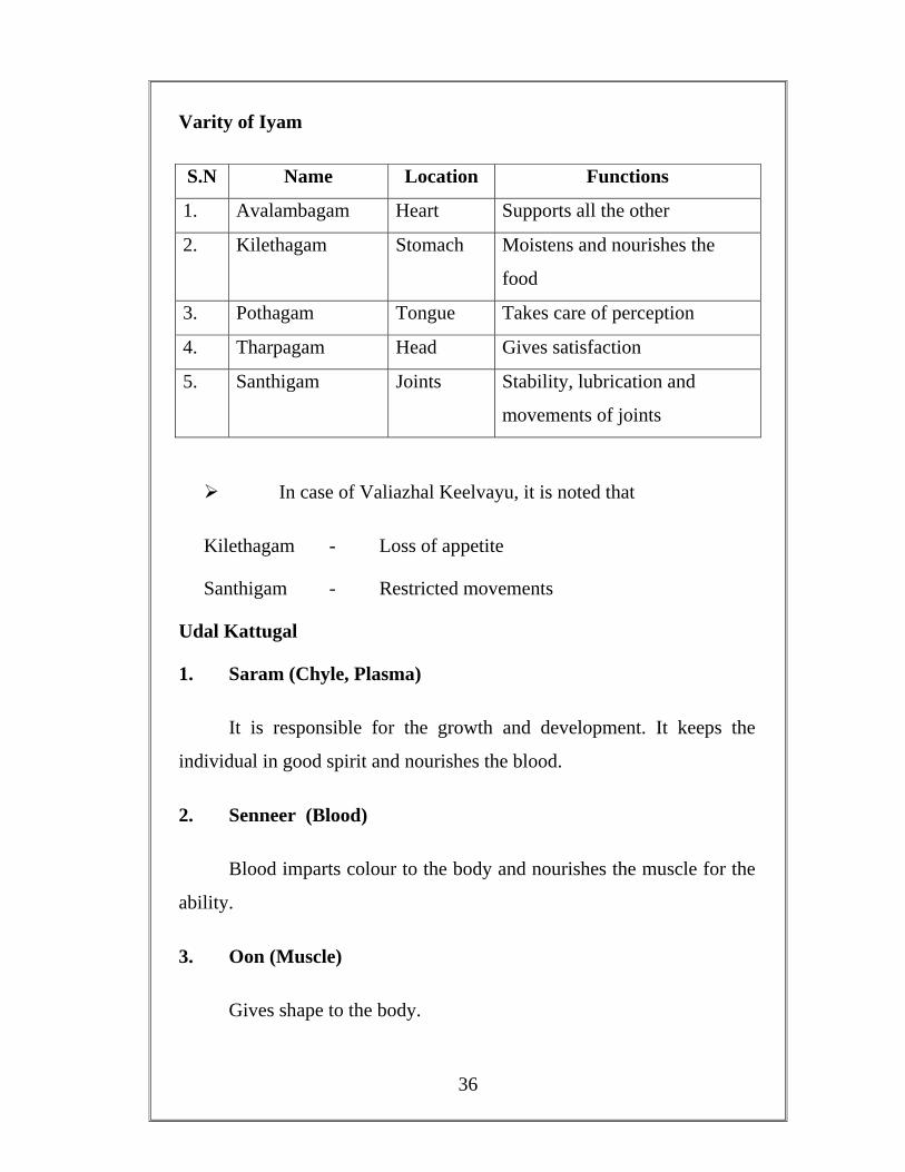
III. Iyam
It imparts moisture
Seat of Iyam
Above the heart, stomach, fat, sperm, tongue, uvula, bone marrow,
blood, nose, nerves, bones, large intestine, eyes and joints.
36
Varity of Iyam
S.N Name Location Functions
1. Avalambagam Heart Supports all the other
2. Kilethagam Stomach Moistens and nourishes the
food
3. Pothagam Tongue Takes care of perception
4. Tharpagam Head Gives satisfaction
5. Santhigam Joints Stability, lubrication and
movements of joints
In case of Valiazhal Keelvayu, it is noted that
Kilethagam - Loss of appetite
Santhigam - Restricted movements
Udal Kattugal
1. Saram (Chyle, Plasma)
It is responsible for the growth and development. It keeps the
individual in good spirit and nourishes the blood.
2. Senneer (Blood)
Blood imparts colour to the body and nourishes the muscle for the
ability.
3. Oon (Muscle)
Gives shape to the body.
37
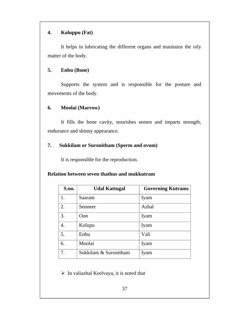
4. Koluppu (Fat)
It helps in lubricating the different organs and maintains the oily
matter of the body.
5. Enbu (Bone)
Supports the system and is responsible for the posture and
movements of the body.
6. Moolai (Marrow)
It fills the bone cavity, nourishes semen and imparts strength,
endurance and shinny appearance.
7. Sukkilam or Suronitham (Sperm and ovum)
It is responsible for the reproduction.
Relation between seven thathus and mukkutram
S.no. Udal Kattugal Governing Kutrams
1. Saaram Iyam
2. Senneer Azhal
3. Oon Iyam
4. Kolupu Iyam
5. Enbu Vali
6. Moolai Iyam
7. Sukkilam & Suronitham Iyam
In valiazhal Keelvayu, it is noted that
38
Saaram - Easy fatigability, loss of appetite
Senneer - Anemia, raised ESR
Oon - Muscle wasting
Kolupu - Restricted movements
Enbu - Pain and swelling, bone erosion, deformity
Moolai - Soft tissue swelling in proximal inter phalangeal joints
Gnanendhriyam
Mei - Feels all types of sensation
Vai - For identifying taste
Kan - Meant for vision
Mooku - For identifying smell
Sevi - For hearing
In case of Valiazhal Keelvayu, it is noted that
Mei - Pain and tenderness of joints
Kanmendhriyam
Kai - Mejority normal works done by hands
Kal - For walking
Vai - For speaking
Eruvai - for defaecation
Karuvai - For reproduction
In case of Valiazhal Keelvayu, it is noted that
Kai, Kal - Difficult in using limbs
Eruvai - Constipation
39
Pinineekam (Treatment)
Siddha system of medicine is a unique system of medicine in
which the treatment is given both for the body and the mind.
Thiruvalluvar, in his Thirukkural, under the heading, ‘Marundhu’
mentions about the diet disease and its prevention. Eg.
ÁÕó¦¾É §Åñ¼¡Å¡õ ...
«üÈ¡ÄÇ ÅÈ¢óÐñ¸ ...
¾£ÂÇÅ¢ñÈ¢ò ¦¾È¢Â¡ý ...
So in Siddha system, treatment is not only for the removal of the
disease but for prevention and improving the body condition –
Rejuvenation.
Kaapu (Prevention)
Neekkam (Treatment)
Niraivu (Restoration)
Kaapu
Preventive aspect is very much stressed in all Siddha litrature.
Body and mind should be clean and free of evil thoughts and deeds.
" Ó측ø ÁÄÁÐ ¦À¡øÄ¡¾ Å¡ö× ãýÚ ÐõÁø º¢ì¸¡ ÁÄ¡Ú ºÄ¾¡¨Ã Å¢ðÎî º¢Ú¿¨¼Ôõ
¨Á측Π¦¸¡ñ¼ ŢƢ¡ö ÁÉ¢¾÷ìÌ Å¡öÀ¦¾É¢ø ±ì¸¡ÄÓõ À¢½¢Å¡Ã¡¾ ¸¡Âõ −Õõ¦À¡ì̧Á "
-º¢ò¾ ÁÕòÐÅ¡í¸ ÍÕì¸õ; Àì¸õ 192
40
Neekam
A good physician should know about the derangement of Kutram
and should treat the patients on the basis of altered Kutram.
Treatment is based on
1. To bring the Thridosham to normal
2. To treat the disease according to its symptoms through medicines.
3. To increase the natural immunity.
To Normalize Tridosham
" Å¢§ÃºÉò¾¡ø Å¡¾õ ¾¡Øõ
ÅÁÉò¾¡ø À¢ò¾õ ¾¡Øõ
¿º¢Â —ïÉò¾¡ø ¸Àõ ¾¡Øõ "
ãýÈ¢¦Ä¡ýÚ ¯Â÷󾨾 ÓýÉ÷ Óó¾¢Â¾¨É ¦Â¡Æ¢ò¾¢Î ÁÕó¾¢Î
¾½¢Ôõ §¿¡Â¢ý ¾ó¾¢ÃÁ¢Ð§Å À½¢ì ¸½¢ò¾¢Êý À¢ÈÅ¡ö À¢ý̽õ "
Vadha diseases can be brought down by the Viraesanam, by giving
laxatives and purgatives, according to the patients’ conditions.
Management
" Å¢§ÃºÉò¾¡ø Å¡¾õ ¾¡Øõ "
" μи¢ýÈ ÁÄì¸ð¨¼ ¦Â¡Æ¢Â ¨Åò¾¡ø ¯¼Ä¢ÖûÇ Å¡¨¾¦ÂÄ¡ ¦Á¡Îí¸¢ô §À¡Ìõ "
41
Valiazhal Keelvayu is one of the vadha diseases. So, according to
the Siddha literature, vitiated Vadha may be suppressed by giving
purgatives.
For Purgation
Vellai ennai - ½ to 1 ounce (14 to 18 ml) At Morning on empty
stomach
After rearrangement of Dhosas
Kumari Mathirai - 1 twice daily with jaggery (Internal)
Nochi Thylam - 60 ml applied externally
As per the Text book of Siddha Maruthuvam,
Internal Medicine
Vanga Chunnam, Pavala Parpam- 65 ml with Cow Milk.
Vanga Chunnam, Muthu Parpam, Thanaga Parpam - 65 ml with
Cow Milk.
External Medicine
Kukkil Vennai, Alarchi Vennai, Ulundu Thailam- Patti Kattatuthal
Anupanam
" «ÛÀ¡Éò ¾¡§Ä ÂÅ¢ú¾í¸¼ ¸¡ñ¨Á ¸ÉÁ¡Ì ¦Áý¨Á ¦ÂøÄ¡õ ¸¡ðÎõ-−ÉÁ¡É §À¾¡§À ¾í¸¦ÇøÄ¡õ §À¾¢ò ¾È¢ó¾Å§Ã
¿¡¾¡ì¸ ¦ÇýÛÁ¨È áø "
-§¾¨ÃÂ÷ ¦ÅñÀ¡; Àì¸õ 158
42
Anupanam or Thunai marundhu in Tamil is commonly translated
as vehicle, adjuvant, adjunct, and supporting or co joint or concurrent
drug therapy. In Siddha system of medicine, the adjuvant is one of the
most important during therapy.
Panai Vellam (À¨É ¦ÅøÄõ)
" ¾íÌ À¨É¦ÅøÄò¾¡ø Å¡¾À¢ò¾õ Å£Ú¸Àï ºýÉ¢ §¿¡
ö
ÅøÄÕº¢ ÌýÁÁÚÁ¡ø " - À¾¡÷ò¾ ̽ º¢ó¾¡Á½¢
Vadha Pitha Thondham, Kabha Thondham, Sanni Patham, Vadha Kunmam, Arojagam will cure.
Pathiyam
During the course of treatment, the patient is advised for the following diet and physical activities. This form of medical advice is termed as Pathiyam. It means both Pathiya Patharthas and Apathiya Pathrathas.
" Àò¾¢Âò¾¢ É¡§Ä ÀÄÛñ¼¡ ÌõÁÕóÐ Àò¾¢Âí¸û §À¡É¡ü ÀÄý §À¡Ìõ
Àò¾¢Â§Á ¦ÅüÈ¢ ¾Õõ ÀñʾÕì ¸¡¾Ä¢É¡ü Àò¾¢Â§Á Ôò¾¢Â¦ÁýÚ À¡÷ "
-§¾¨ÃÂ÷ ¦ÅñÀ¡; Àì¸õ 159
The Pathiyams are
Kadum Pathiyam
Miga KadumPathiyam
Ichcha Pathiyam
Uppilla Pathiyam
43
In Theraiyar Venba,
" ¸ÎÌ ¿üÈ¢Äò ¦¾ñ¦½ö ÜúÀ¡ñ¼í¸û ¸¼¨Ä
ÅΞ¡¸¢Â ¦¾íÌÁ¡ ÅÕ쨸 ¿ü¸¡Âõ
ÁÊÅ¢ Ä¡¾¦ÅûÙûÇ¢¦¸¡û Ò¨¸Â¢¨Ä ÁЦÀñ
þ¼Ú À¡¸§Ä¡ ¼¸ò¾¢ ¿£ì¸¢¼Ä¢î º¡Àò¾¢Âõ "
- º¢ò¾ ÁÕòÐÅ¡í¸î ÍÕì¸õ
Diet " ÒÇ¢ÐÅ÷ Å¢ïÍõ ¸È¢Â¡ø ââìÌõ Å¡¾õ "
ÒÇ¢ôÒ, ÐÅ÷ôÒ Í¨ÅÔûÇ ¯½× Ũ¸¸¨Ç ¿£ì¸ §ÅñÎõ.
In Patharthaguna Chinthamani
" ¦ºí¸Ø¿£÷ §¸¡ð¼ó §¾ý Á¢ÇÌ ¿ø¦Äñ¦½ö ¾í̦ÀÕí ¸¡Âó ¾Ø¾¡¨Æ ±í¦¸íÌõ Üðκ¢Ú ÓòЦ¿ö §¸¡¾¢ø ¯Ø󾢨Ÿû
Å¡ðÎõ «É¢ Äò¨¾ Á¾¢ "
- Àì¸õ 369
Chenkaluneer, Honey, Pepper, Sesame oil, Asafoetida, Thaluthalai,
Caster oil, Black gram
SIRAPPU MARUTHUVAM
Kayakalpam, Pranayamam, meditation, Yogasanam, and
Thokkanam (Marthanam) are the special features of Sirappu Maruthuvam
(Special Medicine) in Siddha.
Thokkanam (Massage)
By applying the oil on the affected portion of the body and
massaging the same is called Thokkanam (Marthanam).
44
Types
1. Thattal 2. Erukkal 3. Pidithal 4. Murukal 5. Kaikattal 6. Azhuthal 7. Izhuthal 8. Mazhathuthal 9. Asaithal
According to Patharthaguna Chinthamani,
" ¦¾¡ì¸½ò¾¢½¡Ä¢ Ãò¾õ §¾¡ø °É¢¨Å¸ðÌ
Á¢ì¸ º×츢Âï ºÁ£¦ÃÛõ §À¡ ¦Áö츾¢¸
Ò‰Ê ÔÃì¸õ Ò½÷¢¨Å ¸¾¢ìÌõ
Àð¼ —ÄîºÄÚõ À¡÷ "
Vadha diseases are cured not only by internal medicine but also by
Thokkanam.
Theraiyar also quoted this as
" Á÷ò¾ÉÁ¡¸¢Â ¦¾¡ì¸½ò¾¢ý ¦ºöÂÅÌôÀ¦É - º¾¡
¿¢ò¾Óõ Å¡¾õ À¢½¢ò¾ À¢½¢ô¨À ¦ºôÀ§É "
In Valiazhal Keelvayu, Asaithal type of Thaokanam is used.
Ottradam (Fermentation)
Ottradam woth Nochi leaves are given to all patients to relieve pain
and swelling in affected joints.
45
" ¦¿¡îº¢, ¾Ø¾¡¨Æ, Å¡¾¿¡Ã¡Â½ý, ±ÕìÌ þ¨Ä¸¨Ç žì
¸¢ ´üÈÁ¢¼ Å¡¾§¿¡ö¸û, ¸£øÅ¡¾õ ¾£Õõ "
-§¾Ãý ¾Õ
PRANAYAMA
Pranayama means the breath control, which include the three stages
as inhalation, suspension and exhalation.
Method
Sit in a comfortable position (Padmasana or Sukhasana)
Inhale through the left nostril, keeping the right one closed with
the thump of the right hand.
Hold the breadth by closing the left nostril with ring finger and
the little finger of the right hand.
Exhale through the right nostril slowly by removing the thumb.
Then inhale through the right nostril, keeping the left one closed
with the ring finger and the little finger of right hand.
Hold the breath by closing the right nostril with the thumb of
right hand.
Exhale through the left nostril by removing the fingers kept in
the left one.
This completes one round of the exercise. Repeat this four to five
times.
46
Benefits
It increases the longevity, prevents and helps to cure the disease.
Respiratory, circulatory and nervous systems are strengthened.
MEDITATION
Integration of the body, mind, will, consciousness, ego and the self.
Method
Sit in Padmasana or Sukhasana, and concentrate on a particular
object or in action.
Benefits
It purifies and enriches the blood, gives total relaxation.
Serotonin secretion increases, which is a natural tranquilizer.
Endorphins, encephalin secretion increase, which is a natural pain
killer.
During deep slow wave sleep, delta waves produced, the same
thing happen in meditation, so we get refreshness within minutes.
YOGASANAM
Yoga is the science of right living and as such, is intended to be
incorporated in daily life. It works on all aspects of the persons: the
physical, vital, mental, emotional, psychic and spiritual. According to the
scientist, Yoga therapy is successful because of the balance created in the
47
nervous and endocrine systems which directly influences all the other
systems and organs of the body.
For Valiazhal Keelvayu,
Pawan Muktasana – Part I Anti rheumatic group
Sava Asana are advised.
Pawnamuktasan (Dynamic Practices)
Pawan means wind or Prana.
Mukta means release.
Asana means posture.
Stiffness of the body is due to the blocked Prana and accumulation
of toxins. This group of Asanas releases the blocked Prana and the
removal of toxins. They are concerned with the loosening of the joints of
the body. So they are advised to the patients of Valiazhal Keelvayu,
where vigorous physical exercises are not advised.
Sava Asana
Starting position:
Lie on the floor, face facing upward, Keep the legs straight and
together, Hands should be placed close to the body, palms facing
downward.
Method Position 1 Splitting the legs slowly sideward and keeping them one or two
feet apart.
48
Position 2
Moving the hands slowly sideward and keeping the arms as relaxed
as possible, keeping the palms upward, with natural flexion at the finger.
Position 3:
Taking a deep breath and exhale deeply.
Position 4
Closing the eyes and keeping the mind on breathing.
Keep this position for about 60 to 120 seconds and relax
throughout. Then slowly come back to the starting position by releasing
the position one by one in the reverse order.
Keep the mind on breathing for a few seconds
Try to forget all external thoughts while in the final position.
Try to relax the body and mind as much as possible
Benefits
Gives a complete relaxation to the whole body.
Gives the mental peace.
It helps to cure many functional, organic, structural and
psychological disorder
It is useful to energize our body and mind for further action.
49
Niraivu (Restoration)
Reassurance is given to all patience for speedy recovery.
All of them are advised to live in a good morality
Avoid excessive workload.
Avoid exposure to cold.
Avoid emotional stress at any cost.
Advised to do Yogasanas.
50
REVIEW OF LITERATURES
MODERN ASPECTS
RHEUMATOID ARTHRITIS
Rheumatoid Arthritis is a chronic, systemic, autoimmune disorder
that causes the immune system to attack the joints causing inflammation.
It can also cause inflammation of the tissue around the joints, as well as
other organs in the body.
Autoimmune diseases are illness, which acquire when the body
tissues are mistakenly attacked by its own immune system. The immune
system is a complex organization of cells and antibodies designed
normally to “seek and destroy”, invaders of the body, particularly
infections. Patients with these diseases have antibodies in their blood,
which target their own body tissues, where thay can be associated with
inflammations.
Definition
Rheumatoid arthritis is a symmetrical, destructive and deforming
polyarthritis affecting small and large synovial joints with associated
systemic disturbance a variety of extra articular features and the presence
of circulating anti- globulin antibodies (Rheumatoid factors)
Synovial Joints
To better understand the effects of Rheumatoid Arthritis on a joint,
a brief review of a Synovial joint is essential.
51
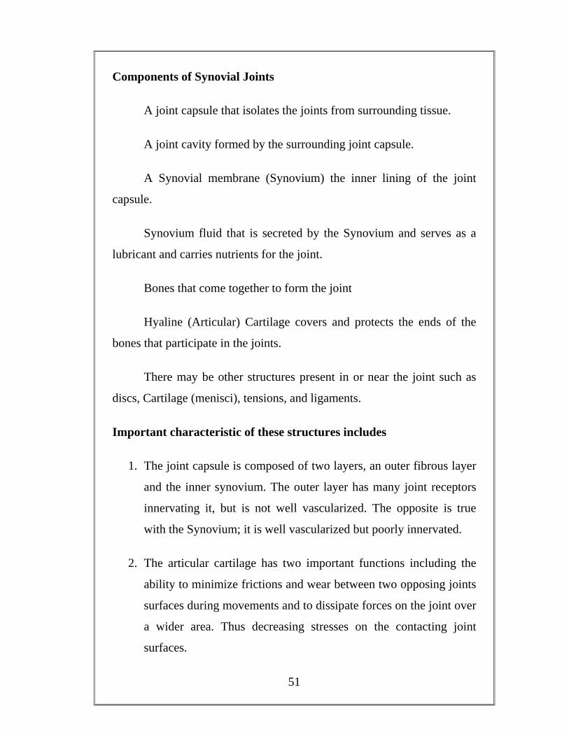
Components of Synovial Joints
A joint capsule that isolates the joints from surrounding tissue.
A joint cavity formed by the surrounding joint capsule.
A Synovial membrane (Synovium) the inner lining of the joint
capsule.
Synovium fluid that is secreted by the Synovium and serves as a
lubricant and carries nutrients for the joint.
Bones that come together to form the joint
Hyaline (Articular) Cartilage covers and protects the ends of the
bones that participate in the joints.
There may be other structures present in or near the joint such as
discs, Cartilage (menisci), tensions, and ligaments.
Important characteristic of these structures includes
1. The joint capsule is composed of two layers, an outer fibrous layer
and the inner synovium. The outer layer has many joint receptors
innervating it, but is not well vascularized. The opposite is true
with the Synovium; it is well vascularized but poorly innervated.
2. The articular cartilage has two important functions including the
ability to minimize frictions and wear between two opposing joints
surfaces during movements and to dissipate forces on the joint over
a wider area. Thus decreasing stresses on the contacting joint
surfaces.
52
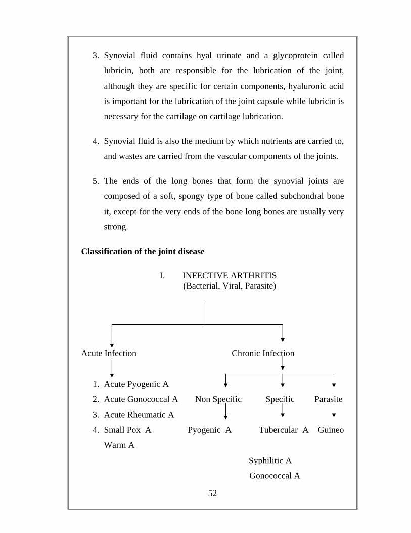
3. Synovial fluid contains hyal urinate and a glycoprotein called
lubricin, both are responsible for the lubrication of the joint,
although they are specific for certain components, hyaluronic acid
is important for the lubrication of the joint capsule while lubricin is
necessary for the cartilage on cartilage lubrication.
4. Synovial fluid is also the medium by which nutrients are carried to,
and wastes are carried from the vascular components of the joints.
5. The ends of the long bones that form the synovial joints are
composed of a soft, spongy type of bone called subchondral bone
it, except for the very ends of the bone long bones are usually very
strong.
Classification of the joint disease
I. INFECTIVE ARTHRITIS (Bacterial, Viral, Parasite)
Acute Infection Chronic Infection
1. Acute Pyogenic A
2. Acute Gonococcal A Non Specific Specific Parasite
3. Acute Rheumatic A
4. Small Pox A Pyogenic A Tubercular A Guineo
Warm A
Syphilitic A
Gonococcal A

53
II. RHEUMATOID ARTHIRITIS
Rheumatoid Arthiritis Sero Negative Spondyle
Arthropathy
RA (Adult) Ankylosing Spondylitis Juvenile RA (Childhood) Reiter’s Disease
Psoriatic A
Primary Osteo Artiritis
III. DEGENERATIVE ARTHIRITIS
Secondary Osteo Artiritis
Tabes-Charcot’s Arthropathy
Syringo Myelia
IV. NEUROPATHIC ARTHROPATHY Leprosy
Diabetes Mellitus
Alkaptonurine A V. METABOLIC ARTHIRITIS
Gout
Allergic A VI. ARTHRITIS IN SYSTEMIC DISORDER
Hemophilic A
Villonodular Synovitis
VII. MISCELLANEOUS JOINTS
Synovial Chondromatosis
VII. HISTORICAL JOINT

54
HISTORY
The name is based on the term “rheumatic fever” an illness which
includes the joint pain and is derived from the Greek word “Rheumatos”
(Flowing). The suffix “oid” (Resembling) gives the translation as joint
inflammation that resembles the Rheumatic fever. The first recognished
discription of rheumatoid arthritis was in 1800 by the French Physician
Dr. Augustin Jacob Landre-Beaubais (1772-1840), who was based in the
famed Salpetriere Hospital in Paris. The name Rheumatoid Arthritis itself
was coined in 1859, by British Rheumatologist, Dr. Alfred Baring
Garrod.
EPIDEMIOLOGY
Occurs in approximately one percentage of the population.
Is two to three times more prevalent among women than men.
Most commonly develops in the third to fifth decade of life.
Approximately 80% of total cases occur between the ages of 35
and 50.The prevalence and the incidence increase with age and
peaks at about age 70.
Some Native American groups have higher prevalence rates (5
to 6%), Black persons from the Caribbean have lower
prevalence rates.
Shortens the life spans by 3 to 10 years.
Global burden of disease 2000 study published in the World Health
Report 2002, Rheumatoid Arthritis is the 31st leading cause of years lived
55
with disability (YLDs) at global level accounting for 0.8% of total global
YLDs.
AETIOLOGY
Genetics: The cause of Rheumatoid Arthritis is still unknown. Some
scientists believe that the tendency to develop Rheumatoid Arthritis
may be genetically inherited. Rheumatoid Arthritis tends to run in
families. And frequency of disease is increased in first-degree relatives
of patients with Rhematoid Arthritis. Up to 50% of the genetic
contribution to susceptibility is due to genes in the HLA region. HLA-
DR4 is the major susceptibility haplotype (most specifically DR0401
& 0404), is more important in Indians. Concordance rates are higher
in monozygotic twins (12-15%) than in dizygotic twins (3%)
Immune system: People with Rheumatoid Arthritis have an
abnormal immune system response that mistakes the body’s healthy
tissue for a foreign invasion and attacks it. This miscommunication in
body is known as Rheumatoid Arthritic factor. About 80 of the R.A.
patients have circulating antibody known as Rheumatoid factor which
is immunoglobin M produced against the Fc portion of IgG. Rarely it
may be IgG, IgA of IgE type. It forms immune complex, which
produces inflammation.
Gender: Female gender is a risk factor. It goes into remission on
pregnant and this is increased in post partum and by breast-feeding.
Infection: Rheumatoid Arthritis is triggered by kind of infection.
Isoloation of variety of organisms from the synovial tissue, synovial
fluid blood of the affected persons, supports this possibility. It
includes Diptheroid Bacilli, Chlamydia Pneumoniae, Mycoplasma,
56
Erysipelothrix, Rubella, Parovirus, human herpes virus 6 and Viruses
especially Ebstein Barr Virus
Environment factors: Smoking and obesity are also risk factors for
Rheumatoid Arthritis.
Trauma : Traumatic incidence is a precipitating cause for the
development of R.A.
Psycological stress: It also plays a role. The study of identical twins
in one of whom Rheumatoid Arthritis, developed tends to support this
concept.
Vascular Changes: Alteration of peripheral vascular bed with
autonomic influence causes the symmetrical pattern of arthritis.
Neurogenic factor : Neuropeptide Rheumatoid Arthritis peptide can
cause inflammation. These peptides which are from sympathetic fibers
of spinal cord are responsible for the inflammation. This is confirmed
by the incidence of severity of Rheumatoid Arthritis in the
nonparalysed side of the hemeplegic patients.

57
PATHOGENESIS
There is a genetic predisposition of RA and that the joint
inflammation is immunologically mediated; however, the initating
agent(s) and the precise interplay between genetic and environmental
factors remain to be clarified.
In all likelihood the disease is initiated by activation of helper T
cells responding to some arthritogenic agent (MICROBE). Activated
CD4+ cells produce a number of cytokines that have two principle
effects: (1) activation of macrophages and other cells in the joint space,
which release tissue destructive enzymes and other factors that perpetuate
inflammation, and (2) activation of the B-cell system, resulting in the
production of anti-bodies, some reactions damage the joints and are
believed to play an important role in disease progression. In the context
of this general scheme the role of genetic factors, T cells, cytokines, B
cells, and infectious agents are as follows:
IMMUNOGENESIS
The role of immune processes in the development of the
rheumatoid lesions is indicated by a number of observations of patients
with the disease:
The presence of gamma globulins (in particular, IgG and IgM) in
synovial fluid and leukocytes, in synovial plasma cells, lymphoid
centers, and lining cells and in subcutaneous nodules and vessel walls.
These gamma globulins are not a direct cause of rheumatoid disease,
since the disease occurs in persons with agammaglobulinemia.
58
The presence of RF in synovial plasma cells and in syovial lining
cells, which are capable of synthesizing RF.
The presence of synovial leukocytes, interstitial connective tissue, and
lining cells of complement components associated with decreased
complement titers in the synovial fluid.
The presence of IgG, IgM and B1c complement in the articular
cartilage of patients with first or second degree osteoarthrosis.
The presence of antinuclear factor (ANF) in the serum of patients with
advanced disease; this suggests a role of autoimmunity, if not as the
primary cause then perhaps owing to the chronicity of rheumatoid
arthritis.
The common association of rheumatoid arthritis with amyloidosis.
HISTOGENESIS
On the basis of available histologic and immumologic data, a
concept of the histogenesis of rheumatoid arthritis has evolved. An
antigen, which could be extraneous (related to infection) or endogenous
(related to abnormal gamma globulins), gains access to the joint cavity
and elicit an immune reaction that is both humoral and cell mediated and
in which polymorphonuclear leukocytes, T and B lymphocytes, and
macrophages interact.
Complexes of various immunoglobulins, RF, and complement-
some quite large and insoluble are formed and phagocytosed by cells
termed RA cells or phagocytes. Thus the inflammatory process is set in
motion, terminating in the formation of granulation tissue and scarring.
59
Lysosomal enzymes from the cells of the exudates and from the pannus
participate in the destruction of cartilaginous matrix by degrading both
proteoglycans and collagen. They thus play a major role in the
deterioration of the joint.
PATHOLOGY
The Pathological changes proceed in three stages and are Synovitis,
destruction and deformity. The attack of a joint by the disease usually
begins with the synovium. Early in the disease, edema begins to be seen
in cells in the synovium and multiplications of synovial lining cells occur.
As the disease progresses, the synovium may grow considerably larger
eventually forming tissue called pannus. Pannus can be considerably the
most destructive element affecting joints in the patient with rheumatoid
arthritis. Pannus can attack articular cartilage and destroy it. Further the
pannus can destroy the soft subchondral bone once the protective articular
cartilage is gone. The synovial fluid secreted by the synovium is thought
to be avascular articular cartilage. In this disease process, an interaction
between antibodies and antigen occurs and causes alteration in the
composition of the synovial fluid. Ultimately, digestants are formed in
the fluid, which attack the surrounding tissue. Once the composition of
the fluid is altered, it is less able to perform the normal function noted
above, and more likely to become destructive. The changes in the
syovium and synovial fluid briefly described above are responsible for a
large amount of joint and soft tissue destruction. The destruction of bone
eventually leads to laxity in tendons and ligaments. Under the strain of
daily activities and other forces, these alterations in bone and joint
structure result in the deformities frequently seen in patients with
rheumatoid arthritis. Considerable destruction of the joint can occur with
pannus invading the subchondral bone.

60
Bone destruction occurs at areas where the hyaline cartilage and
the synovial lininig do not adequately cover the bone. If the disease
progresses to a more advanced stage, the articular cartilage may lose its
structural and density resulting in an inability to withstand the normal
forces placed on the joint. In these advanced cases, muscle activity
causes the involved ends of the bones to be compressed together causing
further bone destruction. Further, the disease can irreversibly change the
structure and function of a joint to the degree that other degenerative
changes may occur, especially in the weight bearing joints of the body.
Thus, joint destruction can progress to the degree that joint motion
is significantly limited and joints can become markedly unstable.
Pathological process
Tissues involved
Results in
Deformities
Vasculitis Necrosis Fibrosis
Joint structures
Synovitis effusion articular cartilage
destruction. pericapsulitis Ligamentous instability
Swelling Stiffness
Instability-subluxation dislocation
Instrinsic plus deformity
Plasma cell Proliferation
Tendon Tenosynovitis Rupture Ulnar devation of fingers
Concertina collapse of
fingers
Granulation tissue and pannus
formation
Muscle Wasting atrophy fibrosis Contracture ankylosis
Synovial hypertrophy in
joint and tendon
Bone
Subcutaneous
Osteoporosis-Thinning of cortex and loss of
trabecular structure. cyst formation subchondral
erosions(adjacent to metaphysis) destruction
Nodules
61
CLINICAL FEATURES
The symptoms of Rheumatoid Arthritis depending on the degree of
tissue inflammation, remission can occurs spontaneously and patients feel
well when relapse, symptoms return (Flare).
General Symptoms
Malaise Fever Fatigue Loss of appetite Weight loss Myalgias Weakness or loss of energy
Joints Symptoms
Pain Swelling Morning Stiffness Inflammation Nodules Deformity
Pain
The pain in Rheumatoid Arthritis has several sources. Pain can
come from inflammation of the joint and surrounding tissues or from
working the joints too hard.
Swelling The area around the affected joint is swollen and puffy. Morning Stiffness
Morning Stiffness is a very characteristic of Rheumatoid Arthiritis,
which is lost for one hour and above. Stiffness is most noticeable in the
morning and improves latter in the day.
62
Inflammation
Redness, tenderness and warmth are the hallmarks of
inflammation.
Nodules
These are hard bumps that appear on or near the joint. They often
are found near the elbows.
Deformity
Occurred from several mechanisms, all related to Synovitis and the
patients attempt to avoid the pain by keeping the joints in the least painful
position.
Patterns of onset of Rheumatoid Arthritis
Insidious - 70% Acute - 15%
Oligoarticular - 44% Systemic - 10%
Polyarticular - 35% Palindromic - 5%
Monoarticular - 21%
CLINICAL MANIFESTATIONS
I. Articular Manifetation
Rheumatoid Arthiritis is a chronic Inflamatory Arthiritis;
symptoms must have been present for atleast six weeks, in order to make
the diagnosis. It mainly affects the joints of the Knuckles
(Metacorpophalangeal) and the proximal inter Phalange joints of the
63
hands. It also affects the joints of the feet- metatarsophalangeal joints.
The wrist, elbow, knees, ankles are also frequently affected. The
vertebrae of the neck are sometimes involved in the people who have had
the disease many years.
Hands and Wrist
Rheumatoid arthritis often causes symmetric arthritis which
characteristic involvement of certain specific joints such as proximal
interphalangeal joints and metacarpophalangeal joints. The distal inter-
phalangeal joints are rarely involved.
In early course of the disease, there may be spindling of the
progress due to synovial hypertrophy and effusion in the interphalangeal
joints.
Later, marked with synovial hypertrophy on the dorsum of the
wrist, with the involvement of extensor tendon sheath, results in dropped
finger. The same process in the palmar aspect may lead to carpal tunnel
syndrome. Tenosynovities of long flexor tendons in palm may result in
stiffness of the fingers and cause trigger finger.
Piano Key Sign
Weakening of the distal radio ulnar ligament by synovitis allow the
distal ulnar to migrate dorsally so that it over rides the radius (Caput
ulnae syndrome). The Ulnae can be depressed by pressure, like a piano
key.
64
Carpal Colapse and Fusion
It may occur in late, when the instability of the wrist may lead to
collapse of the carpal bones causing for shortening of the carpus and
ultimately spontaneous fusion of the wrist. The eventual functional loss
characterized by the inability to make a fist and pinch thin objects,
weakened grip strength.
Persisting synovitis, weakening of the capsule, muscle wasting,
tendon rupture and destruction of the articular surface leads to
characteristic Rheumatoid hand deformity, which includes:
“Swan neck deformity” with hyperextension of the proximal
interphalangeal joints with fixed flexion of the distal
interphalangeal joints.
“Button hole deformity” (Boutoniere deformity) which
includes fixed flexion of the proximal interphalangeal joints and
extension of the distal interphalangeal joints.
The “Z deformity” of the thumb (Radial deviation at the wrist
with ulnar deviation of the digits often with palmar subluxation
of the proximal interphalangeal joints).
“Intrinsic Plus Deformity” Hyper extension of the first
interphalangeal joints and flexion of the first
metacarpophalangeal joint with consequent loss of thumb
mobility and pinch.
“Bull horn deformity” due to rupture of the extensor
communis tendon from synovitis near the ulnar styloid.
65
Palmar erythema is also common. Raynaud’s phenomenon may
occur in the early stage.
Feet and Ankles
Active synovitis in the metatarsophalangeal joint can produce pain
and tenderness best elicited by the lateral squeezing of the joints.
The synovial swelling of the active disease together with
destruction of the ligament between the metatarsal heads may broaden the
forefoot and separate the toes to produce the “day light sign”.
Deformities may also develop in the feet including eversion at the
hind foot (subtalar joint), plantar subluxation of the metatarsal heads,
widening of the forefoot, hallux-valgus and lateral deviation and dorsal
subluxation of the toes. So the patient walks on the unprotected heads of
the metatarsal bones. The patient complains of a feeling of walking on
pebbles and the metatarsal heads are readily palpable on the sole of the
foot.
In the hind foot calcaneal erosions, hallux-valgus deformities are
found.
Rheumatoid synovitis may develop in the subtaloid and midtarsal
joints. Chronic arthritis in this region can lead to “pesplano-valgus
deformity”.
Foot Deformities in Rheumatoid Arthritis
Callosity under PIP joint
Plantar callosity
66
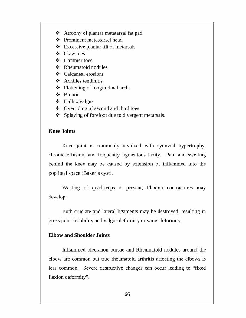
Atrophy of plantar metatarsal fat pad Prominent metastarsel head Excessive plantar tilt of metarsals Claw toes Hammer toes Rheumatoid nodules Calcaneal erosions Achilles tendinitis Flattening of longitudinal arch. Bunion Hallux valgus Overriding of second and third toes Splaying of forefoot due to divergent metarsals.
Knee Joints
Knee joint is commonly involved with synovial hypertrophy,
chronic effusion, and frequently ligmentous laxity. Pain and swelling
behind the knee may be caused by extension of inflammed into the
popliteal space (Baker’s cyst).
Wasting of quadriceps is present, Flexion contractures may
develop.
Both cruciate and lateral ligaments may be destroyed, resulting in
gross joint instability and valgus deformity or varus deformity.
Elbow and Shoulder Joints
Inflammed olecranon bursae and Rheumatoid nodules around the
elbow are common but true rheumatoid arthritis affecting the elbows is
less common. Severe destructive changes can occur leading to “fixed
flexion deformity”.
67
Pain in the shoulder can be referred from the neck or be due to
involvement of acromio-clavicular joint, sub-acromial bursa, rotator cuff
and bicipital tendon as well as the gleno-humeral joint.
Cervical spine
The upper cervical discs are frequently involved. The cervical
vertebrae may become subluxed and this may cause serious neurological
disorders.
The atlantoaxial articulations and their associated ligaments are
frequently involved. Separation between the odontoid process and the
first cervical vertebra exceeds the normal of 2 to 3 mm that can be
detected by X-ray. They complain pain in the cervical spine that radiates
upwards over the occiput and vertex to the forehead.
Atlantoaxial dislocation may cause the vertebrobasilar
insufficiency or may produce neurological signs by direct pressure on the
cord.
Hip joints
The hip is less commonly involved but when it occurs, it causes
serious disability. Occasionally the disease remains monoarticular for
several years but eventually other sites are affected. Persistent synovitis
in a weight-bearing joint soon leads to the destruction of the cartilage and
bone. The acetabulam is eroded and eventually the femoral head may get
perforated at its floor. The hallmark of the disease is progressive bone
destruction on both sides of the joints without any reactive
osteophyte formation. This is often referred to as “aseptic necrosis” and
is more common in corticosteroid treated patients.
68
Other joints
Rheumatoid arthritis affects all the synovial joints. Temporo mandibular involvement produces pain on chewing. Acromio clavicular, sterno clavicular and cricoartenoid joints may also be involved.
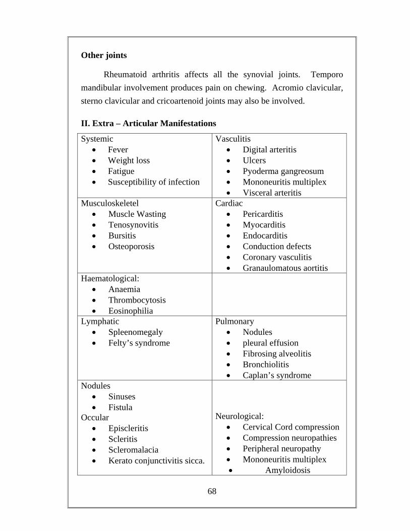
II. Extra – Articular Manifestations
Systemic • Fever • Weight loss • Fatigue • Susceptibility of infection
Vasculitis • Digital arteritis • Ulcers • Pyoderma gangreosum • Mononeuritis multiplex • Visceral arteritis
Musculoskeletel • Muscle Wasting • Tenosynovitis • Bursitis • Osteoporosis
Cardiac • Pericarditis • Myocarditis • Endocarditis • Conduction defects • Coronary vasculitis • Granaulomatous aortitis
Haematological: • Anaemia • Thrombocytosis • Eosinophilia
Lymphatic • Spleenomegaly • Felty’s syndrome
Pulmonary • Nodules • pleural effusion • Fibrosing alveolitis • Bronchiolitis • Caplan’s syndrome
Nodules • Sinuses • Fistula
Occular • Episcleritis • Scleritis • Scleromalacia • Kerato conjunctivitis sicca.
Neurological:
• Cervical Cord compression • Compression neuropathies • Peripheral neuropathy • Mononeuritis multiplex • Amyloidosis
69
DIAGNOSIS
The typical picture of bilateral symmetric inflammatory
polyarthritis involving small and large joints in both upper and lower
extremities with sparing of the axial skeleton except the cervical spine
suggest the diagnosis.
Consititutional features like morning stiffness, demonstration of
subcutaneous nodules, presence of the rheumatoid factor and
radiographic findings of juxta articular bones substantiate the diagnosis.
Criteria for the diagnosis of Rheumatoid Arthritis:
a. Morning stiffness I hr,
b. Arthritis of 3 or more joint areas.
c. Arthritis of hand Joints.
d. Symmetric Arthritis.
e. Rheumatoid Nodules.
f. Rheumatoid factor.
g. Radiological changes.
Diagnosis of Rheumatoid Arthritis is made with 4 or more criteria.
Criteria’a to d’ must be present for atleast 6 weeks, and Criteria ‘b to e’
must be observed by a physician.
Grading of Rheumatoid Arthritis Patients Tenderness
Grade 1 The patient says the joint is tender.
Grade 2 The patient winces.
70
Grade 3 The patient winces and withdraws the affected part.
Grade 4 This patient will not allow the joint to be touched.
Restriction of Motion
Grade 1 No restriction of ability to perform normal activities.
Grade 2 Moderate restriction but with an ability to perform most
activities of daily activity.
Grade 3 Marked restriction with an ability to perform most activities
of daily activity.
Grade 4 Incapacitation with confinement to bed or wheel chair.
INVESTIGATIONS
I . Haematological
Full Blood Count- Anaemia, Esinophilia, Thrombocytosis
ESR – Increased in active stage
Serum proteins – Hyperglobulinaemia with elevation of Gamma
and Alpha 2 globulins hypoalbuminaemia during acute phase.
II. Immunological
I. Rheumatoid Factor
Rheumatoid Factor is an immunoglobulin M (Ig M) antibody
directed against normal human immunoglobulin. It is usually measured
by agglutination tests. (Ig G) (Agglutination of Ig G coated latex
71
particles) and reported as either negative or positive with titers up to
1:320.
Rose Waaler Test: A special type of passive haemagglutination test
is Rose Waaler test. In Rheumatoid Arthritis autoantibodies appears
in the Serum, which acts as an antibody to gamma globulin. The
Rheumatoid Arthritis factor is able to agglutinate red ells coated with
globulin. The antigen used for the test is suspension of sheep
erythrocytes sensitized with subagglutinating dose of rabbit anti
sheep erythrocyte antibody. (Amboceter)
Latex agglutination test (Latex fixation test)
Differential agglutination test (DAT)
Human erythrocyte agglutination test (HEAT)
2. Anti Nuclear Antibodies
Routine determination of the resence of antinuclear antibodies is
best performed by the indirect immuno fluorescent technique.
3. C-Realtive Protien
CRP is produced in the liver and is normally found in Serum in
minute amounts (less than 0.6 mg/dl). In conditions characterized by
inflammation with tissue destruction, the CRP level may increase.
Although CRP has many effects in the immune system, its specific
primary role is still unclear.

72
III. Special Investigations
1. Synovial fluid analysis
Confirms the presence of inflammatory Arthritis. Fluid may show
positive Roose-Waaler test in joint fluid, before it can be detected in
blood. Also it may show neutrophils or monocytes inclusion bodies.
2. Synovial biopsy
Villus formation with thickening of synovial layer and infiltration
with abnormal cells.
IV. Radiographic Evaluation
Soft tissue swelling
Juxta articular osteoporosis
Erosion of joint margins
Joint spaces are decreased
Deformities
Atlantoaxial subluxation
Subchondralerosions and cyst formation
Fibrous and bony ankylosis develops in the late stages.
73
V. Arthroscopy
In acute Rheumatoid Arthritis synovium is oedematous, diffusely
erythematous and friable. In more chronic conditions it becomes
thickened.
VI. Renal biopsy
Indicated in cases of reduced tubular or glomerular functions.
VII. Pulmonary biopsy
Used to distinguish Rheumatoid nodules from carcinoma or to
establish diagnosis of fibrosing alveolitis.
VIII. Ultra Sound
IX. Scintigraphy
X. CT Scanning
Shows cartilage and sub –chondral bone damage long before
conventional X rays
XI. MRI
XII. Urine Analysis
XIII. Biochemical Analysis
74
XIV. Anti CCP Antibodies (Cyclic Citrullinated Peptide Antibodies)
It is positive in 80% of all Rheumatoid Arthiritis cases, but it is
rarely positive if Rheumatoid Arthiritis is not present, giving it a
specificity of around 98%. It can be detected in early stages of the
disease, even before the on set of clinical disease.
XV. Genetic tests
HLADRB1 Typing to detect the presence of ‘shared epitope’.
XVI. Antinuclear Antibody Assay (ANA)
XVII. Bone Densitometery (DEXA)
Bone Density Test to Check For Bone Loss.
Differential Diagnosis of Rheumatoid Arthritis
Sero negative spondyloarthropathy included following rheumatoid
like conditions were the serum is negative for rheumatoid factor. They
are.
1. Ankylosing Spondylitis
Ankylosing spondylitis is a chronic, progressive and crippling
disease affecting the spine associated with calcification and ossification
of ligaments and capsules of joints resulting in complete bony ankylosis.
The exact etiology is unclear. Ankylosing spondylitis has been found to
be more prevalent in certain races and hence shows a genetic
predisposition. It is related to certain tissue types of the human leukocytic
75
antigen (HLA) system. The majority of ankylosing spondylitis patient is
found to belong to HLA-B27 groups.
The disease occurs in the 3rd and 4th decades of life and is more
common in males. The patients present with complaints of diffuse pain in
the back and vague pain in other joints.
2. Reiter’s disease
Reiter’s disease characterized by triad of polyarthritis, urethritis,
conjunctivitis. The joint condition is an acute polyarthritis resembling
rheumatoid arthritis. It does not cause destructive changes in the joint
structures. The urethritis is non-gonococcal but the exact organism is not
known.
3. Psoriatic Arthritis
Psoriatic arthritis is a polyarthritis seen in about 10% of patients
with psoriasis.
The most common type is the one involving the distal
interphalangeal joints of the hands and feet with psoriatic nail
joints. Metacarpophalangeal joints are never involved in
psoriatic arthritis.
Arthritis multilens is a severe form where there is marked
destruction of joints.
Symmetrical polyarthritic type
Oligo arthritic type.
Spondyloarthritic type.
76
4. Enteropathic Arthritis
Chronic inflammatory bowel diseases like regional enteritis
(Crohn’s) disease) and ulcerative colitis are associated with arthritic
lesion in abour 10% of the cases. There is peripheral (or) involvement of
the spine. The Joint condition shown remissions and exacerbations along
with activity of the underlying bowel disease.
5. Sjogren’s Syndrome
Sjogren’s syndrome is an immunologic disorder characterized by
progressive destruction of the exocrine glands leading to mucosal and
conjuctival dryness (Sicca syndrome) accompanied by a variety of
autoimmune phenomena. Glandular, when the clinical manifestations are
within the exocrine system and extra glandular, when other tissues are
involved as well.
The disease predominately affects woman in the third of fourth
decades of life.
Clinical Manifestations:
i. Keratoconjuctivitis and Xerostomia
ii. Renal involvement produces mild interstitial nephritis that may
result in renal tubular acidosis.
iii. Sensory polyneuropathy and mononeuritis multiplex.
iv. Pulmonary involvement generally takes the form of an interstitial
pneumonitis which is usually a little clinical significance.
77
COMPLICATIONS
Septic Arthritis
It may complicate Rheumatoid Arthritis, particularly in patients
with long-standing nodular sero-positive disease. In debilitated patients,
fever and leucocytosis may be absent and the signs of infection limited to
malaise and slight exacerbation of inflammation in one or more joints.
Amyloidosis
It is a complication of prolonged active disease and is formed in
25% to 35% of patients at autopsy, making Reumatoid Arthiritis, a
leading cause of secondary amyloidosis.
Fixed Deformities
The perils often the common place ones resulting from ignorance
and neglects. Early assessment and planning should prevent postural
deformities that will result in joint contractures.
Muscle Weakness
Even mild degree of myopathy or neuropathy when combined with
prolonged inactivity may lead to profound muscle wasting and weakness.
This should be prevented by physiotheraphy and pain control if possible.
If not the surgeon must be forewarned of the difficulty of postoperative
rehabilitation.
78
Joint Rupture
Occasionally the joint lining ruptures and synovial contents spill
into the soft tissue. Treatment is directed at the underlying synovitis i.e
splintage and injection of the joint synovectomy as a second resort.
Spinal Cord Compression
It is a rare complication of cervical spine instability. By the onset
of weakness Upper Motor Neuron (UMN) signs in the lower limb is
suspicious. If they occur, immobilization of the neck is essential and
spinal fusion should be carried out as soon as possible.
Systemic Vasculitis
This is a rare but potentially serious complication. High doses of
corticosteroids and Intra Venous (I.V) plasma volume expanders may be
called for.
Clinical Course and Prognosis
The course of Rheumatoid Arthritis is variable and difficult to
predict in an individual patient. Most patients experience persistent but
fluctuating disease activity accompanied by a variable degree of joint
deformity.
Approximately 15% of patients of R.A. have a short-lived
inflammatory process that remits without major deformities.
Factors suggest poor prognosis:
Insidious onset
79
Unremitting disease
Presence of Rheumatoid nodules
Systemic involvement with increased ESR Anaemia
Presence of RA factor, HLADR4
Juvenile Rheumatoid Arthritis
Juvenile Rheumatoid Arthritis (J.R.A) is one of the more common
connective tissue diseases of children and is a major cause of functional
disability in this age group. By definition, it begins before the age of 16
and most patients are diagnosed early childhood. There is 2:1 female
predominance except in the subgroup that has a systemic onset, in which
the sexes are equally affected. J.R.A differs from R.A. in adults. In that
Oligo arthritis is more common.
Systemic onset is more frequent.
Larger joints are affected more than smaller joints.
Rheumatoid nodules and rheumatoid factor are usually absent
and
Antinuclear antibody seropositivity is common.
Genetic susceptibility, abnormal immuno regulation, cytokine,
production and viral infection may all play a role in the pathogenesis.
The morphology of the joint, pathology, including marked synovitis is
similar to the alteration in adult R.A. The development of symptoms,
such as fatigue, joint stiffness and limited range of motion is generally
slow and gradual. Commonly targeted joint in all forms of the disease are
80
the knees, wrists, elbows and ankles. They become warm and swollen
and are often involved symmetrically, Pericaridtis, myocaridtis,
pulmonary fibrosis, glomerulonephritis, uveitis and growth retardation
are potential extra-articular manifestations. A systemic onset may begin
rather abruptly, associated with high spiking fevers, migratory and
transient skin rash, hepatomegaly, spleenomegaly and serositis.
Satisfactory recovery occurs in 70 to 90% of patient and in only 10%
severe joint deformities present.
Management
Current concepts, regarding the management of Rheumatoid
Arthiritis emphasize medical tharaphy, physical and occupational
theraphy and patients’ education. The aim of treatment is to
Relieve pain Keep the inflammatory process down to minimum. Preserve joint movement Maintain the tone of muscles Prevent deformitives and stiffness of joints
Calcium and vitamin D have been proven to be beneficial in the
treatment of osteoporosis (low bone density). Omega-3 fatiasis have been
found to have antiinflamatory properties at high doses, present in Salmon
fish. Vitamin C and haematinics in diets essential.
Surgery
The role of surgery is mainly reconstructive or rehabilitative. The
following procedures are used, in the management of Rheumatoid
Arthritis.
81
1. Synovectomy
2. Capsulotomy
3. Osteotomy
4. Arthrodesis
5. Arthroplasty-Excision and Replacement.
Adjunctive Treatments
Rehabilitation
Restoration of a disabled individual to perform function to a
normal or near normal manner is called Rehabilitation.
Rheumatoid Arthritis is a crippling disease. Hence the Rheumatoid
Arthritis patients require rehabilitative measures that include physical
therapy, and special splints and appliances to help them and manage their
own self care activities like dressing and eating.
Physical Therapy (Physiotherapy)
Physiotherapy is the application of physical agents and principles
to pathological conditions for the purpose of producing therapeutic
effects.
Physiotherapy includes Active exercise Passive joint movements Local heat Massage Electrical stimulation of muscles Ultra sound therapy Light therapy, ultraviolet rays and infrared rays.
82
Exercise Therapy
Once inflammation is satisfactorily controlled, appropriate and
regular exercises is essential to strengthen muscles weakened by disease.
Rheumatoid arthritis often makes joints stiff and restricts their motion if
they are not used regularly. Exercises are designed to meet the needs of
each patient and should be monitored by professionals specializing in
physical medicine.
Exercise Guidelines
The patient is advised to engage in low – impact exercises like
stationary cycling, rowing and water aerobics and,
Start with low intensity exercise.
During periods of inflammation or pain (for rheumatoid arthritis),
reduce exercise intensity and duration.
Patient need longer than normal (longer than ten minutes) warm ups
and cool downs.
Modify patients exercise intensity or duration depending on how the
patient feel that day.
It is important that patient should move his joints through a full range
of motion at least once a day, otherwise, joints will become stiff.
If the patient experience pain two hours after exercise, he need to
adjust the intensity or the duration of the exercise.
83
The patients should do exercise to the point in he can (unless he has
hypertension) because these type of exercises put the least amount of
stress on the joints.
Patient should use isometric exercises when he can (unless he has
hypertension) because these types of exercises put the least amount of
stress on his joints.
Advise the patient to consult the doctor if he experience severe pain
after exercise.
Active Exercise is given to
Mobilize joints, strengthening muscles, Improve co-ordination or
balance.
Passive Joint Movements
The chief use of passive joint movement is to preserve full mobility
when the patient is unable to move the joint actively.
Types of Exercises
Range – of – motion Exercise, Strengthening Exercise, Limbering
up Exercise:
1. Range – of – Motion Exercises / Stretching Exercises
Stretching exercise involve moving a joint as it will comfortably
go through its full range of motion or stretch. This exercise helps to
maintain normal joint movement or restore movement that has been lost.
84
Clinical Assessment of Joint Motion
The most widely used and recommended instrument is the
universal Goniometer, sometimes called an Arthrometer. Basically, it is
protractor, to the center of which two long slender arms or levers are
attached. Usually only one of the arms is movable, but may variations in
design are possible.
2. Strengthening Exercise
Strengthening exercises help to maintain or increase the strength
and power of the muscles.
3. Limbering up Exercises
Help to reduce morning stiffness or stiffness after staying in one
position too long by doing the Range – of – motion exercises each day
only a few times of loosen up.
Details of Range – of – Motion Exercises
Upper Extremities – Shoulder
Arms at side with elbow straight, bring arms forward upward by year.
Arms at side with elbow straight, take arms sideward upward.
Arms at side bend elbows to right angle and take hands apart.
Elbow
Bend elbow, touching fingers to top of shoulder.
Straighten elbow.

85
Fore Arm
Elbows bent, turn palm of the hand and then back of the hand
towards face.
Wrist
Keeping forearm steady, move the wrist up and down as in
waving.
Again hold forearm steady, mover the wrist up and down as in
hand shaking.
Make circle with hands.
Hand and Fingers
Make tight first.
Open fingers as wide as possible.
With the hand open spread fingers away from each other and
then together.
Touch tip of the thumb to the tip of each finger.
Bend the thumb in toward palm of the hand.
Lower Extremities
Knee
Sit with your feet off the floor. Lift the leg and then allow it to
return of the bent position slowly.
86
Ankle
Pull foot up and in, and then push back down. Make circle with foot. Pull foot in toward other foot. Pull foot to outside.
Toes
Pull up on toes then curl toes under.
Exercises for the Neck
In the sitting position, twist your head as far as possible in each
direction.
Sit or stand with your hands on the hips. First circle the head
clockwise, and then counter clockwise.
In the sitting position, try to touch each shoulder with your
head.
In the sitting position look behind as far as possible and then
look at your toes.
Exercise Benefits for Individuals with Arthritis
Helps to preserve muscle strength and normal mobility of
joints.
Relieves pain and stiffness.
Prevents further deformities.
Improves over-all physical fitness.
Improves coordination.
87
Heat and Cold Treatment
Head and cold treatment effective means of relaxing muscles and
relieving pain in arthritis joints. A hot bath, hat pads, paraffin wax and
cold compresses are some methods frequently used.
Self Management Techniques for Rheumatoid Arthritis
Self management is the most important aspect of the treatment of
Rhematoid arthritis
The Ten Self Help Techniques
1. Positive Mental Attitude
The patient is told to focus on thins other than pain and their own
body. They are encouraged to think positively.
2. Regular Medication
The patient is told the value of regular and correct medication.
3. Regular Exercises
The patient should follow a regular and appropriate exercise
programme, most suited for them.
4. Use of Joints
The patient is told the value of correct posture and the methods of
using the joints.
88
5. Energy Conservation
Patients are instructed to listen to the body ‘inner signals’ for rest. Slowing down and avoiding too many activities reduce the stress and strain on the joints.
6. Assistive Devices
Devices like splints, braces and walking sticks can help stabilize the joints, provide strength and reduce pain and inflammation.
7. Adequate Sleep
A good adequate sleep provides rest to the ailing joints and reduces the pain and swelling.
8. Massage
A Good moderate massage brings warmth and relieves pain due to arthritis.
9. Relaxation Techniques
Relaxation techniques like yoga, meditation, etc, help to relax the muscles, mind and controls respiration heart rate and blood pressure.
10. Modification in the Daily Activities
Using western toilets Bath aids and railings Long handle broom stick and mob to clean the floors. Use of walking sticks while walking, climbing high chairs Avoid squatting on the ground for food. To avoid squeezing the clothes. To avoid walking on hard and uneven and rough surfaces. To sleep on hard surface.
89
REVIEW OF LITERATURES
PROPERTIES OF TRIAL DRUGS
MEDICINE I: KUMARI MATHIRAI ( ÌÁâ Á¡ò¾¢¨Ã)
Ingredients:
1. Katralai (¸üÈ¡¨Æ)
Aloe barbadensis (aloe vera) Family : asphodeleaceae (liliaceae)
Classical names: Kanni, Kumari, Miracle Plant, Sanjivini Buti
Chemical Constituents
1. Glycoprotiens – COX 2 Inhibitors
2. Enzymes Bradykinage – Reduce Inflammation, Lipase, Protease – Digestion
3. Sugars Mucopolysaccharides – Enhance the immune system and help to
Detoxiicity, Immuno Regulating effects
4. Fatty Acids Anti Inflammatory Agents: β sistosterol, Campesterol, Lupeol,
Cheolesterol
5. Salicylic Acid: Anti Inflammatory, Anti bacterial
6. Aloin (Glycosides):
Barbaloin – Antibacterial activity, Aloe – Emodin, Arthron,
Isobarbadoin, Beta Barbaloin
7. Anthroquinones: Analgegic, Antimicrobial, Purgative
Aloe – emodin, Iso emodin, Resins.
8. Vitamins
90
Vit A – Anti Oxidant, Vit C, Vit E, Vit B2 (One of the few plant
sources)
9. Minerals
Calcium, Sodium, Potassium, Manganese, Copper, Magnesium,
Zinc, Chromium, Selenium - Anti oxidant
10. Amino Acids – 22
Actions
Anti Inflammatory, Alterative, Toxic, Antiviral, Anthelmintic,
Purgative, Emmenagogue.
Juices of Leaves - applied to painful inflammations
(Dr.K.M.Nadkarni’s Indian Materia Medica)
In USSR – Aloe juice with Iron effective against Anaemia.
(Wealth of India)
Aloe Juice – Used in vitiated conditions of Vatha and Pitha.
(Indian medicinal Plants- Vaidhya Rathnam P.S.Variars)
Radical scavenging glycoprotein inhibiting Cyclooxygenase -2
and Thromboxane A2 Synthase from Aloe Vera – Medicinal
and Aromatic Plants Absracts (MAPA) Page 58
ͨŠ: º¢Ú¨¸ôÒ
¾ñ¨Á : ¾ðÀõ
À¢Ã¢× : −É¢ôÒ
" ÅüÈ¡ì ÌÁâ ¾ý¨É ÅüȦÄÉ ×ñ½¢Ûﺣ÷ ÓüÈ¡ì ÌÁâ¦ÂÉ ãÙ§Á - ¿üÈ¡ìÌò ¾¢ñ¨ÁÔ ÁøÄ¡ ¦¾Ã¢¨Å§Â Â¡É¡Ö Óñ¨Á Á¢Ì áÈ¡Á¡Ôû "
- §¾Ãý ¦ÅñÀ¡
91
2. VILVAM (ROOT) (Å¢øŧÅ÷)
Aegle Marmelos Family: Rutaceae
Clinical Names: Bilva, Bael tree, Holy fruit tree, Maluram.
Chemical Constituents:
1. Xanthotoxin, Scopoletin, Tembamide, Umbelliferone, Marmine,
2. Skimmianine – sedatine, hypothermic, Haplopine, Decursinol,
Lupeod,
3. Auraptene- Anti tumor effect-coumerin, reducing sugars, Tannin
Actions
‘Vata haram” – Indian Meteria Medica
Decotion of root, root bark – for intermittent fever
(Dr.K.M.Nadkarni’s Indian Meteria Medica)
Roots-Antibiotic-Hand book of Medicinal plants
(Prof.S.K.Bhattacharjee)
Roots- useful in vitiated conditions of Vatha , intermittent fever,
swellings, dyspepsia, diarrhea. – Indian Medicinal Plants Vol I;
Vaidhya Ratnam P.S.Varier’s.
One of “Dasa mul “¾ºãÄõ - 10 Roots used in Siddha,
Ayurvedha.
Moderate anti tumor effect – compendium of Indian Medicinal
Plants
92
ͨŠ: ÐÅ÷ôÒ ¾ý¨Á : ¾ðÀõ À¢Ã¢× : ¸¡÷ôÒ
̽õ
" Å¢øÅò¾¢ý §ÅÕìÌ Å£ÚÌýÁ Å¡Ô ¸Àõ
¦º¡ø ¦Å¡½¡ À¢ò¾ó ¦¾¡¼÷ §º¡¨À-Åø¸À
¾¡¸ ÍÃõ ¿£§ÃÈüï ºó¿¢§Â¡Î ¦ÁöÅÄ¢Ôõ
§Å¸¦Á¡Î ¿£í̧Á "
-À¾¡÷ò¾ ̽ º¢ó¾¡Á½¢
3. KADUKKAI (¸Î측öò §¾¡ø)
Terminalia Chebula Family: Combretacerae
Classical Names: Haritaki, Myrobalans, Suddha, Pathya
Chemical Constituents: Tannin, Gallic acid, Chebulinic acid, Lucilage, Iron, Copper
Actions: Laxative, Astrigent
“Vatha Harra “ _Prof. S.K.Bhattacharjee
(Hand Book of Medicinal Plants)
ͨŠ: ¸¡÷ôÒ ÐÅ÷ôÒ «òмý −É¢ôÒ, ÒÇ¢ôÒ, ¨¸ôÒ À¢Ã¢× : −É¢ôÒ
̽õ
" ÀÆÁÄò¨¾ô §À¡ìÌõ À¸Ã¢Ö¼Öì
¸ÆÌ ¾Õõ Òò¾¢ÂÇ¢ìÌõ - ÀƸ¢
ÁÕí ¸Îò¾ Å¡¾À¢ò¾õ Åý¸Àò¨¾ ¾£÷ìÌí
¸Õí ¸Î측 ¦ÂýÚÇò¾¢ü ¸¡ñ "
-À¾¡÷ò¾ ̽ º¢ó¾¡Á½¢
¾£Õõ §¿¡ö¸û:
93
À¨Æ ÁÄò¨¾ô §À¡ìÌõ, ¯¼ÖìÌ «ÆÌ Òò¾¢ìÌ ¦¾Ç¢¨ÅÔõ
¾Õõ. Å¡¾, À¢ò¾, ¸Àò¨¾ ¿£ìÌõ
4. NEEM (§ÅÄõ À¢º¢ý)
Azadirachta Indica; Family : Meliaceae
Classical Names: Vathari, Vembu, Nimbam, Arittam, Paripathiram,
Margosa Tree
Actions: Picin – Tonic, Alterative
ͨŠ: ¨¸ôÒ
¾ý¨Á : ¦ÅôÀõ
À¢Ã¢× : ¸¡÷ôÒ
II. EXTERNAL MEDICINE – NOCHI THYLAM
1. NOCHI (¦¿¡îº¢)
Vitex Negundo Family : Verbenaeceae
Classical Names : Nirgundi, Nirnochi, Sirunochi, Sindhuvaram
Chemical Constituents
Alkaloids – Nishindine, Hydrocotylene, Glucononitol, Beta
Hydroxy Bezoic acid, Vitamin C, Carotena, Glucoside, Diterpene I,
Artemetin – Anti Inflammation
Actions
94
Anagestic, Anti Inflammatory, Anti Bacterial, Anti Fungal
Inhibited Male rat paw oedema induced by Carragemin – Page
886 Compendium of I.M.P
Leaves have Anti Inflammatory, Analgegic activities –MAPA
Decoction of leaves – reduce the Rheumatic swellings of joints
sprains
Leaves – applied for Rheumatic Arthirities Inflammation,
Sprains
(Dr.K.M.Nadkarni’s Indian Metiria Medica, the wealth of India.)
ͨŠ: ¨¸ôÒ, ÐÅ÷ôÒ, ¸¡÷ôÒ ¾ý¨Á : ¦ÅôÀõ
À¢Ã¢× : ¸¡÷ôÒ Ì½õ :
" §¿¡Â¡ ¸Ä¢¨Â ¦¿¡ÊìÌ Ä¢Õó¾ ¦Åõ¨Á μ¡Á ½¡Ù ÓÂ÷òоÖì - ¸¡Â§¿÷ Åó¾Ó¾ø ¿ñÀ¡¸¢ Å¡¾ò¨¾ §ÂÔÈÄ¡ü º¢óÐ Å¡Ãí ¸ÉÖó¾£ "
- §¾¨ÃÂ÷ ¦ÅñÀ¡ 2. ERUKKU (±Õì¸õ §Å÷)
Calotropis Gigantea Family: Asclepiadaceae
Classical Names: Arukkan
Actions:
Dried root bark – substitutes for 1 pecauanna, Febrrifuge Anthelmintic, Depurative diphonatic
ͨŠ: ¨¸ôÒ, ¸¡Ãõ, ÁÐÃõ ¾ý¨Á : ¦ÅôÀõ À¢Ã¢× : ¸¡÷ôÒ
95
̽õ:
“±Ä¢ Å¢¼í Ìð¼¨Á §ÁÚ ¸¢ÕÁ¢ ÅÄ¢Ý¨Ä Å¡Ô Å¢¼ Áó¾õ "
-À¾¡÷ò¾ ̽ º¢ó¾¡Á½¢
¾£Õõ §¿¡ö¸û:
ƒÅ¨¸ ÅÄ¢, ¸£øÅ£ì¸õ, ÅÇ¢§¿¡ö¸û
3. KOTTAM (¦Åû¨Çì §¸¡‰¼õ)
Costus Speorousus Family: Costaceae Actions : Tonic, stimulant, diaphoretic
ͨŠ: ¨¸ôÒ, Å¢ÚÅ¢ÚôÒ ¾ý¨Á : ¦ÅôÀõ À¢Ã¢× : ¸¡÷ôÒ
4.THAGARAI VIDHAI (¾¸¨Ã Å¢¨¾)
Casia Tora Family : Cesalpenaceae. Action: Febriguge, Germicide
ͨŠ: ¨¸ôÒ, ¯Å÷ôÒ ¾ý¨Á : ¦ÅôÀõ À¢Ã¢× : ¸¡÷ôÒ
5. DILL (º¾Ìô¨À)
Anethum Graveloens Family : Umbrelliferae Actions: Carminative, deobstruent, stimulant, Antispasmodic Chemical Constituents: Anethine, Apiol, Phellandrene
ͨŠ: −É¢ôÒ, ¸¡÷ôÒ ¾ý¨Á : ¦ÅôÀõ À¢Ã¢× : ¸¡÷ôÒ
96
̽õ :
" Å¡¾¦Á¡Î о¢¸¡ Å¡¾õ º¢Ã¸ §¿¡ö §Á¡Ð ¦ºÅ¢ §¿¡ö ¸À§¿¡ö ãÎÍÃõ - μи¢ýÈ
ãÄì¸ÎôÒ Ó¾¢÷ À£È¢ºõ §À¡Ìõ »¡Äî º¾Ìô¨À ¿¡Î "
- «¸ò¾¢Â÷ ̽š¸¼õ
6. GINGILEE OIL (¿ø¦Äñ¦½ö) Sesamum Indicum Family : Pedaliaceae Chemical constituents: Sesamin, sesamolin, phytosterol, Vitamin “E”, Oleic, Linoleic Acids,
Stearin Palmitin, Myristin
Actions: Demulcent, Emollient, Laxatine, Nutritive
ͨŠ: −É¢ôÒ, ¾ý¨Á : ¦ÅôÀõ À¢Ã¢× : ¸¡÷ôÒ
̽õ:
" ¦ºôÀâ š¾ À¢ò¾ò ¾£¨Á¸Öõ "
-À¾¡÷ò¾ ̽ º¢ó¾¡Á½¢
¾£Õõ §¿¡ö¸û: Å¡¾À¢ò¾ §¿¡ö¸û ¾£Õõ, ¯¼ø Åý¨Á ¾Õõ.
MATERIALS AND METHODS
The dissertation work on Valiazhal Keelvayu was carried out,
during, the year of 2007 -2009 at the post graduate department of sirappu
Maruthuvam, Govt. Siddha Medical College, Palayamkottai.
97
Selection of Patients:
For the clinical study 28 patients suffering from Valiazhal
Keelvayu, were admitted in the inpatient ward of Government Siddha
Medical College, palayamkottai of both sexes and varying age groups.
Out of these 20 ideal cases were selected for the study. 30 patients were
treated as out patients also.
In this study certain criteria was followed based on clinical
symptoms, nutritional status, seasonal variations, economic status, family
history and other significant diseases. The confirmation of clinical
diagnosis was made on both Siddha and modern aspects.
Investigations:
The symptoms of Valiazhal Keelvayu more or less correlates with
Rheumatoid arthritis in modern medicine. In Siddha aspect the diagnosis
was made under the following criteria.
Mukkutra nilai Ennvagai thervu Udal kattukal Kalam Nilam Neer kuri Nei kuri
In modern aspects the routine laboratory investigations were made.
Haematological investigations: Total W.B.C. count.
Differential W.B.C. count.
Erythrocyte sedimentation rate.
98
Haemoglobin percentage.
Blood Sugar.
Blood Urea.
Serum Cholesterol.
Urine Analysis:
Albumin Sugar. Deposits
Motion Analysis:
Ova Cyst
Specific investigations:
Rheumatoid factor. Radiographic evaluation.
Management:
The drugs were selected after a detailed study of various siddha
literatures. They are
1. Kumari Maathirai 2. Nochi Thylam
The pharmacological study and the bio chemical study of both the
drugs were conducted at the Department of pharmacology, Department of
BioChemistry, Govt. Siddha Medical College, Palayamkottai
respectively.
RESULTS AND OBSERVATION
99
Results were observed with respect to the following criteria.
1. Age
2. Sex
3. Religion
4. Occupation
5. Food Habits
6. Yaakkai Illakanam
7. Aetiological factors
8. Economic Status
9. Mukkutra Kalam
10. Paruva Kaalam
11. Thinai
12. Mukkutra Theory
13. Udal Kattugal
14. Enn Vagai Thervugal
15. Neikuri
16. Duration of Illness
17. Onset of Disease
18. Clinical Features
100
a. Symptoms
b. Signs
c. Involvement of Extrimities
d. Joint Involvement
e. Deformities of Joints
19. Results After Treatment
a. Showing Change in Pain & Swelling of Affected Joints
b. Showing Changes in tenderness
c. Changes in Restriction of Motion
d. Changes in Functional Ability
20. Results.
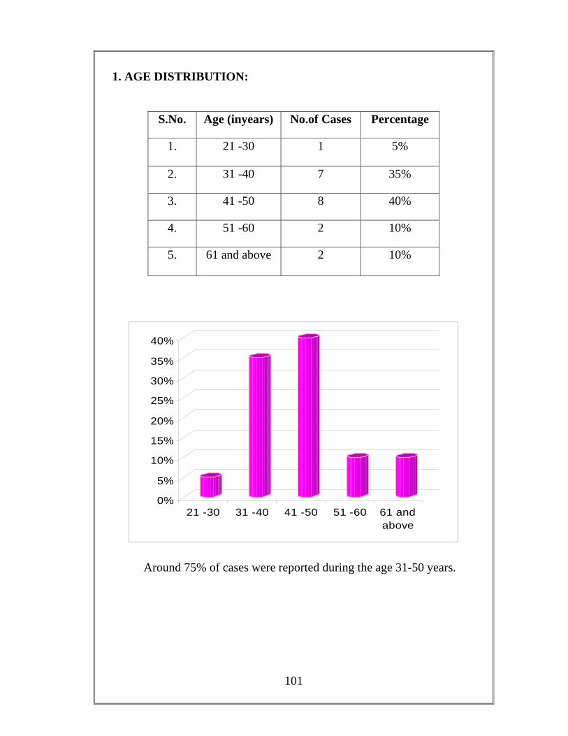
101
1. AGE DISTRIBUTION:
S.No. Age (inyears) No.of Cases Percentage
1. 21 -30 1 5%
2. 31 -40 7 35%
3. 41 -50 8 40%
4. 51 -60 2 10%
5. 61 and above 2 10%
0%
5%
10%
15%
20%
25%
30%
35%
40%
21 -30 31 -40 41 -50 51 -60 61 andabove
Around 75% of cases were reported during the age 31-50 years.
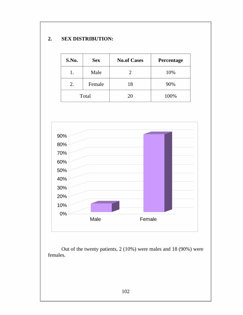
102
2. SEX DISTRIBUTION:
S.No. Sex No.of Cases Percentage
1. Male 2 10%
2. Female 18 90%
Total 20 100%
0%
10%
20%
30%
40%
50%
60%
70%
80%
90%
Male Female
Out of the twenty patients, 2 (10%) were males and 18 (90%) were
females.
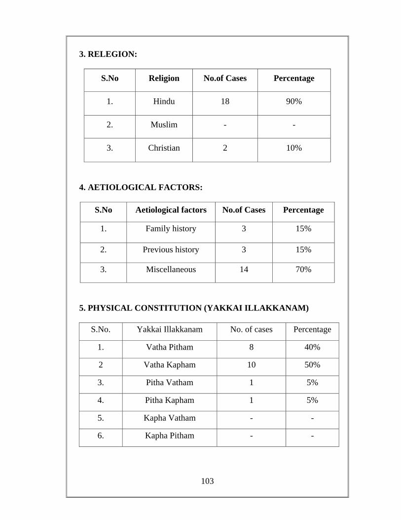
103
3. RELEGION:
S.No Religion No.of Cases Percentage
1. Hindu 18 90%
2. Muslim - -
3. Christian 2 10%
4. AETIOLOGICAL FACTORS:
S.No Aetiological factors No.of Cases Percentage
1. Family history 3 15%
2. Previous history 3 15%
3. Miscellaneous 14 70%
5. PHYSICAL CONSTITUTION (YAKKAI ILLAKKANAM)
S.No. Yakkai Illakkanam No. of cases Percentage
1. Vatha Pitham 8 40%
2 Vatha Kapham 10 50%
3. Pitha Vatham 1 5%
4. Pitha Kapham 1 5%
5. Kapha Vatham - -
6. Kapha Pitham - -
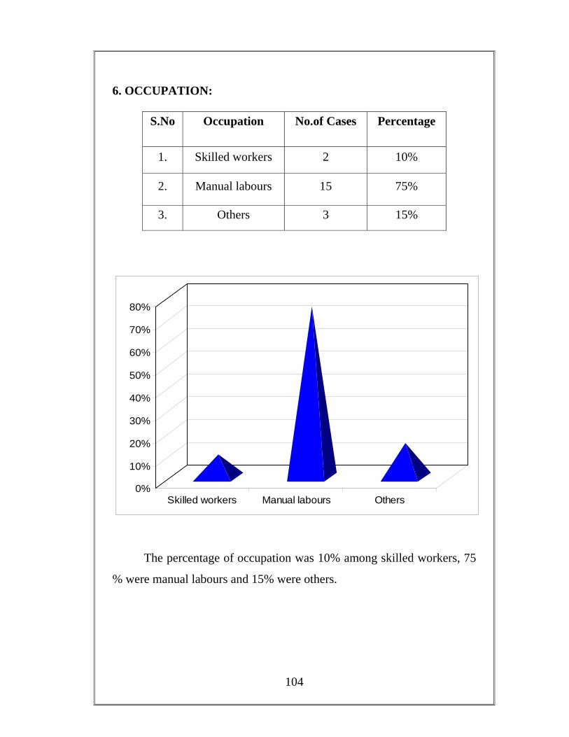
104
6. OCCUPATION:
S.No Occupation No.of Cases Percentage
1. Skilled workers 2 10%
2. Manual labours 15 75%
3. Others 3 15%
0%
10%
20%
30%
40%
50%
60%
70%
80%
Skilled workers Manual labours Others
The percentage of occupation was 10% among skilled workers, 75
% were manual labours and 15% were others.
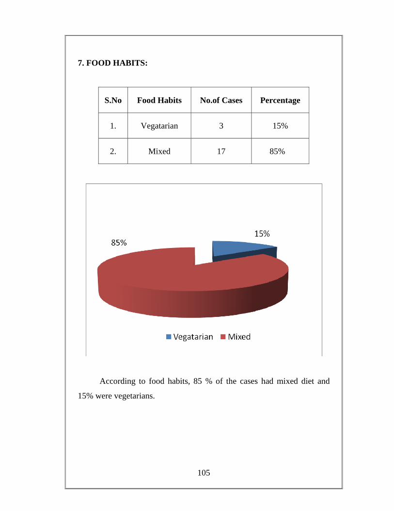
105
7. FOOD HABITS:
S.No Food Habits No.of Cases Percentage
1. Vegatarian 3 15%
2. Mixed 17 85%
According to food habits, 85 % of the cases had mixed diet and
15% were vegetarians.
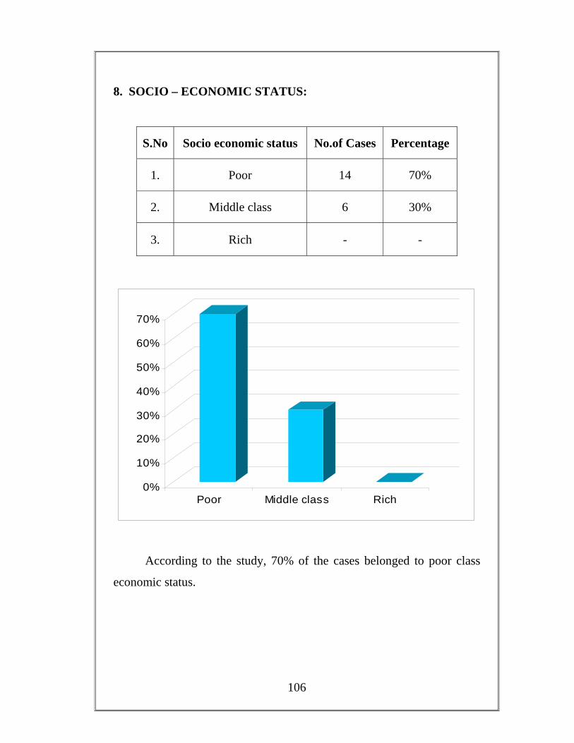
106
8. SOCIO – ECONOMIC STATUS:
S.No Socio economic status No.of Cases Percentage
1. Poor 14 70%
2. Middle class 6 30%
3. Rich - -
0%
10%
20%
30%
40%
50%
60%
70%
Poor Middle class Rich
According to the study, 70% of the cases belonged to poor class
economic status.
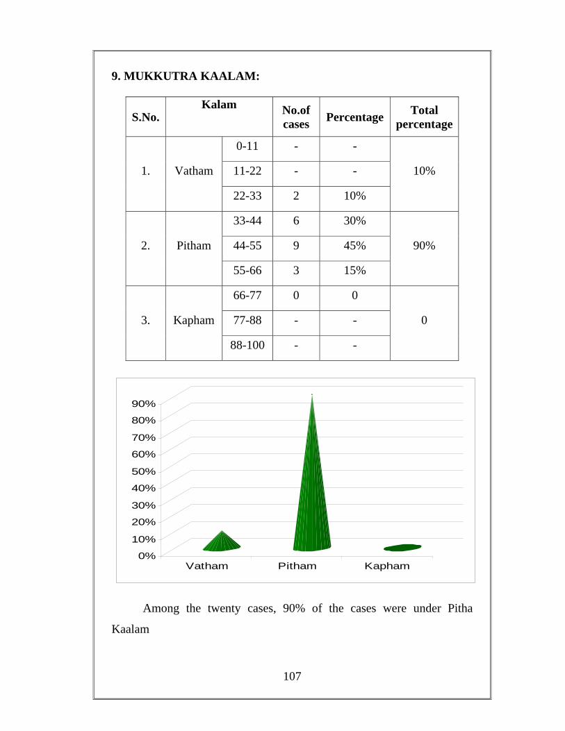
107
9. MUKKUTRA KAALAM:
S.No. Kalam No.of
cases Percentage Total percentage
1. Vatham
0-11 - -
10% 11-22 - -
22-33 2 10%
2. Pitham
33-44 6 30%
90% 44-55 9 45%
55-66 3 15%
3. Kapham
66-77 0 0
0 77-88 - -
88-100 - -
0%
10%
20%
30%
40%
50%
60%
70%
80%
90%
Vatham Pitham Kapham
Among the twenty cases, 90% of the cases were under Pitha
Kaalam
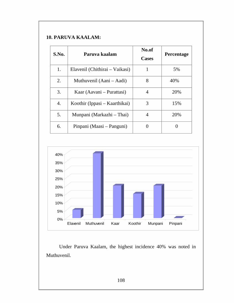
108
10. PARUVA KAALAM:
S.No. Paruva kaalam No.of
Cases Percentage
1. Elavenil (Chithirai – Vaikasi) 1 5%
2. Muthuvenil (Aani – Aadi) 8 40%
3. Kaar (Aavani – Purattasi) 4 20%
4. Koothir (Ippasi – Kaarthikai) 3 15%
5. Munpani (Markazhi – Thai) 4 20%
6. Pinpani (Maasi – Panguni) 0 0
0%
5%
10%
15%
20%
25%
30%
35%
40%
Elavenil Muthuvenil Kaar Koothir Munpani Pinpani
Under Paruva Kaalam, the highest incidence 40% was noted in
Muthuvenil.
109
11. THINAI
S.No. Thinai No.of Cases Percentage
1. Kurinji 1 5%
2. Mullai 2 10%
3. Marutham 15 75%
4. Neithal 2 10%
5. Paalai - -
All the twenty cased belonged to Marutha nilam
12. MUKKUTRA THEORY: a) DERANGEMENT IN THE TYPES OF VATHAM
S.No. Types of Vatham No.of Cases Percentage 1. Pranan - - 2. Abanan 15 75%
3. Viyanan 20 100%
4. Uthanan - -
5. Samanan 15 75%
6. Naagan - -
7. Koorman - -
8. Kirukaran - -
9. Devathathan 14 70%
10. Dananjeyan - -
Abanan was affected in 75% of Cases, Viyanan was also affected
in 100% of the cases, Samanan was affected in 75% of the cases and
Devathathan were affected in 70% of the cases.
110
b) DERANGEMENT IN TYPES OF PITHAM
S.No. Types of Pitham No.of Cases Percentage
1. Analam 15 75
2. Ranjakam 15 75
3. Saathakam 20 100
4. Prasakam 5 25
5. Aalosakam - -
Among the cases studied, Analam pitham was affected in 75% of
the cases and Saathaka pitham was affected in 100% of the cases and
Prasakam was affected in 25% of the cases and Ranjakam was affected in
75% of cases.
c) DERANGEMENT IN TYPES OF KABAM:
S.No. Types of Kabam No.of Cases Percentage
1. Avalambakam - -
2. Kilethakam 15 75
3. Pothakam - -
4. Tharpakam - -
5. Santhikam 20 100
111
13. UDALKATTUGAL
S.No. Udalkattugal No.of Cases Percentage
1. Saaram 20 100
2. Senneer 20 100
3. Oon 2 10
4. Kozhuppu 20 100
5. Enbu 20 100
6. Moolai 20 100
7. Sukkilam/ Suronitham - -
Among the 20 cases, Saaram,Seneer, Kozhupu, Enbu and Moolai
were found affected in 100% of the cases and in 10% of the cases Oon
was affected.
14. ENNVAGAI THERVUGAL:
S.No. Enn Vagai Thervugal No.of Cases
Percentage
1. Naa 5 25 2. Niram 10 50
3. Mozhi - -
4. Vizhi - -
5. Sparism 20 100
6. Naadi 20 100
7. Malam 15 75
8. Moothiram 2 10
Among the cases studied, Sparism and Naadi were affected in
100% of the cases. Malam is affected in 75% of the cases and Niram is
affected in 50% of the cases.
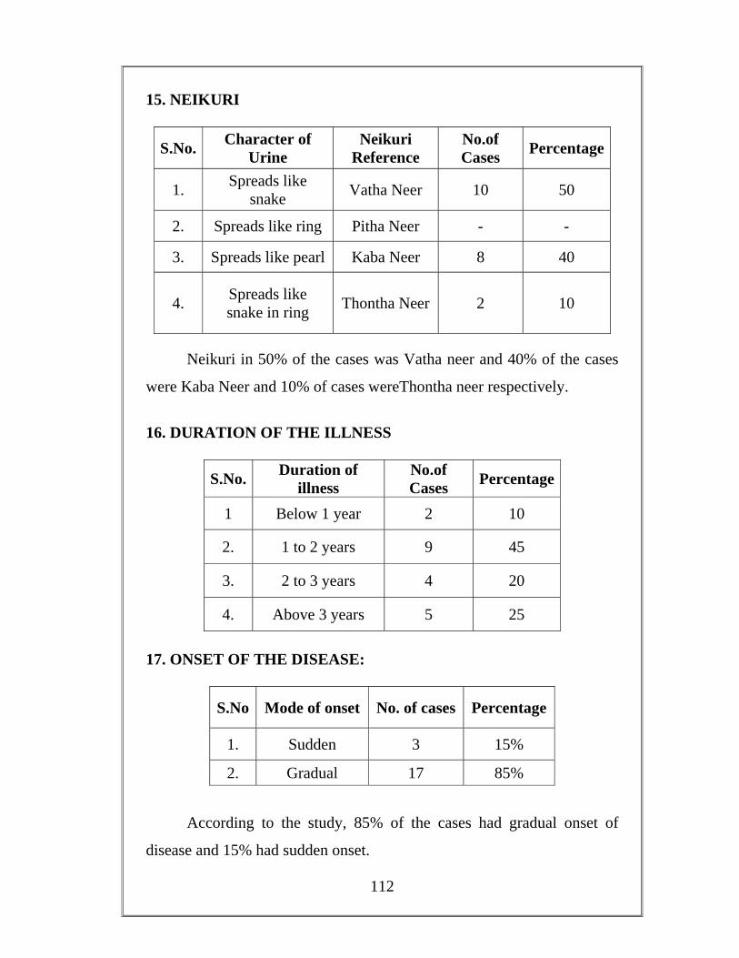
112
15. NEIKURI
S.No. Character of Urine
Neikuri Reference
No.of Cases Percentage
1. Spreads like snake Vatha Neer 10 50
2. Spreads like ring Pitha Neer - -
3. Spreads like pearl Kaba Neer 8 40
4. Spreads like snake in ring Thontha Neer 2 10
Neikuri in 50% of the cases was Vatha neer and 40% of the cases
were Kaba Neer and 10% of cases wereThontha neer respectively.
16. DURATION OF THE ILLNESS
S.No. Duration of illness
No.of Cases Percentage
1 Below 1 year 2 10
2. 1 to 2 years 9 45
3. 2 to 3 years 4 20
4. Above 3 years 5 25
17. ONSET OF THE DISEASE:
S.No Mode of onset No. of cases Percentage
1. Sudden 3 15%
2. Gradual 17 85%
According to the study, 85% of the cases had gradual onset of
disease and 15% had sudden onset.
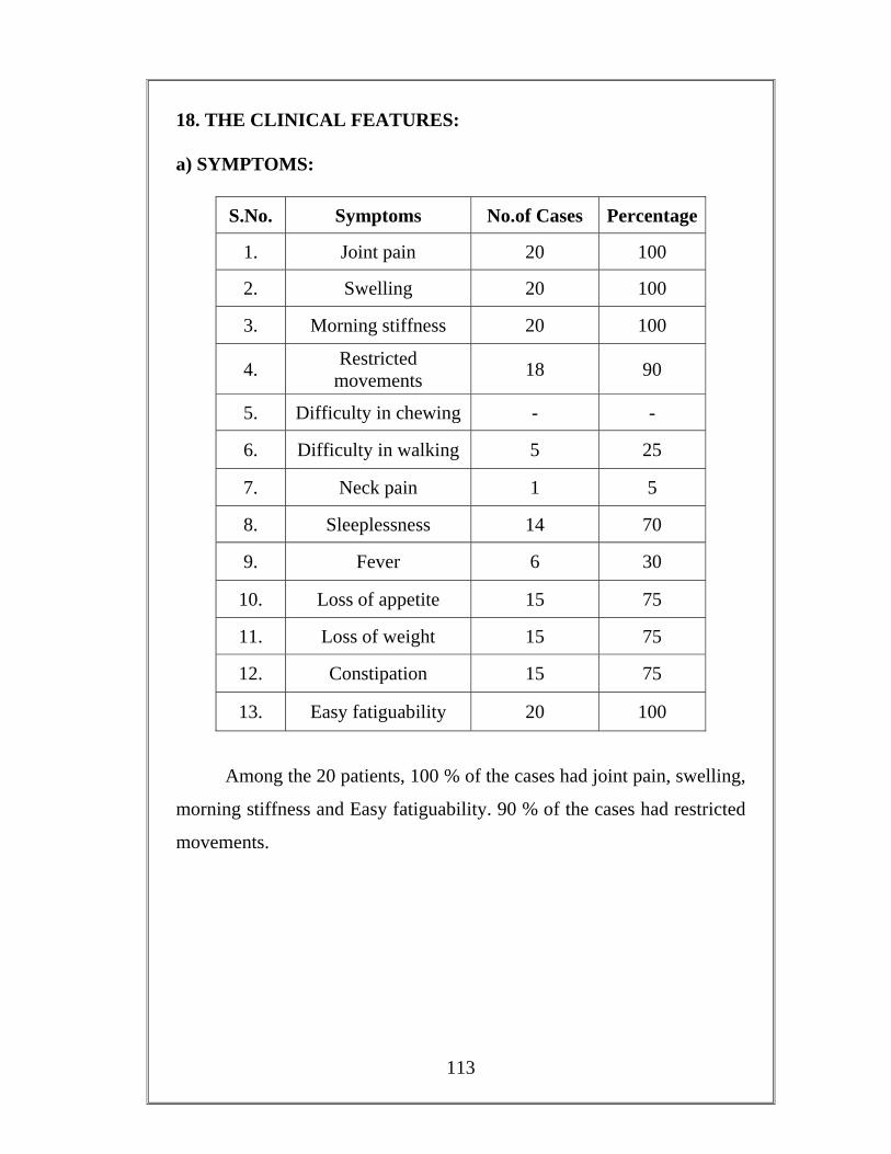
113
18. THE CLINICAL FEATURES: a) SYMPTOMS:
S.No. Symptoms No.of Cases Percentage
1. Joint pain 20 100
2. Swelling 20 100
3. Morning stiffness 20 100
4. Restricted movements 18 90
5. Difficulty in chewing - -
6. Difficulty in walking 5 25
7. Neck pain 1 5
8. Sleeplessness 14 70
9. Fever 6 30
10. Loss of appetite 15 75
11. Loss of weight 15 75
12. Constipation 15 75
13. Easy fatiguability 20 100
Among the 20 patients, 100 % of the cases had joint pain, swelling,
morning stiffness and Easy fatiguability. 90 % of the cases had restricted
movements.
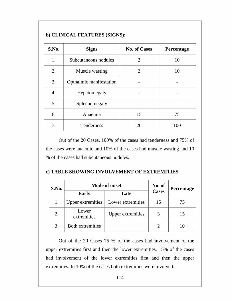
114
b) CLINICAL FEATURES (SIGNS):
S.No. Signs No. of Cases Percentage
1. Subcutaneous nodules 2 10
2. Muscle wasting 2 10
3. Opthalmic manifestation - -
4. Hepatomegaly - -
5. Spleenomegaly - -
6. Anaemia 15 75
7. Tenderness 20 100
Out of the 20 Cases, 100% of the cases had tenderness and 75% of
the cases were anaemic and 10% of the cases had muscle wasting and 10
% of the cases had subcutaneous nodules.
c) TABLE SHOWING INVOLVEMENT OF EXTREMITIES
S.No. Mode of onset No. of Cases Percentage
Early Late
1. Upper extremities Lower extremities 15 75
2. Lower extremities Upper extremities 3 15
3. Both extremities 2 10
Out of the 20 Cases 75 % of the cases had involvement of the
upper extremities first and then the lower extremities. 15% of the cases
had involvement of the lower extremities first and then the upper
extremities. In 10% of the cases both extremities were involved.
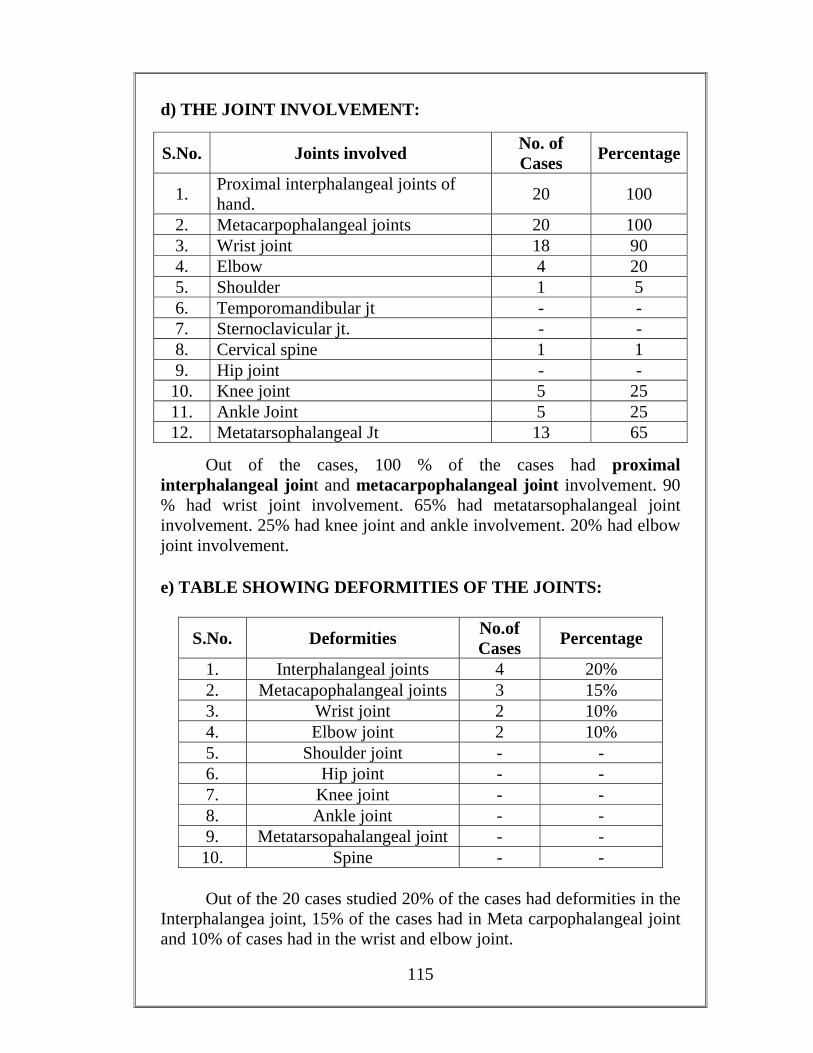
115
d) THE JOINT INVOLVEMENT:
S.No. Joints involved No. of Cases Percentage
1. Proximal interphalangeal joints of hand. 20 100
2. Metacarpophalangeal joints 20 100 3. Wrist joint 18 90 4. Elbow 4 20 5. Shoulder 1 5 6. Temporomandibular jt - - 7. Sternoclavicular jt. - - 8. Cervical spine 1 1 9. Hip joint - -
10. Knee joint 5 25 11. Ankle Joint 5 25 12. Metatarsophalangeal Jt 13 65
Out of the cases, 100 % of the cases had proximal interphalangeal joint and metacarpophalangeal joint involvement. 90 % had wrist joint involvement. 65% had metatarsophalangeal joint involvement. 25% had knee joint and ankle involvement. 20% had elbow joint involvement.
e) TABLE SHOWING DEFORMITIES OF THE JOINTS:
S.No. Deformities No.of Cases Percentage
1. Interphalangeal joints 4 20% 2. Metacapophalangeal joints 3 15% 3. Wrist joint 2 10% 4. Elbow joint 2 10% 5. Shoulder joint - - 6. Hip joint - - 7. Knee joint - - 8. Ankle joint - - 9. Metatarsopahalangeal joint - -
10. Spine - - Out of the 20 cases studied 20% of the cases had deformities in the
Interphalangea joint, 15% of the cases had in Meta carpophalangeal joint and 10% of cases had in the wrist and elbow joint.
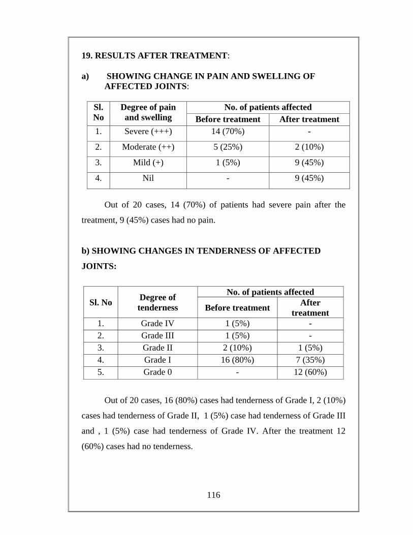
116
19. RESULTS AFTER TREATMENT: a) SHOWING CHANGE IN PAIN AND SWELLING OF AFFECTED JOINTS:
Sl. No
Degree of pain and swelling
No. of patients affected Before treatment After treatment
1. Severe (+++) 14 (70%) -
2. Moderate (++) 5 (25%) 2 (10%)
3. Mild (+) 1 (5%) 9 (45%)
4. Nil - 9 (45%)
Out of 20 cases, 14 (70%) of patients had severe pain after the
treatment, 9 (45%) cases had no pain.
b) SHOWING CHANGES IN TENDERNESS OF AFFECTED
JOINTS:
Sl. No Degree of tenderness
No. of patients affected
Before treatment After treatment
1. Grade IV 1 (5%) - 2. Grade III 1 (5%) - 3. Grade II 2 (10%) 1 (5%) 4. Grade I 16 (80%) 7 (35%) 5. Grade 0 - 12 (60%)
Out of 20 cases, 16 (80%) cases had tenderness of Grade I, 2 (10%)
cases had tenderness of Grade II, 1 (5%) case had tenderness of Grade III
and , 1 (5%) case had tenderness of Grade IV. After the treatment 12
(60%) cases had no tenderness.
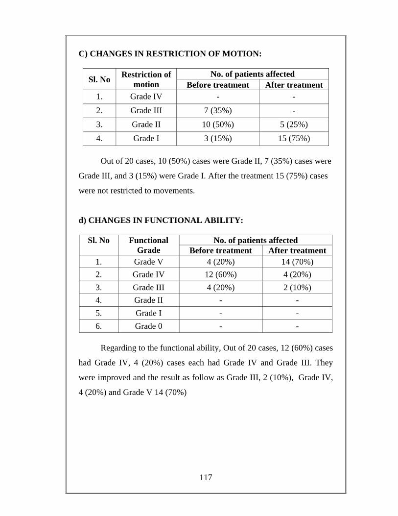
117
C) CHANGES IN RESTRICTION OF MOTION:
Sl. No Restriction of motion
No. of patients affected Before treatment After treatment
1. Grade IV - - 2. Grade III 7 (35%) - 3. Grade II 10 (50%) 5 (25%) 4. Grade I 3 (15%) 15 (75%)
Out of 20 cases, 10 (50%) cases were Grade II, 7 (35%) cases were
Grade III, and 3 (15%) were Grade I. After the treatment 15 (75%) cases
were not restricted to movements.
d) CHANGES IN FUNCTIONAL ABILITY:
Sl. No Functional Grade
No. of patients affected Before treatment After treatment
1. Grade V 4 (20%) 14 (70%) 2. Grade IV 12 (60%) 4 (20%) 3. Grade III 4 (20%) 2 (10%) 4. Grade II - - 5. Grade I - - 6. Grade 0 - -
Regarding to the functional ability, Out of 20 cases, 12 (60%) cases
had Grade IV, 4 (20%) cases each had Grade IV and Grade III. They
were improved and the result as follow as Grade III, 2 (10%), Grade IV,
4 (20%) and Grade V 14 (70%)
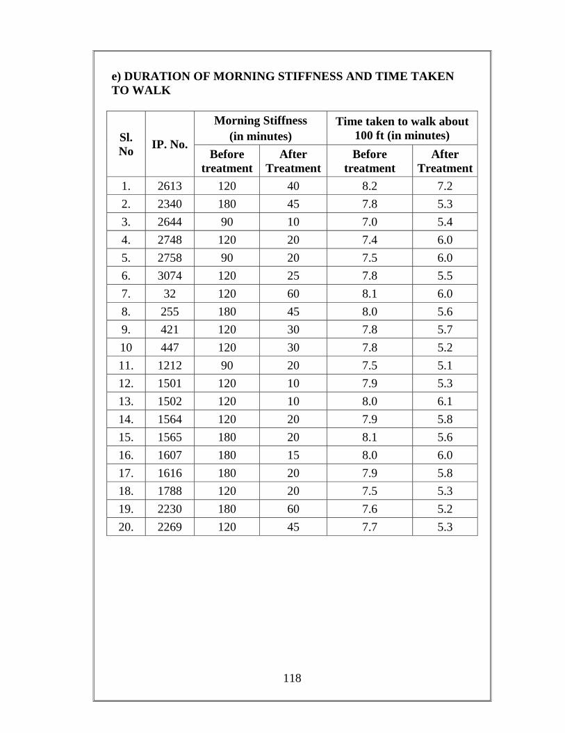
118
e) DURATION OF MORNING STIFFNESS AND TIME TAKEN TO WALK
Sl. No IP. No.
Morning Stiffness (in minutes)
Time taken to walk about 100 ft (in minutes)
Before treatment
After Treatment
Before treatment
After Treatment
1. 2613 120 40 8.2 7.2 2. 2340 180 45 7.8 5.3 3. 2644 90 10 7.0 5.4 4. 2748 120 20 7.4 6.0 5. 2758 90 20 7.5 6.0 6. 3074 120 25 7.8 5.5 7. 32 120 60 8.1 6.0 8. 255 180 45 8.0 5.6 9. 421 120 30 7.8 5.7 10 447 120 30 7.8 5.2 11. 1212 90 20 7.5 5.1 12. 1501 120 10 7.9 5.3 13. 1502 120 10 8.0 6.1 14. 1564 120 20 7.9 5.8 15. 1565 180 20 8.1 5.6 16. 1607 180 15 8.0 6.0 17. 1616 180 20 7.9 5.8 18. 1788 120 20 7.5 5.3 19. 2230 180 60 7.6 5.2 20. 2269 120 45 7.7 5.3
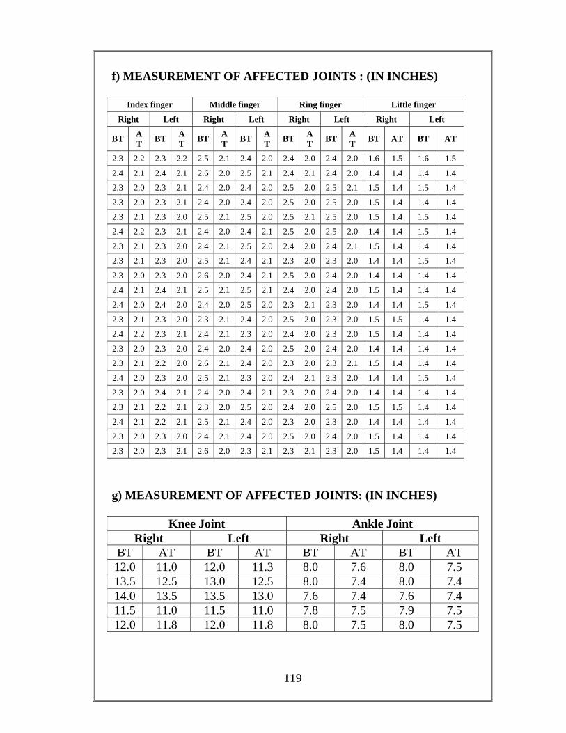
119
f) MEASUREMENT OF AFFECTED JOINTS : (IN INCHES)
Index finger Middle finger Ring finger Little finger
Right Left Right Left Right Left Right Left
BT AT BT A
T BT AT BT A
T BT AT BT A
T BT AT BT AT
2.3 2.2 2.3 2.2 2.5 2.1 2.4 2.0 2.4 2.0 2.4 2.0 1.6 1.5 1.6 1.5
2.4 2.1 2.4 2.1 2.6 2.0 2.5 2.1 2.4 2.1 2.4 2.0 1.4 1.4 1.4 1.4
2.3 2.0 2.3 2.1 2.4 2.0 2.4 2.0 2.5 2.0 2.5 2.1 1.5 1.4 1.5 1.4
2.3 2.0 2.3 2.1 2.4 2.0 2.4 2.0 2.5 2.0 2.5 2.0 1.5 1.4 1.4 1.4
2.3 2.1 2.3 2.0 2.5 2.1 2.5 2.0 2.5 2.1 2.5 2.0 1.5 1.4 1.5 1.4
2.4 2.2 2.3 2.1 2.4 2.0 2.4 2.1 2.5 2.0 2.5 2.0 1.4 1.4 1.5 1.4
2.3 2.1 2.3 2.0 2.4 2.1 2.5 2.0 2.4 2.0 2.4 2.1 1.5 1.4 1.4 1.4
2.3 2.1 2.3 2.0 2.5 2.1 2.4 2.1 2.3 2.0 2.3 2.0 1.4 1.4 1.5 1.4
2.3 2.0 2.3 2.0 2.6 2.0 2.4 2.1 2.5 2.0 2.4 2.0 1.4 1.4 1.4 1.4
2.4 2.1 2.4 2.1 2.5 2.1 2.5 2.1 2.4 2.0 2.4 2.0 1.5 1.4 1.4 1.4
2.4 2.0 2.4 2.0 2.4 2.0 2.5 2.0 2.3 2.1 2.3 2.0 1.4 1.4 1.5 1.4
2.3 2.1 2.3 2.0 2.3 2.1 2.4 2.0 2.5 2.0 2.3 2.0 1.5 1.5 1.4 1.4
2.4 2.2 2.3 2.1 2.4 2.1 2.3 2.0 2.4 2.0 2.3 2.0 1.5 1.4 1.4 1.4
2.3 2.0 2.3 2.0 2.4 2.0 2.4 2.0 2.5 2.0 2.4 2.0 1.4 1.4 1.4 1.4
2.3 2.1 2.2 2.0 2.6 2.1 2.4 2.0 2.3 2.0 2.3 2.1 1.5 1.4 1.4 1.4
2.4 2.0 2.3 2.0 2.5 2.1 2.3 2.0 2.4 2.1 2.3 2.0 1.4 1.4 1.5 1.4
2.3 2.0 2.4 2.1 2.4 2.0 2.4 2.1 2.3 2.0 2.4 2.0 1.4 1.4 1.4 1.4
2.3 2.1 2.2 2.1 2.3 2.0 2.5 2.0 2.4 2.0 2.5 2.0 1.5 1.5 1.4 1.4
2.4 2.1 2.2 2.1 2.5 2.1 2.4 2.0 2.3 2.0 2.3 2.0 1.4 1.4 1.4 1.4
2.3 2.0 2.3 2.0 2.4 2.1 2.4 2.0 2.5 2.0 2.4 2.0 1.5 1.4 1.4 1.4
2.3 2.0 2.3 2.1 2.6 2.0 2.3 2.1 2.3 2.1 2.3 2.0 1.5 1.4 1.4 1.4
g) MEASUREMENT OF AFFECTED JOINTS: (IN INCHES)
Knee Joint Ankle Joint Right Left Right Left
BT AT BT AT BT AT BT AT 12.0 11.0 12.0 11.3 8.0 7.6 8.0 7.5 13.5 12.5 13.0 12.5 8.0 7.4 8.0 7.4 14.0 13.5 13.5 13.0 7.6 7.4 7.6 7.4 11.5 11.0 11.5 11.0 7.8 7.5 7.9 7.5 12.0 11.8 12.0 11.8 8.0 7.5 8.0 7.5
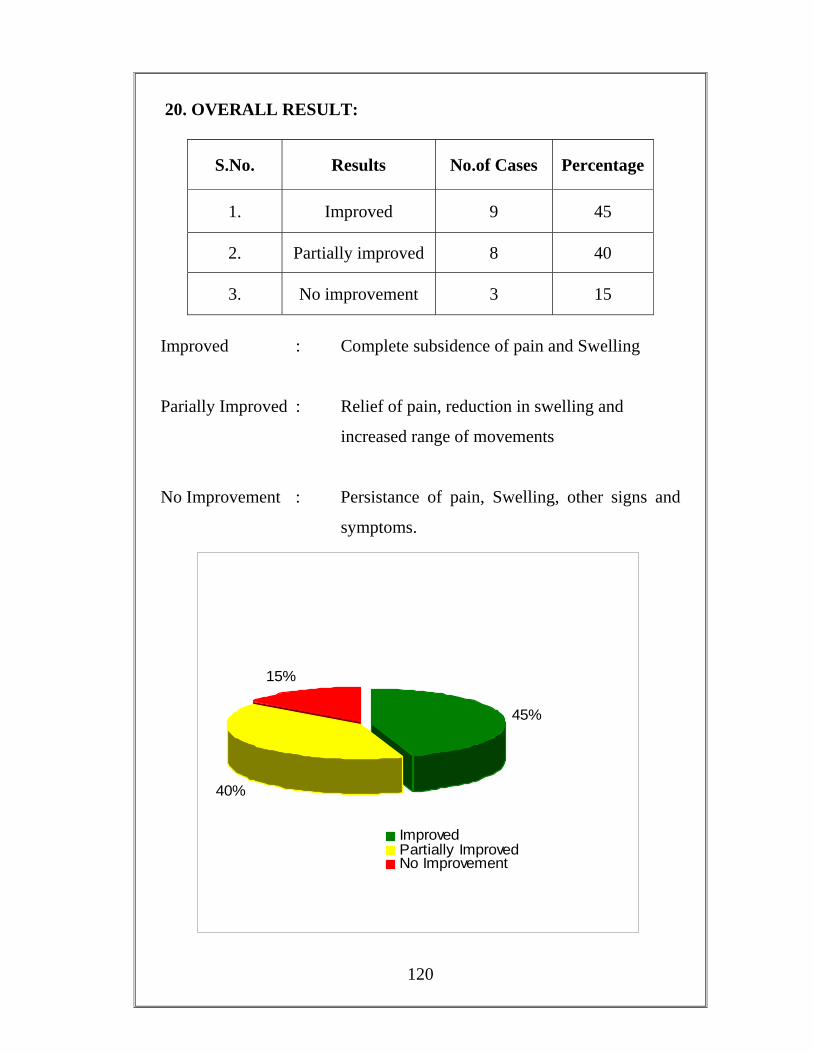
120
20. OVERALL RESULT:
S.No. Results No.of Cases Percentage
1. Improved 9 45
2. Partially improved 8 40
3. No improvement 3 15
Improved : Complete subsidence of pain and Swelling
Parially Improved : Relief of pain, reduction in swelling and
increased range of movements
No Improvement : Persistance of pain, Swelling, other signs and
symptoms.
45%
40%
15%
ImprovedPartially ImprovedNo Improvement
121
DISCUSSION
According to Siddha system of medicine diseases are classified in
to 4448 types. Among them Yudi vaidhaya chinthamani has classified
Vatha diseases into 80 types. Among the 80 types, 10 varieties are
concerned with joint diseases. One among them is Santhu vatham. In
Sabapathy scripts this disease has been compared with the disease Keel
vayu, explained in Siddha Maruthuvam text.
Valiazhal Keelvayu also mentioned by Agathiar in his Agathiar
Gunavagadam as Vatha Pitha Soolai (Vatha Azhal Soolai). The
symptoms of this disease correlate with Rheumatoid Arthritis, which is
the study topic of author. 28 cases of Valiazhal keelvayu were admitted in
the Post graduate Sirappu maruthuvam inpatient ward, GSMC
Palayamkottai. Out of these twenty cases were selected for this study and
also 30 patients were treated as outpatients.
For diagnostic purposes the parameter used in siddha aspect were
Porial Arithal, Pulanal Arithal, Vinaathal, Kanmendriyam,
Gnanendriyam, Uyir Thathukkal, Udal Thathukal, Ennvagai Thervugal,
Neerkuri, Neikuri etc. The modern parameters used were criteria for the
diagnosis of Rheumatoid arthritis, laboratory investigations, radiological
evaluation etc. All the patients were strictly advised to follow diet
restriction and peaceful lifestyle.
THE SEX DISTRIBUTION:
Out of the twenty patients 2 (10%) cases were males and 18 were
females (90%).
So Valiazhal Keelvayu is most commonly affected in female.
122
THE AGE DISTRIBUTION:
Among the 20 cases, 35% of the cases were between 31 - 40 years.
40% of the cases were between 41-50 years and 20% of the cases were
above 61 years. From the above data, the incidence of the disease seems
to be more in the age group of 31-50 years.
So the disease is more common in fourth and fifth decade as said in
modern medicine.
THE KAALAM:
According to Siddha concept maximum number of cases, 90%
were noted to be in Pitha Kalam which is between 33 to 66 years, which
co – ordinates with modern concept of the incidence in 4th – 5th decade.
This is why because of patients in Pitha Kaalam have risk factors
like smoking, alcohol, etc and they have very much stress in their life.
THE PARUVA KALAM:
Among the 20 cases, 40% of the cases were seemed to develop the
disease during Muthuvenil kalam (Aani-Aadi) 20% of the cases Kaar
Kaalam (Aavani-Puratasi) and 20% of the cases developed during
Munpani kalam (Markazhi – Thai).
So highest percentage was occurred in the Vali provoked Kaalam.
THE THINAI:
Among the 20 cases, 15 were belonged to Marutha nilam, two
cases each for Mullai and Neithal and one from Kurinchi. Even though as
123
per Siddha literatures, Marutha nilam is said to be free from diseases,
here the patients developed diseases due to alteration in their food habits
and routine activities.
THE SOCIO – ECONOMIC STATUS:
70% of the cases were under poor class; where as 30% of the cases
belonged to middle economic groups.
THE AETIOLOGICAL FACTORS:
15% of the cases had positive family history. And 15% had
previous history. Others were 70% cases (about 5 cases were found in
Chickungunia affected persons). Literary evidences shows that Valiazhal
keel vayu develops during winter and has genetic predisposition.
THE ONSET OF DISEASE:
The mode of onset was acute in 15% and gradual onset in 85% of
the cases. Thus the incidence of the disease with gradual onset is more, as
per literatures.
THE CLINICAL MANIFESTATIONS:
The patients with Valiazhal keelvayu (R.A.) present with articular
manifestations with or without extra articular manifestations were
noticed.
100 % of the cases had joint pain, swelling, easy fatigability and
morning stiffness.90% of the cases had restricted movements and 25% of
the cases had difficulty in walking. 5% of the cases had pain in the neck.
The joint in all the cases experienced early morning stiffness for bout 2-3
124
hours. Decreased movements of the joints noticed in all the cases which
were assessed by asking the patient to move the joint in particular
direction.
INVOLVEMENT OF UPPER AND LOWER EXTREMITIES:
The incidence of initial involvement of joints of the upper limb was
noticed in 75 % of the cases. 15 % of the cases, showed involvement of
the lower limbs while 10% of the cases showed involvement of upper and
lower limbs simultaneously.
INVOLVEMENT OF INDIVIDUAL JOINTS:
The proximal interphalangeal joint and meta carpophalangeal joints
were involved in 100% of the cases. 90% of the cases wrist joint. 25% of
the cases ankle joint. 25% of the cases the knee joint, 20% of the cases
the elbowjoint 5% of the cases cervical and 5% of the cases shoulder
and 65% of the cases metatarsophalangel joints were involved.
DEFORMITIES OF JOINTS:
10% of the cases had deformities in the wrist joint and elbow
joints, 15% of the cases metacarpophalangeal joint and 20% cases were
nterphalangeal joint.
ELICITATION OF EXTRA ARTICULAR MANIFESTATIONS:
Besides the articular symptoms certain signs which include the
examination of the enlargement of liver and spleen were carried out.
Examinations of respiratory system, cardio vascular system, were also
carried out. Opthalmic manifestations were also noted. Either
125
hepatomegaly or spleenomegaly was not present in any of the cases.
Likewise respiratory and cardio vascular manifestation and ophthalmic
manifestation were not present. 5% of case had rheumatoid nodule.
UYIR THATHUKKAL:
Uyir thathukkal include 3 vital humours namely
Vatham Pitham Kabam
The derangement in any of the above three causes diseases. They
were noticed in the 20 cases and are discussed below.
VATHAM:
Viyanan was affected in 100% of the cases because Viyanan is the
main constituent in flexion and extension movements. Abanan, Samanan
and Kiruharan were affected in 75% of the cases. Devathathan was
affected in 70% of the cases. When abanan is affected it produces
constipation, the disturbance in viyanan produce pain and restricted
movements of the joints and if samanan is affected there is loss of
appetite and when Devathathan is affected, insomnia causes.
PITHAM:
Anala pitham, Ranjagam were affected in 75% of the cases.
Saathaga pitham affected in 100% of the cases. Prasakam was affected in
25% of the cases.
Affected anala pitham produce loss of appetite. Ranjakam, when
affected produces decrease in haemoglobin and affected saathaga pitham
126
produce restriction of movements. So they are unable to carry out regular
works properly.
KABAM:
Santhigam was affected in 100% of the cases. Kilethagam was
affected in 75% of the cases.
Affected kilethagam produce loss of appetite and affected
santhigam produce joint pain, swelling and restriction of movements.
UDALKATTUGAL:
Saaram, Seeneer, Koluppu, Moolai and Enbu were affected in
100% of the cases which shows general debility and restricted
movements, joint pain and swelling.
Oon was affected in 10% of the cases which shows, slight
muscular wasting.
ENNVAGAI THERVGAL:
Naadi was observed in all the 20 cases. All the cases had thontha naadi.
Sparism was affected in all the cases which showed joint
tenderness, warmthness over the inflammed joints.
Malam was affected in 75% of cases and they had constipation.
Naa was affected in 25% of the cases and the tongue was coated
and was pale in colour due to anaemia.
Niram was affected in 50% of the cases.
127
NEIKURI:
50% of the cases showed Vaatha Neer and 40% of the cases had
Kapha Neer and 10% of the cases had Thontha Neer.
INVESTIGATIONS:
Besides Siddha based investigations since the disease has been
compared with Rheumatoid Arthritis, investigations meant for
Rheumatoid Arthritis were also done. Routine examination of blood,
urine and stools were done during the admission and the discharge.
Examination of urine and stools showed no abnormalities, blood
showed 75% of the cases showed decreased in haemoglobin percentage
and 100% of the cases had increased ESR with maximum of 100 mm in
one hour. Rheumatoid arthritis factor was positive in eighteen cases.
Blood sugar, Blood urea and Serum cholesterol were done in the
Biochemistry Department Govt. Siddha Medical College, Palayamkottai.
The values were found normal in all the cases.
The patients were also subjected to radiological evaluation which
revealed Rheumatoid Arthritis.
Hands with wrist joints AP view:-
Reduced interphalangeal joint space
Soft tissues swelling
Oesteophorosis
Peri articular erosion
Deformities
128
KNEE:
Oesteophorosis Narrowing of joints space
FEET AND ANKLE:
Synovial thickening Sclerosis Narrowing of joints space
THE MANAGEMENT:
Siddha system of medicine follows certain criterias in the
management of the diseases.
"Å¢§ÃºÉò¾¡ø Å¡¾õ ¾¡Øõ"
This shows that before treating Vatha diseases, laxatives or
purgatives can be administered to correct altered vatham.
Valiazhal Keelvayu comes under Vatha diseases; all the 20 cases
were given Vellai ennai- ½ to 1 ounce (14 to 18 ml) at morning on the
first day of admission. The next day the following medicines were
started.
The internal drug given was “ÌÁâ Á¡ò¾¢¨Ã" (Kumari
maathirai) one tablet two times daily with jaggeryand the external drug
given was “¦¿¡îº¢ò ¨¾Äõ” (Nochi Thylam). The patients were asked
to apply the oil over the swellings and in the areas of pain. Clinically the
drugs were free from side effects.

129
ABOUT THE TRIAL DRUGS:
KUMARI MATHIRAI:
¸üÈ¡¨Æ ¾ðÀ ţâÂò¨¾Ôõ, þÉ¢ôÒô À¢Ã¢¨ÅÔõ ¯¨¼ÂÐ.
"Å¢§ÃºÉò¾¡ø Å¡¾õ ¾¡Øõ" ±ýÈ ÜüÚôÀÊ ¦¿öôÒô À¨ºÔ¨¼Â
¦Åû¨Ç ±ñ¨½ ¦¸¡Îì¸ôÀðÎ, À¢ý þÉ¢ôÒô À¢Ã¢¨Å ¯¨¼Â
ÌÁâ Á¡ò¾¢¨Ã ¦¸¡Îì¸ôÀðÎûÇÐ. «¾É¡ø «Ð Á¢Ì¾¢ôÀð
¼ šԨŠ¾ýÉ¢¨Äô ÀÎòО¢ø Á¢¸îº¢Èó¾ ÁÕ󾡸 Å¢
Çí̸¢ÈÐ. §ÁÖõ ÁÕòЊӨȢø ±¾¢Õ¨Ã¢ý «ÊôÀ¨¼Â¢ø
¦ºÂøÀθ¢ÈÐ. ±É «È¢ÂÄ¡õ.
Kumari Maathirai contains, Kadukkai, in which the main
ingredient is ferrous ion which is easily absorbable and helps to increases
the Hb level in this patient. It has significant acute and chronic, anti
inflammatory, analgesic, antipyretic effects, so the trial drugs reduces the
pain and swelling and also treat the anemia in Valiazhal Keelvayu patient.
NOCHI THYLAM:
Its main ingredient Nochi had analgesic and anti inflammatory
properties. So external applications of this reduce the pain, swelling,
stiffness in affected joints and increase the range of movements.
Hot water fomentation and fomentation with Nochi leaves were
advised to all the patients after the application of the oil. In the initial
stage all the patients were advised to take bed rest. After the severity of
symptoms had reduced, they were advised to do some exercises.
130
The patients were observed carefully when they were performing
the exercises. Care was taken for patients to perform exercise slowly
without any strain. Sava Asana is advised to all the patients. Since the
Valiazhal Keelvayu affects the patients physically and psychologically,
this Asana gives good sleep and relives the mental stress.
ASSESSMENT OF THE EFFECTS OF MANAGEMENT:
All the 20 cases were treated with “ÌÁâ Á¡ò¾¢¨Ã" (Kumari
Mathirai) internally and “¦¿¡îº¢ò ¨¾Äõ” (Nochi Thylam)
externally. Thokkanam, Otradam and Asanas were also advised. The
results of treatment were assessed on the basis of reduction in symptoms
such as pain, joint swelling, and a sense of well being. The patients were
treated for about 45 days. At the end of the treatment the results were
discussed as follows: Improved, Partially improved and no improvement.
45% of the cases improved well.
40% of the cases were partially improved and
15% of the cases showed no improvement.
At the end of the treatment there was a rise in Hb level and
decreased in ESR level in all the cases. After discharge the patients were
advised to attend the post graduate department of Sirappu maruthuvam
O.P. for further follow up.
During the course of treatment diet restrictions was strictly advised.
Patients were advised to take preventive measures like avoiding exposure
to chill weather. They were advised to take bath in warm water.
131
SUMMARY
Valiazhal Keelvayu is very well correlate to Rheumatoid arthritis
in modern medicine. It is one among a common problem in clinical
practice. It has received an international attention in search of finding out
of a new drug for the ailment of the sufferers.
In Valiazhal Keelvayu, usually small joints are affected in
symmetrical manner. Joint pain, Swelling, tenderness which may be
aggravated on motion. Morning stiffness of 2 to 3 hours duration, easy
fatigability, anaemia, occasional fever are the common manifestation of
the disease.
The study on Valiazhal keelvayu (R.A) was with trial drugs.
1. “ÌÁâ Á¡ò¾¢¨Ã" (Kumari Maathirai) 1 tablet with
jaggery B.D. Internal.
2. “¦¿¡îº¢ò ¨¾Äõ” (Nochi Thylam) External.
Twenty cases of both sexes and of various age groups were chosen
and the study was carried out. 90% Valiazhal Keelvayu patients were
female.
90% of the patients belonged to Pitha kaalam. 40% of the cases
were seemed to develop the disease during Muthuvenil kaalam (Aani-
Aadi). Most of the cases (75%) came from Marutha Nilam. Seventy five
percent of cases belonged to poor class families. Considering aetiology
15% of the cases had a positive family history.

132
85% of the cases had a gradual on set of disease. All the cases had
joint pain, swelling, morning stiffness. 90% of the cases had restricted
movements. 75% of the cases had initial involvement of joints of the
upper limb.
In all the cases proximal interphalangeal joints and Meta
carpophalangeal joints were involved. 20% of the cases had deformities
in their interphanlangeal, 15% metacarpophalangeal joints and 10% wrist
and elbow joint.
Regarding Uuyir thathukkal, viyanan was affected in 100% of the
cases. Abanan, Samanan were affected in 75% of cases. Aanala pitham,
Ranjakam were affected 75% of the cases. Sathaka pitham was affected
in 100% of cases. And santhigam were affected in all the cases,
kilethagam was affected in 75% of cases.
Saaram, Seeneer, Kollupu and enbu were affected in all the cases.
Regarding Enn Vagai Thervugal, sparism. Naadi were affected in all the
cases. Neerkuri was found straw in colour. Neikuri indicated Vatha neer
in 50% of the cases and Kaba Neer in 40 % of cases. Malam was affected
in 75% of cases. 60% of the cases had moderate restriction but with an
ability to perform normal activities.
R.A. factor was positive in eighteen cases. 75% of the cases
showed a decrease in haemoglobin percentage. E.S.R. was raised in all
the cases. The trial medicines were given to 20 Cases with Valiazhal keel
vayu.
The internal drug was “ÌÁâ Á¡ò¾¢¨Ã ” (Kumari Maathirai)
in the dose of 1 tablet in jaggery twice a day after food.
133
The external drug was “¦¿¡îº¢ò ¨¾Äõ” (Nochi Thylam)
which was applied externally to the affected joints.
Hot water and Nochi leaf fomentation was given after the
application of external drug. All the patients were advised to follow the
balanced dietary habit. All the patients were also advised to follow the
preventive measures like avoiding exposure to cold weather and advised
to take bath in warm water. The observation made during this study,
showed that the trial medicines were clinically effective.
The potency of the drugs were studied by pharmacological and
biochemical analysis. It shows that the drugs have significant, acute and
chronic anti inflammatory, anti pyretic and analgesic actions.
134
CONCLUSION
In this study, results were found to be good in 45% of the cases.
No adverse effects were noticed during the treatment period.
Further follow up of these patients showed good recovery and fine
improvement in the general well beings as they could carry out their day
to day activities.
The preparations of both medicines are simple and expiry is
upto one year. So they can be stored and used.
The trial medicine has significant acute and chronic anti
inflammatory, analgesic and antipyretic actions.
Early diagnosis and prompt management prevents the disabilities
and deformities resulting from Rheumatoid arthritis.
So it is concluded that the treatment with “ÌÁâ Á¡ò¾¢¨Ã”
(Kumari Mathirai) “¦¿¡îº¢ò ¨¾Äõ” (Nochi Thylam) is good in the
view of efficacy and safety.
135
PREPARATION OF TRIAL MEDICINES
1. INTERNAL MEDICINE – KUMARI MATHIRAI
(Å¡¾ò¾¢üÌ ÌÁâ Á¡ò¾¢¨Ã)
" ŢǧŠÃ⾸¢ò §¾¡ø §ÅÄõ À¢º¢É¢ Âǧš÷ ¿¢¨È¨Ãò¾ ×ñ¨¼ - ÌǦ¿ö «Áâ¢Öñ Å¡¾À¢ò¾ ¨Á¢¨Å§À¡ ¦Áö§Â ÌÁâ ¢Ãò¾ò¾¢ü Ü "
- §¾¨ÃÂ÷ ¦ÅñÀ¡ (Àì¸õ 131
)
Å¢øŧÅ÷, ¸Î측öò§¾¡ø, §ÅÄÀ¢º¢ý ãý¨ÈÔõ ´§Ã «Ç×¼ý
¿¢ÚòÐ ±ÎòÐ ¸üÈ¡¨Çî º¡üȢĨÃòÐ ¯Õñ¨¼ ¦ºöÐ ¯ÄèÅò
Ð ¦ÅøÄò¾¢Öñ½ Å¡¾Óõ, ¦¿ö¢Öñ½ô À¢ò¾Óõ, «Áâ¢Öñ½
§ºòÐÁÓõ ¿£íÌõ ±ñÀ¾¡Ìõ.
§ºÕõ ºÃì̸û:
Å¢øŧÅ÷ - 100 ¸¢Ã¡õ
¸Î측öò§¾¡ø - 100 ¸¢Ã¡õ §ÅÄõÀ¢º¢ý - 100 ¸¢Ã¡õ ¸üÈ¡¨Æº¡Ú - 700 ml.
¦ºöÓ¨È:
Å¢øŧÅ÷, ¸Î측öò§¾¡ø, §ÅÄõÀ¢º¢ý ãý¨ÈÔõ ¾É¢ò¾É¢§
 ¿ýÌ −ÊòÐ Ýýõ ¦ºöÐ ±ÎòÐì ¦¸¡ñÎ ¸øÅò¾¢Ä¢ðÎ ¸üÈ¡
¨Çîº¡Ú º¢È¢Ð º¢È¢¾¡¸ Å¢ðÎ ¿ýÌ «¨ÃòÐ Á¡ò¾¢¨Ã ¯ÕðÎõ ÀìÌÅò¾¢ø ±ÎòÐ ¯Õñ¨¼Â¡¸ (500mg) ¯ÕðÊ ¸
¡Â¨ÅòÐ ±ÎòÐ즸¡û×õ.
«Ç×: ´Õ Á¡ò¾¢¨Ã −ÃñÎ §Å¨Ç.
«ÛÀ¡Éõ: ¦ÅøÄõ
¬¾¡Ãáø: §¾¨ÃÂ÷ ¦ÅñÀ¡, (Àì¸õ 131) ¬º¢Ã¢Â÷: ¼¡ì¼÷ ¬÷. ¾¢Â¡¸Ã¡ºý
136
II. EXTERNAL MEDICINE – NOCHI THYLAM
(¦¿¡îº¢ò ¨¾Äõ)
§ºÕõ ºÃì̸û:
¦¿¡îº¢î º¡Ú - 2.6 Ä¢ð¼÷
¿ø¦Äñ¦½ö - 840 Á¢.Ä¢ð¼÷
±Õì¸õ §Å÷ - 12.6 ¸¢Ã¡õ
¦Åû¨Çì §¸¡‰¼õ - 12.6 ¸¢Ã¡õ
¾¸¨Ã Å¢¨¾ - 12.6 ¸¢Ã¡õ
º¾Ìô¨À - 12.6 ¸¢Ã¡õ
¦ºöÓ¨È:
¦¿¡îº¢ þ¨Ä¨Â ¸øÅò¾¢Ä¢ðÎ þÊòÐ º¡Ú ±ÎòÐì ¦¸¡ûÇ×õ.
±Õì¸õ §Å÷, ¦Åû¨Çì §¸¡‰¼õ, ¾¸¨Ã Å¢¨¾, º¾Ìô¨À §À
¡ýÈÅü¨È þÊòÐ ¦¿¡îº º¡Ú, ¿ø¦Äñ¦½ö −ÅüÚ¼ý ¸ÄóÐ «Îô§
ÀüÈ¢ ±Ã¢òÐ ¨¾Äõ ¸¡öîº ±ÎòÐì ¦¸¡ûÇ×õ.
¾£Õõ §¿¡ö: Å¡Ô×¼ý ÜÊ ÅÄ¢, Å£ì¸õ ¿£íÌõ.
¬¾¡Ãõ:
¨Åò¾¢Â ¾¢ÃðÎ - ºÃ§À¡ƒ¢ (Àì¸õ 5)
¬º¢Ã¢Â÷: ¼¡ì¼÷. ±Š. ¦Åí¸¼Ã¡ƒý
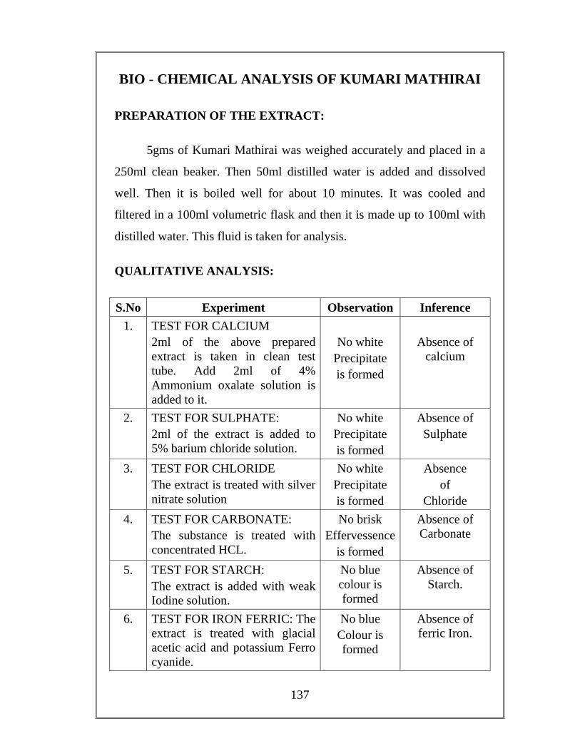
137
BIO - CHEMICAL ANALYSIS OF KUMARI MATHIRAI
PREPARATION OF THE EXTRACT:
5gms of Kumari Mathirai was weighed accurately and placed in a
250ml clean beaker. Then 50ml distilled water is added and dissolved
well. Then it is boiled well for about 10 minutes. It was cooled and
filtered in a 100ml volumetric flask and then it is made up to 100ml with
distilled water. This fluid is taken for analysis.
QUALITATIVE ANALYSIS:
S.No Experiment Observation Inference 1. TEST FOR CALCIUM
2ml of the above prepared extract is taken in clean test tube. Add 2ml of 4% Ammonium oxalate solution is added to it.
No white
Precipitate is formed
Absence of
calcium
2. TEST FOR SULPHATE: 2ml of the extract is added to 5% barium chloride solution.
No white Precipitate is formed
Absence of Sulphate
3. TEST FOR CHLORIDE The extract is treated with silver nitrate solution
No white Precipitate is formed
Absence of
Chloride 4. TEST FOR CARBONATE:
The substance is treated with concentrated HCL.
No brisk Effervessence
is formed
Absence of Carbonate
5. TEST FOR STARCH: The extract is added with weak Iodine solution.
No blue colour is formed
Absence of Starch.
6. TEST FOR IRON FERRIC: The extract is treated with glacial acetic acid and potassium Ferro cyanide.
No blue Colour is formed
Absence of ferric Iron.
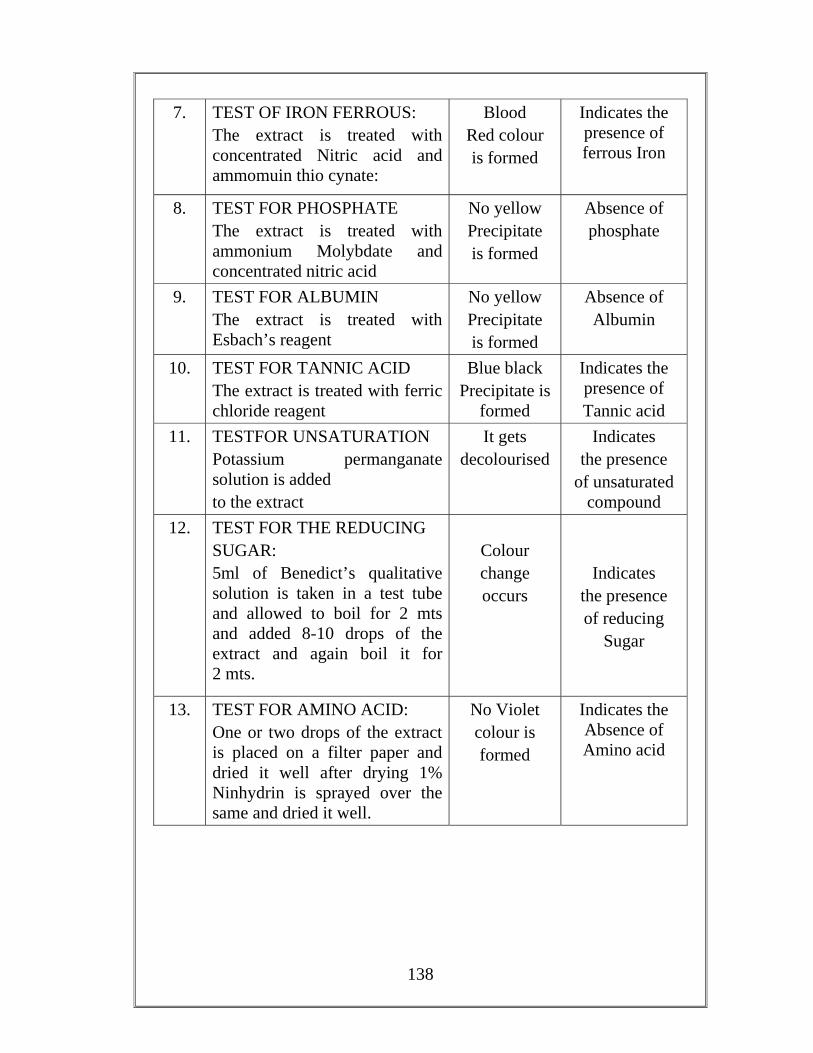
138
7. TEST OF IRON FERROUS: The extract is treated with concentrated Nitric acid and ammomuin thio cynate:
Blood Red colour is formed
Indicates the presence of ferrous Iron
8. TEST FOR PHOSPHATE The extract is treated with ammonium Molybdate and concentrated nitric acid
No yellow Precipitate is formed
Absence of phosphate
9. TEST FOR ALBUMIN The extract is treated with Esbach’s reagent
No yellow Precipitate is formed
Absence of Albumin
10. TEST FOR TANNIC ACID
The extract is treated with ferric chloride reagent
Blue black Precipitate is
formed
Indicates the presence of Tannic acid
11. TESTFOR UNSATURATION Potassium permanganate solution is added to the extract
It gets decolourised
Indicates the presence
of unsaturated compound
12. TEST FOR THE REDUCING SUGAR: 5ml of Benedict’s qualitative solution is taken in a test tube and allowed to boil for 2 mts and added 8-10 drops of the extract and again boil it for 2 mts.
Colour change occurs
Indicates the presence of reducing
Sugar
13. TEST FOR AMINO ACID: One or two drops of the extract is placed on a filter paper and dried it well after drying 1% Ninhydrin is sprayed over the same and dried it well.
No Violet colour is formed
Indicates the Absence of Amino acid
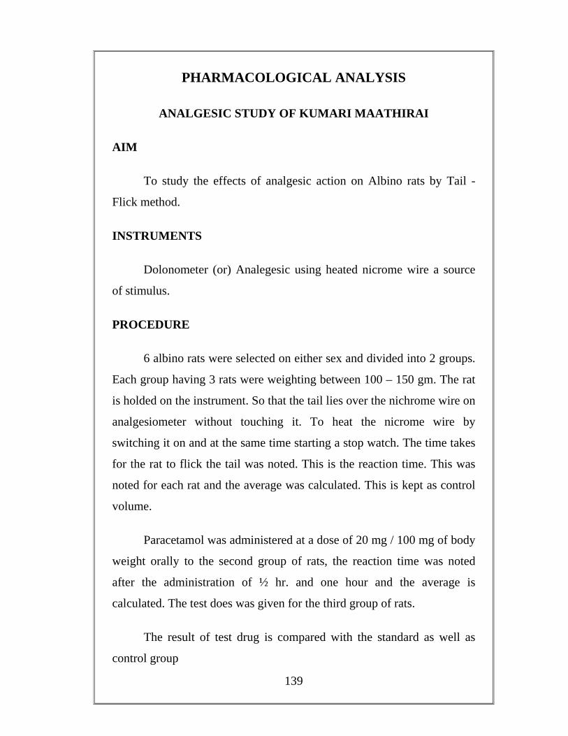
139
PHARMACOLOGICAL ANALYSIS
ANALGESIC STUDY OF KUMARI MAATHIRAI
AIM
To study the effects of analgesic action on Albino rats by Tail -
Flick method.
INSTRUMENTS
Dolonometer (or) Analegesic using heated nicrome wire a source
of stimulus.
PROCEDURE
6 albino rats were selected on either sex and divided into 2 groups.
Each group having 3 rats were weighting between 100 – 150 gm. The rat
is holded on the instrument. So that the tail lies over the nichrome wire on
analgesiometer without touching it. To heat the nicrome wire by
switching it on and at the same time starting a stop watch. The time takes
for the rat to flick the tail was noted. This is the reaction time. This was
noted for each rat and the average was calculated. This is kept as control
volume.
Paracetamol was administered at a dose of 20 mg / 100 mg of body
weight orally to the second group of rats, the reaction time was noted
after the administration of ½ hr. and one hour and the average is
calculated. The test does was given for the third group of rats.
The result of test drug is compared with the standard as well as
control group
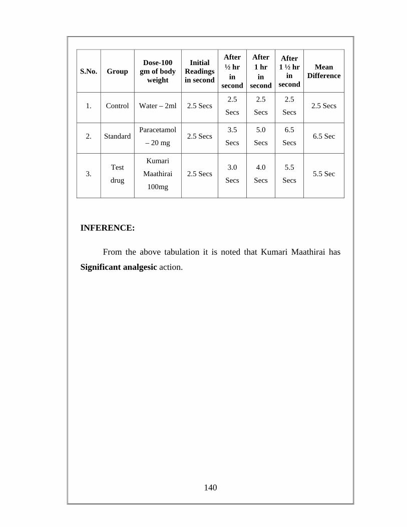
140
S.No. Group Dose-100
gm of body weight
Initial Readings in second
After ½ hr
in second
After 1 hr in
second
After 1 ½ hr
in second
Mean Difference
1. Control Water – 2ml 2.5 Secs 2.5
Secs
2.5
Secs
2.5
Secs 2.5 Secs
2. Standard Paracetamol
– 20 mg 2.5 Secs
3.5
Secs
5.0
Secs
6.5
Secs 6.5 Sec
3. Test
drug
Kumari
Maathirai
100mg
2.5 Secs 3.0
Secs
4.0
Secs
5.5
Secs 5.5 Sec
INFERENCE:
From the above tabulation it is noted that Kumari Maathirai has
Significant analgesic action.
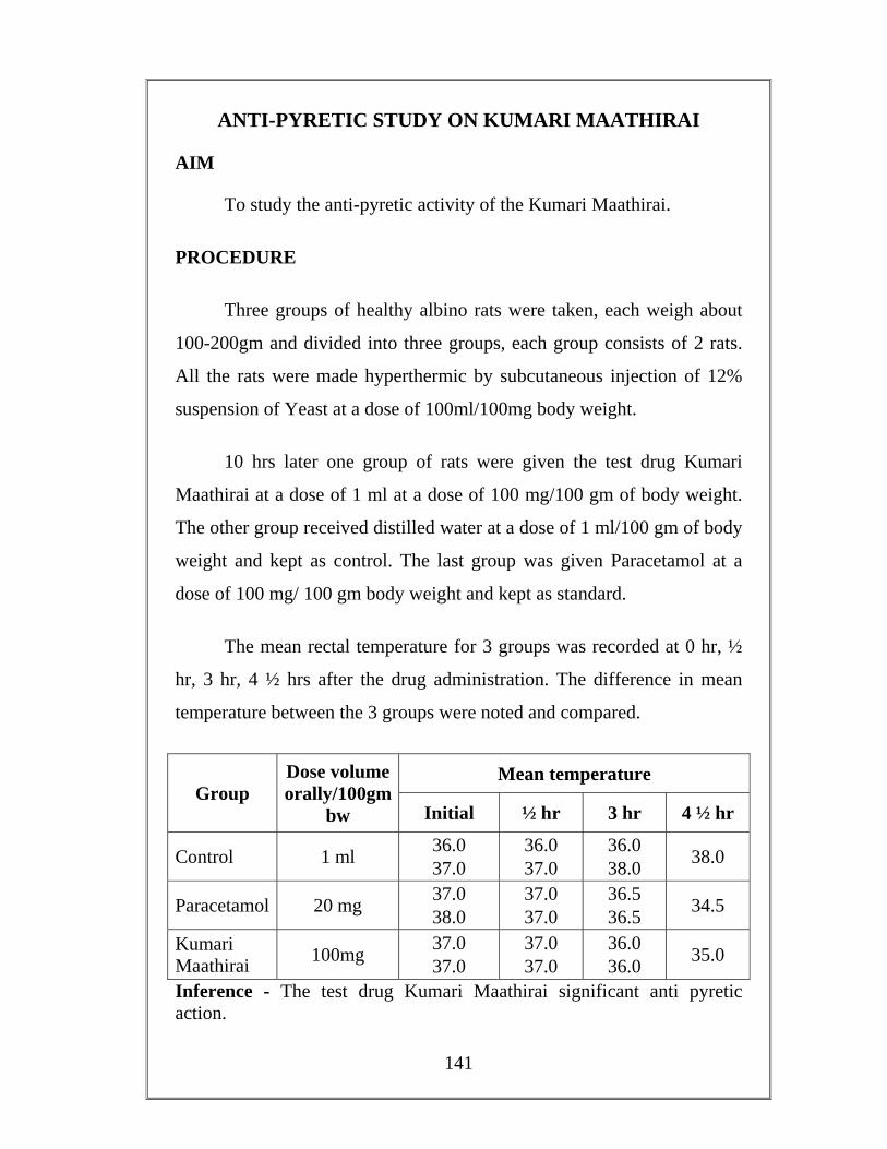
141
ANTI-PYRETIC STUDY ON KUMARI MAATHIRAI
AIM
To study the anti-pyretic activity of the Kumari Maathirai.
PROCEDURE
Three groups of healthy albino rats were taken, each weigh about
100-200gm and divided into three groups, each group consists of 2 rats.
All the rats were made hyperthermic by subcutaneous injection of 12%
suspension of Yeast at a dose of 100ml/100mg body weight.
10 hrs later one group of rats were given the test drug Kumari
Maathirai at a dose of 1 ml at a dose of 100 mg/100 gm of body weight.
The other group received distilled water at a dose of 1 ml/100 gm of body
weight and kept as control. The last group was given Paracetamol at a
dose of 100 mg/ 100 gm body weight and kept as standard.
The mean rectal temperature for 3 groups was recorded at 0 hr, ½
hr, 3 hr, 4 ½ hrs after the drug administration. The difference in mean
temperature between the 3 groups were noted and compared.
Group Dose volume orally/100gm
bw
Mean temperature
Initial ½ hr 3 hr 4 ½ hr
Control 1 ml 36.0 37.0
36.0 37.0
36.0 38.0 38.0
Paracetamol 20 mg 37.0 38.0
37.0 37.0
36.5 36.5 34.5
Kumari Maathirai 100mg 37.0
37.0 37.0 37.0
36.0 36.0 35.0
Inference - The test drug Kumari Maathirai significant anti pyretic action.
142
ACUTE ANTI-INFLAMMATORY ACTIVITY IN RATS BY
HIND-PAW METHOD
AIM
To demonstrate the acute anti-inflammatory activity of Kumari
Maathirai in albino rats by Hind-paw method.
PROCEDURE:
Nine Albino rats weighing 100-150gm were taken and divided into
three groups and each group consisting three rats.
First group was kept as control and received water. Second group
received Ibuprofen at a dose of 20mg/100gm-body weight. Third group
animals received Kumari Maathirai suspension at a dose of
200mg/100gm-body weight.
Before administration of drugs, the Hind-paw volume of all the rats
was measured. This was done by dipping the Hind-paw upto the tibio-
tarsal junction in mercury plethysmograph. Soon after the measurement
at the drugs were administered. One hour after the administration of
drugs a sub-cutaneous injection of 0.1ml of 1%/W/V of carrageenin in
water was made into planter surface of both the Hind-paw of each rat.
Three hours after carrageenin injection, the Hind-paw volume was
measured once again. Difference between the initial and final value were
noted and compared.
This method is more suitable method for studying anti-
inflammatory activity in acute inflammation.

143
THE EFFECT OF KUMARI MAATHIRAI IN ACUTE ANTI-
INFLAMMATORY ACTIVITIES.
Group Dose volume orally
Initial reading
Final reading
Mean difference
Percentage Inflammation
Percentage Inhibition
Control Water 2 ml 0.55 1.4 0.85 100 -
Standard Ibuprofen
20mg/100gm 0.55 0.85 0.3 35.2 64.8
Test
drug 200mg/100gm 0.7 0.95 0.25 29.4 70.6
RESULT:
The drug Kumari Maathirai has Significant acute – anti
inflammatory action.

144
CHRONIC ANTI-INFLAMMATORY STUDY BY COTTON-
PELLETS GRANULOMA METHOD
DRUG
Kumari Maathirai (Internal use)
AIM
To study the chronic anti-inflammatory activity of the drug in
albino rats by cotton pellets implantation (granuloma) method.
PROCEDURE:
Cotton pellets each weighing long was prepared and sterilized in an
autoclave for about one hour under 15 lbs atmosphere pressure. Nine
Albino rats weighing between 100-200gm were selected and were
divided into 3 groups. Each rat was anaesthetized with ether and cotton
pellets were implanted subcutaneously in the groin, two in each side.
From the day of implantation, one group of animals received
Kumari Maathirai. at a dose of 200mg/100gm of body weight.
On the eighth day the rats were sacrificed and the pellets were
removed weighed. Then they were put in an incubator at 60ºC-80ºC and
then weighed.
The concordant weight was noted for all groups and compared.
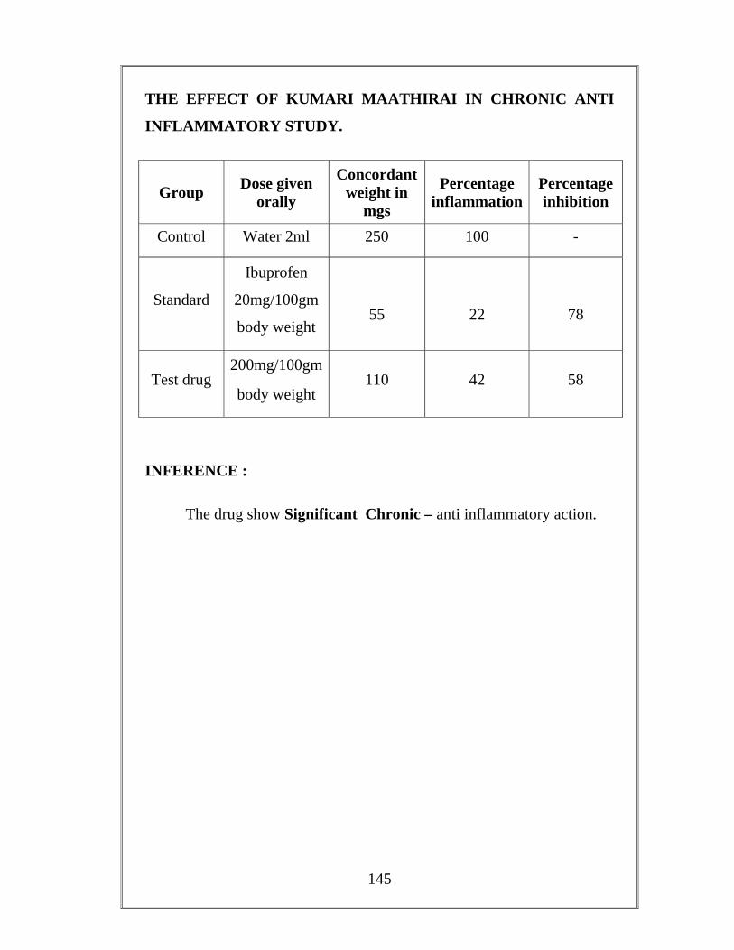
145
THE EFFECT OF KUMARI MAATHIRAI IN CHRONIC ANTI
INFLAMMATORY STUDY.
Group Dose given orally
Concordant weight in
mgs
Percentage inflammation
Percentage inhibition
Control Water 2ml 250 100 -
Standard
Ibuprofen
20mg/100gm
body weight
55
22
78
Test drug 200mg/100gm
body weight 110 42 58
INFERENCE :
The drug show Significant Chronic – anti inflammatory action.
146
ACUTE ANTI – INFLAMMATORY STUDY ON
NOCHI THYLAM (External use)
By Hind – Paw method in Albino Rats
AIM
To study the acute antiinflammatory activity of the test drug Nochi
Thylam
PROCEDURE
Six healthy albino rats weighing 100 -150 gm, were taken and
divided into two groups, each consisting of 3 rats.
First group was kept as control by giving distilled water 2 ml/ 100
gms of body weight. The second group was kept as test group.
Before application of test drug, the hind – paw volume of all rats
were measured. This was done by dipping the hind – paw (upto tibiotrasal
junction) into a mercury plethysmography. While dipping the hind paw,
by pulling the syringe piston, the level of mercury in the ventre small tube
was made to coincide with red marking and reading was noted from the
plethysmograph.
One hour later, a sub – cutaneous injection of 0.1 ml of 1% (W/V)
carrageenin in water was made into plantar surface of both hind – paw of
each rat. To the second test group Nochi Thylam. was topically applied
for three times over the inflamed surface in a thin layer with 30 minutes
gap. To the other groups no drug was applied over the inflamed surface.
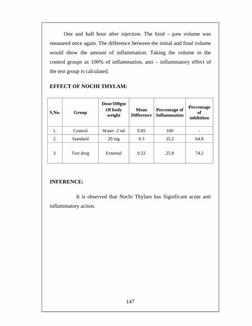
147
One and half hour after injection. The hind – paw volume was
measured once again. The difference between the initial and final volume
would show the amount of inflammation. Taking the volume in the
control groups as 100% of inflammation, anti – inflammatory effect of
the test group is calculated.
EFFECT OF NOCHI THYLAM:
S.No. Group
Dose/100gm Of body weight
Mean Difference
Percentage of Inflammation
Percentage of
inhibition
1. Control Water -2 ml 0.85 100 -
2. Standard 20 mg 0.3 35.2 64.8
3. Test drug External 0.22 25.8 74.2
INFERENCE:
It is observed that Nochi Thylam has Significant acute anti
inflammatory action.
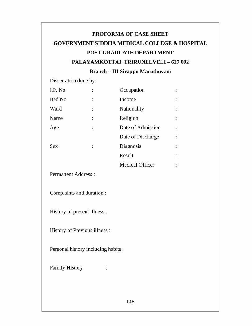
148
PROFORMA OF CASE SHEET
GOVERNMENT SIDDHA MEDICAL COLLEGE & HOSPITAL
POST GRADUATE DEPARTMENT
PALAYAMKOTTAI. TRIRUNELVELI – 627 002
Branch – III Sirappu Maruthuvam
Dissertation done by:
I.P. No : Occupation :
Bed No : Income :
Ward : Nationality :
Name : Religion :
Age : Date of Admission :
Date of Discharge :
Sex : Diagnosis :
Result :
Medical Officer :
Permanent Address :
Complaints and duration :
History of present illness :
History of Previous illness :
Personal history including habits:
Family History :
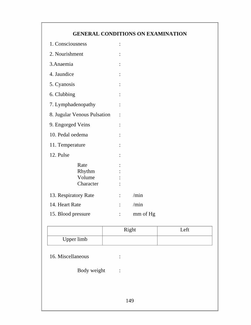
149
GENERAL CONDITIONS ON EXAMINATION
1. Consciousness :
2. Nourishment :
3.Anaemia :
4. Jaundice :
5. Cyanosis :
6. Clubbing :
7. Lymphadenopathy :
8. Jugular Venous Pulsation :
9. Engorged Veins :
10. Pedal oedema :
11. Temperature :
12. Pulse :
Rate : Rhythm : Volume : Character :
13. Respiratory Rate : /min
14. Heart Rate : /min
15. Blood pressure : mm of Hg
Right Left
Upper limb
16. Miscellaneous : Body weight :
150
º¢ò¾ Ó¨È §¾÷× 1. ¿¢Äõ
ÌȢﺢ Óø¨Ä ÁÕ¾õ ¦¿ö¾ø À¡¨Ä
2. ÀÕÅ ¸¡Äõ
¸¡÷ ¸¡Äõ (¬Å½¢-ÒÃ𼡺¢) ܾ¢÷ ¸¡Äõ (³ôÀº¢-¸¡÷ò¾¢¨¸) ÓýÀÉ¢ (Á¡º¢-ÀíÌÉ¢) −ǧÅÉ¢ø (º¢ò¾¢¨Ã-¨Å¸¡º¢) ÓЧÅÉ¢ø (¬É¢-¬Ê)
3. ¡쨸 (¯¼ø)
Å¡¾õ À¢ò¾õ ¸Àõ ¸ÄôÒ
4. ̽õ
ºòÐÅõ −Ếõ ¾¡Áºõ
5. ¦À¡È¢ ÒÄý¸û
¦Áö-°Ú Å¡ö-ͨŠ¸ñ-´Ç¢ ãìÌ-¿¡üÈõ ¦ºÅ¢-´Ä¢
6. ¸ý§Áó¾¢Ã¢Âõ
¨¸-¾¡Éõ ¸¡ø-¸ÁÉõ Å¡ö-źÉõ ±ÕÅ¡ö-Å¢º÷ì¸õ ¸ÕÅ¡ö-¬Éó¾õ
7. ¯ð¸¡Âõ
§¾¡û ¦À¡ÕòÐ ÒÂõ ÓÆí¨¸ ¦À¡ÕòÐ ºÂõ ¨¸ìÌÇÍ ¸ÃÀõ «íÌÄ¢
151
8. «¾ì¸¡Âõ −ÎôÒ ¦À¡ÕòÐ Óý¸¡ø ̾¢í¸¡ø ¸ÀÃõ «íÌÄ¢
9. Åõº¢¸û 10. ÓõÁÄõ ÁÄõ
ãò¾¢Ãõ Å¢Â÷¨Å
11. À¢È ¯ÚôҸǢý ¿¢¨Ä −Õ¾Âõ
ÒôÒºõ −¨Ãô¨À ¸øÄ£Ãø Áñ½£Ãø º¢Ú̼ø ¦ÀÕí̼ø º¢Ú¿£Ã¸õ ã¨Ç ¸Õô¨À
12. ¯Â¢÷ò ¾¡Ðì¸û («) Å¡¾õ
À¢Ã¡½ý «À¡Éý Ţ¡Éý ¯¾¡Éý ºÁ¡Éý ¿¡¸ý Ü÷Áý ¸¢Õ¸Ãý §¾Åò¾ò¾ý ¾É了Âý
(¬) À¢ò¾õ «Éü À¢ò¾õ −Ãﺸ À¢ò¾õ º¡¾¸ À¢ò¾õ ¬§Ä¡º¸ À¢ò¾õ À¢Ã¡º¸ À¢ò¾õ
(þ) ¸Àõ
«ÅÄõÀ¸õ ¸¢§Ä¾¸õ
152
§À¡¾¸õ ¾üÀ¸õ ºó¾¢¸õ
13. ¯¼ø ¾¡Ðì¸û
º¡Ãõ ¦ºó¿£÷ °ñ ¦¸¡ØôÒ ±ýÒ ã¨Ç Í츢Äõ/ͧá½¢¾õ
14. ±ñ Ũ¸ò§¾÷׸û
¿¡Ê ŠÀâºõ ¿¡ ¿¢Èõ ¦Á¡Æ¢ ŢƢ ÁÄõ ¿¢Èõ
±¨¼ −Ú¸ø −Ǹø
º¢Ú¿£÷
1. ¿£÷ìÌÈ¢ ¿¢Èõ Á½õ ±¨¼ Ѩà ±ïºø
2. ¦¿öìÌÈ¢
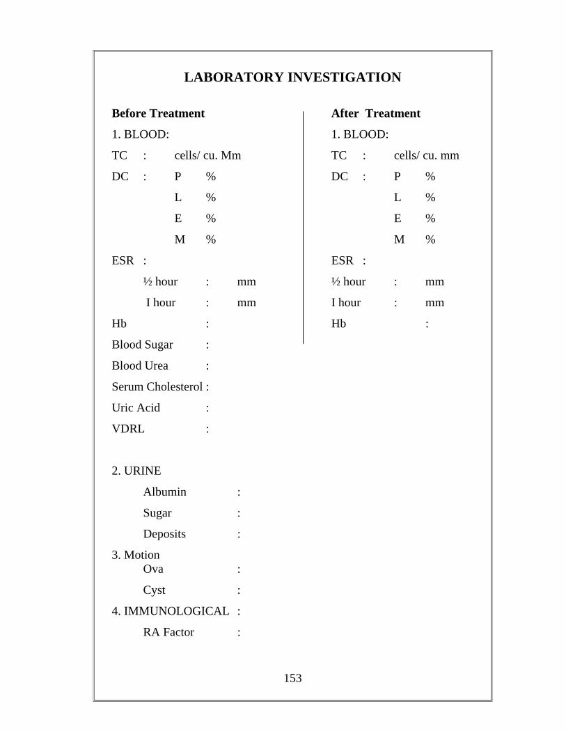
153
LABORATORY INVESTIGATION
Before Treatment After Treatment
1. BLOOD: 1. BLOOD:
TC : cells/ cu. Mm TC : cells/ cu. mm
DC : P % DC : P %
L % L %
E % E %
M % M %
ESR : ESR :
½ hour : mm ½ hour : mm
I hour : mm I hour : mm
Hb : Hb :
Blood Sugar :
Blood Urea :
Serum Cholesterol :
Uric Acid :
VDRL :
2. URINE
Albumin :
Sugar :
Deposits :
3. Motion Ova :
Cyst :
4. IMMUNOLOGICAL :
RA Factor :
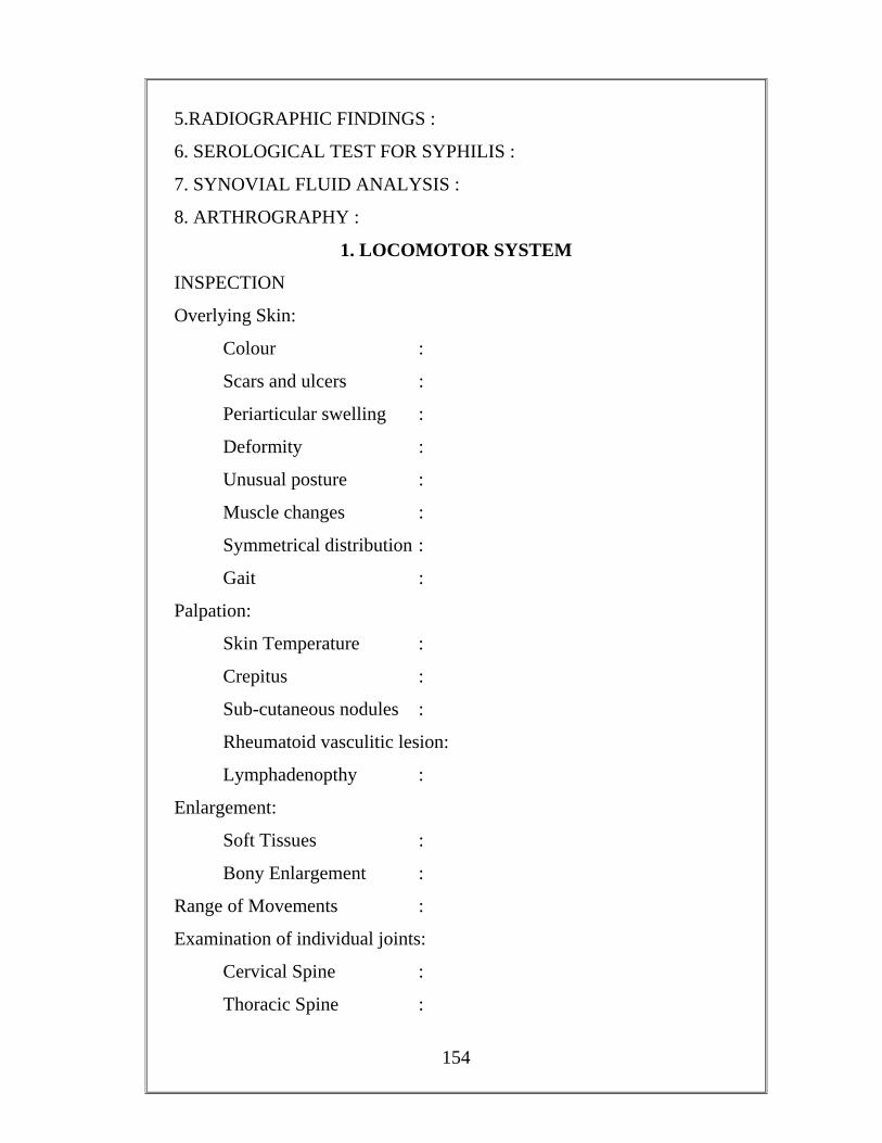
154
5.RADIOGRAPHIC FINDINGS :
6. SEROLOGICAL TEST FOR SYPHILIS :
7. SYNOVIAL FLUID ANALYSIS :
8. ARTHROGRAPHY :
1. LOCOMOTOR SYSTEM
INSPECTION
Overlying Skin:
Colour :
Scars and ulcers :
Periarticular swelling :
Deformity :
Unusual posture :
Muscle changes :
Symmetrical distribution :
Gait :
Palpation:
Skin Temperature :
Crepitus :
Sub-cutaneous nodules :
Rheumatoid vasculitic lesion:
Lymphadenopthy :
Enlargement:
Soft Tissues :
Bony Enlargement :
Range of Movements :
Examination of individual joints:
Cervical Spine :
Thoracic Spine :
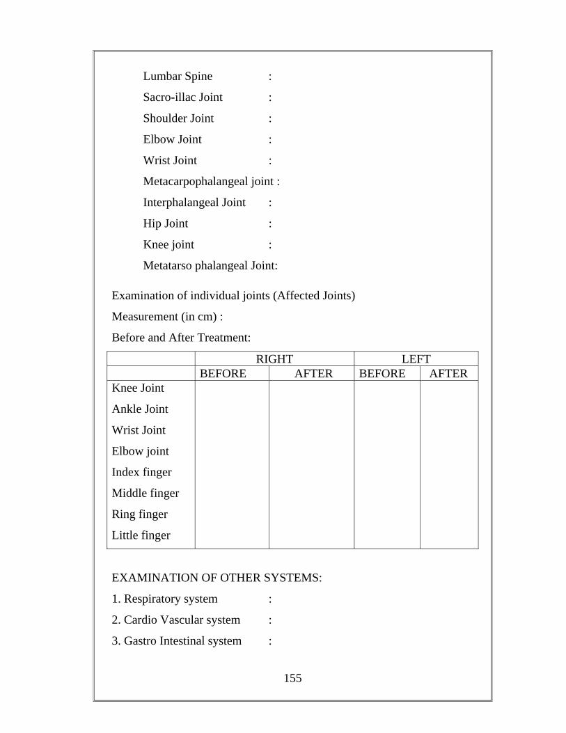
155
Lumbar Spine :
Sacro-illac Joint :
Shoulder Joint :
Elbow Joint :
Wrist Joint :
Metacarpophalangeal joint :
Interphalangeal Joint :
Hip Joint :
Knee joint :
Metatarso phalangeal Joint:
Examination of individual joints (Affected Joints)
Measurement (in cm) :
Before and After Treatment:
RIGHT LEFT BEFORE AFTER BEFORE AFTERKnee Joint
Ankle Joint
Wrist Joint
Elbow joint
Index finger
Middle finger
Ring finger
Little finger
EXAMINATION OF OTHER SYSTEMS:
1. Respiratory system :
2. Cardio Vascular system :
3. Gastro Intestinal system :
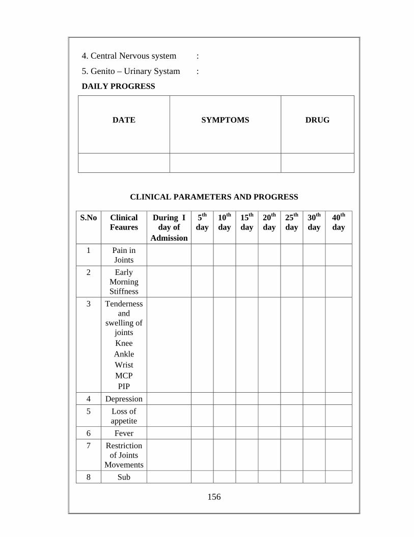
156
4. Central Nervous system :
5. Genito – Urinary Systam :
DAILY PROGRESS
DATE
SYMPTOMS
DRUG
CLINICAL PARAMETERS AND PROGRESS
S.No Clinical Feaures
During I day of
Admission
5th day
10th day
15th day
20th day
25th day
30th day
40th day
1 Pain in Joints
2 Early Morning Stiffness
3 Tenderness and
swelling of joints Knee Ankle Wrist MCP PIP
4 Depression 5 Loss of
appetite
6 Fever 7 Restriction
of Joints Movements
8 Sub
157
cutaneous nodules
9 Deformity 10 Muscle
Weakness
11 Muscle Wasting
12 Ulnar Deviation
13 Entrapment Neuropathy
+++ - Severe
++ - Moderate
+ - Mild
- - Nil
DIFFERENTIAL DIAGNOSIS
FINAL DIAGNOSIS:
LINE OF TREATMENT
MEDICINE
MEDICAL ADVICE
158
GOVERNMENT SIDDHA MEDIDCAL COLLEGE HOSPITAL,
POST GRADUATE DEPARTMENT,
PALAYAMKOTTAI, TIRUNELVELI-627 002
BRANCH – III SIRAPPU MARUTHUVAM.
ADMISSION-DISCHARGE SHEET FOR “VALI AZHALKEEL VAYU”
IP No : Occupation :
Bed No : Income :
Ward : Nationality :
Name : Religion :
Age : Date of Admission :
Sex : Date of Discharge :
Permanent Address: Diagnosis :
Result :
Temporary Address: Medical officer :
CLINICAL PICTURES
Sl.No. During Admission During Discharge
PLACE:
DATE: Signature of Medical Officer.
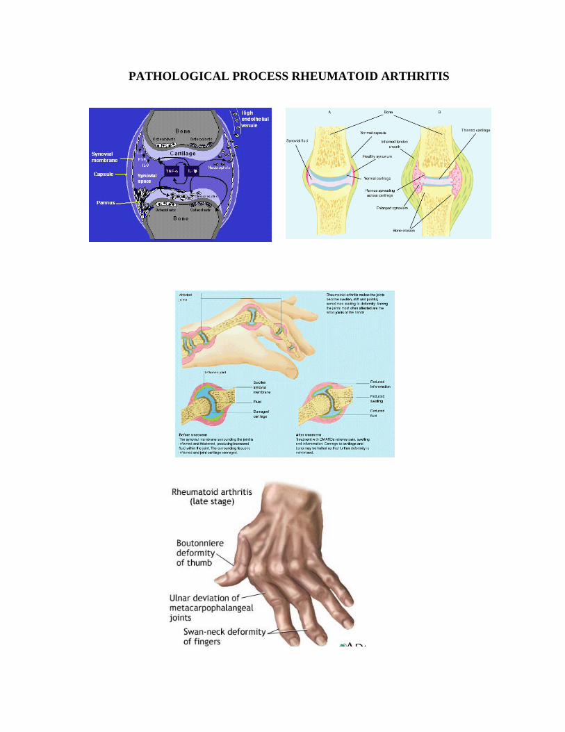
PATHOLOGICAL PROCESS RHEUMATOID ARTHRITIS
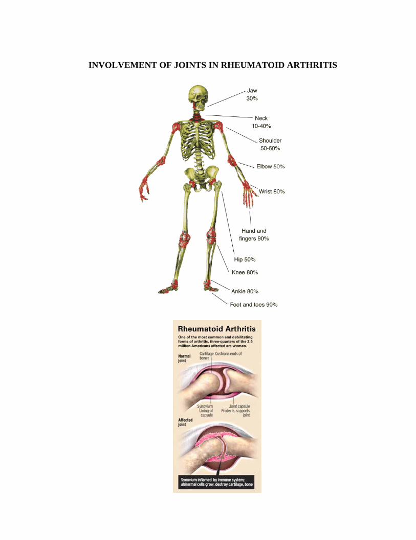
INVOLVEMENT OF JOINTS IN RHEUMATOID ARTHRITIS
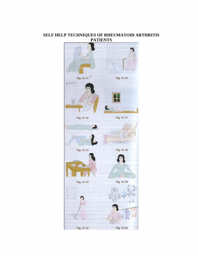
SELF HELP TECHNIQUES OF RHEUMATOID ARTHRITIS PATIENTS
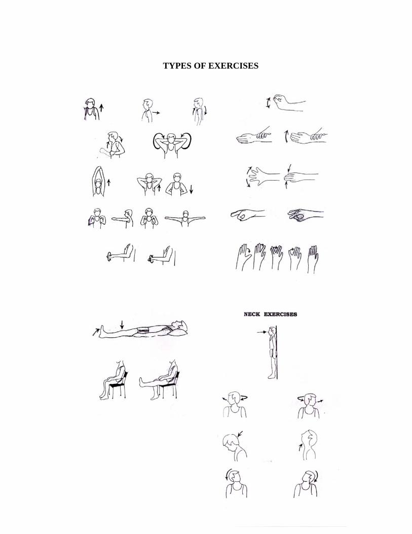
TYPES OF EXERCISES
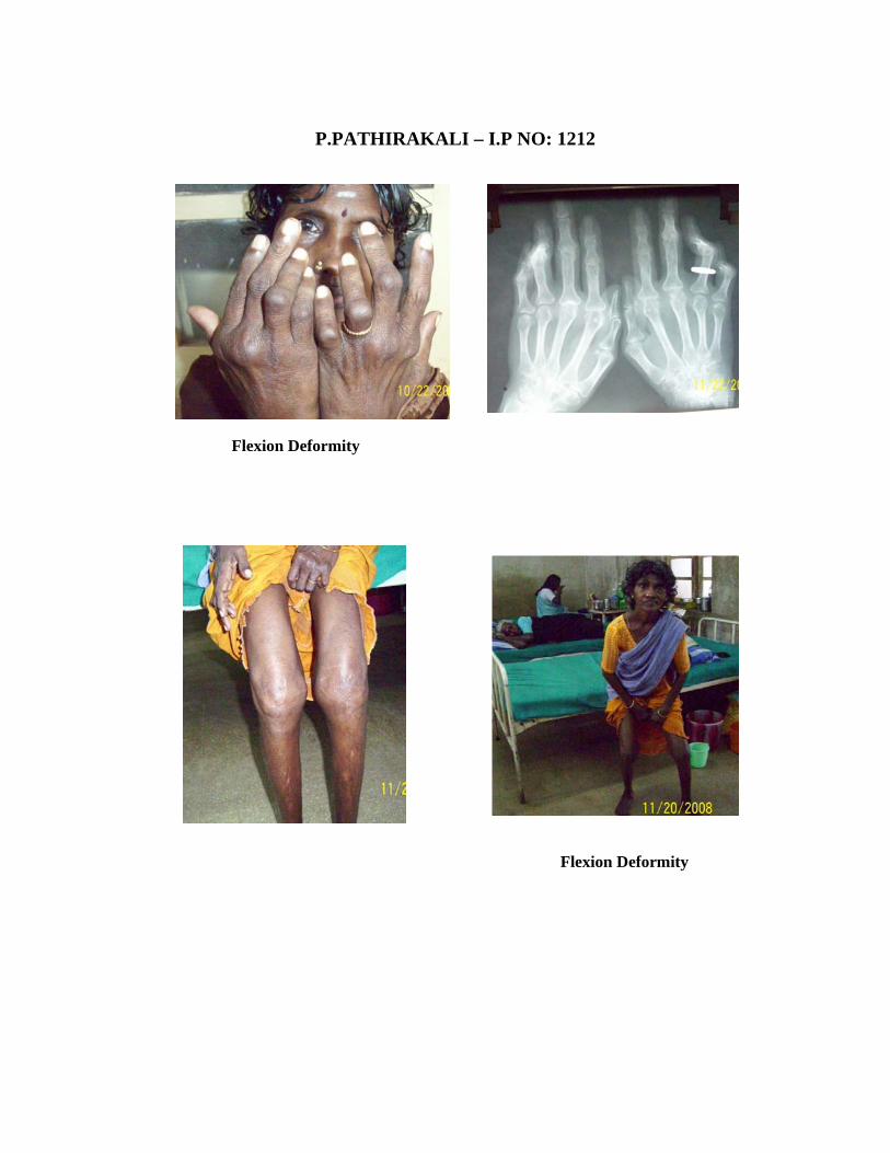
P.PATHIRAKALI – I.P NO: 1212
Flexion Deformity
Flexion Deformity
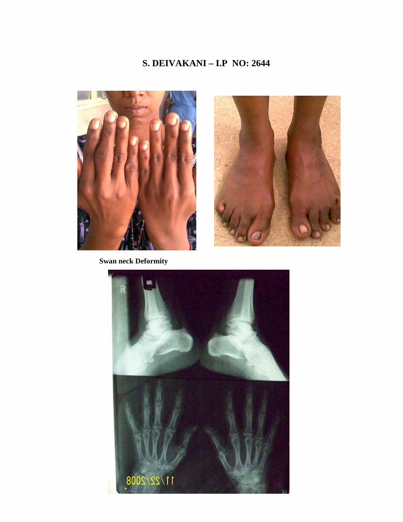
S. DEIVAKANI – I.P NO: 2644
Swan neck Deformity
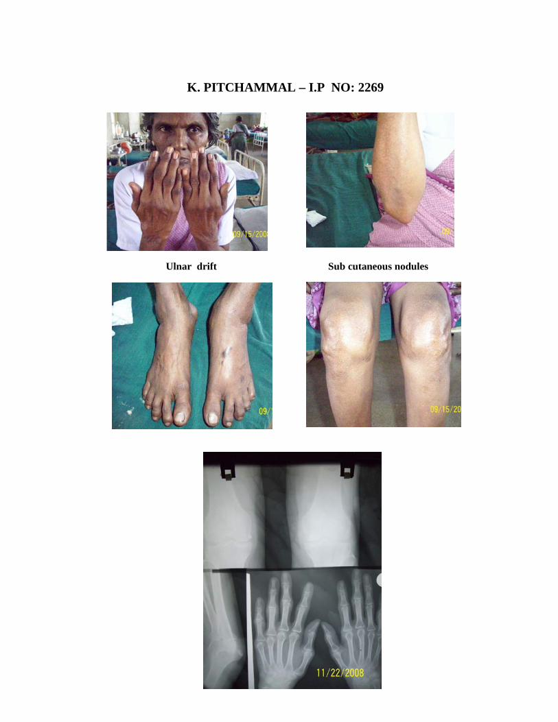
K. PITCHAMMAL – I.P NO: 2269
Ulnar drift Sub cutaneous nodules

I. ARPUTHA MARRY – I.P NO:2340
Flexion deformity Cervical spondylosis
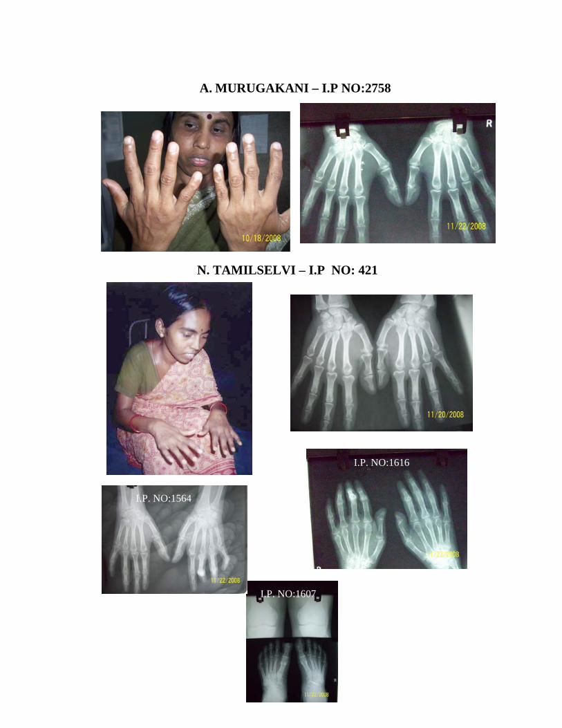
A. MURUGAKANI – I.P NO:2758
N. TAMILSELVI – I.P NO: 421 I.P. NO:1616 I.P. NO:1564 I.P. NO:1607
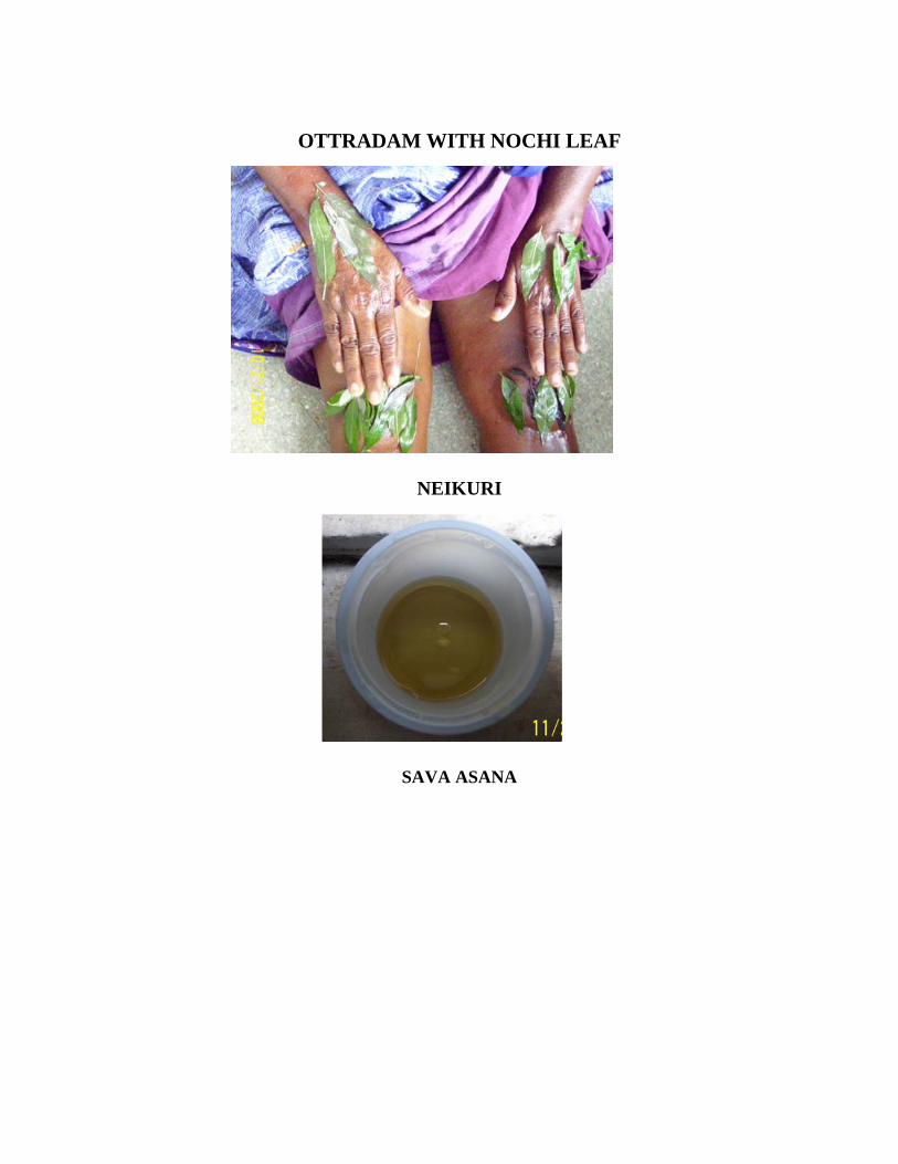
OTTRADAM WITH NOCHI LEAF
NEIKURI
SAVA ASANA
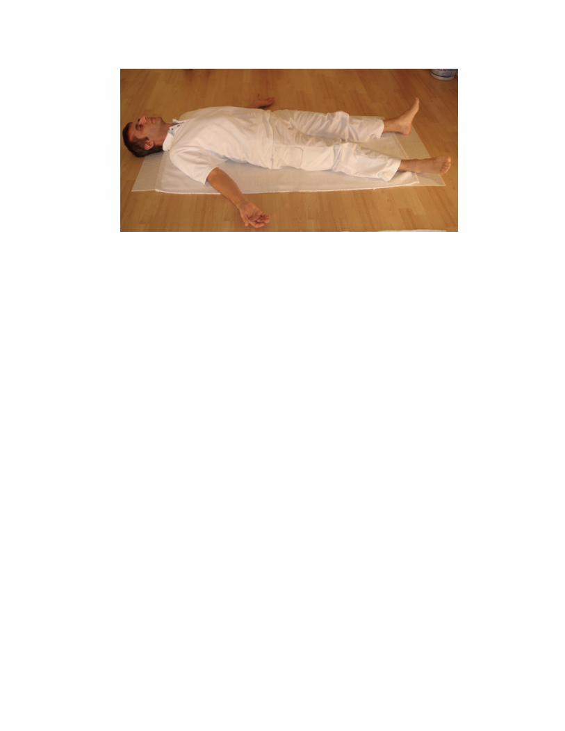
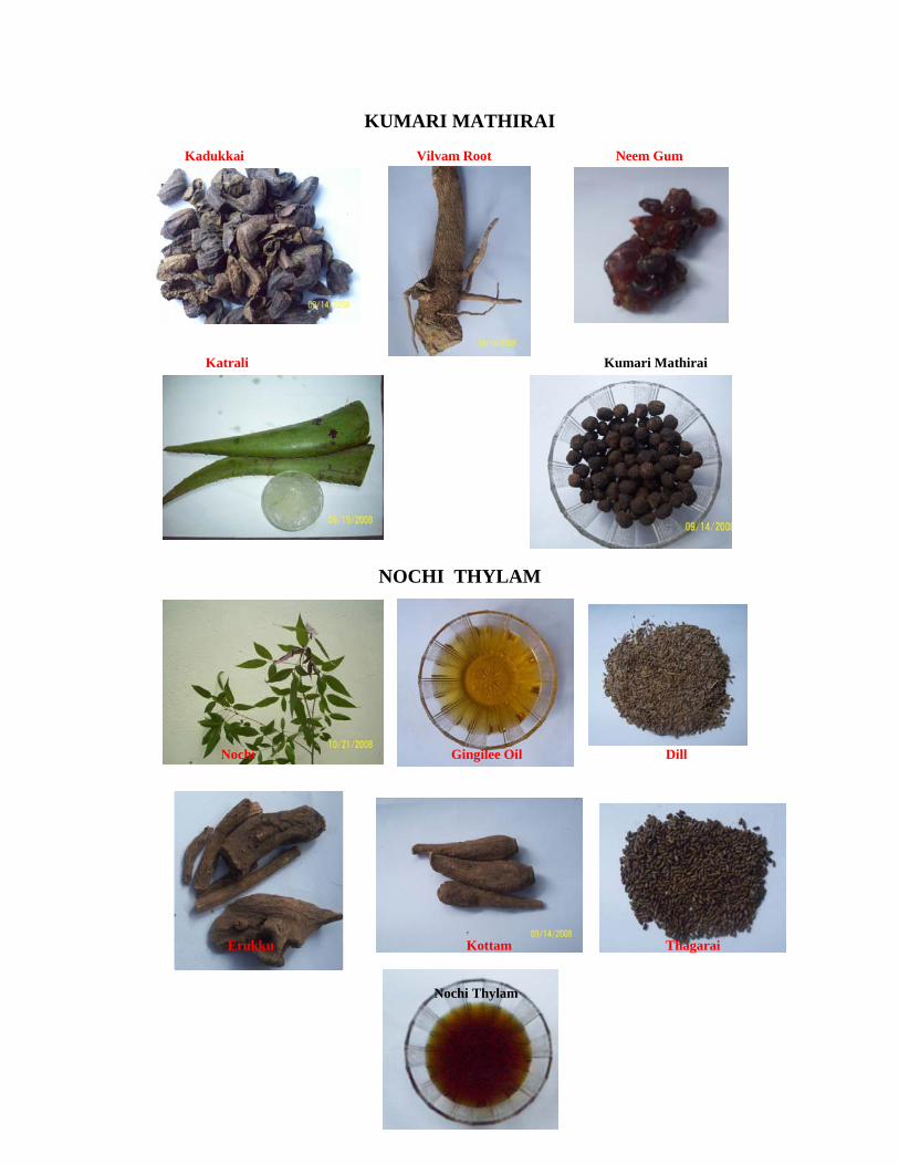
KUMARI MATHIRAI Kadukkai Vilvam Root Neem Gum
Katrali Kumari Mathirai
NOCHI THYLAM
Nochi Gingilee Oil Dill Erukku Kottam Thagarai
Nochi Thylam
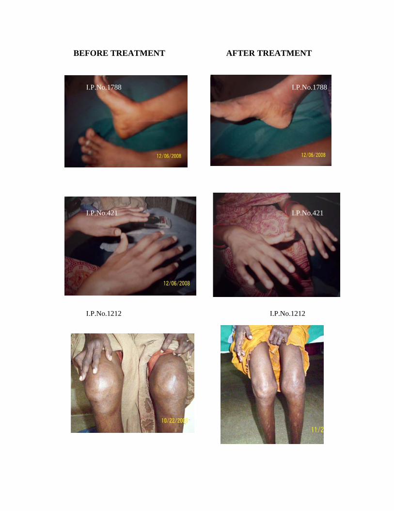
BEFORE TREATMENT AFTER TREATMENT I.P.No.1788 I.P.No.1788 I.P.No.421 I.P.No.421 I.P.No.1212 I.P.No.1212
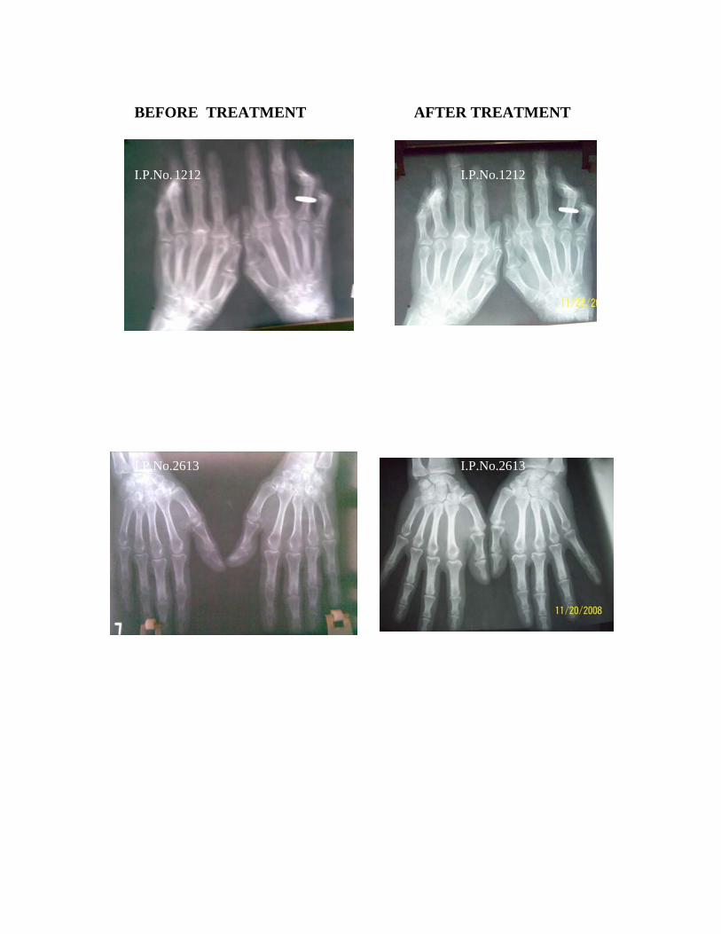
BEFORE TREATMENT AFTER TREATMENT I.P.No. 1212 I.P.No.1212 I.P.No.2613 I.P.No.2613

P% L% E% P% L% E% 1/2 hr 1 hr 1/2 hr 1 hr1 2163 9200 9600 56 32 12 50 48 2 45 85 10 20 61% 71% NIL NIL NIL NIL NIL NIL NIL NIL NIL NIL2 2340 9900 10000 70 28 2 50 50 0 40 85 8 15 64% 78% NIL NIL NIL NIL NIL NIL NIL NIL Occ. EC NIL3 2644 8600 8900 57 40 3 54 66 0 45 85 12 25 60% 71% NIL NIL NIL NIL NIL Few PC NIL NIL NIL NIL4 2748 8900 9000 70 26 4 49 50 1 18 40 7 15 60% 72% NIL NIL NIL NIL NIL NIL NIL NIL NIL NIL5 2758 8200 8500 56 40 4 50 49 1 25 50 10 15 58% 69% NIL NIL NIL NIL NIL Few EC NIL NIL NIL NIL6 3074 9100 9200 62 36 2 51 49 0 22 45 8 15 60% 72% NIL NIL NIL NIL NIL NIL NIL NIL Occ. EC NIL7 32 9600 9800 60 36 4 52 48 0 25 55 5 10 57% 65% NIL NIL NIL NIL NIL NIL NIL NIL NIL NIL8 255 9000 9300 48 48 4 49 50 1 35 70 10 20 60% 70% NIL NIL NIL NIL NIL Few EC NIL NIL NIL NIL9 421 7000 7300 55 40 5 49 51 0 40 80 7 20 59% 69% NIL NIL NIL NIL NIL NIL NIL NIL NIL NIL
10 447 9200 9400 58 42 0 52 48 0 42 82 8 18 60% 72% NIL NIL NIL NIL NIL NIL NIL NIL NIL NIL11 1212 9200 9500 68 28 4 50 49 1 30 65 15 25 61% 69% NIL NIL NIL NIL NIL Few PC NIL NIL NIL NIL12 1502 9200 9300 50 48 2 50 50 0 24 48 5 10 65% 79% NIL NIL NIL NIL NIL NIL NIL NIL NIL NIL13 1501 9800 9900 50 48 2 50 49 1 16 a 7 15 60% 71% NIL NIL NIL NIL NIL NIL NIL NIL NIL NIL14 1565 9100 9200 59 40 1 50 49 1 23 48 12 20 70% 78% NIL NIL NIL NIL NIL NIL NIL NIL NIL NIL15 1564 8800 8900 70 27 3 50 49 1 26 50 12 24 68% 76% NIL NIL NIL NIL NIL NIL NIL NIL NIL NIL16 1607 8800 8900 69 28 3 50 48 2 40 88 15 22 68% 76% NIL NIL NIL NIL NIL NIL NIL NIL NIL NIL17 1616 9000 9100 60 38 2 50 50 0 20 40 5 10 60% 69% NIL NIL NIL NIL NIL NIL NIL NIL NIL NIL18 1788 8700 8900 59 40 1 50 49 1 44 89 12 24 50% 59% NIL NIL NIL NIL NIL NIL NIL NIL NIL NIL19 2230 8400 8700 57 40 3 50 49 1 40 82 15 28 68% 71% NIL NIL NIL NIL NIL NIL NIL NIL NIL NIL
20 2269 8400 8600 69 30 1 60 40 0 40 80 15 25 60% 68% NIL NIL NIL NIL NIL NIL NIL NIL NIL NIL
WBS Differential Count Cu m.m E.S.R mm/hr AT BTHB BT
Haematological Investigation and Urine Analysis of 20 IP Patients
S.No IP.No
Haematological Investigation Urine Analaysis Motion Analaysis
WBC Total Count Cu m.m
AT Alb Alb Sug
AT
BT ATBT AT BT AT
BT Cyst
P- Polymorph L - Lymphocyte E - Eosinophil Hb - Haemoglobin ESR - Erythrocyte Sedimentation Rate Alb - Albumin Sug - Sugar Dep - Deposit Occ. EC - Occationally Epithelial Cells Few PC - Few pus cells BT - Before Treatment AT - After Treatment
Dep Ova Cyst OvaSug Dep