A STUDY ON THE PRESENCE OF ACCESSORY ...
135
A Dissertationon A STUDY ON THE PRESENCE OF ACCESSORY MAXILLARY OSTIUM Submitted to the THE TAMILNADU DR. M.G.R. MEDICAL UNIVERSITY In partial fulfilment of the requirements For the award of the degree of M.S.BRANCH IV (OTORHINOLARYNGOLOGY) GOVERNMENT STANLEY MEDICAL COLLEGE & HOSPITAL THE TAMILNADU DR. M.G.R. MEDICAL UNIVERSITY, CHENNAI, TAMILNADU APRIL 2015
-
Upload
khangminh22 -
Category
Documents
-
view
1 -
download
0
Transcript of A STUDY ON THE PRESENCE OF ACCESSORY ...

A Dissertationon
A STUDY ON THE PRESENCE OF ACCESSORY
MAXILLARY OSTIUM
Submitted to the
THE TAMILNADU DR. M.G.R. MEDICAL UNIVERSITY
In partial fulfilment of the requirements
For the award of the degree of
M.S.BRANCH IV (OTORHINOLARYNGOLOGY)
GOVERNMENT STANLEY MEDICAL
COLLEGE & HOSPITAL
THE TAMILNADU DR. M.G.R. MEDICAL UNIVERSITY,
CHENNAI, TAMILNADU
APRIL 2015

DECLARATION
I, Dr. VRINDA .B. NAIR, solemnly declare that the dissertation,
titled “A Study On The Presence Of Accessory Maxillary Ostium” is
a bonafide work done by me during the period of January 2013 to July
2014 at Government Stanley Medical College and Hospital, Chennai
under the expert supervision of PROF. DR. T. BALASUBRAMANIAN,
M.S., D.L.O., Professor and Head, Department Of Otorhinolaryngology,
Government Stanley Medical College and hospitals, Chennai.
This dissertation is submitted to The Tamil Nadu Dr. M.G.R.
Medical University in partial fulfilment of the rules and regulations for
the M.S. degree examinations in Otorhinolaryngology to be held in April
2015.
Place: Chennai-1 DR.VRINDA .B. NAIR
Date:

CERTIFICATE
This is to certify that the dissertation -“A Study On The Presence
Of Accessory Maxillary Ostium” presented BY DR.VRINDA .B.
NAIR , is an original work done in the Department of
Otorhinolaryngology, Government Stanley Medical College and Hospital,
Chennai in partial fulfillment of regulations of the Tamil Nadu Dr.
M.G.R. Medical University for the award of degree of M.S.
(Otorhinolaryngology) Branch IV, under my supervision during the
academic period 2012-2015.
THE DEAN, Govt. Stanley medical college,
Chennai-1.
PROF.DR.T.BALASUBRAMANIAN PROFESSOR AND HEAD OF THE DEPARTMENT Govt. Stanley Medical College and Hospital Chennai-1
Place : Chennai Date :

ACKNOWLEDGEMENTS
I wish to express my sincere thanks to Prof. Dr.AL. MEENAKSHI
SUNDARAM, MD, DEAN, Government Stanley Medical College and
Hospital for having permitted me to utilize the facilities of the hospital for
conducting this study.
My heartfelt gratitude to Prof.Dr.T. BALASUBRAMANIAN,
M.S., D.L.O., Professor and Head of the Department, Department of
Otorhinolaryngology, Government Stanley Medical College and Hospital
for his constant motivation, valuable suggestions, and expert supervision
during the course of this study.
I express my whole-hearted gratitude to Prof.Dr.N.
SEETHALAKSHMI M.S.,D.L.O.,D.N.B, Professor and Chief of ENT
UNIT II of Otorhinolaryngology, PROF. DR. F.ANTHONY
IRUDHAYA RAJAN M.S.,D.L.O, Professor, for supporting, guiding
and encouraging me in this study.
I wish to thank my Assistant Professors DR.K.ATHIYAMAN M.S,
DR.C.KARUPPASAMY M.S.,D.L.O., DR.M.P.CHANDRAMOULI
M.S., DR.SARAVANA SELVAN M.S., DR.C.BHARANIDHARAN
D.L.O. for their valuable tips and guidance.
I am grateful to all the other post-graduates who most willingly
helped me during this study period.

I also thank the staff nurses, theatre personnel, OPD staff,
Department of Otorhinolaryngology, Government Stanley Hospital for
their co-operation and assistance in the conduct of this study.
I wish to extend my gratitude to my statistician for his expert
assistance.
Last but not the least, I am indebted and grateful to all the
Patients and Normal volunteers who constitute the backbone of this
study, who most willingly and selflessly subjected themselves to this
study for the sake of the benefit of their community and without
whom this study would not have been possible.

TABLE OF CONTENTS
1. ABSTRACT 1
2. OBJECTIVE 3
3. INTRODUCTION 5
4. REVIEW OF LITERATURE 7
5. MATERIALS AND METHODS 38
6. RESULTS 51
7. DISCUSSION 64
8. CONCLUSION 109
9. ANNEXURES 111
A.BIBLIOGRAPHY
B. PROFORMA
C. ETHICAL COMMITTEE
APPROVAL LETTER
D. PATIENT INFORMATION
SHEET
E. INFORMED CONSENT FORM
F. PLAGIARISM
G. MASTER SHEET

1. ABSTRACT
The association between accessory ostium and chronic
sinusitis is still a subject of controversy as various studies have
given a wide range of variable results. The prevalence of accessory
ostium varies between 2 – 44 %.
Objective
To study the prevalence of Accessory ostium in those with
Chronic Sinusitis and those without the disease.
Methods
83 patients with chronic sinusitis and 83 subjects without the
disease were studied by nasal endoscopy for the presence of
accessory ostium.
Results
30 % of those with chronic sinusitis had an accessory ostium
, whereas only 10 % without the disease had it ( p value <0.001 ).
Majority of the accessory ostia were on the left and in the
posterior nasal fontanelle , in both the groups. 6 (24 %) patients and
1 (13 %) unexposed subject had bilateral accessory ostia.Only 2% of

Page | 2
the diseased individuals had double ostia. Only 12 % showed
circular phenomenon. However symptoms like headache , facial pain ,
post nasal drip and halitosis , and purulence on examination
(Rhinosinusitis Task Force Criteria) were not found to have any
specific association with the presence of accessory ostia.
Conclusion
To conclude we would say that the presence of an accessory
ostium can be considered as an indicator of maxillary sinus disease,
along with the other criteria for chronic sinusitis and that it can
help surgeons in their decision making as to whether a surgery is
required or not.
Key words : accessory ostium , maxillary sinusitis

Page | 3
2. OBJECTIVE
To study,
· The prevalence of accessory maxillary ostium in chronic
sinusitis and that in normal subjects by nasal endoscopy.

Page | 4
INCLUSION CRITERIA:
1. Age:15-45 years.
2. Patients with clinical symptoms of chronic maxillary sinusitis.
3. Radiological diagnosis of chronic maxillary sinusitis.
4. Persistence of symptoms for more than 12 weeks even after
medical line of treatment.
EXCLUSION CRITERIA:
1. <15 or>45 years of age.
2. History of previous maxillary sinus surgeries.
3. Patients with sinonasal neoplasms.
4. Patients with sinonasal encephaloceles.
5. Patients with known ciliary dysfunction such as primary ciliary
dyskinesia and Kartagener Syndrome .
6. Patients with sinonasal polyposis.
7. Patients with fungal sinusitis.
8. Patients with Cystic Fibrosis .
9. Patients who are immunocompromised (with the exception of
diabetics) .

Page | 5
3. INTRODUCTION
Chronic sinusitis is a group of disorders characterized by the
inflammation of nasal and paranasal sinus mucosa which is now
extremely prevalent, affecting 32 million adults , or 16.3 % of the
adult population[9]. Its diagnosis is mainly based on clinical
symptoms and signs, supported by diagnostic nasal endoscopy and
CT scan.
Maxillary sinusitis is the side effect of evolution as a result
of which man acquired an erect posture. The secretions of the
maxillary sinuses have to be transported against gravity by the
mucociliary action. Hence the normal functioning of a sinus
depends on – patency of the ostium , normal mucociliary transport
and the normal quality and quantity of secretions.
Accessory ostia also known as Giralde’s orificeare defects in
the lateral nasal wall, in the region of fontanelles, which
communicate with the maxillary sinus , in addition to the natural
ostium. They are thought to develop as a consequence of damage
to the fontanelles following sinus infections. This in turn becomes
a reason for recurrence of infection due to mucous recirculation.

Page | 6
Majority of the accessory ostia occur singly. A few multiple ostia
have also been observed. They are mostly acquired and rarely
congenital. However there is still a controversy over its etiology.
The present study is an attempt at finding the prevalence of
Accessory Ostium in those patients with Chronic Sinusitis and in
those without. Here we have used Nasal Endoscopy as a tool to
investigate the presence of accessory ostia. We have deliberately
avoided CT PNS as a diagnostic tool as our study included normal
subjects as well.

Page | 7
4. REVIEW OF LITERATURE
Hippocrates in the 5th century B.C itself has described
sinusitis when he quoted “in a person having a painful spot in
head, with intense headaches, pus or fluid running from the nose
removes the disease.”
Leonardo da Vinci in his period had accurately given the
description of the maxillary antrum and the frontal sinus. [1]
In 1869, Wertheim made a Conchoscope to examine the
anterior and middle thirds of the nasal cavity. [1]
In 1879, Nitze-Leiter developed the Cystoscope, and this
marked the second stage in the developement of endoscopy. [1]
Hirschmann is considered the Father of Endoscopy. In 1902,
Hirschmann and Valentin introduced a modified cystoscope into a
maxillary sinus through an enlarged dental alveolus.[1]
During the period 1951 – 1956, Hopkins made improvements in
the optics of endoscopy which included a separate light source , small
diameter of the endoscope , large field of vision , excellent resolution
, high contrast and constancy of color. [1]

Page | 8
In the 1950s Messerklinger developed a systematic diagnostic
approach to the lateral wall of nose. [1]
HISTORY OF STUDIES ON MAXILLARY ACCESSORY OSTIUM
There have been various studies in the past conducted to find
the presence of Accessory Maxillary Ostia (AMO) on live subjects
as well as cadavers. In 1870 , Zuckerkandl introduced the term
‘fontanelle’ for certain regions below the uncinate and above the
inferior turbinate , deficient in bone covered by nasal mucous
membrane medially and maxillary sinus laterally , with connective
tissue sandwiched in between. Levine et al described the anterior and
posterior fontanelle (ANF &PNF) in relation to the uncinate - PNF
being posterosuperior to uncinate and ANF between the inferior
edge of uncinate and attached margin of inferior turbinate[25].
Scheaffer (1953) discovered twin openings at the Hiatus Semilunaris
and described them as Triple PMO. He preferred the term
Duplicate PMO than Accessory ostium. Rice and Scheaffer (1993)
used the term Accessory maxillary sinus ostia for all the extra
openings other than the primary maxillary ostium irrespective of
their location. Levine et al (1993) described that accessory ostia

Page | 9
result from rupture of fonatanelles due to the blockage in the
primary maxillary ostium. Scheaffer (1920) reported the AMO to
number between 1 -3. But this is uncommon and hence there is not
much in the literature regarding multiple openings. Stammberger &
Kennedy (1995) came up with an incidence of 4-5 % in the
general population as opposed to 25 % in those with chronic
sinusitis.
Incidence of AMO ranges between 2 - 44 % , lower values
being obtained from in vivo studies whereas higher ones from
cadaver studies. The reason for this wide variation may be that the
AMO that may be covered with a mucous film and in inaccessible
locations are often missed out in in vivo studies. Whereas in cadaver
studies, due to drying and fixing, the moist nasal mucosa can
undergo shrinkage, also the fontanelle mucosa may be damaged
during drying and investigations, giving an impression of higher
incidence.
Schaeffer (1920) reported on an incidence of 43% in a
cadaver study.

Page | 10
Myerson (1932), van Alyea (1936), Lang & Wurzburg (1991),
Kumar. H et al (2001, Lady Hardinge medical college, New Delhi ) in
their studies on cadavers came up with an incidence of 31 %, 23 %,
28%, 30 %[5] respectively.
May et al (1990) found a 0 % incidence in cadavers and a 10
% incidence in endoscopic studies. Kennedy & Zinreicch in 1991 in
an endoscopic study reported a value of 15 %. Wigand states that
25 % of specimens showed an accessory ostium[35].

Page | 11
INCIDENCE AND LOCATION OF
ACCESSORY MAXILLARY OSTIA
S. No. REFERENCE LOCATION INCIDENCE
% STUDY MATERIAL
1 Schaeffer (1920) ANF or PNF 43 Cadavers
2 Myerson (1932) Not Specified 31 Cadavers
3 Van Alyea (1936)
Not Specified 23 Cadavers
4 May et al (1990) PNF 0 Cadavers
10 Endoscopic
5 Kennedy &Zinreich (1991)
Not Specified 15 Endoscopic
6 Lang & Wurzburg (1991)
Not Specified 28 Cadavers
7 Stammberger & Kennedy (1995)
ANF or PNF 5
General population
25 Diseased
8 Kumar et al (2001)
ANF/PNF/HS 31 cadaver

Page | 12
An experimental animal study was conducted by Genc et al
in New Zealand type rabbits where they induced rhinogenic sinusitis
using Streptococcus pneumonia in the right nasal cavities. After
sacrificing the rabbits on the 21st day , their lateral nasal walls were
examined. An accessory ostium was observed in 40% in the
fontanelle like regions. The opposite nasal cavity showed no pus or
accessory ostium. Histopathological examination of the mucosa
showed - eosinophils, lymphocytes, plasma cells, ciliary loss, epithelial
degeneration, areas of ulceration and lymphoid follicle hyperplasia.
RELEVANT ANATOMY
EMBRYOLOGY OF THE LATERAL NASAL WALL[2]
Paranasal sinuses develop from lateral nasal wall ridges called
the Ethmoturbinals. In the 8th week of intrauterine life, 5-6 ridges
are formed which undergo regression and fusion after which 3-4
persist. The 1stethmoturbinal regresses, with its ascending part
forming the Aggernasi and the descending part forming the
Uncinate. The second and third ethmoturbinals give rise to Middle
and Superior turbinates, whereas the fourth and fifth fuse to form
the supreme turbinate.

Page | 13
The Maxilloturbinal ridge arises inferior to these structures
and gives rise to the Inferior turbinate. Primary furrows between
the ethmoturbinals form nasal meati and recesses. The first furrow
lies between the first and second ethmoturbinals, the descending
part of which forms the Ethmoidal infundibulum, Middle meatus
and Hiatus semilunaris. Its ascending part forms the Frontal recess.
The second and third primary furrows form the superior and
supreme meati respectively. Secondary lateral nasal wall Evagination
gives rise to the Bulla ethmoidalis, and the Secondary lateral wall
Invagination gives rise to the Supra and Retrobullar recesses.
Primordial maxillary sinus develops as a shallow groove from
the inferior aspect of the Ethmoidal Infundibulum into the mass of
the maxilla.[2,3] The evolution of maxillary sinus depicted in the
following figure shows why the maxillary ostium is found at the
floor of the ethmoidal infundibulum and why the drainage and
ventilation of the maxillary sinus pass through it. Maxillary ostium
lies on the medial wall of the ethmoidal infundibulum at the
transition of its middle to posterior third.

Page | 14
Figure 1:Development of maxillary sinus from ethmoidal infundibulum.
Maxillary sinus is the first sinus to appear ( 7 – 10 weeks ).[3] It
is around 7 x 4 x 4 mm3 at birth. It grows at a rate of 2 mm
vertically and 3 mm anteroposteriorly.[3] Its growth slows down
around the age of 7 with a second growth phase thereafter.[3] At
12 years , pneumatisation reaches just under the lateral orbital wall
at the junction of the zygomatic process. Inferiorly it reaches up to
the level of the nasal floor , and , after 2nd dentition , till below the
nasal floor. [3] Its final size ~15 ml is attained at the age of 17
years.[3] Relative enlargement of the sinus occurs in old age due to
resorption of the alveolus.

Page | 15
ANATOMY OF LATERAL WALL[2]
The complex ethmoidal labyrinth can be reduced into a series
of lamellae which corresponds to the following - 1st- uncinate, 2nd –
ethmoidal bulla, 3rd – basal or ground lamella and 4th – lamella of
superior turbinate.
Agger Nasi
It is seen as a prominence anterior to the attachment of the
middle turbinate to the lateral nasal wall. This region may be
pneumatised by an anterior ethmoidal cell giving rise to the
Aggernasi cell. It takes origin from the superior aspect of the
infundibulum or the frontal recess region.
Ethmoidal Bulla
It is the most constant and largest of all anterior ethmoidal
cells, located in the middle meatus , posterior to the uncinate
anterior to the basal lamella. It is based on the lamina papyracea
(LP). In 8% of subjects , it is unpneumatised and appears as a bony
projection from the LP , known as the Torus Lateralis.

Page | 16
Hiatus Semilunaris
Hiatus Semilunaris Inferior of Grunwald is a 2 dimensional
sagittaly oriented crescent shaped gap between the posterior free
margin of the uncinate process and the anterior free margin of the
bulla ethmoidalis. It communicates with the ethmoidal infundibulum.
Hiatus Semilunaris Superior
It is cleft between the posterior wall of bulla and the basal
lamella where the middle meatus communicates with the Lateral
Sinus ( Retrobullar and Suprabullar recess)
Ethmoidal Infundibulum
It is a 3 dimensional funnel shaped passage through which
secretions from Anterior ethmoid, Maxillary and frontal sinus are
transported and channeled into the middle meatus.
Frontal Recess
It is the most anterosuperior part of the anterior ethmoidal
sinus that forms a connection with the frontal sinus. It is bounded
laterally by Lamina Papyracea, medially by middle turbinate,

Page | 17
anteriorly by the posterosuperior wall of Agger nasi and posteriorly
by the anterior wall of the ethmoidal bulla.
Osteomeatal Unit
It is a functional designation (Naumann) referring to all the
middle meatal structures ,viz. – the uncinate , ethmoid infundibulum ,
anterior ethmoidal cells , ostia of anterior ethmoidal , maxillary and
frontal sinuses.
Uncinate process
It is a sickle shaped sagittal oriented structure paralleling the
ethmoidal bulla and with a free posterior border. It is attached
anterosuperiorly to the ethmoidal crest of maxilla. Directly inferior
to this it is attached to the posterior aspect of the lacrimal bone.
Posteroinferiorly its attached to the ethmoidal process of inferior
turbinate ,and posterosuperiorly to the lamina perpendicularis of
palatine bone. Superiorly it is attached to lamina papyracea / skull
base / vertical attachment of the middle turbinate.

Page | 18
Fontanelles[1,2,3,8,35]
Anterior and posterior to its attachment to the inferior
turbinate, uncinate process has no bony attachments. In a
disarticulated skull there is a large opening, the maxillary hiatus, on
the medial wall of the maxillary bone which in life is filled by the
maxillary process of inferior turbinate, uncinate process, bulla
ethmoidalis, perpendicular plate of palatine bone and lacrimal bone.
Nevertheless a portion of this maxillary hiatus is devoid of bony
structures and in life is covered by dense connective tissue which
is a continuation of the periosteum and the mucous membranes of
the middle meatus and the maxillary sinus. Zuckerkandl called these
structures the Anterior and Posterior Fontanelles ( AF &PF ) in
relation to the uncinate process. This part of the lateral nasal wall
is also known as its Membranous Area. PF is larger and more
distinct than AF and AO are frequently encountered in the PF.
They are considered to be formed as a consequence of infection.
Their formation is often likened to that of perforations on the
tympanic membrane[3]. Their natural incidence is difficult to confirm
yet there are views that it could be congenital. Their incidence

Page | 19
have been found to be 4-5 % in the general adult population and
25 % in those with Chronic rhinisinusitis[3].
MUCOCILIARY CLEARANCE[2]
Paranasal sinuses are lined by pseudostratified ciliated
columnar epithelium with goblet cells. The maxillary sinus has the
highest density of goblet cells compared to other paranasal sinuses (
9700/mm2 ).[3] Seromucinous glands are relatively infrequent ,
concentrated around the ostium. The thickness of the sinus mucosa
is 0.2 – 0.8 mm.
Nasal cilia are relatively short (5 µ) and are found at the rate
of 200/cell. They are formed of 9 paired outer microtubules
surrounding a single inner pair of microtubules. They have a beat
frequency of 12 Hz [2] (7-16Hz) [3] at body temperature , which
propels materials at the rate of 3 – 25 mm / minute by their
metachronous movement. Maxillary sinus produces remarkable
quantities of Nitric oxide. The production is increased in cases of
allergic rhinitis and decreased in chronic rhinosinusitis and
Kartagener’s syndrome. In chronic sinusitis ultrastructural changes in
the mucosa with secondary ciliary dysfunction is seen , with 23%

Page | 20
mucosal samples showing absent ciliary activity. Isotope methods
show that there is a lesser drainage of tracer substances in sinuses
with
a. Retention of fluid
b. Thick mucosa
Quantitative and qualitative changes in the secretion, including
the delicate periciliary fluid layer is of greater importance for the
impairment of mucociliary transport during sinus inflammation than
the structural abnormality of the cilia or their retarded beat rate.
Ciliary impairment in the presence of purulent secretions is due to-
a. High proteolytic enzyme activity
b. Low pH
c. Anaerobic mucosal metabolism
d. Impairment of mucosal oxygen supply ( rare )

Page | 21
SECRETION AND TRANSPORTATION[1]
Principles
Drainage and ventilation are two important factors for the
normal physiology of the paranasal sinuses and their mucus
membranes. Drainage depends on secretion and transport
mechanisms which in turn depend on -
· Amount of mucus
· Composition of mucus
· Effectiveness of ciliary beat
· Mucosal resorption
· Condition of ostia and ethmoidal clefts
· Free flow of inspired air
· Mucosal pulsations and movements of fontanelles (in case of
inflammation)
The nasal mucosa is lined by a mucous blanket which is
produced by the mucoserous nasal glands and intraepithelial goblet
cells. Mucus is a complex non Newtonian fluid whose quality and

Page | 22
quantity are important and requires an intact blood supply and
nervous system. It is composed of -
1. Water & ions
2. Glycoproteins (sialomucins , fucomucins , sulphomucins)
3. Enzymes (lysozymes, lactoferrin)
4. Circulatory proteins (Complement , α 2 macroglobulin , CRP)
5. Ig- IgA , IgE , IgG , IgM , IgD
6. Cells – surface epithelium , basophils , eosinophils , leucocytes.
The mucus film has two layers – the sol phase , which is the
inner serous layer (water and ions) and the gel phase , which is the
outer viscous layer (glycoproteins). Cilia beat in the inner sol phase
and the outer gel phase also moves along over the sol phase like a
“carpet”. Dust particles adhere to the gel phase for being
transported out of the paranasal sinuses. A pH of range of 7.5 – 7.6
is required for maintaining an equilibrium between the sol phase
and gel phase. The production and composition of this mucus is
dependent on –

Page | 23
· Humidity
· Pollution
· Airborne irritants
The mucosal glands are controlled mainly by the
parasympathetic fibers. The nerve fibers from the Superior
Salivatory Nucleus via the Greater Petrosal Nerve reach the
Pterygopalatine ganglion, from where the postganglionic fibers
supply the mucosal glands.
The sympathetic fibers arise from the Lateral Horn of the
spinal cord. The postsynaptic fibres via the Carotid plexus form the
DeepPetrosal Nerve which joins the Greater Petrosal Nerve to form
the Vidian Nerve which ultimately supplies the nasal and sinus
mucosa.
Substance P secreted from type C fibers via local reflexes
also have great effect on mucosal glands. They are found to
produce Hypersecretion , Vasodilation and extravasation of plasma.
On an average the maxillary sinus mucus layer is renewed
every 20 – 30 minutes. The mucosal secretions form a homogenous
layer all along the walls of the sinuses except at the ostia where

Page | 24
the viscous layer is found to be thicker as secretions from the
whole sinus converge there.
When mucosal surfaces come close to each other leaving a
recess in between, the gap is filled by the mucosal blanket by a the
Bridging phenomenon, whereby the cohesive forces in the gel phase
bridge the gap, while the sol phase fills the recess in between.
Similarly the flow over a small mucosal defect also goes
unhindered due to cohesive nature of the mucus carpet.
But when the mucus is too viscous, this defect can prove to
be an obstacle with the secretion being retained at the site of the
defect. Similarly at the region of crests a thick secretion will be
retained for a while and finally drain away under the influence of
gravity. A highly viscous mucus can block the primary maxillary
ostium ,later fall down into the sinus only to be transported again
towards the primary ostium. If the sinus ostium is oval or oblong ,
theciliary beat works on the mucus from two or three sides and the
mucus passes through the corners of the ostium.
In cases where an Accessory ostium of up to 4mm is present ,
a normal secretion bypasses it, and a viscous secretion moves over

Page | 25
it without leaving the sinus through it. But in larger accessory ostia
the part of the mucus passing through its middle gets transported
out, whereas that moving around the corners get bypassed. This
same phenomenon can be visualized in inferior meatal antrstomies.
Accessory ostia also are sites of what is called ‘recirculation’. ie.
Mucus from the ethmoidal infundibulum moves into the maxillary
sinus via the accessory ostium and then out through the natural
ostium only to return back through the accessory ostium.

Page | 26
Figure 2Mucous Blanket moving over and across
the Accessory Ostium
towards the Natural Ostium

Page | 27
Figure 3Blue arrow shows circular transport of mucous.
SECRETION TRANSORT PATHOLOGY
Hypersecretion causes the mucus to flow into the deepest part
of a sinus due to gravity. In a normally composed mucus , the gel
layer persists on the surface. Active cilia in the non drowned part
of the mucosa may pull away the mucus carpet on the surface
because of cohesion , provided the cilia beat normally and the

Page | 28
direction of transport in the corresponding areas are not opposing
each other.
Figure 4 A half filled maxillary sinus. The cohesional forces of the
gel phase are holding back the transportation of the mucus. The
still intact cilia are not powerful enough to ‘tear off ’ the gel layer
from the surface

Page | 29
In case of decreased secretions or reduced humidity at the
surface , sol phase becomes rather thin. The mucus becomes very
viscous and the gel phase comes in direct contact with the cilia ,
thereby impeding their action resulting in a worm like movement
of the mucus layer.
During inflammation of the sinuses , the mucosa gets inflamed
rapidly and even pulsate. Also increased movements in the region
of the membranous portions of the fontanelles may occur. Such
mucosal movements may also assist the transport of secretions out
of the maxillary sinus.
There is another phenomenon known as “secretion
expressways” which is found in both cases of abnormal secretions
as well as apparently normal sinuses. This means that the transport
of secretion is not uniform throughout a sinus. From time to time
the mucosa of one region transports secretion faster than the
neighboring areas. In some time the slower areas catch up speed
and the faster region slows down.
The normal ciliary beat frequency ranges between 8 – 20
beats per second. Optimal mucociliary clearance system requires –

Page | 30
· Normal ventilation
· Humidification
· Normal Metabolism
· Osmotic pressure
· Optimal pH – 7-8
· Optimal temperature – around 330 C
· protection from noxious stimuli
TRANSPORT OF SECRETION IN THE MAXILLARY SINUS
Hilding Sr. and later Messerklinger made the discovery that
secretions from sinuses follow a genetically predetermined route to
reach their respective ostia. In the maxillary sinus the secretions are
transported in a stellate pattern from the floor. The secretions pass
along the anterior , medial posterior , lateral walls and the roof of the
sinus and finally converge at its natural ostium. From there it
moves to the floor of the posterior third of the ethmoidal
infundibulum where the natural ostium usually opens. In the
ethmoidal infundibulum the secretions from the frontal and anterior

Page | 31
ethmoidal sinuses usually join. The ethmoidal infundibulum via the
Hiatus
Figure 5Mucous Transportation Pathways inside Maxillary Sinus
semilunaris drains into the middle meatus. From here the
secretion passes over the posterior free border of the uncinate
process and along the medial face of the inferior turbinate and then
into the nasopharynx posteriorly where they pass anterior and
inferior to the Eustachian tube orifice. Up to the border of the

Page | 32
ciliated and squamous epithelium active transport continues after
which secretions move downwards by gravity aided by the
swallowing mechanism.
Secretions from the maxillary sinus always tend to pass
through the natural ostium even in the presence of a single or
multiple accessory ostia or a surgically created inferior meatal
antrostomy.
MECHANISM OF MAXILLARY SINUSITIS
During the transport of secretions through the ethmoidal
infundibulum, which is a prechamber through which the maxillary
sinus communicates with the middle meatus, if its opposing mucosal
surfaces come into intense contact and firmly pressed against each
other pathologically , the cilia in the region get immobilized and
mucus transport is hampered. Obstruction in such a key area acts
like a ‘bottle neck’ and affects the ventilation and drainage of the
whole maxillary sinus resulting in retained secretions. When this
area of obstruction expands or an infection ensues the retained
secretions acts as a culture medium for bacteria or virus , thus
perpetuating the vicious cycle.

Page | 33
Also poor ventilation decreases the pH of the sinus , further
slowing down its ciliary movement with the resultant formation of
viscous mucous. Such viscous mucous may remain in the sinus for
a considerable length of time due to the blockade in the
prechamber. This again is a favorable condition for microbial
growth, toxins of which may cause further impairment of the
mucosal function. This is an instance where an otherwise normal
sinus ends up diseased due to a pathology that lies in the outflow
tract.
Inhaled pathogens deposited at the entrance of the middle
meatus adhere to the mucus. Due to the confluence of the mucus
pathways of the ethmoidal infundibulum and the frontal recess , the
microbes get transported into either of the sinuses. In the presence
of a suboptimal self healing capacity of the sinus mucosa or
inadequate antibiotic therapy an Acute or a Chronic recurring
sinusitis results.
Maxillary sinuscopic studies have revealed that viscous mucus
sometimes enters the sinus through accessory ostiapresent in either
of the fontanelles. This mucus is transported along the natural
pathways towards the natural ostium where it exits the sinus. This

Page | 34
mucus may again re enter the sinus via the accessory ostium and
this may continue endlessly. As long as the natural ostium is
patent this is not of much significance. But if it is blocked or in
the presence of a nasal infection this plays an important role in the
transport of pathogens into the maxillary sinus from the nose. Due
to ostial blockade infected secretions are unable to leave the sinus
resulting in maxillary sinusitis.
Thus the cause of infections of the large sinuses like
maxillary sinus is mostly rhinogenic. It usually spreads from the
nose through the ethmoid clefts and prechambers into the maxillary
and frontal sinuses. An exception to this is dentogenic sinusitis
(<2%). Other such exceptions are foreign bodies in the maxillary
sinus (aberrant root filling material) , blood in the sinus following
trauma, cholesterol or mucus retention cysts (depending on their
location).
OSTIAL PATENCY AND GAS STUDIES[2]
A normal average sinus ostial diameter is around 2.5 mm (3 –
6 mm) and 6mm in length. When ostial patency is impaired
(mucosal swelling, mucus entrapment, polyps etc ) there is an

Page | 35
aberration of pO2 and pCO2 which results in mucosal exudation
and development of local pathology. Mucosal exudation is preceded
by extravasation and interstitial spreading of plasma derived liquids
and proteins leaving the lamina propria with a pool of
inflammatory mediators.
Clinical initiation of sinusitis is a 3 step process -
· Ostial obstruction
· Pathogenic bacteria
· Impaired local defense.
In vivo studies and measurements of sinus ventilation are rare
owing to their small size and inaccessibility of the cavities.
Assumption of a uniform pressure throughout the nose led Proetz
(1932) to state that the presence of an accessory ostium could not
increase the exchange rate of sinus air. In a study conducted by
Hood et al, Departments of Bioengineering, Aeronautics and
Otolaryngology, St. Mary’s Hospital , Imperial college London[4], they
created numerical models of human maxillary sinuses to understand
the sinus gas exchange better. They found that, in the presence of a
small ostium and a large concentration difference between the nose

Page | 36
and sinus, diffusion is the dominant transport mechanism. Whereas
for larger ostia and smaller concentration differences it is
convection. Contrary to Proetz’s findings , they have proved that the
presence of one or more accessory ostia increases the sinus
ventilation rates by several folds. In their study model, a standard
double ostium geometry (ostium 1 &ostium 2 with 3mm diameter & 6
mm length each ) has a pressure difference of ~0.1 Pa between the
ostia.[4] This pressure difference produces an inflow to the sinus
through the upstream ostium and an outflow from the sinus through
the downstream ostium of 7.2 x 10⁻⁷m3/s.[4] The time to replace 90
% of sinus air (T90) ~31.9 seconds. In the various other double
ostium models , which varied in the ostial diameter and length , this
T90 varied from 8.9 seconds to 31.9 seconds.[4]
LIMITATIONS OF SINUS VENTILLATION[4]
The natural ventilation rate in a single ostium sinus is extremely
slow. This slow ventilation is protective as it -
- helps prevent drying of mucosal secretions
- helps maintain a near sterile environment with high NO
concentration and minimal pathogen access.

Page | 37
Whereas the transport rates are higher in a double ostium
sinus with ostia exposed to even very slightly different pressure
changes compared to a single ostium sinus. When a sinus with a
single ostium can be considered as a reservoir of fluid attached to
the nose, only a sinus with multiple ostia can have a net flow
through it as it offers an alternative flow path in parallel with the
nose. Thus their study contradicts Proetz’s conclusion that an
accessory ostium cannot increase the ventilation of a sinus.
ADVERSE EFFECTS OF INCREASED VENTILATION
1. Decreased NO concentration - (when convective transport
exceeds production) leading to impaired mucociliary function
resulting in increases pathogen entry into the sinus
2. Mucosal drying - ( as density of goblet cells in sinus is less
than nasal cavity). Risk of mucosal drying is more when the
upstream ostium is closer to the nostril.
3. Circular mucociliary transport - by this pathogen from other
parts of the nasal cavity gains access into the sinus.

Page | 38
5. MATERIALS AND METHODS
83 Patients (Male and female in the age group of 15-45 years) with
Chronic Maxillary Sinusitis who attended the ENT Out Patient
Department and 83 subjects , who attended the ENT Out Patient
Department for reasons other than Chronic Sinusitis and normal
volunteers, during the period January 2013 to July 2014 , who
satisfied the inclusion criteria were enrolled for the study after
getting an informed written consent.
Study design : Cross Sectional study
Study place : Department of ENT, Government Stanley Medical
College and Hospital.
Study and Follow-up period : January 2013 to July 2014.
Sample size: exposed subjects – 83
unexposed subjects – 83
Total sample size – 166

Page | 39
METHODS OFSTUDY
� HISTORY
� CLINICAL EXAMINATION
� DIAGNOSTIC NASAL ENDOSCOPY
DIAGNOSTIC CRITERIA FOR CHRONIC
RHINOSINUSITIS(1997 RHINOSINUSITIS TASK FORCE)[9]
(>12 weeks)
Major Factors
1.Facial pain/pressure
2.Nasalobstruction
3.Nasaldischarge/discolored postnasal drip
4.Hyposmia/anosmia
5.Purulenceon examination

Page | 40
Minor Factors
1.Headache
2.Fever (all nonacute)
3.Halitosis
4. Dental pain
5. Fatigue
6. Cough
7. Ear pain/pressure/fullness
DIAGNOSIS[9]
Presence of either;
Ø 2 major factors, or,
Ø 1 major and2 minor factors.

Page | 41
Facial pain/pressure alone does not constitute a suggestive
history for diagnosis in the absence of another major symptom or
sign.
Since in our present study we had normal subjects as well ,
we did not use the CT findings as a criteria in order to avoid
unnecessary radiation exposure. Hence in our study we only
adhered to the Task Force Criteria and Nasal Endoscopic findings
for diagnosing Chronic Sinusitis.
EQUIPMENT USED:
1. 4mm – wide angled zero degree, 30o Karl Storz endoscopes.
2. Stryker HD camera ( 24 mm )
3. High definition LED monitor
Patient in supine position with head turned to right side.

Page | 42
Figure 61.HD camera , 2. 300 Karl Storz Endoscope , 3. 00 Karl Storz endoscope ,
4. Tilley’s nasal dressing forceps , 5. Thudicum speculum , 6. Metal suction tip ,
7. Kidney tray , 8. Nasal packs dipped in decongestant solution , 9. Defog
solution – Savlon
.

Page | 43
Figure 7Diagnostic Nasal Endoscopy being done in our Department
Page | 44
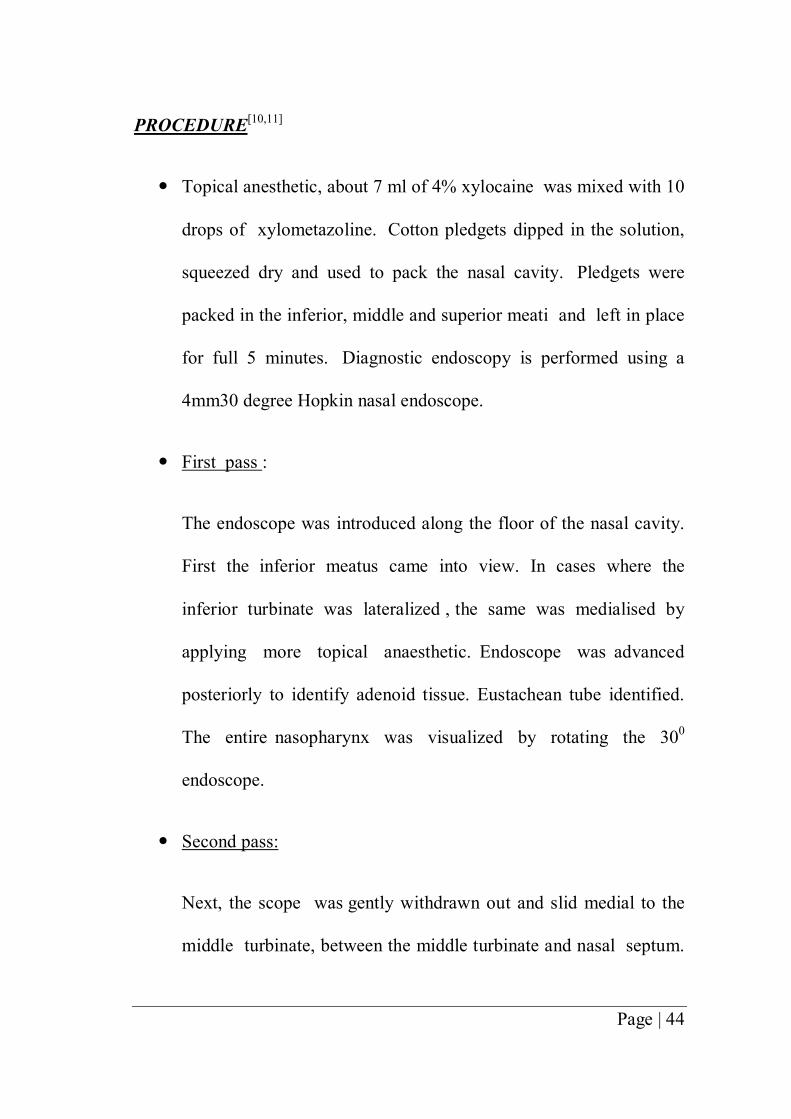
PROCEDURE[10,11]
� Topical anesthetic, about 7 ml of 4% xylocaine was mixed with 10
drops of xylometazoline. Cotton pledgets dipped in the solution,
squeezed dry and used to pack the nasal cavity. Pledgets were
packed in the inferior, middle and superior meati and left in place
for full 5 minutes. Diagnostic endoscopy is performed using a
4mm30 degree Hopkin nasal endoscope.
� First pass :
The endoscope was introduced along the floor of the nasal cavity.
First the inferior meatus came into view. In cases where the
inferior turbinate was lateralized , the same was medialised by
applying more topical anaesthetic. Endoscope was advanced
posteriorly to identify adenoid tissue. Eustachean tube identified.
The entire nasopharynx was visualized by rotating the 300
endoscope.
� Second pass:
Next, the scope was gently withdrawn out and slid medial to the
middle turbinate, between the middle turbinate and nasal septum.

Page | 45
The scope is gently slipped medial to the middle turbinate to view
the sphenoethmoidal recess.
� Third pass:
The shape and size of the middle turbinate as well as its
relationship to the lateral nasal wall and septum was evaluated.
The middle turbinate is gently medialised and the attachment of
the uncinate process is carefully noted. Any discharge in this
area also recorded. If accessory ostium is present it comes into
view now. Accessory ostium is present more posteriorly.
Normal ostium is actually not visible during diagnostic nasal
endoscopy. Accessory ostium is spherical in shape and
oriented anteroposteriorly, while the natural ostium of
maxillary sinus is oval in shape and oriented transversely.
PMO AMO
Ovoid in shape Circular
Anterior and superior to AMO Posrerior and inferior to PMO
Always present Not present in all cases
Difficult to see clinically Easily seen if present

Page | 46
CROSS SECTIONAL STUDY
A cross-sectional study examines the relationship between disease
(and other variables of interest as they exist in a defined population at a
single point in time or over a short period of time. Cross-sectional studies
can be thought of as providing a snapshot of the frequency of a disease or
other health related characteristics (e.g. exposure variables) in a
population at a given point in time.
Cross-sectional studies are used to assess the burden of disease or
health needs of a population and are particularly useful in informing the
planning and allocation of health resources.
SAMPLE SIZE CALCULATION
From the review of literature on earlier studies on the
presence of accessory ostium, we chose Kennedy and Stammberger’s
endoscopic study which gave a prevalence of 5% in the general
population[5]. In a study conducted in the Rajiv Gandhi University
of Health Sciences , Karnataka the prevalence of accessory ostia in
chronic sinusitis was found to be 22 %[46] In order to get an Indian
standard the value of 22% was used for calculation.

Page | 47
Sample size was calculated using OpenEpi Version 3 , open
source calculator.
Sample Size:X-Sectional, Cohort, & Randomized Clinical Trials
Two-sided significance level(1-alpha): 95
Power(1-beta, % chance of detecting): 90
Ratio of sample size, Unexposed/Exposed: 1
Percent of Unexposed with Outcome: 5
Percent of Exposed with Outcome: 22
Odds Ratio: 5.4
Risk/Prevalence Ratio: 4.4
Risk/Prevalence difference: 17
Kelsey Fleiss Fleiss with CC
Sample Size – Exposed 85 83 95
Sample Size-Nonexposed 85 83 95
Total sample size: 170 166 190
Prevalence = number of cases in a defined population at a given period of time
number of persons in the given period of time
Using the formula the prevalence of accessory ostium in
chronic sinusitis (Pc)and the prevalence of accessory ostium in
those without chronic sinusitis (Pn ) was calculated.

Page | 48
Pc = 30 %
Pn= 10 %
In order to find the significance of the values , a Chi Square
test was done on the sample.
The data captured in the sample size was arranged in the following
table to arrive the X2
Data type 1 Data type 2 Totals
Category 1 A b a + b
Category 2 C d c + d
Total a + c b + d a + b + c + d = N
The formula for Chi Square Distribution is
X2= (ad-bc)2 (a+b+c+d)
(a+b) (c+d) (b+d) (a+d)
AO + AO - Totals
Exposed (CRS +) 25 58 83
Not Exposed(CRS -)
8 75 83
Total 33 133 166
Chi Square distribution table.

Page | 49
probability level (alpha)
Df 0.5 0.10 0.05 0.02 0.01 0.001
1 0.455 2.706 3.841 5.412 6.635 10.827
2 1.386 4.605 5.991 7.824 9.210 13.815
3 2.366 6.251 7.815 9.837 11.345 16.268
4 3.357 7.779 9.488 11.668 13.277 18.465
5 4.351 9.236 11.070 13.388 15.086 20.517
Applying the formula above we get:
Chi square = 166[(25)(75) - (8)(58)]2 / (33)(133)(83)(83) = 10.93058
When a comparison is made between one sample and another, a
simple rule is that the degrees of freedom equal (number of columns
minus one) x (number of rows minus one) not counting the totals for
rows or columns.
For our data this gives (2-1) x (2-1) = 1.
We now have our chi square statistic (x2 = 10.93058), our
predetermined alpha level of significance (0.05), and our degrees of
freedom (df = 1). Entering the Chi square distribution table with 1 degree
of freedom and reading along the row we find our value of x2 (x2 =

Page | 50
10.93058) lies near 10.827. The corresponding probability is less than
0.001. Since a p-value of 0.001 is lesser than the conventionally accepted
significance level of 0.05 (i.e. p < 0.05) we reject the null hypothesis.
In other words, there is a statistically significant difference in the
proportion of AO in patients with Chronic Sinusitis and in those
without.
When p < 0.05, we generally refer to this as a significant difference.

Page | 51
6. RESULTS
Prevalence Of AO (Accessory ostium) In The Exposed Population
AO No. in exposed population (CRS)
YES 25
NO 58
TOTAL 83
Yes, 30%
No, 70%
PREVALENCE OF AO IN EXPOSED

Page | 52
Prevalence Of AO In The Unexposed Population
AO No. in unexposed population
YES 8
NO 75
TOTAL 83
Yes10%
No90%
PREVALENCE OF AO IN ENEXPOSED

Page | 53
Bar Chart Showing In Difference In Prevalence In Chronic Sinusitis
&Normal Population
Among the 83 CRS patients enrolled for study , 25 ( 30 % )
had an Accessory ostium and in the 83 unexposed persons it was 8
( 10 % )
AGE DISTRIBUTION OF AO IN CHRONIC SINUSITIS (CRS)
Subjects in the age group 15 – 45 years were chosen for the
study. The distribution of CRS in the various age groups , as
obtained from our recent study has been shown in the table and
chart are given below.
AO [VALUE]
AO [VALUE]
0%
5%
10%
15%
20%
25%
30%
35%
Exposed Not Exposed

Page | 54
0
2
4
6
8
10
12
14
16
18
20
15 - 20 21 - 25 26 - 30 31 - 35 36 - 40 > 40
No. of CRS patients in each age group
Age distribution of CRS
Age group Frequency of CRS
15 - 20 19
21 - 25 14
26 - 30 18
31 - 35 9
36 - 40 12
40 - 45 11

Page | 55
Age Distribution of AO in CRS
CRS AO % of AO
Age < 30 51 14 27.45
Age > 30 32 11 34.38
SIDE DISTRIBUTION OF AO IN CHRONIC SINUSITIS
The following are the tables and charts showing the side on
which the AO was present - right / left / bilateral in the group with
CRS.
27.45%
34.38%
0.00%5.00%
10.00%15.00%20.00%25.00%30.00%35.00%
40.00%
< 30 > 30
Age Distribution of AO in CRS

Page | 56
SIDE FREQUENCY
Right 9
Left 10
B/L 6
Right36%
Left40%
B/L24%
SIDE OF AO IN CRS

Page | 57
SIDE DISTRIBUTION OF AO IN THE UNEXPOSED
The following are the table and chart showing the side on
which the AO was present - right / left / bilateral in the unexposed
group.
SIDE FREQUENCY
Right 3
Left 4
B/L 1
Right37%
Left50%
B/L13%
SIDE OF AO IN UNEXPOSED

Page | 58
DISTRIBUTION OF AO ACCORDING TO ITS SITE ON THE
LATERAL WALL – ANF / PNF
AO is more commonly found on the posterior nasal
fontanelle (PNF) than the anterior nasal fontanelle (ANF). Following
is the frequency of the sites of occurrence in the two groups.
IN CHRONIC SINUSITIS
SITE FREQUENCY
ANF 6
PNF 19
AF24%
pF76%
SITE OF AO IN CRS
AF pF

Page | 59
IN THE UNEXPOSED
SITE FREQUENCY
ANF 0
PNF 8
AF0%
pF100%
SITE OF AO IN UNEXPOSED
AF pF

Page | 60
DISTRIBUTION OF DOUBLE OSTIA IN
CHRONIC SINUSITIS
The occurrence of AO is usually single and occasionally
multiple. In our study we found only 2 such cases and that too
only in those with CRS. Double ostia were not present in the
unexposed group in the present study.
NO. OF OSTIA FREQUENCY
DOUBLE 2
SINGLE 23
Double, 8%
One, 92%
DOUBLE OSTIA IN CRS

Page | 61
Distribution of signs & symptoms among CRS patients
with and without AO
The following tables show the variation in percentage of
symptoms and signs like discolored postnasal drip , halitosis ,
purulence on examination.
Also Recirculation phenomenon in the accessory ostium was found
only in 2 patients of CRS.
Frequency CRS with AO Percentage
Discolored
postnasal drip 24 25 96.00%
Halitosis 16 25 64.00%
Purulence on
examination 19 25 76.00%
Recirculation 3 25 12.00%

Page | 62
96.00%
76.00%
64.00%
12.00%
0.00%
20.00%
40.00%
60.00%
80.00%
100.00%
120.00%
Discolored postnasal drip
purulence on examination
Halitosis recirculation
symptoms & signs - CRS with AO
Frequency CRS without
AO Percentage
Discolored postnasal drip 56 58 96.55%
Halitosis 24 58 41.38%
Purulence on examination 42 58 72.41%
Recirculation 0 58 0.00%

Page | 63
96.55%
72.41%
41.38%
0.00%0.00%
20.00%
40.00%
60.00%
80.00%
100.00%
Discolored postnasal drip
purulence on examination
Halitosis recirculation
symptoms & signs - CRS without AO

Page | 64
7. DISCUSSION
INTERPRETATION AND ANALYSIS OF DATA
PREVALENCE OF ACCESSORY OSTIUM
Analysis of our present study shows that 25 out of 83 ie. 30
% CRS patients had an AO and that 8 out of 83 ie. 10 % ,
subjects without CRS had an AO. All of them were round in
shape. As mentioned earlier prevalence of AO shows a wide range
between 2 – 44 % , with cadaveric studies showing a higher
incidence than that on live subjects. The various studies and results
have been tabulated in the section ‘Review of Literature’.
Figure 8a Right sided AO in a patient with CRS

Page | 65
Figure 9 A closer look at the same AO showing the interior of maxillary sinus
Figure 10Showing 23 year old male without CRS with left PNF AO

Page | 66
Here we are quoting a few more studies that were not discussed in
the previous section.
In a prospective cohort study by Jog and Mc Garry, on
Rhinology clinic patients and General ENT clinical controls, they
reported that 7 % of Rhinology patients and 2% of the controls had
AO. Of the rhinology patients with rhinitis and sinusitis , 8 %
showed AO.[20]
AlperSindel et al [21] Manju et al [19] Kolvekar et al[22] Manjula Patil et
al[23]in their cadaver studies have reported the prevalence as 13.8% ,
18.5 % , 22.5% and 26% respectively.
AGE
Subjects chosen for the study belonged to the age group
between 15 and 45 years. In the present study the frequency of
CRS in each group was found to be the following –
51patients with CRS were below the age of 30, and 32 were
above the age of 30. Of the 51 patients below 30 years, 14 had an

Page | 67
accessory ostium, ie. 27.45 %. Of the 32 patients above 30 years, 11
had accessory ostia, ie. 34.38 %.
In the unexposed group, of the 35 individuals below 30 years 4
(11.42 %) had AO, and out of 48 above 30 years, 4 (8.33 %) had
AO.
Though literature says that AO can develop with advancing
age, such an association was not obtained in our study[24].
GENDER
According to the review of literature gender had no effect on
the development of AO. Hence no conscious effort was taken to
equalize the number of males and females while enrollment.
SIDE
In our study, of the CRS patients with AO, 10 (40 %) on the
Left, 9 (36 %) were on the Right and 6 (24 %) were present
Bilaterally. And in the unexposed group, 4 (50 % ) on the left, 3
(37%) on the right, and 1 (13 % ) were found bilaterally.

Page | 68
Figure 11 Bilateral AO (PNF in the a 16 year old male with CRS
In a study by Mladina et al , 68.3 % AO were bilateral in
patients with CRS as opposed to none the normal subjects.[17]
In another study by Mladina , Skitarelic and Casale on
patients with and without post nasal discharge , 57.5 % were present

Page | 69
bilaterally in those with post nasal discharge as against none in the
group of healthy subjects. There
is no mention on the right / left distribution in either of these
studies.[18]Kolvekar et al[22] have reported on a 2.66% laterality.
In a cadaver study by Kumar et al the findings were as
follows – right 66.7 % , left 33.3 %. There were no bilateral cases.[5]
In a cadaver study Manju et al they found that AO occurred
on the Right in 60% and on the Left in 40%. [19]
A radiological study by Sheetal et al showed 13 % Right and
11 % left.
Figure 12bilateral maxillaryostia in PNF

Page | 70
LOCATION OF ACCESSORY OSTIUM – ANF / PNF
In the group with CRS , 19 (76 %) AO were found on the
posterior nasal fontanelle and 6 ( 24 % ) in the anterior nasal
fontanelle. In the unexposed individuals , all the 8 AO were found
on the posterior nasal fontanelle.
PNF are larger than ANF and Accessory ostia are more
commonly found on the posterior nasal fontanelle[1,2,3,8,24]. Myerson (
1932 ) says AO commonly lies in the posterior nasal fontanelle
below and behind the natural ostium. In 23 % it they lie further
posteriorly, in 14 % inferiorly and 11 % superiorly.The findings in
this study corroborates the literature. Other studies - Mladina et al
(pnf - 19.3 %, anf–0.61 % in CRS and pnf – 0.48 %, anf – nil in
healthy subjects ), But a few studies which contradict it was also
found – Kumar et al ( cadaver study: pnf - 22.2 %; anf – 66.7 % ),
Manju et al (pnf – 25 %, anf – 70 % ), Manjula Patil et al ( pnf – 45.5
%, anf – 54.55%).

Page | 71
Figure 13AO in PNF
NUMBER OF OSTIA
Out of 25 AO in the CRS group 23 ( 92% ) were single
ostia and only 2 ( 8 % ) cases had double ostia. In the unexposed
group none had double ostia. Accessory ostia are usually single and
occasionally multiple. Levine .H however has commented that one
to three accessory ostia may be present , mostly in the anteroinferior
fonatanelle.[25]

Page | 72
Figure 14 Double Accessory Ostia
Kumar et al have reported a 44.4 % incidence of double AO in
their cadaver study. Manju Singhal et al have reported a 35 %
incidence of double AO , and all of them were in the anterior nasal
fontanelle. [19]
Manjula Patil et al in a cadaver study found 18 % double, 9 %
multiple and 72.2 % single ostia.

Page | 73
Again all these were cadaver studies. Of all the studies ,
cadaver studies have given a larger incidence of accessory ostia
and here in particular the incidence of multiple / double ostia are
also larger in these studies. This could be due to the fact that
moist nasal mucosa undergo shrinkage after death , and following
drying and fixing the fontanelles undergo damage resulting in the
formation of accessory ostia.[5]
Figure 15 Lower ostia was not visualized initially as
it was almost obscured by the uncinate

Page | 74
RECIRCULATION
Recirculation was found only in 3 ( 12 % ) cases of AO in
this study. All the three cases had post nasal drip and also
purulence on examination.
Figure 16 mucous moving out of AO
As shown in the figure below, the mucous that exits into the
middle meatus re enters the maxillary sinus through the accessory
ostium, taking along with it the pathogens from the nasal cavity. As
the cilia beat towards natural ostium , the secretion again moves up

Page | 75
towards it, only to repeat this vicious cycle. This results in
persistence of sinus infection.
In the figure above, secretion appears to be moving out of
the accessory ostium into the middle meatus. In the normal course,
though an accessory ostium is more advantageously placed than the
natural one, it does not take part in the physiological drainage of
the maxillary sinus secretions.
Figure 17 Circular phenomenon – the mucous secretion re entering
the maxillary sinus via the Accessory Ostium

Page | 76
Only in the presence of any blockade or obstruction of the
natural ostium does secretions get transported out through the
accessory ostium, under the effect of gravity.
ANALYSIS OF THE SYMPTOMS AND SIGNS
95 % of CRS patients complained of headache and 42 % had
facial pain. 80 out of 83 (96.4 % ) patients complained of post
nasal discharge. Hence there was no significant difference in its
presence between those with and without AO. Halitosis was a
complaint only for 49.3 %. However it was higher in those with
AO ( 64 % ) than those without AO (41.38 % ). Purulence on
examination was slightly higher in the presence of an AO ( 76% )
in contrast to those without ( 72.41 % ).But both these associations
could not be proved statistically as the p value was > 0.05 for both.
CHRONIC RHINOSINUSITIS[9],[12]
Chronic rhinosinusitis is one of the most common
otorhinolaryngologic problems affecting 32 million adults, or 16.3 %
of the adult population.[9]
Histopathologically, sinusitis can be defined as the inflammation
of nasal and paranasal mucosa. The underlying bone can undergo

Page | 77
osteitic changes. Since the mucosal lining is contiguous between the
nose and sinuses, one can’t spared if the other is involved. Hence
the term Sinusitis has now been expanded into Rhinosinusitis.
There are two categories of changes in Chronic Rhinosinusitis
1. Polypoid mucosal changes with eosinophilia ( which causes
more damage to the nasal mucosa )
2. Submucosal serous gland hyperplasia.
The Rhinosinusitis Task Force of the American Academy of
Otolaryngology Head and Neck Surgery have developed a
classification.[12]
CLASSIFICATION DURATION
ACUTE RHINOSINUSITIS (ARS) 7 days to ≤ 4 weeks
SUB ACUTE RHINOSINUSITIS 4 weeks to 12 weeks
RECURRENT ACUTE
RHINOSINUSITIS
≥ 4 episodes of ARS / year
CHORONIC RHINOSINUSITIS
(CRS)
≥ 12 weeks
Acute exacerbation of Chronic
Rhinosinusitis
Sudden worsening of CRS with
return to baseline.

Page | 78
The 1997 Task Force of Rhinosinusitis gave a consensus
report on standardizing the criteria for diagnosing Chronic sinusitis.
Symptoms and signs comprise the cornerstone for this criteria. The
criteria have been divided into major and minor.

Page | 79
The 1997 Task Force of Rhinosinusitis report also described
the following physical findings which they divided into 2 groups.
These are important but not required as a part of TFR criteria.
They are -
1. those findings that are accessible to all clinicians (
examination of face and anterior rhinoscopic findings )
2. those accessible only to specialists ( nasal endoscopy)
The specificity of endoscopy is 85 %.
External Findings Anterior Rhinoscopy Nasal Endoscopy
Swelling and
erythema of
maxillary, frontal,
ocular, orbital areas
Hyperemia Blue discoloration of
turbinates
Edema OMC / ostia purulence
Crusts Polyp
Purulence Septal deviation
Polyp Concha Bullosa
Changes in symptoms
after topical
decongestion
Paradoxical Middle
Turbinate
Obstructive anomalies

Page | 80
The Chronic Rhinosinusitis Task Force has published another
set of guidelines for diagnosing adult Chronic Rhinosinusitis where
it recommends to continue the use of 1997 TFR CRS symptoms
and to add the existence of physical findings – Polyps , Purulence ,
Polypoid changes [9]
[13]There is another staging system which can be considered
the most accepted CT staging system called the Lund-Mackay CT
staging system. It is a very simple system and has a high degree
of interobserver and intraobserver agreement. It is the only system
recommended by the Task Force on Rhinosinusitis for outcomes
research.
Scoring is based entirely on CT findings. Each sinus is given a of 0/1/ 2:
0 = no opacification,
1 = partial opacification,
2 = total opacification .

Page | 81
There is a separate grading for frontal, maxillary, anterior
ethmoid, posterior ethmoid, and sphenoid sinuses. The ostiomeatal
complex is also included in the score. The total possible score is 24.
LUND-MACKAY COMPUTED TOMOGRAPHY STAGING SYSTEM
No
abnormality
Partial
opacification
Total
opacification
Anterior
ethmoid
L
R
0
0
1
1
2
2
Posterior
ethmoid
L
R
0
0
1
1
2
2
Maxillary L
R
0
0
1
1
2
2
Frontal L
R
0
0
1
1
2
2
Sphenoid L
R
0
0
1
1
2
2
Non obstructed Obstructed
Osteomeatal
complex
R
L
0
0
2
2

Page | 82
MANAGEMENT ALGORITHM FOR CHRONIC SINUSITIS[9]
DIAGNOSIS OF CRS SUGGESTED BY HISTORY
SINONASAL ENDOSCOPY
POSITIVE ENDOSCOPY NORMAL
PURULENCE CT PNS
OBTAIN CULTURE POSITIVE NORMAL
- Consider other diagnosis
POLYP -Allergy evaluation
MANAGEMENT OF POLYP
INITIATE TREATMENT ALLERGY EVALUATION

Page | 83
TREATMENT OF CHRONIC RHINOSINUSITIS[14]
Our upper respiratory tract which includes the nasopharynx is
a storehouse for pathogenic bacteria which cause respiratory tract
infections including rhinosinusitis.
An Upper Respiratory Tract Infection has several phases - An
initial viral infection which lasts about 10 days undergoes complete
recovery in the majority. A minority develop an acute secondary
bacterial infection by facultative aerobic bacteria. This if not
resolved pave way for anaerobic bacteria of the oral flora.
EARLY VIRAL INFECTION (~ 10 DAYS)
Complete recovery(majority) 0.5 %
Secondary acute bacterial
infection [Streptococcus
pneumoniae, H.influenzae,
Moraxella catarrhalis]
Not resolved
Anaerobic bacteria of oral flora

Page | 84
MICROBIOLOGY OF ACUTE SINUSITIS[14]
The community acquired acute purulent maxillary, frontal and
ethmoid sinusitis are caused by Streptococcus pneumoniae,
Hemophilus influenzae, Moraxella catarrhalis, beta hemolytic
streptococci.
Staphylococcus aureus and Hemophilus influenza are the
causative agents in acute sphenoid sinusitis.
In nosocomial infections , pseudomonas and gram negative
rods are the culprits.
Whereas fungal sinusitis is seen in the immunocompromised
and diabetics.
MICROBIOLOGY OF CHRONIC SINUSITIS[14]
Anaerobes like Prevotella , Fusobacterium , Peptostreptococcus
and aerobes like Staphylococcus aureus , Moraxella catarrhalis and
Hemophilus sp. are the pathogens in chronic sinusitis.
Of these, Staphylococcus aureus, Hemophilus, Prevotella and
Fusobacterium affect more than one third patients.

Page | 85
Pseudomonas aeroginosa and gram negative aerobic bacilli are
seen in sinus infections following sinus surgeries.
In chronic sinusitis, Polymicrobial infection is more common.
Hence it is more difficult to eradicate with narrow spectrum
antibiotics.
In chronicity, aerobes and facultative species are replaced by
anaerobes.
This is thought to be due to –
1. Selective pressure of antibiotics that enable resistant organisms
to survive.
2. Persistent edema results in decreased blood supply ;
consumption of oxygen by aerobic bacteria leads to decreased
oxygen tension. There develops an increased acidity in the
sinuses as well. These conditions are favorable for anaerobic
growth.

Page | 86
TREATMENT OF CHRONIC SINUSITIS[14]
MEDICAL MANAGEMENT
Antibiotics should be effective against both aerobes and
anaerobic Beta Lactamase Producing Bacteria. The following
antibiotics can be used for treatment.-
Oral & Parenteral Forms Only Parenteral
Amoxicillin Clavunate Cefoxitin
Clindamycin Cefotetan
Chloramphenicol Cefmetazole
Macrolide + Metronidazole Imipenem
Newer generation - Trovafloxacin
Along with antibiotics, topical steroid sprays, nasal saline
irrigation and mucolytics is also useful.[15]
In infections by aerobic gram negative bacteria like
pseudomonas aeroginosa
1. parenteral aminoglycosides
2. 4th generation cephalosporins – Cefipime , Ceftazidime

Page | 87
3. Fluoroquinolones ( oral / parenteral ) in post pubertal age groups.
These antibiotics should be given for 21 days. The treatment
may be extended up to 10 weeks.
When a patient does not respond to medical management ,
surgical drainage should be done. Antibiotic treatment alone without
surgical drainage of pus may not result in eradication of disease.
The reason for failure of medical treatment in chronic
sinusitis may be due to the fact that chronically inflamed mucosa
with a lesser blood supply is a poor medium of transport of
antimicrobial agents to the affected tissue even in the presence of a
therapeutic blood level. Also the reduced oxygen tension and acidic
pH of the inflamed sinuses interfere with antimicrobial activity of
the drugs.
[16]Another reason for drug resistance in chronic sinusitis are
supposedly the formation of Biofilms. Biofilm is a structured
community of cells , enclosed in a self produced polymeric matrix
and adherent to an inert or living surface. It may be made of
bacterial or fungal cells that communicate with each other in a

Page | 88
cooperative manner. The matrix is slime like made of polysaccharide,
nucleic acids and proteins.
In a study on New Zealand white rabbits, sinusitis was
induced by pseudomonas aeroginosa. On days 1, 5, 10, 20 – pus was
cultured to get pseudomonas aeroginosa. The mucosa on scanning
under a microscope revealed growth and biofilm.
Another study was conducted on the sinonasal specimens
from patients undergoing revision sinus surgery or office based
debridement. All were antibiotic non responders. Their specimens
also showed findings consistent with presence of biofilms.
SURGICAL MANAGEMENT
INDICATIONS[42]
Absolute Indications
1. complications of sinusitis
2. expansile mucoceles
3. allergic / invasive fungal sinusitis
4. suspected neoplasia

Page | 89
Relative Indications
1. Symptomatic nasal polyps unresponsive to medical therapy
2. Chronic / Recurrent acute sinusitis unresponsive to medical
therapy
Functional Endoscopic Sinus Surgery has shown to be the
approach of choice in chronic sinusitis. Though the association
between anatomical variations and recurrence of disease is still
under dispute , surgery is acceptable when the site of obstruction
complements the area of recurrent symptomatology.
When a patient complains of pain , one should keep in mind
that the nasal mucosa is more sensitive to pain caused by a
blocked ostium than a thickened mucosa[42]. Its easier to surgically
intervene in patients who have recurrent or persistent symptoms , or
those with imminent complications than those with minor sinus
disease of doubtful significance. Nasal obstruction , congestion and
post nasal discharge are indicative of uncomplicated sinus disease
whereas severe pain associated with pressure changes like an air
travel points to obstruction of the sinus[42].

Page | 90
PREOPERATIVE EVALUATION
Prior to surgery, a patient requires culture directed
antibiotics which according to the severity of the disease may be
given for up to 2 or more weeks. Steroids may put into use in
case of polyposis or hyperactive mucosa. A preoperative course of
20 – 30 mg prednisone for 3 – 10 days will suffice.
Before surgery, the patient should undergo diagnostic nasal
endoscopy to review the anatomy and pathology, to rule out any
acute infection and to take cultures for intraoperative and
postoperative antibiotic selection. [42]
Preoperative CT evaluation of each anatomic site for any
variation should be done closely. The areas to stress are - the skull
base , the medial orbital wall, ethmoid vessels, posterior ethmoid,
maxillary sinus medial wall, sphenoid sinus, frontal recess and
frontal sinus [42].
Extent of Surgery [42]
Mucosal preservation is the norm in Functional Endoscopic
Sinus Surgery. It should always be borne in mind that unnecessary
stripping of mucosa will lead to exposure of bone and that

Page | 91
denuded bone leads to delayed healing. Ciliary action may not come
back to normal in those sites where the bone remains bare for more
than 6 months.
The surgery should extend one stage beyond the disease
established by the Computed Tomogram , or that identified during
surgery. It has been shown that the underlying bone is also
involved in Chronic sinusitis , and that removal of the mucosa alone
does not solve the problem unless the osteitic bone is removed.
This is particularly important in areas like the uncinate which is
more severely involved.
ANTROSTOMY [42]
Theoretically speaking the size of the antrostomy is to be
kept small. This is to protect it from over ventilation and its
consequences, viz., nitrous oxide washout , slowing of ciliary activity
and decreased bacteriostatic activities.
The uncinate bone occupies a considerable part of the medial
wall of the maxillary sinus and is commonly affected by osteitic
changes. When a diseased uncinate is incompletely removed, disease
persists and scarring in the region also occurs. So in minimal

Page | 92
disease , a small opening of the ostium is all that is necessary.
Whereas in case of a long standing and diffuse chronic sinusitis ,
with evidence of osteitis on CT scan or during surgery, a complete
removal of the uncinate and a wide middle meatal antrostomy is
advisable. If the medial wall of the maxillary sinus behind the
antrostomy is displaced into the nasal airflow because of a medially
extending maxillary sinus , air will directed into the maxillary sinus
during inspiration. To avoid this , the medially plasced wall is to be
removed up to the pterygoid plate.
What is to be done in the presence of an accessory ostium
will be discussed in the coming section.
More important than the size of the antrostomy is its
continuity with the natural ostium , lest recirculation of mucous and
persistent infection despite surgery will result. Alternatively if the
normal ostium remains closed with an open neo ostium , disease
will remain in the periosteal area associated with pooling of
secretions and resultant infection as the cilia only beat towards the
natural ostium.

Page | 93
Rest of the steps of FESS are not being discussed here as
the discussion is being limited to Chronic Maxillary sinusitis.
COMPLICATIONS OF ENDOSCOPIC ANTROSTOMY[42]
Complications are fortunately rare in middle meatal
antrostomies. If present they are -
1. Bleeding
2. Facial pain
3. Numbness ( injury to alveolar nerves supplying the meatal
wall of maxillary sinus)
4. Nasolacrimal duct injury / epiphora
5. Synechiae
6. Blindness( possible, but usually associated with ethmoidectomy)
MAXILLARY SINUSITIS& ACCESSORY OSTIUM
Maxillary sinus is the largest of all human sinuses. As a
result of evolution when man attained an erect posture, the primary
maxillary ostium ended up being located at a higher level resulting

Page | 94
in the maxillary sinus draining against gravity. Maxillary sinusitis is
the result of non dependent drainage and impedance of mucociliary
action. The natural ostium also opens in an angle to the coronal
plane.
Sinus disease and Accessory Ostium – How are they related ?
If in the presence of an intact uncinate process , you are able
to see an opening which takes you into the maxillary antrum, it is
rather an Accessory Ostium than a natural ostium.[32,37]Accessory
ostium is also known as Giralde’s orifice[43]. They are found over
the weak areas on the lateral nasal wall called fontanelles which are
devoid of bone and covered only by mucosal membrane of the
nasal cavity on one side and that of the maxillary sinus on the
other side with intervening connective tissue.
It may be assumed that
1. Sinus infections damage fontanelles. Here the formation of an
accessory ostium can be likened to a tympanic membrane
perforation. When pus collects in the maxillary sinus it finds
a way out by perforating through the potential weak areas
called fontanelles on the lateral wall of nose. It happens more

Page | 95
commonly through the larger posterior fontanelle, which results
in the formation of AO. This is the Acquired Development
Hypothesis of accessory ostium.[6]
2. AO may be a cause of sinus disease by causing disturbance
in the mucociliary transport and also by allowing pathogens
to easily enter the maxillary sinus.
3. If looked at this way , it can also be said that an acute nasal
or sinus infection damages the fontanelles which do not have
a bony component , thus creating an AO , which in turn leads
to recurrent sinusitis.
Mucous clearance in maxillary sinus is exclusively by
Mucociliary action. It happens against gravity. The cilia in the
sinus beat only towards the Primary maxillary ostium. Though an
Accessory maxillary ostium is located in a more advantageous
position with respect to gravity , the secretions are not transported
out through it as one might assume.
In accessory ostia of up to 4 mm, the secretions with a normal
viscosity bypass around[1,31, rather than pass through it. If a larger
accessory ostium is present, the part of the mucous carpet moving

Page | 96
through the center of the accessory ostium alone gets transported
out into the middle meatus. The portion of the mucous in the
periphery passes along its margins are taken into the natural
ostium. On the other hand the secretion that has already been
transported out through the natural ostium can reenter through the
accessory ostium , and this time when it enters the mucous carries
with it all the pathogens that have got adhered to its outer viscous
layer. Thus the pathogens in the nasal cavity gain free access into
the antrum via this ‘extra hole’. This recirculation can go on and
on , thus perpetuating the infection and working as a vicious cycle.
[6,26]A similar scenario can happen following endoscopic sinus
surgery for chronic sinusitis. While doing a middle meatal
antrostomy, if the surgically created opening is not connected to the
natural ostium, it is equivalent to an iatrogenic accessory ostium.
This is called the ‘Missed Ostium Sequence’[30,32]. Here the due to
the same recirculation phenomenon, a persistent sinusitis results
despite the surgery. Joining the surgically created ostium with the
natural ostium solves the problem.

Page | 97
Shaffer, Ramadan et al conducted a study on chronic sinusitis
patients with and without accessory ostium. They studied the ciliary
area on maxillary sinus biopsy specimens taken during endoscopic
sinus surgery. Electron microscopic examination of the specimens
showed a significant difference in the ciliary area in the two
groups. Those with accessory ostium showed a reduced ciliary area
than those without it.
APPLICATIONS OF THE AWARENESS OF ACCESSORY
OSTIUM
Why is the awareness about accessory ostium important ?
In today’s scenario where CT scan nasal endoscopy play a
major role in the diagnosis and treatment of sinusitis, it is
advisable to be aware of all the anatomical and pathological
variants of the nasal cavity and paranasal sinuses, one such being,
our Accessory ostium.
The presence of an accessory ostium in a symptomatic
individual can be pointer that he is suffering from Chronic sinusitis
or say chronic maxillary sinusitis. Often you can even witness the
mucous reentry during a diagnostic nasal endoscopic session. Thus

Page | 98
it can aid in your decision making and also in planning the
surgery.
The radiologist should be aware of this condition as in a CT
of the paranasal sinuses, it will as an additional communication
between the maxillary sinus and the nasal cavity. sometimes when
stacked one above the other, it may appear as two openings in a
coronal film.
Van Alyea ( 1936 ) stated that in 20 % cases , the primary
maxillary ostium is unapproachable due to the altered configuration
of the uncinate or bulla , or due to the size of the ostium. In such
cases when you fail to canulate the primary maxillary ostium , the
accessory ostium can be used to irrigate the maxillary sinus (
Levine et al , 1993 ). Or one can even use the fontanelles to create
an alternate pathway to reestablish the ventilation.
There is a recent new invention called the ‘Nasal Ventilation
System’[41] which is an apparatus that is used for making an
accessory ostium on the lateral nasal wall that can be used for
purposes like irrigation, ventilation and drug delivery. It consists of
an introducer, a ventilation tube, an irrigation catheter, a secondary

Page | 99
irrigation catheter, balloon catheter. The introducer with the
ventilation tube and grommet is used to create an accessory ostium
and then gain access through it. The irrigation catheter is channeled
through the ventilation tube which irrigates the sinus cavity. it can
also be used to express out the sinus contents. A secondary
irrigation catheter or a balloon catheter may be introduced through
the outer irrigation catheter to reach the deeper parts of the sinus.
Moreover this apparatus can be used to deliver topical fluids , anti-
inflammatory drugs, drug delivery substances, drug impregnated coils
, foams or beads.
Now that we know the importance of an accessory ostium ,
lets find out how to identify it.
Distinguishing features between AMO and PMO[7,36]
Following are the differences between an AMO and PMO –
1. PMO lies in a more oblique plane. The anterior lip of PMO
lies more lateral than the posterior lip. Whereas the AMO lies
in the same ( sagittal ) plane as the lateral nasal wall.

Page | 100
2. PMO is ovoid in shape whereas AMO is circular.
3. In the fontanelles , PMO tends to lie anterior and / or superior
to AMO.
4. PMO is always present whereas AMO is present only in a
certain percentage of the population , a figure which shows a
wide variation between 2 – 44 %.
5. PMO is quite difficult to see clinically , whereas an AMO is
easily seen if present.

Page | 101
ENDOSCOPIC SINUS SURGERY AND ACCESSORY OSTIUM
When a patient is taken up for Endoscopic Sinus Surgery , a
Middle Meatal Antrostomy ( MMA ) is not always required if the
natural ostium is found patent after uncinectomy. If however an
accessory ostium is found , it has to be joined to the natural
ostium.[27,28,29,30,34,38] It can be done by introducing a back – biting
forceps into the accessory ostium and extending it anteriorly into
the natural ostium[28]. Failure to identify the natural ostium , and
therefore connecting it to the accessory ostium will result in a
circular flow of mucous, and persistence of infection[27,29,30]. In other
words , a Pseudo Middle Meatal Antrostomy[36 ] is created with a
resultant failure of Functional Endoscopic Sinus Surgery[29].
Figure 18 a posterior fontanelle AO encountered during FESS being shown using a Ball probe

Page | 102
Figure 19 AO being widened and backbiting forceps being
introduced into it

Page | 103
.
Figure 20 AO being joined to natural ostium by cutting anteriorly using Back biting forceps.
Figure 21A wide Middle MeatalAntrostomy after joining
the natural and accessory ostia

Page | 104
While doing a middle meatal antrostomy , the ostium can be enlarged
into either fontanelle. But a thing to be kept in mind is that any
damage especially over the posterior ostial edge will result in
disruption of mucociliary clearance [27] .
If the ostium is not found , one may perforate the posterior
fontanelle just above the attachment of the inferior turbinate. In this
scenario if one fails to join it to the natural ostium recirculation
will result , as mentioned earlier[27]. Hence the continuity with the
natural ostium may be confirmed using a 450 or 700 endoscope.
But Kennedy contradicts this statement, when he says that in the
absence of an accessory ostium, it is best not to enter the maxillary
sinus through the posterior fontanelle, lest the mucosa should be
invaginated into the sinus and stripped from adjacent walls[28].
Patients coming with recurrent sinusitis following endoscopic
sinus surgery should undergo an endoscopic examination. After a
course of medical treatment a CT scan of the paranasal sinuses
should be taken and carefully examined for the presence of a
separate natural ostium and an accessory ostium or a posterior
fontanelle ostium. If it is positive, the patient should be taken up
for a second sitting where the two ostia should be joined. As

Page | 105
mentioned above a back biter should be inserted into the accessory
ostium and cut forwards to the natural ostium. A microdebrider can
be used to trim away the excess tissue after creating this tissue
edge.
Enlarging The Maxillary Ostium – Still An Enigma !
There are two schools of thought in this regard.
Earlier FESS surgeons supported the enlargement of the
maxillary ostium towards the posterior fontanelle thus creating a
wide Middle Meatal Antrostomy[29]. They advocate that a small neo
ostium neither allows a proper visualization of the maxillary sinus ,
neither do they allow adequate tissue removal if required from
inside the sinuses[30]. Hence a 1 x 2 cm wide neo ostium may be
created at the expense of both the anterior and posterior
fontanelles.
Whereas now the supporters of the idea - as the uncinate is
located close to the maxillary ostium , the narrow transient space
between it is prone to obstruction and that the removal of the
uncinate alone is sufficient to restore a healthy sinus. But little
data is available in support of either argument. [29]

Page | 106
Nitrous oxide is synthesized by NO synthase ( NOS ) in the
mucosa of the sinuses. There are three types of NOS - Type I, II,
III Of these, the most important in the production of Nitrous oxide
in the sinuses is Type II. It is induced by bacterial infections. It
stimulates ciliary motility , inhibits infections by bacteria, fungi and
viruses , thereby playing an important role in the local innate
defense mechanism of the sinuses. [29]
Previous gas studies by computational modelling have also
proved that increased sinus ventilation in the form of a large
ostium or multiple ostia is more detrimental than beneficial in
terms of the increased nitrous oxide washout , mucosal drying
( partly because sinuses have a lower density of mucous and serous
glands as compared to the nasal cavity ), reduced mucociliary
activity and pathogen entry[4].
Kennedy et al have stated that the primary maxillary ostium
has a dimension of 5 x 5 mm and that its significant enlargement
leads to the dilution of sinus NO concentration and subsequent
bacterial colonization of the sinus. [29]

Page | 107
In a study conducted by Wormald, he correlated the
concentration of NO with that of the size of the maxillary sinus
ostium. There appeared to be a significant reduction in
concentration of NO in those sinuses and nasal cavities with large
maxillary ostia ( > 5 x 5 mm ). But this study did not prove that a
lower concentration of nitrous oxide predisposed to recurrent
infections.
Hence this area requires more study and scientific proof.
Can an accessory ostium be closed ?
A study conducted by M.A Penttilä during the period 2011 –
2013 shows that accessory ostia can be closed ! They did the
closure endoscopicaly making use of rotation flaps from the
undersurface of the middle turbinate as abdominal fat grafts did not
give satisfactory results.[39] The accessory ostium was denuded and
the anterior end of the rotation flap was inserted into it. The
posterior end remained attached to the middle turbinate itself. The
procedure gave a fairly good closure rate. On follow up visits
nasal endoscopic examination confirmed closure of accessory ostia

Page | 108
in 76 % patients and 75 % perforations with transient mild side
effects.
Though the procedure has a theoretical advantage , the long
term results are yet to be monitored as it is a very recent study.

Page | 109
8. CONCLUSION
· There have been various earlier studies on the presence of
Accessory Ostium with a wide range of results right from 0
% to 44 %.
· In our study we found the prevalence of accessory ostium as
30 % in Chronic sinusitis and 10 % in those without the
disease.
· In both the groups it was more on the left side and in the
posterior nasal fontanelle than the anterior nasal fontanelle.
· Only 2 % of the diseased individuals had double ostia and
recirculation was present only in 3 cases (12 %).
· Although majority of the patients presented with headache
and facial pain irrespective of the presence of an accessory
ostium, halitosis had a higher incidence in those with
accessory ostium; it was not statistically significant though.

Page | 110
To conclude we would say that the presence of an accessory
ostium can be considered as an indicator of maxillary sinus disease,
along with the other criteria for chronic sinusitis and that it can
help surgeons in their decision making as to whether a surgery is
required or not.

9. ANNEXURES
A. BIBLIOGRAPHY
1. Functional endoscopic sinus surgery, The Messerklinger Technique, H.Stammberger, History of Nasal Endoscopy pg 1- 13.
2. Diseases of the sinuses- diagnosis and management, David W. Kennedy, James Zinreich ;Anatomy of Paranasal Sinuses Pg 1-12.
3. Scott Brown’s Otorhinilaryngology Head and Neck Surgery 7th edition, vol. 2, part 13- Anatomy of Nose and PNS. Pg-1315-1343
4. Computational modeling of flow and gas exchange in models of the human maxillary sinus. C. M. Hood, R. C. Schroter, D. J. Doorly, E. J. S. M. Blenke and N. S. TolleyJ ApplPhysiol107: 1195–1203, 2009. First published july16,2009;doi:10.1152 /japplphysiol .91615.2008
5. Accessory maxillary ostia: Topography and clinical application, Kumar et al, Deartment of Anatomy, Lady Hardinge Medical College, New Delhi, India; J Anat. Soc. India 50(1) 3-5 (2001)
6. Development of maxillary accessory ostium following sinusitis in rabbits.; SelhattinGenc, MugeOzcan, Ali Titiz, Adnan Unal ; Kecioren Education and Research hospital ENT clinic & Ankara NumuneEduction and Research Hospital, Ankara, Turkey, Rhinology , 46,121-124, 2008.
7. Diseases of the sinuses- diagnosis and management, David W. Kennedy, James Zinreich; chapter 21: Paediatric Sinus Surgery ;David S. Parsons,GaryNishioka; pg 275
8. Otolaryngology Clinics of North America ,Sinusitis,April 2004, vol.37;No.2; pg 221-242 –Aanatomic Variations and Pearls- Functional Endoscopic Surgery.
9. Otolaryngology Clinics of North America ,Sinusitis,April 2004, vol.37 ; pg 243-252- Adult Chronic Rhinosinusitis : Diagnosis; Anand K. Deviah
10. Diagnostic nasal endoscopyBy Dr. T. Balasubramanian M.S. D.L.O. drtbalu’s otolaryngology online
11. Scott Brown’s Otorhinilaryngology Head and Neck Surgery 7th edition, vol. 2, part 13-Nasal Endoscopy pg1344-1354

12. Scott Brown’s Otorhinilaryngology Head and Neck Surgery 7th edition, vol. 2, part 13-chapter 113; Rhinosinusitis –Michael S. Benninger ; pages 1439-1448.
13. Head & Neck Surgery--otolaryngology, Volume 1 ,section ii – Rhinology and allergy ;pg412; Byron J. Bailey, Jonas T. Johnson, Shawn D. Newlands.
14. Otolaryngology Clinics of North America ,Sinusitis,April 2004, vol.37; pg-253-266 –Microbiology and antimicrobial management of sinusitis; Itzhak Brook, MD MSc
15. Otolaryngology Clinics of North America ,Sinusitis,38(2005) , pg 1143-1153; Classification, management of rhinosinusitis and its complications.
16. Otolaryngology Clinics of North America ,Sinusitis,38(2005) pg 1193-1201- Bacterial biofilm:do they play a role in chronic sinusitis; James N. Palmer MD
17. The Two Hole Symdrome- Mladina et al- American journal of Rhinology and Allergy 23:6 pg602-4; PubMed NCBI
18. ‘Two Hole Syndrome is present in more than half of post nasal discharge patients?’R.Mladina et al ,ActaOtolaryngologia 05/2010;130(11):1274-7; PubMed.
19. Anatomy of Accessory Maxillary Ostium with Clinical Application- Manju Deepak Singhal; International journal of Medical Sciences and public Health 2014;3(3):327-329
20. ‘How Frequent are Accessory Ostia?’Jog.M,McGarry GW; Journal of Laryngology otology 2003April; 117(4):270-2.
21. ‘Endoscopic Cadaver Study- Accessory Maxillary Ostium’ AlperSindel et al, 2014;41(2):262-267 Dicle med j -0921.2014.02.0413: http://www.readperiodicals.com/201406/3386392721.html
22. ‘Study of anatomical variation of lateral nasal wall by endoscope’ –Kolvekar, Khusru, Pradeep:Unique journal of Medical and Dental Sciences 2014,02(2) 117-121.
23. ’Ostium MaxillareAccessorium- A Morphological Study’ ;Manjulapatil ;National Journal of Clinical Anatomy vol 1(4) pg 171-175

24. Lang J. Clinical Anatomy of the Nose, Nasal Cavity and Paranasal Sinuses. Stuttgart: Georg ThiemeVerlag, 1989.
25. Sinus Surgery : Endoscopic and microscopic approaches. edited by Howard L. Levine, M. Pais Clemente ; pg 31
26. ‘Iatrogenic maxillary sinus recirculation and beyond’;Gutman M1, Houser S.;Ear Nose Throat J. 2003 Jan;82(1):61-3:abstract.
27. Scott Brown’s Otorhinilaryngology Head and Neck Surgery 7th edition, vol. 2, part 13-chapter 117; Surgical Management of Rhinosinusitis; pg- 1478 – 1499.
28. Diseases of the sinuses- diagnosis and management, David W. Kennedy, James Zinreich ; Functional Endoscopic Sinus Surgery –Anaesthesia, Technique, postoperative management; pg:218.
29. Endoscopic sinus surgery- Anatomy, 3D Reconstruction and Surgical Technique chapter 5 ; Peter John Wormald ;Thieme publication.
30. Minimally Invasive Endonasal Sinus Surgery-principles, Techniques, Results, Complications, Revision Surgery - Werner G. Hosemann, Rainer K. Weber, Rainer E. Keerl, Valerie J.Lund - Maxillary Sinus procedures -Endonasal Maxillary Sinus Fenestration
31. Peterson's principles of Oral and Maxillofacial Surgery. 2nd edition, vol 1;MichaelMiloro, peter Larsen
32. Sinus Surgery : Endoscopic and microscopic approaches. edited by Howard
L. Levine, M. Pais Clemente; chapter 10.
33. Anatomical Principle Of Endoscopic Sinus Surgery; RenukaBradoo. Pg111.
34. Micro Endoscopic Surgery Of The Paranasal Sinuses And The Skull Base: Aldo C. Stamm, Wolfgang Draf; Surgical Approaches To Sinusitis:Overview Of Techniques, Pg187
35. Endoscopic Surgery Of The Paranasal Sinuses And Anterior Skull Base ;Malte Erik Wigand; Pg 43
36 Complications In Endoscopic Sinus Surgery Diagnosis, Prevention And Management, S.K Kaluskar ;Pg 29 – 30
37. Manual of Endoscopic Sinus Surgery: and its Extended Applications ; Daniel Simmen , Nick S. Jones , pg114

38. Minimally Invasive Maxillofacial Surgery, Maria J. Troulis, Leonard B. Kaban pg53 – 55
39. Closure of accessory maxillary ostium during endoscopic sinus surgery for CRS - M.A. Penttilä; ENT, Centro HospitalarLisboaOcidental, Lisbon, Portugal - http://www.europeanrhinologicsociety.org/Congress/Abstracts/ERS-0496.pdf
40. Effects of Maxillary Sinus Accessory Ostia on Mucosal Ciliary Area-Marcus D. Shaffer, MD; Hassan H.Ramadan,MD; Diane Schwegler-Berry,MS; Jonathan Franks, MS;2012 Combined Sections Meeting; The American Laryngological Rhinological and OtologicalSociety.Inc; http://www.triomeetingposters.org/poster/effects-of-maxillary-sinus-accessory-ostia-on-mucosal-ciliary-area-the/
41. Nasal ventilation system and method of using same-inventor- Patrick C. Melder Patent WO2010025299A2 - Nasal ventilation system and method of using same - Google Patents.
42. Head & Neck Surgery--otolaryngology, Volume 1,edited by Byron J. Bailey, Jonas T. Johnson, Shawn D. Newlands, Section ii Rhinology and Allergy , chapter 34- Complications of sinus surgery , pg 480
42. Diseases of the sinuses- diagnosis and management, David W. Kennedy, James Zinreich; chapter 16A –Functional Endoscopic Sinus Surgery: Concepts, Surgical indications, and instrumentation. 197-210 : David W.Kennedy.
43. “A nasal endoscopic study of anatomical variations of the human nasal ostiomeatalcomplex"Dr. Bharathi D, Dept. of Anatomy © Rajiv Gandhi University of Health Sciences, Karnataka.

B. PROFORMA
NAME: AGE/SEX I.P.No:
D.O.A:
D.O.S:
DOD
CHIEF COMPLAINTS
1.Facial pain/pressure
2.Nasal obstruction
3.Nasal discharge/discolored postnasal drip
4.Hyposmia/anosmia
5.Headache
6.Fever (all nonacute)
7.Halitosis
8.Dental pain
9.Fatigue
10.Cough
11.Ear pain/pressure/fullness
H/O OF PRESENT ILLNESS
PAST HISTORY
H/o DM/ epilepsy/ PTB/ drug allergy/ bleeding diathesis /SHT/Cardiac disorder

FAMILY H/O:
PERSONAL H/O: Diet
Bladder and bowel habits
Addictions
GENERAL EXAMINATION:
Anemia Jaundice Cyanosis Clubbing Pedal edema
GLNE
CVS: RS: P/A: CNS:
VITALS:
LOCAL EXAMINATION:
NOSE:
EXTERNAL CONTOUR
DORSUM
VESTIBULE
SEPTUM

ANTERIOR RHINOSCOPY:
Septum
Inf. Turbinate
Inf. meatus
Mid turbinate
Mid meatus
RIGHT
LEFT
PNS TENDERNESS :
RIGHT LEFT
COTTON WOOL TEST
COLD SPATULA TEST
COTTLE’S TEST
E/O THROAT:
ORAL Cavity
OROPHARYNX
IDL EXAMINATION
POST NASAL EXAMINATION

Examination of Ear:
PRE AURICULAR
AREA PINNA
POST AURICULA
R AREA EAC TM
RIGHT
LEFT
DIAGNOSTIC NASAL ENDOSCOPY
ANAESTHESIA:
TECHNIQUE:
ACCESSORY OSTIUM
AO +/-
Single/double/multiple Side – left/ right
Location- ANF/PNF
Shape Recirculation

C. ETHICAL COMMITTEE APPROVAL LETTER

D. PATIENT INFORMATION SHEET

E. INFORMED CONSENT FORM

G. PLAGIARISM

Si. No hosp. no. name age sex duration
Facp/pr N.obs Nasal
dis
Hyposmia/anosmi
a
pnd poe hal Cough H fever(na)
earp/pr dent.P Fatigu
eton.Hyp CSOM max.
tend. AO side site number double shape recir
1 178136 prema 45 f 8 y y y y n y y y y y n y n n b/l y y L Pf 1 n R n2 179234 renjit kumar 16 m 5 y n y n n y y y y y n n n n y n y y b/l pf ,pf 1,1 n r,r n3 180123 saritha 30 f 5 y n y n y y y n n y n n n n n n y y L af 2 y r,r n4 199873 suganta 43 f 1 y y n n n y n n n y n n n n n n y y r pf 1 n r n5 335122 nagamma 35 f 5 m y n n n y y y n y n n n n n n y y L af 1 n r y6 286906 saritha 39 f 1 y y n n n y n y n y n n n n n n y y b/l af,af 1,1 n r n7 287100 divya 16 f 5 y y y y y y y y y y y y n n n b/l y y r pf 1 n r n8 272774 sudhakar 20 m 6 y n y y n y n n n y n n n n n n n y l pf 1 n r n9 272895 sumati 44 f 2 y y n y n n n n y y n n n n n n y y r af 1 n r n
10 1404126 chitra 34 f 3 y y n y y y y y y y n n n n n r y y r Pf 1 n r n11 346371 andal 32 f 2 y y y n y y n n y y n y n n n n y n n n 0 n n n12 341963 panjavarnam 41 f 1 y n y y n y y y n y n n n n n n n n n n 0 n n n13 348422 karanraj 20 m 2 y n y n n y y n n y n n n n n n n n n n 0 n n n14 369172 soundarya 18 f 1 y n y y n n y y y y y n n n n n n n n n 0 n n n15 341175 tamil selvi 35 f 3 y n y y y y y y y y n n n n n n y n n n 0 n n n16 344918 rakesh 19 m 2 y n y n n y y y y y y n n n n n y y b/l pf,pf 1,1 n r n17 315473 sagunthala 36 f 6 m n y y y y n n n y n n n n n n y n n n 0 n n n18 323783 rakesh sharma 26 m 2 y n n y n y y n n y n n n n y n y n n n 0 n n n19 322439 nirmal kumar 24 m 1 y n y y n y n n n n n n n n n n y n n n 0 n n n20 364446 jayanthi 40 f 1 y n y n n y y n n y n n n n n n n n n n 0 n n n21 472816 veerappan 45 m 2 y n y y n y y n y y n n n n n n y n n n 0 n n n22 369450 saddam hussain 18 m 1.5 y n y n n y y y n y n n n n n n y n n n 0 n n n23 376467 summayiah 23 f 4 y n y n n y y n y y n n n n n n n y l pf 2 y r n24 389887 sumathi 40 f 6 y y y n n y n y n y n n n n n n y y b/l af,af 1,1 n r n25 396811 meharunissa 30 f 7 y n y n y y y n n y n n n n n n y y l Pf 1 n r n26 409537 savithri 45 f 7 y n y y n y y y y y n n n n n n y y r Pf 1 n r n27 370756 manju 25 f 8 m n y n n y n n y y y n n n n n y n n n 0 n n n28 377686 murugesan 45 m 2 y n y n n y n n y y y n n n n n y n n n 0 n n n29 499901 srimathi 18 f 1 y n y y n y n n y y n n n n y n y n n n 0 n n n30 361724 malliga 42 f 2 y y y n n y y n n y n n n n n n n n n n 0 n n n31 364125 krishnakumar 31 m 5 y n y y n y y n n y n n n n n n n n n n 0 n n n32 361033 devi 32 f 3 y y y n n y y y n y n y n n n n y n n n 0 n n n33 371383 lakshmi 26 f 1 y n y y y y n n n y n n n n n n y n n n 0 n n n34 371890 manikandan 23 m 4 y y n y n y y y y y n n n n n n y y b/l pf,pf 1,1 n r n35 268712 rajesh 17 m 5 y y y y y y y y y y n y n n n b/l y n n n 0 n n n36 251928 priya 30 m 2 y n y n y y y n n y n n n n n n y n n n 0 n n n37 397156 kamala 28 f 1 y y y n y y y y y y y y n n n n n n n n 0 n n n38 394050 valli 34 f 3 y n y n n y y y y y n n n n n n y n n n 0 n n n39 313700 murali 20 m 5 y y n y n y y n y y n n n n n n y n n n 0 n n n40 384163 satyamoorthy 40 m 6 y n y n y y n y n y n n n n n n y n n n 0 n n n
EXPOSED

Si. No hosp. no. name age sex duration
Facp/pr N.obs Nasal
dis
Hyposmia/anosmi
a
pnd poe hal Cough H fever(na)
earp/pr dent.P Fatigu
eton.Hyp CSOM max.
tend. AO side site number double shape recir
41 394320 megalai 26 f 2 y y y y y y y y y y n n n n n n y n n n 0 n n n42 529390 kaja sheikh 42 m 8 y y y y y y n y y y y y n n n n y n n n 0 n n n43 378288 kaja moideen 28 m 6 m n y n y y n y y y n y n n n n n n n n 0 n n n44 394292 madhavi 24 f 6 m n y n y y y n y y n y n n n n n n n n 0 n n n45 407288 kathira bee 30 f 1 y n y y y y y y n y n n n n n n n n n n 0 n n n46 399391 usha 31 f 8 m n y y y y n n n y n n n n n n n n n n 0 n n n47 330783 shakila 30 f 1y n n y y y y n y y n y n n n n n n n n 0 n n n48 395195 geetha 17 f 3 y n y y y y y y y n y y n n y n y n n n 0 n n n49 370259 mathi 40 m 2 y n n y n y y y y y n y n n n n y n n n 0 n n n50 372700 amsa 30 f 1 y n y y n y y y y y n y n n n n y n n n 0 n n n51 394888 ravanammal 28 f 1.5y n y n n y y y y y n n n n n y y n n n 0 n n n52 419294 kairunisa 22 f 2y y y y y y y n y y n n n n n n y n n n 0 n n n53 388429 rizwana 18 f 10 m n y y n y y n y y n n n n y n y n n n 0 n n n54 1400562 sujatha 30 f 3y y y n y y y y y y n y n n n n y n n n 0 n n n55 1400244 ambilka 24 f 1y n y n y y y n n y n n n n n n y n n n 0 n n n56 1400168 valliyammal 36 f 5y y n y n y y n y n y n n n n b/l y n n n 0 n n n57 1400827 yuvarna` 20 f 2y y y y y y y n y y n n n n y n y n n n 0 n n n58 1401304 mujeeb 38 m 4y y n n n y y n y y n y n n n n y n n n 0 n n n59 1402152 pratap singh 22 m 3y y y y y y y y y y n y n n y n y n n n 0 n n n60 1402218 guru 36 m 8y n y n y y y n y y n y n n n n y n n n 0 n n n61 1402530 usman 26 m 3y y n y n y y n y y n y n n n n y n n n 0 n n n62 1404917 jebaraj 39 m 2y y y y y y y y y y y y n n n n y y r pf 1 n r y63 372089 purushothaman 43 m 5y y y n y y y n n y n y n n n n y n n n 0 n n n64 76129 revathy 32 f 1y n y n n y y n y y n y n n n n y n n n 0 n n n65 11529 suganya 23 f 2y y y n n y y y y y y y n n n n y n n n 0 n n n66 141196 alangaram 21 f 1 y y Y y n y y n y y n y n n n n y y L pf 1 n r y67 154587 babu 18 m 1 y n y n n y y n y y n y n n n n y n n n 0 n n n68 167481 manoj 19 m 1.5y y y y n y y y y y n y n n n n y y r af 1 n r n69 174952 karthika 23 f 2 y n n y y y n n n y n n n n n n y n n n 0 n n n70 95263 renugadevi 20 f 10 m y L n y n n n n y n n n n n n y n n n 0 n n n71 166981 santosh 15 m 1 y n n y n y n n y y n y n n n b/l y n n n 0 n n n72 175139 logi 37 m 3 y y n n n y y n y y n n n n n n y y L pf 1 n r n73 141725 ranjith kumar 16 m 1 y y n y n y n y y y n n n n y b/l n n n n 0 n n n74 184597 surya 24 m 2 y n L y n y y y n y n n n n n n y n n n 0 n n n75 189906 nandakumar 28 m 3 y n r n n y y n y y n y n n n n y n n n 0 n n n76 188137 vinitha 16 f 2 y y n y n y y n n n n y n n n n y n n n 0 n n n77 586801 selvi 25 f 1 y y Y n y y y y y y n y n y n n y y r pf 1 n r n78 586811 sarath kumar 28 m 3 y y Y n y y y y y y n y n n n n y y r pf 1 n r n79 248380 narasimhan 26 m 5 y n n y y y y y y y n n n n n n n y L pf 1 n r n80 234698 malai 36 f 4 y n Y n n y n y n y n n n n n n y y l pf 1 n r n81 234703 jaya 41 f 2 y n n y y y y y y y n y n n n n n n n n 0 n n n82 228973 tamil selvan 27 m 1.5 y y y n n y n y y y y n n n y n n n n n 0 n n n83 273100 ghouse basha 22 m 6 y n n y n y y n y y n n n n n b/l n y b/l pf,pf 1,1 n r n

sl no. hosp. no.name age sex diagnosis Facp/pr N.obs Nasal
dis
Hyposmia/anosmi
a
pnd poe hal Cough H fever(na)
earp/pr dent.P Fatigue ton.
Hyp CSOM max.tend. AO side site double number shape recircula
tion
1 49790 manyan 23 M ES n n n n n n n n n n n n n n n n n y L pf n 1 r n2 346013 vinaya kumar 23 m otmyc n n n n n n n n n n n n n n n n n n n n n 0 n n3 346015 valii 24 f vn n n n n n n n n n n n n n n n n n n n n n 0 n n4 267071 kanchana 33 f mhc n n n n n n n n n n n n n n n n n n n n n 0 n n5 44483 viji 26 f vn n n n n n n n n n n n n n n n n n n n n n 0 n n6 348411 lakshmi 44 f SNHL n n n n n n n n n n n n n n n n n n n n n 0 n n7 343370 bavani 40 f mng n n n n n n n n n n n n n n n n n n n n n 0 n n8 343394 munusamy 43 m vc palsy n n n n n n n n n n n n n n n n n n n n n n n n9 349468 nirmala 30 f vc cyst n n n n n n n n n n n n n n n n n n n n n 0 n n
10 450126 silendran 44 m NV n n n n n n n n n n n n n n n n n n n n n 0 n n11 352230 hari 21 m NV n n n n n n n n n n n n n n n n n n n n n 0 n n12 353284 kubendran 45 m vc cyst n n n n n n n n n n n n n n n n n n n n n 0 n n13 361931 sivaranjani 18 f vn n n n n n n n n n n n n n n n n n n n n n 0 n n14 361791 mohammad 24 m otmyc n n n n n n n n n n n n n n n n n n n n n 0 n n15 46477 nagaraj 44 m NV n n n n n n n n n n n n n n n n n n n n n 0 n n16 46980 sundaramoorthy41 m NV n n n n n n n n n n n n n n n n n n n n n 0 n n17 358516 easa 45 m vn n n n n n n n n n n n n n n n n n n n n n 0 n n18 199933 rajagopal 44 m vc palsy n n n n n n n n n n n n n n n n n y L pf n 1 r n19 391052 venkatesh 40 m mhc n n n n n n n n n n n n n n n n n y L pf n 1 r n20 228404 tilak bahadur 45 m mhc n n n n n n n n n n n n n n n n n y L pf n 1 r n21 586819 anand 26 m vb n n n n n n n n n n n n n n n n n y b/l pf,pf n 1,1 r n22 586712 abirami 32 f mhc n n n n n n n n n n n n n n n n n y R pf n 1 r n23 294560 ragupathy 16 m NV n n n n n n n n n n n n n n n n n y R pf n 1 r n24 29456 siva 23 m NV n n n n n n n n n n n n n n n n n y R pf n 1 r n25 298370 syamala 35 f mhc n n n n n n n n n n n n n n n n n n n n n 0 n n26 227687 anwar 19 m otmyc n n n n n n n n n n n n n n n n n n n n n 0 n n27 228420 devi 40 f dcr n n n n n n n n n n n n n n n n n n n n n 0 n n28 345342 priya 20 f mng n n n n n n n n n n n n n n n n n n n n n 0 n n29 25287 gangadharan 40 m ES n n n n n n n n n n n n n n n n n n n n n 0 n n30 38030 thasneem 36 f ES n n n n n n n n n n n n n n n n n n n n n 0 n n31 147643 lakshmi 46 f mhc n n n n n n n n n n n n n n n n n n n n n 0 n n32 27698 manikandan 23 m mhc n n n n n n n n n n n n n n n n n n n n n 0 n n33 20876 bhuvaneswari 30 f pas n n n n n n n n n n n n n n n n n n n n n 0 n n34 289756 sulekha 28 f sms n n n n n n n n n n n n n n n n n n n n n 0 n n35 28703 manikandan 17 m NV n n n n n n n n n n n n n n n n n n n n n 0 n n36 253793 jabamalai 39 m NV n n n n n n n n n n n n n n n n n n n n n 0 n n37 387367 thangaraj 45 m pl.ad n n n n n n n n n n n n n n n n n n n n n 0 n n38 128908 nazeer 33 m mhc n n n n n n n n n n n n n n n n n n n n n 0 n n39 138766 pugazanthi 30 m mhc n n n n n n n n n n n n n n n n n n n n n 0 n n40 128936 chinnadurai 43 m MOE n n n n n n n n n n n n n n n n n n n n n 0 n n41 146735 raziya 45 f mhc n n n n n n n n n n n n n n n n n n n n n 0 n n42 37698 murugesan 36 m mhc n n n n n n n n n n n n n n n n n n n n n 0 n n43 140567 shakeela 16 f pas n n n n n n n n n n n n n n n n n n n n n 0 n n44 38965 fiaz 40 m sms n n n n n n n n n n n n n n n n n n n n n 0 n n
UNEXPOSED

sl no. hosp. no.name age sex diagnosis Facp/pr N.obs Nasal
dis
Hyposmia/anosmi
a
pnd poe hal Cough H fever(na)
earp/pr dent.P Fatigue ton.
Hyp CSOM max.tend. AO side site double number shape recircula
tion
45 37609 seenivasan 39 m bppv n n n n n n n n n n n n n n n n n n n n n 0 n n46 27462 murugan 37 m NV n n n n n n n n n n n n n n n n n n n n n 0 n n47 37876 sukumari 40 f osc n n n n n n n n n n n n n n n n n n n n n 0 n n48 348470 uma 28 f kob n n n n n n n n n n n n n n n n n n n n n 0 n n49 369150 arun 16 m NV n n n n n n n n n n n n n n n n n n n n n 0 n n50 315489 karuna 17 f fb throat n n n n n n n n n n n n n n n n n n n n n 0 n n51 47278 raziya 24 f NV n n n n n n n n n n n n n n n n n n n n n 0 n n52 348410 balaraman 40 m mhc n n n n n n n n n n n n n n n n n n n n n 0 n n53 369140 mohini 37 f dcr n n n n n n n n n n n n n n n n n n n n n 0 n n54 315389 pushpa 38 f NV n n n n n n n n n n n n n n n n n n n n n 0 n n55 344900 abdul khader 39 m mhc n n n n n n n n n n n n n n n n n n n n n 0 n n56 368123 lathika 44 f mng n n n n n n n n n n n n n n n n n n n n n 0 n n57 272809 sagunthala 23 f jug ect n n n n n n n n n n n n n n n n n n n n n 0 n n58 322420 maheswari 30 f fb ear n n n n n n n n n n n n n n n n n n n n n 0 n n59 273110 guru 30 m mhc n n n n n n n n n n n n n n n n n n n n n 0 n n60 346350 mukesh 18 m NV n n n n n n n n n n n n n n n n n n n n n 0 n n61 140478 jagadeesan 37 m pl.ad n n n n n n n n n n n n n n n n n n n n n 0 n n62 347136 john 35 m val cys n n n n n n n n n n n n n n n n n n n n n 0 n n63 261895 sampath 35 m mhc n n n n n n n n n n n n n n n n n n n n n 0 n n64 342874 jeelan 17 m bvfp n n n n n n n n n n n n n n n n n n n n n 0 n n65 433785 malkonadyan 45 m fb throat n n n n n n n n n n n n n n n n n n n n n 0 n n66 346279 silambarasan 34 m fb throat n n n n n n n n n n n n n n n n n n n n n 0 n n67 1E+06 ragupathy 40 m thy fis n n n n n n n n n n n n n n n n n n n n n 0 n n68 457482 fathima 17 f NV n n n n n n n n n n n n n n n n n n n n n 0 n n69 230852 kuppusamy 45 m mhc n n n n n n n n n n n n n n n n n n n n n 0 n n70 204362 chitra 38 f mng n n n n n n n n n n n n n n n n n n n n n 0 n n71 427584 shanti 40 f bvfp n n n n n n n n n n n n n n n n n n n n n 0 n n72 270283 shalini 19 f mng n n n n n n n n n n n n n n n n n n n n n 0 n n73 1E+06 hassan 29 m smf n n n n n n n n n n n n n n n n n n n n n 0 n n74 1E+06 jahangir 25 m leu. pl n n n n n n n n n n n n n n n n n n n n n 0 n n75 341298 syed 19 m ran n n n n n n n n n n n n n n n n n n n n n 0 n n76 272700 mukesh 17 m pub.ph n n n n n n n n n n n n n n n n n n n n n 0 n n77 163800 kaliyan 45 m kob n n n n n n n n n n n n n n n n n n n n n 0 n n78 145622 anandan 43 m mng n n n n n n n n n n n n n n n n n n n n n 0 n n79 204376 kathija bee 29 f pap. Ca n n n n n n n n n n n n n n n n n n n n n 0 n n80 305674 umapathy 45 m vb n n n n n n n n n n n n n n n n n n n n n 0 n n81 306186 malliga 43 f mhc n n n n n n n n n n n n n n n n n n n n n 0 n n82 2E+06 saravanan 39 m mhc n n n n n n n n n n n n n n n n n n n n n 0 n n83 1E+06 sakunthala 37 f vn n n n n n n n n n n n n n n n n n n n n n 0 n n

KEY TO MASTER CHART
y - yes
n - no
b/l - bilateral
r - round
af - anterior fontanelle
pf - posterior fontanelle
fac p/pr - facial pain/pressure
N. obs - nasal obstruction
N. dis - nasal discharge
pnd - post nasal discharge (discolored)
poe - purulence on examination
hal - halitosis
recir - recirculation
H - headache
fever (na) - fever all non acute
ear p/pr - ear pain/pressure
dent. p -dental pain

ton. hyp - tonsillar hypertrophy
CSOM - chronic suppurative otitis media
max. tend - maxillary tenderness
AO -accessory ostium
ES - eagle's syndrome
otmyc - otomycosis
vn - vocal nodule
mhc - master health check up
SNHL - sensory neural hearing loss
vc palsy - vocal cord palsy
vc cyst - vocal cord cyst
NV - normal volunteer
vb - vestibulitis
dcr - dacrocystitis
mng - multinodular goitre
pas - preauricular sinus
sms - submandibular sialadenitis
pl.ad - pleomorphic adenoma
jug ect - jugular ectasia

fb ear - foreign body ear
val cys - vallecular cyst
bvfp - bilateral vocal fold paralysis
thy fis - thyroglossal fistula
smf - sub mucous fibrosis
leu. pl - leucoplakia
ran - ranula
pub.ph - puberphonia
pap. ca - papillary carcinoma thyroid
MOE - malignant otitis externa
bppv - benign paroxysmal postional vertigo
osc - otosclerosis
kob - keratosis obturans
fb throat - foreign body throat