Acute effects of dropsets among different resistance training methods in upper body performance
Upload
khangminh22Category
view
0download
0
A Study on Comparison of different Phenotypic methods for detection of Extended Spectrum Beta Lactamase
Production among Enterobacteriaceae in Urinary Tract Infection
in a Tertiary Care Centre
DISSERTATION SUBMITTED FOR
BRANCH – IV - M.D. DEGREE
(MICROBIOLOGY)
APRIL 2017
THE TAMILNADU
DR.M.G.R.MEDICALUNIVERSITY
CHENNAI, TAMILNADU

BONAFIDE CERTIFICATE
This is to certify that the dissertation entitled “A STUDY ON COMPARISON
OF DIFFERENT PNENOTYPIC METHODS FOR DETECTION OF
EXTENDED SPECTRUM BETA LACTAMASE AMONG
ENTEROBACTERIACEAE IN URINARY TRACT INFECTION IN A
TERTIARY CARE CENTRE” submitted by Dr.R.SASIREHA to the Tamil Nadu
Dr. M.G.R. Medical University, Chennai in partial fulfillment of the requirement for
the award of M.D degree Branch– IV (Microbiology) is a bonafide research work
carried out by her under direct supervision & guidance.
DR.M.R.VAIRAMUTHU RAJU,M.D., DR.P.A.T.JAGATHEESWARY,M.D., DEAN Director i/c, Madurai Medical College & Institute of Microbiology, Govt.Rajaji Hospital, Madurai Medical College, Madurai. Madurai.

CERTIFICATE FROM THE GUIDE
This is to certify that the dissertation “A STUDY ON COMPARISON OF
DIFFERENT PNENOTYPIC METHODS FOR DETECTION OF EXTENDED
SPECTRUM BETA LACTAMASE AMONG ENTEROBACTERIACEAE IN
URINARY TRACT INFECTION IN A TERTIARY CARE CENTRE” is a
bonafide record of work done by DR.R.SASIREHA, under my guidance and
supervision in the Institute of Microbiology, Madurai Medical College, Madurai
during the period of her Post graduate study of M.D. MICROBIOLOGY from 2014 –
2017.
Prof.Dr.S.RADHAKUMARI Institute of Microbiology, Madurai Medical College, Madurai

DECLARATION
I, DR.R.SASIREHA declare that, I carried out this work on, “A STUDY
ON COMPARISON OF DIFFERENT PNENOTYPIC METHODS FOR DETECTION
OF EXTENDED SPECTRUM BETA LACTAMASE AMONG
ENTEROBACTERIACEAE IN URINARY TRACT INFECTION IN A TERTIARY
CARE CENTRE” at the Institute of Microbiology, Madurai Medical College. I also
declare that this bonafide work or a part of this work was not submitted by me or any
others for any award, degree or diploma to any other University, Board, either in India
or abroad.
This is submitted to The Tamilnadu Dr. M. G. R. Medical University, Chennai
in partial fulfillment of the rules and regulations for the M.D. Degree examination in
Microbiology.
Place : MADURAI DR.R.SASIREHA
Date :

ACKNOWLEDGEMENT
I am grateful to the Dean, DR.M.R.VAIRAMUTHU RAJU M.D., Madurai
Medical College and Government Rajaji Hospital, Madurai for permitting me to
carry out this study.
I would like to express my deep sense of gratitude and sincere thanks to
Professor Dr.P.A.T.Jagatheeswary, M.D., Director i/c, Institute of Microbiology,
Madurai Medical College, for her constant help, guidance and encouragement given to
me throughout this study.
I express my sincere thanks to Prof. Dr.R.Vibhushanan M.D.,
Prof.Dr.V.Dhanalakshmi M.D., and Prof.Dr.S.Radhakumari M.D., for their
valuable suggestions and moral support given to me throughout the study.
I would like to express my sincere thanks to my guide
Prof.Dr.S.Radhakumari M.D, co-guides Dr.C.Sugumari and Dr.N.Rammurugan
M.D., their valuable suggestions and guidance given to me.
I would like to express my sincere thanks to all my Assistant professors
Dr.S.Maheshprabhu M.D, Dr.N.Anuradha M.D., Dr. J. Suriakumar M.D.,
Dr.M.R.VasanthaPriyan M.D., Dr.D.Saradha M.D., Dr.G.Manjula M.D
Dr.S.Nalayini M.D. for their valuable suggestions and guidance given to me.

I am thankful to all my colleagues Dr.S.Meerah, Dr.B Sree Bavai Malar,
Dr.MinuGeorge, Dr.J.Vijay Anand Dr.S.Rajeswari Dr. R.Vasanthi for their
moral support and cooperation rendered during the work.
I extend my thanks to all staff members, Institute of Microbiology for giving
full cooperation and timely help in carrying out the laboratory studies.
I extend my thanks to my parents, my family members, my husband
Dr.B.Shanthakumar daughter Karunya and son Krish for their esteemed moral
support rendered during the study.

CONTENTS
S.No Title Page. No
1 INTRODUCTION
1
2 AIMS &OBJECTIVES
11
3 REVIEW OF LITERATURE
12
4 MATERIALS AND METHODS
39
5 RESULTS
53
6 DISCUSSION
73
7 SUMMARY
85
8 CONCLUSION
87
9 BIBILIOGRAPHY
88
10 ANNEXURE i)Preparation of Gram stain ii)Preparation of Media iii)Data Collection proforma iv)Master Chart v)Ethical Committee Approval Form vi)Anti Plagiarism Certificate
102

INTRODUCTION

1
INTRODUCTION
Infectious diseases are the major cause of morbidity and mortality and
also responsible for worsening the living conditions of many millions people
around the World.41 Molecular studies of pathogenesis of microorganisms
revealed an explosion of information about the various microbial and host
molecules that lead on to infections and diseases41.Urinary tract infection (UTI)
is one of the most common infection prevalent in humans after respiratory and
gastro-intestinal infections. It leads to both community as well as hospital
acquired infections (HAI) in developing world and seeks medical attention.
About 150 million people are being affected due to UTI across the world95.In
2010, 3.1% of the people who had been visited emergency department were
due to UTI19and the incidence rate was about 50,000/million of people in
India95.UTI leads to a number of deaths either due to acute infection or chronic
renal failure.
Urinary tract infection is defined as a condition in which the presence
and multiplication of bacteria anywhere in the Urinary tract32.Severity of
Urinary Tract Infections mainly depends on factors such as age, time,
geographical distribution and immune status. The presence of bacteria in the
urine is termed as Bacteriuria. The Suprapubic aspiration is most reliable
specimen as it is sterile, followed by catheterized urine. There is always a
higher risk of contamination of urine samples collected by the patients. Hence
Kass introduced the term significant bacteriuria(Kass1956) and it is defined

2
that the presence of 105 or more of the same organism per ml of urine78 to
exclude the bacterial contamination in urine.
Classification of Urinary Tract Infection25
The classification of UTI is based on many factors-Anatomically UTI is
classified into, Upper urinary tract infection (involves kidney and ureter) and
lower urinary tract infection (involves urethra and bladder), with symptoms as
Symptomatic bacteriuria and Asymptomatic bacteriuria (ABU) and clinically it
is classified into Uncomplicated and Complicated .Uncomplicated urinary tract
infection means infection occurring in normal genitourinary tract without prior
instrumentation. Complicated urinary tract infection means infection occurring
in individual having either structural or functional abnormalities in
genitourinary tract or having indwelling catheters.
Epidemiology and Etiology
UTI is one of the commonest infections which needs medical attention.
During their life time about 10% of people experience UTI in some form14.It is
one of the important cause for HAI and it accounts for 35% of all HAI.
Neonates, young women, prepubertal girls, elderly men, and individual with
any structural abnormality or on immune suppression have higher risk for
Urinary Tract Infections.
UTI occurs commonly in women than men except in infants and elderly
people41 .In neonatal period UTI incidence is higher in male child due to the
congenital anomalies of urinary tract and prostatic hypertrophy in elderly. The

3
incidence of UTI is higher in female which is about 50-80 % in whom 20-30%
of them have recurrent episodes usually within 2 weeks. ABU was found to be
more common among 20-40 years of age i.e 5% and it increases to 40-50% in
elderly men and women41. Most of the UTI are monobacterial (95%)67 and
Escherichia coli is the frequent cause of both community and hospital acquired
UTI which accounts for 75%114 In contrast, recurrence is common in structural
abnormalities and associated with polymicrobial infections67. Proteus,
Pseudomonas, Enterococcus faecalis, Klebsiella, and Enterobacter are common
in complicated UTI.
Risk factors25, 67 - All ages; In both female and male any Urological surgery,
Catheterization, Stents, any obstruction in the urinary tract, neurogenic bladder,
renal transplantation are the common risk factors. In female with previous UTI,
and in males (children and young adults) who have not undergone circumcision
are more prone for infection.
Adult female-.Sexual intercourse, use of diaphragm, and pregnancy are the
risk factors. Hormonal changes common during pregnancy make urethra and
ureter more susceptible to bacterial adhesion and infection. A 70% of pregnant
women develop glycosuria due to increased plasma volume and decreased
urine concentration resulting in an increase of bacterial growth28. UTI is more
common in female because Urethra of female is short, so that bacteria have less
distance to travel to reach the bladder. In addition urethra is in close proximity
to moist, warm vulvar and perianal areas, which are less effective in preventing

4
bacterial entry. Similarly during sexualinter course bacteria can enter in to the
urethra and incomplete emptying of bladder in diaphragm users, as it pushes
against the urethra and infection occurs followed by stasis of urine.
Elderly people -In female due to the estrogen deficiency there will be loss of
vaginal lacto bacilli which leads them more prone for infection. In post
menopausal women- Cystocele is common and affects complete bladder
emptying and leads to residual urine followed by recurrent UTI. In elderly male
decrease of prostatic secretion which has bactericidal effect also leads to
urinary infection.
Pathogenesis Clinical manifestation and Complications14,67
Three major routes by which bacteria invade are8ascending route,
haematogenous and lymphatic spread.
Ascending route - Microorganisms (mainly gram negative bacteria) from
gastro intestinal tract able to colonize periurethral region and also in vagina.
Adhesion in the uroepithelium is the important step in pathogenesis. Following
colonization these organisms gain entry into the bladder through
instrumentation or any other manipulation, multiplication happens in the
bladder resulting in cystitis. From bladder enter into ureter, and then to the
kidney. Haematogenous route- Seeding of the kidney occurs due to the
systemic infection. Lymphatic spread- Whenever there is increase of bladder
pressure chance of increase in UTI due to the lymphatic flow to the kidney.

5
Host defenses in urinary tract depends mainly on - factors like PH,
osmolality, organic acids of urine , presence of bactericidal activity, cytokines
and peptides of mucosa of urinary tract, inhibitors of bacterial adherence like
Tammhorsfall proteins, lactoferrin, SIgA, low molecular weight
oligosaccharides and mucopolysaccharide of bladder are responsible for host
defense mechanisms. Humoral and cell mediated immunity, Prostatic
secretions are also taking part in this action.
In neonates and children less than 2 years the symptoms are nonspecific. Major
manifestations are fever, failure to thrive, and vomiting. In children greater
than 2 years localizing symptoms such as dysuria, frequency, and abdominal or
flank pain are also observed12. In adults, frequent painful micturation is seen
due to irritation of vesicle as well as urethral mucosa due to bacteria. Patient
may sometimes experience heaviness or pain in suprapubic region and urine
may be associated with a tinge of blood or frank blood.
Upper UTI usually manifest with fever with or without chills, frequency,
dysuria urgency along with flank tenderness. UTI is asymptomatic in elderly
individual and if symptomatic it is not diagnostic as they has been
experiencing hesitancy, dysuria, frequency and incontinence very often.
Patient with indwelling catheter usually presented with fever and flank pain but
without lower urinary tract symptoms.
In Pediatric age group, infection may sometime spread outside the
urinary tract resulting in orchitis in boys and sepsis in both sex. The Most

6
serious complication is Pyelonephritis. In adults recurrent urethritis resulting in
urethral narrowing, prostatitis and permanent kidney damage are other
important complications. Life threatening complication is sepsis and renal
failure. In order to reduce the complication in UTI early intervention with
appropriate and adequate dose of antimicrobials. Antimicrobials should bind
the target site effectively in order to disrupt the cellular processes for cessation
of bacterial growth.
Beta-Lactam antibiotics are used to treat UTI due to their high efficacy,
less toxic and well tolerated by the people at any age group. Beta lactam
antibiotics act on both gram positive and gram negative bacteria. Antimicrobial
resistance is mainly due to any interruption in the essential steps for
antimicrobial action it will results in bacterial resistance to antimicrobial
action14.Different aspects of resistances are, Biologic resistance,
environmentally mediated resistance and microorganism mediated resistance
which is further classified into intrinsic resistance and acquired resistance8.
Resistance to Beta lactams14,67
1. Enzymatic destruction of β-lactam ring by Beta lactamase, produced by
the organism
2. Altered target due to the mutation in PBP (Penicillin Binding Protein)
resulting in reduced affinity for antibiotic or not able to bind Beta
lactams.

7
3. Decreased uptake or its efflux of the drug due to change either in
number or character of porin channels of outer membrane so that the
drug does not reach the target site8.
Worldwide resistance to Beta lactam antibiotics among gram negative
uropathogens are increasing because of inappropriate and extensive use of
antimicrobial agents. Antibiotic resistances are mainly due to the production of
Beta lactamases by uropathogens.
Beta-lactamases are family of enzymes produced by the bacteria which
inactivate 59 the Beta - lactam antibiotics by splitting the amide bonds in the
Beta lactam ring. Even prior to the use of penicillin in medical practice beta
lactamase production was observed in Escherichia coli. Penicillinase was the
beta lactamase produced by the Staphylococcus aureus which was plasmid
encoded. Due to that there was quick spread of resistance to the other clinical
isolates. Naturally occurring chromosomally mediated beta lactamases are
usually found in most of the gram negative bacteria. The development of this
type of beta lactamases are mainly due to the antibiotic pressure by the
organism that found in the environment which are able to produce beta lactam.
TEM 1 was the first beta lactamase which was isolated from the Escherichia
coli strain from the patient named Temoniera of Greece and designated as
TEM.
Beta lactamase classification - Early classification scheme was by Richmond
and sykes59. Ambler proposed more modern scheme based on functional and

8
molecular characteristics. Bush-Jacoby Medeiros proposed another
classification which is mainly based on both functional and molecular
characterstics. Beta- lactamases are easily transfer from one bacteria to another
by their presence in chromosomes or in plasmid. These enzymes located on
transposons 59,67 which also contain resistance genes for other classes of
antibiotics resulting in multiple drug resistance bacterial strains.
A series of enzymatic variants having broadened spectrum of activity
against for newly developed antibiotics appeared in early 1980.These Beta
Lactamases are called as Extended Spectrum Beta Lactamase which was
first reported in the year 1983. Extended Spectrum Beta Lactamases are the
enzymes which confer resistance to penicillins, First, second and third
generation Cephalosporins and Monobactams by hydrolyzing the antibiotic and
are inhibited by Beta lactamase inhibitors such as clavulinic acid. ESBL belong
to the class A of Ambler classification 2be of Bush Jacoby Medeiros
classification45.
Gene responsible for ESBL is located normally in plasmid of 80kb in
size or large10. This plasmid also carries the resistance determinants for
fluroquinolones, aminoglycosides, Tetracyclines Chloramphenicol resulting in
multidrug resistant. Multidrug resistance is increasing in Enterobacteriaceae
and it is becoming an emerging health problem worldwide as well as in Indian
hospital scenario 28,98.ESBL are most troublesome Beta lactamase because most
of them encoded in plasmid which facilitate spreading of ESBL from one

9
organism to another very easily. This plasmid mediated ESBL derived from
mutated parent TEM and SHV enzymes 98. Commonest ESBL types are TEM,
SHV and CTX-M types.
Depending on different geographical area prevalence of ESBL
producers vary and prevalence of ESBL is 28% to 84 % 4 Incidence of
community acquired UTI is high in Asia, Denmark, Pacific, Japan, India,
Russia and USA ESBL producing E coli in UTI was highest in India (60%),
Hong Kong (48%) and Singapore (33%)106. Many studies shows there is an
increasing emergence of resistance worldwide and also in India for commonly
used antibiotics among the uropathogens for the past three decades 90,106. The
reason for the resistance are inappropriate use of antibiotics and lack of
knowledge regarding resistance pattern to the corresponding areas lead to the
wrong choice of antibiotics19,90 Among Enterobacteriaceae, Escherichiacoli,
Klebsiella, Proteus, Enterobacter are the commonest uropathogens associated
with UTI. These are common organism producing Extended Spectrum Beta
Lactamase114. Incidence of ESBL producing strains are steadily increasing
nowadays106 because they are plasmid mediated. Important reason for therapy
failure is the production of ESBL producing strains. Widely used antibiotic for
the treatment of Enterobacteriaceae are Beta lactams10. Also emergence of Beta
lactamase production has become a major problem. The ESBL positive strains
show increased mortality and resistance pattern when compared to the non
ESBL strain. Multidrug resistance is a major problem in the management of
UTI. Many new Beta Lactams were developed over the years. New Beta

10
lactamase emerged for each new Beta Lactam antibiotics5. Due to the overuse
of new antibiotics there is an emergence of new variant of beta lactamase.
Antimicrobial resistance surveillance is important for the empirical
selection of the antibiotic in order to treat the UTI. This study focuses in
detection and incidence of ESBL producing organism in Enterobacteriaceae
group of bacteria from the urine sample by different phenotypic methods,
(DDST, PCT, CHROM agar and E-test), to compare the sensitivity of different
phenotypic methods in detection of ESBL production and also to find out the
suitable antibiotic for treating infection caused by ESBL producing bacteria in
a tertiary care hospital.

AIMS & OBJECTIVES

11
Aims and objectives
1. To see the Prevalence of Enterobacteriaceae from urine samples of
suspected cases of Urinary Tract Infection.
2. To detect the incidence of ESBL production among the isolated
Enterobacteriaceae by Phenotypic methods.
3. To compare the four phenotypic methods in detecting ESBL producing
strains among Enterobacteriaceae.
4. To ascertain correlation between Phenotypic and genotypic methods of
ESBL detection.
5. To find out the suitable antibiotics for treating the infection caused by
Non ESBL and ESBL producing bacteria of Enterobacteriaceae in this
setting.

REVIEW OF LITERATURE

12
Review of Literature
UTI incidence
LatikaJ Shah62 et al 2015 India defined UTI is the condition in which
pathogenic microorganism are detected in the urine with or without presence of
specific symptoms. Women are more prone for infection and nearly 20% of
women suffer from UTI but this infection is uncommon in men upto fifth
decade of life
Besty Foxman35 et al 2003 Michigan - according to this study 1 in 3 women
by the age of 24 years had UTI and need of antimicrobial therapy. UTI was the
second most common infection in elderly people and it accounts for nearly
25%.
Chaudhary Navin Kumar19 et al 2013 India that nearly 40-50% of women
experience UTI in their life time. Each year nearly 150 million people are
diagnosed as UTI.
Devanand Prakash28 et al 2013 India -described the UTI as the presence of
bacteriuria with urinary symptoms. According to this study the prevalence of
infection was 53.82%. The prevalence in women is higher (73.57%) when
compared to male (35.14%). Also this study shows that incidence is higher in
elderly (63.51%) followed by the age group 26-37 years (58.11%). Incidence of
UTI varies with age female to male ratio of age 15-25years is 17:1 and for 26-
36years 9.75:1 and for greater than 48years is 0.27:1.

13
Nader Shaikh97 et al 2008 – According to his analysis prevalence of UTI in
symptomatic Paediatric population was 7.8%
A Sharma98, et al at Nepal 2011-During first decade of life nearly 3% of girls
and 1% of boys develop UTI. Diagnosis of UTI is one of the markers for
urinary tract abnormalities in children. According to this study male to female
ratio was 1:1.8.
Ashish Jitendranath12 et al 2015 India-During first 3 months UTI is
common in boys. Maximum number of infection is seen in the 0-6 years of
age group. Gram positive cocci are seen predominantly in this age group when
compared to the other age group.
V.Vijaya Swetha117 et al 2014 at India- Among hospital visits UTI is the
second most common cause. In outpatient department nearly 7 million people
visit due to UTI and for emergency department 1 million people visits and 1
lakh people are hospitalized annually.
Najar MS72 2009 et al Uropathogens after colonization in to the periurethral
region slowly ascent in to the bladder through urethra, to kidney through ureter
and to the prostate through ejaculatory ducts. Mechanical barriers that prevent
ascension are urethra and uretero vesicle junction. In the bladder after
multiplication the organisms colonize the mucosa of the bladder and slowly
invade the mucosal surface. Flow of urine and contraction of bladder prevent
the stasis of urine and colonization.

14
Chein-Wei Lin20 et al 1999 Taiwan said that one of the important cause for
fever in neonate is UTI. Diagnosis is difficult because the symptoms are non
specific and difficulty in getting sterile samples. Recurrent UTI leads to renal
damage. If left untreated, lead to end stage renal disease. In order to prevent
these complications early detection of UTI correction of congenital
abnormalities of genito urinary tract is important. Incidence of neonate with
genitourinary abnormalities is nearly 20-60%. Most common genito urinary
tract abnormality is VUR (Vesico Ureteric Reflux) In this study common is
UPJ(Uretero Pelvic Junction) stenosis. Low birth weight babies are more
prone for UTI. Urine culture is said to be positive if ≥ 105 bacterial colonies
in clean catch mid stream urine sample ≥10 4 in intermittent catheterization and
any number of colonies in supra pubic aspiration. Main symptoms in neonates
are fever, GI problems like Vomiting, Hyperbilurubinemia and poor appetite.
In urinary tract obstruction abdominal distension, Oliguria , Urosepsis are
common signs and symptoms. Male to female infant ratio of UTI is 1.3:1.
Palak Gupta180 2015 Puducherry UTI manifests in children as fever of
unknown origin. Incidence varies with age and sex. In first 3 months of life
UTI incidence of Boy to Girl is 3.7:2%. After 3 months ratio is about 1.1:3%.
Anatomic and physiological factors play major role in UTI particularly VUR.
One of the important reason for recurrence in children is VUR, which leads to
dreadful complication like pyelonephritis. Diagnosis of this at appropriate time
is important to prevent renal damage.

15
M Eshwarappa32 et al 2011 Bangalore India- Here Study group was
community-acquired urinary tract infection (CA-UTI). The main aim is to
determine the clinical presentation and risk factors associated with UTI. If UTI
is associated with risk factors such as higher age, pregnancy, immune
suppression and co morbidity the treatment becomes more challenging.
According to this study, elderly age group particularly males (50-79) are
commonly affected (57.4%).In this age group complicated UTI is common.
Uncomplicated UTI is common in female age group of 29 to 44years.Incidence
in Pediatric age group is 9.8%. The male: female ratio was 1.63:1in
Complicated UTI. In general both in complicated and uncomplicated the most
common clinical presentation were fever and dysuria (11.4%). But in acute
uncomplicated the common symptom was increased frequency. Children with
urolithiasis manifest as dysuria, pain, irritability, and hematuria. In this study
diabetes mellitus is the commonest factor (42.6%) responsible for Complicated
UTI. Any urogenital instrumentation like stent, TURP, cystoscopy and
catheterization increase the incidence of UTI. Chances of development of
bacteriuria are greatly increased in patients with catheterization more than two
weeks. UTI is not definitely diagnosed only with clinical presentation. In order
to diagnose UTI definitely urine culture is very important. Even though UTI is
common in developing countries only 9.17% are definitely diagnosed by urine
culture.
Taiwo SS 108et al 2006 Nigeria Any urinary tract instrumentation particularly
catheterization contribute to 66-86% of UTI. Patient acquiring infection

16
through catheterization depends on factors such as host susceptibility, method
by which catheter was introduced and duration and quality of catheter.
According to previous study 100% of chance of infection is possible if
indwelling urethral catheter of more than 4 days draining into an open system
and infection rate decreased to 20% if it is maintained in closed drainage. In
this study if catheter was in situ for a week, infection rate is about 13.3% and
if more than one week rate of infection increases to 98.9%.
Interpretation of urine culture
Oxford text book78 of 2nd edition - For the diagnosis of UTI demonstration of
bacteria in urine is important. But there are certain conditions in which urine is
sterile are perinephric tissues, obstucted pyonephrosis and pyogenic abscess of
kidney. Just presence of bacteria in the urine does not indicate infection
because the urine can be contaminated by the bacteria which are normally
present in the anterior urethra and periurethral area. In order to solve this
problem Kass introduced one criteria according to which bacterial count ≥ 105
/ml of same bacterial species indicate true bacteriuria which distinguishes from
contamination. Accuracy of true bacteriuria is enhanced by the demonstration
of pyuria that is more than 10WBC/mm3 but some time in symptomatic
women on one occasion they had 105 off the same organism /ml of urine and
on another occasion count is low. From this observation concept of low count
bacteriuria was established. In symptomatic women diagnosis of infection
mainly based on the bacterial count 102 or more per ml accompanied with
pyuria. This low count bacteriuria is very common in UTI associated with

17
Staphylococcus saprophyticus because it is having longer generation time than
other enteral bacteria. In men diagnosis of bacteriuria 103 or more of the same
organism is sufficient for the diagnosis of true bacteriuria as there is less
contamination. Recurrent infection is of two types re-infection and relapse.
Relapse means after completion of treatment recurrence of infection with same
organism. Reinfection means after eradication of infection with treatment, once
again patient is infected with different organism after 7-10days and it is more
common than relapse. Treatment failure is defined as the condition in which
bacteria are not eliminated from the urinary tract with appropriate antibacterial
agent. Main factors which differentiate the true bacteriuria from the
contamination are number and nature of the organism. Small number of
bacteria or mixed growth is due to contamination.
Kass criteria has been questioned in CL Saldhana72 et al 2009whenthe
bacterial counts are 102 or more organism per ml when it accompanied by
pyuria (>10 wbc/mm3) in symptomatic young women. The Infectious Disease
Society of America (IDSA) slightly modified this Kass criteria. According to
IDSA for the diagnosis of cystitis 103 CFU/ml and for pyelonephritis it is
104 per ml. Epidemiology of urinary tract infections analysis is very helpful for
early diagnosis and prevention. In young women annual incidence of
uncomplicated UTI is about 0.5-0.7 episodes per patient. In men symptomatic
infection is uncommon. Any risk factors which interfere with the normal
urinary flow increases the chances of development of infection in both sex of
any age group.

18
Normal flora and pathogens of urinary tract
Conie mahon25- New born urine is sterile but in prepubertal age group, the
commonest organisms are Micrococci, alpha and non haemolytic Streptococci,
and Coliforms. In adults Lactobacillus acidophilus, Staphylococcus
epidermidis are predominant.Lactobacillus acidophilus, Yeast, and
Staphylococcus epidermidis are the predominant normal flora in pregnancy.
Common pathogens associated with UTI are Enterobacteriaceae, Pseudomonas,
Enterococci , Staphylococcus aureus , Streptococcus agalactiae, less common
are Gardnerella and Ureaplasma. In acute pyelonephritis cystitis,
CAUTI(Catheter Associated Urinary Tract Infection) Enterobacteriaceae is the
commonest. In recurrent and chronic UTI adherent Escherichia coli is common.
Classification of Enterobacteriaceae 25,59- In humans and animals organisms
belonging to the Enterobacteriaceae are normally found in the intestinal tracts.
Also they are commonly found in the environment such as soil, water and
plants. These types of organisms are frequently recovered from the clinical
specimens. Immunocompromised patients are more prone for HAI, either after
colonization or by invasive procedures in which mucous membrane are
transected or traumatized. Genera and important species of this family
discussed in this text book table 6-5.according to Bergey’s Manual of
Systematic Bacteriology there are 44 genera and 176 named species in
Enterobacteriaceae family.Among this Enterobacteriaceae family Escherichia
coli, Klebsiella, and Proteus species are having more uropathogenic features
and also commonly recovered from the clinical specimens.

19
Virulence factors
Escherichia coli46,67 is highly uropathogenic due to the presence of virulence
factors such as fimbriae, Siderophores, Haemolysin and relative resistance to
vaginal fluids. Due to the presence of fimbria it binds firmly to the urothelium.
Three types of fimbriae‘S’ fimbriae (S FA-1), Type ‘P’ fimbriae and Type ‘Dr’
fimbriae.
Rozalski 8 A et al 1997 Poland according to this uropathogenic feature of
Proteus is due to the presence of virulence factors such as fimbriae or afimbrial
adhesions, swarming phenomenon, invasiveness, proteolysis, and hemolytic
activity .
Archana gupta9 et al – New York 2003 presence of extracellular capsule in
the Klebsiella protect the bacteria from phagocytosis. Fimbrial , non fimbrial
adhesions and somatic O antigens serve as virulence factors in addition to the
capsule.
Prevalence of Enterobacteriaceae in UTI7,14
Yee-Hsuan Chiou14, Escherichia coli(66.6%) was the commonest organism in
neonates followed by Klebsiella(10%) and Enterobacter(7%).In recurrent UTI
Escherichia coli, Enterobacter cloacae and Proteus are the commonest
organism.
Taiwo108 SS et al-Pathogens like Escherichia coli, Proteus, Klebsiella
Pseudomonas, Enterococci, Serratia, Enterobacter, and Candida are associated

20
with Catheter Associated Urinary Tract Infection. In this study commonest
organism is Klebsiella(36.6%),followed by Pseudomonas(27%) and
Escherichia coli (20.6%).
Sharma7 et al-In this study the most frequently isolated pathogens were
Escherichia coli 33.3% followed by Klebsiella pneumoniae 11.1% Proteus
species 7.4% Edwardsiella tarda 3.7%,Citrobacter fruendii 3.7%.Morganella
morganii 3.7%.
Treatment of UTI and role of Beta-Lactams in UTI
Thana Khawcharoenporn111 et al 2013 Chicago USA-According to IDSA
(Infectious Disease Society of America) for uncomplicated cystitis, routinely
prescribed drugs are Nitrofurantoin and Sulphamethoxazole - Trimethoprim.
But for complicated UTI and for pyelonephritis ceftriaxone, fluroquinolones,
carbapenems and aminoglycoside are preferred.
John L Brusch48 et al- Usually UTI in males are considered as complicated
UTI .If the patient is having any obstructive conditions or associated with any
comorbid conditions they have to be admitted and these patients should be
treatedwith ceftrioxone ceftazidime (third generation cephalosporins),
fluroquinolones,or an aminoglycoside.
Richard Colgan92 et al 2011 Mary land university- For uncomplicated UTI
oral antibiotics like Sulphamethoxazole, Trimethoprim Nitrofurantoin, and
fluroquinolones are sufficient. If pathogens are resistant to the above antibiotics

21
then beta lactams have to be given. In pregnant women commonly used
antibiotics are Ampicillin, Amoxicillin,and Cephalosporins. In children with
UTI, Sulphamethoxazole-Trimethoprim Cephalosporins, and Amoxicillin with
Clavulinic acid can be given. Children with acute kidney infections are treated
with Cefixime and Gentamicin.
Mechanism of action of Beta lactam antibiotics
Goodman and Gillman38- Worldwide the most common group of antibiotic
used for infection control purpose are Beta-lactam antibiotics. Among the
antibiotic group, Betalactam is the largest group. All the members of this group
contains four membered Beta lactam ring. Based on the chemical nature of the
ring structure fused to beta lactam, which is divided into groups- Penicillins,
Cephalosporins, Carbacefs Monobactams,and Carbapenems. Main action of
beta lactam antibiotics is inhibition of the bacterial cell wall synthesis by acting
on the peptidoglycan layer. Peptidoglycan is composed of glycan chains cross
linked with peptide chain. Repeating units of N acetyl muramic acid and N
acetyl glucosamine constitutes the glycan chain and strength and stability to the
bacterial cell wall is provided by the cross linkage. Main role of trans
peptidase is to cleave the terminal D alanine, in order to release the energy and
this energy is used for the cross linking of peptide chain. The process of cross
linking is called as transpeptidation which is catalysed by PBP and are made up
of transpeptidase and its related proteins. Spectrum of antimicrobial activity
that is from narrow to broad spectrum and its efficacy and safety can be
22
enhanced by modification of the moieties attached to Penicillins and
Cephalosporins
Resistant mechanisms of Beta lactams67- Mechanism of occurrence of the
drug resistance to the Beta lactam antibiotics are1) alteration in the target site
2) affinity of PBP which is decreased for beta lactam antibiotic by modification
of exsisting PBP and import of new PBP 3)destruction of Beta lactam antibiotic
by Beta lactamase enzyme and decrease of Beta lactam antibiotic concentration
inside the cell by restriction of the entry of antibiotic due to the a) loss of porins
and b) pumping it out by efflux mechanism. Among these resistance
mechanism the production of beta lactamase enzymes by the organisms is the
commonest, and antibiotic inactivation by beta lactamase depends on,
hydrolysis rate, over production of beta lactamase ,structure modification of
resident beta lactamase, import of new beta lactamase, and target protein
susceptibility. The reasons for the resistance to beta lactam antibiotics is in
Gram positive cocci like MRSA changes in PBPs which are normally present
in cellwall or acquiring insensitive beta lactam PBP. But in Gram negative
bacteria it may be due to combination acquired beta lactamase endogenously
with impermeability and efflux of the drug.
The beta- lactamases- Murray - Manual of Clinical Microbiology84 -
according to this text lactamase is a heterogeneous group of Penicillin
recognizing proteins. They belong to the super family of active site serine
proteases. The mechanism by which it act by cleaving an amide bond of beta-

23
lactam ring and form an acyl-enzyme complex. These enzymes can inactivate
any beta lactam antibiotics. There are about nearly 170 enzymes of this kind.
Classification of Beta-lactamases67 AmblerClassification Class Active
site Enzyme
Type Substrates Examples
A Serine pencillinases Benzyl, arboxy amino and ureido penicillins,narrow spectrum cephalosporins.
In staphylococcus aureus PC1
C
Extended Spectrum (ESBL) Carbapenamses Cephalosporinases
Broad spectrum substrates, oxymino beta lactams(Ceftazidime,Cefotaxime,Ceftrioxone)and Monobactams. Extended spectrum with cephamycins and carbapenems. Cephamycins with Extended spectrum substrates
In Enterobacteriaceae TEM,SHV derived,CTX-M derived,VEB-1,VEB-2,PER-1,GES-1,GES-2, IBC-2 in pseudomonas aeruginosa KPC-1,KPC-2,KPC-3 in Klebsiella pneumonia AmpC type enzymes
D Oxacillinases Broad spectrum Extended –spectrum carbapenamases
Amino and uriedopenicillins, cloxacillin, methicillin,oxacillin and some narrow spectrum cephamycins Broad spectrum substrates with oxymino beta lactams and monobactams Extended-spectrum substrates with cephamycins and carbapenems.
OXA in pseudomonas aeruginosa OXA-derived in P.aeruginosa OXA-derived in Acinetobacter
B Metallo beta lactamases(zn2+)
carbapenemases
Extended-spectrum substrates with cephamycins and carbapenems.
IMP,VIM,

24
Functional Classification of beta lactamases by Bush-Jacoby-Medeiros
Group Enzyme Type Inhibition by
Clavulinate
Molecular Class
Examples
1 Cephalosporinase No C Enterobacter cloacaeP99(c)
2a Penicillinase yes A Bacillus cereus,Staphylococcus aureus (B)
2b Broad -spectrum yes A SHV-1(B),TEM-1(P) 2be Extended -
Spectrum yes A Klebsiella oxytoca
K1(C),TEM-3(P) 2br Inhibitor resistant Diminished A TEM-30(IRT-2)(P) 2c Carbenicillinase yes A AER-1(C)PSE-1(P) 2d Cloxacillinase yes DorA Streptomyces
cacaoi(C) OXA-1(P) 2e Cephalosporinase yes A Proteus vulgaris (C)
FEC-1(P) 2f Carbapenamase yes A IMI-1(C)NMC-A(C) 3 Carbapenamase No B Stenotrophomonas
maltophilia L1(C),IMP-1(P)
4 penicillinase No Burkholderia cepacia(C),SAR-2(P)
Types of beta lactamases15,67,89 There are more number of beta lactamases and
most of them are the derivatives of TEM or SHV enzymes.
TEM derived - Beta lactamase which is commonly encountered in gram
negative bacteria is TEM -1 especially in Escherichia coli and Klebsiella
pneumonia. The TEM derived beta lactamase was first reported in 1965 from
Escherichia coli. In Escherichi coli Ampicillin resistance is commonly (90%)
due to TEM-1. TEM 3 has increased activity against Extended Spectrum
Cephalosporins and reported in 1988. TEM derived ESBL are susceptible to B
lactamase inhibitors. Nowadays nearly 140 TEM type enzymes are available.
In United States TEM-10, TEM-12, TEM-26 are common.

25
SHV (Sulphydrylvariable )derived- an another important beta lactamase are
primarily derived from Klebsiella species. SHV1 is resistant to broad spectrum
penicillins not to oxyiminocephalosporinswhich is chromosomally encoded in
most of the isolates of Klebsiella pneumoniae but in Escherichia coli it is
generally plasmid mediated. Ampicillin resistance is due to plasmid mediated
which accounts for 20% in this species. Nearly 60 SHV types have been
described so far. This type is predominant in US and Europe. The most
common types are SHV-5 and SHV-12.
CTX-M (Cefotaximase) derived – they are not related to TEM and SHV
which acquire from chromosomal ESBL gene found in Kluyvera species a
Gram negative rod found in the environment. These member hydrolyze 3rd
generation Cefotaxime so that they were designated as CTX-M and it is better
inhibited by Tazobactam rather than clavulinic acid. Nowadays CTX-M
enzymes are most prevalent ESBL and CTX-M 15 is common in Escherichia
coli.
OXA1 type is common in Pseudomonas aeruginosa. But this type is also
seen in 1-10% of Escherichia coli. According to Ambler classification OXA
belong to molecular classification class D and functional group 2d. They are
commonly resistant to Ampicillin and Cephalothin and poorly inhibited by
Clavulinic acid. ESBL phenotype is also expressed by this type, by amino acid
subsititutions in OXA. PER type hydrolyze penicillins and cephalosporins and
are inactivated by clavulinic acid. It was detected in Pseudomonas aeruginosa,

26
Salmonella enterica, E.coli and Proteus mirabilis. GES type resembles class A
ESBL. GES-1, GES-2 normally found in South Africa whereas BES-1,IBC-
1,SFO-1,andTLA-1 are uncommon ESBL found only in Enterobacteriaceae.
Detection of beta – lactamases76- By various biochemical tests beta-lactamase
enzymes can be detected. This test was mainly based on measuring Penicilloic
acids which was produced when Beta-lactamases hydrolyse benzyl Penicillins.
There are three methods by which the acid production was determined.
Acidometric method- by measuring the change in pH of an indicator dye the
acid production was detected. Iodometric method- based on the ability of
Penicilloic acid to reduce iodine and reverse the formation of the blue colour
when iodine complexes with starch. Chromogenic Cephalosporin method-
Here Nitrocephin was used. Generally Nitrocephin was yellow in colour but
when the beta-lactam ring was hydrolysed it turns in to red.
β -lactamase inhibitors59-These compounds structurally resemble Beta-lactam
antibiotics. Reversibly or irreversibly they can bind to beta-lactam antibiotics
by that they protect the antibiotics from destruction. They act as suicide
bombers utilizing all available enzymes. These compounds also have weak
antibacterial activity but they are potent inhibitors of most of the plasmid-
encoded and some of the chromosome encoded beta-lactamases. There are
three important beta-lactamase inhibitors. They are Clavulanic acid, Sulbactam
and Tazobactam. Only low level of antibacterial action was present in
Clavulanic acid but when combined with beta lactam antibiotics, bacterial

27
inhibition is enhanced which are otherwise resistant to beta-lactam antibiotics.
Sulbactam has broader spectrum of inhibition but they are less potent.
Tazobactam is as potent as Clavulanic acid.
Extended spectrum of β-lactamase- Enzymes which are capable of
hydrolyzing major beta-lactam antibiotics including third generation
Cephalosporins are called as Extended Spectrum Beta- Lactamases.
ESBL Definition: Jung Hun Lee51 et al 2010 Korea-IDSA declared ESBL
producing Enterobacteriaceae, Multidrug resistant Pseudomonas aeruginosa,
Acinetobacter baumanii, MRSA, Vancomycin resistant Enterococcus faecium
and among the fungus Aspergillus species are dangerous pathogens.In1987 the
term Extended broad spectrum beta lactamases was introduced and they are the
counterpart of broad spectrum. Beta lactamases which are plasmid mediated
mediate resistance to Extended spectrum Cephalosporins and it was proposed
in the year 1987. The word broad has been removed and ESBL was used from
the year 1989. As per the functional or the classic definition suggested by
Giske ESBLs are the enzymes which are able to hydrolyze Penicillins,
Extended spectrum Cephalosporins, Monobactams and not able to hydrolyze
Cephamycins or Carbapenams and inhibited by beta lactamase inhibitors
hydrolyzed by ESBL. Up to this date there are three kinds of definitions for
ESBL. According to that 1.classic definition beta-lactamases belong to
Ambler class A and 2be of functional group,2 in broadened definition of
ESBL, classical ESBLs, with non TEM and non SHV ESBLs, OXA type

28
ESBLs and AmpC type ESBLs are included but not carbapenamases 3. in all
inclusive definition along with broadened definition of ESBL,
Carbapenamases are included. According to all inclusion definition there are
three classes ESBL 1) ESBLA named for class A ESBL which is further
divided in to high and low prevalent ESBL. Different guidelines for detection
of functional ESBL applicable to only ESBLA class.2)ESBLM (miscellaneous
ESBL) further divided in to ESBLM-C(class c plasmid mediated AmpC relavant
to AmpC ESBL) and ESBLM-D (class D relavant to OXA –ESBL. Detection of
pathogens that produce both ESBLA and ESBL M-C. are difficult. Latter is
common in Enterobacter, Serratia and Citrobacter because clavulinic acid
inhibition on ESBL A is hidden by AmpC beta lactamase.3) ESBL CARBA. –
along with 1 and 2 it includes Carbapenamases. Livermore explained the
limitation of all inclusive definition that is in general carbapenamase activity is
not one of the feature of ESBL. He agreed ESBLA and ESBLM. According to
Bush also ESBLs are successfully treated by carbapenems. So that ESBLCARBA
designation is not necessary. ESBL M-C ESBLM-D ESBL CARBA are not inhibited
by betalactamase inhibitor. Finally Bush states that ESBLA is only included in
the ESBL category.
Risk factors for ESBL-According to Michael Osthoff73 2015 Australia if
ESBL producing organisms are resistant to three classes of antibiotics,
Aminoglycoside, Trimethoprim-sulfamethoxazole and fluro quinolones, then
they are considered as multiresistant. The important risk factors for ESBL-
GNB UTI are recent overseas travel, repeated exposure of antibiotics

29
particularly in the previous 6wks, duration of stay in the hospital as inpatient,
colonization of rectal and urinary tract, diabetes mellitus, immune suppression,
cancer.
Mahesh65 et al 2010 Bangalore India-Important risk factors such as past
history of any genitourinary surgery and catheterization play a major role in
acquisition of infection by the organism of particularly ESBL positive strains .
Local immunity status of the urinary tract is disturbed by recent urological
procedures. In diabetes mellitus secretion of local cytokine is decreased which
lead to decrease in number of leukocyte by which natural host defence
mechanism was lowered.
Beta Lactamases in Enterobacteriaceae Thenmozhi112 et al 2013 India-
Recently new antibiotic resistance are acquiring in the bacteria and we have
been forced to fight against the new type of resistance. Different types of Beta
Lactamases, particularly ESBLs are produced by the Enterobacteriaceae.
Nowadays Proteus mirabilis produce ESBL commonly next to that of
Escherichia coli and Klebsiella. Most of the ESBLs were derivatives of TEM-1
and 2 types, SHV -1 and CTX-M types. Usually multidrug resistance type of
phenotypes is exhibited by the ESBL producing organism. Antibiotic
resistance may be intrinsic or acquired. Mutations happening in the existing
genetic material or acquiring new genetic element from other bacteria are the
two important mechanism by which bacteria prevent the antibiotic effect.
Naturally Escherichia coli are susceptible to Ciprofloxacin and Ampicillin but

30
nowadays they are resistant to the above drugs. Ciprofloxacin resistance is due
to the mutation of existing genes and Ampicillin resistance due to acquisition
of beta lactamase coded gene. Enterobacteriaceae group of organism are able
to produce AmpC and ESBL. In Enterobacter, Providentia, Citrobacter,and
Serratia AmpC production is common. But ESBL are commonly produced by
Escherichia coli, Klebsiella and Proteus. On exposure to antibiotics AmpC
production are induced. The strongest inducers are Penicillins, first generation
Cephalosporins, Cefoxitin and Carbapenems. In Enterobacter hyperproduction
of AmpC type 1 beta lactamases are seen, but in Klebsiella spp, Escherichia
coli,and Proteus there is no hyper production but acquire beta lactamases
AmpC and ESBLs through plasmid mediated of which ESBL is more common.
There are certain basic differences between Ampc and ESBLs such as AmpCs
are not derivatives of TEM and SHV( parent beta lactamases) , inactivate
cephamycins, not inhibited by beta lactamase inhibitors such as Clavulinic
acid.
Dissemination of ESBL- Alma Brolund3 et al 2013 Sweden- Global
epidemiological survey through different surveillance regarding resistance is
important to detect bacterial strains with new type of resistance and also very
helpful in gaining knowledge about emerging clones. Two important
mechanisms by which ESBL dissemination happening and they are Reservoirs
of resistance gene and Clonal expansion.

31
Prevalence of ESBL Yong Chong119 et al 2013 Japan -During the early 1980,
ESBLs were detected in Europe and it slowly disseminated throughout the
world. Klebsiella pneumoiae was a frequent ESBL producer till 1990 and it
was the most important organism responsible for nosocomial outbreaks. During
21st century only ESBL producing Escherichia coli increased its number.
Compared to other regions, in Asia ESBL producing isolates are greater in
number. According to 2007 studies the prevalence of ESBL exceeds 30%.
One study of Japan showed that prevalence of ESBL steadily increasing. In
2003 data it was 5.41% in Escherichia coli and 0.87% in Klebsiella but in 2009
in Escherichia coli it was 17.12% and for Klebsiella it was about 10.47%
METHODS OF ESBL DETECTION 24,34,42,
Several phenotypic methods are available to detect the ESBL
production. Among the various phenotypic methods some of them are
discussed below.
a. Double-disk approximation test 34,42
n Muller – Hinton agar plate Organism is swabbed. An antibiotic disk
containing one of the Oxyimino beta-lactam antibiotics is placed 20mm
(centre to centre) from the Amoxicillin –Clavulanic acid disk. If there is an any
enhancement of zone of inhibition of the Oxyimino beta-lactam towards the
Clavulanate present in Amoxy-clav disk indicates the ESBL positive43, 45.

32
b.Three Dimensional test24
The main advantage of this test is simultaneous determination of
antibiotic susceptibility and beta- lactamase substrate profile. Two types of
inoculums are prepared.
Inoculum-1: contains 109 – 1010 CFU/ml of active ESBL producers.
Inoculum-2: Contains 0.5 Mc Farland Std. (150 million organisms/ml)
Plate is inoculated as for disc diffusion procedure with inoculum - 2. In
the inoculated plate a circular slit was cut on the agar 4mm inside the position
at which the antibiotic discs were placed and inoculum1(109-1010CFU/ml) was
poured into it. Any distortion or discontinuity in the circular zone of inhibition
is interpreted as positive for ESBL production.
E test: Prabha93 et al 2016 Pondicherry
Bacterial susceptibility to the antibacterial agents can be quantitatively
determined by this E- test. Determination of MIC in microgram per ml for
various antibacterial agents against bacteria is possible by this method.
Features and advantages of E-test93 (Ezy MICTM strip HIMEDIA) Ezy
MICTMstrip is made up of porous material. MIC values and antibacterial agents
are distributed on both sides of the strip so that it can be placed on the agar
surface by any side. Within 60 seconds strip was absorbed due to its porous
nature. Proper method of reading of MIC values by without opening the lid of
MH plate. Here for the detection CTX/CTX+ and CAZ/CAZ+ are used. CTX
codes for Cefotaxime 0.25-16µg/ml and CTX+ codes for Cefotaxime0.016-

33
1µg/ml plus 4 µg/ml of Clavulinic acid. CAZ codes for Ceftazidime 0.5-32
µg/ml and CAZ+ 0.064-4 µg/ml plus Clavulinic acid. E test ESBL strips have 2
gradients i.e on one end CTX or CAZ and on the opposite end CTX+ or CAZ+.
MIC is the point of intersection of the inhibition ellipse with the E-test strip
edge. Ratio of CT MIC and CTL MIC > 8 indicates presence of ESBLs.
Phenotypic Confirmation Test 23,59- First Lawn culture was made on MHA
plate with test organism of 0.5 Mac Farland’s standard and 3rd generation
cephalosporin, Ceftazidime (30µg) disc was tested alone and along with their
combination for 10mg of Clavulanic acid. If there is 5mm increase in zone of
inhibition for Ceftazidime / Clavulanic acid (30µg/10µg) are confirmed as
ESBLs. (CLSI recommends MIC > 2µg/ml for Cefotaxime, Ceftazidime,
Aztreonam, Ceftriaxone (or) Cefpodoxime as potential ESBL producers).
Two indicators of ESBLs are
1. 4 fold reduction in MIC when 3 Generation Cephalosporins are used
with Clavulanic acid.
2. 5mm increase in diameter of Zone of inhibition when using disc
diffusion method with 3rd generation Cephalosporin alone and
combination with Clavulanic acid.
Koneman’s59 Text Book of Diagnostic Microbiology Sixth edition according
to this Chromogenic agar is one type of media in which artificial substance
like chromogens are incorporated in the media. Chromogens are hydrolysed by
specific microbial enzymes and produce specific coloured compounds. It was

34
first designed by H.Killian and Bulow in order to identify the Escherichia coli
in urine. Nowadays chromogenic media are used for the presumptive
identification of bacteria and enzyme producing strains. By using this media
there was reduction in inoculation time >50% and reduction in work up time
>20%.
He´ le`ne Re´ glier-Poupet43 et al 2008- Chrom ID medium contains
antibiotics for inhibition of Gram positive bacteria and also contain
Cefpodoxime which is a marker for ESBL resistance mechanism. CHROM
agar also inhibits yeast. Urine sample is directly inoculated and incubated for
24hrs to 48 hrs. A colour chart, provided by the manufacturer is used for
identification of ESBL strain. According to that chart ESBL producing
Escherichia coli pink or burgundy Proteae tribe light to dark brown
Klebsiella,Citrobacter ,Serratia and Enterobacter groups blue or green in
colour.
Kjersti Sturd58 et al 2013 Norway-Generally Chrom agar contains different
chromogenic substances targeting different enzymes generally beta-
galactosidase or beta glucuronidase and deaminase.
Detection of ESBL among AmpC producers Deepika Handa27 et al 2013
Meerut India -In this study cefoxitin disk was used as a screening agent for
AmpC production. Isolates which showed resistance to cefoxitin (zone of
inhibition is less than 18mm) were considered as screen positive for AmpC
production.In this study they used two methods IBM and M3D for the

35
detection of AmpC( Manchanda and Singh).The isolates were considered as
AmpC producers if there is any distortion in zone of inhibition for cefoxitin
and non producers when there is no distortion in zone of inhibition.
Paul R. Ingram86 et al 2011 Australia-Tris-EDTA test otherwise called as
AmpC disc test was used for the detection of AmpC production. Antibiotic
discs supplemented with boronic acid and cloxacillin were used for inhibitor
based test because both the compounds inhibit AmpC activity.
Jaspal kaur45 et al 2016 - Jalandhar, India - Compared to clavulinic acid
tazobactam and sulbactam are less likely to induce AmpC beta lactamases. In
the presence of AmpC beta lactamases, Cefepime is used for the detection of
ESBL because it is minimally affected by AmpC betalactamase. In Modified
double disk synergy test Cefepime and Piperacillin-Tazobactam are used. In
this test PTZ disc was placed at a distance of 22-25mm from cefepime disc and
also disc of AMC (augmentin) was placed in MHA with cefotaxime,
cefpodoxime, ceftazidime,and cefepime at a distance of 16-20mm from it .If
the isolate shows synergism for only cefepime and PTZ then it was considered
as ESBL positive.
Sasirekha Bakthavatchaluet96 al 2013 Bangalore India -Multiple beta
lactamases are produced due to the inappropriate use of beta lactam antibiotics
particularly cephalosporin leading to therapeutic failure for beta lactam or beta
lactam with beta lactamase inhibitors. For the detection of ESBL CLSI
established confirmation methods. But for AmpC production there are several

36
methods for confirmation but there is no standard guide lines by CLSI for the
confirmation of AmpC production. In the presence of AmpC, detection of
ESBL is not possible by routine CLSI PCT (phenotypic confirmation test))
method. This is because Clavulinc acid induces the chromosomal AmpC
expression in high level and this masks the synergy arising from inhibition of
an ESBL. So if the strain containg both ESBL and AmpC it results in false
negative test for ESBL detection.In this study important substance used for the
detection of AmpC is boronic acid. For the detection of AmpC, cefoxitin and
cefoxitin with boronic acid was used and with three dimensional disk method it
was confirmed.
Molecular detection methods: Tests previously described only presumptively
identify the presence of ESBL. For studying ESBL earlier determination of
iso-electric point was sufficient. But nowadays s there are more than 90 TEM
type and 25 SHV type of beta lactamase and many of them have same iso-
electric point, so it has become impossible to detect the individual ESBLs.
PCR is the easiest and most reliable molecular method used to detect ESBLs
with oligonucleotide primers which are specific for a beta-lactamase gene.
These primers can be chosen from sequence available in Gene Bank.
Medical significance of detection of ESBL59- Increased risk of treatment
failure is common with expanded spectrum beta-lactam antibiotics in patients
with infection caused by ESBL producing organism. If the organism is
confirmed as ESBL producer then it is considered as resistant to all 3rd

37
Generation Cephalosporins. Many ESBL isolates will not be phenotypically
resistant; even through their MIC is so high. Epidemic diseases are produced
by the ESBL producing strains especially in Intensive Care Units and failure to
control the outbreaks has resulted in new mutant types in some institution.
Treatment for ESBL- Eshwar singh33 et al, Kelley E56 Martinet al 2015
and Dominick30 J 2015 Carbapenems are most effective and reliable
treatment for the infection caused ESBL strains. Due to the presence of Trans
6 – hydroxy ethyl group they are highly resistant to the hydrolytic activity of all
ESBLs. Amino glycosides and fluoroquinolones may be used alternatively if
they show in vitro activity. A Beta- lactam and Beta-lactamase inhibitor
combination such as Cefeperazone-sulbactum and Piperacillin Tazobactam
may also be a further option to consider48 even though clinical data for their use
are absent. For these agents susceptibility pattern varies among ESBL
producers so it should be used with caution. Cephamycins, such as Cefotetan
and Cefoxitin although active in vitro they are not recommended for treating
such infections, because of the relative ease with which these strains decrease
the expression of outer membrane proteins, rendering them resistant.In urinary
tract infection combination with Beta lactamase inhibitor such as Clavulanic
acid can be used41.

38
Prevention and control measures Jaumana49N et al 2003,-In order to
prevent spreading and outbreaks of ESBL producing bacteria Proper infection
control practices and barrier methods are essential. Other practices that reduce
the occurrence of ESBL’s are, controlling the rational use of antimicrobial
drugs in the community, hospital and veterinary settings and also to Support
the antimicrobial surveillance programmes both at local and national levels.

MATERIALS AND METHODS

39
Materials and Methods
The present study was conducted in Government Rajaji Hospital,
Madurai Medical College, Madurai. Ethical committee clearance from the
Institution was obtained and before collecting the specimens, informed written
consent was obtained from the patients.
Study Period: September 2015 to August-2016
Study Population: Patients attending as op and in wards of various departments
like Medicine, Surgery, Nephrology, Paediatrics, Urology, STD, Obstetrics and
gynaecology at Government Rajaji Hospital, with fever, dysuria, frequency,
urgency, lower abdominal pain / flank pain and supra pubic tenderness that are
suggestive of upper and lower Urinary tract infections were considered and
included in the study.
Sample Size : 400 urine samples
Study Centre: Government Rajaji Hospital and Institute of Microbiology,
Madurai Medical College, Madurai.
Inclusion criteria :
1. The Patients with symptoms of UTI of all age groups
2. Patients with symptoms of UTI attending op and in wards of various
departments.
3. Catheterized patients with symptoms of UTI like flank pain, fever.

40
Exclusion criteria
1. Patients with UTI but without any symptoms
2. Patient with prior antibiotics
3. Catheterized patients without symptoms of UTI
4. Severly ill Patients
5. Pregnant women.
Specimen Collection14,59,75
Patients from various departments with symptoms of urinary infections
were instructed to collect clean catch midstream urine (CCMSU) in a sterile, dry,
and wide mouthed leak proof screw capped container. Before the collection of
urine sample the following instruction was given to male and female patients.
For females; Patients were advised to wash their hands and cleanse the genital
area with soap and water and dry the area with sterile gauze pad. Patients were
asked to hold the labia apart and asked to collect 10-20ml of Clean Catch
Midstream Urine (CCMSU) in a sterile container.
For males – Patients were advised to clean the glans penis with soap and water
then completely rinse with clean water. They were advised to retract the fore
skin and asked to collect 10-20 ml of Clean Catch Midstream Urine in a sterile
container.
For catheterized patients – clamping to be done above the catheter port and
the collecting port was disinfected with 70% ethanol. By using sterile syringe
and needle 5 to10 ml of urine was aspirated.

41
Supra pubic aspirate 59– for this procedure bladder must be full and skin over
the bladder site was disinfected before the procedure. Urine was collected
directly by a sterile syringe with needle inserted percutaneously just above the
pubis. This procedure is mainly for the infants.
Specimen transport - collected urine specimen was transported to the
laboratory within one hour. If there is any delay in transport specimen was
refrigerated at 4-6ºC.
Processing of sample14,75
Macroscopy- Initially macroscopic examination was done for the collected
Urine specimes for the presence of colour, turbidity and deposits. All samples
were subjected for initial screening methods like wet mount preparation and
Gram Staining.
Microscopy Direct Gram Staining - A smear was made from a drop of well mixed un
centrifuged urine sample in a clean glass slide which was air dried heat fixed ,
stained and examined under oil immersion objective lens. Presence of 1to 5
bacteria per oil immersion field generally correlated with significant bacteriuria
(≥105CFU/ml). Presence of Pus cells were examined and its presence taken as
definite indication of UTI34.
Wet mount preparation - One drop of well mixed uncentrifuged urine sample
was placed at the centre of the cleaned glass slide and cover slip was placed

Semi Quantitative Culture in Blood Agar Plate
Semi Quantitative Culture in Mac Conkey Agar Plate
Gram Negative Bacilli

42
over the drop. It was examined for the presence of pus cells under 10X , and
pus cell count more than 8/mm3 was correlated with pyuria.
Culture - Before inoculation, the urine sample was thoroughly mixed. The
calibrated loop which delivers 0.001ml of urine volume was flamed and
cooled. It was vertically inserted in to the sample container. The centre of the
culture plate (Nutrient agar, Bloodagar and MacConkey agar and CLED) was
touched with the loop containing fixed volume of sample. From the point of
inoculation it was spreaded initially by drawing vertical line across the
diameter of the plate without any intermittent heating. In order to produce
isolated colonies the loop was drawn across the entire surface of the culture
plate by crossing the primary streaking several times and inoculated plates
were incubated for 24hrs at 35ºc.
Interpretation of culture14-With the help of hand lens, the inoculated culture
plates were examined after 24hours for the growth of organisms, colonies were
counted on each plate. To determine the number of microorganisms per ml in the
original specimen the number of colonies in the culture plate was multiplied by
1000. Interpretative criteria may vary according to the type of urine i.e clean
catch mid stream, Catheterized, or Suprapubic specimen. The interpretation of
the culture was done according to the following table given in ref14.

43
Result Specimen type and clinical condition
Processing of sample
If CFU/ml is ≥ 104of a single potential pathogen or two potential pathogens
CCMS/acutecystitis, pyelonephritis,or catheterized urines.
Complete processing of the sample to be done.
If CFU/ml is ≥ 103of single potential pathogen.
CCMSurine/symptomatic male, acute urethral syndrome, or catheterized urines.
Complete processing of the sample to be done.
If ≥ three type of organisms without predominating type of organism.
CCMS urine or catheterized urine.
Possibility of contamination so no need for processing.
If there is two or three types of organism with one predominant type and≤ 104CFU/ml of other types of organisms.
CCMS Complete processing of the sample to be done only for the predominant type of organism.
If there is ≥ 102of any number of organism types
Suprapubic aspirates or surgically obtained during (ileal conduits, cystoscopy)
Complete processing of the sample to be done.
Identification of Bacteria
For the identification of Enterobacteriaceae the isolated bacteria from the
culture media was subjected to the following tests.
• Gram staining
• Demonstration of motility by Hanging drop method
• Standard biochemical reactions(Standard biochemical reactions.
(Catalase test (Tube method), Oxidase test, Nitrate reduction test,
Indole test Methyl Red test (MR Test) ,Voges Proskauer test (VP
TEST),Citrate utilization test Triple Sugar Iron agar, Urease test,
Oxidative – Fermentative test (OF TEST) Decarboxylase test (LAO
TEST),Phenylalanine Deaminase Test, Sugar Fermentation test).
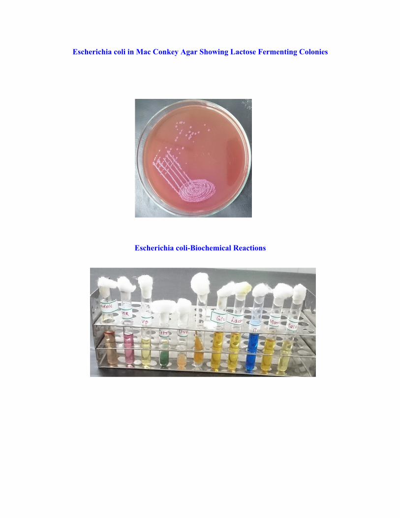
Escherichia coli in Mac Conkey Agar Showing Lactose Fermenting Colonies
Escherichia coli-Biochemical Reactions

Klebsiella Pneumoniae in Mac Conkey Agar Showing Lactose Fermenting Colonies
Klebsiella Pneumoniae - Biochemical Reactions

Proteus mirabilis in Blood Agar Showing Swarming
Proteus mirabilis – Biochemical Reaction

44
Antimicrobial sensitivity testing53
As per CLSI 2016 guidelines using antibiotic discs (Hi-media,
Mumbai),the antimicrobial sensitivity pattern for all the isolates isolated from
significant bacteriuria were done in Mueller Hinton Agar (MHA) by modified
Kirby – Bauer disc diffusion method.
Preparation of inoculum :
4 to 5 well isolated representative colonies were taken from the 24 hrs
culture plate with the help of a sterile loop and transferred to a test tube
containing 4-5ml of sterile peptone water and incubated for 2-6 hrs at 35◦C.
Then the turbidity was adjusted to 0.5 McFarland standards. This is done by
holding both the standard and inoculum tube side by side and no more than one
inch from the face of the Wickerham card (with adequate light present). This
inoculum was used for sensitivity testing.
Inoculation of MHA plates
A sterile cotton swab was dipped in to the inoculum and with firm
pressure the swab was rotated several times inside the wall of the tube to remove
the excess broth from the swab. Then the entire dried surface of Mueller Hinton
agar plate was inoculated by streaking with the swab. This procedure was
repeated by rotating the plate two more times by rotating the plates at 60 degree
to ensure an even distribution of inoculums. Finally, the rim of the agar was
swabbed. The lid was replaced and left for 3-5 minutes to allow any excess
moisture to be absorbed. Within 15 minutes the antibiotic discs were applied.

45
Control strains used with each batch
i. Escherichia coli ATCC 25922
ii. Staphylococcus aureus ATCC 25923
Antibiotic sensitivity test
According to CLSI guidelines all the isolates were tested with
predetermined battery of antibiotic discs (HIMEDIA, Mumbai)of Ampicillin,
Gentamicin Amikacin, Cotrimoxazole Nitrofurantoin, Norfloxacin,
Levofloxacin, Cephalexin, Cefuroxime, Ceftazidime, Cefotaxime, Cefoxitin,
Cefepime, Amoxyclav and Imipenam. Along with the above drugs Erythromycin
and Vancomycin were tested for Gram positive cocci. Piperacillin-Tazobactum
and Cefeperazone-Sulbactum were used only for Enterobacteriaceae.
Application of discs to inoculated Muller Hinton agar plates 59
With the help of forceps, the antibiotic disks were placed on agar plates.
In order to ensure the complete contact of the disk with the agar surface disks
were pressed down. Discs were distributed evenly so that they were not closer
than 24 mm from centre to centre of the disc and incubated at 37° C for 16 – 18
hrs.
Reading of AST and interpretation of results
After overnight incubation, each plate was examined. With the help of
antibiogram scale around each disks the zones of complete growth inhibition
including the diameter of the disk was measured. The zones were measured to
the nearest millimeter. For measuring the size of the zone, ruler was held on the

46
back of the Petri dish. The Petri dish was viewed with reflected light against a
black non reflecting background. With unaided eyes if the zone margin shows
no obvious visible growth it was considered as a zone of inhibition. According to
CLSI standard the sizes of the zones of inhibition were interpreted and reported
as ‘susceptible’, ‘intermediate’ or ‘resistant’ to the drug.
Screening for ESBL production22
For ESBL detection Quality control Klebsiella pneumoniae ATCC
700603(ESBL positive) Escherichia coli ATCC 25922 (ESBL negative).
1. Modified Kirby Bauer disc diffusion method
According to CLSI isolates showing zone of inhibition ≤ 22mm with
Ceftazidime(30 μg) and ≤ 27mm with Cefotaxime(30 μg )were interpreted as
probable ESBL producers. For the confirmation of ESBL production different
phenotypic methods were used. Here the methods used were
1. Double Disk Synergy Test (DDST)
2. ESBL CHROM Agar
3. Phenotypic Confirmation Test (PCT)
4. E –Test
1. Double disc synergy test42,93
This test mainly used to demonstrate a synergistic action of 3rd generation
Cephalosporin or Monobactam with Clavulanic acid. Inoculum was prepared as
said above and with the help of sterile swab, lawn culture was made on MHA
plate. Two different third generation cephalosporins Cefotaxime CTX(30µg) and
Ceftazidime CAZ(30µg) were placed at a distance of 20mm centre to centre

47
from the Amoxicillin Clavulanate (AMC20µg/10µg) and incubated at 37°C for
16 – 18 hrs. Enhancement of zone of inhibition to any one of the third generation
antibiotic disk on the side of the disk containing clavulanate was interpreted as
ESBL producer.
2. ESBL CHROM Agar43,58,82
Chrom agar consists of nutritive base, Chromogenic substrates with
mixture of antibiotics including Cefpodoxime enable the growth of ESBL
producing Enterobacteria. Ready prepared ChromIDTM ESBL Agar plate from
Biomerieux was inoculated. It was incubated with the cover bottom side at 37ºc
for 18-24hrs.
Principle of this CHROM agar medium is the use of chromogenic
substrates revealing metabolic enzymes specific for certain species of bacteria
Escherichia coli – Spontaneous pink to burgundy coloration of strains
expressing beta glucuronidase. Klesiella ,Enterobacter, Serratia, Citrobacter-
spontaneous green, brownish green, or blue colouration of the strains
expressing a beta-glucosidase. Proteeae (Proteus, Providencia, Morganella)-
spontaneous dark brown to light brown coloration of strains expressing
deaminase.

ESBL SCREENING
DOUBLE DISC SYNERGY TEST
E.coli Proteus mirabilis
ESBL CHROM agar (showing E.coli, (Pink) Proteus mirabilis(brown) Klebsiella spp(green))

48
Interpretation done according to the instruction given by manufacturer.
MICROORGANISMS TYPE OF COLONY ESBL Escherichia coli Pink to burgundy colour ESBL Klebsiella, Enterobacter, Citrobacter, Serratia
Green or Blue colour
ESBL Proteus species Dark brown to light brown Non ESBL strains Inhibited
3. Phenotypic confirmation Test (PCT)23
Using a sterile cotton swab that was soaked with broth, a lawn culture
was made onto the dried surface of Mueller –Hinton agar (MHA). The plates
were allowed to dry for 15min. Cefotaxime(30µg), Cefotaxime-
clavulanate(30µg/10µg) ceftazidime(30µg) and ceftazidime-clavulanate
(30µg/10µg) were placed on to the inoculated MHA plate at a distance of
20mm. Then incubation was done at 35◦C for 16-18 hrs. The zone diameter
was recorded and interpretation was done as per CLSI guideline.
Interpretation
A ≥ 5mm increase in the zone diameter for either antimicrobial agent
tested in combination with clavulanate vs its zone diameter of the agent when
tested alone that isolates are regarded as ESBL producing bacteria.
4 E-test ESBL93( Ezy MICTM strip HIMEDIA)
By using E-test strips the both disc diffusion and Minimum Inhibitory
Concentration (MIC) were studied. All test isolates were tested with the E-test
strip containing Ceftazidime gradient at one end and Ceftazidime plus
Clavulanate gradient on the opposite end, also Cefotaxime at one end and
Cefotaxime plus clavulanate at another end. Before the procedure the E-test

PHENOTYPIC CONFIRMATORY TEST (PCT)
ESBL NON ESBL
E-test
ESBL NON ESBL

49
strips were brought to room temperature. MHA were inoculated as for disc
diffusion and with the help of forceps the E-test strip was placed over the agar
surface and incubated for 35º-37ºc MIC was the point of intersection of the
inhibition ellipse with the E-test strip edge. When the ratio of the value obtained
for Ceftazidime(CAZ): the value of Ceftazidime in combination with Clavulanic
acid is more than 8 interpreted as ESBL positive strain . Also when the ratio of
the value obtained for Cefotaxime(CTX): the value of Cefotaxime in
combination with clavulinic acid is more than 8 interpreted as ESBL producer.
For both if the value is less than 8 it is interpreted as ESBL negative strain.
Finally for the ESBL producing Enterobacteriaceae the sensitivity pattern
is noted.
Molecular characterization of ESBL producing Enterobacteriaceae
Phenotypically confirmed ESBL positive isolates were further processed
in HELINI Biomolecules, Chennai to detect the presence of beta lactamase
encoding genes of family TEM, SHV and CTX-M (Cefotaximase). DNA was
extracted by using pure fast® bacterial DNA purification kit. 2X PCR Master
mix contained 2U of Taq polymerase,10X Taq reaction buffer, 2mM Mgcl2,
1µl of 10mM dNTPs mix and Red dye PCR additives. Agarose gel
electrophoresis was performed with agarose, 50X TAE buffer, 6X gel loading
buffer and Ethidium bromide.
Bacterial DNA purification procedure
1ml of overnight culture was centrifuged at 6000 rpm for 5minutes and
supernatant was discarded. Pellet was suspended in 0.2ml PBS. 180µl of
lysozyme digestion buffer and 20µl of Lysozyme(10mg/ml) was added and

50
incubated at 37ºC for 15min. 400µl of binding buffer, 5µl of internal control
template and20µl of Proteinase K were added and it was mixed well by
inverting several times. This mixture was incubated at 56ºC for 15min and
300µl of ethanol was added and mixed well By using pipette, the entire sample
volume was transferred to pure fast spin column and centrifuged for 1 minute.
Flow through was discarded and 500µl of wash buffer-1 was added. It was
centrifuged for 1minute. Flow through was discarded and 500µl of wash
buffer-2 was added. Then it was centrifuged at for additional 1 minute.
The flow through was discarded and the column was centrifuged for
additional 1 minute to remove any residual ethanol. The Pure fast® spin
column was transferred into a fresh 1.5ml micro centrifuge tube. 100µl of
elution buffer was added to the centre of Pure fast® spin column membrane
and again incubated for 1 minute at room temperature and centrifuged for 2
minutes.
PCR Primer
TEMF-GATAACACTGCGGCCAACTT
TEMR-CTGCAACTTTATCCGCCTCC
SHVF-CGCCGCCATTACCCATGACGCGAT
SHVR-ACCCGATCGTCCACCATGCCACT
CTXF-ACGTGGCGATGAATAAGCTG
CTXR-AACCCAGGAAGCAGGCAGTC
PCR amplification :
The PCR reactant mixture for each sample is prepared by adding 10µl of
PCR master mix, 5µl of primer mix and 5µl of purified DNA of each sample
to a total final volume of 20µl.

51
PCR Procedure:
20µl of the PCR reactant mixture was mixed gently, spin down briefly
and placed into PCR machine. It was programmed as follows:
Initial Denaturation : 95ºCfor5min
Denaturation : 94ºCfor30sec
Annealing : 58ºCfor30sec 35cycles
Extension : 72ºCfor30sec
Final extension : 72ºCfor5min
Loading: 2% agarose gel was prepared by mixing 2gm of agarose in 100ml of
1XTAE buffer. 8µl 6X Gel loading dye was added to each PCR vial and 5µl of
PCR sample was loaded. After that run electrophoresis at 50V till the dye
reaches three fourth distances. The bands were observed in UV
transilluminator.
Agarosegel electrophoresis: 2% agarose was prepared by the addition of 2gm
agarose in 100ml of 1X TAE buffer ( melted using microoven).When the
agarose gel temperature was around 60°C,5µl of Ethidium bromide was added.
Warm agarose solution was poured slowly into the gel platform. The gel set
was kept undisturbed till the agarose solidifies. 1X TAE buffer was poured into
submarine gel tank. The gel platform was placed carefully into tank. The tank
buffer level was maintained 0.5cm above than the gel.PCR Samples were
loaded after mixed with gel loading dye along with 10µl of 100bp DNA
Ladder. (100bp,200bp, 300bp,400bp, 500bp, 600bp, 700bp, 800bp, 900bp,

Antibiotic of Choice for NON ESBL Producers Antibiotic Of Choice For ESBL Producers
ESBL Strain Showing TEM, SHV, CTX-M Genes

52
1000 and 1500bp). Then electrophoresis was run at 50 V till the dye reaches
three fourth distance of the gel. Gel was viewed in UV trans illuminator and
observed the bands pattern.
INTERPRETATION:
The presence of TEM, SHV and CTX-M genes were indicated by the
amplification of 250bp 276 bp and 296bp PCR product from the clinical isolates
respectively.
STATISTICS: For statistics soft ware SPS 16 was used. UTI Prevalence age
and sex distribution, ESBL prevalence were expressed in percentage. Chi
Square method was used for comparison of the four Phenotypic methods for
detection of ESBL among Enterobacteriaceae.

RESULTS

53
RESULTS
A total of 400 samples were collected from both inpatients and out
patients with age group ranging from 0-80 years with symptoms suggestive of
UTI. Out of 400 patients 23.50% were less than 12years 34 % were in the 13-
44 age group, 16.5 % were in middle age group and 26% belong to older age
group >60years. This is shown in Table 1 and Figure 1
Age and Sex wise distribution of specimens collected (n= 400)
Table 1
Age group
Male % Female % Total %
0-12 42 10.50 52 13.00 094 23.50
13-44 38 09.50 98 24.50 136 34.00
45-60 34 08.50 32 08.00 066 16.50
>60 68 17.00 36 09.00 104 26.00
182 45.50 218 54.50 400 100
Figure1
10.5 9.5
8.5 17
13 24.5
8 9
0-12 13-44 45-60 >60
Age and Sex wise distribution of specimens collected
Male Female

54
400 specimens were collected from the patients with symptoms
suggestive of UTI from the various departments by aseptic methods (clean
catch midstream urine, catheterized and Suprapubic).Out of 400 specimens
collected 154 samples showed significant growth by that prevalence of UTI
was 38.50%. This is shown in Table2.
Prevalence of UTI among specimens collected (n= 400)
Table 2
Total specimen
collected from patients
with symptoms
Patients with UTI
(significant growth)
% of
UTI
400 154 38.50%
Out of 154 UTI patients 45.46 % patients were from male and 54.54 %
patients were from female. Male to female ratio was 1: 1.2. The Prevalence of
UTI was greater in age group of 13-44years (36.38%) followed by older age
group 27.92%. prevalence rate in paediatric age group was 23.37 and in
middle age group it was12.33%. In < 12 yrs group 2 specimens were collected
from the male new born baby by supra pubic aspiration method. In older age
group among 27.92%, 23 patients (14.94%) were catheterized and 20 patients
(12.99%) were non catheterized. This is shown in Table 3and Figure 2

55
Distribution of UTI according to age and sex (n=154)
Table 3
Figure2
Table No 4 shows out of 154 patients with UTI, 115(74.68%) patients were
inpatients and 39(25.32%) were out patients. According to Department wise,
prevalence of UTI was highest in Medicine department i.e 29.87% particularly
in patients admitted in Medicine Ward followed by Paediatrics 23.37% ,
0-12 13-44 45-60 >60
7.79
11.7
6.49
19.48
15.58
24.68
5.84 8.44
Distribution of UTI according to age and sex Male Female
Age
group Male % Female % Total %
0-12 12 07.79 24 15.58 36 23.37
13-44 18 11.70 38 24.68 56 36.38
45-60 10 06.49 09 05.84 19 12.33
>60 30 19.48 13 08.44 43 27.92
Total 70 45.46 84 54.54 154 100

56
Obstetrics and gynaecology 18.18%,Surgery15.59%, and Urology 7.14%.
Least distribution was seen in department of STD 3.25% and Nephrology 2.6%
it was observed that prevalence of UTI was higher in Inpatients. This is shown
in Table 4 and Figure3.
Department wise distribution of UTI (n=154) Table 4
Department IP OP Total
Number % Number % Number %
Paediatrics 28 18.18 08 5.19 36 23.37
Medicine 36 23.38 10 6.49 46 29.87
Obstetrics and
Gynaecology 20 12.99 08 5.19 28 18.18
Surgery 18 11.69 06 3.90 24 15.59
Urology 09 05.84 02 1.30 11 07.14
Nephrology 04 02.60 - - 04 02.60
STD - 05 3.25 05 03.25
115 74.68 39 25.32 154 100
Figure3
18.18
23.38
12.99 11.69
5.84
2.6 5.19
6.49 5.19
3.9 1.3
0
3.25
Paediatrics Medicine Obstetricsand
Gynaecology
Surgery Urology Nephrology STD
Department wise distribution IP OP

57
Pattern of isolates in specimen with significant growth
Out of the 400 specimens collected 154 specimens were with significant
bacteriuria in which 148 specimens were with single isolate and 6 specimens
with 2 isolates each. This is shown in Table5
Table 5(n=154)
Total
Samples One Isolate Two Isolates
154 148 06
% 96.10 3.90
Distribution of isolates among specimens with UTI
Distribution of isolates among the specimens with single isolate (n=148)
Table 6 showed out of 154 specimens 148 specimens with single
isolates. Among the 148 isolates the predominant organism was
Enterobacteriaceae 119(80.41%) 80.41% followed by Coagulase negative
Staphylococcus 9(6.08%) NFGNB 9(6.08%) and Staphylococcus aureus
6(4.05%) and Enterococcus spp 5(3.37%). This is shown in Table6
Table6 (n=148)
Name of the organism Number %
Enterobacteriaceae 119 80.41
NFGNB 09 06.08
Staphylococcus aureus 06 04.05
Enterococci 05 03.38
CONS 09 06.08
Total 148 100

58
Figure 4
Table 7 shows that among 154 specimens, 6specimens showed two
isolates. Out of this 6 specimen totally 12 pathogens were isolated. Among the
12 pathogens 5 were CoNS followed by 3 Enterobacteriaceae, 2 NFGNB and
Enterococci each.
Distribution of isolates among the specimens with two isolates (n=6)
Table 7
Organisms Number
Escherichia coli with CoNS 1
Escherichia coli with NFGNB 1
Klebsiella pneumonia with CoNS 1
Enterococci with CoNS 2
NFGNB with CoNS 1
80.41%
6.08%
4.05% 3.38
6.08%
Distribution of isolates among specimens
Enterobacteriaceae
NFGNB
Staphylococcus aureus
Enterococci
CONS

59
Prevalence of urinary pathogens
Among 154 specimen processed the major cause for UTI was GNB 133
(83.13%) particularly Enterobacteriaceae (76.25%) followed by NFGNB
6.88% and by Gram positive cocci was 27(16.87%). Out of 27 GPC, 14 were
CoNS of which 9 were isolated alone and 5 were associated with other
pathogen. Among CoNS 5 isolates were Staphylococcus saprophyticus.
Distribution of organism among total isolates ( n=160 )
Table 8
Organism
Among specimen with one isolate n=148
Among specimen with
two isolates n=6
(N=6×2=12)
Total n=160
%
Enterobacteriaceae 119 3 122 76.25
NFGNB 09 2 11 06.88
Staphylococcus aureus 06 0 06 3.75
Enterococci 05 2 07 4.37
CoNS 09 5 14 8.75
Total 148 12 160 100
Figure5
76.25%
6.88% 3.75% 4.37% 8.75%
Distribution of organism among total isolates
Enterobacteriaceae NFGNB Staphylococcus aureus Enterococci CoNS

60
Table No 9 shows that among the 122 Enterobacteriaceae, Escherichia
coli and Klebsiella spp alone constitutes 92.64% and remaining 7.37% was
constituted by Proteus mirabilis, Citrobacter koseri and Enterobacter
aerogens. This is shown in Table9, and Figure 6
Distribution of isolates among Enterobacteriaceae in UTI (n= 122)
Table 9
Enterobacteriaceae Number %
Escherichia coli 81 66.40
Klebsiella Pneumoniae 25 20.49
Klebsiella Oxytoca 07 05.74
Proteus mirabilis 06 04.91
Citrobacter koseri 02 01.64
Enterobacter aerogens 01 00.82
Total 122 100
Figure6
Escherichia coli
Klebsiella Pneumoniae
Klebsiella Oxytoca
Proteus mirabilis
Citrobacter koseri
Enterobacter aerogens
66.4
20.49
5.74
4.91
1.64
0.82
Distribution of isolates among Enterobacteriaceae
%

61
Antimicrobial susceptibility pattern
All the isolates isolated from154 urine samples with UTI were
processed for Antibiotic Sensitivity Test. AST of Enterobacteriaceae was
represented in the following table.
Antimicrobial susceptibility of isolated Enterobacteriaceae( n=122)
Table 10
Sl.
No
Esc
heri
chia
col
i
(n=
81)
Kle
bsie
lla
spp
(n=
32)
Prot
eus
spp(
n=6)
Cit
roba
cter
spp(
n=2)
Ent
erob
acte
r
aero
gens
( n=
1)
Tot
al n
=12
2
1. Gentamycin (GEN) 30(37%) 11(34%) 3(50%) 1(50%) 1(100%) 46(37%)
2. Amikacin (AK) 72(89%) 26(81%) 5(83%) 2(100%) 1(100%) 106(87%)
3. Ampicillin (AMP) 12(15%) - 2(33%) - - 14(12%)
4. Co-
trimoxazole(COT) 14(17%) 04(13%) 0 1(50%) 1(100%) 20(16%)
5. Nitrofurantoin(NIT) 69(85%) 24(75%) - 1(50%) 0 94(77%)
6 Norfloxacin (NX) 20(25%) 09(28%) 1(17%) 1(50%) 0 31(25%)
7 Levofloxacin(LE) 62(77%) 26(81%) 4(67%) 2(100%) 1(100%) 95(78%)
8 Cephalexin (CN) 12(15%) 04(13%) 1(17%) 0 - 17(14%)
9 Cefuroxime(CXM) 14(17%) 05(16%) 1(17%) 0 - 20(16%)
10. Cefoxitin (CX) 60(74%) 21(66%) 3(50%) 2(100%) - 86(71%)
11. Ceftazidime (CAZ) 41(51%) 15(47%) 4(67%) 2(100%) 1(100%) 63(52%)
12. Cefotaxime (CTX) 40(49%) 14(44%) 4(67%) 2(100%) 1(100%) 61(50%)
13. Cefipime (CPM) 58(72%) 24(75%) 5(83%) 2(100%) 1(100%) 90(74%)
14. Imipenam (IPM) 81(100%) 32(100%) 6(100%) 2(100%) 1(100%) 122(100%)
15. Amoxyclav (AMC) 30(37%) 12(38%) 2(50%) 0 - 46(38%)
16. Piperacillin-
Tazobactum (PIT) 70(86%) 28(87.5%) 5(83%) 0 0 103(84%)
17. Cefeperazone-
Sulbactum (CFS) 70(86%) 25(78%) 6(100%) 2(100%) 1(100%) 104(85%)

62
Figure 7
Enterobacteriaceae showed highest sensitivity to Imipenam (100%)
followed by Amikacin(87%), Levofloxacin(78%), Nitrofurantoin(77%)
Cefepime(74%), Cefoxitin(71%). Lower sensitivity pattern observed to
Ampicillin(12%), Cephelexin(14%) Co-trimoxazole(16%), Cefuroxime(16%),
Norfloxacin(25%) and Gentamicin(37%). In Citrobacter koseri and
Enterobacter aerogens highest sensitivity was observed in third generation
Cephalosporins.
Phenotypic detection of ESBL Distribution of ESBL detected among
Enterobacteriaceae by phenotypic screening methods.
Table No11 shows that out of 81 isolates of Esherichia coli 41 (50.62%)
were resistant to any one of the third generation cephalosporins. Among 32
isolates of klebsiella spp 17(53.13%) showed resistance among 6 isolates of
Proteus mirabilis 2(33.33%) resistant to any one of the third generation
37%
87%
12% 16%
77%
24% 25%
78%
14% 16%
71% 52% 50%
74%
100%
38%
84% 85%
Antimicrobial susceptibility of isolated Enterobacteriaceae %

63
cephalosporins. In Citrobacter and Enterobacter no resistance noted for third
generation cephalosporins. Among 122 Enterobacteriaceae 63(51.64%) isolates
of Enterobacteriaceae showed resistance to any one of the third generation
Cephalosporins. This is shown in Table 11 and Figure 8. All the 122 isolates
were subjected to the phenotypic methods DDST, CHROM agar, PCT and E-
test for detection and confirmation of ESBL.
Table 11(n=122)
Organism
Number of
isolates
Organism sensitivity to
third generation Cephalosporins
%
organism with resistance to any
one of third generation
Cephalosporins (Propable ESBL
Producer)
%
Escherichia coli
81 40 49.38 41 50.62
Klebsiella spp
32 15 46.87 17 53.13
Proteus mirabilis
06 04 66.67 02 33.33
Citrobacter koseri
02 02 100.00 00 00
Enterobacter aerogens
01 01 100.00 00 00
Figure8
49.38 46.87 66.67 100 100
50.62 53.13 33.33 0 0
Distribution of ESBL detected among Enterobacteriaceae by phenotypic screening methods.
Organism sensitivity to third generation Cephalosporins
organism with resistance to any one of third generation Cephalosporins

64
Table No 12 shows that by DDST method of 81isolates of Escherichia
coli 15 isolates (18.51%), 8 (25%) of 32 isolates of Klebsiella species and 2
(33.33%) of 6 isolates of Proteus mirabilis were detected as ESBL. This is
shown in Table 12 and Figure 9
ESBL detection by Double Disk Synergy Test (DDST method) n=122
Table 12
Organism Number of
isolates Detection of ESBL by DDST method
%
Escherichia coli 81 15 18.51
Klebsiella spp 32 08 25.00
Proteus mirabilis 06 02 33.33
Citrobacter koseri 02 00 -
Enterobacter aerogens
01 00 -
Figure 9
18.51
25
33.33
0 0 Escherichia coli Klebsiella spp Proteus mirabilis Citrobacter koseri Enterobacter
aerogens
ESBL detection by DDST method
%

65
ESBL detection by CHROM agar (n= 122)
Table 13
Organism Number of isolatesDetection of ESBL by Chrom agar method
%
Escherichia coli 81 34 41.98
Klebsiella spp 32 14 43.75
Proteus mirabilis 06 02 33.33
Citrobacter koseri 02 00 00
Enterobacter aerogens
01 00 00
ESBL detection by PCT method (n=122)
Table 14
Organism Number of isolates Detection of ESBL
by PCT method %
Escherichia coli 81 34 41.98 Klebsiella spp 32 14 43.75 Proteus mirabilis 06 02 33.33 Citrobacter koseri 02 00 Enterobacter aerogens
01 00
.ESBL detection by E test method(n=122)
Table 15
Organism Number of isolates Detection of ESBL by E test method
%
Escherichia coli 81 34 41.98
Klebsiella spp 32 14 43.75
Proteus mirabilis 06 02 33.33
Citrobacter koseri 02 00
Enterobacter aerogens
01 00

66
Figure10
From the above tables 13, 14, 15 and fig 10 it was observed that by
CHROM agar, PCT, and E test method among 81 isolates of Escherichia coli
34 isolates ( 41.98%) among 32 isolates of Klebsiella species 14
isolates(43.75%) and among 6 isolates of Proteus mirabilis 2 isolates(33.33%)
were detected as ESBL producer.
From the above tables 12,13,14,15 it was observed that out of 122 only
25 isolates were confirmed with DDST method. By Chrom agar, PCT, and E
test method 50 isolates were confirmed as ESBL positive. All the isolates
which were positive for ESBL by the above methods subjected to genotypic
method for the study of prevalence of TEM, SHV and CTX-M among the
ESBL producer.
41.98 43.75 33.33
ESBL detection by CHROMagar, PCT, E test method
%

67
Comparison of different methods for ESBL detection in
Enterobacteriaceae
Detection of ESBL among Escherichia coli by CHROM agar, PCT, and
E-test was higher than DDST method and this was statistically significant
(P=0.026). Also detection of ESBL among Klebsiella spp by the other
methods was higher than DDST method however this was not statistically
significant (P=0.432). This is shown in Table 16.
Table16
Organism DDST (%) Chrom
agar (%) PCT (%)
E-Test (%)
P value
Interpretation
Escherichia coli
15(18.51%) 34(41.98%) 34
(41.98%) 34
(41.98%) 0.026
Significant
Klebsiella spp
08(25%) 14(43.75%) 14
(43.75%) 14
(43.75%) 0.432
Not Significant
Proteus mirabilis
02(33.33%) 02(33.33%) 02
(33.33%) 02
(33.33%) -
-

68
Table No 17 shows that Prevalence of ESBL among Enterobacteriaceae
was 40.98% and Non ESBL 59.02%.
Prevalence of ESBL (n=122)
Table 17
Organism ESBL % Non ESBL %
Escherichia coli(n=81)
34 41.98 47 58.02
Klebsiella spp (n=32)
14 43.75 18 56.25
Proteus mirabilis (n=6)
02 33.33 04 66.67
Citrobacter koseri(n=2)
00 - 02 100
Enterobacter aerogens(n=1)
00 - 01 100
Total( n=122) 50 40.98 72 59.02
Figure 11
41.98 43.75 33.33
0 0
58.02 56.25 66.67
100 100
Prevalence of ESBL
ESBL% Non ESBL %

69
Prevalence of ESBL was higher in inpatients (31.15%) compared to the
out patients (9.84%). In Non ESBL prevalence of in patients was (36.88%) and
out patients was (22.13%). Generally both ESBL and Non ESBL prevalence
was greater in inpatients. But regarding the out patients prevalence of Non
ESBL was higher (22.13%) than ESBL (9.84%). This is shown in Table 18
Prevalence of ESBL according to Inpatient and Outpatient Department
Table18 (n=122).
organism ESBL Non ESBL
IP Op IP OP
Escherichia coli(n= 81)
30 (37.04%)
04 (4.94%)
33 (40.74%)
14 (17.28%)
Klebsiella spp(n=32) 06
(18.75%)08
(25%) 6
(18.75%) 12
(37.50%)
Proteus mirabilis (n=6)02
(33.33%)00
04 (66.67%)
00
Citrobacter koseri (n=2)
00 00 01
(50%) 01
(50%) Enterobacter aerogens (n=1)
00
00
01 (100%) 00
Total n=122
38 (31.15%)
12 (9.84)
45 (36.88%)
27 (22.13%)
Figure 12
31.15% 36.88%
9.84%
22.13%
NON ESBL
Prevalence of ESBL according to Inpatient and
Outpatient department IP OP
ESBL, NON ESBL

70
Comparison of the antimicrobial sensitivity pattern of ESBL producer and non ESBL producers number & Percentage
Organism AMP GEN AK COT CN CXM CX CAZ CTX CPM NIT NX LE IPM CFS PIT AMC
E coli
ESBL
(n=34) 0
8
(24)
26
(76)
2
(6)
0
0 28
(82) 0 0
17
(50)
29
(85)
5
(15)
24
(76)
34
(100)
29
(85)
28
(82)
6
(18)
NonESBL
(n=47)
12
(25)
22
(47)
46
(97)
12
(26)
12
(26)
14
(30)
32
(68)
41
(87)
40
(85)
41
(87)
40
(85)
15
(31)
38
(81)
47
(100)
41
(87)
42
(89)
24
(51)
Klebsiella
spp
ESBL(n=14)
-
2
(14)
10
(71)
1
(7) 0 0
7
(50) 0 0
8
(57)
10
(71)
3
(21)
10
(71)
14
(100)
10
(71)
12
(85)
02
(14)
NonESBL
(n=18)
- 9
(44)
16
(89)
3
(17)
4
(22)
5
(28)
14
(78)
15
(83)
14
(78)
16
(89)
14
(78)
6
(33)
16
(89)
18
(100)
15
(83)
16
(89)
10
(71)
Proteus
mirabilis
ESBL(n=2)
0 0
2
(100) 0 0 0
1
(50)
0 0 1
(50) - 0
1
(50)
2
(100)
2
(100)
2
(100)
0
0
NonESBL
(n=4)
2
(50)
3
(75)
3
(75) 0
1
(25)
1
(25)
2
(50)
4
(100)
4
(100)
4
(100) -
1
(25)
3
(75)
4
(100)
4
(100)
4
(100)
2
(50)

71
In the present study Antimicrobial resistance pattern in Escherichia coli,
Klebsiella spp, and Proteus mirabilis of ESBL producer and non- producer,
were compared and presented in the above Table.
Multiple drug resistance was more common in ESBL producer when
compared to the non- ESBL producers. In case of E. coli, sensitivity of
Gentamicin is reduced from (47% to 24%). Amikacin shows (21%) reduction
in sensitivity. The co-resistance activity was found in Co- trimoxazole
showing decreased sensitivity from 26% to 6%. Fluroquinolones also showed
co-resistant pattern, Norfloxacin (31% to 15%), and Levofloxacin (81 % to
76%).
Table 20 shows that distribution of ESBL genes among the
Enterobacteriaceae.
Resistance genes in ESBL strains
Table 20 (n=50)
organism TEM only
SHV only
CTX-M only
TEM&
SHV
TEM& CTX-M
SHV& CTX-M
TEM,SHV&CTX-M
Total
Escherichia Coli(n=34)
3 2 6 4 7 5 7 34
Klebsiella Spp(n=14)
2 1 3 1 2 2 3 14
Proteus mirabilis (n=2)
1 1 02
Total
(n=50)
5 3 9 5 10 7 11 50

72
Percentage of resistance genes
Table 21 (n=50)
Genus Number of Isolates %
CTX-M only 9 18
TEM only 5 10
SHV only 3 06
CTX-M,TEM 10 20
CTX-M,SHV 7 14
TEM,SHV 5 10
CTX-M,TEM,
SHV
11 22
Total 50 100
Out of 50 ESBL positive isolates, 9(18%) isolates were positive for
CTX-M, 5(10%) isolates were positive for TEM, 3(6%) isolates were positive
for SHV only,10(20%) isolates were positive for CTX-M & TEM, 7(14.%)
isolates were positive for CTX-M &SHV, 5(10%) isolates were positive for
TEM & SHV and 11(22%) isolates were positive for TEM,SHV & CTX-M.

DISCUSSION

73
Discussion
Urinary Tract Infections(UTI) are the most prevalent bacterial infection
among the humans in general clinical practice. Every year nearly 150 million
people are affected with UTI and due to this incidence health care expenditure
is about 6 million dollars60. If the UTI are not properly treated it can lead to
complications like stone formation, pyelonephritis and renal failure. One of the
most important factor which has got an impact in the management of UTI is the
emergence of antimicrobial resistance among the uropathogens over the past
decade. The most common mechanism of antimicrobial resistance among the
gram negative bacteria is the production of the Extended Spectrum Beta
Lactamase enzymes. This prospective study was undertaken to know the
prevalence of ESBL by various phenotypic methods and confirmation by
genotypic methods. During the study a total of 400 non repetitive (Clean catch
midstream, catheterized and suprapubic) urine samples were collected
aseptically. According to Chau et al21, the main aim of clean catch midstream
urine collection is to avoid contamination during voiding by urethral and
perineal flora.
By Morton RE74 1982 for the diagnosis of UTI in paediatric population
adequate result was obtained by MSU if properly collected and SPA is
indicated when there is in need of accurate diagnosis or if MSU can’t be
obtained. Among the total 400 samples collected 154 (38.50%) showed
significant growth of bacteria. The similar prevalence has been reported by
seen in Trupti Bajpai et al114 (38.3%)at Madhya Pradesh. The Prevalence
74
rate was higher in the present study when compared to the study done by
Elizabeth17et al ( 32.1%) Bangalore. From the 154 samples which showed
significant growth totally 160 uropathogens isolates were isolated this was
because 6 of them grew two isolates each.
In the present study the gender wise prevalence of UTI showed 45.46%
were male and 54.54% were females with male to female ratio was 1:1.2. In
Tamilnadu the same ratio (1:1.3%) was seen in study of Baby Padmini13 et al
2004 Coimbatore. From several previous studies by Carolin Elizabeth
George17 et al (60.7%) Bangalore, Astal 11et al (65%) Palestine and
Ahmed1 et al (84%) Kashmir it was observed that females are more prone for
UTI than male. In present study more number of UTI were found in
reproductive age group 36.38% followed by older age group 27.92%.But in
study Sood and Gupta105 et al 2011 Rajasthan prevalence was higher in
older age group(35%) followed by reproductive age group(23%). Reason for
higher incidence in female is because of shorter urethra, and the opening of
urethral meatus in to the moist introitus and close approximation with rectum.
favours the colonization of bacteria resulting in bacterial cystitis. Other factors
which favours the occurrence of UTI in reproductive age group are sexual
intercourse and pregnancy. In young men the important factor which increases
the risk for UTI is lack of circumcision resulting in symptomatic urethritis. In
older age group prevalence was slightly higher in males and in the present
study male to female ratio was 2.3:1 and this is in concordance with study of
Andrea Cove6 et al (2:1) U.K.

75
In this study next to the reproductive age group, and older age group, the
prevalence of UTI was higher in Paediatric age group 23.37% which was lesser
when compared to the study of Palak Gupta80 35.4%.From the studies of
Palak Gupta80, Riccabona91et al it was observed that in the first year of life
UTI is seen more commonly in male child 3.7% and 2% in female child and
there after incidence steadily increasing in female population. If the infection
occurs in preschool boys it is usually associated with congenital abnormalities.
When bacteriuria was first detected in Paediatric population there was chance
of presence of some referral urinary tract abnormality in one third of this
population. The presence of bacteriuria in Paediatric group defines a population
at higher risk for the development of bacteriuria in adult group.
Worldwide Prevalence of Uropathogens
Organism Latin America
P.H.A Bours et al88
Brazil Daynae
Moraes26
Mathya Pradesh Trupti
Bajpai114et al
Tamil nadu
Ramseh89 et al
Present study
Enterobacteriaceae 83.6% 82.8% 63.70% 64.90% 76.25% NFGNB - 0.8% 13.88% 20.65% 06.88% Staphylococcus aureus
3.3% 2.5% 01.78% - 03.75%
Enterococcus - 3.5% 07.12% 9.5% 04.37% CoNS - 9.4% 01.42% 5% 08.75% Other pathogens 13.25 - - - -
In the present Study organism belonging to the Enterobacteriaceae
family were commonly isolated. Study conducted in India by Trupti
Bajpai114et al in Latin America by P.H.A Bours et al88 and in Brazil

76
byDaynae Moraes26 also showed that Enterobacteriaceae group of organism
were commonly isolated from the UTI.
Distribution of organism among Enterobacteriaceae World wide
Enterobacteriaceae Latin America88
Brazil 26 Madhya pradesh114
Tamil nadu89
Present study
Escherichia coli 57.89% 73.69% 68.57% 46.15% 66.40% Klebsiella spp 03.95% 07.58% 28.57% 34.51% 26.23% Proteus mirabilis 02.63% 11.37% 0.57% 09.60% 04.91% Citrobacter koseri - 01.67% - 03.94% 01.64% Enterobacter aerogens
07.89% 04.03% 0.57% 04.53% 0.82%
Among Enterobacteriaceae in the present study, Escherichia coli
(66.40%) was the commonest organism isolated followed by Klebsiella
(26.23%), Proteus mirabilis (4.91%) and the least isolated was
Citrobacterkoseri(1.64%), and Enterobacter aerogens (0.82%). This prevalence
was almost similar to the study done at Brazil by Dayane Moraes26 et al, India
at Madhya Pradesh by Trupti Bajpai 114et al and in Tamilnadu by Ramseh89
et al Coimbatore.
According to this study the most effective antibiotics against organism
of Enterobacteriaceae were Imipenam (100%) followed by Amikacin (86%),
Levofloxacin (78%) and Nitrofurantoin (77%). Similar findings were reported
by Carolin17 et al(90% for Amikacin and 70% for Nitrofurantoin) and
Sarasu95 et al(for Amikacin 100%, Levofloxacin 64% and for Nitrofurantoin
68%) from India. From the present study the alarming finding notified that
most of the strains were resistant to Ampicillin (14%) and Cotrimoxazole(14%)

77
Also in studies of Curtis Nickel47et al, Anbumani 5et al, and Sarasu 95et al
sensitivity to Ampicillin was (45%,19%,16%,). In the study of Sarasu95 et
al(18%) Tamilnadu and study of Mandira Mukerji68 et al India (13.5%)
less sensitive for Cotrimoxazole was observed. Reason for this resistance may
be because these antibiotics have been extensively used in this region for a
longer period and also due to the misuse of antibiotics, which has led to the
emergence of resistant bacteria today. Hence generally in India Cotrimoxazole
and Ampicillin cannot be recommended as an empiric therapy for the treatment
of UTI.
Nowadays one of the challenge faced by every Microbiologist is the
detection of ESBL production by the Enterobacteriaceae. The main aim for the
ESBL detection is to prevent the dissemination by co transmission and also for
the epidemiological purpose. By controlling the dissemination therapeutic
failure can be prevented.
In the present study four phenotypic methods DDST, CHROM
agar,PCT and E-test were compared for the detection of ESBL. Among these
two methods (DDST, PCT) widely used in the routine testing and the other two
methods(E-test, CHROM agar) are specifically developed to detect ESBL
production. The main aim of this study to achieve most sensitive method for
ESBL detection in a Enterobacteriaceae family by using the combination of
routine method with a specific ESBL test method.

78
According to the CLSI all the organisms of Enterobacteriaceae were
screened for potential ESBL producer by Kirby – Bauer disc diffusion method
on MHA. Since some times ESBL isolates show false susceptibility to third
generation Cephalosporins Anbumani5 et al in standard disc diffusion method
so it is must to do the specific Phenotypic methods along with screening
methods.
In the present study PCT, E-test, and CHROM agar detected totally
50(40.98%) isolates as ESBL out of 122 isolates, but DDST detected only
25(20.49%) isolates as ESBL positive. Thus additional 20.49% were detected
as ESBL by the other methods. The similar findings i.e lesser detection of
ESBL by DDST method was also observed by Mohammed Hisham85 et al
2016 at Kerala and by Prabha93 et al 2016 at Pudhucherry. According to
CLSI guidelines also PCT is more effective in detection of ESBL producer than
DDST method.
In this study DDST was less sensitive than the other methods CHROM
agar, PCT, and E-test. But study conducted by Ewelina Kaluzana34 et al 2014
showed higher sensitivity to DDST method . In the present study ESBL Chrom
agar detected 41.98 % of ESBL of Escherichia coli 43.75% of ESBL of
Klebsiella spp and 33.33% ESBL of Proteus mirabilis. But according to
Ewelina Kaluzana34 et al 2014 Chrom ID ESBL method used for the detection
of ESBL strains are characteristically showed relatively high sensitivity with
low specificity, so it has got chances for false positivity. This is due to the fact

79
that Chrom ID detected not only ESBL enzymes but also broad spectrum beta
lactamases. Both PCT and E test method detected 41.97% in Escherichia coli ,
43.75% in Klebsiella spp and 33.33% in Proteus mirabilis.
Prevalence of ESBL at different Geographical areas
Geographical areas
E.coli Klebsiella spp
Proteus mirabilis
Citrobacter spp
Enterobacter aerogens
Authors
Nepal 13.5% 16.55% - - - Anil Chander et al 20137
Islamabad 51% 40.90% - - - Shamin Mumtaz et al 200677
Mumbai (India)
40.62%
27.58% 19.05% - - K.Aruna et al 201210
Pune (India) 28.72%
15.90% - - - ParulAgrawal et al 200881
Rajasthan (India)
56.92%
67.04% 41.89% 27.59% - Meetha Sharma et al 201371
Kerala (India) 62.3% 67.4% Shashikala et al 200789///
Coimbatore (Tamilnadu)
41% 40% - - - Baby Padmini et al 200413
Chennai (Tamilnadu)
60% - - - - Anbumani Narayana samy
et al 20105 Present 41.98
%43.75% 33.33%
This study reported 40.98% ESBL producers among Enterobacteriaceae
unlike the studies made by Tankhiwale 110et al (48.3%) and Khurana 57et al.
(26.6%).
Regarding the prevalence of ESBL in the present study 41.98% is in
Escherichia coli 43.75% in Klebsiella spp and 33.33% in Proteus mirabilis.
Similar findings were seen in the study done by Baby Padmini13 et al in
Tamil nadu in Mumbai by K.Aruna10 et al. In this study prevalence of ESBL

80
was low when compared to the study done by Meetha Sharma71 et al at
Rajasthan and Anbumani Narayanasamy5 et al at Chennai. This ESBL
prevalence was quite high when compared to the study done by
ParulAgrawa81 et al at Pune and Anil Chander7 et al at Nepal.
In the present study, among the in-patients, ESBL producing
Escherichia coli (37.04%) was found to be most prevalent organism followed
by Proteus mirabilis(33.33%) and Klebsiella spp (18.75%).While in outpatients
Klebsiella (25%) was the most prevalent ESBL producing organism, followed
by Escherichia. coli (4.94%). A similar finding i.e Klebsiella more prevalent in
outpatients was observed by Mumtaz 77et al in Pakistan.
Nowadays ESBL are the most common problem in hospitalized patients
throughout the world. The prevalence of ESBLs among clinical isolates varies
greatly worldwide and in geographical areas and are rapidly changing over
time63.
Multiple drug resistance was seen in the ESBL producers than the non-
ESBL producers. In present study in Escherichia coli sensitivity of Gentamicin
is reduced from 47 % to 24 %. Whereas in Amikacin it was reduced from 97%
to 76%. Co-trimoxazole sensitivity reduced nearly 20%. Fluroquinolones also
showed co- resistance pattern, Sensitivity reduced in Nalidixic acid (38% to
9%), Norfloxacin (31% to 15%), and Levofloxacin (81 % to 76%). Whereas,in
Klebsiella spp Gentamicin, sensitivity decreased from (50% to14 %). Other
drugs shown to be resistance are Co-trimoxazole (44% to 14%), Levofloxacin

81
(89% to 71%). In Proteus mirabilis highest reduction in sensitivity was
observed in Gentamicin, Amikacin and Levofloxacin.Similar type of Co-
resistance pattern was observed in the study of Singh S103 et al at Khanpur.
Resistance gene coding to Quinolones and beta - Lactam antibiotics are
located on the same plasmid and thus passed on together among different
species of Enterobacteriaceae, in addition to loss of porins (or) efflux pump
and these multiple factors play a major role in co-resistance50.Further studies
showed that CTX-M gene in Escherichia.coli highly associated with MDR
phenotype.
Co-resistance pattern seen in Co- trimoxazole and Gentamicin and this
is due to that single plasmid which carry resistance gene to these agent along
with ESBL gene87.In this study, the resistance to Fluoroquinolones varied from
11% – 75% for Enterobacteriaceae and this was in concordance with study
done by Mahesh65 et al Bangalore where 27.6 to 90% of resistance was
observed. Quinolones are the most active agents against UTI pathogens in
North America as per the study of Gordon 39et al.
In the present study the co-resistance was low for Amikacin (21%).
Similar findings were observed by Baby Padmini and Appalaraju 28 and V.P
Sarasu et al 95
According to this study sensitivity for ESBL producing Escherichia coli,
Klebsiella spp and Proteus mirabilis for Piperacillin-Tazobactum and
Cefeperazone-sulbactum were (82%,85%,100% ) and (85%,71%,100%)

82
Whereas in Mangalore, Shigu et al85 showed highest sensitivity of the ESBL
producing Escherichia coli to CFS and PIT(100%/100%) and for ESBL
producing Klebsiella spp(98%/88%)The present study sensitivity regarding
CFS and PIT was low when compared to Shigu et al but sensitivity was high
when compared to the study conducted by Anbumani5et al (for PIT 49%)
According to the present study it was observed that the most effective
antibiotic against ESBL producing Escherichia coli Klebsiella spp and Proteus
mirabilis in UTI are Imipenam (100%,100%,100%), Cefeperazone-sulbactum
(85%,71%/100%),Piperacillin-Tazobactum, (82%,85%,100%), Amikacin
(76%,71%,100%), Levofloxacin(76%,71%,50%) and Nitrofurantoin (85%,
71%-),Similar findings were observed in the study of Baby Padmini et al 2004.
According to Gaurav Dalela37 et al 2012 highest sensitivity observed to
Cefepime (83.2%) for Piperacillin-Tazobactum,(75.8%) and for Amikacin it
was (74.7%). For the above drugs resistance observed in non ESBL producers
but this was due to different mechanism other than Extended Spectrum Beta
lactamase such as AmpC beta lactamase, metallobetalactamase etc.
Highest sensitivity for beta lactamase and beta lactamase inhibitors was
observed in the Study conducted in Kerala by Shasikala et al99. In that study
Piperacillin-Tazobactum (96.8%), Cefeperazone-sulbactum (92.2%), were
sensitive to ESBL producers.
When compared to the other oral antibiotics Nitrofurantoin has shown
least resistance especially for Escherichia coli followed by Klebsiella spp. Most
of the Indian studies Khurana S et al57 Tankhiwale SS et al110 have

83
demonstrated that Nitrofurantoin can be used as the first line drug in the
treatment of uncomplicated UTI as it is highly concentrated in the urine and it
can be administered orally. The reason for least resistance is limitation of its
use because it has no role in the treatment of other infection. Main drawback of
Nitrofurantoin is that cannot be used in upper UTI and also in Proteus mirabilis
infection because of its intrinsic resistance nature to Proteus mirabilis (CLSI
2016).
From the present study and also from the previous study it was observed
that the most reliable treatment for infection caused by ESBL producing
Enterobacteriaceae are Carbapenams. Despite their utility chances for the
emergence of resistance so carbapenams can be reserved for serious infections.
Alternate drugs like Nitrofurantoin, Piperacillin Tozabactam, cefaperazone
Sulbactam ,Amikacin, Levofloxacin and cefepime can be given for the ESBL
producing UTI.
In the present study CTX-M (64%) is the most common and it is present
as either alone or in combination with TEM, SHV,or both. The high prevalence
of CTX-M gene in the present study was in concordance with Mohamad
Hisham PP et al 85 (56%) and Meetha Sharma et al71 (82.5%) But in study
of Bali et al Turkey31 TEM (73%) type was predominant and in study of
Kawthar55 et al Egypt SHV(69.2%) was predominant. CTX-M was low when
compared to the present study. Also data from the last 10 years and Livermore
et al stated that worldwide CTX-M gene was most prevalent which is replacing

84
SHV and TEM types as predominant ESBL in Asian and in many European
countries. According to Goyal et al40 majority of ESBL strains harbored two or
more gene and this was in concordance with our present which showed more
than one type of beta lactamases in 22 out of 50 isolates.

SUMMARY

85
Summary
The study was conducted at Government Rajaji Hospital and Institute of
Microbiology Madurai over a period of one year from September 2015 to
August 2016 with 400 patients suffering from UTI, which included 45.50 % of
males and 54.50 % of females. Among 400 patients 154 (38.50%) of them had
significant bacteriuria. Among 154 specimens tested more than 1 isolate was
obtained in 6 specimens (2 organisms isolated) UTI was higher in the age
group 13-44years 36.38% followed by elderly people 27.92%.
In this study, totally 133 Gram Negative Bacilli and 27 Gram Positive
Cocci were isolated among which 122 were to Enterobacteriaceae. Among
the Enterobacteriaceae Escherichia coli was the commonest organism
isolated (66.40%) followed by Klebsiella spp (26.23%), Proteus mirabilis
(4.91%), Citrobacter koseri (1.64%) and Enterobacter aerogens (0.82%).
ESBL producing organisms accounts for treatment failure leading to high
morbidity and even mortality. Hence early detection of the ESBL producing
organisms is very important for the treatment aspect. In the present study
comparison methods were employed for the detection of ESBL. It was
observed that out of 122 only 25 isolates (20.49%) were confirmed as
ESBL with DDST method. By Chrom agar, PCT, and E test method 50
isolates (40.98%) were confirmed as ESBL positive. Phenotypic method
DDST showed 20.49% false negative result when compared to the other
Phenotypic methods PCT, CHROM agar, and E-test.

86
Prevalence of ESBL production was found in 41.98 % of the E. coli,
43.75% of the Klebsiella spp and 33.33% of the Proteus mirabilis.
All 50 ESBL strains detected by phenotypic methods were confirmed with
genotypic methods for the presence of ESBL gene (TEM, SHV and CTX-
M). CTX-M only (18%) TEM only (10%) SHV only (4%) CTX-M,TEM
(20%) CTX-M,SHV (16%) TEM,SHV (10%) CTX-M,TEM,SHV(22%).
By comparing Phenotypic and genotypic method DDST is less sensitive.
But in detecting the ESBL other three methods were equally effective.
CHROM agar and E-test being costly, PCT can be performed as routine test
for detection of ESBL.
Multiple drug resistance was seen in ESBL producing strains than the non
ESBL production.
Non ESBL producing Escherichia coli, Klebsiella spp, and Proteus
mirabilis UTI are highly sensitive to Ceftazidime Cefotaxime and
Amikacin ( 87% /83%/100% (85%,78%100%) and ( 97%89%75%)
The sensitivity pattern ESBL producing Escherichia coli, Klebsiella spp
and Proteus mirabilis in UTI are Imipenam (100%,100%,100%),
Cefeperazone-sulbactum(85%,71%/100%),Piperacillin-Tazobactum, (82%,
85%, 100%),Amikacin(76%,71%,100%),Levofloxacin (76%,71%,50%) and
Nitrofurantoin (85%,/71%,/-).

CONCLUSION

87
Conclusion
ESBL producers among uropathogens is increasing in incidence.
Although genotypic methods are more sensitive, Resource constraints
prevent these tests from being used in routine diagnostic laboratories.
PCT method can be performed as routine test for the detection of ESBL as
it is more sensitive, simple to perform and cost effective.
Non ESBL producing strains are sensitive mainly to aminoglycoside and
third generation Cephalosporins. As aminoglycosides are injectables and
nephrotoxic third generation Cephalosporins can be used for treating UTI as
they are less toxic and also orally effective.
All the ESBL isolates are 100% sensitive to Imepenam. Even though they
are highly sensitive to Imepenam, there is chances for the emergence of
resistance to Carbapenem, so it should be kept in reserve as the second line
of drug. Next higher sensitive drugs like Nitrofurantoin and Levofloxacin
which are most economic and orally effective can be given to outpatients.
Amikacin, Cefepime, beta-lactamase-Inhibitors Cefeperazone- Sulbatum,
and Piperacillin-Tazobactum, can be given to inpatients.
Based on the prevalence rate of the ESBL production, institutional
antibiotic policy can be tailored in a health care facility to achieve superior
therapeutic outcome and also to bring about a reduction in healthcare costs..
Drug resistance pattern varied from place to place which is related to the
nature of the pathogen and usage of antimicrobial agents.

BIBLIOGRAPHY

88
BIBLIOGRAPHY
1. Ahmad S, Pattern of Urinary tract infection in Kashmir and Antimicrobial
Susceptability. BangladeshMeResCounc,Bull. 2012:38;79–9.
2. AkramM, Shahid,M KhanA.U 2007. Etiology and antibiotic resistance
patterns of community- acquired urinary tract infections in J N M C Hospital
Aligarh, India. Annals of Clinical Microbiology and Antimicrobials 2007 Mar
23; 6:4.
3. Alma Brolund, Oscar Franze, O¨ jar Melefors Karin Tegmark-Wisell Linus
Sandegren Plasmidome-Analysis of ESBL-Producing Escherichia coli
Using Conventional Typing and High-Throughput Sequencing PLOS ONE
June 2013 | Volume 8 | Issue 6 | e65793.
4. Ana, C. Gales, Jones, R.N., Kelly A. Gordon, Helio, Sader. Werner W.Wilke,
Mondell,L Beach M.L.Michael A. Pfaller, GaryV. Doern 2000. “Activity
spectrum of 22 antimicrobial agents tested against UTI pathogens in
hospitalized patients in Latin America. Report from the second year of the
SENTRY Antimicrobial Surveillance programme. Journal of Antimicrobial
Chemotherapy (2000)45,295-303.
5. Anbumani Narayanaswamy, M.Mallika, Prevalence and Susceptibility of
extended spectrum beta-lactamases in urinary isolates of Escherichia coli
in a Tertiary Care Hospital, Chennai South India Internet Journal of
Medical 2010 January;6(1):39-43.
6. Andrea cove-Smith and Michael Almond, Management of urinary tract
infections in the elderly Trends in Urology Gynaecology & Sexual Health
July 5/August 2007.
7. Anil Chander and Chandrika Devi Shrestha, Prevalence of Extended
Spectrum Beta lactamase producing Escherichia coli and Klebsiella
pneumoniae urinary isolates in a tertiary care hospital in Kathmandu,
Nepal. Chander and Shrestha BMC Research Notes 2013, 6:487.
8. Antoni Rozalski, Zygmunt Sidorczyk, and Krystyna KotełkoPotential
Virulence factors of Proteus mirabilis Microbiology and Molecular

89
Biology Reviews, Mar. 1997, p. 65–89, American Society for
Microbiology.
9. Archana Gupta,Krow Ampofo, David Rubenstein, Lisa Saiman, Extended
Spectrum Beta Lactamase- producing Klebsiella pneumoniae Infections: a
Review of the Literature Journal of Perinatology (2003) 23, 439–443.
10. Aruna K, T. Mobashshera Prevalence of Extended Spectrum Beta-
lactamase production among uropathogens in south Mumbai and its
antibiogram pattern EXCLI journal 2012;11:363-372 – ISSN 1611-2156.
11. Astal Z, El-Manama A, Sharif FA. Antibiotic resistance of bacteria
associated with community-acquired urinary tract infections in the
southern area of the Gaza trip.JChemother. 2002;14:259 [PubMed].
12. Ashish Jitendranth, Radhika,R Bhargavi L,GeethaBhai Ramala
BeeviMicrobiological Profile Of Urinary Tract Infection in Paediatric
population from a Tertiary care hospital in South Kerala. Journal of
Bacteriology & Mycology 2015,1(1);00002.
13. Baby padmini, S., and B. Appalaraju. 2004. ESBL in Urinary isolates of E.
coli and Klebsiella pneumonia - Prevalence and susceptibility pattern in a
Tertiary Care Hospital, IJMM, 22(3): 172-174.
14. Bailey and Scott’s Diagnostic Microbiology. Thirteenth edn. pp:307 –
327;919 -930.
15. Bush, K., Jacoby G.A. and Medeiros, A.A. 1995. A functional classification
scheme for β-lactamase and its correlation with molecular structure.
Antimicrobial agents’ chemotherapy. 39: 1211 – 1233.
16. Campbell- Walsh urology, Alen JWein,Louis,Partin, Peters- eleventh edition.
17. Carolin Elizabeth George, Gift Norman, G Venkata Ramana, Devashri
Mukherjee, and Tata Rao Treatment of uncomplicated symptomatic
urinary tract infections: Resistance patterns and misuse of antibiotics J
Family Med Prim Care. 2015 Jul-Sep; 4(3): 416–421.
18. Cenk Aypak, Adalet Altunsoy and Nurs¸en Düzgün Empiric antibiotic
therapy in acute uncomplicated urinary tract infections and

90
fluoroquinolone resistance: a prospective observational study Annals of
Clinical Microbiology and Antimicrobials 2009, 8:27 .
19. Chaudhary Navin kumar , Murthy s Mahadeva Extended Spectrum Beta
lactamases in uropathogen. Asian J Pharm Clin Res, Vol 6, Issue 3, 2013,
207-210.
20. Chein-Wei Lin, Yee-Hsuan Chiou, Ying-Yao Chen, Yung-Feng Huang,
Kai-ShengHsieh, Ping-KuangSung, Urinary Tract Infection in Neonates
Clinical Neonatology 1999 Vol.6 No.2.
21. Chua, A.T., Ariceo Editha and Pena, A. 1988. Comparison of initial verses
midstream voided urine culture among men. Phil. J. Microbiol Infect.
Dis.:17(1): 22 – 24.
22. Clinical and Laboratory Standards Institute. Jan 2015 M-100-S25
Performance Standards for Antimicrobial Susceptibility Testing Twenty- fifth
informational supplement vol 35 No 3 pg 32, 44,108 Clinical and Laboratory
Standards Institute, Wayne, Pa. USA.
23. Clinical and Laboratory Standards Institute. Jan 2016 M-100S Performance
Standards for Antimicrobial Susceptibility Testing Twenty-Sixth
informational supplement vol 35 No 3 pg 40, 52,118 Clinical and Laboratory
Standards Institute, Wayne, Pa. USA.
24. Collee.J.G., Fraser.A.G., Manimon,B.P and A. Simmons.2006. Mackie and
Mc Cartney-Practical Medical Micro Biology. 14th Edn.84-90,95-111;151-
178.
25. Conne R Mahon, Donald C.Leishman George – Text book of Diagnostic
Microbiology.. fifth Edn. Pp: 420 – 454, 884 – 900.
26. Dayane Moraes ; Alexandre Braoios ; Janio Leal Borges Alves ; Rafael
Menezes da Costa Prevalence of uropathogens and antimicrobial
susceptibility profile in outpatient from Jataí-GO J Bras Patol Med Lab, v.
50, n. 3, p. 200-204, 2014
27. Deepika Handa, Anita Pandey, Ashish Kumar Asthana, Ashutosh Rawat,
Seemant Handa, Bhaskar Thakuria Evaluation of phenotypic tests for the

91
detection of AmpC beta-lactamase in clinical isolates of Escherichia coli
Indian journal of Pathology and Microbiology
28. Devanand Prakash and Ramchandra Sahai Saxena Distribution and
Antimicrobial Susceptibility Pattern of Bacterial Pathogens Causing
Urinary Tract Infection in Urban Community of Meerut City, India.
Hindawi Publishing Corporation ISRN Microbiology Volume 2013,
Article ID 749629.
29. Diagnosis and Treatment of Acute Uncomplicated Cystitis Richard
Colgan,, and Mozella williams, American Family Physician Volume84,
Number7 October1, 2011Am Fam Phycian 2011 oct 1;84(7);771-776.
30. Dominick J. Salvatore Pharm D, BCPS , Beth H. Resman-Targoff
Pharm D2 Treatment Options for Urinary Tract Infections Caused by Extended-
Spectrum Β-Lactamase-Producing Escherichia coli and Klebsiella pneumonia
journal of academic hospital medicine 2015.
31. Elif Burcu Bali Leyla Açık and Nedim Sultan Phenotypic and molecular
characterization of SHV, TEM, CTX-M and Extended-Spectrum –Beta-
lactamase produced by Escherichia coli, Acinetobacter baumannii and
Klebsiella isolates in a Turkish Journal of Microbiology Research Vol. 4
(8), pp. 650-654, 18, 2010 ISSN 1996-0808.
32. Eshwarappa, Dosegowda, Vrithmani Aprameya, MW Khan, P Shiva
Kumar, P Kempegowda Clinico-microbiological profile of Urinary tract
infection. 3Indian J Nephrology 2011 Jan vol 21 30-36 .
33. Eshwar Singh R R. , M. Veena , K.G. Raghukumar , G. Vishwanath , P.N.
Sridhar Rao , B.V. Murlimanju ESBL production: Resistance pattern in
Escherichia coli and Klebsiella pneumoniae, a study by DDST method
Research Journal of Pharmaceutical,Biological and Chemical sciences
Volume: 2: Issue-4: Oct - Dec -2011 ISSN 0976-4550.
34. Ewelina Kałużna, Patrycja Zalas-Więcek , Eugenia Gospodarek
Comparison of detection methods for extended-spectrum beta-lactamases
in Escherichia coli strains Postepy Hig Med Dosw (online), 2014 tom 68:
808-813 e-ISSN 1732-2693.

92
35. Foxman, B. and Brown, P. 2003. Epidemiology of urinary tract infections
transmission and risk factors incidence and costs. Infect. Dis. Clin. N. Am.
Vol. (17): 227 241.
36. Gales A, Sader H, Jones R, SENTRY participants group (Latin America).
Urinary tract infection’s trends in the American hospitals: reports from the
SENTRY Antimicrobial Surveillance programme (1997- 2000). Diagn
Microbial Infect Dis 2002; 44:289-99.
37. Gaurav Dalela, Sweta Gupta, Dinesh Kumar Jain, Pushpa Mehta Antibiotic
Resistance Pattern in Uropathogens at a Tertiary Care Hospital at Jhalawar
with Special Reference To ESBL, AmpC b-Lactamase and Mrsa
Production Journal of Clinical and Diagnostic Research. 2012 May (Suppl-
2), Vol-6(4): 645-651.
38. Goodmann & Gillman’s. The pharmacological basis of therapeutics 12th Edn:,
1477-1481.
39. Gordon, K.A. and Jones, R.N. 2003. Susceptibility pattern of orally
administered antimicrobials among urinary tract infections pa thogens from
hospitalized patients in North America: America report Europe and Latin
America. Results from the SENTRY Antimicrobial Surveillance Program.
Diagn. Microbiol. Infect. Dis. 45(4): 295 – 301.
40. Goyal A,Prasad KN, Prasad A,Gupta S,Goshal U,Ayyagiri Extended
Spectrum of Beta Lactamase in Escherichia coli and Klebsiella pneumonia
and associated risk factors. Indian J Med Res 2009 Jun;129(6);695-700.
41. Harrison Infectious Diseases 2nd edition Dennis L. Kasper, Anthony S.Fauci-
295-305.
42. He´le`ne Garrec, Laurence Drieux-Rouzet, Jean-Louis Golmard, Vincent
Jarlier, and Je´roˆme Robert1 Journal of clinical microbiology, American
Society for Microbiology Mar. 2011, Vol. 49, No. 3 p. 1048–1057.
43. He´le`ne Re´glier-Poupet,1 Thierry Naas,2 Ame´lie Carrer,2 Anne Cady,1
Jean-Marie Adam,1 Nicolas Fortineau,2 Claire Poyart1 and Patrice
Nordmann2 Performance of chromID ESBL, a chromogenic medium for

93
detection of Enterobacteriaceae producing extended-spectrum b-lactamases
Journal of Medical Microbiology (2008), 57, 310–31577.
44. Insan Nitin Goel,Sharma sachin, Chaudhary Pravin kumar Payal Nikil.
Different Phenotypic methods for detection of ESBL production in bacteria
. A review. Journal of Pharmaceutical and scientific Innovation 2014.
45. Jaspal kaur , Gomty Mahajan , Kailash Chand , Sheevani Shashi Chopra,
Enhancing Phenotypic Detection of ESBL in AmpC co-producers by using
Cefepime and Tazobactam Journal of Clinical and Diagnostic Research.
2016 Jan, Vol-10(1): DC05-DC08.
46. Jawittz text book of microbiology. 26th Edn. Pp – 153 -155.
47. J. Curtis Nickel, vol. 9 no. 2 2007 Reviews in urology Urinary Tract
Infections and Resistant Bacteria Rev Urol. 2007;9(2):78-80.
48. John L Brusch, Urinary Tract Infection in Males Med scape 2013.
49. Joumana N Samaha-Kfoury, George F Araj Recent developments in beta
lactamases and extended spectrum beta lactamases BMJ Volume 327 22
November 2003.
50. Jones, G.L., Warren, R.E., Skindmore, S.J., Davies, V.A., Gabriel, T., Upton,
M. 2008. Prevalence and distribution of plasmid mediated Quinolone
resistance genes in clinical isolates of E. coli lacking ESBL. J. Antimicrob
Chemother: 62(6): 1245 – 51.
51. Jung Hun Lee, Il Kwon Bae Sang Hee Lee. New definitions of Extended-
Spectrum β-lactamase conferring worldwide emerging antibiotic resistance
Medicinal Research Reviews 2010 Volume 32, Issue.
52. Kanchana Manickam, James A. Karlowsky,. Heather Adam, Philippe R.
S. Lagacé-Wiens Assunta Rendina, a Paulette Pang, a Brenda-LeeMurray,
Michelle J. Alfaa,b CHROMagar Orientation Medium Reduces Urine
Culture Workload Journal of Clinical Microbiology jcm.asm.org 1 p.
1179–1183. April 2013 Volume 51 Number 4

94
53. Karen Bush and George A. Jacoby Updated Functional Classification of
Beta-Lactamases Antimicrobial agents and chemotherapy, Mar. 2010, p.
969–976 Bloomington.
54. Kass, E. 1955. Asymptomatic infection of the urinary tract. Trans Association
of American Physician; 69: 56 – 63.
55. Kawthar I. Mohamed, Mona A. Khattab, Nevin M. Al-azhary and Ayat R.
Abdallah Molecular characterization of Extended Spectrum β Lactamases
(ESBL) genes in phenotypically ESBLs Producing Escherichia coli and
Klebsiella pneumoniae isolates. Egypt. J. Microbiol. Vol. 38, May, 2014.
56. Kelly E. Martin Treatment of ESBL-producing E coli Lower Urinary Tract
Infection Pharmacy Times 2013.
57. Khurana S, Taneja N, Sharma M. Extended spectrum beta-lactamase
mediated resistance in urinary tract isolates of family Enterobacteriaceae.
Indian J Med Res 2002;116:145-9.
58. Kjersti Sturød , Ulf R Dahle , Einar Sverre Berg , Martin Steinbakk and
Astrid L Wester Evaluation of the ability of four EBSL-screening media to
detect ESBL-producing Salmonella and Shigella BMC Microbiology 2014,
14:217.
59. Kone man’s color Atlas and Text book of microbiology. 2006. Antimicrobial
susceptibility testing .pp: 945 -1015. Sixth Edn.
60. Kucheria R, Dasgupta P, S H Sacks, M S Khan, N S Sheerin. Urinary tract
infections: new insights into a common problem Postgraduate Medical
Journal. March 2005.
61. Kumar Rakesh , Dahiya S.S., Hemwani Kirti and Srivastava Preeti
Isolation of Human Pathogenic bacteria causing Urinary tract infection and
their Antimicrobial susceptibility pattern in a Tertiary care Hospital,
Jaipur, India. International Research Journal of Medical Sciences ISSN
2320 –7353 Vol. 2(6), 6-10, June (2014).
62. Latika J Shah, Geeta M Vaghela, Hetvi Mahida urinary tract infection:
Bacteriological profile and its antibiotic susceptibility in western india

95
National journal of medical research print ISSN: 2249 4995/ eISSN: 2277
8810.
63. Livermore, D.M. 1995. β-lactamase in laboratory and clinical resistance.
Clin. Microbiol. Rev.: 8: 557 – 584.
64. Luzzaro, F., Mezzatesta, M., Mugnaioli, C., Perilli, M., Stefani, S. and
Amicosante, G. 2006. Trends in production of Extended spectrum beta-
lactamases among Enterobactriaceae of Medical interest: Report of the II
Italian Nationwide Survey. J. Clin. Microbiol; 4(5): 1659-64.
65. Mahesh, E, Ramesh, Indumathi, V.A., Punith, K., Raj, K and Anupama H, A.
2010. Complicated urinary tract infection in a tertiary care centre in South
India. Al Ameen J. Med. Sci. 3(2): pp. 120 – 127.
66. Mahesh, D Ramesh, VA Indumathi, Mohd. Wasim Khan, Prithivi S
Kumar, K Punith. Risk Factors for Community Acquired Urinary Tract
Infection caused by ESBI-producing Bacteria JIACM 2010; 11(4): 271-6.
67. Mandell, Douglas and Bennetts. Principles and practices of Infectious
diseases. 8th edition: Vol. (I); pp – 238 – 252: 875 – 905.
68. Mandira Mukherjee, Shreya Basu, Sandip Kumar Mukherjee, Monalisa
MajumderMultidrug-Resistance and Extended Spectrum Beta-Lactamase
Production in Uropathogenic E. Coli which were Isolated from
Hospitalized Patients in Kolkata, Indian Journal of Clinical and Diagnostic
Research. 2013 March, Vol-7(3): 449-453.
69. Manu Chaudhary Shailesh Kumar Anurag Payasi Prevalence and
Antimicrobial Sensitivity of Extended-Spectrum β-Lactamase Producing
Gram Negative Bacteria from Clinical Settings in India from 2010-
2012International Journal of Medicine and Medical Sciences, ISSN: 2051-
5731, Vol.46, Issue.2 1.
70. Mathur P,Kapil A,Das B, DhawanB. Prevalence of Extended Spectrum
Beta Lactamase producing Gram negative bacteria in a tertiary care
Hospital. Indian J Med Res 2002 Apr 115;153.

96
71. Meeta Sharma , Sati Pathak , Preeti Srivastava Prevalence and antibiogram
of Extended Spectrum β-Lactamase (ESBL) producing Gram negative
bacilli and further molecular characterization of ESBL producing
Escherichia coli and Klebsiella spp. Journal of Clinical and Diagnostic
Research. 2013 Oct, Vol-7(10): 2173-2177.
72. M. S. Najar, C. L. Saldanha, and K. A. Banday Approach to urinary tract
infections Indian J Nephrol. 2009 Oct; 19(4): 129–139.
73. Michael Osthoff a,b , Sarah L. McGuinness a , Aaron Z. Wagen a , Damon
P. Eisen a,c, Urinary tract infections due to Extended-Spectrum Beta-
Lactamase producing Gram-negative bacteria: identification of risk factors
and outcome predictors in an Australian tertiary referral hospital
International Journal of Infectious Diseases 34 (2015) 79–83.
74. Morton RE, Lawande R .The diagnosis of urinary tract infection; Comparison of
urine culture from suprapubic aspiration and midstream collection in a childrens
out patient department in Nigeria. Ann Trop Paediatr 1982 2(3);109-12.
75. Monica Cheesbrough. 2006. District laboratory practice in tropical countries
Part II. pp: 105 – 114.
76. Muktikesh Dash, Sanghamitra Padhi, Indrani Mohanty, Pritilata
Panda, and Banojini ParidaAntimicrobial resistance in pathogens causing
urinary tract infections in a rural community of Odisha, India J Family
Community Med. 2013 Jan-Apr; 20(1): 20–26.52. Shaifali I, Gupta U,
Mahmood SE, Ahmed J. Antibiotic susceptibility patterns of urinary
pathogens in female outpatients. N Am J Med Sci. 2012;4:163–9.
77. Mumtaz S, Ahmad M, Aftab I, Akhtar N, ul Hassan M, Hamid AExtended
spectrum beta-lactamases in enteric gram-negative bacilli: related to age
and gender. J Ayub Med Coll Abbottabad. 2007 Oct-Dec;19(4):107-11.
78. Oxford text book of clinical Nephrology, Alex M.Davison , J.Stewart
Cameron, Jean-Pierre grunfield and Winearls second edition vol2 pg1213-
1261.

97
79. Padmavati Bisht, Sushma Tamta and Umesh Bacteriological Profile and
Antibiotic Resistance Pattern of Urinary Tract Infections in Kumaun
Region Int.J.Curr.Microbiol.App.Sci (2015) 4(8): 874-883.
80. Palak Palak Gupta , Jharna Mandal , Sriram Krishnamurthy , Deepak
Barathi Nandini Pandi profile of urinary tract infections in paediatric
patients Indian J Med Res 141, April 2015, pp 473.
81. Parul Agrawal, A. N. Ghosh, Satish Kumar, B. Basu, K. Kapila.Prevalence
of extended-spectrum β-lactamases among Escherichia coli and Klebsiella
pneumoniae isolates in a tertiary care hospital indian journal of pathology
and microbiology - 51(1), 2008.
82. Pathani Jayashri Sanjay Rathod, Afroz Blosch,Bimal chauhan Utility of
UTI Chrom agar Media for the rapid identification of the Uropathogens
NHL Journal of Medical sciences/2013 vol2 /issue 1.
83. Patricia a. Bradford Extended-Spectrum -Lactamases in the 21st Century:
Characterization, Epidemiology, and Detection of This Important
Resistance Threat Clinical Microbiology Reviews, Oct. 2001, p. 933–951
Vol. 14, No. 4.
84. Pattirck, R. Murray, Ellen, J. B., James H., Jergenson, Mary, L. L. and
Michael A. 2006. Murray - Manual of Clinical Microbiology. 9th Edn. Vol
(1): pp – 323 – 325, 1114 – 1182.
85. PP Mohammed Hisham MB Shabina , S Remadevi , Beena Philomina
Comparative analysis of detection methods of Extended Spectrum Beta
Lactamases in Gram Negative clinical isolates with special reference to
their Genotypic Expression Journal of International Medicine and
Dentistry 2016; 3(1): 34-41.
86. Paul R. Ingram, Tim J. J. Inglis, Tessa R. Vanzetti, Barbara A. Henderson,
Gerald B. Harnett and Ronan J. Murray Comparison of methods for AmpC
b-lactamase detection in Enterobacteriaceae Journal of Medical
Microbiology (2011), 60, 715–721.

98
87. Philippe RS Lagacé-Wiens , Kim A Nichol MSc, Lindsay E Nicolle , Mel
R DeCorby , Melissa McCracken, Michelle J Alfa, Michael R Mulvey ,
George G Zhanel ,ESBL genotypes in fluoroquinolone-resistant and
fluoroquinolone-susceptible ESBL-producing Escherichia coli urinary
isolates in Manitoba Can J Infect Dis Med Microbiol Vol 18 No 2
March/April 2007.
88. P. H. A. Bours, R. Polak, A. I. M. Hoepelman, E. Delgado, A. Jarquin, and
A. J. Matute, “Increasing resistance in community-acquired urinary tract
infections in Latin America, five years after the implementation of national
therapeutic guidelines, International Journal of Infectious Diseases, vol. 14,
no. 9, pp. e770–e774, 2010.
89. Ramesh, N, Sumathi, C.S., Balasubramanian, V., Palaniappan, K.R. and
Rajesh Kannan, V. 2008. Urinary tract infection and antimicrobial
susceptibility pattern of extended spectrum of β – lactamase producing clinical
isolates. Advances in Biological Research. 2(5- 6): 78 – 82.
90. Ranganathan Vasudevan,Urinary Tract Infection: An Overview of the
Infection and the Associated Risk Factors Journal of Microbiology &
Experimentation Volume 1 Issue 2 – 2014.
91. Riccabona, MarcusUrinary tract infections in children.Current Opinion in
Urology:January 2003 - Volume 13 - Issue 1 - pp 59-62 Urinary tract
infections and sexually transmitted diseases.
92. Richard colgan, and Mozella williams, Diagnosis and Treatment of Acute
Uncomplicated Cystitis American Family Physician Volume 84, Number
7, 2011.
93. R.Prabha, JoshyM, Easow and M. Swapna Int.J.Curr.Microbiol.App.Sci
(2016) 5(4): 565-577.
94. Sabharwal ER. Antibiotic susceptibility patterns of uropathogens in
obstetric patients. N Am J Med Sci.2012;4:316–9.
95. Sarasu V.P, Radhakumari Prevalence of ESBL and AMPC Betalactamase
production among Escherichia coli and Klebsiella isolated in Urinary Tract
Infection Medical Sciences vol3 issue 9 2014 ISSN 2277-8179.

99
96. Sasirekha B Prevalence of ESBL, AmpC β- lactamases and MRSA among
uropathogens and its antibiogram EXCLI Journal 2013;12:81-88 –ISSN
1611-2156.
97. Shaikh N, MoroneNE, BostJE, FarrellMH Prevalence of Urinary tract
infection in a childhood a meta analysis Peditr Infect Dis J.2008 Apr,27
(4); 302-8.
98. Sharma A, S Shrestha, S Upadh P Rijal Clinical and Bacteriological profile
of urinary tract infection in children at Nepal Medical College Teaching
Hospital . Nepal Med Coll J 2011; 13(1): 24-26.
99. Shashikala, S., R. Kavitha, A. R., Gireesh, P.M. and Shamsul Karim. 2007.
Routine screening for ESBL production N necessity of Today. The
International Journal of Microbiology. 3(1).
100. Shiju, M.P., Yashavanth, R. and Narendra, N. 2010. Detection of Extended
Spectrum β – lactamase production and multidrug resistance in clinical
isolates of E. coli and K. pneumonia in Mangalore. J. Clinical and Diagnostic
Research. 4(3): pp – 2442–5.
101. Shivaprakasha.S, K Radhakrishnan, A Gireesh, P Shamsul
Karim. Routine Screening For ESBL Production, A Necessity Of Today.
The Internet Journal of Microbiology. 2006 Volume 3 Number 1.
102. Sibhghatulla Shaikh a Jamale Fatima b , Shazi Shakil b, Syed Mohd.
Danish Rizvi , Mohammad Amjad Kamal Antibiotic resistance and
Extended Spectrum beta-lactamases: Types, epidemiology.
103. Singh S., Kumar S., Sandhu R., Devi R. and Yadav A.k Prevalence of
drug resistance in ESBLproducing Escherichia coli causing UTI in rural
tertiary care hospital from Haryana, India International Journal of Basic
and Applied Medical Sciences ISSN: 2277-2103.
104. Singhal, S., Mathur, T., Khan, S, Upadhyay, D.J., Chung, S., Grind, P.,
Rallan, A., 2005. Valuation of method for Amp C β- lactamases in Gram
negative clinical isolate from tertiary care hospital. Ind. J. Med. Microbiol.:
23(2): pp – 120 – 24.

100
105. Smita sood and Ravi gupta Antibiotic resistance pattern of community
acquired uropathogens at a tertiary care hospital in Jaipur, Rajasthan.
Indian j community med. 2012 Jan-Mar; 37(1): 39–44.
Swaminathan Rajan Webmed Central Research articles ISSN 2046-1690
2014.
106. Swarna,S.R N.N. Srimathi, Radha Madhavan, and S. Gomathi
Performance of Extended Spectrum beta lactamases (ESBL) screening agar
in various clinical specimens Indian J Med Res v.141(4); 2015 Apr
PMC4510731.
107. Taiwo SS and 2 Aderounmu AOA Catheter Associated Urinary Tract
Infection: Aetiologic Agents and Antimicrobial Susceptibility Pattern in
Ladoke Akintola University Teaching Hospital, Osogbo, Nigeria African
Journal of Biomedical Research, Vol. 9 (2006); 141 - 148 ISSN 1119 –
5096.
108. Taneja N, Rao P, Arora J, Dogra A. Occurrence of ESBL & Amp-C
betalactamases & susceptibility to newer antimicrobial agents in
complicated UTI. Indian J Med Res 2008;127:85-8.
109. Tankhiwale, S.S., Jalgaonkar, S.V., Ahamad, S. and Hussani, U. 2004.
Evaluation of extended spectrum β - lactamase in urinary isolates. Indian J.
Med. Res, 120 (6): 553 – 556.
110. Thana Khawcharoenporn, Shawn Vasoo,and Kamaljit Singh Urinary Tract
Infections due to Multidrug-Resistant Enterobacteriaceae: Prevalence and
Risk Factors in a Chicago Emergency Department Emergency Medicine
International Volume 2013,
111. Thenmozhi S and B.T.Sureshkumar Prevalence of Extended Spectrum Beta-
Lactamase Producing Gram Negative Bacteria in Private Hospital,
Tiruchengode, Tamilnadu, India Int.J.Curr.Microbiol.App.Sci (2013) 2(5):
280-289.
112. Topley Wilson,. Microbiology and microbial infection.Vol.1. Bacterial
infections of the urinary tract 10th Edition, pp: 1386-1505.

101
113. Trupti Bajpai, Maneesha Pandey , Meena Varma , Ganesh S. Bhatambare
Prevalence of extended spectrum beta-lactamase producing uropathogens and
their antibiotic resistance profile in patients visiting a tertiary care hospital in
central India: Implications on empiric therapy Indian Journal of Pathology
and Microbiology- 5 7 ( 3 ) 2 0 1 4 Volume : 57 | Issue : 3 | Page : 407-412
114. Ullah, F, Malik, S.A. and Ahmed, J. 2009. Antimicrobial susceptibility pattern
and ESBL prevalence in Klebsiella pneumoniae from urinary tract infections
in the North- West Pakistan. African Journal of Microbiology Research.
3(11): pp. 676 – 680.
115. Usha M, Shwetha D comparison of chromogenic agar medium versus
conventional media for identification of uropathogens and evaluation of
extended spectrum beta lactamases Journal of Biomedical and Pharmaceutical
Research Volume 4, Issue 1, 2015, 68-71. ISSN: 2279 – 0594.
116. V.Vijaya Swetha1 , U.Sreenivasa Rao , P. Hema Prakash3and S. Subbarayudu
Aerobic bacteriological profile of urinary tract infections in a tertiary care
hospital Int.J.Curr.Microbiol.App.Sci (2014) 3(3): 120-125 ISSN: 2319-7706
Volume 3 Number 3 (2014) pp. 120-125.
117. Yazdi M , Nazemi A, Mirinargasi M , Jafarpour M , Sharifi SH Genotypic
versus Phenotypic methods to detect Extended-Spectrum, BetaLactamases
(ESBLs) in Uropathogenic Escherichia coli scholars Research Library Annals
of biological Research,2012,3 (5):2454-2458.
118. Yong Chong,1 Shinji Shimoda,1 Hiroko Yakushiji,2 Yoshikiyo Ito,3
Toshihiro Miyamoto,1 Tomohiko Kamimura,3 Nobuyuki Shimono4 and
Koichi Akashi1 Community spread of extended-spectrum blactamase-
producing Escherichia coli, Klebsiella pneumoniae and Proteus mirabilis: a
long-term study in Japan Journal of Medical Microbiology (2013), 62, 1038–
1043.

ANNEXURES

ANNEXURE-1
PREPARATION OF GRAM STAIN:
GRAM STAIN REAGENTS
1. Methyl violet – Primary stain
Methyl violet 10g
95% ethyl alcohol 100ml.
Distilled water 1L
2. Gram’s lodine – Mordant
lodine 10g
Potassium lodide 20g
Distilled water 1 L
3. Acetone – Decolouriser
4. Dilute Carbol Fushsin – Counter stain
Basic fushsin 0.3 g
95% Ethyl alcohol 10 ml
Phenol crystals, melted 5 ml
Distilled water 95 ml
Basic fuchsin was dissolved in alcohol 5% phenol solution was added
and was allowed to stand overnight. Then the solution filtered through coarse
filter paper.

ANNEXURE-2
PREPARATION OF MEDIA
PREPARATION OF NUTRIENT AGAR
Contents:
• Peptone – 5 g
• Beef extract –1.5 g
• Yeast extract –1.5 g
• Sodium choloride – 5 g
• Agar – 15g
28 g of the contents were suspended in 1000 ml of distilled water. It was
heated to boiling to dispense the medium completely. Medium was sterilized by
autoclaving at 121 degree C at 15 lbs pressure for 15 minutes.
PREPARATION OF BLOOD AGAR
Nutrient agar 100 ml
Sheep blood (defibrinated) 10 ml
• The sterile nutrient agar was melted by steaming and cooled to 45 deg C
• 5%-10% sheep blood was added aseptically with constant shaking.
• The blood was mixed with molten nutrient agar thoroughly but gently, to avoid
froth formation. To remove the bubbles, media was flamed.
• Immediately poured into petri dishes and allowed to set.

PREPARATION OF MUELLER – HINTON AGAR
Contents:
Beef extract 2.0 gm
Acidicase peptone 17.5 gm
Starch 1.5 gm
Agar 17.0 gm
Distilled water 1000 ml
Final pH 7.4+0.2
Dissolved the ingredients in one liter of distilled water. Mixed thoroughly. Heated
with frequent agitation and boiled for one minute. Dispensed and sterilized by
autoclaving at 121 deg. C for 15 minutes. Should not be overheated. When remelting
the sterile medium, heated as briefly as possible.

Nehahspapd; ngah;: taJ: ,dk;:
tpyhrk;:
jfty; mspf;fg;gl;l xg;Gjy; gbtk;
Nkw;Fwpg;gpl;l kUj;Jt Ma;tpy; Xh; gq;Nfw;ghsuhf Nrh;f;fg;gl;l
,jd; %yk; ehd; Rje;jpukhf vd; xg;Gjiy mspf;fpNwd;.
,e;j kUj;Jt Ma;tpd; Nehf;fk; kw;Wk; Kf;fpaj;Jtk; gw;wp kw;Wk;
mjdhy; Vw;gLk; vdJ nghWg;Gfs; gw;wp vdf;F jfty;
njhptpf;fpd;whh;. ,NjhL $Ljyhf >ehd;
Njjpapl;l vdf;F mspf;fg;gl;l Nehahspf;fhd jfty; jhs;
kw;Wk; jfty; mspf;fg;gl;l xg;Gjy; gbtj;jpy; mlq;fpa tpguq;fs; gw;wp
gbj;J Ghpe;J nfhz;Ls;Nsd;. kUj;Jth; Nghjpa kw;Wk; tphpthd
tpjj;jpy; vd; gq;Nfw;G gw;wpj; jPh;khdpf;f vdf;Fg; Nghjpa Neuk;
,Ue;jJ.
,e;j kUj;Jt Ma;T elj;jg;gl;l kpf Kf;fpkhdjhf vd;
kUj;Jthpd; Fwpg;Gfis ehd; gpd;gw;WNtd;. ve;j fhuzKk;
mspf;fhky;> vdf;F ve;j e\;lKk; Vw;glhky; ve;j Neuj;jpYk; Ma;it
tpl;L tpyf vdf;F chpik cz;L.
,e;j kUj;Jt Ma;tpy; Nrfhpf;fg;gLk; vdJ nrhe;j
jfty;>Fwpg;ghf vdJ kUj;Jt nufhh;Lfspy; vdJ ngah; kw;Wk;
ghypdk; kw;Wk; ,dk; Fwpf;fg;gLk; vd;gjw;F ehd; rk;kjpf;fpNwd; ,e;j
jfty; MdJ
1. vyf;l;uhdpfy; Kiwapy; my;yJ xU gFjp fhfpj tbtpy; gjpT
nra;ag;gLk; gj;jpukhf itf;fg;gLk; kw;Wk; kjpg;gPL
nra;ag;gLk;.
2. tpQ;Qhd kjpg;gPL kw;Wk; $Ljy; tpQ;Qhd cgNahfj;jpw;fhf
kw;Wk; mspf;fg;gLk;.
3. cfe;j Njrpa kw;Wk; rh;tNjr nuFNyl;lhp mjhhpl;bfSf;F
mDg;gg;gLk;.

,NjhL kl;Lkpd;wp mq;fPfhpf;fg;gl;l gpujpepjpfs; vdJ nrhe;j
tpguq;fs; cldhd kUj;Jt nufhh;Lfis ghpNrhjpf;fyhk;. tpQ;Qhd
kjpg;gPL kw;Wk; kUj;Jt Ma;tpd; nray; jpwDf;fhf jftiy
KOikahf rhpahfg; ghpkhw;wk; nra;a ,J cjTfpwJ.
ehd; ,e;j Ma;tpy; ,Jtiu gq;Nfw;W ,Uf;ftpy;iy kw;Wk; ,e;j
Ma;T Muk;gpf;Fk; Kd;G 30 ehl;fspy; ehd; kw;nwhU Ma;tpy;
gq;Nfw;wpUf;ftpy;iy vd;gij cWjp nra;fpNwd;.
Nehahspf;fhd jfty; jhspd; xU mry; cld; ifna*j;jjpl
jfty; mspf;fg;gl;l xg;Gjy; gbtj;ij ehd; ngw;Ws;Nsd;.
Nehahsp:
ngah; nghpa vOj;Jfspy; ifnaOj;J Njjp
rhl;rp:
ngah; nghpa vOj;Jfspy; ifnaOj;J Njjp
Nehahspf;F cwT Kiw:
ehd; lhf;lh; Nkw;fz;;l
ngaUila Nehahspf;F Ma;tpd; Nehf;fk; kw;Wk; jd;ik gw;wp
tpsf;fpAs;Nsd; vd;gij cWjp nra;fpNwd;. NkYk; ehd; midj;J
Ma;T rk;ge;jg;gl;l Nfs;tpfSf;Fk; gjpy;fs; mspj;Js;Nsd;. kw;Wk;
Ma;tpd; epge;jidfis mth;fSf;F tpsf;fpAs;Nsd; vd;gij cWjp
nra;fpNwd;.
kUj;Jth;:
ngah; nghpa vOj;Jfspy; ifnaOj;J Njjp

PROFORMA
Name: Serial No:
Age: Lab No:
Sex: OP/IP No:
Education: D.O.A:
Occupation: D.O.D:
Income: Provisional Diagnosis:
Address:
Chief complaints:
Fever
Dysurea
Frequency
Urgency
Lower abdominal/ flank pain
H/O Present illness:
Associated conditions- instrumentation/ surgery in urinary tract
Calculi
Diabetes mellitus
Chronic kidney and liver diseases
Benign Prostatic Hypertrophy
Pregnancy
Immuno compromised state
Treatment History: H/O anti biotic intake, duration
Past History: H/O Similar episode in the past
Instrumentation/ surgery in urinary tract

Family History:
Personal History:
General Examination: Stature, nourishment, anaemia, jaundice, cyanosis, clubbing, lymphadenopathy, pedal edema.
Vital signs: Temperature, pulse rate, respiratory rate, blood pressure.
Systemic examination: Abdomen
Inspection: shape of the abdomen
Position of the umbilicus
Movements of the abdominal wall
Skin and surface of the abdomen
Palpation : Mass
Tenderness (Suprapubic)
Rigidity
Organomegaly
Percussion : Any free fluid
Auscultation : Bowel sounds
Bruit
Examination of groin and genital region
P/V:
P/R:
Examination of other systems
CVS: RS; CNS:
Definitive Diagnosis

WORKSHEET
Specimen: Urine
Method of collection : MSU/Indwelling catheter/Cystoscope/Suprapubic aspiration
I. Macroscopic Examination: Color
Turbidity
II. Microscopic Examination: Wet mount
Gram staining
III. Culture : Nutrient agar
MacConkey agar
Blood agar
CLED agar
IV. Biochemical Reactions:
Gram staining :
Motility :
Catalase :
Oxidase :
Sugar fermentation tests :
IMViC :
Urease :
TSI :
LAO :
Special Tests:
Micro organism isolated :
V. Anti Microbial Susceptibility test:
VI. Screening for ESBL : 1. Antibiogram(resistant to any one of the third generation Cephalosporins
VII. Conformation of ESBL : 1. Double disc synergy test
2. CHROM agar test
3. PCT test
4. E Test

MASTERCHART

S.NO MICRO NO NAME AGE SEX
and propable W I O ORG
GEN AK AMP COT NIT NX LE C N
CXM CX CAZ
CTX CPM
IMP
AMC PIT CFS ERY VAN
INTERPRET
DDST CHROM PCT ETEST
Result
1 1221 Kala 28 Fburning
micturation M OP E.coli S S S R S S S S S S S S S S S S S SGNon esbl
2 1222 Selvi 27 F dysuris NG3 1223 Devi 48 F abd pain NG4 1224 Karupu 21 M abd pain NG
5 1225 Durai 75 M dysuria U OP E.coli R S R R S R S R R S S S S S S S S SGnon esbl
6 1312 Rekha 32 F frequency S OP K.p S S -
R S S S R R R R R S S S S S SG S S S ESB
L7 1313 Mala 33 F dysuria
8 1314Muruga
n 48 M flank pain S IP CONS - -
S S R R -
S -
S - - - - - -
S SG
9 1315Kanagav
el 80 Mcatheterized
,fever U IP E.coli R S R R S R S R R S R R R S R S S SG S S S ESB
L10 1714 Karthi 55 F dysuria NG
11 1715 Malathi 64 Fburning
micturation M OP K.p R S -
R S R S R R R S S S S R S S SGnones
bl
12 1876 Subha 41 Fburning
micturation OG IPEnteroc
occi
S(HLG)
- S -S S
- - - - - - - - - - - -S SG
13 1877 Priya 17 F fever NG
14 1878 Kannan 42 M dysuria STD OP
Klebpneumoni
ae S s S R S S S S S S S S S S S S S SG15 1879 Kavitha 71 F frequency
16 2234 Senthil 73 M urgency M IP E.coli R S R S R R R R R R R R S S R S S SG S S S ESB
L
17 2235 Raja 1 MCHburning
micturation P IPS.aureu
s s
-
S S R R
- - -
S
- - - - - - -
R SG
NON ESB
L
18 2236 Ragavan 4 MCH dysuria NG
19 2343 Raji 44 Fburning
micturation OG IPKoxyto
ca S S -
R S R R R R R R R R S R S S SG S S S S ESB
L
20 2344 Renuka 16 F
lower abdominal
pain NG21 2345 Ram 54 M fever NG
22 2672 Raj 68 Mburning
micturation NG
23 2673 Sarasu 6 FCH dysuria P OP E.coli S S R R S S S R R R S R S S R S S SG S S S ESB
L24 2674 Saratha 58 F dysuria NG25 2691 Rani 7 FCH fever NG
26 2692 Sam 2 MCH fever P IPEnteroc
occi
R(HLG) S S S SG
27 2693 Jothi 69 F
lower abdominal
pain OG OP
Kleb oxytoc
a R R -
R S R S R R S R R R S R S S SG S S S ESB
L

28 2694Kaliam
mal 33 F dysuria NG
29 2789 Kannan 48 M dysuria M IP Proteus S S S R -
S S S S S S S S S R S S SG
NONESB
L
30 2790 Devan 58 M
lower abdominal
pain NG31 2791 Maruth 57 M dysuria NG
32 2792Pandiyammal 16 F dysuria M IP
Koxytoca R S
-R S S S R R R R R R S R S S SG S S S S
ESBL
33 2843 Pandi 41 Mburning
micturation M IPNGGN
B R S R R S R S R R R S R S S R SG34 2844 Mari 46 F fever NG
35 2845 Mani 78 M dysuria S IPNFGN
B S S R R R R R R R R S S S S S SG
36 2846Vasanth
a 62 Fburning
micturation NG
37 2847 Kumar 1M MCH fever P IP E.coli S S S S S R S S S S S S S S S S S SG
NON ESB
L38 2880 Kumari 18 F dysuria NG
39 2881 Arasu 66 Mburning
micturation NG40 2882 Rahul 11 FCH dysuria NG41 2883 Revathy 38 F dysuria NG
42 2884 Mani 65 Mcatheterized
,fever NG
43 2945 Iswarya 9 FCH
lower abdominal
pain NG
44 2946 jey 3 MCH fever NG45 2947 ganga 13 F dysuria NG46 2948 mari 38 M fever NG
47 2949 ravi 41 M
lower abdominal
pain U OP
Kleb pneumoniae S S
-
S S R S R R S S S S S S S S SG
NON ESB
L
48 2950 selsi 5 FCH fever P OP
Kleb pneumoniae R S R S R R S R R S R R R S R S R SG S S S S
ESBL
49 2951 mari 57 F dysuria NG
50 2952Kaliam
mal 48 M fever NG
51 3002 mani 80 Mcatheterized
,fever NG52 3003 arasu 8 FCH fever NG
53 3004 mala 14 F
lower abdominal
pain NG
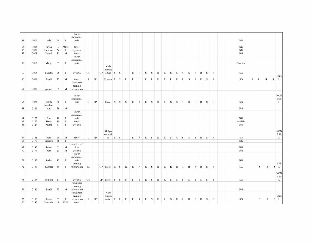
54 3005 kala 64 F
lower abdominal
pain NG
55 3006 devan 5 MCH fever NG56 3007 kanmani 34 F dysuria NG57 3008 Senthil 35 M fever NG
58 3067 Manju 63 F
lower abdominal
pain Candida
59 3068 Panchu 25 F dysuria OG OP
Kleb pneumoniae S S
-R S S S R R S S S S S R S S SG
60 3069 Pandi 72 M fever S IP Proteus R S R R -
R R R R R R R S S R S S SG S S S S ESB
L
61 3070 pannai 65 M
flank pain burning
micturation
62 3071 natchi 60 F
lower abdominal
pain S IP E.coli S S S R R R S R R S S S S S R S S SG
NON ESB
L
63 3121Natchim
uthu 59 M NG
64 3122 Anu 40 F
lower abdominal
pain NG65 3123 Banu 40 F fever candida66 3124 Mathi 47 F dysuria NG
67 3125 Ram 60 M fever U IP
Klebpneumoni
ae R S
-
R R R S R R S S S S S R S R SG
NON ESB
L68 3179 Saranya 68 F NG
69 3180 Sarasu 65 Mcatheterized
,fever NG70 3191 Ram 21 M dysuria NG
71 3182 Radha 42 F
lower abdominal
pain NG
72 3183 Kumari 39 Fburning
micturation M OP E.coli R S R R R R S R R R R R R S R S S SG S S S ESB
L
73 3184 Pothum 57 F dysuria OG 0P E.coli S S S S S R S R R S S S S S S S S SG
NON ESB
L
74 3185 Sandi 72 M
flank pain burning
micturation NG
75 3186 Pavai 65 F
flank pain burning
micturation S IP
Kleb pneumoniae R R R R R R S R R S R R R S R S S SG S S S
ESBL
76 3187 Vasanthi 5 FCH fever

77 3241 Vasan 4 MCH fever p op K.p R S R R S R R R R R R R S S S S S SG S S S S ESB
L
78 3242 Kalil 50 M
lower abdominal
pain
79 3243 Kani 28 F dysuria N IP
S.saprophyticu
s - -
R R R R - - -
S - - - - - - -
S SG
80 3244 Kannan 64 M
flank pain burning
micturation U IP E.coli R R R R S R S R R R R R R S R S S SG S S S ESB
L81 3245 Angel 6 FCH fever NG
82 3246 Selvan 34 M dysuria M OP
Klebpneumoni
ae R S -
R S R S R R R R R R S R S S SG S S S ESB
L
83 3247 Selvi 8 FCH fever P IPNFGN
B S S R R R R S R R R S R S S R SG84 3248 Ponni 27 F fever NG85 3249 Vijaya 66 F dysuria NG
86 3250 Vidhya 9 FCHburning
micturation P IP E.coli S S S S S S S R R S S S S S S S S SG
NONESB
L
87 3251 Babu 3 MCH fever NG
88 3252 Kali 80 Mcatheterized
,fever NG
89 3253 Malliga 38 Fburning
micturation NG
90 3254 Kamala 16 Fburning
micturation NG
91 3255 Rathina 56 F
lower abdominal
pain M IPK.pneumoniae R S
-
R S R S R R S S S S S R S S SG
NON ESB
L
92 3256 Kutti 1 MCH fever NG
93 3257Krishna
n 14 M
lower abdominal
pain NG
94 3258 Raja 50 M
lower abdominal
pain NG95 3259 Pooja 64 F dysuria NG96 3260 Rathina 13 F fever NG
97 3291 Sekar 75 Mcatheterized
,fever NG
98 3292 Rani 18 F
lower abdominal
pain M IP E.coli R S R R S R S S S S S S S S S S S SG
NON ESB
L99 3293 Kani 63 F fever NG
100 3294 James 63 Mcatheterized
,fever NG

101 3295 James 46 M
lower abdominal
pain M IPC.koser
i S S
-
S S S S
- -
S S S S S R S S SG
NON ESB
L
102 3296 Kattan 67 M urgency M IP Proteus S S S R
-
R R R R R S S S S S S S SG
NON ESB
L103 3297 Selvi 28 F dysuria
104 3298 Ambika 53 F urgency OG OP E.coli R R R R S R S R R S R R R S R S S SG S S S ESB
L105 3312 Baby 68 F dysuria NG
106 3313 Bala 47 M
lower abdominal
pain M IP
Kleb pneumoniae R R R R R R S R R S R R S S R S R SG S S S S
ESBL
107 3314 Kumar 62 M dysuria NG
108 3315 Chinnu 21days MCH fever P IP E.coli R R R R S R S R R S R R S S R S S SG S S S ESB
L109 3316 Mega 41 M dysuria NG
110 3317 Janani 11 FCH
lower abdominal
pain NG111 3318 Dhanya 19 F urgency NG112 3319 Jothi 10 FCH dysuria NG
113 3320 Vijaya 14 M
lower abdominal
pain M IP
Keeb pneumoniae R S
-
R R R S R R S S S S S R S S SG
NON ESB
L
114 3371 Sethu 70 M
flank pain burning
micturation NG
115 3372Sethupat
hy 61 Mcatheterized
,fever NG116 3373 Anush 9 FCH urgency NG117 2274 Vannan 69 M dysuria NG
118 2275 Vani 66 F urgency OG OP
Klebpneumoni
ae S S
-
R S R S S S S S S S S R S S SG
NON ESB
L
119 3376 Shree 29 F dysuria S IP
S.saprophyticu
s - -
R R s s - - -
S - - - - - - -
R SG120 3377 Kohi 54 F urgency NG121 3378 Sound 69 M dysuria NG
122 3379 Kohila 8 FCH dysuria P IP
Kleb pneumoniae R S
-
R S R S R R S R R R S R R R SG
NON ESB
L123 3380 Sankara 59 M urgency NG124 3381 Anu 28 F NG
125 3382 Ragu 28 M
lower abdominal
pain NG

126 3383 Badhri 2 MCH fever P IP E.coli S S S S S S S S S S S S S S S S S SG
NON ESB
L
127 3384 Raja 65 M
flank pain burning
micturation S IP Proteus R S R R -
R S R R R R R S S R S S SG S S S S ESB
L128 3385 Pitchai 64 F urgency NG129 3421 kala 16 F dysuria NG
130 3422 Saranya 44 F urgency S OP
Klebpneumoni
ae S S
-
R S S S R R R S S S S S S S SG
NON ESB
L
131 3423 Gowri 16 Fcatheterized
,fever M IPEnterobacter S S
-
S R R S
- - -
S S S S R R S SG
NON ESB
L132 3424 Prakash 67 M urgency NG
133 3425 Lalith 2 MCH fever NG134 3426 Veeram 63 F urgency NG
135 3427 Veeram 70 M
flank pain burning
micturation S IPE.coli CoNS
S -
S -
R S
R S
S R
R R
R -
R -
R -
S S
R -
R -
R -
S -
R -
S -
S -
S - SG S S S
ESBL
136 3428 Kattan 74 M dysuria NG
137 3429 Chellam 1 MCH fever NG138 3430 Ponni 18 F urgency NG139 3471 Rajini 41 M urgency NG
140 3472 Prakash 40 M
lower abdominal
pain NG
141 3473 Chellam 2 FCH fever p IP E.coli R S R R S R S R R S S S S S S S S SG
NON ESB
L
142 3474 Ponnu 27 F dysuria OG IP
S,saprophyticu
s - -
S S S R - - -
S - - - - - - -
R SG143 3475 Kannu 60 F dysuria M IP CoNS - - R R R S - - - S - - - - - - - S SG
144 3476 Ravi 58 M urgency S IP E.coli R S R R R R S R R S S S S S S S S SG
NON ESB
L145 3477 Gnana 67 F dysuria NG
146 3478 Mani 72 Mcatheterized
,fever NG147 3479 Devar 58 M urgency NG148 3480 Pavai 29 F urgency NG
149 3521 Nagan 42 M urgency STD OP E.coli S S R R S S S R R S R R R S S S S SG S S S ESB
L

150 3522 Malai 68 M urgency U OP Ckoseri R S
-
R R R S
- -
S S S S S R S S SG
NON ESB
L151 3523 Arasu 39 M urgency NG
152 3524 Ganse 69 M
flank pain burning
micturation NG
153 3525 Naga 41 F dysuria OG IP E.coli R S S S S S S S S S S S S S S S S SG
NON ESB
L154 3526 Nagan 69 M urgency NG155 3527 Kannan 48 M dysuria NG156 3528 Selvai 5 FCH fever NG
157 3529 Sam 1 FCH fever P IP E.coli S S R S S S S R R S R R S S S S S SG S S S ESB
L
158 3641 Kannu 68 F urgency M IP E.coli R R R R R R R R R R R R R S R S S SG S S S S ESB
L
159 3644 Chiinu 29 M
lower abdominal
pain NG160 3645 Palani 60 M urgency NG161 4646 Pavai 40 F urgency NG162 3647 Pavithra 47 F urgency NG
163 3648 Suresh 65 Mcatheterized
,fever STD OP E.coli R S S S S S S S S S S S S S S S S SG
NON ESB
L
164 3649 Divya 39 F urgency M IP E.coli S S R R R R S R R S R R S S R S R SG S S S S ESB
L
165 3650 Soloman 68 M fever U IP E.coli R S R R S R S S S S S S S S R S S SG
NON ESB
L166 3693 Suganya 66 F urgency NG167 3694 Sundhar 58 M urgency NG
168 3695 Dinesh 1 MCH fever NG
169 3696Ranmes
h 59 M urgency NG
170 3697 Swathi 3 MCH fever P IPNFGN
B S S R S R R S R R R S R S S R SG
171 3698 Meena 42 F
lower abdominal
pain NG
172 3699 Saroja 48 F dysuria OG IP E.coli R S R R S R S R R S S S S S S S S SG
NON ESB
L173 3700 Raju 65 M fever NG

174 3708 Suresh 9 FCH fever P OP E.coli S S S S S S S S S S S S S S S S S. SG
NON ESB
L
175 3709 Nambi 66 Mcatheterized
,fever NG
176 3810 Jithan 2 MCH fever NG177 3811 kumutha 38 F dysuria NG178 3812 Divya 59 F urgency NG
179 3813 Vishali 8 FCH fever P IP
E.coli, NFGN
B
S R
S S
R R
R R
S S R
RS S
R S
R S
S S
R S
R S
S S
S S
R S
S S
S S SG S S S
ESBL
180 3814 Bharath 39 M urgency M IP
Klebpneumoni
ae R S R R S R R R R R R R R S R R R SG S S S ESB
L
181 3815 Mant 48 M
lower abdominal
pain NG
182 3816 Sumo 4 MCH fever NG183 3817 Gaya 37 F dysuria NG
184 3818 ponni 46 F urgency OG IP E.coli R S R R S S S R R R S S S S S S S SG
NON ESB
L185 3819 Chandra 4 FCH fever NG
186 3867 Mahesh 59 M urgency S IPEnteroc
occi
S(HLG) S
-S S
- - - - - - - - - - - -S SG
187 3868 pannu 64 F dysuria NG
188 3869 Baby 6 FCH dysuria P IP E.coli R S R R R R R R R R S S S S S S S SG
NON ESB
L189 3870 Kavya 36 F fever NG190 3871 Bharathi 65 F urgency NG191 3872 Praddep 69 M dysuria NG
192 3873 Pandi 70 M urgency S IP E.coli R R R R S R R R R S R R S S R R S SG S S S S ESB
L193 3874 Kannan 14 M dysuria NG194 3875 Jithu 7 FCH dysuria NG
195 3876 Bakkya 39 F dysuria OG OPS.sapro K pneu R
S R
S R
R R
S R
R R R
S R R R S S R S R
R - SG S S S S
ESBL
196 3877 Selvi 8 FCH
lower abdominal
pain P OP E.coli S S S S S R S S S S S S S S S S S SG
NONESB
L197 3879 Priya 18 F fever
198 3880 Suresh 63 M fever S IP E.coli R S R S S R R R R S R R R S R S S SG S S S S ESB
L

199 3895 Suriya 11 MCH
lower abdominal
pain P OPKleb pneu S S
-
R S S S R R S R R R S S S S SG
NON ESB
L200 3896 Mari 59 F fever OG IP CONS - - S R R R - - - S - - - - - - - S SG
201 3897Moham
mad 43 M dysuria M OP E.coli S S R R S R R R R R S S R S R S S SG
NON ESB
L202 3898 Beham 63 F fever NG203 3890 Bavi 38 F dysuria NG
204 3900 Kavin 9 MCH fever P IPNFGN
B S S R R R R R R R R S R S S R SG
205 3911 Bavith 38 F fever S IPNFGN
B R S R R \R R R R R R S S S S R SG
206 3912 Arasu 62 Mcatheterized
,fever S IP Ecoli R R R R S R S R R S R R S S R S S SG S S S ESB
L207 3913 Siva 29 M fever NG
208 3914 Prathap 47 M dysuria M IP E.coli R S R R S R S R R S S S S S S S S SG
NON ESB
L209 3915 Monica 8 FCH fever NG
210 3916 Ravi 48 M
lower abdominal
pain NG211 3918 Sudha 3 FCH fever NG212 3967 Priya 28 F dysuria candida213 3968 Sundar 65 M fever NG
214 3969 Selvan 67 Mcatheterized
,fever NG
215 3970 Ranji 43 F dysuria OG IP E.coli R R R R S R S R R S R R R S R S R SG S S S S ESB
L
216 3971 Rajam 64 F
lower abdominal
pain OG OPKleboxytoca S S
-
R S S S S S S S S S S S S S SG
NON ESB
L
217 3972 Dev 46 M dysuria STD OPK.pneumoniae S S
-
R R R R R R S R R R S R S S SG
NON ESB
L218 3973 Dilip 4 FCH NG
219 3974 Ajith 28 M
lower abdominal
pain NG220 3985 Jothika 6 FCH fever NG
221 3986 Karthi 38 M dysuria S IP E.coli R S R R S R R R R R R R S S R S S SG
NON ESB
L
222 3987 Kali 40 F dysuria OG IPEnteroc
occi
S(HLG)
-S
-S S
- - - - - - - - - - -S SG
223 3988 Meena 44 F fever NG

224 3989 Kumari 19 F urgency NG
225 4000 Sugu 7 FCH dysuria P IPS.aureu
s - -
S S S S - - -
S - - - - - - -
S SG226 4004 Manna 46 F dysuria NG
227 4005 Lallu 2 MCH fever P IPS.aureu
s - -
R S S S - - - R - - - - - - -
S SG
228 4006 saroja 68 Mcatheterized
,fever M IP E.coli R S R R S R S R S R R R R S R R S SG S S S ESB
L
229 4007 mani 9 MCH
lower abdominal
pain NG
230 4008 Chitu 2 MCH dysuria NG
231 4009 Kannan 19 F dysuria M IPNFGN
B S S R R R R S R R R R R R S R SG
232 4010 Divya 8 FCH
lower abdominal
pain P IP
CoNS Enteroc
occi
S(HLG)
- R S
S -
R S
R S
- - - S -
- - - - - - - S - S SG
233 4012 Ponni 70 F dysuria OG IP E.coli R R R R S R S R R S R R R S R R S SG S S S S ESB
L
234 4014 Sam 2 MCH fever NG
235 4016 Kamal 68 M dysuria S IP E.coli R S R R S R S R R R R R R S R R R SG
NON ESB
L
236 4017 Amala 24 F
lower abdominal
pain S IPK.oxyt
oca R S R R S R S R R S R R S S R S R SG S S S S ESB
L
237 4018 Kala 9 FCH fever P IPNFGN
B S S R R R R R R R R R R R S R S S SG
238 4019 Nambi 58 M
lower abdominal
pain NG
239 4020 Rama 47 F fever OG IPK.pneumoniae R S
-
R S R S R S S S S S S S S S SG
NON ESB
L
240 4034 Suria 46 M
lower abdominal
pain NG
241 4035 Kumar 66 M
lower abdominal
pain NG
242 4036 Kaniya 64 M
lower abdominal
pain NG
243 4037 Suriya 11 MCH fever NG
244 4038 Pavia 28 F
lower abdominal
pain candida

245 4039 Nalayini 4 FCH
lower abdominal
pain P IP E.coli S S R R S S S R R S R R S S R S S SG S S S ESB
L
246 4040 Kattu 68 Mcatheterized
,fever NG
247 4041 Matti 58 M
lower abdominal
pain NG248 4042 Dilip 4 FCH fever NG249 4054 Chinna 59 F fever NG250 4055 Alagu 7 FCH fever NG
251 4056 Beham 68 Fcatheterized
,fever NG
252 4057 Devan 8 MCH fever NG
253 4058 Rosy 29 Fburnig
micturation M IP E.coli S S S S S R S S S S S S S S R S S SG
NON ESB
L
254 4059 Bala 29days MCHsuprapubic
aspirate NG
255 4060 Rosline 7 FCHburnig
micturation NG
256 4074 shan 68 Mcatheterized
,fever U IP E.coli S S R R S R S R R R S S S S R S S SG
NON ESB
L
257 4075 mathi 6 FCHburnig
micturation NG
258 4076Santhan
am 46 Mburnig
micturation NG
259 4077 Selvi 40 F
lower abdominal
pain M IP Proteus R S R R
-
R R R R R S S S S R S S SG
NON ESB
L
260 4078 Sundar 38 Mburnig
micturation STD OPK.pneumoniae R S R R S R S R R S R R S S R S S SG S S S
ESBL
261 4079 Anil 1 MCH fever P IP E.coli S S R R S S S R R S R R S S R S S SG S S S ESB
L
262 4108 Janani 11 FCHburnig
micturation P OP E.coli R S R R S R S R R S S S S S S S S SG
NON ESB
L
263 4109 Dev 29 M fever M IP E.coli R S R R S R R R R S R R S S R S S SG S S S S ESB
L264 4110 Shoba 27 F fever
265 4112 Shathi 30 F
lower abdominal
pain S IPNFGN
B R R R R R R R R R R S S S S R SG
266 4113 Palani 67 Fcatheterized
,fever NG
267 4114 Naveen 2 MCH fever NG

268 4115Kanayira
m 70 Mcatheterized
,fever U IP E.coli S S R R S S S R R S S S S S R S S SG
NON ESB
L
269 4119 Kaveen 4 MCH
lower abdominal
pain NG
270 4145 maru 66 Mburnig
micturation NG
271 4146 Megala 8 FCH fever P IP E.coli R S R R S S S R R S R R S S R S S SG S S S ESB
L272 4147 Deva 58 M fever NG
273 4148 Samuth 29 F
lower abdominal
pain NG
274 4149Sanmath
i 7 FCH fever NG
275 4150 Sami 68 Mburnig
micturation S OPK.oxyt
oca R R
-
R S R R R R R S S S S S S S SG
NON ESB
L
276 4156 Selvi 7 FCH
lower abdominal
pain P IP E.coli S S R R S R R R R S S S S S S S S SG
NON ESB
L
277 4157 Boomi 38 Mburnig
micturation NG
278 4158 Kala 18 F
lower abdominal
pain NG
279 4159 Kavi 5 FCH flank pain P IP
CoNS,Enteroc
occi S - S
SS -
S S
S S
- - - S -
- - - - - - - S - S SG
280 4160 Ammu 35 Fburnig
micturation S OP E.coli S S S S S S S S S S S S S S S S S SG
NON ESB
L
281 4167s\Sathay
a 8 FCH flank pain NG
282 4168 Savi 57 Fcatheterized
,fever NG
283 4169 Ravi 8 MCH flank pain NG
284 4170 Malayan 65 Fburnig
micturation OG IP E.coli S S R R R R R R R R R R R S S S S SG
NON ESB
L285 4178 Devi 19 F flank pain NG
286 4179 Kannan 46 Mburnig
micturation M IP E.coli S S R R S R R R R R S S S S S S S SG
NON ESB
L287 4180 Ponnam 40 M flank pain NG288 4196 rethu 10 FCH flank pain NG
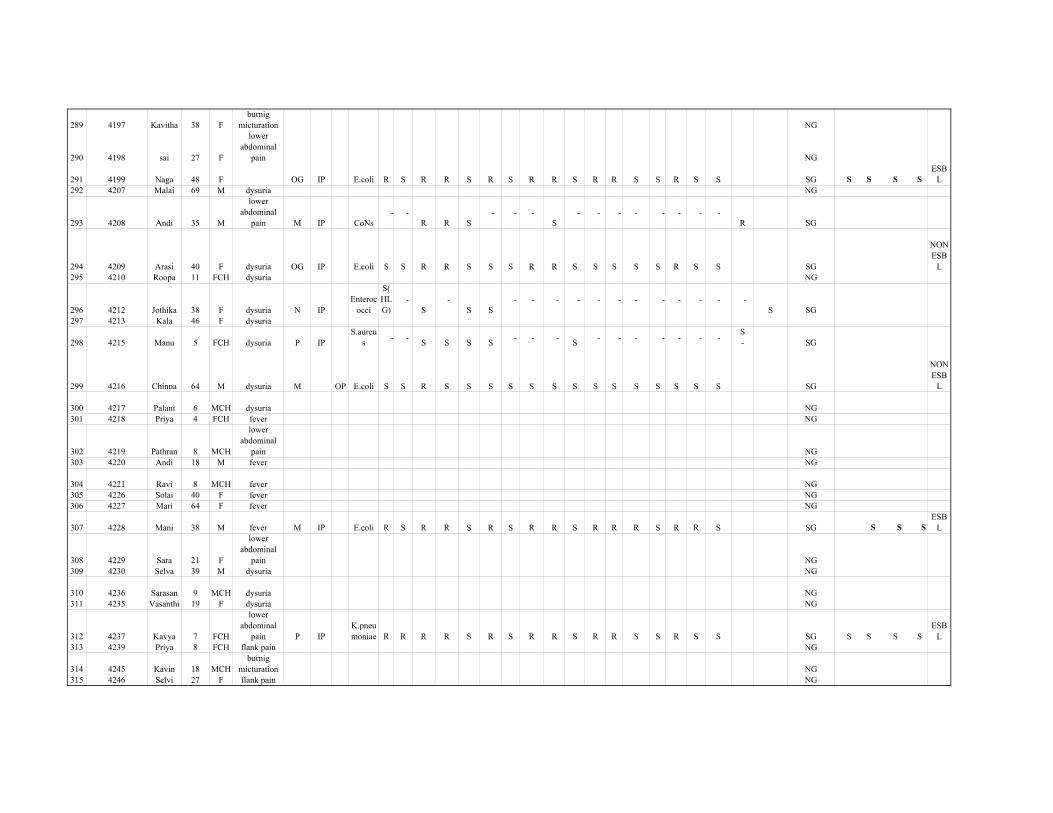
289 4197 Kavitha 38 Fburnig
micturation NG
290 4198 sai 27 F
lower abdominal
pain NG
291 4199 Naga 48 F OG IP E.coli R S R R S R S R R S R R S S R S S SG S S S S ESB
L292 4207 Malai 69 M dysuria NG
293 4208 Andi 35 M
lower abdominal
pain M IP CoNs - -
R R S - - -
S - - - - - - - -
R SG
294 4209 Arasi 40 F dysuria OG IP E.coli S S R R S S S R R S S S S S R S S SG
NON ESB
L295 4210 Roopa 11 FCH dysuria NG
296 4212 Jothika 38 F dysuria N IPEnteroc
occi
S(HLG)
-S
-S S
- - - - - - - - - - - -S SG
297 4213 Kala 46 F dysuria
298 4215 Manu 5 FCH dysuria P IPS.aureu
s - -
S S S S - - -
S - - - - - - -
S - SG
299 4216 Chinna 64 M dysuria M OP E.coli S S R S S S S S S S S S S S S S S SG
NON ESB
L
300 4217 Palani 6 MCH dysuria NG301 4218 Priya 4 FCH fever NG
302 4219 Pathran 8 MCH
lower abdominal
pain NG303 4220 Andi 18 M fever NG
304 4221 Ravi 8 MCH fever NG305 4226 Solai 40 F fever NG306 4227 Mari 64 F fever NG
307 4228 Mani 38 M fever M IP E.coli R S R R S R S R R S R R R S R R S SG S S S ESB
L
308 4229 Sara 21 F
lower abdominal
pain NG309 4230 Selva 39 M dysuria NG
310 4236 Sarasan 9 MCH dysuria NG311 4235 Vasanthi 19 F dysuria NG
312 4237 Kavya 7 FCH
lower abdominal
pain P IPK.pneumoniae R R R R S R S R R S R R S S R S S SG S S S S
ESBL
313 4239 Priya 8 FCH flank pain NG
314 4245 Kavin 18 MCHburnig
micturation NG315 4246 Selvi 27 F flank pain NG

316 4247 Selsi 7 FCH
lower abdominal
pain NG
317 4248Ammath
a 63 F
lower abdominal
pain OG IP E.coli R S R R S R S R R S R R R S R R S SG S S S ESB
L
318 4252 Malayan 68 Mcatheterized
,fever M IP CoNS - -
R R R R - - -
S - - - - - - -
S SG
319 4253 Pappu 8 FCH dysuria P IPK.pneumoniae R S
-
R S R S R R S S S S S R S S SG
NON ESB
L320 4254 Pandi 47 M dysuria NG321 4255 Ammu 10 FCH dysuria NG
322 4256 Palani 38 M dysuria S IP E.coli S S R R S R S R R R R R R S R R R SG
NON ESB
L323 4257 Kannath 26 F fever NG324 4258 Selvi 44 F fever NG
325 4265 Roopa 9 FCH
lower abdominal
pain P IPS.aureu
s S -
S S S S -
S -
S - - - -
S - -
SG
326 4266 Vijayan 11 MCHburnig
micturation NG
327 4267 Malai 67 Mcatheterized
,fever U IP E.coli R S R R S R S R R S R R R S R S S SG S S S S ESB
L
328 4268 Kali 32 Fburnig
micturation M IP
S.saprophyticu
s - -
S R S S - - -
S - - - - - - -
S SG329 4269 Arasi 17 F fever
330 4270 Naveen 9 MCH dysuria P OPK.oxyt
oca S S
-
R R R S R R S S S S S S S S SG
NON ESB
L
331 4276 Andi 68 M dysuria U IP E.coli R S R R S R S R R S R R R S R S S SG S S S S ESB
L332 4277 Ponnan 58 M dysuria NG
333 4278 Sound 38 Mburnig
micturation M OP E.coli S S R S R S S R S S S S S S S S S SG
NON ESB
L334 4279 Kamala 35 F dysuria candida
335 4280 Eswar 67 Mcatheterized
,fever NG336 4284 Rama 19 F dysuria NG
337 4285Ramach
and 60 M dysuria NG
338 4286 Chandru 7 MCH dysuria NG
339 4287 Mandra 33 F urgency M IP E.coli R S R R S R S R R R S S S S S S S SG
NON ESB
L

340 4288Manika
m 43 Mburnig
micturation M IPS.aureu
s - -
R R R R - - -
S - - - - - - -
S SG341 4294 Seetha 48 F dyuria NG
342 4295 Megala 64 M urgency M IP E.coli R S R R S R S R R R S S S S S S S SG
NON ESB
L
343 4296 Sen 6 MCH urgency NG344 4298 Kavya 29 F urgency NG
345 4301 amman 65 F urgency OG IP E.coli R S R R S R S R R S R R R S R S S SG S S S S ESB
L
346 4302 Deva 66 Mcatheterized
,fever NG347 4306 Pandi 70 M frequency NG
348 4307 Palani 39 M frquency M OP E.coli S S R S S S S R R R R R S S R R R SG
NON ESB
L
349 4308Manicka
m 64 Mcatheterized
,fever NG
350 4309 Arasi 31 Fburnig
micturation NG351 4310 Veeram 54 M dyuria NG
352 4314 Ponni 58 Fburnig
micturation NG
353 4315Kanman
i 30 Fburnig
micturation M IP E.coli R S R R R R S R R R R R R S R R R SG
NON ESB
L
354 4316 Selva 9 MCHburnig
micturation NG
355 4317 Kavin 8 MCH fever NG356 4319 Sela 39 F urgency NG
357 4320 Mani 46 Mburnig
micturation NG358 4324 Devi 47 F frequency NG
359 4325 Pavi 65 M urgency S OP E.coli S S R R S R S R R S S S S S S S S SG
NON ESB
L360 4327 Kumari 64 F urgency
361 4328 Kaviya 29 F urgency M IP E.coli R S R R S R S R R S R R R S R S S SG S S S ESB
L
362 4329 Ganesh 70 M urgency M IP E.coli R S R R S S S R R S S S S S R S S SG
NON ESB
L
363 4330Krishna
n 67 M urgency NG364 4331 Veni 58 F urgency NG

365 4335 Nalini 29 Fburnig
micturation M OPKpneumoniae R S
-
R S R S R R S S S S S S S S SG
NON ESB
L366 4336 Jeya 58 F frequency
367 4337 Ananthi 17 F frequency M IP E.coli R S R R S R R R R S R R S S R S S SG S S S S ESB
L
368 4338 Somu 66 Mcatheterized
,fever NG369 4339 Sundari 18 F frequency NG370 4342 Mani 57 M frequency NG
371 4343 Kandan 66 M urgency N IP E.coli R S R R S S S R R S S S S S R S S SG
NON ESB
L
372 4344 Selvan 38 M urgency M OP E.coli R S R R S R S R R S S S S S R S S SG
NON ESB
L
373 4345 Selvi 59 Fburning
micturation NG374 4346 Kaniya 48 F frequency NG
375 4347 Sugu 28 Fburning
micturation NG376 4348 Nagy 19 F frequency NG
377 4349 Nagam 68 F urgency M IP E.coli R S R R S R R R R S R R S S R S R SG S S S S ESB
L378 4350 Pavi 68 F urgency NG379 4351 Banu 69 F urgency NG
380 4352 Kavin 2 MCH fever NG381 4353 Pappan 58 M flank pain NG
382 4355 Uma 30 Fburning
micturation M IP
CoNS NFGN
B S SS R
S R
R R
R R R R R
S S S S S S S
S - SG
NON ESB
L
383 43357 Mahesh 2 MCH fever P IP E.coli R S R R S R S R R S S S S S S S S SG
NON ESB
L
384 4358 Kumar 69 Mburning
micturation
385 4359 Kalan 65 M fever M IP E.coli R S R R S R R R R S R R S S R S R SG S S S S ESB
L386 4360 Dhana 38 F flank pain NG387 4361 Lakshmi 32 F urgency NG388 4362 Pavithra 48 F urgency NG389 4365 Maniya 58 F fever NG
390 4366 Rathiya 65 F flank pain U IP Proteus R R R R
-
R S R R S S S S S R S S SG
NON ESB
L

391 4367 Ranji 32 Fburning
micturation OG OP E.coli R S R R S R S R R S S S S S S S S SG
NON ESB
L
392 4368 Manna 68 M urgency N IP E.coli R S R R S R S R R R S S S S S S S SG
NON ESB
L393 4369 Mali 34 F urgency NG394 4370 Ponnu 64 F urgency NG
395 4371 Kala 42 F flank pain OG IP E.coli R S R R R R S R R S S R S S R S S SG
NON ESB
L396 4372 Selvi 28 F dysuria NG397 4373 Vani 26 F urgency NG
398 4374 Anu 38 F urgency OG IP E.coli R S R R S R R R R S R R R S R S R SG S S S S ESB
L399 4375 Dhanu 34 F urgency NG
400 4376 Mohan 43 Fburning
micturation OG IP E.coli R S R R S R S R R S S S S S S S S SG
NON ESB
L


Copyright © 2022 FDOKUMEN