A study of the relationship of glucose-6-phosphate ... - EliScholar
149
Yale University EliScholar – A Digital Platform for Scholarly Publishing at Yale Yale Medicine esis Digital Library School of Medicine 1977 A study of the relationship of glucose-6-phosphate dehydrogenase deficiency and bacterial infection in a hospitalized Iranian population Marcia S. Clark Yale University Follow this and additional works at: hp://elischolar.library.yale.edu/ymtdl is Open Access esis is brought to you for free and open access by the School of Medicine at EliScholar – A Digital Platform for Scholarly Publishing at Yale. It has been accepted for inclusion in Yale Medicine esis Digital Library by an authorized administrator of EliScholar – A Digital Platform for Scholarly Publishing at Yale. For more information, please contact [email protected]. Recommended Citation Clark, Marcia S., "A study of the relationship of glucose-6-phosphate dehydrogenase deficiency and bacterial infection in a hospitalized Iranian population" (1977). Yale Medicine esis Digital Library. 2471. hp://elischolar.library.yale.edu/ymtdl/2471
-
Upload
khangminh22 -
Category
Documents
-
view
0 -
download
0
Transcript of A study of the relationship of glucose-6-phosphate ... - EliScholar
Yale UniversityEliScholar – A Digital Platform for Scholarly Publishing at Yale
Yale Medicine Thesis Digital Library School of Medicine
1977
A study of the relationship of glucose-6-phosphatedehydrogenase deficiency and bacterial infection ina hospitalized Iranian populationMarcia S. ClarkYale University
Follow this and additional works at: http://elischolar.library.yale.edu/ymtdl
This Open Access Thesis is brought to you for free and open access by the School of Medicine at EliScholar – A Digital Platform for ScholarlyPublishing at Yale. It has been accepted for inclusion in Yale Medicine Thesis Digital Library by an authorized administrator of EliScholar – A DigitalPlatform for Scholarly Publishing at Yale. For more information, please contact [email protected].
Recommended CitationClark, Marcia S., "A study of the relationship of glucose-6-phosphate dehydrogenase deficiency and bacterial infection in a hospitalizedIranian population" (1977). Yale Medicine Thesis Digital Library. 2471.http://elischolar.library.yale.edu/ymtdl/2471
medical library

"f'b'-L
Permission for photocopying or microfilming of 11 -/ cy^s\dt-< / 0>&Pt>
^ tfyyUsi a< OtC'm cU icf\ ~^ 3pyj h'tjZi 20?11
(TITLE OF THESIS) v^\
for the purpose of individual scholarly consultation or reference is hereby
granted by the author. This permission is not to be interpreted as affect¬
ing publication of this work or otherwise placing it in the public domain,
and the author reserves all rights of ownership guaranteed under common
law protection of unpublished manuscripts.
/ Signature of Author
Date / r;y-9
Digitized by the Internet Archive in 2017 with funding from
The National Endowment for the Humanities and the Arcadia Fund
https://archive.org/details/studyofrelations00clar_0
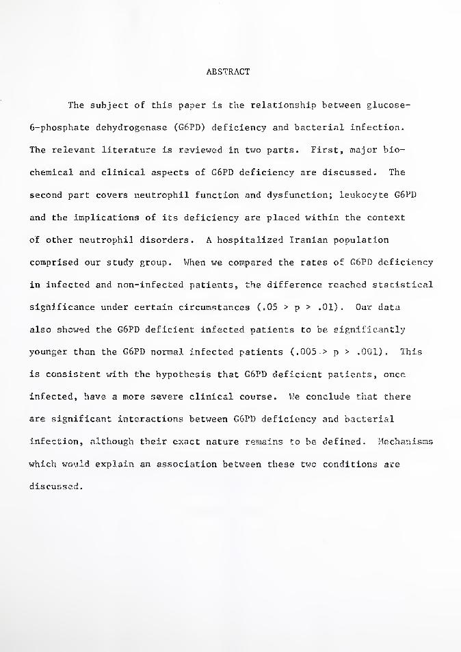
ABSTRACT
The subject of this paper is the relationship between glucose-
6-phosphate dehydrogenase (G6PD) deficiency and bacterial infection.
The relevant literature is reviewed in two parts. First, major bio¬
chemical and clinical aspects of G6PD deficiency are discussed. The
second part covers neutrophil function and dysfunction; leukocyte G6PD
and the implications of its deficiency are placed within the context
of other neutrophil disorders. A hospitalized Iranian population
comprised our study group. When we compared the rates of G6PD deficiency
in infected and non-infected patients, the difference reached statistical
significance under certain circumstances (.05 > p > .01). Our data
also showed the G6PD deficient infected patients to be significantly
younger than the G6PD normal infected patients (.005 > p > .001). This
is consistent with the hypothesis that G6PD deficient patients, once
infected, have a more severe clinical course. We conclude that there
are significant interactions between G6PD deficiency and bacterial
infection, although their exact nature remains to be defined. Mechanisms
which would explain an association between these two conditions are
discussed.

A STUDY OF THE RELATIONSHIP OF GLUCOSE-6-PHOSPHATE DEHYDROGENASE
DEFICIENCY AND BACTERIAL INFECTION IN A HOSPITALIZED
IRANIAN POPULATION
by
MARCIA S. CLARK
B.A., Cornell University, 1973
A Thesis Presented to the Faculty of Yale. Medical in Partial Fulfillment of the Degree of
Doctor of Medicine
School
March 1, 1977


TABLE OF CONTENTS
Acknowledgements
Section Page(s)
I. Introduction to G6PD Deficiency . ..... 1 -10
Definitions and History . .. 1 Mechanisms of Hemolysis ............ 2 G6PD Variants ...... . 3 Leukocyte G6PD .. 6 Mode of Inheritance ....... . 7 G6PD Deficiency and Associated Conditions ... 7
II. Neutrophil Function and Dysfunction . 11-29
A. Normal Neutrophil Function Production and Delivery; .. 11
Phagocvtosis: Recognition and Ingestion ... 13 Killing and Digestion ....... . 14
B. Neutrophil Abnormalities Disorders of Production and Delivery . 16 Disorders of Recognition and Ingestion .... 20 Disorders of Killing and Digestion .. 21 Miscellaneous Disorders , . 28
III. A Study of G6PD Deficiency and Bacterial Infection in a Hospitalized Iranian Population . 30-43
Introduction ... 30 Materials and Methods .. 31 Results ..... . 34 Discussion... 38
Figures... 4 4-45
Tables .. *6-56
Bibliography ............ . 57-65

ACKNOWLEDGEMENTS
X would like to gratefully acknowledge the contributions of
the following:
- Dr. Richard Root (Professor of Medicine, Chief of Infectious Disease Section, Yale Medical School) whose vast knowledge will remain an inspiration long after this thesis is completed, for his help in planning the project, his valuable suggestions during the writing, and his financial help for the trip to Iran
- Dr. Abdolgnader Molavi (Associate Professor of Medicine, Pahlavi University School of Medicine, Shiraz, Iran) who guided me through the data collection, and who together with his family made my stay in Iran a personally as well as medically rewarding experience
- Dr. Alfred Evans and the Committee on International Fellowshi for their generous funding
- Housestaff of Nemazee and Saadi Hospitals, Shiraz, Iran, for their aid in finding patients and translating an occasional Persian phrase
- Staff of the Yale Medical Library, especially Sara Battison, for tracking down a series of almost unobtainable references
- Ken Pobuler, who as a Yale medical student collected some of the data
- Linda Rodman, for putting up with last minute changes and still doing a beautiful typing job
Judy Gordon, for typing a draft of this manuscript, but, more importantly, for her invaluable friendship and support


I
Definitions and History
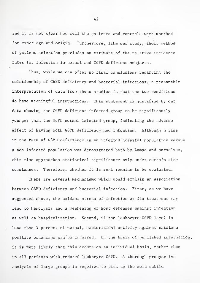
Glucose-6-phosphate dehydrogenase (G6PD), the first enzyme in
the pentose phosphate pathway, catalyzes the conversion of glucose-6-
phosphate to 6-phosphogluconolactone while reducing NADP to NADPH
[Figure l]. The major practical importance of G6PD lies in its role
in the red blood cell, where its deficiency can cause hemolytic anemia.
Most commonly, the anemia is episodic and is related to one of a number
of agents, but occasionally a chronic hemolytic anemia occurs. Although
the former entity has been known for thousands of years, it was only
recently that the link between G6PD deficiency and hemolytic anemia was
established. In 1926, plasmoquin, an 8-amirioquinoline compound, was
first used in treating syphilitics who had been inoculated with malaria;
shortly thereafter, a series of reports appeared describing hemolytic
anemia with administration of the drug [1]. Little progress was made
in determining the etiology of the condition until 1950, when investi¬
gators received a new research tool in the form of primaquine, a compound
related to but therapeutically more effective than plasmoquin. In 1954
Dern et al. [2], utilizing cross-transfusions between normal and
primaquine-sensitive individuals, demonstrated that the basic abnormality
was in the red blood cell. Other investigators found that the hemolytic
susceptibility was restricted to the older erythrocytes and, since at
1


2
least some metabolic processes were known to decrease with aging,
attention was directed towards a metabolic disorder [1], In 1965,
Carson and his collaborators [3] reported a decreased G6PD level in
the erythrocytes of affected individuals.
Mechanisms of Hemolysis
Although it was soon generally accepted that the G6PD deficiency
and hemolytic anemia were related, detailed understanding of the hemo¬
lytic mechanism was and still is lacking. An outline of events as they
are known requires a brief digression into certain aspects of erythrocyte
biochemistry.
As mentioned before, G6PD is one of the enzymes of the hexose
monophosphate shunt (HMP) [Figure l]. This pathway is very important
to the red blood cell, even though it metabolizes a much smaller quantity
of glucose than the Embden-Meyerhof pathway. The reduction of NADP to
NADPH and the production of the nucleotide precursor ribose-5-phosphate
take place via the HMP. The latter can also be formed by other means;
in contrast, NADPH in red cells is supplied entirely by the HMS. A
source of NADPH is critical because it acts as the coenzyme for the
conversion of oxidized glutathione (GSSG) to reduced glutathione (GSI1) .
The GSH prevents the oxidation of sulfhydryl groups of proteins such
as hemoglobin, usually because GSH itself is instead oxidized to GSSG.
In cases where the oxidant is GSH acts by reducing the H,,G^ to
H^O [4], A second important role of NADPH is to provide electrons for
reduction and cleavage of GSK-hemoglobin complexes, which would otherwise
be liable to precipitate.

■ f ■ • .
;V ,-r ■ • • .

3
It is believed that infections, fava beans, and other stimuli
induce hemolysis by causing the production of various oxidizing substances
such as or ot^er unstable radicals. Drugs themselves can also
have oxidizing capacity. In the normal person, these substances can
be handled by the production of NADPH from the HMS, but G6PD deficiency
blocks this pathway. Consequently, the erythrocyte proteins, including
hemoglobin, are not protected from oxidation. Oxidized hemoglobin can
denature and precipitate as Heinz bodies, and by binding to and damaging
the cell membranes, the Heinz bodies increase cell susceptibility to
removal by the reticuloendothelial system [1,5].
Baehner £t al. [6] have made a more specific proposal regarding
the mechanism of hemolysis during infection; they suggest that HpC^
produced by phagocytizing leukocytes could serve as a partial source
of the oxidant stress. Their hypothesis was supported by experiments
in which phagocytizing leukocytes caused a fall in GSII in neighboring
G6PD deficient red blood cells, a result associated with increased
destruction of these cells in the liver and spleen. The effect was
not present with normal erythrocytes, nor with separation of the G6PD
deficient cells from the leukocytes. Pyrimidine aglycons have been
implicated as the offending agent in favism [7], and naturally occurring
substances such as ascorbate, pyruvate, and cysteine in "spontaneous"
hemolysis [5].
G6PD Variants
During the course of these and other investigations, it became
clear that G6PD deficiency varied enormously biochemically and clinically.


4
Over 80 variants have now been described in the 100 million or so people
with the condition [8], and doubtless more remain to be found. Only
a few of the major variants will be discussed here; complete compilations
are available in the literature [9,30].
The normal G6PI) enzyme, known as type B, has a molecular weight
of 190,000 to 240,000 and has 3 to 6 similar or identical subunits,
although the functioning enzyme is probably dimeric [1]. The biochemical
properties, including substrate affinity, thermostability, optimal pH,
and electrophoretic characteristics, are well known [Table 1], One of
the most common variants, called type A because it migrates faster than
type B, is present in approximately 30 percent of American black males
[5]. With the use of trypsin digests, it was found to differ from type B
by only a single amino acid [11]. Type A has normal activity and so has
little clinical importance, in contrast to Type A (which receives its
name because it migrates wit3t Type A, but has decreased activity).
About 11 percent of American black males have Type A [5] and the gene
exists in varying frequencies throughout Africa. The catalytic activity
of each A enzyme molecule, is close to normal, but there is a fall in
the total number of molecules [1], and the decrease in activity between
young and old cells is more marked than in normal cells [12,13]. Overall,
the activity in the blood is usually 5-15 percent of normal [5]; very
rarely, activity can be absent [14]. Affected individuals are generally
asymptomatic except under conditions of stress, such as with certain
drugs or infection [Table 2]. Because G6PD activity is low only in
older cells, the hemolysis tends to be mild and self-limited; a typical

.. 0 '
.

5
time course involves an onset 1 to 3 days after the inciting agent has
been introduced, with recovery beginning in A to 6 days [5].
A more severe form of G6PD deficiency is found in the Mediterranean
area, including Sardinia, Italy, Greece, Iran, Rumania, Israel, and
perhaps some areas in the Far East [15]. It is possible that G6PD
Mediterranean, as it is known, represents a heterogeneous group [11,16,
17,18] but the major biochemical characteristics listed in Table 1 are
constant. The frequency of the gene ranges from less than 0.1 percent
in Northern Europe to greater than 50 percent of Kurdish Jewish males
[5]. The quantity of enzyme is reduced in very young red blood cells,
and their catalytic efficacy is also lower [1]. Because all erythrocytes
are affected, the G6PD activity of whole blood tends to be less than
1 percent, and the hemolytic episodes are often more severe and not
self-limiting. The list of precipitating agents is similar to, but not
identical with, that for G6PD A ; for example, chloramphenicol causes
hemolysis in G6PD Mediterranean but not G6PD A [Table 2]. Fava beans,
which can lead to an extremely rapid and severe hemolysis, also appear
to affect mainly Caucasians [8]. Another factor may also be involved,
since only some G6PD Mediterranean patients are affected by the fava
beans.
A host of other G6PD variants have been described, ranging from
G6PD Canton, similar in its properties to G6PD Mediterranean [15], G6PD
Oklahoma, with a tremendous deterioration in activity with aging [19]
and G6PD Hektoen, with increased activity [5]. The clinical manifesta¬
tions depend on the characteristics of the enzyme; patients can be


6
asymptomatic, have mild or severe disease resembling that described
above, or rarely, have a chronic nonspherocytic anemia. Seen with some
of the rarer types of G6PD deficiency, as well as occasionally in G6PD
Mediterranean, the degree of shortened erythrocyte lifespan is generally
mild [5],
Leukocyte G6PD
G6PD deficiency is not necessarily limited to the red blood cells
and articles have appeared describing decreased levels in leukocytes,
lens tissue, kidney, adrenal tissue, platelets, saliva, liver and other
tissues [1]; most of these studies were in Caucasians [8,20]. A
complete analysis of these reports is not feasible here, and only the
leukocyte levels will be discussed in some detail. While most people
believe that the leukocyte and erythrocyte enzymes are the same [1],
some investigators have claimed differences [1,19] and the issue has
not been definitively settled. One published report even showed separate
inheritance of the two defects, but the measurement techniques described
are open to question [21]. Moreover, the findings have not been con¬
firmed by other researchers. In any case, several different studies
have demonstrated lowered leukocyte levels in association with some
types of erythrocyte G6PD deficiency. Marks and Gross [12] reported
that Caucasian, but not black, males with severe erythrocyte G6PD
deficiency (less than 4.5 standard deviations below control values)
have a leukocyte G6PD level approximately one third that of controls.
A similar study carried out by Ramot et jal. [22] showed leukocytes with
25 percent of normal activity. Affected Chinese males have also been

7
found to have decreased leukocyte G6PD levels [23], The potential
functional significance of lowered leukocyte G6PD levels to the anti¬
microbial activities of these cells will be discussed below.
Mode of Inheritance
The mode of inheritance of G6PD deficiency is well known to be
X-linked. It was established by inheritance patterns, and with linkage
studies of other traits such as color blindness and hemophilia A.
The study of a patient with Turner's syndrome and fully expressed
G6PD deficiency helped to rule out a sex-influenced process [7].
G6PD deficiency was, in fact, instrumental in confirmation of the
Lyon hypothesis; Beutler et_ a_l. [24] studied females heterozygous for
the trait and found two distinct populations of cells, rather than one
with intermediate activity. It is a useful cell marker in other systems,
for example the study of blood cell precursor development [25] and
Down's syndrome [26].
G6PD Deficiency and Associated Conditions
In the short time that it has been known, G6PD deficiency has
been associated with a number of other pathological processes. Its
relationship to bacterial infection is complex, and leukocyte G6PD
levels may well play a role in host defenses. This subject will be
addressed in the following two sections of this paper; here, only a
brief review of specific associated conditions, infectious and non-
infectious, is presented.
One area of great controversy concerns whether G6PD deficiency

4
8
might have a selective advantage in some circumstances. The fact that
it is so common, and that the distribution to some extent parallels
that of malaria, has led to the hypothesis that G6PD deficiency might
confer a degree of protection against malaria. Support for this
idea has been gathered in a series of experiments, most of which used
G6PD A and Plasmodium falciparum; these experiments demonstrated
decreased parasitization of the G6PD deficient red blood cells as
compared to normal [27,28]. An explanation for this protection was
proposed by Kosover [29]: the increased level of GSSG, a substance
known to inhibit protein synthesis in rabbits, could act to retard
parasite proliferation. These theories leave many points unexplained,
including the fact that G6PD deficiency is so frequent and severe in
areas where there is a fairly high selective disadvantage due to the
popularity of fava beans. Huheey and Martin [30] suggested that
favism may augment G6PD deficiency's protective effect against malaria,
but this remains pure speculation. They also point out that G6PD
Mediterranean may be particularly effective in combating P_. vivax,
which is more important than P_. falciparum in Mediterranean areas.
Their reasoning is that P. vivax has more exoerythrocytic stages than
P_. falciparum, and in G6PD Mediterranean many body tissues, including
the liver, are G6PD deficient. Finally, as Carson and Fischer [7]
have noted, there are other diseases, such as plague, which have a
similar distribution and might also have played a selective role.
G6PD deficiency is a well known cause of neonatal jaundice, and
in some parts of the world may be the most common one [19]. The process

.

9
seems to affect Caucasian, Indian, and Oriental infants [8,31,32].
Valaes e_t al. [33] studied groups of Greek newborns, confirming the link
between G6PD deficiency and neonatal jaundice while raising additional,
complicating issues. They found evidence for another icterogenic factor
which accentuated the jaundice in normal and deficient patients; they
also found that the degree of hyperbilirubinemia and anemia were not
always parallel, as one might expect in a hemolytic state. Recently,
Meloni et al. [34], discovering that barbiturates were therapeutically
effective, postulated that transiently impaired liver function could
also be partly responsible for the jaundice. An increased incidence
and severity of jaundice has also been noted in black and Caucasian
hepatitis patients with G6PD deficiency [35,36,37], Doubtless this
is partly due to hemolysis resulting from the oxidative stress of
infection, but similar to neonatal jaundice, the evidence for hemolysis
was not always entirely convincing, and other factors were believed
to contribute. Jaundice has been associated with lobar pneumonia in
African G6PD patients [38,39] and liver biopsies have shown changes
consistent with cholestasis [40]. G6PD deficiency has been reported
to predispose to typhoid in Ghana [41] and Thailand [42], but both
studies could be questioned on grounds of bias in patient selection.
A small study in India linked leprosy and G6PD deficiency [43], while
TB was shown not to be related to G6PD deficiency in the Far East [44],
Regarding non-infectious diseases, several investigators have
produced population studies showing an inverse relationship between
G6PD deficiency and malignancy [45,46,47,48], but the study and control

10
groups differ in a number of ways and many other etiologies are equally
or more plausible. Beaconsfield [45] did have an interesting suggestion
of why G6PD deficiency and cancer might be related: lack of G6PD by
depressing the pentose phosphate pathway interferes with pentose sugar
and nucleic acid production and impedes rapid growth. More recently,
Eaton [49] noted that catalase inactivation stimulates the phosphate
shunt and some investigators have found decreased liver catalase with
hepatomas, reinforcing the idea that the pentose phosphate pathway may
be an important limiting factor in neoplastic (and possibly malarial)
processes. Other diseases described as being related to G6PD deficiency
include thalassemia [50], cataracts [51], regional enteritis, and
granulomatous colitis [52,53], but the data is tenuous. Finally,
a considerable amount has been written on the relationship of Hemo¬
globin S and G6PD deficiency [54,55,56,57,58], While the controversy
is not yet totally resolved, the prevailing opinion is that the genes
are common in the same populations, but that the presence of one docs
not appreciably alter the clinical course of the other.


II
NORMAL NEUTROPHIL FUNCTION [5,8,59]
Production and Delivery
Neutrophils, the chief phagocytic cells in the blood, make up
the myeloid series together with eosinophils and basophils. The first
morphologic stage in their development is the myeloblast; whether there
are distinct myeloblasts for each of the three lines of the myeloid
series is unknown. Morphologically, myeloblasts are characterized by
large nuclei with two to five nucleoli and even, diffuse chromatin,
while the small quantity of basophilic cytoplasm contains numerous
mitochondria and no granules. Myeloblasts give rise to promyelocytes,
which can be distinguished by the increasingly prominent endoplasmic
reticulum and, more importantly, the appearance of primary, azurophilic
granules. These are formed on the concave surface of the Golgi body
and are rounded or elongated, with crystalline contents of lysosomal
hydrolases, myeloperoxidase, cationic proteins and others. Secondary
granules, which appear first in the myelocyte stage, are made on the
convex surface of the Golgi body and are variable in shape. Their
homogeneous appearing contents include alkaline phosphatase and lacto-
ferrin. This type of secondary granule is unique to neutrophils, and
the myelocyte is thus the first stage at which differentiation of cell
11

./* I i 1 •

12
lines can be made. Metamyelocytes have clearly indented nuclei lacking
nucleoli, and moderately dense, sometimes clumped nuclear chromatin.
The endoplasmic reticulum is much less prominent, as one would expect
from the decrease in protein synthesis; under normal circumstances,
metamyelocytes have probably lost their ability to divide and reproduce.
The final two stages, the juvenile or band form and the mature poly¬
morphonuclear leukocyte are usually distinguished on the basis of their
nuclei; the former has partial separation of the nuclear lobes, while
in the latter there are only thread-like connections. The nuclei of
mature cells have coarse, condensed chromatin, and the cytoplasm contains
both types of granules. In accord with the fact that their main source
of energy is glycolysis, there are large stores of glycogen but few
mitochondria. Precise understanding of the regulation of the relative
numbers of cells in various stages of development in the marrow, the
intravascular pool (both circulating and marginated) and the tissues
is lacking at present. Several granulopoietic agents have been described.
Other factors which may regulate the number of cells in different body
pools include physiologic cycling (possibly at several different inter¬
vals),' heat, exercise, emotional state, infection, and drugs [5,8].
The processes by which neutrophils are delivered to sites of
inflammation are complex. For unknown reasons, capillary endothelium
can change in such a way as to cause local granulocyte adherence
followed by diapedesis through vessel walls [60], This tends to occur
in injured areas, and the white cells are further drawn to inflamed
regions by two kinds of substances: cytotoxins, which act directly,


13
including most Importantly by-products of complement activation, and
cytotaxigens, which generate cytotaxins and include lysosomes, endo¬
toxins and immunologic complexes. These chemotactic factors do not
increase the speed at which neutrophils move, but rather influence
their direction [8]. It is known that while migrating, polymorpho¬
nuclear leukocytes develop a stiff rim of cytoplasm that contains actin
polymers and glycogen particles, and that the ability to assemble micro¬
tubules is integral to responding with directed movement. Precise
details of the events in migration are lacking, however.
Phagocytosis: Recognition and Ingestion
The susceptibility of many microbes to phagocytosis by neutrophils
is enhanced by the process of opsonization. Both bacteria and poly¬
morphonuclear leukocytes, particularly immature forms, have a net
surface charge which is negative and they therefore naturally tend to
repel each other. Opsonins are traditionally defined as agents in
serum which enhance the palatibility of particles [60]. They consist
most importantly of heat-labile complement and heat stable antibody;
specific receptors for the Fc. fragment of IgG and for C3b may exist in
neutrophils. Specific IgG antibody, therefore, can bind and opsonize
directly, although in most physiological situations complement is
probably also activated. In contrast, IgM must act via complement
fixation since it has no specific receptors. In the absence of antibody
some bacteria themselves can activate the alternate complement pathway
[60,61]. It is possible that different bacteria require fixation of
different complement products [62], and under some circumstances organisms


14
can be taken up if there is a suitable surface against which they can
be trapped, so-called "surface phagocytosis" [60], In order for the
microbes to be ingested energy must be generated; as mentioned earlier,
it is produced by glycolysis and oxygen is not required. This is
probably advantageous, in view of the often anoxic inflammatory environ¬
ment. During the act of attachment the hyaline ectoplasm, the outer
cytoplasmic rim, forms filopodia which surround the particle. With
phagocytosis the cellular membrane surrounding the particle is internalized,
forming a phagosome whose walls are comprised of former plasma membrane.
Killing and Digestion
During ingestion, lysosomes fuse with the phagosomal membrane
and discharge their contents into the vacuole; most likely the secondary
granules fuse shortly before the primary. This process is known as
lysosomal degranulation and the enzymes released mediate the oxygen
independent microbicidal system. They have a variety of partially
understood effects; for example, the hydrolases are believed to digest
the bacteria and to prevent bacterial multiplication, lactoferrin binds
iron, a necessary growth factor, and myeloperoxidase has a complicated
bactericidal role to be discussed later. Many of the strongly cationic
granular proteins also seem to be microbicidal, perhaps by binding to
acidic groups on the target organisms and in some way preventing growth.
In addition, lactate accumulation and carbonic acid production result
in an acid environment in the phagocytic vacuole; this has an adverse
effect on most organisms, as well as promoting the reactions described
below.


15
In the normal sequence of events phagocytosis is followed by
a burst of metabolic activity; the actual stimulus has been shown to
be mediated by surface perturbation [63,64]. The oxidation of glucose
via the hexose monophosphate shunt increases as does oxygen consumption,
setting into motion the oxygen dependent bacterial mechanisms. Recent
evidence has shown that the first event in the formation of bactericidal
compounds is a one electron reduction, of oxygen, resulting in superoxide
anions, 0^ [65] [Figure 2], Not only is this agent intrinsically
toxic, but it is an intermediate in the production of more such agents,
^2^2 an<^ ^ the following reactions:
(1) 2 0 • + 2H+ - H20? + 02 [66]
(2) 02" + H202 -> 02 + Oil” + OH* [67]
These substances, together with other unstable intermediates (for
example singlet oxygen) comprise the myeloperoxidase independent,
oxygen dependent bactericidal system. In the myeloperoxidase mediated
system, hydrogen peroxide and an oxidizable cofactor, usually a halide,
are required. The hydrogen peroxide, provided by bacterial or leukocyte
metabolism, forms a complex with the iron of the heme moiety delivered
into the phagocytic vacuole by degranulation; this complex in turn forms
antimicrobial substances by oxidizing the cofactor. The nature and
mode of actions of these substances are known to vary with the cofactor
but are not completely understood [67], Figure 2 demonstrates some of
the postulated interrelationships between phagocytosis-stimulated oxygen
consumption, superoxide and peroxide formation and pathways for their

16
utilization in intact granulocytes.
NEUTROPHIL ABNORMALITIES
Disorders of Production and Delivery [5,8]
Neutrophil disorders can best be divided into those of production
and delivery, recognition and ingestion, and killing and digestion.
Granulocytopenia, here defined as a reduction in circulating neutrophils,
can result from reduced granulopoiesis, impaired release from the marrow,
or decreased cell survival. A variety of drugs and infections can
produce granulocytopenia by any one or a combination of these mechanisms,
and several hereditary syndromes exist which have been tentatively
classified. Infantile genetic agranulocytosis is among the examples
of reduced granulopoiesis. Inherited in an autosomal recessive pattern,
the findings consist of marked neutropenia, moderate anemia, frequent
severe infections and a bone marrow revealing maturation arrest at the
myelocyte stage. Two forms of an autosomal dominant familial neutropenia
exist: one in which there is moderate neutropenia, no clinical diffi¬
culties, and an essentially normal bone marrow, and a more severe type
in which infections are very common and most of the granulocytes do not
mature beyond myelocytes. An interesting report exists of two brothers
with periodontal and throat infections whose granulocytes showed this
maturation defect and whose plasma inhibited the differentiation of
normal cells. A number of cases of cyclic neutropenia, involving
approximately three week cycles of neutropenia and mild infection, have
been described. It seems likely that the problem is one of mild marrow

17
failure and feedback mechanisms. Schwachman's syndrome consists of
pancreatic insufficiency and neutropenia, the infections varying with
the degree of the latter. Neutropenia can also be associated with
immunoglobulin abnormalities, usually in the pediatric age group.
Bone marrow examination again reveals myelocyte arrest, and the patients
are subject to numerous infections. In another type of neutropenia,
the entire granulocyte series is hypoplastic, in contrast to the normal
erythropoiesis and thrombocytopoiesis. Classically, chronic dermatologic
infections are apparent. Myelophthisis is another cause of reduced
granulopoiesis, although pancytopenia is more likely to develop.
Chronic benign neutropenia is believed to result from increased
peripheral destruction, and therefore belongs in the second category.
The bone marrow reveals bands but few mature forms, and most patients
have a moderately increased incidence of pyogenic infection. A similar
but clinically less severe form exists in adults. Two kinds of transient
neonatal neutropenia have been reported, one in which the mothers them¬
selves also were neutropenic and one, similar to red cell incompatibility,
in which the mothers presumably had become sensitized to the fetus'
leukocytes and transmitted agglutinins. Cases of leukocyte autoantibody
and neutropenia often occur with connective tissue diseases, particularly
systemic lupus erythematosis. The opposite of granulocytopenia,
granulocytosis, can result from any one of a large number of conditions,
for instance infections, inflammatory disorders, tumor, treatment with
certain drugs (e.g., corticosteroids), emotional stimuli, and metabolic
and hemotologic diseases. Hereditary forms, although extremely rare.


18
also exist. A number of mechanisms including increased production of
cells in the marrow and release into the circulation shifts from mar-
ginating to circulating pools or decreased exit from the circulation
can be invoked to explain granulocytosis in these states.
The Pelger-Huet anomaly is an example of another type of
production disorder, that of a predominantly morphologic abnormality.
It is a dominant condition which, in the heterozygous state, causes
leukocyte nuclei to have a decreased number of lobes, so called
"pince-nez" nuclei. That, together with the persistence of nucleoli
and the coarse nuclear chromatin, are suggestive of an abnormality of
nuclear maturation. While these cells function normally, the homo¬
zygous state is felt to be lethal. Interestingly, similar cells are
seen in pseudo or acquired Pelger-Huet anomaly, which is associated
with various malignancies, metabolic diseases, infections, and so forth.
A generally benign condition with hypersegmented nuclei is also known,
and can mimic the morphology seen in vitamin or folate deficiency.
The May-Hegglin anomaly is a disease in which the granulocytes have
basophilic inclusions but apparently function normally; the platelets,
however, also have inclusions and sometimes abnormal bleeding occurs.
Alder-Reilly leukocytes, associated with polysaccharide disorders such
as Hunter’s and Hurler's syndromes, also contain giant granules but
function normally. The Chediak-Higashi syndrome and other granule
abnormalities will be discussed as disorders of phagocytosis as they
have important functional significance.
The well-named lazy leukocyte syndrome exemplifies impaired


19
neutrophil delivery; interestingly, both random movement and chemotaxis
are impaired. This presumably causes the peripheral neutropenia and
poor response to infection. Other tests of phagocytosis, bactericidal
capacity, and humoral and cellular immunity are normal. The two children
with the disease had presented with recurrent stomatitis, otitis,
gingivitis, and low grade fevers [68].
The constellation of findings known as Job's syndrome was first
described in two red haired, fair skinned girls with recurrent cold
staphylococcal abscesses [69], Laboratory tests on these patients
[69,70] revealed no abnormalities, although two similar patients
reportedly failed to reduce nitroblue tetrazolium [71] (see below for
discussion of this test). More recently, however, a paper has appeared
in which the leukocytes were found to have defective chemotaxis and
extremely high IgE levels. Random migration, phagocytosis and bac¬
tericidal activity against staphylococcus and JE. coli were normal.
The suggestion was made that patients with Job's syndrome lack the
ability to develop an early inflammatory response and are therefore
extremely susceptible to staphylococcus, the most common bacterium on
the skin. Some experimental support is available in animals, in whom
it has been shown that infection will be suppressed only if an appropriate
inflammatory response takes place in two to four hours. The authors'
explanation for the lack of classic signs of inflammation associated
with the abscesses was intriguing, although pure speculation: the
leukocytes, spending more time in the systemic circulation and there
being exposed to an increased histamine level, might therefore release

20
fewer inflammatory mediators in local sites [72]. Clark et aJL. [73]
reported a case of an eleven year old girl with recurrent pyogenic
and mucocutaneous Candida infections. Examination revealed normal
production of chemotactic factors, but impaired migration; cellular
immunity also was abnormal. One possible etiology of abnormal mobility
was suggested by a brief report of an infant with bacterial skin and
visceral infections, neutropenia, and decreased migration. Electron
microscopy showed a defect of contractile proteins comprising cellular
microfilaments, perhaps of actin polymers [74], Other factors may also
be involved; for example, cirrhotic patients have impaired chemotaxis
associated with both a serum inhibitor and a deficit of complement [75].
Disorders of Recognition and Ingestion [59]
Several examples of recognition and ingestion defects are known.
Defective opsonization may be partly responsible for the increased
susceptibility of newborns to infection. IgM, which may be useful in
the opsonization of gram negative organisms, is not transported across
the placenta and lack of it may account for some of this [76]. Levels
of both IgG [77] and complement [78] are related to birth weight, which
may lead to additional difficulties for low birth weight infants. It
has been shown that serum from sickle cell patients does not enhance
pneumococcal phagocytosis as well as normal serum, and there is some
evidence that this is due to a defect in the alternate complement
pathway [79]. An infant with eczema, diarrhea, and recurrent infections
with staphylococci and gram negative organisms was reported to have
a dysfunction of C5. Although no decrease in C5 could be found, the

21
defect v/as repaired by the addition of purified C5 [62,80]. Defects
of other complement components, resulting in a wide range of clinical
findings, have also been reported [81]. Another type of syndrome is
termed tuftsin deficiency. Tuftsin is a tetrapeptide covalently bound
to leukokinin, a leukophilic gamma globulin. It is cleaved off by
an enzyme called leukokininase and is thought to act directly on the
phagocyte, not the opsonized target. Tuftsin is probably synthesized
in the spleen, as it is absent in splenectomized individuals. Four
patients with rash, lymphadenopathy, and pulmonary disease, caused by
staphylococcus, streptococcus, Candida, and perhaps other organisms,
have been reported. They were found to have an inactive tuftsin mutant,
and clinically they responded to the administration of gamma globulin
[82].
Disorders of Killing and Digestion [59] (See Figure 2)
Among the causes of impaired killing and digestion are disorders
which affect the neutrophil granules. Chediak-Higashi syndrome appears
to result from abnormal granule formation in cells throughout the body,
but the primary defect is not known. Histochemically, the granules
contain normal constituents for the cell line in which they appear; in
neutrophils, they are primary granules. Patients with the disease have
light coloring, due to the decreased number of melanin granules, and
an increased incidence of infection. A study by Root et al. [83] showed
that Chediak-Higashi neutrophils have defective bactericidal defences
against catalase positive and catalase negative organisms, secondary
to a delay and a decrease in the transfer of granule enzymes. Phagocytosis


22
as well as the metabolic response to phagocytosis were found to be
normal, but the authors note that neutropenia and an impaired chemotactic
response have also been demonstrated in this disease. A second type
of primary granule abnormality is myeloperoxidase (MPO) deficiency.
As described above, MPO is required for part of the oxygen-dependent
bactericidal system of leukocytes. It may also help regulate hexose
monophosphate shunt (HMP) metabolism, and help protect neutrophils from
the toxic effects of [84], In the presence of azide and cyanide,
which inhibit MPO by forming complexes with the iron in the heme moiety,
killing of Lactobacillus acidophilus, Staphylococcus aureus, and Candida
t.ropicalis is impaired [85]. MPO deficiency, which appears to be an
autosomal recessive trait, has been described in five patients. Of
these, only one had an increased susceptibility to infection; he had
disseminated candidiasis, but it should be noted that the patient had
diabetes mellitus as well. Study of the neutrophils revealed normal
phagocytosis, decreased killing of serratia and S. aureus, and absent
killing of Candida. HowTever, the fact that the man did not present
until middle age, and that the other cases are clinically wTell, speaks
against MPO being an essential part of defense mechanisms [84]. Perhaps
the polymorphonuclear leukocytes have a great overkill capacity, or
perhaps they are able to compensate with an increase in their non MPO
dependent bactericidal mechanisms. The latter suggestion is supported
by Klebanoff’s [85] demonstration that MPO deficient leukocytes are
more efficientthan azide treated normal leukocytes in killing ingested
bacteria. It is interesting that one case of anomalous specific,

.

23
secondary granules has been reported. A 14 year old boy with recurrent
staphylococcal infections of the skin and respiratory tract had neutro¬
phils with bilobed nuclei and decreased bactericidal activity against
staphylococcus. A complete lack of leukocyte alkaline phosphatase was
found and may have been related in some way to the killing disorder
[86], although it should be noted that the low-leukocyte alkaline
phosphatase in chronic myelogenous leukemia is not associated specifi¬
cally with impaired microbicidal activity.
A second category of defective bactericidal mechanisms is that
in which the neutrophils do not have the appropriate metabolic response
after phagocytosis: they do not increase their oxygen consumption and
glucose metabolism to produce . While all aerobic bacteria produce
^2^2 ^ ^eir own metabolic pathways, some contain catalase to break it
down. Catalase negative organisms, however, provide a source of
which is apparently sufficient to allow normal bactericidal mechanisms
to proceed; hence, patients with this sort of abnormality are more
prone to infection with catalase positive bacteria only [87]. The.
large number of reactions involved in hydrogen peroxide production and
catabolism implies that any one of a number of different enzymatic
defects could produce this picture (see Figure 2). In chronic granu¬
lomatous disease, of childhood (CGD), perhaps the best known example
of this type of disease, recent work has shown a deficiency of NADPH
oxidase activity [87], The function of this enzyme is to generate the
highly reactive reduction product of oxygen superoxide. Hydrogen
peroxide is then formed by a dismutation reaction in which one molecule

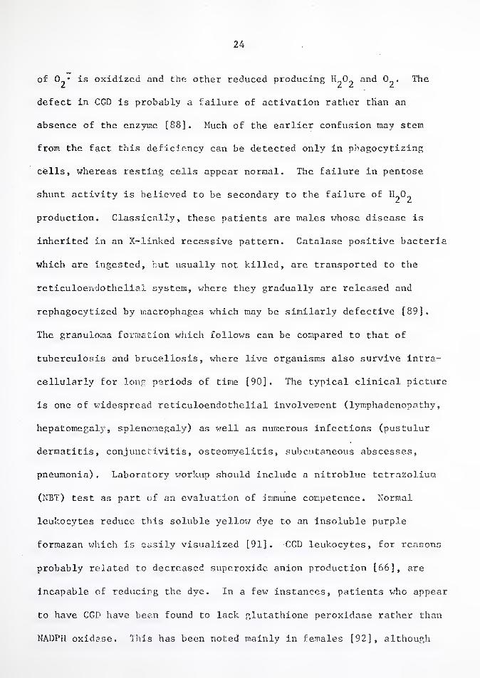
24
of is oxidized and the other reduced producing anc^ ^2*
defect in CGD is probably a failure of activation rather than an
absence of the enzyme [88], Much of the earlier confusion may stem
from the fact this deficiency can be detected only in phagocytizing
cells, whereas resting cells appear normal. The failure in pentose
shunt activity is believed to be secondary to the failure of
production. Classically, these patients are males whose disease is
inherited in an X-linked recessive pattern. Catalase positive bacteria
which are ingested, but usually not killed, are transported to the
reticuloendothelial system, where they gradually are released and
rephagocytized by macrophages which may be similarly defective [89].
The granuloma formation which follows can be compared to that of
tuberculosis and brucellosis, where live organisms also survive intra-
cellularlv for long periods of time [90]. The typical clinical picture
is one of widespread reticuloendothelial involvement (lymphadenopathy,
hepatomegaly, splenomegaly) as well as numerous infections (pustulur
dermatitis, conjunctivitis, osteomyelitis, subcutaneous abscesses,
pneumonia). Laboratory workup should include a nitroblue tetrazolium
(NBT) test as part of an evaluation of immune competence. Normal
leukocytes reduce this soluble yellow dye to an insoluble purple
formazan which is easily visualized [91]. CGD leukocytes, for reasons
probably related to decreased superoxide anion production [66], are
incapable of reducing the dye. In a few instances, patients who appear
to have CGD have been found to lack glutathione peroxidase rather than
NADPH oxidase. This has been noted mainly in females [92], although

• ;

25
one case of a Japanese male has been recorded [93]. CGD, therefore,
may not be a single disease entity.
Glucose-6-phosphate dehydrogenase appears to be intimately
involved in hydrogen peroxide formation and catabolism. Through the
activity of G6PD, the NADPII needed to reduce oxygen to superoxide is
produced. Hydrogen peroxide is then derived from by dismutation
as shown in Figure 2. On this basis, one might predict that a lack of
leukocyte G6PD would cause a lack of hydrogen peroxide formation and
affect cellular metabolism and function in a way mimicking CGD. A
comparison of the two types of cells has been made, and it was found
[94] that leukocyte G6PD levels less than 5 percent of normal resulted
in defective killing of catalase positive bacteria. Similar to CGD
leukocytes, there is no post phagocytic respiratory burst with
production, and there is failure of NTjT reduction. Both will reduce
NBT if cells are disrupted and NADH or NADPH is added. The addition
of methylene blue (MB) differentiates the two types of cells by the
following reactions:
(1) NADH/NADPH + MB dlaPhora5e , NAD/NADP + MBH
(2) MBH + 02 -S°S£°ma.tic„_., m + h2o2
MB added to CGD cells will stimulate normal pentose shunt activity and
improve microbicidal activity [94], G6PD deficient cells which have
decreased levels of NADH and NADPH will exhibit no such response on
exposure to MB. Further study of the G6PD deficient cells revealed
normal phagocytosis, granule formation, and degranulation. Leukocytes

’
. ■
'

26
with G6PD activity of 20-50 percent of normal had no functional defects;
as mentioned before, only at 1-5 percent levels were bactericidal
abnormalities detected [94],
One group of investigators [95] postulated that G6PD deficiency
might be responsible for some cases of classic CGD; they observed an
increased rate of decay of G6PD activity in three male CGD patients.
A 16 month old girl with CGD, defined by a compatible clinical history
and failure to reduce NBT, was found to have a white cell G6PD level
approximately half of normal. Of note is the fact that her parents
were first cousins [96]. While the clinical significance of these
reports can be questioned, at least two patients with complete absence
of leukocyte G6PD and impaired microbicidal activity are known. One
was a middle-aged Caucasian female with hemolytic anemia and fatal
E. coli sepsis [97], Studies of her leukocytes, whose G6PD activity
was completely nondetectable, revealed normal bacterial ingestion,
normal destruction of S_. faecalis (catalase-negative), and abnormal
destruction of S_. aureus, _E. coli and S_. marcesens (catalase-positive).
As one would predict, an NBT test revealed no reduction. Measurements
of leukocyte NADH and NADPH oxidase were not different from controls.
The patient’s post-transfusion erythrocyte G6PD level was 50 percent
of normal; four brothers, three sisters and one son all had normal red
and white cell G6PD and no increased incidence of infection. Genetically,
it is possible that the patient was homozygous for an autosomal recessive
gene which in some way affects G6PD activity, or that she was hetero¬
zygous for the X-linked G6PD gene. In the latter case, one must invoke

27
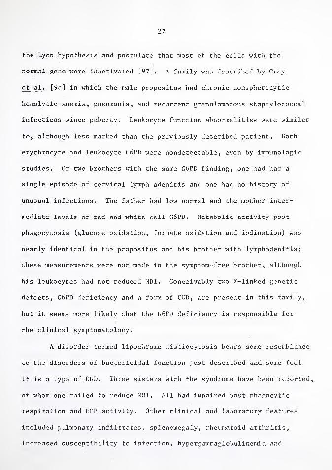
the Lyon hypothesis and postulate that most of the cells with the
normal gene were inactivated [97]. A family was described by Gray
et al. [98] in which the male propositus had chronic nonspherocytic
hemolytic anemia, pneumonia, and recurrent granulomatous staphylococcal
infections since puberty. Leukocyte function abnormalities were similar
to, although less marked than the previously described patient. Roth
erythrocyte and leukocyte G6PB were nondetectable, even by immunologic
studies. Of two brothers with the. same G6PD finding, one had had a
single episode of cervical lymph adenitis and one had no history of
unusual infections. The father had low normal and the mother inter¬
mediate levels of red and white cell G6PD. Metabolic activity post
phagocytosis (glucose oxidation, formate oxidation and iodination) was
nearly identical in the propositus and his brother with lymphadenitis;
these measurements were not made in the symptom-free brother, although
his leukocytes had not reduced NBT. Conceivably two X-linked genetic
defects, G6PD deficiency and a form of CGD, are present in this family,
but it seems more likely that the G6PD deficiency is responsible for
the clinical symptomatology.
A disorder termed lipochrome histiocytosis bears some resemblance
to the disorders of bactericidal function just described and some feel
it is a type of CGD. Three sisters with the syndrome have been reported,
of whom one failed to reduce NBT. All had impaired post phagocytic
respiration and HMP activity. Other clinical and laboratory features
included pulmonary infiltrates, splenomegaly, rheumatoid arthritis,
increased susceptibility to infection, hypergammaglobulinemia and

.
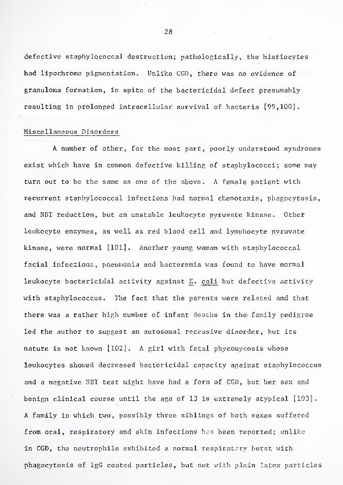
28
defective staphylococcal destruction; pathologically, the histiocytes
had lipochrome pigmentation. Unlike CGD, there was no evidence of
granuloma formation, in spite of the bactericidal defect presumably
resulting in prolonged intracellular survival of bacteria [99,100].
Miscellaneous Disorders
A number of other, for the most part, poorly understood syndromes
exist which have in common defective killing of staphylococci; some may
turn out to be the same as one of the above. A female patient with
recurrent staphylococcal infections had normal chemotaxis, phagocytosis,
and NET reduction, but an unstable leukocyte pyruvate kinase. Other
leukocyte enzymes, as well as red blood cell and lymphocyte pyruvate
kinase, were normal [101]. Another young woman with staphylococcal
facial infections, pneumonia and bacteremia was found to have normal
leukocyte bactericidal activity against E_. coli but defective activity
with staphylococcus. The fact that the parents were related and that
there was a rather high number of infant deaths in the- family pedigree
led the author to suggest an autosomal recessive disorder, but its
nature is not known [102]. A girl with fatal phycomycosis whose
leukocytes showed decreased bactericidal capacity against staphylococcus
and a negative NBT test might have had a form of CGD, but her sex and
benign clinical course until the age of 13 is extremely atypical [103].
A family in which two, possibly three siblings of both sexes suffered
from oral, respiratory and skin infections has been reported; unlike
in CGD, the neutrophils exhibited a normal respiratory burst with
phagocytosis of IgG coated particles, but not with plain latex particles


29
[104], Finally, it should be pointed out that not all of these syndromes
are necessarily related to neutrophil dysfunction. A potential example
is a male with repeated staphylococcal infections culminating in death
at age 19. Although his immunoglobulins were normal, he was found to
lack specific antibodies to the staphylococcal F and S antigens of
leukocidin. The hypothesis was made that the patient may have become
tolerant of staphylococcal antigens during the second trimester of
pregnancy when his mother had impetigo [105]. Such speculation is
interesting but wild, and is perhaps an indication of the great amount
of work that remains in the field of host defense mechanisms.


Ill
Introduction
In the preceding section neutrophil function and dysfunction
were discussed, and the role of G6PD in bactericidal mechanisms reviewed
in the context of other abnormalities. Leukocytes with a G6PD level
less than 5 percent of normal were shown to have defective killing of
catalase positive bacteria; several cases of individuals with G6PD
deficient leukocytes and increased infection susceptibility were cited.
Other ways in which G6PD deficiency might be related to infection have
been proposed, for example, via the hemolysis which so often occurs.
Kaye and his collaborators [106,107] have shown that acute hemolysis
in mice lowers resistance to S.. typhimurium, E. coli, and JS. aureus,
but not to D. pneumoniae. They theorized that macrophages may become
temporarily overloaded with erythrocyte products, and may be unable to
sustain their usual bacterial defenses as well. The possibility also
exists that free hemoglobin itself can increase infection susceptibility,
by providing an easily accessible source of iron for invading microbes
[108]. In addition, there is some evidence that G6PD deficiency is
associated with pneumococcal and salmonella infections, suggesting a
defect in opsonization'similar to that in sickle cell disease [A2].
That G6PD deficiency can adversely affect host defense against
bacterial infection in isolated individuals has been fairly well
30


31
established as described above, but whether it also plays a role in
large populations is not known. In order to answer this question, we
decided to study a population in which severe G6PD deficiency and
bacterial infections were both common, to see whether infected patients
had a higher rate of G6PD deficiency. Although it was not feasible
for us to measure leukocyte G6PD levels in addition to those in the
erythrocytes, we chose a Caucasian population in which deficiency of
the latter is known to be associated with deficiency of the former.
All bacterial infections were included, and where possible, subgroups
were analyzed, to allow for the different mechanisms that may be
operative.
Materials and Methods
(a) Patient selection
The project was carried out in Shiraz, Iran at Nemazee and Saadi
Hospitals, two teaching hospitals affiliated with Pahlavi University
Medical School. All subjects were Iranian males, and all except one
were inpatients at one of these hospitals. Approximately three times
per week charts were reviewed of all adult patients in Nemazee Hospital,
and from the medical, orthopedic and neurosurgical wards in Saadi
Hospital. During the last month of the three month period, pediatric
charts were also reviewed. Patient data were gathered from the charts
and when necessary, from responsible medical personnel. The data
collected included age, place of origin, history of present illness,
past medical history, family history, physical examination findings,
laboratory data, procedures performed, therapy received, and hospital

'■

32
course. All male inpatients with bacterial infections and no known
predisposing reason to infection other than possible G6PD deficiency
were studied as the infected group. Infections were diagnosed by one
or more of the following methods: cultures, titers, biopsies, x-rays,
diagnostic taps, and antibiotic response. In cases where the etiologic
agent was definitively established by cultures, titers, or biopsies,
the patient was categorized as Class I. In other cases the presence
of a bacterial infection was well established by a combination of
clinical symptoms, x-rays, examination of body fluids, biopsies and
antibiotic response, but proof of the etiologic agent was not obtained
and the patient was categorized as Class II. Patients in whom an
etiologic agent was highly suspected, but not proven (for example,
a chest x-ray with cavitary lesions in a person with clinical signs
of tuberculosis) wrere also placed in the latter category. The one
outpatient in the infected group was seen at Nemazee Outpatient Clinic,
brucellosis titers were diagnostic, and it was elected to give him
antibiotics at home rather than in the hospital. Those patients free
of diseases known to be associated with infection or G6PD deficiency
were selected as control "A". As far as possible, all individuals with
systemic diseases were avoided. Thus, to isolate G6PD deficiency as
a potential cause for bacterial infections, patients who had the
following conditions or treatments were, excluded from the infected and
the major control groups: steroid therapy, malignancy, diabetes
mellitus, cirrhosis, chronic renal or cardiac failure, sickle cell
anemia, Cooley’s anemia, malaria, hydatid cyst, open trauma, opium


33
^buse, viral infections, tetanus, vasculitis or renal stones, and those
in whom no diagnosis could be made. Most patients identified with these
conditions were not tested, since these disorders in and of themselves
can predispose to secondary bacterial infections. In some situations
G6PD testing was performed on individuals with these conditions before
all clinical data was available, and they serve as control "E". This
provided us with two control groups; control "A" is a group selected
for a lack of predisposing factors for bacterial infection, and control
"B" a group with a variety of potentially predisposing factors to
infection, or syndromes which mimicked bacterial infections in some
of their features.
(b) Assay
Measurement of erythrocyte G6PD activity was carried out as
follows: for each assay approximately 5 cc. of blood was drawn into
a syringe, transferred immediately to a Vacutainer tube containing EDTA,
and mixed well. Bloods were refrigerated at 4°C within 15 minutes and
were tested within three days with the semiquantitative method developed
by Sigma (Technical Bulletin No. 400). Kit instructions were followed
exactly, including appropriate compensation for the degree of anemia
present. In this assay, erythrocytes are lysed by water to release
G6PD, This solution is then added to glucose-6-phosphate and NADP in
the presence of phenazine methosulfate, an electron carrier and
dicblorophenol indophenol, a blue dye. The NADPH formed during the
oxidation of glucose-6-phosphate to 6-phosphogIuconate reduces the dye
to a colorless form, and the rate of the reaction can be followed

34
visually. The tubes are observed for a six hour period. For a
minority of patients the quantitative test for G6PD activity performed
in the Nemazee Hospital Laboratory (Brilliant Cresyl Blue Reduction,
Dade, Miami, California) was used. In these cases, only samples with
0% activity were considered deficient.
(c) Grouping of data and statistical analysis
Statistical significance of differences in rates of G6PD defi¬
ciency were determined by the chi-square method of 'analysis. Infected
patients were compared with control "A" and control "A" plus control "B"
No separate comparisons with control nB': were made, because of the
small number of subjects. Subgroups of patients matched exactly for
age and origin were analyzed in a similar way. Those infected patients
in whom an etiologic agent was established were divided according to
whether the agent was catalase positive or negative. The catalase
positive group was compared with controls, but the catalase negative
group was too small to permit statistical analysis. The unpaired "t11
test was used to analyze the mean age difference between the G6PD normal
and the G6PD deficient infected groups. With both the chi-square and
the unpaired "t" test, p < .05 is considered statistically significant.
Results
(a) Assay validity
Before conducting the studies in Iran assays were run at Yale
University School of Medicine using blood samples tested quantitatively
by the Yale-New Haven Hospital laboratories. The age of the samples


35
ranged from two hours to eleven days. Samples of all ages with normal
G6PD activity decolorized within one hour, and samples with 10 percent
activity decolorized in two to two and a half hours, consistent with
data obtained by Sigma.
In Iran, an effort was made to study all infected patients as
soon as possible after admission, usually within a few days. It was
not always feasible to carry out the assays when no medications were
being given; however, a complete list of drugs that each patient was
receiving was compiled, and no agents that might have affected the assay
were present in the study group. Fourteen of our seventeen abnormal
samples had not decolorized at 6 hours, indicating very severe defi¬
ciency. Two of the remaining three took 3 hours but when repeated
took 4 hours or more, and the third took 4^ hours but could not be
repeated. Of the 14 that took more than 6 hours, 5 were randomly
repeated and all confirmed the original result; in 2 cases the assay
was performed within 2 hours of the time the blood was drawn. Three
of the normal assays were repeated and confirmed.
(b) Patient characteristics
The demographic characteristics of all patients are presented in
Table 3. The average age of the 55 infected patients was 31 years; the
G6PD normal group differed substantially from the G6PT) deficient grouD,
34 years compared to 22. This difference was statistically significant
(.005 > p > .001). There were 65 control "A" patients, with an average
age of 34 years, and 33 control "B" patients, with an average age of
38 years. In these two groups there was no marked difference between

.

36
G6PD normal and G6PD deficient patients. The distribution between
those from villages and from major cities was relatively even, although
a somewhat greater proportion of the control "A" group came from the
cities. Of the city patients, a total of seven came from places other
than Shiraz (three infected patients and three control "B,: patients,
one each from Yazd, Teheran, and Mashad, and one control "A" patient
from Yazd). It seems reasonable to assume that villagers are less
likely to travel, and that therefore an even smaller percentage is
from outside the Shiraz area.
The criteria by which infections were established are listed
in Table 4, and the diagnoses themselves in Table 5. Tables 6a and 6b
contain the diagnoses of control "A" and control "B" respectively.
Some of the patients had a history of a disease other than that for
which they were admitted to the hospital. Of the infected patients,
three had a past history of typhoid (the present illnesses were two
with pneumonia and one with osteomyelitis), one had psoriasis (shigel¬
losis), and one had hypertension (meningitis). Of the control "A”
patients, one had had typhoid (snake bite), one had had malaria (upper
gastrointestinal tract bleed), and one had an ulcer (low back pain);
of control "B:l patients, one had rheumatoid arthritis (and was on
steroid therapy) and one had had smallpox (diagnosis of present illness
not made).
The results of the G6PD assays are in Table 7. Twenty two
percent of the infected patients were G6PD deficient, compared to
twelve percent of control "A" and six percent of control "B" (Table 7a).


37
Tables 7b, 7c, and 7d show the rates of G6PD deficiency in three sub¬
groups, one with matched ages, one with matched origins (villagers
only), and one selected on the basis of whether the etiologic agent
was catalase positive or catalase negative. Matching for age wTas
important from two aspects: older patients might have a lower frequency
of G6PD deficiency if the gene causes an increase in mortality rates
and older patients have a naturally higher rate of infection for a
variety of reasons. Matching for origin was important to account for
genetic differences in rates of G6PD deficiency, and genetic and environ¬
mental differences in rates of infection. We looked at whether infections
were caused by catalase positive or catalase negative organisms, because
of the previously cited work showing the G6PD deficient leukocytes may
be similar to CGD leukocytes in demonstrating impaired killing of cata¬
lase positive organisms [92],
For Tables 7a, 7b, and 7c the differences in rates of G6PD defi¬
ciency between the infected group and control "A11 were analyzed by the
chi-square method, and in all cases the differences were found not to
be statistically significant (p > .10). Comparisons were also made
between each infected group and control "A" plus control "B". The
results are as follows:
Table Patient Group X ^ £
7a all 3.86 .05 > p > .01
7b age-matched 3.67 .10 > P > .05
7c origin-matched 1.79 P ~ .10
Thus, when controls "A" and "B" together are considered as the control


38
group, the differences in rates of G6PD deficiency reached statistical
significance. No separate comparisons with control "B" were made,
owing to the small number of subjects. For Table 7d, the catalase
positive group was compared with all control "A" patients (Table 7a),
and with all control "A" patients plus control "B" patients (Table 7a).
The differences were not statistically significant (p > .10). The
catalase negative group had no G6PD deficiency, but statistical analysis
was not carried out because of the small size of the group.
Mortality data was collected but is incomplete because it was
not possible to follow patients remaining in hospital after termination
of the study. As far as is known, none of the 12 G6PD deficient infected
patients and 2 of the 43 G6PD normal infected patients expired. There
was no mortality in the control ’’A" group of 65 patients; in control "B11,
one of the 2 G6PD deficient and 3 of the 31 G6PD normal patients failed,
to survive.
Discussion
The frequency of G6PD deficiency in Iran has been studied by
a number of other investigators (see Table 8), and our results are in
rough agreement. We did not have enough geographic distribution to
confirm or refute Beaconsfield1s [110] finding of a higher incidence
in previously malaria-infested areas.
Although the rates of G6PD deficiency were greater in infected
than in control groups, the differences were not significant when the
control group was matched as closely as possible for age, origin, and
underlying diseases. When we included a secondary control group with


39
a variety of conditions that were not present in the infected groups,
the differences reached significance (p as low as .05 > p > .01, in
patients matched exactly for age). However, the secondary control
group included patients whose susceptibility to infection might have
been increased due to disease (other than G6PD deficiency) or therapy,
thus it was not strictly comparable with the infected patient group.
One might expect an artificially low rate of G6PD deficiency in control
"B" as the combination of G6PD deficiency and infection could lead to
an increased mortality. The same bias could occur if patients with
G6PD deficiency are less liable to develop the types of disorders
present in control "3". Either of these possibilities would explain
why the differences in rate of G6PD deficiency between the infected
and control groups increase when the secondary control group is included.
Some studies have shown that G6PD deficient individuals are more
susceptible only to catalase positive organisms (see previous section),
but our comparison of the group infected with catalase positive
organisms and the controls showed no statistically significant differ¬
ences in the frequency of G6PD deficiency. Although the rate of G6PD
deficiency in the catalase negative group was zero, the number of cases
was too small to permit meaningful statistical analysis. Neither did
we have enough subjects with any one disease to analyze separately,
and so we cannot make a definite statement on this issue. However,
the G6PD deficient patients did seem to have a disproportionate share
of staphylococcal septicemia, pneumonia, and brucellosis. We also
compared the demographic characteristics of the G6PD deficient infected


40
group with the G6PD normal infected group. The average age of the
former was 22 years, and the latter 34 years (difference significant
at the .005 > p > .001 level). One interpretation of this observation
is that the combination of G6PD deficiency and infection leads to
hospitalization at an earlier age than infection alone. This is
consistent with the hypothesis that G6PD deficient patients, once
infected, have a more severe clinical course. Data on mortality rates
would be useful in further testing of this hypothesis, but our data
are insufficient for this purpose.
It is important to point out what we feel are some potential
weaknesses in our study. The number of patients sampled was relatively
small, and conceivably some of the observed trends might become signifi¬
cant only in a large population study. Lack of optimal facilities and
culturing techniques prevented us from establishing every diagnosis of
infectious disease by culture, and sampling error might have been involved
in those that were. It was not possible for us to fully document
previous antibiotic therapy, and this would be important in altering
the course of infectious diseases. We were not able to measure leuko¬
cyte G6PD levels, which would have made our study more precise. Exact
data on leukocyte G6PD activity would be particularly relevant in
establishing the clinical significance of defective pentose phosphate
metabolism in the cells. Finally, the method of patient selection might
be faulted in several ways. Specifically, ongoing hemolysis caused by
infection or its treatment can lead to hospital admissions. By concen¬
trating only on patients with infections severe enough to result in


hospitalization, a true picture of a population's susceptibility to
infection is not obtained. Rather, one has only an estimate of the
prevalence of serious infections, and this estimate does not differ¬
entiate between infections made more severe by G6PD deficiency (with
or without hemolysis) and the reverse. Close monitoring of hemolytic
parameters before hospitalization would be required to determine whether
this is a source of bias, as well as perhaps producing information cn
the relationship of hemolysis and infection; it would also provide a
more accurate index of the incidence of infection in normal and G6PD
deficient subjects. By selecting mainly adult patients, we automatically
minimized the chances of detecting very serious effects of G6PD defi¬
ciency on host resistance, which could result in early mortality. Borne
of the information we attempted to collect may not have been reliable
and complete. Thorough medical records were not always available and
some relevant data may have been deleted including precise statements
concerning ethnic background, nutritional status, and means of referral.
Another population study of the relationship of G6PD deficiency
and infection has been published by Lampe et al. [42], They compared
rates of G6PL) deficiency in hospitalized Thai children with S. typhi,
H. influenza b, pneumococcus, staphylococcus, and tuberculous infections
with outpatient controls. Taking only male patients, their data show
a difference in rate of G6PD deficiency between the infected group and
the controls approaching significance (p - .05), a finding rather similar
to ours. Their criteria for diagnosis of infectious disease were more
rigorous than ours, but their definition of G6PD deficiency is vague,

i-J
■
42
and it is not clear how well the patients and controls were matched
for exact age and origin. Furthermore, like our study, their method
of patient selection precludes an estimate of the relative incidence
rates for infection in normal and G6PD deficient subjects.
Thus, while we can offer no final conclusions regarding the
relationship of G6PD deficiency and bacterial infections, a reasonable
interpretation of data from these studies is that the two conditions
do have meaningful interactions. This statement is justified by our
data showing the G6PD deficient infected group to be significantly
younger than the G6PD normal infected group, indicating the adverse
effect of having both G6PD deficiency and infection. Although a rise
in the rate of G6PD deficiency in an infected hospital population versus
a non-infected population was demonstrated both by Lampe and ourselves,
this rise approaches statistical significance only under certain cir¬
cumstances. Therefore, whether it is real remains to be evaluated.
There are several mechanisms which would explain an association
between G6PD deficiency and bacterial infection. First, as we have
suggested above, the oxidant stress of infection or its treatment may
lead to hemolysis and a weakening of host defenses against infection
as well as hospitalization. Second, if the leukocyte G6PD level is
less than 5 percent of normal, bactericidal activity against catalase
positive organisms can be impaired. On the basis of published information,
it is more likely that this occurs on an individual basis, rather than
in all patients with reduced leukocyte G6FD. A thorough prospective
analysis of large groups Is required to pick up the more subtle

43
influences which might increase infection rates as well as severity.
In such a study, the importance of other factors, such as hemolysis
and abnormal opsonization, could be evaluated. This project would be
potentially useful in providing additional knowledge of host defense
mechanisms, as well as having clinical importance to the millio-ns of
people with G6PD deficiency.

A A
FIGURE 1
HEXOSE MONOPHOSPHATE SHUNT
Glucose - 6- phosph o to
dehydrogenase Horn NADP r, M |
-2H // HC.OH
HjCOPOj"
Ribulose - 5-phosphote
Prpsphofibo- iscmerase
H2copor Dihydroxyacetone
phosphate
HzCOPOj
SedoheoPilose - 7-phosphate
(From Reference 1)


45
FIGURE 2
METABOLIC PATHWAYS FOR OXYGEN COMSUMP'
DURING PHAGOCYTOSIS IN P.V1NS
0 2
(Figure by Dr. Richard Root:) SOD: Superoxide Dismutase
GSH-PO: Glutathione Fcroxidac


TA
BL
E
1 (f
rom R
efe
ren
ce
1)
46
O TO P P P P P e TO TO TO cn TO
X P E E E TO E a TO TO TO to P TO
p O O o P o o 13 S3 P 22
P
:o T3 TO i—1 r—1 H TO P TO P TO TO TO TO TO TO P E E E TO E TO P TO i-i TO TO to
W P O o o TO o TO 13 J3 TO s TO TO to a
cn H
> P P \D O fro o to u M H to P
Crf w H a
o P < cj P
X w u o M P
A
G O P
•r-i TO p
p to G CM vO TO TO <r <T 1 CO o N O V V V CO TO3 •TO TO CM CM r-H
•TO £•« TO P
T—1 P ID P • TO TO •
P S g E P o < p 1 TO TO. 1 •
23 c> O o CM co £ • Z •
CM P
p 00 P p O p TO TO CN p
O n 1 E E ! uo
E O TO TO CP U~l 0 O p
22 is
u p TO GJ *.
TO ^ P O TO « p
.TOPE O O o o o P P TO o P p c o 0 P O TO P C TO O
TO E ^ 01 P W
p P p p p
Jo TO P p o > TO o P E o CO o p LO TO TO q 00 CM 1 1 TO O p 1 o o < G 00 o
'Cf u £•-$ p P
w TO G G P 0! TO TO + i Tj o P g c < 01 TO
TO TO p TO O 0) > 22 W
12
9
(PO
. -
pH
6.7
)

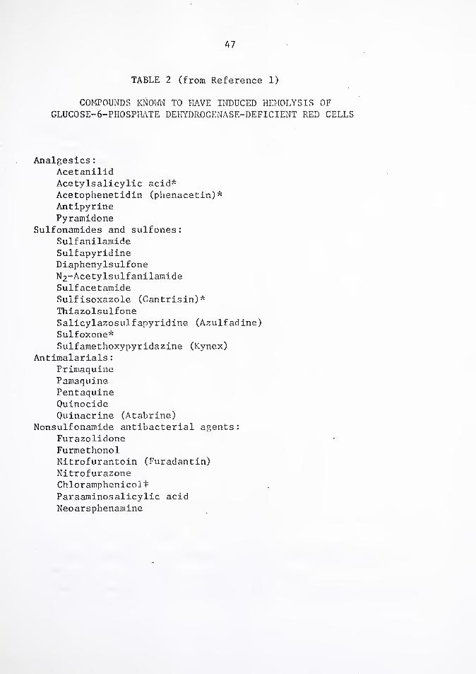
47
TABLE 2 (from Reference 1)
COMPOUNDS KNOWN TO HAVE INDUCED HEMOLYSIS OF GLUCOSE-6-PHOSPHATE DEHYDROGENASE-DEFICIENT RED CELLS
Analgesics: Acetanilid Acetylsalicylic acid* Acetophenetidin (phenacetin)* Antipyrine Pyramidone
Sulfonamides and sulfones: Sulfanilamide Sulfapyridine Diaphenylsulfone N2*-Acetylsulf anilamide Sulfacetamide Sulfisoxazole (Gantrisin)* Thiazolsulfone Salicylazosulfapyridine (Azulfadine) Sulfoxone* Sulfamethoxypyridazine (Kynex)
Antimalarials: Primaquine Pamaquine Pentaquine Quinocide
Ouinacrine (Atabrine) Nonsulfonamide antibacterial agents:
Furazolidone Furmethonol Nitrofurantoin (Furadantin) Nitrofurazone Chloramphenicol £
Paraaminosalicylic acid Neoarsphenamine


48
TABLE 2 (continued)
Miscellaneous: Naphthalene Trinitrotoluene Methylene blue* Nalidixic acid Dimercaprol (BAL)* Phenylhydrazine Ouininet Quinidinet
Mestranol
* Slightly hemolytic in Negroes, or only in very large doses,
t Hemolytic in Caucasians, but not in Negroes.
£ Possibly hemolytic in Caucasians, but not in Negroes or Orientals.


PA
TIE
NT
DE
MO
GR
AP
HIC
DA
TA
49
§ o d
4-J
o !Z
M
o
'O co E o
S3
M
G tc CO
rH rH •H
>
G G fcC fcO G CO (0
G > < G
fc£ <
U G
E G
S5
W 4J C G
•H •U «
Pm
4-* O
Pi O o w H
< u
id r-1 n- rH O i—1 CO o CO
-H o rH O rH rH o o O
<• co o- CO rH On o o o rH t—H CM CM rH 1—i
CM CO o CO \D CO CM o CM cn CM CO rH C\1
/—> /-s v /~v LO M—V o m o O O r^ in co m o- in r-* CC' o oc cc ! cc i i 1 1 1 1
1 rH i <r cm <r in cc m CO r-1 CO rH 04 pH rH rH rH '-- M-M V_M ■sM W w W MT CM rH mt r- <?■ CC CC CO CM CO CO CO cn co co co
CO CM m r- oo m rH CM CO MT rH in m \D CO CO
C4'*C IM <4M rH G rH G rH G d xo d 'O d ~o
r—1 pH rH C C CO O Q cd Ci c CO Pm C •w Pm Pm •U Pm Pm HI CD cO o O ID o M3 M3 O o e> H O O H O O H
Q w C c H O o C_5 Pm Pi — w H < H c Pm 5Z E3 “ tS O O M a o
•x) CO
rd w co S -c g «
G CO
■d co
CM w
M
d ca G G
T5
N cO G
•H
JG CO


50
CLASS
CLASS
TABLE 4
DIAGNOSTIC CRITERIA IN INFECTED PATIENTS
DIAGNOSIS CRITERION FOR DIAGNOSIS
Pneumococcus Culture Staphylococcal sepsis Culture Streptococcal meningitis Culture Shigellosis Culture Anthrax Culture Brucellosis Titer Typhoid Titer Tuberculosis Biopsy A. israelii abscess Biopsy
II: DIAGNOSIS CRITERIA FOR DIAGNOSIS1
Pneumonia Meningitis Osteomyelitis Tuberculosis Erysipelas Brain abscess Pyopneumothorax
CXR and response to antibiotics LP and response to antibiotics X-ray or biopsy CXR Response to antibiotics Pus and air in CSF Pus in pleural fluid
In all cases, clinical symptoms were also compatible.

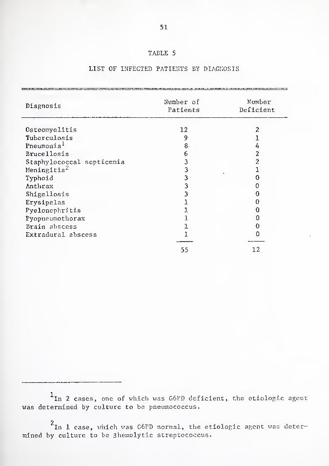
51
TABLE 5
LIST OF INFECTED PATIENTS BY DIAGNOSIS
Diagnosis Number of Number Patients Deficient
Osteomyelitis
Tuberculosis Pneumonia1 Brucellosis Staphylococcal septicemia Meningitis2 Typhoid Anthrax Shigellosis Erysipelas Pyelonephritis Pyopneumothorax
Brain abscess Extradural abscess
12 2
9 1 8 A 6 2 3 2 3 1 3 0 3 0 3 0 1 0 1 0 1 0 1 0 1 0
55 12
^In 2 cases, one of which was G6PD deficient, the etiologic agent
was determined by culture to be pneumococcus.
2 In 1 case, which was G6PD normal, the etiologic agent was deter¬
mined by culture to be 3hemolytic streptococcus.

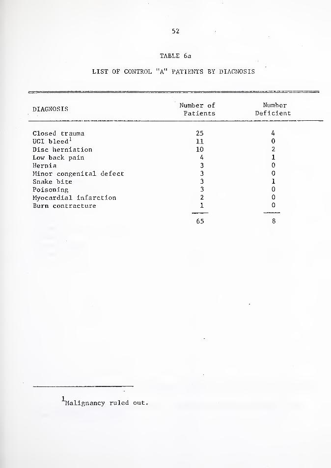
52
TABLE 6a
LIST OF CONTROL "A" PATIENTS BY DIAGNOSIS
DIAGNOSIS Number of Number Patients Deficient
Closed trauma UGI bleed1 Disc herniation Low back pain Hernia Minor congenital defect Snake bite Poisoning Myocardial infarction Burn contracture
25 4 11 0 10 2
4 1 3 0 3 0 3 1 3 0 2 0 1 0
65 8
Malignancy ruled out.

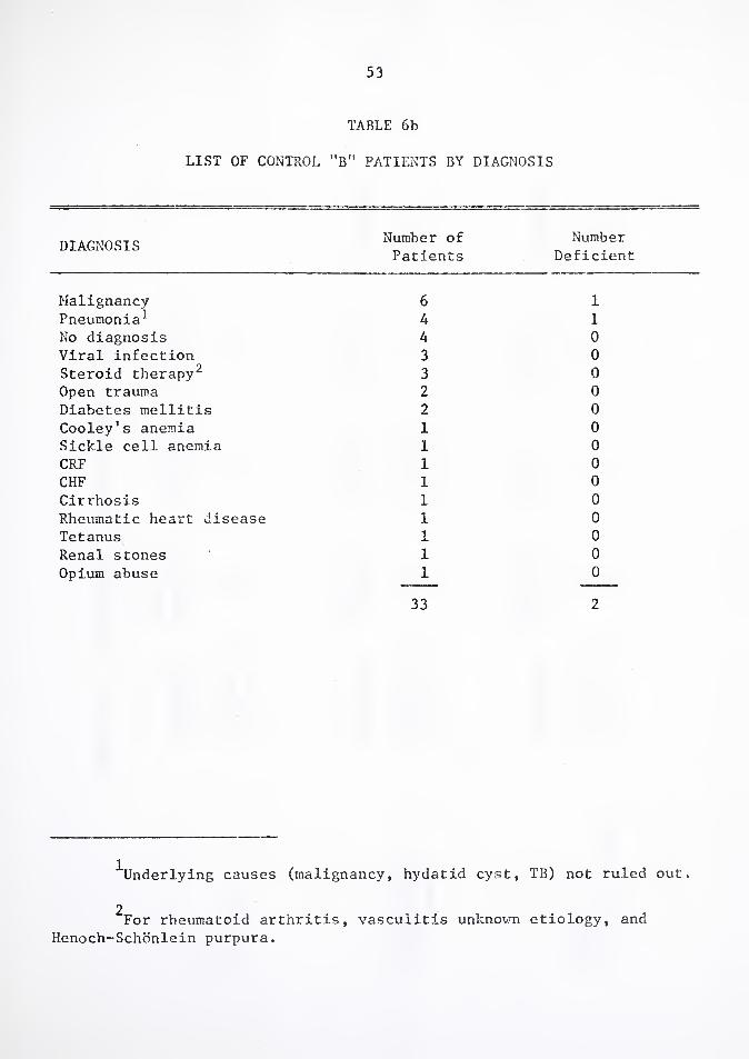
53
TABLE 6b
LIST OF CONTROL "B" PATIENTS BY DIAGNOSIS
DIAGNOSIS Number of Number Patients Deficient
Malignancy Pneumonia1 No diagnosis Viral infection Steroid therapy2 Open trauma Diabetes mellitis Cooley's anemia Sickle cell anemia CRF CHF Cirrhosis Rheumatic heart disease Tetanus Renal stones Opium abuse
6 1 4 1 4 0 3 0 3 0 2 0 2 0 1 0 1 0 1 0 1 0 1 0 1 0 1 0 1 0 1 0
33 2
^Underlying causes (malignancy, hydatid cyst, TB) not ruled out.
2 For rheumatoid arthritis, vasculitis unknown etiology, and
Henoch-Schonlein purpura.

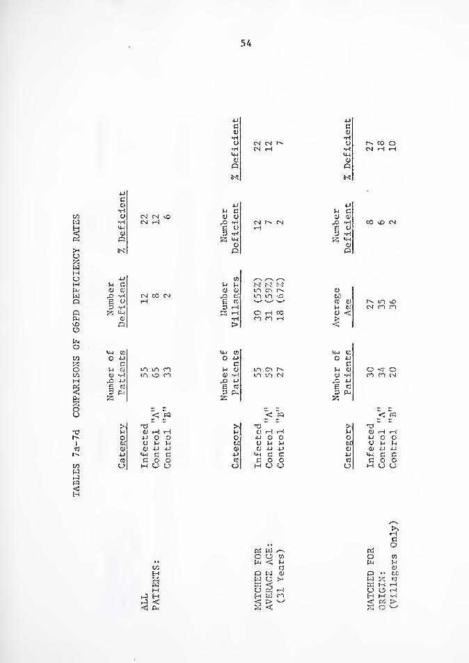
TA
BL
ES
7a-7
d
CO
MP
AR
ISO
NS
OF
G6P
D
DE
FIC
IEN
CY
RA
TE
S
54
4-)
G <D
40 C G
o IJH
0) o
M tN N CM pH
O MOO •H CM pH rH MH
o o
e^s
JO
c G
•H O
•H <JH
CJ
CM CM MO CM pH
tM
4-!
g c CJ o
rO *H E O 3 *H S UH
CJ Q
CM CM I—I
JO
G C G <v
rO *H
E <-> 3 *H S *tH
CJ O
CO MO CM
JO co G C G G CJ CJ CJ CJ
rO *H CM CO CM ,0 01
E o rH EG 3 *H 3 rH 2 <4H K rH
CD •rH G >
Gj CO U-) CO O 40 o JO
G G G G LO m co G QJ CJ ■H LT, VO CO CD •rH
x> 40 rG JO
e G G 3 Po *3 PH
53
< To
52
>• •G tn G G rH rH G O 40 o o O
21 CJ G G tc o 40 40 G
jo C-i G G JO
G G O O G CJ M u CJ CJ
^—\ r—v
S'? S'? S'? m O' r'- G m in MO GO '—✓ '—' G G f'- in MO
G 6C CM co CO c rH 00 G < CO CO pH >
<
U-4 o: o JO
C in O'! M G G o <r O in m CM G •H co co CM
03 JO
E G 3 Ph 2
< CQ < C9
TJ - _ H H3 -
CD rH rH G G rH pH 40 O O O JO O o u G G M O G G G 40 40 G G 40 JO
UH C G 40 lr4 C C G o O G C O O
M CJ CJ a M CJ CJ
>1 G O
pc: w Pi o CJ CO O CO
• • Go G G-i G CO G G Eh C3 w G o • • PC 2! W a >o w ?2 G W 33 <H ffi IH rH w CJ P5 rH CJ CJ iH
p4 H H W CO H M •H rJ <
rU
> '—' Pi < Gj < O


.55
•H •H
C #1 -rH 3 <0 0)
•H rH 3 CJ CO o i—\ Pi
cm QJ 3 «h O iH 3 3 o U 3
FQ 3 S'? a
#* to cO E
1—1 CO O rH -H 3
4J QJ CO •rH Pi C c o 4-J <D <u O rH CJ r0 *H m o E 3 < e O rH CJ 3 •<H CO pi •-
S IPI CO <D to CJ 3 e - 3 U
CO 4-J V rH o rH E u Q) 3 o
3 bC *H 4-J bC •H P a. 3 QJ rH o X- <0 3 Pi b£ CO m C/0 4-J Pi 3 < CJ 4-J
> •> cO OO ■< CO JO
3 O #i
CJ CJ to CJ > 3 o S CJ u CJ 0 r o
u-l CO rH ,X CJ o 4J Jo CO o
d -C U E Pi o CM •■o- & -3 3 3 •H CM CO 4-J CJ
rQ 4J 4-J G G 6 CO C/0 < Ph 3 PH
53 .. ..
+ l + 1
H QJ CJ QJ 3 Pi 10 to CO 3 o CJ to CO 3 to r—1 i—( rH rH o CO cO 3 3 4J 4-J 4-J 4H 4-J CO CO cO 3 3
V o CJ CJ CJ
'd o • • 4-J
U CJ to PH 3 rH
e M-i d o e o ►j i—i O PH
H £h
W ts w
>-* c: P3 C


G6P
D
DE
FIC
IEN
CY
IN
IRA
NIA
N
MA
LE
S,
PU
BL
ISH
ED
ST
UD
IES
56
d o
•H E S a s E E X to 0) o o (!) QJ a) CO
■H «H iH 1—t rH rH rH •H r-! CO CO CO CO CO CO £ O O o o o o O a) pi *-*
s X S x
to cu
d >J •H to CO to d
•H - to rH U tO CD CO <0 o CO p
>-i QJ o .H rH •H -U 4-1 CO d x h 4-J Jj O to to •H XI O
•H o rH to 4.J
X E to X 4-J P. o e CO d •H c d to h CO CO d cl i—1 cO co
4-4 V-i •H cO X u M to qj to PI¬ E <0 C O d o m x N CO d 4-1 CO r- X <y r"- 4-J cO cC O CO E o 00 o CN O o X M C H H
co V 4-i 4-1
•H 4-1 4-c CO CO CO
•u co AJ n 4.1 O *H CO o CO ty m c
T-) Q) rH o rH X H CO -a TO CO
1 3 O AJ c 4-J w to •H TO CO •H
d a o a. td CO o CO X o r—! o o X « X
u
V
X CO E w p o 00 CN CN n- cc ON
2: <y LO ON <3- in o o •r-) <T) iH rH m r—1 <r
rH X CO p AJ CO o
H
4J
C o m
•H 00 m in CN ON CN oo o • • CN i ON N • X •
•H on CN ON cr\ ON 4-t tH <y o
6-S
1—1 CN
CO rH N0 rH ON l—j I—1
cn TO ON
on rH NO ON
m 0) ON rH ON •H rH ;H r-r 4-1 r—i r—t
ON CO o 4-1 rH ai P O c rH d rH X
>-i CO H o rH T-i o o E '—1 o W-l d •-3 CO p £ CO TO *H H O <u d U to w X Ch
A bla
nk
sp
ace sig
nif
ies
that
the
info
rmati
on
was
no
t av
ail
ab
le.


57
BIBLIOGRAPHY
1. Stanbury, J.B., Wyngaarden, J.B., Fredericksen, D.S. The Metabolic Basis of Inherited Disease, 3rd ed. New York: McGraw-Hill, 1972.
2. Dern, R.J., Weinstein, I.M., LeRoy, G.V., Talmage, D.W., Alving, A.S. "The Hemolytic Effect of Primaquine I: The Localization of the Drug-Induced Hemolytic Defect in Primaquine-Sensitive Individuals," J Lab Clin Med 43: 303 (1954).
3. Carson, P.E., Flanagan, C.L., Ickes, C.E., Alving, A.S. "Enzymatic Deficiency in Primaquine-Sensitive Erythrocytes," Science 124: 484 (1956).
4. Beutler, E. "Abnormalities of the Hexose Monophosphate Shunt," Sem Hem 8: 311 (1971).
5. Williams, W.J., Beutler, E., Erslev, A.J., Rundles, R.W. Hematology. New York: McGraw-Hill, 1972.
6. Baehner, R.L., Nathan, D.G., Castle, W.B. "Oxidant Injury of Caucasian G6PD-Deficient Red Blood Cells by Phagocytosing Leukocytes During Infection," JCI 50: 2466 (1971).
7. Carson, P.E., Frischer, H. "G6PD Deficiency and Related Disorders of the Pentose Phosphate Pathvjay," Am J Med 41: 744 (1966).
8. Wintrobe, M.W., Lee, G.R., Boggs, D.R., Bithell, T.C., Athens, J.W., Foerster, J. Clinical Hematology, 7th ed. Philadelphia: Lea &
Febiger, 1974.
9. Yoshida, A., Beutler, E., Motulsky, A. "Human G6PD Variants," Bull WHO 45: 243 (1971).
10. Livingstone, F.B. Abnormal Hemoglobins in Human Populations. Ann Arbor, Michigan: Anthropological Institute of the University of Michigan, 1973.
11. Yoshida, A. "A Single Amino Acid Substitution (Asparagine to Aspartic Acid) Between Normal (B+) and the Common Negro Variant (A+) of Human G6PD," Proc Nat Acad Sci 57: 835 (1967).
12. Marks, P.A., Gross, R.T. "Erythrocyte G6PD Deficiency: Evidence of Differences Between Negroes and Caucasians with Respect to This Genetically Determined Trait," JCI 38: 2253 (1953).


58
13. Piomelli, S., Corash, L.M., Davenport, D.D., Miraglia, J., Amrosi, E.L. "In vivo Lability of G6PD in Gd A- and Gd Medi¬ terranean Deficiency," JCI 47: 940 (1968).
14. Owusu, S.K. "Absence of G6PD in Red Cells of an African," Br Med J 4: 25 (1972).
15. McCurdy, P.R. "Clinical Manifestations of Variant G6PD Enzymes," Ann Clin Lab Sci 1: 184 (1971).
16. Kirkman, H.N., Schettini, F., Pickard, B.M. "Mediterranean Variants of G6PD," J Lab Clin Med 63: 726 (1964).
17. Bonsignore, A., Fornaini, G., Leoncini, G., Fantoni, A. "Electro¬ phoretic Heterogeneity of Erythrocyte and Leucocyte G6PD in Italians from Various Ethnic Groups," Nature 211: 876 (1966).
18. Stamatoyannopoulos, G., Voigtlander, V., Kotsakis, P., Akrivakis, A. "Genetic Diversity of Mediterranean G6PD Deficiency Phenotype," JCI 50: 1253 (1971).
19. Luzzatto, L. "Inherited Haemolytic States: G6PD Deficiency," Clin Haem 4: 83 (1975).
20. Bonsignore, A.. Fornaini, G., Fantoni, A., Segni, P. "Charac¬ terization of Leukocyte G6PD in Sardinian Mutants," JCI 45: 1865 (1966).
21. Sabine, J.C., Jung, E.D., Fish, M.B., Pestanar, L.C., Rankin, R.E. "Observations on the Inheritance of G6PD Deficiency in Erythro¬ cytes and in Leucocytes," Brit J Haem 9: 164 (1963).
22. Ramot, Be, Fisher, S., Szeinberg, A., Adam, A., Sheba, C., Gafni, D. "A Study of Subjects with Erythrocyte G6PD Deficiency. II. Investigation of Leukocyte Enzymes," JCI 38: 2234 (1959).
23. Chan, T.K., Todd, D., Wong, C.C. "Tissue Enzyme Levels in Erythrocyte G6PD Deficiency," J Lab Clin Med 66: 937 (1965).
24. Beutler, E., Yeh, M., Fairbanks, V.F. "The Noram Human Female as a Mosaic of X-Chromosome Activity: Studies Using the Gene for G6PD Deficiency as a Marker," Proc Nat Acad Sci 48: 9 (1962).
25. Gandini, E., Gartler, S.M. "G6PD Mosaicism for Studying Develop¬ ment of Blood Cell Precursors," Nature 224: 599 (1969).
26. Phillips, J., Herring, R.M., Goodman, H.O., King, J.S. Jr. "Leukocyte Alkaline Phosphatase and Erythrocyte G6FD in Down’s Syndrome," J Med Genet 4: 268 (1967).

59
27. Kruatrachue, M., Klongkumnuanhara, K., Harinasnta, C. "Infection- rates of Malarial Parasites in Red Blood Cells with Normal and Deficient G6PD," Lancet 1: 404 (1966).
28. Gilles, H.M., Fletcher, K.A., Hendrickse, R.G., Linder, R., Reddy, S., Allan, N. "G6PD Deficiency, Sickling and Malaria in African Children in Southwestern Nigeria," Lancet 1; 138 (1967)
29. Kosower, N.S., Kosower, E.M. "Molecular Basis for Selective Advantage of G6PD Deficient Individuals Exposed to Malaria," Lancet 2: 1343 (1970).
30. Huheey, J.E., Martin, D.L. "Malaria, Favism, and G6PD Deficiency,' Experlentia 31: 1145 (1975).
31. Sharma, A., Singh, H., Dhatt, P.S., Gupta, II.L., Arya, R.K. "G6PD Deficiency in the Newborn," Indian Ped 12: 393 (1975).
32. Das, B.N., Bhakoo, O.N., Jolly, J.G. "Neonatal Hyperbilirubinemia Associated with G6PD Deficiency," Indian Ped 11: 645 (1974).
33. Valaes, T., Karaklis, A., Stravrakakis, D., Bavela-Stravrakakis, K., Perakis, A., Doxiadis, S.A. "Incidence and Mechanism of Neonatal Jaundice Related to G6PD Deficiency," Ped Res 3: 448 (1969).
34. Mellon!, T., Cagnazzo, G., Dore, A., Cutillo, S. "Phenobarbitai for Prevention of Hyperbilirubinemia in G6PD Deficient Newborn Infants," J Peds 82: 1048 (1973).
35. Choremis, C,, Kattamis, C.A., Kyriazakou, M., Gavriilidou, E. "Viral Hepatitis in G6PD Deficiency (letter)," Lancet 1: 269 (1966).
36. Morrow, R.II. Jr., Smetana, H.F., Sai, F.T., Edgcomb, J.H. "Unusual Features of Viral Hepatitis in Accra, Ghana," Ann Int Med 68: 1250 (.1968),
37. Kattamis, C.A., Tjortjatou, F. "The Hemolytic Process of Viral Hepatitis in Children with Normal or Deficient G6PD Activity," J Peds 77: 422 (1970).
38. Owusu, S.K. "G6PD Deficiency in Jaundice Associated with Lobar Pneumonia," (letter), Lancet 2: 325 (1973).
39. Tugwell, P. "G6PD Deficiency in Nigerians with Jaundice Asso¬ ciated with Lobar Pneumonia," Lancet 1: 968 (1973).


60
40. Williams, A.O., Tugwell, P., Edington, G.H. "G6PD Deficiency and Lobar Pneumonia, Fine Structure of the Liver," Arch Path Lab Med 100: 25 (1976).
41. Owusu, S.K., Foli, A.K., Konotey~Ahulu, F.I.D., Janosi, M. "Frequency of G6PD Deficiency in Typhoid Fever in Ghana," (letter), Lancet 1: 320 (1972).
42. Lampe, R.M., Kirdpon, S., Mansuwan, P., Benenson, M. "G6PD Deficiency in Thai Children with Typhoid," J Peds 87: 576 (1975).
43. Banait, P.P., Junnarkar, R.V. "Study of Erythrocyte G6PD Defi- ciency in Leprosy," Int J Lep 39: 168 (1971).
44. Saha, N. "Incidence of G6PD Deficiency in Patients of Three Different Ethnic Groups Suffering from Pulmonary Tuberculosis," J Med Genet 6: 292 (1969).
45. Beaconsfield, P., Rainsbury, R., Kalton, G. "G6PD Deficiency and Cancer," Oncologia 19: 11 (1965).
46. Sulis, E., Spano, G. "Osservazioni preliminari sull' incidenza neoplastica e sul comportamento enzimatico e proliferative del tessuto tumorale negli individui carent i di glucosio-6- fosfato deidroger.asi," Boll Ital Biol Speri 44: 1246 (1968).
47. Naik, S.N., Anderson, D.E, "G6PD Deficiency and Cancer," (letter) Lancet 1: 1060 (1970).
48. Sulis, E. "G'6PD Deficiency and Cancer," (letter), Lancet 1_: 1185 (1972).
49. Eaton, J.W. "Catalase Activity and Cancer," (letter), Lancet 2: 46 (1972).
50. Mlsra, R.C., Basu, A.K., Svarup Mitra, S. "Erythrocyte G6PD Deficiency in Thalassemia Syndromes—A Cytochemical Study," J Indian M A 65: 40 (1975).
51. Harley, J.D., Agar, N.S., Gruca, M.A., McDermid, M.E., Kirk, R.L. "Cataracts with a G6PD Variant," (letter), Br Med J 2: 86 (1975).
52. Sheehan, R.G., Necheles, T.F., Lindeman, R.J., Meyer, H.J., Patterson, J.F. "Regional Enteritis and Granulomatous Colitis Associated with Red Blood Cell G6PD Deficiency," NEJM 277: 1124 (1967).
53. Katsaros, D., Truelove, S. "Regional Enteritis and G6PD Deficiency, NEJM 281: 295 (1969).

'•••' '

61
54. Petrakis, N.L., Weisenfield, S.L., Sams, B.J. Collen, M.F., Cutler, J.L., Siegelaub, A.B. "Prevalence of Sickle-Cell Trait and G6PD Deficiency. Decline with Age in Frequency of G6PD Deficient Negro Males," NEJM 282: 787 (1970).
55. Piomelli, S., Reindorf, C.A., Arzanian, M.T., Corash, L.M. "Clinical and Biochemical Interactions of G6PD Deficiency and Sickle Cell Anemia," NEJM 287: 213 (1972).
56. Steinberg, M.H., Dreiberg, B.J. "G6PD Deficiency in Sickle-Cell Anemia," Ann Int Med 80: 217 (1974).
57. Beutler, E., Johnson, C., Powars, D., West, C. "Prevalence of G6PD Deficiency in Sickle-Cell Disease," NEJM 290: 826 (1974).
58. Bienzle, U., Sodeinde, 0., Effiong, C.E., Luzza'cto, L. ''G6PD Deficiency and Sickle Cell Anemia: Frequency and Features of the Association in an African Community," Blood 46: 591 (1975).
59. Baehner, R.L. "Microbe Ingestion and Killing by Neutrophils: Normal Mechanisms and Abnormalities," Clin Haemat 4: 609 (1975).
60. Stossel, T.P, "Phagocytosis: Recognition and Ingestion," Sem
Hemat 12: 83 (1975)~
61. Murphy, P. The Neutrophil. New York: Plenum Medical Book Company, 1976.
62. Miller, M.E., Nillson, U.R. "A Familial Deficiency of the Phagocytosis-Enhancing Activity of Serum Related to a Dysfunction of the Fifth Component of Complement (C5)*, NEJM 282: 354 (1970).
63. Goldstein, I.M., Roos, D., Kaplan, H.B., Weissmann, G. "Complement and Immunoglobulins Stimulate Superoxide Production by Human Leukocytes Independently of Phagocytosis," JCI 56: 1155 (1975).
64. Goldstein, I.M., Cerqueira, M., Lind, S., Kaplan, H.B. "Evidence That the Superoxide Generating System of Human Leukocytes is Associated with the Cell Surface," JCI in press.
65. Babior, B.M., Curmutte, J.T., McMurrich, B.J. "The Particulate Superoxide-Forming System from Human Neutrophils," JCI 58: 989 (1976).
66. Baehner, R.L., Murrman, S.K., Davis, J., Johnston, R.B. Jr.. "The Role of Superoxide Anion and Hydrogen Peroxide in Phagocytosis- Associated Oxidative Metabolic Reactions," JCI 56: 571 (1975).

.

62
67. Klebanoff, S.J. "Antimicrobial Mechanisms in Neutrophilic Polymorphonuclear Leukocytes," Sem Hemat 12: 117 (1975).
68. Miller, M.E., Oski, F.A., Harris, M.B. "Lazy Leucocyte Syndrome. A New Disorder of Neutrophil Function," Lancet 1: 665 (1971).
69. Davis, S.D., Schaller, J., Wedgwood, R.J. "Job's Syndrome.
Recurrent, 'Cold', Staphylococcal Abscesses," Lancet 1: 1013 (1966).
70. White, L.R. , Ianetta, A., Kaplan, E.L., Davis, S.D., Wedgwood, R.J. "Leucocytes in Job's Syndrome," (letter), Lancet 3 : 630 (1969).
71. Bannatyne, R.M., Skowron, P.N., Weber, J.L. "Job’s Syndrome—A Variant of Chronic Granulomatous Disease," J Peds 75: 236 (1969).
72. Hill, H.R., Ouie, P.G., Pabst, H.F., Ochs, K.D., Clark, R.A., Klebanoff, S.J., Wedgwood, R.J. "Defect in Neutrophil Granulocyte Chemotaxis in Job's Syndrome of Recurrent ’Cold’ Staphylococcal Abscesses," Lancet 2: 617 (1974).
73. Clark, R.A., Root, R.K., Kimball, H.R., Kirkpatrick, C.H. "Defective Neutrophil Chemotaxis and Cellular Immunity in a Child with Recurrent Infections,” Ann Int Med 7S: 515 (1973).
74. Boxer, L., Hedley-LThite, T., Glader, B„, Stossel, T, "A Primary Defect in Neutrophil Motility," Clin Res 22: 384A (1974).
75. DeMeo, A.N., Andersen, B.R. "Defective Chemotaxis Associated with a Serum Inhibitor in Cirrhotic Patients," NEJM 286: 735 (1972).
76. Dossett, J.H., Williams, R.C. Jr,, Ouie, P.G. "Studies on Inter¬ action on Bacteria, Serum Factors and Polymorphonuclear Leukocytes in Mothers and Newborns," Peds 44: 49 (1969).
77. Forman, M.L., Stiehm, E.R. "Impaired Opsonic Activity but Normal Phagocytosis in Low Birth Weight Infants," NEJM 281: 926 (1969).
78. McCracken, G.H. Jr., Eichenwald, H.F. "Leukocyte Function and the
Development of Opsonic and Complement Activity in the Neonate," Amer J Pis Child 121: 120 (1971).
79. Johnston, R.B. Jr., Newman, S.L., Struth, A.G. "An Abnormality of the Alternate Pathway of Complement. Activation in Sickle Cell Disease," NEJM 288: 803 (1973).
80. Miller, M.E., Seals, J., Kaye, R., Levitsky, L.C. "A Familial, Plasma-Associated Defect of Phagocytosis. A New Cause of Recurrent Bacterial Infections," Lancet 2: 60 (1968).

62
67. Klebanoff, S.J. "Antimicrobial Mechanisms in Neutrophilic Polymorphonuclear Leukocytes," Sem Hemat 12: 117 (1975).
68. Miller, M.E., Oski, F.A., Harris, M.B. "Lazy Leucocyte Syndrome.
A New Disorder of Neutrophil Function," Lancet 1: 665 (1971).
69. Davis, S.D., Schaller, J., Wedgwood, R.J. "Job's Syndrome.
Recurrent, 'Cold', Staphylococcal Abscesses," Lancet 1: 1013 (1966).
70. White, L.R., Ianetta, A., Kaplan, E.L., Davis, S.D., Wedgwood, R.J. "Leucocytes in Job's Syndrome," (letter), Lancet 1 : 630 (1969).
71. Bannatyne, R.M., Skowron, P.N., Weber, J.L. "Job’s Syndrome—A Variant of Chronic Granulomatous Disease," J Peds 75: 236 (1969).
72. Hill, H.R., Ouie, P.G., Pabst, H.F., Ochs, K.D., Clark, R.A., Klebanoff, S.J., Wedgwood, R.J. "Defect in Neutrophil Granulocyte Chemotaxis in Job's Syndrome of Recurrent 'Cold' Staphylococcal Abscesses," Lancet 2: 617 (1974).
73. Clark, R.A., Root, R.K., Kimball, H.R., Kirkpatrick, C.H. "Defective Neutrophil Chemotaxis and Cellular Immunity in a Child with Recurrent Infections,” Ann Int Med 78: 515 (1973).
74. Boxer, L., Hedley-White, T., Glader, B„, Stossel, T, "A Primary Defect in Neutrophil Motility," Clin Res 22: 384A (1974).
75. DeMeo, A.N., Andersen, B.R. "Defective Chemotaxis Associated with a Serum Inhibitor in Cirrhotic Patients," NEJH 286: 735 (1972).
76. Dossett, J.H., Williams, R.C. Jr,, Ouie, P.G. "Studies on Inter¬ action on Bacteria, Serum Factors and Polymorphonuclear Leukocytes in Mothers and Newborns," Peds 44: 49 (1969).
77. Forman, M.L., Stiehm, E.R. "Impaired Opsonic Activity but Normal Phagocytosis in Low Birth Weight Infants," NEJM 281: 926 (1969).
78. McCracken, G.H. Jr., Eichenwald, H.F. "Leukocyte Function and the
Development of Opsonic and Complement Activity in the Neonate," Amer J Pis Child 121: 120 (1971).
79. Johnston, R.B. Jr., Newman, S.L., Struth, A.G. "An Abnormality of the Alternate Pathway of Complement Activation in Sickle Cell Disease," NEJM 288: 803 (1973).
80. Miller, M.E., Seals, J., Kaye, R., Levitsky, L.C. "A Familial, Plasma-Associated Defect of Phagocytosis. A New Cause of Recurrent Bacterial Infections," Lancet 2: 60 (1968).

.

63
81. Johnston, R.B. Jr., Stroud, R.M. "Complement and Host Defense Against Infection," J Peds 90: 169 (1977).
82. Constantopoulos, A., Najjar, V.A., Smith, J.W. "Tuftsin Defi¬ ciency: A New Syndrome with Defective Phagocytosis," J Peds 80: 564 (1972).
83. Root, R.K., Rosenthal, A.S., Balestra, D.J. "Abnormal Bactericidal, Metabolic and Lysosomal Functions of Chediak-Higashi Syndrome Leukocytes," JCI 51: 649 (1972).
84. Lehrer, R.I., Cline, M.J. "Leukocyte Myeloperoxidase Deficiency and Disseminated Candidiasis: Role of Myeloperoxidase in Resistance to Candida Infection," JCI 48: 1478 (1969).
85. Klebanoff, S.J. "Myeloperoxidase: Contribution to the Micro¬ bicidal Activity of Intact Leukocytes," Science 169: 1095 (1970).
86. Strauss, R.G., Bove, K.E., Jones, J.F., Mauer, A.M., Fulginitis, V, "An Anomaly of Neutrophil Morphology with Impaired Function," NEJM 290: 478 (1974).
87. Hohn, D.C., Lehrer, R.I. "NADPH Oxidase Deficiency in X-Linked Chronic Granulomatous Disease," JCI 55: 707 (1975).
88. McPhail, L.C., DeChatelet, L.R., Shirley, P.S., Wilfert, C., Johnston, R.B. Jr., McCall, C.E. "Deficiency of NADPH Oxidase Activity in Chronic Granulomatous Disease," J Peds 90: 213 (1977).
89. Johnston, R.B. Jr., Baehner, R.L. "Chronic Granulomatous Disease: Correlation Between Pathogenesis and Clinical Findings," Peds 48: 730 (1971).
90. Holmes, B., Ouie, P., Windhorst, D., Good, R. "Fatal Granulomatous Disease of Childhood: An Inborn Abnormality of Phagocytic Function," Lancet 1: 1225 (1966).
91. Baehner, R.L., Nathan, D.G. "Quantitative Nitroblue Tetrazolium Test in Chronic Granulomatous Disease," NEJM 278: 971 (1968),
92. Holmes, B., Park, B.H., Ilalawista, S.E., Quie, P.G., Nelson, D.L., Good, R.A. "Chronic Granulomatous Disease in Females. A Defi¬ ciency of Leukocyte Glutathione Peroxidase," NEJM 283: 217 (1970).
93. Matsuda, I., Oka, Y., Taniguchi, N., Furuyama, M., Kodama, S., Arashima, S. "Leukocyte Glutathione Peroxidase Deficiency in a Male Patient with Chronic Granulomatous Disease," J Peds 38: 581 (1976).

.

64
94. Baehner, R.L., Johnston, R.B. Jr., Nathan, D.G. "Comparative Study of the Metabolic and Bactericidal Characteristics of Severely G6PB Deficient Polymorphonuclear Leukocytes and Leuko¬ cytes from Children with Chronic Granulomatous Disease," RES 12_: 150 (1972).
95. Bellanti, J.A., Cantz, B.E., Sclegel, R.J. "Accelerated Decay of G6PD Activity in Chronic Granulomatous Disease," Ped Res 4: 405 (1970).
96. Corberand, J., deLarrard, B., Vergnes, H., Bildstein, M.,
Carriere, J.P. "Maladie granulomateuse chronique avec deficit en glucose-6-phosphate deshydrogenase leucocytaire. Observation d’une fillette de 16 mois," Nouv Presse Med 5: 868 (1976).
97. Cooper, M.R., DeChatelet, L.R., McCall, C.E., LaVia, M.F., Spurr, C.L., Baehner, R.L. "Complete Deficiency of Leukocyte G6PD with Defective Bactericidal Activity," JCI 51: 769 (1972).
98. Gray, G.R., Klebanoff, S.J., Stamatoyannopoulos, G., Austin, T., Naiman, S,C., Yoshida, A., Kliman, M.R., Robinson, G.C.F. "Neutrophil Dysfunction, Chronic Granulomatous Disease, and Non Sherocytic Haemolytic Anaemia Caused by Complete Deficiency of G6PD," Lancet 2; 530 (1973).
99. Ford, D.K., Price, G.E., Culling, C.F.A., Vassar, P.S. "Familial Lipochrome Pigmentation of Histiocytes with Hyper Globulinemia, Pulmonary Infiltration, Splenomegaly, Arthritis, and Susceptibility to Infection," Air: J Med 33: 478 (1962).
100. Rcdey, G.E., Park, B.H., Ford, D.K., Gray, B.H., Good, R.A. "Defective Bactericidal Activity of Peripheral Blood Leukocytes in Lipochrome Histiocytosis," Am J Med 49: 322 (1970).
101. Burge P.S., Johnson, W.S., Hayward, A.R. "Neutrophil Pyruvate Kinase Deficiency with Recurrent Staphylococcal Infections: First Reported Case," Br Med J 1: 742 (1976).
102. Mar.dell, G.L. "Staphylococcal Infection and Leukocyte Bactericidal Defect in a 22 Year Old Woman," Arch Int Med 130: 754 (1972).
103. Bruun, J.N., Solberg, C.O., Hamre, E., Janssen, C.J. Jr., Thunold, S., El.de, J. "Acute Disseminated Phycomycosis in a Patient with Impaired Granulocyte Function," Acta path mlcrcblol scand 84: 93 (1976).
Van der Meer, J.W.M., van Zwet, T.L., van Furth, R., Weemaes, C.M.'R. "New Familial Defect in Microbicidal Function of Polymorphonuclear Leukocytes," Lancet 2: 630 (1975).
104.


65
105. Davis, V'.C.j Douglas, S.D., Fudenberg, H.1I. "A Selective Neutro¬ phil Dysfunction Syndrome: Impaired Killing of Staphylococci," Ann Int Ked 69: 1237 (1968).
106. Kaye, D., Hook, E. "The Influence of Hemolysis or Blood Loss on Susceptibility to Infection," J Imm 91: 65 (1963).
107. Kaye, D., Gill, F., Hook, E. "Factors Influencing Host Resistance to Salmonella Infections: The Effects of Hemolysis and Erythro- phagocytosis," Am J Hed 254: 205 (1967).
108. "Iron and Resistance to Infection," (editorial), Lancet 2: 325 (1974).
109. Bowman, Walker, D.G. "Glutathione Stability of the Erythrocytes in Iranians," (letter), Nature 184: 1325 (1959).
110. Beaconsfield., P, "G6PD Deficiency and Physiopathologic Processes," (letter), NEJM 278: 914 (1968).
111. Hedayat, Sh,f Amirsha’ny, P., Khademy, B. "Frequency of G6PD Deficiency Among Some Iranian Ethnic Groups,' Iron geogr Med 19:
163 (1969),
13.2. Frischer, H, , Bowman, J.E., Carson, P.E., Reiclcmann, K.II., Willerson, D.J. Jr., Colwell, J. "Erythrocytic Glutathione Reductase, Glucose-6-Phosphate Dehydrogenase and 6-Phospho- gluconic Dehydrogenase Deficiencies in Populations of the United States, South Vietnam, Iran, and Ethiopia," J Lab Clin Med 81:
603 (1973),


VALE MEDICAL LIBRARY
9002 01022 81
YALE MEDICAL LIBRARY

YALE MEDICAL LIBRARY
Manuscript Theses
Unpublished theses submitted for the Master's and Doctor's degrees and deposited in the Yale Medical Library are to be used only with due regard to the rights of the authors. Bibliographical references may be noted, but passages must not be copied without permission of the authors, and without proper credit being given in subsequent written or published work.
This thesis by has been used by the following persons, whose signatures attest their acceptance of the above restrictions.
NAME AND ADDRESS DATE