Plant Diversity of the Damietta Branch, River Nile, Egypt: An Ecological Insight
Upload
khangminh22Category
view
3download
0
A STUDY OF THE ECOLOGY OF WEST NILE VIRUS IN EGYPT'
R. M. TAYLOR2, T. H. WORK, H. S. HURLBUT AND FARAG RIZK
Departments of Virology and Entomology, U. S. Naval Medical Research UnitNo. 3, Cairo, Egypt
For more than three years the Department of Virology of NAMRU-3 hasbeen engaged in the investigation of the epidemiology of human infections withWest Nile virus and the natural history of the virus in Egypt. Since 1953 severalreports dealing with specific phases of the study have been issued (Taylor andHuribut, 1953; Taylor, 1953; Smithburn et al., 1954; Work et al., 1953 & 1955;Huribut, 1956; Huribut and Weitz, 1956). While many gaps in our knowledgeneed to be filled before the intricate behavior of West Nile virus, even in thisenvironment, can be described with assurance, it is felt that enough informationhas been accumulated to justify a coordinated summation.
The history of West Nile virus has been reviewed in two previous communications from this department (Taylor, 1953; Work et al., 1955). It will be sufficient to recall here that the virus was first discovered in the blood of a nativewoman of the West Nile province of Uganda, who at the time was sufferingfrom a mild febrile illness (Smithburn et al., 1940). Subsequent serological surveys revealed neutralizing antibodies against the virus to be widely disseminated in the native populations of Uganda, Kenya, the Belgian Congo and theSudan (Smithburn and Jacobs, 1952; Smithburn, 1952).
Philip and Smadel (1943) experimentally demonstrated that Aedes albopictuswould transmit West Nile virus, and Kitaoka (1950) made a similar observation in respect to Culex pipiens and Culux tritaeniorhynchus. The virus againcame to attention when in the summer of 1950 Dr. John R. Paul collected a seriesof bloods from children in the Sindbis sanitary district, some thirty kilometersnorth of Cairo, for the purpose of examining for antibodies against the poliomyelitis viruses. In the process of these examinations West Nile virus was foundin the blood of three of 250 children (Melnick, Paul et al., 1951). These authorsalso found that a high percentage of the village population above the age oftwo to four years possessed neutralizing and complement-fixing antibodies forWest Nile virus. This in brief was the state of knowledge concerning the ecologyof West Nile virus when a program for the study of viral and rickettsial infections in Bgypt was initiated by NAMRU-3 in the fall of 1951.
‘Thisstudy was conducted under the auspices of the Ministry of Public Health of theEgyptian Government and the U. S. Naval Medical Research Unit No.3, with aid from TheRockefeller Foundation, and latterly from the Office of Naval Research through a contractadministered by the University of Chicago.
The opinions or assertions contained herein are the private ones of the authors and arenot to be construed as official or reflecting the views of the Navy I)epartment or the NavalService at large.
2 Present address: Yale University Medical School, 333 Cedar St., New Haven, Connecti
cut.
579
Thi. On•

580 TAYLOR, WORK, HURLBUT AND RIZK
The prevalence of West Nile infection among inhabitants of villages situatednear Cairo and the lack of information on the manner of transmission and thegeneral ecology of the virus offered an enticing problem for investigation. During the succeeding three years, the study of the behavior of West Nile virus inthis environment received major attention.
Plan and scope of the study. The object of the study was to elucidate theepidemiology of human infection and the natural history and life cycle of thevirus in the climatic and biological environment of Egypt. The methods usedincorporated attempts to isolate the virus from naturally infected hosts andvectors, to determine the occurrence and incidence of infection in man andother vertebrates by serological surveys for specific antibodies against the virus,and to bolster and supplement information gained from the examination offield material by laboratory experiments on susceptibility to infection of suspected hosts and the transmission of the virus by arthropods. In addition, observations were made on the prevalence and seasonal variation of the arthropodand vertebrate fauna likely to be involved in the cycle and maintenance of thevirus.
Initially, human sera were collected at various points along the EgyptianNile and examined for complement-fixing (CF) and neutralizing (NT) antibodies against West Nile. This serological survey was later extended to theSudan and additional sampling was made in the Nile Delta in order to securea more comprehensive concept of the geographical distribution of West Nilevirus infection than that given by the previous surveys of Melnick and Paul(1951) in Egypt and Smithburn (1952) in the Sudan.
The more intensive studies were carried out in a restricted area in the upperNile Delta where evidence pointed to a yearly recurrence or a high endemicityof the infection. It was from here that information on the age, sex, and seasonalincidence of infection in the human population was obtained; from this areaalso most of the bloods from birds and lower mammals, and the arthropods,particularly mosquitoes, were collected.
The development of leads strongly influenced the course and emphasis of theinvestigation. For example, the isolation of the virus from mosquitoes and laterfrom birds focused attention upon them as natural vectors and hosts, and thefinding that human infection was highly endemic in certain zones in the NileDelta but not in others led to a search for the cause of this phenomenon.
There will first be recorded information gathered on the infection in man,other vertebrates, arthropod vectors, zonal activity of the virus in the NileDelta, and data on over-wintering of the virus in an endemic area. This information will be rather tersely presented, with limited comments. Discussion anddeductions will be reserved until the end, because many of the data are interrelated and their interpretations are interdependent.
METHODS AND TECHNIQUES
As methods and techniques have been described in other publications (Taylorand Hurlbut, 1953; Work et al., 1953, 1955; Hurlbut, 1956), only the salientfeatures will be given here.

ECOLOGY OF WEST NILE VIRUS IN EGYPT 581
Collection and handling of blood specimens. The blood specimens from manand the larger mammals were collected in vacutainers of 20 ml. capacity. Forbirds and small animals, syringes were employed. Techniques for obtainingblood by heart puncture from birds have been described by Work et al. (1955).Domestic fowl were bled from the wing vein. If the specimens were to be usedfor virus isolation or if there was delay beyond a few hours before they reachedthe laboratory, the tubes containing the blood were placed in a thermos jugwith ice.
Upon arrival at the laboratory the serum was separated from the clot andtransferred to small screw-cap vials and stored at —15 to —20°C.The sera tobe examined for virus were inoculated immediately without previous freezing.The frozen sera were preserved for serological tests and for reinoculation in theevent a viral agent was obtained from inoculation of the fresh specimen.
Collection and handling of arthropod specimens. Mosquitoes were, for the mostpart, collected in a New Jersey type light trap, but some captures were madein animal-baited traps, by hand capture, and by knock-down sprays in houses(Taylor and Hurlbut, 1953; Huribut and Weitz, 1956). Other arthropods wereusually collected from their respective hosts, although most of the ticks wereobtained from their favored habitat during the non-feeding period. Upon arrivalat the laboratory the arthropods were classified, usually as to species, and thoseto be examined for virus were sent to the virus laboratory for inoculation.
Virus isolation: vertebrates. Attempts to isolate West Nile virus from vertebrates were confined largely to the blood serum. In a few instances the brainand spleen from sick birds were examined. Bloods to be examined for virus werebrought to the laboratory within a few hours, never exceeding ten hours, andin the meantime were kept in a thermos containing ice. Upon arrival at thelaboratory the serum was separated from the clot, cleared by centrifugation for15 minutes at 2,000 rpm., and 0.05 ml. inoculated intracerebrally (i.e.) andsubcutaneously (s.c.) into each of seven mice one to three days of age. Thiswas accomplished by inserting a 26-gauge needle attached to a@ ml. tuberculinsyringe under the skin between the shoulder blades, passing it upward into theocciput and injecting 0.02 ml. into the brain; then withdrawing the needle fromthe skull and injecting 0.03 ml. under the skin before complete withdrawal. Themice were observed daily for three to four weeks and if any died or becamedefinitely ill or paralyzed, the brain was removed and a 10 per cent suspensionprepared for passage to a new group of mice. Either 10 per cent inactivatednormal rabbit serum or 0.5 per cent bovine albumin in .85 per cent NaC1 buffered at pH 7.3 was used for diluent. All passage material was cultured forbacteria by streaking on blood agar plates.
If brain to brain passage of bacteria-free suspensions consistently producedparalysis and death of the mice, the presence of a viral agent was suspected.The final identification of West Nile virus was made by titration against aknown West Nile immune serum.
Initially the material being examined for virus was inoculated into bothinfant and adult mice, but after experience demonstrated that in no instancewas West Nile or any other virus recovered from adults that was not also re
582 TAYLOR, WORK, HURLBUT AND RIZK
covered from infant mice, the inoculation of adult mice with the original specimen was discontinued. If, on the other hand, only adult mice had been used,many strains of viruses other than West Nile would have been missed.
In order to determine the relative sensitivity of infant and young adult miceinoculated i.c. and embryonated hens' eggs inoculated into the yolk sac, indetecting the presence of West Nile virus, the following experiment was conducted. Six human sera from which West Nile virus had been isolated weretitrated in tenfold dilutions in infant mice, young adult mice and seven-day-oldembryonated eggs. The mice were inoculated i.e. and the embryos inoculatedinto the yolk sac. The titer was consistently highest in infant mice (average1.9 logs), next highest in adult mice (average 1.2 logs) and lowest in embryonatedeggs (less than 1 log). Indeed, from one serum the virus was not recovered byegg inoculation. These sera had been preserved for several months at —20°C.and it is not improbable that fresh sera might have given higher titers. Nevertheless the titrations were made simultaneously and are indicative of the relative effectiveness of the three methods in detecting low concentrations of WestNile virus.
Yolk sac inoculation of the egg embryos was chosen because it is a simpleprocedure and previous experience had shown that infection with the prototype West Nile strain consistently produced death of the embryos inoculatedby this route (Taylor, 1952).
During the first year and a half of the study the inoculated mice were heldfor four weeks before discarding, but after it was found that some if not all ofthe mice in the groups from which virus was recovered died or developed paralysis within three weeks, the period of observation was reduced to 21 days.
When organs (brain and spleen) were examined for virus, a 10 per cent suspension was prepared and inoculated as above described for the blood serum.
Virus isolation: arthropods. Arthropods to be examined for virus were pooledaccording to taxonomic classification, source and place of collection. Each poolwas triturated in a porcelain mortar with pestle, suspended in the diluent (10per cent inactivated rabbit serum or 0.5 per cent bovine albumin), centrifugedfor 15 minutes at 2,000 rpm. and the supernate used for inoculation. No antibiotic was used in the initial inoculation but if the mice died from bacterial infection, the specimen was reinoculated after the addition of penicillin andstreptomycin. Depending upon the requirements to control bacterial infection,as much as 2,000 units of penicillin and 10 mg. of streptomycin per 1 ml. of thearthropod suspension was added. The amount of the diluent used for suspendingthe arthropods varied with the size and number of the arthropods in the pool;for mosquitoes the quantity of the diluent ranged from 0.05 ml. to 0.2 ml. perinsect. Only non-blooded mosquitoes were inoculated.
All specimens including both blood sera and arthropod suspensions thatyielded virus were reinoculated to verify the isolation. Pending reinoculation thesuspensions were retained in a cold box at approximately —20°C.
Complement fixation (CF) test. Antigen. Antigen was prepared by the benzineextraction method (De Boer and Cox, 1947). Five- to seven-day-old mice were
ECOLOGY OF WEST NILE VIRUS IN EGYPT 583
inoculated i.e. with a 10—sdilution of West Nile virus, and with the onset ofparalysis (third to fourth day), the brains were harvested by aspiration into acontainer immersed in a CO2 and alcohol bath (Strome, 1953).
The brains thus collected were transferred to a small type Waring blender, towhich four volumes of sterile distilled water were added, and homogenized for atotal of 90 seconds. The homogenate was then centrifuged for 30 minutes at4,000 rpm. and the supernate removed and distributed in large (200 cc.) centrifuge bottles with rounded bottom, each bottle receiving 25 to 30 ml. Thesuspension was then shell-frozen and lyophilized. After complete desiccation,benzine equal to the original volume was added to the desiccate along with a fewglass beads to facilitate suspension of the dried material. After 30 minutes offrequent shaking, the benzine suspension was centrifuged and the supernatantbenzine removed. This process was repeated twice. After the third extractionthe supernatant benzine was removed and the precipitate freed of benzine byplacing the bottles in a vacuum jar with an oil trap and applying moderatevacuum. To the desiccate was then added 0.85 NaCl solution equal to the volumeof the original suspension, and the bottles were shaken by a shaking machine for30 minutes and placed in the +4 ice box overnight. The following morning thesuspension was centrifuged for 30 minutes at 10,000 rpm. and the withdrawnsupernate constituted the antigen. For inactivation of the virus 0.05 per centformalin was added.
To determine the potency of the antigen, it was titrated against a known WestNile immune serum. Only those antigens were used which gave fixation in dilutions of 1/16 and above. Two to four fixing units were used in the test.
Hemolylic system. The usual sheep cell, rabbit amboceptor, guinea pig complement system was used. All were prepared in the laboratory. The sheep cells werepreserved, not longer than one month, in modified Alsever's solution (Kendricket al., 1945). A solution of 8.5 NaC1, 0.1 MgSO4 and 0.015 grams of CaC12 (anhydrous) per 1,000 ml. of distilled water, and buffered with 0.575 barbital (aciddiethylbarturic) and 0.375 grams of sodium barbital was used as diluent for allingredients employed in the test. Twofold dilutions of the test sera commencingat a dilution of 1/4 were made. Each element in the test was contained in 0.2ml. volume, thus making a total of 1 ml. in each tube. Two units of complementwere used in the test and the primary incubation was carried out at +4°C for18 hours. After addition of the sensitized cells, the racks were placed in a waterbath at 37°C.,and reading made at the end of 30 minutes.
Besides the usual anticomplementary control, normal mouse brain treated inthe same way as in the preparation of the antigen was added to a tube containingthe lowest serum dilution. This was done because it was found that occasionally aserum would give fixation in low dilution with normal mouse brain even afterbenzine extraction.
Neutralization test (NT). Neutralization tests were performed in the usualmanner by mixing equal volumes of uninactivated serum with a virus dilutioncalculated to give approximately 100 LD50. The serum virus mixture was incubated at 37°C.for two hours and 0.03 ml. inoculated i.e. into each of a group of

584 TAYLOR, WORK, HURLBUT AND RIZK
six mice. Survival ratios of 4/5, 5/5, 5/6, and 6/6 were recorded as positive neutralization, while a 0/5, 1/5, 0/6 or 1/6 survival ratio was taken as a negativeresult. Survival ratios in-between were tentatively recorded as inconclusive andthe test repeated. If on retesting the result was again inconclusive, the test was sorecorded.
Either 10 per cent inactivated normal rabbit serum or 0.5 per cent bovine albumin in buffered saline was used for diluent. Approximately the same titrationwas obtained with both diluents. It should be cautioned, however, that it is important to heat the rabbit serum for 3@2hour at 56°C.before use; some presumablynormal rabbits have been encountered whose unheated serum partially inactivated the virus.
RESULTS OF FIELD AND LABORATORY STUDIES
Vertebrate hosts. Information on vertebrate hosts was gained mainly throughserological surveys for specific antibodies, and to a lesser degree through isolationof the virus from the blood. Also, supporting evidence on suspected hosts wassought by experimental infection. Trial infection, particularly by the meanswhich probably apply in nature, is of value not only in verifying the specificityof the immune response but in distinguishing between hosts which play an activepart in the cycle of the virus and those which do not. That is, an “active―hostshould circulate virus in adequate concentration to infect the arthropod vector;otherwise the virus is trapped. Such infections have been termed “dead-end―infections. These dead-end infections may, however, elicit specific antibodies tothe virus. Consequently, the finding of antibodies to the virus in a species ofanimal does not necessarily imply that the species is involved in the cyclicpropagation of the virus.
Vertebrate hosts will be dealt with under the following headings: man, othermammals, and avian species.
MAN
Susceptibility to infection. That man is susceptible to infection with West Nilevirus has been amply demonstrated by the isolation of the virus from infectionsacquired in nature (Smithburn et at., 1940; Melnick, Paul et al., 1951; Bernkopfet al., 1953; Taylor, 1953; and Goldblum et at., 1954), by accidental laboratoryinfection (Hamilton and Taylor, 1954), by the frequency of neutralizing andcomplement-fixing antibodies among indigenous populations in an endemic environment, and by experimental infection (Southam and Moore, 19Mb). Usinga low mouse passage strain of Egyptian origin, Southam and Moore found thatthe virus reached titers in the blood that should be sufficient to infect mosquitoes.It may, therefore, be concluded that man is not only susceptible to the infectionhut may serve as an active host.
Distribution of West Nile antibodies in the human population of Egypt and theSudan. Human blood collections were made at various points along the NileValley in Egypt and the Sudan with the object of securing information on thegeographical distribution of West Nile and other viral and rickettsial infectionsas revealed by the presence of humoral antibodies.
ECOLOGY OF WEST NILE VIRUS IN EGYPT 585
Usually an effort was made to obtain blood samples from both children andadults in about equal proportion, but for practical and uncontrollable reasons itwas not always feasible to follow a consistent pattern. In Egypt, specimens weresecured from persons attending Health Centers or assembled by the HealthOfficer, and from school children; in the Sudan, from villagers called in by thetribal chief, school children and in some instances from dressing station andhospital patients. Consequently, the sampling was selective rather than randomand the number taken in any one locality was usually insufficient to warrantmore than a rough estimate of the incidence of infection as deduced from thepresence of humoral antibodies. This “spotsampling― was done primarily todenote the geographical dissemination of the virus. Notwithstanding theselimiting and influencing factors, certain general inferences may be drawn fromthe survey.
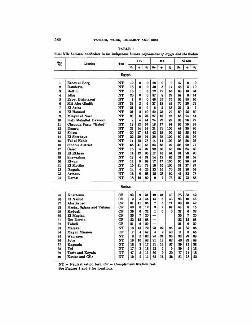
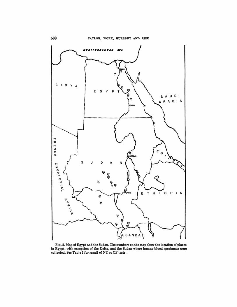
The results of the NT or CF tests with West Nile virus on sera collected fromindigenous inhabitants along the Egyptian Nile and in the Sudan are shown inTable 1.' Examination for neutralizing antibodies was not done on samples fromcertain localities in the Sudan and in these instances the results of the CF reactions are given. In the table the “localities―are listed more or less from north tosouth and the corresponding “MapNo.― will identify the situation on theaccompanying maps (Figures 1 and 2).
The data are tabulated according to the age of the donor, 0-14 years, 15 yearsand above, and all ages combined. Tabulation has not been made by sex asanalysis failed to demonstrate any significant difference in the frequency ofantibodies in males and females.
The first impression gained from casual inspection of the table is that antibodiesto West Nile virus are widely disseminated throughout the Nile Valley and thesouthern Sudan, as at only one (Ezbet el Borg, Map No. 1) of the 40 localitiessampled was there failure to find positive reactors. The over-all percentages ofNT positives in bloods collected in Egypt are 44 for 0-14 years, 72 for 15 yearsand above, and 61 for all ages combined, and in the Sudan 28,48 and 40 respectively (Table 2).
Antibodies in children are of greater interest because they are more likely to beindicative of local infection and because they give some indication of the recentpresence of the virus. With this in mind a more careful perusal of Table 1 willreveal area trends, particularly in the sera collected in Egypt. It will be notedthat in places situated in the northern portion of the Delta (Map Nos. 1—7)no,or only a few, children possessed West Nile antibodies; whereas further inlandand to the south in the Delta (Map Nos. 10—16),the ratio of positives amongchildren as well as adults is high (summarized in Table 2). This zonal distributionof West Nile antibodies along with other related information will be commentedupon later under a separate heading.
In addition to the localities listed in the table, 16 bloods were collected fromdesert Bedouins in the arid region of the Sinai Peninsula and 35 from residents ofthe Gaza area to the north, likewise an arid region. Only one was positive by the
3 The neutralization test on 331 of the sera included in this table was performed by
Dr. K. C. Smithburn (Smithburn et al., 1954).
‘@LocationTest0-1415+AllagesNo.1+1%No.1+1%No.1+1%Egypt1
23456789
101112131415161718192021222324EzbetelBorg
DamiettaBaltimIdkuEzbetBisintawaiMitAbuGhalibElAzizaEl HamoulMinyet el NaarKafr Mehallet DawoodChenuda Farm “Ezbet―DarnruShiwaEl SherkayaTelelKebirSindbis districtCairoEl EkhsasShawashnaElwanElMotihaNagadaArmantDarawNT
NTNTNTNTNTNTNTNTNTNTNTNTNTNTNTNTNTNTNTNTNTNTNT19
181620
7332121289
18232022146515141313151416180
010020264
1214172012414
1249
1149
160
060060
102144676185917863278631697329568928
3022374637
62927241821423814639217141716232590
5128
35182
23182217213838145860161217161623
70
17552276493379679294
10090
10010092659486
10010070927847
4838575370275055333644626028
128107312730313741270
5138
3520
225242629355558269964281626272032230
1034146629
750447981808997937760905987875478
85Sudan25
262728293031323334353637383940Khartoum
El NahudAbu ZabadKasha,SalaraandTukmaKadugliEl MugladUrn DoreinTalodiMalakalMayenMissionWau areaJubaKaguadaYeiToritandKeyalaKatireandGiloCF
CFCFCFCFCFCFCFNTCFNTNTNTNTNTNT29
93150263522211878
19181747199
411667
156
1345
10335231
44351223206829725763531718111149
14731
—
—
—
234
25212122306224
6520
222
24131229
1649
437167
0
96509662579
302678
23385327352321411133403939778133
1016867
156
356
2923155
141842
434215222068298555885838131822
586 TAYLOR, WORK, HURLB1]T AND RIZK
TABLE 1
West Nile humoral antibodies in the indigenous human populations of Egypt and the Sudan
NT Neutralization test; CF = Cornplernent fixation test.See Figures 1 and 2 for locations.

587ECOLOGY OF WEST NILE VIRUS IN EGYPT
FIG. 1. Map of Nile Delta. The numbers on the map show the location of places wherehuman blood specimens were collected. See Table 1 for results of NT or CF tests.
CF test and this came from an Arab who had resided for 15 years in the city ofSuez.
On the visit to Gaza, sera were collected from lifelong residents of the area andalso from refugees who had lived until five years previously in areas where WestNile virus had probably been frequently active in the past. Tests of the sera ofthe lifelong residents did not indicate activity of the virus in Gaza. Of the serafrom former residents of a West Nile active area, 40 per cent had neturalizingantibodies against West Nile and indeed 20 per cent had CF antibodies.
Studies on human infection in the Sindbis Sanitary District. The followinginformation on the seasonal, age, and sex incidence of human infection, as revealed by isolation of the virus and the presence of specific antibodies, was obtained from an intensive study in the Sindbis sanitary district situated about 30kilometers north of Cairo in the Nile Delta. This area was chosen because therewas reason to believe that West Nile infection was prevalent, because of its convenience to the laboratory, and because a great deal of useful demographic, sanitary, and economic information was available from a previous survey (Weir et a!.,1952).
Suffice it to state here that the Sindbis sanitary district incorporates five viilages all within a radius of six kilometers from a health center located in thevillage of Sindbis. While it is purely agricultural and essentially rural in nature,nearly all of the approximately 25,000 inhabitants live in the five villages.
588 TAYLOR, WORK, RURLBUT AND RIZK
M(DIT(RRdN(iN Std
LIBY4
EGY PT
SAUDIA
UGANDA
FIG. 2. Map of Egypt and the Sudan. The numbers on the map show the location of places
in Egypt, with exception of the Delta, and the Sudan where human blood specimens werecollected. See Table 1 for result of NT or CF tests.

Neutralization0—14
No. + %15+ No. + %All
ages
No. +%Delta:
Lower EgyptNon-endemic area (Map Nos.
1—7)Transitionalarea (Map Nos.8—9)
Endemic area(Map Nos.10—16).Cairo
Upper EgyptEgypt,total
SudanGrand total
134
49171151034711466173
812046520941
2502
16702763442841206
562209212169720490180
41208601075039860139
73956588724867340
105391107224
1168350151883
493286417271213985124
47846077614056
ECOLOGY OF WEST NILE VIRUS IN EGYPT 589
TABLE 2West Nile neutralizing antibodies in the indigenous human populations of Egypt
and the Sudan summarized according to region
Note. This table is a summarization of the results of the NT reactions shown in Table 1.
Surrounding each village are about 2,000 acres of irrigated, rich, and highly cultivated land.
The houses in the villages are constructed of sun dried bricks and are closelypacked, their fronts forming a continuous wall along narrow streets. Both thehuman inhabitants and the livestock are quartered on the ground floor but theremay be a half story above for storage purposes. Aside from an occasional openspace about a pond or a canal where domestic animals congregate and manure ispiled or grain thrashed, there is no free ground within the confines of these villages and the over-all density of both human and domestic animal population isexceedingly high. The human inhabitants are thus not only in very intimatecontact with one another but also with the numerous domestic quadrupedschiefly gamoose (water buffalo), cows, sheep, goats, donkeys and a few camelsand with fowl, such as chickens, ducks, pigeons and a few geese. Indeed thequadrupeds and fowl all live under the same roof with the owner and, as far asassociation is concerned, are virtually a part of the family.
The main roads and highways in the terrain surrounding the villages are frequently flanked with a row of casuarina, flame (royal poinciana), or eucalyptustrees, and near the villages groves of citrus fruit are not uncommon, all of whichafford ample harborage for birds. The most abundant non-migratory wild birdsin this district, in order of frequency are: house sparrows (Passer domesticus),doves (Streptopelia senegalensis), hooded crows (Corvus coron.e sardonius), buffbacked herons (Bubulcus ibis ibis), rock pigeons (Columba livia), and kestrels(Falco tinnunculus).
The study of human infection in this district consisted of attempts to isolatethe virus from the blood of children taken during an acute febrile episode and torecognize previous infection by examining the blood serum for specific CF andNT antibodies for West Nile virus.

YearNo. blood specimensVirusisolationsNo.%1951—52
19531954*
Total
100913025139
1040.89
0.770.782824230.81
590 TAYLOR, WORK, RURLBUT AND RIZK
TABLE 3West Nile virus isolations from the blood of febrile children in Sindbis District
according to year
* The number of bloods examined in 1954 was purposely reduced and the smaller num
ber examined during the year does not imply that there were fewer febrile children thanduring the two preceding years.
Isolation of West Nile virus from human blood. The routine of obtaining bloodfor examination for virus was as follows. All children attending the outpatientdepartment of the Sindbis Health Center had rectal temperatures taken by anurse and those in a febrile state were referred to the physician for furtherexamination. Bloods were taken from a selected number of children who accordingto the mothers had not been ill for more than three days, and whose physicalexamination revealed no specific cause of illness, such as the recognizable exanthemata, marked gastro-intestinal disturbances, pulmonary signs, or obviousbacterial or skin infections. In other words, children were selected for examination for circulating virus who were suffering from an acute undiagnosed febrileillness. Since it had been shown by serological surveys that infection with WestNile virus was likely to occur at an eariy age, children below four years of agewere given preference in the selection.
The number of bloods thus collected and examined for virus, and the numberof West Nile virus isolations, tabulated according to year are shown in Table 3.West Nile virus was isolated during each of the three years (1952, 1953, and1954) covered by the study and from children from all of the villages during atleast one year. The ratio of isolations to the number of specimens examined didnot vary significantly from year to year.
In Table 4 the years are combined, but the number of specimens exsmined andnumber of virus isolations made are tabulated according to month. All of theisolations were made during the months of June, July, August, and September.The highest ratio of isolations to the number of specimens examined occurredduring the months of July and August.
Table 5 shows the number of virus isolations made according to the age andsex of the children from whom the blood specimens were taken. The number ofisolations in each age and sex group is too small to be statistically significant butthere is a suggestion that virus is found more frequently in the blood of malesthan of females and in the age group of one and two years than in older andyounger age groups.
West Nile complement fixing (CF) and neutralizing (NT) antibodies in human

MonthNo. blood specimensVirusisolationsNo.%January
FebruaryMarchAprilMayJuneJulyAugustSeptemberOctoberNovemberDecember
Total
2141631922021962002832703023122382520
00001
12910000
0000
.54.23.3
.300
0282423.8TABLE
5West Nile virus isolations from blood of febrile children in Sindbis District
according to age andsex0-11/12
mo.
M F1—2
yrs..3—4 yrs.5-9 yri.10-14 yrs.AllagesM
FM FM FM FMFNo.
bloodsNo. virusiso%vinusisoBoth sexesVirusiso%virusiso
398 21010.3 0
60810.2815
54413 51.6 0.91359181.3363
2222 20.6 0.958540.7139
990 00 0238005
80 00 0
13001720
108316 70.9 0.62803230.8
ECOLOGYOF WEST NILE VIRUS IN EGYPT 591
TABLE 4West Nile virus isolations from the blood of febrile children in Sindbis District
according to month
Note: The totals in this table differ from those in Table 4 because 21 persons whose ageor sex was not definitely recorded have been omitted from this table. None of the senafrom these 21 persons yielded virus.
bloods. Most of the bloods included in this survey were collected from infants andyoung children for the purpose of attempting virus isolation. A smaller numberwere also collected, however, from older children and adults solely for examiningfor antibodies, and the survey is therefore by no means a random sampling. It isweighted heavily in the younger age group; and moreover most of the childrenof this group were in a febrile state when the blood was taken. Whether or notthis condition affected the results of the tests of antibodies to West Nile virus isnot known. We have no reason to suspect that it did, since a subsequent surveyconducted in cooperation with the WHO, in which blood samples were collectedfrom presumably normal children, gave essentially the same pattern of CFreactors in children from 0-4 years. The data here presented include only first
592 TAYLOR, WORK, HURLBUT AND RIZK
bleedings. A certain number of children were re-bled but the results of theserebleedings will be tabulated subsequently.
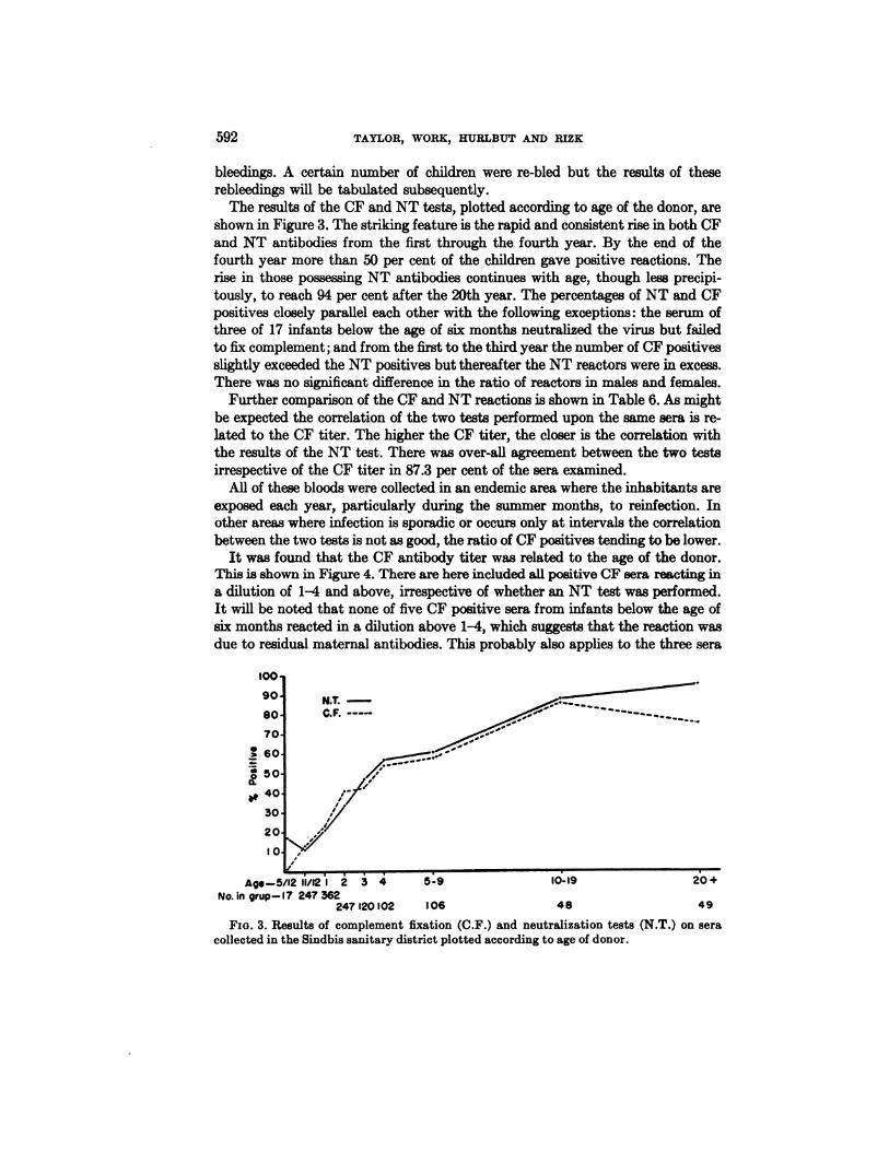
The results of the CF and NT tests, plotted according to age of the donor, areshown in Figure 3. The striking feature is the rapid and consistent rise in both CFand NT antibodies from the first through the fourth year. By the end of thefourth year more than 50 per cent of the children gave positive reactions. Therise in those possessing NT antibodies continues with age, though less precipitously, to reach 94 per cent after the 20th year. The percentages of NT and CFpositives closely parallel each other with the following exceptions: the serum ofthree of 17 infants below the age of six months neutralized the virus but failedto fix complement; and from the first to the third year the number of CF positivesslightly exceeded the NT positives but thereafter the NT reactors were in excess.There was no significant difference in the ratio of reactors in males and females.
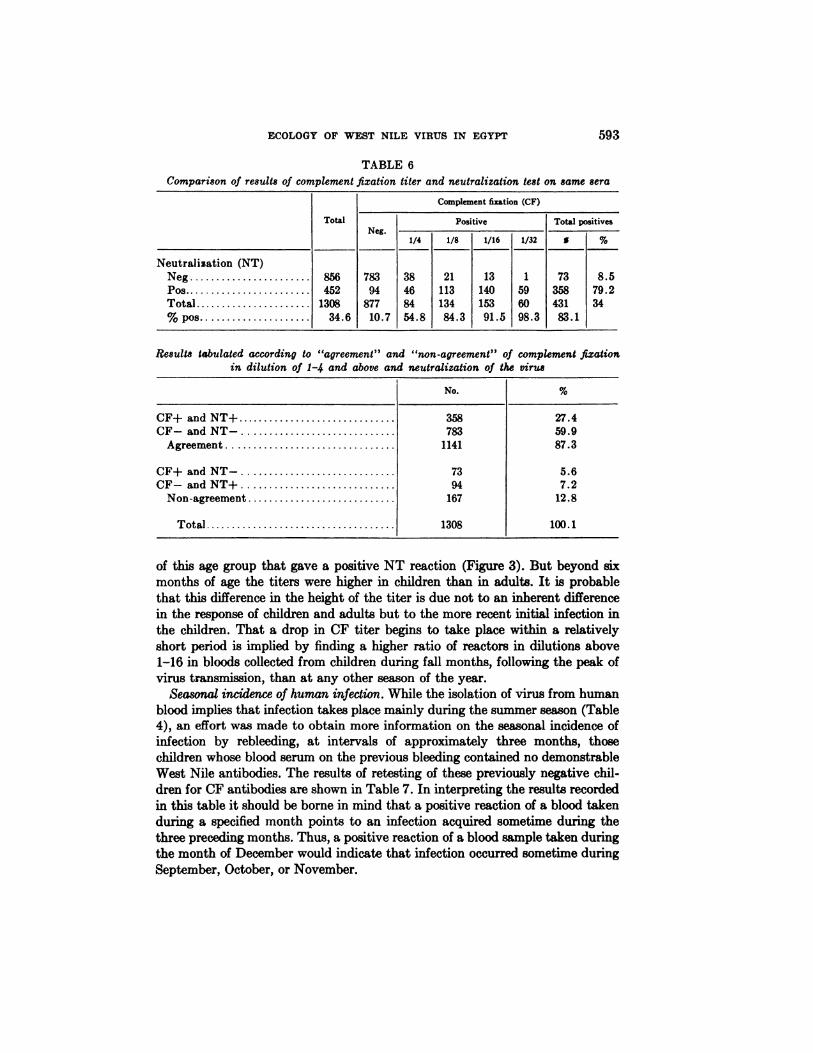
Further comparison of the CF and NT reactions is shown in Table 6. As mightbe expected the correlation of the two tests performed upon the same sera is related to the CF titer. The higher the CF titer, the closer is the correlation withthe results of the NT test. There was over-all agreement between the two testsirrespective of the CF titer in 87.3 per cent of the sera examined.
All of these bloods were collected in an endemic area where the inhabitants areexposed each year, particularly during the summer months, to reinfection. Inother areas where infection is sporadic or occurs only at intervals the correlationbetween the two tests is not as good, the ratio of CF positives tending to be lower.
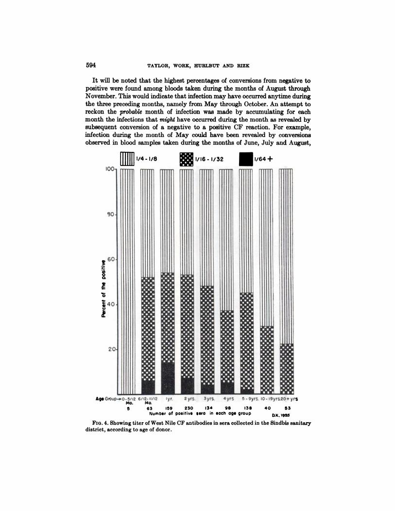
It was found that the CF antibody titer was related to the age of the donor.This is shown in Figure 4. There are here included all positive CF sera reacting ina dilution of 1-4 and above, irrespective of whether an NT test was performed.It will be noted that none of five CF positive sera from infants below the age ofsix months reacted in a dilution above 1-4, which suggests that the reaction wasdue to residual maternal antibodies. This probably also applies to the three sera
N.T.C.F.
5,
Agi—5/12 11/12I 2 3 4 5-9 10-19 20+No. in grup—I7 247 362
247 120102 106 48 49
FIG. 3. Results of complement fixation (C.F.) and neutralization tests (N.T.) on sera
collected in the Sindbis sanitary district plotted according to age of donor.
TotalComplement
fixation (CF)
Positive Total positivesNeg.
1/4 1/8 1/16 1/32 S%Neutralization
(NT)NegPosTotal% pos
8564521308
34.6783
9487710.738
468454.821
11313484.313
14015391.51
596098.373
35843183.18.5
79.234
No.%CF+andNT+
35827.4CF—andNT—78359.9Agreement
114187.3CF+
and NT—735.6CF—andNT+947.2Non-agreement16712.8Total
1308100.1
ECOLOGY OF WEST NILE VIRUS IN EGYPT 593
TABLE 6Comparison of results of complement fixation titer and neutralization test on same sera
Results tabulated according to “agreement―and “non-agreement―of complement fixationin dilution of 1-4 and above and neutralization of the virus
of this age group that gave a positive NT reaction (Figure 3). But beyond sixmonths of age the titers were higher in children than in adults. It is probablethat this difference in the height of the titer is due not to an inherent differencein the response of children and adults but to the more recent initial infection inthe children. That a drop in CF titer begins to take place within a relativelyshort period is implied by finding a higher ratio of reactors in dilutions above1—16in bloods collected from children during fall months, following the peak ofvirus transmission, than at any other season of the year.
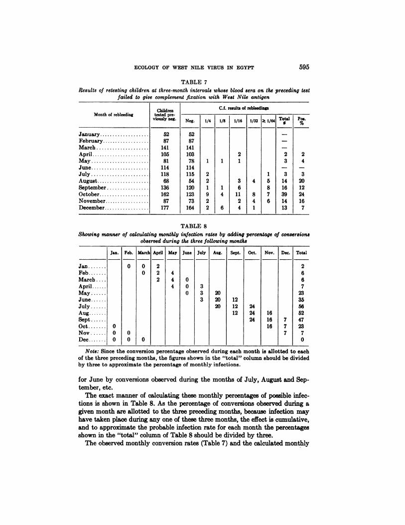
Seasonal incidence of human infection. While the isolation of virus from humanblood implies that infection takes place mainly during the summer season (Table4), an effort was made to obtain more information on the seasonal incidence ofinfection by rebleeding, at intervals of approximately three months, thosechildren whose blood serum on the previous bleeding contained no demonstrableWest Nile antibodies. The results of retesting of these previously negative children for CF antibodies are shown in Table 7. In interpreting the results recordedin this table it should be borne in mind that a positive reaction of a blood takenduring a specified month points to an infection acquired sometime during thethree preceding months. Thus, a positive reaction of a blood sample taken duringthe month of December would indicate that infection occurred sometime duringSeptember, October,or November.
594 TAYLOR, WORK, KUELB1JT AND RIZK
It will be noted that the highest percentages of conversions from negative topositive were found among bloods taken during the months of August throughNovember. This would indicate that infection may have occurred anytime duringthe three preceding months, namely from May through October. An attempt toreckon the probable month of infection was made by accumulating for eachmonth the infections that might have occurred during the month as revealed bysubsequent conversion of a negative to a positive CF reaction. For example,infection during the month of May could have been revealed by conversionsobserved in blood samples taken during the months of June, July and August,
I /64 +
AgGroup—.o-5/i2 6/12-11/12 yr. 2yrs.Mo. Mo.5 63 159 230 134 98 138
Number of positive sero in each age group
Syrs. 4 yrs 5 - 9yrS. 0- I9yrS.20+ yFS
40 53
@k.t955
FIG. 4. Showing titer of West Nile CF antibodies in sera collected in the Sindbis sanitarydistrict, according to age of donor.
ftllIllI1/4-1/8@I/I6-I/32
Month of rebleedingChildren testedprevtouzly neg.C.f.
results of rebleedings— — — —
Neg. 1/4 1/8 1/16 1/32@ 1/64 T@talP;.January
FebruaryMarch
5287
1415287
141——
—April
MayJune
10581
11410378
11411212 3
—2
4—July
AugustSeptemberOctober
11868
136162115
541201232
2191 43
6114 81
5873
1416393
2012
24November877322461416December
1771642641137
ECOLOGY OF WEST NILE VIRUS IN EGYPT 595
TABLE 7Results of retesting children at three-month intervals whose blood sera on the preceding test
failed to give complement fixation with West Nile antigen
TABLE 8Showing manner of calculating monthly infection rates by adding percentage of conversions
observed during the three following months
Jan. Feb. March April May June July Aug. Sept. Oct. Nov. Dec. Total
Jan 0 0 2 2Feb 0 2 4 6March 2 4 0 6April 4 0 3 7May 0 3 20 23June 3 20 12 35July 20 12 24 56Aug 12 24 16 52Sept 24 16 7 47Oct 0 16 7 23Nov 0 0 7 7Dec 0 0 0 0
Note: Since the conversion percentage observed during each month is allotted to eachof the three preceding months, the figures shown in the “total―column should be dividedby three to approximate the percentage of monthly infections.
for June by conversions observed during the months of July, August and September, etc.
The exact manner of calculating these monthly percentages of possible infections is shown in Table 8. As the percentage of conversions observed during agiven month are allotted to the three preceding months, because infection mayhave taken place during any one of these three months, the effect is cumulative,and to approximate the probable infection rate for each month the percentagesshown in the “total―column of Table 8 should be divided by three.
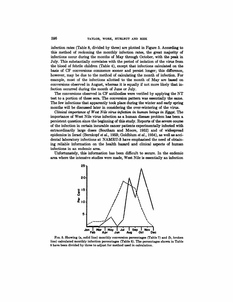
The observed monthly conversion rates (Table 7) and the calculated monthly
596 TAYLOR, WORK, HURLBUT AND RIZK
infection rates (Table 8, divided by three) are plotted in Figure 5. According tothis method of reckoning the monthly infection rates, the great majority ofinfectionsoccurduringthemonths ofMay throughOctober,with thepeak inJuly. This substantially correlates with the period of isolation of the virus fromthe blood of febrile children (Table 4), except that infections calculated on thebasis of CF conversions commence sooner and persist longer; this difference,however, may be due to the method of calculating the month of infection. Forexample,most of the infectionsallottedto the month of May are based onconversionsobservedinAugust,whereasitisequallyifnotmore likelythatinfection occurred during the month of June or July.
The conversions observed in CF antibodies were verified by applying the NTtest to a portion of these sera. The conversion pattern was essentially the same.The few infections that apparently took place during the winter and early springmonths willbe discussedlaterinconsideringthe over-winteringofthevirus.
Clinical importance of West Nile virus infection in human beings in Egypt. Theimportance of West Nile virus infection as a human disease problem has been apersistent question since the beginning of this study. Reports of the severe courseof the infection in certain incurable cancer patients experimentally infected withextraordinarilylargedoses (Southam and Moore, 1952) and of widespreadepidemicsinIsrael(Bernkopfetat.,1953;Goldblum etat.,1954),aswellasaccidental laboratory infections at NAMRU-3 have emphasized the need of obtaining reliable information on the health hazard and clinical aspects of humaninfections in an endemic area.
Unfortunately, this information has been difficult to secure. In the endemicarea where the intensive studies were made, West Nile is essentially an infection
C
0
Fxo. 5. Showing (a, solid line) monthly conversion percentages (Table 7) and (b, brokenline) calculated monthly infection percentages (Table 8). The percentages shown in Table8 have been divided by three to adjust for method used in calculation.
25
5
Jan I Mar I May I Jul I Sep I NovFeb Apr Jun Aug Oct Dec
ECOLOGY OF WEST NILE VIRUS IN EGYPT 597
of early childhood and, according to serological conversions from negative topositive, takes place mainly during the summer months (Table 7 and Figure 5).It is also during this season that other febrile illnesses among the village childrenare common and widespread.
Because the diagnosis of West Nile infection can be made only by the isolationand identification of the virus from the blood of the patient or by the serologicalresponse,bothofwhich requiretime,thediagnosisinan individualcaseisnecessarily retrospective. Thus no early differentiation from other febrile disorderswas possible. Although a history was taken and physical examination made whenthe child was first brought to the clinic and the initial blood sample taken, thechild was not hospitalized and return visits were infrequent. It was therefore notpossible to follow currently the course of the illnesses which later proved to beWest Nile. In reviewing the records made by the physician when the child wasbrought to the clinic, combined with subsequent information obtained from themothers on 22 cases proven to be West Nile by virus isolation from the blood, thefollowing impressions were gained. Fever of rapid onset, averaging 38.5—39°C.,accompanied by gastro-intestinal disturbances, malaise, profuse sweating, a finepapular rash (5 cases), moderate enlargement of the cervical, axillary and inguinallymph nodes(3cases),and occasionallycongestionoftheeyesand throat.The fever remained high for 5-6 days followed by gradual decline and a ratherprolonged convalescence. No signs of central nervous system involvement wereobserved.As farasisknown no deathscouldbe attributedtotheinfectionandfrank encephalitis in the highly endemic area was exceedingly rare.
The full course of the infection was followed only in two adults, both of whomwere laboratory technicians. The illness of one of them has been described(Hamilton and Taylor, 1954). The course of the disease in the second technicianwas similar except that the onset was more sudden. Severe headache, principallyin the occipital region, accompanied by muscular fatigue, particularly of the armsand legs, some initial gastric discomfort, anorexia and fever of 38 to 39°C.forseveral days were the outstanding manifestations. Recovery was somewhat retarded but complete.
Both of these technicians were engaged in the isolation of viruses from humanblood and mosquitoes, involving the inoculation and brain-to-brain passages inmice. They were therefore exposed to the handling of freshly isolated strains.At least three other persons on the technical staff of the laboratory developedneutralizing antibodies for West Nile virus during the course of their assignment,but neither the date of the infection nor the associated symptoms, if any, weredefinitely determined.
It is not improbable that epidemic outbreaks of West Nile virus infection occasionally occur in the non-endemic zone along the northern fringe of the Deltabut no suspect epidemics were reported during the four years covered by thisstudy. But, as previously mentioned, febrile episodes among children are exceedingly common during the summer and an epidemic would have to be rathersevere and have unusual characteristics in order to be recognized and reported.
SpeciesTested‘Neg.PositiveNumber%Mammals:
CamelCowDonkeyGamoose (water buffalo)GoatHorseSheepRattus rattusBats
Total mammals
93615
1884914644348
4662
308
53482
494344
2797
67
1351
1215
—
418778
1747722
8623—
840Avian
species:Chicken (domestic)
Rebleedings on previously negativesCrow (Corvus corone sardonius)Duck (domestic)Dove (Streptopelia senegalensis senegalensis)Goose (domestic)Heron (Bubulcus ibis ibis)Hoopoe (Upupa epops major)Kestrel (Falco tinnunculus)Kite (Milvus migrans aegyptius)Pigeon (domestic)Sparrow (Passer domesticus)Quail (Coturnix c. coturnix)
Total avian species
2415
163148
2965
531
59268
42020
1357126
2347
5—
—
44158
2504
2102
226
18—
31
1511
—
17016
136514252728—
2542—
40Grand
total 88652935740
598 TAYLOR,WORK,HUBLBUTAND RIZK
VERTEBRATE HOSTS OTHER THAN MAN
Information on vertebrate hosts other than man was sought through threechannels: a survey for specific NT antibodies in the blood serum; a search forWest Nile virus in the blood and organs of animals; and experimental infection.
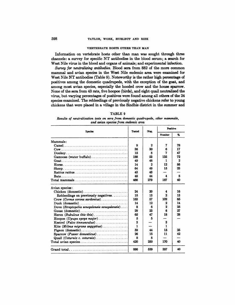
Survey for neutralizing antibodies. Blood sera from 882 of the more commonmammal and avian species in the West Nile endemic area were examined forWest Nile NT antibodies (Table 9). Noteworthy is the rather high percentage ofpositives among the domestic quadrupeds, with the exception of the goat, andamong most avian species, especially the hooded crow and the house sparrow.None of the sera from 43 rats, five hoopoe (birds), and eight quail neutralized thevirus, but varying percentages of positives were found among all others of the 24speciesexamined.The rebleedingsofpreviouslynegativechickensrefertoyoungchickens that were placed in a village in the Sindbis district in the summer and
TABLE 9Results of neutralization tests on sera from domestic quadrupeds, other mammals,
and avian species from endemic area
ECOLOGY OF WEST NILE VIRUS IN EGYPT 599
re-bled in the fall. Two of the 15 originally negative chickens had on rebleedingbecome positive, suggesting that infection occurred during their sojourn in thevillage.
The question naturally arises as to the specificity of the neutralization of thevirus by a large proportion of the sera from domestic quadrupeds and certainavian species. Is it the result of infection with West Nile virus or some closelyantigenically related virus, or is it due to the presence of a viricidal element in theserum that is unrelated to previous virus infection? We know of no generallyapplicable, trustworthy, and practicable method of distinguishing between specific and nonspecific neutralization or inactivation of a virus in a serum-virusmixture. But we have tried two procedures: securing and examining blood serumfrom animals that presumably could not have been exposed to infection withWest Nile or related arthropod-borne viruses; and heating the serum at 56°C.forone half hour and reactivating with fresh normal serum.
Through the kindness of Dr. G. W. Dick, sera from 15 horses and 10 sheep wereobtained from England. None of these sera neutralized West Nile virus. In contrast 12 of 14 (86%) horse and 15 of 64 (23%) sheep sera from the West Nileendemic area in Egypt were positive (Table 9). Sera from 14 horses were alsoobtained from Brazil, and two neutralized the virus. However, other arthropodborne viruses antigenically related to West Nile are known to exist in Brazil,which may account for the two positive reactors.
It has been noted that non-specific viricidal action in presumably normal seramay be removed by heating to 56°C.We observed this phenomenon in some ofour laboratory-reared rabbits. It was also observed that the addition of freshnormal human serum did not reactivate the heated rabbit serum althoughunheated serum neutralized two or more logs of the virus. Applying this criterionto 23 sera from horses, sheep, gamooses and donkeys, whose sera when originallytested was positive, it was found that the heating of these sera for one half hourat 56°C.only slightly diminished the original neutralizing action and that whenmixed with fresh normal human serum the neutralization of West Nile virus wasfully restored. it is felt, therefore, that such evidence as has been obtained indicates that the neutralization of West Nile virus by serum from quadrupeds collected in the West Nile endemic area is probably specific and is not due to normally present viricidal substances in the serum.
In regard to the sera from avian species, it can only be said that none of theserafrom 24 chickensimportedfrom the United StatesneutralizedWest Nilevirus; and, as will be reported later, the species of wild birds that were testeddemonstrated a high susceptibility to infection with West Nile virus followed bythe development of NT antibodies in the surviving birds. Moreover, a few captured birds whose serum neutralized the virus were tested and found to be resistant to infection, whereas birds of the same species whose serum failed toneutralize the virus were highly susceptible to infection.
Search for virus. Specimens from 295 animals, comprised mainly of bloods fromcrows, herons, and domestic pigeons, were inoculated in an attempt to isolatevirus, and West Nile virus was isolated twice from pigeons and once from a crow

SpeciesNo.of
animalsexainmedNature
of specimen
Blood Brain SpleenWNisolationsMammals:
DogDonkeyHorseMule
Total mammals
32
134
221 13
2124
21Avian
species:ChickenCrow (Corvus corone sardonius)Heron (Bubulcus ibis)Pigeon
Total avian species
41596644
2734
1566143
2644
273
164
162
1312
3Grand
total 29526537133
600 TAYLOR,WORK,}IURLBUTANDRIZK
TABLE 10Specimens from mammal and asian species examined for virus
and number of West Nile isolations
Note: One of the isolations from pigeons was made in July and the other in August.The isolation from the crow was made in August.
(Table 10). One of the pigeons was definitely ill and the brain and the spleen aswell as the blood yielded virus. Obviously in the random sampling of a bird population by means of shooting, as was done mainly with crows, the chance of obtaining a bird in the viremic stage of infection is relatively slight, although theincidence of infection among them may be rather high. Most of the herons werenestlings or were captured shortly after leaving the nest. If West Nile infectionof herons during the nestling stage is frequent, the opportunity of recoveringthe virus from fledglings should have been greater than from older birds. NoWest Nile virus was obtained but several strains of another and antigenicallydistinct virus were isolated from the blood of these fledgling herons.
The brains of a dozen horses, four mules, two donkeys and three dogs, suspectedof having central nervous system (CNS) involvement, were examined for virusduring the course of these studies. Rabies virus was recovered from the brains oftwo of the dogs and one of the horses, but none yielded West Nile virus. However,most of the brains were either in poor condition when received or had been preserved in another laboratory for six months to two years at a temperature ofapproximately —20°C.
Susceptibility to experinwntal infection. In planning investigations on experimental infection of vertebrates, consideration was given to the prevalence of thevertebrate in the endemic zone, its rate of reproduction, the probable manner ofinfection in nature, and the height and duration of the viremia following infection.
For a vertebrate to play an important part in the continued cyclic passage andmaintenance of the virus it is evident that it must be present in considerablenumbers and that a yearly supply of non-immunes must be provided. It is also
ECOLOGY OF WEST NILE VIRUS IN EGYPT 601
evident that the vertebrate must be susceptible to infection through the prevailing natural manner, and that it must circulate virus in sufficient concentration toinfect the common arthropod vector. Predominant attention was given to wild
birds and certain domestic fowl because they reproduce rapidly, and becausesome species are numerous and the adults showed a high ratio of neutralizingantibodies against West Nile virus. A few attempts were made to infect domesticquadrupeds. As mosquitoes appeared to be the main if not the sole natural vectorsof the virus, trial infection was carried out largely by permitting mosquitoesknown to be infected to feed upon the experimental animal; on succeeding daysthe animal was bled and the blood serum titrated for virus by inoculating serialtenfold dilutions i.c. into young adult mice.The wildbirdsselectedforexperimentationon susceptibilityto infectionby
bite of infected Culex mosquitoes comprised the house sparrow, hooded crow,kestrel, buff-backed heron, and palm dove. (For a full account of these experiments consult Work, Hurlbut and Taylor, 1955.) All of the species circulatedvirus in varying concentration. The highest titers of circulating virus were foundin the hooded crow and the house sparrow, and the lowest in the palm dove.In the house sparrow and crow the titers ranged between 3.5 and above 8 logs,and if the bird survived, the virus usually circulated for as long as six days.Though theviremiawas ofa lowergradeinthekestrel,buff-backedheronandpalm dove,some individualscirculatedvirusinadequateconcentrationtoinfectmosquitoes. Successful cyclic passage was tried and accomplished in all of theabove enumerated species, though the percentage of mosquitoes infected following feeding was highest with the hooded crow and sparrow.
Young chicks readily acquire the infection when exposed to infected mosquitoes and circulate virus in adequate concentration to infect normal mosquitoes.In four chicks from one to eleven days of age that were infected by mosquito bite,the titer of the virus in the blood ranged from above 4 to 6.3 logs, and seven ofeightmosquitoesthatfedupon two ofthesechicksbecame infected.However,with the advance of age, chickens become refractory and after the third week oflife are not consistently infected by mosquito bite; if virus does circulate, it is oflow concentration. Thus, two of three chicks that were three weeks of age andone ofeightolderchickenscirculatedvirusafterexposuretoinfectedmosquitoes,but only for one day.
The results of experimental infection of domestic pigeons were conditioned bythe West Nile virus strain employed. In all of the above mentioned experiments alow mouse passage Egyptian strain isolated either from mosquitoes or fromhuman blood was used. The mosquito strain (Ar-248) was first used in attemptsto infect squabs and adult pigeons. The results were disappointing, as only one oftwo squabs and none of four adult pigeons circulated detectable virus after trialinfection by mosquito bite, although there was a conversion to positive or inconclusive neutralization of the virus in all of the four adult pigeons one monthfollowing exposure. Somewhat better results were obtained when a West Nile
strain(An-1230)isolatedfrom a sickpigeonwas used.With thisstrain,circulating virus titers of low order but of several days' duration were produced.
Trial infection of domestic quadrupeds was confined to two mules, two young

602 TAYLOR, WORK, EURLBUT AND RIZK
sheep, and a young gamoose. One of the mules received 100,000 mouse LD50intravenously and the other was bitten by 15 infected mosquitoes. Virus was detected in the blood of the mule inoculated i.v. on the third day following inoculation in a dilution not exceeding 1—10.However, the blood of both mules neutralized West Nile virus one month later. Neither of the sheep circulated virus afterbeing bitten by infected mosquitoes but one later developed neutralizing antibodies. The young gamoose was bitten by two infected mosquitoes and also failedto circulate virus. By accident the post bleeding from the gamoose was discardedbeforebeingexaminedforneutralizingantibodies.
Arthropod host-vectors. The term host-vectors is used because according to ourpresentknowledge,ifan arthropodiscapableof activetransmissionofa virus,the virus is commonly harbored within the body of the arthropod for an indefiniteperiod, usually throughout the life span of the arthropod. Thus, from the aspect.of time, the designation of host is more applicable to arthropods than to vertebrates. The recognition of arthropod host-vectors of West Nile virus was attempted by search for virus in captured arthropods and by experimental infection and transmission of the virus by suspected vectors.
Search for virus in captured arthropods. Search for virus in captured arthropodswas limited almost exclusively to hemophagus species. A few pools of Musca spp.were included because of their great abundance in Egyptian villages and thetendency of certain species to feed on secretions about the eyes and nose ofchildren. In all, 78,067 specimens, grouped according to taxonomic classificationinto 1533 pools, were inoculated into infant mice in search of virus. With theexception of some of the ticks, the collections were made in or near the Sindbissanitarydistrict.The mosquitoescomposed abouttwo-thirdsofthespecimensaswell as of the pools, and West Nile virus was found only in mosquitoes (Table 11).
Mosquitoes consisted of five main species: Culex antennatus, Culex pipiens,Culex univittatus, Aedes caspius, and Anopheles pharoensis (Table 12). Otherspecies were so few in number that it was not deemed worth-while to examinethem for virus. During 1952, taxonomic differentiation between C. pipiens and C.univittatus was not made; consequently, one column in the table is designated aseither one or both of these species. However, in the two subsequent years, whenthe species were separated, it was found that during the season when the virusisolations were made from the undifferentiated pools, C. pipiens was exceedinglyscarcewhileC. univittatuswas abundant.Itmay thereforebe inferredthattheundifferentiated pools inoculated in 1952 were composed almost exclusively of C.univittatus, and that in all probability the virus was contained in this species. Thefact that the percentage of isolations from the mixed pools was virtually the sameas that from the pools composed exclusively of C. univittatus, gives further support to this inference. If this deduction is accepted, then 12 of the 17 isolationsrecorded were from C. univittatus, and five from C. antennatus. Relative to the
number of specimens of the two species inoculated, the virus was recovered 10times more frequently from C. univittatus than from C. antennatns, and accordingto the number of pools, five times more frequently (Table 12).
The virus was recovered from mosquitoes during the three successive years,

No. ofspecimensNo.
of poolsinoculatedW.N.
virusisolationsFleas
Flies:MuscasppPhiebotomus sp ....
LiceMitesMosquitoesTicks:
AmblyommaspArgassppBoophilus sppDermacentor spHaemaphysalis spHyatoinma sppOrnithodoros sppRhipocephalus sppUnclassified
3,272
6,351123
3,6486,887
51,937
941,359
43113312
5161,8401,422
4254
206
4880
1003
12921621
429064
317Total
78,067153317TABLE
12
West Nile virus isolations from mosquito pools according to species ofmosquitoMosquito
speciesSpecimensPoolsAve no.T?speC./poOlaW.N.
virus Isolations
NumberCulex
antennatusCulex pipiensCulez pipiens &/or univittatusCulex univiUatusAedes caspiusAnopheles pharoensis
3471455142104633220271246485
15753
160846472
35404024205
—
39
—
—0.015
0.140.14
—1.03
—
5.665.63
—
—Total
5193710035217
603ECOLOGY OF WEST NILE VIRUS IN EGYPT
TABLE 11Number of specimens and number of pools of arthropods examined for virus,
and number of West Nile virus isolations
1952, 1953 and 1954, that mosquito collections were made. The recoveries werelimited to the months of July, August and September, eleven in July, five inAugust, and one in September (Table 13).
In Table 14 is shown the number of specimens and poois of Culex mosquitoesinoculated according to calendar month and the number of pools that yieldedWest Nile virus during the three-year period. The column headed “C.pipiensand/or C. univittatus―applies to 1952 only. While no virus was recovered untilthe season of the year when C. univittatus and C. antennatus become fairly plenti

195219531954Jan.Feb.Mar.Apr.MayJuneJuly
434Aug.
32Sept.
1Oct.Nov.Dec.Total
75
5Total
115117
604 TAYLOR,WORK,EURLBUTAND BIZK
TABLE 13West Nile virus isolations from mosquito pools according to year and month
TABLE 14Number of specimens and pools of Culex mosquitoes inoculated, and number of West Nile
virus isolations, grouped according to month for the three year-period (1952—54)
C.j@ipseas and/orC. usiviUatasC. @i@s C. smiviUaius C. ante,esatus
JanFebMarchAprilMayJuneJulyAugSeptOctNovDec
Totals
No.pools
657
4617261394699
157
No.spec.
4771
2383419605477108323159
267160
5514
No. No. No. No. No. No. No. No. No. No.WN spec. pools WN spec. pools WN spec. pools WN
18 1672 13
59 1999 43364 36 3 3177 66354 8 2 817 38 7 5817 83 2896 15 1 1019 37 1 7273 83 3282 12 3778 59 1 10187 121137 7 635 20 4400 5412 1 47 3 1087 19
84 22104 43 3 6332 160 9 34714 485 5
* For 1952 only. During 1953 and 1954 distinction was made between C. pipiens and
C. univittatus and the two species inoculated separately.
ful, the frequency of isolation of the virus was not directly related to the abundance of these mosquitoes, since the peak of abundance of both species was notreached until September and yet all save one of the isolations were made in Julyand August. The highest frequency of isolations in respect to the number of specimens and pools of mosquitoes inoculated occurred during the month of July.
West Nile virus was probably recovered from mosquitoes on five additionaloccasions, but because the isolations could not be confirmed by subsequent reinoculation of the mosquito suspensions they have not been recorded in the tables.Three of these probable isolations were made from C. univittatus, one each inJuly, August and September. The other two consisted of one from A. pharoensisin July and one from C. pipiens in January. The one from C. pipiens wifi bereferred to later, as it has a significant bearing upon the over-wintering of thevirus.
Experiments on infection and transmission of the virus by arthropods. The initial

ECOLOGY OF WEST NILE VIRUS IN EGYPT 605
step was to introduce the virus into the hemocele by puncture or by measuredinjection, and observe whether the virus persisted and multiplied within thebody of the arthropod. This was regarded as a screening process. If it was foundthat the virus was retained and increased in titer, the next step was to determineif the arthropod could transmit the virus by bite, and finally, to see if the arthropod could be infected through feeding and in turn transmit the infection to asusceptible vertebrate by bite. These experiments have been reported in detailby Huribut (1956). Only the pertinent features will be given here.
The screening process was applied to mosquitoes, including A. caspius, C.pipiens, C. antennatus, C. univittatus, and Culiseta longiareolata; house fly,Musca domestica vicina; soft ticks, Ornithodoros savignyi, Ornithodoros erraticusand Argas persicus; hard tick, Rhipicephalus S. sanguitwus; human lice, Pediculushumanu8 corporis; mite, Derinanye&us gallinae; bedbug, Cim.ex lectularius; flea,Xenopsylla cheopis; hippoboscid fly, Pseudolynchia canariensie; and cockroaches,Periplaneta americana and Blatella germanica.
The virus was retained and showed evidence of growth in all the species ofmosquitoes examined and also, rather surprisingly, in a wide variety of otherarthropods, including house ifies, human lice, the soft ticks, and the one speciesof hard tick. However, the titer of the virus in the mosquitoes, following a suitable period of extrinsic incubation, ranged higher than in the other arthropods.Also, it was only in mosquitoes that the active vector cycle was demonstrated,that is, infection through feeding followed by transmission through bite.
One of the soft ticks, 0. savignyi, two weeks after being infected by injectioninto the hemocele, transmitted the infection by bite to infant mice, and viruswas demonstrated in the coxal fluid. However, although both 0. savignyi and0. erraticus acquired infection from feeding on infected mice and 27 to 33 daysafter the infective blood meal the titer of the virus in the body of the ticks rangedfrom 3.5 to 5.3 logs, neither species transmitted the infection by feeding uponnormal mice.
C. univittatus held at a temperature of 28—32°C.transmitted the virus as earlyas five days after the infective meal, and both C. univittatus and C. pipiens werecapable of transmitting the virus when kept at mean daily winter temperaturesof 12—23°C.
It was found that a virus titer of 1.5 logs in the infective blood meal wouldinfect an occasional mosquito; that a titer of 2.5 logs would infect approximately 50 per cent; and that titers of 3.5 logs and above would infect the majority if not all of the feeding mosquitoes and enable them to transmit the infection by bite.
Bionomics of mosquitoes and ticks examined for virus. Observations on the bionomics of predominant mosquitoes in the Nile Delta have been reported in thepaper of Hurlbut and Weitz (1956). These studies incorporated the seasonalprevalence and the feeding habits of C. antennatus, C. univittatus, C. pipiens, A.pharoensis, and A. caspius.
The seasonal prevalence of the three Culex species, as revealed by light-trapcatches in the endemic zone (Sindbis district), is graphically illustrated in Figure

606 TAYLOR,WORK,RURLBUTANDRIZK
6. C. antennatus and C. univittatus are most prevalent during the summer andthe early fall. This likewise applies to A. pharoensis and A. caspius. On the otherhand, C. pipiens was caught by light traps mainly during the late fall and spring.Bedroom catches from November to June were comprised exclusively of C.pipiens and most of the females were blooded, thus denoting that this species isactive throughout the year.
Larval collections confirmed the captures of imagoes, in that C. pipiens larvapredominated during the months of January, February and March, and October,November and December. During the warmer season, the highest percentages oflarvae consisted of C. antennatue, C. univittatus and A. pharoensis. However, itshould be noted that, although only imagoes of C. p&piens were found duringthe winter months, larvae of all of the Culex species as well as A. pharoensis andA. caspius were found during all seasons of the year. Thus it is probable thatthese mosquitoes are maintained by active breeding rather than hibernation.This was confirmed in the case of C. pipiens by the failure to find fat bodiesassociated with hibernation in any of the imagoes collected during the winterseason.Informationon thefeedinghabitswas securedfrom capturesintrapsbaited
with different animals, and by the precipitin reaction of the blood in the stomachof captured mosquitoes. In man-baited traps C. pipiens and A. pharoensis werecaught in relatively greater numbers than in light traps, while traps baited withbirds captured almost exclusively C. univittatus. Precipitin tests performed onblooded Culex mosquitoes caught in light traps, showed that 95 per cent ofC. pipiens had fed upon man. The next most frequent feeder on man was C.antennatus (22 per cent), followed by C. univittatus (11 per cent). Avian bloodwas identified in 9 per cent of C. univittatus, 1.5 per cent of C. antennatus, and 0per cent of the C. pipiens captured in light traps. However, a few, 0.6 per centof C. pipiens captured in bedrooms during the colder months, had fed upon avianblood. Nearly all the remainder had taken blood from man (96%). It may bededuced, therefore, that C. pipiens is definitely anthropophilic, and that C.univittatus is more attracted to birds than any of the other Cutex species. C.antennatus and C. univittatus also fed extensively on domestic quadrupeds, particularly bovids and donkeys.
All three Culex species extend southward along the Egyptian Nile and intothe Sudan. The following information on the Sudan is supplied by Lewis (1945).C. univittatus is the commonest culicine in many parts of the Sudan but thetypical form is believed to bite man only rarely. An atypical form (Var. neavel)feeds readily on man and according to Lewis is probably widely distributedalong the Upper Nile (this atypical form has not been found in collections madeby this laboratory in Egypt). C. antennatus is widely distributed throughoutthe Sudan and is a common man-biting mosquito. C. pipiens has been definitelyidentified only along the main Nile.
Reference to the distribution of the culicines in the lower Nile Delta and therelation to the activity of West Nile virus will be made later. The ticks werecollected mainly in Egypt, but some were obtained from the Sudan and Ana
ZoneCrowsHuman—____________0-1415
AllagesNo.+%+No..+%4-No.+%-f-No.+%+Non-endemic
Map nos.1—7
Transitional, Map nos.8—9
Endemic Map nos. 10-16.9464
163923
106103665134
491713
81202
1670206
5622080
4120839
7395340
10539183
4932824
4784
ECOLOGY OF WEST NILE VIRUS IN EGYPT 607
tolia. For a review of the ecology of all the species utilized, except AnatolianDerinacentor, see Hoogstraal (1956).
Investigation of “endemic―and “non-endemic―zones in the Nile Delta. Allusionwas made in presenting results on the extended serological survey in Egyptand the Sudan to the paucity of West Nile antibodies, particularly in children,in places sampled along the northern rim of the Nile Delta. This first came tonotice in an initial survey when it was found that, in contrast to the Sindbisdistrict,none ofthe bloodscollectedfrom childrenat Damietta,situatednearthe Mediterranean coast, neutralized the virus (Smithburn €1al., 1954). Thisobservation was confirmed by the collection of additional blood samples in thevicinity of Damietta. It then seemed important to attempt to define the areawhere thevirushad notinvadedorwas relativelyinactive,from theareafurthersouth where active transmission of the virus was occurring. It was hoped that acomparison of the two environments might yield a clue to the factor or factorsresponsible for the maintenance and activity of the infection.
On the criterion of positive reactors among children, division may be made intotwo and possibly three categories. First, the localities where there are no orvery few positives;thiswould includethe localitiesbearingMap Nos. 1-7 inTable 1 (Mit Abu Ghalib, Map No.6, has been included in this category becauseone of the two positive children had lived in a village further inland and to thesouth). Second, localities where the percentage of positives is slightly highersuggesting more frequent but irregular activity of the virus, Map Nos. 8 and 9.Third,localitieswhere thepercentageofpositivesinchildrenishigh,Map Nos.10-16. By reference to the map (Figure 1) it will be observed that the localities falling into the first category form a crescent along the northern rim ofthe Delta; that the two localities in the second category are somewhat further inland; and those in the third category are distributed over the middleand southern portion of the Delta.
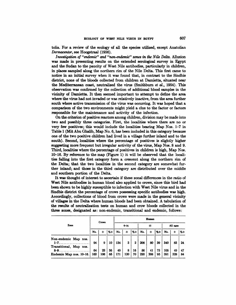
It was thought of interest to ascertain if these zonal differences in the ratio ofWest Nile antibodies in human blood also applied to crows, since this bird hadbeenshown tobe highlysusceptibletoinfectionwithWest Nilevirusand intheSindbis district the percentage of crows possessing specific antibodies was high.Accordingly, collections of blood from crows were made in the general vicinityof villages in the Delta where human bloods had been obtained. A tabulation ofthe results of neutralization tests on human and crow bloods collected in thethree zones, designated as: non-endemic, transitional and endemic, follows:
608 TAYLOR,WORK,RURLBUTANDRIZK
The correlation of West Nile neutralizing antibodies in human and crow serais evident and tends further to confirm the concept of non-endemic, transitionaland endemic zones. This was referred to in the paper by Work et at. (1955). Ahigh percentage of positives among crows, such as is found in the endemic zone,implies that some or many of the young crows produced each year becomeinfected, as do the young children. The endemicity or at least the yearly activityof the virus in the Sindbis district, situated in the “endemiczone―,is furtherattested by the isolation of the virus from mosquitoes and human blood duringthree successive summers. On the other hand, the absence or scarcity of positivesin the non-endemic and transitional zones implies that there had been no, oronly limited, activity of the virus during the life span of the crow, which probablydoes not average more than five to six years. This phenomenon of continuousactivity of the virus in one area in the Delta and not in another was naturallyintriguing, and attention was directed toward discovering the cause.
The climatic conditions in the endemic and non-endemic zones are notmarkedly different. As is commonly found, fluctuations in temperature near asea coast are not so great as further inland; monthly mean minimum temperatures near the apex of the Delta (Barrage, Sindbis district) range from 2 to 3°C.lower during the winter, and monthly mean maximum temperatures 2 to 4°C.higher during the summer, than along the Mediterranean coast (Damietta).Light rains and fog are more frequent during the winter season along the lowerDelta than further inland. But it is doubtful if either of these factors greatlyinfluences the fauna and flora, since freezing temperatures are rare in eitherzone and water is supplied mainly by irrigation.
There are, however, rather significant differences in the density of the humanpopulation and the intensity of land cultivation. Near the coast the villages arefewer and the population ranges from 100 to 200 per square kilometer, while inthe middle and southern part of the Delta the population usually exceeds 600 persquare kilometer. Although agriculture is the main occupation in both zones,fishing is an industry of considerable importance in villages near the coast. Thenumber and variety of livestock parallel the agriculture and the density of thehuman population. There appears to be no marked difference in the character ofhuman dwellings and the village environment, except that at some villages nearthe coast livestock may be quartered outside of the village rather than underthe same roof as the owner. No census of wild birds was taken but the samespecies were observed in both zones. Crows and other birds that feed largely oncultivated land and agricultural products may be less numerous near the coast.Yet none of these differences seemed to offer a satisfactory explanation for theabsence or presence of WN virus activity.
It appeared more likely that the key might be found in vectors of the virus. Asmosquitoes were the only proven vectors, emphasis was placed upon the study ofthis insect in the different zones. It was discovered that the total number ofmosquitoes caught per trap-night was five times greater in the endemic areaaround Sindbis than in the non-endemic zone. It was also found that C. unielttatus, the mosquito, from which the virus had been most frequently isolated, was

609ECOLOGY OF WEST NILE VIRUS IN EGYPT
rare in the places sampled in the non-endemic zone (Hurlbut and Weitz, 1956).For example, at Mit Abu Ghalib (Map No. 6) and Bisintawai (Map No. 5) inthe non-endemic zone, the average monthly catch of C. univittatus per trap-nightfrom June through November was 0.018 and 0.047 respectively, while during thesame period at Barada in the Sindbis district (Map No. 16) in the endemic zone,the catch was 18.06 specimens per trap-night. Moreover, no C. univittatus werecaptured at Mit Abu Ghalib and Bisintawai from July through September, theperiod of greatest activity of West Nile virus in the endemic zone and the timewhen C. univittatus reaches its peak of prevalence (Figure 6). However, therelation of the prevalence of C. univittatus to recent activity of West Nile virus,as gathered from antibodies in children and crows, is not fixed and constant;only 0.042 and 0.207 were captured per trap-night at Kafr Mahallet Dawood(Map No. 15) and Ezbet Chenuda (Map No. 11), which have been placed in theendemic zone on the basis of the serological survey. These two villages are situated along the northern edge of the endemic zone and it is conceivable that the
O.K.I9@
Fzo. 6. Showingrelative monthly prevalence of the three principal speciesof Cuter mosquitoes in the Sindbis sanitary district as revealed by light-trap catches.
C.ontenriotusO―—--'OC. univittotus
C.pipsens
I
Jon. Feb. Mar. Apr. May June @kily Aug. Sept. Oct. Nov. DeC.
610 TAYLOR,WORK,HUELBUTANDRIZK
antibodies found there in children and in crows may have resulted from a recentinvasion of the virus and are not indicative of prolonged endemicity.
Another hemophagus arthropod that has a distribution in the Nile DeltacorrespondingroughlytotheactivityofWest Nilevirus,isthetickOrnithodoroserraticus (Hoogstraal et at., 1954). Yet a causal relationship is doubtful, sinceefforts to demonstrate transmission of West Nile virus by this tick were unsuccessful, although it may be infected by ingestion of the virus and the virusretained within the body of the tick in moderately high titer for at least thirtydays (Hurlbut, 1956).
Examination of serafrom British troops in the Suez Canal Zone. In collaborationwith Col. M. H. P. Sayers, British Army Medical Corps, bloods were collectedfrom 127 British soldiers who had been resident in the Suez Canal Zone fromone and a half to more than two years, and from 54 persons who had recentlyarrived. These bloods were examined for West Nile CF antibodies. All of the 54bloods from recent arrivals were negative and only one of the 127 bloods fromresident soldiers gave a positive reaction. This blood fixed complement in adilution of 1—32,suggesting a rather recent infection. Fifty-seven of the residentsoliders were stationed near the Canal at a point separated by more than 10miles of desert from the cultivated part of the Delta. The other 70 soldiers werestationed within two miles of the village of Tel El Kebir, situated on the edgeof the Delta, where a high percentage of the native population of the villagepossess West Nile antibodies (Table 1), yet none of these soldiers' sera gave apositive CF reaction. It should be stated, however, that there was very littlecontact between the soldiers and the indigenous inhabitants and that the exercise of mosquito control about the camps kept these insects at a low level.
Investigations on over-wintering of the virus. The isolation of the virus fromchildren and from mosquitoes during three successive summers, 1952, 1953 and1954, the earlier isolations by Melnick, Paul et at. (1951) from bloods collectedfrom children during the summer of 1950, the yearly development of specificantibodies in non-immune children, as well as the progressive increase with ageof the ratio of West Nile antibodies in the human population in the Sindbisdistrict, all point to continuous endemicity and over-wintering of the virus.Otherwise the virus would have to be introduced each summer from some distantfocus, possibly by migratory birds. This possibility is discounted because thetemperature gradients for a hundred miles or more up the Nile are no morefavorable for the activity of mosquitoes during the winter months than those inthe upper Delta. Indeed it is more than 1,000 miles to the south before climaticconditions are encountered that are conducive to uninterrupted prevalence ofmosquitoes throughout the year. Moreover, water fowl are among the moreimportant migratory birds and these in their passage rest largely in lakes nearthe coast. If they were responsible for introducing the virus each year, a highrate of WN antibodies in the human and the crow inhabitants in the vicinity ofthese lakes would be expected. Actually, it is here that the lowest antibodyrates are encountered.
Having concluded that a true state of endemicity exists, three means by

ECOLOGY OF WEST NILE VIRUS IN EGYPT 611
which the virus might be maintained during the winter months when mosquitoesare scarce were postulated and investigated : first, in hibernating mosquitoes;second, in some long life arthropod, such as ticks ; and third, by slow cyclictransmission of the virus by the relatively few mosquitoes that continue to beactive.
No evidence was obtained that the three main species of Culex mosquitoes inEgypt hibernate during the winter months. On the contrary, consistent breeding, though retarded during certain seasons, appears to take place throughoutthe year (Hurlbut and Weitz, 1956).
A good many ticks of several species had been examined for West Nile viruswithout positive results, but after the virus was found in birds, the argasid ticksreceived special attention. In all 26,130 specimens, grouped into 530 pools andrepresenting 8 different taxonomic classifications of ticks, were examined withoutrecovering West Nile virus (Table 11). Other viruses were isolated from bothA. persicus and A. reflexus ticks which were thought initially to be related toWest Nile, but further study of these viruses has cast doubt on any close relationship. Moreover, attempts to infect mosquitoes with either the A. persicusor A. reflexus virus strains, which also differ from one another, have met withonly limited success. These tick strains are still under study, but the presentimpression is that they are distinct from West Nile, and since they cannot beexperimentally transmitted by mosquitoes—seemingly the main if not solevector of West Nile virus—the theory that they may represent reversible mutantsengendered by passage through different arthropod and vertebrate hosts receiveslittle support.
Thus, no evidence was uncovered that the virus is preserved in hibernatingmosquitoes or in ticks, to become active when mosquitoes become plentifulduring the summer. This left the third postulate, that the virus is maintainedby slow passage involving the mosquitoes that remain active during the coldermonths. This concept is supported by highly suggestive, if not conclusive,evidence. First, it will be recalled that a few children, two in April and three inMay, converted from negative to positive reactors by the CF test (Table 7). Theseconversions were confirmed by the NT test. Taken at their face value, thesefindings signify that infection took place sometime during the preceding threemonths, when the mean temperature is near the minimum. Secondly, althoughmosquitoes are difficult to find during the winter months, C. pipiens does persistin an active state. As previously mentioned, they were captured in bedroomsand most of the females were blooded. Finally, West Nile virus was presumablyisolated from a pool of 14 C. pipiens captured in bedrooms on the 27th of January. This isolation is not listed in Tables 11, 12 or 13 because it could not beconfirmed by re-inoculation of the mosquito suspension three weeks later. Twomice among the seven originally inoculated died on the 7th and 11th day andpassageof the brainsfrom thesetwo mice yieldedWest Nilevirus.As itwasrecovered on a second inoculation from the brain suspension of these two mice,it must be concluded that the virus was in the mosquitoes or that contaminationoccurred during the initial passage.
612 TAYLOR,WORK,HUELBUTANDRIZK
DISCUSSION
First, it is appropriate to examine the validity of the virus isolations and thereliability of the serological reactions upon which the inferences to be drawnfrom these studies so largely depend.
Validity of WN virus isolations. In the handling, inoculation and sub-passageof large numbers of field specimens, cross-contamination either through humanerror or by some other means has been of constant concern. In spite of all reason
able precautions, it is probable that several accidental infections did occur in ourlaboratory. Certainly there were two or more laboratory technicians who contracted WN infection while engaged in the manipulation of freshly isolatedstrains. Consequently, isolations were not accepted unless confirmed by re-isolation from the original material or, in the case of isolation from human blood,by the development of specific humoral antibodies in the patient following convalescence. It is believed, therefore, that all the isolations listed in the tables areunequivocally valid. It is probable that some if not most of those that wererejected (five from human blood and five from arthropods) are also valid isolations, but, with the exception of the one from C. pipiens in January, 1954, theirinclusion would have no significant bearing upon the interpretation of the ecologyof the virus.
Reliability of serological reactions. Reasons have been given for believing thatthe neutralization of WN by certain mammal and bird sera is not due to someviricidal substance which may be normally present in the sera. The neutralizingaction of the sera was only partially reduced by heating at 56°C.and was fullyrestored by the addition of fresh normal serum; the birds whose sera neutralizedthe virus were refractory to WN infection; and sera from horses, sheep andchickens from England and the United States exerted no neutralizing effect.Also, sera from persons who presumably had not been exposed to WN or otherrelated arthropod-borne viruses did not contain WN antibodies, namely, Britishand Americans recently arrived in Egypt, and Americans used in the experimentsreported by Southam and Moore (1954).
On the other hand, it is recognized that infection with one member of arthropod-borne Group B viruses (Casals and Brown, 1954) may stimulate both NTand CF antibodies against another member of the group (Smithburn, 1954;Pond et at., 1955). In the initial serological survey of indigenous human inhabitants along the Egyptian Nile it was found that 12 or 8.4 per cent of the seraneutralized Japanese B encephalitis (JBE) virus and 114 or 34.9 per cent neutralized Ntaya virus (Smithbum et at., 1954). Is it likely, therefore, that some ofthe reactions against WN are attributable to infections with these two viruses?We believe not; it seems to us more likely that the reverse is true and that infection with WN accounted for the neutralization of JBE and probably Ntaya.First of all, acute and convalescent sera from known WN infections were sentto the Rockefeller Foundation Virus Laboratories in New York, and it was therefound that the convalescent sera not infrequently neutralized to varying degreesJBE and Ntaya. Indeed, Goldblum (personal communication) has reported thatWN infection quite consistently stimulates neutralization of JBE. Secondly,
ECOLOGY OF WEST NILE VIRUS IN EGYPT 613
all of the 12 survey sera that neutralized JBE and all but five of the 114 sera thatneutralized Ntaya, also neutralized WN. Finally, neither JBE or Ntaya hasbeen recovered from any of the rather large number of arthropod and vertebratespecimens examined, and there is therefore no positive proof that either ofthese viruses exists in Egypt. Nor has any other virus been recovered that isclosely related antigenically to WN. Thus, for lack of evidence to the contrary,it is probable that within the inherent fallibility of the test the neutralization ofWN recorded in this paper is indicative of previous infection with WN virusrather than of non-specific inactivation of the virus or of infection with someother affied virus.
There is rather good correlation between the CF and NT tests on sera collectedin the highly endemic Sindbis district, particularly if the sera fix complement ina dilution of 1—16and above. The correlation varies in areas in the northernportion of the Delta which may be a reflection of the discontinuous course ofthe infection. it is generally supposed that CF antibodies to the viruses tendto recede and disappear with the passage of time, while NT antibodies are morepermanent and may be retained throughout life. In all likelihood this applies toWN virus (Southam and Moore, 1954a), but we know of no adequate supportingdata covering a sufficient length of time and involving enough individuals to besignificant. It has not been possible in our studies to investigate the duration ofCF antibodies, because in the area where rebleedings could be made the inhabitants were subjected, at least each summer, to reinfection, and it is conceivable that exposure to the bite of infected mosquitoes may stimulate a rise ora retention of CF antibodies which would otherwise decrease beyond detection.Indeed it is suspected that the rather high percentage of adults in the highlyendemic area whose sera give a positive CF reaction may be the result of yearlyexposure to infected mosquitoes. Otherwise it would be necessary to concludethat CF humoral antibodies normally persist for many years, since it is virtuallycertain that most of the adults in this area became initially infected duringchildhood. If this concept is correct it would account for the fluctuations in theratio of CF positives to NT positives in areas where the virus is not yearly active.That is, if there had been no visitation of the virus for several years the ratio ofCF positives to NT positives should be low, and if the blood samples were takenshortly following an epidemic invasion, then the ratio of CF positives to NTpositives should be high, comparable to endemic areas where presumably continuous exposure to infected mosquitoes keeps the CF antibodies at a detectablelevel, although in the older age groups the titer is generally low (Figure 4). Asonly 54.8 per cent of sera which fixed complement in a dilution not exceeding1—4neutralized the virus, and as a few reactors in this dilution were found inareas where the NT test was negative, the specificity of the CF test in thisdilution is questionable.
Granted the validity of the virus isolations and the reliability of the serological reactions, within the limits of these tests, as indicators of previous WNinfection, the inferences that seem permissible on the epidemiology of the diseasein man and the ecology of the virus will now be discussed.

614 TAYLOR,WORK,HUBLBUTANDRIZK
The epidemiology of human infection. The serological surveys imply that human infection is widespread among the indigenous inhabitants of the cultivatedportion of the Egyptian Nile and in the southern Sudan, but not commonamong desert Arabs or among British soldiers stationed in the Suez Canal zone.This distribution is consistent with the prevalence of mosquitoes. In areas notconducive to the breeding of mosquitoes or where they are artificially controlled,the infection is rare or absent. This falls in with other evidence of the mosquito'sbeing the principal if not the only vector of the virus; in the endemic area wherethe intensive studies were made, the infections are highly seasonal and for themost part occur during the period when the virus is also found in mosquitoes.In this area West Nile infection is essentially a childhood disease, the adultpopulation having acquired immunity. Most of the infections probably takeplace in the villages, since infected mosquitoes were repeatedly captured withinthe village compounds. This would account for the lack of any difference in thesex incidence of infection, because both sexes are equally exposed in the villages.
Along the coastal perimeter of the Nile Delta the infection is less frequent andis probably not endemic. It is presumed that epidemic outbreaks may occurfrom time to time in this region when conditions are favorable and the virus isreseeded from the interior of the Delta.
it is suspected that man is not always involved in the main channel of virusactivity and that his infection may be incidental to or only partially involved ina broader cycle of the virus.
Vertebrate hosts other than man. The results of both serological surveys andexperimental infection indicate that the virus is infectious to a rather wide rangeof vertebrates. However, as has been pointed out, a vertebrate host must circulate virus in sufficient concentration to infect the arthropod vector in order tobe involved in the continuous cycle of the virus. While investigations on susceptibility to infection have not been exhaustive, the only animals found tofulfill this criterion (besides man) belong to the Ayes class. Also their numbersand rapidity of reproduction are sufficient to furnish a continuing adequatesupply of non-immune hosts. It would seem, therefore, that the main current ofthe virus passes through certain species of birds and young chickens with sideeddies involving man, and that infection of other animals, such as the domesticquadrupeds, may be purely tangential. Experiments on susceptibility of thislatter group included only mules, sheep, and one gamoose, but none circulatedvirus to an appreciable degree. Another reason for discounting the role of quadrupeds is that sheep, goats and gamooses are the more common species in thevillages and only in sheep and gamooses were WN antibodies frequent, both ofwhich species failed to circulate virus when bitten by infected mosquitoes.
Arthropod host-vectors. WN virus is retained and may increase in a wide varietyof arthropods, including, besides mosquitoes, ticks, lice, fleas and even houseifies when introduced by puncture into the body of the arthropod. But when theprerequisites for an effective vector are applied the field is narrowed to themosquito, since only the mosquito was found capable of acquiring the infectionby feeding upon an infected animal and transferring it to another animal by bite.

ECOLOGY OF WEST NILE VIRUS IN EGYPT 615
Thus, as far as is now known the mosquito is the only proven transmitter ofthe virus and only from the mosquito has the virus been recovered in nature.
Probable cycle of the virus. Naturally, the ensuing discussion on the cycle ofthe virus applies to the environment where these studies were made, and particularly to the endemic area centered about the Sindbis district, but the deductionsmay also hold for the greater part of the Nile Valley and the Sudan.
As has been pointed out, the only vertebrates and the only arthropods thathave been found to fulfill the requirements of host and vector in the continuouscyclic propagation of the virus are man, birds and mosquitoes. Because the viruscirculates in higher concentration in certain species of birds than in man, andbecause the species of mosquito (C. univittatus) from which the virus was mostfrequently recovered has an avidity for avian blood, it seems probable that thedominant cycle may be through birds and mosquitoes. This would fall in with thegreater frequency of isolation of the virus from mosquitoes during the early partof the mosquito season when the crop of young non-immune birds becomes available. This combination of a plentiful supply of susceptible birds with the onsetof the emergence of C. univittatus probably serves as a springboard for the rapidincrease in the activity and dissemination of the virus in which the non-immunechildren become involved. This activity continues, but on a diminishing scaleprobably as a result of a decrease in the non-immune birds and children, throughthe remainder of the summer and early fall. Though children may participate inthe cycle, its yearly recrudescence is probably dependent upon the spring supplyof young susceptible avian species. For, considering the high immunity rate inthe human population, it is difficult to conceive that the virus could be maintained by the man-mosquito cycle alone, even though the birth rate is high.
The presence of WN neutralizing antibodies in the blood of domestic quadrupeds, notably the camel, cow, gamoose and horse, indicates that they arefrequently infected, but it is doubtful if they participate in the cycle of thevirus; the available information suggests that if viremia does occur it does notreach a level sufficient to infect mosquitoes. If this is true, then the infection inquadrupeds is tangential and represents a dead-end type of infection, detectableonly by the development of neutralizing antibodies. This concept of the cycleof the virus is graphically illustrated in Figure 7.
it is not within the scope of this paper to review the literature and discussthe relation of the postulated cycle and ecology of West Nile to other arthropodborne viruses. But the analogy to the equine encephalitis viruses, St. Louis,Japanese B and Murray Valley encephalitis is obvious, notably in the involvement of mosquitoes as vectors and birds as vertebrate hosts. (For reviews andreferences see Ferguson, 1954; Anderson, 1954; Hammon, Reeves and Sather,1951; Tigertt and Hammon, 1950.)
Over-wintering and dissemination of the virus. The only positive evidenceunearthed on the over-wintering of the virus in parts of the Nile Delta pointed toa retarded but continuous passage of the virus during the three or four coldermonths of the year by the relatively few but active C. pipiens which persistthrough this season of the year. The development of WN antibodies in a few

616 TAYLOR, WORK, HURLBUT AND BIZK
children during the winter months, the presence and activity of C. @iipiensinbedrooms, the fact that the virus will multiply and can be transmitted by mosquitoes (including C. pipiens) during ambient winter temperatures, not to
mention the probable isolation of WN from C. pipiens in the month of January,all make a rather convincing case. And yet, if this is the way the virus survivesthe winter, its survival must hang by a slender thread, considering that most ofthe human population is immune and that only rarely does C. pipiens feed uponbirds.
Though the regular and consistent transport of the virus from the tropicalclimateofthesouthernSudan to the NileDeltaby migratingbirdsseems unlikely,itishighlyprobablethatitisdisseminatedby birdswithinthe homerange of such indigenous non-migratory species as the hooded crow, buff-backedheron, domestic sparrow and pigeon. it is asserted that the rock pigeon changesits habitat from time to time and may therefore carry the virus from some distance. No data were collected on the ffight or wind-drift range of mosquitoes.There is some travel of the human population but that of children, who aremost likely to be infected, is usually limited to the neighboring villages.
Avian Spe@\‘ Wild and
Hypothetical Cycles of West Nile VirusFIG. 7. Diagrammatic illustration of hypothetical cycles of West Nile virus. Note that
a complete cycle involving domestic quadrupeds alone is questioned.
I
ECOLOGY OF WEST NILE VIRUS IN EGYPT 617
The above remarks apply to the Nile Delta, the southern part of which seemsto provide sufficiently favorable conditions for the over-wintering of the virusand the creation of a continuous endemic state. It is not unlikely, however, thatthe truly migratory birds may be involved in spreading the infection along theless cultivated and sparsely inhabited portion of the Nile Valley and particularlybeyond the northern boundaries of Egypt. This possibifity deserves investigation.
Endemic and non-endemic zouss in the Nile Delta. The results of serologicalsurveys clearly imply that WN infection of both the human and the hoodedcrow population is less frequent along the northern fringe of the Delta thanfurther inland, which led to the somewhat arbitrary division into endemic,transitional and non-endemic zones. A study of the biological environment inthe endemic and non-endemic zones revealed differences which alone, or morelikely in combination, may account for the variation in the persistence andactivity of the virus. The human population is not so dense, the land not sointensely cultivated, and mosquitoes and apparently birds that are associatedwith cultivated land are not so numerous in the non-endemic as in the endemiczone. Also, and this may be a crucial factor, C. univittatus is relatively much lessprevalent in the places sampled in the non-endemic and in the northern edge ofthe endemic zone than in the highly endemic area about Sindbis.
The permanent maintenance of a parasite demanding a constant supplyof favorable hosts and effective vectors is frequently tenuous, and any imbalancein the essential requirements of the biological environment may break the chainin its cyclic propagation. it is believed, therefore, that a less dense human andbird population, combined with relative paucity of the most effective vectormay be responsible for the failure of permanent tenure of the virus in the northern part of the Delta. However, it is surmised that this northern area is beingperiodically reseeded from the permanently endemic area to the south, probablyby birds. Districts within the home range of non-migratory birds from thetruly endemic area would be seeded more frequently, and serologically might beindistinguishable from the endemic area, while villages further removed wouldbe subjected to less frequent invasions of the virus, manifested by a low percentage of WN antibodies among the inhabitants, especially in the youngerage group.
SUMMARY AND CONCLUSIONS
The results are recorded of an investigation of the geographical distribution ofWest Nile virus in Egypt and the southern Sudan, the epidemiology of humaninfection, and the general ecology of the virus in the Nile Delta, as revealed byvirus isolations, surveys for specific antibodies and study of the biologicalenvironment.
According to serological surveys the virus is widely disseminated along theEgyptian Nile and in the southern Sudan; 61 per cent of 1168 human bloodscollected in Egypt and 40 per cent of 350 collected in the Sudan neutralized WNvirus.
In an endemic area in the Nile Delta where the intensive studies were conducted, West Nile infection is essentially a childhood disease with yearly peaks
618 TAYLOR,WORK,HUELBUTANDRIZK
of transmission during mid-summer. No reliable information on the symptomatology of the infection in children was obtained, but the impression was gainedthat it is a self-limited, non-fatal febrile disease rarely associated with definitemanifestations of encephalitis.
The virus was repeatedly isolated from the blood of febrile children (23) andfrom mosquitoes, Cutex univittatus (12) and Culex antennatus (5), twice frompigeons (Columbalivia) and once from the hooded crow (Corvuscoronesardonius).The fact that C. univittatus was more frequently found to be infected than anyother species of mosquito may be due to its relative predilection for avian blood.None of 530 pools of other arthropods, including ticks, lice, fleas, mites and ifies,yielded virus.
On both experimental and ecological grounds it is concluded that the maincycle of the virus is probably through mosquitoes and birds in which man maybecome involved. Serological evidence points to infections of domestic quadru
peds, but these are probably only tangential or dead-end infections.It is likely that non-migratory birds are responsible for the local dissemination
of the virus but, as it appears to be endemic in parts of the Nile Delta, its transport over long distances by migratory birds was not investigated. There isevidence for believing that the virus over-winters through a process of retardedtransmission by the mosquitoes that remain active throughout the colder months,although the possible role of ticks as a reservoir of the virus should be furtherinvestigated. Non-endemicity in the northern rim of the Delta may be accountedfor by the less dense human and bird population there and the scarcity of whatappears to be the most important vector, C. univittatus.
ACKNOWLEDGMENTS
Recognition is made of the assistance rendered by: Dr. Mohamed Abdel
Kader and latterly Dr. Noeman Soliman Youssef in selecting and collectingblood specimens from febrile children at the Sindbis Health Center; Miss MouniraGhoneim, Health Visitor, in rebleeding children in the Sindbis sanitary district;and Mr. Harry Hoogstraal, head of the Department of Zoology NAMRU-3,and his assistants in collecting and ide@itifyingmany of the ticks examined.
Acknowledgment is also made to Dr. J. R. Kingston and Dr. N. H. Wiebengafor administrative aid in the virus laboratory during a two and a one-year periodrespectively, and to H. R. Dressler, Del Thrasher, M. A. Sylvester, L. J. Reber,R. B. Clarke, J. I. Thomas, E. W. Spangler, H. R. Fallers, Jr., Rostom Bablanian,and Mrs. Afaf Sabry El Agamawy, for invaluable technical help in the laboratoryfor varying periods during the course of this investigation.
REFERENCES
ANDERSON, S. G., 1954. Murray Valley encephalitis and Australian X-disease, J. Hyg. 52:
447—468.BERNKOPF, H., LEVINE, S., AND NERSON, R., 1953. Isolation of West Nile virus in Israel,
J. Inf. Di8. 93: 207-218.CASALS, J., AND BROWN, L. V., 1954. Hemagglutination with arthropod-borne viruses,
J. Exp. Med.99:429-449.
ECOLOGY OF WEST NILE VIRUS IN EGYPT 619
DR BOER, C. J., ANDCox, H. R., 1947. Specific complement-fixing diagnostic antigens forneurotropic viral diseases, J. Immutwl. 55: 192—204.
FERGUSON, F. F., 1954. Biological factors in the transmission of American arthropod-borne
virus encephalitis, U. S. Dept. Health & Education &Welfare. PublicHealthMono graphNo.23.
GOLDBLUM, N., STaRK, V. V., AND PADERSKI, B., 1954. West Nile fever. The clinical features
of the disease and the isolation of West Nile virus from the blood of nine human cases,Amer. J. Hyg. 59: 89—103.
HAMILTON, P. K., AND TAYLOR, R. M., 1954. Report of clinical case ofWest Nile virus infection probably acquired in the laboratory, Am. J. Trop. Med. & Hyg. 3: 51—53.
HAMMON, W. McD., REEVES, W. C., AND SATHER, G. E., 1951. Japanese B encephalitis virusin the blood of experimentally inoculated birds, Amer. J. Hyg. 53: 249—261.
HOOGSTRAAL, H., 1956. African Ixodoidea I. Ticks of the Sudan with special reference to
EquatoriaProvince,U. S.Navy, Washington,D. C. (Inpress)HOOGSTRAAL, H., SALAH, A. A., AND KAISER, M. N., 1954. Summary of the known distribu
tion and biology of Ornithodoros erraticus, J. Egyptian Pub. Health As8oc. 29: 127-138.HUELBUT, H. 5., 1956. West Nile virus infection in arthropods, Am. J. Trop. Med. & Hyg.
5: 76—85.HURLBUT, H. S., AND WEITZ, B., 1956. Some observations on the bionomics of the common
mosquitoes of the Nile Delta, Am. J. Trop. Med. & Hyg. In press.KENDRICK, D. B., JR., ELLIOTT, J., REICHEL, J., JR., AND VAUBEL, E. K., 1945. Supply of
preserved blood to European Theater of Operations, Bull. U. S. Army M. Dept. (No. 84)pp. 66-73.
KITAOKA, M., 1950. Experimental transmission of the West Nile virus by the mosquito,
Jap. Med. J. 3: 77-81.MELNICK, J. L., PAUL, J. R., RIORDAN, J. T., BARNETT, VoH. H., GOLDBLUM, N., AND
ZABIN,E., 1951. Isolation from human sera in Egypt of a virus apparently identical toWest Nile virus, Proc. Soc. Exper. Biol. and Med. 77: 661-665.
PHILIP, C. B., AND SMADEL, J. E., 1943. Transmission of West Nile virus by infected Aedesalbopictu8, Proc. Soc. Exper. Biol. and Med. 53: 49—50.
POND, W. L., RUSS, S. B., ROGERS, N. G., AND SMADEL, J. E., 1955. Murray Valley encephalitis virus: its serological relationship to the Japanese—West Nile—St.Louis encephalitisgroup of viruses, J. Immunol. 75: 78—84.
SMITHBURN, K. C., 1952. Neutralizing antibodies against certain recently isolated viruses
in sera of human beings residing in East Africa, J. Immunol. 69:223-234.SMITHBURN, K. C., 1954. Antigenic relationships among certain arthropod-borne viruses as
revealed by neutralization tests, .1. Immunol. 72: 376—388.SMITEBURN, K. C., HUGHES, T. P., BURKE, A. W., AND PAUL, J. H., 1940. A neurotropic
virus isolated from the blood of a native of Uganda, Am. .1. Trop. Med. 20: 471-492.SMITHBURN, K. C., AND JACOBS, H. R., 1942. Neutralization tests against neurotropic
viruses with sera collected in Central Africa, J. Immunol. 44: 9—23.SMITHBURN, K. C., TAYLOR, R. M., RIzK, F., AND KADER, A., 1954. Immunity to certain
arthropod-borne viruses among indigenous residents of Egypt, Am. J. Trop. Med. &Hyg. 3: 9—18.
SOUTHAM, C. M., AND MOORE, A. E., 1952. Clinical studies of viruses as antineoplasticagents, with particular reference to Egypt 101 virus, Cancer 5: 1025—1034.
Sotrra&M, C. M., AND MOORE, A. E., 1954a. Anti-virus antibody studies following inducedinfection of man with West Nile, Ilheus and other viruses, .1. Immunol. 72: 446-462.
SOUTHAM, C. M., AND MOORE, A. E., 19Mb. Induced virus infections in man by Egyptisolates of West Nile virus, Am. J. Trop. Med. & Hyg. 3: 19—50.
STROME, C. P. A., 1953. A closed vacuum system for harvesting infectious infant mousebrain material, Proc. Soc. Exper. Biol. and Med. 84: 287-288.
TAYLOR, R. M., 1952. Studies on certain viruses isolated in the tropics of Africa and SouthAmerica: their growth and behavior in the embryonated hen egg, J. Immunol. 68:473—494.

620 TAYLOR,WORK,HUELBUTANDRIZK
TAYLOR, R. M., 1953. Observations on the epidemiology of West Nile virus infection. Pre
sented at the Proceedings of the 5th International Congress of Trop. Med. & Malaria,Istanbul, 1953. NAMRU-3 Research Project No. NM 007 082.13.08.
TAYLOR, R. M., AND HURLBUT, H. 5., 1953. Isolation of West Nile virus from Culex mosqui
toes, J. Roy. Egyptian Med. Assoc. 36: 199—208.TIGERTT, W. D., AND HAMMON, W. McD., 1950. Japanese B encephalitis: a complete review
of experience on Okinawa 1945-1949,Am. J. Trop. Med. 30: 689.WEIR, J. M., WA5IF, I. M., HAS5AN, F. R., ATTIA, S. D. M., AND KADAR, M. A., 1952. An
evaluation of health and sanitation in Egyptian villages, J. Egyptian Pub. HealthAssoc. 27: 55—114.
Wotx, T. H., HURLBUT,H. S., ANDTAYLOR,R. M., 1953. Isolation of West Nile virus fromhooded crow and rock pigeon in the Nile Delta, Proc. Soc. Exper. Biol. and Med. 84:719—722.
WORK,T. H., HURLBUT,H. S., ANDTAYLOR,R. M., 1955. Indigenous wild birds in the NileDelta as potential West Nile virus circulating reservoirs, Am. J. Trop. Med. & Hyg.4: 872—888.
Copyright © 2022 FDOKUMEN