
A SPECT Scanner for Rodent Imaging Based on Small-Area Gamma Cameras
8
2524 IEEE TRANSACTIONS ON NUCLEAR SCIENCE, VOL. 57, NO. 5, OCTOBER 2010 A SPECT Scanner for Rodent Imaging Based on Small-Area Gamma Cameras Eduardo Lage, José L. Villena, Gustavo Tapias, Naira P. Martínez, Maria L. Soto-Montenegro, Mónica Abella, Alejandro Sisniega, Francisco Pino, Domènec Ros, Javier Pavía, Manuel Desco, and Juan J. Vaquero, Senior Member, IEEE Abstract—We developed a cost-effective SPECT scanner proto- type (rSPECT) for in vivo imaging of rodents based on small-area gamma cameras. Each detector consists of a position-sensitive pho- tomultiplier tube (PS-PMT) coupled to a 30 30 NaI(Tl) scintil- lator array and electronics attached to the PS-PMT sockets for adapting the detector signals to an in-house developed data ac- quisition system. The detector components are enclosed in a lead- shielded case with a receptacle to insert the collimators. System performance was assessed using for a high-resolution par- allel-hole collimator, and for a 0.75-mm pinhole collimator with a 60 aperture angle and a 42-mm collimator length. The energy res- olution is about 10.7% of the photopeak energy. The overall system sensitivity is about and planar spatial res- olution ranges from 2.4 mm at 1 cm source-to-collimator distance to 4.1 mm at 4.5 cm with parallel-hole collimators. With pinhole collimators planar spatial resolution ranges from 1.2 mm at 1 cm source-to-collimator distance to 2.4 mm at 4.5 cm; sensitivity at these distances ranges from 2.8 to . To- mographic hot-rod phantom images are presented together with images of bone, myocardium and brain of living rodents to demon- strate the feasibility of preclinical small-animal studies with the rSPECT. Index Terms—Single Photon Emission Computed Tomography (SPECT), small-animal imaging, small-area gamma camera. Manuscript received January 05, 2010; revised April 16, 2010; ac- cepted June 25, 2010. Date of publication September 13, 2010; date of current version October 15, 2010. This work was supported in part by the CD-TEAM project, CENIT program, Spanish Ministerio de Indus- tria and with grants from the Ministerio de Educación y Ciencia, Projects TEC2007-64731/TCM, TEC2008-06715-C02-01, SAF2009-08076, program ARTEMIS S2009/DPI-1802, Comunidad de Madrid, and the RECAVA-RETIC Network. E. Lage, J. L. Villena, G. Tapias, M. Abella, and A. Sisniega are with Unidad de Medicina y Cirugía Experimental, Hospital General Universitario Gregorio Marañon, Madrid, Spain (e-mail: [email protected]; [email protected]; gus- [email protected]; [email protected]; [email protected]). N. P. Martínez and F. Pino are with Unidad de Biofísica y Bioingeniería, Uni- versidad de Barcelona-IDIBAPS, Barcelona, Spain (e-mail: nairamv@gmail. com; [email protected]). M. L. Soto-Montenegro is with Unidad de Medicina y Cirugía Experimental, Hospital General Universitario Gregorio Marañon, Madrid, Spain and also with CIBERSAM, Madrid, Spain (e-mail: [email protected]). D. Ros is with Unidad de Biofísica y Bioingeniería, Universidad de Barcelona- IDIBAPS, Barcelona, Spain and also with CIBER-BBN, Barcelona, Spain (e-mail: [email protected]). J. Pavía is with Servicio de Medicina Nuclear, Hospital Clínico y Provincial de Barcelona, Barcelona, Spain and also with CIBER-BBN, Barcelona, Spain (e-mail: [email protected]). M. Desco is with Unidad de Medicina y Cirugía Experimental, Hospital General Universitario Gregorio Marañon, Madrid, Spain, CIBERSAM, Madrid, Spain, and also with the Departmento de Bioingeniería e Ingeniería Aeroespa- cial, Universidad Carlos III de Madrid, Spain (e-mail: [email protected]). J. J. Vaquero is with the Departmento de Bioingeniería e Ingeniería Aeroespa- cial, Universidad Carlos III de Madrid, Spain (e-mail: juanjose.vaquero@uc3m. es). Color versions of one or more of the figures in this paper are available online at http://ieeexplore.ieee.org. Digital Object Identifier 10.1109/TNS.2010.2057516 I. INTRODUCTION S INCE the application of the pinhole imaging principle to SPECT, several scanners dedicated to rodents have been developed, thus providing an alternative technique to ex-vivo measurement of biological parameters. The earliest small-an- imal SPECT systems were configured with one to four clinical scintillation cameras operated using a collimator with a single pinhole aperture [1]–[3]. These systems allow high spatial reso- lution and small field of view projection data to be obtained by using high magnification factors and moderate spatial resolution detectors. However, this approach requires a heavy collimated detector to rotate around a small object with a constant radius of rotation (thus challenging the mechanical implementation) or to rotate the animal (thus making difficult its use with living samples). Currently, the trade-off between spatial resolution and detection sensitivity in most small-animal systems (based on ei- ther clinical gamma cameras or custom detectors) has improved by an effective use of the active area with multipinhole colli- mators, thus increasing acquisition speed and optimizing field of view size [4]–[10]. Nevertheless, as the number of pinholes increases, the detector area required for the resulting projec- tions also increases with the consequent rise in material cost. Al- though these newer systems provide a wide range of possibilities for translational research, most common protocols encountered in practice can also be accomplished using more cost-effec- tive solutions with lower performance. These include small-an- imal tomographic systems [11]–[15] and planar imaging sys- tems [16]–[19] based on small-area gamma cameras. This type of solution based on compact gamma cameras is relatively in- expensive and has also been extensively investigated for clinical purposes [20]–[22]. In this work we present the rSPECT, a prototype scanner for in vivo imaging of rodents implemented to evaluate the feasi- bility of constructing a 4-head SPECT/CT commercial system with coplanar configuration (similar to that reported in [23]). This prototype is based on two small-area ( active area per camera) and high-resolution gamma cameras mounted opposite each other on a robotic gantry. The spatial res- olution and sensitivity can be adjusted depending on the study requirements by interchanging the collimators (parallel-hole or pinhole with different apertures) and adjusting the radius of ro- tation (ROR). To compensate for the intrinsic lack of magnifi- cation derived from small-area detectors and the relatively poor resolution of parallel-hole collimators, iterative reconstruction algorithms with accurate system modeling were used. Perfor- mance of the detectors was evaluated in terms of energy resolu- tion and integral uniformity without collimators, and in terms of 0018-9499/$26.00 © 2010 IEEE
Transcript of A SPECT Scanner for Rodent Imaging Based on Small-Area Gamma Cameras
2524 IEEE TRANSACTIONS ON NUCLEAR SCIENCE, VOL. 57, NO. 5, OCTOBER 2010
A SPECT Scanner for Rodent Imaging Based onSmall-Area Gamma Cameras
Eduardo Lage, José L. Villena, Gustavo Tapias, Naira P. Martínez, Maria L. Soto-Montenegro,Mónica Abella, Alejandro Sisniega, Francisco Pino, Domènec Ros, Javier Pavía, Manuel Desco, and
Juan J. Vaquero, Senior Member, IEEE
Abstract—We developed a cost-effective SPECT scanner proto-type (rSPECT) for in vivo imaging of rodents based on small-areagamma cameras. Each detector consists of a position-sensitive pho-tomultiplier tube (PS-PMT) coupled to a 30 30 NaI(Tl) scintil-lator array and electronics attached to the PS-PMT sockets foradapting the detector signals to an in-house developed data ac-quisition system. The detector components are enclosed in a lead-shielded case with a receptacle to insert the collimators. Systemperformance was assessed using ����� for a high-resolution par-allel-hole collimator, and for a 0.75-mm pinhole collimator with a60 aperture angle and a 42-mm collimator length. The energy res-olution is about 10.7% of the photopeak energy. The overall systemsensitivity is about � ��� �� ���� and planar spatial res-olution ranges from 2.4 mm at 1 cm source-to-collimator distanceto 4.1 mm at 4.5 cm with parallel-hole collimators. With pinholecollimators planar spatial resolution ranges from 1.2 mm at 1 cmsource-to-collimator distance to 2.4 mm at 4.5 cm; sensitivity atthese distances ranges from 2.8 to � ��� �� ����. To-mographic hot-rod phantom images are presented together withimages of bone, myocardium and brain of living rodents to demon-strate the feasibility of preclinical small-animal studies with therSPECT.
Index Terms—Single Photon Emission Computed Tomography(SPECT), small-animal imaging, small-area gamma camera.
Manuscript received January 05, 2010; revised April 16, 2010; ac-cepted June 25, 2010. Date of publication September 13, 2010; date ofcurrent version October 15, 2010. This work was supported in part bythe CD-TEAM project, CENIT program, Spanish Ministerio de Indus-tria and with grants from the Ministerio de Educación y Ciencia, ProjectsTEC2007-64731/TCM, TEC2008-06715-C02-01, SAF2009-08076, programARTEMIS S2009/DPI-1802, Comunidad de Madrid, and the RECAVA-RETICNetwork.
E. Lage, J. L. Villena, G. Tapias, M. Abella, and A. Sisniega are with Unidadde Medicina y Cirugía Experimental, Hospital General Universitario GregorioMarañon, Madrid, Spain (e-mail: [email protected]; [email protected]; [email protected]; [email protected]; [email protected]).
N. P. Martínez and F. Pino are with Unidad de Biofísica y Bioingeniería, Uni-versidad de Barcelona-IDIBAPS, Barcelona, Spain (e-mail: [email protected]; [email protected]).
M. L. Soto-Montenegro is with Unidad de Medicina y Cirugía Experimental,Hospital General Universitario Gregorio Marañon, Madrid, Spain and also withCIBERSAM, Madrid, Spain (e-mail: [email protected]).
D. Ros is with Unidad de Biofísica y Bioingeniería, Universidad deBarcelona- IDIBAPS, Barcelona, Spain and also with CIBER-BBN, Barcelona,Spain (e-mail: [email protected]).
J. Pavía is with Servicio de Medicina Nuclear, Hospital Clínico y Provincialde Barcelona, Barcelona, Spain and also with CIBER-BBN, Barcelona, Spain(e-mail: [email protected]).
M. Desco is with Unidad de Medicina y Cirugía Experimental, HospitalGeneral Universitario Gregorio Marañon, Madrid, Spain, CIBERSAM, Madrid,Spain, and also with the Departmento de Bioingeniería e Ingeniería Aeroespa-cial, Universidad Carlos III de Madrid, Spain (e-mail: [email protected]).
J. J. Vaquero is with the Departmento de Bioingeniería e Ingeniería Aeroespa-cial, Universidad Carlos III de Madrid, Spain (e-mail: [email protected]).
Color versions of one or more of the figures in this paper are available onlineat http://ieeexplore.ieee.org.
Digital Object Identifier 10.1109/TNS.2010.2057516
I. INTRODUCTION
S INCE the application of the pinhole imaging principle toSPECT, several scanners dedicated to rodents have been
developed, thus providing an alternative technique to ex-vivomeasurement of biological parameters. The earliest small-an-imal SPECT systems were configured with one to four clinicalscintillation cameras operated using a collimator with a singlepinhole aperture [1]–[3]. These systems allow high spatial reso-lution and small field of view projection data to be obtained byusing high magnification factors and moderate spatial resolutiondetectors. However, this approach requires a heavy collimateddetector to rotate around a small object with a constant radiusof rotation (thus challenging the mechanical implementation)or to rotate the animal (thus making difficult its use with livingsamples). Currently, the trade-off between spatial resolution anddetection sensitivity in most small-animal systems (based on ei-ther clinical gamma cameras or custom detectors) has improvedby an effective use of the active area with multipinhole colli-mators, thus increasing acquisition speed and optimizing fieldof view size [4]–[10]. Nevertheless, as the number of pinholesincreases, the detector area required for the resulting projec-tions also increases with the consequent rise in material cost. Al-though these newer systems provide a wide range of possibilitiesfor translational research, most common protocols encounteredin practice can also be accomplished using more cost-effec-tive solutions with lower performance. These include small-an-imal tomographic systems [11]–[15] and planar imaging sys-tems [16]–[19] based on small-area gamma cameras. This typeof solution based on compact gamma cameras is relatively in-expensive and has also been extensively investigated for clinicalpurposes [20]–[22].
In this work we present the rSPECT, a prototype scanner forin vivo imaging of rodents implemented to evaluate the feasi-bility of constructing a 4-head SPECT/CT commercial systemwith coplanar configuration (similar to that reported in [23]).This prototype is based on two small-area (active area per camera) and high-resolution gamma camerasmounted opposite each other on a robotic gantry. The spatial res-olution and sensitivity can be adjusted depending on the studyrequirements by interchanging the collimators (parallel-hole orpinhole with different apertures) and adjusting the radius of ro-tation (ROR). To compensate for the intrinsic lack of magnifi-cation derived from small-area detectors and the relatively poorresolution of parallel-hole collimators, iterative reconstructionalgorithms with accurate system modeling were used. Perfor-mance of the detectors was evaluated in terms of energy resolu-tion and integral uniformity without collimators, and in terms of
0018-9499/$26.00 © 2010 IEEE
LAGE et al.: A SPECT SCANNER FOR RODENT IMAGING BASED ON SMALL-AREA GAMMA CAMERAS 2525
spatial resolution and sensitivity using the parallel-hole collima-tors and the pinhole collimators. Tomographic hot-rod phantomimages are presented together with images of bone, myocardiumand brain of living rodents to demonstrate the feasibility of pre-clinical small-animal studies with the rSPECT.
II. MATERIALS AND METHODS
A. Gamma Cameras
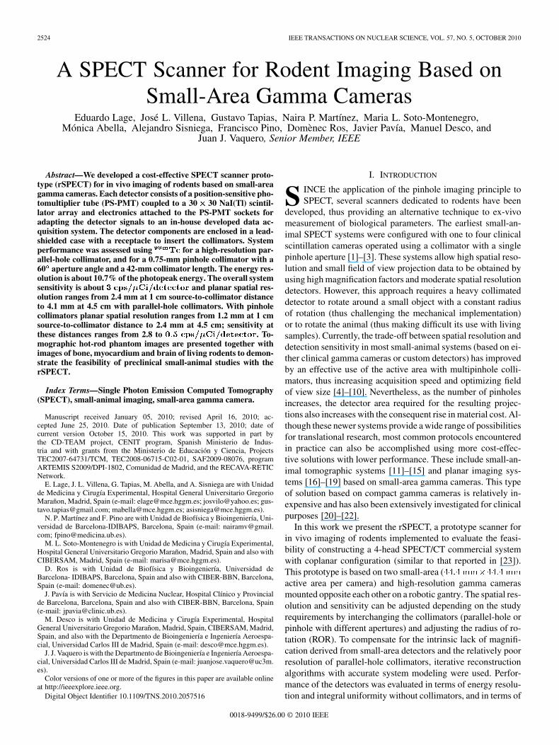
Each gamma camera was built using a Hamamatsu H-8500position-sensitive photomultiplier tube (PS-PMT) coupled to aNaI (Tl) scintillator array through a thin layer of optical grease(BC-630, Bicron-St. Gobain, Paris, France). The scintillatormatrices were also assembled by Bicron-St. Gobain and consistof 30 30 pixels of . The crystals areseparated by a 200- thick white reflector material whichalso covers its front side. The array is viewed through a 3 mmglass window and encapsulation is completed by a 50- thickaluminum cover. The photomultiplier have 12 stages of metalchannel dynode and 8 8 multiple anodes, providing an activearea of roughly . Each detector includes acompact PCB (Printed Circuit Board) stack directly attached tothe PS-PMT sockets, containing a symmetric charge divisioncircuit [24], [25]. Using this scheme the 64 anode signals of thePS-PMT are read out via a matrix of 128 resistors (one “X” andone “Y” per pad) arranged to allow the X and the Y signals fromone tube to be represented by 8 signal lines for each dimension(8 X-channel x 8 Y-channel). The PCB stack also includes ahigh-voltage supply and amplification stages for the resultingposition signals and for a timing signal from the PS-PMT [26].The scintillator array, the PS-PMT, and the electronics areassembled in a black-delrin enclosure which fits in a lead-cov-ered case (Fig. 1(a)). This enclosure contains a mechanism thatallows easy changing of the collimator depending on the studyrequirements. The camera configurations evaluated in this workare based on a) a low-energy and high-resolution parallel-holecollimator (1.2-mm hexagonal holes, 20-mm thick, and 0.2-mmseptal thickness) primarily for (whole body) rat studies andplanar imaging, and b) a low-energy pinhole collimator de-signed for organ-focused and (whole body) mice imaging,with a 42-mm collimator length, 8-mm-thick lead shielding,a 60 aperture angle, and interchangeable tungsten inserts(knife-edge type) with different aperture sizes (Fig. 1(b)).
B. Acquisition Electronics
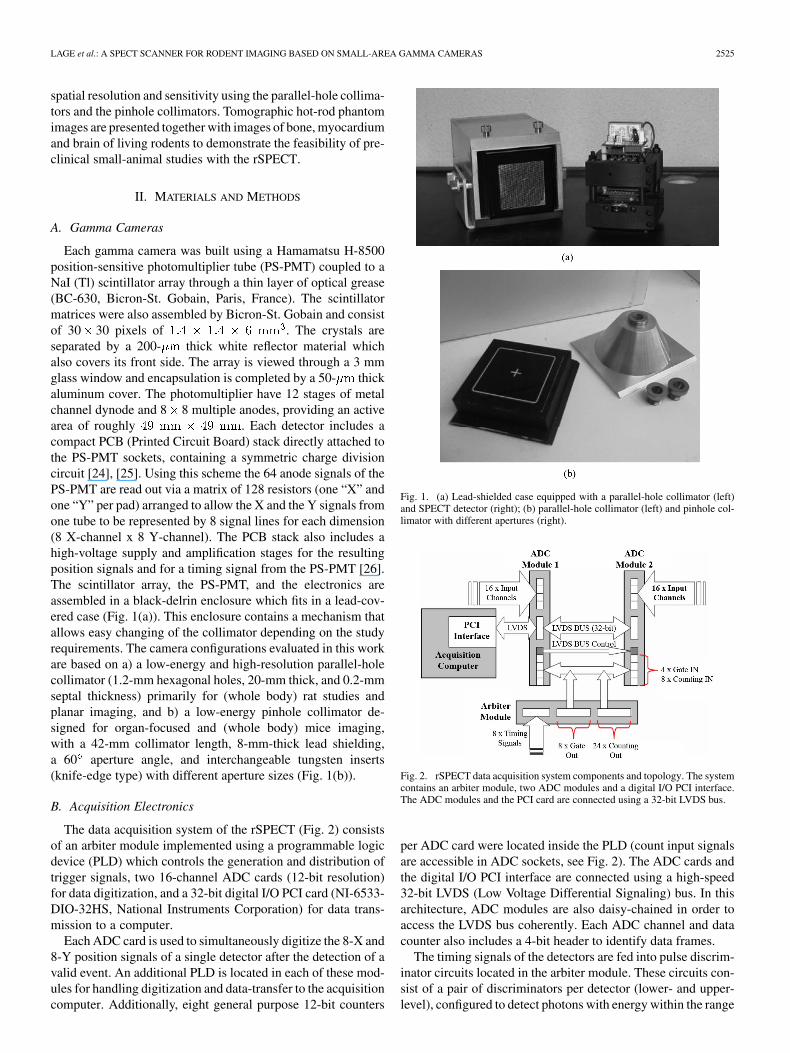
The data acquisition system of the rSPECT (Fig. 2) consistsof an arbiter module implemented using a programmable logicdevice (PLD) which controls the generation and distribution oftrigger signals, two 16-channel ADC cards (12-bit resolution)for data digitization, and a 32-bit digital I/O PCI card (NI-6533-DIO-32HS, National Instruments Corporation) for data trans-mission to a computer.
Each ADC card is used to simultaneously digitize the 8-X and8-Y position signals of a single detector after the detection of avalid event. An additional PLD is located in each of these mod-ules for handling digitization and data-transfer to the acquisitioncomputer. Additionally, eight general purpose 12-bit counters
Fig. 1. (a) Lead-shielded case equipped with a parallel-hole collimator (left)and SPECT detector (right); (b) parallel-hole collimator (left) and pinhole col-limator with different apertures (right).
Fig. 2. rSPECT data acquisition system components and topology. The systemcontains an arbiter module, two ADC modules and a digital I/O PCI interface.The ADC modules and the PCI card are connected using a 32-bit LVDS bus.
per ADC card were located inside the PLD (count input signalsare accessible in ADC sockets, see Fig. 2). The ADC cards andthe digital I/O PCI interface are connected using a high-speed32-bit LVDS (Low Voltage Differential Signaling) bus. In thisarchitecture, ADC modules are also daisy-chained in order toaccess the LVDS bus coherently. Each ADC channel and datacounter also includes a 4-bit header to identify data frames.
The timing signals of the detectors are fed into pulse discrim-inator circuits located in the arbiter module. These circuits con-sist of a pair of discriminators per detector (lower- and upper-level), configured to detect photons with energy within the range
2526 IEEE TRANSACTIONS ON NUCLEAR SCIENCE, VOL. 57, NO. 5, OCTOBER 2010
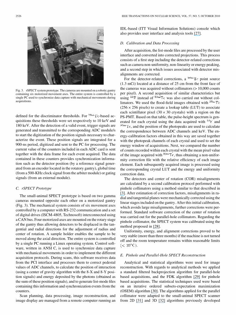
Fig. 3. rSPECT system prototype. The cameras are mounted on a robotic gantrycontaining six motorized movement axes. The entire system is controlled by asingle PC used to synchronize data capture with mechanical movements duringacquisitions.
defined for the discriminator thresholds. For -based ac-quisitions these thresholds were set respectively to 10 keV and180 keV. After the detection of a valid event, trigger signals aregenerated and transmitted to the corresponding ADC module/sto start the digitization of the position signals necessary to char-acterize the event. These position signals are integrated for a900-ns period, digitized and sent to the PC for processing. Thecurrent value of the counters included in each ADC card is senttogether with the data frame for each event acquired. The datacontained in these counters provides synchronization informa-tion such as the detector position (by a reference signal gener-ated from an encoder located in the rotatory gantry), global time(from a 500-KHz clock signal from the arbiter module) or gatingsignals (from an external module).
C. rSPECT Prototype
The small-animal SPECT prototype is based on two gammacameras mounted opposite each other on a motorized gantry(Fig. 3). The mechanical system consists of six movement axescontrolled by a computer with RS-232 communication and a setof digital drives (ISCM-4805, Technosoft) interconnected usinga CAN bus. Four motorized axes are mounted on the rotary stageof the gantry thus allowing detectors to be displaced in the tan-gential and radial directions for the adjustment of radius andcenter of rotation. A sample holder enables the sample to bemoved along the axial direction. The entire system is controlledby a single PC running a Linux operating system. Control soft-ware, written in ANSI C, is used to synchronize data capturewith mechanical movements in order to implement the differentacquisition protocols. During scans, this software receives datafrom the PCI interface and processes them to correct pedestalvalues of ADC channels, to calculate the position of interaction(using a center of gravity algorithm with the 8-X and 8-Y posi-tion signals) and energy deposited by the photons (obtained asthe sum of these position signals), and to generate list-mode filescontaining this information and synchronization events from thecounters.
Scan planning, data processing, image reconstruction, andimage display are managed from a remote computer running an
IDL-based (ITT Visual Information Solutions) console whichalso provides user interface and analysis tools [27].
D. Calibration and Data Processing
After acquisition, the list-mode files are processed by the userinterface and converted into corrected projections. This processconsists of a first step including the detector-related correctionssuch as camera non-uniformity, non-linearity or energy peaking,and a second step in which issues associated with detector mis-alignments are corrected.
For the detector-related corrections, a point source(1.3 mCi) located at a distance of 25 cm from the front face ofthe cameras was acquired without collimators (> 10,000 countsper pixel). A second acquisition of similar characteristics butusing instead of was also carried out without col-limators. We used the flood-field images obtained with(256 256 pixels) to create a lookup table (LUT) to associateeach scintillator pixel (30 30 crystals) with a region on thePS-PMT. Based on that table, the pulse-height spectrum is gen-erated for each crystal using the data acquired with and
, and the position of the photopeaks are used to calculatethe correspondence between ADC channels and keV. The en-ergy-calibration factors obtained in this way are saved togetherwith the photopeak channels of each crystal and used to set theenergy window of acquisitions. Next, we compared the numberof counts recorded within each crystal with the mean pixel valueof the image acquired with , thus obtaining a non-unifor-mity correction file with the relative efficiency of each imageelement. Each subsequently acquired image is processed usingthe corresponding crystal LUT and the energy and uniformitycorrection data.
The detectors and center of rotation (COR) misalignmentsare calculated by a second calibration protocol performed withpinhole collimators using a method similar to that described in[3]. After estimation of correction factors, misalignments in ra-dial and tangential planes were mechanically corrected using thelinear stages included on the gantry. After this initial calibration,which avoids large misalignments, further corrections were per-formed. Standard software correction of the center of rotationwas carried out for the parallel-hole collimators. Regarding thepinhole collimator, the SPECT system was calibrated using themethod proposed in [28].
Uniformity, energy, and alignment corrections proved to bevery stable (more than three months) if the machine is not turnedoff and the room temperature remains within reasonable limits
.
E. Pinhole and Parallel-Hole SPECT Reconstruction
Analytical and statistical algorithms were used for imagereconstruction. With regards to analytical methods we applieda standard filtered backprojection algorithm for parallel-holebased acquisitions, and the FDK algorithm [29] for pinholebased acquisitions. The statistical techniques used were basedon an iterative ordered subsets-expectation maximization(OSEM) algorithm [30]. The algorithms applied for the parallelcollimator were adapted to the small-animal SPECT scannerfrom 2D [31] and 3D [32] algorithms previously developed
LAGE et al.: A SPECT SCANNER FOR RODENT IMAGING BASED ON SMALL-AREA GAMMA CAMERAS 2527
for clinical cameras. The algorithm for the pinhole collimatorwas also an in-house version of [32] adapted to this specificgeometry. In this case, to avoid axial artifacts, the reconstruc-tion was limited to those axial slices where each voxel inthe field-of-view contributes to all projections. Moreover, thedisplacement of the bed between two successive acquisitions(whole body studies) was less than the size of the reconstructedvolume in z-direction. All reconstruction iterative algorithmsincluded compensation of the spatially varying collimatorresponse. However, for pinhole collimators, only geometriccollimator response was considered. Pinhole penetration andthe depth-of-interaction in the detector array as a function ofthe angle-of-incidence on the detectors were not modeled.
F. Performance Evaluation
Planar measurements were carried out for both cameras andthe different available collimators (high-resolution parallel-holecollimators and pinhole collimators equipped with 0.75-mmapertures). To determine planar spatial resolution of the system,a single 0.3-mm inner diameter glass capillary source filled with2.1 mCi of was used. Before acquisition, the line sourceand the cameras were leveled axially in such a way that theimage was projected onto the central pixels of both detectors.On the first measurement, the source-to-collimator distance was10 mm with both collimator types. Cameras were stepped in5-mm increments until a 45-mm source-to-collimator distancewas reached. Projections were acquired for 120 seconds ateach camera position using a 20% symmetric energy window.FWHM spatial resolution of each projection was determinedby fitting a Gaussian function to the count profile of each rowof crystals and averaging them.
The integral uniformity of each camera was measured usingthe field-flood images obtained with over the useful(UFOV, 28 28 crystals) and central (CFOV, 21 21 crystals)fields of view. The energy performance was evaluated at 140keV for each usable pixel by fitting the channels near thephotopeak to a Gaussian function. The energy resolution wasthen calculated as the FWHM of the Gaussian function dividedby the photopeak energy in percentage.
Finally, a square plastic phantomfilled with an aqueous solution of (2.6 mCi) was usedto evaluate camera sensitivity at different source-to-collimatordistances. The initial distance (measured from the center ofthe phantom) was 15 mm with both collimator types. Cameraswere stepped in 5-mm increments until a 45-mm distance wasreached. Projections were acquired for 120 s at each positionusing a 20% symmetric energy window.
G. SPECT Imaging
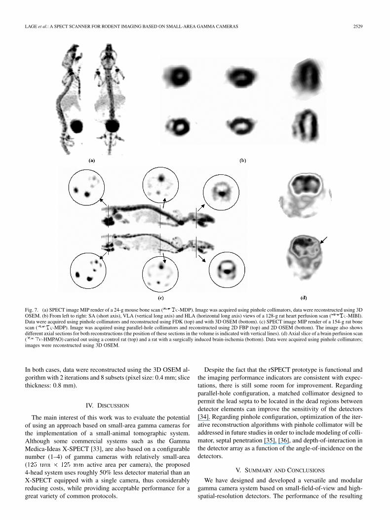
The spatial resolution was evaluated in tomographic imagesusing a micro-Derenzo hot-rods phantom (Ultra-Micro HotSpot Phantom, Data Spectrum Corporation, Hillsborough).This phantom has six sectors with rod diameters of 2.4, 2.0,1.7, 1.35, 1.0, and 0.75 mm. The phantom was filled with 4.5mCi of and acquired for two hours using the pinholecollimators equipped with the 0.75-mm aperture. A total of 120projections were acquired over 360 with a 33.5-mm radius ofrotation (ROR).
In order to assess the possibilities of the system for in vivoimaging, the rSPECT was tested in several common imagingprotocols.
a) The first set of experiments was based on -MDPtracer. Whole-body bone scans of a 154-g rat (5 axial po-sitions) and of a 24-g mouse (4 axial positions) were ac-quired with parallel-hole and pinhole collimators, respec-tively. Acquisitions were performed after displacement ofthe bed and ensuring a suitable overlap between two suc-cessive displacements. The rat was injected with 6.3 mCiand the mouse with 5.2 mCi of the aforementioned radio-pharmaceutical. After two hours uptake, 60 projectionswere acquired over 360 in each axial position (ROR: 37.2mm and 32.1 mm for rat and mouse, respectively). Thetotal scan time was two hours in both cases.
b) For the second experiment, a 1-hour heart perfusionscan was carried out using a 128-g rat and the pinholecollimators. The animal was injected with 3.8 mCi of
-MIBI. After one hour uptake, 120 projectionswere acquired over 360 (ROR: 31.3 mm).
c) Finally, a brain perfusion study was carried out using twoWistar rats and the pinhole collimators. Before acquisi-tion, brain-ischemia was surgically induced in one of theanimals. Eighteen hours after intervention both animalswere injected with 4 mCi of -HMPAO; after onehour uptake, 120 projections of 30 seconds were acquiredover 360 (ROR: 30.1 mm).
All the animal experiments were carried out according to theguidelines defined in the European directive 86/609/EEC on theprotection of animals used for experimental and other medicalpurposes. The animals were anesthetized with isoflurane duringthe scans.
III. RESULTS
A. Performance Evaluation
The performance of the implemented gamma cameras wascharacterized in terms of spatial resolution and sensitivity forparallel-hole and pinhole collimators (0.75-mm aperture). Inte-gral uniformity and energy resolution were evaluated withoutcollimators.
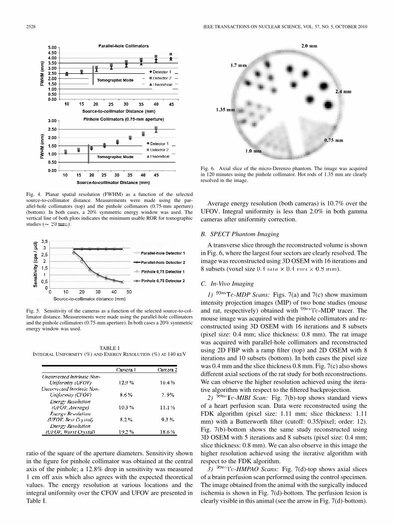
1) Spatial Resolution: The results of the planar resolutionmeasurements as a function of source-to-collimator distance areshown in Fig. 4. Theoretical spatial resolution was calculatedin both cases assuming a 1.6-mm intrinsic detector resolution.Differences in spatial resolution between both detectors werenegligible. Planar spatial resolution for parallel-hole collima-tors ranged from 2.4 mm when the source is at 10 mm to 4.1mm when the source-to-collimator distance is 45 mm. With thepinhole collimators, the spatial resolution ranged from 1.2 mmto 2.4 mm at the same distances.
2) Sensitivity: Fig. 5 shows the camera sensitivity with par-allel-hole and pinhole collimators (0.75-mm aperture). Thesevalues are in good agreement with the expected theoreticalvalues (calculated as the geometric efficiency of the collima-tors). The sensitivity of pinhole collimator equipped with biggerapertures (not yet available) is expected to be proportional tothat shown in Fig. 5, with the proportionality factor being the
2528 IEEE TRANSACTIONS ON NUCLEAR SCIENCE, VOL. 57, NO. 5, OCTOBER 2010
Fig. 4. Planar spatial resolution (FWHM) as a function of the selectedsource-to-collimator distance. Measurements were made using the par-allel-hole collimators (top) and the pinhole collimators (0.75-mm aperture)(bottom). In both cases, a 20% symmetric energy window was used. Thevertical line of both plots indicates the minimum usable ROR for tomographicstudies �� �� ���.
Fig. 5. Sensitivity of the cameras as a function of the selected source-to-col-limator distance. Measurements were made using the parallel-hole collimatorsand the pinhole collimators (0.75-mm aperture). In both cases a 20% symmetricenergy window was used.
TABLE IINTEGRAL UNIFORMITY (%) AND ENERGY RESOLUTION (%) AT 140 KEV
ratio of the square of the aperture diameters. Sensitivity shownin the figure for pinhole collimator was obtained at the centralaxis of the pinhole; a 12.8% drop in sensitivity was measured1 cm off axis which also agrees with the expected theoreticalvalues. The energy resolution at various locations and theintegral uniformity over the CFOV and UFOV are presented inTable I.
Fig. 6. Axial slice of the micro-Derenzo phantom. The image was acquiredin 120 minutes using the pinhole collimator. Hot rods of 1.35 mm are clearlyresolved in the image.
Average energy resolution (both cameras) is 10.7% over theUFOV. Integral uniformity is less than 2.0% in both gammacameras after uniformity correction.
B. SPECT Phantom Imaging
A transverse slice through the reconstructed volume is shownin Fig. 6, where the largest four sectors are clearly resolved. Theimage was reconstructed using 3D OSEM with 16 iterations and8 subsets (voxel size ).
C. In-Vivo Imaging
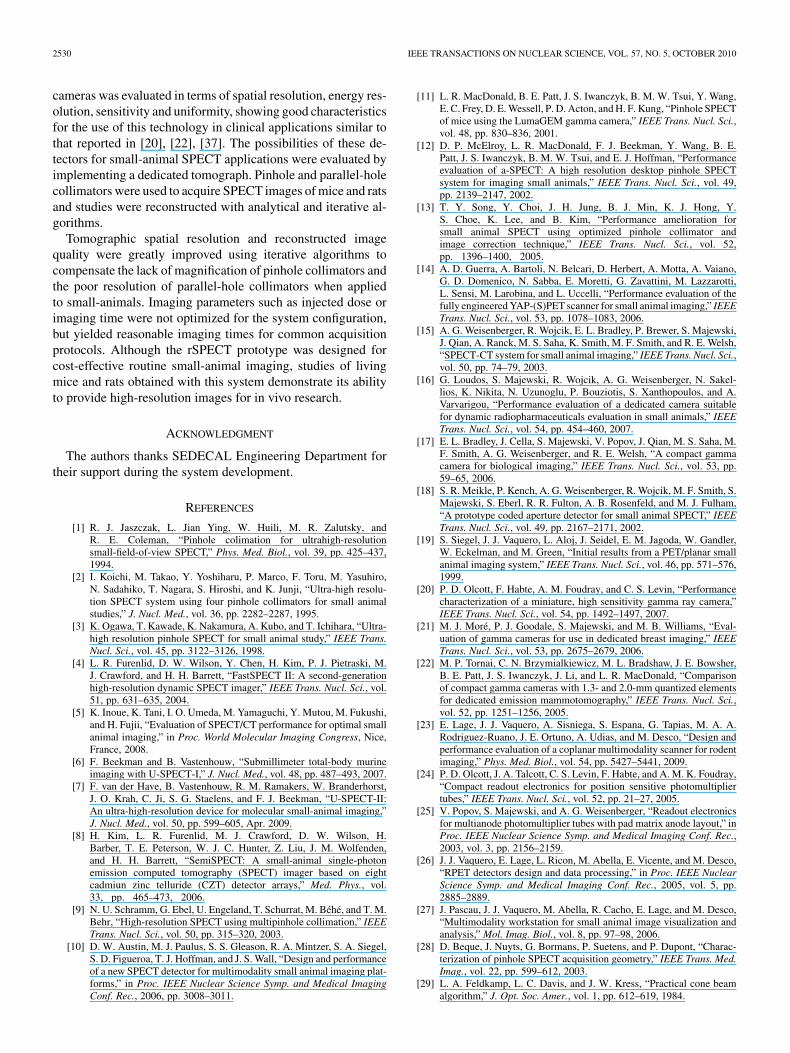
1) -MDP Scans: Figs. 7(a) and 7(c) show maximumintensity projection images (MIP) of two bone studies (mouseand rat, respectively) obtained with -MDP tracer. Themouse image was acquired with the pinhole collimators and re-constructed using 3D OSEM with 16 iterations and 8 subsets(pixel size: 0.4 mm; slice thickness: 0.8 mm). The rat imagewas acquired with parallel-hole collimators and reconstructedusing 2D FBP with a ramp filter (top) and 2D OSEM with 8iterations and 10 subsets (bottom). In both cases the pixel sizewas 0.4 mm and the slice thickness 0.8 mm. Fig. 7(c) also showsdifferent axial sections of the rat study for both reconstructions.We can observe the higher resolution achieved using the itera-tive algorithm with respect to the filtered backprojection.
2) -MIBI Scan: Fig. 7(b)-top shows standard viewsof a heart perfusion scan. Data were reconstructed using theFDK algorithm (pixel size: 1.11 mm; slice thickness: 1.11mm) with a Butterworth filter (cutoff: 0.35/pixel; order: 12).Fig. 7(b)-bottom shows the same study reconstructed using3D OSEM with 5 iterations and 8 subsets (pixel size: 0.4 mm;slice thickness: 0.8 mm). We can also observe in this image thehigher resolution achieved using the iterative algorithm withrespect to the FDK algorithm.
3) -HMPAO Scans: Fig. 7(d)-top shows axial slicesof a brain perfusion scan performed using the control specimen.The image obtained from the animal with the surgically inducedischemia is shown in Fig. 7(d)-bottom. The perfusion lesion isclearly visible in this animal (see the arrow in Fig. 7(d)-bottom).
LAGE et al.: A SPECT SCANNER FOR RODENT IMAGING BASED ON SMALL-AREA GAMMA CAMERAS 2529
Fig. 7. (a) SPECT image MIP render of a 24-g mouse bone scan ( ��-MDP). Image was acquired using pinhole collimators, data were reconstructed using 3DOSEM. (b) From left to right: SA (short axis), VLA (vertical long axis) and HLA (horizontal long axis) views of a 128-g rat heart perfusion scan ( ��-MIBI).Data were acquired using pinhole collimators and reconstructed using FDK (top) and with 3D OSEM (bottom). (c) SPECT image MIP render of a 154-g rat bonescan ( ��-MDP). Image was acquired using parallel-hole collimators and reconstructed using 2D FBP (top) and 2D OSEM (bottom). The image also showsdifferent axial sections for both reconstructions (the position of these sections in the volume is indicated with vertical lines). (d) Axial slice of a brain perfusion scan( ��-HMPAO) carried out using a control rat (top) and a rat with a surgically induced brain-ischemia (bottom). Data were acquired using pinhole collimators;images were reconstructed using 3D OSEM.
In both cases, data were reconstructed using the 3D OSEM al-gorithm with 2 iterations and 8 subsets (pixel size: 0.4 mm; slicethickness: 0.8 mm).
IV. DISCUSSION
The main interest of this work was to evaluate the potentialof using an approach based on small-area gamma cameras forthe implementation of a small-animal tomographic system.Although some commercial systems such as the GammaMedica-Ideas X-SPECT [33], are also based on a configurablenumber (1–4) of gamma cameras with relatively small-area( active area per camera), the proposed4-head system uses roughly 50% less detector material than anX-SPECT equipped with a single camera, thus considerablyreducing costs, while providing acceptable performance for agreat variety of common protocols.
Despite the fact that the rSPECT prototype is functional andthe imaging performance indicators are consistent with expec-tations, there is still some room for improvement. Regardingparallel-hole configuration, a matched collimator designed topermit the lead septa to be located in the dead regions betweendetector elements can improve the sensitivity of the detectors[34]. Regarding pinhole configuration, optimization of the iter-ative reconstruction algorithms with pinhole collimator will beaddressed in future studies in order to include modeling of colli-mator, septal penetration [35], [36], and depth-of-interaction inthe detector array as a function of the angle-of-incidence on thedetectors.
V. SUMMARY AND CONCLUSIONS
We have designed and developed a versatile and modulargamma camera system based on small-field-of-view and high-spatial-resolution detectors. The performance of the resulting
2530 IEEE TRANSACTIONS ON NUCLEAR SCIENCE, VOL. 57, NO. 5, OCTOBER 2010
cameras was evaluated in terms of spatial resolution, energy res-olution, sensitivity and uniformity, showing good characteristicsfor the use of this technology in clinical applications similar tothat reported in [20], [22], [37]. The possibilities of these de-tectors for small-animal SPECT applications were evaluated byimplementing a dedicated tomograph. Pinhole and parallel-holecollimators were used to acquire SPECT images of mice and ratsand studies were reconstructed with analytical and iterative al-gorithms.
Tomographic spatial resolution and reconstructed imagequality were greatly improved using iterative algorithms tocompensate the lack of magnification of pinhole collimators andthe poor resolution of parallel-hole collimators when appliedto small-animals. Imaging parameters such as injected dose orimaging time were not optimized for the system configuration,but yielded reasonable imaging times for common acquisitionprotocols. Although the rSPECT prototype was designed forcost-effective routine small-animal imaging, studies of livingmice and rats obtained with this system demonstrate its abilityto provide high-resolution images for in vivo research.
ACKNOWLEDGMENT
The authors thanks SEDECAL Engineering Department fortheir support during the system development.
REFERENCES
[1] R. J. Jaszczak, L. Jian Ying, W. Huili, M. R. Zalutsky, andR. E. Coleman, “Pinhole colimation for ultrahigh-resolutionsmall-field-of-view SPECT,” Phys. Med. Biol., vol. 39, pp. 425–437,1994.
[2] I. Koichi, M. Takao, Y. Yoshiharu, P. Marco, F. Toru, M. Yasuhiro,N. Sadahiko, T. Nagara, S. Hiroshi, and K. Junji, “Ultra-high resolu-tion SPECT system using four pinhole collimators for small animalstudies,” J. Nucl. Med., vol. 36, pp. 2282–2287, 1995.
[3] K. Ogawa, T. Kawade, K. Nakamura, A. Kubo, and T. Ichihara, “Ultra-high resolution pinhole SPECT for small animal study,” IEEE Trans.Nucl. Sci., vol. 45, pp. 3122–3126, 1998.
[4] L. R. Furenlid, D. W. Wilson, Y. Chen, H. Kim, P. J. Pietraski, M.J. Crawford, and H. H. Barrett, “FastSPECT II: A second-generationhigh-resolution dynamic SPECT imager,” IEEE Trans. Nucl. Sci., vol.51, pp. 631–635, 2004.
[5] K. Inoue, K. Tani, I. O. Umeda, M. Yamaguchi, Y. Mutou, M. Fukushi,and H. Fujii, “Evaluation of SPECT/CT performance for optimal smallanimal imaging,” in Proc. World Molecular Imaging Congress, Nice,France, 2008.
[6] F. Beekman and B. Vastenhouw, “Submillimeter total-body murineimaging with U-SPECT-I,” J. Nucl. Med., vol. 48, pp. 487–493, 2007.
[7] F. van der Have, B. Vastenhouw, R. M. Ramakers, W. Branderhorst,J. O. Krah, C. Ji, S. G. Staelens, and F. J. Beekman, “U-SPECT-II:An ultra-high-resolution device for molecular small-animal imaging,”J. Nucl. Med., vol. 50, pp. 599–605, Apr. 2009.
[8] H. Kim, L. R. Furenlid, M. J. Crawford, D. W. Wilson, H.Barber, T. E. Peterson, W. J. C. Hunter, Z. Liu, J. M. Wolfenden,and H. H. Barrett, “SemiSPECT: A small-animal single-photonemission computed tomography (SPECT) imager based on eightcadmiun zinc telluride (CZT) detector arrays,” Med. Phys., vol.33, pp. 465–473, 2006.
[9] N. U. Schramm, G. Ebel, U. Engeland, T. Schurrat, M. Béhé, and T. M.Behr, “High-resolution SPECT using multipinhole collimation,” IEEETrans. Nucl. Sci., vol. 50, pp. 315–320, 2003.
[10] D. W. Austin, M. J. Paulus, S. S. Gleason, R. A. Mintzer, S. A. Siegel,S. D. Figueroa, T. J. Hoffman, and J. S. Wall, “Design and performanceof a new SPECT detector for multimodality small animal imaging plat-forms,” in Proc. IEEE Nuclear Science Symp. and Medical ImagingConf. Rec., 2006, pp. 3008–3011.
[11] L. R. MacDonald, B. E. Patt, J. S. Iwanczyk, B. M. W. Tsui, Y. Wang,E. C. Frey, D. E. Wessell, P. D. Acton, and H. F. Kung, “Pinhole SPECTof mice using the LumaGEM gamma camera,” IEEE Trans. Nucl. Sci.,vol. 48, pp. 830–836, 2001.
[12] D. P. McElroy, L. R. MacDonald, F. J. Beekman, Y. Wang, B. E.Patt, J. S. Iwanczyk, B. M. W. Tsui, and E. J. Hoffman, “Performanceevaluation of a-SPECT: A high resolution desktop pinhole SPECTsystem for imaging small animals,” IEEE Trans. Nucl. Sci., vol. 49,pp. 2139–2147, 2002.
[13] T. Y. Song, Y. Choi, J. H. Jung, B. J. Min, K. J. Hong, Y.S. Choe, K. Lee, and B. Kim, “Performance amelioration forsmall animal SPECT using optimized pinhole collimator andimage correction technique,” IEEE Trans. Nucl. Sci., vol. 52,pp. 1396–1400, 2005.
[14] A. D. Guerra, A. Bartoli, N. Belcari, D. Herbert, A. Motta, A. Vaiano,G. D. Domenico, N. Sabba, E. Moretti, G. Zavattini, M. Lazzarotti,L. Sensi, M. Larobina, and L. Uccelli, “Performance evaluation of thefully engineered YAP-(S)PET scanner for small animal imaging,” IEEETrans. Nucl. Sci., vol. 53, pp. 1078–1083, 2006.
[15] A. G. Weisenberger, R. Wojcik, E. L. Bradley, P. Brewer, S. Majewski,J. Qian, A. Ranck, M. S. Saha, K. Smith, M. F. Smith, and R. E. Welsh,“SPECT-CT system for small animal imaging,” IEEE Trans. Nucl. Sci.,vol. 50, pp. 74–79, 2003.
[16] G. Loudos, S. Majewski, R. Wojcik, A. G. Weisenberger, N. Sakel-lios, K. Nikita, N. Uzunoglu, P. Bouziotis, S. Xanthopoulos, and A.Varvarigou, “Performance evaluation of a dedicated camera suitablefor dynamic radiopharmaceuticals evaluation in small animals,” IEEETrans. Nucl. Sci., vol. 54, pp. 454–460, 2007.
[17] E. L. Bradley, J. Cella, S. Majewski, V. Popov, J. Qian, M. S. Saha, M.F. Smith, A. G. Weisenberger, and R. E. Welsh, “A compact gammacamera for biological imaging,” IEEE Trans. Nucl. Sci., vol. 53, pp.59–65, 2006.
[18] S. R. Meikle, P. Kench, A. G. Weisenberger, R. Wojcik, M. F. Smith, S.Majewski, S. Eberl, R. R. Fulton, A. B. Rosenfeld, and M. J. Fulham,“A prototype coded aperture detector for small animal SPECT,” IEEETrans. Nucl. Sci., vol. 49, pp. 2167–2171, 2002.
[19] S. Siegel, J. J. Vaquero, L. Aloj, J. Seidel, E. M. Jagoda, W. Gandler,W. Eckelman, and M. Green, “Initial results from a PET/planar smallanimal imaging system,” IEEE Trans. Nucl. Sci., vol. 46, pp. 571–576,1999.
[20] P. D. Olcott, F. Habte, A. M. Foudray, and C. S. Levin, “Performancecharacterization of a miniature, high sensitivity gamma ray camera,”IEEE Trans. Nucl. Sci., vol. 54, pp. 1492–1497, 2007.
[21] M. J. Moré, P. J. Goodale, S. Majewski, and M. B. Williams, “Eval-uation of gamma cameras for use in dedicated breast imaging,” IEEETrans. Nucl. Sci., vol. 53, pp. 2675–2679, 2006.
[22] M. P. Tornai, C. N. Brzymialkiewicz, M. L. Bradshaw, J. E. Bowsher,B. E. Patt, J. S. Iwanczyk, J. Li, and L. R. MacDonald, “Comparisonof compact gamma cameras with 1.3- and 2.0-mm quantized elementsfor dedicated emission mammotomography,” IEEE Trans. Nucl. Sci.,vol. 52, pp. 1251–1256, 2005.
[23] E. Lage, J. J. Vaquero, A. Sisniega, S. Espana, G. Tapias, M. A. A.Rodriguez-Ruano, J. E. Ortuno, A. Udias, and M. Desco, “Design andperformance evaluation of a coplanar multimodality scanner for rodentimaging,” Phys. Med. Biol., vol. 54, pp. 5427–5441, 2009.
[24] P. D. Olcott, J. A. Talcott, C. S. Levin, F. Habte, and A. M. K. Foudray,“Compact readout electronics for position sensitive photomultipliertubes,” IEEE Trans. Nucl. Sci., vol. 52, pp. 21–27, 2005.
[25] V. Popov, S. Majewski, and A. G. Weisenberger, “Readout electronicsfor multianode photomultiplier tubes with pad matrix anode layout,” inProc. IEEE Nuclear Science Symp. and Medical Imaging Conf. Rec.,2003, vol. 3, pp. 2156–2159.
[26] J. J. Vaquero, E. Lage, L. Ricon, M. Abella, E. Vicente, and M. Desco,“RPET detectors design and data processing,” in Proc. IEEE NuclearScience Symp. and Medical Imaging Conf. Rec., 2005, vol. 5, pp.2885–2889.
[27] J. Pascau, J. J. Vaquero, M. Abella, R. Cacho, E. Lage, and M. Desco,“Multimodality workstation for small animal image visualization andanalysis,” Mol. Imag. Biol., vol. 8, pp. 97–98, 2006.
[28] D. Beque, J. Nuyts, G. Bormans, P. Suetens, and P. Dupont, “Charac-terization of pinhole SPECT acquisition geometry,” IEEE Trans. Med.Imag., vol. 22, pp. 599–612, 2003.
[29] L. A. Feldkamp, L. C. Davis, and J. W. Kress, “Practical cone beamalgorithm,” J. Opt. Soc. Amer., vol. 1, pp. 612–619, 1984.
LAGE et al.: A SPECT SCANNER FOR RODENT IMAGING BASED ON SMALL-AREA GAMMA CAMERAS 2531
[30] H. M. Hudson and R. S. Larkin, “Accelerated image reconstructionusing ordered subsets of projection data,” IEEE Trans. Med. Imag., vol.13, pp. 601–609, 1994.
[31] D. Pareto, A. Cot, J. Pavia, C. Falcon, I. Juvells, F. Lomena, and D.Ros, “Iterative reconstruction with compensation of the spatial variantfan beam collimator response in neurotransmission SPET imaging,”Eur. J. Nucl. Med. Mol. Imag., vol. 30, pp. 1322–1329, 2003.
[32] A. Cot, C. Falcon, C. Crespo, C. J. Sempau, D. Pareto, S. Bullich, F.Lomena, F. Calvino, J. Pavia, and D. Ros, “Absolute quantification indopaminergic neurotransmission SPECT using a monte carlo-basedscatter correction and fully 3-dimensional reconstruction,” J. Nucl.Med., vol. 46, pp. 1497–1504, 2005.
[33] Gamma Medica-Ideas [Online]. Available: http://www.gm-ideas.com[34] W. Xia, J. Seidel, J. W. Kakarekad, T. J. Pohidad, D. E. Milenice, J.
Proffittf, S. Majewski, A. G. Weisenberger, M. V. Green, and P. L.Choykeb, “MONICA: a compact, portable dual gamma camera systemfor mouse whole-body imaging,” Nucl. Med. Biol., vol. 1, pp. 245–253,2010.
[35] R. Accorsi and S. D. Metzler, “Analytic determination of the resolu-tion-equivalent effective diameter of a pinhole collimator,” IEEE Trans.Med. Imag., vol. 23, pp. 750–763, 2004.
[36] S. D. Metzler and R. Accorsi, “Resolution- versus sensitivity-effec-tive diameter in pinhole collimation: Experimental verification,” Phys.Med. Biol., vol. 50, pp. 5005–5017, 2005.
[37] F. Sánchez, J. M. Benlloch, B. Escat, N. Pavón, E. Porras, D. Kadi-Hanifi, J. A. Ruiz, F. J. Mora, and A. Sebastia, “Design and tests ofa portable mini gamma camera,” Med. Phys., vol. 31, pp. 1384–1397,2004.