A Sixteenth-Century Warrior Grave from Uppsala, Sweden: the Battle of Good Friday
28
International Journal of Osteoarchaeology Int. J. Osteoarchaeol. 15: 23–50 (2005) Published online 6 September 2004 in Wiley InterScience (www.interscience.wiley.com). DOI: 10.1002/oa.746 A Sixteenth-Century Warrior Grave from Uppsala, Sweden: the Battle of Good Friday A. KJELLSTRO ¨ M* Osteoarchaeological Research Laboratory, Department of Archaeology, Stockholm University, Royal Palace Ulriksdal, SE-170 79 Solna, Sweden ABSTRACT Little is known about the Battle of Good Friday in Uppsala. The historical records are scarce and of limited extent. Moreover, the more spectacular event of the Stockholm Bloodbath has drawn most of the attention from both the contemporary public and later historians. This is why the discovery of a mass grave in the steep slope of Uppsala Castle in 2001 has provoked much interest. An analysis of the osseous material showed that the remains of at least 60 male individuals, mostly between 25–34 years of age, were buried in the excavated area. The demographic profile is largely similar to other European war-related skeletal assemblages of the same era. Sharp force trauma was exhibited primarily on the skulls, with no obvious dominance to either side. The trauma distribution pattern suggests that the battle was not fought face-to-face. Blade wounds concentrated in specific regions imply a standardised technique when delivering the blows. The combination of commingled bones and articulated elements suggests that the individuals were in different stages of skeletonisation when buried. Copyright ß 2004 John Wiley & Sons, Ltd. Key words: Uppsala; Battle of Good Friday; mass grave; commingled bones; sharp force trauma Introduction and historical background At the end of the 14th century a union between Sweden, Denmark and Norway was established, causing political unrest in Scandinavia. The final abandonment of the union between Denmark (Norway) and Sweden during the early 16th century caused an outbreak of violent campaigns. Sten Sture (the Younger), the Swedish adminis- trator, had incited the peasantry against the Danish King Kristian II. For several decades, Swedish peasant forces met the Danish mercen- aries in violent encounters. The Swedish archbishop in Uppsala, Gustav Trolle, was allied to the Danish king who had armed forces stationed in and around the town (Heise & Mollerup, 1903–1906; Kjersgaard & Hvidtfeldt, 1963). In 1519, with the support of the Pope, Trolle excommunicated Sten Sture and his associates (Syse, 2003). In March 1520 Sten Sture died in battle on the ice of Lake A ˚ sunden, but his troops continued their campaign against the Danish king. Later that spring, the conflict led to a frightening encounter known as the Battle of Good Friday. On the morning of 6 April in 1520, Good Friday, Swedish troops attacked the Danish troops stationed in the town of Uppsala. Con- temporary sources mention that the weather was cold and snowy, which made the battle situation less than ideal (Olaus Petri, cited in Sahlgren 1917). The slippery soil is said to have made the Copyright # 2004 John Wiley & Sons, Ltd. Received 7 January 2003 Revised 25 March 2003 Accepted 13 February 2004 * Correspondence to: Osteoarchaeological Research Laboratory, Department of Archaeology, Stockholm University, Royal Palace Ulriksdal, SE-170 79 Solna, Sweden. e-mail: [email protected]
Transcript of A Sixteenth-Century Warrior Grave from Uppsala, Sweden: the Battle of Good Friday

International Journal of OsteoarchaeologyInt. J. Osteoarchaeol. 15: 23–50 (2005)Published online 6 September 2004 in Wiley InterScience (www.interscience.wiley.com). DOI: 10.1002/oa.746
ASixteenth-CenturyWarrior GravefromUppsala,Sweden: the Battleof GoodFriday
A. KJELLSTROM*Osteoarchaeological Research Laboratory, Department of Archaeology,
Stockholm University, Royal Palace Ulriksdal, SE-170 79 Solna, Sweden
ABSTRACT Little is known about the Battle of Good Friday in Uppsala. The historical records are scarceand of limited extent. Moreover, the more spectacular event of the Stockholm Bloodbath hasdrawn most of the attention from both the contemporary public and later historians. This is whythe discovery of a mass grave in the steep slope of Uppsala Castle in 2001 has provokedmuch interest. An analysis of the osseous material showed that the remains of at least 60 maleindividuals, mostly between 25–34 years of age, were buried in the excavated area. Thedemographic profile is largely similar to other European war-related skeletal assemblages ofthe same era. Sharp force trauma was exhibited primarily on the skulls, with no obviousdominance to either side. The trauma distribution pattern suggests that the battle was notfought face-to-face. Blade wounds concentrated in specific regions imply a standardisedtechnique when delivering the blows. The combination of commingled bones and articulatedelements suggests that the individuals were in different stages of skeletonisation when buried.Copyright � 2004 John Wiley & Sons, Ltd.
Key words: Uppsala; Battle of Good Friday; mass grave; commingled bones; sharp force
trauma
Introduction and historicalbackground
At the end of the 14th century a union betweenSweden, Denmark and Norway was established,causing political unrest in Scandinavia. The finalabandonment of the union between Denmark(Norway) and Sweden during the early 16thcentury caused an outbreak of violent campaigns.Sten Sture (the Younger), the Swedish adminis-trator, had incited the peasantry against theDanish King Kristian II. For several decades,Swedish peasant forces met the Danish mercen-aries in violent encounters.
The Swedish archbishop in Uppsala, GustavTrolle, was allied to the Danish king who hadarmed forces stationed in and around the town(Heise & Mollerup, 1903–1906; Kjersgaard &Hvidtfeldt, 1963). In 1519, with the support ofthe Pope, Trolle excommunicated Sten Sture andhis associates (Syse, 2003). In March 1520 StenSture died in battle on the ice of Lake Asunden,but his troops continued their campaign againstthe Danish king. Later that spring, the conflictled to a frightening encounter known as theBattle of Good Friday.
On the morning of 6 April in 1520, GoodFriday, Swedish troops attacked the Danishtroops stationed in the town of Uppsala. Con-temporary sources mention that the weather wascold and snowy, which made the battle situationless than ideal (Olaus Petri, cited in Sahlgren1917). The slippery soil is said to have made the
Copyright # 2004 John Wiley & Sons, Ltd. Received 7 January 2003Revised 25 March 2003
Accepted 13 February 2004
* Correspondence to: Osteoarchaeological Research Laboratory,Department of Archaeology, Stockholm University, Royal PalaceUlriksdal, SE-170 79 Solna, Sweden.e-mail: [email protected]
horses stumble, and the slushy weather made theuse of firearms impossible. The battle was notfought on equal terms. The Swedish forces alliedwith Sture consisted of organised peasant forceswith scarce knowledge of war tactics. The Danishsoldiers, on the other hand, were paid mercen-aries from Germany, France and Scotland(Sandstedt, 2003). The weaponry of the medievalSwedish peasant forces varied. Generally,though, in this kind of close combat, the requiredminimum was a sword, a shield and some kind ofhelmet (Sandstedt, 2003). However, as a conse-quence of the development in weapon technol-ogy, chain mails and breastplates, crossbows,bills, poleaxes and glaives had become morecommon during the 15th and 16th centuries. Avariety of helmets were used to protect the head,from simple chainmail coifs to sallats and kettlehats. Among the technically more advancedDanish mercenaries, half-armours, sallets, shortswords, halberds and pikes were used in closecontact with the enemy (Oakeshott, 1980;Sandstedt, 2003). On both sides the weaponsand armour varied according to the rank and typeof soldier, such as mounted men or foot soldiers.
Even though the Swedish troops were success-ful in the early morning hours of the battle, theirluck changed. At the end of the day the Danishtroops defeated the armed farmer troops. It hasbeen stated that around 2000 men were killed onthe Danish side, and that the losses were evengreater among the Swedes (Berg & Gafvert,2000).
Contemporary sources give some informationon the Battle of Good Friday. The 16th centuryLutheran reformer Olaus Petri stated that severalSwedes drowned, going through the ice on thestream Fyrisan (cited in Sahlgren, 1917). Further-more, Petri mentions that some soldiers werekilled in a fire in a brick barn. It has also beenclaimed that, by a decision of the archbishopTrolle, the corpses belonging to the Swedishtroops were left in bogs and marshlands (quotedby Heise & Mollerup, 1903–1906; Kjersgaard &Hvidtfeldt, 1963).
The exact location of the battle is not known,but pieces of information have been gatheredfrom contemporary sources. Moreover, the topo-graphical appearance of Uppsala at the time,archaeological finds of brick ovens (presumably
from a brick barn) and the belief that theencounter happened on a large, open flat area,point towards the area today known asStadstradgarden (Syse, 2003). This neighbour-hood was close to the outskirts of the contem-porary city. During an excavation of the area in1971, several articulated skeletons were found(Syse, 2003), but unfortunately these skeletonswere not recovered.
The documentation dealing with the burial ofthe fallen men after the battle is scarce. Themercenaries are said to have been buried atUppsala Cathedral and the churchyard adjacentto it (Heise & Mollerup, 1903–1906). However,no exact location for such graves has been iden-tified. The information that Trolle ordered thatthe fallen Swedes should be left unburied in bogsto ‘dogs and ravens’ (Kjersgaard & Hvidtfeldt,1963) must be considered when trying to locatethe burials of the fallen Swedes. Furthermore, inNovember of 1520, enemies of the Danish admi-nistration were executed in Stockholm. Thisevent is known as the Stockholm Bloodbath. Ithas been suggested that the bodies from the Battleof Good Friday got a final burial in conjunctionwith this event (Syse, 2003). The Swedishprisoners captured during or after the battle inApril may have been executed at the same time.
In May 2001, only tens of metres from theskeletal finds discovered in the 1970s, humanremains were unearthed during the broadeningof a small gravelled road. In order to get a reliabledating, bones were submitted for radiocarbonanalysis. The result dated the bones to 355�50 BP (Ua-17998), which calibrates to 1440–1650 AD at 95% probability (Syse, 2003). Onthe basis of the dating and location of the grave,the skeletal assemblage could be associatedwith the Battle of Good Friday. It was decidedto investigate the find location in more detail,and in August the same year an excavation super-vised by the antiquarian Bent Syse at the provin-cial museum Upplandsmuseet was performed.Except for the osseous material, only a fewartefacts were found. Pieces of charcoal, a dicemade of bone or antler and fragments of medievaltiles may all come from the covering layers, andnot from the buried individuals (Syse, 2003). Theonly discovery that may be connected to warfareis a narrow-ended piece of iron.
24 A. Kjellstrom
Copyright # 2004 John Wiley & Sons, Ltd. Int. J. Osteoarchaeol. 15: 23–50 (2005)

At an early stage of the discovery, it wasdecided to adopt a multidisciplinary approachin the analysis of the excavation. The antiquarianBent Syse investigated the historical and archae-ological context. The director of the nationaltrophy collection at the Army Museum, weaponhistorian Fred Sandstedt, reviewed weaponry andtactics of contemporary warfare. All discoveredteeth were analysed by the forensic odontologistHelene Borrman, and the author examined theosseous material. The results from the excavationand the subsequent analysis led to a joint pub-lication Langfredagsslaget—En arkeologisk historia(Syse, 2003).
This paper summarises the results of the osteo-logical analysis. The demography of the Uppsalamass grave is reviewed. Special emphasis is givento signs of sharp force trauma inflicted by edgedmetal weapons observed on the skeletal elements.The wounds and the wound pattern are examinedand documented in detail. The analysis of theperimortem trauma aims to reconstruct the vio-lent fate, prior to death, of the individuals.Comparisons are made with other war-relatedskeletal assemblages to illustrate differences andcommon features. Furthermore, an attempt ismade to give a reasonable explanation of thetaphonomic history of the assemblage.
War-related mass graves—thereference sample
European assemblages of human remains derivedfrom battles dated to the same era as the Battle ofGood Friday are rare. However, discoveries andanthropological studies of warrior mass gravessimilar to the one in Uppsala have been doneelsewhere. The skeletal assemblage from theBattle of Wisby, AD 1361, with 1185 documen-ted skeletons, is one of the most discussed histor-ical mass graves (Ingelmark, 1939). In Portugal,commingled remains from at least 400 individualsfrom the Battle of Aljubarrota, AD 1385, havebeen examined (Cunha & Silva, 1997). Medievalremains from 60 individuals have been documen-ted from Sandbjerget in Denmark (Bennike,1998). One of the most recent studies of histor-ical war-related mass graves is the multidisciplin-ary analysis of 38 individuals discovered in a mass
grave from the Battle of Towton, AD 1461(Fiorato et al., 2000). Moreover, there are boneassemblages which are not from true mass gravesbut share several common features with thematerials mentioned above. For example, about179 skeletons have been analysed from HenryVIII’s flagship Mary Rose which sank off Ports-mouth in 1545 (Stirland, 2000). In Sweden,skeletal elements from c. 200 men have beenidentified from the Royal warship Kronan whichexploded and sank in the Baltic off the island ofOland in the summer of 1676 (During, 1997).Both assemblages represent, like the mass graves,individuals who died at a single event and moreor less from the same cause.
The demographic profile of the above-mentioned assemblages is in general specialised,with predominantly young males. Striking in allcases, except for the drowned men from MaryRose, are the signs of multiple perimortem trau-matic lesions on the individuals. The specificlesion pattern and the distribution of wounds ineach assemblage have revealed information aboutthe particular encounter, as well as the closecombat situation. Specific wounds have enabledthe identification of weapon types used. Thenumber of wounds and their lethal potentialhave suggested the intensity of the battles.
Material and methods
The excavation
An area of 8 m2 was investigated adjacent to thearea where the human remains were first recov-ered (Figure 1). In this area two large pits, A2 andA4, were recovered. In addition, four more or lesscomplete skeletons and commingled bones werefound in an upper layer separated from the pit A4(Figure 2).
The more recent of the pits, A2, was1.8� 0.70 m (depth 0.18 m) with an east–westorientation. The total extent of the pit wasinvestigated. The excavation revealed a deliber-ate attempt to organise the deposition of thebones in the pit. In the western part a total ofseven crania were gathered and in the easternpart all the femora and tibiae had been orientedalong the margin of the grave (Figure 3). Among
A Swedish Warrior Grave 25
Copyright # 2004 John Wiley & Sons, Ltd. Int. J. Osteoarchaeol. 15: 23–50 (2005)

the commingled bones the remains of at least twoarticulated skeletons were recovered. These indi-viduals were placed according to the traditionalChristian orientation (east–west, heads to thewest).
The pit A4 was found in the eastern part of theexcavated area. The total extent of the pit is notknown since the margins to the north and southcontinued outside of the excavated area. A mod-ern road cut the pit in the east. This pit was largerthan A2, covering almost a third of the excavatedarea, with a depth of at least 0.8 m (Syse, 2003).In the top layers four articulated skeletons buriedin an east–west orientation (heads to the west)were found. No attempt to organise the com-mingled bone elements or complete limbs couldbe seen in A4. As in A2, the exposed part of A4was examined to the bottom.
Around 0.4 m below the bottom layer of A4,yet another commingled bone layer appearedwhich was c. 0.3 m thick. This layer was leftuntouched. However, a radiocarbon analysis
(Ua-20084) dated the bones to the same periodas A4 and A2 (Syse, 2003). Thus, the stratigraphyindicates that, at first, a layer of commingledskeletal elements had been placed directly onthe ground or in a shallow pit. This bone layerhad then been covered by soil and upon this layerthe pit A4 was placed. A2 was later, althoughapparently close in time, and placed to the westof A4. In the top layer of the area four more orless complete articulated skeletons were buried.
The total extent of the whole grave area is notknown. The structure of the current grave pits,taken together with the previous discovery ofhuman remains in the 1970s and the appearanceof the topography, suggest that the area couldhold several grave pits similar to those recoveredin the excavations. It has been estimated that atleast 50 mass burials, similar to the present grave,may exist in the vicinity over an area of 200–300 m2 large area (Syse, 2003). However, atpresent any further archaeological investigationsare not planned.
Figure 1. Map showing the location of the excavated grave (arrow). In connection with an earlier investigation in 1971, skeletons were
discovered about10m to the west of the present grave. (Reproducedby permission of Upplandsmuseet.)
26 A. Kjellstrom
Copyright # 2004 John Wiley & Sons, Ltd. Int. J. Osteoarchaeol. 15: 23–50 (2005)

Figure 2. The excavated area with pits A2 and A4 marked. In A4 are shown four, more or less complete individuals from the top layer.
(Reproduced by permission of Upplandsmuseet.)
Figure 3. Detail of A2 with a mix of both commingled crania and articulated elements. Note the articulated ribs from the left side of a rib
cage andarticulated vertebrae to the right. (Reproducedby permission of Upplandsmuseet.)
A Swedish Warrior Grave 27
Copyright # 2004 John Wiley & Sons, Ltd. Int. J. Osteoarchaeol. 15: 23–50 (2005)
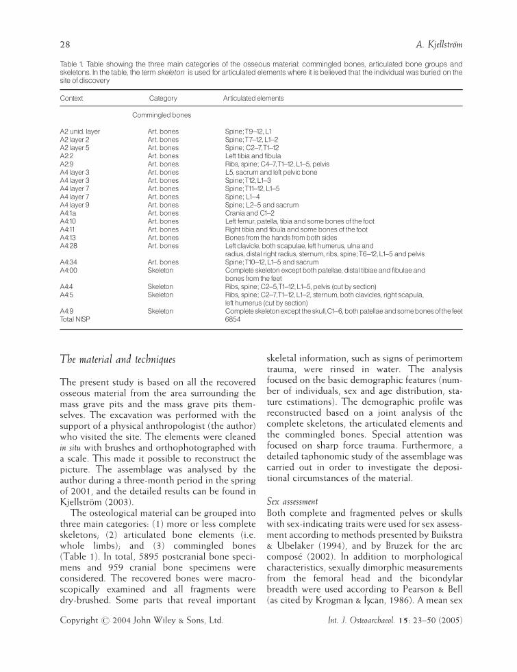
The material and techniques
The present study is based on all the recoveredosseous material from the area surrounding themass grave pits and the mass grave pits them-selves. The excavation was performed with thesupport of a physical anthropologist (the author)who visited the site. The elements were cleanedin situ with brushes and orthophotographed witha scale. This made it possible to reconstruct thepicture. The assemblage was analysed by theauthor during a three-month period in the springof 2001, and the detailed results can be found inKjellstrom (2003).
The osteological material can be grouped intothree main categories: (1) more or less completeskeletons; (2) articulated bone elements (i.e.whole limbs); and (3) commingled bones(Table 1). In total, 5895 postcranial bone speci-mens and 959 cranial bone specimens wereconsidered. The recovered bones were macro-scopically examined and all fragments weredry-brushed. Some parts that reveal important
skeletal information, such as signs of perimortemtrauma, were rinsed in water. The analysisfocused on the basic demographic features (num-ber of individuals, sex and age distribution, sta-ture estimations). The demographic profile wasreconstructed based on a joint analysis of thecomplete skeletons, the articulated elements andthe commingled bones. Special attention wasfocused on sharp force trauma. Furthermore, adetailed taphonomic study of the assemblage wascarried out in order to investigate the deposi-tional circumstances of the material.
Sex assessmentBoth complete and fragmented pelves or skullswith sex-indicating traits were used for sex assess-ment according to methods presented by Buikstra& Ubelaker (1994), and by Bruzek for the arccompose (2002). In addition to morphologicalcharacteristics, sexually dimorphic measurementsfrom the femoral head and the bicondylarbreadth were used according to Pearson & Bell(as cited by Krogman & Iscan, 1986). A mean sex
Table 1. Table showing the three main categories of the osseous material: commingled bones, articulated bone groups andskeletons. In the table, the term skeleton is used forarticulated elements where it is believed that the individual was buried on thesite ofdiscovery
Context Category Articulated elements
Commingledbones
A2unid. layer Art. bones Spine;T9 12,L1
A2 layer 2 Art. bones Spine;T7 12,L1 2
A2 layer 5 Art. bones Spine; C2^7,T1 12
A2:2 Art. bones Left tibiaand fibula
A2:9 Art. bones Ribs, spine; C4^7,T1 12,L1 5, pelvis
A4 layer 3 Art. bones L5, sacrumand left pelvicbone
A4 layer 3 Art. bones Spine;T12,L1 3
A4 layer 7 Art. bones Spine;T11 12,L1 5
A4 layer 7 Art. bones Spine; L1 4
A4 layer 9 Art. bones Spine; L2^5 andsacrum
A4:1a Art. bones CraniaandC1 2
A4:10 Art. bones Left femur, patella, tibiaandsomebonesof the foot
A4:11 Art. bones Right tibiaand fibulaandsomebonesof the foot
A4:13 Art. bones Bones from thehands fromboth sides
A4:28 Art. bones Left clavicle, both scapulae, left humerus, ulnaand
radius, distal right radius, sternum, ribs, spine;T6 12,L1 5 andpelvis
A4:34 Art. bones Spine;T10 12,L1 5 andsacrum
A4:00 Skeleton Complete skeleton except bothpatellae, distal tibiaeand fibulaeand
bones from the feet
A4:4 Skeleton Ribs, spine; C2^5,T1 12,L1 5, pelvis (cut by section)
A4:5 Skeleton Ribs, spine; C2^7,T1 12,L1 2, sternum, both clavicles, right scapula,
left humerus (cut by section)
A4:9 Skeleton Completeskeletonexcept theskull,C1 6,bothpatellaeandsomebonesofthefeet
Total NISP 6854
28 A. Kjellstrom
Copyright # 2004 John Wiley & Sons, Ltd. Int. J. Osteoarchaeol. 15: 23–50 (2005)

value was calculated for each element, and theelement, was assigned to one of five possible sexcategories: F, female; F?, probable female; ?,ambiguous sex; M?, probable male; M, male(Kjellstrom, 2003). The method is modifiedfrom Acsadi & Nemeskeri (1970).
Age estimationThe age of the individuals was estimated on thebasis of traits on the skull, teeth and pelvis. Onthe skull, the ectocranial suture closure (Meindl &Lovejoy, 1985) and dental attrition (Brothwell,1981) were examined. Development of thespheno-occipital synchondrosis according toSahni et al. (1998) was considered. On the pelvis,age was estimated using the changes of the pubicsymphysis (Brooks & Suchey, 1990) and theauricular surface (Lovejoy et al., 1985). The devel-opment of the iliac crest according to Bass (1987)was taken into account. A mean age interval wasthen calculated for crania and pelves (Kjellstrom,2003). In addition, epiphyseal fusion of theremaining elements was registered in three clo-sure stages (0: open; 1: closing; 2: closed). Theage estimation for epiphyseal fusion was based onBrothwell (1981).
Metrics, robusticity and statureFor both the skulls and the long bones, measure-ments were taken according to Buikstra &Ubelaker (1994), using sliding callipers and anosteometric board. The femoral robusticity indexpresented by Bass (1987) is based on calculationsfor left-sided femora. The height of the indivi-duals was calculated from the long bones, usingthe regression formulas presented by Trotter &Gleser (1952, 1958) and Sjøvold (1990). Sinceknowledge of sex and ethnicity is expected whenTrotter & Gleser calculations are used, Sjøvold’smethod, which does not take these factors intoaccount, is used as a complementary estimation.Measurements for the skulls and the long bones,other than those used for the stature estimations,are found in the Appendix, Tables I–III, togetherwith those for the skulls.
Sharp force traumaCuts were identified according to characteristicspresented by Wenham (1989), Reichs (1998),
Symes et al. (1998) and Houck (1998). Thediagnostic criteria were:
(a) a linear lesion with well-defined sharp edges;(b) v-shape in cross-section;(c) flat, smooth and polished cut surfaces: these
can be seen on both sides of the bone ifthe weapon entered in a right angle, if theweapon entered at another angle, the obtuse-angled side shows a smooth surface while theacute side shows signs of flaking;
(d) signs of parallel striations, perpendicular tothe kerf floor, which can be seen macrosco-pically or microscopically.
When analysing perimortem trauma, any signs ofhealing, such as rounded cut edges or changesdue to infection, were noted.
In the cases where a blade wound was visibleto the naked eye on different cranial bones, aneffort was made to reconstruct the skulls. Foreach cut mark, the appearance, side, locationand affected element were registered. Further-more, the cut wounds were allocated into threegroups according to their orientation: vertical,horizontal or oblique. The angle and striationof the cut surfaces were used as indicators of thedirection of the blow (Boylston, 2000), whetherit was delivered from above, below or perpen-dicular to the bone. The fact that the traumawas superficial or had penetrated the bone wasnoted. On the neurocrania, the latter showedthat both the tabula externa and interna wereintersected. For the bones of the face, traumalesions were documented as penetrating if thesinus cavities had been affected. All the surfacesof sharp force trauma were examined through amicroscope at approximately � 10–40 magni-fication. The minimum length of the cut wasmeasured with sliding callipers. However, dueto post-depositional fragmentation, the mea-surements are of limited value (AppendixTable IV). Each cut was photographed anddocumented on a distribution diagram. Sinceexperience of earlier combats was of interest,the appearance and frequency of healedwounds, i.e. the presence of antemortemtrauma, was documented.
Different types of perimortem trauma werealready noted in the field. However, in the
A Swedish Warrior Grave 29
Copyright # 2004 John Wiley & Sons, Ltd. Int. J. Osteoarchaeol. 15: 23–50 (2005)

present study only injuries from edged weaponsand puncture wounds were documented. Thepuncture wounds were low in number (N¼ 4)and will not be dealt with in the present study.Neither has an attempt been made to investigateblunt force trauma.
Minimum number of individualsSince the total assemblage consisted of com-mingled bones, an estimation of the minimumnumber of individuals (MNI) was considered tobe of interest (cf. Lyman, 1996). The frequency ofarticular ends of the long bones and claviculafrom each side was estimated, and the degree offusion was considered. A separate estimation ofthe crania was also performed using each identi-fied bone of the neuro-cranium, the zygomaticbone and the mandible, where an individuallandmark was counted. Although most of thecrania were crushed, the neurocrania were inthe most cases possible to reconstruct. The pre-servation of the postcranial elements was ingeneral good. However, it was clear during theexcavation that elements were missing.
Taphonomic aspectsThe mass grave can be looked upon as a sec-ondary burial. At least the commingled boneshad been moved after the death of the victimsand been secondarily deposited in the pits. Thebones have a homogeneous brown-yellow col-our and are in general in a good state of pre-servation. Signs of post-depositional damagecan be documented among the fragmentedbones as recent breaks. Fractures showingmore lightly coloured fracture surfaces com-pared with the bone surface were presumablypost-depositional as a result of rough handlingafter discovery. Signs of carnivore activity werelooked for, using marks described by Haynes(1980), Binford (1981), Lyman (1996) andHaglund (1997).
To gain a more comprehensive understandingof the preservation of the assemblage, an indexof survival and recovery frequency of the ele-ments was created. The survivor index is basedon the MNI for each element or group ofelements.
Results
The demographic profile
MNIThe most numerous bone for the assessment ofminimum number of individuals (MNI) was theproximal end of the right femur (Table 2), show-ing that remains of at least 60 individuals wereburied in the grave. The MNI for the skulls was52, based on the supra-orbital margin on the rightfrontal bone and the mastoid process on the righttemporal bone (Table 3).
Sex assessmentFigure 4 presents the relative frequency of sexedbones according to the five sexing categories.The results show that the pelves imply a samplesubstantially dominated by males (82%). Pre-sence of an auricular sulcus on the pelves wasnoted in five occasions out of 58 having this part
Table 2. Minimum number of individuals (MNI) and survivalindexbasedonpostcranial bones
Element MNI MNI MNI Survival
Right Left R/L index
atlas 16 26.7
axis 14 23.3
C1-7/7 8.7 14.5
T1-12/12 8.1 13.5
L1-5/5 10.6 17.7
Sacrum 14 23.3
Pelvis: pubicbone 13 19 31.7
Clavicle: medial end 12 13 21.7
Clavicle; lateral end 14 12 23.3
Scapula; cavitasglen. 28 19 46.7
Humerus; proximal end 20 38 63.3
Humerus; distal end 30 35 58.3
Radius; proximal end 17 6 28.3
Radius; distal end 15 7 25.0
Ulna; proximal end 18 13 30.0
Ulna; distal end 9 11 18.3
Carpals: the capitate 5 4 8.3
Metacarpals:Mc III 5 8 13.3
Phalanx (manus)/28 3.4 5.6
Femur; proximal end 60 46 100.0
Femur; distal end 40 42 70.0
Tibia; proximal end 41 40 68.3
Tibia; distal end 38 41 68.3
Fibula; proximal end 20 14 33.3
Fibula; distal end 21 18 35.0
Calcaneus 15 17 12.3
Remaining tarsals: the talus 9 8 15.0
Metatarsals:Mt III 5 12 20.0
Phalanx (pes)/28 1.4 2.3
30 A. Kjellstrom
Copyright # 2004 John Wiley & Sons, Ltd. Int. J. Osteoarchaeol. 15: 23–50 (2005)

of the element preserved. It is of some interestthat only one of these (A4 Layer 3) exhibits apreauricular sulcus together with other femalefeatures. The sulcus on the remaining four ossacoxae were combined with male features. The sexdiscriminating measurements of the femora indi-cate an almost complete male sample (98 and100%, respectively). However, the skulls, on theother hand, present a normal distribution of sexes(33% females and 31.5% males) and more indi-viduals assigned to ambiguous sex.
Age estimationIn Table 4 the frequency of the calculated meanages is shown in age intervals. The results showthe maximum variation and there is a risk that the
same individual is represented more than once.The mean age estimate for the skulls, based onthe individual mean age of each skull and mand-ible, is 28.8 years. The mean age estimate for thepelves, based on the individual mean age of eachestimated pelvis, is 27.7 years.
Table 3. Minimumnumberof individuals (MNI) andsurvival index basedoncranial bones
Element MNI MNI MNI Survival
Right Left R/L index
Frontal: supra-orbitalmargin 52 49 100
Parietal: bregma i.e. thepoint where the coronal suturemeets the sagital 50 51 98.1
Occipital: nuchal crest 49 94.2
Temporal: mastiodprocess 52 48 100
Zygomatic: frontosphenoidprocess 49 49 94.2
Mandible: condyle 29 26 55.8
Figure 4. Bar chart showing the frequency of sexedpelves (N¼ 85), femora (caput N¼ 83, distal epiphysis N¼ 65) and crania (N¼ 73).
Table 4. Frequency of mean ages in age intervals based onestimations from the skulls andpelves
Age interval Skulls Pelves
14^24 22 28
25^34 47 35
35^44 10 8
44^ 2 1
81 72
A Swedish Warrior Grave 31
Copyright # 2004 John Wiley & Sons, Ltd. Int. J. Osteoarchaeol. 15: 23–50 (2005)

In addition, four unfused (stage 0) distalfemoral epiphyses from the right side and fivefusing (stage 1) proximal femoral epiphyses fromthe same side were found among the elements.This implies that at least four individuals werebelow 23 years of age.
StatureTable 5 shows that the average stature of themales based on the femur was 174.5 cm. Further-more, the mean stature calculated from all longbones, using both the method presented byTrotter & Gleser and that Sjøvold suggests, thatall the individuals were taller than 170 cm. Themean value of the femoral robusticity is 12.78(range 11.37–14.47, standard deviation 0.93)based on the estimations of 31 left femora.
Sharp force trauma
When summarising the signs of sharp forcetrauma from the remains, the complete spectraof violence cannot be surveyed. Many of theskulls were crushed and the fragmented state ofthe cranial bones, especially those in the facialregion, may have prevented discoveries of addi-tional blade wounds. The high level of fragmen-tation also hindered detailed interpretations ofblunt force trauma. It is, though, reasonable tosuggest that the individuals also had to endureblows from other kinds of weapons. Moreover,the number of flesh wounds affecting the softtissue only must have been substantial.
The craniaA total of 92 blade wounds were registered on theskulls (Table 6 and Figure 5). In total, 85 blade
wounds could be noted on 31 (60%) of the 52(MNI) crania. The average number of wounds peraffected cranium is 2.7, and for all the discoveredcrania, 1.6. In addition, seven blade woundswere found on six (18%) of the 33 commingledmandibulae. The frequency of wounds per cra-nium and commingled mandibles are shown inTable 7.
AppearanceAll blade wounds possess the criteria for peri-mortem sharp force trauma outlined by Wenham(1989) and Reichs (1998). They are linearlesions, V-shaped in cross-section and withsmooth cut surfaces without signs of healing orinfection (Figures 6 and 7).
Side distributionOf the total number of blade wounds identified,44 (48%) were located on the left side, 40 (43%)on the right side and 8 (9%) were parasagittallycentred (Table 6). If parasagittal wounds areexcluded, the relative frequency of the woundson the left side would be 54%.
LocationThe majority of the frontal wounds (facial andfrontal bones together with the parietal-frontaltransition) are distributed on the right side of theskull (7:17), (Table 6). Left side wounds (24:15)predominate among lateral blows (the parietaland temporal bones). Left side wounds alsodominate (13:8) among the wounds distributedon the back of the crania (the occipital, theparietal-occipital and the temporal-occipitaltransition).
Table 5. Meanstature on longbones, basedonestimations fromTrotter &Gleser (1952,1958) and Sj�vold (1990), in cm
Trotter &Gleser Sj�vold
Element N Range SD Height Range SD Height
Femur 58 165.2 185.6 5.0 174.5� 3.94 163.0 186.1 5.6 173.5� 4.52
Tibia 49 156.6 187.5 5.3 171.3� 4.00 152.2 190.7 6.6 170.4� 4.11
Fibulae 6 169.2 180.6 4.4 174.5� 3.86 165.6 186.5 6.1 173.1�4.06
Humerus 39 167.3 184.6 4.4 175.8� 4.57 161.6 189.9 7.2 175.4� 4.94
Radius 10 171.0 181.5 4.0 176.4� 4.66 166.9 178.5 4.2 173.1�4.98
Ulna 7 174.2 180.3 2.2 176.6� 4.72 169.9 177.6 2.8 173.0� 4.96
32 A. Kjellstrom
Copyright # 2004 John Wiley & Sons, Ltd. Int. J. Osteoarchaeol. 15: 23–50 (2005)

Affected elementsOf the blade wounds identified to either side ofthe skull, the parietal bones were the most oftenaffected cranial bones on both sides (Table 6).The second and third most affected locations ofwounds are the parietal-occipital and temporal-occipital regions on the left side. On the rightside, the second and third most affected locationsof wounds are the mandible and the parietal-occipital region. Poor preservation could havehad an effect on the number of identified bonesand hence the distribution of wounds. Therefore,the numbers of affected elements in relation tothe number of complete observed elements werecounted. The 92 blade wounds affected, in total,83 cranial elements (Table 8). In relation to thenumber of complete preserved elements, themandible and right parietal were the mostaffected bones (40%). However, in total, 24wounds were distributed to either side, whichin relation to the number of complete elementsobserved implies that 14% of the right and 14.5%
of the left elements were affected. However, itneeds to be considered that this does not takeinto account parasagittal wounds.
OrientationThirty-one (34%) of the blows are vertical, 27(29%) oblique and 34 (37%) horizontal (Table 6).The vertical wounds dominate (39%) on the leftside while the right-sided wounds are more oftenhorizontal (43%). Among the parasagittal woundsthere is a slight dominance of horizontal blows,although the number of cuts is small.
DirectionIn total, 60 (65%) of the blows were deliveredfrom above, 11 (12%) from below and 21 (23%)were perpendicular (Table 6). A similar divisioncould be seen on either side, with a majority ofthe blows delivered from above (left side: 29(66%) above, 6 (14%) below and 9 (20%) per-pendicular; on the right side: 25 (62%) above, 3(8%) below and 12 (30%) perpendicular).
Table 6. Totalnumberof sharpforce traumaonthe crania
Element Sharpforce Vertical Vert.^horiz. Horizontal From From A/B Penetrating Superficial
n n n n aboven belown n n n
LeftParietal 20 8 9 3 14 2 4 11 9Occipital 3 1 � 2 2 1 � 2 1Temporal 4 1 � 3 3 1 � 3 1Frontal 2 2 � � 2 � � 1 1Mandible 3 1 1 1 3 � � � 3Maxilla 1 � 1 � � 1 � � 1Parietal-frontal 1 � 1 � 1 � � � 1Parietal-occipital 6 3 1 2 3 1 2 5 1Temporal-occipital 4 1 1 2 1 � 3 3 1
44 17 14 13 29 6 9 25 19
RightParietal 13 3 � 10 8 2 3 8 5Occipital 2 2 � � � � 2 � 2Temporal 2 1 1 � 2 � � 2 �Frontal 4 2 1 1 3 � 1 2 2Mandibula 7 1 3 3 5 1 1 4 3Maxilla 1 � 1 � � � 1 1 �Parietal-frontal 4 1 1 2 3 � 1 1 3Parietal-occipital 6 1 4 1 4 � 2 6 �Zygomatic-maxilla 1 � 1 � � � 1 � 1
40 11 12 17 25 3 12 24 16
Left/rightOccipital 2 � � 2 2 � � 1 1Maxilla 1 � � 1 � 1 � 1 �Parietal-parietal 4 3 1 � 3 1 � 2 2Occipital-sphenoidal 1 � � 1 1 � � 1 �
8 3 1 4 6 2 � 5 3
Total 92 31 (34%) 27 (29%) 34 (37%) 60 (65%) 11 (12%) 21 (23%) 54 (59%) 38 (41%)
A Swedish Warrior Grave 33
Copyright # 2004 John Wiley & Sons, Ltd. Int. J. Osteoarchaeol. 15: 23–50 (2005)

Figure 5. Distributionof perimortem sharp force lesions on the crania.The greyareas represent parts severedby cuts.Unfilled lines repre-
sent antemortem trauma. (Modified from Buikstra & Ubelaker,1994: attachment 6a^8b.)
Table 7. Distributionof bladewoundsonthe craniaanddisarticulatedmandibles
Numberof Numberof %affected Crania ID Disarticulatedmandibles
bladewounds affected crania crania
1 6 19 4:41, 4:40, 4:14, 4:12, 4:38, 4:57 4:32, 4:45, 4:63, 4:43,
4,23, 4 Layer 7
2 10 32 4:1b, 4:21, 4,25, 4:49, 4:65, 4:29, 4:23
4:56, 4:59, 4:50, 2:18
3 4 13 4:55, 4:54, 4:39, 4:62
4 8 26 4:52ab, 4:68, 4:7, 4:48, 4:42, 4:27,
4:46, 4:66
5 3 10 4:53, 4:35, 4:61
34 A. Kjellstrom
Copyright # 2004 John Wiley & Sons, Ltd. Int. J. Osteoarchaeol. 15: 23–50 (2005)

Superficial/penetratingThe majority (59%) of the blade blows hadpenetrated the cranial vault (Table 6). The con-dition is the same on both sides, where 57% of all
cuts on the left-sided and 60% of the right-sidedelements had transected the bone. In all, 63% ofthe parasagittal blade wounds were penetratingones.
Figure 6. Two blade wounds on the left parietal and occipital bones of cranium A4:52.
Figure 7. Detail of twobladewounds (AandB) ontheright sideof theoccipitalboneof the craniumof A4:55.WoundAprecededwoundB.
Striae are discernible on the cut surface of wound A.
A Swedish Warrior Grave 35
Copyright # 2004 John Wiley & Sons, Ltd. Int. J. Osteoarchaeol. 15: 23–50 (2005)

Postcranial bonesPostcranially, the blade wounds were few com-pared with the crania (Figure 8 and Table 9).Only 11 wounds could be identified on ninebones, two from the right side, six from the leftand one axial. All the blade wounds are shown aslinear lesions with smooth cut surfaces and with-out signs of antemortem remodelling.
Five of the wounds were identified on threetibiae. One of these bones (A4 Layer 5) showedthree blade wounds on the anterior proximal partof the shaft. Two of the wounds were deliveredlaterally from above and the third from the front.Another tibia (A4 Layer 7) had a wound locatedon the anterior proximal part of the shaft whichwas delivered laterally from below. A right tibia(A4 Layer 2) displays a blade wound in theproximal part of the posterior side of the shaft.The wound was delivered from below. A femur(A4 Layer 4) exhibits a horizontal blade woundthat cuts the greater trochanter. The blow wasdelivered laterally perpendicular into the lengthof the thigh. On an os coxae (A4 Layer 6) fromthe left side, a wound from an edged weapon wasfound superior to the acetabulum. The blow musthave been delivered with considerable force sincethe weapon sliced the ilium. It has not beenpossible to establish the exact direction of theblow, but it is suggested that the attack wasdelivered fronto-laterally from the left side.
Above the waist, three perimortem bladewounds were identified. On a left humerus (A4Layer 2) the lateral part of the distal joint hadbeen cut off. Another left-sided humerus (A4Layer 5) showed a blade wound in the middleand on the anterior side of the shaft. The distaljoint of an ulna from the left side had been cutin half.
The decapitated manOne of the almost complete skeletons (A4:00) inthe top layer of A4 had most probably beendecapitated. The man was lying on his backwith the lower three cervical vertebrae (C5–C7)in the top-west. The skull, with C1–C4 still inposition, was placed to the right of the waist,close to the forearms and the pelvic bone. Ahorizontal cut had sliced through the spinousprocess and the body of C4 (Figure 9). Both thesuperior and inferior parts of the vertebral bodyshow smooth cut surfaces. Most likely the super-ior surface is the obtuse side of the cut. Thestriations of the cut surface suggest that thedirection of the blow was dorso-ventral. Onthe dorsal side of the vertebral body, the cut isseen 3.9 mm from the inferior border. The slicebecomes narrower ventrally. The most ventralpart of the slice is damaged postmortem andfound separately. The severed wedge-shapedslice was found together with the lower cervicalvertebrae and the rest of the body. On thespinous process, the obtuse side of the cut isseen on the inferior piece of C4. The acutesurface of the cut together with postmortemdamage is seen on the superior piece of C4.Due to the sharp and clean edge of the cut onthe fragile spinous process, it is suggested thatthe blow was delivered from behind. The cleancut also shows that the weapon must have beenvery sharp. The rest of the remains of theindividual showed no other signs of sharp forcetrauma.
Antemortem traumaIn the assemblage, 15 bones exhibit signs of well-healed antemortem trauma with united fractureedges (Table 10) (Kjellstrom, 2003). Of these,four depressed fractures, with rounded fractureends and no sign of infection, were identified on
Table 8. Cranial elements affectedby sharpforce trauma
No. complete No. affected
elements elements
observed
n %
Frontal 41 8 20
Mandible 25 10 40
Left maxilla 34 2 6
Rightmaxilla 34 2 6
Left zygomatic 41 0 0
Right zygomatic 46 1 2
Left parietal 49 15 31
Right parietal 48 19 40
Left temporal 41 7 17
Right temporal 42 2 5
Occipital 46 17 37
Total 447 83 19
36 A. Kjellstrom
Copyright # 2004 John Wiley & Sons, Ltd. Int. J. Osteoarchaeol. 15: 23–50 (2005)

two crania (A4:26 and A4:12) and may be inter-preted as the result of weapon-related violence.One of these crania, A4:26, showed no sign ofperimortem trauma but exhibit healed linearwounds on the frontal (26 mm) and parietalbone (29 mm) (Figure 10). It is suggested thatthey were inflicted as superficial wounds by anedged weapon. On the left parietal bone of thesame cranium a round pit (12 mm in diameter)with smooth edges can be seen. On the right
frontal bone of the cranium of A4:12 an oval(15 mm) shallow pit can be seen. Even though itis obvious that some kind of sharp external forcecaused the two pits on both the crania, noidentification of the weapons could be made. Inaddition to the healed trauma, cranium A4:12also exhibits a perimortem blade wound on thefrontal and right parietal bone. The locations ofthe healed trauma on the crania are similar to theperimortem blade wounds.
Figure 8. Distributionof perimortem cut lesions on thepostcranial skeleton. (Modified from Buikstra & Ubelaker,1994: attachment 3a^3b.)
A Swedish Warrior Grave 37
Copyright # 2004 John Wiley & Sons, Ltd. Int. J. Osteoarchaeol. 15: 23–50 (2005)

Taphonomy
Survival indexNo signs of bleaching or cracks due to weath-ering or post-depositional processes were docu-mented on the bones (see Bass, 1997). Thesurvival index in Table 2 clearly shows thatpreservation and recovery is connected with thesize and robusticity of the bone elements. Largerlong bones such as the femora, tibiae and humeri
are better represented than the smaller, morefragile bones from the hands or feet. The reasonfor the low frequency of vertebrae (MNI) ispresumably that only those identified to an exactposition (number) in the spine were counted.This mainly reflects the cumbersome work ofestimating MNIs from vertebrae.
Animal activityConsidering the fate of the Swedish victims ofthe battle, any indications of animal activity wereconsidered of great interest. However, only fiveelements show marks presumably connected toanimal activity (Figure 11 and Table 11). Allelements were found in pit A2. The surface ofthe lesions has the same colour as the rest of thebone. The punctures are well demarcated in allcases except for the one on the mandible. Thissuggests that the lesions originated in rather freshbone. The distinct appearance of the markscorresponds to tooth punctures made by carni-vores identified in other studies (Haynes, 1980;Binford, 1981). Since animals are known tomodify bones without direct evidence of tooth
Table 9. Postcranial elements affectedby sharpforce trauma
Bone Numberof Numberof Numberof
affected complete affected bladewounds
element elements
observed
n %
C4 6 1 16.7 1
Right tibia 35 1 2.8 1
Left tibia 34 2 5.9 4
Right femur 33 1 3.0 1
Left humerus 31 2 6.5 2
Left pelvicbone 19 1 5.2 1
Left ulna 6 1 16.7 1
Total 183 9 6 11
Figure 9. Dorsal viewof the fourth cervical vertebra of individual A4:00, showingahorizontal cut.
38 A. Kjellstrom
Copyright # 2004 John Wiley & Sons, Ltd. Int. J. Osteoarchaeol. 15: 23–50 (2005)

marks (Willey, 1990), all other characteristicstypical of animal activity were looked for. How-ever, the ends of the long bones, the part mostcommonly affected by scavengers (Binford, 1981;Willey, 1990; Haglund, 1997) are, in many cases,
intact, and if not, most often only transversefractures without rounded fracture ends can beseen (i.e. not fresh breaks). Furthermore, no signsof scoring, furrows, ragged or polished ends havebeen noted (Binford, 1981; Lyman, 1996;Haglund, 1997). The breakage of the long boneshas not left longitudinally split fragments orchannelled breakage, typical of animal destruc-tion (Binford, 1981; Willey, 1990). The fre-quency of smaller bones from hands or feet waslow, indicating that information about somecarnivore activity may have been lost. At CrowCreek, for example, a mass grave sharing somesimilar features with the Uppsala grave, it wasthought that scavengers might have devouredbones at the farthest extremities, causing a lowfrequency of these bones (Willey, 1990). How-ever, at Crow Creek, the signs of puncture marksand splintering are far more frequent thanobserved in the Uppsala assemblage. In theSwedish material the almost total lack of signsof animal modification on larger elements isstriking. The low number of carnivore toothmarks (N¼ 5), all identified in pit A2, were found
Table10. Signsof healedantemortem trauma
Anl Layer Affected element Area
2 4 Left clavicle Central shaft
2 Unid. Left rib Corpus
4 2 Right ulna Distal shaft
4 3 Right femur Proximal shaft
4 4 Left ribs� 2 Corpus
4 5 Right radius Distal shaft
4 5 First phalanx (pes) Distal shaft
4 8 b Left femur Proximal shaft
4 4:28 Right rib Corpus
4 4:28 Left rib Corpus
4 Unid. Left second Shaft
metatarsal
4 Unid. Left radius Proximal shaft
4:26 Cranium Right side of the
frontal bone
4:26 Cranium Left temporalbone
4:26 Cranium Left parietal bone
4:12 Cranium Right frontal bone
Figure10. Antemortemblade wound on the frontal bone of craniumof A4:26.
A Swedish Warrior Grave 39
Copyright # 2004 John Wiley & Sons, Ltd. Int. J. Osteoarchaeol. 15: 23–50 (2005)

on five different elements and could, theoreti-cally, come from the same skeleton. This meansthat prior to burial, one to five bodies at most hadbeen exposed to gnawing animals.
Discussion
The demography
The osteological MNI showed that the remainsof at least 60 individuals were buried in the 8 m2
large excavated area. The MNI is supported by 60
canine teeth from the right maxilla estimatedduring the odontological analysis (Borrman,2003). It is difficult to evaluate the demographicprofile when dealing with commingled assem-blages such as the one in Uppsala. However, theresults show that the skeletal remains belonged tomales, not excluding perhaps one or a handful offemales that may have been present. The sexassessments of the skulls imply a more femininesample. However, it is postulated that this is bothdue to the young age of the male individualsand also to population differences in relation tothe reference sample used (Kjellstrom, 2004).
Figure11. A second cervical vertebra (A2:17) showingpuncture marks.
Table11. Elements affectedby carnivore toothmarks�allwere found inpit A2
Elements affected Context Description
Mandible Layer 3 Irregulardepression (14.1mm) onthe left ramus
C1 A2:13 A roundpuncturemark (3mm) onthe right ventral arc
C2 A2:17 Two roundpuncturemarks (4.3 and 4.0mm) onthe left and right ventral side of the
corpus
Rib, right side Layer 3 A roundpuncturemark (3.4mm) oncaudal side of the corpus
Rib, left side Layer 3 A roundpuncturemark (2.9mm) onventral side of the corpus
Rib Layer 3 A roundpuncturemark (2.4mm) onventral side of the corpus
40 A. Kjellstrom
Copyright # 2004 John Wiley & Sons, Ltd. Int. J. Osteoarchaeol. 15: 23–50 (2005)

Moreover, similar results, with a small number ofidentified female characteristics on the bones,have been found both among the skeletonsfrom the battle of Visby and in Sandbjerget(Ingelmark, 1939; Bennike, 1998).
Further, the majority of the age estimations,88% of those based on the pelvis or 85% of thosebased on the skulls, showed that the individualswere between 14–34 years. Among elements withunfused epiphyses, four distal femora (6% of thetotal MNI) imply that at least these individualswere below 23 years of age. However, no remainsof children (individuals below 14 years of age),were identified in Uppsala. Only one pelvis andtwo skulls originate from individuals above 44years. The age distribution (Table 4) is largelysimilar to the one in Sandbjerget, Towton and onthe Mary Rose (Bennike, 1998; Boylston et al.;2000a; Stirland, 2000). In these samples, very fewchildren were identified and the frequency ofindividuals above 40 years of age was low.
The mean stature of the individuals was174 cm. This height implies that the men in theUppsala sample, compared with those in thereference samples and Swedish soldiers up to aslate as the 19th century, in general, were rathertall (Table 12). Only in the medieval sample ofSandbjerget was the estimated mean staturegreater than at Uppsala. The mean value of thefemoral robusticity in the sample (12.78) is simi-lar to that estimated for the Towton males,(12.87: Knusel, 2000). Knusel (2000) has com-pared the femoral robusticity of the Towton menwith that of men from a contemporary skeletalsample. He has found no statistically significantdifference. The skeletal robusticity of the menfrom Uppsala appears to be similar to young menduring the pre-industrial era.
The sharp force trauma
The sharp force injury pattern of the skeletonsfrom Uppsala raises two main issues for discus-sion: (1) the distribution of cranial versus post-cranial blade wounds; and (2) the pattern ofcranial wounds.
In Uppsala 60% of the total number of craniaexhibit at least one blade wound. Since thepostcranial bones are commingled, it is difficultto estimate the frequency of wounds per indivi-dual. However, the 11 postcranial wounds could,at most, have affected 18% of the 60 individuals.The uneven distribution of blade woundsbetween the skulls and the rest of the remainsare most likely not arbitrary, and poor preserva-tion cannot be the sole explanation.
A trauma distribution pattern on the skeletonssimilar to that of Uppsala, with a low frequencyof postcranial wounds, was observed in bothTowton and Sandbjerget. In Towton, only 33%of the individuals showed signs of perimortemtrauma below the skull, while 96% of the craniaexhibited weapon-related lesions (Novak, 2000).As in Uppsala, the total lack of ribs showing signsof blade wounds was noted in the Towtonremains. Novak (2000) mentioned that the headmay have been the primary target. She suggestedthat the reason for this could be poor quality ofthe helmets. An alternative explanation could bethat the helmets were easy to knock off in battleor that the helmets had been dropped by the menwhile trying to escape their enemies (Knusel &Boylston, 2000). The ratio between cranial andpostcranial blade wounds is less pronounced inthe Danish sample: 122 cranial (on 90% of theskulls) and 63 postcranial blade wounds wereregistered on the 60 individuals (Bennike,
Table12. Examples ofmeanstatures of soldiers fromdifferent timeperiods
Period Skeletal assemblage Meanstature Author
Modern1900^ Swedish soldiers1986 179 cm During (1992)
Post-medieval1500 1900 Swedish soldiers1850 165 cm During (1992)
Kronan 170.6 cm During (pers.comm.)Mary Rose 171cm Stirland (2000)
Battle of Good Friday 174 cm Kjellstr m (2003)
Medieval 950 1500 Sandbjerget 175.7 cm Bennike (1998)
Battle ofWisby 170.4 cm Ingelmark (1939)
Battle of Towton 171.6 cm Boylstonetal. (2000a)
A Swedish Warrior Grave 41
Copyright # 2004 John Wiley & Sons, Ltd. Int. J. Osteoarchaeol. 15: 23–50 (2005)

1998). At Sandbjerget the postcranial sharp forcetraumatic lesions were exhibited on all extremi-ties and also on ribs. The number of perimortemtraumatic lesions exhibited in the chest regionwas low, and it was suggested that an effectivebody protection and/or attacks of mounted oppo-nents could explain the distribution of sharpforce trauma (ibid.). A quite different pattern ofblade wounds was observed among the skeletalremains from the Battle of Visby. In all, 456 bladewounds were identified, but only 182 (40%) ofthese were exhibited on the crania (Ingelmark,1939). Instead the lower legs were the mostaffected; perhaps as suggested by Knusel &Boylston (2000), the chain mail coifs coveringmost of the Visby skulls offered protectionenough for the head.
Hence, a distinctly different distribution ofcranial and postcranial blade wounds may givesome hints about the defective protection gear ofthe fighting men, on the body or the head.However, the prevailing fighting technique mayalso be reflected in the trauma pattern, i.e. thatthere was a primary target on the body. Anuneven trauma distribution with a majority ofcranial wounds could also be used as an indica-tion of mounted antagonists.
With reference to the above described skeletalsamples, the trauma distribution in Uppsala indi-cates differences in quality of protective gear ondifferent body parts of the victims—in this case asatisfactory body protection in combination withpoor, or lack of, helmets. It may be expected thatthe protective gear of the Danish mercenarieswas of better quality than that of the Swedishpeasant forces. Therefore, it is plausible that thetrauma pattern might shed some light on theorigin of the victims. It is obvious that the cleardominance of head trauma of the victims in themass burial is due to deliberate attempts to aimfor and hit the heads of the opponents, possiblyfrom the back of a horse. Contemporary sourcesmake it reasonable to assume that mounted menindeed took part in the battle (Olaus Petri, citedin Sahlgren, 1917).
The location of the postcranial blade woundsin the samples can also be used as indications ofthe fighting situation. In Towton a majority ofthe sharp force trauma were discovered on theforearms, which were interpreted as defence
injuries (Novak, 2000). In Sandbjerget, 21(33%) of in total 63 blade wounds were displayedon the forearms and hands (Bennike, 1998). InUppsala, only one blade wound was exhibited inthis region. Instead, five wounds were exhibitedon three tibiae, and two of these were deliveredfrom below. The same affected element anddirection of wounds were common in Visby,and it is suggested that these wounds wereinflicted on mounted men with exposed shins(Ingelmark, 1939). Due to the small sample size,the same explanation for wounds on the tibiae inUppsala is difficult to assure, but this could be anindication that some of the fallen men in Uppslawere, in fact, mounted.
The low prevalence of typical defence injuriesmay imply that the attacked men did not have theopportunity to protect themselves in a closecombat situation. In bioarchaeological studies, adominance of left-sided cranial injuries (espe-cially in the frontoparietal region) are seen asindications of single face-to-face fighting wherethe assailant attacked with the weapon in theright hand (Ingelmark, 1939; Manchester, 1983;Larsen, 1997; Knusel & Boylston, 2000). Amongthe fallen men from the Battle of Visby, 69% ofthe blade wounds on the crania were exhibitedon the left side (Ingelmark, 1939), which suggeststhat the combatants fought their close combatsface-to-face. Even though most of the sharp forcewounds in Towton were noted in the front andback of the skulls, the left side slightly dominatedamong the lateral wounds (Novak, 2000). Thiswound pattern indicates principally frontalattacks. Due to an almost equal distribution ofwounds to either side, another form of closecombat was suggested at Sandbjerget (Bennike,1998). In Uppsala, no clear side dominance ofsharp force trauma could be seen in either thelocation of trauma on the skulls or the distribu-tion of cuts on complete bones. This, togetherwith a comparatively low frequency of frontalwounds, implies that many of the men did notfight face-to-face.
Nevertheless, the side distribution of bladewounds of the Uppsala skulls is interesting inrelation to the different views presented inFigure 5. The face and forehead seem moreaffected on the right side. In a posterior andlateral view, the skulls appear to be more affected
42 A. Kjellstrom
Copyright # 2004 John Wiley & Sons, Ltd. Int. J. Osteoarchaeol. 15: 23–50 (2005)

on the left side. In a posterior view, the traumapattern in Sandbjerget is similar to that atUppsala, with a majority of the blade woundsconcentrated on the left side (Bennike, 1998,Figure 6 and Table 5). However, most of thecranial blade wounds in the Danish sample arelocated on the facial and frontal bones withoutany dominance of either side. The asymmetricalside distribution of wounds in a frontal andposterior view shown in Uppsala is not seen atTowton (Novak, 2000, Figure 8.5). In the Visbysample, the posterior view has a left side dom-inance of blade wounds while the frontal view isequally distributed (Ingelmark, 1939, Table 15).Thus, Uppsala and Sandbjerget share a lack of atrauma pattern dominating the left side, in gen-eral, but both show an uneven distribution to theleft side when only the posterior view of the headis observed. At Visby it was suggested that blowsto the back of the head were delivered to fleeingmen (Ingelmark, 1939).
The division of wounds within the same typeof element but from opposing sides are intri-guing, since some areas exhibit a more distinctdivision than others. On the lower jaws in theUppsala sample, the area of collum mandibulae isparticularly affected on the right side, with sevenseparate wounds showing a similar direction(Table 6 and Figure 5). The same area on theleft side of the investigated elements is free fromblade wounds. At Sandberget, a majority of bladewounds in this region of the mandible are dis-played on the right side (Bennike, 1998). InTowton, this area of the lower jaw is free fromperimortem blade wounds on both sides (Novak,2000). Ingelmark (1939) interpreted signs ofmultiple sharp force trauma located in the sameregion as an indication of deliberate and well-aimed blows. Hence, it is plausible that bladewounds concentrated to a specific region inseveral individuals imply a standardised techni-que when delivering blows. It is reasonable toassume that the mercenaries had experience fromprevious armed conflicts and that they may havebeen trained in standardised weapon-handlingtechniques. In Uppsala most of the blows havea horizontal direction (37%), followed by thevertical (34%) and oblique (29%) oriented blows.The vertical blade wounds dominate on the leftside and the horizontal on the right; the differ-
ence in side distribution, however, is small andcould be seen as random. It is interesting tocompare the direction of the blade wounds inUppsala with Visby. In Visby only 3.5% of theblows were horizontal (Ingelmark, 1939). Thecause of this variation could be differences infighting techniques due to contemporary skills orsimply to differences in the battle situation.According to Ingelmark (1939), horizontal blowsare ‘difficult to strike at the head of an opponentwho is standing upright’, and he suggested thatthey were most often struck at those who hadfallen. Again, this could indicate that the inves-tigated men in Uppsala were not in a traditionalface-to-face fighting situation.
Most of the crania exhibit multiple wounds(Table 7). Moreover, most of the wounds wereprobably lethal due to the fact that 59% of theobserved trauma penetrated the neurocraniumand most likely caused brain damage. The highfrequency of sharp force trauma is not unusual inviolence-related mass graves. At Sandbjerget, anaverage of more than two wounds per cranium orthree wounds per individual was noted (Bennike,1998). At Towton, an average of 2.7 bladewounds per cranium was observed (Novak,2000, Table 8.6). The large number of woundssuggests the intensity of these battles. Further-more, antemortem weapon-related trauma mightreflect prior encounters (Larsen, 1997). Amongthe remains from the battle of Aljubarrota, mostof the observed wounds were in different stagesof healing, and so were obtained before the battlein question (Cunha & Silva, 1997). In Towton,32% show healed, presumably weapon-related,cranial trauma (Novak, 2000). The frequency ofhealed trauma lesions in Uppsala was in generallow. Only two skulls showed lesions that werethought to be weapon-related. Hence, the scar-city of antemortem weapon wounds implies thatexperiences from other battles were not commonamong the fallen young men. This could implythat the victims were Swedes, since the Swedishtroops in general consisted of peasants withlimited involvement in previous combats.
An interpretation of the decapitated man
Was the decapitation of the skeleton A4:00 theresult of a battle-related event or was the man
A Swedish Warrior Grave 43
Copyright # 2004 John Wiley & Sons, Ltd. Int. J. Osteoarchaeol. 15: 23–50 (2005)

executed? Alternative explanations such as sacrificeor other social motivations have been suggested inearlier studies of beheaded individuals (Harmanet al., 1981; Boylston et al., 2000b; Anderson,2000). However, these are less likely in the presentcase. Since the man was found in the top layer, hewas obviously buried after the arrangement of thepit A4. Nevertheless, the burial of the man musthave occurred close in time due to the fact that hewas placed on the top of, and in close contact with,the rest of the bones in the pit (Syse, 2003). Theskeleton was articulated and complete, except forthe tibiae and feet, which lay outside of theexcavated area but presumably in correct anatomi-cal position. The placing of the head close to theindividual but out of anatomical position suggeststhat the sharp force trauma did not occur during thebattle. It is likely that the body and the head of aperson decapitated during such a fight would havebecome separated from the body and not respect-fully placed together later during the funeral. Sincethe inferior slice of the severed vertebral body ofC4 was found articulated with the rest of the body,the remains could not have been decomposingfor a long period of time. In sum, the direction ofthe blow and orientation of the cut together withthe burial context points towards the man beingexecuted.
Examples of decapitating executions where thefourth cervical vertebra was affected are knownfrom at least one historically documented case(Waldron, 1996). However, studies of beheadedindividuals, as a result either of execution or forother motives, show that it is possible to find cutson all cervical vertebrae (Harman et al., 1981;McWhirr et al., 1982; Bennike, 1985; Boylstonet al., 2000b; Kjellstrom, 2000). Still, the mostfrequent placement of transverse cuts is the mid-cervical area (McWhirr et al., 1982; Aufderheide &Rodrigues-Martin, 1998).
To the north of skeleton A4:00, in the samelayer and with the same orientation, another man(A4:9) was found. The skeleton of A4:9 wascomplete and articulated except for the headand the first six cervical vertebrae which weremissing. No signs of perimortem trauma could befound on the remaining C7 or on the rest of theskeleton. A hypothetical explanation for boththese individuals is that they were possibly asso-ciated executions.
The postmortem fate of the victimsSeveral archaeological and osteological factorsconnect the skeletons in the hillside of Uppsalacastle to the Battle of Good Friday in 1520. Thedating of the remains falls approximately in thetime period in question. The sex and age dis-tribution of the individuals and the sharp forcetrauma pattern indicate that the remains originatefrom men who died as a result of battle. However,one problem remains. Why the mixture of com-plete skeletons, whole limbs or articulated ele-ments and commingled bones?
In the pits of A2 and A4, 11 groups of articu-lated elements with bones from the vertebralcolumn, sacrum and/or pelves (i.e. parts fromthe trunk) were found. In addition, three groupsof articulated elements from the lower limb and/or thigh were identified. On one occasion articu-lated bones from a hand were found together.These articulated bones, especially those fromthe vertebral column, were found close to eachother and cannot be explained as the result ofdisturbed graves. A reasonable explanation isthat, except for a few men buried in the toplayers together with gathered commingledbones, whole limbs and parts of trunks wereplaced in the pits. Mutilation could not bethe main reason, since only a few blade woundswere found postcranially. The undamaged ele-ments and the high frequency of commingledbones suggest that the individuals were more orless defleshed by the time of interment. Also, thiswould have allowed the observed sorting ofelements in pit A2.
Usually, a body decomposes in three steps;tissue decomposition, skeletonisation and disarti-culation (Lyman, 1996). The decompositionoccurs due to a variety of different agents suchas micro-organisms (enzymes, bacteria and fungi),smaller organisms (insects) and larger scavengers.The rate of decomposition is generally dependenton factors in the surrounding environment, suchas the temperature, cause of death and the state ofthe body before death. Open wounds help themicro-organisms to reach soft tissue at a faster ratein general, and the wounded parts in particular.Also, the decomposition of a body is acceleratedwhen placed above ground compared with whenit is buried. According to Micozzi (1991), a meanof seven years is needed to skeletonise a buried
44 A. Kjellstrom
Copyright # 2004 John Wiley & Sons, Ltd. Int. J. Osteoarchaeol. 15: 23–50 (2005)

body completely. In a shallow grave or withsurface burials, the process is speedier. Ubelaker(1997) mentioned that much of the soft tissue onbodies left above ground has been dissolved aftersix months. Furthermore, the temperature is animportant factor affecting the taphonomic pro-cess. Bodies left in a warm environment tend toputrefy faster than those left in a temperate orcold environment. The putrefaction process isprompt between 15–37�C, but slower in tempera-tures below 12�C (Micozzi, 1991). Hence, thetemperature accelerates the speed of decay of thebodies during the summer and slows it downduring the winter (Bass, 1997). Frozen bodiescan remain intact, but as soon as thawing beginsthe decomposition resumes. It appears that theprocess of freezing and thawing accelerates therate of disarticulation, but that the sequenceremains the same (Micozzi, 1997). In tempera-tures of less than 4�C, no putrefaction will occurbecause cool environments prevent bacteria fromgrowing (Micozzi, 1997). Forensic and modernstudies of putrefying bodies exposed to sunlighthave shown that the skin can remain intact, sothat organisms may spread and totally destroyunderlying soft tissue, even while protected fromthe sun (Bass, 1997). Moreover, bone surfacesexposed to sunlight may bleach, while shadedparts of an element may be covered by moss. Thetime for weathering cracks to emerge on bonesmay vary from a couple of months to several years(Ubelaker, 1997).
Disarticulation occurs as the last step of thedecomposition of a body. A skeleton free fromsoft tissue may hold some ligamentous tissue andarticular cartilage (Lyman, 1996). The remainingdried soft tissue is usually ‘most common at thepoints of muscle and ligament attachment such asalong the spine and at the articular ends of thelong bones’ (Galloway, 1997). The joint mor-phology, mobility and the soft tissue holding ajoint together affect the disarticulation. Theburial of a body keeps the elements togetherand prevents movement (Lyman, 1996). Thesequence of disarticulation is suggested to occurfrom the head downward (Micozzi, 1991).Reports of the order of joint disarticulation inlarger animals show that the crania and mandibleare separated at an early stage and the vertebraeat the last (cf. Lyman, 1996).
Animal activity is usually seen as one of themajor taphonomic agents (cf. Ubelaker, 1997).Surface burials may be the object for gnawing,trampling and digestion. In modern forensic set-tings (Haglund, 1997) it has been noted thatcarnivores routinely destroy the face and neckregion of a body, followed by the chest cavity.Larger animals attack the articular ends of longbones rich in marrow, while rodents have atendency to affect protruding edges like theorbits or muscular attachments. Different factorsseem to attract larger carnivore interest for acadaver. Haglund (1997) mentions that frozenand thawed bodies may be less ‘desirable’ thanfresh ones. Furthermore, the position of a bodyand protecting clothes also dictate access for thescavengers.
The osseous material from Uppsala has beengrouped into three categories. The first categoryrepresents those individuals who were comple-tely skeletonised and resulted in commingledbones. The second category represents partiallyskeletonised individuals, where dried ligamen-tous tissue or other external factors such as clothsprobably held the skeletal elements together.This category resulted in discoveries of articu-lated element groups. Both groups can be seen assecondary burials. The third category representsindividuals presumably buried in the area ofdiscovery, which resulted in complete in situskeletons.
Why were the skeletal elements in the Uppsalasample deposited in this way? The note left byArchbishop Trolle indicating that bodies of thefallen men were left to dogs and ravens in bogsseems to contain some amount of truth. Theremark indicates that the bodies for some timewere left untouched, to decompose and skeleto-nise above ground. This would not have been aunique act of hostility. Similar behaviour is knownfrom other war-related skeletal finds in Europe. Inthe battle of Aljubarrota, Portugal, in 1385, thevictims were left on the battlefield for seven yearsbefore the remains were placed in a chapel(Cunha & Silva, 1997). The skeletons were bythen disarticulated and the bones commingled. Ifthis was the case in Uppsala on 6 April 1520, thebodies of the men would immediately havestarted to freeze since it was a cold and snowyday according to Olaus Petri (Sahlgren, 1917).
A Swedish Warrior Grave 45
Copyright # 2004 John Wiley & Sons, Ltd. Int. J. Osteoarchaeol. 15: 23–50 (2005)

Later that spring the bodies must have thawedand started to decay with accelerating speed.However, if the bodies were left above groundfor some time, why the remarkably low number(five) of tooth marks? Were the bones not acces-sible to animals? Maybe the environment (e.g. abog) made it difficult for larger scavengers toreach the bodies. Furthermore, there is a possibi-lity that the dryness of the bodies made them lessdesirable for gnawing animals. Also, the lownumber of postcranial blade wounds may suggestthat the bodies were well protected, which couldhave prevented attacks from scavengers.
The decomposition time is not known. Theinterval between the time of death and the gath-ering of the remains could not have been longsince signs of weathering were not seen. Presum-ably, the bodies were lying long enough for thesoft tissue to have decomposed but not longenough for extensive weathering signs to appearon the bones. Further, the homogeneous colour ofthe bones suggests that they were all lying in thesame kind of environment. Later, commingledbones and whole limbs–still articulated ele-ments–were gathered. The last category consistedparticularly of parts of the vertebral column whereligamentous tissue kept the vertebrae together.
To a certain extent, it may be possible that thebones were placed reverentially according toelement in the grave pits, so that the craniawere grouped in one part of the pit and longbones in another. This resembles a division in achapel ossuary. On the top of the pits or withclose contact to them, the complete bodies of afew men were buried. At least one of them hasbeen decapitated (A4:00). The possibility that themen were formally executed suggests that theywere probably associated with the fallen men inthe pits (e.g. prisoners of war). The time delay is ofsome interest and may be related to the historicalrecords of the Stockholm Bloodbath.
Conclusions
Traditionally, the winners write the history. Thisis not entirely true when looking at the Battle ofGood Friday and other weapon-related mass
graves. Here, the victims of the battles shed somelight on often poorly documented historicalevents. In the Uppsala mass grave at least 60individuals were identified. The vast majoritywere comparatively tall men between 25–34years of age.
When analysed in detail, the trauma patternsindicate that the men were trying to escape or atleast did not meet their enemy in a traditionalface-to-face situation. The low prevalence oftypical defence injuries, the high number ofposterior injuries, the comparatively few frontalwounds on the crania, the large number ofhorizontal cranial wounds and the lack of aleft side dominance among the wounds, allimply that most of the men in the burial werenot standing upright facing the antagonists.Furthermore, scarce protective gear reflectedby the trauma pattern, signs of a standardisedweapon technique, and the low occurrence ofantemortem injuries suggest that the victimswere Swedes. Moreover, the taphonomicaspects of the skeletal remains show that theviolent battle was only the beginning of a waitof several months before the men were finallyburied.
Acknowledgements
A poster presentation of the mass grave wasmade at the 2002 Paleopathology Associationmeeting in Coimbra, Portugal. I would like tothank the participants at this meeting for com-ments and encouraging criticism. I am grateful tothe editors and the anonymous journal reviewerfor showing such interest in the first version ofthe manuscript. Their suggestions and editorialproposals helped to improve the later versionsignificantly. I would also like to thank EbbaDuring and Jan Stora for useful comments andobservations, and Christopher Knusel for athorough proof-reading and important remarks.For providing access to the material, photo-graphs and maps, I thank the Provincial Museumof Uppland. I also thank the Arkansas Archae-ological Survey for permission to use skeletonrecording forms.
46 A. Kjellstrom
Copyright # 2004 John Wiley & Sons, Ltd. Int. J. Osteoarchaeol. 15: 23–50 (2005)

Appendix
Table I. Cranialmeasurements in the total sample (mm)
Mean Min. Max. Standarddeviation Numberofcases
Maximumcranial length 188.22 181 201 7.14 9
Maximumcranial breadth 141.57 135 152 5.65 7
Bizygomatic diameter 130.50 130 131 0.71 2
Basion-bregmaheight 136.00 130 143 5.35 4
Cranial base length 102.67 100 108 4.62 3
Basion-prostion length 101.66 101.66 101.66 � 1
Maxillo-alveolar breadth 62.72 60.01 64.95 2.33 6
Biauricular breadth 126.21 117.95 133.67 6.46 6
Nasalheight 52.03 51.56 52.5 0.66 2
Nasal breadth 24.15 23.24 24.75 0.80 3
Orbital breadth 37.64 36.03 38.68 1.18 5
Orbital height 31.50 30.08 32.64 1.30 3
Biorbital breadth 95.80 90.45 99.53 2.87 13
Interorbital breadth 22.89 20.63 24.32 1.62 4
Frontal chord 112.98 107.76 121.99 4.09 8
Parietal chord 115.93 109.4 124.82 4.28 10
Occipital chord 98.61 94.91 103.62 2.65 9
Foramenmagnum length 34.89 30.47 36.73 2.53 5
Foramenmagnumbreadth 27.73 25.76 30.36 1.92 4
Mastoid length 33.56 27.39 39.36 2.76 37
Chinheight 32.69 29.42 38.27 2.40 14
Heightof themandibular body 30.71 27.16 36.82 2.14 26
Breadthof themandibular body 12.81 9.07 15.95 1.70 29
Bigonicalwidth 109.97 100.49 129.1 13.10 4
Bicondylar breadth 120.61 117.94 123.28 3.78 2
Minimumramusbreadth 36.17 33.13 41.52 2.35 25
Maximumramusbreadth 44.78 40.13 49.35 2.24 16
Maximumramusheight 66.76 60 72.02 5.38 4
Mandibular length 78.93 73 83.8 5.48 3
Mandibularangle 35.25 31.5 39 5.30 2
Upper facialheight 67.74 67.74 67.74 � 1
Minimal frontal breadth 97.57 88.94 105.71 4.48 21
Upper facial breadth 105.47 97.97 110.72 3.60 19
Table II. Cranial indices in the total sample
Mean Min Max Standard Number
deviation ofcases
Cranial index Mesochrany (average) 76.51 74.19 79.35 2.07 7
Length-height Orthocrany (average) 73.81 71.43 77.72 2.72 4
Breadth-height Metriocrany (average) 96.45 92.86 98.55 2.55 4
Nasal index Leptorrhiny (narrownasal aperture) 45.85 44.27 47.44 2.24 2
Orbital index Mesoconchy (average) 85.32 78.99 88.74 5.49 3
A Swedish Warrior Grave 47
Copyright # 2004 John Wiley & Sons, Ltd. Int. J. Osteoarchaeol. 15: 23–50 (2005)

Table III. Measurements from longbones in the total sample; the femoral robusticity index is included
Right Left
Measurements Mean Standard Min Max n Mean Standard Min Max n
deviation deviation
Humerusmaximum length 339.1 16.6 315.5 368.5 16 337.1 14.4 308.7 359.4 23
Humerus epicondylar breadth 65.4 3.4 60.4 74.1 23 67.1 4.1 60.4 74.1 26
Humerus vertical diameterof head 47.9 2.0 44.2 52.1 16 49.0 2.7 43.5 53.7 30
Humerus transversal diameterof head 44.5 1.4 42.1 46.3 14 44.5 1.9 40.6 49.3 27
Humerusmaximumdiameteratmidshaft 25.0 1.5 22.2 28.2 31 24.8 1.8 20.8 28.9 36
Humerusminimumdiameteratmidshaft 20.3 2.1 17.2 26.2 31 19.8 1.6 17.2 23.1 36
Radiusmaximum length 257.0 11.9 241.6 269.3 6 254.2 9.6 240.5 261.9 4
Radiusanterior-posteriordiameter 13.2 1.0 11.5 14.7 13 12.9 0.8 11.7 14.5 12
atmidshaft
Radiusmedial-lateral diameteratmidshaft 18.0 1.6 14.9 20.9 13 17.4 1.4 14.3 19.6 12
Ulnamaximum length 269.1 6.7 264.3 278.7 4 268.5 6.2 262.3 274.7 3
Ulnaanterior-posteriordiameter 15.8 1.3 14.0 17.9 13 14.8 0.9 13.1 16.2 14
Ulnamedial-lateral diameter 19.3 2.0 15.8 21.3 13 18.8 1.0 17.2 20.5 14
Ulnaphysiological length 238.5 9.3 229.9 251.7 4 237.2 12.4 219.3 251.4 5
Ulnaminimumcircumference 48.2 0.8 47.0 49.0 5 45.0 3.7 41.0 52.0 9
Femurmaximum length 468.0 21.8 437.2 517.5 27 471.3 21.2 429.7 516.1 31
Femur bicondylar length 467.5 21.1 438.3 515.8 27 469.1 19.7 428.6 512.9 31
Femurepicondylar breadth 83.1 7.1 51.7 97.7 35 83.0 3.3 76.3 88.6 33
Femurmaximumheaddiameter 50.2 3.4 43.2 58.8 47 50.1 3.0 44.0 58.5 42
Femuranterior-posterior 31.6 3.5 26.4 44.8 54 33.1 2.7 28.2 42.4 48
subtrochanteric diameter
Femurmedio-lateral subtrochanteric diameter 35.9 3.3 26.0 43.0 55 36.4 2.7 31.6 43.1 49
Femuranterior-posteriormidshaftdiameter 30.1 2.5 25.5 38.1 53 30.3 2.3 25.5 36.5 49
Femurmedio-lateralmidshaftdiameter 29.1 2.3 25.6 34.5 53 29.2 2.2 25.1 36.1 49
Femurmidshaft circumference 96.2 5.2 88.0 112.0 53 96.6 4.7 86.0 109.0 49
Tibiamaximum length 370.0 24.8 308.8 436.2 27 368.2 18.1 336.6 407.8 22
Tibiamaximumproximal 78.5 3.7 72.4 86.4 22 78.2 4.1 71.4 86.6 16
epiphyseal breadth
Tibiamaximumdistal epiphyseal breadth 55.2 3.3 50.4 63.5 32 56.3 5.1 48.8 75.2 23
Tibiamaximumdiameterat the 32.8 2.2 29.5 37.5 36 34.9 2.5 27.7 39.8 36
nutrient foramen
Tibiamedio-lateral diameterat the 26.2 1.7 22.6 30.0 36 26.2 2.1 22.5 30.2 36
nutrient foramen
Tibiacircumferenceat the 97.7 5.0 84.0 110.0 36 100.0 5.3 88.0 111.0 36
nutrient foramen
Fibulamaximum length 384.4 17.5 365.3 404.4 4 373.9 19.4 360.2 387.7 2
Fibulabreadth 16.2 1.5 14.1 19.5 16 14.7 1.3 12.5 17.0 11
Femoral robusticity 12.78 0.77 11.51 14.28 26 12.78 0.93 11.37 14.47 31
Table IV. Measurements of bladewoundsonthe skulls (mm)
Element Mean Range Standarddeviation Number ofcases
Parietal 49.9 26.6 108.1 18.44 33
Occipital 37.9 20^52.9 10.56 7
Temporal 36.1 20.4^46 10.98 4
Frontal 40.6 17.6^85.9 24.13 6
Mandible 13.5 4.2^27.1 7.59 9
Maxilla 43.2 18.9^72.9 � 1
Parietal-parietal 46.7 18.9^72.9 23.13 4
Parietal-frontal 45.9 14.7 94.9 30.43 5
Parietal-occipital 58.5 30.3^99.5 22.37 12
Temporal-occipital 35.3 29.5^41 8.13 2
83
48 A. Kjellstrom
Copyright # 2004 John Wiley & Sons, Ltd. Int. J. Osteoarchaeol. 15: 23–50 (2005)

References
Acsadi G, Nemeskeri J. 1970. History of Human Life Spanand Mortality. Akademiai Kiado: Budapest.
Anderson T. 2000. Two decapitations from RomanTowcester. International Journal of Osteoarchaeology 11:400–405.
Aufderheide AC, Rodrigues-Martin C. 1998. TheCambridge Encyclopedia of Human Paleopathology.Cambridge University Press: Cambridge.
Bass WM. 1987. Human Osteology: A Laboratory and FieldManual of the Human Skeleton (3rd edn). MissouriArchaeological Society: Columbia, MO.
Bass WM. 1997. Outdoor decomposition rates inTennessee. In Forensic Taphonomy: The PostmortemFate of Human Remains, Haglund WD, Sorg MH(eds). CRC Press: Boca Raton, FL; 181–186.
Bennike P. 1985. Palaeopathology of Danish Skeletons: AComparative Study of Demography, Disease and Injury.Akademisk Forlag: Copenhagen.
Bennike P. 1998. De faldne fra Krigergraven-analys afskeletterne fra Sandbjerget. Liv og Levn 12: 14–21.
Berg LO, Gafvert B. 2000. Saga och historia. Malar-landskapens historia. In Svensk militarhistorisk atlas: Enguide till klassiska slagfalt och krigsskadeplatser i Sverige,Dahl P (ed.). Hjalmarson & Hogberg: Stockholm;138–167.
Binford LR. 1981. Bones. Ancient Men and Modern Myths.Academic Press: New York.
Borrman H. 2003. Tander som informationskalla- enodontologisk analys. In Langfredagsslaget: En arkeolo-gisk historia, Syse B (ed.). Upplandsmuseet: Uppsala;109–131.
Boylston A. 2000. Evidence for weapon-relatedtrauma in British archaeological samples. In HumanOsteology in Archaeology and Forensic Science, Cox M,Mays S (eds). Greenwich Medical Media: London;357–380.
Boylston A, Holst M, Coughlan J. 2000a. Physicalanthropology. In Blood Red Roses: The Archaeology of aMass Grave from the Battle of Towton AD 1461, FioratoV, Boylston A, Knusel C (eds). Oxbow Books:Oxford; 45–59.
Boylston A, Knusel C, Roberts C. 2000b. Investigationof a Romano-British rural ritual in Bedford. Journal ofArchaeological Science 27: 241–254.
Brooks JM, Suchey ST. 1990. Skeletal age determina-tion based on the os pubis: a comparison of theAcsadi-Nemiskeri and Suchey-Brooks methods.Human Evolution 5: 227–238.
Brothwell DR. 1981. Digging up Bones (3rd edn). Ox-ford University Press: Oxford.
Bruzek J. 2002. A method for visual determination ofsex using the human hip bone. American Journal ofPhysical Anthropology 117: 157–168.
Buikstra JE, Ubelaker DH. 1994. Standards: For datacollection from human skeletal remains. Arkansas Archae-ological Survey Research Series, Fayetteville, AK. No.44.
Cunha E, Silva AM. 1997. War lesions from thefamous Portuguese medieval Battle of Aljubarrota.International Journal of Osteoarchaeology 7: 595–599.
During E. 1992. Osteologi, benens vittnesbord. ARKEO-Forlaget: Gamleby.
During E. 1997. Specific skeletal injuries observed onthe human skeletal remains from the Swedish se-venteeth century man-of-war, Kronan. InternationalJournal of Osteoarchaeology 7: 591–594.
Fiorato V, Boylston A, Knusel C. 2000. Blood Red Roses:The Archaeology of a Mass Grave from the Battle of TowtonAD 1461. Oxbow Books: Oxford.
Galloway A. 1997. The process of decomposition: amodel from the Arizona-Sonoran Desert. In ForensicTaphonomy: The Postmortem Fate of Human Remains,Haglund WD, Sorg MH (eds). CRC Press: BocaRaton, FL; 139–150.
Haglund WD. 1997. Dogs and coyotes: postmorteminvolvement with human remains. In Forensic Taph-onomy: The Postmortem Fate of Human Remains, HaglundWD, Sorg MH (eds). CRC Press: Boca Raton, FL;367–381.
Harman M, Molleson TI, Price JL. 1981. Burials,bodies and beheadings in Romano-British andAnglo-Saxon cemeteries. Bulletin of the British Museumof Natural History (Geology) 35: 145–188.
Haynes G. 1980. Prey bones and predators: potentialecologic information from analysis of bone sites.Ossa 7: 75–97.
Heise A, Mollerup V. 1903–1906. Danmarks Rigetshistorie III, 1481–1588. Det Nordiske forlag:København.
Houck MH. 1998. Skeletal trauma and the individua-lization of knife marks in bones. In Forensic Osteology:Advances in the Identification of Human Remains (2ndedn), Reichs KJ (ed.). Charles C. Thomas: Spring-field, IL; 410–424.
Ingelmark BE. 1939. The skeletons. In Armour from theBattle of Wisby 1361, Thordeman B (ed.). KungligaVitterhets historie och antikvitets akademien:Stockholm; 149–209.
Kjellstrom A. 2000. En medeltida massgrav franSigtuna, Sverige: tolkning och diskussion kring ettflertal skelett med spar efter trauma. Hikuin 27: 265–276.
A Swedish Warrior Grave 49
Copyright # 2004 John Wiley & Sons, Ltd. Int. J. Osteoarchaeol. 15: 23–50 (2005)

Kjellstrom A. 2003. Manniskorna i slaget- vad benenberattar. In Langfredagsslaget: En arkeologisk historia,Syse B (ed.). Upplandsmuseet: Uppsala; 60–108.
Kjellstrom A. 2004. Evaluations of sex assessmentusing weighted traits on incomplete skeletal re-mains. International Journal of Osteoarchaeology (www.interscience.wiley.com/journal/oa [doi: 10.1002/oa720]).
Kjersgaard E, Hvidtfeldt J. 1963. Danmarks historie. Bind5. De forste Oldenborgere, 1448–1533. Kopenhavn.
Knusel C. 2000. Activity-related skeletal change. InBlood Red Roses: The Archaeology of a Mass Grave from theBattle of Towton AD 1461, Fiorato V, Boyslton A,Knusel C (eds). Oxbow Books: Oxford; 103–118.
Knusel C, Boylston A. 2000. How has the Towtonproject contributed to our knowledge of medievaland later warfare? In Blood Red Roses: The Archaeologyof a Mass Grave from the Battle of Towton AD 1461,Fiorato V, Boylston A, Knusel C (eds). OxbowBooks: Oxford; 169–188.
Krogman WM, Iscan MY. 1986. The Human Skeleton inForensic Medicine. Charles C. Thomas: Springfield, IL.
Larsen CS. 1997. Bioarchaeology: Interpreting Behavior fromthe Human Skeleton. Cambridge University Press:Cambridge.
Lovejoy CO, Meindl RS, Pryzbeck TR, Mensforth RP.1985. Chronological metamorphosis of the auricu-lar surface of the ilium: a new method for thedetermination of adult skeletal age. American Journalof Physical Anthropology 68: 15–28.
Lyman RL. 1996. Vertebrate Taphonomy. CambridgeUniversity Press: Cambridge.
Manchester K. 1983. The Archaeology of Disease. Uni-versity of Bradford: Bradford.
McWhirr A, Viner L, Wells C. 1982. Romano-BritishCemeteries at Cirencester. Corinium Museum: Cirencester.
Meindl RS, Lovejoy CO. 1985. Ectocranial sutureclosure: a revised method for the determination ofskeletal age at death based on lateral-anterior sutures.American Journal of Physical Anthropology 68: 57–66.
Micozzi MS. 1991. Postmortem Change in Human andAnimal Remains. Charles C. Thomas: Springfield, IL.
Micozzi MS. 1997. Frozen environments and softtissue preservation. In Forensic Taphonomy: The Post-mortem Fate of Human Remains, Haglund WD, SorgMH (eds). CRC Press: Boca Raton, FL; 171–180.
Novak S. 2000. Battle-related trauma. In Blood RedRoses: The Archaeology of a Mass Grave from the Battle ofTowton AD 1461, Fiorato V, Boylston A, Knusel C(eds). Oxbow Books: Oxford; 90–102.
Oakeshott E. 1980. European Weapons and Armour.Lutterworth Press: London.
Reichs KJ. 1998. Postmortem dismemberment: recov-ery, analysis and interpretation. In Forensic Osteology:Advances in the Identification of Human Remains (2ndedn), Reichs KJ (ed.). Charles C. Thomas: Spring-field, IL; 353–388.
Sahlgren J (ed.). 1917. En swensk croneka/Olavus Petri.Sveriges kristliga studentrorelses forlag: Uppsala.
Sahni D, Jit I, Neelam, Suri S. 1998. Time of fusion ofthe basisphenoid with the basilar part of the occi-pital bone in northwest Indian subjects. ForensicScience International 98: 41–45.
Sandstedt F. 2003. Langfredagsslaget—soldaternaoch vapen. In Langfredagsslaget—en arkeologisk historia,Syse B (ed.). Upplandsmuseet: Uppsala; 50–59.
Sjøvold T. 1990. Estimation of stature from longbones utilizing the line of organic correlation. Hu-man Evolution 5: 431–447.
Stirland A. 2000. Raising the Dead: The Skeletons Crew ofKing Henry VIII’s Great Ship, the Mary Rose. John Wiley& Sons: Chichester.
Symes SA, Berryman HE, Smith OC. 1998. Sawmarks in bone: introduction and examination ofresidual kerf contour. In Forensic Osteology: Advancesin the Identification of Human Remains (2nd edn), ReichsKJ (ed.). Charles C. Thomas: Springfield, IL; 389–409.
Syse B. 2003. Langfredagsslaget—en arkeologisk his-toria. In Langfredagsslaget—en arkeologisk historia, SyseB (ed.). Upplandsmuseet: Uppsala; 11–48.
Trotter M, Gleser GC. 1952. Estimation of staturefrom long-bones of American Whites and Negroes.American Journal of Physical Anthropology 10: 463–514.
Trotter M, Gleser GC. 1958. A pre-evaluation ofestimation of stature based on measurements ofstature taken during life and of long bones afterdeath. American Journal of Physical Anthropology 16:79–123.
Ubelaker DH. 1997. Taphonomic applications inforensic anthropology. In Forensic Taphonomy: ThePostmortem Fate of Human Remains, Haglund WD, SorgMH (eds). CRC Press: Boca Raton, FL; 77–90.
Waldron T. 1996. Legalized trauma. International Jour-nal of Osteoarchaeology 6: 114–118.
Wenham SJ. 1989. Anatomical interpretations ofAnglo-Saxon weapon injuries. In Weapons and War-fare in Anglo-Saxon England, Chadwick Hawkes S(ed.). Oxford University Committee for Archaeol-ogy: Oxford; 123–139.
Willey P. 1990. Prehistoric Warfare on the Great Plains:Skeletal Analysis of the Crow Creek Massacre Victims.Garland Publishing: New York.
50 A. Kjellstrom
Copyright # 2004 John Wiley & Sons, Ltd. Int. J. Osteoarchaeol. 15: 23–50 (2005)