8/16-bit single voltage Flash MCU family with RAM, E3 TM(emulated ...
Upload
khangminh22Category
view
4download
0
�����������������
Citation: Balanescu, L.; Moga, A.;
Balanescu, R.; Strimbu, T.;
Cardoneanu, A. Our Experience with
Cyst Excision and
Hepaticoenterostomy for Choledocal
Cyst: A Single Center Case Review of
16 Patients. Medicina 2022, 58, 416.
https://doi.org/10.3390/
medicina58030416
Academic Editors: Gaetano Gallo
and Roberto Cirocchi
Received: 13 December 2021
Accepted: 9 March 2022
Published: 11 March 2022
Publisher’s Note: MDPI stays neutral
with regard to jurisdictional claims in
published maps and institutional affil-
iations.
Copyright: © 2022 by the authors.
Licensee MDPI, Basel, Switzerland.
This article is an open access article
distributed under the terms and
conditions of the Creative Commons
Attribution (CC BY) license (https://
creativecommons.org/licenses/by/
4.0/).
medicina
Article
Our Experience with Cyst Excision and Hepaticoenterostomyfor Choledocal Cyst: A Single Center Case Review of 16 PatientsLaura Balanescu 1,2,*, Andreea Moga 1,2, Radu Balanescu 1,2, Tudor Strimbu 1 and Ancuta Cardoneanu 1
1 Department of Pediatric Surgery, “Grigore Alexandrescu” Clinical Emergency Hospital for Children,011743 Bucharest, Romania; [email protected] (A.M.); [email protected] (R.B.);[email protected] (T.S.); [email protected] (A.C.)
2 Department of Pediatric Surgery, “Carol Davila” University of Medicine and Pharmacy,050474 Bucharest, Romania
* Correspondence: [email protected]; Tel.: +40-722-984-237
Abstract: Background and Objectives: Choledocal cyst is a rare congenital disease of the biliary treedefined by dilatation of the extrahepatic and/or intrahepatic biliary ducts. Untreated, it leads to com-plications such as cholangitis, stone formation and malignant degeneration. The standard treatmentfor choledocal cyst is complete excision and subsequent biliary reconstruction via hepaticojejunos-tomy or hepatiocoduodenostomy. Materials and Methods: We report our experience with 16 pediatriccases of choledocal cyst over a 10-year period. Results: The predominant symptoms were nausea andjaundice, both at 62.5% (n = 10), followed by abdominal pain at 56.3% (n = 9). Ultrasonography wasthe diagnostic method used in all patients. Computed tomography was used in 75% (n = 12) andmagnetic resonance imaging in 25% (n = 4) of cases. Age at the time of intervention ranged from2 months to 17 years with a mean of 4 years and 5 months. The open approach was used in ninepatients and the laparoscopic approach was used in seven patients, with one conversion to opensurgery. Complete excision of the choledocal cyst was performed in 15 cases (93.7%), and partialexcision with mucosectomy was performed in one case (6.2%). Eight patients (50%) underwenthepaticoduodenostomy and eight (50%) underwent hepaticojejunostomy, out of which one wasattempted laparoscopically but was converted. We had a postoperative complication rate of 12.5%(n = 2) represented by anastomotic leak and pancreatitis. Conclusions: From our experience withthese cases, we concluded that a wide hepaticoduodenostomy constitutes a favorable choice over thetraditional hepaticojejunostomy, being more physiological and less time consuming.
Keywords: choledocal cyst; hepaticoduodenoanastomosis; hepaticojejunoanastomosis
1. Introduction
Choledocal cyst is a congenital disease of the biliary tree defined by dilatation of theextrahepatic and/or intrahepatic biliary ducts. The most common is type I, a fusiformdilatation of the extrahepatic common bile duct [1]. The cyst needs to be surgically excisedregardless of the age at diagnosis because it can lead to complications such as cholelitiasis,pancreatitis, cholangitis and malignancy of the biliary tree [2].
The standard treatment for choledocal cyst is complete excision succeeded by hepati-coenterostomy. Roux-en-Y biliary jejunostomy and biliary duodenostomy are options forbiliary reconstruction after choledocal cyst excision. Conventional hepatobiliary surgeonsoften opt for the Roux-en-Y jejunostomy due to its long history of safeness, being thegold standard for biliary reconstruction for many years. Biliary duodenostomy has beenproposed to be a more physiologic option during reconstruction [3]. We aim to report ourexperience with pediatric choledocal cyst over a 10-year period and describe our experiencewith the presentation and management of choledocal cyst and our short and intermediateoutcomes of both biliary drainage procedures after choledocal cyst excision.
Medicina 2022, 58, 416. https://doi.org/10.3390/medicina58030416 https://www.mdpi.com/journal/medicina

Medicina 2022, 58, 416 2 of 14
2. Patients and Methods
We performed a retrospective study including all patients below 18 years of agewith a diagnosis of choledocal cyst type I and IV, according to the Todani classification,who underwent open or laparoscopic cyst excision and subsequent biliary reconstruction(Roux-en-Y hepaticojejunostomy or hepaticoduodenostomy) at our center in a period of tenyears, from January 2011 to May 2021. Data were collected through retrospective reviewof the patients’ electronic medical records by two independent reviewers and comparedfor consistency. A total of 24 patients were identified out of which 7 were excluded due toincomplete data and one due to the association of biliary atresia to the choledohal cyst.
Data about gender, clinical manifestation, pre- and postoperative laboratory findings,imaging methods performed to diagnose/confirm, type of choledocal cyst as per Todanimodification of the Alonso-Lej classification, cyst size, age at surgery, surgical techniqueand reconstruction type and short and intermediate outcomes were analyzed.
Clinical manifestations include jaundice, abdominal pain, abdominal palpable mass,nausea and vomiting, acholic stool and hypochromic urine. The children who presentedwith cholangitis or pancreatitis were initially treated for it, followed by surgery afterseveral weeks of medical treatment. All patients were investigated using abdominalultrasonography. Computed tomography or magnetic resonance imaging were used toconfirm the diagnosis and to provide a better anatomical configuration of the malformation.Cyst size was measured, and presence of intrahepatic bile duct dilation was evaluated.Choledocal cysts were classified according to the Todani classification into five types. Thecholedocal cysts seen in the pediatric population are types I and IV. The distinction betweentype I and IVA is arbitrary, because the intrahepatic ducts are rarely completely normal,suggesting they are variations of the same disease (Table 1) [4].
Table 1. Todani classification of the choledocal cyst.
Type
IA Diffuse cystic dilatation of the extrahepatic bile ducts, with normal intrahepatic ducts
IB Focal, segmental cystic dilatation of the extrahepatic bile ducts
IC Fusiform dilatation, usually extending from the pancreaticobiliary junction to theintrahepatic duct
II A thin-stemmed diverticulum of the extrahepatic bile duct
III Cystic dilatation of the distal extrahepatic bile duct, extending into the duodenallumen (cholodococele)
IVA Cystic or fusiform dilatation of the intrahepatic of extrahepatic bile ducts
IVB Multiple cystic dilatation of the extrahepatic bile ducts (radiographically appear as astring of beads or bunch of grapes)
V Multiple, cystic or saccular dialations of the intrahepatic bile ducts. These CCs refer toCaroli’s disease, and occur as connecting cavernous ectasia.
The procedure was performed under general anesthesia with the patient in the supineposition, with a tilt to the right and head elevation of the operating table. Open or laparo-scopic approaches were used according to the surgeon’s preference.
For the laparoscopic approach, five 5 mm ports were placed, one supraumbilical forthe 30-degree telescope and four ports, one on the anterior axillary line, used for traction onthe gallbladder, the second one on the midline line, below the xiphisternum, used for livertraction and two working ports, one on the midclavicular line and the other one on themidline, between the umbilicus and the xiphisternum. A right transverse supraumbilicalincision was used for the open approach.
After dissection of the gallbladder and exposure of the anatomical relation of the cysticduct to the choledocal cyst, complete excision of the choledocal cyst was completed iffeasible. The extent of cyst resection is proximally up to the hilum, until the non-dilated

Medicina 2022, 58, 416 3 of 14
biliary duct, and distally up to the pancreaticobiliary junction (funneling of the dilatedbile duct). If posterior cyst wall dissection could not be accomplished due to portal veinadhesions, Lilly’s procedure (partial excision of the cyst with mucosectomy) was performed.In patients where hepaticoduodenostomy was the method of choice, the remaining commonbile duct was anastomosed in an end-to-side manner to the kocherized duodenum, startingon the posterior wall, using delayed absorbable continuous sutures. An essential aspectwas to perform the anastomosis beyond the first duodenal part, distal to the pylorus, toprevent mechanical complications with pyloric function or emptying of the stomach, aswell as to accomplish continuous bile flow.
When Roux-en-Y anastomosis was chosen, the jejunum was divided 10–20 cm fromthe ligament of Treitz and ascended 20–40 cm to the hepatic hilum. A termino-lateraljejuno-jejunal anastomosis was performed ensuring that the Roux limb was brought upsufficiently. The biliary enteric continuity was established with a termino-terminal hepatico-jejunal anastomosis. The small bowel length increases with the child’s age. Thus, a 40 cmRoux loop is unnecessarily long in infants and small children, a redundant loop leading tocomplications such as intestinal occlusion, bile stasis, ascending cholangitis, stone formationand malabsorption.
After surgery, all patients were admitted in the intensive care unit. Complete bloodcount, liver function tests and ultrasonography of the abdomen were conducted to evaluatethe postoperative status and short-term outcomes. Short term outcomes included durationof drainage, length of hospital stay and the occurrence of short-term complications such ascholangitis, pancreatitis, anastomotic leak, intestinal obstruction, ileus and wound infection.Cholangitis was considered when the levels of total bilirubin and liver enzymes were notedabove the normal range and in association with the symptoms of Charcot’s triad. Thecondition of pancreatitis was defined by a threefold serum amylase or lipase levels increase.Patients were discharged from the hospital after full tolerance for oral intake. Pathologicalreports of resected specimens were reviewed to confirm the presence of a choledocal cystand identify any signs of malignancy.
The intermediate-term follow-up of our patients was conducted by visits to our centerwhere clinical examination, ultrasonography and liver function tests were performed. Theoccurrence of intermediate-term complications was evaluated, including symptoms ofbiliary gastritis, biliary stones and bile duct malignancy.
3. Results
We studied a total of 16 patients. Six (37.5%) were male and ten (64.7%) were female.The male to female ratio was 1:1.7. Clinical symptoms varied; the classical triad of jaundice,lump and pain was present in only two patients. The predominant symptoms were nauseaand jaundice, both at 62.5% (n = 10), abdominal pain at 56.2% (n = 9), followed by palpableabdominal mass, acholic stool and hyperchromic urine noticed in only 12.5 % of cases(n = 2). Cholangitis was the initial manifestation in seven cases (43.7%), and it was definedby the association of fever, jaundice and right upper quadrant abdominal pain with highlevels of total bilirubin and liver enzymes. Paraclinical tests showed elevated liver enzymesin 56.2% of patients (n = 9). Three patients presented with pancreatitis. Age at the timeof intervention ranged from 2 months to 17 years with a mean of 4 years and 5 months.Hospital stays averaged 11 days, ranging from 9 to 37 days. The seven patients (43.7%) thatwere diagnosed with cholangitis underwent the procedure after an average of 32.7 days(Table 2).
As a diagnostic approach, abdominal ultrasound was performed in all patients andidentified the choledocal cyst in all cases. Dilatation of intrahepatic bile ducts was noticedin 75% of cases (n = 12). The size of the common bile duct during ultrasound in all of thesepatients ranged from 14 mm to 99 mm.

Medicina 2022, 58, 416 4 of 14
Table 2. Demographic data of the patients.
Age(Months)
Sex
Initial Presentation Imaging Techniques
SurgicalTechnique
Approach Postop.Complications
Pathology
Follow Up(Months)Ns Jd AP AM As/Hu Pt Cl US CT MRI Cyst
Type
CystDimension
(mm) *
LiverDisease
1 63 F Yes Yes No No No No Yes Yes No No HD Open No 1C 90 N 282 26 F Yes No No No No No No Yes No No HD Lap No 1C 25 N 123 204 F Yes No No No No No No Yes No Yes HD Lap No 1B 39 N LTF4 36 F Yes Yes Yes No No Yes Yes Yes Yes No HJ Open Yes—Pt 1C 54 N 65 23 F No Yes Yes No No No Yes Yes Yes No HD Lap No 4A 26 CH 126 120 M Yes No Yes No No No No Yes Yes No HD Lap No 1B 30 N 487 35 M Yes Yes Yes No No No Yes Yes Yes No HD Lap No 1A 42 N 168 18 M No Yes No No Yes No No Yes Yes No HJ Open No 1C 48 N 109 14 F No No Yes No No No No yes Yes Yes HJ Open No 4A 14 N 24
10 2 M Yes No No No No No No Yes Yes No HD Open No 1C 30 N 3611 82 F Yes Yes Yes Yes No Yes Yes Yes Yes Yes HD Lap Yes—AL 1C 99 N LTF12 84 F Yes Yes Yes Yes No No Yes Yes Yes Yes HJ Open No 1C 60 N 1213 4 M No Yes No No Yes No No Yes Yes No HJ Conversion No 1C 23 VH Deceased14 2 F No Yes No No No Yes No Yes Yes No HJ Open No 1C 40 N 315 84 M Yes Yes Yes No No No No Yes No No HJ Open No 1C 60 N LTF16 54 F No No Yes No No No Yes Yes Yes No HJ Open No 1A 80 N 3
F = female, M = male, Ns = nausea, Jd = jaundice, AP = abdominal pain, AM = abdominal palpable mass, As/Hu = achromic stool/Hyperchromic urine, Pt = pancreatitis, Cl = cholangitis,US = ultrasonography, CT = computed tomography, MRI = magnetic resonance imaging, HD = hepaticoduodenostomy, HJ = hepaticojejunostomy, Lap = laparoscopic approach,AL = anastomotic leakage, N = no liver disease, VH = viral hepatitis, CH = cholestatic hepatitis, * Maximal diameter measured by ultrasonography.
Medicina 2022, 58, 416 5 of 14
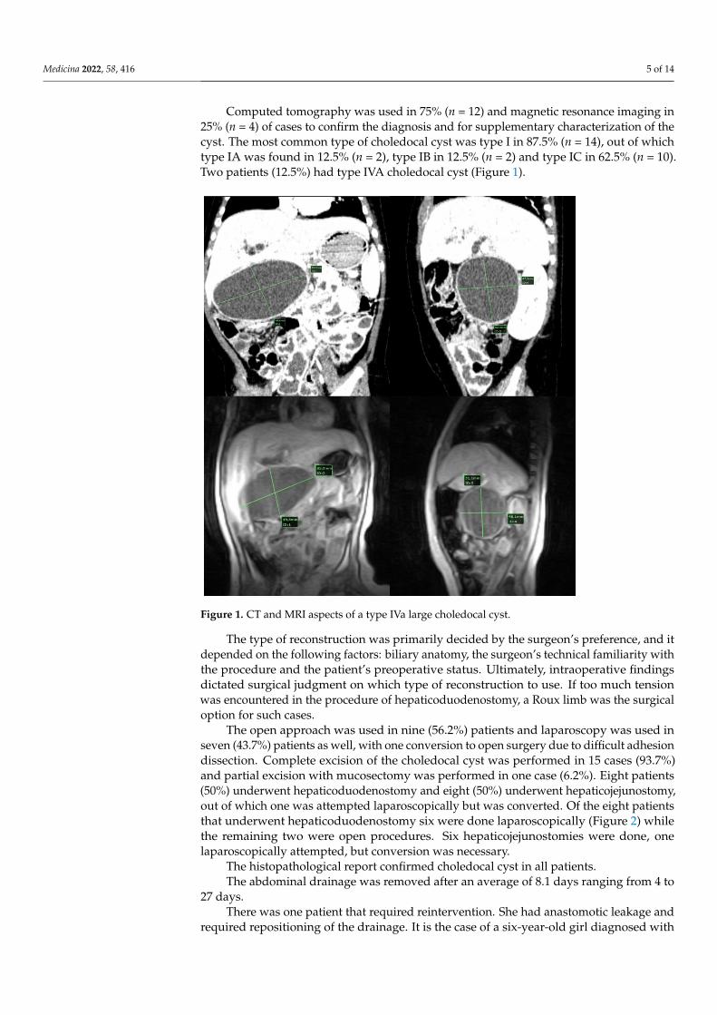
Computed tomography was used in 75% (n = 12) and magnetic resonance imaging in25% (n = 4) of cases to confirm the diagnosis and for supplementary characterization of thecyst. The most common type of choledocal cyst was type I in 87.5% (n = 14), out of whichtype IA was found in 12.5% (n = 2), type IB in 12.5% (n = 2) and type IC in 62.5% (n = 10).Two patients (12.5%) had type IVA choledocal cyst (Figure 1).
Medicina 2022, 58, x FOR PEER REVIEW 5 of 14
As a diagnostic approach, abdominal ultrasound was performed in all patients and identified the choledocal cyst in all cases. Dilatation of intrahepatic bile ducts was noticed in 75% of cases (n = 12). The size of the common bile duct during ultrasound in all of these patients ranged from 14 mm to 99 mm.
Computed tomography was used in 75% (n = 12) and magnetic resonance imaging in 25% (n = 4) of cases to confirm the diagnosis and for supplementary characterization of the cyst. The most common type of choledocal cyst was type I in 87.5% (n = 14), out of which type IA was found in 12.5% (n = 2), type IB in 12.5% (n = 2) and type IC in 62.5% (n = 10). Two patients (12.5%) had type IVA choledocal cyst (Figure 1).
Figure 1. CT and MRI aspects of a type IVa large choledocal cyst.
The type of reconstruction was primarily decided by the surgeon’s preference, and it depended on the following factors: biliary anatomy, the surgeon’s technical familiarity with the procedure and the patient’s preoperative status. Ultimately, intraoperative find-ings dictated surgical judgment on which type of reconstruction to use. If too much ten-sion was encountered in the procedure of hepaticoduodenostomy, a Roux limb was the surgical option for such cases.
The open approach was used in nine (56.2%) patients and laparoscopy was used in seven (43.7%) patients as well, with one conversion to open surgery due to difficult adhe-sion dissection. Complete excision of the choledocal cyst was performed in 15 cases (93.7%) and partial excision with mucosectomy was performed in one case (6.2%). Eight patients (50%) underwent hepaticoduodenostomy and eight (50%) underwent hepati-cojejunostomy, out of which one was attempted laparoscopically but was converted. Of the eight patients that underwent hepaticoduodenostomy six were done laparoscopically (Figure 2) while the remaining two were open procedures. Six hepaticojejunostomies were done, one laparoscopically attempted, but conversion was necessary.
Figure 1. CT and MRI aspects of a type IVa large choledocal cyst.
The type of reconstruction was primarily decided by the surgeon’s preference, and itdepended on the following factors: biliary anatomy, the surgeon’s technical familiarity withthe procedure and the patient’s preoperative status. Ultimately, intraoperative findingsdictated surgical judgment on which type of reconstruction to use. If too much tensionwas encountered in the procedure of hepaticoduodenostomy, a Roux limb was the surgicaloption for such cases.
The open approach was used in nine (56.2%) patients and laparoscopy was used inseven (43.7%) patients as well, with one conversion to open surgery due to difficult adhesiondissection. Complete excision of the choledocal cyst was performed in 15 cases (93.7%)and partial excision with mucosectomy was performed in one case (6.2%). Eight patients(50%) underwent hepaticoduodenostomy and eight (50%) underwent hepaticojejunostomy,out of which one was attempted laparoscopically but was converted. Of the eight patientsthat underwent hepaticoduodenostomy six were done laparoscopically (Figure 2) whilethe remaining two were open procedures. Six hepaticojejunostomies were done, onelaparoscopically attempted, but conversion was necessary.
The histopathological report confirmed choledocal cyst in all patients.The abdominal drainage was removed after an average of 8.1 days ranging from 4 to
27 days.There was one patient that required reintervention. She had anastomotic leakage and
required repositioning of the drainage. It is the case of a six-year-old girl diagnosed with

Medicina 2022, 58, 416 6 of 14
type IC choledocal cyst who underwent choledocal cyst excision and hepaticoduodenos-tomy by laparoscopic approach. Intraoperatively, a large choledocal cyst was identified,highly vascularized, with thick walls, which was adherent to the pancreas on the posteriorwall. On postoperative day 5 the abdominal drainage was removed. Two days later, the pa-tient complained of abdominal distention and pain accompanied by nausea and vomiting.Abdominal ultrasonography identified a 42 mm fluid collection in the hepatic hilum. It wassupplementary characterized by computed tomography which identified a 98/102/92 mminterhepaticoduodenal mixed collection (air, fluid, blood) that compressed the adjacentstructures associated with a large amount of free intraabdominal fluid. The decision ofreintervention was taken. Evacuation of the abdominal fluid and abdominal drainage wasperformed. Postoperative evolution was favorable, with slow regression of the collectionand the abdominal drainage was removed after 27 days. Six months postoperatively thepatient is doing well, with no other complications.
Medicina 2022, 58, x FOR PEER REVIEW 6 of 14
Figure 2. Intraoperative laparoscopic aspect.
The histopathological report confirmed choledocal cyst in all patients. The abdominal drainage was removed after an average of 8.1 days ranging from 4 to
27 days. There was one patient that required reintervention. She had anastomotic leakage and
required repositioning of the drainage. It is the case of a six-year-old girl diagnosed with type IC choledocal cyst who underwent choledocal cyst excision and hepaticoduodenos-tomy by laparoscopic approach. Intraoperatively, a large choledocal cyst was identified, highly vascularized, with thick walls, which was adherent to the pancreas on the posterior wall. On postoperative day 5 the abdominal drainage was removed. Two days later, the patient complained of abdominal distention and pain accompanied by nausea and vom-iting. Abdominal ultrasonography identified a 42 mm fluid collection in the hepatic hi-lum. It was supplementary characterized by computed tomography which identified a 98/102/92 mm interhepaticoduodenal mixed collection (air, fluid, blood) that compressed the adjacent structures associated with a large amount of free intraabdominal fluid. The decision of reintervention was taken. Evacuation of the abdominal fluid and abdominal drainage was performed. Postoperative evolution was favorable, with slow regression of the collection and the abdominal drainage was removed after 27 days. Six months post-operatively the patient is doing well, with no other complications.
Four patients (25%) had ileus, out of which three had hepaticojejunostomy as a method of biliary reconstruction. All of these patients presented delayed return of bowel function, abdominal distension and radiologic findings of ileus. There was one case of pancreatitis, who postoperatively presented abdominal pain and elevated pancreatic
Figure 2. Intraoperative laparoscopic aspect.
Four patients (25%) had ileus, out of which three had hepaticojejunostomy as a methodof biliary reconstruction. All of these patients presented delayed return of bowel function,abdominal distension and radiologic findings of ileus. There was one case of pancreatitis,who postoperatively presented abdominal pain and elevated pancreatic enzymes. Therewere no postoperative cases of cholangitis or gastrointestinal bleeding. None of the patientshad wound infection.
There was one death following multiple organ system failure, caused by Clostridiumdifficile sepsis. It was a three-month-old boy with a history of CMV hepatitis presenting

Medicina 2022, 58, 416 7 of 14
with jaundice and acholic stool. Postoperatively, a bacterial infection triggered an acutedeterioration of his chronic liver failure.
The mean follow-up period was 11.3 months. None of the patients developed symp-toms suggestive of biliary reflux, and therefore no further investigations were carried out,such as contrast study or endoscopy.
4. Discussion
Choledocal cyst is a rare condition defined as an abnormal, disproportionate dilatationof the biliary ducts [5,6]. The 2015 Diagnostic Criteria for Congenital Biliary Dilatation wereformulated by the Japanese Study Group on Pancreaticobiliary Maljunction, being the firstformulation of universal guidelines [7]. In those criteria, the popular term of “choledocalcyst” was replaced with “congenital biliary dilatation” (CBD). This new term was adoptedto furnish a more correct expression and to provide a universally proper understanding ofthis pathology and thus improved treatment outcomes worldwide.
Choledocal cysts are utterly rare in Europe and the United States. They appear morefrequently in Asia, where the incidental rate is as high as 1:1000 hospital admissions [8].Choledocal cyst etiology is not well defined. The most reputable theory was formulatedby Babbitt in 1969. He proposed as an explanation an abnormal junction between thebiliopancreatic ducts (ABPJ), which form a common duct which allows reflux of pancreaticenzymes into the biliary tree. This leads to increased pressure with consecutive pathologicalconditions in the biliary tract and pancreas such as inflammation, ectasia and conclusivelydilatation [9]. The APBJ was not searched for in our patients. Another ethological theorysuggests the implication of the obstruction of the common bile duct, a theory that issustained by animal models, where ligation of the common bile duct leads to a dilatationcomparable to the Todani type I choledocal cyst [10]. Abnormal function and spasm ofthe sphincter of Oddi has been correlated with choledocal cyst [11]. One more theory isthe congenital theory of Davenport and Basu, which suggests an abnormality of ganglioncells in the slender portion of the common bile duct in children with choledocal cyst. Itcan determine a functional obstruction and proximal dilatation of the duct, in the sameway as Hirschsprung disease or achalasia of the esophagus [12]. There are a few reports offamilial cases and associated anomalies [13]. Congenital anomalies that can be associatedwith choledocal cyst are double common bile duct, sclerosing cholangitis, hepatic fibrosis,pancreatic cyst, annular pancreas and cardiac anomalies [14]. We have not found anycongenital anomalies associated with choledocal cyst in our series.
International literature reports a 3–4:1 female to male ratio. This ratio is variable indifferent studies, but there is always a female prevalence [15]. We found a 1.6:1 female tomale ratio. More than 60% of the choledocal cysts present during the first year of life [16].In our study, the median age at presentation was 4 years and 5 months and only 18.7%were diagnosed in the first year of life.
The clinical presentation is often vague and nonspecific. However, the diagnosisis simplified by modern imaging techniques. The typical triad of jaundice, pain and apalpable mass can be identified in approximately 20% of presentations [17]. It is morefrequently seen in children than in adults, and 85% of children have at least two features ofthe triad at diagnosis, in comparison with only 25% of adults [18]. We found this clinicalassociation in only two patients. Jaundice and abdominal pain are the most commonsymptoms [19]. We found these symptoms in 62.5% and 56.2% of cases, respectively. Otherpresenting forms of presentation can be cholangitis, pancreatitis and biliary peritonitisfrom cyst rupture. Cholangitis was the manner of presentation in seven cases (43.7%). Ininfancy, choledocal cysts may present with pale stool, hepatomegaly and jaundice andmay be hard to differentiate from biliary atresia. Due to the widespread use of prenatalultrasonography, many choledocal cysts are now identified in the fetus but they can alsobe identified by magnetic resonance imaging [20]. Prenatally diagnosed choledocal cystshave a predisposition of developing liver fibrosis and portal hypertension early after birth.Obstructive jaundice and enlarging cysts are indicators for early surgery. Since biliary

Medicina 2022, 58, 416 8 of 14
sludge starts forming as soon as two weeks of life, it might be beneficial to undergo surgicaltreatment within two weeks of life. Nonetheless, the ideal timing of surgery, in particularin asymptomatic newborns, has not been precisely defined. We had no cases of prenatallydiagnosed choledocal cyst. Cystic biliary atresia should be considered as a differentialdiagnosis as it can easily mimic a choledocal cyst [7].
Ultrasonography is suitable to identify choledocal cysts in most children. Ultra-sonography is the best initial imaging method for evaluating the entire intrahepatic andextrahepatic biliary system and gallbladder. It identifies a choledocal cyst as a distinctivecystic or fusiform dilatation of the common bile duct, of the intrahepatic ducts or sometimesa cyst in the porta hepatis, apart from the gallbladder. It may also identify the associatedcomplications such as cholelithiasis, cholangitis or malignancy. Ultrasonography is alsohelpful for follow-up and assessment of any residual biliary dilatation after choledocal cystsurgery [21].
In some cases, advanced imaging techniques are required. Computed tomographycan be necessary to confirm the diagnosis and magnetic resonance imaging to obtain dataabout the extent of the cyst, defects within the biliary tree and presence of the anomalousjunction of the pancreaticobiliary duct [22]. Magnetic resonance cholangiopancreatography(MRCP) is at this time considered the gold standard for imaging [22] (Figure 3). We usedultrasonography as the initial imagistic diagnostic method in all patients, confirmed bycomputed tomography in 75% of cases and supplementarily characterized by magneticresonance imaging in 25% of cases.
Medicina 2022, 58, x FOR PEER REVIEW 8 of 14
ultrasonography, many choledocal cysts are now identified in the fetus but they can also be identified by magnetic resonance imaging [20]. Prenatally diagnosed choledocal cysts have a predisposition of developing liver fibrosis and portal hypertension early after birth. Obstructive jaundice and enlarging cysts are indicators for early surgery. Since biliary sludge starts forming as soon as two weeks of life, it might be beneficial to undergo sur-gical treatment within two weeks of life. Nonetheless, the ideal timing of surgery, in par-ticular in asymptomatic newborns, has not been precisely defined. We had no cases of prenatally diagnosed choledocal cyst. Cystic biliary atresia should be considered as a dif-ferential diagnosis as it can easily mimic a choledocal cyst [7].
Ultrasonography is suitable to identify choledocal cysts in most children. Ultraso-nography is the best initial imaging method for evaluating the entire intrahepatic and ex-trahepatic biliary system and gallbladder. It identifies a choledocal cyst as a distinctive cystic or fusiform dilatation of the common bile duct, of the intrahepatic ducts or some-times a cyst in the porta hepatis, apart from the gallbladder. It may also identify the asso-ciated complications such as cholelithiasis, cholangitis or malignancy. Ultrasonography is also helpful for follow-up and assessment of any residual biliary dilatation after chole-docal cyst surgery [21].
In some cases, advanced imaging techniques are required. Computed tomography can be necessary to confirm the diagnosis and magnetic resonance imaging to obtain data about the extent of the cyst, defects within the biliary tree and presence of the anomalous junction of the pancreaticobiliary duct [22]. Magnetic resonance cholangiopancreatog-raphy (MRCP) is at this time considered the gold standard for imaging [22] (Figure 3). We used ultrasonography as the initial imagistic diagnostic method in all patients, confirmed by computed tomography in 75% of cases and supplementarily characterized by magnetic resonance imaging in 25% of cases.
Figure 3. MRCP aspect of a large, type IVa choledocal cyst.
In 1959, Alonso Lej et al. published the first clinical series of patients with choledocal cyst and offered the first systematic description of choledocal cysts based on the clinical and anatomic findings in 96 cases [23]. They divided choledocal cyst into three types and described the therapeutic approach for each one. Todani et al. classified choledocal cysts into five major types and several subtypes according to cyst morphologies, out of which 90–95% are type I [24]. The incidence rates reported in the literature are 50–80% type I, 2% type II, 1.4–4.5% type III, 15–35% type IV and 20% type V [25]. The incidence rates
Figure 3. MRCP aspect of a large, type IVa choledocal cyst.
In 1959, Alonso Lej et al. published the first clinical series of patients with choledocalcyst and offered the first systematic description of choledocal cysts based on the clinicaland anatomic findings in 96 cases [23]. They divided choledocal cyst into three types anddescribed the therapeutic approach for each one. Todani et al. classified choledocal cystsinto five major types and several subtypes according to cyst morphologies, out of which90–95% are type I [24]. The incidence rates reported in the literature are 50–80% type I,2% type II, 1.4–4.5% type III, 15–35% type IV and 20% type V [25]. The incidence ratesdetermined in our study were consistent with the literature; 87.5% had type I choledocalcyst, and 12.5% had type IV A. Cysts of the cystic duct are not included in the Todaniclassification, being an entity that is even rarer than choledocal cysts, with only 14 cases

Medicina 2022, 58, 416 9 of 14
reported in the literature to date. Serena Serradel et al. proposed including these cystsunder a new type VI category [26].
The treatment strategy varies according to cyst type. Treatment modalities havechanged from Tondani (1997) to Baison (2019), but the fundamental principles have re-mained unchanged (Table 3) [27]. For type I cyst, cyst excision and hepaticoenterostomyis the treatment of choice, Roux-en-Y anastomosis being the gold standard. T-tube ap-plications and sphincteroplasty are not recommended nowadays. Drainage is associatedwith biliary stasis, recurrent infection, pancreatitis and cholangitis [27]. For type II cyst,diverticulectomy is now routinely performed, but it used to be described only in casereports. In type III choledocal cyst, transduodenal excision and sphincteroplasty wereat the experimental stage in the past, but they are endoscopically performed with ERCPtoday, and surgery is used only as the second choice. For type IV cysts, cyst excision andhepaticoenterostomy is recommended. For type V cyst, resection is recommended forpartial disease and liver transplant for diffuse disease [28].
Table 3. Surgical treatment from past to present.
1977—Todani 2019—Baison
Type I Cyst excision + Roux-en-Y HJ (T-tubefor IB, sphincteroplasty for IC)
Cyst excision + Hepaticoenterostomy(HJ/HD)
Type II No experience(Case reports − diverticulectomy) Diverticulectomy
Type IIINo experience (Case reports −
transduodeal excision+ sphincteroplasty)
Endoscopic transduodenal excision+ sphincteroplasty
Type IV Cyst excision + HJ Cyst excision + Hepaticoenterostomy(HJ/HD)
Type V Partial resection in localized disease Partial resection for partial disease,liver transplantation for diffuse disease
Excision of type I and type IVA choledocal cysts followed by either hepaticoduodenos-tomy or hepaticojejunostomy has been generally accepted as surgical treatment in thesepatients [14].
Most choledocal cysts of Todani type I or IVA can be safely managed by the laparo-scopic approach, except the ones that are perforated or in case of extensive adhesions orprevious biliary surgery with consecutive disturbed anatomy [29]. The first minimally inva-sive choledocal cyst excision with hepaticojejunostomy for reconstruction was performed in1995 in a 6-year-old girl with type I choledocal cyst, and the first robotic case was performedin 2006 [30,31]. A systematic review and meta-analysis in 2015 by Zhen et al. compared5611 laparoscopic choledocal cyst excisions to 5797 open procedures [32]. They demon-strated longer operative time for laparoscopic procedures but shorter hospital stays andfaster recovery of bowel function. There was no overall difference between most complica-tions. They identified lower rates of intraoperative blood transfusion and postoperativeadhesive bowel obstruction in the laparoscopic group. A second meta-analysis publishedin the same year by Shen et al. [33] confirmed Zhen et al.’s findings and suggested that withimprovement of laparoscopic techniques this may become the preferred approach. Theadvantages of the laparoscopic approach include small incisions with minimal scarring, agood view of the porta hepatis, portal vein and hepatic arteries due to umbilicus-to-hepatichilum direction of the scope, easier dissection and anastomosis due to magnified view andthe possibility of overall abdominal examination [34]. Robotic excision of the choledocalcysts is now being increasingly practiced at centers where robotic surgery is available, beingthe second most common robotic procedure after pyeloplasty [35].
There is a scarcity of papers comparing hepaticojejunostomy and hepaticoduodenos-tomy after excision of the choledocal cyst in a single center, the majority of which come

Medicina 2022, 58, 416 10 of 14
from the Far East. There are both advantages and disadvantages for each of the techniques,rendering the choice between them still a matter of debate [3].
Hepaticojejunostomy requires completion of two anastomoses, which significantlyprolongs the operative time and sacrifices the eventuality of diagnostic endoscopy or endo-scopic stenting [36]. Laparoscopic hepaticojejunostomy is particularly laborious to execute,with the creation of the limb extracorporeally. The Roux-en-Y limb defunctionalizes acertain length of jejunum [37]. The more extensive bowel manipulation in patients whoundergo hepaticojejunostomy could prompt longer periods of enteral feeding [38]. Nu-merous studies demonstrate that complications include anastomotic leak, cholangitis andfluid collection in the gallbladder fossa within the early postoperative period, whereas latecomplications include anastomotic stricture, biliary stone formation and cholangitis [36].Hepaticojejunostomy advocates suggest there are less leak-associated complications on thegrounds of a lower suture tension in the Roux limb, and it also diverts enteric content awayfrom the biliary tree, thus preventing cholangitis postoperatively [39]. Some maintain thathepaticojejunostomy grants a more durable reconstruction when Yamataka et al. recom-mendations are followed: end-to-end anastomosis when feasible, end-to-side anastomosiswhen inevitable with anastomosis of the common hepatic duct near to the closed end ofthe biliary pouch, careful choice of the vascular supply to the limb, and individualizationof the length of the Roux [40].
The advantages of hepaticoduodenostomy consist of shorter operative times becausea single anastomosis is required as opposed to three in a jejunal reconstruction, fasterbowel function recovery as a result less handling and shorter hospital stays [38]. Someauthors suggest that hepaticoduodenostomy leads to higher rates of reflux, which in turncontribute to higher incidence of postoperative cholangitis, anastomotic stricture andinfrequently carcinoma [37]. The preconditions of a safe hepaticoduodenostomy are asatisfactory duodenum mobilization so as to be able to draw the common bile duct stumpfor a tension-free anastomosis. Anastomosis is contraindicated in cases where tension freesuture is unachievable and should be converted to hepaticojejunostomy [41]. Todani et al.has advocated for a hepaticoduodenostomy with an ample enough stoma at the hepatichilum, considering it the most effective approach [42].
We are in favor of hepaticoduodenostomy whenever practical because it is a morephysiological method compared with hepaticojejunostomy and more straightforward toexecute. Regarding laparoscopy, we consider hepaticoduodenostomy better and easier toperform, complications such as adhesive bowel obstruction being less likely. Reinterventionin the event of stricture or lithiasis is easier with hepaticoduodenostomy as there is noRoux limb [33]. Nonetheless, some surgeons prefer the Roux-en-Y approach on accountof a lasting safety record, being considered by some authors the gold standard for biliaryreconstruction [37]. Some centers described more endoscopy-confirmed bilious gastritisby bile reflux, adhesive bowel obstruction and cholangitis in the hepaticoduodenostomygroup [43] and Todani et al. [44] reported a hilar bile duct carcinoma 19 years after hepati-coduodenostomy. It was considered that duodenal content reflux into the bile ducts via thehepaticoduodenal anastomosis may have been harmful to the mucosal lining. A 2013 meta-analysis comparing hepaticoduodenal with hepaticojejunal anastomoses in 679 patientsindicated the techniques are comparable in terms of most outcomes, excluding greater rateof gastric reflux [37]. A 15-year review by Silva-Baez et al. found a 25% complication ratein hepaticojejunostomy compared to 16.6% in hepaticoduodenostomy [5]; complicationsincluded cholangitis, leak and postoperative reflux. There were no deaths. The authorssupport the use of duodenum as the safer alternative.
In our study, length of hospital stay varied greatly from 9 days to 37 days, with anaverage of 11 days. Other authors reported average admissions ranging between 8 and12.2 days [4,36]. Global morbidity of surgical choledocal cyst treatment varies between13% to 40%, most consisting of wound-related complications or bilioenteric anastomoticleaks [45]. Bile leak is also the predominant mortality cause [46].

Medicina 2022, 58, 416 11 of 14
Anastomotic leak is an early complication. One patient (6.2%) developed anastomoticleak; a rate comparable to that of the literature [47]. Our case was managed conserva-tively, only requiring repositioning of the drainage. However, caution should be exercisedwhen signs of peritonitis and hemodynamic instability develop, because these warrantexploration and revision of the anastomosis.
Abdominal drainage time was analyzed and averaged 8.1 days. Important fluiddischarge, both in quantity and duration, suggests lymphatic damage caused by dissectionof the cyst and the perihilar region [36].
The occurrence of cholangitis after reconstruction is thought to be caused by bilestasis, as well as reflux of duodenal content into biliary tree with bacterial overgrowth andconsecutive partial obstruction. A Roux-en-Y anastomosis makes use of a defunctionalized,relatively long and peristaltic limb that provides sheltering to the ducts. Hepaticoduo-denostomy is hypothetically associated with higher rates of cholangitis because of thedirect path of enteric flora from the duodenum into the biliary tree [37]. We had no cases ofpostoperative cholangitis.
None of the patients developed symptoms specific for biliary reflux gastritis; hence,they did not undergo endoscopy, nor did they develop anastomotic strictures duringthe follow-up period. However, the presence of duodenogastric biliary reflux is difficultto determine by symptomatology only, so specific examination techniques are neededto diagnose it. Shimotakahara et al. objectified postoperative biliary gastritis in 33% ofpatients that underwent a hepaticoduodenostomy and in 7% of patients that underwenthepaticojejunostomy, but interestingly enough, no difference was found in the incidenceof anastomotic stricture [43]. Postoperative cholangitis incidence is well correlated withanastomotic or supra-anastomotic strictures, which seems to be linked to the surgeon’sexperience rather than the method for biliary-enteric anastomosis [44]. Some authorsconsidered the hilar anastomosis at the bifurcation preferable to a distal anastomosisfor type I cysts, in order to provide a broader anastomosis. It is thought that biliaryreconstruction close to the pyloric ring could impede continuous excretion of the bile intothe duodenum [33]. Therefore, a crucial point in hepaticoduodenostomy is to performit beyond the first part of the duodenum, subsequent to a proper Kocher maneuver, inorder to avoid any disturbance in pyloric function or gastric evacuation [37]. Another non-neoplastic late complication that might occur is intrahepatic biliary lithiasis [48] howeverwe could not identify any such case in our study.
Biliary malignancy incidence in patients diagnosed with choledocal cyst is twiceas high as the general population. Pathogenesis for these premalignant lesions is notsufficiently explained yet. The overall risk of biliary malignancy is 10–15% in East Asiancountries [28], however, these data originate from the East, but rates differ in the West. In2019 Baison et al. compared all single and multi-center reports from the East and West anddiscovered a malignancy rate of 0–17% in the East and 3–8% in the West, and a recurrencerate of 0–10% in the East and 3–8% in the West. Mortality and morbidity rates, type ofcholedocal cyst, patient history and symptomatology were similar [28].
Malignancy risk persists even after cyst excision (0.75–5.4%). Neoplastic complicationsafter surgically treated choledocal cysts are difficult to assess and require extensive follow-up. The literature most often reports them in association with intrahepatic duct dilation,biliary lithiasis, recurrent cholangitis and strictures [49]. Koike et al. suggest that excisionextent could be correlated with neoplastic complication incidence [27]. Malignancy riskincreases with each decade of unoperated life; malignancy risk is 1% at 10 years, 15% at20 years, 26% at 40 years and 45% at 70 years. Hence, biliary cancer is rare if excision isperformed at less than 10 years of age [4].
Our study has several limitations; the study was done in a retrospective manner, andwe have a small number of patients and a relatively short follow-up period, standardizedpostoperative follow-up being imperative for long-term conclusions. We attribute therelatively short follow-up to low patient compliance and a lack of interdisciplinary follow-up protocol.
Medicina 2022, 58, 416 12 of 14
5. Conclusions
Choledocal cyst is a rare disease. All authors agree on the necessity of cyst excisionand biliary reconstruction; however, neither technique has proven itself beneficial enoughto be universally adopted yet. There are advantages and disadvantages of each techniqueand of each approach. Since there are no prospective randomized clinical trials, judgementand experience of the attending surgeon will determine the technique for biliary tractreconstruction.
From our experience with these patients, we concluded that a wide hepaticoduodenos-tomy constitutes a favorable choice instead of the traditional hepaticojejunostomy, beingmore physiological and less time consuming.
Author Contributions: Conceptualization, L.B.; methodology A.M.; formal analysis, T.S. and A.C.;writing—original draft preparation, A.M.; writing—review and editing, T.S. and A.C.; supervisionR.B.; project administration, L.B. All authors have read and agreed to the published version ofthe manuscript.
Funding: This research received no external funding.
Institutional Review Board Statement: The study was conducted in accordance with the Declarationof Helsinki and approved by the Institutional Review Board.
Informed Consent Statement: Informed consent was obtained from all subjects involved in thestudy. Ethical approval was obtained from the hospital ethics committee.
Data Availability Statement: https://docs.google.com/spreadsheets/d/1C5xkUE1qGhUsOpXhvyb65AA_OkPelNLgzyNlIEB-GrA/edit?usp=sharing (accessed on 12 December 2021).
Conflicts of Interest: The authors declare no conflict of interest.
References1. Rubalcava, N.S.; Overman, R.E.; Pilkington, M.; Grant, C.N.; Geiger, J.D.; Jarboe, M.D.; Speck, K.E. Laparoscopic choledochal cyst
resection using a novel articulating instrument in pediatric patients. J. Pediatr. Surg. Case Rep. 2021, 70, 101888. [CrossRef]2. Liem, N.T.; Pham, H.D.; Dung, L.A.; Son, T.N.; Vu, H.M. Early and intermediate outcomes of laparoscopic surgery for choledochal
cysts with 400 patients. J. Laparoendosc. Adv. Surg. Tech. 2012, 22, 599–603. [CrossRef] [PubMed]3. Lee, C.; Byun, J.; Ko, D.; Yang, H.-B.; Youn, J.K.; Kim, H.-Y. Comparison of long-term biliary complications between open and
laparoscopic choledochal cyst excision in children. Ann. Surg. Treat. Res. 2021, 100, 186–192. [CrossRef]4. Todani, T. Congenital choledochal dilatation: Classification, clinical features, and long-term results. J. Hepatobiliary Pancreat. Surg.
1997, 4, 276–282. [CrossRef]5. Silva-Baez, H.; Coello-Ramírez, P.; Ixtabalán-Escalante, E.M.; Sotelo-Anaya, E.; Gallo-Morales, M.; Cordero-Estrada, E.;
Sainz-Escarrega, V.H.; Ploneda-Valencia, C.F. Treatment of choledochal cyst in a pediatric population. A single institutionexperience of 15-years. Case series. Ann. Med. Surg. 2016, 5, 81–85. [CrossRef]
6. Söreide, K.; Körner, H.; Havnen, J.; Söreide, J.A. Bile duct cysts in adults. Br. J. Surg. 2004, 91, 1538–1548. [CrossRef] [PubMed]7. Hamada, Y.; Ando, H.; Kamisawa, T.; Itoi, T.; Urushihara, N.; Koshinaga, T.; Saito, T.; Fujii, H.; Morotomi, Y. Diagnostic criteria
for congenital biliary dilatation 2015. J. Hepatobiliary Pancreat. Sci. 2016, 23, 342–346. [CrossRef] [PubMed]8. Hong, L.; Wu, Y.; Yan, Z.; Xu, M.; Chu, J.; Chen, Q.-M. Laparoscopic surgery for choledochal cyst in children: A case review of
31 patients. Eur. J. Pediatr. Surg. Off. J. Austrian Assoc. Pediatr. Surg. 2008, 18, 67–71. [CrossRef]9. Babbitt, D.P. Congenital choledochal cysts: New etiological concept based on anomalous relationships of the common bile duct
and pancreatic bulb. Ann. Radiol. 1969, 12, 231–240.10. Spitz, L. Experimental production of cystic dilatation of the common bile duct in neonatal lambs. J. Pediatr. Surg. 1977, 12, 39–42.
[CrossRef]11. Imazu, M.; Iwai, N.; Tokiwa, K.; Shimotake, T.; Kimura, O.; Ono, S. Factors of biliary carcinogenesis in choledochal cysts.
Eur. J. Pediatr. Surg. Off. J. Austrian Assoc. Pediatr. Surg. 2001, 11, 24–27. [CrossRef] [PubMed]12. Kusunoki, M.; Saitoh, N.; Yamamura, T.; Fujita, S.; Takahashi, T.; Utsunomiya, J. Choledochal cysts. Oligoganglionosis in the
narrow portion of the choledochus. Arch. Surg. 1988, 123, 984–986. [CrossRef] [PubMed]13. Iwata, F.; Uchida, A.; Miyaki, T.; Aoki, S.; Fujioka, T.; Yamada, J.; Joh, T.; Itoh, M. Familial occurrence of congenital bile duct cysts.
J. Gastroenterol. Hepatol. 1998, 13, 316–319. [CrossRef]14. Soares, K.C.; Arnaoutakis, D.J.; Kamel, I.; Rastegar, N.; Anders, R.; Maithel, S.; Pawlik, T.M. Choledochal cysts: Presentation,
clinical differentiation, and management. J. Am. Coll. Surg. 2014, 219, 1167–1180. [CrossRef] [PubMed]

Medicina 2022, 58, 416 13 of 14
15. Katabi, N.; Pillarisetty, V.G.; DeMatteo, R.; Klimstra, D.S. Choledochal cysts: A clinicopathologic study of 36 cases with emphasison the morphologic and the immunohistochemical features of premalignant and malignant alterations. Hum. Pathol. 2014,45, 2107–2114. [CrossRef] [PubMed]
16. Liu, C.-L.; Fan, S.-T.; Lo, C.-M.; Lam, C.M.; Poon, R.T.-P.; Wong, J. Choledochal cysts in adults. Arch. Surg. 2002, 137, 465–468.[CrossRef]
17. Bhavsar, M.S.; Vora, H.B.; Giriyappa, V.H. Choledochal cysts: A review of literature. Saudi J. Gastroenterol. 2012, 18, 230. [PubMed]18. Lin, S.L.; Shan, K.M.; Hung, Y.B.; Ng, S.H.; Lin, C.Y. Choledochal cyst associated with acute acalculous cholecystitis. J. Pediatr.
Gastroenterol. Nutr. 2000, 31, 307–308. [CrossRef]19. Jiménez-Urueta, P.S.; Alexis-Pacheco, B.; de Jesús Gutiérrez-Escobedo, J.; Castañeda-Ortiz, R.A.; Suárez-Gutiérrez, R. Resección
de quiste de colédoco y hepatoduodeno anastomosis vía laparoscópica. Acta Pediátrica México 2013, 34, 258–262.20. Liu, S.-L.; Li, L.; Hou, W.-Y.; Zhang, J.; Huang, L.-M.; Li, X.; Xie, H.-W.; Cheng, W. Laparoscopic excision of choledochal cyst and
Roux-en-Y hepaticojejunostomy in symptomatic neonates. J. Pediatr. Surg. 2009, 44, 508–511. [CrossRef] [PubMed]21. Mishra, A.; Pant, N.; Chadha, R.; Choudhury, S.R. Choledochal cysts in infancy and childhood. Indian J. Pediatr. 2007, 74, 937–942.
[CrossRef] [PubMed]22. Irie, H.; Honda, H.; Jimi, M.; Yokohata, K.; Chijiiwa, K.; Kuroiwa, T.; Hanada, K.; Yoshimitsu, K.; Tajima, T.; Matsuo, S.; et al.
Value of MR cholangiopancreatography in evaluating choledochal cysts. AJR Am. J. Roentgenol. 1998, 171, 1381–1385. [CrossRef][PubMed]
23. Alonso-Lej, F.; Rever, W.B.J.; Pessagno, D.J. Congenital choledochal cyst, with a report of 2, and an analysis of 94, cases.Int. Abstr. Surg. 1959, 108, 1–30. [PubMed]
24. Todani, T.; Watanabe, Y.; Narusue, M.; Tabuchi, K.; Okajima, K. Congenital bile duct cysts: Classification, operative procedures,and review of thirty-seven cases including cancer arising from choledochal cyst. Am. J. Surg. 1977, 134, 263–269. [CrossRef]
25. Singham, J.; Yoshida, E.M.; Scudamore, C.H. Choledochal cysts: Part 1 of 3: Classification and pathogenesis. Can. J. Surg. 2009,52, 434–440.
26. Serena Serradel, A.F.; Santamaría Linares, E.; Herrera Goepfert, R. Cystic dilatation of the cystic duct: A new type of biliary cyst.Surgery 1991, 109, 320–322.
27. Lipsett, P.A.; Pitt, H.A. Surgical treatment of choledochal cysts. J. Hepatobiliary Pancreat. Surg. 2003, 10, 352–359. [CrossRef]28. Piskin, E.; Ustuner, M.A.; Oter, V.; Aydin, O.; Ozgun, Y.M.; Colakoglu, M.K.; Aksoy, E.; Keklik, T.T.; Ozogul, Y.B.; Bostanci, E.B.
Single-Center Results of Choledochal Cysts in Turkish Population. Arch. Iran. Med. 2021, 24, 43–47. [CrossRef]29. Yeung, F.; Chung, P.H.Y.; Wong, K.K.Y.; Tam, P.K.H. Biliary-enteric reconstruction with hepaticoduodenostomy following laparo-
scopic excision of choledochal cyst is associated with better postoperative outcomes: A single-centre experience. Pediatr. Surg. Int.2015, 31, 149–153. [CrossRef]
30. Farello, G.A.; Cerofolini, A.; Rebonato, M.; Bergamaschi, G.; Ferrari, C.; Chiappetta, A. Congenital choledochal cyst: Video-guidedlaparoscopic treatment. Surg. Laparosc. Endosc. 1995, 5, 354–358.
31. Woo, R.; Le, D.; Albanese, C.T.; Kim, S.S. Robot-assisted laparoscopic resection of a type I choledochal cyst in a child.J. Laparoendosc. Adv. Surg. Tech. A 2006, 16, 179–183. [CrossRef] [PubMed]
32. Zhen, C.; Xia, Z.; Long, L.; Lishuang, M.; Pu, Y.; Wenjuan, Z.; Xiaofan, L. Laparoscopic excision versus open excision for thetreatment of choledochal cysts: A systematic review and meta-analysis. Int. Surg. 2015, 100, 115–122. [CrossRef] [PubMed]
33. Shen, H.-J.; Xu, M.; Zhu, H.-Y.; Yang, C.; Li, F.; Li, K.-W.; Shi, W.-J.; Huo-Jian, S. Laparoscopic versus open surgery in childrenwith choledochal cysts: A meta-analysis. Pediatr. Surg. Int. 2015, 31, 529–534. [CrossRef] [PubMed]
34. Diao, M.; Li, L.; Cheng, W. Role of laparoscopy in treatment of choledochal cysts in children. Pediatr. Surg. Int. 2013, 29, 317–326.[CrossRef]
35. Nazki, S.; Kanojia, R.P.; Bawa, M.; Binu, V.; Lal, S.; Sood, A.; Samujh, R. Robotic Excision of Choledochal Cyst with Hepaticoduo-denostomy (HD): Report of HD Technique, Initial Experience, and Early Outcome. Eur. J. Pediatr. Surg. Off. J. Austrian Assoc.Pediatr. Surg. 2021, 31, 286–291. [CrossRef]
36. Saxena, N.A.; Kulkarni, B.K.; Borwankar, S.S.; Lahoti, H.N.; Multani, P.; Oak, S.N. Hepaticoduodenostomy as a technique forbiliary anastomosis in children with choledochal cyst: An experience with 31 cases. Ann. Pediatr. Surg. 2017, 13, 78–80. [CrossRef]
37. Narayanan, S.K.; Chen, Y.; Narasimhan, K.L.; Cohen, R.C. Hepaticoduodenostomy versus hepaticojejunostomy after resection ofcholedochal cyst: A systematic review and meta-analysis. J. Pediatr. Surg. 2013, 48, 2336–2342. [CrossRef]
38. Santore, M.T.; Behar, B.J.; Blinman, T.A.; Doolin, E.J.; Hedrick, H.L.; Mattei, P.; Nance, M.L.; Adzick, N.S.; Flake, A.W. Hepatico-duodenostomy vs hepaticojejunostomy for reconstruction after resection of choledochal cyst. J. Pediatr. Surg. 2011, 46, 209–213.[CrossRef]
39. Gardikis, S.; Antypas, S.; Kambouri, K.; Lainakis, N.; Panagidis, A.; Deftereos, S.; Polychronidis, A.; Dolatzas, T.; Simopoulos, C.The Roux-en-Y procedure in congenital hepato-biliary disorders. Rom. J. Gastroenterol. 2005, 14, 135–140.
40. Yamataka, A.; Kobayashi, H.; Shimotakahara, A.; Okada, Y.; Yanai, T.; Lane, G.J.; Urao, M.; Miyano, T. Recommendations forpreventing complications related to Roux-en-Y hepatico-jejunostomy performed during excision of choledochal cyst in children.J. Pediatr. Surg. 2003, 38, 1830–1832. [CrossRef]
41. Serber, J.; Stranzinger, E.; Geiger, J.D.; Teitelbaum, D.H. Association of gastroschisis and choledochal cyst. J. Pediatr. Surg. 2009,44, e23–e26. [CrossRef] [PubMed]

Medicina 2022, 58, 416 14 of 14
42. Todani, T.; Watanabe, Y.; Mizuguchi, T.; Fujii, T.; Toki, A. Hepaticoduodenostomy at the hepatic hilum after excision of choledochalcyst. Am. J. Surg. 1981, 142, 584–587. [CrossRef]
43. Shimotakahara, A.; Yamataka, A.; Yanai, T.; Kobayashi, H.; Okazaki, T.; Lane, G.J.; Miyano, T. Roux-en-Y hepaticojejunos-tomy or hepaticoduodenostomy for biliary reconstruction during the surgical treatment of choledochal cyst: Which is better?Pediatr. Surg. Int. 2005, 21, 5–7. [CrossRef] [PubMed]
44. Todani, T.; Watanabe, Y.; Urushihara, N.; Noda, T.; Morotomi, Y. Biliary complications after excisional procedure for choledochalcyst. J. Pediatr. Surg. 1995, 30, 478–481. [CrossRef]
45. Moslim, M.A.; Takahashi, H.; Seifarth, F.G.; Walsh, R.M.; Morris-Stiff, G. Choledochal Cyst Disease in a Western Center: A 30-YearExperience. J. Gastrointest. Surg. Off. J. Soc. Surg. Aliment. Tract. 2016, 20, 1453–1463. [CrossRef]
46. Edil, B.H.; Cameron, J.L.; Reddy, S.; Lum, Y.; Lipsett, P.A.; Nathan, H.; Pawlik, T.M.; Choti, M.A.; Wolfgang, C.L.; Schulick, R.D.Choledochal cyst disease in children and adults: A 30-year single-institution experience. J. Am. Coll. Surg. 2008, 206, 1000–1005.[CrossRef]
47. Li, M.-J.; Feng, J.-X.; Jin, Q.-F. Early complications after excision with hepaticoenterostomy for infants and children withcholedochal cysts. Hepatobiliary Pancreat. Dis. Int. HBPD Int. 2002, 1, 281–284.
48. Mukhopadhyay, B.; Shukla, R.M.; Mukhopadhyay, M.; Mandal, K.C.; Mukherjee, P.P.; Roy, D.; Biswas, S.K.; Basu, K.S. Choledochalcyst: A review of 79 cases and the role of hepaticodochoduodenostomy. J. Indian Assoc. Pediatr. Surg. 2011, 16, 54–57. [CrossRef]
49. Goto, N.; Yasuda, I.; Uematsu, T.; Kanemura, N.; Takai, S.; Ando, K.; Kato, T.; Osada, S.; Takao, H.; Saji, S.; et al. Intrahepaticcholangiocarcinoma arising 10 years after the excision of congenital extrahepatic biliary dilation. J. Gastroenterol. 2001, 36, 856–862.[CrossRef]
Copyright © 2022 FDOKUMEN