A Review of Dengue Fever with Special Reference to Malaysia
19
_____________________________________________________________________________________________________ *Corresponding author: E-mail: [email protected]; Asian Journal of Research in Infectious Diseases 1(2): 1-19, 2018; Article no.AJRID.44628 A Review of Dengue Fever with Special Reference to Malaysia Meer Ahmad A. M. 1* , Verasingam Kumarasamy 1 , Yen Yew Ngau 2 , Chee Loon Leong 3 and Chew Aik Koay 4 1 Department of Community Medicine, MAHSA University, Bandar Saujana Putra, 42610 Jenjarom, Selangor, Malaysia. 2 Department of Internal Medicine, MAHSA University, Bandar Saujana Putra, 42610 Jenjarom, Selangor, Malaysia. 3 Infectious Diseases, Medical Department, Hospital Kuala Lumpur, 50586 Jalan Pahang, Wilayah Persekutuan Kuala Lumpur, Malaysia. 4 MOZFREE (M) Sdn Bhd, Subang Jaya, Malaysia. Authors’ contributions This work was carried out in collaboration between all authors. Author MAAM is the lead author and correspondence author. He is also the initiator of this Review Article and addressed much of the aspects of epidemiology including Control and Prevention and aspects of Laboratory Diagnosis. Author VK had written the preceding Review to this Article in 2006 in the Malaysian Medical Journal to which this Article is a sequel. He has also contributed to aspects of Epidemiology including Control and Prevention in this Article. Authors YYN and CLL are Internal Medicine Consultants who contributed to the Clinical Management aspects of this Article. Author CAK addressed the Entomological aspects of this Article, and addressed the part on Newer Insecticides and Innovative Methods. All authors read and approved the final manuscript. Article Information DOI: 10.9734/AJRID/2018/v1i229764 Editor(s): (1) Dr. Shweta Sharma, Department of Microbiology, Dr. Ram Manohar Lohia Hospital and PGIMER, New Delhi, India. Reviewers: (1) Iram Liaqat, GC University, Pakistan. (2) S. C. Weerasinghe, Teaching Hospital Kurunegala, Sri Lanka. (3) Lívia Garcia Bertolacci-Rocha, Universidade Federal de Goiás, Brasil. (4) Kolitha H. Sellahewa, Neville Fernando Teaching Hospital, Sri Lanka. Complete Peer review History: http://www.sciencedomain.org/review-history/26895 Received 16 August 2018 Accepted 22 October 2018 Published 29 October 2018 ABSTRACT Introduction: Dengue is the most prevalent viral mosquito-borne disease, with over 2.5 billion humans at risk given its endemicity in more than 100 countries. Globally, 50-100 million cases of dengue occur annually, with approximately 0.7% resulting in dengue haemorrhagic fever (DHF), and 22,000 deaths. In 2017, there were 83,849 reported cases of dengue fever in endemic under-reported Malaysia, with 177 deaths. Review Article
-
Upload
khangminh22 -
Category
Documents
-
view
1 -
download
0
Transcript of A Review of Dengue Fever with Special Reference to Malaysia
_____________________________________________________________________________________________________ *Corresponding author: E-mail: [email protected];
Asian Journal of Research in Infectious Diseases 1(2): 1-19, 2018; Article no.AJRID.44628
A Review of Dengue Fever with Special Reference to Malaysia
Meer Ahmad A. M.1*, Verasingam Kumarasamy1, Yen Yew Ngau2,
Chee Loon Leong3 and Chew Aik Koay4
1Department of Community Medicine, MAHSA University, Bandar Saujana Putra, 42610 Jenjarom,
Selangor, Malaysia. 2Department of Internal Medicine, MAHSA University, Bandar Saujana Putra, 42610 Jenjarom,
Selangor, Malaysia. 3Infectious Diseases, Medical Department, Hospital Kuala Lumpur, 50586 Jalan Pahang,
Wilayah Persekutuan Kuala Lumpur, Malaysia. 4MOZFREE (M) Sdn Bhd, Subang Jaya, Malaysia.
Authors’ contributions
This work was carried out in collaboration between all authors. Author MAAM is the lead author and
correspondence author. He is also the initiator of this Review Article and addressed much of the aspects of epidemiology including Control and Prevention and aspects of Laboratory Diagnosis. Author VK had written the
preceding Review to this Article in 2006 in the Malaysian Medical Journal to which this Article is a sequel. He has also contributed to aspects of Epidemiology including Control and Prevention in this Article. Authors YYN and
CLL are Internal Medicine Consultants who contributed to the Clinical Management aspects of this Article. Author CAK addressed the Entomological aspects of this Article, and addressed the part on Newer Insecticides and
Innovative Methods. All authors read and approved the final manuscript.
Article Information
DOI: 10.9734/AJRID/2018/v1i229764 Editor(s):
(1) Dr. Shweta Sharma, Department of Microbiology, Dr. Ram Manohar Lohia Hospital and PGIMER, New Delhi, India. Reviewers:
(1) Iram Liaqat, GC University, Pakistan. (2) S. C. Weerasinghe, Teaching Hospital Kurunegala, Sri Lanka.
(3) Lívia Garcia Bertolacci-Rocha, Universidade Federal de Goiás, Brasil. (4) Kolitha H. Sellahewa, Neville Fernando Teaching Hospital, Sri Lanka.
Complete Peer review History: http://www.sciencedomain.org/review-history/26895
Received 16 August 2018 Accepted 22 October 2018
Published 29 October 2018
ABSTRACT
Introduction: Dengue is the most prevalent viral mosquito-borne disease, with over 2.5 billion humans at risk given its endemicity in more than 100 countries. Globally, 50-100 million cases of dengue occur annually, with approximately 0.7% resulting in dengue haemorrhagic fever (DHF), and 22,000 deaths. In 2017, there were 83,849 reported cases of dengue fever in endemic under-reported Malaysia, with 177 deaths.
Review Article

Meer Ahmad et al.; AJRID, 1(2): 1-19, 2018; Article no.AJRID.44628
2
One of our authors (Verasingam K 2006) here, earlier published an article in the Med J Malaysia titled “Dengue Fever in Malaysia: Time for Review?” This Article is meant as a sequel. Method: The Authors here narrate from their own personal-experiences, as well as from reviewing existing-literature. Results and Conclusion: Clinical Management has brought about vast improvements in mortality and morbidity. Similarly, great advancements in Laboratory Diagnostics. Prevention and Control methods have been desiring of greater achievements, but also show greater promise with Newer Insecticides, Innovative Methods and Vaccines. Dengue Fever would very likely become near-eradicated just like all other vaccine-preventable diseases, once comprehensive mass-vaccination programmes are available globally, using safe and very-effective tetravalent-vaccines soon to be available.
Keywords: Dengue fever; Severe dengue; epidemiology; clinical management; laboratory diagnosis;
control and prevention; dengue vaccine.
1. EPIDEMIOLOGY Dengue is considered the most important mosquito-transmitted viral disease in terms of morbidity and mortality. It is the most prevalent viral mosquito-borne disease, with over 2.5 billion humans at risk of exposure given its endemicity in more than 100 countries, compared to nine countries in 1970 [1–11]. Dengue fever is a benign, acute febrile syndrome occurring mainly in tropical regions. In a small proportion of cases, the virus causes increased vascular permeability that leads to a bleeding diathesis or disseminated intravascular coagulation (DIVC) known as dengue haemorrhagic fever (DHF) [3]. The vectors are Aedes aegypti, which breeds in and around houses and buildings, and the smaller Aedes albopictus which breeds outdoors. They are day-biting mosquitoes, whose peak biting-hours are dawn, early morning and dusk [1–11]. Besides other factors, the bite of the A. aegypti is more likely to cause severe dengue (DHF and dengue shock syndrome, DSS) [12–14]. Major sources of Aedes-breeding are at construction sites, solid-waste dumps, open-spaces and in factories [1–11]. The WHO estimates that 50-100 million cases of dengue occur annually, with approximately 500,000 of those cases (0.7%) resulting in dengue haemorrhagic-fever (DHF), with an estimated 22,000 deaths per year, mostly in children. In 20-30% of DHF cases, the patient develops shock, known as the dengue shock syndrome (DSS). 1, 4, 5 – 8 Studies in the Philippines and Indonesia reveal that 70 – 80% of dengue infections are asymptomatic [15–16].
The term severe dengue is used classically to describe DHF and DSS, but some authors include Dengue With Complications (DWC) as severe dengue. Such complications, outside of DHF and DSS, include mostly neurological complications (which are commoner in children) and liver involvement. There is no specific treatment for dengue/ severe dengue, although there is the promise of effective anti-viral drugs at least to prevent mild dengue converting to severe dengue. 7 – 8 Early-detection and access to proper medical care, however, lower fatality rates below 1% [1]. There are 4 distinct, but closely related, serotypes of the virus that cause dengue (DEN-1, DEN-2, DEN-3 and DEN-4). Recovery from infection by one provides lifelong immunity against that specific serotype. However, cross-immunity to the other serotypes after recovery is only partial and temporary. Subsequent infections by other serotypes increase the risk of developing severe dengue [1–11]. In Malaysia, various serotypes steadily begin to predominate in the various years – the present predominant strain being DENV3 replacing DENV1 and DENV2 [17]. The prevalence of individual serotypes varies across different geographies, countries, regions, seasons and over time [1–11]. In Malaysia, there is a male-preponderance at 57%. The number of cases steadily increases from a moderate among toddlers to a peak in the early twenties, before steadily falling to a moderate again in the late 40s, and before tapering down to a low in the elderly - although the highest incidence is among the working and school-going age groups [17,9–10].

Meer Ahmad et al.; AJRID, 1(2): 1-19, 2018; Article no.AJRID.44628
3
Fig .1. Common Aedes aegypti breeding-sites, including in Malaysia
DENV1 DENV2 DENV3 DENV4
Fig. 2. Circulating dengue-serotypes in Malaysia 2013 – 2015. [17}

Meer Ahmad et al.; AJRID, 1(2): 1-19, 2018; Article no.AJRID.44628
4
Severe dengue (DHF and DSS) may be commoner in females and those with co-morbids such as diabetes-mellitus and obesity. The case-fatality rate in severe dengue appears much higher in females [18–19]. The disease is endemic in Malaysia since the 1980s [3,9–10,20–21]. Shepard DS et al. [20] say that because Malaysia has a passive-surveillance system, the number of dengue cases is under-reported [20]. In Malaysia, a “dengue outbreak” is defined as two cases occurring in an area over 14 days, while a “dengue hotspot” is when the outbreak is sustained more than 30 days [22]. Nur Azila MA et al. in studying 1000 subjects aged 35-74 years old, found 91.6% to be dengue seropositive. The sero-prevalence increased with every 10-year increase in age. Gender and ethnicity were not factors. There was no difference between urban and rural areas [23]. In 2017, there were 83,849 reported cases of dengue fever nationwide with 177 deaths, which is a conspicuous reduction from the immediate previous years [24–25].
The Health Ministry attributes its recent achievements to the integrated efforts of various ministries, agencies, society and individuals, although this reduction may be a part of the six-year pattern in the country discussed below [21,23–25]. The National Dengue Task Force (NDTF), which comprises seven ministries and various agencies, was set up since July 2014 to mobilise agencies and members of the public in dengue prevention and control activities [24 – 25]. Besides the NDTF, there is the National Dengue Committee [17]. It was after 2013 that a sharp increase in the incidence was noted in Malaysia, which has remained sustained [17]. The contributing factors for this sharp increase are thought to be serotype-shift, the mobility of the population, climate change, human behaviour, poor environmental sanitation and ineffectiveness of vector-control activities [3,17]. In the Malaysian context, health reforms in the late nineties that integrated the vertical organizational structure of the Vector Borne Disease Control Programme with the general health services resulted in loss of technical expertise as well as constraints in funding, as
Fig. 3. Weekly-trend of Dengue-cases in Malaysia, 2011 – 2015. [17]
Meer Ahmad et al.; AJRID, 1(2): 1-19, 2018; Article no.AJRID.44628
5
limited health resources were moved to other competing programmes under the Ministry of Health which is thought to be the cause of the sharp increase, coupled with local-governments being made responsible in some instances. After years of neglect, cities like Greater Kuala Lumpur, Penang, Johore Bahru, Seremban and Melaka have become hyper-endemic for dengue transmission, where more than one virus serotype are circulating [11]. While Wiwanitkit V [26] says that the application of vector-control methods is labour-intensive, requires discipline and diligence, and hard to sustain. In Malaysia, outbreaks of dengue tend to recur in six-year cycles, consisting of four years with high numbers of cases followed by two years with relatively low numbers. However, the annual average incidence of reported dengue cases in successive six-year cycles has been increasing [21]. Shepard et al. [20] estimated an economic burden of dengue illness in Malaysia at US$56 million per year, which is approximately US$2.03 per capita. They say that the overall economic burden of dengue would be even higher if they had included costs associated with dengue prevention and control, dengue surveillance, and long-term sequelae of dengue. Packierisamy PR et al. [21] estimated the costs of dengue prevention. In 2010, Malaysia spent US$73.5 million, or 0.03% of the country's GDP, on its National Dengue Vector Control Program. If innovative technologies for dengue vector control prove efficacious, and a dengue vaccine was introduced, they say substantial existing spending could be rechanneled to fund them.
2. CLINICAL MANAGEMENT OF DENGUE The management of dengue fever is mainly supportive, with adequate fluid replacement and antipyretics. So far there is no licensed therapeutic-drug available for the treatment of dengue, although active-research is ongoing [27–28]. Fever should be treated with paracetamol. Non-steroidal anti-inflammatory drugs (NSAIDs) should not be used as they are associated with bleeding risks and Reye’s syndrome. Most patients can be managed as outpatients, with regular monitoring. It is recommended that patients be provided with a monitoring-chart [29].
The patient’s hemodynamic status (blood pressure, pulse rate and volume) and full blood count (haemoglobin, haematocrit, white blood cell count, platelets count) require monitoring on a daily basis. Patients require education about warning signs and need to be advised to seek urgent medical-attention in case of warning signs. Severe abdominal-pain, recurrent vomiting and/or diarrhoea, inability to tolerate orally, lethargy, irritability and altered consciousness, cold peripheries and shortness of breath are warning signs associated with severe dengue outcome [30,33,34]. Dengue fever without warning sign typically has 2 phases, ie febrile and convalescent phase. Dengue fever with warning sign has three phases: febrile, critical (plasma leakage) and convalescent (re-absorptive) phases [8]. Leptospirosis, typhus, malaria, typhoid and other viral exanthemas (especially measles) need to be excluded as a cause of the fever. Chikungunya virus causes similar symptoms and signs and is transmitted by the same mosquito vector, Aedes aegypti. In studies comparing these two diseases, joint-pain was reported somewhat more often in chikungunya, whereas abdominal pain and leukopenia were more common in dengue [35–37]. The presence of typical skin rash, ‘isles of white in a sea of red’, thrombocytopenia, and leucopaenia supports a diagnosis of dengue. Febrile phase: The febrile-phase occurs 4-7 days after being bitten by an infected-mosquito and it lasts between 2 and 7 days. In this phase, there is high-fever (described as saddle-back fever), headache, retro-orbital pain, myalgia, typical macular-rash and facial-flushing [38 – 39]. There may be petechiae and minor gum bleeding, leucopaenia and thrombocytopenia. After about 3 to 5 days of fever, the patient enters into the critical-phase. Critical phase: Critical-phase occurs when the temperature settles (defer-vescence) typically during day 3 to 7 of dengue [30,38–40]. Plasma leakage may occur during this period. In a patient who is not improving clinically despite the defer-vescence, besides a rising haematocrit and the presence of pleural effusion and ascites, it suggests plasma-leakage. Plasma-leakage must be detected early and treated to prevent it from progressing to severe decompensated and intractable shock - ultimately leading to multi-organ failure and death. Patients with coexisting
Meer Ahmad et al.; AJRID, 1(2): 1-19, 2018; Article no.AJRID.44628
6
conditions such as diabetes mellitus, pregnancy, obesity, renal-failure, advanced age and chronic haemolytic-anaemia have a higher risk of dengue complications. These group of patients may need hospital admission for close-observation. Intravenous 0.9% saline (normal saline) has been widely used as fluid-replacement. Isotonic-solutions such as 0.9% saline, Ringer’s lactate or Hartman’s solution is preferable for fluid-replacement. Intravenous fluid-replacement should not be for more than 24-48 hours, as the immunological-process causing plasma-leakage usually settles within that time-frame. Judicious fluid-administration must be practised to maintain good perfusion and normal vital signs, besides a minimum urine output of at least 0.5 ml/kg/hr. Haematocrit must be particularly monitored. In the absence of a baseline haematocrit (hct), the median values of a normal haematocrit among Malaysian population is used i.e. for males 60 years and below - hct 46%, males above 60 years - hct 42%, while for all females - hct 40% [41]. Additional laboratory investigations, including arterial/venous blood gas, serum lactate and bicarbonate improves the accuracy of fluid-replacement. Studies have shown that there is no difference in overall-outcome and mortality between the use of crystalloids versus colloids [42–44]. Colloids appear to be more useful as initial-resuscitation in patients with intractable-shock as it restores cardiac-index and haematocrit faster than crystalloids. Fluid-infusions must be closely monitored and adjusted regularly. Fluids are given in excess of that required, or fluid-transfusions extending into the re-absorptive phase may cause fluid overload. There is insufficient evidence to justify the use of corticosteroids in managing dengue shock-syndrome [45]. Thrombocytopenia, endothelial-activation, liver-dysfunction and disseminated intravascular coagulation (DIVC) associated with severe dengue, all contribute to bleeding in dengue. Minor bleeding like gum-bleeding and skin-petechiae are relatively common in dengue fever. These are usually self-limiting and insignificant. Gastrointestinal (GI) bleeding, menorrhagia, epistaxis and haemoptysis may be severe and require blood-transfusion. Often, the bleeding may be occult. Significant occult-bleeding should be suspected if there are signs of intravascular hypovolemia without elevation of haematocrit. Cool peripheries, prolonged capillary refill-time
(normal less than 2 seconds), tachycardia and reduced pulse-volume, besides confusional-state are all suggestive of poor circulatory-volume and bleeding. Prolonged-shock and metabolic-acidosis lead to severe bleeding and DIVC. In such cases, packed blood-cells, blood-component or fresh whole-blood transfusion may be required. Prophylactic platelet or freshly frozen plasma-transfusion have not been shown to produce a sustainable rise in platelet-count nor improvement in coagulation profile [46–52]. There is also an increased potential-risk of allo-immunization and transfusion-reaction.
Other than plasma-leakage and bleeding, dengue virus also has pathogenic-effects on the liver, heart and central nervous-system. Elevations of liver-enzymes are usually mild and are common in dengue [53]. Aspartate aminotransferases (AST) is usually more severely elevated compared to alanine aminotransferases (ALT). The relatively frequent isolation of dengue virus from liver-tissues of fatal-cases of dengue-hepatitis suggests that liver-injury is directly mediated by dengue virus infection of hepatocytes and Kupffer-cells. Haemophagocytic lymphohistiocytosis (HLH) is rare but is a potentially fatal medical-condition that can occur after a patient has had severe dengue-infection. Haemophagocytic lymphohistiocytosis is characterized by hyper-inflammation, uncontrolled-proliferation of activated-lymphocytes, prolonged fever, pancytopenia, jaundice and hepato-splenomegaly. However, dengue associated HLH may be under-recognized due to overlapping signs and symptoms of HLH and dengue (e.g., fever, hepato-splenomegaly and pancytopenia). Identification of dengue-patients with, or at risk of developing, HLH is by detection of markedly-high serum-ferritin levels (≥ 500 mg/l) or serum IL2-receptor (≥ 2400 units/L). Signs and symptoms associated with HLH include hepatomegaly, splenomegaly and lymphadenopathy. Dengue with HLH usually has more prolonged fever and hospitalisation and is associated with warning-signs. Laboratory-findings include anaemia, besides raised aspartate transaminase and alanine transaminase. Treatment includes high-dose corticosteroids and IVIG with or without etoposide [54].

Meer Ahmad et al.; AJRID, 1(2): 1-19, 2018; Article no.AJRID.44628
7
Neurological-complications in dengue range between 0.5% and 5.4%, and present in a wide-spectrum of clinical-conditions [55–57]. Most commonly it presents as encephalopathy and encephalitis. Other reported manifestations include meningo-encephalitis, intracranial-bleed, transverse-myelitis and Guillain-Barre Syndrome. Convalescent (reabsorptive) phase: This phase typically occurs 24 to 48 hours after the critical phase. During this phase the patient's symptoms improve, appetite returns and hemodynamic parameters stabilise. Reabsorption of extravasated fluids occurs, and there is diuresis. Hematocrit decreases and returns to normal. It is important to recognise this phase as a continuation of fluids during this phase may lead to fluid overload and pulmonary oedema. Despite being a leading cause of illness and mortality in the tropics and subtropics, there are no therapeutics currently available. There is an urgent need to develop an effective antiviral [58]. Newer therapeutic-approaches include direct viral-inhibitors and modifiers of virus-host interactions [59–60]. Direct-acting agents include small-molecule inhibitors of essential viral-enzymes (NS2B-3 protease, NS3 helicase, NS5 methyl-transferase, the NS5 polymerase) or small-molecule and antibody-inhibitors of viral entry/fusion. Randomised-trials using chloroquine, lovastatin, balapiravir (a polymerase inhibitor), and celgosivir (an alpha-glucosidase inhibitor) among adults with dengue have not shown a significant benefit on viraemia, NS1 antigenaemia, or fever [61–63]. Severe dengue is a potentially fatal form of dengue virus infection. The pathogeneses of DHF/ DSS are not clearly understood. One hypothesis concerning virus-virulence and the immune-enhancement hypothesis has been debated. Although dengue disease-severity has been associated with evidence of genetic-differences in dengue-strains, virus-virulence has been difficult to measure because of the lack of in-vivo and in-vitro models of the disease [64]. Patients with DHF/DSS are characterised by several manifestations, including severe thrombocytopenia, vascular leakage, and hepatomegaly. In addition to the effect of virus load and virus variation, abnormal immune responses of the host after Dengue-virus
infection may also account for the progression to DHF/DSS. Viral-autoimmunity is also involved in the pathogenesis of numerous viral infections, such as human immunodeficiency virus, human hepatitis C virus, human cytomegalovirus, herpes simplex virus and Epstein- Barr virus. Antibodies directed against DV non-structural protein 1 (NS1) shows cross-reactivity with human-platelets and endothelial-cells, which lead to platelet and endothelial-cell damage and inflammatory-activation. The hypothesis is that anti-DV NS1 is involved in the pathogenesis of DHF/DSS [65] There is also evidence that the presence of certain serotypes, including primary-infection with DENV-3 in the SEA region and secondary-infection with DENV-2, DENV-3, and DENV-4 also in the SEA region, besides DENV-2 and DENV-3 from non-SEA regions, increased the risk of severe dengue [66]. 3. LABORATORY DIAGNOSIS
Dengue can be diagnosed clinically. However, laboratory tests are required to confirm the diagnosis. Definitive-diagnosis is important for epidemiological-surveillance and monitoring purposes. Development of assays are ongoing and newer assays using more advanced technologies have been developed, though not fully validated [67]. Multiple sequential infections occur in dengue endemic areas. Besides, primary and secondary infection-status further cause difficulty in laboratory-diagnosis [68].
Improved disease-management requires an effective diagnostic-assay that can be used with confidence - especially in the acute early-stage and for detecting signs of severity [69]. The ideal dengue diagnostic-test would be one that is simple, rapid, and affordable having high sensitivity and specificity - which is able to differentiate between primary and secondary infections and be able to serotype the virus. The time-frame for laboratory-diagnosis is from the onset to ten days post-disease (remembering that about 2% do not seroconvert) – but, most often clinical-management needs to commence before confirmatory laboratory-diagnosis [70]. Diagnostic-tools currently are mainly serological-based, nucleic acid-based or antigen-detection [71].
The best diagnostic is the isolation of the virus or detection by direct immunofluorescence (IF), or genome-amplification via PCR [72]of viral-antigens like NS1 using either ELISAs or rapid lateral-flow tests [73-74]. Very often antibodies are sought for out of convenience, but these are useful diagnostics only when results are obtained using paired samples. Currently, virus-isolation, PCR and Direct IF are done only in referenceresearch-laboratories [70]. The NS1-test is more user-friendly. Apart from ELISAs, rapid-flow tests have been developed and validated [75–76]. These are useful in the first five days of symptoms when viraemia still exists. Serology is also useful in demonstrating serofrom negative to positive IgM-antibody, or a fourfold increase in IgG antibody-titres in paired serum-specimens. Patients who are IgMand are PCR-negative are usually classified as probable recent dengue-infection, since IgMantibodies to dengue remain elevated for 90 days after the illness, and could have been from an infection that occurred between two and thremonths ago [70]. In addition, it is important to note that crossreactivity with other flavi-viruses, including West Nile virus (WNV), St. Louis encephalitis virus (SLE), Japanese encephalitis virus (JEV), Zika virus and yellow fever virus (YFV), do occ
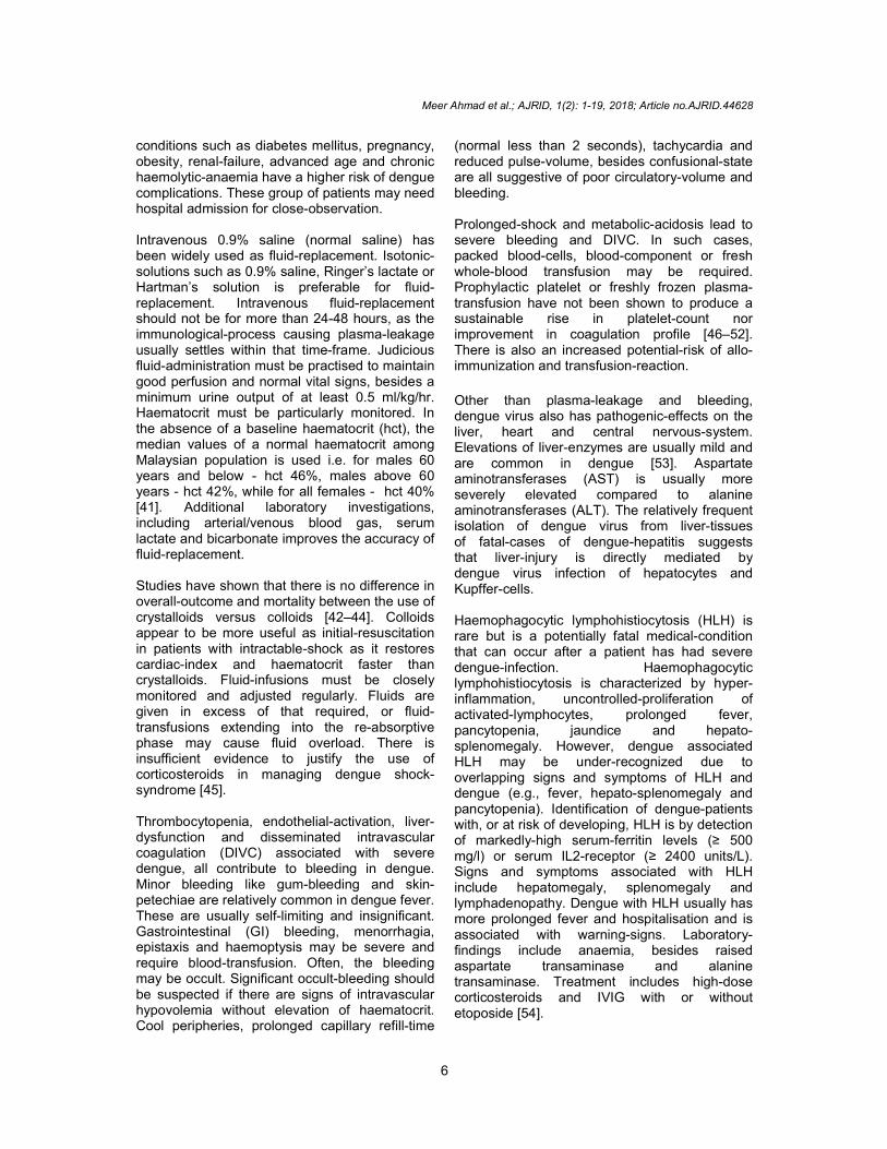
Fig. 4. Timeline of Laboratory Changes
Meer Ahmad et al.; AJRID, 1(2): 1-19, 2018; Article no.
8
lation of the virus or detection by direct immunofluorescence (IF), or
], or detection antigens like NS1 using either ELISAs or
Very often antibodies are sought for out of convenience, but these are useful diagnostics only when results are obtained using paired
isolation, PCR and Direct IF are done only in reference- and
friendly. Apart from flow tests have been developed
These are useful in the first five days of symptoms when viraemia still exists. Serology is also useful in demonstrating sero-conversion
antibody, or a four-titres in paired
nts who are IgM-positive negative are usually classified as
infection, since IgM-antibodies to dengue remain elevated for 90 days after the illness, and could have been from an infection that occurred between two and three
In addition, it is important to note that cross-viruses, including West
Nile virus (WNV), St. Louis encephalitis virus (SLE), Japanese encephalitis virus (JEV), Zika virus and yellow fever virus (YFV), do occur.
However, these infections are very rare in Malaysia. Yet, a look into the patient’s Recent Travel History would be helpful. A pairedtesting for all of the flavi-viruses, best provides a diagnosis as to which flavi-virus it is, especially in those who present late after onset of illness (> 5 days), when virus and viral antigens become undetectable [69,70].
Immune responses to dengue Dengue virus induces the production of all classes of antibodies, primarily targeting the virus envelope-proteins. The level of antibodies primarily depends on whether the individual has a primary or a secondary dengue infection [67,72]. Fig. 4 shows the timelines of Laboratory Changes viz-a-viz Clinical-features In a primary infection, low antibodyobserved - while in a secondary infection, high IgG is detectable in the acute phase which sometimes masks the detection osecondary infection [29]. In a primary infection, viraemia is present through the incubation-period till the 4illness. During this viraemia-phase virusand virus-RNA detection is possible. NS1 Antigen detection is detectable through the incubation-period till the 12
th Day (
of the illness [29].
Fig. 4. Timeline of Laboratory Changes viz-a-viz Clinical-features. [29
Article no.AJRID.44628
However, these infections are very rare in Malaysia. Yet, a look into the patient’s Recent Travel History would be helpful. A paired-sample,
viruses, best provides a virus it is, especially in
ose who present late after onset of illness (> 5 days), when virus and viral antigens become
Immune responses to dengue
Dengue virus induces the production of all classes of antibodies, primarily targeting the virus
proteins. The level of antibodies primarily depends on whether the individual has a primary or a secondary dengue infection
timelines of Laboratory features [29].
In a primary infection, low antibody-titres are while in a secondary infection, high
IgG is detectable in the acute phase which sometimes masks the detection of IgM in
In a primary infection, viraemia is present period till the 4th Day of
phase virus-isolation RNA detection is possible. NS1
Antigen detection is detectable through the Day (convalescent)
29]
Meer Ahmad et al.; AJRID, 1(2): 1-19, 2018; Article no.AJRID.44628
9
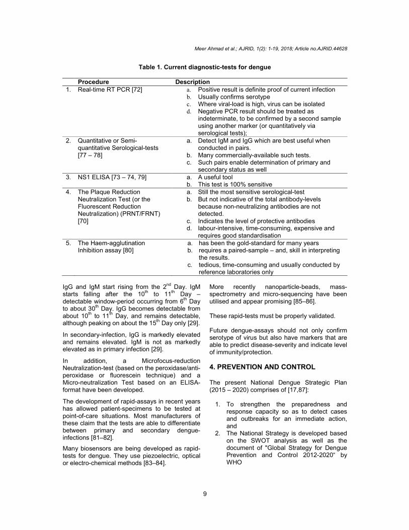
Table 1. Current diagnostic-tests for dengue Procedure Description 1. Real-time RT PCR [72] a. Positive result is definite proof of current infection
b. Usually confirms serotype c. Where viral-load is high, virus can be isolated d. Negative PCR result should be treated as
indeterminate, to be confirmed by a second sample using another marker (or quantitatively via serological tests);
2. Quantitative or Semi-quantitative Serological-tests [77 – 78]
a. Detect IgM and IgG which are best useful when conducted in pairs.
b. Many commercially-available such tests. c. Such pairs enable determination of primary and
secondary status as well 3. NS1 ELISA [73 – 74, 79] a. A useful tool
b. This test is 100% sensitive 4. The Plaque Reduction
Neutralization Test (or the Fluorescent Reduction Neutralization) (PRNT/FRNT) [70]
a. Still the most sensitive serological-test b. But not indicative of the total antibody-levels
because non-neutralizing antibodies are not detected.
c. Indicates the level of protective antibodies d. labour-intensive, time-consuming, expensive and
requires good standardisation 5. The Haem-agglutination
Inhibition assay [80] a. has been the gold-standard for many years b. requires a paired-sample – and, skill in interpreting
the results. c. tedious, time-consuming and usually conducted by
reference laboratories only IgG and IgM start rising from the 2nd Day. IgM starts falling after the 10th to 11th Day – detectable window-period occurring from 6
th Day
to about 30th Day. IgG becomes detectable from about 10
th to 11
th Day, and remains detectable,
although peaking on about the 15th Day only [29].
In secondary-infection, IgG is markedly elevated and remains elevated. IgM is not as markedly elevated as in primary infection [29].
In addition, a Microfocus-reduction Neutralization-test (based on the peroxidase/anti-peroxidase or fluorescein technique) and a Micro-neutralization Test based on an ELISA-format have been developed.
The development of rapid-assays in recent years has allowed patient-specimens to be tested at point-of-care situations. Most manufacturers of these claim that the tests are able to differentiate between primary and secondary dengue-infections [81–82].
Many biosensors are being developed as rapid-tests for dengue. They use piezoelectric, optical or electro-chemical methods [83–84].
More recently nanoparticle-beads, mass-spectrometry and micro-sequencing have been utilised and appear promising [85–86]. These rapid-tests must be properly validated. Future dengue-assays should not only confirm serotype of virus but also have markers that are able to predict disease-severity and indicate level of immunity/protection. 4. PREVENTION AND CONTROL The present National Dengue Strategic Plan (2015 – 2020) comprises of [17,87]:
1. To strengthen the preparedness and response capacity so as to detect cases and outbreaks for an immediate action, and
2. The National Strategy is developed based on the SWOT analysis as well as the document of "Global Strategy for Dengue Prevention and Control 2012-2020“ by WHO
Meer Ahmad et al.; AJRID, 1(2): 1-19, 2018; Article no.AJRID.44628
10
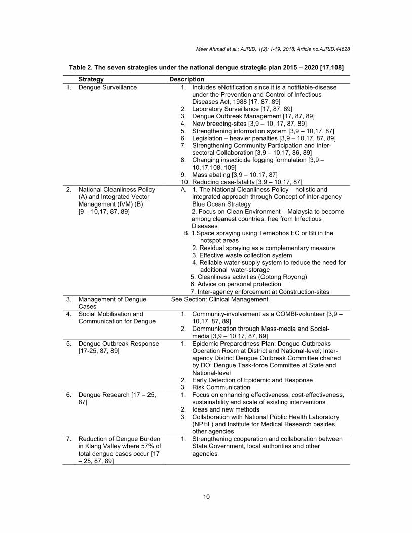
Table 2. The seven strategies under the national dengue strategic plan 2015 – 2020 [17,108]
Strategy Description 1. Dengue Surveillance 1. Includes eNotification since it is a notifiable-disease
under the Prevention and Control of Infectious Diseases Act, 1988 [17, 87, 89]
2. Laboratory Surveillance [17, 87, 89] 3. Dengue Outbreak Management [17, 87, 89] 4. New breeding-sites [3,9 – 10, 17, 87, 89] 5. Strengthening information system [3,9 – 10,17, 87] 6. Legislation – heavier penalties [3,9 – 10,17, 87, 89] 7. Strengthening Community Participation and Inter-
sectoral Collaboration [3,9 – 10,17, 86, 89] 8. Changing insecticide fogging formulation [3,9 –
10,17,108, 109] 9. Mass abating [3,9 – 10,17, 87] 10. Reducing case-fatality [3,9 – 10,17, 87]
2. National Cleanliness Policy (A) and Integrated Vector Management (IVM) (B) [9 – 10,17, 87, 89]
A. 1. The National Cleanliness Policy – holistic and integrated approach through Concept of Inter-agency Blue Ocean Strategy 2. Focus on Clean Environment – Malaysia to become among cleanest countries, free from Infectious Diseases
B. 1.Space spraying using Temephos EC or Bti in the hotspot areas
2. Residual spraying as a complementary measure 3. Effective waste collection system 4. Reliable water-supply system to reduce the need for
additional water-storage 5. Cleanliness activities (Gotong Royong) 6. Advice on personal protection 7. Inter-agency enforcement at Construction-sites
3. Management of Dengue Cases
See Section: Clinical Management
4. Social Mobilisation and Communication for Dengue
1. Community-involvement as a COMBI-volunteer [3,9 – 10,17, 87, 89]
2. Communication through Mass-media and Social-media [3,9 – 10,17, 87, 89]
5. Dengue Outbreak Response [17-25, 87, 89]
1. Epidemic Preparedness Plan: Dengue Outbreaks Operation Room at District and National-level; Inter-agency District Dengue Outbreak Committee chaired by DO; Dengue Task-force Committee at State and National-level
2. Early Detection of Epidemic and Response 3. Risk Communication
6. Dengue Research [17 – 25, 87]
1. Focus on enhancing effectiveness, cost-effectiveness, sustainability and scale of existing interventions
2. Ideas and new methods 3. Collaboration with National Public Health Laboratory
(NPHL) and Institute for Medical Research besides other agencies
7. Reduction of Dengue Burden in Klang Valley where 57% of total dengue cases occur [17 – 25, 87, 89]
1. Strengthening cooperation and collaboration between State Government, local authorities and other agencies

Meer Ahmad et al.; AJRID, 1(2): 1-19, 2018; Article no.AJRID.44628
11
The four components of a SWOT analysis examine (a) the current strengths that should be maintained and built on, (b) the weaknesses that need to be addressed, (c) the opportunities that are available for moving toward more optimal function, and (d) the threats that may prevent progress from being made [82].
In Malaysia, the current new directions in dengue-control include [17]:
1. Having all registered dengue cases confirmed by laboratory tests,
2. Increasing source-reduction activity, and 3. Reducing fogging-activity from two cycles
to one cycle
New Tools and Strategy in the Prevention and Control in Malaysia consist of:
A. New strategies in hotspot areas such as in Table 3.
For the effective control of the dengue-vector mosquito-populations, there is a need to combine several strategies, such as chemical, biological and integrated control. The chemical-insecticide is the most commonly used, as it is effective against both the larval and the adult mosquitoes [92].
The ultimate aim of insecticide-control management has two parts i.e. the control of
Aedes-immatures and the control of the adult-mosquitoes. The control of adults especially in a dengue-epidemic situation is aimed at the killing of the infective female-mosquitoes. Whereas, the control of Aedes-immatures is targeted towards the overall reduction of the mosquito population-density and, indirectly reducing the human-vector pathogen-contact to prevent dengue-transmissions [85].
One of the two common chemical-controls is Larvicide-application to treat household drinking-water containers which has low-relative toxicity and is safe for humans. The other technique is space spraying (Ultra low volume, ULV or thermal fogging), which is generally employed in emergency outbreaks of dengue [93]. Different chemical groups have been used to control Aedes spp in Malaysia since the 1960s, including organo-chlorines, organo-phosphates, carbamates and pyrethroid-insecticides. The main contributor towards today's raging dengue epidemics is the worst practice of routine space-spraying and thermal-fogging which do not kill 100% of the vector-mosquito populations. This causes an artificial-selection due to ignorance that has led to serious chemical-resistance nowadays – and, have seriously impacted human-health following the excessive uses of various insecticides more routinely and at a higher-frequency [100].
Table 3. New strategies in hotspot areas (including concepts)
Strategy Description/constraints
1. Residual spraying [17, 87]
Read within Text outside this Table
2. Larviciding activity using Temephos EC or Bti [9-10,17, 87, 89]
Read within Text outside this Table
3. Use of newer-generation insecticides [9-10,17, 87]
Read within Text outside this Table
4. Use of novel ovi-traps and other innovative methods [9-10,17, 87]
Read within Text outside this Table
5. Release of genetically-modified Aedes (A) or Wolbacchia-infected Aedes (B) [9-10, 90, 91 – 96]
A. 1. Hampered by logistical-difficulty due to flight-range of Aedes viz-a-viz release-radii in heavily built-up areas
B. 1. Same difficulty does not exist because Wolbacchia-infection passed onto progenies – should be self-propagating, but in practice such propagation not found more than 100 meters per year
2. The strain of Wolbacchia shown to be effective in this method not able to survive ambient temperatures in the tropics. This claim is controversial.
3. The Method is undergoing pilot-study by IMR in

Meer Ahmad et al.; AJRID, 1(2): 1-19, 2018; Article no.AJRID.44628
12
Selangor
6. Larviciding of primary water-sources such as water-treatment plants (A), together with Aerial-spraying (B) [89, 91, 93 – 94, 98 - 99]
A. 1.Similar to fluoridation of water
2.Pyripoxifen (after EIA done) or Bti
3. Was done in parts of Brazil
B. 1. Aerial-spraying using US CDC Protocol
C. Combined method can be implemented if safe and cost-effective vaccine still not found, after pilot-study
7. Isolation of Cases 1. Not useful since 70-80% of infections are asymptomatic, yet infective [15 – 16, 89, 91 - 94]
2. Besides, diagnosis is usually made on 3rd
to 5th day [1 –
11] The routine space-spraying and fogging pollutes the environment and the food chain - besides also directly eliminating most of the natural-enemies of the vector-mosquitoes, such as ants, spiders, dragonflies, praying-mantises, lizards, frogs, birds and bats. These natural-enemies are of great-value as vital biological-control agents in suppressing the mosquito-populations, which control-measures must be preserved for long-term successful mosquito-control [100]. In view of the above-mentioned predicament, there are innovative new-strategies developed specifically to outsmart the dengue-vector mosquito. These are described in Table 4 [100]. In the prevention and control of dengue-outbreaks, the use of household insecticide-products (HIP), such as the insecticide aerosol-sprays have been very much a part of active and sustainable community-participation [101].
They are handy and fast-action, effective in killing all the mosquitoes and always ready-to-use. The dengue-hotspot communities should pro-actively do thorough spraying in the morning and in the evening every day within their premise, to ensure that there is no infective female Aedes-mosquitoes hiding within. For non-hotspot communities, such thorough-spraying needs to be done only once a week to ensure there is no Aedes-mosquitoes breeding inside their premise. Therefore, this aerosol insecticide-spray should be integrated into the overall dengue-vectors control-program for maximum control-results [101]. However, those ordinary aerosol-insecticide come with a choking smell, cause staining and leave an oily-film on surfaces, dissuading many. The new generation of mini-aerosol spray-insecticide, equipped with metered-valve and
Table 4. Innovative new-strategy in dengue-vector control
Method Description 1. Attractant Toxic Sugar
Baiting 100 a. Attracts all the hungry and dehydrated adult-mosquitoes
(male & female) when they emerge from pupae (especially first two days)
b. Since nectar-meals are scarce indoors, the bait is the most readily-available and attractive choice.
c. Only needs placement in strategic-locations indoor d. Safe because no chemicals extruding into air or
environment e. Mostly used as supplement in control
2. Attractive Lethal Ovi-position Traps 100
a. Makes full-use of the Aedes-vector mosquito’s skip-ovi-position characteristic i.e. in using the female as mechanical-vectors to cross-contaminate the other breeding-sites which are beyond our detection.
b. Attracts gravid-mosquitoes to come and lay eggs in the special-station that contains water and a lacing-formulation of oviposition-attractants. All (100%) of these eggs cannot develop into adult-mosquitoes.
c. The formulation has the insect growth-regulator, IGR, which contaminates these female-mosquitoes - when they lay

Meer Ahmad et al.; AJRID, 1(2): 1-19, 2018; Article no.AJRID.44628
13
eggs in their hidden breeding-sites in the wild, they go on to cross-contaminate all the breeding-sites, and all the hidden-eggs.
d. All of the chemicals used in this, always stay inside the station - thus protecting all the natural-enemies of the mosquito and ensuring sustainable natural biological-control
slow-release nano-technology formulation (active ingredient is meto-fluthrin at 0.76%w/w) has been developed to overcome all these negative aspects – they are odourless, clean and dry, very low-volatile organic compounds (VOCs), non-oily, non health-hazard and eco-friendly compared to the ordinary aerosol-insecticide [101]. In the standard room of up to 30 m
3, one needs
to spray at the four corners. Each 83ml (50g) mini aerosol-spray can deliver the fixed-amount in 800 sprays of protection in 200 rooms, providing mosquito-free protection for up to 8 hours. In comparison, the ordinary 600ml (380 g) aerosol-spray could only spray up to 42 rooms and provide an hour of mosquito-free protection. For smaller spaces like in a car, this mini spray can be sprayed once in the car to ensure no mosquitoes while driving. In this manner, it is also a great tool in preventing mosquitoes being transported from one location to another. Outside-fogging can also be done [101]
B. Specific protection Primary Prevention of diseases classically comprises of Health Promotion and Specific Protection. [91], [95 – 97, 103]. Health Promotion has been extensively outlined above. Specific Protection should comprise of an appropriate Mass Vaccination Program of Endemic Areas, appropriate use of effective mosquito-repellents such as DEET, lemon eucalyptus or picaridin, and the appropriate use of mosquito-nets by day-sleeping children, the elderly and the infirm. The last two can be made available, subsidised, at Health Clinics throughout the country [1 – 10, 91,95–97]. In late 2015 and early 2016, the first dengue vaccine, Dengvaxia (CYD-TDV) by Sanofi Pasteur, was registered in several countries for use in individuals 9-45 years of age living in endemic areas. But overall, the much waited-for dengue-vaccine has been a disappointment both in its efficacy and its safety [104–108]. If a sufficiently effective and safe vaccine can be found, it will transform dengue fever into a
vaccine-preventable disease, and the disease can be quickly brought to near-eradication levels just like all other previous vaccine-preventable diseases. Takeda Pharmaceutical Company Limited, (“Takeda”) in November 2017 announced the data from an 18-month interim analysis of the ongoing Phase 2 DEN-204 trial of its live, attenuated tetravalent dengue vaccine-candidate, TAK-003 (also referred to as TDV). This interim-analysis showed that children and adolescents who received TAK-003 had a relative-risk of symptomatic-dengue of 0.29 (95% CI: 0.13–0.72) compared to children and adolescents in the placebo control-group [109]. TAK-003 was found to be safe and well-tolerated in terms of solicited local-reactions and systemic adverse-events, relative to the placebo control-group [104]. In participants who were sero-negative at baseline, a second-dose given at Month 3 improved the tetravalent sero-positivity rate at Month 6 to 86%, compared to 69% in the one-dose group. A booster dose at Month 12 resulted in a 100% tetravalent sero-positivity rate at Month 13 in participants who were sero-negative at baseline [109]. TAK-003 is currently under evaluation in the Tetravalent Immunization against Dengue Efficacy Study (TIDES), a large-scale Phase 3 efficacy-trial being conducted in eight dengue-endemic countries. Data from TIDES will be available in late 2018 [109]. The US National Institute of Allergy and Infectious Diseases (NIAID) has developed the LATV dengue vaccines TV003/TV005. A single dose of either TV003 or TV005 induced sero-conversion to four DENV serotypes in 74-92% (TV003) and 90% (TV005) of flavivirus-seronegative adults and elicited near-sterilizing immunity to a second dose of vaccine administered 6-12 months later [110–112]. The Phase III clinical-trial of the TV003 commenced in February 2016 among 17,000

Meer Ahmad et al.; AJRID, 1(2): 1-19, 2018; Article no.AJRID.44628
14
volunteers in multiple locations in Brazil with the aim of determining its efficacy and safety. The estimated primary-completion date is June 2018, and the estimated study-completion date is December 2022 [110–112]. When vaccines are available which afford greater than 90% protection against all four strains, the risk of antibody-directed enhancement (ADE) in subsequent natural-infections, causing severe dengue, becomes remote because secondary infections would be rare. Dengue fever very likely will become reduced to sporadic-outbreaks of mostly the Sylvan-type, just like yellow-fever, once a successful mass-vaccination program of a safe and highly-effective vaccine becomes feasible and affordable.
5. CONCLUSION In conclusion, dengue fever and its complications have been a serious scourge of mankind for too long in recent history, affecting countries across the globe. The case-fatality rate of the disease in these countries, including Malaysia, is not negligible. However, Clinical Management has brought about vast improvements in mortality and morbidity. Similarly, great advancements in Laboratory Diagnostics have been seen. Prevention and Control methods have been desiring of greater achievements, but also show greater promise with comprehensive re-evaluated programmes, newer insecticides, innovative methods and vaccines. Dengue fever would very likely become near-eradicated just like all other vaccine-preventable diseases, once comprehensive mass-vaccination programmes are available globally, using safe and very-effective tetravalent-vaccines soon to be available.
CONSENT It is not applicable. ETHICAL APPROVAL
It is not applicable. ACKNOWLEDGEMENT The Authors would like to thank Dato Seri Dr. Suresh Lachman – Consultant Physician Assunta Hospital, Professor Dato Dr. Khairul
Anuar bin Abdullah – Vice-chancellor, MAHSA University, Professor Dato Dr. Ravindran Jegasothy – Dean, Medical Faculty, MAHSA University, Professor Dato Dr. Abdul Razak bin Abdul Muttalif – HOD Internal Medicine Dept., MAHSA University, Professor Dr. Hematram Yadav and Associate Professor Dato Dr. CS Prathapa Senan – Community Medicine Dept., MAHSA University, Professor Wan Ariffin Abdullah – Paediatric Dept., MAHSA University and Dr. Anbazhagan Kuppusamy – Consultant Infectious Diseases Physician, Manipal Hospital Klang, for their valuable input in proof-reading the manuscript of this Article. The Authors would like to thank Dr. Zailiza bte Suli of the Ministry of Health Malaysia, for her kind permission in letting us use Fig. 2 and Fig. 3 in this Article from her Presentation in 2016, besides thanking Professor Shamala Sekaran, Microbiology & Immunology Dept., MAHSA University, for her guidance in the Laboratory Diagnostics part.
COMPETING INTERESTS Dr. CA Koay declares that he is Technical Manager of a Firm that sells one brand of the ‘mini-aerosol spray-insecticide’, one brand of the ‘attractant baiting’ and one brand of the ‘ovi-position traps’. The remaining Co-authors declare that they have no Conflict of Interest whatsoever, in writing this Article.
REFERENCES
1. WHO. Dengue and severe dengue. April 2017.
2. US Centers for Disease Control. Dengue. 19 Jan 2016.
3. Bains S. Severe Dengue Infection. Medscape. Oct 12, 2017.
4. WHO. Dengue control. 2018. 5. WHO REGIONAL OFFICE FOR THE
WESTERN PACIFIC. THE DENGUE STRATEGIC PLAN FOR THE ASIA PACIFIC REGION. Fifty-ninth session 25 July 2008 Manila, Philippines. 22–26 September 2009.
6. WHO Western Pacific Region. WHO ASEAN Workshop on Priority Actions for Dengue Prevention and Control. 3 to 5 May 2011. Manila, Philippines
7. WHO Regional Office for South-east Asia. Comprehensive guidelines for Prevention and Control of Dengue and Dengue Haemorrhagic Fever; 2011.
Meer Ahmad et al.; AJRID, 1(2): 1-19, 2018; Article no.AJRID.44628
15
8. WHO. Dengue guidelines for diagnosis, treatment, prevention and control. WHO/HTM/NTD/DEN/2009.1. Expiry date: 2014
9. Song-Quan Ong. Dengue Vector Control in Malaysia: A Review for Current and Alternative Strategies. Sains Malaysiana. 2016.45(5):777–785.
10. Ang Kim Teng, Satwant Singh. Epidemiology and new initiatives in the prevention and control of dengue in Malaysia. Dengue Bulletin. 2001;25.
11. Kumarasamy V. Dengue fever in Malaysia: Time for review?. Med J Malaysia. 2006;61(1).
12. Paupy C, Delatte H, Bagny L, et al. Institute Pasteur. Aedes albopictus, an arbovirus vector: From the darkness to the light. Microbes Infect. 2009;11(14-15):1177-85.
13. Whitehorn J, Kien DT, Nguyen NM, Nguyen HL, Kyrylos PP, Carrington LB, et al. Comparative Susceptibility of Aedes albopictus and Aedes aegypti to Dengue Virus Infection After Feeding on Blood of Viremic Humans: Implications for Public Health. J Infect Dis. 2015;212(8):1182-90.
14. Vaughn DW, Green S, Kalayanarooj S, Innis BL, Nimmannitya S, Suntayakorn S, et al. Dengue viremia titer, antibody response pattern, and virus serotype correlate with disease severity. J Infect Dis. 2000;181(1):2-9.
15. Alera MT, Srikiatkhachorn A, Velasco JM, Tac-An IA, Lago CB, Clapham HE, et al. Incidence of Dengue Virus Infection in Adults and Children in a Prospective Longitudinal Cohort in the Philippines. PLoS Negl Trop Dis. 2016;10(2): e0004337.
16. Kosasih H, Alisjahbana B, Nurhayati, de Mast Q, Rudiman IF, Widjaja S, et al. The Epidemiology, Virology and Clinical Findings of Dengue Virus Infections in a Cohort of Indonesian Adults in Western Java. PLoS Negl Trop Dis. 2016;10(2): e0004390.
17. Zailiza Suli. Dengue Prevention and Control in Malaysia. International Conference on Dengue Prevention and Control & International Dengue Expert Consultation Meeting. December 7-8 , 2015. The Magic School of Green Technologies, National Cheng Kung University, Tainan City. Ministry of Health Malaysia.
18. José de Jesus Dias, Júnior, Maria dos Remédios Freitas Carvalho Branco, Rejane Christine de Sousa Queiroz, Alcione Miranda dos Santos, Emnielle Pinto Borges Moreira, and Maria do Socorro da Silva. Analysis of dengue cases according to clinical severity, São Luís, Maranhão, Brazil. Rev Inst Med Trop Sao Paulo. 2017;59:e71.
19. Sam SS, Omar SF, Teoh BT, Abd-Jamil J, AbuBakar S. Review of Dengue hemorrhagic fever fatal cases seen among adults: A retrospective study. PLoS Negl Trop Dis. 2013;7(5):e2194.
20. Shepard DS, Undurraga EA, Lees RS, Halasa Y, Lum LC, Ng CW. Use of multiple data sources to estimate the economic cost of dengue illness in Malaysia. Am J Trop Med Hyg. 2012;87(5):796-805.
21. P.Raviwharmman Packierisamy, Chiu-Wan Ng, Maznah Dahlui, Jonathan Inbaraj, Venugopalan K. Balan, Yara A. Halasa, et al. Cost of Dengue Vector Control Activities in Malaysia. Am J Trop Med Hyg. 2015;93(5):1020–1027.
22. Ibid. Petaling Jaya City Council. 2017. 23. Nor Azila, Sharifah Azura Salleh, Hui-min
Neoh, Syed Zulkifli Syed Zakaria and Rahman Jamal. Dengue epidemic in Malaysia: Not a predominantly urban disease anymore. BMC Research Notes. 2011;4:216.
24. Ministry of Health Malaysia. Health Ministry: Dengue cases down in 2017 compared to 2016. The Star Online. 10 Jan 2018.
25. Ministry of Health Malaysia. Almost 83,000 dengue cases nationwide in 2017; 171 deaths. New Straits Times online. 7 April 2018.
26. Wiwanitkit V. Dengue fever: diagnosis and treatment. Expert Rev Anti Infect Ther. 2010;8(7):841-5.
27. Lai YC, Chuang YC, Chang CP, Lin YS, Perng GC, Wu HC, et al. Minocycline suppresses dengue virus replication by down-regulation of macrophage migration inhibitory factor-induced autophagy. Antiviral Res. 2018;155:28-38.
28. Nitsche C. Strategies towards protease inhibitors for emerging flaviviruses. Adv Exp Med Biol. 2018;1062:175-186.
29. Academy of Medicine of Malaysia - Clinical Practice Guidelines. 2014, Recommendations for Patient Safety and Minimal Monitoring Standards during ...
Meer Ahmad et al.; AJRID, 1(2): 1-19, 2018; Article no.AJRID.44628
16
2015, Management of Dengue Infection in Adults (3rd Edition).
30. Rigau-Perez JG, Laufer MK. Dengue-related deaths in Puerto Rico, 1992-1996: Diagnosis and clinical warning signs. Clin Infect Dis. 2006;42(9):1241-6.
31. Ong A, Sandar M, Chen MI, Sin LY. Fatal dengue haemorrhagic fever in adults during 9. A dengue epidemic in Singapore. International Journal of Infectious Diseases. 2007;11(3):263-7.
32. Wichmann O, Hongsiriwon S, Bowonwatanuwong C, Chotivanich K, Sukthana Y, Pukrittayakamee S. Risk factors and clinical features associated with severe dengue infection in adults and children during the 2001 epidemic in Chonburi, Thailand. Trop Med Int Health. 2004;9(9):1022-9.
33. Simmons CP, et al. Current concepts: Dengue. N Engl J Med. 2012;366:1423-32
34. WHO. Dengue: Guidelines for diagnosis, treatment, prevention and control - new edition. WHO, Geneva; 2009.
35. Taraphdar D, Sarkar A, Mukhopadhyay BB, Chatterjee S. A comparative study of clinical features between monotypic and dual infection cases with Chikungunya virus and dengue virus in West Bengal, India. Am J Trop Med Hyg. 2012;86:720.
36. Lee VJ, Chow A, Zheng X, Carrasco LR, Cook AR, Lye DC, Ng LC, Leo YS. Simple clinical and laboratory predictors of Chikungunya versus dengue infections in adults. PLoS Negl Trop Dis. 2012;6:e1786.
37. Mohd Zim MA, Sam IC, Omar SF, Chan YF, AbuBakar S, Kamarulzaman A. Chikungunya infection in Malaysia: comparison with dengue infection in adults and predictors of persistent arthralgia. J Clin Virol. 2013;56:141.
38. Nimmannitya S. Clinical spectrum and management of dengue haemorrhagic fever. Southeast Asian J Trop Med Pub Hlth. 1987;18(3):392-7.
39. Gubler DJ. Dengue and dengue haemorrhagic fever. Clin Microbio Review. 1998;11(3):480-96.
40. Maria G. Guzmán, Mayling Alvarez, Rosmari Rodriguez, Delfina Rosario, Susana Vázquez, Luis Valdés. Fatal dengue hemorrhagic fever in Cuba, 1997. Int J Infect Dis. 1999;3:130–5.
41. Angeli Ambayya, Anselm Ting Su, Nadila Haryani Osman, Nik Rosnita Nik-Samsudin, Khadijah Khalid, Kian Meng Chang, et al. Haematological reference
intervals in a multiethnic population. PLoS One. 2014;9(3):e91968.
42. Ngo NT, Cao XT, Kneen R, Wills B, Nguyen VM, Nguyen TQ, et al. Acute management of dengue shock syndrome: A randomized double-blind comparison of 4 intravenous fluid regimens in the first hour. Clin Infect Dis. 2001;32:204.
43. Dung NM, Day NP, Tam DT, Loan HT, Chau HT, Minh LN, et al. Fluid replacement in dengue shock syndrome: A randomized, double-blind comparison of four intravenous-fluid regimens. Clin Infect Dis. 1999;29:787.
44. Wills BA, Nguyen MD, Ha TL, Dong TH, Tran TN, Le TT, et al. Comparison of three fluid solutions for resuscitation in dengue shock syndrome. N Engl J Med. 2005; 353:877.
45. Panpanich R, Sornchai P, Kanjanaratanakorn K. Corticosteroids for treating dengue shock syndrome. Cochrane Database Syst Rev. 2006;CD003488
46. WHO. Dengue: guidelines for diagnosis, treatment, prevention and control - new edition. WHO, Geneva 2009
47. Thomas L, Kaidomar S, Kerob-Bauchet B, Moravie V, Brouste Y, King JP, et al. Prospective observational study of low thresholds for platelet transfusion in adult dengue patients. Transfusion. 2009;49: 1400.
48. Khan Assir MZ, Kamran U, Ahmad HI, Bashir S, Mansoor H, Anees SB, et al. Effectiveness of platelet transfusion in dengue Fever: a randomized controlled trial. Transfus Med Hemother. 2013; 40:362.
49. Lye DC, Lee VJ, Sun Y, Leo YS. Lack of efficacy of prophylactic platelet transfusion for severe thrombocytopenia in adults with acute uncomplicated dengue infection. Clin Infect Dis. 2009;48:1262.
50. Lye DC, Archuleta S, Syed-Omar SF, Low JG, Oh HM, Wei Y, et al. Prophylactic platelet transfusion plus supportive care versus supportive care alone in adults with dengue and thrombocytopenia: a multicentre, open-label, randomised, superiority trial. Lancet. 2017;389:1611.
51. Lum LC, Abdel-Latif Mel-A, Goh AY, Chan PW, Lam SK. Preventive transfusion in DSS- is it necessary? J Pediatr. 2003;143(5):682-4.
52. Chairulfatah, A, Setiabudi, D., Agoes, Colebunders R. Thrombocytopenia and

Meer Ahmad et al.; AJRID, 1(2): 1-19, 2018; Article no.AJRID.44628
17
platelet transfusions in dengue haemorrhagic fever and dengue shock syndrome. Dengue Bulletin. 2003;27.
53. Kalayanarooj S, Vaughn DW, Nimmannitya S, Green S, Suntayakorn S, Kunentrasai N, et al. Early clinical and laboratory indicators of acute dengue illness. J Infect Dis. 1997;176:313.
54. Esther M Ellis, Tyler M. Sharp, Janice Perez-Padilla, Liza González, B. Katherine Poole-Smith, Emmaculate Lebo, et al. Incidence and risk factors developing Dengue-Associated Hemophagocytic Lymphohistiocytosis in Puerto-Rico, 2008 - 2013. PLOS Neglected Tropical Diseases. 2016;1-14.
55. Cam BV, Fonsmark L, Hue NB, Phuong NT, Poulsen A, Heegaard ED. Prospective case control study of 127 encephalopathy in children with dengue hemorrhagic fever. Am J Trop Med Hyg Dec. 2001;65(6):848–51.
56. Hendarto SK, Hadinegoro SR. Dengue encephalopathy. Acta Paediatr Jpn. 1992; 34 (3):350–7.
57. Carod-Artal FJ, Wichmann O, Farrar J, Gascón J. Neurological complications of dengue 129 virus infection. Lancet Neurol. 2013;12(9):906-19.
58. Low JG, Gatsinga R, Vasudevan SG, Sampath A. Dengue antiviral development: A continuing journey. Adv Exp Med Biol. 2018;1062:319-332.
59. Noble CG, Shi PY. Structural biology of dengue virus enzymes: Towards rational design of therapeutics. Antiviral Res. 2012;96:115.
60. Krishnan MN, Garcia-Blanco MA. Targeting host factors to treat West Nile and dengue viral infections. Viruses. 2014;6:683.
61. Nguyen NM, Tran CN, Phung LK, Duong KT, Huynh Hle A, Farrar J, et al. A randomized, double-blind placebo controlled trial of balapiravir, a polymerase inhibitor, in adult dengue patients. J Infect Dis. 2013;207:1442.
62. Low JG, Sung C, Wijaya L, Wei Y, Rathore APS, Watanabe S, et al. Efficacy and safety of celgosivir in patients with dengue fever (CELADEN): A phase 1b, randomised, double-blind, placebo-controlled, proof-of-concept trial. Lancet Infect Dis. 2014;14:706.
63. Whitehorn J, Nguyen CV, Khanh LP, Kien DTH, Quyen NTH, Tran NTT, et al. Lovastatin for the treatment of adult
patients with dengue: A randomized, double-blind, placebo-controlled trial. Clin Infect Dis. 2016;62:468.
64. Prommalikit O, Thisyakorn U. Dengue virus virulence and diseases severity. Southeast Asian J Trop Med Public Health. 2015;46(Suppl 1):35-42.
65. Lin CF, Wan SW, Cheng HJ, Lei HY, Lin YS. Autoimmune pathogenesis in dengue virus infection. Viral Immunol. 2006;19(2): 127-32.
66. Soo KM, Khalid B, Ching SM, Chee HY. Meta-Analysis of Dengue Severity during Infection by Different Dengue Virus Serotypes in Primary and Secondary Infections. PLoS One. 2016;11(5): e0154760
67. Sekaran, Shamala Devi. Laboratory Diagnosis of Dengue: A Review. International Medical Journal of Malaysia. 2014;14(1):17-28.
68. Sekaran SD, Soh HJ. Issues in contemporary and potential future molecular diagnostics for dengue. Expert Review of Molecular Diagnostics; 2016.
69. Guzman MG, Halstead SB, Artsob H, Buchy P, Farrar J, Gubler DJ, et al. Dengue: A continuing global threat. Nat Rev Microbiol. 2010;8:S7-16.
70. Sekaran SD, Artsob H. Molecular diagnostics for the detection of human flavivirus infections. Expert Opinion on Medical Diagnostics. 2007;1:521-30.
71. Mathew A, Rothman AL. Understanding the contribution of cellular immunity to dengue disease pathogenesis. Immunol Rev. 2008;225:300-13.
71. Mathew A, Rothman AL. Understanding the contribution of cellular immunity to dengue disease pathogenesis. Immunol Rev. 2008;225:300-13.
72. Kong YY, Thay CH, Tin TC, Devi S. Rapid detection, serotyping and quantitation of dengue viruses by TaqMan real-time one-step RT-PCR. J Virol Methods. 2006;138: 123-30.
73. Pal S, Dauner AL, Mitra I, Forshey BM, Garcia P, Morrison AC, et al. Evaluation of Dengue NS1 Antigen Rapid Tests and ELISA Kits Using Clinical Samples. PloS One. 2014;9(11):e113411.
74. Bessoff K, Delorey M, Sun W, Hunsperger E. Comparison of two commercially available dengue virus (DENV) NS1 capture enzyme-linked immunosorbent assays using a single clinical sample for

Meer Ahmad et al.; AJRID, 1(2): 1-19, 2018; Article no.AJRID.44628
18
diagnosis of acute DENV infection. Clin Vaccine Immunol. 2008;15:1513–18.
75. Wang SM, Sekaran SD. Early diagnosis of Dengue infection using a commercial Dengue Duo rapid test kit for the detection of NS1, IGM, and IGG. Am J Trop Med Hyg. 2010;83:690-95.
76. Wang SM, Sekaran SD. Evaluation of a commercial SD dengue virus NS1 antigen capture enzyme-linked immunosorbent assay kit for early diagnosis of dengue virus infection. J Clin Microbiol. 2010;48: 2793-97.
77. Pal S, Dauner AL, Valks A, Forshey BM, Long KC, Thaisomboonsuk B, et al. Multicountry prospective clinical evaluation of two enzyme-linked immunosorbent assays and two rapid diagnostic tests for diagnosing dengue fever. J Clin Microbiol. 2014;53:1092-1102.
78. Blacksell SD, Jarman RG, Gibbons RV, Tanganuchitcharnchai A, Mammen MP Jr, Nisalak A, et al. Comparison of seven commercial antigen and antibody enzyme-linked immunosorbent assays for detection of acute dengue infection. Clin Vaccine Immunol. 2012;19:804-10.
79. Guzman MG, Jaenisch T, Gaczkowski R, Hang VTT, Sekaran SD, Kroeger A, et al. Multi-country evaluation of the sensitivity and specificity of two commercially-available NS1 ELISA assays for dengue diagnosis. PLoS Neglected Tropical Diseases. 2010;4:e811.
80. Clarke DH, Casals J. Improved methods for hemagglutination studies with arthropod-borne viruses. Proc Soc Exp Biol Med. 1955;88:96-9.
81. Naz A, Zahid D, Murky SN, Nadeem M, Sil BK, Shamsi TS. Evaluation of efficacy of various immunochromatographic rapid tests for dengue diagnosis. Pak J Med Sci. 2014;30:166-71.
82. Blayney DW. Strengths, weaknesses, opportunities, and threats. J Oncol Pract. 2008;4(2):53.
83. Ferraz FO, Bomfim MRQ, Totola AH, Ávila TV, Cisalpino D, Pessanha JEM, et al. Evaluation of laboratory tests for dengue diagnosis in clinical specimens from consecutive patients with suspected dengue in Belo Horizonte, Brazil. J Clin Viro. 2013;58:41-6.
84. Wu TZ, Su CC, Chen LK, et al. Piezoelectric immunochip for the detection of dengue fever in viremia phase. Biosens Bioelectron. 2005;21:689-95.
85. Antje J. Baeumner, Nicole A. Schlesinger, Naomi S. Slutzki, Joseph Romano, Eun Mi Lee, and Richard A. Montagna, et al. Biosensor for Dengue Virus Detection: Sensitive, Rapid, and Serotype Specific. Anal Chem. 2002;74:1442-8.
86. Fatimah Ibrahim, Tzer Hwai Gilbert Thio, Tarig Faisal. Diagnosis and management of tropical diseases: A review. Sensors. 2015;15:6947-6995;
87. Antunes, P. et al. Quantification of NS1 dengue biomarker in serum via optomagnetic nanocluster detection. Sci. Rep. 2015. 5, 16145
88. Health Ministry Malaysia. Pelan Strategik Pencegahan Dan Kawalan Denggi 2009 – 2013. Bahagian Kawalan Penyakit, Kementerian Kesihatan Malaysia. 4 Februari 2009.
89. Meer Ahmad AM. Target real Aedes breeding grounds. The Star Online (Letter to Editor). 7 Jan 2017.
90. Meer Ahmad AM. Questions over dengue control methods. The Star Online (Letter to Editor). 8 Nov 2016.
91. Meer Ahmad AM. Dengue vaccine is still viable. The Star Online (Letter to Editor). 18 Aug 2016.
92. Bisset JA, Rodríguez MM, San Martín JL, Romero JE, Montoya R. Evaluación de la resistencia a insecticidas de una cepa de Aedes aegypti de El Salvador. Rev. Panam. Salud Publ. 2009;26(3):229-234.
93. World Health Organization. Vector surveillance and control. In: Dengue Haemorrhagic Fever – Diagnosis, treatment, prevention and control. 2nd ed. Geneva. 1997;48-59.
94. Yap HH, Chong NL, Foo AES, Lee CY. Dengue vector control: Present status and future prospects. Kaohsiung J Med Sci. 1994;10:S102—Slog.
95. Meer Ahmad AM. Reduce cost of dengue tests. The Star Online (Letter to Editor). 22 Jan 2016.
96. Meer Ahmad AM. Why shun the dengue vaccine? Free Malaysia Today News Portal. (Letter to Editor) 30 June 2016.
97. Meer Ahmad AM. A simple strategy to control dengue fever. Free Malaysia Today News Portal. (Letter to Editor) 12 March 2016.
98. Ross PA, Wiwatanaratanabutr, Axford JK, White VL, Endersby-Harshman NM, Hoffmann AA. Wolbachia Infections in Aedes aegypti Differ Markedly in Their

Meer Ahmad et al.; AJRID, 1(2): 1-19, 2018; Article no.AJRID.44628
19
Response to Cyclical Heat Stress. PLoS Pathog. 2017;13(1):e1006006.
99. Ye YH, Carrasco AM, Dong Y, Sgrò CM, McGraw EA. The effect of temperature on Wolbachia-Mediated dengue virus blocking in Aedes aegypti. Am J Trop Med Hyg. 2016;94(4):812-9.
100. Schmidt TL, Barton NH, Rašić G, Turley AP, Montgomery BL, Iturbe-Ormaetxe I, et al. Local introduction and heterogeneous spatial spread of dengue-suppressing Wolbachia through an urban population of Aedes aegypti. PLoS Biol. 2017; 15(5):e2001894.
101. Alyssa Navarro. Clearing the Air: Monsanto Breaks Silence on Zika Virus, Microcephaly and Sumitomo Larvicide. Tech Times. 16 February 2016. Available:http://www.techtimes.com/articles/133955/20160216/clearing-the-air-monsanto-speaks-out-about-zika-virus-microcephaly-and-sumitomo-larvicide.htm
102. US Centers for Disease Control. Information on Aerial Spraying; Aerial Spraying and Mosquito Control. 10 October 2017.
103. “Introduction to Epidemiology” in A Textbook of Community Medicine in South-east Asia. Phoon and Chen PCY, Editors. John Wiley & Sons Ltd.; 1986.
104. Meer Ahmad AM. Reveal vaccine’s efficacy, safety. The New Straits Times. 21 Feb 2017.
105. Meer Ahmad AM. Doubts in the Efficacy and Safety of the CYD-TDV Dengue-Vaccine. Berita MMA Newsletter. March 2017.
106. Ferguson N, Isabel Rodríguez-Barraquer, Ilaria Dorigatti, Luis Mier-y-Teran-Romero,
Daniel J. Laydon, Derek A. Benefits and risks of the Sanofi-Pasteur dengue vaccine: Modeling optimal deployment. Science. 2016;353(6303):1033-1036.
107. Health Ministry Malaysia. Ministry warns of effects of Dengvaxia. The Star Online. Sunday, 3 Dec 2017.
108. Health Ministry Malaysia. Dengvaxia dengue vaccine not distributed in Malaysia, says Health DG. Malay Mail. 03 December 2017.
109. Takeda Newsroom. Takeda’s Dengue Vaccine Candidate Associated with Reduced Incidence of Dengue in Children and Adolescents; New 18-Month Interim Phase 2 Data Published in The Lancet Infectious Diseases. 7 Nov 2017.
110. NIH US National Library of Medicine. Phase III Trial to Evaluate Efficacy and Safety of a Tetravalent Dengue Vaccine. Butantan Institute. ClinicalTrials.gov. 13 March 2018.
111. Whitehead SS. Development of TV003/TV005, a single dose, highly immunogenic live attenuated dengue vaccine; what makes this vaccine different from the Sanofi-Pasteur CYD™ vaccine?. Expert Rev Vaccines. 2016;15(4):509–517. Published Online. 2015 Dec 2.
112. Whitehead SS, Anna P. Durbin , Kristen K. Pierce, Dan Elwood, Benjamin D. McElvany, Ellen A. Fraser, et al. In a randomized trial, the live attenuated tetravalent dengue vaccine TV003 is well-tolerated and highly immunogenic in subjects with flavivirus exposure prior to vaccination. PLOS: Neglected Tropical Diseases. 8 May 2017.
_________________________________________________________________________________ © 2018 Meer Ahmad et al.; This is an Open Access article distributed under the terms of the Creative Commons Attribution License (http://creativecommons.org/licenses/by/4.0), which permits unrestricted use, distribution, and reproduction in any medium, provided the original work is properly cited.
Peer-review history: The peer review history for this paper can be accessed here:
http://www.sciencedomain.org/review-history/26895