![Influence of C-5 substituted cytosine and related nucleoside analogs on the formation of benzo[a]pyrene diol epoxide-dG adducts at CG base pairs of DNA](https://static.fdokumen.com/doc/165x107/6324883058da543341065147/influence-of-c-5-substituted-cytosine-and-related-nucleoside-analogs-on-the-formation.jpg)
A p-Menth-1-ene-4,7-diol (EC-1) from Eucalyptus camaldulensis Dhnh. Triggers Apoptosis and Cell...
9
A p-Menth-1-ene-4,7-diol (EC-1) from Eucalyptus camaldulensis Dhnh. Triggers Apoptosis and Cell Cycle Changes in Ehrlich Ascites Carcinoma Cells Farhadul Islam, 1,5 Jahan Ara Khanam, 1 * Mahbuba Khatun, 1 Natasha Zuberi, 1 Laboni Khatun, 1 Syed Rashel Kabir, 1 Md Abu Reza, 2 MM Ali, 3 M A Rabbi, 4 Vinod Gopalan 5 and Alfred King-Yin Lam 5 * 1 Department of Biochemistry and Molecular Biology, University of Rajshahi, Rajshahi, Bangladesh 2 Department of Genetic Engineering and Biotechnology, University of Rajshahi, Rajshahi, Bangladesh 3 Department of Applied Chemistry and Chemical Engineering, University of Rajshahi, Rajshahi, Bangladesh 4 BCSIR Laboratories, Rajshahi, Bangladesh 5 Cancer Molecular Pathology, Griffith Health Institute, Griffith University, Gold Coast, Australia Anticancer activities of p-menth-1-ene-4,7-diol (EC-1) isolated from Eucalyptus camaldulensis Dhnh. were stud- ied on Ehrlich ascites carcinoma (EAC) cells by MTT (3-[4,5-dimethylthiazol-2-yl]-2,5 diphenyl tetrazolium bro- mide) assay. Anticancer activities also analyzed in EAC-bearing mice by assessment of cancer growth inhibition, changes in cancer volume, changes in life span, and hematological parameters. Apoptosis was analyzed by fluo- rescence microscope, DNA fragmentation assay, and flow cytometry. The expression of apoptosis-related genes, Bcl-2, Bcl-X, PARP-1, p53, and Bax, were analyzed using polymerase chain reaction (PCR). EC-1 significantly inhibited proliferation of EAC cells in vivo and restored the altered hematological parameters of EAC-bearing mice. Cytological observation by fluorescence microscope showed apoptosis of EAC cells upon treatment with EC-1. Also, DNA fragmentation assay revealed EAC cells’ apoptosis following EC-1 treatment. Increased mRNA expressions of p53 and Bax genes and negative expressions of Bcl-2 and Bcl-X were observed in cells treated with EC-1. These findings confirmed the induction of apoptosis by EC-1. In addition, MTT assay showed dose-dependent anticancer activity of EC-1 against EAC cell. Cell cycle analysis revealed that EC-1 treatment caused suppression of EAC cells at S phase. To conclude, EC-1 is a novel anticancer compound and showed antiproliferative and apoptotic activities in cellular and mice models. Copyright © 2015 John Wiley & Sons, Ltd. Keywords: apoptosis; cell cycle suppression; Eucalyptus camaldulensis; cancer; EAC cells. INTRODUCTION The genus Eucalyptus (family: Myrtaceae) is a group of plants that includes many species and subspecies that are native to Australia. It has a worldwide distribu- tion including regions in Bangladesh (Hossain and Pasha, 2004; Brooker and Kleinig, 2009). The leaves and bark of these plants have been widely used as traditional medicine for the treatment of many diseases such as influenza, dysentery, tonsillitis, and gastrointes- tinal tract infections (Takasaki et al., 2000; Silva et al., 2003; Tatsuya et al., 2008). Bioactive compounds from Eucalyptus camaldulensis, Eucalyptus urophylla, and other species also have cytotoxic, antibacterial, antifun- gal, analgesic, antiinflammatory, antioxidant, and larvi- cidal activities (Cimanga et al., 2002; Silva et al., 2003; Benyahia et al., 2005; Su et al., 2006; Siramon and Ohtani, 2007; Cheng et al., 2009). A plant under this family, E. camaldulensis Dhnh., is used traditionally for the treatment of various ailments including bronchial catarrh, fever, croup, diphtheria, whooping cough, wound, ulcer, and so on (Coelho-de-Souza et al., 2005). The plant also showed antibacterial, antinociceptive effects, and cytotoxic activity against different cancer cell lines both in vivo and in vitro (EI-Ghorab et al., 2003; Al-Fatimi et al., 2005; Abdel-Nasser et al., 2011). Our group has previously reported that petroleum ether and methanol extracts of this plant exhibit promis- ing anticancer activity against Ehrlich ascites carcinoma (EAC) cells in Swiss albino mice in a dose-dependent manner (Islam et al., 2012; Islam et al., 2014a). These re- sults inspired us to develop compound of natural origin with anticancer effect. In this study, we first reported the isolation of a pure compound p-menth-1-ene-4,7-diol (designated as EC-1) from this plant. In addition, the ef- fects of this compound against carcinoma cells (EAC) in vitro and in animal model were also studied. MATERIALS AND METHODS Materials. The stem bark of E. camaldulensis Dhnh. (family: Myrtaceae) was collected from the Rajshahi University campus, Rajshahi, Bangladesh, in February, 2011. It was then authenticated by Professor A. T. M * Correspondence to: Alfred K. Lam, School of Medicine, Griffith University Gold Coast Campus, Gold Coast, Q4222, Australia; Jahan Ara Khanam, Department of Biochemistry and Molecular Biology, University of Rajshahi, Rajshahi, Bangladesh. E-mail: [email protected] (Alfred King-Yin Lam); [email protected] (Jahan Ara Khanam) PHYTOTHERAPY RESEARCH Phytother. Res. (2015) Published online in Wiley Online Library (wileyonlinelibrary.com) DOI: 10.1002/ptr.5288 Copyright © 2015 John Wiley & Sons, Ltd. Received 9 August 2014 Revised 27 November 2014 Accepted 5 December 2014
-
Upload
independent -
Category
Documents
-
view
0 -
download
0
Transcript of A p-Menth-1-ene-4,7-diol (EC-1) from Eucalyptus camaldulensis Dhnh. Triggers Apoptosis and Cell...

* CorrespUniversityAra KhanUniversityE-mail: a.ljahanara_k
PHYTOTHERAPY RESEARCHPhytother. Res. (2015)Published online in Wiley Online Library(wileyonlinelibrary.com) DOI: 10.1002/ptr.5288
Copyright
A p-Menth-1-ene-4,7-diol (EC-1) from Eucalyptuscamaldulensis Dhnh. Triggers Apoptosis and CellCycle Changes in Ehrlich Ascites Carcinoma Cells
Farhadul Islam,1,5 Jahan Ara Khanam,1* Mahbuba Khatun,1 Natasha Zuberi,1 Laboni Khatun,1Syed Rashel Kabir,1 Md Abu Reza,2 MM Ali,3 M A Rabbi,4 Vinod Gopalan5and Alfred King-Yin Lam5*1Department of Biochemistry and Molecular Biology, University of Rajshahi, Rajshahi, Bangladesh2Department of Genetic Engineering and Biotechnology, University of Rajshahi, Rajshahi, Bangladesh3Department of Applied Chemistry and Chemical Engineering, University of Rajshahi, Rajshahi, Bangladesh4BCSIR Laboratories, Rajshahi, Bangladesh5Cancer Molecular Pathology, Griffith Health Institute, Griffith University, Gold Coast, Australia
Anticancer activities of p-menth-1-ene-4,7-diol (EC-1) isolated fromEucalyptus camaldulensisDhnh. were stud-ied on Ehrlich ascites carcinoma (EAC) cells by MTT (3-[4,5-dimethylthiazol-2-yl]-2,5 diphenyl tetrazolium bro-mide) assay. Anticancer activities also analyzed in EAC-bearing mice by assessment of cancer growth inhibition,changes in cancer volume, changes in life span, and hematological parameters. Apoptosis was analyzed by fluo-rescence microscope, DNA fragmentation assay, and flow cytometry. The expression of apoptosis-related genes,Bcl-2, Bcl-X, PARP-1, p53, and Bax, were analyzed using polymerase chain reaction (PCR). EC-1 significantlyinhibited proliferation of EAC cells in vivo and restored the altered hematological parameters of EAC-bearingmice. Cytological observation by fluorescence microscope showed apoptosis of EAC cells upon treatment withEC-1. Also, DNA fragmentation assay revealed EAC cells’ apoptosis following EC-1 treatment. IncreasedmRNA expressions of p53 and Bax genes and negative expressions of Bcl-2 and Bcl-X were observed in cellstreated with EC-1. These findings confirmed the induction of apoptosis by EC-1. In addition, MTTassay showeddose-dependent anticancer activity of EC-1 against EAC cell. Cell cycle analysis revealed that EC-1 treatmentcaused suppression of EAC cells at S phase. To conclude, EC-1 is a novel anticancer compound and showedantiproliferative and apoptotic activities in cellular and mice models. Copyright © 2015 John Wiley & Sons, Ltd.
Keywords: apoptosis; cell cycle suppression; Eucalyptus camaldulensis; cancer; EAC cells.
INTRODUCTION
The genus Eucalyptus (family: Myrtaceae) is a groupof plants that includes many species and subspeciesthat are native to Australia. It has a worldwide distribu-tion including regions in Bangladesh (Hossain andPasha, 2004; Brooker and Kleinig, 2009). The leavesand bark of these plants have been widely used astraditional medicine for the treatment of many diseasessuch as influenza, dysentery, tonsillitis, and gastrointes-tinal tract infections (Takasaki et al., 2000; Silva et al.,2003; Tatsuya et al., 2008). Bioactive compounds fromEucalyptus camaldulensis, Eucalyptus urophylla, andother species also have cytotoxic, antibacterial, antifun-gal, analgesic, antiinflammatory, antioxidant, and larvi-cidal activities (Cimanga et al., 2002; Silva et al., 2003;Benyahia et al., 2005; Su et al., 2006; Siramon andOhtani, 2007; Cheng et al., 2009). A plant under thisfamily, E. camaldulensis Dhnh., is used traditionallyfor the treatment of various ailments including
ondence to: Alfred K. Lam, School of Medicine, GriffithGold Coast Campus, Gold Coast, Q4222, Australia; Jahanam, Department of Biochemistry and Molecular Biology,of Rajshahi, Rajshahi, [email protected] (Alfred King-Yin Lam);[email protected] (Jahan Ara Khanam)
© 2015 John Wiley & Sons, Ltd.
bronchial catarrh, fever, croup, diphtheria, whoopingcough, wound, ulcer, and so on (Coelho-de-Souzaet al., 2005). The plant also showed antibacterial,antinociceptive effects, and cytotoxic activity againstdifferent cancer cell lines both in vivo and in vitro(EI-Ghorab et al., 2003; Al-Fatimi et al., 2005;Abdel-Nasser et al., 2011).
Our group has previously reported that petroleumether and methanol extracts of this plant exhibit promis-ing anticancer activity against Ehrlich ascites carcinoma(EAC) cells in Swiss albino mice in a dose-dependentmanner (Islam et al., 2012; Islam et al., 2014a). These re-sults inspired us to develop compound of natural originwith anticancer effect. In this study, we first reported theisolation of a pure compound p-menth-1-ene-4,7-diol(designated as EC-1) from this plant. In addition, the ef-fects of this compound against carcinoma cells (EAC)in vitro and in animal model were also studied.
MATERIALS AND METHODS
Materials. The stem bark of E. camaldulensis Dhnh.(family: Myrtaceae) was collected from the RajshahiUniversity campus, Rajshahi, Bangladesh, in February,2011. It was then authenticated by Professor A. T. M
Received 9 August 2014Revised 27 November 2014Accepted 5 December 2014
F. ISLAM ET AL.
Naderuzzaman and Dr Goura Pado Ghosh, of theDepartment of Botany, University of Rajshahi, Bangla-desh. A voucher specimen was conserved in the Depart-ment of Botany, University of Rajshahi, Bangladesh.
Extraction and isolation. The collected plant materialswere shed dried. Powdered stem bark (750 g) was keptimmersed in 2.5L of methanol (Merck, Munchen,Germany) at room temperature for 10days with occa-sional shaking and stirring. The contents were thenpressed through a tincture press (Karlkolb, Dreieich,Germany) and concentrated with a rotary evaporatorat 40 °C followed by filtration. Twenty-six gram ofcrude methanol extract was obtained. A portion of thiscrude methanol extract (~18 g) was applied on a0.15–0.3mm silica gel (Merck) and chromatographiedusing chloroform with a gradient of low polar solventsystem to high polar solvent system up to 100% metha-nol. Sixty-nine fractions were collected after chromatog-raphy and fractions 39–45 were further subjected topreparative thin-layer chromatography. The sampleswere dissolved in methanol and applied to the plates.The plates were then developed with a mobile phase ofchloroform/methanol in the ratio of 7:3. Then, the plateswere allowed to dry under ultraviolet light (at wave-lengths 254 and 366nm), and a specific active band wasscraped off from each plate. The final compound waseluted from collected silica matrix in the plates bydissolving it in methanol and then removing from silicagel by filtration. The purified compound was obtainedin amorphous powder form (~180mg) and was desig-nated as EC-1.
Spectrometric analysis. Spectrometric analysis wasperformed to determine the structure of the purifiedEC-1 compound. Infrared (IR) spectra were taken onFTIR-8900 spectrophotometer (Shimadzu, Kyoto,Japan) and nuclear magnetic resonance (NMR) spectrawere recorded on a 400MHz FT spectrometer DPX-400(Burker, Karlsruhe, Germany). The analysis was per-formed in Analytical Research Division, BangladeshCouncil of Scientific and Industrial Research (BCSIR)Laboratories, Dhaka-1205, Bangladesh.
Animals. Adult male Swiss albino mice, 6–8weeks old(25±4g body weight), were collected from animal re-source branch of the International Centre for DiarrhealDisease Research, Bangladesh. Animals were housed inpolypropylene cages containing sterile paddy husk asbedding material under hygienic conditions with a max-imum of six animals in a cage. They were maintainedunder controlled conditions (12:12h light–dark withtemperature 22±5 °C). Ethic approval was obtainedfrom Institutional Animal, Medical Ethics, Biosafetyand Biosecurity Committee for experimentations onanimal, human, microbes, and living natural sourcesfrom Institute of Biological Sciences, University ofRajshahi, Bangladesh (225/320-IAMEBBC/IBSc).
Cell culture. Ehrlich ascites carcinoma cells wereobtained by the courtesy of Indian Institute of Chemical
Copyright © 2015 John Wiley & Sons, Ltd.
Biology, Kolkata, India. The cells were maintained asmammary gland cancer cell in ascites in Swiss albinomice by intraperitoneal inoculation (biweekly) of1 × 106 cells/mouse. For in vitro study, EAC cells werecultured in Roswell Park Memorial Institute (RPMI)-1640 medium having glucose, 2mM L-glutamine inpresence of 10% fetal calf serum, and 1% (v/v)penicillin-streptomycin in a humidified atmosphere of5% CO2 at 37 °C.
Cytotoxicity assay. Cytotoxicity of EC-1 against EACcell was assayed by MTT (3-[4,5-dimethylthiazol-2-yl]-2,5 diphenyl tetrazolium bromide) colorimetrictechnique. For this experiment, 1 × 10⁶ EAC cells in200μL RPMI-1640 media were plated in a 96-well plate.To assess the maximum efficiency, five different concen-trations (10μg, 20μg, 40μg, 80μg, and 100μg/mL) ofEC-1 compound were added into each well of EACcells. EAC cells treated solvent, dimethyl sulfoxide(DMSO), was used as control. The assays were per-formed in triplicates to avoid experimental errors.These cells were then incubated for 24h at 37 °C inCO2 incubator. After the incubation period, aliquotwere removed, and 180μL of phosphate buffered saline(PBS) and 20μL of MTTwere added to each well. Afterthat, the plates were further incubated for 8h at 37 °C.Then, the aliquot was removed, and 200μL of acidicisopropanol was added and incubated again at 37 °Cfor 1h. Lastly, absorbance was taken at 570nm in micro-titer plate reader (Optica Microplate Reader, MikuraLtd., Horsham, UK).
Cell growth inhibition in vivo. To determine the cellgrowth inhibition properties of the compound, fivegroups of Swiss albino mice (n=6) weighing 25±4gwere used (Kabir et al., 2011). To assess the therapeu-tic evaluation, 1 × 10⁶ EAC cells were inoculated intoeach group of mice on day 0. Treatments were startedafter 24h of tumor inoculation and continued for5days. In brief, groups 1, 2, and 3 received EC-1 atthe doses of 0.3, 1.0, and 2.0mg/kg/per day, respec-tively, via intraperitoneal injection. Group 4 receivedan anticancer drug, bleomycin (0. 3mg/kg/day),whereas group 5 was used as control receiving DMSOonly. Mice of each group were sacrificed on day 6, andthe total intraperitoneal cancer cells were harvested by0.98% normal saline. Viable cells were first identifiedwith trypen blue and then counted by a hemocytome-ter under inverted microscope (XDS-1R, Optika, Ber-gamo, Italy).
Survival time and tumor weight. The survival time of theEAC-bearing mice and tumor weight were analyzed aspreviously published methods (Khanam et al., 2010).In summary, five groups of Swiss albino mice (six ineach group) were used. For therapeutic evaluation,1 × 106 EAC cells per mouse were inoculated to eachgroup of mice on day 0. Treatment was started after24h of tumor inoculation and continued for 10days.Groups 1–3 received EC-1 at different concentrations;group 4 was treated with bleomycin, and group 5 ani-mals used as solvent (DMSO) control. Tumor growth
Phytother. Res. (2015)

EC-1 INHIBITS CANCER GROWTH
of the mice was monitored by recording the daily weightchange. The survival time of the mice in each group wasrecorded and the mean survival time (in days) of eachgroup was calculated.
Hematological parameters. Hematological profiles ofthe experimental mice were investigated to reflect theeffects of EC-1 on the body of EAC-bearing mice afterintraperitoneal administration of the compound. Theywere determined by hemocytometer using cell dilutionfluids (Islam et al., 2014b).
Morphological changes and nuclear damage. EAC cells’apoptosis induced by EC-1 was studied. Morphologicalobservation of EAC cells in both treated (2.0mg/kg/day) and control groups were studied using a fluores-cence microscope (Olympus IX71, Seoul, Korea). Inshort, EAC cells were collected from the experimentalmice and stained with 0.1μg/mL of Hoechst 33342 at37 °C for 20min. Then, the cells were washed and resus-pended with PBS for analyzing the morphologicalchanges under fluorescence microscopy.
DNA fragmentation assay. DNA fragmentation assayby agarose gel electrophoresis was determined by themethod described previously (Islam et al., 2014a).EAC cells were collected from mice treated with (fiveconsecutive days at 2.0mg/kg/day) and without the com-pound EC-1. The total DNAwas isolated from the cellsby using a commercial DNA extraction kit (Promega,Madison, WI, USA) and analyzed by electrophoresison 1.5% agarose gel containing 0.1μg/mL ethidiumbromide and visualized under ultraviolet illuminator.
Reverse transcription polymerase chain reaction. TotalRNA was extracted using TRIzol method from micereceiving EC-1 (2.0mg/kg/day) and control EAC-bearing mice on day six of tumor implantation. cDNAwas prepared using 3μg RNA in a final volume of20μL having 100pmol random hexamer and 50U ofMuLV reverse transcriptase (New England Biolab, Ips-wich, MA, USA) according to the manufacture’sguidelines. Expression of five growth regulatory genes,namely, Bcl-2, Bax, p53, PARP-1, and Bcl-X, were ex-amined using these cDNA as template for PCR, whereβ-Actin was used as control. Reaction mixture (25μL)were prepared containing 1X of taq polymerase,25pmol each of forward and reverse primer, 2.5mMof each dNTP, and 0.25U of platinum Tag polymerase(Tiangen, Beijing, China). Primer sequences and theirthermal cycle conditions were listed in Table 1.BioRad (Hercules CA, USA) gradient thermal cyclerwas used for amplifications. All the PCR productswere analyzed in 1.0% agarose gel and GeneRular1kb DNA ladder (Fermentus, Pittsburgh, PA, USA)was used as marker.
Cell cycle analysis. Cancer cells from EC-1-treated(2.0mg/kg) and untreated mice were collected after5 days of initial tumor inoculation and were fixed in
Copyright © 2015 John Wiley & Sons, Ltd.
70% ethanol after washing with cold PBS. Cells in 1mlPBS were then treated with 50μL RNase A (1mg/ml)for 30min at 37 °C. Finally, the cells were stained with5μL propidium iodide (1mg/ml) in dark at 4 °C for5min. The fractions of cells in G0/G1, S, and G2/M phasewere then analyzed by a FACS flow cytometer (PartecCyFlow SL, Münster, Germany).
Statistical analysis. The percentage of cell growth inhibi-tion, increase of animals’ life span, and body/tumorweight were expressed as the mean± standard error ofmean. The numerical results were then compared statis-tically using paired t-test to establish if the differenceswere statistically significant. All the data was enteredinto a computer database, and the statistical analysiswas performed using the Statistical Package for SocialSciences for Windows (version 22.0, IBM SPSS Inc.,New York, NY, USA). Significance level was takenat p< 0.05.
RESULTS
Purification of the compound and spectrometricanalysis
Isolated and purified EC-1 was characterized by IR andNMR spectral data. The IR spectrum revealed twohydroxyl bands observed at 2941.54 and 3344.68 cm�1.Its 1H-NMR spectrum exhibited one olefinic proton atδ 5.98, and a singlet of one methylene with the integra-tion of two protons was observed at 4.37 (H-7) that indi-cated the presence of a hydroxylated methylene group.Moreover, signals at 2.14~2.46 (m), 1.80 (ddd), and2.67 (ddd) ppm were assigned to three additional methy-lene groups. The 13C-NMR of EC-1 also showed two ole-finic carbons at δ 112.9 (C-1) and δ 148.9 (C-2). Inaddition, the carbon signals observed at δ 76.3 (C-4)and δ 67.2 (C-7) indicated the presence of a tertiaryand a primary hydroxyl group, respectively. On the basisof the observations (Table 2) and comparison with pub-lished (Ishikawa et al., 2001) data, the structure of EC-1was confirmed as p-menth-1-ene-4, 7-diol (Fig. 1A).Thus, the isolation of menth-1-ene-4,7-diol is reportedfor the first time from ‘E. camaldulensis Dhnh.’ inthis study.
Inhibition of cancer cell growth with EC-1 in vitro
The cytotoxic effect of EC-1 on EAC cell was assessedby MTT calorimetric assay. The compound inhibitscancer cell growth in a dose-dependent manner(Fig. 1B). EC-1 showed maximum cancer cell growthinhibition (85%) at the concentration of 100μg/mLwhen compared with the growth of cancer in the con-trol solvent (p< 0.01). The minimum suppression ofcancer cell growth (12.5%) was observed at 10μg/mLcompared with control cells (treated with solventonly). The IC50 value of the compound was deter-mined as 32μg/mL against EAC cell (Fig. 1C).
Phytother. Res. (2015)
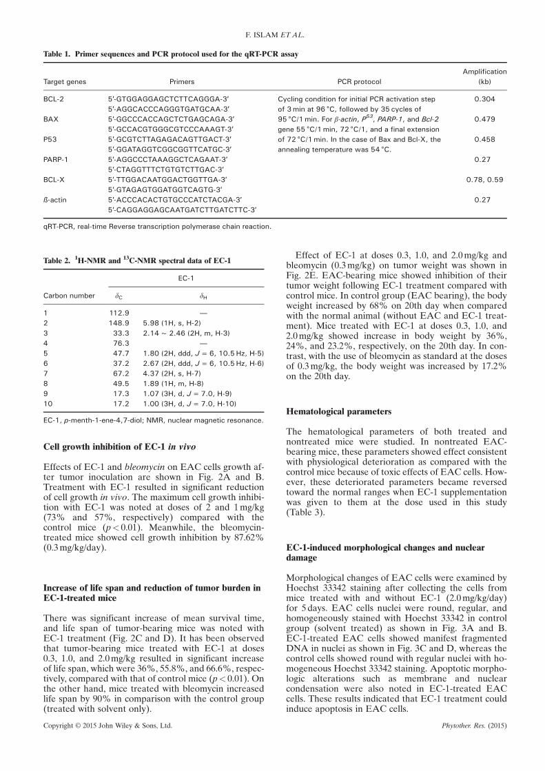
Table 1. Primer sequences and PCR protocol used for the qRT-PCR assay
Target genes Primers PCR protocolAmplification
(kb)
BCL-2 5′-GTGGAGGAGCTCTTCAGGGA-3′ Cycling condition for initial PCR activation stepof 3min at 96 °C, followed by 35 cycles of95 °C/1min. For β-actin, P53, PARP-1, and Bcl-2gene 55 °C/1min, 72 °C/1, and a final extensionof 72 °C/1min. In the case of Bax and Bcl-X, theannealing temperature was 54 °C.
0.3045′-AGGCACCCAGGGTGATGCAA-3′
BAX 5′-GGCCCACCAGCTCTGAGCAGA-3′ 0.4795′-GCCACGTGGGCGTCCCAAAGT-3′
P53 5′-GCGTCTTAGAGACAGTTGACT-3′ 0.4585′-GGATAGGTCGGCGGTTCATGC-3′
PARP-1 5′-AGGCCCTAAAGGCTCAGAAT-3′ 0.275′-CTAGGTTTCTGTGTCTTGAC-3′
BCL-X 5′-TTGGACAATGGACTGGTTGA-3′ 0.78, 0.595′-GTAGAGTGGATGGTCAGTG-3′
ß-actin 5′-ACCCACACTGTGCCCATCTACGA-3′ 0.275′-CAGGAGGAGCAATGATCTTGATCTTC-3′
qRT-PCR, real-time Reverse transcription polymerase chain reaction.
Table 2. 1H-NMR and 13C-NMR spectral data of EC-1
Carbon number
EC-1
δC δH
1 112.9 —
2 148.9 5.98 (1H, s, H-2)3 33.3 2.14~2.46 (2H, m, H-3)4 76.3 —
5 47.7 1.80 (2H, ddd, J=6, 10.5Hz, H-5)6 37.2 2.67 (2H, ddd, J=6, 10.5Hz, H-6)7 67.2 4.37 (2H, s, H-7)8 49.5 1.89 (1H, m, H-8)9 17.3 1.07 (3H, d, J=7.0, H-9)10 17.2 1.00 (3H, d, J=7.0, H-10)
EC-1, p-menth-1-ene-4,7-diol; NMR, nuclear magnetic resonance.
F. ISLAM ET AL.
Cell growth inhibition of EC-1 in vivo
Effects of EC-1 and bleomycin on EAC cells growth af-ter tumor inoculation are shown in Fig. 2A and B.Treatment with EC-1 resulted in significant reductionof cell growth in vivo. The maximum cell growth inhibi-tion with EC-1 was noted at doses of 2 and 1mg/kg(73% and 57%, respectively) compared with thecontrol mice (p< 0.01). Meanwhile, the bleomycin-treated mice showed cell growth inhibition by 87.62%(0.3mg/kg/day).
Increase of life span and reduction of tumor burden inEC-1-treated mice
There was significant increase of mean survival time,and life span of tumor-bearing mice was noted withEC-1 treatment (Fig. 2C and D). It has been observedthat tumor-bearing mice treated with EC-1 at doses0.3, 1.0, and 2.0mg/kg resulted in significant increaseof life span, which were 36%, 55.8%, and 66.6%, respec-tively, compared with that of control mice (p< 0.01). Onthe other hand, mice treated with bleomycin increasedlife span by 90% in comparison with the control group(treated with solvent only).
Copyright © 2015 John Wiley & Sons, Ltd.
Effect of EC-1 at doses 0.3, 1.0, and 2.0mg/kg andbleomycin (0.3mg/kg) on tumor weight was shown inFig. 2E. EAC-bearing mice showed inhibition of theirtumor weight following EC-1 treatment compared withcontrol mice. In control group (EAC bearing), the bodyweight increased by 68% on 20th day when comparedwith the normal animal (without EAC and EC-1 treat-ment). Mice treated with EC-1 at doses 0.3, 1.0, and2.0mg/kg showed increase in body weight by 36%,24%, and 23.2%, respectively, on the 20th day. In con-trast, with the use of bleomycin as standard at the dosesof 0.3mg/kg, the body weight was increased by 17.2%on the 20th day.
Hematological parameters
The hematological parameters of both treated andnontreated mice were studied. In nontreated EAC-bearing mice, these parameters showed effect consistentwith physiological deterioration as compared with thecontrol mice because of toxic effects of EAC cells. How-ever, these deteriorated parameters became reversedtoward the normal ranges when EC-1 supplementationwas given to them at the dose used in this study(Table 3).
EC-1-induced morphological changes and nucleardamage
Morphological changes of EAC cells were examined byHoechst 33342 staining after collecting the cells frommice treated with and without EC-1 (2.0mg/kg/day)for 5days. EAC cells nuclei were round, regular, andhomogeneously stained with Hoechst 33342 in controlgroup (solvent treated) as shown in Fig. 3A and B.EC-1-treated EAC cells showed manifest fragmentedDNA in nuclei as shown in Fig. 3C and D, whereas thecontrol cells showed round with regular nuclei with ho-mogeneous Hoechst 33342 staining. Apoptotic morpho-logic alterations such as membrane and nuclearcondensation were also noted in EC-1-treated EACcells. These results indicated that EC-1 treatment couldinduce apoptosis in EAC cells.
Phytother. Res. (2015)

Figure 1. Structure and in vitro cytotoxicity of p-menth-1-ene-4,7-diol (EC-1). (A) Structure of the compound. (B) The Ehrlich as-cites carcinoma (EAC) cells were treated with various doses ofEC-1 for 24 h in RPMI-1640 medium. The growth inhibition wasmeasured by MTT assay (n=3, mean ± standard error of mean).(C) IC50 value of EC-1 was calculated from the dose–responsecurve. Level of significance *p<0.05 and **p<0.01 whencompared with that of control group.
EC-1 INHIBITS CANCER GROWTH
DNA fragmentation analysis
The activation of the endogenous Ca2+/Mg2+-dependentendonuclease is the most distinctive biochemicalhallmark of apoptosis (Islam et al., 2014a). Thisactivated endonuclease will mediate the cleavage ofinternucleosomes and generate oligonucleotide frag-ments of about 180–200 base pairs. DNA isolated from
Copyright © 2015 John Wiley & Sons, Ltd.
EC-1-treated EAC cells showed ladder-type DNA thatis a characteristic feature of apoptosis induction;whereas in the control group, a smear-like DNA degra-dation was obtained (Fig. 3E).
Altered expressions of cancer-related genes inEC-1-treated cells
Reverse transcription PCR was used to study themRNA expression levels of several tumor-related genesin control and EC-1-treated EAC cells (Fig. 3F). Thecontrol cells showed high expressions of Bcl-2 and Bcl-Xgenes. Also, the Bax mRNA expression was not foundin the control cells. When treated with EC-1, EAC cellsshowed reduced Bcl-2 and Bcl-X mRNA expressions,whereas the expression level of Bax gene was increasedremarkably. In addition, the p53 and PARP-1 genesshowed increased expressions in EC-1-treated cells.On the other hand, no expression of these genes wasfound in control mice EAC cells (Fig. 3F)
Cell cycle changes with EC-1 treatment
The effect of EC-1 on different cell cycle phases of EACcells was studied. The untreated cells showed threephases G0/G1, S, and G2/M in the cell cycle, and the pro-portion of G0/G1, S, and G2/M phases in the untreatedEAC cells were calculated to be 49%, 32%, and 19%,respectively. On the other hand, cells treated withEC-1 were changed at G2/M (40% vs 19%) and G0/G1(56% vs 49%) phase population compared with controlcells. Meanwhile, the cells in S phase showed remark-able reduction in phase population (4% vs 32%) com-pared with control cells (Fig. 4). These resultsindicated that the EC-1 compound inhibits the cellularproliferation of EAC cells by suppressing the synthesisof DNA in S phase of cell cycle. The compound also at-tenuated the cancer cells in G2/M phase to some extent.
DISCUSSION
The compound EC-1 (a simple monoterpinoid) wasisolated first time in this study from the plant E.camaldulensis Dhnh., and there is no report in theliterature of its anticancer properties. It is reportedthat other terpinoids from Eucalyptus showedantiinflammatory, analgesic, and antipyretic activities(Annalucia et al., 2008). These results scientifically sup-port for some traditional usages of this plant in tradi-tional medicine, although the precise mechanismneeded additional research.
In the present study, we found that the EC-1 com-pound inhibited EAC cells proliferation in vitro in adose-dependent manner (Fig. 1B and C). In vivo effectof the compound has been compared with the data ob-tained by parallel experiments using conventional anti-cancer drug, bleomycin. EC-1 treatment resulted insignificant reduction EAC cells growth, and this wasreasonably comparable with that of bleomycin (73% vs88%). Also, this compound significantly increases thelife span of EAC-bearing mice (Fig. 2C and D). Duringthis study, we also found that the EC-1 compound can
Phytother. Res. (2015)

Figure 2. In vivo anticancer activities of p-menth-1-ene-4,7-diol (EC-1). (A) Number of Ehrlich ascites carcinoma (EAC) cells in experimentalmice, counted by inverted microscope in presence and absence (control) of EC-1 at different dose on day 6 of tumor inoculation. (B) Percent-age of cell growth inhibition at different doses in comparison to control mice. (C) Mean survival time of experimental mice. (D) Percentage oflife span increase with the treatment and (E) reduction of tumor burden at different doses. Data are expressed as mean ± standard error ofmean (n=6). Level of significance *p<0.05 and **p<0.01 when compared with that of control group. This figure is available in colour on-line at http://wileyonlinelibrary.com/journal/ptr.
F. ISLAM ET AL.
reduce the tumor burden of EAC-bearing mice remark-ably, as the EC-1-treated mice showed only 23% incre-ment of body weight in comparison with control mice,whereas body weight of bleomycin-treated miceincreased by 17% (Fig. 2E). Normal mice also treatedwith EC-1 at the highest dose used in this study to inves-tigate the toxic effect (regarding behavioral,bodyweight, and biochemical parameters) of the com-pound on host. There was no adverse effects observed(data were not presented here).In EAC-bearing mice, anemia occurred because of
breakdown of red blood cell (Kabir et al., 2012). Supple-mentation with EC-1 in EAC-bearing mice restored all
Copyright © 2015 John Wiley & Sons, Ltd.
the altered hematological parameters toward normalrange on day 12 of tumor inoculation (Table 3), and sim-ilar results were also observed in EAC-bearing micetreated with bioactive compounds (Rasida et al., 2012).These findings rectify the suitability of EC-1 as anemerging natural anticancer agent.
Apoptosis is a self cell-suicidal mechanism that can betrigger by cytotoxic compounds and regulated by varioussignaling pathways (Islam et al., 2014a). Cells underwentapoptosis are characterized morphologically by irregularcells shape, shrinkage, nuclear material condensation,and apoptotic body formation and these changes causedby proteases (caspases) (Graf et al., 2007). Induction of
Phytother. Res. (2015)
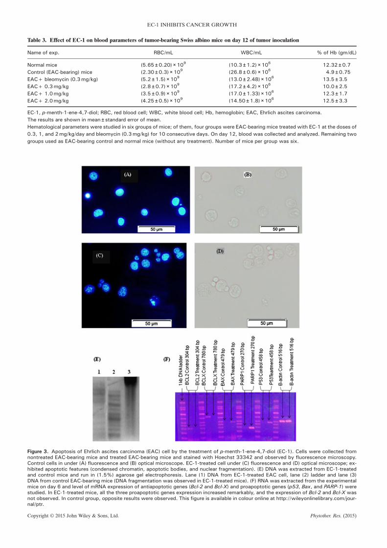
Table 3. Effect of EC-1 on blood parameters of tumor-bearing Swiss albino mice on day 12 of tumor inoculation
Name of exp. RBC/mL WBC/mL % of Hb (gm/dL)
Normal mice (5.65 ± 0.20) × 109 (10.3 ± 1.2) × 106 12.32 ± 0.7Control (EAC-bearing) mice (2.30 ± 0.3) × 109 (26.8 ± 0.6) × 106 4.9 ± 0.75EAC+ bleomycin (0.3mg/kg) (5.2 ± 1.5) × 109 (13.0 ± 2.48) × 106 13.5 ± 3.5EAC+ 0.3mg/kg (2.8 ± 0.7) × 109 (17.2 ± 4.2) × 106 10.0 ± 2.5EAC+ 1.0mg/kg (3.5 ± 0.9) × 109 (17.0 ± 1.33) × 106 12.3 ± 1.7EAC+ 2.0mg/kg (4.25 ± 0.5) × 109 (14.50 ± 1.8) × 106 12.5 ± 3.3
EC-1, p-menth-1-ene-4,7-diol; RBC, red blood cell; WBC, white blood cell; Hb, hemoglobin; EAC, Ehrlich ascites carcinoma.The results are shown in mean ± standard error of mean.Hematological parameters were studied in six groups of mice; of them, four groups were EAC-bearing mice treated with EC-1 at the doses of0.3, 1, and 2mg/kg/day and bleomycin (0.3mg/kg) for 10 consecutive days. On day 12, blood was collected and analyzed. Remaining twogroups used as EAC-bearing control and normal mice (without any treatment). Number of mice per group was six.
Figure 3. Apoptosis of Ehrlich ascites carcinoma (EAC) cell by the treatment of p-menth-1-ene-4,7-diol (EC-1). Cells were collected fromnontreated EAC-bearing mice and treated EAC-bearing mice and stained with Hoechst 33342 and observed by fluorescence microscopy.Control cells in under (A) fluorescence and (B) optical microscope. EC-1-treated cell under (C) fluorescence and (D) optical microscope; ex-hibited apoptotic features (condensed chromatin, apoptotic bodies, and nuclear fragmentation). (E) DNA was extracted from EC-1-treatedand control mice and run in (1.5%) agarose gel electrophoresis. Lane (1) DNA from EC-1-treated EAC cell, lane (2) ladder and lane (3)DNA from control EAC-bearing mice (DNA fragmentation was observed in EC-1-treated mice). (F) RNA was extracted from the experimentalmice on day 6 and level of mRNA expression of antiapoptotic genes (Bcl-2 and Bcl-X) and proapoptotic genes (p53, Bax, and PARP-1) werestudied. In EC-1-treated mice, all the three proapoptotic genes expression increased remarkably, and the expression of Bcl-2 and Bcl-X wasnot observed. In control group, opposite results were observed. This figure is available in colour online at http://wileyonlinelibrary.com/jour-nal/ptr.
EC-1 INHIBITS CANCER GROWTH
Copyright © 2015 John Wiley & Sons, Ltd. Phytother. Res. (2015)

Figure 4. Effect of p-menth-1-ene-4,7-diol (EC-1) on cell cycle phases. (A) Bar diagram showing the percentages of cell in each phase wereevaluated by flow cytometry. Data are presented as mean values obtained from three independent experiments, and the results areexpressed as means ± standard error of mean. Histogram representing flow cytometry results (B) control and (C) effects of EC-1 on the cellcycle phases. The x-axis (FL3) is representing the intensity of the propidium iodide (PI) staining that is directly proportional to the amount ofDNA in cells and y-axis representing the cell number. This figure is available in colour online at http://wileyonlinelibrary.com/journal/ptr.
F. ISLAM ET AL.
apoptosis in EAC cells by EC-1 was confirmed by theobservation of changes in nuclear material and cell shapeas compared with that of untreated control EAC cells(Fig. 3). Apoptosis of EAC cell was also studied usingdifferent compounds of plant origin (Islam et al.,2014a). Ottelion A, a plant product, inhibited the EACcell growth by apoptosis with the mediation of increasedlevels of p53 and CD8+ cells (EI-Missiry et al., 2012).Oleandrin, a plant glycoside, can induce apoptosis inmany cell lines by upregulating Fas and tumor necrosisfactor receptor 1 (Sreenivasan et al., 2006). Similarly, thisstudy showed induction of apoptosis in EC-1-treatedEAC cells by regulating many cancer-related genes(Fig. 3F). Switch on/off of apoptosis is regulated by nu-merous growth related genes; of them, Bcl-2 (B cell lym-phoma gene 2) family gene was believed to be the firstone attributed in the apoptotic process (Gradiloneet al., 2003). The gene was expressed in cancer and wascorrelated with clinical pathological parameters of pa-tients with cancer (Yuen et al., 2001). Among theapoptosis-related genes, Bax, Bid, and Bak act asproapoptotic, and other members Bcl-2, Bcl-X, andBcl-Wact as antiapoptotic class (Kabir et al., 2013). In re-sponse to anticancer agents, the expression levels ofthese genes alter in cancer cell. Decreased expressionof Bcl-X and increased level of Bax and Bak were ob-served in HeLa cell with the treatment of a plant com-pound FRAP (Ju et al., 2012).In the present study, we found that Bcl-2 and Bcl-X
expressions were observed in control mice, and they
Copyright © 2015 John Wiley & Sons, Ltd.
were absent in mice treated with EC-1, whereasproapoptotic genes such as Bax, P53, and PARP-1expressions showed opposite results. We found overex-pression of these genes in EC-1-treated mice in compar-ison with that of control mice (Fig. 3F). Similar resultswere also found in different cancer cell lines treatedwith compounds obtained from plant materials (Zhaoet al., 2010; Groc et al., 2001).. Increased expression ofthese genes could lead to cytochrome c release frommitochondria, which in turn, stimulate the activation ofcaspases-9, initiating a downstream cascade leading tocell death (Antonsson, 2001). Altered expression ofthese proapoptotic and antiapoptotic genes in EAC cellupon EC-1 treatment further confirmed the induction ofprogrammed cell death of cancer cell.
Many chemotherapeutic and DNA-damaging agentsinduce cancer cells’ apoptosis by arresting cells at G1,S, or G2/M phase. Compounds of natural origin such ascape aloe, ottelione A, and guarana caused EAC cellsdeath by arresting cell cycle progression (EI-Missiryet al., 2012; Kametani et al., 2007; Fukumasu et al.,2011). In the present study, we found that the isolatedcompound caused suppression of EAC cell in S phaseof cell cycle (Fig. 4). It implies that treatment with EC-1 compound inhibited the synthesis of DNA in EAC cell.Also, EC-1-driven DNA damage in EAC cells has thenled to apoptosis. The compound also increased the pop-ulation of cells in G2/M phase in comparison with controlgroup. In addition, many human cancer cell lines haveshown arrested at G0/G1 and S phase inhibition by the
Phytother. Res. (2015)
EC-1 INHIBITS CANCER GROWTH
treatment of chemotherapeutic agents (El-Sherbinyet al., 2001).In conclusion, the EC-1 compound (menth-1-ene-
4,7-diol) isolated from E. camaldulensis can induce apo-ptosis of cancer cell and reduced cells population at Sphase of the cell cycle. Structural changes in cells andaltered expression pattern of different genes treatedwith this compound supported that EC-1 induces apo-ptosis in EAC cells. Further works with other cancer celllines and with higher animals are required to developeffective anticancer drug in future with this compound.
Copyright © 2015 John Wiley & Sons, Ltd.
Acknowledgements
The authors would like to acknowledge the International Centre forDiarrheal Disease research, Bangladesh (ICDDR’B) for providing ex-perimental animal and laboratory facility of flow cytometry and theBangladesh Council of Scientific and Industrial Research, Dhaka, Ban-gladesh, for spectrometric analysis of the compound. We would like tothank Griffith University for the support of the project.
Conflict of Interest
The authors have no conflict of interest.
REFERENCES
Abdel-Nasser S, Nahla A, Eman AS, Olli M, Jari S, Kalevi P. 2011.Phenolic constituents of Eucalyptus camaldulensis Dehnh,with potential antioxidant and cytotoxic activities. Rec NatProd 5: 271–280.
Al-Fatimi M, Friedrich U, Jenett-Siems K. 2005. Cytotoxicity ofplants used in traditional medicine in Yemen. Fitoterapia 76:355–358.
Annalucia S, Paola SV, Federica A, et al. 2008. Stimulatory effectsof Eucalyptus essential oil on innate cell-mediated immuneresponse. BMC Immunol 9: 17.
Antonsson B. 2001. Bax and other pro-apoptotic bcl-2 family ‘killerproteins’ and their victim, the mitochondrion. Cell Tissue Res306: 347–361.
Benyahia S, Benayache S, Benayache F, et al. 2005. Cladocalol, apentacyclic 28-nor-triterpene from Eucalyptus cladocalyxwithcytotoxic activity. Phytochemistry 66: 627–632.
Brooker MIH, Kleinig DA. 2009. Field Guide to Eucalyptus (3rd edi-tion). Bloomings: Melbourne, Australia.
Cheng SS, Huang CG, Chen YJ, Yu JJ, ChenWJ, Chang ST. 2009.Chemical compositions and larvicidal activities of leaf essen-tial oils from two eucalyptus species. Bioresour Technol 100:452–456.
Cimanga K, Kambu K, Tona L, et al. 2002. Correlation betweenchemical composition and antibacterial activity of essentialoils of some aromatic medicinal plants growing in the Demo-cratic Republic of Congo. J Ethnopharmacol 79: 213–20.
Coelho-de-Souza LN, Leal-Cardoso JH, de Abreu Matos FJ, LahlouS, Magalhães PJ. 2005. Relaxant effects of the essential oil ofEucalyptus tereticornis and its main constituent 1,8-cineole onguinea-pig tracheal smooth muscle. Planta Med 71:1173–1175.
EI-Ghorab AH, EI-Massry KF, Marx F, Fadel HM. 2003. Antioxidantactivity of Egyptian Eucalyptus camaldulensis var. brevirostrisleaf extracts. Nahrung 47: 41–45.
EI-Missiry MA, Othman AI, Amer MA, Mohamed E. 2012.Ottelione A inhibited proliferation of Ehrlich ascites carcinomacells in mice. Chem Biol Interact 200: 119–127.
El-Sherbiny YM, Cox MC, Ismail ZA, Shamsuddin AM, Vucenik I.2001. G0/G1 arrest and S phase inhibition of human cancercell lines by inositol hexaphosphate (IP6). Anticancer Res 21:2393–403.
Fukumasu H, Latorre AO, Zaidan-Dagli ML. 2011. Paullinia cupanaMart. var. sorbilis, guarana, increases survival of Ehrlich asci-tes carcinoma (EAC) bearing mice by decreasing cyclin-D1expression and inducing a G0/G1 cell cycle arrest in EAC cells.Phytother Res 25: 11–16.
Gradilone A, Gazzaniga P, Ribuffo D, et al. 2003. Survivin, bcl-2,bax, and bcl-X gene expression in sentinel lymph nodes frommelanoma patients. J Clin Oncol 21: 306–312.
Graf D, Bode JG, Häussinger D. 2007. Caspases and receptorcleavage. Arch Biochem Biophys 462: 162–170.
Groc L, Bezin L, Jiang H, Jackson TS, Levine RA. 2001. Bax, bcl-2,and cyclin expression and apoptosis in rat substantia nigraduring development. Neurosci Lett 306: 198–202.
Ishikawa T, Sega Y, Kitajima J. 2001. Water-soluble constituentsof Ajowan. Chem Pharm Bull 49: 840–844.
Hossain MK, Pasha MK. 2004. An account of the exotic flora ofBangladesh. J For Environ 2: 99–115.
Islam F, Khatun H, Khatun M, Ali MM, Khanam JA. 2014a. Growthinhibition and apoptosis of Ehrlich ascites carcinoma cells bythe methanol extract of Eucalyptus camaldulensis. Pharm Biol52: 281–290.
Islam F, Ghosh S, Khanam JA. 2014b. Antiproliferative and hepa-toprotective activity of metabolites from Corynebacteriumxerosis against Ehrlich ascites carcinoma cells. Asian Pac JTrop Biomed 4: S284–S292.
Islam F, Khatun H, Ghosh S, Ali MM, Khanam JA. 2012. Bioassayof Eucalyptus extracts for anticancer activity against Ehrlichascites carcinoma (EAC) cells in Swiss albino mice. AsianPac J Trop Biomed 2: 394–398.
Ju HK, Lee HW, Chung KS, et al. 2012. Standardized flavonoid-rich fraction of Artemisia princeps Pampanini cv. Sajabalinduces apoptosis via mitochondrial pathway in human cervi-cal cancer HeLa cells. J Ethnopharmacol 141: 460–468.
Kabir SR, Nabi MM, Haque A, Zaman R, Mahmud ZH, Reza AM.2013. Pea lectin inhibits growth of Ehrlich ascites carcinomacells by inducing apoptosis and G2/M cell cycle arrest in vivoin mice. Phytomed 20: 1288–1296.
Kabir SR, Islam MF, Alom MJ, Zubair MA, Absar N. 2012. Purifica-tion and characterization of a snake guard seed lectin withantitumor activity against Ehrlich ascites carcinoma cellsin vivo in mice. Protein Pept Lett 19: 360–368.
Kabir SR, Zubair, MA, Nurujjaman M, et al. 2011. Purification andcharacterization of a Ca2+-dependent novel lectin fromNymphaea nouchali tuber with antiproliferative activities.Biosci Rep 31: 465–475.
Kametani S, Oikawa T, Kojima-Yuasa A, et al. 2007. Mechanism ofgrowth inhibitory effect of cape aloe extract in Ehrlich ascitestumor cells. J Nutr Sci Vitaminol 53: 540–546.
Khanam JA, Islam MF, Jesmin M, Ali MM. 2010. Antineoplasticactivity of acetone semicarbazone (ASC) against Ehrlich asci-tes carcinoma (EAC) bearing mice. J Natn Sci Foundation SriLanka 38: 225–231.
Rasida P, Islam F, Khanum JA, Yeasmin T. 2012. Preventive effectof ethanol extract of Alpinia calcarata Rosc on Ehrlich’s asciticcarcinoma cell induced malignant ascites in mice. Asian Pac JTrop Med 5: 121–125.
Silva J, Abebe W, Sousa SM, Duarte VG, Machado MI, Matos FJ.2003. Analgesic and anti-inflammatory effects of essentialoils of eucalyptus. J Ethnopharmacol 89: 277–283.
Siramon P, Ohtani Y. 2007. Antioxidant and antibacterial activitiesof Eucalyptus camaldulensis leaf oils from Thailand. J WoodSci 53: 498–504.
Sreenivasan Y, Raghavendra PB, Manna SK. 2006. Oleandrin medi-ated expression of Fas potentiates apoptosis in tumor cells. JClin Immunol 26: 308–322.
Su YC, Ho CL, Wang EI Chang ST. 2006. Antifungal activities andchemical compositions of essentials oils leaves of four euca-lyptus. Taiwan J For SCi 21: 49–61.
Takasaki M, Konoshima T, Etoh H, Pal Singh I, Tokuda H, NishinoH. 2000. Cancer chemopreventive activity of euglobal-G1from leaves of Eucalyptus grandis. Cancer Lett 155: 61–65.
Tatsuya H, Fumihide T, Takanobu T, Masato N, Tomihisa O. 2008.Bioactive monoterpene glycosides conjugated with galic acidfrom the leaves of Eucalyptus globules. Phys Earth Planet Inter69: 747–753.
Yuen AP, Lam KY, Choy JT, Ho WK, Wei WI. 2001. The clinico-pathological significance of bcl-2 expression in the surgicaltreatment of laryngeal carcinoma. Clin Otolaryngol Allied Sci26: 129–133.
Zhao Q, Cao X, Zeng B, Wang C, Yan L, Xu C. 2010. Muscadomestica larva lectin induces apoptosis in BEL-7402 cellsthrough a mitochondria-mediated reactive oxygen speciesway. Biol Pharm Bull 33: 1274–1278.
Phytother. Res. (2015)


![Excited-State Diproton Transfer in [2, 2′-Bipyridyl]-3, 3′-diol: the Mechanism Is Sequential, Not Concerted](https://static.fdokumen.com/doc/165x107/6332daff5f7e75f94e094ac2/excited-state-diproton-transfer-in-2-2-bipyridyl-3-3-diol-the-mechanism.jpg)


![Exposure to benzo[a]pyrene of Hepatic Cytochrome P450 Reductase Null (HRN) and P450 Reductase Conditional Null (RCN) mice: Detection of benzo[a]pyrene diol epoxide-DNA adducts by immunohistochemistry](https://static.fdokumen.com/doc/165x107/63259f17c9c7f5721c022d3b/exposure-to-benzoapyrene-of-hepatic-cytochrome-p450-reductase-null-hrn-and-p450.jpg)

