A novel mouse model for the aggressive variant of NK cell and T cell large granular lymphocyte...
7
Leukemia Research 34 (2010) 203–209 Contents lists available at ScienceDirect Leukemia Research journal homepage: www.elsevier.com/locate/leukres A novel mouse model for the aggressive variant of NK cell and T cell large granular lymphocyte leukemia Akihiko Yokohama a,b,1 , Anjali Mishra a,1 , Takeki Mitsui a,b , Brian Becknell c , Jessica Johns a , Douglas Curphey a , Bradley W. Blaser d,e , Jeffrey B. VanDeusen e , Hsiaoyin Mao a , Jianhua Yu a , Michael A. Caligiuri a,f,∗ a Department of Molecular Virology, Immunology, and Medical Genetics, Department of Internal Medicine, Division of Hematology/Oncology, The Ohio State University (OSU) College of Medicine, Columbus, OH, United States b Department of Medicine and Clinical Science, Gunma University Graduate School of Medicine, 3-39-15 Showa, Maebashi, Gunma 371-8511, Japan c OSU Department of Pediatrics, United States d The Brigham and Women’s Hospital, Boston, MA, United States e OSU Integrated Biomedical Sciences Graduate Program, United States f The OSU Comprehensive Cancer Center, The James Cancer Hospital and Solove Research Institute, Columbus, OH 43210, United States article info Article history: Received 18 March 2009 Received in revised form 22 June 2009 Accepted 26 June 2009 Available online 5 August 2009 Keywords: Natural killer cell NK and T large granular lymphocyte leukemia IL-15 Proto-oncogene abstract Murine models of disease are vital to the understanding of pathogenesis and the development of novel therapeutics. We have previously established interleukin (IL)-15 transgenic (tg) mice that demonstrate rapid proliferation of natural killer (NK) and T cells, followed by spontaneous transformation to lethal leukemia. Herein, we have characterized this model, which has many features in common with the aggressive variants of NK and T large granular lymphocyte leukemia (LGLL) in humans. The LGLL blasts are cytolytic and produce IFN- ex vivo. Cytogenetic analysis revealed trisomy of chromosome 17 and/or 15. This model should provide opportunities to develop effective standard therapies for this fatal disease. © 2009 Elsevier Ltd. All rights reserved. 1. Introduction Large granular lymphocyte leukemia (LGLL) is a clonal disorder of the NK cell or T cell lineage that presents clinically as indo- lent or aggressive disease [1]. Aggressive forms of LGLL are often associated with very poor prognosis, with most patients exhibiting rapidly progressive disease that is poorly responsive to multi-agent chemotherapy [2]. Aggressive variants of human NK cell and T LGLL are characterized by an immunophenotype that includes surface expression of CD56 along with a variable surface expression pattern of NK cell and/or T cell markers [3,4]. Since the aggressive variants of LGLL are rare and difficult to manage, rigorous studies are required to identify mechanisms of This work is supported by National Cancer Institute grants CA16058, CA95426, and CA68458. ∗ Corresponding author at: The Ohio State University, Comprehensive Cancer Cen- ter, 300W. 10th Avenue, Suite 521C, Columbus, OH 43210, United States. Tel.: +1 614 293 7523; fax: +1 614 293 3132. E-mail address: [email protected] (M.A. Caligiuri). 1 Akihiko Yokohama and Anjali Mishra contributed equally to this work. pathogenesis and opportunities for therapeutic intervention [5]. We previously showed that IL-15tg mice display early expansions of peripheral blood lymphocytes, specifically NK cells and mem- ory phenotype CD8 + T cells. Later, IL-15tg mice develop a striking leukemic expansion with multi-organ infiltration [6,7]. In the current study, we describe a murine model of aggres- sive NK and T LGLL that may eventually provide an opportunity to test and validate new therapeutic advances. We have systematically characterized 39 murine leukemia cases – phenotypically, function- ally and cytogenetically – to validate this clinically relevant model of aggressive NK and T LGLL. 2. Materials and methods 2.1. Mice and in vivo studies IL-15tg mice were generated and maintained as described previously [6]. Age and sex matched SCID mice were purchased from The Jackson Laboratory (Bar Har- bor, ME). To study in vivo transplantability of these leukemia cells, 10 5 leukemia cells from peripheral blood, spleen or bone marrow of moribund mice were intravenously injected into sub-lethally (4.5 Gy) irradiated SCID recipient mice. All procedures were approved by the Institutional Animal Care and Use Committee (IACUC) of The Ohio State University. 0145-2126/$ – see front matter © 2009 Elsevier Ltd. All rights reserved. doi:10.1016/j.leukres.2009.06.031
Transcript of A novel mouse model for the aggressive variant of NK cell and T cell large granular lymphocyte...

Ag
ADJa
Tb
c
d
e
f
a
ARRAA
KNNlIP
1
olarcaeo
m
a
tT
0d
Leukemia Research 34 (2010) 203–209
Contents lists available at ScienceDirect
Leukemia Research
journa l homepage: www.e lsev ier .com/ locate / leukres
novel mouse model for the aggressive variant of NK cell and T cell largeranular lymphocyte leukemia�
kihiko Yokohama a,b,1, Anjali Mishra a,1, Takeki Mitsui a,b, Brian Becknell c, Jessica Johns a,ouglas Curphey a, Bradley W. Blaser d,e, Jeffrey B. VanDeusen e, Hsiaoyin Mao a,
ianhua Yu a, Michael A. Caligiuri a,f,∗
Department of Molecular Virology, Immunology, and Medical Genetics, Department of Internal Medicine, Division of Hematology/Oncology,he Ohio State University (OSU) College of Medicine, Columbus, OH, United StatesDepartment of Medicine and Clinical Science, Gunma University Graduate School of Medicine, 3-39-15 Showa, Maebashi, Gunma 371-8511, JapanOSU Department of Pediatrics, United StatesThe Brigham and Women’s Hospital, Boston, MA, United StatesOSU Integrated Biomedical Sciences Graduate Program, United StatesThe OSU Comprehensive Cancer Center, The James Cancer Hospital and Solove Research Institute, Columbus, OH 43210, United States
r t i c l e i n f o
rticle history:eceived 18 March 2009eceived in revised form 22 June 2009ccepted 26 June 2009
a b s t r a c t
Murine models of disease are vital to the understanding of pathogenesis and the development of noveltherapeutics. We have previously established interleukin (IL)-15 transgenic (tg) mice that demonstraterapid proliferation of natural killer (NK) and T cells, followed by spontaneous transformation to lethalleukemia. Herein, we have characterized this model, which has many features in common with the
vailable online 5 August 2009
eywords:atural killer cellK and T large granular lymphocyte
eukemia
aggressive variants of NK and T large granular lymphocyte leukemia (LGLL) in humans. The LGLL blastsare cytolytic and produce IFN-� ex vivo. Cytogenetic analysis revealed trisomy of chromosome 17 and/or15. This model should provide opportunities to develop effective standard therapies for this fatal disease.
© 2009 Elsevier Ltd. All rights reserved.
L-15roto-oncogene. Introduction
Large granular lymphocyte leukemia (LGLL) is a clonal disorderf the NK cell or T cell lineage that presents clinically as indo-
ent or aggressive disease [1]. Aggressive forms of LGLL are oftenssociated with very poor prognosis, with most patients exhibitingapidly progressive disease that is poorly responsive to multi-agenthemotherapy [2]. Aggressive variants of human NK cell and T LGLLre characterized by an immunophenotype that includes surface
xpression of CD56 along with a variable surface expression patternf NK cell and/or T cell markers [3,4].Since the aggressive variants of LGLL are rare and difficult toanage, rigorous studies are required to identify mechanisms of
� This work is supported by National Cancer Institute grants CA16058, CA95426,nd CA68458.∗ Corresponding author at: The Ohio State University, Comprehensive Cancer Cen-
er, 300W. 10th Avenue, Suite 521C, Columbus, OH 43210, United States.el.: +1 614 293 7523; fax: +1 614 293 3132.
E-mail address: [email protected] (M.A. Caligiuri).1 Akihiko Yokohama and Anjali Mishra contributed equally to this work.
145-2126/$ – see front matter © 2009 Elsevier Ltd. All rights reserved.oi:10.1016/j.leukres.2009.06.031
pathogenesis and opportunities for therapeutic intervention [5].We previously showed that IL-15tg mice display early expansionsof peripheral blood lymphocytes, specifically NK cells and mem-ory phenotype CD8+ T cells. Later, IL-15tg mice develop a strikingleukemic expansion with multi-organ infiltration [6,7].
In the current study, we describe a murine model of aggres-sive NK and T LGLL that may eventually provide an opportunity totest and validate new therapeutic advances. We have systematicallycharacterized 39 murine leukemia cases – phenotypically, function-ally and cytogenetically – to validate this clinically relevant modelof aggressive NK and T LGLL.
2. Materials and methods
2.1. Mice and in vivo studies
IL-15tg mice were generated and maintained as described previously [6]. Age
and sex matched SCID mice were purchased from The Jackson Laboratory (Bar Har-bor, ME). To study in vivo transplantability of these leukemia cells, 105 leukemia cellsfrom peripheral blood, spleen or bone marrow of moribund mice were intravenouslyinjected into sub-lethally (4.5 Gy) irradiated SCID recipient mice. All procedureswere approved by the Institutional Animal Care and Use Committee (IACUC) of TheOhio State University.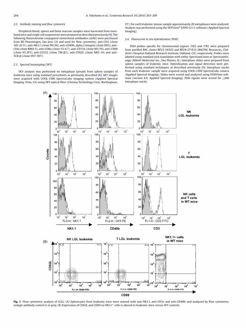
2 ia Re
2
bff1C(T
2
lwI
Fi
04 A. Yokohama et al. / Leukem
.2. Antibody staining and flow cytometry
Peripheral blood, spleen and bone marrow samples were harvested from mori-und mice and single cell suspensions were prepared as described previously [6]. Theollowing fluorochrome-conjugated monoclonal antibodies (mAb) were purchasedrom BD Pharmingen, San Jose, CA and used for flow cytometry: anti-CD3 (clone45-2C11), anti-NK1.1 (clone PK136), anti-CD49b, alpha 2 integrin (clone DX5), anti-D4 (clone RM4-5), anti-CD8a (clone 53-6.7), anti-CD11b (clone M1/70), anti-CD69clone H1.2F3), anti-CD122 (clone TM-�1), anti-CD62L (clone MEL-14) and anti-CR-� (clone H57-597).
.3. Spectral karyotyping (SKY)
SKY analysis was performed on metaphase spreads from spleen samples ofeukemia mice using standard procedures as previously described [8]. SKY images
ere acquired with COOL-1300 SpectraCube imaging system (Applied Spectralmaging, Vista, CA) using SKY optical filter (Chroma Technology Corp., Rockingham,
ig. 1. Flow cytometry analysis of LGLL. (A) Splenocytes from leukemic mice were stainsotype antibody control is in grey. (B) Expression of CD62L and CD69 on NK1.1+ cells is al
search 34 (2010) 203–209
VT). For each leukemic mouse sample approximately 20 metaphases were analyzed.Analysis was performed using the SKYView® EXPO v2.1.1 software (Applied SpectralImaging).
2.4. Fluorescent in situ hybridization (FISH)
FISH probes specific for chromosomal regions 15E2 and 17B1 were preparedfrom purified BAC clones RP23-343O3 and RP24-271E12 (BACPAC Resources, Chil-dren’s Hospital Oakland Research Institute, Oakland, CA), respectively. Probes werelabeled using standard nick translation with either SpectrumGreen or SpectrumOr-ange (Abbott Molecular Inc., Des Plaines, IL). Interphase slides were prepared from
spleen samples of leukemic mice. Hybridization and signal detection were per-formed using standard techniques as described previously [9]. Interphase nucleifrom each leukemic sample were acquired using COOL-1300 SpectraCube camera(Applied Spectral Imaging). Slides were scored and analyzed using FISHView soft-ware (version 4.0; Applied Spectral Imaging). FISH signals were scored for ≥200interphase nuclei.ed with anti-NK1.1, anti-CD3� and anti-CD49b, and analyzed by flow cytometry;tered in leukemic mice versus WT controls.
A. Yokohama et al. / Leukemia Research 34 (2010) 203–209 205
Table 1Expression of surface antigens in NK and T LGL leukemia.
Mouse number Type of LGLL NK1.1 CD122 DX5 CD3 TCR� CD8 CD4 CD11b
1 NK + + + − − − − −2 NK + + − − − − − −3 NK + + + − − − − −4 NK + + + − − − − −5 NK + + + − − − − −6 NK + + + − − − − −7 NK + + + − − − − +8 NK + + + − − − − +9 NK + + + − − − − +
10 NK + + − − − − − −11 NK + + + − − − − +12 NK + + + − − − − −13 NK + + − − − − − −14 NK + + − − − − − −15 NK + + + − − − − +16 NK + + + − − − − +17 NK + + + − − − − +18 NK + + + − − − − +19 NK + + + − − − − +20 T + + + + + − − +21 T + + + + + + − −22 T + + + + + − − +23 T + + − + + − − +24 T + + + + + + − −25 T + + + + + − − −26 T + + + + + − − −27 T + + + + + − − −28 T + + + + + − − −29 T + + + + + − − −30 T + + + + + − − −31 T + + + + + − − +32 T + + + + + + − +33 T + + + + + − − −34 T + + + + + − − +35 T + + + + + − − −36 T + + + + + − − −37 T + + + + + + − −3 +3 +
2
btwbmf
2
td
3
3
dmccmomTc
8 T + +9 T + +
.5. Cytotoxicity and IFN-� production in aggressive NK cell and T cell LGLL
For cytotoxicity assays, fresh peripheral blood leukocytes (90% NK or T LGLLlasts by flow cytometry) were harvested from symptomatic mice and used as effec-or cells against 51Cr-labeled YAC-1 cells as previously described [6]. For IFN-� ELISA,hole spleen cells from NK or T LGLL mice were stimulated with 100 ng/ml of recom-
inant murine IL-18 (R&D Systems, Minneapolis, MN) and 10 ng/ml of recombinanturine IL-12 (R&D Systems). After stimulation for 16 h, supernatant was evaluated
or the release of soluble IFN-� by ELISA (R&D Systems).
.6. Statistics
All leukemia survival curves were calculated by the Kaplan–Meier method, usinghe GraphPad Prism software (GraphPad Software Inc., San Diego, USA). All otherata were analyzed by unpaired t-test using the same software.
. Results
.1. Immunophenotyping identifies both NK and T LGLL
The leukemia identified in the IL-15tg mouse was initiallyescribed as a fatal T-NK lymphocytic leukemia which followed aarked leukocytosis in an unknown fraction of the mice [6,7]. In the
urrent study, we first performed a complete immunophenotypicharacterization of the leukemic blasts from the spleens of 39 mice
oribund with leukemia (which occurred in approximately 30%f IL-15tg mice). This analysis identified NK1.1 and CD122 as com-on surface markers in 100% of both NK and T LGLL (Fig. 1A and
able 1). CD122 is also referred to as the IL-2 or IL-15 receptor betahain and is encoded by the gene Il2rb in mice. Approximately 50%
+ + − − ++ − − − +
of these leukemia were CD3+ (20 out of 39 leukemia mice tested),and were therefore classified as T LGLL. All the T LGLL cases, withthe exception of mouse number 39, exhibited expression of TCR-�.Whereas a CD8+ T cell lymphocytosis occurred in all IL-15tg mice,only 4 of 20 cases (20%) of T LGLL were CD8+, and none expressedCD4 (Table 1). All cases of NK LGLL consistently demonstrated theNK1.1+CD122+CD3− phenotype (Fig. 1A and Table 1). Expression ofthe integrin subunit, CD49b, was observed in 15 out of 19 NK LGLLcases and 19 out of 20 T LGLL cases. Comparable phenotypic datawas also obtained from bone marrow and peripheral blood consis-tent with a uniformity of the NK and T LGLL phenotypes in differentorgans.
3.1.1. CD69+CD62Llo provide a unique phenotype of blast cells inall NK and T LGLL
Leukemic mice were also analyzed for the expression of twoantigens, CD69 and CD62L, which are early activation and naïvecell markers, respectively [10,11]. In striking contrast to the absenceof CD69 expression in NK cells and T cells from wild type mice,NK and T LGL leukemic mice showed significant expression of thisactivation marker on the blast population (Fig. 1B). NK and T LGLLalso had low expression of CD62L, unlike the majority of wild type
NK cells and T cells that express relatively high expression of CD62L.Therefore, in contrast to wild type NK cells and T cells, LGLL displayhigher expression of CD69 and lower expression of CD62L.We next performed Wright–Giemsa staining of the LGLLon peripheral blood from leukemic mice which showed a
206 A. Yokohama et al. / Leukemia Research 34 (2010) 203–209
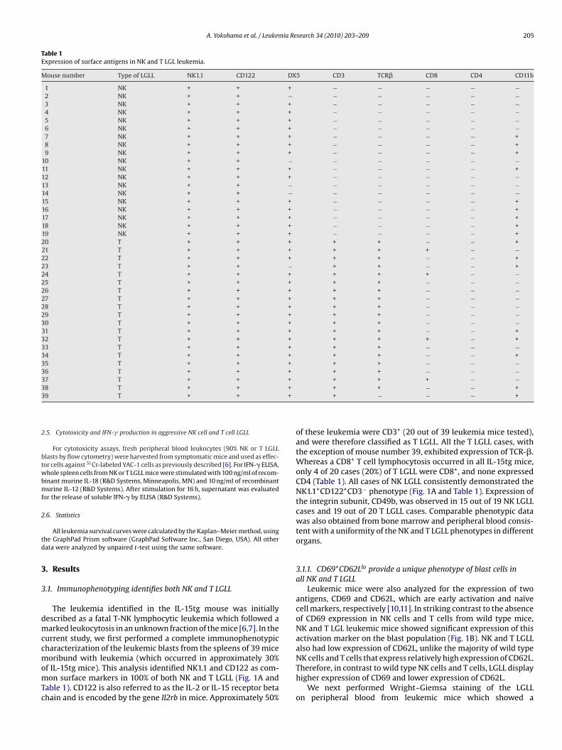
F . (A) Bb ctronL d LGLm ificatio
pleflompo
F((g
ig. 2. Leukemia mice have reduced survival and enhanced lymphoid proliferationlasts in peripheral blood of a representative moribund mouse. (B) Transmission eleGL leukemia cells show characteristic electron dense granules in the cytoplasm anouse reveals massive infiltration of blasts in bone marrow and spleen (40× magn
redominance of blasts (Fig. 2A), all exhibiting a large granularymphoblast morphology (Fig. 2B). Thereafter, we investigated thextent of leukemic infiltration by both histology (Fig. 2C) and byow cytometric analysis (data not shown). Both revealed evidence
f massive infiltrations of leukemic blasts throughout. Approxi-ately 90% of the bone marrow and spleen lymphocyte gatedopulation were NK1.1+ blasts in both NK and T LGLL.Identificationf chromosomal aberrations in NK and T LGLL
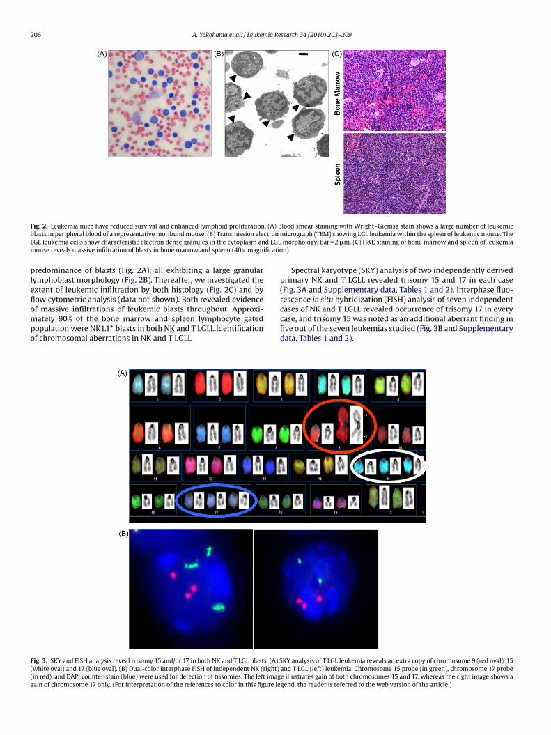
ig. 3. SKY and FISH analysis reveal trisomy 15 and/or 17 in both NK and T LGL blasts. (A) Swhite oval) and 17 (blue oval). (B) Dual-color interphase FISH of independent NK (right)in red), and DAPI counter-stain (blue) were used for detection of trisomies. The left imagain of chromosome 17 only. (For interpretation of the references to color in this figure le
lood smear staining with Wright–Giemsa stain shows a large number of leukemicmicrograph (TEM) showing LGL leukemia within the spleen of leukemic mouse. Themorphology. Bar = 2 �m. (C) H&E staining of bone marrow and spleen of leukemian).
Spectral karyotype (SKY) analysis of two independently derivedprimary NK and T LGLL revealed trisomy 15 and 17 in each case(Fig. 3A and Supplementary data, Tables 1 and 2). Interphase fluo-rescence in situ hybridization (FISH) analysis of seven independent
cases of NK and T LGLL revealed occurrence of trisomy 17 in everycase, and trisomy 15 was noted as an additional aberrant finding infive out of the seven leukemias studied (Fig. 3B and Supplementarydata, Tables 1 and 2).KY analysis of T LGL leukemia reveals an extra copy of chromosome 9 (red oval), 15and T LGL (left) leukemia. Chromosome 15 probe (in green), chromosome 17 probee illustrates gain of both chromosomes 15 and 17, whereas the right image shows agend, the reader is referred to the web version of the article.)

ia Research 34 (2010) 203–209 207
3p
Lbi5
weaauIsmbtc
Fwcctrshm
A. Yokohama et al. / Leukem
.3. Leukemic blasts are capable of killing target cells androduce IFN-� .
Functional assays were performed to ascertain if NK and TGLL blasts retain their LGL properties. We tested peripherallood mononuclear cells (PBMCs) from leukemic mice (contain-
ng approximately 90% blasts) directly in cytotoxicity assays against1Cr labeled YAC-1 target cells. Blast cells from both NK and T LGLLere highly cytotoxic ex vivo, killing with >45% efficiency at an
ffector to target (E:T) ratio of only 6.25:1, and doing so withoutdditional in vitro cytokines stimulation (Fig. 4A). In addition, welso tested the ability of NK and T LGLL blasts to secrete IFN-�pon stimulation with two monokines. We used the combination of
L-12 and IL-18 since these cytokines have been known to demon-trate synergistic effect in inducing the production of IFN-� by bothurine and human NK cells. We found that both NK and T LGLL
lasts secrete high levels of IFN-� in response to monokine stimula-ion (Fig. 4B). Thus, both NK and T LGLL blasts display the functional
haracteristics of mature LGL.ig. 4. Cytotoxicity and IFN-� release by NK and T LGL blasts. (A) Freshly isolatedhole blood leukocytes from two mice with NK LGL (©) and T LGL (�) leukemia that
ontained approximately 90% blasts were incubated with 51Cr labeled YAC-1 targetells as described in Section 2 at the indicated effector:target in 96-well plates inriplicate. The data represents mean ± SD of triplicates for all the effectors to targetatios. (B) IFN-� production in leukemia mice was determined (n = 6) by overnighttimulation of total splenocytes with IL-12 and IL-18 in vitro. Supernatants werearvested and analyzed for IFN-� production using an ELISA. Data are expressed asean ± SD of duplicates and are pooled from four independent experiments.
Fig. 5. Transplantability of primary leukemia cells in vivo. (A) Kaplan–Meier over-all survival analysis shows a significant difference between WT controls (-*-) andleukemia mice (NK LGL leukemia, � and T LGL leukemia, �). (B) Kaplan–Meier sur-vival curve of SCID mice after transfer of leukemia cells to secondary recipients.Groups of 4 mice were injected intravenously with 1 × 10⁵ peripheral blood cells(�) or injected with same number of spleen cells (♦) or bone marrow cells (�) on
day 0. Mice transplanted with different sources of LGL leukemic blasts showed mor-tality within the same period of time irrespective of the source of LGL leukemicblasts. Results shown are representative of two independent experiments that wereperformed.3.4. NK1.1+ cells transplanted from leukemia mice rapidly developlethal leukemia
Finally, we plotted the survival curve for IL-15tg mice that spon-taneously develop primary NK or T LGLL. Our data indicate thatthe leukemic mice have a median survival age of approximately28 weeks (Fig. 5A). We also transplanted ∼105 NK or T LGLL blastsfrom spleen, bone marrow or peripheral blood of leukemic mice tosub-lethally irradiated SCID mice to evaluate the capacity of blaststo reconstitute leukemia. Indeed, these animals developed a lethalleukemia with a median latency of approximately 70 days (Fig. 5B).Flow cytometric analysis of bone marrow, peripheral blood, andspleen cells of recipient animals demonstrated the immunopheno-type of the primary leukemia (data not shown). This data confirmsthe aggressive nature of the NK and T LGLL in vivo.
4. Discussion
In this report, we have described a novel mouse model for thedevelopment of aggressive NK and T LGLL arising in the setting ofchronic inflammation driven by constitutive over-expression of IL-15 [6,7]. Previous studies have identified IL-15 as a critical cytokine
2 ia Re
foofatwlfarel
tnioisAcma
mdimoacsttlcbtv(tRsctititrp
tahNtotTdmldL
08 A. Yokohama et al. / Leukem
or the development and homeostasis of NK, NKT cells and mem-ry CD8+ T cells [18–20]. In IL-15tg mice, the dramatic expansionf CD8(+) T and NK cells from birth gives rise to a very aggressiveorm of NK and T LGLL in approximately 30% of cases. Interestingly,pproximately half of IL-15tg mice with this aggressive LGLL havehe NK phenotype and the other half have the T cell phenotype,ith virtually all T cell cases displaying the NK antigen NK1.1. All
eukemias that were analyzed also had CD62LloCD69+ phenotype asound in human LGLL [12,13], along with functional attributes oftenttributed to their human counterparts as noted below [14,15]. Cur-ently we do not have a clear understanding of the key molecularvents that lead to the development of LGLL in IL-15tg mice, regard-ess of the cell type.
Previous studies indicate that human NK and T LGLL blasts retainhe functional properties and immunophenotype of mature LGL,amely: (1) surface expression of CD56 [16–18]; (2) natural cytotox-
city towards tumor and virus infected cells [19] and (3) productionf inflammatory cytokines such as IFN-� [20]. In this report, we have
dentified NK1.1 and the IL-15 receptor � chain, CD122, as commonurface markers on all the NK and T LGLL arising in the IL-15tg mice.dditionally, these leukemias demonstrate spontaneous cytotoxi-ity towards YAC-1 targets and production of IFN-� in response toonokine co-stimulation. Thus, these findings in our mouse model
re consistent with human LGLL [14,15].Remarkably, cytogenetic studies of human LGLL show few com-
on recurrent chromosomal abnormalities associated with theisease [1,3,5]. Our cytogenetic data demonstrate that trisomy 17
s a common abnormality in both NK and T LGLL arising in IL-15tgice. Trisomy of chromosome 17 in murine leukemia is rare, and its
ccurrence in 100% of the NK and T LGLL examined in this study mayttest to a pivotal role in leukemogenesis. It should be noted humanhromosome 21 shows conserved syntenies to mouse chromo-ome 17 and as such shares a significant fraction of gene triplicateshat are located on chromosome 21 in Down syndrome, wherehere is an increased incidence of acute myeloid and lymphoblasticeukemia. Thus it is conceivable, if not probable that the same extraopy of gene(s) on chromosome 21 in humans that are responsi-le for the acute leukemia in Down syndrome are contained withinhe mouse chromosome 17 [21,22]. Trisomy 15 is also found withariable frequency in murine models of acute myeloid leukemiaAML) and acute lymphoblastic leukemia, including those arising inhe setting of the PML-RARA gene fusion, gamma irradiation, andauscher murine leukemia virus infection [23,24]. Interestingly, tri-omy of human chromosome 8, syntenic to mouse 15, is the mostommon single chromosomal aberration in both the myelodysplas-ic syndrome (MDS) and AML in humans, and has also been reportedn human NK cell malignancies [25]. Spira et al. have hypothesizedhat trisomy is an early event in leukemogenesis which leads toncreased oncogene dosage and thereby contributes to malignantransformation [26]. However, it remains to be determined if theseecurrent chromosomal aberrations truly contribute to the onset orrogression of LGLL in humans or in our animal model.
The utilization of a mouse with constitutive over-expression ofhe pro-inflammatory cytokine IL-15 to generate a model of theggressive variant of NK or T LGLL is not in itself irrelevant to theuman counterpart of this disorder. In many instances, the humanK or T LGLL cell lines that have been derived from patients with
his disorder are dependent on either IL-15 or IL-2 in vitro, as bothf these cytokines use the same � and �c receptor componentso transmit their activation signals to the target cell [14,15,27].his in vitro dependence on IL-15 or IL-2 suggests comparable
ependency on either cytokine in vivo, rather than an activatingutation in their receptor components that would render the celline cytokine independent. Indeed, Waldmann and colleagues haveesigned and implemented an anti-IL-2/15R� mAb trial to treat TGLL [28]. Finally, there is good evidence that IL-15 is critical for the
[
search 34 (2010) 203–209
development of human and murine NK cells and hepatic-derivedNKT cells in mice, and for the homeostatic maintenance of NK/T-NKand CD8(+) memory T cells [29]. Collectively, with the evidenceprovided in this report, it appears that increased expression ofIL-15 directly contributes to the malignant transformation of eitherNK cells, T-NK cells, T cells or their common precursor.
In summary, we have characterized a mouse model of sponta-neous NK and T LGLL arising in the setting of increased expressionof IL-15. This murine model most closely resembles the aggres-sive variant of human LGLL of the NK cell or T cell type, and maytherefore prove to be useful in order to develop novel treatmentstrategies for this rare disease that currently has no standard formof beneficial therapy.
Conflict of interest
The authors do not have any conflict of interest to declare.
Acknowledgements
We thank Dr. Pamela J Swiatek of the Van Andel Research Insti-tute for her extensive help with FISH and REB experiments andhelpful discussions. We also thank Michelle M. Le Beau of the Uni-versity of Chicago Cancer Research Center for assistance with earlymouse cytogenetics. We acknowledge the assistance of the follow-ing shared resources of The Ohio State University ComprehensiveCancer Center: Mouse Pathology, Flow Cytometry, Microscopy, andStatistics.
Appendix A. Supplementary data
Supplementary data associated with this article can be found, inthe online version, at doi:10.1016/j.leukres.2009.06.031.
References
[1] Nakamura F, Tatsumi E, Kawano S, Tani A, Kumagai S, Nishikori M, et al. Acutelymphoblastic leukemia/lymphoblastic lymphoma of natural killer (NK) lin-eage: quest for another NK-lineage neoplasm. Blood 1997;89:4665–6.
[2] Alekshun TJ, Tao J, Sokol L. Aggressive T-cell large granular lymphocyteleukemia: a case report and review of the literature. Am J Hematol 2007;82:481–5.
[3] Matano S, Terahata S, Nakamura S, Kobayashi K, Sugimoto T. CD56-positiveacute lymphoblastic leukemia. Acta Haematol 2005;114:160–3.
[4] Gentile TC, Uner AH, Hutchison RE, Wright J, Ben-Ezra J, Russell EC, et al.CD3+, CD56+ aggressive variant of large granular lymphocyte leukaemia. Blood1994;84:2315–21.
[5] Johansson B, Mertens F, Mitelman F. Clinical and biological importance of cyto-genetic abnormalities in childhood and adult acute lymphoblastic leukemia.Ann Med 2004;36:492–503.
[6] Fehniger TA, Suzuki K, Ponnappan A, VanDeusen JB, Cooper MA, Florea SM, etal. Fatal leukemia in interleukin 15 transgenic mice follows early expansions innatural killer and memory phenotype CD8+ T cells. J Exp Med 2001;193:219–31.
[7] Fehniger TA, Suzuki K, VanDeusen JB, et al. Fatal leukemia in interleukin-15transgenic mice. Blood Cells, Mol, Dis 2001;27:223–30.
[8] Le Beau MM, Bitts S, Davis EM, Kogan SC. Recurring chromosomal abnormalitiesin leukemia in PML-RARA transgenic mice parallel human acute promyelocyticleukemia. Blood 2002;99:2985–91.
[9] Graveel C, Su Y, Koeman J, Wang LM, Tessarollo L, Fiscella M, et al. Activating Metmutations produce unique tumor profiles in mice with selective duplication ofthe mutant allele. Proc Natl Acad Sci USA 2004;101:17198–203.
[10] Li XY, Matsuzaki G, Yoshikai Y, Muramori K, Nomoto K. T cells expressing bothL-selectin and CD44 molecules increase in number in peritoneal exudate cellsand in vitro-stimulated spleen cells from mice immunized intraperitoneallywith Listeria monocytogenes. Immunology 1993;78:28–34.
[11] Natarajan K, Sawicki MW, Margulies DH, Mariuzza RA. Crystal structure ofhuman CD69: a C-type lectin-like activation marker of hematopoietic cells.Biochemistry 2000;39:14779–86.
12] Yang J, Epling-Burnette PK, Painter JS, Zou J, Bai F, Wei S, et al. Antigen activationand impaired Fas-induced death-inducing signaling complex formation in T-large-granular lymphocyte leukemia. Blood 2008;111:1610–6.
[13] Lima M, Almeida J, Dos Anjos Teixeira M, Alguero Md Mdel C, Santos AH, Bal-anzategui A, et al. TCRalphabeta+/CD4+ large granular lymphocytosis: a newclonal T-cell lymphoproliferative disorder. Am J Pathol 2003;163:763–71.
ia Re
[
[
[
[
[
[
[
[
[
[
[
[
[
[
[28] Morris JC, Janik JE, White JD, Fleisher TA, Brown M, Tsudo M, et al. Preclinicaland phase I clinical trial of blockade of IL-15 using MikÎ21 monoclonal anti-
A. Yokohama et al. / Leukem
14] Robertson MJ, Cochran KJ, Cameron C, et al. Characterization of a cell line, NKL,derived from an aggressive human natural killer cell leukemia. Exp Hematol1996;24:406–15.
15] Gong JH, Maki G, Klingemann HG. Characterization of a human cell line (NK-92) with phenotypical and functional characteristics of activated natural killercells. Leukemia 1994;8:652–8.
16] Kingreen D, Siegert W. Chronic lymphatic leukemias of T and NK cell type.Leukemia 1997;11(Suppl. 2):S46–9.
17] Prieto J, Rios E, Parrado A, et al. Leukaemia of natural killer cell large granu-lar lymphocyte type with HLA-DR-CD16-CD56bright+ phenotype. J Clin Pathol1996;49:1011–3.
18] Macon WR, Williams ME, Greer JP, Hammer RD, Glick AD, Collins RD, et al.Natural killer-like T-cell lymphomas: aggressive lymphomas of T-large granularlymphocytes. Blood 1996;87:1474–83.
19] Ohno T, Kanoh T, Arita Y, Fujii H, Kuribayashi K, Masuda T, et al. Fulminant clonalexpansion of large granular lymphocytes. Characterization of their morphology,phenotype, genotype, and function. Cancer 1988;62:1918–27.
20] Itoh K, Tsuchikawa K, Awataguchi T, Shiiba K, Kumagai K. A case of chronic lym-
phocytic leukemia with properties characteristic of natural killer cells. Blood1983;61:940–8.21] Malinge S, Izraeli S, Crispino JD. Insights into the manifestations, outcomes, andmechanisms of leukemogenesis in Down syndrome. Blood 2009;113:2619–28.
22] Hattori M, Fujiyama A, Taylor TD, Watanabe H, Yada T, Park HS, et al. The DNAsequence of human chromosome 21. Nature 2000;405:311–9.
[
search 34 (2010) 203–209 209
23] Herbst EW, Gropp A. Relevance of trisomy 15 and other chromosome abnor-malities in spontaneous AKR leukemia of mice with and without Robertsonianrearrangement. J Cancer Res Clin Oncol 1982;104:207–18.
24] Herbst EW, Gropp A, Stehle W, Fohlmeister I, Nielsen K, Claussen CP, et al.Rauscher leukemia in the mouse in trisomy of chromosome 19. Verh DtschGes Pathol 1982;66:122–7.
25] Paulsson K, Johansson B. Trisomy 8 as the sole chromosomal aberration inacute myeloid leukemia and myelodysplastic syndromes. Pathol Biol (Paris)2007;55:37–48.
26] Spira J, Wiener F, Ohno S, Klein G. Is trisomy cause or consequence of murine Tcell leukemia development? Studies on Robertsonian translocation mice. ProcNatl Acad Sci USA 1979;76:6619–21.
27] Di Noto R, Pane F, Camera A, Luciano L, Barone M, Lo Pardo C, et al. Charac-terization of two novel cell lines, DERL-2 (CD56+/CD3+/Tcry5+) and DERL-7(CD56+/CD3−/TCRgammadelta−), derived from a single patient with CD56+
non-Hodgkin’s lymphoma. Leukemia 2001;15:1641–9.
body in T cell large granular lymphocyte leukaemia. Proc Natl Acad Sci USA2006;103:401–6.
29] Ma A, Koka R, Burkett P. Diverse functions of IL-2, IL-15, and IL-7 in lymphoidhomeostasis. Annu Rev Immunol 2006;24:657–79.