On the process of formation of Early Rus’ burial culture in the Novgorod Land
1316
ISSN 0031-0301, Paleontological Journal, 2009, Vol. 43, No. 10, pp. 1316–1329. © Pleiades Publishing, Ltd., 2009.
INTRODUCTION
During the three last decades, the interest of bota-nists dealing with fossil and modern plants to Carbon-iferous synangiate pollen organs has considerablyincreased: earlier studied materials were revised(Millay and Taylor, 1979; Meyer-Berthaud, 1989), andnew taxa were described (Eggert and Taylor, 1971;Stidd et al., 1985; Meyer-Berthaud, 1986; Rowe, 1988;and others). To date, more than 20 genera of microspo-rangiate organs have been described from the Carbon-iferous deposits of Europe, Great Britain, and Northand South Americas (Erwin et al., 1994). They belongto four orders of Lyginopteridophyta: Calamopytiales,Lyginopteridales, Callistophyllales, and Medullosales.Early Carboniferous deposits only contain members ofthe first and second orders. The majority of them werefound in Scottish and English localities, and the restwas reported from the United States, Germany, andRussia (Orlova, 2002, 2007). The first Russian find of asynangiate organ was preliminary discussed by Orlova(2002); the present paper publishes a detailed study ofthis fossil.
MATERIAL AND METHODS
The material was collected from the Porog Vittsa 2locality, situated at the right bank of the Msta River,near the village of Putlino, Borovichi district of theNovgorod Region. An imprint of a fertile axis bearing
synangia and numerous casts and imprints of detachedsynangia were found in yellow ferruginous sandstone.In addition to the fertile axes, sterile remains of Lygi-nopteridales, Medullosales, and Calamopytiales werefound (Orlova and Snigirevskii, 2003, 2004). Fragmen-tary fronds of
Rhodeopteridium
and
Sphenopteridium
are particularly numerous. The total list of the plantremains found in this locality includes
Archaeosig-illaria vanuxemii
(Goepp.) Kidston,
Lepidodendronacuminatum
(Goepp.) Stur,
L. veltheimii
Sternb.,
Lepi-dostrobus
sp. 1,
Lepidostrobus ornatus
Brongn.,
Flem-ingites
sp. 1,
Stigmaria
sp.,
Archaeocalamites radiatus
(Brongn.) Stur,
Sphenopteridium bifidum
(L. et H.)Benson,
Sphenopteridium
sp. 1,
S. pachyrrhachis
(Goepp.) Pot.,
S. jurinae
O. Orl. et S. Snig.,
S. gaebleri
Gothan,
Adiantites antiquus
(Ett.) Stur,
A. machanekii
Stur,
Adiantites
sp. 1
, Sphenopteris distans
Sternb.,
Sphenopteris
sp. 1,
S. foliolata
Stur.,
Rhodeopteridiumhochstetteri
(Stur) Purk.,
R. goeppertii
(Ett.) O. Orl. etS. Snig.,
R. tenue
(Gothan) Kotas.,
Lyginodendron
sp.,and
Rhynchogonium sulcatum
(L. et H.) Zal. (Orlovaand Rasskazova, 2005).
The deposits of the Porog Vittsa 2 locality are char-acterized by a palynological assemblage with con-stantly occurring numerous
Lycospora pusilla
(Ibr.)Som.
Lophotriletes grossepunctatus
(Waltz) Isch.,
Triquitrites marginatus
H., St. et M.,
T. comptus
Will.,
Trachytriletes subintortus
Isch.,
Schulzospora campy-loptera
(Waltz) H., St. et M.,
S. magnifica
(Isch.)
A New Microsporangiate Organ from the Lower Carboniferous of the Novgorod Region, Russia
O. A. Orlova
a
, N. R. Meyer-Melikian
†
,
a
, and N. E. Zavialova
b
a
Moscow State University (MGU), Leninskie gory 1, Moscow, 119991 Russia
b
Borissiak Paleontological Institute of the Russian Academy of Sciences, ul. Profsoyuznaya 123, Moscow, 117997 Russiae-mail: [email protected]
Received February 5, 2008
Abstract
—A new species of the genus
Telangiopsis, T. nonnae
O. Orlova et Zavialova, was described on thebasis of a microsporangiate organ from the Lower Carboniferous deposits of the Novgorod Region. The mor-phology of branching fertile axes, synangia, and sporangia was thoroughly studied. The three-dimensional sys-tem of fertile axes branches monopodially; ultimate axes bear numerous connivent bunches of synangia, whichconsist of three to six basally fused elongated ovate sporangia. The morphology and ultrastructure of prepollengrains were studied, which were extracted from the rock matrix surrounding the sporangia. The two-layeredexine includes a well-developed endexine and an alveolate ectexine, with one–three rows of large thin-walledalveolae. The new species was compared with other Early Carboniferous microsporangiate organs.
Key words
: Early Carboniferous, Novgorod Region, fertile axis, synangia, Lyginopteridales, trilete prepollen,exine ultrastructure.
DOI:
10.1134/S003103010910013X
PALEONTOLOGICAL JOURNAL
Vol. 43
No. 10
2009
A NEW MICROSPORANGIATE ORGAN FROM THE LOWER CARBONIFEROUS 1317
Oshurk.,
Tripartites vetustus
Schem.,
Punctatisporitesglaber
(Naum.) Playf., and some other spores occurringmuch more rarely (Orlova and Rasskazova, 2005). Thepalynological assemblage fits the palynological zonesPerotriletes tessellatus–Schulzospora campyloptera(TC), Raistrickia nigra–Triquitrites marginatus (NM),Tripartites vetustus–Rotaspora fracta (VF), and Bel-lispores nitidus–Reticulatisporites carnosus (NC) of theUpper Visean=Namurian A (Serpukhovian) of WestEurope.
Collection, no. 293 is kept at the Department ofPaleontology, MGU. Olympus SZ-6045 and LeicaMZ16 stereomicroscopes were used during the studyand photographing. A rock fragment with detachedsynangia was macerated after Grishchuk (fluoric acidfollowed by centrifugation). The organic residue withprepollen grains was mounted on slides and studied intransmitted light, under a Zeiss Axioplan 2 microscope;some prepollen grains were studied under Camscan andHitachi S-405A scanning electron microscopes
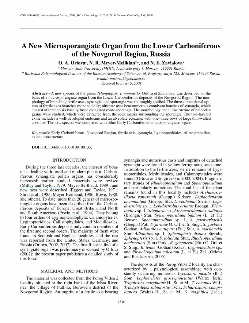
Fig. 1.
Telangiopsis nonnae
sp. nov., holotype MGU, no. 293/47; general view of microsporangiate organ, branching fertile axesand clusters of synangia are shown,
×
3.5. Porog Vittsa 2 locality, Borovichi district, Novgorod Region, Russia; Lower Carbonifer-ous, Visean Stage, Upper Visean Substage.
1318
PALEONTOLOGICAL JOURNAL
Vol. 43
No. 10
2009
ORLOVA
et al
.
(SEMs). Ultrathin sections were made using an LKB 3ultramicrotome with a diamond knife and studied undera Hitachi H-600 transmission electron microscope(TEM). In total, 21 prepollen grains were studied intransmitted light; 18, under SEM; and 5, under TEM.Grids with ultrathin sections and films and graphicalfiles documenting the sections are kept at the laboratoryof paleobotany, PIN.
SYSTEMATIC PALEOBOTANY
Division LyginopteridophytaClass LyginopteridopsidaSubclass Lyginopterididae
Order Lyginopteridales
Family Lyginopteridaceae Potonie, 1902Genus
Telangiopsis
Eggert et Taylor, 1971
Telangiopsis nonnae
O. Orlova et Zavialova, sp. nov.
Plates 25–29
E t y m o l o g y. In memory to Nonna RobertovnaMeyer-Melikian, our scientific teacher and outstandingbotanist, died an early death.
H o l o t y p e. MGU, no. 293/47, Geological Fac-ulty; fertile leafless axis bearing terminal synangia;Porog Vittsa 2 locality, right bank of the Msta River,Borovichi district, Novgorod Region, Russia; Lower
Carboniferous, Visean Stage, Upper Visean Substage(Fig. 1).
D i a g n o s i s. System of fertile axes branchesmonopodially, three-dimensional. Main axis with alter-nating secondary axes. Secondary axes bear alternatingtertiary axes terminating with ultimate axes of numer-ous connivent clusters of synangia gathering in pairs,or, occasionally, single. Synangia 1.2–3.5 mm long andup to 3.5 mm wide in terminal part. Each synangiumconsists of three–six elongated ovate basally fused spo-rangia. External surface of sporangia with coarse longi-tudinal ribs. Prepollen circular to circular-triangularand oval, 27.8 x 32.0
μ
m, trilete, rays reach 3/4–4/5 ofradius. Exine surface verrucate, folded, and equatori-ally echinate. Exine bilayered, considerably varying inthickness, with alveolate ectexine.
D e s c r i p t i o n (Figs. 1–4). The leafless fertile axisshows a compound architecture. Fertile axes of all ordersalternate in three dimensions. The visible length of the mainaxis (=rachis) is 34 mm, and the width is 2.5 mm. Therachis bears alternating lateral branches (secondaryaxes), which are situated at a distance of 4–11 mm fromeach other. The upper axis of the second order is onlypreserved in the studied specimen (Pl. 25, fig. 2; Pl. 26,fig. 1; Figs. 1, 2). The next secondary axis was situatedat a distance of 11 mm below the visible secondaryaxis. The preserved secondary axis deviates from themain axis at an angle of about 35
°
and branches three-dimensionally, producing alternating branches. Thewidth of this secondary axis is 0.5–0.6 mm. The tertiaryaxis deviates from the secondary axis at an angle of 45
°
at a distance of 5 mm from the point where the second-ary axis deviates from the main axis. The terminal fer-tile axis of the forth order bearing synangia deviatesfrom the tertiary axis (Pl. 25, fig. 2). All axes are cov-ered with longitudinal ribs (Pl. 26, fig. 1). The distancebetween adjacent ribs is 0.15–0.2 mm.
The synangia deviate from the ultimate axis in threedimensions. The synangia are attached to the axis withshort petioles. Most often, the synangia are in pairs; or,more rarely, they are solitary (Fig. 2). Usually, the clus-ters of synangia are situated closely to each other andoften are partially or completely superimposed (Pl. 25,fig. 1). The synangia consist of three to six sporangia(Fig. 3). Most often, synangia of four or five sporangiaoccur (Figs. 3c–3e; Pl. 26, figs. 2, 4). Usually, synangiaare situated along the bedding plane, and, therefore, inlongitudinal section resembling wide cups (Figs. 2, 3;Pl. 26, figs. 2–6). Rarely, they are tear-shaped, small,and consisting of three sporangia (Fig. 3b). Most prob-ably, such synangia were immature. The length of thesporangia preserved along the bedding plane variesfrom 1.2 to 3.5 mm; and the width in the terminal partreaches 3.5 mm (Table 1). Some synangia are pros-trated in shape of a four- or five-pointed star (Fig. 2).Oblique sections of such synangia show that the spo-rangia are radially situated and have free sides. There-fore, the synangia consist of sporangia fused with each
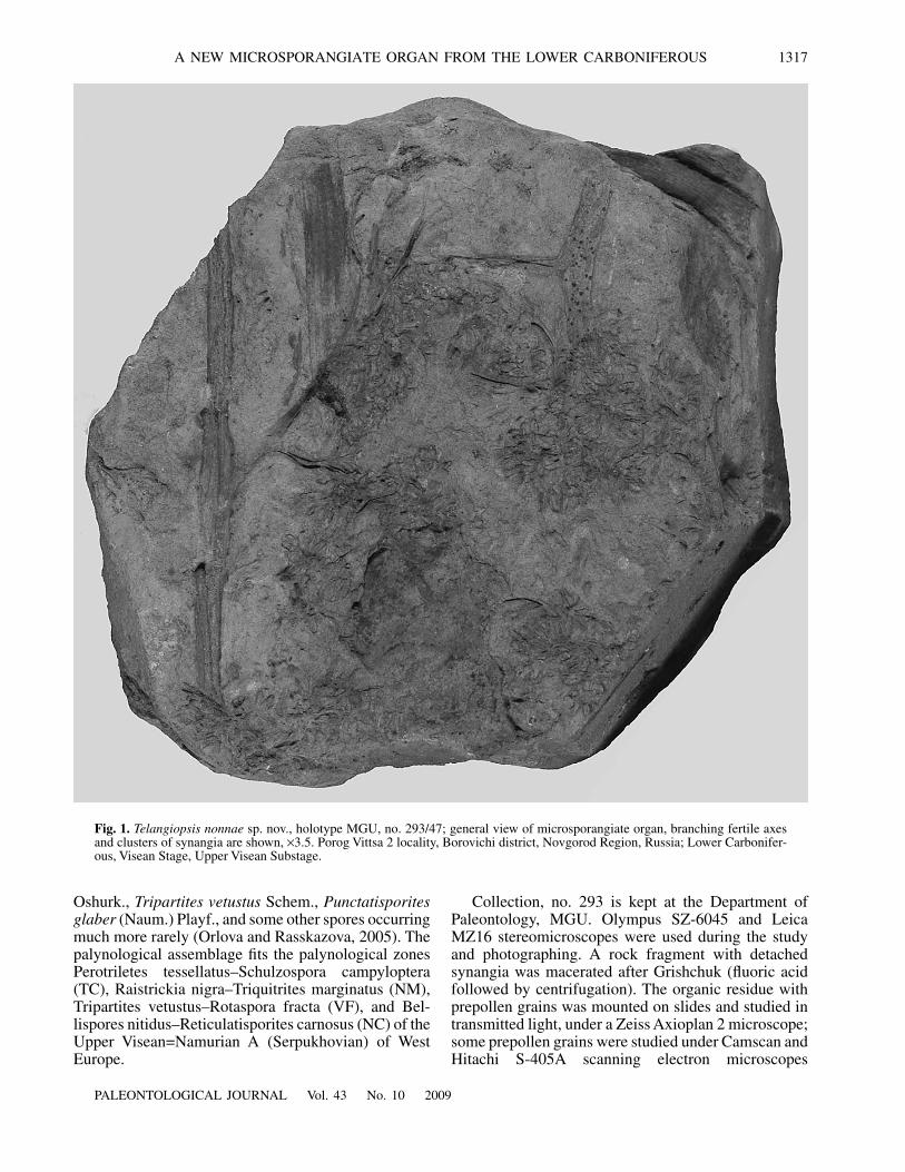
Fig. 2.
Reconstruction of pollen organ of
Telangiopsis non-nae
sp. nov. Scale bar 2 mm.
PALEONTOLOGICAL JOURNAL
Vol. 43
No. 10
2009
A NEW MICROSPORANGIATE ORGAN FROM THE LOWER CARBONIFEROUS 1319
other in their basal parts and free at the apices. No dis-tinct synangiate cushion was observed. The only regionwhere it is possibly present is shown in Pl. 26, fig. 2(Fig. 3e). As a rule, the sporangia are elongated ovatewith a more acuminate basal part and a less acuminateand occasionally rounded terminal extremity (Figs. 1–3;Pl. 26, figs. 4, 5). Occasionally, the apices of the spo-rangia are toothed (Pl. 26, fig. 6). Most often, the max-imal width of the sporangium is situated in its middleand is equal to 0.4–0.6 mm. The length of the sporangiavaries from 1.2 to 2.0 mm. The external surface of thesporangia is covered with coarse longitudinal ribs.There are four to six occasionally dichotomizing ribson each sporangium (Pl. 25, figs. 2, 3; Pl. 26, figs. 2, 5).In rare cases, a longitudinal keel was observed on spo-rangia (Pl. 26, fig. 4; Figs. 3e, 3f). Apparently, the spo-rangia were longitudinally dehiscent. Some sporangiaare opened along the bedding plane. Numerousimprints of rounded prepollen grains are visible in lon-gitudinal folds of the walls of such sporangia (Pl. 25,fig. 3). Scattered prepollen grains are also visible in therock surrounding the synangia.
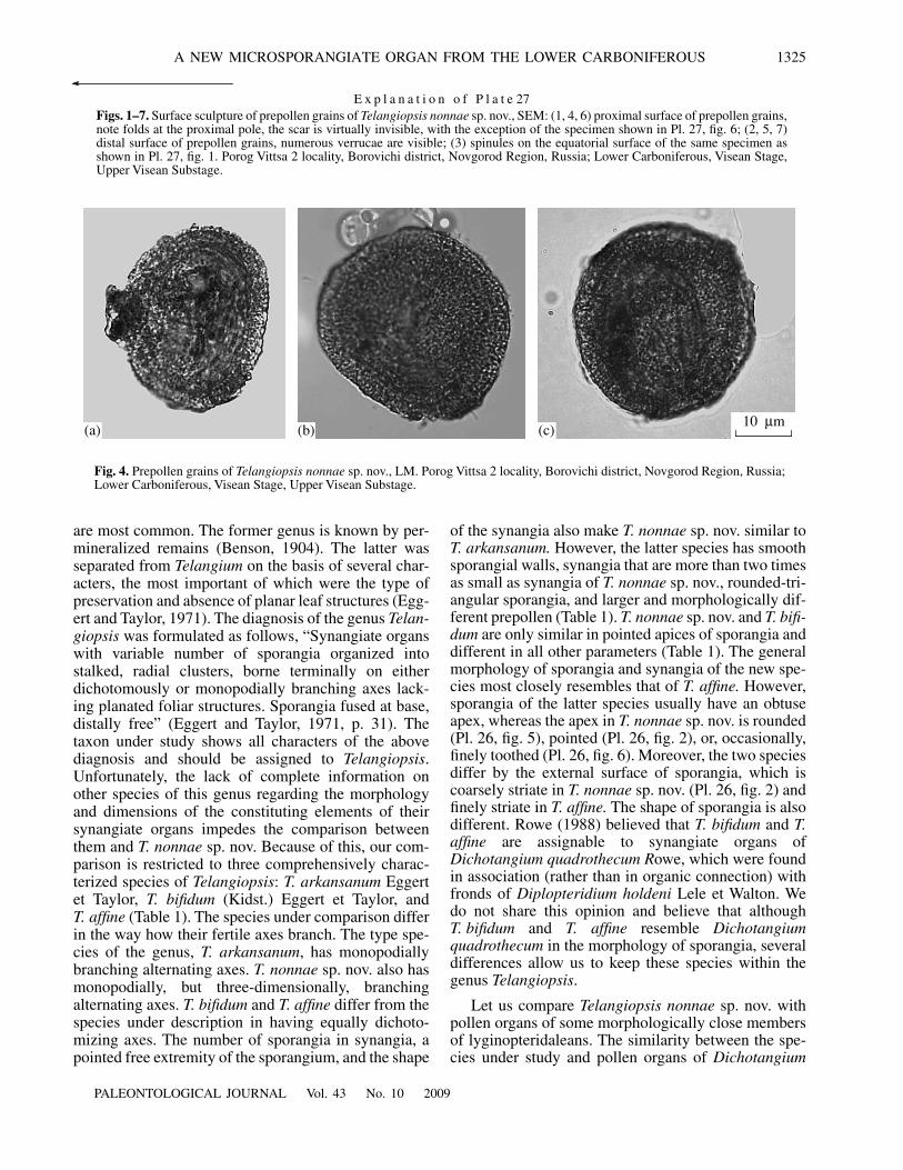
Numerous sporangiate walls and miospores inmonads and tetrads were obtained by the maceration ofthe rock surrounding the synangia. More than 90% ofthe palynological assemblage is represented by similarmiospores, which were assigned to
Geminosporaparvibasilaria
(Naum.) Byvsch on the basis of charac-ters visible in transmitted light (determination wasaccomplished by M.V. Oshurkova). The prepollengrains are rounded, rounded-triangular, or, occasion-ally, oval. Usually, they are preserved in polar position.The sizes vary from 23.4 to 37.0
μ
m, 27.8
×
32.0
μ
m onaverage (Fig. 4). The proximal scar is trilete, the rays
are closed, up to 3/4–4/5 of the radius of the prepollengrain, and often poorly visible. The exine is thick andbilayered. In some prepollen grains, the exine in thepolar region is slightly lighter than in other regionsimplying that the exine of the proximal and/or distalpoles is thinner than the equatorial exine. In transmittedlight, the exine appears vermiculate, that, apparently, isresulted from superimposed sculptural elements andinner partitions of the exine. The margin of the prepol-len grain is uneven, crenate, and with occasionalspinules.
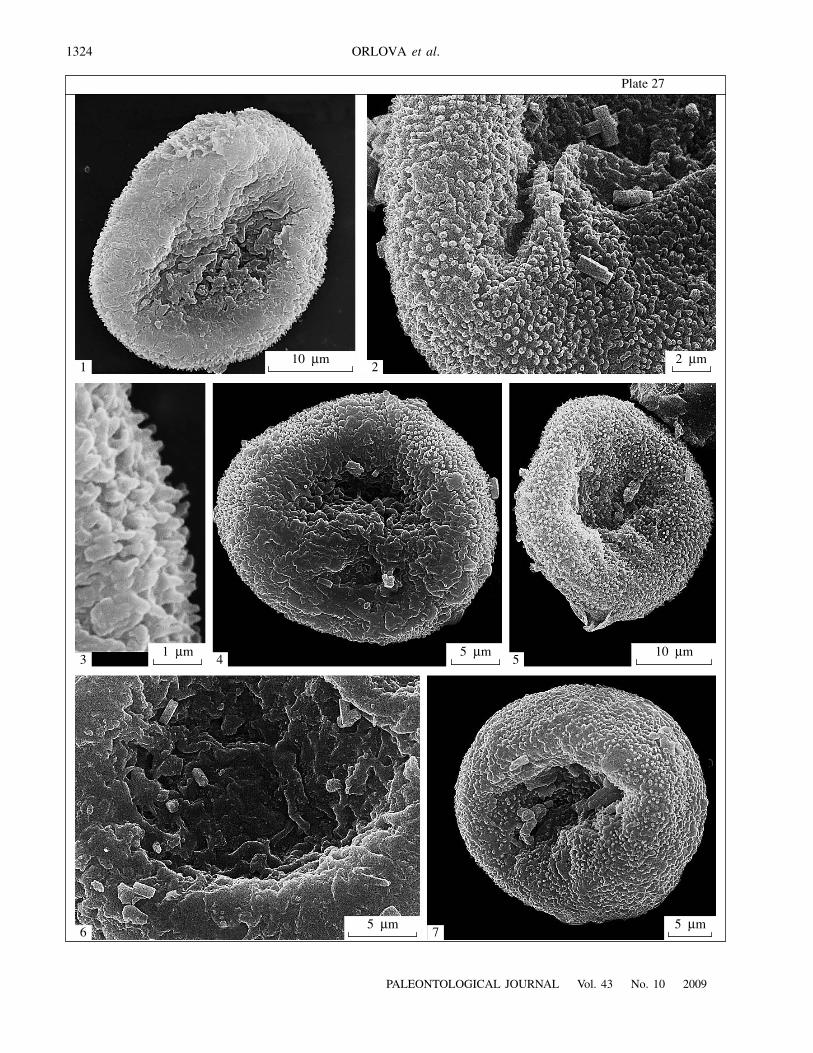
SEM revealed diverse sculpture of the exine(Pl. 27). Numerous small verrucae cover the distal sur-face, often reaching equatorial regions (Pl. 27, figs. 2,5, 7), where they occasionally transform into spinules1.3–1.5
μ
m long (Pl. 27, fig. 3). The exine is elevated inthe equatorial regions, and there are depressions in thepolar regions. Although verrucae occur on the proximalface as well, numerous folds are more characteristic ofthe proximal sculpture. The proximal scar is closedwith non-elevated rays and usually is nearly undistin-guishable under SEM (Pl. 27, figs. 1, 4). Under SEM,we have only seen a distinct proximal scar in one of thestudied specimens (Pl. 27, fig. 6).
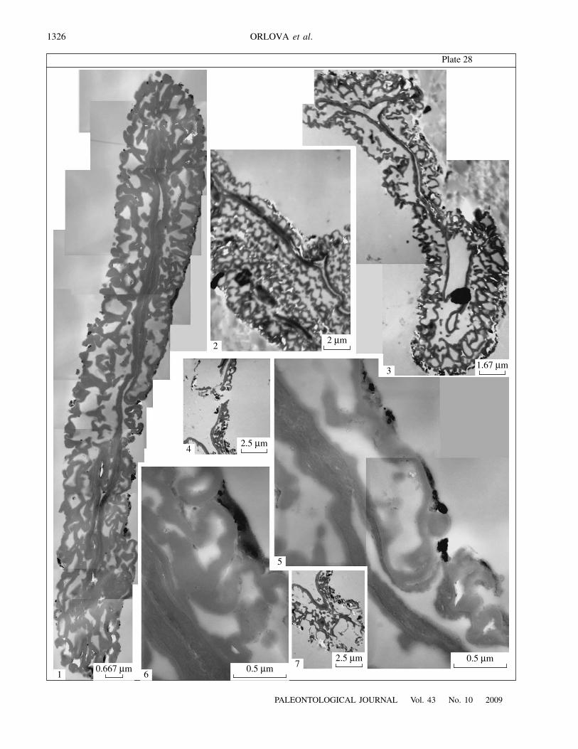
TEM revealed two layers in the exine (Pls. 28, 29).The exine thickness varies along the perimeter of thepollen grain: the thickest exine is situated in the equa-torial region, reaching 2.3–3.1
μ
m. The distal exine is 1.3–2.1
μ
m thick, in places becoming thinner up to 0.7
μ
m.The proximal exine is 1.1–1.7
μ
m, becoming sharplythinner up to 0.3
μ
m under rays of the proximal scar(Pl. 28, figs. 1, 3). Granular elements (cut verrucae) aremore numerous on the distal surface of the exine than
a b c
d e f
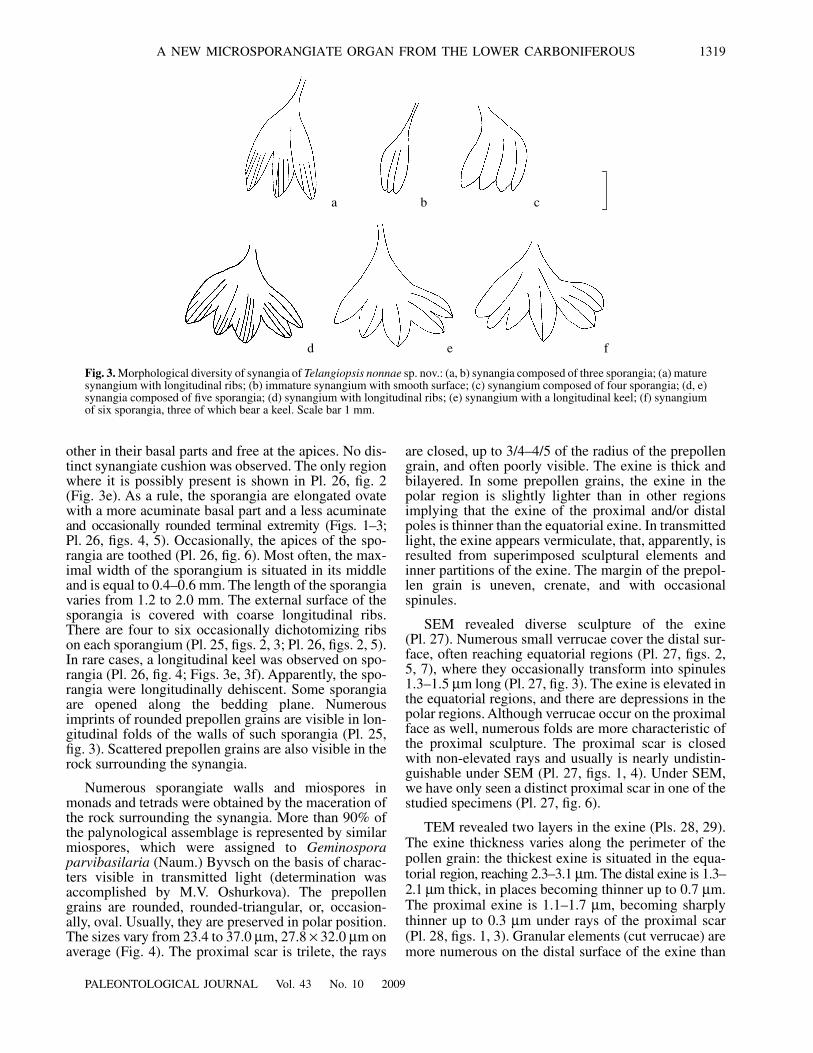
Fig. 3.
Morphological diversity of synangia of
Telangiopsis nonnae
sp. nov.: (a, b) synangia composed of three sporangia; (a) maturesynangium with longitudinal ribs; (b) immature synangium with smooth surface; (c) synangium composed of four sporangia; (d, e)synangia composed of five sporangia; (d) synangium with longitudinal ribs; (e) synangium with a longitudinal keel; (f) synangiumof six sporangia, three of which bear a keel. Scale bar 1 mm.
1320
PALEONTOLOGICAL JOURNAL
Vol. 43
No. 10
2009
ORLOVA
et al
.
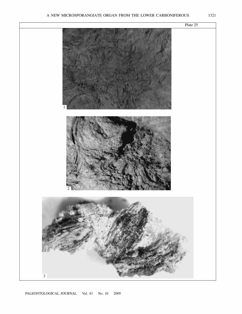
E x p l a n a t i o n o f P l a t e 25
Figs. 1–3.
Telangiopsis nonnae
sp. nov., holotype MGU, no. 293/47: (1) aggregation of synangia, note numerous synangia consti-tuted of elongated obovate sporangia with rounded apices,
×
3.2; (2) ultimate fertile axis bearing pairs of synangia,
×
7.5; (3) threesporangia split along the rock matrix, note numerous imprints of prepollen grains,
×
40. Porog Vittsa 2 locality, Borovichi district,Novgorod Region, Russia; Lower Carboniferous, Visean Stage, Upper Visean Substage.
on the proximal surface (Pl. 28, fig. 3; Pl. 29, fig. 4).The external outline of the proximal face is more undu-lated because of the folds which were visible on theproximal surface under SEM (Pl. 29, fig. 1). The ectexineis alveolate, with relatively thin partitions (0.1–0.15
μ
mthick), the alveolae are relatively spacious, approxi-mately up to 1.0
μ
m long and up to 0.8
μ
m high (Pl. 28,fig. 6). Dependent on the thickness of the ectexine, itsalveolae are arranged in one to three tiers: in two orthree tiers in the equatorial area; more often in two tiersor, occasionally, in one tier on the distal side; and moreoften in one tier or, occasionally, in two tiers on theproximal side (Pl. 28, figs. 2, 4).
The endexine is well developed, more electron-dense than the ectexine, 0.1–0.15
μ
m in average thick-ness, becoming thinner up to 0.025
μ
m under the rays(Pl. 28, fig. 5). Its heterogeneous nature is apparentunder greater magnifications: the external part is lesselectron-dense and contains one to three white lines, and theinternal part is more electron-dense (Pl. 28, figs. 5, 6; Pl. 29,figs. 3, 5).
C o m p a r i s o n. The species under description dif-fers from the closest species
T. affine
(Kidst.) Eggert etTaylor (Kidston, 1924) by the mode of branching:
T. nonnae
sp. nov. has a system of axes which branchmonopodially, and synangia of
T. affine
are situated onthe extremities of terminal axes of equally dichotomiz-ing branches. The differences also concern the outlinesof the sporangia:
T. nonnae
sp. nov. has elongated obo-vate sporangia, and
T. affine
has elongated lanceolate,and nearly linear sporangia. In addition, these two spe-cies differ by the external surface of their sporangia:finely longitudinally striate in
T. affine
and coarselylongitudinally striate in
T. nonnae
sp. nov. The prepol-len grains are also different: smooth, rounded, andmore than 50
μ
m in diameter in
T. affine
; and sculptured(with verrucae, folds, and equatorial spinules),rounded, rounded-triangular, occasionally oval, and nomore than 37
μ
m in
T. nonnae
sp. nov.
R e m a r k s. As far as the palynological assemblageextracted from the rock matrix surrounding thedetached sporangia is nearly completely constituted bymiospores of only one species, which general morphol-ogy answers that of prepollen grains of the Lyginop-teridales, we believe that these miospores represent pre-pollen grains of the plant under study and we may sup-ply the characteristics of the new taxon with data ontheir morphology and ultrastructure.
According to the morphological characters visiblein transmitted light, the prepollen grains under studyare closest to the diagnosis of Geminospora. There is acertain discrepancy between the data obtained withhelp of light and electron microscopes. Although thegenus Geminospora describes cavate spores, weobserved under TEM no distinct cleavage between theexinal layers. Sections of one of the prepollen grainsshow ectexine and endexine that are loosely mating inthe polar area. Unfortunately, this specimen was mostprobably mechanically damaged (in places the endex-ine is missing and the same might be true for the adja-cent tier of ectexinal alveolae), and we cannot beassured that the observed polar cleavage is not an arti-fact (Pl. 29). The discrepancy between LM and TEMresults can be explained as follows. The species Gemi-nospora parvibasilaria was erected on the basis of lightmicroscopical observations, and, therefore, one cannotsay with reasonable confidence how the area visible intransmitted light as a cavum looks under TEM. It is adistinct possibility that this area is corresponded by thethickest areas of airy alveolate ectexine, not separatingfrom the endexine. Another conceivable explanation isintraspecific variation: some specimens have a cavumdeveloped to a greater degree, whereas it is developedto a lesser degree or completely lacking in others. Ananalogous case was described by Zavialova et al.(2004) in Permian pollen grains of Cordaitina, varyingin the width of their monosaccus. Pollen grains bearingthe narrowest saccus show a protosaccus in sections;however, ectexinal partitions slightly do not touch theendexine in specimens with a wider saccus, and theultrastructure can be defined as intermediate betweenprotosaccus and eusaccus. The wider the saccus is, thewider the distance is between the endexine and ectexi-nal partitions pending into the saccus cavity. Dealingwith TEM, it is difficult to reach statistical amount ofstudied specimens; therefore, the risk is run that all thesections were obtained from specimens lacking acavum.
Some sections of four from five specimens studiedunder TEM show thinned regions on both sides of theexine (Pl. 28, fig. 3; Pl. 29, fig. 1). Since the prepollengrains under study are usually preserved in polar posi-tion, and rays of the proximal trilete scar occupy up to4/5 of the radius, one can suppose that the side showingthinned regions in all sections of the series of sections,and with a thinned endexine in these regions, is theproximal side. However, thinned regions are present onthe opposite side as well. Although no shaped distal
PALEONTOLOGICAL JOURNAL Vol. 43 No. 10 2009
A NEW MICROSPORANGIATE ORGAN FROM THE LOWER CARBONIFEROUS 1321
Plate 25
1
2
3
1322
PALEONTOLOGICAL JOURNAL Vol. 43 No. 10 2009
ORLOVA et al.
Plate 26
1
2
3
4
5
6
PALEONTOLOGICAL JOURNAL Vol. 43 No. 10 2009
A NEW MICROSPORANGIATE ORGAN FROM THE LOWER CARBONIFEROUS 1323
Table 1. Comparison between the taxon under study and the most similar species of the genus Telangiopsis
Species
Characters
Telangiopsis arkan-sanum Eggert et Taylor
T. bifidum (Kidst.) Eggert et Taylor
T. affine (Kidst.) Eggert et Taylor
Telangiopsis nonnae sp. nov.
Branching of axes Monopodial alternating Equally dichotomizing Equally dichotomizing Monopodial alternating
Shape of synangia in lateral view
Cup-shaped Cyathiform Cup-shaped Cup-shaped
Dimensions of sporan-gia, mm
L 1.0/W 0.8 L 6.3–6.7/W 3.75–4.0 L 2.5–3.5/W 2.78–3.0 L 1.2–3.5/W 2.0–3.5
Number of sporangia per synangium
5–6 12–25 6 3–6
Shape of sporangia Rounded-triangular Linear Elongated lanceolate Elongated obovate
Dimensions of sporan-dia, mm
No data L 2.5–3/W 0.45 L 2.5–3/W 1–1.5 L 1.2–2.0/W 0.4–0.6
Free extremities of spo-rangia
Pointed Pointed Obtuse Pointed
External surface of spo-rangia
Smooth Fine longitudinal stria-tion in the middle part
Fine longitudinal stria-tion
Coarse longitudinal striation
Sporangial cushion No data Triangular Supposedly existed Not developed
Prepollen grains (corre-sponding taxon of dis-persed miospores)
Punctatisporites, Gran-ulatisporites
Cyclogranisporites No data Geminospora parvibasilaria (Naum.) Byvsch.
Morphology of prepol-len grains
Rounded, trilete, smooth or punctuate, with folds
Rounded, trilete, smooth
Rounded, trilete, smooth
Rounded, up to rounded-triangular, tri-angular, sculptured
Dimensions of prepol-len grains
47–54 40–50 52 30–35
Geographic region North America Great Britain and Ire-land
Great Britain and Ire-land
Northwestern Russia
Geological age Early Namurian Late Visean–Namurian Late Visean Late Visean
References Eggert and Taylor, 1971; Balme, 1995
Kidston, 1924; Balme, 1995
Kidston, 1924 Present paper
aperture was observed, the presence of thinned regionsalso on the distal side is an indirect index that such anaperture is forming. Ultrastructurally, the prepollengrains under study are more advanced than by theirgeneral morphology.
M a t e r i a l. Holotype MGU, no. 293/47.
DISCUSSION
Early Carboniferous synangiate pollen organs oflyginopteridaleans are represented by several genera,occurring as isolated organs or, rarely, associating withsterile structures (Walton, 1931; Jennings, 1976; Rowe,1988). Remains of Telangium Benson and Telangiopsis
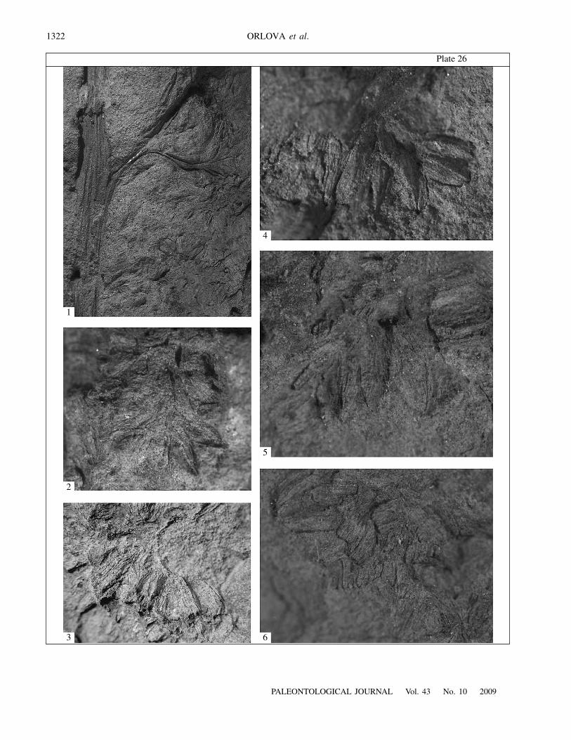
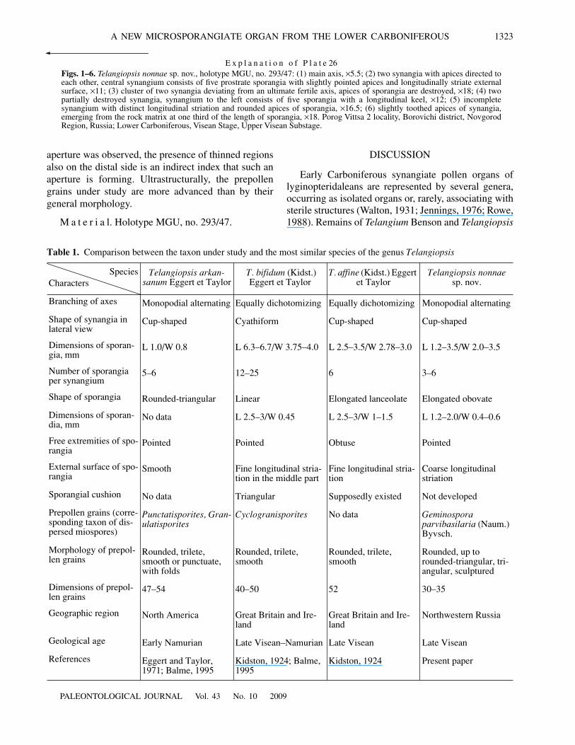
E x p l a n a t i o n o f P l a t e 26Figs. 1–6. Telangiopsis nonnae sp. nov., holotype MGU, no. 293/47: (1) main axis, ×5.5; (2) two synangia with apices directed toeach other, central synangium consists of five prostrate sporangia with slightly pointed apices and longitudinally striate externalsurface, ×11; (3) cluster of two synangia deviating from an ultimate fertile axis, apices of sporangia are destroyed, ×18; (4) twopartially destroyed synangia, synangium to the left consists of five sporangia with a longitudinal keel, ×12; (5) incompletesynangium with distinct longitudinal striation and rounded apices of sporangia, ×16.5; (6) slightly toothed apices of synangia,emerging from the rock matrix at one third of the length of sporangia, ×18. Porog Vittsa 2 locality, Borovichi district, NovgorodRegion, Russia; Lower Carboniferous, Visean Stage, Upper Visean Substage.
1324
PALEONTOLOGICAL JOURNAL Vol. 43 No. 10 2009
ORLOVA et al.
Plate 27
110 μm 2 μm
10 μm5 μm1 μm
5 μm 5 μm
2
3 4 5
76
PALEONTOLOGICAL JOURNAL Vol. 43 No. 10 2009
A NEW MICROSPORANGIATE ORGAN FROM THE LOWER CARBONIFEROUS 1325
E x p l a n a t i o n o f P l a t e 27Figs. 1–7. Surface sculpture of prepollen grains of Telangiopsis nonnae sp. nov., SEM: (1, 4, 6) proximal surface of prepollen grains,note folds at the proximal pole, the scar is virtually invisible, with the exception of the specimen shown in Pl. 27, fig. 6; (2, 5, 7)distal surface of prepollen grains, numerous verrucae are visible; (3) spinules on the equatorial surface of the same specimen asshown in Pl. 27, fig. 1. Porog Vittsa 2 locality, Borovichi district, Novgorod Region, Russia; Lower Carboniferous, Visean Stage,Upper Visean Substage.
10 μm(a) (b) (c)
Fig. 4. Prepollen grains of Telangiopsis nonnae sp. nov., LM. Porog Vittsa 2 locality, Borovichi district, Novgorod Region, Russia;Lower Carboniferous, Visean Stage, Upper Visean Substage.
are most common. The former genus is known by per-mineralized remains (Benson, 1904). The latter wasseparated from Telangium on the basis of several char-acters, the most important of which were the type ofpreservation and absence of planar leaf structures (Egg-ert and Taylor, 1971). The diagnosis of the genus Telan-giopsis was formulated as follows, “Synangiate organswith variable number of sporangia organized intostalked, radial clusters, borne terminally on eitherdichotomously or monopodially branching axes lack-ing planated foliar structures. Sporangia fused at base,distally free” (Eggert and Taylor, 1971, p. 31). Thetaxon under study shows all characters of the abovediagnosis and should be assigned to Telangiopsis.Unfortunately, the lack of complete information onother species of this genus regarding the morphologyand dimensions of the constituting elements of theirsynangiate organs impedes the comparison betweenthem and T. nonnae sp. nov. Because of this, our com-parison is restricted to three comprehensively charac-terized species of Telangiopsis: T. arkansanum Eggertet Taylor, T. bifidum (Kidst.) Eggert et Taylor, andT. affine (Table 1). The species under comparison differin the way how their fertile axes branch. The type spe-cies of the genus, T. arkansanum, has monopodiallybranching alternating axes. T. nonnae sp. nov. also hasmonopodially, but three-dimensionally, branchingalternating axes. T. bifidum and T. affine differ from thespecies under description in having equally dichoto-mizing axes. The number of sporangia in synangia, apointed free extremity of the sporangium, and the shape
of the synangia also make T. nonnae sp. nov. similar toT. arkansanum. However, the latter species has smoothsporangial walls, synangia that are more than two timesas small as synangia of T. nonnae sp. nov., rounded-tri-angular sporangia, and larger and morphologically dif-ferent prepollen (Table 1). T. nonnae sp. nov. and T. bifi-dum are only similar in pointed apices of sporangia anddifferent in all other parameters (Table 1). The generalmorphology of sporangia and synangia of the new spe-cies most closely resembles that of T. affine. However,sporangia of the latter species usually have an obtuseapex, whereas the apex in T. nonnae sp. nov. is rounded(Pl. 26, fig. 5), pointed (Pl. 26, fig. 2), or, occasionally,finely toothed (Pl. 26, fig. 6). Moreover, the two speciesdiffer by the external surface of sporangia, which iscoarsely striate in T. nonnae sp. nov. (Pl. 26, fig. 2) andfinely striate in T. affine. The shape of sporangia is alsodifferent. Rowe (1988) believed that T. bifidum and T.affine are assignable to synangiate organs ofDichotangium quadrothecum Rowe, which were foundin association (rather than in organic connection) withfronds of Diplopteridium holdeni Lele et Walton. Wedo not share this opinion and believe that althoughT. bifidum and T. affine resemble Dichotangiumquadrothecum in the morphology of sporangia, severaldifferences allow us to keep these species within thegenus Telangiopsis.
Let us compare Telangiopsis nonnae sp. nov. withpollen organs of some morphologically close membersof lyginopteridaleans. The similarity between the spe-cies under study and pollen organs of Dichotangium
1326
PALEONTOLOGICAL JOURNAL Vol. 43 No. 10 2009
ORLOVA et al.
Plate 28
1
2 μm
1.67 μm
2.5 μm
2.5 μm
0.5 μm0.5 μm
0.667 μm
2
3
4
5
7
6
PALEONTOLOGICAL JOURNAL Vol. 43 No. 10 2009
A NEW MICROSPORANGIATE ORGAN FROM THE LOWER CARBONIFEROUS 1327
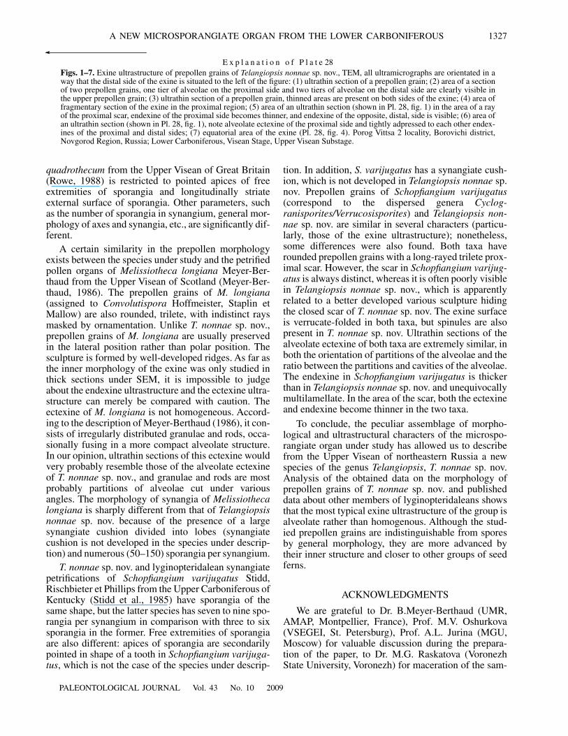
E x p l a n a t i o n o f P l a t e 28Figs. 1–7. Exine ultrastructure of prepollen grains of Telangiopsis nonnae sp. nov., TEM, all ultramicrographs are orientated in away that the distal side of the exine is situated to the left of the figure: (1) ultrathin section of a prepollen grain; (2) area of a sectionof two prepollen grains, one tier of alveolae on the proximal side and two tiers of alveolae on the distal side are clearly visible inthe upper prepollen grain; (3) ultrathin section of a prepollen grain, thinned areas are present on both sides of the exine; (4) area offragmentary section of the exine in the proximal region; (5) area of an ultrathin section (shown in Pl. 28, fig. 1) in the area of a rayof the proximal scar, endexine of the proximal side becomes thinner, and endexine of the opposite, distal, side is visible; (6) area ofan ultrathin section (shown in Pl. 28, fig. 1), note alveolate ectexine of the proximal side and tightly adpressed to each other endex-ines of the proximal and distal sides; (7) equatorial area of the exine (Pl. 28, fig. 4). Porog Vittsa 2 locality, Borovichi district,Novgorod Region, Russia; Lower Carboniferous, Visean Stage, Upper Visean Substage.
quadrothecum from the Upper Visean of Great Britain(Rowe, 1988) is restricted to pointed apices of freeextremities of sporangia and longitudinally striateexternal surface of sporangia. Other parameters, suchas the number of sporangia in synangium, general mor-phology of axes and synangia, etc., are significantly dif-ferent.
A certain similarity in the prepollen morphologyexists between the species under study and the petrifiedpollen organs of Melissiotheca longiana Meyer-Ber-thaud from the Upper Visean of Scotland (Meyer-Ber-thaud, 1986). The prepollen grains of M. longiana(assigned to Convolutispora Hoffmeister, Staplin etMallow) are also rounded, trilete, with indistinct raysmasked by ornamentation. Unlike T. nonnae sp. nov.,prepollen grains of M. longiana are usually preservedin the lateral position rather than polar position. Thesculpture is formed by well-developed ridges. As far asthe inner morphology of the exine was only studied inthick sections under SEM, it is impossible to judgeabout the endexine ultrastructure and the ectexine ultra-structure can merely be compared with caution. Theectexine of M. longiana is not homogeneous. Accord-ing to the description of Meyer-Berthaud (1986), it con-sists of irregularly distributed granulae and rods, occa-sionally fusing in a more compact alveolate structure.In our opinion, ultrathin sections of this ectexine wouldvery probably resemble those of the alveolate ectexineof T. nonnae sp. nov., and granulae and rods are mostprobably partitions of alveolae cut under variousangles. The morphology of synangia of Melissiothecalongiana is sharply different from that of Telangiopsisnonnae sp. nov. because of the presence of a largesynangiate cushion divided into lobes (synangiatecushion is not developed in the species under descrip-tion) and numerous (50–150) sporangia per synangium.
T. nonnae sp. nov. and lyginopteridalean synangiatepetrifications of Schopfiangium varijugatus Stidd,Rischbieter et Phillips from the Upper Carboniferous ofKentucky (Stidd et al., 1985) have sporangia of thesame shape, but the latter species has seven to nine spo-rangia per synangium in comparison with three to sixsporangia in the former. Free extremities of sporangiaare also different: apices of sporangia are secondarilypointed in shape of a tooth in Schopfiangium varijuga-tus, which is not the case of the species under descrip-
tion. In addition, S. varijugatus has a synangiate cush-ion, which is not developed in Telangiopsis nonnae sp.nov. Prepollen grains of Schopfiangium varijugatus(correspond to the dispersed genera Cyclog-ranisporites/Verrucosisporites) and Telangiopsis non-nae sp. nov. are similar in several characters (particu-larly, those of the exine ultrastructure); nonetheless,some differences were also found. Both taxa haverounded prepollen grains with a long-rayed trilete prox-imal scar. However, the scar in Schopfiangium varijug-atus is always distinct, whereas it is often poorly visiblein Telangiopsis nonnae sp. nov., which is apparentlyrelated to a better developed various sculpture hidingthe closed scar of T. nonnae sp. nov. The exine surfaceis verrucate-folded in both taxa, but spinules are alsopresent in T. nonnae sp. nov. Ultrathin sections of thealveolate ectexine of both taxa are extremely similar, inboth the orientation of partitions of the alveolae and theratio between the partitions and cavities of the alveolae.The endexine in Schopfiangium varijugatus is thickerthan in Telangiopsis nonnae sp. nov. and unequivocallymultilamellate. In the area of the scar, both the ectexineand endexine become thinner in the two taxa.
To conclude, the peculiar assemblage of morpho-logical and ultrastructural characters of the microspo-rangiate organ under study has allowed us to describefrom the Upper Visean of northeastern Russia a newspecies of the genus Telangiopsis, T. nonnae sp. nov.Analysis of the obtained data on the morphology ofprepollen grains of T. nonnae sp. nov. and publisheddata about other members of lyginopteridaleans showsthat the most typical exine ultrastructure of the group isalveolate rather than homogenous. Although the stud-ied prepollen grains are indistinguishable from sporesby general morphology, they are more advanced bytheir inner structure and closer to other groups of seedferns.
ACKNOWLEDGMENTS
We are grateful to Dr. B.Meyer-Berthaud (UMR,AMAP, Montpellier, France), Prof. M.V. Oshurkova(VSEGEI, St. Petersburg), Prof. A.L. Jurina (MGU,Moscow) for valuable discussion during the prepara-tion of the paper, to Dr. M.G. Raskatova (VoronezhState University, Voronezh) for maceration of the sam-
1328
PALEONTOLOGICAL JOURNAL Vol. 43 No. 10 2009
ORLOVA et al.
Plate 29
1.67 μm
0.25 μm
0.5 μm
0.8 μm2.5 μm
1
3
2
4
PALEONTOLOGICAL JOURNAL Vol. 43 No. 10 2009
A NEW MICROSPORANGIATE ORGAN FROM THE LOWER CARBONIFEROUS 1329
ple, to Dr. S.M. Snigirevskii (St. Petersburg State Uni-versity, St. Petersburg) and S.Yu. Kharitonov (MGU)for their help during the field-trip in the NovgorodRegion, Yu.I. Rostovtseva (MGU) for the assistancewith drawing, and to the staff of the Laboratory of elec-tron microscopy (MGU) for the assistance with SEMand TEM.
The study was supported by the Russian Foundationfor Basic Research, no.0 8-04-00633.
REFERENCES
1. B. E. Balme, “Fossil In Situ Spores and Pollen Grains:An Annotated Catalogue,” Rev. Palaeobot. Palynol. 87,81–323 (1995).
2. M. Benson, “Telangium scottii, a New Species ofTelangium (Calymmatotheca) Showing Structure,” Ann.Bot. 18, 161–177 (1904).
3. D. A. Eggert and T. N. Taylor, “Telangiopsis gen. nov. anUpper Mississippian Pollen Organ from Arkansas,” Bot.Gaz. 132, 30–37 (1971).
4. D. M. Erwin, H. W. Pfefferkorn, and V. Alleman, “EarlySeed Plants in the Southern Hemisphere: 1. AssociatedOvulate and Microsporangiate Organs from the Carbon-iferous of Peru,” Rev. Palaeobot. Palynol. 80, 19–38(1994).
5. J. R. Jennings, “The Morphology and Relationships ofRhodea, Telangium, Telangiopsis and Heterangium,”Am. J. Bot. 1119–1133 (1976).
6. R. Kidston, “Fossil Plants of the Carboniferous Rocks ofGreat Britain,” Mem. Geol. Surv. GB, Paleontol. 2 (Part5), 379–522 (1924).
7. N. R. Meyer-Melikian, I. Yu. Bovina, Ya. V. Kosenko,et al., Atlas of the Morphology of Asteraceae: Palyno-morphology and Development of Sporoderm in Repre-sentatives of the Family Asteraceae (T-vo Nauchn. Izd.KMK, Moscow, 2004) [in Russian].
8. B. Meyer-Berthaud, “Melissiotheca: A New Pteri-dosperm Pollen Organ from the Lower Carboniferous ofScotland,” Bot. J. Lin. Soc. 93, 277–290 (1986).
9. B. Meyer-Berthaud, “First Gymnosperm Fructificationswith Trilete Prepollen,” Palaeontogr. Abt. B 211 (4–6),87–112 (1989).
10. M. A. Millay and T. N. Taylor, “Paleozoic Seed Fern Pol-len Organs,” Bot. Rev. 45 (3), 301–375 (1979).
11. O. A. Orlova, “Preliminary Report on Synangiate Fruc-tifications of Telangiopsis from the Upper Visean of theMoscow Syneclise (Novgorod Region, Russia),” inAbstr. 6th Europ. Palaeobot. Palynol. Conf., Athens,August 29–September 2, 2002 (Athens, 2002), pp. 140–141.
12. O. A. Orlova, “Early Carboniferous Plants of theArkhangelsk Region, Russia,” Paleontol. J. 41 (11),1138–1150 (2007).
13. O. A. Orlova and N. B. Rasskazova, “The Age of theLocality of Late Viséan Plants “Porog Vittsa < 2” (Vil-lage of Putlino, Novgorod Region) Based on Palynolog-ical Data,” in Paleobiology and Detailed Stratigraphy ofthe Phanerozoic: Contribution to Academician V.V.Menner’s 100th Birthday (Ross. Akad. Estestv. Nauk,Moscow, 2005), pp. 64–79 [in Russian].
14. O. A. Orlova and S. M. Snigirevskii, “Late Viséan Lygi-nopteridophytes from the Vicinity of the Town of Borov-ichi (Novgorod Region): 1. Calamopityales,” Paleontol.Zh., No. 6, 105–111 (2003) [Paleontol. J. 37 (6), 665–671 (2003)].
15. O. A. Orlova and S. M. Snigirevsky, “Late Viséan Lygi-nopteridophytes from the Vicinity of the Town of Borov-ichi (Novgorod Region): 2. Lyginopteridales andMedullosales,” Paleontol. Zh., No. 4, 104–109 (2004)[Paleont ol. J. 38 4, 458–464 (2004)].
16. N. P. Rowe, “New Observations on the Lower Carbonif-erous Pteridosperm Diplopteridium Walton and an Asso-ciated Synangiate Organ,” Bot. J. Linn. Soc. 97, 125–158 (1988).
17. B. M. Stidd, M. O. Rischbieter, and T. L. Phillips,“A New Lyginopterid Pollen Organ with Alveolate Pol-len Exines,” Am. J. Bot. 72 (4), 501–508 (1985).
18. J. Walton, “Contributions to the Knowledge of LowerCarboniferous Plants III. On the Fossil-Flora of theBlack Limestones in Teilia Quarry, Gwaenysgor, nearPrestatyn, Flintshire, with Special Reference to Diplop-teridium telianum Kidston sp. (gen. nov.) and SomeOther Fern-like Fronds,” Philos. Trans. R. Soc. Lond.219, 347–379 (1931).
19. N. E. Zavialova, A. V. Gomankov, O. P. Yaroshenko, andL. V. Rovnina, “Morphology and Ultrastructure ofMonosaccate Pollen Grains of the Genus Cordaitinafrom the Permian of Russia,” Acta Palaeobot. Pol. 44 (1),3–35 (2004).
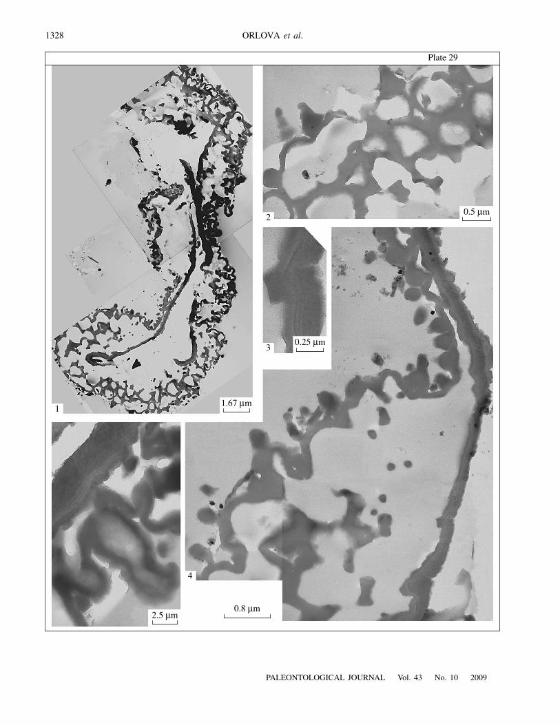
E x p l a n a t i o n o f P l a t e 29Figs. 1–5. Ultrastructure of prepollen grains of Telangiopsis nonnae sp. nov., TEM: (1) fragmentary section of the exine, distal sideis to the left of the figure; (2–5) areas of the section shown in Pl. 29, fig. 1; (2) equatorial spinules cut; (3) heterogeneous structureof the endexine; (4) equatorial-distal area, granular elements are sectioned verrucae; (5) area showing one tier of alveolae of theproximal ectexine, note heterogeneous structure of the endexine. Porog Vittsa 2 locality, Borovichi district, Novgorod Region, Rus-sia; Lower Carboniferous, Visean Stage, Upper Visean Substage.
Copyright © 2022 FDOKUMEN