Oligonucleotides with novel, cationic backbone substituents: aminoethylphosphonates
A new approach for immobilization of oligonucleotides ontopiezoelectric quartz crystal for preparation of a nucleic acid sensor for
following hybridization
Memed Duman, Reza Saber, Erhan Piskin *
Department of Chemical Engineering and Bioengineering Division, Hacettepe University, 06532 Beytepe, Ankara, Turkey
Received 31 October 2001; received in revised form 2 September 2002; accepted 19 December 2002
Abstract
The aim of this study is to develop a nucleic acid sensor based on piezoelectric crystal microbalance system (QCM) for following
hybridization. Piezoelectric quartz crystal surfaces were first treated in a glow�/discharge apparatus with ethylene diamine (EDA)
plasma at 15 W (discharge power), 2.5 min (incubation time) and 35 ml/min (monomer flow rate) to create amino groups on the
crystal surfaces. The thickness of the EDA�/plasma film formed was about 439/24 A. Then, the amino groups on the crystal surfaces
were converted to aldehyde groups by reacting the amino groups with glutaraldehyde (GA) at different conditions. A GA
concentration of 2.5% and an incubation time of 2 h were selected as optimal values at this step, corresponding to a GA surface
concentration of about 270 ng/cm2. A double strand Oligonucleotides, having one extra base on 5?-end of one of the complementary
strands, were immobilized through the amino groups of this base onto the GA-modified crystals. Optimal immobilization conditions
were as follows: oligonucleotide concentration: 1 mg/ml; time: 3 h; pH: 9.2 carbonate buffer; ionic strength: 0.1; and temperature:
20 8C. The QCM sensor carrying the covalently bound strand was used in the hybridization experiments, which showed that
equilibrium is achieved in about 5 min, and the frequency shift measured is related to the concentration of the target strand to be
measured within the medium.
# 2003 Elsevier Science B.V. All rights reserved.
Keywords: Nucleic acid sensors; Piezoelectric quartz crystals; Glow�/discharge; Plasma polymerization; Glutaraldehyde treatment; Oligonucleotide
immobilization; Hybridization/dehybridization
1. Introduction
Sequence-specific hybridization between nucleic acids,
either in solution or immobilized on fixed support, are
widely used for the detection and analysis of genetic
material for diverse applications including identification
of genetic diseases and disorders, detection and char-
acterization of viruses, bacteria, and parasites, etc.
(Symons, 1989; Downs, 1991). In nucleic acid sensors,
hybridization can be followed by several techniques,
including electrochemical (Palecek, 1988; Moser et al.,
1997; Hianik et al., 2001), optical (Jost et al., 1991;
Piscevic et al., 1995), and piezoelectric (Yamaguchi et
al., 1993; Su and Thompson, 1995; Caruso et al., 1997,
1998; Bardea et al., 1998; Xhang et al., 1998; Zhou et al.,
2000, 2001; Patolsky et al., 2001).
Due to its piezoelectric properties, quartz crystals are
known as microbalance (QCM) which can function as
an extremely sensitive mass sensor capable of measuring
subnanogram levels of mass changes. They are also
inexpensive and are considered chemically, physically
and mechanically durable systems. For immobilization
of the probe nucleic acids (mostly single-strand oligo-
nucleotides) effectively on the piezoelectric crystal
surfaces, several methods have been proposed, each
with its own advantages and disadvantages (Montrel et
al., 1997; Yamaguchi et al., 1993; Su and Thompson,
1995; Caruso et al., 1997, 1998; Bardea et al., 1998;
Xhang et al., 1998; Wittunng-Stafshede et al., 2000;
Zhou et al., 2000, 2001; Patolsky et al., 2001; Towery et
al., 2001). Recently, a novel technique has been applied
* Corresponding author. Tel.: �/90-312-297-7473; fax: �/90-312-
299-2124.
E-mail address: [email protected] (E. Piskin).
Biosensors and Bioelectronics 18 (2003) 1355�/1363
www.elsevier.com/locate/bios
0956-5663/03/$ - see front matter # 2003 Elsevier Science B.V. All rights reserved.
doi:10.1016/S0956-5663(03)00087-3
by us and by others for surface modification of piezo-
electric crystals, in which crystals are treated in glow�/
discharge systems (Nakanishi et al., 1996; Nakamura et
al., 1997; Kurosawa et al., 1997; Mutlu et al., 1999; Wuet al., 2000; Towery et al., 2001; Saber et al., 2002).
Several reactive or non-reactive plasmas can be created
within the glow�/discharge apparatus, which modify the
surfaces by depositing films having functional groups.
These, the so-called plasma-polymerized films covering
the crystal surfaces, are extremely thin and homogenous
coatings. They adhere strongly to the crystal and are
highly resistant to chemical and physical treatments.Moreover, sensors produced using this method are more
reproducible from sample to sample and exhibit lower
noise than sensors made using conventional immobiliza-
tion methods.
In our previous studies, we have applied ethylene
diamine (EDA) plasma-polymerization (or glow�/dis-
charge treatment) to create amino groups on the quartz
crystals (with Ag electrodes) and immobilized antibodieson these crystals using glutaraldehyde (GA) (as cross-
linker and spacer arm) to prepare immunosensors
(Mutlu et al., 1999; Saber et al., 2002). Here, we report
preparation of a QCM based nucleic acid sensor for
investigation of hybridization, by following a very
similar procedure for nucleic acid immobilization.
2. Materials and methods
2.1. Materials
EDA was supplied from Fluka (Switzerland), GA was
purchased from Aldrich (UK). The 20-mer single-
stranded oligonucleotide having the sequence 5?-GTGT TTG CCT GTT CTC AGA C-3? was used as theprobe (‘‘ligand’’) in the DNA sensor, which is denoted
here as ‘‘P1’’ in this study. The complementary (‘‘tar-
get’’) strand, which is called ‘‘P2’’, has the sequence 5?-GTC TGA GAA CAG GCA AAC A-3?, which is the
most widely recognized sequence in the DNA’s of
Turkish population. Both oligonucleotides were synthe-
sized by Life Technologies (UK). Note that the probe
(P1) contains one extra nucleotide at 5?-end, and aftercomplete hybridization of these two complementary
single-strands, the double strand contains one free
base at one end, this means that the amino groups of
this base are available for covalent binding on the
crystal surfaces. It should be emphasized that only one
free amino group at the end of the double strand is
available for binding, which is the key difference
between this study and all previous studies. Buffersolutions with different pH and ionic strength were
obtained from Life Technologies and also prepared in
our laboratory by using analytical grade chemicals.
2.2. QCM system
The quartz crystal used in this study was commer-
cially available an AT-cut 10 MHz quartz crystal of a8.7�/0.17 mm wafer which was placed between 4.7 mm
silver electrodes and mounted on a ceramic holder with
a plug (AEC, Taiwan). The oscillation electronic circuit
was a typical Collpits oscillator, which has a buffer
amplifier. 15 V DC was applied to the oscillator circuit
to drive the crystal and the frequency was measured with
a Hewlett�/Packard frequency counter (Model No: HP
53181A, 225 MHz, USA). The crystal was enclosed in acase, which was maintained in a chamber at a constant
temperature of 359/0.1 8C during the measurements to
reduce frequency fluctuations due to dust. This QCM
system gave a frequency drift of less than 1 Hz in air
after a few minutes (B/5 min) initial stabilization period.
The procedure for preparing the silver surface has been
described in the following section. The mass change
(Dm ) on the quartz crystal which is related to theresulting frequency shift Df was calculated from the
Sauerbrey equation given below (Sauerbrey, 1959).
Df ��2:26�10�6f 2Dm=A (1)
Where, Df is the change in the fundamental frequency of
the coated crystal (Hz); f , the fundamental frequency of
the quartz crystal (Hz); Dm , the mass deposited on the
crystal (g); and A is the area coated (cm2).
2.3. Modification of piezoelectric quartz crystals
2.3.1. Glow�/discharge treatment
For oligonucleotide immobilization, the quartz crys-
tal surface was treated in a glow�/discharge system
(Mutlu et al., 1999). The glow�/discharge reactor was a
Pyrex† glass tube with a diameter of 5.6 cm and a length
of 52 cm. Two copper electrodes of 7�/17 cm wereexternally placed on the reactor. One of the electrodes
was connected to a radio-frequency generator with
frequency of 13.6 MHz (Tasarim Ltd., Model: T-RF-
1200, Turkey) through a matching network unit (Ta-
sarim Ltd., Model: T-RF-1100), while the other elec-
trode was grounded. The monomer tank was connected
to the reactor through a flow meter (Gilmont, F-1200,
Size 2, USA) and a needle valve (Brooks, Model 1355,CA, USA).
EDA was used as the active monomer, and was
plasma-polymerized in the glow�/discharge reactor, to
create amine-like active groups on the quartz crystal
surface for further treatments given below. Briefly, the
glow�/discharge reactor was evacuated to 10�1 mbar.
The monomer, EDA, was allowed to flow through the
reactor at a flow rate of 35 ml/min. The quartz crystalswere exposed to the EDA plasma for 2.5 min at glow�/
discharge power of 15 W. It should be noted that these
were optimal plasma treatment conditions which were
M. Duman et al. / Biosensors and Bioelectronics 18 (2003) 1355�/13631356
obtained in our related previous studies (Mutlu et al.,
1999; Saber et al., 2002) and also after preliminary
experiments (Duman, 2001). The modifications were
followed by measuring the shifts in the resonancefrequencies of the crystals before and after the surface
modification.
2.3.2. Glutaraldehyde activation
In the second step, the primary amine groups on the
crystal surface created after the plasma-polymerization
of EDA were converted to aldehyde groups by treating
the surfaces with GA. The GA concentration and the
incubation time were changed while the treatmenttemperature was kept constant at 4 8C. For reductive
animation of the secondary amine linkage (for stabiliza-
tion of the Shiff base) we also used NaCNBH3 (about
five times of the GA concentration, molar basis). In the
first set, the glow�/discharge treated quartz crystals were
incubated with solutions containing different amounts
of GA (0.1, 0.5, 1, 2.5 and 5%) for 2 h. In the second set
of experiments, the GA concentration was kept constant(2.5% GA in a carbonate buffer, pH 9.2) and the
incubation time was changed (1, 2, 4, 8 and 16 h).
Physically adsorbed GA molecules were removed by
washing with pure water several times. Following the
washing step, the crystals were dried in air (at room
temperature for about 15 min, until a constant fre-
quency was obtained) and the frequencies of the air-
dried crystals were recorded. It should be note that aGA molecule has two aldehyde groups. At this treat-
ment step, it was assumed that one aldehyde group on
each GA is covalently bonded to amine group on the
EDA treated crystal while the other aldehyde group
stays free for the next treatment step.
2.4. Oligonucleotide immobilization
In this part of the study, the oligonucleotide immo-
bilization (e.g. covalent binding of the double strand
oligonucleotide onto the GA-modified crystal surfaces)
was investigated at different conditions. Note that these
immobilization studies were conducted where the non-
specific adsorption of the oligonucleotide was at mini-
mum. These conditions were derived based on the
experiments performed in the previous step. Here, inthe first group, only the oligonucleotide concentration
(0.1, 0.5, 1.0, 2.0 and 4.0 mg/ml) was changed and the
immobilization time was kept constant at 3 h. In the
second group, the oligonucleotide concentration was 1.0
mg/ml, while the immobilization time was changed (1, 2,
3, 4, 8, 20 h). For reductive amination of the secondary
amine linkage between the surface aldehyde groups
(coming from the immobilized GA molecules) on thesurface and the terminal amine groups of the oligonu-
cleotide molecules, we also used NaCNBH3 (about five
times of the oligonucleotide concentration, molar basis).
Incubation was conducted in a shaker at room tem-
perature. After the immobilization step, the crystals
were first washed with distilled water and then with the
buffer solution for 15 min. The crystals were dried in airat room temperature, and the frequency shifts were
recorded. It should be noted that the double strand
oligonucleotide was covalently immobilized on the
crystal surface from the amino groups of the free
nucleotide on the P1 probe.
2.5. Hybridization experiments
In order to determine the performance of the QCM asa sensor, firstly the P2 strand (not covalently bound to
the surface) was removed from the crystal surfaces
(‘‘dehybridization’’). The crystals carrying the double
strand oligonucleotide were incubated in a carbonate
buffer (pH 9.2) at 94 8C for 3�/4 min. After washing and
drying several times with buffers and pure water,
frequency shifts were measured by the QCM system. It
should be noted that we have continued the washingstep until we were confident that we removed all of
complementary strand P2 from the surface. Practically,
when we reached the frequency shift (decrease) that
corresponds to about half of the frequency shift
(increase) observed in the immobilization of the double
strand oligonucleotide, we stopped the washing and
took these crystals carrying the single strand (P1) for
further tests.The nucleic acid sensor carrying the covalently bound
single strand (P1) was incubated with the solutions
containing the complementary strand (P2) at different
concentration (0.001, 0.01, 0.05, 0.1 and 1 mg/ml) for
different times in the range of 1�/25 min. Hybridization
was evidenced by a stepwise mode. The QCM sensor
was removed from the solution after hybridization,
rinsed and dried and the change in the resonancefrequency of the crystal before and after incubation
was recorded.
In the second approach, the required amount of the
P2 oligonucleotide was dissolved in 3 ml phosphate
buffer (pH 7.4). The crystal carrying the P1 oligonucleo-
tide was mounted onto a homemade cell, which was
then dipped into the solution containing P2 molecules.
The frequency shifts were recorded continuously withthe microbalance system described below, while the
medium was magnetically stirred.
2.6. STM imaging
Scanning tunneling microscopy (STM) images of the
crystal surfaces were obtained by using a homemade
STM constructed in our laboratory (Zareie, 1995).Mechanically, it consists of two main modules, a
scanner part constructed with a piezo tube (PZT tube)
(EBL No: 3 PZT-5H, Staveley Sensors, CT) glued to
M. Duman et al. / Biosensors and Bioelectronics 18 (2003) 1355�/1363 1357
aluminum body and a magnetically driven slider. The
electrical connections to the PZT are through a special
connector and the whole system is mounted on a
vibration isolation stage.For STM measurements, the oligonucleotide immo-
bilized crystals were attached to the sample holder with
silver epoxy. Then, the STM images were taken at room
temperature and at atmospheric pressure, using a 50 mV
sample bias and a tunneling current of 850 pA. Etched
tips of Pt/Ir (80:20) wires (0.5 mm in diameter, Digital
Instruments, Santa Barbara, CA) were used. Prior to
use, the tips were washed with acetone. The brightnessand contrast was adjusted using PHOTOSHOP v3.0
(Adope System, CA, USA) to enhance the images
details.
3. Result and discussion
In this study, glow�/discharge technique was selected
as a promising method for double strand oligonucleo-
tide immobilization on the piezoelectric crystal surface.
In the conventional techniques, such as gel entrapment,dip coating, irradiation of polymers, etc., the surface
generated on the crystal becomes bulky, depressing the
quartz crystal’s sensitivity, expanding response time and
also creating difficulties in some applications. However,
the polymer coating thickness created by glow�/dis-
charge technique was about 200 A eliminating the above
deficiencies while providing maximum sensitivity.
Furthermore, the functional groups (�/NH2) on thesurface enable the double strand oligonucleotide to be
quite easily linked to the treated surface by GA
activation from one strand, which have one more
nucleotide. The glow�/discharge parameters were opti-
mized and the result and the performance of the nucleic
acid sensor are summarized in the following section.
3.1. Surface modification of quartz crystals
3.1.1. Glow�/discharge treatment
Glow�/discharge treatment has been widely used to
change the surface properties of materials without
affecting their bulk structures for diverse applications(Clark and Feast, 1978; Yasuda, 1985; Fawcett et al.,
1988; d’Agustino, 1991; Piskin, 1992). Depending upon
the glow�/discharge conditions, surface modification
occurs through etching and/or deposition. Gases (e.g.
argon, oxygen, air, nitrogen), non-polymerizable polar
gases (e.g. water, ammonia), organic vapors (e.g.
methanol, acetone) or polymerizable gases or vapors
(e.g. ethylene, fluoroethylenes) have been used to createdifferent functional groups, (e.g. carboxyl, hydroxyl,
amino) on the material surfaces by this simple surface
modification technique.
In this study, in order to create amino-like active
groups on the quartz crystal surfaces for double strand
oligonucleotide immobilization, we used EDA as the
active monomer. According to preliminary studies, weconcluded that optimal conditions for plasma polymer-
ization are, the discharge power: 15 W; treatment time:
2.5 min; and the monomer flow rate: 35 ml/min. The
average frequency shift and standard deviation of these
crystals comparing with the blank crystals (without
glow�/discharge treatment) was 1439/24 Hz. This fre-
quency shift is based on measurements of ten randomly
selected crystals from different loads is quite significant.These crystals were used in the further studies presented
below.
It should be noted that in the glow�/discharge
treatment we deposited EDA�/plasma film on both sides
of the quartz crystal. The film thickness was determined
by the frequency shift of the bare crystal induced by the
film coating. Thickness is related to the frequency shift
as given in the following equation (Lawson, 1967; Wu etal., 2000; Sugimoto et al., 2000). The thickness of the
EDA films on the quartz crystals created in our studies,
was calculated from the following equation, and found
to be 439/24 A.
Dt�0:27 Df =r (2)
Where, Dt is the thickness of the polymeric film on thecrystal surface (A); Df , the change in the fundamental
frequency of the coated crystal (Hz); and r is the density
of the film which was taken as the density of the EDA
monomer (0.895 g/cm3). The polymerization rate of
EDA, similar to aliphatic amines, is slow compared with
those well-known plasma-polymerized monomers con-
taining nitrogen (e.g. allylamine) (Yasuda and Lamaze,
1973; Bell et al., 1975; Hozumi et al., 1983; Tajima et al.,1988). It was reported that these films have the
structures containing �/NH2, �/NH�/, �/N�/N�/, and �/
CN. In our study we have used EDA plasma, which
was also selected as the active plasma in similar studies
(Nakanishi et al., 1996a,b; Muguruma and Karube,
1999; Towery et al., 2001). As reported in these studies,
it was expected that amino groups exist on the surface of
the ethylene diamine plasma-polymerized film.
3.1.2. Glutaraldehyde attachment
Usually GA is used as an agent for crosslinking of
amine groups in compounds like protein and oligonu-
cleotides (Hermanson, 1996). In our preliminary experi-
ments we observed that in order to covalently bind the
oligonucleotide molecules to the crystals (coated with
the EDA�/plasma films), best results were obtained
when the crystals were incubated in carbonate buffer
(pH 9.2) with different GA concentration for differentincubation times.
In the first group of experiments, we changed the GA
concentration and followed the changes in frequency
M. Duman et al. / Biosensors and Bioelectronics 18 (2003) 1355�/13631358
shifts. Increasing the GA concentration from 0.1 to
2.5%, the frequency shift was doubled. The maximum
frequency shift was 889/13 Hz (the average and the
standard deviation of ten crystals), corresponding amass deposition of about 270 ng/cm2 (calculated from
Eq. (1)). However, at higher GA concentration, a slight
decrease in the GA deposition was observed, which may
be due to polymerization of the GA molecules in the
aqueous medium. Therefore, a GA concentration of
2.5% was selected as the optimal concentration and
applied in the later part of the study.
In the second set, the treatments were carried out at2.5% GA concentration in the same buffer, but for
different incubation times. The frequency change after 1
h was only 12 Hz. However, after 2 h, there was a steep
increase in the mass accumulation, which reached to
889/13 Hz. Thereafter the accumulation continued to
increase by increasing the incubation time, but not very
significantly. Therefore, 2 h was selected as the optimal
time and this time was used in the later part of the study.
3.2. Immobilization of double strand oligonucleotide
An oligonucleotide, known as fourth allele specific
probe of DQa’s subgroup, which is most frequently used
in genetic analysis of Turkish population’s DNA, was
selected as a model ligand and immobilized onto the
GA-attached piezoelectric crystals. This is one of the
basic probes on the test strips for identification of theDNA in criminology laboratories. As mentioned before,
there are many different methods for immobilization of
oligonucleotide onto solid surfaces. In this study, we
have applied for the first time, a quite simple technique
for immobilization a single strand oligonucleotide
ligand defined here as ‘‘P1’’. We have used two
complementary single strand oligonucleotides, P1 (hav-
ing one extra base on 5? end) and P2. These strands werebrought together in an aqueous medium and complete
hybridization was achieved. Then, this double strand
oligonucleotide was immobilized on the GA-attached
crystal surfaces by the reaction with the amino groups of
the free base (coming from the P1 strand) and the
aldehyde groups on the crystal surface.
Oligonucleotide immobilization was investigated at
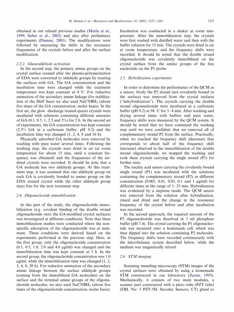
different conditions. In the first group of experiments,increasing the oligonucleotide concentration in the
medium from 0.1 to 1.0 mg/ml caused an increase in
the amount of oligonucleotide immobilized, as expected
(Fig. 1). However, we observed a noticeable decrease in
the mass deposited, when the nucleotide concentration
was further increased. Note that there are quite a high
number of aldehyde groups available for immobilization
on the GA-attached crystal surface. But, the oligonu-cleotide molecules consist of relatively long chain and
contain only one base group at one end available to
react with the surface aldehyde groups. This situation
retards complete reaction between the amines and the
surface aldehydes. Increasing the oligonucleotide con-
centration in the immobilization medium, most prob-
ably increased the steric hindrance between the
competing long chains to react with the surface aldehyde
groups. In conclusion, 1 mg/ml oligonucleotide concen-
tration was selected as an optimal concentration for
effective oligonucleotide immobilization, and these crys-
tals were used in the next immobilization step. The
average frequency shift was 126 Hz and the correspond-
ing amount of mass deposited was about 380 ng/cm2.
These values were comparable with similar studies in
which different immobilization methods have been, used
(Yamaguchi et al., 1993; Su and Thompson, 1995;
Caruso et al., 1997, 1998; Bardea et al., 1998; Xhang
et al., 1998; Zhou et al., 2000, 2001; Patolsky et al.,
2001). For instance, in their recent study, Zhou and
coworkers have immobilized a 18-mer DNA probe (a
single strand) on the crystal surfaces carrying a poly-
electrolyte layer by electrostatic adsorption, and mea-
sured a frequency shift of 21 Hz after drying in air
(immobilized amount was calculated of 19 ng) (Zhou et
al., 2000, 2001). Alternatively, they have first chemically
immobilized on the crystal surface, then further reacted
with biotylated DNA for the immobilization of the
DNA probe. The immobilization amount was calculated
from the frequency decrease (38 Hz) and found to be 27
ng. In conclusion, for the 10 MHz quartz crystals used
in this work, Sauerbrey equation predicts that a
frequency change of 1 Hz corresponds to a mass
increase of 3.019/0.03 ng on the electrode (0.69 cm2).
It is almost the same amount reported in Zhou’s work
which predicted that a frequency change of 1 Hz,
corresponds to a mass increase of 0.9029/0.01 ng on
the electrode (0.204 cm2).
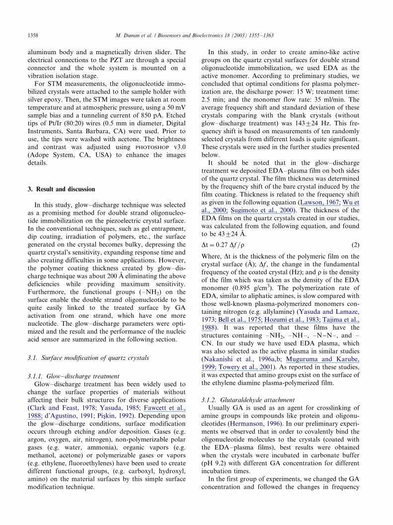
In order to observe the effects of immobilization time
on the immobilization, the GA-attached crystals were
incubated with the aqueous solution of the double
strand oligonucleotide (1 mg/ml) at room temperature
Fig. 1. Effects of oligonucleotide concentration on oligonucleotide
immobilization.
M. Duman et al. / Biosensors and Bioelectronics 18 (2003) 1355�/1363 1359
for different periods of time (1�/16 h). There was a
drastic increase in the first 3 h followed by more
moderate increase in the immobilization at longer time
(Fig. 2). Therefore, we assumed that 3 h would besufficient time to have adequate amounts of oligonu-
cleotide on the crystal surface. Furthermore, 3 h should
also be a reasonable period of time for practical
applications.
3.3. Performance of nucleic acid sensor
At the last step of this study, we investigated the
performance of a nucleic acid sensor carrying the ‘‘P1’’
strand. The other strand, i.e. the so-called ‘‘P2’’ (not
covalently bound to the surface) was dehybridized(removed) by applying the procedure mentioned before.
The nucleic acid sensor carrying the covalently bound
single strand was then used in the hybridization experi-
ments, in which these crystals were incubated with the
aqueous solutions of the ‘‘P2’’ strand at different
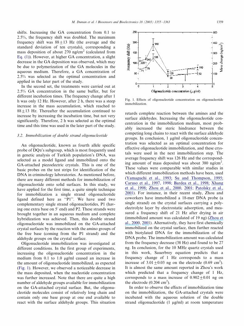
concentrations for different time periods. As seen in
Fig. 3, hybridization was almost completed in about 5
min in most cases. This short hybridization time is much
better when compared with the data given in similar
studies (Yamaguchi et al., 1993; Su and Thompson,
1995; Caruso et al., 1997, 1998; Bardea et al., 1998;
Xhang et al., 1998; Zhou et al., 2000, 2001; Patolsky et
al., 2001). We concluded that spacer arms with longer
chain lengths (longer than GA molecules) may increase
the availability of the immobilized strain for hybridiza-
tions and thus should reduce the time necessary to reach
equilibrium. Further studies are being conducted to
validate this hypothesis.
Fig. 3 also shows that there is no significant difference
in the hybridization curves (at equilibrium) at P2
concentrations of 0.1 and 1 mg/ml. This indicates that
almost all of the immobilized oligonucleotides (the P1
strand) were hybridized with the complementary mole-
cules (the P2 strand) corresponding to a P2 concentra-
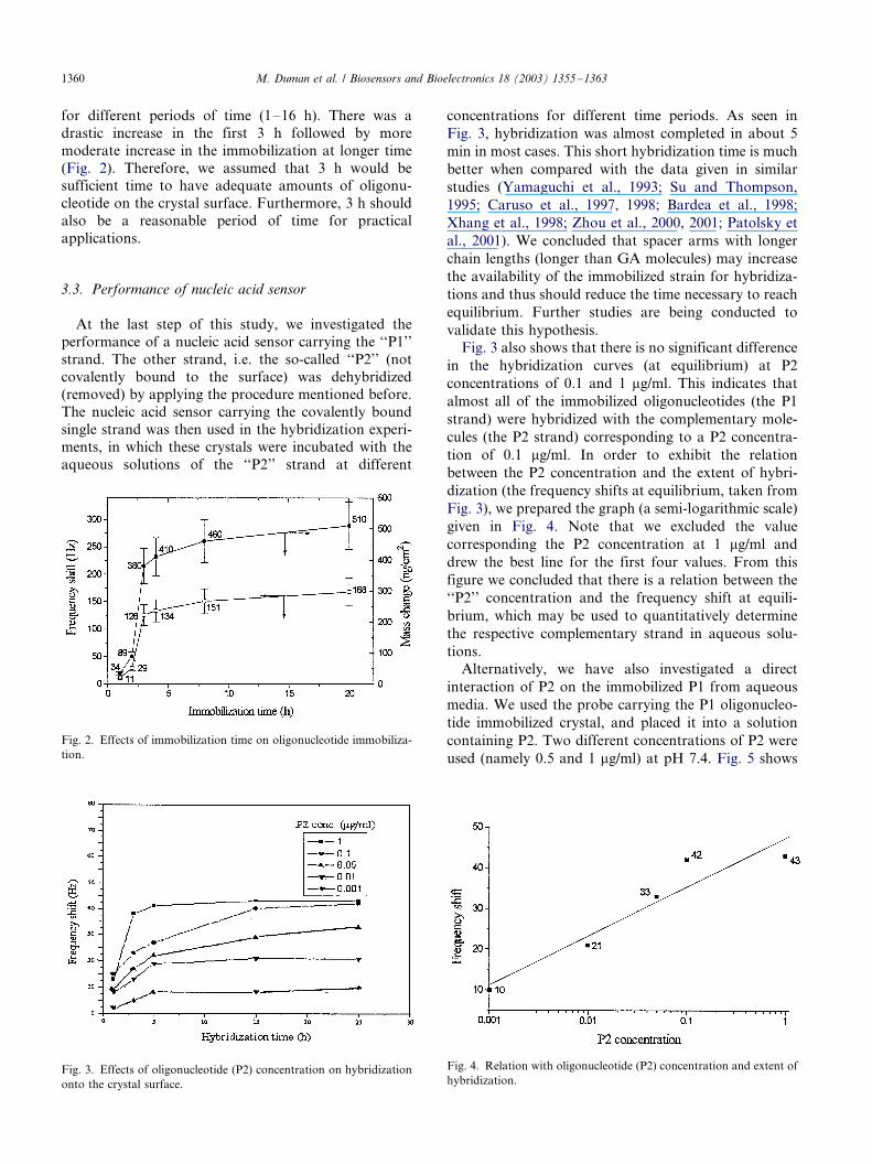
tion of 0.1 mg/ml. In order to exhibit the relation
between the P2 concentration and the extent of hybri-
dization (the frequency shifts at equilibrium, taken from
Fig. 3), we prepared the graph (a semi-logarithmic scale)
given in Fig. 4. Note that we excluded the value
corresponding the P2 concentration at 1 mg/ml and
drew the best line for the first four values. From this
figure we concluded that there is a relation between the
‘‘P2’’ concentration and the frequency shift at equili-
brium, which may be used to quantitatively determine
the respective complementary strand in aqueous solu-
tions.
Alternatively, we have also investigated a direct
interaction of P2 on the immobilized P1 from aqueous
media. We used the probe carrying the P1 oligonucleo-
tide immobilized crystal, and placed it into a solution
containing P2. Two different concentrations of P2 were
used (namely 0.5 and 1 mg/ml) at pH 7.4. Fig. 5 shows
Fig. 2. Effects of immobilization time on oligonucleotide immobiliza-
tion.
Fig. 3. Effects of oligonucleotide (P2) concentration on hybridization
onto the crystal surface.
Fig. 4. Relation with oligonucleotide (P2) concentration and extent of
hybridization.
M. Duman et al. / Biosensors and Bioelectronics 18 (2003) 1355�/13631360
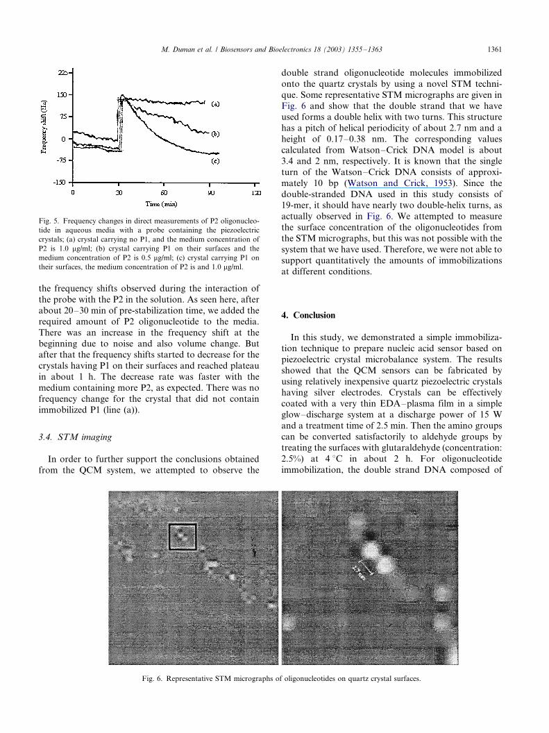
the frequency shifts observed during the interaction of
the probe with the P2 in the solution. As seen here, after
about 20�/30 min of pre-stabilization time, we added the
required amount of P2 oligonucleotide to the media.
There was an increase in the frequency shift at the
beginning due to noise and also volume change. Butafter that the frequency shifts started to decrease for the
crystals having P1 on their surfaces and reached plateau
in about 1 h. The decrease rate was faster with the
medium containing more P2, as expected. There was no
frequency change for the crystal that did not contain
immobilized P1 (line (a)).
3.4. STM imaging
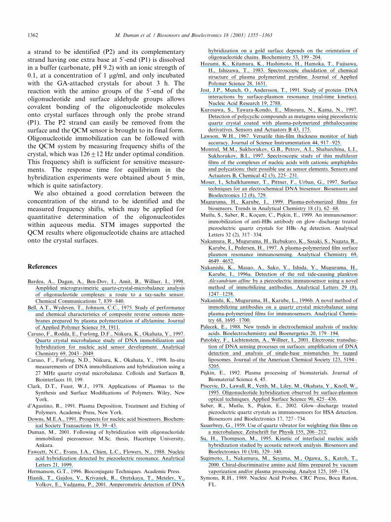
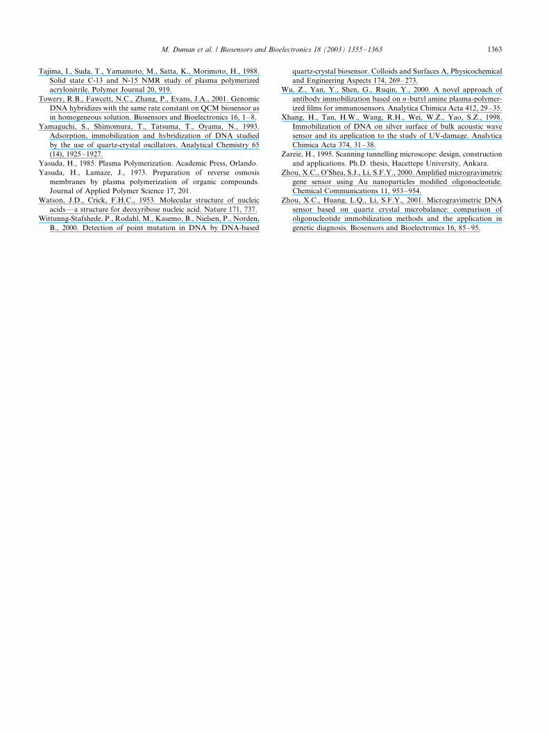
In order to further support the conclusions obtained
from the QCM system, we attempted to observe the
double strand oligonucleotide molecules immobilized
onto the quartz crystals by using a novel STM techni-
que. Some representative STM micrographs are given in
Fig. 6 and show that the double strand that we haveused forms a double helix with two turns. This structure
has a pitch of helical periodicity of about 2.7 nm and a
height of 0.17�/0.38 nm. The corresponding values
calculated from Watson�/Crick DNA model is about
3.4 and 2 nm, respectively. It is known that the single
turn of the Watson�/Crick DNA consists of approxi-
mately 10 bp (Watson and Crick, 1953). Since the
double-stranded DNA used in this study consists of19-mer, it should have nearly two double-helix turns, as
actually observed in Fig. 6. We attempted to measure
the surface concentration of the oligonucleotides from
the STM micrographs, but this was not possible with the
system that we have used. Therefore, we were not able to
support quantitatively the amounts of immobilizations
at different conditions.
4. Conclusion
In this study, we demonstrated a simple immobiliza-
tion technique to prepare nucleic acid sensor based on
piezoelectric crystal microbalance system. The results
showed that the QCM sensors can be fabricated by
using relatively inexpensive quartz piezoelectric crystalshaving silver electrodes. Crystals can be effectively
coated with a very thin EDA�/plasma film in a simple
glow�/discharge system at a discharge power of 15 W
and a treatment time of 2.5 min. Then the amino groups
can be converted satisfactorily to aldehyde groups by
treating the surfaces with glutaraldehyde (concentration:
2.5%) at 4 8C in about 2 h. For oligonucleotide
immobilization, the double strand DNA composed of
Fig. 5. Frequency changes in direct measurements of P2 oligonucleo-
tide in aqueous media with a probe containing the piezoelectric
crystals; (a) crystal carrying no P1, and the medium concentration of
P2 is 1.0 mg/ml; (b) crystal carrying P1 on their surfaces and the
medium concentration of P2 is 0.5 mg/ml; (c) crystal carrying P1 on
their surfaces, the medium concentration of P2 is and 1.0 mg/ml.
Fig. 6. Representative STM micrographs of oligonucleotides on quartz crystal surfaces.
M. Duman et al. / Biosensors and Bioelectronics 18 (2003) 1355�/1363 1361
a strand to be identified (P2) and its complementary
strand having one extra base at 5?-end (P1) is dissolved
in a buffer (carbonate, pH 9.2) with an ionic strength of
0.1, at a concentration of 1 mg/ml, and only incubatedwith the GA-attached crystals for about 3 h. The
reaction with the amino groups of the 5?-end of the
oligonucleotide and surface aldehyde groups allows
covalent bonding of the oligonucleotide molecules
onto crystal surfaces through only the probe strand
(P1). The P2 strand can easily be removed from the
surface and the QCM sensor is brought to its final form.
Oligonucleotide immobilization can be followed withthe QCM system by measuring frequency shifts of the
crystal, which was 1269/12 Hz under optimal condition.
This frequency shift is sufficient for sensitive measure-
ments. The response time for equilibrium in the
hybridization experiments were obtained about 5 min,
which is quite satisfactory.
We also obtained a good correlation between the
concentration of the strand to be identified and themeasured frequency shifts, which may be applied for
quantitative determination of the oligonucleotides
within aqueous media. STM images supported the
QCM results where oligonucleotide chains are attached
onto the crystal surfaces.
References
Bardea, A., Dagan, A., Ben-Dov, I., Amit, B., Willner, I., 1998.
Amplified microgravimetric quartz-crystal-microbalance analysis
of oligonucleotide complexes: a route to a tay-sachs sensor.
Chemical Communications 7, 839�/840.
Bell, A.T., Wydeven, T., Johnson, C.C., 1975. Study of performance
and chemical characteristics of composite reverse osmosis mem-
branes prepared by plasma polymerization of allylamine. Journal
of Applied Polymer Science 19, 1911.
Caruso, F., Rodda, E., Furlong, D.F., Niikura, K., Okahata, Y., 1997.
Quartz crystal microbalance study of DNA immobilization and
hybridization for nucleic acid sensor development. Analytical
Chemistry 69, 2043�/2049.
Caruso, F., Furlong, N.D., Niikura, K., Okahata, Y., 1998. In-situ
measurements of DNA immobilizations and hybridization using a
27 MHz quartz crystal microbalance. Colloids and Surfaces B,
Biointerfaces 10, 199.
Clark, D.T., Feast, W.J., 1978. Applications of Plasmas to the
Synthesis and Surface Modifications of Polymers. Wiley, New
York.
d’Agustino, R., 1991. Plasma Deposition, Treatment and Etching of
Polymers. Academic Press, New York.
Downs, M.E.A., 1991. Prospects for nucleic acid biosensors. Biochem-
ical Society Transactions 19, 39�/43.
Duman, M., 2001. Following of hybridization with oligonucleotide
immobilized piezosensor. M.Sc. thesis, Hacettepe University,
Ankara.
Fawcett, N.C., Evans, I.A., Chien, L.C., Flowers, N., 1988. Nucleic
acid hybridization detected by piezoelectric resonance. Analytical
Letters 21, 1099.
Hermanson, G.T., 1996. Bioconjugate Techniques. Academic Press.
Hianik, T., Gajdos, V., Krivanek, R., Oretskaya, T., Metelev, V.,
Volkov, E., Vadgama, P., 2001. Amperometric detection of DNA
hybridization on a gold surface depends on the orientation of
oligonucleotide chains. Biochemistry 53, 199�/204.
Hozumi, K., Kitamura, K., Hashimoto, H., Hamoka, T., Fujisawa,
H., Ishizawa, T., 1983. Spectroscopic elucidation of chemical
structure of plasma polymerized pyridine. Journal of Applied
Polymer Science 28, 1651.
Jost, J.P., Munch, O., Andersson, T., 1991. Study of protein�/DNA
interactions by surface-plasmon resonance (real-time kinetics).
Nucleic Acid Research 19, 2788.
Kurosawa, S., Tawara-Kondo, E., Minoura, N., Kama, N., 1997.
Detection of polycyclic compounds as mutagens using piezoelectric
quartz crystal coated with plasma-polymerized phthalocyanine
derivatives. Sensors and Actuators B 43, 175.
Lawson, W.H., 1967. Versatile thin-film thickness monitor of high
accuracy. Journal of Science Instrumentation 44, 917�/925.
Montrel, M.M., Sukhorukov, G.B., Petrov, A.I., Shabarchina, L.I.,
Sukhorukov, B.I., 1997. Spectroscopic study of thin multilayer
films of the complexes of nucleic acids with cationic amphiphiles
and polycations: their possible use as sensor elements. Sensors and
Actuators B, Chemical 42 (3), 225�/231.
Moser, I., Schalkhammer, T., Pittner, F., Urban, G., 1997. Surface
techniques for an electrochemical DNA biosensor. Biosensors and
Bioelectronics 12 (8), 729.
Muguruma, H., Karube, I., 1999. Plasma-polymerized films for
biosensors. Trends in Analytical Chemistry 18 (1), 62�/68.
Mutlu, S., Saber, R., Kocum, C., Piskin, E., 1999. An immunosensor:
immobilization of anti-HBs antibody on glow�/discharge treated
piezoelectric quartz crystals for HBs�/Ag detection. Analytical
Letters 32 (2), 317�/334.
Nakamura, R., Muguruma, H., Ikebukuro, K., Sasaki, S., Nagata, R.,
Karube, I., Pedersen, H., 1997. A plasma-polymerized film surface
plasmon resonance immunosensing. Analytical Chemistry 69,
4649�/4652.
Nakanishi, K., Masao, A., Sako, Y., Ishida, Y., Muguruma, H.,
Karube, I., 1996a. Detection of the red tide-causing plankton
Alexandrium affine by a piezoelectric immunosensor using a novel
method of immobilizing antibodies. Analytical Letters 29 (8),
1247�/1258.
Nakanishi, K., Muguruma, H., Karube, L., 1996b. A novel method of
immobilizing antibodies on a quartz crystal microbalance using
plasma-polymerized films for immunosensors. Analytical Chemis-
try 68, 1695�/1700.
Palecek, E., 1988. New trends in electrochemical analysis of nucleic
acids. Bioelectrochemistry and Bioenergetics 20, 179�/194.
Patolsky, F., Lichtenstein, A., Willner, I., 2001. Electronic transduc-
tion of DNA sensing processes on surfaces: amplification of DNA
detection and analysis of single-base mismatches by tagged
liposomes. Journal of the American Chemical Society 123, 5194�/
5205.
Piskin, E., 1992. Plasma processing of biomaterials. Journal of
Biomaterial Science 4, 45.
Piscevic, D., Lawall, R., Veith, M., Liley, M., Okahata, Y., Knoll, W.,
1995. Oligonucleotide hybridization observed by surface-plasmon
optical techniques. Applied Surface Science 90, 425�/436.
Saber, R., Mutlu, S., Piskin, E., 2002. Glow�/discharge treated
piezoelectric quartz crystals as immunosensors for HSA detection.
Biosensors and Bioelectronics 17, 727�/734.
Sauerbrey, G., 1959. Use of quartz vibrator for weighting thin films on
a microbalance. Zeitschrift fur Physik 155, 206�/212.
Su, H., Thompson, M., 1995. Kinetic of interfacial nucleic acids
hybridization studied by acoustic network analysis. Biosensors and
Bioelectronics 10 (3/4), 329�/340.
Sugimoto, I., Nakamura, M., Seyama, M., Ogawa, S., Katoh, T.,
2000. Chiral-discriminative amino acid films prepared by vacuum
vaporization and/or plasma processing. Analyst 125, 169�/174.
Symons, R.H., 1989. Nucleic Acid Probes. CRC Press, Boca Raton,
FL.
M. Duman et al. / Biosensors and Bioelectronics 18 (2003) 1355�/13631362
Tajima, I., Suda, T., Yamamoto, M., Satta, K., Morimoto, H., 1988.
Solid state C-13 and N-15 NMR study of plasma polymerized
acrylonitrile. Polymer Journal 20, 919.
Towery, R.B., Fawcett, N.C., Zhang, P., Evans, J.A., 2001. Genomic
DNA hybridizes with the same rate constant on QCM biosensor as
in homogeneous solution. Biosensors and Bioelectronics 16, 1�/8.
Yamaguchi, S., Shimomura, T., Tatsuma, T., Oyama, N., 1993.
Adsorption, immobilization and hybridization of DNA studied
by the use of quartz-crystal oscillators. Analytical Chemistry 65
(14), 1925�/1927.
Yasuda, H., 1985. Plasma Polymerization. Academic Press, Orlando.
Yasuda, H., Lamaze, J., 1973. Preparation of reverse osmosis
membranes by plasma polymerization of organic compounds.
Journal of Applied Polymer Science 17, 201.
Watson, J.D., Crick, F.H.C., 1953. Molecular structure of nucleic
acids*/a structure for deoxyribose nucleic acid. Nature 171, 737.
Wittunng-Stafshede, P., Rodahl, M., Kasemo, B., Nielsen, P., Norden,
B., 2000. Detection of point mutation in DNA by DNA-based
quartz-crystal biosensor. Colloids and Surfaces A, Physicochemical
and Engineering Aspects 174, 269�/273.
Wu, Z., Yan, Y., Shen, G., Ruqin, Y., 2000. A novel approach of
antibody immobilization based on n -butyl amine plasma-polymer-
ized films for immunosensors. Analytica Chimica Acta 412, 29�/35.
Xhang, H., Tan, H.W., Wang, R.H., Wei, W.Z., Yao, S.Z., 1998.
Immobilization of DNA on silver surface of bulk acoustic wave
sensor and its application to the study of UV-damage. Analytica
Chimica Acta 374, 31�/38.
Zareie, H., 1995. Scanning tunnelling microscope: design, construction
and applications. Ph.D. thesis, Hacettepe University, Ankara.
Zhou, X.C., O’Shea, S.J., Li, S.F.Y., 2000. Amplified microgravimetric
gene sensor using Au nanoparticles modified oligonucleotide.
Chemical Communications 11, 953�/954.
Zhou, X.C., Huang, L.Q., Li, S.F.Y., 2001. Microgravimetric DNA
sensor based on quartz crystal microbalance: comparison of
oligonucleotide immobilization methods and the application in
genetic diagnosis. Biosensors and Bioelectronics 16, 85�/95.
M. Duman et al. / Biosensors and Bioelectronics 18 (2003) 1355�/1363 1363
Copyright © 2022 FDOKUMEN