A molecular beacon-based real-time NASBA assay for detection of Mycobacterium avium subsp....
10
A molecular beacon-based real time NASBA assay for detection of Listeria monocytogenes in food products: Role of target mRNA secondary structure on NASBA design Anna Nadal a,1 , Anna Coll a,1 , Nigel Cook b , Maria Pla a, ⁎ a Institute of Food and Agricultural Technology, University of Girona, E-17071 Girona, Spain b Central Science Laboratory, Sand Hutton, York, YO41 1LZ, UK Received 29 June 2006; received in revised form 6 November 2006; accepted 20 November 2006 Available online 26 January 2007 Abstract A molecular beacon-based real-time NASBA (QNASBA) assay for detection and identification of Listeria monocytogenes has been developed. A correlation between targeting highly accessible mRNA sequences and QNASBA efficiency and sensitivity was demonstrated. The assay targets a sequence from the mRNA transcript of the hly gene which is specific for this bacterium; and includes an internal amplification control to disclose failure of the reaction. It was fully selective and consistently detected down to 100 target molecules and 40 L. monocytogenes exponentially growing cells per reaction. In addition, it was capable of accurate quantification of target RNA molecules independently of the presence of DNA in the sample. In combination with a short RNase treatment prior to nucleic acids extraction our QNASBA specifically detected viable L. monocytogenes cells. It was successfully applied to rapid detection of this pathogen in meat and salmon products, and is therefore a useful tool for the study of L. monocytogenes in food samples. We finally discuss considerations of target secondary structure with regard to development of NASBA assays. © 2006 Elsevier B.V. All rights reserved. Keywords: Listeria monocytogenes; Real-time NASBA (QNASBA); RNA detection and quantification; Secondary structure; Viable microorganisms 1. Introduction Listeria monocytogenes is a facultative anaerobic gram- positive bacterial species widely distributed in the environment, capable of growth over a pH range of 4.39 to 9.40, and at refrigeration temperatures. It is the etiological agent of listeriosis, a severe infectious disease that predominantly affects certain risk groups, including pregnant women, newborns, elderly people and immunocompromised patients. Human listeriosis is associated with food products contaminated with L. monocytogenes, meat and salmon products being major sources of this food-borne pathogen (Peccio et al., 2003; Ryser, 1999). The classical approach for detection of L. monocytogenes in food involves growth in pre-enrichment medium, growth in enrichment medium and biochemical and serological confir- matory tests (Farber and Peterkin, 1991), and takes around 10 days. DNA-based techniques such as PCR and, notably, real- time PCR (QPCR) offer a rapid, specific and sensitive alternative. QPCR assays have been developed for the quantitative detection of L. monocytogenes (Hough et al., 2002; Koo and Jaykus, 2003; Liming et al., 2004; Nogva et al., 2000; Rodríguez-Lázaro et al., 2004b) and can exhibit limits of detection of around 100 cfu and limits of quantification of around 1000 cfu/g of food (Rodríguez-Lázaro et al., 2004c, 2005). However, amplification of DNA from dead cells can overestimate the number of actual colony-forming units (Josephson et al., 1993). Efforts have been made to reduce amplification of DNA from dead cells through selective degradation by externally added DNases (Nogva et al., 2000) or intercalating dyes (Nogva et al., 2003; Pla et al., 2005). As messenger (m)RNA molecules generally possess shorter half- Journal of Microbiological Methods 68 (2007) 623 – 632 www.elsevier.com/locate/jmicmeth ⁎ Corresponding author. Institute of Food and Agricultural Technology INTEA, University of Girona, Escola Politècnica Superior (Edif.1) Campus Montilivi, E-17071 Girona, Spain. Tel.: +34 972 419852; fax: +34 972 418399. E-mail address: [email protected] (M. Pla). 1 A.N. and A.C. equally contributed to the manuscript. 0167-7012/$ - see front matter © 2006 Elsevier B.V. All rights reserved. doi:10.1016/j.mimet.2006.11.011
-
Upload
independent -
Category
Documents
-
view
1 -
download
0
Transcript of A molecular beacon-based real-time NASBA assay for detection of Mycobacterium avium subsp....

(2007) 623–632www.elsevier.com/locate/jmicmeth
Journal of Microbiological Methods 68
A molecular beacon-based real time NASBA assay for detection of Listeriamonocytogenes in food products: Role of target mRNA secondary
structure on NASBA design
Anna Nadal a,1, Anna Coll a,1, Nigel Cook b, Maria Pla a,⁎
a Institute of Food and Agricultural Technology, University of Girona, E-17071 Girona, Spainb Central Science Laboratory, Sand Hutton, York, YO41 1LZ, UK
Received 29 June 2006; received in revised form 6 November 2006; accepted 20 November 2006Available online 26 January 2007
Abstract
A molecular beacon-based real-time NASBA (QNASBA) assay for detection and identification of Listeria monocytogenes has beendeveloped. A correlation between targeting highly accessible mRNA sequences and QNASBA efficiency and sensitivity was demonstrated. Theassay targets a sequence from the mRNA transcript of the hly gene which is specific for this bacterium; and includes an internal amplificationcontrol to disclose failure of the reaction. It was fully selective and consistently detected down to 100 target molecules and 40 L. monocytogenesexponentially growing cells per reaction. In addition, it was capable of accurate quantification of target RNA molecules independently of thepresence of DNA in the sample. In combination with a short RNase treatment prior to nucleic acids extraction our QNASBA specifically detectedviable L. monocytogenes cells. It was successfully applied to rapid detection of this pathogen in meat and salmon products, and is therefore auseful tool for the study of L. monocytogenes in food samples. We finally discuss considerations of target secondary structure with regard todevelopment of NASBA assays.© 2006 Elsevier B.V. All rights reserved.
Keywords: Listeria monocytogenes; Real-time NASBA (QNASBA); RNA detection and quantification; Secondary structure; Viable microorganisms
1. Introduction
Listeria monocytogenes is a facultative anaerobic gram-positive bacterial species widely distributed in the environment,capable of growth over a pH range of 4.39 to 9.40, and atrefrigeration temperatures. It is the etiological agent oflisteriosis, a severe infectious disease that predominantly affectscertain risk groups, including pregnant women, newborns,elderly people and immunocompromised patients. Humanlisteriosis is associated with food products contaminated withL. monocytogenes, meat and salmon products being majorsources of this food-borne pathogen (Peccio et al., 2003; Ryser,1999).
⁎ Corresponding author. Institute of Food and Agricultural TechnologyINTEA, University of Girona, Escola Politècnica Superior (Edif.1) CampusMontilivi, E-17071 Girona, Spain. Tel.: +34 972 419852; fax: +34 972 418399.
E-mail address: [email protected] (M. Pla).1 A.N. and A.C. equally contributed to the manuscript.
0167-7012/$ - see front matter © 2006 Elsevier B.V. All rights reserved.doi:10.1016/j.mimet.2006.11.011
The classical approach for detection of L. monocytogenes infood involves growth in pre-enrichment medium, growth inenrichment medium and biochemical and serological confir-matory tests (Farber and Peterkin, 1991), and takes around10 days. DNA-based techniques such as PCR and, notably, real-time PCR (QPCR) offer a rapid, specific and sensitivealternative. QPCR assays have been developed for thequantitative detection of L. monocytogenes (Hough et al.,2002; Koo and Jaykus, 2003; Liming et al., 2004; Nogva et al.,2000; Rodríguez-Lázaro et al., 2004b) and can exhibit limits ofdetection of around 100 cfu and limits of quantification ofaround 1000 cfu/g of food (Rodríguez-Lázaro et al., 2004c,2005). However, amplification of DNA from dead cells canoverestimate the number of actual colony-forming units(Josephson et al., 1993). Efforts have been made to reduceamplification of DNA from dead cells through selectivedegradation by externally added DNases (Nogva et al., 2000)or intercalating dyes (Nogva et al., 2003; Pla et al., 2005). Asmessenger (m)RNA molecules generally possess shorter half-

624 A. Nadal et al. / Journal of Microbiological Methods 68 (2007) 623–632
lives, they have been considered more suitable than DNA forviability assays (Bej et al., 1991; Cook, 2003). Determination ofviable L. monocytogenes has been reported by reverse-transcription (RT) coupled to PCR (Norton and Batt, 1999),for which complete DNA digestion is required prior to theamplification reaction. Nucleic acid sequence-based amplifica-tion (NASBA) (Compton, 1991) is a very promising alternativemethod, since it can selectively amplify mRNA even in thepresence of genomic DNA (Simpkins et al., 2000). The NASBAproduct can be detected in real time using molecular beacons(Leone et al., 1998; Tyagi and Kramer, 1996). This technologyhas been applied to the quantification of RNAviral particles (deBaar et al., 2001; Hibbitts et al., 2003; Moore et al., 2004).NASBA has the potential to detect viable microorganismsin various sample types including environmental and foodmatrices (Chan and Fox, 1999; Cook, 2003).
Although conventionalNASBAhas been reported (Blais et al.,1997; Uyttendaele et al., 1995), no real-timeNASBA (QNASBA)assay has been published to date for L. monocytogenes. Wedescribe a QNASBA assay for selective and sensitive detection ofviable L. monocytogenes, and its application to food samples. Inaddition, we present our assay as an illustrative example of theimportance of target mRNA secondary structures for QNASBAoptimization and discuss several technical issues that may becritical for the design of a QNASBA assay.
2. Materials and methods
2.1. Bacterial strains, culture media and growth conditions
Twenty Listeria isolates (15 L. monocytogenes and 5 non-L. monocytogenes isolates) and 9 non-Listeria spp. strains wereused in this study (Table 1). The L. monocytogenes isolateswere representative of the different serovars, as indicated inTable 1. Unless stated, L. monocytogenes strain CECT 4031was used. Listeria strains were grown in brain heart infusionbroth at 37 °C, and non-Listeria strains were grown in MRSbroth or tryptone soya broth at 30 °C. For plate cultures, 1.5%(wt/vol) agar was added to these media. All media werepurchased from Oxoid (Hampshire, United Kingdom).
2.2. Nucleic acid preparation and extraction
Overnight cultures were diluted in the same broth toexponential phase (approximately 105 ml−1). Nucleic acidswere extracted by the High Pure RNA Isolation kit (RocheApplied Science, Penzberg, Germany) according to themanufacturer's recommendations. Nucleic acid concentrationwas determined by spectrophotometry using the NanoDrop ND-1000 device (NanoDrop Technologies, Delaware, USA) and the260/280 and 260/230 ratios were calculated (Sambrook andRussell, 2001).
2.3. DNAse and RNase treatments
To confirm that the origin of the NASBA signals wasindeed mRNA, prior to amplification, 500 ng L. monocyto-
genes nucleic acids were subjected to DNase or RNaseenzymatic treatments. DNase treatment was performed usingthe RQ1 RNase-free DNase kit (Promega, Madison, USA),according to the manufacturer's instructions. RNase treat-ment was performed with 20 μg/ml ribonuclease A (kindlyprovided by M. Ribó, UdG), treated to inactivate any con-taminating DNase, according to the manufacturer's instruc-tions. After enzymatic treatment, purification of nucleic acidswas performed according to Sambrook and Russell (2001).To confirm that the enzymatic treatments had been effective,the treated extracts were assayed by hly QPCR (Rodríguez-Lázaro et al., 2004b) and QRTPCR (i.e. conventional reversetranscription coupled to the same hly QPCR assay). Theexperiment was in triplicate and a total of 8 QNASBA reactionswere performed per treatment.
2.4. Synthesis and quantification of target RNA transcript
We optimized the QNASBA using the RNA transcript hly-1339, a 605-nt molecule whose secondary structure waspredicted by the RNAstructure program version 4.11 (Mathewset al., 2004). It corresponds to nucleotides 1339 to 1944 ofthe L. monocytogenes hly gene. We designed hly-1339 sothat the QNASBA target sequence was placed in a centralposition, because the artificial 5′- and 3′-termini of the RNAtranscript may adopt secondary structures. hly-1339 wassynthesized by in vitro transcription from a PCR productobtained with L. monocytogenes genomic DNA and primersT7hly-1339F and hly-1919R (Table 2). To obtain RNA target,1 μg DNA template was transcribed in vitro for 1 h at 37 °Cfollowed by a 5 min treatment with RNase-free DNase I at37 °C. Cellulose CF11 chromatography was used to eliminateDNA fragments and non-incorporated nucleotides. The tran-script was then quantified by spectrophotometry. hly specificQPCR was used to confirm the absence of DNA (Rodríguez-Lázaro et al., 2004b).
2.5. Synthesis of hly-1339 DNA for use as NASBA control
The dsDNA molecule DNA-hly-1339 was used as a controlto assess the nature of the QNASBA template molecules. Itcorresponds to nucleotides 1339 to 1919 of the L. mono-cytogenes hly gene (the same as the RNA target hly-1339) andwas synthesised by PCR with L. monocytogenes genomic DNAand primers DNA-hly-1339F (equivalent to T7hly-1339Fexcept for the T7 sequence) and hly-1919R. Reactions wereperformed with TaqMan PCR core reagents (Applied Biosys-tems-Roche Molecular Systems Inc, Branchburg, NJ, USA) and50-μL final volume including 1× bufferII, 1.5 mM MgCl2,200 μM dNTPs, 900 nM of each primer and 1 U AmpliTaqGold DNA polymerase. Reactions were run in a Master CyclerGradient device (Eppendorf AG, Hamburg, Germany), usingthe following program: 10 min at 95 °C; 40 cycles of 20 s at95 °C, 30 s at 56 °C and 1 min at 72 °C; and 7 min at 72 °C. TheDNA-hly-1339 product was purified with the Qiaquick PCRpurification kit (Qiagen Sciences Inc., Maryland, USA) andquantified by spectrophotometry and hly specific QPCR.
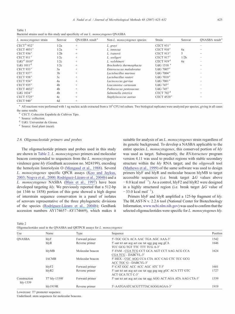
Table 1Bacterial strains used in this study and specificity of our L. monocytogenes QNASBA
L. monocytogenes strain Serovar QNASBA result a Non-L. monocytogenes species Strain Serovar QNASBA result a
CECTb 932 c 1/2a + L. grayi CECT 931 c −CECT 4031 c 1/2a + L. innocua CECT 910 c 6a −CECT 936 c 1/2b + L. ivanovii CECT 913 c 5 −CECT 911 c 1/2c + L. seeligeri CECT 917 c 1/2b −UdGd 1010 e 1/2c + L. welshimeri CECT 919 d 6a −UdG 1011 d 1/2c + Brochothrix thermosphacta UdG 1510 e −CECT 933 c 3a + Enterococcus malodoratus UdG 7007 e −CECT 937 c 3b + Lactobacillus murinus UdG 7004 e −CECT 938 c 3c + Lactobacillus reuteri UdG 7010 e −CECT 934 c 4a + Lactococcus garviae UdG 7001 e −CECT 935 c 4b + Leuconostoc carnosum UdG 747 e −CECT 4032 c 4b + Pediococcus pentosaceus UdG 745 e −UdG 1034 e 4b + Salmonella enterica CECT 702 d −CECT 5725 c 4c + Staphylococcus aureus CECT 4520 c −CECT 940 c 4d +a All reactions were performed with 1 ng nucleic acids extracted from a 105 CFU/ml culture. Two biological replicates were analyzed per species, giving in all cases
the same results.b CECT: Colección Española de Cultivos Tipo.c Source: collection.d UdG: Universitat de Girona.e Source: food plant (meat).
625A. Nadal et al. / Journal of Microbiological Methods 68 (2007) 623–632
2.6. Oligonucleotide primers and probes
The oligonucleotide primers and probes used in this studyare shown in Table 2. L. monocytogenes primers and molecularbeacon corresponded to sequences from the L. monocytogenesvirulence gene hly (GenBank accession no. M24199), encodingthe hemolysin listeriolysin O (Mengaud et al., 1988). SeveralL. monocytogenes specific QPCR assays (Koo and Jaykus,2003; Nogva et al., 2000; Rodríguez-Lázaro et al., 2004b) and aL. monocytogenes NASBA (Blais et al., 1997) have beendeveloped targeting hly. We previously reported that a 512-bp(nt 1346 to 1858) portion of this gene showed a high degreeof interstrain sequence conservation in a panel of isolatesof serovars representative of the three phylogenetic divisionsof the species (Rodríguez-Lázaro et al., 2004b); GenBankaccession numbers AY174657–AY174669), which makes it
Table 2Oligonucleotides used in the QNASBA and QRTPCR assays for L. monocytogenes
Use Name Type Sequenc
QNASBA hlyF Forward primer 5′-TGChlyR Reverse primer 5′-aat tct
TCC GChlyMB Molecular beacon 5′-FAM
CGATCIACMB Molecular beacon 5′-HEX
ACC TGhlyF2 Forward primer 5′-CAT GhlyR2 Reverse primer 5′-aat tct
ACT GCConstruction
hly-1339T7 hly-1339F Forward primer 5′-aat tct
hly1919R Reverse primer 5′-AATG
Lowercase: T7 promoter sequence.Underlined: stem sequences for molecular beacons.
suitable for analysis of an L. monocytogenes strain regardless ofits genetic background. To develop a NASBA applicable to theentire species L. monocytogenes, this conserved portion of hlywas used as target. Subsequently, the RNAstructure programversion 4.11 was used to predict regions with stable secondarystructure within the hly RNA target; and the oligowalk tool(Mathews et al., 1999) of the same software was used to designprimers hlyF and hlyR and molecular beacon hlyMB to targetaccessible sequences (i.e. break target ΔG values above−8.0 kcal mol−1). As a control, hlyF2 and hlyR2 were designedin a highly structured region (i.e. break target ΔG value of−33.0 kcal mol−1).
Primers hlyF and hlyR amplified a 125-bp fragment of hly.The BLAST-N v. 2.2.6 tool (National Center for BiotechnologyInformation, www.ncbi.nlm.nih.gov) was used to confirm that theselected oligonucleotides were specific for L. monocytogenes hly.
e Position
GCA ACA AAC TGA AGC AAA-3′ 1542aat acg act cac tat agg gag aag gCAG TGT TTC TTT TCG A-3′
1646
- CGA TCG CCT GCA AGT CCT AAG ACG CCAG - DABCYL-3′
1624
- CGC AGG CCA CTA ACC CAG CTC TCC GCGC G - DABCYL-3′GC ACC ACC AGC ATC T-3′ 1601aat acg act cac tat agg gag aag gGC ACATTT GTCATCT C-3′
1727
aat acg act cac tat agg AGG ACT AGA ATA AAG CTA-3′ 1339
AATCACGTTTTACAGGGAGAA-3′ 1919

626 A. Nadal et al. / Journal of Microbiological Methods 68 (2007) 623–632
Blast search was carried out by using default settings of the tool.All oligonucleotides were purchased from MWG Biotech AG(Germany).
2.7. IAC synthesis
The QNASBA IAC was an in vitro synthesized chimeric RNAmolecule containing an internal non-target sequence flanked bysequences complementary to the target QNASBAprimers hlyF andhlyR. The non-target sequencewas a 105-bp fragment derived fromZeamays. The construction strategywas that of (Rodríguez-Lázaroet al., 2004a). The IAC sequence was 5′-UGCGCAACAAACU-GAAGCAAAAGCAACCUCUACCAAAUCUACGCUGA-GAGCUUCCGCGAGUGGGAGGCCGACCCCACUAACC-CAGCUCUCCGCGAGGAGAUGCGCAUCCAGUUCAAC-GACAUGAACAUCGAAAAGAAACACGCGGAUG-3′ anddid not show any significant homology to any sequence depositedin public databases when analyzed using the BLAST-N v. 2.2.6software (cutoff E-value, 0.1).
2.8. QNASBA
Reactions were carried out using the NucliSens® Basic Kit(bioMérieux bv, The Netherlands). The primers and probeconcentrations were initially optimized with hly-1339 (data notshown) to give the lowest time to positivity (Tp, equivalent tothreshold cycle (CT) in real time PCR assays) and the highestnormalized fluorescence intensity (URn) (Perkin-Elmer AppliedBiosystems User Bulletin 2 [ABI PRISM 7700 SequenceDetection System], 1997). The IAC concentration per reactionwas subsequently optimised to the lowest copy number givingconsistent TP (i.e. standard deviation S.D. below 10%) in orderto avoid any interference with target amplification. This wasperformed using 103 copies of hly-1339 and 105, 104, 3×10³
and 103 copies of IAC.In the optimised assay, 1.25 μl of target template solution
was added to 6.25 μl NASBA pre-mix containing 103 copiesIAC in 80 mM Tris–HCl (pH 8.5), 24 mM MgCl2, 140 mMKCl, 30% v/v DMSO, 2 mM each dNTP, 4 mM each NTP,200 nM hlyF primer, 500 nM hlyR primer and 200 nM hlyMB(FAM and DABCYL dual-labelled) and IACMB (HEX andDABCYL dual-labelled) molecular beacons. The mixtureswere incubated at 42 °C for 5 min; and 2.5 μl of enzymemixture (0.08 U/μl RNase H, 32 U/μl T7 RNA polymerase,6.4 U/μl AMV reverse-transcriptase per reaction) was subse-quently added. Reactions were incubated at 42 °C for 90 min inan ABIPrism® 7700 Sequence Detection System (AppliedBiosystems, Foster City, USA) and fluorescence was monitoredin real-time. Unless otherwise stated, all reactions wereperformed in a total of 8 replicates in 3 different QNASBAruns that corresponded to 3 independent experiments for hlytranscript or 3 biological replicates for L. monocytogenescells. Non-template controls (NTC) were included todetermine the threshold TP and ΔRn values to consider apositive result.
Initial reactions for the design and optimization of QNASBAeither included 5-min incubation at 65 °C (i.e. standard reaction
conditions recommended by the NucliSens® Basic Kitmanufacturer) or 95 °C prior to 42 °C for 5 min and additionof the enzyme mix; or direct incubation at 42 °C for 5 minbefore addition of the enzyme mix.
2.9. Limit of detection and quantification capacity of hlyQNASBA
The limit of detection (LOD) was established using the hly-1339 RNA transcript. In three independent experiments, serialdilutions of hly-1339 RNA transcript were prepared and 4replicates of samples containing 106, 105, 104, 103, 102, 10 and1 target copies were analyzed. For each dilution, the expectednumbers of target molecules per reaction were calculated at the95% confidence level by Monte Carlo simulations taking intoaccount the experimental design (i.e. decimal dilutions in a finalvolume of 0.1 ml). In parallel, three replicates of exponentiallygrowing L. monocytogenes cell cultures were quantified by thestandard plate count technique (Anonymous, 1998b) and ten-fold serial dilutions were prepared in phosphate-buffered saline(PBS, Dulbecco's, Gibco, Paisley, Scotland), corresponding to4×105, 4×10⁴, 4×10³, 4×10², 40, 4 and 0.4 CFU/ml. Fromeach dilution, nucleic acids were extracted and subjected toQNASBA. The number of positive signals obtained wereexpressed as a percentage, and compared with the number oftarget molecules/cells in each dilution (Knutsson et al., 2002).
Quantification capacity was assessed by plotting the TP
values obtained against the logarithm of the number of targetmolecules or L. monocytogenes CFU in each dilution andcalculating a linear regression. Both the slope and R2 values aregiven.
2.10. Sample treatment for comparison of NASBA signals fromviable and non-viable cells
In three replicates, L. monocytogenes cells were grown inbrain heart infusion broth at 37 °C to exponential phase(approximately 105 ml−1). Two 5-ml aliquots (#1 and #2) weretaken from the culture. Aliquot #1 was immediately processed(see below) and aliquot #2 was incubated at 65 °C in a heatedwater bath for 5 min, to kill cells prior to processing. Monitoringof the sample temperature was carried out in a separate 5-mlaliquot that was treated in parallel and discarded: the time ofincubation was considered to start when the sample temperaturewas 65 °C. Colony forming units of aliquots #1 and #2 werequantified by plate counting or most probable number tech-niques (MPN) (Anonymous, 1998b). One ml of each aliquotwas digested with 20 μg/ml ribonuclease A for 40 min at 37 °C;and subsequently RNA was extracted and QNASBA wasperformed as previously indicated.
Preliminary experiments were conducted to assess thepersistence of hly mRNA target in non-viable cells. Heat-treated cells were kept at RT for 5, 15, 30, 60, 120 and 180 min.then analyzed by MPN and QNASBA. In all cases QNASBAwas found to overestimate cell countings around 1 logarithmicunit. To achieve full and quick degradation of hly mRNA fromnon-viable cells a ribonuclease A treatment was included before

627A. Nadal et al. / Journal of Microbiological Methods 68 (2007) 623–632
nucleic acid extraction. Control experiments were carried out todemonstrate that ribonuclease A digestion did not affect theQNASBA signal from exponentially growing cells.
2.11. Artificial contamination of meat and salmon withL. monocytogenes and sample treatment prior to QNASBA
In three replicates, samples of 25 g of cooked ham or smokedsalmon slices obtained in local markets were artificiallycontaminated with exponentially growing L. monocytogenescells diluted in peptone water at the following approximateconcentrations: 104, 103, 102, 10, 1, 0.1 and 0 (control) UFC/g.L. monocytogenes was analyzed in all samples by standardmicrobiological methods (according to document ISO 11290(Anonymous, 1998a) and QNASBA. Briefly, slices werediluted (1:10) with half-Fraser broth (Oxoid), homogenizedfor 1 min in stomacher bags (125-μm pore size; BioChek,Gouda, Holland) and incubated at 30 °C for 24 h. One ml ofeach pre-enrichment was used for nucleic acid extraction andQNASBA. Pre-enrichment cultures were further diluted (1:100)with Fraser broth (Oxoid) and incubated at 30 °C. One-mlaliquots were taken after 24 h and 48 h enrichment for nucleicacids extraction and QNASBA. After 48 h enrichment, 0.1-mlaliquots were plated on Palcam agar plates and a number ofcolonies were confirmed by API Listeria (bioMérieux, France).
2.12. Statistical analyses
Statistical treatment of the data was performed using SPSSfor windows v13.0 (SPSS Inc, Chicago, IL). Descriptiveanalyses, one-way ANOVA and (when applicable) pair-wisecomparison of mean values by Tukey's test were used, with asignificant level of Pb0.05.
Fig. 1. In silico predicted secondary structure of hly-1339 RNA transcript (only nt 1beacon.
3. Results and discussion
3.1. Role of the secondary structure in the design of primersand probes for NASBA
The strategy used to design oligonucleotides suitable forL. monocytogenes-specific QNASBA was based on targetsequence specificity and in silico predicted secondary structure.The former is a classical approach for nucleic acid amplifica-tion-based techniques e.g. PCR. In contrast, the importanceof the role of secondary structures in the design of NASBA hasnot been emphasised to date. RNA molecules tend to adoptsecondary structures whose formation and stability can betheoretically predicted on a sequence basis using secondarystructure prediction algorithms. Accessibility to the targetsequence is a crucial requirement for successful interaction ofnucleic acid molecules (e.g. Branch, 1998; Nadal et al., 2005;Scherr et al., 2000). This is especially relevant for NASBA dueto its isothermal nature where typically no denaturing steps areincluded which could destabilise target secondary structures.
We designed two different primer pairs. They targeted eitherpredictably accessible sequences (oligonucleotides hlyF/R/MB,Table 2, Fig. 1) or sequences involved in a predicted strongsecondary structure (oligonucleotides hlyF2/R2/MB, Table 2,Fig. 1). For the NASBA amplicon and the 600-nt RNA hly-1339, break target ΔG values were −1.3 to −6.9 kcal mol−1;and −17.4 to −32.3 kcal mol−1 respectively.
In combination with hlyMB probe (Table 2) and in thestandard reaction conditions, the performance of QNASBAtargeting accessible sequences was significantly higher than theamplification obtained when the primers were placed inpredictably structured sequences (Table 3). When 100 targetcopies (i.e. 100-fold less than the former reactions) were
539 to 1781 portion is shown) and position of QNASBA primers and molecular

Table 4QNASBA results using different initial amounts of RNA target andL. monocytogenes cells
hly-1339 copy number 106 105 104 103 102 10 1
% of positiveresults
100 100 100 100 100 25 0
TP (mean±S.D.) a 16.7±0.6
19.9±0.3
23.7±0.1
27.5±0.3
32.4±2.5
33.1±3.6
% of IAC positiveresults
0 0 0 100 100 100 100
L. monocytogenesCFU/ml
4×10⁵ 4×10⁴ 4×10³ 4×10² 40 4 0.4
% of positiveresults
100 100 100 100 25 0 0
TP (mean±S.D.) a 17.4±0.1
20.3±0.5
22.9±1.0
28.2±1.4
34.8±4.5
% of IAC positiveresults
0 0 0 0 100 100 100
All reactions were performed in the presence of 1000 copies of IAC.Results were obtained in a total of 8 reactions corresponding to 3 independentexperiments (hly-1339 transcript) or biological replicates (L. monocytogenescells).a Calculated only for the positive results.
628 A. Nadal et al. / Journal of Microbiological Methods 68 (2007) 623–632
subjected to NASBA they could only be detected using theprimer pair which targets accessible sequences (100% vs. 0% ofthe replicates).
We further confirmed that target structural impediments werethe source of such differences by either denaturing the targetRNA before NASBA was performed or preserving anysecondary structure (i.e. direct 42 °C incubation). As shownin Table 3, RNA incubation at 95 °C prior to NASBA allowedprimers targeting poorly accessible sequences to consistentlyprime target amplification, whereas preservation of targetsecondary structures resulted in lower percentages of positiveamplification (50% and 75% at the respective lower tempera-tures). Amplification of the sequences with strong secondarystructure was not completely inhibited; a possible explanation isthat they are partially accessible as a function of “RNAbreathing”. Such breathing occurs temporally randomly (andincreases with the temperature). This might cause the NASBAreaction to slow down (higher TP values) and increase the SDvalues. Primers targeting accessible sequences performedequally well after 95 °C, 65 °C and 42 °C pre-treatments(higher percentages of positive reactions and lower TP and S.D.values). These results evidence a correlation between strong insilico-predicted secondary structure of target sequences and thereduced yield of RNA amplification by QNASBA. Therefore,careful in silico analysis of the predicted structure of the targetis recommended as a preliminary step in the development ofNASBA. It should also be remembered that applying hightemperatures to denature any mRNA secondary structure priorto NASBA, could result in denaturation of the genomic DNA;subsequent primer annealing would then facilitate amplificationof the DNA sequences, which would be especially undesir-able in a viability assay. Omission of a high-temperature stepshortens the assay in comparison to including DNase treatment.
3.2. Quantification of L. monocytogenes target RNA transcriptsby QNASBA
Table 4 shows the detection probability against target copynumber. Down to 100 copies of target RNA could be detected inall replicates. A TP threshold was established at 35 (i.e. thehighest expected TP [with 95% probability] for the lowestnumber of target molecules giving consistent QNASBA signal).The IAC was correctly amplified in all reactions containing upto 1000 RNA target molecules (mean TP value, 44.5; S.D., 1.6);thus proving that not only hly-1339 positive but also negativeQNASBA replicates performed adequately.
Table 3QNASBA amplification of hly-1339 RNA transcript (104 copies) with different prim
Primer pair hlyF/R
Temperature pre-treatment 42 °C 65 °C
Percentage of positive results 100 100TP mean±S.D.a 21.7b±0.4 21.6b±1.1
Results were obtained in a total of 8 reactions corresponding to 3 independent experiused.a Calculated only for the positive results. b/c Different letters indicate statistically sig
Regression curves were calculated for TP values obtainedin QNASBA reactions corresponding to 106 to 103 targetcopies (i.e. they displayed TP relative S.D. values below 5%).The number of RNA target copies correlated with TP in a highlylinear manner as demonstrated by R2 values above 0.998.The slopes (i.e. −3.61) were similar to the theoretical optimalones calculated for real-time PCR assays (Higuchi et al.,1993) and in particular to the ones obtained in the hly basedQPCR which targets the same gene region (nt 1603 to 1667,(Rodríguez-Lázaro et al., 2004b). Thus, the reaction proceededefficiently.
These results indicate that our QNASBA assay has thepotential for accurate quantification of the RNA target. The useof target RNA transcripts as calibrants for quantification byQNASBA has been reported (e.g. Moore et al., 2004; Polstraet al., 2002). Our results stress the importance of the secondarystructure in NASBA and therefore highlight the need forcalibrants that adopt secondary structures similar to those in thetarget.
One critical parameter for the validation of an alternativemethod is the relative accuracy, i.e. the closeness of agreementbetween a test result and the accepted reference value(according to document ISO 16140 (Anonymous, 2002). The
er pairs and pre-treatments
hlyF2/R2
95 °C 42 °C 65 °C 95 °C
100 50 75 10022.2b±0.2 59.2c±10.2 36.7c±3.4 57.3c±9.2
ments. In each experiment, a new aliquot of 104 target RNA molecules/5 μl was
nificant differences between groups (Pb0.05).
629A. Nadal et al. / Journal of Microbiological Methods 68 (2007) 623–632
quantification capacity of our hly QNASBA was further testedby assessing the degree of correspondence between thequantitative data obtained by spectrophotometry and theQNASBA assay using hly-1339 transcript. QPCR was used toconfirm that the amount of DNA in the transcript RNA sampleswas residual (data not shown). Four hly-1339 transcriptsolutions at different concentrations (i.e. around 1×106,1×105, 1×10⁴ and 1×10³) were quantified by the two methods(Table 5). The TP values obtained were extrapolated to thecorresponding standard regression curve, previously calculatedexperimentally using hly-1339 as calibrator. The obtainedvalues differed by less than 21%, indicating good correspon-dence along a 4-logarithmic range e.g. (Rodríguez-Lázaro et al.,2004b). Quantitative values obtained with QRTPCR producedsimilar results (data not shown).
3.3. hly QNASBA detects L. monocytogenes RNA but notgenomic DNA
NASBA has been shown to selectively mediate the detectionof RNA. This should eliminate the necessity for DNasetreatment, which is required when using RT-PCR to try toensure that the signal comes only from amplification of RNA(Klein and Juneja, 1997). But our previous results with aM. avium subsp. paratuberculosis NASBA (Rodríguez-Lázaroet al., 2004e) indicated that in this bacterium NASBA candetect DNA. To assess the use of our hly QNASBA to detectL. monocytogenes RNA we verified the nucleic acid origin ofour QNASBA signal. To do this, in 3 independent experimentsL. monocytogenes nucleic acid extracts were used directly in theQNASBA or used after treatment with DNAse I or RNAse A.Equal amounts of hly nucleic acids as determined by hlyQRTPCR and hly QPCR were subsequently subjected toQNASBA. The quantitative results obtained by extrapolatingTP values to a hly-1339 transcript based standard curvedemonstrate that the QNASBA substrate was indeed RNA.Samples with mainly RNA (i.e. those treated with DNAse I)displayed the highest readings: Tp=24.9±0.3, corresponding to4.8×103 ±1.2% hly mRNA molecules per reaction. Asexpected, non-treated samples containing RNA but also DNAexhibited significantly lower (pb0.05) values (Tp=27.3±0.4,1.3×103 ±1.5% hly mRNA molecules per reaction). On thecontrary, RNase A treated samples did not produce any positiveQNASBA signal. This proved that genomic DNA is not asubstrate for amplification in our QNASBA.
Table 5Quantification of hly-1339 RNA transcript by QNASBA and spectrophotometry
Calculated hly-1339 copy number
Spectrophotometry 1×10⁶ 1×10⁵ 1×10⁴ 1×10³
QNASBA(mean±S.D.)
1.1×10⁶
±2%1.2×10⁵
±17%1.0×10⁴
±5%0.9×10³
±18%% of correspondence 110% 121% 104% 92%
Results were obtained in a total of 8 reactions corresponding to 3 replicates.Spectrophotometry values were calculated from the 3 independent stocksolutions. Triple lectures showed S.D. below 5%.
3.4. Specific and sensitive detection of L. monocytogenes by hlyQNASBA
The hly QNASBA detected only the L. monocytogenesisolates (Table 1). Positive IAC signals showed that the negativeresults were true (data not shown). This proved the targetspecificity of the assay.
Table 4 shows the QNASBA results obtained with serialdilutions of a L. monocytogenes overnight culture. hly RNAcould be consistently detected in cultures containing 400 CFU/mlof L. monocytogenes as determined by plate cell counting (i.e.limit of detection LOD, 400 CFU/ml). Samples containing 40 cfuwere positive in 25% of analyses. These values are similar to otherpublished NASBA e.g. (Rodríguez-Lázaro et al., 2004d).
The number of L. monocytogenes CFU/ml (from 4×10⁵ to4×102 CFU/ml) correlated with TP in a highly linear manner asestablished by R2 values of the calculated linear regressioncurves above 0.97. The slopes (i.e. −3.57) showed that theamplification was very efficient. These values were especiallyrelevant taking into account that they were obtained from serialdilutions of exponentially growing cells and each sample wasindependently subjected to nucleic acid extraction prior toQNASBA. Consequently, deviation from the linear relationshipof TP and CFU/ml was probably due to the difference in RNAextraction efficiencies at various RNA concentrations. It shouldbe remarked that all samples derived from a single exponen-tially growing culture; and therefore the mean levels of hlyRNA per CFU were similar.
The hly copy numbers calculated with hly-1339 transcriptcalibrators indicated that, under the conditions used in thetests, L. monocytogenes cells harbored around 2.3 hly mRNAmolecules per cell.
3.5. QNASBA amplification of mRNA distinguished viable fromnon-viable L. monocytogenes
In 3 independent experiments, L. monocytogenes cells wereeither incubated at 65 °C or left untreated prior to MPNenumeration and QNASBA (Table 6). QNASBA quantificationof untreated aliquots was possible by using a regression curveconstructed with serial dilutions of the same cell culture (i.e. thehly expression levels per cell were the same). As expected, theobtained cell numbers correlated with CFU shown by standardmicrobiological methods. Heat-treated aliquots showed survivalrates of around 0.3% as determined by MPN technique.QNASBA analyses of heat-treated samples produced inconsis-tent results (RSD values above 10%), with TP readingssignificantly (Pb0.05) higher than untreated cells and abovethe LOD. In contrast, hly QPCR-based quantification ofL. monocytogenes produced the same results in untreated andheat-treated cells. This demonstrates the suitability of ourQNASBA method to detect viable L. monocytogenes cells.
3.6. Application of hly QNASBA in food matrices
Many countries have a zero tolerance policy regarding thepresence of L. monocytogenes in food (European Commission,

Table 7Conventional and QNASBA determination of L. monocytogenes cells in cookedham and smoked salmon samples
L. monocytogenes CFU/g 104 103 102 10 01 0.1 0
Cooked Ham Microbiological method(ISO)
+ + + + + + −
hly QNASBA(pre-enrichment)
+ + + − − − −
hly QNASBA(24 h enrichment)
+ + + + + + −
hly QNASBA(48 h enrichment)
+ + + + + + −
Smoked salmon Microbiological method(ISO)
+ + + + + + −
hly QNASBA(pre-enrichment)
+ + − − − − −
hly QNASBA(24 h enrichment)
+ + + + + + −
hly QNASBA(48 h enrichment)
+ + + + + + −
All reactions were performed in the presence of 1000 copies of IAC. All hlynegative QNASBA showed positive IAC amplification.Results were obtained in a total of 8 reactions corresponding to 3 independentbiological replicates.
Table 6MPN count, QNASBA and QPCR analysis of viable and heat-treated L.monocytogenes cells
Treatment
Non-treated Heat-treated
MPN CFU/ml 4.3×10⁴ 125QNASBA TP 20.4a 29.5b
CFU/ml 3.9×10⁴ bLODQPCR CT 17.7c 18.6c
CFU/ml 2.9×10⁴ 1.6×10⁴
Results are mean of a total of 8 reactions corresponding to 3 biologicalreplicates. Except for QNASBA of heat-treated cells, R.S.D. was always below10%.a/b, c Different letters indicate statistically significant differences between groups(Pb0.05).
630 A. Nadal et al. / Journal of Microbiological Methods 68 (2007) 623–632
2005; FDA FSIS, 2003). Examples of food products consideredto be at risk for transmission of human listeriosis are smokedsalmon and processed meat products because of the frequentoccurrence of L. monocytogenes in food processing plants(Gravani, 1999), and since this bacterium can survive the cold-smoking process (Farber and Peterkin, 1991; Rijpens andHerman, 2002). In three replicates, cooked ham and smokedsalmon slices were artificially contaminated with decreasingamounts of L. monocytogenes cells and analyzed by standardmicrobiological methods (Anonymous, 1998a). One-ml ali-quots were taken after pre-enrichment and after 24 and 48 henrichment and subjected to nucleic acids extraction andhly QNASBA. As shown in Table 7, QNASBA detectedL. monocytogenes immediately after pre-enrichment in samplescontaining down to 102 cfu/g of cooked ham; and down to103 cfu/g of smoked salmon. Interestingly, consistent positiveQNASBA signals were obtained from all food samplesartificially contaminated with down to approximately 10−1
L. monocytogenes per g of product (i.e. around 2.5 CFU/25 g)after 48 h and also 24 h enrichment in Fraser broth. Moreover,TP values obtained after 48 and 24 h enrichment were notstatistically different (pb0.05) and proved to be highlyconsistent i.e. overall TP mean=17.64±0.89 (RSD, 5.0%) forsalmon and overall TP mean=19.88±2.78 (RSD, 13.9%) forcooked ham samples. The same results were obtained throughconventional microbiological analyses (Table 7); and by hlyQPCR (data not shown). This proved that our hly QNASBAassay in combination with a standard pre-enrichment andenrichment protocol can provide reliable L. monocytogenesdetection in around 3 working days.
As expected, control samples inoculated with 0 CFUL. monocytogenes/25 g product produced QNASBA negativeresults (IAC positive) in all tested conditions.
We describe the first QNASBA assay for selective andsensitive detection of L. monocytogenes. It targets hly mRNA;and includes an IAC to disclose failure of the reaction (Hoorfaret al., 2004). Our QNASBA is capable of accurate quantifica-tion of RNA molecules present in a sample using a long RNAtranscript containing the target as calibrator. This makes ourQNASBA a useful tool for RNA quantification with a rangeof different purposes. As an example, hly NASBA results
linearly correlated with the number of L. monocytogenes cellsalong a 4-logarithmic range.
Our hly QNASBA is an illustrative example of a number oftechnical issues critical for most favorable design andoptimization of a molecular beacon-based QNASBA assay.The formation of intramolecular structures has been reported tohave an effect on intramolecular interactions involving nucleicacids such as small interfering RNAs (Bohula et al., 2003; Leeet al., 2002), antisense oligonucleotides (Kretschmer-Kazemiand Sczakiel, 2003; Vickers et al., 2000), trans-cleavingribozymes (Amarzguioui et al., 2000; Campbell et al., 1997;Nadal et al., 2003) and triplex-forming clamps (Nadal et al.,2005). Therefore, target mRNA secondary structures maypose a substantial but as yet unaddressed problem regardinginteraction with primers and probes for NASBA. Our resultsshow a correlation between the design of NASBA primers andprobe targeting highly accessible sequences (as determined byin silico analyses and experimentally confirmed) and excellentNASBA efficiency and sensitivity. In contrast, primers designedagainst strongly structured sequences resulted in poor amplifi-cation efficiencies and sensitivities. Therefore, we stronglyrecommend accessibility mapping of the target RNA (e.g. by insilico prediction of secondary structure) aimed at the design ofprimers and probes targeting accessible sequences. Moreover,we demonstrate that an adequate design can obviate the 65 °Cincubation commonly performed prior to NASBA in order todestabilize the secondary structure of RNA. This may haveimportant implications e.g. (i) it converts NASBA into a realisothermic reaction, thus simplifying the experimental proce-dure; and (ii) it may avoid short or partially degraded DNAmolecules denaturing and becoming substrates for NASBA,which is especially important for viability issues. Finally, ourresults show that quantification of mRNA is possible by usingQNASBA and the corresponding in vitro synthesized transcriptto construct a standard curve. However, it is important to remark

631A. Nadal et al. / Journal of Microbiological Methods 68 (2007) 623–632
that target sequences within such calibrants should adoptsecondary structures similar to the ones of the mRNA target. Inour hly QNASBA example this was achieved by using a longRNA hly transcript (i.e. 589 nt) in which the QNASBA targetsequences were placed at the central positions 211 to 315.
As our assay can accurately quantify hly mRNA molecules,and hly is an important virulence factor (Vazquez-Boland et al.,2001b,a) and the principal target in molecular methods forL. monocytogenes analyses (Hough et al., 2002; Koo and Jaykus,2003; Nogva et al., 2000; Rodríguez-Lázaro et al., 2004b), ourQNASBA is a useful tool to assess hly expression under differentin vivo and in vitro conditions. As an example, our preliminaryassays suggested that hly levels slightly rose with temperature(from 37 to around 50 °C) and were significantly lower (pb0.05)at 10 °C. Changes in the extracellular medium composition(e.g. activated charcoal) have also been reported to induce theexpression of hly (Ermolaeva et al., 1999; Ripio et al., 1996).
NASBA reactions for viability purposes commonly require apost-killing incubation to allow degradation of target mRNA fromdead cells. It has been demonstrated that mRNA degradation canbe dependent on the susceptibility of the transcript (Alifano et al.,1994), or regions thereof (Norton and Batt, 1999). As an example,a 15 min post-pasteurization treatment is enough to achievecomplete abolishment of a Salmonella spp. specific NASBAsignal (Simpkins et al., 2000); although positive NASBAwas stillobtained up to 24 h and 30 h after E. coli cell death (Birch et al.,2001;Min andBaeumner, 2002).Although ourL.monocytogenesQNASBA primers and probe were complementary to regions ofthe target mRNA predictably susceptible to enzymatic degrada-tion as recommended (Cook, 2003), up to 3 h post-heat treatmentcould not fully abolish theQNASBA signal originating fromdeadcells. To avoid long post-killing treatments, we coupled ourQNASBA assay to an RNase A treatment which rapidly and fullydegraded target mRNA from dead cells. Using this method,our QNASBA assay was capable of specifically detecting viableL. monocytogenes cells.
It should be remarked that the two methods (microbiologicaland QNASBA after 24- or 48-h enrichment) correctly detectedL. monocytogenes in all tested artificially contaminated foodsamples, with no false positive or false negative results atany contamination level nor in any of the tested food matrices.Our L. monocytogenes QNASBA assay provides a useful addi-tional analytical tool for analysis of clinical and environ-mental samples. It would be especially useful for analysis ofproducts in which high loads of inactive L. monocytogenes cellsare expected, producing false positive QPCR results. As withQPCR, e.g. (Rodríguez-Lázaro et al., 2005), quantification ofL. monocytogenes in food products could be envisaged by acombination of MPN method and QNASBA based confirma-tion of liquid cultures.
Acknowledgements
This work was supported by the Spanish Ministerio deCiencia y Tecnología (grant AGL2002-03496) and the “LaMarató de TV3” Foundation. A.C. received a studentship fromthe Generalitat de Catalunya (2005FI 00144). We thank Prof.
Pere Puigdomènech and Teresa Esteve (Consorci CSIC-IRTA,Barcelona, Spain) for valuable suggestions.
References
Alifano, P., Bruni, C.B., Carlomagno, M.S., 1994. Control of mRNA processingand decay in prokaryotes. Genetica 94, 157–172.
Amarzguioui, M., Brede, G., Babaie, E., Grotli, M., Sproat, B., Prydz, H., 2000.Secondary structure prediction and in vitro accessibility of mRNA as tools inthe selection of target sites for ribozymes. Nucleic Acids Res. 28, 4113–4124.
Anonymous, 1998a. Microbiology of food and animal feeding stuffs-horizontalmethod for the detection and enumeration of Listeria monocytogens. Part 1.Detection Method. ISO 11290-1. International Organization for Standard-ization, Geneva, Switzerland.
Anonymous, 1998b. Microbiology of food and animal feeding stuffs-horizontalmethod for the detection and enumeration of Listeria monocytogens. Part 2.Enumeration Method. ISO 11290-2. International Organization for Stan-dardization, Geneva, Switzerland.
Anonymous, 2002. Microbiology of food and animal feeding stuffs— protocolfor the validation of alternative method. ISO/DIS 16140. InternationalOrganization for Standardization, Geneva, Switzerland.
Bej, A.K., Mahbubani, M.H., Atlas, R.M., 1991. Detection of viable Legionellapneumophila in water by polymerase chain reaction and gene probemethods. Appl. Environ. Microbiol. 57, 597–600.
Birch, L., Dawson, C.E., Cornett, J.H., Keer, J.T., 2001. A comparison ofnucleic acid amplification techniques for the assessment of bacterialviability. Lett. Appl. Microbiol. 33, 296–301.
Blais, B.W., Turner, G., Sooknanan, R., Malek, L.T., 1997. A nucleic acidsequence-based amplification system for detection of Listeria monocyto-genes hlyA sequences. Appl. Environ. Microbiol. 63, 310–313.
Bohula, E.A., Salisbury, A.J., Sohail, M., Playford, M.P., Riedemann, J.,Southern, E.M., Macaulay, V.M., 2003. The efficacy of small interferingRNAs targeted to the type 1 insulin-like growth factor receptor (IGF1R) isinfluenced by secondary structure in the IGF1R transcript. J. Biol. Chem.278, 15991–15997.
Branch, A.D., 1998. A good antisense molecule is hard to find. Trends Biochem.Sci. 23, 45–50.
Campbell, T.B., McDonald, C.K., Hagen, M., 1997. The effect of structure in along target RNA on ribozyme cleavage efficiency. Nucleic Acids Res. 25,4985–4993.
Chan, A.B., Fox, J.D., 1999. NASBA and other transcription-based amplifica-tion methods for research and diagnostic microbiology. Rev. Med.Microbiol. 10, 185–196.
Compton, J., 1991.Nucleic acid sequence-based amplification.Nature 350, 91–92.Cook, N., 2003. The use of NASBA for the detection of microbial pathogens in
food and environmental samples. J. Microbiol. Methods 53, 165–174.de Baar, M.P., Timmermans, E.C., Bakker, M., de Rooij, E., van Gemen, B.,
Goudsmit, J., 2001. One-tube real-time isothermal amplification assay toidentify and distinguish human immunodeficiency virus type 1 subtypes A,B, and C and circulating recombinant forms AE and AG. J. Clin. Microbiol.39, 1895–1902.
Ermolaeva, S., Belyi, Y., Tartakovskii, I., 1999. Characteristics of induction ofvirulence factor expression by activated charcoal in Listeria monocytogenes.FEMS Microbiol. Lett. 174, 137–141.
European Commission, 2005. Regulation No 2073/2005 of 15 November 2005on microbiological criteria for foodstuffs. Off. J. Eur. Communities, L,Legis. 338, 1–26.
Farber, J.M., Peterkin, P.I., 1991. Listeria monocytogenes, a food-bornepathogen. Microbiol. Rev. 55, 476–511.
FDA FSIS, 2003. Quantitative assessment of relative risk to public health fromfoodborne Listeria monocytogenes among selected categories of ready-to-eat foods. Department of Health and Human Services Food and DrugAdministration/United States Department of Agriculture Food SafetyInspection Service.
Gravani, R., 1999. Incidence and cotnrol of Listeria in food-processingfacilities. In: Ryser, E.T., Marth, E.H. (Eds.), Listeria, Listeriosi, and FoodSafety. Marcel Dekker, Inc., New York, N.Y., pp. 657–709.
632 A. Nadal et al. / Journal of Microbiological Methods 68 (2007) 623–632
Hibbitts, S., Rahman, A., John, R., Westmoreland, D., Fox, J.D., 2003.Development and evaluation of NucliSens basic kit NASBA for diagnosis ofparainfluenza virus infection with ‘end-point’ and ‘real-time’ detection.J. Virol. Methods 108, 145–155.
Higuchi, R., Fockler, C., Dollinger, G., Watson, R., 1993. Kinetic PCR analysis:real-time monitoring of DNA amplification reactions. Biotechnology (N.Y.)11, 1026–1030.
Hoorfar, J., Cook, N., Malorny, B., Wagner, M., De Medici, D., Abdulmawjood,A., Fach, P., 2004. Diagnostic PCR: making internal amplification controlmandatory. J. Appl. Microbiol. 96, 221–222.
Hough, A.J., Harbison, S.A., Savill, M.G., Melton, L.D., Fletcher, G., 2002. Rapidenumeration of Listeria monocytogenes in artificially contaminated cabbageusing real-time polymerase chain reaction. J. Food Prot. 65, 1329–1332.
Josephson, K.L., Gerba, C.P., Pepper, I.L., 1993. Polymerase chain reactiondetection of nonviable bacterial pathogens. Appl. Environ. Microbiol. 59,3513–3515.
Klein, P.G., Juneja, V.K., 1997. Sensitive detection of viable Listeriamonocytogenes by reverse transcription-PCR. Appl. Environ. Microbiol.63, 4441–4448.
Knutsson, R., Lofstrom, C., Grage, H., Hoorfar, J., Radstrom, P., 2002.Modeling of 5′ nuclease real-time responses for optimization of a high-throughput enrichment PCR procedure for Salmonella enterica. J. Clin.Microbiol. 40, 52–60.
Koo, K., Jaykus, L.A., 2003. Detection of Listeria monocytogenes from a modelfood by fluorescence resonance energy transfer-based PCRwith an asymmetricfluorogenic probe set. Appl. Environ. Microbiol. 69, 1082–1088.
Kretschmer-Kazemi, F.R., Sczakiel, G., 2003. The activity of siRNA inmammalian cells is related to structural target accessibility: a comparisonwith antisense oligonucleotides. Nucleic Acids Res. 31, 4417–4424.
Lee, N.S., Dohjima, T., Bauer, G., Li, H., Li, M.J., Ehsani, A., Salvaterra, P.,Rossi, J., 2002. Expression of small interfering RNAs targeted against HIV-1rev transcripts in human cells. Nat. Biotechnol. 20, 500–505.
Leone, G., van Schijndel, H., van Gemen, B., Kramer, F.R., Schoen, C.D., 1998.Molecular beacon probes combined with amplification by NASBA enablehomogeneous, real-time detection ofRNA.NucleicAcidsRes. 26, 2150–2155.
Liming, S.H., Zhang, Y., Meng, J., Bhagwat, A.A., 2004. Detection of Listeriamonocytogenes in fresh produce using molecular beacon — real-time PCRtechnology. J. Food Sci. 69, M240–M245.
Mathews, D.H., Burkard, M.E., Freier, S.M., Wyatt, J.R., Turner, D.H., 1999.Predicting oligonucleotide affinity to nucleic acid targets. RNA 5, 1458–1469.
Mathews, D.H., Disney, M.D., Childs, J.L., Schroeder, S.J., Zuker, M., Turner,D.H., 2004. Incorporating chemical modification constraints into a dynamicprogramming algorithm for prediction of RNA secondary structure. Proc.Natl. Acad. Sci. U. S. A. 101, 7287–7292.
Mengaud, J., Vicente, M.F., Chenevert, J., Pereira, J.M., Geoffroy, C., Gicquel-Sanzey, B., Baquero, F., Perez-Diaz, J.C., Cossart, P., 1988. Expression inEscherichia coli and sequence analysis of the listeriolysin O determinant ofListeria monocytogenes. Infect. Immun. 56, 766–772.
Min, J., Baeumner, A.J., 2002. Highly sensitive and specific detection of viableEscherichia coli in drinking water. Anal. Biochem. 303, 186–193.
Moore, C., Hibbitts, S., Owen, N., Corden, S.A., Harrison, G., Fox, J., Gelder,C., Westmoreland, D., 2004. Development and evaluation of a real-timenucleic acid sequence based amplification assay for rapid detection ofinfluenza A. J. Med. Virol. 74, 619–628.
Nadal, A., Robertson, H.D., Guardia, J., Gomez, J., 2003. Characterizationof the structure and variability of an internal region of hepatitis C virusRNA for M1 RNA guide sequence ribozyme targeting. J. Gen. Virol. 84,1545–1548.
Nadal, A., Eritja, R., Esteve, T., Pla, M., 2005. “Parallel” and “Antiparallel Tail-Clamps” increase the efficiency of triplex formation with structured DNAand RNA targets. ChemBioChem 6, 1034–1042.
Nogva, H.K., Rudi, K., Naterstad, K., Holck, A., Lillehaug, D., 2000.Application of 5′-nuclease PCR for quantitative detection of Listeriamonocytogenes in pure cultures, water, skim milk, and unpasteurized wholemilk. Appl. Environ. Microbiol. 66, 4266–4271.
Nogva, H.K., Dromtorp, S.M., Nissen, H., Rudi, K., 2003. Ethidium monoazidefor DNA-based differentiation of viable and dead bacteria by 5′-nucleasePCR. Biotechniques 34, 803–804.
Norton, D.M., Batt, C.A., 1999. Detection of viable Listeria monocytogeneswith a 5′ nuclease PCR assay. Appl. Environ. Microbiol. 65, 2122–2127.
Peccio, A., Autio, T., Korkeala, H., Rosmini, R., Trevisani, M., 2003. Listeriamonocytogenes occurrence and characterization in meat-producing plants.Lett. Appl. Microbiol. 37, 234–238.
Pla, M., Rodríguez-Lázaro, D., Badosa, E., Montesinos, E., 2005. Measuringmicrobiological contamination in fruit and vegetables. In: Jongen, W. (Ed.),Improving the Safety of Fresh Fruit and Vegetables. Woodhead PublishingLimited, Cambridge, England, pp. 89–134.
Polstra, A.M., Goudsmit, J., Cornelissen, M., 2002. Development of real-timeNASBA assays with molecular beacon detection to quantify mRNA codingfor HHV-8 lytic and latent genes. BMC Infect. Dis. 2, 18.
Rijpens, N.P., Herman, L.M., 2002. Molecular methods for identification anddetection of bacterial food pathogens. J. AOAC Int. 85, 984–995.
Ripio, M.T., DominguezBernal, G., Suarez, M., Brehm, K., Berche, P.,VazquezBoland, J.A., 1996. Transcriptional activation of virulence genesin wild-type strains of Listeria monocytogenes in response to a change in theextracellular medium composition. Res. Microbiol. 147, 371–384.
Rodríguez-Lázaro, D., D'Agostino, M., Pla, M., Cook, N., 2004a. Constructionstrategy for an internal amplification control for real-time diagnostic assaysusing nucleic acid sequence-based amplification: development and clinicalapplication. J. Clin. Microbiol. 42, 5832–5836.
Rodríguez-Lázaro, D., Hernández, M., Scortti, M., Esteve, T., Vázquez-Boland,J.A., Pla, M., 2004b. Quantitative detection of Listeria monocytogenesand Listeria innocua by real-time PCR: assessment of hly, iap, andlin02483 targets and AmpliFluor technology. Appl. Environ. Microbiol. 70,1366–1377.
Rodríguez-Lázaro, D., Jofré, A., Aymerich, T., Hugas, M., Pla, M., 2004c.Rapid quantitative detection of Listeria monocytogenes in meat products byreal-time PCR. Appl. Environ. Microbiol. 70, 6299–6301.
Rodríguez-Lázaro, D., Lloyd, J., Herrewegh, A., Ikonomopoulos, J., D'Agos-tino, M., Pla, M., Cook, N., 2004d. A molecular beacon-based real-timeNASBA assay for detection of Mycobacterium avium subsp. paratubercu-losis in water and milk. FEMS Microbiol. Lett. 237, 119–126.
Rodríguez-Lázaro, D., Lloyd, J., Ikonomopoulos, J., Pla, M., Cook, N., 2004e.Unexpected detection of DNA by nucleic acid sequence-based amplificationtechnique. Mol. Cell. Probes 18, 251–253.
Rodríguez-Lázaro, D., Jofré, A., Aymerich, T., Garriga, M., Pla, M., 2005.Rapid quantitative detection of Listeria monocytogenes in salmon products:evaluation of pre-real-time PCR strategies. J. Food Prot. 68, 1467–1471.
Ryser, E.T., 1999. Foodborne listeriosis. In: Ryser, E.T., Marth, E.H. (Eds.),Listeria, Listeriosis, and Food Safety. Marcel Dekker, Inc., New York,pp. 299–358.
Sambrook, J., Russell, D., 2001. Molecular Cloning: A Laboratory Manual.Cold Spring Harbor Laboratory Press, Cold Spring Harbor, N.Y.
Scherr, M., Rossi, J.J., Sczakiel, G., Patzel, V., 2000. RNA accessibilityprediction: a theoretical approach is consistent with experimental studies incell extracts. Nucleic Acids Res. 28, 2455–2461.
Simpkins, S.A., Chan, A.B., Hays, J., Popping, B., Cook, N., 2000. An RNAtranscription-based amplification technique (NASBA) for the detection ofviable Salmonella enterica. Lett. Appl. Microbiol. 30, 75–79.
Tyagi, S., Kramer, F.R., 1996. Molecular beacons: probes that fluoresce uponhybridization. Nat. Biotechnol. 14, 303–308.
Uyttendaele, M., Schukkink, R., Vangemen, B., Debevere, J., 1995.Development of Nasba(R), A Nucleic-Acid Amplification System, forIdentification of Listeria-Monocytogenes and Comparison to Elisa and AModified Fda Method. Int. J. Food Microbiol. 27, 77–89.
Vázquez-Boland, J.A., Dominguez-Bernal, G., Gonzalez-Zorn, B., Kreft, J.,Goebel, W., 2001a. Pathogenicity islands and virulence evolution in Listeria.Microbes and Infection 3, 571–584.
Vázquez-Boland, J.A., Kuhn, M., Berche, P., Chakraborty, T., Dominguez-Bernal, G., Goebel, W., Gonzalez-Zorn, B., Wehland, J., Kreft, J., 2001b.Listeria pathogenesis and molecular virulence determinants. ClinicalMicrobiology Reviews 14, 584–640.
Vickers, T.A., Wyatt, J.R., Freier, S.M., 2000. Effects of RNA secondarystructure on cellular antisense activity. Nucleic Acids Res. 28, 1340–1347.