A Kernel for Open Source Drug Discovery in Tropical Diseases
10
A Kernel for Open Source Drug Discovery in Tropical Diseases Leticia Ortı ´ 1,2 , Rodrigo J. Carbajo 2 , Ursula Pieper 3 , Narayanan Eswar 3¤ , Stephen M. Maurer 4 , Arti K. Rai 5 , Ginger Taylor 6 , Matthew H. Todd 7 , Antonio Pineda-Lucena 2 , Andrej Sali 3 *, Marc A. Marti-Renom 1 * 1 Structural Genomics Unit, Bioinformatics and Genomics Department, Centro de Investigacio ´ n Prı ´ncipe Felipe, Valencia, Spain, 2 Structural Biology Laboratory, Medicinal Chemistry Department, Centro de Investigacio ´n Prı ´ncipe Felipe, Valencia, Spain, 3 Department of Bioengineering and Therapeutic Sciences, Department of Pharmaceutical Chemistry, and California Institute for Quantitative Biosciences, University of California San Francisco, San Francisco, California, United States of America, 4 Gould School of Law, University of Southern California, Los Angeles, California, United States of America, 5 School of Law, Duke University, Durham, North Carolina, United States of America, 6 The Synaptic Leap, San Ramon, California, United States of America, 7 School of Chemistry, University of Sydney, Sydney, New South Wales, Australia Abstract Background: Conventional patent-based drug development incentives work badly for the developing world, where commercial markets are usually small to non-existent. For this reason, the past decade has seen extensive experimentation with alternative R&D institutions ranging from private–public partnerships to development prizes. Despite extensive discussion, however, one of the most promising avenues—open source drug discovery—has remained elusive. We argue that the stumbling block has been the absence of a critical mass of preexisting work that volunteers can improve through a series of granular contributions. Historically, open source software collaborations have almost never succeeded without such ‘‘kernels’’. Methodology/Principal Findings: Here, we use a computational pipeline for: (i) comparative structure modeling of target proteins, (ii) predicting the localization of ligand binding sites on their surfaces, and (iii) assessing the similarity of the predicted ligands to known drugs. Our kernel currently contains 143 and 297 protein targets from ten pathogen genomes that are predicted to bind a known drug or a molecule similar to a known drug, respectively. The kernel provides a source of potential drug targets and drug candidates around which an online open source community can nucleate. Using NMR spectroscopy, we have experimentally tested our predictions for two of these targets, confirming one and invalidating the other. Conclusions/Significance: The TDI kernel, which is being offered under the Creative Commons attribution share-alike license for free and unrestricted use, can be accessed on the World Wide Web at http://www.tropicaldisease.org. We hope that the kernel will facilitate collaborative efforts towards the discovery of new drugs against parasites that cause tropical diseases. Citation: Ortı ´ L, Carbajo RJ, Pieper U, Eswar N, Maurer SM, et al. (2009) A Kernel for Open Source Drug Discovery in Tropical Diseases. PLoS Negl Trop Dis 3(4): e418. doi:10.1371/journal.pntd.0000418 Editor: Timothy G. Geary, McGill University, Canada Received December 29, 2008; Accepted March 23, 2009; Published April 21, 2009 Copyright: ß 2009 Ortı ´ et al. This is an open-access article distributed under the terms of the Creative Commons Attribution License, which permits unrestricted use, distribution, and reproduction in any medium, provided the original author and source are credited. Funding: MAM-R acknowledges the support from a Spanish Ministerio de Educacio ´ n y Ciencia grant (BIO2007/66670). AS acknowledges the support from the Sandler Family Supporting Foundation and the National Institutes of Health (R01 GM54762, U54 GM074945, P01 AI035707, and P01 GM71790). AP-L acknowledges the support from a Spanish Ministerio de Ciencia e Innovacio ´ n grant (SAF2008-01845). RJC acknowledges the support from the Ramon y Cajal Program of the Spanish Ministerio de Educacio ´ n y Ciencia. We are also grateful for computer hardware gifts to AS from Ron Conway, Mike Homer, Intel, IBM, Hewlett-Packard, and NetApp. The funders had no role in study design, data collection and analysis, decision to publish, or preparation of the manuscript. Competing Interests: The authors have declared that no competing interests exist. * E-mail: [email protected] (AS); [email protected] (MAM-R) ¤ Current address: DuPont Knowledge Center, Hyderabad, India Introduction There is a lack of high-quality protein drug targets and drug leads for neglected diseases [1,2]. Fortunately, many genomes of organisms that cause tropical diseases have already been sequenced and published. Therefore, we are now in a position to leverage this information by identifying potential protein targets for drug discovery. Atomic-resolution structures can facilitate this task. In the absence of an experimentally determined structure, comparative modeling can provide useful models for sequences that are detectably related to known protein structures [3,4]. Approximately half of known protein sequences contain domains that can be currently predicted by comparative modeling [5,6]. This coverage will increase as the number of experimentally determined structures grows and modeling software improves. A protein model can facilitate at least four important tasks in the early stages of drug discovery [7]: prioritizing protein targets for drug discovery [8], identifying binding sites for small molecules [9,10], suggesting drug leads [11,12], and optimizing these leads [13–15]. Here, we address the first three tasks by assembling our computer programs into a software pipeline that automatically and on large-scale predicts protein structures, their ligand binding sites, and known drugs that interact with them. As a proof of principle, we applied the pipeline to the genomes of ten organisms that cause tropical diseases (‘‘target genomes’’). We also experimentally tested two predicted drug-target interactions using Nuclear Magnetic www.plosntds.org 1 April 2009 | Volume 3 | Issue 4 | e418
-
Upload
independent -
Category
Documents
-
view
1 -
download
0
Transcript of A Kernel for Open Source Drug Discovery in Tropical Diseases
A Kernel for Open Source Drug Discovery in TropicalDiseasesLeticia Ortı1,2, Rodrigo J. Carbajo2, Ursula Pieper3, Narayanan Eswar3¤, Stephen M. Maurer4, Arti K. Rai5,
Ginger Taylor6, Matthew H. Todd7, Antonio Pineda-Lucena2, Andrej Sali3*, Marc A. Marti-Renom1*
1 Structural Genomics Unit, Bioinformatics and Genomics Department, Centro de Investigacion Prıncipe Felipe, Valencia, Spain, 2 Structural Biology Laboratory, Medicinal
Chemistry Department, Centro de Investigacion Prıncipe Felipe, Valencia, Spain, 3 Department of Bioengineering and Therapeutic Sciences, Department of
Pharmaceutical Chemistry, and California Institute for Quantitative Biosciences, University of California San Francisco, San Francisco, California, United States of America,
4 Gould School of Law, University of Southern California, Los Angeles, California, United States of America, 5 School of Law, Duke University, Durham, North Carolina,
United States of America, 6 The Synaptic Leap, San Ramon, California, United States of America, 7 School of Chemistry, University of Sydney, Sydney, New South Wales,
Australia
Abstract
Background: Conventional patent-based drug development incentives work badly for the developing world, wherecommercial markets are usually small to non-existent. For this reason, the past decade has seen extensive experimentationwith alternative R&D institutions ranging from private–public partnerships to development prizes. Despite extensivediscussion, however, one of the most promising avenues—open source drug discovery—has remained elusive. We arguethat the stumbling block has been the absence of a critical mass of preexisting work that volunteers can improve through aseries of granular contributions. Historically, open source software collaborations have almost never succeeded withoutsuch ‘‘kernels’’.
Methodology/Principal Findings: Here, we use a computational pipeline for: (i) comparative structure modeling of targetproteins, (ii) predicting the localization of ligand binding sites on their surfaces, and (iii) assessing the similarity of the predictedligands to known drugs. Our kernel currently contains 143 and 297 protein targets from ten pathogen genomes that arepredicted to bind a known drug or a molecule similar to a known drug, respectively. The kernel provides a source of potentialdrug targets and drug candidates around which an online open source community can nucleate. Using NMR spectroscopy, wehave experimentally tested our predictions for two of these targets, confirming one and invalidating the other.
Conclusions/Significance: The TDI kernel, which is being offered under the Creative Commons attribution share-alike licensefor free and unrestricted use, can be accessed on the World Wide Web at http://www.tropicaldisease.org. We hope that thekernel will facilitate collaborative efforts towards the discovery of new drugs against parasites that cause tropical diseases.
Citation: Ortı L, Carbajo RJ, Pieper U, Eswar N, Maurer SM, et al. (2009) A Kernel for Open Source Drug Discovery in Tropical Diseases. PLoS Negl Trop Dis 3(4):e418. doi:10.1371/journal.pntd.0000418
Editor: Timothy G. Geary, McGill University, Canada
Received December 29, 2008; Accepted March 23, 2009; Published April 21, 2009
Copyright: � 2009 Ortı et al. This is an open-access article distributed under the terms of the Creative Commons Attribution License, which permits unrestricteduse, distribution, and reproduction in any medium, provided the original author and source are credited.
Funding: MAM-R acknowledges the support from a Spanish Ministerio de Educacion y Ciencia grant (BIO2007/66670). AS acknowledges the support from theSandler Family Supporting Foundation and the National Institutes of Health (R01 GM54762, U54 GM074945, P01 AI035707, and P01 GM71790). AP-Lacknowledges the support from a Spanish Ministerio de Ciencia e Innovacion grant (SAF2008-01845). RJC acknowledges the support from the Ramon y CajalProgram of the Spanish Ministerio de Educacion y Ciencia. We are also grateful for computer hardware gifts to AS from Ron Conway, Mike Homer, Intel, IBM,Hewlett-Packard, and NetApp. The funders had no role in study design, data collection and analysis, decision to publish, or preparation of the manuscript.
Competing Interests: The authors have declared that no competing interests exist.
* E-mail: [email protected] (AS); [email protected] (MAM-R)
¤ Current address: DuPont Knowledge Center, Hyderabad, India
Introduction
There is a lack of high-quality protein drug targets and drug leads
for neglected diseases [1,2]. Fortunately, many genomes of
organisms that cause tropical diseases have already been sequenced
and published. Therefore, we are now in a position to leverage this
information by identifying potential protein targets for drug
discovery. Atomic-resolution structures can facilitate this task. In
the absence of an experimentally determined structure, comparative
modeling can provide useful models for sequences that are
detectably related to known protein structures [3,4]. Approximately
half of known protein sequences contain domains that can be
currently predicted by comparative modeling [5,6]. This coverage
will increase as the number of experimentally determined structures
grows and modeling software improves. A protein model can
facilitate at least four important tasks in the early stages of drug
discovery [7]: prioritizing protein targets for drug discovery [8],
identifying binding sites for small molecules [9,10], suggesting drug
leads [11,12], and optimizing these leads [13–15].
Here, we address the first three tasks by assembling our
computer programs into a software pipeline that automatically and
on large-scale predicts protein structures, their ligand binding sites,
and known drugs that interact with them. As a proof of principle,
we applied the pipeline to the genomes of ten organisms that cause
tropical diseases (‘‘target genomes’’). We also experimentally tested
two predicted drug-target interactions using Nuclear Magnetic
www.plosntds.org 1 April 2009 | Volume 3 | Issue 4 | e418
Resonance (NMR) spectroscopy. By virtue of pairing specific
proteins with already known drugs, our pipeline has the potential
of increasing the efficiency of target identification, target
validation, lead discovery, lead optimization, and clinical trials.
The current project is part of our efforts within the Tropical
Disease Initiative (TDI, http://www.tropicaldisease.org) [16].
TDI was conceived as a decentralized and web-based open source
drug discovery effort in which academic and corporate scientists
volunteer to work together on discovering drugs for neglected
diseases. TDI’s open source approach complements many new
initiatives that have been proposed over the last decade [1,8,16–
25]. However, relatively few volunteers have so far truly engaged
in these efforts and their impact is still difficult to assess [26]. Based
on our experience with The Synaptic Leap (TSL) online discussion
forum of TDI (http://www.thesynapticleap.org), we suggest that a
major stumbling block for open source drug discovery has been
the absence of a critical mass of preexisting work that volunteers
can build on incrementally. Here, we address this bottleneck by
introducing a ‘‘kernel’’ to facilitate drug discovery for tropical
diseases. This kernel (v1.0) includes 297 potential drug targets
from the target genomes and is freely available via web 2.0
dissemination tools on the TDI web site.
We begin by describing our computational pipeline as well as
the experimental procedures for testing two selected targets
(Methods). Next, we describe the modeling of proteins in ten
pathogen genomes, prediction of binding of known drugs to the
modeled proteins, and experimental testing of these predictions for
two select protein targets (Results). Finally, we discuss how we
expect a full-scale TDI open source project to use the kernel and
its potential impact on open source drug discovery (Discussion).
Materials and Methods
Computational pipelineWe have assembled a computational pipeline that relies on
several databases and programs, taking as input protein sequences
and producing an output containing protein models as well as
predicted locations of binding sites for small molecules on their
surfaces and predicted types of molecules they bind. The pipeline,
which relies on the MODPIPE package [27] and the AnnoLyze
program [9], has been applied to genomes of ten pathogens that
cause tropical diseases. The output of the pipeline has been stored
in a relational database for easy searching and dissemination over
the web.
TDI target genomesWe selected the following ten target genomes based on both
disease burden and the completeness of published sequences:
Cryptosporidium hominis (CyrptoDB [28]), Cryptosporidium parvum
(CryptoDB [28]), Leishmania major (GeneDB [29]), Mycobacterium
leprae (OrthoMCL-DB [30]), Mycobacterium tuberculosis (TubercuList
[31]), Plasmodium falciparum (PlasmoDB [32]), Plasmodium vivax
(PlasmoDB [32]), Trypanosoma brucei (GeneDB [29]), Trypanosoma
cruzi (GeneDB [29]), and Toxoplasma gondii (ToxoDB [33]). We
then mapped the transcript sequences onto UniProt ids [34].
Annotation databasesFunctional annotation for predicted binding sites in our models
relied on the following databases: (i) UniProt [34], which contains
385,721 sequences from the SwissProt database and 5,814,087
sequences from the TrEMBL database, was used to annotate the
transcripts from the target genomes; (ii) MODBASE [6], which
contains 6,805,385 comparative models calculated by MODPIPE
for domains in 1,810,521 proteins, was used to store all
comparative models; (iii) DBAli [35], which contains 1.7 billion
pairwise alignments generated by an all-against-all comparison of
known protein structures, was used to identify structure relation-
ships between our modeling templates and other known protein
structures; (iv) LigBase [36], which contains 232,852 structurally
defined ligand-binding sites in PDB, was used as a resource for
AnnoLyze to predict ligand binding sites on pathogen protein
models; (v) MSDChem [37], which contains 8,287 small ligands,
was used as an annotated repository of small molecules in the PDB
database; and (vi) DrugBank [38], which contains 4,765 drug-like
compounds (including 1,485 FDA-approved small molecule drugs,
128 FDA-approved biotech drugs, 71 nutraceuticals, and 3,243
experimental drugs), was used to identify small molecules in the
MSDChem database that have similar chemical composition to
known drugs.
Comparative protein structure predictionModels for all sequences from the ten target genomes were
calculated using MODPIPE, our automated software pipeline for
comparative modeling [27,39]. It relies primarily on the various
modules of MODELLER [40] for its functionality and is adapted
for large-scale operation on a cluster of PCs using scripts written in
PERL and Python. Sequence-structure matches are established
using a variety of fold-assignment methods, including sequence-
sequence [41], profile-sequence [42,43], and profile-profile
alignment [43,44]. Odds of finding a template structure are
increased by using an E-value threshold of 1.0. By default, ten
models are calculated for each of the alignments [40]. A
representative model for each alignment is then chosen by ranking
based on the atomic distance-dependent statistical potential
DOPE [45]. Finally, the fold of each model is evaluated using a
composite model quality criterion that includes the coverage of the
modeled sequence, sequence identity implied by the sequence-
structure alignment, the fraction of gaps in the alignment, the
compactness of the model, and various statistical potential Z-scores
[45–47]. We only used the models that were predicted to have a
‘‘correct’’ fold (i.e., a MODPIPE quality score higher than 1.0);
based on our benchmarking studies, we expect the true positives
rate of 93% and the false positives rate of 5%.
Binding site predictionThe AnnoLyze program [9] was used to predict binding sites for
small molecules on all well-assessed models. Briefly, AnnoLyze
predicts ligand-binding sites on the surface of a model by
Author Summary
Open source drug discovery, a promising alternativeavenue to conventional patent-based drug development,has so far remained elusive with few exceptions. A majorstumbling block has been the absence of a critical mass ofpreexisting work that volunteers can improve through aseries of granular contributions. This paper introduces theresults from a newly assembled computational pipeline foridentifying protein targets for drug discovery in tenorganisms that cause tropical diseases. We have alsoexperimentally tested two promising targets for theirbinding to commercially available drugs, validating oneand invalidating the other. The resulting kernel provides abase of drug targets and lead candidates around which anopen source community can nucleate. We invite readers todonate their judgment and in silico and in vitro experi-ments to develop these targets to the point where drugoptimization can begin.
TDI Kernel
www.plosntds.org 2 April 2009 | Volume 3 | Issue 4 | e418
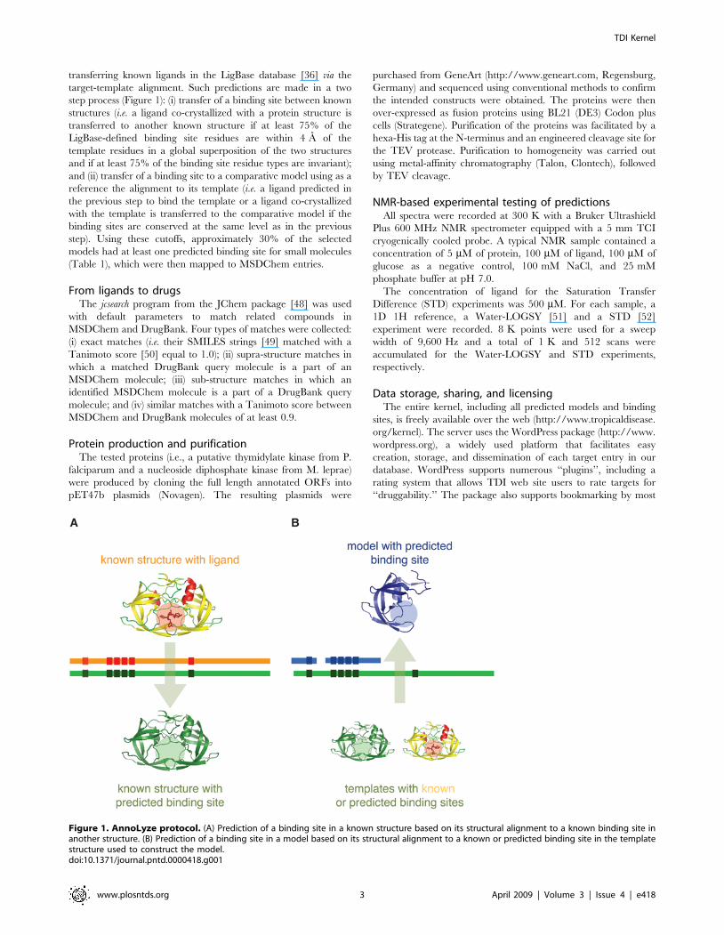
transferring known ligands in the LigBase database [36] via the
target-template alignment. Such predictions are made in a two
step process (Figure 1): (i) transfer of a binding site between known
structures (i.e. a ligand co-crystallized with a protein structure is
transferred to another known structure if at least 75% of the
LigBase-defined binding site residues are within 4 A of the
template residues in a global superposition of the two structures
and if at least 75% of the binding site residue types are invariant);
and (ii) transfer of a binding site to a comparative model using as a
reference the alignment to its template (i.e. a ligand predicted in
the previous step to bind the template or a ligand co-crystallized
with the template is transferred to the comparative model if the
binding sites are conserved at the same level as in the previous
step). Using these cutoffs, approximately 30% of the selected
models had at least one predicted binding site for small molecules
(Table 1), which were then mapped to MSDChem entries.
From ligands to drugsThe jcsearch program from the JChem package [48] was used
with default parameters to match related compounds in
MSDChem and DrugBank. Four types of matches were collected:
(i) exact matches (i.e. their SMILES strings [49] matched with a
Tanimoto score [50] equal to 1.0); (ii) supra-structure matches in
which a matched DrugBank query molecule is a part of an
MSDChem molecule; (iii) sub-structure matches in which an
identified MSDChem molecule is a part of a DrugBank query
molecule; and (iv) similar matches with a Tanimoto score between
MSDChem and DrugBank molecules of at least 0.9.
Protein production and purificationThe tested proteins (i.e., a putative thymidylate kinase from P.
falciparum and a nucleoside diphosphate kinase from M. leprae)
were produced by cloning the full length annotated ORFs into
pET47b plasmids (Novagen). The resulting plasmids were
purchased from GeneArt (http://www.geneart.com, Regensburg,
Germany) and sequenced using conventional methods to confirm
the intended constructs were obtained. The proteins were then
over-expressed as fusion proteins using BL21 (DE3) Codon plus
cells (Strategene). Purification of the proteins was facilitated by a
hexa-His tag at the N-terminus and an engineered cleavage site for
the TEV protease. Purification to homogeneity was carried out
using metal-affinity chromatography (Talon, Clontech), followed
by TEV cleavage.
NMR-based experimental testing of predictionsAll spectra were recorded at 300 K with a Bruker Ultrashield
Plus 600 MHz NMR spectrometer equipped with a 5 mm TCI
cryogenically cooled probe. A typical NMR sample contained a
concentration of 5 mM of protein, 100 mM of ligand, 100 mM of
glucose as a negative control, 100 mM NaCl, and 25 mM
phosphate buffer at pH 7.0.
The concentration of ligand for the Saturation Transfer
Difference (STD) experiments was 500 mM. For each sample, a
1D 1H reference, a Water-LOGSY [51] and a STD [52]
experiment were recorded. 8 K points were used for a sweep
width of 9,600 Hz and a total of 1 K and 512 scans were
accumulated for the Water-LOGSY and STD experiments,
respectively.
Data storage, sharing, and licensingThe entire kernel, including all predicted models and binding
sites, is freely available over the web (http://www.tropicaldisease.
org/kernel). The server uses the WordPress package (http://www.
wordpress.org), a widely used platform that facilitates easy
creation, storage, and dissemination of each target entry in our
database. WordPress supports numerous ‘‘plugins’’, including a
rating system that allows TDI web site users to rate targets for
‘‘druggability.’’ The package also supports bookmarking by most
Figure 1. AnnoLyze protocol. (A) Prediction of a binding site in a known structure based on its structural alignment to a known binding site inanother structure. (B) Prediction of a binding site in a model based on its structural alignment to a known or predicted binding site in the templatestructure used to construct the model.doi:10.1371/journal.pntd.0000418.g001
TDI Kernel
www.plosntds.org 3 April 2009 | Volume 3 | Issue 4 | e418
web-based social networks. In particular, each of the TDI kernel’s
target pages includes a ‘‘blog it’’ button that allows registered users
of The Synaptic Leap (TSL, http://www.thesynapticleap.org) to
post TDI entries directly into the TSL discussion panels. TSL is
our web-based ‘‘collaboratory’’ portal that is designed to host open
source drug discovery projects in much the same way SourceForge
hosts software collaborations.
The TDI kernel is fully searchable and downloadable through
our Web site (http://www.tropicaldisease.org/kernel/). Options
include direct downloads of individually requested targets, pre-
defined sets for each of our ten target genomes, and user-defined
batch downloads. Additionally, all our predictions are available as
supporting information files to this article (Datasets S1, S2, S3, S4).
Users receive the data with no restriction in accordance with the
Science Commons protocol for implementing open access data
[53] that was designed to embody normal academic attribution
norms and facilitate tracking of work based on the kernel. While
our predictions are in the public domain, some of the drugs used in
our predictions might be subject to patents.
Results
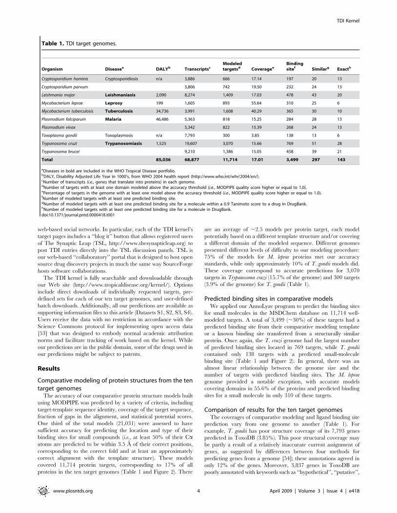
Comparative modeling of protein structures from the tentarget genomes
The accuracy of our comparative protein structure models built
using MODPIPE was predicted by a variety of criteria, including
target-template sequence identity, coverage of the target sequence,
fraction of gaps in the alignment, and statistical potential scores.
One third of the total models (21,031) were assessed to have
sufficient accuracy for predicting the location and type of their
binding sites for small compounds (i.e., at least 50% of their Caatoms are predicted to be within 3.5 A of their correct positions,
corresponding to the correct fold and at least an approximately
correct alignment with the template structure). These models
covered 11,714 protein targets, corresponding to 17% of all
proteins in the ten target genomes (Table 1 and Figure 2). There
are an average of ,2.5 models per protein target, each model
potentially based on a different template structure and/or covering
a different domain of the modeled sequence. Different genomes
presented different levels of difficulty to our modeling procedure:
75% of the models for M. leprae proteins met our accuracy
standards, while only approximately 10% of T. gondii models did.
These coverage correspond to accurate predictions for 3,070
targets in Trypanosoma cruzi (15.7% of the genome) and 300 targets
(3.9% of the genome) for T. gondii (Table 1).
Predicted binding sites in comparative modelsWe applied our AnnoLyze program to predict the binding sites
for small molecules in the MSDChem database on 11,714 well-
modeled targets. A total of 3,499 (,30%) of these targets had a
predicted binding site from their comparative modeling template
or a known binding site transferred from a structurally similar
protein. Once again, the T. cruzi genome had the largest number
of predicted binding sites located in 769 targets, while T. gondii
contained only 138 targets with a predicted small-molecule
binding site (Table 1 and Figure 2). In general, there was an
almost linear relationship between the genome size and the
number of targets with predicted binding sites. The M. leprae
genome provided a notable exception, with accurate models
covering domains in 55.6% of the proteins and predicted binding
sites for a small molecule in only 310 of these targets.
Comparison of results for the ten target genomesThe coverages of comparative modeling and ligand binding site
prediction vary from one genome to another (Table 1). For
example, T. gondii has poor structure coverage of its 7,793 genes
predicted in ToxoDB (3.85%). This poor structural coverage may
be partly a result of a relatively inaccurate current assignment of
genes, as suggested by differences between four methods for
predicting genes from a genome [54]; these annotations agreed in
only 12% of the genes. Moreover, 3,837 genes in ToxoDB are
poorly annotated with keywords such as ‘‘hypothetical’’, ‘‘putative’’,
Table 1. TDI target genomes.
Organism Diseasea DALYb TranscriptscModeledtargetsd Coveragee
Bindingsitef Similarg Exacth
Cryptosporidium hominis Cryptosporidiosis n/a 3,886 666 17.14 197 20 13
Cryptosporidium parvum 3,806 742 19.50 232 24 13
Leishmania major Leishmaniasis 2,090 8,274 1,409 17.03 478 43 20
Mycobacterium leprae Leprosy 199 1,605 893 55.64 310 25 6
Mycobacterium tuberculosis Tuberculosis 34,736 3,991 1,608 40.29 365 30 10
Plasmodium falciparum Malaria 46,486 5,363 818 15.25 284 28 13
Plasmodium vivax 5,342 822 15.39 268 24 13
Toxoplasma gondii Toxoplasmosis n/a 7,793 300 3.85 138 13 6
Trypanosoma cruzi Trypanosomiasis 1,525 19,607 3,070 15.66 769 51 28
Trypanosoma brucei 9,210 1,386 15.05 458 39 21
Total 85,036 68,877 11,714 17.01 3,499 297 143
aDiseases in bold are included in the WHO Tropical Disease portfolio.bDALY, Disability Adjusted Life Year in 1000’s, from WHO 2004 health report (http://www.who.int/whr/2004/en/).cNumber of transcripts (i.e., genes that translate into proteins) in each genome.dNumber of targets with at least one domain modeled above the accuracy threshold (i.e., MODPIPE quality score higher or equal to 1.0).ePercentage of targets in the genome with at least one model above the accuracy threshold (i.e., MODPIPE quality score higher or equal to 1.0).fNumber of modeled targets with at least one predicted binding site.gNumber of modeled targets with at least one predicted binding site for a molecule within a 0.9 Tanimoto score to a drug in DrugBank.hNumber of modeled targets with at least one predicted binding site for a molecule in DrugBank.doi:10.1371/journal.pntd.0000418.t001
TDI Kernel
www.plosntds.org 4 April 2009 | Volume 3 | Issue 4 | e418
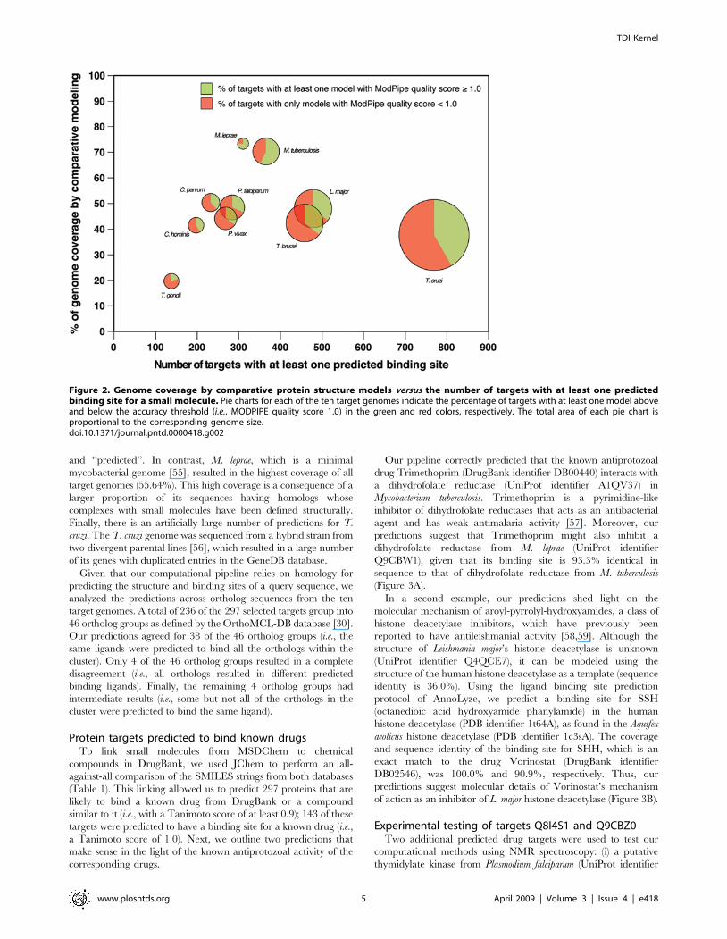
and ‘‘predicted’’. In contrast, M. leprae, which is a minimal
mycobacterial genome [55], resulted in the highest coverage of all
target genomes (55.64%). This high coverage is a consequence of a
larger proportion of its sequences having homologs whose
complexes with small molecules have been defined structurally.
Finally, there is an artificially large number of predictions for T.
cruzi. The T. cruzi genome was sequenced from a hybrid strain from
two divergent parental lines [56], which resulted in a large number
of its genes with duplicated entries in the GeneDB database.
Given that our computational pipeline relies on homology for
predicting the structure and binding sites of a query sequence, we
analyzed the predictions across ortholog sequences from the ten
target genomes. A total of 236 of the 297 selected targets group into
46 ortholog groups as defined by the OrthoMCL-DB database [30].
Our predictions agreed for 38 of the 46 ortholog groups (i.e., the
same ligands were predicted to bind all the orthologs within the
cluster). Only 4 of the 46 ortholog groups resulted in a complete
disagreement (i.e., all orthologs resulted in different predicted
binding ligands). Finally, the remaining 4 ortholog groups had
intermediate results (i.e., some but not all of the orthologs in the
cluster were predicted to bind the same ligand).
Protein targets predicted to bind known drugsTo link small molecules from MSDChem to chemical
compounds in DrugBank, we used JChem to perform an all-
against-all comparison of the SMILES strings from both databases
(Table 1). This linking allowed us to predict 297 proteins that are
likely to bind a known drug from DrugBank or a compound
similar to it (i.e., with a Tanimoto score of at least 0.9); 143 of these
targets were predicted to have a binding site for a known drug (i.e.,
a Tanimoto score of 1.0). Next, we outline two predictions that
make sense in the light of the known antiprotozoal activity of the
corresponding drugs.
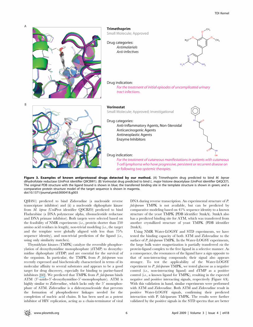
Our pipeline correctly predicted that the known antiprotozoal
drug Trimethoprim (DrugBank identifier DB00440) interacts with
a dihydrofolate reductase (UniProt identifier A1QV37) in
Mycobacterium tuberculosis. Trimethoprim is a pyrimidine-like
inhibitor of dihydrofolate reductases that acts as an antibacterial
agent and has weak antimalaria activity [57]. Moreover, our
predictions suggest that Trimethoprim might also inhibit a
dihydrofolate reductase from M. leprae (UniProt identifier
Q9CBW1), given that its binding site is 93.3% identical in
sequence to that of dihydrofolate reductase from M. tuberculosis
(Figure 3A).
In a second example, our predictions shed light on the
molecular mechanism of aroyl-pyrrolyl-hydroxyamides, a class of
histone deacetylase inhibitors, which have previously been
reported to have antileishmanial activity [58,59]. Although the
structure of Leishmania major’s histone deacetylase is unknown
(UniProt identifier Q4QCE7), it can be modeled using the
structure of the human histone deacetylase as a template (sequence
identity is 36.0%). Using the ligand binding site prediction
protocol of AnnoLyze, we predict a binding site for SSH
(octanedioic acid hydroxyamide phanylamide) in the human
histone deacetylase (PDB identifier 1t64A), as found in the Aquifex
aeolicus histone deacetylase (PDB identifier 1c3sA). The coverage
and sequence identity of the binding site for SHH, which is an
exact match to the drug Vorinostat (DrugBank identifier
DB02546), was 100.0% and 90.9%, respectively. Thus, our
predictions suggest molecular details of Vorinostat’s mechanism
of action as an inhibitor of L. major histone deacetylase (Figure 3B).
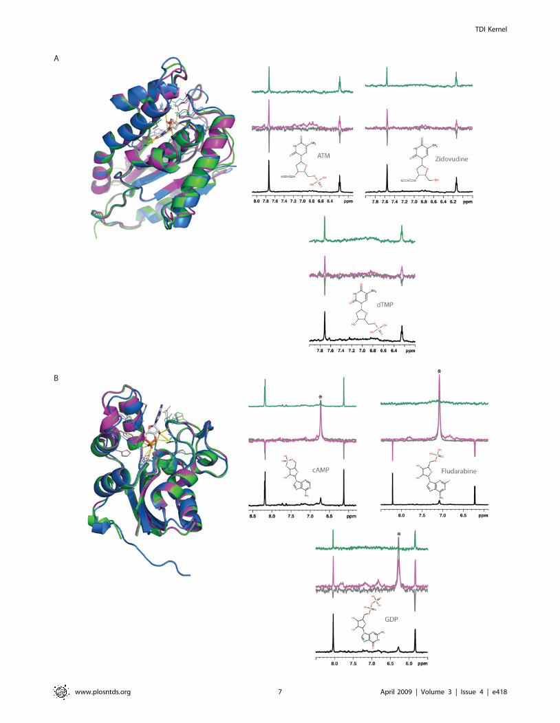
Experimental testing of targets Q8I4S1 and Q9CBZ0Two additional predicted drug targets were used to test our
computational methods using NMR spectroscopy: (i) a putative
thymidylate kinase from Plasmodium falciparum (UniProt identifier
Figure 2. Genome coverage by comparative protein structure models versus the number of targets with at least one predictedbinding site for a small molecule. Pie charts for each of the ten target genomes indicate the percentage of targets with at least one model aboveand below the accuracy threshold (i.e., MODPIPE quality score 1.0) in the green and red colors, respectively. The total area of each pie chart isproportional to the corresponding genome size.doi:10.1371/journal.pntd.0000418.g002
TDI Kernel
www.plosntds.org 5 April 2009 | Volume 3 | Issue 4 | e418
Q8I4S1) predicted to bind Zidovudine (a nucleoside reverse
transcriptase inhibitor) and (ii) a nucleoside diphosphate kinase
from M. leprae (UniProt identifier Q9CBZ0) predicted to bind
Fludarabine (a DNA polymerase alpha, ribonucleotide reductase
and DNA primase inhibitor). Both targets were selected based on
the feasibility of NMR experiments (i.e., protein shorter than 250
amino acid residues in length), non-trivial modeling (i.e., the target
and the template were globally aligned with less than 75%
sequence identity), and non-trivial prediction of the ligand (i.e.,
using only similarity matches).
Thymidylate kinases (TMPK) catalyze the reversible phosphor-
ylation of deoxythymidine monophosphate (dTMP) to deoxythy-
midine diphosphate (dTDP) and are essential for the survival of
the organism. In particular, the TMPK from P. falciparum was
recently expressed and biochemically characterized in terms of its
molecular affinity to several substrates and appears to be a good
target for drug discovery, especially for binding to purine-based
inhibitors [60]. We predicted that TMPK from P. falciparum binds
ATM (39-azido-39-deoxythymidine-59-monophosphate). ATM is
highly similar to Zidovudine, which lacks only the 59 monophos-
phate of ATM. Zidovudine is a dideoxynucleoside that prevents
the formation of phosphodiester linkages needed for the
completion of nucleic acid chains. It has been used as a potent
inhibitor of HIV replication, acting as a chain-terminator of viral
DNA during reverse transcription. An experimental structure of P.
falciparum TMPK is not available, but can be predicted by
comparative modeling based on 41% sequence identity to a known
structure of the yeast TMPK (PDB identifier 3tmkA). 3tmkA also
has a predicted binding site for ATM, which was transferred from
another crystallized structure of yeast TMPK (PDB identifier
2tmkA).
Using NMR Water-LOGSY and STD experiments, we have
tested the binding capacity of both ATM and Zidovudine to the
surface of P. falciparum TMPK. In the Water-LOGSY experiments,
the large bulk water magnetization is partially transferred via the
protein-ligand complex to the free ligand in a selective manner. As
a consequence, the resonances of the ligand have a sign opposite to
that of non-interacting compounds; their signal also appears
stronger. To test the applicability of the Water-LOGSY
experiment to P. falciparum TMPK, we tested glucose as a negative
control (i.e., non-interacting ligand) and dTMP as a positive
control (i.e., a known ligand for TMPK), resulting in the expected
negative and positive interacting signals, respectively (Figure 4A).
With this validation in hand, similar experiments were performed
with ATM and Zidovudine. Both ATM and Zidovudine result in
positive Water-LOGSY signals, confirming their predicted
interaction with P. falciparum TMPK. The results were further
validated by the positive signals in the STD spectra that are better
Figure 3. Examples of known antiprotozoal drugs detected by our method. (A) Trimethoprim drug predicted to bind M. lepraedihydrofolate reductase (UniProt identifier Q9CBW1). (B) Vorinostat drug predicted to bind L. major histone deacetylase (UniProt identifier Q4QCE7).The original PDB structure with the ligand bound is shown in blue; the transferred binding site in the template structure is shown in green; and acomparative protein structure model of the target sequence is shown in magenta.doi:10.1371/journal.pntd.0000418.g003
TDI Kernel
www.plosntds.org 6 April 2009 | Volume 3 | Issue 4 | e418
TDI Kernel
www.plosntds.org 7 April 2009 | Volume 3 | Issue 4 | e418
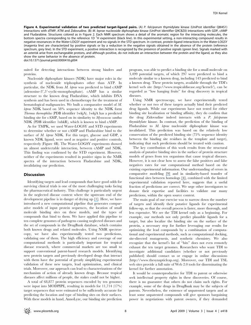
suited for detecting interactions between strong binders and
proteins.
Nucleoside diphosphate kinases (NDK) have major roles in the
synthesis of nucleoside triphosphates other than ATP. In
particular, the NDK from M. leprae was predicted to bind cAMP
(adenosine-39,59-cyclic-monophosphate). cAMP has a similar
structure to the known drug Fludarabine, which inhibits DNA
synthesis and has been used in chemotherapy for the treatment of
hematological malignancies. We built a comparative model of M.
leprae NDK based on 58% sequence identity to the NDK form
Thermus thermophilus (PDB identifier 1wkjA). 1wkjA has a predicted
binding site for cAMP, based on its similarity to Myxococcus xanthus
NDK (PDB identifier 1nhkR), which is known to bind cAMP.
As for TMPK, we used Water-LOGSY and STD experiments
to determine whether or not cAMP and Fludarabine bind to the
surface of M. leprae NDK. For this target, glucose and GDP, a
known NDK ligand, were used as negative and positive controls,
respectively (Figure 4B). The Water-LOGSY experiments showed
an almost undetectable interaction, between cAMP and NDK.
This finding was confirmed by the STD experiment. However,
neither of the experiments resulted in positive signs in the NMR
spectra of the interaction between Fludarabine and NDK,
invalidating our prediction.
Discussion
Identifying targets and lead compounds that have good odds for
surviving clinical trials is one of the most challenging tasks facing
the pharmaceutical industry. This challenge is particularly urgent
in the neglected disease context where the upstream end of the
development pipeline is in danger of drying up [1]. Here, we have
introduced a new computational pipeline that generates compar-
ative models of input protein sequences, the location of small
molecule binding sites on these models, and the types of
compounds that bind to them. We have applied this pipeline to
ten complete genomes of pathogens causing neglected diseases and
the set of compounds in the DrugBank database, which contains
both known drugs and related molecules. Using NMR spectros-
copy, we have also experimentally tested two predictions,
validating one of them. The high efficiency and coverage of our
computational methods is particularly important for tropical
disease research, where commercial markets are too small to
support conventional patent-based research models. Identifying
new protein targets and previously developed drugs that interact
with them have the potential of greatly simplifying experimental
validation of these new targets, lead optimization, and clinical
trials. Moreover, our approach can lead to characterizations of the
mechanism of action of already known drugs. Because tropical
diseases affect millions of people, the stakes could not be higher.
A total of 68,877 protein sequences encoded by ten genomes
were input into MODPIPE, resulting in models for 11,714 (17%)
target sequences that were estimated to be sufficiently accurate for
predicting the location and type of binding sites on their surfaces.
With these models in hand, AnnoLyze, our binding site prediction
program, was able to predict a binding site for a small molecule on
3,499 potential targets, of which 297 were predicted to bind a
molecule similar to a known drug, including 143 predicted to bind
a known drug. These protein targets, available through the TDI’s
kernel web site (http://www.tropicaldisease.org/kernel/), can be
regarded as ‘‘low hanging fruits’’ for drug discovery in tropical
diseases.
Using NMR spectroscopy, we have experimentally tested
whether or not two of these targets actually bind their predicted
drug ligands. While our experiments have not tested for either
binding site localization or binding affinity, they do confirm that
the drug Zidovudine indeed interacts with a P. falciparum
thymidylate kinase. In contrast, the prediction of the binding of
Fludarabine to M. leprae nucleoside diphosphate kinase was
invalidated. This prediction was based on the relatively low
conservation of the predicted binding site (75% sequence identity
between the binding site residues in the template and target),
indicating that such predictions should be treated with caution.
The key contribution of this work results from the structural
analysis of putative binding sites in the surface of protein structure
models of genes from ten organisms that cause tropical diseases.
However, it is not clear how to assess the false positives and false
negatives rates for our computational method based on the
existing experimental information. Our understanding of errors in
comparative modeling [9] and in similarity-based transfer of
functional sites between homologs [4], combined with the limited
experimental validation reported here, suggests that a useful
fraction of predictions are correct. We urge other investigators to
donate their expertise and facilities to validate our many
predictions, within the open source context.
The main goal of our exercise was to narrow down the number
of targets and identify their putative ligands for experimental
follow-up, so that the overall process is faster, more thorough, and
less expensive. We see the TDI kernel only as a beginning. For
example, our methods not only predict plausible ligands for a
target, but also localize the binding site on the surface of the
protein, a necessary step for further leveraging our results for
optimizing the lead compounds by a combination of computa-
tional and experimental methods, such as computational docking,
site-directed mutagenesis, and synthetic chemistry. We also
recognize that the kernel’s list of ‘‘hits’’ does not even remotely
exhaust the ten target genomes. Researchers who want TDI to
investigate additional candidates (whether or not previously
published) should contact us or engage in online discussions
(http://www.thesynapticleap.org). Moreover, our TDI and TSL
web sites provide a full suite of Web 2.0 tools for disseminating the
kernel for further annotation.
It would be counterproductive for TDI to patent or otherwise
seek intellectual property rights in these discoveries. Of course,
there is no guarantee that others do not claim such rights. For
example, some of the drugs in DrugBank may be the subjects of
patents. Nevertheless, the existence of unpatented targets and at
least some unpatented compounds will give sponsors bargaining
power in negotiations with patent owners, if they demanded
Figure 4. Experimental validation of two predicted target-ligand pairs. (A) P. falciparum thymidylate kinase (UniProt identifier Q8I4S1)interactions with dTMP, ATM and Zidovudine. (B) M. leprae nucleoside diphosphate kinase (UniProt identifier Q9CBZ0) interactions with GDP, cAMPand Fludarabine. Structures colored as in Figure 2. Each NMR spectrum shows a detail of the aromatic region for the interacting molecules, thebottom spectra corresponding to the reference 1D 1H experiment (black line). In this experimental setting, a non-interacting compound results innegative resonances in the Water-LOGSY experiment and no signals in the STD spectrum. In contrast, protein-ligand interactions in the Water-LOGSY(magenta line) are characterized by positive signals or by a reduction in the negative signals obtained in the absence of the protein (referencespectrum, grey line). In the STD experiment, a positive interaction is recognized by the presence of positive signals (green line). Signals marked withan asterisk arise from exchangeable protons, and although positive, do not indicate an interaction between the protein and the ligand, as they alsoshow the same behavior in the absence of protein.doi:10.1371/journal.pntd.0000418.g004
TDI Kernel
www.plosntds.org 8 April 2009 | Volume 3 | Issue 4 | e418
excessive royalties. The net result will be to reduce the royalties
that patent owners can charge and sponsors must pay.
Many open source licenses contain ‘‘viral’’ terms, which limit
users’ ability to seek intellectual property of their own. In the case
of drug discovery, however, such strategies are likely to be
expensive and, in some cases, legally dubious [61,62]. Neverthe-
less, these obstacles are not fatal and one can imagine schemes in
which discoveries are embargoed for months or years, so that
access is limited to those who promised not to seek patents of their
own [63]. We have decided against trying to impose a viral
condition on subsequent researchers. First and foremost, open
source requires as many workers, volunteer and commercial, as
possible, implying minimal restrictions on the data, including viral
terms. Second, at least some of the organisms included in the
kernel (e.g., M. tuberculosis) have potential commercial markets large
enough to offset a fraction of sponsors’ R&D costs. Nevertheless, it
is still possible that an unscrupulous corporation, for example,
could try to patent trivial improvements to the kernel. This,
however, seems unlikely in the impoverished world of neglected
disease research, at least for the immediate future. In the
meantime, we prefer to leave the question open until open source
collaboration has been firmly established. That will put the final
responsibility where it belongs – with the volunteers whose labor
and insights we are depending on to turn TDI’s kernel into safe,
effective, and affordable cures.
Supporting Information
Dataset S1 PDF version of the data for selected targets
Found at: doi:10.1371/journal.pntd.0000418.s001 (0.27 MB PDF)
Dataset S2 Tab separated version of the data for selected targets
Found at: doi:10.1371/journal.pntd.0000418.s002 (0.13 MB
TXT)
Dataset S3 Excel version of the data for selected targets
Found at: doi:10.1371/journal.pntd.0000418.s003 (0.36 MB XLS)
Dataset S4 MySQL version of the data for selected targets
Found at: doi:10.1371/journal.pntd.0000418.s004 (0.14 MB
TXT)
Acknowledgments
We are grateful to Bissan Al-Lazikani for help in collecting the target
genome sequences and James McKerrow, Brian Shoichet, and David S.
Roos for helpful discussions. We also thank Rafael Gonzalbes for help
preparing Figure 4.
Author Contributions
Conceived and designed the experiments: APL AS MAMR. Performed the
experiments: LO RJC UP NE MAMR. Analyzed the data: LO RJC UP
NE APL MAMR. Contributed reagents/materials/analysis tools: UP NE
APL MAMR. Wrote the paper: SMM AKR GT MHT APL AS MAMR.
References
1. Nwaka S, Ridley RG (2003) Virtual drug discovery and development for
neglected diseases through public-private partnerships. Nat Rev Drug Discov 2:919–928.
2. Rai A, Reichman JR, Uhlir P, Crossman C (2008) Pathways across the valley of
death: novel intellectual property strategies for accelerating drug discovery.Yale J Health Policy Law Ethics 8: 53–89.
3. Baker D, Sali A (2001) Protein structure prediction and structural genomics.
Science 294: 93–96.
4. Marti-Renom MA, Stuart AC, Fiser A, Sanchez R, Melo F, et al. (2000)Comparative protein structure modeling of genes and genomes. Annu Rev
Biophys Biomol Struct 29: 291–325.
5. Kopp J, Schwede T (2006) The SWISS-MODEL Repository: new features and
functionalities. Nucleic Acids Res 34: D315–D318.
6. Pieper U, Eswar N, Davis FP, Braberg H, Madhusudhan MS, et al. (2006)MODBASE: a database of annotated comparative protein structure models and
associated resources. Nucleic Acids Res 34: D291–D295.
7. Tramontano A (2006) The role of molecular modelling in biomedical research.FEBS Lett 580: 2928–2934.
8. Aguero F, Al-Lazikani B, Aslett M, Berriman M, Buckner FS, et al. (2008)
Genomic-scale prioritization of drug targets: the TDR Targets database. NatRev Drug Discov 7: 900–907.
9. Marti-Renom MA, Rossi A, Al-Shahrour F, Davis FP, Pieper U, et al. (2007)
The AnnoLite and AnnoLyze programs for comparative annotation of proteinstructures. BMC Bioinformatics 8: S4.
10. Watson JD, Sanderson S, Ezersky A, Savchenko A, Edwards A, et al. (2007)
Towards fully automated structure-based function prediction in structural
genomics: a case study. J Mol Biol 367: 1511–1522.
11. Rester U (2008) From virtuality to reality—virtual screening in lead discoveryand lead optimization: a medicinal chemistry perspective. Curr Opin Drug
Discov Devel 11: 559–568.
12. Huey R, Morris GM, Olson AJ, Goodsell DS (2007) A semiempirical free energyforce field with charge-based desolvation. J Comput Chem 28: 1145–
1152.
13. Leach AR, Shoichet BK, Peishoff CE (2006) Prediction of protein-ligandinteractions. Docking and scoring: successes and gaps. J Med Chem 49:
5851–5855.
14. Noble ME, Endicott JA, Johnson LN (2004) Protein kinase inhibitors: insightsinto drug design from structure. Science 303: 1800–1805.
15. de Paulis T (2007) Drug evaluation: PRX-00023, a selective 5-HT1A receptor
agonist for depression. Curr Opin Investig Drugs 8: 78–86.
16. Maurer SM, Rai A, Sali A (2004) Finding cures for tropical diseases: is open
source an answer? PLoS Med 1: e56. doi:10.1371/journal.pmed.0010056.
17. Munos B (2006) Can open-source R&D reinvigorate drug research? Nat Rev
Drug Discov 5: 723–729.
18. Hopkins AL, Witty MJ, Nwaka S (2007) Mission possible. Nature 449: 166–169.
19. Nwaka S, Hudson A (2006) Innovative lead discovery strategies for tropical
diseases. Nat Rev Drug Discov 5: 941–955.
20. Nwaka S (2005) Drug discovery and beyond: the role of public-private
partnerships in improving access to new malaria medicines. Trans R Soc TropMed Hyg 99: S20–S29.
21. Nwaka S, Riopel L, Ubben D, Craft JC (2004) Medicines for Malaria Venturenew developments in antimalarials. Travel Med Infect Dis 2: 161–170.
22. Kepler T, Marti-Renom MA, Maurer SM, Rai AK, Taylor G, et al. (2006)Open source research—the power of us. Aust J Chem 59: 291–294.
23. Sachs J (1999) Helping the world’s poorest. Economist 352(8132): 17–20.
24. Kremer M, Glennerster R (2004) Strong Medicine: Creating Incentives forPharmaceutical Research on Neglected Diseases. Princeton: Princeton Univer-
sity Press.
25. Singh S (2008) India takes an open source approach to drug discovery. Cell 133:
201–203.
26. Matter A, Keller TH (2008) Impact of non-profit organizations on drug
discovery: opportunities, gaps, solutions. Drug Discov Today 13: 347–352.
27. Eswar N, John B, Mirkovic N, Fiser A, Ilyin VA, et al. (2003) Tools for
comparative protein structure modeling and analysis. Nucleic Acids Res 31:3375–3380.
28. Heiges M, Wang H, Robinson E, Aurrecoechea C, Gao X, et al. (2006)CryptoDB: a Cryptosporidium bioinformatics resource update. Nucleic Acids
Res 34: D419–D422.
29. Hertz-Fowler C, Peacock CS, Wood V, Aslett M, Kerhornou A, et al. (2004)
GeneDB: a resource for prokaryotic and eukaryotic organisms. Nucleic AcidsRes 32: D339–D343.
30. Chen F, Mackey AJ, Stoeckert CJ Jr, Roos DS (2006) OrthoMCL-DB: queryinga comprehensive multi-species collection of ortholog groups. Nucleic Acids Res
34: D363–D368.
31. Cole ST, Brosch R, Parkhill J, Garnier T, Churcher C, et al. (1998) Deciphering
the biology of Mycobacterium tuberculosis from the complete genome sequence.Nature 393: 537–544.
32. Stoeckert CJ Jr, Fischer S, Kissinger JC, Heiges M, Aurrecoechea C, et al. (2006)PlasmoDB v5: new looks, new genomes. Trends Parasitol 22: 543–546.
33. Gajria B, Bahl A, Brestelli J, Dommer J, Fischer S, et al. (2008) ToxoDB: an
integrated Toxoplasma gondii database resource. Nucleic Acids Res 36:
D553–D556.
34. Wu CH, Apweiler R, Bairoch A, Natale DA, Barker WC, et al. (2006) TheUniversal Protein Resource (UniProt): an expanding universe of protein
information. Nucleic Acids Res 34: D187–D191.
35. Marti-Renom MA, Pieper U, Madhusudhan MS, Rossi A, Eswar N, et al. (2007)
DBAli tools: mining the protein structure space. Nucleic Acids Res 35:
W393–W397.
36. Stuart AC, Ilyin VA, Sali A (2002) LigBase: a database of families of alignedligand binding sites in known protein sequences and structures. Bioinformatics
18: 200–201.
37. Golovin A, Dimitropoulos D, Oldfield T, Rachedi A, Henrick K (2005)
MSDsite: a database search and retrieval system for the analysis and viewing ofbound ligands and active sites. Proteins 58: 190–199.
TDI Kernel
www.plosntds.org 9 April 2009 | Volume 3 | Issue 4 | e418
38. Wishart DS, Knox C, Guo AC, Cheng D, Shrivastava S, et al. (2008) DrugBank:
a knowledgebase for drugs, drug actions and drug targets. Nucleic Acids Res 36:D901–D906.
39. Eswar N, Webb B, Pieper U, Shen M-Y, Eramian D, et al. (2008) ModPipe: a
large-scale protein structure modeling pipeline for the genomic era. Submitted.40. Sali A, Blundell TL (1993) Comparative protein modelling by satisfaction of
spatial restraints. J Mol Biol 234: 779–815.41. Smith TF, Waterman MS (1981) Identification of common molecular
subsequences. J Mol Biol 147: 195–197.
42. Altschul SF, Madden TL, Schaffer AA, Zhang J, Zhang Z, et al. (1997) GappedBLAST and PSI-BLAST: a new generation of protein database search
programs. Nucleic Acids Res 25: 3389–3402.43. Eswar N, Webb B, Marti-Renom MA, Madhusudhan MS, Eramian D, et al.
(2006) Comparative protein structure modeling using Modeller. Curr ProtocBioinformatics Chapter 5: Unit 5.6.
44. Marti-Renom MA, Madhusudhan MS, Sali A (2004) Alignment of protein
sequences by their profiles. Protein Sci 13: 1071–1087.45. Shen MY, Sali A (2006) Statistical potential for assessment and prediction of
protein structures. Protein Sci 15: 2507–2524.46. Eramian D, Shen MY, Devos D, Melo F, Sali A, et al. (2006) A composite score
for predicting errors in protein structure models. Protein Sci 15: 1653–1666.
47. Melo F, Sanchez R, Sali A (2002) Statistical potentials for fold assessment.Protein Sci 11: 430–448.
48. Csizmadia F (2000) JChem: Java applets and modules supporting chemicaldatabase handling from web browsers. J Chem Inf Comput Sci 40: 323–324.
49. Weininger D, Weininger A, Weininger JL (1989) SMILES. 2. algorithm forgeneration of uniques SMILES notation. J Chem Inf Comput Sci 29: 97–101.
50. Gower JC (1971) A general coefficient of similarity and some of its properties.
Biometrics 27: 857–871.51. Dalvit C, Pevarello P, Tato M, Veronesi M, Vulpetti A, et al. (2000)
Identification of compounds with binding affinity to proteins via magnetizationtransfer from bulk water. J Biomol NMR 18: 65–68.
52. Meyer B, Peters T (2003) NMR spectroscopy techniques for screening and
identifying ligand binding to protein receptors. Angew Chem Int Ed 42:864–890.
53. Creative Commons (last accessed February 2009) Protocol for implementing
open access data. http://sciencecommons.org/projects/publishing/open-access-data-protocol/.
54. Dybas JM, Madrid-Aliste CJ, Che FY, Nieves E, Rykunov D, et al. (2008)Computational analysis and experimental validation of gene predictions in
Toxoplasma gondii. PLoS ONE 3: e3899. doi:10.1371/journal.pone.0003899.
55. Vissa VD, Brennan PJ (2001) The genome of Mycobacterium leprae: a minimalmycobacterial gene set. Genome Biol 2: REVIEWS1023.
56. El-Sayed NM, Myler PJ, Bartholomeu DC, Nilsson D, Aggarwal G, et al. (2005)The genome sequence of Trypanosoma cruzi, etiologic agent of Chagas disease.
Science 309: 409–415.57. Rosowsky A, Papoulis AT, Forsch RA, Queener SF (1999) Synthesis and
antiparasitic and antitumor activity of 2, 4-diamino-6-(arylmethyl)-5,6,7,8-
tetrahydroquinazoline analogues of piritrexim. J Med Chem 42: 1007–1017.58. Darkin-Rattray SJ, Gurnett AM, Myers RW, Dulski PM, Crumley TM, et al.
(1996) Apicidin: a novel antiprotozoal agent that inhibits parasite histonedeacetylase. Proc Natl Acad Sci U S A 93: 13143–13147.
59. Mai A, Cerbara I, Valente S, Massa S, Walker LA, et al. (2004) Antimalarial and
antileishmanial activities of aroyl-pyrrolyl-hydroxyamides, a new class of histonedeacetylase inhibitors. Antimicrob Agents Chemother 48: 1435–1436.
60. Kandeel M, Kitade Y (2008) Molecular characterization, heterologousexpression and kinetic analysis of recombinant Plasmodium falciparum
thymidylate kinase. J Biochem 144: 245–250.61. Boettinger S, Burk DL (2004) Open source patenting. J Int Biotechnol Law 1:
221–231.
62. Feldman RC (2004) The open source biotechnology movement: is it patentmisuse? Minn J Law Sci Technol 6: 117–166.
63. Maurer SM (2008) Open source drug discovery: finding a niche (or maybeseveral). UMKC Law Rev 76: 405–435.
TDI Kernel
www.plosntds.org 10 April 2009 | Volume 3 | Issue 4 | e418