A Human Blood-Brain Barrier Transcytosis Assay Reveals Antibody Transcytosis Influenced by...
11
A Human Blood-Brain Barrier Transcytosis Assay Reveals Antibody Transcytosis Influenced by pH-Dependent Receptor Binding Hadassah Sade 1 , Claudia Baumgartner 1 , Adrian Hugenmatter 2 , Ekkehard Moessner 2 , Per-Ola Freskga ˚rd 3 , Jens Niewoehner 1 * 1 Large Molecule Research, Pharma Research and Early Development (pRED), Roche, Penzberg, Germany, 2 Large Molecule Research, Pharma Research and Early Development (pRED), Roche, Schlieren, Switzerland, 3 Neuroscience Discovery and Translation Area, Pharma Research and Early Development (pRED), F. Hoffmann-La Roche, Basel, Switzerland Abstract We have adapted an in vitro model of the human blood-brain barrier, the immortalized human cerebral microvascular endothelial cells (hCMEC/D3), to quantitatively measure protein transcytosis. After validating the receptor-mediated transport using transferrin, the system was used to measure transcytosis rates of antibodies directed against potential brain shuttle receptors. While an antibody to the insulin-like growth factor 1 receptor (IGF1R) was exclusively recycled to the apical compartment, the fate of antibodies to the transferrin receptor (TfR) was determined by their relative affinities at extracellular and endosomal pH. An antibody with reduced affinity at pH5.5 showed significant transcytosis, while pH- independent antibodies of comparable affinities at pH 7.4 remained associated with intracellular vesicular compartments and were finally targeted for degradation. Citation: Sade H, Baumgartner C, Hugenmatter A, Moessner E, Freskga ˚rd P-O, et al. (2014) A Human Blood-Brain Barrier Transcytosis Assay Reveals Antibody Transcytosis Influenced by pH-Dependent Receptor Binding. PLoS ONE 9(4): e96340. doi:10.1371/journal.pone.0096340 Editor: Felix Schlachetzki, University of Regensburg, Germany Received January 9, 2014; Accepted April 7, 2014; Published April 30, 2014 Copyright: ß 2014 Sade et al. This is an open-access article distributed under the terms of the Creative Commons Attribution License, which permits unrestricted use, distribution, and reproduction in any medium, provided the original author and source are credited. Funding: The work was funded by Roche. The funder provided support in the form of salaries for all authors, but did not have any additional role in the study design, data collection and analysis, decision to publish, or preparation of the manuscript. The specific roles of these authors are articulated in the ‘author contributions’ section. Competing Interests: The authors have read the journal’s policy and have the following conflicts: All authors are paid employees of Roche affiliates, whose company funded this study. There are no products in development or marketed products to declare. Part of the data described in the paper are included in the patent application: "Method and Constructs for the pH Dependent Passage of the Blood-brain-barrier", US20120282176. This does not alter the authors’ adherence to all the PLOS ONE policies on sharing data and materials. * E-mail: [email protected] Introduction Despite decades of intensive research in the labs of academic institutions and the pharmaceutical industry, the blood-brain barrier has remained a significant hurdle for treatment of CNS diseases with growing unmet medical need [1]. Limiting brain access to small, predominantly hydrophobic molecules, this barrier is especially insurmountable to therapeutic proteins. Although antibodies are in clinical development for several CNS indications, brain exposure of these potential medicines is low (,0.1%) and may in many cases be insufficient for therapeutic efficacy. The most important physiological entry route for proteins into the brain is through receptor-mediated transcytosis (RMT), the exploitation of which has already been proposed for the transport of biologics into the brain [2]. However, the pathways and sorting mechanisms of transcytosis in blood-brain barrier endothelial cells are poorly understood, preventing the targeted generation of ‘‘brain shuttle’’ molecules. Another way to further our under- standing on the molecular properties predisposing a protein, or more specifically an antibody, to efficient BBB passage, would be a robust in vitro transcytosis assay, enabling the screening of many antibodies and correlating their transcytosis capacity with other molecular properties like receptor specificity or affinity. Although many in vitro transcytosis assays have been described in the literature (for review see [3]), published data are often not in agreement with the calculated transcytosis capacity of brain endothelial cells, and may therefore rather represent paracellular flux than transcytosis. Additional requirements for a transcytosis assay to generate predictive data for clinical candidate develop- ment are the use of a human cellular system and robust, reproducible assay conditions with little inter-assay variability. We sought to establish a reliable in vitro model system of transcytosis using the hCMEC/D3 immortalized human brain endothelial cell line [3,4]. Although trans-endothelial electrical resistance (TEER) values indicate high paracellular flux of the confluent hCMEC/D3 cell layer, the cell line expresses endothelial specific and tight junction markers [4] and functional drug transporters [5] and has been successfully utilized as a surrogate for primary human brain endothelial cells in permeability studies [6], interaction of immune cells and pathogens at the BBB interface [7] or integrity of the BBB in neurodegenerative diseases [8,9]. Indeed, the cell line has also been used to assess the transport of peptides across the endothelial cell monolayer albeit mostly with insufficient control of paracellular flux [10,11]. We chose the transferrin (Tf): transferrin receptor (TfR) model system to validate our assay. The transferrin receptor (TfR) represents a prototypical receptor for RMT; its expression and function in capillary endothelial cells has been thoroughly investigated [12– PLOS ONE | www.plosone.org 1 April 2014 | Volume 9 | Issue 4 | e96340
-
Upload
independent -
Category
Documents
-
view
0 -
download
0
Transcript of A Human Blood-Brain Barrier Transcytosis Assay Reveals Antibody Transcytosis Influenced by...

A Human Blood-Brain Barrier Transcytosis Assay RevealsAntibody Transcytosis Influenced by pH-DependentReceptor BindingHadassah Sade1, Claudia Baumgartner1, Adrian Hugenmatter2, Ekkehard Moessner2, Per-Ola Freskgard3,
Jens Niewoehner1*
1 Large Molecule Research, Pharma Research and Early Development (pRED), Roche, Penzberg, Germany, 2 Large Molecule Research, Pharma Research and Early
Development (pRED), Roche, Schlieren, Switzerland, 3 Neuroscience Discovery and Translation Area, Pharma Research and Early Development (pRED), F. Hoffmann-La
Roche, Basel, Switzerland
Abstract
We have adapted an in vitro model of the human blood-brain barrier, the immortalized human cerebral microvascularendothelial cells (hCMEC/D3), to quantitatively measure protein transcytosis. After validating the receptor-mediatedtransport using transferrin, the system was used to measure transcytosis rates of antibodies directed against potential brainshuttle receptors. While an antibody to the insulin-like growth factor 1 receptor (IGF1R) was exclusively recycled to theapical compartment, the fate of antibodies to the transferrin receptor (TfR) was determined by their relative affinities atextracellular and endosomal pH. An antibody with reduced affinity at pH5.5 showed significant transcytosis, while pH-independent antibodies of comparable affinities at pH 7.4 remained associated with intracellular vesicular compartmentsand were finally targeted for degradation.
Citation: Sade H, Baumgartner C, Hugenmatter A, Moessner E, Freskgard P-O, et al. (2014) A Human Blood-Brain Barrier Transcytosis Assay Reveals AntibodyTranscytosis Influenced by pH-Dependent Receptor Binding. PLoS ONE 9(4): e96340. doi:10.1371/journal.pone.0096340
Editor: Felix Schlachetzki, University of Regensburg, Germany
Received January 9, 2014; Accepted April 7, 2014; Published April 30, 2014
Copyright: � 2014 Sade et al. This is an open-access article distributed under the terms of the Creative Commons Attribution License, which permitsunrestricted use, distribution, and reproduction in any medium, provided the original author and source are credited.
Funding: The work was funded by Roche. The funder provided support in the form of salaries for all authors, but did not have any additional role in the studydesign, data collection and analysis, decision to publish, or preparation of the manuscript. The specific roles of these authors are articulated in the ‘authorcontributions’ section.
Competing Interests: The authors have read the journal’s policy and have the following conflicts: All authors are paid employees of Roche affiliates, whosecompany funded this study. There are no products in development or marketed products to declare. Part of the data described in the paper are included in thepatent application: "Method and Constructs for the pH Dependent Passage of the Blood-brain-barrier", US20120282176. This does not alter the authors’adherence to all the PLOS ONE policies on sharing data and materials.
* E-mail: [email protected]
Introduction
Despite decades of intensive research in the labs of academic
institutions and the pharmaceutical industry, the blood-brain
barrier has remained a significant hurdle for treatment of CNS
diseases with growing unmet medical need [1]. Limiting brain
access to small, predominantly hydrophobic molecules, this barrier
is especially insurmountable to therapeutic proteins. Although
antibodies are in clinical development for several CNS indications,
brain exposure of these potential medicines is low (,0.1%) and
may in many cases be insufficient for therapeutic efficacy. The
most important physiological entry route for proteins into the
brain is through receptor-mediated transcytosis (RMT), the
exploitation of which has already been proposed for the transport
of biologics into the brain [2]. However, the pathways and sorting
mechanisms of transcytosis in blood-brain barrier endothelial cells
are poorly understood, preventing the targeted generation of
‘‘brain shuttle’’ molecules. Another way to further our under-
standing on the molecular properties predisposing a protein, or
more specifically an antibody, to efficient BBB passage, would be a
robust in vitro transcytosis assay, enabling the screening of many
antibodies and correlating their transcytosis capacity with other
molecular properties like receptor specificity or affinity. Although
many in vitro transcytosis assays have been described in the
literature (for review see [3]), published data are often not in
agreement with the calculated transcytosis capacity of brain
endothelial cells, and may therefore rather represent paracellular
flux than transcytosis. Additional requirements for a transcytosis
assay to generate predictive data for clinical candidate develop-
ment are the use of a human cellular system and robust,
reproducible assay conditions with little inter-assay variability.
We sought to establish a reliable in vitro model system of
transcytosis using the hCMEC/D3 immortalized human brain
endothelial cell line [3,4]. Although trans-endothelial electrical
resistance (TEER) values indicate high paracellular flux of the
confluent hCMEC/D3 cell layer, the cell line expresses endothelial
specific and tight junction markers [4] and functional drug
transporters [5] and has been successfully utilized as a surrogate
for primary human brain endothelial cells in permeability studies
[6], interaction of immune cells and pathogens at the BBB
interface [7] or integrity of the BBB in neurodegenerative diseases
[8,9]. Indeed, the cell line has also been used to assess the
transport of peptides across the endothelial cell monolayer albeit
mostly with insufficient control of paracellular flux [10,11]. We
chose the transferrin (Tf): transferrin receptor (TfR) model system
to validate our assay. The transferrin receptor (TfR) represents a
prototypical receptor for RMT; its expression and function in
capillary endothelial cells has been thoroughly investigated [12–
PLOS ONE | www.plosone.org 1 April 2014 | Volume 9 | Issue 4 | e96340

14] and data from [15–17] strongly support the transcytosis of
diferric transferrin across the endothelial cell and release into the
brain parenchyma. Endosomal acidification after the internaliza-
tion of the Tf-TfR complex leads to release of iron from the ligand,
but apo-transferrin stays bound to the receptor until the complex is
transported to the plasma membrane, where it is released due to
poor binding affinity to the receptor at neutral pH.
We have successfully adapted the ‘‘pulse-chase’’ method
described by Raub and Newton to eliminate experimental artifacts
caused by the poor TEER and increased paracellular flux inherent
to this cell line. We provide evidence for transcytosis of transferrin
and show that co-culture with astrocytes does not influence uptake
or transcytosis of the ligand. We have developed highly sensitive
ELISAs for the detection of human, mouse and hamster IgG in
picogram quantities, which would enable radiolabel-free evalua-
tion of antibodies for transcytotic potential in a medium through-
put format. We describe the transport potential of antibodies
against TfR as well as other potential transcytosis receptors tested
in the assay. While an antibody against the IGF1 receptor
(IGR1R) was exclusively recycled to the apical compartment,
certain anti-TfR antibodies were successfully transported across
the endothelial cell layer, while others were targeted for
degradation in intracellular compartments. Interestingly, we found
a correlation between the transcytosis capacity of the anti-
transferrin receptor antibodies and pH-dependence of the
receptor-antibody interaction, identifying a potential new mech-
anism for the enhancement of transcytosis.
Materials and Methods
AntibodiesThe cDNAs of heavy and light chains of antibody 128.1
(described in WO93/10819 and [18]) were cloned into
pcDNA3.1-derived expression vectors and the antibody was
expressed into the culture medium of HEK293F cells after
transient transfection of both vectors according to a standard
protocol (#K9000-01, Invitrogen, Frankfurt, Germany). The
antibody was purified over a protein-A column and radiolabelled
at Perkin Elmer (Rodgau, Germany). IGF-1R mAb R1507 is
described in US7572897 and [19]. Antibody MEM-189 was
obtained from Biozol (Eching, Germany), MEM-75 and 13E4
from Abcam (Cambridge, UK), M-A712 from BD Biosciences
(Heidelberg, Germany) and LT-71 from Hytest (Turku, Finland).
Cell CulturehCMEC/D3 cells, a brain endothelial cell line immortalized by
transduction with hTERT and SV40 large T [4] were obtained
from Pierre-Olivier Couraud under license (INSERM, Paris,
France). Medium and supplements for hCMEC/D3 and primary
human astrocytes were obtained from Lonza (Verviers, Belgium).
MDCK and rat C6 media and components were obtained from
Invitrogen.
Primary human astrocytes (passages 2–4) were obtained from
Cell Systems (Troisdorf, Germany) and cultured in ABM medium
fully complemented with the AGM SingleQuots kit. Rat C6
Glioma cells from ATCC were cultured in DMEM-F12 contain-
ing 5% heat inactivated FBS, 10% horse serum and 2 mM L-
Glutamine. MDCK cells from ATCC were cultured in MEM
containing 10% fetal bovine serum, 2 mM L-glutamine, 1 mM
sodium pyruvate, and 1500 mg/L sodium bicarbonate.
hCMEC/D3 cells (passages 26–29) were cultured to confluence
on collagen (Sigma, Schnelldorf, Germany) coated coverslips
(microscopy) in EBM2 medium containing 2.5% FBS, quarter of
the supplied growth factors and fully complemented with supplied
hydrocortisone, gentamycin and ascorbic acid.
For all transcytosis assays, high density pore (16108 pores/cm2)
PET membrane filter inserts (0.4 mm, 12 mm diameter; Millipore,
Schwalbach, Germany) were used in 12-well cell culture plates
(Corning, Amsterdam, Netherlands). Optimum media volumes
were calculated to be 400 ml and 1600 ml for apical and basolateral
chambers respectively. Apical chambers of Millicell hanging filter
Figure 1. Description of the transcytosis assay. hCMEC/D3 cells grown to confluence on collagen and fibronectin coated membrane filterinserts (and serum starved for 1 h at 37uC before assay for experiments with 125I-transferrin) were incubated apically with the ligand or the differentantibodies for 60 min at 37uC. After 1 hr, media from the apical and basolateral chambers were collected to assess paracellular flux following whichthe luminal and abluminal membranes of the monolayer were washed four times with medium at RT and the washes monitored to determineefficiency of removing unbound antibody. The filters containing cells were transferred to a fresh plate containing pre-warmed medium and cells werechased up to the desired time points at 37uC or on ice and at these different time points, the ligand or antibody associated with cells or in the mediafrom the apical and basolateral chambers was analyzed in a gamma counter or assessed by IgG ELISA.doi:10.1371/journal.pone.0096340.g001
Human In-Vitro BBB Assay for Antibody Transcytosis
PLOS ONE | www.plosone.org 2 April 2014 | Volume 9 | Issue 4 | e96340

Human In-Vitro BBB Assay for Antibody Transcytosis
PLOS ONE | www.plosone.org 3 April 2014 | Volume 9 | Issue 4 | e96340
inserts were coated with Rat tail Collagen 1 (7.5 mg/cm2; BD
Biosciences) followed by Fibronectin (5 mg/ml; Sigma) each
incubation lasting for 1 hr at RT. hCMEC/D3 cells were grown
to confluent monolayers (,26105 cells/cm2) for 10-12 days in
EMB2 medium. Empty filters coated with collagen/fibronectin
were blocked in PBS containing 1% BSA o/n before the assay and
then calibrated for at least 1 h in EBM2 before the assay. For
generating endothelial-astrocyte co-cultures, the basolateral side of
the filter inserts and TC plastic were coated with 10 mg/ml poly-d-
lysine (Sigma) o/n at RT. The solution was aspirated, TC plastic
dried for 2 h at RT and the coated surfaces were rinsed 3 6with
PBS before astrocytes were seeded at 16103 in 200 ml on the
bottom side of an inverted filter insert. Cells were allowed to
adhere for 15 min at 37uC and the insert was then placed in a 12-
well cell culture plate with the respective volumes of medium in
apical and basolateral chambers. For culture of astrocytes in a
culture plate, 16103 cells were plated in 1600 ml. After 48 hours
the filters were processed for culturing hCMEC/D3 cells as
described above. Experiments with the co-culture model were
performed 10–12 days after hCMEC/D3 cells were seeded.
Transcytosis assays
Transferrin Transcytosis Assays. 125I-Tfn was obtained
from Perkin Elmer. The entire assay was performed in serum free
EBM2 medium which was otherwise reconstituted as described.
On day of the assay, cells were serum starved for 60 min to deplete
Tfn. Filter inserts with or without cells were incubated apically
with radiolabelled ligand for 1 h at 37uC following which the
entire apical and basolateral volumes (referred to as values of stock
post loading) were collected. Paracellular flux and stability of the
radio-iodinated ligand were calculated from these values. The
monolayers were washed at RT in serum free medium apically
(400 ml) and basolaterally (1600 ml) 3 x, 3–5 min each. All the
washes were collected to monitor efficiency of removal of the
unbound ligand. Prewarmed medium was added to the apical
chamber and the filter insert transferred to a fresh 12 well plate
(blocked o/n with PBS containing 1% BSA) containing ml pre-
warmed medium. At this point, filters with or without cells were
lysed in 500 ml RIPA buffer in order to determine specific ligand
uptake. The remaining filters were incubated at 37uC or at 4uCand samples collected at various time points to determine apical
and/or basolateral release of ligand. Intact and degraded Tfn was
assessed using TCA and AgNO3 precipitation as follows: Media
and cell lysates were centrifuged in the presence of trichloroacetic
acid and carrier protein (BSA) at 1100 6 g for 10 min. Intact
protein is associated with the pellet, degraded protein and free
iodine remains in the supernatant, which is then centrifuged in the
presence of AgNO3 at 1100 6g for 10 min to separate degraded
protein (supernatant) from free iodine (AgI precipitate). For each
time point, data was generated from two empty filter inserts and
three inserts with cell cultures.
Antibody Transcytosis Assays. The entire assay was
performed in complete EBM2 medium and protocol similar to
that of the ligand based assays except for the detection of IgG in
transcytosis assay supernatants by ELISA. The entire procedure
was performed at RT and Biotek ELx405 was used to perform the
wash steps. Briefly, a 384 well plate was coated with 30 ml/well of
1 mg/ml anti-human/mouse-IgG, Fcc-specific (Jackson Immunor-
esearch, Newmarket, England) in PBS without Ca2+ and Mg2+(Invitrogen) for 2 h followed by an hour’s incubation in blocking
buffer (Roche; PBS containing 1% BSA or 1% Crotein C for
human and mouse IgG assays respectively). Serially diluted
samples from the transcytosis assay and standard concentrations
of the antibody used in the transcytosis assay were added to the
plate and incubated for 2 h. After four washes, 30 ml/well of
50 ng/ml anti-human/mouse-F (ab) 2-Biotin in blocking buffer
was added and incubated for a further 2 h. Following 6 washes,
30 ml/well of 50 ng/ml (huIgG assay) or 100 ng/ml (mIgG assay)
Poly-HRP40-Streptavidin (Fitzgerald, Acton/MA, USA; in PBS
containing 1% BSA and 0.05% Tween-20) was added and
incubated for 30 min. After 4 washes, immune complexes were
detected by addition of 30 ml/well of BM Chemiluminescence
Substrate (Roche). The luminescence signal was measured using
TECAN F200 and concentration calculated using the fitted
standard curve. The sensitivity of the assay ranged from 10 pg/ml
to 10 ng/ml.
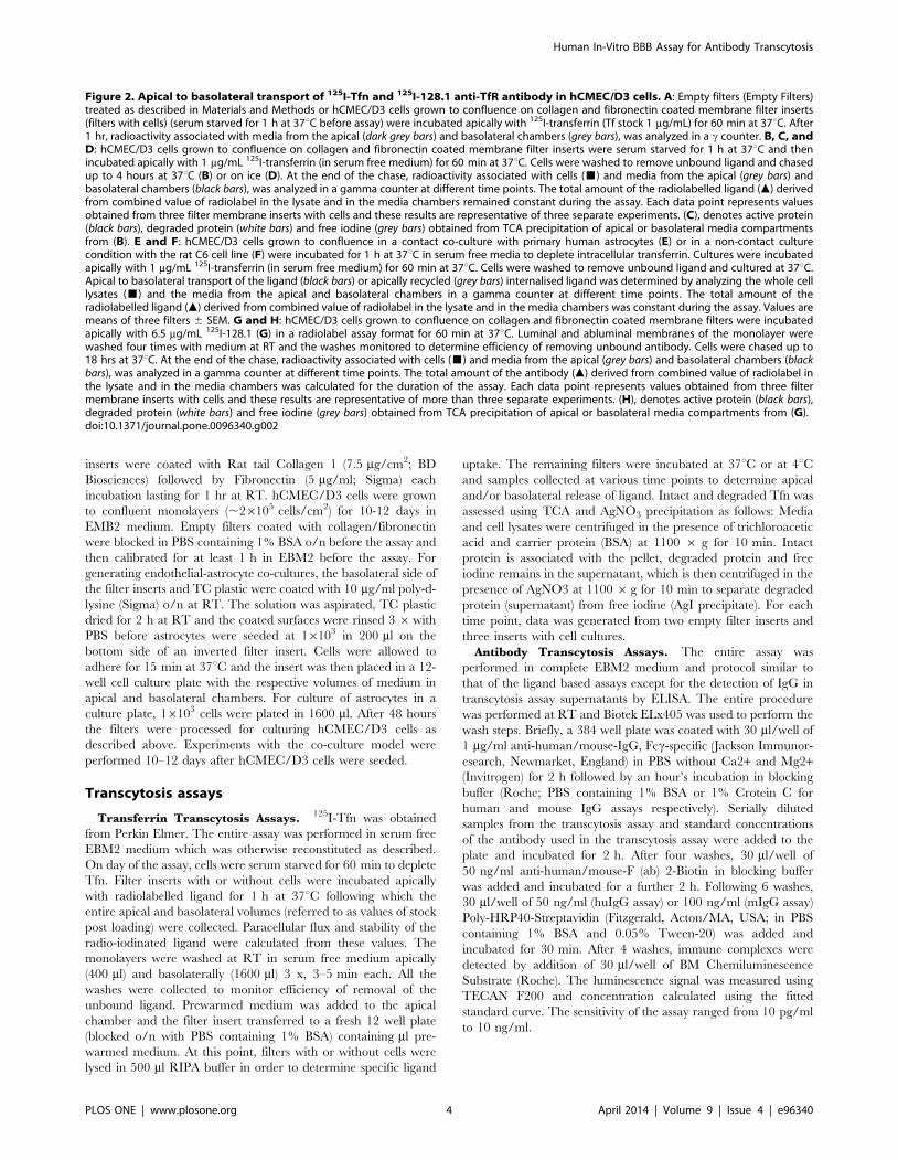
Figure 2. Apical to basolateral transport of 125I-Tfn and 125I-128.1 anti-TfR antibody in hCMEC/D3 cells. A: Empty filters (Empty Filters)treated as described in Materials and Methods or hCMEC/D3 cells grown to confluence on collagen and fibronectin coated membrane filter inserts(filters with cells) (serum starved for 1 h at 37uC before assay) were incubated apically with 125I-transferrin (Tf stock 1 mg/mL) for 60 min at 37uC. After1 hr, radioactivity associated with media from the apical (dark grey bars) and basolateral chambers (grey bars), was analyzed in a c counter. B, C, andD: hCMEC/D3 cells grown to confluence on collagen and fibronectin coated membrane filter inserts were serum starved for 1 h at 37uC and thenincubated apically with 1 mg/mL 125I-transferrin (in serum free medium) for 60 min at 37uC. Cells were washed to remove unbound ligand and chasedup to 4 hours at 37uC (B) or on ice (D). At the end of the chase, radioactivity associated with cells (&) and media from the apical (grey bars) andbasolateral chambers (black bars), was analyzed in a gamma counter at different time points. The total amount of the radiolabelled ligand (m) derivedfrom combined value of radiolabel in the lysate and in the media chambers remained constant during the assay. Each data point represents valuesobtained from three filter membrane inserts with cells and these results are representative of three separate experiments. (C), denotes active protein(black bars), degraded protein (white bars) and free iodine (grey bars) obtained from TCA precipitation of apical or basolateral media compartmentsfrom (B). E and F: hCMEC/D3 cells grown to confluence in a contact co-culture with primary human astrocytes (E) or in a non-contact culturecondition with the rat C6 cell line (F) were incubated for 1 h at 37uC in serum free media to deplete intracellular transferrin. Cultures were incubatedapically with 1 mg/mL 125I-transferrin (in serum free medium) for 60 min at 37uC. Cells were washed to remove unbound ligand and cultured at 37uC.Apical to basolateral transport of the ligand (black bars) or apically recycled (grey bars) internalised ligand was determined by analyzing the whole celllysates (&) and the media from the apical and basolateral chambers in a gamma counter at different time points. The total amount of theradiolabelled ligand (m) derived from combined value of radiolabel in the lysate and in the media chambers was constant during the assay. Values aremeans of three filters 6 SEM. G and H: hCMEC/D3 cells grown to confluence on collagen and fibronectin coated membrane filters were incubatedapically with 6.5 mg/mL 125I-128.1 (G) in a radiolabel assay format for 60 min at 37uC. Luminal and abluminal membranes of the monolayer werewashed four times with medium at RT and the washes monitored to determine efficiency of removing unbound antibody. Cells were chased up to18 hrs at 37uC. At the end of the chase, radioactivity associated with cells (&) and media from the apical (grey bars) and basolateral chambers (blackbars), was analyzed in a gamma counter at different time points. The total amount of the antibody (m) derived from combined value of radiolabel inthe lysate and in the media chambers was calculated for the duration of the assay. Each data point represents values obtained from three filtermembrane inserts with cells and these results are representative of more than three separate experiments. (H), denotes active protein (black bars),degraded protein (white bars) and free iodine (grey bars) obtained from TCA precipitation of apical or basolateral media compartments from (G).doi:10.1371/journal.pone.0096340.g002
Human In-Vitro BBB Assay for Antibody Transcytosis
PLOS ONE | www.plosone.org 4 April 2014 | Volume 9 | Issue 4 | e96340

Immunofluorescence using Confocal MicroscopyTo investigate the localization of the 128.1 antibody and the
natural ligand holotransferrin, monolayers of hCMEC/D3 cells
grown to confluence on collagen-coated cover slips were incubated
with 5 mg/ml FITC tagged holotransferrin (Invitrogen) or 1 mg/ml
128.1, MEM-189 or the IGF-1R antibodies for 10 min following
which the medium was removed and replaced with fresh medium.
After 1 hour at 37uC, the monolayers were fixed in 4% PFA for
15 min at RT, permeabilized for 10 min (16 PBS with 0.1%
Triton X-100) and incubated with an antibody to late endosomal/
lysosomal marker CD63 (Abcam, Cambridge, UK) for 1 hour at
RT. Cells were subjected to 16 PBS washes for 15 min and
sequentially incubated where necessary with secondary antibodies;
goat anti-human IgG-Alexa Flour 488 and/or chicken anti-mouse
IgG-Alexa Fluor 595 (1:200, Invitrogen) for 1 hour at RT. Cells
were washed in 16 PBS for 30 min and cover slips mounted in
UltraCruz fluorescent mounting medium (Santa Cruz, Heidel-
berg, Germany) and confocal fluorescent images were obtained
using an Leica DM IRB/E microscope. Confocal images and z-
stacks were acquired and analyzed by Leica Confocal Software
(Leica, Wetzlar, Germany. All the confocal images show a single,
representative, section of a Z-series taken through the entire cell.
Binding and Competition ELISAThe entire procedure was performed at RT on a shaking
platform and all wash steps (PBS containing 0.1% tween) were
done on the Biotek ELx405. Briefly, a 96 well plate (Nunc,
Langenselbold, Germany) was coated for 1 h with 50 ng of the
extracellular subunit of recombinant TfR (R&D Systems,
Wiesbaden, Germany). The plates were then blocked for 1 h in
sample buffer (PBS containing 1% BSA) and washed 4 x. 50 ml of
antibody dilutions made in sample buffer were transferred to the
assay plate and incubated for 1.5 h following which the plates were
washed 4 x. Peroxidase labeled anti mouse IgG (GE Healthcare,
Munchen, Germany) or anti human IgG (Jackson Immunore-
search) secondary antibodies used at recommended dilutions were
added to the wells and incubated further for 30 min. Following 6
washes, 50 ml of TMB (Sigma) was added per well and incubated
for 5–20 min. After addition of 50 ml of 1N HCl, antibody-
receptor complexes were detected by measuring the OD of the
plate at 450/620 nm. The procedure for the blocking and
competition ELISA was similar except in the latter assay, dilutions
of respective antibodies in sample buffer were added to the plate
for 1 h in the pre-block conditions and assay plates washed 4 6after this incubation.
Figure 3. Uptake and fate of an antibody directed againstdifferent transcytosis receptors in hCMEC/D3 monolayers. A–DhCMEC/D3 cells grown to confluence on collagen and fibronectincoated membrane filters were incubated apically with 1 mg/mL of thefollowing antibodies: the anti-human TfR antibody, 128.1 (A), themouse anti–human TfR antibody, MEM-189 (B), an anti-human IGF-1Rantibody (C) and the mouse anti-human insulin receptor (IR) antibody,83–14 (D) for 60 min at 37uC. Luminal and abluminal membranes of themonolayer were washed four times with medium at RT and the washesmonitored to determine efficiency of removing unbound antibody.Cells were chased up to 5 hrs at 37uC. At the end of the chase, antibodyassociated with cells (&) and media from the apical (grey bars) andbasolateral chambers (black bars), were analyzed by the sensitive IgGELISA at different time points. The total amount of IgG (m) derived fromcombined value of antibody present in the lysate and in the mediachambers was calculated for the entire duration of the assay. Values aremeans of three filters 6 SEM.doi:10.1371/journal.pone.0096340.g003
Human In-Vitro BBB Assay for Antibody Transcytosis
PLOS ONE | www.plosone.org 5 April 2014 | Volume 9 | Issue 4 | e96340

Results
‘‘Pulse-chase’’ assay mode overcomes leakiness inhCMEC/D3 transcytosis assay
hCMEC/D3 cells were cultured to confluent monolayers on
collagen/fibronectin-coated filter inserts, and upon reaching the
TEER value of <40 ohm/cm2, were used to investigate the
apical-to-basolateral transport of transferrin. On the basis of
kinetic parameters obtained with iodinated transferrin in primary
bovine brain endothelial cells (influx rate of 0.03 min21, total
efflux of 0.017 min21 and a ratio of 1:3 between basolateral and
apical efflux) [16], we calculated an expected transport rate of 62
pg of the ligand per hour in our assay set-up. However, the
paracellular flux of the hCMEC/D3 cellular monolayer to 70 kDa
FITC dextran is 1.8 6 1025 cm/min, which would equate to
6.8 ng/mL of the ligand in the basolateral compartment following
an hour’s incubation with 1 mg/mL 125I-Transferrin. This
indicated that it would be impossible to measure transcytosis
against the expected background of paracellular flow. Indeed
when filter-grown hCMEC/D3 cells were apically incubated with
1 mg/mL 125I-Transferrin for 1 hour, close to 10,000 counts (,30 ng) of the ligand were measured in the basolateral compart-
ment. In filters without cells, this value was elevated by a factor of
seven, still demonstrating restriction to protein passage by the
cellular monolayer. Since the internalized ligand measured in
cellular lysates was in the range of 5-7 ng in several experiments
and unchanged by the concentration of the ligand used, it is likely
that the events thus measured in the basolateral compartment are
mostly contributed by paracellular flux and not by transcytosis.
We confirmed this possibility by using 125I-Protein A as a negative
control (data not shown). We did not detect radiolabel associated
with the hCMEC/D3 cells, but the radiolabel measured in the
basolateral compartment was similar to the assay with iodinated Tf
under the same conditions. These results confirmed the necessity
of modifying our assay to a ‘‘pulse-chase’’ format as suggested by
Raub and Newton (Fig. 1).
Serum starved cells were incubated with iodinated transferrin
for one hour at 37uC to allow for ligand internalization. After
washing and transferring of the filter to a fresh plate, the amount
of radioactivity in the apical and basolateral medium compart-
ments as well as in the cell lysate was determined by gamma-
counting at different incubation times. After four hours of
incubation, approximately 40% of the radioactivity internalized
by the cells was observed at the apical side (recycling), 40% at the
basolateral side (transcytosis), and 20% remained inside the cells
(Fig. 2B). As shown by trichloro-acetic acid (TCA) precipitation),
.90% of the transported material was active protein, excluding
the leakage of radioactivity after protein degradation (Fig. 2C).
When transcytosis was carried out at 4uC, only minimal amounts
of radioactivity were found in the basolateral and apical
compartments, confirming the implication of an energy-depen-
dent, active transport process. Although hCMEC/D3layer tight-
ness has been shown not to respond to cues delivered by astrocytes,
we tested if transferrin transport might be influenced by the
presence of rat or human astrocytes in a contact or non-contact
culture (Fig. 2E and F); however, this was not the case.
Owing to the high serum concentration, transferrin is a sub-
optimal brain transporter and so we tested an antibody to the
human transferrin receptor (128.1), described by Friden et al. [18]
to access the brain of Cynomolgus monkeys following intravenous
Figure 4. Degradation of 128.1 but not MEM-189 in hCMEC/D3 cells. hCMEC/D3 endothelial cells were pulsed with 1 mg/ml 128.1 (A) orMEM-189 (B) for 10 min at 37uCThe coverslip cultures were washed and cultured at 37uC for various time periods. Cultures were fixed in 4% PFA,permeabilised and immunostained with an antibody to the late endosomal/lysosomal marker CD63 and appropriate secondary antibodies asdescribed in Materials and Methods and examined with a laser scanning confocal microscope. Insets: secondary antibodies only.doi:10.1371/journal.pone.0096340.g004
Human In-Vitro BBB Assay for Antibody Transcytosis
PLOS ONE | www.plosone.org 6 April 2014 | Volume 9 | Issue 4 | e96340

injection. In our transcytosis system, 125I-labeled 128.1, although
readily taken up by the cells, even after 18 hours was not
transported to either basolateral or apical side, but remained in
cells (Fig. 2G). TCA precipitation confirmed that a large portion of
the antibody was degraded inside cells (Fig. 2H).
Transcytosis of antibodies against BBB receptors revealsdetails about sorting mechanism
Next, we established highly sensitive IgG ELISAs in order to be
able to detect picogram quantities of transcytosed IgG, obviating
the need for radioactive labeling. Figure 3A shows that using this
IgG ELISA, results for mAb 128.1 transcytosis were very similar to
those obtained with radioactive antibody. We then went on to test
other antibodies in our transcytosis system. A commercially
available antibody against the transferrin receptor, mAb MEM-
189, to our surprise showed a completely different behavior
compared to mAb 128.1 (Fig. 3B): after five hours of incubation,
about 40% of the antibody were found in each the basolateral and
the apical compartment, while only 20% had remained inside
cells, indicating that the antibody had been transcytosed and
recycled to equal amounts, in a similar way as shown for the
ligand, transferrin. By contrast, an antibody against the IGF1
receptor, although capable of leaving the endothelial cells, was
exclusively recycled to the apical compartment (Fig. 3C), while an
antibody against the insulin receptor showed only weak uptake
and some recycling (Fig. 3D). In conclusion, our assay proved to be
a valuable tool for the investigation of BBB sorting pathways for
different internalizing receptors.
In order to have a thorough understanding of the intracellular
fate of the two TfR antibodies mAb 128.1 and mAb MEM-189,
hCMEC/D3 cells were pulsed with each of the antibodies for ten
minutes, unbound antibody was removed and after one hour of
incubation, cells were fixed and stained for immunofluorescence
microscopy. Figure 4 shows the intracellular localization of the
antibodies together with a staining of CD63, a marker for the late
endosomal/lysosomal compartment. While mAb 128.1 shows a
significant co-localization with CD63, MEM-189 was found in
vesicular structures different from late endosomes. These obser-
vations are in line with the transcytosis assay data, arguing for
intracellular degradation of mAb 128.1 and differential sorting of
MEM-189 to recycling and transcytosis pathways.
Next we asked the question, what property of the antibodies
might be responsible for their intracellular fate. In order to
compare binding epitopes on the human TfR extracellular domain
(ECD), we performed a competition ELISA on immobilized TfR-
ECD (Fig. 5A). mAb 128.1 was not inhibited from binding to TfR-
ECD, but pre-incubation of TfR-ECD with 128.1 completely
abolished binding of MEM-189. The significantly higher affinity of
128.1 as compared to MEM-189 could explain why blocking is not
efficient in both ways. However it was clear that the epitopes of the
antibodies are overlapping and unlikely to reside in completely
different regions of the receptor, which could exclude the antibody
epitope as a determinant of intracellular sorting. Affinity itself, as
shown by Yu et al., may be one of the criteria influencing
transcytosis, but we later identified antibodies in the same affinity
range as MEM-189, not capable of transcytosis (see below). We
speculated that pH change during acidification of endosomes may
play a role in TfR antibody sorting, and indeed we could show
that MEM-189 showed a strongly diminished affinity for the TfR
at pH 5.5 (late endosomal pH), as compared to pH 7.4 (Fig. 5B).
mAb 128.1, by contrast, was pH-independent.
Testing other commercially available antibodies for pH-
dependent binding to the TfR, we observed that antibodies LT-
71 and MEM-75 displayed weaker binding to the receptor at
endosomal pH, while antibodies M-A712 and 13E4 bound in a
pH-independent fashion (Fig. 6, A–D). Strikingly, the latter two
antibodies were irreversibly trapped inside the hCMEC/D3 cells
when investigated in our transcytosis assay (Fig. 6, G and H), while
LT-71 and MEM-75 showed different degrees of transcytosis and
recycling (Fig. 6, E and F). Although these data also show a
correlation between affinity and transcytosis, the comparable
affinities of antibodies M-A712 and MEM-189, combined to their
strikingly different transcytosis behavior, indicate that there may
be additional mechanisms governing the intracellular fate of
transcytosing antibodies. Finally, we wanted to investigate if
transcytosis of pH-dependent TfR antibodies could be blocked by
Figure 5. 128.1 is a high affinity antibody that competessuccessfully with MEM-189 for binding to TfR in ELISA and incontrast to MEM-189 shows no pH dependence. A: CompetitionELISA described in Materials and Methods showing binding of 128.1 tothe extracellular subunit of TfR in the presence (&) or absence (m) of apre-block by 5 mg/ml MEM-189. Similarly, binding of MEM-189 in thepresence (%) or absence (n) of 5 mg/mL 128.1. B, Binding ELISAdescribed in Materials and Methods showing binding of the 128.1 anti-human transferrin receptor antibody (&,%) and MEM-189 antibody(N,#) to the extracellular subunit of the human TfR at pH 7.4 (&,N) orat pH 5.5 (%,#).doi:10.1371/journal.pone.0096340.g005
Human In-Vitro BBB Assay for Antibody Transcytosis
PLOS ONE | www.plosone.org 7 April 2014 | Volume 9 | Issue 4 | e96340

Human In-Vitro BBB Assay for Antibody Transcytosis
PLOS ONE | www.plosone.org 8 April 2014 | Volume 9 | Issue 4 | e96340

bafilomycin, an inhibitor of endosomal acidification. Figure 6I
shows that pre-incubation of hCMEC/D3 cells with bafilomycin
strongly reduced basolateral passage of antibody MEM-189, while
apical recycling was unaffected. This result confirmed our
hypothesis that endosomal acidification is an essential mechanistic
step in facilitating the transcytosis of TfR antibodies with reduced
affinity at low pH.
Discussion
There is a wealth of literature describing the phenomenon of
transcytosis in vivo and in vitro in addition to the classical pathway of
receptor-mediated endocytosis and recycling. However, many
publications describing in vitro models of protein transcytosis
through the blood-brain barrier neglect the magnitude of
paracellular flux as opposed to the small amount of transcytosed
material [20–22]. In fact, our results show (Fig. 2A) accumulation
of 30 ng of the transferrin ligand or Protein A in the basolateral
compartment following incubation with 1 mg/mL of the radiola-
bel. In order to cope with this limitation, which is especially
apparent in the relatively leaky monolayers of hCMEC/D3 cells
[4], we have applied a pulse-chase assay set-up, initially described
by Raub and Newton for primary bovine brain endothelial cells
[16]. In order to detect the low amounts of transcytosed antibody
and at the same time avoid using radiolabeled material, the
development of highly sensitive IgG ELISAs has been instrumental
for assessing the transcytosis potential of antibodies of different
species. Furthermore, the ELISA protocol can be easily adapted
for automation which makes it highly attractive in terms of assay
throughput.
Although several studies addressed the transcytosis of antibody-
targeted nanoparticles and immunoliposomes across hCMEC/D3
[10,23], this is the first study to investigate the transcytosis and the
fate of free antibodies targeting receptors capable of mediating
transcytosis in immortalized human brain endothelium. Our
results support the following conclusions:
Apical to basolateral transcytosis of intact transferrin occurs in
hCMEC/D3 monolayer cultures. Ligand is also equally recycled
to the luminal membrane. Both events are temperature sensitive
but not modulated by astrocyte co culture (Fig. 2)
An antibody to the IGF1R is exclusively recycled, while
antibodies against the TfR are either degraded in lysosmes or
recycled/transcytosed (Figs. 3 and 4)
Reduced affinity of antibodies to the transferrin receptor at
endosomal pH may enhance antibody transcytosis (Figs. 5 and 6).
We provide strong evidence for the transcytosis of intact
transferrin in hCMEC/D3 cells (Fig. 2C–D). Between several
experiments, we recorded similar values (2200–2500 cpm; 5–
7 ng/,20 mg cell protein) of uptake followed invariably by
equivalent rates of transcytosis and recycling of the internalized
ligand. High values of transcytosed/recycled ligand (,5 ng) from
our assay can be explained by the level of membrane-localized
TfR on hCMEC/D3 cells compared to primary human brain
endothelial cells. At similar culture confluency, the immortalized
cells consistently show higher levels of membrane-resident TfR
than primary human endothelial cells (data not shown). The work
of Raub and Newton (1991; [16]) and Descamps et al. (1996; [15])
have described the paucity of membrane-localized TfR in
confluent cultures of primary endothelial cells and hence the
published values of transcytosis from primary cell cultures are
generally low. The ratio of transferrin transcytosis to recycling was
described as 1:3 in primary bovine brain endothelial cells [16]; by
contrast, we observe similar rates for both processes. It cannot be
excluded that this difference, in line with the higher TfR
expression level, could also be due to slightly distorted sorting of
the transferrin receptor in the immortalized cell line; alternatively,
we cannot rule out the possibility that some material unspecifically
bound to the cells and is only released after prolongued
incubation.
Following our validation of the assay protocol with the ligand,
we proceeded to test antibodies against putative transcytosis
receptors for transport. Targeting of receptors, particularly the
insulin and transferrin receptors by chimeric peptides and
antibodies has been suggested to be an effective way of delivering
drugs to the brain in several animal models [24–28]. The OX-26
murine monoclonal antibody to the rat TfR [29] and the 83–14
murine monoclonal antibody to the human insulin receptor [27]
are the best known examples of antibodies with published BBB
permeability properties.
We tested an antibody to the IGF-1R, described for its capacity
to engage in transcytotic activity [28,30]. Our results indicate
slight intracellular degradation and significant recycling of the
antibody to the apical surface (Fig. 3C), but no transcytosis.
Investigation of IGF-1R mediated transcytosis has implicated
facilitation of the process by the association with LRP1 in the rat
brain [30,31]. We could not detect membrane-resident LRP1 in
hCMEC/D3 cells (data not shown). Reports showing Lrp1
expression in hCMEC/D3 were obtained using methods which
detect both extracellular and intracellular protein [32]. Absence of
membrane LRP1 could offer an explanation for the lack of
transcytosis of an IGF-1R mAb in our model system.
We generated the 128.1 anti-TfR antibody described in Friden
et al. (1996; [18]), because it had been suggested to access the
brain in Cynomolgous monkeys after intravenous application. In
hCMEC/D3 cells, the antibody initially appeared to follow the
classical uptake and internalization through clathrin-coated
vesicles and subsequent localization in early endosomes. It is well
established in numerous cell types that the TfR-ligand complex is
endocytosed and rapidly recycled [33,34]. In fact, the receptor-
ligand complex is commonly used as a marker of early and
recycling endosomes. However, the 128.1 antibody localized to a
Figure 6. pH dependence and uptake, fate of different antibodies directed against the human TfR in hCMEC/D3 monolayers. A–D:Binding ELISA described in Materials and Methods showing binding of mouse anti-human transferrin receptor antibodies LT-71 (A), MEM-75 (B), M-A712 (C) and 13E4 (D) to the extracellular subunit of TfR at pH 7.4 (N) or at pH 5.5 (#). E–I: hCMEC/D3 cells grown to confluence on collagen andfibronectin coated membrane filters were incubated apically with 1 mg/mL of the following mouse anti-human TfR antibodies: LT-71 (E), MEM-75 (F),M-A712 (G), 13E4 (H) for 60 min at 37uC. Luminal and abluminal membranes of the monolayer were washed four times with medium at RT and thewashes monitored to determine efficiency of removing unbound antibody. Cells were chased up to 5 hrs at 37uC. At the end of the chase, antibodyassociated with cells (&) and media from the apical (white bars) and basolateral chambers (grey bars), were analyzed by the sensitive IgG ELISA atdifferent time points. The total amount of IgG (m) derived from combined value of antibody present in the lysate and in the media chambers wascalculated for the entire duration of the assay. Values are means of three filters 6 SEM. (I) hCMEC/D3 cells were incubated apically with 1 mg/mLmouse anti-human TfR MEM-189 antibody as in (E–H) and cells were chased up to 5 hrs at 37uC in the presence of 50 nM Bafilomycin. At the end ofthe chase, antibody associated with cells (&) and media from the apical (grey bars) and basolateral chambers (white bars), were analyzed by thesensitive IgG ELISA at different time points. The total amount of IgG (m) derived from combined value of antibody present in the lysate and in themedia chambers was calculated for the entire duration of the assay. Values are means of three filters 6 SEM.doi:10.1371/journal.pone.0096340.g006
Human In-Vitro BBB Assay for Antibody Transcytosis
PLOS ONE | www.plosone.org 9 April 2014 | Volume 9 | Issue 4 | e96340

CD63-positive late endosomal/lysosomal compartment one hour
after internalization, where it presumably is degraded. In contrast
to 128.1, the mouse monoclonal antibody MEM-189 is processed
like the ligand (Fig. 3B). It is endocytosed at a slightly slower rate
than transferrin and the 128.1 antibody in hCMEC/D3 cells,
which is likely attributable to the lower-affinity binding profile of
the antibody (Fig. 5A). However, once internalized, the antibody is
processed through the endocytic pathway with transcytosis and
recycling kinetics comparable to that of transferrin (Fig. 2B and
Fig. 3B). The 128.1 and MEM-189 antibodies target overlapping
epitopes, as shown by a competition ELISA (Fig. 5A), which
lowers the probability that the MEM-189 local epitope on TfR is
solely responsible for its transcytosis potential, although it cannot
be excluded. Even antibodies with closely overlapping epitopes
can demonstrate fundamental functional differences, as illustrated
for the CD20 antibodies GA101 and rituximab [35].
Intracellular degradation of TfR antibodies after endocytosis
has been described by others [36], and the 128.1 antibody follows
those examples in terms of lysosomal localization and degradation
(Fig. 4A). Furthermore, incubation of murine lymphoma cells with
full IgGs against the TfR has been shown to even downregulate
surface expression of the TfR [36]. In contrast, Yu et al. (2011;
[37]) have demonstrated that in vivo, antibodies against the
transferrin receptor are transported into the brain to an extent
inversely correlated to their binding affinity. We compared the
bivalent affinities of the different TfR antibodies by direct ELISA
and observed a similar correlation with regard to transcytosis
potential (Figs. 3 and 6). A low-affinity antibody, LT-71 (EC50 of
approx. 660 ng/mL) was capable of shuttling through hCMEC/
D3 monolayer, albeit after low uptake (Fig. 6A), while the high-
affinity antibodies 128.1 and 13E4 (EC50 of 10 ng/mL) remain
inside the cells. However, two antibodies with intermediate
binding affinities (MEM-189: 85 ng/mL and M-A712: 35 ng/
mL) demonstrated significantly different sorting behavior: while
MEM-189 was efficiently transcytosed and recycled (Fig. 3B), M-
A712 was incapable of leaving the endothelial cells (Fig. 6G). It is
unlikely that the small difference in binding affinity should be
responsible for the striking difference in transcytosis potential.
The transferrin:transferrin receptor complex undergoes dra-
matic conformational changes upon endosomal acidification,
which are partly driven by iron release from the ligand, but
which are accompanied by significant changes in the TfR
conformation [38–40]. These conformational changes might be
responsible for the observed pH-dependence of several TfR
antibodies. In addition, histidine residues in antibody CDR could
contribute to pH-dependent target binding. The mechanism of
transferrin transcytosis, especially which sorting events determine
routing for basolateral transcytosis or apical recycling, have not yet
been investigated. In polarized cells, TfR is known to be recycled
via recruitment of the adaptor protein AP1B in the recycling
endosome [41]. Apo-Tf stays bound to the receptor and follows its
path back to the cell surface. A key difference between antibodies
and the ligand interacting with the transferrin receptor is the
bivalent nature of the TfR:antibody interaction. High-affinity,
bivalent TfR antibodies are invariably sorted to lysosomes,
possibly by interfering with TfR sorting via irreversible receptor
crosslinking [36]. Although the exact mechanism of this sorting
event is unknown, it seems plausible that antibodies with reduced
affinity at endosomal pH might relieve receptor cross-linking due
to lower complex stability, allowing the receptor to pursue its
physiological sorting pathway. It needs to be stressed that
transcytosis supported by pH-dependent receptor binding has so
far only been demonstrated in vitro using the hCMEC/D3 model
system. For a more general applicability, other sytems like primary
brain endothelial cells and, more importantly, in vivo experiments,
need to be performed.In summary, we have developed a human
BBB transcytosis assay enabling us to quickly screen antibodies for
putative brain shuttle receptors for their transcytosis potential.
Furthermore, our data suggest a mechanism in addition to
reduction of binding affinity, which might facilitate antibody
transcytosis over the BBB, namely pH-dependent binding to a
transcytosis receptor.
Acknowledgments
The authors would like to thank Pierre-Olivier Couraud, Ignacio A.
Romero and Babette B. Weksler for providing the hCMEC/D3 cells.
Author Contributions
Conceived and designed the experiments: JN HS AH EM POF. Performed
the experiments: HS CB. Analyzed the data: HS JN CB. Wrote the paper:
HS JN.
References
1. Begley DJ (2004) Delivery of therapeutic agents to the central nervous system:
the problems and the possibilities. Pharmacol Ther 104: 29–45.
2. Jones AR, Shusta EV (2007) Blood-brain barrier transport of therapeutics via
receptor-mediation. Pharm Res 24: 1759–71.
3. Deli MA, Abraham CS, Kataoka Y, Niwa M (2005) Permeability studies on in
vitro blood-brain barrier models: physiology, pathology, and pharmacology. Cell
Mol Neurobiol 25: 59–127.
4. Weksler BB, Subileau EA, Perriere N, Charneau P, Holloway K, et al. (2005)
Blood-brain barrier-specific properties of a human adult brain endothelial cell
line. Faseb J 19: 1872–4.
5. Carl SM, Lindley DJ, Couraud PO, Weksler BB, Romero I, et al. (2010) ABC
and SLC transporter expression and pot substrate characterization across the
human CMEC/D3 blood-brain barrier cell line. Mol Pharm 7: 1057–1068.
6. Poller B, Gutmann H, Krahenbuhl S, Weksler B, Romero I, et al. (2008) The
human brain endothelial cell line hCMEC/D3 as a human blood-brain barrier
model for drug transport studies. J Neurochem 107: 1358–68.
7. Mairey E, Genovesio A, Donnadieu E, Bernard C, Jaubert F, et al. (2006)
Cerebral microcirculation shear stress levels determine Neisseria meningitidis
attachment sites along the blood-brain barrier. J Exp Med 203: 1939–1950.
8. Kania KD, Wijesuriya HJ, Hladky SB, Barrand MA (2011) Beta amyloid effects
on expression of multidrug efflux transporters in brain endothelial cells. Brain
Res.
9. Tai LM, Holloway KA, Male DK, Loughlin AJ, Romero IA (2010) Amyloid-
beta-induced occludin down-regulation and increased permeability in human
brain endothelial cells is mediated by MAPK activation. J Cell Mol Med 14:
1101–1112. 10.1111/j.1582-4934.2009.00717.x.
10. Markoutsa E, Pampalakis G, Niarakis A, Romero IA, Weksler B, et al. (2011)
Uptake and permeability studies of BBB-targeting immunoliposomes using thehCMEC/D3 cell line. Eur J Pharm Biopharm 77: 265–274.
11. Wang ZH, Wang ZY, Sun CS, Wang CY, Jiang TY, et al. (2010) Trimethylated
chitosan-conjugated PLGA nanoparticles for the delivery of drugs to the brain.
Biomaterials 31: 908-915. 10.1016/j.biomaterials.2009.09.104.
12. Jefferies WA, Brandon MR, Hunt SV, Williams AF, Gatter KC, et al. (1984)
Transferrin receptor on endothelium of brain capillaries. Nature 312: 162–163.
13. Pardridge WM, Eisenberg J, Yang J (1987) Human blood-brain barriertransferrin receptor. Metabolism 36: 892–895.
14. Risau W, Hallmann R, Albrecht U (1986) Differentiation-dependent expressionof proteins in brain endothelium during development of the blood-brain barrier.
Dev Biol 117: 537–545.
15. Descamps L, Dehouck MP, Torpier G, Cecchelli R (1996) Receptor-mediated
transcytosis of transferrin through blood-brain barrier endothelial cells.Am J Physiol 270: 1149–58.
16. Raub TJ, Newton CR (1991) Recycling kinetics and transcytosis of transferrin inprimary cultures of bovine brain microvessel endothelial cells. J Cell Physiol 149:
141-151. 10.1002/jcp.1041490118.
17. Skarlatos S, Yoshikawa T, Pardridge WM (1995) Transport of [125I]transferrin
through the rat blood-brain barrier. Brain Res%19;683: 164–171.
18. Friden PM, Olson TS, Obar R, Walus LR, Putney SD (1996) Characterization,
receptor mapping and blood-brain barrier transcytosis of antibodies to thehuman transferrin receptor. J Pharmacol Exp Ther 278: 1491–8.
19. Gong Y, Yao E, Shen R, Goel A, Arcila M, et al. (2009) High expression levels of
total IGF-1R and sensitivity of NSCLC cells in vitro to an anti-IGF-1R antibody
(R1507). PLoS One 4: e7273.
Human In-Vitro BBB Assay for Antibody Transcytosis
PLOS ONE | www.plosone.org 10 April 2014 | Volume 9 | Issue 4 | e96340

20. Abulrob A, Sprong H, Van Bergen en Henegouwen P, Stanimirovic D (2005)
The blood-brain barrier transmigrating single domain antibody: mechanisms of
transport and antigenic epitopes in human brain endothelial cells. J Neurochem
95: 1201–14.
21. Demeule M, Poirier J, Jodoin J, Bertrand Y, Desrosiers RR, et al. (2002) High
transcytosis of melanotransferrin (P97) across the blood-brain barrier.
J Neurochem 83: 924–33.
22. Wang P, Xue Y, Shang X, Liu Y (2010) Diphtheria Toxin Mutant CRM197-
Mediated Transcytosis across Blood-Brain Barrier In Vitro. Cell Mol Neurobiol.
10.1007/s10571-010-9496-x.
23. Dan M, Cochran DB, Yokel RA, Dziubla TD (2013) Binding, Transcytosis and
Biodistribution of Anti-PECAM-1 Iron Oxide Nanoparticles for Brain-Targeted
Delivery. PLoS One 8: e81051. 10.1371/journal.pone.0081051 [doi];PONE-D-
13-25817 [pii].
24. Fishman JB, Rubin JB, Handrahan JV, Connor JR, Fine RE (1987) Receptor-
mediated transcytosis of transferrin across the blood-brain barrier. J Neurosci
Res 18: 299–304.
25. Friden PM, Walus LR, Musso GF, Taylor MA, Malfroy B, et al. (1991) Anti-
transferrin receptor antibody and antibody-drug conjugates cross the blood-
brain barrier. Proc Natl Acad Sci U S A 88: 4771–4775.
26. Pardridge WM, Buciak JL, Friden PM (1991) Selective transport of an anti-
transferrin receptor antibody through the blood-brain barrier in vivo.
J Pharmacol Exp Ther 259: 66–70.
27. Pardridge WM, Kang YS, Buciak JL, Yang J (1995) Human insulin receptor
monoclonal antibody undergoes high affinity binding to human brain capillaries
in vitro and rapid transcytosis through the blood-brain barrier in vivo in the
primate. Pharm Res 12: 807–16.
28. Reinhardt RR, Bondy CA (1994) Insulin-like growth factors cross the blood-
brain barrier. Endocrinology 135: 1753–1761.
29. Friden PM, Walus LR, Watson P, Doctrow SR, Kozarich JW, et al. (1993)
Blood-brain barrier penetration and in vivo activity of an NGF conjugate.
Science 259: 373–7.
30. Duffy KR, Pardridge WM, Rosenfeld RG (1988) Human blood-brain barrier
insulin-like growth factor receptor. Metabolism 37: 136–140.
31. Nishijima T, Piriz J, Duflot S, Fernandez AM, Gaitan G, et al. (2010) Neuronal
Activity Drives Localized Blood-Brain-Barrier Transport of Serum Insulin-likeGrowth Factor-I into the CNS. Neuron 67: 834-846. 10.1016/j.neuron.
2010.08.007.
32. Nazer B, Hong S, Selkoe DJ (2008) LRP promotes endocytosis and degradation,but not transcytosis, of the amyloid-beta peptide in a blood-brain barrier in vitro
model. Neurobiol Dis 30: 94–102.33. Dautry-Varsat A, Ciechanover A, Lodish HF (1983) pH and the recycling of
transferrin during receptor-mediated endocytosis. Proc Natl Acad Sci U S A 80:
2258–2262.34. Klausner RD, van RJ, Ashwell G, Kempf C, Schechter AN, et al. (1983)
Receptor-mediated endocytosis of transferrin in K562 cells. J Biol Chem 258:4715–4724.
35. Niederfellner G, Lammens A, Mundigl O, Georges GJ, Schaefer W, et al. (2011)Epitope characterization and crystal structure of GA101 provide insights into the
molecular basis for type I/II distinction of CD20 antibodies. Blood 118: 358–
367.36. Lesley J, Schulte R, Woods J (1989) Modulation of transferrin receptor
expression and function by anti-transferrin receptor antibodies and antibodyfragments. Exp Cell Res 182: 215–233.
37. Yu YJ, Zhang Y, Kenrick M, Hoyte K, Luk W, et al. (2011) Boosting brain
uptake of a therapeutic antibody by reducing its affinity for a transcytosis target.Sci Transl Med 3: 84ra44.
38. Eckenroth BE, Steere AN, Chasteen ND, Everse SJ, Mason AB (2011) How thebinding of human transferrin primes the transferrin receptor potentiating iron
release at endosomal pH. Proc Natl Acad Sci U S A 108: 13089–13094.39. Lawrence CM, Ray S, Babyonyshev M, Galluser R, Borhani DW, et al. (1999)
Crystal structure of the ectodomain of human transferrin receptor. Science 286:
779–782.40. Steere AN, Chasteen ND, Miller BF, Smith VC, MacGillivray RT, et al. (2012)
Structure-based mutagenesis reveals critical residues in the transferrin receptorparticipating in the mechanism of pH-induced release of iron from human
serum transferrin. Biochemistry 51: 2113–2121.
41. Hsu VW, Bai M, Li J (2012) Getting active: protein sorting in endocyticrecycling. Nat Rev Mol Cell Biol 13: 323–328.
Human In-Vitro BBB Assay for Antibody Transcytosis
PLOS ONE | www.plosone.org 11 April 2014 | Volume 9 | Issue 4 | e96340