A Genetic and Functional Relationship between T Cells and Cellular Proliferation in the Adult...
10
A Genetic and Functional Relationship between T Cells and Cellular Proliferation in the Adult Hippocampus Guo-Jen Huang 1 , Adrian L. Smith 2 , Daniel H.D. Gray 3 , Cormac Cosgrove 4 , Benjamin H. Singer 5 , Andrew Edwards 1 , Stuart Sim 4 , Jack M. Parent 5 , Alyssa Johnsen 3 , Richard Mott 1 , Diane Mathis 3 , Paul Klenerman 4 , Christophe Benoist 3 , Jonathan Flint 1 * 1 Wellcome Trust Centre for Human Genetics, University of Oxford, Oxford, United Kingdom, 2 Department of Zoology, University of Oxford, Oxford, United Kingdom, 3 Section on Immunology and Immunogenetics, Joslin Diabetes Center and Department of Pathology, Harvard Medical School, Boston, Massachusetts, United States of America, 4 Peter Medawar Building for Pathogen Research, Nuffield Department of Medicine, University of Oxford, Oxford, United Kingdom, 5 Department of Neurology, University of Michigan, Ann Arbor, Michigan, United States of America Abstract Neurogenesis continues through the adult life of mice in the subgranular zone of the dentate gyrus in the hippocampus, but its function remains unclear. Measuring cellular proliferation in the hippocampus of 719 outbred heterogeneous stock mice revealed a highly significant correlation with the proportions of CD8+ versus CD4+ T lymphocyte subsets. This correlation reflected shared genetic loci, with the exception of the H-2Ea locus that had a dominant influence on T cell subsets but no impact on neurogenesis. Analysis of knockouts and repopulation of TCRa-deficient mice by subsets of T cells confirmed the influence of T cells on adult neurogenesis, indicating that CD4+ T cells or subpopulations thereof mediate the effect. Our results reveal an organismal impact, broader than hitherto suspected, of the natural genetic variation that controls T cell development and homeostasis. Citation: Huang G-J, Smith AL, Gray DHD, Cosgrove C, Singer BH, et al. (2010) A Genetic and Functional Relationship between T Cells and Cellular Proliferation in the Adult Hippocampus. PLoS Biol 8(12): e1000561. doi:10.1371/journal.pbio.1000561 Academic Editor: Theo D. Palmer, Stanford University, United States of America Received July 5, 2010; Accepted October 28, 2010; Published December 14, 2010 Copyright: ß 2010 Huang et al. This is an open-access article distributed under the terms of the Creative Commons Attribution License, which permits unrestricted use, distribution, and reproduction in any medium, provided the original author and source are credited. Funding: This work was supported by a grant from the Wellcome Trust to JF and by grant R01 AR055271 from the National Institutes of Health to CB and DM. The funders had no role in study design, data collection and analysis, decision to publish, or preparation of the manuscript. Competing Interests: The authors have declared that no competing interests exist. Abbreviations: GCV, ganciclovir; HS, heterogeneous stock; MHC, Major Histocompatibility Complex; QTL, quantitative trait loci; RMIP, resample-based model inclusion probability. * E-mail: [email protected] Introduction The discovery that neurogenesis occurs in the adult hippocam- pus has attracted considerable attention, yet its function remains unclear [1,2]. Adult neurogenesis is known to occur in two areas of the mammalian brain, the subventricular zone, which gives rise to olfactory bulb interneurons, and the dentate gyrus of the hippocampal formation, which gives rise to granule cells [3]. Its occurrence in the latter structure has prompted considerable speculation that it is involved in known functions of the hippocampus: learning, memory, and emotional regulation. However, the hippocampal function of adult neurogenesis is still debated. Experiments using antimitotic agents and irradiation to kill newly dividing cells in the brain have produced conflicting results (reviewed in [4]). Genetically targeted ablation of neurogenesis also reports contrasting effects: normal learning and memory [5], normal anxiety with a reduction in contextual freezing and normal spatial memory [6], no change in freezing but impaired spatial memory [7,8], a combined impairment of spatial memory and reduction of contextual freezing responses [9,10], or increased anxiety but no effect on spatial memory [11]. While it is possible that the behavioural effects of neurogenesis ablation are subtle (a recent report argues that they include specific impairments in spatial discrimination [1]), it may also be the case that adult neurogenesis has additional roles. Here we adopted a genetic approach to address this question. Rates of adult hippocampal neurogenesis differ between inbred strains of mice, indicating that quantitative trait loci (QTL) contribute to this variation [12]. We asked if QTLs influencing neurogenesis could be found that influenced other phenotypes, which might cast light on the function of neurogenesis. We decided to map variation in adult hippocampal neurogen- esis in heterogeneous stock (HS) mice, a stock descended from eight inbred progenitor strains (A/J, AKR/J, BALB/cJ, C3H/ HeJ, C57BL/6J, CBA/J, DBA/2J, and LP/J) and maintained for over 50 generations [13]. The large number of recombinants that have accumulated since the founding of the stock means that QTLs are mapped to an average region of 3 Mb, so that co- localization is more likely to reflect pleiotropic action than co- incident location. The HS is unique not only for its high resolution and the number of QTLs that have been mapped (843) [13] but also for the diversity of traits analysed, including disease models (asthma, anxiety, and type 2 diabetes), as well as haematological, immunological, biochemical, and anatomical assays. The pheno- types include those previously suggested to be related to neuro- genesis: novelty suppressed feeding [14], measures of anxiety taken in an elevated plus maze and open field, and contextual fear conditioning (data are freely available from http://gscan.well.ox. ac.uk) [9–11]. Our aim was to explore the relationship of these, and other, phenotypes to adult neurogenesis in the HS mice. PLoS Biology | www.plosbiology.org 1 December 2010 | Volume 8 | Issue 12 | e1000561
-
Upload
independent -
Category
Documents
-
view
2 -
download
0
Transcript of A Genetic and Functional Relationship between T Cells and Cellular Proliferation in the Adult...
A Genetic and Functional Relationship between T Cellsand Cellular Proliferation in the Adult HippocampusGuo-Jen Huang1, Adrian L. Smith2, Daniel H.D. Gray3, Cormac Cosgrove4, Benjamin H. Singer5,
Andrew Edwards1, Stuart Sim4, Jack M. Parent5, Alyssa Johnsen3, Richard Mott1, Diane Mathis3,
Paul Klenerman4, Christophe Benoist3, Jonathan Flint1*
1 Wellcome Trust Centre for Human Genetics, University of Oxford, Oxford, United Kingdom, 2 Department of Zoology, University of Oxford, Oxford, United Kingdom,
3 Section on Immunology and Immunogenetics, Joslin Diabetes Center and Department of Pathology, Harvard Medical School, Boston, Massachusetts, United States of
America, 4 Peter Medawar Building for Pathogen Research, Nuffield Department of Medicine, University of Oxford, Oxford, United Kingdom, 5 Department of Neurology,
University of Michigan, Ann Arbor, Michigan, United States of America
Abstract
Neurogenesis continues through the adult life of mice in the subgranular zone of the dentate gyrus in the hippocampus,but its function remains unclear. Measuring cellular proliferation in the hippocampus of 719 outbred heterogeneous stockmice revealed a highly significant correlation with the proportions of CD8+ versus CD4+ T lymphocyte subsets. Thiscorrelation reflected shared genetic loci, with the exception of the H-2Ea locus that had a dominant influence on T cellsubsets but no impact on neurogenesis. Analysis of knockouts and repopulation of TCRa-deficient mice by subsets of T cellsconfirmed the influence of T cells on adult neurogenesis, indicating that CD4+ T cells or subpopulations thereof mediate theeffect. Our results reveal an organismal impact, broader than hitherto suspected, of the natural genetic variation thatcontrols T cell development and homeostasis.
Citation: Huang G-J, Smith AL, Gray DHD, Cosgrove C, Singer BH, et al. (2010) A Genetic and Functional Relationship between T Cells and Cellular Proliferation inthe Adult Hippocampus. PLoS Biol 8(12): e1000561. doi:10.1371/journal.pbio.1000561
Academic Editor: Theo D. Palmer, Stanford University, United States of America
Received July 5, 2010; Accepted October 28, 2010; Published December 14, 2010
Copyright: � 2010 Huang et al. This is an open-access article distributed under the terms of the Creative Commons Attribution License, which permitsunrestricted use, distribution, and reproduction in any medium, provided the original author and source are credited.
Funding: This work was supported by a grant from the Wellcome Trust to JF and by grant R01 AR055271 from the National Institutes of Health to CB and DM.The funders had no role in study design, data collection and analysis, decision to publish, or preparation of the manuscript.
Competing Interests: The authors have declared that no competing interests exist.
Abbreviations: GCV, ganciclovir; HS, heterogeneous stock; MHC, Major Histocompatibility Complex; QTL, quantitative trait loci; RMIP, resample-based modelinclusion probability.
* E-mail: [email protected]
Introduction
The discovery that neurogenesis occurs in the adult hippocam-
pus has attracted considerable attention, yet its function remains
unclear [1,2]. Adult neurogenesis is known to occur in two areas of
the mammalian brain, the subventricular zone, which gives rise to
olfactory bulb interneurons, and the dentate gyrus of the
hippocampal formation, which gives rise to granule cells [3]. Its
occurrence in the latter structure has prompted considerable
speculation that it is involved in known functions of the
hippocampus: learning, memory, and emotional regulation.
However, the hippocampal function of adult neurogenesis is still
debated. Experiments using antimitotic agents and irradiation to
kill newly dividing cells in the brain have produced conflicting
results (reviewed in [4]). Genetically targeted ablation of
neurogenesis also reports contrasting effects: normal learning
and memory [5], normal anxiety with a reduction in contextual
freezing and normal spatial memory [6], no change in freezing but
impaired spatial memory [7,8], a combined impairment of spatial
memory and reduction of contextual freezing responses [9,10], or
increased anxiety but no effect on spatial memory [11].
While it is possible that the behavioural effects of neurogenesis
ablation are subtle (a recent report argues that they include specific
impairments in spatial discrimination [1]), it may also be the case
that adult neurogenesis has additional roles. Here we adopted a
genetic approach to address this question. Rates of adult
hippocampal neurogenesis differ between inbred strains of mice,
indicating that quantitative trait loci (QTL) contribute to this
variation [12]. We asked if QTLs influencing neurogenesis could
be found that influenced other phenotypes, which might cast light
on the function of neurogenesis.
We decided to map variation in adult hippocampal neurogen-
esis in heterogeneous stock (HS) mice, a stock descended from
eight inbred progenitor strains (A/J, AKR/J, BALB/cJ, C3H/
HeJ, C57BL/6J, CBA/J, DBA/2J, and LP/J) and maintained for
over 50 generations [13]. The large number of recombinants that
have accumulated since the founding of the stock means that
QTLs are mapped to an average region of 3 Mb, so that co-
localization is more likely to reflect pleiotropic action than co-
incident location. The HS is unique not only for its high resolution
and the number of QTLs that have been mapped (843) [13] but
also for the diversity of traits analysed, including disease models
(asthma, anxiety, and type 2 diabetes), as well as haematological,
immunological, biochemical, and anatomical assays. The pheno-
types include those previously suggested to be related to neuro-
genesis: novelty suppressed feeding [14], measures of anxiety taken
in an elevated plus maze and open field, and contextual fear
conditioning (data are freely available from http://gscan.well.ox.
ac.uk) [9–11]. Our aim was to explore the relationship of these,
and other, phenotypes to adult neurogenesis in the HS mice.
PLoS Biology | www.plosbiology.org 1 December 2010 | Volume 8 | Issue 12 | e1000561
Results
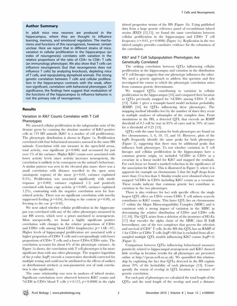
Variation in KI67 Counts Correlates with T CellPhenotypes
We assessed cellular proliferation in the subgranular zone of the
dentate gyrus by counting the absolute number of KI67-positive
cells in 719 HS animals (KI67 is a marker of cell proliferation).
The phenotypic distribution is shown in Figure 1a. Proliferation
correlated with four behavioural measures assessed in the same HS
animals. Correlation with one measure in the open-field arena,
total activity, was significant (p = 0.006) and accounted for just
over 1% of the variance. Higher KI67 counts were associated with
lower activity levels (since activity increases neurogenesis, the
correlation is unlikely to be consequent on the animal’s behaviour).
A similar pattern was seen in the elevated plus maze: there was a
small correlation with distance travelled in the open arms
(anxiogenic regions) of the maze (p = 0.03, variance explained
0.5%). Proliferation was associated significantly with startle
response (p = 0.002, variance explained 1.5) and positively
correlated with home cage activity (p = 0.005, variance explained
1.2%), contrasting with the negative correlation seen for fear-
related activity. There was no significant correlation with novelty
suppressed feeding (p = 0.64), freezing to the context (p = 0.09), or
freezing to the cue (p = 0.92).
We next asked whether cellular proliferation in the hippocam-
pus was correlated with any of the other phenotypes measured in
our HS screen, which were a priori unrelated to neurogenesis.
Most unexpectedly, we found a highly significant positive
correlation with changes in the relative proportions of CD4+and CD8+ cells among blood CD3+ lymphocytes (p = 1.6E207).
Higher levels of hippocampal proliferation are associated with a
higher proportion of CD8+ T cells and correspondingly with lower
proportions of CD4+ T cells and a lower CD4+/CD8+ ratio. The
correlation accounts for about 6% of the phenotypic variance. As
Figure 1a shows, the correlation with T cell phenotypes is far more
significant than with any other phenotype. The negative logarithm
of the p value (logP) exceeds a conservative threshold corrected for
multiple testing and could not be attributed to the effects of outliers
or distributional artefacts (a non-parametric test of rank correla-
tion is also significant).
The same relationship was seen in analyses of inbred strains.
Significant correlations were observed between KI67 counts and
%CD8 in CD3+ blood T cells (r = 0.115, p = 0.0008) in the eight
inbred progenitor strains of the HS (Figure 1b). Using published
data from a large genetic reference panel of recombinant inbred
strains (BXD) [12,15], we found the same correlations between
cellular proliferation in the hippocampus and CD8+ T cell
frequency (r = 0.61, p = 0.0006) (Figure 1c). Replication in the two
inbred samples provides conclusive evidence for the robustness of
the correlation.
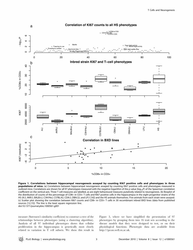
KI67 and T Cell Subpopulation Phenotypes AreGenetically Correlated
The striking correlation between QTLs influencing cellular
proliferation in the hippocampus and variation in the distribution
of T cell lineages suggests that one phenotype influences the other.
We used a genetic approach to address this question and first
investigated the extent to which the phenotypic correlation arises
from common genetic determinants.
We mapped QTLs contributing to variation in cellular
proliferation in the hippocampus [16] and compared their location
to QTLs previously mapped for %CD8+ among CD3+ T cells
[13]. Table 1 gives a resample-based model inclusion probability
(RMIP) [16] for QTLs influencing these phenotypes. The
mapping method identifies loci by the number of times they recur
in multiple analyses of subsamples of the complete data. From
simulations in the HS, a detected QTL that exceeds an RMIP
threshold of 0.5 will be true in 85% of cases and in 70% of cases
for a threshold of 0.25 [13].
QTLs with the same location for both phenotypes are found on
five chromosomes, 5, 6, 10, 13, and 16. However, plots of the
logPs frequently identify the same peaks for both phenotypes
(Figure 2), suggesting that there may be additional peaks that
influence both phenotypes. To test whether variation in T cell
lineages and cellular proliferation in the hippocampus have a
common genetic origin, we included %CD8+ in CD3+ as a
covariate in a linear model for KI67 and mapped the residuals.
For each locus we found a marked reduction in the significance of
the association for KI67. This is illustrated in Figure 2, where it is
apparent for example on chromosome 5 that the logP drops from
more than 15 to less than 3. Similar results were obtained when we
mapped %CD8+ in CD3+ including KI67 counts as a covariate.
These results indicate that common genetic loci contribute to
variation in the two phenotypes.
There is also evidence for loci with specific effects: the single
largest QTL effect on CD4+ versus CD8+ T cell subsets scarcely
contributes to KI67 counts. This latter QTL lies on chromosome
17 within the Major Histocompatibility Complex (MHC) and is
consistent with a strong impact of variation in the MHC in
determining the relative distribution of CD4+ and CD8+ cells
[17,18]. The QTL arises from a deletion of the promoter of H2-Ea
[17] that encodes the alpha chain of the MHC class II Eabheterodimer, one of the two complexes that govern the selection
and survival of CD4+ T cells. In the HS this QTL has an RMIP of
1 for CD4+ or CD8+ T cells (logP.80) but is excluded from all re-
sampled multiple QTL models influencing KI67 counts (logP,4)
(Figure 2).
Comparison between QTLs influencing behavioural measures
putatively related to hippocampal neurogenesis and KI67 showed
little overlap in location (results for all phenotypes are presented
online at http://gscan.well.ox.ac.uk). We quantified this relation-
ship by exploiting the fact that QTLs detected in the HS explain
about 70% of the heritability of each phenotype [13]. Conse-
quently the extent of overlap in QTL location is a measure of
genetic correlation.
For each pair of phenotypes we calculated the total length of the
QTLs and the total length of the overlap and used a distance
Author Summary
In adult mice new neurons are produced in thehippocampus, where they are thought to influencelearning, memory, and emotional regulation. The mecha-nisms and functions of this neurogenesis, however, remainunclear. Here we report that in different strains of mice,variation in cellular proliferation in the hippocampus (anindex of neurogenesis) correlates with variation in therelative proportions of the ratio of CD4+ to CD8+ T cells(an immunology phenotype). We also show that T cells caninfluence neurogenesis (but that neurogenesis does notinfluence T cells) by analyzing knockouts, depleting miceof T cells, and repopulating alymphoid animals. The stronggenetic correlation between T cells and cellular prolifera-tion in the hippocampus contrasts with the weak, oftennon-significant, correlation with behavioral phenotypes. Ofsignificance, the findings here suggest that modulation ofthe functions of the hippocampus to influence behavior isnot the primary role of neurogenesis.
T Cells and Neurogenesis
PLoS Biology | www.plosbiology.org 2 December 2010 | Volume 8 | Issue 12 | e1000561
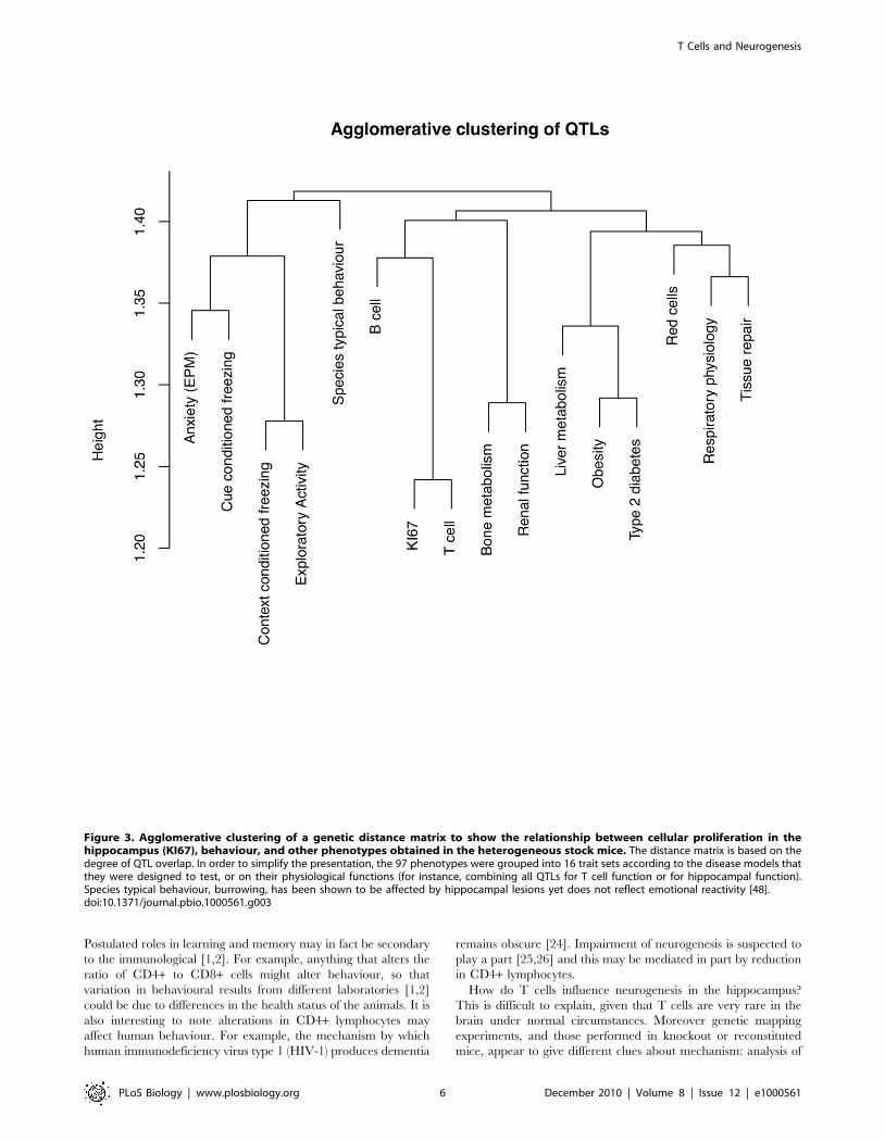
measure (Sørensen’s similarity coefficient) to construct a tree of the
relationships between phenotypes (using a clustering algorithm).
Analysis of all 97 individual phenotypes shows that cellular
proliferation in the hippocampus is genetically most closely
related to variation in T cell subsets. We show this result in
Figure 3, where we have simplified the presentation of 97
phenotypes by grouping them into 16 trait sets according to the
disease models that they were designed to test, or on their
physiological functions. Phenotypic data are available from
http://gscan.well.ox.ac.uk.
Figure 1. Correlations between hippocampal neurogenesis assayed by counting KI67 positive cells and phenotypes in threepopulations of mice. (a) Correlations between hippocampal neurogenesis assayed by counting KI67 positive cells and phenotypes measured inoutbred mice. Correlations are shown for all 97 phenotypes measured with the negative logarithm of the p value (log10P) of the Spearman correlationcoefficient on the vertical axis. Three T cell measures are labelled, as are eight behavioural measures putatively related to neurogenesis. (b) Boxplot ofthe distribution of counts of the percentage of CD8+ in CD3+ T cells and KI67 positive cells in the hippocampus in the eight progenitor strains of theHS (A/J, AKR/J, BALB/cJ, C3H/HeJ, C57BL/6J, CDA/J, DBA/2J, and LP/J [16]) and the HS animals themselves. Five animals from each strain were assayed.(c) Scatter plot showing the correlation between KI67 counts and CD8+ in CD3+ T cells in 30 recombinant inbred BXD lines (data from publishedsources [12,15]). The line is the least square regression line.doi:10.1371/journal.pbio.1000561.g001
T Cells and Neurogenesis
PLoS Biology | www.plosbiology.org 3 December 2010 | Volume 8 | Issue 12 | e1000561

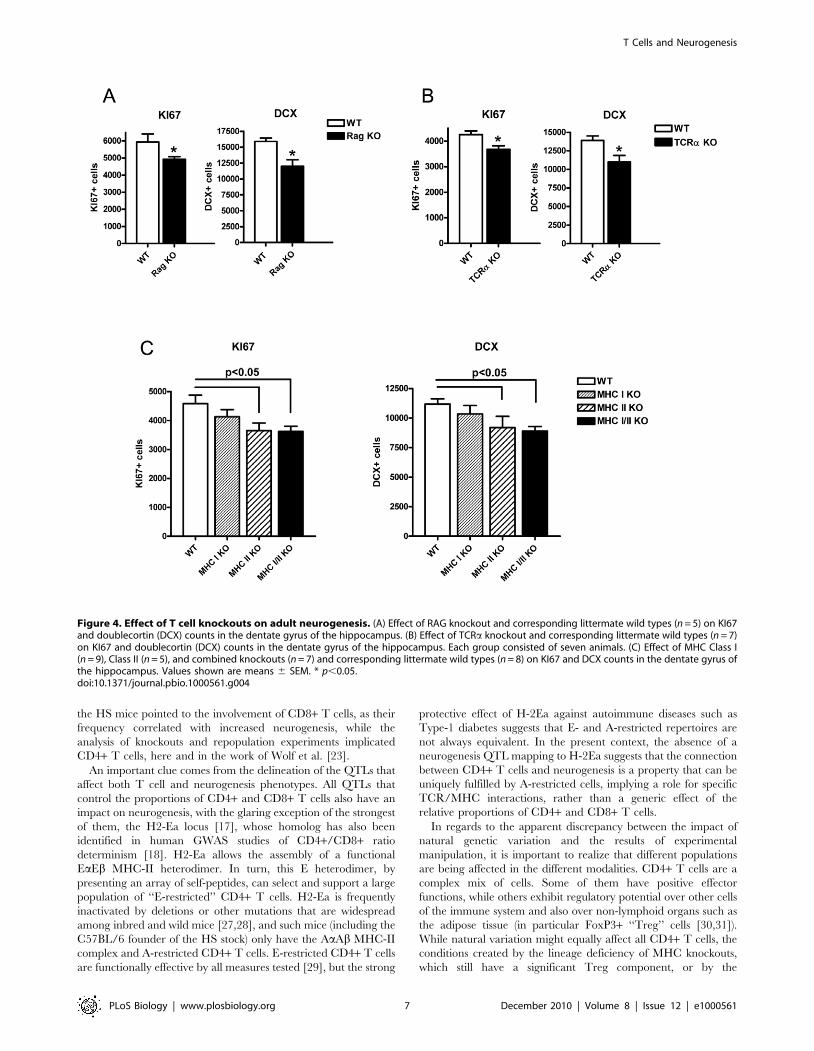
CD4+ T Cells Mediate the Effect on Adult NeurogenesisOur genetic data are consistent with a model in which adult
neurogenesis influences T cells and also with one in which T cells
influence neurogenesis (where our measure of neurogenesis is
cellular proliferation in the hippocampus). We tested the first
model by ablating adult neurogenesis and then looking for changes
in T cells. We did this using i.c.v. ganciclovir (GCV) in both
GFAP-tk [6] and nestin-tk mice [19]. Four weeks later, we
collected blood samples and quantitated CD4+ and CD8+ T cell
subsets. This schedule is effective in ablating dividing putative
forebrain progenitors in GFAP-TK mice while avoiding the gut
illness caused by high doses of GCV [20,21]. The hippocampus
was stained with a marker for immature neurons (doublecortin,
DCX). DCX positive cells were depleted in both models.
Compared to untreated littermates we found no significant
difference in T cell subsets after ablation of neurogenesis in either
model (GFAP-tk: t = 21.7, df = 14, p value = 0.1; nestin-tk: t = 0.1,
df = 14, p = 0.9).
We tested the second hypothesis, an effect of T cell function on
neurogenesis, by examining a series of mouse mutants with
alterations in T cell differentiation. As well as the cell proliferation
marker KI67, we used DCX to detect hippocampal neurogenesis
in 10-wk-old mice. We first investigated Rag-deficient mice, which
are devoid of any B or T lymphocytes: mutant mice showed a
marked reduction in both readouts of neurogenesis (KI67 and
DCX) (Figure 4A; p = 0.02 (KI67) and p = 0.01 (DCX)). This was
also true for TCRa knockouts (Figure 4B, p = 0.01 (KI67) and
p = 0.02 (DCX)), implicating cells expressing the abT cell receptor
and excluding B and cdT lymphocytes.
To determine which of the CD4+ or CD8+ lineages of abT cells
had the most influence, we tested knockout mice affecting MHC-I
or MHC-II molecules (which are largely devoid of CD8+ and
CD4+ T cells, respectively) as well as double-knockout animals. A
reduction in KI67 and DCX was present for both MHC-II
(p = 0.03 (KI67) and p = 0.04 (DCX)) and MHC-I/II deficient
mice (p = 0.006 (KI67) and p = 0.002 (DCX)), suggesting that the
presence of CD4+ cells mediate the effect of lymphocytes on
neurogenesis (Figure 4C).
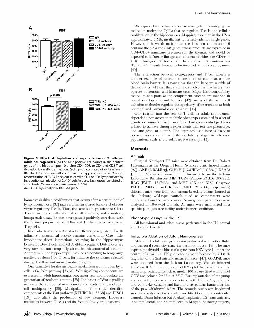
We performed two independent tests of the functional
involvement of CD4+ and CD8+ T cells. First, we depleted either
population by injection of anti-CD4 or anti-CD8 monoclonal
antibodies, reducing their numbers to less than 5% of untreated
mice at 10 d post-treatment. Despite the successful depletion of T-
cell subsets, there was no significant change of KI67 levels in the
dentate gyrus of the hippocampus (Figure 5A). Second, we
performed a repopulation experiment, in which 26107 CD4+ or
CD8+ T cells from spleen and lymph node were transferred into
TCRa-deficient hosts. Transfer of CD4+ T cells led to a
significant increase in KI67 staining in 2 wk post-transfer
(compared to the TCRa knockout p = 0.03). In contrast transfer
of CD8+ T cells did not alter KI67 staining (Figure 5B).
Discussion
Our analysis of a large stock of outbred mice makes two novel
observations about adult neurogenesis in the hippocampus. First,
we find a correlation between variation in cellular proliferation in
the hippocampus and the relative proportions of CD4+ and CD8+subsets of abT cells. The correlation is much larger than that
found for over 90 other phenotypes. Second, the correlation is
driven by genetics: QTLs that contribute to variation in cell
proliferation in the hippocampus also contribute to variation in the
relative proportions of CD4+ and CD8+ T cells. Importantly, the
genetic correlation is due to natural sequence variants that
differentiate inbred strains of laboratory mice.
Our findings extend previous observations that T cells
contribute to variation in adult neurogenesis [22,23]. Previously,
analysis of mice with a mutation that ablated both T- and B-
lymphocyte compartments showed impairment of adult neuro-
genesis, which could be restored by repopulation with mono-
specific T cells recognizing a CNS-antigen (myelin basic-protein).
This was interpreted as an effect of autoimmune attack [22].
Reduced adult neurogenesis was also found in mice with no T or B
cells due to a mutation in either RAG1 or RAG2 [23].
Repopulation and depletion experiments again implicated T cells
[23]. The latter experiments, together with analysis of CD42/2
mice, indicated that CD4+ T cells contributed to adult
neurogenesis, regardless of their antigen specificity.
Results obtained from the HS indicate that the relationship
between T cells and cellular proliferation in the hippocampus is
not merely an artefact of transgenic or knockout mice, or of the
very unusual conditions created in alymphoid mice upon immune
reconstitution, but arises from the far more subtle cues of natural
genetic variation: loci that contribute to variation in cell
Table 1. QTLs that contribute to cellular proliferation in thehippocampus (KI67) and to %CD8 in CD3 T-cells.
Phenotype Chr Start End RMIP
%CD8+ in CD3 1 59.2 59.9 0.31
KI67 1 132.7 137.7 0.4
%CD8+ in CD3 3 126.9 129.5 0.59
KI67 4 40.4 49.8 0.26
KI67 5 71.2 76.8 0.68
%CD8+ in CD3 5 70.8 76.8 0.2
%CD8+ in CD3 5 113.6 114.7 0.94
KI67 6 68.8 74.8 0.22
%CD8+ in CD3 6 68.8 73.7 0.94
KI67 6 127.1 127.8 0.27
%CD8+ in CD3 7 47.9 51.8 0.25
%CD8+ in CD3 7 118.4 122.6 0.36
%CD8+ in CD3 10 38.3 40.3 0.2
KI67 10 38.3 45.3 0.26
%CD8+ in CD3 12 26.6 30.9 0.77
KI67 13 35.2 37.7 0.53
%CD8+ in CD3 13 36.1 37.7 0.35
KI67 13 110.6 119.2 0.49
KI67 16 34.4 35.7 0.67
%CD8+ in CD3 16 34.8 36.6 0.3
%CD8+ in CD3 17 33.7 34.5 1
%CD8+ in CD3 17 43.6 49.6 0.58
The table gives the start and end coordinates of the 95% confidence intervals ofeach QTL (these coordinates are in megabases and are from Build 37 of themouse genome). Entries in bold indicate QTLs where there is overlap betweenthe two phenotypes. Our measure of probability that the locus is correctlyidentified is a resample model inclusion probability (RMIP) in the last column.An RMIP is the expected proportion of times a locus is included in a multilocusmodel. A value of 1 means that the locus would be included in all repeatedanalyses (estimated by re-sampling the data) and an RMIP of 0.5 means thelocus is included in 50% of such analyses. Assessing false positive rates bysimulation indicates that at an RMIP threshold of 0.25 about one false positiveQTL occurs every four genome scans.doi:10.1371/journal.pbio.1000561.t001
T Cells and Neurogenesis
PLoS Biology | www.plosbiology.org 4 December 2010 | Volume 8 | Issue 12 | e1000561
proliferation in the hippocampus also contribute to variation in the
relative proportions of CD4+ and CD8+ T cells. Naturally
occurring genetic variation is a very different setting (and
obviously more physiogically relevant) than the gross abnormal-
ities of knockout mice.
Two questions follow: why do T cells influence neurogenesis in
the hippocampus, and how do they do it? Our data suggest a
broader role for T cell function on adult neurogenesis than
previously suspected [22,23]. The strikingly large correlation
between cellular proliferation in the hippocampus and T cell
subsets, attributable to naturally occurring genetic variation,
suggests that the correlation itself is functionally important
(otherwise it would have decayed through stochastic fluctuation
in allele frequencies). The strong correlation contrasts with the
weak, often non-significant, correlation with behavioural pheno-
types often invoked as functional consequences of adult neuro-
genesis. This suggests that modulation of the behavioural functions
of the hippocampus may not be the primary role of neurogenesis.
Figure 2. Genetic mapping of cellular proliferation in the hippocampus (KI67), the %CD8+ in CD3 T cells, and the residuals of amodel including %CD8+ in CD3 as a covariate (residual KI67). The horizontal scale is the megabase (Mb) distance along the chromosome(build 37 of the mouse genome) and the horizontal scale the negative logarithm of the p value (logP) for association between the phenotype andgenotype. Results from chromosomes 1, 5, and 17 are shown. Double-headed arrows indicate peaks with a RMIP greater than 0.25. The dotted line inthe KI67 plots is the result of mapping the KI67 counts conditioning on %CD8+ in CD3 T cells.doi:10.1371/journal.pbio.1000561.g002
T Cells and Neurogenesis
PLoS Biology | www.plosbiology.org 5 December 2010 | Volume 8 | Issue 12 | e1000561
Postulated roles in learning and memory may in fact be secondary
to the immunological [1,2]. For example, anything that alters the
ratio of CD4+ to CD8+ cells might alter behaviour, so that
variation in behavioural results from different laboratories [1,2]
could be due to differences in the health status of the animals. It is
also interesting to note alterations in CD4+ lymphocytes may
affect human behaviour. For example, the mechanism by which
human immunodeficiency virus type 1 (HIV-1) produces dementia
remains obscure [24]. Impairment of neurogenesis is suspected to
play a part [25,26] and this may be mediated in part by reduction
in CD4+ lymphocytes.
How do T cells influence neurogenesis in the hippocampus?
This is difficult to explain, given that T cells are very rare in the
brain under normal circumstances. Moreover genetic mapping
experiments, and those performed in knockout or reconstituted
mice, appear to give different clues about mechanism: analysis of
Figure 3. Agglomerative clustering of a genetic distance matrix to show the relationship between cellular proliferation in thehippocampus (KI67), behaviour, and other phenotypes obtained in the heterogeneous stock mice. The distance matrix is based on thedegree of QTL overlap. In order to simplify the presentation, the 97 phenotypes were grouped into 16 trait sets according to the disease models thatthey were designed to test, or on their physiological functions (for instance, combining all QTLs for T cell function or for hippocampal function).Species typical behaviour, burrowing, has been shown to be affected by hippocampal lesions yet does not reflect emotional reactivity [48].doi:10.1371/journal.pbio.1000561.g003
T Cells and Neurogenesis
PLoS Biology | www.plosbiology.org 6 December 2010 | Volume 8 | Issue 12 | e1000561
the HS mice pointed to the involvement of CD8+ T cells, as their
frequency correlated with increased neurogenesis, while the
analysis of knockouts and repopulation experiments implicated
CD4+ T cells, here and in the work of Wolf et al. [23].
An important clue comes from the delineation of the QTLs that
affect both T cell and neurogenesis phenotypes. All QTLs that
control the proportions of CD4+ and CD8+ T cells also have an
impact on neurogenesis, with the glaring exception of the strongest
of them, the H2-Ea locus [17], whose homolog has also been
identified in human GWAS studies of CD4+/CD8+ ratio
determinism [18]. H2-Ea allows the assembly of a functional
EaEb MHC-II heterodimer. In turn, this E heterodimer, by
presenting an array of self-peptides, can select and support a large
population of ‘‘E-restricted’’ CD4+ T cells. H2-Ea is frequently
inactivated by deletions or other mutations that are widespread
among inbred and wild mice [27,28], and such mice (including the
C57BL/6 founder of the HS stock) only have the AaAb MHC-II
complex and A-restricted CD4+ T cells. E-restricted CD4+ T cells
are functionally effective by all measures tested [29], but the strong
protective effect of H-2Ea against autoimmune diseases such as
Type-1 diabetes suggests that E- and A-restricted repertoires are
not always equivalent. In the present context, the absence of a
neurogenesis QTL mapping to H-2Ea suggests that the connection
between CD4+ T cells and neurogenesis is a property that can be
uniquely fulfilled by A-restricted cells, implying a role for specific
TCR/MHC interactions, rather than a generic effect of the
relative proportions of CD4+ and CD8+ T cells.
In regards to the apparent discrepancy between the impact of
natural genetic variation and the results of experimental
manipulation, it is important to realize that different populations
are being affected in the different modalities. CD4+ T cells are a
complex mix of cells. Some of them have positive effector
functions, while others exhibit regulatory potential over other cells
of the immune system and also over non-lymphoid organs such as
the adipose tissue (in particular FoxP3+ ‘‘Treg’’ cells [30,31]).
While natural variation might equally affect all CD4+ T cells, the
conditions created by the lineage deficiency of MHC knockouts,
which still have a significant Treg component, or by the
Figure 4. Effect of T cell knockouts on adult neurogenesis. (A) Effect of RAG knockout and corresponding littermate wild types (n = 5) on KI67and doublecortin (DCX) counts in the dentate gyrus of the hippocampus. (B) Effect of TCRa knockout and corresponding littermate wild types (n = 7)on KI67 and doublecortin (DCX) counts in the dentate gyrus of the hippocampus. Each group consisted of seven animals. (C) Effect of MHC Class I(n = 9), Class II (n = 5), and combined knockouts (n = 7) and corresponding littermate wild types (n = 8) on KI67 and DCX counts in the dentate gyrus ofthe hippocampus. Values shown are means 6 SEM. * p,0.05.doi:10.1371/journal.pbio.1000561.g004
T Cells and Neurogenesis
PLoS Biology | www.plosbiology.org 7 December 2010 | Volume 8 | Issue 12 | e1000561
homeostasis-driven proliferation that occurs after reconstitution of
lymphopenic hosts [32] may result in an altered balance of effector
versus regulatory T cells. Thus, the same subpopulations of CD4+T cells are not equally affected in all instances, and a unifying
interpretation may be that neurogenesis positively correlates with
the relative proportion of CD4+ and CD8+ effector relative to
Treg cells.
In cellular terms, how A-restricted effector or regulatory T-cells
influence hippocampal activity remains conjectural. One might
hypothesize direct interactions occurring in the hippocampus
between CD4+ T cells and MHC-II+ microglia. CD4+ T cells are
very rare but not completely absent in this anatomical location.
Alternatively, the hippocampus may be responding to long-range
mediators released by T cells, for instance the cytokines released
during T cell activation in lymphoid organs.
One candidate for the molecular mechanism set in motion by T
cells is the Wnt pathway [33,34]. Wnt signalling components are
expressed in adult hippocampal progenitor cells and modulate the
generation of newborn neurons [35]. Inhibition of Wnt signalling
increases the number of new neurons and leads to a loss of stem
cell multipotency [36]. Manipulation of recently identified
components of the Wnt pathway (NEUROD1 [37,38] and DISC1
[39]) also alters the production of new neurons. However,
mediators between T cells and the Wnt pathway are unknown.
We expect clues to their identity to emerge from identifying the
molecules under the QTLs that co-regulate T cells and cellular
proliferation in the hippocampus. Mapping resolution in the HS is
approximately 3 Mb, insufficient to formally identify single genes.
However, it is worth noting that the locus on chromosome 6
contains the Cd8a and Cd8b genes, whose products are expressed in
CD4+CD8+ immature precursors in the thymus, and would be
expected to influence lineage commitment to either the CD4+ or
CD8+ lineages. A locus on chromosome 13 contains Fst
(Follistatin), already known to be involved in adult neurogenesis
[40].
The interaction between neurogenesis and T cell subsets is
another example of neural-immune communication across the
blood brain barrier: it is now clear that this happens outside of
disease states [41] and that a common molecular machinery may
operate in neurons and immune cells. Major histocompatibility
molecules and parts of the complement cascade are involved in
neural development and function [42]; many of the same cell
adhesion molecules regulate the specificity of interactions at both
neuronal and immunological synapses [43].
Our insights into the role of T cells in adult neurogenesis
depended upon access to multiple phenotypes obtained in a set of
genotyped animals. The delineation of biological control pathways
is hard to achieve through experiments that test one phenotype,
and one gene, at a time. The approach used here is likely to
become more common with the availability of genetic reference
populations, such as the collaborative cross [44,45].
Methods
AnimalsOriginal Northport HS mice were obtained from Dr. Robert
Hitzemann at the Oregon Health Sciences Unit. Inbred strains
(A/J, AKR/J, BALB/cJ, C3H/HeJ, C57BL/6 J, CBA/J, DBA/2
J, and LP/J) were obtained from Harlan (UK) or the Jackson
Laboratory (Bar Harbor, ME). TCRa (Philpott PMID: 1604321),
RAG (PMID: 1547488), and MHC (Ab and b2M, Cosgrove
PMID: 1909605 and Koller PMID: 2682666, respectively)
deficient mice were from our custom-breeding colony housed at
the Jackson; wild-type controls used as comparators were
littermates from the same crosses. Neurogenesis parameters were
analysed in 10-wk-old animals. All mice were maintained in a
specific pathogen free facility under barrier conditions.
Phenotype Assays in the HSAll behavioural and other assays performed in the HS animal
are described in [46].
Inducible Ablation of Adult NeurogenesisAblation of adult neurogenesis was performed with both cellular
and temporal specificity using the nestin-tk mouse [19]. The mice
express the thymidine kinase (tk) gene from HSV type 1, under the
control of a minimal TK promoter element followed by a 1.8 kb
fragment of the 2nd intronic nestin enhancer [47]. GFAP-tk mice
were obtained from the Jackson Laboratory. We administered
GCV via ICV infusion at a rate of 0.25 ml/h by using an osmotic
minipump. Minipumps (Alzet, model 2004) were filled with 2 mM
GCV and primed for 36 h at 37uC. For implantation of the pump
and cannula, mice were anesthetized with 130 mg/kg ketamine
and 20 mg/kg xylazine and fixed to a stereotaxic frame after loss
of the paw withdrawal reflex. The osmotic pump was implanted
subcutaneously over the scapulae and fitted to an intraven-tricular
cannula (Brain Infusion Kit 3, Alzet) implanted 0.21 mm anterior,
0.85 mm lateral, and 3.0 mm deep to Bregma. Following surgery,
Figure 5. Effect of depletion and repopulation of T cells onadult neurogenesis. (A) The KI67 positive cell counts in the dentategyrus of the hippocampus 10 d after CD4, CD8, or CD4 and CD8 T celldepletion by antibody injection. Each group consisted of eight animals.(B) The KI67 positive cell counts in the hippocampus after 2 wk ofreconstitution of TCRa knockout mice with CD4 or CD8 lymphocytes byintraperitoneal injection of 26107 cells/mouse. Each group consisted ofsix animals. Values shown are means 6 SEM.doi:10.1371/journal.pbio.1000561.g005
T Cells and Neurogenesis
PLoS Biology | www.plosbiology.org 8 December 2010 | Volume 8 | Issue 12 | e1000561
all mice were housed individually for the remainder of the
experiment.
ImmunohistochemistryFor KI67 staining, sections were mounted on the superfrost slide
(BDH, UK), dried overnight, incubated in the 0.01 mol/L citric
buffer for 40 min at 90, incubated in 3% H2O2 for 10 min, rinsed,
and incubated overnight at room temperature with rabbit anti-
KI67 antibody (1:4000, Vector Lab). Next day, a standard rabbit
IgG ABC kit procedure was used and reacted 5–10 min with
Sigma DAB tablet. Sections were then counterstained with cresyl
violet and cover-slipped under DPX. KI67-labeled cells were
counted on every eighth section through the entire rostrocaudal
extent of the granule cell layer bilaterally. Section thickness is
40 mm. For DCX staining, free floating sections were incubated
with goat anti-DCX antibody (1:400, Santa Cruz Labs), then
following the same ABC kit procedure, reacted with Sigma DAB
tablet. DCX-labeled cells were counted on every sixteenth section
through the entire rostrocaudal extent of the granule cell layer
bilaterally. Section thickness is 40 mm.
Flow CytometryBlood samples (100–150 ml) were gathered in EDTA via tail-
vein phlebotomy. Erythrocytes were lysed in 5 rounds of
incubation with ACK lysis buffer followed by centrifugation.
Blocking in Fc solution was followed by incubation with CD3-PE,
CD4-APC, and CD8 FITC (eBiosciences) and two rinses in FACS
buffer. Flow cytometry was then performed on a BD FacsCanto II
instrument with FACS Diva software (BD Biosciences). Counts of
CD4 and CD8 positive cells were gated on the CD3 population,
and all percentages are expressed as a fraction of CD3+ cells.
Depletion of T Cell Subsets by AntibodyC57BL6/J mice (n = 8 in each group) received mouse received
intraperitoneal injections of anti-CD4 (YTS 191 and YTA 3.1.2)
or anti-CD8 (169) antibodies or both together, and the control
mice were injected with YK1X337 IgG. Antibodies were given
over a 3-d period, 0.5 mg per injection of anti-CD8, anti-CD4, or
control IgG antibody injection. Antibodies for depletion were a gift
from Dr. Stephen P. Cobbold, Sir William Dunn School of
Pathology, Oxford, UK. Mice were killed 8 d after the last
injection. Blood samples were collected for CD3, CD4, and CD8
analysis.
T Cell Isolation TransferSingle cell suspensions of splenocytes from C57BL6/J donors
were subjected to magnetic bead cell sorting using anti-CD4 or
anti-CD8a paramagnetic beads (Miltenyi-Biotech) and positively
sorted with the AutoMACs cell separation equipment (Miltenyi-
Biotech) according to the manufacturer’s instructions. Briefly, cells
were incubated with beads on ice for 20 min in PBS 2 mM EDTA
washed and applied to the AutoMACS, and positive and negative
fractions were collected and tested for purity prior to transfer (all
transferred populations were .96% pure as judged by FACS).
Sorted cell populations were washed by centrifugation in DMEM,
resuspended to 86107/ml and administered to TCRa KO mice
(n = 6 in each group) by intraperitoneal injection of 250 ml/mouse
(26107/mouse).
Author Contributions
The author(s) have made the following declarations about their
contributions: Conceived and designed the experiments: GJH PK CB JF.
Performed the experiments: GJH ALS DHDG CC BHS AE SS JMP AJ DM.
Analyzed the data: GJH DHDG JMP RM PK CB. Wrote the paper: CB JF.
References
1. Clelland CD, Choi M, Romberg C, Clemenson GD, Jr., Fragniere A, et al.(2009) A functional role for adult hippocampal neurogenesis in spatial pattern
separation. Science 325: 210–213.
2. Deng W, Aimone JB, Gage FH (2010) New neurons and new memories: how
does adult hippocampal neurogenesis affect learning and memory? Nat Rev
Neurosci 11: 339–350.
3. Gould E (2007) How widespread is adult neurogenesis in mammals? Nat Rev
Neurosci 8: 481–488.
4. Leuner B, Gould E, Shors TJ (2007) Is there a link between adult neurogenesis
and learning? Hippocampus 16: 216–224.
5. Jaholkowski P, Kiryk A, Jedynak P, Ben Abdallah NM, Knapska E, et al. (2009)New hippocampal neurons are not obligatory for memory formation; cyclin D2
knockout mice with no adult brain neurogenesis show learning. Learn Mem 16:439–451.
6. Saxe MD, Battaglia F, Wang JW, Malleret G, David DJ, et al. (2006) Ablation ofhippocampal neurogenesis impairs contextual fear conditioning and synaptic
plasticity in the dentate gyrus. Proc Natl Acad Sci U S A 103: 17501–17506.
7. Dupret D, Revest J-M, Koehl M, Ichas F, De Giorgi F, et al. (2008) Spatialrelational memory requires hippocampal adult neurogenesis. PLoS One 3:
e1959. doi:10.1371/journal.pone.0001959.
8. Zhang C-L, Zou Y, He W, Gage FH, Evans RM (2008) A role for adult TLX-
positive neural stem cells in learning and behvaviour. Nature 451: 1004–1007.
9. Farioli-Vecchioli S, Saraulli D, Costanza M, Pacioni S, Cina I, et al. (2008) Thetiming of differentiation of adult hippocampal neurons is crucial for spatial
memory. PLoS Biology 6: e246. doi:10.1371/journal.pbio.0060246.
10. Imayoshi I, Sakamoto M, Ohtsuka T, Takao K, Miyakawa T, et al. (2008) Roles
of continuous neurogenesis in the structural and functional integrity of the adult
forebrain. Nat Neurosci 11: 1153–1161.
11. Bergami M, Rimondini R, Santi S, Blum R, Gotz M, et al. (2008) Deletion of
TrkB in adult progenitors alters newborn neuron integration into hippocampalcircuits and increases anxiety-like behavior. Proc Natl Acad Sci U S A 105:
15570–15575.
12. Kempermann G, Chesler EJ, Lu L, Williams RW, Gage FH (2006) Naturalvariation and genetic covariance in adult hippocampal neurogenesis. Proc Natl
Acad Sci U S A 103: 780–785.
13. Valdar W, Solberg LC, Gauguier D, Burnett S, Klenerman P, et al. (2006)
Genome-wide genetic association of complex traits in heterogeneous stock mice.Nat Genet 38: 879–887.
14. Santarelli L, Saxe M, Gross C, Surget A, Battaglia F, et al. (2003) Requirement
of hippocampal neurogenesis for the behavioral effects of antidepressants.Science 301: 805–809.
15. Chen J, Harrison DE (2002) Quantitative trait loci regulating relativelymphocyte proportions in mouse peripheral blood. Blood 99: 561–566.
16. Valdar W, Holmes CC, Mott R, Flint J (2009) Mapping in structured
populations by resample model averaging. Genetics 182: 1263–1277.
17. Yalcin B, Nicod J, Bhomra A, Davidson S, Cleak J, et al. (2010) Commerciallyavailable outbred mice for genome-wide association studies. PLoS Genet 6(9):
e1001085. doi:10.1371/journal.pgen.1001085.
18. Ferreira MA, Mangino M, Brumme CJ, Zhao ZZ, Medland SE, et al. (2010)
Quantitative trait loci for CD4:CD8 lymphocyte ratio are associated with risk oftype 1 diabetes and HIV-1 immune control. Am J Hum Genet 86: 88–92.
19. Singer BH, Jutkiewicz EM, Fuller CL, Lichtenwalner RJ, Zhang H, et al. (2009)
Conditional ablation and recovery of forebrain neurogenesis in the mouse.
J Comp Neurol 514: 567–582.
20. Bush TG, Savidge TC, Freeman TC, Cox HJ, Campbell EA, et al. (1998)Fulminant jejuno-ileitis following ablation of enteric glia in adult transgenic
mice. Cell 93: 189–201.
21. Garcia AD, Doan NB, Imura T, Bush TG, Sofroniew MV (2004) GFAP-
expressing progenitors are the principal source of constitutive neurogenesis inadult mouse forebrain. Nat Neurosci 7: 1233–1241.
22. Ziv Y, Ron N, Butovsky O, Landa G, Sudai E, et al. (2006) Immune cells
contribute to the maintenance of neurogenesis and spatial learning abilities inadulthood. Nat Neurosci 9: 268–275.
23. Wolf SA, Steiner B, Akpinarli A, Kammertoens T, Nassenstein C, et al. (2009)
CD4-positive T lymphocytes provide a neuroimmunological link in the control
of adult hippocampal neurogenesis. J Immunol 182: 3979–3984.
24. Kaul M (2009) HIV-1 associated dementia: update on pathological mechanismsand therapeutic approaches. Curr Opin Neurol 22: 315–320.
25. Krathwohl MD, Kaiser JL (2004) HIV-1 promotes quiescence in human neural
progenitor cells. J Infect Dis 190: 216–226.
26. Okamoto S, Kang YJ, Brechtel CW, Siviglia E, Russo R, et al. (2007) HIV/
gp120 decreases adult neural progenitor cell proliferation via checkpoint kinase-mediated cell-cycle withdrawal and G1 arrest. Cell Stem Cell 1: 230–236.
27. Dembic Z, Ayane M, Klein J, Steinmetz M, Benoist CO, et al. (1985) Inbred
and wild mice carry identical deletions in their E alpha MHC genes. EMBO J 4:127–131.
T Cells and Neurogenesis
PLoS Biology | www.plosbiology.org 9 December 2010 | Volume 8 | Issue 12 | e1000561
28. Mathis DJ, Benoist C, Williams VE, 2nd, Kanter M, McDevitt HO (1983)
Several mechanisms can account for defective E alpha gene expression in
different mouse haplotypes. Proc Natl Acad Sci U S A 80: 273–277.
29. Cosgrove D, Bodmer H, Bogue M, Benoist C, Mathis D (1992) Evaluation of the
functional equivalence of major histocompatibility complex class II A and E
complexes. J Exp Med 176: 629–634.
30. Littman DR, Rudensky AY Th17 and regulatory T cells in mediating and
restraining inflammation. Cell 140: 845–858.
31. Feuerer M, Herrero L, Cipolletta D, Naaz A, Wong J, et al. (2009) Lean, but not
obese, fat is enriched for a unique population of regulatory T cells that affect
metabolic parameters. Nat Med 15: 930–939.
32. Goldrath AW, Bevan MJ (1999) Selecting and maintaining a diverse T-cell
repertoire. Nature 402: 255–262.
33. Li G, Pleasure SJ (2005) Morphogenesis of the dentate gyrus: what we are
learning from mouse mutants. Dev Neurosci 27: 93–99.
34. Inestrosa NC, Arenas E (2010) Emerging roles of Wnts in the adult nervous
system. Nat Rev Neurosci 11: 77–86.
35. Zhou CJ, Zhao C, Pleasure SJ (2004) Wnt signaling mutants have decreased
dentate granule cell production and radial glial scaffolding abnormalities.
J Neurosci 24: 121–126.
36. Wexler EM, Paucer A, Kornblum HI, Palmer TD, Geschwind DH (2009)
Endogenous Wnt signaling maintains neural progenitor cell potency. Stem Cells
27: 1130–1141.
37. Kuwabara T, Hsieh J, Muotri A, Yeo G, Warashina M, et al. (2009) Wnt-
mediated activation of NeuroD1 and retro-elements during adult neurogenesis.
Nat Neurosci 12: 1097–1105.
38. Gao Z, Ure K, Ables JL, Lagace DC, Nave KA, et al. (2009) Neurod1 is essential for
the survival and maturation of adult-born neurons. Nat Neurosci 12: 1090–1092.39. Mao Y, Ge X, Frank CL, Madison JM, Koehler AN, et al. (2009) Disrupted in
schizophrenia 1 regulates neuronal progenitor proliferation via modulation of
GSK3beta/beta-catenin signaling. Cell 136: 1017–1031.40. Ageta H, Murayama A, Migishima R, Kida S, Tsuchida K, et al. (2008) Activin
in the brain modulates anxiety-related behavior and adult neurogenesis. PLoSOne 3: e1869. doi:10.1371/journal.pone.0001869.
41. Lucin KM, Wyss-Coray T (2009) Immune activation in brain aging and
neurodegeneration: too much or too little? Neuron 64: 110–122.42. Shatz CJ (2009) MHC class I: an unexpected role in neuronal plasticity. Neuron
64: 40–45.43. Yamada S, Nelson WJ (2007) Synapses: sites of cell recognition, adhesion, and
functional specification. Annu Rev Biochem 76: 267–294.44. Chesler EJ, Miller DR, Branstetter LR, Galloway LD, Jackson BL, et al. (2008)
The Collaborative Cross at Oak Ridge National Laboratory: developing a
powerful resource for systems genetics. Mamm Genome 19: 382–389.45. Churchill GA, Airey DC, Allayee H, Angel JM, Attie AD, et al. (2004) The
Collaborative Cross, a community resource for the genetic analysis of complextraits. Nat Genet 36: 1133–1137.
46. Solberg LC, Valdar W, Gauguier D, Nunez G, Taylor A, et al. (2006) A protocol
for high-throughput phenotyping, suitable for quantitative trait analysis in mice.Mamm Genome 17: 129–146.
47. Yaworsky PJ, Kappen C (1999) Heterogeneity of neural progenitor cells revealedby enhancers in the nestin gene. Dev Biol 205: 309–321.
48. Deacon RM, Rawlins JN (2005) Hippocampal lesions, species-typical behavioursand anxiety in mice. Behav Brain Res 156: 241–249.
T Cells and Neurogenesis
PLoS Biology | www.plosbiology.org 10 December 2010 | Volume 8 | Issue 12 | e1000561