A Defined and Xeno-Free Culture Method Enabling the Establishment of Clinical-Grade Human Embryonic,...
14
A Defined and Xeno-Free Culture Method Enabling the Establishment of Clinical-Grade Human Embryonic, Induced Pluripotent and Adipose Stem Cells Kristiina Rajala 1 *, Bettina Lindroos 1 , Samer M. Hussein 2 , Riikka S. Lappalainen 1 , Mari Pekkanen-Mattila 1 , Jose Inzunza 3 , Bjo ¨ rn Rozell 4 , Susanna Miettinen 1 , Susanna Narkilahti 1 , Erja Kerkela ¨ 1 , Katriina Aalto- Seta ¨la ¨ 1 , Timo Otonkoski 2 , Riitta Suuronen 1,5,6 , Outi Hovatta 7 , Heli Skottman 1 1 Regea - Institute for Regenerative Medicine, University of Tampere, Tampere University Hospital, Tampere, Finland, 2 Biomedicum Stem Cell Center and Children’s Hospital, University of Helsinki, Helsinki, Finland, 3 Division of Medical Nutrition, Department of Biosciences and Nutrition, Karolinska Institutet, Stockholm, Sweden, 4 Department of Laboratory Medicine, Karolinska Institutet, Karolinska University Hospital, Stockholm, Sweden, 5 Department of Biomedical Engineering, Tampere University of Technology, Tampere, Finland, 6 Department of Eye, Ear and Oral Diseases, Tampere University Hospital, Tampere, Finland, 7 Department of Clinical Science, Intervention and Technology, Karolinska Institutet, Karolinska University Hospital, Stockholm, Sweden Abstract Background: The growth of stem cells in in vitro conditions requires optimal balance between signals mediating cell survival, proliferation, and self-renewal. For clinical application of stem cells, the use of completely defined conditions and elimination of all animal-derived materials from the establishment, culture, and differentiation processes is desirable. Methodology/Principal Findings: Here, we report the development of a fully defined xeno-free medium (RegES), capable of supporting the expansion of human embryonic stem cells (hESC), induced pluripotent stem cells (iPSC) and adipose stem cells (ASC). We describe the use of the xeno-free medium in the derivation and long-term (.80 passages) culture of three pluripotent karyotypically normal hESC lines: Regea 06/015, Regea 07/046, and Regea 08/013. Cardiomyocytes and neural cells differentiated from these cells exhibit features characteristic to these cell types. The same formulation of the xeno-free medium is capable of supporting the undifferentiated growth of iPSCs on human feeder cells. The characteristics of the pluripotent hESC and iPSC lines are comparable to lines derived and cultured in standard undefined culture conditions. In the culture of ASCs, the xeno-free medium provided significantly higher proliferation rates than ASCs cultured in medium containing allogeneic human serum (HS), while maintaining the differentiation potential and characteristic surface marker expression profile of ASCs, although significant differences in the surface marker expression of ASCs cultured in HS and RegES media were revealed. Conclusion/Significance: Our results demonstrate that human ESCs, iPSCs and ASCs can be maintained in the same defined xeno-free medium formulation for a prolonged period of time while maintaining their characteristics, demonstrating the applicability of the simplified xeno-free medium formulation for the production of clinical-grade stem cells. The basic xeno- free formulation described herein has the potential to be further optimized for specific applications relating to establishment, expansion and differentiation of various stem cell types. Citation: Rajala K, Lindroos B, Hussein SM, Lappalainen RS, Pekkanen-Mattila M, et al. (2010) A Defined and Xeno-Free Culture Method Enabling the Establishment of Clinical-Grade Human Embryonic, Induced Pluripotent and Adipose Stem Cells. PLoS ONE 5(4): e10246. doi:10.1371/journal.pone.0010246 Editor: Henning Ulrich, University of Sa ˜o Paulo, Brazil Received October 27, 2009; Accepted March 30, 2010; Published April 19, 2010 Copyright: ß 2010 Rajala et al. This is an open-access article distributed under the terms of the Creative Commons Attribution License, which permits unrestricted use, distribution, and reproduction in any medium, provided the original author and source are credited. Funding: This research was supported by the Academy of Finland (grant number 212776) http://www.aka.fi; TEKES the Finnish Funding Agency for Technology and Innovation (grant number 1128/31/05 and 3524/31/07) http://www.tekes.fi; the Competitive Research Funding of the Pirkanmaa Hospital District (grant number 9H197) http://www.pshp.fi; and the Swedish Research Council (grant number K2007-54X-15080-04-3) http://www.vr.se. The generation of iPS cell lines was supported by funding for the ESTOOLS consortium under the Sixth Research Framework Program of the European Union. The funders had no role in study design, data collection and analysis, decision to publish, or preparation of the manuscript. Competing Interests: The authors Kristiina Rajala, Riitta Suuronen, Outi Hovatta, and Heli Skottman declare competing financial interests defined as patent application, formulation and methods for culturing embryonic stem cells, PCT/FI2008/050331 - PCT, relating to the xeno-free culture medium described in the article. * E-mail: [email protected] Introduction Stem cells are invaluable tools for research, drug screening, to study diseases and can potentially serve as a resource for regenerative therapies. Multipotent adipose stem cells (ASCs) exhibiting immunoprivileged properties are an attractive and abundant stem cell source for regenerative medicine that upon induction can undergo adipogenic, osteogenic, chondrogenic, neurogenic and myogenic differentiation in vitro [1–3]. However, even more expectations on clinical applicability in diverse fields of cell- and tissue-replacement therapies are focused on pluripotent stem cells. Besides of hESCs, a promising new source of pluripotent cells was recently discovered, as human somatic cells were reprogrammed by introducing a set of transcription factors PLoS ONE | www.plosone.org 1 April 2010 | Volume 5 | Issue 4 | e10246
-
Upload
independent -
Category
Documents
-
view
0 -
download
0
Transcript of A Defined and Xeno-Free Culture Method Enabling the Establishment of Clinical-Grade Human Embryonic,...
A Defined and Xeno-Free Culture Method Enabling theEstablishment of Clinical-Grade Human Embryonic,Induced Pluripotent and Adipose Stem CellsKristiina Rajala1*, Bettina Lindroos1, Samer M. Hussein2, Riikka S. Lappalainen1, Mari Pekkanen-Mattila1,
Jose Inzunza3, Bjorn Rozell4, Susanna Miettinen1, Susanna Narkilahti1, Erja Kerkela1, Katriina Aalto-
Setala1, Timo Otonkoski2, Riitta Suuronen1,5,6, Outi Hovatta7, Heli Skottman1
1 Regea - Institute for Regenerative Medicine, University of Tampere, Tampere University Hospital, Tampere, Finland, 2 Biomedicum Stem Cell Center and Children’s
Hospital, University of Helsinki, Helsinki, Finland, 3 Division of Medical Nutrition, Department of Biosciences and Nutrition, Karolinska Institutet, Stockholm, Sweden,
4 Department of Laboratory Medicine, Karolinska Institutet, Karolinska University Hospital, Stockholm, Sweden, 5 Department of Biomedical Engineering, Tampere
University of Technology, Tampere, Finland, 6 Department of Eye, Ear and Oral Diseases, Tampere University Hospital, Tampere, Finland, 7 Department of Clinical Science,
Intervention and Technology, Karolinska Institutet, Karolinska University Hospital, Stockholm, Sweden
Abstract
Background: The growth of stem cells in in vitro conditions requires optimal balance between signals mediating cellsurvival, proliferation, and self-renewal. For clinical application of stem cells, the use of completely defined conditions andelimination of all animal-derived materials from the establishment, culture, and differentiation processes is desirable.
Methodology/Principal Findings: Here, we report the development of a fully defined xeno-free medium (RegES), capable ofsupporting the expansion of human embryonic stem cells (hESC), induced pluripotent stem cells (iPSC) and adipose stemcells (ASC). We describe the use of the xeno-free medium in the derivation and long-term (.80 passages) culture of threepluripotent karyotypically normal hESC lines: Regea 06/015, Regea 07/046, and Regea 08/013. Cardiomyocytes and neuralcells differentiated from these cells exhibit features characteristic to these cell types. The same formulation of the xeno-freemedium is capable of supporting the undifferentiated growth of iPSCs on human feeder cells. The characteristics of thepluripotent hESC and iPSC lines are comparable to lines derived and cultured in standard undefined culture conditions. Inthe culture of ASCs, the xeno-free medium provided significantly higher proliferation rates than ASCs cultured in mediumcontaining allogeneic human serum (HS), while maintaining the differentiation potential and characteristic surface markerexpression profile of ASCs, although significant differences in the surface marker expression of ASCs cultured in HS andRegES media were revealed.
Conclusion/Significance: Our results demonstrate that human ESCs, iPSCs and ASCs can be maintained in the same definedxeno-free medium formulation for a prolonged period of time while maintaining their characteristics, demonstrating theapplicability of the simplified xeno-free medium formulation for the production of clinical-grade stem cells. The basic xeno-free formulation described herein has the potential to be further optimized for specific applications relating toestablishment, expansion and differentiation of various stem cell types.
Citation: Rajala K, Lindroos B, Hussein SM, Lappalainen RS, Pekkanen-Mattila M, et al. (2010) A Defined and Xeno-Free Culture Method Enabling theEstablishment of Clinical-Grade Human Embryonic, Induced Pluripotent and Adipose Stem Cells. PLoS ONE 5(4): e10246. doi:10.1371/journal.pone.0010246
Editor: Henning Ulrich, University of Sao Paulo, Brazil
Received October 27, 2009; Accepted March 30, 2010; Published April 19, 2010
Copyright: � 2010 Rajala et al. This is an open-access article distributed under the terms of the Creative Commons Attribution License, which permitsunrestricted use, distribution, and reproduction in any medium, provided the original author and source are credited.
Funding: This research was supported by the Academy of Finland (grant number 212776) http://www.aka.fi; TEKES the Finnish Funding Agency for Technologyand Innovation (grant number 1128/31/05 and 3524/31/07) http://www.tekes.fi; the Competitive Research Funding of the Pirkanmaa Hospital District (grantnumber 9H197) http://www.pshp.fi; and the Swedish Research Council (grant number K2007-54X-15080-04-3) http://www.vr.se. The generation of iPS cell lineswas supported by funding for the ESTOOLS consortium under the Sixth Research Framework Program of the European Union. The funders had no role in studydesign, data collection and analysis, decision to publish, or preparation of the manuscript.
Competing Interests: The authors Kristiina Rajala, Riitta Suuronen, Outi Hovatta, and Heli Skottman declare competing financial interests defined as patentapplication, formulation and methods for culturing embryonic stem cells, PCT/FI2008/050331 - PCT, relating to the xeno-free culture medium described in thearticle.
* E-mail: [email protected]
Introduction
Stem cells are invaluable tools for research, drug screening, to
study diseases and can potentially serve as a resource for
regenerative therapies. Multipotent adipose stem cells (ASCs)
exhibiting immunoprivileged properties are an attractive and
abundant stem cell source for regenerative medicine that upon
induction can undergo adipogenic, osteogenic, chondrogenic,
neurogenic and myogenic differentiation in vitro [1–3]. However,
even more expectations on clinical applicability in diverse fields of
cell- and tissue-replacement therapies are focused on pluripotent
stem cells. Besides of hESCs, a promising new source of
pluripotent cells was recently discovered, as human somatic cells
were reprogrammed by introducing a set of transcription factors
PLoS ONE | www.plosone.org 1 April 2010 | Volume 5 | Issue 4 | e10246
linked to pluripotency to yield induced pluripotent stem cells
(iPSC) [4,5]. Human iPSCs are a potential source of patient-
specific pluripotent stem cells that could be used to treat a number
of human degenerative diseases without evoking immune
rejection. From these stem cell types, only patient specific ASCs
have so far been used in clinical cell therapy while clinical trials
using hESCs is at the very beginning (http://www.geron.com).
Many major challenges including teratoma formation, immuno-
genicity and the use of oncogenes and retroviruses in the
reprogramming of iPSCs need to be addressed before hESCs
and iPSCs can be safely used as a source for clinical cell therapy.
One of the major challenges for the clinical use of stem cells is the
exposure to undefined animal-derived products during in vitro
establishment and expansion of the cells. Considerable progress has
been made towards the generation of defined culture conditions for
stem cells. FBS has been mostly replaced with knockout- serum
replacement (KO-SR, Invitrogen) [6–8] and human feeder cells have
been successfully used to replace mouse embryonic fibroblasts (MEFs)
in the derivation and expansion of hESCs and iPSCs [9–15]. In
addition, various feeder cell-free culture conditions have been
developed for the culture of hESCs and iPSCs [16–19]. Despite the
progress, most existing stem cell lines have been exposed to a variety of
undefined animal-derived products which makes these cell lines
undesirable for clinical applications. In addition to establishment and
culture of stem cells, many differentiation protocols utilize a variety of
undefined products that may have unknown effects to the cell
characteristics and differentiation. The potential consequences of
transplanting human cells exposed to animal-derived products into
patients include an increased risk of graft rejection, immunoreactions,
and viral or bacterial infections, prions, and yet unidentified zoonoses
[20–22]. Therefore, optimization and standardization of a fully defined
xeno-free establishment, culture and differentiation methods for stem
cells is needed for research and especially for clinical application.
Since there is a clear indication that hESCs cultured without
feeder cells in long-term cultures may be more prone to
undesirable abnormalities caused by suboptimal culture conditions
and enzymatic passaging of hESCs [23,24], we believe that the
goal for developing clinical-grade hESCs and iPSCs should be
focused on the use of qualified human feeder cells. In the present
study we have developed a completely new defined xeno-free
medium capable of supporting the expansion of hESCs, iPSCs and
ASCs while maintaining their characteristics. In addition, we
describe the derivation and long-term culture of new hESC lines
using the xeno-free medium.
Results
Initial development of a xeno-free culture mediumformulation
We developed a xeno-free medium (RegES) composed of a
knockout-Dulbecco’s modified Eagle’s medium (KO-DMEM,
Invitrogen, Carlsbad, CA) base supplemented with human serum
albumin, amino acids, vitamins, antioxidants, trace minerals, and
growth factors (Table S1). All medium components were synthetic,
recombinant, or of human origin. Removal of any of the
components resulted in reduced growth or excess differentiation
of hESCs (data not shown). Initially, the xeno-free RegES medium
was evaluated with hESC lines HS237, HS346 and HS401 [25,26]
and it was noticed that the colonies were thinner and the growth of
the hESCs was slower than in hESC lines derived and cultured in
the conventional medium containing KO-SR (hES medium, data
not shown). Despite the thinner colonies and slower growth, new
hESC line, Regea 06/015, was successfully derived using the first
version of the xeno-free RegES medium and hFF cultured in
human serum. After a couple of passages, the growth of the Regea
06/015 cell line declined and, to avoid losing the cell line, the cell
line was transferred at passage 7 to conventional culture medium
containing KO-SR. Since then, we have been culturing the Regea
06/015 cell line continuously for over 3 years in hES medium
containing KO-SR. The cell line exhibits normal hESC
characteristics including pluripotency as determined by embryoid
body analysis and teratoma formation and is karyotypically
normal (Figure S1, S2, S3).
Next, the medium was further optimized by addition of several
different components and by adjusting the osmolarity to 320–
330 mOsm/kg. (Table S1). These improvements markedly
enhanced the cell characteristics, including the colony morphology
and growth (data not shown). Comparison of xeno-free RegES
medium with the commercially available xeno-free HEScGRO
medium specifically developed for hESC cultures using feeder cells
revealed that in our study and in contrast to RegES, HEScGRO
medium was not able to maintain the undifferentiated growth of
the hESCs but instead resulted in excess differentiation as judged
by the morphology of the hESC colonies (Fig. 1). For that reason
in the following studies, the hESC line Regea 06/040 derived and
cultured in the hES medium was used as a control cell line instead
of hESC line cultured in HEScGRO.
Optimized formulation supports the derivation andculture of new hESC lines
The optimized RegES medium was used to derive two
additional hESC lines (Regea 07/046 and Regea 08/013; Fig. 2
and 3) from 26 surplus embryos that did not undergo proper
blastocyst formation. Both hESC lines have been continuously
cultured for over 80 passages. The cell lines have been maintained
by mechanical passaging but a single-cell enzymatic dissociation
(SCED) method developed and described by Ellerstom and co-
workers [27] utilizing recombinant enzyme TrypLE Select
(Invitrogen), is feasible with RegES as well (Fig. 4A). With the
enzymatic propagation method, hESCs can be passaged with a
1:10 split ratio, enabling a rapid increase in the number of cells. In
addition, the RegES medium can also be used for freezing and
thawing of the hESCs (Fig. 4B).
Both of the hESC lines derived and cultured in RegES medium
have been karyotyped regularly and exhibit a normal diploid
karyotype (Figure S2). Analysis of these cell lines by immunocy-
tochemical stainings (Nanog, Oct4, Sox2, SSEA-4 and TRA-1-60)
and flow cytometry (SSEA-4, TRA-1-81) demonstrated that both
cell lines express markers that are characteristic of hESCs at levels
comparable to the control hESC line Regea 06/040 derived and
cultured using hES medium (Fig. 2B, 3A). In quantitative reverse
transcription PCR (qRT-PCR) no significant differences (over 2-
fold) in the expression levels of markers Nanog, Oct4, GABRB3,
GDF3, DNMT3B and TDGF1 were detected between hESC lines
derived in RegES and in control hESC line derived in hES
medium (Fig. 3C). An enzyme-linked immunosorbent assay
(ELISA)-based analysis of cell proliferation showed that the cell
proliferation rates of Regea 07/046 (A = 0.286) and Regea 08/013
(A = 0.272) were comparable to that of control cell line Regea 06/
040 (A = 0.287) cultured in hES medium (Fig. 3B).
Human ESCs cultured for prolonged periods in xeno-freeculture medium maintain their pluripotency anddifferentiation characteristics
To confirm that the new cell lines maintain their pluripotency in
vitro, we performed an embryoid body (EB) assay. The EB-derived
cells from the cell lines Regea 07/046 and 08/013 as well as from
Xeno-Free Culture Method
PLoS ONE | www.plosone.org 2 April 2010 | Volume 5 | Issue 4 | e10246
the control cell line Regea 06/040 expressed markers from the
three different embryonic lineages; endoderm, ectoderm, and
mesoderm, although the expression of T was not detected in cells
from Regea 08/013 cell line, and T, SOX17 and SOX1 were not
detected in cells from control Regea 06/040 cell line (Fig. 5A). As
demonstrated for the hESC line 06/015 (Figure S3), an in vivo
pluripotency assay was also performed for the cell lines Regea 07/
046 and Regea 08/013 and for the control cell line Regea 06/040
using teratoma formation in severe combined immunodeficient
(scid)-beige mice and structures from all three germ layers were
detected (Figure S4, S5, S6).
We also tested whether hESCs derived and cultured in xeno-
free conditions can differentiate to cardiomyocytes and neural cell
lineages. To initiate cardiomyocyte differentiation, undifferentiat-
ed hESC colonies were dissected into aggregates and plated on the
top of END-2 cells [28]. Spontaneously beating areas were
observed after 12-16 days after the initiation of the cardiac
differentiation. Dissociated, spontaneously beating cells had
striated patterning and were stained with cardiac troponin T
and ventricular myosin heavy chain markers (Fig. 5B). To generate
neuronal cells, hESC colonies cultured in RegES and hES media
were dissected into small clusters and cultured in suspension for up
to 20 weeks [29]. The differentiated cells expressed neural
precursor markers, neuronal markers and astrocytic marker in
RT-PCR (Fig. 5C). Immunocytochemical staining verified the
neuronal and glial fate of the cells (Fig. 5D). The cardiac and
neural differentiation efficiency was similar in hES and RegES
medium (data not shown). These results indicated that hESC lines
derived and cultured in xeno-free culture medium maintain their
pluripotency and furthermore cardiomyocytes and neuronal cells
can be generated from these cell lines.
Xeno-free conditions support the culture of human iPSCsTo further demonstrate the performance of the developed
medium for human pluripotent cells, we cultured two iPSC lines
on human feeder cells in these conditions. Currently, both iPSC
lines have been continuously cultured in RegES medium for over
20 passages. The morphology and molecular characteristics of the
cells are similar as compared to the cells cultured in hES medium
as demonstrated by immunocytochemical stainings (Nanog, Oct4,
Sox2, SSEA-4 and TRA-1-60), flow cytometry analysis (SSEA-4,
TRA-1-81) and qRT-PCR were no significant differences (over 2-
fold) in the expression levels of markers Nanog, Oct4, GABRB3,
GDF3, DNMT3B and TDGF1 were detected between iPSC lines
cultured in RegES and in hES medium (Fig. 6A-D). In addition,
analysis of EBs demonstrated that iPSCs cultured in xeno-free
RegES medium maintain their ability to differentiate to all three
germ layers although the expression of T was not detected in cells
from FiPS 6–14 cell line (Fig. 6E).
Xeno-free conditions enhance proliferation and cellsurface marker expression profile of human ASCs
ASCs isolated from adipose tissue samples were used to assess
the performance of xeno-free RegES medium for the culture of
mesenchymal stem cells. To determine the proliferation rate of
ASCs grown in xeno-free RegES medium and allogeneic human
serum-containing medium (HS medium) we performed the WST-
1 proliferation analysis at several time points (1, 4, 7 and 11 days).
Seven ASC lines were used for the analysis in both conditions.
Concurrently, cell morphology was observed by light microscopic
examination to confirm the proliferation assay results (Fig. 7A).
The proliferation analysis showed already at day 4 that cultures
with RegES medium exhibited a higher proliferation rates of
ASCs as compared to HS medium (Figure 7B). Subsequently,
ASCs continued to proliferate at a higher rate in RegES medium
compared to HS medium at day 7 and 11. Significant differences
in the proliferation rates were observed between the RegES
medium and HS medium at day 4 (p = 0.035), day 7 (p = 0.022)
and day 11 (p = 0.018) (Fig. 7B).
Flow cytometric characterization was performed to compare
surface marker expression characteristics of ASCs expanded in
xeno-free RegES medium and HS medium (Table 1). Four cell
lines were analyzed for every culture condition. The flow
cytometry analysis revealed .50% expression for ASCs cultured
in both culture conditions for the adhesion molecule CD105 and
extracellular matrix protein CD90. ASCs cultured in both culture
conditions showed no expression (,4%) of CD34 and CD45,
markers of hematopoietic cell lineage. Additionally, no expression
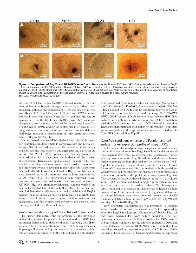
Figure 1. Comparison of RegES and HEScGRO xeno-free culture media. Human ESC line HS401 during the adaptation phases in RegESculture medium and in HEScGRO medium. Human ESC line HS401 was transferred from hES culture medium to new culture conditions using stepwiseadaptation: 20:80, 50:50, 80:20 and 100%. A) Adaptation phases to HEScGRO medium. Note excess differentiation of hESC colonies at adaptationphases 80:20 and after completion of the adaptation (100%). B) Adaptation phases to RegES culture medium.doi:10.1371/journal.pone.0010246.g001
Xeno-Free Culture Method
PLoS ONE | www.plosone.org 3 April 2010 | Volume 5 | Issue 4 | e10246
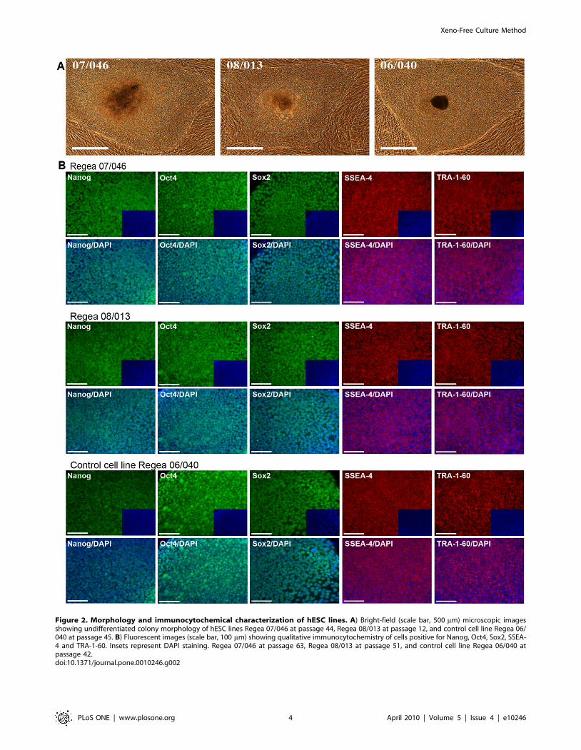
Figure 2. Morphology and immunocytochemical characterization of hESC lines. A) Bright-field (scale bar, 500 mm) microscopic imagesshowing undifferentiated colony morphology of hESC lines Regea 07/046 at passage 44, Regea 08/013 at passage 12, and control cell line Regea 06/040 at passage 45. B) Fluorescent images (scale bar, 100 mm) showing qualitative immunocytochemistry of cells positive for Nanog, Oct4, Sox2, SSEA-4 and TRA-1-60. Insets represent DAPI staining. Regea 07/046 at passage 63, Regea 08/013 at passage 51, and control cell line Regea 06/040 atpassage 42.doi:10.1371/journal.pone.0010246.g002
Xeno-Free Culture Method
PLoS ONE | www.plosone.org 4 April 2010 | Volume 5 | Issue 4 | e10246
(,1%) of MHC Class II isotype HLA-DR was observed for ASCs
cultured in HS or RegES media and modest expression (10%) of
MHC Class I isotype HLA-ABC was seen in RegES cultured
ASCs. While both culture conditions maintained the characteristic
surface marker expression profile of ASCs, statistical analysis
revealed significant differences in the expression of sialomucin-like
adhesion molecule CD34 (p = 0.043), leucocyte common antigen
CD45 (p = 0.017), adhesion molecule CD105 (p = 0.020) and
MHC Class I isotype HLA-ABC (p = 0.021) of ASCs cultured in
HS and RegES media.
The multilineage differentiation capability of ASCs expanded in
RegES was examined by culturing cells at passage 3 under
conditions that support osteogenic, adipogenic and chondrogenic
differentiation. The osteogenic induction was evident after 14 days
in culture in osteogenic RegES medium (Figure 7C). After less
than 7 days of adipogenic induction, accumulation of small lipid
droplets was visible in the cells grown in adipogenic RegES
medium by light microscopic inspection within the ASCs (data not
shown). At day 14, ASCs in RegES adipogenic induction cultures
proliferated rapidly and grew in a layered manner, with lipid
droplets forming in several layers (Figure 7D). In the chondrogenic
induction cultures, ASCs expanded in chondrogenic RegES
medium began to aggregate and form condensed pellets within
2–3 days after induction. At day 14, the chondrogenic induction
was confirmed with Alcian blue staining (Figure 7E).
Discussion
As the number of potential applications for stem cell
transplantation in the treatment of various degenerative diseases
is rapidly increasing a strong focus is needed on safety,
reproducibility and quality of stem cell transplants. To generate
therapeutically safe and usable stem cell-derived products for
clinical cell therapies, all animal-derived material must be
eliminated from the establishment, culture, and differentiation
steps. A completely xeno-free culture system was described by
Ellerstrom and co-workers [30] in which they used a xeno-free
derivation procedure, human serum, and xeno-free human
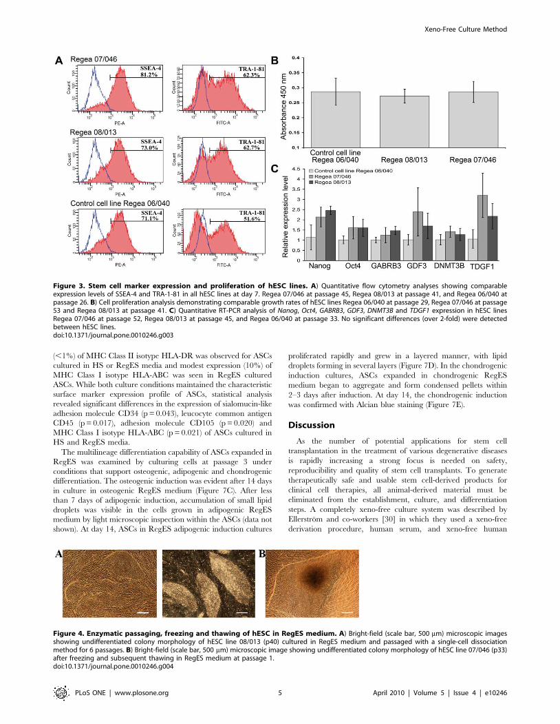
Figure 3. Stem cell marker expression and proliferation of hESC lines. A) Quantitative flow cytometry analyses showing comparableexpression levels of SSEA-4 and TRA-1-81 in all hESC lines at day 7. Regea 07/046 at passage 45, Regea 08/013 at passage 41, and Regea 06/040 atpassage 26. B) Cell proliferation analysis demonstrating comparable growth rates of hESC lines Regea 06/040 at passage 29, Regea 07/046 at passage53 and Regea 08/013 at passage 41. C) Quantitative RT-PCR analysis of Nanog, Oct4, GABRB3, GDF3, DNMT3B and TDGF1 expression in hESC linesRegea 07/046 at passage 52, Regea 08/013 at passage 45, and Regea 06/040 at passage 33. No significant differences (over 2-fold) were detectedbetween hESC lines.doi:10.1371/journal.pone.0010246.g003
Figure 4. Enzymatic passaging, freezing and thawing of hESC in RegES medium. A) Bright-field (scale bar, 500 mm) microscopic imagesshowing undifferentiated colony morphology of hESC line 08/013 (p40) cultured in RegES medium and passaged with a single-cell dissociationmethod for 6 passages. B) Bright-field (scale bar, 500 mm) microscopic image showing undifferentiated colony morphology of hESC line 07/046 (p33)after freezing and subsequent thawing in RegES medium at passage 1.doi:10.1371/journal.pone.0010246.g004
Xeno-Free Culture Method
PLoS ONE | www.plosone.org 5 April 2010 | Volume 5 | Issue 4 | e10246
foreskin feeder (hFF) cells to derive a hESC line. We have
previously demonstrated, however, that several commercial xeno-
free serum replacements and human sera are not suitable for long-
term undifferentiated culture of hESCs [31]. Recently Crook and
co-workers described the proof-of-concept for the generation of six
clinical-grade hESC lines using hFF as feeder cells [32]. These
lines were established and banked in a clinical-grade manufactur-
ing process (cGMP) facility in compliance with international
regulatory requirements. Although they used cGMP-compliant
reagents and materials in the production process of the hESC
lines, not all the reagents were xeno-free, possibly compromising
the safety of the established hESC lines for clinical application. In
this study, we have identified all the essential nutrients and growth
factors required to enable feasible growth characteristics and to
maintain the undifferentiated state of hESCs, iPSCs and ASCs.
The described RegES medium formulation is designed to meet the
special requirements of stem cell culture and the quality
requirements for clinical use of stem cells.
Our data demonstrate that hESCs can be successfully derived
and cultured long-term in the RegES formulation while
maintaining their undifferentiated state. Furthermore, EB forma-
tion, teratoma analysis and differentiation of cardiomyocytes and
neural cells demonstrated that hESCs derived and cultured in
xeno-free RegES medium maintain their pluripotency and
differentiation characteristics. The characteristics and differentia-
tion potential of hESCs cultured in the xeno-free RegES medium
were comparable to hESCs cultured in the conventional hES
culture medium. In the comparison of the RegES medium to the
xeno-free commercially available HEScGRO medium it was
noticed that HEScGRO was unable to maintain the undifferen-
tiated growth of hESCs. Human ESCs can be frozen and thawed
using the xeno-free medium RegES and both mechanical
passaging and single-cell enzymatic dissociation methods can be
used. With enzymatic single-cell propagation, the number of cells
can be multiplied rapidly, thereby enabling up-scaling and large-
scale cultivation of hESCs [27]. As demonstrated with the first
derived hESC line Regea 06/015, hFF can be cultured without
any animal-derived materials, enabling the establishment and
production of xeno-free hESC lines in the future. Furthermore,
our formulation enables feasible culture environment for iPSCs.
We believe that these results suggest that the derivation of iPSC
lines is also possible by using xeno-free RegES medium and should
be tested.
In the culture of ASCs the replacement of FBS with pooled
allogeneic human serum and human serum derivatives has been
reported to support equal or higher proliferation rates and
multipotentiality [33–35]. However, serum composition is largely
unknown, and shows significant lot-to-lot variability that may
affect the reproducibility of the results [2]. In clinical therapy, a
feasible option would be to use patient’s autologous serum but it
may not always be available in large quantities. In the culture of
ASCs our results show that the xeno-free RegES medium
exhibited significantly higher proliferation rates compared to the
reference media containing HS. Both culture conditions main-
tained the characteristic surface marker expression profile of ASCs
with expression of markers verifying the mesenchymal origin of
cells and no expression of markers of the hematopoietic origin of
cells. However, statistical analysis revealed significant differences
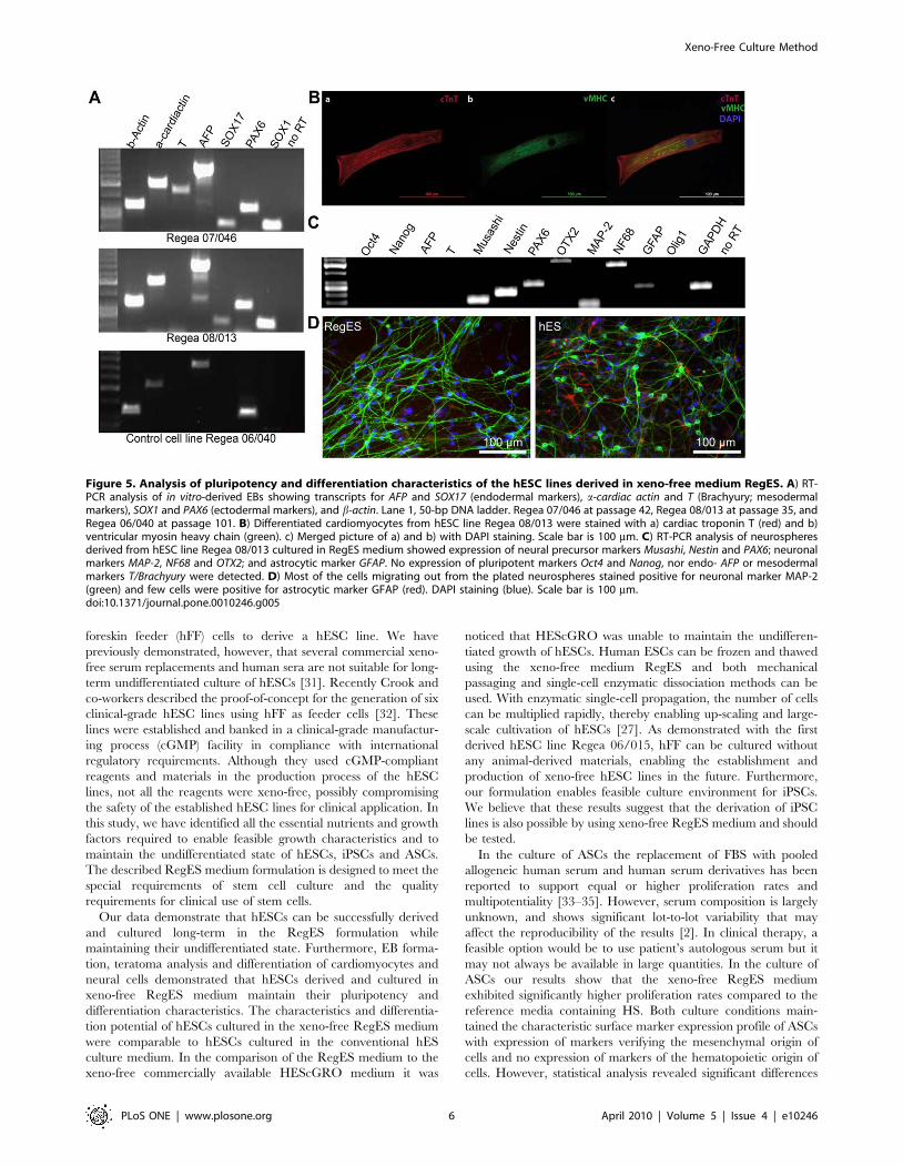
Figure 5. Analysis of pluripotency and differentiation characteristics of the hESC lines derived in xeno-free medium RegES. A) RT-PCR analysis of in vitro-derived EBs showing transcripts for AFP and SOX17 (endodermal markers), a-cardiac actin and T (Brachyury; mesodermalmarkers), SOX1 and PAX6 (ectodermal markers), and b-actin. Lane 1, 50-bp DNA ladder. Regea 07/046 at passage 42, Regea 08/013 at passage 35, andRegea 06/040 at passage 101. B) Differentiated cardiomyocytes from hESC line Regea 08/013 were stained with a) cardiac troponin T (red) and b)ventricular myosin heavy chain (green). c) Merged picture of a) and b) with DAPI staining. Scale bar is 100 mm. C) RT-PCR analysis of neurospheresderived from hESC line Regea 08/013 cultured in RegES medium showed expression of neural precursor markers Musashi, Nestin and PAX6; neuronalmarkers MAP-2, NF68 and OTX2; and astrocytic marker GFAP. No expression of pluripotent markers Oct4 and Nanog, nor endo- AFP or mesodermalmarkers T/Brachyury were detected. D) Most of the cells migrating out from the plated neurospheres stained positive for neuronal marker MAP-2(green) and few cells were positive for astrocytic marker GFAP (red). DAPI staining (blue). Scale bar is 100 mm.doi:10.1371/journal.pone.0010246.g005
Xeno-Free Culture Method
PLoS ONE | www.plosone.org 6 April 2010 | Volume 5 | Issue 4 | e10246
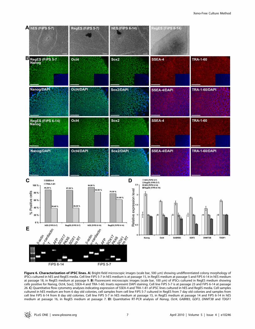
Figure 6. Characterization of iPSC lines. A) Bright-field microscopic images (scale bar, 500 mm) showing undifferentiated colony morphology ofiPSCs cultured in hES and RegES media. Cell line FiPS 5-7 in hES medium is at passage 15, in RegES medium at passage 5 and FiPS 6-14 in hES mediumat passage 18, in RegES medium at passage 9. B) Fluorescent microscopic images (scale bar, 100 mm) of iPSCs cultured in RegES medium showingcells positive for Nanog, Oct4, Sox2, SSEA-4 and TRA-1-60. Insets represent DAPI staining. Cell line FiPS 5-7 is at passage 23 and FiPS 6-14 at passage26. C) Quantitative flow cytometry analyses indicating expression of SSEA-4 and TRA-1-81 of iPSC lines cultured in hES and RegES media. Cell samplescultured in hES medium are from 6 day old colonies, cell samples from cell line FiPS 5-7 cultured in RegES from 7 day old colonies and samples fromcell line FiPS 6-14 from 8 day old colonies. Cell line FiPS 5-7 in hES medium at passage 15, in RegES medium at passage 14 and FiPS 6-14 in hESmedium at passage 16, in RegES medium at passage 7. D) Quantitative RT-PCR analysis of Nanog, Oct4, GABRB3, GDF3, DNMT3B and TDGF1
Xeno-Free Culture Method
PLoS ONE | www.plosone.org 7 April 2010 | Volume 5 | Issue 4 | e10246
in the surface marker expression of ASCs cultured in HS and
RegES media. In addition, RegES culture medium is able to
maintain the differentiation capacity of the ASCs and thus
maintain the multipotency of the ASC population. Our results
indicate that RegES medium possess all the inherent promise for
translational use in clinical applications and after additional pre-
clinical safety and efficacy studies, it could be used to replace
autologous or allogenous HS in the clinical cell therapy studies
with ASCs undergoing in our institute [3] and elsewhere.
Although all stem cell lines worldwide share the expression of
characteristic stem cell markers, different culture conditions are
known to influence gene expression and therefore many of the cell
properties. The use of completely defined conditions will allow for
better understanding of stem cell regulation and provide more
reproducible results. Currently, there are at least three commer-
cially available xeno-free culture media, HEScGRO (Millipore),
developed for the culture of hESC lines with feeder cells,
KnockOut SR Xeno-free (Invitrogen) which can be used with or
without feeder cells and TeSR2 (StemCell Technologies) for the
culture of hESC lines in feeder-cell free conditions. In the present
study we have successfully developed a new totally xeno-free
effective culture medium for human ESCs, iPSCs and ASCs.
These cells can be maintained in the same defined xeno-free
medium formulation supporting feasible proliferation while
maintaining their characteristics. In addition, we have described
the derivation of new hESC lines using the xeno-free RegES
medium and showed that these lines retain their differentiation
potential after long-term culture in the xeno-free RegES culture
medium. However, further studies of in vitro differentiated
derivatives are needed to evaluate the functionality of the
differentiation protocols with the xeno-free RegES medium.
Recently a large-scale multi-laboratory comparison study of
different culture conditions for hESCs was published [36]. Future
studies should focus on comparison of different xeno-free culture
conditions and on validation of these conditions to demonstrate
long-term culture, maintenance of key features of self-renewal,
pluripotency, and genetic stability, as well as derivation,
reprogramming or isolation of new stem cell lines as a full proof-
of-principle. As many of the current differentiation protocols
utilize a variety of undefined products we suggest that xeno-free
RegES medium should be tested as a base for differentiation
protocols for stem cells. The use of defined products in the
expression of day 6 colonies in iPSC lines FiPS 5-7 in hES medium at passage 10, in RegES medium at passage 7 and FiPS 6-14 in hES medium atpassage 11, in RegES medium at passage 8. No significant differences (over 2-fold) were detected between iPSC lines. E) RT-PCR analysis of in vitro-derived EBs showing transcripts for AFP and SOX17 (endodermal markers), a-cardiac actin and T (Brachyury; mesodermal markers), SOX1 and PAX6(ectodermal markers), and b-actin. Lane 1, 50-bp DNA ladder. Both cell lines at passage 10.doi:10.1371/journal.pone.0010246.g006
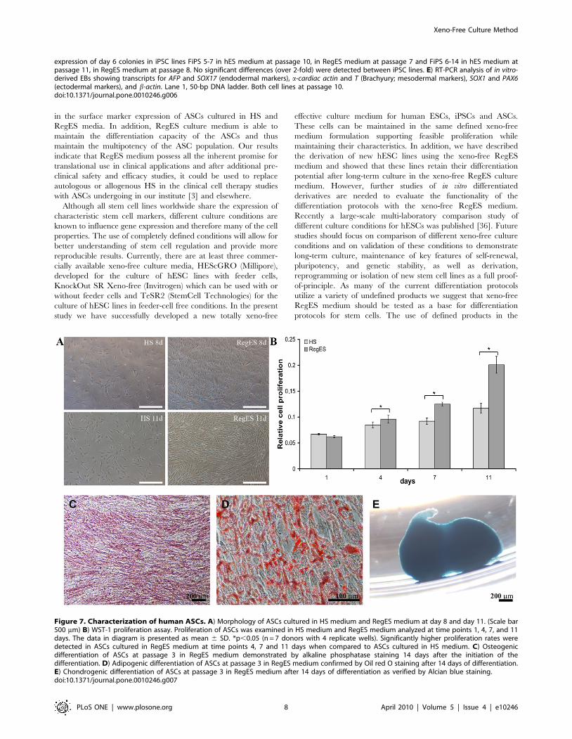
Figure 7. Characterization of human ASCs. A) Morphology of ASCs cultured in HS medium and RegES medium at day 8 and day 11. (Scale bar500 mm) B) WST-1 proliferation assay. Proliferation of ASCs was examined in HS medium and RegES medium analyzed at time points 1, 4, 7, and 11days. The data in diagram is presented as mean 6 SD. *p,0.05 (n = 7 donors with 4 replicate wells). Significantly higher proliferation rates weredetected in ASCs cultured in RegES medium at time points 4, 7 and 11 days when compared to ASCs cultured in HS medium. C) Osteogenicdifferentiation of ASCs at passage 3 in RegES medium demonstrated by alkaline phosphatase staining 14 days after the initiation of thedifferentiation. D) Adipogenic differentiation of ASCs at passage 3 in RegES medium confirmed by Oil red O staining after 14 days of differentiation.E) Chondrogenic differentiation of ASCs at passage 3 in RegES medium after 14 days of differentiation as verified by Alcian blue staining.doi:10.1371/journal.pone.0010246.g007
Xeno-Free Culture Method
PLoS ONE | www.plosone.org 8 April 2010 | Volume 5 | Issue 4 | e10246
differentiation protocols will facilitate the discovery of molecular
mechanisms behind the differentiation to various cell types and
reproducibility of differentiation processes. Our results indicate
that the basic RegES culture medium is applicable for further
optimization of xeno-free establishment, culture and differentia-
tion of stem cells and can ultimately serve as a platform for the
production of clinical-grade pluripotent stem cells and their
derivatives for safer clinical cell therapy treatments.
Materials and Methods
Ethics StatementThe study was conducted in accordance with the Karolinska
Institutet Ethics Committee to derive and culture hESCs, with the
Ethics Committee of Pirkanmaa Hospital District to culture,
characterize and differentiate hESCs derived at Karolinska
Institutet (R05051), to derive, culture, characterize and differen-
tiate new hESC lines at Regea (R05116) and with the permission
of the National Authority for Medicolegal Affairs to do research
with human embryos (Dnro 1426/32/300/05). Donated embryos
were received from Tampere University Hospital in vitro
fertilization (IVF) clinic. An informed consent form was signed
by both partners after receiving an oral and written description of
the study. The donors did not receive financial compensation.
Animal experiments were performed at the animal facilities of
Karolinska University Hospital or at Turku Center for Disease
Modeling and Department of Physiology, Institute of Biomedicine,
University of Turku in accordance with the approval of the
institution’s Ethics Committee. Adipose stem cells (ASCs) were
isolated from adipose tissue samples collected from female donors
undergoing elective surgical procedures at the Department of
Plastic Surgery, Tampere University Hospital with the permission
of the Ethics Committee of Pirkanmaa Hospital District. Human
iPS cells were derived and characterized at the University of
Helsinki, with the permission of the Ethics Committee of the
University of Helsinki.
Cell CultureThe hESC lines HS237, HS346, and HS401 used in the
optimization of the xeno-free formulation RegES were initially
derived and characterized at the Karolinska Institutet, Stockholm,
Sweden [25,26]. These cell lines were derived and cultured using
conventional hES medium containing KO-DMEM basal medium
supplemented with 20% KO-SR, 2 mM Glutamax, 0.1 mM b-
mercaptoethanol (all from Invitrogen, Carlsbad, CA), 0.1 mM
MEM non-essential amino acids (NEAA), 1% penicillin-strepto-
mycin (both from Cambrex Bio Science, Walkersville, MD) and
8 ng ml/ml recombinant human bFGF (R&D Systems Inc.,
Minneapolis, MN). Human iPSC lines FiPS 5-7 and FiPS 6-14
were derived and characterized at the University of Helsinki.
Briefly, human cDNAs of Oct4, Sox2, Nanog, Klf4 and Lin28, were
amplified by direct PCR from hESC cDNA, and cloned into
pMXs retroviral vector (Addgene Inc., Cambridge, MA). 293-
GPG packaging cells were transfected with each pMXs-cDNA
vector separately using Fugene HD (Roche Diagnostics GmbH,
Mannheim, Germany). Next day, fresh MEF media was added.
Viral supernatant was collected 3 times at days 4, 5, and 6 post-
transfection, combined and then filtered. The human neonatal
foreskin fibroblast line CCCD-1112Sk (ATCC) was plated on
gelatin-coated petri dishes and incubated overnight. Cells were
then infected with an equally mixed combination of fresh virus-
containing supernatant twice at 24 h intervals. At day 3, infected
cells were harvested and re-seeded on a Mitomycin C-treated
MEF layer. The following day, the medium was changed to hES
medium. After 20 to 30 days post-transduction, ES-like colonies
were picked, expanded, and characterized as previously described
for hESCs [37]. Transgene expression was studied using RT-PCR
to confirm transgene silencing. FiPS 5-7 was generated using
Oct4, Sox2, Nanog, and Lin28 encoding retroviruses, while FiPS
6-14 was generated using the same factors plus to Klf4.
Human ESCs and iPSCs were gradually adapted to the RegES
medium (Table S1) (20:80, 50:50, 80:20, and 100:0) during the
first four passages. Commercially available hFFs (CRL-2429,
ATCC, Manassas, VA) cultured in IMDM with L-glutamine and
25 mM HEPES (Invitrogen) supplemented with 10% FBS
(Invitrogen) and 1% penicillin-streptomycin (Cambrex Bio Sci-
ence) were used for hESCs and iPSCs culture. Confluent
monolayers of hFFs were mitotically inactivated by irradiation
(40 Gy) and plated at a density of 3.86104 cells/cm2. The growth
of hESCs and iPSCs was monitored microscopically and culture
media were changed daily. The hESC and iPSC cultures were
mechanically passaged every 6 to 7 days to new mitotically
inactivated feeder cells. Single-cell enzymatic passaging was
performed for hESCs as described previously [27] using TrypLE
Select (Invitrogen) and an 8 to 12 day splitting interval. The
hESCs were frozen using RegES medium supplemented with 10%
DMSO (Sigma-Aldrich, St. Louis, MO). Xeno-free commercially
available HEScGRO medium (Millipore Corporation, Billerica,
MA) without any supplementation was compared to RegES
medium for the culture of hESCs.
ASCs were isolated from adipose tissue samples collected from
female donors undergoing elective surgical procedures in the
Department of Plastic Surgery, Tampere University Hospital.
Isolation of ASCs from adipose tissue samples was carried out as
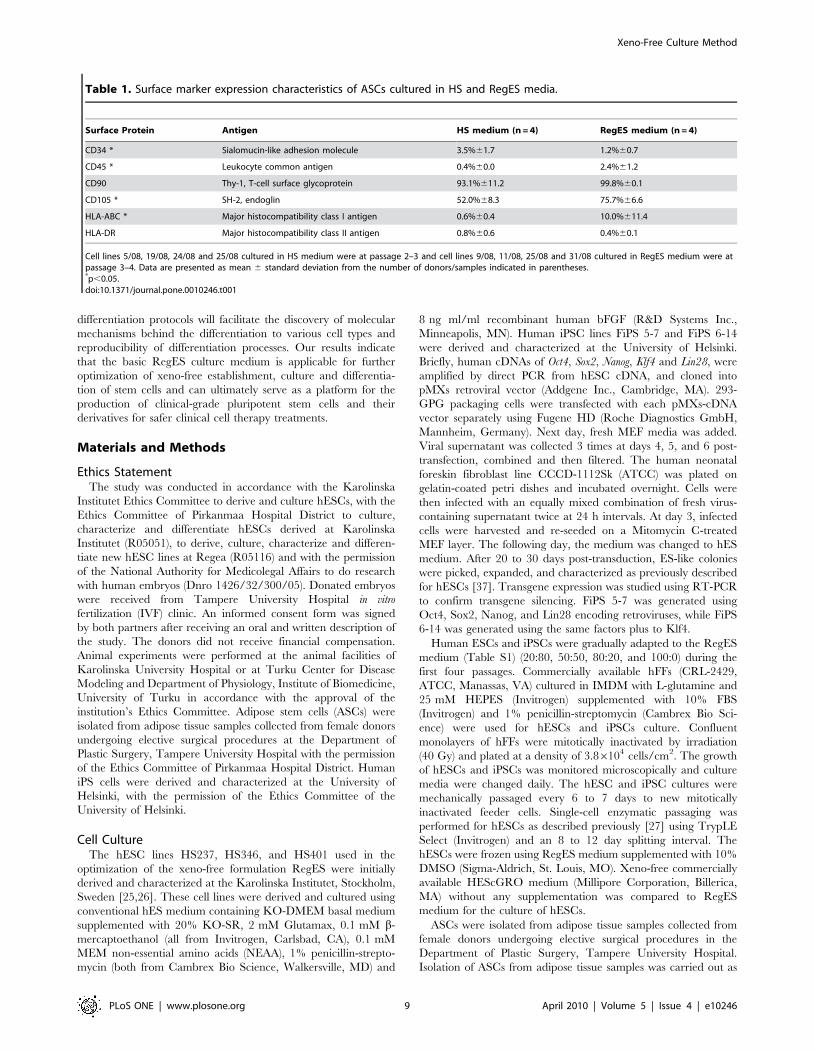
Table 1. Surface marker expression characteristics of ASCs cultured in HS and RegES media.
Surface Protein Antigen HS medium (n = 4) RegES medium (n = 4)
CD34 * Sialomucin-like adhesion molecule 3.5%61.7 1.2%60.7
CD45 * Leukocyte common antigen 0.4%60.0 2.4%61.2
CD90 Thy-1, T-cell surface glycoprotein 93.1%611.2 99.8%60.1
CD105 * SH-2, endoglin 52.0%68.3 75.7%66.6
HLA-ABC * Major histocompatibility class I antigen 0.6%60.4 10.0%611.4
HLA-DR Major histocompatibility class II antigen 0.8%60.6 0.4%60.1
Cell lines 5/08, 19/08, 24/08 and 25/08 cultured in HS medium were at passage 2–3 and cell lines 9/08, 11/08, 25/08 and 31/08 cultured in RegES medium were atpassage 3–4. Data are presented as mean 6 standard deviation from the number of donors/samples indicated in parentheses.*p,0.05.doi:10.1371/journal.pone.0010246.t001
Xeno-Free Culture Method
PLoS ONE | www.plosone.org 9 April 2010 | Volume 5 | Issue 4 | e10246
described previously [38,39]. Briefly, the adipose tissue was
minced manually into small fragments and digested with
1.5 mg/mL collagenase type I (Life Technologies, Paisley, UK)
in a shaking water bath at 37uC. To separate the adipose stem cells
from the surrounding tissue the digested tissue was centrifuged and
filtered in sequential steps. The isolated cells were expanded in
DMEM/F-12 1:1, supplemented with 1% GlutaMAX, 1%
antibiotics/antimycotic (a/a; 100 U/mL penicillin, 0.1 mg/mL
streptomycin, and 0.25 mg/mL amphotericin B) (all from Invitro-
gen) and 10% alloHS (PAA Laboratories GmbH, Pasching,
Austria), (HS medium). For testing defined, xeno-free conditions,
ASCs were transferred to RegES culture medium in flasks with
pre-coated CELLstart (Invitrogen). For detachment of ASCs
TrypLE Select (Invitrogen) was used.
Derivation of hESC linesSurplus poor quality embryos that could not be used for
infertility treatment were obtained as donations from couples
undergoing in vitro fertilization treatment at Tampere University
Hospital, Tampere, Finland. The mechanical derivation of the cell
lines from blastocyst-stage embryos with a hardly visible inner cell
mass was performed using a specially made flexible metal needle
(Hunter Scientific, Essex UK) and surgical knife as described
previously [26]. For successful derivation of the 06/015 cell line,
we used 10 surplus embryos (arrested around day 3–4) without
blastocyst development. Derivation of the cell line Regea 06/015
was performed by removing the zona pellucida and plating the
remaining cells on the top of hFF cells using a flexible needle.
Human ESC line Regea 06/040 was derived using hES medium
and hESC lines Regea 06/015, Regea 07/046, and Regea 08/013
were derived using RegES medium. Human ESC line Regea 06/
015 was derived on feeder cells (C-12300 PromoCell GmbH,
Heidelberg, Germany) cultured in IMDM with L-glutamine and
25 mM HEPES (Invitrogen) supplemented with 10% human
serum (Sigma-Aldrich), 0.1 mM NEAA (Cambrex Bio Science),
and 0.1 mM penicillin-streptomycin (Cambrex Bio Science).
ImmunocytochemistryImmunocytochemistry was performed as previously described
[31]. The primary antibodies used for hESCs and iPSCs were
specific for Nanog, Oct4 (both from R&D Systems), SSEA-4 and
Sox2 (both from Santa Cruz Biotechnology, Inc., Santa Cruz, CA)
and TRA-1-60 (Millipore). The cells were incubated with primary
antibody solution overnight at 4uC. The cells were probed with
Alexa Fluor 568 or Alexa Fluor 488 (both from Invitrogen)
secondary antibodies for 1 h in the dark at room temperature. The
differentiated cardiomyocytes were incubated overnight at 4uCwith anti-cardiac troponin T (Abcam, Cambridge, UK) and anti-
ventricular myosin heavy chain (Chemicon). Alexa Fluor 488 or
568 (Invitrogen) conjugated anti-goat and anti-mouse antibodies
were used as secondary antibodies. Differentiated neural cells were
immunostained after 9 weeks of differentiation. For immunocyto-
chemical staining neurospheres were plated on human laminin
(Sigma Aldrich) coated wells in the absence of bFGF and after
growing three days the cells were fixed. The fixed neuronal cells
were incubated overnight at 4uC with polyclonal rabbit anti-
MAP-2 (Chemicon) for neuronal cells and polyclonal sheep anti-
GFAP (R&D Systems) for astrocytes. Alexa Fluor 488 and 568
conjugated anti-rabbit or anti-sheep (Invitrogen) were used as
secondary antibodies. Human ESCs labeled only with secondary
antibodies, were used as negative controls and no fluorescense in
these samples were detected. After incubation, the cells were
mounted in Vectashield mounting medium containing 49,6-
diamidino-2-phenylindole (Vector Laboratories, Inc., Burlingame,
CA). The labeled cells were photographed with an Olympus IX51
phase contrast microscope with fluorescence optics and Olympus
DP30BW camera (Olympus Corporation, Tokyo, Japan).
Alkaline phosphatase stainingThe in vitro osteogenic differentiation capacity was determined 14
days after the initiation of differentiation by alkaline phosphatase
staining (ALP) as described previously [33,40,41]. The cell cultures
were fixed with a 4% paraformaldehyde (PFA) solution and stained
with the leukocyte alkaline phosphatase kit according to the Sigma
Procedure No. 86 (#86R-1KT). The ALP staining was confirmed
by a purple staining visualized by microscopy.
Adipogenesis Oil red O stainingThe adipogenic differentiation was confirmed with Oil red O
staining as described previously [33]. Cell cultures were main-
tained for 14 days and subsequently fixed with 4% PFA,
pretreated with 60% isopropanol and stained with a 0.5% Oil
red O solution in 60% isopropanol. After fixation and staining, the
wells were rinsed with distilled water and visualized by
microscopy. Adipocytes were identified as cells with red-stained
lipid vesicles.
Chondrogenesis Alcian blue stainingThe chondrogenic differentiation cultures were maintained for 14
days and were subsequently fixed with 4% PFA, and stained with 1%
Alcian blue stain. Cells stained with Alcian blue verified the presence
of sulphated proteoglycans within the chondrogenic cell pellet.
Flow cytometryHuman ESCs and iPSCs cultured in hES and RegES media
were analyzed by flow cytometry using antibodies against SSEA-4-
PE (R&D Systems) and TRA-1-81-FITC (BD Pharmingen, San
Diego, CA). Human ESC samples cultured in hES and RegES
media were from 7 day old colonies, iPSC samples cultured in hES
medium from 6 day old colonies and samples cultured in RegES
medium from 7 day old (5–7) or 8 day old colonies (6–14). Both
undifferentiated and differentiated cells present in the culture dish
were included in the analyses. Alexa Fluor 488 (Invitrogen) and R-
phycoerythrin-conjugated (Caltag-Medsystems Ltd, Buckingham,
UK) secondary antibodies were used as isotype controls. ASCs
cultured in HS and RegES media (n = 4, passages 2–4) were
analyzed by flow cytometry using monoclonal antibodies against
CD90-APC (BD Biosciences), CD45-FITC (Miltenyi Biotech,
Bergisch Gladbach, Germany), CD34-APC, HLA-ABC-PE, HLA-
DR-PE (all from Immunotools GmbH Friesoythe, Germany), and
CD105-PE (R&D Systems). The samples were analysed by flow
cytometry (FACSAriaH, BDBiosciences). The acquisition was set
for 10 000 events per sample. The data were analyzed using
FACSDiva Software version 4.1.2 (BD Biosciences).
RNA isolation and reverse transcriptionTotal RNA was isolated using an RNeasy mini plus kit (Qiagen,
Valencia, CA) according to the manufacturer’s instructions. The
concentration and quality of isolated RNA was determined using a
ND-1000 Spectrophotometer (NanoDrop Technologies, Wilming-
ton, DE). Complementary DNA (cDNA) was synthesized from
50 ng of total RNA using a Sensiscript Reverse Transcription Kit
(Qiagen) according to the manufacturer’s instructions.
Quantitative RT-PCRBoth undifferentiated and differentiated hESCs and iPSCs in
the culture plate were included in the quantitative RT-PCR
Xeno-Free Culture Method
PLoS ONE | www.plosone.org 10 April 2010 | Volume 5 | Issue 4 | e10246

analyses. Quantitative RT-PCR was performed for hESCs and
iPSCs with Applied Biosystems Gene Expression Assays: GAPDH
(Hs99999905_m1; 122-bp product), DNMT3B (Hs01003405_m1;
80-bp product), TDGF1 (Hs02339496_g1; 102-bp product), Nanog
(Hs2387400_g1; 109-bp product), GDF3 (Hs00220998_m1; 65-bp
product), GABRB3 (Hs01115771_m1; 72-bp product), and Oct4
(Hs00999632_g1; 77-bp product). All samples and the no-template
controls were analyzed in triplicate. Quantitative RT-PCR was
performed with an Applied Biosystems 7300 Real-Time PCR
system using the following conditions: 40 cycles of 50uC, 2 min;
95uC, 10 min; and 95uC, 15 s; followed by 60uC, 1 min. The data
were analyzed with a 7300 System SDS Software (Applied
Biosystems). The cycle threshold (Ct) values were determined for
every reaction. Relative quantification was calculated using the
22DDCt method [42]. All data were normalized to the expression of
the gene GAPDH whose expression does not change under the
experimental conditions. The data are presented as mean fold-
change. Neural cells: RNA isolation and reverse transcription of
RT-PCR samples ($10 spheres/sample) was performed as
described above. The primer set for RT-PCR contained Oct4,
Nanog for undifferentiated hESC; a-fetoprotein for endodermal
lineage; brachyury/T for mesodermal lineage; Musashi, Nestin, and
PAX6 for neuroectodermal cells; MAP-2, NF68 and OTX2 for
neuronal cells; GFAP for astrocytes, and Olig1 for oligodentrocyte
precursor cells. Data were normalized to the expression of the
gene GAPDH whose expression does not change under the
experimental conditions. Primer sequences are shown in supple-
mentary Table S2. The RT-PCR reactions were performed in an
Eppendorf Mastercycler using 35 PCR cycles with an initializing
step at 95uC for 3 min, denaturation at 95uC for 30 s, annealing at
55uC for 30 s, and extension at 72uC for 1 min, followed by a final
extension at 72uC for 5 min. The PCR products were analyzed by
electrophoresis on a 1.5% agarose gel containing 0.4 mg/ml
ethidium bromide (Sigma-Aldrich).
Cell proliferation assayCell proliferation of the hESC lines was determined using a
colorimetric immunoassay (Roche Diagnostics) based on the
measurement of bromodeoxyuridine (BrdU) incorporation during
DNA synthesis. The assay was performed according to the
manufacturer’s instructions. The hESC colonies at day 5 were
labeled with BrdU-labeling solution overnight at 37uC. The
hESCs (105 cells/well) were added to a 96-well plate, with 8
replicates per cell line. Cells that were not labeled with BrdU were
used as a background control. The absorbance of the samples was
measured at 450 nm using a Viktor 1429 Multilabel Counter
(PerkinElmer-Wallace, Norton, OH).
The cell viability and proliferation activity of the ASCs was
assessed using PreMix WST-1 Cell Proliferation Assay System
(Takara Bio Inc., Shiga, Japan). The ASCs (n = 7 donor cell
samples, with 4 replicates per cell line) were seeded on a 48-well
plate at a density of 4500 cells/cm2, and the proliferation was
determined at 1, 4, 7 and 11 days. Briefly, the cell culture medium
was removed and DPBS and PreMix WST-1 were added 10:1.
The well plate was incubated for 4 hours in 37uC and the cell
proliferation activity was measured using a Viktor 1429 Multilabel
Counter at 450 nm.
KaryotypingKaryotype analysis was performed by G-banding technique in
Medix Laboratories Inc, Helsinki, Finland. In brief, the hESCs
were treated with colchicine for 2 hours, and cells in metaphase
were analyzed using conventional light microscopy (Olympus, BX
50). Karyotypes were determined using IKAROS-software
designed for chromosome analysis (MetaSystems GmbH, Altlus-
sheim, Germany). A total of 20 cells in metaphase were analyzed
for each cell line.
Analysis of pluripotency in vitroThe embryoid bodies (EBs) formed by mechanically dissecting
the hESC and iPSC colonies were cultured without feeder cells in
RegES or hES medium without bFGF for 4 weeks before RNA
isolation. The media was changed every 2 to 3 days. RNA
isolation and reverse transcription from EBs was performed as
described above. The expression of markers characteristic of
ectoderm (PAX6 and SOX1, DNA Technologies Inc. Gaithersburg,
MD), endoderm (a-fetoprotein and SOX17, DNA Technologies Inc.),
and mesoderm (a-cardiac actin, Proligo, Sigma-Aldrich and T, DNA
Technologies Inc.) development in EBs was determined using RT-
PCR primers. b-actin (DNA Technologies Inc.) whose expression
does not change under the experimental conditions was used as a
control. Primer sequences are shown in supplementary Table S2.
Negative controls contained sterilized water instead of the cDNA
template. The RT-PCR reactions were performed in an
Eppendorf Mastercycler as follows: denaturation at 95uC for
3 min and 40 cycles of denaturation at 95uC for 30 s, annealing at
57uC for 30 s, and extension at 72uC for 1 min, followed by a final
extension at 72uC for 5 min. The PCR products were analyzed by
electrophoresis on a 1.5% agarose gel containing 0.4 mg/ml
ethidium bromide (Sigma-Aldrich) and 50 bp DNA standard
(MassRulerTM DNA Ladder Mix, Fermentas).
Analysis of pluripotency in vivoAnimal experiments with hESC lines Regea 06/015, Regea 06/
040 and Regea 07/046 were performed at the infection-free
animal facility of Karolinska University Hospital and experiments
with hESC line Regea 08/013 at the animal facility of Turku
Center for Disease Modeling and Department of Physiology,
Institute of Biomedicine, University of Turku. Human ESCs were
harvested from the culture plates using dispase or TrypleSelect
and mechanical treatment. Five colonies (103 to 104 hESC) were
washed twice in PBS and subsequently implanted beneath the
testicular capsule of a young (6–8 week-old) severe combined
immunodeficiency (scid)/beige male mouse (C.B.-17/GbmsTac-
scid-bgDF N7, M&B, Ry, Denmark) or in the case of Regea 08/
013 male Nude mouse (Hsd;Athymic Nude-Foxn1nu, Harlan).
Three animals were used for cell line. Teratoma growth was
determined by palpation once per week, and the mice were killed
by cervical dislocation 8 to 9 weeks after implantation. The in vivo
pluripotency of the hESC lines Regea 06/015, Regea 06/040 and
Regea 07/046 was analyzed as previously described [8,25].
Immunohistochemical studies of Regea 08013 cell line teratomas
were done in paraffin sections. The dewaxed and rehydrated
sections were treated in pressure cooker in 1 mM EDTA, pH 8,
(Desmin and HNF3b) or 10 mM sodium citrate buffer, pH 6,
(NCAM) for 5 minutes to reveal antigenic sites, cooled at room
temperature (RT) for 2 hours, and rinsed in phosphate buffer
saline (PBS). After rinsing with PBS the sections were incubated
with primary antibodies (rabbit anti-desmin, sc-14026, mouse anti-
HNF3b, sc-101060, purchased from Santa Cruz Biotechnology, or
rabbit anti-NCAM, AB5032, purchased from Chemicon Interna-
tional. Primary antibodies were diluted (1:1000, 1:3000, and 1:800
respectively) in PBS containing 0.05% TweenH and incubated
overnight at +4 degrees. The next day, after several rinses with
PBS, endogenous peroxidase was inactivated with 1% hydrogen
peroxide in PBS for 20 minutes. After rinsing several times with
PBS, the sections were incubated with secondary antibodies (HRP
conjugated Dako EnvisionTM+ Systems) for 30 minutes (RT).
Xeno-Free Culture Method
PLoS ONE | www.plosone.org 11 April 2010 | Volume 5 | Issue 4 | e10246
Slides were rinsed with PBS. Color was developed with
diaminobenzidine substrate (Dako EnvisionTM+ Systems). Sections
were slightly counterstained with Mayer’s hematoxylin, dehydrat-
ed, and mounted.
Differentiation of hESCs to cardiomyocytes and neuralcells
Cardiomyocyte differentiation was carried out as described
previously [28] by co-culturing Regea 08/013 hESCs, maintained
in RegES (p46) and in hES (p52) media, with mouse visceral
endodermal-like (END-2) cells, which were a kind gift from Prof.
Mummery, Humbrecht Institute, Utrecht, Netherlands. Briefly,
undifferentiated hESC colonies were dissected mechanically into
aggregates containing a few hundred cells and placed on the top of
plated END-2 cells in hES culture medium without KO-SR and
bFGF. Differentiating cell colonies were monitored by microscopy
daily and the medium was changed after 5 days of culturing. After
12 days the 10% KO-SR (Invitrogen) was added to the medium.
Differentiation was performed in 12-well plates, 15 hESC colony
pieces in a well. The beating areas were dissociated by collagenase
II treatment. The cells were plated on 0.1% gelatin coated cell
culture plates in a medium containing 7.5% FBS.
Neural differentiation was performed as previously described
[29]. Briefly, hESC (Regea 08/013) colonies maintained in RegES
(p53) and in hES (p58) media, were mechanically dissociated into
small clusters containing ,3000 cells. Clusters were transferred
into 6-well ultra low attachment plates (Nunc, Thermo Fisher
Scientific, Rochester, NY) and cultured as floating aggregates, e.g.
neurospheres, for up to 20 weeks in neural differentiation medium
consisting of 1:1 DMEM/F-12:Neurobasal media supplemented
with 2 mM Glutamax, 1xB27, 1xN2 (all from Invitrogen), 25 U/
ml penicillin-streptomycin (Lonza Group Ltd), and 20 ng/ml
bFGF (R&D Systems). Medium was changed 3 times/week and
the spheres were mechanically passaged weekly.
Differentiation of ASCs to adipogenic, osteogenic andchondrogenic lineages
ASCs (passage 3) cultured in RegES medium were analyzed for
their capacity to differentiate toward the adipogenic, osteogenic, and
chondrogenic lineages with ASCs cultured in medium containing HS
as reference. Briefly, osteogenic differentiation analyses were
performed on ASCs seeded onto a 12-well plate at a density of
2.56103 cells/cm2 in RegES media supplemented with 150 mM L-
ascorbic acid 2-phosphate (Sigma), 10 mM b-glycerophosphate
(Sigma) and 100 nM dexamethasone (Sigma) in culture vessels were
pre-coated with CELLstart. The control cultures were maintained in
RegES media without supplements in pre-coated culture vessels. The
cultures were maintained 14 days and osteogenic differentiation was
detected by alkaline phosphatase activity staining.
Adipogenic differentiation was induced by culturing ASCs for two
weeks in adipogenic media with an initial seeding density of
26104 cells/cm2 in culture vessels were pre-coated with CELLstart.
For the adipogenic induction, RegES media supplemented with
33 mM biotin (Sigma), 1 mM dexamethasone, 100 nM insulin
(Invitrogen) and 17 mM pantothenate (Fluka, Buchs, Switzerland)
was used while the control cultures were maintained in RegES
without adipogenic supplements. The cultures were analyzed using
an Oil red O stain as an indicator of intracellular lipid accumulation.
The chondrogenic differentiation capacity analysis was assessed
by micromass culture technique [1,43]. Briefly, 16105 cells were
seeded onto a 24-well culture plate in a 10 ml volume, and were let
adhere for 3 h in a cell incubator prior to addition of the
chondrogenic RegES media containing 1% ITS+1 (Sigma),
50 mM L-ascorbic acid 2-phosphate, 55 mM sodium pyruvate
(Invitrogen), L-proline 23 mM (Sigma). The control cultures were
maintained in RegES without chondrogenic supplements. The
chondrogenic induction cultures and control cultures were
performed without CELLstart pre-coating. The cultures were
maintained for 14 days prior to analysis. Chondrogenesis was
confirmed using Alcian blue stain.
Statistical analysesANOVA by the methodology of mixed models was used for the
proliferation data analysis of ASCs between the different culture
media compositions within each separate experiment setup to
account for the correlation of multiple measurements within
donors. Mann-Whitney U test was applied in the surface marker
expression of ASCs to compare the different culture conditions.
The surface marker expression of ASCs was analyzed using
Wilcoxon signed-rank statistics, which was used for pairwise
comparison between the different culturing conditions. The
statistical analyses were performed at the significance level
p,0.05. The analyses were performed using SPSS software
version 13 (SPSS Inc., Chicago, IL, USA).
Supporting Information
Table S1 Complete Formulation for RegES Medium.
Found at: doi:10.1371/journal.pone.0010246.s001 (0.05 MB
DOC)
Table S2 Primer sequences.
Found at: doi:10.1371/journal.pone.0010246.s002 (0.05 MB
DOC)
Figure S1 Characterization of a hESC line Regea 06/015
derived in xeno-free medium RegES. A) Bright-field (scale bar,
500 mm) microscopic image of Regea 06/015 cell line at passage 3
after derivation. B) Bright-field (scale bar, 500 mm) microscopic
image showing undifferentiated colony morphology of Regea 06/
015 cell line at passage 35. C) RT-PCR analysis of in vitro-derived
EBs of hESC line Regea 06/015 at passage 60 showing transcripts
for AFP and SOX17 (endodermal markers), a-cardiac actin and T
(Brachyuru; mesodermal markers), SOX1 and PAX6 (ectodermal
markers), and b-actin as a housekeeping control. Lane 1, 50 bp
DNA ladder. D) Fluorescent (scale bar, 200 mm) microscopic
images showing undifferentiated colony morphology and qualita-
tive immunocytochemistry of hESCs positive for the transcription
factor Nanog, Oct4, Sox2, SSEA-4, and TRA-1-60. Insets
represent DAPI staining.
Found at: doi:10.1371/journal.pone.0010246.s003 (7.33 MB TIF)
Figure S2 Karyograms of hESC lines. A G-banding karyograms
showing normal karyotypes of hESC lines, Regea 06/015 at
passage 35, Regea 07/046 at passage 36, Regea 08/013 at passage
25 and Regea 06/040 at passage 71.
Found at: doi:10.1371/journal.pone.0010246.s004 (9.41 MB TIF)
Figure S3 Histology of teratomas from cell line Regea 06/015 at
passage 35. A) An overview of a teratoma. B) Endoderm: detail
from the right part of the overview showing a lumen outlined by
high cylindrical cells (*). The cells are compatible with endodermal
differentiation of intestinal or respiratory type. C) Ectoderm: a
cluster of neurons assembled into a ganglion like structure (arrow).
Fibers tracts emanates from the cluster. D) Ectoderm: a
squadmous epithelial island composed of fairly vacuolated cells
(arrow). E) Mesoderm: lower part an island composed of
hypertrophic chondrocytes (arrow).
Found at: doi:10.1371/journal.pone.0010246.s005 (7.14 MB TIF)
Xeno-Free Culture Method
PLoS ONE | www.plosone.org 12 April 2010 | Volume 5 | Issue 4 | e10246
Figure S4 Histology of teratomas from cell line Regea 07/046 at
passage 17. A) This image shows tubular structures lined by
cuboidal to cylindrical epithelium and aggregates consisting of
smooth muscle cells (sm) are seen. B) Endoderm: some PASD+cells (arrow) can be seen in the image, which are compatible with
Goblet cells indicating the endodermal differentiation. C)
Endoderm and mesoderm: a single tubule (*) lined by cuboidal
to cylindrical epithelium. Note that it is embedded in a loosely
arranged connective tissue - primitive mesenchyme. An interpre-
tation would be that this represents endodermal (epithelial tubules)
and mesodermal components. D) Endoderm or ectoderm: this
image shows an epithelium lined tubule (*) embedded within a
very loosely arranged mesenchyme. The epithelium can be
interpreted as bilayered potentially representing a squamous
variant. It is impossible to state if it is endodermal or ectodermal
in origin.
Found at: doi:10.1371/journal.pone.0010246.s006 (8.57 MB TIF)
Figure S5 Histology of teratomas from cell line Regea 08/013 at
passage 86. A) Ectoderm: Large area of NCAM-positive, neuronal
cells (arrow). B) Mesoderm: Bundles of muscle cells are stained
positive for Desmin (arrow). C) Endoderm: Pseudostratified
ciliated columnar epithelium stained positive for HNF3b (arrow).
Found at: doi:10.1371/journal.pone.0010246.s007 (6.05 MB TIF)
Figure S6 Histology of teratomas from cell line Regea 06/040 at
passage 34. A) and B) An overview of teratomas. Endoderm and
mesoderm: note cartilage (c) and tubular structure (*) outlined by
cylindrical cells suggesting mesodermal differentiation and varying
amounts of Goblet cells suggesting endodermal differentiation. C)
Endoderm: a high power view of an area from B is shown. Note
smooth muscle like cells (sm) and cylindrical cells with scattered
mucous producing Goblet cells. D) Ectoderm: a strong nuclear
expression of MITF (arrow) specific for retinal pigment epithelial
cells is seen indicating ectodermal differentiation.
Found at: doi:10.1371/journal.pone.0010246.s008 (5.84 MB TIF)
Acknowledgments
We thank Dr Catharina Ellerstrom for her help with the SCED-passaging
method, Medix Laboratories Inc, Helsinki, Finland for the karyotype
analysis, Saija Savolainen for the teratoma analysis of hESC line Regea
08/013, Heini Huhtala for the statistical analyses, Maarit Patrikainen for
technical assistance with neuronal differentiation, Anna-Maija Honkala
and Miia Juntunen for technical assistance with ASCs, Hanna Koskenaho,
Niina Ikonen, Outi Melin and Sari Leinonen for technical assistance with
hESCs and iPS cells.
Author Contributions
Conceived and designed the experiments: KR SM SN EK KAS RS OH
HS. Performed the experiments: KR BICL SH RL MPM JI BR HS.
Analyzed the data: KR BICL RL MPM JI BR. Contributed reagents/
materials/analysis tools: TO OH. Wrote the paper: KR SH JI SM SN EK
KAS TO RS OH HS.
References
1. Zuk PA, Zhu M, Ashjian PH, De Ugarte DA, Huang JI, et al. (2002) Human
adipose tissue is a source of multipotent stem cells. Mol Biol Cell 13: 4279–
4295.
2. Lendeckel S, Jodicke A, Christophis P, Heidinger K, Wolff J, et al. (2004)
Autologous stem cells (adipose) and fibrin glue used to treat widespreadtraumatic calvarial defects: case report. J Craniomaxillofac Surg 32: 370–373.
3. Mesimaki K, Lindroos B, Tornwall J, Mauno J, Lindqvist C, et al. (2009) Novelmaxillary reconstruction with ectopic bone formation by GMP adipose stem
cells. Int J Oral Maxillofac Surg 38: 201–209.
4. Takahashi K, Tanabe K, Ohnuki M, Narita M, Ichisaka T, et al. (2007)Induction of pluripotent stem cells from adult human fibroblasts by defined
factors. Cell 131: 861–872.
5. Yu J, Vodyanik MA, Smuga-Otto K, Antosiewicz-Bourget J, Frane JL, et al.
(2007) Induced pluripotent stem cell lines derived from human somatic cells.
Science 318: 1917–1920.
6. Thomson J, Itskovitz-Eldor J, Shapiro S, Waknitz M, Swiergiel J, et al. (1998)
Embryonic stem cell lines derived from human blastocysts. Science 282: 1145–7.
7. Koivisto H, Hyvarinen M, Stromberg A, Inzunza J, Matilainen E, et al. (2004)
Cultures of human embryonic stem cells - serum replacement medium or serum-
containing media and the effect of basic fibrolast growth factor. ReproductiveBioMedicine Online 9: 330–337.
8. Inzunza J, Gertow K, Stromberg M, Matilainen E, Blennow E, et al. (2005)Derivation of human embryonic stem cell lines in serum replacement medium
using postnatal human fibroblasts as feeder cells. Stem Cells 23: 544–9.
9. Richards M, Fong CY, Chan WK, Wong PC, Bongso A (2002) Human feeders
support prolonged undifferentiated growth of human inner cell masses and
embryonic stem cells. Nat Biotechnol 20: 933–6.
10. Richards M, Tan S, Fong CY, Biswas A, Chan WK, et al. (2003) Comparative
evaluation of various human feeders for prolonged undifferentiated growth ofhuman embryonic stem cells. Stem Cells 21: 546–56.
11. Hovatta O, Mikkola M, Gertow K, Stromberg AM, Inzunza J, et al. (2003) A
culture system using human foreskin fibroblasts as feeder cells allows productionof human embryonic stem cells. Hum Reprod 18: 1404–9.
12. Amit M, Margulets V, Segev H, Shariki K, Laevsky I, et al. (2003) Humanfeeder layers for human embryonic stem cells. Biol Reprod 68: 2150–6.
13. Cheng L, Hammond H, Ye Z, Zhan X, Dravid G (2003) Human adult marrow
cells support prolonged expansion of human embryonic stem cells in culture.Stem Cells 21: 131–42.
14. Genbacev O, Krtolica A, Zdravkovic T, Brunette E, Powell S, et al. (2005)Serum-free derivation of human embryonic stem cell lines on human placental
fibroblast feeders. Fertil Steril 83: 1517–29.
15. Lee JB, Lee JE, Park JH, Kim SJ, Kim MK, et al. (2005) Establishment andmaintenance of human embryonic stem cell lines on human feeder cells derived
from uterine endometrium under serum-free condition. Biol Reprod 72: 42–9.
16. Li Y, Powell S, Brunette E, Lebkowski J, Mandalam R (2005) Expansion of
human embryonic stem cells in defined serum-free medium devoid of animal-derived products. Biotechnol Bioeng 91: 688–98.
17. Ludwig T, Levenstein M, Jones J, Berggren W, Mitchen E, et al. (2006)
Derivation of human embryonic stem cells in defined conditions. Nat Biotechnol
24: 185–187.
18. Fletcher J, Ferrier P, Gardner J, Harkness L, Dhanjal S, et al. (2006) Variations
in humanized and defined culture conditions supporting derivation of new
human embryonic stem cell lines. Cloning Stem Cells 8: 319–34.
19. Hakala H, Rajala K, Ojala M, Panula S, Areva S, et al. (2009) Comparison of
biomaterials and extracellular matrices as a culture platform for multiple,
indeoendently derived human embryonic stem cell lines. Tissue Eng Part A 15:
1–12.
20. Martin M, Muotri A, Gage F, Varki A (2005) Human embryonic stem cells
express an immunogenic nonhuman sialic acid. Nat med 11: 228–232.
21. Cobo F, Stacey G, Hunt C, Cabrera C, Nieto A, et al. (2005) Microbiological
control in stem cell banks: approaches to standardisation. Appl Microbiol
Biotechnol 68: 456–66.
22. Skottman H, Hovatta O (2006) Culture conditions for human embryonic stem
cells. Reproduction 132: 691–8.
23. Mitalipova MM, Rao RR, Hoyer DM, Johnson JA, Meisner LF, et al. (2005)
Preserving the genetic integrity of human embryonic stem cells. Nat Biotechnol
23: 19–20.
24. Imreh MP, Gertow K, Cedervall J, Unger C, Holmberg K, et al. (2006) In vitro
culture conditions favoring selection of chromosomal abnormalities in human
ES cells. J Cell Biochem 99: 508–516.
25. Inzunza J, Sahlen S, Holmberg K, Stromberg AM, Teerijoki H, et al. (2004)
Comparative genomic hybridization and karyotyping of human embryonic stem
cells reveals the occurrence of an isodicentric X chromosome after long-term
cultivation. Mol Hum Reprod 10: 461–6.
26. Strom S, Inzunza J, Grinnemo KH, Holmberg K, Matilainen E, et al. (2007)
Mechanical isolation of the inner cell mass is effective in derivation of new
human embryonic stem cell lines. Human Reproduction 22: 3051–8.
27. Ellerstrom C, Strehl R, Noaksson K, Hyllner J, Semb H (2007) Facilitated
expansion of human embryonic stem cells by single-cell enzymatic dissociation.
Stem Cells 25: 1690–6.
28. Pekkanen-Mattila M, Kerkela E, Tanskanen JM, Pietila M, Pelto-Huikko M,
et al. (2009) Substantial variation in the cardiac differentiation of human
embryonic stem cell lines derived and propagated under the same conditions-a
comparison of multiple cell lines. Ann Med 41: 360–370.
29. Hicks AU, Lappalainen RS, Narkilahti S, Suuronen R, Corbett D, et al. (2009)
Transplantation of human embryonic stem cell-derived neural precursor cells
and enriched environment after cortical stroke in rats: cell survival and
functional recovery. Eur J Neurosci 29: 562–74.
30. Ellerstrom C, Strehl R, Moya K, Andersson K, Bergh C, et al. (2006) Derivation
of a xeno-free human embryonic stem cell line. Stem Cells 24: 2170–6.
31. Rajala K, Hakala H, Panula S, Aivio S, Pihlajamaki H, et al. (2007) Testing of
nine different xeno-free culture media for human embryonic stem cell cultures.
Hum Reprod 22: 1231–1238.
Xeno-Free Culture Method
PLoS ONE | www.plosone.org 13 April 2010 | Volume 5 | Issue 4 | e10246
32. Crook JM, Peura TT, Kravets L, Boesman AG, Buzzard JJ, et al. (2007) The
generation of six clinical-grade human embryonic stem cell lines. Cell Stem Cell1: 490–4.
33. Lindroos B, Boucher S, Chase L, Kuokkanen H, Huhtala H, et al. (2009)
Serum-free, xeno-free culture media maintain the proliferation rate andmultipotentiality of adipose stem cells in vitro. Cytotherapy 11: 958–972.
34. Kocaoemer A, Kern S, Kluter H, Bieback K (2007) Human AB serum andthrombin-activated platelet-rich plasma are suitable alternatives to fetal calf
serum for the expansion of mesenchymal stem cells from adipose tissue. Stem
Cells 25: 1270–1278.35. Mirabet V, Solves P, Minana MD, Encabo A, Carbonell-Uberos F, et al. (2008)
Human platelet lysate enhances the proliferative activity of cultured humanfibroblast-like cells from different tissues. Cell Tissue Bank 9: 1–10.
36. Akopian V, Andrews PW, Beil S, Benvenisty N, Brehm J, et al. (2010)Comparison of defined culture systems for feeder cell free propagation of human
embryonic stem cells. In Vitro Cell Dev Biol Anim. [Epub ahead of print].
37. Mikkola M, Olsson C, Palgi J, Ustinov J, Palomaki T, et al. (2006) Distinctdifferentiation characteristics of individual human embryonic stem cell lines.
BMC Dev Biol 6: 40.
38. Gimble J, Guilak F (2003) Adipose-derived adult stem cells: isolation,
characterization, and differentiation potential. Cytotherapy 5: 362–369.
39. Niemela SM, Miettinen S, Konttinen Y, Waris T, Kellomaki M, et al. (2007) Fat
tissue: views on reconstruction and exploitation. J Craniofac Surg 18: 325–335.
40. Haimi S, Moimas L, Pirhonen E, Lindroos B, Huhtala H, et al. (2008) Calcium
phosphate surface treatment of bioactive glass causes a delay in early osteogenic
differentiation of adipose stem cells. J Biomed Mater Res A 91A: 540–547.
41. Haimi S, Suuriniemi N, Haaparanta AM, Ella V, Lindroos B, et al. (2009)
Growth and osteogenic differentiation of adipose stem cells on PLA/bioactive
glass and PLA/beta-TCP scaffolds. Tissue Eng Part A 15: 1473–1480.
42. Livak KJ, Schmittgen TD (2001) Analysis of relative gene expression data using
real-time quantitative PCR and the 2(-Delta Delta C(T)) method. Methods 4:
402–408.
43. Denker AE, Nicoll SB, Tuan RS (1995) Formation of cartilage-like spheroids by
micromass cultures of murine C3H10T1/2 cells upon treatment with
transforming growth factor-beta 1. Differentiation 59: 25–34.
Xeno-Free Culture Method
PLoS ONE | www.plosone.org 14 April 2010 | Volume 5 | Issue 4 | e10246