ADP Regulates SNF1, the Saccharomyces cerevisiae Homolog of AMP-Activated Protein Kinase

A cotranscriptional model for 3�-end processing of theSaccharomyces cerevisiae pre-ribosomal RNA precursor
ANTHONY K. HENRAS,1 EDOUARD BERTRAND,2 and GUILLAUME CHANFREAU1
1Department of Chemistry and Biochemistry and the Molecular Biology Institute, University of California, Los Angeles, Los Angeles, California90095-1569, USA2Institut de Genetique Moleculaire de Montpellier (IGMM), Unite Mixte de Recherche (UMR) du Centre National de la Recerche Scientifique(CNRS), IFR 24, 34293 Montpellier Cedex 5, France
ABSTRACT
Cleavage of the Saccharomyces cerevisiae primary ribosomal RNA (rRNA) transcript in the 3� external transcribed spacer (ETS)by Rnt1p generates the 35S pre-rRNA, the earliest detectable species in the pre-rRNA processing pathway. In this study we showthat Rnt1p is concentrated in a subnucleolar dot-shaped territory distinct from the nucleolar body. The 35S pre-rRNA islocalized at the periphery of the Rnt1p dot, in a pattern that suggests a diffusion of the 35S pre-rRNA from the site of Rnt1pprocessing. When plasmid-borne versions of the rDNA are used to express rRNAs, the Rnt1p territory reorganizes around theseplasmids, suggesting a close association between Rnt1p and the plasmid-borne rDNA units. Rnt1p was found associated with theendogenous rDNA by chromatin immunoprecipitation. Deletion of functionally important Rnt1p domains result in a loss of thedot-shaped territory, showing that this subnucleolar territory corresponds to a functional site of processing. These results showthat a large fraction of Rnt1p is localized at the site of transcription of the rDNA, suggesting that the cleavage of the primarypre-rRNA transcript to generate the 35S pre-rRNA is a cotranscriptional event.
Keywords: nucleolus; RNase III; endonuclease; nuclear import
INTRODUCTION
The RNase III family of ribonucleases forms an evolution-ary conserved set of enzymes whose members have beenidentified from bacteria to human (Nicholson 1999;Lamontagne et al. 2001). These enzymes are characterizedby their ability to bind directly to double-stranded RNAs(dsRNAs) through a dsRNA-binding domain (dsRBD) andintroduce staggered endonucleolytic cleavages within thebound substrates through a typical nuclease domain con-taining the RNase III signature motif. The genome of theyeast Saccharomyces cerevisiae encodes a unique proteincontaining the RNase III signature motif, Rnt1p (AbouElela et al. 1996). Although Rnt1p is not essential for yeastviability, the deletion of the RNT1 gene induces a severegrowth defect (Abou Elela and Ares 1998; Chanfreau et al.1998b). Rnt1p is required for the processing of many cel-lular noncoding RNAs such as rRNAs (Abou Elela et al.
1996; Kufel et al. 1999), four of the five snRNAs (Chanfreauet al. 1997; Abou Elela and Ares 1998; Allmang et al. 1999;Seipelt et al. 1999), and many small nucleolar RNAs(snoRNAs; Chanfreau et al. 1998a,b; Qu et al. 1999; Lee etal. 2003). All these RNAs are initially synthesized as precur-sor transcripts that contain additional sequences besides themature RNAs. Rnt1p initiates the maturation of these pre-cursors by cleaving stem-loop structures in the sequences tobe removed. Cleavage in these regions generate entry sitesfor exoribonucleases that further process these cleaved in-termediates into the mature molecules (Allmang et al. 1999;Qu et al. 1999; Lee et al. 2003). The function of Rnt1p is notsolely devoted to the maturation of noncoding RNAs.Rnt1p cleavage sites have been identified in the introns ofpre-messenger RNAs (pre-mRNAs) encoding ribosomalproteins, and the enzyme has been shown to take part in theturnover of unspliced pre-mRNAs and lariat introns ofthese transcripts (Danin-Kreiselman et al. 2003).
Rnt1p RNA substrates include a variety of transcripts thatare synthesized by different transcription machineries (RNApolymerase I or II), presumably in different nuclear terri-tories. Some of them are processed into mature RNAs thatfunction in the nucleus and do not exit this compartment atany stage of their biogenesis. Thus, Rnt1p must be present
Reprint requests to: Guillaume Chanfreau, Department of Chemistryand Biochemistry and the Molecular Biology Institute, University of Cali-fornia Los Angeles, Box 951569, Los Angeles, CA 90095-1569, USA; e-mail:[email protected]; fax: (310) 206-4038.
Article published online ahead of print. Article and publication date areat http://www.rnajournal.org/cgi/doi/10.1261/rna.7750804.
RNA (2004), 10:1572–1585. Published by Cold Spring Harbor Laboratory Press. Copyright © 2004 RNA Society.1572

inside the nucleus to take part in the maturation of thesespecific transcripts. However, whether the enzyme is exclu-sively nuclear, nucleolar, or also functions in the cytoplasmis unknown so far. Rnt1p is expected to be present in thenucleolus to take part in the maturation of the pre-rRNAbut also in the nucleoplasm to process the precursors ofsnRNAs and snoRNAs as well as to take part in the turnoverof intron-containing mRNAs. Although the known func-tions of Rnt1p provide clues concerning the localization ofthe enzyme, the detail of Rnt1p localization remain unclear.
The precise timing of the cleavage events catalyzed by theenzyme during the expression of the target RNAs is not fullyunderstood. Rnt1p substrates can be cleaved in vitro in theabsence of transcription (Chanfreau et al. 1997, 1998a,b,2000), but these observations do not rule out cotranscrip-tional processing in vivo. In particular, the pre-rRNA pri-mary transcript, which is the most abundant Rnt1p sub-strate in the cell, is hardly detectable in vivo. In wild-typeyeast cells, the earliest ribosomal RNA processing interme-diate detectable by Northern blot corresponds to the 35Spre-rRNA, which results from the cleavage of the initialprimary rRNA transcript by Rnt1p. The fact that the initialprimary transcript itself is not detectable has led to thehypothesis that the cleavage step carried out by Rnt1p iscotranscriptional (Allmang and Tollervey 1998). Alterna-tively, it is possible that cleavage occurs rapidly after tran-scription termination, resulting in a lack of detection of theprimary transcript using standard assays. In support of thishypothesis, transcripts corresponding to the bona fide pri-mary pre-ribosomal RNAs and including the Rnt1p cleav-age site can be detected using approaches that are moresensitive than Northern blot (Reeder et al. 1999). Furthersupport for a cotranscriptional model of 3�-end processingof the 3� ETS was provided by a recent study showing thattranscription termination is inhibited in a yeast strain lack-ing Rnt1p (Prescott et al. 2004).
To answer the question of the localization of Rnt1p, andto try to elucidate the timing of the pre-rRNA processingevent catalyzed by Rnt1p, we have studied its subcellularlocalization. Rnt1p can be detected only within the nuclearcompartment of the cells and not in the cytoplasm. In thenucleus, Rnt1p is homogenously distributed throughout thenucleoplasmic region and appears more concentratedwithin the nucleolus. Rnt1p concentrates within a discretesubnucleolar domain that is likely to correspond to theterritory of the nucleolus where Rnt1p participates in thematuration of the pre-rRNA. The 32 carboxy-terminalamino acids of Rnt1p contain a canonical nuclear localiza-tion signal that governs the import of the protein into thenucleus. Deletion of Rnt1p domains that have been shownto influence processing efficiency result in a loss of thediscrete subnucleolar domain, showing that this territorycorresponds to a functional site of processing. Finally Rnt1pis found associated with the rDNA chromatin, showing thatprocessing at the 3�ETS is a cotranscriptional event.
RESULTS
Rnt1pGFP strongly accumulates within a nucleolarstructure different from the nucleolar body
To assess the subcellular localization of Rnt1p, we con-structed a yeast strain in which the open reading frame(ORF) of the RNT1 chromosomal gene is fused at the Cterminus to the Green Fluorescent Protein (GFP; see Ma-terials and Methods). In this strain, the expression of thesequence encoding the fusion protein is driven by the natu-ral promoter and terminator sequences of RNT1 and thereare no auxotrophy markers present at the vicinity of themodified chromosomal region. Fluorescent microscopyanalysis of cells expressing Rnt1p–GFP revealed that thefusion protein is only detectable in the nucleus and is notsignificantly observed in the cytoplasm, at least within thedetection limits of our approach (Fig. 1A; data not shown).Some subnuclear accumulation could be observed, with afraction of cells showing a nucleolar concentration of theGFP signal (Fig. 1A; data not shown). The GFP signal ob-tained with the protein expressed from its endogenous pro-moter was low, and required long exposure times (15–30sec in Fig. 1A).
To facilitate localization studies and further study theprotein domains required for its localization, we used acentromeric vector, pUG35 (Niedenthal et al. 1996) drivingthe expression of the Rnt1p–GFP fusion protein from amethionine conditional promoter (pUG35–RNT1). Yeastcells carrying a RNT1 deletion were transformed withpUG35–RNT1, grown on plates lacking methionine andanalyzed by fluorescence microscopy. The Rnt1p–GFP pro-tein fusion fully complements the deletion of RNT1, as nodetectable growth defect could be observed, neither onplates nor on liquid culture (data not shown). The GFPsignal appeared similar to the one observed from the en-dogenous locus (Fig. 1A), but required much shorter expo-sure times for detection (1–2 sec). Therefore we decided tocharacterize the localization of Rnt1p in cells carrying thepuG35–RNT1 plasmid. As described for the endogenousprotein, Rnt1p–GFP expressed from the pUG35–RNT1plasmid was found concentrated in a subnuclear structurein the majority of cells. This structure was not an artefact offixation of the cells, as it could also be observed in livingcells (data not shown). We characterized the localization ofthis structure using DAPI staining and the Nop1p protein asa nucleolar marker (Fig. 1B). The RNT1–GFP dots wereobserved at the periphery of the nucleus (Fig. 1B). Thisstructure consistently overlaps with, or is juxtaposed next tothe immunofluorescent signal corresponding to the endog-enous Nop1p nucleolar protein (Fig. 1B). This dot-shapednucleolar structure was reminiscent of a previously de-scribed subnucleolar domain, the nucleolar body (NB), thatdisplays common characteristics with the Cajal Bodies ob-served in vertebrate cells (Verheggen et al. 2001).
Cotranscriptional rRNA processing
www.rnajournal.org 1573

The NB has been proposed to be a discrete area of thenucleolus in which box C/D snoRNAs transiently accumu-late before being targeted to their final functional sites.Some aspects of box C/D snoRNP biogenesis are thought totake place within the NB (Verheggen et al. 2001). BecauseRnt1p participates in the maturation of many box C/DsnoRNAs (Chanfreau et al. 1998a; Qu et al. 1999), we hy-pothesized that the nucleolar dot-shaped structure in whichRnt1p–GFP accumulates corresponds to the NB. To testthis hypothesis, we simultaneously expressed Rnt1p–GFPand an artificial box C/D snoRNA that was previouslyshown to accumulate within the NB (Verheggen et al.2001). Fluorescent in situ hybridization (FISH) using aprobe complementary to this artificial snoRNA re-vealed that the nucleolar structure containing Rnt1p andthe NB do not overlap (Fig. 1C), indicating that these twonucleolar subdomains correspond to distinct territories. Inagreement with this observation, no change in the positionor aspect of the Rnt1p–GFP signal were observed in a strainlacking Nsr1p or overexpressing Srp40p (data not shown),whereas the organization of the nucleolar body is affectedin these strains (Verheggen et al. 2001). We conclude
from these results that Rnt1p accumulates within a nucleo-lar subdomain that does not correspond to the nucleolarbody.
The tRNA genes are found clustered in the nucleolus,where they colocalize with the U14 box C/D snoRNA(Thompson et al. 2003). This clustering is dependent uponefficient transcription by RNA polymerase I and is per-turbed in a strain deprived of the nonessential RNA poly-merase I subunit Rpa49p (Thompson et al. 2003). To in-vestigate whether Rnt1p colocalizes with this nucleolarstructure, we performed FISH experiments to detect theU14 snoRNA in a strain expressing Rnt1p–GFP. As shownin Figure 1D, the Rnt1p dot and U14 do not colocalize,showing that these correspond to distinct subnucleolarstructures. This result is consistent with the position of theRnt1p dot relative to Nop1p, a box C/D snoRNP compo-nent (Fig. 1). In addition, we introduced the pUG35–RNT1plasmid in a rpa49� deletion strain, and we monitored thelocalization of Rnt1p–GFP. The Rnt1p dots were still de-tectable at 30°C in this mutant background strain (data notshown), showing that the Rnt1p dots do not correspond tothe nucleolar territory where tRNA genes are clustered.
FIGURE 1. Rnt1p is localized in the nucleolus and in the nucleoplasm. (A) Subcellular localization of Rnt1p–GFP expressed from the endogenouslocus (top row, endo) and from the pUG35 plasmid (pUG35). (B) Subcellular localization of Rnt1pGFP expressed from the pUG35 vector. Shownare pictures obtained from the haploid strain rnt1�TRP transformed with plasmids pUG35–RNT1, and treated for immunofluorescence withanti-Nop1p antibodies. From left to right, DAPI staining (blue), Rnt1p fused to the GFP (green), endogenous Nop1p (red), and merged imagesenabling the simultaneous visualization of both Nop1p and DAPI, GFP and DAPI, and Nop1p and GFP. (C) The nucleolar structure containingRnt1p and the NB correspond to different nucleolar subdomains. (Left) Localization of Rnt1p–GFP (green); (middle) localization of the artificialsnoRNA in the NB obtained by FISH (red); (right) merged image to which the DAPI staining has been added. (D) Rnt1p does not colocalize withthe U14 snoRNA. (Left) Localization of Rnt1p–GFP (green); (middle) localization of the U14 snoRNA obtained by FISH (red); (right) mergedimage.
Henras et al.
1574 RNA, Vol. 10, No. 10

The 35S pre-ribosomal RNA is localized at theimmediate periphery of the nucleolar territorywhere Rnt1p is concentrated
The primary ribosomal RNA transcript is the most highlyexpressed Rnt1p substrate in exponentially growing cells(60% of the cellular transcripts; Warner 1999). This pre-cursor is produced by RNA polymerase I in the nucleolusand is thought to be rapidly processed by Rnt1p to generatethe 35S pre-rRNA precursor (Allmang and Tollervey 1998;Reeder et al. 1999). The high amount of pre-rRNA precur-sor produced may require a high concentration of Rnt1p atthe site of processing. Therefore, we hypothesized that thedot structure where Rnt1p accumulates corresponds to theregion of the nucleolus where the enzyme takes part in thematuration of the rRNA precursor. To test this, we com-pared the localization of the endogenous 35S pre-rRNAwith that of Rnt1p–GFP. This was achieved by FISH using
a probe complementary to a region of the 5� external tran-scribed spacer (5� ETS), a flanking sequence not present inthe mature rRNAs (Fig. 2A). This probe hybridizes up-stream from the A0 site and detects only cleavage interme-diates that contain the intact 5� ETS, that is, the 35S pre-cursor. As shown in Figure 2A, the the 35S pre-rRNA coversan area of the nucleolus whose shape is reminiscent of apearl necklace, or is composed of discrete dots. The Rnt1pdot was always observed in very close proximity to the 35S,but the position of these two subnucleolar domains do notoverlap. Significantly, on the images in which the FISHsignal appears as a crown-shaped structure, the discrete fo-cus containing Rnt1p is positioned in the middle of thisstructure. These observations suggest that the 35S pre-rRNA species that are generated by Rnt1p cleavage diffuseout from their initial site of processing, which can be visu-alized by the concentrated Rnt1p–GFP signal in the dotstructure.
FIGURE 2. Localization of Rnt1p with the 35S pre-rRNA precursor and rDNA. (A) The 35S pre-rRNA is localized at the immediate peripheryof the nucleolar dot containing Rnt1p. Shown is a schematic representation of the rRNA primary transcript. The red line indicates the site ofhybridization of the oligonucleotide used in the FISH experiment. The hairpin indicates the site of Rnt1p cleavage. Shown are pictures obtainedfrom the haploid rnt1�TRP strain transformed with plasmids pUG35–RNT1, and treated for pre-rRNA FISH. (Left) Rnt1p–GFP (green); (middle)localization of the 35S pre-rRNA (red); (right) merged image. (B) Rnt1p accumulates within several nuclear foci in a yeast mutant strain expressingthe rRNAs from multicopy plasmids. Shown are (immuno)fluorescence pictures obtained from strain NOY758 transformed with pUG35–RNT1.Legends as in Figure 1A. (C) Rnt1p strongly accumulates within several nuclear foci in a yeast mutant strain expressing the rRNAs from multicopyplasmids containing an RNA polymerase II promoter. Shown are (immuno)fluorescence pictures obtained from strain NOY759 transformed withpUG35–RNT1. Legends as in Figure 1A.
Cotranscriptional rRNA processing
www.rnajournal.org 1575
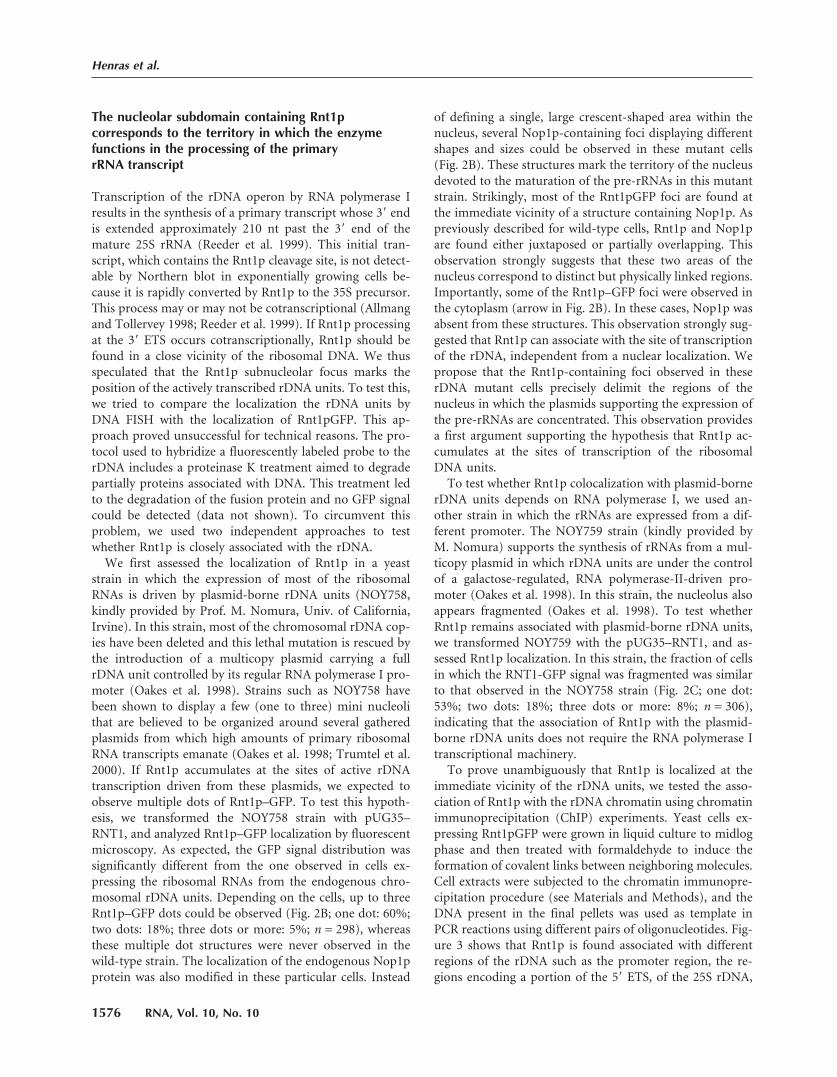
The nucleolar subdomain containing Rnt1pcorresponds to the territory in which the enzymefunctions in the processing of the primaryrRNA transcript
Transcription of the rDNA operon by RNA polymerase Iresults in the synthesis of a primary transcript whose 3� endis extended approximately 210 nt past the 3� end of themature 25S rRNA (Reeder et al. 1999). This initial tran-script, which contains the Rnt1p cleavage site, is not detect-able by Northern blot in exponentially growing cells be-cause it is rapidly converted by Rnt1p to the 35S precursor.This process may or may not be cotranscriptional (Allmangand Tollervey 1998; Reeder et al. 1999). If Rnt1p processingat the 3� ETS occurs cotranscriptionally, Rnt1p should befound in a close vicinity of the ribosomal DNA. We thusspeculated that the Rnt1p subnucleolar focus marks theposition of the actively transcribed rDNA units. To test this,we tried to compare the localization the rDNA units byDNA FISH with the localization of Rnt1pGFP. This ap-proach proved unsuccessful for technical reasons. The pro-tocol used to hybridize a fluorescently labeled probe to therDNA includes a proteinase K treatment aimed to degradepartially proteins associated with DNA. This treatment ledto the degradation of the fusion protein and no GFP signalcould be detected (data not shown). To circumvent thisproblem, we used two independent approaches to testwhether Rnt1p is closely associated with the rDNA.
We first assessed the localization of Rnt1p in a yeaststrain in which the expression of most of the ribosomalRNAs is driven by plasmid-borne rDNA units (NOY758,kindly provided by Prof. M. Nomura, Univ. of California,Irvine). In this strain, most of the chromosomal rDNA cop-ies have been deleted and this lethal mutation is rescued bythe introduction of a multicopy plasmid carrying a fullrDNA unit controlled by its regular RNA polymerase I pro-moter (Oakes et al. 1998). Strains such as NOY758 havebeen shown to display a few (one to three) mini nucleolithat are believed to be organized around several gatheredplasmids from which high amounts of primary ribosomalRNA transcripts emanate (Oakes et al. 1998; Trumtel et al.2000). If Rnt1p accumulates at the sites of active rDNAtranscription driven from these plasmids, we expected toobserve multiple dots of Rnt1p–GFP. To test this hypoth-esis, we transformed the NOY758 strain with pUG35–RNT1, and analyzed Rnt1p–GFP localization by fluorescentmicroscopy. As expected, the GFP signal distribution wassignificantly different from the one observed in cells ex-pressing the ribosomal RNAs from the endogenous chro-mosomal rDNA units. Depending on the cells, up to threeRnt1p–GFP dots could be observed (Fig. 2B; one dot: 60%;two dots: 18%; three dots or more: 5%; n = 298), whereasthese multiple dot structures were never observed in thewild-type strain. The localization of the endogenous Nop1pprotein was also modified in these particular cells. Instead
of defining a single, large crescent-shaped area within thenucleus, several Nop1p-containing foci displaying differentshapes and sizes could be observed in these mutant cells(Fig. 2B). These structures mark the territory of the nucleusdevoted to the maturation of the pre-rRNAs in this mutantstrain. Strikingly, most of the Rnt1pGFP foci are found atthe immediate vicinity of a structure containing Nop1p. Aspreviously described for wild-type cells, Rnt1p and Nop1pare found either juxtaposed or partially overlapping. Thisobservation strongly suggests that these two areas of thenucleus correspond to distinct but physically linked regions.Importantly, some of the Rnt1p–GFP foci were observed inthe cytoplasm (arrow in Fig. 2B). In these cases, Nop1p wasabsent from these structures. This observation strongly sug-gested that Rnt1p can associate with the site of transcriptionof the rDNA, independent from a nuclear localization. Wepropose that the Rnt1p-containing foci observed in theserDNA mutant cells precisely delimit the regions of thenucleus in which the plasmids supporting the expression ofthe pre-rRNAs are concentrated. This observation providesa first argument supporting the hypothesis that Rnt1p ac-cumulates at the sites of transcription of the ribosomalDNA units.
To test whether Rnt1p colocalization with plasmid-bornerDNA units depends on RNA polymerase I, we used an-other strain in which the rRNAs are expressed from a dif-ferent promoter. The NOY759 strain (kindly provided byM. Nomura) supports the synthesis of rRNAs from a mul-ticopy plasmid in which rDNA units are under the controlof a galactose-regulated, RNA polymerase-II-driven pro-moter (Oakes et al. 1998). In this strain, the nucleolus alsoappears fragmented (Oakes et al. 1998). To test whetherRnt1p remains associated with plasmid-borne rDNA units,we transformed NOY759 with the pUG35–RNT1, and as-sessed Rnt1p localization. In this strain, the fraction of cellsin which the RNT1-GFP signal was fragmented was similarto that observed in the NOY758 strain (Fig. 2C; one dot:53%; two dots: 18%; three dots or more: 8%; n = 306),indicating that the association of Rnt1p with the plasmid-borne rDNA units does not require the RNA polymerase Itranscriptional machinery.
To prove unambiguously that Rnt1p is localized at theimmediate vicinity of the rDNA units, we tested the asso-ciation of Rnt1p with the rDNA chromatin using chromatinimmunoprecipitation (ChIP) experiments. Yeast cells ex-pressing Rnt1pGFP were grown in liquid culture to midlogphase and then treated with formaldehyde to induce theformation of covalent links between neighboring molecules.Cell extracts were subjected to the chromatin immunopre-cipitation procedure (see Materials and Methods), and theDNA present in the final pellets was used as template inPCR reactions using different pairs of oligonucleotides. Fig-ure 3 shows that Rnt1p is found associated with differentregions of the rDNA such as the promoter region, the re-gions encoding a portion of the 5� ETS, of the 25S rDNA,
Henras et al.
1576 RNA, Vol. 10, No. 10

and of two different regions of the 3� ETS, as well as the 5Sgenes. The signals obtained after PCR in the anti-GFP im-munoprecipitates were significantly higher than the signalsobtained from the pellets obtained with the irrelevant anti-FLAG antibodies or with the protein G sepharose beadsalone (compare the Input and IP lanes). In contrast, noenrichment was obtained in a strain expressing the GFPalone (data not shown). To test whether the association ofRnt1p with the rDNA is specific, and because the rDNA isrepeated in multiple copies, we tested the interaction ofRnt1p with other repeated chromosomal regions. We chosethe CUP1 genes estimated to be present at a number ofabout 15–20 copies per haploid cells, and the telomericDNA, present at both ends of each chromosome. The in-tensity of the signals corresponding to the different rDNAregions obtained with the anti-GFP immunoprecipitatesrepresented between 65% and 75% (depending on the
rDNA region) of the input signals. In the same conditions,the amplifications of a region of CUP1 and of a telomerictarget gave signals representing only 10.75% or 16.5% of theinput signals, respectively. These quantifications show thatrDNA gene fragments are reproducibly found enrichedfour- to sevenfold in the Rnt1p–GFP immunoprecipitatescompared to negative control regions such as the telomeresor the CUP1 genes. These results show that Rnt1p is asso-ciated with the rDNA chromatin, an observation fully con-sistent with the hypothesis that processing by Rnt1p at the3� ETS occurs cotranscriptionally.
The presence of Rnt1p at the vicinity of the 5S genes wassomehow surprising because the 5S rRNA is not thought tobe processed by Rnt1p. This enrichment could be explainedby the tandem repetition of the rDNA repeats, where the 5Sgene follows closely (0.8–1.2 kB) the preceding 25S se-quence, and precedes closely the next rDNA promoter.
FIGURE 3. Rnt1p is associated with the rDNA chromatin. (A) Organization of the rDNA repeat and localization of the PCR products obtainedin the ChIP procedure. (B) Shown are �32P-labeled PCR products obtained from Input DNA (lane 1) or DNA extracted from immunoprecipi-tation reactions in extracts prepared from the rnt1:TRP, pUG35–RNT1 strain using anti-GFP IgGs (�GFP, lane 2), irrelevant anti-FLAG IgGs(�-FLAG, lane 3), or with Protein G-conjugated sepharose beads only (Beads, lane 4), using oligonucleotide pairs hybridizing to the indicatedDNA sequences. (C) Quantification of the ChIP data. Five independent ChIP experiments were carried out as described in Materials and Methods.For each experiment, the intensity of the radioactive bands was measured using Image Quant. The relative immunoprecipitation efficiency of thedifferent DNA targets by the anti-GFP antibodies was calculated as follows: For a given primer pair, the average intensity of the bands obtainedin the negative control experiments and reflecting the immunoprecipitation background (“�-FLAG” or “Beads only” controls) was subtractedfrom the intensity of the band obtained with the relevant, �-GFP antibodies. The resulting value was then divided by the intensity of the bandcorresponding to the INPUT experiment. This value was then divided by the value obtained for the CUP1 signal (negative control). Error barsindicate the standard deviation. Because CUP1 was used as an internal standard for each experiment, no error bar is included.
Cotranscriptional rRNA processing
www.rnajournal.org 1577

Given the low resolution of the ChIP technique and thetandem repeats organization of the yeast rDNA, the moststraightforward explanation for the signal observed for the5S rDNA is that it results from the presence of the Rnt1pprotein 500 bp–1 kb upstream, near the 3� ETS sequence.Nevertheless, to further prove the specificity of our chro-matin immunoprecipitation reactions, we assessed the en-richment of Rnt1p near two other genes, the snR190-U14snoRNA gene and the U6 gene. These genes were chosenbecause Rnt1p is known to process the dicistronic precur-sor that contains snR190 and U14 (Chanfreau et al.1998b), but does not process U6. This ChIP experimentshowed that Rnt1p is enriched near the snR190–U14 genes,but not at the vicinity of the U6 gene (Fig. 3), showing thatchromatin immunoprecipitation using Rnt1p–GFP resultsin a selective enrichment of genes encoding Rnt1p sub-strates. This result also suggests that processing of thesnR190–U14 precursor occurs in a cotranscriptional fash-ion.
The N-terminal domain of Rnt1p is not required forthe import of Rnt1p into the nucleus but is necessaryfor formation of the nucleolar Rnt1p dot
The results presented so far indicate that Rnt1p is a nuclearenzyme distributed throughout the nucleus that alsostrongly accumulates within a nucleolar structure corre-sponding to the site of transcription of the rDNAs. We nextfocused on the identification of the domains of the proteinrequired for this particular subcellular localization. We ex-pressed truncated versions of Rnt1p fused to the GFP (Fig.4A) and studied their localization. To define the boundariesof the truncations to be introduced in Rnt1p, we comparedthe amino acid sequence of Rnt1p with that of Escherichiacoli RNase III. The bacterial enzyme is composed of anN-terminal catalytic domain containing the RNase III sig-nature motif fused to a C-terminal dsRNA-binding domain.In Rnt1p, these two domains are flanked by two additionalmodules that are absent from the bacterial enzyme (Fig.4A). The amino-terminal region of Rnt1p preceding thenuclease domain includes approximately 175 amino acidsand was proposed to influence the dimerization of the en-zyme (Lamontagne et al. 2000). Downstream from thedsRBD, the last 40 amino acids of Rnt1p define a carboxy-terminal domain that has been proposed to interact withGar1p and to be required for the nuclear import ofthe box H/ACA snoRNP core proteins (Tremblay et al.2002). These two domains are found specifically within theyeast enzyme and not in its bacterial counterpart. Thus, wesuspected that in addition to their previously proposedfunction, they might play a role in the localization of theenzyme. We initially focused on the amino-terminal do-main of Rnt1p because it had been proposed to be requiredfor nuclear import (Nagel and Ares 2000). We studied thesubcellular localization of two different truncated versions
of Rnt1p lacking the first 47 (Rnt1p�N47) or 172(Rnt1p�N172) N-terminal amino acids. Yeast cells lackingthe endogenous RNT1 gene were transformed with pUG35-derivative constructs supporting the expression of thesemodified proteins fused to the GFP. The growth rate ofthe resulting strains and the localization of the modifiedproteins were assessed and compared to those of a strain inwhich the wild-type Rnt1p–GFP protein is expressed(Fig. 4).
Deletion of the first 47 amino acids of Rnt1p induced asignificant growth defect, because Rnt1p�N47 cells showed
FIGURE 4. The N-terminal domain of Rnt1p is not required for theimport of Rnt1p into the nucleus of yeast cells. (A) Schematic repre-sentation of the constructs used in the truncation analysis. Shown arethe boundaries of the Rnt1p domains and of the truncations gener-ated, and the generation time of the the rnt1�TRP transformed withthe pUG35 derivatives expressing the different truncated versions ofthe protein. The generation time of rnt1�TRP transformed with plas-mid pUG35 (knockout control) is 440 min. (N-ter) N-terminal do-main, (ND) RNase III Nuclease Domain, (dsRBD) double-strandedRNA binding domain, (C-ter) C-terminal domain. (B) Shown are(immuno)fluorescence images obtained with the haploid strainrnt1�TRP transformed with plasmids pUG35–RNT1�N47 orpUG35–RNT1�N172. Legends as in Figure 1A.
Henras et al.
1578 RNA, Vol. 10, No. 10
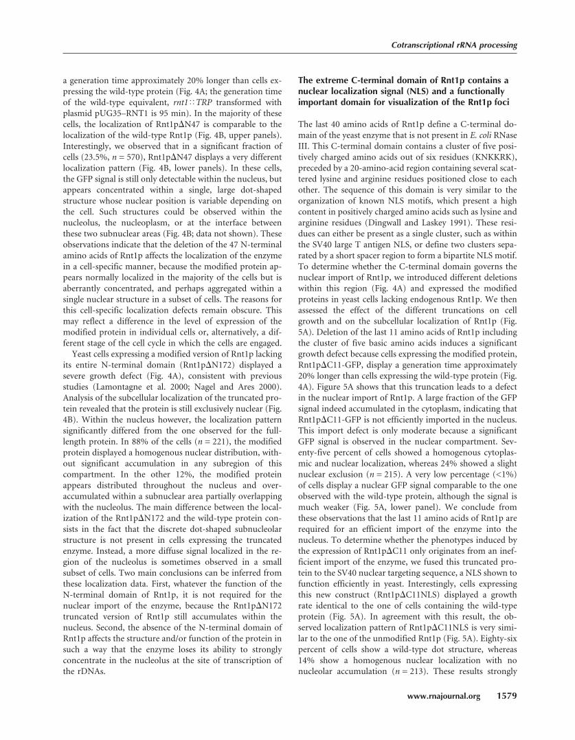
a generation time approximately 20% longer than cells ex-pressing the wild-type protein (Fig. 4A; the generation timeof the wild-type equivalent, rnt1�TRP transformed withplasmid pUG35–RNT1 is 95 min). In the majority of thesecells, the localization of Rnt1p�N47 is comparable to thelocalization of the wild-type Rnt1p (Fig. 4B, upper panels).Interestingly, we observed that in a significant fraction ofcells (23.5%, n = 570), Rnt1p�N47 displays a very differentlocalization pattern (Fig. 4B, lower panels). In these cells,the GFP signal is still only detectable within the nucleus, butappears concentrated within a single, large dot-shapedstructure whose nuclear position is variable depending onthe cell. Such structures could be observed within thenucleolus, the nucleoplasm, or at the interface betweenthese two subnuclear areas (Fig. 4B; data not shown). Theseobservations indicate that the deletion of the 47 N-terminalamino acids of Rnt1p affects the localization of the enzymein a cell-specific manner, because the modified protein ap-pears normally localized in the majority of the cells but isaberrantly concentrated, and perhaps aggregated within asingle nuclear structure in a subset of cells. The reasons forthis cell-specific localization defects remain obscure. Thismay reflect a difference in the level of expression of themodified protein in individual cells or, alternatively, a dif-ferent stage of the cell cycle in which the cells are engaged.
Yeast cells expressing a modified version of Rnt1p lackingits entire N-terminal domain (Rnt1p�N172) displayed asevere growth defect (Fig. 4A), consistent with previousstudies (Lamontagne et al. 2000; Nagel and Ares 2000).Analysis of the subcellular localization of the truncated pro-tein revealed that the protein is still exclusively nuclear (Fig.4B). Within the nucleus however, the localization patternsignificantly differed from the one observed for the full-length protein. In 88% of the cells (n = 221), the modifiedprotein displayed a homogenous nuclear distribution, with-out significant accumulation in any subregion of thiscompartment. In the other 12%, the modified proteinappears distributed throughout the nucleus and over-accumulated within a subnuclear area partially overlappingwith the nucleolus. The main difference between the local-ization of the Rnt1p�N172 and the wild-type protein con-sists in the fact that the discrete dot-shaped subnucleolarstructure is not present in cells expressing the truncatedenzyme. Instead, a more diffuse signal localized in the re-gion of the nucleolus is sometimes observed in a smallsubset of cells. Two main conclusions can be inferred fromthese localization data. First, whatever the function of theN-terminal domain of Rnt1p, it is not required for thenuclear import of the enzyme, because the Rnt1p�N172truncated version of Rnt1p still accumulates within thenucleus. Second, the absence of the N-terminal domain ofRnt1p affects the structure and/or function of the protein insuch a way that the enzyme loses its ability to stronglyconcentrate in the nucleolus at the site of transcription ofthe rDNAs.
The extreme C-terminal domain of Rnt1p contains anuclear localization signal (NLS) and a functionallyimportant domain for visualization of the Rnt1p foci
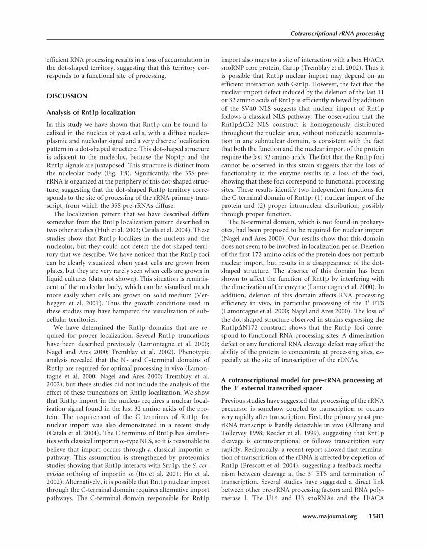
The last 40 amino acids of Rnt1p define a C-terminal do-main of the yeast enzyme that is not present in E. coli RNaseIII. This C-terminal domain contains a cluster of five posi-tively charged amino acids out of six residues (KNKKRK),preceded by a 20-amino-acid region containing several scat-tered lysine and arginine residues positioned close to eachother. The sequence of this domain is very similar to theorganization of known NLS motifs, which present a highcontent in positively charged amino acids such as lysine andarginine residues (Dingwall and Laskey 1991). These resi-dues can either be present as a single cluster, such as withinthe SV40 large T antigen NLS, or define two clusters sepa-rated by a short spacer region to form a bipartite NLS motif.To determine whether the C-terminal domain governs thenuclear import of Rnt1p, we introduced different deletionswithin this region (Fig. 4A) and expressed the modifiedproteins in yeast cells lacking endogenous Rnt1p. We thenassessed the effect of the different truncations on cellgrowth and on the subcellular localization of Rnt1p (Fig.5A). Deletion of the last 11 amino acids of Rnt1p includingthe cluster of five basic amino acids induces a significantgrowth defect because cells expressing the modified protein,Rnt1p�C11-GFP, display a generation time approximately20% longer than cells expressing the wild-type protein (Fig.4A). Figure 5A shows that this truncation leads to a defectin the nuclear import of Rnt1p. A large fraction of the GFPsignal indeed accumulated in the cytoplasm, indicating thatRnt1p�C11-GFP is not efficiently imported in the nucleus.This import defect is only moderate because a significantGFP signal is observed in the nuclear compartment. Sev-enty-five percent of cells showed a homogenous cytoplas-mic and nuclear localization, whereas 24% showed a slightnuclear exclusion (n = 215). A very low percentage (<1%)of cells display a nuclear GFP signal comparable to the oneobserved with the wild-type protein, although the signal ismuch weaker (Fig. 5A, lower panel). We conclude fromthese observations that the last 11 amino acids of Rnt1p arerequired for an efficient import of the enzyme into thenucleus. To determine whether the phenotypes induced bythe expression of Rnt1p�C11 only originates from an inef-ficient import of the enzyme, we fused this truncated pro-tein to the SV40 nuclear targeting sequence, a NLS shown tofunction efficiently in yeast. Interestingly, cells expressingthis new construct (Rnt1p�C11NLS) displayed a growthrate identical to the one of cells containing the wild-typeprotein (Fig. 5A). In agreement with this result, the ob-served localization pattern of Rnt1p�C11NLS is very simi-lar to the one of the unmodified Rnt1p (Fig. 5A). Eighty-sixpercent of cells show a wild-type dot structure, whereas14% show a homogenous nuclear localization with nonucleolar accumulation (n = 213). These results strongly
Cotranscriptional rRNA processing
www.rnajournal.org 1579

suggest that the last 11 amino acids of Rnt1p only take partin the import of the enzyme but are not involved in anyaspect of its nuclear functions.
Deletion of the last 11 amino acids of Rnt1p only partiallyaffects the import of Rnt1p, suggesting that this region ofthe protein is part of a larger NLS. To test this hypothesis,we deleted the last 32 amino acids and analyzed the effectsof this truncation. A yeast strain expressing Rnt1p�C32
displayed a severe growth defect, as its growth rate is re-duced by almost four times compared to the wild-type con-trol and it grew only slightly faster than the knockout strain(Fig. 4A). The last 32 amino acids of Rnt1p are thereforealmost essential for the function of the enzyme. Analysis ofthe subcellular localization of Rnt1p�C32–GFP revealed astrong defect in the nuclear import of the truncated protein(Fig. 5A). Thirty-three percent of cells (n = 30) showed ahomogenous nuclear and cytoplasmic GFP signal, whereas67% showed a slight to strong nuclear exclusion, indicatinga very low concentration of Rnt1p�C32 in the nucleus. Thelast 32 C-terminal amino acids of Rnt1p therefore define adomain that is essential for the nuclear import of Rnt1p.Expression of Rnt1p�C32 fused to the SV40 NLS did notabolish the growth defect observed with the truncated pro-tein. The growth rate was, on the contrary, further reduced,the generation time of this strain being increased by fivetimes compared to the wild-type control. Surprisingly, thisstrain grew slower than the knockout strain (Fig. 4A), in-dicating that the expression of Rnt1p�C32NLS is more del-eterious than the absence of Rnt1p. However this proteindid not exhibit a dominant negative effect when expressedin wild-type cells (data not shown). The Rnt1p�C32NLSprotein was present within the nucleus, as expected giventhe presence of the SV40 NLS. The distribution of the en-zyme inside the nucleus is, however, different from thedistribution of the wild-type protein. Rnt1p�C32NLS washomogenously distributed throughout the nucleus, withoutsignificant accumulation in any subnuclear domain. Weconclude that the last 32 amino acids of Rnt1p, in additionto being essential to the import of the enzyme, also includeresidues that are required for the discrete accumulation ofRnt1p at the site of transcription of the rDNAs. It is possiblethat the deletion of the whole C-terminal domain of Rnt1palters the structure of the double-stranded RNA-bindingdomain located nearby (Wu et al. 2004) and decreases RNAbinding, preventing Rnt1p accumulation into functionalsites.
The previous results show that the C-terminal domain ofRnt1p includes a motif that is necessary for the import ofthe protein into the nucleus. To match the definition of anNLS, this domain must also be sufficient to direct a non-nuclear protein to the nucleus. To determine whether theC-terminal domain of Rnt1p displays such a property, wefused the last 32 amino acids of Rnt1p to the GFP andcompared the localization of this construct to the localiza-tion of the GFP alone in wild-type yeast cells. As shown inFigure 5B, when fused to the C-terminal domain of Rnt1p,the GFP moiety became predominantly localized in thenucleus compared to the GFP alone, which localizes uni-formly in the cell. These results demonstrate that the C-terminal domain of Rnt1p is both necessary and sufficientto govern the active import of a protein into the nucleusand hence behaves as a bona fide nuclear targeting signal.Finally, deletion of Rnt1p domains that are required for
FIGURE 5. The C-terminal domain of Rnt1p is necessary and suffi-cient for nuclear import. (A) Deletion of the C-terminal amino acidsof Rnt1p perturbs nuclear import. Shown are (immuno)fluorescenceimages obtained with the haploid strain rnt1�TRP transformed withplasmids pUG35–RNT1�C11, pUG35–RNT1�C11NLS, pUG35–RNT1�C32, or pUG35–RNT1�C32NLS. Legends as in Figure 1A.(B)The C-terminal domain of Rnt1p is a bona fide nuclear targetingsignal. Shown are fluorescence pictures obtained from the haploidstrain BMA64 (wild type) transformed with plasmid pUG35-C32 orpUG35.
Henras et al.
1580 RNA, Vol. 10, No. 10
efficient RNA processing results in a loss of accumulation inthe dot-shaped territory, suggesting that this territory cor-responds to a functional site of processing.
DISCUSSION
Analysis of Rnt1p localization
In this study we have shown that Rnt1p can be found lo-calized in the nucleus of yeast cells, with a diffuse nucleo-plasmic and nucleolar signal and a very discrete localizationpattern in a dot-shaped structure. This dot-shaped structureis adjacent to the nucleolus, because the Nop1p and theRnt1p signals are juxtaposed. This structure is distinct fromthe nucleolar body (Fig. 1B). Significantly, the 35S pre-rRNA is organized at the periphery of this dot-shaped struc-ture, suggesting that the dot-shaped Rnt1p territory corre-sponds to the site of processing of the rRNA primary tran-script, from which the 35S pre-rRNAs diffuse.
The localization pattern that we have described differssomewhat from the Rnt1p localization pattern described intwo other studies (Huh et al. 2003; Catala et al. 2004). Thesestudies show that Rnt1p localizes in the nucleus and thenucleolus, but they could not detect the dot-shaped terri-tory that we describe. We have noticed that the Rnt1p focican be clearly visualized when yeast cells are grown fromplates, but they are very rarely seen when cells are grown inliquid cultures (data not shown). This situation is reminis-cent of the nucleolar body, which can be visualized muchmore easily when cells are grown on solid medium (Ver-heggen et al. 2001). Thus the growth conditions used inthese studies may have hampered the visualization of sub-cellular territories.
We have determined the Rnt1p domains that are re-quired for proper localization. Several Rnt1p truncationshave been described previously (Lamontagne et al. 2000;Nagel and Ares 2000; Tremblay et al. 2002). Phenotypicanalysis revealed that the N- and C-terminal domains ofRnt1p are required for optimal processing in vivo (Lamon-tagne et al. 2000; Nagel and Ares 2000; Tremblay et al.2002), but these studies did not include the analysis of theeffect of these truncations on Rnt1p localization. We showthat Rnt1p import in the nucleus requires a nuclear local-ization signal found in the last 32 amino acids of the pro-tein. The requirement of the C terminus of Rnt1p fornuclear import was also demonstrated in a recent study(Catala et al. 2004). The C terminus of Rnt1p has similari-ties with classical importin �-type NLS, so it is reasonable tobelieve that import occurs through a classical importin �pathway. This assumption is strengthened by proteomicsstudies showing that Rnt1p interacts with Srp1p, the S. cer-evisiae ortholog of importin � (Ito et al. 2001; Ho et al.2002). Alternatively, it is possible that Rnt1p nuclear importthrough the C-terminal domain requires alternative importpathways. The C-terminal domain responsible for Rnt1p
import also maps to a site of interaction with a box H/ACAsnoRNP core protein, Gar1p (Tremblay et al. 2002). Thus itis possible that Rnt1p nuclear import may depend on anefficient interaction with Gar1p. However, the fact that thenuclear import defect induced by the deletion of the last 11or 32 amino acids of Rnt1p is efficiently relieved by additionof the SV40 NLS suggests that nuclear import of Rnt1pfollows a classical NLS pathway. The observation that theRnt1p�C32–NLS construct is homogenously distributedthroughout the nuclear area, without noticeable accumula-tion in any subnuclear domain, is consistent with the factthat both the function and the nuclear import of the proteinrequire the last 32 amino acids. The fact that the Rnt1p focicannot be observed in this strain suggests that the loss offunctionality in the enzyme results in a loss of the foci,showing that these foci correspond to functional processingsites. These results identify two independent functions forthe C-terminal domain of Rnt1p: (1) nuclear import of theprotein and (2) proper intranuclear distribution, possiblythrough proper function.
The N-terminal domain, which is not found in prokary-otes, had been proposed to be required for nuclear import(Nagel and Ares 2000). Our results show that this domaindoes not seem to be involved in localization per se. Deletionof the first 172 amino acids of the protein does not perturbnuclear import, but results in a disappearance of the dot-shaped structure. The absence of this domain has beenshown to affect the function of Rnt1p by interfering withthe dimerization of the enzyme (Lamontagne et al. 2000). Inaddition, deletion of this domain affects RNA processingefficiency in vivo, in particular processing of the 3� ETS(Lamontagne et al. 2000; Nagel and Ares 2000). The loss ofthe dot-shaped structure observed in strains expressing theRnt1p�N172 construct shows that the Rnt1p foci corre-spond to functional RNA processing sites. A dimerizationdefect or any functional RNA cleavage defect may affect theability of the protein to concentrate at processing sites, es-pecially at the site of transcription of the rDNAs.
A cotranscriptional model for pre-rRNA processing atthe 3� external transcribed spacer
Previous studies have suggested that processing of the rRNAprecursor is somehow coupled to transcription or occursvery rapidly after transcription. First, the primary yeast pre-rRNA transcript is hardly detectable in vivo (Allmang andTollervey 1998; Reeder et al. 1999), suggesting that Rnt1pcleavage is cotranscriptional or follows transcription veryrapidly. Reciprocally, a recent report showed that termina-tion of transcription of the rDNA is affected by depletion ofRnt1p (Prescott et al. 2004), suggesting a feedback mecha-nism between cleavage at the 3� ETS and termination oftranscription. Several studies have suggested a direct linkbetween other pre-rRNA processing factors and RNA poly-merase I. The U14 and U3 snoRNAs and the H/ACA
Cotranscriptional rRNA processing
www.rnajournal.org 1581

snoRNP protein Gar1p localize in the dense fibrillar com-ponent (Lazdins et al. 1997; Leger-Silvestre et al. 1999), theprobable site of transcription by RNA polymerase I (Hozaket al. 1994; Jackson and Cook 1995; Lazdins et al. 1997;Mosgoeller et al. 1998). In addition, the nucleolar process-ing factor Nop1p colocalizes with RNA polymerase I inmouse embryos (Cuadros-Fernandez and Esponda 1996)and with nascent rRNA transcripts (Garcia-Blanco et al.1995). In addition, several snoRNP proteins and rRNA pro-cessing factors can be found biochemically associated withyeast RNA polymerase I (Fath et al. 2000). These observa-tions have linked pre-rRNA processing to RNA polymeraseI transcription. Our results strongly suggest that in S. cer-evisiae, cleavage of the primary pre-rRNA transcript byRnt1p to generate the 35S precursor is a cotranscriptionalevent. Because the transcription of the rDNA genes repre-sents about 60% of the total cellular transcription (Warner1999), high amounts of primary ribosomal RNAs continu-ously emerge from the active rDNA units, and their 3�-endprocessing is likely to mobilize high amounts of Rnt1p. Thechromatin immunoprecipitation results (Fig. 3), and thefact that the dot-shaped Rnt1p territory becomes reorga-nized when the rRNAs are expressed from multicopy plas-mids provide evidence for association of Rnt1p with rDNAunits. In the light of these considerations, we conclude thatthe observed nucleolar area in which Rnt1p is strongly ac-cumulated corresponds to the site of transcription of therDNA genes and early processing of the ribosomal RNAprecursor. In support of this hypothesis, it was recentlyshown that the nucleolar accumulation of Rnt1p is reducedin a polymerase I temperature-sensitive strain (Catala et al.2004). However, analysis of Rnt1p localization in a rpa49�strain, in which the levels of transcription by RNA poly-merase I are reduced (Liljelund et al. 1992), did not show asignificant difference compared to wild-type cells (data notshown). Interestingly the reorganization of the Rnt1p fociaround plasmid-borne rDNA units occurs even if therRNAs are expressed from an RNA polymerase II promoter(Fig. 2C). This suggests that Rnt1p association with therDNA may occur independently from the specific RNApolymerase machinery, or that Rnt1p may be recruited by asubunit of the polymerase that is common between RNApolymerases I and II. Intriguingly, there is no significantenrichment of Rnt1p at the vicinity of the 3� ETS rDNAregion compared to other rDNA regions in our ChIP ex-periment, showing that Rnt1p is probably evenly distrib-uted throughout most of the rDNA region. Alternatively, itis possible that the repeated nature of these sequencesmakes local enrichment very hard to distinguish using thechromatin immunoprecipitation technique. For example,the enrichment observed in the 5S region may be due to theclose proximity of this sequence to the preceding 3� ETSand to the poor resolution of the ChIP technique. The evendistribution of Rnt1p throughout the rDNA can suggestseveral models of cotranscriptional cleavage of the pre-
rRNA transcript. In the first model, Rnt1p is present stati-cally on most regions of the rDNA, but the fraction ofRnt1p present at the vicinity of the 3� ETS sequence at theDNA level may be sufficient to cleave the nascent 3� ETSsequence after this sequence exits from the RNA polymer-ase. Alternatively it is possible that the Rnt1p subunits pres-ent along the rDNA may form a reservoir of enzymes. Inthis model, the RNA polymerase machinery (through a sub-unit that would be common to Pol I and Pol II) wouldrecruit one of the Rnt1p subunits present in the chromatinwhen it travels along the rDNA transcription unit. Finally itis possible that Rnt1p is recruited very early at the promoterby the RNA polymerase, and that it travels along the rDNAunit with the RNA polymerase. The presence of Rnt1palong the entire rDNA sequence observed by chromatinimmunoprecipitation would result from the dynamic asso-ciation of Rnt1p with the RNA polymerase machinery. Fur-ther experiments may shed light on the validity of each ofthese models.
MATERIALS AND METHODS
Yeast strains and plasmids
Yeast manipulation and transformation was performed using stan-dard techniques. The sequence of oligonucleotides used is avail-able upon request. Most yeast strains used in this study are aderivative of BMA64 (Baudin et al. 1993). Yeast strains carryingthe rnt:TRP disruption have been described previously (Chanfreauet al. 1998b). The NOY758 and NOY759 strains were kindly pro-vided by M. Nomura (Oakes et al. 1998). The rpa49� strain waspurchased from Open Biosystems. To construct plasmids express-ing the RNT1–GFP fusions, the RNT1 open reading frame wasamplified by PCR using Pfu Polymerase (Stratagene) and clonedinto pUG35 (Niedenthal et al. 1996), using BamHI and ClaI sitesthat were introduced into the primers used for PCR. Truncationsin the RNT1 gene were generated by PCR. For construction of thestrain carrying a GFP fusion at the endogenous RNT1 locus, aKan-URA marker was first inserted after the RNT1 stop codon, asdescribed (Storici et al. 2001). A PCR fragment was generatedusing the pUG35–RNT1 plasmid as a template, an upstream oli-gonucleotide hybridizing from positions 620–640 of the RNT1ORF, and a downstream oligonucleotide hybrdizing to the end ofthe GFP sequence. The downstream oligonucleotides also carried50 nt of complementarity to the RNT1 3� UTR. In a second step,this PCR fragment was transformed in the previously describedstrain, and transformants were selected on 5-fluoroorotic acid, andscreened for correct insertion of the GFP fusion by PCR on yeastcolonies.
Indirect immunofluorescence microscopy
Cells were collected from patches grown on plates and transferredto small Erlenmeyer flasks containing 10.5 mL of liquid medium(same composition as the medium corresponding to the initialplates). Cells were fixed by adding 1.5 mL of 16% formaldehyde
Henras et al.
1582 RNA, Vol. 10, No. 10

(2% final concentration) and incubating for 30 min at 30°C withgentle shaking. Cells were washed once with 1 mL of 0.1 M KPO4
(pH 6.5) and once with 1 mL P solution (1.2 M Sorbitol in 0.1 MKPO4 at pH 6.5). Cell were pelleted and resuspended with 1 mL ofP solution. Spheroplasts were generated by adding 15 µL of 10mg/mL Zymolyase 20T (Seikagaku Corp.) and 5 µL of �-mercap-toethanol, and incubated for 1 h at 37°C with gentle shaking.Spheroplasts were recovered by gentle centrifugation, resuspendedwith 500 µL of P solution, and spotted on eight-well multitestslides previously coated with polylysin. After 10 min, excesscells were removed and the slides were incubated for 6 min inice-cold methanol followed by 30 sec in ice-cold acetone. Slideswere then washed twice with 5 mg/mL BSA, 1× PBS and incubatedfor 2 h at room temperature with anti-Nop1p monoclonalantibodies (kindly provided by J. Aris, Univ. of Florida) diluted1:5000 in 5 mg/mL BSA in 1× PBS. Slides were then washed threetimes for 5 min with 5 mg/mL BSA in 1× PBS and incubated for2 h at room temperature with Cy3-conjugated anti-mouse IgGs(Sigma) diluted 1:1000 in 5 mg/mL BSA, 1× PBS. Slides were thenwashed three times with 1× PBS, incubated for 10 min at roomtemperature with 4,6-diamidino-2-phenylindole (DAPI) dilutedto 1 µg/mL in 1× PBS. Slides were washed three times for 5 minwith 1× PBS and covered with 60% glycerol and a coverslip. Fluo-rescent images were captured with an Optronics cooled CCD cam-era attached to a Zeiss Axioplan microscope. All images werecaptured with a PHOTOSHOP AV capture plug-in on a Macin-tosh Power PC AV computer.
Fluorescent in situ hybridization (FISH)
Detection of the nucleolar body by FISH was performed as de-scribed (Verheggen et al. 2001). FISH detection of the 35S pre-rRNAs was performed as follows, using the probe 5�AXCCTTCGCTGCXCACCAATGGAAXCGCAAGATGCCCACGAXG�(X = Cy3-dT). This probe hybridizes upstream of the A0 site.
FISH detection of the U14 snoRNA was performed as followsusing a mixture of the two following probes: 5�Cy3-GTGGAAACTGCGAATGTTAAGGAACCAGTCTTTCATCACCGTGAT-Cy3and 5�Cy3-GAAGAGCGGTCACCGAGAGTACTAACGATGGGTTCGTAAGCG-Cy3. Cells were collected from patches grown onplates and transferred to small Erlenmeyer flasks containing 9 mLof liquid medium (same composition as the medium correspond-ing to the initial plates). Cells were fixed by adding 3 mL of 16%formaldehyde (4% final concentration) and incubated for 30 minat 25°C with gentle shaking. Cells were then recovered and washedonce with 10 mL of buffer B (1.2 M sorbitol, 100 mM K-phosphatebuffer at pH 7.5). Cells were recovered by centrifugation, resus-pended with 500 µL of digestion buffer (1 mL of Buffer B, 20 µLof 50× Complete EDTA-free protease inhibitor cocktail [RocheDiagnostics], 10 µL of 200 mM vanadium ribonucleoside complex,2 µL of PMSF, 2 µL of �-mercaptoethanol, 10 µL of 20 mg/mLZymolyase 20T from Seikagaku dissolved in buffer B) and incu-bated for 30 min at 30°C with gentle shaking. After digestion,spheroplasts were recovered by centrifugation, rinsed with 10 mLof ice-cold buffer B, pelleted again, and resuspended with 300 µLof ice-cold buffer B. Aliquot fractions of the suspension (50 µL)were spotted on each well of a multitest slide previously coatedwith polylysin. After incubation for 30 min, excess cells were re-moved and the slide was incubated overnight in −20°C 70% etha-nol. Spheroplasts were then rehydrated by incubating the slide
twice for 5 min with 2× SSC at room temperature and for 5 minwith 2× SSC, 10% formamide.
For the in situ hybridization of the probe to the spheroplasts,two different solutions (A and B) were prepared separately. Solu-tion A was obtained by mixing 2 µL of a 10 ng/µL oligonucleotideprobe solution, 4 µL of formamide, 2 µL of 2× SSC, 2 µL of 10mg/mL wheat germ tRNAs, and 7 µL of ddH2O. Solution B wasobtained by mixing 20 µL of 20% dextran sulfate in 4× SSC, 2 µLof 10 mg/mL BSA, and 2 µL of 200 mM vanadium ribonucleosidecomplex. Solution A was incubated for 5 min at 100°C, chilled onice for 5 min, and mixed with room temperature solution B. Theresulting solution was loaded onto the slide (20 µL per well) andincubated for 3 h at 37°C. Slides were then washed two times for15 min at 37°C with 37°C pre-warmed 2× SSC, 10% formamideone time for 15 min at room temperature with 2× SSC, 0.1%Triton X-100 and then two times for 15 min at room temperaturewith 1× SSC. Slides where then rinsed three times for 5 min with1× PBS at room temperature, incubated for 10 min with a 1 µg/mLDAPI solution at room temperature, and rinsed again three timesfor 5 min with 1× PBS. Slides were covered with 60% glycerol anda coverslip, and imaging was performed as described above.
Chromatin immunoprecipitation (ChIP)
A 50-mL culture of the relevant yeast strain was grown to a lateexponential phase (OD600 = 1). Cross-linking was achieved byadding formaldehyde to the culture to a final concentration of 1%and incubating for 30 min at 30°C with gentle shaking. Cross-linking was quenched by adding glycine to a final concentration of125 mM and incubating for 5 min at 30°C with gentle shaking.Cells were transferred into a 50-mL centrifugation tube, washedtwice with 20 mL ice-cold 1× PBS, and transferred into 1.5-mLsiliconized tubes. After centrifugation, cell pellets were resus-pended with 400 µL ice-cold lysis buffer (50 mM HEPES/KOH atpH 7.5, 500 mM NaCl, 1 mM EDTA at pH 8.0, 1% Triton X-100,0.1% sodium-deoxycholate, 0.1% SDS) supplemented with prote-ase inhibitors (Complete EDTA-free; Roche Diagnostics). Glassbeads (500 µL) were added to the tubes and cells were broken byvigorous vortexing for 45 min at 4°C. Extract and cell debris wereseparated from the beads and sonicated on ice two times for 15 sec(sonic Dismembrator 550, Fisher Scientific, setting 4 out of 10),with a 1-min interval. Cell debris was pelleted by centrifugation(13,000 rpm, 5 min, 4°C) and the supernatant was recovered andtransferred to a new 1.5-mL siliconized tube. A 20-µL aliquotfraction of this extract was frozen at this stage for subsequentanalysis (INPUT; see below).
Each immunoprecipitation (IP) experiment was carried out us-ing a 50-µL aliquot fraction of the extract transferred to a new1.5-mL siliconized tube. We added 1.5 µg of anti-GFP antibody(Clontech), 1.5 µg of an irrelevant antibody, or no antibody to a50-µL aliquot fraction of extract and incubated overnight at 4°Cwith gentle shaking. Recovery of the antibodies and their boundepitopes was achieved by adding to each tube 25 µL of a 50%protein-G sepharose beads (Pharmacia) suspension (1 volume ofbead:1 volume of lysis buffer; beads were previously washed sev-eral times with ddH2O and then twice with the Lysis Buffer),incubating 90 min at 4°C with gentle shaking and spinning at 2000rpm for 2 min at 4°C. Beads pellets were then washed two timesfor 10 min with 1 mL of lysis buffer at 4°C, one time for 10 minwith 1 mL of sodium-deoxycholate buffer (10 mM Tris-HCl at pH
Cotranscriptional rRNA processing
www.rnajournal.org 1583

8.0, 0.25 M LiCl, 0.5% NP-40, 0.5% sodium-deoxycholate, 1 mMEDTA at pH 8.0) at 4°C and one time for 10 min with TE (10 mMTris-HCl at pH 8.0, 1 mM EDTA at pH 8.0) at 4°C. After the lastwash, beads were pelleted and the washing buffer was removedcompletely.
Immunoprecipitated proteins were eluted from the beads andthe cross-link was reversed by mixing bead pellets with 50 µL ofelution buffer (50 mM Tris-HCl at pH 8.0, 10 mM EDTA at pH8.0, 1% SDS) and incubating overnight at 65°C in a hybridizationoven. At this stage, the 20-µL INPUT aliquot fractions werethawed, mixed with 80 µL of TE, and incubated along with theimmunoprecipitates. Eluates were recovered from the IP experi-ments (about 50 µL) and diluted by adding 50 µL of TE. A 1:10aliquot fraction (10 µL) was taken out at this stage, mixed withSDS-loading buffer, and frozen for subsequent Western blot analy-sis of the precipitated proteins. We added 10 µg of glycogen and100 µg of proteinase K to the remaining samples and tubes wereincubated for 2 h at 56°C. DNA fragments contained in the IPsand INPUTs were purified by phenol extraction followed by etha-nol precipitation and the final DNA pellets were resuspended with40 µL of ddH2O. Contaminant RNA molecules potentially presentin the samples were degraded by RNase A treatment. PCR reac-tions were carried out using 0.5 µL of the immunoprecipitatedDNAs or 0.5 µL of a 1:100 dilution of the INPUT DNAs, with PfuTurbo polymerase (Stratagene) and in the presence of 1 µCi of�32P dCTP per reaction. Half of the PCR reactions were mixedwith loading buffer and loaded on 6% acrylamide:bisacrylamide(19:1), 0.5× TBE gels. After migration, gels were dried and exposedto PhosphorImager screens.
ACKNOWLEDGMENTS
We thank J. Gober for the extensive use of his fluorescence mi-croscope; P. Marc for constructing the pUG35-Rnt1p plasmid andhelp with early studies; J. Aris, M. Caizergues-Ferrer, Y. Henry, T.Meier, and M. Nomura for yeast strains and/or antibodies; P.E.Gleyzes for providing the pre-rRNA FISH protocol and the rRNAoligonucleotide probe; S. Baserga, J. Gallagher, M. Grunstein, andD. Robyr for chromatin immunoprecipitation protocols; and D.Black and H. Martinson for critical reading of the manuscript.This work was supported by a long-term EMBO PostdoctoralFellowship and by a Human Frontier Long-Term PostdoctoralFellowship (A.H.) and by grant GM61518 from National Institutesof Health (NIH) to G.C.
The publication costs of this article were defrayed in part bypayment of page charges. This article must therefore be herebymarked “advertisement” in accordance with 18 USC section 1734solely to indicate this fact.
Received April 29, 2004; accepted July 15, 2004.
REFERENCES
Abou Elela, S. and Ares Jr., M. 1998. Depletion of yeast RNase IIIblocks correct U2 3� end formation and results in polyadenylatedbut functional U2 snRNA. EMBO J. 17: 3738–3746.
Abou Elela, S., Igel, H., and Ares Jr., M. 1996. RNase III cleaveseukaryotic preribosomal RNA at a U3 snoRNP-dependent site. Cell85: 115–124.
Allmang, C. and Tollervey, D. 1998. The role of the 3� external tran-
scribed spacer in yeast pre-rRNA processing. J. Mol. Biol. 278: 67–78.
Allmang, C., Kufel, J., Chanfreau, G., Mitchell, P., Petfalski, E., andTollervey, D. 1999. Functions of the exosome in rRNA, snoRNAand snRNA synthesis. EMBO J. 18: 5399–5410.
Baudin, A., Ozier-Kalogeropoulos, O., Denouel, A., Lacroute, F., andCullin, C. 1993. A simple and efficient method for direct genedeletion in Saccharomyces cerevisiae. Nucleic Acids Res. 21: 3329–3330.
Catala, M., Lamontagne, B., Larose, S., Ghazal, G., and Abou Elela, S.2004. Cell cycle dependent nuclear localization of yeast RNase IIIis required for efficient cell division. Mol. Biol. Cell. 15: 3015–3030.
Chanfreau, G., Elela, S.A., Ares Jr., M., and Guthrie, C. 1997. Alter-native 3�-end processing of U5 snRNA by RNase III. Genes & Dev.11: 2741–2751.
Chanfreau, G., Legrain, P., and Jacquier, A. 1998a. Yeast RNase III asa key processing enzyme in small nucleolar RNAs metabolism. J.Mol. Biol. 284: 975–988.
Chanfreau, G., Rotondo, G., Legrain, P., and Jacquier, A. 1998b. Pro-cessing of a dicistronic small nucleolar RNA precursor by the RNAendonuclease Rnt1. EMBO J. 17: 3726–3737.
Chanfreau, G., Buckle, M., and Jacquier, A. 2000. Recognition of aconserved class of RNA tetraloops by Saccharomyces cerevisiaeRNase III. Proc. Natl. Acad. Sci. 97: 3142–3147.
Cuadros-Fernandez, J.M. and Esponda, P. 1996. Immunocytochemicallocalisation of the nucleolar protein fibrillarin and RNA polymer-ase I during mouse early embryogenesis. Zygote 4: 49–58.
Danin-Kreiselman, M., Lee, C.Y., and Chanfreau, G. 2003. RNAseIII-mediated degradation of unspliced pre-mRNAs and lariat in-trons. Mol. Cell 11: 1279–1289.
Dingwall, C. and Laskey, R.A. 1991. Nuclear targeting sequences—Aconsensus? Trends Biochem. Sci. 16: 478–481.
Fath, S., Milkereit, P., Podtelejnikov, A.V., Bischler, N., Schultz, P.,Bier, M., Mann, M., and Tschochner, H. 2000. Association of yeastRNA polymerase I with a nucleolar substructure active in rRNAsynthesis and processing. J. Cell Biol. 149: 575–590.
Garcia-Blanco, M.A., Miller, D.D., and Sheetz, M.P. 1995. Nuclearspreads: I. Visualization of bipartite ribosomal RNA domains. J.Cell Biol. 128: 15–27.
Ho, Y., Gruhler, A., Heilbut, A., Bader, G.D., Moore, L., Adams, S.L.,Millar, A., Taylor, P., Bennett, K., Boutilier, K., et al. 2002. Sys-tematic identification of protein complexes in Saccharomyces cer-evisiae by mass spectrometry. Nature 415: 180–183.
Hozak, P., Jackson, D.A., and Cook, P.R. 1994. Replication factoriesand nuclear bodies: The ultrastructural characterization of repli-cation sites during the cell cycle. J. Cell Sci. 107: 2191–2202.
Huh, W.K., Falvo, J.V., Gerke, L.C., Carroll, A.S., Howson, R.W.,Weissman, J.S., and O’Shea, E.K. 2003. Global analysis of proteinlocalization in budding yeast. Nature 425: 686–691.
Ito, T., Chiba, T., Ozawa, R., Yoshida, M., Hattori, M., and Sakaki, Y.2001. A comprehensive two-hybrid analysis to explore the yeastprotein interactome. Proc. Natl. Acad. Sci. 98: 4569–4574.
Jackson, D.A. and Cook, P.R. 1995. The structural basis of nuclearfunction. Int. Rev. Cytol. 162A: 125–149.
Kufel, J., Dichtl, B., and Tollervey, D. 1999. Yeast Rnt1p is required forcleavage of the pre-ribosomal RNA in the 3� ETS but not the 5�ETS. RNA 5: 909–917.
Lamontagne, B., Tremblay, A., and Abou Elela, S. 2000. The N-ter-minal domain that distinguishes yeast from bacterial RNase IIIcontains a dimerization signal required for efficient double-stranded RNA cleavage. Mol. Cell. Biol. 20: 1104–1115.
Lamontagne, B., Larose, S., Boulanger, J., and Elela, S.A. 2001. TheRNase III family: A conserved structure and expanding functionsin eukaryotic dsRNA metabolism. Curr. Issues Mol. Biol. 3: 71–78.
Lazdins, I.B., Delannoy, M., and Sollner-Webb, B. 1997. Analysis ofnucleolar transcription and processing domains and pre-rRNAmovements by in situ hybridization. Chromosoma 105: 481–495.
Lee, C.Y., Lee, A., and Chanfreau, G. 2003. The roles of endonucleo-lytic cleavage and exonucleolytic digestion in the 5�-end processing
Henras et al.
1584 RNA, Vol. 10, No. 10

of S. cerevisiae box C/D snoRNAs. RNA 9: 1362–1370.Leger-Silvestre, I., Trumtel, S., Noaillac-Depeyre, J., and Gas, N. 1999.
Functional compartmentalization of the nucleus in the buddingyeast Saccharomyces cerevisiae. Chromosoma 108: 103–113.
Liljelund, P., Mariotte, S., Buhler, J.M., and Sentenac, A. 1992. Char-acterization and mutagenesis of the gene encoding the A49 subunitof RNA polymerase A in Saccharomyces cerevisiae. Proc. Natl. Acad.Sci. 89: 9302–9305.
Mosgoeller, W., Schofer, C., Wesierska-Gadek, J., Steiner, M., Muller,M., and Wachtler, F. 1998. Ribosomal gene transcription is orga-nized in foci within nucleolar components. Histochem. Cell. Biol.109: 111–118.
Nagel, R. and Ares Jr., M. 2000. Substrate recognition by a eukaryoticRNase III: The double-stranded RNA-binding domain of Rnt1pselectively binds RNA containing a 5�-AGNN-3� tetraloop. RNA6: 1142–1156.
Nicholson, A.W. 1999. Function, mechanism and regulation of bac-terial ribonucleases. FEMS Microbiol. Rev. 23: 371–390.
Niedenthal, R.K., Riles, L., Johnston, M., and Hegemann, J.H. 1996.Green fluorescent protein as a marker for gene expression andsubcellular localization in budding yeast. Yeast 12: 773–786.
Oakes, M., Aris, J.P., Brockenbrough, J.S., Wai, H., Vu, L., and No-mura, M. 1998. Mutational analysis of the structure and localiza-tion of the nucleolus in the yeast Saccharomyces cerevisiae. J. CellBiol. 143: 23–34.
Prescott, E.M., Osheim, Y.N., Jones, H.S., Alen, C.M., Roan, J.G.,Reeder, R.H., Beyer, A.L., and Proudfoot, N.J. 2004. Transcrip-tional termination by RNA polymerase I requires the small subunitRpa12p. Proc. Natl. Acad. Sci. 101: 6068–6073.
Qu, L.H., Henras, A., Lu, Y.J., Zhou, H., Zhou, W.X., Zhu, Y.Q., Zhao,J., Henry, Y., Caizergues-Ferrer, M., and Bachellerie, J.P. 1999.Seven novel methylation guide small nucleolar RNAs are processed
from a common polycistronic transcript by Rat1p and RNase III inyeast. Mol. Cell. Biol. 19: 1144–1158.
Reeder, R.H., Guevara, P., and Roan, J.G. 1999. Saccharomyces cerevi-siae RNA polymerase I terminates transcription at the Reb1 ter-minator in vivo. Mol. Cell. Biol. 19: 7369–7376.
Seipelt, R.L., Zheng, B., Asuru, A., and Rymond, B.C. 1999. U1 snRNAis cleaved by RNase III and processed through an Sm site-depen-dent pathway. Nucleic Acids Res. 27: 587–595.
Storici, F., Lewis, L.K., and Resnick, M.A. 2001. In vivo site-directedmutagenesis using oligonucleotides. Nat. Biotechnol. 19: 773–776.
Thompson, M., Haeusler, R.A., Good, P.D., and Engelke, D.R. 2003.Nucleolar clustering of dispersed tRNA genes. Science 302: 1399–1401.
Tremblay, A., Lamontagne, B., Catala, M., Yam, Y., Larose, S., Good,L., and Elela, S.A. 2002. A physical interaction between Gar1p andRnt1p is required for the nuclear import of H/ACA small nucleolarRNA-associated proteins. Mol. Cell. Biol. 22: 4792–4802.
Trumtel, S., Leger-Silvestre, I., Gleizes, P.E., Teulieres, F., and Gas, N.2000. Assembly and functional organization of the nucleolus: Ul-trastructural analysis of Saccharomyces cerevisiae mutants. Mol.Biol. Cell 11: 2175–2189.
Verheggen, C., Mouaikel, J., Thiry, M., Blanchard, J.M., Tollervey, D.,Bordonne, R., Lafontaine, D.L., and Bertrand, E. 2001. Box C/Dsmall nucleolar RNA trafficking involves small nucleolar RNP pro-teins, nucleolar factors and a novel nuclear domain. EMBO J.20: 5480–5490.
Warner, J.R. 1999. The economics of ribosome biosynthesis in yeast.Trends Biochem. Sci. 24: 437–440.
Wu, H., Henras, A., Chanfreau, G., and Feigon, J. 2004. Structuralbasis for recognition of the AGNN tetraloop RNA fold by thedouble-stranded RNA-binding domain of Rnt1p RNase III. Proc.Natl. Acad. Sci. 101: 8307–8312.
Cotranscriptional rRNA processing
www.rnajournal.org 1585
Copyright © 2022 FDOKUMEN