A CLINICAL STUDY ON PAPULOSQUAMOUS DISORDERS ...
182
A CLINICAL STUDY ON PAPULOSQUAMOUS DISORDERS IN CHILDREN LESS THAN 12 YEARS Dissertation submitted to THE TAMILNADU DR. M.G.R. MEDICAL UNIVERSITY CHENNAI – 600 032 APRIL 2015 in partial fulfillment of the regulations required for the award of M.D. DEGREE IN DERMATOLOGY, VENEREOLOGYAND LEPROLOGY (BRANCH XII ) DEPARTMENT OF DERMATOLOGY, VENEREOLOGY AND LEPROLOGY COIMBATORE MEDICAL COLLEGE HOSPITAL
-
Upload
khangminh22 -
Category
Documents
-
view
3 -
download
0
Transcript of A CLINICAL STUDY ON PAPULOSQUAMOUS DISORDERS ...

A CLINICAL STUDY ON PAPULOSQUAMOUS
DISORDERS IN CHILDREN LESS THAN 12 YEARS
Dissertation submitted to
THE TAMILNADU
DR. M.G.R. MEDICAL UNIVERSITY
CHENNAI – 600 032
APRIL 2015
in partial fulfillment of the regulations required for the award of
M.D. DEGREE
IN
DERMATOLOGY, VENEREOLOGYAND LEPROLOGY
(BRANCH XII )
DEPARTMENT OF DERMATOLOGY, VENEREOLOGY
AND LEPROLOGY
COIMBATORE MEDICAL COLLEGE HOSPITAL

DECLARATION
I Dr. PREETHA PRASAD solemnly declare that the dissertation
entitled “A CLINICAL STUDY ON PAPULOSQUAMOUS
DISORDERS IN CHILDREN LESS THAN 12 YEARS” was done by me
in the Department of Dermatology and Venereology at Coimbatore Medical
College Hospital during the period from August 2013 to July 2014 under the
guidance & supervision of Dr. P. P. RAMASAMY M.D., D.D., Professor &
Head of Department, Department of Dermatology and Venereology,
Coimbatore Medical College Hospital, Coimbatore. The dissertation is
submitted to Tamil Nadu Dr. MGR Medical University, Chennai towards the
partial fulfillment of the requirement for the award of M.D., degree in
Dermatology, Venereology and Leprology.I have not submitted this
dissertion on any previous occasion to any university for the award of any
degree.
PLACE: Dr. PREETHA PRASAD
DATE:

CERTIFICATE
This is to certify that the dissertation entitled “A CLINICAL STUDY
ON PAPULOSQUAMOUSDISORDERS IN CHILDREN LESS THAN
12 YEARS” is a record of bonafide work done by Dr. PREETHA PRASAD,
Post graduate student in the Department of Dermatology, Venereology and
Leprology,Coimbatore Medical College Hospital, Coimbatore under the
guidance of Dr.P.P.Ramasamy M.D.,D.D., Professor & Head of Department,
Department of Dermatology, Coimbatore Medical College Hospital,
Coimbatore in partial fulfillment of the regulations of the Tamilnadu Dr.M.G.R
Medical University, Chennai towards the award of M.D., degree(Branch XII)
in Dermatology, Venereology and Leprology.
Date : GUIDE,
Dr.P.P.Ramasamy M.D., D.D.,
Professor, Department of Dermatology,
Coimbatore Medical College & Hospital.
Date : Dr. P. P. Ramasamy M.D., D.D.,
Professor & Head of the Department ,
Department of Dermatology,
Coimbatore Medical College & Hospital
Date : Dr.S.Revwathy MD.DGO., DNB
Dean,
Coimbatore Medical College & Hospital
Coimbatore.


ACKNOWLEDGEMENT
First of all, I Thank the Almighty for what I am today. I wish to thank
the Dean of this institution, Dr. S. Revwathy MD., D.G.O., DNB, for
permitting me to undertake this study. I would like to thank my guide,
Dr.P.P.Ramasamy M.D., D.D., Professor and Head of Department of
Dermatology, Venereology and Leprology from the bottom of my heart for
his scholary advice and timely assistance during the preparation of the thesis.
My special thanks to Dr. M. Revathy M.D., Assistant Professor of
Department of Dermatology for her constant encouragement and valuable
suggestions during the period of this study.
I would like to express my gratitude to our Prof. Dr. K. Mahadevan,
M.D, D.V., Department of STD for his support andguidance .
I owe great debt of gratitude to Dr. R. Madhavan M.D., and
Dr.S.Bharathi M.D., Assistant Professors in Department of Dermatology
for their kind support and encouragement. I sincerely thank
Dr.B.Eswaramoorthy M.D., and Dr.A.P.Balaji, Assistant Professors in
Department of Dermatology and Venereology for their valuable guidance
and help.
I extend my sincere thanks to all my postgraduate colleagues for their
help and co-operation for collecting datas in the study. I am very grateful to
all patients for their co-operation and participation in the study.
On a personal note I thank my parents, sister, husband and his parents
for their constant moral support, which helped me to make this study a
reality.

TABLE OF CONTENTS
S.NO CONTENTS PAGE
1 INTRODUCTION 1
2 AIMS AND OBJECTIVES 3
3 REVIEW OF LITERATURE 4
4 METHODOLOGY 75
5 RESULTS 77
7 DISCUSSION 99
8 SUMMARY 117
9 CONCLUSION 121
10 BIBILOGRAPHY 123
11 ANNEXURES
• PROFORMA
• CONSENT FORM
• KEY TO MASTER CHART
• MASTER CHART
144
147
149

LIST OF TABLES
Sl.No Table Page
No.
1 Differential diagnosis of psoriasis in childhood 20
2 Differential diagnosis of lichenplanus in children 38
3 Clinical patterns of seborrheic dermatitis 52
4 Percentage of pediatric patients attending dept of Skin and
STD
77
5 Percentage of pediatric patients with a papulosquamous
disorder
78
6 Age sex distribution of children with papulosquamous
disorders
78
7 Frequency of various papulosquamous disorders 80
8 Frequency of papulosquamous disorders in males and
females
81
9 Seasonal variation of papulosquamous disorders 83
10 Age sex distribution of psoriasis patients 84
11 Percentage of various morphological type of psoriasis 86
12 Age sex distribution of children with pityriasis rosea 88
13 Frequency of various types of PR 89
14 Site of involvement of pityriasis rosea 90

15 Age sex distribution of patients with lichen planus 91
16 Percentage of morphological types of lichen planus 91
17 Age sex distribution of patients with seborrhoeic dermatitis 92
18 Site of involvement in Seborrhoeic dermatitis 93
19 Age-sex distribution of patients with lichen striatus 94
20 Site of distribution of lichen striatus 94
21 Site of involvement and type of PRP 95
22 Site of involvement in lichen nitidus 96
23 Site of involvement in parapsoriasis 97
24 Comparison of frequency of chronic plaque psoriasis in
various studies
102
25 Comparison of patients with juvenile pityriasisrubrapilaris in
different studies
114

LIST OF CHARTS
Sl.No Charts Page No
1. Age sex distribution of children with papulosquamous
disorders
79
2. Frequency of papulosquamous disorders 80
3. Distribution of papulosquamous disorders in males
and females
82
4. Age distribution of patients with psoriasis 85
5. Percentage of morphological type of psoriasis 86
6. Age distribution of patients with pityriasis rosea 88

LIST OF ABBREVIATIONS
IL-1 – Interleukin-1
TNF alpha – Tumour Necrosis Factor alpha
MHC – Major Histocompatibility Complex
IFN – Interferon
GPP – Generalised Pustular Psoriasis
TNF – Tumour Necrosis Factor
FDA – Food and Drug Administration
HHV – Human Herpes Virus
DNA – Deoxy ribonucleic Acid
HHV 7 – Human Herpes Virus 7
EBV – Epstein Barr Virus
PR – Pityriasis Rosea
LP – Lichen Planus
HLA – Human Leukocyte Antigen
HSV 2 – Herpes Simplex Virus 2
HIV – Human Immunodeficiency Virus
HCV – Hepatitis C Virus
HPV – Human Papilloma Virus
CD8 – Cluster of Differentiation 8
TGF – Transforming Growth Factor
CD95L – Cluster of Differentiation 95
CD95 – Cluster of Differentiation 95
NBUVB – Narrow Band Ultraviolet B
PUVA – Psoralene Ultraviolet A
PRP – Pityriasis Rubra Pilaris
HAART – Highly Active Antiretroviral Therapy
IL–1B – Interleukin – 1B
ILVEN – Inflammatory linear verrucous epidermal naevi
LN – Lichen Nitidus
MEN 2C – Multiple Endocrine Neoplasia 2C
CD4 – Cluster of Differentiation 4
CD8 – Cluster of Differentiation 8
LN – Lichen Nitidus
UVB – Ultraviolet B
LPP – Lichen Plano Pilaris
MF – Mycosis fungoides
PLC – Pityriasis lichenoides chronica
PLEVA – Pityriasis Lichenoideset Varioliformis Acuta
IgM, C3 – Immunoglobulin M, Complement 3
MMR – Mumps Measles Rubella
DPT – Diphtheria Pertussis Typhoid
PAC – Papular acrodermatitis of chilhood
GCS – Gianotti Crosti Syndrome
KOH – Potassium Hydroxide
VDRL – Venereal Disease Research Laboratory
OPD – Out Patient Department

COLOUR PLATES
S.No Figures
1. Fig. 1 Chronic Plaque Psoriasis
2. Fig. 2 Psoriasis – Scalp & Face
3. Fig. 3 Guttate Psoriasis
4. Fig. 4 Plantar Psoriasis
5. Fig. 5 Psoriasis – Nail
6. Fig. 6 Nail Pitting in Psoriasis
7. Fig. 7 Flexural Psoriasis
8. Fig. 8 Pustular Psoriasis
9. Fig. 9 Histopathology of Psoriasis
10. Fig. 10 Pityriasis Rosea
11. Fig.11 Herald Patch
12. Fig. 12 Pitariasis Rosea-face
13. Fig. 13 Lichenoid Pitariasis Rosea
14. Fig. 14 Classical LP
15. Fig. 15 Generalised LP
16. Fig. 16 Linear LP extending to Plams
17. Fig. 17 LP – Koebnerisation
18. Fig. 18 LP-Violaceous Plaque over Lip
19. Fig. 19 LP – Genital involvement
20. Fig. 20 LP - Nail Dystrophy
21. Fig. 21 Histopathology of LP
22. Fig. 22 Lichen Stratitus - Upperlimb
23. Fig. 23 Lichen Striatus – Thigh
24. Fig. 24 Lichen Striatus – Face
25. Fig. 25 Seborrhoeic Dermatitis-Scalp
26. Fig. 26 Seborrhoeic Dermatitis – Face
27. Fig. 27 Seborrhoeic Dermatitis – Erythroderma
28. Fig 28 Pityriasis Rubra Pilaris – Type IV
29. Fig 29 Pityriasis Rubra Pilaris – Type III
30. Fig. 30 Lichen Nitidus
31. Fig. 31 Generalised LN
32. Fig. 32 LN with Koebnerisation
33. Fig. 33 LN – Genitals
34. Fig. 34 Small Plaque Parapsoriasis
35. Fig. 35 PLEVA

ABSTRACT
Title:
A Clinical study on papulosquamous disorders in children less than
12 years,
Background and objectives
Among the wide spectrum of skin diseases in children,
papulosquamous disorders form quite a common group. Papulosquamous
disorders during childhood can present a vast array of clinical findings.
This study was done to know the hospital based prevalence and the
clinical patterns of papulosquamous disorders among children less than
12 years of age presenting to skin OPD, Coimbatore Medical College
Hospital.
Methodology
This is a descriptive study conducted from August 2013 to July
2014 at the outpatient Department of Skin and STD, Coimbatore Medical
College Hospital.105 consecutive children with papulosquamous disorder
were included in the study. Routine investigations were done for all cases
and special investigations like potassium hydroxide mount, culture and
skin biopsy were done for relevant cases.
Results
Papulosquamous disorders constituted 0.81%% of the pediatric
dermatoses during the study. M: F ratio was 1.1:1.0. Majority of the
patients belonged to 10-12years age group. Among papulosquamous
disorders, psoriasis was the most common disease (25.7%), followed by
pityriasisrosea (22.95%) and lichen planus(18.09%).
CONCLUSION
Papulosquamous disorders are common in children and have varied
presentations. Genetic inheritance of papulosquamous disorder is less
significant. Papulosquamous disorders in children require a separate view
from adult dermatoses as there are important differences in clinical
presentation, treatment and prognosis. By understanding the
morphological characterestics of papulosquamous diseases and
differentiating the diseases clinically , we can explain the prognosis of
the disease to the parents, which will alleviate their worry.
KEY WORDS:
Papulosquamous, childhood, psoriasis, pityriasis rosea

1
INTRODUCTION
Papulosquamous diseases are typically characterized by well-
demarcated areas of papules and scales typically on an erythematous
background. Papulosquamous disorders form a common group among the
wide spectrum of skin diseases in children. The disease which comes
under this group varies from the inflammatory skin disease like psoriasis
to infections like syphilis and from self-resolving pityriasis rosea to the
treatment resistant parapsoriasis.
The diseases which come under papulosquamous group mimic
each other. Atypical presentations can be there in individual diseases.
Diagnosing these atypical variants clinically will save the time,resources
and avoid procedures like biopsy in children.
Children are not little adults. A child’s skin is different from that
of adults. The skin of children is thinner. They are more prone for
damage from external insults. Surface to volume ratio of child’s skin is
greater than that of adults, so more amount of chemicals can be absorbed
by them.

2
The presentation of the same disease may differ in children and
adults. Prevalence of subtypes in the same disease will vary between
adults and children.
Though there are various studies on paediatric dermatosis and
individual papulosquamous disorders, there is a paucity of studies
regarding papulosquamous disorders in children.
Thus studying the prevalence and clinical features of
papulosquamous disorders in children helps dermatologists to understand
better the papulosquamous disorders in children and to manage them
appropriately.

3
AIM AND OBJECTIVES
1. To study the prevalance of papulosquamous disorders in children
less than 12 years of age
2. To study the prevalence, age sex distribution, morphology of
individual papulosquamous disorders in children less than 12
years of age
4
REVIEW OF LITERATURE
The papulosquamous diseases,characterized by scaly papules or
plaques, constitute the largest conglomerate group of disease seen by
dermatologist. The word “Papule” is derived from the latin word papula
which means pimple and “scale”is derived from latin words
squames.The nosology of these disorders is based on a descriptive
morphology of clinical lesions characterized by scaly papules and
plaques.1.
These diseases assume considerable importance because of their
frequency of occurrence .Because all are characterized by
papules,patches,plaques and scaling,clinical confusion may result in their
differentiation. Separation of each of these disease becomes important
because the treatment and prognosis for each tends to be disease specific.
In a study of pattern of pediatric dermatoses in
Rajasthan,papulosquamous disorders constituted 1.66% of all the
dermatoses.2In a similar study in Saudi Arabia, papulosquamous
disorders constituted 8.1% of all dermatoses.3
Papulosquamous disorders of children can present with various
clinical manifestations. The frequency of distribution of papulosquamous
disordes in children varies in different age groups also. For example
5
seborrheic dermatitis is relatively more common during early ages of life.
Certain morphological types of these disorders will be more common in
children than that of adults. Guttate psoriasis is more common in children
than adults.
The papulosquamous disorders have classic and distinct clinical
features. But sometimes the morphology of the lesion will appear
atypical.The diseases which are classified under papulosquamous
disorder can present with non papular non scaly form also. For example,
bullous lichen planus, purpuric pityriasis rosea and pustular psoriasis.
Some of the diseases in this group are chronic and relapsing. This
necessitates periodical examination of the children and long term follow
up. So parent education is an important for the better management of
these group of disorders. They should be advised regarding adherence to
the treatment and avoidance of the trigerring factors such as minor
trauma.
Various authors have included different set of disorders in their
study of papulosquamous disorders.
Gibson et al , perry H.O considered Psoriasis, Pityriasis Rubra
Pilaris, Pityriasis Rosea, Lichen Planus, Lichen Nitidus , lichen striatus
and parpsoriasis in their study.4 Toussaint S. and Kamino H considered
6
the same set of diseases in their study.5 But Hall,
6includedpsoriasis,
Pityriasis rosea,Lichen Planus, tinea versicolor, seborrheic dermatitis,
secondary syphilis and drug eruptions in his study.
ICD classification of papulosquamous disorders7
Psoriasis
• Psoriasis vulgaris
• Generalized pustular psoriasis
• Impetigo herpetiformis
• Von zumbusch’s disease
• Acrodermatitis continua
• Pustulosis Palmaris et plantaris
• Guttate psoriasis
• Arthropathic psoriasis
• Other psoriasis
• Psoriasis.unspecified
Parapsoriasis
• Pityriasis lichenoides et varioliformis acuta
• Mucha habermann disease
7
• Pityriasis lichenoides chronica
• Lymphomatoid papulosis
• Small plaque parapsoriasis
• Large plaque parapsoriasis
• Retiform parapsoriasis
• Other parapsoriasis
• Parapsoriasis unspecified
Pityriasis rosea
Lichen planus
Other papulosquamous disorders
• Pityriasis rubra pilaris
• Lichen nitidus
• Lichen striatus
• Lichen ruber moniliformis
• Infantile papular acrodermatitis(Gianotti-Crosti syndrome)
• Other specified papulosquamous disorders
• Papulosquamous disorder ,unspecified

8
Papulosquamous disorders in disease classified elsewhere
Psoriasis
Psoriasis is a common, chronic, disfiguring, inflammatory and
proliferative condition of the skin, in which both genetic and
environmental influences have a critical role.8 It is characteristized by
red, scaly, sharply demarcated, indurated plaques, present particularly
over extensor surfaces and scalp. The disease is enormously variable in
morphology, duration, periodicity of flares, severity and extent. Plaque
psoriasis is the most frequent type in children, as in adults. However
initial lesions are often smaller, thinner and less scaly.9When compared to
adults certain clinical variants like erythroderma, arthropathy, and
localized and generalized pustular psoriasis are rare in children.10
Epidemiology
A definitive paucity of studies existson the epidemiological
background of childhood psoriasis. Psoriasis accounts for about 4% of all
dermatoses in children less than 16yrs of age and occurs at all ages in 2-
3% of the population.11,12
Psoriasis constituted 3.4% of all skin disorders in a study
conducted in Kashmir13
. A study from south India in children less than
9
14 years showed a prevalence of 1.4%.14
A prospective study in Kuwait
showed a prevalence of 4% among paediatric dermatoses.15
In a study
conducted at Saudi Arabia over a period of 24 months psoriasis
constituted about 22.6% among the papulosquamous disorders in children
less than 13 yrs.3
About 31-45% of adults with psoriasis have noted the onset during
first two decades of life. As much as 40% of adult patients with psoriasis
have reported manifestations of psoriasis in childhood, with at least one-
third of the patients demonstrating features of psoriasis before the age of
16.1 years.17
. A study conducted in north India showed, the average age
of onset of psoriasis in boys is 6-10years and that of girls is 10-14 years.18
Psoriasis has been reported to be more common in girls.19
However
Nanada et al observed an equal gender distribution in childhood20
which
is similar to results of Morris et al21
and Kumar et al18
studies.
Seventy percentage of affected paediatric population have a family
history of psoriasis and affected twins have been described.22
But history
of occurrence in the family varies in different races. A study conducted
in north India showed a positive family history of 4.5% among
children.18
A study conducted in China also showed a family history of
only 8%.23
But a study conducted in United States showed a family

10
history of 51.4%.24
The lifetime risk of developing psoriasis is thought to
be 4% if no parent is affected,28% if one parent is affected and 65% if
both parents are affected. 25
Chronic plaque type psoriasis is the most common presentation in
children which is similar to adults. Guttate psoriasis is more common in
children. The severity of the condition may vary from mild localized
disease to life threatening neonatal pustular dermatoses or exfoliative
dermatoses.
Etiopathogenesis8
Henseler and Christophers demonstrated that the bimodal peak in
disease onset could be taken as evidence for the existence of two
pathogenetically distinct forms of the disease, similar to the model for
diabetes mellitus. Thus, type 1 is hereditary, strongly HLA associated
(particularly HLA-Cw6), early onset and more likely to be severe. Type
II is sporadic, HLA unrelated, of late onset and usually mild.Childhood
psoriasis have more familial preponderance than adult onset psoriasis.
Physical, chemical, electrical, surgical, infective and inflammatory
insults have been recognized to trigger psoriatic lesions.Drugs like
lithium, antimalarials, beta blockers, ACE Inhibitors, NSAIDs are
reported to be responsible for onset or exacerbation of psoriatic
11
lesions.Acute guttate psoriasis may be associated with past history of
streptococcal throat infection.
In general sunlight is beneficial to psoriatic lesions but in a minority of
patients, psoriasis can be exacerbated by sunlight.
In HIV infection, severity of psoriasis and incidence of psoriatic
arthropathy will be more.
Pathogenetic mechanism
Pathogenesis of psoriasis can be explained by the following four
factors;
• Epidermal proliferation
There is an increase in the proliferation of cells in the basal and
suprabasal layers of epidermis. There is a sevenfold increase in the
number of cycling cells. Transforming growth factor is an important
mediator for keratinocyte proliferation.
• Vascular changes
Vascular growth or angiogenesis is an important factor in the
pathogenesis of psoriasis. Immunohistochemical studies have shown that
there is a fourfold increase in the endothelium of superficial dermis with a
proliferating index of 3%.
12
In vivo models of angiogenesis have demonstrated that epidermal
keratinocytes are the primary source for angiogenesis. These cells will
produce various angiogenic factors including vascular endothelial growth
factor. VEGF is overexpressed in psoriatic epidermis and receptors are
increased in psoriatic microvasculature.
Inflammatory mediators like E cadherin and intercellular adheshion
molecule (ICAM 1) is overexpressed in dermal capillaries of psoriatic
skin, which cause accumulation of lymphocytes in lesional epidermis
and dermis. Other inflammatory mediators which induce leukocyte
homing in skin are histamine, neuropeptides, IL-1 and TNFα.
• Molecular genetics
Psors1, which is located within MHC on chromosome 6p is the major
psoriasis genetic determinant .It accounts for about 35-50% of
heritability of the disease.
• Immunology and inflammation
There is a dysregulation in the innate immune mechanism in
psoriatic patients. Innate immunomediators like antimicrobial peptides
are overexpressed in psoriatic lesions. This will lead to antigen driven T
cell expansion and activation. The most important subsets that are

13
activated in psoriasis are Th1 and Th17. Th17 clone of Tcells will
overexpress IL23, which will induce psoriatic phenotype. Avtivated T
cells will also secrete IFN gamma, which causes keratinocyte
proliferation.
Histopathology5,26
A fully developed plaque of psoriasis will show hyperkeratosis
with confluent parakeratosis. Stratum corneum will show micromunro
abscess.spongiform pustules of Kogoj will be there in the stratum
malphigi.. There will be hypogranulosis with regular acanthosis and
Suprapapillary thinning of epidermis Dermis will show elongation and
edema of dermal papillae with dilated tortuous dermal capillaries and
lymphocytic infiltration.
CLINICAL FEATURES
Chronic plaque psoriasis
This is the most common morphological variant of psoriasis seen
in children. The lesions will be well defined erythematous plaques with
easily removable silvery white scales distributed over extensor aspect of
knee, elbow and trunk.As compared to adults, in children the plaques are

14
often smaller and scales are finer and softer27
. Children will have severe
itching than adults.
Removal of scales over the plaque with a glass slide will reveal a
red glistening membrane called Buckley’s membrane. Successive
scraping of the plaque will produce fine punctate bleeding points. This
phenomenon is called Auspitz sign. Koebnerisation is a process in which
isomorphic cutaneous lesions develops at the line of trauma. Koebner
phenomenon can develop at the sites of trauma in psoriasis.
Facial involvement is more common in children than adults. It
occurs in about 4-5% of patients. Periorbital area is the most typical site
affected.27
Guttate psoriasis
The term guttate psoriasis is derived from the Latin word gutta
,which means “a drop”. The lesions of guttate psoriasis will be
erythematous papules and plaques which vary from 2-3mm to 1cm with
overlying silvery white scales, which are distributed almost
symmetrically over trunk and proximal part of extremities. Guttate
psoriasis mostly occurs in children ,often a week after streptococcal
pharyngitis.

15
Various studies have shown that about 40% of children with
guttate psoriasis may progress to chronic plaque psoriasis.28
Scalp psoriasis29
In children scalp is frequently the first affected site (20-40%). The
lesions will be similar to that occurring in other parts of the body. The
psoriatic lesion over the scalp and eyebrows can be greasy and more
salmon coloured, often termed sebopsoriasis. Lesions of psoriasis
frequently extend beyond the hairline, to forehead, preauricular,
postauricular and nuchal region. This feature of psoriasis is in contrast to
seborrheic dermatitis which confines to the hairline.
Tinea amiantacea , also called as pityriasis amiantacea is a variant
of scalp psoriasis occurring in children, which is characterized by large
plates of firmly adherent scales(asbestos –like) on scalp and hair. It
usually begins in childhood and about 2-15% may progress to more
typical psoriasis.
Flexural psoriasis
Flexural psoriasis involves groins, vulva , axillae, submammary
folds, gluteal cleft and other body folds. It is more common in adults than
in children. In children the most frequently affected area is napkin area

16
and axillae. It presents as sharply demarcated bright red plaques. Scales
may not be evident clinically but may be revealed on gentle scrapping.
Many infants with diaper area psoriasis may show psoriatic plaques in
other regions.
Nail psoriasis29
Nail involvement is seen in 25-50% of pediatric patients with
psoriasis. Pitting is the most common manifestation and other features
like discolouration onycholysis, subungual hyperkeratosis, splinter
haemorrhages can also be seen . Nail bed and hyponychium may show
circular areas of discoloration, which resembles an ‘oil drop’. Secondary
infection with bacteria, candida and dermatophytes can be seen with
increased frequency. Nail disease will be more severe if onset of psoriasis
is early and familial.
Erythrodermic psoriasis: 27
It is a severe variant of psoriasis characterised by generalised
erythema and scaling. It accounts for about 16-24% of all cases of
erythroderma cases. It can develop in a patient with pre existing chronic
plaque psoriasis or as a manifestation of unstable psoriasis precipitataed
by drugs, infection, steroid withdrawal or overuse of tar or dithranol.
Erythrodermic psoriasis is relatively rare in chilren.
17
Pustular psoriasis
Classification of pustular psoriasis is8
1 Localized pustular psoriasis:
(a) palmoplantar pustulosis
(b) acrodermatitis continua
2 Generalized pustular psoriasis:
(a) acute
(b) of pregnancy
(c) infantile and juvenile
(d) circinate
(e) localized (not hands and feet).
Infantile and juvenile pustular psoriasis8
Pustular psoriasis is a variant of psoriasis described by Von
zumbusch in 1910. In children, pustular psoriasis is very rare and
accounts for about 1.1% of childhood psoriasis. Average age of onset of
pustular psoriasis in children is 2-10 years,but cases with onset in the
first year of life have also been described. Two case of congenital

18
generalised pustular psoriasis have been reported. Male preponderance is
seen in childhood GPP with a ratio of 3:2.
Infantile pustular psoriasis is usually benign. Generally systemic
symptoms are absent. Fever and symptoms of acute toxicity can develop
in some children. Annular and circinate forms are the most common
presentations. Lesions begin as discrete areas of erythema, which become
raised and oedematous. Pustules appear at the periphery which desiccate,
leaving a trailing fringe of scale as the lesion slowly advances. Von
zumbusch pattern also be seen in children, which have an abrupt onset
with features of toxicity. In older children the pattern will resemble that
of adults.
The lesions will subside within few weeks but recurrent episodes
can be there. Von zumbusch type can progress to erythroderma. The
prognosis of pustular psoriasis in children is variable.
Extracutaneous involvement 29
Arthritis :Psoriatic arthritis is biphasic in pediatric population.
Younger children will present with dactylitis and small joint involvement.
Most of them will be female children. The affected fingers and toes will
have a “sausage shaped” appearance due to the juxtaarticular swelling.
Radiological examination of metatarsophalangeal and

19
metacarpophalangeal joint will show “pencil-in-cup” or “pencil-and-
goblet” deformity.
Older children with psoriatic arthritis will have axial joint
involvement and enthesitis.
A study of 211 children with moderate and severe psoriasis showed
that 9% of affected children had joint involvement.
Uveitis:14-17% of children with psoriatic arthritis will have
asymmetric anterior uveitis .
Others :Recent studies have shown that there is a clear link
between psoriasis and metabolic syndrome and cardiovascular risk.
Studies in pediatric patients also showed that risk of obesity,
cardiovascular complications begins in childhood and adolescence.

20
Differential diagnosis:
Table 1:Differential diagnosis of psoriasis in childhood30
TYPE OF PSORIASIS DIFFERENTIAL DIAGNOSIS
Psoriasis vulgaris Discoid eczema
Tinea corporis
SCLE
Lichen simplex chronicus
SPEK
Cutaneous Tcell lymphoma
Guttate psoriasis Pityriasis rosea
Pityriasis lichenoides chronicus
Lichen planus
Secondary syphilis
Drug eruption
Flexural psoriasis Intertrigo
Candidiasis
Hailey-Hailey disease
Erythrodermic psoriasis Drug induced erythroderma
PRP
Eczema
CTCL

21
Pustular psoriasis Impetigo
Superficial candidiasis
Subcorneaal pustular dermatosis
Transient neonatal pustular melanosis
Acropustulosis of infancy
AGEP
Scalp psoriasis Tinea capitis
Atopic dermatitis
Seborrheic dermatitis
Nail psoriasis Tinea unguium
Nail dystrophy
Lichen planus
Treatment 31
Treatment of psoriasis in children is a therapeutic challenge. There
is no definite guidelines for the management of psoriasis in children and
many therapeutic agents are not approved for use in children. The aim of
treatment of psoriasis is to control the disease and improve the quality of
life of the children. Success of treatment depends on compliance to the
treatment which can be enhanced by parent education about nature of
disease and treatment options.

22
Topical vitamin D analogue(calcipotriol and calcitriol) is an
effective well tolerated treatment option for mild to moderate childhood
psoriasis. Studies have shown that calcipotriol is safe and effective in
treating childhood psoriasis ,involving<30% of body surface area. The
maximum recommended dose is 50g/week/m2 .
Vitamin D analogue
induces differentiation of keratinocytes , inhibits their proliferation and
inhibits immunological mediators in the pathogenesis of psoriasis. It
should not be applied on the face,scalp,genital area and areas under
occlusion. Hypercalcemia can result from application of large quantities.
Tacrolimus is an imunomodulator which inhibits calcineurin and
prevents the production of inflammatory cytokines from T-cells. Topical
0.1% tacrolimus is an effective treatment option for psoriasis over the
face and intertriginous areas.
Guttate psoriasis and pustular psoriasis can be precipitated by
streptococcal infections. So appropriate antibiotics can be given in these
forms of psoriasis.Even tonsillectomy have been advised in recurrent
pustular psoriasis.
Methotrexate can be used in children with severe and extensive
psoriasis. It will be more beneficial when psoriasis is associated with
arthritis. Retinoids and ciclosporin are effective in pustular psoriasis.
23
Children on long term retinoids are at a risk of premature epiphyseal
closure. So they require radiological monitoring at an interval of 1 year.
Hypertension, nephrotoxicity,hypertrichosis and gingival hyperplasia
are the adverse effects of ciclosporin.Phototherapy can be used in
extensive disease as an alternative to systemic treatment, but it has the
potential side effect of carcinogenicity and premature ageing of the skin.
Biologicals play an important role in the treatment of childhood
psoriasis which are resistant to topical and systemic therapy. TNF alpha
blocking biologicals are etanercept, infliximab and adalimumab . T cell
targeting biologicals are efalizumab and abatacept. Efalizumab
,amonoclonal antibody directed against CD11a was FDA approved for
treatment of moderate to severe plaque psoriasis. Abatacept, a Tcell co
stimulation modulator is used to treat psoriatic arthritis.
Prognosis 30
Guttate psoriasis in children is usually a self limiting disease,
which resolves within 12-16 weeks. In contrast to this, chronic plaque
psoriasis has a prolonged course with remission and exacerbation. The
other variants like erythrodermic and psoriatic arthropathy have poor
prognosis. The disease will be severe and persistent.
24
PITYRIASIS ROSEA
Pityriasis rosea is a common,self limiting skin eruption that
typically begins as a single thin oval scaly plaque on the trunk, known as
herald patch. The word pityriasis means scales and rosea means pink.
Camille melchoir gibert coined the name and gave the accurate
description of pityriasis rosea in 1860.32
Alfred Blashko described
peripheral scaling in 1899. Emile vidal described pityriasis circinata et
marginata in 1882.33
Epidemiology:
The incidence of pytiriasis rosea ranges from 0.3934
to 4.835
per
100 dermatology cases. The incidence of pityriasis rosea in paediatric
patients is 1.02 per 100 patients.36
The prevalence of pityriasis rosea
reported in adolescents is 0.6%. 37
Most of the cases are seen between the ages of 10 and 35.38
The
youngest reported age of pityriasis rosea was 3 months.39
It is very rare in
those who are less than 2 years and more than 65 years. Male and female
are affected equally. But various studies have shown a slight female
preponderance of approximately 1.5:1.38,40

25
Aetiology
Pityriasis rosea is a disease of unknown origin. The natural course
of the disease, prodromal symptoms, primary herald patch , secondary
eruptions followed by complete remission suggests an infectious etiology.
This is also strengthened by the observation of clustering of the disease in
some communities.41
Studies have shown that there is a relationship between pityriasis
rosea and HHV 6 and 7. Electron microscopic examination of lesional
biopsies have shown evidence of human herpes virus DNA.42
HHV7
PP85 antigen can be detected in lesional biopsies of pityriasis rosea.43
Other viruses like cytomegalovirus, EBV, adenovirus, influenza
virus, parainfluenza virus, parvovirus B19, picornavirus have also been
suspected to be associated with pityriasis rosea.41
Bacteria which are
suspected to be associated with PR are legionella, mycoplasma and
chlamydia.
T lymphocytotoxic antibodies44
and antinuclear antibodies45
are
also seen in patients with pityriasis rosea, which suggests an autoimmune
etiology.

26
Clinical features:
The initial lesion of PR is known as primary plaque of PR or herald
patch. Herald patch is seen in 50-90% of the inividuals with PR.45
The
characteristic lesion of herald patch is a 2-4 cm well demarcated oval or
round salmon coloured, erythematous or hyperpigmented plaque with
fine collarete of scale, just inside the periphery of the plaque. It is usually
situated on the thigh or upper arm, the trunk or the neck; rarely it may be
seen on the face, scalp or the penis. A gap of 2 days to 2 months may be
there between herald patch and secondary eruption. But usually
secondary eruption occurs within 2 weeks of appearance of mother
patch.
The secondary eruption can be of two types. i) small plaques
resembling miniature form of primary plaque distributed along the line of
clevage in a Christmas tree pattern. ii) small red non scaly papules that
spreads peripherally. Secondary eruptions occurs in crops at intervals of
a few days to reach a maximum in about 10 days. The lesions are mainly
distributed over anterior aspect of chest, abdomen, back. Sometimes the
lesions may be seen over the neck and proximal extremities.

27
In children, there will be predominantly papular or urticarial
lesions in the early stages, which are later surmounted by an
inconspicuous ring of fine scales.46
The centre of the lesion may be
covered with scales in children which gives a lichenoid appearance .
An atypical presentation may be seen in about 20 percentage of the
patients.45
The herald patch may be absent or present as double or
multiple lesions. The primary plaque may be the only manifestation of the
disease. Papular PR is the most common atypical presentation in children.
Vesicular, pustular,purpuric, haemorrhagic, erythema multiforme like
and urticarial like atypical morphological types may be seen. In children
distribution of the secondary eruptions mayoccur exclusively over the
extremities,and face. Localized forms of Pityriasis rosea may be seen
over scalp , axillae, vulva and groin.
Severe pruritis is seen in about 25 percentage of the patients.
Usually children do not complain of intense pruritis. Flu like symptoms
like generalised malaise, headache, nausea , loss of appetite , fever and
arthralgia are reported in a minority of patients.
28
DIAGNOSTIC CRITERIA OF PITYRIASIS ROSEA47
Essential clinical features:
1. Discrete circular or oval lesions
2. Scaling on most lesions
3. Peripheral collarette scaling with central clearance on at least two
lesions
Optional clinical features (at least one has to be present):
1. Truncal and proximal limb distribution, with less than 10% of
lesions distal to midupper-arm and mid-thigh and secondary
eruptions.
2. Orientation of most lesions along direction of the ribs
3. A herald patch (not necessarily the largest) appearing at least two
days before the generalized eruption
Exclusional clinical features:
1. Multiple small vesicles at the center of two or more lesions
2. Most lesions on palmar or plantar skin surfaces
3. Clinical or serological evidence of secondary syphilis
29
Histopathology:5
Epidermis will show mounds of parakeratosis with plasma,
decreased granular layer, spongiosis, moderate acanthosis. Dermis will
show perivascular infiltrate predominantly of lymphocytes, eosinophils
and histiocytes and extravastion of RBCs. Exocytosis is also seen. Herald
patch will show pronounced acanthosis, deeper and denser perivascular
inflammatory infiltrate and papillary dermal edema.
Differential diagnosis:45
Guttate psoriasis, tinea corporis , pityriasis versicolor , nummular
dermatitis, parapsoriasis ,pityriasis lichenoides chronica, drug eruptions,
viral rashes and secondary syphilis.
Treatment :
No active treatment is necessary as pityriasis rosea have a self
limiting course.45
We should educate the patient about the disease process
and reassure them. If the child is symptomatic , topical antipruritics,
topical steroid and oral anti histamines can be given. If the patient
presents with flu like symptoms in the earlier stage or with extensive
disease , a high dose of acyclovir will hasten the resolution.42
Phototherapy is also useful in some patients.48
Earlier oral antibiotics like
30
erythromycin and azithromycin were thought to have beneficial role in
PR.The use of oral erythromycin antibiotic (1 g four times a day for 2
weeks for adults) was reported to clear the disease49
within 2weeks of
treatment but later studies have proven that there is no significant
efficacy for these antibiotics.50,51
Prognosis:45
All patients with PR will have a spontaneous resolution of the
disease , normally between 4-10 weeks after the onset of the disease.
Post inflammatory hypopigmentation and hyperpigmentation can be
there. Patients who are treated with phototherarpy have more chance of
developing post inflammatory hyperpigmentation.
LICHEN PLANUS
Lichen planus is a pruritic papulosquamous disease of unknown
etiology which is characterised by violaceous flat topped polygonal scaly
papules involving the flexor aspect of wrists, legs, oral and genital
mucosa.52
Lichen planus is derived from the greek word leichen meaning
tree moss and latin word planus meaning flat.53
The word lichen planus is
first coined by Erasmus Wilson in 1869.54

31
Epidemiology
Lichen planus is distributed worldwide. Overall prevalence of LP in
general population is less than 1 percent .55
Lichen planus is more
common in adults above the age of 30.About 60-65% of cases occur in
adults above 30 years of age. Both oral LP and cutaneous LP are reported
rarely in children. Various studies have shown a prevalence of 2-11% in
children and adolescents.55
In a study conducted in children in
Birmingham ,UK there was an over representation of south Asians in the
series.56
The youngest age of LP was reported in a 3 weeks infant.57
Although LP is sporadic in origin, cases with familial
predisposition are recorded. In the literature , less than 100 cases of
familial lichen planus have been reported.58
About 10-15% of adult patients with cutaneous LP will have nail
involvement.59
In children nail involvement is rare. Nail involvement in
children ranges from 0-16.6%.55
In a study conducted in nail LP in
children, out of 15 cases, 10 children had typical nail matrix lesions, 2
children had 20-nail dystrophy (trachyonychia), and 3 children had
idiopathic atrophy of the nails.60
32
Mucosal involvement is seen in upto 40% of the paediatric patients
as compared to 50-70% of adult patients.55,61.
Oral lichen planus without
cutaneous lesions will occurin 20-30% 0f patients.58
In paediatric
population,mucosal involvement is more common in children in India and
Kuwait.55
Follicular involvement of LP is rare in childhood.
Aetiopathogenesis
Various theories have been put forwarded to explain the etiology of
LP but exact mechanism has yet to be elucidated.
In 1977,Black proposed an autoimmune mechanism in the
etiopathogenesis of LP. This is based on clinical and histological
similarity of LP with LE, association of other autoimmune disease with
LP and immunoglobulin staining of basement membranes and colloid
bodies which is similar to autoimmune disease.62
Specific HLA types have been found associated with LP. HLA A3,
HLA A5, HLA Bw35and HLA DR1have been found in close association
with LP. HLA haplotypes reported in familial cases are HLA B7, -Aw19
,-B18 and –Cw8. HLA B8 is more strongly associated with oral lichen
planus and HLA Bw5 with cutaneous LP.63
The pathogenesis of LP can be explained in the following steps;

33
Antigen recognition:55
The nature of the antigen which stimulates the pathogenetic
process is unknown. The antigens may be a contact allergen, drug , viral
or infectious agents or an unidentified immunogenic target.
The most import contact allergen proposed as an antigen is
mercury in dental amalgam. Other metals which act as contact sensitiser
are gold, palladium and beryllium. Viral etiologies in lichen planus are
HSV2 , HIV, HCV and HPV. Bacterial infections like syphilis,
helicobacter pylori are also suggested in the etiology of LP.
Cytotoxic lymphocyte activation:55
Antigen recognition will lead to activation,clonal expansion and
proliferation of CD8+ T cells. As a result , cytokines and chemokines like
interleukins , interferon gamma, TNF α and TGF β1 are secreted. Both
pro and anti inflamatory cytokines are secreted simultaneously. Clinical
behaviour is determined by the balance between lymphocytic activation
and downregulation. The lymphocytes interacts with basement membrane
resulting in apoptosis, basement membrane disruption , reduplication
and subepidermal clefting.
34
Keratinocyte apoptosis 55
The activated cytotoxic T cells will trigger apoptosis of the
keratinocytes. TNFα and granzyme B which are secreted by T cell will
lead to keratinocyte apoptosis. Binding of CD95L to CD95 on
keratinocyte will also trigger the process.Matrix metalloproteinase
secreted by T cells will block the cell survival signals to keratinocyte and
induces apoptosis, thus disrupting the epithelial basement membrane.
CLINICAL FEATURES
Classical LP lesions appear as purple or violaceous flat topped
polygonal papules which variesin size from pin point to 1.5cm. The
typical site of involvement in both adult and children are volar aspect of
wrists, around ankles and lumbar region,often bilateral and
symmetrical.Koebnerisation may be positive in lichen planus.In a report
of fifty cases of lichen planus 28% of the children showed koebner
phenomenon.64
Wickhams striae may be evident over the papules or
plaques which appear as white reticular network.LP lesions are generally
mildly to intensly pruritic,but in children the lesions are often non
pruritic. Hypertrophic lesions will have severe itching.
Childhood onset LP will be similar in presentation to classical LP,
with lesions typically affecting lowerlimbs. The course of LP in children
35
is similar to the adult course with majority of disease resolving within 1
year. 65
Variants of LP are hypertrophic,follicular, actinic, linear, LP
pigmentosus, annular,atrophic,LP of palms and soles. Mucosal,nail and
scalp involvement can be seen in LP.
Linear LP is more common in childhood.65
It may present as a few
papules arranged linearly in a few centimetres or can extend to involve a
whole limb. Multiple linear LP lesions following Blaschko lines has been
reported in some individuals. Multiple linear LP was documented in a
patient with human immunodeficiency virus (HIV)
infection.Hypertrophic LP is a chronic variant of LP which presents as
intensely pruritic thick verrucous hyperkeratotic plaques which are
distributed bilaterally symmetrical over the shins.This variant is
frequently reported in children.65
Annular LP is an uncommon clinical variant of LP which is
characterized by a ring like cluster of purple and polygonal flat topped
papules with a central normal or atrophic skin. Actinic LP presents as
annular or discoid patches with deeply pigmented centre and peripheral
halo of hypopigmentation. Actinic LP generally occurs in children or
young adults in tropical countries.
36
Follicular LP is rare in childhood.65
Clinically it presents as
perifollicular scaling,erythema,hyperkeratotic papules and loss of
follicular orifices. Most common site of involvement on the scalp is
parietal and vertex areas.
Lichen planus pemphigoides is a rare autoimmune blistering
disease in which typical lichen planus lesions evolve into bullous lesions
with a mean lag time of 8 weeks. The most common site involved
areextremities and about half of the affected children will have
palmoplantar lesions.
Nail involvement in children is relatively rare as compared to
adults. Finger nails are involved more than that of toe nails.The nail
changes include exaggeration of the longitudinal lines ,linear depressions,
loss of lustre,thinning of nail plate,splitting or nicking of nail margin,
atrophy,pterygium,onycholysis , subungual hyperkeratosis. Violaceous
lines or papules in the nail bed may occasionally seen through the nail
plate.LP has been shown to cause idiopathic atrophy of nails in
children.Rarely Lp can cause severe ulceration of soles which can lead to
complete loss of toe nails.66
Mucosal involvement in LP in children is less common than adults.
Most common sites involvedare the buccal mucosa and tongue. Mucous

37
membrane of genitalia,anus and larynx can also be involved. Rare cases
of tympanic membrane and oesophagus involvement have also been
reported67
. The morphological type of mucosal LP are reticular, erosive,
atrophic,bullous and plaque type. Reticular patches and white plaques on
the buccal and gingival mucosa and violaceous papules over the lips are
the most common types described. Ulcerated variant is very rare in
children.
Histopathology: 5
The papules or plaques of lichen planus will show compact
orthokeratosis,wedge shaped hypergranulosis,irregular acanthosis giving
a saw toothed appearance of rete ridges,vacoular alteration of basal layer
and band like dermal lymphocytic infiltrate close to the epidermis. Lower
epidermis will show necrotic keratinocytes, which are also called as
colloid orhyaline or cytoid or civatte bodies. They will have a
homogenous eosinophilic appearance measuring an average of 20um in
diameter.The dermal infiltrate is composed almost entirely of
lymphocytes intermingled with macrophages. A few eosinophils or
plasma cells may be there. As a result of damage to basal cells, the dermis
will show pigmentary incontinence and melanophages. An artifactual
separation of epidermis and dermis termed Max joseph space is seen
occasionally.
38
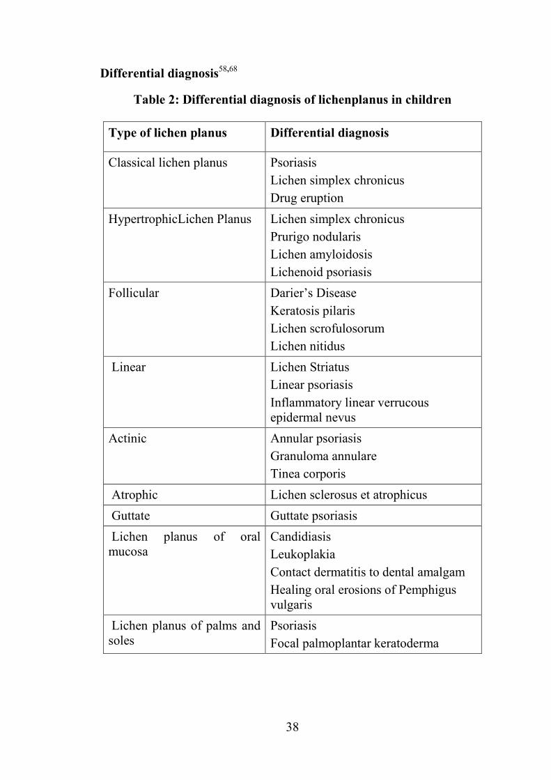
Differential diagnosis58,68
Table 2: Differential diagnosis of lichenplanus in children
Type of lichen planus Differential diagnosis
Classical lichen planus Psoriasis
Lichen simplex chronicus
Drug eruption
HypertrophicLichen Planus Lichen simplex chronicus
Prurigo nodularis
Lichen amyloidosis
Lichenoid psoriasis
Follicular
Darier’s Disease
Keratosis pilaris
Lichen scrofulosorum
Lichen nitidus
Linear Lichen Striatus
Linear psoriasis
Inflammatory linear verrucous
epidermal nevus
Actinic
Annular psoriasis
Granuloma annulare
Tinea corporis
Atrophic Lichen sclerosus et atrophicus
Guttate Guttate psoriasis
Lichen planus of oral
mucosa
Candidiasis
Leukoplakia
Contact dermatitis to dental amalgam
Healing oral erosions of Pemphigus
vulgaris
Lichen planus of palms and
soles
Psoriasis
Focal palmoplantar keratoderma
39
Treatment
Idiopathic LP oftenhas a self resolving natural course. Some
patients will have remission and exacerbation of the disease.
In paediatric population ,first line of therapy in cutaneous or oral
LP is potent and super potent topical steroids with or without
antihistamines. Topical steroids will help in reducing pruritis and flatten
skin lesions. Topical steroids have to be continued for several weeks to
achieve these effects.But there is risk of atrophy in long term use.
Topical calcineurin inhibitors such as tacrolimus and pimecrolimus
can be tried in those who require prolonged therapy.68
In oral LP, avoidance of triggers and maintenance of good oral
hygiene should be observed. Topical steroids in an adhesive base is
needed for oral LP for prolonged period. Other treatment options for oral
LP in children are topical tretinoin , isotretinoin, tacrolimus and
pimecrolimus.68
Oral corticosteroids should be considered in severe generalised LP
of childrenor if topical treatment fails to relieve the symptoms. Oral
corticosteroids should be given in a dose of 0.5-1mg/kg for 2-6 weeks
with gradual tapering thereafter. Other treatment options in children are

40
dapsone ,griseofulvin, metronidazole,oral retinoids,cyclosporine and
thalidomide. NBUVB and PUVA therapy is also used in generalise LP.
PROGNOSIS58,67
Skin lesions in LP will most often subside within 9-18 months.
Occasionally lichen planus may persist for years. About 10-20% of
patients sufferfrom one or more recurrences of their skin lesions.
Hypertrophic LP and oral LP will take long time to resolve. The lesion
may tend to flatten or resolvewith hyperpigmentation, which may persist
for months or years.
LP lesions may recur in about 15-20% of the patients. Recurrences
are more common in generalised LP. Malignant transformation is seen in
less than 1 percent of persistent oral mucous lesions on long term follow
up.
PITYRIASIS RUBRA PILARIS
Pityriasis rubra pilaris (PRP) was first described in 1856 by
Devergie and was named by Besnier in 1889.69
Pityriasis rubra pilaris
(PRP) is a group of keratinisation disorders that have in common,
circumscribed follicular keratoses, branny scale and an orange-red
erythema surrounded by normal skin, and palmoplantar keratoderma .70

41
Epidemiology
In India , incidence of PRP is 1 in 50,000 as compared to 1 in
5000 in Great Britain ,which makes clear that there is a racial
variation.71,72
There is a bimodal age of onset without sex predilection.
The most common type of PRP is adult onset classical type.
Aetiopathogenesis:
The aetiology and pathogenesis of PRP are still poorly understood.
It represents a disorder of keratinisation. Most childhood cases of PRP are
acquired without any family history,but an autosomaldominant
inheritance is also reported.73
Clinically and histologically, acquired and
hereditary forms are indistinguishable, but autosomal dominant form will
have less severe onset from birth to early childhood.
Vitamin A deficiency, infection and trauma are the various
postulated causes of PRP. 74,75,76
Studies have shown that there is an
association between PRP and decreased retinol binding protein. A study
conducted on PRP in children by Allison D S et al showed that 27% of
patients had preceeding upper respiratory tract infection and 10% had
preceeding trauma.76
Epidermal thymidine labelling is increased from an
average normal 3% to 27%.

42
PRP may be a result of dysregulation of immune system and
abnormal response to various antigenic triggers .77
Bacterial or viral
infection beforethe eruption are commonly noticed in juvenile PRP. PRP
is also associated with rheumatism and malignancy.78
Photoaggravation is
also a suggested triggering factor.79
Down’s syndrome, osteoporosis,arthropathy , coeliac sprue, protein
losing enteropathy,hypothyroidism,hyperparathyroidism and myasthenia
gravis are rare associations of PRP.80
Griffiths classification:71
Based on clinical and morphological appearance,Griffiths proposed a
classification of five types
Type 1: Adult onset, classical
Type II: Adult onset, atypical
Type III: Juvenile onset, classical
Type IV: juvenile onset,circumscribed
Type V: Juvenile onset, atypical
Juvenile onset PRP is discussed in detail

43
Type III (Juvenile onset, classical)70
The age of presentation of type III PRP is 5-18 years. Type III PRP
accounts about 10% of the total PRP cases. Type III resembles type 1
PRP clinically, but type III have a better prognosis than type 1.It starts as
orange red macule on head, neck or upper trunk with fine scales which
later spread cephalocaudally to involve whole body. Later, perifollicular
erythematous papules with central acuminate keratinous plug develops. A
characteristic feature of widespread PRP is “islands of sparing”. Palms
and soles will show thick yellow keratoderma which is referred to as
“PRP sandal”. Scalp may be covered with pityriasiform scales. Ectropion
may be seen. Nails may be normal or yellow,thickened and curved.
Pruritis may be seen in the erupting stage. Sometimes burning
sensation will be there. The symptoms will improve as the disease
stabilises.Rarely type III can get transformed to typeIV.
Type IV: Juvenile onset, circumscribed70
The age of onset of type IV PRP is usually less than 12years.It
presents as asymptomatic,erythematous grouped follicular papules over
extensor aspect of knees and elbows. A few erythematous scaly macules
or follicular keratotic plugs can be seen in the trunk. Marked
Palmoplantar keratoderma can be seen in some cases.the lesions are
44
nonpruritic. Scalp is spared in this type. These lesions does not progress
to type I and type III.
Type V : Juvenile onset ,atypical70
This type usually present since birth or in the first few years of life.
It is characterised by follicular hyperkeratosis with only minimal
erythema. Scleroderma like changes of the digits have also been reported
in many patients. This type usually persists throughout life.Majority of
familial PRP cases are of type V . It presents as follicular ichthyoses and
erythrokeratoderma.
HIV associated
PRP like eruptions have been documented in all age groups with
HIV infection. it is usually diagnosed in known HIV patients. But
sometimes the eruption can be a sign of HIV infection.
This type is associated with acne conglobata , cystic acne or
hidradenitis suppurativa. It is usually refractory to therapy but early
intiation of HAART may be effective.81

45
Histopathology5
A fully developed lesion shows alternating orthokeratosis and
parakeratosis oriented in both vertical and horizontal directions,slight
spongiosis,focal or confluent hypergranulosis,acanthosis with broad and
short rete ridges,thick suprapapillary plates in the epidermis. Dermis will
how mildly dilated vessels and superficial perivascular lymphocytic
infiltrate.
Differential diagnosis81
Psoriasis, lamellarichthyosis , keratosis pilaris, erythrokeratoderma
variabilis
Treatment
Mainstay of treatment for PRP is topical treatment.General
measures like use of gentle bathing soap,liberal use of emollients should
be advised. Topical steroids, keratolytics, calcipotriene and retinoids are
commonly used.
Oral retinoids like acitretin(0.5-1mg/kg) and isotretinoin (0.75-
1.5mg/kg) can be used in severe disabling disease.Methotrexate and
cyclosporine alsohave been reported to be effective.80

46
Prognosis
Type III PRP often resolves with in an average period of 1-2 years.
Couse of type IV PRP is uncertain, but some cases clear in the late teens.
Type V PRP have a chronic course. It usually resolves with retinoids but
relapses on stopping the drug.81
LICHEN STRIATUS
Lichen striatus is a self limiting disease of unknown origin
characterized by inflammatory papular eruptions in a linear distribution
or Blaschkoid distribution. 82
Epidemiology
It is more common in children between the age of 5-15years,with a
female preponderance. 83
About two third of the cases will have lesions
over the limbs. Pruritis is moderate and occurs in about 10% of the cases.
Aetiology
Definitive aetiology of lichen striatus is unknown. Lichen striatus
is an acquired disease. Trauma, ultraviolet light and contact dermatitis
have been implicated. Seasonal variation was reported in an Australian
study, and was found to be more frequent in the spring season.84

47
Studies on lichen striatus of children have shown a high
association with atopic dermatitis.Various studies have shown that 60-
80%of children with lichen striatus have a history of atopy.85
The
abnormal immune status associated with atopy may contribute to the
development of lichen striatus.
Viral infection has also been suggested as a trigerring factor.
Lichen striatus have shown an increased amount of IL-1β,whichsupports
an in situ inflammation driven process,trigerred by infectious
pathogens.86
An epigenetic mechanism has been suggested in the occurrence of
familial cases of lichen striatus. Eventhough familial cases are rare, cases
of simultaneous occurrence in siblings was reported . This also suggests
a common environmental trigger.87
Congenital presence of an abnormal skin clone due to a postzygotic
mutation has also been proposed as a possible explanation for LS .88
The
cells bearing the mutation will be quiescent. A trigger, most commonly a
viral infection will cause this aberrant clone to express a novel antigen,
resulting in the disease manifestations..

48
Clinical features
Lichen striatus initially presents as asymptomatic, discrete tiny
erythematous and lichenoid papules which later coalesce to form a
irregular linear band. The surface of the papules may have an associated
scaling. Common sites involved are extremities and trunk. It usually
presents as a unilateral single lesion,although rare cases of multiple and
bilateral involvement are reported.89.
Facial involvement is also seen.
The lesion progress for 2to 3weeks and resolves within a period of
3-12 monthsleaving a postinflammatory hypopigmentation or
hyperpigmentation.
Nail involvement can be seen, frequently on the fingers, especially
the thumb. Longitudinal ridging, splitting and thinning with
onychodystrophy are common nail manifestations . Nail lichen striatus
may preceed the onset of cutaneous lesions.90
Histopathology5
Epidermal changes include focal parakeratosis, spongiosis,
intracellular edema often associated with exocytosis. Dermis will show
superficial perivascular inflammatory infiltrate of lymphocytes with
histiocytes. Focally in the papillary dermis the infiltrate may have a band
like distribution with extension into the the lower portion of epidermis.

49
The corresponding regions of epidermis will show vacuolar alteration of
the basal layer and necrotic keratinocytes with melanophages in the
papillary dermis.
Differential diagnosis91
Linear psoriasis, linear Darier's disease, linear lichen planus,linear
porokeratosis and other linear nevi, are to be considered in the differential
diagnosis. ILVEN is the most important differential diagnosis for lichen
striatus,but it will have an earlier onset and will persist indefinitely.
Hypomelanosis of Ito and vitiligo are the differential diagnosis in
the hypopigmented stage.
Treatment
Benign and self limiting nature of the disease should be explained
to the parents. Intralesional steroid can be used in persistent case. If nails
are affected, potent topical steroids can be used. Topical tacrolimus
ointment will hasten the resolution of lesion.
Prognosis
Lichen striatus is a benign condition. The mean duration of the
lesion is 6-9 months. Relapses can occur within 4 years. The lesions may
heal with post inflammatory hypopigmentation.91
. Nail matrix lichen

50
striatus will not cause permanent damage and will resolve
spontaneously.92
SEBORRHEIC DERMATITIS
Seborrhoeic dermatitis is a common chronic papulosquamous
dermatosis,which is characterised by self limiting erythematous scaly
eruptions that occurs primarly over the face, postauricular, presternal and
intertrigenous areas. The term seborrheic dermatitis was first coined by
Unna in 1887.93
Epidemiology
The exact incidence of seborrheic dermatitis is unknown, but the
disorder is quite common. Seborrheic dermatitis showed an incidence of
1.6% among children , in a study conducted in India94
. In United States it
affects about 3-5% of general population.95
Seborrheic dermatitis have
bimodal peak, one in infancy and second around fourth to seventh
decade. Males are affected more frequently than females. Seborrheic
dermatitis is found in upto 85% of patients with HIV infection.
Aetiopathogenesis
The aetiology of seborrheic dermatitis is not well understood.
However many factors are implicated in the pathogenesis of seborrheic
dermatitis.
51
A positive correlation with sebum and sebaceous have been
postulated. It is evidenced by predilection for areas of high sebaceous
density and correlation of activity with increased hormonal levels during
first year of life and adolescents.
Theories have suggested that Malassezia furfur plays an important
role in the etiology of seborrheic dermatitis. Ruiz maldonao et al detected
Malassezia furfur in 73% of patients with seborrheic dermatitis.96
Studies done in infantile seborrheic dermatitis have demonstrated
abnormality with regard to essential fatty acids. A transient impairment
in delta-6-desaturase was detected in these children.96
Although a family history of seborrhea is quite often reported ,
there is no conclusive evidence that heredity plays a role.
Seborrheic dermatitis is associated with parkinsonism and
neurological disorders where sebum production is increased. It is also
associated with some medical conditions like malabsorbtion, epilepsy,
obesity , alcoholic pancreatitis.97,98
Seborrheic dermatitis is an established
marker of HIV infection and also seen in local cutaneous
immunosuppression.99
52
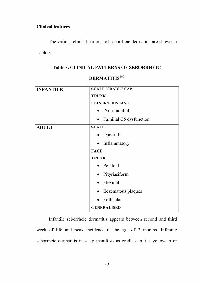
Clinical features
The various clinical patterns of seborrheic dermatitis are shown in
Table 3.
Table 3. CLINICAL PATTERNS OF SEBORRHEIC
DERMATITIS100
INFANTILE SCALP (CRADLE CAP)
TRUNK
LEINER’S DISEASE
• .Non-familial
• Familial C5 dysfunction
ADULT SCALP
• Dandruff
• Inflammatory
FACE
TRUNK
• Petaloid
• Pityriasiform
• Flexural
• Eczematous plaques
• Follicular
GENERALISED
Infantile seborrheic dermatitis appears between second and third
week of life and peak incidence at the age of 3 months. Infantile
seborrheic dermatitis in scalp manifests as cradle cap, i.e. yellowish or
53
whitish greasy thick adherent , frequently confluent scales occurring
mostly over vertex or frontal regions. Pruritis is usually absent or may be
mild.
On the face ,it appear as small round scaly erythematous areas
mainly over forehead,eyebrows, retroauricular region and nasolabial
folds. The lesions may also develop over neck, axillae,umbilicus,inguinal
and intergluteal folds. In the intertriginous areas the lesions will become
macerated and eroded. So scaling will be prominent only at the
peripheries. These areas may have secondary candidal or bacterial
infection evidenced by erythematous papules or pustule.Blepharitis is
also a common manifestation of seborrheic dermatitis in infants.100
Occasionally, seborrhoeic dermatitis may become generalized,
resulting in erythroderma in infants.Leiners disease is a severe type of
seborrheic dermatitis which is associated with diarrhoea, failure to thrive
and erythroderma.A possibility of immunodeficiency should be suspected
when it is severe, generalised and exfoliative,in adults.
Histopathology: 101
The histopathological features of seborrheic dermatitis vary
according to different clinical stages.
54
In acute and chronic stage, there will be orthokeratosis,
parakeratosis follicular plugging, spongiosis, psoriasiform hyperplasia.
Dermis will show superficial perivascular infiltrate of histiocytes and
lymphocytes.In chronic stage superficial plexus of dermis will show
markedly dilated capillaries and venules,
Differential diagnosis
Atopic dermatitis, psoriasis, intertrigo, Langerhans’cell
histiocytosis, multiple carboxylase deficiency,zinc deficiency and tinea
capitis.
Treatment
Infantile seborrheic dermatitis can be best managed by frequent
shampooing , with a gentle ‘no tears’ shampoo.Thick adherent scale can
be removed by application of baby oil or mineral oil followed by gentle
scraping with a toothbrush and shampooing.If there is severe
inflammation, a low potent topical steroid .For non scalp areas a low
potent topical steroid or antifungal will be usually effective.Topical anti
bacterial and candicidal will be useful if superadded infection is there
Blepharitis can be managed by warm compressess, non irritating
shampoo or mechanical removal of scales when necessary.
55
Prognosis
Infantile seborrheic dermatitis has an excellent prognosis. The
lesions will clear within 3-4 weeks in some patients. Most of the patients
will have spontaneous clearance of the lesion by the age of 8-12 months
LICHEN NITIDUS
Lichen nitidus was first described by Pinkus in 1907.103
In Latin,
the word nitidus means “shiny” or “glistening”. Lichen nitidus is a self
limiting chronic inflammatory papulosquamous disorder defined more
often in children and young adults.
Epidemiology
Epidemiological data of lichen nitidus is not well documented.
Lichen nitidus occurs more common in blacks than Caucasians. Children
andyoung adults are affected more than that of elderly. Lichen nitidus is
more common in males than females. A 25 year survey of skin disease
in African-Americans reported an incidence of 3.4 cases/10,000
population for lichen nitidus..104
Aetiopathogenesis
The exact aetiology of lichen nitidus is unknown. LN is sporadic
in origin but familial cases have been reported. In the earlier days it was
thought to be a tuberculoid reaction. Lichen nitidus was once thought to

56
be a variant of lichen planus , as the two diseases coexisted in the same
individual and also due to the development of lichen planus following
generalised lichen nitidus.
Recently postulated mechanism for the pathogenesis of lichen
nitidus is that an allergen may intiate the pathological process. Allergen
will cause epidermal and dermal antigen presenting cells to activate cell
mediated response , initiates accumulation of lymphocytes and form
discrete inflammatory papules.103
Clinical features
The lesions of lichen nitidus are characterised by multiple grouped
and discrete pinpoint to pinhead sized ,fleshcoloured to slight pink
flat,shiny papules,commonly distributed over forearm, penis, chest and
abdomen. The papules will be hypopigmented in blacks. Scaling may be
minimal and can be elicited by rubbing the surface of the papules.
Koebnerisation and linear arrangement can be seen.
Rare forms of lichen nitidus are generalised,linear,actinic,
vesicular, hemorrhagic, perforating, purpuric and spinous follicular.
Lichen nitidus may rarely affect mucous membrane,palms,soles and nails.
In children rare variants like generalised lichen nitidus, perforating type
and actinic lichen nitidus are reported. 103

57
Atopic dermatitis , Down’s syndrome and juvenile chronic
arthritis have been reported to be associated with lichen nitidus in
children.105,106,107
In adults, lichen nitidus are reported to be associated
with Crohn’s disease, erythema nodosum and MEN2e.
Histopathology5
Epidermis will be flattened wth focal parakeratosis, diminished
granular layer, vacoular alteration of basal layer and focal subepidermal
clefting. . Colloid bodies are seen rarely. Dermis will show a well
circumscribed mixed cell granulomatous infiltrate . At each lateral
margin of the infiltrate rete ridges tend to bend inward and seem to
clutch the infiltrate in a manner of a “ claw clutching a ball”. The
infiltrate is composed of lymphocytes,monocytes and a few
multinucleated epitheloid histiocytes. Transepidermal elimination of the
infiltrate through the thinned epidermis can occur. Capillary wall
degeneration and extravasation of RBCs are seen in purpuric and
hemorrhagic lesions.
Differential diagnosis65
Lichen planus, plane wart, keratosis pilaris ,Darier’s disease,
lichen spinulosus, lichen scrofulosorum, id reaction are considered as
differential diagnosis.

58
Prognosis
LNis a chronic self limiting disease with an average duration of 1
year. New lesions may continue to develop as older lesions resolve.
Lesions heal without scar formation or pigmentary abnormalities. The
eruption may persist rarely.
Treatment
Medical treatment is considered when the patient is symptomatic or
when the lesions are generalised and persistent. Topical treatment
includes mid to high potent steroids and tacrolimus. Immunotherapy with
dinitrochlorobenzene has been used. Studies have shown that oral
isotretinoin is effective in generalised LN and acitretin in palmoplantar
involvement.
In refractory cases NBUVB, PUVA, astemizole, enoxaparin and
etretinate have been used. Systemic retinoids can cause premature
epiphyseal plate closure and stop normal growth in children.
PARAPSORIASIS
The term parapsoriasis was coined by Brocq in 1902 to encompass
a group of conditions that clinically resembled psoriasis and seborrhoeic
dermatitis.108
Parapsoriasis is classified into large plaque parapsoriasis
and small plaque psoriasis.
59
Epidemiology
Parapsoriasis is a disease predominantly seen in adults. Males are
affected more commonly than females. Small plaque psoriasis shows a
definite male predominance of approximately 3-4: 1.109
Parapsoriasis
is rarely seen in children. In a study, about 5% of small plaque psoriasis
and 20% of large plaque psoriasis were reported in children.
CLINICAL FEATURES65,108
Small plaque parapsoriasis
Chronic superficial scaly dermatitis, persistent superficial
dermatitis digitate dermatosis and xanthoerythroderma perstans are the
synonyms for small plaque parapsoriasis.
The lesions are usually insidious in onset and asymptomatic. Most
common sites involved are trunk and limbs. The typical lesions are
hyperpigmented or yellowish red,round or oval, patches with well defined
sharp borders of size 1-5cm. The patches will be covered with fine
moderately adherent scales. In the digitate variant of small plaque
parapsoriasis elongated fingerprint like lesions oriented along the
cleavage lines are seen commonly over the flanks. In this type the long
axis of the lesions will be more than 5 cm. A variant of parapsoriasis,
known as xanthodermia perstants will have a yellowish hue .
60
Large plaque parapsoriasis
Parakeratosis variegate,retiform parapsoriasis,atrophic
parapsoriasis and poikilodermatous parapsoriasis are the synonyms for
large plaque parapsoriasis.
Large plaque psoriasis commonly occurs in middle aged males
with a peak incidence in fifth decade. Unexposed areas, especially trunk
and flexures are the most commonly affected areas. Extremities and
breasts in females are other common sites. The lesions will present as
persistent ,reddish brown or salmon pink patches or plaques with small
and scanty scaling which are more than 5cm, often measuring more than
10cm. The lesion are stable in size but will increase in number. Later
atrophic changes along with mottled pigmentation and telangiectasia can
occur over patches and plaques. The surface will have wrinkling –
“cigarette paper wrinkling.”
A rare variant , known as retiform parapsoriasis which refers to a
net-like or zebra-like pattern of scaly macules and papules that eventually
becomes poikilodermatous in appearance. An icthyosiform variant of
large plaque parapsoriasis is also reported in the literature.

61
Histopathology5
Elongated mounds of parakeratosis with collection of plasma
above a basket weave cornified layer is a characteristic finding.
Epidermis will also show acanthosis,spongiosis and exocytosis of
lymphocytes. Papillary dermis will show mild superficial perivascular
lymphocytic infiltrate.
Early stages of large plaque parapsoriasis will show similar
features as that of small plaque parapsoriasis. Advanced lesions will have
epidermal atrophy , marked exocytosis of lymphocytes, and a band-like
infiltrate of lymphocytes in the papillary dermis. Dilated vessels and
melanophages can be seen in dermis.
Differentialdiagnosis10,110
Small plaque parapsoriasis should be differentiated from nummular
eczema, psoriasis vulgaris and guttate psoriasis, pityriasis rosea,
pityriasis lichenoides chronica and secondary syphilis
Large plaque psoriasis should be differentiated from plaque type
psoriasis, tinea corporis, contact dermatitis, poikilodermatous skin
conditions such as: Rothmund-Thomson syndrome, Bloom’s syndrome,
dyskeratosis congenita, systemic lupus erythematosus, dermatomyositis,
radiodermatitis ,patch stage of mycosis fungoides.

62
Treatment
Patients with small plaque parapsoriasis should be reassured about
the benign nature of the disease. Small plaque parapsoriasis can be treated
with emollients, topical corticosteroids , topical tar products, and narrow
band UVB therapy. Treatment response is variable and temporary.
LPP requires aggressive treatment. The goal of treatment is to
suppress the disorder to prevent possible progression to overt MF. In
large plaque parapsoriasis topical corticosteroids should be used with
caution because of atrophic nature of plaques.Emollients ,NBUVB and
PUVA therapy can be used.
Repeated examinationwith sequential biopsies of suspicious lesion
is needed every 3 months initially and then every 6 months to 1 year for
evidence of progression.110
If the histopathological section shows early
stages of early MF , it can be treated with topical nitrogen mustard,
bexarotene gel, imiquimod or carmustine. Narrow band UVB and broad
band UVB therapy can also be used in such situations.
Prognosis
Small plaque parapsoriasis may persist for years to decades,
without treatment. Malignant transformation is rarely reported. Various
studies have shown that about 10-35% of large plaque parapsoriasis can

63
turn to mycosis fungoides. Retiform variant may progress to overt MF in
virtually all cases.110
So children with large plaque parapsoriasis should
undergo serial biopsies to rule out malignant transformation.
PITYRIASIS LICHENOIDES
Pityriasis lichenoides is a spectrum of cutaneous eruption of
unknown etiology that mainly affects children and young adult.
Jadassohn and Neisser first described pityriasis lichenoides in 1894 in
two separate reports.111,112
Juliusberg in 1899 described the chronic form
in 1899 and gave the name PLC. Mucha in 1916 and Haberman in 1925
described the acute form of pityriasis lichenoides and Degos in 1966
reported a severe febrile ulceronecrotic subtype now usually called febrile
ulceronecrotic Mucha Habermann disease (FUMHD)
Gelmetti et al.113
suggested an alternative classification. Based on
anatomic distribution, they described three patterns: a diffuse form
involving glabrous skin over the whole body surface, a central variety
involving the neck, trunk, and proximal extremities, and a peripheral
form with an acral distribution. Acral involvement including the face is
more common in children.
64
Epidemiology
The prevalence and incidence of pityriasis lichenoies is not well
documented.There is a male predominace of 1.5:1to 3:1.110
A case of
pityriasis lichenoides has been described at birth. PLC is seen in about
37.5% of paediatric patients with pityriasis lichenoides. PLC is three
times more common than PLEVA. About 50% of reported cases of
febrile ulceronecrotic Mucha Habermann disease have been seen in
children.
Aetiopathogenesis
Pityriasis lichenoides is a disorder of unknown etiology. Infectious
agents such as Toxoplasma gondii, Epstein-Barr virus,
cytomegalovirus,parvovirus B19 and HIV have been associated with
some cases.114
Certain studies have demonstrated T cell clonality in
pityriasis lichenoides ,which suggests that it is a benign lympho
proliferative disease in which a vigorous host immune response prevents
the condition from evolving into lymphoma.
Pityriasis lichenoidess is removed from parapsoriasis group of
disorders and now considered as a lymphocytic vasculitis.115,116

65
Epidermis of piryriasis lichenoides lesions have shown a reduction
in CDla+ antigen-presenting dendritic (Langerhans) cells in
immunohistologic studies. CD8+ cells predominate PLEVA lesions and
PLC lesions will have both CD8+ and CD4+ cells. Majority of these
Tcells will express cytolytic enzymes(granzyme band TIA-1)and
memory proteins(CD45RO) . Presence of IgM, C3 deposition in the
dermoepidermal junction and in the dermal vessel walls in the early acute
lesions suggest a humoral response. These data suggest that PL may be
due to persistent antigen stimulation triggered by an infectious agent
resulting in immune complex formation.113
Clinical features
PLEVA
PLEVA is characterised by acute onset of firm reddish papules of
2-10mm size. Later the papules will become hemorrhagic, vesicular,
necrotic and crusted,leaving scars with hyperpigmentation or
hypopigmentation. The eruption may sometimes be associated with
fever, headache, malaise and lymphadenopathy. Most common sites
affected are anterior aspect of trunk, flexors, and proximal part of
extremities. Lesions are usually non pruritic. The disease usually lasts for
several weeks to months. After an acute course ,PLEVA can progress to
PLC.

66
PLC
PLC may begin de novo or may evolve from PLEVA. It is
characterised by recurrent crops of lichenoid, reddish brown papules
which are covered by mica like scales. The papules will involute within
3-6 weeks leaving hyperpigmented macules which gradually fades. The
lesions may last from several months to several years with frequent
remissions and exacerbations.
In febrile ulceronecrotic Mucha Haberman disease, there will be
acute onset of fever with purpuric papules and nodules. Later multiple
lesions will coalesce to form diffuse large necrotic ulcers.
Histopathology5
In PL, epidermis will show confluent parakeratosis, mild
spongiosis, necrotic keratinocytes and basal cell vacoulation.PLEVA will
have more pronounced vacoular degeneration with marked
exocytosis.Dermis will have perivascular lymphocytic infiltrate which is
more deep in PLEVA. Papillary dermis will show melanophages and
extravasated RBCs.
Differential diagnosis.110
PLC: guttate psoriasis,pityriasis rosea,lichen planus and secondary
syphilis.

67
PLEVA:Arthropod bites, Lymphomatooid papulosis, vasculitis, varicella
FUMHD:Varicella,Lymphomatoid papulosis,Lymphoma
Treatment
Ultraviolet therapy is the most effective therapy for pityriasis
lichenoides. Satisfactory outcome is seen with oral antibiotics,
particularly azithromycin and erythromycin. Topical corticosteroids and
oral antihistamine have a role in reducing the associated pruritis. In
persistent cases and ulceronecrotic variants,methotrexate and
cyclosporine can be used.
Prognosis and complications
Pityriasis lichenoides have variable outcome. It may recover
spontaneously within weeks or may persist and recur for years. The
duration of the disease in children correlate better with the clinical
distribution of lesions. The longest duration will be seen in peripheral
distribution and shortest with diffuse pattern.117
PLC will persist longer
than PLEVA. Malignant transformation is very rare although case of PLC
transforming to Tcell lymphoma has been reported.
Most common complication of pityriasis lichenoides is secondary
infection. In febrile ulceronecrotic variant the patient may develop
arthralgia , gastrointestinal and central nervous system manifestations.
68
LYMPHOMATOID PAPULOSIS
Lymphomatoid papulosis was first described by Macauly in
1968118
. It is a benign recurrent self healing dermatosis with histologic
features suggestive of lymphoma.
Epidemiology
It is a rare disease with an incidence of about 1.2-1.9 per 1,000,000
in adults.119
Boys affected with lymphomatoid papulosis have a younger
age of onset than girls. The course of lymphomatoid papulosis issimilar in
children and adults, including the risk of lymphoid malignancies. But
some studies have shown that the disease in children will resolve more
spontaneously than in adults.
Clinical features
The lesions typically involve trunk and extremities. There will be
recurrent crops of numerous reddish brown papules or vesiculopustules
over a period of months to several years. The lesions will
characteristically develop central hemorrhagic necrosis and crusting
which gradually resolves with hyperpigmentation or hypopigmentation.
Occasionally varioliform scars may develop. Rarely large ulcerating
nodules, plaques or nonulcerating papules occur. Regional distribution
69
has been described in children although more generalised spread develops
later.
Histopathology120,121
There are three histological types described in lymphomatoid
papulosis.
Type A (histiocytic type): This consist of scattered or grouped
pleomorphic or anaplastic lymphoid cells. Inflammatory cells like
eosinophils, neutrophils, histiocytes may be seen in the dermis.Type
B(Lymphocytic): There will be epidermotrophic infiltrate of small to
medium sized lymphoid cells.Type C(ALCL- type): There will be
nodular infiltrate of large atypical lymphoid cells arranged in sheets.
Inflammatory cells may be present.
Differential diagnosis122
The differential diagnosis includespityriasis lichenoides,arthropod
reaction, pseuolymphoma and anaplastic large cell lymphoma.
Treatment and prognosis122
Topical ultrapotent corticosteroids for 2-3 weeks followed by
weekly pulse is effective in clearing the lesions. Low dose oral
methotrexate(5-10mg/week) will suppress the development of new

70
lesions. PUVA therapy has also been reported to have beneficial effect on
the disease. There may be relapse of the lesions after discontinuation of
the therapy. So, in patients with relatively few and non scarring
lesions,long term follow up without active treatment should be
considered.
Prognosis
About 10-20% of adult patients will develop malignant lymphoma
and there is increased risk of developing non lymphoid malignancies. A
study conducted in 35 paediatric patients with lymphomatoid papulosis
showed that 9% of them developed non Hodgkins lymphoma,122
which
emphasize the need for long term surveillance.
PAPULAR ACRODERMATITIS OF CHILDHOOD
Papular acrodermatitis of childhood was initially described by
Gianotti in 1955.123
Subsequently in 1956 Crosti and Gianotti described
the entity and since then the disorder has been commonly as Gianotti
Crosti syndrome. They described the disorder as symmetrical distribution
of erythematous papular eruption over face, buttocks and extremities of
children.

71
Epidemiology
Gianotti crosti syndrome is distributed world wide. The age group
affected are 1-6 years with 90% being affected before the age of 4
years.123
There is no specific gender or race predisposition. Adult cases
are rarely reported. (Pac )
Aetiology 124
The etiology of PAC has long been suspected to be viral in origin.
The common viruses implicated are Ebstein Barr virus, cytomegalovirus,
coxsackie virus, parvovirus B19,rotavirus, and HHV 6. A possible
association withvaccines has also been suggested. MMR vaccine,
DPT,oral polio, hepatitis B vaccines are reported to be associated with
PAC.Bacterial pathogens include Mycoplasma pneumoniae,
Boreliaburgdorfei, Bartonnella henselae, groupA B-hemolytic
streptococcus.
The exact mechanism of pathogenesis of PAC is not well
understood. It may be due to a viral antigenemia and circulating immune
complexes.
72
Clinical features124
Papular acrodermatitis is seen predominantly in children between
the age of 1and 6 years. There will be a history of upper respiratory tract
infection in majority of the children. The eruption is characterised by
monomorphous,erythematous, edematous papules and papulovesicles
distributed symmetrically over face ,buttocks and extensor surface of
upper and lower extremities. The papules may coalesce into large
erythematous plaques. Lesions are usually non pruritic or mildly pruritic.
It may take 8-12 weeks to resolve completely. Post inflammatory
hypopigmentation may occasionally persist for several months.
Diagnostic Criteria for Gianotti-Crosti Syndrome123
Cuhh ATT proposed a diagnostic criteria for PAC.
A Patient is diagnosed to have Gianotti Crosti syndrome if he/she
exhibits all positive clinical features on at least one occasion or clinical
encounter, and does not exhibit any negative clinical feature on any
occasion or clinical encounter related to rash, and no differential
diagnosis is considered more likely than diagnosis of GCS based on
clinical judgment, and if lesional biopsy is performed, findings are
consistent with GCS

73
Positive Clinical Features
1. 1.Monomorphous, flat-topped, pink-brown papules or
papulovesicles 1–10 mm in diameter.Any 3 or all 4 sites
involved: cheeks, buttocks, extensor surfaces of forearms,
extensor surfaces of legs.
2. Symmetry
3. Duration of 10 days or more
Negative Clinical Features
1. Extensive truncal lesions
2. Scaly lesions
Differential diagnosis125
Acrodermatitis enteropathica, erythema infectiosum, erythema
multiforme, hand-foot-and-mouth disease,Henoch-Schönlein purpura,
Kawasaki disease, lichen planus, papular purpuric gloves-and-socks
syndrome, papular urticarial and scabies.
Histopathology
Epidermis will show parakeratosis, focal spongiosis, and
lymphocytes. There is a superficial and mid dermal perivascular infiltrate
of lymphocytes and histiocytes. Sometimes the inflammatory infiltrate
74
may also present as lichenoid infiltrate. Papillary edema and
extravasation of RBCs may also be seen.
Treatment and prognosis
Gianotti crosti syndrome is a self limiting disease. Treatment is
mainly supportive. In hepatitis B associated PAC, a self resolving
anicteric viral hepatitis may evolve after 2 weeks of onset of eruptions

75
METHODOLOGY
The study was conducted in the outpatient department of
dermatology, Coimbatore medical college hospital between the time
period of August 2013 and July 2014. This is a clinical study of
papaulosquamous disorders in children less than 12years who were
clinically diagnosed to have papulosquamous disorder as per ICD 10
classification .Ethical committee clearance was obtained. A written
informed consent was obtained from the parents.
Inclusion criteria :
All children less than 12 years who presented to the skin OPD with
papulosquamous morphology of skin lesions.
Exclusion criteria:
Children whose parents did not give consent for the study.
One hundred and five children were clinically diagnosed to have
papulosquamous disorder during the study period. A detailed history
including age of onset, duration of the disease, progression and associated
symptoms were obtained from the patients and their parents. Relevant
family history and history of drug intake prior to the onset of disease
were recorded.General examination and systemic examination were done.

76
Dermatological examination was done. Morphology, distribution,
characters of scales and any special features were recorded. Hair, nails,
oral and genital mucosa were examined in detail.
All routine investigations including haemoglobin, total blood
count, differential count, erythrocyte sedimentation rate were done for
each patient.
Special investigations like KOH mount,VDRL and biopsy were
done for relevant patients
Children from 0-12 years were divided into four groups. The
findings were recorded in the profoma and tabulated in the masterchart.
The results were analysed and discussed in detail.

77
RESULTS
One hundred and five patients under the age of 12 years with
papulosquamous disorders attending the outpatient department of
Dermatology,venereology and leperology , Coimbatore medical college
hospital formed the study material.The duration of study was from
August 2013 to July 2014.
During the study period of 1 year, 69445 patients attended the
outpatient department, of which 12835 were children
Table 4 No of total patients and pediatric patients attended the OPD
Total number of
patients attendedthe
skin opd
Total number of
paediatric patients
attended skin opd
Percentage of
paediatric patients
attended skin opd
69445 12835 18.48%
Out of the paediatric patients, 105 patients had papulosquamous
disorders.

78
Table 5 Percentage of pediatric patients with
papulosquamous disorder
Total number of
paediatric patients
attening skin opd
Total number of patients
with papulosquamous
disorders
Percentage of
papulosquamous
disorder in children
12835 105 0.81%
SEX RATIO
Total number of patients included in the study were 105, out of
which 55(52%) were male and 50(48%) were female.
The male to female ratio in the study was 1.1:1
AGE AND SEX DISTRIBUTION
Table 6Age sex distribution of children with
papulosquamous disorders
Age( years) Male Female Total
0-3 13 (23.64%) 11 (22%) 24 (22.86%)
4-6 14 (25.45%) 11 (22%) 25 (23.81%)
7-9 15 (27.27%) 12 (24%) 27 (25.71%)
10-12 13 (23.64%) 16 (32%) 29 (27.62%)
Total 55 (100%) 50 (100%) 105 (100.00%)
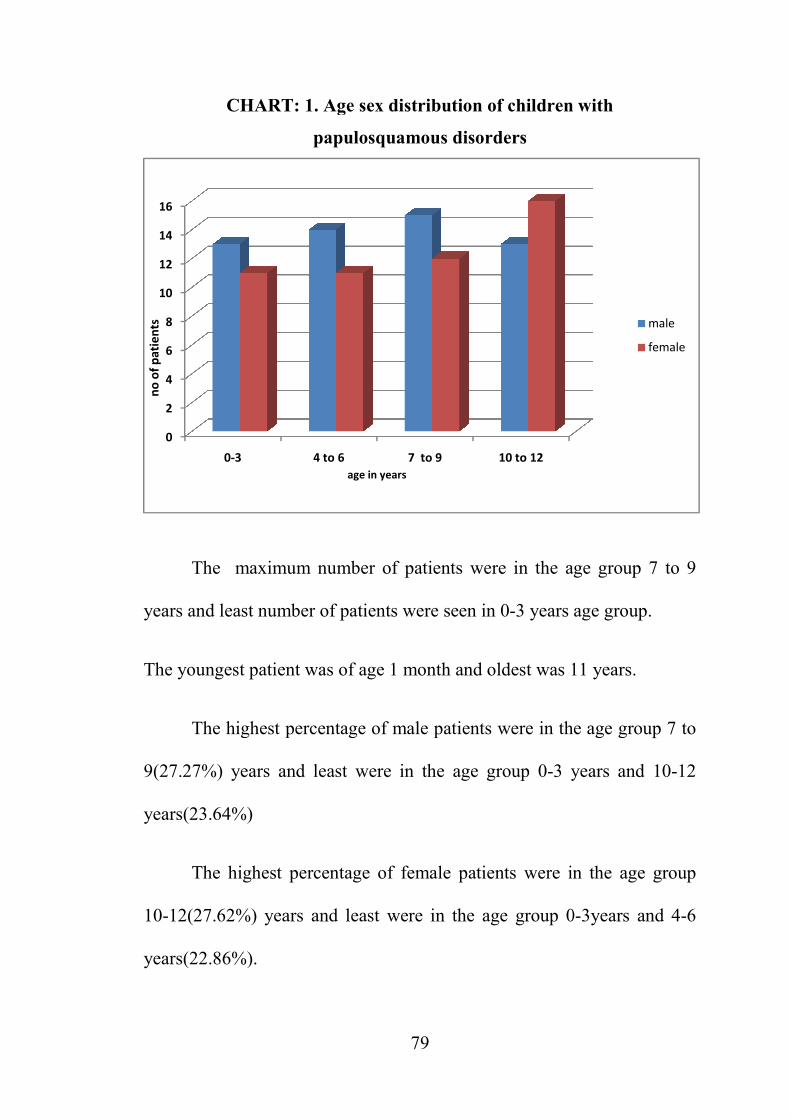
CHART
The maximum
years and least numb
The youngest patient
The highest pe
9(27.27%) years and
years(23.64%)
The highest p
10-12(27.62%) years
years(22.86%).
0
2
4
6
8
10
12
14
16
0-3
no
of
pa
tie
nts
79
ART: 1. Age sex distribution of children w
papulosquamous disorders
aximum number of patients were in the age
number of patients were seen in 0-3 years ag
patient was of age 1 month and oldest was 1
hest percentage of male patients were in the
rs and least were in the age group 0-3 ye
hest percentage of female patients were in
) years and least were in the age group 0
4 to 6 7 to 9 10 to 12
age in years
dren with
he age group 7 to 9
ears age group.
was 11 years.
in the age group 7 to
3 years and 10-12
ere in the age group
up 0-3years and 4-6
male
female
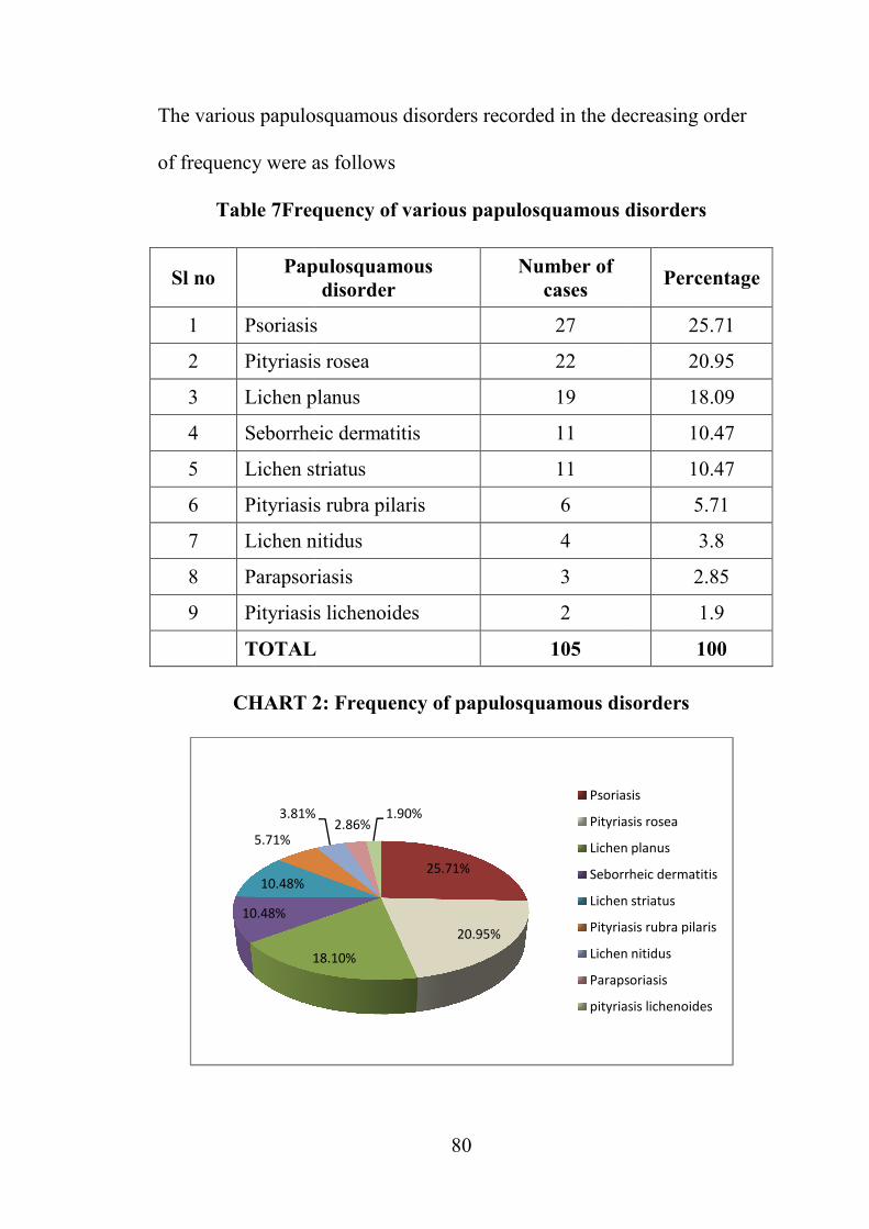
The various papulosq
of frequency were as
Table 7Frequ
Sl no Papu
1 Psoriasis
2 Pityriasis r
3 Lichen pla
4 Seborrheic
5 Lichen str
6 Pityriasis r
7 Lichen nit
8 Parapsoria
9 Pityriasis l
TOTAL
CHART 2
18
10.48%
10.48%
5.71%
3.81%
80
osquamous disorders recorded in the decr
ere as follows
Frequency of various papulosquamous di
Papulosquamous
disorder
Number of
cases
riasis 27
riasis rosea 22
planus 19
orrheic dermatitis 11
en striatus 11
riasis rubra pilaris 6
en nitidus 4
psoriasis 3
riasis lichenoides 2
TAL 105
2: Frequency of papulosquamous diso
25.71%
20.95%
18.10%
1%2.86%
1.90%
Psoriasis
Pityriasi
Lichen p
Seborrh
Lichen s
Pityriasi
Lichen n
Parapso
pityriasi
e decreasing order
ous disorders
Percentage
25.71
20.95
18.09
10.47
10.47
5.71
3.8
2.85
1.9
100
disorders
iasis
riasis rosea
en planus
rrheic dermatitis
en striatus
riasis rubra pilaris
en nitidus
psoriasis
riasis lichenoides

81
Psoriasis was the most common papulosquamous disorder with 27
cases (25.71%) ,followed by pityriasis rosea with 22 cases( 20.95%) and
pityriasis lichenoides was the least common with 2 cases(1.9%).
The distribution of papulosquamous disorders in males and females
in the study is as follows.
Table 8Frequency of papulosquamous disorders
in males and females
Sl no Papulosquamous disorder Males Females
1 Psoriasis 11(20%) 16(36%)
2 Pityriasis rosea 10(18.18)% 12(24%)
3 Lichen planus 12(21.82)% 7(14%)
4 Seborrheic dermatitis 6(10.91)% 5(10%)
5 Lichen striatus 6(10.91)% 5(10%)
6 Lichen nitidus 3(5.45)% 1(2%)
7 Pityriasis rubra pilaris 5(9.09)% 1(2%)
8 Parapsoriasis 2(3.64)% 1(2%)
9 Pityriasis lichenoiddes 2(3.64)% 0
TOTAL 55(100%) 50(100%)

Chart 3 Dis
In males the m
planus (21.82%), f
pityriasis lichenoides
Psoriasis(36%
seen in females follo
, parapsoriasis and lic
Seasonal variation
below table:
0
2
4
6
8
10
12
14
16
18
20N
0:
of
pa
tie
nts
82
Distribution of papulosquamous disord
males and females
s the most common papulosquamous disord
%), followed by psoriasis (20%). Para
noides (3.64%) were the least common disea
s(36%) was the most common papulosqua
s followed by pityriasis rosea(24%). Pityrias
and lichen nitidus were seen in 2% of femal
iation of papulosquamous disorders is
papulosquamous disorders
disorders in
disorder was lichen
Parapsoriasis and
diseases recorded.
losquamous disorder
ityriasis rubra pilaris
female patients.
rs is shown in the
males
females

83
Table 9Seasonal variation of papulosquamous disorders
Disease Winter
(Dec-Feb)
Summer
(Mar-May)
Monsoon
(June-Nov)
Psoriasis 15 5 7
Pityriasis rosea 11 5 6
Lichen planus 5 9 5
Seborrheic dermatitis 3 6 2
Lichen striatus 4 5 2
Pityriasis rubra pilaris 1 3 2
Lichen nitidus 1 1 2
Parapsoriasis 1 0 2
Pityriasis lichenoides 1 1 0
Total 42 35 28
Percentage 40% 33.33% 26.67%
Incidence of papulosquamous disorder was high during the winter
season(40%). 33.33% of patients visited during the summer season and
26,67% of the patients visited during monsoon season.
The incidence of psoriasis(55.56%) and pityriasis rosea (50%)
were more during the winter season.

84
PSORIASIS
In the study, out of 12835 patients ,psoriasis was reprted in 27
patients. The prevalence of childhood psoriasis in the study is 0.21%. The
prevalence of childhood psoriasis among papulosquamous disorder is
25.71%.
There were 11 boys and 16 girls with a male to female ratio of
1:1.45
AGE SEX DISTRIBUTION OF PATIENTS WITH PSORIASIS
The table shows the age sex distribution of psoriasis
Table 10: Age sex distribution of patients with psoriasis
Age(in years) Male Female Total
0-3 1(9.09%) 2(12.50%) 3(11.11%)
4-6 3(27.27%) 3(18.75%) 6(22.22%)
7-9 5(45.45%) 2(12.50%) 7(25.93%)
10-12 2(18.18%) 9(56.25%) 11(40.74%)
Total 11(100%) 16(100%) 27(100%)
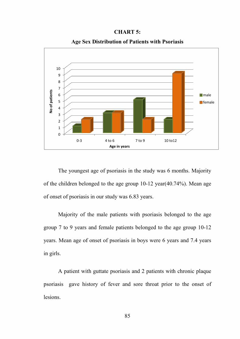
Age Sex
The youngest
of the children belon
of onset of psoriasis
Majority of th
group 7 to 9 years a
years. Mean age of o
in girls.
A patient with
psoriasis gave hist
lesions.
0
1
2
3
4
5
6
7
8
9
10
0-3
No
of
pa
tie
nts
85
CHART 5:
e Sex Distribution of Patients with Psoria
ngest age of psoriasis in the study was 6 mo
belonged to the age group 10-12 year(40.74
riasis in our study was 6.83 years.
of the male patients with psoriasis belong
ears and female patients belonged to the ag
ge of onset of psoriasis in boys were 6 years
t with guttate psoriasis and 2 patients with
e history of fever and sore throat prior to
4 to 6 7 to 9 10 to12
Age in years
soriasis
s 6 months. Majority
r(40.74%). Mean age
belonged to the age
the age group 10-12
years and 7.4 years
with chronic plaque
rior to the onset of
to12
male
female

A positive fam
patient gave histor
father had history of
Pruritis was pr
Table No 11: Percen
Type of psoriasis
Chronic plaque psori
Guttate psoriasis
Flexural psoriasis
Palmoplantar psorias
Pustular psoriasis
Scalp psoriasis
Total
CHART 6:P
15%
11%
11%
7%
86
ive family history was present in 2(7.41%
history of psoriasis in the sibling and the
ory of psoriasis.
was present in 16(59.25%) patients.
ercentage of various morphological type
Number of cases
e psoriasis 14
4
3
soriasis 3
2
1
27
Percentage of morphological types of p
52%
7%
4%
Chronic
Guttate
Flexural
Palmop
Pustular
scalp
7.41%) patients, one
d the other patients
l type of psoriasis
Percentage
51.85%
14.81%
11.11%
11.11%
7.41%
3.70%
100.00%
es of psoriasis
onic plaque psoriasis
tate psoriasis
ural psoriasis
oplantar psoriasis
tular psoriasis

87
In this study chronic plaque psoriasis,14(51.85%) was the most
common type of psoriasis seen in children .
Exclusive guttate psoriasis was seen in 4(14.81% ) children. But
guttate lesion co existed with chronic plaque psoriasis in 7(25.92%)
chidren. Pustular psoriasis was seen in 7.41% of children.
The nails were involved in 5(18.51%) cases. Pitting was the most
common nail change.Other nail changes observed in the study are
subungual hyperkeratosis and thickening of nail plate
Exclusive scalp psoriasis was seen only in 1(3.7%) patient but 4
(14.81%) patients had scalp lesions along with other site involvement.
Infantile psoriasis was present in 2(7.4%) children.
Koebnerisation was present in 12( 44.44%) patients.Auspitz sign
was positive in all patients except the patients with pustular psoriasis,
flexural and palmoplantar psoriasis .
PITYRIASIS ROSEA
In the study , pityriasis rosea was seen in 22 patients (20.95%).
There were 10 male patients and 12 females patients with male to female
ratio 0.83: 1.

Table 12 Age se
Age in yea
0-3
4-6
7-9
10-12
TOTAL
CHART
One year was
Maximum number of
In the study
symptoms of upper r
0
1
2
3
4
5
0-3
no
of
pa
tie
nts
88
ge sex distribution of children with pityr
in years Male Fem
2(20%) 2(16.6
3(30%) 2(16.6
2(20%) 4(33.3
3(30%) 4(33.3
TAL 10(100%) 12(10
ART 7:Age-sex distribution of pityriasis ro
r was the youngest age affected and eldest w
ber of patients belonged to the age group 10
study,6 (27.27%) patients had history
pper respiratory tract infection
04 to06 7 to 9 10 to 12
age in years
pityriasis rosea
Female
16.67%)
2(16.67%)
4(33.33%)
4(33.33%)
12(100%)
asis rosea
dest was of 11 years.
oup 10-12 years.
story of prodromal
Male
Female
89
Herald patch was seen in 12(54.54%) patients and absent in10
patients(45.46%). Most common site of herald patch in the study was
trunk. The median time between the appearance of herald patch and wide
spread eruption was 1 week( range4 days-10days)
Table 13: frequency of various types of PR
Sl no Type of pityriasis
rosea
Number of
patients Percentage
1 Classical 18 81.81%
2 Inverse 3 13.6%
3 lichenoid 1 4.54%
Most common type of PR in the study was classical PR, seen
in18(81.81%) patients. Inverse type of PR was seen in 3(13.6% ) patients
in the study . Lichenoid PR was seen in 1 (4.54%) patient.
One(4.54%) patient had eczematous changes associated with PR
lesions
.

90
Table14:Site of involvement of pityriasis rosea
Sites No of patients Percentage
Face 6 27.27%
Neck 7 31.81%
Chest 10 45.46%
Back 18 81.81%
Upperlimb 13 59.09%
Lower limb 7 31.81%
In the study ,most common site affected was back(81.81%).
Upperlimb lesions were seen in 12(59.09%) patients. Lowerlimb and
neck lesions lesions were seen in 7 (31.81% )of patients. Least common
affected site was face,6(27.27%) children.
Lichen planus
During the study period of 1 year,out of 12835 attended the OPD,
19 children had lichen planus. Thus the prevalence of lichen planus in
children in our hospital was 0.14%. Out of 105 patients with
papulosquamous disorders 19(18.09%) patients had lichen planus.In the
study,12(63.16%)were male and 7(36.84%) were female with male
female ratio 1.7:1

91
Table15:Age sex distribution of patients with lichen planus
Age( years) Male Female Total
0-3 1(8.33%) 0(0%) 1(5.26%)
4-6 4(33.33%) 4(57.1%) 8(42.11%)
7-9 5(41.67%) 1(14.28%) 6(31.58%)
10-12 2(16.67%) 2(28.57%) 4(21.05%)
Total 12(100%) 7(100%) 19(100%)
In the study youngest age of onset was 3 years and eldest was 11
years. Maximum number of patients belonged to age group 7 to 9 years.
Table16 : Percentage of morphological types of lichen planus
Type No. of cases Percentage
Classical 11 57.89%
Generalised 5 26.32%
Linear 3 15.79%
The most common morphologic variant of LP was classical LP
(57.89%). Five patients (26.32%) had generalised LP and 3(15.79%) had
linear LP. Koebnerisation was seen in 68.42% 0f patients.
Nail involvement was seen in 3 patients(15.79%). Two patients ha
thinning of nail plate and one patient had naildystrophy of finger
nails.Oral mucosal involvement were seen in 4 patients.(21.05%).
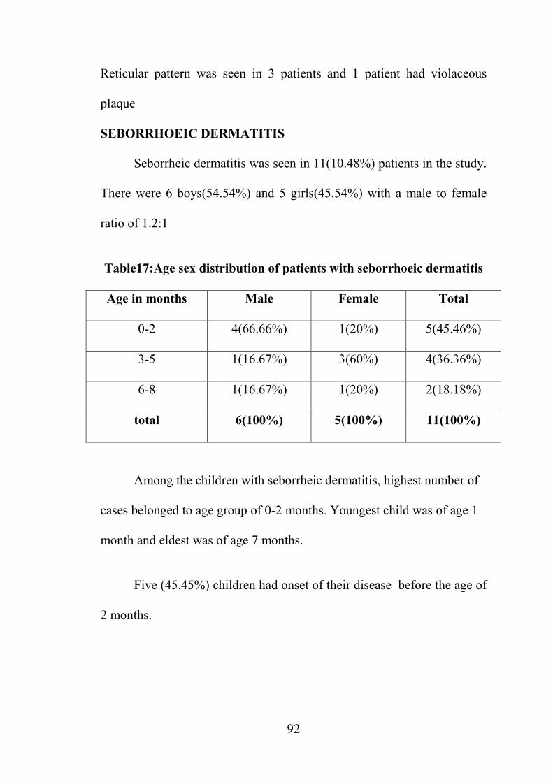
92
Reticular pattern was seen in 3 patients and 1 patient had violaceous
plaque
SEBORRHOEIC DERMATITIS
Seborrheic dermatitis was seen in 11(10.48%) patients in the study.
There were 6 boys(54.54%) and 5 girls(45.54%) with a male to female
ratio of 1.2:1
Table17:Age sex distribution of patients with seborrhoeic dermatitis
Age in months Male Female Total
0-2 4(66.66%) 1(20%) 5(45.46%)
3-5 1(16.67%) 3(60%) 4(36.36%)
6-8 1(16.67%) 1(20%) 2(18.18%)
total 6(100%) 5(100%) 11(100%)
Among the children with seborrheic dermatitis, highest number of
cases belonged to age group of 0-2 months. Youngest child was of age 1
month and eldest was of age 7 months.
Five (45.45%) children had onset of their disease before the age of
2 months.
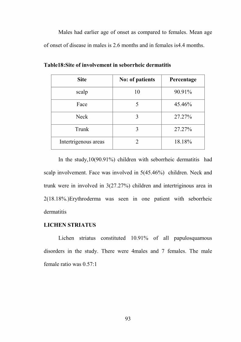
93
Males had earlier age of onset as compared to females. Mean age
of onset of disease in males is 2.6 months and in females is4.4 months.
Table18:Site of involvement in seborrheic dermatitis
Site No: of patients Percentage
scalp 10 90.91%
Face 5 45.46%
Neck 3 27.27%
Trunk 3 27.27%
Intertrigenous areas 2 18.18%
In the study,10(90.91%) children with seborrheic dermatitis had
scalp involvement. Face was involved in 5(45.46%) children. Neck and
trunk were in involved in 3(27.27%) children and intertriginous area in
2(18.18%.)Erythroderma was seen in one patient with seborrheic
dermatitis
LICHEN STRIATUS
Lichen striatus constituted 10.91% of all papulosquamous
disorders in the study. There were 4males and 7 females. The male
female ratio was 0.57:1

94
Table19:Age-sex distribution of patients with lichen striatus
Age in years Male Female Total
0-3 2(60%) 2(28.57%) 4(36.36%)
4-6 0 2(28.57%) 2(18.18%)
7-9 1(40%) 3(42.86%) 4(36.36%)
10-12 1(40%) 0 1(9.10%)
Total 4(100%) 7(100%) 11(100%)
In this study minimum age of onset was at 2 years and maximum at
11years. Eight (72.72)% 0f the patients had associated pruritis.
All cases had unilateral distribution.
Table20 :Site of distribution of lichen striatus
Site No. of cases Percentage
Face 2 18.18
Trunk 2 18.18
Upperlimb 3 27.27
Lowerlimb 4 36.36
In the study,most common site of involvement of lichen striatus
was lowerlimb,4(36.36%) patients followed by upperlimb, 3(27.27%)
patients. Face and trunk were involved in 2(18.18%) patients.
One patient had eczematous change over the lichen striatus.

95
PITYRIASIS RUBRA PILARIS
In this study 6 children presented with pityriasis rubra pilaris,
which accounts for 5.71% of papulosquamous disorder. There were 5
males and 1female with male female ratio 5:1
Table 21:Site of involvement and type of PRP
Sl no Age/Sex Site of involvement Type
1 3Yrs/Mch Chest, back, abdomen Type IV
2 6yrs/Fch Chest,Abdomen, back Type IV
3 8yrs/Mch Elbow, knees TYPE IV
4 11yrs/Mch Chest, abdomen, back TYPEIII
5 11yrs/Mch Chest, abdomen, back TYPE IV
6 11yrs/Mch Scalp. back, arms TYPE 1V
The youngest age of onset was 3 years and eldest age of onset was
11 years.
In our study TypeIV PRP , was the most common type noticed.
One patient had scalp involvement and 2(33.33%) patient had palms and
soles thickening.
One child had lichen nitidus and one had associated lichen striatus
along with PRP lesions.
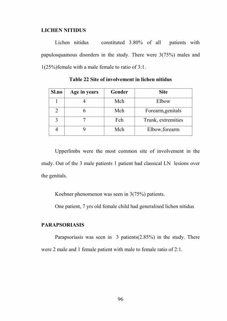
96
LICHEN NITIDUS
Lichen nitidus constituted 3.80% of all patients with
papulosquamous disorders in the study. There were 3(75%) males and
1(25%)female with a male female to ratio of 3:1.
Table 22 Site of involvement in lichen nitidus
Sl.no Age in years Gender Site
1 4 Mch Elbow
2 6 Mch Forearm,genitals
3 7 Fch Trunk, extremities
4 9 Mch Elbow,forearm
Upperlimbs were the most common site of involvement in the
study. Out of the 3 male patients 1 patient had classical LN lesions over
the genitals.
Koebner phenomenon was seen in 3(75%) patients.
One patient, 7 yrs old female child had generalised lichen nitidus
PARAPSORIASIS
Parapsoriasis was seen in 3 patients(2.85%) in the study. There
were 2 male and 1 female patient with male to female ratio of 2:1.
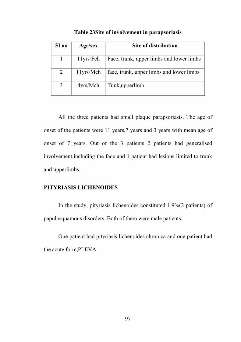
97
Table 23Site of involvement in parapsoriasis
Sl no Age/sex Site of distribution
1 11yrs/Fch Face, trunk, upper limbs and lower limbs
2 11yrs/Mch face, trunk, upper limbs and lower limbs
3 4yrs/Mch Tunk,upperlimb
All the three patients had small plaque parapsoriasis. The age of
onset of the patients were 11 years,7 years and 3 years with mean age of
onset of 7 years. Out of the 3 patients 2 patients had generalised
involvement,including the face and 1 patient had lesions limited to trunk
and upperlimbs.
PITYRIASIS LICHENOIDES
In the study, pityriasis lichenoides constituted 1.9%(2 patients) of
papulosquamous disorders. Both of them were male patients.
One patient had pityriasis lichenoides chronica and one patient had
the acute form,PLEVA.

98
8 year old male patient presented with hypopigmented and brown
coloured scaly patches over chest, back and arms. Histopathological
examination revealed features suggestive of pityriasis lichenoides
chronica.
11 year old male patient presented with erythematous papules over
chest and back, with central necrosis and mild scaling. There was history
of fever prior to the onset of disease. Histopathological examination
revealed features suggestive of PLEVA.

Fig. 1 Chronic Plaque Pso
99
COLOUR PLATES
ue Psoriasis Fig. 2 Psoriasis – Scal
Fig. 3 Guttate Psoriasis
Scalp & Face
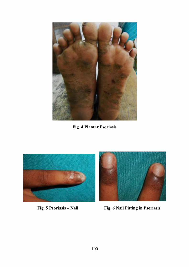
100
Fig. 4 Plantar Psoriasis
Fig. 5 Psoriasis – Nail Fig. 6 Nail Pitting in Psoriasis

Fig. 8. Pustular Psoriasis
101
Fig. 7 Flexural Psoriasis
riasis Fig. 9 Histopathology of P
Showing Hyperkeratosis ,Parakeratosis,micro m
Elongation of Rete ridges and Lymphocy
y of Psoriasis
icro munro abscess, regular
phocytes in the dermis
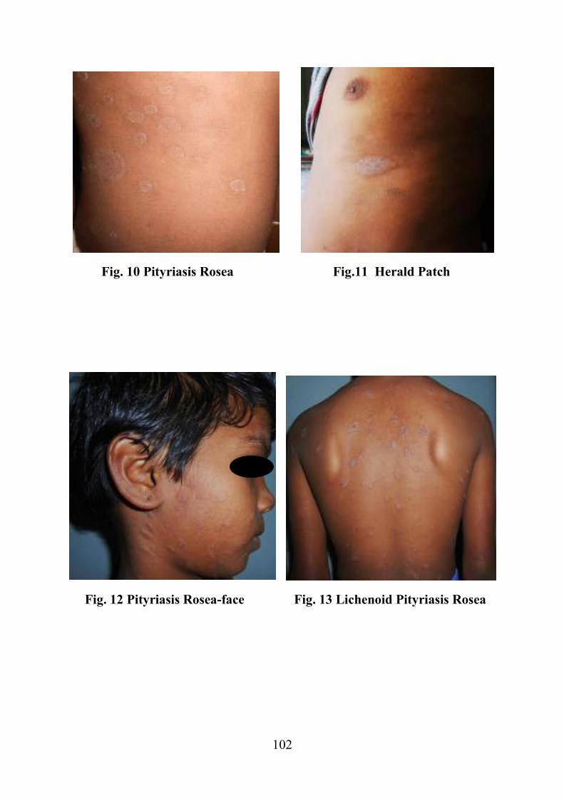
102
Fig. 10 Pityriasis Rosea Fig.11 Herald Patch
Fig. 12 Pityriasis Rosea-face Fig. 13 Lichenoid Pityriasis Rosea

103
Fig. 14 Classical LP Fig. 15 Generalised LP
Fig. 16 Linear LP extending to Palms Fig. 17 LP – Koebnerisation

Fig. 18 LP-Violaceous
involvement
Fig. 20 LP - Nail Dys
104
ceous Plaque over Lip Fig. 19 LP – Ge
ail Dystrophy Fig. 21 Histopatho shows saw toothening of r with Bandlike infiltrat
Genital
pathology of LP ng of rete ridges with
filtrate in the dermis

105
Fig. 22 Lichen Stratitus - Upperlimb
Fig. 23 Lichen Striatus – Thigh Fig. 24 Lichen Striatus – Face

106
Fig. 25 Seborrhoeic Dermatitis-Scalp Fig. 26 Seborrhoeic Dermatitis – Face
Yellow greasy scales
Fig. 27 Seborrhoeic Dermatitis – Erythroderma

107
Fig 28 Pityriasis Rubra Pilaris – Type IV
Fig 29 Pityriasis Rubra Pilaris – Type III
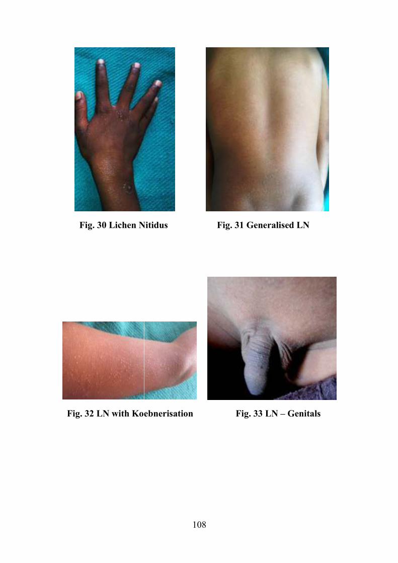
Fig. 30 Lichen Nit
Fig. 32 LN with Koeb
108
en Nitidus Fig. 31 Generalised
Koebnerisation Fig. 33 LN – G
alised LN
Genitals

Fig. 3
Erythema
109
Fig. 34 Small Plaque Parapsoriasis
Fig. 35 PLEVA
hematous Papules with Central Necrosisrosis

110
DISCUSSION
Dermatological disease forms an important part in the out patient
attendance of paediatric population. Skin diseases in children are
associated with significant morbidity and they form a major health
problem in the paediatric age group. Skin diseases contribute to 30% of
outpatient visits to paediatrician and 30% of patients consulting a
dermatologist will be from paediatric age group.126,127
The prevalence of paediatric dermatoses in various parts of India
ranges from 8.7 to 35% in school based surveys.14
In the present study,
hospital based prevalence of skin disease among children was 18.48%.
Papulosquamous disorders constituted 0.81% of paediatric dermatosis in
the present study. In an Indian study conducted in Rajasthan,
papulosquamous disorders constituted 1.66% of paediatric dermatosis.2In
a recent study from Turkey128
papulosquamous disorders constituted
6.9% of all paediatric dermatosis and a similar study129
showed a
prevalence of 14.2% among the paediatric dermatosis.
The difference in the prevalence rate may be due to the short
duration of the study and geographical variations.

111
AGE -SEX DISTRIBUTION
In the present study male patients(52%) outnumbered the female
patients(48%) with a male female ratio of 1.1:1which is similar to the
study by Vetrichevvel etal,130
where boys (57.5%) outnumbered girls
(42.5%). But in another study on papulosquamous disorders in children,
females(54.9%) outnumbered males(45.1%)131
In our study highest percentage of patients belonged to the age
group. 10-12 years(27.62%). In Kalyani vasram vadite study, highest
percentage of patients belonged to the age group 5-9 years.131
FREQUENCY OF VARIOUS PAPULOSQUAMOUS DISORDERS
In the present study, the various papulosquamous disorders seen in
decreasing order of frequency were, psoriasis(25.71%), pityriasis
rosea(20.95%), lichen planus(18.09%), seborrheic dermatitis(10.47%).
Lichen striatus(10.47%), pityriasis rubra pilaris( 5.71%),lichen
nitidus(3.8%) , parapsoriasis (2.85%) and pityriasis lichenoides(1.9%).
In a similar study conducted by Kalyani vasram vadite,131
the
frequency of papulosquamous disorders in decreasing order were lichen
planus(28.43%), pityriasis rosea(25.49%), seborrheic dermatitis(19.61%),
psoriasis vulgaris(16.67%), lichen striatus(3.92%), pityriasis rubra
pilaris(3.92%) and lichen nitidus( 1.96%).

112
Vetrichevvel et al130
reported pityriasis rosea as the commonest
disease in their study followed by lichen planus, seborrheic
dermatitis(11.11%), psoriasis (11.1%), pityriasis rubra pilaris (10.2%),
lichen nitidus(9.2%), lichen striatus (6.4%), pityriasis lichenoides
chronica (2.7%), acrodermatitis enteropathica and drug induced
erythroderma (one case each, 0.9%) and a total of 5 cases of
erythroderma due to papulosquamous causes were noted.
An Egyptian study132
reported pityriasis rosea as the commonest
disease in papulosquamous disorder followed by psoriasis and and PRP .
Psoriasis
During the study period of 1year, out of 12385 patients attended
dermatology OPD 27 patients had psoriasis. Thus the hospital based
prevalence of childhood psoriasis was 0.21%. This is comparable to the
study conducted in Karnataka which showed a prevalence of 0.38%131
and
low as compared to a South Indian study in paediatric population which
showed a prevalence of 1.4%.14
A study conducted in Saudi arabia by Ghada A. Ben Saif and Salha
A. Al Shehab3 showed a prevalence of 1.8% among paediatric
dermatosis. In a Turkish study133
and study by Gul et al128
showed higher
prevalence of 3.8% and 5.4% respectively.

113
In this study, among 105 patients with papulosquamous disorders,
27(25.71%) were psoriasis. This is comparable to a study conducted in
Saudi Arabia3 which showed a prevalence of 22.6% childhood psoriasis
among the papulosquamous disorders. But Vetrichevvel et al130
noted a
lower prevalence of 11.11% of childhood psoriasis among
papulosquamous disorders.
In the present study females out numbered males with a male to
female ratio of1: 1.45.Mercy et al134
reported a similar male to female
ratio of 1:1.48 among childhood psoriasis where as kumar et al18
showed
a male to female ratio of 1.09:1.A survey of 1262 childhood psoriasis in
Australia21
reported a male to female ratio of 1:1.
The age of onset in our study ranged from 6 months to 12 years.
Kumar et al18
reported age of onset from 4days to 14 years.
The mean age of onset in our study was 6.83years.In a study from
Denmark135
mean age of onset of childhood psoriasis was 8.1 years which
is slightly higher than the present study. Kumar et al18
reported a mean
age of onset of 8.1±2.1 years in boys and 9.3±2.3 in girls. In the present
study mean age of onset of psoriasis in boys was 6 years and 7.4 years in
girls. So boys had an earlier age of onset than girls.
114
In our study,the peak age of onset in males was 7 to 9 years where
as in females, peak age of onset was 10-12 years. Kumar et al18
reported a
peak age of onset of 6-10 years in males and 10-14 years in females.
Chronic plaque psoriasis was the most common presentation in our
study(51.85%) as observed by various previous studies.18,20,22,23
Table 24Comparison of frequency of chronic plaque psoriasis in
various studies
Sl.no Author Place of study Percentage
1 Nanda A et al20
Chandigarh ,India 69.6%
2 Fan X et al22
China 68.6%
3 Kumar et al18
India 60.6%
4 Yan wu et al23
China 52.6%
5 Present study coimbatore 51.85%
In contrast to the above studies, guttate psoriasis was the most
common type observed in Denmark.21
In the present study guttate
psoriasis was seen in 14.81%. Nanda et al20
observed guttate psoriasis in
25.9% of childhood psoriasis and Yan wu et al23
reported a frequency of
25.5% of guttate psoriasis in china.
The palms and soles have been reported to be uncommon sites of
psoriasisinvolvement in children. However, in our study, palmoplantar
psoriasis was observed in 11.11% of patients. Kumar B et al18
reported
115
palmoplantar psoriasis in 12. 8% of childhood psoriasis. Morris et al21
reported plantar psoriasis in 4% ,however Fan X et al22
did not observe
plantar psoriasis in their study.
In our study,pustular psoriasis was observed in 7.41% of the
psoriasis patients . Yan Wu et al23
observed in 10.90% of children with
psoriasis.Seyhan M et al136
reported pustular psoriasis in 13.1% of
childhood psoriasis.
In our study infantile psoriasis was present in 7.4% of the patients.
In Kalyani vasram study131
infantile psoriasis was seen in 5.9% of
patients with psoriasis
Nail involvement was seen in 5(18.52%) patients which is low as
compared to Yan wu et al23
,who reported 25.5%. Pitting was the most
common finding in the study.
Variable familial incidence has been reported in various studies of
childhood psoriasis which ranges from 9.8 to 89%.In our study family
history was seen in 2(7.4%) patients. Kumar et al18
and Yan wu et al23
reported a familial incidence of 4.5% an 8% in childhood psoriasis
respectively. But a study conducted in United States by Mercy et al 134
showed a familial incidence of 51.4% in childhood psoriasis.

116
Yan wu et al23
and Seyhan et al136
reported that upper respiratory
tract infection was the most common triggering factor in childhood
psoriasis. In our study precipitating factor was seen in 11.11% patients,
which was upper respiratory tract infection in all the cases.
Pruritis was the most common symptom in our study(59.25%)
kumar et al81
studies reported pruritis in.87.1% of the patients.
Koebnerisation was seen in 12 patients (44.44%) which was higher than
the observation in kumar et a18l study,which was 27.9%.
Psoriatic arthropathy and erythrodermic psoriasis were not
observed in our present study.In a study from Karnataka131
also there
were no cases of psoriatic arthropathy ans erythroderma.A north Indian
study reported psoriatic arthropathy in 1.1% of their subjects. Mercy et
al134
study reported psoriatic arthritis in 10.5% of all the subjects.Nanda
et al20
study reported psoriatic erytroderma in 1.8% of their patients. A
study from china23
showed , psoriatic erythroderma in 5.1% of their
patients. These study shows that prevalence of psoriatic erythroderma
have regional and racial variation

117
Pityriasis rosea
Pityriasis rosea is an acute self limiting inflammatory
papulosquamous disorder of unknown etiology.
In the present study .pityriasis rosea contributed 0.17% of all the
paediatric dermatosis attending our skin OPD. A study from South India
reported ,14
pityriasis rosea in 0.2% of all the paediatric dermatosis.
Another study conducted in north eastern region of India94
showed a
frequency of 1.2%. but a study from Iraq129
showed a higher frequency of
2.9% among children.
In a clinical study of papulosquamous disorders in children by
Vetrichevvel et al130
, pityriasis rosea was the most common
disorder(32.4%) where as in our study PR was the second common
condition seen in 20(22.95%) patients. This is comparable to a clinical
study of papulosquamous disorders in children in Karntaka(25.49%)130
.
In our stuy there were 10 males and 12 females with male to
female ratio of 0.83: 1 which is similar to study conducted by Kalyani
vasaram valdite131
with male to female ratio 0.7:1.In a clinical study on
pityriasis rosea conducted by Egwin AS et al137
male to female ratio was
1.5:1 and by Gunduz et al138
was 1.1:1

118
The youngest age of pityriasis rosea in Andrew etal139
study was 3
months old and 1 year in our study.
The maximum number of children with pityriasis rosea belonged to
the age group 10-12 years(31.8%). Gunduz et al138
and kalyani vasram
vadite131
observed peak incidence in 6-11 years age group with 49% and
53.8% patients respectively
Seasonal variation has been found in pityriasis rosea. Gunduz et
al138
and Kalyani Vasram Vadite131
observed a peak incidence during
winter and autumn months. In our study peak incidence was during
winter season(54.55%) similar to the observation of Vetrichevvel et al.130
A history of fever and sorethroat suggestive of upper respiratory
tract infection was seen in 27.27% of patients.Gunduz et al138
observed
prodromal symptoms in 33% of patients .
Pruritis was present in 16(72.72%) of the patients.Gunduz et al138
observed pruritis in 69% of their patients.
Herald patch was seen in 54.46% of patients in our study, most
common over the trunk with a median period between appearance of
herald patch and widespread eruptions of 7days(4-12days). Gunduz et
al138
study observed herald patch in 45% of patients, which was located

119
most often over the trunk. The median period between appearance of
herald patch and widespread eruptions was 4 days(range 0-30 days).
Most common site of involvement in our study was back 81.81%.
Least affected site in our study was face(27.27%) .In a clinical study by
Kalyan vasaram valdite131
, back was the most common site(88.5%) and
neck was the least affected site(23.1%)
In our study,most common type of pityriasis rosea was classical
type(81.81%). Inverse PR was seen in 3 patients and lichenoid PR in
4.54% of patients.
We have observed eczematous changes in 1(4.54%) patient with
pityriasis rosea.
Lichen planus
Lichen planus is a papulosquamous disorder of unknown
etiology.Eventhough lichen planus is very rare in children,most reports of
LP in children have come from the Indian subcontinent, suggesting that
children of South Asian origin are more susceptible to developing
LP.(lp7) The frequeny of lichen planus varies from 2.1% to 11.2% in the
paediatric population. The prevalence of childhood lichen planus in
children in our study was 0.15%. This is low when compared to study
conducted by Handa et al52
in North India ,who reported 2.5% among the

120
total paediatric patients. A hospital based study conducted at
Iraq129
showed , lichen planus formed 3.9% of the total number of
paediatric dermatosis in a 6 month period.
In the study of Vetrichevvel et al130
lichen planus constituted
14.8% of various papulosquamous disorders where as in our study
lichenplanus constituted 18.09%of the total papulosquamous
disorders.Kalyani vasaram valdite131
reported a higher frequency of
28.43% of lichen planus among the papulosquamous disorers.
Most of the studies show an equal sex prediliction in lichen planus
or a slight male preponderance. Handa et al52
and Kalyani vasaram
valdite131
reported a male female sex ratio of 1.1:1 . In our study the male
female ratio was higher than these studies,ie; 1.7:1. Sharma and
Maheswari64
have also reported a higher male female ratio of 2:1.
In our study, age group ranged from 3-12 years with mean age of
onset of 6.6 years. In Handa et al52
study of lichen planus in 87 children,
the age group ranged from 8 months to 12 years with mean age of onset
of 7.1 years.
The most common clinical type in our study was classical lp(
57.89%) which is similar to Handa et a52
and Sharma et al64
who observed
classical LP in 60.9% and 60%of their LP patients respectively. Linear
121
LP was observed in 15.79 patients in our study. Linear LP was observed
from 8-30% in various studies52,55,64.
One case of linear LP ha involvement
of palms also.In our study we observed acute generalised LP in 26.32 %
of all LP patients which is higher than Handa et al52
, who observed 6.9%
and Sharma and Maheswari64
who observed 4%.
Various studies on childhood lichen planus had reported a
frequency of 41-70% of involvement of extremities.52,64,68
In our study, the
most common site to be involved by lichen planus was
extremities(78.95%), more commonly lower limbs.
Oral mucosal involvement in Lp ranges from 4.3 to 39% in various
studies52,64,140,
Oral mucosal involvement was seen in 4 patients(21.05%)
in our study which is comparable to Nnoruka et al141
who observed oral
involvement in 23.1% of their patients.
In our study nail involvement was seen in 3 patients(15.79%).
Kanwar and de68
observed nail involvement in 19.5% of their patients.
But in other studies it ranges from 0-7.7%.52,55,,64
Koebnerisation is seen commonly in childhood LP, which
ranges from 24 to 28%.In our study we observed a higher percentage of
koebnerisation of 68.42%. But Kanwar and 68
observed koebneriastion in
only 6% of their patients.

122
Pruritis was present in 84% of the patients in our study. This is
comparable to findings of other authors who found pruritis in 87%,
89.6% and 100% of their patients respectively.64,68,142
Lichen striatus
Lichen striatus is a self limiting papulosquamous disorder of
unknown etiology in which ,the lesions follow the Blaschko lines.
Lichen stratus constituted 10.91% of all the papulosquamous
isorders in our study, which is high as compared to the study of
Vetrichhevel et al130
who observed lichen striatus in 6.4% children.
Lichen striatus is more common in females than in males. In our
study male: female ratio was 0.57: 1 which is similar to Patrizzi et al143
who noted male to female ratio of 0.5:1.
Lichen striatus is more common in the age group 5-15 years.In our
study maximum number of patients were seen in the age group0-3 years
and 7-10 years with mean age of 5.5 years. Patrizzi et al143
reported a
mean age of 4.5 years
In our study the most common site of presentation was
extremities(63.63%); lower limb (36.36%) more than
upperlimb(27.27%). This is similar to Taniguchi et al144
study in which
123
lesions were predominant in the lowerlimbs. In Patrizzi et al143
study,
extremities were the most frequent site with no substantial difference
between upperlimbs and lower limbs were seen. Taieb et al88
study
reported upperlimbs to be the most common site.
One patient with lichen striatus had eczematous changes.
SEBORRHEIC DERMATITIS
In our study seborrheic dermatitis was seen in 11(10.48% )
patients. This is comparable to Vetrichevvel et al131
who noted seborrheic
dermatitis in 11.11% of their patients with papulosquamous disorders.
Seborrheic dermatitis affects males more often than female
children. In our study 54.54% of the cases were male and 45.45% cases
were female with a male to female ratio of 1.2: 1.Wananukul et al145
study also showed that 51.66% of the cases were male and 48.33% were
male to female ratio was 1.06:1.But in Kalyani vasram vadite130
study
females(55%) outnumbered males(45%) with male to female ratio of
0.82:1
The youngest child was of age 1 month and eldest of age 7 months.
Majority of the children belonged to 0-2 months age group. Wananukul et
al145
also noticed 60% of the children were less than 2 months.
124
In our study , the scalp (90.91%) was the most common site to be
involved which is comparable to Foley et al146
and Kalyani vasram
vadite131
study, where scalp involvement was seen in 86% an 95% of the
children respectively.
Generalised seborrheic dermatitis was noted in 1 patient in our
study who was a 2 year old male child. In a study, infantile seborrheic
dermatitis constituted 10% of the causes of erythroderma10
.
PITYRIASIS RUBRA PILARIS
Pityriasis rubra pilaris is an idiopathic inflammatory
papulosquamous disorder characterised by follicular hyperkeratosis,
palmoplantar karatodema and erythroderma. In our study 6(5.71%)
children presented with pityriasis rubra pilaris. Vetrichevvel et al130
noted
pityriasis rubra pilaris in 10.92 % of the children in their study and
kalyani vasram vadite noted 3.92% of the children in their study.
In our study mean age was 8 years, slightly higher than yang et al
who reported mean age of 6.5 years in their study of 28 cases of juvenile
pityriasis rubra pilaris.
The male to female ratio in our study was 5:1.
In our study Type IV ,juvenile onset circumscribed pityriasis rubra
pilaris was the most common type (83.33%) followed by type III. Yang

125
et al studies also reported a similar observation. There were no cases of
type V PRP in our study. There were no case of erythroderma due to PRP
in our study.
Table 25Comparison of patients with juvenile pityriasis
rubra pilaris in different studies
Sl no: Present study Yang et al147
Gelmetti et al148
Number of cases 6 28 31
Age of onset(in years) 3-11 0.67-18 2-13
Type III 16.67% 14.33% 34%
Type IV 83.33% 85.7% 66%
Type V 0 0 0
Site of involvement
Scalp 16.67% 25% 32%
Palms 33.33% 89% 84%
Soles 33.33% 86% 84%
Trunk 50% 39% 25%
Elbows 66.67% 64% 77%
Knees 50% 89% 77%
Lichen nitidus
Lichen nitidus is a rare chronic papulosquamous disorder of
uncertain etiology seen primarily in the paediatric population. In our
study , 4(3.8%) patients presented with lichen nitidus which is low when

126
compared to vetrichevvel et al131
who reported lichen nitidus in 9.2% of
children.
In lichen nitidus, there is an equal male to female ratio or slight
male predominance. In our study male to female ratio was 3:1 which is
similar to Zapata et al149
study in which male to female ratio was 3:1.
Lapins et al also reported a higher male to female ratio of 4:1.
In our study mean age of onset was 6.5 years . Mean age of onset
of males was 6.1 years which is comparable to Lapins150
study where
mean age of onset was 7 years for males. Mean age of onset in female in
lapins et al study was 13 years150
. In our study only one female was
noticed who was 7 years age old.
Extremities were the most common site of involvement in our
study which is similar to kalyana vasram vadite study.. Zapata et al149
noted upperlimbs and trunk to be the most common site of involvement in
their study.
Generalised lichen nitidus is a rare variant of lichen nitidus.In our study
one patient, 7 year old female had generalised lichen nitidus.
Parapsoriasis
Parapsoriasis is a group of uncommon inflammatory disorders
characterised by persistent scaly inflammatory eruptions. In our study
127
there were 3 (3.80%)cases of parapsoriasis.Vetrichevvel et al reported
parapsoriasis in There were 2 males and 1 female.All the three cases
were of small plaque parapsoriasis. Out of the 3 patients ,2 patients had
involvement all over the body including face and 1 patient had trunk and
extremities involvement.
Pityriasis lichenoids
Pityriasis lichenoies is a papulosquamous disorder of unknown
etiology with acute and chronic presentations.
In our study, we noticed pityriasis lichenoides in 2(1.9%) patients
Vetrichevvel et al131
also reported pityriasis lichenoides in 2.7% of the
children with papulosquamous disease in their study.
Pityriasis lichenoides is more common in males. In various studies
male to female ratio varies from 1.4:1 to 2:1.151,152
In our study both of
them were male patients.
One patient had pityriasis lichenoides chronica and the other had
the acute form,PLEVA.

128
SUMMARY
A clinical study on papulosquamous disorders of children less than
12 years was done from August 2013 to July 2014. The age-sex
prevalence, age of onset, symptoms,morphology and site of involvement
of lesions of each diseases were recorded, analysed and compared with
other studies and discussed.
� Papulosquamous disorders constituted 0.81% of the paediatric
dermatoses during the study.Majority of the patients belonged to
the age group10-12 years. The youngest patient was 1 year old at
the time of presentation. Boys outnumbered girls with male to
female ratio of 1.1:1
� Psoriasis was the most common papulosquamous disorder
(25.71%) followed by pityriasis rosea(20.95%). Pityriasis
lichenoides was seen least in number(1.90%).Psoriasis patients in
our study showed a female predominance with a male to female
ratio of 1:1.45. Chronic plaque psoriasis was the most common
type(51.85%).Guttate psoriasis was seen in (14.81%%) of patients.
Palmoplantar psoriasis was seen in 11.11%. Nail involvement was
seen in 18.52% and scalp psoriasis in 3.7% .Pruritis was seen in
55.55% and Koebneriastion in 40.74% patients.

129
� Pityriasis rosea was seen in 22.95% patients, with male to female
ratio of 1.5:1. It was common in 10-12 years age group(31.8%)
.Youngest patient was of age 1 year .Pruritis was seen in 72.72%
patients.Herald patch was seen in 54.46% patient with most
common site of involvement over the chest.Avaerage time period
between appearance of herald patch and widespread eruptions was
7 days.Most common type was classical PR seen in 81.81%
patients. Inverse PR was seen in-13.6% and lichenoid PR was seen
in4.54% patients.
� Lichen planus was seen in 18.09% of the total 105 patients with
male to female ratio of 1.7:1. Mean age of onset was 6.6years.
Most common clinical type was classical LP(57.89%). Generalised
LP was seen in 26.32% patients and linear LP was seen in 15.79%
patients. Most common site involved was extremities(78.95%),
more commonly lowerlimbs. Oral mucosal involvement was seen
in 21.05% patients. Nail involvement was seen in 15.79%. pruritis
was seen 84% of the patients and koebnerisation in 68.42%
patients.

130
� Seborrheic dermatitis was seen in 10.48% of patients with male to
female ratio of 1.2: 1. Children with seborrheic dermatitis belonged
to 1 months and 7 months. Majority of children belonged to 0-2
months age group. Most common site to be involved was
scalp(90.91%). Erythroderma was present in 1 patient.
� Lichen striatus constituted 10.48% of patients with
papulosquamous disorders with male to female ratio of 0.5:1. Mean
age of onset was 5.5 years. Most common sites involved were
extremities.Most common pattern observed was linear pattern.
� Pityriasis rubra pilaris was seen in5.71% of the patients with male
to female ratio of 5:1.Most common type of PRP noticed was Type
IV(83.33%). Most common sites observe was elbows(66.67%).
Palms and soles were involved in 33.33% of patients an scalp was
involved in16.67%
� Lichen nitidus was seen in 3.8% of patients with male female ratio
of 3:1. Most common site involved was extremities.One of the
three male patients had typical lesions over genitalia. Generalised
lichen nitidus was seen in one patient.

131
� Parapsoriasis was seen in 2.85% of patients in the study. All the
patients had small plaque parapsoriasis. The mean age of onset of
parapsoriasis in our study was 7 years. Two patients had
generalised involvement including the face. One patient had
distribution of the lesions over the trunk and upperlimbs.
� Pityriasis lichenoides was seen in 21.9% of the patients. One
patient had pityriasis lichenoides chronica and one patient had
PLEVA. In our study,all the patients were male.

132
CONCLUSION
Papulosquamous disorder is a common dermatological group of
disorders which is more predominant in male children . Various factors
like age, gender and seasonal variation play a role in the occurrence and
exacerebration of these group of disorders. Genetic inheritance plays
negligible role in the occurrence of these disorders. The prevalence rate
of papulosquamous disease varies from region to region. This regional
variation can be due to the climatic and racial variation.
The prevalence of papulosquamous disorders in our study was
0.81%. The most common papulosquamous disorder in the study was
psoriasis
Understanding the morphology , distribution of the lesions,
characteristics of the scales and special features like koebnerisation,
Auspitz sign, herald patch etc will help to differentiate the individual
diseases clinically. This will avoid invasive procedures like biopsy
particularly in paediatric age group, as the patients and the parents will be
anxious . When it is difficult to differentiate the diseases clinically,
biopsy and histopathological examination will come in hand. Individual
diseases in this group have variable prognosis..
133
By understanding the morphological characterestics of papulosquamous
diseases and differentiating the diseases clinically ,we can explain the
prognosis of the disease to the parents, which will alleviate their worry.
LIMITATION OF THE STUDY:
� As the study was of short duration, follow up and response to
treatment could not be evaluated.
� Natural course of the disorders could not be studied as the study
was of short duration.

134
BIBLIOGRAPHY
1. Fox BJ, Odom RB.BalaiPapulosquamous diseases: a review.J Am
AcadDermatol. 1985 Apr;12(4):597-624.
2. M,KhareAK,Gupta LK, Mittal A, KuldeepCM.Pattern of paediatric
dermatoses in a teritiary care centre of South West
Rajasthan.Indian J dermatol 2012;57:275-8.
3. Ghada A. Ben Saif and Salha A. Al Shehab Pattern of Childhood
Dermatoses at a Teaching Hospital of Saudi ArabiaInt J Health
Sci(Qassim).Jul 2008:63-74.
4. Gibson LE,PerryHO.Papulosquamous and exfoliative dermatitis .In
Moschella SL, Hurley HJ , editors. Dermatology.3rd
ed.
Philadelphia: WB Saunder’s company ; 1992.p607-51
5. MobiniN,ToussaintS,Kamino H. Non infectious erythematous
popular and squamous disorders. In Elder
DE,ElensistsasR,JohnsonJr.Bl, Murphy GF, editors. Lever’s
histopathology of skin.9thed Philadelphia: Lippincott Williams and
Wilkns ;2006.p179-214.
6. Hall JC. Saurer’s manual of skin disease. 8thed,
Philadelphia:Lippincot Williams and Wilkins; 2000.
7. Papulosquamousdisorders(L40-45).Chapter XII Diseases of skin
and subcutaneous tissue(L00-L99).International Statistical

135
Classification of Diseases and Related Health Problems 10th
Revision (ICD-10) Version for 2010.cited on September 2014.
Available from
http://apps.who.int/classifications/icd10/browse/2010/em#/L40-45
8. C.E.M.Griffiths and J.N.W.N.Baker. In Tony
Burns,Stephenbreathnach, Neil cox, Christopher Griffiths
editors..Rooks text book of dermatology. 8th
edition.Chapter
20,Psoriasis: 20.1-20.60
9. Benoit S,HammH.Psoriasis in childhood and adolescence. Clinical
features and therapy.Hautarzt 2009;60:100-8
10. Dogra S, De D, Ragunatha S. Disorders of keratinisation. In:
InamadarAC,Sacchidanand S, eds. Textbook of Pediatric
Dermatology, 1st ed. New Delhi:Jaypee publications;2009:pp334-
59
11. Patel S, Paller AS: Pediatric psoriasis. In: Yoo JY, Lee CS,
Lebwohl MG, et al ed. Psoriasis, New York: Informa Healthcare;
2009:219-238.
12. Rogers M: Childhood psoriasis. CurrOpinPediatr 2002; 14(4):404-
409.
13. QaziMasood,Iffat Hassan. Pattern of skin disorders in Kashmir
valley. Indian J DermatolVenerolLeprol 2002; 47(3):147-8

136
14. Karthikeyan K, Thappa DM, Jeevankumar B. Pattern of
pediatricdermatoses in a referral centre in South India. In Pediatrics
2004;41:373-7.
15. Nanda A, Hasawi FA, Alsaleh QA. A prospective survey of
pediatric dermatology clinic patients in Kuwait: An analysis of
10,000 cases. PediatrDermatol 1999;16:6-11.
16. Jain N, Khandpur S. Paediatric dermatoses in India.Indian J
Dermatol,Venerol,Leprol 2010; 76: 451-4.
17. SandipanDhar, Raghubir Banerjee, Nilesh Agrawal,Sharmila
Chatterjee, and RajibMalakar. Psoriasis in children: An insight.
Indian J Dermatol. 2011 May-Jun; 56(3): 262–265.
18. Kumar B, Jain R, Sandhu K, Kaur I, Handa S. Epidemiology of
childhood psoriasis: A study of 419 patients from northern India.
2004;43:654-8. Int J Dermatol. 2004;43:654–8.
19. Verbow J.Psoriasis in childhood. Arch Dis Child 1992;67:75-6.
20. Nanda A,KaurS,Kaur I et al . Childhood psoriasis: an
epidemiological survey of 112 patients. Paediatr Dermatology
1990;7:19-21.
21. Morris A, Kaur S, Kaur I et al. Chilhood psoriasis: a clinical
review of 1262 cases. Paediatrdermatol 2001;18: 188-98.
137
22. Fan X, Xiao FL, Yang S, et al: Childhood psoriasis: a study of 277
patients from China. J EurAcadDermatolVenereol 2007;
21(6):762-765.
23. Wu Y, Lin Y, Liu HJ, Huang CZ, Feng AP, Li JW. Childhood
psoriasis: A study of 137 cases from central China. World J
Pediatr. 2010;6:260–4.
24. Mercy et al.Clinical manifestations of pediatric psoriasis: Results
of a multicentre study in the United States.PediatrDermatol. 2013
July ; 30(4): 424–428.
25. SwanbeckG,InterotA,Martinssont,Enerbackk C,
EnlundF,Samuelsson et al.Genetic counselling in psoriasis:
Empirical data on psoriasis among first degree relatives of 3095
psoriatic probans.Br J Dermatol 1997;137: 9339-42.
26. David weedon.Thepsoriasiformreation pattern. Weedon’s Skin
pathology. 3rd
edition, pg.78-79
27. Flora B.de Waard-van der Spek and Arnold P Oranje. Psoriasis.
Harper’s Textbook of Paediatric Dermatology, 3rd
edition.80.1-
80.6.
28. Naldi L, Peli L, Parazzini F, et al: Family history of psoriasis,
stressful life events, and recent infectious disease are risk factors
for a first episode of acute guttate psoriasis: results of a case-
control study. J Am AcadDermatol 2001; 44(3):433-438.

138
29. Amy S. Paller and Anthony J. Mancini. Psoriasis and related
papulosquamous disorders. Hurwitz clinical paediatric
dermatology. 4th
edition.pg.71-91.
30. Johann E,Gujonsson and james T elder.Psoriasis. Fitzpatrik’s
textbook of ermatology in general medicine. 8th ed. Vol1: pg197-
231
31. Flora B.de Waard-van der Spek and Arnold P Oranje.
Psoriasis:Treatments. Harper’s Textbook ofPaediatric
Dermatology. 3rd edition.82.1-80.7.
32. Gibert CM: TraitePratique de Maladies de la Peauet de la Syphilis,
3rd edition. Paris, Plon, 1860, p. 402.
33. KlauderJV.PityriasisRoseawith particular reference to its unusual
manifestations .JAMA 1924;82:178-83.
34. De Souza Sittart JA, Tayah M, Soares Z. Incidence pityriasisrosea
of Gibert in the Dermatology Service of the Hospital do
ServidorPúblico in the state of São Paulo .Med CutanIberoLat Am.
1984;12(4):336-8.
35. Olumide Y. Pityriasisrosea in Lagos. Int J Dermatol. 1987
May;26(4):234-6.
36. Giam YC. Skin diseases in children in Singapore. Ann Acad Med
Singapore. 1988 Oct;17(4):569-72.
139
37. Traore A1, Korsaga-Some N, Niamba P, Barro F, Sanou I, Drabo
YJ. Pityriasisroseain secondary schools in Ouagadougou, Burkina
Faso. Ann DermatolVenereol. 2001 May;128(5):605-9.
38. Chuh AA et al: Temporal case clustering in pityriasis rosea: A
regression analysis on 1379 patients in Minnesota, Kuwait, and
Diyarbakir, Turkey. Arch Dermatol 141:767, 2005.
39. HYATT HW Sr. Pityriasisrosea in a three month old infant. Arch
Pediatr. 1960 Sep;77:364-8.
40. Drago F, Vecchio F, Rebora A: Use of high-dose acyclovir in
pityriasisrosea. J Am AcadDermatol2006;54:82,
41. Antonio A.T.Cuhh and Vijay Zawar. PityriasisRosea, Harper’s
Textbook of Paediatric Dermatology. 3rd
edition;84.1-84.5
42. Drago F et al: Human herpes virus-like particles in pityriasisrosea
lesions: An electron microscopy study. J CutanPathol 2002;29:359.
43. Broccolo F, Careddu AM et al.Additional evidence that Pityriasis
is associated with reactivation of human harpes virus-6 and -7.J
Invest Dermatol 2005;124:1234-40.
44. Hosokawa H,HorioS,TakiuchiY,MaruyamaN,AsadaY.Naturally
occurring T lymphocytotoxic antibody in viral and related skin
diseases.ActaDermVenerol 1984;64:275-80.

140
45. Andrew Blauvelt. PityriasisRosea. Fitzpatrik’s Dermatology in
General Medicine.8thed: 362-6
46. J.C. Sterling. Virus Infections.In: Burns T, BreathnachS,Cox N,
Griffiths C, eds. Rook textbook of Dermatology.8th
ed.
Oxford:Blackwell science;2010:33.78-33.81
47. Chuh A, Lee A, Zawar V, Sciallis G, Kempf W. Pityriasisrosea -
An update.Indian J DermatolVenereolLeprol 2005;71(5):311-5
48. Neoh CY et al: Characterization of the inflammatorycell infiltrate
in herald patches and fullydeveloped eruptions of pityriasisrosea.
ClinExpDermatol 35:300, 2010
49. Sharma PK, Yadav TP, Gautam RK et al. Erythromycin in
pityriasisrosea: Adouble-blind, placebo-controlled clinical trial. J
Am AcadDermatol 2000; 42:241–4.
50. Rasi A1, Tajziehchi L, Savabi-Nasab.Oral erythromycin is
ineffective in the treatment of pityriasisrosea. J Drugs Dermatol.
2008 Jan;7(1):35-8.
51. Amer A1, Fischer H. Azithromycin does not cure pityriasisrosea.
Pediatrics. 2006 May;117(5):1702-5.
52. Handa S, Sahoo B. Childhood lichen planus: A study of 87 cases.
Int JDermatol 2002;41(7):423-7.

141
53. VenketramanMysore. Lichen Planus and lichenoid dermatoses.
Dermatological diseases: A practical approach.1st ed. 59-63
54. Wilson E.On lichen planus. J cutan Med Dis Skin1869;3:117-32
55. Luis-Montoya P, Domínguez-Soto L, Vega-Memije E: Lichen
planus in 24 children with review of the literature. PediatrDermatol
2005; 22(4):295-298.
56. Balasubramaniam P, Ogboli M, Moss C. Lichen planus in children:
review of 26cases. ClinExpDermatol 2008; 33: 457–9.
57. Pusey WA.Lichenplanus in an infant less than 6 month
old.ArchDermatolSyph 1929;19: 671-2
58. MarkR .Pittelkow, MazenSDaoud.Lichen Planus. Fitzpatrik’s
textbook of dermatology in general medicine 8thedition.Vol 1: 244-
255
59. Vega ME et al: Ashy dermatosis versus lichen planuspigmentosus:
A controversial matter. Int J Dermatol 31:87, 1992
60. Nail lichen planus in children: clinical features, response to
treatment, and long-term follow-up. Arch Dermatol. 2001
Aug;137(8):1027-32. Tosti A, Piraccini BM, Cambiaghi S, Jorizzo
M)
142
61. Usman A, Kimyai-Asadi A, Stiller MJ, et al: Lichenoid eruption
following hepatitis B vaccination: first North American case report.
PediatrDermatol 2001; 18(2):123-126.
62. Black MM. What is going on in lichen planus? ClinExpDermatol.
1977 Dec;2(4):303-10.
63. Mahood JM: Familial lichen planus. Arch Dermatol.119:292, 1983
64. Sharma R, Maheshwari V. Childhood lichen planus: a report of
fifty cases. PediatrDermatol. 1999 Sep-Oct;16(5):345-8.
65. Harper N Price and Andrea L Zaenglein. Lichen Planus and Lichen
Nitidus. Harper’s Textbook of Paediatric Dermatology
3rd
edition.Vol 1: 85.1-85.17.
66. Vasquez Garcia J, Pérez Oliva N, PeireioFerreirós MAM et al.
Lichen planusfollicularistumidus with cysts and comedones.
ClinExpDermatol 1992; 17:346–8.
67. S.M. Breathnach. Lichen Planus and Lichenoid Disorders .chapter
41.Rook’s Textbook of Dermatology.8th
ed.Vol3: 41.1-41.28
68. Kanwar AJ, De D. Lichen planus in children. Indian J
DermatolVenereolLeprol 2010;76(4):366-72
69. Hogan PA. Papulosquamous disease. In: Schachner LA, Hansen
RC, eds. Pediatric Dermatology. 3rd ed. Philadelphia: Elsevier
publishers;2003:pp643-81.

143
70. M.R. Judge1, W.H.I. McLean2 & C.S. Munro.In Tony
Burns,Stephenbreathnach, Neil cox, Christopher Griffiths
editors..Rooks text book of dermatology. 8th edition. Chapter
19.Disorders of keratinisation.19.76-19.80.
71. Griffiths WA et al. Pityriasisrubrapilaris. ClinExpDermatol.
1980;105-12.
72. Sehgal VN, Jain MK, Mathur RP. Pityriasisrubrapilaris in
Indians.Br J Dermatol. 1989 Dec;121(6):821-2.
73. Vanderhooft SL, Francis JS, Holbrook KA, et al: Familial
pityriasisrubrapilaris. Arch Dermatol 1995; 131(4):448-453.
74. Finzi AF, Altomare G, Bergamaschini L, Tucci A
Pityriasisrubrapilaris and and retinol-binding protein. Br J
Dermatol. 1981 Mar;104(3):253-6.
75. vanVoorst Vader PC, van Oostveen F, Houthoff HJ, Marrink J
Pityriasisrubrapilaris, vitamin A and retinol-binding protein: a case
study.ActaDermVenereol. 1984;64(5):430-2.
76. Allison DS, el-Azhary RA, Calobrisi SD, Dicken CH.
Pityriasirubrapilaris in children. J Am AcadDermatol 2002;47:386-
9.DECEMBER 2008.
77. Betlloch I et al: Acute juvenile piryriasisrubrapilaris:Asuperantigen
mediateddisease PediatrDermatol18:41.12.,0 01.
144
78. KurzydloAM, Gillespie R. Paraneoplasticpityriasisrubrapilaris in
association with bronchogenic carcinoma. Australas J Dermatol.
May 2004;45(2):130-2.
79. Evangelou G, Murdoch SR, Palamaras I, Rhodes LE.
Photoaggravatedpityriasisrubrapilaris.
PhotodermatolPhotoimmunolPhotomed 2005;21:272-4.
80. Melinda Jen and Mary Wu Chang.PityriasisRubraPiaris. Harper’s
Textbook of Dermatology,3rd
edition.Vol.1:83.1-83.8
81. DanielaBruch-GerharzThomas Ruzick .PityriasisRubraPilaris,
Fitzpatrik’s textbook of Dermatology in general medicine.7th ed.
Vol 1: 232-6.
82. Kanegaye JT, Frieden IJ. Lichen striatus: simultaneous occurrence
in siblings. Pediatrics 1992; 90: 104-6.
83. Patrone P, Patrizi A, Bonci A, Passarini B. Lichen striatus: studio
clinicoedistologico di 8 casi. G Ital DermatolVenereol 1990; 125:
267-70.)
84. Kennedy ,RogersM,Lichenstriatus.Paeiatr
Dermatology1996;13:95-9
85. Toda K,Okamoto H, HorioT. Lichen Striatus. Int J Dermatol
1986;70: 200-6.

145
86. Zhang Y, McNuttNS. Lichenstriatus. Histological, immune
histochemical and ultrastructural study of 37 cases. JCutan
Pathol2001;28: 65-71.
87. HappleR.Transposable elements and the lines of blaschko: A new
perspective,Dermatology 2002;204;4-7.
88. Taïeb A, Youbi E, Maleville J. Lichen striatus: a Blaschko linear
acquired inflammatory skin eruption. J Am AcadDermatol 1991;
25: 637-42.
89. Gelmetti C, Cerri D,Gianotti R et al.Bilateral multiple lichen
stiatus.PaeiatrDermatolNews(Bari)1998;7:97.
90. GoskowickMo, EichenfieldLF. Onychodystrophy with lichen
striatus. PaediatrDermatol 1994;11: 282-3
91. Alain Taeib and EouardGrosshans. Lichen striatus. Harper’s
Textbook of Dermatology. 3rd
edition. Vol 1: 86.5
92. Tosti A, Peluso AM, Misciali C, Cameli N. Nail lichen striatus:
clinical features and long-term follow-up of five patients. J Am
AcadDermatol 1997; 36: 908-13.
93. Bernard LA, Eichenfield LF. Eczematous and papulosquamous
disorders. In:Eichenfield LF, Frieden IJ, Esterly NB, eds. Neonatal
Dermatology, 2nd ed.Philadelphia: Elsevier; 2008: pp236-44.

146
94. M M Huda, Pranabkumarsaha. Pattern of dermatosis among
paediatric patients attending a medical college hospital in the
North eastern of India. Indian J of Dermatol 2004;49(4) 189-191.
95. Johnson M-LT, Roberts J. Prevalence of Dermatological Diseases
Among Persons 1–74Years of Age. Washington DC: US
Department of Health Education, NationalCenter for Health
Statistics, 1978: PHS 79–1660.
96. Ruiz Maldonado R ,Lopez Martinez R,Chavarria P et
al.Pityriosporumovale in infantile seborrheic
dermatitis.PaediatrDermatol 1989;6:16-20.
97. Tager A. Seborrhoeic dermatitis in acute cardiac disease. Br J
Dermatol 1964; 76:367–9.
98. Barba A, Piubello W, Vantini I et al. Skin lesions in chronic
alcoholic pancreatitis.Dermatologica 1982; 164: 322–6.
99. Cohen BA. Papulosquamous eruptions. In: Cohen BA, eds.
PediatricDermatology. 3rd ed. Philadelphia: Elsevier; 2005.pp67-
99
100. Holden CA, Berth-Jones J. Eczema, lichenification, prurigo and
erythroderma.In: Burns T, Breathnach S, Cox N, Griffiths C, eds.
Rook textbook ofDermatology.7th ed. Oxford: Blackwell science;
2004: pp17.10-17.15.
147
101. Oranje AP, van Joost T, van Reede EC, Vuzevski VD, Dzoljic-
Danilovic G, ten Kate FJ, Stolz E. Infantile seborrheic dermatitis.
Morphological and immunopathological study. Dermatologica.
1986;172(4):191-5.
102. Carlo M Gelmetti and Ramon Grimalt. Infantile seborrheic
dermatitis. Harper’s textbook of dermatology.3rd
d. vol 1.chapter
35; 35.5-35.6
103. Pittelkow MR, Daoud MS. Lichen nitidus. In: Wolff K , Goldsmith
LA, KatzSI, Gilchrest BA, Paller AS, Leffell DJ, eds. Fitzpatrick's
Dermatology inGeneral Medicine 7th ed. NewYork: McGraw Hill;
2007: pp255-8
104. Hazen HH: Syphilis and skin diseases in the American Negro:
Personal observations. Arch DermatolSyphilol 31:316, 1935
105. LakshmishaC,ThappaDM.Generalised lichen nitidus with downs
syndrome.JEurAcaddermatol Venerol2006;20(9):1156-7
106. Lestringant GG et al: Coexistence of atopic dermatitis and lichen
nitidus in three patients. Dermatology 192:171, 1996Pediatr
Dermatol. 1999 Sep-Oct;16(5):406-7.
107. Bercedo A, Cabero MJ, García-Consuegra J, Hernado M, Yaez S,
Fernández-LlacaH.Generalized lichen nitidus and juvenile chronic
arthritis: an undescribed association.PediatrDermatol. 1999 Sep-
Oct;16(5):406-7.

148
108. Sarveswari KN, Yesudian P. The conundrum of parapsoriasis
versus patchstage of mycosis fungoides. Indian J
DermatolVenereolLeprol 2009;75(3):229-35.
109. Tasić S1, Stojanović S, Poljacki M, Vucković N. . Small-plaque
parapsoriasis: case report.Med Pregl. 2001 May-Jun;54(5-6):278-
81.
110. Gary S. Wood, Chung-Hong Hu, & Rosemarie Liu .Parapsoriasis
and PityriasisLichenoides.Fitzpatrik’s textbook of dermatology in
general medicine.8th
ed. 285-295.
111. Neisser A. ZurFrage der lichenoidenEruptionen.
VerhDtschDermatolGes 1894;4:495-9.
112. Jadassohn J. Ube reineingenartigespsoriasiformes
undlichenoidesExanthem. VerhDtschDermatolGes 1894;4:524-9.
113. Gelmetti C, Rigioni C, Alessi E, et al.: Pityriasislichenoidesin
children: a long term follow-up of eighty-nine cases. J
AmAcadDermatol 23: 473-478, 1990.
114. ZlatkovNB,AndrewVC.Pityriasislichenoides and toxoplasmosis.Br
J Dermatol 1977;87:1114-6
115. Hood AF, Mark EJ. Histopathological diagnosis of
pityriasislichenoidesetvarioliformisacuta and its co-relation. Arch
Dermatol 1982;118:478-82.

149
116. Longly J, Demar L, Feinstein RP, Miller RL, Silvers DN. Clinical
and histopathological features of pityriasislichenoides et
varioliformisacuta in children. Arch Dermatol 1987;123:1335-9.
117. Devang G. Patel, MDGeorge Kihiczak, MD,Robert A. Schwartz,
MD, MPH,Camila K. Janniger, MD,W. Clark Lambert, MD, PhD
PityriasisLichenoides.Paediatr Dermatol.2000(66):17-23.
118. Whittaker SJ, MacKie RM. Cutaneous lymphomas and
lymphocyticinfiltrates. In: Burns T, Breathnach S, Cox N, Griffiths
C, eds. Rook textbookof Dermatology.7th ed. Oxford: Blackwell
science; 2004:pp54.25-54.27.
119. ChaliAdssaf,WolframS terry .CutaneousLymphoma.Fitzpatrik’s
textbook of Dermatology 7th
ed.vol 1.145:1392.
120. El Shabrawi-Caelen L, Kerl H, Cerroni L. Lymphomatoid
papulosis: reappraisal of clinicopathologic presentation and
classification into subtypes A, B, and C. Arch Dermatol. Apr
2004;140(4):441-7.
121. Willemze R, Beljaards RC. Spectrum of primary cutaneous CD30
(Ki-1)-positive lymphoproliferative disorders. A proposal for
classification and guidelines for management and treatment. J Am
Acad Dermatol. Jun 1993;28(6):973-80.
150
122. Nijsten T1, Curiel-Lewandrowski C, Kadin ME.
Lymphomatoidpapulosis in children: a retrospective cohort study
of 35 cases. Arch Dermatol. 2004 Mar;140(3):306-12.
123. . Seetharam KA, Zawar V. Viral infections and exanthems. In:
Inamadar AC,Sacchidanand S, eds. Textbook of Pediatric
Dermatology, 1st ed. New Delhi:Jaypee publications;2009:pp156-8
124. Amy S.Paller, Anthony J Mancini. Exanthematous disease of
childhood.Hurwitz clinical paediatric dermatology 4th
ed.16:380-
382.
125. Lima DA, Rocha DM, Miranda MFR. Gianotti-Crosti syndrome:
clinical,laboratorial features, and serologic profiles of 10 cases
from Belém, State ofPara, Brazil. An. Bras. Dermatol
2004;79(6):699-707.
126. Thappa DM. Common skin problems in children. In J Pediatr
2002;69:701-06.
127. Federman DG, Reid MC, Feldman SR, Greenhoe J, Kirsner RS.
The primary care provider and the care of skin disease. Arch
Dermatol 2001;137:25-9.
128. Gül U, Cakmak SK, Gönül M, Kiliç A, Bilgili S. Pediatric skin
disordersencountered in a dermatology outpatient clinic in Turkey.
Pediatr Dermatol.2008;25(2):277-8

151
129. M. D. Al-Mendalawi and J.G. Ibrahim .Pattern of dermatoses in
Iraqi children.EMHJ.2012 ;18(4):365-371.
130. Vetrichevvel TP, Thappa DM. A clinical study of papulosquamous
diseases inchildren. In 34th national conference of Indian
association of dermatologists,venereologists and leprologists; 2006
Feb 1-5, Hyderabad, Andhra Pradesh,India; 2006. p.333 (Abstract
no. P241)
131. KalyaniVasramvadite.Clinical and epidemiological stuy of
papulosquamous disorders in children attendin dermatology op.
rajiv Gandhi university of health, sciences.2011
132. FawziaFaragMostafa , Aida Abdel Hamid Hassan,Mohamed
Ibrahim Soliman, Amani Nassar, Randa Hassan Deabes Prevalence
of skin diseases among infants and children in Al Sharqia
Governorate, Egypt.EDOJ June 2012. Vol 8 No 1: 4,
133. Tollefson MM, Crowson CS, McEvoy MT, MaraditKremers H.
Incidence ofpsoriasis in children: A population-based study. J Am
AcadDermatol .2010;62(6):979-87.
134. Mercy et al. Clinical manifestations of paediatric psoriasis: Results
of a multi center study in the united states.PediatrDermatol. 2013
July ; 30(4): 424–428.
135. Nyfors A, Lemholt K. Psoriasis in children. A short review and a
survey of245 cases. Br J Dermatol 1975;92(4):437-42.

152
136. Seyhan M1, Coşkun BK, Sağlam H, Ozcan H, Karincaoğlu Y.
Psoriasis in childhood and adolescence: evaluation of demographic
and clinical features. Pediatr Int. 2006 Dec;48(6):525-30.
137. Egwin AS, Martis J, Bhat RM, Kamath GH, Nanda KB. A clinical
study on pityriasisrosea. Indian J Dermatol 2005;50:136-8
138. Gündüz O, Ersoy-Evans S, Karaduman A. Childhood
pityriasisrosea. PediatrDermatol 2009;26(6):750-1
139. Hendricks AA, Lohr JA. PityriasisRosea in Infancy. Arch
Dermatol1979;115(7):896-7
140. . Nanda A, Al-Ajmi HS, Al-Sabah H, Al-Hasawi F, Alsaleh QA.
Childhood lichen planus: a report of 23 cases. PediatrDermatol
2001;18(1):1–4.117
141. NonurkaEN.Lichenplanus in African Children. a study of 13
cases.Paediatrdermatol 2007;24:495-8.
142. . Kumar V, Garg BR, Baruah MC, Vasireddi SS. Childhood lichen
planus. JDermatol 1993;20(3):175-7.
143. Patrizi A, Neri I, Fiorentini C, Bonci A, Ricci G. Lichen striatus:
clinical andlaboratory features of 115 children. PediatrDermatol
2004;21(3):197-204

153
144. Taniguchi Abagge K, ParolinMarinoni L, Giraldi S, Carvalho VO,
de OliveiraSantini C, Favre H. Lichen striatus: description of 89
cases in children. PediatrDermatol 2004;21(4):440-3
145. Wananukal s et al.Malassezia furfur in infantile seborrheic
dermatitis.Asian Pac J Allergy immunol.2005;23:101-5.
146. Foley P, Zuo Y, Plunkett A, Merlin K, Marks R. The frequency of
commonskin conditions in preschool-aged children in Australia:
seborrheic dermatitis and pityriasiscapitis (cradle cap). Arch
Dermatol 2003;139(3):318-22
147. Yang CC, Shih IH, Lin WL, Yu YS, Chiu HC, Huang PH, et al.
Juvenilepityriasisrubrapilaris: Report of 28 cases in Taiwan. J Am
AcadDermatol 2008;59(6):943-8
148. Gelmetti C, Schiuma AA, Cerri D, Gianotti F. Pityriasisrubra
pilaris in childhood: a long-term study of 29 cases. Pediatr
Dermatol 1986;3:446-51.
149. Zapata Román G, Ruiz-Maldonado R, Tamayo L. Lichen nitidus in
children.Study of 17 cases. Med CutanIberoLat Am 1981;9(5):323-
8
150. N. A. Lapins, C. Willoughby, and E. B. Helwig, “Lichen nitidus. A
study of forty-three cases,” Cutis, vol. 21, no. 5, pp. 634–637,
1978.

154
151. Nair PS. A clinical and histolopathological study of pityriasis
lichenoides. Indian J DermatolVenereolLeprol 2007;73:100-2
152. Longly J, Demar L, Feinstein RP, Miller RL, Silvers DN. Clinical
and histopathological features of pityriasislichenoides et
varioliformisacuta in children. Arch Dermatol 1987;123:1335-9.

155
APPENDIX 1
PROFORMA
Name:………………….....................Sl. No:………………
Age:…. OP No……………....
Sex -M/F Date:………………..
Informant:…………..:
Address:
PRESENTING COMPLAINT :-
H/o skin lesions
H/o itching
H/o other symptoms
HISTORY OF PRESENTING ILLNESS :
SKIN LESIONS
Mode of onset :Sudden/Rapid/Insidious
Distribution :
Itching : Mild/Moderate/Severe
Seasonal Variation
Aggravating / Relieving factors
Remissions / Exacerbations
OTHER SYMPTOMS:Fever/joint pain/others
OTHER SITES:
156
Treatment History:Topical/ Systemic/ Others
Past History :Similar illness / Any other contributing illness
Family History :Similar illness / Any other contributing illness.
Personal History :Diet:Veg / Mixed
Appetite – Good / reduced
Sleep – Sound /disturbed
Bowel – Regular / irregular
Bladder – Regular /variability.
EXAMINATION :
General Physical Examination:
Height: Weight:
Temp: Febrile / Afebrile. Resp Rate- / min
Pulse: / min. BP- mmHg
Built: Poor / Moderate / Well.
Nourishment: Poor / Moderate / Well
Cyanosis: Present / Absent Icterus: Present / Absent Pallor: Present
/Absent
Pedal oedema: Generalised Lymphadenopathy:
Cutaneous Examination ;
Types of Lesions:
Pattern:
Distribution: Symmetrical / Asymmetrical
Scales : Fine/ Coarse
157
Colour
Size
Adherence: Loosely/ Firmly
Special characters:
OTHER SITES
Palms
Soles
Hairs
Nails
Mucosa membrane: Conjunctival /Genital/Oral/ Anal/Nasal
Systemic Examination :-
P/A
RS
CVS
CNS
Provisional Diagnosis:
Associated diseases if any: (A) Cutaneous (B) Systemic
Investigations
Blood: Hb% - TC-DC-ESR-
Urine routine KOH
Skin biopsy
FINAL DIAGNOSIS:

158
CONSENT FORM
Dr. Preetha Prasad, post graduate student in the Department of
Dermatology, Venereology and Leprosy, Coimbatore medical college
hospital is conducting a study on “A CLINICAL STUDY ON
PAPULOSQUAMOUS DISORDERS IN CHILDREN LESS THAN
12 YEARS”. The study and test procedures were explained to me
clearly. I hereby give my consent to participate my child in this study and
to give blood sample and to take biopsy if needed. The data obtained
herein may be used for research and publication.
Place:
Date: Signature of the Parent
(Name: )

159
xg;g[jy; gotk;xg;g[jy; gotk;xg;g[jy; gotk;xg;g[jy; gotk;
bgah; :
ghypdk; : taJ :
Kfthp :
muR nfhit kUj;Jtf; fy;Y}hpapy; njhy; kUj;Jt
Jiwapy; gl;l nkw;gog;g[ gapYk; khztp mth;fs; nkw;bfhs;Sk;
"FHe;ijfSf;F brjpy; cwpjy; neha ;" Fwpj;j Ma;tpy; bra;Kiw
kw;Wk; midj;J tptu';fisa[k; nfl;Lf; bfhz;L vdJ
re;njf';fis bjspt[g;gLj;jpf; bfhz;nld; vd;gij bjhptpj;Jf;
bfhs;fpnwd;.
ehd; ,e;j Ma;tpy; vd; FHe;ijia ghpnrhjid bra;a KG
rk;kjj;JlDk;/ Ra rpe;jida[lDk; fye;J bfhs;s
rk;kjpf;fpnwd;.
vd; FHe;ijf;fhf bra;a[k; ,e;j Ma;tpy; vd;Dila
midj;J tpgu';fs; ghJfhf;fg;gLtJld; ,jd; Kot[fs;
Ma;tpjHpy; btspaplg;gLtjpy; Ml;nrgid ,y;iy vd;gij
bjhptpj;Jf; bfhs;fpnwd;. ve;j neuj;jpy; ,e;j Ma;tpypUe;J ehd;
tpyfpf; bfhs;s vdf;F chpik cz;L vd;gija[k; mwpntd;.
,lk; : ifbahg;gk; / nuif
ehs; :
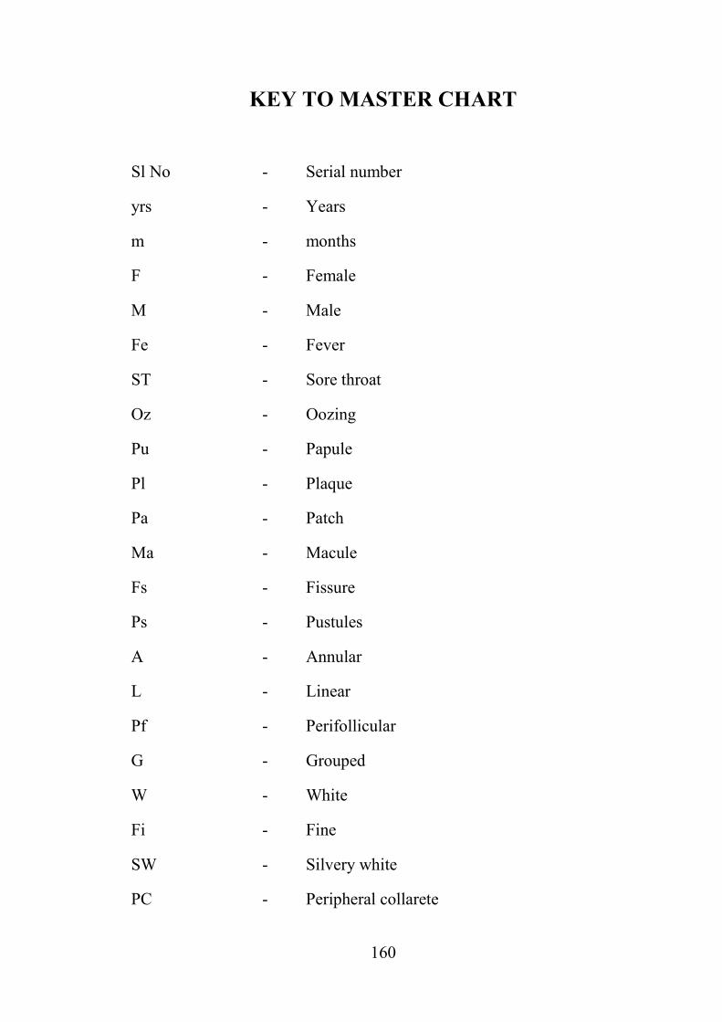
160
KEY TO MASTER CHART
Sl No - Serial number
yrs - Years
m - months
F - Female
M - Male
Fe - Fever
ST - Sore throat
Oz - Oozing
Pu - Papule
Pl - Plaque
Pa - Patch
Ma - Macule
Fs - Fissure
Ps - Pustules
A - Annular
L - Linear
Pf - Perifollicular
G - Grouped
W - White
Fi - Fine
SW - Silvery white
PC - Peripheral collarete

161
Y - yellow
Gr - Greasy
Th - Thickened
As - Auspitz sign
Kb - Koebnerisation
Hp - Herald patch
PV - Psoriasis vulgaris
GP - guttate psoriasis
FP - Flexural psoriasis
SP - Scalp psoriasis
PPS - palmoplantar psoriasis
FP - flexural psoriasis
PUPS - Pustular Psoriasis
PR - Pityriasisrosea
LP - Lichen planus
GLP - generalised LP
LLP - Linear lichen planus
SD - seborrhoeic dermatitis
LS - Lichen striatuus
PRP - Pityriasis rubrapilaris
LN - Lichen nitidus
PLEVA - pityriasis lichenoides et varioliformis acuta
PLC - pityriasis lichenoides chronic
PaPS - Parapsoriasis

SL
No. NAME Age/Sex
CLINICAL FEATURES
DIAGNOSIS
HISTORY MORPHOLOGY
AGE OF ONSET
Month of onset
PRURITIS
OTHER
SYMPTOMS
FAMILY HISTORY
LESION
PATERN
SITES
NECK
FACE
CHEST
ABDOMEN
BACK
UPPERLIM
B
LOWERLIM
B
FLEXURES
PALMS
SOLES
NAILS
SCALP
ORAL MUCOSA
GENITALS
SCALING
TYPE OF SCALES
SPECIAL
FEATURES
1 Rani 5yrs/F 4yrs february + - - Pu,Pl A - - + + - + + - - - - + - - + SW AS.Kb PV
2 Eeswari 6yrs/f 6yrs December - Fe - pa,pu - - + - - - + + - - - - - - - + PC HP PR
3 Amritha 3m/F 3m March - - - pa - - + - - - - - - - - - + - - + Y,Gr Kb SD
4 Parvathi 4yrs/F 4yrs April + - - pu,pl L - - - - - - + - - - + - - - + Fi,W Kb LLP
5 Maari 11yrs/F 11yrs March + - - pa A + - - - + + + - - - - - - - + PC - PR
6 Goutham 5yrs/M 4yrs December + - - pu - - - + + + - - - - - - - + - + Fi,W Kb LP
7 Raman 11yrs/M 12yrs January + - - pu L - + - - - - - - - - - - - - + Fi - LS
8 Pavithra 11yrs/F 12yrs January +
+ Pl A - - - - - + + - - - + + - - + SW AS,Kb PV
9 Sankar 7yrs/M 7yrs february + - - pu L - - - - + - - - - - - - - - + Fi - LS
10 Roja 8yrs/F 7yrs March + - - pu L - - - - - + + - - - - - - - + Fi Kb LLP
11 Bharath 8yrs/M 7yrs October + - - pu - - - - - - + + - - - - - - - +
+
Fi
Fi -
PRP
LN
12 Aaditya 5yrs/M 5yrs April - - - pa A + + + - + - - - - - - - - - + PC HP PR
13 Jaiharini 8yrs/F 8yrs February + - - pu,pl A - - + + + + - - - - - - - - + PC HP PR
14 Sabira 11yrs/F 11yrs December + Fe - Pu,Pl - - - + - - + - - - - - - - - + SW AS,Kb GP
15 Jennifer 7yrs/F 5yrs December + - - PU - - - - - - - - - - - - - - - + SW AS,Kb GP
16 Arun 6yr/M 6yrs December - - - pu G
- - - - + - - - - - - - - + Fi - LN
17 Chithra 7yrs/F 7yrs September + oz - pu L,G - - - - + - - - - - - - - - + Fi - LS with
Eczema
18 Thilakaraj 5yrs/M 4yrs May + - - pu,pl - - - - - - + + + - - - - + - + Fi Kb GLP
19 Kaasi 4yrs/M 4yrs May - - - pa - + + + - - - + + - - - - - - + PC - IPR
20 Vaani 11yrs/F 11yrs January + Fe - Pl A - - - - - - + - - + + - - - + SW AS PV
21 Paramjothi 3yrs/M 3yrs December - - - pu A - - + - + + - - - - - - - - + Fi - PRP with LS
22 Kabil 10yrs/M 8yrs January + - - pu,pl - - - - - - + + - - - - - - - + Fi Kb LP
23 Thanushri 6yrs/F 5yrs December + - - Pu,Pl A - - - - - + - - - + - + - - + SW AS, Kb PV
24 Nageswari 6m/f 6m March - - - Pl - - - - - - - - + - - - - - - - - - FP
25 Manimekala 3yrs/F 3yrs December + - - pu L - - - - - - + - - - - - - - + Fi - LS
26 Harish 9yrs/M 9yrs january - Fe,
ST - pu,pa - - - - - + - - - - - - - - - + PC Ez PR
27 Ilakiya 11yrs/F 10yrs September + - - Pu,Pl A - - + - + + + - - - + - - - + SW AS,Kb PS
28 Ganesh 11yrs/m 11yrs March - Fe - pa - - - + + + - - - - - - - - - - - - PLEVA

29 Kavitha 5yrs/F 5yrs
30 Malathi 5yrs/F 5yrs
31 Padma 7yrs/F 7yrs
32 kalayarasi 6yrs/F 6yrs
33 Ramkumar 4m/M 4m
34 Gunasekar 8yrs/M 7yrs
35 Suresh 11yrs/M 10yrs
36 Gaurav 4yrs/M 4yrs
37 Mathiyan 4yrs/M 4yrs
38 Paimala 8yrs/F 6yrs
39 Anandi 11yrs/F 10yrs
40 karthik 11yrs/M 11yrs
41 Poorni 4yrs/F 4yrs
42 Loganathan 9yrs/M 9yrs
43 Pandi 2yr/M -
44 Maalavika 6yrs/F 6yrs
45 Poomani 2yrs/F 2yrs
46 Madhan 8yrs/M 7yrs
47 Rajeswari 10yrs/F 10yrs
48 Ramalingam 8yrs/M 8yrs
49 Priya 11yrs/F 11yrrs
50 Vasudev 8yrs/M 7yrs
51 Thangavel 4yrs/M 4yrs
52 Vaishnav 8yrs/M 7yrs
53 Saavanth 9yrs/M 4yrs
54 Vasanthi 9yrs/F 9yrs
55 Aadiraj 3yrs/M 3yrs
56 Selvi 8yrs/F 8yrs
57 Arjun 11yrs/M 11yrs
58 Madhusri 11yrs/F 11yrs
59 Lekshmi 10yrs/f 9yrs
60 Thayyal 7yrs/F 7yrs
61 Ajith 4yrs/M 3yrs
62 Muthu 2yrs/M 2yrs
63 Anitha 11yrs/F 11yrs
64 Narmada 7yrs/F 6yrs
65 Lakshman 11yrs/M 11yrs
66 Janani 10yrs/F 9yrs
67 Rahul 12yrs/M 12yrs
5yrs April + - -
5yrs february - - -
7yrs february + - -
6yrs March - - -
4m April - - -
7yrs february + - -
10yrs April + - -
4yrs June - - -
4yrs october - - -
6yrs May + - -
10yrs February + - -
11yrs August - - -
4yrs March - - -
9yrs December - - -
- April + - -
6yrs June + - -
2yrs November - Fe -
7yrs February + Fe +
10yrs January + - -
8yrs April + - -
11yrrs March - Fe,ST -
7yrs December + - -
4yrs April - - -
7yrs February - - -
4yrs September - - -
9yrs february + - -
3yrs May + - -
8yrs May + - -
11yrs July - - -
11yrs May - Fe,
ST -
9yrs January - - -
7yrs December + - -
3yrs January + - -
2yrs March + - -
11yrs December + - -
6yrs October + - -
11yrs May - - -
9yrs December - - -
12yrs February - - -
Pl - - - - -
pu L - - - -
pa A - - + +
pu G,Pf - - - -
pa - - + + -
pa - - - + +
pu G,Pf - - + +
PL,Fs L - - - +
Pl,Fs - - - - -
Pu,Pl A - - - -
Pu,pl - - - - -
Pl,Fs - - - - -
Pu L - - - -
pu,pl - - - +
pu G,L - - - -
pu,pl - - - + +
pu,pa A - - - -
Pl,F - - - - -
Pa A - - + +
Pu - - - + +
pu,pa - + + + +
Pu - - - - -
pu - - - - -
Pu,Pl A - - -
pu - - - - -
pu,pl - - + - -
pu - - - + -
pu L - - - -
Pl - - - - -
pu,pa - + + - -
Pu,Pl - - - + +
pa A - - - +
pu,pl - - - - -
Pu L - - - -
Ma,Pa - - + + +
Pa - - - + +
Pl,pu G,Pf - - - -
Pl - - - - -
Pu,Pa - - - - -
- - - - - - -
- + - - - - -
+ + - - - - -
- + + - - - -
- - - + - - -
+ - - - - - -
+ + + - + + -
- - - - - + -
- - - - - + -
+ + - - - - +
- - + - - - -
- - - - + + -
- + - - - - -
+ + + - - - -
- - + - - - -
+ + + - - - +
+ + + - - - -
- - + - + - +
+ - - - - - -
- + - - - - -
+ - + - - - -
+ + + - - - -
- + + - - - -
+ + - - - - -
- + + - - - -
+ + - - - - -
- + + - - - -
- - + - - - -
- - - + - - -
- - - + - - -
+ + + - - - -
+ + - - - - -
- + - - -
- - + - - - -
+ + + - - - -
+ + + - - - -
+ + - - + + -
- - - - - + -
+ - - - - - -
+ - - + SW AS
- - - + Fi -
- - - + PC HP
+ - - + Fi -
+ - - + Y,Gr -
- - - + Fi,Br -
- - - + Fi -
- - - + Th Fi -
- - - + Th -
+ - - + SW AS.Kb
- - - + Fi Kb
- - - + Th -
- - - + Fi -
- - - + Fi -
- - - + Fi -
- - - + Fi Kb
- - - + PC -
- - - + Th SW AS
- - - + PC HP
- - - + SW AS,Kb
- - - + PC -
- - - + SW AS
- - + + Fi -
- - - + SW AS, Kb
- - - + FI Kb
- - - + PC HP
- + - + Fi Kb
- - - + Fi -
- - - + Fi -
- - - + PC -
- - - + SW -
- - - + PC HP
- - - + Fi -
- - - + Fi -
- - - + Fi -
- - - + Fi kb
- - - + Fi
- - - + Th -
- - - + PC -
SP
LS
PR
PRP
SD
PLC
PRP
PPS
PPS
AS.Kb PV
LP
PPS
LS
LP
LS
GLP
PR
PV
PR
AS,Kb GP
PR
GP
LN
AS, Kb PV
LN
PR
LP
LS
FP
IPR
PUPS
PR
LP
LS
PaPS
LN
PRP
PPS
PR
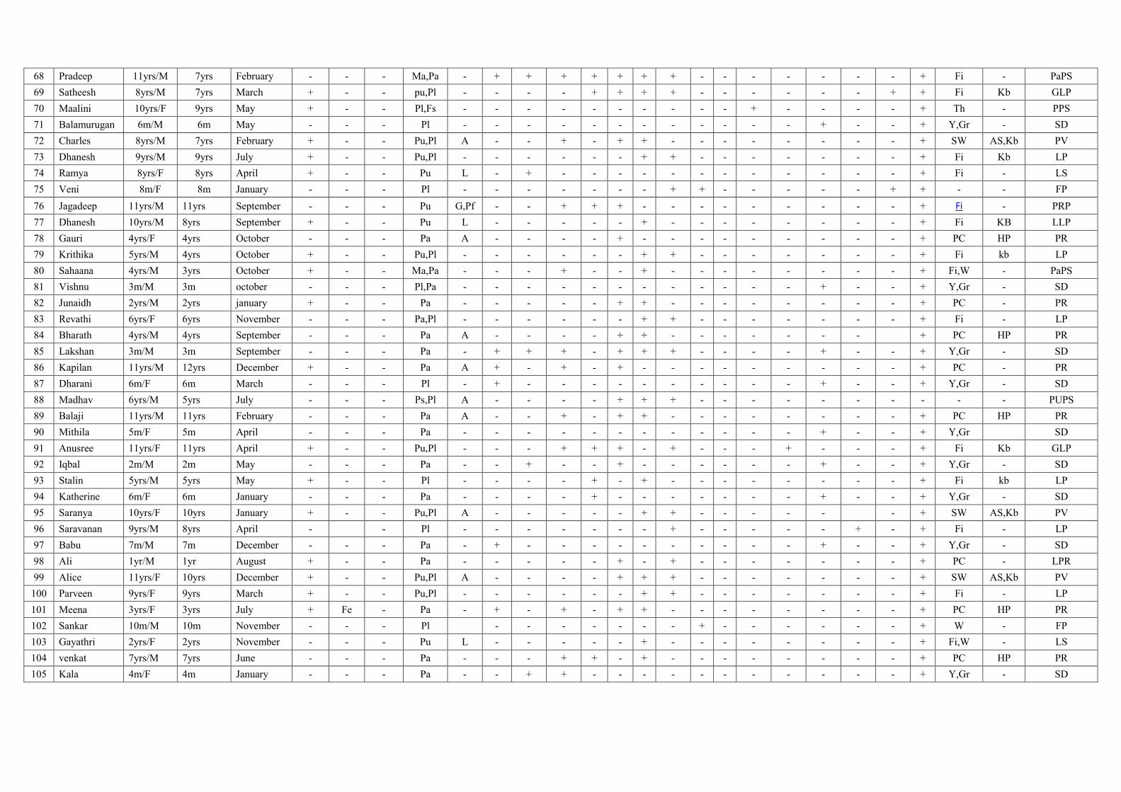
68 Pradeep 11yrs/M 7yrs February - - - Ma,Pa - + + + + + + + - - - - - - - + Fi - PaPS
69 Satheesh 8yrs/M 7yrs March + - - pu,Pl - - - - + + + + - - - - - - + + Fi Kb GLP
70 Maalini 10yrs/F 9yrs May + - - Pl,Fs - - - - - - - - - - + - - - - + Th - PPS
71 Balamurugan 6m/M 6m May - - - Pl - - - - - - - - - - - - + - - + Y,Gr - SD
72 Charles 8yrs/M 7yrs February + - - Pu,Pl A - - + - + + - - - - - - - - + SW AS,Kb PV
73 Dhanesh 9yrs/M 9yrs July + - - Pu,Pl - - - - - - + + - - - - - - - + Fi Kb LP
74 Ramya 8yrs/F 8yrs April + - - Pu L - + - - - - - - - - - - - - + Fi - LS
75 Veni 8m/F 8m January - - - Pl - - - - - - - + + - - - - - + + - - FP
76 Jagadeep 11yrs/M 11yrs September - - - Pu G,Pf - - + + + - - - - - - - - - + Fi - PRP
77 Dhanesh 10yrs/M 8yrs September + - - Pu L - - - - - + - - - - - - - - + Fi KB LLP
78 Gauri 4yrs/F 4yrs October - - - Pa A - - - - + - - - - - - - - - + PC HP PR
79 Krithika 5yrs/M 4yrs October + - - Pu,Pl - - - - - - + + - - - - - - - + Fi kb LP
80 Sahaana 4yrs/M 3yrs October + - - Ma,Pa - - - + - - + - - - - - - - - + Fi,W - PaPS
81 Vishnu 3m/M 3m october - - - Pl,Pa - - - - - - - - - - - - + - - + Y,Gr - SD
82 Junaidh 2yrs/M 2yrs january + - - Pa - - - - - + + - - - - - - - - + PC - PR
83 Revathi 6yrs/F 6yrs November - - - Pa,Pl - - - - - - + + - - - - - - - + Fi - LP
84 Bharath 4yrs/M 4yrs September - - - Pa A - - - - + + - - - - - - - + PC HP PR
85 Lakshan 3m/M 3m September - - - Pa - + + + - + + + - - - - + - - + Y,Gr - SD
86 Kapilan 11yrs/M 12yrs December + - - Pa A + - + - + - - - - - - - - - + PC - PR
87 Dharani 6m/F 6m March - - - Pl - + - - - - - - - - - - + - - + Y,Gr - SD
88 Madhav 6yrs/M 5yrs July - - - Ps,Pl A - - - - + + + - - - - - - - - - - PUPS
89 Balaji 11yrs/M 11yrs February - - - Pa A - - + - + + - - - - - - - - + PC HP PR
90 Mithila 5m/F 5m April - - - Pa - - - - - - - - - - - - + - - + Y,Gr SD
91 Anusree 11yrs/F 11yrs April + - - Pu,Pl - - - + + + - + - - - + - - - + Fi Kb GLP
92 Iqbal 2m/M 2m May - - - Pa - - + - - + - - - - - - + - - + Y,Gr - SD
93 Stalin 5yrs/M 5yrs May + - - Pl - - - - + - + - - - - - - - - + Fi kb LP
94 Katherine 6m/F 6m January - - - Pa - - - - + - - - - - - - + - - + Y,Gr - SD
95 Saranya 10yrs/F 10yrs January + - - Pu,Pl A - - - - - + + - - - - - - + SW AS,Kb PV
96 Saravanan 9yrs/M 8yrs April - - Pl - - - - - - - + - - - - - + - + Fi - LP
97 Babu 7m/M 7m December - - - Pa - + - - - - - - - - - - + - - + Y,Gr - SD
98 Ali 1yr/M 1yr August + - - Pa - - - - - + - + - - - - - - - + PC - LPR
99 Alice 11yrs/F 10yrs December + - - Pu,Pl A - - - - + + + - - - - - - - + SW AS,Kb PV
100 Parveen 9yrs/F 9yrs March + - - Pu,Pl - - - - - - + + - - - - - - - + Fi - LP
101 Meena 3yrs/F 3yrs July + Fe - Pa - + - + - + + - - - - - - - - + PC HP PR
102 Sankar 10m/M 10m November - - - Pl - - - - - - - + - - - - - - + W - FP
103 Gayathri 2yrs/F 2yrs November - - - Pu L - - - - - + - - - - - - - - + Fi,W - LS
104 venkat 7yrs/M 7yrs June - - - Pa - - - + + - + - - - - - - - - + PC HP PR
105 Kala 4m/F 4m January - - - Pa - - + + - - - - - - - - - - - + Y,Gr - SD