
A biopolymer-based carbon nanotube interface integrated with a redox shuttle and a D-sorbitol...
10
ORIGINAL PAPER A biopolymer-based carbon nanotube interface integrated with a redox shuttle and a D-sorbitol dehydrogenase for robust monitoring of D-sorbitol Jana Šefčovičová & Jaroslav Filip & Peter Tomčík & Peter Gemeiner & Marek Bučko & Peter Magdolen & Jan Tkac Received: 28 January 2011 /Accepted: 18 June 2011 /Published online: 7 July 2011 # Springer-Verlag 2011 Abstract We describe the preparation and characterization of a glassy carbon electrode modified with a bionanocom- posite consisting of a hyaluronic acid, dispersed carbon nanotubes, and electrostatically bound toluidine blue. The electrode was used to detect NADH in the batch and flow-injection mode of operation. The electrode was further modified by immobilizing sorbitol dehydrogenase to result in biosensor for D-sorbitol that displays good operational stability, a sensitivity of 10.6 μA mM −1 cm −2 , a response time of 16 s, and detection limit in the low micromolar range. The biosensor was successfully applied to off-line monitoring of D-sorbitol during its bioconversion into L-sorbose (a precursor in the synthesis of vitamin C) by Gluconobacter oxydans. The sample assay precision is 2.5% (an average RSD) and the throughput is 65 h −1 if operated in the flow-injection mode. The validation of this biosensor against a reference HPLC method resulted in a slope of correlation of 1.021±0.001 (R 2 =0.99997). Keywords Biosensor . Carbon nanotubes . Hyaluronic acid . NADH sensor . D-sorbitol dehydrogenase Introduction Carbon nanotubes (CNTs) since their discovery [1, 2] are applied in a wide range of analytical applications with nanoscale built biosensor devices as a good example [3, 4]. A dominant detection platform for biosensors using CNTs is an electrochemical-based one, because CNTs are offering high surface area, low overvoltage of detection and rapid electrode kinetics due to high length/diameter aspect ratio and presence of edge plane sites and defects [5, 6]. CNTs since their introduction as an electrode modifier [7–9] are frequently employed to solve a problem with electrode fouling by oxidation products of NADH [8, 10]. Detection of NADH is of a particular interest, since it is a cofactor of more than 500 enzymes, which are an integral part of biosynthetic reactions (bioconversions) and various devices like biofuel cells and biosensors (with implementation of various transducing mechanisms) [11–16]. Reproducible manipulation with CNTs requires their solubilisation, but covalent functionalisation negatively affecting properties of CNTs should be avoided [17]. Biopolymers, produced by living organisms, are renewable materials considered to be non-toxic and biodegradable. Electronic supplementary material The online version of this article (doi:10.1007/s00604-011-0641-0) contains supplementary material, which is available to authorized users. J. Šefčovičová : J. Filip : P. Gemeiner : M. Bučko : P. Magdolen : J. Tkac (*) Department of Glycobiotechnology, Center for Glycomics, Institute of Chemistry, Slovak Academy of Sciences, Dúbravská cesta 9, SK-845 38 Bratislava, Slovakia e-mail: [email protected] P. Tomčík Institute of Analytical Chemistry, Faculty of Chemical and Food Technology, Slovak University of Technology, Radlinského 9, SK-812 37 Bratislava, Slovakia P. Tomčík Department of Chemistry, Faculty of Education, The Catholic University in Ružomberok, Hrabovská cesta 1, SK-034 01 Ružomberok, Slovakia Microchim Acta (2011) 175:21–30 DOI 10.1007/s00604-011-0641-0
Transcript of A biopolymer-based carbon nanotube interface integrated with a redox shuttle and a D-sorbitol...

ORIGINAL PAPER
A biopolymer-based carbon nanotube interface integratedwith a redox shuttle and a D-sorbitol dehydrogenasefor robust monitoring of D-sorbitol
Jana Šefčovičová & Jaroslav Filip & Peter Tomčík &
Peter Gemeiner & Marek Bučko & Peter Magdolen &
Jan Tkac
Received: 28 January 2011 /Accepted: 18 June 2011 /Published online: 7 July 2011# Springer-Verlag 2011
Abstract We describe the preparation and characterizationof a glassy carbon electrode modified with a bionanocom-posite consisting of a hyaluronic acid, dispersed carbonnanotubes, and electrostatically bound toluidine blue. Theelectrode was used to detect NADH in the batch andflow-injection mode of operation. The electrode wasfurther modified by immobilizing sorbitol dehydrogenaseto result in biosensor for D-sorbitol that displays goodoperational stability, a sensitivity of 10.6 μA mM−1 cm−2,a response time of 16 s, and detection limit in the lowmicromolar range. The biosensor was successfully appliedto off-line monitoring of D-sorbitol during its bioconversioninto L-sorbose (a precursor in the synthesis of vitamin C) by
Gluconobacter oxydans. The sample assay precision is 2.5%(an average RSD) and the throughput is 65 h−1 if operated inthe flow-injection mode. The validation of this biosensoragainst a reference HPLC method resulted in a slope ofcorrelation of 1.021±0.001 (R2=0.99997).
Keywords Biosensor . Carbon nanotubes . Hyaluronicacid . NADH sensor . D-sorbitol dehydrogenase
Introduction
Carbon nanotubes (CNTs) since their discovery [1, 2] areapplied in a wide range of analytical applications withnanoscale built biosensor devices as a good example [3, 4].A dominant detection platform for biosensors using CNTsis an electrochemical-based one, because CNTs are offeringhigh surface area, low overvoltage of detection and rapidelectrode kinetics due to high length/diameter aspect ratioand presence of edge plane sites and defects [5, 6]. CNTssince their introduction as an electrode modifier [7–9] arefrequently employed to solve a problem with electrodefouling by oxidation products of NADH [8, 10]. Detectionof NADH is of a particular interest, since it is a cofactor ofmore than 500 enzymes, which are an integral part ofbiosynthetic reactions (bioconversions) and various deviceslike biofuel cells and biosensors (with implementation ofvarious transducing mechanisms) [11–16].
Reproducible manipulation with CNTs requires theirsolubilisation, but covalent functionalisation negativelyaffecting properties of CNTs should be avoided [17].Biopolymers, produced by living organisms, are renewablematerials considered to be non-toxic and biodegradable.
Electronic supplementary material The online version of this article(doi:10.1007/s00604-011-0641-0) contains supplementary material,which is available to authorized users.
J. Šefčovičová : J. Filip : P. Gemeiner :M. Bučko : P. Magdolen :J. Tkac (*)Department of Glycobiotechnology, Center for Glycomics,Institute of Chemistry, Slovak Academy of Sciences,Dúbravská cesta 9,SK-845 38 Bratislava, Slovakiae-mail: [email protected]
P. TomčíkInstitute of Analytical Chemistry, Faculty of Chemical and FoodTechnology, Slovak University of Technology,Radlinského 9,SK-812 37 Bratislava, Slovakia
P. TomčíkDepartment of Chemistry, Faculty of Education, The CatholicUniversity in Ružomberok,Hrabovská cesta 1,SK-034 01 Ružomberok, Slovakia
Microchim Acta (2011) 175:21–30DOI 10.1007/s00604-011-0641-0

Thus, such material combines excellent functional propertieswith environmental friendliness and sustainable developmentfeatures. It is not surprising polysaccharide-based bio-polymers have attracted considerable attention of thenanotechnology scientists as an effective colloidal dispersantof the nanomaterials providing an increased biocompatibilityand utilisable functionalities [18], which are featuresimportant in many applications [19–22].
Chitosan is widely utilised biopolymer for preparation ofCNTs dispersions due to several advantages compared toother dispersing agents [23] with successful application forconstruction of robust biosensors [24–26]. However, recentstudies suggested hyaluronic acid (HA) based dispersion ofCNTs offers excellent conductivity and stability [27]. HAoccurring in a variety of sizes having an array of differentregulatory/structural functions [28] has been used as adispersing agent for preparation of CNT-based biofibers(employed in tissue engineering and for growth offibroblast cells) showing its biocompatibility [27, 29].
A novel application of HA-based CNT dispersions wasshown recently by our group e.g. preparation of electrochem-ical NADH sensor and biosensor [30]. Such devices werebased on an integration of CNTs within a transducer bycasting of a dispersion of CNTs prepared in a green way withHA as an effective dispersant on the electrode surface. Theinterface modified by HA dispersed CNTs showed 3x highercatalytic current and higher operational stability towardsNADH compared to the sensor device built on a morefrequently applied dispersion of CNTs in a chitosan matrix.Moreover, the study also showed more favourable redoxbehaviour of two standard redox probes and a betterselectivity in the presence of two interference compoundson the interface modified by CNTs prepared with the aid ofHA compared to chitosan-based CNT dispersion [30].
The main objective of this paper is to further enhance anoverall performance of NADH sensing and dehydrogenase-based biosensing by introduction of a redox shuttle—toluidine blue, effectively docked by strong electrostaticforces to the CNT-based HA nanocomposite. Robustnessand overall performance of the current biosensingapproach was validated using samples containing D-sorbitol, which is a substrate in a bioconversion processusing Gluconobacter oxydans with a precious L-sorboseas the product (a precursor for production of vitamin C—ascorbic acid) [31, 32].
Materials and methods
Enzymes and other consumables
Sorbitol dehydrogenase (SDH, sheep liver, 50 U mg−1);single walled CNTs (OD=1.1 nm, L=0.5–100 μm, >90%
purity); Nafion® (20% solution); chitosan (degree ofdeacetylation of 85%), hyaluronic acid (HA, Streptococcusequi), toluidine blue, glutaraldehyde and acetic acid werepurchased from Sigma (St. Louis, USA, http://www.sigmaaldrich.com). NAD+ and NADH were supplied fromSorachim (Paris, France, http://www.sorachim.com). D-sorbitol was obtained from Fluka (Steinheim, Germany,http://www.sigmaaldrich.com). Agar and yeast extract (G.oxydans cultivations) were from Oxoid (Basingstoke, UK,http://www.oxoid.com). KH2PO4 and K2HPO4 (Mikrochem,Pezinok, Slovakia, http://www.mikrochem.com) werecomponents for preparation of 50 mM phosphate bufferpH 8.0. Distilled water was used for preparation of allstock solutions.
Electrode modification
Dispersions (1 mg mL−1) of CNTs were prepared in 0.1%solution of HA in distilled water using an ultrasonication(Bandelin DT 102 H, Bandelin electronics, Berlin,Germany, http://www.bandelin.com) at 25 °C (20 min).Glassy carbon electrodes (GCE, d=3 mm, BAS, WestLafayette, USA, http://www.basinc.com) were polishedusing 0.3 μm alumina/diamond slurry (Struers A/S,Ballerup, Denmark, http://www.struers.com) and sonicatedin distilled water for 30 s. GCE was covered by 5 μL ofHA/CNT dispersion and left to dry at 25 °C (30 min).Next, 5 μL of 0.5 mM toluidine blue (in distilled water)was applied for its adsorption (electrode was covered byan eppendorf tube in an inverted position and sealed byParafilm™ to prevent drying of toluidine blue solution).After 20 min toluidine blue solution was withdrawn andthe surface was gently washed by distilled water. Theelectrode was covered by an outer layer cast either from0.5% Nafion® in 50 mM phosphate buffer pH 8.0 (2×10 μL, a second layer was applied after the first one wasdried) or chitosan (10 μL of 0.1% chitosan solution in 1%acetic acid) and left to dry (30 min). The HA/CNT layerwith toluidine blue adsorbed without any additional outermembrane was tested in the CV experiment as a control.
A GCE (d=1 mm, Cypress Systems, Division of ESA,Inc., Lawrence, USA, http://www.esainc.com) for FIA wasmodified by pippetting of 2 μL of the HA/CNT dispersion,subsequently covered by 2 μL of 0.5 mM toluidine blue for20 min and washed, as described above. Finally theelectrode was covered either by 0.5% Nafion® in 50 mMphosphate buffer pH 8.0 (2×4 μL) or by 4 μL of 0.1%chitosan in 1% acetic acid and left to dry (30 min) at 25 °C.
For biosensor preparation the bionanocomposite consist-ing of HA/CNT with toluidine blue adsorbed was furthermodified by 3 μL of 8% SDH, left to dry and covered by anouter membrane deposited either from 0.5% Nafion® (2×10 μL) or from 0.1% chitosan in 0.3% acetic acid (10 μL),
22 J. Šefčovičová et al.

if not mentioned otherwise. Some other modifications weretested, as well (e.g. introduction of 5 μL of 2.5%glutaraldehyde for covalent SDH immobilisation).
Instrumentation and assay procedures
For electrochemical measurements potentiostats AutolabPGSTAT 128 N and 302 N (EcoChemie, Utrecht, Nether-land, http://www.ecochemie.nl) were used with GCE as aworking electrode, with an Ag/AgCl reference electrodeapplied for chronoamperometric experiments (batch andFIA mode), while a Hg/Hg2SO4 reference one wasemployed for CV experiments. A platinum electrode wasapplied as a counter one for CV measurements. A potentialof 0 mV vs. Ag/AgCl electrode was used in chronoampero-metric experiments (batch and FIA mode of operation).
Fourier Transform Infrared Spectroscopy (FTIR) spectrawere measured on spectrometer Nicolet 6700 (ThermoFisher Scientific, USA, http://www.thermofisher.com) withDTGS detector and Omnic 8.0 software. Spectra werecollected from 4,000 to 400 cm−1 at a resolution of 4 cm−1
with 128 scans averaged with sample in a solid state.The FIA system (FIAlab Instruments Inc., Bellevue,
USA, http://www.flowinjection.com) consisting of peristal-tic pumps (Watson-Marlow, Houston, USA, www.watson-marlow.com), sampling/injection valves (Vici, Houston,USA, http://www.vici.com) and a flow cell (7 μL, CypressSystem, Lawrence, USA) was running with an injectionloop of 20 μL and a flow rate of 0.5 ml min−1 in 50 mMphosphate buffer pH 8.0, if not mentioned otherwise. Thebatch mode of operation was performed in a glass vessel in10 mL of 50 mM phosphate buffer pH 8.0 using a stirrer(250 rpm). An utility designed in Labview (NationalInstruments, Austin, USA) enabling computerised controlof all mechanical components of FIA system was designedallowing to control the time between injections and thechoice of the loop, which content will be injected. Theutility was applied also for data acquisition and evaluationpurposes (e.g. automatic reading of peak current) whichoffer options for design of a fully-automated feedback-loopcontrol systems.
D-sorbitol was determined by High Performance LiquidChromatography (HPLC) using a Preparative Chroma-tography System WATERS Delta Prep 3000 (Milford,USA, http://www.waters.com) using a Polymer IEX Pb2+
column (300×8 mm, Watrex, Prague, Czech Republic,http://www.watrex.cz). Elution with redistilled water wasperformed at a flow rate of 1 mL min−1 at 80 °C andmonitored by a RI detector (Philips PU 4026, Eindhoven,Netherlands). Samples/standards were injected onto thecolumn via 100 μL loop with retention time of 22.4 min.
The strain of G. oxydans BIODIOL 621 (CzechCollection of Microorganisms, Brno, Czech Republic,
http://www.sci.muni.cz/ccm) was maintained on an agarmedium containing (values in g L−1): D-sorbitol 100; yeastextract 10; calcium carbonate 20; agar 20; and transferredmonthly. The cell biomass was prepared by an aerobiccultivation at 30 °C on a rotary shaker (Shell Lab,Cornelius, USA) at 250 rpm in 250 mL Erlenmeyer flasksfilled with 50 mL of medium. The growth mediumcontained (in g L−1): D-sorbitol 50; yeast extract 10;succinic acid 3; the pH of the medium was adjusted to5.5–5.8 with HCl. Cells of G. oxydans were grown in thegrowth medium for 24 h until A600=1.2 (wet biomass of8 gL−1 after centrifugation at 10,000 rpm for 3 min) and thecells were used for inoculation of the bioconversionmedium.
Bioconversion of D-sorbitol into L-sorbose was done inthe medium containing (g L−1): D-sorbitol 70; sodiumpyruvate 0.55; K2HPO4 0.13; NaH2PO4 0.35 with pHadjusted to 5.5–5.8 by HCl. 250 mL flasks containing50 mL of the sterile medium were inoculated with 5% ofthe seed culture broth. The conversion was performed at30 °C at 200 rpm with samples aseptically withdrawnduring the biotransformation (48 h).
Results and discussion
NADH sensor
HA forms an aggregate with toluidine blue due to strongelectrostatic interactions between oppositely chargedchemical species (Fig. S1 Electronic SupplementaryMaterial, ESM). This fact was confirmed by a FTIRinvestigation (Fig. 1) revealing quite large shifts of themain peaks of HA [33] as follows (HA→HA/toluidineblue): 1 ν(O-H) 3346→3262; 2 νas(CO from COO-)1612→1603; 3 νs(CO from COO-) 1410→1402; 4 ν(C-OH from a saccharidic structure) 1046→1032. Theselarge shifts suggest all major bonds of HA exist in adifferent environment after being integrated with tolui-dine blue. A similar shift of the frequency towards lowerfrequencies was noticed in the case of HA being blendedwith CNTs [30]. Interestingly, when CNTs were blendedinto a HA/toluidine blue hybrid structure a small shifttowards higher frequencies was noticed for peaks atposition 1,2 and 3 as follows: 1 ν(O-H) 3262→3271; 2νas(CO from COO-) 1603→1606, 3 νs(CO from COO-)1402→1406. This suggests a competition between CNTsand HA to bind toluidine blue, since CNTs are well knowto bind toluidine blue strongly [34]. The electrostaticinteraction between HA and toluidine blue resembleselectrostatic docking of positively charged redox shuttleson a negatively charged Nafion® layer [35]. The influenceof toluidine blue concentration (0.2–50 mM) on the
A biopolymer-based carbon nanotube interface for biosensing 23

NADH sensor sensitivity revealed an optimal value of0.5 mM (data not shown) applied for electrode modification.
The sensor composed of the HA/CNT layer withtoluidine blue adsorbed was tested in the ability to detectNADH. Our previous study confirmed HA is not redoxactive compound towards NADH oxidation and its majorrole within nanocomposite is to disperse CNTs [30]. HA asa negatively charged polymer introduces this charge to thebackbone of CNTs. As a result modified CNTs are repellingeach other due to electrostatic repulsion preventing re-aggregation of CNTs after a dispersion process took place.Additional beneficial role of using HA for preparing ofCNT dispersions was efficiency in discrimination betweenthe signal coming from the analyte (NADH) compared tothe signal from two interfering compounds (dopamine andascorbate) [30]. Another, not yet explained beneficial roleof HA, when blended with CNTs was a more reversibleelectrochemistry of two redox probes ([Fe(CN)6]
3- and [Ru(NH3)6]
3+) compared to their electrochemistry on a chitosan/CNT nanocomposite and better operational stability ofNADH sensing [30].
Detection of NADH on HA/CNT modified by tolui-dine blue is compared to detection of NADH on a bareGCE and GCE covered by HA/CNT. A peak potential forNADH oxidation is negatively shifted from +229 mV(bare GCE) to −26 mV (GCE with an applied HA/CNTlayer) (Fig. 2). Although a higher current responsetowards NADH can be seen on the unmodified GCEcompared to GCE integrated by HA/CNT nanocompo-site, HA/CNT nanocomposite proved to be resistanttowards passivation by NADH oxidising products, whatcompensates for lower current output [30]. When, GCEwas covered by HA/CNT with toluidine blue, a furthernegative shift to −470 mV for oxidation of NADH wasnoticed (Fig. 2).
Redox behaviour of NADH on GCE modified byHA/CNT with toluidine blue revealed two pairs of redoxwaves. Larger cathodic peak at potential of −702 mV(e.g. −257 mV vs. Ag/AgCl electrode) is consistent withpeak of a monomer of toluidine blue adsorbed on CNTswithout any other dispersing matrix present (e.g. Epc of−246 mV vs. Ag/AgCl) [34]. Lower cathodic peak atpotential of −470 mV (e.g. +25 mV vs. Ag/AgClelectrode) is again consistent with a second redox peakobserved at +21 mV vs. Ag/AgCl attributed to thepolymeric form of toluidine blue [34]. Presence of thepolymeric form of toluidine blue on the electrode surfacewas attributed to electrooxidation of toluidine bluemediated by NADH or its oxidation products [34].
-1000 -750 -500 -250 0 250 500
-30
-20
-10
0
10
20
E/mV
I/µA
Bare GCE
GCE/HA/CNT
GCE/HA/CNT/TB
Fig. 2 Electrochemistry of 1 mM NADH in 50 mM phosphate bufferpH 8.0 on a bare GCE, and GCE modified by the hyaluronic acid/CNTs (GCE/HA/CNT) or hyaluronic acid/CNTs with toluidine blue(GCE/HA/CNT/TB) (thick solid lines). A relevant part of a back-ground CV in buffer is shown for a better clarity (thin solid lines)
4000 3000 2000 1750 1500 1250 1000
0,00
0,25
0,50
0,75
1,00
1,25
1,50
1,75
3
2
Abs
orba
nce
Wavenumbers/cm-1
HA
HA/TB
HA/TB/CNTs14
Fig. 1 FTIR spectra ofhyaluronic acid (HA) alone,hyaluronic acid/toluidineblue (HA/TB) adduct andbionanocomposite hyaluronicacid/CNTs with toluidine blue(HA/TB/CNTs). Peaks atvarious positions are assigned tothe following bonds: position 1ν(O-H); position 2 νas(CO fromCOO-); position 3 νs(CO fromCOO-) and position 4 ν(C-OHfrom a saccharidic structure)
24 J. Šefčovičová et al.

Moreover a higher current output towards 1 mM NADHwas obtained on the electrode modified by HA/CNTs withtoluidine blue, when compared to the HA/CNT modifica-tion (Fig. 2).
The effect of the outer membrane on the performance ofNADH sensing is shown in Table 1 and Fig. S2 (ESM). Bothtypes of the outer layers represent a diffusion barrier forNADH as judged from a comparison of the sensor sensitivitywith the Nafion® outer layer (28.7 μA mM−1 cm−2) or thechitosan outer layer (14.8 μA mM−1 cm−2) compared to thesensor without any outer layer (35.0 μA mM−1 cm−2, a valueobtained from CV). Variation of batch to batch NADHsensor preparation with the Nafion® outer layerexpressed as an average RSD was 13.2% and in thecase of the chitosan outer layer a larger variation wasseen with an average RSD of 18.1%.
The preferred outer layer for NADH sensor is chitosanbecause of low detection limit offered and its biocompat-ibility. Thus, this configuration of the NADH sensor wastested in the FIA system, which revealed a sensitivity of(18.5±0.4) μA mM−1 cm−2, response time of 44 s,detection limit of 14 μM and a linear range from0.03 mM to 3 mM. The NADH sensor in a FIA modebased on the HA/CNT interface without any redox shuttledocked offered a sensitivity of (7.1±0.1) μA mM−1 cm−2,response time of 59 s and detection limit of 130 μM [30].Thus, an incorporation of a redox shuttle within the HA/CNT nanocomposite improved overall performance ofNADH detection even at much lower applied potential.The sensitivity and the response time of NADH sensor inFIA system presented here is comparable to other publishedprotocols (not only based on CNT modified interface) withsensitivity values in the range from 4 to 32 μA mM−1 cm−2
and response time values from 11 to 144 s [36–41].Reproducibility of the assay was very good with an
average RSD of 4.8% (n=4). Stability of the NADHsensor was tested in a FIA mode by repetitive injections of2 mM NADH with a shift of the initial sensitivity of19.1 μA mM−1 cm−2 to 125% of the original value in 17 hof a continuous use (data not shown). This NADH sensoris much more stable compared to the NADH sensor basedon the HA/CNT layer without toluidine blue present (76%of the initial sensitivity in 22 h) under similar experimentalconditions. Moreover, the stability of this NADH sensor is
much higher compared to other NADH sensors based onCNTs with 4–30% loss of NADH sensing ability in just1 h [7–46].
D-sorbitol biosensor
The influence of pH, Eappl and temperature on the biosensorperformance was optimised (data not shown) and forfurther use optimal values were chosen e.g. pH 8.0 andEappl of 0 mV vs. Ag/AgCl. Temperature of 25 °C waschosen for further utilisation in the batch mode in order tocompare the biosensor performance in the FIA mode ofoperation (without a possibility to control temperatureinside the flow cell).
When the chitosan outer layer was deposited from 0.1%chitosan in 1% acetic acid (an optimal composition for theNADH sensor), a negligible biosensor current response wasobserved. Thus, an optimisation of the composition of thechitosan solution was carried out. The optimal outer layercast from a 0.1% chitosan solution in 0.3% acetic acid,produced the biosensor with the current response higherthan obtained on the one using Nafion® as the outer layer(Fig. S3, ESM), suggesting this layer preserves the activityof the enzyme. Parameters such as an enzyme concentration(Fig. S4, ESM) and an overall biosensor configuration(Figs. 3 and 4) were optimised, as well. For further use 8%SDH solution for immobilisation, the biosensor configura-tion with SDH in a direct contact with the toluidine bluelayer were chosen.
In the batch mode of operation the biosensor deviceexhibited an initial sensitivity of 10.6 μA mM−1 cm−2
(Fig. 5, Table 2), a value only slightly lower thanpreviously published using the biosensor based on twoenzymes (SDH and diaphorase) and a soluble mediator(ferricyanide) [47]. The biosensor sensitivity is muchhigher compared to other SDH biosensors published inthe literature (lower part in Table 3) [30–50] and wellwithin range for the dehydrogenase-based biosensors(Table 3). The initial sensitivity of the biosensor towardsD-sorbitol was comparable to the sensitivity of the NADHsensor of 14.8 μA mM−1 cm−2, indicating an efficientsignal transduction with the enzyme containing bionano-composite. Response time of the biosensor (90% of thesteady-state current) was 16 s, much shorter than in our
Table 1 Comparison of two different outer layers (Nafion® or chitosan) on the performance of the NADH sensor based on HA/CNT withtoluidine blue adsorbed within nanocomposite working in the batch mode of operation
Outer layer Sensitivity [μA mM-1 cm−2] Detection limit [μM]a Response time [s]b Reproducibility of sensor prepn.
Nafion® 28.7±1.4 119 18 13.2%
Chitosan 14.8±1.2 15 25 18.1%
a calculated from S/N=3; b time needed to attain 90% of steady-state current value
A biopolymer-based carbon nanotube interface for biosensing 25

previous study (53 s) without toluidine blue present [30], wellwithin the range previously published for dehydrogenase-based biosensors (Table 3). Generally it can be said shorterresponse time of the dehydrogenase-based biosensors (3–8 s)was observed only in cases the outer protective layer wasabsent. In our case it is very important to have the outerlayer, which protects the SDH enzyme to be degraded by theproteases released from lysed bacterial cells present in thecultivation media used for detection of D-sorbitol concen-tration. Moreover, the outer chitosan layer provides amechanical support in the flow system with a relativelyhigh lateral flow velocity (calculated as 4.2 cm s−1) insidethe flow cell. The detection limit obtained here (4 μM) wasin the lower range values for dehydrogenase-based systems(0.8–100 μM) (Table 3). Reproducibility of sorbitol stand-ards assays expressed as RSD was within range 1.2–4.1%with an average error (RSD) of measurement of 3.1%.
Interestingly, when glutaraldehyde was applied forcovalent coupling of the enzyme to the modified GCE notonly lower sensitivity of the biosensor, but a higher Km
app
value was observed, as well, when compared to theimmobilisation protocol without glutaraldehyde treatment(Table 2). Km
app value read for configuration B and C(Table 2) is comparable to Km value of 1.5 mM publishedfor the enzyme in the solution [51]. If the enzyme wassandwiched between two biopolymers (chitosan and HA) alower Km
app value was observed (configuration A and D,Table 2). These two values are lower compared to theparameter obtained in the solution phase for SDH showinghigher affinity of the enzyme for its substrate, whensandwiched between these two biopolymers. This suggestsa beneficial role of the presence of two biopolymers, whichare considered as biocompatible ones, on the catalyticproperties of the enzyme.
In order to exploit the full potential of the FIA systemfor analysis of real samples, optimisation of the flow rate onthe current output and on the assay throughput was carriedout with an optimal flow rate being 0.5 mL min−1 (Figs. 6and S5, ESM). Characterisation of the biosensor performancein the FIA mode of operation revealed an initial sensitivity of(9.4±0.1) μA mM−1 cm−2 (Fig. 6), a value similar topreviously published value of (9.2±0.1) μA mM−1 cm−2 forthe biosensor configuration with 2 enzymes and a soluble
Fig. 3 A schematic representa-tion of all biosensor configura-tions tested
0
20
40
60
80
I.
I/nA
csorbitol
[mM]
HA/CNT/TB-CHI/GA-SDH - config. B
HA/CNT/TB-CHI/GA-SDH-CHI - config. C
0.0 0.5 1.0 1.5
0.0 0.5 1.0 1.5 2.00
100
200
300
400
500 HA/CNT/TB-CHI-SDH-CHI - config. D
HA/CNT/TB-SDH-CHI - config. A
csorbitol
[mM]
I/nA
II.
Fig. 4 The effect of the biosensor configuration on the device outputsignal in the batch mode of operation. HA—hyaluronic acid, TB—toluidine blue, CHI—chitosan, SDH—D-sorbitol dehydrogenase,GA—glutaraldehyde
0.0 0.5 1.0 1.5 2.0 2.50
150
300
450
600 Batch mode
I ss/n
A
c/mM
I p/nA
0
10
20
30
40
50
60
FIA mode
Fig. 5 A calibration curve for the biosensor device operated in thebatch (with a steady state current Iss plotted) or FIA mode (with acurrent of a peak in FIA Ip plotted). In the FIA mode of operation aquasi linear part of the calibration curve is shown with a dashed redline. A flow rate of 0.5 ml min−1 was used for FIA measurements,while a stirring rate of 250 rpm was used for batch mode of operation.For all other conditions see Fig. S2
26 J. Šefčovičová et al.
mediator ferricyanide [47]. The response of the biosensor inthe FIA mode was much shorter (55 s with a sample
throughput of 65 h−1) compared to our previous study (340 swith a sample throughput of 10 h−1) [47].
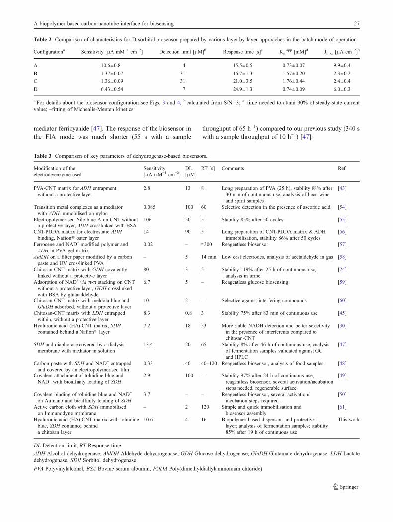
Table 2 Comparison of characteristics for D-sorbitol biosensor prepared by various layer-by-layer approaches in the batch mode of operation
Configurationa Sensitivity [μA mM−1 cm−2] Detection limit [μM]b Response time [s]c Kmapp [mM]d Jmax [μA cm−2]d
A 10.6±0.8 4 15.5±0.5 0.73±0.07 9.9±0.4
B 1.37±0.07 31 16.7±1.3 1.57±0.20 2.3±0.2
C 1.36±0.09 31 21.0±3.5 1.76±0.44 2.4±0.4
D 6.43±0.54 7 24.9±1.3 0.74±0.09 6.0±0.3
a For details about the biosensor configuration see Figs. 3 and 4, b calculated from S/N=3; c time needed to attain 90% of steady-state currentvalue; –fitting of Michealis-Menten kinetics
Table 3 Comparison of key parameters of dehydrogenase-based biosensors.
Modification of theelectrode/enzyme used
Sensitivity[μA mM−1 cm−2]
DL[μM]
RT [s] Comments Ref
PVA-CNT matrix for ADH entrapmentwithout a protective layer
2.8 13 8 Long preparation of PVA (25 h), stability 88% after30 min of continuous use; analysis of beer, wineand spirit samples
[43]
Transition metal complexes as a mediatorwith ADH immobilised on nylon
0.085 100 60 Selective detection in the presence of ascorbic acid [54]
Electropolymerised Nile blue A on CNT withouta protective layer, ADH crosslinked with BSA
106 50 5 Stability 85% after 50 cycles [55]
CNT-PDDA matrix for electrostatic ADHbinding, Nafion® outer layer
14 90 5 Long preparation of CNT-PDDA matrix & ADHimmobilisation, stability 86% after 50 cycles
[56]
Ferrocene and NAD+ modified polymer andADH in PVA gel matrix
0.02 – ≈300 Reagentless biosensor [57]
AldDH on a filter paper modified by a carbonpaste and UV crosslinked PVA
– 5 14 min Low cost electrodes, analysis of acetaldehyde in gas [58]
Chitosan-CNT matrix with GDH covalentlylinked without a protective layer
80 3 5 Stability 119% after 25 h of continuous use,analysis in urine
[24]
Adsorption of NAD+ via π-π stacking on CNTwithout a protective layer, GDH crosslinkedwith BSA by glutaraldehyde
6.7 5 – Reagentless glucose biosensing [59]
Chitosan-CNT matrix with meldola blue andGluDH adsorbed, without a protective layer
10 2 – Selective against interfering compounds [60]
Chitosan-CNT matrix with LDH entrappedwithin, without a protective layer
8.3 0.8 3 Stability 75% after 83 min of continuous use [45]
Hyaluronic acid (HA)-CNT matrix, SDHcontained behind a Nafion® layer
7.2 18 53 More stable NADH detection and better selectivityin the presence of interferents compared tochitosan-CNT
[30]
SDH and diaphorase covered by a dialysismembrane with mediator in solution
13.4 20 65 Stability 8% after 46 h of continuous use, analysisof fermentation samples validated against GCand HPLC
[47]
Carbon paste with SDH and NAD+ entrappedand covered by an electropolymerised film
0.33 40 40–120 Reagentless biosensor, analysis of food samples [48]
Covalent attachment of toluidine blue andNAD+ with bioaffinity loading of SDH
2.9 100 – Stability 97% after 24 h of continuous use,reagentless biosensor, several activation/incubationsteps needed, regenerable surface
[49]
Covalent binding of toluidine blue and NAD+
on Au nano and bioaffinity loading of SDH3.7 – – Reagentless biosensor, several activation/
incubation steps required[50]
Active carbon cloth with SDH immobilisedon Immunodyne membrane
– 2 120 Simple and quick immobilisation andbiosensor assembly
[61]
Hyaluronic acid (HA)-CNT matrix with toluidineblue, SDH contained behinda chitosan layer
10.6 4 16 Biopolymer-based dispersant and protectivelayer; analysis of fermentation samples; stability85% after 19 h of continuous use
This work
DL Detection limit, RT Response time
ADH Alcohol dehydrogenase, AldDH Aldehyde dehydrogenase, GDH Glucose dehydrogenase, GluDH Glutamate dehydrogenase, LDH Lactatedehydrogenase, SDH Sorbitol dehydrogenase
PVA Polyvinylalcohol, BSA Bovine serum albumin, PDDA Poly(dimethyldiallylammonium chloride)
A biopolymer-based carbon nanotube interface for biosensing 27

Reproducibility of the assays in the FIA system wasexcellent with an average RSD of 2.1% (variation 0.6%–4.0%). The stability of the biosensor was much improved e.g. decrease to 85% of the initial response in 19 h of acontinuous use (with a steady-state response at the end ofstability experiment) compared to a decrease to 47% of theinitial value during the same time (with an exponentialdecay of response in time), as previously reported [47]. Anenhanced sensitivity of D-sorbitol detection and highoperational stability of the biosensor device are most likelythe result of applied biocompatible matrix (e.g. HA andchitosan) for enzyme immobilisation and strong electrostat-ic docking of the redox shuttle within the nanocomposite.
Off-line monitoring of the bioconversion
Progress in the fermentation/biotransformation technologyhas to be accompanied with a tight control and regulation ofthe process in real time due to a complex and dynamicnature of the system. FIA system is frequently used for realtime monitoring because of a short response time, highflexibility and inexpensive components [52]. Biosensorsoffering selectivity of detection in a complex matrix withflexibility to be implemented into the FIA system representa promising sensing platform for on-line bioprocessmonitoring [53]. The optimised biosensor configurationwas finally applied for an off-line analysis of real samplestaken from a biotransformation process implementing theFIA mode of operation.
Samples were diluted in a way the concentration of theD-sorbitol falls into the concentration window of the quasi
linear range of the biosensor (0.5–2 mM) (Fig. 5) using FIAmode of operation and samples were measured withalternate injections of samples and standards, at least intriplicate. The concentration of D-sorbitol in the samplewas calculated by a linear interpolation of responses toD-sorbitol standards before and after sample injection.The D-sorbitol biosensor exhibited a very low averagerelative standard deviation (RSD) of 2.5% (RSD from0.3% to 4.0%). Moreover, the biosensor device providedreliable data as suggested from an excellent agreementwith HPLC (Fig. 7). Importantly the difference in D-sorbitol concentration read between these two analyticaltechniques expressed as residuals is scattered across thewhole concentration range of D-sorbitol supporting con-clusion there is no systematic error in reading D-sorbitolby the biosensor device when compared to a referenceanalytical method. A closer look at a difference of the D-sorbitol concentration provided by the biosensor comparedto the HPLC method for each particular sample expressedas RSD was in the range 0.1%–3.1% (an average value of1.1%), underlying an excellent agreement between thesetwo analytical methods. The time needed for analysis ofD-sorbitol by the biosensor was approx. 1 min, while
50 100 150 200 250 300 35050
100
150
200
250
300
350
c sorb
itol b
y H
PLC
[mM
]
csorbitol
by biosensor [mM]
50 100 150 200 250 300 350-4
-2
0
2
4
Res
idua
ls [m
M]
Fig. 7 Validation of the analysis of fermentation samples by thebiosensor with a reference analytical method (HPLC). Error bars forboth methods of analysis represent RSD for a particular samplemeasured in triplicate by both analytical methods. Residuals betweenthese two methods are shown, as well
0,0 0,5 1,0 1,5 2,010
15
20
25
30
35
40Response
Flow rate/mL min-1
I/nA
30
60
90
120Throughput
Thr
ough
put/h
-1
Fig. 6 The effect of a flow rate in a FIA mode of operation on thebiosensor response and assay throughput. The biosensor was preparedwith an enzyme layer cast from 8% SDH solution sandwichedbetween hyaluronic acid/CNT with toluidine blue (HA/CNT/TB layer)and chitosan outer layer (0.1% solution in 0.3% acetic acid) with anoverall configuration HA/CNT/TB-SDH/CHI. The experiment wasperformed in 50 mM phosphate buffer pH 8.0 at 25 °C with a sampleloop of 20 μl at an applied voltage of 0 mV vs. Ag/AgCl
28 J. Šefčovičová et al.

22 min was needed for HPLC measurement. Error ofHPLC measurements expressed as an average RSD (2.2%)was similar to the error of D-sorbitol detection by thebiosensor.
Conclusions
The NADH sensor and the D-sorbitol biosensor devicewere constructed using the bionanointerface based on thelayer consisting of HA/CNT with adsorbed toluidine blue.The basic parameters of both devices were obtained in thebatch and the FIA mode of operation. The biosensorconfiguration was carefully optimised in order to preparerobust biosensor system with a high performance ofdetection (sample throughput of 65 h−1) and operationalstability (85% of the original sensitivity after 19-h ofcontinuous use). Finally, the biosensor device was success-fully validated against a reference HPLC method usingfermentation samples.
Acknowledgements The financial supports from projects VEGA 1/0066/09 and 1/0335/10; COST CM8T D43 and SAV-FM-EHP-2008-04-04 are acknowledged. This contribution/publication is the result ofthe project implementation: Centre for materials, layers and systemsfor applications and chemical processes under extreme conditionssupported by the Research & Development Operational Programfunded by the ERDF.
References
1. Iijima S (1991) Helical microtubules of graphitic carbon. Nature354:56–58
2. Wiles PG, Abrahamson J (1978) Carbon fibre layers on arcelectrodes—I: their properties and cool-down behaviour. Carbon16:341–349
3. Merkoci A (2006) Carbon nanotubes in analytical sciences.Microchim Acta 152:157–174
4. Katz E, Willner I (2004) Biomolecule-functionalized carbonnanotubes: applications in nanobioelectronics. Chemphyschem5:1085–1104
5. Vashist SK, Zheng D, Al-Rubeaan K, Luong JHT, Sheu F-S (2011)Advances in carbon nanotube based electrochemical sensors forbioanalytical applications. Biotechnol Adv 29:169–188
6. Banks CE, Compton RG (2005) Exploring the electrocatalyticsites of carbon nanotubes for NADH detection: an edge planepyrolytic graphite electrode study. Analyst 130:1232–1239
7. Britto PJ, Santhanam KSV, Ajayan PM (1996) Carbon nanotubeelectrode for oxidation of dopamine. Bioelectrochem Bioenerg41:121–125
8. Musameh M, Wang J, Merkoci A, Lin YH (2002) Low-potentialstable NADH detection at carbon-nanotube-modified glassycarbon electrodes. Electrochem Commun 4:743–746
9. Wang J, Musameh M, Lin YH (2003) Solubilization of carbonnanotubes by Nafion toward the preparation of amperometricbiosensors. J Am Chem Soc 125:2408–2409
10. Kachoosangi RT, Musameh MM, Abu-Yousef I, Yousef JM, KananSM, Xiao L, Davies SG, Russell A, Compton RG (2009) Carbon
nanotube−ionic liquid composite sensors and biosensors. AnalChem 81:435–442
11. Kumar SA, Chen SM (2008) Electroanalysis of NADH usingconducting and redox active polymer/carbon nanotubes modifiedelectrodes-a review. Sensors 8:739–766
12. Gorton L, Domínguez E (2002) Electrocatalytic oxidation of NAD(P)H at mediator-modified electrodes. Rev Mol Biotechnol82:371–392
13. Radoi A, Compagnone S (2009) Recent advances in NADHelectrochemical sensing design. Bioelectrochemistry 76:126–134
14. Katakis I, Dominguez E (1997) Catalytic electrooxidation ofNADH for dehydrogenase amperometric biosensors. MicrochimActa 126:11–32
15. Arakawa T, Koshida T, Gessei T, Miyajima K, Takahashi D, KudoH, Yano K, Mitsubayashi K (2011) Biosensor for L-phenylalaninebased on the optical detection of NADH using a UV light emittingdiode. Microchim Acta 173:199–205
16. Korpan YI, Soldatkin OO, Sosovska OF, Klepach HM, Csoregi E,Vocanson F, Jaffrezic-Renault N, Gonchar MV (2010)Formaldehyde-sensitive conductometric sesnors based on com-mercial and recombinant formaldehyde dehydrogenase. Micro-chim Acta 170:337–344
17. Valcárcel M, Cárdenas S, Simonet BM (2007) Role of carbonnanotubes in analytical science. Anal Chem 79:4788–4797
18. Dias AMGC, Hussain A, Marcos AS, Roque ACA (2011) Abiotechnological perspective on the application of iron oxidemagnetic colloids modified with polysaccharides. Biotechnol Adv29:142–155
19. Krajewska B (2004) Application of chitin- and chitosan-basedmaterials for enzyme immobilizations: a review. Enzyme MicrobTechnol 35:126–139
20. Bhattacharyya S, Guillott S, Dabboue H, Tranchant JF, Salvetat JP(2008) Carbon nanotubes as structural nanofibers for hyaluronicacid hydrogel scaffolds. Biomacromolecules 9:505–509
21. Rinaudo M (2006) Chitin and chitosan: properties and applications.Prog Polym Sci 31:603–632
22. Koev ST, Dykstra PH, Luo X, Rubloff GW, Bentley WE, PayneGF, Ghodssi R (2010) Chitosan: an integrative biomaterial forlab-on-a-chip devices. Lab Chip 10:3026–3042
23. Tkac J, Ruzgas T (2006) Dispersion of single walled carbonnanotubes. Comparison of different dispersing strategies forpreparation of modified electrodes toward hydrogen peroxidedetection. Electrochem Commun 8:899–903
24. Zhang MG, Smith A, Gorski W (2004) Carbon nanotube−chitosansystem for electrochemical sensing based on dehydrogenaseenzymes. Anal Chem 76:5045–5050
25. Tkac J, Whittaker JW, Ruzgas T (2007) The use of single walledcarbon nanotubes dispersed in a chitosan matrix for preparation ofa galactose biosensor. Biosens Bioelectron 22:1820–1824
26. Rappathy D, Gopalan AY, Lee KP (2009) Synergistic contributionsof multiwall carbon nanotubes and gold nanoparticles in achitosan–ionic liquid matrix towards improved performance fora glucose sensor. Electrochem Commun 11:397–401
27. Razal JM, Gilmore KJ, Wallace GG (2008) Carbon nanotubebiofiber formation in a polymer-free coagulation bath. Adv FunctMater 18:61–66
28. Kogan G, Soltes L, Stern R, Gemeiner P (2007) Hyaluronic acid:a natural biopolymer with a broad range of biomedical andindustrial applications. Biotechnol Lett 29:17–25
29. Moulton SE, Maugey M, Poulin P, Wallace GG (2007) Liquidcrystal behavior of single-walled carbon nanotubes dispersed inbiological hyaluronic acid solutions. J Am Chem Soc 129:9452–9457
30. Filip J, Šefčovičová J, Tomčík P, Gemeiner P, Tkac J (2011) Ahyaluronic acid dispersed carbon nanotube electrode used for amediatorless NADH sensing and biosensing. Talanta 84:355–361
A biopolymer-based carbon nanotube interface for biosensing 29

31. Svitel J, Tkac J, Vostiar I, Navratil M, Stefuca V, Bucko M,Gemeiner P (2006) Gluconobacter in biosensors: applications ofwhole cells and enzymes isolated from Gluconobacter andAcetobacter to biosensor construction. Biotechnol Lett 28:2003–2010
32. De Muynck C, Pereira CSS, Naessens M, Parmentier S, SoetaertW, Vandamme EJ (2007) The genus Gluconobacter oxydans:comprehensive overview of biochemistry and biotechnologicalapplications. Crit Rev Biotechnol 27:147–171
33. Gilli R, Kacurakova M, Mathlouthi M, Navarini L, Paoletti S(1994) FTIR studies of sodium hyaluronate and its oligomers inthe amorphous solid phase and in aqueous solution. CarbohydrRes 263:315–326
34. Lawrence NS, Wang J (2006) Chemical adsorption of phenothi-azine dyes onto carbon nanotubes: toward the low potentialdetection of NADH. Electrochem Commun 8:71–76
35. Ranganathan S, Guo R, Murray RW (2007) Nanoparticle films aselectrodes:voltammetric sensitivity to the nanoparticle energy gap.Langmuir 23:7372–7377
36. Radoi A, Compagnone D, Batic M, Klincar J, Gorton L, PalleschiG (2007) NADH screen-printed electrodes modified with zirconi-um phosphate, Meldola blue, and Reinecke salt. Application to thedetection of glycerol by FIA. Anal Bioanal Chem 387:1049–1058
37. Fanjul-Bolado P, Queipo P, Lamas-Ardisana PJ, Costa-Garcia A(2007) Manufacture and evaluation of carbon nanotube modifiedscreen-printed electrodes as electrochemical tools. Talanta74:427–433
38. Munteanu FD, Mano N, Kuhn A, Gorton L (2004) NADHelectrooxidation using carbon paste electrodes modified withnitro-fluorenone derivatives immobilized on zirconium phosphate.J Electroanal Chem 564:167–178
39. Stergiou DV, Prodromidis MI, Veltsistas PG, Evmiridis NP (2004)Study of the electrochemical behavior of disperse blue 1-modifiedgraphite electrodes. Application to the flow determination ofNADH. Electroanalytical 16:949–954
40. Sha YF, Gao Q, Qi B, Yang XR (2004) Electropolymerization ofazure B on a screen-printed carbon electrode and its application tothe determination of NADH in a flow injection analysis system.Microchim Acta 148:335–341
41. Wang Y, You CP, Zhang S, Kong JL, Marty JL, Zhao DY, Liu BH(2009) Electrocatalytic oxidation of NADH at mesoporous carbonmodified electrodes. Microchim Acta 167:75–79
42. Lin WJ, Liao CS, Jhang JH, Tsai YC (2009) Graphene modifiedbasal and edge plane pyrolytic graphite electrodes for electro-catalytic oxidation of hydrogen peroxide and β-nicotinamideadenine dinucleotide. Electrochem Commun 11:2153–2156
43. Tsai YC, Huang JD, Chiu C-C (2007) Amperometric ethanolbiosensor based on poly(vinyl alcohol)–multiwalled carbonnanotube–alcohol dehydrogenase biocomposite. Biosens Bioelectron22:3051–3056
44. Wooten M, Gorski W (2010) Facilitation of NADH electro-oxidation at treated carbon nanotubes. Anal Chem 82:1299–1304
45. Tsai YC, Chen SY, Liaw HW (2007) Immobilization of lactatedehydrogenase within multiwalled carbon nanotube-chitosannanocomposite for application to lactate biosensors. Sens ActuatB: Chem 125:474–481
46. Zhai XR, Wei WZ, Zeng JX, Gong SG, Yin J (2006) Layer-by-layer film based on chitosan/carbon nanotubes, and its applicationto electrocatalytic oxidation of NADH. Microchim Acta 154:315–320
47. Sefcovicova J, Vikartovska A, Patoprsty V, Magdolen P, Katrlik J,Tkac J, Gemeiner P (2009) Off-line FIA monitoring of D-sorbitolconsumption during L-sorbose production using a sorbitolbiosensor. Anal Chim Acta 644:68–71
48. Saidman SB, Lobo-Castanon MJ, Miranda-Ordieres AJ, Tunon-Blanco P (2000) Amperometric detection of D-sorbitol withNAD+−D -sorbitol dehydrogenase modified carbon pasteelectrode. Anal Chim Acta 424:45–50
49. Hassler BL, Kohli N, Zeikus JG, Lee I, Worden RM (2007)Renewable dehydrogenase-based interfaces for bioelectronicapplications. Langmuir 23:7127–7133
50. Hassler BL, Amundsen TJ, Zeikus JG, Lee I, Worden RM (2008)Versatile bioelectronic interfaces on flexible non-conductivesubstrates. Biosens Bioelectron 23:1481–1487
51. El-Kabbani O, Darmanin C, Chung RP (2004) Sorbitol dehydro-genase: structure, function and ligand design. Curr Med Chem11:465–476
52. Ruzicka J, Hansen EH (2008) Retro-review of flow-injectionanalysis. Trends Anal Chem 27:390–393
53. Bilitewski U (2005) Biosensors for bioprocess monitoring. ComprAnal Chem 44:539–578
54. Wu Q, Maskus M, Pariente F, Tobalina F, Fernandez VM, LorenzoE, Abruna HD (1996) Electrocatalytic oxidation of NADH atglassy carbon electrodes modified with transition metal complexescontaining 1,10-phenanthroline-5,6-dione ligands. Anal Chem68:3688–3696
55. Du P, Liu SN, Wu P, Cai CX (2007) Single-walled carbonnanotubes functionalized with poly(nile blue A) and theirapplication to dehydrogenase-based biosensors. Electrochim Acta53:1811–1823
56. Liu SN, Cai CX (2007) Immobilization and characterization ofalcohol dehydrogenase on single-walled carbon nanotubes andits application in sensing ethanol. J Electroanal Chem 602:103–114
57. Zheng HT, Zhou JL, Zhang JM, Huang R, Jia HL, Suye S (2009)Electrical communication between electrode and dehydrogenaseby a ferrocene-labeled high molecular-weight cofactor derivative:application to a reagentless biosensor. Microchim Acta 165:109–115
58. Gessei T, Sato H, Kazawa E, Kudo H, Saito H, Mitsubayashi K(2009) Bio-sniffers for ethanol and acetaldehyde using carbon andAg/AgCl coated electrodes. Microchim Acta 165:179–186
59. Zhou HJ, Zhang ZP, Yu P, Su L, Ohsaka T, Mao LQ (2010)Noncovalent attachment of NAD(+) cofactor onto carbonnanotubes for preparation of integrated dehydrogenase-basedelectrochemical biosensors. Langmuir 26:6028–6032
60. Chakraborty S, Raj CR (2007) Amperometric biosensing ofglutamate using carbon nanotube based electrode. ElectrochemCommun 9:1323–1330
61. Campbell CE, Rishpon J (2001) NADH oxidation at the honey-comb like structure of active carbon: coupled to formaldehydeand sorbitol dehydrogenases. Electroanal 13:17–20
30 J. Šefčovičová et al.


![@aa SRT\d >R^ReR d]R^d 64 - Daily Pioneer](https://static.fdokumen.com/doc/165x107/632df348c95f46bf4c073a3c/aa-srtd-rrer-drd-64-daily-pioneer.jpg)