α6β1- and αV-integrins are required for long-term self-renewal of murine embryonic stem cells in...
13
RESEARCH ARTICLE Open Access α6β1- and αV-integrins are required for long-term self-renewal of murine embryonic stem cells in the absence of LIF Sandhanakrishnan Cattavarayane 1 , Riitta Palovuori 1 , Jayendrakishore Tanjore Ramanathan 1,2 and Aki Manninen 1* Abstract Background: The growth properties and self-renewal capacity of embryonic stem (ES) cells are regulated by their immediate microenvironment such as the extracellular matrix (ECM). Integrins, a central family of cellular ECM receptors, have been implicated in these processes but their specific role in ES cell self-renewal remains unclear. Results: Here we have studied the effects of different ECM substrates and integrins in mouse ES cells in the absence of Leukemia Inhibitory Factor (LIF) using short-term assays as well as long-term cultures. Removal of LIF from ES cell culture medium induced morphological differentiation of ES cells into polarized epistem cell-like cells. These cells maintained epithelial morphology and expression of key stemness markers for at least 10 passages in the absence of LIF when cultured on laminin, fibronectin or collagen IV substrates. The specific functional roles of α6-, αV- and β1-integrin subunits were dissected using stable lentivirus-mediated RNAi methodology. β1-integrins were required for ES cell survival in long-term cultures and for the maintenance of stem cell marker expression. Inhibition of α6-integrin expression compromised self-renewal on collagen while αV-integrins were required for robust ES cell adhesion on laminin. Analysis of the stemness marker expression revealed subtle differences between α6- and αV-depleted ES cells but the expression of both was required for optimal self-renewal in long-term ES cell cultures. Conclusions: In the absence of LIF, long-term ES cell cultures adapt an epistem cell-like epithelial phenotype and retain the expression of multiple stem cell markers. Long-term maintenance of such self-renewing cultures depends on the expression of β1-, α6- and αV-integrins. Keywords: Embryonic stem cells, Integrin, Extracellular matrix, Self-renewal, Adhesion Background Embryonic stem (ES) cells are isolated from the inner cell mass (ICM) of the blastocyst. Similar to ICM cells, ES cells are pluripotent and can self-renew thereby mak- ing them invaluable tools in the field of regenerative medicine. During embryonic development ICM differenti- ates into epiblast cells and subsequently to all the different cell types of the adult via several morphological transforma- tions involving sequential epithelial-to-mesenchymal (EMT) and mesenchymal-to-epithelial transitions (MET) [1]. In the developing embryo, dynamic changes in the surrounding extracellular matrix (ECM) regulate the fates of embryonic cells thereby participating to the complex orchestration of the body plan [2]. In particular, the laminin-rich basement membrane has been shown to be an essential regulator of cellular differentiation during embryonic development [3]. Despite significant advances in the characterization of cell- ECM interactions in differentiating ES cells, the molecular machineries underlying ECM-mediated regulation of stem cell properties remain incompletely understood. Integrins are the major cellular ECM-receptors that have also important signaling functions [4,5]. ES cells express a remarkable repertoire of integrins including several laminin-, fibronectin- and collagen-binding integrin heterodimers [6,7]. Tryggvason and coworkers reported that laminin-511 substrate allowed prolonged culture of pluripotent murine and human ES cells in vitro in the absence of leukemia in- hibitory factor (LIF) that is generally required to maintain ES cells in undiffentiated state in feeder cell-free in vitro cultures [6,8,9]. ES cells adhered to LN-511 mainly via * Correspondence: [email protected] 1 Biocenter Oulu, Oulu Center for Cell-Matrix Research, Faculty of Biochemistry and Molecular Medicine, University of Oulu, Aapistie 5, Oulu 90220, Finland Full list of author information is available at the end of the article © 2015 Cattavarayane et al.; licensee BioMed Central. This is an Open Access article distributed under the terms of the Creative Commons Attribution License (http://creativecommons.org/licenses/by/4.0), which permits unrestricted use, distribution, and reproduction in any medium, provided the original work is properly credited. The Creative Commons Public Domain Dedication waiver (http://creativecommons.org/publicdomain/zero/1.0/) applies to the data made available in this article, unless otherwise stated. Cattavarayane et al. BMC Cell Biology (2015) 16:3 DOI 10.1186/s12860-015-0051-y
Transcript of α6β1- and αV-integrins are required for long-term self-renewal of murine embryonic stem cells in...
Cattavarayane et al. BMC Cell Biology (2015) 16:3 DOI 10.1186/s12860-015-0051-y
RESEARCH ARTICLE Open Access
α6β1- and αV-integrins are required for long-termself-renewal of murine embryonic stem cells inthe absence of LIFSandhanakrishnan Cattavarayane1, Riitta Palovuori1, Jayendrakishore Tanjore Ramanathan1,2 and Aki Manninen1*
Abstract
Background: The growth properties and self-renewal capacity of embryonic stem (ES) cells are regulated by theirimmediate microenvironment such as the extracellular matrix (ECM). Integrins, a central family of cellular ECMreceptors, have been implicated in these processes but their specific role in ES cell self-renewal remains unclear.
Results: Here we have studied the effects of different ECM substrates and integrins in mouse ES cells in theabsence of Leukemia Inhibitory Factor (LIF) using short-term assays as well as long-term cultures. Removal of LIFfrom ES cell culture medium induced morphological differentiation of ES cells into polarized epistem cell-like cells.These cells maintained epithelial morphology and expression of key stemness markers for at least 10 passages inthe absence of LIF when cultured on laminin, fibronectin or collagen IV substrates. The specific functional roles ofα6-, αV- and β1-integrin subunits were dissected using stable lentivirus-mediated RNAi methodology. β1-integrinswere required for ES cell survival in long-term cultures and for the maintenance of stem cell marker expression.Inhibition of α6-integrin expression compromised self-renewal on collagen while αV-integrins were required for robustES cell adhesion on laminin. Analysis of the stemness marker expression revealed subtle differences between α6- andαV-depleted ES cells but the expression of both was required for optimal self-renewal in long-term ES cell cultures.
Conclusions: In the absence of LIF, long-term ES cell cultures adapt an epistem cell-like epithelial phenotype and retainthe expression of multiple stem cell markers. Long-term maintenance of such self-renewing cultures depends on theexpression of β1-, α6- and αV-integrins.
Keywords: Embryonic stem cells, Integrin, Extracellular matrix, Self-renewal, Adhesion
BackgroundEmbryonic stem (ES) cells are isolated from the innercell mass (ICM) of the blastocyst. Similar to ICM cells,ES cells are pluripotent and can self-renew thereby mak-ing them invaluable tools in the field of regenerativemedicine. During embryonic development ICM differenti-ates into epiblast cells and subsequently to all the differentcell types of the adult via several morphological transforma-tions involving sequential epithelial-to-mesenchymal (EMT)and mesenchymal-to-epithelial transitions (MET) [1]. In thedeveloping embryo, dynamic changes in the surroundingextracellular matrix (ECM) regulate the fates of embryoniccells thereby participating to the complex orchestration of
* Correspondence: [email protected] Oulu, Oulu Center for Cell-Matrix Research, Faculty of Biochemistryand Molecular Medicine, University of Oulu, Aapistie 5, Oulu 90220, FinlandFull list of author information is available at the end of the article
© 2015 Cattavarayane et al.; licensee BioMedCommons Attribution License (http://creativecreproduction in any medium, provided the orDedication waiver (http://creativecommons.orunless otherwise stated.
the body plan [2]. In particular, the laminin-rich basementmembrane has been shown to be an essential regulator ofcellular differentiation during embryonic development [3].Despite significant advances in the characterization of cell-ECM interactions in differentiating ES cells, the molecularmachineries underlying ECM-mediated regulation of stemcell properties remain incompletely understood.Integrins are the major cellular ECM-receptors that have
also important signaling functions [4,5]. ES cells express aremarkable repertoire of integrins including several laminin-,fibronectin- and collagen-binding integrin heterodimers[6,7]. Tryggvason and coworkers reported that laminin-511substrate allowed prolonged culture of pluripotent murineand human ES cells in vitro in the absence of leukemia in-hibitory factor (LIF) that is generally required to maintainES cells in undiffentiated state in feeder cell-free in vitrocultures [6,8,9]. ES cells adhered to LN-511 mainly via
Central. This is an Open Access article distributed under the terms of the Creativeommons.org/licenses/by/4.0), which permits unrestricted use, distribution, andiginal work is properly credited. The Creative Commons Public Domaing/publicdomain/zero/1.0/) applies to the data made available in this article,
Cattavarayane et al. BMC Cell Biology (2015) 16:3 Page 2 of 13
α6β1- and αVβ1-integrins and not only retained expressionof pluripotency markers but also the capacity to contributeto chimeric tissues when injected into mouse blastocysts.On the contrary, another study on murine ES cells reportedthat integrin-mediated binding to laminin, fibronectin orcollagen activated a signaling cascade leading to suppres-sion of ES cell self-renewal [7]. Recently, the Hubbell la-boratory developed and tested various synthetic substratesfor their capacity to maintain mouse ES cell self-renewaland concluded that simultaneous ligation of α5β1-, αVβ5-,α9β1- and α6β1-integrins promotes stemness of ES cells[10]. These integrins have also been implicated in the regu-lation of mouse and human ES cell self-renewal in a num-ber of other studies performed under various growthconditions [11-14]. Finally, Suh and Han found that α2β1-integrin promoted ES cell self-renewal on collagen sub-strate [15]. Integrin-mediated cell-ECM interactions arethus clearly involved in the regulation of stem cell proper-ties although the specific role(s) of integrins whether theypromote or inhibit self-renewal remains unclear.Here we have addressed the functional roles of cell-
matrix interactions on ES cell differentiation and self-renewal by studying the effects of selected ECM substratesin combination with RNAi-mediated silencing of integrinexpression. To focus our studies on the role of the ECMwe performed all experiments in feeder-free culture condi-tions in the absence of LIF. Upon acute LIF withdrawal EScells adopted cobblestone morphology and displayed tran-sient changes in the expression of key stem cell factors in-dicative of a transition from the ground-state pluripotentES cells into so-called primed epistem cell (epiSC)-likecells. Interestingly, these cells could be efficiently propa-gated for up to ten passages in the absence of LIF on allother substrates except on collagen I (Col-I) to which cellsadhered poorly and were often lost during the culture. Onall the other substrates prolonged culture led to restor-ation of an ES cell-like expression profile of stemnessmarkers. α6-integrins were found to be required for self-renewal marker expression on collagen substrate whereasαV-integrins were required to maintain ES cell cultures onLN-511 in the absence of LIF. Inhibition of the expressionof β1-integrins that can pair with both α6- and αV-integrins, led to self-renewal defects on all of the sub-strates studied. These data suggest that α6β1-integrins arecrucial for ES cell self-renewal and survival on collagen-rich substrates whereas αV-integrins appear to play a roleby regulating adhesive properties and differentiation of EScells on laminin.
ResultsThe effect of the ECM matrix on the ES cell morphologyand adhesionTo study the role of the ECM on ES cell self-renewal weseeded ES cells onto tissue culture plates coated with
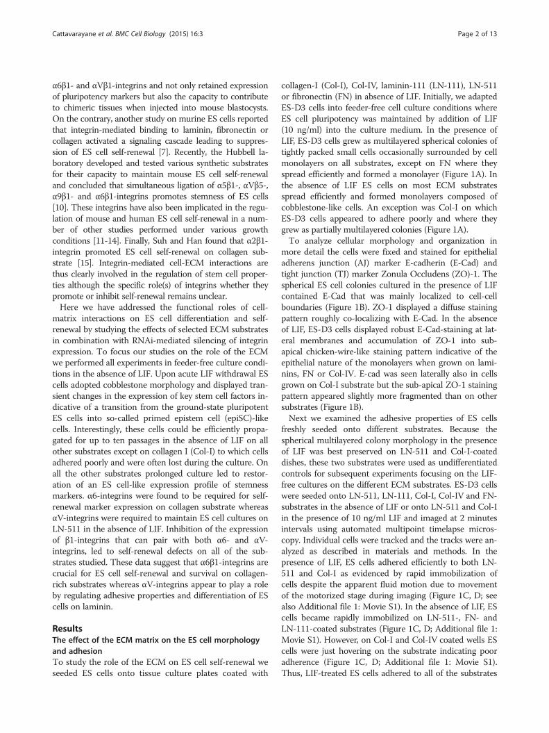
collagen-I (Col-I), Col-IV, laminin-111 (LN-111), LN-511or fibronectin (FN) in absence of LIF. Initially, we adaptedES-D3 cells into feeder-free cell culture conditions whereES cell pluripotency was maintained by addition of LIF(10 ng/ml) into the culture medium. In the presence ofLIF, ES-D3 cells grew as multilayered spherical colonies oftightly packed small cells occasionally surrounded by cellmonolayers on all substrates, except on FN where theyspread efficiently and formed a monolayer (Figure 1A). Inthe absence of LIF ES cells on most ECM substratesspread efficiently and formed monolayers composed ofcobblestone-like cells. An exception was Col-I on whichES-D3 cells appeared to adhere poorly and where theygrew as partially multilayered colonies (Figure 1A).To analyze cellular morphology and organization in
more detail the cells were fixed and stained for epithelialadherens junction (AJ) marker E-cadherin (E-Cad) andtight junction (TJ) marker Zonula Occludens (ZO)-1. Thespherical ES cell colonies cultured in the presence of LIFcontained E-Cad that was mainly localized to cell-cellboundaries (Figure 1B). ZO-1 displayed a diffuse stainingpattern roughly co-localizing with E-Cad. In the absenceof LIF, ES-D3 cells displayed robust E-Cad-staining at lat-eral membranes and accumulation of ZO-1 into sub-apical chicken-wire-like staining pattern indicative of theepithelial nature of the monolayers when grown on lami-nins, FN or Col-IV. E-cad was seen laterally also in cellsgrown on Col-I substrate but the sub-apical ZO-1 stainingpattern appeared slightly more fragmented than on othersubstrates (Figure 1B).Next we examined the adhesive properties of ES cells
freshly seeded onto different substrates. Because thespherical multilayered colony morphology in the presenceof LIF was best preserved on LN-511 and Col-I-coateddishes, these two substrates were used as undifferentiatedcontrols for subsequent experiments focusing on the LIF-free cultures on the different ECM substrates. ES-D3 cellswere seeded onto LN-511, LN-111, Col-I, Col-IV and FN-substrates in the absence of LIF or onto LN-511 and Col-Iin the presence of 10 ng/ml LIF and imaged at 2 minutesintervals using automated multipoint timelapse micros-copy. Individual cells were tracked and the tracks were an-alyzed as described in materials and methods. In thepresence of LIF, ES cells adhered efficiently to both LN-511 and Col-I as evidenced by rapid immobilization ofcells despite the apparent fluid motion due to movementof the motorized stage during imaging (Figure 1C, D; seealso Additional file 1: Movie S1). In the absence of LIF, EScells became rapidly immobilized on LN-511-, FN- andLN-111-coated substrates (Figure 1C, D; Additional file 1:Movie S1). However, on Col-I and Col-IV coated wells EScells were just hovering on the substrate indicating pooradherence (Figure 1C, D; Additional file 1: Movie S1).Thus, LIF-treated ES cells adhered to all of the substrates
Figure 1 LIF withdrawal induces a morphological change in ES-D3 cells from multilayered clusters to epithelial monolayers and impairsES-D3 adhesion to collagen. A) ES-D3 cells were cultured for 5 days on tissue-culture dishes coated with LN-511, LN-111, Col-I, Col-IV or FN inthe presence or absence of 10 ng/ml of LIF and imaged using a phase contrast microscope equipped with a CCD-camera. Scale bar is 100 μm.B) ES-D3 cells were cultured on LN-511 in the presence or absence of LIF or on LN-111, Col-I, Col-IV or FN in the absence of LIF, fixed with 4%PFA and stained for an epithelial AJ marker E-Cad (red) and a TJ marker ZO-1 (green). Cells were imaged using confocal microscopy and a singleoptical slice at the level of TJs (as determined by chicken-wire-like ZO-1 staining) is shown. Scale bar is 50 μm. C) ES-D3 cells were counted andseeded (1000 cells/mm2) onto tissue culture dishes (3.5 cm Ø) coated with the indicated ECM substrates and individual cells were tracked at 2minutes intervals upon their contact with the ECM. The amount of adherent (immobile) cells was determined as described in materials andmethods. The data shown comes from 2 independent experiments in which at least 50 individual cells were tracked. The relative amount ofimmobilized cells is depicted with differentially sized red circles the center of the graphs. D) A graph showing the percentage of ES-D3 cellsimmobilized on the indicated substrates within the first 30 minutes upon seeding.
Cattavarayane et al. BMC Cell Biology (2015) 16:3 Page 3 of 13
Cattavarayane et al. BMC Cell Biology (2015) 16:3 Page 4 of 13
tested within the first 60 minutes whereas in the absenceof LIF ES cells did adhere to laminins and fibronectin butadhered poorly to collagens within this time early frame.
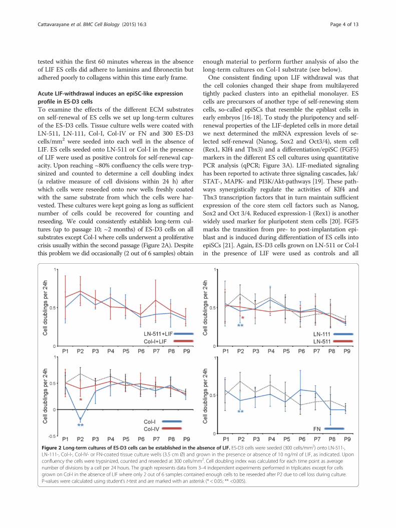
Acute LIF-withdrawal induces an epiSC-like expressionprofile in ES-D3 cellsTo examine the effects of the different ECM substrateson self-renewal of ES cells we set up long-term culturesof the ES-D3 cells. Tissue culture wells were coated withLN-511, LN-111, Col-I, Col-IV or FN and 300 ES-D3cells/mm2 were seeded into each well in the absence ofLIF. ES cells seeded onto LN-511 or Col-I in the presenceof LIF were used as positive controls for self-renewal cap-acity. Upon reaching ~80% confluency the cells were tryp-sinized and counted to determine a cell doubling index(a relative measure of cell divisions within 24 h) afterwhich cells were reseeded onto new wells freshly coatedwith the same substrate from which the cells were har-vested. These cultures were kept going as long as sufficientnumber of cells could be recovered for counting andreseeding. We could consistently establish long-term cul-tures (up to passage 10; ~2 months) of ES-D3 cells on allsubstrates except Col-I where cells underwent a proliferativecrisis usually within the second passage (Figure 2A). Despitethis problem we did occasionally (2 out of 6 samples) obtain
Figure 2 Long-term cultures of ES-D3 cells can be established in the abLN-111-, Col-I-, Col-IV- or FN-coated tissue culture wells (3.5 cm Ø) and groconfluency the cells were trypsinized, counted and reseeded at 300 cells/mmnumber of divisions by a cell per 24 hours. The graph represents data from 3–grown on Col-I in the absence of LIF where only 2 out of 6 samples containeP-values were calculated using student’s t-test and are marked with an asteris
enough material to perform further analysis of also thelong-term cultures on Col-I substrate (see below).One consistent finding upon LIF withdrawal was that
the cell colonies changed their shape from multilayeredtightly packed clusters into an epithelial monolayer. EScells are precursors of another type of self-renewing stemcells, so-called epiSCs that resemble the epiblast cells inearly embryos [16-18]. To study the pluripotency and self-renewal properties of the LIF-depleted cells in more detailwe next determined the mRNA expression levels of se-lected self-renewal (Nanog, Sox2 and Oct3/4), stem cell(Rex1, Klf4 and Tbx3) and a differentiation/epiSC (FGF5)markers in the different ES cell cultures using quantitativePCR analysis (qPCR; Figure 3A). LIF-mediated signalinghas been reported to activate three signaling cascades, Jak/STAT-, MAPK- and PI3K/Akt-pathways [19]. These path-ways synergistically regulate the activities of Klf4 andTbx3 transcription factors that in turn maintain sufficientexpression of the core stem cell factors such as Nanog,Sox2 and Oct 3/4. Reduced expression-1 (Rex1) is anotherwidely used marker for pluripotent stem cells [20]. FGF5marks the transition from pre- to post-implantation epi-blast and is induced during differentiation of ES cells intoepiSCs [21]. Again, ES-D3 cells grown on LN-511 or Col-Iin the presence of LIF were used as controls and all
sence of LIF. ES-D3 cells were seeded (300 cells/mm2) onto LN-511-,wn in the presence or absence of 10 ng/ml of LIF, as indicated. Upon2. Cell doubling index was calculated for each time point as average4 independent experiments performed in triplicates except for cellsd enough cells to be reseeded after P2 due to cell loss during culture.k (* < 0.05; ** <0.005).
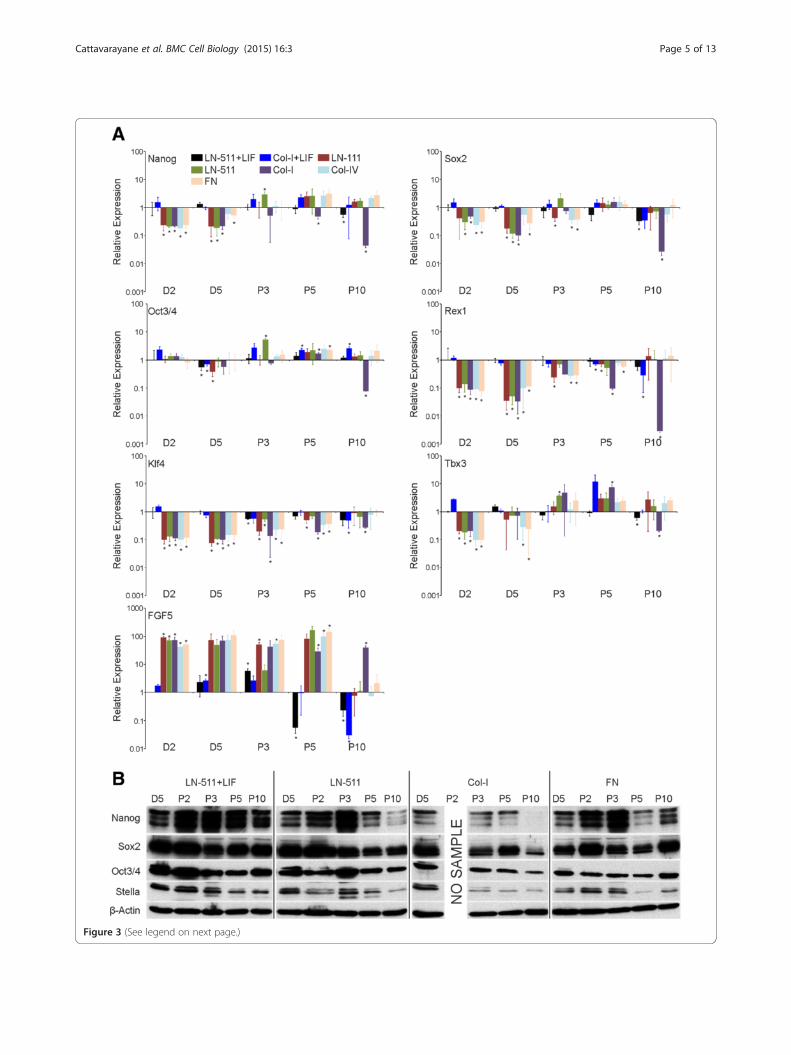
Figure 3 (See legend on next page.)
Cattavarayane et al. BMC Cell Biology (2015) 16:3 Page 5 of 13
(See figure on previous page.)Figure 3 Prolonged culture of ES-D3 cells in the absence of LIF leads to an enrichment of epithelial self-renewing cells with an EScell-like transcription profile. A) ES-D3 cells were cultured as indicated in Figure 2 and total RNA samples were collected at indicated time point.The mRNA expression levels of self-renewal markers (Nanog, Sox2, and Oct3/4), ES cell markers (Rex1, Klf4 and Tbx3) and a differentiation marker (FGF5)were determined using qPCR analysis. The data show means +/−STD of replicates from 2–3 independent samples. P-values (student’s t-test) < 0.005 aremarked with an asterisk (*). B) ES-D3 cells were grown as above and lysed at indicated time points followed by SDS-PAGE and western blot analysisusing Nanog, Sox2, Oct3/4 and Stella antibodies to study the protein expression levels of these stem cell markers. β-actin was used as a loading control.The data shown is representative of 2–3 experiments with similar results.
Cattavarayane et al. BMC Cell Biology (2015) 16:3 Page 6 of 13
samples were normalized and compared relative to theLN-511 + LIF sample at day 2. Regardless of the matrix-coating used, the expression of FGF5 was highly inducedafter LIF-withdrawal, whereas levels of Nanog, Sox2, Klf4,Tbx3 and Rex1 were downregulated suggesting that ES-D3 cells differentiated into epiSC-like cells (Figure 3A).The expression levels of Oct3/4 mRNA were relativelystable. Interestingly, upon prolonged culture the cellsshowed a gradual shift towards a more ES cell-like mRNAexpression profile (Figure 3A). Some markers returned toES cell like levels during passages 3–5 (Nanog, Tbx1)whereas FGF5 displayed a late response such that downreg-ulation of FGF5 mRNA expression was not yet evident inP5 samples but was prominent in P10 samples (Figure 3A).A notable exception was cells grown on Col-I without LIFin which the expression levels of essentially all of the stud-ied ES cell markers were strongly reduced and FGF5 levelsremained high (Figure 3A). Curiously, late passage samplesfrom ES-D3 cells grown on Col-IV in the absence of LIFdid maintain the expression of stemness markers and alsoshowed downregulated levels of FGF5 expression.The protein levels of Nanog, Sox2 and Oct3/4 were
studied by western blotting. Despite the observed down-regulation of the Nanog and Sox2 mRNAs at early timepoints, high levels of Nanog and Sox2 protein expressionwere maintained until approximately third passage in EScells seeded onto LN-511 or FN substrates (Figure 3A, B).In contrast, Nanog was rapidly downregulated in cellsgrown on Col-I substrate (Figure 3B). The cell numbers inCol-I substrate in the absence of LIF strongly declinedsuch that all of the remaining cells at passage 2 were usedfor reseeding and thus no protein sample could be ob-tained for this time point. In agreement with the mRNAanalysis the protein levels of Oct3/4 were relatively stable,although a noticeable reduction was seen in ES cellsseeded onto Col-I and FN substrate (Figure 3B). We alsostudied the protein expression levels of Stella, an ES cellmarker that is epigenetically silenced in epiSCs [22]. Whilethere was a tendency to see a drop of Stella expression onall substrates studied, visible levels of Stella protein wereretained on all three substrates (Figure 3B). Of note, a de-cline in the levels of Stella was observed also in late pas-sages of LIF-treated cells (Figure 3B). These data suggestthat efficient adhesion correlates with ES cell self-renewal.Since collagen substrates did not efficiently support ES-D3
cell adhesion, LIF-treated cells seeded onto LN-511 wereused as a control in all subsequent experiments.
Integrins mediate the ECM-derived signals to regulate EScell self-renewalAs integrins are the major class of cellular ECM-receptorswe addressed the functional roles of selected integrin sub-units in LIF-free ES cell cultures. Extensive characterizationof integrin expression profiles in mouse ES cells have beenreported [6,7,10; see Additional file 2: Table S1]. By usingdifferent approaches and criteria, these studies implicatedseveral β1-integrins, namely α6β1, αVβ1, α5β1, α2β1 andα9β1, in the regulation of ES cell self-renewal. In addition,αVβ5-integrin function has been associated with thisprocess [13]. To study the specific role of selected integrinsin the regulation of ES cell self-renewal in the absence ofLIF we used lentiviral vectors to stably deplete the expres-sion of β1-, α6-, αV-integrins. Integrin β1-subunit is foundin a number of collagen-, laminin- and fibronectin-bindingintegrin heterodimers and it is essential during early devel-opment [23-26]. Out of the many β1-containing integrins,α6β1-heterodimer is highly expressed in ES cells and ap-pears to convey important ECM-mediated signals to regu-late ES cells self-renewal in different models [6,10-12,27].αV-integrin forms heterodimers with a number of differentβ-subunits and can also mediate β1-integrin-independentbinding to FN [28]. ES-D3 cells were infected with concen-trated lentiviral vectors to generate β1-, αV- or α6-integrinknockdown (Itg-KD) cell lines. A virus vector containing anempty shRNA expression cassette was used as a control.Two independent shRNA constructs were used for eachof the three targets and knockdown efficiencies wereconfirmed by qPCR (Figure 4A) and western blotting(Figure 4B). The knockdown efficiencies were maintainedthroughout the duration of the experiments (Figure 4C-E).However, Itgβ1-KD cells did not survive freezing andthawing and thus the role of β1-integrin could only bestudied in freshly transduced ES cells.The adhesive properties of the different Itg-KD ES-D3 cell
lines on LN-511, Col-I and FN substrates were studied usingthe timelapse microscopy-based cell tracking analysis. Oneof two shRNAs for both β1- and αV-integrins caused mod-erately reduced adhesion to LN-511 in the presence of LIFbut since this effect was not obvious with the secondshRNAs these may be off-target effects (Figure 4F). Notably,
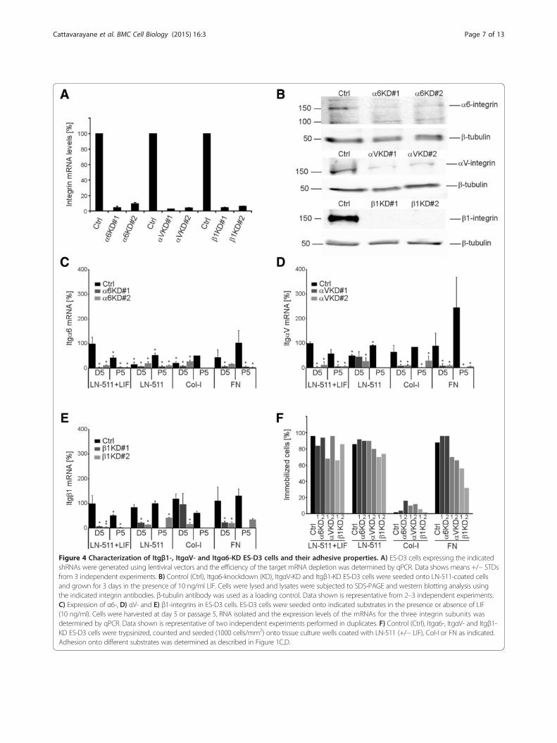
Figure 4 Characterization of Itgβ1-, ItgαV- and Itgα6-KD ES-D3 cells and their adhesive properties. A) ES-D3 cells expressing the indicatedshRNAs were generated using lentiviral vectors and the efficiency of the target mRNA depletion was determined by qPCR. Data shows means +/− STDsfrom 3 independent experiments. B) Control (Ctrl), Itgα6-knockdown (KD), ItgαV-KD and Itgβ1-KD ES-D3 cells were seeded onto LN-511-coated cellsand grown for 3 days in the presence of 10 ng/ml LIF. Cells were lysed and lysates were subjected to SDS-PAGE and western blotting analysis usingthe indicated integrin antibodies. β-tubulin antibody was used as a loading control. Data shown is representative from 2–3 independent experiments.C) Expression of α6-, D) αV- and E) β1-integrins in ES-D3 cells. ES-D3 cells were seeded onto indicated substrates in the presence or absence of LIF(10 ng/ml). Cells were harvested at day 5 or passage 5, RNA isolated and the expression levels of the mRNAs for the three integrin subunits wasdetermined by qPCR. Data shown is representative of two independent experiments performed in duplicates. F) Control (Ctrl), Itgα6-, ItgαV- and Itgβ1-KD ES-D3 cells were trypsinized, counted and seeded (1000 cells/mm2) onto tissue culture wells coated with LN-511 (+/− LIF), Col-I or FN as indicated.Adhesion onto different substrates was determined as described in Figure 1C,D.
Cattavarayane et al. BMC Cell Biology (2015) 16:3 Page 7 of 13

Cattavarayane et al. BMC Cell Biology (2015) 16:3 Page 8 of 13
only moderate adhesion defects (~70% of the Itgβ1-KD cellsadhered) were observed in Itgβ1-KD cell populations onLN-511 in the absence of LIF, and Itgα6- and ItgαV-KD cellsadhered to LN-511 as well as the controls (~90% of the cellsadhered; Figure 4F). As noted earlier, Col-I did not supportefficient adhesion for any of the cell lines (Figure 4F). BothItgβ1- and ItgαV-KD cells had a significant adhesion defecton FN (Figure 4F).To analyze the potential roles of these integrins in ES
cell self-renewal, control and the Itg-KD cells were cul-tured on LN-511, Col-I and FN in the absence of LIF.Cells grown on LN-511 in the presence of LIF were usedas controls in each case. The cell doubling index in thedifferent cell lines was determined as described earlier(Figure 2). The control virus-infected cells behaved aswild-type ES-D3 cells and could be efficiently main-tained without LIF on LN-511 and FN substrates butnot on Col-I (Figure 5A). Itgβ1-KD cells could be pas-saged up to one month (P5) on LN-511 in the presenceof LIF, whereas in the absence of LIF the cultures couldnot be maintained beyond first two passages (Figure 5A).Itgα6-KD cells could be cultured for several passages inthe presence or absence of LIF on LN-511 as well as onFN substrate. Although the cell doubling rates wereslightly reduced ItgαV-KD cells could be cultured onLN-511 in the presence of LIF (Figure 5A). In the ab-sence of LIF ItgαV-KD cells appeared to detach as cellcolonies grew bigger although individual Itgα V-KD cellsadhered as well as the control ES-D3 cells upon contactwith LN-511 substrate (Figures 4F, 5A). All cell lines ad-hered poorly to Col-I and were lost during early pas-sages except for ItgαV-KD cells which despite pooradhesion was the only cell line that consistently surviveduntil passage 5 (Figure 5A). On FN, Itgβ1-KD cultureswere lost at later passages while ItgαV- and Itgα6-KDcells could be maintained (Figure 5A). We noted thatthe cell loss in collagen-seeded Itgα6- and Itgβ1-KD cellsand in LN-511 grown ItgαV- and Itgβ1-KD cells coin-cided with significantly reduced KD-efficiencies in theremaining cells suggesting negative selection against effi-cient silencing of α6- or β1-integrins on collagen I andαV- or β1-integrins on LN-511 (Figures 5A, 4C-E).We next studied the stemness marker expression in ES-
D3 control and the different Itg-KD cells seeded onto LN-511, Col-I and FN substrates in the absence of LIF. Cellsseeded onto LN-511 in the presence of LIF were used as acontrol. As noted earlier, LIF withdrawal systematically ledto downregulation of stem cell marker expression and up-regulation of FGF5 in all conditions and cell lines (Figure 5B;see also Additional file 3: Figure S1). This effect was particu-larly prominent in control cells on FN and in Itgβ1-KD cellson all substrates (Figure 5B). In contrast, ItgαV-KD cellswere slightly less responsive to LIF withdrawal when com-pared with controls on LN-511 and FN. Vast majority of the
ItgαV-KD cell colonies detached on LN-511 and not enoughsamples could be obtained for analysis for later time points.ItgαV-KD cells survived on FN and Col-I, where they didnot show major changes in the expression profile of the dif-ferent markers when compared with controls (Figure 5B).Finally, when comparing the stemness profile of the differentItg-KDs with control cells in the presence of LIF it wasfound that while ItgαV-KD cells demonstrated a similarstemness marker expression profile as the controls, Itgα6-and Itgβ1-KD cells had reduced expression levels of Nanog,Klf4 and Tbx3 at passage 5. It was also noted that the ex-pression levels of α6-integrin were downregulated upon LIFwithdrawal (Figure 4C). These data suggest that α6β1-integrin function is important for the maintenance of pluri-potency of ES-D3 cells.
DiscussionIn this study we addressed the role of the extracellularmatrix components and selected integrins in the regula-tion of ES cell self-renewal under feeder-free cultureconditions and in the absence of ectopic factors (LIF)that might artificially promote ES cell self-renewal. Itwas found that on all studied ECM substrates ES cellsinitially appeared to differentiate towards epiblast-likeepiSCs [16-18]. These cells grew as polarized epithelialmonolayers and could be maintained in culture for atleast ten passages in the absence of LIF on most sub-strates. Prolonged culture without LIF appeared to en-rich for cells which displayed epithelial characteristics(see Additional file 4: Figure S2) and expressed highlevels of multiple stemness markers. Col-I substrate wasan exception as it did not support strong adhesion orproliferation of ES cells in the absence of LIF. The find-ing that ES cells adhere poorly on Col-I substrate is inagreement with an earlier report by Hayashi and co-workers [7]. Hayashi et al. concluded that integrin-mediated contact with laminin and FN may negativelyaffect ES cell self-renewal while lack of integrin activa-tion on collagen substrate would promote ES cell self-renewal [7]. In our study ES cells cultured in the absenceof LIF on Col-I did show signs of differentiation (upreg-ulation of FGF5 and downregulation of Nanog, Sox2,Rex1, Klf4 and Tbx3) as did cells grown on laminin orFN where the cells readily adhered. Moreover, upon pro-longed culture the levels of ES cell marker mRNAs in-creased on all other substrates except on Col-I. A criticaldifference is the use of ectopic inhibition of ES cell dif-ferentiation, as we omitted LIF from the culture mediumwhile Hayashi et al. kept LIF in their setup [7]. It is pos-sible that addition of even a small amount of LIF is suffi-cient to override the need for efficient integrin-mediatedadhesion. LIF promoted the survival and growth of ES-D3 cells on Col-I whereas in the absence of LIF, cells ad-hered poorly leading to significant cell loss particularly
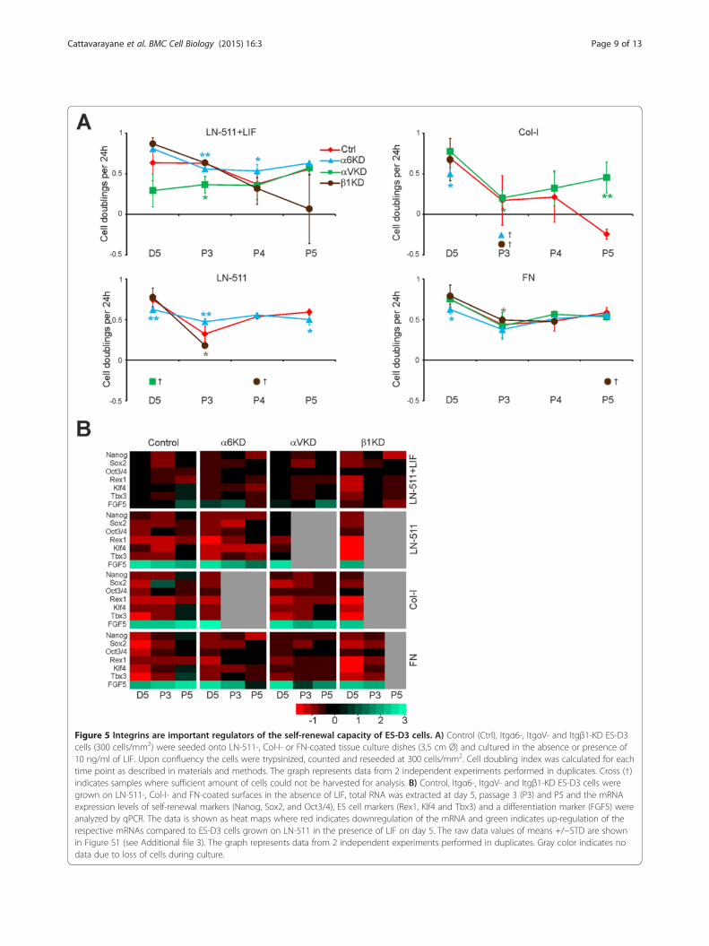
Figure 5 Integrins are important regulators of the self-renewal capacity of ES-D3 cells. A) Control (Ctrl), Itgα6-, ItgαV- and Itgβ1-KD ES-D3cells (300 cells/mm2) were seeded onto LN-511-, Col-I- or FN-coated tissue culture dishes (3.5 cm Ø) and cultured in the absence or presence of10 ng/ml of LIF. Upon confluency the cells were trypsinized, counted and reseeded at 300 cells/mm2. Cell doubling index was calculated for eachtime point as described in materials and methods. The graph represents data from 2 independent experiments performed in duplicates. Cross (†)indicates samples where sufficient amount of cells could not be harvested for analysis. B) Control, Itgα6-, ItgαV- and Itgβ1-KD ES-D3 cells weregrown on LN-511-, Col-I- and FN-coated surfaces in the absence of LIF, total RNA was extracted at day 5, passage 3 (P3) and P5 and the mRNAexpression levels of self-renewal markers (Nanog, Sox2, and Oct3/4), ES cell markers (Rex1, Klf4 and Tbx3) and a differentiation marker (FGF5) wereanalyzed by qPCR. The data is shown as heat maps where red indicates downregulation of the mRNA and green indicates up-regulation of therespective mRNAs compared to ES-D3 cells grown on LN-511 in the presence of LIF on day 5. The raw data values of means +/−STD are shownin Figure S1 (see Additional file 3). The graph represents data from 2 independent experiments performed in duplicates. Gray color indicates nodata due to loss of cells during culture.
Cattavarayane et al. BMC Cell Biology (2015) 16:3 Page 9 of 13

Cattavarayane et al. BMC Cell Biology (2015) 16:3 Page 10 of 13
during the early passages. The remaining cells that did ad-here to Col-I in the absence of LIF displayed downregu-lated levels of multiple stem cells markers. Col-IVsubstrate was similarly a poor substrate for freshly seededES cells but unlike Col-I, Col-IV substrate did supportself-renewal in our long-term culture setup. Since levels ofI-domain containing collagen-binding integrins are low inmouse ES cells [6,7,10,29], the differential capacity ofCol-I and Col-IV substrates to support adhesion in long-term cultures could be due to reported interactionsbetween Col-IV and laminin, a critical component of base-ment membranes [30]. Our preliminary data suggest thatCol-IV coating might facilitate adhesion by capturing lam-inin that is abundantly secreted by ES cells upon LIF with-drawal and thereby promote the assembly of cell derivedlaminin in vitro ([24] and data not shown).The important role for laminin substrate for mouse ES
cell self-renewal is supported by a study from Domogatskayaet al. where LN-511 and LN-332 (both of which sup-ported robust ES cell adhesion) were found to allow long-term self-renewal of ES cell cultures in the absence of LIF[6]. In the current study we found that, in addition to LN-511, also LN-111, FN and Col-IV enabled sustained EScell cultures in the absence of LIF. While FN was notstudied by Domogatskaya et al., only limited growth of EScell on LN-111 substrate was observed [6]. The reasonsfor these differences are not clear but one possibility couldbe the reported variability between the commercial prepa-rations of LN-111 from Engelbreth-Holm-Swarm (EHS)sarcoma used in these studies [31,32]. However, both stud-ies, together with other reports, confirm a positive correl-ation between integrin-mediated adhesion and ES cellself-renewal [6,8,10,12,13,27].The functions of specific integrins in ES cells are not well
understood but several studies report high levels of α6β1-integrin expression and/or a functional role for this integrinin ES cell adhesion [7,12,33,34]. Furthermore, αV-integrinsare thought to contribute to the regulation of ES cellself-renewal [6,10,13]. Here, by using RNAi experimentswe showed that β1-, α6- and αV-integrin subunits areinvolved in maintaining ES cell survival and self-renewal.In the absence of LIF, Itgβ1- and Itgα6-KD cells did notsurvive on Col I substrate as the cultures were lost priorthe first passage. Importantly, depletion of α6- and ofβ1-integrins also affected the maintenance of stem cellmarker expression profile even when LIF was present, indi-cating that α6β1-integrin is likely the key integrin heterodi-mer regulating ES cell self-renewal and pluripotency. Sinceα6β1-integrin is a laminin receptor, it is possible that therole of α6β1-integrin on collagen substrate is to assembleendogenous laminin secreted by ES cells themselves. Sucha mechanism would be in line with the known importanceof laminin during early development and maintenance ofpluripotent stem cell populations [6,8,24]. Expression of
αV- and β1-integrin subunits was required for the main-tenance of long-term cultures on LN-511. Despite initial ad-hesion of individual ItgαV- and Itgβ1-KD cells to LN-511substrate, the growing cell colonies gradually detached lead-ing to cell loss during handling of the cultures. Integrin-mediated signaling is a complex process and, in addition tobiochemical cues, mechanical cues conveyed by integrinsfrom the ECM may also contribute to the regulation of EScell differentiation [35]. αV-integrins are required for cellularmechanotransduction and for maturation of α2β1-integrin-mediated focal adhesions [36,37]. This phenomenon couldbe related to the adhesion defects observed in larger ItgαV-KD ES cell colonies.Although both α6β1- and αVβ1-integrins can bind to
laminins, our findings suggest that self-renewal of ES cellscan be supported also on other substrates such as FN. Ourdata does not exclude the possibility that laminin is stillthe critical mediator of self-renewal signals because of thecapacity of ES cell to produce their own laminin-richmatrix. Further studies are needed to address this possibil-ity in more detail. However, our data clearly shows thatα6β1- and αV-integrins play important roles in maintain-ing efficient ES cell self-renewal. Refinement of the dis-tinct roles of α6β1- and αV-integrins in stem cell-ECMcommunication will help us to define stem cell nichesleading to intelligible strategies to design culture condi-tions for in vitro stem cell applications. Given the multipleparallels between stem cells and cancer stem cells, target-ing the function of correct integrin(s) could also help inthe development of novel, more effective, approaches tolimit cancer stem cell self-renewal.
ConclusionsThe present study shows that acute LIF withdrawal con-verts multilayered ES cell colonies into epiSC-like epithelialmonolayers. Upon prolonged culture these cells remain epi-thelial but they regain ES cell-like expression profile of cen-tral stem cell markers and self-renew in the absence of LIF,given that they maintain a proper integrin-mediated adhe-sion to their substrate. Laminin-binding α6β1-integrins arecritical for maintaining the ES cell-like identity, whereasαV-integrins contribute to stable adhesion on LN-511.
MethodsAntibodies, plasmid constructs and reagentsPrimary antibodies used in this study are listed in (SeeAdditional file 2: Table S2). HRP-conjugated (anti-rabbit,anti-mouse) antibodies were from Jackson Inc. Cy3- andDyelight488-conjugated secondary (anti-mouse, anti-rabbit, anti-goat) antibodies were from Jackson Inc. andAlexa-488-conjugated secondary antibodies were fromInvitrogen. The shRNA knockdown constructs target-ing integrins were from Sigma Mission (see Additionalfile 2: Table S3). D-MEM (31966), β-mercaptoethanol
Cattavarayane et al. BMC Cell Biology (2015) 16:3 Page 11 of 13
(31350–010), and Dulbecco’s PBS were from GIBCO,ES-Qualified Fetal Calf Serum (FCS) (10439–024) wasfrom Invitrogen. Nonessential Amino Acids (M7145),LN-111 (L2020), and LN-511 (L6274) were purchasedfrom Sigma, FN (1918FN) was from R&D Systems, Col-I (5005-B) was from Advanced Biomatrix and Col-IV(354233) was from BD Biosciences. Leukemia Inhibi-tory Factor (LIF; LIF2050) was from Millipore.
Cell cultureMouse embryonic stem cells (ES-D3; ATCC/CRL-1934;[38]) were cultured on ECM-coated tissue culture platesin D-MEM glutamax (High Glucose, GlutaMAX™, Pyru-vate) supplemented with 15% FCS (ES-Qualified), 1%Non-Essential Amino acid, 0.1 mM β-mercaptoethanoland 1% penicillin/streptomycin, at 37°C and 5% CO2.When indicated, 10 ng/ml LIF was added. Cell cultureplates were coated by incubating plates with ECM proteinsLN-111 (20 μg/ml), LN-511 (5 μg/ml), FN (10 μg/ml) inPBS, Col-I (150 μg/ml) in 0.1 M NaHCO3 buffer pH8.3and Col-IV (10 μg/ml) in 50 mM HCl for 2 hours in +37°Cand 5% CO2. Cells were subcultured every 4–6 days with0.05% trypsin-EDTA (25300–054, Gibco) solution.
Adherence assayCells were detached from the culture dishes using 0.05%trypsin-EDTA and counted. One thousand cells/mm2
were seeded onto 6-well plates coated with LN-511, Col-Ior FN as indicated above in the presence or absence of10 ng/ml of LIF. Plates were then placed into OKOLabBasic WJ CO2 microscope stage incubator (OKOlab) ad-justed to 37°C and 5% CO2 and imaged at 2 min intervalsfor a total period of 2 hours using Olympus Cell^P live-cell/timelapse imaging system equipped with 10x phasecontrast objective (Olympus). Due to plate handling, stagepositioning and focusing steps, imaging was started 15–20minutes after seeding. The first 15 frames (30 minutes) ofthe timelapse sequences were analysed using MTrackJplugin for ImageJ [39]. At least fifty cells in every sequencewere tracked. The cells having a total track length of lessthan 15 μm were considered as adhered.
Lentiviral RNA interferenceThird-generation replication incompetent lentivirus vec-tors were generated using a four plasmid system in HEK293 T cells. The packaging vector containing the desiredshRNA construct was from SIGMA. The helper plas-mids pMD2.G (Plasmid 12259), and pMDL g/p RRE(Plasmid 12251) and pRSV-Rev (Plasmid 12253) werefrom Addgene [40]. DNA transfection was done usingLipofectamine 2000 from Invitrogen (11668–019). Thepackaging vector: pVSVG : pMDL g/p RRE : pRSV-Revwere used in 3:1:1:1 proportion and the total DNA usedfor transfection was 20 μg. Nearly confluent (70–80%)
293 T cells were grown on Corning CellBind (#3296)10 cm dish. One hour before transfection, 5 ml of freshpre-warmed medium (DMEM, low glucose, 10% FCS,0.1% penicillin/streptomycin) was added to packing cells.To prepare transfection mix, 80 μl of Lipofectamine2000 was added to 1 ml of OptiMEM and incubated atroom temperature for 5 minutes. In another tube, 20 μgof DNA was added in the proportion indicated above to1 ml of OptiMEM. The DNA solution was mixed withOptiMEM containing Lipofectamine drop by drop withgentle tapping of the tube. The mixture was incubated atroom temperature for 20 minutes. The mixture was addedon top of the cells, swirled gently and cells were placed intohumidified incubator (+37°C, 5% CO2). 24 hours post trans-fection, media containing the transfection mix was removedcarefully and 5 ml of fresh media was added. The mediasupernatant containing the viral particles was collectedevery 12 hours for 3–4 days and stored at +4°C. The viralsupernatants were pooled and centrifuged at 1000 rpm for5 minutes and filtered through a 0.44 μm filter. The filteredviral supernatant was then concentrated by ultracentrifuga-tion at 100000 X g for 2 hours. The supernatant wasdiscarded and the viral pellet resuspended in completeDMEM (1/100th of original volume).The cells were infected using 100 X concentrated virus
particles for 24 hours in the presence of 4 μg/ml poly-brene (107689, Sigma-Aldrich) and the infected cellswere selected using puromycin (4 μg/ml, Sigma). Thepuromycin selected cells were expanded and stored byfreezing in 90% ES-Qualified FCS, 10% DMSO (Sigma)in liquid nitrogen. Integrin β1-knockdown cells did notrevive upon freezing, so the Itgβ1-KD cell lines werefreshly made for every analysis.
Quantitative PCRTotal RNA was isolated from ES cells cultured on differ-ent ECM coating conditions using Qiagen RNA easy col-umn following manufacturer’s instructions. The isolatedtotal RNA was DNase-treated (Fermentas) followed byinactivation of the enzyme by incubating the samples at65°C for 20 minutes. 1 μg total RNA was annealed with25 μg/ml oligo-(dT) at 70°C for 10 minutes and chilledon ice for 2 minutes. cDNA synthesis was done using50 mM TrisCl pH 8.3, 50 mM KCl, 4 mM MgCl2,10 mM DTT, 0.5 mM of each dNTP and 200 U/ml M-MLV reverse transcriptase at 42°C for 1 hour and 70°Cfor 15 minutes. The synthesized cDNA was then diluted10-fold and used as a template for qRT-PCR. qRT-PCRwas performed using Brilliant III Ultra-FAST SYBR GreenqPCR master mix (Stratagene). The primers used in thisstudy are listed in (See Additional file 2: Table S3). Briefly,0.3 μM of each primer was mixed with 2 μl of dilutedcDNA, 1 X SYBR and the final volume made up to 10 μl.The reactions were carried out at 1 cycle of 95°C 3 min,
Cattavarayane et al. BMC Cell Biology (2015) 16:3 Page 12 of 13
40 cycles of 95°C 20 sec and 65°C 20 sec and 1 cycle at 95°C1 min 55°C 30 sec and 95°C 30 sec (MX3005P, Stratagene).The analysis of the data was done in Microsoft Excel.
Immunofluorescence microscopyES-D3 cells were cultured in the 24-well plates contain-ing coverslips coated with the indicated ECM moleculesas described above. Cells were washed with PBS, fixed in4% PFA for 10 minutes at room temperature or at 4°Covernight. Cells were permeabilized and non-specificbinding sites were blocked with PBS-Glycine (20 mM)containing 1% BSA and 0.1% Triton X-100 in for 20 mi-nutes at room temperature. Cells were washed once withPBS containing 0.5% BSA and 0.2% glycine and incu-bated with the indicated primary antibodies overnight at4°C. Subsequently the coverslips were washed 3 timesand incubated with secondary antibody in PBS contain-ing 0.5% BSA and 0.2% glycine on an ice bath for 1–2hours in dark. The cells were washed and mountedusing Immu-Mount™ (ThermoScientific). The slideswere then analyzed using Olympus FluoView FV1000confocal microscope.
Cell doubling rate analysisES-D3 cells were passaged for a period of 2 months inthe indicated substrate coated dishes. The cells wereseeded at a density of 300 cells/mm2. Cell numbers werecounted at the end of every passage. Cell doubling raterepresents the average number of cell divisions of a sin-gle cell in 24 hours. Cell doubling is calculated using theonline cell doubling calculator [41].
Western blottingThe cells cultured on different ECM coatings, as describedabove, were collected and lysed using RIPA lysis buffer(10 mM Tris–HCl pH7.5, 0.5% SDS, 1% IGEPAL, 0.15 MNaCl, 1% Sodium deoxycholate + protease and phosphat-ase inhibitors), sonicated for 3 minutes at 100% amplitudein a QSonica800 water bath sonicater. The sonicated ly-sates were then centrifuged to remove insoluble material.Protein concentration was estimated using the BCAmethod (23225; Pierce). 10–30 μg of total protein wasloaded onto a 12% gels for SDS electrophoresis, and theproteins were transferred to polyvinylidene difluoridemembranes. Membranes were blocked with 5% BSA inTBS-0.1% Tween buffer for 2 hours. Primary antibodieswere diluted into TBS-0.1% Tween buffer containing 1%BSA and incubated with the membranes overnight at +4°C. Membranes were washed three times, followed byaddition of HRP-conjugated secondary antibodies (1:5000)in TBS-0.1% Tween buffer containing 1% BSA and incu-bation for 2 hours at room temperature. The membraneswere washed 3–5 times in TBS-0.1% Tween buffer
followed by detection of proteins by chemiluminescenceusing LAS-3000 imaging system (Fujifilm).
Additional files
Additional file 1: Movie S1. Examples of cell adherence assay usingtimelapse microscopy and cell tracking. ES-D3 cells were seeded onto 6-welltissue culture plates coated with LN-511 in the presence (upper left panel)or absence of 10 μg/ml LIF (upper right panel) or onto Col-I (lower leftpanel) or FN (lower right panel) in the absence of LIF. Cells were imagedat 2 min intervals for 30 minutes and cell tracks were determined. Celladherence was determined from these data as described in materialsand methods (Figure 1C, D).
Additional file 2: Table S1. Integrin expression in murine ES cells.Table S2. Primary antibodies used in this study. Table S3. Oligos used inthis study.
Additional file 3: Figure S1. Expression of self-renewal and differentiationmarkers in control and in Itgα6-, ItgαV- and Itgβ1-KD cells. Control, Itgα6-,ItgαV- and Itgβ1-KD ES-D3 cells were grown on LN-511-, Col-I- and FN-coated tissue culture plates in the absence of LIF, total RNA was extracted atday 5, passage 3 (P3) and P5 and the mRNA expression levels of self-renewalmarkers (Nanog, Sox2, and Oct3/4), ES cell markers (Rex1, Klf4 and Tbx3) anda differentiation marker (FGF5) were analyzed by qPCR. The respective mRNAexpression levels were compared relative to levels obtained in ES-D3 cellsgrown on LN-511 in the presence of LIF on day 5. The raw data values ofmeans +/−STD are shown. The graph represents data from 2 independentexperiments performed in duplicates. P-values < 0.005 are depicted with anasterisk (*). Cross (†) indicates samples where sufficient amount of cells couldnot be harvested for analysis.
Additional file 4: Figure S2. ES-D3 cells have epithelial morphology inlong-term cultures. A) ES-D3 cells (300 cells/mm2) were seeded onto LN-511-,Col-I- or FN-coated tissue culture dishes (3.5 cm Ø) and cultured in theabsence or presence of 10 ng/ml of LIF. Upon confluency the cells weretrypsinized, counted and reseeded at 300 cells/mm2. After 5 passagesin culture (~one month) cells were imaged using a phase contrastmicroscope equipped with a CCD-camera. B) ES-D3 cells were culturedas in A), washed twice with PBS, fixed with 4% PFA and stained for anepithelial AJ marker E-Cad (red) and a TJ marker ZO-1 (green). Cellswere imaged using confocal microscopy at the level of TJs. Scale bar is50 μm.
AbbreviationsES cells: Embryonic stem cells; ECM: Extracellular matrix; EpiSCs: Epistem cells;LIF: Leukemia inhibitory factor; RNAi: RNA interference; Col: Collagen;LN: Laminin; FN: Fibronectin; Itg: Integrin; KD: Knockdown; qPCR: Quantitativepolymerase chain reaction.
Competing interestsThe authors declare that they have no competing interests.
Authors’ contributionsSC: Conception and design, collection and assembly of data, data analysisand interpretation, manuscript writing; RP: Conception and design, collectionand assembly of data, data analysis and interpretation; TRJ: Collection andassembly of data; AM: Conception and design, manuscript writing, dataanalysis and interpretation, financial support. All authors read and approvedthe final manuscript.
AcknowledgmentsJaana Träskelin and Riitta Jokela are acknowledged for expert technicalassistance. We thank Raija Soininen and Manninen laboratory members forcomments and critical reading of the manuscript. Mika Kaakinen, IlyaSkovorodkin and Veli-Pekka Ronkainen from the BCO imaging core facility isacknowledged for help with confocal and timelapse-microscopy setups.Hang-Mao Lee is acknowledged for his help with preparation of the qPCRheat maps. This work was funded by Academy of Finland (114330; 140974,263770, 135560), Sigrid Juselius Foundation, Centre for International Mobility(CIMO), Biocenter Oulu and Biocenter Finland.
Cattavarayane et al. BMC Cell Biology (2015) 16:3 Page 13 of 13
Author details1Biocenter Oulu, Oulu Center for Cell-Matrix Research, Faculty of Biochemistryand Molecular Medicine, University of Oulu, Aapistie 5, Oulu 90220, Finland.2Current address: Université de Lorraine, CS 25233, Nancy cedex, 54052,France.
Received: 9 September 2014 Accepted: 3 February 2015
References1. Thiery JP, Acloque H, Huang RY, Nieto MA. Epithelial-mesenchymal
transitions in development and disease. Cell. 2009;139(5):871–90.2. De Arcangelis A, Georges-Labouesse EN. Integrin and ECM functions: roles
in vertebrate development. Trends Genet. 2000;16(9):389–95.3. Smyth N, Vatansever HS, Meyer M, Frie C, Paulsson M, Edgar D. The targeted
deletion of the LAMC1 gene. Ann N Y Acad Sci. 1998;857:283–6.4. Gilcrease MZ. Integrin signaling in epithelial cells. Cancer Lett. 2006;247(1):1–25.5. Takada Y, Ye X, Simon S. The integrins. Genome Biol. 2007;8(5):215.6. Domogatskaya A, Rodin S, Boutaud A, Tryggvason K. Laminin-511, but not
−332, −111 or −411 enables mouse embryonic stem cell self-renewalin vitro. Stem Cells. 2008;26(11):2800–9.
7. Hayashi Y, Furue MK, Okamoto T, Ohnuma K, Myoishi Y, Fukuhara Y, et al.Integrins regulate mouse embryonic stem cell self-renewal. Stem Cells.2007;25(12):3005–15.
8. Rodin S, Domogatskaya A, Strom S, Hansson EM, Chien KR, Inzunza J, et al.Long-term self-renewal of human pluripotent stem cells on humanrecombinant laminin-511. Nat Biotechnol. 2010;28(6):611–5.
9. Raz R, Lee CK, Cannizzaro LA, d'Eustachio P, Levy DE. Essential role of STAT3for embryonic stem cell pluripotency. Proc Natl Acad Sci U S A.1999;96(6):2846–51.
10. Lee ST, Yun JI, Jo YS, Mochizuki M, van der Vlies AJ, Kontos S, et al.Engineering integrin signaling for promoting embryonic stem cellself-renewal in a precisely defined niche. Biomaterials. 2010;31(6):1219–26.
11. Yu KR, Yang SR, Jung JW, Kim H, Ko K, Han DW, et al. CD49f enhancesmultipotency and maintains stemness through the direct regulation ofOCT4 and SOX2. Stem Cells. 2012;30(5):876–87.
12. Meng Y, Eshghi S, Li YJ, Schmidt R, Schaffer DV, Healy KE. Characterizationof integrin engagement during defined human embryonic stem cellculture. FASEB J. 2010;24(4):1056–65.
13. Braam SR, Zeinstra L, Litjens S, Ward-van Oostwaard D, van den Brink S, vanLaake L, et al. Recombinant vitronectin is a functionally defined substratethat supports human embryonic stem cell self-renewal via alphavbeta5integrin. Stem Cells. 2008;26(9):2257–65.
14. Brafman DA, Phung C, Kumar N, Willert K. Regulation of endodermaldifferentiation of human embryonic stem cells through integrin-ECMinteractions. Cell Death Differ. 2012;20(3):369–81.
15. Suh HN, Han HJ. Collagen I regulates the self-renewal of mouse embryonicstem cells through alpha2beta1 integrin- and DDR1-dependent Bmi-1.J Cell Physiol. 2011;226(12):3422–32.
16. Guo G, Yang J, Nichols J, Hall JS, Eyres I, Mansfield W, et al. Klf4 revertsdevelopmentally programmed restriction of ground state pluripotency.Development. 2009;136(7):1063–9.
17. Brons IG, Smithers LE, Trotter MW, Rugg-Gunn P, Sun B, de Sousa Lopes SMC, et al. Derivation of pluripotent epiblast stem cells from mammalianembryos. Nature. 2007;448(7150):191–5.
18. Tesar PJ, Chenoweth JG, Brook FA, Davies TJ, Evans EP, Mack DL, et al. Newcell lines from mouse epiblast share defining features with humanembryonic stem cells. Nature. 2007;448(7150):196–9.
19. Niwa H, Ogawa K, Shimosato D, Adachi K. A parallel circuit of LIFsignalling pathways maintains pluripotency of mouse ES cells. Nature.2009;460(7251):118–22.
20. Rogers MB, Hosler BA, Gudas LJ. Specific expression of a retinoic acid-regulated,zinc-finger gene, Rex-1, in preimplantation embryos, trophoblast andspermatocytes. Development. 1991;113(3):815–24.
21. Hebert JM, Boyle M, Martin GR. mRNA localization studies suggest thatmurine FGF-5 plays a role in gastrulation. Development. 1991;112(2):407–15.
22. Hayashi K, Lopes SM, Tang F, Surani MA. Dynamic equilibrium andheterogeneity of mouse pluripotent stem cells with distinct functional andepigenetic states. Cell Stem Cell. 2008;3(4):391–401.
23. Fassler R, Meyer M. Consequences of lack of beta 1 integrin geneexpression in mice. Genes Dev. 1995;9(15):1896–908.
24. Li S, Harrison D, Carbonetto S, Fassler R, Smyth N, Edgar D, et al. Matrixassembly, regulation, and survival functions of laminin and its receptors inembryonic stem cell differentiation. J Cell Biol. 2002;157(7):1279–90.
25. Stephens LE, Sutherland AE, Klimanskaya IV, Andrieux A, Meneses J,Pedersen RA, et al. Deletion of beta 1 integrins in mice results in inner cellmass failure and peri-implantation lethality. Genes Dev. 1995;9(15):1883–95.
26. Aumailley M, Pesch M, Tunggal L, Gaill F, Fassler R. Altered synthesis oflaminin 1 and absence of basement membrane component deposition in(beta)1 integrin-deficient embryoid bodies. J Cell Sci. 2000;113:259–68.
27. Lee ST, Yun JI, van der Vlies AJ, Kontos S, Jang M, Gong SP, et al. Long-termmaintenance of mouse embryonic stem cell pluripotency by manipulatingintegrin signaling within 3D scaffolds without active Stat3. Biomaterials.2012;33(35):8934–42.
28. Humphries JD, Byron A, Humphries MJ. Integrin ligands at a glance. J CellSci. 2006;119(Pt 19):3901–3.
29. White DJ, Puranen S, Johnson MS, Heino J. The collagen receptor subfamilyof the integrins. Int J Biochem Cell Biol. 2004;36(8):1405–10.
30. Hohenester E, Yurchenco PD. Laminins in basement membrane assembly.Cell Adh Migr. 2013;7(1):56–63.
31. Ferletta M, Ekblom P. Identification of laminin-10/11 as a strong cell adhesivecomplex for a normal and a malignant human epithelial cell line. J Cell Sci.1999;112:1–10.
32. Wondimu Z, Gorfu G, Kawataki T, Smirnov S, Yurchenco P, Tryggvason K,et al. Characterization of commercial laminin preparations from humanplacenta in comparison to recombinant laminins 2 (alpha2beta1gamma1), 8(alpha4beta1gamma1), 10 (alpha5beta1gamma1). Matrix Biol. 2006;25(2):89–93.
33. Cooper HM, Tamura RN, Quaranta V. The major laminin receptor of mouseembryonic stem cells is a novel isoform of the alpha 6 beta 1 integrin. J CellBiol. 1991;115(3):843–50.
34. Hierck BP, Thorsteinsdottir S, Niessen CM, Freund E, Iperen LV, Feyen A,et al. Variants of the alpha 6 beta 1 laminin receptor in early murinedevelopment: distribution, molecular cloning and chromosomal localizationof the mouse integrin alpha 6 subunit. Cell Adhes Commun. 1993;1(1):33–53.
35. Uda Y, Poh YC, Chowdhury F, Wu DC, Tanaka TS, Sato M, et al. Force viaintegrins but not E-cadherin decreases Oct3/4 expression in embryonicstem cells. Biochem Biophys Res Commun. 2011;415(2):396–400.
36. Schiller HB, Hermann MR, Polleux J, Vignaud T, Zanivan S, Friedel CC, et al.beta1- and alphav-class integrins cooperate to regulate myosin II duringrigidity sensing of fibronectin-based microenvironments. Nat Cell Biol.2013;15(6):625–36.
37. Teravainen TP, Myllymaki SM, Friedrichs J, Strohmeyer N, Moyano JV, Wu C,et al. alphaV-Integrins Are Required for Mechanotransduction in MDCKEpithelial Cells. PLoS One. 2013;8(8):e71485.
38. Doetschman TC, Eistetter H, Katz M, Schmidt W, Kemler R. The in vitrodevelopment of blastocyst-derived embryonic stem cell lines: formation ofvisceral yolk sac, blood islands and myocardium. J Embryol Exp Morphol.1985;87:27–45.
39. Rasband WS: ImageJ, U. S. National Institutes of Health, Bethesda, Maryland,USA, http://imagej.nih.gov/ij/. 1997–2014.
40. Dull T, Zufferey R, Kelly M, Mandel RJ, Nguyen M, Trono D, et al. Athird-generation lentivirus vector with a conditional packaging system.J Virol. 1998;72(11):8463–71.
41. Roth V: Doubling Time: http://www.doubling-time.com/compute.php. 2006.
Submit your next manuscript to BioMed Centraland take full advantage of:
• Convenient online submission
• Thorough peer review
• No space constraints or color figure charges
• Immediate publication on acceptance
• Inclusion in PubMed, CAS, Scopus and Google Scholar
• Research which is freely available for redistribution
Submit your manuscript at www.biomedcentral.com/submit


![An EMF Study of LiF-BeF2 Solutions [Disc 3] - Molten Salt ...](https://static.fdokumen.com/doc/165x107/63237dd8be5419ea700ea0fe/an-emf-study-of-lif-bef2-solutions-disc-3-molten-salt-.jpg)