Whole-heart coronary magnetic resonance angiography in a patient with unstable angina

Tntodepte
F†
a
Journal of the American College of Cardiology Vol. 50, No. 15, 2007© 2007 by the American College of Cardiology Foundation ISSN 0735-1097/07/$32.00P
Cardiac Imaging
64-Slice Computed TomographyCoronary Angiography in PatientsWith High, Intermediate, or Low PretestProbability of Significant Coronary Artery Disease
W. Bob Meijboom, MD,*† Carlos A. G. van Mieghem, MD,*† Nico R. Mollet, MD, PHD,*†Francesca Pugliese, MD,*† Annick C. Weustink, MD,*† Niels van Pelt, MD,*†Filippo Cademartiri, MD, PHD,† Koen Nieman, MD, PHD,* Eric Boersma, MSC, PHD,*Peter de Jaegere, MD, PHD,* Gabriel P. Krestin, MD, PHD,† Pim J. de Feyter, MD, PHD, FACC*†
Rotterdam, the Netherlands
Objectives We assessed the usefulness of 64-slice computed tomography coronary angiography (CTCA) to detect or rule outcoronary artery disease (CAD) in patients with various estimated pretest probabilities of CAD.
Background The pretest probability of the presence of CAD may impact the diagnostic performance of CTCA.
Methods Sixty-four-slice CTCA (Sensation 64, Siemens, Forchheim, Germany) was performed in 254 symptomatic pa-tients. Patients with heart rates �65 beats/min received beta-blockers before CTCA. The pretest probability forsignificant CAD was estimated by type of chest discomfort, age, gender, and traditional risk factors and definedas high (�71%), intermediate (31% to 70%), and low (�30%). Significant CAD was defined as the presence ofat least 1 �50% coronary stenosis on quantitative coronary angiography, which was the standard of reference.No coronary segments were excluded from analysis.
Results The estimated pretest probability of CAD in the high (n � 105), intermediate (n � 83), and low (n � 66) groupswas 87%, 53%, and 13%, respectively. The diagnostic performance of the computed tomography (CT) scan wasdifferent in the 3 subgroups. The estimated post-test probability of the presence of significant CAD after a nega-tive CT scan was 17%, 0%, and 0% and after a positive CT scan was 96%, 88%, and 68%, respectively.
Conclusions Computed tomography coronary angiography is useful in symptomatic patients with a low or intermediate estimatedpretest probability of having significant CAD, and a negative CT scan reliably rules out the presence of significant CAD.Computed tomography coronary angiography does not provide additional relevant diagnostic information in symptom-atic patients with a high estimated pretest probability of CAD. (J Am Coll Cardiol 2007;50:1469–75) © 2007 by theAmerican College of Cardiology Foundation
ublished by Elsevier Inc. doi:10.1016/j.jacc.2007.07.007
pp
tmsp
p2p
M
S
he estimated pretest probability of having significant coro-ary artery disease (CAD) in a study population should beaken into account in the evaluation of the diagnostic accuracyf computed tomography coronary angiography (CTCA) toetect or rule out the presence of coronary stenosis. Thestimated pretest probability of having obstructive CAD inatients who present with chest pain is related to age, gender,ype of chest discomfort, and traditional risk factors. Thestimated pretest probability is lowest in younger female
rom the *Department of Cardiology, Thoraxcenter, Rotterdam, the Netherlands; andDepartment of Radiology, Erasmus Medical Center, Rotterdam, the Netherlands.
tManuscript received March 14, 2007; revised manuscript received July 2, 2007,
ccepted July 10, 2007.
atients with nonanginal chest pain and highest in older maleatients with typical angina (1).The diagnostic performance of CTCA has mostly been
ested in symptomatic patient populations with a high esti-ated pretest probability of having significant CAD, and a few
tudies have reported on the impact of different estimatedretest probabilities on the performance of CTCA (2).The purpose of this study was to evaluate the diagnostic
erformance and clinical usefulness of 64-slice CTCA in54 patients with high, intermediate, or low estimatedretest probability of having significant coronary stenosis.
ethods
tudy population. During a 24-month period, 254 pa-
ients presenting with typical angina pectoris, atypical an-
fSai(stpcatCtaP6meSCa0cptm(pttTsT
Iawc
IddpiQwei1ccmshetvtCsetomfocaSfdpitbpsee
pc(vacp
opAdas
1470 Meijboom et al. JACC Vol. 50, No. 15, 2007CTCA and Pretest Probability for CAD October 9, 2007:1469–75
gina pectoris, and nonanginalchest pain who were referred forconventional coronary angiogra-phy (CCA) were included intothe study. Typical angina wasdefined as having 3 characteris-tics: 1) substernal discomfort; 2)that is precipitated by physicalexertion or emotion; and 3) re-lieved with rest or nitroglycerinwithin 10 min. Atypical anginapectoris was defined as having 2of 3 of the definition character-istics. Nonanginal chest pain wascharacterized as 1 or absence ofthe described chest pain features.The estimated pretest probability
or obstructive CAD was estimated using the Duke Clinicalcore, which includes type of chest discomfort, age, gender,nd traditional risk factors (3,4). Patients were categorizednto a low (1% to 30%), intermediate (31% to 70%), or high71% to 99%) estimated pretest probability group of havingignificant CAD. No patients with previous history of percu-aneous coronary intervention, coronary artery bypass surgery,rior myocardial infarction, impaired renal function (serumreatinine �120 �mol/l), persistent arrhythmias, or knownllergy to iodinated contrast material were included. Conven-ional coronary angiogram was performed before or after theTCA and served as the standard of reference. The institu-
ional review board of the Erasmus Medical Center Rotterdampproved the study, and all subjects gave informed consent.atient preparation. Patients with a heart rate exceeding5 beats/min received additional beta-blockers (50/100 mgetoprolol) 1 h before the computed tomography (CT)
xamination.can protocol. All scans were performed with a 64-sliceT scanner that features a gantry rotation time of 330 ms,temporal resolution of 165 ms, and a spatial resolution of.4 mm3 (Sensation 64, Siemens, Forchheim, Germany). Aalcium scoring scan was performed with the followingarameters: 64 � 0.6 mm collimation, 330 ms rotationime, 120 kV tube voltage, 150 mAs tube current, 3.8m/rotation table feed, prospective electrocardiogram
ECG) X-ray tube modulation. Afterward, the CTCA waserformed using identical parameters aside from a higherube current between 850 and 960 mAs and withouthe use of prospective ECG X-ray tube modulation.he radiation exposure was estimated using dedicated
oftware (ImPACT, version 0.99x, St. George’s Hospital,ooting, London, United Kingdom).A bolus of 95 ml of contrast material (400 mgI/ml;
omeron, Bracco, Milan, Italy) was injected intravenously inn antecubital vein at 5 ml/s, and a bolus-tracking techniqueas used to synchronize the arrival of contrast in the
Abbreviationsand Acronyms
CAD � coronary arterydisease
CCA � conventionalcoronary angiogram
CI � confidence interval
CT � computedtomography
CTCA � computedtomography coronaryangiography
LR � likelihood ratio
QCA � quantitativecoronary angiography
oronary arteries and the initiation of the scan. s
mage reconstruction. Datasets were reconstructed imme-iately after the scan after a stepwise approach as previouslyescribed (5,6). If necessary, multiple datasets of a singleatient were used separately in order to obtain optimalmage quality for all available coronary segments.
uantitative coronary angiography (QCA). All scansere carried out within 1 week before or after CCA. One
xperienced cardiologist, unaware of the results of CTCA,dentified and analyzed all coronary segments, using a7-segment modified American Heart Association classifi-ation. All segments, regardless of size, were included foromparison with CTCA. Segments were classified as nor-al (smooth parallel or tapering borders), as having non-
ignificant disease (wall irregularities or �50% stenosis), oraving significant disease (stenosis �50%). Stenoses werevaluated in the worst view, and classified as significant ifhe lumen diameter reduction exceeded �50% measured byalidated QCA algorithm (CAAS, Pie Medical, Maastricht,he Netherlands).
T image evaluation. One observer analyzed total calciumcores of all patients using dedicated software. Two experi-nced observers, a radiologist and a cardiologist, unaware ofhe results of CCA, evaluated the CTCA data sets on anffline workstation (Leonardo, Siemens) using (curved)ultiplanar reconstruction. Segments were scored positive
or significant CAD if there was �50% diameter reductionf the lumen by visual assessment. Segments distal to ahronic total occlusion were excluded. Interobserver dis-greements were resolved by a third reader.tatistical analysis. The diagnostic performance of CTCA
or the detection of significant coronary artery stenoses asefined by QCA is presented as sensitivity, specificity,ositive and negative predictive values with the correspond-ng 95% confidence intervals (CIs), and positive and nega-ive likelihood ratios (LRs) were calculated. Comparisonetween CTCA and QCA was performed on 3 levels:atient-by-patient, vessel-by-vessel, and segment-by-egment analysis. A Mantel-Haenszel test was performed tovaluate the trend in sensitivity and specificity relative to thestimated pretest probability for obstructive CAD.
Categorical characteristics are expressed as numbers andercentages, and compared between the 3 groups using thehi-square test. Continuous variables are expressed as meansstandard deviation) and compared with 1-way analysis ofariance followed by post-hoc Bonferroni correction todjust for multiple comparisons. If not normally distributed,ontinuous variables are expressed as medians (25th to 75thercentile range) and compared with Kruskal-Wallis test.An additional analysis was done to investigate the effect
f nesting since repeated assessments within the sameatient were made that were not independent observations.
random selection of a single segment per patient wasone, and the diagnostic accuracy for detecting significantrtery disease was calculated. Interobserver and intraob-erver variability for the detection of significant coronary
tenosis and agreement between techniques to classify pa-
td
R
Pb7fsrtt
twet1Dwnsol
ghhp(wisomvCvp0pgDpt(wm
al-Walli
1471JACC Vol. 50, No. 15, 2007 Meijboom et al.October 9, 2007:1469–75 CTCA and Pretest Probability for CAD
ients as having no, single-, or multivessel disease wasetermined by �-statistics.
esults
atient demographics are shown in Table 1. Additionaleta-blockers before CT scanning were administered in4% (188 of 254) of patients decreasing the mean heart raterom 71 � 11 beats/min to 59 � 8 beats/min. The meancan time was 12.7 � 1.6 s. Initially, all data sets wereeconstructed in the mid- to end-diastolic phase. In 34% ofhe cases (86 of 254), additional higher quality reconstruc-ions obtained during end systole were used for evaluation.
The estimated radiation exposure using prospective X-rayube modulation for the calcium score in women and menas 1.8 and 1.4 mSv, respectively. The estimated radiation
xposure for the contrast-enhanced scan without prospec-ive X-ray tube modulation was 17.0 mSv in women and3.4 mSv in men, which is in line with previous reports (7).iagnostic performance of 64-slice CTCA: all patientsith chest pain. The observed pretest probability of sig-ificant CAD, defined as having at least 1 �50% coronarytenosis per patient was 50%. The diagnostic performancef CTCA for detecting significant stenoses on a patient
Patient Demographics (n � 254)
Table 1 Patient Demographics (n � 254)
Pret
>70%High
(n � 105)
Typical angina 89 (85)
Atypical angina 16 (15)
Nonanginal chest pain 0 (0)
Men 97 (92)
Age (yrs)* 63 � 9
BMI (kg/m2)* 27.5 � 4.2
Heart rate (beats/min)* 57 � 8
Risk factors
Hypertension† 68 (65)
Hypercholesterolemia‡ 71 (68)
Diabetes mellitus§ 16 (15)
Current smoker 32 (30)
Previous smoker 14 (13)
Family history of CAD� 57 (54)
Obesity¶ 32 (30)
Calcium score (Agatston score)# 354 (103–814
Conventional coronary angiography
Prevalence of obstructive CAD 82 (78)
Absence of CAD 6 (6)
Nonsignificant disease 17 (16)
Single-vessel disease 42 (40)
Multivessel disease 40 (38)
*Mean and standard deviation. †Blood pressure �140/90 mm Hg orhypercholesterolemia. §Treatment with oral antidiabetic medicationsecond-degree relatives with premature CAD (age �55 years). ¶Body motherwise indicated. Categorical variables were tested with chi-squarenormally distributed, continuous variables were compared with Krusk
evel is detailed in Table 2. Eighteen patients with angio- a
raphic nonsignificant disease were incorrectly classified asaving significant CAD by CT: 17 patients were scored asaving single-vessel disease, and 1 patient was misinter-reted as having multivessel disease. Ninety-eight percent124 of 126) of patients with significant CAD on CCAere correctly identified by CTCA (Fig. 1). The 2 patients
n whom the severity of disease was underestimated bothhowed significant lumen narrowing in the circumflex cor-nary artery (both 53% diameter reduction, 1 in the proxi-al and 1 in the midsegment). Forty patients with single-
essel disease were evaluated as having multivessel disease byTCA due to overestimation of disease severity in other
essels. Agreement between CTCA and QCA on a per-atient (no or any disease) level was very good (�-value:.84), whereas agreement between techniques to classifyatients as having no, single-, and multivessel disease wasood (�-value: 0.61).iagnostic performance of 64-slice CTCA: patient-by-
atient analysis. The analysis comprised 105 (43%) pa-ients with a high estimated pretest probability for CAD, 8333%) patients with an intermediate, and 66 (26%) patientsith a low estimated pretest probability for CAD. Theean age between patients with high estimated probability
obability of Significant CAD
p Value
30% to 70%Intermediate
(n � 83)
<30%Low
(n � 66)
31 (37) 3 (4)
29 (35) 21 (32) �0.0001
23 (28) 42 (64)
47 (57) 27 (41) �0.0001
61 � 8 50 � 12 �0.0001
27.1 � 4.8 26.7 � 4.2 NS
60 � 7 61 � 7 �0.01
43 (52) 30 (45) �0.05
52 (63) 14 (21) �0.0001
11 (13) 4 (6) NS
16 (19) 16 (24) NS
6 (7) 4 (6) NS
39 (47) 29 (44) NS
21 (25) 13 (20) NS
134 (1–296) 0 (0–56) �0.0001
32 (39) 12 (18) �0.0001
21 (25) 36 (55) �0.0001
30 (36) 18 (27)
22 (27) 9 (14)
10 (12) 3 (5)
nt for hypertension. ‡Total cholesterol �180 mg/dl or treatment forlin. �Family history of coronary artery disease (CAD), having first- orex (BMI) �30 kg/m2. #Median and quartiles. Values are n (%) unlessntinuous variables were tested with 1-way analysis of variance. If nots test. The p values �0.05 were considered statistically significant.
est Pr
)
treatmeor insuass ind
test. Co
nd intermediate estimated probability was significantly
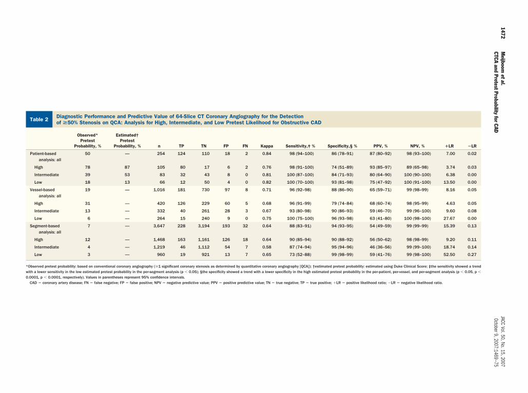
Diagnostic Performance and Predictive Value of 64-Slice CT Coronary Angiography for the Detectionof >50% Stenosis on QCA: Analysis for High, Intermediate, and Low Pretest Likelihood for Obstructive CAD
Table 2 Diagnostic Performance and Predictive Value of 64-Slice CT Coronary Angiography for the Detectionof >50% Stenosis on QCA: Analysis for High, Intermediate, and Low Pretest Likelihood for Obstructive CAD
Observed*Pretest
Probability, %
Estimated†Pretest
Probability, % n TP TN FP FN Kappa Sensitivity,‡ % Specificity,§ % PPV, % NPV, % �LR �LR
Patient-basedanalysis: all
50 — 254 124 110 18 2 0.84 98 (94–100) 86 (78–91) 87 (80–92) 98 (93–100) 7.00 0.02
High 78 87 105 80 17 6 2 0.76 98 (91–100) 74 (51–89) 93 (85–97) 89 (65–98) 3.74 0.03
Intermediate 39 53 83 32 43 8 0 0.81 100 (87–100) 84 (71–93) 80 (64–90) 100 (90–100) 6.38 0.00
Low 18 13 66 12 50 4 0 0.82 100 (70–100) 93 (81–98) 75 (47–92) 100 (91–100) 13.50 0.00
Vessel-basedanalysis: all
19 — 1,016 181 730 97 8 0.71 96 (92–98) 88 (86–90) 65 (59–71) 99 (98–99) 8.16 0.05
High 31 — 420 126 229 60 5 0.68 96 (91–99) 79 (74–84) 68 (60–74) 98 (95–99) 4.63 0.05
Intermediate 13 — 332 40 261 28 3 0.67 93 (80–98) 90 (86–93) 59 (46–70) 99 (96–100) 9.60 0.08
Low 6 — 264 15 240 9 0 0.75 100 (75–100) 96 (93–98) 63 (41–80) 100 (98–100) 27.67 0.00
Segment-basedanalysis: all
7 — 3,647 228 3,194 193 32 0.64 88 (83–91) 94 (93–95) 54 (49–59) 99 (99–99) 15.39 0.13
High 12 — 1,468 163 1,161 126 18 0.64 90 (85–94) 90 (88–92) 56 (50–62) 98 (98–99) 9.20 0.11
Intermediate 4 — 1,219 46 1,112 54 7 0.58 87 (74–94) 95 (94–96) 46 (36–56) 99 (99–100) 18.74 0.14
Low 3 — 960 19 921 13 7 0.65 73 (52–88) 99 (98–99) 59 (41–76) 99 (98–100) 52.50 0.27
*Observed pretest probability: based on conventional coronary angiography (�1 significant coronary stenosis as determined by quantitative coronary angiography [QCA]); †estimated pretest probability: estimated using Duke Clinical Score; ‡the sensitivity showed a trendwith a lower sensitivity in the low estimated pretest probability in the per-segment analysis (p � 0.05); §the specificity showed a trend with a lower specificity in the high estimated pretest probability in the per-patient, per-vessel, and per-segment analysis (p � 0.05, p �
0.0001, p � 0.0001, respectively). Values in parentheses represent 95% confidence intervals.CAD � coronary artery disease; FN � false negative; FP � false positive; NPV � negative predictive value; PPV � positive predictive value; TN � true negative; TP � true positive; �LR � positive likelihood ratio; �LR � negative likelihood ratio.
1472M
eijboometal.
JACCVol.50,No.15,2007
CTCAand
PretestProbabilityfor
CADOctober9,2007:1469–75

daatsg
tiitesDvticds1nfTt
tpDsaQaorTwoTp
uCl1dss
aspt2ps(
D
TrmstTeapii
iabww
dnT
1473JACC Vol. 50, No. 15, 2007 Meijboom et al.October 9, 2007:1469–75 CTCA and Pretest Probability for CAD
ifferent from the mean age in the low probability group,nd the median calcium score was significantly different inll 3 groups. The mean heart rate was significantly lower inhe high estimated probability group compared with thoseeen in the intermediate and low estimated probabilityroups (Table 1).
The diagnostic performance of CTCA was different inhe patient groups with various estimated pretest probabil-ties. The specificity showed a trend with a lower specificityn the high estimated pretest probability (p � 0.05, sensi-ivity p � NS). The diagnostic impact of CTCA on thestimated pretest probability of having significant CAD ishown in Figures 2 and 3.
iagnostic performance of 64-slice CTCA: vessel-by-essel analysis. The diagnostic performance of CTCA forhe detection of significant lesions on a vessel-based analysiss detailed in Table 2. Two significantly diseased rightoronary arteries, 1 left anterior descending artery, and 5iseased circumflex coronary arteries were incorrectly clas-ified as nonsignificantly diseased by CTCA. Of a total of,016 vessels, the severity of a lesion was overestimated in 97onobstructive vessels (false positives). The diagnostic per-ormance of the CT scan was different in the 3 subgroups.he specificity showed a trend towards lower specificity in
Figure 1 CTCA Image of the Right Coronary Artery
Volume-rendered computed tomography coronary angiography (CTCA) image (A) ofthe right coronary artery. A curved multiplanar reconstructed image (B) and a thickmaximum-intensity projected image (C) disclose a significant coronary stenosis(arrows) in the midright coronary artery, which was corroborated by conventionalcoronary angiogram (D). Proximally and distally of the significant obstructed lesion,nonsignificant calcified plaques can be seen (C).
he high estimated pretest probability (p � 0.0001, sensi- g
ivity p � NS). Agreement between CTCA and QCA on aer-vessel level was good (�-value: 0.71).iagnostic performance of 64-slice CTCA: segment-by-
egment analysis. Overall, 3,647 (of 4,318 potentiallyvailable segments) were included for comparison withCA. Unavailable segments included 547 anatomically
bsent segments on CCA and 124 segments distal to anccluded coronary segment. Segments were not excluded foreasons such as severe calcifications or poor image quality.he �-value for interobserver and intraobserver variabilityas 0.70 and 0.72, respectively. The diagnostic performancef CTCA for detecting significant stenoses is detailed inable 2. Agreement between CTCA and QCA on aer-segment level was good (�-value, 0.64).The severity of 32 significant coronary stenoses was
nderestimated or missed and classified as nonsignificant byTCA. Most of these significant lesions (24 of 32) were
ocated in distal segments or in side branches. The severity of93 nonsignificant lesions was overestimated by CTCA. Theiagnostic performance of the CT scan was different in the 3ubgroups with a lower sensitivity (p � 0.05) and a higherpecificity (p � 0.0001) in the low pretest probability group.
Analysis on the randomly selected segments resulted insensitivity of 92% (24 of 26; 95% CI 73% to 99%),
pecificity of 93% (212 of 228; 95% CI 89% to 96%),ositive predictive value of 60% (16 of 40; 95% CI 43%o 75%), and a negative predictive value of 99% (212 of14; 95% CI 96% to 100%). The effect of nesting isrobably minimal as the result of this analysis is veryimilar to the results shown in the per-segment analysisTable 2).
iscussion
he diagnostic performance of 64-slice CTCA to detect orule out the presence of significant coronary stenosis hasainly been reported for patients with stable angina pectoris
cheduled for invasive CCA, and these studies have shownhat CTCA can reliably rule out significant CAD (5,8–10).he majority of these patients presented with a high
stimated pretest probability of having significant CAD,nd only scant information is available on the diagnosticerformance of 64-slice CTCA in patients with a low orntermediate estimated pretest probability of having signif-cant CAD.
In this study, we used the Duke Clinical Score, whichncorporates clinical presentation of chest pain, age, gender,nd traditional risk factors, to estimate the pretest proba-ility of having significant CAD. Using the LRs of the tests,hich were obtained in this study, post-test probabilitiesere calculated.The pretest probability of CAD may impact of the
iagnostic performance of the CT scan. Indeed, the diag-ostic performance of CTCA in the 3 groups was different.he specificity was lower in the high pretest probability
roup compared with the low pretest probability group,
wtphs
wmptrtnpefv
5pprdp3tvliomCr
1474 Meijboom et al. JACC Vol. 50, No. 15, 2007CTCA and Pretest Probability for CAD October 9, 2007:1469–75
hereas sensitivity was lower in the per-segment analysis inhe low pretest probability group. This observation canrobably be explained by the higher calcium scores in theigher probability groups, which tend to overestimate theeverity of stenosis.
Figure 2 Impact of CTCA on Various Estimated Pretest Probab
1Estimated using Duke Clinical Score (including Diamond-Forrester criteria and procoronary stenosis as determined by quantitative coronary angiography); 3calculatecoronary artery disease; CTCA � computed tomography coronary angiography; Est
Figure 3 Influence of CTCA on Probability ofObstructive CAD as a Function of Pretest Probability
Using the positive and negative likelihood ratios obtained from Table 2, we cal-culated the estimated post-test probabilities of CAD after positive and negativefindings on CTCA from various estimated pre-test probabilities of CAD. Dashedlines � CTCA�; dotted lines � CTCA�. Abbreviations as in Figure 2.
p
A negative CT scan was present in 75% of the patientsith a low estimated pretest probability and in approxi-ately 50% of the patients with an intermediate estimated
retest probability. The negative predictive value of CTCAo exclude significant CAD was excellent in these patients,educing the estimated post-test probability to zero. Thus,hese patients would not need further downstream diag-ostic tests. They may be candidates for secondaryrevention measures, such as statin therapy in the pres-nce of nonobstructive plaques or could be dischargedrom further cardiac follow-up in the absence of anyisible plaque.
A positive CT scan occurred in approximately 25% and0% of the patients with a low or intermediate estimatedretest probability, respectively. The number of false-ositive outcomes was rather high in these patients, whichenders a positive CT scan rather unreliable for clinicalecision making. In these patients it may be reasonable toroceed to invasive CCA in the case of left main disease,-vessel disease, and in the presence of a critical stenosis inhe proximal part of a major coronary artery. In case ofessel disease in distal vessels or side branches, equivocalesions, or uninterpretable scans, one may consider a non-nvasive stress test to determine the functional significancef a doubtful coronary stenosis. A negative functional testay overrule the clinical significance of a (false)-positiveT scan and reduce the need for invasive coronary angiog-
aphy. A positive functional test may further increase the
of Significant CAD
c clinical variables); 2based on conventional coronary angiography (�1 significantBayesian statistics (post-test odds � pretest odds � likelihood ratio). CAD �
imated; Obs � observed.
ilities
gnostid using� est
robability of having significant CAD and should be fol-

lcnetp
nbetianCavpcaSswrercslbosi
qces
REoN
R
1
1475JACC Vol. 50, No. 15, 2007 Meijboom et al.October 9, 2007:1469–75 CTCA and Pretest Probability for CAD
owed by invasive coronary angiography and coronary revas-ularization if symptoms are not alleviated by the antiangi-al mediation. However, further studies are necessary tovaluate the diagnostic value of the combination of func-ional data from a stress test with the anatomical datarovided by CTCA.In the high estimated pretest probability group, a
egative CTCA reduced the estimated post-test proba-ility to 17%, whereas a positive CTCA increased thestimated post-test probability to as high as 96%. Givenhe high estimated pretest probability of significant CADn this group, the majority of these symptomatic patientsre likely to proceed to invasive CCA even if CTCA isegative, since the post-test probability of significantAD was still �10%. Computed tomography coronary
ngiography, therefore, appears to be of limited clinicalalue in the evaluation of the high estimated pretestrobability group. Assessment for the presence of myo-ardial ischemia with a functional test may be moreppropriate in this situation.tudy limitations. The studied patients were not a pro-pective, consecutive group of patients. However, selectionas not based on particular patient demographics, but
ather on the availability of the 64-slice CT scanner for thexamination of cardiac patients. The rather high estimatedadiation exposure of 64-slice CTCA (17 to 13.4 mSv) asompared with CCA (3 to 6 mSv) is of concern (7). In thistudy we did not use prospective ECG X-ray tube modu-ation, which can significantly reduce radiation exposure,ut requires a regular heart rhythm and limits the possibilityf reconstructing images in the end-systolic phase. In ourtudy end-systolic data sets provided optimal image qualityn 34% of patients.
Currently, there is no validated software available able touantify the degree of stenoses. So far, the severity oforonary stenosis as assessed by CT is rather crudely visuallystimated as more or less than 50% luminal diameter
tenosis.eprint requests and correspondence: Dr. Pim J. de Feyter,rasmus Medical Center, Department of Cardiology and Radiol-gy, Room Hs 207, P.O. Box 2040, 3000 CA Rotterdam, theetherlands. E-mail: [email protected].
EFERENCES
1. Diamond GA, Forrester JS. Analysis of probability as an aid in theclinical diagnosis of coronary artery disease. N Engl J Med 1979;300:1350–8.
2. Hendel RC, Patel MR, Kramer CM, et al. ACCF/ACR/SCCT/SCMR/ASNC/NASCI/SCAI/SIR 2006 appropriateness criteria forcardiac computed tomography and cardiac magnetic resonance imag-ing: a report of the American College of Cardiology Foundation/American College of Radiology, Society of Cardiovascular ComputedTomography, Society for Cardiovascular Magnetic Resonance, Amer-ican Society of Nuclear Cardiology, North American Society forCardiac Imaging, Society for Cardiovascular Angiography and Inter-ventions, and Society of Interventional Radiology. J Am Coll Cardiol2006;48:1475–97.
3. Gibbons RJ, Balady GJ, Bricker JT, et al. ACC/AHA 2002 guidelineupdate for exercise testing: summary article. A report of the AmericanCollege of Cardiology/American Heart Association Task Force onPractice Guidelines (Committee to Update the 1997 Exercise TestingGuidelines). J Am Coll Cardiol 2002;40:1531–40.
4. Pryor DB, Shaw L, McCants CB, et al. Value of the history andphysical in identifying patients at increased risk for coronary arterydisease. Ann Intern Med 1993;118:81–90.
5. Mollet NR, Cademartiri F, van Mieghem CA, et al. High-resolutionspiral computed tomography coronary angiography in patients referredfor diagnostic conventional coronary angiography. Circulation 2005;112:2318–23.
6. Meijboom WB, Mollet NR, Van Mieghem CA, et al. Pre-operativecomputed tomography coronary angiography to detect significantcoronary artery disease in patients referred for cardiac valve surgery.J Am Coll Cardiol 2006;48:1658–65.
7. Hausleiter J, Meyer T, Hadamitzky M, et al. Radiation dose estimatesfrom cardiac multislice computed tomography in daily practice: impactof different scanning protocols on effective dose estimates. Circulation2006;113:1305–10.
8. Leschka S, Alkadhi H, Plass A, et al. Accuracy of MSCT coronaryangiography with 64-slice technology: first experience. Eur Heart J2005;26:1482–7.
9. Raff GL, Gallagher MJ, O’Neill WW, Goldstein JA. Diagnosticaccuracy of noninvasive coronary angiography using 64-slice spiralcomputed tomography. J Am Coll Cardiol 2005;46:552–7.
0. Leber AW, Knez A, von Ziegler F, et al. Quantification of obstructiveand nonobstructive coronary lesions by 64-slice computed tomogra-
phy: a comparative study with quantitative coronary angiography andintravascular ultrasound. J Am Coll Cardiol 2005;46:147–54.Copyright © 2022 FDOKUMEN