Quale Iliade leggeva Ariosto?, «Aevum», LXXXVIII (2014), 3, pp. 587-613
Upload
khangminh22Category
view
1download
0
RADIOCARBON, Vol 51, Nr 2, 2009, p 613–625 © 2009 by the Arizona Board of Regents on behalf of the University of Arizona
© 2009 by the Arizona Board of Regents on behalf of the University of ArizonaProceedings of the 5th International 14C and Archaeology Symposium, edited by Irka Hajdas et al.RADIOCARBON, Vol 51, Nr 2, 2009, p 613–625
613
ULTRAFILTRATION: BOON OR BANE?
C M Hüls1 • P M Grootes • M-J NadeauLeibniz-Laboratory for Radiometric Dating and Isotope Research, Christian-Albrechts-University, Kiel, Germany.
ABSTRACT. Ultrafiltration of bone collagen, dissolved as gelatin (M ~100,000 D), has received considerable attention asa means to remove small contaminants and thus produce more reliable dates (Brown et al. 1988; Bronk Ramsey et al. 2004;Higham et al. 2006; Mellars 2006). However, comparative dating studies have raised the question whether this cleaning stepitself may introduce contamination with carbon from the filters used (Bronk Ramsey et al. 2004; Brock et al. 2007; Hüls etal. 2007).
Here, we present results of further ultrafiltration experiments with modern and fossil collagen samples using Vivaspin 20™and Vivaspin 15R™ ultrafilters. Evidently, the Vivaspin 20 (VS 20) ultrafilter with a polyethersulfone (PES) membraneretains more material in the >30 kD fraction than the Vivaspin 15R (VS 15R) filter with a regenerated cellulose membrane(Hydrosat), which may be related to increased retention of proteins due to suboptimal electrostatic conditions during ultrafil-tration with the PES membrane. In addition, this filter type shows clear evidence for contamination with fossil carbon, pre-sumably from membrane fibers, in the <30 kD fraction. Radiocarbon measurements on ultrafiltrated fossil collagen seem toindicate small contributions of modern carbon via glycerin left on and within the filter membranes of both types. AlthoughSEM pictures show film remnants on the fibrous filter structure of cleaned filter membranes, EDX analysis on the VS 20membrane to not support the assumption this may be glycerin. Our observations indicate the risks and benefits of the use ofultrafiltration in cleaning collagen samples for 14C dating need to be further quantified, especially for the cleaning of fossilbone collagen of good quality samples.
INTRODUCTION
The radiocarbon dating of bones is usually performed on the collagen fraction, which is purified bydissolution from the demineralized bone as gelatin in hot acidic water (hydrolysis, e.g. Longin1970). However, this procedure will also include other hot-water-soluble components such ashumics and degraded protein fragments (from exogenous sources, such as soil amino acids, orendogenous by formation from collagen) (van Klinken and Mook 1990). Adding an alkali extractionof the demineralized bone material helps to remove alkali-soluble organics such as humic acid (vanKlinken and Hedges 1995). In addition, ultrafiltration (UF) has been suggested as an effectivemethod to remove low-molecular weight contaminants and degraded proteins from the high-molec-ular and unchanged proteins of the collagen (e.g. Brown et al. 1988; Bronk Ramsey et al. 2004).However, unlike the classical sieving process, the separation between smaller and larger particlesalso depends on the charge state of the membrane and the proteins, and can even be reversed (e.g.Gosh 2003; Wang and Rodgers 2005; Burns and Zydney 1999).
The UF cleaning method for extracted fossil collagen has received considerable attention (e.g. Mel-lars 2006), as it was shown that redating previously dated bones using UF and modern bone pretreat-ment resulted in many cases in 14C ages that were significant older and more consistent (e.g. BronkRamsey 2004; Higham et al. 2006). However, comparative dating studies also found evidence forcontamination introduced by the filters used (Bronk Ramsey et al. 2004; Brock et al. 2007; Hüls etal. 2007).
Here, we present further results of ultrafiltration tests using modern and fossil bone collagen, indus-trial collagen, and 2 different UF-membrane types, with a focus on the performance of the 2 mem-branes with regard to the partitioning of the solution over filtrate and retentate and the effect of fil-tering on the 14C content of these fractions.
1Corresponding author. Email: [email protected].
614 C M Hüls et al.
MATERIAL AND METHODS
The details of the 2 filters, Vivaspin 20™ (VS 20, a polyethersulfone membrane, PES) and Vivaspin15R™ (VS 15R, a regenerated cellulose membrane, Hydrosat), both with a molecular weight cut-off(MWCO) of 30,000 Dalton (D), as well as their cleaning and use have been described in a previouspaper by Hüls et al. (2007). They also showed that the VS 20 membrane has a 14C content of 0.54 ±0.09 pMC, while VS 15R shows values between 14.36 ± 0.11 and 17.48 ± 0.15 pMC. Thus, bothmembranes consist mainly of fossil carbon. The membranes have been coated with glycerin to keepthe filters flexible. Before 2005, this glycerin contained fossil carbon, and now contains modern car-bon (Brock et al. 2007). A modern carbon source was confirmed by 14C measurements of the glyc-erin used (courtesy of Vivascience) and of samples taken from the cleaning water of the filters (glyc-erin: 107 ± 0.35 pMC; cleaning water step 1 >30 kD: 108.30 ± 0.38 pMC; <30 kD: 108.08 ± 0.33pMC and 106.94 ± 0.32 pMC; Hüls et al. 2007). To avoid sample contamination, we remove theglycerin from the filter membranes by repeated washing and ultrasonication with ultra-pure water ina cleaning procedure based on Bronk Ramsey et al. (2004), described in Hüls et al. (2007). Compar-ison of 14C contents before and after filtering indicates the procedure is adequate for the removal ofthe humectants (Hüls et al. 2007).
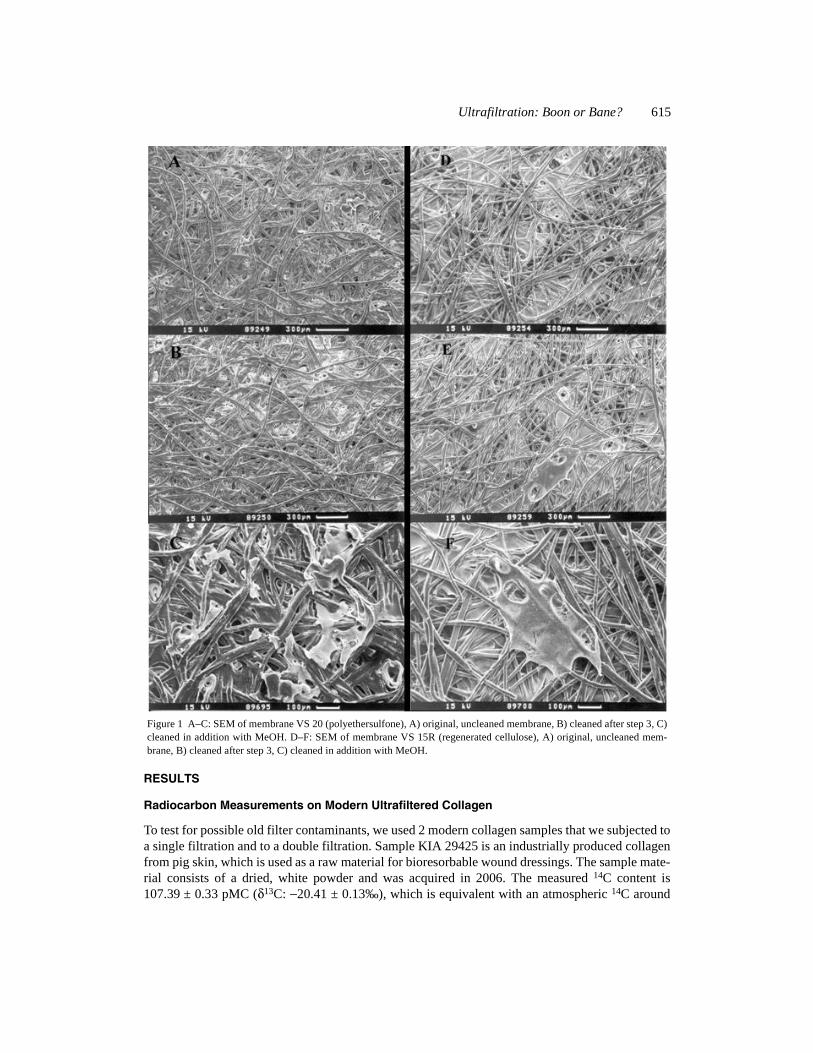
For a visual inspection of the cleaning, we took scanning electron microscope (SEM) pictures oforiginal and cleaned membranes of the 2 ultrafilters VS 20 and VS 15R (Figure 1). The untreated fil-ter membranes (VS 20 and VS 15R) show a coating, which we assumed at first to be glycerin, onand in between the fibers of either PES or Hydrosat (Figure 1A and D). Remnants of this coating arestill visible in the cleaned filter membranes (Figure 1B and E). A more rigorous cleaning, includingmethanol, still left flakes of the coating (Figure 1C and F). Energy dispersive X-ray spectroscopy(EDX) on VS 20 membranes (original and cleaned), however, indicates the flakes are composed ofPES and are thus an artifact of the manufacture of this membrane. A similar measurement for the VS15R membrane is possible but will not give conclusive results, since membrane and humectant con-tain largely similar elements with only minor differences in the carbon to oxygen ratio.
Both tested ultrafilters contain both fossil and modern carbon. We therefore tested for residual con-tamination caused by the cleaned filters by ultrafiltration of 2 modern and 2 archaeological collagensamples. The modern materials are an industrially produced collagen from porcine dermis (MBPMedical Biomaterial Products, Hüls et al. 2007) and collagen extracted from a bovine bone acquiredin 2006. The old collagen was obtained from 2 Paleolithic bones from caves in the Swabian Jura,using our standard modified Longin procedures (Longin 1970; Grootes et al. 2004; Hüls et al. 2007).
The ultrafiltration tests were performed on the lyophilized collagen fractions of the pig skin and the1 modern and 2 fossil bones, dissolved in ultra-clean water (VS 20: 20 mL; VS 15R: 15 mL) andthen centrifuged at 3000 rpm until 1–2 mL is left above the ultrafilter. Starting weights for ultrafil-tration of modern collagen of 10, 30, and 80 mg were used to test for any effect of collagen concen-tration on recovery, partition between filtrate and retentate, and 14C concentration. The 2 fossil col-lagen samples were filtered using 50 mg. In addition to the single filtration experiment (1*filtration), modern collagen was subjected to a double filtration (2* filtration), e.g. first filtrationdown to 1–2 mL in the retentate, removal of the filtrate, refilling of the ultrafilter and the supernatantwith ultra-pure water, and a subsequent 2nd filtration down to 1–2 mL in the >30 kD fraction.
Both UF fractions, the retentate (>30 kD) and the filtrate (<30 kD), were lyophilized and weighed.Both freeze-dried ultrafiltered collagen fractions (UF col >30 kD and <30 kD) and the original col-lagen (Lcol) were burnt to CO2, graphitized, and measured at the Leibniz-Labor AMS system usingstandard procedures (Nadeau et al. 1997, 1998).
Ultrafiltration: Boon or Bane? 615
RESULTS
Radiocarbon Measurements on Modern Ultrafiltered Collagen
To test for possible old filter contaminants, we used 2 modern collagen samples that we subjected toa single filtration and to a double filtration. Sample KIA 29425 is an industrially produced collagenfrom pig skin, which is used as a raw material for bioresorbable wound dressings. The sample mate-rial consists of a dried, white powder and was acquired in 2006. The measured 14C content is107.39 ± 0.33 pMC (δ13C: −20.41 ± 0.13‰), which is equivalent with an atmospheric 14C around
Figure 1 A–C: SEM of membrane VS 20 (polyethersulfone), A) original, uncleaned membrane, B) cleaned after step 3, C)cleaned in addition with MeOH. D–F: SEM of membrane VS 15R (regenerated cellulose), A) original, uncleaned mem-brane, B) cleaned after step 3, C) cleaned in addition with MeOH.

616 C M Hüls et al.
2003 (Levin and Kromer 2004; Levin and Kromer, personal communication, 2006). Depending onthe homogeneity of the raw material (age of the pigs of which skins were used is assumed to be ~6months) and diet of the animal, the variability in 14C content may be assumed to be ≤1 pMC.
Sample KIA 30242 is collagen, extracted in the Leibniz Laboratory from a bovine bone acquiredin 2006. The 14C content measured on the unfiltered collagen (Lcol) is 106.21 ± 0.29 pMC (δ13C:−13.45 ± 0.17‰), equivalent with an atmospheric 14C between 2003 and 2005 (Levin and Kromer,personal communication, 2006). The remaining non-soluble bone gave a 14C concentration of106.30 ± 0.3 pMC (δ13C: −16.05 ± 0.13‰), well within 1 σ of the collagen 14C. Based on theseresults, we assume a rather homogenous 14C distribution within the extracted collagen.
Modern Industrial Collagen (KIA 29425)
The results for the single and double ultrafiltration experiments for both filter membranes are sum-marized in Table 1 and illustrated in Figure 2.
VS 20 (PES)
The recovery of the single and double filtration with VS 20 ultrafilters is between 75 and 90%, withsome variability during the double filtration experiment (see Table 1). The >30 kD fraction (reten-tate) generally contains more than 90 wt% (92.7–100 wt%) of the recovered material, indicating acollagen sample that contains mostly high-molecular proteins >30 kD.
Figure 2 Ultrafiltration of modern industrial collagen (KIA 29425): A) Vivaspin 20 (PES); B) Vivaspin 15R (Hydrosat).

Ultrafiltration: Boon or Bane? 617
14C measurements of the filtrate (<30 kD) indicate an apparent pollution with fossil carbon, mostlikely from the membrane material (14C: 0.31–0.5 pMC, Hüls et al. 2007) (see Table 1, Figure 2A).Assuming only PES as the source of contamination, the 14C difference between the unfiltered col-lagen and the filtrate could indicate 12.6% and 10.4% contamination for the 30- and 80-mg samples,respectively. This would translate into 30 μg and 20 μg of carbon from the membrane fibers, respec-tively. The retentates of the single filtration experiment also show a slight and steady increase in 14Cwith collagen weight. The 14C difference between >30 kD and unfiltered collagen is between 2–3 σof the measurement uncertainties; the range is 1.8 pMC, ~5 σ.
The 14C measurements for the double filtration experiment show similar features as the single filtra-tion but with increased 14C concentrations in all fractions. As neither glycerin nor filter material
Table 1 14C measurements of modern industrial collagen of KIA 29425 (14C: 107.39 ± 0.3 pMC,δ13C: −20.41 ± 0.13‰).
MembraneWeight (mg)
Recovery (%) Size wt% pMC δ13C (‰)a
1 * Ultrafiltration
VS 20
10 78.1 >30 kD 100 106.72 ± 0.26 −20.5 ± 0.1<30 kD 0.0 n.a. n.a.
30 78.5 >30 kD 98.1 108.3 ± 0.27 −21.2 ± 0.2<30 kD 1.9 93.93 ± 0.9 −23.1 ± 0.5
80 75.2 >30 kD 98.4 108.51 ± 0.25 −20.9 ± 0.1<30 kD 1.6 96.3 ± 0.75 −21.4 ± 0.6
VS 15R
10 80.0 >30 kD 62.3 108.72 ± 0.28 −19.5 ± 0.1<30 kD 37.8 107.49 ± 0.3 −19.5 ± 0.2
30 77.8 >30 kD 69.6 108.27 ± 0.31 −18.4 ± 0.1<30 kD 30.4 108.04 ± 0.29 −17.9 ± 0.2
80 79.3 >30 kD 70.4 108.01 ± 0.27 −21.0 ± 0.1<30 kD 29.6 108.97 ± 0.3 −20.0 ± 0.1
2 * Ultrafiltration
VS 20
10 90.5 >30 kD 100.0 108.41 ± 0.35 −19.9 ± 0.3<30 kD 0.0 n.a. n.a.
30 83.8>30 kD 92.7 109.23 ± 0.36 −20.3 ± 0.1
<30 kD 1: 4.6 1: 99.32 ± 0.44 1: −23.8 ± 0.32: 2.7 2: n.a. 2: n.a.
80 81.8 >30 kD 95.1 111.6 ± 0.32 −24.6 ± 0.2
<30 kD 1: 2.6 1: 103.75 ± 0.57 1: −22.2 ± 0.12: 2.3 2: 103.42 ± 0.45 2: −25.4 ± 0.6
VS 15R
10 86.3>30 kD 61.5 107.59 ± 0.26 −20.7 ± 0.1
<30 kD 1: 30.9 1: 108.96 ± 0.29 1: −20.3 ± 0.12: 7.5 2: 106.63 ± 0.62 2: −20.9 ± 0.5
30 85.7>30 kD 61.1 108.97 ± 0.3 −19.0 ± 0.1
<30 kD 1: 31.7 1: 109.24 ± 0.27 1: −21.4 ± 0.12: 7.2 2: 107.92 ± 0.36 2: −22.4 ± 0.5
80 85.5>30 kD 63.8 109.56 ± 0.26 −20.9 ± 0.1
<30 kD 1: 27.8 1: 108.74 ± 0.27 1: −18.2 ± 0.22: 8.4 2: 107.19 ± 0.31 2: −20.5 ± 0.7
aMeasured at the AMS system simultaneously with the 14C/12C ratio. This δ13C includes the effects of fractionation duringgraphitization and in the AMS-system and, therefore, cannot be compared with δ13C values obtained per mass spectrometeron CO2.

618 C M Hüls et al.
show these high 14C concentrations, the values for the retentate cannot be explained unless weassume significant age and chemical inhomogeneity in the industrial collagen or batch-to-batchglycerin. The first filtrates from the double filtration can be compared to those from the single filtra-tion. The higher 14C concentrations in these fractions may thus indicate either a smaller contamina-tion with carbon from the filter fibers or an increased contribution of younger carbon from the glyc-erin or different collagen, like the retentate. The second filtrate of the 80 mg-sample showed a 14Cconcentration well within 1 σ of first filtrate, indicating no additional contamination.
The retentate (>30 kD) of the double filtration experiment gave 14C concentrations from 108.41 to111.6 pMC (from 1.0 to 4.2 pMC, ~2.2 to 9.6 σ above the unfiltered collagen), increasing withamounts filtrated. If one assumes a large 14C inhomogeneity in this industrial collagen, the resultscould be explained by removal of smaller proteins with a lower 14C content from larger proteins witha higher 14C content. However, an increasingly effective UF in the larger-sized samples is difficultto accept since the opposite would be expected (higher filtering efficiency with less material to befiltered). On the other hand, if the steady increase of 14C with sample size was caused by enhancedcontamination with carbon from glycerin due to the double filtration, the 14C concentration of theglycerin on these filters must have been much higher than indicated by the 14C measurements of thecleaning waters. Clearly, these rather inconsistent results do not fit a simple model.
VS 15R (Hydrosat)
The recovery of material after ultrafiltration with the VS 15R is around 80%. The distribution ofmaterial over the >30 kD and <30 kD fractions for the VS 15R ultrafilter is quite different from thatfor VS 20. Here, 60–70 wt% of the material stay in the retentate (>30 kD) instead of 90–100 wt%.Both ultrafilters have the same MWCO (30 kD) and thus the question arises which filter is correctlyseparating the different molecular weights.
The 14C contents of the retentate of the single filtration experiment are slightly higher than but closeto the 14C content of the unfiltered collagen (difference for the 10-, 30-, and 80-mg sample is 3, 2,and 1.8 σ, respectively) (Figure 2B) and within 1 σ between each sample. Furthermore, the 14C con-centrations of the 30- and 80-mg sample fractions agree well with the measured 14C concentrationsof the same fractions filtered with VS 20 (Δ: ≤0.5 pMC, ≤1.4 σ).
The filtrates of the single filtration experiment with VS 15R show, in contrast to those of VS 20, 14Cconcentrations close to the 14C of the unfiltered collagen but increasing with sample size. Thus,there is no evidence for a contamination with fossil carbon within the <30 kD fractions. Instead,increased 14C concentrations seem to indicate a small contribution of younger carbon, which, how-ever, is difficult to prove due to the rather small difference in 14C between glycerin and the collagen.
The 14C measurements of the double filtration experiment also show 14C values close to or slightlyabove the 14C concentration of the unfiltered collagen. A steady increase in 14C with sample size forthe retentate seems to indicate admixture of younger carbon as was seen in the single filtrationexperiment.
To what extent the results of the filtration experiments with the industrial collagen are transferableto bone collagen extracted in the laboratory is uncertain; thus, identical tests were done on extractedmodern bone collagen.
Modern Bovine Bone Collagen (KIA 30242)
The results for the single and double ultrafiltration experiments using VS 20 and VS 15R are sum-marized in Table 2 and illustrated in Figure 3.

Ultrafiltration: Boon or Bane? 619
VS 20 (PES)
The recovery of the single and double filtration is >90%, except for the 10-mg double filtration,where only 71% was recovered (Table 2). Whether the latter is an outlier is as yet unknown. Thefraction retained on the filter (>30 kD) is around 90 wt%, similar to the observations made with KIA29425 and the VS 20 filter.
The filtrates of the single filtration experiment again show significantly depleted 14C values, indicat-ing a contamination with fossil carbon from membrane material. Since this is also seen in the filtra-tion experiments with the industrial collagen, it may be characteristic for this membrane type. The
Table 2 14C measurements of modern bovine bone collagen of KIA 30242 (14C: 106.21 ± 0.29 pMC,δ13C:−13.45 ± 0.17‰).
MembraneWeight (mg)
Recovery (%) size wt% pMC δ13C (‰)a
1 * Ultrafiltration
VS 20
10 95.6 >30 kD 92.8 105.19 ± 0.36 −14.3 ± 0.2<30 kD 7.2 94.91 ± 0.58 −14.7 ± 0.3
30 99.7 >30 kD 89.0 105.55 ± 0.33 −13.5 ± 0.1<30 kD 11.0 100.20 ± 0.32 −15.0 ± 0.2
80 91.4 >30 kD 92.9 106.33 ± 0.41 −12.1 ± 0.2<30 kD 7.1 101.51 ± 0.31 −12.4 ± 0−3
VS 15R
10 91.4 >30 kD 47.5 105.06 ± 0.43 −14.6 ± 0.2<30 kD 52.5 106.04 ± 0.33 −11.9 ± 0.2
30 108.8 >30 kD 20.8 105.13 ± 0.33 −12.2 ± 0.2<30 kD 79.2 107.94 ± 0.36 −14.5 ± 0.2
80 98.7 >30 kD 30.7 105.75 ± 0.34 −13.5 ± 0.2<30 kD 69.3 105.66 ± 0.28 −13.6 ± 0.1
2 * Ultrafiltration
VS 20
10 71.6>30 kD 88.0 105.98 ± 0.29 −13.2 ± 0.2
<30 kD 1: 4.0 n.a n.a.2: 8.0
30 92.4>30 kD 98.6 105.39 ± 0.29 −13.5 ± 0.1
<30 kD 1: 1.4 n.a. n.a.2: 0.0
80 90.3>30 kD 92.7 106.32 ± 0.30 −13.6 ± 0.2
<30 kD 1: 6.8 1: 102.16 ± 0.27 1: −13.7 ± 0.22: 0.5 2: n.a. 2: n.a.
VS 15R
10 27.2>30 kD 2.2 n.a. n.a.
<30 kD 1: 92.6 1: 105.36 ± 0.37 1: −11.1 ± 0.22: 5.1 2: n.a. 2: n.a.
30 94.5>30 kD 14.7 n.a. n.a.
<30 kD 1: 82.2 1: 106.51 ± 0.31 1: −14.1 ± 0.22: 3.1 2: 105.39 ± 0.46 2: −17.0 ± 0.2
80 96.0>30 kD 27.3 105.12 ± 0.32 −12.7 ± 0.2
<30 kD 1: 66.3 1: 106.9 ± 0.30 1: −13.9 ± 0.12: 6.4 2: 106.92 ± 0.30 2: −12.5 ± 0.1
aMeasured at the AMS system simultaneously with the 14C/12C ratio. This δ13C includes the effects of fractionation dur-ing graphitization and in the AMS-system and, therefore, cannot be compared with δ13C values obtained per mass spectrom-eter on CO2.

620 C M Hüls et al.
amount of old carbon contamination needed to produce the 10-, 30-, and 80-mg filtrate ages is 11%(34 μg C), 6% (80 μg C), and 5% (110 μg C), respectively.
The retentates of the 10- and 30-mg samples show 14C concentrations slightly below the 14C contentof the unfiltered collagen (Δ ~ ≤1 pMC, ≤2.2 σ). The retentate of the 80-mg sample gave a 14C con-centration very similar to that of the unfiltered collagen. These 14C results therefore do not indicatea significant contamination with young carbon from glycerin. A contamination with fossil carbonfor the smaller samples (10 and 30 mg) cannot be excluded, but would be on the order of <1% (<40μg C). This is clearly opposite to the results obtained with the industrial collagen (KIA 29425),which suggest a contamination with younger carbon.
The fractions of the double filtration experiment show 14C concentrations close to or even within themeasurement uncertainties of the respective fractions of the single filtration, indicating no addi-tional contamination with exogenous carbon for the retentate.
VS 15R (Hydrosat)
The recovery of material after the single and double ultrafiltration is >90%, except for the 10-mgdouble filtration, where only 27.2% was recovered (Table 2). This low recovery and a recovery>100% for the 30-mg sample in the single filtration may be attributable to weighing errors. Theretained >30 kD fraction is about 48, 21, and 31 wt% for the single filtration of the 10-, 30-, and 80-mg sample, respectively. This is somewhat less than the 60–70% obtained with the pig skin collagenfor VS 15R and much less the than fraction retained on the VS 20 filters. For the double filtrationexperiment, the amount of the retained >30 kD fraction decreased further down to 15% (30-mg sam-
Figure 3 Ultrafiltration of modern bovine bone collagen (KIA 30242): A) Vivaspin 20 (PES); B) Vivaspin 15R (Hydrosat).

Ultrafiltration: Boon or Bane? 621
ple) and 27% (80-mg sample). It is obvious that this VS 15R ultrafilter membrane passes more thanthe PES membrane.
The retentates (>30 kD) of the 10-, 30-, and 80-mg single filtration samples show 14C concentrationsslightly lower than the unfiltered collagen (Δ ≤1.15 pMC, ≤ 2.5 σ) (Figure 4). The 14C concentrationmeasured in the 30-mg sample does not indicate a contamination of the retentate with young carbonas could result from the use of an incompletely cleaned filter (below).
All 14C concentrations of the retentate agree well with the measurements obtained with the VS 20filter. This result fits into the concept of a homogeneous 14C concentration of our sample KIA30242. Any significant decrease in 14C of the respective ultrafiltration fractions is thus evidence foran old contamination, which could be inferred to be about 1% (10- and 30-mg sample; ~20 and 30μg C, respectively) and 0.6% (80-mg sample; ~60 μg C). This is clearly different from what isobserved with the same membrane in the filtration of the pig skin collagen (KIA 29425, see above),where a contamination with young carbon was suggested.
The filtrates of the 10- and 80-mg samples of the single filtration experiment gave 14C concentra-tions close to the original unfiltered collagen (Δ: ≤0.6 pMC, ≤1.5 σ). The filtrate of the 30-mg singlefiltration gave a higher 14C concentration indicating a contamination with young carbon. This agreeswith the assumption of the use of an insufficiently cleaned filter as cause of a recovery of more than100% (Table 2), but not with the 14C in the retentate.
The double filtration experiment gave enough material for 14C measurements for all three 1st fil-trates and for the 2nd filtrates of the 30- and 80-mg samples. The 14C concentrations scatter aroundthe value of the unfiltered collagen (from −0.85 pMC, ~1.8 σ, to +0.71 pMC, ~1.7 σ) and thus donot indicate any statistically significant contamination.
Radiocarbon Measurements on Fossil Ultrafiltered Collagen
Ultrafiltration experiments with modern collagen have clear limitations regarding the assessment ofa contamination with young carbon. We therefore tested the ultrafiltration with collagen from 2 dif-ferent fossil bones of Paleolithic age, extracted in the Leibniz Laboratory.
KIA 31556
The collagen content of this sample, inferred from the amount of extracted gelatin, is about 11 wt%.In this respect, this sample should be considered well preserved. The 14C concentration of the col-lagen is 0.52 ± 0.05 pMC (δ13C: −18.93 ± 0.19‰), which converts to a conventional 14C age of42,210 +840/−760 yr BP. This compares well to an earlier measurement of the same bone in 2000giving 0.51 ± 0.04 pMC and an age of 42,410 +670/−620 yr BP and confirms the reproducibility andreliability of the result. The non-soluble organic bone rest, retained on the 0.45-μg silver filter, wassmall with respect to the collagen and too small for a reliable 14C measurement. Dissolved collagenwas filtered with the VS 20 and the VS 15R ultrafilters. The results for the 14C measurements on theultrafiltration fractions from VS 20 and VS 15R filters are shown in Table 3 and Figure 4. The recov-ery of material after ultrafiltration is 76% for filtration with VS 20 and 96% for filtration with VS15R. The VS 20 filter retains ~92 wt% in the >30 kD fraction, VS 15R only ~43 wt%.
All measured 14C concentrations are higher than the 14C concentration of the unfiltered collagen (Δ:0.2–0.3 pMC, 2–3 σ). The 14C concentrations of the retentate and the filtrate, filtered with VS 20 andVS 15R, are comparable within the 2-σ measurement uncertainty. From the filtration experimentswith the modern collagen, a contamination with fossil C from the VS 20 filter membrane is known.

622 C M Hüls et al.
However, the 14C concentrations of the VS 20 membrane and the unfiltered collagen are similar, sothis is not seen here. The results thus may be best explained by a small contamination with younger
Table 3 14C measurements of fossil collagen.Lab ID/14CAge13C
Mem-brane
Weight(mg)
Reco-very(%)
Size(kD) wt% pMC 14C age BP δ13C (‰)a
KIA 31556/0.52 ± 0.05 pMC42,210 + 840/−760 BP−18.93 ± 0.19‰
VS 20 50 75.9 >30 91.5 0.69 ± 0.05 39,990 + 630/−580 −22.9 ± 0.2<30 8.5 0.81 ± 0.11 38,650 + 1220/−1060 −22.4 ± 0.1
VS 15R 50 96.1 >30 43.1 0.74 ± 0.05 39,470 + 620/−570 −22.3 ± 0.2<30 56.9 0.74 ± 0.05 39,380 + 580/− 540 −22.6 ± 0.2
KIA 31557/VS 20 50 87.9
>30 86.2 0.18 ± 0.05 50,970 + 2680/−2010 −18.3 ± 0.20.12 ± 0.05 pMC <30 13.8 0.40 ± 0.07 44,390 + 1430/−1220 −20.8 ± 0.154,350 ± 4400/−2830 BP
VS 15R 50 94.96>30 33.3 0.46 ± 0.05 43,190 + 960/− 860 −19.8 ± 0.2
−20.80 ± 0.28‰ <30 66.7 0.27 ± 0.05 47,610 + 1710/−1410 −18.9 ± 0.2aMeasured at the AMS system simultaneously with the 14C/12C ratio. This δ13C includes the effects of fractionation during
graphitization and in the AMS system and, therefore, cannot be compared with δ13C values obtained per mass spectrometeron CO2.
Figure 4 Ultrafiltration of fossil bone collagen. Left side: KIA 31556; right side: KIA31557.

Ultrafiltration: Boon or Bane? 623
carbon in both ultrafiltration fractions, as was observed in earlier work (Hüls et al. 2007). If so, acontamination of about 0.2%, equivalent to about 30–40 μg of recent carbon, would be sufficient toexplain the results. Lacking unambiguous confirmation about a presence of remaining glycerin inthe filter membranes, the source of such a contamination remains unknown.
KIA 31557
The collagen content of this sample is about 7.8 wt%. The 14C concentration of the collagen wasmeasured to 0.12 ± 0.05 pMC (δ13C: −20.80 ± 0.28‰, conventional 14C age 54350 +4400/−2830 yrBP). An earlier measurement on the same bone in 2000 gave 0.31 ± 0.05 pMC and an age of 46,380+1360/−1170 yr BP. The lower 14C content of the recent preparation may reflect the NaOH cleaningstep, which had not yet been put into routine in 2000. The non-soluble organic bone rest gave a 14Cof 2.28 ± 0.37 pMC, indicating a significant contribution of young foreign carbon to this small frac-tion and to the original organic material of the bone. In this respect, we would expect an older 14Cage for the retentate compared to the unfiltered collagen, if residual contamination was still presentin the gelatin filtered over 0.45 μm, and was linked to small-sized molecules.
The recovery of material after ultrafiltration is 88% for VS 20 and 95% for filtration with VS 15R,similar to KIA 31556. The VS 20 filter retains ~86 wt% in the >30 kD fraction and VS 15R ~33 wt%.This difference in retention is similar to that observed for KIA 31556 and the young bovine collagenKIA 30242, also prepared in-house.
The collagen of the retentate, filtered with VS 20, gave a 14C concentration close to the concentra-tion of the unfiltered collagen (Δ: 0.06 pMC, 0.8 σ) (see Table 3, Figure 4). The 14C concentrationin the filtrate is higher (0.28 ± 0.09 pMC, ~ 3.2 σ), suggesting enrichment of a younger contaminant(cleaning of the sample or filter contaminant). A mass balance calculated 14C concentration(14Ccalc = a × 14C>30 kD + b × 14C<30 kD; where a and b are relative amounts of UF fractions) is, due tothe small size of the filtrate, about 0.22 pMC, close to the unfiltered collagen. These results thus donot show a statistically significant contamination with foreign carbon from the filter. Yet the resultsdo suggest an increase in 14C with increased processing
The filtration experiment with VS 15R gives a significantly higher 14C concentration in the smaller>30 kD fraction (Δ: 0.34 pMC, 4.8 σ). The 14C concentration in the larger (66.7%) filtrate is closeto that of the unfiltered collagen (Δ: 0.15 pMC, 2 σ). These results appear at odds with earlier find-ings, but may reflect statistical scatter for small numbers in the absence of significant contamination.
CONCLUSIONS
The potential of ultrafilters to introduce organic carbon, especially from the glycerin coating of thefilter membranes, into filtered samples is a problem that still has not been completely resolved. Car-bon measurements of filtered ultra-pure water during and after the recommended cleaning proceduresuggest an almost complete removal of the glycerin coating the filter membranes. SEM pictures stillshow the presence of film remnants on and within the fibrous cleaned membranes. However, EDXanalysis on the VS 20 filter membrane indicates these flakes may be polyethersulfone.
The 2 filter types, VS 20 (polyethersulfone) and VS 15R (regenerated cellulose), both show a goodrecovery of the filtered collagen (values—with 1 exception—in the range from 70 to 100%). The 2filter types with a rated molecular weight cut-off of 30 kD show, however, a quite different massseparation as documented by different amounts of material retained above and passed through thefilter. VS 20 retained 85–100 wt% of the sample material above the filter, with a slightly higherretention for the pig collagen (92.7–100%). The VS 15R filter retained 60–70 wt% of the pig col-

624 C M Hüls et al.
lagen but 20–50 wt% of the modern and fossil bone collagen. One possible explanation for this dif-ference could be the influence of electrostatic forces between protein and polyethersulfone mem-brane, depending on pH and protein ionization (e.g. Wang and Rogers 2005; Burns and Zydney1999), and vice versa.
This raises several questions: (i) What is the true molecular weight cut-off of the different filters forgelatin, polypeptides, and contaminants?; (ii) How is the filtration property of the different ultrafil-ters influenced by the cleaning procedure and (iii) by the filtering conditions like pH and tempera-ture?; and (iv) How dependent is the size distribution of the collagen hydrolysis products on the col-lagen and the pH, temperature, and time of the hydrolysis.
If the separation of the retentate and the filtrate by the VS 15R filter is correct, the modern collagensample apparently contains a large fraction of small proteins (<30 kD), which would indicate alarger degradation of bone protein due to gelatinization >60 °C (Brown et al. 1988).
14C measurements with modern collagen give for VS 15R no clear evidence for contamination witheither young or old carbon for both fractions (>30 kD and <30 kD). The VS 20 filter shows a clearcontamination with fossil carbon, possibly from the filter, in the <30 kD fraction. 14C concentrationsmeasured on ultrafiltered fossil collagen indicate, as in previous studies (e.g. Hüls et al. 2007), asmall contamination with younger carbon.
The question whether ultrafiltration is a boon or bane for the reliable dating of bones can thus not yetbe answered conclusively. Despite its potential advantages for cleaning heavily contaminated bonesamples, its risks need to be better understood before ultrafiltration should become a routine elementof bone 14C dating.
ACKNOWLEDGMENTS
We thank the Leibniz team for high quality sample preparation and AMS analyses. We appreciatethe comments of 2 anonymous reviewers which helped to improve the manuscript.
REFERENCES
Brock F, Bronk Ramsey C, Higham TFG. 2007. Qualityassurance of ultrafiltered bone dating. Radiocarbon49(2):187–92.
Bronk Ramsey C, Higham T, Bowles A, Hedges R. 2004.Improvements to the pretreatment of bone at Oxford.Radiocarbon 46(1):155–63.
Brown TA, Nelson DE, Vogel JS, Southon JR 1988. Im-proved collagen extraction by modified Longinmethod. Radiocarbon 30(2):171–7.
Burns DB, Zydney AL. 1999. Effect of solution pH onprotein transport through ultrafiltration membranes.Biotechnology and Bioengineering 64(1):27–37.
Gosh R. 2003. Protein Bioseparation Using Ultrafiltra-tion: Theory, Applications and New Developments.London: Imperial College Press. 166 p.
Grootes PM, Nadeau M-J, Rieck A. 2004. 14C-AMS atthe Leibniz-Labor: radiometric dating and isotope re-search. Nuclear Instruments and Methods in PhysicsResearch B 223–224:55–61.
Higham T, Bronk Ramsey C, KaravaniÊ I, Smith FH,Trinkaus E. 2006. Revised direct radiocarbon dating
of the Vindija G1 Upper Paleolithic Neandertals. Pro-ceedings of the National Academy of Science (USA)103(3):553–7.
Hüls CM, Grootes PM, Nadeau M-J. 2007. How clean isultra filtration cleaning of bone collagen? Radiocar-bon 49(2):193–200.
Levin I, Kromer B. 2004. The tropospheric 14CO2 levelin mid-latitudes of the Northern Hemisphere (1959–2003). Radiocarbon 46(3):1261–72.
Longin R. 1970. Extraction du collagène des os fossilespour leur datation par la méthode du carbone 14 [PhDdissertation]. Université de Lyon.
Mellars P. 2006. A new radiocarbon revolution and thedispersal of modern humans in Eurasia. Nature439(7079):931–5.
Nadeau M-J, Schleicher M., Grootes PM, Erlenkeuser H,Gottdang A, Mous DJW, Sarnthein JM, Willkomm H.1997. The Leibniz-Labor AMS facility at the Chris-tian-Albrechts-University, Kiel, Germany. NuclearInstruments and Methods in Physics Research B123(1–4):22–30.
Ultrafiltration: Boon or Bane? 625
Nadeau M-J, Grootes PM, Schleicher M, Hasselberg P,Rieck A, Bitterling M. 1998. Sample throughput anddata quality at the Leibniz-Labor AMS Facility. Ra-diocarbon 40(1):239–45.
van Klinken GJ, Hedges REM. 1995. Experiments oncollagen-humic interactions: speed of humic uptake,and effects of diverse chemical treatments. Journal ofArchaeological Science 22(2):263–70.
van Klinken GJ, Mook WG. 1990. Preparative high-per-
formance liquid chromatographic separation of indi-vidual amino acids derived from fossil bone collagen.Radiocarbon 32(2):155–64.
Wang Y, Rodgers VGJ. 2005. The effect of electrostaticproperties in binary protein ultrafiltration. In: TheAmerican Institute of Chemical Engineers, 2005 An-nual Meeting, Topical 8 Bioseparations, Abstract#465. Membrane-Based Bioseparations.
Copyright © 2022 FDOKUMEN