5Lipoxygenase gene disruption reduces amyloid- pathology in a mouse model of Alzheimer's disease
10
The FASEB Journal • Research Communication 5-Lipoxygenase gene disruption reduces amyloid- pathology in a mouse model of Alzheimer’s disease Omidreza Firuzi,* Jiamin Zhuo,* Cinzia M. Chinnici,* Thomas Wisniewski, † and Domenico Pratico `* ,1 *Department of Pharmacology, Temple University School of Medicine, Philadelphia, Pennsylvania, USA; and † New York University School of Medicine, Department of Neurology, Psychiatry and Pathology, New York, New York, USA ABSTRACT 5-Lipoxygenase (5LO), by producing leukotrienes, is a proinflammatory enzyme, and there is evidence suggesting that it is up-regulated with aging and may be involved in Alzheimer’s disease (AD). In this paper, we studied the effect of 5LO-targeted gene disruption on the amyloid phenotype of a transgenic mouse model of AD, the Tg2576. Amyloid- (A) deposition in the brains of Tg2576 mice lacking 5LO was reduced by 64 – 80% compared with Tg2576 con- trols. This reduction was associated with a similar significant decrease in A levels measured by sandwich ELISA. Absence of 5LO did not induce any significant change in amyloid- precursor protein (APP) levels and processing, or A catabolic pathways. Furthermore, in vitro studies showed that 5LO activation or 5LO metab- olites increase, whereas 5LO inhibition decreases, A formation, secondary to correspondent changes in -secretase activity. These data establish for the first time a novel functional role for 5LO in the pathogen- esis of AD-like amyloidosis, thereby modulating -secretase activity. Our work suggests that pharmaco- logical inhibition of 5LO could provide a novel thera- peutic tool for AD.—Firuzi, O., Zhuo, J., Chinnici, C. M., Wisniewski, T., Pratico ` , D. 5-Lipoxygenase gene disruption reduces amyloid- pathology in a mouse model of Alzheimer’s disease. FASEB J. 22, 1169 –1178 (2008) Key Words: -secretase Tg2576 A amyloid- precursor protein leukotrienes Alzheimer’s disease (AD) is a neurodegenerative disorder that causes progressive loss of cognitive func- tion and dementia. The pathological hallmarks in the AD brains are amyloid plaques in the extracellular parenchyma consisting mainly of Amyloid- (A) pep- tides (1), and intraneuronal tangles made of abnor- mally phosphorylated microtubule-associated tau pro- tein (2). Increasing evidence from human and mouse models suggests that inflammation is an important player in the pathogenesis of AD (3). Eicosanoids, including prostaglandins and leukotrienes, derive from arachi- donic acid by the action of cyclooxygenase (COX-1 and COX-2), lipoxygenase (12/15 lipoxygenase (12/15LO), and 5-lipoxygenase (5LO) enzymes. They have impor- tant proinflammatory functions and a large array of other biological activities (4). The roles of cyclooxyge- nases (5– 8) and 12/15LO (9, 10) in AD have been studied, but much less attention has been directed to the 5LO (11). Previously, it has been shown that 5LO is present in the central nervous system (CNS) in neurons and other cell types in mice (12) and rats (13, 14). 5LO and leukotrienes in the CNS have neuromodulatory and neuroendocrine functions (14 –16) and might play an important role in aging-associated neurodegenerative diseases (12, 13). 5LO and its metabolites have been also been reported to be associated with excitotoxic injury (17, 18) and cerebral ischemia (19). Taken together, these data would indirectly indicate that 5LO may have also a role in neurodegeneration, but only few studies have directly addressed this issue. It has been shown that 5LO promoter mutations cause a lower 5LO expression compared to wild-type (20). In a pilot study on the association of 5LO promoter poly- morphism and the age of onset of AD, a trend toward higher frequency of wild-type 5LO promoter was found in early onset AD (21). Although because of the limited number of cases, no statistical significance was found between the two groups, this study implies that higher expression of 5LO in cases with wild-type 5LO pro- moter might be associated with earlier onset of AD (21). In the present study, we investigated the effect of 5LO on the A pathology of a transgenic mouse model of AD-like amyloidosis, the Tg2576 (22). To this end, we generated a double transgenic model crossing 5LO- deficient with Tg2576 mice. Genetic disruption of 5LO resulted in a significant reduction in A deposits and A42 levels. These changes were not associated with a significant alteration in amyloid- precursor protein (APP) processing or A catabolism. In vitro studies 1 Correspondence: Dept. of Pharmacology, Temple Univer- sity, 3420 North Broad St., Medical Research Bldg., Room 706A, Philadelphia, PA 19140, USA. E-mail: praticod@ temple.edu doi: 10.1096/fj.07-9131.com 1169 0892-6638/08/0022-1169 © FASEB
-
Upload
independent -
Category
Documents
-
view
5 -
download
0
Transcript of 5Lipoxygenase gene disruption reduces amyloid- pathology in a mouse model of Alzheimer's disease
The FASEB Journal • Research Communication
5-Lipoxygenase gene disruption reduces amyloid-�pathology in a mouse model of Alzheimer’s disease
Omidreza Firuzi,* Jiamin Zhuo,* Cinzia M. Chinnici,* Thomas Wisniewski,†
and Domenico Pratico*,1
*Department of Pharmacology, Temple University School of Medicine, Philadelphia, Pennsylvania,USA; and †New York University School of Medicine, Department of Neurology, Psychiatry andPathology, New York, New York, USA
ABSTRACT 5-Lipoxygenase (5LO), by producingleukotrienes, is a proinflammatory enzyme, and there isevidence suggesting that it is up-regulated with agingand may be involved in Alzheimer’s disease (AD). Inthis paper, we studied the effect of 5LO-targeted genedisruption on the amyloid phenotype of a transgenicmouse model of AD, the Tg2576. Amyloid-� (A�)deposition in the brains of Tg2576 mice lacking 5LOwas reduced by 64–80% compared with Tg2576 con-trols. This reduction was associated with a similarsignificant decrease in A� levels measured by sandwichELISA. Absence of 5LO did not induce any significantchange in amyloid-� precursor protein (APP) levels andprocessing, or A� catabolic pathways. Furthermore, invitro studies showed that 5LO activation or 5LO metab-olites increase, whereas 5LO inhibition decreases, A�formation, secondary to correspondent changes in�-secretase activity. These data establish for the firsttime a novel functional role for 5LO in the pathogen-esis of AD-like amyloidosis, thereby modulating�-secretase activity. Our work suggests that pharmaco-logical inhibition of 5LO could provide a novel thera-peutic tool for AD.—Firuzi, O., Zhuo, J., Chinnici,C. M., Wisniewski, T., Pratico, D. 5-Lipoxygenase genedisruption reduces amyloid-� pathology in a mousemodel of Alzheimer’s disease. FASEB J. 22, 1169–1178(2008)
Key Words: �-secretase � Tg2576 � A� � amyloid-� precursorprotein � leukotrienes
Alzheimer’s disease (AD) is a neurodegenerativedisorder that causes progressive loss of cognitive func-tion and dementia. The pathological hallmarks in theAD brains are amyloid plaques in the extracellularparenchyma consisting mainly of Amyloid-� (A�) pep-tides (1), and intraneuronal tangles made of abnor-mally phosphorylated microtubule-associated tau pro-tein (2).
Increasing evidence from human and mouse modelssuggests that inflammation is an important player inthe pathogenesis of AD (3). Eicosanoids, includingprostaglandins and leukotrienes, derive from arachi-donic acid by the action of cyclooxygenase (COX-1 and
COX-2), lipoxygenase (12/15 lipoxygenase (12/15LO),and 5-lipoxygenase (5LO) enzymes. They have impor-tant proinflammatory functions and a large array ofother biological activities (4). The roles of cyclooxyge-nases (5–8) and 12/15LO (9, 10) in AD have beenstudied, but much less attention has been directed tothe 5LO (11).
Previously, it has been shown that 5LO is present inthe central nervous system (CNS) in neurons and othercell types in mice (12) and rats (13, 14). 5LO andleukotrienes in the CNS have neuromodulatory andneuroendocrine functions (14–16) and might play animportant role in aging-associated neurodegenerativediseases (12, 13). 5LO and its metabolites have beenalso been reported to be associated with excitotoxicinjury (17, 18) and cerebral ischemia (19). Takentogether, these data would indirectly indicate that 5LOmay have also a role in neurodegeneration, but onlyfew studies have directly addressed this issue. It hasbeen shown that 5LO promoter mutations cause alower 5LO expression compared to wild-type (20). In apilot study on the association of 5LO promoter poly-morphism and the age of onset of AD, a trend towardhigher frequency of wild-type 5LO promoter was foundin early onset AD (21). Although because of the limitednumber of cases, no statistical significance was foundbetween the two groups, this study implies that higherexpression of 5LO in cases with wild-type 5LO pro-moter might be associated with earlier onset of AD(21).
In the present study, we investigated the effect of5LO on the A� pathology of a transgenic mouse modelof AD-like amyloidosis, the Tg2576 (22). To this end,we generated a double transgenic model crossing 5LO-deficient with Tg2576 mice. Genetic disruption of 5LOresulted in a significant reduction in A� deposits andA�42 levels. These changes were not associated with asignificant alteration in amyloid-� precursor protein(APP) processing or A� catabolism. In vitro studies
1 Correspondence: Dept. of Pharmacology, Temple Univer-sity, 3420 North Broad St., Medical Research Bldg., Room706A, Philadelphia, PA 19140, USA. E-mail: [email protected]
doi: 10.1096/fj.07-9131.com
11690892-6638/08/0022-1169 © FASEB
showed that 5LO activation and its metabolites in-crease, whereas 5LO inhibition decreases, A� forma-tion by modulating the �-secretase complex activity.Taken together, these data provide the first experimen-tal evidence supporting a functional role of 5LO in theamyloid pathology of this mouse model of AD througha mechanism that involves the modulation of the�-secretase activity.
MATERIALS AND METHODS
Mice and treatments
All animal procedures were approved by the InstitutionalAnimal Care and Usage Committee, in accordance with theU.S. National Institute of Health guidelines. Tg2576 trans-genic mice expressing hAPP with the Swedish mutation(K670N/M671L) (22) and 5-lipoxygenase-deficient (5LO�/�)mice (23) were both backcrossed onto C57BL/6SJL mice atleast 5 times. Tg2576 mice were crossed to 5LO�/� togenerate Tg2576 � 5LO�/� mice. These mice showed regu-lar growth and looked outwardly healthy. All mice weregenotyped by polymerase chain reaction (PCR) analysis usingtail DNA, were kept in a pathogen-free environment on a12-hour light-dark cycle, and had access to food and water adlibitum. All of the experiments presented in this paper wereperformed on female mice.
After sacrifice, animals were perfused intracardially withice-cold 0.9% PBS containing 10 mM EDTA and 1% BHT.Brains were removed and dissected into two hemibrains bymidsagittal dissection. The left hemibrain was used for bio-chemistry assays or total RNA extraction, and the righthemibrain was immersion-fixed in 4% paraformaldehyde in0.1 M PBS (pH 7.6) overnight for immunohistochemistrystudies.
Human subjects
Human hippocampus samples were obtained from patientswith neuropathologically confirmed AD (3 females and 2males) and normal control subjects (1 female and 1 male).Averages of age in AD and control groups (78.4�2.2 vs.72.0�7.0 yr) were not significantly different. Additional hu-man frontal cortex samples were obtained from patients withneuropathologically confirmed AD (3 females and 4 males)and normal control subjects (3 females and 1 male). Averagesof age in AD and control groups (79.7�4.6 vs. 77.2�2.0 yr)were not significantly different. Postmortem diagnostic eval-uation was performed in accordance with standard his-topathological criteria and previously described proceduresthat have been used in earlier reports from our laboratory(24).
Immunohistochemistry
Immunostainings were performed as reported previously byour group (25–27). Serial 6-�m-thick coronal sections weremounted on 3-aminopropyl triethoxysilane (APES) -coatedslides. Every eighth section from the habenular commissureto the posterior commissure (8–10 sections per animal) wasexamined using unbiased stereological principles. The sec-tions were deparaffinized, hydrated, pretreated with formicacid (88%), and subsequently treated with 5% H2O2 inmethanol. Sections were blocked in 2% fetal bovine serumbefore incubation with primary antibody overnight at 4°C.
4G8 (total A�) was diluted 1:10,000, whereas BA27 (A�40)and BC05 (A�42) (28) were used as supplied by the manu-facturer (Wako Chemicals, Osaka, Japan). Subsequently, thesections were incubated with biotinylated anti-mouse IgG(Vector Laboratories, Burlingame, CA, USA) and then devel-oped using the avidin-biotin complex method (Vector Labo-ratories) with 3,3�-diaminobenzidine (DAB) as a chromogen.Light microscopic images from the hippocampus, somatosen-sory cortex, and perihippocampal cortex were used to calcu-late the area occupied by A�-immunoreactivity using thesoftware Image-Pro Plus for Windows version 5.0 (MediaCybernetics, Silver Spring, MD, USA). The threshold opticaldensity that discriminated staining from background wasdetermined and kept constant for all quantifications. Thearea occupied by A�-immunoreactivity was measured by thesoftware and was divided by the total area of interest tocalculate the percentage area occupied by A�-immunoreac-tivity.
Biochemical analyses
Sequential extractions of mouse brain samples in RIPA andthen formic acid (FA) were performed as described previ-ously (6). For measuring A�1–40 and A�1–42 levels, sensitivesandwich ELISA kits were used (IBL America, Minneapolis,MN, USA). FA samples were first neutralized in 1M Tris baseat the ratio of 1:20. A�1–38 levels were detected by a sandwichELISA using Ban50 as capture antibody (Wako Chemicals)and Ab14.1.1, a monoclonal antibody raised against A�38(29), as detection antibody. Ab14.1.1 was a generous gift fromDr. Todd E. Golde (Department of Neuroscience, MayoClinic, Jacksonville, FL, USA). Analyses were always per-formed in duplicate and in a coded fashion.
Leukotriene B4 in brain, urine, and conditioned media wasmeasured by an EIA kit (Assay Designs, Ann Arbor, MI, USA)after appropriate lipid extractions. Mouse TNF-� and 8-iso-prostaglandin F2� (isoprostane F2�-III) were measured incortex homogenates by ELISA (Pierce Biotechnology, Rock-ford, IL, USA) and EIA kits (Assay Designs), respectively.
�-Secretase activity in brain homogenates from 15-month-old Tg2576 � 5LO�/� and Tg2576 control mice was mea-sured by using a kit from R&D Systems (Minneapolis, MN,USA) following the manufacturer’s instructions.
Western blot analyses
RIPA fractions of mouse and human brain homogenates wereused for Western blot analyses. Samples were electrophoresedon 7.5% Tris-glycine polyacrylamide gels and transferred tonitrocellulose membranes. For detection of C-terminal frag-ments, samples were electrophoresed on 16.5% Tris/Tricinepolyacrylamide gels. For detection of A� peptides in brainlysates, Tris/Tricine/8 M urea gel (12% T/3% C separationgel) was performed as described (30).
Antibodies and dilutions used for Western blot analysiswere as follows: anti-A� N-terminal (82E1; 2.5 �g/ml; IBLAmerica), anti-APP N-terminal raised against amino acids66–81 for total APP (22C11; 1:1500; Chemicon International,Temecula, CA, USA), Anti-sAPP�swe (6A1; 2.5 �g/ml, IBLAmerica), Anti-sAPP� (2B3; 2.5 �g/ml; IBL America), anti-GFAP (2.2B10; 1:2000; Invitrogen, Carlsbad, CA, USA), anti-APP C-terminal for CTFs (1:600; EMD Biosciences, La Jolla,CA, USA), anti-APP C-terminal for CTFs (369; 1:600; agenerous gift from Dr. Sam Gandy, Thomas Jefferson Univer-sity, Philadelphia, PA, USA), anti-BACE-1 (1:200; IBL Amer-ica), anti-IDE N-terminal (1:1000; EMD Biosciences), anti-neprilysin (1:150; Santa Cruz Biotechnologies, Santa Cruz,CA, USA), anti-apoE (1:100; Santa Cruz Biotechnology),
1170 Vol. 22 April 2008 FIRUZI ET AL.The FASEB Journal
anti-5-lipoxygenase (clone 33; 1:500; BD Biosciences) andanti-�-actin (1:4000; Santa Cruz Biotechnology). HRP-conju-gated secondary antibodies were from Santa Cruz Biotechnol-ogy and Pierce Biotechnology.
Mouse embryonic fibroblast isolation
To isolate mouse embryonic fibroblasts (MEFs), pregnantfemale mice were sacrificed 13–14 days after observing vaginalplugs. Uterine horns were dissected out, briefly rinsed in 70%ethanol, and placed into a sterile petri dish containing PBSwithout bivalent cations. Each embryo was isolated and sepa-rated from its placenta and surrounding membranes. Thehead was separated and used for genotyping. Liver andkidneys were removed, and the remaining body tissue wasplaced in another sterile petri dish, washed thoroughly withPBS, and minced with crossed scalpels. Tissue was suspendedin 1 ml trypsin-EDTA 0.15% and was repeatedly aspirated andexpelled with a 1-ml pipette to break up all big pieces. Thesuspension was incubated at 37°C for 20–30 min and vortexmixed repeatedly. The resulting cell suspension was platedinto a 10-cm dish with prewarmed MEF medium (DMEM withhigh glucose, 10% fetal bovine serum, L-glutamine (200mM), and penicillin/streptomycin). The culture medium wasreplaced with fresh DMEM after 24 h. After 2 or 3 passages,MEFs were genotyped again to avoid possible errors in initialgenotyping due to maternal tissue contamination. APP �5LO/ and APP � 5LO�/� MEFs were always used forexperiments. Leukotriene B4 and A� were measured in themedia after 30 min and 3 h, respectively, after challenge withA23187. The kit from R&D Systems was used to measure the�-secretase activity in MEFs, 3 h after the addition of A23187.As positive controls, we tested 2 different �-secretase inhibi-tors (L-685,458 and DAPT (N-[N-(3,5-difluorophenacetyl)-L-alanyl]-S-phenylglycine t-butyl ester), which were able tosuppress the signal to baseline at the concentration of 100 nMwhen incubated with MEFs. In contrast, GL189, a BACE-1inhibitor, did not affect the �-secretase activity, showing thespecificity of the assay.
HEK293-C99 cells
HEK293-C99 cells stably transfected with human C-terminalfragment C99 of human APP containing A� sequence, werekindly provided by Dr. Robert W. Doms (Department ofMicrobiology, University of Pennsylvania, Philadelphia, PA,USA). Cells were maintained in DMEM (supplemented with10% fetal calf serum, 2 mM glutamine, 100 U/ml penicillinand 100 �g/ml streptomycin) containing 200 �g/ml G418.For A� measurements, cells were challenged with 5LO me-tabolites for 8 h and with 5LO inhibitor for 24 h.
Real-time quantitative RT-PCR amplification
RNA isolation and reverse transcription were performed asdescribed previously (12). The experiments were performedusing TaqMan Gene Expression Assays consisting of a 20�mix of unlabeled primers and FAM™ (6-carboxyfluorescein)dye-labeled TaqMan MGB probe and performed using theABI PRISM 7900HT Sequence Detection System (AppliedBiosystems, Foster City, CA, USA) (12).
Data analysis
Data analyses were performed using SigmaStat for Windowsversion 3.00. Statistical comparisons between Tg2576 �5LO/ and Tg2576 � 5LO�/� mice were performed by
unpaired Student’s t test or the Mann-Whitney rank sum testwhen a normal distribution could not be assumed. Compar-isons between more than two groups were performed byone-way analysis of variance (ANOVA). Values in all figuresrepresent mean � se.
RESULTS
5LO is up-regulated in old Tg2576 mice and inhuman AD brain
We sought to investigate the changes of 5LO expressionlevels in aging Tg2576 mice and in humans with AD.5LO mRNA levels were measured by real-time quanti-tative RT-PCR amplification in the hippocampus, cor-tex, and cerebellum of 3- (young) and 24-month-old(old) Tg2576 mice. 5LO message levels were increasedin cortex (2.0-fold increase) and especially in hip-pocampus (4.1-fold increase) of old vs. young Tg2576mice (Fig. 1A). In contrast, cerebellar levels were notchanged. Interestingly, at both ages, mRNA levels inthe hippocampus were always higher than the levels incerebellum and cortex in mice of the same age (Fig.1A). Finally, 5LO protein levels in the hippocampusand frontal cortex of humans with AD, as determinedby Western blot analysis, were significantly increasedcompared with controls (Fig. 1B, C).
A� deposition is decreased in the brains of Tg2576mice deficient for 5LO
Brain sections of Tg2576 � 5LO/ and Tg2576 �5LO�/� mice were immunostained with 4G8, ananti-A� antibody reactive to amino acid residues 17–24(Fig. 2A–D), and the immunoreactive area was calcu-lated in three different brain regions: hippocampus,somatosensory, and perihippocampal cortex. At 12months of age, we observed a significant reduction ofA� immunoreactivity in the hippocampus of Tg2576 �5LO�/� mice, when compared with Tg2576 � 5LO/
mice. Immunoreactivity in the somatosensory and peri-hippocampal cortex of the same animals was alsodecreased, but the difference did not achieve statisticalsignificance (P0.064 and 0.173, respectively) (Fig. 2A,B, E). In 15-month-old Tg2576 � 5LO�/� mice, A�immunoreactivity was significantly decreased in allthree brain regions when compared with Tg2576 �5LO/ (Fig. 2C--E). Similar to the younger animals,we found that 15-month-old mice had a more promi-nent reduction in the hippocampus (80.3%) comparedto the somatosensory (64.4%) and perihippocampalcortex (66.8%).
Genetic ablation of 5LO significantly reduces A�levels in the brains of Tg2576 mice
To assess the effect of genetic absence of 5LO on A�formation in vivo, we measured its levels in the hip-pocampus and cortex of Tg2576 � 5LO/ and
11715-LIPOXYGENASE AND ALZHEIMER’S DISEASE
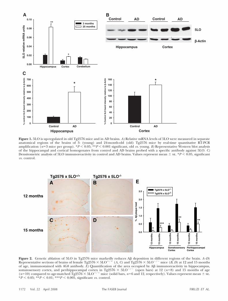
Figure 2. Genetic ablation of 5LO in Tg2576 mice markedly reduces A� deposition in different regions of the brain. A–D)Representative sections of brains of female Tg2576 � 5LO/ (A, C) and Tg2576 � 5LO�/� mice (B, D) at 12 and 15 monthsof age, immunostained with 4G8 antibody. E) Quantification of the area occupied by A� immunoreactivity in hippocampus,somatosensory cortex, and perihippocampal cortex in Tg2576 � 5LO�/� (open bars) at 12 (n8) and 15 months of age(n10) compared to age-matched Tg2576 � 5LO/ mice (solid bars, n6 and 12, respectively). Values represent mean � se.*P � 0.05; **P � 0.01; ***P � 0.005, significant vs. control.
Figure 1. 5LO is up-regulated in old Tg2576 mice and in AD brains. A) Relative mRNA levels of 5LO were measured in separateanatomical regions of the brains of 3- (young) and 24-month-old (old) Tg2576 mice by real-time quantitative RT-PCRamplification (n3 mice per group). *P � 0.05; **P � 0.001 significant, old vs. young. B) Representative Western blot analysisof the hippocampal and cortical homogenates from control and AD brains probed with a specific antibody against 5LO. C)Densitometric analysis of 5LO immunoreactivity in control and AD brains. Values represent mean � se. *P � 0.05, significantvs. control.
1172 Vol. 22 April 2008 FIRUZI ET AL.The FASEB Journal
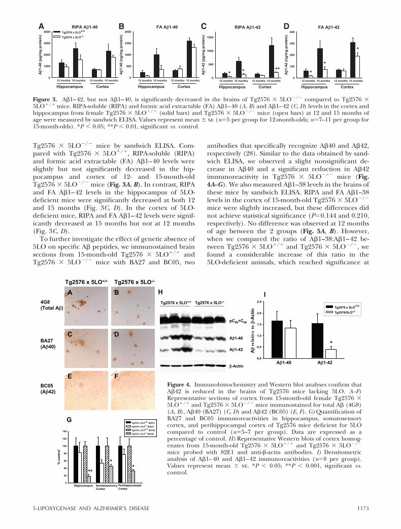
Tg2576 � 5LO�/� mice by sandwich ELISA. Com-pared with Tg2576 � 5LO/, RIPA-soluble (RIPA)and formic acid extractable (FA) A�1–40 levels wereslightly but not significantly decreased in the hip-pocampus and cortex of 12- and 15-month-oldTg2576 � 5LO�/� mice (Fig. 3A, B). In contrast, RIPAand FA A�1–42 levels in the hippocampus of 5LO-deficient mice were significantly decreased at both 12and 15 months (Fig. 3C, D). In the cortex of 5LO-deficient mice, RIPA and FA A�1–42 levels were signif-icantly decreased at 15 months but not at 12 months(Fig. 3C, D).
To further investigate the effect of genetic absence of5LO on specific A� peptides, we immunostained brainsections from 15-month-old Tg2576 � 5LO/ andTg2576 � 5LO�/� mice with BA27 and BC05, two
antibodies that specifically recognize A�40 and A�42,respectively (28). Similar to the data obtained by sand-wich ELISA, we observed a slight nonsignificant de-crease in A�40 and a significant reduction in A�42immunoreactivity in Tg2576 � 5LO�/� mice (Fig.4A--G). We also measured A�1–38 levels in the brains ofthese mice by sandwich ELISA. RIPA and FA A�1–38levels in the cortex of 15-month-old Tg2576 � 5LO�/�
mice were slightly increased, but these differences didnot achieve statistical significance (P0.144 and 0.210,respectively). No difference was observed at 12 monthsof age between the 2 groups (Fig. 5A, B). However,when we compared the ratio of A�1–38:A�1–42 be-tween Tg2576 � 5LO/ and Tg2576 � 5LO�/�, wefound a considerable increase of this ratio in the5LO-deficient animals, which reached significance at
Figure 3. A�1–42, but not A�1–40, is significantly decreased in the brains of Tg2576 � 5LO�/� compared to Tg2576 �5LO/ mice. RIPA-soluble (RIPA) and formic acid extractable (FA) A�1–40 (A, B) and A�1–42 (C, D) levels in the cortex andhippocampus from female Tg2576 � 5LO/ (solid bars) and Tg2576 � 5LO�/� mice (open bars) at 12 and 15 months ofage were measured by sandwich ELISA. Values represent mean � se (n5 per group for 12-month-olds; n7–11 per group for15-month-olds). *P � 0.05; **P � 0.01, significant vs. control.
Figure 4. Immunohistochemistry and Western blot analyses confirm thatA�42 is reduced in the brains of Tg2576 mice lacking 5LO. A–F)Representative sections of cortex from 15-month-old female Tg2576 �5LO/ and Tg2576 � 5LO�/� mice immunostained for total A� (4G8)(A, B), A�40 (BA27) (C, D) and A�42 (BC05) (E, F). G) Quantification ofBA27 and BC05 immunoreactivities in hippocampus, somatosensorycortex, and perihippocampal cortex of Tg2576 mice deficient for 5LOcompared to control (n5–7 per group). Data are expressed as apercentage of control. H) Representative Western blots of cortex homog-enates from 15-month-old Tg2576 � 5LO/ and Tg2576 � 5LO�/�
mice probed with 82E1 and anti-�-actin antibodies. I) Densitometricanalysis of A�1–40 and A�1–42 immunoreactivities (n8 per group).Values represent mean � se. *P � 0.05; **P � 0.001, significant vs.control.
11735-LIPOXYGENASE AND ALZHEIMER’S DISEASE
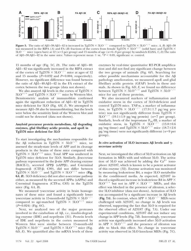
15 months of age (Fig. 5C, D). The ratio of A�1–40:A�1–42 was significantly increased in the RIPA extractof the cortex of Tg2576 � 5LO�/� mice at ages of 12and 15 months (P0.032 and P0.004, respectively).However, no significant difference was found betweenthe ratio of A�1–40:A�1–42 in the FA extract of thecortex between the two groups (data not shown).
We also assayed A� levels in the cortex of Tg2576 �5LO/ and Tg2576 � 5LO�/� mice by Western blot.Densitometric analysis of immunoblots confirmedagain the significant reduction of A�1–42 in Tg2576mice deficient for 5LO (Fig. 4H, I). We attempted tomeasure A�1–38 also by immunoblotting, but the levelswere below the sensitivity limit of the Western blot andcould not be detected (data not shown).
Amyloid precursor protein metabolism, A� degradingenzymes, glial fibrillary acidic protein, and apoE inTg2576 mice deficient for 5LO
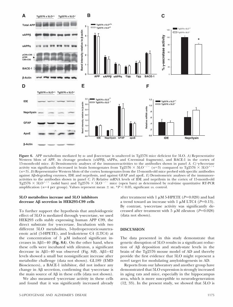
To start investigating the mechanisms responsible forthe A� reduction in Tg2576 � 5LO�/� mice, weassessed the steady-state levels of APP and its cleavageproducts in the brains of these mice compared withTg2576 � 5LO/ mice. Total APP was unaltered inTg2576 mice deficient for 5LO. Similarly, �-secretasepathway represented by the �-site APP cleaving enzyme(BACE-1), secreted APP� (sAPP�), and C-terminalfragment-� (CTF-�; C99), did not differ betweenTg2576 � 5LO�/� and Tg2576 � 5LO/ mice (Fig.6A, B). 5LO deficiency did not alter �-secretase pathwayeither, as measured by the secreted APP� (sAPP�) andC-terminal fragment-� (CTF-�; C83) in the Tg2576mice (Fig. 6A, B).
We measured �-secretase activity in brain homoge-nates of these mice and found a significantly lower�-secretase activity in 15-month-old Tg2576 � 5LO�/�
compared to age-matched Tg2576 � 5LO/ mice(P0.024) (Fig. 6C).
Next, we analyzed two of the major proteasesinvolved in the catabolism of A�, i.e., insulin-degrad-ing enzyme (IDE) and neprilysin (31). Protein levelsof IDE and neprilysin in the cortex measured byWestern blot were similar between 15-month-oldTg2576 � 5LO�/� and Tg2576 � 5LO/ mice (Fig.6D, E). We quantified also the mRNA levels of these
enzymes by real-time quantitative RT-PCR amplifica-tion and did not find any significant change betweenthe two groups of animals (Fig. 6F). Searching forother possible mechanisms accountable for the A�pathology amelioration, we measured apoE and glialfibrillary acidic protein (GFAP) levels in these ani-mals. As shown in Fig. 6D, E, we found no differencebetween Tg2576 � 5LO/ and Tg2576 � 5LO�/�
mice for any of these proteins.We also measured markers of inflammation and
oxidative stress in the cortex of 5LO-deficient andcontrol Tg2576 mice. TNF-�, a marker of inflamma-tion, in Tg2576 � 5LO�/� (17.9�1.7 pg/mg pro-tein) was not significantly different from Tg2576 �5LO/ (19.1�1.9 pg/mg protein) (n7 per group).Similarly, levels of the isoprostane F2�-III, a marker ofoxidative stress, in Tg2576 � 5LO�/� (13.8�0.6pg/mg tissue) and Tg2576 � 5LO/ mice (18.7�2.6pg/mg tissue) were not significantly different (n9 pergroup).
In vitro activation of 5LO increases A� levels and �-secretase activity
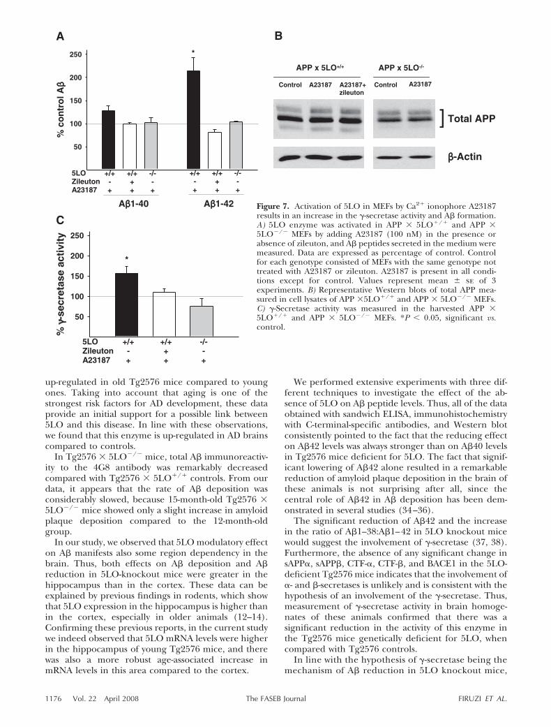
Next, we investigated the effect of 5LO activation on A�formation in MEFs with and without 5LO. The activa-tion of 5LO was achieved by adding the Ca2 iono-phore A23187, which is known to be a potent activatorof this enzyme (32). Enzyme activation was monitoredby measuring leukotriene B4, a major 5LO metabolitein the conditioned media. As expected, A23187 in-duced a significant increase in leukotriene B4 in APP �5LO/ but not in APP � 5LO�/� MEFs, and thiseffect was blocked in the presence of zileuton, a selec-tive 5LO inhibitor (data not shown). Activation of 5LOwas accompanied by a significant increase in A� levels(Fig. 7A). In contrast, when APP � 5LO�/� MEFs werechallenged with A23187, no change in A� levels wasobserved, supporting the fact that 5LO is required forthe observed effect of A23187 (Fig. 7A). Under ourexperimental conditions, A23187 did not induce anychange in APP levels (Fig. 7B). Interestingly, �-secretaseactivity was significantly increased after 5LO activationin APP � 5LO/ and the presence of zileuton wasable to block this effect. No change in �-secretaseactivity was observed in 5LO knockout MEFs (Fig. 7C).
Figure 5. The ratio of A�1–38:A�1–42 is increased in Tg2576 � 5LO�/� compared to Tg2576 � 5LO/ mice. A, B) A�1–38was measured in the RIPA (A) and FA (B) fractions of the cortex from female Tg2576 � 5LO/ (solid bars) and Tg2576 �5LO�/� mice (open bars) at 12 (n5 per group) and 15 months of age (n6–8 per group) by sandwich ELISA. C, D) The ratioof A�1–38:A�1–42 in the same mice. Values represent mean � se. *P � 0.05, significant vs. control.
1174 Vol. 22 April 2008 FIRUZI ET AL.The FASEB Journal
5LO metabolites increase and 5LO inhibitorsdecrease A� secretion in HEK293-C99 cells
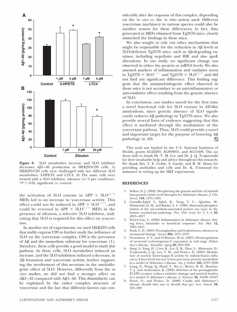
To further support the hypothesis that amyloidogeniceffect of 5LO is mediated through �-secretase, we usedHEK293 cells stably expressing human APP C99, thedirect substrate for �-secretase. Incubation with twodifferent 5LO metabolites, 5-hydroperoxyeicosatetra-enoic acid (5-HPETE), and leukotriene C4 (LTC4) atthe concentration of 5 �M induced significant in-creases in A�1–40 (Fig. 8A). On the other hand, whenthese cells were incubated with zileuton, a significantdecrease in A�1–40 was observed (Fig. 8B). A�1–42levels showed a small but nonsignificant increase aftermetabolite challenge (data not shown). GL189 (EMDBiosciences), a BACE-1 inhibitor, did not induce anychange in A� secretion, confirming that �-secretase isthe main source of A� in these cells (data not shown).
We also measured �-secretase activity in these cellsand found that it was significantly increased already
after treatment with 1 �M 5-HPETE (P0.020) and hada trend toward an increase with 1 �M LTC4 (P0.13).By contrast, �-secretase activity was significantly de-creased after treatment with 5 �M zileuton (P0.028)(data not shown).
DISCUSSION
The data presented in this study demonstrate thatgenetic disruption of 5LO results in a significant reduc-tion of A� deposition and steady-state levels in thebrain of the Tg2576 mouse model of AD and therebyprovide the first evidence that 5LO might represent anovel target for modulating amyloidogenesis in AD.
Reports from our laboratory and another group havedemonstrated that 5LO expression is strongly increasedin aging rats and mice, especially in the hippocampusarea, which is more susceptible to neurodegeneration(12, 33). In the present study, we showed that 5LO is
Figure 6. APP metabolism mediated by �- and �-secretase is unaltered in Tg2576 mice deficient for 5LO. A) RepresentativeWestern blots of APP, its cleavage products (sAPP�, sAPP�, and C-terminal fragments), and BACE-1 in the cortex of15-month-old mice. B) Densitometric analyses of the immunoreactivities to the antibodies shown in panel A. C) �-Secretaseactivity was significantly decreased in brain homogenates from Tg2576 � 5LO�/� (n3) compared to Tg2576 � 5LO/
(n3). D) Representative Western blots of the cortex homogenates from the 15-month-old mice probed with specific antibodiesagainst A�-degrading enzymes, IDE and neprilysin, and against GFAP and apoE. E) Densitometric analyses of the immunore-activities to the antibodies shown in panel C. F) Relative mRNA levels of IDE and neprilysin in the cortex of 15-month-oldTg2576 � 5LO/ (solid bars) and Tg2576 � 5LO�/� mice (open bars) as determined by real-time quantitative RT-PCRamplification (n4 per group). Values represent mean � se. *P � 0.05, significant vs. control.
11755-LIPOXYGENASE AND ALZHEIMER’S DISEASE
up-regulated in old Tg2576 mice compared to youngones. Taking into account that aging is one of thestrongest risk factors for AD development, these dataprovide an initial support for a possible link between5LO and this disease. In line with these observations,we found that this enzyme is up-regulated in AD brainscompared to controls.
In Tg2576 � 5LO�/� mice, total A� immunoreactiv-ity to the 4G8 antibody was remarkably decreasedcompared with Tg2576 � 5LO/ controls. From ourdata, it appears that the rate of A� deposition wasconsiderably slowed, because 15-month-old Tg2576 �5LO�/� mice showed only a slight increase in amyloidplaque deposition compared to the 12-month-oldgroup.
In our study, we observed that 5LO modulatory effecton A� manifests also some region dependency in thebrain. Thus, both effects on A� deposition and A�reduction in 5LO-knockout mice were greater in thehippocampus than in the cortex. These data can beexplained by previous findings in rodents, which showthat 5LO expression in the hippocampus is higher thanin the cortex, especially in older animals (12–14).Confirming these previous reports, in the current studywe indeed observed that 5LO mRNA levels were higherin the hippocampus of young Tg2576 mice, and therewas also a more robust age-associated increase inmRNA levels in this area compared to the cortex.
We performed extensive experiments with three dif-ferent techniques to investigate the effect of the ab-sence of 5LO on A� peptide levels. Thus, all of the dataobtained with sandwich ELISA, immunohistochemistrywith C-terminal-specific antibodies, and Western blotconsistently pointed to the fact that the reducing effecton A�42 levels was always stronger than on A�40 levelsin Tg2576 mice deficient for 5LO. The fact that signif-icant lowering of A�42 alone resulted in a remarkablereduction of amyloid plaque deposition in the brain ofthese animals is not surprising after all, since thecentral role of A�42 in A� deposition has been dem-onstrated in several studies (34–36).
The significant reduction of A�42 and the increasein the ratio of A�1–38:A�1–42 in 5LO knockout micewould suggest the involvement of �-secretase (37, 38).Furthermore, the absence of any significant change insAPP�, sAPP�, CTF-�, CTF-�, and BACE1 in the 5LO-deficient Tg2576 mice indicates that the involvement of�- and �-secretases is unlikely and is consistent with thehypothesis of an involvement of the �-secretase. Thus,measurement of �-secretase activity in brain homoge-nates of these animals confirmed that there was asignificant reduction in the activity of this enzyme inthe Tg2576 mice genetically deficient for 5LO, whencompared with Tg2576 controls.
In line with the hypothesis of �-secretase being themechanism of A� reduction in 5LO knockout mice,
Figure 7. Activation of 5LO in MEFs by Ca2 ionophore A23187results in an increase in the �-secretase activity and A� formation.A) 5LO enzyme was activated in APP � 5LO/ and APP �5LO�/� MEFs by adding A23187 (100 nM) in the presence orabsence of zileuton, and A� peptides secreted in the medium weremeasured. Data are expressed as percentage of control. Controlfor each genotype consisted of MEFs with the same genotype nottreated with A23187 or zileuton. A23187 is present in all condi-tions except for control. Values represent mean � se of 3experiments. B) Representative Western blots of total APP mea-sured in cell lysates of APP �5LO/ and APP � 5LO�/� MEFs.C) �-Secretase activity was measured in the harvested APP �5LO/ and APP � 5LO�/� MEFs. *P � 0.05, significant vs.control.
1176 Vol. 22 April 2008 FIRUZI ET AL.The FASEB Journal
the activation of 5LO enzyme in APP � 5LO/
MEFs led to an increase in �-secretase activity. Thiseffect could not be induced in APP � 5LO�/�, andcould be reversed in APP � 5LO/ MEFs in thepresence of zileuton, a selective 5LO inhibitor, indi-cating that 5LO is required for this effect on �-secre-tase.
In another set of experiments, we used HEK293 cellsthat stably express C99 to further study the influence of5LO on the �-secretase complex. C99 is the precursorof A� and the immediate substrate for �-secretase (1);therefore, these cells provide a good model to study thispathway. In these cells, 5LO metabolites induced anincrease, and the 5LO inhibition induced a decrease, inA� formation and �-secretase activity, further support-ing the involvement of this secretase in the amyloido-genic effect of 5LO. However, differently from the invivo studies, we did not find a stronger effect onA�1–42 compared with A�1–40. This dissimilarity canbe explained by the rather complex structure of�-secretase and the fact that different factors can con-
siderably alter the response of this complex, dependingon the in vivo vs. the in vitro system used. Different�-secretase machinery in various species could also beanother reason for these differences; in fact, datagenerated in MEFs obtained from Tg2576 mice, closelymimicked the findings in these mice.
We also sought to rule out other mechanisms thatmight be responsible for the reduction in A� levels in5LO-deficient Tg2576 mice, such as A�-degrading en-zymes, including neprilysin and IDE and also apoEalterations. In our study, no significant change wasobserved in either the protein or mRNA levels. We alsoassessed markers of inflammation and oxidative stressin Tg2576 � 5LO�/� and Tg2576 � 5LO/ and didnot find any significant difference. This finding sug-gests that the antiamyloidogenic effect observed inthese mice is not secondary to an anti-inflammatory oranti-oxidative effect resulting from the genetic absenceof 5LO.
In conclusion, our studies unveil for the first timea novel functional role for 5LO enzyme in AD-likeamyloidosis, since genetic absence of 5LO signifi-cantly reduces A� pathology in Tg2576 mice. We alsoprovide several lines of evidence suggesting that thiseffect is mediated through the modulation of the�-secretase pathway. Thus, 5LO could provide a noveland important target for the purpose of lowering A�pathology in AD.
This work was funded by the U.S. National Institutes ofHealth, grants AG22203, AG008051, and AG15408. The au-thors wish to thank Dr. V. M. Lee and Dr. J. Q. Trojanowskifor their invaluable help and advice throughout this research.We thank Drs. T. E. Golde, S. Gandy, and R. W. Doms forproviding antibodies and cells and Dr. K. Townsend forassistance in setting up the MEF experiments.
REFERENCES
1. Selkoe, D. J. (2002) Deciphering the genesis and fate of amyloidbeta-protein yields novel therapies for Alzheimer disease. J. Clin.Invest. 110, 1375–1381
2. Grundke-Iqbal, I., Iqbal, K., Tung, Y. C., Quinlan, M.,Wisniewski, H. M., and Binder, L. I. (1986) Abnormal phosphor-ylation of the microtubule-associated protein tau (tau) in Alz-heimer cytoskeletal pathology. Proc. Natl. Acad. Sci. U. S. A. 83,4913–4917
3. Wyss-Coray, T. (2006) Inflammation in Alzheimer disease: driv-ing force, bystander or beneficial response? Nat. Med. 12,1005–1015
4. Funk, C. D. (2001) Prostaglandins and leukotrienes: advances ineicosanoid biology. Science 294, 1871–1875
5. Yermakova, A. V., and O’Banion, M. K. (2001) Downregulationof neuronal cyclooxygenase-2 expression in end stage Alzhei-mer’s disease. Neurobiol. Aging 22, 823–836
6. Sung, S., Yang, H., Uryu, K., Lee, E. B., Zhao, L., Shineman, D.,Trojanowski, J. Q., Lee, V. M., and Pratico, D. (2004) Modula-tion of nuclear factor-kappa B activity by indomethacin influ-ences A beta levels but not A beta precursor protein metabolismin a model of Alzheimer’s disease. Am. J. Pathol. 165, 2197–2206
7. Liang, X., Wang, Q., Hand, T., Wu, L., Breyer, R. M., Montine,T. J., and Andreasson, K. (2005) Deletion of the prostaglandinE2 EP2 receptor reduces oxidative damage and amyloid burdenin a model of Alzheimer’s disease. J. Neurosci. 25, 10180–10187
8. Firuzi, O., and Pratico, D. (2006) Coxibs and Alzheimer’sdisease: should they stay or should they go? Ann. Neurol. 59,219–228
Figure 8. 5LO metabolites increase and 5LO inhibitordecreases A�1–40 production in HEK293-C99 cells. A)HEK293-C99 cells were challenged with two different 5LOmetabolites, 5-HPETE and LTC4. B) The same cells weretreated with a 5LO inhibitor, zileuton (n3 per condition).*P � 0.05, significant vs. control.
11775-LIPOXYGENASE AND ALZHEIMER’S DISEASE
9. Yao, Y., Clark, C. M., Trojanowski, J. Q., Lee, V. M., and Pratico,D. (2005) Elevation of 12/15 lipoxygenase products in AD andmild cognitive impairment. Ann. Neurol. 58, 623–626
10. Pratico, D., Zhukareva, V., Yao, Y., Uryu, K., Funk, C. D., Lawson,J. A., Trojanowski, J. Q., and Lee, V. M. (2004) 12/15-lipoxygenaseis increased in Alzheimer’s disease: possible involvement in brainoxidative stress. Am. J. Pathol. 164, 1655–1662
11. Mhatre, M. (2006) The role of the 5-lipoxygenase pathway inAlzheimer’s disease. Drugs Future 31, 83–89
12. Chinnici, C. M., Yao, Y., and Pratico, D. (2006) The 5-lipoxy-genase enzymatic pathway in the mouse brain: Young versus old.Neurobiol. Aging 28, 1457–1462
13. Uz, T., Pesold, C., Longone, P., and Manev, H. (1998) Aging-associated up-regulation of neuronal 5-lipoxygenase expression:putative role in neuronal vulnerability. FASEB J. 12, 439–449
14. Lammers, C. H., Schweitzer, P., Facchinetti, P., Arrang, J. M.,Madamba, S. G., Siggins, G. R., and Piomelli, D. (1996) Ara-chidonate 5-lipoxygenase and its activating protein: prominenthippocampal expression and role in somatostatin signaling.J. Neurochem. 66, 147–152
15. Morris, A. A., and Rodger, I. W. (1998) Leukotrienes and thebrain. Lancet 352, 1487–1488
16. Piomelli, D. (1994) Eicosanoids in synaptic transmission. Crit.Rev. Neurobiol. 8, 65–83
17. Manev, H., Uz, T., and Qu, T. (1998) Early upregulation ofhippocampal 5-lipoxygenase following systemic administrationof kainate to rats. Restor. Neurol. Neurosci. 12, 81–85
18. Simmet, T., and Tippler, B. (1990) Cysteinyl-leukotriene pro-duction during limbic seizures triggered by kainic acid. BrainRes. 515, 79–86
19. Zhou, Y., Wei, E. Q., Fang, S. H., Chu, L. S., Wang, M. L., Zhang,W. P., Yu, G. L., Ye, Y. L., Lin, S. C., and Chen, Z. (2006)Spatio-temporal properties of 5-lipoxygenase expression andactivation in the brain after focal cerebral ischemia in rats. LifeSci. 79, 1645–1656
20. Drazen, J. M., Yandava, C. N., Dube, L., Szczerback, N., Hippen-steel, R., Pillari, A., Israel, E., Schork, N., Silverman, E. S., Katz,D. A., and Drajesk, J. (1999) Pharmacogenetic associationbetween ALOX5 promoter genotype and the response to anti-asthma treatment. Nat. Genet. 22, 168–170
21. Qu, T., Manev, R., and Manev, H. (2001) 5-Lipoxygenase(5-LOX) promoter polymorphism in patients with early-onsetand late-onset Alzheimer’s disease. J. Neuropsychiatry Clin. Neuro-sci. 13, 304–305
22. Hsiao, K., Chapman, P., Nilsen, S., Eckman, C., Harigaya, Y.,Younkin, S., Yang, F., and Cole, G. (1996) Correlative memorydeficits, Abeta elevation, and amyloid plaques in transgenicmice. Science 274, 99–102
23. Chen, X. S., Sheller, J. R., Johnson, E. N., and Funk, C. D.(1994) Role of leukotrienes revealed by targeted disruption ofthe 5-lipoxygenase gene. Nature 372, 179–182
24. Pratico, D., Lee, V. M. Y., Trojanowski, J. Q., Rokach, J., andFitzgerald, G. A. (1998) Increased F2-isoprostanes in Alzhei-mer’s disease: evidence for enhanced lipid peroxidation in vivo.FASEB J. 12, 1777–1783
25. Pratico, D., Uryu, K., Leight, S., Trojanoswki, J. Q., and Lee,V. M. (2001) Increased lipid peroxidation precedes amyloidplaque formation in an animal model of Alzheimer amyloidosis.J. Neurosci. 21, 4183–4187
26. Uryu, K., Laurer, H., McIntosh, T., Pratico, D., Martinez, D.,Leight, S., Lee, V. M., and Trojanowski, J. Q. (2002) Repetitive
mild brain trauma accelerates Abeta deposition, lipid peroxida-tion, and cognitive impairment in a transgenic mouse model ofAlzheimer amyloidosis. J. Neurosci. 22, 446–454
27. Sung, S., Yao, Y., Uryu, K., Yang, H., Lee, V. M., Trojanowski,J. Q., and Pratico, D. (2004) Early vitamin E supplementation inyoung but not aged mice reduces Abeta levels and amyloiddeposition in a transgenic model of Alzheimer’s disease. FASEBJ. 18, 323–325
28. Suzuki, N., Cheung, T. T., Cai, X. D., Odaka, A., Otvos, L., Jr.,Eckman, C., Golde, T. E., and Younkin, S. G. (1994) Anincreased percentage of long amyloid beta protein secreted byfamilial amyloid beta protein precursor (beta APP717) mutants.Science 264, 1336–1340
29. Kukar, T., Murphy, M. P., Eriksen, J. L., Sagi, S. A., Weggen, S.,Smith, T. E., Ladd, T., Khan, M. A., Kache, R., Beard, J., Dodson,M., Merit, S., Ozols, V. V., Anastasiadis, P. Z., Das, P., Fauq, A.,Koo, E. H., and Golde, T. E. (2005) Diverse compounds mimicAlzheimer disease-causing mutations by augmenting Abeta42production. Nat. Med. 11, 545–550
30. Yagishita, S., Morishima-Kawashima, M., Tanimura, Y., Ishiura,S., and Ihara, Y. (2006) DAPT-induced intracellular accumula-tions of longer amyloid beta-proteins: further implications forthe mechanism of intramembrane cleavage by gamma-secretase.Biochemistry 45, 3952–3960
31. Leissring, M. A., Farris, W., Chang, A. Y., Walsh, D. M., Wu, X.,Sun, X., Frosch, M. P., and Selkoe, D. J. (2003) Enhancedproteolysis of beta-amyloid in APP transgenic mice preventsplaque formation, secondary pathology, and premature death.Neuron 40, 1087–1093
32. Burkert, E., Szellas, D., Radmark, O., Steinhilber, D., and Werz,O. (2003) Cell type-dependent activation of 5-lipoxygenase byarachidonic acid. J. Leukoc. Biol. 73, 191–200
33. Qu, T., Uz, T., and Manev, H. (2000) Inflammatory 5-LOXmRNA and protein are increased in brain of aging rats. Neuro-biol. Aging 21, 647–652
34. Iwatsubo, T., Odaka, A., Suzuki, N., Mizusawa, H., Nukina, N.,and Ihara, Y. (1994) Visualization of A beta 42(43) and A beta40 in senile plaques with end-specific A beta monoclonals:evidence that an initially deposited species is A beta 42(43).Neuron 13, 45–53
35. Golde, T. E. (2003) Alzheimer disease therapy: can the amyloidcascade be halted? J. Clin. Invest. 111, 11–18
36. McGowan, E., Pickford, F., Kim, J., Onstead, L., Eriksen, J., Yu,C., Skipper, L., Murphy, M. P., Beard, J., Das, P., Jansen, K.,Delucia, M., Lin, W. L., Dolios, G., Wang, R., Eckman, C. B.,Dickson, D. W., Hutton, M., Hardy, J., and Golde, T. (2005)Abeta42 is essential for parenchymal and vascular amyloiddeposition in mice. Neuron 47, 191–199
37. Weggen, S., Eriksen, J. L., Das, P., Sagi, S. A., Wang, R., Pietrzik,C. U., Findlay, K. A., Smith, T. E., Murphy, M. P., Bulter, T.,Kang, D. E., Marquez-Sterling, N., Golde, T. E., and Koo, E. H.(2001) A subset of NSAIDs lower amyloidogenic Abeta42 inde-pendently of cyclooxygenase activity. Nature 414, 212–216
38. Eriksen, J. L., Sagi, S. A., Smith, T. E., Weggen, S., Das, P.,McLendon, D. C., Ozols, V. V., Jessing, K. W., Zavitz, K. H., Koo,E. H., and Golde, T. E. (2003) NSAIDs and enantiomers offlurbiprofen target gamma-secretase and lower Abeta 42 in vivo.J. Clin. Invest. 112, 440–449
Received for publication July 17, 2007.Accepted for publication October 18, 2007.
1178 Vol. 22 April 2008 FIRUZI ET AL.The FASEB Journal