5 Cell Growth and Division - Mrs. Bays' Science Page
33
CHAPTER 5 Cell Growth and Division KEY CONCEPTS 5.1 The Cell Cycle Cells have distinct phases of growth, reproduction, and normal functions. 5.2 Mitosis and Cytokinesis Cells divide during mitosis and cytokinesis. 5.3 Regulation of the Cell Cycle Cell cycle regulation is necessary for healthy growth. 5.4 Asexual Reproduction Many organisms reproduce by cell division. 5.5 Multicellular Life Cells work together to carry out complex functions. BIOLOGY RESOURCE CENTER BIOLOGY CLASSZONE.COM View animated chapter concepts. • Mitosis • Binary Fission • Investigating Bacterial Growth • Mitosis Stage Matching Game Keep current with biology news. • Featured Stories • Strange Biology • Polls Get more information on • Cell Cycle • Asexual Reproduction • Levels of Organization 132 Unit 2: Cells
-
Upload
khangminh22 -
Category
Documents
-
view
0 -
download
0
Transcript of 5 Cell Growth and Division - Mrs. Bays' Science Page
Ti l C d 2 03240 Fil N bh 0205 2 i dd U l di L M difi d 7/26/06 10 33 AM
C H A P T E R
5 Cell Growth and Division
KEY CONCE PTS
5.1 The Cell CycleCells have distinct phases of growth, reproduction, and normal functions.
5.2 Mitosis and CytokinesisCells divide during mitosis and cytokinesis.
5.3 Regulation of the Cell CycleCell cycle regulation is necessary for healthy growth.
5.4 Asexual ReproductionMany organisms reproduce by cell division.
5.5 Multicellular LifeCells work together to carry out complex functions.
BIOLOGYRESOURCE CENTER
BIOLOGY CLASSZONE .COM
View animated chapter concepts.• Mitosis• Binary Fission• Investigating Bacterial Growth• Mitosis Stage Matching Game
Keep current with biology news.• Featured Stories• Strange Biology• Polls
Get more information on• Cell Cycle• Asexual Reproduction• Levels of Organization
132 Unit 2: Cells
bhspe-0205co_2.indd CO:132 7/26/06 12:50:23 PM
creo
cancerous healthy
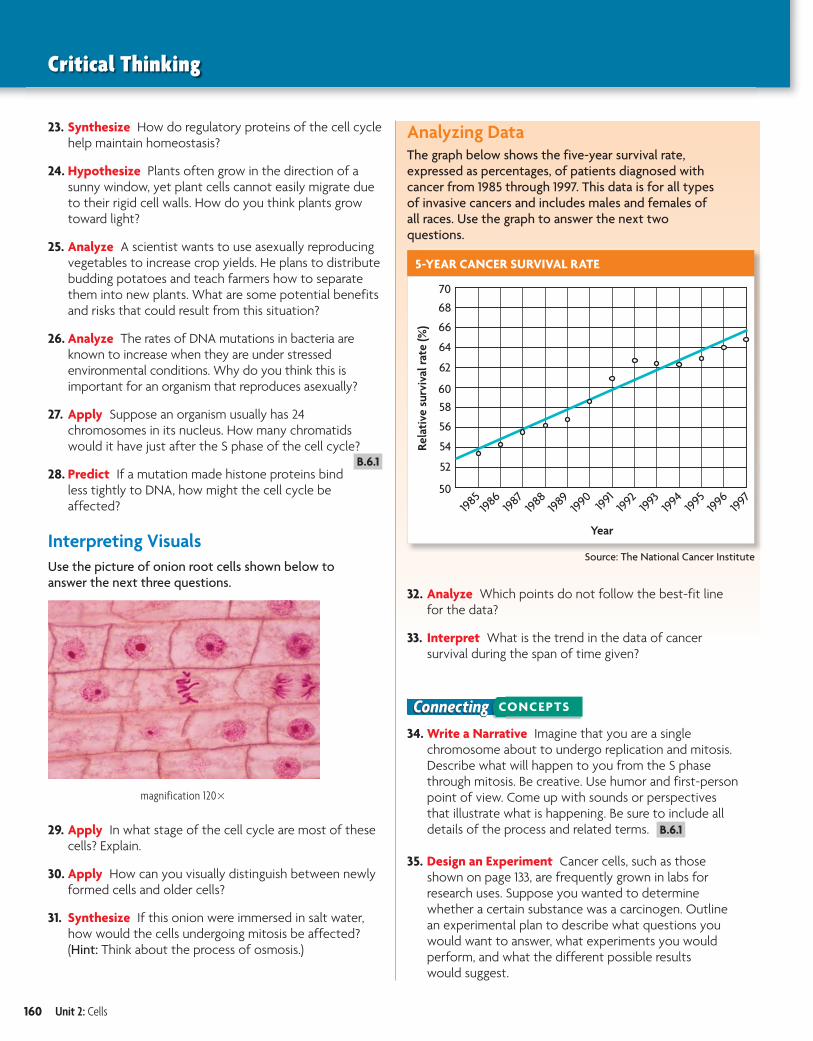
Connecting CONCEPTS
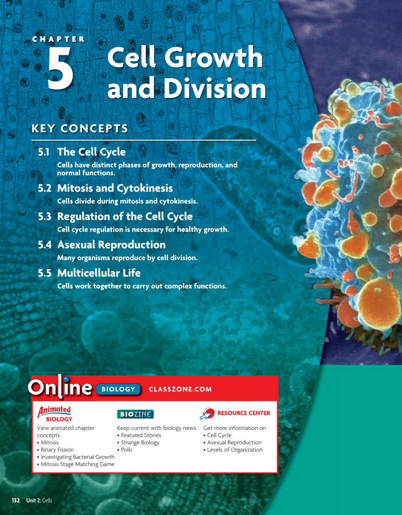
The photograph above shows a lung cancer cell undergoing cell division.
Unlike healthy cells, cancer cells can divide without limit—they are what scientists call immortal. This property is useful to scien-tists who culture cancer cells for research purposes. However, cancer cells are very dangerous in the body, where they may form tumors and invade tissues.
Human Biology These photo-graphs show half of a cancerous lung and half of a healthy lung. Gases diffuse across the surfaces of a healthy lung, so the mem-brane surfaces must be thin and moist. Exposure to substances such as tobacco smoke can cause changes in the lung cells. Cilia are destroyed, and the lung lining becomes thicker. The lungs can no longer clean themselves, so they are more susceptible to cancer-causing agents.
What does it mean for a cell to be immortal?
colored SEM; magnification 5000�
Chapter 5: Cell Growth and Division 133
b10hspe-0205co.indd 133 8/13/08 12:55:35 PM

FIGURE 5.1 Cells grow and copy their DNA during interphase. They also carry out cell-specific functions in G1 and G2. During M stage, both the nucleus (in mitosis) and cytoplasm (in cytokinesis) are divided.
Connect Many of life’s little chores such as sweeping and dusting, are quietly satisfying and rather fun. Washing dishes by hand, however, is never fun, which is why some clever person made the dishwasher. This handy invention soaks, washes, and rinses your dishes to a spot-free, sanitary sparkle. You unload the dishes, and the machine is ready to start the cycle all over again. A cell goes through a cycle, too. This cycle of growth, DNA synthesis, and division is essen-tial for an organism to grow and heal. If it goes out of control, abnormal cell growth may occur, resulting in cancer cells like those shown on the previous page.
MAIN IDEA
The cell cycle has four main stages.Just as all species have life cycles, from tiny chihuahuas to massive beluga whales, cells also have a life cycle. The cell cyclecell cycle is the regular pattern of growth, DNA duplication, and cell division that occurs in eukaryotic cells. FIGURE 5.1 shows its four main stages: gap 1, synthesis, gap 2, and mitosis. Gap 1, synthesis, and gap 2 together make up what is called interphase.
The stages of the cell cycle get their names from early studies of cell division. Scientists’ observations were limited by the microscopes of the time.
When a cell was not actively dividing, they could not see activity in it. Thus, they originally divided the cell cycle
into two parts: interphase, when the cell appeared to be at rest, and mitosis, when the cell was dividing.
Improved techniques and tools later allowed scientists to detect the copying of DNA (DNA synthesis), and they changed their description of the cell cycle to include the synthesis stage. Since they still could not see anything happening during the other parts of inter-phase, scientists named the periods between mitosis and synthesis “gap 1” and “gap 2.” Eventually, scientists learned that, during interphase, cells carry out their normal functions and undergo critical growth and preparation for cell division.
5.1 The Cell CycleKEY CONCEPT Cells have distinct phases of growth, reproduction, and normal functions.
MAIN IDEAS• The cell cycle has four main stages.
• Cells divide at different rates.
• Cell size is limited.
VOCABULARYcell cycle,cell cycle, p. 134
mitosis,mitosis, p. 135
cytokinesis,cytokinesis, p. 135
134 Unit 2: Cells
INDIANASTANDARDS
B.6.1 Describe the process of mitosis and explain that this pro-cess ordinarily results in daughter cells with a genetic make-up iden-tical to the parent cells.
b10hspe-020501.indd 134 4/19/10 12:52:18 PM
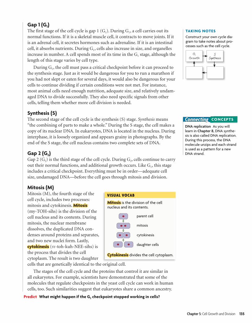
Mitosis is the division of the cell nucleus and its contents.
VISUAL VOCAB
Cytokinesis divides the cell cytoplasm.
parent cell
mitosis
cytokinesis
daughter cells
TAKING NOTESConstruct your own cycle dia-gram to take notes about pro-cesses such as the cell cycle.
G1
GrowthS
Synthesis
ConnectingDNA replication As you will learn in Chapter 8, DNA synthe-sis is also called DNA replication. During this process, the DNA molecule unzips and each strand is used as a pattern for a new DNA strand.
CONCEPTS
Gap 1 (G1)The first stage of the cell cycle is gap 1 (G1). During G1, a cell carries out its normal functions. If it is a skeletal muscle cell, it contracts to move joints. If it is an adrenal cell, it secretes hormones such as adrenaline. If it is an intestinal cell, it absorbs nutrients. During G1, cells also increase in size, and organelles increase in number. A cell spends most of its time in the G1 stage, although the length of this stage varies by cell type.
During G1, the cell must pass a critical checkpoint before it can proceed to the synthesis stage. Just as it would be dangerous for you to run a marathon if you had not slept or eaten for several days, it would also be dangerous for your cells to continue dividing if certain conditions were not met. For instance, most animal cells need enough nutrition, adequate size, and relatively undam-aged DNA to divide successfully. They also need specific signals from other cells, telling them whether more cell division is needed.
Synthesis (S)The second stage of the cell cycle is the synthesis (S) stage. Synthesis means “the combining of parts to make a whole.” During the S stage, the cell makes a copy of its nuclear DNA. In eukaryotes, DNA is located in the nucleus. During interphase, it is loosely organized and appears grainy in photographs. By the end of the S stage, the cell nucleus contains two complete sets of DNA.
Gap 2 (G2)Gap 2 (G2) is the third stage of the cell cycle. During G2, cells continue to carry out their normal functions, and additional growth occurs. Like G1, this stage includes a critical checkpoint. Everything must be in order—adequate cell size, undamaged DNA—before the cell goes through mitosis and division.
Mitosis (M)Mitosis (M), the fourth stage of the cell cycle, includes two processes: mitosis and cytokinesis. Mitosis (my-TOH-sihs) is the division of the cell nucleus and its contents. During mitosis, the nuclear membrane dissolves, the duplicated DNA con-denses around proteins and separates, and two new nuclei form. Lastly, cytokinesis (SY-toh-kuh-NEE-sihs) is the process that divides the cell cytoplasm. The result is two daughter cells that are genetically identical to the original cell.
The stages of the cell cycle and the proteins that control it are similar in all eukaryotes. For example, scientists have demonstrated that some of the molecules that regulate checkpoints in the yeast cell cycle can work in human cells, too. Such similarities suggest that eukaryotes share a common ancestry.
Predict What might happen if the G2 checkpoint stopped working in cells?
Chapter 5: Cell Growth and Division 135
bhspe-020501.indd Sec1:135 6/26/06 9:20:09 AM
creo

FIGURE 5.2 CELL LIFE SPAN
CELL TYPE APPROXIMATE LIFE SPAN
Skin cell 2 weeks
Red blood cell 4 months
Liver cell 300–500 days
Intestine—internal lining 4–5 days
Intestine—muscle and other tissues
16 years
Source: Spaulding et al., Cell 122:1.
Multicellular organisms use cell division for growth and repair.
ConnectingLymphocytes As you will learn in Chapter 31, lymphocytes are a part of your immune system. There are two major types of lymphocytes, B and T cells. Both types recognize specific antigens.
CONCEPTS
MAIN IDEA
Cells divide at different rates.Rates of cell division vary widely, as shown in FIGURE 5.2. The prokaryotic cell cycle is similar but not identical to that of eukaryotic cells. Recall that prokaryotes do not have the membrane-bound organelles and cytoskeleton found in eukaryotes. Thus, prokaryotic cells typically divide much faster than do eukaryotic cells.
The rate at which your cells divide is linked to your body’s need for those cells. In human cells, the S, G2, andM stages together usually take about 12 hours. The length of the G1 stage differs most from cell type to cell type. The rate of cell division is greater in embryos and children than it is in adults. Their cell cycle is shorter, and many of their organs are still developing. But the rate of cell division also varies within different tissues of the adult body. The internal lining of your digestive tract receives a lot of wear and tear. As a result, cells that line your stomach and intestine are replaced every few days. In contrast, cells that make up the rest of your intestine
(mainly smooth muscle) and many of your internal organs, such as lungs, kidney, and liver, divide only occasionally, in response to injury or cell death.
Cells that divide only rarely are thought to enter a stage that some scientists call G0. In G0, cells are unlikely to divide, although they continue to carry out their normal functions. Some cells, such as neurons, appear to stay perma-nently in the G0 stage. However, some data suggest that neurons actually candivide, and this question continues to be actively researched. Other cells, such as lymphocytes, a type of white blood cell, may remain in G0 for years until they recognize an invader. Once the invader binds to a lymphocyte receptor, the lymphocyte goes through rapid cell divisions to help fight infection.
Infer Do you think a skin cell would have a long or short G1 stage? Explain why.
MAIN IDEA
Cell size is limited.Cells have upper and lower size limits. If cells were too small, they could not contain all of the necessary organelles and molecules. For instance, a cell with too few mitochondria would not have enough energy to live. Nor can cells grow beyond a certain size, even if surrounded by plenty of nutrients. The upper limit on cell size is due to the ratio of cell surface area to volume. Recall that oxygen, nutrients, and wastes move across the cell membrane, or the surface of the cell. These materials must be transported in adequate amounts and with adequate speed to keep the inside of the cell functioning. But as a cell increases in size, its volume increases faster than its surface area, as shown in FIGURE 5.3. Therefore, a further increase in size could result in a surface area too small for the adequate exchange of materials.
136 Unit 2: Cells
Ti l C d 2 03240 Fil N bh 020501 i dd U i ld L M difi d 1/5/07 11 33 AMbhspe-020501.indd 136 1/5/07 11:33:55 AM
creo
5.1 ASSESSMENT
Connecting CONCEPTS
ONLINE QUIZClassZone.com
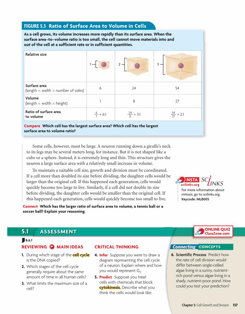
FIGURE 5.3 Ratio of Surface Area to Volume in CellsAs a cell grows, its volume increases more rapidly than its surface area. When the surface area–to–volume ratio is too small, the cell cannot move materials into and out of the cell at a sufficient rate or in sufficient quantities.
Relative size
Surface area (length � width � number of sides) 6 24 54
Volume(length � width � height) 1 8 27
Ratio of surface areato volume 6
_ 1 = 6:1 24 __ 8 = 3:1 54
__ 27 = 2:1
1 2 3
Compare Which cell has the largest surface area? Which cell has the largest surface area to volume ratio?
For more information about mitosis, go to scilinks.org.Keycode: MLB005
REVIEWING MAIN IDEAS
1. During which stage of the cell cyclecell cycle is the DNA copied?
2. Which stages of the cell cycle generally require about the same amount of time in all human cells?
3. What limits the maximum size of a cell?
CRITICAL THINKING
4. Infer Suppose you were to draw a diagram representing the cell cycle of a neuron. Explain where and how you would represent G0.
5. Predict Suppose you treat cells with chemicals that block cytokinesis.cytokinesis. Describe what you think the cells would look like.
6. Scientific Process Predict how the rate of cell division would differ between single-celled algae living in a sunny, nutrient-rich pond versus algae living in a shady, nutrient-poor pond. How could you test your prediction?
Some cells, however, must be large. A neuron running down a giraffe’s neck to its legs may be several meters long, for instance. But it is not shaped like a cube or a sphere. Instead, it is extremely long and thin. This structure gives the neuron a large surface area with a relatively small increase in volume.
To maintain a suitable cell size, growth and division must be coordinated. If a cell more than doubled its size before dividing, the daughter cells would be larger than the original cell. If this happened each generation, cells would quickly become too large to live. Similarly, if a cell did not double its size before dividing, the daughter cells would be smaller than the original cell. If this happened each generation, cells would quickly become too small to live.
Connect Which has the larger ratio of surface area to volume, a tennis ball or a soccer ball? Explain your reasoning.
Chapter 5: Cell Growth and Division 137
B.6.1
b10hspe-020501.indd 137 4/19/10 12:57:12 PM
FIGURE 5.4 This duplicated chromosome is tightly condensed. (colored SEM; magnification unknown)
ConnectingBiochemistry As you will learn in Chapter 8, a nucleotide is made of three parts: a sugar, a phosphate group, and a nitrogen-containing molecule called a base. When the sugars and phos-phate groups bond, they form the backbones of the long chains called nucleic acids.
CONCEPTS
phosphate base
sugar
Connect When you were a child, perhaps you attended a birthday party where goody bags were handed out. Whoever stuffed the bags had to make sure that each bag had exactly the same number of erasers, candies, and stickers. Other-wise, some ill-mannered child (not you, of course) might have raised a fuss if an item was missing. In a similar way, your cells must receive a full set of DNA—no more, no less—to work properly. Dividing DNA is a complicated task because the DNA is so long and stringy. Mitosis is an amazing process that efficiently sorts two sets of DNA and divides them between two nuclei.
MAIN IDEA
Chromosomes condense at the start of mitosis.DNA is a double-stranded molecule made of four different subunits called nucleotides. A chromosomechromosome is one long continuous thread of DNA that consists of numerous genes along with regulatory information. Your body cells have 46 chromosomes each. If stretched out straight and laid end to end, the DNA in just one of your cells would be about 3 meters (10 feet) long. How does it fit inside the nucleus of a microscopic cell?
DNA wraps around proteins that help organize and condense it. During interphase, or when a cell is not dividing, DNA is loosely organized—it looks a bit like spaghetti. During mitosis, however, your chromosomes are tightly condensed, as shown in FIGURE 5.4. These changes in DNA’s organization allow a cell to carry out its necessary functions. During all of interphase, proteins must access specific genes for a cell to make specific proteins or to copy the entire DNA sequence. During mitosis, the duplicated chromosomes must condense to be divided between two nuclei. If chromosomes remained stringy during mitosis, they could become entan-gled. Perhaps a cell would get two copies of one chromosome and no copies of a different one. FIGURE 5.5 shows the process that converts a chromosome from a linear strand of DNA to its highly condensed form. The key to this process is the association between DNA and proteins.
Mitosis and CytokinesisKEY CONCEPT Cells divide during mitosis and cytokinesis.
MAIN IDEAS• Chromosomes condense at the start
of mitosis.
• Mitosis and cytokinesis produce two genetically identical daughter cells.
VOCABULARYchromosome,chromosome, p. 138
histone,histone, p. 139
chromatin,chromatin, p. 139
chromatid,chromatid, p. 139
centromere,centromere, p. 139
telomere,telomere, p. 139
prophase,prophase, p. 140
metaphase,metaphase, p. 140
anaphase,anaphase, p. 140
telophase,telophase, p. 140
5.2
138 Unit 2: Cells
INDIANASTANDARDS
B.6.1 Describe the process of mitosis and explain that this pro-cess ordinarily results in daughter cells with a genetic make-up iden-tical to the parent cells.
b10hspe-020502.indd 138 4/19/10 1:00:55 PM
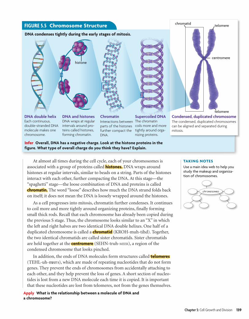
DNA double helixEach continuous, double-stranded DNA molecule makes one chromosome.
DNA and histones DNA wraps at regular intervals around pro-teins called histones, forming chromatin.
Chromatin Interactions between parts of the histones further compact the DNA.
Supercoiled DNAThe chromatin coils more and more tightly around orga-nizing proteins.
Infer Overall, DNA has a negative charge. Look at the histone proteins in the figure. What type of overall charge do you think they have? Explain.
Condensed, duplicated chromosomeThe condensed, duplicated chromosomes can be aligned and separated during mitosis.
centromere
chromatid
telomere
telomere
DNA condenses tightly during the early stages of mitosis.
FIGURE 5.5 Chromosome Structure
histone
TAKING NOTESUse a main idea web to help you study the makeup and organiza-tion of chromosomes.
chromosomes
At almost all times during the cell cycle, each of your chromosomes is associated with a group of proteins called histones. DNA wraps around histones at regular intervals, similar to beads on a string. Parts of the histones interact with each other, further compacting the DNA. At this stage—the “spaghetti” stage—the loose combination of DNA and proteins is called chromatin. The word “loose” describes how much the DNA strand folds back on itself; it does not mean the DNA is loosely wrapped around the histones.
As a cell progresses into mitosis, chromatin further condenses. It continues to coil more and more tightly around organizing proteins, finally forming small thick rods. Recall that each chromosome has already been copied during the previous S stage. Thus, the chromosome looks similar to an “X” in which the left and right halves are two identical DNA double helixes. One half of a duplicated chromosome is called a chromatid (KROH-muh-tihd). Together, the two identical chromatids are called sister chromatids. Sister chromatids are held together at the centromere (SEHN-truh-MEER), a region of the condensed chromosome that looks pinched.
In addition, the ends of DNA molecules form structures called telomeres (TEHL-uh-meers), which are made of repeating nucleotides that do not form genes. They prevent the ends of chromosomes from accidentally attaching to each other, and they help prevent the loss of genes. A short section of nucleo-tides is lost from a new DNA molecule each time it is copied. It is important that these nucleotides are lost from telomeres, not from the genes themselves.
Apply What is the relationship between a molecule of DNA and a chromosome?
Chapter 5: Cell Growth and Division 139
bhspe-020502.indd Sec2:139 6/26/06 10:16:15 AM
creo
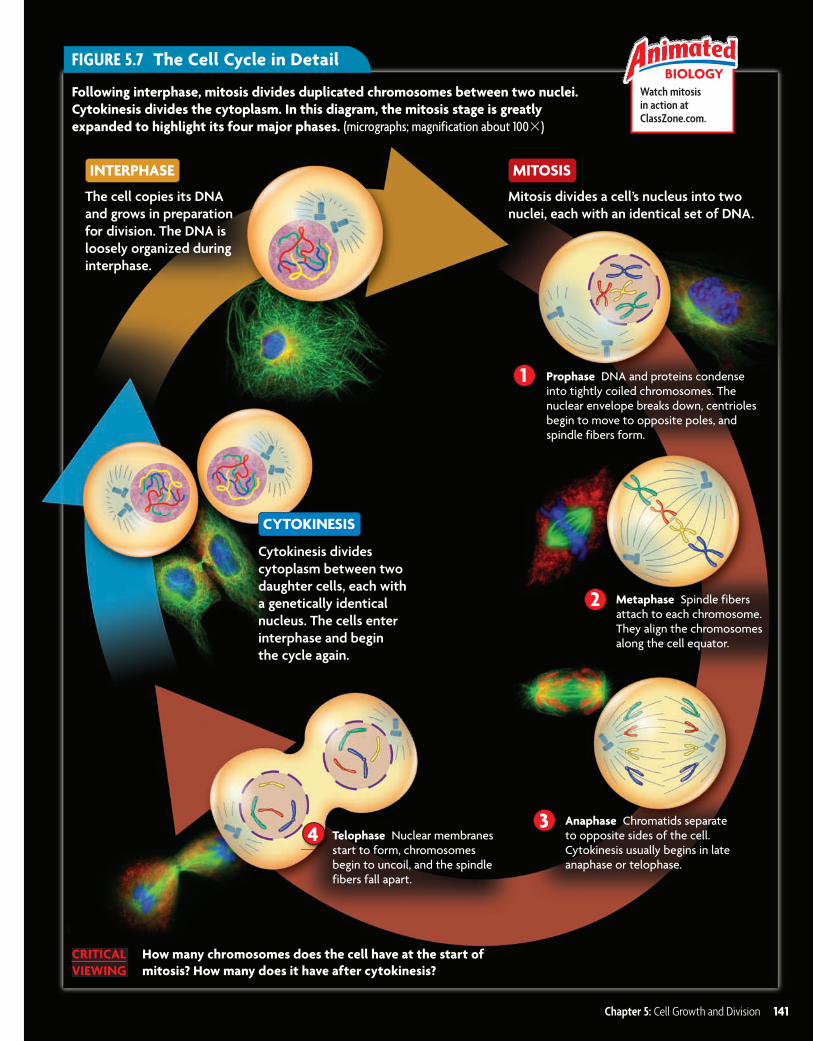
INTERPHASE
MITOSIS
CYTOKINESIS
FIGURE 5.6 The nucleus and chromosomes go through dramatic changes in a dividing cell.
Parent cellcentrioles
nucleus with DNA centrosome
spindle fibers
ConnectingCells As you will learn in Chap-ter 6, your body has two major cell types. Germ cells develop into eggs or sperm. Somatic cells make up the rest of your body.
CONCEPTS
MAIN IDEA
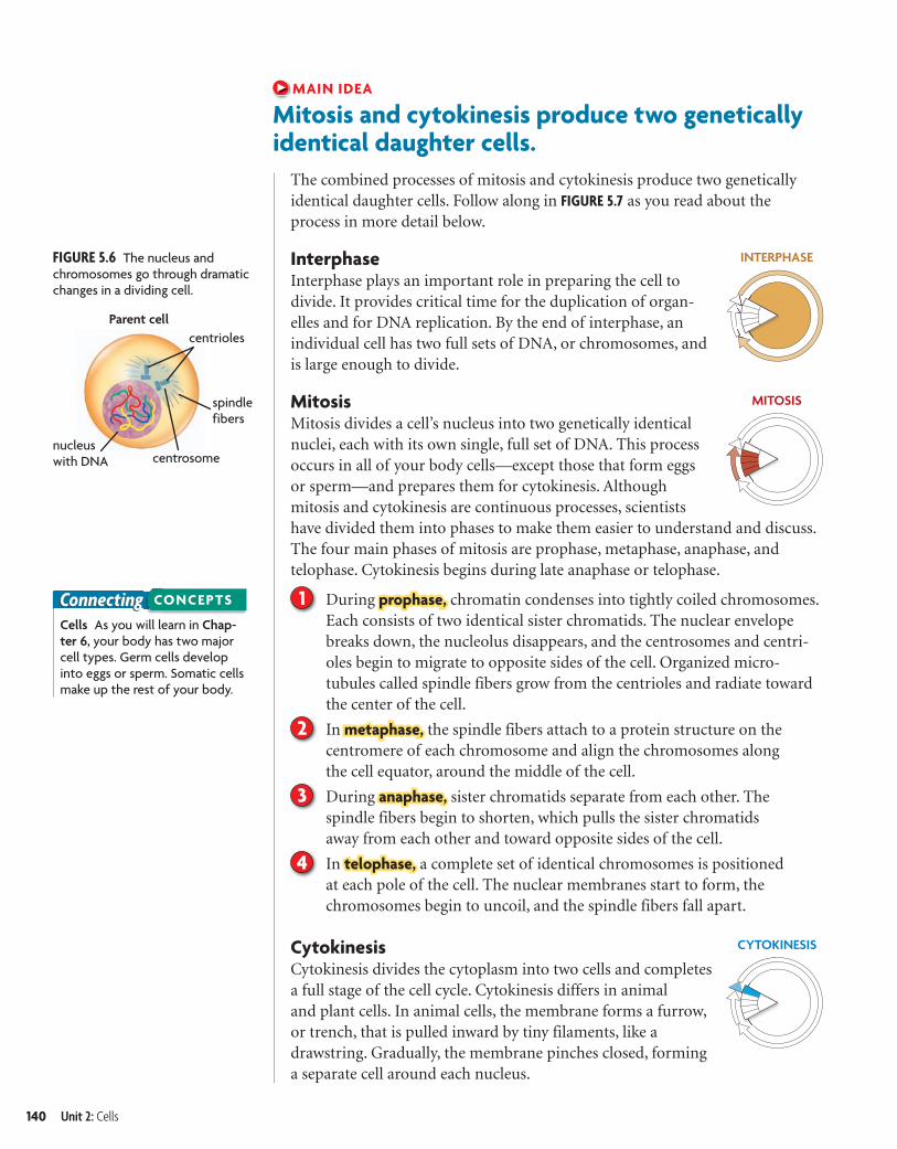
Mitosis and cytokinesis produce two geneticallyidentical daughter cells.
The combined processes of mitosis and cytokinesis produce two genetically identical daughter cells. Follow along in FIGURE 5.7 as you read about the process in more detail below.
InterphaseInterphase plays an important role in preparing the cell todivide. It provides critical time for the duplication of organ-elles and for DNA replication. By the end of interphase, an individual cell has two full sets of DNA, or chromosomes, and is large enough to divide.
MitosisMitosis divides a cell’s nucleus into two genetically identical nuclei, each with its own single, full set of DNA. This process occurs in all of your body cells—except those that form eggs or sperm—and prepares them for cytokinesis. Although mitosis and cytokinesis are continuous processes, scientists have divided them into phases to make them easier to understand and discuss. The four main phases of mitosis are prophase, metaphase, anaphase, and telophase. Cytokinesis begins during late anaphase or telophase.
1 During prophase, chromatin condenses into tightly coiled chromosomes. Each consists of two identical sister chromatids. The nuclear envelope breaks down, the nucleolus disappears, and the centrosomes and centri-oles begin to migrate to opposite sides of the cell. Organized micro-tubules called spindle fibers grow from the centrioles and radiate toward the center of the cell.
2 In metaphase, the spindle fibers attach to a protein structure on the centromere of each chromosome and align the chromosomes along the cell equator, around the middle of the cell.
3 During anaphase, sister chromatids separate from each other. The spindle fibers begin to shorten, which pulls the sister chromatids away from each other and toward opposite sides of the cell.
4 In telophase, a complete set of identical chromosomes is positioned at each pole of the cell. The nuclear membranes start to form, the chromosomes begin to uncoil, and the spindle fibers fall apart.
CytokinesisCytokinesis divides the cytoplasm into two cells and completesa full stage of the cell cycle. Cytokinesis differs in animal and plant cells. In animal cells, the membrane forms a furrow, or trench, that is pulled inward by tiny filaments, like a drawstring. Gradually, the membrane pinches closed, forming a separate cell around each nucleus.
140 Unit 2: Cells
Ti l C d 2 03240 Fil N bh 020502 i dd U l di L M difi d 6/26/06 10 15 AMbhspe-020502.indd Sec2:140 6/26/06 10:16:28 AM
creo
BIOLOGYWatch mitosis in action at ClassZone.com.
1
FIGURE 5.7 The Cell Cycle in Detail
Following interphase, mitosis divides duplicated chromosomes between two nuclei. Cytokinesis divides the cytoplasm. In this diagram, the mitosis stage is greatly expanded to highlight its four major phases. (micrographs; magnification about 100�)
2
43
Prophase DNA and proteins condense into tightly coiled chromosomes. The nuclear envelope breaks down, centrioles begin to move to opposite poles, and spindle fibers form.
MITOSISINTERPHASE
CYTOKINESIS
Mitosis divides a cell’s nucleus into two nuclei, each with an identical set of DNA.
Metaphase Spindle fibers attach to each chromosome. They align the chromosomes along the cell equator.
Telophase Nuclear membranes start to form, chromosomes begin to uncoil, and the spindle fibers fall apart.
The cell copies its DNA and grows in preparation for division. The DNA is loosely organized during interphase.
Anaphase Chromatids separate to opposite sides of the cell. Cytokinesis usually begins in late anaphase or telophase.
Cytokinesis divides cytoplasm between two daughter cells, each with a genetically identical nucleus. The cells enter interphase and begin the cycle again.
How many chromosomes does the cell have at the start of mitosis? How many does it have after cytokinesis?
CRITICAL VIEWING
Chapter 5: Cell Growth and Division 141
bhspe-020502.indd 141 1/5/07 11:35:53 AM
creo
5.2 ASSESSMENT
Connecting CONCEPTS
ONLINE QUIZClassZone.com
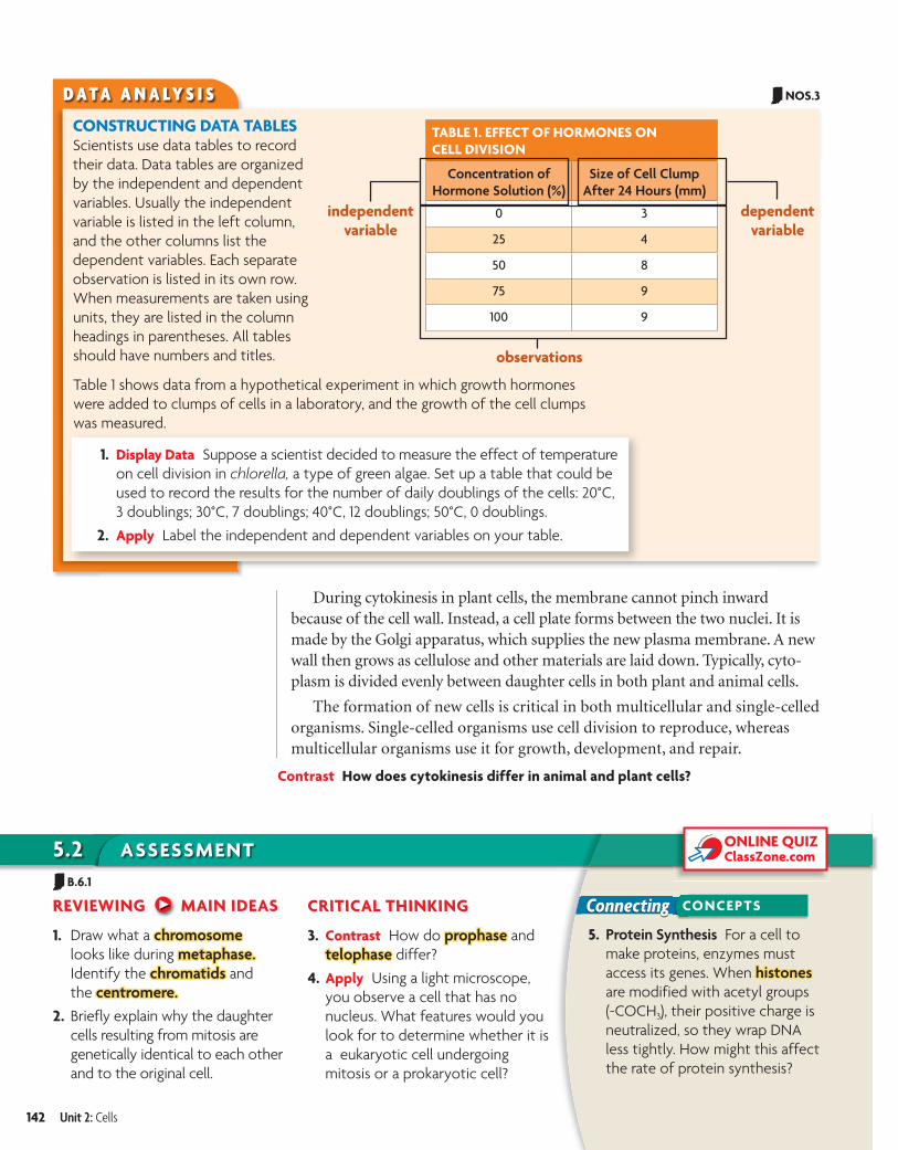
D A T A A N A LY S I S
CONSTRUCTING DATA TABLESScientists use data tables to record their data. Data tables are organized by the independent and dependent variables. Usually the independent variable is listed in the left column, and the other columns list the dependent variables. Each separate observation is listed in its own row. When measurements are taken using units, they are listed in the column headings in parentheses. All tables should have numbers and titles.
1. Display Data Suppose a scientist decided to measure the effect of temperature on cell division in chlorella, a type of green algae. Set up a table that could be used to record the results for the number of daily doublings of the cells: 20°C, 3 doublings; 30°C, 7 doublings; 40°C, 12 doublings; 50°C, 0 doublings.
2. Apply Label the independent and dependent variables on your table.
TABLE 1. EFFECT OF HORMONES ON CELL DIVISION
Concentration of Hormone Solution (%)
Size of Cell Clump After 24 Hours (mm)
0 3
25 4
50 8
75 9
100 9
independentvariable
dependentvariable
observations
Table 1 shows data from a hypothetical experiment in which growth hormones were added to clumps of cells in a laboratory, and the growth of the cell clumps was measured.
During cytokinesis in plant cells, the membrane cannot pinch inward because of the cell wall. Instead, a cell plate forms between the two nuclei. It is made by the Golgi apparatus, which supplies the new plasma membrane. A new wall then grows as cellulose and other materials are laid down. Typically, cyto-plasm is divided evenly between daughter cells in both plant and animal cells.
The formation of new cells is critical in both multicellular and single-celled organisms. Single-celled organisms use cell division to reproduce, whereas multicellular organisms use it for growth, development, and repair.
Contrast How does cytokinesis differ in animal and plant cells?
REVIEWING MAIN IDEAS
1. Draw what a chromosomechromosome looks like during metaphase.metaphase. Identify the chromatidschromatids and the centromere.centromere.
2. Briefly explain why the daughter cells resulting from mitosis are genetically identical to each other and to the original cell.
CRITICAL THINKING
3. Contrast How do prophaseprophase and telophasetelophase differ?
4. Apply Using a light microscope, you observe a cell that has no nucleus. What features would you look for to determine whether it is a eukaryotic cell undergoing mitosis or a prokaryotic cell?
5. Protein Synthesis For a cell to make proteins, enzymes must access its genes. When histoneshistones are modified with acetyl groups (-COCH3), their positive charge is neutralized, so they wrap DNA less tightly. How might this affect the rate of protein synthesis?
142 Unit 2: Cells
NOS.3
B.6.1
b10hspe-020502.indd 142 4/19/10 1:02:10 PM
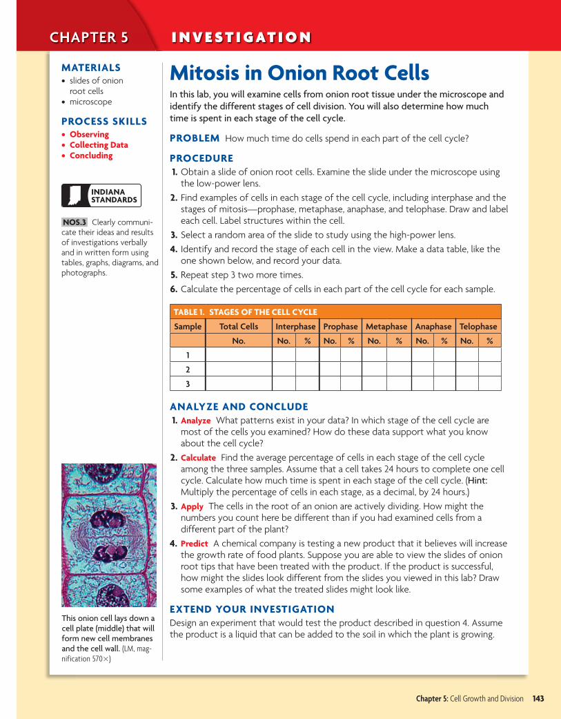
This onion cell lays down a cell plate (middle) that will form new cell membranes and the cell wall. (LM, mag-nification 570�)
CHAPTER 5 I N V E S T I G AT I O N
MATERIALS• slides of onion
root cells• microscope
PROCESS SKILLS• Observing• Collecting Data• Concluding
Mitosis in Onion Root CellsIn this lab, you will examine cells from onion root tissue under the microscope and identify the different stages of cell division. You will also determine how much time is spent in each stage of the cell cycle.
PROBLEM How much time do cells spend in each part of the cell cycle?
PROCEDURE 1. Obtain a slide of onion root cells. Examine the slide under the microscope using
the low-power lens. 2. Find examples of cells in each stage of the cell cycle, including interphase and the
stages of mitosis—prophase, metaphase, anaphase, and telophase. Draw and label each cell. Label structures within the cell.
3. Select a random area of the slide to study using the high-power lens. 4. Identify and record the stage of each cell in the view. Make a data table, like the
one shown below, and record your data. 5. Repeat step 3 two more times. 6. Calculate the percentage of cells in each part of the cell cycle for each sample.
TABLE 1. STAGES OF THE CELL CYCLE
Sample Total Cells Interphase Prophase Metaphase Anaphase Telophase
No. No. % No. % No. % No. % No. %
1
2
3
ANALYZE AND CONCLUDE 1. Analyze What patterns exist in your data? In which stage of the cell cycle are
most of the cells you examined? How do these data support what you know about the cell cycle?
2. Calculate Find the average percentage of cells in each stage of the cell cycle among the three samples. Assume that a cell takes 24 hours to complete one cell cycle. Calculate how much time is spent in each stage of the cell cycle. (Hint: Multiply the percentage of cells in each stage, as a decimal, by 24 hours.)
3. Apply The cells in the root of an onion are actively dividing. How might the numbers you count here be different than if you had examined cells from a different part of the plant?
4. Predict A chemical company is testing a new product that it believes will increase the growth rate of food plants. Suppose you are able to view the slides of onion root tips that have been treated with the product. If the product is successful, how might the slides look different from the slides you viewed in this lab? Draw some examples of what the treated slides might look like.
EXTEND YOUR INVESTIGATION Design an experiment that would test the product described in question 4. Assume the product is a liquid that can be added to the soil in which the plant is growing.
Chapter 5: Cell Growth and Division 143
INDIANASTANDARDS
NOS.3 Clearly communi-cate their ideas and results of investigations verbally and in written form using tables, graphs, diagrams, and photographs.
b10hspe-020502ci.indd 143 4/19/10 1:05:15 PM
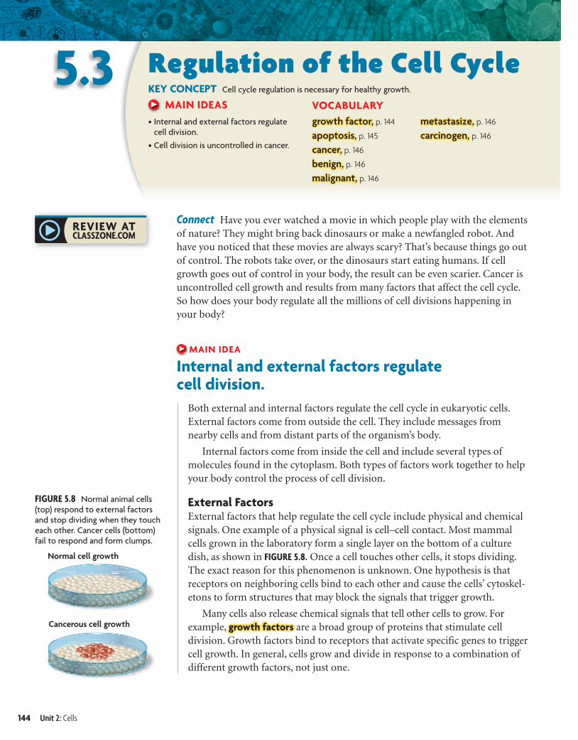
FIGURE 5.8 Normal animal cells (top) respond to external factors and stop dividing when they touch each other. Cancer cells (bottom) fail to respond and form clumps.
5.3 Regulation of the Cell CycleKEY CONCEPT Cell cycle regulation is necessary for healthy growth.
MAIN IDEAS• Internal and external factors regulate
cell division.
• Cell division is uncontrolled in cancer.
VOCABULARYgrowth factor, p. 144
apoptosis, p. 145
cancer, p. 146
benign, p. 146
malignant, p. 146
metastasize, p. 146
carcinogen, p. 146
Connect Have you ever watched a movie in which people play with the elements of nature? They might bring back dinosaurs or make a newfangled robot. And have you noticed that these movies are always scary? That’s because things go out of control. The robots take over, or the dinosaurs start eating humans. If cell growth goes out of control in your body, the result can be even scarier. Cancer is uncontrolled cell growth and results from many factors that affect the cell cycle. So how does your body regulate all the millions of cell divisions happening in your body?
MAIN IDEA
Internal and external factors regulate cell division.
Both external and internal factors regulate the cell cycle in eukaryotic cells. External factors come from outside the cell. They include messages from nearby cells and from distant parts of the organism’s body.
Internal factors come from inside the cell and include several types of molecules found in the cytoplasm. Both types of factors work together to help your body control the process of cell division.
External FactorsExternal factors that help regulate the cell cycle include physical and chemical signals. One example of a physical signal is cell–cell contact. Most mammal cells grown in the laboratory form a single layer on the bottom of a culture dish, as shown in FIGURE 5.8. Once a cell touches other cells, it stops dividing. The exact reason for this phenomenon is unknown. One hypothesis is that receptors on neighboring cells bind to each other and cause the cells’ cytoskel-etons to form structures that may block the signals that trigger growth.
Many cells also release chemical signals that tell other cells to grow. For example, growth factors are a broad group of proteins that stimulate cell division. Growth factors bind to receptors that activate specific genes to trigger cell growth. In general, cells grow and divide in response to a combination of different growth factors, not just one.
144 Unit 2: Cells
b10hspe-020503.indd 144 9/2/08 12:26:10 PM
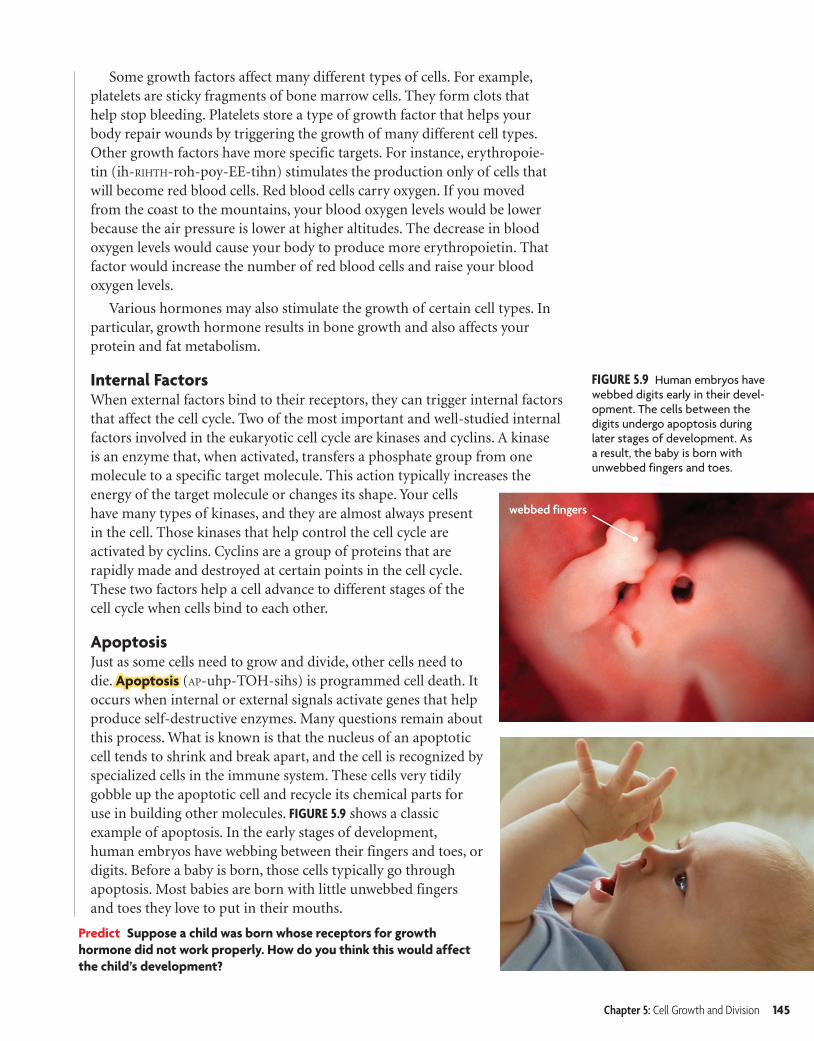
FIGURE 5.9 Human embryos have webbed digits early in their devel-opment. The cells between the digits undergo apoptosis during later stages of development. As a result, the baby is born with unwebbed fingers and toes.
webbed fingers
Some growth factors affect many different types of cells. For example, platelets are sticky fragments of bone marrow cells. They form clots that help stop bleeding. Platelets store a type of growth factor that helps your body repair wounds by triggering the growth of many different cell types. Other growth factors have more specific targets. For instance, erythropoie-tin (ih-RIHTH-roh-poy-EE-tihn) stimulates the production only of cells that will become red blood cells. Red blood cells carry oxygen. If you moved from the coast to the mountains, your blood oxygen levels would be lower because the air pressure is lower at higher altitudes. The decrease in blood oxygen levels would cause your body to produce more erythropoietin. That factor would increase the number of red blood cells and raise your blood oxygen levels.
Various hormones may also stimulate the growth of certain cell types. In particular, growth hormone results in bone growth and also affects your protein and fat metabolism.
Internal FactorsWhen external factors bind to their receptors, they can trigger internal factors that affect the cell cycle. Two of the most important and well-studied internal factors involved in the eukaryotic cell cycle are kinases and cyclins. A kinase is an enzyme that, when activated, transfers a phosphate group from one molecule to a specific target molecule. This action typically increases the energy of the target molecule or changes its shape. Your cells have many types of kinases, and they are almost always present in the cell. Those kinases that help control the cell cycle are activated by cyclins. Cyclins are a group of proteins that are rapidly made and destroyed at certain points in the cell cycle. These two factors help a cell advance to different stages of the cell cycle when cells bind to each other.
ApoptosisJust as some cells need to grow and divide, other cells need to die. Apoptosis (AP-uhp-TOH-sihs) is programmed cell death. It occurs when internal or external signals activate genes that help produce self-destructive enzymes. Many questions remain about this process. What is known is that the nucleus of an apoptotic cell tends to shrink and break apart, and the cell is recognized by specialized cells in the immune system. These cells very tidily gobble up the apoptotic cell and recycle its chemical parts for use in building other molecules. FIGURE 5.9 shows a classic example of apoptosis. In the early stages of development, human embryos have webbing between their fingers and toes, or digits. Before a baby is born, those cells typically go through apoptosis. Most babies are born with little unwebbed fingers and toes they love to put in their mouths.
Predict Suppose a child was born whose receptors for growth hormone did not work properly. How do you think this would affect the child’s development?
Chapter 5: Cell Growth and Division 145
b10hspe-020503.indd 145 8/12/08 5:07:16 PM
Ti l C d 2 03240 Fil N bh 020503 i dd U l di L M difi d 6/26/06 10 37 AM
FIGURE 5.11 This cancerous mole is an example of a skin cancer, which may metastasize quickly.
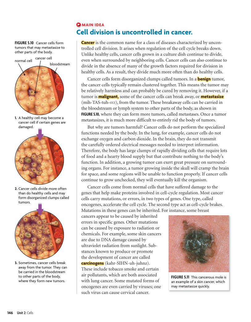
3. Sometimes, cancer cells break away from the tumor. They can be carried in the bloodstream to other parts of the body, where they form new tumors.
normal cellcancer cell
bloodstream
FIGURE 5.10 Cancer cells form tumors that may metastasize to other parts of the body.
1. A healthy cell may become a cancer cell if certain genes are damaged.
2. Cancer cells divide more often than do healthy cells and may form disorganized clumps called tumors.
MAIN IDEA
Cell division is uncontrolled in cancer.Cancer is the common name for a class of diseases characterized by uncon-trolled cell division. It arises when regulation of the cell cycle breaks down. Unlike healthy cells, cancer cells grown in a culture dish continue to divide, even when surrounded by neighboring cells. Cancer cells can also continue to divide in the absence of many of the growth factors required for division in healthy cells. As a result, they divide much more often than do healthy cells.
Cancer cells form disorganized clumps called tumors. In a benign tumor, the cancer cells typically remain clustered together. This means the tumor may be relatively harmless and can probably be cured by removing it. However, if a tumor is malignant, some of the cancer cells can break away, or metastasize (mih-TAS-tuh-SYZ), from the tumor. These breakaway cells can be carried in the bloodstream or lymph system to other parts of the body, as shown in FIGURE 5.10, where they can form more tumors, called metastases. Once a tumor metastasizes, it is much more difficult to entirely rid the body of tumors.
But why are tumors harmful? Cancer cells do not perform the specialized functions needed by the body. In the lung, for example, cancer cells do not exchange oxygen and carbon dioxide. In the brain, they do not transmit the carefully ordered electrical messages needed to interpret information. Therefore, the body has large clumps of rapidly dividing cells that require lots of food and a hearty blood supply but that contribute nothing to the body’s function. In addition, a growing tumor can exert great pressure on surround-ing organs. For instance, a tumor growing inside the skull will cramp the brain for space, and some regions will be unable to function properly. If cancer cells continue to grow unchecked, they will eventually kill the organism.
Cancer cells come from normal cells that have suffered damage to the genes that help make proteins involved in cell-cycle regulation. Most cancer cells carry mutations, or errors, in two types of genes. One type, called oncogenes, accelerate the cell cycle. The second type act as cell-cycle brakes. Mutations in these genes can be inherited. For instance, some breast cancers appear to be caused by inherited errors in specific genes. Other mutations can be caused by exposure to radiation or chemicals. For example, some skin cancers are due to DNA damage caused by ultraviolet radiation from sunlight. Sub-stances known to produce or promote the development of cancer are called carcinogens (kahr-SIHN-uh-juhnz). These include tobacco smoke and certain air pollutants, which are both associated with lung cancer. Some mutated forms of oncogenes are even carried by viruses; one such virus can cause cervical cancer.
146 Unit 2: Cells
bhspe-020503.indd Sec3:146 6/26/06 10:38:05 AM
creo
5.3 ASSESSMENT
Connecting CONCEPTS
ONLINE QUIZClassZone.com
Q U I C K L A B O BS E RVI N G
CancerIn this lab, you will compare normal cells with cancerous cells and observe the differences between them.
PROBLEM How do normal and cancerous cells compare?
PROCEDURE 1. Examine the slides of normal cells under the microscope. Draw and describe
your observations.
2. Repeat step 1 with slides of cancer cells.
ANALYZE AND CONCLUDE1. Compare How does the structure of the normal cells compare with the
structure of the cancerous cells for each of the slides you viewed?
2. Infer Cancer cells not only appear different from normal cells but they also divide more rapidly. Why do you think chemotherapy, a common treatment for cancer, results in the loss of hair?
MATERIALS• microscope• slides of normal cells• slides of cancerous cells
REVIEWING MAIN IDEAS
1. Describe what a growth factor is and how it influences the cell cycle.
2. Explain how cancer cells differ from healthy cells.
CRITICAL THINKING
3. Contrast How do benign and malignant tumors differ?
4. Hypothesize Suppose chromosomes in a skin cell are damaged by ultraviolet radia-tion. If the damaged genes do not affect cell cycle regulation, do you think the cell will become cancerous? Explain.
5. Cell Organelles Some anticancer drugs prevent microtubules from forming spindle fibers. Why do you think these drugs might be effective treatments for cancer?
Standard cancer treatment often involves both radiation and chemother-apy. Radiation therapy is the use of radiation to kill cancer cells and shrink tumors. It works by damaging a cell’s DNA so much that the cell cannot divide. Radiation is usually localized—that is, its use is targeted to a specific region—because it can also hurt healthy cells. Chemotherapy uses certain drugs, often in combination, to kill actively dividing cells. Like radiation, it kills both cancerous and healthy cells. However, chemotherapy is systemic—drugs travel throughout the entire body.
Medical researchers use laboratory-grown cancer cells in their search for cancer treatments. Much of what is known about the cell cycle has come from studies that use cancer cells. The most famous cancer cells used for research are called HeLa cells. HeLa cells were originally obtained in 1951 from a cervical tumor removed from a woman named Henrietta Lacks. This cell line continues to be grown and studied in laboratories all over the world.
Analyze HeLa cells are also used to study cell signaling processes. What might be a disadvantage of using cancer cells to study processes occurring in healthy cells?
Chapter 5: Cell Growth and Division 147
b10hspe-020503.indd 147 9/2/08 12:26:46 PM

Binary fission is the asexual reproduc-tion of a single-celled organism by division into two roughly equal parts.
VISUAL VOCAB
parent cell
daughter cells
DNA duplicates
cell begins to divide
ConnectingCell Structure Recall from Chapter 3 that many scientists hypothesize that mitochondria and chloroplasts were originally free-living prokaryotes. One piece of evidence that supports this hypothesis is the fact that these two organelles replicate much as bacteria do, through fission.
CONCEPTS
Connect In this flashy world of ours, you may think that the humble bacterium would have little chance of finding a mate. No dazzling smile, no fancy hair products, no shiny car, and—if we are brutally honest—not even a brain. With all of these limitations, it may seem that our bacteria friends would be destined to die out. And yet, bacteria are found in abundance and live just about every-where on Earth. How can there be so many bacteria?
MAIN IDEA
Binary fission is similar in function to mitosis.Reproduction is a process that makes new organisms from one or more parent organisms. It happens in two ways—sexually and asexually. Sexual reproduction involves the joining of two specialized cells called gametes (eggs and sperm cells), one from each of two parents. The offspring that result are genetically unique; they have a mixture of genes from both parents. In con-trast, asexual reproduction is the creation of offspring from a single parent and does not involve the joining of gametes. The offspring that result are, for the most part, genetically identical to each other and to the single parent.
Binary Fission and MitosisMost prokaryotes reproduce through binary fission. Binary fission (BY-nuh-ree FIHSH-uhn) is the asexual repro-duction of a single-celled organism by division into two roughly equal parts. Binary fission and mitosis have similar results. That is, both processes form two daughter cells that are genetically identical to the parent cell. However, the actual processes are different in several important ways.
As you already learned, prokary-otes such as bacteria do not have nuclei. And although they do have DNA, they have much less of it than do most eukaryotes. Also, most of a bacterium’s DNA is in the form of one circular chromosome, and bacteria have no spindle fibers.
Asexual ReproductionKEY CONCEPT Many organisms reproduce by cell division.
MAIN IDEAS• Binary fission is similar in function
to mitosis.
• Some eukaryotes reproduce through mitosis.
VOCABULARYasexual reproduction, p. 148
binary fission, p. 148
5.4
148 Unit 2: Cells
b10hspe-020504.indd 148 9/2/08 4:15:27 PM
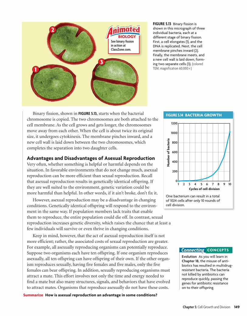
FIGURE 5.13 Binary fission is shown in this micrograph of three individual bacteria, each at a different stage of binary fission. First, a cell elongates (1), and the DNA is replicated. Next, the cell membrane pinches inward (2). Finally, the membrane meets, and a new cell wall is laid down, form-ing two separate cells (3). (colored TEM; magnification 60,000�)
See binary fi ssionin action atClassZone.com.
BIOLOGY
FIGURE 5.14 BACTERIA GROWTH
One bacterium can result in a total of 1024 cells after only 10 rounds of cell division.
1
2
3
Evolution As you will learn in Chapter 18, the misuse of anti-biotics has resulted in multidrug-resistant bacteria. The bacteria not killed by antibiotics can reproduce quickly, passing the genes for antibiotic resistance on to their offspring.
Connecting CONCEPTS
Binary fission, shown in FIGURE 5.13, starts when the bacterial chromosome is copied. The two chromosomes are both attached to the cell membrane. As the cell grows and gets longer, the chromosomes move away from each other. When the cell is about twice its original size, it undergoes cytokinesis. The membrane pinches inward, and a new cell wall is laid down between the two chromosomes, which completes the separation into two daughter cells.
Advantages and Disadvantages of Asexual ReproductionVery often, whether something is helpful or harmful depends on the situation. In favorable environments that do not change much, asexual reproduction can be more efficient than sexual reproduction. Recall that asexual reproduction results in genetically identical offspring. If they are well suited to the environment, genetic variation could be more harmful than helpful. In other words, if it ain’t broke, don’t fix it.
However, asexual reproduction may be a disadvantage in changing conditions. Genetically identical offspring will respond to the environ-ment in the same way. If population members lack traits that enable them to reproduce, the entire population could die off. In contrast, sexual reproduction increases genetic diversity, which raises the chance that at least a few individuals will survive or even thrive in changing conditions.
Keep in mind, however, that the act of asexual reproduction itself is not more efficient; rather, the associated costs of sexual reproduction are greater. For example, all asexually reproducing organisms can potentially reproduce. Suppose two organisms each have ten offspring. If one organism reproduces asexually, all ten offspring can have offspring of their own. If the other organ-ism reproduces sexually, having five females and five males, only the five females can bear offspring. In addition, sexually reproducing organisms must attract a mate. This effort involves not only the time and energy needed to find a mate but also many structures, signals, and behaviors that have evolved to attract mates. Organisms that reproduce asexually do not have these costs.
Summarize How is asexual reproduction an advantage in some conditions?
Chapter 5: Cell Growth and Division 149
bhspe-020504.indd Sec4:149 6/26/06 10:44:18 AM
creo

5.4 ASSESSMENT
Connecting CONCEPTS
ONLINE QUIZClassZone.com
FIGURE 5.15 Yeast and hydras can reproduce by budding. (hydra: LM, magnification 12�; yeast: colored SEM, magnification 3,200�)
bud
Yeast
Hydra
MAIN IDEA
Some eukaryotes reproduce through mitosis.Some eukaryotes also reproduce asexually, through mitosis. Have you ever grown a new plant from a stem cutting? Or seen a new sea star growing from the arm of another one? These new organisms are the result of mitotic repro-duction and are therefore genetically the same as the parent organism. Mitotic reproduction is especially common in simpler plants and animals. It occurs in both multicellular and unicellular eukaryotes. It can take several forms, including budding, fragmentation, and vegetative reproduction.
In budding, a small projection grows on the surface of the parent organ-ism, forming a separate new individual. The new organism may live indepen-dently or attached as part of a colony. For instance, hydras and some types of yeast reproduce by budding. Examples are shown in FIGURE 5.15.
In fragmentation, a parent organism splits into pieces, each of which can grow into a new organism. Flatworms and sea stars both reproduce by frag-mentation. Many plants, including strawberries and potatoes, reproduce via vegetative reproduction. In general, vegetative reproduction involves the modification of a stem or underground structures of the parent organism. The offspring often stay connected to the original organism, through struc-
tures called runners, for example.
Many organisms can reproduce both asexually and sexually. The form of reproduction may depend on the current conditions. The sea anem-one can reproduce in many ways. It can reproduce asexually by dividing
in half, by breaking off small pieces from its base, or by budding. It can also reproduce sexually by making eggs and sperm. Some species of anemone have males and females. In other anemone species, the same organism can produce both eggs and sperm cells.
Synthesize How might the asexual reproduction of genetically identical plants be useful to humans? How could it prove harmful to our food supply?
REVIEWING MAIN IDEAS
1. Explain how mitosis differs from binary fission.
2. Briefly explain why cutting a flatworm into pieces would not kill it.
CRITICAL THINKING
3. Infer How does an organism benefit by being able to reproduce both sexually and asexually?
4. Apply Yeasts are growing in two dishes. You treat one dish with a chemical that blocks DNA replica-tion but forget to label it. How can you identify the treated dish?
5. Ecology Two populations live in the same habitat and compete for food. The first group is larger and uses asexual reproduction; the second reproduces sexually. What could happen to cause the second group to outnum-ber the first?
150 Unit 2: Cells
b10hspe-020504.indd 150 8/12/08 5:07:49 PM
ConnectingHomeostasis As you learned in Chapter 1, homeostasis is the maintenance of a stable internal environment. Both an organism’s physiology and its behavior help it achieve homeostasis.
CONCEPTS
Multicellular LifeKEY CONCEPT Cells work together to carry out complex functions.
MAIN IDEAS• Multicellular organisms depend on
interactions among different cell types.
• Specialized cells perform specific functions.
• Stem cells can develop into different cell types.
VOCABULARYtissue,tissue, p. 151
organ,organ, p. 151
organ system,organ system, p. 151
cell differentiation,cell differentiation, p. 152
stem cell,stem cell, p. 153
Reviewhomeostasis
5.5
Connect Each of us enters this world as a screaming infant. At first, the ability to eat solid foods or take a step draws forth great praise. These general skills rapidly lose their wonder, however, and by the time you reach the age of 18, everyone wants to know what you plan to do with yourself. Will you build houses or de-sign clothing or treat patients? What will your specialty be? Cells, too, undergo specialization to carry out the complex functions required by the body.
MAIN IDEA
Multicellular organisms depend on interactions among different cell types.
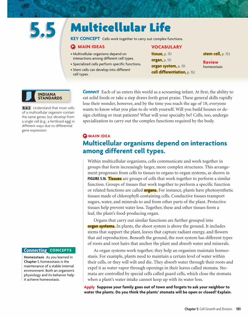
Within multicellular organisms, cells communicate and work together in groups that form increasingly larger, more complex structures. This arrange-ment progresses from cells to tissues to organs to organ systems, as shown in FIGURE 5.16. Tissues Tissues are groups of cells that work together to perform a similar function. Groups of tissues that work together to perform a specific function or related functions are called organs.organs. For instance, plants have photosynthetic tissues made of chlorophyll-containing cells. Conductive tissues transport sugars, water, and minerals to and from other parts of the plant. Protective tissues help prevent water loss. Together, these and other tissues form a leaf, the plant’s food-producing organ.
Organs that carry out similar functions are further grouped into organ systems.organ systems. In plants, the shoot system is above the ground. It includes stems that support the plant, leaves that capture radiant energy, and flowers that aid reproduction. Beneath the ground, the root system has different types of roots and root hairs that anchor the plant and absorb water and minerals.
As organ systems work together, they help an organism maintain homeo-stasis. For example, plants need to maintain a certain level of water within their cells, or they will wilt and die. They absorb water through their roots and expel it as water vapor through openings in their leaves called stomata. Sto-mata are controlled by special cells called guard cells, which close the stomata when a plant’s water intake cannot keep up with its water loss.
Apply Suppose your family goes out of town and forgets to ask your neighbor to water the plants. Do you think the plants’ stomata will be open or closed? Explain.
Chapter 5: Cell Growth and Division 151
INDIANASTANDARDS
B.6.2 Understand that most cells of a multicellular organism contain the same genes, but develop from a single cell (e.g., a fertilized egg) in different ways due to differential gene expression.
b10hspe-020505.indd 151 4/19/10 1:08:29 PM
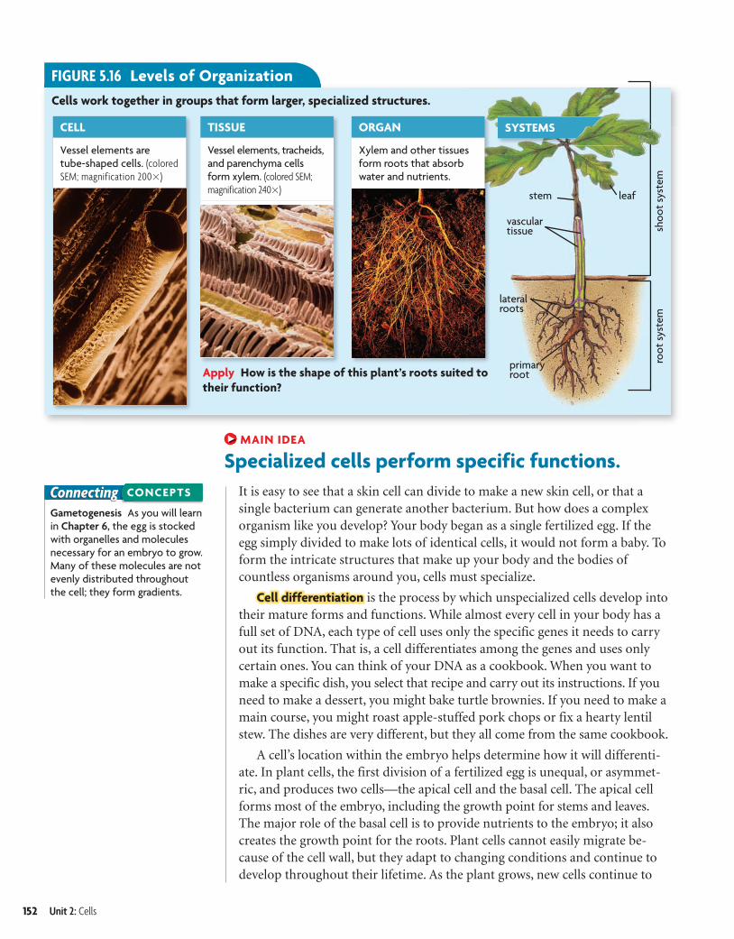
FIGURE 5.16 Levels of OrganizationCells work together in groups that form larger, specialized structures.
CELL TISSUE ORGAN
Vessel elements are tube-shaped cells. (colored SEM; magnification 200�)
Vessel elements, tracheids, and parenchyma cells form xylem. (colored SEM; magnification 240�)
Xylem and other tissues form roots that absorb water and nutrients.
Apply How is the shape of this plant’s roots suited to their function?
SYSTEMS
vasculartissue
leafstem
primary root
lateralroots
shoo
t sy
stem
root
sys
tem
ConnectingGametogenesis As you will learn in Chapter 6, the egg is stocked with organelles and molecules necessary for an embryo to grow. Many of these molecules are not evenly distributed throughout the cell; they form gradients.
CONCEPTS
MAIN IDEA
Specialized cells perform specific functions.It is easy to see that a skin cell can divide to make a new skin cell, or that a single bacterium can generate another bacterium. But how does a complex organism like you develop? Your body began as a single fertilized egg. If the egg simply divided to make lots of identical cells, it would not form a baby. To form the intricate structures that make up your body and the bodies of countless organisms around you, cells must specialize.
Cell differentiation is the process by which unspecialized cells develop into their mature forms and functions. While almost every cell in your body has a full set of DNA, each type of cell uses only the specific genes it needs to carry out its function. That is, a cell differentiates among the genes and uses only certain ones. You can think of your DNA as a cookbook. When you want to make a specific dish, you select that recipe and carry out its instructions. If you need to make a dessert, you might bake turtle brownies. If you need to make a main course, you might roast apple-stuffed pork chops or fix a hearty lentil stew. The dishes are very different, but they all come from the same cookbook.
A cell’s location within the embryo helps determine how it will differenti-ate. In plant cells, the first division of a fertilized egg is unequal, or asymmet-ric, and produces two cells—the apical cell and the basal cell. The apical cell forms most of the embryo, including the growth point for stems and leaves. The major role of the basal cell is to provide nutrients to the embryo; it also creates the growth point for the roots. Plant cells cannot easily migrate be-cause of the cell wall, but they adapt to changing conditions and continue to develop throughout their lifetime. As the plant grows, new cells continue to
152 Unit 2: Cells
b10hspe-020505.indd 152 8/12/08 5:08:16 PM
Outer Skin cells help prevent infection and dehydration (colored SEM; magnification 130�).
FIGURE 5.17 Cell DifferentiationCell differentiation in the developing animal embryo is based on location.
Outer Skin cells help prevent infection and dehydration. (colored SEM; magnification 500�)
Middle Bone cells form a hard matrix (shown) that supports and protects organs. (colored SEM; magnification 15�)
Inner Intestinal epithelia have a large surface area that increases absorption. (colored SEM; magnification 25�)
Animal embryo cross section
outer
middle
inner
differentiate based on their location. For example, cells on the outer layer of a leaf may become epidermal cells that secrete a waxy substance that helps prevent water loss. Cells on the lower leaf surface may become guard cells that control the exchange of water, air, and carbon dioxide.
In animals, an egg undergoes many rapid divisions after it is fertilized. The resulting cells can migrate to a specific area, and the cells quickly begin to differentiate. The early animal embryo generally takes the shape of a hollow ball. As the embryo develops, part of the ball folds inward, forming an inner layer and creating an opening in the outer cell layer. A middle layer of cells then forms between the other two.
As shown in FIGURE 5.17, in vertebrates, the outer cell layer differentiates to form the outer layer of skin and elements of the nervous system such as the brain and spinal cord. The middle cell layer forms bones, muscles, kidneys, and the inner layer of skin. The inner cell layer forms internal organs such as the pancreas, lungs, and digestive system lining.
Analyze Why is regulation of the differentiation process during the early stages of development so critical?
MAIN IDEA
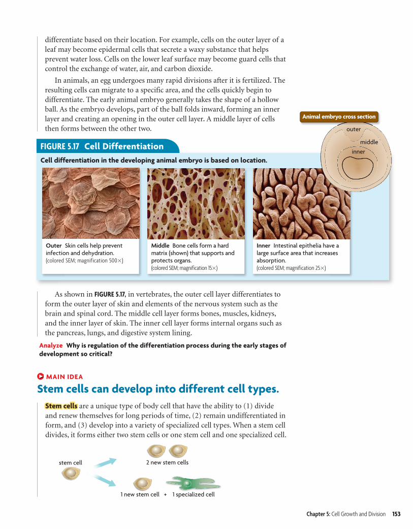
Stem cells can develop into different cell types.Stem cells are a unique type of body cell that have the ability to (1) divide and renew themselves for long periods of time, (2) remain undifferentiated in form, and (3) develop into a variety of specialized cell types. When a stem cell divides, it forms either two stem cells or one stem cell and one specialized cell.
Chapter 5: Cell Growth and Division 153
bhspe-020505.indd 153 1/5/07 11:40:38 AM
creo
Ti l C d 2 03240 Fil N bh 020505 i dd U l di L M difi d 6/26/06 11 02 AM
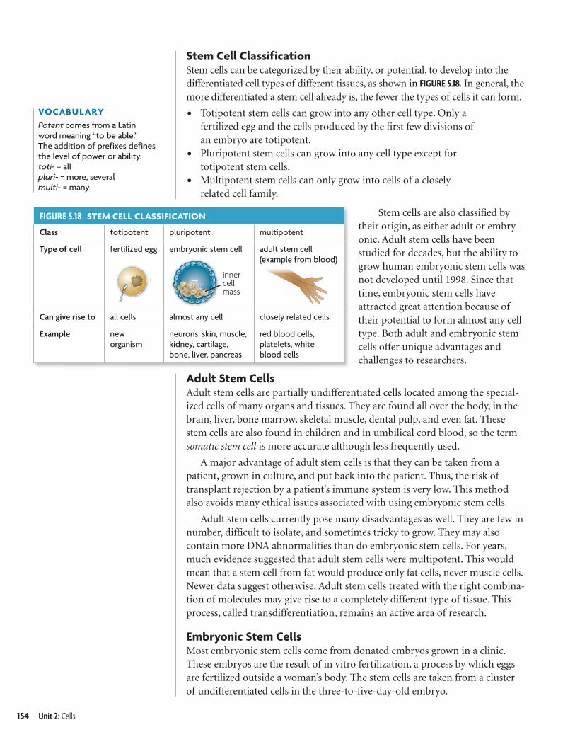
FIGURE 5.18 STEM CELL CLASSIFICATION
Class totipotent pluripotent multipotent
Type of cell fertilized egg embryonic stem cell adult stem cell(example from blood)
Can give rise to all cells almost any cell closely related cells
Example neworganism
neurons, skin, muscle, kidney, cartilage, bone, liver, pancreas
red blood cells, platelets, white blood cells
innercellmass
VOCABULARYPotent comes from a Latin word meaning “to be able.” The addition of prefixes defines the level of power or ability. toti- = all pluri- = more, severalmulti- = many
Stem Cell ClassificationStem cells can be categorized by their ability, or potential, to develop into the differentiated cell types of different tissues, as shown in FIGURE 5.18. In general, the more differentiated a stem cell already is, the fewer the types of cells it can form.
• Totipotent stem cells can grow into any other cell type. Only a fertilized egg and the cells produced by the first few divisions of an embryo are totipotent.
• Pluripotent stem cells can grow into any cell type except for totipotent stem cells.
• Multipotent stem cells can only grow into cells of a closely related cell family.
Stem cells are also classified by their origin, as either adult or embry-onic. Adult stem cells have been studied for decades, but the ability to grow human embryonic stem cells was not developed until 1998. Since that time, embryonic stem cells have attracted great attention because of their potential to form almost any cell type. Both adult and embryonic stem cells offer unique advantages and challenges to researchers.
Adult Stem CellsAdult stem cells are partially undifferentiated cells located among the special-ized cells of many organs and tissues. They are found all over the body, in the brain, liver, bone marrow, skeletal muscle, dental pulp, and even fat. These stem cells are also found in children and in umbilical cord blood, so the term somatic stem cell is more accurate although less frequently used.
A major advantage of adult stem cells is that they can be taken from a patient, grown in culture, and put back into the patient. Thus, the risk of transplant rejection by a patient’s immune system is very low. This method also avoids many ethical issues associated with using embryonic stem cells.
Adult stem cells currently pose many disadvantages as well. They are few in number, difficult to isolate, and sometimes tricky to grow. They may also contain more DNA abnormalities than do embryonic stem cells. For years, much evidence suggested that adult stem cells were multipotent. This would mean that a stem cell from fat would produce only fat cells, never muscle cells. Newer data suggest otherwise. Adult stem cells treated with the right combina-tion of molecules may give rise to a completely different type of tissue. This process, called transdifferentiation, remains an active area of research.
Embryonic Stem CellsMost embryonic stem cells come from donated embryos grown in a clinic. These embryos are the result of in vitro fertilization, a process by which eggs are fertilized outside a woman’s body. The stem cells are taken from a cluster of undifferentiated cells in the three-to-five-day-old embryo.
154 Unit 2: Cells
bhspe-020505.indd Sec5:154 6/26/06 11:04:21 AM
5.5 ASSESSMENT
Connecting CONCEPTS
ONLINE QUIZClassZone.com
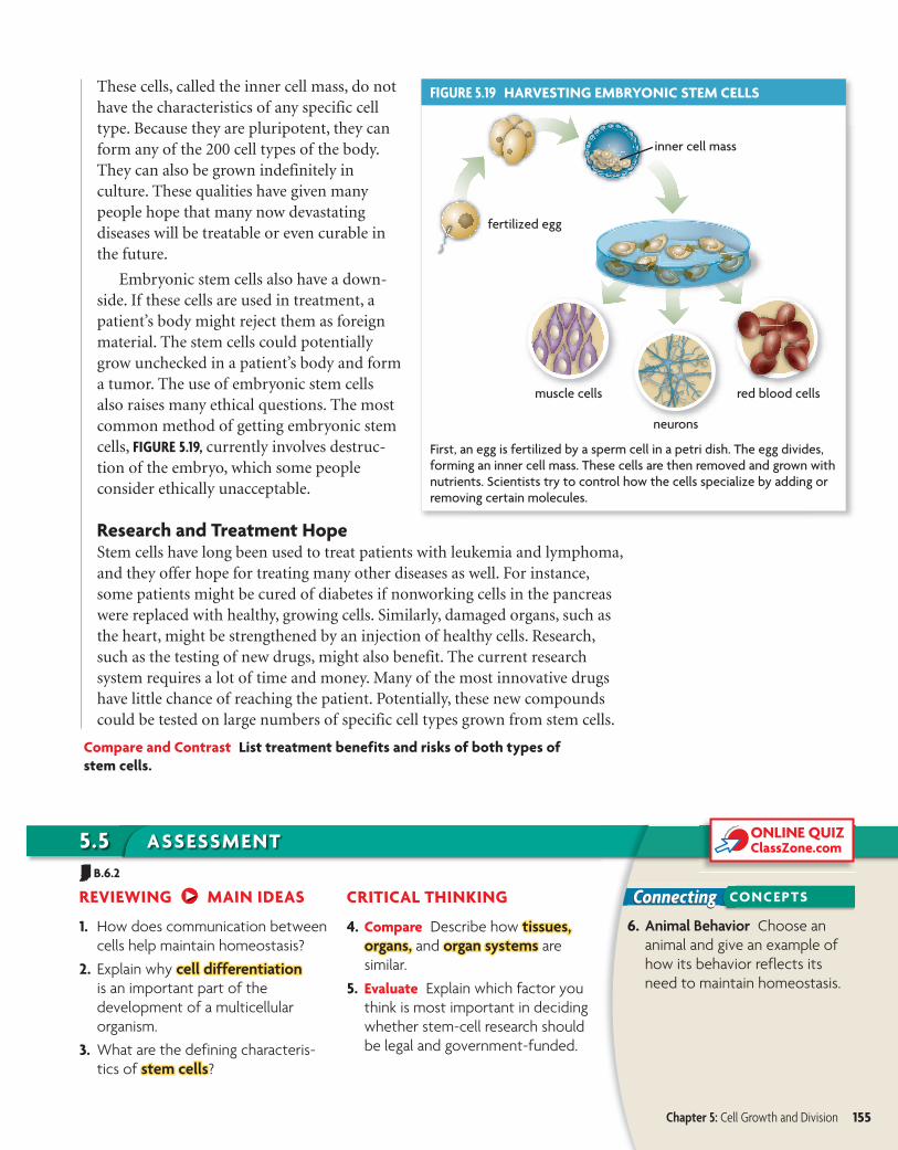
FIGURE 5.19 HARVESTING EMBRYONIC STEM CELLS
First, an egg is fertilized by a sperm cell in a petri dish. The egg divides, forming an inner cell mass. These cells are then removed and grown with nutrients. Scientists try to control how the cells specialize by adding or removing certain molecules.
These cells, called the inner cell mass, do not have the characteristics of any specific cell type. Because they are pluripotent, they can form any of the 200 cell types of the body. They can also be grown indefinitely in culture. These qualities have given many people hope that many now devastating diseases will be treatable or even curable in the future.
Embryonic stem cells also have a down-side. If these cells are used in treatment, a patient’s body might reject them as foreign material. The stem cells could potentially grow unchecked in a patient’s body and form a tumor. The use of embryonic stem cells also raises many ethical questions. The most common method of getting embryonic stem cells, FIGURE 5.19, currently involves destruc-tion of the embryo, which some people consider ethically unacceptable.
Research and Treatment HopeStem cells have long been used to treat patients with leukemia and lymphoma, and they offer hope for treating many other diseases as well. For instance, some patients might be cured of diabetes if nonworking cells in the pancreas were replaced with healthy, growing cells. Similarly, damaged organs, such as the heart, might be strengthened by an injection of healthy cells. Research, such as the testing of new drugs, might also benefit. The current research system requires a lot of time and money. Many of the most innovative drugs have little chance of reaching the patient. Potentially, these new compounds could be tested on large numbers of specific cell types grown from stem cells.
Compare and Contrast List treatment benefits and risks of both types of stem cells.
REVIEWING MAIN IDEAS
1. How does communication between cells help maintain homeostasis?
2. Explain why cell differentiationcell differentiation is an important part of the development of a multicellular organism.
3. What are the defining characteris-tics of stem cellsstem cells?
CRITICAL THINKING
4. Compare Describe how tissues,tissues, organs,organs, and organ systemsorgan systems are similar.
5. Evaluate Explain which factor you think is most important in deciding whether stem-cell research should be legal and government-funded.
6. Animal Behavior Choose an animal and give an example of how its behavior reflects its need to maintain homeostasis.
Chapter 5: Cell Growth and Division 155
B.6.2
b10hspe-020505.indd 155 4/19/10 1:11:35 PM

MATERIALS• plastic knife• phenolphthalein agar• metric ruler• 250-mL beaker• 100-mL graduated cylinder• 100 mL sodium hydroxide
solution• timer• plastic spoon• paper towel
Modeling Cell Surface Area–to–Volume RatioA cell’s surface area–to–volume ratio affects the amount of material that can diffuse across the membrane and throughout the cell. You will make cell models to determine how this ratio changes as cell size increases.
SKILLS Modeling, Inferring
PROBLEM Which cell has the greatest surface area–to–volume ratio?
PROCEDURE 1. Make three model cells by using the knife to cut three cubes from the phenolphthalein
agar. Cell A should be 3 cm on each side, cell B should be 2 cm on each side, and cell C should be 1 cm on each side. Use the ruler to make exact measurements.
2. Calculate the area of one side of each cell. Calculate the total surface area of each cell. Record your data in Table 1. (Hint: Multiply the area of one side by the number of sides.)
3. Calculate the volume of each cell. Record your data in the table. (Hint: Multiply the length by the width by the height of the cube.)
4. Calculate the ratio of surface area to volume for each cell. For example, for cell A, the ratio would be 54 cm2:27 cm3 � 2:1 � 2. Record your data.
5. Put the model cells in the beaker. Carefully cover them with sodium hydroxide solution, which turns the agar pink. Soak the cells in solution for four minutes. Use a spoon to turn the cells repeatedly throughout that time.
6. Remove the cells from solution and dry them on the paper towel.7. Use the knife to cut each cube in half. Measure the distance from the edge of the cell
to the inner edge of the pink line. This shows how far the sodium hydroxide diffused.
TABLE 1. CALCULATIONS OF CELL SIZE
Cell Area of One Side (cm2)
Total SurfaceArea (cm2)
Volume of Cell (cm3)
Surface Area–to–Volume Ratio
A
B
C
ANALYZE AND CONCLUDE 1. Analyze How does the surface area–to–volume ratio change as cell size increases? How
might this affect the diffusion of materials throughout a cell? 2. Apply Identify which cell turned pink in the greatest proportion, and explain how this
relates to cell size.3. Apply How does a cell’s surface area–to–volume ratio affect its ability to stay alive?
CHAPTER 5 O P T I O N S F O R I N Q U I RY
Use these inquiry-based labs and online activities to deepen your understanding of cell growth and development.
I N V E S T I G AT I O N
156 Unit 2: Cells
NOS.6 Use analogies and models (math-ematical and physical) to simplify and repre-sent systems that are difficult to understand or directly experience due to their size, time scale, or complexity, and recognize the limi-tations of analogies and models.
INDIANASTANDARDS
b10hspe-0205oi.indd 156 2/4/10 6:00:51 AM
BIOLOGY
CLASSZONE .COM
W E B Q U E STCancer occurs when the cell cycle breaks down. In this WebQuest, you will learn about the most common cancer in the United States—skin cancer. Explore its causes, how cells become cancerous, and why prevention is truly the best medicine.
A N I M AT E D B I O L O G YMitosis Stage Matching GameHow well can you recognize the stages of mitosis? Test your skills by categorizing images showing different phases of mitosis.
V I RT UA L L A BInvestigating Bacterial GrowthNot all bacteria thrive in the same environ-mental conditions. In this interactive lab, you will determine which strains of bacteria grow in an environment with oxygen and which strains grow without oxygen.
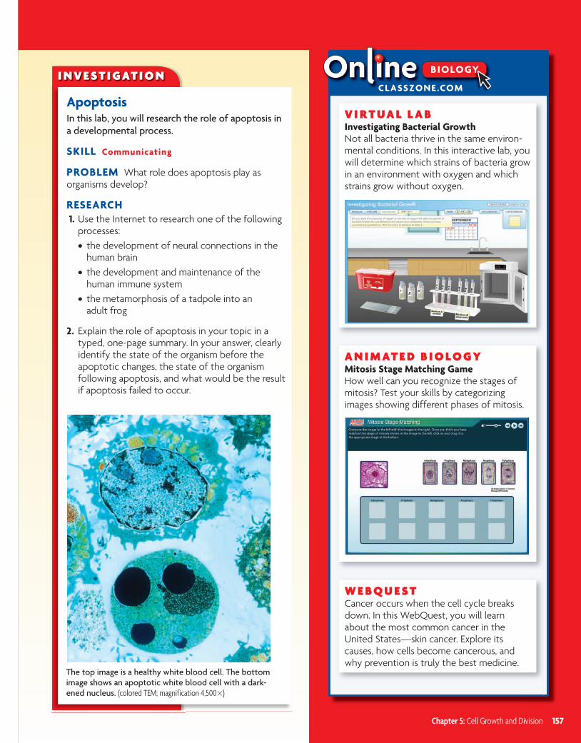
The top image is a healthy white blood cell. The bottom image shows an apoptotic white blood cell with a dark-ened nucleus. (colored TEM; magnification 4,500�)
I N V E S T I G AT I O N
ApoptosisIn this lab, you will research the role of apoptosis in a developmental process.
SKILL Communicating
PROBLEM What role does apoptosis play as organisms develop?
RESEARCH 1. Use the Internet to research one of the following
processes:• the development of neural connections in the
human brain• the development and maintenance of the
human immune system• the metamorphosis of a tadpole into an
adult frog
2. Explain the role of apoptosis in your topic in a typed, one-page summary. In your answer, clearly identify the state of the organism before the apoptotic changes, the state of the organism following apoptosis, and what would be the result if apoptosis failed to occur.
Chapter 5: Cell Growth and Division 157
b10hspe-0205oi.indd 157 8/12/08 5:58:38 PM
Titl C d 2 03240 Fil N bh 0205 i dd U t t t L t M difi d 2/23/07 3 41 PM
CHAPTER
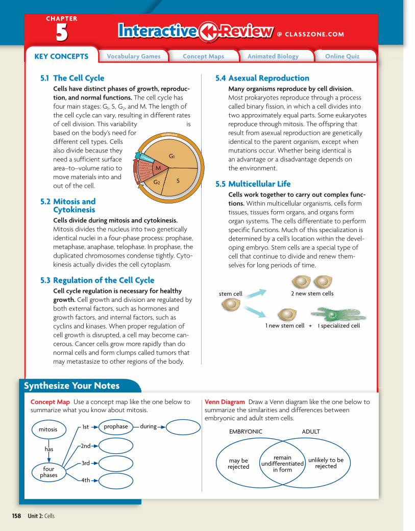
@ CLASSZONE .COM5KEY CONCEPTS Vocabulary Games Concept Maps Animated Biology Online Quiz
5.1 The Cell CycleCells have distinct phases of growth, reproduc-tion, and normal functions. The cell cycle has four main stages: G1, S, G2, and M. The length of the cell cycle can vary, resulting in different rates of cell division. This variability is based on the body’s need for different cell types. Cells also divide because they need a sufficient surface area–to–volume ratio to move materials into and out of the cell.
5.2 Mitosis and CytokinesisCells divide during mitosis and cytokinesis. Mitosis divides the nucleus into two genetically identical nuclei in a four-phase process: prophase, metaphase, anaphase, telophase. In prophase, the duplicated chromosomes condense tightly. Cyto-kinesis actually divides the cell cytoplasm.
5.3 Regulation of the Cell CycleCell cycle regulation is necessary for healthy growth. Cell growth and division are regulated by both external factors, such as hormones and growth factors, and internal factors, such as cyclins and kinases. When proper regulation of cell growth is disrupted, a cell may become can-cerous. Cancer cells grow more rapidly than do normal cells and form clumps called tumors that may metastasize to other regions of the body.
5.4 Asexual ReproductionMany organisms reproduce by cell division. Most prokaryotes reproduce through a process called binary fission, in which a cell divides into two approximately equal parts. Some eukaryotes reproduce through mitosis. The offspring that result from asexual reproduction are genetically identical to the parent organism, except when mutations occur. Whether being identical is an advantage or a disadvantage depends on the environment.
5.5 Multicellular LifeCells work together to carry out complex func-tions. Within multicellular organisms, cells form tissues, tissues form organs, and organs form organ systems. The cells differentiate to perform specific functions. Much of this specialization is determined by a cell’s location within the devel-oping embryo. Stem cells are a special type of cell that continue to divide and renew them-selves for long periods of time.
Concept Map Use a concept map like the one below to summarize what you know about mitosis.
Venn Diagram Draw a Venn diagram like the one below to summarize the similarities and differences between embryonic and adult stem cells.
remain undifferentiated
in form
mitosis
unlikely to be rejected
EMBRYONIC ADULT
four phases
has
prophase1st
2nd
3rd
4th
during
may be rejected
Synthesize Your Notes
158 Unit 2: Cells
bhspe-0205cr.indd 158 2/23/07 3:42:07 PM
creo
5.1 cell cycle, p. 134 mitosis, p. 135 cytokinesis, p. 135
5.2 chromosome, p. 138 histone, p. 139 chromatin, p. 139 chromatid, p. 139 centromere, p. 139 telomere, p. 139
prophase, p. 140 metaphase, p. 140 anaphase, p. 140 telophase, p. 140
5.3 growth factor, p. 144 apoptosis, p. 145 cancer, p. 146 benign, p. 146 malignant, p. 146 metastasize, p. 146 carcinogen, p. 146
5.4 asexual reproduction, p. 148 binary fission, p. 148
5.5 tissue, p. 151 organ, p. 151 organ system, p. 151 cell differentiation, p. 152 stem cell, p. 153
Chapter Vocabulary
Reviewing VocabularyVisualize VocabularyFor each term below, draw a simple picture that represents the meaning of the word. Here is an example for mitosis.
1. prophase
2. metaphase
3. anaphase
4. telophase
5. cytokinesis
6. centromere
7. telomere
Word Origins 8. The prefix pro- means “earlier than” or “prior to.” Explain
how this meaning relates to the word prophase.
9. The prefix telo- means “distant, far, or end.” How does this meaning relate to the words telophase and telomere?
10. The term mitosis comes from the Greek root mitos, which means “thread.” How does this meaning relate to the process of mitosis?
Reviewing MAIN IDEAS 11. The cell cycle has four main stages—G1, S, G2, and M.
What occurs in the cell during each stage?
12. Compare the rates of cell division occurring in your neurons and your hair follicles.
13. What is the relationship between a cell’s surface area and its volume?
14. You know that a chromosome is a very long, continuous strand of DNA. How do proteins help condense chromosomes?
15. Describe what happens in each main phase of mitosis—prophase, metaphase, anaphase, and telophase.
16. How does the process of cytokinesis differ from the process of mitosis?
17. Increased levels of cyclin help trigger a cell to divide. Do you think a growth factor would increase or decrease cyclin levels? Explain.
18. Describe how uncontrolled cell division is dangerous in organisms.
19. List one similarity and one difference between binary fission and mitosis.
20. You pull a leaf from a plant and place it in a cup of water. After a week, roots start to grow from the leaf. What type of reproduction has occurred, and what role does mitosis play in it?
21. Briefly describe how cell differentiation occurs in the developing animal embryo.
22. List three characteristics of all stem cells.
Chapter Assessment
Chapter 5: Cell Growth and Division 159
B.6.1
B.6.1
B.6.1
B.6.3
158-161_b10hspe-0205cr.indd 159 5/3/10 9:42:40 AM
Critical Thinking
23. Synthesize How do regulatory proteins of the cell cycle help maintain homeostasis?
24. Hypothesize Plants often grow in the direction of a sunny window, yet plant cells cannot easily migrate due to their rigid cell walls. How do you think plants grow toward light?
25. Analyze A scientist wants to use asexually reproducing vegetables to increase crop yields. He plans to distribute budding potatoes and teach farmers how to separate them into new plants. What are some potential benefits and risks that could result from this situation?
26. Analyze The rates of DNA mutations in bacteria are known to increase when they are under stressed environmental conditions. Why do you think this is important for an organism that reproduces asexually?
27. Apply Suppose an organism usually has 24 chromosomes in its nucleus. How many chromatids would it have just after the S phase of the cell cycle?
28. Predict If a mutation made histone proteins bind less tightly to DNA, how might the cell cycle be affected?
Interpreting VisualsUse the picture of onion root cells shown below to answer the next three questions.
29. Apply In what stage of the cell cycle are most of these cells? Explain.
30. Apply How can you visually distinguish between newly formed cells and older cells?
31. Synthesize If this onion were immersed in salt water, how would the cells undergoing mitosis be affected? (Hint: Think about the process of osmosis.)
Analyzing DataThe graph below shows the five-year survival rate, expressed as percentages, of patients diagnosed with cancer from 1985 through 1997. This data is for all types of invasive cancers and includes males and females of all races. Use the graph to answer the next two questions.
32. Analyze Which points do not follow the best-fit line for the data?
33. Interpret What is the trend in the data of cancer survival during the span of time given?
Connecting CONCEPTS
34. Write a Narrative Imagine that you are a single chromosome about to undergo replication and mitosis. Describe what will happen to you from the S phase through mitosis. Be creative. Use humor and first-person point of view. Come up with sounds or perspectives that illustrate what is happening. Be sure to include all details of the process and related terms.
35. Design an Experiment Cancer cells, such as those shown on page 133, are frequently grown in labs for research uses. Suppose you wanted to determine whether a certain substance was a carcinogen. Outline an experimental plan to describe what questions you would want to answer, what experiments you would perform, and what the different possible results would suggest.
5-YEAR CANCER SURVIVAL RATE
Source: The National Cancer Institute
magnification 120�
160 Unit 2: Cells
B.6.1
B.6.1
158-161_b10hspe-0205cr.indd 160 5/3/10 9:43:13 AM
For more test practice,go to ClassZone.com.
Chapter 5: Cell Growth and Division 161
B.6.1; B.6.2; B.6.3; NOS.1
1 Scientists searching for new anticancer drugs treat a cell culture with a certain compound. Following treatment, they notice that the culture has stopped growing. Untreated cells from the same culture, however, have continued to grow. These results could indicate that the compound blocks the normal cell cycle. What else could have caused these results?
A The compound had degraded.
B The compound prevented cells from mutating.
C The compound killed the treated cells.
D The compound had no effect.
The untreated cells serve as a control in this experiment. Therefore, differences between the treated and untreated cells should be the result of the drug. If the drug has no effect, the two groups of cells should be the same.
THINK THROUGH THE QUESTION
2 After being scraped or a cut, the skin is able to heal. What biological process best accounts for the replacement of skin cells?
A mitosis
B meiosis
C asexual reproduction
D cementation
3
A
B
C
In this diagram, cell A is undergoing mitosis. If cell A has 6 chromosomes, how many chromosomes will the resulting cells B and C have?
A none
B 3 each
C 6 each
D 12 each
4 What is a main function of spindle fibers?
A to condense chromatin into chromosomes
B to separate chromosomes during mitosis
C to replicate the chromosomes before cell division
D to decondense chromosomes following mitosis
5
tissues
organ systems
X
This figure represents some levels of structural and functional organization in multicellular organisms. Which term fits in the box marked “X”?
A cells
B organelles
C organs
D organisms
6 Stem cells can form many kinds of cells. In contrast, most body cells cannot form different types of cells. For example, skin cells can only make skin cells, and nerve cells only make nerve cells. Write a statement explaining why skin cells will never become nerve cells.
158-161_b10hspe-0205cr.indd 161 4/19/10 1:15:58 PM
A group of embryonic stem cells(colored SEM, magnification 1000�)
Stem Cell Research—Potential Solutions, Practical ChallengesA news program asks viewers to vote on-line: “Should stem cell research be banned? Yes or no?” Some people claim that stem cell therapy will revolutionize medicine. Others believe that some types of stem cell research violate ethical standards and are not justified by the potential benefits. Be-tween these two positions exists a wide range of ideas about what is or is not ac-ceptable. Would you know how to vote?
BIOZINE at CLASSZONE .COM
UNIT 2: CELLS
INTERNET MAGAZINE
Go online for thelatest biology newsand updates on allBioZine articles.
Expanding the Textbook
Careers
Opinion Poll
Strange Biology
Bio Bytes
News Feeds
Science Daily
CNN
BBC
162 Unit 2: Cells
Ti l C d 2 03240 Fil N bh 02 f i dd U i ld L M difi d 1/5/07 11 44 AMbhspe-02uf.indd 162bhspe-02uf.indd 162 1/5/07 11:44:19 AM1/5/07 11:44:19 AM
creo

Using Stem CellsStem cells are undifferentiated cells that can regener-ate themselves and develop into specialized types of cells. Stem cell research offers the hope of under-standing basic cell processes and treating or even cur-ing many dreaded diseases. However, a lot of techni-cal challenges must be overcome before stem cell therapy is a realistic option, and ethical issues con-tinue to surround stem cell research.
Potential BenefitsStem cell research offers many potential benefits.
• Studying adult stem cells may help scientists better understand how tissues develop and what goes wrong when those tissues become diseased.
• A better understanding of the properties of stem cells may give scientists more information about how cancer cells replace themselves and thus helps scientists develop more targeted cancer therapies.
• Stem cells could be used to grow human tissues to test the effects of drugs and chemicals.
• Stem cells may be used to replace healthy cells that are killed by radiation treatment for cancer.
• Stem cells may be used to replace tissues. For example, chemotherapy kills blood-producing cells in bone marrow. Many chemotherapy patients have marrow taken before treatment that is later rein-jected into the patient after treatment. However, the marrow could have cancer cells. The use of blood-producing stem cells in place of bone marrow transplants would remove this danger.
Somatic Cell Nuclear TransferSomatic cell nuclear transfer (SCNT), also called therapeutic cloning, is a method for obtaining stem cells that has been used to clone animals. The process is still under development, however, and it has not yet been used to produce human stem cells. SCNT offers the hope of using a patient’s own DNA to produce stem cells that can form many types of specialized cells. The use of stem cells that are genetically identical to a patient would decrease that person’s risk of cell rejection and the need for drugs to suppress the immune system. Many SCNT studies have been done in mice; the diagram to the right shows how the SCNT process might be applied in human cells.
1 An unfertilized egg is taken from a female’s body, and the nucleus—containing the DNA—is removed. A cell is then taken from a patient’s body. The nucleus is removed and inserted into the egg.
2 The egg is given a mild electrical stimulation, which makes it divide. The DNA comes from the patient’s nucleus, and the materials needed for division come from the egg.
3 The stem cells could then be cultured and caused to differentiate into any tissue or organ needed by the patient.
Once a stem cell line is established, in theory it can continue to grow indefinitely. Researchers could use these cell lines without having to harvest more stem cells. The cell lines also could be frozen and shipped to other researchers around the world.
at CLASSZONE .COM
TECHNOLOGY
The DNA from the body cell is extractedto be used, and the rest is discarded.
The egg cell DNA is extracted and discarded.
An organ or tissue can be transplantedinto a patient without rejection.
humanegg cell
DNA
heart cells red bloodcells
spinal cordcells insulin
producingcells
neurons
body cellfrom
patient
3
2
1
BioZine 163
bhspe-02uf.indd 163 1/12/07 4:17:14 PM
creo
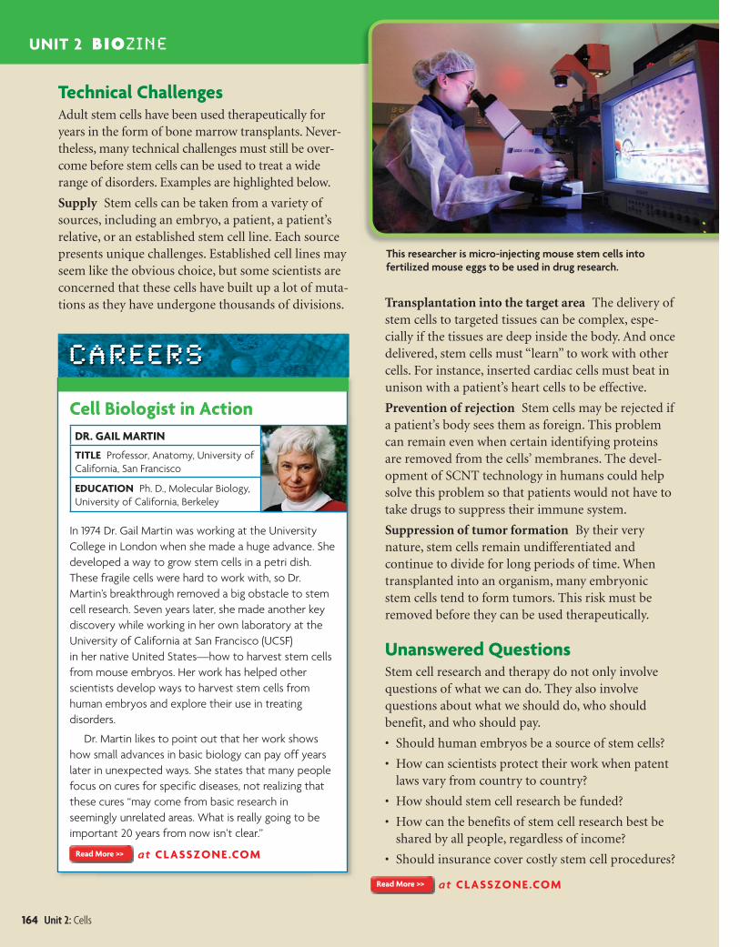
This researcher is micro-injecting mouse stem cells into fertilized mouse eggs to be used in drug research.
Technical ChallengesAdult stem cells have been used therapeutically for years in the form of bone marrow transplants. Never-theless, many technical challenges must still be over-come before stem cells can be used to treat a wide range of disorders. Examples are highlighted below.
Supply Stem cells can be taken from a variety of sources, including an embryo, a patient, a patient’s relative, or an established stem cell line. Each source presents unique challenges. Established cell lines may seem like the obvious choice, but some scientists are concerned that these cells have built up a lot of muta-tions as they have undergone thousands of divisions. Transplantation into the target area The delivery of
stem cells to targeted tissues can be complex, espe-cially if the tissues are deep inside the body. And once delivered, stem cells must “learn” to work with other cells. For instance, inserted cardiac cells must beat in unison with a patient’s heart cells to be effective.
Prevention of rejection Stem cells may be rejected if a patient’s body sees them as foreign. This problem can remain even when certain identifying proteins are removed from the cells’ membranes. The devel-opment of SCNT technology in humans could help solve this problem so that patients would not have to take drugs to suppress their immune system.
Suppression of tumor formation By their very nature, stem cells remain undifferentiated and continue to divide for long periods of time. When transplanted into an organism, many embryonic stem cells tend to form tumors. This risk must be removed before they can be used therapeutically.
Unanswered QuestionsStem cell research and therapy do not only involve questions of what we can do. They also involve questions about what we should do, who should benefit, and who should pay.
• Should human embryos be a source of stem cells?
• How can scientists protect their work when patent laws vary from country to country?
• How should stem cell research be funded?
• How can the benefits of stem cell research best be shared by all people, regardless of income?
• Should insurance cover costly stem cell procedures?
at CLASSZONE .COM
UNIT 2 BIOZINE
Cell Biologist in ActionDR. GAIL MARTIN
TITLE Professor, Anatomy, University of California, San Francisco
EDUCATION Ph. D., Molecular Biology, University of California, Berkeley
In 1974 Dr. Gail Martin was working at the University College in London when she made a huge advance. She developed a way to grow stem cells in a petri dish. These fragile cells were hard to work with, so Dr. Martin’s breakthrough removed a big obstacle to stem cell research. Seven years later, she made another key discovery while working in her own laboratory at the University of California at San Francisco (UCSF) in her native United States—how to harvest stem cells from mouse embryos. Her work has helped other scientists develop ways to harvest stem cells from human embryos and explore their use in treating disorders.
Dr. Martin likes to point out that her work shows how small advances in basic biology can pay off years later in unexpected ways. She states that many people focus on cures for specific diseases, not realizing that these cures “may come from basic research in seemingly unrelated areas. What is really going to be important 20 years from now isn’t clear.”
at CLASSZONE .COM
CAREERS
164 Unit 2: Cells
Ti l C d 2 03240 Fil N bh 02 f i dd U hl b L M difi d 6/28/06 4 39 PMbhspe-02uf.indd UF:164 6/28/06 4:42:06 PM
creo